


- Multiple structural, functional methods
- Different levels of spatial & temporal analysis
- Functional tools have different strengths & weaknesses
2017-08-25 11:51:23
Prelude
Today's topics
- Spatial and temporal scales
- A bit more about structural methods
- Functional methods
Clarity
Functional methods
- Recording from the brain
- Interfering with the brain
- Stimulating the brain
- Simulating the brain
Recording from the brain
- Single/multi unit recording
- Microelectrodes
- Small numbers of nerve cells
Single/multi-unit Recording
Single/multi-unit recording
- What does neuron X respond to?
- Great temporal (ms), spatial resolution (um)
- Invasive
- Rarely suitable for humans, but…
Electrocorticography (ECoG)

Single-cell studies ask…
- How does firing frequency, timing vary with behavior?
Positron Emission Tomography (PET)
Positron Emission Tomography (PET)
- Radioactive tracers (glucose, oxygen)
- Positron decay
- Experimental condition - control
- Average across individuals
More on PET
- Temporal (~ s) and spatial (mm-cm) resolution worse than fMRI
- Radioactive exposures + mildly invasive
- Dose < airline crew exposure in 1 yr
Functional Magnetic Resonance Imaging (fMRI)
- Neural activity -> local \(O_2\) consumption increase
- Blood Oxygen Level Dependent (BOLD) response
- Oxygenated vs. deoxygenated hemoglobin ≠ magnetic susceptibility
- How do regional blood \(O_2\) levels (& flow & volume) vary with behavior X?
- MRI "signals" relate to the speed (1/T) of "relaxation" of the perturbed nuclei to their state of alignment with the main (\(B_0\)) magnetic field.
- Imaging protocols emphasize different time constants of this relaxation (\(T1\), \(T2\), \(T2^*\)); \(T^2*\) for BOLD imaging
Evaluating fMRI
- Non-invasive, but expensive
- Moderate but improving (mm) spatial, temporal (~sec) resolution
- Spatial limits due to
- field strength (@ 3T ~3mm^3 voxel)
- Physiology of hemodynamic response
- Temporal limits due to
- Hemodynamic Response Function (HRF): ~ 1s delay plus 3-6 s ramp-up
- Speed of image acquisition
- Indirect measure of neural activity
Hemodynamic Response Function (HRF)
Generate "predicted" BOLD response to event; compare to actual

Higher field strengths (3 Tesla vs. 7 Tesla)
I want some power…
"Assuming a realistic range of prior probabilities for null hypotheses, false report probability is likely to exceed 50% for the whole literature."
Reproducibility of workflows
Electroencephalography (EEG)
- How does it work?
- Electrodes on scalp or brain surface
- What do we measure?
- Voltage differences between source and reference electrode
- Combined activity of huge # of neurons
How does EEG arise?
- Current/voltage gradients between apical (near surface) dendrites and basal (deeper) dendrites and cell body/soma


Collecting EEG
EEG
- High temporal, poor spatial resolution
- Analyze frequency bands
- LOW: deep sleep (\(\delta\) band)
- MIDDLE: Quiet, alert state (\(\alpha\) band)
- HIGH: “Binding” information across senses? (\(\gamma\) band)
EEG Frequency
Event-related potentials (ERPs)
- EEGs time-locked to some event - Averaged over many trials
ERPs

Brain Computer Interface (BCI)
Magneto-encephalography (MEG)
- Like EEG, but measuring magnetic fields
- High temporal resolution
- Magnetic field propagates w/o distortion
- But are orthogonal to electric field
- Requires shielded chamber (to keep out strong magnetic fields)
- ++ cost vs. EEG
MEG

How do EEG/MEG and fMRI relate?
How do EEG/MEG and fMRI relate?
- BOLD fMRI likely reflects presynaptic input to area
- EEG/MEG likely reflects postsynaptic response to those inputs
- (Logothetis et al. 2001) and (Logothetis and Wandell 2004)
Manipulating the brain
- Interfering with it
- Stimulating it
Interfering with the brain
- Nature’s“experiments”
- Stroke, head injury, tumor
- Neuropsychology
Phineas Gage
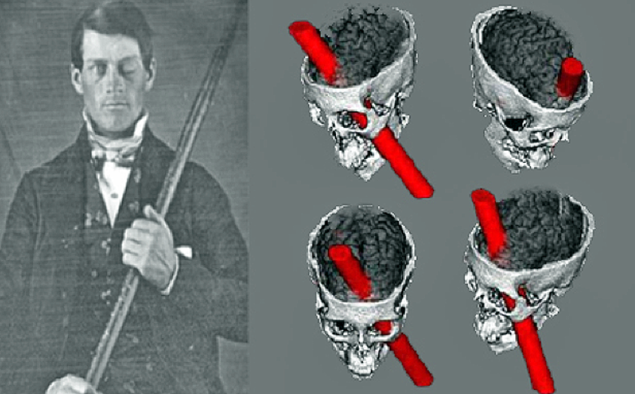

Evaluating neuropsychological methods
- Logic: damage impairs performance = region critical for behavior
- Weaker spatial/temporal resolution
Stimulating the brain
- Electrical (Direct Current Stimulation - DCS)
- Pharmacological
- Magnetic (Transcranial magnetic stimulation-TMS)
Stimulating the brain
- Spatial/temporal resolution?
- Assume stimulation mimics natural activity?
Deep brain stimulation as therapy
- Parkinson’s Disease
- Depression
- Epilepsy
Optogenetics more closely mimics brain activity
Simulating the brain
- Computer/mathematical models of brain function
- Example: neural networks
- Cheap, noninvasive, can be stimulated or “lesioned”
Main points
References
Gilmore, Rick O, Michele T Diaz, Brad A Wyble, and Tal Yarkoni. 2017. “Progress Toward Openness, Transparency, and Reproducibility in Cognitive Neuroscience.” Ann. N. Y. Acad. Sci., 2~may. N. Y. Acad. Sci. doi:10.1111/nyas.13325.
Logothetis, Nikos K, and Brian A Wandell. 2004. “Interpreting the BOLD Signal.” Annu. Rev. Physiol. 66 (1): 735–69. doi:10.1146/annurev.physiol.66.082602.092845.
Logothetis, Nikos K, Jon Pauls, Mark Augath, Torsten Trinath, and Axel Oeltermann. 2001. “Neurophysiological Investigation of the Basis of the fMRI Signal.” Nature 412 (6843): 150–57. doi:10.1038/35084005.
Poldrack, Russell A, Chris I Baker, Joke Durnez, Krzysztof J Gorgolewski, Paul M Matthews, Marcus R Munafò, Thomas E Nichols, Jean-Baptiste Poline, Edward Vul, and Tal Yarkoni. 2017. “Scanning the Horizon: Towards Transparent and Reproducible Neuroimaging Research.” Nat. Rev. Neurosci. advance online publication (5~jan). doi:10.1038/nrn.2016.167.
Sejnowski, Terrence J, Patricia S Churchland, and J Anthony Movshon. 2014. “Putting Big Data to Good Use in Neuroscience.” Nat. Neurosci. 17 (11): 1440–1. doi:10.1038/nn.3839.
Sladky, Ronald, Pia Baldinger, Georg S Kranz, Jasmin Tröstl, Anna Höflich, Rupert Lanzenberger, Ewald Moser, and Christian Windischberger. 2013. “High-Resolution Functional MRI of the Human Amygdala at 7 T.” Eur. J. Radiol. 82 (5): 728–33. doi:10.1016/j.ejrad.2011.09.025.