


- Multiple structural, functional methods
- Different levels of spatial & temporal analysis
- Functional tools have different strengths & weaknesses
2018-08-23 08:12:17
Prelude
Today's topics
- Spatial and temporal scales
- A bit more about structural methods
- Functional methods
Your turn
- Pick two papers you want to read and (better) understand
- Email me APA formatted citation (with DOIs)
- Indicate three concepts/terms you are especially interested in understanding
- Choose a behavior or mental state you want to (better) understand
- Take an information processing perspective and briefly sketch out (in no more than a short paragraph) the main inputs, outputs, and computations involved.
- When thinking about outputs make sure to distinguish between behaviors (e.g., movements, facial expressions, vocalizations) and physiological states (e.g., changes in heart rate, hormone concentrations in the blood, etc.)
Functional methods
- Recording from the brain
- Interfering with the brain
- Stimulating the brain
- Simulating the brain
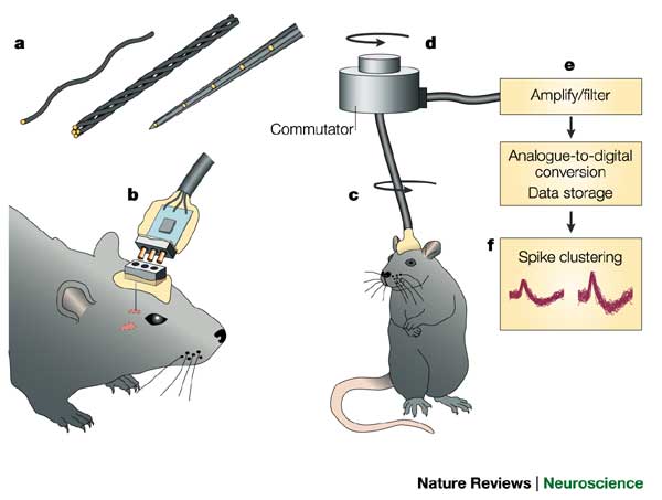
Recording from the brain
- Single/multi unit recording
- Microelectrodes
- Small numbers of nerve cells
Single/multi-unit Recording
Single/multi-unit recording
- What does neuron X respond to?
- Great temporal (ms), spatial resolution (um)
- Invasive
- Rarely suitable for humans, but…
Electrocorticography (ECoG)

Single-cell studies ask…
- How does firing frequency, timing vary with behavior?
Positron Emission Tomography (PET)
Positron Emission Tomography (PET)
- Radioactive tracers (glucose, oxygen)
- Positron decay activates paired detectors
- Tomographic techniques reconstruct 3D geometry
- Experimental condition - control
- Average across individuals
More on PET
- Temporal (~ s) and spatial (mm-cm) resolution worse than fMRI
- Radioactive exposures + mildly invasive
- Dose < airline crew exposure in 1 yr
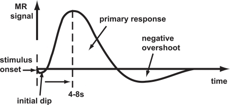
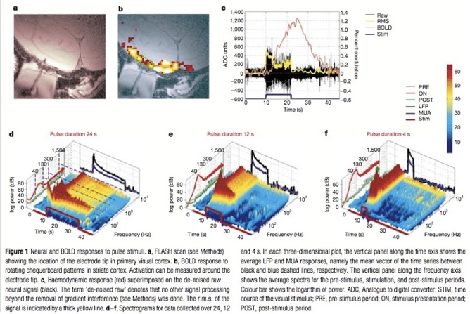
Functional Magnetic Resonance Imaging (fMRI)
- Neural activity -> local \(O_2\) consumption increase
- Blood Oxygen Level Dependent (BOLD) response
- Oxygenated vs. deoxygenated hemoglobin ≠ magnetic susceptibility
- How do regional blood \(O_2\) levels (& flow & volume) vary with behavior X?
- MRI "signals" relate to the speed (1/T) of "relaxation" of the perturbed nuclei to their state of alignment with the main (\(B_0\)) magnetic field.
- Imaging protocols emphasize different time constants of this relaxation (\(T1\), \(T2\), \(T2^*\)); \(T^2*\) for BOLD imaging
Evaluating fMRI
- Non-invasive, but expensive
- Moderate but improving (mm) spatial, temporal (~sec) resolution
- Spatial limits due to
- field strength (@ 3T ~3mm^3 voxel)
- Physiology of hemodynamic response
- Temporal limits due to
- Hemodynamic Response Function (HRF): ~ 1s delay plus 3-6 s ramp-up
- Speed of image acquisition
- Indirect measure of neural activity
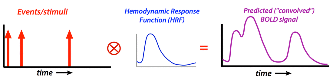
Hemodynamic Response Function (HRF)
Generate "predicted" BOLD response to event; compare to actual

Higher field strengths (3 Tesla vs. 7 Tesla)
I want some power…
"Assuming a realistic range of prior probabilities for null hypotheses, false report probability is likely to exceed 50% for the whole literature."
- Solution
- Make data, materials (analysis code) more widely and openly available
- OpenNeuro.org, Human Connectome Project, etc.
- Increases sample size, improves detection of small effects
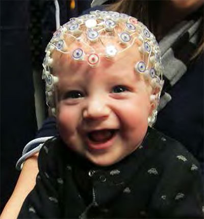
Electroencephalography (EEG)
- How does it work?
- Electrodes on scalp or brain surface
- What do we measure?
- Voltage differences between source and reference electrode
- Combined activity of huge # of neurons
How does EEG arise?
- Current/voltage gradients between apical (near surface) dendrites and basal (deeper) dendrites and cell body/soma


Collecting EEG
EEG
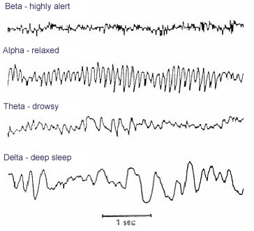
- High temporal, poor spatial resolution
- Analyze activity in different 'bands' of frequencies
- LOW: deep sleep (\(\delta\) band)
- MIDDLE: Quiet, alert state (\(\alpha\) band)
- HIGHER: Sensorimotor activity reflecting observed actions? (\(\mu\) band), (Hobson & Bishop, 2017)
- HIGHER STILL: “Binding” information across senses or plasticity? (\(\gamma\) band), (Amo et al., 2017)
EEG Frequency
Event-related potentials (ERPs)
- EEGs time-locked to some event - Averaged over many trials
ERPs

Brain Computer Interface (BCI)
Magneto-encephalography (MEG)
- Like EEG, but measuring magnetic fields
- High temporal resolution
- Magnetic field propagates w/o distortion
- But are orthogonal to electric field
- Requires shielded chamber (to keep out strong magnetic fields)
- ++ cost vs. EEG
MEG

How do EEG/MEG and fMRI relate?
How do EEG/MEG and fMRI relate?
- BOLD fMRI likely reflects presynaptic input to area
- EEG/MEG likely reflects postsynaptic response to those inputs
- (Logothetis et al., 2001) and (Logothetis & Wandell, 2004)
Manipulating the brain
- Interfering with it
- Stimulating it
Interfering with the brain
- Nature’s“experiments”
- Stroke, head injury, tumor
- Neuropsychology
Phineas Gage
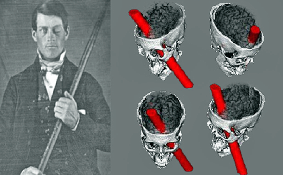

Evaluating neuropsychological methods
- Logic: damage impairs performance = region critical for behavior
- Weaker spatial/temporal resolution
Stimulating the brain
- Electrical (Direct Current Stimulation - DCS)
- Pharmacological
- Magnetic (Transcranial magnetic stimulation - TMS)
Stimulating the brain
- Spatial/temporal resolution?
- Assume stimulation mimics natural activity?
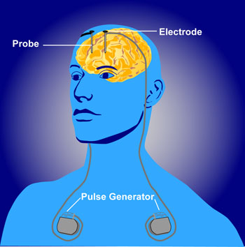
Deep brain stimulation as therapy
- Depression
- Epilepsy
- Parkinson’s Disease
Optogenetics more closely mimics brain activity
Optogenetics
- Gene splicing techniques insert light-sensitive molecules into neuronal membranes
- Application of light at specific wavelengths alters neuronal function
- Cell-type specific and temporally precise control
Simulating the brain
- Computer/mathematical models of brain function
- Example: neural networks
- Cheap, noninvasive, can be stimulated or “lesioned”
Main points
References
Amo, C., De Santiago, L., Zarza Luciáñez, D., León Alonso-Cortés, J. M., Alonso-Alonso, M., Barea, R., & Boquete, L. (2017). Induced gamma band activity from EEG as a possible index of training-related brain plasticity in motor tasks. PloS One, 12(10), e0186008. https://doi.org/10.1371/journal.pone.0186008
Hobson, H. M., & Bishop, D. V. M. (2017). The interpretation of mu suppression as an index of mirror neuron activity: Past, present and future. Royal Society Open Science, 4(3), 160662. https://doi.org/10.1098/rsos.160662
Logothetis, N. K., & Wandell, B. A. (2004). Interpreting the BOLD signal. Annu. Rev. Physiol., 66(1), 735–769. https://doi.org/10.1146/annurev.physiol.66.082602.092845
Logothetis, N. K., Pauls, J., Augath, M., Trinath, T., & Oeltermann, A. (2001). Neurophysiological investigation of the basis of the fMRI signal. Nature, 412(6843), 150–157. https://doi.org/10.1038/35084005
Sladky, R., Baldinger, P., Kranz, G. S., Tröstl, J., Höflich, A., Lanzenberger, R., … Windischberger, C. (2013). High-resolution functional MRI of the human amygdala at 7 T. Eur. J. Radiol., 82(5), 728–733. https://doi.org/10.1016/j.ejrad.2011.09.025