511-anatomy-outline
Rick Gilmore
2019-09-19 10:49:28
Fun
Resources
Harvard Brain Atlas
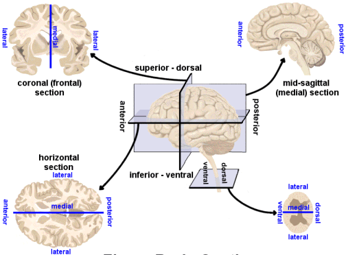
Directional terms
- Anterior/Posterior
- Medial/Lateral
- Superior/Inferior
- Dorsal/Ventral
- Rostral/Caudal

Bipeds vs. quadripeds

Image axes
- Horizontal/Axial
- Coronal/Transverse/Frontal
- Sagittal (from the side)

Supporting structures
Meninges
- Dura mater (‘tough mother’)
- Arachnoid membrane
- Subarachnoid space
- Pia mater (‘gentle mother’)

Ventricular system
- Also known as cerebral ventricles
- Lateral (1st & 2nd)
- 3rd
- Cerebral aqueduct
- 4th

- Ventricles filled with cerebrospinal fluid (CSF)
- CSF clears metabolites during sleep (Xie et al., 2013)?
Blood Supply
- Carotid & vertebral arteries converge on Circle of Willis
- Anterior, Middle, and Posterior Cerebral arteries main output

Blood/brain barrier
- Cells forming blood vessel walls tightly packed
- Active transport of molecules typically required

Area Postrema
- In brainstem, blood-brain barrier thin
- Chemoreceptors detect toxins, trigger emesis if necessary

Organization of the Nervous System
- Central Nervous System (CNS)
- Brain
- Spinal Cord
- (Everything encased in bone)
- Peripheral Nervous System (PNS)
Organization of the CNS
| Major division | Ventricular Landmark | Embryonic Division | Structure |
|---|---|---|---|
| Forebrain | Lateral | Telencephalon | Cerebral cortex |
| Basal ganglia | |||
| Hippocampus, amygdala | |||
| Third | Diencephalon | Thalamus | |
| Hypothalamus | |||
| Midbrain | Cerebral Aqueduct | Mesencephalon | Tectum, tegmentum |
| Hindbrain | 4th | Metencephalon | Cerebellum, pons |
| – | Mylencephalon | Medulla oblongata |
- Forebrain, midbrain, hindbrain terminology derives from embryonic stages in CNS development.
Hindbrain

- Hindbrain: structures adjacent (or caudal to) 4th ventricle
- Medulla oblongata
- Cerebellum
- Pons


Medulla oblongata
- Cardiovascular regulation
- Muscle tone
- Fibers of passage
- Ascending fibers (from body), a.k.a. afferents
- Descending fibers (exiting brain), a.k.a., efferents

Cerebellum
- “Little brain”
- Dorsal to pons
- Movement coordination, simple learning

Pons
- Bulge on brain stem
- Neuromodulatory nuclei (e.g., 5-HT, NE, ACh, DA)
- Relay to cerebellum
Midbrain
- Tectum (roof), dorsal
- Tegmentum (floor), ventral

Tectum
- “Roof” of the midbrain
- Superior and inferior colliculus (colliculi is plural)
- Reflexive orienting of eyes, head, ears


Tegmentum
- “Floor” of the midbrain
- Species-typical movement sequences
- Neuromodulatory nuclei
- Norepinephrine (NE)
- Serotonin (5-HT)
- Dopamine (DA) – from ventral tegmental area (VTA)

Forebrain
- Diencephalon
- Telencephalon

Diencephalon
- Thalamus
- Hypothalamus

Thalamus
- Input to cortex
- Functionally distinct nuclei (collection of neurons)
- Lateral geniculate nucleus (LGN), vision
- Medial geniculate nucleus (MGN), audition
- Pulvinar, attention?

Hypothalamus
- Five Fs: fighting, fleeing/freezing, feeding, and reproduction
- Controls pituitary gland (“master” gland)
- Anterior pituitary (indirect release of hormones)
- e.g., Corticotropin Releasing Hormone (CRH) -> release of cortisol from Adrenal Cortex
- Posterior pituitary (direct release of hormones)
- Oxytocin
- Vasopressin (aka, Arginine Vasopressin – AVP; Anti-diuretic Hormone – ADH)
- Oxytocin
- Anterior pituitary (indirect release of hormones)


Telencephalon
- Basal ganglia
- Hippocampus, amygdala
- Cerebral cortex
Basal Ganglia
- Skill and habit learning
- Linked to Tourette syndrome, obsessive-compulsive disorder (OCD), addiction, movement disorders
- Example: Parkinson’s Disease

- Striatum
- Caudate nucleus
- Putamen
- Globus pallidus
- Subthalamic nucleus
- Substantia nigra (tegmentum)

Hippocampus
- Medial to lateral ventricles
- Memories of specific facts or events; place memory in non-human animals
- Fornix projects to (mammillary bodies of) hypothalamus


Amygdala (“almond”)
- Physiological state, behavioral readiness, affect
- NOT the fear center! (LeDoux, 2015).

Cerebral Cortex
- Cerebral hemispheres
- Groove (sulcus or sulci)
- Bumps (gyrus or gyri)
- Grey vs. white matter
- Lobes
Lateral view

Medial view

Nissl stain

Lobes of the cerebral cortex
- Frontal
- Temporal
- Parietal
- Occipital
- Names derive from bones of the skull


Longitudinal fissure
- Also known as superior longitudinal fissure
- Divides the cerebral hemispheres

Lateral sulcus/fissure
- Also known as Sylvian Fissure
- Divides frontal from temporal lobe

Central sulcus
- Also known as Rolandic Fissure or Fissure of Rolando
- Divides frontal from parietal lobe

Frontal lobe
- Anterior to central sulcus
- Superior to lateral fissure
- Dorsal to temporal lobe

- Primary motor cortex (M1)
- Supplementary motor cortex
- Frontal eye fields (FEF)
- Prefrontal cortex
- Planning, problem solving, working memory…?
- Basal forebrain
- Nucleus accumbens (NAcc), part of ventral striatum
- Anterior cingulate cortex (ACC)
- Primary olfactory cortex

Inferior Frontal Gyrus (IFG)

Middle Frontal Gyrus (MFG)

Temporal lobe
- Ventral to frontal, parietal lobes
- Inferior to lateral fissure

- Primary auditory cortex (A1)
- Object, face recognition
- Storage of memories about events, objects
- Amygdala, hippocampus
Inferior Temporal Gyrus (ITG)

Entorhinal Cortex (ER)

Parietal lobe
- Caudal to frontal lobe
- Dorsal to temporal lobe
- Posterior to central sulcus
- Primary somatosensory cortex (S1)
- Perception of spatial relations, action planning
Inferior Parietal Lobule

Superior Parietal Lobule

Occipital lobe
- Caudal to parietal & temporal lobes
- Primary visual cortex (V1)
- Secondary visual areas (V2…V7)

Insular cortex (insula)
- medial to temporal lobe
- deep inside lateral fissure

- Primary gustatory cortex
- Self-awareness, interpersonal experiences, motor control, interoception
Brodmann Areas

- Cytoarchitectonic (cellular architecture) differences in cerebral cortex
- Numbered areas, e.g. V1 == Area 17


White matter pathways
- Brainstem
- Projection fibers
- Association fibers
- Commissural fibers

(Oishi, Faria, Zijl, & Mori, 2010), Chapter 3, Figure 1.
Brainstem projections
- Corticospinal tract (descending/efferent)
- Dorsal column/medial lemniscus (ascending/afferent)
- Superior/inferior cerebellar peduncles (from/to cerebellum)

(Oishi et al., 2010), Chapter 3, Figure 8.
Projection fiber tracts
- Internal capsule
- Thalamic radiation
- Cortico-{pontine, bulbar, reticular} tracts


(Oishi et al., 2010), Chapter 3, Figure 11.
Cortical white matter tracts
- Superior/inferior longitudinal fasciculus
- Arcuate fasciculus part of sup. long. f.
- Superior/inferior fronto-occipital fasciculus
- Cingulum, fornix (hyp-hip), stria terminalis (hyp-amyg)
Commissural fibers
- Corpus callosum
- Anterior commissure (AC)
- Posterior commissure (PC)


Anterior, Posterior Commissures

Spinal cord
- Spinal column w/ vertebrae
- Cervical (8), thoracic (12), lumbar (5), sacral (5), coccygeal (1)
- Spinal segments & 31 nerve pairs
- Cauda equina

- Spinal segments (rostral to caudal) ennervate specific body segments
- When focusing on the skin, these are called dermatomes

- Dorsal/Ventral
- Dorsal root (sensory)
- Ventral root (mostly motor)
- Grey (interior) vs. white matter (exterior)
- Cerebral cortex opposite (grey exterior, white interior)


Organization of the PNS
- Somatic division
- Autonomic division (Autonomic Nervous System)
Somatic division
Cranial nerves
- Afferents (input), efferents (output), or mixed
- Innervate head and neck
- Olfactory (I), optic (II), (VIII) auditory, vagus (X), etc.
- Spinal nerves

Spinal nerves

Autonomic nervous system
- CNS & PNS components
- Controls “vegetative functions”
- Limited voluntary control
- Two divisions
- Sympathetic
- Parasympathetic

- Bipolar (continuum) vs. bivariate autonomic space (Berntson, Cacioppo, & Quigley, 1991)

Sympathetic division
- Prepares body for action
- “Fight or flight”
- Spinal cord
- ganglion chain along spinal column to End organs
- Neurotransmitters (NTs)
- Preganglionic: ACh
- Post: NE
Parasympathetic division
- “Around” sympathetic
- Restorative function
- “Rest & digest”
- Spinal cord (or Vagus n.) -> ganglia near end organs -> end organ
- NT: ACh
References
Berntson, G. G., Cacioppo, J. T., & Quigley, K. S. (1991). Autonomic determinism: The modes of autonomic control, the doctrine of autonomic space, and the laws of autonomic constraint. Psychological Review, 98(4), 459–487. https://doi.org/10.1037/0033-295X.98.4.459
LeDoux, J. (2015, August 10). The Amygdala Is NOT the Brain’s Fear Center. Psychology Today. Retrieved from https://www.psychologytoday.com/blog/i-got-mind-tell-you/201508/the-amygdala-is-not-the-brains-fear-center
Oishi, K., Faria, A. V., Zijl, P. C. van, & Mori, S. (2010). MRI atlas of human white matter. Academic Press.
Xie, L., Kang, H., Xu, Q., Chen, M. J., Liao, Y., Thiyagarajan, M., … others. (2013). Sleep drives metabolite clearance from the adult brain. Science, 342(6156), 373–377. https://doi.org/10.1126/science.1241224