511-evo-devo
Rick Gilmore
2020-10-15 15:41:10
- Fun
- Evolution
- Human brain development
- Prenatal period
- Differentiation
- Infancy & Early Childhood
- Summary of developmental milestones
- How brain development clarifies anatomical structure
- References
Fun
Evolution
Public acceptance of evolution
![[[@miller2006public]](http://dx.doi.org/10.1126/science.1126746)](data:image/gif;base64,R0lGODlhIgG4Aff+AP///+A4Pt3UlABWmdKTlOMnL94mLt4qMd4tM+QvN98vNeQyOd8xN98yOOA1O+U3POY4P+A3PeI4PeE4PeE4P+A6QOE+Q9U+ReFCSORPVOVaX+dkaOlxdbtcX+2Mj/KtsPfJy/rd3t8oMeEqM+AtN+AxOtUzPvvo6dUjMdIrN9kuO9UvO7YxR/75+iQgIUA8PTQxMlVSU1xZWm1qa3h1dnVyc5eUlY6LjGVjZGFfYH99fnt5eqelpp6cnZqYmZKQkYiGh4OBgrCur62rrKupqvr5+uvq6+Xk5bq5ugBFkwBMmABKlABChgdSnPj6/Pz9/gBOmQBOlgBPlQBNlABMkwBLkgdVnCluqwBVmgBSmQBSlwBRlgBQlQBRkwBPkgBKiwdPjghYnA5UkStxqyxxq5u715u819Ti7gBXmwBVmABTlAJXlgNZnANYmgNXmQZZlwdcngdbnAhbmQtdngxfoA9gnx5ppShwpSxzqy10rDF3rDVzpDp8sEiGtlaPumOYwWSYwXGhx4CrzI600jpJVI6ux5y916rH3bjQ4k5WXMbZ6NPi7tXk7/D1+fT4+xpjlyZqmXmgvJO2z8TT3drm7uHs9D14mJ+6ytLd4+zx9OHm6dTh6N7o7VWGlpq2vebq626Tlqapqf3+/vv8/PP09ImlmbzMxezw7f3+/ZquleTp4rzFot/i1LS6k9TaltbbmsLEkcTGldDRktLTmN/emt3cmP7+/NzaldbUpd3XlN3XltHNk/n47N7WlNfRlNfRmPXz4dvSjuHXlNzTkd7VlN3Vld7Vl9/XmePbpefgsuvmv/Ht0uTYj+XZlN3Tk+nblNnLj/79+vz59vj18t6rfefBpd6Xc+atk8KDcd9yXv319N9XT91OSZR+ffS+vv3x8UxJSXFvb2lnZ6mnp6KgoP/9/f/+/vj39/b19fDv7+Hg4Nzb29fW1tLR0c7NzcnIyMTDw7++vra1tbOysqSjo/7+/v39/fv7+/T09PPz8/Ly8vHx8e3t7ejo6AAAAAAAACH5BAUAAP4ALAAAAAAiAbgBAAj/AAEIHEiwoMGDCBMqXMiQ4TlyLRpKnEixosWLGDNq3Mixo0eJ6BIhUkSypMmTKFOqXMmypcuXMGPKnFlyEy9ewHDqzMmTVwgQP4MCHfqz3MeKo0YxTEow372jAPidIyiN0Bc2bbJq3cq1q9evYMOKHUu2rNmzaNtksUSsV7G3cOMWo2XNQYAKd/PirRABwwmoBdfxQ0hOHkMhQgYaAYJu4pEhRNQBODdv3tR8QpDca0eOqlUsaUKLHk26tOnTqFOrXs26tevXsNNAYUtMgO3buAXcskbCge/fwBtY+At4IDl39QAoJQikMwB7Ccn1MO5c4pAZ5NYBAGIDyHQdPXYQ/wEQjt3Aql/QDFjPvr379/Djy59Pv779+/jz6x8wm1jt3LnRRUIABBZooAN+FWdcO+jQ84MR69jQAz438ACAEDfEAwAR49ygXTw+zECPQOeIMxhiQJinEDnO5YPDOejMoE4NABhB4w/TCYSeevv16OOPQAYp5Hv9/QfgbbXwZuCSASBInIL0sOODD+mcQ0M7Pcxjg4XorJPDPuIg8c4NMR4RRI79xHBPPjGo08MNAqXDAz30kDMPdADEE04Q6qSzQz2j4OAOEADEOMo8cOpoFY9DNuroo5DqV+SRuO1WApMHJqggAPS0Q4M7heYwjg3x2DDOOUT4AA4/O/R5Qzs/AP/Ag3P8yGCPp7IaNlk77vTKjlEC3TNEEPjQUM89OLRDaIz28BCroulFKu201D46KaW2JTkgpgQ6uSkA5LSzAxIAILuOUkDII4QNRchgBA3qLHaEDgD4kGM9QawzT6xA9BPnnHXeSVA7Opzz5RE78CNOPewEAYAOiUHLaLUUV2yxfNdiKyC33Wqq4DjsvEMDEfzQE4QQRsiTbhDyxJAOn0bcUM8OPdQwnkBI3EDEO8UWIdAo/ARtRDoDtUNPDe/UC4QO5N5wAw3v5EMD0YoyEcUUWGet9dZcd+3112CHLfbYZJdt9tlTJGFJMGy37Xbbt2RzAAJ01233AQE8WVwRSvX/047P66wD+BPqqINOPefYgzgARbCjz1MCFUEE0fY0tpAR7uwjkDnrSCYQO0cA0A48BIXUhx+op6766qy37vrrsMcu++y012777alHokwyu/fO+++8V8PB8MQXP7wH33yrvEd4DoQPDumYs/z01FdPkDnYe1QOsNZ3bxE+4mjj/fjkl2/++RihI8Mlh7Tv/vvwxy///PTXb//9+Oev//77TwLM/wBchjcGSMACGvCABYwI+ipij3QkhyD40I5FjkA1aGFBCxjMoAY3yMEOevCDIAyhCEdIwhKakIRK6AQz3JYLazCgATCMoQxnSMMGMGA40ytHqmygIo3oQweWGwgR/4RQj3aMw2fnEEIPjDCZeWioHkiwUxEi1Dz0DCA2WMyiFrfIxS6KBgqdaIaRkuSbCJjxjGhMoxq9tbwixKAdQwCHZO5BtOTkw2fQIRqMnmOEfDynUO8qgj2mAoB+zGAzM3DZhoAwrFGQQxw7eI46jjCDedQjHEmT2MU2yUlqgVGMSKKGAyLAsVJ2TG+bKkIOBhMEeuDjBzpwxzlu8AMaGEEdNwDCDXogIzOFwx3oCMcNGhYzz8mDBpMpgg4Go4PQ5eAI6FjMQM4BBHLZwAbnWVQnt8lNIX1yjHUhpSk5xkblqbIf9ZAOPXowoyOE4xwna1i71mEvAKSDHDrQh8vQQf+DG+iKUw57TsLqMTUAiEM7RyAUAN4RjhrgAwCIyma0uknRit7nm7fRhSjFOU5MlfNbqmTiDUwWjh/cQB3Y1Bk7xgGAGRhhHDxomDh0kI5wTCYGMiDIECKpnB0QjQaaw4FkEhq5feiApT3ApiYtytSmugejtqHLKDvKrY+mUgZHOIIM2GGDERUSTkAAWWdwcAR68MAGR4gHDRQGgB/aIFEAWMcMkjOKtW4nHv14JwDMBIAHegcANCDXUp1KWItCVQC1oEYJOErVTKFSQaOowQ5mMIRC0qwd+hgROZCgDsMEIWVIkMdbf6APQr2ytJ4DwA/ekY4eiOMH6mCHZEk3phn/8IAfQyDHDvphBCBmkwlTqIJwh0vc4hr3uMhNrnKXy9zmOve50HVuElTIQmsY4ADYza52t8vdAxjAAY9VUBGqNJAiQFM5ylEcdO5RD3tAxwjoUBzknjLIgXQWaPiQCgDwQbX85oMf9jDCEeohina0gyDnSAQgAsHgBjv4wRCOsIQnTOEKW/jCGM6whjV8CWV4+MPe8ICIR0ziEpuYxMlboIr3i4NpSG/FMDbf9gyCihjbGCOkEIc0bszjHvv4xyRKRCQGQeQiG/nISE6ykpfM5CY7+clQjrKUo2yKZVj5ylZuxwe2zOUue/nLXBYf+XzGEPM2ZB8HniATf3s1tLn5/81wjrOc57yEJIDiGW8LBjOwgYLr+tm7gP6zoL373fAu7wg+0EEmFaKOGTQvOvK4Bzx64DN8nDV08JiSv5BgKnTgEnIA2FFhR91NJdx5GLgZBi2wkYIJNHZJVqUeDXiQDnQc7hxFWNwR9CGQfLxDHNA5wlTucY7QDYSSAHBHDWRANGfJYwejWEc/rlkjI4QjMTUg3WBJzW2LQeHOAFq1CV4Na49ZLxw+EEgsdXCcIbiDBjvIKg1mgAMAHBMIqAJCOHIE0UgKcpkP89czBTKEWBWBH0DQkA+UGmptdvvhFPv2M8KNjXGTu0Cxnh47ckCDI2wJCDgaQnl4wIMbIEGu6v8QRz9+gAR6zIMfOSCzyX420IIeFAAbPzA8cLADzUV02xAPurXAHSBsrMDVF2+Subs3LHawHAjicIc45iGPddSAHXlthwyQMA91jAOzMRfIMQUyM6IlDACrdMcMJFiPIuxStekGutDnHiSJU/zoSVe6oZWnj32Q4waqHBM49IG0XAOBB+/IgRFm0I+n9KAd6ZABIQ1ZhHsYkh1FkNA76LV1feCjHvrIh3jIs2j0gMaLqE+96lfPGlNPvOgmAI7sZz974ez9W/MIwg/8hVZ3jIgd3DFCzHwwokGRYxTvgGYPQE0PJOyjB9wRTC4l84MpwcMef+dBl35QRatgJS3gD7//+MdP/vK3QeLEkIu4K8D+9rv//fDvy+1BqpDlFKS9ZDcImidyDzKrw18DoT4jQRMEWIAGeIAImIAloQk30RM50Q9EEYFCMYESqEBAVj3gMw3cc4Ec2BHmYAsIkRzSM4IAQIImWIIoaA4b2IHf8jz88AQwGIMyOIM0WIM2eIM4mIM6uIM82IM+mIOisDnYM4REOILbc4RImIRKOGMsOD1VsQd8EIVSOIVUWIVWeIVYmIVauIVc2IVe+IVaCAjIkAxjWIZkeIZjeA0bsIZs2IZu+IYcMH/Lcw790A9klhH6EjnTpDmM0w8PNRBPwSuewQRawAWGeIiImIiKuIiM2IiO//iIkBiJkjiJlNiIahMMw5CJmriJmbgbCqAADBCKojiKpCiKCFABcvgtBKUDsKUR9vAq6QAEOdAY67ADNaAhoxIOpOMOMtAZ71JBokZ3wtgjW0Ab2GIbt7BRF5dx1WMP4uA56uAOnQMy9sQOSDAY9RAPGtIP81BB7TADcSVyg/ED8nAEOIAODyUPcIIEC6duEdNwEzWM8nhRxniMZMRYVMWM1OOM8HAE97ADO7AO7CAPaxcPNTAONTAKSZV8JxMEljMO9CIQA+VTACBUAvF2oqNUziJREzOPHhkfGUMpiTVV5KaP01MPOBAE5GBUSXMO8ZAD2mgh5SEOJTgPUUcDAP84c+VSc0Rzc/QwA4OxUEqFIRz5kUYJkvWILfe4jEvXPc5obDrwNzsQDzMQD/AwBPZQA+1QbwBwHfDwDpCDBDTSVz0HALtVkfogD+FQQe4QdxgJjx15lEcZkkeiUST5aia5PPaQAxL0KfkgDkaAA0gQDzzgjPtAA/JgBOwgDkeQDw+0D+GAD0WQcgzyd0QZA/DQD0STDrnXD6NQHhJ1QSc0mqRZmqZ5mqipQUpgCcNADJz4mrvRALQ3m8GBQ+WTjVQDD0ykLkKQVevwBBpyBD1ALu9ADvHQPPMwBOlABOTAA4bDA0vUlTAFKu7AIkJQnZ4BBnQwB9zZnd75neAZnuL/OZ7kWZ7meZ7omZ7qOZ5hYAnH8J7wGZ/wSQvUYAEYcJ/4mZ/6mZ8WkAGpqIoX8UAIlloUYQS8FoA5gAmVsKAM2qAO+qAQGqESOqEUWqEWeqEYmqEUmgnS0KEe+qEf+g0nMKIkWqImeqInsIJNWBwZuKIuChUC+qIx9jya4Ag2eqM4mqM6uqM82qM++qNAGqRCOqREyqPRcKRImqRKuqRIagstoA1QGqVSKqXS8GIyShAxOhGP1nCPYAde+qVgGqZiOqZkWqZmeqZomqZquqZsOqZ3gAvIEKdyOqd0WqfIcAzIQA0ZsKd82qd9qgH/qTz10A9BdBHtIASjUIcIhmD2/9cPt7JoDccEWZCalFqplnqpIbQFb7ALK5RnnppnqLYbJFBDM6QAqLhAo3ADEXkR9wAEAgl9cKIODfVA6gAOaUYPO3AOUrNmcCmXR5kGc7ALRnKMSmkNl2JKeVk9BLNQPwAq7AAP40AKAAAPP3Bg0UgOAJhs4PgU6nMO7DAENLAcQICZohMOlqMDN9OrvvqRWCAHuyAMxEqsSXKspZSs1OMO9MIO8cCYT4OrtyKWReADh1cDeMIDqwoP4vAUovcU7+AgpANvQ6AURPAs6rqu84gFwQqv8Vqs9EpOTUk++LpX9PBGNmAYL6IO9HCO9PAOBiNzAcWLKmIEO2APRQAvGf8yCjEgBGfSlXAVjBYrj2ngrhq7sUcyr+Nkr9PjDkGgDzuAsO1gA+QyA++ANOGQDzwAeWGXJza1DuLQQ/sQSfgwA0AADuGwePYEjjdQHT77s8IIrO9KtCJprEf7sePTDp9VA1oFKzowDzoADzIiA/jQKZFHZvhQAwEbkOaBDy8pS+iQDjQwDqNQlTaQbuEgQZGaBVuQuZq7uZzbuZ77uaAbuqI7uqRbuqZ7upwbBZv6mqzbupmYJAjwQqQKQ6YaqN9iD+oIAFyHBOlgA0FAMqMgD/IAD1PED0VwfQOBBEPADkQwBORiBEIwRAfaDqGDskKADr7nGZCQB3jQvd77veD/G77iO77kW77me77om77qu77gOwZ6AKd2Gr/xew0aUL/2e7/3uwG2uyn9EJUF4U/8Z2wKkaVRUajP8wlOkMAKvMAM3MAO/MAQHMESPMEUXMEWfMEPbAsavMEc3MEezMHS0AIiPMIkXMJWaj7uZRDrIMAYKA7TcKUwHMMyHCqTwAiLcMM4nMM6vMM83MM+/MNAHMRCPMREXMSLcAo3kcRKrMT7EAJO/MRQHMVSPMVQbIExTEgVcYeKAgZwEAde/MVgHMZiPMZkXMZmfMZonMZqvMZsHAap8AzGEMdyPMfGIAytcAEWkMd6vMd83Md+bAEVQLcD4Q6D0Q+gQhBGQARt/0fAyVZZAvEOg4FXXccQ2jgR8GAYafUU60AESIBElTEV+iAE8RBt7wiPp8d6qJzKqmwaWZAFpfAMzhDLsjzLzmDHJrAAtJnLtGl7C1EDoGKQBYEP5mED2UoQfDsQM6CvNYAO7MCHK8JvC7EPNaAP6xAOMaA51hgOlcUdf8Vue0sePbS2bNttWfDGxGrHF2AXeedRgiyRv6wDATsq+tCw6hADPmAPSPADGtIOPlADAQVYRLBWK5cO1WkD8xBXEmIYmWZb+fAmoCIEZxUPGZK8NBI0O8CrREAPgVIE6CAO+/Ai7AgAbynO40xq5dwM59wKJiAB68zO87cD77wPq2ID5P8QmMq0Du/WDwHJeDawqjQAA0lTBOVBDkAAc+4ED/GwA2Xyu0rEtTESITvADjIAOfQQkTPDRPtAD8/kJ4DyJTwQBFF3IT3rcCVNzq+c0unc0kySl0FwYPGwTDZFBDdwDuAIBJiBA/KgzzaFBP+8AzQQBNChA8ybGDOgdueQDq8SKzyQKqETb0AgChN7D1lLBBH5j0x0DwlFD/x0LDjQv+6wLpxCsSRd1oRVzq+HLejM0mrtWAvxA5X11foAjiUXIwDgkEhAA4BTb0KwquWhAxYi2M1rUOyAA/ywDkEwL/UyDj2QNC4FBEUw21n7jQOh1AMBD0Eg2fuAMEZAkzX1MO//ONqk3VQnndKxt9qsrRCJFyLs4LhdaQNWAgAS4if0AA/3sEs7oFBmyQ7Pg0vrIAQHjbf0oEuRRDM1MA+e8gM28IpFMATuPQNkNgopkg7jkAPs5A48gDRKowMaEgQ+wDLFAoyEAFzRNeIkXuImfuIkvgSl0KmfqmetkAIi0F0yPuPaVWgMgTmaUzmMcw710BjnsA5t1w7+MgrsYLzTpBTocA6JUwQ+czhtJdGM4zhP0Q9APhk7XigDIwTmxQ/QZATtEJRx5Tn2ADqio21BtmAbluZqvuZs3uZrbgofFudyrgztcGJ2fud2Lmbo03L+ixGMjKUHsaXPo+czDGQnnBDm/xANhY4Q+CBs6JNjO7bokj7pN5ZgQzZlmJ7pmr7pnD5lksAKWBbqWKYMYFbqpn7qhF7oniYRRvCHVdNmcxbrsj7rtD4FS6AGsMDin9pC3iUCvv7rwB7swj7svm7j51MPQmAe7NBDGsEDmiEP9EA0LYd4cUIE3grN4B3e3dSusDC0IkkNAYCP5o203zIKL1BvPRB3hYrF5dUYTzAQyyGrG9JKQYB8KgMnn9kD0BEOaVax2s5U7RoL3l604C7uq03uqYQDMcC8nTEsPBC8N7AlCHMOQzBtGt6w9qYilP0w2iEDQRkE8VAP44Df1VeU/95UAT/wAJJYZmTe5ba/eyMiPv9ABDxg3PmANDcAD40GDzEwtTDyDskiDh8uEN3BKTswDzLQGOkgDvpQDzmgAwEJUWMdjydfUSkvr8ro8hjXztRzDp1dA3s7Dp1xA+MwDvxQDzrjAztwA/0gsDLgdscsENBXLuMQDkpFDti0D+6S1OAi2mRd9RR19ceoUeGu9VsP84DRLvhABDAwD2NilkjAA/7CA0DgDi/AIVSSUy1j5gwlEPaAA+axl0kDJnFFIzvwT9kO+BeTBXIg8FhfRmoU+7Iv+wgvXjWQDkUADokRBD29D0PARPOQq1sFDzpwNHEFDkFUBPlyD0Fw0HtluET/NPDgJwcaqYVYidif/dq//dz/P4lRsAa57rqdSA0kAIqleP7oj/4IkDcLxOOM4zOYrRS5phw+Q2ZB4zPwwFNFE/ICDBBF0AEgWK9fOgDt3BEkeC5RIUMRJU6kWNHiRYwZNW7k2NFjRzOGlo0kWbKksg8pVa5k2dJlyxYMZc6kWdPmTZw5bR6p0U7nT4L4xEkDWtToUaRJlR598rPcUqhRpRoVOJWh0GlWtW7l2tXrV7Bhjzr0A+jPWbRp1a5l29btW7hx5c6lS9eUMrx58V7z0NfvX8CBBQ/2+00s0HpH7s3EV6ToPp9IjxiRKY0QkylUNG/m3NnzZ9ChRY8mXdr0adNLSjEL1tr1sG0GRBygXdv2/23cuXMbcHDisMxzN27YoMywCI7iDH8sBEpO3j14PRzj42HjCAB4PnxcB7DPBz91NxYTtPwFzQD06dWvZ9/e/Xv48eXPp1+f/pZUzwTs319MV7YSAhBwQAILNPBABB3AwLffCDJiBiOAsAGAUa4rIod+uuuniCKO4HAUe9CpZ5R0KOOOoCNmAMCdGmRAiIcf5NlhlHX6+WFCAGyAwaca4GGovPPsE3JIIos08sgsUmmGv/6KARBBKKOEUkEGG+SHhn16GMIeG2gg55wc0lmnBh3CUUcGduhph6cigLghnB5msKEegubZAYAi7NGBHwB0yDAH7oT4AYDwdFjIBxwBAP/ySEYbdfRR+rIoRT8m/cuGBCkz1ZTKBgnSJ4YZZFDHHSCOyMEdcdKhIR50crAHiHd6cEcdcYqgIR16gshHBscAyJWgUXbgp55bARBnHQDYkaGdem5g5waf5rnhR0LMg/RabLMtMsklK/0vQE3DTXDBTgEwgoYi2tmBHhmACILWfmZQx55wRrnBnR7WnOEcIEYh4ocLexXiTgDs2QGhgwHAoR91cPDxCHHkkYEcABCl1lptM9Z44/SSpJS/YgR4UlySCeS0UyPC0ecdGt4Bgs4iZOBnB3nUyaEeWHuAx50czgmiCB5uOIdXgtSZoR578AnnCFRumGcdGuqJ2J578oH/Jx4ZfABg1YuxSONrsMMWe2yyyzb7bLTTVnttttXOQotSmnFm7rn3u9QBvPPWe2+++/a7AQuq/A2dGmqYwacbgBinTSTW0eEGXulhxx0afgDiHnruQWLxnxmyAR5+fpgBiHXUqYEGd4gVLh6GemDniM7JIwQMOOKw/Xbcc9d9d9579/134IMXfnjhw8jPGOSTPyYbByxw/nnoo5d+euorILfce0RkiB98AKgHH33UiUeHIuqhs7GCvKfTHpmOQMKefM7Jx7EiuqcwH33OYeipdpBlqNVJMGIRAyRgAQ14QAQmUIELZGADHfhACELwFLygYAUpeIIQZFCDG+RgBz34/0ENxqRcOWkHPYaQnBECgBTiyEoKXfjCopgDhjOk4VVwoAlH5FCHO+RhD334QyAGUYhDJGIRjVjEIkRDiUtkYhOZ2AJtRFGKU6RiFa2oDWnIsIZbpNYj7PBFMIZRjGMkYxnNeEY0plGNa2TjGkuBDDjGUY5zlOM1MnBHPOZRj3vkYwY0ILga9oNPNknMeGiiDnkQBBUMsYf+8MQP9hUMHeiwRzsiIzsmvE0Lm+RkJz35SVCGUpSjJGUpTXlKVJJSCaB4hmtc+cpX3sIaCmhALW15S1zmMpcKqAAgYYiOx0nHJvbggT5s8oN3nMMHMxjIEXYQjncAYBw3qIE6ACC6Hf+sY2b5uFiQOPZNcB4JCpNiUjnNuZ9bUMMBESjZlK5XQx4AoSBGWIdPRuGOd9hjH8jKhxHc0atkqYgd5BAHn2wwhHWI4xz6u0EPjMWOc4wCADuYRzfDeVGM1gcKrDxnR/dTC2uss53j8uUL4REDdhBkBj0AwjzQMY8f3KAf6CJHEHwQBDoBgAg6yKmw6pEwHKjjHI7zCQ5s4D96DEp2GMtoU52qnnF+zKPlrAU1SsDOkRroZFuchwx+MAoIqcpcQ8BBjvChs4AR5FcAuIdPiyUOdajDBzhAVjvmIQ7WCWFaS/XmU/2K0Y1Kdar8AWkEsJpVk71zi+cAx5jSAUxpEUH/HNfExzjakY4c9AoJNSjIDvYxUT5hiCDk2CsAhjCtH2iNr39l7UWjOlhz0iKkh0WsgLZKw3T0g2f8EAc8htADLb1DBgC4QT7I4Y50xKBX/AjHOYrQsHagA1HxCII9+GGEINCjHvyYaUXDcUnLZHIL4yVvec17XvSmV73rZW973fte+LZ3CawcRn3te1/81leWCmCALv3731rysqQuZIcNbJBScWgnHXIVQkXfka5+nEMIEiUIEeaRDiL4gB7q8M4PriOEg1JSCD6QxygKnFNFEQISeGBxi138YhjHWMYzpnGNbXxjHOf4xlcoRTLo+OM5XkMDQyZykY18ZCRrYAMD/57hKHAwyKLgw39HOQKUAYAPHHzCCVvmcpe9/GUwh1nMYyZzmc18ZjSfGQDRsEWb3fxmOLs5Gi2gc53tfGc855nOWuRiQVj1Qqz0WdCDJnShoQJAAUZQ0YtmdKMdzWhOWFDSFgRhpS0NQhEa+igoNkokqUU74oVa1KMmdalHbQVLHCN5q1YeNLhRAerFWtbVU+xhjuAOK2ulHUIgiPZGwd2BAIAfg+SuEdIVza61TdnLZnaznc1sKFhCAMSgW7XnlgtoXKAEfuN2t/8WuE7dwKbI3so9SNcPHcige0iYwQ9SOg8gcC2pNaDHOWiQnEW1Vt8ZizYxiOFRbF/AAbXdVP+twbJTFKuDHNZ0BxLYwQ548OAIaiKUDVh3hHfQYx23BsA6UroiFa1DHjXgEznG4T0wHUEd4ejVDXitAyJYdN8zv1a//93RgA+c4FK6bVjCwbp+7EMfQIjHDlJUqsqJTh7IUQcSdKAOIcxg5O+Y7HIIMg4dMGQHlBlHDsZh3RqUwx45oEwRxEGZf8mc5mtnlM0Bnm0J7JznBv/KDHxEjxkgIRzyyME7sjnRdhTYWOu4hxB6Ow+K7QDd7KDBeNbaVsqkwx066AE+djAi0eqVIKdVO9s9PyS34xzuco9Sz8FiU4LQQB46eEd0aYAsHbCjHVqbwTp2gIQgwEMePNiahNv/zZB4hEPrnwWAOZyJDnFkb18AmAHriEux1X5e+vYJ/TlzTnp3MpkrR5DBEHjGk3aoYxThQNYOFDIhHLADB+1o/jzoAYAa6IMdMJgyPmqwj3PwLB774IfjhgD/DHMoU+me8Zuy8MoM1EhABVxABmxABUwCS4AlWIIGExABBLhADMxADdxADuTAA+iNTlE4cvCREpKHIniHz8I1fkgpeEAHdxgCJDACdUCWd7iHfcgBCiMIJCCCfRACIhiCIxioeJAoI+ABIuCmW6MTd3g/hiALs6gLKIxCKZxCKpTCQtALLMyLZCAMLuxCwjAMTbMJdAiCRCGIezgRnUAxfgi2oMCB/xYKQzjUCVQYhXLgM5t4gnLIQz3cQz7sQz/0wxaowzisiXSQBzbkCqEgikFcREZsxMNwCIj4CEmcREqsREvkiJBgBZPYRE5cBhB4CVAMxVDUBkfECUMyilNMMSbQAi5oRVd8RViMRVmcRVqsRVu8RVzMRV18RSnYglZghvwKxvtyBWwwgP4CMGRERgUAN0JLB4Q4pJiriRckiH3IKfBgCHVAw3WoJOeLvun7xvXAggFoBWGALSYhBmIcAcNaR3ZsR3d8x3c0vS2ih/fzgUNElZoogqczHRngpnaggR34PyKIt0SqGB04B8I5kXwDR4ZMj1boFnOcNl/AhgTAvgORx/8aIgceeAcYEIJ+uAEfOId/ZAfWUQiCeAcVUQd4oAE+uYFkQo4nS5EVqQFRIIgg4D1vbEiGfMiI3A90pEiL1Cq6y0geUIcXeIeVGQ52ML8cIK7I4IGsIwi3+ixx8DukCZPCsUfTKq2F1MlvdINWyIWenLZXwIYCCMoCwUgaIof3w4F8OAJpib0aGAUggIcdiCSsM8OpNJZ34Kl7CCoZiAcbGJRxUKoUYyqv/EZyHMufrEi0HBC1hKF6IAeKyQF0uBenawfOkocYYEKQ07qBCAJ2AKt+wAF0OJcHMQjh04H/y8nE9Dxx5MmeJIZfoEh4vE3cfMcGGEoaogfewwHBDBr/P8mBjXuBDDHDd9mHIZiYIwg+ICCCeogpHUgkMqQZe8O3y2DFXdxO7uxO7/zOW5SCLkgFYBRGYXSFDigABlhP9mxP93xP+HxPBOilQnNGAAjCc4AHdaCM14kXT1sRIbiHwGsHPmEHZKsHeIiMUYCHlGqHbgQASLxECZ1QCq1QisjETszQkfhEUexQD820QuM0htAHHeC1mUhFoEjFRCxFFv0JUajJFhWLIkAhRGShGL1RHM3RmXDCKuxRH/1RIEULQGiHLCzSvCAAL0xSJS2MTrkHgKoJJ9WJInCkHbUmpDAC+8EkB9xSLu1SL92MJeiCX5RACZSFDjAAA9ANNV1T/zblDe2jCtdJlo/bHgCIh9a0CSSAPpwYAsOUCR/suAClECTggc/KhyEggu6BB5PrB3ZwqM57TZrDgjf4RXPshV3oAMd8TKF806JIBxdQkR5QLf1Jh3BAByQYAiotH+OYB60ZBWNqQuOIJ0/ZnuZihxmIAYSYB8oDAj2hBwNjvh5AAj75rkeF1H3DAjmQzcGy1A5YAE29SN48CiOQgYLiAXLAHNJBgheYh6JLrXoID5l6FnoQAofS1R6wB2nJvTq9ARz4ASO4AYAhiJEDgHzAhz0BgHcBAOKcgSkVh31YP+VQrcPsK2M91kmFyKnqBTN11mdNy2g1iniBkUPtS3VQPP9xyAfcywe62gF4cL/xQQd5cKgzBBSQtNWG2Ycb0VWZWCuDGZZimYG+xLx+sIEZ0IHrkJZiLVjWklRlTdhZwNSGdVhOBYrSPAIasAEhGAJxYKmUsYdT3Zp3yAEDQwJ4iLmQJaYfiAF1sAEk6IdwQII7+ZcjCAciGI8hiMq2clmqdIfLc7JBGg4AIAfD7EqdbS2eRViPYlaGDVrbetii6IfhGodtxT1PmazNAYBwcIfvopPdAwD3Wwd5oqsYSZF2mCwfkCf4iwx2mCypHAhYIVXMAo8ZIQjtmKiCPEwsSCXVXV3WbV3XLaU1eEjzvC8BuNQE8Dbczd28YQBmlAp+4Kz/I9hWfaAB1ymCvOtYfF0HpxuHfkDedQiCdgiCecCBiYsHdOOXHoCTiQsC4nNKDMOBHhiVGri9uA0CjgUAeSAHo5uZV00xMagD+I1f+Z1f+q1f+71f/M1f/d1f/u3f+V2FZjgGAR5gAibgX+gAC8AABV5gBm5gB35gCGbgDBjan6gHR0KfMzSmc+CHUViMPBG2IzgHqiEIfiiCfIiweiiCUbBg7zkCSjqHEJaJdZiHDkkHF+6O5DCCQTKCI5Aok7QhTWgEIR5iIi5iIz5iJE5iJV5iJm5iJ37iIpYGJ5riJboiK75iLJ6ib7BDHUUKEcUybupiMR7jQcQyLUszNE5j/zVeYzYmszh7Yzi2hVHQMzquYztuAS4m46SwDEjIAx37Y0AOZEEe5Bm7gj4AMkSWo2QohQ5IMkd+5Edesi6WsqQ4gmfEpCyIL03eZE7uZE9Wryh4hGCY3dl9BlBggQRIRlUGMAHrlHqQBz5hUK4gAiGoh3ZYHAglV8rgB2tFiCPQyHxYBxvwNLqt26fKAkjwhXIcy/0wZRZ4gKCNTK4Yu/KrZYm6B1Swh3q45H3olXSAsiJIh/XplXgR0FtFiIEcgiC4Bxog1xsYhRpAgh9wKGjKWWPOKGRWZmZu5lOG5oaV5q2wB3FgB31snJhzNyIIAh3gvZUEgi15k/+zHHgFAP8hcD55oAEI1Uc+sVl9tVUOORaMJtWKSZRivmd8TuZlZmZn9udnBWitqAccCAcgAIcEnRDUSa1zoF7qlYdpQQfhOgfy4wfT1IHiYFm3QgiYxSknc4eenoFR4Gl7NulwyueUHsuVjma/1QqBRq7bm70+cQcbSKT0u5N26IEjAIIdUDcgMCYbGAeCMS2CCRaEwBJjYdt6uAdxiN4rC4d66AGSrhaClWqMoup9FoCr/uestgp7CKo+kYd1mBZxAOt5KIccaBgjmIceGIce0Id+DAI+iYcXON11OBoKYUkAILp+uD9AYQddyQGrmRYaQAJqEa9Prm3bvu3bnoJH8AViIGX/YXTmVF5l4calVm6QeshXH4CHYCGH2iOCeDAHHTBUhfaBySMHHUCHzb5PcAhjgkCmdOgBcfgBdWCH0/GRs92BaKqpHWAHI7huauljQo5v+Z5v+h6DQ07kRE6GHmhkSO5v/5bkJqWTUWiKGdVmEGErCO0H4AIAZ3yCEfGed+AsmUCkX8MHftAffLhkfuheLDUHS5IJM25jER9xEi/xLUuiOE5xW4iGOb5jF3fxPN6icXAXGnWQevaKQNNjHd9xhqiamxiFJ0XEHJiEMyhyIz9yJE9yJV9yJm9yJ39yKI9yKT+DRaCESbtyScOgS9tyLg8hHlcKywA1UxtzMi/zMg8D/0j4BVVjNTZPHtmigFmLczmHHuuh4KNY4aUokahgVAhFB0PyNPQ5AjQsD695NkM/dERHdChIZmqzNkenG5AiAd2ddG8DHDs3Ch+o8aLYQagwN3V4BxyogYUwHRxxOh1Yh/bm7pIWbHAi7IgEKXDh27699KIIguvYYADoB3ToYfsMPwDQh354B3RgB2tCgnzhk1F4h+vQB8owglHAhyDsOHY4TgB4B+Gjhx5wrlFAvCCgEBk4giHQGoqKalbfGFc3R1iXdchM7KMolWRxKNbjASEAAnXggR6A3iHYASLAAcNbh+ATghowAiKoKXeIB3laa3qAJnkgQ3HIKZbylZ7wHv9hk6d60AF4CGtfmVvALvemOnfYSnd1DwCXRgp3ZwetoQF4uJDfXMJxGIKTa0ogQIJ4oBgdGIcJwT3C5d5gRVwbFodIsgF5OgJ5IFsUeRkAYKmfo2iu3HiOH2yU7kmQV/eRP4ogyJBG7RMfiYHflAckOIJxmLB6iXnfOu1QxY4gQIKDTwceaId7mAF7MJpI2ilslPB+kKd9UJFzuSaBXfWmzxiPH6yol/WpNwr0PgLHOYIYeAfKJL9V0Qd76AEicLJRmM6igxh2+IF1+AF6sHZ1iAF0IIdoAgIh4Lucgnt8sBGt2QfF7Qd9IE55kCdixaQlqALar33bv33cz33d333/3u993/994A/+JHgEaCBTMs0Fa0hTNl1+5q8NN/UKJLABcuCHIeCBcUCk/FwT6TeCh3tlBN04zWadAlucc6CHcaDhNTEXysQBFBvBAkPUhCAHHxCCE7QBH+gHYUYxh4gEQQAIQQIHEixo8CDChAoXMmzo8KEgScqWTaxI8aLFid48ePjQ8aPHkCBHiixJkqQ2ACpXsmzp8iXMmDJntjTCrwcQlvvWAahH8wg/lqTESaNp9CjSpEqXMlWKSmU5ALakUp1qtSrWq1qzUi0XtSnYsC/Z0Zt3TuzKoUXRsm3r9i3cuHLn0gVwLlEkiHr38u3rl6Gpi4InDl5W2ONJk4oT/zP+kLIuZLnSCDFZUuUy5syaN3Pu7Pkz6NCiR5OukqQTs2CqV7NuzTrXNhEKDtCubfs27ty5DTA4ETnmvXsvRwnBNxOfkFEr181TWWTluX0r9+VbmQ4dgHbuWE7+gmUA+PDix5Mvb/48+vTq17NvP0AJKGYChjkbRt9+/fv6nQnIla1BAAEKOCCBBRp4oAMYnGDOby8NMcRL58TQz0zpxHAWAPbc0E46QOSA3To71BAPAEgEQcM7AAgBhA7spENDOit1h4Z7Ndp4I445ogdFJ88I8COQQQoJZDG6ZFPCgUkqmWSCvjXoUg89AHAEPOkYIY8+9uTAzz5CpMgPO0jwBP/AO/Pk8Fx2MwCwzhDhBPWDPEeIkw4O6rBTwzky8DOPDQDoIISMhHxBo46FGnooolCA4uOQjQZZjABHLjkppU0+6RI59IySwzhH3CBPEEbgwM8RJrYTDw7yhDMKEkD0IINwAIyjw0o78FPPDjGK484O9mi5DxDy3IAEADz8EOigiCq7LLPp8cioo44WU4yklFqLoIKXtpQpOrTKs4M7ObgjjhFk1jBEPDcAAMQRQfRDypkq0RMEg/fYWg+MAMzwjg71bHrTDTQQK4S6Ks3YLMIJL6sotNEOWWQ2JFw7MYGWartSpvgcO4QO8LzDDw4dEhEEuseyq0M++sRbYg0q4Sr/3Q4UirPODKOcE84R4QCQTss3SGmwoIQqPDTRNT7rsLRGkhAB0007/TTUUUtt8cX1RJnPDfUcUYM6+xSRQzs4sAMEPa36mY4P5MATA5r7hINPEeqI0w465NxA8CgzyEOPDUXg0M4Q6obDTqBMaMEF4okrvjjjjTv+OOSRSz455ZVzsUSP9mm+OeecB5ONCAyIPjrppZt+OuoIVODkxQCww04R29UDDzlI2IPEO+4QIcQ66mz3zjn8kCNEPPasNM8Q6RBBDg/qoMNDD+Wqk2mM7fTAA5g/q3RXIYZ4/z344Ys/Pvnlm38++umrv773ZUxiGPzxyz//Mt58cD/++eu//z///eP/WOte4pOmnEMdTOmHPliCD6IEsIF1mYoDIyhBsCxwGhO8IAYzqMG5oCMRfgAECEMowhGSsIQmPCEKU6jCFbKwhSH0hDJiKMMZyrAaHLkhDnOowx3ysIc3/MYGwRIrpSiHO5SZAhWSqMQlMrGJTnwiFKMoxSlSsYpWpMJpnuGa1tyCGggwgG7CKMYx3sYADmBdg9YhDyOog0RKcQc8YOIOCAFgHwPkhwFXwg4MneMspzLiF4omyEE6a1HDcFgXHRABijESW2iMDDmCQI91uONYSlFHilxSBB2oQx01kEF12kGDHdCRBzWwoxBi4AMAoKMGR0CW0AgpS0Iezf9htaAG0xqpywFRLTL1kAHhyNQnI9xgHvYQQj7uQSJ5/EAfR5iHD2xnA3eUwwdAGBOZ1KQOeNDgVjcAXg7OsY5wnMUe8BhCnwAQBB7AcpbuHCTDkKYLagRgkbvcZS8jcwMZtINMq6TBt5BQg3e0wwbw8EE8egAPGXCzH0AAQj3KJI4i8oBWKrkXDaSDg3bNwAbSAYA70hm4dr6zpAqrZbRumct76jKfkCGODJDQDh+gYwdk6gESiOADeAQhHD6YwTxy4oNT/aAeJpIBmuhhUXvdKqP6YocMhGBNn7wjnUSwJAAOZtKtMiuetsSlPVnKSJdGZh43YEcPzmHTd/hgkzv/2IcObNCONuZErvJAaBDegVSVwENnFzUOJwEgjiPgwCcfys4q10UPZA0gDY59LGQjK9nJUraylr0sZjOr2c2mQQk9QtotS+CA0ZK2tKY9LWpTm9oGWOCRdTFnPPTWjx3gowZCEBgAcBAEAHDzHf3gKQDs6gOzxmOvrOxmPpAggyEY4bY28AG+yPEDG9SDHT8IBzxamcesBo2r3k3UouSJS0VKrbzmPa/TyDqX2dEjjqhAwhGMQIR4+IQdeYQHD9YBpjXtox/rQMc84uEO46kkHvTIhzzmMY8jvHcIxuHHEOaBnXYIYR5IOCdL0CGDQyCiwx7+MIhDLOIRk7jEJj4x/4pTrGIPYwIYLn4xjF8cAhDQuMY2vjGOc6zjHdO4BRoc4FKeEJSl5ANNKlmgNiAYxCWLRRQAMAeUoyzlKVO5yla+MpMdiA8caKIRXv4ymMMs5jGTucxmPjOa06zmNTfiHtF4M5zj/GZtfEMbdr4znvOs5z3zuc94ZlCWLzYZMdSh0IY+NKITrehFM7rRjn40pCMdaTo8YhXNOAamM61pWmTDAhj4NKhDLepRk7rUpgZ1Blw7wXsAGSb1GCIA7vHKpfADQ0DzjhZyretd87rXvv41sIMt7GETu9jFXkMrmtG5zd0iYqp9NrSjbVoGtLZ195gHPYawD3rEaCVFsNVM+P+xAyMjgZ1HiEes4FEcuyiYj/MoQjvIAchYfrfe7cHCG5Jty2qJFZ/Zuhg/chBgdOyRJUWQAYUI/BIjrGwfNcDHOGMgHYHeoKhAsIGrLnoD4w2OpPb++Hrwre+U8rvfjVQvZIxgUXdsHAn0+ME7tITHH/gAH2jtwTgA0A8f/MC4AgUAP8QdlCDwJAdyOwc6xGEPeVgUAD7zOMijbh4sxGHkjqpFyU1OMZTXJR056ME8vJ4PHfhga/nAgRHskY5PIWEGRnj4cNth3Hm5DKPS2Ve/NmUEcQThBgYkGNSlLvjwiLwZ+0aS1k/+b23xY1/sGEU49nEDeNgjHKLiR7zFUbL/damDtipDExGWanfB8urVOFiHDI4gBFq9PPCDF3zhD594xas6MkaggUryBix4FMHto5oZzpGgrlAFASjGbYeaLoqdINBtBiDbxxFspU2d/alwWdgC9rOv/e1zv/ve/z74wy/+8ZN//FFIQyuYsWzNNVsEDXg//OMv//nTv/72f78CVtc6fgRBOKOggT78AO/RQD7UQKvQAw0IATz0yQ2kQzzsQA/MAJqMAhC4yDh83TaFQxA0hzXtAIlUnAfmQ74ADSTkAR6cIAqmoAquIAu2oAu+IAzGoAzOoAySgR6sAjLkoA7uYA5agwb8IBAGoRAOIREWoREG4QbUHmTUg62h/wMTCgd29Nc9sAM/BMdZnINPqMMR2Fp2CEERAMURYMcR5JE59I7LsMMrtUMcrcSWfYITvCEcxqEcziEd1qEd3iEe5qEe7iEfOoEt/CEgBuIfokILFKIhHiIiJqIiLiIjJiKgBdpRtBpSKNyRiYMFQSImGsUjZiInigU65MAknIEojiIplqIpniIqpqIqriIrtqIrvuIicAIvzCIt1uIsnkAI5KIu7iIv9qIv/iIw8qKPdWJkTAYYwEEcJKMyLiMzNqMzPiM0RqM0TiM1VmM1WoElGIM2biM36sIsdAAEWIA4jiM5lqM5niM6pqM4VsDiwQU6YIdYpEOsnEOMsNpLQP8cUxxBt90aFnCWP/4jQAakQFIWFFiCABCDMySkQiYkMXxjAkgbREakabGWEsJEQdnAkMWEEOQcSxiBD/iAPBQRTezAfe0WOcxaS+xOPbTDODzHOQhBDwQF7QyBE84DOfSAO6yDDSicVr2eTxYkMRCDo/TCNy7A7I1VO9KEO/BTP9Cj1/RERvJDPgwBt7HEOwSBOsgAiURHT+yM8dzDV+IL3QDAA95DP/hfPxBYP8zAPSBfDMQIEQDBEPSfFtJAzrGDOwTBKoVDJnFXsvjkT1pCUA5lUR7l1iXlTAABsQCAPtxAOOhAOvwADdCXENTAPMhDDWzgSryDvI0IMe0AO6D/AxDcAMrQgzycDQ3owA6sA794Ej4cgQ7owEfJA+6dwyYFhQ4cQT3kAIUUwQ80h0rUwHbYQDr5Jb0BZtQBpVA2ClF2gFEa5rVwXUvkzZD5yw8IgTwAgTrsywycQznEZT7MADy2gzjoAEQBARGwg7uMWxAw02LJwNu5QzzowDv0ig8YgQ0gAZDRXYZglK6oQ4jMiUqsAw0Ih1m5HnJ+nHISpnNCZ3QiZkzgSjCtic5EIM3FpU8MQc7NwEexHA/kxAzsgM+oAzuRwzjYQM6h3Q4cwZ3MJwCQQ+cl0EoMgU0BwCjkCgA4VZ3cg+T9DNmoRA8UZ08maHIK5nIOSXM+p4NO/4p0bksNPEcR5JWs/ACDvIM4qAQR0IM9iIOMVhUAABMQAMqU0Ep7+gAPjAI48MPgxENe4V4P9MM3sQTN+MT/BQUQxEM/kBOQHkuejAluAQ0TbIEUDCqhFqqhHiqiJqqiLiqjNqqjPuqjLoElbBFryEIHFIACZKqmbiqndqqnfiqoZioCBEBFuoQ+7AAEsgMO9IA8zFYPxEMR3MANyKc81AMQGEd25Nw4jEPn0QM77EMOkINsroMO9AAO4MNoEl3OqEPYtUM4jEN1qATMpUMPiMMP2EkN7EAcDQE5kKTr5MSYwiMrJQIf9IG5niu6pqu6riu7tqu7viu8xqu8zmsfRP9CMtwrvubrvRIAB/Srv/4rwAaswA4swQIsECkFPxjBPaADP8TIKBxBAtVDP+zDKHzlSviLc9jFFqpD9BSRPqRDPdSDPVCHSuSDE/oEPvSDSKpDSPIDPtQaAOBDt5HKKPhEERBYO/QTG47KJhLjBZVDzcqEKIxCOfTspUDZBfXDaV4Qkvms0z5tXUiiA2kYh62Y1V4t1mat1oLYJMSY134t2MLYjPEY2ZZtj0HtXAwpkXpXFkCCLwgD0sRtkNySRNatAzQAhDIZ0sHEPuisUsQXIDXWQA4u4RZuQEKB28Kt3Mot3dptRFJkAJ2DrNpAtLJSucSEy8EEOcjDPcBDDzz/Bz7wgA280hH0gA9QiDr4QA+kgzrcwBCp7dpuVdu+7eIyLjUoEnQ26VwYwQz0Az/YQz2sw1TeAHb4bqydwxZOiXSMIUscgZq4wyfFiLF8SxEY4DgAwT3siw/0SQ2soXHGbr3NruLW7let1FHqrlzwA4ycwz38wEDRADi0wzvUQDisA3kGwQ58og/YAxDMAHSphBDY1M3qAG5SSA6wQzjcwyic3g7Ugz6Eg9UIaXeBL9smLvmKVz3lbt5Ghj7EQDj8AMi8kjz0QD1sFDtMXsv8gBqpSzosZVLtlo36p75cpY3iAMuxUs0IC4JS8CyJ7wWXb1jNHvrGxe2hg3DMLzzE/4O84UAf6YDZDBUSyNUGGhdt1l2MgJvYhIM99B6x1oPDBVdifS8Py64F//DV3S55oZcaP80QwwXvxlcRjMIQ7JTr7std8ZQ5/EA7yAOKlh3bqESeFoFZzgDs2EAP8IvfvIOwfA078MBe9uVkGI7lTDIlV7IlX7LiTMEj+EIwrJ8nf3IXZSrqjDIpl86olipdSO4N2MD0XCuwsMM6yKp/nWY8sGg8tC490MMQlc0+4IQN6NcNaGd2jOYrXeUP9IM6/IDCcQ/7NLMzPzM0R3P4mMEh0I81X/P8KIP/bDM37w8AOZCRZUiExkTfupoAqYQ6UAgbMhDaOpDRosVXtDMxVv+QPNezPT/tXXyQC+0zP/ezP/+zCP3BH5gCDRW0QR+0DfmQQi90Dh3sPaNFJCPRFU00RVe0RV/0ElVBKqQGpXa0a9yCNRgAGJERSYeRGaHyWxSBEexjWLSRnAKnkXGlSpCsSrhsdmzHDo8xIaVBKhieGc+tNZRAEBtmG6NFte4AOUit6/jtTCCBX2XIhnTIYYXIiJTIiaTIyAjni+wj7Or0LKVCw/x0LQT1kgZIUYsFet4DwvGDEKCuO7SDDtSAEeyRPhiQOogJj85DHMFDjabJmrTJm8TJnNQJO8BIDvQtrVTfrR2nV8sSWP80UCPekp51WNzASRodybgSEAxOENT/wFr6Tg+MkzxsST+YyBDwykrMSq3cCo7uSq/YQ50c9jhNaU43dtH0NGQDyVhLtoNSNljsExC4AzSti1kRCz3k3GC5g4kulg5QCL8Ed1/zJ1PhS4zgnb9syTjMQA4cC+Attm2X1GPntgDsdlkHgG83hWXLi7zdzQ+QSKYIVj/EGw8AChD0gw0QzDf19c/1xFsBQMwIFs3YjHgCAMOlyNPdmuAaroIvOINHVnjn9m47bt1C7pMAgbwBgCexgziMjXEDwc2kyzeFg9z1gyvZgA6QJzy6DdzIDd3YDd7oDd/YxWiaQz10nHd/tyxhARY8OGTv9hr/eNSc91LUgzsEkyh4/24cvcMr8QM9HME61A472EM8YCbmjYMQuMM5NM/xJM/yNM/zRA+GU8+U+IAfaQ8rbdjWprmar7maq0LYvvnXjq3ZzjmPDSOTxbdAievFQsd2JYURyGglTkM8PzQxosKVHTqiWxkmNtcQqHNkbBlLE7qkT/oESQO50iumZ7qmbzqnn6sn6Cuoh3q+VkPBlrqpnzoHODShU6JMsPo5UIagQqqszzqt17qtG+oUJMGieLRH54IXIUCoBruwh+opLxnc2IWeg5QbxYQ72A4/9AM8Bt1KGIF0yO+84bggwUdY124iDXV5m/UGO9A9xIC87cBTqwQ55IQQsLpK1JQRxNYPEP9OUE2mrADBQIng5Yoxtg+NV/0w3Xr7tws5XeyDOACBPdBADmC4DdCXsazDC8gDPozD6q5EPLSMiW5PDoxhOKSDOKADXKsTEdT2vh9Kv1+wSgF8eQv8XMApDbSuOMjafM7RDxwBOMBDPnhu01mTrHCKPfBDDZQDbLdDTqDDDNjDEGBVV4+8spQ8+c5TBn97xYR7A7kDTgxBD9QAOugDEWjegYbDc8ADEBSWSmDczrhDseJDAy/wDRP9MRWMvit9szB97Z481Ec9Sl/MEBDBBTKdOtAATw2BEByLmhDMPIjDAM1DX0df0i1sOHgSgdtUevNjg08+5QOkti8k5mc+5of/loR3PmlRuAbdAA+wgwusww8gAW/WAA8gwQ4s8BH4AD0U1wAZgdfzA7FCSA3olJTozQ2EfP0GyjFao/APP/EXv/E7YxMsCjcuP/MvPy1QgzpGv/RLPzvevbbkZD3MgxOg4RwhgTrog4O9AxL0AxFcGEsIARKwAzkUD4HzABHkg9YMzzm4w2KtxCeG4ivmv/7vP/8DxBmBAwkWNGhw0SleCxk2dPiQ14kQEylWtHgRY0aNE1sA8PgRZEiRI0mWNHkSJYAi/TzWO8kPHUh84qaltHkTZ06dO3n29PkTaFChNvHh+OQEaVKlS5k2dfoUalSpU6k+tXUVa1atWEe18PoV/2xYsWPJlh1rbmhatTmlEYKUB09cuXPp1rV7F29evXv59q07Rg8uZIMJFzY8uIcGxYsZN3b8GHJkxxtOrLW885zNIiHbMsmyBXRo0aNJlzZ9GnVq1atZk47yZhexYbNp17Z9q1sKBg149/b9G3hw4cN7K6hQ+TKAfETIHQlZj4iReptNqiOHsl8Plx6RwAMQb0g+APjmzYuZboi8IuuEgGz7Bc0A+fPp17d/H39+/fv59/dvP405dhFGgAINPPBAYmrpZgUJAngQwgglnJDCCi2E0AEMkLvsB3raMUIkdoqQZ4iT3pnBo1FKWgcHezzqJ5wi5qHhhxvu0cGGG3wYpf+GHnTgAYBw2Pnovfj+OxLJJJVcsj4s5BgQwSgNVLAbExy8EMsstQwgww3XskccdTwih58e2HGHHBvSqQEHdYSwgRx73mTHHuZmBKCdG3q4px16fGjviBzF2W4IHQDQYR0AclBHBlEAmOEIHJSrAYAfeiCSEPiY3JTTTpnEQkACpYySSiu3PBVVDDVMDgB5cpCnnnDiCUeIG+QJ54gebDgnnxvIiccHGPuJYR13KLXHCCDcGYKGfsRB5wZ44MFhOxuAAKAHIJCQYZ8ZeJhHnHxwmIecHQCY5wZMNfWU3XbdpS+NJ0UdFcEFTU0V3yy7ZBXPGOLRdRxydDhihyN4uI7/n3DQCWIHIWhQ59p2aqgnHnpmgEceIHVQJ5xR1JnBRWyvxceGGdI94occLl3nhxjGAYCeH9Q18t2abVYyQCjpTdDeK/P9mcJ9+fWVHRngoWGHImg4ghx6AAgiHgBs8OEeANIJAgB3ls6hiB/ikcfpHfqhQR91WvQInnA+Eqcdj8RNFIB+cHCuYUyZ2EKKvPXem+++/f4b8MAFH5zwwvueYo1dglmc8cYdDwY3FBSYnPLKLb8c88w1rxyBALxUyx4kyAlnnSLA6UcHmQeGZ4eiyUFCnR14cKfXeiKFpwZ5ZngHCSKePmeeIEre7pyB7wHiZQD4GdejI3KIOh0a0iEy/5E9+rge++y135777r3/HvzwxR+/ez+QSQb99NVfH30eOHgf/vjln5/++u2n/xtWj2gHH49iKmIz53DROviRjiOAKB/t4AcAMhO3dOhjHfkYxT0CWI96qKMfDfSIO5BQD3ZsBx1wU46Y8OSdj5ACB9Pj1wpZ+JF6PMFROCnHE9DSQhuSZDs39EjIUMJDj5BCHNrQ4RCJWEQjHlEo6JDBIRDRRCc+EYpRlOIUqVhFK14Ri1ls4iSA0UUvfhGMYQRGCEBQRjOeEY1pVOMa2cjGjiARjpwhBBNodjM73nE+WYCEL+a1syjVghoOEOQgCVlIQx4SkYlE5KpwYo+YpOQcOf/kiQA9cg8NjuQcJMyJEfqnrgGkAZShFOUoSVlKU54SlalU5SpZCYU99tGPB7pFIBVZS1vespANsMDnSAI8H8RDHTXwIUmKcAMVeoQf5CCHCVOCKLRhjQgsEYkQfMeeqqljHONYIADqMQ8Q3QMJbWvHpWaGR3PeUY98jKWUABmACAANnkFj5EmIUAMMrkMcLiqCiyRZhP8FYR8AqBoA3rGDd8TAO9PhJgNHUgM6fYcG9jhCA/WxnYSdgx0ziMH02PGOGnEHBu8AADliILMgtc0jRTrnSmuWTliuUwCAjMA741nTBwntJD/AGgDwyVMg3OAI8hBpD4xgAx0gYRRAyA7/iAh6nRogYR8+AMI6zmGDHKFDCFG7gT5oAAQgqAMeAwMCOvpxgxuIp1WUygc+dMBUALADawujgXfS4Q4beOQHPignS/nqKZfCtF6BpKlN44lTk6xDBhv7WPHUgYQgyCMI6sgnOt6Rg3PoKGoeaYfJgNA1IrADCEYIB1uR0CEAyMAINXgHZAs6ChsUdQiR9Ag9dmqPHTC1CDhAwkjfYQMT9tYj6NprX4m7pL8C1kC6oIY7CVtTw5qkCD4QxzpwlQMAfIxgQ6CHEaQqg3OEIwYq2mAQPjqDIPjAB+oAEj3GYYOX4cAIBWMHDeJhKHLETh8gKVQlb8tAIFwnHzLQnQ2q/wbckZoUACot7oKRdFzkxpQaM21uYeeJEkZltAgzQMdm6+EDspHDB+mQQa9s0B6PvEOvMlhHEObRPCDUAwjz8IHvYpDadYSVddjqh7JAwg5xfGQH/RvCTs/hgx+AIxyZaYdeAbADedhtClSQ8pSpXGUrXxnLWdbylrncZS8v4RHQeNyYx3wLayAAzWlW85rZ3GY3v7nNB3AAL0fiDh/Flm1IAEIQdouEcFBMB/SoATp8EF/n4Mlp5ODBOn60jnTkgB63bUegRxyEGwShHUcIkxD48Y5wEAGtALhBO9JBBBz0QB0+uME4mOkD77jjBjUQAsHy65FzJMIPgND1rnnda/9f/xrYwRb2sIldbGMD4g+RUMaymd1sZz9bGdXwwLSpXW1rXxvb2da2tvOHEnyoY5v4CKgRtmkP6vCDH0VQ6DmoMwrqZGYfR0DHEYLVwHQYQd1FKKBH0nGOUZTDakcYKE/nUYQjGBAd5+jHEbapEhWlox/86Ic73CETHNQkjhm3TDk43nGPfxzkIRf5yD8ejRaUo4ZIPIKJ4zgTaWgc5jGXOVDqId443roQhtD5znnec5//HOhBF/rQiV50o5fhEMtQ+tKZ3nSnO90bH5D61KledatfHetZr/obZ17EzmiBC2EX+9jJXnaznx3taVf72tnO9iVYwjZxl/vcZdGBERD/B+951zsDjENnmCscJQYHCT+GlJIDuidTdWTw4vMTBUsQgxgPjlIuZtGBBcwU85nX/OY533nPY/65R8yWDxqeNRpYGGQf8cE79JEmMaU6WG+dGj/UYaPhMh739oHC4yMv+QNR3vITzlfoi2iPHNhZZgbMmtrwsY6YWPII4u10Pj2SUWzZIB40QMcM4iEEIJxDHO8gh8xqwEwF5x7989k95H1/oF5UfgHCxxfxiWiPGaQDtLELBzzYQal41IAG8gEJamAHIiYcwoFaZgtrnCkG1IFrzGFuJCUdUERXbi/9cm/9eq/9BOD9gk/+UIX+hqgexOEGwuEddAAetg93micI/96hVgCgBq7mCNjhbKQmXYSgBujBssjLBmJkB3wgCAxFuFIq8S7wAjNwAwukA+PvA08lBHUITOAhHUZQTHBAHmgguoLAaOThZTamY44g9UZqp+brUu7BHWbgZYrAHXKgxXzgrohwXYwQA3kvCTkQ/prQCSvsiOwBB+CGvoyABt6hBtbBuoAgHubBaWjACBBlDUPG+uJmbjYIB7YJHmagf2ogsxJsjpagCjrREz8RFENRFEeRFEvRFE8RFVExCSCBzFqxFevOAA5AFmeRFmvRFm8RF3NRFg1gzjKuHoCq+hgmBcvvBniABtqBdwDAB4rgHWjgBq7FI8ph1IogCEyIH/9mAKUkK1HUIQiog4ESIRIEQRzHkRzL0RzPER3TUR3XkR3b0R0F4RKWQRnkkR7n0R7rER/vcR4/wAP40R/7ESD/USADkiAH0iALsh+7LY6ewIV4xSOKIJIWyBxQwSXECx8u6brgoR46SSU0iN00S4QAAIheruviyBxSridEYRQY0hYAoCVf0iVjEiZnUiZrkiZvMiYBLhpKkuZ4oiiOiSeD0iZiSCiLUi2kIRH4gHyWkimb0imd0g/YRyqnkn2uYQPuByuzUivlRyGNcoXOYY7wxnDGkizL0izHcgnEQHFckS0ZJxeywQA2Ry7nki4pp3P8zobU7Sb4wa1CQh2eDAD/UOEj7KGB9M2HXKIdUAoOFU8OF+wRdCYJbyEbSAAPQVAPdUjEMlEkikAxCQowReIH3qHINAwACMYEAWAcYo2EHFAd4ivUzq8xFywLHvOlJK8WsqEEKjMP8ZKFhGAG9CofpieD7gEe9icc+mHe2IEd+KEe2gEevNH62IEcxGGBbGAI8OkcMkNP0AIIwKFtdqDFFjM2F282IXMDbzM3dVNLnnCFdOD/0EF0AOAYgaAH1iEecoAdBC0ehOceZEwHtoMIDMUjdoA5d2B62uQcGK3i7IzHYsYCx5OvyrM2Hww91XM9LzMvw2QGOOhSasAdxAF67soHdkse0oUftoU6aIu//5hTegAgTFKtD+uhYICgbV5QPCG0uCS0DmMKNy1UXzC0hdghBm4gBoQAHjo00+iTH2TGQ87FB9zhsWSAOgawJXYgoAhUUaRp/PhBBoIAHAwlr9QFC7SATMvUTM8UTdNUTdeUTdvUTd+0TbfgMWVj7uo07iSTBHBJT/eUkHSJN/mFPu9hvt5BqcDBHTZMEYMAH3pApIbABnjABtYhBs4tyYrAbNqB0LAvCOyBL4OAB+xBHQgRSMJBMdsCDOhgDlJVVVeVVVvVVV8VVmNVVmeVVmcVEn7hGHJVV3eVV3t1V2khGyoAA4aVWIvVWI8VWZNVWYvVAjLgT1kFdgBATvSBB//GgQf6QR58IFFmhx2c40z64U/mweaIYB5KzQfoQR2i6gec402GoJMo5gjWwQZyCB1yABMqAV/zVV/3lV/71V//FWADVmAHlmClwWAPFmETVmEV9htOwGEfFmIjVmInlmIrdmIBzisXaiSabycYDiSACOMyVmRHlmR/oig0wRFSVmVXlmVb1mVfFmZjVmZnlmZrNhpuFmdzVmd3dmelQRt+FmiDVmiHlmiL1miHVhpQsmSPkhAewQ6eFmqjVmqnlmqr1mqvFmuzVmu1dg8E4zC+FmwJ4xg2IAPK1mzPFm3TVm3Xlm3TVgOe1Ssl6SaGqTOyAE7vFm/zVm/3tkyj4DH/27IthyEYuAEB9M5wDzc4+m4PlXPgTuJMSKIdTAwdtiMfGg7dPsIIAgoZES8OcTRCabMOi2EYtsEBfBRL2JNV+IFG6KHWSsII2mMdREokjmcdUEcG+ocddoAGnmwegIC+UBMIVCsfFPFBPdecdDQJi8EZuCE9TbdCUDc5fmSH2kEeWtMH2qZM5qEeyOEF4EEI1qEehiD2NmgG6mEd5KEGFugG4iEdJhEH+oFj0uFZJu1pfOdGjZelkHcDlXcbmtd5JwR6LeMcYkCaikAGeuAIdEAISDUd4OGx5CEG2sHS6gEexiEcQoYHBLTJCnR6xMEddqAc+LAdrmX77GEIEAw2//H3eEE3eZe3dP9XnuDWMtAhBrbpHGZgFOBBRMmhHpBAHH4gmAAACdLFHX5AShVwReuhRS9GB8phFHAA1gCghG1UEztXhfFIf9uvGKCBdGE4hosvB1AKHZIGWLCFHmyAHoDgB9aBUmQsrOLhiL9DbQY0vwrGRSXrHtAhHII4vkTtOojQM1pDkAeZkAt5kP02NuxUkWljcAsXcR/5cBW3iIQgB5DAHaInH/RhQze0/H7qCGQgHdr1B9xhUj0CH2pgH85hDeMhH8bBU3+gHmqACHzgUmagRH2HdDAFEvyCl3vZl3/Zl8eAD7w2bIvZMDggAyRDmZeZmRmDMo7oX4YAHf/cYTPaYZl4Kj0+SAjcYQb3YQi6g4d6Zx+oaQiOoAiEgB6EkwdArTTJQQhU2Wk+oiiOoirq2Z7vGZ/v+R62gp/7WStMziwCWqAHWiyUdmnv4dByAiZkgiaW1qEfGqJDol4ngREWwaIvGqMzWqM3mqM72qM/GqRDmqPPYB8gwqRPGqUjYiNWmqVbOgS4LqKDwlThIA5q2qZvGqdzWqd3mqd72qd/Gqh1OgzmABaawRiOGqmTWqmXmqmVmhaowQKiWqqnmqqr2qqvGquFVYaLsnFvoqvfAwtYSazHmqzLuqyzwA1aoRmcga3b2q3fGq7j+q0BqQT41K4TyU9tSB3cQZr/eMIdmCkklsUj9sGiNEkdDm3e1AGjNDOFr/hdnEStd5RelIu5YDiAgeIecqBkPlMn9nokikCxamDE8ARpSoQIfHe3ChCBz6EGErqxHbtdsOANIluy2Wm5Bst5L/snisC64CFieMwd5OEHpLPF1kFHzuEIZAwJ7MEG3KEcpEqETiQjaUB9RRO+JhEMt6/hPLV4YdtTZJu2azuwJMyygXQteBsJ3JMfjmoGas/JjCaMjeAdWOwdjKZZvCprhkAcxCuDF4pAlTigwm8H6sH4RCscPhUAhiBd7te73QW8m0G8/0iwvFi3fcKAgwAHioAILiXGfEAeUMFcUPAvgYAeoLTD/9ohHn4gH+SBCOJ4HAT0Hv6bBgL8BOshs43ACPqvRMYBhYuwwR87DsI7wguEsnHbdCu8J3g7xuUhHu6Kzzx8jM3BPUmVOQhVaqj3T9Y1js8QyGIiCNjhHmZAbtAhtTajHnhAdUoEkKcgFdvczd8czuPcC1qBGQDXzh/HzHRRz/d8z3lxq3cbB/aBjfdhBwQtHWxAHtDBUKrxR4KgxK/FB9gBCXqgvoYgwyspsrxZBppjVoDAd2pEB54sHuqJHdCBeG0tEQAhEFad1Vvd1V8d1mNd1med1mvd1me9HaBN13ed15UhGbYN2INd2D1AiFjIdq7pHvYhHhaIHyqKJTIIH/+KUx/OAUT4gVeY0x3YoR94yB2EgE/YQYHeSnbt4R1Q6h3iQXM1syi0waBjeidsYSdZBWPd/Yi6WiTktpJCYiTpnd/7vevOQQYiYRAGnuAL3uAPHuETXuEXnuEb3uETnh2eTuInfhm0zuIvHuMzvtj9nSc6IwqmAORDXuRHnuRL3uRPHuVTXuVXvuRToc7vnMy4IRb5nOZrns/9PKYzaSc4iXMZ88c3JQ1S4RmGYX+doX+9+EL/3DLWoQeu9yeoiae8HQBGAQl4IKDyYQjYmR/G4Z3ZgZwY/Oc5xQ1SAcKL/uiR/nTNm4VYBHb6wSXuQUU06JGmvpPmXnmSLKM26lz/dCBb7CHQrAoVaMD7nIZUuzvslUToW/js0d5CkDwt6sZtjKwfene3HLhE4IoczjnGtgN9lYOtFiiyFGUdZgAiw4RS+IFSpsbwDx9JEt/sX5jxn1ft+aUecIAdzGEckIAfcsAIRtgIoNgG+gHTBpEZa2BsmEpFpVXGp2d3/vOJo3j7RqFEV5/1/8P1tdjo/Tf2JcTxhWIERWpWAgVmxIEedOBE6OEGfNgjWhz9AwoAAjSJZ9xFP7jm2IaE/2z8qL/69wOtASJVMwEECxo8iFBAMWfbSkR4CDGixIkUK1q8GKEBhhMAOnr8CDKkyJEkAfjQAWDUjiM/AMC7YQ8Avxuj/2TmQAcACQ2Q7MR53IETyLt04dLl4KdORzocRdwBAbBDnkdphJho4YI1q9atXLt6/Qo2rNixW6VIScVsmNq1bNu6VRts2wgGdOvavYs3r969fBkgqMCxpODBg4vsAGIDiBFyAIro8IHEXg/IAIjoQLJPR494MTveaJeOCI4e6tzV2IEEALkgO+ABSEzDXbod+jyeS1TIkO7dvHv7/g08uPDhxIv7NmOG1bLlzJs7f97c24fp1Ktbv449u/bt1VsQ/g4+ZL8jRRp3HHXECAB76TsaUc+e38d184ocSXcE5z717uV37LcPAO248xE+4kgTXoIKLshggw4+CGGEEk5IIf8ABk5TYYYabshhhx5+OOFtfgDyR4kmnohiiiquyGKLLr74oifKzEhjjTbeiGOOOuZYjQc+/ghkkEIOSeSP34CIpIRUMUFFk04+CWWUUk5JZZVWXnllEp0wE0yXXn4JZphijklmmLdgg4IBB6zJZptuvgknnAY4EFiSI9XDT2cljXLPSPiss+B9H1H1BRoDHIpoooouymijjj4KaaSQKtGJMMQkhGmmmm56EC3YqDBBAKKOSmqppp56qgMb2fnODz7EA1IR4vBXUjxEjESEEPW0M0555wjRg3r88EBOOgAcQc8Q+axjg56EGipptNJOS62kUHTSzKWcbsutpp6qgGr/uOKGq2qdIAbxAzsxsDNKTPcUkUM/AJyTT0fn9FNTR/sFmI5//81wTzszxGAsEUAMEcQ9NAALUzjx/MADAOG8MxUhhVaLccYaQ3pttt1+DDJB345Lcsnl2nkDD+iEo04P8NgDBD85pKNOEEDoMAoS4fxQjzxBkEMDD/bcEM6tHcmz0zmOyafDEQDkwA7AReCQzgz9CMGYDTZUfPHGXn+Nccfahkz2piOXjDaqJyd5QwziADFKEPOMMoMRVEeVjgw49RMEPj70MMoQN9SDzjsy9AkAPUF0ZM8O/NRDg7EzvBNEPaPkoA8RNeTQDgDz3MA1tGCPTrqjYpeNeqZnp836/6hrI3lDD/jQgMQN8tQTjt39zKCOPeHkQ0QQN/HQ+Tw/9APEDjKUB8AQO5y3g7E0BCiOOzvUcw8OR+ygzjtP9bB1R4RikUb55p+Pfvrqr89+++6/7z6lAzlDf/32349//vrvb/8w3zoAwAAKcIAELKABG2ABc30ICOS4Rzg+Nw99yEBm+6hBPOymjhkAAAjpoAeBhOCDcfRAgsxbxwzqkRIayAcI8ehHDfaRgyOwIwhLAYA6agCV1IiPEGCAQxx+CMQgCnGIRCyiEY+IxCQi0QrYMoYTnwjFKEpxilSsYhSP4SkTWGCLXOyiF78IxjBWYFVJIgcOdACUd+jABjUoQv8Q4uEOHfxggkEYB1DkwQ4A2CqO5NgB4gDwg6H0QBw/UAc7TuOaIaCRYj+4gQ7gYQQd4KQj6MjBJBixiExqcpOc7KQnPwnKUIpylKP8BC9OicpUqnKVrGylK1sJjBDIcpa0rKUtb4nLWZbDTqPQh7E6ko7aNGYfRTACPGhQhHsYoTz2QKE9apKOdKDQI+qQxyj4gQ9+nMNCv5RJgNwToHZ0ziMXspM5z4nOdFIICUHQQR7TiQ8cfMIJ9KynPe+Jz3zqc5/87Kc//WkPWwh0oAQtqEEPilCBtmChDG2oQx8K0YhKdKItMIc6RVIOfOgJnVSBRB7wANKQinSkJC2pSU//itKUqvSkY8jDKpAB05jKdKY0ralNkVENDeh0pzztqU9/CtSgCnUDCryoUSvGhCxsYalMbapTnwrVqEp1qlSt6lS10Iq0vGWrXO3qLagxAQY0YKxkLatZz4rWtKoVrQoADIju4cwjdOYeR5jmd/bhL5DwY5yo8Ig9tmkvwJ7DP+pwWuhKh9jEHqoVwkjdtmoBjQBEoHWUbd3rOFSPIKijMeQowjvyAwS7EuYdUhHJD9xxDh/MACfbmxgA1FiDzpHjBjToBz90UK8ddk2xvP1aKwbi2E3pghoVmGxlj0uyy27IHjgA1BHUgY8ckIMfQGAHoNAhn36cAx3wyCM6+tEO/3m143HtgAfzABA19JJDHPKxwRBMqA8csOMdNBjFNncQsR3M47C97a/GfhvcTfUCGhYwLnIPnCoydsge4gDUO3gngxvYjQiTa4cPAKCDz85jB/FoRw7mQQN9BMEG95jHSaZpmWk6rh7RAwAO3EGDejzhKOhgxw7ySI+W6FZ0/u1xtNzQilwEWFPDLS6Cj1wq5WqIwYC6xw7OEYR33AOH5KAHO7ZGA1ip4wYeRIkN2BEP0PEDCcvriOI64uTHRQ4Ak9OB5Y7Cjh+Iw2lCAN2OfYznaDF2yJmCrGSRDOgAKDlDDN7sOYCS4XM8hR48YMeFH2kDenDZKa9pBxJ84I4gyP+jzDnBIQBYHCDHuXgd4RiFrIR5g625CqlKtaqrXw3rWFdVC2jpqq1vrZZcUMMBYl2rr38NbLO2taiElkE87hOOc9BgCPpASQ+CtZIYxKO5NvCBGgHZjnnYgAc2WEcMmMePZBdBHThoBzp8YAMkAOEe4ojHEFrynoVJbJwA6OhHV4rvfOt73ylt6UtvCvCA0zSnQi24wQ8OVKKCSAg2IIcR4lGPd/jAmALqHBF4wIN+wKMHQ2jHEVzjjn6sI+Q+EMLcPEKEeYTGB/RQxz588AOnMWvi9iCCD+pzZbvGc57/7LnPfw70ngc0oUQvukGjEQ2KKn3pTHeoRY8aHtEWCFD/JKmH1DtyhLyWE+pc77rXv66h6F6SlGQvu9nPPspTvHLtbGd7Lt8O97jn0jtgvyhVeqjEvOt973xfYimeYcXAC76Kx2AIBcKI+MQrHvFjJHbdkzQ++El+8pSvfPuyAIW/82/znN+fAIbBDQYYcPSkL/3oEeh4c9bjvCVRB9VDQp+OnPcc38THEQCL5l0RiL957r2isvB3PndrIQ0J9HEHfU7q5usjRJhkR9oBq5CM4jPpAMJNALCOHVgQACAMxzvVpY50rPnOvi//obYQfOFzyxnccIDxK4t8cxqBBn26BxKsyY4XIKFz5yhvXeMhBPjgEQJTD+swBOEgHz8gD0eA/wPogEKrBgBBEAOdowNCwHvm13uZ9wzqty3FAA3b4H7vZ1kKdlFG4EcWEg82MA7rAA5IsAP7EA828wP2gARAM03jgBIdsWIthgPqkA/HlEfwQA9AQCA8oGP1ZjE8hoF4BnwbyIGbshDcUAIiOIKpZycmiDjtoDjlgAOjQA9C0HFhZkP0wGlnBgBpBjnGIg6FtV7ssgN880F2hoS7tYRMmH5PmCnEN4VUmDbxd4XPAwA9oICVkwP40A7hADcmpkZkxjxI4GmgBhXyggP8MQ4/wA8ysAMv8Dyxg1RTgCWfCIqhKIpVUgVJUApcUiapqIpkAg0YIAIIAIuxKIuzSIu1aP+LtwiLB0AnUMcPOPAOpCYPOlEPMsAO9xADOMRwRJAu36Yv4YAP4yYO5jZbQgAEqHAE20ME9sAPRyAO++V9tpEIIwIj40iO5WiOLxIPO6KO67iOReKO7wiPRXIkR3UPRNADeDQO8kAxSABx79A56zBePBCMyzcPQxAa5MAD6oAOPNAD8iEP9CAPuAcP/cAOPVAgODANT/d4FSIK6WQO5mALLRAN5UCSJWmSJ4mSKamSK1kOGrmR4HEOm6Ug/SBMHWEgCPKSOamTO3lUt5EbxgGUQSmUQ2kIyKEc0IGUSQkdrMAdTemUTwmV06ENPAl5VXEVZIGVWamVW2kWtYZrX6n/FszQCibQa8Fmlmd5lgqQQFSZIKxXEn9EfnXYW1iABanghMInDGPpABjBl33pl33ph+i0Dgv4IO+ABKPQD9DVEYVFSc91DmA2KEkolz1mlxyYlxcgAXxIhYFpTolBDq+3IPkwPUgwAz4AKAZDO1ABBD1wezVgWHOohJOZWAJhma2AmZopgpyZJMdUE/iQGt3jDkNAYvsgBAkpFeI0CpOBOI6oGhY4L0dxBKs1A/4SBBETl7KpWJWpfnlpAiGIm4Gmm0hSnR2RDzKQDxBTA9oWc+AQD+yQA4HIA/NQXZ3hA09BBDJgA+cgfvVgDzlgBOEQDjyAQoJzgdiJWNqJl7aZ/5nfCZ4kaFQ6YJ0YhgQ6oA5AwB7icEg25BP0QATwsIYekRgWsg6IgQ/Xczn9YAQCcyvKWKAGSjoIymfcuaAMimThCSI88BQdEYP0YA9BMAoZxA47sQ4bapEP5h/yAIj1sD3owBT4kGwdIQQ8AwT0gFRRMAVXiqVZqqVbyqVd6qVfCqZhmqVUgBaraKZf0gorMAK4yKZt6qZvGosHEAFWaE7xBAQLaIypIQ48EDs3BAD7gAPxkAP0kA7vgIDAVAMxCQ/hIBU04ANcJiDw4Iu+I5PzkgiCMAiZqqmbyqmd6qmfCqqhKqqj2qnsoJSneqpMGZWryqpQOZVdZwTjQA9OM/9n9aADQ2BN6DBO7CAEkKQO4/BORyMP68ADFAMA6TAE87BNSEAEnfMORtMRpCAO2mALbLkgApUh5oB0ScetIumt3Qqu3yqu4Uqu42qu5cqtu/R45MAYRXADuPcd1/Qd+sB6W2et94qvqtcPKFQP53B1HDIN4Ugi50iwBTuOMsKOCasjPRKPDeuwD9uw85ivGrIknjiKF4uxWKIlqHimHdslX6UAahInI0uyJWuyJ+smc0KnmLV8EUJXC6JNkfkFLpox12IpeEgQX2VkNLqZDpogI9cY7+CW82AEpiYSRnBh9iIPPeBdUuevgoEEEXME8fBHEzkv8hCRjIME+dAOjNH/ojQbKaeDs7VAXAbGs8ZnoyJBD8+jD+LwTR6xDvdAGeKxOJ5BhEegD+u2HliHMyHxRxX0J+EQA990BPq3QTZwA+PQEUMAA3n0jdcJttaCLWPzhGS7s2eLtj4bHjyAEvowA0UAD4JDcpcYDvoADzawX8zyA3XrYqUlBOAASY+qDvPwAvGQDpOhDvzwN8uJQ/zADztAiTMgFXPWDluTD0EQDgTCiZAbuRwzuTgrAJZrtphbo5oLHpwLAJ4LADcwBA92D8ejA0SQD29YA8dkmIAIAPIQA7JzBEXxpwypDjmQDzzQvTqgOMunOBbFYupRvD4gFUKgAzMAD/XgMp/BfXL4/yzNKy1ii4fSS72Zu7IlMQ6Lkw8+4XDcqBrUhQTAGASkqZ856h47c2jqFQQTp0GrKWmHKA+dYRlotmIVuAM2gA6PVDt28w7iYIE5xjXkY3k97MPwIz8C0HlDnD9fNQElYHpJrMRLvMSo5yA6cywyEIjP5RMN+UazUS/wsg9J8RHm0A4v+DwTk4L94BM/QLXnMQPvJDA/sU2P6jZGkAPtsoY3cAPggBIVWDF413d7zMdKxETNMHiBTEW0QA2LZ8iHjMiJbAGN5yDpIA42QAMWSQ+JGQ4AwAP8QA46sA+IUR+PegM5Wg/xQA4OMwri8A438Kg+UEnvYBrAqg7Bwx+jUP9d6TAOOUAaHWEDunIDjTQO0xQE7oAO4wcAlTR2aGfMx0x2ptR2y/xKcufMzwzNz0x3DdIUVFcE/IkT12wP63AOqMAOvYN9y+QRR+AO/NEP+MIO6VAe44EKKaoP+tAOAjiAQmAf2yjPStMR32xX5zAK5VUg4oAhE2tOLinQBf0R/zoYG2Uh8hR0DQ1QRgfREZ10TUfRFW3RFy1RBG3QEGJv/ObRLKUHuCBwIz3SyWANGYBwKa3SK83SB6dwGz0hS9JqskbTUBUFawALwgCWO+0WtJANI4CWQS3UQx3Uw/aS9YAPCA0SfxIo3QSbdYgFchALjQW9ImMNJPDAWZ1gEdz/ISkYaXBpL+TgfCWRK7vSK/MCLMJCLMYSDz0wycziLJK5hFE91VUtMtmwh1qt14JmvUnCD5BMHig0Cig0WD1QBKNQE/KB2OvBPLsTMANTMAeTMAvTA4ODw+1QG67FvL1H11Rd1bRw1Xu912m7IfLQEkCwDrc7fb9MBEdwA8S5A+PgDqAjBNGHNPOyNBjmNFDzuVOzFK6By+Hz1HMt1Z4NvT6N1aKt1aStIcbDZu1gBJUjA+vwBDYQBN1lNde9WjrAH2bYOGomOZTzZv1ADjXAMp6DwHKNgZ1t1wIA2smt3A/M3BkSpfNWfYaBE+HwnkgQA4nRDz1gRx7hPNAjPdRj/z3YgwP+YQMtAT5ckwVaAOERLuETTuEVbuEXrgVb8AY5zdMdrha1kA0IQNQjTuIlPlZqydUdUmfz9sUKQ6/kAATEo0GfFg8vUFrYd0IptEIt9EIxxA460Bk/wBipuUOPYAdHjuRJruRLzuRN7uRIfgezQNJTDnDWgAEZgOVZruVbzuVd7uVfDuZhLuZjjuUakOIcIsrNEwSpZg8/gA5FQLRB0A8/YANS0Q/gkFsdEUi3S0iGVAM0kEiL5Dk9wN39IEnkJA6ZcFSi8AQAENGPjlAw7SAajU7P1BExe4bnsR41YVsR52nUZE3YFLP40E388E1GYFji9BHnsL7j4OqvDv/rsS7rsz7rBUnrsY5xua7ru87rGDcExdnrvR4Kw07sxW7sxp6sx67sy87sx16QzQ7t0T7s8zDWPPmfxpohrE4DOsDt3e7t3w7u4R7uMJAD4m7u5y7uMfAC6M7u7C4OMNDu8W7uMCAO8m7v3v4C+2WtRrsh0troHRIO9KYhokCNHkJuH9KDHSIKUyrpElKeeaUhOQDcGyIK5e0h7RADHzKBHlIDXtvwDwLn8Koh8lCpGwJ9HmIEzqnitLIhHfbxLw/zMf/xtLch9aBdHrEPIj8h5wXxFOIvLC8h6JDn6pwhcGUvb4sONSnzrRfAE08h81ADaewSAAqaEtIODeg5M3D/A9UOIUNzYehgAzNgA0qtIDYWW1PfNBPy3zIQIEkhwOhFAzOwe0tPErQjM07t8PZQZ/WgPexUIedwAy9EbvhQAyovIUJQt0h6aE7/IOEAD0ewbkAwvzkYISYmA+ph9zMzMeogDjpP97Lnn8+9IXXUbMM8A/IsIfJgj/ngM9yXtBLSD/j5DvVwc5YshxBCBFozD6TQEkNK9gpyFOUQ+o4fDjiR8J8fEvAiHzUw9xTiAzWQD8hj+kr/INtznunQ+vLw+hBiDmRGml0rFTg6IYIj5PzAM5yv0A6i/P0pH1lWAzjRYMgvfTmwWfSfIUTwZADwn/q/Ay1bmDkABODAA/Gw/xMNRyFDGIg/MA6MAeASUgRsDwDE2BKGWiE5ECD231xU42I9L/8AcePHvCD2ABxEmFDhQoYA7sWY1+7IvRnyboxrmFEjAHv40tFQxw8HPHHtNp5U6A5Hu3Ds2uF4N8MISpT2ZgiJVwMfEHhBhtBEmc8duHhFBBK0p6PHuB9AnT6FGrUhuiHkZkrNaE+eECLuABwhJwQdVo3tzgFg1+Md2YbvyMEDYA4eOa9sE/bjwWOmuh7y7N4VMk4Iv3NVZ6YjwiPdX8aNHT+GHLlhvYT1KEvGnFnzZs6dPX8GHVr0aNKlTZ9GnVr1atatXb+GWmTfWdi1bd9WXUQcjhw/cf8GHv88MzpwR9a96AcAHT+E/PjVs1fkKoBRVw3qG3uwX76DRooIBx++NboYo87FSMdOh46fNnb0KLJeR0F9O4LsQLeuho1w69DZl6ceH4DQITnxDkRQtHPAqWEGuMIpKod1wFEHgHpkQGIUGdz5wYZ6whFinRjOGbCdGA56RxwAerghQRdf1Iw8csRZB4AYZtgBnHVsyMEkcdgBYAciaIDLBxvWCQIAIoLIJxwa0hECnB1k0AFGK69kjLx6egIgB3iM4K6eeGI44scP5aEBCQBu6GGdKsep0h4baJgnHCP4+Q5LPfd0Ch9w+CFunR90UEedUY5gJwYjcrDhHfSEaGnDdmj/AICeHYo4gohwjogBHnUW4zNUURkqYp6zkFjnnnFsyFCIHoDE4QcfTAJACBviAcAIuNh5hx9yyEmuHRt6qHBUY489yR4ZgES2WWf/qoecYp+ltlprr8U2W2235bZbb+2yx556RnF2FHLvou3bFxtht113G6GshxxwoCFdoFCJJl99940GoSJqwHUIXBNKx0C2zNEmYYUX1uagc2gQpwZQD6KhLnUTdISMOerguGOO7WgEgHR6KLCIfPS5h9yz6jni3IWAOSZmmWUuRhmEiFMRCHIAwPSgIGxI9856vitC3HOYS2dihbTJAAOnn3bagg1ayHWGI4yo7oiDwvGKn+Ts/+nH5Yt/c8QOKbRAO20tuJgjZADkaeqHGdrxIZ6k9vGBxTwVAmYYv//+O5hkEMqnhhzYsWGce24YULdwDOThhniMaJGedZBw0B2BBl4aAwYcAD10BxTQgGojaEAnZSR8uOGeGtiRBwh5ivihh53HBq5sLQbgvfcBtKjD7XlaZA8AHeYZJRwkwkknCFr5FiB66aUPxuaD9KFBHhtsEEJ7AMKh27fvhQCAH0ptYIceGwAAwh14ZmBIGwwcCKB++wNooHQA9IkhnKbu6YcMzMeOIOyMgOmoEu7IZofd+Y53wBNeU4CAqyD4hQbjEAcSBsMQYEzPg4JDiD7CgT0cIOEHPP9gHxJsgMKDqONnRtjBitohBDXVYAjz4FxC5Ee/+9Uvf1QzH5jWAQQhxMB87egHEH4gjx1oUIELnILa0jaFOLhtCEAwRxDUBAQeqEMG7wiHvfgGODKC8CD7wMEohOACecDjBvuYwToYd65yGCEG+unHDNhBBL/8AAmXWYg2LKCABhTSkA1AgP6MUAPKGGkUfwoHPOyRDxnEIwjZeeJtnNCHPOjBk5/UQx744IiDuENN8gDSjsbhg3vMwwfyEBtCeIEMWtbSltYDwDnoUYT4eIUHPvhJO26gNQDAo26q6oEP+OEOk6ijds/T4QY0ME1qUpMDVMvHEAyiDh8I4QZFkAcKD97Rl3vYKh4BAQA7)
Types of evidence
- Fossil
- Fossil dating
- Geological
- Where fossils are found relative to one another
- How long it takes to form layers
- Genetic
- Rates of mutation
- Anatomical
- Homologous structures across species
“Seen in the light of evolution, biology is, perhaps, intellectually the most satisfying and inspiring science. Without that light, it becomes a pile of sundry facts some of them interesting or curious, but making no meaningful picture as a whole.”
Why Gilmore thinks the theory so controversial (in the U.S.)
- Contradicts verbatim/non-metaphorical reading of some religious texts
- Makes humans seem less special
- Time scales involved beyond human experience
- Scientific method vs. other ways of knowing
- Found in nature ≠ good for human society
- Few negative consequences of ‘disbelief’
- U.S. culture individualistic, skeptical, anti-elitist, anti-intellectual
- Lower levels of religious belief among U.S. scientists
- Politics
- A minority of citizens support teaching evolution-only
- Majority of classroom teachers aren’t strong advocates
Evolution and development
Ontogenesis and phylogenesis
- Ontogenesis
- Development within lifetimes, history of individuals
- Phylogenesis
- Change across lifestimes, history of species
Ontogeny does not recapitulate phylogeny (Haeckel), but…

Source: Wikipedia
Complex multicellular life emerged “recently”


Nervous system architectures
How nervous systems differ
- Body symmetry
- radial
- bilateral
![Source: [[@arendt_nerve_2016]](https://doi.org/10.1038/nrn.2015.15)](data:image/jpeg;base64,/9j/4AAQSkZJRgABAQAAAQABAAD/2wBDAAMCAgMCAgMDAwMEAwMEBQgFBQQEBQoHBwYIDAoMDAsKCwsNDhIQDQ4RDgsLEBYQERMUFRUVDA8XGBYUGBIUFRT/2wBDAQMEBAUEBQkFBQkUDQsNFBQUFBQUFBQUFBQUFBQUFBQUFBQUFBQUFBQUFBQUFBQUFBQUFBQUFBQUFBQUFBQUFBT/wgARCAOeA54DASIAAhEBAxEB/8QAHAABAAIDAQEBAAAAAAAAAAAAAAUGAwQHAgEI/8QAGQEBAQEBAQEAAAAAAAAAAAAAAAECAwQF/9oADAMBAAIQAxAAAAH9UgAAAAAAAAAAAAAAAAAAAAAAAAAAAAA1q9LcrOnTXDbOdLcxjjr9cq9VOvy1EoZ3cAAAAAAAAAAAAAAAAAAAAAAAAAAAAAB5r5YkCJ5AieQInkCJ5AieQInkCJ5AieQInkCJ5AieQInkCJ5AieQInkCJ5AieQInkCJ5AieQInkCJfT1BH73oQcxoyBg0ZcK/YBPIETyBE8gRPIETyBE8gRPIETyBE8gRPIETyBE8gRPIETyBE8gRPIETyBE8gRPIETyBE8gRPIETyBngAAAAAACu2D79AAAAAAAAAAAAAAAAAAAAAAAAAAAAAAAAAAAAAAAAAAAAAAAAANLUmPh9AAAAAAAAAAAAAAAAAAAAA5V0vmB0jR5VbyRrm9XTq+LnuwXLxT4w6p95ZJnUgAAAAAAAAAAAAAAAAAAAAAAAAAAAAAAAAAAAAAAAAAAACslmV4WFXhYVeFhV4WFXhYVeFhV4WFXhP/a+J37A4iw+qxsk95gxO+oAWFXhYVeFhV4WFXhYVeFhV4WFXhYVbsgAAAAAAAAAAAAAAAAAAAAAAAAAAAAAAAAAAABqetkAAAAAAAAAAAACnlwaO6fWlrEsA0t0AAA0s2cAAAAAAAAAAAAAACpFtYdUkAAGPIAAAAAAAAAAAAAAAAAAAAAAAAAAAAAAUSzblQNrWz7xTcEzsEXsY5Yw1+6046r98+gAAAAAAAAAAAAAAAAD5wfvPMjW3sumbeDHNFfmIizGpozFIOy/fn0AAAAAAAAAAAAAAAAeftQLG1xsNcbDXGw1xsNcbDXGw1xsNcbDXGw1xsNcbDXGw1xsNcbEVujYa42GuNhrjYa42GuNhrjYa42GuNhrjYa42GuNhrjYa42GuN3Yha8XtXPZYFfFgV8WBXxYFfFgV8WDRjfBY1fFgV8WBXxYFfFgV8WBXxYFXmzdAAAAAAAAAAAAA8+gAAAAAAAPB7xZeeljmvz136Tcce60ZxaAAAAAAAAAAAAAAAAAwZwAAAAAAAAAAAAA8ewAAAAAAAAAAAAAAAAAAAAi5OkFqpdZhM8+mRNHud1XNrTp2OEr23kHRtdr765pNa3cVbsgAAAAAAAAAAAAAAAAAAAAAAAAAAAAAAAAAAAAAAAAAAAAAAAAAAIsxcysMRMwmOAkMcZaI25Gc9Stz8ZdTeHPpLY0NM67TV6qujddEFoAAAAAAAAAAAAAAAAAAAAAAAAAAAAAAAAAAAAAAAAAAAAAAxmR8+gDX2By+o2WEmY+Nl5DPKvbEnrFPz4WPDZ5ijW3Xs8eo2yXUnZKX6117Ix5LoAAAAAAAAAAAAAAAAAAAAAAAAAAAAAAAAAAAAAAAAAAAB8o4wynFtqTttX5lNFrt/HLKdcx81vdsnW7IKTSbjoTNb1LDT5j5KU+144a0noF+fIiQtsUfKXm75H1TH511kbNp7l0AAAQNdmegHm69IiXAAAAAAAAAAAAAAAAAAAAAAAAAAAAAAAAAAAAAAOEebryqZ1cUtH44b0T4tS/IKajLq4dN4x+g9dfot1aD0gRP5l/V3575+bHikIyZkvMpVpzjLHhu+u+fHzrseu11U25a0AAVbZksGLLUDmPTOY9Txwxcp8Wtma6Kb9IWgAPPrnUzOR/uK5+aQ2on0l72ucW3p6ZoXYAAAAAAAAAAAAAAAAAAAAAAAAAAAACMk6AcclZys5570RLymPPVJrckzJWfcnekJ3blE7vt1J59XQpczCV2xzXLxx9D6prXevxnulXmOW904/bc8ti13L33+l59FoAH586tUatjzfoTkVxp96ROlq2Dn5Iju9Mnenrn3MtdrqqjXm6C0CFgd7Djz+9eU1M86ZaISxY47UBY6nvv05iy9fWAAAAAAAAAAAAAAAAAAAAAAAAAAAAAByvQnJuTi0PZ8UzHaUhFTMjaIDp2tqt2vIuDOWoSbJRt3YhufnkXrUmNn7E7N1DdGzZd9wuwADnmpMdK5JKaszXsMVIc/LKQts0ZnW1di2XVCmuv7m+9OuJruFoFOjd7W5eOw0G2pmHhuiR8m5T5nzU9NcnvnX3TgtAAAAAAAAAAAAAAAAAAAAAAAAAAAAAePY4XJ9JrkcvwaMnnjaLxzvuOuoWlTiJOhudzBa9WvEzyMOLV9qgtip6ZeHLpNb8qegXv59W8p3+j16Yo1SmcHPzblLsWWcduqT20va/fCOodfbZxegACBk+G55Y7VUL3z8vm11vDelO6Xxq6TnK+ZzUu6HA/oTjzNr6X+bemb9XRhroAAAAAAAAAAAAAAAAAAAAAAAAAAAAA0t3wfk2506y44v0d+UP1brr6FtZo94xzNKjOkRZH7kxBSRm1Ma5ggLhpV5rV8j4qtn8bZTOhQc5bahdafA+j1XHDB1GdyXfHomWgMeefhbftpSYPol+t4v3P1k36gumlu1I5/QbZfMeeSuJru5V1XnaQ+hbdmS27EdI62+fYU5hszNKk74xZbQAAAAAAAAAAAAAAAAAAAAAAAAAAAAGLKOJXGvavn+XcMGSs779ga+x19sXpWEV5YRXlhFeWEV7St1Rzx3Vg+67V5YRXvtgAEfv/QBT+Hfor89c/LftbRlefj1O18C751930b9AChX2LOIfoL8/WyTqzU27UD6mzlUFbtWTpWWgX+1WbMOZT0LuyR3Tfz/1ks4tAAAAAAAAAAAAAAAAAAAAAAPn0AAAAAAo3Pu9cd4+GdiPUvnhPWXmXTe/0guwAAGrtJK3ZKlbJw+jXoAAAApdA7ZzLn5q7uZ/mPLX++V609faGuwGjxHu/uOT3GyVdKh1yuWOqvB9E+Lyzatk9HN/XRlfKhcPhQMHR0mlpzK0AAAAAAAAAAAAAAAAAAAAABQL/wDnmSzdX4L9P0EfLfrW2RjUcjpLe4TM/o2W4l2y36FVW1E5BKz/ADjh8+euFW+630xTbl09gXQAAGnFWGpZ89tfPuvQAqlr5tJY/UFpJakboFhRW8Z2DVJFGezfVmvnR/H34Te3Dy91Vs9Z0Zm6KFZyW+a9NL29aJuNLKbDQwkqwSJnk65F3V3YM4RmybWtTPBfPYHyuE1tVfeJoAAAAAAAAAAAAAAADj/UfzzJbvUN1YsfPabkzxh7BCRWPNN4N3Dd70X0CGutaB97+vT1Ww1iz60AgZ5Jyik915By8EN3H85S+PD+lVAv/o+4F0AA09xJXbFUbbOH0a9DnnQ6fJGQl4JC6VnJQ/N+LWNK6CBpHVRTa71QVL1a/BIS+jvXXOIm37UzToHp4q1b6aKVqdAFRj78KVr30Uicm/p605et3XDN/schJwSTsurMVq42++a6Y88N6KBQK9ER3S68a/Q9egAAAAAAAAAAAAAAAa35/wD0T+a5NSy1XZzyiLNt8wToFmrklnx1zb551e+mr9Y5LdHOmSc7z11/SNp432TfoC0ACjcR/U8dz8v51t9bi+Hx/wBM7f55/QHp+1mGvQABpRNjqGfNbxr0gAAAK1ZavnhaBruAAAAA+fazOeSxa+wBegAHn57HmqW2snNNbH8xx65Y6tad9nJOsfmMqUrHfpmZs2yXQAAAAAAAAAAAAAAACo24cO5v0Wj45dA5Jcawxd5n1mx87j/QOU3/AH9Kah7zTJ57Nzq4Vl0/R81iy9PWAAAB55x0lM/nXsFqZ4hv0AANTbSVuyVG2zh9GvQAAAq9oq+eFoGu4AAAAHir/LTnzexr0gAAAKjbqSc0xSGjjj0q9U+4b7Q35r/QHBZmb/RnIevNBQAAAAAAAAAAAAAAAAH58pV65vjlJUbqtaZ7Bly/OfyOc5Mch0+tbeV9V5vnydN5T1avN929HX6FOrFnhpndlNGrpdo6EFsUmPXocLVJMuqqxqXyNhtdbnjpt4LOLoDXgLPUc+a3Pn3XpAAAVe0VfPC0DXcAAABrbNQnLYs4oXoAAAA4117gx728Oxjj1Gfx5N9uZ8Y/QXB846V1/gne9bAAAAAAAAAAAAAAAAAA5fzD9Dfk7HLrHMui84nHs2fx5x8zm0B1yt7+pbOadLqOfHc8Oj1/fu3Rv0U+D6FjkpK7CqxHQPpRM11wpSJCWl0omLoBqrQnQ/pQ5yf2DOLQGvsErNmqdrzw+jXoAAVe0VfPC0DXcAAACPj4+4Z8wa9IAAAAFU5leaHMQXTKT1yLMNbjvz5+lPzbJh/Sf5g7mlxF0AAAAAAAAAAAAAAAAA4x2fUmeK8/6rReXg6P5hrBPNh5x0an672+qTMmstf/AJ96fRC0AAACEj7XgzwzfabZV3RrsAAABrQdlqWfNbXz7r0gAKvaKvnhaBruAAAhZqoZ4TMsXqF0AAAABTOe3mj551n9D/nHo911oXTmnS+NFI6DznqecdOGtgAAAAAAAAAAAAAAAAAY+edHTPJrHkp/PyW2Or9pah+ne8m/QGtgAAAAAKxZ05wM9Ew2eVvfPu/SAAA1dolastRtueH0a9ACr2ir54Wga7gAD4QElAW3PmDXpAAAAAArnIf0Dz45/Ub1o45bVm53gur7y+0+jJ03nfcrc4ugAAAAAAAAAAAAAAABCE2qeNLgraJPg/ZfExS+t1tdWRVc1tkVtFkVvCWoWgAAAAMWUlNsUhXc+exK5Y72C7AA1oKzVHPmtz5916QFXtFXzwtA13AAVyw1TPnse0XuFoAAAABqRRYFfEVSel/Dk0T2jUk5vbLZ7WW2q/6qeAAAAAAAAAAAAAAAAArdkrclJu1anWa3vwvxZ/z7qiW/RxQRsWquykYt2uS5MR8T5t6wLoAAAAAADRrlx+Z448tRlkmBruAw5iVW1VK154ehr0KvaKvnhaBruAMCVuz1q054Br0AAAAAAVrFlr0mOSrOVLdk55IyWSLo9xtlYKt2GLRs8b7DVtF0AAAAAAAAAAAABjqOCFxwscVraGOEncOXXK7vNYs33r66otSSqrUKqtRKrHXstVWqtznhWj1d1XRvBfotAAAAAAAAQU6mKratWrZ4XNq7WvSFuKtWqp581sefWvSq9oq+eFoGu4CoWGLz5rD9NekAAAAAACH07IStLKitLDqMxXyymq19siq3gtaAtAAAAAAAAAAAAVqy0aZx5oeW4+GBj8+vjzrdUZS9JGSr0j09HQPtHvHT1haAA8+hT5uVgs+adVa03qF6AAAAAAAAAAVbem4jPnl1Rtl36xZV6Ve0VS054eqvaKutoGu4iJmHt8PMTkGu4AAAAAHj3UrXOXoXqBVbPVrZnzhr0AAAAAAACMJPFXIKZlrV+If0NMddVO2XoFoAAEBVZOH5eTZmoTYzyitKR2c84XYj92Y22ttXUjfuedC7/Q+jXUAAADWq1yTjq7VV35Jsa7gAAAAAAAAAY6lcU5RslXfMxP1y0VJLbV7PWGrQNd1RkdzPmkRr0gAAAAACCmJmr2ytTlZWnuXug5ypzjOSBeoXQAAAADSw1OZ9Z9pjz4fuUZPHz5M1nz6sl1mtJ09QAAAFar3RqDjjJYYjzz8uKN94scN/XndK7hZLdmNdMtrO3vC0AAAABDTKZqNuw1LPnubQ39egLoAAAAAAAABob6Zptpz1HPnz59+KttCNz69Fdtulu55BrsAAAAAAr9gTGhv1G3TFPuEP8YkIXWta/Rr0AAAACJJSgb9VzynpKLyY4SBom9oaE2urA6XRkg70dPUF0AAAA8QmmavvZ9zEZDW35M17JOjDa62vSyK3uWzAAAAAAAAK37sMdnzyKnW27yC9QAAAAAAAAAFMucOZ5EAAAAAAAAAIrDN0/PmuFPt9YXHbNbZuwvUAAABTrjoyVzDD17n45fNvaEm9HZoxJun3z3rtWqt37LrtFzOvsa2AAAABV+K9qiJmCsGpz4614r2VJnS0ItblmrUOl0kOe9CWyi6AAAAAAAA81G4JyipWB1JztI16QAAAAAAAAEDPVfPG0DXYAAAAAAAABgzkqvjFM8/DMjp7wAAAAAKjzDvvFc8qp4um7JsYsvT9dOVY+spjgPaMm5emYWgAI3XrWcW/c56mOjObdC10hPFlLWllFaWUVlLV+cdtYvd7Vram1AoAAAAAAAADU20lMtmaqZ81sQ8xruF2AAAAAAAq9oq+eFoGu4AAAAAAAAAwpWLbWLPngGvQAAAANWTaQVeZv0PFeGrgqw3pqr+UtambS2lVhaVWFpVqy2gUnW28XPzw1h+ROeXnE8W2CWo8TenakFO9PQA8+hUJmWg8eacVS166hegAAAAAAAAAAELG2zDnhl+06wrIDXYAAAAABV7RV88LQNdwAAAAEHOV5Nf7UNaS8orGTKvSpuR/2LmbF91/luz9r0gsj8idlJCyVqytBaPhjqWO648uCt2SB16KTk3/AGa+DYniJYZEpNshJ82ajMRhcdWN2En7JW7JdAvyr2mqyVuCuGxz8sPG2yoTOldIudrQv/Nelb9Ia6AAa1UujPHV2qrIJNDXcAAAAAAAAAABW7InOu2KMgs8rg8+t+kAAAABV7RV88LQNdwAAAAFesMGlatOikp+pexWcVrFFjrzJJWPNpLz+Zs45/56EPtlrdkULVM2Z3Hl2Mpv0+KxatWIVg+Tz+tORsbddWRrrW1kFbWQVtZBES4og0yUnc3ccqpLaWTPDVnqlikt8JPYLu4Sx19YAAACFmkxU7XhqueNxaG/ruF0AAAAAAAAAAx5CU6wSNex57CrNm11C9AAAFXtFXzwtA13AAAAAAAAaO9VJy3Z4aC7AAAQGzA48s9LVVU7A7s61VdmxD59NegAAAAAD5zvotLzzq27A3bHCNy6UZnlbKtbY63bx4ZHXW6Dp6gAAAAAK4scfnzyCm2+79i9QAAAAAAAAAANOtT8fnjM7MXKXYXYACr2ir54Wga7gAAAAAAAeav7sOfNmGvSAfK/Odh+VT7OexHWHVnKFsUZ8ZtH2r/L3tKq/S0o6RvYLoAAAAAADnFV7jp448y2JSGzwn9f1iznZkKfbune1jfoAAAAAAA81C4s8ouUr+Bzs416QAAAAAAAAAPnOeiwpOAAAAVe0VfPC0DXcAAAAAAAREzE22F08+ff1YbX13sNdx2CZrVhkcjOo0JO9cbb1jy2NY+tbbPL38PLFsGKbjciywtAAAAAPlYLQr3xLFUdxFZ07d9zyl96utdbErljtAAAAAAAAae4kptq2Krnz2pCzWuwXYAAAAAAAAAAAACr2ir54Wga7gAAAAGjCZ5WnXrudjFG3f2zVrHmXrW6pZ8DeDmfTM5S4+/65BwPQcZRpu0+SnerX5OeSF1wkHSOwYivUTsGkWDLCzyyQtAAAAA88y6by6SNuPigJ0rVrUlJDXGieLb95iOcHZ7VUraoWgAAAAAAAAQ8VbcOeGX7TLIu+NdgAAAAAAAAAAAFXtFXzwtA13AHw+oiKzxtkVEyrMUtYgpz6vULsAAAAAAAAAAAAAAAAAD5TLoSr/LSirrQKutAq6agZyx3D5911BQAAAAAAAAAFasqc69YYyDzyt7z636QAAAAAAAAAAFXtFWzwtKB0i1wehNpBrYIqVL2C6AAAAAAAAAAAAAAAAAAAAAAAAAYM5KdYt+u489iVqy66hegAAAAAAAAADHkJT56Sr+PPYFYs+uoXoAAAAAAMCZ1X854WeveLCVvFbxpbpewXQAAAAAAAAAAAAAAAAAAAAAAAAAAAAAGlWbn8zxw56lLJLjXcAAAAAAAAAADUq9z+Z46+zVJJJka7gADRmd5U042KvbNhKpsWQefRruCgAAAAAAAAAAAAAAAAAAAAAAAAAAAAAAAAAIKdTFVtWrV88Lk09zXpC0AAAAAAAAABCTaYqtq1KPnz9DVRdzcHITZVN2fL8+mu4AAAAAAAAAAAAAAAAAAAAAAAAAAAAAAAAAAAAAAAFayWGOz55FTrdd+xeoAAAAAAB5rk52X5Vfs57mnO7pVZeTNBrsAAAAAAAAAAAAAAAAAAAAAAAAAAAAAAAAAAAAAAAAAAAB5qNwTlFykBrTnaBr0gAAGKtzlafFW9zHrFZ8pVbJmXYXqAAAAAAAAAAAAAAAAAAAAAAAAAAAAAAAAAAAAAAAAAAAAAAAA09wlNtOzVcee1K1jurTr13OmHzafRWLJ7XoF6AAAAAAAAAAAAAAAAAAAAAAAAAAAAAAAAAAAAAAAAAAAAAAAAAAEHH54WyIi5dIj7ahCTX1eoXYAAAAAAAAAAAAAAAAAAAAAAAAAAAAAAAAAAAAAAAAAAAAAAAAA+H1XtTPC2QGnPMwC2Fj5AvYLoAAAAAAAAAAAAAAAAAAAAAAAAAAAAAAAAAAAAAAAAAAAAAAAAAaybKr+c8LNXvlgK19tQ1dovcLQAAAAAAAAAAAAAAAAAAAAAAAAAAAAAAAAAAAAAAAAAAAAAAAABHTMiqacZ2D3pwquzYi+fRrsCgAAAAAAAAAAAAAAAAAAAAAAAAAAAAAAAAAAAAAAAAAAAAAAAAAAARWCcTkF6gAAAAAAAAAAAAAAAAAAAAAAAAAAAAAAAAAAAAAAAAAAAAAAAAAAAAAAAAAAAAAAAAAAAAAAAAAAAAAAAAAAAAAAAAAAAAAAYcNf4YfpLd4jInXXBbYdOcdyHXo6h186nNfm25HYPnG7EXrZ4NNnXnGJ06Vr8tuJZtT5wTHg/QOzyvzM9WVWEvfornuJjo7n+41bc/MJJi+uc4DpqkeG70o1512C9QAAAAAAAAAAAAAAAAAAAAAAAAAAAAAAAAAAAAAIKr9FH57le3jkte72OJz/AE0c7+dFHKYXuA5rm6IOEWXqQ47n62OF9LtOsYa7bvc5cq8dYY8nOo3q5afXepLvmefopnnXno5ed+ejDn+HoxaZczXf/8QANhAAAgICAQIEBAUEAgMBAAMAAwQCBQABBhQVERITMBAgQFAWJTU2YCEjJDEHIjIzNCYXQYD/2gAIAQEAAQUC++Mm6Zbjt+LkKFRZatU/kt7bdYQdjElj/K7X9MBrdEGFodHi9VJ4FvU2TJuG7YcsD2Mo9Tp89jVgsCR5OzbHXH/Kjh0wEdeEdeLi6QqVKrksffFQQCKjXCZmigZ/8KrRruzA25+EASV++b3qOo2/rZ3FjO4sZ3FjO4sZ3FjO4sZ3FjO4sZ3FjO4sZ3FjO4sZ3FjO4sZ3FjO4sZ3FjO4sZ3FjO4sZ3FjO4sZ3FjO4sZ3FjO4sZ3FjO4sZ3FjO4sZ3FjO4sZ3FjO4sZ3FjO4sZ3FjO4sZ3FjO4sZ3FjO4sZ3FjO4sZ3FjO4sZ3FjO4sZYsNNo6sGPDuLGdxYzuLGdxYzuLGdxYzuLGdxYzuLGdxYzuLGdxYzuLGdxYzuLGdxYzuLGdxYzuLGdxYzuLGdxYzuLGdxYzuLGdxYzuLGdxYzuLGdxYzuLGdxYzuLGdxYzuLGdxYzuLGdxYzuLGdxYzuLGdxYzuLGdxYzuLGdxYzuLGdxYwdtH1vfuP7sYx1HX8icUg6tTsSarPe8P5L4eH2y8uWVblqwXSgpeovSqOUp3JeLclVFQvWa1cMVumZNW/r3WD8irlmXbFauEjYr2Yf5QyfSy4OOuWNRJglhVvw3+NeMEgFlBfw/4yhLorZJ0a1jxhgDJ6ltWv4rA71YnxHx3b/wAq8us8M9OPn/pqPhEsPJrUdQjrNihuc4RJrWvD+NafZel4WmeFpnhaZ4WmeFpnhaZ4WmeFpnhaZ4WmeFpnhaZ4WmeFpnhaZ4WmeFpnhaZ4WmEHaFGsCzWX8LTPC0zwtM8LTPC0zwtM8LTPC0zwtM8LTPC0zwtM8LTPC0zwtM8LTPC0zwtM8LTNPsJk/hLYpHWWBpYH2h1bTigISGD+R/idYm03BPrePhijgHwNWYVHPht0EWvs34rqdS9aGKvgcl8nqR9T77zlgq3E6tcSldyC0YrcVsHAXanIZj4/YQdHya95AevIzfOMH02UV/a8pNWk1vx19kqHWIccUmmFXjk9jMvyM2rTjthZtWW+ZyIu3yA8rAJh/iZvkp69n74yuNsCtbZ1YGKU7UCVW53kOK7hSSp3XLP8NNBEvx+QGT1GzXUuJtyqNa8NfZKuttqlQPGZKDLx1yYAULM7Otrma+3T48xVi7ST8QmqSkvdcVckhr7Hveo661fOtXzrV861fOtXzrV861fOtXzrV861fOtXzrV861fOtXzrV861fOtXzrV861fOtXzrV861fOtXzrV861fOtXzrV861fOtXxa1GRvrV861fOtXzrV861fOtXzrV861fOtXzrV861fOtXzrV861fOtXzrV861fOtXzrV861fOtXzrV861fOtXzrV861fOtXzrV8gwIm/eg1qbX2lRWNznak87Unnak87Unnak87Unnak87Unnak87Unnak87Unnak87Unnak87Unnak87Unnak87Unnak87Unnak87Unnak87Unnak87Unnak87Unnak87Unnak87Unnak87Unnak87Unnak87Unnak87Unnak87Unnak87Unnak87Unnak87Unnak87Unnak87Unnak87Unnak87UnhKZEkSOMVRIUgt67GpnY1M7GpnY1M7GpnY1M7GpnY1M7GpnY1M7GpnY1M1x9LUuxqZ2NTOxqZ2NTOxqZ2NTOxqZ2NTOxqZ2NTOxqZ2NTDVUloJNRdU+rjHUI/Q6LCU7i7Uo1xT9Qf2eQBzJ9XGOoR+1ynqGsS36XMeVa69sjEAqq3jCdQDc9g/ldm725BR0TwOXz8sOROnVWuy6Us+SL7EfvcJ0Nd67LkbKRL7e9a/lW/9BuZ9fYcaYrD7LJuA2mBIVUo21Rs2xmEEoZaryJ13FgeYa5d8iu1eUptSByFA5v5PaWO65e6nZ361Yv3VcvookkstqVds9Y35jt2RTTmIEJt6Tm0yO4l6KVbTLLra1x680ov0i38lshMmU/CUjbtKVOqGPyalooRSThFsBd9YoyaQUmhbjyR0AwMKbIEQWJswAlXRLK/RRGCxsr1j+SsG6cDd5dHUry14pv3YjnByAvpFtysjTv1XMHbzFBe7gPkWphYlGrYcMUip2o9QriNzRhxTlaNg3/HyEiGHzWq2vXUpoujlPQ9z9UWepF8PUD8sEmVJsWK8xUnmTMwnMi89rHyLxK1Zdkz0Wbl2Exy88P43LkJjpJ3sWmrO3DWat7LR+GWhWKyopbaFwlkZxn8LoL7IhUFdVqtMluYI1+owZbCOZCi2aBJOYJxqFbHQt66v1h6/wC5Y/5g5psakDj0vXQr6ql0DkSTLn8Zfv8AcYR5UuxWxvelq2raI8snZ3VhbvwZBQ0E75njFikoaLVjyMtcgOsT+BeIKnPyqMU0qquJc6snBleNuQQWW4j0Jz0qmpH6ZKrzaHIUYF856l3ValZDLx6c8hxYPismFMfz3VpGpS43eM2TOb3qOlrZRw/8N5AKaLTZQuEgnJBJzfhMt2wivGcnxkaMVT19va43ypetqqu4Fa6+DRtrrpUTFmdiEUa2p1NskUPUl5otOLP7I5X+ZS2/uQBvxC3Rajc09M4Sns/ZKSIRsPatLDigYjpbfkI0tW70nGONcbij7MpahGV+ImSuXJZq2fjg+Raji7Imxff+WRhPDLLidYJ4pvf3WXtf4s1/KRSqi+KM4+fShVzfLLeo6jvUgjlJghPIxvkldGASm23AGx97aTJyOvrLNqvsmgJcwqeO2Z2IfLeXsKcdC8Wyqc5Q1paqKOe0be4jTryFpBPiNT6p/ZOXu89w/wCpbCQd7Y8sAOxZw6E1S1dlqxF99sm+hROTrTTmSC6iEma5eUWjTmOdazcqwcp1t7V9MBYnOaD1VYRs0vkvbeT53q+MW4VgV5MqTSorQXroEj5E2gz0xS8ihYSua0tjyjjrk555dePy32uutOLMDPR5zLfnctSjTc0tvkFp553VysCKq/sXLG1asC+g4aPqSfrhTW3rxgjXQisP/Uvy+x++8mrLCz0giMrDetQMpZ9nwvZ2s7dUj13OvAOwaYbkt6s8dKKa/HLINfx+MvNH4cneMsqtSAXFJccy4ZYTG5w0SFlUj3WJLlVHx9Wblr5I6l899XkSt5CIhlNfDs4ck/uXnImorP8Aodqp+LrRWSZvUlJk5pocpc38uVXKhWDPy3kvNtaWpkHrxNZsjGL0/wC2k4PQ968D3RteUU/UF995HXQVPZoiGjYegvqBdKs+toRdswjqh/MYUdhOgdNTLXadTwqLQwj9IXwt67Vmosz6/wAzC2roqqo0gewQcSwt6qdGXaOplT0V11MUrG4v7HbG4rOSS9FJPNk3GEqG1c1T8aBUm+Wy35riowWckJ5HAf3EqYvVOk/9twKWz0xvXqvvtlSOXbHZ9B49Cw1NHmKPbWkFtjVtazUHagG98WdVYZMPe5Q+W0r5lmJ8c5+G8k0Ee+6L7wFeaz2McQw+e15eJMsbq3JLXJ3QylzHRIhZYi9K3U65T1SKlMvSLg6q4dR4gPpoqBhP53P15eYUV12/WiRdZmeoQjKIlVitWWgTkxFgC3KE6Rmvu0bXX32WvGPTzFQf8jh/wFp/2n/+qPBAwZqqCvcq4/OdYLUPw7XZCrTHrWta17G/9b4Yx5tcJXwvGn1olun6/a1laHidZk6b6PlHUFDkda17NvaxqwIWDbXILEEy5VgmIstx8h716c65yLidurKcxi6CubQYGPxGQvH+T+ub763WDdb5st1PGki+dSyj5lf+OD+eHxuBaYcGCuLaGBXBtOyp52VPOyp52VPOyp52VPOyp52VPHK5FNTzV2AVq2W+yp4zXgTL8bi3jUiZae5Bg2a6mlaWLkyXDggEr7cCIbGi08SvvmlBVPIFbWPzMtCTDdWM7N3j1Tt7WznX2uG18HrMyac/+kahlj1NU7h8lxw25PVwn07GqLDNLMvVPHuSkdn99dB1KdXL00pQ0RXgxum5H8bH9S3pv8Zy05+NIchbsB2Ny0ih3Ao6WsuTQt6mVlbISZatbly/eWpGpnq0YWhjh/8A6TtCStR3pwt2X+/g4fplVvzpgAHeUbRok0NclmSF+HzH5O4to49waQc7qUixiIm1XchbRwJPVF8XWoJKhA/dZpgJdcZSkup8OQbO3fchpIVlOxUsUK65osg+L7HpsnBoc/vs5ahE5RjtISLNfTkK7lsZalH4WX6kKriK3PVxNbwozKTJVxOzLi8OkXpZxs1qMyI2aefcCcYEWptaruOD4/8A3/w9Ppi8eCeX4dMbLL/fw5mxKFfW087tgY4ihnMI7DZpeUHJ2Y+WwRN5KuGwMXga5VfNDjHfycqHslG/dFtU+Pca0n8hCaQ5dzH+qHKj9PQVwNqofC4e7dXJ08B8a43YxUcGSJYffCw9QdNEHgroQcNAGs445rTPwdrxu77NHOzRzs0c7NHOzRzs0c7NHOzxwCYGC9mjnZo52aOdmjnZo5GmHonwbQA9n+vjydCT9f1noOSc2R5NqRa2pNIjXzcylLVYPpqXk2t+OvjcVI7daHq2QrKyMWsHPRB/DkSRH6o1j3rjNRJO2pUfPxy5++8kreharEOqyY/KMU2ePsrng0D2rL8tsPctmOlrhVG9nT1/fxOfSMfNZhgwgRGc6Wn5F2wazYXB4rcquN5e7ju+5CpDd7xQ5Bx+NyHVbdvcYDsvVWls9S3XdfvvKa8zQEJerPRGQ5YxMUfG9RjRe00vBtejYnJf2+VeOq+tj/iJf9yrm9bT24+uv5/Q+R10Neu9c2djqXJK2FNT13lpJ6WpuTZa8eVtd9luI4MlVxllCDN/bWgGai43yZt3ev8AVryMdeetqm3HSUljclUTCiBarEq/99D5vTXbBrbJCEBQFNWH9t/8vs/bOGDIXuOtoiFXPKAQprFhhHjqiZ/lIOBoajqOvSh5uR6dKrV0KdVH4b/rlfSKVu/k8PuTnMlEnJXK/f12RtB+UmvNClstVcl+WAbuELMFl8t1RgsV1rTp96aIfNpCnGptOrh7Ta0XFqZmRV/lFYusZ1dhrOpsc6mxzqbHOpsc6mxzqbHOpsc6mxzqbHOqsPHqrDedTY5NuxhFNjq1N/00GweZH1NjnVWOdTY51NjnVWGdTY51NjnU2OdTY51NjnU2OdTY51Njla516PzMMDUDCWpw+MWRzP8AYuREhuxTT/EFRxKBzWub/pgWRsfCZIj05zFFM71eK6TZkaqJw+6WS18trx4FlOK51mYsgCRjx3lBdysd+09+XWXys2W6jjqnHQSVpmyI2at+BuSHI+tuDcjVDNu3XUgO3BtUF+uU5OQrRecvQIz2Tz8wrWPT4m5YxTmtYBs6+m/SJf8Ai3PcOJ8fs9o0XDNnAQXJFDY3cBVZo7cIBMWAVSiugnZYuwgZ1cAIkhfLWMxcjVLJ21glNJsTy/Hv0WV9KpOE42IYSzVEyZkS0T8xS9RWsdtWfhvfhrvy3n49CckJWy/X/YeTjihYs8htGAcYrGp2fqR8bi+i2MW5VczkMIJoblBZeB8H32vODk4brO26oLdlp2L9EZw9d8lzV6tlejlRSsLoVlNUxK1mpuQW4fZbWg4tSNTMt8kq7VvRK3c1l+xtWgKy28QJakpydEhKesDXHpJ3cC8go7U0uRap1iivn4NOKLhKte9IxLilnM3feJqlTpab9Il/4tCmfjFlTME5D2w57NKvXKhqUqbkEkG2qdtidrYtw2KzTLKgsA7epOPrIDNx5spLanb9eVtw1YqnHeO/otldBHjTfVuVF1yAs91Br6MeLWl4tV8VYdIsqzUkBHyBzmV1Mcd2AOwtWL1qCgTTVr/sD5+mSet3GlK7ltkHZuQKjq9pzvnBQVlM67bErRXVGrUIhsKwfJp0wCuyhB1BawX49cg1lZNAVprktZ63y2tUC4Uc4I4AYGdj3CUhlpeUQbl7L/5bZfIAL6kfPY557HPPY557HPPY557HPPY557HPPY557HPPY557HPPY5PdjKFeCSqG/66WFYKB89jnnsc89jnnsc89jnnsc89jnnsc89jnnsc89jnnsc89jlStNGtMKfJ58kX0jx2frU4lWdaxJ2KZkltWD84RJFCu7dlrYwqkrZ+bpYm6Jrj1WblbuteXX2BkEGgOdJHOrrw1pd6PjzKXIi29v2lPi0VceshxUo+S2ciX3gCdEVSrOp4hYv4aXufCZ9VVLXKj+fkHFg3OHEzTsf9GIVPJWKzFWhOg+dxWDqtG1Myvt7JL8Re7vfhlWyV4nzyjqcYx1CO9a3l9WgbgkeVOUdkUd1SOEcD8OZt+fVfrVepx6jLcurrjUB9ifPODRKczbp4WiqCIyFiV5wuccsGnz2qwdJ7qEuoq2KxhSckeHEpTWbgLmTk6sbNZSHqrVvufsPIAslrvjrNDMLup5XWRKRkJYnF87/wCW2fty/c3u3RpSgAMVg+1yFeLFaqXt1kY0z29BvzByctQjyKZWnPQ8z3GKrtld9jJxdElhyhf0LTUbKjeXsmX6lzTKmqjYpaaCu4vuTQd1DuyHt1mGi03RwcsTCJBpZOvypUEqn7O9alqx4OmzufHrZCfGlGEqr52loOL0jM5h9qX7m9yctQjUR26b2+USlqqXBqVs/v0n+PHjJjLdiK1dsui2PC6vr3/svNwVTV1WcQNuF4yovWKUy5Fq6JB1np+vA46fUaS0U6k4urDUHoalqoUYTc5AENdse9bH9La/lzvtS/c3uXEtuGhHUI+3ellMyqHVtE/sWVHuRD5yWMCV5t7DrhqpYO/ZeYqU8L1GtRYjyc2xxQQUddAtBMA/+xG+SV6u+P2F04MoYnhF6oDaBJts95oA+R614aya/V2kD9cZBXbUO1CztQsTCs9nahZ2sWSrgR3L0Y3nahZ2oWdqFiAlrEPahYiPp7f5DhgyGjNOIvZl+5vbZYgqCkXn6fuXbwWHK1aMUaPqTOUwpwXzmhdiiNny1nBmtsn+y88Xr1betcpxzu7O3qbSouiEn5Y73HxjJmHIR2VGo1Af/ryzrnoWSv8A1ygm+7yj4E1PbfB/J+FuS2ZKekUYfA7WdXZ2ytvPVfVWBx8hTm+7xmyiWzsHFSF5gjYTTKjYnFcce6uzOhcz3xussDi5GD9d+W2/wG/9+zL9ze3Yfmlj7h5bgF45fTsC66RGt9dAcNDhnKlYtVXR+YXC2YDt/svOXZIL1XIDdVySEpQrCPEiCc/R/wBa5XWIHsaUVFs5t73l5BB2+cGCtU4DTpCh8OpCC2kksBiKiE01BxAVGKKBiVdbNRdNaFkspXq07KKZVvIl3Q9dWM7Civ1dZFGrhuore2LprQs0zjPefKcMWA0ZpaD7Ev3N7T7kK9SmSmqr7nItRmnLcGLBxmJ3aNYDjvwsAbZTmb1nKaRqs4p6IP7JcJSsqyqXJG6tFF7itrN1Iyk3uGeEfLY9TNXijbk2cshDsuT8gG3YyTUEgt8CLBLvoVs6FbOgWzoFs6BbOhWwqYIDRIg/roVs6BbOgWzoFs6BbOgWwYBh+ey10D2t+PsS/c3tE/Nrf3b56S7wWSMYlphpnjqm1K348liRBubAwuccd2QH2XklD6NkuTRdk69O6JvAa3m4bOLitdEUP+2irEH13FASYW9t6qG5IVoRInuMAiyClPL0vnl+5vZtntoqViOq9P3bBhuDYNk2FeYdxrJeav8AjyuOvX9BidDxJeUGvsrSw3AOqTrc5VXlJqlNp+tGHcok8SB4fXR7ZYyYV1UV/cya/p7pgwYH6LNHijgXg+3Z/l7vzy/c3sq/mlr71st6j660wEYISusOFXPlW+PMnNdxjGHYqHfnc+zThEkbLjkhCrDCAvCHmJtf+wisOnQhol4YAYLC99uqlA1faxbl7TK8GgUh5+j80v3N7Fy1IK6SsElfevBFmq2uNUbiA7EYolkTXJWFNp82kUdjy9rYxeu7toXqE48Up5faL2r2TXX9Xhmu0sL0BmpQhEcfobCsDYRHZGrJ+Pj7Vp+XP/NL9zewh+Y2P0Fkkug47WGrRDs1mQEp1nN6pfQXNRrglpr1N1KYSFBCQxfaLE010ETyRhV8fccc+kIOJYbVZpNpPhsBewyvBoFGxPYfll+5vnumZxEqvBRf6F+g88bpKRssqRPcm6tb0t0YBQrqU8ghWGv9lefIuwe2ZWgvdHb11r2da9nWvZppyOda9nWvZO0aHLrXs617OtezrXsLaNLx992p9Qqlr4l9i1129vW/H5Zfub5t78uqrXXte820NJfvQs70LO9CzvQs7yHeSslZ5E9fHcbgEI96FnehZC5BMv2Nr9aqY6ur+SYV50PIDs1guQ78e+EK/Dky0qgV1ODg+RFay3soNMKXOyPfiffRu33TusPtjjOxHa0n0DaQnheuxS4IsDD+YoonFRlkKPyS/c3zXJJNTGOIoe9yP9Gu3t19fxVg0BT5MhDGbpNTEbZewmLkaJiJlnvllfZhXo92kXOUm5CkA1j/AO77G1+tSRaqLdVttphKnYJR19TDxqkTLXQ+POapjBburCibaAAFAwppmqKzyCALQfHw05VbywrTFva2vNWca+h3rxwtaauJX2YbDXzXOuiPrfjr4y/c3ynNBcNIGZNe/wAj/RrZUlvdGRNSciqAzjToAJ67KZmeRIrKsVIN+hzJJcoONycHZcptWjWNE7/5fWTnEUO9sMZK3c3GTru87i4OddY9drLH1RWPcS53AudwLncC53EudxLncC4lvt8e4FzuJc7gXO4lzuBccOdwP0dhUjdktakXN8pBxKOjJIHyS/c3y2f5k99BbKTer/Czzws88LPPCzzws8/M88LPPCzzws8/M8ko8yx9ZYsRtTk3/m+PmwzHhuPjLav/AFuvbZs51zMZRnH6dhYTYf8AKosXZE2H5LjXRn1vza+Ev3N8jrcEFaVSYF/pJz0OCTWnlfsN9PfSwKMOETEaIf6RY1vRF5aiYq2jbkHzTRspKn1vx9mUdT1KvPUyQsg2EPqGKsixULSDm/iUUTDpCSFH4S/c3yMfmtt7/m1uXy3stnhCOoR+jISIR/iLWDLEw/bsWtOu5KM9Lnlscta2WXjrUobnqMY61kSQKOvl0D/tP1UG5AtJrF+oerBPaHZGrp6346+FvraZoy1LWS/c3xtHugTqkegU9+01uvajLUtfJXf51j9ExZqqbnfDnlkJ6xULam7pSIwJU1bZot+zctSArLUQwXj5i+OFhoupf2Ig1GGtG3PPD4Q3/me2dcbQvBmjxdkbYfpyDiWG1GabaNgCwFhRxMOkJIWsl+5viH80t/oCQ0WFQTaZvjctbVRQV0kp7zbgkQ79eynpGUMksWWBXXWj6m/gzxuvbO1cjX1Q151AezZk9Wz1rzYHW/VbJ/X/AHLo4Q1LWvNpgetRJIm83rfq+6zVkXNX2o3t/UPVOjFSttyNlvroWdb8dS/c3wuG5LK16cUFPfG9ONpl0qSQ1GhurfAn+dee89YhrxjGRlj5d71HG4B8tGhuPt3KHqjFLyahPUsKKBNFcgDI6K5KItQwq/myZpb1DXoiqEySL71hVisNCtDIE+odRDYBi0xS7nEbYKMshw2An4g+Cn5lafQXCkzrotweVxb8ptcYPFYFEGUU/clvUdQO3ZjDXLgN8gXl2CmY8mHa1CVdSzKX3H0JVcdSkHZCSPHa3hLUJCJHwY1vW9ZoAgaq67Z9/QFDA4/QZo8TdC+H6eUdS1NE9RIAxMTbtWhnrnxWiLQ5mXTVgkr9CL8qtctUO4J1T/XqXW+rLrXh7lg/pEfcX9YpYwRmcp5QTeG7DGm4q60hNzP+gRq2HeSVtUKtj7so6nHsrANRqW5YzVsralKOCWbJm5G3JWnkQn0bdT/eStdFJ9TyRhrUK9EVYl9HZpafUqXtuq47+U2VR/mNe5a/2rIhPT1L0mx+WdOayFoDGmJ60ob8ztzFCEincmG04W4pCN6lS6B9H22rOQGtXJpS7sfz9zZzubOdyZzubOdzZzubOdzZzubOIWHW7+hdQC+PTjFPuE4kj9O7X9Yz9LYflT+chJ4orLwVX9x1SLqp4HeQXtZDnvyPL3cpzDsTscONpdgy8H0lEA3NbrQkwQlAsQLjWj7cv1tKVRLjyWjw5O7yGCG3rgaRt3YBIK3cDts8lgmVu6gu2rcruIK3cTO1v6h9FuOpanXnq5IWQbGH022Sd9+lOGDAqU0w7X/MLz3rkGwEda6ghGD1pTukIzAAeVnLxyYhqNMJBkROcgTCYtYiFBb3JfrfFViqVDI5i5e+q49SFGWsuLiJ7euLKd5bLtELd69SlvZKWY61ZPz8jrf1D6R+pg3NW2mI30sv3N9NyPUlBVKXQIe/cB0O01xOwINaitamVI5oNx8W65d4YRQXF87NiqnsNgsxnqR8fg0gxJ3prLOmss6ayzprLOmss6exzprLOmss6ayzprLK5Iq0/pWlBOh87NFgTwYH9HL9zfTO/wCfc/QLVSyrfweqgvm9u3amokumNfUxqMn3Vpb1orFLIB4Mi+Z1ET4YusVEoT0SP1Rq0qJELMT2vopfub6UxYgFQiltb3GWRqBUZYYTXHvddBH1IDV0bO3Szt0s7dLN18taWXi4Dt0s7dLO3Szt0s7dLKrZIs/HkpZ+SJtnVUWPtz/WO6Z3Or6+uHvkEx7SuFnp/LKOpalXnq5IWYX9fVP1Y3ditCoz8fH6GX7m92xZMKe52Ec1Owlrxss8bLPGyzxss8bLH1LCwW13HWvGyzxss8bHPGyzxssVaa1YfKQkQjXHK8YY/wDQgKLFPS2W6ikE/wBhhvlMY1dva2oKEt2WtqVb3qGePWTlfxNu/J1zj7a8bPkrLHFzNtgSprnuZK79R+PIQb1gtDGHxxmxEAsXyLZu5S1m5rXEWonEOlf7lW/M/VDckC1IqX6oooHH0zNLiTwbAPvy/c3u2P8A93MJAiSu2v8AiCv5GGyFHkwfBjkBg8iZu4BZnyFXor3kJqymPciVTUuYMOfixfpw3wiN1jQRjWu4md1+ufJ4+Gf15EfWvDGP/RU/pr3HpNcgtqaZbVyoYeobWulYUjlUzaVSChfUXqrQNCOlkvdN1LHfocZa1xW0r3LHdJVHSfrv1H47146lQChJ4ERzQBOjzrjFxxaegA49LzL6LodJqNU/87C42heDNDi7Imw/VO1PnMjbesX3pfub3bH/AO29VKw4xHcl1Kc7XBLErF1SOQmpygEiUVmGrgGlYRsG+K2+iv68CXV8CuajxBtM82OlslqtVLzci1+ufI2SV2yMcQjycfPAVOcA+2tZ21rCqGBBPUrDO2tZ21rO2tZ21rO2tZ21rO2tZ21rEENpy+S9MTWl1Bqj8f661ls+tsNdZQezx8M1Pqbv2WKwipq+0G/9W9XhsRQePUT1vUte5L9ze7YqnNPyWeeWzzyWeeWzzy2eeWzwDdiw15bPPLZ55bPPJZ55LPPJZ55bPFVG+4fGybKwdNQaK/xO0JWHf4nz0LVzBceTjOOtR17lq3JJJtRloEGWVwaclvCSK80suJWLCAXNKJsEcr6wFbD2rCrE/gbQqRPqpwiSO02KbaNgGwF7cv3N9G6zFNWlV2uj7VpYSXyur414cYdApHvvUZ01o5gKFMM/D32bgCxri5AwlqWt6dWGaz3Shjo6z6hd8ilGKB5MgY1stl7hgwYH6DVLibwXhfVO1OilVttxL7Uv3N9Haf5r3tWNhFAKxF6zO7MM50DzWL0ia0voqnf+IWMbEAAkFlhCBwVp5NpTn6WWkBSZXEIeg/1vvecqfOVO18xfqmlBOi8zVJi7A2xexL9zfRS35dUmup37DvIEK/I3TD2dgk4wrWKp/TWVKTZl2fTEEcQBnvxmo70W/DzZAEGVQwhXKUSpCF991EL4tNsUu4z1OP1Fg+OuXSuhuNL1wVWPYl+5vor4strhFEAvlYvkwT6m0czsXUYOsVEKdFXkz8NV+fh8Mc7MWOdusI509vDPNcxxKbMx/QuUzYCryIi5OUoDqFhRWJv0hKR3CDAItCqHiyN9BvWpamgxUSQsQ2I/p95x7z3j3sy/c30Sn+fdY5Y6UL3Umd1Jhre4NPppnxdvSkO6kzupM7qTO6kzupM7qTO6kzupM7qTO6kzupM7qTJW84RAaLIPfaUC6F/jYhLVigZaNqc9El0yydpCIaVEw5/RP1MWSKW0om+ml/XVMqylX+zL9zfQ2rvb0KxXVZXF5ArqYztHueVOnRr3gWtaBO0XdrBNBOLZR6h3xLuR3V1ZEMMOaeWksFgTI42Kk4VdstcLieWOUzqy2bfVjOZWNWfXLdQ1/wDNUfpX0L1UQZ9WgYYdnuGklBpK/SNpheD6jVHgDjZF9fL9ze+U4wRlfhntlKyt5x4+CexBgGDX6xzXfhWWt6smsarYrFOgmPT7QZcRisvrmqq5T2bKEenswgDyH0VyXlV/jsJj2PgF1FHVSiqE/NAqB1/x7OX/AOrs2erpmP8A5Kj9K90NzNgXczZ3Qud0LndC5N6ZdQsJj13Qud0Lnd9xJ9IasIoRC0G79fL9ze4y6BSPe/Xzp7JvBUKkJRjqGvjYbmGwM2NiASLrSM5Bga5gqQhpQeymCcjElm5bchLDtDZGEwVwrTXTwJgLzD0q5dOxiTqBel1kPP8A4nnO/uYK0cg1/uS/04WYOEH/AC8c+RV8Dv3aVYTTmp2vFXQWLat+g4aV+hBsnIUBMWf0z9YJ7BWZa8n1sv3N7Ldoojnd2Gs7e+1i1KmtLWvD7PL/AFYa83BK+qWXrxvLB4kksQPJpx3+OFEzt8XT0nYDoI7ja29iSx49Yf8Aj9MUUDj6dmjxN0L4Pq5fub5nLZRDO6tt52ttvFKpRH7Xv/SY7JNT1LLPPY56llnqWWepZZ57Hx9Syzz2OEA+2X6hyp36yFtpgn1Uv3N8W7pNKXcX287Qy1ilWoh92cW6sAbMqRP9/VPV4bAcHj1U9b1LX1Ev3MzdprS66wbzsxWcUr1kY/eTBgwP0GqXE3gvi+pnCJI7UYp9pWAXx/SSnGGiXy2petaN5+HYHMskBOP35yp9Qqltv1vqnamJyLW0hm+gMca8N8gCTf5q3kKEMtiDAMf4E2mF4Pqs0eBNBgf1LSonQ+ZqixdgbQvbYaCpDv2mM6e0cwVApCeo6jr+DmrS15K+zFYR+qPVkULX2g3/AGGXQJQ771GdLaOYvQpgn4fwx+qg5Je2mub6qwq4PYtaEXN8Wn10o96IznQ2LeK0SSs/4gwsJsPlbpMVbC6L6llYTYSWG+Ok7syznb3msVpU1JfxVqp3oqVtopfpmLtReXXPt52gzOLViqf8adQC+LTjFNuE4kj9BuWo6LyBTUvXtW87F6+LpgVj/HZR1LU0GKmSFiGxH7kpahEnIVfN5rZzNceCTYgwDH+RP1MWiK20hm9ghYBjLkK89/m7mR46vPYxQDD+TNKCdD5mqPF2BtC+JTjBHd+Iu/JauYPjyvmjHUNfys9XNYob1fcO9SYzprJvBUCkJajqOv5Y3bKJZ3VlrO3utYtTKK7/AJa3dJpy7hYN52c7WK1iqWv5a1epKz66ybzspWcVQXRj/LPHww98mCfVWjmdi6jFkgJw/lk5xHol+rqXrWjedh0fF1RKw/lhmBLx3fDJvyWjeQoF97GKAY/yxp5dKPfJMZ01m3gaFQctR1HX8sbsFkY96K1nQWDeK0iSkv5e8lJ3FKVJKX/+NDGguJVwLwVXQPQ+Z2wWrRp3SNjP4b3qOl2BNi+Rg8FQRlqcQOgZnpkUj/OU414/xXlf7b4k6bh7PCmppcYreaGJecdu7tzlNdy4j1axzlgVY9zB6pX49y2Vla/8rf0pEjx5Ly6y5uWNl/8AyD4jYvttWFRylWj4RX8zYnai/wCQv/zoeX9ZKj5a5d2PKf24j/8AEDqKxtKxDLkMeZE1Fe62e9hymckRciZglvkbqY2+RHjbPXZ42HIW59js+Q9I8HlfqVc+TuAC1dsks7DkJa5ao5DJyw/h94nOxqD8VjY8ShwOy1w1XjFmXkSlHZ1XLB8WvUs/B73Q8u4wW4NxyqaA3zehZ5Ahc8ZbjyFzibqdw9xaVjxWi44Wr43rgTROH0dO7Gxlwbz8u4lw4vH2i8XtbK75T+20f/ipqeagqriE0G0uPuraaqrBa+X407CqLR9XxstPa2i97TvWUi0rqT1tV2VxUW3HzTtJUjJqSxoGWqRireSuLSjcswU1ecTH8iZWG4vCGhw+h//EADcRAAEDAQUFBgUEAwADAAAAAAEAAhEDEiAhMUEQIjAycQQTUFGR8CNAYbHRQqHB4RQzUmCAgf/aAAgBAwEBPwH/AN55nxguhTKJglN8WKInRCQjiJU5LPxUrPY99drjDZCBwQxQ8Qm4U7AJrpEouxAWDkBPVC7ImPCrSnBTNytogICdkCU1mq+t6mZlxVo1DAyURcJhOqkIVimunwAoyDKlx0TQUNhMCVJc605Ap2So1HWrAyu2rDsVUO5KndhMbZCtt80DOW2pmAiBBJTAJxU2SPAIvubGwNJTWhuVwuARLHCEP+SosoMfUzQot1WW2pzyni22AjTxGKeQ7BAz4LKlSpUqVOyy0Yp5ZkEW5BymDKa8OuVH2Ai11RslNYWBUyXGF3GMqpTg2gmWo3vADCGw8GpLt0JrA3JO504A5ptMOxlARtdSDkBAhEWhBVkZ+Bmab7MJriHY5XqjiwWtL9WAi45Kjy+Fdopg765m5phtCbpE4FUjAsHS9Vpl2S7px0VNlgeFvY9mWSpPDcECHCRdqbhFT1uTtlY7Juyp2T8wdjnzyqHEw5yNO1qm1QDYuZqtSdEUgqPaXUTZfkmuDxLbhEiCqR/Qcxw8VjwAcY+VKecIQ8lVtd61PkbwVVs740VJ9sC7W7O2r1XxeyOVGsKzZFypuEVPX39L1XTrfe6w2UwFrcc7369o8/kzmmZSnu+MqglpVN3eMhMst3Rec0OEFU6DKRJbcInAqkY3Dpdq6dRf/wBlT6N+988+w5fKHNM3WIyTJRiyuzHMKgN48SruEVPLPpdq6dReqPsNwzTGWG2b7cTOx2XyjwM1ReHskInFMdNNdm5iVSGZ2wo2NIcJBUKLmapYbh0uVdOovN+I+3oPZ/F92ATBA2HFDL5MiQqLTSeWnVOaQ9MnunKiw+qAi86njaZgUypJsnA3qm4RU9xcq6dRdquMWW5lNaGiyL78k3EbW6/KEA5pzbJxTQTg1NbZ4D2B4xVs08KmXn+bpE4FUjG4dNtXTqLtPfJqenBxbkrStTkgI+Vc0OQAGXDsupcmXl+E14eJFyruEVPXp/W2rp1FyqZ+GNUBAgeHOpybTcCm1MbL8Dcpbs0zp9tlXTqNuSpb3xDr9vnKlSzkjWKpVC4wdkKFChOc1mLlCjhOaHiHKXUubEIGcRsq7sVPL7bKunUbam+RT9en9/nhQpEwoUccmApnNHyTHWU2qdUDN2waeNPLy/CY8Py4hYWY0/RMeH7KW78M6fZVdOo2OcGi0VSaQLTszwGvklp020t4mp5/bhFwCt+azu1jogYCiUNlPlvPp2sRgU2pjZfnxH0w/HVCoWmzUVXdip5fZVdOo2O+I+xoPY4DnBsSqoI+INPsgZEhVTuwMygA0QL5MK0UXE7GNBvPbaxCtIzqiFTpTiVlfc0OEFS6lniP3QIIkcMgOEFQ6l9W+/VMp7gBOSOAVNpa3HPgPaHtslU3Fwg5hM+G7u9NEN+pPlfc7DBTO0Uy44oADAX7IRptK7tqy4RplptU0yoH4a/L1Phu7z1VfkteSptsNg53iJRBCzQa8JglWfln0w9CoWGzU9eHVMR14eaaDbFI5DH8e/pwHjUIA5kKJVjyuudZXeIODtkKE59g7ww8+KQDgVDqWWITXB4lvBq6dRw8lRxFs6+xeJjEoOtCVKlSpUqdtTPYJGIXeG5YNPGn6fhMeH5cV1PG0zAptSTZdgeBV06jg4rFEWhB2Y3f9xn9P3/pHglOM7WGRcfTtYjAptTGy/PiuYHiCrZpYVMvP836unUcCNoNrEXnfFNgZa/j87CQM13zTyCffnkm94TvYC+8xknY5LRDO85oeIKl1LPEfugQ4SOLZdS5MvL8Jjw/K7V06jhVXWW4ZpjbDQ0XatT9IMJrwBFMSoquzMe/eiFFmZx68B4JGGwGdjMTwDTLTapplQPw14rqdreGBTbUb1yrp1HC56vT77XPazmK70u5G/x79E5vaDqE2mWGbA9f6XeP/wCPsu982lA2hPBNMFWLC1TJ4L6Yf1QqFps1PX5Grp1HBc4NBcUxwpNmoYJQdaxCcLWqaxrMQFKlSpUqeFmu7HDIBEFQ6lliE1weJHHq6dRfdVY3AlW3u5W+qNIv/wBjv4TabGco4Qvm5CHDdTxtMwKZUk2XYHjVdOouGsxpicVaqOyEdff8rurXO6U1jWYNHysKFCc8MO9lxXsDxBVs08KmXn+eLV06hGszIYqarshHX3/K7mecymtDRDR83mrLqXJl5fhMeHiRxbDqf+vLy/CY8Py4GS75p5cei+K76fuu5aefFAAYDwB9O1vDAptTGy/A8V9O1vDAptTGy/A3CQMSu+B5BPvzUVXfRCiz9WPXwVzQ8Q5S6lzYhAgiRxHNDxDkXmhz5ea72eQSoquzMdPf8IUWDE49fCjTLd6mmVA/DXguqNZzFd4Xcjf49+is1HczvRNpMbjHhz6Yf1QqFm7U9brqjGcxXeOdyN9cP7Xdvdzu9MP7TabGco8SIBwKh1LlxC7+n5q293K31Xdudzu9MP7/AHTabWco8UNZgMZlWqrshHX3/K7qecymta3Bo8UNZmQx6KarshHv3qu5B5zKADRA8TJjNd808uK+K76fuu5B58UABgPEyQ3ErvgeQSoquzMe/eiFFmuPXxfu2F1ojH/yvtPaD2ctwkaodtpxvHzR7TSGvn+2a/y6Wf8AC/yaeP0wR7U1pNr6dcV/mUYmUe00xJ8lTqNqi03xJ1MOcHHRDsNFsBqPY6ZJPX9807stN/pH/wAR7Iwku1TuyU3830/ZHslMiPpCPZWEEaFU6Yptshf/xAAlEQACAQIHAQEBAQEBAAAAAAABEQAgMQIQEiEwUFFAQWFgQpD/2gAIAQIBAT8B/wDB95j/AGbPcuOX7fFkYBWPZeo4pqgPRb0/05GYSWqbGG2Q2joOQlunIyUAVDmxn8yRM00G8O4ih67aFZuglROAKCaYRB1hgChvkA6NOa6OxUaNR2rxRzD1WMfsvBvUKsQc0mALqyCJhKl6T71j8m8UdOLCf+ZhxqXpHVHI3hhgpxYNUZwGYS6DtvUaztBV+/N+5fuQrIcGEYbUjyk13Nf7kfoEF+Q+0mowbVjI/IYMvyCYeUeUGq5rMGY+QbHL8grXkdR9oNJ4DBmPlSyAXAnGr1DzM0jffhtH85D5LWpPuZoPnXqP2geZGge/YSpqmHE6rctsz7kczvt94MGKpK0b5ErRvIeQ5jjHvE46sVOG1Sj95FH7D7Dlc8J9yPC48gKiI8xh4bctoshwiDbaftZNAD4FEIhxryP5ztvDBw7wRfMo/eM8n84DmqSY46Lc1uI8g+A5uhK0b5VHwH4r8ZzFCj95U5a9Z+G+bm9Zh4bc1rRuk8RqJj8m8XAaBwLyA8qpPF+0PybxR/yPi0xLIcJDj9+E8Q2vml8K5LfAa3GYneJdCvI+Y0MTeLpk5a/KY5vF79trS/La0b4XN4vehUfvKo/aX5N4ulvLc1rxzeLql5AeFxzeLriHH7S44ol2doxN4vYl2jE3i7VzeL3tXN4u1fk3i7df6vFi0zWJqE1CahNU1iahAX2SmgTQJpBmkTSJpE0wBT//xABXEAACAQICAwgMCwUHBAEDBAMBAgMABBESEyExBSIyQVFhktEUIzAzNDVCUnGBkZMQIEBTYnKhsbLB0iRQYHSUBhVDY6Lh8HOCwuLxJURUZGVwg4CEw//aAAgBAQAGPwL9+Sy4Y5FLYV2QiGFwcrxNtU1p8mj37JhjjsOHxbFRHpOybgQ7cMMRtqWz0coeNA5cpvDjyH+LLv8A6TfdW5u68Y/ZpYkivBzeS/qq1W0JEl1fNCHQYlQXbEjnqJFjvmsZEbSG9IORuIg89XV08zNcKLjCTj1FsK/s/b9mzQLc2rPM0eGZt6KjsI7rdGaSKPFltjvuYs1f2eluCWlG6OQs204ZhrrdKKSb9lhtY5Mp2LwsTVnujazboSrJcxqZJ8BDIjNhweLm/iuSJuC6lTQsyueDJo8G4xS7mAP2OhzKcd8pxxBBrSy3c9y2XKNIdQ9Qq4gFzPHZTFmaANvRjt11Yygtms4zFHr4sMKN3HPLbyOuSTRnhioLSN5E0E2njkxxYN/w1c3DYu9xCIZAdhUY9dQ20lzcSW8DI0KFuDlOI9P79xJwFY29rNcJ84AAD6Ma8Xz9JeuvF8/SXrrxfP0l668Xz9JeuvF8/SXrrxfP0l668Xz9JeuvF8/SXrrxfP0l668Xz9JeuvF8/SXrrxfP0l668Xz9JeuvF8/SXrrxfP0l668Xz9JeuvF8/SXrrxfP0l668Xz9JeuvF8/SXrrxfP0l668Xz9JeuvF8/SXrrxfP0l668Xz9JeuvF8/SXrrxfP0l668Xz9JeuvF8/SXrrxfP0l668Xz9JeuvF8/SXrrxfP0l668Xz9JeuvF8/SXrrxfP0l668Xz9JeuvF8/SXrrxfP0l668Xz9JeuvF8/SXrrxfP0l668Xz9JeuvF8/SXrrxfP0l668Xz9JeuvF8/SXrrxfP0l66nhSwmDOhUYsvXXi+fpL114vn6S9deL5+kvXXi+fpL114vn6S9deL5+kvXXi+fpL114vn6S9deL5+kvXXi+fpL114vn6S9deL5+kvXXi+fpL114vn6S9deL5+kvXXi+fpL114vn6S9deL5+kvXXi+fpL114vn6S9deL5+kvXXi+fpL114vn6S9deL5+kvXXi+fpL114vn6S9deL5+kvXXi+fpL114vn6S9deL5+kvXXi+fpL114vn6S9deL5+kvXXi+fpL114vn6S9deL5+kvXXi+fpL114vn6S9deL5+kvXXi+fpL114vn6S9deL5+kvXXi+fpL114vn6S9deL5+kvXXi+fpL114vn6S9deL5+kvXXi+fpL10kU8Ulq76l0g1MeTEfILe38maQBvRWA1D+I5IZBvXGHo56tpJNblBmPP/wDxBFLE5FjasqXIHHn6qjaeZIVc5VLnDE1IsNwrNGuZl2HDl9FXEUTYPHIyKMeGB5VWQvbzt5zZmkJPlnDE8VB7iZYw2oc/oprpJ1aBdr8lCCC6jkmIxyDbhRgku0WQHKeRTyE8VCS4lEak4DnPNWltpVlQHKcOI8n8UyStsRcam0t7ohe4yPGYQcMdmutwEnQmaDdBYZgRxqDrrcpgNXYs4J6NbtWznLOb+WQIdpU4a6mXR4MYZiRhtOZq3Ju7vVbdhCNZG2JJq++t2d1FB/u/KusDvjjbhUt5POh3UvNifNpxIKmsL5f2tRIk0BG+lYk6+fGv7P2NxItqdAdLdSLnKsBqX01u62kaZWljIkZcubebf4r2fBnyjNy1zVxMp9lYYDDkrYKDFRmHHhWDKGHIf4bPYUcehBw002ODegD764Vp0W664Vp0W664Vp0W664Vp0W664Vp0W664Vp0W664Vp0W664Vp0W664Vp0W664Vp0W664Vp0W664Vp0W664Vp0W664Vp0W664Vp0W664Vp0W664Vp0W664Vp0W664Vp0W66ZM9prGHAbrqKIPakIoXHI3XXCtOi3XXCtOi3XXCtOi3XXCtOi3XXCtOi3XXCtOi3XXCtOi3XXCtOi3XXCtOi3XXCtOi3XXCtOi3XXCtOi3XXCtOi3XXCtOi3XXCtOi3XXCtOi3XXCtOi3XXCtOi3XXCtOi3XSi+jQRMconiOoHixHF/BUsaNkZlKhuSo4l4KLlH7pmgbZIhWo1ds7hQC3Kf4kfseK5vI0OBlt4SyY+nj9VJPC2aNthww+ATW8qTRHY6HEVZ20mbSXTMseA5Bj8K2xlQTsMwjx1kfucg3sYytlJOOAPppN+u/4OvbUywyZzC2R+Y/FyZhnwxy46/wB/bpSQkq+jwxHECcD9lW0MAAhSNQuHJhVktskbvcTiLtmyv7vu2imDw6ZJI1y4a8MMKsHgtoUurucwRRoMsYOJ1+wV/Z0XcsUy6SXBo0ynvZ4qujHe2SmBcy27Asz6uPkrcmKyWFOzoWlzTYnJqBqytZ0gkla3ZmnVMDjzc1PL2XZzKkoQ2qAlspOHC5f3NulDFudLdhri5GYYZNbnnxr+zMYRb4HBIrg6suC7a3dZVzsLpsFxwx1VbWdwlsTchsht5s2UgY4NW6a3IhMMVxkOEh3m9GoatdTXsKW5s4i28abCV1G0gflVraWFuk5uLfshZJHyqBq2+2ljltYxe9iZmnU8/BqPTx22heYRZEmxkXHYcP37JDKoeKRcrKeMULa2uYprdNUZnBzqOTnqw011pJLefTM2XDHmqO/z6lgMOT11a2gnwubWXTRTYbGxJ/Otzry6niHYjMdHENuK4VulbQzxCC9eRzIy9sGbirciXSg9gwGEjDhYgD8qivtJgEhaLL6abcwXEK2+cMHCb5t9jr/c09tEluc80kiysx1Zmx2VuLFHIHWycu7N5WIP5mt2rZWVEu30kcmP2Gty7k21tZx2mfMkW1sVwrdA5Ee0u5dNnza13oGGHqo2kVlZ3MWY6OeUDFQTx8uFW98MghjtWhyjlxFSXgcLG1qYecHGra00FrG0MySG42tJg2P7kxOqu/R9IV3+Ppiu/wAfTFd/j6Yrv8fTFd/j6Yrv8fTFd/j6Yrv8fTFd/j6Yrv8AH0xXf4+mK7/H0xXf4+mK7/H0xXf4+mK7/H0xXf4+mK7/AB9MV3+Ppiu/x9MV3+Ppiu/x9MV3+Ppiu/x9MV3+Ppiu/wAfTFd/j6Yrv8fTFXkbTRZI2ULvh5oNd/j6Yrv8fTFd/j6Yrv8AH0xXf4+mK7/H0xXf4+mK7/H0xXf4+mK7/H0xXf4+mK7/AB9MV3+Ppiu/x9MV3+Ppiu/x9MV3+Ppiu/x9MV3+Ppiu/wAfTFd/j6Yrv8fTFd/j6Yrv8fTFd/j6Yrv8fTFd/j6Yrv8AH0xWCSKx5j3eSHKcUAOP7q7LuxpI2PaYW4Kry4cZrwWHoCvBYegK8Fh6ArwWHoCvBYegK8Fh6ArwWHoCvBYegK8Fh6ArwWHoCvBYegK8Fh6ArwWHoCvBYegK8Fh6ArwWHoCvBYegK8Fh6ArwWHoCvBYegK8Fh6ArwWHoCvBYegK8Fh6ArwWHoCvBYegK8Fh6ArwWHoCvBYegK8Fh6ArwWHoCvBYegK8Fh6ArwWHoCvBYegK8Fh6ArwWHoCvBYegK8Fh6ArwWHoCvBYegK8Fh6ArwWHoCvBYegK8Fh6ArwWHoCvBYegK8Fh6ArwWHoCvBYegK8Fh6ArwWHoCvBYegK8Fh6ArwWHoCvBYegK8Fh6ArwWHoCsDaxekLgaNlm08kuHYry85wIbly7axnlmuJON2lI+wbK2Se+frrZJ75+utknvn662Se+frrZJ75+utknvn662Se+frrZJ75+utknvn662Se+frrZJ75+utknvn66LZHzHj0rddbJPfP11sk98/XWyT3z9dbJPfP11sk98/XWyT3z9dbJPfP11sk98/XWyT3z9dbJPfP11sk98/XWyT3z9dGSxkkjmXWEeQsj8xx++op14MihsPlgCgADiHyJkDAuu1cdYrTXTkYnBUUYs55AKV8pTMMcrbR+6EkZFLpwWI1j5ZgowHIP3ZviB6fg3RHzsKN7P8A5q+kl3wj3iDkAdQfz9tNMT2tVzY81XNyz6S7uZzoo31hf9hUZk75lGb0/wAWT3OQy6Nc2UcdJLE2ZWGNWBPeVuA7+qrS6tpN4sgLKPLGFbnbqpvoSMjkebt+7H2Vesm/GHZCjzkbhfaoNCxZ+2PkWL6cZP5axW5ixnMzsGxYeTjjs5PvprJcMkcOdvTjWv8AirVtr+7N04FhklHanU4xzDk9PNXZFpKdGuoYPgcOTnrGTCeDjfOxynnFPBFIJ7STeosja4pNoXH7qawJ0cijSQ48Wv8A8TqqxjkU6Ms9v9TVrjPrGrmpIUHbVl1Z/LAZvYoq33St5CzpIqw6++DZj6/uqe/kYM87cLmFdkZiNzbA7z/Nk5fQKmIJWCM5VmbZIfo8tCIXAWQ7A+rH+KNIttPdMTgEgXE0IhuY0DKQ6N5Skc+rCpLjdC8ERh4ZkGZvTr1D2V2TavdTJ5Sm2wDDl1CjJgDZ3mCTAbNfBepba5lKyQSYrc+bm4Ln6LbDz1dW90Ckk5AcqN5FL/hFfSBV3J/jM2jy+YMM0v3V2MriK3V3SFvNG1m9WyksLNh2Pc45mXYuGo+qv7n3PYQwRL+1XTcGMcnOxqN7uTsa1C4KJmyySD6XIPoj10bKCW2kfDVoDvhzioodI8uRcueThH0/xMyWky28x2SMubCs11fyXDcpUH78azG5XOeCjQqWb0ZcDSzPbXI2tpbe4zITtPD2njoS/wB536g699o5B7BU8Nne29/G+Pae9SL6BUd1JGey7ZNHd27DfFDwv1CtJm0ht3SIv5yEhom9RrdO2ThS8AchkAH5GhED+y2kKx6Lz2PL7Ka4lnFpbRM5e52ZseJOuoJIoWt7bH9njKYux5QvlNznUK0m6c0c8519jhtM3/dxsfsrFbeWNRs7Rk+/Co+xo+wbBWxeZtbuORf4mklKs+QY5UGLH0CnaDcx7JWG9ll37LzlBRluIJd1rphiZzMJU/7tgX11+1jPlGZXlhY26fUXyvTWkW4TL5USJHEV58MDiKzaGLdJOTKQ49DBRhQ07z28sWIju8m/T6L8R+48xq7jSEGOdUIVcd7gcdXJzY1NuncxNv1wCLxatW2jcSaO7aR8w0h7UG5FQb5yKW5uQu84BuuDH9WMah6zTmDTbtXZ3jHP2pObAfcKJuoMYvmjDJBGOjj9tB3tI7RvPyhx0hSwWmmusdskcRyL6W/iAu7BVG0n47rc7r3N02O9tIVUYf8AOU1IHKvENbyynNFAPNQagW56wt7q4jjOqISTE6vnGxpWW60CfOXMCFn9CAY00zvILZdt1fHe/wDbHs9tKbOz0obUtxdjHP8AUXk59QpWig7IilbBFXyvq9eysGtpVOGwpUok7Kss4y50AxX0gjXSs8LbsRbc8d0w/wBNYSWExjH+ILvMF/MVGUk3RFvxOJI51HrrsiGHsnXwpbJMfWc1RwzXdvubFjsiTNM3Mq4mlbWMRx/w5ulcW8ShLfVEz/4hq2gyZXmg03+1YSHfmN3UcuUVJPcIA9xb4aPlYjZW4wSQi6E0KZMeHxEUJlUxsCVeM7VPwb0g8Wr4I4bJ1hzntkp2qvNTPP20LvmeY6vZTTYaDc6I5UjA4R4gBxn7qa7lfQx4994TMeROusBFqPkk4k88jflUU1/jdz4jQWSjeryEihPd4BZFaRwuwQJsX/uNR3WOO6e6RywY7Ik4gK3QtkOZbK0MIJ8onEsfaKjd4tLHIFS4ynfQyAamrJFN2PejfK6ahMK000ZzrvWng1SIfpCtJFop4SNc8GKt6wKjNtfxCB2AmhzkYDl18dZ0MQk45WIzGhbRSGSQ+apwHr/hq80BjV7WdYu2nASMQDl5ttTzRxyaaFcZLVt7IvL7KWEKHt5jmY+UMMBj/p+2rdQrJeW8mSMr5akmpZJAY1jg1k7BsxoT27K0EDxxw5hqAAzM2HqFG53QuJpCqq+t9mOsAcmr763SHZESQ595v+LFv9qfsaQ2NmDlz4b40lvHiVXym2k8vws8k1w6Fs2jMmqrSOBQAGZUjXlyHA+qknmbLaKMsYTYR9Hm5+OplhUKkQwRRs24D7dfqqaSI9tbGKN/xv8AlV/bjgwwW9t0mxNWN6kekayzwYHizalb8Ptq7hJxL2hxPKcdf31BIN88kQR1Oxyu0H1UrHtlo2+jfyl85fTx+qlfvkmXEEbLiP8AUKS6g3mkGYSQnLXhRf8A6katXbZWfmACj7KyQxrGvN3BpcM0h3saDym4qnguRHmjRW3nKcdR+DE6hTwwzq8qbV/g69gZMy3jaUerfZvVrB/7ab/BngRMZU/xIig7Zz5SfZUnZqK+ikwYDycRgf8AtddYPLUR24S6PfeUF2nm4qWJFWSAvotYxJGIw9oxFSZjoIp5CzDLhkYb1tXowx9vLRtQ/Y8N0sbvIh14YZGA6BNW8UUj21vIQEgC71Ux5MP+YE0kN4ssWAxiXRk5hyDl6qfJFPEV2iaMr8MkgjeUqMckY1mjebsZczbLRTiFHITx1IsCBVjjOVFFGTBliVS7Ow2mrWLHg6JT6WbMfsFboy3KHsJ2S5f6YBIjQenVVw19kS3umS2eIbExU5PzHsqCKY79Ga2c8uI1H16q0EeqZblFX6wxH5VNHPjHDPqlT5qUeUKk3Put7d2T6Msu1SOC49VPufd4IJTiuHBzco5m+w+nuTO7BEUYljxVd7oPJb6K37VDmGpWx3px9dQuA2aXGRi3GSfuoR27QzXBk0ZBfUnppmu2e2aUDRW+ckEDaDhxE0t5IxM7LqQDKqY8WHcSScAOM1+yRSXn04xgnSOqt7Fax+mUv9wrEpayDmLr+VftFu8Q86Pti/Zr+yhJDIsqHylOP8AWyNIimUSQDMeNl1fbq9dJoZnjky76KTFdGfNc8XHg+zlowZ2Zo49E+kXW0ePBccTKf+a6lbyY0Zx0qmX5uIP93XV2q6pN/Kh/zEwYfY2FCPKexraKWMRrji+bfDX6GwrCMtJgowEXGzLgxw5FBI17MBz1aX4jWCN5QIoVGo4bD/vx4/GJOyrhge1u5APMWrteqSUnJzFhgOimJ9dRrgywq6zAjlxUKPVHr9dYImMbro8PpKcy+3fCoZc/bsBEz8p4UT/eKg3RK/swfTTqusxy5cMMOTnrs+K3NvceShPfF4vXWnWEs8amO4QbWRdh9Q+6g8Ewb5uZNqN/ziqSyvl0e6Nrqk5JBxOOY/GTeaaeTUseYDi4+are5mCCSQYnR7PgcNEJEkORsxIVRykirG3kkUSztmNw5OCN5JxFRJmSW5cqiq75fWaN7d2wbROdAi746+M8tSXsklxKgfPE0u9zEjWcO5NNNrsEbCOLikI8o8uvYKzzbBsj4hW+whj4sK0hlOXlxoZl0kfn4V2TZvoZePjDfWHH99HFdHMmqSPHHKfzHP8Av6WYDMyjeryni+2ppZ2kmTyzlwx+mxwJI5lGA1V2xbiSCMbyeXfYDmfD7GGFTXCzK0MyARhcdeHL6KdGbLIY2gdG4jtX86uJCcXm3OUBfpBsOqpZEkD8MjLr1mEfnVxpVKwSb3K+rBQoGP2Y09vBNbjc9XylMzaS4YbScuvCrXCIHXpIXinaRW9TbfRqNRzjUTtHIfinc60bBccs0o/CK3PsBwHzM+HNU8sSnSyA7Tjh6OStzklXLcvKFcfSarlePKWHpGsfdUEy8CVTl6WK/aPtpLqGETHMHGoGlt5ouxrnDg+S3oNFbR+xriOBZtKBx6wMftqe1ubMWd7GcZMqYLJ9MVjhr5fjbrZo4dJEijJJsy48LN6MdVWujl0hVMGxOsc3wQR5GuhkwMKkjLieFUckyaa1gjGaFZcpOvk8r0U8sttH2HLhOWJzMMMMBj+VRaCeWOInQ6NE8jjccVRwpwEUKMe43Mi8PJgv1jqH21DAvAgQD8qRPXROHB11lPB5KXbr5KwO1dVRzrqXMIpPqNs9jfef38Bbuqww4SKg4UjjZ6BT9myaNbKPhIcCMu91Hk4XtqR4YnZRtGRe1/WOHCPminB0skDnGRCuV1PNromQYFdWMqHV6aziaLMvJt9lL2PGZ21kLGvtpVmMkdlLqTQnOpUedhtqRnjfJBgBdJgXiPKOMphhRu1KRXCHPKi7M3zi09zM4wz61U+qgeX4YoYDkkuH0efzRhSKMcy8YNRysoMicFuT4IjIgfRPpEx4m5aZTsYYVHBGm8hUIB9GjHI+kAO8OGvDnqOVR2m3JLPz8lFsBmPH3A3tvbu0RjLSZW3rHys3JqFdm7nmbK+Q6FTlyDnFYPkjmzEBQ+Ob0VAmIgO932Jxl17Kdo3/AGogADJicuPE3kmn7FiOnnOfKdZqW60qaOc5sFGVVw1e2kSSdcWGOrXqrXbbx2KxEPwjz8lO3YjaOHVLr32PNy0LaSJoJycMNq7Mdvo+NZQefOGPoUZvyFT4HHBsPspjyDCtEW377BWaooSd+dVHnFXEWGswHXz7R91K3KMf392QCMsxO9JwwfLj7N6K3Ku7cNiEOXXwm0ZIJHLjSDc9c+XBUk2k+c//ADkNZMdNO2+UPwUHLzmpU4cqSicu21lUf7/ZUMbYPbtIBHzMQmb/AJz1EjSNFK7GPE8EnHet+R9VXNtdxySyIoTCIZjhiSPxU1xbRPayHHBJUwGP1aY3aS2cyPsjO9PPSoWL4DhHj+HJjkkUh435GFMrro501SR8h6vjLFbbAe2XA2Acg5TSQxLlRdg7iyOMysMCDUl5aRqbcRgMrNz0lxYFIpFfM6bfZVrcXDJPJLIfrR4c1SzoP2bgEny8DxUHjRtDGWVT5zbOKorTMOwsVfGXVhhtHPjV44dp8wGeJNgpILa2ijmEmURPvj6alJ7Wkm9EbNgU59VNLm00uAUO41qB8a1HmQyN7SoqU8uFN6aT0Y1jy66y8YbOfRSH0ihlGOkXL66tH/y1x9P7+nlnyRxxxsltATji3nv1VHYlsWhhAWQbcyjUfbV4qW+i0hiA1aiOPD14+2tymQa1jw9J2mpC+uSXEnHizIRh9lI0Q/ZxklC+YcFbH8q0yapLedXB9IH54Va7sbmBXlKDPExwzilLLlYjWOT4yXNth2QmrA7HXkrRvjBP81LqP+/qrYawaaNTzuKORnlA8qKNmHtArPdBoLXit/Kf6/VQVFCqNgHcNDaRdnTjNmRThs5OX/aoz2LGAmtwD30cg16tVHsjcx8vCzRnHBfzask+57AeWGOoJy7Psp5LS20dvm0miCbfQau7lQYW7GKq0gy7/jFJFZDsW2C+ESjfN6F/M0Fgie4bNt2ksaZrWYTpGgbJsHoJ5eOl7IJSQk5lhbVgTjlpXES51GAbDX3D/wD1f/OgWOXSEvRMSZlx2k4V2+HfHzthoKFAULhhRMads5EpQ0TY7dorSJ5Bx9FTWU5kMYcmN44ywHGV9VfstykvHhx+z9/Gtyo5EKST3fBI14YnD7KtLj5ub769C5vYwb8zXojUezSJ+Qq4t5BijxqCOkPyqaCeSOW3BxiYcLnxHcMk0ayryOMa8GGHJicPZWC2sK+iMdzAG6TZWfSO+QBs2OOrm5qXNPI29Ob6T69/9v2Cldd1Mu3TOzFVUatYGvkPtqJZZkuHk1BYzm18RLDVr11qs0iI4TSnV6sNtXF7dFBamXGR11My46yo4qEgvLprIrmyCQn7dtbms4uEglmXMrgZX16jjtHknnrUMO4ghdJPIcscfKeqppLi40yuklsiqMFVgA2r7fZUbpiUyAb3ioM2IxHHx0c3B46d4SiQrlyxPicwJ4zSSKmjx1FPNPGKaQa1YAHmqZpOTZtpdOAgumM0LL/hvtyn/nLXa2WK7Gt7djl18qni9NLY3pyXXkF9Rf8A3/f1pPIW/ZmLqvFmwwxq8A2oM49VRHzkZPsPWKfnZh0grj/yor/k6/U3+/xLGJ8dGS+IDEY73mqewEEmlijWUtpGwwPr5qtrEwSaWdHkVhI2AC4Y8fPXej71+uu9H3r9dd6PvX6670fev113o+9frrvR96/XXej71+uu9H3r9dTTmFmEaFyBK3F663L/AGWT9v4PbW3u9za9dXNtGGM1vgJFzvqx2cdd6PvX66tJIVZG06DvjdfxEOTSyu2VYwwFXOjl0Ni5CjTJvQMAdQ2lg3qqcRgz3B7Y2Y4Ynm4hUcM6S2UL71lQ4OTzco9FQWgZCsOV2gJwLjm9dOs5C27yNlhGvQjkPMPsq3lSXS2yazCNjU9vCVhCFpct0CSi7Av3n10gR8J8mZo9erl18fxzLNIscY42rEYwh1yrjtjiHCfmJqGOH9m0eN4CPJYntY9lYNaXMTDW4SPSRjnB4/v5q0pt4nMvBV2yGPkDUWubGaIsMowKsM3tq4THFo1izVc2tvAHkMucOzjKAePDbh6KLT3uiYakW3Xe+lsdtaLsoPbNw2ZMJcOMAjVrprd9S8RXyTxEVkvLdyw1C4hTMp6vQale3Ks1vrZZdUkWHGtRW17Hop3TNG/FIP39NEdedCtZW2wya/8AnqFaM8ax+1X0Z+wiprfzgSPWAfvB+Juf6X/DV/2IYQexIc2mx5TyVuT2WYCexrjLoQfo8tXVzFPNBkkdYLdLXOjZdW+OHHzbKs91CmW3yjsm1Yb8Y8nopru4u4rcy60bLmCA7BzmpbWSea6t+xtOHnh0bjA6+LXUG6PZwhM3bFttEDGEx2Hjxw46vbSC5NnBZhQzIoLu7DHj4hW6wzp2fYTLFpcm9cHDA4eg0xud1cJJXAV9AN7zKo21u3ZySyXCRWudJJotG+sHUa/sh6f/APlW7cc0iLBaaPKSMMoKYnGtzGW+kvYrqcQyBrbJHrG1DhVn/Mp8M0ureIW1nCpd0r0RyQ70plQqxdRsHMKxRQlgVQ4yBlI16/Tqo5I8+JBxlOfDDZhjyVbHP2Om9XgZtJr2c1DSLFlQto2XW+zjrdPgrJHIJcSu1QA2B5tRpLmSR9FLHwYcWUHi9dTrf2sUjoQui4zjh11bdongQJmCqwKDHloNjNeJkytHIMuTk5aR8pTMMcp4viSTyHBUGNdmzJowMWElxwUH0F/OljkuQ8tzrnlOrBB5A5P/AJp55EySXDZsp8ldij2fDb2sC6RxGSMTqjPnGtNDJhMgyuXGOlx5ast0LeQyxW+BaJ+EARgRm5KjlTguoYfE3emi4OQRnnNf2dtY9c6kOTyDj/fxY6gNdX2RW7Hkkzocu0GsyQTNqJxCcw/NB7at7tWwiEmVuLe5m/I0CDiDxj4dz/S/4auL/OS80SxFOIZf/mrW/wA5D28bxheXNh1VOLK9a2gmcyGPJjlJ25ahkldpUhXBY22ZvOPPRt47h0jWbTwDDHRHk9Fdnz3Jnm0Ohy5cFwxxoW9vfPFZg4rHk3yjHgg8lPe2lx2NNIoWUFcyvhsPpq5s2mcvcvpZZyN8zYjqq3dZTDNbvpI3wx11ezT3DzPdw6F9WGHorc5Oyu3WL4xyZNowwwI9FbraR2Zd0VCuvm4Lhqqw7JvmlFlKskYVMMcOWrP+ZT4YoQxjWeTI7cWXDWKjaVR2HayZVDY4lcv58tKiAKqjAAcXwW0ysmsa9KNQwPFz07iHRLIxUy446QkaqPJPbOp/7f8A5NWzsf8ABT7q3PfRLnxdcxH0cR91Po4I0znFsF20SFAJ2/FucvEM1QogaO01IEHClbkrsq6VWuCN6uGpPiBpNSXMWVWPLUCngmdcal4y+CCreI7UjCn2fDNPxqNXpqcXLZWnUyyPyGuyd0g4zII4ZiN7hQdGDKdhH79ZeUYU8c4Gcb0ZqMccgI83HZWeVY/rMKurEEGNO2xYcSni9R+GMsXVozirI2FeEXHvTXhNx7014Tce9NeE3HvTXhNx7014Tce9NeE3PvTXhNz701NGl1c54WyuDIa8JuPemvCLj3prwm496a8JuPemvCLj3pqN2lmkyNmAeQkY/DFp0ziJxIuPL8Te5SIznZW8oVbXOyUgZggzKgHGfVUTFMy6F1DDZicPyq2VhlyIFI5xqrcmV+E8nFzo3x0GJETSASEclBkTPZLhtOpceOsR8TRvvXGtHG1TU24u6By3S76KXzqNrdr+1Wcyl/pLy0rKcVIxB+GWOLXJwgOXCrlIgVuY0wki4xhUUYVXjCBWjPk0tizFrG51wk+S3J/zm/fwuY2GjuHwaPkblFF85UKfJ21wTO3FnNSXegSWGXBXROEuGPB9uykmibPG4zKw4x3OG/Henwhn9Hkt6j9/dbiTkQ4U8aOqxqytIAOHq4Jq7wGC6QDDnA1/BDE2y2uI2U/5bHD7McPjzpJwShqC/Azpl0Uy/R4jSWm6OKrh2m4wxV14qzwSrKvKpx+Ca2jk7fE2Uoww9nL8G5Qj8Iza8PNqwLDe3StBIOUVdbnTHM9m+UH6PxLK8g3rzvopE86murSdtz59pZOD6xUax5N0Fsn0mkQYBubHVUyPC1vcQnB42/fsM0CGVoCSYxtIPJSMitIh4l1V3jSfVkxwrMmnZRrysKssrZhoxr5+PuckMgxRxlNNbzHG4tjo25+Q+zuiPtjSQM/opW2tJvyeU1dSDgNJq58BgTUmrDK7J7Knzav2bD0ktqqPSd8yjN6fitPcSCOJdrGrtbcSz7nM3fBFrC8g/wCeyuw7dJXYpo1iKa8at7e7jD73WjjHCrUWMi5Zzo5oEOz/AJ+XwaRsYrgbJo9RrRjdg6PlKb6nee5e7vzwm4TVFuhNCbe0g7yj7WPLX9520JuIZVyzxrt9NZNz9zJWbzp96orXXY0cMlzdkYiNBS7obp4CRe9QDYlH+8blYrYHVBb8dCGCMRxjiFXV0pYvcYYg7Bhyfv66xuFgImkxjXbjm463lq5HKNdNhjbjlO0+ihayIphncspDa0bDHAj1d0hvRqik7TN/4nujxSLmjcZSDWG5x7ITLlEcjAFOTA8dIn93y5VGG9dD+dXHauwoXfNnm1tsGxR+dCchri5GyabWR6OIfGKuodTxMMawAwFZsozcuFRwWYYGVwjyL5C8tLoogZR/itrb4jtFHi7HEu+tv3vNC6sViOUuvnclR3mkaOE73FuTKcdXpw9YpJYnDowxBHxmAOBI21PbXjlMGxzOcddCyhRpFOrSjZsxqbQk9qcxtiMNfxZ3ECdmZDo5Nhx4tddjaHRGPU+k1EGt4AifONq9lanYa8yy5sCp5RRilKi5j3rAHhc47nJC/BcYUYZe/wAB0b9fxmaK3hyBiozS6/urvFt749VeD2/vj1V4Pb++PVXg9v749VeD2/vj1V4Pb++PVXg9v749VeD2/vj1V4Pb++PVXg9v749VeD2/vj1V4Pb++PVXg9v749VE9jW+oY9+PVUE+GXSIHw5MR8GkS3gyHHDNLr+6vB7f3x6q8Ht/fHqrwe398eqvB7f3x6q8Ht/fHqrwe398eqvB7f3x6q8Ht/fHqrwe398eqvB7f3x6q8Ht/fHqrwe398eqvB7f3x6qhuCuQuuOXHHD47SysEjUYkmgw2EY/EeENjIgBYcmP7juJSqLKrkIMm3Aj7ePH0VcskLC8jABbHvoJx1+yoVmuHi0S5lj874W0bh8pwOHwYswUcppo2cHA4YhtuqhMm9mZN63LzHmqbIDGuXJi22PlHN1GmtpseyJpAc23HHZj8YyhmhuMMNIv51FHfQMM29U5t6xrIq6WT6ApbgOkMsbY6xj6qlhmULPHrxXYw7nFeDvUvapfyPxry7UYvGWyg8ubAUpvM11cuMXlZjt5uSt0dzLiYyRWyLPFLIdejPEfRUWSKfRSnCOYx71qu7PsaVRCVUNk5Rx8lS72WSOI4SSomKJUJxMpn70kQxL1PPJmt0g75ply5ahidJrczd6MyZQ/NUtoqTTTxMqusceOXHjqQSRT6OLhyrHvVqDBsUNmTzbazyXUtt+0kaVNbd82UiaKaZ2GOESY6qNxA2aMhhrGBBG0GrH/oJ+GjVwVOB0bax6au0umLy7nFkcnaw2r7a3Yiu5S0iTJIxc8HMmY1GwEqwStkS4ZMEY+muxgkk8+XMUhXEqOet2buVnEIvOMaxqHFVtHISGuGyx6uPDGmhjjlkCvojKqbwNyU8CpLcSxjGQQpjk9NR3cIe4ifUNEuJq5VNInY3fdIuULUe9lSKU5Y5nTBGrLoZ5SFzHRJjgKiuIWzxSYMrVa/UrQ7qLo0J7XdqO1t6eQ0HjdZFPGpx+AW7zosx2ITrrNLIsa8rHCtDaB7+4+bgH50l1usQkaHGKyTgg8rcp+JdjXktVLSycQrsqYYT3R07Dkx2D1DChZodLceUqa8g+lyfuLsp0EkWq4yecRvSPtQ+qs9nZaGAksJEGbEDbzf81UN09BFDDJiMnJziiMwxGsjGpLSyXT5t60/kJ1mgOyroRFQH3+BXDj1cVF5t0Z0jx3vbDr66zsogT5y7bNIfUdlYx28ly2HDy4/adVW2jY9hrjjGTnw9PNUCX9lLFe3LkQtbr5GOHHtq3kkhjlXiKrtHGQPOGOPooXUN1prt3YRxxb7e8/J9XCke+TJPr1YYaviiLPonVg6Phjga/a5I2hk1aZN7hqxwIpIxEYoVOok8L1UtxbYJINoOxxyGs0ZyyLw4m4S9ykhfguMKMM3hFu2jk6/i3Voxy6UuA3IcdVJFfWtwl2gykRxF1fnUit27mZexrm/h0EUZPe0A1Y85q1tjZ3MdwqqjxmLAR4Dbm2YVuppI5At3ojE4QlTguB18VXO50tpPLcZpNHo4yVlzbDj11uPcSI86QW7QS6IZimOvHCrhLWGQMro6CZcmmykHDX+dWVtb21xEy3Ec8rzRFNCFOPrPFqrd6V42WOWWIoxHC3nFW6sVyt/JelnEUMWYRZPJw4q3MkeKQIbLRZsuODch5Kmsxby9kR3gJTIdmlxxHLqorcm8Ww0S6HsQHfPjvs2Hqq6SaKSFjPOwWXbgTqqx/wCgn4aNPHGpd21YD69QGJf2K6VeyvTGcR7dVf2liytEl3DGI5SNROQg1a2Nzb7p6dciPb5m0a4cePBy8dboTzRSyW95o2SWKMvlKjDKcK3dwtZEklutKkbjWw1VuK0FrcaOGUmR3iKhd7z0JNy47y3vXnGmhKHQyLjvmPFs4xW6guYJnjuZ+yIpoYy+bEDe6thGFXUsdpI15d3LyxwKubQ5jx+jbV1uXapcpM8TZpriIpnc7STUG5qWc8V1vFcPGQsWG05tlXMd12aYcoFstrjlfVrxI4/TVhFNG0UqjWj7RvjVp9Sng7Bur07CkduSPadVOu5O5s9qyHfm1l2enDeijoRPNGvMWq9u5UkW7O/jKKQNuBHPTzLdllzle3vlc4cuA1VLbi+a0voNsTJqI9ONBN1Lm9tUOoXMNwXg9eOtaRc5kwHCY4k/ALG3fKzkCVhxKa7HZ97PdY3jqCdHHjsPpwwrOX/uHcn56TVPIPojyajNnCYo332L8J+c/uGeXHLkQtj6qt5WkzzLggc4cW+Pr4ArLPg8cAJkDjBsmPLS3ekRc6nRpKcpY8lQ3m6gWCYpkW3STBfXy+ipYbe3vg8PC7dkQc+OOFZbSfHDAsdMZVUfWw2+jGkvZS8cMDaExQTsZN9znUK7M3NlmjnIIHZiiTBufEfdXYV65vt0wdib4e0VFmwV3XXHxrXZmXSTQwnKqajm4tmw09r/AGinEt3PlZBLvubDDiNR3d1J2MEXGLNjvsScFLceUavTUUQu0LSYYYf81fGa3nG9OsEbVPKKZ4LmO5w8hkyE+ujHKCuU4ENtU0k0MhhmXgyp/wA1ikt7vCC5OpW8iT0ch5u5Q3o1Qy4Qz/8Ai35ev4rRrbwyLmJDacj/AMa8Dg/qT+ivA4P6n/0rwOD+pP6K8Dg/qT+ivA4P6k/orwOD+p/9K8Dg/qf/AErwOD+pP6K8Dg/qf/SvA4P6k/orwOD+pP6K8Dg/qT+ivA4P6k/oph2HBrGHhJ/RVtC2BaONUOHMPgEQtoXC477T4Y6/q14HB/U/+leBwf1P/pXgcH9Sf0V4HB/Un9FeBwf1P/pXgcH9Sf0V4HD/AFJ/RXgcH9Sf0V4HB/U/+leBwf1P/pXgcH9Sf0V4HB/Un9FeBwf1P/pUEMuGdF15dlFc7R7kDUchwa59fEn3+imgs4liUlIwiDAAFgKht7KxFygXWdKE1+up1nubiwfSHNbxb5U5g2Bqe2/vU7kWjd6SWPtjk7WUtz1udunbSmRUieGVnGVn5NXp++irAMp1EGnSKQ9jeREf8P0c3NT3EgJC8S1JHFvry4bPqXm2eyraCzkdI4d9Kyrx7dfFtrsy7aWSyj2NIe+nm5qwGofuGSGQYo4ymtFuVcSFgxZUYkMJVx33oIzesCjngNxePIV37tiF+zjqz0dhqtIA6LnxOZvT7aW3vo33PniOuUb70CmtJLBru20YWafWuk1aqaaOG4tJokCvG+sa/KHsqadrVbk5QdFhiGPP99aNraGKwiG2Qlcqk6t9RvNzYB/eLSKouF1kg81aS7eXdLdW81GNd88Y562My5BrK76ng3OtzPf3B0zTyeRicRl5+ekW6nUXucmbQKMwPGcv0kPry8ooS2saur4Mrk5vZ3DSoex7wDvgGpuZq7Hu4jGeLzW+qa84HaKEVzmurXifbIn6vvpZoJBLE2xl7hLBJrSRcpowzn9pt20UnPyH1juiJmOTsUnLxY5h3ee4LfsxOWJfRx9wIIxB4qCqAqjUAK166WVtz0v5l3qo75dR21cWq9jblxhsy20rF8MeMMtLLHd2buykGS6UxxLr2IeXXUzSXNtdYSFQ1rwRzfDoteWLflcOOpLq4Q/tGqPDDK3qI16+fGpLaRiXbt1xvtg5CdeukiiUJGgwAH7jluLcSYJckLMSAQSSSP8AnNUsgVIo1TSyK7d6OO+GP1h91W09rMskrYZ0w4ROz8qkt76wj0yDSaQ+U3KTyVMTu1niYHNGDvceMAGn7Ju47yJl1piAcKlwuxuepXvpbg69lRi/3WNxEV2oGbDjo2Vm0wjVDvnGsg8hrtEYvbvLgGfaj/8AxUs96wQS644gNaf7emhHa71se2uCASmGzGpVxN5JPEys6jHRttQg8tJubaMhjWTPI5XZxsvtx9vcWguIxLG3EaMqFprT53jX63XWD6uetNHvoH77Hy8456WRDmRhiD3CG+GqGXCCf/xb8vX3RP5Rvxju0dnCe3XBy4+avGaSJBgijAdzbNHFIFIbCZyqes1MkDWdujqHK22adSeU8lQM89tIMG8PhKRLs4PK1T9vtrjtp31qmVRzfAWOwU+iAkMz68vEfJPNqpItejgGvDjPPq20M2Gkl37AbB+5OyyhzZs+THek8taAyCGK+xxb6OG+HtVfaaHYuF7CvbMOEFOwU8jwxw3WUr2zUmFKkm50MxbgFY9Y9lSlbDsOTWkjgk69WI+2n7Ig0yrvwq7TxAU5NlFHoZR3xscp6q0bXUGJTvMK8fppFtbaMvJvpJ2PAw2YUGk3Ra4v7hNQ24c3/OSp7aTM6umjYAjVjz8tRStExCFVXj5MMeao9Hg+IxMuGt+PHuWB1ime2JtHPEutPZWTsfshOWM6qSK4GVsTgvmjk7hJDIMUcZTT205xubY6Nz53I3rHc0/lG/GO6licANdS7ouO+b2Ecif790YLjvmA1AH76bJFJK6x61yC3ZdfFhtq3aTG3TfDNfjSrj6OXnq5jW5tphgrZbePJl+CZ2zEYYYLtpg+GW3U8NRj9v3VJNLEdCN9vhhjyD9zWsdzKEmMTeVs2Zfzq4a03QIGYqDm4WHoqKLdV5GlC5QIuXzjUk9lu3opDmEKSPlxq1V5+ymIxZsdp5KkizlNIuTOg1rVzG0t5cNxNs18VLHZ7mOiyABrhteGqpopGZNIuViKkjWVXLMAk2GYDHl9BqEbHRJFkD7DIzfbxUHv7kyz4rljBxLbKXLqXDV8mh3RHe+9XH1TsPqPc0/lG/GO6xbmxnvu+mI4o/8AfZQVRgBqA7pFb6K2uEYZjDNJlZvRV1JHax2jIQrW0jYkc+YbKgMtwm5pIbLLK2lDbN7rq4bsq2u0GCh7dQMPT8AR54bcM413AxU81XxLKH4AVZ8/s5RUrSZho4VXBuf1n9zHsuVbWSWINnB1446/srFN1IImkLDhFTt3uGFQ2bQm/Yb3SnFfZUNtMs9q0vawwwYZuIeihFEpSMNvQGY4UnB2lRs2eusItxhIWYxK+91/Z6ahwtIbfc+PBCCuGbnxoxNjkbFDgNYHpqP+7rB7yQ5ossvBY4g9dQNIhhfa0ZGsenDnq1BtTdMZ+2SNjr5V9hFYfBdB5JgECZVSVlGzmqVdzYbi6jibI08l68aZuMLy0+nhvLSRGwytdswPODjrrvtz/Uv11325/qX66myNeronMZ0k0i6+bXXfbn+pfrrvtz/Uv10oM9wC2oY3T6/tqPc/9p39uZ9J2U/EcMNtd9uf6l+uu+3P9S/XXfbn+pfrppY2vVUOU7bNIp1euu+3P9S/XUsSyStHoFbCSQtrzHl+K8UgzI4ykVJZTHGe1OTE+Uvkn2dyT+Ub8Y7pJNIcsaDMTUl5OMLi6Ocg+Qvkr3W57fue+QZBb38eX2PSmKJZwPmG0U0JPk849NXMtsLWdRvGjvpu3Jht2Y4a6MktrFaTud+sOw8mvj+Cw1HLpfJJGvDmB+6rxsJcpc4777xlC/dV2AII4kVMFgiCY8+0/ua2mms2uZZ0bNk26sMD99PDLYXBXNhn0nA32oVo4LdXssmx484blNQqu5NtKEcZHCHNx68aAL5Dn52YeutoyvqC4j1ajsrNGYLeJicAujwTVqGNRyz7pK5GK6Ay468ddKSN9x5ttDQGDci00hWOcYIcMu00rZjLo4sdKpBMurVUDvoVhijYyRRtiY8w1ZufV8O6gj75oxl9OWrALtCkN9bE41dXUK5pVACY7MScPzqyyDdS5V2yXPZkIC4eeOTA8VbpGTdCdIbW6yRwx4AYYA4HVrFbqyXV20QjuWiSTLmK8gA46htM969vNA0mF+uDZgeKrjdF907hbiITNHkyhRlJwxGGvZX9lrg3M0DXGJwiw3h0WOI1VZxLdSx4bntmkXDO2/HHxVu1b3V4xhs8pSeUYsoK8fLW58ay7oSwXIYP2amUHVtXkqa5n3QnKw3UkawLgFIB49Wurm4u71oX7JlhSYJmYb/BQBxmorPNfNbT27yZb9d8GUjZ7ak/l1/EfjQbojgr2qf6h4/Ue5J/KN+Md0isBrhjwmuP/Ffz9XdXIBYgbF21HayT3g0jZntbq2zvhxlHwrSusV6iLqu4pNHIB9L/AJ6qgim3M3M3QRiE7ItJt9hy7McfXSqNgGHwHNm3jBt7FpT0a3RWNuyQDwo4pNXMdeYHmOIpADjp4MOFm1rxYnb6hh+5rSWKIyzrJiBgcuXDA4n1ipA25W9lkBZdpXiOrDmqDG6SAI+rNJkxNRRJ/aFLdscqwNJj+VduUJJxlTgqnlC7a2N5wLEEek81RS3d6Y8dSwxx59X0aAMd5JMZNT5MBzVm1nLyvQVo7q9uEODRx6hs2CmAPYsAjEa5fJH/AA1cbp2mbC4OjVSdiqfvO34b0SSpGSE4TYcVSzWG6q2JmOaSMFHjLcuB2Grq3ur/ALN7J1StLMPsA1L6qjMu7ZuI4uChZFx+sRwqvJEvI2NzLpWzSLqOGFXMHZwGmn7IziRcUbmqG/n3VFzcRxmPWyKuB5hUu5y3qGKQOCxkXHfY4/fW56RborBNY95mzK3k5dY9FRX7XqNMkGg4a4HXjjW6Je8X9uC5+2LvcBqwq1ubjdfsmW2xyYsij2CpVjvI20krSnNIu0mjZi/C4XBuklEi5kfNmqLdCfdQXFzHG0WtkVcDzCpTG6yDsdeCcfKPxnicYo4wIp7SU4zWxyY8q+Se4p/KN+MdzknfYg2cvNRebXdTnSzHn5PVs7qqPHdyKXGIs8c459VNlut0ruCEYnOCGgbiPETSDNbZwc/ZT2rH0Bx+dLP2BZK0K727sWxVjxjZ8MsYkeIkcKJsGFKZn0rOuTtriZgRxDR4HH61S6NWSS3Ods+8BU8cjHYvIopWGsEY6v3Lc2yyaJpUK58McKUO1yBFJl0r8vr5afsuR8kJMnaxroJ/dl5cBjn0nmrxahUax4AKu95QPTQ1BUL8WrV/808lpDFNKcO/6wgq4Ez2RGZtUWGPq5q5ydWHLVlAt+baQR5isC4Njzn0VBbWLqk0smAJ4gNpqOCFBHGgwCj4cXiRzysuNeDxdAV4PF0BXg8XQFeDxdAV4PF0BXg8XQFMy2sbsBqUINdHLBGki8KJ4wGWu8RdAV4PF0BXg8XQFeDxdAV4PF0BXg8XQFHRxqmPmjD48N+OB3qb6vEe4p/KN+MdzEe22szmb6UnEPV3aJVvpLTCMkgQaRT6amu5bmaRiciXcUeCEchWpboG7ly6kmgmWIEDiymlz46SQmRswwOJ5cPiTA44o+kQ7wb3mOGYe2oH0S6KbAYYOUDcTFfLPJRtZc2mh+cwDMvEcBs/czX+kdzOMmvYh8mlbAAPxHZSxXE7EygOILberyeqhvXBKa82311mxGYb4+mpYZQSJl2Y4baumMVuGgYqNG2YqaWMDXxVupuiyo8ce8jcLvtQ31DdKdDHNOuCxn/DXk7oJATDcrwZk2ihBuiAhOpbheA3V3V4nGKsMDT2sp7dbnIeccR7gn8o34x3IlBmmc5Il5WNJFjmba7ec3Ge7SrFeWcCBBgs2301K+adnMhzTW2Bjc8wq8R4InlxbF5EcyH05dVW5/yxs+JDhcLCzoQQ0uTGpMIjlTHB40G+w483VSzxxSGF4cNKUyKfQNvrP7meGVcyMMCK0Vzrgx3lzhq9Dchq3uE7ZolwZSN7l4yaV9Kskib3SaMjHk1cdaQaguvfHXzipQiqGKEJm2bOOppQELu5TeLl4+OkhS3lkMoKaWL/AAuf00pffWkJGLfPOPyH392KSKHQ7QaxgzXVlxw+XH9Xl9FCWFw6HukF+OB3qb0cR7gn8o34x3J7nbb22McXO3lN+XdyE3IivWaPXPK2AHNsq4tZIJRNjn0VhJ2tQfupkETRRvqMUcj6jz5eP11HYXR7Zi2glxxWUcx48PiRRqcdHEzPgSMPTgDWqVNIUxCkusnq4m/OrfOd03lER13UeSP2fucqwDKdoNSdhrpYGGD2bHi+ieL0UsVumjij3uiwwKHkI4jWlB3pGwcfpp4sSmOOsbRjUduGL5RtO01o4SdBsknGwDkXlNJFGoRFGAA4vkBurFhBc+Up4Enp66aGRDb3acKF9vpHKO5yRSDFHGU09rMcZ7Y6NjyjiPs+On8o34x3ERQ+ETnRx83PUcKcFBh3fNHcy2yoc0hhAzFeQUl9DY9hWI1tLc3Bjnnx/wCcdLbqIBnGKwwtpAg45HblpuylLucohlOpY1bUHJ8lQNi89B7S5E1s2KQLdbXC4Lnx+k+oV23c+VXEZclGUqcFx1V2qOOzUY55JGzsNSnUPQ2NSIzaeMPmmuJt9hzjDWB6NWuoLeG1u0TVJLHZEMAPJkjJ5+Spm7NnuYE7WFuIMjq3HicNf7p7Ntl/a4xrUf4q+afypLaB2VHAkkmXaqHYBzn7NdRLb53in3ghJxwfiwxoPuhIpT/8eLYfrHjoKqhVGwD5EM+KyLrSVNTIeY0Id0sMhOCXijeN9bzT3ODdAd7PaZ/qnY3qP3/HT+Ub8Y7jLenvUfaofzPyHTpZNe3tw2EefWiH8qFss73d7cvpZoIcI4gvlY8gpXuO1xTtppCy4KVTUI087ipWyjsjHKSjf4rvjlH1Vx9FF47mQJopXHOA2j/BUrsxcJNoc0zbN6uQ+kbK/wDpsPZFwiCYJCR2o474c4PJSqxC3chY2u6NmDveVGHFhyHVSq76RwNbkYY/um4ljXNIkbMoHGcKaGGNbt3OIZHxx1Di21Hc33a1XXlO3lwA4h9vyUo6hkOoqeOs1oDc2fHbE75PqH8q0kL5hxjjHp7jJDIMyOMpFPaTHG4tTo2PnDyW9Y+Mn8o34x3BLaE/tFwci8w4zUcMYwRBgPkUwtCIeypB2SeNk4xV0z2rQZQLG0VRiEDbZPtrdrQ5YxY2SJGcMMrAE4/YBV64YaK3W1lRCxOEZ4S4clXojR55bW50wXWRNG3Fz7dtQqimza1ftE7DFjEfJIptHGqFjmbKMMT+5YYYodK7qzcLDADDrrPLbxxrytMBWMMEUv1ZxXga+9FeBr70V4GvvRWqxUf/ANgrwNfeivA196KRWtUVn1KDMNdeBr70V4GvvRXga+9FeBr70UHltMExAJEg+Qdk2z9jXfnjY31hx12Pdp2Pc8Xmv6D3GHdJeAO1T/U4j6j8ZP5Rvxj4+J2VLui3BPa4B9Dl9fd3mlOCIMTXep/dGu9T+6Nd6n90a71P7o13qf3Rps1tKc2o9p20SLJ9YwPaeL/goAQzADiERrvU/ujXep/dGo48sqGQ5VzJhr/clr/0JfvSt1ri5GkWzm7FgjbYuA1n0mnuIbZdPlw3urGpru+i0UUZcmXHiB2YVbNPaNb29ywSOQuDrOzMOLGry0tbNppbZlDEuFXWMa7PKOO2aHQ+XpMcMvtqG2vLU2jz46Js+dWPm8xq5a23PklS3kaN2LgY4cnLX9nru3RpleVyqDbwals7mDsWdI9Nw8ylOXGuzxZSHc3bp8wzZfPy8lW9rb2zXcs8JmjyMAuHpqLJYGRymZ8ZQAvNjxmhcRgqGYAq20ENrHyHRzJmH3VhcZrmz4pvKT63XQdGDodhHx3jcZkYYEVLYSnGW1OAJ8pPJP8Azk+Kn8o34x8eLc6I4PPrkI8mPj9uylRRlVRgB3e59A+8VNKmuY7yJeVycBV1uddSmW6spMpc7XQ61ape2O4iYrIUjY5PTUGkm7+MYsoJz+ipUiZhLFw45FKsvqpFWU5XbIsuQ6Nm5A1brpnbKttAQOThVDdXF92Uh/8AuCuGfXyVuckbSoNFJmjdSv2UUkd1AfRmTIcgbkzVuf8AzI/C37ktP+hL96Vc3tpD2VbXeBmgVgHVx5S47fRW+sja2+G2ZxnJ9Aq/3JniaHOXyz4go2JxFWySbgpHKhGecsuQYca8Zrdi4kXCK4ljaM47QEwNZQqrdwbovexIzan3+IHrFbntJaPZW9pJp2MrKWZsMABhxc9bprDYvc43kuVkdQMfpY1uEgwfsaR3mYHZiKlmYYWslgbcuDxlq/ubsLGYRdjC6zjRZdmfl2cVbnOgzWttYtblyePVh91SzT2Tbo2zRKsADjLEfKxBP21oJ4xFJpicqnEAF9XyMz7nYZTre0Y7xvRyGmy4pKmp4X1Mnp+PBukv+FvJueM9W2sRrHxE/lG/GPjPLIcqIMxNSX8wwmudYU+Qnkj5Bc+gfeKgt1klt4bUdkNJGNrnUo1+s1ZXunuLuO5HYs5cA5eNTqHLW7QKMC08+AI21/ZfGNt5BJmxXg70VuukeKGXc1Y0kw1Zt9x1a7nXN5uis4CRvYAa1Yf9uzVtxrdIPqM9rFo/p4Zsa3AuHhkaO0nMk0eXfAYtrw5q3Oltc00SQyYyhTlx5MavRPc3TX+bXYxx71N9yYbOfGtzf5hfwN8tZ3IVVGJJ4qz2tqph8lp5Mhf0DD763tiIz5004w+zGsWvlT/pW+r7TQwvA55JoMFPrFOrJop49Tx44+sc3wW06W8k6CORDosMRjl5TzV4uu/Yn6q8XXfsT9VeLrv2J+qvF137E/VXi679ifqrxdd+xP1V4uu/Yn6qlEO5l6BJIZWxynfH/urxdd+xP1V4uu/Yn6q8XXfsT9VeLrv2J+qvF137E/VWiWwuVJYa2y4bfrfJFlVjb3ScCePhDrHNS226KiGU6kmXvcvUeb4zIwzKwwINS7nyHF7U4KT5Ufkn8vV8RP5Rvxj40W5w70O3XH1eJfWfu+QzQRlQ7DVm2V3q096/6a71ae9fqrvVp71+qu9WnvX6q71ae9fqrvVp71+qu9WnvX6q71ae9fqrvVp71+qu9WnvX6qtjKtukcUukOR2J2EcnP8ALexY9/axtjO3ExGxOupd9m9fwFQNfPQFQEHhwMG9RGH3nuh7KiC2bcG4TyfrcnpoFSCDxj5Q0UyCSNtqtXl3th7ZIv1CllhcSRtsYfFg3RX/AAt5Lzxnq21iNY+FP5Rvxj4ss8nAQY000/hVwdJLzci+ofJWZjgqjEmo5wrIrjEBtv7ijiBK6aVYyRycdLFGqpENWobKwygHlFYHiOFa2xpSdlRukjRPHwXSsJL64aTmky/YKNreSY464Zn1ZxyHn7kQRiDxGjJuf2yDa1ox/ByeiiYzgy8KNtTL6R8pa53PIjkOt4TwJOo0Y2BhuF4UL7R8RkYYqwwIqSxkPbLc4AnjTi+FP5Rvxj4q2+22tCJJPpP5I9W35ARjrHF8aGxThXLYNzJ5VBRqA+SM7nKqjEk1n7Fm0HzmrZy4baV0OZW1g90jSPXFbEu7cWbYB8C4HWBWrYddfnWC+2suYqOQVqrJMoYc9RwKSbacHKp8hhr1c2HcxKjGC5XgzJt9fLQt90FEUh1LMOA/V8pBOKSrwZU4S0It0eBsW6Xgn08lYjWPhhv0/wAPey86UCNh+BP5Rvxj4jSAZpDvY085jspUJzSnfSP5zHb8gj3QTgDeTj6PL6qBGsH4tzeHgL2mL0Dafb8jwlnRG83HXWW1ikuX5hlHtNSJNMtupHAiGPtNXIubpoTHjl1Y5jVq5aYMyY98NPZzNpsqaRJOPDHDA9yyRnCaU6NebnoQxjeJqoA1r2VyVlG01nat4uPw7ln6bfgbuhjlQOh2g1vc13Yebtki6xSywuJEbYR8oKuoZTtBrPZY3Frx2pOtfqH8q0kLY4amB1FTyEfAyMMVYYEVLYyHGS2OAPKnEfgT+Ub8Y+I0u23tN4nO/GfkLIwzKRgRUm5spxMW+hY+VH/ts+I+Tvr7xPSaihHkju5lmfIv30ZJ2kt4vIgVsvrbDjrtV/NGfr5h9tdt3SnfmjAT7hWEUIHO2smuT0fBpZbdXbnFCGzj7Jm4KqnBppbxxJeTHNIRsXkUejuSLxQRl/WdVcZpaycVctGSVsKOXWtcfsrUMF5T8G5yDvmnxHowOPdmudzmEcp1vC3e5Oo89NGQYLlOHBJwh1jn+U9k2z9i3g/xF2NzMOOuxbxOxrviHkyc6n4IN0V4Kdrm+oeP1H4E/lG/GPhyxeESnRxjnqOFfJG3lPyCS1mAAK54WHHy/BHdW4xurY51HnDyl9dRzxHGNxiPhRP8K1XOfrHZ3fNK2s6lRdbOeQCuy7odt/w4toiHXyn42s4U8l3cvNENei4KfZto3UsYidxlji+bTk7n2TEclxENR84eaaSVdaOMaBGyt97aIhGZvPNcLMRWFYrtrLwRyUZp20UA4sNtG9uE0bsMsUR/w05+c93UnGOZO9zJqZKWDdPAY6kul4D+nzT8pMUyZl+0c4rJeEz2nk3XGv1+uipwkikXD0ipbGU4zWpy4nyk8k0s2U6LsYrm4scw+GS6OuGDtUXOfKPyESw+EQHSR9VRzJsYez4GtTqtromSH6L+Uv5+34HlfUqDE1p5O+3B0revYPZ3Uk7BQma5e2hfWkUIAOXixatMEzT4YaWQlm9p+KY4pVkYbcmse2sF20oOZ5X4MajFm9VLc3owy647fbl5zynuryxb+z2vF83zrzc3wZWOrkrHyTxVqOB4qwPD5aING4n3ioMddJfXakzHfRRNshH6uf5C0cih0bUVYajWNvmu7H5ja8f1eUc1CSFw6/d8owIxFGSwGlt9rWhOz6nVSXojKStHl32o4chFSpb7nS3CxcJ8wTH6uO2obqHHRSrmGNSJG+jdhgG5KjgTgoMPkRi2W10cyfRfjHwNGGySjfxv5rDYaDsuSZTklTzWG2rawH+M2aT6g291G90krnKkY8o1vrWCRfNSXX9orsWWOWAZyIRKPJw2Y+0UGtsjMvCjfVmHMeKsVxVgcGRtRU8h+BRgZJH4Ea7WrNfNnH/46d7Hp86vJjjX1AUV3PTSMdeZ9SqOXn9VM2bSzvw5m2t1Dm7sVIxB1EUEgmjeEcFZl1qOTGsGuIox/lxY/fQeORrxfLjfAH0rWDRTq3IYqzJaHD/MfL9la7CbS/6fbSzXrCRl1rCOAnWfkhubN+x7rj81/rCux7hOx7oeQdjej5Va2dnikl5JojPhjolwxJqG1hGEUS5V+SNHjlfhI/mtxGt+Ms8ZySryN8C3o1W8+Ec/MfJb8qut0Dsc6KL6g6z3W2mfvZUxg8jVjRjdQ6HajUm+L2bEKCdsZ5DzVDdJqLsIpPpA7D6q21cl9b4Lgfo1CY5NEGlCs+GOAP8AvhUInaa5stLo2Zm3rPxLhyctQzx2lwmUdpuIN6QOqil3NdyR8SxFs321HJbkmMDLvto9PdNBHbPO2XMcCBhWUWTE8mlSsvYL5sMcNKuNeL5PeLXi+T3i14vk94teL5PeLXi+T3i14vk94teL5PeLXi+T3i1MrRNC8RwKsQfkWSVceRhtFZbzGa14rkDWv1uugykMp2EfKLSXTyxdjvnyodT8x+TJfjvEmEdxzea3wdjLrlum0Kj07T6hUcKDBEXKO6yQtsYbeSp7XSaC9j1Zh520H0GtDexm2nXacN6efGjDJrV+MGkhiGaRpEyrynHGtdhL/wBrKahupLVoYh2t2ZhsOz7akgk4Mi5TVnpc0TQYqUiOUZuCf+c9Ku9iiQYDHUAKDKQwPGKyxII1xxwXuj/9D86uEcRvumWlCCMduz4nLhW54uTjcDcvtn1swxqV5bS67FhbLJc5N4vPykc9QwLHJdXMwLJDCNeXl16gKkup0lthG2Ro5U32bkHL6qW2mt57OdxmRZwN+ObD7qijlsb1DLJo494N8fbXYsUE15chc7RwYbwc5OqpLqMSFYsRJHk7YrDauXlpbSa1ns55ELxiYDfgbdh+yt0frJ+H5HgdYoy7nb6La1mx1H6vIaJjJDrqeN9TIecfJ1t83auxy+HPmHyZ45FzI4wIqXc+c5pbbgsfLj8k/lU022KzGhT654X5Du6X0alsgyzKu0py+rrqe3u3Jhzaa3vIOFGp2HnXi++o1QosjDEERnRTDlGXV9xq1kQyvfxtnCbEA48BhjTSmeaHRjKYdWrnFNGbKG8Q6tIrZX9h1UEvLWdJBqJEZcHnxFPL2PdKTrZ0jlA9JwodmToq48ODtwHSJK9GskBLIxz5iccce6v/AND86WOaNon0jnK23hVaXBikaB7Voc6riA2bHXyVulDc2+6Nzuu2cBQW0WGO9y+ThhVtug0Es1u1mLaTRLmaIg47OSop7e1lDW9wsyxSjK0oHN11ufJFbTww2rGR5J48mvDgjlqa/vNz7/tfarVBbEhF429JrdC4kt5prW9COskKZyjKMMpArdi9t4pbe4vbhZVhXvqxjAH/ALiMa3MuLa2vexkSUPPdFtpGrhVuh9ZPw/JRPG5trteDOm30HlFLa7oIILg8CQd7l9B5eb5Mn8o34x8nTdGHDsi31AH/ABAdWWoomOMnCkblY6z8g7Hs4jNLpldEj8nEHSD0YZT66OWS2tFbWYSDKh9I2eymktJIC7ABhicDhzNjh7anS7tms72b6WMbej4hSaINjx7D7aSOMZUQZQOQdwwnuI4jyM2FdquI5PquDXCHt+Hsi3mjTFMhEkeb868Itvcn9VeEW3uT+qvCLb3J/VXhFt7k/qrwi29yf1V4Ta+5P6q8Itvcn9VeEW3uT+qvCLb3J/VXhFt7k/qqeSaRZHlIO8XKBgPkzRTIJIzxGsJC93Yeftki9PKKWSNg6NsYfJE/lG/GPk9ta7Yrf9ol9PkD7z8hnuUTt83CYnH/AOPhglcsrRHEZTt5u6M0ffWIRPSaOG+kPCkbaxpo2iheVRiQyDEUf2WLoDGs8bPcWnlQucWUcqn8qWWNg6MMQR8fRy48oKnAg8tCO+7bb7FugPxddBlOIPGPlbT7nYDHW9seC/o5DRAxjmXhxPwl+Rp/KN+MfJnkc4IoxJp7uUYTXbaU8w8kezuryytlRdZNSTyx6HHExrxheelup90blBkzs2ZQB9lBl3QumU6wQ69VPk3RumyNlbB11Hk2V4dd9Jf014dd9JeqvDrvpL+msez7vpr+mkmh3SuZInGKsrrr/wBNeHXfSX9NeHXfSXqrw676S9VeHXfSXqrw676S/pq9heZ5ljZcpkwx4PxLeKEYzZ9JgeRa00QxLLiFY011PlU5MgRTjx8fwAxKsiAa1JwONSBVhZGbNosTvfXQ0llKPqkGsiMVl+bcYH4xBGIPEaMm5++i8q1Y6v8At5KOTFZF4cT6mX5WHxMVwvBmTaKEO6IC46luV4Denk+RJ/KN+Md2t44MmaViMX9Fa5bYekGsRLbEcwNd8t+ia75b9E13y36Jrvlv0TXfLfomjA00CoxGbBTrHJWAe36Jrvlv0TXfLfomu+W/RNd8t+ia75b9E12PPo2BiMgKekD8/jM7kKqjEk0t1MpWzQ4wxHy/pGpPqmoonGKPFlI5qvYJ99LuYxjw84eR7dVWe5o0bX0qGeV5XyqNe+J9dbpzmNWnsO+JG+KtyYGry7W3igKx50OfEgddRT3caaaQqiKr8InlNSWc2h0xiMimCTMCKtbgWqyWsMWZt/vyvGRVpa2Vutw1zbm4R2bAAatvtqAaO3R2XFzLLlUHkFdn2SrE+k0b4tjlOfLq5aDtDCJs2BxkwQDlxq6hdUE1uwDaNsynHkNbo/WT8A+JHca9GoKSFdqqeOkCd7A1Ya9VYfZWhGM1x8ym318nro9mRCIHZLFv1HMf+YVqnWTmjBY/ZWWObJOhxGrBl9RrG5iKZNYuodeQ8uG0VDOcM51Nhyjb8cSKTBcrwZk2jrpbfdFRE51JOve5Oo83yspIodDtBrG1DXVnx25O+T6vVWkgfMNh5QeQ/IE/lG/GO7WH12/DW5HZJUW/ZO/z7MMtNNuX4vW2bsgxjtZfHe4c+2tOkE8dnozJ2TKuVNVQyyW9xBaTELHcyLgpx2c4x56FgLOV49CXzKusnHDHbsp7eG3mu5YxjIIQN57fuqC4jzz6d9HHGg3xbk17Ka7SylWXMEyygb3fAa9dC4nimhzPo0hZe2O3EAK7ElhltLkrnWObDfjmIqW4W3uXtoXZJZQgwTA4Hj1+qoIGgmh0/eZJAMsn/Oet0ZEkuZxHO+cPviDyLzUlpNbT2kzoXjEwG/A27PuqL+Wb8Y+N/wDtkZ9+36fv+CT6pq2+pUF4rhbfAaePz2XgVDujDFDcssRhkgm1ZhjiCDxGt0bYQW1pNcLlRI9g+sauLIMFeSHR48+FW6yxRQ3Vuysqls6Nhy+mnaSxtbMZMo0ZzNj6cNlDccRwKujMJu9JjvTtOXDbVhPFgLW2s2tsOPaMPuqW8FvDexSxLGomfDQkerjq43PLRdkNM0q5eDw8wqwuGt4mMLEvaPLvW9eFboXEqwxrcFSqQ+TgK3R+sn4B8TA0WtppbTHyIyCnRNGAXdxe3PHEGCInO+UfZx1o3haSHX26BM2v6Q20Rb2suzvk/a1H50015cyyYDHRw9qX0atZpJjezpcrrGBzKnNr1/bXb3V2x/w1IqWxUBYJsZ4eY+Wv59waKVBJG21WrVnu9z+TbJF1illhcSRtsYfK+yrR+xrvzhwX5mHHXY1ynY14P8M7G51PH3dP5Rvxju1h9dvw1uQ0cbOsdzmcjyRlqQDWSh1eqk3OcG3uXtNHg/knnoblrYXEN1KFjk0keEcWGGLZth2asKtrswyyW7WpgzxpmwbMDrrdEyW088N1IJY5II8+vDDKaZN0bKWUTztMUhGLQY7Nmv2VuhDluJwJla2W4HbmjDKdf21uZulawSy9iXGka3ZMrsuGU4A8dbnXEcE0FtZZ2aSZMhcsMMoBrdS3MD6eSS4KR4azi2qv7PkRMRC3bNXA3nHW7PY0csc8l4XXJwzHiMSvqrcy4tra97HSOUPNdFtpGrhVF/LN+MfFeyhJW0jOFxKPK/yx+dKiKFVRgAOL4CvLqpY03RmCLqA0cfVXjKX3UfVXjKX3UfVWeTdZ415WSMD7qc2+68koQ4FhEmH4a8ZS+6j6q8ZS+6j6q8ZS+6j6q8ZS+6j6q8ZS+6j6q8ZS+6j6q8ZS+6j6q8ZS+6j6qmdpnnklIJZgBxYcXxbeFZDCkz5WdduzYOTGtHEgRduArUKPFUlsZ0Ere0eqsDilwBv4zt9XKKx5qsVj2oWkPMuUj7z3Jrnc4hJDreBuBJ1HnpkwMVwnDhfhL8rySrs1qw2qeUGhFfnSQbEu/wBfJ6axGsd1T+Ub8Y7tbyW+jzxMThJjhs5q4Fn0nrgWfSeuBZ9J64Fn0nrgWnTeuBZ9J6nhWK07TgC+d8MeTZXAs+k9cC06T1wLTpvXAs+k9cCz6T1wLPpPXAs+k9dkXGhAERjAiJPGDx+j4n932jZZiMZZR/hL10kEK5UX4meaRYl5XOFYWNtNenzlGVOka7ZPFYp5sAzv0j1VpJVa7l8+5bP/ALVgBgO6vIgBfYuOyir3rSHbvlGXH1axTSu2mji3s6OvbIufVwl568nXx0ttpDHGVMj5NRIGrCskEaxL/ljChnGDjgyKcGX112HdX8mbhRvkXfjrphEDmfW8jnFm9J7mr4mK4TgTx8JaW33SAQnUlwve36j8rKsAynaDWeyBuLTjtSda/U6qzwvjyjjU8h7on8o34x8klmbYgxpS/fZO2P6T3NYIBnu5eAvJzmsuOeRjmkkO1j8GaaVIx9I1hZWs139LDIntNduuY7NPNtxmbpGtI0ZuJfnJznP2/INDv5ZRtWNccKMYJj3wzq64HLx0CDvatxMMUkjZMMTgx5D6sa7Q8lufoNq9hpZ48JmjBGZRtU8RXqrHRRs3m5/ywpWkCrL5Sq2OFbnxr3wSZ8eQAa/v7q0ciB0barVjb5rqz+ZJ36fV5a0kL5hx8o+V9kW79jXfzi7G5mHHQtr1Ox7jiPkP6D3NP5Rvxj5JbWQ4OOll9A7nmwzyNvUjG1jTT3s6G9l4WGvD6Ir9ksnYefNvBX7Te6JfMthh9tZxCHk+cl3zfb8jLN30yPpfrZjUlxciSS2xISKPkHGaie1n0lufIl5OY0YjiramVhtU8tRyP3zWrYcoOBot7fRVlpBhC5Ifn1ahR0SqvLlq1w+acn7O79kWr9jXXnDY3pFdjXadj3XJ5L/VPysxzIHQ8tb7NeWXLtkj6xSyxOJIzsYdxT+Ub8Y+RknZU9+3+O28+oNnce23KZvNXWa/ZIERfPuJB9woXF5dvK4GAWLeqK7VCoPLx/JnubNgsjd8hbgydRp7aWSS0hLnB9W8PGh66VE4KrgOOmYemnswmedpM0CecrHHH/t14+qiuHFWSRQV2b7XW+fEIvCapL6ZSpkGWNTtC/7/ACDRzLmHEeMVkvCZ7TybrjX6/XQZTip2EfKdLIHbWFVY1zMxOwCmtXhmtbgLn0cw4S8owqSaIFDJwlB3p58O4p/KN+MfI0tYzhLdNohzDjPspI0GCqMAPjaMSaeb5uAZ2+yu02yWaefcHM3RFftt3NdfQxyJ7BWjW3iEfm5a12cHQFaoMv1XIrtc91H9Wdq3m6d4vpYN94re7qsfrwqa1XdtJ9aAj864Fk//AHMtHsmJInx2I+b5FM0CR3MEjF8hbK4x28xo2zRTW0bjFY3GAB4wDs9lMQMSBiBy1Hcg6WaZVLzHj5uYc1O3NsrHlponxCtyVJZ3GDSxgMsijDOvo5fkOB1ijLueNJBtezJ/Byeii0Ta11MjamQ8hHyl93JN7Dg0FrGPMza2POSO5J/KN+MfI7i42xWw0Ef1vKP3D4EiEMs8jgnCLD8zXi67/wBH6q8XXf8Ao/VWWHct7ZPnJCrn2BqxvoN0r36LFFTohqyQ7k3ES8iLGP8Ayrxdd/6P1V4uu/8AR+qvF13/AKP1V4uu/wDR+qvF13/o/VXi67/0fqrxdd/6P1V4uu/9H6q8XXf+j9VeLrv/AEfqrxdd/wCj9VeLrv8A0fqosdzrvADHyP1VHKvBdQwx+QGKeNZYz5LU0lirrcpvlXSsQ/0dZoXUQZMxxwUkA8oK7KyAHDlxp325ELYUA0z3lw2spEmbDmGFTXVwujllwAj8xR8jFxC5trxdky8fMw4xQtb5Bb3R4JHAl+qfy+TnDbUcV3LHNMMcWijyLt5O5J/KN+MfIpZsMXAwReVuIVFE7DMoxdjxtxmskOe8l8y3XN9uyoTPbC2TQtlGfM20baha2l0Mj3MUWcAHUzYGnuLe97P0QzNbXEajOOZl2Gob8OEt5ED5nOGHprSxypJH56tiKDl1CHY2OqmsdMonVcxxIw9HppVmniiZtgdwMaGd1THzjhXZAuIjB87nGX21pIpEkj85DiKkZbmFlj4RVwcKE1u4YHycd8PVTRR3EUki7UVwSKOlniiw253AwpENxEHk1oucYt6KjjGg7GMZLYntmbmHJWg08Wm+bzjN7Km+o33VZ/8ART7vkTXVlhnfXLAdSyc/MayzZreQf4cwwprWyGmd9RccFBymo4IlCIi4AD5KYp0EiHiNYSZrux+c2yR+nlFCSJw6HYR+4E/lG/GPkGaR1QfSNZbaOW8b/KXV7agaTR2McT5wvfGJ+6s11JLet/nNvejsoLGgRRxKMKt/+i/3irUnUOzbf8dSFZBPOwIjhj3zO3EK/svZTPEsUebSaYZo9LhiuPtOFbsyC5t5WktcHgtUwGbXg23bW5KCRSxaAZceeptJFFrtUZcyjW2Y6/TW7nZElkJDOQReR5josN7hr2V/ZS1nlW/jFxhpCNTjIcK3JjmjRNz8kmRcoEem4seLZjW6ccLiKxe0wuGj1KJMdvpwo7lzR2t0vY+KXEKDWo4mFSmzULdaIhzEN/hm1/ZjW553MEQuhNF2JocM23X6sMca3baSJJCIrcDMMeI1fvoUzpJNlbDWMJNXsqwY6z2A5+6heKtlbBrlWWNF7djn5eWpf+mfuqz/AOin3d2WSOymZGGIOqvAJvsrwGb7K8Bm+yvAZvsrf7nSsOfCsF3PlUcgwrwGb7K8Bm+yolktZYxI4QMcNvyUz7nkIx1tAeA/VRTAxTrwon2j5en8o34x3XGaVI/rGsLO2luPpYZV+2u23CWq+bCMT7azSKbh/OmOasAMByD4kE2iklQRsp0Yx5KyS2M8iea0WNYxbmyRHlSECjHJYzyIdqtFiKyQ7nzRLyJEBTFdy3UscThAKR5Nz5ndOCxiGIoNPubJKw2F4QaTGxnOTWvahvaMctjPIh8losa0Ue58yR+asQwo6Dc6WLHbkiAp2i3PmjZ+EViAxoyxbmSRyHylhGNM4spw7bW0es0YuwJtGdqaIYUH7CnzgYBtHrwpn/uuTM206Ea6kUWlziVI4HNVsjjBljUEerupqSSNijrZ4hhtGqty5bS7le5mdAYTLnzg8LVRiNxrDZC+U5A3Jm2UI7ifJIVzhACWI5gKhwvMIWtTL2KY9ZGPD/2q/uXmke5E8uGbMFEWOrmpYop8zPwCVIV/qnjrsXT5rgNkKKpOB560LT4NmyZshyZuTNsqz/mU/P5MCcY5l4EqamWlg3SwAOpLpeA3p5D8uT+Ub8Y7l2+dEPm46/ZX7HYSMPnJ+1r11+03uiX5u1GX7dtZlhDP5774/b+6DUwAxPYez1UjW9tFBO8AGdUAOOWpdy5V/wDqGjaHsTLv2f8A5rxrc9Zt9JHuZlZ/pZhVucDh2A+v/wDsFbv28IInkubkINmOutzoTurdSSIyFbXIoaNl5d7qwrd1sus3A9e9q9097ILvPgdz4oxvMH49WPrqy/mI/wA/k7RyKHRtRVuOsbYNd2PHb7Xj+ryjmoSwOHQ/Z8sT+Ub8Y+PhPOiN5mOLeyv2OwfL87dHRr7Ntftl8+X5q27Wvt212mBEPnYa/b+7IoOxYH0a5c3ZBGP+mvA4P6k/prwKD+oP6a8Dg/qT+mvA4P6k/prwOD+pP6a8Cg/qD+mvA4P6k/prwOD+pP6atxJBDEiSrIWExbZ/2/KTdWT9jXfH5kn1h+ddj3CdjXg2xNx86njHytP5Rvxj4mSScaT5tN83sFfsthol+cu2y/6Rrr9tv5HHzVv2peuu0QJGfOA1n1/vYxiV4TtDxnWKFvuiApOpLheA/UfleWVdY1q41Mp5QaEV+dJDsS7A/FyViNY+Up/KN+MVkMweT5uPfN7BX7NZaBfnLo4f6RX7beyzD5uLtafZrrCCFIvqj99NHIgdG2qaxgzXdn8yTv09HLWeF8w4+UfKirAMp2g1ns+3WvHbE61+r1VmibZtU7V9PyXFiFHKayRZrl/NhGNdrhjtE86U5m9grS3k8t1Jhl25Vw5MBWWCFIh9EYfv/si2fsa689djfWHHQtr1Oxrni8yT6p/L5Xp4HNtdDZKvH6eWltr9Ox7g8FvIk9B/L5DmkdY15WOFZbWOW8b/ACl3vtNf4VknNv2rNcvJdt/mtq9lZY0CDkA/gMxToJEPEawmLXdj87tkj9PKOelkjYOjawy/KmimQSIeI1vs95Yedtki/UKWWJxJG2xl7pnmlSJeVzhWFlbTXn0wMqdI1224js0823GZukazyIbmXz52zmsAMB/BDT7nYDHW9qeC/o5DTZcUlTU8T6mQ8/ytrnc4iNzre3PAk6jz0y4GKdOHC/CXuGaeZIV5XbCv2G0nu/p4aNPaa7ddJZp5lsMW6R6q0hj083zk5zt9v8GrKjG3uk4E6bR6eUUttuiohmOpJR3uX0ch5vlayBjDcpwJ02jrpbbdBRFKdSTDvcvUeb4maeZIh9I1+w2Us/8AmSdrT7a/ab3sdPm7QYf6jWcQB5fnJd+3tP8ACLRTIJI22q1b3Pe2PJtkj/UKEkLh0PGPlTRTIJIztVqWNp+y7Q6guOMsf6hX7JYyMPnJt4vXX7TfaJfm7UZf9RrOkIMnzj75vaf4WNxZP2Pccfmv6RXY9wnY115h2N6D8nyaXSSeZFvjX7PaaFfPuD+Vftd5JJ9CLeLXaYEQ8uGv+Gskq48h4xWS8JmtfJuRtX63XQZSGU7CPkOJOArJCzXcnmW65/8Aau1wRWScsxzt7BWN5czXX0Scq+wVhDEkY+iP4ewIxFGXc/tkG17Nj+Dk9FFojvl1PG2pkPIR3UljgBxmilvnvZfNtlzfbsrepFuenK/bH6qzXckt83+c296OyssaBF5FGH8RieJzbXi8GdPuPKKW1v0FvcnguO9y/VP5dxLOwRRxscKy2iS3z/5C4jpbK/wNz096/VQa7eW+f/8AUNiOjsoKihFHEowH8TtFPGJIzxGt/nvLDztskXp84UskTiRDsI+JmkdUXlY4VltIpb1v8pd70tlb54rBORO2P1UHnz3knnXDZvs2VgBgOQfxY1xueRE51vCeA/UabTnsaVNTRybaws7SWf6ZGVftrttylqvmwDE+01nkQ3MnnznPWAGA/i3CWdVbzNreyv2SxfD5yfeDrr9pvCi/N24y/bWZYQX899Z/i7I8wMnzce+b2Cv2ax0K/OXbYf6RX7ZfSyD5uHtafZrrtMCJzga/4u0ZmEkvzUW/b2Cv2ayFuvzl23/iK/br2WcfNx9rT7KywQpEPoj+LtGJNPL83AM7fZXabVLNPPuTi3RFft13Nd/QxyJ7BWWCFIV5EXD+LcWYKOessOa6k82Bc1drhjsk86Y5m9grG9uZrz6JOVPYKyQxrEvIgw/i3NLIsa8rHCsLWGW7PKgwX21reOzT6O+as1w0l23+a2r2VlRQi8ijD+Lc08yRD6Zwr9is5rn6bDRp7TXbrpLVfMt1xPSNZ3QzyefMcxrADAfxbjcTpF9Y1+w2Usw+cl7Wn266/ab3QL83aDD/AFHXWdIAZPnH3ze0/wAYIBczQIOEITgW9dZ44F0nzj75vaf/APDVpJGCIoxLHioTW8qzRnYynVTPBKsqq2UlTx/HEl1MkCE4BnOGuiltdxTMOJW+HE6hQkhkWSM7GU4j4rzStkjQZmY8QoMNYOunSKVXZNTAHZTQhwZVGJTjw7hmkcIuzFj/AAtuj/0GqxiuWx3L3TjV0c7Ec1unNEiySJeTEK75QdfLUG5t0trIbhSY5LSXOAeQ1ulFNHG6RsqyR6TVCPo8tbr3Rt1U2TsoXHhYVuNcx2SyyX7ZdHm2VbRXdpBHulcyERx6XtaqPKY1NubdLALlE0ivbSZ0cVZnDH9rXV6jVi1hYf3d2ASbgtgrejCrq1sUtcLXhtdTZMx5Frcu57GAs7t9FI+bXG9bqWSQLLa2lsTNITtYjgeyrO5gsxEZpTHFbh9WbHjJpNz7pLQzToWhe1mzrm801eXs1uI7u3l0Jt8ePirchLaESPeKZH197UbaZI4LZbdZTGyNL25QOPCt0/5d/uqD/pr91Xe60G+SO6eOaP6NXt6Gxh7DV8ajuHig7FdsMqy9sHPhU9hoxkjjEgfHbW6k+gXGzfKBjwqa/u7VYbPRB1IbFmPJVvc3tokdpMQMUbFkx2Y01lbxw7wA4zPlz+io7C0gWS5KZ3znerSvuhZKW04XJn1emksbZYtLkzM0z5UUVfzGJOyLThIrYqfQas7qa0RbW4IGp99rp7GwgSWSNc0jSHACrYSwKl5McBGz70c5NSWU4i0wXOrQPmVh/CF3bRYaSWMqM2yrfcq6wE0cKhZF8lwNtNubpYuyRdafLmORxyGty90ZraxsobYEGG2OzV6NdX13BHBPZXzKXZnwZOXVW61nbG0NneMz6WQnMMeLCv7OxYxZrCTNLvvuqyvbUQvc2p7zccCReQ1LcXVhufYjLljS1XFhy4tVtDbFA0dwsh0hw1Crbdjcpo0m4NxHIcBItXd3aWdjuhFdb4pd6jG3sptz5Ft4rrDODAmVA9T2sjiS+uFdpZMdrkctWVg7wrf2kplTyozr2GkmutzNzLGKNf8A7cZnLcuPFUl+WXsB+2NFjtkww2VezSOjk4rb68cq1ZXM9tY2Zt5dI91bMc0o5MK3T/l3+6oP+mv3Vfx3IRkuJ2fAa96av1ZwbSaMomvfCo7Y2lg6IfCGXFivopr6xEDrLGI2Epwy1utbyNE010+ZSDqqPc6VsriJVzDiYVb2N5oI7aIjNLG2LPhzU0K21o0erRzkkPHUF7Zsk8oiEUiSnDNz40IptAs+mD5VJwC+mlv7eKC5JTJJBPsPOKvYWhtILifgrCMABzmtzbVMmltymfE6tQqW+sRFMJlwkjlbLVpPJ2O17A2OQjtbDkp5p7SztRhgqwDfe3+I5IJlzxSLlZTxigqjBQMAPkX/xAAsEAEAAQMDAgQGAwEBAAAAAAABEQAhMUFRYXGBkaHB8BAgMEBQsWDR8eGA/9oACAEBAAE/Ifzhow31EwTQiTR553a9QCzddTn3j5JolMjXNHqxRaYpgNGq1P8AK/a99PYgbpYd1Z4aFhbZxBbMC071NGqhGGanUJ0ptSzRCN4YKsijCUWZizLmiQi58svF+KMGwALAYWm1IyAWSXyR5URHwbIhoQkg23o/lU1FgMwkVGMpr9mL0yxJ/bcEcVCq4YBdAltlohbBxJsYSEqxV81IsWFPhROuQEBiZLPJUcPABJLrnKic8s9Ad1BG7WiIONkX/OuRASrgq7FW3dMEnP5EkSJEiRIkSJEiRIkSJEiRIkSJEiRIkSJEiRIkSJEiRIkSJEiRIkSJEiRJzNhBlKAB+RkSJEiRIkSJEiRIkSJEiRIkSJEiRIkSJEiRIkSJEiRIkSJEiRIkSJEiRIlLrFm4hEnh+wMyxzWbp3igIAEAfyObpeNVoORuNJF1cYL5fXQ5MfyUAgIPxmESVTsF8HjQZxwVAseBUggkEONL8ihfu7JCWcX8qLiwoAs6OyMtJ+a7dewLvasLWpz2dRp3oC+DLG1DC3KsjGAuFq+4l3doF16U0WhMwyjI8P8AKc20ccFFsBbDRXTYips7J7DLhs1I7YM5gLRo7VIBA4rmu48gm+Kv4UsuhM7ItNBO5bgFx10JrCJRvEL0mXms87F2sHOQ0ldJCnwrYQt3au8SkqAW7/ylJIfhwMzioTMXrhDFt460jpgPhSaId1RQISOy1KyE7hSJeJ3HeuE4slDgB/GlGZpUkzmJyTj7Rs2bNmzZs2bNmzZs2bNmzZs4gGlu0GLtV2CPtDZs2bNmzZs2bNmzZs2bNmzYNk0Uywa8m0ynT+FBYXDUkTQUgADYPxMPGQmJM10oREXf5GsF7VOHpGkyGx1VHi9xLmRuUgKsBvTi1QNIYb1qNMEl3LpY+LKzvMTKG34dkdCOEYRhBfmkbg4Hh3oOEbgnHf5fTcDePzwITvYCeJobKWbWKSeoXwEb2pY7bG1I0s9azNNCHsLwIu9L0zuE3hS25qw/wYpIxvdLNNfHBsMQSc4pqveYSWkscKBEOVEyuxmYiKiHf8I4qSPAnkZLkbBUlLDqayTtEOKkkES4jaXFTxG4x5RtqbUvRcwSMDGEuWMtQTWpTUh4MMJl4pe/1s1CwXspkB1usWokxOtQ3s5eQsI7Tahk/OKXRjEISjGVxY0milWDk7mwMF6g/LaXZlM0ABg9uSGpCGof3bYnzl60KRxFU32PDtUAJyQpxS8olG7LM0J1py0I+HNRZsfhGsN/8xhMNJq3iPRUhCiJHOuZtJbWjvT3csDYDIb0stCE0h6r5c0H7PPqIGJUu9JOIkIuYg2tQ0AqZspqIkZsOQ4kXmhB+DcIA1a9lete5PWvcnrXuT1r3J617k9a9yete5PWvcnrXuT1r3J617k9a9yete5PWvcnrXuT1r3J617k9a9yete5PWvcnrXuT1r3J617k9a9yete5PWvcnrXuT1r3J60TAs6CzXdr3J617k9a9yete5PWvcnrXuT1r3J617k9a9yete5PWvcnrXuT1r3J617k9a9yete5PWvcnrXuT1r3J617k9a9yete5PWvcnrXuT1r3J617k9a9yete5PWuNHG/CampqampqampqampqaiWKNLNTU1NTU1NTU1NTU1P3qwTRF5GVMssEzLiYK9p+le0/SvafpXtP0r2n6V7T9K9p+le0/SvafpXtP0r2n6V7T9K9p+le0/SvafpXtP0r2n6V7T9K9h+le0/SvafpXtP0r2n6V7T9K9p+le0/SvafpXtP0r2n6V7z9K9p+le0/SvafpXtP0r2n6V7T9K9p+le0/SvafpXtP0r2n6V7T9K9p+le0/SvafpXtP0r2n6V7T9K9p+le0/SvafpXtP0r2n6V7T9K9p+le0/SvafpXtP0pUJsMOiXKOeY6XIHiMpbN6HouuXRAHAfVsWLFixYsWLFixYAhWG5fqrFixYsWLFixYsUBTl0JOmJ2XKBdAiySY+7Sat5WAgPsoiMJC5iTSjfYF7FlqXlDAiQw8/iJkLQDLmHT7w8Q8BAfjJoLMSov8ACb7R3MQ9VBcBE7NPF9lR5EsbCadAxY2ARHY69aJAEkCML/yxgJks4M0VeGb8yUkzhQ7j++1ImL0gqs+8xVxw9YQy8SoTJAzAADt3ymTOfuRvMCdOavqizQmcaOWrpU6oixedh4VCyCWCf5VOVllpqYUfCBcTjkoBEjkZqG2BfNqDxZyY9BbdaVM+EDzEGWavmlTclnms2Dej1oa3my6AF6qjdkCaUgpxZTVac5S55C42aCkWRWNv9Jp+E0YwQ3fBiopGY5wcw4KXtECe1P8AKAWSilu6BzSgJve5ENHmp4Mo6caJkgyGrcIRxXoLnSnsV1awNmYJ5NqAOGBfBuII9k61Z4AILOyyb8XqHINEYtfEWeFDxBNsp5qjrQH/ACkZTlZjWv2rrh4wh61aBRkHVd6x+lTdiSNnUNqZSDSph1W/8mTWYWEm8G8UC6ieDLR4ULOyaXsmYKQQQO4CABBbFMw088wt07rMdqvcvazmSdbwxUDZuwCJriQs3inJZWsvX4sTy70I2F3yXsLTDwiNbILsXeb0ddYwB/XjyUbOd8Hepz929D8knDrJ4EB0NaCgiBIx1iq1ZBMV2r6t+v8AJjMdZuJoGWoBrPP+e16hMI/E7LwLUOMIhOhYnzjihEQBwQlI22Sh8bv5qKUbegid2AR8aTlS2ggAwZUKQTaiap51YQMAnUnmszhdsghIC0pHStTDjIN19yahoGwwG6LcDO9CSZ5CNtaHrOgwv/X5fOKkuHC/mQg/kF+U64Chkkv8ylDlBjQQtSvprGD9oowPhTs/XgnNzZ0Av3qKzrcyiPFUgD45x0u4R1qdxKlKGZYIaqMq92kbuNFpAAYaKRSomYWs8WbdaTdKXNl4QYDp50SjsO5Gf68K0VZcDZk8UKQ4LdWVyh1azkUMIdgENRQ9kw9rD1irCQDBhOv8bWCndOM6EtjBcjrT/d4LYZB5a1Yp65w/soIydXBDu1z/AI6SXuQ+RWBKVJbnwNUUZSmH4GzlG4HmashuWqdVw/dTSGGc8YDBm3JqZqRH04c7eUCpIWXSjOx40DLwjkeAO+CjQZpsLHiu8UB5SAnRHYIeXpTGriKH4Gd5oeNr1axwj+qQaReNKYedRq+kWD4LXPEpSEWT+zqZMoC9ckJ4NJl8ubvf1Wpux3U0fxqy+zysHU2DvTbbjIRtc5e2mkYosEjoMjY13y2EA6Jilh4A2TA7Kj0Kk/tR1jjY7c1fvYAV4QBY2VFi5lEIC29igazva7/rlw4pt0lV1O78UDLb2az4VCsQcZD0EyumaHaKjAbRs8bgULax1n/Q7FMwv9dH9koKR0ItOD9BFXlNARYyNMp2pmS6ZFS89RDMEpo6qKO5QHs/EQz7Aspb/wAwLPM/41JwxWpneKyZec8Yp5S/8yUW2QP0FOrQJVw8auRvCyIG4gkHwcIBlamubHk/vt/DWiEXQwl7shNlUyrcdlk43IneY8R/i4uwp4GgBVqYbrIgdyNxqU+1BilJq5lucUHihkV2uguDSTCllm9rPEgHO0GtJ1g1xO1ibueNSBE5ybLxg45FIzLC72mz2+KpFmZxhvRzCztD+OIWXM2gjLEUEGAoCQOxFLBOYrAdIoz69av5fIlNhqsXQ5oNtyJonVkV9FXQE97cF3rJEk8XoK9jBUsYenemYZmIvjeYeZRgSHslf+Numj6SxgKwAytNSBMrNM/iTAabVbOIoSkpAZXOGiFIu1hcbmIDeph1gRbCy0TV4qHpSwUXE2/0QDGlSAKeSC0Hk67TTfYWvfzXSQPOl0tGvGC64KY41Qn8AYEDRG3fSBKkRtyamsImwK5UKMoPWUDCHAa2iJaglsIdXfyKg649LE/aoRScZIO7rcqFjHSC3BiC3ZpEk7broxKFkApaLvkBaNgIsmRxFHyqRAJWo1ykx/zjUl82kfZXpq4ZgIINesdVbBow73wF/dKxYdn3mQTkanKm2DrjMTcwq+qEHU2504aXTDR9Kw4CQzKRe47VazwJRzh9V/VWlYLU5+sHZH5jYkI19JGSaKVQkJak1x0+E+N26BkBTHjFJ+lxPrzcBYmowMiSnvRZwZtS2nLzKVSwGUU0FvSGSCoRtlNpg08PpE8UtekR6MMFpdIPa1jsHL5UQZu39sVawhN+kgCYeDShw3th7aHyaOlLcnNOEiOoXNR3/PP3tAWB3RUSJYIC5JElLQO5mWhuQIvMjhjRc9yhMHeGRwbmgJbWlAoYFpore6TDwoMGoLdqTJeLoiFt4HWaL0Qu2Gx0nU0p3KEWKDlPAWttTALjz88CGRBpNGyWhsgZ+XMNX672+7p1pLYmFkgwdrd6hkLFxMwdCxWv+uIGE7QHauYI7XrxFQXiSaRLw4aGrKYgsWRyY86WhdmUWf6m9AWtTJmdG87sYajQURDbClmYh2itDWxZePlacZIYSyb9DpI5oogqBV3RjjiPgcwGhlJJbc7VI6eogCIdGiy2oO6ONYEbpOjN6Z3RNhnIZhJlNKllJTSwEH0Zc4qP2MitKQOoQ/S96UW03UjrxCWzVmp8CkqRGQwKlO+3/FLDkTdf+Pj89SYcVdpKtgqe/eLaNQCJNUTtUBy25yxDsICb3akLiOUgLRPUaCZrAme5LUIooIgg8ZVCX5l1DUxvcqKlpQAlVcNwRI1vTEkiBWSKXUl9FGaMmq/MYPWdQyKNyo/s05RUhHXyoicCfiyu1uZCpzaClQODAaJm6LmWfg7IDYQh5mpipLjZIpzUE8wI8bDR39GA20XXsFWQHxvQCEAMbp9AMiNctrjAiZ1ijAyKjC0HNnDu0sS2LIa7v+U4+LjaElji3rUJgEkBeobgJrFRGS6NxjN9DSpqwJLKLOEl+lFxRuAlEqYKDrJkJBiyl+2g7wtocKggVq82yGvo+bawjur9qI+EK7D/ALXCAKVwKgatIxWYVA/VQfbfCrhuxxNSK8H4j89BwVwai7LZB1BpISxlC6uMs5mrRQy7Ni863SdJav4yc6B5Omt6QYLn8KzNS+tQN406yUI9SmY7Yt9q8z1of4uWFKNSADWcCIzcrHUqcKyMOMgzPemeeLV5PxUHwukemjw0wmbM8Y3Wj8xpRbnnJ/hRUjwH0QYqYpHJTQ6o53Tm0BtUcMUi/wDrtS9IyVIuHE08ajuQcskh4SvWhrrs0IRa4vMboUDkOU5Jh0SnrQwYkXCNg0m9Dq1AXSYdd6DqPEcDra2qeZwRARBzfPzf9/kD1oybqXW9Zrd1D9Ih2Slh41RSZNsNP1XhlEgNtg5EeDW8iXQIfMfzzQRawoknhZtUrtqwLR7JqepSgIDGGCPi0UwMjiJLnN6mFc2CgbpGNSjol3KdyBhnG1R13BA/vParcz5bmJ307G1QUIZZltPzQ8Tlb1Tf06Ncn15foOaf5FRRTQz+6v1grl7EBa5c1RzIwce+1HEaBwH0JybmyMdGp0tdrUtKxS4mWTJZ1jShyFRIksRixsWpQz3YkxdNze/BqWsSyEMAWybTEVgUJXGYJ1x4UG6cYNGWx7Cm5WM78nWxL521oNiGmWVGAxgDSGc0GZ5jmwJi8S3tloaWwmBtP0HBXB/0rGBJlZf6is2kPQqhcWm56DUY4AFqFMLIy+VbVwEgb1Pdsxco08KmGt3lVwZUttHiiKglliPVf88QHCUF0dIAUB7B71axcxeLKv1wB9lityrl7tSjoPIpEpKML7lhBBe9nK8fJNTU/B1sbR51e9q90USEbA9KBAQGgfCampqamp+AVAw71MDRnyYLTW6taosp0ICkNwmmfArHf6EEVDEkxk0QzA4JBjUdhmCovMVyPmdVqkQwBg/9gopuwkrGTj0mi5ioFLXXhBtBoVgkywfRzGIcdZ0GVo3IQCtwN7L3odLhETQiH4CAynVp0cQTI0pvvwNls4nas162Sx4lDBbCwrTtU94mcGQB5tICeldmKasXkzZ1nptuV542FnWjVIre10THZZ/PPrRu3KBdBacxJesprnfI6VMiLkjx+zU1RUHU0/JmtrWRWTIoyNdT1QDfRF/DByO/6LTp06dOnTplD2koJithZce/E2pkaxXMTdY22+GmRx6iyMyQ/IlsLcKst1wW8YqeKNCLkQQBGWXSrH2/iUfIKICkEbYhPfd1O0mKVbG5strHNMTECqdnUsSrqpWS3aC2PcU5mRPlqCCSSWARQdUDAaICIgunz59StBRFna2XpugDa21LfMFbPCGpdJrSXHFV2zI4Oqk6TZVHOgZte2GTmrgC0BWgicTrFMXCQmkMtSTgBIe+crHqtbNK2iQLmE79sHnW1AwCGEQ3RITHBNLhiIeIKkyDLw7wS8gjalAkTLFMyOhJNzmowNG9hs42cRH54i0IrqUvER0YaEWQXdUaebRbtvyrpOmUWRbikQUWFN00ZaI0RbsrHIo4pWBs8tvK01KUQcPp4BpedTkAnJgBZBOLVa+WQ1Y2fIzpWzC9/o0AjS7SqWF7hb6VuFT30hLs5xtM0n8lsEoIJxmK/b/apizDhIC1qxa1FNEQkIWVkr3Hd+NjUxUSxN3Sr8WmuFJmdwwt6Yi/LRkDpY5zQjOFkNJoGir34A2C3S2etFA7ZxEuNpegRzWsSRXfMFDfWmOkG0yFZgnGlXOgu0ZAzrZ4NSg/DSTYTuUgZWgjaQEF63pnEDqeHySqFdTsReAQO2qKkmDcTrBuxHmpZVs0Ikjsnv8AEkMCwY9yw+2oYmYr1YbzcoYoD7QBquw4ttShS48JPxaGwDGYVh9at+MGbL2WfD88psKTsUL3rSupY7lRIEBkK+vKSfFlJEKU9sUSY0iSJ8jQBUILCUaIoYksLz2pAEcbXq0lvDUS4DysOSH7oQmCCDZhvwonLxQysBUcY4DfP+FikIo9F5RoJzRACjLomMBYBoVg00REIuakUcBZYgQn7V4OKdVHiqCS3WhkvOlMGmpjQ64r3Hd+MSWBYupnkxvFFPMjZod2UuiGi6sOgBgPgPDCuYNT1U0WSMEwI0oh6W83D4FU3hkjW2nO1qa/0P2p14yq6mVDKCJ6/LrUJdBloArFdgHgpYAyER6Bzz8lgMcZBx73rZjemaMMWP5X+hrzz9D4uk17rFTVNPOQ8LUSRWg5t6RPXej8PK5E/Ogvhl3q/MXpFEpybcl9poEaKlJNLipH9Th4+KL0IGSQ/RMEEEEEAghGQFIpJ2jX5hDBBDFoRRgMMfEkiS8A4fOgMPjHMqOAlyS9Fr29dpKXTGm2T02Nl1phSo4YZfqoXpC6CwPCKPm2Uk4qE/YGSHY0RXB+SWy3xoUos2DiOGoOPddDzniUeYoGo4+JtQJvimKimBuITbs0UzbJVGv90paCrPuW78vzxX8DZhXpMXKkPBBuU8YPYBrSmHNbKXVldmM0OkfAJj6eg3A5PmEdPqjdz3BsUp3DozuUcB+70I9rI2Mve3wkhHJQjzOwo+YbhmJ0tShJgC7KzyUaSKZXsX91oCeOPhemJQ2MssOlTT3SXDI5edSQCpsWepI9iupxTLH68EPkw7QsFi/n+qvE4/fquhRDV0K+3kNC93EVGHe9Mcng/ncTgIeh5JtrepWNyM3SXSpYWd4niqPF1DA2gM0dlNI3X80/TlIWHDVx4e6T98PP6kwCLPVxMVyMboaWDxuYO4TyqWcubomryQtxmyzmS1bBwHC/n8sPhOoYPOpYMWLq4v8AtGyE3ARr3vapiYtsS0jigtET8ejxvGkt2ijYZ6Qxid/cVG7tsOv/AGiA7CEHHHdmhJe6vbPeCJvUX+b2YwDseF8yCEfJ99/vjUkYOsVE/lvEOJf6p0/UGvPvoVs2TYzaV9Z7Vj1b+93eWr4evLCNH4+bxr9AZ+ZoF1Nsgn1YirFc8x5KiNExzxAq9ckjptgyer9Q6UOxd8lt3oZ+mMpnDI1NayVfqgIGzfFCWLmV8KLUBrbFMjcyuzQrlhHYv0HzZw9ED2aIiCwFgoCLpiayihl1Ojp61Cwlzl3vp0I+IglSMnazaS6fGPhCZi/5K1Y5j2vG+4lSyBLksjcJnqVuQDN8yzpADSh7zB4NRLvk60t0LrKdnFmjEpETAenysiIsPHS5jNHCUYL4JVOaf6E7DhRhQk3YYFTBJgFnhP0zhlKrz9WRjuPmBmUDChjFFAJLo/StHz582bNnzZxru1Uxl6fC8ZTkkNgrzZdmwY86UjRgZLiQGL9vwsyfHNmiPhfoXz58+fPnzLUUvzXIdfnnHqawVKpCEkfIToJNEo/T+Caiv8csc2bMFS0QZp7AefcRLa6OlXswPTLMaRn4ICrAUWj5JzDA/pHv8EJjlYKgRaTJmt3tRtnEATvqrJCas1EytE60N7vhYOC/7qb/ACusd9zGIaxT4tpRH2dCL1NBjiQOCg0Sa21tWtB2k1hw8Y+np5Ftj/fbvRc+RoQGk4u47tRBVFmebGzpVsYDzPK6yG+1Q0JQh9L5B0WpTXbWUV2tqIC6XhqLxrGKWfGg0yG3NbmeRWT37VZQjKGJg7xo0i7xVjIna9KYIDjPu69qvqHYbrYaBCts5ol2jip02G0DKuKb1g4AgNEa952V5Sm0RokJLKaKIMgJOxKslOAQQ4BXwpggkoRgjYuFzUJH4cl2TpTPiZrgxsvUBU0SnUbWpaVZQDKeN6Ofi1px1cZpKH3MnWTSIhoZ1yTiJhd4vSy3dIdIedJp28Gb3l9KBWYDUr3XLWJL7AaH+asBMjB8PhB5suqZi3ZrjvjoRxvaT4qxRRPMzon0tsfEGVgMrTck6unpzr4UVlWrJ9GqlNyB6pf6/BCZQJTh21z9jNdQTg+ST/bEs1GG7zROzF7zeo2w2oBvUvPgl6SeVbmpuLGa9kXhnYhpRiAxo7EXTpFQhb0JeO7woudIoy9oHBSzRoqiTCLmghYa1YgJ2E6MzZIqUwXSsOqxAgSIYCKmhVRCOjpNhXepoA6hsU0fllJBCwcakKd6JeYBIoKQnMNxpFavAUjFMqHHsc30pjgZ13njZw/SN+XvjmkEvOZMdxD8jivHu0KuyFEWwj00KIeYpyDWxcJzUZYxRJ5GLIUr7LQs0TFkvAAiyHellqYLpdCZvZij5uKtgCLpaLVE7yckoLgYiwvQ8iiEkJcuBLLUQNRgCLyhtQw/e3Mi8jMszRkjsMW2x+1DecwqCZgZSUme4MWdBXzomCrttMpxS69a952V5Slh5A5XX5U1udYWLp8DtUEQII2KdYYmnRCueMt0iQv2opcyYqykm5peh6zphR28KyhNeWQ5d8Vu6V/A8dUk8UwTMJwJ6hZNoq1iJVWkmy4NbUgRfVapqvlWeG6Wm6FrRUpyCx84DcFI22EjcK9hy1P10dHwPNV4CKCbhrsVPSETZ9VErIISJ1CJa22qElkIJR1AxNiK1JLXuMm3iJM0wuBSHpeLbmhIEB3gX4TttlfScMeTSQyGeQzjDZGs04RBeHYP9FASu0za+Vc3/A5vrMmIWlSAzCmU+0nwqi7ph+JbLywdpMtBkrIEdt7yaTUk6BoM7ZeCsU2qy3iW1ZxQoIuMDRcZGAK81AkZMaIPR0Cd9KQT1GthSRi6ofk9Jh3C0bRNQhlU2l4+VGlfzxCWGUk434oVZJfqQQZ8ECzS9lHC0uHAhMyjQpECYZpLYFi7hv8AMhNQTgmE3KVk3kR9yT2KcaQRGwdq5Glw4dE2aFFmBm5eZ7T9LysKJb3R+Qan07iUWcT/AH8IH+k+RAgQP9hT/YfBA/2HyoECBAJoKy3CKKmlGEgMUZCmA5CWBTicZr/QU/0HxQIH+k+RAkQP9BT/AEHxQIASaXKiiW2aZcNSRrdGmTV0L+dLOa2AWOtJn1WjjXdNaUZ1OvC61nXSjazmXM2oOiFmW0lLEp+W01heDQTYwEibVNlGEcy1H/hpUFjiCVW1PLHymnDDobTaSiEBJIwbLlEB6xYSpzSusi42lL+G9GAAWA/AqQXEYYbZqE4v0pAtIhaDeVoxIZ6AsQVc4IWW5SsZWM2aCcLFzdkrN8hIyxY63moxG2JDGEZ0y3iKLOx4NKYI383FRdJFdxEIceAp67yuEICdLhbTikgRiRqzCzOJpms402i6zOhUpMqWYdk0o4Y1pIHBFuzWgwqcYSB3CrWCFKKCzZgTpNx2+hLGgCYGganOSpeD1jvqdM8UDwizTXtQpTWCx9Hk5odEy6R+gGcp3NalkO5R6I/U5tXclG/1gCtg1qMRY7JY979AAxoVhKPyMFAG1GwAbNYzSSG4bUNts4guhELNtKJPNUgOrXle9tquNLFARdK3OvxmjIcrOh3auvRaQZ0hdgKjJAlorBMTDB50YdB8AfgmhhoWgADMy3ZYBamH/SMDm4WImxQ/BJw2weYIROlD3x6w3XqlRwzXZaRQgbcUCxliiBFoGRs9WmPJJbLuFiLUa8YYigE5vPjRu6h5Yl4HSm7b4o1Apup3rc9g2xF89pedT8xyTEy466UbOZA6SYJdH9UhAyZxezhXcPo599aDojo8laGhkt7H/G8UMQp6xp1OqTZB+p560OsI2o/QPhgkve6L2fdHEcSyPZRUdKi4PpnuJZk5CpzHinEaK4jhoUGvcJLfqONqFWEILQvJZTefgrsFK1AR5GWANZasOKRfwwhOEA2FyeasQGjUkwQ8Z/CajmDqIoJOV0GB60jmgA5OrbGuWmpy+yyiWdOK3yZILKmGCe5QFpIcDO5uGOlEr70LAjtfrU/rJLZcxN0UzNnYxrkf3RlKyzkLGBrU1UMkJNxOxTS9yIFx4NOaHBAmU3EkGyYvEmtS3XGqLJd1+k4AQhHWruGtc/TtT3K2Grs3KgVKSzK2+gmvePDV++Y0e45n7hwLocl0KcEZzRc91/D6gDOSxk7WeNaFqaOC2X+lG6AhjYsVMDlHpau8/A9ciYa2A5o+IlBumPFLGlM7WUxlNjXZiYsUW/CiP9XUm5tS/wAi4NiMsZrMdpjdfQ8aEUADQHLe0xRNTXlI9mkkTxRfsRSmdtmJ4KDCRtgujNuZvV2f52gmDGlHaRRBDMZgsUFanLkmCHNKIjEZgQ7ou2K0wr26mTQz4U0CQIiLfbaL2Rb3tNFoZJPt3C+CG1TfxftQ+DwMB9QrrtjHpqb4qLImCZTpa1DPfa6RZYZGaW4hgFqImdNfgUeEmvdtO9KdMSUmk4i9Yb1GgyNebli1jWPw2M6WFISzEgKDMyQDOJhY86v7yXa2o6ZaV+MpJPUPNagAXJwrdmN3GlWgln2VskwnOtMI1qCbywJzhUWIq7BaC16FbAaqzXZrRIcmqXAM6bNSkG6MfdFqlE0UmMpXskFpPI0RiwWPhF/QGpusipfbPSNwsdXFXyqbC5h2ux8UaPenJAayuOfiixriSEEswa6MpexOGx1Z+KNGKxQ356yROGzhz8EbNgsbwJJMW+UzKqtRzTEZvk9Xs6j9s4DmmrQKa1MyXoh5r9WVWSVmmX+qhSKt+4FS9CXjWTRpOhQIWim7qhiy6GvwYJUy6XY/6VJCNnqJ607SoyeYqkboR2u/hgu6FxqBbQEIq08wTlhbnWss6GIW4XGsmmy5waG2dKvQDkJIJJlemCJaJpFj1VImYDCNwrsavWj/ABHrCABLN/Ktg8kA2nMRT4ykkNymXXaPChKzdYgUplf6obg/lhNarbifjmYD3MedPARHUF7zNG1OYmHK7xdQoR4BocO0stDQvKuOujdcxQnV2R2sV17VYvuEBX1wzcaHaACwV0lfSpTJ6EmqVXZi82qJom2RsIS5Q6U/Hj6fosxcVB5ENZI1ujFC0qhekZv+KgnUPfYvgsWq1uFRfB5hLh4+d2cfxGqW8A9FoYSXPtXHlUyk+ekuOVH1FS5TM6ViM45SkOBGdamuIy6OzWGgMB2IdTmTUxKsLeHQ+FyYH0x0kTV8VwUCmV6EVQn1viXlMG8AwYfwzioWAkUsHmRWZDbdRJqXpNyQ7/T0tRf0z0r9kOy3KDaZv2F5jPer6QpyQW5eqokulYj+yzK57VMKTEsN1rnPWlZoKL2ND/tqA9q14SFZVmHbN70CUTdhHM0XIssaEFG8hlv8RkwQKXfW4eX5Ra5rDeoccwLJEBEg03Xo9bRjpBmeTeomhqBEgXxasDPb1RHgRrRYDd4g8WKjIp0ZrhakNuYHakjDNBkN4xGhQnMlMagAFhL/ALrQLBBUi+R5oOiqtJSWcUq9wNlBNEFw6UElNcKNsWO9ALhKYOn8x+mXajTTpOs/1P6+0cTnsyGXoOVgqJFXkf0EDp9X+624AQUcceiOCanRpXOZAnIGQT+mlYqpKvoAjoz8VTXDtI6VJKsVtsaboW3oyft7vyYiRjKLSrAQVI/hTLEqz9Yq/wDl5SGHUEDO9QLmtyYXyVrztZkS7JpNTxgTlbIhen6okLQR4Kc0iXsWgCyq59KmCst52vDoK1tBMueKNOafTW1eebLOhzUEVBXNPDG/JRw+m4Aj46LxgKvcHpXuD0r2B6V7A9K9gele4PSlsSQKtqJ7RMHyRXuj0r2B6V7A9K9helewPSvYHpXPjQy8PnaxX4h8h/dABLj9m42u23D5G/VPrARciZ1aU1KEUHm9i+/jT6qes4HPN2h+VhLwQnoHxblFOYUEm4EDnCqYeINIyKuQlFKxDDqo2JXtx+GCazu2sP6J3pqhJcDcZ5qy+T8CQltZBSP7JCLd0VsYl+C9nOaVTgDfAzG2lMsAJ0yTqRUJzWNMa0dZNlCY8Vidagwge4vmW722+ptbpvE7nDTWU/be/rQySfUmlOVXrp4++j7JxAEztd/b0rWB25S6d/rPJYwb3Fq1QrAbOht2ovdkrZ0UYuv5L5BNAjKPBHvUYERegUmu4zSdTeZwiSXz+GBISwpwvkPCP2GGiDi2Fd93Zt0pjQ3GxhKbozxilwLnC3xFi9Aasl8twu9qmuASyDQes0PBBFErBXP9Kwc5Fb95jv0UIQWPqjPGByNLIj5GQ5aOXatY2ho7Jo8fUOFtOvfJf3QySY+xcXfcbP3g7/XY7tBZ0OVParj9G2giSX7oc2CNONigWXuOtKCgAIG3OYi0ls/IJ7DbxcSgHUTejXl8GC/5QjuriXrK1tM7fhxZhApEqIlaBis+sW6VMgM3vjIoKcnAlv8ApWlZIcXVzkmuuIIdWo9hcLrG1MSY60NIx0APsE1i/hk78KHgnrh5h9OQp48NKu1x7th5/YOH+8eHPaXoV4D1c/XFTTAGLya80Rxk5nRMqGt16y9MA3CMLHXWm2EvztPMYttMner8h5PLkyQWLrpS6c0JsxTqFpzSQUhAEShbkbNYvqFlnNG0iZQuq4qtRtgtZseKsKsGM2Dy/iWKXRWPO4xlo8LWwwU9sHlhbUo5pTvUNWHDLGGsqIXu836SDrRtlgcAfZQOXl+U9DFS3rDGwD/g0ASMn0hg0W3Je9kUPrOFgo55Lzf3tu32ETWkeqQWb4aqE1yqfok4ly2b7VF7imal73Qk53rBfsCLZubyLSaOKolkAeTC4Ob0ZfYnRS0TAtxZqzbcKoRJlMalwDivKc5t4c0tsgIT3g/EsYIFMCihpJn60JI2RQBzwZgICzeBVVRFj7U/SwMg2SoIveUPkyc+1BRYsC7DR+jIzfdDV40oz/SHefrOIC0uPchUIAfsaKuilj2wukk+NQER/wAKmCxaHERlpiigpFIJqiI3ShqIiDfyyzG8VhrO7gzFhQjVirBDsXI52sQ7Vf5NBlZX8LkL8gpGvTXAm3UzEGZsfJRRROXplgp+NFCfhZAebfLRRRQAGeCSWMfYA8IyPb/0U4bbL3PS+jBhgBNU+20WoAjI6/UcGqQCVacjnRoG/e8g+uhyUAl+XTTTRJC1GOCuqzN6GBQvbiIPJRR+gAA+OmjOkUcMFieg/hf53CLhGMe45pEks2M7TQOjqMSixtT22DjSh6lLW6fdleG/EURlatijb6LuqC/KIASpAsUVIOBWcf8AigaRCDZkXxGtOrLvElDAGHNM8tLeR6u/MxpU4noiKZWC8zVluUj9LyKwt6wSJ3PsWoNN1W46UPnDhJPhqUBeMrkT5zhpP1HNPHMzT+nbr9Q4Y7Iyx5/o60TMQGAPsAQQTDiU+L5VJmRf+iDtUgDptZmYWoaOTFAjgXzUARL4mFWjvSU83jIiwhZtzTdJpNm3IVdIoktTADedI4o2Qd70hVmgSgTPGJQjPwHmPwm+y7viEaQJmRVqRcsaKHQOrTlaTXBhDPWSgP5WzbJUnBBzR64izpBpejgGARSQmJHo1CnSMaDbqlVRzymCW4Qck0bIxoCfE5uxULk8JMSM4vUJSnSvFE38OajXgpN+LNxUAukoM2ZmzlUHUi2QYPT7IAiSOjSyyLA3V/8AJoWmULK4euH51JsW5qrvfHioyQS4mv0nAoXV6BmndvMwPo3eX7EEl0YSVJkIwTdqkAcmODrEiXemRuoiUtG9AU4JJdqbUAjKETI/ais6DnTBossUa1dDNqMaK4tJ40FwAtg2+3DQxBIqaMhml72EcAYkTYX1K12akx944Fi8AMrUEa7qF3kIOjDpUa1fA3xnyph3MBDuzQeTYB3DJ50am8VYnCarR+EduzdEyYN30HHz58ePPgZGVNkf6fN4efHnzB/rkAFn7QCO9Fwv7FKvv9NPmO0/MSNTEI5KEaEyyfo+kOHm8Kmx8u8KBB9gbhw53CN46Uq/Lhy5cmY+SVy5SxUoTsIziMCPvExQrET9abw9Aa0F28jfyVNArHWu4ypEct7FOCmzNbpRNZqDaoNqg2qDaoNqg2qDaoNqg2qeoAmI9j/wogjSXBKg2qDao+1JbkAka6N7f28nNGhiUyPyuTboTUX8UeKjFBCRNfoOHrhbjLsHK2o7fHx8iA+1PMwjQKjPjvDT8FBmM2GcoHkI71oqiiOiseRYIZqbXlrqYFG9rK9TDmY8g5ISEfSmlcxBHsRTAZoIsLsWi9JId6AiMjqfRAsaEJEoMbuCHK/4dKSBZFk7fcEUxF/QLzCtnSc/uOfkA84jUadU6rGz9O30HFndAP8Aqnb7AMQ5pXPmQKJ0aN14W70LoKAND7QwqmACsNwuQl3Nnaigigan1Fi3w0UO8Sr2r13qcW4QpbjSubQvTKn7UFW9l6BcHV3qXb7ZGkmsdTC8RK3H0xrNoOjY4aeUHqvq4an7jMu7ITr6UKxJhO3v+VCSSXE+ItWURqa9m9I1ISPzOL0WOkQq4vXrEvsCSbBtdPe8qRqBImvy63L7iz+n2M/Bqeav1ZpiQt0etE8Hl7YvCaUu6+LwY6TR0AThiatCTuZdTbOv0i8I15Z7CahEF3O9Zua8UjLgGackl0lEu8d3ipqhoVYoW7apWo1otIRfUiGLA5Gl8wnQOP1ZosFSv7gZjwORKeBl9+k/btVhuoLxQD8JT2I1GlNSM1vSt2+ZxdN07L/qx4/YjSSRhKbSPUGx3V3b5FrQH2isNZi7ur9cqA2vlaAartSeJYQQbt0tsFQrtZfgFoX3yr0ZVaW8N11aVww2srOaPDrNimGNaZCaSZeDulG8MDAeQHr9Jh8wnoKbATWKsuSG5tT5mAUWi+i1Q4t0dKmwjdNqzAGxTqqNajXXpWqwC4P5Xzox9X1cnE/od62AMzl9Bb7mdkUBSPgvmaUBDusl4rpk+DBLlnVP7HRaAEuPyOMvOqXXtmrh5cWdR8fsAC60DoeT4WipL3o8wqIzb/HfPCf6hP1y7JY7ZdU1DkYTcGbv0C2flEkBzQsLotGgl3e32qP8gCNrguX6aOtdDGdDUfKoXQsJ3oFGWYvUwkJui1c81cDpSZ3Im8cFGzETehtOnRoZADRTg8e67YRm+2Wk1a7WbuoXaA0+u0d/XK4duGzRNT4+OP8ARbOlDP3AUmuOF0TR5oXN8QdsHvrUhgFFxinpxfIl/Dt1GkWUfzaPD4Nb+PY9qfY21Jpume4tWqFo1Wp8PDRFk7uPwMJKs7FKhh8uT0EPqq1AStdZI9iMLKXtETTt6StZrkfD5RcvOA7LedTc2t2pu/0lb1YorbcJg9/Hkn1EkrM2z87/ADj4Nqtlze2tXktixRDBUw0ojZM96QFCNt9KZG1OOSDQctb6IQuIOGVfQt9iXProDkqfAXKYfN8RfalDnbK2TR4+4RiSyOGpKw+LCtHwVfo0lGc9xUDJ5xKJjUx25px0KFCcPJip62pJlvFb6pb7v2V/3Lb9Uz4/C5FQeb72aTV6iOxz16NIr2LS/wCJg71AgsfUf5TCw+gbtX64yk/QfKnjJE+Ik0WFC82KFtSbtsajraks4C4TIb/CcGoy70OWrM8oyfsLrbipyx+D/oqWDB86oN0MMGXFOxYXjgNjQfWOEaRhKwaCw6CBuFBdxh5qwigLrQC/DRbEa5asEaEm0jDjaCJPlpFxa7b+s9vtGZpZh2dTrmuUqPcep91bmasz6uCDlpzxZLLBq8/aWXqBZC9Eimja2e2vf4DMEHTH6rtRzDvlydx8PqlIcZWZ88URIUWLVCxsBke1M2W3yywmq0dKtPzz/ol5NRa/rVmc1aQwd5qHMnFSAz7Grbx8fQIGWXakCWNcKDfjUFPwhGsit2mrRnBSxpy+oVvjjBMatcUJSfura+7FbxNe2fWvbPrXvn1r2z617Z9a9s+te2fWvbPrTCrcoCS59lgKXtH3HSgb4Xhxob8ZXIn3CBzSzW45T7YYjG9NHZw8NDJSQ4U4mP7A1EiIeA+razhAytE6NC6XHiB7Bu0xD8r2TQeVS3hkZ0IkPYqBvuaITlpYWswveZKykGWkug2/asrUbbntmnOWx2CbJ0aFxDzICxWBNzyPekrYmEEuX6nk9VRoPXlATMxU5wRb+rUenApktgsCcCKKvEJQy1FwXWrYagNoDXJGqnCpOoMySSbr0fs2t+3nmojynXOFgJvBlpBwTvVTLoqFxhZCsUuSSr17xt+zYAJZHWo/3bI3l+Vh4p7Sw3CaH25spfHSE+D9sDR1+o5pUsLXN5urkq5eyjEvseZ9e1PaZy4Gq79GgXYh50NMDKToNanuqB33JOtjcnNQ5PpuN2U0lzKa0PQG567l84dbRR7rcV76jolAO+yj0Sl6mcVmK6CnscoeB19wTUnRsz6p/r6vk1T5uExBSPhUngL4/BmEatQsgkHOgitWurUrAUSQqbquMYSiwCuR3hYm8EMUuB3DVAJut9Kmxp72S/4lJDWlmnfibI4zUVoI4ZwYCqNOtXqro2MIUnWLte+bftSdHBcNnymrRRv+FuW/X8a4gNvoSxPqwnJTW03caXxX67UVMjMnfTYcjQTq0GocsTdYDzDrQzEDH4BKF2waDnJOY/WY3fjFFi4MLKYQXHo0JU0eAsH0BLLgae1HyF7i9DwEdqT8JuOUmqZIHzPnz59eRiQ6/Lrz68+V38lBBhX7bJX0/d+aHQljEe9OSi59KZH8W4six4nHxPAPsbnclezYnCSbfGd3eihu4sfPPy6qIGiQNWCXN9vFzU44EANpxejaPWwdEl6jHKTm9K7G7tQWjY5PnidCeGEHenAZdDcHTyUEE0iSP3SSUrUqVjm/xUK1hFCdPX8S4koW7QC9NtZBzj7MPF+qDQ8ijFdNZZ56xXtlMpSkLrEgSrgdSZQ5W+/ydOrRUgAlaEzbBAPmdOvXq0mY5BAQpIGvyRZWnqD/AFR8dsQS6LQkzmPEKrsVldmN68W7WdyU2B8wL8h5u9CEpqL2pUVE4X017fMDY0ISNPVOz5sauMUDqzgk8nr92G9i3+44akeKE/6tAElx+/cRdGAKAJ06UdJu4PWuaJK9fmAAAAA0zQgDL3YqCDCwfMAAIAAEFLDSEiX+Y8ViUAVDIMsS29tjvXtG1HKnFqiGkGjNZ/7QpId7pzJWquA4oUnELMBQ6I+VaAhC9MpEQtahNLQgBCmOasHBtOIThKa7fQRUGO1WIFx4/oobP2jcZ60Vvw7CC4F12aZs4HORVPApR8SPyixCc/5MDpjyDisa81FbSU7quFdzR7tseaSm6LUm7EibxLis0E+l4pTryDt9qrzgwrQfiMlY4RnyTRDiT59lbP4nZw03a/bHWdqn7oRowORpXrBKFzcnLtQwmMxqgyPD9849g3UjeMNM0bWMIug0JLoo7OlMJkZnvEWaiziGrVJh0I1KEb0iYkDq9ZojG3YDiVF+F6BDlpF3MYSIZmsNKSpyIXDNoy0roICowCyvWpD2CWOViMSSZKVl5nBMQyHdQsoqKgJtDJa+FFbUHAZMzsKiAOUQrFNyS6/zZWCWxQybnh5/R8VAQCA0r3javJKaaX37L7L5FJZQ2GEJgZyXGp2JxsWiyS5wU/IYeCx+6keHfBi6MXdKWoauWtYCOFR9EJwmgvWO8UoRATm/pUDLsUZMoSklLF7VIHPSL4nGI4qZqf6QgcUnJT40WtYI2PH5VgMEjo0ZvUsodFDtFMXNxU3CQ6p0Vk+kIkmYuhvm4kVHkzhPjL+gURgvOXYZE7tLs+aRcZI3m6km7gsbXWai6vCxP6gOrt9AKUQCRrYV03j/AHKIDEpkfu5ez7lI+Cc5KCyZMkj4J5n3rj2jdUVtWySiXigOQINWVTnQTh0Sj422IrZTuLVN6xq3FuJNaX+hJ2ERhtZbUxPr7FKvgm9G/hliyMraE3SKMiMyfaQrJkNb0yWlvEu6IXVxijK2BbuoOSpT8mcDovUghjQKuXW6Kj9u+2EIUnWL/NlkngeJbv7aYo4jAoBt8BawFRQUklJB8fny+EcnndqTkuDuGx7fR8+fPnz58+cINLUAAAwfKz6S8REzaoZpHdYDadVXK7tOgHTNqYSRgTWkYcumjCXJQGQ+H53G9N12jVjxp7sjndC9QDpx9J8my0e+w71pw23X1OS33d46UnHhg1KDWCQcA1eCjCCXE+7cMxeBCFH7V7S9K95ele0vSveXpXvT0qP2XlUVIrkGTDhXvL0r3t6V709K9tele0vSvaXpXvL0qIn0JMzGHyJGtyL169DvUPDwHq7r8jUN0o86Vugn66nhNavXuyHagMa1j7DZ2KEgCwBAfVt3jpSsS8UkWxEkFxInCjbmpkCgF248NSAMX3gIbrMM9GpZ/wBVhAOSVu7HNW2deB43y9amlv5pcDFRZ1I7bklLHpzSgzXB9/YH01OBs/uOG1Kx+D8X9T2oZJPujfjApEpNkTvmn9+1WPIw5G0NH8A4ytGG7oVk51e59OCu3bB14imoevC8r8G+7ZlaxveenNagX91s+BRkR1/9btQAgLfXYwuDz69qXDStRnCambokZyUtOyoCjYzoDzR0AWFHO2Qo2esFjshmmWdgCmdshUPvSLx3K1ixOqeaB3o+ofLoBIlKbDzKHlo4agQxjCbJo/dyLkYPRDzo6b2m7vpffuFk3oUd2gj6RNJXZWFKXlJJDQNisJthv+tb8P8Aa3rnoYt3oEfYuKkeuic2jPl2iudsJ6OZs0YhAVpDfkOjTgScDeA70doQ0CUx1SrZuSXcKC7WxiZJnmo87EEVzg6JJ+uTxHT7etTnsNvd1Omfu9Kqmjkrar0nn/ajpzKZH7xwKiAStKMvFPSx4rv0Jip4jLTXuhimoSYI85acfpJmsReu6Pkru1b7W+XPJjWdnXxKg6RjuG9ZC7blWAySNxvO1TwHRV6qbnIJaCf+yoyxFq1IricIPu1XE3prob0Q5w8BvLsq/h9gj1KFl3HRojhEDsj33ouJJRIn3N1BYiKAGrURcs4yRIkYYnaaE0mGTwJ5+8cQFlU1L9qVQgPNoHyrF6Sr3r4WHetYz2x82pXb36J+a1uToGGslnepln2bw17z7v8ACBQWYHvoivYyOK+iPomlQ9AUE3wfZKKQTpgJEOsVZGC2O6Icwt6QOxcXQsd6MKiuWRMNhtDbemvKiD9KFTXZ71pHAYlEnJSSxsQlhdASH7FwAhCOtSktsY5Xq8DxS9pIbyRP3CgVpQg99oF37LoH3ri7exSr+41+AGYJWhErBvX+fX/n1s/6pnIeLQ2Sf8nZ4zQUd0h5V/x6/wDHr/x6/wDPr/x6/wDHr/x6/wDHr/x6/wDHr/x6/wDPrP1cmMSs4dsEMJJ9hlUAEnXrUf6EEBlQhck4YpsSk225XTm0yU2SUMaWdcgZg22ohNTeVMQsHLQFoDs6IXe6v2Zt2gsw/wAE9qsm0RPNucr/AG4RKIWUmnC/FmFJBxb7xxAfVArd5SrsAOyXm6rSZwarx4O7VxNHCcKx4tJ0T0haA2xU24kBDIQYMWSgBbRAui0jFJR2ZFy3qFQMhlOIavQQgVzBKfBWSo/PQLQCPlEMrTrxXHAX7KKKrWl8Qr94RJN7UdspkIJi8rV+vkVENaZzqBiZdaI/AiIcIm9WAUy5rfsV7Cxymvat9ewbfsoHSSMnY+bh13o1ha8mej2pXUURLMpjtQKTEBg4+17B0x3Nnmi4TYwHvTmhELK5PyDh8F6wUv6o/i2piNoQLLiyZ60P6p/YI8lYMnEB2PghJgAyuhCn9QLISAHNRsHt5UAiTDcWmrwEilCQSuLdij78gFkE24pNHMtUAk8WaNEEbZL0MZxrNT8BkIK5ZIgvmKMFnXALGrBzNAPaUFkFlt3tRmGskbQ3X7qJ/YJ0m80Tn2AGGNr7p3p/Y+cBmzvUNhiHfyLkgW2po5mW7d0jyElEQrO261Kfc317Bt+qsE1ujJYeNJf2/wB6/wBf+9f6/wDev9f+9RnZZZ+66F4J+6/1/wC9f6/96J1+JAsYePtEmnOp7UlzSD7H39xz+McQ7vRfClvqX5umvH/0ahGb568G1DRFgIPkI15IBVH6q4yphCek0wxrJPGawSjJO009UWUA+dS94Fy75pt7y06LNZnOgHnTTQd24iLXtWTmwh+6WPeSl1Jrul5POmWrM0eb0pYlJXK81FGkI2MSzpUZjnbllltNcW1MNkzQA0nN/XN6UEHEcqN6lOa2iCfq+Sp0PlYTUNKUCdkhcliMzQ8zuZzGL3oYV/aqJArelY5IhGZZImKEHglDAMNkGmauuhadzKR2tObAr9uBY5atdxdiWuEHiazpjH2oucK5HD6UNCsVG2f5mhk/DuLarwpPoLtYSXD9yG7wrLnM/ss/pXKo/wA2gCAg+hFRUVFRUVFRUVFRUVFRUVFRUfW8lToCMAStLoqB21Mb0wW4zS5hCLipoUGy7AmwG9NwTWWncpMUgubj3x3oKAn0iECYQrUohBqRh1qWGwGIGM4Re74WGPtjQ9CSDpTZy5GHzfE9qt7UkytRMjw/gnE1dicXDob1hTmO4Mn4FZk5o9hufiVf3Obz6q7+LMoq2JMVg1j4TN+buJ+SmTJm+brlzfCZjVvutFBPyyJWCG+9H3Dy75xPDreYpC0sokPsWd/v3ElHTMvjNY7LiN8xd4qLK3IdmS/xrMezI6yu/lnli2Inr0pjCQ0cb+poS4ufdH3SyvkQan3LYRwHVzijSiXE+5YI2KhfsiNQ2wL7YXxii/dZ8Ki53agX9Yxerl/NDy6AyNTWE5lHz0cNQCYhhdk0fujfjAJEp6LNPjhoymNls2w0+1dlOUgpsu/PnxWtV7QfugzvzJPAnmuCgnL8+cnPQ7GgoQ1bOe56i/3ZPEI+A6KIJYG/1zy+xQHOmHnWlmp9BFRqu9vQobubJ6FDgXZfwPsIQOTZrDFxCD72qgp9ISJ91lh2daiPo/P+pzQ84lEj9RCJ6UedSpbp+nvlNarfuqeBR7fbeZY7UeEGAP4OklQ1jnY3n/M0ETCG4QeuPumn9Hhef03eggsi3X1OT6HHNBSdvK/1eew1qacO59Gg1e9/Gw7UAx/DD61+jfoNNF39nPMfdrehZfhw0qb/ANCPmO1Hx6fIRehWOG4Lz7nsVNkU3bz3wCtxZsr4lR/ECQ5AJGpOA12fH+nWu2/Q4dvuixzAJKmMGX7o7lNi5wvevd4Vr5c+rD4RW4j7v4jUfxUlj3Me+3qRIdfuax9soEtJYNq8srRL4ivhrOMfcF685BLu3/jVhMXGy7jpRHCYuwaATCUSJ9izEGVYCl5PqvElndrUi91jzoFzdf0WoVtUJ/HkYEIR1oSZGwDlWrwPFLBEgfJE/VOLfKQFSaBrI9cHjWtLqeWQPFouO30nQR5KDk2CD+RnnWAXTZxxNW64s/oHlfr9HIXggO9K8RRG9VA8a5F7f8T50TRawvQR5KxB6IHb+T5Omn58PNFihYxXsTnrQRrlcj8j8ayAU93U84opr36HmsDzo8W7zo9MHhQ0x2AgP5Y/UvQPzCrt8lIDxvXbB81018/d79UXB9WvBseFEQBgLB/LU9nQydl6xkrV8ume42DpqruVY+7QR/LJrZpZL+I1gzePWR8YrIZZ/wC0d2o8zreOrlqP5ZNcAZWXuViq9k+e+KVmrsv24ue7W/Xoxerr/LUC7FMVb98DDvTkU9mfNqd+7en891riWi/T+WqTDVRTpt5PMWK1bPZo8WhUg9+zzmg4bpR/LXYBohXuWJFatTbQefiuVgs+hR0c2D+W9FvMuhrWCF7gy9itePPtJwUf3DvnUeEWgfy2FT0jF6GWsDRxd+bnYrNnf+3J4IrdY8r+I1Efy+/KkB4RJ2rlDb34zUf+NJ95lgG9a3i2VMS5MgOT57ZEkDso6DSmXw+LlACVdK1RikfKMhfABlp/YYHcpff3cvmogtI2bvoXjsWAmhk/ivum1KPxHmJOL28GimQMdDLsVAxqu4vl09WXO9dl3WjJb5xezSkDQxlaIe9MTXkNSOKinIbqsO5tTEIxeWhThjIwG8e70kkFt4trRcSi7EwmyU+fCN1QftV1JagnLhFpWpgqbEE8NT3bYbvd4+FRZTKtw8bVDj5DqGvte+vfNlX9Hpq2Z8/1WGlltNB+HiE3CpFgyRSjTvUHy5uLolq/JtsYiHNEBMN1AKY3bq0qyQuEI+tYa6rlmCn6pbGba9E6tLIjC8tgQ0oRPO4YZ6UQASeVgtSk7kSPwFEH8smX3/iD3GT0STWlIvZBHA49KUAxIaEXopfgPZCiZyX7FN+BWc0Qu5oU6bmpgPOur/8AhLbL4prX7MuztVDmiTLVAWdqlZUaIGdG96H9bx7YN4t4bUpMxPckZSUhuWK+4h5TSgNGkaOUFikYhTKVOLCO1NGhBwsiRz406tO9TKloi896b2o5Ceb7X2ouTYfjs68tex76982UBhE0HQaUZtkgWbnFXvVb4uGe9PeIJYi9s4qXyJwr3m1qcnz/AIEvySVPZmwDGIWptHgSHhenxSKiNFLelsJptuaA8HTTCRmg2LF7axBfXSmimEnQMMUCeTSCaiVJSxska/7rJyHIdViW4/kYQWMcmSothA0D7L//2gAMAwEAAgADAAAAEPPPPPPPPPPPPPPPPPPPPPPPPPPPPPPFDTCLdPPPPPPPPPPPPPPPPPPPPPPPPPPPPPPMMMMMMMMMMMMMMMMMMMMMMAEBAEEMMMMMMMMMMMMMMMMMMMMMOPPPPPPPDDDDDDDDDDDDDDCDCDDDDDDDDDDDDDDDDDDDDDDDDDDDDDDDDDDPPPPPPPPPPPPPPPPPPPPPONUeMffPPPPPPPPPPPPPPPPPPPPPPPPPPPPPPPPPPPPPPPPPPPNMMMMMMMAMEMEMMMMMMMMMPPPPPPPPPPPPPPPPPPPPPPPPPPPPPPPPPPPPPPPPPPPPPPPPPMPONPPPPDPPPPPPPPPPPPPPNNPPNPPPPPPPPPPPPPPPPPPPPPPPPPPPPPPOEEfdXPPPPPPPPPPPPPPPPPIMaXfdPPPPPPPPPPPPPPPPPLDDDDDDDDCDDDDGDDDDDDDDDDDDDDDBDDDDDDDDDDDDGPPPPPPPPPPPPPDPPPPPPPPNLk/PPPPPPPPPPPPPPPPPPPPPPPPPPPPPPPPPPPPPPPPPPPPPPPPPPPPIF+B16OPPPPPPPPPPPPPPPPPPPPPPPPPPPPPPPPPPPPPPPPPPPPPPPPPPLKevfb321PPPPPPPPPPPPPPPPPPPPPPPPPPPPPPPPPPPPPPPPPPPPPPOPPKAEcGEoRXPPPPPPPPPPPPPPPPPPPPPPPPPPPPPPPPPPPPPPPPPPPOMEOh1ODCd7qJKNPPOO9PPPPPPPPPPPPPPPPPPPPPPPPPPPPPPPPPPPPPPKOMTvz/EApxFulPPN/3ntvPPI9/ePPPPPPPPPPPPPPPPPPPPPPPPPPPPPPLKV/dmdL220Es3PKJSLENsvPKaR3lPPPPPPPPPPPPPPPPPPPPPPPPPPPPPKDq/uQ/LW8ITPPOOHDmkqf8AzzOm7H7Tzzzzzzzzzzzzzzzzzzzzzzzzzzzzzxbgrw87+NscYKSTYEm3TzyJB5Jz0PTzzzzzzzzzzzzzzzzzzzzzzzzzzzzyxjrz0QvcXNuRTYfakG7yiEZwZTyNzzzzzzzzzzzzzzzzzzzzzzzzzzzzzzgq3TzzzwvzzzxzzsW3zyyPDwfyh9vzzzzzzzzzzzzzzzzzzzzzzzzzzzTzzNBDzzzy5XzzzzwK3Hzxo+kyKBxczzzzzzzzzzzzzzzzzzzzzzwMTTDylXzmN3fzzzxzzxOEkwgS0Ag0kUgQBzTDThTzzTzzzzzzzzzzzzzzwAGR9hq/Dz8FHrzzy53xdz11zywmxzwxxyziwujBA43zzzzzzzzzzzzzzzyQ2jiR2qPzyywnjzzy3TzzzxnzzzzzymzzzxyzvyCnzzzzzzzzzzzzzzzzxjTAXwERzDDDDBLDDz5XzzzyjzzzzzwnzzzzzzLQ2bzzzzzzzzzzzzzzzzyj9PPz2rSPm2yjGznzy3TzzyjzzzzzCXzzzzxCtT9XzzzzzzzzzzzzzzzzzyPrtoMDxuLocBIrrzzlHzzyjzzzzhlzzzzzz3pzf3zzzzzzzzzzzzzzzzzw+brl37zzzywzfTzzzz1zzyjzzzzXbzzzzzzuDSTbzzzzzzzzzzzzzzzzzzzulY7zzzzzzx2LjzzzjXzyjzzzxXzzzzzzxDqNpnzzzzzzzzzzzzzzzzzjHtYDHP8A0888889EQ888F08o888tW888888wgAjS008888888888888888rhAdZnTYU8888888aO08pk8o885s8888888WkP8eYU888888888888449bo++/u73+c88888888fL+8pco88h88888888t+9esPc888888888888zKL+8e888sEw8888888888sYdpYo8iL8888888584s8888888ww0Nf8888P7aGzt88888pw08888888888IoFg8Fc888888v0+3/8APPPPOPHXaKNPPPPEy/8Addfzzzzzxqf/AM888888888vixsH88888886jMo888888EY464n88888wtFND08888888sxd8888888888sQ0888888888VMU888886RjBbS088888Yc5Qhh88888888s5V0888888888q8888888888pu88888881+xBe888+CYvuOVmN8888888888Ovz88888888o88888888885E88888+teC3KCCU8iiVac8snY88888888888s+W8888888o888888th84ixsVX84qHMIIp8xV8KWWBL8887g088888888888Jq888888o8888889gBpAZhxf4b9WBW9tdtc524MiU8888Pa388888888888dCx8888o8888888884W8888jIcHs888888n2jMk888888s8Z888888888888jf888o888888888J88z8hNh5/8888888s/Nit888888887l08888888888cc888o88888888uecew408844a88888881OyPw88888888O2388888888888888o8888846ctdpUtdApV1NO888888+QXs8K8888888888P28888888888888o888yAUu888MMMMMMMMMc888888N++ugt88888888888Ni888888888888o6Qku88888888888888888888888888NGx8888888888892x8888888xzYEP888888888888888888888888888888se0888888888888fy0883MYtd88888888888888888888888888888888888bP288888888888/usc+8888888888888888888888888888888888888888sUZ88888888kUouc88888888888888888888888888888888888888888888pR0884pQwtc888888888888888888888888888888888888888888888888cWIxAcc8888888888888888888888888888888888888888888888888wnI0/c8888888888888888888888888888888888888888888888888wiFUvc88888888888888888888888888888888888888888888888840r2lO888888888888888888888888888888888888888888888888883s0mN88888888888888888888888888888888888888888888888888888VM88888888888888888888888888888888888888888888888888888888888888888888888888888888888888888888888JZR0dZk514xZ2NCNJr48888888888888888888888888888888888888888NNFM94d8JRS7ghUB4l/8QALBEBAAEDAgQGAwEBAQEBAAAAAREAITFBUWFxgZEQIDChwfBQsdFA4fFggP/aAAgBAwEBPxD/AHx/+yAUEcfl8Bs+1BUGaWJ9n+P7qw/LSpjAHnNR4dPv3pWA+3P+UTYaWXpRqaEcflEzBS5mnvXBKjx4My9dO1SBiF02qAkm3J/8pLn8fNQKmfEScKV4tXBlCah+KDCwlDfECgmnlSErv4lYkrNjSg3RegWbUeLsN2OmtPYzXEBf4aKIP/lRMDPkkmKUCWpH3H79quxHB/e1mgCDyGZaumKkQpQ24fwAoPBI0Wn70K7VlvAmWChQOXClM04nEwz0w+1R9z4b+Vmws/fvzUiD+/i9W8x0jleff+UU7l5V9HOP3QGVPi58BZJTBDvH4ByqJKImKCo18EEhpXJjwUxUsi75F4WpkaSS0n33jSkIjK0AKtGvPWgK3UAIPHEsFXNpJWwzUkG5egEn4HDRjwWKhUKhUKh4APgyEFYHf/zajYDmdf8AtDnEk/rb+6VYjNvfyBLrpWUE97VGUtSns9v7SretJGsO2jy2pWMv3+ASbNYeCipKkqTSpKEilKkUjwCH1+/ZouhPv/z5oSaEx3fqKYQs+K0yzvQEMFHoDV+y/wCDMGHUeDQvDV5hGg35b9LLwnzzBWnEmswb+Q/DIGo+60wxd95FCII8oFCzTI97eZo9rPEfNEtZaUeBfwSxRx8kxbxQSGnNX6c6uht+qMJI+WxMFuR16Psvi8K4KlqWpVNJaF4S1LUsVKpVLtUv9RJHfQbccGO//PiksVy+396mEuFEaeKARxVvEa79KzL9x92oI0j5AYJGmBfR5mj1M8R8GYtUJgqGr7UCaVfar7VfEeFngvtV9qh2q9XxHil7xW4f4sKwFloiwwUwQ2tb7/7RUxGbafb0YM13PhQKNf38UMk+QOW27+70YNv0/d81jwmTbyWrgtyOvVfgL5khHZ+/ObP/ANdDq0YHOrm/G3DzJA+6ePvf42EV9qYrSu1W6MQh971Ao0qZc7ff5RWWb/vzQZkoC4njbyAUJGmDn95o/DxHy4/Zfz/Qz/we7uedSD7jwcKoi3+MMon2ooER11rmJ1zP36VE0htzor1NJxd9PBYq7er71DvXWod6vvRI38bB4OZl6N+U7+XH7L+ZpL1Y5vwZeA0RC8e7q9W/mmL1erTwjl/khTKI/BV6GnT6e5bkXycU77MVHDq+EXnxEglodOGubwR4oBHFNLmt5mj2s8R8mP2X816tQOer4cnfzuWoHwEEpyJ/xzBQqWwp6RTgDNFGc+woBB5kE+5s8z5z+qCJh0+R1PqHmtHSzzfxvynyY/ZfygDRYOG70PeDWgYwFvOZqoU8cv8AIFgVCykzQuhQmD0Id0uo7jpQ/ovpZ44eGPKAQs0wWvbzNHtZ4j44/Zfy3Lhtyb9X2D0EkigWcKC5K0DUSP8AHNSVEi0TFJKkqTzoJDWZf7XN8NtoqaP9OCaeSwdLczXqvynfxx+y/kSGX2mr8HFKAggPRmrVP+NxTVqgqCmKtQHolMcX4TU+iU1l+l5Pxk5X8UEhpupY8Vj+cyfDH7L+KglxRZFfDho75ecaeg6Ug3qCoCgzUQnr2JSLa3j4hUKhUKJwG+hz2ONAblQPSjlJ99+NYvO6nPc4nUy0AEkfC+PNzZ7WejvWax+y/jbuG/wdVuXokmoVCNeahUKg9aYaLsJpTRpRUygGTyZrfn05OWOWaGnIyanM9Rd62h5bPs671LBZMjk5/wBw6UgkNNlkx/T+dKx+y/gjwl6Qbg8Njoe8uvoRch9w4fjmeN1cByY7svJPSyDVgOrFCCTyrIamGp3FKLUM4rB5gY0OE/TucH90gDjRs8uPBvzL+pAYDCZP6cG1EBhcOj/Hg9Fq6PNzZ7Z5DvWP2X8LXYLz0fPQ39BTcY6/feoEb5cdR8nEjWgJJGnJegddehL0oxiPOZlq9PgE70hBiojHlcahSIhL0k7KGOFI5LUAIPOmORrd93Rz3OOd5u0aWR9NwcjSCL95P49+eiMsFI8DE9LPekohNb6rvN/mDgegowP2elcMo89+pc51erK/JqdHHBgxVzoMdW72I7vmxWQ61JovVqm1pQvnJWYq8pQFooAQekgsOpo/x4nUakMBkcn/ADiW/wA9gcY5NHprwXaoD1XHPQ644zTjxDzbvv7eaAlJWvQmGtSEjWTNBPOsLUTUFRUFBD6UEtkwmT7s2dSgmrjQ89n2dHT00EXR+/TQEOKTcsrc0vEZ7HX0EjXpyRP3SMqCMpKCPINI7URJUFQqFFiPa57c8bxQCSUAemChI1uezqctzhnacUMaT0cfsv6agloK5nPTR2zxXzAFICiE4ahUKhUKhUPHco4UlgoFuSVnwzTtns/jljlmh1yMjk5n3h6qSf8AQ8z5L/qiiODvxHU9zUPQx+y/nbFSxU0mk0Vm1S1NJaLniZmGOLfk03b4icKkqxVqtNEVaKMeEIlobYgpcEVLhppXyAxocJ+nc4Ps3pAHGjZ5fzJxL+rFn+nEcjRdE6PpbnjeNc+bH7L+duVKoahoxJN/0xUJUNQ0EHgl8HLfg+XTeAAgoCVBWAnKW7o91XAGxde9g7POoKgqCoPBYpUSoEGygQDUYCTWMeVYclYU9/Rz3OJfcc0YWR9RBIazL/Y5tOTbaKDl89zmaeXH7L+kruSxzbFBgAeUB5w7H9dO/Me8G1juxPSa0IeF3u291CYZb3dpsdA9FBYKZpWsUUXb0GFhcmj/AB4nUakMBkcn/OJb1SUsOp+nc4doaask8MeTH7L+k4NP3fw/Z4nyRzrVDxbPe/ZUjMRsSPdH2Co5Z3kvd+VQZXR+SVMwug/paMBruR7Poz6WWlWN51rNIm2PRglsMJk+7NnUoFZnGh/jwc6Lg/wY/Zf0cDIS9KCguHm6cYwcqMAWdxPZvTkJBwt/3tFPQp3y92/vUtqlUtqltUtqlQyehJSAhq5m1T6SIJGv17qctzg32dKlnJ6+P2X86ORtl7F620bqPYl7xR8ImxZ8vvV8Ad9Xm5aczRJpUu1C1far1epaFNKw8+irNQFINAaw9NLr9DzPnJytQRHB+R1PqHrY/ZfyZhbC72JrR43uex/HKp5HBg7Ee60BADgf5EmoVCoUBBC10nZ2/XWj1IM8tx3HRoXJOj6W545erj9l6EyS2LvWMdYrSx6nsW+rVDP4Jg7ED1moMA2Lf60BDisy/wBjm+G2yVJnnucE09RBIaybvpdpybbRQcvGTU4Jp6CglqdE31HfHvU4IPd8B705t58diD2oWGD8AClh1+E1PpDTWX6Xk/GTlf1QUsOp+nc4PSG9NZ7Gzy/jc4l/ILJBWOnKW7oOy1qUPC73bHZqVN3in2wdAqI/CRyk+9njWPLd1OZqcTqa0CSR9SOUlAXz3jnvzOprUc3og7se01pY9T3bUcilvc9Jx0j8Ui2XU0f48TqNSGAyOT/nEt6JcgT9tvWuHi2e96bQ4D5Z9gpSEndu92X8dALYYTJ92bOtIFmcaH+PBs6OnltoDtq8jL0rVHOx2v3BXwCx3v2JypyQHfV5uXr+SBQkaz+R1OW5wb7OlWBJToXexf2rbRuo9iXvFa45WPmjFhP27r+UiF4Bd6xjrFaHO6l7FqTzeDB2I91oWAHD8pKlltd3ix1StGHjd7EHu5VqbhWDsQPWajDBw/JgJUFYheUt3x71twe74D3rXHnx2IPagQIPyZksHGsFuMsd2B6TWlDwu921BMmW6n92OgUEY/LN4m5vHKcdP/ooqPGPCKjwUbyeIDL014URMVwRewxPKhJdjDx9lRAlCJysYFtYd6t3N2hu7G9SUgLLN0rRHD9zSQkhJw2Ji9rXtQ7LFiwxMxA6s2qfskx1PxM+E+E+XJhL3s0QBInssxfTbXjSUW+E2IxDnURMhBQ5GB/5GaSqyRmcRMRbjrNrVIKVcpvMofdnThSJTfsTPeaXayTE2GZk4zfavon2iv/EACkRAQACAgEDBQABBAMAAAAAAAEAESExIBAwQUBQUWFx8GCRocGAsdH/2gAIAQIBAT8Q/wCc43Bv3dpLuLS+7mJfiFhHJcvUM+6s30QddCHt9y5fVI4IWLjkHzMQF/sCuNl17Sy9wcS7h18YFE0F2TyEM54X4l1HtS10agVrgtFsUg5Z7AxEbqCvEBIdFotlqwGaXKwa43sj2JdFQ0lPmCOuu4RClYS8y7D7BU30JXTco6CYGnBBuKFQ8qVUIQeYFddbDiI5jeohxcG/YK6Guly5cuXLly+lDMWhE8MGm4B4VIo2xRUdtTe5W2RNZ9gX4hro9m7ggadBBg5DArqhgUURLKZTdext40EF65LbnQinE17Z68cRgbioPFLj8PjkuRLHUqexMOF113NTqBhBBZxwTguZlstlvS3pctmZbMy2Z9Q9G+IpYWWdsK44JeGE0jYMEFnBLmtPjo9nMzM8qhuut+iYsVA8S0WG4PMVw4BlphSME2cMnJ4/vNUuBDPLXUfPo2D5iw6isqFGDkApI5fAlzHLxx8f3n/YObp00h6N3Cwm4672weLx/eSoxBSueTfTT0hNxWRgxvBt61KlQpySpUrruY5eOHj+8jZ8c8CGjolk09GllTSYlMNoFxArByfKC2HfLDg8f3inRtgAUc9YrOuz6RB3GzMBdQOwRtLYf3cdz5vHXx/eOTsqehd6gV6S4EABR0uX2Kfogjrhg9Xj+8MsPMMeufQNsm4ZVtwxy8dPH94ZZeew8Tv+NFxFT0qVKlRTJlSu0glMt31Lvph0eP71ydpUxdSvQLRBvcfiVRLzBsvjb/RAady2UA6MfxPH96LRbA7dvYG1OuV9ogn2g3xeag0Sr1DsDbJuGVbdxDBGIwjx/ejo+OwtTDDxBuPFHmBWOa1LRT0B1yuyT7RV3EJc2wxzQSmZ3yQRydtBwzP2IWAjBRnsJZUSmdzJG/xzwYm+lRdoAFHOkW6RR2lGYLY9PkjW4KOSWREm9QBBfdHaQwRjtvDuF3fx/D+fXYPkgN2kq4E1ArhR0gelSots6ldyrmdckEcnZ8f3ua388lqDZcuXLly5fXfoYbJfhb/RAad1yshbDvseP72cy45KelzMOvz8f9x7LVZivxMdHZwbZ8w8Nu6jBlv0fP8A7z8f3sVKlQzqVKldXOvHRa3Kusws55tNQXqAhCrrkglMt3yQRyd2n6IDTj4/vaVGIFFcfAQAUJS+pXznsFTEWDeJ5l17DTMA91tk3C6zw8f3tb/HVQ3LOkR+YIzUt/BPywb7KHUvvPMvfZCBGPQvH97K0WxA+TLuOYDSXLly5cuHZaZX57aXhlOuSCOTv+P7zQYlmiKGUBoR7JzeJ23K9oWad97x/eCWJa0VMtsANelqVKig57qNpbh/d3fH9iSWtFTPa4AYPWU/RBNO7T/qgNOzXxmZfUq/KBXsDbJuGVbd1tk3DKtuC1llX5TLeJXbn2VAUy3bJBvJ3EEplv1TPRcpeald+1WMwD2UG5Z0SltgD24IEY4kG2WdEs7YDT3Lcp1yT7pa0SztAae6JYlrRU+7ADXuifGZa1iZ7XArB7pXxmZfUr5zKr3NQyzP5TLeJXzn3el3X9V2DEA3Kv8AP+OtVFv6/wA9FC/qAbPckKLKNSxv+ZjJcr5i2/5XSq2eJQqf/8QALBABAQACAgIBAgYDAQADAQAAAREAITFBUWFxgZEQIDBAUKFgwfCx0eHxgP/aAAgBAQABPxD+cdS1GheL1ZzjI9gAtgoUUBmxMNvUXVeoDyzq4I8NymUO8g7MFIasGE06azXPONjzoagzRzYcfRyLKX/LDqNPFexD9l/E9Yliu8fDY5xUPWGlQhWgqRd0NE3mxOQJSEJoRrrBmisgVNbzQ1WYdzBSKgilGJB5RzQxvQWHRFmrcet3oKXDdjd4An8e/wAW6MzRKicVP8qTkOblJT3HOOA1cwPKmalCUiQQq9XrveXsMtmisBY5HxK48FHmfIl2CV8axjUaBRQzbDvWHNHStqAkKmhjnPHxI80IGEThwaiCuoAA0pfAtvF1xQIIG0DzvAn846xy8A2qvBj3tD2Hp3qEel/kQYMGDBgwYMGDBgwYMGDBgwYMGDBgwYMGDBgwYMGDBgwYMGDBgwYMGDBgwqlpCAqa46NAH8iwYMGDBgwYMGDBgwYMGDBgwYMGDBgwYMGDBgwYMGDBgwYMGDBgwYMGDBg925/VylYwYgvVw/XtKUjrL/UB9UwF4gIAcBkyZMmTJkyZMmTJkyZMmTJkyZMmTJkyZMmTJkyZMmTJkyZMmTJkyZMmTJkyZMmTJkyZMmTJkyZMmTJkyZMmTJkyZMmTJkyZMmTJkyZMmTJkyZMmTJkyZTNKGuQzoADYgmV+8hJGh1VP1/XREFVFOP3tPyc/4ISEHAED+MecfTa8B3twOK7CwAuqo/THvq8a6M6OJPeUwuaRIhB1K2UyzKNTlWmSUQCWpMBYUWrbhKzcDiy5RqAxGQLsQcfo4lfYjh54wBkiiVDFlIJ2YYd208UKYhYFguPIKJOWIBUoDv8AykyFFwqiMA+MPQgu2ys0Es0mIz+GFWCdBPGzC7T0ZL/IVBwWDnxW3bHSazxYO7IQVg2d6MAS3IbiepQvCd5o4OuDO8ti5cBgrSrVE1Pz34MmTR+2dK7YLybJgsVkgFZES2xhbinIWhwgBGgg2A/5SDAI8jgAQIZsuzwufPzihYgQZsyT2/D/ABdp6wwBitAB36wMLkEGij4TCjFAAB6OMAn/AAEcIIkIS9RTNvGGGxxpxNXGWE44/wAZWFwvatpBNMFJAUYTf7RatWrVq1atWrVq1atWrVq1a5J6KAEv34NKURJhTzn7NatWrVq1atWrVq1atWrVq1xasH1l80aIA0IOxRv+EiXfRRkQI6twOqoQABPt/EkdemxWH0KP0wgohCBEAeKi/X/IwZQDlcSZT7lhcGtPA4ydJ0eUAxBERKJhEgVVAMRIzBkAGtIj8Y2AI2XxTLR7dYb/AAKqWg1OVQpv+GWGCMx0n3tQEbo4uFFC9S9+m9dYj6sOYK4LpNlPy/ZC3LOSy9/z27VGQhb0i71MIxIwhBJ559rgQwJFUhVpPdxyDY7WUxsQJ3TGcbdSLgiUNWiXWgzHrejY2BHUTvOIQPtH2ygOheYau6yXM0mzhdbMJWQRjKoa7S7MIfOKaCSbBcSXvNHwA/wsXWKyAqYKVSshbm1GhxQZbvyPkYcYVkgpwqdV0Y9axZrEGDUsVE4zXxLUTGbRVXRowGNQfnoxZQh2MOxOMJIKCDgtQkqMpAJUIBSlFH1lAg/AXK4sooPeACd/zgOATqpHyLh50Pw9aiTAWKBcJAI2JPDDQtYbwwPZWh/GJJlv8254K7RV2LlbBw7mt46MkDzjx+quxnhFiNIJrGABwyot6G095Nw9XYmekmPQQKIXovBCq5uOwL/CCmEc5SpOKUHV3MDWAGmkaKrOjDcqJqa5sQ0jxgDMXXLkE8HfLj5Fyo5Q8iHj0yhuV7y7oQR0DjLylrR2LQRk61i/9NdBk8B/eJTYRBRdA5p3rEEev4MQZVSB8uJcn+9gAAAAAAAAAAAAAAAAAAAAAAAAAAAAAAAAA7FhLUHls/egAAAAAAAAAAAAAAAAAAAAAAAAAAAAAAAB7eN/7C/hGR7+2R7+2R7+2R7+2R7+2R7+2R7+2R7+2R7+2R7+2R7+2R7+2GdBh4agHqZHv7ZHv7ZHv7Z9X2yPf2yPf2yPf2yPf2yPf2yPf2yPf2yPf2wR/eEyQAqvWO7NaTo4gy6QIRUIgZ4/ey5cuXLly5cuXLly5cuXLnSpcuXLly5cuXIL3/vZcuXLly5cuXLly5cuXLly5cuXLly5cuXLl0sfUNGJ7Ecbe4IcYqJE7GJhQtntL9+jQAe+f1ePHjx48ePHjx48R3SDMDgXnP1ePHjx48ePHjx4+XCCYMF0Y4EUR2EXhKb2ivY0fZ+7AIgjpHACNFA8AaP2QLtYg1QNqMvMcFyk7d42SU8BSpcZ5hmjGYsFiXkcv8OTJDrwbRahZ4/eCN2AA9Bo/jAzCCAK0G+3KZyC2vU6fH/rl00Xa9WC3f6CFf26g3R9MAFqsLhWpUEoe8GCAIBDQ63f8sFrOoNwBdWV+mahvlgjByCOkeHKPwc0nM+Nxg5oykWanT0k2wxcM9AD2/p+LPhTROTn6I9sANYbhB1/bMuCGJFSAtwKtbG8HOb0/wAKLdaWe84QpRKvAe/8q9vHgs1c2hxhpDthFvIOsInHkOSqDGgIgW4Ua6XJKojsWWxMbe8UogOMWmgZpTIakrqAx5R9sNZBGisqb+GhwTop4vwkhnRiRAvJnq/SBfOYAcPYcadwlUiKPjg9GJ3STdHnKStK3rNWfPyqEW42obyqAXVeIlfXOX/Jrgfn14haqDm0694iEKLatEcKCi6xgNwjG9lyU0FbhgZDlTaLlsXYE8Y9k+5xJONoPJhhufKwlOiUtFSUhOt6QVW02VtYhhlmtPuxx50Z6wflFEjsaAdNX4zzaulJWkHSKmaFcpIZXIaibQ8kCDNcVKePQ7hfY7Fponk7bPsOj5V93EIkoypIArth/k0TkJk3NlUC0Fsc5GZ+gAZPQD1lBaJCOTTHmXIpjQ0Nmr0bgzsIuRZZKwUFND0HK+dnZfKAmkAPCGT1M1GgdmihAquIEoE0CnMD7D9soAVrZ8KKP4chlgc3DdowOF0XAi0vABDvoI14rscDhT5jn1roVXuCtzhknEIHhwmmpKbAv0Az6mMygfJLJJOIhCytYf5K2CvOq8gnAHLjJVCTPASNV6NKMTFGIpcJSDujgtFM4QNSYSNKczPgQwCJADqx3UQe4ziSY57tifTOe8HFsQWVZB28DZTGpaMk6PQxZVkjGF7STYVFFg8tkwKENiG30QjBuFonjdvJAO/6HGD/AIdmLRUu9q57Y/0Vo32Hrl5dYU6hIFvOt+h+TNa9Y74QXuuX/HwoZAn4qujAJAOxHn8r9saEMiK0boDyBquSKrbvAAElMcSwRke3JNcETRkBCiS/AM4+RJeRZPDg/HEwnJNXfHxHHOEhz8K4wqg2CvCeQIgXKsIoAZQkq/xrerio+Q8aO0wKOBS6CAEtaE1FNbf4/wB9Cnhsx3oy4L2ifA8546YaIDxCJHWsMsSBhsUEOyX1jzht7HCjeFJB0YIBuZ+LA8Pk/wAbBF4MVbJVACvIQ7O2VGBaqA+Qv0GHjvUaJHeuA83DxWOhgbzyfBcTJAHAo/kN9Kc4/wDw0Y5E0jpHsTLMfWtAAcjOz8GB4Y9RDaavR98MBUECLQpXyJ8uCRuqPlBKgPaAYUxHUBSBsNoKo5FSxtQTdixrbKgnWGVu37IbNm6FfGM8tx0JztFF1AOMA0PEk3gSvOwXUZq86GVR7Y3AY58MVv8ACI6t9sMXqAWyuUYgSB6wR9UUrqXoR2DSY3Fyu3m+a8896x1jV7OnEQAQNGtDvgBedh0PQMQ16P0DU+r/ABlYXIkPzJKGSS3Ul1cPcldCE6kFaYw2Rh87cdSixUdwkc2E4wdipyyPYTzivT+Dho0L7gc73oNQmgoyk8LFd4bGXiG0pYNAbXPBLyavDUK6PWckjRIaQv3hpsUEw08DT4quUVX8EuE5tNUQHMLousYqkYg4OigdFp1jBVCjofnRFyamgSUmtCN07tXnZlUL4CgIvLQdLhuHMCht6pr4wyp24I8dHv7MmK8t+cn86t9cWj9Ak/BPdxoyw2E6Ms2JQ5X5cRaDQQWw6DYPttj8mBnGkaKdiaaOMh1hO/UXN8Tq/fMl++GlfRK+V5X5/QTm+uDSdNMC3+5hgK8YyfQQmudU/ASYVcA9rivSD0kxShA6Wi/4bwcqEtSJwLpeTkS7h7gO50ApAZpbBHDhyPHXdVZ0VPFDJlgWhAujWDR05UrtgQW0Q+VXnJPVHdSBdBBQARBk+KFI7YxXaLk4lFaAaBStIHpNzLofTvToDVUtVuxcI9ZOh4VQ/Jnc/Gh4dHCjkFOis84LEYC/TNAZ2QUjEKuwCsB/4YjcCpKAAafSuPtgLVhnwir1gX7TEFk8BO4cVkmQLx4l8BJWADnqsAWj+vqyOs2xkpSAqHnaeMkWE/cIelCvS16wqJA70oQiHhKUpkehQ41v4ffv/KjS/oj9smaqHQBkqBCkqbdA0VCUOF6q0SIoKrTwPeExYuLuNo3IVBbrBtzaHjBmUG7fTEzZpVVEirX+v0VjQMAFVXQB3gjVIiSPkB7eUJ6z35APssWiG3+oD/WSg2zR88N7Sec0qTDV7KcJ2Oz/AAAkDhUqVbSg47mEgCV6h2FWixEpQyyG8gNci0egA2gWjRN/TBYAJ7sbf9XBVfRjcu+/ewNMTEFCEnmkAdomQCu3w4SgtsG9ayazLFgjASMHrEcEPx5yYRJZHABvBINmg8jxb8YqSYRyenx6SR5cZoNMQeu4g4q8mI/dKzFfMzl75nSjBiFf79QTxjhEl1fjbF3l2lw7Qsg7cAQrVQlaDjsIUkjoyoqW4B85hyMIxpuEjo4RFZRvW0StFWbEQ/NbMKpKScJZSq6N43+7owkEqhumNK/gITzdYEDoCGlSGLLhoQhEqWKUbSzDo6p6skCPnCBLpFlb0UdhSJQ5C4yuRS1UKgISWA7YEP0FmQ8uuqX2oK9tJXBj3m5I9ga6vDgO8XPRUUeqHPxjoB7cJ5zT6/aW7uw9kTEc1MQXQePv3d6lh1CQAZWDETgQH86K2e2g5/gB+FwEeYlS0BOM4QFxkSgCVAmj3TzATwRiYaUd1zoqS7ut+HeVu9/LLTyCLjw7aD6ZH6alX5DNL5A5HBHgqlOSUbEbG+TD6SCxomxa1pqaGU+zyupXiCSAK4YRUXjQgPZwj4T8iwvBmtGYWLn1jq4MNtP5nFpgh0cHgXnDJVzg4s0AkOYVgAVkZ0T9pzGk9cMPYPc6K9mRdIGOiM+gHxPeaL0LQNPcGcyCFooiV0KCi8UpA9m8QPQ11sK0CppKGsG0ZEFQ+V2KodJkPiCcll8X8vBxLiZhI2typYHRds5sZN1ATAIF3zfhr354B+UggtimlyRAHPcKaCgANlxncz0GISwtgJq4zhnnRrpIepYRYgATCkai8sOf0Z4u0WnJJiJ55sft8qsd+Gr4DV/9x9TtEoeTCMG8s4OA/d6C/EO/eTe258nK+2CmkAwfG/IK6Bxg0/ndDxATssaO7Vs0ZqYPeMYJAE1JIGLBZOTNm82hYgumUT7HNGNhEPKN0kFl0kiPKL3x6yD4aRrO4okA2t25zVD1L1iUCAqLxTJvNueRo7Rq1MJ+xutahcMukApOKfeodcqbmYUtDAPAV3sHpgOoA3mJfxipJ+jHbKHhbuYTLxaAmsHndVaqq84On1NZCfmZcYYScAAO0IXziPhnURCj5i4iZvDIBHwaPJcRNsVObA4osE6wlRwG+s9oJjjRhhYMQlgvKFYe3IH5nZkMJICCHiRDblqMpj47iEdwi8g+BaoqEArwQVHXKyBD0pCAIyE2Lt1jvkZIFYW3IfZE2d7svh2p8B3Eu7jcP2Xa7Og4mwbSp0kbaECERpXw46EzmUCGjFtdc84+wAu7JGAHalvrHlS4whNVUzpRg38subFNFO5Vqbvuf/I/9YL+/ubb/rAGGDyl/wDpwVSNGP10AUIsL1es1dr6qp/45GL6Dgf9vl5mDDAueh/3/OpT1gwMYYZpdjNaRyjXwxZRGR7C6TxguESTUREtC9dwGDq8gLRqekbFeD0YCjrQXPE0MIbQyQA1qLqaVNN74+wcQNYZRsgbm1dEIxUkGoE4IIOKZdS747QzgXnAXDE3h7C9AjgwDNRYOjV/EwkArdWO1sO0MdsGjXkE+R2GuRPy8FzSlSZ8BE41Ts9YZfpP3V7Xle/0TqUJRoh4RTB4FZsOzW6ZSB6cgt8umNUOml4iu8YwtJ8y6kdMhqdgsKnQDX26HIWKmG4JKbugGDnfc3mUEoAh4G7cl+AhIEG9iTXbjREm0IrXgj096wnem0/aGF7LA84Wa3wIEOUQraAfmgvfn/BBnci0N7dP/uSd/wDpAf6zbiMr2mH23iQbGCY0J+jOPuPuwzyEP0H/AE5VGLadn6v6XAsX1wE/p/OhxqtGBoCJLYTAVqmcPHoANfCCPs4vpk98fpUox5Ospv1DV0eSXvznIeL5DzSePn3hh7UAmoLsgtB5Gg8gT8O2nUr4zOOEIolxAVd4rbK5xBQ0BYaY6p4yflAN4zW2NxsHZ8C5MY+IW91+UJ8ZAuqXnjjZxJvkcBkWtP8AIHqC/OGk/cIOkNnTfN4Df0BAcAHH53G5sSZwgXdC6I2ARAcVTQGxr4pHJgeezkQmt0WCBjoq0DHYWYJw+jm1lBSOBYAJH2KtwNgSraKI6q2FYLM4dbNwWZtzp8nfFJDG+ZNQlN6KAUDeMcncXo0ppDRDd/c7xAHgK1GphhMmgyAiyGT84oZYXg8X0n9ZJ5EddYANwh4MZmFwnb6KfWZvShKAcQYvrTnAlowXiY32mjv5pofWYjKuVPEouvi3L2lI4wVHyfnGQF45mvvcjSaVkAnT/ciR7n89YykZ4mLgYc4iJSoDxWsaqio4Jd+sw7WyvWo+k+jiyHvqyv1p9M2/lvkyH1pek9YCRYrDViBgiGnCD8Fn/wBZHv7ZHv7ZHv7ZT39scittL8wOU2byt/ovUwGbiCr59sEOaEAHrKeH7ZHv7ZHv7ZHv7ZHv7ZHv7ZHv7fhZFCEWPmZ4WZUY6wJvLhLq8pagopORQR9xsiVDAtuMJQWzQY4zvRKBNbhAMrAomBpcu9IQtTgVvNIAgIAkUsFNzENhT+wEq5IULGamDtfgdcgoYsZSWO3AAhV5X3k/QuFAVzKuyHhMIAqCCY6QMWPIxO2tYE2q0U74NxNUxNqxLVjY1Rl3rLjNKxBX/wAw+CrpAQ0UY9XdkwKqg5smJzAl7I94qY7GMgt6Vq/W6xks2aQ4a5CsPGPnustuPDIkRHhwFherL60jukIB3il4gUA3yBC1olI0P516gFw40NWQCFa2GMYJF3F/4uAWiVhqo/6+0ZyySXFCfpv9ubcqRdyA+x7fkfY1/KlEMfeVBPLbpvCrScRy7RfKAluokE02fksWLFvwxIkb/HFiwKqydSgsVnedCU8NfsaT2xNvyFLhGnkZnV9PA5olVWEUInSYcH4zjV4JRMgyxqBtyDrA1cWDhDpsZrAGooaLyWm6N3zj66SdqIxHhGh5MhLdEOaSINng1tgl0Uu0ShEAEA0GI3q/kdtCy8WOkHeS1OI5TbSoMHsBD/2ikEaIqZqzBH8x/wBb8aB2r0FV4M3xtc1M7MXOx2gev8KDEZNmga1ucq5NElGJWzgqEVHLDitaA2hhFNkxps9rdHNS0uG0MawJugPywv2xQDzOrJhI2qeyJr3wceZ3G4Jqrit6W9VB8LSi4Jo4KqtxmqOlBPjFUtdO0ho07Ra4QWiT7YGkeAOIvDPHHIMbwoS2yMNlg4/nT20QeUP7cbszCGy5z/i5E9rOd/Hir+piNZvfI2D0P6P5Y5L7cT2WNtt9ZEIJlC82ruSe/WLr/wBXw3aTQE5Rw9rtECSuFX5OTCjO7Yl9X+VXCGSRnpBiBUo7IlcP01jKdgRUGLoYxVkDEAg7wSjsDI5igKmRGYDBKZYGZmuzFN8slbguobKMCIGgjnDWZIMZRE6+gS7dBDJuvJ6ssIGxm9/gUOD8EAAECrgTgpt6zqJI/U5ZJR0hzi21hITwggNA6qri/U0IiOpIpGyu3Kigu6S3FIIm+2SrFi8r5MSTsDXZ7Es1L0DkJXkjJj7+pRig8RIBPLleTCMJG4KU2LDjAKJQ5XeK1N8oeTCCy2LYLAwm0pHWKnIhGS17PyBAeL3DR8rrDUGTQ9Q2jG2rzNY4DdoKRfaexeyYbo+oSG6Sg6WdZDOMQMkQBoPhCb5MBttu49wgTsgUiYcRdMIBtXhKh1MPYcTlML9H8RrBcENvR3yCvrkpd8jZz7K/XhwfzoY018AVftixbgsK4XQ4fAqZ1s8364cuEhslSAdFkPMBCBpE5H8gRUg4gWXlWqesDfXtcSPIwnzldxe76mk2gQVTJe2giU3uBSCnnh1y5AGy71duLrFdZeEXkKcRq2490rnNVuqUKkUwbHlbERRCgDs04lzdAPiwCmgx4P5y4Y8ok5HGNaFL6iWa4t45wG5wI3q7CpDyXIoFYWh8ijy7MAUhDxrZqUpA3DeFNOIcH4C+OI3FEY0gKo7yLSP+kblh0FAe4uzDKgOgAPw0CGDefQv/ABcmtu6G/oItOyd4vNnLv7yzBbCicU68deUaR5nlKeKkuILziptF10qntclQw9Pyjl+fyjmwjZTh9hyMFVwwwcFl+TvgPEiAfA7P2bnKpr8YC61w3r8sD6MfkiXGzn+8HcryAnA01RvQH+7+JnxaN6Q+7kn6BqC3vTTvH3Aldgj8DyR9sBUIgl2Jz/O7kTr0E/3hOgTINgeHWCGSIGPMbZ66y2NcpvxU3gOGPLuZ8APwQ/EvIkRyKcifg5/y3+8/5b/ef8t/vP8AlP8Aef8ALf7z/kv94mf8H3xWIiEli8pa4MfGf8t/vP8Anv8Aef8ALf7z/lv95/z3+8K+PQUotMv4sgwJhox3K06wyADwfitqhFIMBHnRuTH4AnFYmmma133kkpeN0X1CHsclG2QEA3sX/wC4mBiyA36AF9ZwPzFYXuqr36WGXGBuUdjg2t9vjF8BCJwn5GMIR0eB68neCaF4Q+/fPcXsx4SWNmpPOxXhtqw4NgkAqPkT8ThJT/8AcMwY2jWkE5InV0nOCl1kCRBycKdjY5efIUDu/antTn+dOb53DGe4g/ExOXFMvNzReejBdCLnCKqQIbZfvjpuxhQp1yOwIFmccrMAqPo8P6ZBfEfDfPqD5PGDf1EeASnkonupm7xqu422KAbRwwf0SoQzOqoe9sGN5Te8CkKHaD9Tv92cJ4/NxJqPgkfUQcAxaEHzHYk+nS5GxBMRUoLohYziEqXZXRBljOH07x1i2RX5kgBC8154RY8mDCSu3Tfw/wDngAWugpHyBHp8WTb6l3dqcwpPHSfjMZUB0Kwo8kb8nk5NNoDy0BHbEOVHE8G96JSVXhSDZRHmpAJYkFKCJrXIi/zhZGvtCnwhOQg3MkqkKboOwNoxImFJxNEnYyI/XK+sq1DTYd7iY56PtNwPhGE6RP0x2q32Efri9ux8V+7Pk/UE2AriFj6NmAJRwXKS/PR8GEJ4WdBP8o9794jpUUpZU8LbMaKj2raflZQbVJiaAWHgT/3flkUcFQQAAVVAByuDsEHegh3zyJNrWM9YzYFhS0xTeDCI/KiJRoShD6ZXN1HWMVoWOeiBgqD5wiqm2X0BOrs3RhdMaJn5Fb7394gIjYzKCxutJw3MDfmCEc+hu3jToTJ6jVBAQXjaDFEGR7/FfZ2I/wDYnIcFsAjoqD3vCUeyREUXVHg+5g0teWT7Ei6K72thhkRSse03YDa+B7yrAoHa8o1TlRXtzYl5LrADvlq+oa/j55D0/QAa/Nwxk7cDVfcpEOETnG8wm9+ZGV524O6j5a7uj1efB3k217tQbygRolN39TbcEfAonyqfH6kG00bTRH6OLps9Kk0FbyiF3jrdQ+BtQarV9uRSdW+rVBawBhPcWZuYYDV4nbv80Q2EwmlBHeHseGA8AcYiIXX+zLi7/i2hpsTyVZQjRZqAQlIiNW8I9fiKliRjHDg1rpVjcC8H94GJcj5+fwU0NJZvOP5BYYiQU5AVV0OA4PSYjda/PneIankZZF4aHjX1p+ZYfIIaIJ8Z2stF60UNOpFOJhJwEFqkdwS8j1MZkoxDzHldPcfyjS6undMlAdhNcYqTb1BSQ3aiKJI5u4Ks7so1fbMFqtbZiyq+1Eo6cKf1OwDDTBsmmnX6fuyghTSexj9Mexspz/5UH7/lWGLn0x5UhhZ5zjhYEv5z2rVq9Wser8bV66W+LyJ8YKp/FR+A6pyMCyI/+c3p3t5Au5pc1hYXBrnbsNAQPyym6ZSqJNfjer1NFhiCR/QVatWrVq1Rgc6GEoIQ4URCClOZ+dLMJYWc66AUSlHjnj8hXm2fIXitp4/glBzw1au3simU5QcI3b2iL5BSBQBGrl0JIIaUBhIm8nC4cYLYCqugzRqHhrF9gPCBymbKmAl9XAAVy8VLh00vLju+UfTD1HxaOKroJOrwZ4d6YitwACC6CObwUEEZbVydKXx+U+kTp7t3RT8ymM/vEDqVqhNBgnWQ7nYv2dHnIonJIsUIppqwxe4K4ygjtVCetfpl2ay8h3fCt9MUEaP5ODnZ1zBh9IX4xsUTdAQQAWANAZV9oIWl5FXtReMgp92SqKQCgBz3C0pIE0FbpvEENZk8G724HyzptMM2CeAipAuL1aBIRtyBEim0OdYki1XpSpjCklDWCDGwy3IaivPOtY4jDBAbUQCKhDFh090VgOHTzhvw2epJWxF4HL8v2aQQQbQC16MkKmsmY7ERHx+An+0/8ywXnEVBNjvnFV2j2NnainlxvC2VDmumOgMO5iXHFGmkAEkdmQKhakotQsMLWMMJ/OCzFtuFE6x5AWQJIXo5x2UzNGo3bTEHNeu42UKgM2GxuZQ1NNlBcUkDsdZOlA+lsikbDo5whDE7rArQ7AD1m/OrtjyAXTpXXGBLGSXSadjyI8I51/DKsyidQ8Oy43tyJxgxuppelJlPOOFYkHCj2PtY7g1gH4rv6ZvNArH0ge8mYgrG/QicCXeHH4AGMqIB5wk4egEbDdkJ+frOmyxhPtnzj5wCvGuIOpoMgLV0fwQf4ICm0WhaCOghlMyGXSMSgiqFmgUSuN8w0jQoCYiwCYh61w9oWh7cYiRHtG1okmhgZemI5GwOoQAEBlINCBmV9CcDQLRTxyYjKVCE82Z919MvTZmb8BP7cvYN+hSKJCCfQ5GnmgwWJNUogVDAt+F8LKqOzaWc12ikqgc0HIpp0gFVsi9avk3qGooKh+SHNtK7y5RAppbxOJyzrDGglRQJu4lABqh1EJ75XLkgcttWKzwNrZ2L5oxAvw7XTo4bQ/R1phPNGh7GJ7MsfEnkpP04Pt/JyZG6jli4e6mAhiX9KJcsaLHjNDxtidYgmKQIVRxmnPaCZB2TAkLg5wI+VJ6Azkly+U+E8E162R5Yrd6J9ADU4UIyYik2GoKrYtEdbzQunJ1+CdgERQxXmWh5V02PeLiaEOEyrap0Rwx4M6WUPAdwalx5dtBSgHZEQOcCQHbdFaSaVFW3GqtY7RA7m9m72/gp/tv/ADA4ibgRUOQCvox0DhwYvBv2jy9w+PhSCRwOI56XObUgUKgBWsJ2NSAL16dKqo5MVPN8DYqPV51zlTcJawgFHSB4bcmkPG0jFLYAbDZcGpHRZTKxAlIR5xeA59rtYHsK4u8QiIc4wdv469DNFQpFug5XerTRjLcdN1eudsg4xq4XFqAHuI/XP6DJoT0DLETU9iMJTYo85ETWxU7TAVoUB882bIbN5KTQC2QMMAKkAA6wqpr+tLKpNUHlw4q1G8ARGW0ZGAJcoVQYANqoU79cNgEgM+Qvn8HECNbBSjUG6fcxONpKWCtPZ01vBnydE/JoEkYy6MkxMu6jVB5KxKHH8CgUHEJoGtOw13hY+ilbDUuQOlqqYhDhgRt8EgoLTRAY7CjzrsbCD0pkJIGxqtlS52HdyAezmSh1SEsDiY2pDYNSwOmnChttbI1fB6NI4KBCp2rjKUTPdEqNOzBR3AMfISXRQ2OTeaM6WUtU4Pyyd45lyhWpQRDhdooyuiDanmeASE12CzBfY2BHAexllihbGGeOhXVwPIYfl5v3HDdYm/HTTBkErBOgfFoPkxaVV5mkXaP+pmxQbAN2m3sLHZ5LwqXPCZ+q17ngRP0bqkRcv3IqXoeMGn4imMK7/Ri6jvywEQIeQ3fgjaA1HBqwHoAee7EBNTyanEOr4zKcsvbmUCsGi6vwFtE1czJDgvjJ7LJyd3M3YP5qSApX3YwMV1bVQMozWTZiiZW9VvAF3k4V/GtW0TVzNWAOCeNF+CMu7R95NE1+NWAOC+PxlatoDV44schqeTmw7UofjbiPSnUaVoCheUPgwKPKOFAECiWEsSHI3OhGQAkm31x5xoGDYDbqoauKLZT1EcvCJJFa4ZQVqMoq2g1xYKDdf6yVgFew1w4xmgQyRFDpE6cJ8a3Gy60ekWnaiBMYQBREFO2voxsbvpTcFJ3tAYqZRqoEBqUT7BVOYV1/55BjEmsHQWYC+IFADgD+BXEv5DVBsd8nGAaWCtcYojAJlOHCYgXLNLm0UZK3WGdNAOZNup5ZEEUDAMErsJB1zc1N9vWWWpCpwhiGhW6JWBNkDRq7ZB43UFlgprWla3FASTSIlO6El7YmdyM2GkkTGwbrGSpKUUXSQcgw0BiPGoQnE5SDSutc6zfD3OM7GoqTQOxgfvGgalJ4gRQuoyaqTeuNr2rvgh+cPTi4XBz1BOhmk4qGFkczw9qg7HOMsYB07Aru7jvsXNnI8TOm7Hw6s6705r/SOkdiIh+hMCFOQGh4RiPSGUWE3k5H0xvt8fpuG3Ku9Azq0Uvh/WRsBVcBicNGYcpy806h+g84RtQRE8Jh5gghiADQAABmx8DCmuMXd8Sy4NGvSuH9HAyIAgKyq7ctTXpUg74lUCUgxuosWJygstat64/EmRlszKQaAdh1cNbg0jUQLRYPEoXIdEhAAlDXYBsTCm1NCIaP+X+C4Yjv0RiBKavERDg1u/NbG0WyXCCrN0YFwo8DGEp23LmXXMp0CAYpCTBdvQJ8OxacOk1ij1HC0ILmUg/C4CXDaX0kBgOBs4xat23xtDZhrUqDm1V+CiUQnXAWzBkFA6F08NAVbLvB/wBSoFUIQ4FVqKYdUZ0KkzUcbUHJvCBZmQBpBRQ4EZsrK2Q4iE4ZBseK4cb/AEJwiRxDhtn0gmL2bzCnRiTqSui5lgAA0Or5PfJ04UBnzZy4h2bBOiKcCPRqJ9H9DxWY5VfXJL0eMGl/cq1wvFdjv1zQ9phyQzOAh+mLpGNIbGgfXfWMN/EmkpvwNIezHuajK1FejTZLnkZ2DDE1jYeSd78GX7XNYBVzwL4K2EjLQcCt4RBYKyigvIlsl6cUtHCQUyERAkq1V/g0pgU5iIK0h5u0sXrbd/HABVW6EZdAucBlsJLCzyOga+2LPMvKTpPaJxlyxoH07wFpnwY2HbRsQ2nlis6mXCBdAhxzwo1fljkhQQERUEEvNEwswWeSKDFA4qjh8mnLPdbIo51rvIxAXBdd9aaDRChi/QU1mp2pSKAs1iS7ZigGAEW5QSslfcgZlJdqB4s6/Sd+IKgeROzHJ4yCfLwXzD1giXARfJBcMxP+7DJqleOLP0BJ9vMJT2cj5DEod7Al+KDfAfuFa5mrQAquFODD3rJ6fsf1CocGzQpQfxo4PhwqhoAsNtkec6n4xV3KQKdATvDMAFsCI9tN3o/Cz8SbZRU2XWKmiYKw1d8vJVqYrEOO2WcColobwAA0H8KwawoZsOE2R5r84XBEbfzIbO5eDFO6fTCAMemmy9YSaxjQLhdevHWC+ew+adpRK1bd4U6TjqjUhs8msWY1dyziPkOr6yLdtU7IIDlHu4Z7ZSiVUbBvh3ijH8gCAgq5PCbxVoYJQjrqZOxx5mDHTrD8JAjgoebll5DoQITrXX4XLly/P2y/P2y/P2y/P2y/P2y/9Mua/wCMp/xl/wCmX/pl+ft+YoXjw1zf5iv9LCJBE0n7dWrTZzc4XpkPVdYKQoMAQA9H6l4Opbx2Qp7xzPH2ebRACK2S5VDNKsoQeQ2yZF1qtSvQuWtOIfhMkNBGjKGibE63kDsNLCC1Lsw8XjEoD7hssv0BKep/CpSZJHO2v2g2AhrjeUDpQYhmBVNlgmAeKkCg2NM2BXfXJ0KVUAiwKclQOMvmvW4mdBSrdOnNuiBVtAAT0XMZzhFPIVmjgUgbm9WZz1oGW0FMvAomBqhDDqtTZNIcZo5kGTNqEC3t4uGTAnRVWECCDJvvTaNZ164RDVAGsNWMDwGj8AopAZGBqveIidjniqz0AB0LmsZGFgb0jKoIiYJwvxiMEIjrAFb5H0OcE7fwcqrINKbA7ILDcF6y4xwcPlfdtuuPwFU4XEA2VDE2IMlaxSpNdjBMVJiw2qtkIBnj8oBB20ZB9nCRAS2y+3Qv2yV9ANNlX66ziTpMCeu9cR+m8ZYHDiNeQa6gSnA4qzZQqASS8BSa5YUKIEegAKiCblXiVFvaQkU2jYH0fhSqIi+DVNbpB7mSLT6WKQl74PQzEvpEmmwNGxJ00P4RxgIftccOEBu1m3EB4vAGPobdPMMMAGtwJHiNAthxgzFZm0UTbNdk6zd9zjgUjxssZdZYMocpaqTz5WoQw0i2QmmqFQ3daJgf0yBIGNh6F5GW54qiRRoNmtKvZj7G/wBzEpMk2oOaGJ4pkhL3SyAzljmQLEizdwYIA238WFRVObP/AMsUSI7wQOxtt3iVKjylKQtF3A7zcSiw+aLDkJjkFJOwMs5KRQUJVrqahom4zeSgIqveOheCSNwAU6QyTAt8abhGsCo4ACVFuLJU/mqWjgA7xcSJpYQWgGryAtF2OW5NAafIrY3HUiBzDwKepCd4IspWqZ6NJsgOdqKmvCAlxBOt7bMektBlzhUGkNEv53iVrghw1/nwJgghRGj+1V+a3h0qX6Y8GBCfqGHocXDQFl8XEtpE7PRFQqDgHWKSiA+FIiKRo4XbWDQpJQNZE3klU5MI5KnzAD+j8AAAYyo/UP0Nm5irSV7juZp5AdawYnK096BZGm/hsuQbkouWWrtgdoy1E3RgrcUvJUuN/QDZsJBRkdVw2HdDouyapoCEuUwK0X1Ai8mw06cLlcXaeCbWm4dwCDiI8CeDaBAAwxMrwbZls4Rth3ws+FwTStVWmnShzDXGAoMACEwCpDlehwSRXSYk9ssBGvnGL65YgDYFTozn8WYYJxWyjFcRQy9LbYjkl3gutOKKUBqwGBK7yymzze0yg6sLEKYspfWQyOBd1q4r6AB5IkUGg2txm7KscRNjsLa3qSyEnzAjtSa6LjxNZljOdts1GJjwYTWcxQ+EYDlWKOzaLp4eWzBnGhp/FwN3X1lnzsUCQ0eO81QFSdHaAQakcX31P3VG48xVLXQCdXTpSKmM/NwsUYAifZwebleFfZZH2v2is91p3tYTtAHlyU4x6MhfHB49v1ddRMgG7CEBYjrVdLcCG6WBohWsLbjeY70IpFaqp7NjOj2gCuEjWrhFw1+FfEzJKPe1zgPrjw0dpGx8MCHdxqrRk2YgdqQb3UCCI9m+f4XjviUERZULN6d4HQBdGVhdU4GaRwApjQUp9sQpVDaxeB173Ybws0CRLTVod3fK3JIPaG3S7WbESPw4i7gJRhDhwCAXnCSk4kned4Q453rAQ9UuNFSgo2m1cE/tYSLLshNCKOARkJqQCiAVYtARciTFEAD2sDbvX463ZwM8VF/PGjBgwaMSlKghQKQV1vHb365F6+pp6fz1gwYUGDPJCOvOLBfzOGnoCd7T/wBxYjYCiOk/ZK1mGNt3y+veJf8A4cCfqryJVdA23JSRd76w7WRoO30CtFXjTNkvHFJrkHyLziyuS61YAxoUDLN/iKCUesj4LWUPAoND4zk64WATCIgK18h1c+EOl7UA5htOD+Ec3BFYyiBqLQ9jAOZciJrCOiI+cPJXOADSXKPTgydE2ExfLGNAW4ipYJSAc0bGRhDG6u9pUuNGvCuahfPYijSp26W+MHGawhsAdqQrB0ZVPQ1cRdq0OiTWCFS4Fm1BmtPH9NzhrU2v/EfOGDZGYi9D7fGj04ZIIlE3f1ASPB2JMsB3QQvwsb8j+yVgnWHlEPwb8AsFKXt7/LKfpDr9bY0B11q+hw6xnhQFzoE075OGjo3Vgm29D4ubbbTVLI8/kg/pmsaK0OvTOGr4jUEuVI1UyYiTgJU8ppUn8M+JX/oD0nI4uMhAouxxwF/IOsgeOpIO0L4F9KzE8DzTNT6KGnhQkzYQ/YDdkE4MvGu8QhYnKRNC7V64yqX9Y5BUDUTYMxGlLavkALW9UNhjJScABBddgXgps3EAA4D9VDvi43pHEiCvlOU2Pkp26zSWPje5Gw7Wz9RhmuHGs3zgvj1wiSoo/sFbrDwMh2NF5nJ68sP1tWMQ/QhA7lvmawX1GlwDp2RtPLIwva9IT6wAFe2DGgGLISiV1bEUVv439ElqDrGOxOYN4bKUDQlqSGBw0TGw4xNKEADxgKHLP4dh7ia3IjpPWAjdyEIXd4u67pybJR8dtTQtbzbUbkAxHFwIdpw/u6iADRamj6gH0ZUWw852npa7s84zsi6B/apoKt0MiROcKAHx+ulzVbkGZ0+PDYd0wK0l5B9E/X1o/pg1ZnsIz32ezHVd+k78dl8n7ArnrJLkH2r/ACmSDova7XlWq+/128+ZKGSguPCrzMpr4Ymc1FJ8iBj8FBEA8Uii8vLZJrgpCtggu83RAWHJrxIoh94E6kIX2ugeLtLBCLLhCGih4sesK44mYpLvZfFBDZf1RMlMAhBssdBlQaHbTo/J6eVwUAVt0/xJkwKdhTcDS2xCwYNstPJfKQg88gxI+f1gvy1mgXS2olCNEnREB2T9xg+qETuADQej9kD/ABaDw2e1Vwjk3TUkdds+XdwnGEBIURomX9BxOm38PuZgvk8YqD+srJFYG64gPWrwB1fBD4/YEBEEenHR9t5ubncBBPGWXRso0NnN0bM5GCeGZ1sd2GCIvoACEP4XtpNYBRndw32mzMLsBH0rzxKNyQRrGw0lYcwEMUqiHNjWSKYcTSd25heEfQC4TzIgzbAC+A/iUVZESAQ27DjJFqoJLLEsgT4rgJYvhiECTueNWBD9ou3YtzlGk9YodT5YDxDyfI4yxU3Cfy3ZdifohQVrpR+u9e8SWX1qvwbD4frFbOO4RCt6u/MyTQh2gcvtdvz+xQ84KxRSinS8XbvE5ymDeUphrIb07kA5cfww2qW2arZkudGr0uQUkkOUMsds9paR9WhHd4pC5v1AcD6ARuFLkU8KLlXt/haRr6gLfJXR6cYhjLA+C8vozuGDH5Bs/IWWW3T8hzyw3gH4WWRCpkBQO0N/lLLKLjBppTry24frTFhk5ATjiD3w6cG/CUvetN8tmW/oMYPea6eUtxAaAoGifqKy2tIQA5XFrxC+IDyVv64ukcuFDQbWoT3iKjY/IcMcThuYof8AmcHXr5KDTYKsfLkXD1NiQkQYnUMBFgyAAADoA+n4sMayrZctNCq+mDT+D1xAc5TTir0gbFAmSHcHlw+HIbTWIO0vMIC0Evb84Wt5uFTQyINhQZcn188FJpyn1FDDcZASGcaBCpN5Nzgd40QkERjHWWcx7xyhVNx9Lj+XMCs1ErTxHAADK5ZICECa5LlifOkMImv/AEGDNPIgt5oHQ9ErnV0Y37dbLICFuTWJVWINKIU04fsGkXaa6gto8mH2lF+qA2fhs7MFVM+twic5fzEkGmhoH6OIqE3Rr7GF+zz+orIgquImzpU+R4wSwHwSAfAfsM1/cA8Mf6a+ljJjpIlnyP2mWiQI3NQJ5+vGOiqwoBYaoIct+cTTMmRUiMBgox3jdN3xlWoENiIK4hIgq1aaFhfMzUlU8w3VRIK4d9RNvHJHMd5JB42way0F4rLnNt9+fB8fwkBjSY0v0EmMlBF4xIX8asgid1t4MWiyZCCLrqA75zae+mKOogUldibFAtGBIhsGRlxoNihanGrL0UyTWgBNjhaBQAG6TJuSUEDiYwoveAV1bW8oRrQs3jKajqqi+Da46ucfOmuqCQVdqZZvGGEyHpm6oU0cXEz+S60ALYRwk6zWLpyAA6onc5DD9iDYEQomI6Lvo/Jf/wAwc4h5r0n16ehehy/lctnXZlWO2J6Mh5ESoHYj+kr4jJqmVP0HAP7b/wCFtfM+P2O4Ix3KY5XHFLxUwdAw58qcoA12xQpWSNBKHpOesF5MjUAabuwsecLGgABjSURY0G5yzkzBwMOPADTrC2PWyPj6dwt084yAWUT6NiWWbDFGUTujEOGzr5xojEl3NQJRUk8ZRtG3eceD4/eHRIqKqHQAKr4wkqfW2n52pJGBHLRQgmbkCP6V6xxTz8JhIe0M3xR+ny/lnwwj2RgxSYF3AuxBEMNBfQXkaKby8e8UxvqsO7M7vxiJUhXIlkGaUiS6jCDR+M0T81+JUySiT67IQmAPA4fspjjME4PG6fuo+nebe3oF6G5+3+WL+UR0DUZEPCKY08jLXfcgV79v0le7pOKc2fNCebzhEAAcB+wfmMYIWgoOlB5xg0qu+7AXAfBZtymXk7Oe7OuaPGXXNfE0Z1TR4yqNaed2VCLnhTM508YZy7MyANtLV4MCAfu0BXHtCGK4IvU0egTtgI9GUH211OMpcvRtv6s8Y1GTHZPjzhYE0Bdb24TU9SFvsiX1cg7xDlvPU+2ep9s9T7Z6n2z1PtnqfbPU+2ep9sfE+2EakMsAZqF4F2BmA8Y4IbETkz1PtnqfbJ8H2/a6yVur48nSbOsBIPRt+X5D7XgZqAIEP9Pkdn5X9DxfVvnSejBbRJUDwn6Ct6wZ5PAu0AO1MPKJ7pgB8IfYvf7VOpGwMqvoBwrSjQHyQWUj9T+CO05eagGymjcWMaVZDb0Dgrvzj6vcbwJP94t4MO1OsBJOxCQesJ10z6cTR2q1B0ABROQmzAfRdnhB/cNw4wQwghA3daQXBljUSifovSgYQIiOkfGUECje5Ty9t+sKz6x71Gx98PT+4aI8ZwPqOvaj/ZdjmrBm6fJ03Q0/kJENlARPtgPlqdp74b+/zlbrJKecLSr5Bq85CfrmNgSEHinX5ng05jD6lBmNdZ0AIB9P2jwOOgiqvxmlTKwF/Te703MILFeiFE/TuJkBU5snC0zhjnIoHDavDnCI/nEJv6msUuFU6QiX7YgXWWiB25MBEhFXx4waBEbG+336yDBWo1+rBOgUObij2ecePyyQkJ2VPB10/pS5Brzgng4/+kmb/vmG6Bcv/wATgEps/cWxT6MjseVpwCsUQnQ/2u3kwvARCiPCP4ONEQd6QV9L7sJsYV0iUfyq3WaieDlH0da+Ac3ZW3KKfro8AGcfrrEF2IZPym/Z8YBMRKgdifkWGHlWK8UofY+B+wWZGUwyhCiTdlN/phHjtM/IgPgFz0rdYzkUhQCmsdNjVcKLRHSaKeMeGmDULoGHPjN0eQApBNS0Dhex/Sgvs+VxvqPmZKwuj8qe1a+1wJJyNlQoZzZsI6DDyCmDZvR9sTkt3TlxrBe1Vt13o2uFqOE0k5wb2ZegXrBwg65evjBM7L7SD7mHH6c7XgAyBGt7eY7j9A4ub4JAD69J2Oz9wjaBBbkR5y7mOtvKpr7T2MdbMUG5Vp+E+L+Bog2UBEw8ROV3P/Y9r8qtwceKcnQfM6vlwE/YDBM+qIj6RwrgGdlN+6vgXf5E3ri8r0+lX6YVvtpGp7Wv66CrAhU6G27QBV4z3DiPKVcksJy2TtnA98muRRHZh9QP2cfVCXe8VtPKuBQPAA/rO/K83NpTVHG+ZZ9cdOCuHfGiWs6O8NgwUwhjuT2q7/SC2eTj6fgFuLxxYdr8YUQIpVR2vjEkkwo0R3H/AOMCBtYjaeskcc0Mhxvn6ZsBFYjt45+uACUQ2D1lU9Oy+gYYlIkWGfXOYgMeHT41fRmg8z9RKY2RaieWD6f0Dhf6Ij0XSdUXrjL+3n1zhFGEPi/L7Bkt7eG3KePa53DzluISBEOGBfMYDJgBE4T8iuS5INvD9Jq+MpEOcwbf2pfr+wGA4KT1c80ddPr8LJp5piJ6rPZzyprWJwnSNE6RPwuJO807HB80fX9ZQwDQ9SHC7b1oKqAo427Be7DXLH00L+Z7ggpixcQhU4WnBurAA0SwOxDSuPtDr9PgUN5DwpGu1EcN45KFBYPkyLcegbw+zxiBlVTL5YmGBP8A7of+Yr1uJNO+D7YXWg6Ont3sxBEVvj/8LkzCleDs9YOP08y0IFKYCogFc0br3Yi01NDwHyX9WYr/AJ9jXuXduwyTVa3PB9nkty6wAIiPZ+4Ukxcg8Ds+g3Hlsb8+ucOoJ4HObYIETZiciOAlRxTfIu7EQtZXREvyhfp+ChXL3plfgnxovp/Y6lQBzzH1b6sRlAJ+ivsafTE1jqK1dcdeDi+YPwCzxGgFf/MbCbuLyvoPv+qZJhHABVwcKxdS8BUQRoqlxaSELOFgvZBqSfkYCrAKr0efjHnKFMvIg3vjb1liXacf/bF7IiTDkLcO1B2mT31r1ovgBelXeBP0wQSj5yw7ritavsKp4KrARjVKs9PhOxxyuZwMG9znLXmOD7/A45Wtjx7L5wwbiHH2ecbRQooseL6+cW+BpCOrt8HbPWMCbok14VITQrAbCfrzHenCMciaTFx4V/tVVLzR2eM1RleBOXbDtAn7gBjodB5EeTFmpRR2X+a9uphGJUUbR2VyUrOcgPIyYs2IIqSmbDNGdL88h0go8jhGYLtichZhoQRXL7Pa1fn9ilzWVIcEL6w6+z8AMnIyaL4SJ2h3iXJl5VPjuj2DiDk96pbx/dsAQAIBoDx+ofzMIpvl0BVNAfBknDClOwT1+8ChtYCnanEUgOsUfOlC4LO0QrhnOfYL8y4B/YiURyZQlhAgNy6Dl1Bz4xAZ30sb15TPjmmKCvBzbCAGd4Gss1boqHguyEickKOCNekDlqqn9fqo6ODUEROxHERX8H4XA0KWAN5wpRrUH4Yf1kQlYBdBAPiR0ib9MckbrZT7ZX5G+MICn1wbxPz6DtR7wOzK8Oxjwdah4DvAn7JLgpNioZ0NL108uUIBuoD4/wDceMv7eZJiSL1AIScdx1DxlvZIgOTtNV7V/aVTVyB4PhN+lMND70dP6mg9gznE0Nq9o+DdfCus3zqj04f/AMmP1SwVp8OF8SQ+Sd5p5odMEfSSH2v/AE2dYsKkNrS7dBdpBojjvcFIoE+KD5ToyQRnW4wKXt77D0SmcvOTRAwqkNDYK8YNjYpq3BiGI8ezkq6m9FBFJRkkSXAr1gCOW6e4HnDOAKgIKO3hV5t7/UMA+M+myrfGAV1glXwXiLbQ7oseTLq/kyxYsPylA5cuGlemBNhU4f2R6k7Hix7R6y67lAPQfB6a8zBJsIrcImk9/uBAih7NTNHP3w/aTE2uA9jT823gesAEaOxMdz0qbKngPpmAKO3oAf8An6qovUG9+8AnxjyeegJH2YPwHI4UyLiVklFe3SmzjH7f6SoNWoJhyN6gCGjAs9A5bdxyL9LkZeFFAtm6gLdGdgPY6yA9qD4wflUu5v6D2CnOSEIyEArAAAw6vMknoUTB9SFZrA7f1amn2PRxajlsC3WCGNNsCV75b84esorQEFaFeNuwclIY97PBQgqgVxGPEO0CpEwSStPODImVeIWkVAeMeMZLFUCJkFRAKzKTMBqkd6zb0KE3lnWNeM6xZpbRFG516oN8OhpABv7TYD8UKg8JgsM7/O0+by/9mfO8/XtteHh5F/bhDg1bC1zyE4/bCch2hoH6OAVqzyfyAH/a4HcEXkEX398/X2+wpYdBvUw2gKoYHb3yOW27hsDQTCUQNF8dGdGVDkFRclAC2I2vCENNa32qVGqKEJDaTYJNkMjSFjpE1LDBGzSNoB6gKMRUywFH1EUPhtl1lBTGl1rCjlRNsG8OccIQFPCIE0PH6ux5n0V4aePChPTgzxboH5iKmjSW4wWbM5DwlTUEu8307hOGLaCoKR0Ozp65rVh2hQwExZ3/AOD3GFRpDbkDObuJYMty9IduPWHLFFEholKFHALfsbRp2y1SFwbJ1l8KPfTQaCzDP2jkyJLxo8hpe/po4lPbDq7b62l1GDf4xW99KKQwtqnqHlyei7gsl7v0gfrgRuMaQ+XlwEVtJIGWWb3vkL1qu8bbGAYTRHA23Hz2nWOx39SolHTzkMj/AJxRBbJpU4Z4Qc4QtLiA9AB+ganaNXkTZ7mB1Yt/8DHq1oL9rhUn4DCkPSEkj+t/G+fWJr8Z5NCfX4d+/tjDkvj8L8BoT5M+mZ9Zqz5c8/pdECCa6tv7VLhYxl49I8g5BEeHB2g03x8bPx1c3nAjhNjexP4tWePAHNC/3wXIP1qeTFES4pYSshIppEBAKgthKecp5wEBwAsLUalNRobwQJcp5Mp5Mp5Mp5Mp5MjyYN/IIhUBSoDuVfpkPRAi3te1erA0BgMkP4kNdFHvTkBAShV6QDq295Cn5xBul3FmhsOljJh1RRPzsIctRuC2DGLIRnqPn6bdzBteEAeET90bCUeRxqhuou/I+Tbs7xkmfUWdjwKP8SrNfxWlFfYc50AXk+iPXz+qPT70Z4PK8B3hUIrj5maH0cCDvGkSkR2r4jHujJkUR5iYahClQehEU9/kWLdjWrI0AVcbJfKklH4P5Vi1at14wcFco3jJS7O/yPgNQhKfymntwxB7y2eAm8IeWbxWu6kBo294qARSJ21bek4ydLiZ0u0SCT3cXst7JHioookFTH3or9XFLl4jL/eFoe0/mflAQB5EeTAIsTD7V/uNvWI7L4Aj9eBR6cv7raKxl/hePNQzYbWA3R5Ph09OEXAURon79Xx+pWMp24YDZOif3joxEhfkx/1P+8/63/ef9b/vP+t/3n/G/wC8hPo1m8nRFelwmhwEwODnP+t/3n/W/wC8/wC9/wB56D/nzn/W/wC8a8OnTELYlfT80wZHmKq9Bm4gTQ+Gfd/LrD/3dsjzV1Eg+zjIuUd9z5gvscEmtvXR2hJueBjtphE5niHOxQ5uUdBdrvBI013rOJvL3YAd3wMe+mYwU8qTTyNyRNLRFW8AUTUMTPGF3DAsQdbqGQyzKDDfNtegYhUAUhqwSgHgOHMwXpcEL8G8cdyvAeetI8J+Xg61s6AAiCxBfArjLglAQ0E7HZjZIGF0DbCVZMgkxito6CXlDjQ8ZCJWwtreJd/YiYWXK3iw3dC7RxytvJ9Kkr4RInvBgqnzPSZzpCizHcoJKssOyJ6T8zhbXbT+mL3YfWFeMQi9FeX4Hs8YV+6fRoud5Ecfma+WMu1eb5dZUr2FG5HT7Af36s25io7H3MswIosRUAoAUuAgtxkrAuVZXQXSmhZiHKp7YiaSAg9FwE1KdVEECFfgmDZ9sNQSWClEdYU4IXGSIORJJgB65pkVkB2khYY0Xrgc08KngKwMPbN1yBz72GwpMJjEReABHaiDmOsMH9gKClXQBJinNinzmXB2by/EzdNHywwA3820kQAqrAyCcDdhy3s3wHgwCwgAQDxn/G8s/wCH7xF9DtTkkRNuQYyAL1U+3Ag1izZOEXA7oVABAu3KP2HZAWbkfbJnF/coAoESLo7yPgvwiKB+Ar24zzgEQIejgMnazHOT3bBhIla3nEiQmSNoZmhXLjDKWITDi0p0O3eQAqM02aLsI4zf0iW0KN6G/B+XgcA0QomIBk1CapTvXAXxsyk0d+nSG1AN6FpIDTYCiNhpgs6grXnQY9fVnOb8gIQGglEReXgxBXA+HWDoIPU1g6VCGekQ723OpkYxboCHqh9B8D9BXMzJ/Y5z+1pfauS8Orima3iFDx6Tsdn7qY8F4ktxweg06nrEDqyNueJ6/kP3yt2vT/0GC6QvvAHg5VAAPKubLgRSKPooD6cRIThpUMQ2ip0N4tgs3QkWkFslGpi14OAblSQ7AeSYyY8SM8OyqivWAlEBViJwfJK25YxDBFVJ3pADkwglg8SNEVwCArnO/wACYK2NTzhx+VwJv7D5y1CN0iRLF/PG5isIjCWjd4mgVCy/l2rm+CzTlQdpxcHssAUZ4UgA4A/BhUeOUSZfGQL1FbZ5fwRwwYirvz+BMI3S7UWOflSH4o0av5ho0aNGjg7EeyAwh1fytXy8ZEJaBOwGRREo3tV1RPcou94BSAhByEBOZsidYRmkTtod9vOrhsNWklHnQDUFwJP2jqeujYRljETNiqIRz/RgtLxGz8NFs5qcoED9BB5zbNMhduvo6Ho4OUAP7xO3qy84P7lMGrdcGcC2Xk+uNOPly6E10Rt3HBfiJ0R4R/dq2qEusJUKJtx+JXqBwWXWGo4MsenH0g6yqvcpAANqEvz+FVVat85V2qvn8GFU/CpW7Vny2yy466GECBprz+LgswtsdR66dXLQYfmP2q8qcoVV2qv43A03X7hIzhSKHPuwfDEA+igeEBvk+cedVCl8/wBNMFc4GB4A4/Vt6LOJh6Vri7YA93CoEq7GzYWkwQW2OGa2goYMMQAB2lE0O7rfGM5rtdFm0oG6RGgLN4EFqvktpWu8M9osBNu4vJw9jigftTqGrUoqjHcLblqGQRthoCBoA/TUV4sG+Lw/dl/eGZY8F6K5/iXk8YJII8J+5mITYSW0iOky3EtwPa3HdJ5HGBTMNiOU7LsT+AVsT9Cg/sYYWqrTleh+BD6fpg3g/wBVHz+2GMQhuueX9B0AZ84aVeQ+iu8U1YYH6nUJ8MaryFrXgEPv6uLEa7R8jR9AwEAGgOD9cdBQEqUs1RwO8YHJEFURGQ43LhRpYaS5J6cGqLi36iaVhffBwlxMAC2jjQfOWa04LBpVKRFE42mHxaNVpAuUcSYt1Ist4fB9sChqCmyPog/0wQD9RKzTPjkcdHvvOdXh+Y6XA9Q0fJP2Hh/dv4ibAHHA9nDpzYKQV7V2+cTL++V7FaQcDl/o/ZwAAQ/SLCvaM4XjteA3hpEB8LHU+DbvGngknzxr+hhc9mA54b/YGGOSlCebM+kwAAQP2N1MCQNvxk/ECHWjAto6qIZUUUl0BpcIhDCwdSoPYMmsoR28Q5zQdzsUecsEYiOGPSUOrixGsX6BPGDklgvNH0ty7kysHAyUDVA5+cCQa47GBfl/WcUKDdAHxj75OnC+gO4I5Z/Zp4wafuhPRs9rpOROk3iqePz4VyXngec//GkIQfI7P3iscjSOADeNKW+Z2Pivun6CBt45yNFKj/BpXzMGd/VjpPphTFpz3jtATtwtrgw9it3ls5R2ZT9mlya72UMhLcgSIgCQRVDauqRgaJ6TkMw4ZEACa2b5N7w6CLeh8/HrrOlQUDjfLF1pycdCuwNbfonfzlkBiLFkjrir5wuisNiyg6h5XKbE5GQXYMdEO/2A16DEHg+w6TLraWZ8AOuo14HOFu0US2Imk/cpDnYuIKTzA5UC5pNt3khvQAahTebOKqo3xuqOe/3itZHLAE/DF3ymBU4TAgD7H5RKmByvBglhouHhofUM4p/rWfI0H19HBtqtCN9bBgTtSNPHCk2+3LC46A/cMbTfxf28G/Agw+ijEza6Bj7q/fBvCB9xQsj4bi0/X/ViGsfmn7hmtUBT+yw3qP7FKTNHnYJOVMUrnG4Sly1dAkRCjiAmEaL4Jkt5oDW94lhToTQcII1GzdsG2iGO+AT/AMxiBIB4hdevK6yYhIJxE0vYO7xsyqA8T2kbEBjpNMP2DnxB0DpE7MqaaDa1XXM36qzfa3fcp382nkUw/bhxgK4uDdwpvhAg0PlX94rc5MSTwRvZ/R+BRa3SsIKbOhX8BjJRk6yccHq/7FH6OCrLX7xIPnjgKd9i8Av1KXLgy5cuXLly4Mc4htQK/YOSN8BhqHTE/YNVEdiDQdgeEidOSQ6Fyj4IGFFSYwq/ugNUbCgGXZstDABpb40nHxgip3RuqlaVNfXCtOPAyQdk5O1wmZhLsmaWwFBZuX9lMAms9FwzX1Q7Dm68jRHb+6R9m8G/thoMiAGaZ3j9Bh0B0IRU5a+/3ivUlXIXhDuJ98dCSEBqxeWfbFHhFKPo/oM8qUJlIPj0dm8Gk4hTAtVpTkxqzGWdZxZ2QkTeFbMgnyEEqt8mE0qFk7FQnddZErAB7Qjeo7wKcDkLlW1nWlPOPdCbg5oF3rXeNCEMiEpRYFfALiA0UV9GPzNc85Upk0fzsTXfjBbMpFPmf2YvsT3qBJNRlygStLjGsSPOtYa7E7mKmCQKXmOG46F8gAPSW9XE+JQyAHwMu3TiAZvj/kHxgOM/ZCBlMnNQSAQmk4dEDiBXIRhh2Mg+UnjEwSQa8iRBdNugwpAN2AVNKyvt/awc8p8HCGw6CJjAvJPw4DYeOnY84CywBD+PVuGCCpTPq4BcKIwvuBPrg/bnYKC6NgK8HrDXTewH4IfKwGnT48gD8Ga9RigDKvjCdZ3G0M7RV0FXHCWAtpYAFLSM4xXKRTlwChcBSnGaeFqiuNmjfGPILwQsdnDTw9Z3Xee7IDjA7FuBoe4XlEoDcY2twR93C1uJtKzkjeCIeMIJ02dOisfj7F+x0FpEUVZim+wMEBTr1XZ1nPAJEiiaT6jvlh3MBp0gE0AXmEw51CpNV9AAIAhhggaCpZ9VwgCp1QuoCjSMxkrVVf1kCTLgLgyTDTfhj2M5YfkPx5cu+ZUIYcf2tGCgdpHfQ/ElSozQKk7UZtg0v7MAiUeRx/jlInaHb4a84P45vHyf1jX8WrXF3Rck+OT9DHjbr681KfBnI5GnTw6/sYTQpsF5ND6GGHqAB8B+Rv4ENNgppF9saOAaqOGppj5aUyniKmASiCT7U5VXpoeWbvzhESHCLReV385b5q421rN7wtuwSrZW564yhaIjsO9FNdZJGytU4Y8zzlQD7QueC33i7ZdbnzTf4yia1TZuN+X75vPe7XwaXuc4e3i6JQ7Lsy8YMqRVGi62u3y53HO5qU7Q0a9YSkbWk0V2oNyVyjFAH1pjXefkaD8I/q/2n/mJeI+W0BwnnLxr/uuCBYSTAiQAiGAtzqPLXOIWoF3dAfALBeC4VVB2TWag1WvGTSYZ6wzmK1qoby/klWLgOQLuoKXGdILtCbYwmwN84ucq7rQ2dUK5M5z/AIvnHB8ftX+/+fYweVRwAuaiv1X4dnD1hAiI9n8MrXIv+x2tfSM8dIEfoSX0YQq6ivhufJ9mbeLVTvlZcCiDgCB+eDkeMjxkeMjxkeMjxkeMjxkeMjxkeMjxkeMjxkeMjxkeMjx+t/af+Y2RcSL0BtzQQ4oLYh5Vx/0vlfHnwDBtSY+pSIENnlj3UyxwJtNM6X1zldUsACBZxoeMDtj+Qb+5IBO247Wo7IiCNl14xIZABIShAEyLI9Z3ef8AXjg+P2z0ggXORWkxtVl8qq6l5KdusVGyqgeW0+wE/glbGPnBC+5l/QzcJ8CXhIMC8Lk1zukOFTa7c/mr6zgB/FIZpRM161SDTkS8yv4EdPVkbQeL19ZomB0TVhU5b5wwCJTZeFeeVyIamQIoh5c+BiQhsqscjNAP27vAcwXknB/oBPM8ZHz943xk8mnQw4/eq1DKBTQF/AH9GU8DIh7MkeIWlHtKj8n4w13lzK95vauH8q2JiI/Y+B5WnjD9+tB15X4HpwwQQonH7pXimfQi2Hrnu5FAXwBbXp7esM64nRHsf3KuQsud04gsviC/fPBvLc8ln6seV0DT5JHFC96X9gh7VwJ/MvWAbP7HO0cfMV8PzHS4UPXV8i/YeH90hFhZLkR5M5zPaLtXr214ctz03zqdr5/a8TODD6uXYGG2+EafVx4lmlgfaH5wCUXOdC0VgMVxmpTRWfJCv1wJ1/OuN/KKGfE9nJ052chSPZ5fgHvBH9ylw8B6KH6X0u/DjsJ3RvZy/J4uDf11mcsfDv6oMQ1/Zgff9SuNN6tUfFY30cGo21A+hB9sDh0BH7H+BObzto7XSGw5ERMGmlhLogmx8NDkecOOES92J+6Gu8tHpOxOk2YLgboD6XyB2avTAtfOn+f6nJ+nZhhC39ykXFh0/wBhQJ8cBLy7q3hCH/8AXDiTe0vIX9AYRHAGAD1/g5MIIkR7xbktfu13+T2HeLz84EB3PAq6f3QvvLNZZV3ByfA9HnE2vM+cOzqw4N/Msxp1QJfRWr6MvlFh9QzQyBh5+350OD/+2DUm9weRE+gYSAANAGj/AAt3k0xBgHidN3Y8R3lr7Hae36m/wuCOzj91upPAP5dL3YfWbpxFE931d3k4hBG/hQ7yuX4+IORfQY391iPYcvlzzyz6VQX5+gyn02unNuj6TIOv8Pc//EfRoTpNnWNqX277c7Hw6npglc0vLsOUdjv91sOeQe/SdJs6wGRZEqwANh71ecSqRB5dACHxgfeoBJ4fuIx3d1Sjzb7CYD/FEuaaxmvWWr6x+cIDHtGfG9XJ2Zf2dn4KkANq9Y8CeEo+Ns+qZwvBgHkq/dws3adF67D65uKGpCvLZffJ/jKwzL83g+0eTLLiWo8AuvXXmYIwRZLYicn7E5n0YHlXjEcBFaP/ACAw0ZhxE8ihfS8ZG9v2XinzcJof/wATk/x1Bzg6B5E7MN2l7Kr676+sy/3Me5Tv+p6U/TuXBydGA8q6MO42Nq9M+tlfPRwfmX84MMbR1vhp6VmnxYZPgAyf5ClyZ6ikHf8Al8XSOK6y6Z9p9WY6jeDT89wyFUgvKkDCYlUpXH+2kyLXoFGfOxmEFml3wH7sELkMDwAA/wAnOJ/PF6DyHIInTifF2L40Gx8NTkwLERcH/dZcv4b1ujj6rhxWyuX4IfC4/ZAG8JL+DAVPwMXtn0MGf2MB4A0f5WgkdnjOBCYl7Y5PrdmORHDIdNOL0nOK2m1CnyyU+ByYdvMOeHQfTDzgu2fJ/TDD3PBgPAGBP8quXKnZlOeC6+2bU7B9UG6PoYdxo6b2nX9zL4nbRnlu4IAADow/ytjKrflG8BfcDKou+SeS79WC703J2NIY3T/AvvWp7XCOP8ruIOcX8ICX4TR9ZnJNa1PyJ9HAF9GAvJ5/Ll7z/bXkXtXJ/ljhAHKvGHCuhifCCfUM0YdrV/I0H0PxhzZ8H+vmQP3CBfmNvzkD/K7MGBVTg+rhVcylHwh9xM43DGlvYsOJ+61+jdRPljh759gg39cCf5Xc2tsf7Ice8Zi/6P7LlT5t8vlA/TCHLaNPkMP2zhT4QPoZP8rUM1wOip8hq9Blly0fUsAPl9YEsu4C91j8GEk+2t+S0PoGagMBgPpk/wArWZr8enpfkPQOb712keARx+ahVnis+jFks1aHlf7CYHgf5eK47FlCC0JHmLecsxW6FeVf7cj/APjRC3JheU9GPQJAmDEp4cLGjxrHfJ2fhfyjdWTUFAvcF+mSrUuzz2/FB5k4AKq9GXLJEgYxPCflXXikAqegMNOKfCFH7Jk/nzVEg62Jhs47ymCPC/muXDKYQljAr2uASNHYn5L/AIj8/GNqOpW14FC9PQ5IbrjEGobtfGWsQmqPCx7+HjBiGkbQzvbXTgx6BkwGFJqzrLBJZngMdvJ4mEOhqyDom3Rz/WCZtdPAC0sVY7hqJWVg98fXAfDavQJa7E2e2Qr+1xVQ1Bq+cQdRTamQ2ER7L4wxZegJA1sKuS4mqWviFSUONHvB3dVc6RVs57/8KCXLDoKLGXrviOSjUM0cvEPOLwAJYbpEUNHn8cHbV/tNE/RsXprzhS1BvZKnycJ5MJFha9KfHadYTRiE9W8J/riVUtYOQ6+mFodUJBcATS8EveGd7EQqRGm9YhgujUoax04WPjfW6UKt4IXWue4YGO3TmnSykA3toTR0ma4TBXsIW/tj/wD+ZtCZBFQ5nOdLtQqmKrT75aipgG+r/fJo2VogDoKa/wAQewZoEhQKH0xWWsJSmgtCPFSYjv0Ac0yVF4SheaI2j5ASgIjWgJVXONZZX0TLMbjriOeC8++mQWylDn1mp+2yefm25mNRXpj1gNUox58hikzYGPhUhJ0N8retkZIEpo1kwBDREtoWynT5wwnyseQx3aay5OQGURsHl2FZ1gbq0r2ooNycCzeOIN9ElApg7ROEubplJ2QdM6bUj3hSTaIAKKFXbVrDSoVRRBClFPDnCbAGWxYyCKnLt4waOcA5s2RXYUAUIy03jzvIAYGJ2KO4ecAn0cKqMtzVdMHAsbkAAoJYez3gsIMG2Q7W6LMTBA4cA4+gUcIwlxtCgGnKvP2yJgkGgNKrWozCbTVCAAGOjTmlo6RW1CneYDmq74M0qoADZ17cNSBsSHbmbXhcBrTiIaMl3xox9Q9CIDnWjWU6BJW5Gujw29GAxJ2+POAhB1zh/kVhPoR4kjEcEVxSAAH0A/CfjMmSfln5J+H/2Q==)
An animal with a nerve “net”
- Segmentation
- Cephalization (concentration of sensory & neural structures in anterior portion of body)
- Encasement in bone (vertebrates)
- Centralized vs. distributed function
Cephalopods have “intelligent arms”
The essentials of biological computation
- Ingestion
- Defense
- Reproduction

Information processing universals
- Sense/detect via sensors
- Specialize by information source/type
- Specialize by target location
- Interoceptive
- Exteroceptive
- Analyze, evaluate, decide
- Current state
- World
- Organism
- Current goals
- Past state(s)
- Current state
- Act
- Move body
- Approach/avoid
- Manipulate
- Ingest
- Signal
- Change physiological state
- Move body
From nerve net to nerve ring, nerve cord, and brain
![[[@arendt_nerve_2016]](http://doi.org/10.1038/nrn.2015.15)](data:image/jpeg;base64,/9j/4AAQSkZJRgABAQEASABIAAD/2wBDAAEBAQEBAQEBAQEBAQEBAQEBAQEBAQEBAQEBAQEBAQEBAQEBAQICAQECAQEBAgICAgICAgICAQICAgICAgICAgL/2wBDAQEBAQEBAQEBAQECAQEBAgICAgICAgICAgICAgICAgICAgICAgICAgICAgICAgICAgICAgICAgICAgICAgICAgL/wAARCAJYAlgDAREAAhEBAxEB/8QAHgABAAICAgMBAAAAAAAAAAAAAAcIBgkBBQIECgP/xABkEAAABgIBAgIFBwYLAwcGCA8BAgMEBQYABwgREhMhCRQVFjEXQVFXkpfXGCIyU2HRIzdxcneBkbO40tYkJ0ImM1KhscHwJTQ1NljVChkoRUd2srQ4Q0RGVFZiY3N0daK1xcf/xAAcAQEAAQUBAQAAAAAAAAAAAAAABAECAwUGBwj/xABUEQABAwIEAwQFCAQMBQEIAgMBAAIDBBEFEiExBkFREyJhcRQygZHwByNCUqGxwdEVM2LhFjQ2Q3JzdIKSs7XxJFNjk6IlFyZEVGRlg7II4sLD0v/aAAwDAQACEQMRAD8A+/jCJhEwiYRMImETCJhEwiYRMImETCJhEwiYRMImETCJhEwiYRMImETCJhEwiYRMImETCJhEwiYRMImETCJhEwi1hczeYe0OMPIjQwsoiuy3Gprq/bmyuVrhaIkXd4pOvIO/aV1xG7fqr9i5Eha3UJbZpZa2MlWqyi9aLIyLZRNaI8JwVQAb335KT4HmHAViQ5BLbXliqMKxyuaaC0tA0an2G13S+KyfH/Um142u16r1RvIP7tYzmst0kVFmrciLeGijvHQoMmK7vCWOniL/AG2U4a/5P6R2fKUCCp1z9oT+zK1sy1VSCdQFmhpdWO01ba9Qtqs5qOmodurVbFAXS0Q0XKRUmVpJNnqyiBmombuPDKlj02UeVvnbxqu9l1bT6FbrNfbHuKpRWwqVG0zV20LGY2upa7P9dJ7BszuOqJ29IpaFxjXLJ3ITKzFFocU1FxKiqRQSrY9NlXblb6QWuVjj5yakdKTE/Wt56m09JbTqcTtPT9+p6U9WmNyi6WreqtF7GrkYlfKejPO/U13DM6hEVZBkosVNB8xVclUN1APPxVmk+R7KDvPJiLsstFWSF0rZtU19hUdV0Dbd02zFO9iUOAsLaItkDFV90lZph/KTaDmNNXSOGrSIcEPMqtXCLoyRUtt4qX9P7k1/vWmJ3rXMu6k4Yk5ZqrKNJWFm6vZK1b6VOvqvcahbanZmDSSq1oi7DGvmj1i+bIrpKIAYCmSOmochBG6lHComETCJhEwiYRMImETCJhEwiYRMImETCJhEwiYRMImETCJhEwiYRMImETCJhEwiYRMImETCJhEwiYRMImETCJhEwiYRMImETCJhEwiYRMImETCJhEwiYRMImETCJhEwiYRMImETCJhEwiYRMImEVfbVodhb99Vvb009jJOAidB7f0dMUKVgkpNhY2O2Lpqa0PHkgu6cGRVjE2OtXTFwxUaqFdpz4iZQhEjJqkWt2L9EXHVDXUbUIfZcVsJfWnLG3cg9MRm5qhJWerRmsrJo2L48xnH2+Fi7O3kLNHwmsWCLCEsyLhKUZ+wIdVy2fg3foyBXl1+VtNbe/wC9T1D8L9h64nOO+x9QWDQOv7xqiucgajd6zEacskNqOxRfI64UG92qywEFF7E9qR91jp7W1fOd5ISTv3iBy/VkhYOF0VWpUzaEEXuR9l/zXocTOA8jx+Uo04lu8lwNB8MYTjG3n67VG8Cs/fttp3raSO14RVSekmjMx/fdMjViKbpFI7Aq4OlkVASAjnXvYWvqqut/RB3aTpN6hLRuuhBbbNwxvfE9a7wetbk5st9s1g2Lr3YzLfe4LJc9nSkjbrW9laGAy0cm4TTIaddmj5ApBSTSKuba3I3/AHKyO2uBezdiSW5bkw3HBRNg3JuLjVt610Q0BcW2srcw0hqON1radQ3xSv3NlLzuuZ6SahLFKkskKK0THtpJpLMiPWzsqA7abKxnDDi+44o64u1Gc2OuWE9z3nujcxS1GnLUSs15Pbt0c28lRg66vPyZ28ZF+si0RVO7OdciJVTlIcxi4VCbm6t9hUTCJhEwiYRMImETCJhEwiYRMImETCJhEwiYRMImETCJhEwiYRMImETCJhEwiYRMImETCJhEwiYRMImETCKEtq7BtUNOU3W+to2IkNjX8s7IM39lJIK1SmVCpli/ei72RpELJOpdFB3O19gxjGy7RWSkp9uiZ6xZpPXrci6b3I5GD5jvWjgI+YgTQ4AQB+fsA+2jCUnX4AJjCAeQmMPmJE9yORf160n7iE/xXwie5HIv69aT9xCf4r4RPcjkX9etJ+4hP8V8InuRyL+vWk/cQn+K+ET3I5F/XrSfuIT/ABXwie5HIv69aT9xCf4r4RPcjkX9etJ+4hP8V8InuRyL+vWk/cQn+K+ET3I5F/XrSfuIT/FfCJ7kci/r1pP3EJ/ivhE9yORf160n7iE/xXwie5HIv69aT9xCf4r4RPcjkX9etJ+4hP8AFfCJ7kci/r1pP3EJ/ivhE9yORf160n7iE/xXwie5HIv69aT9xCf4r4RPcjkX9etJ+4hP8V8InuRyL+vWk/cQn+K+ET3I5F/XrSfuIT/FfCJ7kci/r1pP3EJ/ivhE9yORf160n7iE/wAV8InuRyL+vWk/cQn+K+ET3I5F/XrSfuIT/FfCJ7kci/r1pP3EJ/ivhE9yORf160n7iE/xXwie5HIv69aT9xCf4r4RPcjkX9etJ+4hP8V8InuRyL+vWk/cQn+K+ET3I5F/XrSfuIT/ABXwie5HIv69aT9xCf4r4RfmtQ+RK6KqJ960rsWSVRN00QmA9qqZkzCH+9f49ph6dfL6QEPLCLHqXp/fVFp1TpMRvmpKxNOrUFVYtV5olqZ2pHV6LaxLJR0ZttBJMzkWzRITimmmQTCPYQhehQIsl9yORf160n7iE/xXwie5HIv69aT9xCf4r4RPcjkX9etJ+4hP8V8InuRyL+vWk/cQn+K+ET3I5F/XrSfuIT/FfCJ7kci/r1pP3EJ/ivhE9yORf160n7iE/wAV8InuRyL+vWk/cQn+K+ET3I5F/XrSfuIT/FfCJ7kci/r1pP3EJ/ivhE9yORf160n7iE/xXwie5HIv69aT9xCf4r4RPcjkX9etJ+4hP8V8InuRyL+vWk/cQn+K+ET3I5F/XrSfuIT/ABXwie5HIv69aT9xCf4r4RPcjkX9etJ+4hP8V8InuRyL+vWk/cQn+K+ET3I5F/XrSfuIT/FfCJ7kci/r1pP3EJ/ivhE9yORf160n7iE/xXwie5HIv69aT9xCf4r4RPcjkX9etJ+4hP8AFfCJ7kci/r1pP3EJ/ivhE9yORf160n7iE/xXwie5HIv69aT9xCf4r4RPcjkX9etJ+4hP8V8InuRyL+vWk/cQn+K+ET3I5F/XrSfuIT/FfCJ7kci/r1pP3EJ/ivhE9yORf160n7iE/wAV8IvZoV6v8bsB9qTbCNfezq9cc3OiXmoxsnCQF4rMVIxsLZmb2tysm/Uq1wh5OagPW25JB8zesrGyfslkjevMGJV05KfcKiYRMImETCJhEwiYRMIsaUqEArbml6OyMazsa7IVRtI+tOwKlBSkrGTb5kDMFvBMKklDxygqCmKoA3AgHAgmKJFkuETCJhEwiYRMImETCJhEwiYRMImETCJhEwiYRMIvySXRWFUqSqagoKiisCZyHFJUpSHMkp2GHsUApyCJR6CAHARDoIYRfr8PjhF6IyccEiSIF8zCUUZKSScaLpuD88ek4SaqviMhU8UzMrldFMyoE8MDqlIJgMYAEi97CJhEwiYRMImETCJhEwiYRMImETCJhEwiYRMImETCJhEwiYRMImETCJhEwiYRMImETCJhEwiYRMImETCJhEwiYRMImETCLHH1TgZGzQNvdshVsFajbDEQz4HLlMGjC0ngzziItk1gScCsauxHQyhDGT9V/gxJ3qdxFkeETCJhEwiYRMImETCJhEwiYRMImETCJhEwiYRMImETCJhEwiYRMImETCJhFG+4Nm17TOrdg7XthlPdzXdOsNyl0UBTF28Z16McSRo2PSUH/aZN2ogk0aoh1Os5epJEKYxwASAX0C0O8Yt83vihcORUfe6ttKKsnILi5eedzZnuql2KjwUlzK1ZVnrnlJryokkXRlHlXVrT3TDpm1YKIpNWVQlFG4dRMJCyEXt4G3s5KZdh80+XEZpi9uF5HTNfu1/9FdtbndrGzVGmW5Qmo7briF1+abpMo0tVxdI7HSUR2XHuYiWURiAavYNQJKEfs1CoCVABe3jZcIOuScvy+4+T8NddQl2+89FnsW02q62TXFukKk9OfeWmLBFtGdIjNhNXhHrpUzNCRcnmykSODh60YGFRJi3JplPms4qfPbfe9qMjbtR1/TOun1I4D6A5rbBiNsjbZ1lbZffVX2BaorWlSmoadiPcukxzLVtobyVzeoS50H8uyQJAn9SfFWKlgPHW3uUjxHL/AHttbb/E6t6Si9auKnv7Veu+SGzatc6ReQuugOPVnqbCRVl7feYe/ki3Gw529SCNfqkMMIj64aMnZRY6jCuPjKksLG/LT2/HisW1rzt3FZmvG3dtigNcJ6E5TcjLpx9rNBi2Fka7f1kSLc7fZVC6Wa2PbMrGW+R9Y0zYFLbAIQsWesNZQyyci/CEfi8JYa2NyPcu84qc5L/u7kRWNfSLOFseodzcftkch9N7HidTbD1Mk7rdD2XrSoxiMUOxLe9dbLq8xV9pwMk0nfZNb/PYGUQYOo+SaKNiFthfmDZbVMK1MImETCJhEwiYRMImETCJhEwiYRMImETCJhEwiYRMImETCJhEwiYRMImETCJhEwiYRMImETCJhEwiYRMImETCJhEwiYRMImETCJhEwiYRMImETCJhEwiYRMImEXAiAfEenzf1j8A/aOEXHcH0G+wf/LhE7g+g32D/AOXCJ3B9BvsH/wAuETuD6DfYP/lwidwfQb7B/wDLhE7g+g32D/5cIncH0G+wf/LhE7g+g32D/wCXCJ3B9BvsH/y4RYzbqfVL7BqVq51+Ls8AtIQcotDTkeSRjVpCtzsbZoJ0szcEEiyrWfh4t2j3AIFXYpH6CJQwixnYGndVbVPAq7G19VLorV1bUtXlbHBNpNWGPeKVYNc3H2eqsn3NiSlEtViiX5Sj2O2Ewu3WKch+gEXpG0Vpk4xPi6wpKxYLU8xoqJTXrTFwiw05YAgAm9ZN0V0DECkOy1avA5jhKZusEM3KcggmAYRejRuPul9be7KlP19AxLun6+kdTVmTURfycxD62mZ5pY39GaTc07cuwq6k2xj1/UzrmRIZggVMpEkUiEKtz1UWm4p8PttUrVxktOa2sFFptAj6NrpsxgVmEI31Ugdgoy16ZgyO2LM628aIj1BgJJN1EnUYpKHZGOXuEgJF7HdZRL8RuN03s15uSQ1FUj7PkXVOeSV2btJKPnZNbXzdNpSPaTmNfolkEolon4UemsmdJqmqqRIhSrKgclztfRdtA8YdAVjZ8huSC1TUYzY8lJWWcVsjaLVBZtYbogm1utmi41RYzGEtM22SKnMyjJq2kJYhjFkXLkp1AOS5Ol16ereKPHLSlibW3VunqVS7KwgJ2oxU3DxCxJKDplkmYmwy9Irzl0uqNdo6k3BRLosKx9Xi0F2CajdoiYB6kJJ3N1YfuD6DfYP/AJcKidwfQb7B/wDLhE7g+g32D/5cIncH0G+wf/LhE7g+g32D/wCXCJ3B9BvsH/y4RO4PoN9g/wDlwidwfQb7B/8ALhE7g+g32D/5cIuQMA9eg+YfEPgIdfh1AfMPhhFzhEwiYRMImETCJhEwiYRMImETCJhEwiYRMImETCJhEwiYRMImETCJhEwiYRMImETCJhEwiYRMImETCJhEwiYRMImETCJhEwiYRMImEVe9kQ952FsOu69avbVT9WM648t17s9XePq/M26UNMt4et65ibfGLpu65HgglNScyrHKtpNRJGMat3jZu5eeKVRp5rj8l/Uv6rYn9e8N6CP9vyleeFRPyX9Tfqth/fhvT8S8In5L+pv1Ww/vw3p+JeET8l/U36rYf34b0/EvCJ+S/qb9VsP78N6fiXhE/Jf1N+q2H9+G9PxLwifkv6m/VbD+/Den4l4RPyX9Tfqth/fhvT8S8In5L+pv1Ww/vw3p+JeET8l/U36rYf34b0/EvCJ+S/qb9VsP78N6fiXhE/Jf1N+q2H9+G9PxLwi8i8YdTkMU5U9iFMQxTlMXeG9AMUxDAYpij8pXkYDAAgPzCGEXXRfErScHGsYaGirvFRMY3I0jo2P3Pu9oxYtUzHMRs0aobIKRugBjnECFACgJhHp54TXmvf8AyX9Tfqth/fhvT8S8In5L+pv1Ww/vw3p+JeET8l/U36rYf34b0/EvCJ+S/qb9VsP78N6fiXhE/Jf1N+q2H9+G9PxLwifkv6m/VbD+/Den4l4RPyX9Tfqth/fhvT8S8In5L+pv1Ww/vw3p+JeET8l/U36rYf34b0/EvCJ+S/qb9VsP78N6fiXhE/Jf1N+q2H9+G9PxLwifkv6m/VbD+/Den4lYRdS2olk0/sCiK6+f3mxa0uMo+q18p9jsln2AhTXBq9MTNd2LXJq2yj2Qr7AJeGTiZZgLw8auWys3rdu1dtVjPSqrNgPUAHzDqHXoPxD+X9uFRMImETCJhEwiYRMImETCJhEwiYRMImETCJhEwiYRMImETCJhEwiYRMImETCJhEwiYRMImETCJhEwiYRMImETCJhEwiYRMImETCJhEwiYRMImETCJhEwiYRMImETCJhEwiYRMImEQRAAEREAAA6iI+QAAfERH5gwipXO8sLLYdo7T1Vx40tL7ulNFOYeD29aHF5rOtaJXr7P1mNusdqqAsM+0eHt2x0qhNQMjJN0WreLiUbLHIv5dJ64O1QKttLnmrVQdoF9Wq3O2OIe0OSn2EKs5q1pfwHtqBmZhukoNYknMHLvI95NouzqNzgweO0FlUDC1WXTEpxKi6mFvxZe6X6nLVC8QSFCaVJ2NysMCnGUa3FtUbJSKqVIsAyBxnVooI4UZgDINwYrvUCdyoKCYpFGOtuR9c2PtXkRrNpFLxaHHt/rBrJW91LxTiAs7PZurIvabKXilG6n+xRrWNkyt1VVziRQ6Jlkj+D0HCra1j1U/KysagwLJqvmacedJFcj5R03TZnRcCkCCpXZ1QTMkcVkQIcDdpvFL2iPcXqVFhNT2QzskfapOUrdv1+0qt6tNHOrsaJbVUk4FamQhW9uryiskqSRpMqqZNaHfGOiZ8gqU/gJGMCeEUjAPUOofu/6hwiYRMImETCJhEwiYRMImETCJhEwiYRMImETCJhEwiYRMImETCLxOYCFMcevQpRMPT49CgIj0/b0DCKhw+kA10lb7NEPdZ7hj6JUOTsVxDsO5V4qjOdfRe6p+QqUFX458wj78rY2VWfWi9VWKRmlIEGCb6ZSB0o3bd7kpXZfHldSYblvr4Ns6v08FZ2a3sO1rnv6jQT6dokxToptK8dYpvK3GQWLciMHcxVXyLgnsGYimz+MmCB6y1cmZmIuYqWNr9PxVkZqeha3DS1isMqwgYCAjX0zOTcy6Ri4iHiIxsq8kZSTknpyIsI5u0QWVXXVOVJJNIyihikARwqL3kXTdyii4bqAug4STWRVSAyhFEVSFUSVKYhR6pmIYolH4CA9Q8sIumtdlY1CtWK0yLSbfMKzAzFhfM65X5m0zzxnCRrmUdNYOuwDJw9sMwo3aqEasWSKzt2udNu2SUXUIQxF+lbsDO0V6BsrFtMM2VihoqcZtJ2Dlq9NNWswwbyDZvNQMy0RdwUqmi5IVyzdoouWqxDoOEk1UzkAi73CJhEwiYRMImETCJhEwiYRMImETCJhEwiYRMImETCJhEwiYRMImETCJhEwiYRMImETCJhEwiYRMImETCJhF4nL3kOQR6d5TF6/R3AIdf+vCLWVrOv7P4bbd5WNg0zsbc2qeRm+X/JXX1s1InVrBYa7bb9SaVWNias2FXbLbIdWIBGyUROTgZxEzmIWjbOaPk3EU5iyC+K4kEN11GiijeXFO9ciNh8otm3zj3FunuwvRm0nVurIG1TdLta1U3+E/yZm5ipwz5CWMzibgxPa9aj7xNAQQIs4IEfLCm2WUAgNrWPPX7FCk3xQ5YSEftKcstIsFviZknos53Y+unFvrL+Y5D1Dj7V3yfJ3VZF5GylaPpNWScoKOGks5aRlpPGHiV3arKSWVArszfv8AZ0K6+V4/buh3HLWxat4lo0zTm/8AkrxGsitOltbaetdqaafo+jYaF2Lsikcfnt0CBC8M9p1yvkThpoqiiLdV7OBCST5FJisVLjQE3tfqux4+cFdpu7nxkjd86SRn9Rap2v6TCxJ1PZRNUTFfqlW3DsSj2Djn4+uahKOK6zTVjEbC4jouLZLR9cWS8NJBgok1MUhd0Oth7V3Nh49ckGLcaVYtHtrVqi9c/eaW27m+Toum95XiHqd8tD11oaZrFL21YzV6Jrc8xn7G3l5N4zfyEE2O2RFhHpPHkixJcXFtLDy+5bEvR5UDbOquE/GjW28mUrG7Uoupq3VbjGzc5G2STjZGE9aYoRzqbhnzlpIi3i0o9EqjVdRDw0SFSN2FKAFadzbZXNwqJhEwiYRMImETCJhEwiYRMImETCJhEwiYRMImETCJhEwiYRfmsAmSVKAdRFM4AAfEREogAf24RfOpLaA22Owt/FqOq+SBN9SPpQ4/fGlZGUPeEeKaVBbzGlgkNp3mKmpr3Gl4dKgxmyQSMRk5tZJdBm5hUkJpvHPW5ZLiw1FrW8efx+5SnX9a8lqxyN0fs6a1fsy2NqpzJ9Kw7iTSJpOSasaxtqMmV+PCshJid+am60mU4JBiwlFkSREUD5om5BomsiQxUJFiL7gfvVSk+OvJbc+veSkC90Vc4eK3f6L/AGmlc9Y/JXsjVFPPzTbXyMtFQpLl9tnbE1I7e2hFrTNpYNLmc7VhPMEEzkfPo86baOKt7EWOx08lYbfekr5ZJzUZ6jWtwVPi6tw7fVXWNWiONm4dkW/VfIqQu0vJWuxL62abDrVg1NtteDdVYapappNWNijwEogd/DJuiqyhUFvM36/HtUpJ8Y9xO7Lz3vtxitt3zZ8Txp1zTeNewrBKSMdKS93W4X2Cj7Hm9eVqvWg0LWLxK7Be+FLrRaaYmkV0kknaqTZqsQqXGgtYX1VcOQ9M5hWaGp8XDaV2DE7U1LprgaOoL7A6/wBlX/YVpmo+1Uic3+9Db5dnM6xo+SgVmtijJyKexjudtMc7cLLmnI9RuxZFUWGoOhvp9y+kRLuAo93Xr4ivTr8e3xT9n9XZ29P2YVi/TCJhEwiYRMImETCJhEwiYRMImETCJhEwiYRMImETCJhEwiwu+bBqOtIIbHcpckTGnfMIlmQjV9JScvNSzgGkTAwEHEtXD2w2B26MCbVgxbuHbg/UqKJ+0wgS11FReQR1ABRDSvIFVE4dySptXLNDKEH9Ewtn86iu3EQ/4FUk1A+BiFHywi5+X9z9SPID7tkf9S4RPl/c/UjyA+7ZH/UuET5f3P1I8gPu2R/1LhE+X9z9SPID7tkf9S4RPl/c/UjyA+7ZH/UuET5f3P1I8gPu2R/1LhE+X9z9SPID7tkf9S4RPl/c/UjyA+7ZH/UuET5f3P1I8gPu2R/1LhE+X9z9SPID7tkf9S4RPl/c/UjyA+7ZH/UuET5f3P1I8gPu2R/1LhE+X9z9SPID7tkf9S4RPl/c/UjyA+7ZH/UuET5f3P1I8gPu2R/1LhF4m36sbp3aP38boPUO7WqA9B+kOtl8hwi8vl/cfUjyA8/If92yPw+j/wBZf2j/AG4RPl+XEQAdIb/7RMUDddbIgHZ1ADdf+Ug+Xb1+Yf5B+GEWD6+35ewq5CbB0dvILGM3bwUIx1nGla+wwudhCoB2MbSdIFRpoV4ynQwnFUxxVAiwqJkId1m/y/Lh8NIb/D4//Rqj8/x//OXCIO/Vx8h0fv8AEOvd0HWqA/nAPUDedl+PX58IuQ3+4DyDSPIAA+gNbI/6lwifL+5+pHkB92yP+pcIny/ufqR5Afdsj/qXCJ8v7n6keQH3bI/6lwifL+5+pHkB92yP+pcIny/ufqR5Afdsj/qXCJ8v7n6keQH3bI/6lwifL+5+pHkB92yP+pcIny/ufqR5Afdsj/qXCJ8v7n6keQH3bI/6lwifL+5+pHkB92yP+pcIny/ufqR5Afdsj/qXCJ8v7n6keQH3bI/6lwifL+5+pHkB92yP+pcIny/ufqR5Afdsj/qXCJ8v7n6keQH3bI/99lwiyyk7so13kJSBRWmqxbIKJJPzNNvldmKPaWMAouq1LYSRViao+1K16ykdI0mxUdx5Fiigo5IuHh4Sx9ixdtyj0zIIg7h5yy2KMVMoDScqusNr2yuSaRDmJ63DWWt0Z1HzbAxim7HLNyu3U6dU1Th54Sy/f8pfVf8A0ti/cfvL8N8In5S+q/8ApbF+4/eX4b4RPyl9V/8AS2L9x+8vw3wiflL6r/6WxfuP3l+G+ET8pfVf/S2L9x+8vw3wiflL6r/6WxfuP3l+G+EXj+Unqjr3dNh93Xr3fIbvHr1+HXr8m3x6YRYrZOVFNj39NQgYLYcyzmLYSLtDv5FN5pe7ldPXbJIKT/aXW/8ADCE3HQbPtN0L/wCWe4RASh1IspDkpqnp16bDARKBR6aO3j8On6P8W/mX9mEXkPJXVI/EdiD5CHno7eI+Q+Qh563+Ah8cIn5S2qvPz2J5+Q/7jt4+YfDoP+7fz8sIuB5KaoH4/KGPw+Ojt4j+j17f/o3+bqPT6OuEXl+Uvqv/AKWxfuP3l+G+ET8pfVf/AEti/cfvL8N8In5S+q/+lsX7j95fhvhE/KX1X/0ti/cfvL8N8Is9oO0aLs1vKr0yfRlVYF+WMnoxdpJQ0/ASCiIOUGc/W55i1kYJyq0MRZErtqiK6JyrI+IkIHwikDCJhEwiYRMImETCJhEwiYRMImETCJhEwiYRMIo8ntdRdj2DQr9JOnSq+vGFzTgogQTNHFm7izh4hWzGE356cy0rzObj2xydO1tbZAgj/Cj1J+Kz/wAFL9Un9gv7sIngpfqk/sF/dhE8FL9Un9gv7sIngpfqk/sF/dhE8FL9Un9gv7sIngpfqk/sF/dhE8FL9Un9gv7sIngpfqk/sF/dhE8FL9Un9gv7sIngpfqk/sF/dhE8FL9Un9gv7sIvVI5jVHbhgmsyO+aINXTpmRVuZ02bPTuU2bhw2KYTooKnZuwTOYoFUFqoBBMKZ+hFGdJ3XqLY972hrSiXSAtl00u/gIjacNBGWkQo85ZmLqTia7PyaDYWTWzGjmZ3C8YVyd+zQXbqvW7cjpsKxLc+qlXw0R//ABafx6eaZQ6j+zqXz/qwi58FL9Un9gv7sIngpfqk/sF/dhE8FL9Un9gv7sIngpfqk/sF/dhE8FL9Un9gv7sIngpfqk/sF/dhE8FL9Un9gv7sIngpfqk/sF/dhE8FL9Un9gv7sIngpfqk/sF/dhE8FL9Un9gv7sIngpfqk/sF/dhE8FL9Un9gv7sIngpfqk/sF/dhE8FL9Un9gv7sIngpfqk/sF/dhE8FL9Un9gv7sIngpfqk/sF/dhE8FL9Un9gv7sIngpfqk/sF/dhE8FL9Un9gv7sIngpfqk/sF/dhE8FL9Un9gv7sIngpfqk/sF/dhFAm+OP1f3syqLKWlHcD7uWEHEk6i2jVV5ZaFMtxjdi6ukXCxyijULNBlbs5ZMncJ0miRykFVJI5CqNFPCLZFukmggmCKKRSppJJdU0kkyABSJpJlEATSKUAKUoAAFKUCgAAABhUX69ofSb7Z/8ANhE7Q+k32z/5sInaH0m+2f8AzYRO0PpN9s/+bCJ2h9Jvtn/zYRO0PpN9s/8AmwidofSb7Z/82ETtD6TfbP8A5sInaH0m+2f/ADYRO0PpN9s/+bCJ2h9Jvtn/AM2ETtD6TfbP/mwidofSb7Z/82ETtD6TfbP/AJsInaH0m+2f/NhE7Q+k32z/AObCKP3OuopXZ0TtNu5dMZ5lSpqiSaLcqHq1ig5Cah7BDkmTqEFRdSJlGEspGiBgBALZJlD810cBJyspCwiYRMImETCJhEwiYRMImETCJhEwiYRMImETCJhEwiYRMImETCJhEwiYRMIvzTVTVICiZyHTMAiU5DFMQwAIh1KYoiBi+Q+YD08sIq+MuTOuHFn5IwD323EsOLDKuvNp2l7G+PAtwnNcm2o9RhSRSrh7LOI2nKMF3xCNAMCskm3bA4WKqROlxr4Jp12+zzVIrHsZCtct9v3yhS7aVa8ivRywew9Pz8aHrbayzeipnZExFEhzHJ/th/ZG242QRSAPNN0c4lAwmDKE97TpoqXOazdXEXA5H4uL+xVL9EJbtdaqnuW1YfO0Y6Rdw2v9xST1w4FV/ZkKlQ3EVsWacOFlQF7LgZWBmHCqg+Ks3vTVyc5ymE5Y9LJnjYCe8Wtcb9To4n+8CD00WswiqFVSUt3990Ub3X3u4uZKT/RmY8OPI2GicHOWGxdx7s1JrOH2Y5jnuz9qckOVu6STT4so+n6pHApSaxonXLWwlWFlTYV24qCsmrHA3TSdRZmbJdRVlOge6KUyEAagXLuguTlb/SI7x6De1xa+jrTVkNYM4aM8jvoxiQuMEVxvI6O0jx9FtidXtC3/AFdsUFbISNsVZmY2wwMu1TeRc1DvEJCMkmhxMUjtk9anMm5QMYh+hiGEo9o9ByQtiu6wiYRMImETCJhEwiYRMImETCJhEwiYRMImETCJhEwiYRMImETCJhEwiYRMImETCJhEwiYRMImETCJhEwiYRMImETCJhEwiYRMImETCJhEwiYRMImETCJhEwiYRMImETCJhEwiYRMImETCJhEwiYRV25IcgmfGunttmWug3mz6sh3Ki+1LnRWMfYFtQ1REW3jbHtFPK/Tl56isDKqLTK0E2knsUwaLSSzBRig5VRItU92snK/gm5lblpe0au2xwgtr+9bOrqtrdNJtSixu0JhtsH1qAn2t+YP3VJjnL+wOGybBGZinEXZCv/WIn1cWh8DjNGXFrO2YdRb1h4W+kOY1uNrFQJpK2nMr46c10PrANNpWdWZTpI3myxDxfJldYOVWa7y32jUd6XXcKtcrthZ8kYZq22voNRvCwJdrUaq0czJLZnHu41nYNnj7Zb0qA2k5hm3E7tpZIdvLR4LREy3jmSuCOoDi+VtnN0DgCcwI0F2mxBO29tBstfSYtDP29XEWzMOVskbSRJE9pLW9pHI1jmOdpGdcmYNuQdTnul7e6uHGnW1ipj5SN23wMuEveKSM2VJy4n+Nc05PE2cHpa1IOveKps4h3GuJQIx27TXqx451HrLqTMUZTMx4kjzxnMWHyJHSx52uDfYiyn09Syqpu1pHmUwHS4LXOAv3S11i11gWEOALZWWdaxWtbc7iardnlbLrFpb6l64ns3SM/XItcY6xNbVquxOYLXdMXuCRVE2pZrT+64umt12DZ6e/IOq7GRS0c3lUJ6M1uYtlm7PQMOcHkGSjvAjoHi9/o6u12XJGSSCuxH0V2WOF/pDJNCGU1c0CYOjNszWVDM51AhIfIbkBrp30y63FxcGsbsqcBTp2U5Lax3dxo1VPt36a0lri1pX+nEi9hMLQ7nFGsnWFb/Ox1OKmYACVmqrCexzMIBSNjVJtOXNiYcmXtBZo25esRvdxuTfYADquiwkSx0VO8wCJlQGtiYNNMg+ckB7xfOQ6V99Y4wxp1BtvKsdntXHWocQeAXHN5FSO8pWnUOuOrJLMlbLEan4/adZ1+K25vO4RKqiZ5Fw7KzGv1hq5UQ9rWm5NVDioyjZQU5m1huVuwRexNzv4/GqluP9IbxCexdknVdz1+NgK7dJ6jknphpMxkTZpGsuAj5yYozhWOE11pDacI+i/eGOTWhHEjCvmjR84UaLAWhc0ak2VrnNbbM4Nve1yBe3S+/irSa/2JRNrVOIvmtbhWr5TJ5E68NaajNMJ+Bkk0lToL+qScauokqom4TVSVJ3AoiqkdJUpFCmKFyu+4rM8ImETCJhEwiYRMImETCJhEwiYRMImETCJhEwiYRMImETCJhEwiYRMImETCJhEwiYRMImETCJhEwiYRMImETCJhEwiYRMImETCJhEwiYRMImETCJhEwiYRMImETCJhEwiYRMImETCJhEwijra216HpOiTeydlTZq/T6+McSSkUouZnHXjy8mzhYpmwha9Hu30tIOZZ+zboN2rdVVRRwUAL06mATbUotQu7eXXJDaNgcu9IJ7j1rpUsGi0O6dcKpWft8nIuSqpzMjJWS/wB6Kwjq+dmokDNsEA1ckE6nrrk4HIRPE8uI7t2+Ibf7yPuUeV7yLRZ43D6Qiz+4Oc3byPLTrqy0fXtcase7DpMtqrefJNjTXyEbo+TensEVFawr7WPTmLBTLpV9IWSyW2uGh15EEq7X4w5yK9hmiTaKbmJInhydn3u1YahzbBocbA3/AGTZrbHUm2oIte1loaxtKBO6spn4pIwRiJkjsjTmFnAxvMcMeVwJdIGkFrm5QS0hZFHzlTbHLWZ+vzuptXz1vZWXW0ladackKO/467+iXTi3QmwYawb8bOzw9Ydzybd21ctJoGkPNxjyLckbxtgVbN9fM14kE0UHZOYNQ1wcHN5i4520ItqNRqFyOIx1YqmYjQYZ6FUU7AJGQzMnjni9UtvHrmy917CDnZZze9Hr+VSgnmrrM62TRrG61jqzf0bbruhIURZq9ZcctwRiUhQuTrHXiAEdNXWvmEuQtrVrgEUZytAmp72am2lqRVnTSV2xYWPYfXFiT1I7pdoBrt4OAvuVvm4m+B1NURGzZw2Mk7l0jbwmS4Au/wDVFxHdmbGXE533inYMTYayy0RXXssSJuCdmu3HKQeRL6Q9iU/ZkDrIQ0vOu38s8dLzRahRJmWYsZN+sqrJp8c6hZHBzuU0+2ydmVpcHlwc1pdfdzcwJBA6uFiP2iBusWJwCGCSdkhnjfDC6QkBrpoe0bJIxwboC+aOxaNB2r2jQruBVjobU9bZ3yEsDheLoGn9mV3W78x4araxpG2L77ErlSsLqPbCg8lZ+BT2pZGVaZEcSjyxbqjXhQi29OazLK8uaKcRPcTL3gy17ggl5d4BpABJ6Ec1lkmZHhpoqiodJWkSR0/Z3LxI0undNe92sgIYx0jtC1haA7MQrWRdrUjNF3g7fbVwl9xcjLNN6v31ymlF4thf56k6xtthoMNx947Ssmn6g3m/dWMZHs10Ig5rtWJaXkiihLWt1DV1CZFLnja4G8srQ519ALi9uVgL7b89TvvaSsNTSQyMOetrYWSyZtGwh7Ae8QBkYy5yM0cTdxNzcz1w8pydMjIyH4ycLIrdruoETq7jaG0L6lp/RuvCwyZY0KjrYbXUrba7JKtG7ciD6Xkoc1qdKgoE2NebnZ12PzRtbu27ifpH8PDwAt56qfTMiAztJlcR+tcLZrbBt7WYNgGgNsBq43ctn0E/45cS26c3aYzXWhtick73SlbJryk2CasaF23jaGsTUCsdfVFhENV7TLLLkbleSEVW4475BgeZmUEQRcuEsmg02Ur4/FXIAQEOofDqIfAQ+AiA/H5uoZVFzhEwiYRMImETCJhEwiYRMImETCJhEwiYRMImETCJhEwiYRMImETCJhEwiYRMImETCJhEwiYRMImETCJhEwiYRMImETCJhEwiYRMImETCJhEwiYRMImETCJhEwiYRMImETCJhEwiYRMIsQv1qUo9Ls9vRq1tu61chX8wjUKHGN5i52ZVkiKqUJWYx3ItEXs05OBU0CLOmyInOAqrpJgY5SLSFyp5x8m2kzWKFbKdceFdLsFel5yzOKxMao3Tyls7JuKayVf12WOtS9O1a4LErCaQey7xzKEWVISDKXwjSOY3Py6OBaDzFj8e4rDNK2OwfmaHmwcMpsetjc+5rvLktWm132gbGknZta8f94bHawbCXnIbZrOGsuzr5sibSsEa9CsbK3lTq/aVWdmcWqOZSKdfrMdGQkaqzVmLDeXx3cixaRXBmbukMIOYudcuc61tRfUW5OuP2RotPI2DOexeymdE/tS+Vz3SySBhbdwJ1YWkt+czaWyxCwC7Su7FtEK9NaKlprdFfdNmS8kSx0PYHMjVNGmI451QOwml5Vy+rMFOpJppN38Tca14RXRFTMiyMWds+c4M4cBna7ONCMseh8DdpI6EfYVA9JbOxnbxydqbBwMVI7Kf2SHxvkYRq18ettw1wLVj63K6hvtcbPjX1H2dp6zw8knel62/1TCp0643IjhgybXZGR1yiSq0Pa3hSq7Z5bY1FjQ73AvHkfdomBkDrrM6yMa5hzC7SCPEDfbc+A1II08aVNG2SF/bESwOYQ45iXtaAXaA3e4AjM1msjHgFhdbWrVw5CtNiQ2mUrHpy/wBYrUXuXjRtHdltr6TizEtMHqHjopx4uQVJis0buQCarEo6ckBQWyireObd6y7t4qohEFbSEmPt231FjprmzDw36bc151D8rfycVFRJRHiqEOHaQtfNFURU4eJs/wDGnRejG0jbF3aBnrWdYa3/AIm4NOW1hu2yaLGr7TgbTLy8ix1e0VNB6xpsK9pdb11DRW63jdy0kJg0bUINywVTWntdwBxsszHxEhZma6sovJa0MOcHVzQ2+m2pOXpcnU6m3Rd7TthicK2OZrzUQiNsrS1+aMkvd2JF22lLrlwEji3LfKND1VwNr2gzqszfN7V+7byVIxqc9E6okqDerPAU+DbKqRWj67a5CSr+o+C2qUDyQKuo6GK5s0g38RJ1OzJ13PrGRjrmwAF9QCQAdd7Egu8L6X5BTYpS97YmRsa13eayZwja43tmLSe0ndf1cxDBya0DSz8A52wZoxluQVF5WE1CRq0LSjceJhvaKJXamVumjCOEk4qokp71uVoBCFLXkYBkmBAKyauBAndmD5B+ujkA6tyke5pvb2FbAVFY13/G0lUwfWiMT2DzZA4ygeJD7K5vF2z8Ga5tGqPNDaU5K7K25aHyVcmNu23j1uuRea6j5cxkXzq1bU2NWYyBosH4YKg7LDOBWcpD09XdEEpcktDDq258Te/26+xbSExvb2kdyHc3B2Y/4+8PLQLcll6yphEwiYRMImETCJhEwiYRMImETCJhEwiYRMImETCJhEwiYRMImETCJhEwiYRMImETCJhEwiYRMImETCJhEwiYRMImETCJhEwiYRMImETCJhEwiYRMImETCJhEwiYRMIsDuWz9f6+dQcfcrdB1+TsxLKpWod++IE3ZC06sSVztJa9CIgd3OKMKvESD10RqgqZJBDuMACdMpyAX2XFB2lrvaULC2DXlzrlvibDTafsOIdQUo2emd0m/tHj6lWj1UpgWbw0o2jpIzFZZNMrgY1yUnU7dYpCWsseu+363Xo7a7KCla9N33VesQ2ZMVB5MjGCyh5eMuzmmvZ58i0XGHhZOQoVkRI6KksYhIlyqCJ/DKU5PuUJ6t5ra0vb7jlWrM1d65uXJnj1XN+a8irJIRCcTLklCVkJvX8NMru0FJu7MBtkO4K1TaEM/j1zvWhTg2dpN6A3APVVsfcrlAICHUB6gPmAh8BD6cqqJ8fhhFAfI7etP0Fr0LJa4C23R5aJtjQKVrug15/Z7rsa7WVs+9k06uxbEnaRy4aspFVZ06VbMWTVku6duUkkhER96ddL2Wj28caK1qWpMdxbo0pofiNryCsrKYr9J160hNnbynrqsqq7gG7eUmUHba+7gSKo49lSctIng6osVafCuun7FlLxmCQaF0hDWc7AlzuguNdeQGpUKqAc10lS4RU4sCGgmSS5sI7gA946ZIwS8m2ayrWhVFNqbcmF2nHmp06KqDdSVk6zKwMbYJiuwlckFJlwzvs5YZaFWUbFtLdxMbAtt8scEawzzNvX5NYzGDeVaQwZJHyhzm2bGLNjvoLj1pCNCejRcDcm61ogq6mtjnkjLY6VtoaUPtHGXfzlU5l2ukI0jp2B4Y2ziS43SwA0ibrGs6ywPb7HZH7U0Dr7W7UNh7w23Mu4524iAplq2qwY03iTqEjBJ0+cXONotaduYxgvIVZF9GIlsytry0OyueHO5kCzR1sBq7z25HXRR6l8QkyOmbJJrctaI4WAHvWay75iDobuLMxDXXccqxDYEi3mKFeI627apLDVFZhrO73HV+N5LBI6Pr3u9FRcnbqw4uRXKdq5fXiNa2SksnkzJyxErFctl1qsV2PrviTcywx2Ghy68r7//ANeenIC9+Sxty5mlsd3kgNMlg7UkbatiBId3QLtY1znF3dBjqv8AFLYLK1fIrqfes7WpAZjWlWtGu9sUCn7MY6wu+2zOrDWNSp3SHXiDyexYbT9autvvDaOVUh66YkfDRoGSWF2pBlw+klJkdHlJuTkcRe3O2w10Btc7nVcNinAHCeJx1EsuGuo21WdzzSSmJsttHSGJ7ZGAPecrXBrTISXPu5YjobVVKosQnLyNo1Vu6JvUFrrfc3C7FrbfXtsrELY4dSPQ3XSJyjnCWrNRhtmG2BTLNKRqUqyrLuqsHlnrZ4526esZGTsIg2Jp7OO4te5sOl9CbWNjvrY6WO8w7CKbhTB6bCcDppo8MwsPjZH2hmflDi5zQ94s9xaRI2N1szXFsLmhrWK0i9XtE7Ky8BTr9eajZoG1Jxb3S26dW8VUZByvKEM9jIjWmzR08jT9zVyWRTdqxKcg/qVmkxbOU0JRWRau2aVwcZGiWMB7PEut5Osbtd42IOlhsp8cr6qnbiFGI6mldobmUtuNS2Qtf2sEg8WyR7ENsQVhFjt1zq0whXdLUQHmwKG4hvlP0ZS9HVKq7ELFLvzPpJ9WYuq6wp1ipNndsPWU2zFdlY4p69OmozsS/esmvnY8uygQyx2Oupc3po4E29o8D1Wypal8+TLQ11O7NY5XumiOtrsmY9zQG7gvZqBZwadRcfTWsd5c2oqmWiiNdytOPK88nKjKcxd3X9epPJGm2VRq+iCcSdUS0Q62BIRNsg3KLiLtNrj6uk/iTg5QnkkfV1ZkZcbktcB+0Rc+wfifYt/Tl93FwlAGgMj2lzrG1w1t7A8i4h37IX0cNSqEboprKmXWImQqy5kgQFdUCgCqwpFAATE6gGN2l/NDu6B5AGZVIXsYRMImETCJhEwiYRMImETCJhEwiYRMImETCJhEwiYRMImETCJhEwiYRMImETCJhEwiYRMImETCJhEwiYRMImETCJhEwiYRMImETCJhEwiYRMImETCJhEwiYRMImEWuLfHP1nqO/TNOgdd2C/P6JuSh6otdPrzNw9v1zNfNKye5judRMyHIxmrJE19mVf2NJOWS1kLHSsbXnC9hZsomUpcKoA67/f0+PNapOY/pFqY45Csdr6iqEjs1lWeKM3G0VyaQShkZ+O3nZWNSsD+pmMku+p2xYyxKRMRIw05HRqnrRIoXKybB6g/bWF4DmncWPlf4BWFz2tfE5zwGEPHkdCLjxyuHW4soS0hzqcaT2brodO6+tk9NSXHVDT0/Vm/s5zAbJqeu1F7TxZvFOWWeoHXvgut1TUWNcP2yCzWtOiFKq3l2si1sa4Na032Go/H2Hfw8ljZLkgike8WYwZwdwR3S7lo1wIfytroRY4hXrVdNUcZd8at2QtsCu7q3HqCOvG2mN9jLRWSP9M1OrKPw1US9DFgBV5DaO+Jpxam8OoZ1EQ0i7YigyJJs+2ztgRdpzFzgDbpz99iByUc1zJIw6J4kLpRGbEEhupLrXPrZXBpPUb2XePoDbd91RF8vuQ+9nhtrOr1q/VPEfjfr/XNWgomBjaPbaFvBrDUqtwkJJ2EnuvBxwyL+QbSBHnWnnUnpNSHZsmIX5y5heTlF9PIEKT2+andOS2NgIsb6aPAJJJ5kEAdNtSt3Eryu2zylv9l0/wAF5SlQcNSo9mTanJDZlWnZ+KpE5Ymx3UNWKVqx06iXVmuRI8fXusqo0jQIj2nKqh/CL5A7N6hBHX8vjRZWvD9YyHsH0gdD4DqevJugNzoth2vKq9o9FqNPkrXZL3JVquQ8JI3a4O0ntpt0hHMUWz+zWFdsikgMw+eEXdOCt0UWyajkUmyKKBE0y3q/flZZW5ROu3WRTXVbHUSUIRwiCRlUTnIYpFSEXTOmc5TCBgKchiCJQA5TF6lEi1g3Li29182sfJjkryjt+87npjX90cadltlVzXGv9daonXkQ5LH3FpStc1ls0mtgHmghSISCyS64OEmhI1kR0myBKzKLhzjctva+w8fdpfosXZtzNlkOcxFxbewDL7kWA1y3GYkkNJGl3X13cX6FK7a0Fbtm7BevdZcYNVDableI+CeF989t3LX6K8oo2l7A9ILdc0LGNYeKReNxexsdJoOD11eRnjL3Fnia28ff0YLk/tHmTzt4c+fRR4oy6BomcWQgFz9wZCe897rWOU6kN0LhbNZtmnVaeyRewm1geJJvaxUtmq3G0bOjqid3ESUtUjSs5JXiJ9oqulHzxF5XNfwrVI710s6nHFtpBp1Z61hCNnOkqah/asbGcucGRxHJoOVjAeQvfNax0dbdea4xis/p1PHTPbEJ2OqZXt/m4Y3dlTU7SPVbmzulLbGzJw0gvLlL8tNyLXSWqqW0qDB/c5JnxAuDrV2vIJdRvL2DY22d98oYvUNKrrMhjOIoKnx40/CMEOnQWFXbu1zEQKsdOQM4jjcwF9y2+utjlDnE8tHFx/OwW5BkZSUr6Vjpe0kiBLiMwjcYRLLI42AIbLLI8mwuS0XNgbFNZdlHczdSFathTpzDlgz3dF2NeSF+baRebs9KKROzSicwiyjGOs56t0pmgfoDNeuzrZMqaZwFWPNUFtXS09iA9rr8tWggNHkQXe0LUV+KSw43g2FgObHUwT5zazS6GNzWRjXUtex8rv6Ue9rqqOm3B2dIVYW21sK+MMhfZqNtE+0CUb6ctVGmGOvdrXNtGj3OXGpF0meppzZEE3ESnidgJ3BkmZ1Ez7OakxzgSOjtdj3ZefMmwNud8waRqCANiFtaXFGx1stNkzU80hitqL53vdHG4jZ2YSshfuySMM9V7ctutOwFs1/rh/NN6zFiOhnTOkbZ1Lcphvd61r6k3Aqc0217bpl2i5RvHEuUesZGNaybgHRaeaIqmxYg3u89k20XnDTFeqgBcP5xg+kPrAcnDmOeo89l6O+jkON4W0ytdpWU7Rbt4xf5+Nn0ahoOZzQO84PZbNcO241jjZQ990Kk7I0hs22ViuGI5PA1DZUU6vzvVUzGOVomxVGv2EtniLtrKdirAxkY58wjbgDBuvHHSbtvVgS7pYijeBJE4tDhcFp0I321C3jKSknbHV0cromzAPa+F2VrmuFwcti0g3vbL1B1WUk4Z7xmSpx1o5jbUYV0T+E9jqHI7AYTDtmPcB0iWa77Sm1WomIIB1UQdmD5znH84cgY/wClKXDyaPtAupTYZBo+rfIP6MbT72sv7reCszoTjFpzjbHT7XV1acMZW4umEhebhPTk1bLxeZSNRXQZyVttlifOHcsukR06BFPvTbNiuTptW6CY9mXjQKRsLdPjnr71YDKomETCJhFRnm5zNhuKtQi2cC1gbduO4KqLVKjy0uaNjo+pwBHM1sLZt5eMTGdQOvYKoQ1hcru00VVXL1o3jWiSzlwBQwTztgjLzq7k3m4k2A9p57DS+i12J4lBhdLJUSjtHiwZGCA+RxIa1rQeriGl1srS5uctBup/487Is23dJ602Zc6W511ZbrUoywzNMee1QWr7t+ChxZCM7FsXhkRRKiqkZyzbLGSckMogmI9oZWkloJGUkbHceHsU6NznxxvfGYXvaCWO9ZhIBLXftNOh5XBssf3byt4+8eoyTfbW2lUq5IRrRF0WqBLNJK9SZnjV87jWsLR41ZSUlXbpKOfC3Km27VAaKm8QpE1DlOc1jS55ytHMqk00NPG6WeVsUbNy42GxNhzJNjZoBc7YAnRSJqjatC3dryqbV1hYW9pol1iyy9cnWqDtqm+Z+sOGaoHaP0El2blJ60doLIrJkVRWaqJnKBijlWuDgHNNwdirmPZIxskbg9jwCCNQQdQR5hSHlVcmETCJhEwi4EQKHURAAD4iIgAf2jhEAwCACA9QH4CHmA/1hhEAxTde0xTdB6D0EB6D9A9PgOEXOETCJhEwiYRMImETCJhEwiYRMImETCJhEwiYRMImETCJhEwiYRMImETCJhEwiYRMImETCJhEwiYRMImETCJhEwiYRaEfSU6hs1X5D6121qteVWsN5c1+Tkqq3sDaoIS+wKjYKZrGsWaMuD1X1eGs0fWNiLGiirlIUk6aNWKqKLuQbr4n3D4zuCdfYCR7fvUeUls1OQL9q4gjkcjXOby0cdQORGh2uNP5W7e6lsu19j0WZoW9a9qe3wEtZY80U5ZWKddRVir10srWpN5VMsnZSWmtSVlcw6pU1HUZZtkwUYu2tFQhYyvw6mURte4nKADmHMEaD36C3W24NzosZro6WGeYy9nE3V4IsWv0DSL2AzktBB/nAw95jyXQzYxsR9qRzkZASyzKuSsDZJ+KUBmRhZ9jrSd4iy1gjQiZQNW4jUMG4hnQplctnCfekdAinhFxPkLHMbpYlt7+Tyfu+1Qp6t8D4YGtBa6SIOzC5t2c7n33sS1moF97armj0e5bNtdnUstusWwasSyXVpUqlbpJ1LsGjGwVajXeBrEU9eOfFfsJbWM3JVRRJwoJFjIVsTKgmZ6ond2zeybIxuXutJ5HX8RcEb7ELO6ujFDHVxQ5CY4nuA0JDgCbbjM0kPHUhzTrYqVZdpfbLVq9bvU0jUWIplZgr/apd47moKGZO6XN2ncutaBHT6YKrwr5ppeny1oEDg5UQlU2Ug4cu3jFJHI14cS1rruYTp0vsTbbf7ipUNS1z+xZKHyQudlaC05C8jK54GjT86S0uG1nAABZ9xXt+5uOd7ho7Q2xoeio7Qk6LqXda7XXkRsetacfzNybw7+4zsC3k2zNvsRO/StvYRrb2pIsUnkM6auovskQBCQwncEAHpy1sD7fLbotrTu1DrDLKdhmNm3yxm5OzgNAQCWnMdAF9e+p61f6jSo+B2XsodtWxm6lBd3lSmwVDcyrReQcLRyTuu1lYzFF03ZnSQOs3IgRx4ALC3SOYxckKYpJwirfuvjHReQdr1zK7UkbLY6Prd8+sLTTxpFJnrK2XNQUCQdr2FDtWpXF1Vh2xHpYuMeOhhU1pVZ47jnjtNmq1oQDvqhAIsRdYFz81stsLg/yYoMFYG9HUc6UuS7CXCKQkI+OSqsQazFYOIsXjVM0Q4bQAsViFVSBJs+OJPIgFG2QZmPF7XBWKdofDMwm2drhe9iLg63sdt18v+vdeVSm0mvwSaK9on7JA+1Z2RsqTdCTsr6/bALdXrF43j0yIRcQRrqCjQabZECpIktzdETnMbxT8vRAVLY3vYBnzC2tgyMBjW3JubkuJ5kknwHjHDzW4zBS1VTTsYyo7VrmNzFjaWljFNDECTmIL5ppHOJzPkL3kjQDNqzVpq07rb1DWFjk4iwbbiaXqlOcJOt4U+rtOUTWk7UWl7qpkUwfRlns1MjoiBK/bKC+jYqStoRx2zyWSWDYVDZmwyejkCQ5QL7N5Egc8oF7a3Nrrq8Thr4sOqXYWWtqpDE1ucDJFqGvcPrCJuaQMIOZ+TQhtlHtjfzNbija7RbM4O9caF6olrcUgVZJn12aUqy01VHZXCxhbrUXltVzSrMon6IVLZcqVMfBilzjHrfoSAXkpbS/0mjR9vNt787gE7rW8Qhr+xrGgvrMGMdb1MsNzHUlvM54cwcN+0ZGfphZHq6na72Byl2TKXnYEfpjU9VpO4+UutrvY+owS7nlBOar1Tq6kzkOoQC22HLbS7aqFurSXiuHK9QUiViNnijNckuCljmMji67Hi4I6u1BHLTRzfNbvDcEpa51ZK+UyU1WwFrmH6TyXtkaerCWzRnq8Ec1I+n91G9H/wAoKXSt2xjulQNSqynHDcFTsBXtgjpTjHLyaI6on2kgdodO6sNc3B+zaxj45THkNebMk2SyJTQSDJnOiJjcWPPe52Frn648HDf6rgQulojLA98U9u3fq/LoHPH8+wcmTNGZ7f5uZrmncE7dIFRr6PbbEPZI2VSl+AnJWerEc3tyEkEjB8cdpWhOLgtbyk5NkVUSV0lZUDQUAynzn8CNV932ck4Buig+dZGN7MkMFonm9vqk726B29uR2tcqTT04pnyshFqeVxeGco5CbvydGSElxaBZsly3uv029AIGDqH7f5QEB6CA/tAQH+zMylLnCJhF67t22YtnD164RatGiCrl05cKERQbt0EzLLrrrKGAqSJEiHMcxhApSkEwiAAI4RRLpnfupOQcPaLFpy5x98rtQu0xr2WsMKg/NALWeCZxb+SQgplw1I2s0aRtMMBJIRyjpgqZQxEnJzpqFLQODhdpuFax7JBmjeHtuRcG4uN7EaHzFxfTcFSnKScfCxr+XlnrWNi4tm6kZKRfLptWTCPYoKO3z125VECINkWiKyhzmEClIkJjCAAI5VXL5arCvbOYe+Ji0Pn6MpIckN1VzRlVLXq9sOwx1Y4y6Zma/brHbqXAWKbbtbLQVp6T1nMTTibYQcSYz2xJO46TSekipPTuc6qrWZHZooje7Mr22GxdmFm3vlcBmsQLa6rz+SaXG+JaNkTu1ocPeZGyU7oqmEBtiHS9qwCEyE9lM2MyEPawt7zczdlvPvmbbqPKF41cbpmno7Tn6ffkrbPSD+xwZtSpx1XgJ9k7VsjCBcRtJUa0ubkZtaTfiqlHoQzNkLds/mYpwWZVVQgAaO+4mzrFuZocDldlO+o8NLnkugxvG24W1jGMFRLIQ2RrJIu3gbIx/ZTCF5vKM7fBoAc5xIFjry4i8bnfOjeNuut3Wf2jQ1JmoxvcLlsKQgdg3verktcqPudW4W9wdvJIxGm5jUS8Ad4ylEH0i/YwcWrYH0lOvXBYeJSQuqXNqJ3CUsAbta5bcEPaQO81xzBw5kgWstBgOHS4xNBjWK1La+WlYyH9X2bXSxdo2RlRTyRAdtTyO7Rk7To97gwMDTf6bYCAhKrCxdcrcRGQMDCMW8ZEQ0LHtIqJjI9omCTZlHRrBFNFi0TTKBSJJEKQgB0KUM267/cknUldvhEwiYRMIo32vtGuagpr24WMki+KR1HxEHXYFqSRtFwtE26JHV2o1aKOukEjPv5JVNJEp1UW6RQVdPHDVi3cuUaOcGgucbAKySRkTHSSOyMYLknYAfHtVB5/ZPIG+DJvZu/MtUQbFNYZWv61Wh0o6rNwAip420bns9fkXltsqRDk9YTrMPBs251CpEfuBFJZbVy17yXCJoaxu7nbDztz8BqOdlyNZxLMXStoIWMjh0fLMbMb1Btu4DUsbfLcZnAnKoYQfpSxBcsdvb4nDoLqoHep7Z3PFnTVRVEomI1W2q28IDHDuJ4jFIpiiAgkAD25CGIzOvllzAb90D7zf7rrQDifEXkujrxJYkG1O1o3ts5+byuBdZvVLpuaHkCMNbcgJCYmkkl5EmvN3M0rrHSiDbqRciM6Lkk227e4Opk53vSABVO2VKUwZIhxCVxDSWyHfKRldbnYglunktjR8T17pBE50Na+1zG5pgkIG5a9rpI7+DmjqbAXVyNG8mGmyZt5re+1pbWW4olktIuag7kCysLZ4doduk6suvLL6sh7yQyR3TT1tus3aScaL1EXjP1Vdq9c7OCdk7SW3Dm7tO4/MHkRcHkV2FBiVPiMb3Q5o5YiBJE8ASRuOoDgCQQd2vaSx41adCBajM6nphEwiYRMImETCJhEwiYRMImETCJhEwiYRMImETCJhEwiYRMImETCJhEwiYRMImETCJhEwiYRMImETCJhEwi6G02aCpdasFvtEqzgq1VoSVsdim5FYjZhDwUGwcSkvKPnCo9rdo3j2rhVQ5hApSpCIj0wi+O3dW99uciNyMtsWy22tvByzeYlNPRMFaKmFViqZYGUhGu1G1elYSZZTVedQhF4ebhZNkm+lY8wThq5bUZRurFxnEuuXerqQACdAbbXFj4+xa2ZxkDu1NonguADS8gNflGjXsLXXFw/Wx0DmDfqNL0aC2xuCI1w0eVOgoRdNu93r9frBIKcpd/fDVpWu2utawWfvY/2NKx/tGMkpJVWHRbFYRYOYoke9imDSM19YwvppRYl9jY8wbbnw5HXbquW4hpnVWD1rLOlmewhjzo5jiDlc8k5S06tN3Hfme8omjGMlRrRHtdoVG+R68BsfRCctLxlHsFhqlkqFRkdj6k2BeK1aq3HOm7tk701upSTO1cgg9Qf6WkkCIri/jTOcDKumlYyR8rYnncONrOAIO9gRe4BBsQQeRWuhx3Bq2nhqp62KiqH+vHLIGFsrWZC0Z7BzS4uDZGksex7Tmu11pY0ZK2FjC1f5PtB7mu7usueOsg/WZ1NnDkkZPWfH3jbre6KSqttmY4sXJK7D1XJroIn6pPWjwXbdQUXHjBd6VSOzsZKH5BbuhztzcWLQRsR4+CyuxzA5xNTQ1rZ2xNLcsLJJhZ0jjGGujY5psxzQQXZmgaiwJFlJ6s1etcfKTo/aLiDkN4v9gobFo+hoK1JTIRUm6bwcFButxtqbZItxP67jmEgwlHDQkhHSM/KrGq1edrrSKYZkhLy+SQC0btjbWw0+23O5HO1tJVA6d9VWVbWdnRSmzHFoDuza0AWJBGZ+UkZg50V+/bJlbGUm02/Q2OokaSUJqvzG8a9sWTYXaOlNXoyV711bQJDL2HVrOak4Ss06GtbeMr0tRvEr05SFJuGGRaxzKUJPlnszjKNA11i3Yg89XWBv0Ox9ll1FMahhibdojmsY7Fr43HfKZAA/ObXDrkO11OXKvqf4/7djt8aX1pt+Mj1YhvsGoxNkUh1ziorEP3iRk5eJMqZMgr+qy6D5AFBImKhUCnMQgmEgSgbgHqto1wcA4bO1UxZVVXAiAAIj8A/lEf2AAB8R6/N8+EWk/lryVLyd2GrxC1dYDQ+pFZ5xUt47RYJA8Jc5iLFdzIaZpjoo+CaFK+jvUrJIioUjx13VliZQCzaqOlxCvHaCghfllk7r3jXICD3Qds7hpr6t7nWy884r4mYKuPhfDqnsq6uvHUTtFxTRvaSY2n1fSZW91oJtGHXILi0CAtWcUKnsG5b6ndmRV1YsaxyBd6815Be0H1cj5PUWrapXWcSi5WBkCljqtjkbC+dP3DVRAHZ4VEjdyiZN4CtjA2nZHHEMrQ0Drb997k9ST4K2lihwqlpKWhaGxRwsaNnEauO/wBa5LjcaucSdMoBLR9qoO/9GzrtmjNzd/5b8mthz87Wo18pD1XTjagSs3raCmnPqaaML4LCLg2AoD2NjSllct2IrgIdc2cFrrHQNAF+t9fP8lOMzZKeZoJa2KKNoB3L8wDiNddyfIC6rpzq0/Jym7dm2mqwb5ySL4/0nY9lFo2UFrIyE7eJzUkzHFVAOxeUkqnDzILIgInUK+VXOAGVIc0SodkFNUO0bC8tft+reAHewEh327rR4pK2kiwjFJX2hoaoxTj/AOkqWBk2YDXKxzo5OgygqnXHm5Wmq7d1HyNpkNrncdCRev7bNUPY1mn0K9H3w0dDxE3a67EIRUkyruzEbPUzilNotPWmazxf2pHvlECLZBpsR/RrnU80bnGAloFhZzCbtIJIIIB00LXDTS91zWE8WjhB02DV9PLM7C5HxNa0MIkgdIXxvZI4gsLY3B0frRSMdYFua7d8XIulJ+kb0xxL2lpynTtD2K434jCSuyTsK5IXTjvXa4x2Qz2k8fCd8eOt8IjcKjGsUmpxeRssvNMHBW5COFTp9TFJHVQxTxnuvAcDzF/O+vXyXtNDWU2L0FJiVI4mCqYJInObZ7QdxY3sbizhqLgjUWVq+IOj5ipcerbxu3fobU1Vgoybu9TkYfXXhSug9yUq8JmlZOz1bXE45XV1rBSgTcm1l6a5bkj4p+g7RiRcw6zNYcwuAAdwp4zAAuILuouPcOXlc26lXlj2DOKYs4yPQTasY9o2Ys2yQCCbdozQTbNkEwMIiCZEEkyh1ER6EDqIj55VF7mETCLXF6Vi5ztJ4ZX17FOH7CKlrNrauXmXiFZlvMQuupm8Q6V4lmDqMctm7dJCBRci+9qPWUWrGGfIOVlDrINHOGcuEUmT1raWvfXTTKCb9NN99NVBxJ0zKGpdT5jNls3L2me5IALDE18geL3aWtNjYus27hVj0Tu29U6fhdicR7la4yp7iS3herbUKzNqvItjsSg29ZFGgWPUjyyPXD241JeArIEZnWeO1xRapepeDGiyYtMNHIx0LWBwzN0I0vc962gAJt3ja5AIuTe51vDtXTS4dDCyYGaElr2EszB5vIW9yONjnlp7VzWBzmhwMjnE5jsW5wWqdpnE/eNkrEpPQ86zpDhCLl621RcyEa5lpGNhyv111xAIaDSSkFDSkoHnERgPJQvU7MoDIlcWxSOF7hptlGZ22lhzPQc1tq6R8VFVyRmQSMieWmJnayB2U2Mcf848GxazZxAadCvnPLslpxjjeLV5jagysIau0rKbRCoQOwp59BUyd3Rb7THm2iy2v7Acr7Br0NGzEudlAERBzaEWaKMZGvHKUc0JpaRz2yyTGIylrScxIjsTmsCy4bd9gP2SvN+HpahtfVYm+gfiD4Inl073xUWR8nbOZHLTlzYmyVOSNlzcQvBN7kk+rHaq3RC0Kj6lpDyWmOQ/MObZXq525rLvp6nu4SqV9k6odImY3kNNqFZv75S6+EvdmxjIvXLOHl/Vo536sEaONjPSZQ0nPI4Xs94EmQkkgOaNSw3cPAMCiQQHGsSbBI8VVXJGHOiqpmxVbaWSQlzWT0zLvdTOLpo9xkbTs1F19P8Ax90ZReN2o6bp/XjFJlW6hFkZpKlYw7BzKvlTCtITL9CAi2TQX7l0oYxhQaoplIVJJNMiaZCF6BoDWtaLkNFtd9OpOpPU8161FG2GOOJjnPbE0NBe4ueQ0AAucblzja7nE3JuTqVM/eX6f+oRH+sADy6fP9Hz9MuV65AxR69BAenxAB8w6/DqH0D8304Rc4RMImEVAuT804ldxUGvNAUOGuKBa9ikbqj3NPffYkqy03rp4KXb0FZvEvNpKkER6gBjCX5xyDXvLYgG+s46fcPtIXO8S1D4aFscZ+cmcLeYIa3/AM3tPsX4OaQ1kiVSkN+9OKRM4kpFQnQy6zWIMmCaxlDlEDPHMu+FY6hu4RWVOt5nAM17os3ZwDRu5PgPPqTe55rm/QWSCkomkiIXc/qQy3P6znOzEm/euTqFUnfOr4LWO1E7LUVH7abtlSbjLnXeruzJIMnjuOaopAoYCeEZNITl7iCoRRHuBQ3UOmurKdtPOHxd10rbm+ugNvEe8X/DmMdw2DDMSbPSlzJKmIF1zmsAS0b9bXFxcHW6kHjRoqBSokvboZ09ZT8jcJiRRTBUq7VrNshQOR0sLoh1lTuDr/wgAqQhU1gAhO7uE2ahpIzC6Vt2yFxPgCPPU3+5T+HsFhNBJVxSOZUOle4WOge3Ym4JOYnXUAA6DdZjuevSE1XKvf6eIRew6lJxtmokmQogowtbds9PEsVzF6CrGOpUVYV+iI9jiKtcm1UASuB6Tw8xugnbpewd4tPL2HUHzPNb5s5pKrD8Qj7mdzY5ANjHKbFp/oyWc36rr29YrY1QbdH3+j069RKaqUXc6tX7XGpLiUyyTCxRDOYaJLCXyFUiDwhD/wD7SY5uhqAeq9AWXYRMImETCJhEwiYRMImETCJhEwiYRMImETCJhEwiYRMImETCJhEwiYRMImETCJhEwiYRMImETCJhEwiYRMIsH2Jrai7ZqzukbJq0NdadIvId9KVaxtCydemFYGWZzkYjNwy5vV5qPJKx7JYzR2ms1WM2IC6CpA7cItHG0OP1l41bctelNJRFX15rjlO/1nTarbHLyzyclXKsrZo+Mv8AUGbszgzpyRe0W5u6m0F5aNcOoZw0Rhnwul1zweFwcHENGXtLajla9/3eJPgosnaNlysBaKnK3OCPm8ocXaHmRfIbEZibjQX1S7V1BYtKw+2pGVvLKmJ6w2BJQcw7jbQpbrg3nddTNm9kWrVsdHU2rlp6jarUf241aNDwVcYzt1rNLiYttFRkrMv8RY27gPomwHW4ubn7zYnQm6iSU7S54aL5XCNrT9MuaHOzEn1bHvEhzsoc4kki3twnN3kGSLuJJ24MZyeg65ZLpWapf6vqzZFpmISAqS81G3zYL6v0eISrVdTWhJh22MmVaftDyPk4qr1+NjGhJtlGfTwnuujBFxfpf7ddfIdbrU1GD4c9zWy0jHgubmNjlDrjQBxddwvrfuxmxcS7RZldeX+2mzy2xjPbjaBqcrYS19hL0HX0BQYJd1FTMSyiJ9/Zo5SWnK3UrPR0rIlHTMfJnWJJljfVVyNirRryjIYmCzWb7g+VtRsbfYNFjp8LooWWipWtzWc4Ek3OUjUaNc5mjbEerceUDwFkWtetLPveKqdkn6Q9FhD7xcQN+lrnKQ869s1ipUtse2xU23GwVCYdWSsnr1mI3lXUM/r8mmZl7JZT0LEtZLGXB55SPYN7W2I6EagrcR0xAN2h3YuaQLZQG6ODSBo6NwJLXgBzHtOa9iTtv3Hp4u2dWbpS4Z7q1jyPqLiqxMje9WRtmhtybl1dOQ8C7iarKRZhsyDi73dlUUZ+ssTWJZlN2Kul93rEFwK2YCxzdmAHBlnMPK+x6tPLr9ospgpxGyRtPZ0UhLshNmgk3zRuAOXvWdl1aCLtLTe8w+izmua0/A8eHN0r2smHEqraCttErUpQbLIxVot10StcGMbbtr6xvEevL1W1xbSqT0QuyTlnDr2vZ5h3LotjERbkvYCAARaylMFmAFuRw5A3+4Aey2i3iZeqrF7vBPrPTrVXIyZd12Rn65PQjGfYAYX0G8l4l5HNZlmBFSG9aauHSbhPocpu5sHaID0ECqNCD0Wp/jTW2dI0/WtTvYFhWblqdjG0DZ9RSRSA8Pe4NkgZ0+OUyYC/iJZqDWahJEQMnIRMu1coKCoRwmhoBTmmaInd52tz9e5vm8STv0Oi8yjwp+ExiimAkkBc5z7X7Zz3ucZSTq5zySSTch1xyCsCJhN06iI9A6B1ER6B1EegdR8g6iPkHl55RZLWXPcYAEoCIAPTqACIAboIiXuD/i6CI9Ovw+IYRdVNQ0bYYeVgphuV1FTMc7jJJuY3Z4rJ4idBcoKB5pnApxMQ4eZDlA4eYZR4D2uY4Xa8EHxv8aLFPDHUwTU0zc8M7S1w6hwsdeR6HkbHktXKPA0Xm7G+ntAy0ayLaZJ7fN02skDFePqBq+Bkmra7ezZkBjN7InmrdBKvR7pNFzJOGS0vKtDQzJwu7hR4K+eeLNM51KwWJd6wA2Y08wb78hfTa/MUnyeVGJYjR9vXy1GDU7MjnSH51rGG7KeJ2xacxs4ACIB1gCWBfQppzUlN0ZrOoaooLN00qtMjPZ8cEi9WlJV64cOnMlLzc1JuPz5SfkJl9Ivn7owFM5eSK6wlIBwIXq2MZGxsbG5GMFgByA2Xt1PTw0lPDS00Yhp6ZrWMY0WDWNFmgeQG+5Op1Kk3L1mTCJhEwij/AGprSr7j13cNYXRB84q92gnsBMpxsk7iJAGjwCD4rORZmA7VymskioQ3Q5BMkBVUlUjHSPRzQ5rmu1DgQfI+SskjZNHJFIM0crXNcLkXa4EEXBBFwTqCCORBXzo8o+Mdg0taoigX2StIa8fWeQmuPnIWuo7itVio9ifX0WGttV7plYAEjzlORdXFdRJmpLNDtzpKu4p6RN8lHV/SVVM6md28JLWAEXaAOzZYA6kk5idnNGYX2OoPm+PYNNg0pxXDbwQRNe3NFGxjaOlbCxjiZHvc8SvIcI5omGZgLgGvbeN/6bQ5cb9d8ZrvxI3LQLBP7IWiYU9O29V4ySsNGl9Q6mp1Xu942jYkUbM/ntz1hnJQaTKbMlFNSyZLIdlY2UK6UFotKFV28MjWsL5CCQGOsclwAe0sGhxFzpvbTnbdR48MXwyshgpn1FVJE+RsdNOY3CnMjYmP9Mc0RMqHNLpBkJa4NOXXM1sezusUdncktM16ZtitXofGbSWoNoXZ9UK7W4Gq1idrldQeP4+xXSRjBfUGKeUaaXMwhI9+imvEOnwzDf1crMy2nazO8t7MO7RoaS4knLYEkC9iQ7TMRvsd151T05qJ3Qeitn9OiiikdM573tp+xje97Iw4Mkl7XumWRptKW9n3s2X39Jcva7Gcs9876h9TutrScfQ5WqUFKBiRt1paWVUZKJ0u+joFSLGw0asSbaHssPLKwjZyKSVvbvJlm2BFy5GdR1EcTs0sgzT5i0ZLZSOWfndobYC4vpuuq4cxakw+UfpCraJcXM0kUfo+X0cxgB0TqrXOHwthDGszMDgW3vopV3tyw5mS0FakNrbg0/x1q78KnWo+KrzWxN39xrDiKJadizQxlbXlLrQr+im2lYxu3azRWJIiHeyax0QXVdRo4lPKcjI8jnMdcN1e13JwOoAAsTmA6Eq13GWJV7m01LRdhLLS1HaxQntamCYFwjmY4AtETGZXv7VjCHEi4OUqq0hZd1T83YaZWdo86NmL1O87BjZGCrjC8sIenU6CrxHesZcEUpqdkLLXaRZZmESjpePTUmZiUnGhrBHSUY3QAmOObEJo2ujLpGljCCD6xY+ztvpv+k0n1RprqotHiPF2JUUM9C6SphfS00rJGuB7V9PUZJWgtDQ2eo1M8Ejx80w5XBxutznozNT82qHAWye5X7Ati0VYGNaGo6tvNqZbKtEHMuIaHlrhbH957SuWabywuZpMkScEgRUXXP6hFIIsmBNzAyZod20naG5toBpckeOxAtra2hO69Gw2HEYWTNxGsFU8yPyZWgAMzuc039a5a5rOzJcIxGA177krarmdbJMImEWpbdVslm2+Nlv2YoLrhtjj/rJuouBVyFg6ZR22yZZk3IYehF1ZnZypOv8AwmExih3h1zS4rK5j4Gt5uaNema58r2968/4zrJYajC4orEvlhuCL90SF7gPF2VovyVv60gmNgdqmHqVuwjWJD9PMCrPH7tYwD9PaVuP7e39mZm6vPINAHx9i2VM0GdxBHda1t/NznH8PNazWWzXu2OTOwKpb12b4tRscnBRjYjRBuVlB1t5Ix8jDuE0g/wBp9XklUFCLn6qKkfdxzG6FENEJ3VFdNHIA7snEDl3Wki3sPPndecMxGbFOIa6CtcJDTSvY1tgMscbntLCBa+U2IJ1Oa5OgUgca9nzny1SOnoqUEsJXXsq/sEEVugqZ2K9dVUdzLtydIVG6TF4yrzdICGIUziWUBQDicvblo5niqNM09xhJcNOm5O+hAA81L4fxGpGNDCYpSKaIyOkjDQcx7Mlz3EguAY5sbW2IBc43vmFpc3+tIweqpBxHSLmILXdiQzp2/ZrCgq2imV0JIEMKoD+Y2B2tF+J1/NFMBKbqQRAZFdmZSyFrspY/W3IB1/dcj8lsuJzLDgs7oZjD6PO0lzdCGNmuNeQzFpPhodFZrhRNJz3FrS7xET+G1qBYISqFEh0FqxLy1acNTEN/zZkVog6Ql+BfC7Q8gDOghcHxRPGz2tPvAK9Qo5m1NJSVDTdtRFG8eT2Nd+KtNmRSEwiYRMImETCJhEwiYRMImETCJhEwiYRMImETCJhEwiYRMImETCJhEwiYRMImETCJhEwiYRMImETCJhEwiD16D0+Pzdfh1/bhFrs37wjtPJbdtQ2jsDbziDrOoL1qee05r+pxD4jKKjqbsCsbJ2POWyVWmEDz16tLuqQcCicqRYqvQDFcUWcnKPjPGlCASCReyWFwSLkfH3ffvutevpDK221nygulyqmrqjbUF+Lm1t8XmWsKrIq9MXiqzatbJWKBh30es3nGqex3uqZhVosPe2lUTzLQiz06hiY3Czy4G3d9l9gfPldR3R5Z3TgkERnTkXCwDvMNuPEaLVh6QHjabTvJrjnQ6XJq0Y+9OCtCrt6t0Yr6jPP5iu3WQY7Cs7CRK1VBe9nLcopsk5dpnTSjpgURUTSaIEyyVuVreYZ946+e/mrKlrI4IyW5hCdAdbm2l+oJuTzv5rCeRvHimbEorpxVWzGiSMNI0muyjKKB8xrj3TCLCw7EhqYZoxUOrGsImTjIN9FqoFVXI5gGzdyVwyUXMnGY8tNidDf8vetVT1MkTyHOLmuDjqRfPcNvruTzB06WICun6PmrUqP0DxUsl9ZghqblBC8kuHO9nihGyUcDnak6fY2pnEq7XKKSEmL+bl45JyqAiiJE+03hpgAS2kWHQ3b+X4+9bllu6Tt3mH2E5fuI83BfRjxK1DfdH6kY622LOUS3ylUfLQFfutMqQ02RtdDiGzRpUJDYESQBRNsJJiRVtIrtFFGroWabogkVWXTLlAtvv8arONhrdWdAAAAAOvQPpETD/WJh6jlUXOETCKrW99FzVtm4naernUFFbTgYs9dkGNiF40quzKQdyd+SmW6SiWyzqHcsZZRV9AzKTZ8rDO3j1MzF7GykkzWxTRCUAE2I2Kh1tFHXRCN5yPbq11rlp5+YPMeRGoVZHdvmoAwtb5qrb9Ik0wICyXye2LYkCoobyD2XdNTMJuPkkTeYk8X1FyJfNRkgbqmGtfSzA6MuPA/BXLy4PXxus2LthyLCPuNiPaPaV7Lax2iUABr+k99znij4bQ6mtQp7Z0sYOqZQX2TYYM7NA3Uv8O5RRRKBupjeQhhtJMd2AX6n8kZg2IPteJsYP1nt091yslidQ732U/Qh5+KPoWmJlTWss7GXWvXDZ08mcBMSu0dxAx54+hpnKHR/POFHUmgAGbwbNu4OScbyI6KxBkcCByHPzP4LZ0uBNY/PVyCVo2a24B/pE2Nh0G/M20V2tfa2o+q643qlArcbWYNBZZ2drHpKCq+kXfYL6Yl5B0qo6nZ5ydMp3cg+XcPnageI5cKn/OycAALAWA6LoAA0BrQGtaLAAWAHQAbLOMqqphEwiYRajOe3pLj8ab0XSur2GsH+zWUJTrTb7Pt+/RVX1nQoC/PbHAVRKwJxEuabCce2GMiiIAEeLIpJhsLpZBF0R0lFqKlsAALS5zhcWB2G+3MDvW3IBtstPi2MxYU2MOhfPNKC5rWsc4FrATJYtBu9jAZOz0L42vLCS2yqlHek/wB6NEpGaX2zxEkQUkoVsSDvVP37qKFbqRqDRq8r8VapOtL/APKWZi5mEsD1F2DlzWEXrNFJlLQ0q2m0Y/pswPfpiASBzvozM/wNtxa3dvfULUjiPEI3gVGCvjzPazL3wQWU/bVAByua6xIdE67W9lmznOwhd7ceZe9+QtNnNfz/AB74j7npU8ms2Xa1PkpDTwu3kF6xBpPo9os/bO46Va31swnQVIKS8PHMnEKKziwpkXUsOIwuGWSIWdbRxFj3L89+/Zo01He02Ud/GGHPb2VXSNa2QNLmSvjLCDB2ouH2DmumDYmG3eYRPYN7q1+2qt7405oOt0PkDQ6NsKt2bYlGjdaHrWxa5BW7XLKrvkXVkhUXzNyo8uMdYNOUqwsn0qzXa+M5uS0XZCJxgC/PrM8cAlmBdHCf2gXXfoS2xAtckFuxaA4DNouArMRw/h6DEMbcJ6PCWkZrSxyzCSrIjkkiEbwwMa+VzZYCLPjjjljBlJaJWh+RWk73qO+yl32ZF6iom7LHse2XSmwJHNh5P7fh7ZbZiHjI1CNZRDttqCgjUGdYhWykWnNv+1uk2bzUOoo28aU4MaAY2EukDRyvoNBra2nJ1tLmxuV0rzTMjZPR075Za9kAaGgBxAjb2TG5ywNGU3tLlAzOLmOJfaVNb13d3KSYsURxkq8ZqvVrnY9MrNl35Rn9Is7mUYMKfHzln+UyTsKzeZsE2zi5OuMAZNlJF2Crd1DvTwCphWRx0lE+YMcW9lTnO1zRdrmkXAN93XOulm+B5wMC4bq8SZRyvi9CwZ/pUUtODJBNC5pIjeHnvVAdIc5DezguCC1w32QcffRmaz1q/ibzsWQWuewVrLb7la4iIcSjHUs5N2hrZISPTe06xuJB3Nt4qo2mViW4vX4pu2zkpHrRVBpGt2W2gooYBCbZ5IWlodYC4JJNwNOe5ufFegYXw5h+Fx4cQ30itw2B1O2oLRG5zHuLnZmR2jJN7ZnBzrbuJJKv/rrWGvtSVdlStZ06v0aqx6rtw1gaxGoRMYm6frmcvnYt2wB4rxZcwmVWOJ1VBAO44gAAEtrWtAa1oa0bACw9y3jI44mNjijbFGzZrWhrR5NAAG52CzwAAPIA6B9AZVXphEwi/NUwFTUER6dCGH/+0cItL47A1Xeb7u66xVwgbtV9f8qbpZJCepk80sEchLa00tqaOeV1RzDODpPX6EzMxzVyxE/UjlEqavQ5B6aPEuz7aN7/AFYu/cfsg9N7kgW8F55xa6mZiFLUVNzDRAVBIvcdixwtYb3e9oy31IUtaZ5IRm0Z2Ujao2j4h83IRZu3srp+9XlU4sDNX4tnVcRO2ZLNnBRKsiqsov8An+KmmokBjBHpa4VLnNYA0t5Ovc23sRoLcwbnwK1WDcSMxSeWKmjZTyAAgTFzi8NuCWmMFjS06EOcXXNwCFkzWh6E1hIXDZMvTK/QLjKQ66M9erFPLuYN5GC/avXrhG2zbtJiyMIoEMsLsjN+BESkOKqIAYcrYaWJz5DCI5n6ZiSb662O3vsfNT4KDB6GapqnYa2lxCqblMjnOkbIC4E5JDoXHS4c1kthYgtF1lUJVdZx1znNo6t14wJZLwyTbyt5SfLQMDY4tyLZVsugY4rqTjE4N2iiazRkLdQEyqpLmHoccjY4WTPmggAkl0Lr5QfYdTy2FlJjpqGGtqMQwzDQypqhlkmJdCx4uOTrvcNBq2IA2BBVaIbkpC7MuFg1RMVlJolakp5COkmbpy+bO27hk9aJKyMdJRhAbAuEW7Ol2rLGTFNHx00xMIk17K9s8r6Z0YHaXAN7g3vyIty038fDlIeJYsUq5sLmpAxlZ2gY8OLgbtcBma5otmykjU2sLgcoFqvpJJfjHe7nrRxx82FtTV76/wA5Jx1podn163sMBepCtQOwNv0mM1/PzbZxPMEns1LWKG8Jw3fyDN3LtWsesaOaGfbWnxCCOJkcpLXta0nTSztjfpfS+wuL9V2mHcWYVSUtPR18zoqqCKFzrMJYGSizHF1wAM1mOPqsLmBxAJI26ccuW2keUkEpL6qtBnb9oyaSMtVJpqMRa4djIKrIsZBzFnWUI+h1l27hFKRYLvI5Ry1Wald+tILoJ7KKWOYF0bswBseRB6EHUHzC6yirqTEInTUk4maw5XgXDmOtfK9jgHMdbUBwFx3m3aQVZfMilphEwiYRMImETCJhEwiYRMImETCJhEwiYRMImETCJhEwiYRMImETCJhEwiYRMImETCJhEwiYRMImETCJhEwiolzK4fl5CVDZLulv20NtrYep4fj6jZLBJPgr1c1bObbrF22M5aRbFooZeeViYx0dEB8nbmJjmh1WiIrOAoQDumhFjsVq69O9SE4Ky+j33Sk1I2i6zuy7aFm3SJBAreC2/RE5qESWOI9ASRltXJ+GAmKIqK9C9eoiFkoJYQN1hqWh1PKLagXHsWuuXVXl9bXFYgiK6NSSg5NModxkJGunXpax+geRFix9pE5Tibp/sgiJi9husHYiwv8Av1/Bc+LNlaDoCbj297x5hbSfQhwmuN7+jsX1fsWp1u/1D3rZpzdTtcUzn4NVQ9PqAiisxfJHTUM3nIJ6KZw6KJLtzCUxVCiOTo9WnwJXQxbSDez3e7Qj71v6TIRIhU0ygUhClIQofApSFApQDr8wFAA/qzIsq/J249VauXPb3+roLL9nXt7vBSOr293QenXs6denl1wi181DkJuezqahhHl548QF73PqJ9uiq0Zeh7kkHp6fAtaQtbnJZhvbiNFixTzYlTbKioZuq4PJlUbtzEKsCOMv1ItssD5spf3bhjsp187cttCsuqm2d03iT2HDVTZXHSYktU3tfWWwWyOu9ytxrV6a1ir3JevOVHt1TI7cFrN0q7vxWx124kliEBYViLJp1L7WuN/jorXVGS2aMjMLjXl7lmgSvJ0enS28ex7gExelE2t5lD4iH+8DzDKdp4farfSx9Q+9eAynJrr097uPQGP0EOlF2sUx/oEBDYHU2O08E9LH1D70GS5M9/QbXx4FTyHoNC2qJ/h1AfO/9fhjtPBPSx9Q+9eQS3Jwe4At3HoRL1E3SibW/NAB6CJv94Hl547Tw+1PSx9Q+9ce1+TXTr738eenXp19xdq9Ovx6dflA+PTHaeCelj6h96g268qLrruS2JE3Db3HyIf6oYaYk78h8k29ngQTHkHcHlC1Gv4rK3KEkAlrWwdtejYypmXheM+BugIKC7TwV4nccpEROa9tRyFypLr2zN1219coyrbX4tWOQ13bnVBvbOErWypNeoXVlFRE66qtiI02IYYmfJCz8I7M1V7VQbSzdbp2LEERfbQhUdUZLB0ZBPj4nw/Ne5O7D5C0ktbm7BK6RnoF7sHWlTl4+Cq2yIeXUjr5fq7SXDqLkZO3u26L1uWfBwQFm6ianqvhG7e/vLUPubWsqx1AkeG5bX8fDyVyA8/+sP7B6ZepCwid1lrmzyTWZslDpc/LMHhpFjKTdVgJaRYvzQ7qvGfNH0hHKKt3gwL56yFQpwOLR2q26+CoYgrDonTw18j1HjqdfEqifL7lPx01dKhqmU1bC7z3Q6GvtIDUp6qU/tFHaZFKTKsyWxeoyLWLlFqAnLKu2KZDvloJooq4STh0nTprgmnjha7N3nBubLpci+XS9r72tutdiGJ0uHxymU9rNHF2ohbl7SSMyCIljXFof3nWyglx1ABJAOie36d1Pa7n7b5MkoOlAk36MlVeLvGOnN2237jDTMs5o8dVNlMdcyT8ZZsgQ+uUmMbWpQVGU1BuH8uo8kjuHKmpmn9Id2cQae8bFrLm4Ayau7rXBgcLG4I2Gy4LEcUGL1HoND2ct53ta+np+0cXsjHo/wA5KexhnZTMljc2QuDonWZG2wA9yEiOKM1P369y+lq62oXGIO8Ivd9hfSWydybN2AcaZqyX2dKbBSQbxVBk2DawjBxT2XUNIOJds4n4+OBBi0LGc0wD9S5rnNBGaxGuwDW3sW+s7u3AGhN9dLU08mGxRuOGTRSyRskYyUNezM67msbTxgtjdAQZp7QiRojyxvNyT3Wtp7X+8OPkJxp2ButzTbAyia5qrUWziy0VQLVY417CV23WvRQs3nrK0Uxr8odWqxkuIIs7JGNGTqMOZ4dNy8rK1pkYW2dJGBIC4EsF9sx03Pebf1SbHQEKuIRU8lfA6mAmrMOjZVxvlikNMBJ6nau0ADnuE8RI+Ye7s33Acw2L408ptx8H7/fNeb7qrmarG6dubAs2sqojZn9kvibw0ZRqpUnTDal2kGULYWljlm3rT1g/cJSsWK7qSVeSLZs67ZtLWuijcycEtgZmc4gAkk7NaPW30c24sLmwXS4JxNJR0k1Pi8chiwilbNPUuDGvfI5/ejigZftWsDgGyxOLHZbuEYOm8zj7yZ1DyWqLW06xtcVJuSRsY+stS9rwju30Z1JqPm6UTcomHk3QRD/1uLkkyG8RRu4BmZZqssgYihtsx7XtDmm4cAfGxFxcbjTqu8gniqYmSwvzMkax1jo4B7Q9udps5hLSDlcAbclP4D18wy5ZkwiYRcCIAHUR6f8AePzAAfOOEWkj0ovM2NiIANDaysDGZfPZleL3AEdKqtYcCNop9LNtR2OyRZu+LZLxsbNWC7oM1gfsKRRpFo7BqpPsCONbiFV2UZijfaZ+9t2s5nwcSQ1nib8lxvGGOegUhw+iqeyxKrHeLNZYILhrntA9WWVzmQ099TJJmGjCRQ3gJUbfrrSlhtlwqRZDUd09lI3Sai6rMIxKG4ZimspXarewVKoRb53R6tIQ1j10wYP0EHzaPsmvn0NJnakXZOzQY6eSan7QDtGFz2hp1uzRu/mCQfbzXO0GEVmI4R6XHGKiDt6mOOFziS+kOSL9ZbW8kT3Mfb9oiziW3s1I8iKpITVtoOh9+TNPjUX8US3QNQe3J1MWduVm4tcAWCeOY2WiE4Jg7UTXMrGAD2VGUjUFXc1HeoKVpsOfCc7Ism4sXXd+VulrknU6WUjCeEqiieaiKk9HvnbaSYPkJ6kjuhm4YAS5znF7jbKqxc/9p0/ZFp01rw7qVVZ0e8UKyXen3Oq22oN46w3vb2qKvqdawVy+QMf7QlJCJR2GaETVQcB6sd67SKmZQpxpXOkjZkJLHPDzbmWhjrnyBt4XUPiqWto6JtIHOp56plS8ta6znRRUsznOOUk5GvLLnbMP2Suz9G1s+Qret19I1qm7DvUq0907fTKXSoAqzGvw9nZT0ffu6yWGSjoKnV9nfKVYhFu9kmRParlyxYpmev27VTLSiWdjgCHOY4i5Ow0Iv7Dp4LZYB6ditLNZ4lfSzyRlznC4ZlZLDm3cczXnIbG7ba93S5A6R5AVyfdbOheNdSTeTaSjo1egdrQU3eYU82V6UqZIOwMIqvRc8dUiXvGdlOPGKASXjxIy/gO2rq44UGyOlic1kj+oJsTuW6gDrtr7TeS7glsVTJW0c0UNRKNc0b3BjnXEjo++GtJvc9yziDo0EgxxyF9HLJQ+jaFZaNCvbDtitwkabe7fXj5VG6bGnU5FW0JbYo0tKJpGndwVS3unS0Qs7SScTcAu7gxQEpYqKJnrKHtYIhC0drTiwvs5trFjvA7+e1t1L4j4YGIYbSNw5jBiGFMyRB4GSeEtDZKeU6dyUDMCSA2TW7S4uGoOlRW7KzuZv8lFrm5PkhXq/Y9iUWc1tVo+g7Hvz2tkKW3VfYeg7s2h4G4Sz1qixTsMU0Vhpp/FqLyjYsgaHMnEQqSKoaQ+CJ0T47B0ct7OBvfI/Q2GpyOzW+ievN4BQYvA9tVhdBLhlTRuZFPRVznmJ8biS40tV65Yyxd2EvahhsY33dd30s8HedNA5l0Rm8aRcxQtqxFWrM9fdY2dg7jn8aSdSWZjYKm6fEL73a+VsEdMNGUokBT97HwXyDVyIJn3rSD4H4969VBB2+PHyNtPwIIF6suVUwiYRMImETCJhEwiYRMImETCJhEwiYRMImETCJhEwiYRMImETCJhEwiYRMImETCJhEwiYRMImETCJhEwiYRajPTia5G8+jn3JY2jddxM6Rmda8gIj1cncoglqi/QcxaliiBRMn/yBdW4pjFDr2HMAiBRMOWuF2uHUIRmDmn6QI94/NaG9cTLSVidktFwK4YWFN6qUUwBYgJ2mly8RKimKYm/PLMXr1oA6j0NWQMJupeua862I0t+f2bLmpQQ5lhYsPls4H//ABt7Vfz0BFuaVXZXPXjY4UBF1SdjU/Y1bZAcAINZvBbGd6o0T7vz26U65OkIlAAIAppiAeXWZCbtv1st7THNDG+9y9ov5gZfuAX0t5mUhddL/wDouS//AJB7/wDdVsItHiVMuVv3v6OIKlfLjrL1D0eO/Bf3CpVam2Uf9oNw79Xrz8NgVWVi2gOgRWWTDwknqnsswN1PCK4DMB9Z3tUN7mg1Fxmu8af4umv5b2VeZeY5AabecgoqJv8AOt6hbvS3v4Lfm87dIuNOukdWOuFWln9YdSmxNW61cl1VT5na8PUq3IWuFhUU2SQlZqSMStIuZRvUmxGutvx8Pw/3yHI57XX7wj7oHUE7AHXS+mvuUlW/aXL2D0Xp6c0ztuT3zt9q85BzEnDa3gX+xq7dOI8fZn0SlsKQl9g1Cue/XIumtCQTSgySbeOabLsLNaOWZvoiVkZiPC3mT8c7ffugZEZHZ2aaak271jy6ddTY6C3PA+UHMC2JvG8px52XtBJTWtQ4Vz+uJW2bJtrR7yGrG4r/AFhS13GG44xWmDjuFkejPZiJvEtZJGNTrsw0XQbRtZdx6jt266/f8ed/YjI9HAkBri64FzbTbNpY87c/HZWx1Xsedl947nabK3jsWE5C13kfvGmao4tqWBWva+smm4CGmT6DIfXvuy5Sm6VYai3h7JIX4XCLlvNPHUeefZpMzwgDaxGzfM+729bq17W5A1oHZ23vcXuLi+nPTfbU251Op/IDeC1J1jYKDu7cGzd1WbihydunNXXFoZLPkOO21Kbo+dsVYkY2hmrqBeP9th+RbVCnQVaakBOxQhlZE7WYUjhmRqLXHIDx+Ps/3yFkZOV7QGNIy97fT7jvuddNt8g2LsjZOutDca6hL7n5Cq7K3Rxlt/JCd3LsHflg1gzU2pG6c1ckhrGuN6JpaWe2G9jbLWrOwWt49rExK6UVLuHreYKIRadL679OatbZ0hcCA1uVvXTU8rEDlc3IPvFfL1e7ddNVb72DtE7lpsLY3B/0Hu8bZ63BOYBVynXN7r2HdtsQg02RQYQcDLyyyk2VJPw4QrkhXhW6fZ1rpc22/cAqkDOzJq1heNNd2gD2k/arjXzkPfIiV3nV7XP3SNrty9JZI6ipuxQ2U6480LX2rI3jDrPbTZtbN0VnXEvJxdJnLESejYleMSTkJmfnW8SlY49ADpmaAkgjW/P4+PtsytMhc0i4aTzP0iL2vqLHXpqbKVeMOxNk7T4JaJuO25iVsV7V35U65J2Ccj5GOmZyLofOteh1CYlU5evRDt5IOaVW66qo+cxcevJCcJFZogq6OTKN9ccxb87qmVraoZdNjvfUtN+Z5+Oi3Jh8P6zf9o5mUtVq5gbqLx+43bc2c2fOGVmiKfKR2vis4ZSwv5HZ9jbqQOtIaOgk2ywzEg8uz+FQRbCkoVY6wJmIYDdhrHuDGOcTYNF7/FufUgdVhqJWQQSzSPEbImklxIAHIXLiGjUgakC9rkDVfPRxj01uzcdqu9H0/YYlpte1SAWXl3y8VPUp8lXUtdkVv0VqrVEzBwKLbbJI+Uk9iwzxSTg4k79yWQFw4jmDBmwJpo6eStkL3utGCc24JdYBwAvePMMru6S3QbjQ+cUeF1PEtXLVVcpZRseWzWL2ufKWsZPG2IyF9H2sfZzEwvdE5zWtsY7B++7jxwh0Dxvh44lSp0dMXFEsW4lthWZk3lrTKTcevOOPbrUzkDoVd8o4s88dQYlNn4h5FRRwZwsY6x9xHFHE0NY0CwAvYXOUWBNgLm3NehUdFTUMLYaaJsYDY2ucGta6TsmCNjpCxrQ5waLZiOtgL2XzLcgJKec1vm7USStit3tHma2Vt7VhC0C6MGlZnS7tkbe6smoregR5oXXbl9EKqSLl0ErONgTG11lByo4RSzR1LWelOLrDv3uQ+O52ADhfORfQiwOxsvNcXjgHEE0kro2PdUscXPZU0OZzfm4mtmZn9KlYHBrHsDIpbmOUgXK2Lb31vWXlin2Y+FXncdH8VLtQ7HCxNaknEXXIK3O+PVnj+thg1k7BBRyF0rko3auwIRN69YSifqzpApwyvYJoyx5OV181jbx5aeB5bhbGppI8RpJKSpLzBOZBIGvcxxsRK3VpNsxaWkWsRdpuCovk9M8p+N9ajmENck9tacqFkna+4qE5Is73b7gxvVjkGNAstnW220bM6oMNZJyqRTxtEyS5BZSDyQRBNi2IxSwGnkfnETheUtytI7jRoHC1iLnrYbdVppsFnqjVsw+djZa2SHsIZGhtNCxoa2VmS0jXOeAXB2VhJFyS91z0cNwA5sDNRFgg+P8ApLVFo1qUK/C2LVW1rZpFPxY61S92s0nqyErVrmothBzEc9cR0c9dxkTJElrO+CSQaQ3iqJzWUVRG6R0cuQlxIJN8w7PK0uHW+libAWI1XSU/DmLUktbLR4l6M980rmOce07ZopTDTyTDKbua9xaY3XZGxrZGAyAX3Z8MqhyyotKkqxyis9DtZYdvV2GupCtSNinreEM3i3IyiGx7NPok95p9EykQ1GQIUVHq8c9eqH8Jy2InsYxKGgSuDnAAXAIvYC5N+ZNzppa3NdfRsrI4WR1srJ5WNY3OwOaXERtD3PDie86QPd3bDKRoDdXLzIpSYRa1eedu2TabRpri5rG8xGrA2s02Nsfbeyp2SmIhhXtJ6fi45S0xab+vTUW/bKyMxZoUHKjWWjBCPiXTdZ82bu1lSYpWue0sZIYnP+kLXHW1+fK/K991FrYZqmmlp6esdQSyiwlY1rnsAIuWB92hxF2hxBy3zAXAv88htXPt8cj2umOK9csW/KIvGQMQ2uVnm63rOmRumYpBxI7cnq/SYKnpN9Z6xu+ym9VjDzAoOJiz1aBfRkcs9dOiOHeqlwy7h6PJk0uXOu5zn/XceeUE5RoM2viOEruBxJK04VVtp25S98kxkmnlqS6zaiWUm7xFGXmBgytE1n5S0Fw+tfjtpxvonTdJ1iEiWbk4Ng6e2ewkbgyCx3axyb2y3axptifnMkHtpl5ZZFATHFu2URbicwJdc20UbYY44mCzYwAPYPx3XeUdJDQUlLQ04ywUcbI2X3ysaGgnxdbM48ySeamk5CdvmUDdRAAEwiYQE4gXqBhHqHx+YQ+Hl0zIpK0q8b+M9G5K/lMcjN+ulbVVrxuLk1AUSo9XLJzTYGCt87rtxepmZLIHWfbQZwFUbRtbeIAyRq8RDt/ZyHtB05fGitpYu2mneO0fMMve2ay1i0dAdzzuVpmYJQnEcQxSZhq6rEGCE9pZzY4A3I6GIW7rH6ufe7nE2JsqraR1C70Pxw4mc/KpcLh8o8vt9tI7Hqdglmjuv7aonKTcrmkyYu4poxSTr93GqzNRkCDDgyh3MjX03zuGXkCJvSIqeGHI+NmQgBu51bfQHqRyJ15baKtBg2GYaYp6KkEEgiZBcOeS6Fru4113EPcy/dkeC8N7ubL3V9KpB7i+YfATF8+nn2GEvXoAfP29f68lLcLzEAEOggAgPxAfMB/qwi1g8y5qTtnJbilq7W8UM9sjV4bc5UTB2zZR27rdWqOrbdRKczdK9B8FKybKs0VHJJdRBwWLXHsMVIRCh3aPM/HvVp9ZoGpGv2EfaT8WVS/Rdt4LZF70haddKruqjxu4RwmoNlW0zdRspYt9bkuTTZ121+oosJRdGrDyKl1nyIB1ZP7ECIlKA9T2N1ItsB9p1+PNWs1DSOQAPmbH3jn5rflmRZEwiYRMImETCJhEwiYRMImETCJhEwiYRMImETCJhEwiYRMImETCJhEwiYRMImETCJhEwiYRMImETCJhEwiYRa9vSjx8hKcKtsMkJCRawL1zRou/R0a+dxh7JrWwXqv1i/1p/IR6yThrCO65OOyv/BVSMo0TWROcEVFQGHiDpWUVS+FxZKxhc0je41+4FaPiaatpuH8YqcOmdBXU0D5I3tALgY7PNg4EXLWuGoO61Q6H4E8aas1rxCXmS2ZW7HWY+Oawdg29dIiaiJtgyaoHLCtYO3tFJEiSKRmSrV4j64kZIi3eZQFAPpoKiVxa91WyZkg2ysab7mwGp6WIuPFcBhuM4hO+CeXHafEIayMWYI4IXtk9Z2QNuXD6BY4Bw0OuykFnobVXATa1u5m66vuxTXVzXEYq66zut2qs/GbF1SysUdOXWuxqUzCNJp9Zo+IavZGAeHk3C7d9CpMVgeNXi6WTRXNgLe2c1rXEDo7UgaC9za9zbx5rpBxPDhhgZiEscUMjmtto2Q5nBuZjXOGbKSC4NFy29hewX0LRUmymoyPl41wR3HybJpIMHSZTlI5ZPm6Ttm5IVQpTFKo2WSOAGADAB+ggAgObhdwQQSDoQvYdIA6bOGwmEgOEFkBMAAYSgskdMTAA/EQA3X+rCoqZ0XVvJmhUenURlYdCyTGmVWuVNq/d1zbDZxJNazDtIVo/eNGt18FF2o3ZkOcpOpSHVMVMe342ZBc/v/NYnQROLnOvdxufi6ykaxyiEOgynH0Q7DJ9BhNwCHhnAwHSAPfXyREDGAxP0TAYQEB6j1ZPjX809Hi8em/L3oNY5RG8jSnH4wfm+RoTcBg6kL2EHoa6iHUqf5pR/wCEododC+WMnxr+aGCI7k/GnVefu5ym7hMExoIDCBwE4RG4wOIKCUVQE/vv1EDiUgn8/wA8SB3degYy/Gv5p2EXV3vPPfnzXgFY5RAHQJTj6ACUU+gQm4ADwxP4hku3316eEKn5wk/RE35wh3eeMnxr+ap6PDa2tvjxWNS7zfsDPVSszFt46MJ68PZRjUo9zC7mBeckq9BOZ+RaMThcBId+hX2L1wUhjlU9XYLCiBiIqAWx742PjY94a+UnKNbuLQXG2u4aCfIFQairwyjq8PoKip7GqxR0jKdhLryvhidM9rTqMzYmukykglrXFoIabZMFa5SAPcEtx/AwmA4mCF3CBhUDu6KCYLt1FUO4/Q/XuDvN0H84et+X41/NTvR4dxfrv+9cDWOUQj1NK8fjD1OPcaE3AY3VT/nB7huvXqbp+d/0v+LrjIPf5/mno8N763+PFc+7PKPoBRlePwlAoEAowm4O0CAYDlTAvvr0BMDgBgL07QMAGAO7zxk+NfzT0eHbXpufzXTzWruQd1Ur0ZbbDplrAMr5rq3TDmBgdmrz67WhXiAuoMI5Wdt6rdJy5VgEW/irkUIiRwZTwziQpRqG2N/z/NXCGNrs4uXDqT5dVcEPL/rH+0euXLIq08puOaHJujVWjOrnK0dtXtra32SvLQrNJ7JOW1Envaj2GZEduStmb54yOui1fuEHvsh2ZCXZtRkmLJZCySNsrDG+5a7exIv4acjseoUerpYK2nkpalpdBLYOAc5mYAglpLSDldbK4X7zSRzWeaT0frfj5QYjW+r6+nBVyIS6dVF1JCXlHRjHOrJT0276uZyUN39ouHJzqAkkmiQSopJkLc0BoDRoGgAeQ0H2LLHGyKOOKMZY4mta0XJs1oDWi5uTYADUk+KloQ7gEo/AQEB/rDp8+VV6+TH0qmp09Xbm3eNjg2YUDc6tY37TXtxawlprjq5aqlq+8u8+4XgVgtri4pL3mYiatAlYTRGxmjBSLFRBQsfH6eujeycStu0SC1w85i7oAe6xotcm4vc815/xJTTUuJw18OeFta3ss8dS/tnyFujGRyAwU8MbY2ullzxh3aOuRYlTJaeQtfvuuqFdSR1uXLF8eDrSq6iStpk3U1E7e44TtihbHOtGyJUrS0jKBYnsgm7bt3Sz5s98JqCjdchYzamO7g7uuzWtyNw6xBvseu2oWrgxqla6WOZpjn7UNDW5crszJcj2EusY3XGU3sMzRchwJ7XkVuRxZrDb2rUkk3YkqRbwzN7RKVkwha9smpNWTdRuibtdu3b0rx2dQnVNMjDqbxBOkARYpC+phlBytEjABr9ttPPzWhoq2SqxrDq7M6KBtVBE1hJFmmzje2nLUa3LrbBfSkmAAUegdOqioj+0RUMPXOrXuS/TCJhEwi+en0yjSUvOxajCNlqhDl0Dxx2fyYjWNui5iSj97Ha3mm1KzaKkW0TMshkacaFatpGaZgqoKoKNBVRBt4jhCx+vsBP7vzWN5HPZoJ157actOvsvpcK/3ou69qdpxSp1u16V48uF4UdyW8J6xsGcfe3u44xwrGXOCtzNqHbEFhJFNSMio1IE2bCJatCskU0lhMerdh15+auba1+fPrfY36dLcttlsYy5XLgQ6gIf9fl1AfiAh1D4gP8A2YRaormnN8Etj7HnpxmtO8G+SV5dTd1lYzr7a4sbY2aYsLZ7NIsCJj6/pKxTrps5dLofwkDKvjKHS9VW7lbfVJ+qfvVp02GnPw/cefQ67HSn7S4O9fwPDvhLupijBWjjBy949tJeUIqmtTtq6UXG/M9AbbgX6gAU0A4uyFZjJNqt/CMJtkkmv+YsmJbSSMrb6gi/lrb8PIq21srSfVI92tj7wAfH3r6HCde0vX49A7vh+l/xfD5+7rmRZFyPXoPT49B6dfh1+bCLVdX7WXVfOrl/F2yOFrtreOvdd2Hi6/mkypwex6Zq3WzxrI62qkoYoECzxuylpV7IxXeVwq3m0X5CHSDvy3YnqdvYrR6xvudvEAbezfyN+tqt8S+Pc2nxco/J3hht+cZcmXkW9tm/tf2CddSWr947hK+kZLY2utta+fLGJru9kllXEXHSscSNeMiINDrEXRVFzlADYEHXn0J+PJUAJDSDZ3MePMHob38j1AstxPHTeNb5Haao24qu1dxbK3Raij+AkhL7XqtkinrmFtdRmSFAPDlouyR8myX6lL3GaAoBQKcoZcDcAq4EHUKbcqqphEwiYRMImETCJhEwiYRMImETCJhEwiYRMImETCJhEwiYRMImETCJhEwiYRMImETCJhEwiYRMImETCJhFjNzqFav9TslHuMO0sNUt0HKVqxwb8qhmUtCzbNaOko90CRynBBVo4VKYxDFOTu70zEOUpgoQCCCLg6HyVHAOBa4Xa4EEEAgg6EEHQgjQjmF8seuoDXUPuiz1G3N71YafVdobO1/WWrkzVS/yJqde5SoVFnc3zZZmIzZSRxBerkUQcKD6su6MVddyobg54KWDFJoTG4QMdZrRuXECwBuDlJJsb3tbVfMuKYfguH8ZV9A6llGHU8obHCz1u0cxjmtBLmu7HO4lhzA5MozZVsb2PRNa7f1u/qR7E9ZUxzHvYqeLBRKdzvjhEgGalSaSzs8tIR7ZqZ0t3naEUN4jgCGXTROciu0e2KRjckmVrCDoBI+7SCN8xsDva+9iRddlNFQ1dPC6nqTFDSOa/uxtqZs0Lw5mr+2fkY4d4NBvexcASDZv0fHIa+7rqmzaxsJ6napPUF4ZVOL2SlBNKuteq3M15pY4BzYa1HpptYe5M49wmhIlYooMVyKtHKLZmqs5aI7rDat1bTGV4s5rnNJAsHZbWcBckXvqL6EHWy9F4UxyXiHCfT5ohHLHNLC5zWljJezItK1hJcwOBF2EnI4OAJFlsMyeukTCJhEwiYRMIoO5C6yk9oa3ex9WdtorY1Xkoq/aon3ZereB2dTXAytSdPBABEYZy6KtGSiYB/DxFgftzdSqiGRauB08JEZDZoyHxk7CRurSfA+q7q1zhzXL8X4JPjuCyw0Egp8ZoXx1dBK7aKtpndpTl3/TkcDBOPpU8srDcOWR6b2ZGbg1pU9hxjRxFhPx5/asC96hI1azRjpxDW6nyxRSJ2S8PaI+XjHRQKAAvFnEOpRARvppm1EMcwGUyDUHdrho5p8WkFp8QpnDeOU/EmCYfjNPGYBVsPaRO9eCeNzoqimk0FpKadkkEg+vGbaWUnZnW8TCJhEwiYRMImEXzqelTGI3zyR1rqNUJZJXVKUSMXLRqzWMla5NXFBK6bWtMJJrtHJ4x4y1FG1CLj5BsRJ82mtjNF45w1dMAcpafE5y0sjGzWlzr7HNo0HrqCSDoRcHQ68BxpiXZGKkFnRRRPkla6+V7pHCOnjcARnBex73tddjo2ua8Oa4tNctXViD19arzVoovs2LqtD0nGMTrKNyGdQUXAXUprBPySZUyvbGrMJToPXa5SLKkZJulDGBYx80byXtY46kuf5akaW6WtYf7LzWslkqYaaZ+rpZKgkDk4uj7jW20YG5crQbC5aOi891Qi71lBzEQqUr+Y9f1K5IByClKQm4mnsKK/PDyMqwvqFLk25uolFNu9J+i4EcuiIadtW2ePAt1+0XB9iUE3ZveCD81lqG9Q+mOfb9pmdh6EtJ2X00aYvrfaWpNabHbfmpXqh1K1gkIj4jdSdgWEg5arAI9SLou1nCRwHzA6BgH4Z2IIcA4bOF/evodr2yNbIw3ZIA4eThcfYVJuVVyYRcCIB5j9IB/WIgAf8AWIYRaduSl+0HzO3xxg0drh/FbWstT2vtpbd8awQlEVNfaWh6TctZbgg76k7Zt3NeRmbG7h4tikoVMz9y0SdMjqJERWNYbEtG5B/Aqx1iWjexN/KxBB8DcfgoE4o665XcbNscm6boV/U92tNUbGrVX2ZrDZFmXpS+y4KZpURYtV7YqF8asXbSt7kLR1EYK0tnzUWMyaus5FZZF0YRCguCQ3W3K+/jfy396C4vbXKeZ30FjfXW2hvva+lzfbjxx5Hx+/GV4ipOkWnVO1NUWZGobW1TczxjucqE0+jEJuGdNpmCcrMbLWJKHcEcxsk0U8F0kRQBImomYmXg3vyIVwN76Wt/usG3ZyQ2Zx/2OR7c9H2a4capCvRZh2zqJtK3646/tibp+SdJsrVzBkD8aSdoaMOzlYQJE6J03BH7VMBSEBNtxp9yE2O2nxy/K/kqz8iObGmuRemNmaI431q7cltl7dotl13DVav6yvsXVIJ3cItaDTsGxrrca1HxlOr0aZ7646Ouv6yUY8CJI+L07bS4EEDUnw/FWk3BA1uPG2o5n7/duon5Fcf6rtLc/DnivJy6Nk2HH8Sdz0LcU1DLLml4SnQlCoga52BJOCiVaGdI79r9fkYJZYSKmcIODpgJTKCIi5aDvY3+PuQjVovyPu0194FvI9Fe7hdyFR2rxXpF82PYoqOvVHhnNF3wpMyLSMUqm0NdLL1m+p2oz5chYV0eRilH5gWFMooyRFi/wZwHLgdLn2q5puATvsfMbq3cJOQ1mhoqxV2WjJ6AnY5nLwk3DP2spES8VIt03cfJxckxVOi/j12qqSiKyRzpqJqFOQwlEByqqtaXpMrPruc1zXtOwrgs7yyn7lT7DxeqtTckV2ZXdnQtgjnUZsZsm0MK1Vp8dHFkjTMk8BBipHGcNTqKHUIUtrtrczsrXXtYesdvZzt0HPw05qtXJNnqG/bbssZwii9hS3O5Z8MPsjYvGK6SeuNVVCyMDlYSktyhtYNXVXmwZSqLoysSuxkLG7MyMwEEFB/NobahvreHLz+LobEm3rafB5beBNuVlPvFPT/J/h3sTVOkLNfC8gdQ7Wgdx3i7XJDVrmsv9YbjB/EXabeSNvjpRwhJVqxz1knU41tJot3pTNSkbmMRsqkFQCDa9xr8e1ALaDUa9L3vfl1vfZbWcuVyYRMImETCJhEwiYRMImETCJhEwiYRMImETCJhEwiYRMImETCJhEwiYRMImETCL0JWVjIONkJmZkWMTERLNzIykpJvG7COjY9miZw8fP3ztUiTNmk3Ic6qqhyppkIJjmAoCOEX7MnrSRZtZCPdN3zF82QeMnjRdJy1dtHKRF2zls4QOYjhuoioQ5DkMJTlOBiiJRAcIvZwiYRMIvUfv2MUxeScm8ax8dHtXD1+/fOUWbNkzapGWcunbtyoVNs2TRIcx1DmKQhSiYxgABHCL90lkl0k10VE1UVkyKpKpHIokokoUDpqJqEESnIYhiiAgIgICAgIgOEX6YRMIggAgICHUB8hAfgIfQOEWkbnlrnVtI5FQOx5aJ1fQmewtY2VeyWzYz6WhqTe9mVOy1IlZaOGEKo3bzGyQpyki3Kq5cIuHcSPQycmWLQSaaPGaZkghmMbC5mYFzg4i1rtBDdXa3tfQa73svOPlAwmnqY6LEXUcD5acSNfNM2RwyhodGx7YRmku4EMLyGRgv8AWLg1dNrKWql5Ek6fV94t7tZBdNpafkx19VId96qJW7puxn4NaOUSiSLiqgig5VN2oE7HBVRIoQuqp3RynN2D5Dtm7NjQbdHDLp4HlvdcThc1JiFqg4VPWOcCBL6LTwsdbQhskZjIYDcNa4nu+tmsQMo437Am9Ccl7NCyELVIfT/IW4U+tMImtzLaLJVtphGPoau26Prk+Rq5nIKfjI6Nr794xKRA8rXmC0cwWamduSbTC5iyWame1sbHOzRhultO9obE3P0gLE3toLrteC8Qlp6zEMGmhigppZDNStis0C7bzd12V7mucA4SNZkc8SBgyNzLdSUwGKUwfAwAYOvx6CHUOub1ekLnCJhEwiYRMIuBABAQHzAQEBD9g/HCbqoFdAdJcl5ynnAW+veTYyuwKgIF6NIXelWimwbMraXhoCCPvLSmMdZWqQGKB5CpWZwPVRx569l6aufGdIa3vt8JWj5xv99oDx4teeYXnNHk4W43qcNI7PCeOjJWU/JseKwRt9OgFhoa2lYytY3QOlp655u56uBmwXoyYRMIuBECh1EQAPpEQAP7Rwi8fET/AFhPtF/fhE8RP9YT7Rf34RPET/WE+0X9+EWtTl3wlldobKZ8itQzsQy2bC69lqZN0KxJnbVXZTT1+KkYd0SwMHSalPvTZnGuo1tJuG8kwVaP0UX7LtZtXLeFWUbapujskg2PW17A+Fzy1Fyue4g4fhxyAN7X0apj1a8AEOyh2VjxvlzOJuO825tfZa42XFLlK3rexFbjxpv60vs9/ajTSdauWj7MdhCv4lzT6fAIC02ukodCPpqEaQyoEBP156+WKHYoJjao4ZVtLMgaQy1u9zvcnUczc77ALhZeCsdZJSdgYHspAzKO1IJcCHvcczAO8+5te+UNB2AU+6j9HhuvaNN12fkJaGmj0YEaHNyNR16/ibXtNedqMegif2hfB8avUoykkm5U8OIZTrhBNRNRtMNnZCHRlw4Uxpc6Z+cOv3RoLE7E7nTQ2st9hnA1LBNLUV9Qajte0HZM7rAx5Pdc/wBd3dsDlyDcahbpKZU6zr6pVmjVFghD1enwMRWa9FpLqrpx8NBsEI2NaAu6WOq4EjVukBlFTnVUN3KKnOoYxh24AAsBYBd4AGgNaMrWgAAaAAaAAcgBssm8RP8AWE+0X9+FVPET/WE+0X9+EWG7DpbPZNHtFFez1rrjO0w7uFdT1Ds0jT7dFovClKo6r9niDlcwkkBAECLoiBygcwB8Rwipp/8AFjcNWrFj7H1nJ1azsXTyQPsyqbH2NWdsS8nJGbnkpCz7MirQnK2xdc7ZITFkl3SRO3tRTSL5ZblbtbZW5QR+Nzf2nc+0rs7JpyZ4saEsFR4Pa3j3myr5eq+RxP3GdkLau0m7rMxdftG6NlTdtsAyV4Tg6+U7xVuZ4dVVKOSboImRKdA9bWHd3Qiw068/vPX8bbqROOHGSO0M+vlznNm3zce3NshVVNm7Lvz2LSXnFqc0k2cGygKxBMm7Go1xqnLyANWSBVTpJLESVcrAkTtAWvzJVQLXN7kq0omSN8Tk+foIHABDr8ehgHqH9WVVVXrkHpaw71iavU2G67/qSmoT6khsRnrFy1grbsGvpxzhJnUUL8Cvr1HjRllG7h44jSeuO0EDMwWQKoKoUIvzsqEX52+PjZfrorjDovjg1nU9S0tnAydrcIPLdaZKWmbXd7c7bEEjdaz3a1yTyUmypAdTwUlnIoo+IYUkiCYwiAA2CAAbD481gt/4H8PNpX1/sy/aHoFluUu6Zv52ReoSCTKyPmJSFav7VXmMmlG2l+UiaZfHkGblUxSAU5zAAZQtB1IuULQTci5VqGETDRMOzr8QzYw8LGxyETGxcOmjEsYyMatitGjGMbRwJEjmqLYhCIkQBMqJUygkBAKXpcqqB9HcUuPvHVWWf6poEbDWSwgBbLeZiTmLlsWzFAwKdlj2FcZJ9MzKPiFA3hLPDJd4d4kE/wCdlAACSBqVQAC9ha/2+anmNiYaIB0SJj42OK/kHkq8Tj2jRkR5KSCorvpFym0SIDiQWWETqrnAyqhhExzmHzyqqu1wiYRMImETCJhEwiYRMImETCJhEwiYRMImETCJhEwiYRMImETCJhEwiYRMImETCLgw9AHp/wBXmP8AUHQeo/R+3CL5mt+8z+V0vvjefGyet2h5DVGwLdsLV9FbSka5iY6YFNZlEx9Jido2OFjoqiSKDZZs1lpGye1hPY3RhrzCcjXbIrbC4vJLWgWOlzsNPtPh9yjSPkc50UYDg67cx0DDl1vc3e4b5WjW9iQp94D+krk3l4rfB7ZOl7Y22nRraTTzEaFCvHjGt1aj131GVtez28rPuvdsredYrIrFYuHLZsioBD+rqIptVb2uOjS2xGmnlz+PJZGONmsc2zgLEA3tYDc6Dw+7Rb6AHqAD0EOoAPQfiHX5h/bl6yp1AREAHzD44RciIB5iPQPpHCKHtz1qibV17e9P2yeiGba91aRrbxFZ7FqPWqc2xc+pPiRb5wBHpk1G/rJEVyGQclYnTVKdEVQyhFwQdiqEBwLTsRY+1aEOSXLHnHxCqmv+OJpPX+uiVyXplfa79cwzqzIuKEpPrV6uNG0U7ZO26sKs0TTbKSQsjScYFfGJfRyz9ywkZnHmc0ZXWBGx5EeOlh46+I00WBskjHCKS2a3dfrZwFsxIAs1wFswJtrmBte20jgLzIf8zGW97m0ZxbXXlU2y2qerlDIN4q4OqsnSa+9eObjBt51+VJZWwmlnDF4mdFF00elblQKqwXOpe05rm2izNcHXI2vp19vTw6ix5rYRlyuTCLGrhU4S81ifqNiZpvoSyQ0nByjVQiRyrMZZi4j3RQKukoQFPV3KvYYxDgQ3Q3aPQQEqgkag2Xz67YLyA4bTg0TYktYnGi0XENStPbZjo+qtqurAx9cbLQFdl49rJtm9c2cVCLkmiwzBE4uSXizyMem6cvxj0eVxCnq6Jz5InE0Pdta12X0tl0uL2AcS4ai4JXivFOF43w/LU1lHJJLw5aMNDMjn0tzYsEIDMzM2jJHF7Whw7QFx1lqpVljsuEdu7Nr9i4irBEJtrFNXy5R81MzcRIMFVjhcbQ0jDLR8mcrlu8YsIc8YtDgwZu/EZn8PrZS53OEzY+ze2xzOeC7++4DnocgtlG9gbKJhDqh74sQjpfRpm5XdvNO10p0v8/IGk3Nw4QR9mGMAD8rXFpuLoXlVG1EYHT+/rwwLPLr16E1ftqaVmW8RuZvYZi0xlfaPpp7U2Uaw2CmSEjGaxfXVUZ9xINpGNUUPIC2T6Onqo5mtOYBzjYa2zG1+6OYO4te41Gi9dwzF6bEKeF5layWU5ACQ3tXhuYuiBsXMcA4ssDdrSQSBdbEAEB8w/wDH7hyUtumETCJhEwiYRQlyC1nJbS1rJxFYft4XYUA9i7vqyxuEynTruzKe7LM06RcdUzCMUrIIiwk0yh/tETNv2pupFjAMargdPC5rHZJm2cw9HtN2nyvo7q0kc1y/GGBzY9gk9PQyimxijcyqoJiNIa6md2lM87/NueOxnA9enlljOjiu70vs1huDWdT2AyYOIZabYKJzdde9faFTtkQ8cwdxpsoApk7ZWHtUbMRrgADp4saJi9SmKI3U07amCOYAtzjVp3a4EhzT4tcC0+IUvhrG4uIsEw/F44jTOqmHtYXevT1EbjFU00mg+cp6hkkL9LZmEjQhSlmdb1MIq8cj5GwNq1SIiv2mepylt29rKpSk3WF2DScSg5ufFOWbMHsjGuyMjrN0ATOoVEVATOcEzpmN3lo42BKtc7K0u6KhspfrvO3jYdG0W85dbxX05Mt6jtOwRe8NJ0GtR2w1YWJsjrVlPk9gVVoW7bEYwU7BrSaKQNImMXnGke8mk5AXLZrYHHnp7vyWMSHKHPAZm2BOvnt9m/hqpg0w8rW+9ZV7bOvuQnJ09WnzyzBVtY7hX63Y6vZa1YZGn2+kXSCkNb+JWrrDXOHloeUj1TGMhIRyiSai6ZkVVaFxG9/j2LG+d7HZTHci2xvvz02vv5LN06cwVtcjRE+Su9z3WIrkVb5aqF2hVBsEZVpyWl4GFsMhF/Jr4rKIdzcBOtGyygFKsvDOk0+4UFO1mOu+nl+Soah4BPZaA25+Hh4rvy6klzCcpd8cjDGTWM2OUuxKwYxHJQIJ2qhS60EU3QAomJkjAChQUKIlADB1Zj0P2fkqGpeN47e/8lwXUssconJvjkWchTLkE6exaucgHbCYrognJrUQKdIxDAqAiApCUQUAnQejOeYI935J6S/bs9fb+SF1JLGJ4pN78jDpeCDnxSbErB0vVjEBQrnxS61EvqopiBgV6+EJRAwH6D1xnPjf2fknpTv+Xb3/AJLzR1FLLOUGxd9civEXM3EC/KLVxMCC7kjUrrwy627ha+Mft8QA8MTh2Abv8sZjp426czboqipcSO5a/n1t0VCtS8nIHbUXraVhrZzaN8o/ErY/LeOg4a40W3W01b1vsOA169oMNWYCiHXtOwX7+wNTxjVmPhOjlBqChV1Eu67MRbqT8cvjwWZ73t0yi5cGjXrfwHRXki9aScrGxEkXdvJ2O9sxjSUQjJq71yKm2pHTFtIKspKJX1sKrKUaJuiJvkB7jM101ElhASdRpmdfb7R+SwuqHNJGQG3j035cufRZNoFxZ43YO7aNM3q43iHrLrWD+vr3d9FSsvGFtVEGUl2SclGwbEV2RpBsVUhVUziQ6h+wxSGBMtzSSDf40CksdmY1xFiV3vI59YSMNWQEDbbJTCXLcNXrE3L1N1HMJ00GvAXCXdMmb+SiXpGRVXcMwBQ5EfEFNMyZDp95jZVxsFR7sjXOtfKqSaz27SdjbY5Xah+XLlNVZ3iHNV5nsGXs+wKO1gJytTlNVtbi+1NyzoiyrurMHUbY4qQO4Sbrt5KvLpeEZM6CilmYjz6fAWN8r2sjcWDv8rm/lsF3/Fa6RnLDQNG5CVLbHLWnVu8jayoV2/XqkxVqgValeLFRH7axs2VDXQjXZpKuqnBMVzCQj1FNXtXE6ZaFzhyPx7FbJO+N7mFgOW2oJ/LbVT7JaulIxk9er715HAVk2eLmSHYlXSUUVatHLoGaYLa1APXFAaqFTTH88xgHoUe03RmPIH7PyVgqXE27PW9ue/uUZ6GTW33pHUG8ITb/ACfgYbcOt6bsiIhZfYtPXlIplc4NnONIqQWjtcKN1pJFF4mRXwDnTE4dSCJRARpnOu+nl+Sq+pcx72lgOUkXv09gUrDqWWKfwzb45FlU8M6vhn2LVyqeEmcqSqvhm1qBhSIqYpDn6dpTmAhhAwgA1znxt7PyVvpTt8mnmuQ1HLmOomXe/IwTpKESVIGw6yJ0lVEwWTRWIGtOqSxkTFOUhgAxiGA5QEggbGc+P2fknpTv+X9q8zagmyfp7z5Ik6lOYO6/1wvUqYCKhg7tZB1KUAETD8CgURMIAA4LnDkfs/JVFU47R/b+5fiOqJMqIuDb85EA3BErkXA7IqoNwbGKUxXQuB1t4YNRKYogr3eGIGAQMICGM5+LfknpTv8Al7a7/uWK3Cs27XZ6NY4ndW75BwO4tK159E2m2VuZhJODuG0KvVbBGyUaFBbHUSWhZh4QpiLJKpnORZJQpiAI1DiXAfH3LJFOZH5Cy2h59BforbONl0ZlsBhqt7ZI9nsGVrC9wh6u8Mq0k5yuMXwxkpKQIOEipzybF6LcsimzOutHFkmar5Jui9aKLZFIWcgIGDqUQEPpAQEPL4+YYRBMAfEen/j/AKsIuBOUOvn1EBABAoCYQ6/DqBQEQwipjcuXUNM7fZcceOyUXt7cjOSjVtqu4tVaW1xx3p3riZpad3NaYZwCETb3LBJ0jXqei6CxTT5dFdRtHwTeRmGhVtpc7K54denn5D5+X0B18gHzHz6dOuFRciIB8R6fy4RQbsDbakFe9P63qRYiXtGzLRYBekdHdOG8HrzXMcST2ZZVPZpvJ02eSNThm4KGBNOVurEHBDJlUTMVbbnops8dIDkSOomCxwN0T7g6mFMAFQCAI9T9oGAR6eYAYBHoAhhUX7YRMImETCJhEwiYRMImETCJhEwiYRMImETCJhEwiYRMIuDAIlEAHoIh5D069DfMPT5+g9P7MIvma3vo2hwXO++xM7xUtvLybsDxO5apoEBc5Oq1Wmy87OROw31ttkt1OzqQL+1JuI8T+DTdNq2glJNnybiOUZYSLSOGXNfXyvofuUdoLZpmlpeHZXjawvo640OrmA31GoAUwcb9TcwNsekCr3LbZXHaK47s6a1sNc2xHGsAwLXZLS70yYb1KerpGEA5cXB3CxqFbTnWj2UPHupN4m8RTi5Fi4Yo3gEuzEWPn8e3x28couXZnNyuAtuT4jTbS9id76C4Fzs63Dz64r6YXstfnts1Sd2TWZ0tTeadqE7CTW1zWtaKazbSCXqC8o2UghViXrR0V/LKx0SRq6TcqyCaJgPlss0cDc8jso95PgALk+xYKyspqCEz1UvZRjbQlzj0a1oLnHlZoPitLEtzY5X1Tats2zJbZSret788dKv6WasVLbUNoqoRoIyFbfOrPC2RJpNxrInvGSxO28ewUZISvriCs+yixcrah2Kvc9zIocpJ7ua/e8NDoT9EX8DqQvOKn5QqiSs/RtLhEmHz1FxSy1UTnRzOGvZu7J4EUr7OEQe6zngNJJcy+SWX0gPIdZ4WHR5J6/dyCrZFk9DX0JqSlVOInHQFXNGvdpWwLYewvyR7iPOEbU4SamRF2IroMCGbqKG19Y8C7GsuNg1xd4mxOg5XJA8VmoeI+K52ROraWlpnOjBLYYaiSXOSb2D5A0MAA7zw0F2axIaM1b1dN8hd63NLac7+UTdtgwEgxcVO3zEhL6+1swlK3MEeV12eP2TYIctviUEHkkqmm9bLoiLsySMSyI58ZG30ivcbh7Y2g/SIJOvRosAR0sfFQJcX46q3yCnfDgcTHAh1Q+KollDZGWBip2ubDHJGHZrEy3s3MAS8SLyy5Y7wZUlxAekk4ZN7hraGnNx2l3vfii6Cfe6x1VXa/r+LqSyVWuCrlpbpm13izQ7V3FSj1m16wMgdGNcBAxc4faNmjlGV2h08Ndb256fd7QvR6evpK5jYpHiOazDa5aQ8h1y2/eaAWuyuNwWkBziSQuh1dq2N4OX6v7voMuWeX2Vx2l5LQ0lOXaUeVGr3HYEKrJVBLa1Pg5GcFo9cxs0Vs5be2LAh3IJzbCRlWjWQbwV9ixxJOYHY7a+PLyOmnJSheAkyEOY4Gz7FoBAGkmUW10s7uiwy2Gi2f+jK5Mctt+qWRttaMp2xtHV5paGFP5QwpEaxI7Jt0NcE4pxDJ0huuAGi2zY0qmlIDHxgrt4pq5VQTWeGbJZGEnxHUc+qzNLjucwHPa5B10BPv+xbecvV6YRdFY6xXLhEO6/a4GFs0E/Fv69C2CKYTUS9Bq5SeNyvI2TbqouSkdIIqF7yG7TplOXocpTARaHdqcanPDhA2wtz3Gr2TiZRJiVr9H7GM0pLajjblZpF7REZ6iPmDmKt6SD9+wrzdy3ckWWkJGKXWjnCJlUUOeqsLYySWsfP/wAPHmdkc0ZWAnM49DbU6j32AXl2M8E00NTW43UYiHYbSiWYU8sbeypw53aSvb3sjgwl7wHxnfXMGhp1W7f5PBylmpevRUrsrXtFLDkYQmuZiVq7uXduFUHSU7blHpYhwgeyOHBmKibUqplo/wBQ8JFY5BKRvzj8ShmljNOXHsjmbn+sDfM0A87XJOutrNFgvC+IeNicQpJsFjkkp6BzZInVAJvO11w6NjZWi5s1+WYtfJcxss2zG/QfwX9IVXt8tIbU2330RU+QbVkZs2TDrGVncaMW2Ezyxa99cVMKVhBqiLiWrSih5GON4zhoMhEgV6TtKDEYa5hynJOwd9l9R4jq3xG2xX0ZwTx7g/G1E19M8UuLwtvUUbjZ7CNHPizAGWAu9V4GZgs2VrHDXaCAgIAICAgIdQEB6gIfSAh8QzYLuVzhEwiYRMIuBABAQEOoCAgIfSA+QhhN1UGDA2kuTExWTgZDXXJ4ZC51gQIJWUHvqqQ6I7AgExI37UPeihRrCfbJAYAPI0exLj1Wd/na9pNNWmO1oa27m9BM0d9u1vnGd8a+sx/Mrzmkb/Bfjeror5MG47LqqAfQixaniHpkQsLD06kjZVsbcXmpqx+rpCrf5sF6MmEVceR3/mWoPMA/+UJpwOo/ABGxLgHX+vLXbFWSfq3a2uFSHU6F84qWjkPUpDTm19sUTafIbaPIzV1z03CwN0eOx3o6irZb9YX2Pk7VGqUizQ95TsCEdKyBiV59Bv41Q0m1ctHrRPGbEc9Pb8bKK7LM1l3ZCwWPPQWF7jwHnvp1q/XuLd9o8vqi7cgdMu95UK5XHnTufcmh9dNI3bsPq3fPKvaNRvetJI1LdPmLfZ8bXdaMLfUFJtqg4LGz9peTrZFCPkTSjRe+n4X+Pjorw9rrsY8tIADTbXQWdY+Ngb325dc2sXGewIckNubD1LplTVd12p6OGp6q0FuJ3EQ02rorkFTGu8oVux2FeYyak3FWsjOr3HWDRvJlVlEZBGDO3bPnakeJFagiw008v3/HmrGPbk77szWvJI11DiORIFtD1t4bqHXHHHds/S7JG8YNLXziLOh6P3ZOltno2ueiqU429yIln+sl9foxFohrK9G63KMawe7Cq7ZVOQTBtpoUkm9Mo6CLobC2m9uXJXvewZczhJmeCNNAOdrk2B00FufgFOmwtRw89qytoaJ4fbE1hreE37oC3cg9BvK7XNZ2DfenqTEWlhY6PCUlpeFml4cwUy9oz+WSXM0a31CmrRiD6fTUIC42uRb7P3/7kpmAkIfICXDumx0v43uNNBrfW5sQovZcTrlsC6a7aTGkJOJ4nvvSD2Xb0Vx/u/syMjNe6LX4P3vXlle3HWqVjOjW6Fa+TroZpnRxBwkwCzpOX0SwF86Zs6aC/wAc1R0jG3s/vhmW+ty4OBB8DbS976L9JTT3IWX5n642DG6SNQ2etubkAAWzX+t6HBV5/wAPoLT9q15BWWw79kbqeeuaEjHK1ttKUmOZNIyDcRaCa0Gn6ihMyFRlFtOf3fGyuY6JsbQXlwdrre1w4EgDw3256HkoV1lxp5IVvRuvKc807f2FuaeiY50cR3KCJItE0JvmU2DDWOiRD2XSnipsIaxsIddWtTqapmD84okM4ZrqFIFSRe45f7j7/InyVXvjc4HN3Wva6/Vtyb/b7/esx5D6X5U3qywCMLpSRbWnV8bwDDTuw6ZQaLN2V7F6/tesbFvZ/aeQlpv5XmrJWDehsSOUrUCzauJ6JOZdRWwoSjhGLp4+A5eCNfEGvOfRwIN9r2tYC3Prr+K3UamMgffvJQzYSC3E+lBQ8LoCYIjrp6ZHsAP0SeEJOgfMAhl7NvjwV8H6pnPRe9yI/wDSGgg6gHXkBUigIiAB1NTtigHUR+HmIZV/qlJv1Unl+IWorY/EXe1l20hdanAs4uI3hyO5PaR5KndzDFo/U4VbVvNI2bA7EYig/EJF8k804vCxrUOrtJrvt+dRNMqaoEsJBJ13/csfbx2fd99ARpcXH7yT7AsEtHGHkA2hdVRsnR59fTmvuZXpGblatYQmvNf70JMVbde27VbON20nGjrLMkYbCpzKHsVuSQakKvKwq13aSqcWUWrhwyoOg1I8Pz9vkqsljvo7M8taBpbYAEXNtCeWmx6qQ43jjZ6lIalj9uah3Vyo1Mx4cyOqddQN3jNfW28aa3W82PcbJJL3ypRF0GIqUnKa0mNb1yJtjJ+7LWmWqhi30wzUdKOnju9NB+fx1VC9jgSwhjgbkgHUA+y45nQ76C4Ub6+05yV05pYtOd6N2zZZ++eho03xYg4SjP6zLpU3kjriC3mzsNPu8q5ujZpR3ShdkVYzWwKKnjnvsl2iEh623bIOFwb+F+XXZUDonOLi8WZIXHQ7GwHTpuLnbTpI9d0Lu+k710naNe0u2jeJzSOptbbduO39fVG1UbT69V4sSNLb7R0ZvJvaTS1PsMXsAIeNsFCcNZJjanS76TbsGKiqs+sdYi9tDz1A9+/sVXvifG65BFwBvrtoeZ0uRbpztZdZo/SV8p6ugZMnFnY1Zl9VcYdy1HnIxljVR8bmLeZnX8OxjKJX7C9tK7fkXaZ3czGwWuPuEsZBtFR86dg8esFZd7Ft6m3K5H+3x7lV8kdjd2jyC2wIy+O+g8LewgBYfrvjTe43j4Z2fjTsGvXa+8o9Ncg+ZekI+uV6g1jZGoI886wc8ZdDw7a6qMbZRaNFEoZlolc8aW8nqUw6eetu7WoyF3Sdvj/f4uhljzt1uACGnex6k3vre4231OgWcS2pt0M61bYiqcfncdxn2lzQrtxiNQWXVVb2jc9JaihdFM2s7eqrxslrg3i4SPtvJaKWeErzs75GpRlodWRatJu3x0Im21yLjXp+7b71Rros7buvI0G5ta5F9+R08eVr20M88WqHszVnCLjtrvcEXOV+81XkhTo0a/ZnsE8mYanqc55OR1jGKe7Uw/j2jFHWT6oFZNGTtdqwZA3YtzAk2IQtwN3jp+5UY5rqouYbtIOo593X7VafngGnXmsEWm5dGbR27CQQvL3GWDV5oqoz2ppWptlHAbDid4TWxKmjpGbZMzujkmyWKLOm1BwRVc7Qy6SmVSxvobL50HXpC/Sl0t1T5Xjjr3kfsLSV3sLOmaSjuW8BpO83Xb8w5FR6ZzQLpR4qDs17qCcMmKvtb2ZNMxT8JyezO0lSrK2OLhtr+PltsqOFiACAT5k+7S3tJtreymbdWxPS88kb7AwciF44XbA0bp9farKJ1hLVySo9+vkxLDKUZltRg+sK7NWou21Fs0YoirJPUCvTIJixapnkFxxSzGPLcWJ89vdv4brX1de2iERmA75139XmWgAm4v6vTrooP5GUL0rHILRNP5ObQ5Z11xxNuMB7wXKO1CraoAaVQXkk6Ymv1w1prabjTz0LGsUmbiYj4y2SgMkFHhF5B+iiL014zOAde7XWOnQ+4/ipjHFzGyXNngOAHIOAOzgCd9bgG2uXcLYnx7pXOThHX6zUtubU0rWuNLdRq9j9u8deL9FmdOwrGUTKqWc2xWomWrlm1mZ02Kgd3ajtrbCJlWF5YZyMIArnyAW0WawINjc+O/3WP2L6B2DxtIMmj5k6QfM3jZB01eNVUlmztu4SIqi6bLoHMRduokcpyHIYxDkOUxDGKICNVatL3pd/SDTvF2nM9ZanmY+D2JMxadx2FdH524NNY6o9Tt6rR+VdY4pMrBOzVOfxzNVwQU27RJ87KArkbAJXsaDuqN8cuVK9Af3W6aXpMTNwWv8Aj/ojSGq9374s59N8ZNb0p1DSW0tz7q2VcJh0pMz91um95qY8KlQMY4t0qz1LHuZJKux7xlJqUDg69je2ita5sgcWuvlcQbXJuLadLAEWIuNdFuR4N0iKtlRY8pLPsq87+2btWKepx+37zV1Nc14KCpJgZlG6E0uD1VLT+mZBWNavmKbgF7JYGyLGTs0vLqgyVSqqu3tawCv/AIVqYRMImETCJhEwiYRMImETCJhEwiYRMImETCJhEwiYRYreWs4+plsZ1grU9jdVmwNoAj5QUWR5txDvUYkjtUB6ptRkFGwKGDzAgiPzYKL4kdj6E3bW75o7Wc7PWqjb+LG1nWtEcxmxY5Sktre5tjWEgU3+xoKxGWPUlawlWWYKRhl5BNaoTMAiVqlCv1zxgHZiDube7bce2/j0URmftpA4Evd2ZaRc3FspJcALAHNcWOrgbi4vsdo3NPb+veLtw1SlyK0tOldbct2na1tFxtSx3K56V4+U/UQy21tqEucfV2CtutUFZbBX0K0pKJtGgvppqiRy8jk2CJ6TTOhhc+we+9mjqTsPjkFhra11BRvmLfSZ7tZGxo9eVx7rbXJsBck7lrbnU3VZqPtVr7ZvrLVrHWKeh16rV9mXr0nF0tlA5IspC0VyAjj2iB5UQ+wakZ9I7Glag2BBm+O5O4YuzxUc3SZGfljia+Nj3ETzkmocSA0ixAufVtfT8PK65eCnnkeMQxOV8uKzEsbTlpjLWkkDscrrZbi5AGrQTY2Lj6mvlNJ8vN8W2HRU1zeNG6usj2bj9san3trxzdDJR6VVn6PL7L1ZWaPVho7Z2jJSxEYpjEmZDMQ60fLO3xWSse5vlHZxXOmawNxy1va5353vexusWO1LsHwk1E03ohlMbJO0j7rYyT2pjMkmV72saXNzPuR3mtcbNONbDsvG7hhy815fNw7t0BAcfdkUaRZzeiX+iD763BatqMjyda+VlhfnlZd2OmVucfxEEzRbRq5G7WQgXDNxBi2eLSCSAOkgysbdwJFwSB7tvf435LHwxWT45w/2kMYkqoJJI3SwSltO4MduA6xGYHOWSd9hJaXWDb+tyH2iXgReI+56o2RJ684R7P2LXtrTlS1WvObN5W7v5BWyGl7e2pEStthw1+SjTk9W4Bi2WXdSZvUE4tqzPGkKk8aiY9kpcw2dNGLa2sBpY6aE/iplFXUmJy1lE8B2LYcAxzXFojjicAY5fm8weXXdk0DsweCAW67FNQbgul4joO07xolEpcxbCxp9qcWnl7Y7LsvH0Ldb65I0Wo7SaIEReVy1mI0ipEzZxF+qtXsi5aFFNdqVdSJIHwSZ2kuifoD0uRt4Xv8AuK80xcYjwrxDFikczqvh6ueYA97XhsLqiWBgDHAuLWmTM6LuyMcHSB4jcA52tm6bNtXo5t87542KXWkJaP5rW2SuarZ7WZzbXILYFmtyz2XdbBq9R1kqdppvSbCVdtKU6XkWr5U0bEuLBWo9V7HvGxdnBIcgj+jawNhZmlgL37xJ18Oa9lwyqM1MIWEiHJkbI3KIobARsYHF15XSHv8AduG3AJsQTtu4aX/0hVpqzvWGmqBxooGt9Z3OSp8psa+1qyozq880SNcZ+QsFErV+Fa12+cXnmITsqmpHJpTq8ko3TeoEB2eUwuIAADQ3T3cltYXPdG0ZGsEfcsCSBku0gA8tLC527x3st95O8Cl7xKJ+0O4SgIFE3QO7tAREQL169OoiPT4iPxzKs68sImEXQWmrVu71ybqFwgYe0VayRjyFsFdsEa0mISbiJBEzd9GSsW/SURfsFkDnIokqQxDFN0EMoQCCCLgqhAIIcA5p0IIuCDuCDoQeYO60scrvQq6StVLNOcRYtlovbld8R9ExRp+0utZ3js6qBBWRjJvpBWoPRAOxjNRJCnZGMUj1jJsQFsnpK7AKGqjPYxNpKhty17BlF+jmtsCD5XG4PI+e8S/Jrw3j1FJFTUMOD14B7OaGJrW3P0Zom2ZLE4+sLZ27scCLHQVKO7nQrjIab5BU+a1vtCuOmDxzFWZMYp8KrJ53QVphpmKeeEoBX6BVYmyQj1RmoumCjKQbugOgXkHtrMMnbHUgwTNN2SNJDXDq12g9mnRwB0Xy7jfDvEvBeKwyESYfiEL88M0bjlkLdBJTTC2c23Y75wC7HtLbxndXxM9KXYNeezde8rHMrZqYQqLKG3qgyPJWStokEEkUttxsW38SxQxSCUo2Zg39dQAgDOsFwBxOD1WH42yXLDVkRy7B+zXdM31T/wCJ8Nl7hwP8s1FiYhwvi8MwfFDZrKv1aKoJNhndtSyk6ESWhLtGva75sb9a5Za9cIOKs1UnIiy1ycYt5SFnoCSZTELLxrsgKNZCLlY5dRCQYqJiBiKoqHIYPgYfPOhXuoIIBBuCAQRsQdQQeYI1B5hd3hEwiYRMIoU5AawebV1rLQcA9Qh73DO4u66wsa5AMStbOpr1OdpEwqIJGMMb7YbJtZFIoB6zFyj5ofqmucBjVcBnhcxpyytIcx31XtN2nyvo7q0kc1zHF+BScQYJPSUkopsVpXR1VBMR+orqZwlpZDoTk7RojmaPXgkljOjyF22k9ns9w6yqt+bR68I7l2a7Ww1p4Ii/qNxg3zqAutMkuqZeklEWuMmI9b80AMaP8QvUhyiN1NO2pgjmAyl1w5vNrmkte0+LXAtPkpPDOOR8R4HQYuyE0slQ0tmgd69PUxOdFU0z9B36eoZJC7TUsuNCFKuZ1vlH+yNbwWz4FtBTjqejix89B2aKlazNvK9NxU5XXxZCLfsZNl+cmYqwGKchinTVTVOmoQxTYIuijD8nJt0APlk5C9Ch0KA7emBAoB8ADqx8stLQd9faVYY2Hdt/eufyc2/z7l5Cj/Lt6YH/AKhY5XKDvr7ShjYdC24T8nND4/LLyG6h8B+V6Y6h/IPqPljKN+Y8SnZsuDbUeafk5t/rl5Ch1+PTb0wHX9o9GPmOMo+CUEbBezbXXH5OjcvT/fJyG6CYoCAbdmP+IwF8w9R8/jlMo+CVTs42i2XQ8vsUd6q1zEbWpje5xe1OScU0Xn7vXyspDcMgdyVxR7zZKM8cCZo1MTwV3lccLpAA9wJOSFOBVAMAYaeVlREJWtLRme2xPNj3MOx5ltx4HVanAsVp8dw2PEoIHQRulqYg1xBINLVTUrjobWc6EuHQEA63Ui/k5t+vX5ZeQvX6flemOv8Ab6jmbKL3tr7Vt+zZe+XX2p+Tm3+uXkL8/wD9L0x8/wAf/wAh+f5/pxlHwSnZs+r9/Pf3p+Tm3Hz+WXkN1+n5Xpjr5fN19R+H7srlHwShjYTct1Hms31pp+u6xdWmTjZa32GbuT2IeT87dbRIWiVdewYkkLENU3D0Clas0GBTgVNMhe466iignOYRytrK8CwsNguy2TrOC2fFRMbNPrHErQNijbVBzFVn3tbm4qbi0XzVu6ayLHqPYLOTkEVEjlOmom6MUxevaJSbqNQ45twAADcvIboHkH+96Z8vm/8A0H6MpYfBKs7Ng5faVz+Tm3+uXkKIfQO3pgQ/sFjjKPglOzYRYtuE/Jzb/XLyF8vh129MD0/aHVj5DjKPd4lOzYbd3ZR/f9e1TXCdTCe3JyWdPrxd65r6qQ8PtKYfy0xZLGssKabVsZsmUrJlEMpeUklzKFIzi4F67P1BHsNhlkji7PMCS9zWtAuSSemvIXJPJoJWmxjGMOwUYcKtr5JsVqoKKnjjbnkkmnJ0a0kd2KJklRO4kCOnhlkN8tjn4cdG5gA3yy8hR8vIflemBHp+wfUfh0zNlG/PzK3AjZuG6rn8nNv5/wC+XkL5/H/e9Mef8v8AsPnlMo18fEqvZs17u+6fk5tw69Ny8hg6/H/e9Mef8v8AsOMo6feqCKMAgN0O+6fk5t+nT5ZOQvT6PldmOn9nqOMota2ntVezYRbLouE+NVfVkIF7N7E3TaWtfs1ctrSFsm0ZqShHE3UphrYIBeQjgQTB8i3mmDFwCRzeGc7UgKFOTqUagAf7qoaG7C3vXjsnQqO677CLbYPFWTTdI9iT9Z1Qqiu7hbTshi8cPUbls9m7SBvaGMKZKNNXIZUi0c2kvGnnybuSbQvsmqvvbbdfhf8AQY2ne9G32nLKOJPWOld067qFZMItfBtW1ZWgPU7iznFSrEjZJvEUyRjCnO1XEqNmVUABKU6StLa36KmnTVaGeyTs3JCTgNo3KkbYskFpptBLNZlGY9IpP06foV/aV69wslH1XQWtXenGxrNIdrlVRvPMra+ZEUUXjRiG6DmLVXAZ4m29lqcZJbHTvBIBc4aSCIai/O4d7NQPNZLw5plRf7Z2vUrXXmkqzc7XslEsysvwj5B2DaNio/IStmjLZEz+xKzbnGv+L1LfKWRkHsltGxslGwVdZurbGovJM791WjN4GiwGQluhJGh6nfxOyuwR+fDIGgNHYF8fdc5w7jyB3navNvWIu3NcNNhYfRnU6lC0yo1ukwaCqcDVK9DViIQdOFny6cTARTWFj0V3ToxlHagMGaJTnUExlR7jHEwmN1lLaLA5t5r7jbp+akouDbV+i6+hpmXZVmBSKgkmkd08lQga7HnU7EDLyLxRFkyQAqSQuCNmaBEiJIFIvgF3vvu5cltrbzuPIWajXlV07KV7YV/BuSM9Z2RsWJUdPdd66gox4Zqm+r8OxO9j1Gcc2etJgUXJvCSPK+uR+CZ5aAwDvP0G/lyUetqHQsjhhbmnqDlboedrkkDQAbm4AtfkFeD0R3o4LhzHuz/b2/WRGOio27DsvZ0UEUm0kdt7nkPVhDUkS/MuueD1XXWMWwZzwEUJJuTtQgerJEzwhsrBla0dApTGthiZG05sosCfvPUk6r7cWDBnFsmkbHNW7FgwbIMmTJogk1aNGjVIqDZq1bIEKm2bJokIRNMhSkTIQpCFKUoAFytXt4RMImETCJhEwiYRMImETCJhEwiYRMImETCJhEwiYRMIvBT9Hz6+Rkx8unl0OUeo9fmDp1H9gYRfEHs/VVhtNm5aUKi0aGq9Whl9lzt4avmqilx1HaIHctlr2qZyzQcfU2LSFfzEpcFElo2GfkRsNetDubcLJrxrdMsZzdcwNgy979Cfy1HvUKaM5hOCR2JcTck90kZhqMrbAZ2WBtlvzK6rXFY21rha4aB4rcj+P125G0HbVWqcXrKaqUvO1Wwt9mQ1Xk9kOAdySgPpGM9+XAOZKdQIJEY7VB2RwaN1fDewqoB80ccjHGFoJzDQB1tT42AAF+psufxprJ62no62nkdh0bHO7WN2UCUgEnnfKxoYA613PJA6dlyD0ftLjNpNjPay1DBaI5BWrZsnpi92zTWqJTk3D7EgGbM22mN7m6FdiqQ0CE9a2kU+sMs5aKrqtWZYWqwZFmq5072Oa95ae82MAi5tYnS2ltht16rHS1MOIYnJDLaopMMYySNz5hG9s0rHMfGGNAcBHA5oa4uIcZDsGlxxH0c0vbZXYmwKrsGntmdRm6hsh3NvHnDFbjLcrYrZ7claHLl5s6ErqYw0gtsArNQtakUWqkeZB60IyIRdJVGysax0Jzd4Ej6X3fmtT8oMWHycM1raxpnp5ezYWCp3JIa1rTlkIJ7ozBrg4HvEC5WIekEprZ/tSHYUrd3IinXeg0klgY1uh8YIe4v5dcbEtZK8vA75tr1m/aWv2dGHclbxwODMEmBXDU6rqTXKniooGtjdmGZr3fScdrW21uNx4rUfJphbaXAah08Rmp62qlLDM+SMmNrWRBxjIvI19iO0cLPIJBLbE2p4/wClalt3Qk1x/TrHOGllkoYZepcmuS85qWe3S+cX6VL65K62bnm5N9WI72BOToNXByvxCM2JPJElIxduLRasjWwytna1mY90gXA8L9bWvoAdAp2MiLBMXw/HqaKmfPUuFFLDFmja9rheF0rrd5tM9rnuaxheGkvsWNcRC/HDdKnHvYVX0hRaTrjihxwU2RHXm5br5Vs5Sx8muXU3sGYm2lPPXajMSzyWj3MvFx8Wm9k2h1YuuzFZfvXMkmwOSNk5ErA9jibyuto0bNtbntcHbqukxbD4sSo5jOw4jUtaeyjjDQyGSMXa4OIyBzXE5c24dlsQSRZLnY0PB1GvbCXuFX1YnL2Y2ptt7qpdWjp3kPdtY3t9MQmudTa+ko6NUPFR5rnLTyD5oVxDtX6EkZJxJNGrUTDBp3FzHRObncy7mgnu76k7WtpyOvJeb8CYjPVU1Zg1YwYhPSulqqclzRCW9q4yOmaywYYzZjCWuu+7Ws7mnoei/wCQe85tlXq9w70zsKFsDp9CaEs0luxZ7e4Q1IrkoMxI8nNngSzMHcfZG7awVNH2SZUG7mTsVp9nGQI4bJudxGXEg6ElozEbZr20Hlfx2Xs8L5HSMddkjpI2mUsJDRLmIJaDfQhrhvcgNvfcfYyTu7Q7xATdPMSlEoCP0gURESgPx6CIiHXoIj06jIU1eWETCJhEwirHyi4iaO5eUQ9I3FU0ZJRkV0vUblFChGX3X8u5SAgzdJtANlFYh0JiJesNjlWjpFNP1aTZPGpjojgqKaCqidBURiWN/I8j1B3BHIixUDE8Lw/GaKbD8TpW1lHONWOGx5OY71mPb9F7SHN5HdfKHyZ4xcjPR/2JRrsNktsjRLl8ZCpboiGDhrXzImMPqkZb/DOv8llyKj2h6s+VPCvxIJoiQUDvjWvDYhg9XhzjJAHVdF4C74x0cBuBtmAseYaV8uccfJbieBPmrsLY/FcHfc5g0vliB0y1LGNJcANBURtLXCwlYB3HTRwd5vOeMltaTEDNKzPHa1zLRbb+uFVlBLRUph0VJ/uOkRSZjlh5RkdYHs63YgaOsMW0duClGbbN1DzcFxcteymkkzwPOUE7xnx6Nvob6Dcc73fJh8oGIYDiVFw1icxqOHq17YYu0PzmHyvdljMbie/RySWY+K5bE5wkhLAJKZfXQ0dN3zZB40XRctXKKa7dy2VIu3cILEKqiu3XTESroHSOQ5DlESnIcDB5DnZr6wIIJBFiF7GETCJhFwIAICAh1AQEBAfgID5CA4IuiqFEkNpHktKQZig31vyfUd2iAECgmzg+QNVg0/fKET7G4FR969ew7WabpgYAPJ6/nVjdV34d2uFqStczaKvu4dBM0d4f32DMBzcx53K86pw/hjjiopScuB8c5p4eQhxiniHpMQsLAYhRxtqWC+s9JVON3TBW++PwzYr0VMImETCJhEwi8FP0Q/np/wB4XCodvaPvVYOHH8REd/SVyB/xCbSyBhn8Ub/WTf50i4j5Ov5Kw/23F/8AV65Wiyeu4TCJhEwiYRMImEXBhAoCI/N8wfER+YA+kRH4fy4Q6Kn2tkx3jvOz7td9rrXupFLLp7SICUDtJiypvCR28dotDCj2OUjTUYjUYpcpxEjaqTqqJhby3U2vhBqap9U7WKnvHF0LtpZB7R2bTyDX20cvOMDtxVxTXcUPtJg3Dxnw3C+bZZw7s8VrmG1iDLGMPgeDoynqnNJZPrcLNgvR0wiYRMImETCLg3kAj5+QdfLqPw8/gHx+Hwwi+Xfk/YWMDylgU52eV2NFxuz+SlCBhsSO5B7aq0Ei5aIXgx49zuPZcJr+oOWCzNkwdr1l7GLtUkywiILRjdy3CPUi8d+hH2+wqDibHOpLhxa5rmnQ2JvcaWa4k2O1jz5hetw+gYSZ5G2yfZQzJeIhN56GdtFKBx22LcqdXAQCLdncO7nx+5FE1VqkyJ11VFns22s90Zt3YyVhcyEYvDM0bKP9U69753b3/G32Cyj4GHCjmDswd203rGQk6i2sgBItyaAzk0aEn6kS9Onl83UP7B6dMlrbLT16ZHbTincfm9LhJUrCflzvtiCkLd+9brROrV46USSmWcO4O6XrZrI+hDSqabF4f2O0knqJCDGKqEK5u5PT8V8nmntE7N3JvHUXH5gtISk4eaHclkjG55J0zjtt7kFm1rMtLujLNXi6dfqUehIv3plmzhdWMVcKvnQqtpg8aIZ5HyXuGnKPx+38tNlCpCyepqasDuxHsmHfRurz01cbc9NDbUL76dA6UpXHTTmvtK69j20bU9fV5tCR5GzNBgL90ZVZ/NTz1u2DtNMSc68kpF8sYTKru5NZZdRVY51DSVLJvqVMGETCJhEwiYRMImETCJhEwiYRMImETCJhEwiYRMImETCJhEwig/kNWGc5o/dEejBwEq+mdcW/taTki4rsZIyTGuPnEOaXscXGuXcUig8atVCPEUF3DP1YqiBO4gACwNwdimUO7rhdrtDfax0P2L4D7Po3kTstXXUZs3ge+t7jUejIe9wu7eFO0RgLpfYi4MG6iEjaHD2Dk01YhVs22Ai4YlQYqSEjKH9UMds3QQJqoHx55CyoPfcbteNjflt4ezzXLYfPSCSrkpsVeTUSuDoqll2xuBtpqNdW2OtmjXUlXD5hVi122u8btD6+1BC8qnNa1lCMNbsLhy7S1/zIaI2uGqMCzmEdaPVFUBry1mdpN03zljLunfsJRwpIxMJHkSf3QEHtJM5Ae7fL3dOhG45789AoXD9TDUnFcTZiEvo9ZUyFhNOPRmsiAZlgcNZIiWue2W4EheS0HQqYuBWp9ia11vsFhsdnzI1vIVPYstQl6PyOvEbeK7N3iNbOrXftk1aDgzgV3BrpzdVQhzJsSpi7gkJBlJSPjSSwRq57SxrRbva3A5C+nx7bLi/lUr6b9F0tK10Tn1b2yCSKJzhGyEFwzNAJc8ZSGMAcc5DiGhgcMV9ItXbvYq5x4bEijPqZA3qSQk5W58wITi/rmo2Bw7VRRhJeJUcBK3WeeQxY4G79AxywicKRtHshO8mCkuoXAMeL2cOWW5/29lr9LLN8lMrG4JWQvlvUwyvBZ2ZkmY0WAadTlYwd1uwJzts0x5RHno1X9Eqkkzqtw3DwxSvc7qucYp6l0bI2Tc+7Hreq7EnnF1nLBvGdnF2iUPGkhCybGOjZNkV+2fi7TYs049k5LkrBeInK4tBFyQLa25eXM7cjqtz8ojI5eH6uWaCpdS08sT3yZBljaSGl4jDoy0ta51nl7CxwFpWuN1Pe4dB7Trm+KttvRzPRVAebGtktJbk5acm5xnuN9r6/2+5EdQbPWlCvUuMdrqEiIChBFtnSiCCJD3IWB1WyANplKkEwkjyvuS0WyjTTbUjXXn5Jwtj8WNYQxldZ1ZA0sNNAe65kR7MukcCXfOOJzNflddrsoewB75bn7lsS6XfctMVibqhHUCJm21fr2oGTZ8vsBWl3iOosY8kzSEmRSrtpcsxPWWJFX1iPbVRpHli03zk0jNyVrY4o2iwu5/N3kTblsPt3tuFBhGEYEIBTw3nxIOYZZdbNijfMIiQGhscbLsDI2gF4JeM2ovH6DmNvjVDmKbZMrWkbHH7iosG0101c12YtusK+rqyAuEZCz8xBsmizVg+eWqVkkY2RaJvkZB/KPnahnT5YiewpHNdC1zRob62te3PyXc4RJDLh8MkABY4uGYMLA7KbXAubgbAgkWGi33/D45JWyX5+Ml+tT+2X9+ETxkv1qf2y/vwieMl+tT+2X9+ETxkv1qf2y/vwieMl+tT+2X9+EXqvW8dItXDJ8m0eNHaCrV01ckQcN3LZchk1m7hBYDEXQOmYwGIcpiGAeggOEBI1BsVrO2Z6Ivg5si5L3dChzutZKSXXWsUZp2+2DWlWspHhBRkkJCqQLj1CNK8bGVTdqRSEcu4BY51FRVMKuQJcLoJpRPJStMwIOYXaTb62UgO9oPRcrivBHCWN1TK3E8Bgqqtjg7PZzC4ttbtOzcwSagXzg5tnXGi2TQcTC1qGia9BNmkZCwUYwhoiOaiBWzCLi2aLCPZNymOIlQRZt0EyAIiIFSDqIj55PXVdT1Xa+Ml+tT+2X9+EXIKJmEAKoQRH4AByiI/yAA4ReeETCKFt/wCrnO2tZzFchZBGDu0Y6irhrSzqpgf3V2ZTZBGwUaeP0SOczFOdZN0H6ROnrMbIPWhhFNc4DGq4DUQPja7JKLOY76r2m7Xe8C45tJGxK5ni7An8QYJU0dNMKTFIHR1NDORfsK6meJqWXYnKJWhkoHrwvkjOjiF2WkdoNNw6zrF7Rj1oOQkm7thZqy7ERf1C616QdV+702RASlEH8TbIyXYKD2gB/Uiql6kUKI3U0wqIWS2yl2jm82uBs5p8WuBb7Fm4Xx6PiTA6HFmQmlmmDmTwO9emqoXuhqqZ/wC1T1EckROxyhw7rgVK+Z10CYRMImETCLwU/RD+en/eFwqHb2j71WDhx/ERHf0lcgf8Qm0sgYZ/FG/1k3+dIuI+Tr+SsP8AbcX/ANXrlaLJ67hMImETCJhEwiYRVq5L3mxxNcgNXa4kjRm3N4zC1Aosmj2nXp8eLFWQv+0lExWL2t6zS05F+gJwFJaYXh2Bvz3xAGHWySCNkEBy1FUcjT9Qbvk3HqNuR1dlHNcTxvi1XTUdFgGDz9hxFxXI6kpHjV1OzIX1lcRcWbQ0wfK0nuuqDTxHWVoMz0GkVzW1Lq1BqEeWKrFPgYuuwTADeIdvGRLRJm1BdY35zp2YiXiLrHEyi66qiyhjHUMYZEUTIY44oxZkQDR5AW9p6nmulwnC6LBcMocJw6HsKHDYmQxM3syNoa253c42u9xuXPLnOJJKy7Mi2KYRMImETCJhF6Mm/Qi459JugXM2jmjl+4K1brO3JkGaCjlYrdq3IZRyuKaRgImQpjnMIFKAmEAwi+VawEfyPKJpPUZ9KWJWNmt1bstlXgKVtvUXITW0LtN4gNNe358z4yxewqXDqDaXDRRNnMWisybxqRs+pjpi2LLxcept2dj1HT8dPtCgYs4eiBhcBnc0ahpBtrs45enNp10N9D6+pqNYdn7W3LsdDU8nsyP9r6yrxNiWnjBVuZclXp2LdVmdRZJX2723XFspakelKspAY6OpzyEYAsNgQlncqZ7Ht8VCCIncgXO5Ae3Rzr/Gih8Oi1BISA0PmlcLNawG5Fz3XyB2x1LgeVgLL6vS/D4dB6m6h+3uHr0+kOvz/P1yat2vmj9N3Xdk7I2DrqjnZXlLX8lM6cp9XK3qMrJ02wWeyXCcnb+zRk42tSYSc57rw0MCscjIQkuRGLRfR0RayNFV6/ZI4NY4norJpGxU80jjbI0ne3LTUkDfr7wsQ9CdAVuc5Xb42SWBmZiZlq1fTsrQ7k4gtZp0FCbMjdcwMDBwUVArHUlpNlXZQp3b+wPnyDerOE1SKKPljo46cWhYfra+/VRsNZkw+muLOlGd2+7yXHe2mosLAdNNV9RuZ1MTCJhEwiYRMImETCJhEwiYRMImETCJhEwiYRMImETCJhEwiYRVF59RlYmOFPKiOuMbbZiur6H2WZ/E0VK4uLXJnbVh86YR8M1oSycq+cLSiDFI7ducCOEVVEXXVmdwUaHQG29k72zSA7kTsDyXw48eZXUMdc5SC03Y/SdcVYpKua5QbxcDracknlSr1mGcOpYXwQN8bs06pFzVqgFouRdM5BEIiHliERWXdPlVNW3OQTKY5Sb6g7/kT95BXKWnNPM6u9Drw4SZ3RyWLsoGZoOUOa5zQ6+Ugte5pBADbdPzv4j8i9x8j5DYVU47wdsVZu2GsYDZGuduN4Hc92tRaBYnz9xcK7rZ8/R1fLjVogz4zJ3HRqyMbLkZtHy4rlcDfTPa2BgDzYi4BGlib216bc+ZUXgqSko+GcMYyukkiqYzUgTxBgYyok7ZkYYLNAj7QRhrXOGZpedSVud4RaRaaM430Vi9Y7ygrJe2FMud7qm9bbKT20qdcLN7VZpVCQWdt0Gkc9joFIYxmyj2zNJ21dNVVGyD1JJU8Crkzy8iG2Atsbn40/3Xl/ygYmazGpoHva6loTBCwQ3YS+Zz3FzXG4Mj25o2saTnzCxD2scfT5exFGZ6crdluvFHTnJ1pA2l88i4jZd4itVUTX0wNIKuDlrPHkkE42VkrPV28VFszLpopN0HLsP/ACcCXfdRAlzrPyA22F9dL+y9+nhopfyZMkifWtiqW0UcrIy5sEYtmAYxwaxgcGMjkEobGXNytsYmOiexzaycEZbYZdk3+Bewfo4+PFPg7DsKWV0No1zEbK373zLRvY3M0juevTDwlTr1al5OIZx5SKKMXMVHPu1ioEf6whJqwOyJu8+JNmg+3l48tCu1447BvD1ZK91VI4NYWzOcYo4nNd3HuuLgAi7pMrjGbSWIFl3PpLadrx3r6qX24cYtob5kK2vYj1x1EbEDUmmNRR0K3qNxk5jaUc8SUbG9uOCowCJE3JSOIWMMXxyJsxBbDQH9Y0ODLkctTpbblb8Aue+S57/R8WjjlbRxyTREM7Ite4GFosyI3awMJLWljnNLGxW2ub7aOkG101TBSsIx15VW5OPlog6n+SXdQtOvXcRFor0CAidT36aKDmaeRke3cQ4GUM5ShnBUmZHayTJu/NhnNqiRxBbYE6ggXNt9STv5nS99VqsRBj+UGoklbJH6NSyyM7ZgjbKZgxhcW53GTs85EjmgZm9kHsDmgmcP/g++kJHVXGPd9sltS1DW6m3uUu1LVAzVS2YvtVK+1KAdI0xlKSVsdTj9aTesLTC3SPBw4USXcEQA5kQIRNRTew/qo9S64BuQB9g205L3OkcXUlIS9z7xtN3MEZ1GncAGUWtYW215rdbtWVkILWew5qJdKMZSIo1wlI14kCZlWj+Prco8ZOUyrJnIY6blFI5QMUxepA7iiHUByqQtUcjbdizWzNfcetO3vatr2kvx8pO/9vXbYu8bhE671pT7m5Sq9NSViqVWDyVtv1ktkTcjMYtstGsWUdVXsm+kEgMwj3ePMbdPjyUcSOyve92Rt7DY3Nr7ZdtRtf8AETQmgbWOvELXyp5Bz2nHwWJ7XhlWHKSaJr+XEyi6lccQ0jf6dGv20w/imrtyrDKEeuWHqLjo8fNkTuy0z9bn2j8lZ6Q8kiMdoAOg/L958FkUrI6Ur8rRYWw8wbxCSmzjVwddxshyeg0pS+t7bJIxNbeUiM9UFe3s38g4SRZLx5HCDpZUiSSonOTrXN5+8flr7E7eYg2ZtubA29wP42UdQmwtYxur6HsXdfJe4aQPfnVkZRMfY+ZtDn4R26rtlloJwlCXuCbeyrKQjVgycPVWigoRSkl6lIqt3KCgCzH2+Y/JXmWUOIa0utbkOf8AdHis7tU9oyjWFtUrpzGuNStLt3BMW9bsfKOBh507u0FINZRUinzQizQJEVEgYKLkSReGWTI2VVUUTKamfz+z8laJ5iLiM2HgPy6aqsG+eUGn9d6+5dvtY8jNm7H2/wAP4SPf7A1c+5BvYtRCRf8Aue5cNXCrGFF0tHMGNwYpyrtqmojGSf8A5KenSdlWSTrmJ52J22/JXsfK4tzdxsnq6DqPDzNuitBGzuipqv3q3QfMm5TNP1m8XZ3+1sOUEIvAUpRIoLJjbZUrPwK8VVqdJVAzk5E3RFimaHcFMURpmPwR+SsM8ocG9ncu20Gvl3ft5rqX1646RlCYbSkebVij9byrqZj4q9yPK2ux9UkpOupO1rBEMZl61TSczTBJg+F6xJ1fNAZK+sNkxTOAMx6H7PyVRNNe3ZHN5D8l6LSx0qMYbXsuyOQOxtb0XXOzGmv426THLipScRYkJSoU20QkhIpxceKtIsL1W2Ag1rkkATiyDdvJJNjspFoc1cx1trby/JDNIC0BuYkX2A5kfVB+PYsyBRKNX0HsLWu9dlXuq3nbFBi20grthlsOiXKmWuHtx1VmazWNM0lo1b1Bsoi5brCJVG5TJqlEhyGqCSR+78ldHK97i1wtblpp9gPxz5bCUxESEER6iJCiI/SIgHUcvWdeWEXAgAgICHUBAQEB+AgPkIDhFUKOIOkOSz+MEPVtb8pFV52H6EEjOC5C1KAAbPFgJG5U24W3W8G2k0CAboeU1xLqG7nEiHfrw30atcL2grjcDpM1veA0/nGDN/SY47uXncR/gtxtJTZcmC8eF00ZA7kWMU8Q7ePQAN/SFFEJ2a6z0VQT35he3wCAh1AeoD5gIfAQ+nNgvREwiYRMImEXgp+iH89P+8LhUO3tH3qsHDj+IiO/pK5A/wCITaWQMM/ijf6yb/OkXEfJ1/JWH+24v/q9crRZPXcJhEwiYRMImEXic5SFMcwgUpQEREw9pQAA6iJjf8JQDzEfmAOo+WFQkAEnYKofH4D7fvF05RSHiKwNgbr630AiqVUhGumoGXFeXvLZM/Z4a13urE8oU4p9Va9XKx1EBBQB19Jeollrneo/uQ/1YOr/AP8AK4ZgfqCPxXnfB/8A7x4piXHkt30lY00WEAggNw2KS8lU0G2uKVLO3Di27qSGi13Vvs2C9FTCJhEwiYRMImEVU+ae0mOqePN2k15yPgJS2hHa0rL+RmNfQRCWHYLstcbKNH21bJEV5WQRYupF0g1lZNi1eKMStTOUhWKYCqBcr55Kg+ko6a2jsVtL2SBrOpfUdS1VwzU3DU6rqWJg69HyUuwYnou9tun0Cq/dPmDlVMrCR1PeYmDipCNZRczFqrqQKxwA3vYaDTc+ZHhaxHPXSy53H58rTHuWtuG2aS5zrgd15Y1/IDK5sgu4XI0ViOE2l3OzLxQ7PtXWVQeXm73IdryEtvnjJyKr282Ot6G+YyVNiqty6iphrTtmJwy6tAcV9sWMgkVItZ2Y0CCwulzSKaMRQsYBY7nS2p30udfaVtMMo24fQU9K1rWFjbuytyDO7V5ylziDc2PeO26+j0AEC9PIDdOo9OvTuHzHp1+bqI5nU1fIR6UCZn4T0h0nHS0UKcJNbR0HKpP15r1OLm6qOi56Pp7GTjfVFRfxzfY0PaeoKmK3buwSfEaPF0+hMU36p/iPjosNbmNDUBpscpGm+/5dNeV1YP0Em0Jew7X21RpKNi66xhdZTa0LGkiXx5qaNC8lNjRE05eTjqU9URj4o0lEoN2cNDV9gYbCdZSMTVST6IP1MX9EfcqUQtQUfezfNs1/ujqXH3uPmvp2zKs6YRMImETCJhEwiYRMImETCJhEwiYRMImETCJhEwiYRMImETCLHbdEtZ2r2KEevjxbOYg5eKdySQMRUjmslGumLl+QJNuq2EyCDhRUAXSUR/gf4Uh0+4ovPUINCF8CvHSx6U/KD1vBRPNL0jcFIwcPTdfDKMyX2yU+butc2xHwTcLBMuG/g2XSE5Aism1VcoAhDMLHJOlGjYxiIZqcsjHPa6KPKS7UWBtbTTre1976Lk5YKqF1XC/DqJ0ckkoFixhLHMdlFtC15dbPbV+lrgL1fSO8PdXVTldMXaraBtjiO3Rs6qW25IRHJera6c2S6WOBub2bj31kvMTJN6RUUtgQCRmbmRMBEyQ75NN0hGv2CLTLBKXRtGa2UEbaDly576eOyz4BiT6vCaV3aNh7OLIGGIuEfZkRuYA0jPks5gI0cA0gHnuF4aOHUBoGkwa+t7HqI9Zi0/UK5ctkVHdZJKMnXVkUipqvb4gGDuJkKhKNbI6UbnI3ikm7yLPHN4eQdKovV9dUn55xDw64GtrW3FjcaaG/j0JXkPGcjYOIayZtfFUPMcL3ARCMxuLZoGxSSSsmbTh7JM7XExtkc1rWQTTvJGK86tS1TY/FvZac1qvR+xZmlx7CfrENv6wzurtTU2RM0TirJY5uckLRCKwSoQkOLxucvmPtdOPcTEKDl8mN1I/LJlBLMxANhc3ttbfU67faFfwHVuw/FBTtmdQx1ToGPZDTOzFwY5ghDJsskbZXME5cyCYMY4CR8MoyN15+j82iklstxqYt99F5W20hd1n83rfRMXsye27aSO6DCWuyx2stsIC9aQ6Q2ePbuSpneuERk050ywrEM3BWZUsaYnlweQBfW1ri9vL3aBei8ZUdJUYDXSVsVZLTxxiS+fsxmjeDHmygubdwALw05QWut3VcL0gydDkOM2wx2DHaTdKHuVMkNcNN+zl6jKMa5zkBb4OtycbDUUG8lsS/hW3M4zhosqQMHLtn7bfHIiJlhh0WYzutexGtrbadfKx01t43Xm3yYS1B4gxcMa9kc0TJHdm0E9mSDE8ukDDYhoie5rbP7ITamfMsx2Mk+ono/wBke2Wfjy2kdhar1kwgpBWlXPQuj5VIlmkbBEUGI04eSj5gzRdknVItWH6xas47bqHlEWrBd4VSrx/xVmNcTIRsbnnc66Dlryt4LqBQEcZYhUwU0xZWsp2XbJ2kmZrpe1LS7NG0XdC50gHcc2wBIavoT4C8e5Hixw/0No2fhaPAXCl0ZobYcfrUZIKEfZ9idO7PsuRqiMoQijWFd3iYnHSKJEW7dEHYptGrZsVJEm+ADQAOS9cADQGgkhoABJubAWFydSbbk6k6lTlusQDT21BEQAA1vfB6iIAHlUpkR8x+HllUWsQKnY6FtWh8q9JSesb4a8cWdRaM3bqSe2pWqHLzcbr07i66s2Hr22SRHjBGxxjy73eLlYiVTaNZOMmmjtpJtXkaLeQw6bHU+zw+PjSE3K4Ojk5EEHQW0Ate9tgOvlfbEN8693DuOy8cd+vZCh1q36Qnd5NFNQ6r5WsajZApu361UYCEtEZuB7XkYxxtKNPUl0pFgMe2hVoe5PmjKWVcNRGSqNNPxV8b2RudG0mxtqbWuPO41vvzsNAsn428a69pTeWo9hNnmp69SaDw7U0kjBBulLaNorN2nOSHy1T8fE3G1xrF3NVQI1wuiDztYAdZsg1RjEGbdr4dA7XXl5db/cjp22sbktcOQGxvsD057X8CVUh7xV5KxHHvXPH+N2VqaSqSfHXlTquw12ncgq5rBhH7V3HsO82GnW6620tLeS981KFKsyDN9AxpmZk5ZEXDqPlkhQdRtbi5PMeI6k/FleZmF5dYtItuRtflc6+P3hSCPDa0q6I5sa/d3HQjq68h+GXFfj/SpVe/warctv0roOX19ZBtEotHmXY1ot2fpOo5VMrsxiAZ4Ddu4ACjQnW+1+nx+5WumBewtuAHX5baaaHY9Njusq3lx1u2wIn0lmvYKY0u6h+ZtW0zO0S3zG1qs1ZxtloustVantNEu0ALVd2m3cEoMnKJSSAvGblCVI0XQTcgfvrpa2/Lly+LfmrWPZaG98zNCNB436+F7i/ntm3IXUG0ZPavJLamh9h6lgE9nar4YUOMj2mxKNWbhM1/RG6dwW/b1Yh5mZgpSMoNkf63v0Ywqs8u0fhGvkVQInF+E2kUKDkSfuvsrWSD5oSXIzO38QLeQFvAW012UW6b4n3CA2brzYF2s+oVI6A57b55VPIye3dF7StMfT9ncMWuiqsVzZpqGbhO7IQvxVl35yAmg3aqGXZyDsSkBSt7Agc/EK6WZrmOa0kkNte4+sDy8uX3LNbjqHdVfsW3rDrrYuu/d3Z/pAFeQtqrVN3RStf7EtGlXHGbWuqomJhNjzlfkE9f25ntSlISMkkzKg6kK80UZM51oq7VaLtLgjT3fb8fijXxl8ZIIyssDcAXB92xO/u5rMONutZTQvH7i3pm62mhy93h+XtksLlvTthI3xkEbsHcfIXZkE3azb1qzezLhGu2+MI6VVYNzmcouT+EBQMbKttf48fNXMcHTlwFhYDW3IeHx7wtySf/ADaf8wv/ANkMyKSvPCJhFDG/dXONt6ynKxESKcDcma0Xa9cWk6YHNU9l0+Rb2Kh2L81I5zNELEwZkepE6esx7t40OIprnAY1XAaiF0bXZJBZzHfVe03Y7yBAv1BI5rmuLcCfxDgdXQU04pMSjLKiintrT11M8TUk2xOVszGiRo9eF0kZ7ryF72kNnI7e1lWbuMcpBS7xB3F26ruBEXlOvdcfuq9eqa+7gAfW4q2RkuyEwgAKEaEWL1TVIYa0s/pEDJS3I8izm82vGj2nxa4EfasvC2Os4jwOixQQmlqJA6OogPrU1XC90NXTPvrmgqI5Itdw0OGjgVLOSF0CYRMImEXgp+iH89P+8LhUO3tH3qsHDj+IiO/pK5A/4hNpZAwz+KN/rJv86RcR8nX8lYf7bi/+r1ytFk9dwmETCJhEwiYRVO5MSsldFKjxnqb52xn93jKku8xGKGTfUvRFdFkG1LGm4RTOeOk5JGTi6rELdCnJJXgHiQiEct2wK1zn9lRxmzqm+YjdsQ/WEdC64jafrPvyXn/G9RNiRw/gigmdDWcUdp6TLHo6lwqHJ6fNmFyx8wkjoKd2hE1UJG37F1rPw8TGwMVGwkMwaRUREMGcXFxjBFNsxjo6PbJM2DBm3RKUiDRBogikkQpQKUiRQAAAMmta1jWsaA1rQAANgBoAPABd1T08FJBDS00TYKenY2ONjAA1jGNDWMaBYBrWgNaALAABdllyzJhEwiYRMImETCLU36SLdSFGeV+vurhrmst65RLPtBqS08itlcY7E7sQLKUqEaQ96a6itNGtDZ0wlp5qnWra3cpyr941Ekcq3TVUwrhbW/wOd/z5LQpbpeLoGtdZ6iXnIhleLAVCAKvCI6u1nc6m9vEu9t+wIBiM3btVq6Im2riZtCylLuMa51s9sK5G0G6iWM8SKbaxze3qGs3YDndYactNWkOGwJFj06rkXRvxLFI4g3NTh3bSaAtLRbKCHRvjl2a27XNeN7G2Y/QX6NjT9dp1ettxrkJXa3Ci1g9bwcPVdQW3QEal7sePNWZwprdvuO10eWbqzk4kLKwUcrGJdpkdM0zPEWiKwbNdc47Dp8fF1tEwrVoC9MdWiQll13shzXiPW6TGt2CLlUnbF0q2m9LW9e2WBunCJ6QkZCNlFNeWOYO3kIu9VOQfJMnEYoujFJPXGWSAuY5o3IVsrO0gmYN3NI9+3UbjofAXVA/RsWUmlOckqddy9fVBZ5MRcCyqwMV45LVXIF9TE2V7fTUYtIsbRS4/b8OwQF6hJoQMCxfoJM5p9KzMVXZHFTOvGGk95hIPs2v7FCwqYyUQicbyUznMN9xlN23BsfUI3A8tF9fID1/tEP7B6D/2ZIU9MImETCJhEwiYRMImETCJhEwiYRMImETCJhEwiYRMImETCJhF4nKByGIb9E5TFH4fAwCA/H9g4RfGf6RTjBN6F9LprjdCF4tjnX+1ZNTkX6mldtmPLDVLXWJSFgr0vHt65XWVV1nR2CXu5JRzWSO/cWFCKtCUg8MdYAGBWgtjc5g1fpsN7XHib7Cx0Nui0PETXswyslpmhtTMwxtcGxX7TK7svXGZznOGVpuQxwa61goJ9IxrrTk5W9d8lNj6g0hadj0AjHXWyLnyl3Nb6HraGr9QmH8pR6e5q9NWbOdkNH9vlbC2f+oJychGx7J8t6qCQCOQsPnL2ujzuLhrpYnobHkBofPcry75JOIqquoJsEqaioZX0YzOY4B8l7ujkMbiwNysLW5rEjtnODspsDse4xJxiHGPUK1Qq+mteVxVpY7C6hdIx+xm2lXxnbohXclSkdzV1vPR0XPRBgUepLLFijNFVf8Aa5Hw0FD4qsuMoBcTl6291xp1vsLb+Op+UaSsOKxNdVHsKZhce3ZF6uUB8TnNa79Y0vZKTLHE6HMJWOLIw6xcZEJTiCUU5TghaThoGFRPYookwn7ZbpHkGrBxR/BVSsMANnZEUle1oVJ04QTSPINXYmWRwR3DmkHLe21wdNwBrodibanfVc1gkz6eso5WSmmbI+niztc+OfMAXPhZFGHF0ckjXsqXshLXyOa0ytkzFusrhFZtnwu/uZtHvlr2rdGdfduKKeYsHFLjjouip2eMZ02upyLJjT5hOxVOKmW5FSV47poDGZjG67pUp5ciBVNjVECIZRlzDkSdzpvp59OQXsHHM8NPwxLI0NiNS0RtHaGRx7d+UMPalrCDfK8gl8d8zWuLQ0zbzWjX8jxkvWuYeqT15ntv3nUOmKlXaDfteUG4u7VZrdX1iuYW1XWGXQhJA9ZLY4tckN/5VUIZVdkLI7mSWaRqPSVziRlaCTfUCw8PPy0vvcLhvk1Y+HGa13aMFPDCc5vnAMbYnNe8MYAD852TgHiPNH2hYHyPYy7VL41sOSHIbRVMJaLbM6L4fIw1svUReXbXZSu1XjeGLEaapOyLZckF5CzTaj5k8t0k4OoZRwjX4dV+mcsm0Esqhb2s0s5aCxvdabcxvb/bn5r1DhyN1ZW1uJyRN7JhEcRAsMwJLixo7oAv00uALEFb6ylApSlD/hAA+AB16AAfAA6B8PmzbLs14rIouElUF0k10FkzpLIrEIqkqkqQyaiaiZwEqiZiGMBiiAgIGEBAQHCKJw0DowoAAaa1SAFACgAa4pYAAAHQAAAg/IOgYRPkC0b8fkb1T1+n5OKX/wC48Jc731T5AdGfU3qn7uKX/wC48InyA6N+pvVP3cUv/wBx4RdFaNA6yGuTYUzT+jUbYEW+GtqWbWFcd10s2CBxjvbbeIjEXSkWLoEwX9XUKsCZjGT6mACja/Pkd2ds9ja97X5Xtra+9lEr/wBIehVYwt8LMR7N/YGdr3QCW3zfatjc2Qx5rZ8jg62rbkWUKatNoK3WBfWWxePGs9TbujWa0g+11OUejSDOyRLYxU3Fw1TbUq2i22XR/EOUDuWpEZGNFVNCei4l0ciR4sFUJHmGaM09U0asOuYfWjds9niLFuzmtOi5jAeL48RrHYFjVG7h/iqFhe+ileHtnjbo6pw+oDWsrqW+74w2aC7W1dPTvIabEBoLRnxDTeqfpAQ1xS/7f/QeTF2SfIDo36m9U/dxS/8A3HhE+QHRv1N6p+7il/8AuPCLtIXTmpa5KM5uv6w13BzMeodVhLQ9Hq0XJMlVElEDqNH7CJTWbHFBZUgiQ5REipiiPaYQEikjCJhEwi4EAEBAQ6gICAgPwEB8hAcIqhtgLo7ksuy7Toa55ULuJJiIEKnH1/kPT64U0wzASokI3C4a0gk3iZAEe+W1k/OYTOJQO7Xi9NWkAfM12o6Nma3X2SMF/wClG7m5ecx5eFeN3xW7PB+P3OkbpZkWM00A7VuwDf0jQwiVovrPQyk9+fW3oCAh1AeoD5gIfAQ+nNgvRkwiYRMIvBT9EP56f94XCodvaPvVYOHH8REd/SVyB/xCbSyBhn8Ub/WTf50i4j5Ov5Kw/wBtxf8A1euVosnruEwiYRMImEXWTUxGV+IlJ2aftYqHho97Kyso+VIgyjYyNbKvZCQeLqGAqLRBmgsqqYwgBSJGERDplrnNY1z3HK1oJJOwA1JPgBqVgqamCjp56upmbT01Mx0kkjyA1jGNLnvcToGtaC4k6AAqsXGWJk7iNt5LW5g8YWTeQxLinQ0omom/pOi6+D0dUVZZssoc0dKPmkpKWeYQ/NOnK3lVoqAgwR7YVE10naVsgIfVWLQd2xD9W3wJBL3D6zyPohcLwPTzYkcQ43xCF0NbxV2ZpopBZ9LhUOb9HwZTcsfK2SSuqG6ET1Rjd+qba2GT16CmETCJhEwiYRMIuBHoAj9ACPmPQPL6R+bCLQj6QqzbJdXjY1Yre6hjKxfrfrTUjjX9X5PVO2SLqHfQ0O2v0LMcOG+kX1os7dzFTcotOQtRtzGxykECsom4iiIdVLJHZWOd0HksdRIIqaWS9ixriDcN15d46A3sASLXVBn0xOWrdUzIx1lvMnWdI6zsllhnlJte/dzQlGlZIZeGiGTSxxe1NQWimNi1usKtQibW4u7OUhhFq4JFKsjLO4VE28k8rh3rhoNrGwHI3IttbQWFlz3D8AM+J1T47SOe2IO7MMJa0X3a97HA93UNZbT2fTvxp1pCaf0JqbXNeMmrG1ijwTYHKSYJJv375qExMSgF9VRMYzuZkpByYyiRFTGdCZYBVE5jbBdMTc3PNTjhUWub0oWpnWyeMMtPRfso0rqeca7COzloZzLs5usBHSVUvMC6TYsHi5WqtVsj90YhGMgRZWERRWYOkziQpXNNiPFfKjWJK20INcTt7maURjRbfJ8ddmWVa0EdWb1eFnWptT7SqNfmGKtglp6JWeVkrOYcQDlWBaTkuFTmqir61OxENtoqlzbG0uo0NgQPcL7LUwllJi00IDsleMws12UPaC43I7gzDMNbEkC9+f2fcT9xvt9cf9cbQlo5aLnJ2MfsZ9uqyJHEXnqxNSVXmJZiwSlX5GkPIPodWQYkTkJAhWUogBZB6AA6VmLakWJCsThUTCJhEwiYRMImETCJhEwiYRMImETCJhEwiYRMImETCJhEwiYRVo5Y8eI7ktp6coCjxtE2FLx5amzb5F08joyw+yJWDVQmY9oumeRrclX5ydiJVAhirHj55wdqdJ6k1WSxyxNmYY3aA215gg3BHkVGq6WOsgfTy3AdYhw3a5pDmvHi1wBsdDqDoSvnh2voVafpm0tEXGpsI7YaeqbYQKrstGtvrXT9gpU6Yr1Q3Frm4q1VZGZgSPXCp29tho5UG5ZJYDM4+YSGKbc8GS0NSC8EROOtr2Ivy/K9x5arxGhwDEuC+LjWwkQ4NiLnGdrGNDHXeXsbDazuyabfMOd3LgC7Wi0Yejku8zsLj1JazusR7v3/AERLLwmwqzN2GWmrI5TthWYyMhZq/PXSWfwc56yyeHIxdN1XM62l2LsIhk6drAEitZfJI12aNw8wfI7HyAOugUv5ScJ9LNHiUUgnoTG5jm5SWOY5wzFpBDHnLmDYRDO6VwdG1pNnN2RLwyzaPhfeVomwGRQM8gJRGNIqsCDlds6kLM7U8VQTEaOPHFqs6ZqLrspRJNvGoj4pSxCDZuYau1H5nwvtpsbWC4B1JJHBSHEIRC6dpfBI2O7iHFrn1DiSf1Ts3ZPlic98MjGx08fftR3iXRrxMb29IvsNlRKDEOXHKGn09nARFCo0VbXElQIaMtC0hO2unWleRlkZqHewzuVLMP01TyQkdpOknSbuNbzagExQtbqQAef2XsN/EaL0/jGnrJeHsDo8Nj9NmhNPKGntS13ZODmtDS6PIw5HAuM0ZYwgZiT3s+eJrbI29U9b1iiWqWs2rdpPX9OrtvhpyvMrfyA9jycZBJ/8qaaVKy6RpOvpuasiltgF2iMHKlctU2zk61fNI42ROIbDEbST3zHcsZzB10vp3bDc26qNwrgE9DROoYBkr8bu+Ylwe6lpLi0ReCMpB0EPZxAFxY1pDWvW+ziVxjqfE7UDXWNYlZmyP5KzWrYl8uFhduHcxc9k7AlTz91sjkXSqijNirKrGRjWJlVgjIpixjElTotCDm9YxsbGsaLBoA9y9hhiZBDFBGMscLWtaLAaNAA0Gl9Nbc1ZrLlkTCJhEwiYRMImEUZ7T1FRdxV5OvXeIM7KxfoTVem4168g7XTrIzAwR9ppNsiFkn9Rs7cTmBJ8xXRW7DnRUFVuqqiphngiqGZJW3ANwQSHNPJzXCxa4dQfDYkLR49w7hPElG2jxSm7QQvEsMrHOiqKadt8lRS1EZbLTzsucssT2usXMdmY5zTXxDaOxONyxIbkS/PdNRlWI2heTTONQZLV5BUwJtGXIutQzYreqqAcSp++UWinW3Ij3zLOsHEhnUQTy0XdrHdpT7CewFugmAADf6xoDD9IM58ezHsa4Ke2l4ykOKcO3yxY6xgaYQTZjMagiGSnPL9JQNbRPOtTHQmxkuMzetJBq3esXKDxm7QRdNXTVZNw3ctnCZVm7luuiYxF26iRynIchjEOUwGKYSiA5sAQQCDcFekRyxzMZLE8SRyAOa5pBa5rhcOaRcEEaggkEag2Xs5VZEwiYRMImETCKHd8avU25rOfqkZIkgbaiaOsuvLUKfepUNk1KQb2KhWgnakc4otLNHsBdJk6C5YrO2hxFJwcox6qA1ED42uySCzmO+q9pzMd7HAXHMXGxXN8WYE7iLA6zD4ZhSYg3JPR1Ftaatp3iaknGhNo52MLwPXiMkZ7ryF+2jNnk2/rKt3VaMPX51wm+hrnVVzCLunX+sSLuuX2nvAMPd40ZbIuWalOIACyKCS5OqaxBFSziogZLlyONw5vNr2kte0/0XAj7Vfwtjo4iwOixN0Bo6p4dHU059amq4Huhq6Z3O8FQySO/wBJoa8aOClzJC6FMImEXgp+iH89P+8LhUO3tH3qsHDj+IiO/pK5A/4hNpZAwz+KN/rJv86RcR8nX8lYf7bi/wDq9crRZPXcJhEwiYRMIqgb9Obb9+pnF+PA69fmWrXZu/1UhVKk30/BTHgwVCdqEKXsWu12jyxyhAUAVa9V7KHQO5MR11WTUTRULdWOtJN4Rg91h/rXix/YbJpsV51xd/7yYrhvAkV3UdS1tdi5aSAMOiltFSONhY4nVM7FzQ4F1HBWjmFb0pQKUCh06AHToHkAfsAOvkX6A+AB5B5ZsQvRALCy8sKqYRMImETCJhEwi4N5gIfsH5uvzfR8+EXz6c9KvcErttewQ8FdpesUjaVI2VeJlrxI1jTtdV2oREVSbJYZCw8qELlD3eZho+KiXz2VkqOo7t4K9IBBAyIerqWP9R1hm026+G4+8KyZpfTysa3M5zSANDcm+ljprpvotWfKw6NB3PVb9cGNpl65sSoXHWsa/n67uUWUOxsR0XsNL1yw8lBc26TSczFhOyeLQ7GBhWiqse0Vdy0zIsm5IFE7LLLGbXfqPVufCwFuvMk68lzfD0jIauupSGt7YtlaR2YJOoIyxNDRpci73PdZxDQ0Er6b/R5coK9yc4067nAQRrmyajUanV9u67V8NKSpduZwaDYi6SCX8G9pcuzZFlK7LNDLRkvFP0XMe6XT7hLsl1DhYkclekBAfMB6h9IYVFUvndfmGsuHHJi5vnzdiETpa+IMVXKcG4TXm5yFXr1djQZWVq4YSbh7PSsc0RZum7hF6q9K0M3ceMCJyqNwvkoYzDex07ccwSG10WSneUmvIdZxUJV5qKxJJUWZ1ZEJyLSnabfnJAOoldKQSCXiNS3Z63FFNzItDvG6QNIcutRDuMpFtOoPgfLcfnpqwXxbDwXOb2bmH1QRctkuLmN2hG4zsPQ9frC4LrRjnSCzqIbV9qydbK2i5KWBeb4ljquVrg+UkXE/YuR8a0tNqtR5Izs0jKOmyLWQcCZwxIVucoZMW5O6uThUTCJhEwiYRMImETCJhEwiYRMImETCJhEwiYRMImETCJhEwiYRMIol27pDW28IBGB2FXUZQI5wo/rs20cOYi11CYOQEyztNtUWok+q8yBSlKZdosn4yfVByRdudRE9r2MkaWPaHtduCL/HmsU0MNRE+GeNs0T92uFwf3jkRqORC1LTWobdwuur4RaxMvSts30skjtuFr8XX5G5bBl2jePYRO+YuDbIt0dkPmMa2ZRlmYkRjptVmmxM0g5RVqzk9NVU0tN87E8ugaLZSfVF9v6Ou+48d1wmM4RV4YG1tDM+ahgABjc4kwtvoGnnHc+sLOZfW47yzJ7tXU1LeIFemsnqkCVpabdaWUK8mqXq1vstJZpEp7DsLfq3pMQ8SV7kmyKJkWTMiUtJpMIxYr9axjAWteNjqNRudwPLb7N7rVUeF07I4KiEEZXzSszPF80zS2RjGnutjAdYRx5GAtAALs18Rq72s1OwXzVfF+pS+x9l3XY1y2VfmkZOnlGkBcbo8Te2CbvV1nlVmOv4lPwGqLVu9OZdJu0RaQ0PKKEKjlW9tUkNibcWtnI7oA6fW+PNbGNmIYs5jKOP5tjQwzOblia1ulm6fOO590HW5K2BcZuNLzUzmd2JseXi7fue4skYuSl4tF8av0mpouEpBLX1FWmRF4rDmlkwfSsk4Kg8n5FJFy5bNGjGKjY/a01M2mYWg53O1c47k/kOQ812OF4XBhcBiiJlllOaSR3rPd9tmj6Lbm1zqSSVb3JC2aYRMImETCJhEwiYRMIvxXboOkVEHCSa6KyaiSqSyZFUlUlSiRVJRM4CB0zEMYpiiAlMUwlMAgIhlCARY6gq17GvaWPaHtcCCDqCDoQRsQRoQdwqdPNSbB49OVLFxqZo2LXBnK7yxcX5GSaxMK0I5VO4fSfH6xSKhW+t5sVTKKDWXhi1KQUUErU9YcKLPl9eaeWkOeiAfB9KAmw84XHRh/YPcdyyG5Pm0nDmMcGySVvA8YrcFc4vmwGSQRxNzHM9+DzPOSilJufQZCMOmcbRmhe58rp51VuOjbihHUtT5FwD2GfDDW2qzke7r91olkSTKq5rF4qcomR5WZ5MhimBFwmBHCJyO2Srpksi5UlQVEVQ0ujdctNnNIs5p+q5psWnzGo1FwQV1vD/ABLhXElK+pw2Zwkp3dlUU8zHQ1VJOBd0FVTyASwTN3yvaA9pEkTpInMe6UszrfJhEwiYRMIuBABAQEOoCHQQ+kB+ID+zCKohR+RHkuKIB6trflMqqul06JsYDkVT66BnCIdCESaluGsIEVgKAj3y2rljj3OZYe7XEmlrbWtBX7eE7W//AOxg/wAUZ5uXnbW/wX43cA4x4Lx6bgbMhxmmh71tA1v6SoYs1udRQudq+oN7eAICHUB6gPmAh8BD6c2K9ETCJhF4Kfoh/PT/ALwuFQ7e0feqwcOP4iI7+krkD/iE2lkDDP4o3+sm/wA6RcR8nX8lYf7bi/8Aq9crRZPXcJhEwiYRYfsC8VvWlJtV/t8gEZWafAylinHvb4iqMbEtFHbkGqAD3O3xypgm3QIAqLuF0kUymOoUo45pWQRSTSnLHE0uJ8AL+/oBudFrsXxSiwTDK/F8Rm7Chw6J80rtyGRtLjlG7nm2VjRdz3lrQCSAod41Uaxw9ZnNmbHjgjtubumU9hX2OUMVZWotlWKMfRdXoreAQQaViloxkaqBRFJaWGWkCh3v1BGNRRyNjdNOMtRVHO8fVFrMj/uNs09XZjzXNcEYTWUtFWY5jMPY8RcUyisq2HU04LGspaEGw7tDTNZC4DuuqDUTDWV17JZNXbJhEwiYRMImETCJhEwi1n+kJ4/y+xIRjsRhBp7Gi6zFmaWDXVsqtu33Wo4rE8g7rN61rxURs8XV9qbsStUnFotnFqWPHx8e1O6KmYUV27wRfdXA8jz+N+i0XXPWcLBRmxeN26avYzBQIeJfXWRRa6/uFljaLYrAnCaggN/XbVVbew0byRnPbSCkRDzUxC66pLGytrgmnJu4xh2wZ4XB2douAbi30TudOmmttT6uxN+er8PlE3pMGzCXscCbxndxsbAR6XcGgvkA7MkNJUN1an7r41oPrRDvJ3aVlmo6zKhuTVlxe1Lc7msq2C7rtJZack5VJG5x0jXNN3KJoiygrRcPWDW3ZDgHzp3W4yOubU2NpGkaDUa/YNRuL+JUiDGWnuVUJjy2GdvfaTYE3aO8wguYCLEZ3FugaSp/0r6Xfm/D+06nsBOcm5Rw2ds4BtfeK87O35pbXicIyiK40m9SWyNhri2JNLotSkCOTeKvrlHMPXHSopGXzieJwuJBb3fetiysoZWl0dSw2F/WtprqQ4AjY+4qH9vbZ5p8z7bU5OWtd8hIuqWetTtd9tkrOvKbWj2OPjIFnYKvpGPmEy3aRcTtnq7eAlbxJezWy2zYb2k7jIudbPkrXzsbt3j8fBtrba6xTYjSwhwae1dY7Xy3F7gutyAJIbc911gSLHL9RU+Mln+tqnr09ff6+48ztZf7Hv8AWGOy4JP5V7gik80/EbHfcWb3XLtrxxIzM/POY+8e5h3dLl4yX11aBmodMrwbImue8yPFiL25an22sNuhvfkAMFHDJLMa6dpa67sgPU2BI1tlDdAAXMeHB4ykAD6xdKU+wULVlKqtsnZyx2iMhkzWOUsNxktgyBpx+uvJSjFO5zEPHu7HFs3jtVmwdOWLRdRkwbisgmr3hkpbIqUsImETCJhEwiYRMImETCJhEwiYRMImETCJhEwiYRMImETCJhEwiYRMIqxcz2XtHivvlkSnr3l051pY0o2BbMJCTX9rmQJ7GmU2kQ3VenNFS3qUt3sUzvkvYPjMyi5TSDLJP1b+7m0OnXTb27LFUC9POOz7a7H9wAEu7p7oB0JdtbxWgXXeuuUG4NdO9Q6v0lclnt1TsPyvb6v75pXKHNWa6idlf7VXpVdIDTSr5soCDRyi1crxUOixbtYRwuybNktHFQzSPDi0sYLWuCLAbXB1PXQan6Wt155ScO19VOyaQGngjIyNeHMDWt9W7HWcbbkNF3OuS8Akr6O9EamhtHag11quGQjCo0iowEC9exUWlEITkzHRbVrM2VwzTMYx5KQkknLtwuuou7WVdGUdOF1zKKn3zRlaGjkLdF6MxoYxjB6rAANANALbDQeQ0HJS5lVcmETCJhEwiYRMImETCJhEwiYRV92roKLvE2z2LS5+Q1VuuDj/AGbB7Qq7Rs4dvYlNQzhKo7Crrg6bTZmvjOjGMeKkhA7UyyjmFfREgJXpYs9K2V7Zo3GCpboHtG4+q8bPZfXK7Y6tLXarj+IOEYMVqY8Zwutfw/xRSsyRV8DWuc6MG4p6yBxEddRl2pp5jmjJL6WWmmIlHQa/35LNLRG6k3/XmOstsyIqN6q/ZPHLzVu5AaJHUcvtT2l+mQ5poqCZl3dVlPBsMeQFDpJy0ckWXVshqnZxBVsEE50aQbxyW5xuPPrG6zx+0BmMPCOL54sQp+HOL6RmBcRVFxTva5zsPxLKCXOw6oeATKAM0lBPlrIRctbUQN9IdaABAQ6gICA/AQHqA/1hk1d4ucImETCJhFEG9dX/ACu60nqizkCwNmKaNsVCtQJgdenbGqUi2slBtiIlTMcSsbTGxqq6ZOnrDQXLU4im4UAY9VB6RA+IOyP0cx31XtIc13scBfqLjmuc4rwL+EWB1eHRzeiVwLJqSot3qatp3tmpKgaE/NTsYXgevGXxnuvIPOitoBt/WVduTmMNX7EcJCBvNTVP3OqbsOqyTquX2oOgMcTCePtUZKN0zmAPHbEQckAU1iCKlqBUwMly5H6h7ebXtJa9p/ouBHiLEaFV4Vxz+EWB0WJSQeh1pzxVVOfWpqyne6GrpzqT8zUMka0n12ZHjuuCl7JC6JMIvBT9EP56f94XCodvaPvVYOHH8REd/SVyB/xCbSyBhn8Ub/WTf50i4j5Ov5Kw/wBtxf8A1euVosnruEwiYRMIqfbGP8uW9azpVuHrOvNOrVncG6DAfuZzNvI7NJaO1g7IVwBXKRZOOWuEqgYgiRKs15NUooSggbXyn0mqZTDWKmyyS9C7eJnjqO0cOQazk5ec40P4VcVUHDLe/g3DhgxLE9e7LUB2fC6FwvZwEjDiM7CDZsFI1wLJze4AABQAA+b6fiP0iI/OIj8f5c2C9GGi5wiYRMImETCJhEwiYRMIvE5CqEMQ5SnIcBKYpgAxTFEOhimKPkYogIgID5CAiA+WEWsbkVwDb2fV1d1fpBKpw2u647dPo3S1pjmbvWy2zLpe49zJ8ir0rJRkg82tcKpAytul4SuTh3EDKWEse/lE1Vo1h4ZXB1tTqfj4v7lp92HxB2Hrsl+d1A2/tRVapUW1PKLRLfFONgwarmNuOl+JmhD32yLspxC82izN6bd7texQWWQb06wQNeORjBLT6bvE6Jh5WPx/v4lR30dLIbujAceY7p5k7W5km/1u9uARijzX22K7sOwUlnyVhWKFG3etCO5uX13Av7cWFhOZeuuPXvFY2xa+mSVVbRQaluEo5SRbkeyEpblvXCMHDZWKs9HZyJ+L/msBwylJJyusRtmNgDm/A2ttYDTQ3kygcKeQu2pfVMVf3O+n9Ru+ldfUy4PqvSoHUsTQDt6/J61v6tuQlSxhbrMVTdFAhJkkW+QXjr9rrYot3EI5VgI4Xl7YY22sL2N9bnVZo6KkhOZsedwcXDMc1iTfTpblzB8SVuu4y8JqVp5WmbCna/AV7aUJESIp1TVMvNxGkdaS1zYq/KjXNPVwzJi8Y6nm7OqtPkrEypJRENOPHElAs412u5cucqklxOl9Fe7CtTCJhEwiYRMImETCJhEwiYRMImETCJhEwiYRMImETCJhEwiYRa5ORO+ZTUynJTZl03RsDXWrdC1jVz8kDrmlavs03OyN0jJMjWHiGdzo8m8sF0mre6rUPEMknTVBR3ItkRBMFVnJbHE+Y8ljkc8OYxgF3dfbtryttv7l0tPleZR30e+29abHqyjPa/MTszOtdp8YLZOaxVi2CcinEbNiXnFyHj11lCest3D2uzEuwj3bM5V1FmYlfDbmOxA0WEzOF2ghzwbWsRfy73329izCpbFjbzrGv7oq3OiwSWqbTHQ8rBX9RDjFE1d4ysCCDmFFWUm9StiRrxyg5bmSaO/V3g+MUpm5TD24vY2LQq9tNnydk3Na9rn89PI6rM54bHV0E3Fn5i3Gtt1hYlSXsTbi5X0VTSbgWkaCS0zqtAqguHRRTb9oj46hRIj3nAQAHHkArW1EjyQ2IXF+vLfmv1lmlsr72Djp3l1eoSSsj48XW2E5G8ZIiRsEmUCCeMr7KV1OktOSAAol1btCLKh4hAEnUxerO430GiNnkcHERhwbvv8An+9fk/GzRktH1+Q5h3JjYJd0qwh6++acXWU9LSCLQj47CLg3WqiO5F8DNVFUUUEVFQSWIoJOw5RFmJv3RogqZCC4Rts3fU8/aob1ftq17D2Bu/XD7k1fqnYtSb5m9IQcbJDxgPN7IPBar1Zs97Za7XnOmmzs6SaGzCtVWrdN6KPsUzhRcCreGkJN9gR8eKudNK0ZsjXNsDfUWuSLet4KayN7arY1aely8vKluRjSTS1UTjeMalpRhlTFKlLq1omphfpRJjHJ2ujNwQN4heiggYOtM2l8oVvpEgZnMQy7XufzXUupaRZJTy7zmvYGiFVZsZG0LOluKDZGtR8mqsjGv7EsvrEpIBk4WbOCN1XgoJrnQORExzkMUK3vbujVVE8py2ib3iQNTqRy3XYQk/sGA2hpJsG8LJs2nbJHYaDxrNweoTRDtnD0NG01+agZzX1GjVVB9YKIlUI6cNXDd0P8GP8ABqBc0k+CyxSOf2mcAFptYf7lXRy9ZUwiYRMImETCJhEwiYRYXsDXdK2nVZSlbArcXaaxMJplexUq38VIVW6hV2T5oumYi0bLNnREl2b1qqi8ZuEU3DRdFchFC4poYp43RysEjHbg/YR0IOoIsQdQQVrMYwbC8foJ8Lxiijr6GotmjkFxdpu17SCHRyMcA+OWNzZIngPje1wBFYPbu1+Lgilc3Ns3lx9bFAUtglbObJuvUMcn0KYNjRkagdzuGhtkAARsMegazMEEgPNx80mV1OJQs89CPnS6qox9PUyxj9sAXkaPrgZwPWDtXLhPS+IeA3FuJvqOKuDm7Vga6bE8NYNxWxsBfiVIwf8AxkLDXQtF6qGpaH1TbbVmz125wMTaKnORFlrk8wbykLPQMi0loeWjnRAUbP42SYLKIvmZyD1KomcxR8w69QEAnseyRjZI3iRjxcFpBBHUEaFeh0NdR4nSU9fh9VHXUVWwSRTRPbJFIxwu17HsJa5p5FpIXe5epaYRMIggAgICHUB8hAfgIfQOEVRBEdJclydQK11tylOYo/BNlBci6dXu4huvcmk3C4augjgPXqJ5bVpf0nMr+fr+7S1g3EWIe4TMb9naRt9pj6u1877/AAxxuNmYFx37Gw41TRb8mgYlQxW8amgG76nW3YCAgAgICAh1AQ8wEB+AgPzhmwXom6YReCn6Ifz0/wC8LhUO3tH3qsHDj+IiO/pK5A/4hNpZAwz+KN/rJv8AOkXEfJ1/JWH+24v/AKvXK0WT13CYRMIoy3Fs2J09ri0bBl2bqVJBskiRdfjgEZa2WaWeNoWpUyEICR/EnJizyMRGMyiUS+PJkMboQphDBUztpoJJnAuyDRo3c4mzWjxc4ho8StHxHjlPw7gtfi88bqj0VoEcLP1lRPI4RU9NHoby1M744Ixa2d4voCsZ49azltaa/Knb3TaU2dd5qT2LtydaCoLWY2RaxQXnSR4qG6+78Y1bxkHDk6F8KHqzBPp1KIjZRwOgh+cOaeUl8h6vdqbeDdGN6NaFA4OwOowPBwMRkbPjuKyvrcRlbfLJW1FjKGXJ+Zga1lLTDTLTQRNtopzyUurTCJhEwiYRMImETCLgw9AEfoAR/sDCKmsztncC05vKQj7dpyj0HUFrkYRZ1b6BcrJIJQlf1bR9hz9jmpqN2jEtkkSFs0n0TTZkBJvGkEyiqhjDljnEEAC97LBJMWPawMzF1ra21Jt0KwvQW/dt8lNO693lq3Y+onlH2VABYIJOc4/bMgbKwKm+fRMlC2WuuN7nWr9mj5mMkmcixWEVWbpiqir0MQct7Q/Vvvz/AHFWvndG8sdH3h0de/lopgFzydA4pjeNJAqAAIpDpDZILAURAAMKQ7x7gKIiAAPToIj0DGd17ZNfM/krTVEGxiIPn+5eIuOTYn8H340j43w8ANI7JFfyDvEBQDePd+iPX9H4D1Hyx2jtsmvt/JPSXXy9kc3S+vusq9XjkRs3X2z5PX1js+s1LHC8e79yRfyUBxl2fbTl17r24QVLscZGRFa3e6nLBc1JOcZmZxEdGunL5NE6SAHdCk3UrmdvYD/e3gsgleWB4YLE21dbnbmLfasp2zvHdmntFbE5C2S46uf0TWOrp7bk83iePuyfeZWswFaPa3bRnDTW+GgtbAeLT6FZvVGiiawCi4FE5DgVnJ5K1k73uDRFckgXDrgX6myldrJcmXjNu/RvOkvVnLZq6IqbSWxhImR23ScpEWULvMSJqARYgCAm8jD0AR8hGmdwv3b2Nuf5KhqSL/NaN0Jvp9yzDSt3vVoe7Tr9/cVJ/L6+vUVXGktTYOarcbJRkxrWh3tFRxDzllllW79JzbHbcxyvBTVTapnBJIwnAcjTcA9VnY7Oxr7WzclO2VV6YRMImETCJhEwiYRMImETCJhEwiYRMImETCJhF4icgD2iYoGH4FEwAI/1dcIuBUIHziboPQewpj9B+gewB6D/AC4RAUKI9Pzg/nEOQBEfgACcodR/ZhF54RaqeUmg5Lk1Dcy9QV6ej6tcJU/Fe46+scw3dPIGI2VqV4ltzXa9lZMQFd3VlblR4ZtKEQAy/s985OgRRciaZrHGx1Fxp+Kwyv7N0bjq3YjzzC/sF+nmpjgr1tzaIoU3YXFqY19E2WvWZns6ZumyNRW+hxryQg3rdOIpbGqy8pI7UiHU66IQqz+JrqaUYAuXiRX4BFGx3NyfFQzkDnPD7kEEAX6+I+0E/bdawqhwx3PAaY9HRGv9V2qD/JQ1rsjVu59Va4V4sSk3a9jzmuqFR4nkLVYzazOWpWyYtdKoWeNMeYNG2pKI2Ao+RK3WTkGLlcm5J1Wd0zS6bvA5rWuCLAE6XADr662Gthy2lyi8DHUZaGhZ7W7a0UaE9GdJ8aaa33JbKBte3V7Y03ta/Ww1FeyjSAbMFY9rVp+EYlko9i3iE2rBKPanMi1ATVJFrc7fjryVXztyEB3fDriw031PPlz3+1RKbh3v6K1bcadftHQnIq27d9Hzxj431Owzl118uXjntnWGoJqi32Mn7Jc5kj+Ho59lTsbeErVTCSsw8lawchmYvWkEspXML7afu9vwFd28RcC3ugOJ6Ei3Lf7bDlzKwKT0Hd9mTnpO9EwuqmW3N03Cb4k6pYcqJWR11CH15f4LhZx/S+V60vpyWTtEC6h7G1c3OKLXmsk8eyy5GZAZuDncDaHXHQfl/tf96CQNELwQxgvcW8T4fS8SL8rq0F+4X3ucjeXthj9d0qQ2fs30hvG7kHrG3uHlSZ2l5q3Vcxw7PPzRbaZErqnyCUVrPcJU40qzddUrxVFJIxZQhVak318/x9qxdu24BcSwscDpzObz3uL2XXw/FXbyFwptadaqikLzVfSL2DljL81SWOlpLWbTEnt207EUrYEYyYXNa/yeo52L1W9rLmNLXGcLDi8RkTxaDBoZcX3035f7+WvmsnbRhxdmzNDRpYXvsTr494a3vuCvSoXE3cGneKegdf1jT9Lb3IeQ9junKAaLWOPFv2xJUZxs3fF+qFioMzumPUq9ju7aetGuVVXMudy9iod/LFgyJSqXaFNNPL4+NbKwysfISSSA2w3FzoDe2u3xZS9wx1BsPRNU4maz2bG+xbFEbo5yzLGGCerFkLF0262TZV4oDEsjSo9pEJdlMsUL3NIxkyYsVDnaNWbVFEiJL2bnxv8AZZZ43se55bztr5ADc6nW/wB/NbisyLKmETCJhEwiYRMImETCJhFwYoGDoIdf+4fmEBD4D9A/EPmwhF1Umz6NtutZ6W2VxhexFfk5eQWnL3o+eXWjtP7SkHBvEk5hiZg1WPqPZjgO4xp6LarMZJYe6xQ8koJH7WA+lkheZqFwYTq+I6RydSLfq3n67QQ4+u12487ruFMRwOrqsb4Ekjo56l5lq8KmcWYbXvOr5WZGuOHV0mt6unjdFO/Wtpp3WljkzU29KntU8zBkay9M2PURbI33VF1bt4q+0td0JgaOJCOQcrITFbdiRQY6di3D6ElCJmMxfqnIqmlmgqo587QDHNH68btHt8bbFp5OaS13Ilb3h7irD8f9JphHLhmNYflFXh9U1sdXSl3ql7GucyWGSxMNXA+WlnFzFM4hwbNeSV06YRMIoh3rq4Nv6ysVNbSZq/YjhHz1GtaRO51Tdh1WSa2Sg29qJSiYTx1rjItwoQoh47Yi7Y4+GucBjVVOKmF8V8j9Cx31XtOZjh/RcAfEXGxK53irAv4RYHW4bHP6HWnJLS1AF3U1ZTvbNSVDdCfmahkb3AeuwPYdHFcaK2h8rutIC3PI4IGzFNJV2+1UynetT9jVOSdVu+1NbuVMcSsbRGySSCh+nrDMWzogCkuQRrSz+kQskIyv1a9v1XtJa9vscDbqLHmqcK48OIsEpMRkh9ErgXw1dOT3qatp3ugq6c3JPzU7HtY4+vHkkHdeCZfyQujXgp+iH89P+8LhUO3tH3qsHDj+IiO/pK5A/wCITaWQMM/ijf6yb/OkXEfJ1/JWH+24v/q9crRZPXcJhEEQABERAAAOoiPkAAHxER+YMIqfOQHfHJBNiHY61bxbkGz98IAVRlZuR07CAvEshN4Z03bWl0GcB4qQTdCz2wGJg7XUIYCa42q6zKNYaA3PQzEaefZsNz+08c2lecyNPFfGrYjaTh/gR7Xv5tnxqWO8beYLcMpJe1cOVXVxHSSlNrgAAAAAAdAAAAAD4AAeQBmxXo2y5wiYRMImETCJhEwiYReJ/wBA3803/YOEWo3fdT2FtZtvDROvUiR5dycvImv7Iu0rX5Kx1Claeq+ktJ7C2AS0xsXYIpZ8lZEKtF09q0SkWaro99cCVZMrdQwYnetqVHkytkzuds3QXAJNzzO2+9uvkan2vS3KWk/lCaE9Stdspm1OXnDjljVNh6Cg71qyuRNf2xyVojLmtqlg7YbFk5SrJx6tPmbnKJlmjA6g9ryrpME0U10C2kggdQVaJIn9m9wALQ4WNiNAbX0HgBppbRZBatA7Wabhn9V1Kq7ei+NH/wAYpxhtsVDVu03qMqaGkZPiHNqbzPGzDSzA9Q1ErusWqU9HkdBFjLvVElGwGcqieuljoAfj2oJIjEXuDe0tb7dN9duh9uyj256f5HV50lTUWF4i+JdH588jnUlUJei7U3fDG0ZM6Xpr/Q74+t9fbBiLZeeOrDc8nsI5WUZJumUXJu45daHXho0U2Ve6bgbjz+/l9quD4nAkW7XKdRprpzN7Hz19p1/Wx6n5AVivOxh1uQGxLdM+iV506m1jsOQ1/caVsJDacrtmO2NpvXL5mlapqU1zdmtGTh2lZPPTvtuSJWWxl3x7CLhEtcw1G241G2v7v3K4yREZSQGtcLjS3O976HXptcLo+W2pdkXrW3Nyq7P1TyH2vsq8cR6BXeFSVJjNkWmCjnQcdGkPs2lovKXIJRNL2efeoWd9Zxs6qC0/CTUW1SWlWrc8ajTTS3xaysjdGBEW5Q0EZr6dNdx9x1185K3pG8gbFyarszUdZXasz+seTPCyFq9hquvt42OQu3G1EaH8tWwH23B2W3oNH1mLe07EgbDTka89l1hqftGWTWXWZP4+mlyb7+Xx8dbqgMfZvu4EkOP0bX1sLbk6Ag+PXfcBovt9/OS3aPUobXpoFHr3dQLx700AfndfPyAP2/T55kZ6oWWD9Uzy/FWTy5ZUwiYRMImETCJhEwiYRMImETCJhEwiYRMImEUfbS2TXNSUSxX+0qOPZdfZFWBmwTTcS01Ju10o+CrUCzUUIEhY5SbdMI+ObAYDOXsiikX9IRChIAJOgCo5zWtc5xytaCSTyA1JWqp/tzczy4yi192Jsio3KP8AZUmpFa3sdUitfVNCdZjIIVSCr85SZNG+N2SJ/VHczN+sOZV60cvGCEQzM1ZJ6WXE5GzPY2OzW2353Fwfgrz+r4uqI6+aCKAMghy2LrEua5tw62/suOnLWPbxPNpnulbva7nbu4znwHOx9g2N+iJUw/gysalSHkFCRwnUAwGK3iPGP2ACjg4dvSNNiU5F84jAvtzWqreLMReMzakUsZvawGtrW21J+LrBtRTtgkb9HsdYzczrSwyjF8aHlICWsjiLa2GPaLrMSWiqSc+5jbxSFnZG6T5pIsvW0m75QzGQYvSIOUsdLiVS6oa0vzscDoeoHXeywYPxZi0lfFHLOaiGQHMx4BsWi5sQLgHruL7lbv8ASmxflY1NrzZCrAIhzcqjCTshEgoZYkTKvGZAl4pNwcAFyk2lk3qBFBABUIgU4gAmEM6cG4B6r1xrg5rXDZwBHkRdYNZ9Cy0ne7Ne6nuC9UFzb2NXbT0PDwGr5+JcOakyk42MkG43WjSLlo4Mwk1CKkIv4JhRIciZDCoJ6FoO6tfG2SwcL2XX/IXsn/2mNk/d9x+/CnKZB1Kx+jxdD7ynyGbJ/wDaX2T/ACfJ9x+6fT5h8lPn54yDqU9Hi6H3lPkM2V/7TGyfu+4/fhTjIOpT0aLofeU+QzZP/tMbJ+77j8P/APynGQdSno8XQ+8rr2nHW6R7uWkGHIS8Mn886bPpx8z1fxyavJl6yj28UzeSrpvqIqkk6Si2jVskouZQ6bdsmgQxUiFIDIFUwRkAEaDxWMX2qSOrohnYNg8u73U4B9NxVdTnJbXuh0YZrLTixmsSlLyxNRGbwLNZ4BESu3yjdmVddFE7giq6JT4pXwwND5X9mwkC52udrm1gPE2HitTi+J4LgFPFWYxWNw+klljgEshcImySktjEkgBZC1zu72krmRhxa0vDntBzUujtjnKBi8mNkCAgAgPyfcfuvQQAQ8/kp+HQQH+vrmXIOpW0EELhcC97c/cvL5C9k/8AtMbJ+77j9+FOMg6lV9Hi6H3le5XdBy7K9VK8W3cd8vi1IJZ1ICFlq/q2AiU39qh04B+/dmplFj3TtUkYVUqKYuASAywnORQSp9twaBssjI2R3yi1/j4/3Vj8qr0wiYRMImETCJhEwiYRMImETCKFttaMqW2PY8uu5l6dsOomcr0LatKcoRF+pDl2BfXE4qTVbLIycA68NIJGEk0H0JKESKSQYLiVM6ceemjnyuJMc0fqyNNnt8jqCDza4FruYK5fiLhTD+IRTVDpJcMxnDi51HiFK4R1lK53rCN5a5kkMlgJ6WdktLO0ATROsCIwrW8bfrGcidccn2kTBvZaQQg6NvaBbrRmodmyDg4JRkPLpPXSxtQbMciJSlhJN0rGya49tdmH6xjRrTAypkhcIa4BhJs2UaRyE7A3/VvP1SSCfUcSco0dFxViOBVdNgnHccdLLUvEVJi0LSzDa55NmRSBzn/o2ufypZ5HQTv0o6mVxMMdtymAwdSj1D4fQICHxAQH4D+wcnr0MG65wqoIAICA+YCHQQ+kBwiqEsY+kuTSK/UzfW3KUSM3ICbsYQHImmV43s9wIGVIk298NXwajc5ug+JK6saEDucSf5+vt6NXZr2hrvcJmj3fORi39KMc3Lzp7jwvxxGbZME497rjsyHGaaG7CdQ1v6RoYiwm2tRQRjV84vbwBAQAQHqAgAgIfAQHzAc2C9FXip+iH89P+8LhUO3tH3qsHDj+IiO/pK5A/wCITaWQMM/ijf6yb/OkXEfJ1/JWH+24v/q9crRZPXcJhFCe/wDaTzVOvHktX49Cc2BY5OKouq6u4UFJK0bNt7gYyoRLg5FCmSiE3gqv5RYo/wCyxEJIOzfmICORquoNPCXNGeZ5DI2/We7Ro8ubjyaHHkuX4vx2XAMGlqKKJtVjFa+Olw+BxsJ66oJZTRuNwRG115p3D9XTxSyHRhXa6U1ez0/riv0lGRWnpNqR7LWu1vCAWSul5scg5n7vdZQQOYRkJa0yMo9OXuEqRXSbdPokimUK00Ap4WRglzhcucd3OcbvcfFzrn7FI4YwKPhzBaLCxOauojDpKiocLPqauZ7pqqqfqe/UTvklIuQ0ODB3WgKV8kLoEwiYRMImETCJhEwiYRBDqAgPwHyHCKApjjtUJKz2m1MbNtarv7nLoT9haU/bF3rEM9m0IGFrQyZIWLkyt2jlSHr0OkqKJCFUFmChiioc5jUIBVpa11swvZep+TlBj5jsvf3X+nfY3zCIh/8AO30iP9uUyg76+0qnZR/UC8T8dYBMhlFdnb8TTIUTnOffGxSlIQhRExzGNLdAKBAHqI/AA+jBaNzy13KoWRAFzmgAaknw5+xenGaFqU3HMJiH23vGViZRo2koyUjOQV/fR8ixeIkcM37B80mjpPGirdRM6SqZzEORQDEMJRAco3I9rXtOZrtQQbg32IINjfqFjgfTVEMVTTPZPBO0PZIxwcx7XC7Xsc0kOa4EFrgSCCCCQV735OUGHkGy9/AHTp0+XfY3Tp9HT2t8Mrlb0WXs49e6NVx+TjBj8dl7+8+gD/v32N5gA9QAf/K3n0H4fR82Mo9/iU7KOxGQWKfk4wf1l7++YfPe+xh8wDoA9Blvj08uvxxlb0Tso/qDr7eqkHXGrq5rBtYk4J1ZZN5bJ4lksMxbbVOW+bk5VGChay1UWlZ52qqRBGCr0S3SRIJUiFa9wF7znMa5X8gOQUkYRMImETCJhEwiYRMImETCJhEwiYRMImETCJhFqr3bbwkt8XmSsJFrOfU9iqtU1fW3D1yFXrMs/wBZQttt91LEIrFRd3xYl/Tjkn6xVHUcxakbRxmJXsgd3q8QqDEWxjUOF7cjrz8raD3rjuJ8TlpHxUze82QBxbsCbnV1tSAAMrb2JNz4R3tGn1OSqklsuuSL6Ms52jeUTOM06MSxoxr1IjuHdRTpycoFSbmOQhE0wBI6Zuzon1DNTURxOjdUMcRIQDv61jqLHwXFYlS0slNJiEEjo6hwDr5z85lNiwtceQNgANDfkqybOMRzDxqiLhA3hPvCK0K3Ku4IZ0n6wiuR4CwmaCYoeH2AQoK9pTCIj0yDMQWtOmh8Oa57ELOjjIIu02ta5F9Qb30vtYDXqvf46zERC3xF7Piqgiqg2LGyqypiNGk4R+VzHJLqkOAqEWMY4eF1HuP18Ugp93StI5jZQX7EaHlfkOW/+6vwSSKKs+fFmkWBubB5NwCR1F9OZ3FgVaiNsUrrGOitdVfkheIOsQp3iNcqtTq2spuUq8S4fuJBtGzNslqdLqLV9h60ok2Byi3dA2TTTXUfGTFQ++/SWQNZcFw00FzbxtsvSn8VejtigL4zI0Ad0OebAWu8B1m2A1Oh5gK0WpuTAQblDXe/ZpKNsS062iaDtRzChC0TbUTNrAFZBxLRiZ4mm7NKqqlHyEK6WYEkHvgPa8kuykE2zXZQztlaNQH9Py+PuXU0GIw1scffa2cjVl9zvdt9wRqLXI2PjdoBAfMP/H7hzOtimETCJhEwi9CUi42cjX8NMsGcrEyjNzHycZItUH0dIsHiJ27xi/YukzpPWSrdRQiqSpDpqEOJTlMURDKEBwLXAOa4WIOoIO4I6LDUU8FVBNTVMLainqGuZJG9oex7HAtcx7HAtc1zSQ5rgQQSCCFTw9A2ZxiE0hpGPlNoaPQMC0nx/dyZV7pQGRTdXDjj5Yp14RN/CpJiJi0iZdJtiFJ4ValoshEoZzrjFNQi9K0z0o3hv3mD/ouJtYf8pxA+o5tg0+cHB8d4GcZuF4n47wq3WTB3PvU0bebsHmlcA+Jo1/RdS8MAFqGogAFNJZDWe06Lt6spWygzyE3FeuOop8mZu7jZeBnY8QJKVq0V+UQRfVa0M1TAR5GyDds9an8lkCgJRGbDNFOzPE7M3Y8iCN2uB1a4cwQCOYXaYFj+E8SUDcRwerFXT5nRvFnMkhlZpJBUQyBstPURnSSCZjJYzo5o0vIWZVuUwiYRMImETCJhEwiYRMImETCJhEwi6OyVqvXCDlaza4OJsldnWDiLmoKdjmktDy8a7IKbqPk4x+ioi/ZKJiIHSVIYhg+IdQAcsexkjHRyND2PFiCAQR0IOhCi1tDR4lS1FDiFLHW0VWwxyxSsbJHIxws5j2PBa5pG7XAgqpPu9tbi4JFqI1tO7+PrYpgX1sd05sG59RRyYdSG1hKybkzjbVGbJAPStSK5rCwRTEkDIyyZWsCnAyT0I+aa6qpG/QuXSRj/AKZNzI0D6BOcD1S7Ri88FFxBwIc2FNqOKeD2b0Jc6bE8OYP/AJGR7i/EaRg2oZnGsiaLUk9Q0R0jbOa+2NSdqVaNumv7JGWitSoLA0k4xYxyFcNVTN38c+bLEIvFTDV2RVB4xdpIPWThE7d23RXIdMJ0U0c7GyRPD2O2I+0dQRzB1B0IXc4PjWFY/QQYpg9dHX0NRfLJGb95pLXse0gPjljcCyWKRrJYngskY1wIGbZkW0UR7z1gTb2srLS0ZRSvTrhNjM0y1oFEzum3+sSLSx0O4s+0O4Voy2RcS7MQBAFkUFkDdU1jgMeqg9IgfEHZHmxa7m17SHMcP6LgD4jTmue4qwIcRYHXYY2c0lU8Nkppx61NVwPbNSVLed4KhkchA9Zocw6OK/LRGzltta0gbTKRpIC2oGf1vYVVBTvUqGyKo+Xr98rB+5Y5xQa2Rg+Bqofp6ywXaOyAKThMwqWc1EDJHNySah7fqvacrm+xwNuoseas4Tx08Q4HSV80IpMQYXwVlPfWmrad5hq4DqTaOdj+zcfXiMcg7rwVLyn6Ifz0/wC8LkhdGdvaPvVYOHH8REd/SVyB/wAQm0sgYZ/FG/1k3+dIuI+Tr+SsP9txf/V65Wiyeu4XAj0ARHr5B18g6j/IAB8RwiqBSuu8+QU9s9UBca34/urHq/VvXv8AUp/bDtP2XujYbYewgOEYhoBKZGq/n+G5LbewRIumbNfHaqq3z2vFRl0bOhkOkjwP2f1QPXtOR186wzNxTxhV467v4HwiZqGg0NpcQddmJ1gNhcU7bYbA7Wz/ANIWJD2lXAzYL0VMImETCJhEwiYRMImEWK2e71WmK1hG0TLaGNcbTHUquGdkcAjI2iXayDuKh/WUkTJs3LkkY7TbiuZJNZwCTVM5nThuirjfLHHkEjg3tHBrb83EEgeZsbeOm5C19diuH4Y6hbX1LaY4lUMpYC4Gz6iRr3Rx5gC1rpBG4MLy0OfljaS97GuykBAQAQHqAgAgP0gPmA5kWw3XOETCKrnK2ak3lIhNN1d+qwuPIazN9Rxr5mZP1+BqsrHvpba1wbFMcDIrRWrYu1Ktlg6ASSdxxO4p1SdYNc4uibTsNpKx3ZjqGkEyO5eqwOIP1svULg+P6qokwul4aw+Uw4nxjO3Do3ttnhp5GOkxCpbzBp6COocx3Kd0LbguCsbAwkTWoSIr0DHtomDgoxhDw0UzSBFpGRMW0RYRsc1RKPRJugxboJEKHkBUgDJjGNja1jG5WMAAA2AAsB7Bou0pKWnoqWno6SEU9LSsZHHG0WayONoYxjRyDWgNA6BdtlykJhEwiYRMImETCJhEwiYRMIuBEA+IgHUQAOogHmPwDz+fCKlPKjm7rfjA9ioKddVdWxv4n3kkCW+9R2vq7XK0o+Wio1/My7iLknzmQkpVrJIxTCMiJBy6LAyTtf1RiwXdBillbEAXc9vg2HxoolXWQ0TBJMTZxsLW9uri0AeZuSbAFftpDnxxk3TVGU2027rKtTpphetSlUl9lUlZ+0sDcGZ00I5+1lwbz8U8ayUY4jnrcSldoSKZTot3hHLNvcx7XtDmnQrNFNFPHHLG67ZRdt7AnltfqCNLjTQkK6ACAh1D/vAf5BAfgOXrKucImETCJhEwihDfu1XeqaQg8gI5lN3u3T0ZRtdQUms5bRcnb50jpZFxNumpBUa1qMhY6amZZRMQWLGV10VuIuToFGx7xGxzzsFhqJ2U0Mk8nqRi/iTsAPM6eC1TLIlFy4ZmnX9rlJGcnZqx3CQbNGD65XO1yiS09YHMc36t4JgqDOOZxUaic5GEXEsGRF1BQOqrzNTMaiUu5bD7v9l5Ji1e/E6x0zgAPVbbYa258uQ5nfS9lN1rpbBtQkmoAiDytNRXFyCfhmVXfOwSlGYgdQyi7VYXQFBMB+JCj2AInE2SWJohHMx/juOe6z1VIxtEwWAfTC99rlxs4b3IN7W8Nt70GClqxk0+WnzHTrMedVwL1VYqasoiqHYyZN0DnExlDKG7D9e0QJ3dDF6lENT2Za52b1Bzv7vyXGCl7OV5muIGa3uAXDkAPH7Ndl2NRYLypnFpnXaUZEodiKKKRyxpSCh2IHSaCXs9UYEFUCm8EyR3Bu8nigBTCasYzd9xyt2tt7ug381dTMMhdUTO7KIbfRty02s0H6ti7UX3UoRbqNVbqFhSoGj0TEaCRq0Ozb+MYnjJlKQCF9YOBh7iqAJxMKYd5hHqXM7C0izTcD4+CtjG6MtPZWyDTQWFzr4X89bqW6U8PZCKa3sCLeXqFsjJaszEPJImcpLxshHuUXbMRUL2nbHSWU6Im7wTOQFUwTOUhwlwSODg2/X2WFxZbbDqqVksUYf3W3te92lozDKeWttOVuS2UcaJyTsnHrSE7NPXElMSuqKC8lZJ2uo5dyMgesRpHb92uqYTKu110zqqmEREyipxHzHOnGwXsTSSxpO5APvF1N+VVyYRMImETCIIAICAgAgIdBAfMBAfiAh84YRVt2ZoEZizq7a1FY/kp3eRq0aubU1jxlapsONjSmKxq+5aWR03TvkCmkYybV4RdpPw4KCaHlmqZl2rmHPSZpPSKd/o9ULd4C7XgbNkboHjobh7fouGoPE45wh6TXO4h4drf4PcUhrWuqGs7SnrWMFmwYnShzBVxAd2OUOjq6YH/hqiNpcx/Os9/BNWdPUu2a6Oqt3ptHbtCpO34ylX2BGRpSmfWnTd0O1bpX+ukTMVRy1FFpPw4KFLNRDMpkXDhDVZpPR52dhUgE5Tq14G7o3fTHUaOb9Jo0JYFxf6VWt4f4iov4PcUta5wpnP7SnrGM1fUYZVFrG1kIFnPYWR1dMCBVU0QLXPsiAgIdQHqA+YCHwEPpyYu2TCJhEwiYRMImETCJhEwiYRMImETCLgQAwCAgAgPkID5gOEVYNg6Bk0LTJbc0JYmesdtvwRWszV0zcPdX7iBkmVJpH7bqbBVMzmVK2ICDS0RooWONJ2EMvJxyRohaFLSHtDUUr+wqD6wteOS2wkaOfR4s9vVze6eExjhGoZXVHEXCFYzAuI5rGdrmufQYlkADWYjTsIJkDRkjr4MlZC2wLp4W+ju7zVe/o26zjrXF4rz/VG64aPGSmNY2V42dqycQkoVurb9cWZskm02ZQDOTFKElHlIuzMsm3m4+HfmFoF0FU2V5hlYYKlguWO5j6zHbPZ+0NRs4NOil4BxdDilU/BcVon8PcUUzM8tBO4OL4wQDUUU4DY66kzG3bwgOjJayphp5T2asH5CHzCAh/KAh/3hktdhv5KoDwfkQ5NtZABFtrnlOLeHkepxIxg+Q1LrygwL83euVNv73ayhV2ChxL+fKaxjUy9ziS/O15vTVwP8xXaHo2ZgNj/APkYLH9qNvNy85ly8K8cRzX7PB+Py2J/JkWMU0J7J2pDQcQoYjETbvT0MLfXm1t6cQEoCAgICdIQEPMBAVC9BAfnDNgvRTt7vvVYOHH8REd/SVyB/wAQm0sgYZ/FG/1k3+dIuI+Tr+SsP9txf/V65Wiyeu4VduSOwLDVqjE0rXbpFDb2455LWusFDpkdFg5eTYvH9g2E/ZdRFWEq1PYzk856lEiqsQ0YiIKvkSmh1kz42NiiNqioORngSCS/yjaHPPWwG5C4zjbF66gw6nwzBXhvEnEsooaAkZhDJIxz5qx7dzFQUzJap+lnOjji9aVt5Q1pr6t6qoVT13UWqjWu1CDYwcYVycqz5wkzS6LSMo6BMovpl27O4dvXBw8Ry7fLrqCJ1DCOeCFlPDHBELMiAA66cz1J3J5kkrfYJhFHgOE4fg+HsMdJh0TYmXN3ODRq97rDNJI4ukleRd8jnONyVnOZVtUwiYRMImETCJhEwiYRR1trWsHt/Xdp11YlHjWOsscCCcnGKmby8BLsnKEpXbTBOSebSwxNhYxcnHrAAik8ikD9B6CA4Z4W1EMkLiQHi1xuDuHDoWkAg9QtNxDglJxHg1fg1cXMgrWAB7DaSGRjmyQ1ETvozU8zI54XfRkjaeSwXjvsufvdQkYHYKbRluHV06411tuOaJC1aK22JZs3rW2QzQyRBSqtkrb+FsMV0AQI0sQNTGFdouUmKjmfLGWzACpgOSQDbMLEOH7L2kPb4OtuCtTwbjlXi2GzUmMBsXEeAymixFjRlaaiNrXNqImkAinrYXxVdPpYRzZL5o3Wn/Ja69cCIAAiPwABEfn8g/YGE2VR9fddr8k9m7OVN6zVNJsHPH/XoiYFWy9wfrQ9r3tY2ZVUQ/PTkG9FrIKl6GTWpcuiBhKooUdfE0T1k05N46Ydiz+kSHSnbqGM/uuHNedYM7+EPG2PY4Tnw7hZrsHo+bXVLzFUYtM24+i8UtCHDUPpall7EhW5zYL0VMImETCJhEwiYRMImEUabW2zT9O1ZS02906EqzxtDV+Ah2h5a1XO0SImJDU+mwCBwVsFneKlMCLdMQKmmmq7dKtmLdy6Rtc5rQSTYBWveyNjpJHBjGC5J2A+PadhqoGjeRm3FSpyD/jBb3cMoIGEtP2XrCz2BsmcR7UnEQ+koto5fE+CpGMo9R7gEEHLgAAxsLahjr2a63W1x9i18eK08mrIpSzk7syQf8JJ+xfrYN77glm6TSmaTca8XdgUT2nkDa6bCRUWgBgMq6Qoet7bNz1kedodqTQxohI5lO5WQQKQQNc+YNBIGvj3fsOv2LJNXwxMzkEHkHER39ju/wC5hUUTy217f/st635NJwxzCZ7X9F0lPT4SBQN3JNV9gTFnm7A1YAPmf2YtGOFenQXRE+5M8V9W7Yd2/Qfibf8A6rTzY48gtjAYerWkn2GSw9uQ+AWof0ovovWHMnTdbgNEI0TVeyqncmdo94JuLcTS98ZICRVaA2PapF0rL3GMUeoN3BySDx0Tx26agIqABkzRDPK2WOVre2DbgsLrEg27wJuLi1raXBOy0bsSr4MTosQYw11PEJmSwGQB5ErWhs0Tn9wSxFtgDkDo5JGtc07yPxcpcT6PHh2lTuVtr1tLWi1XLYV1maTqLX6alfsj6+SiANdW6a002aHcXFoRoiyj27BJoLEzh8LQxmrPwiludnnlc9rMjpDt9XQDcaa76fermioxCsmnhp3QOqXtcIy8OMdmtbdz22YHOcDI7JoHHQuNyd3nE+q22naD1/DXRhIwEydlJzKNLlZNSZfa2grJOydgq+q3Eoq5cGeqVitycXBmMVy5RIeEMi1XUZpNxzbsBDWhxuQNSu8ia9kUbJH9o9rQHO6m2p9/t66qxeXLImETCJhEwi1r8w5t0G4aDFvgLFtInU+yJakuHPjJktdzscxXK9aWUKsp0RUnIPWcdMOlG5BF4dhcXLtEgtGT5RLX4iX9gco05npy/Fc1xU+ZmGHsmktc7vEfRA0F+l7nU9NNVX6KpNjlmKMpEtiqILkN6uuV63OdsVqoBA8dE4J+CVIxQAx/zjl7e4hPMvdomQyOGZouPPp8eZXnMVHUSsEkTbg7G40tpqNLWtvr4Drmu3ZSXI7YRKnrAxwNUXJ1iFSWCQkhIcypFzkIJXJUnAlEPMf+eEe0TFL0zVDnkhhN26e07787bhTMUllzMiJPZ2vfQ5nc7m1jY/f12iiUrFPYxjSS2fPKwDJAwv2FcSUItMziwkFFootF/pt2hUllBTFQAKcwgJxTTEcjujjADp3ZANQ0es7Tp7eftWsfTUrY2yYjOYGN7wjGr3nUA5dwLHS+hO9gq8IoonkzTTGElJaqsnMivFRrpUTvGaJnBlEFwaKHOkqBSKlMZMB6n8QqgdxCgOQ7DNmDS6MXsDuOngfg7LRgNLzK2N0tM1zi1p1IBO9tR5gb77LNWN0czDc68a2TiIaOKVJ7LzLhuCaHePeo2j2rcCi4WEAD80VOiXcBjdRMUo5RKXAkANaNyfw8fjwUplW+VpdGOziZo5z7WF9SABa59uimKiTcNFzCNpkpZmwgKak+ullk3SwJEi65BsVX0o+k0TgB2iQNkziXxQIY5gMkUplDFLkqBueRmU3tr+H23W5w5hmqohH33XvYb2IsARuLkgC9rm43WznirXp2r8edRwlkZOYuVaUuLOrEPUzov4Vs+M4koyEkUlCgKckyiXrBq5KPmVdmoUfMM6poIa0HcAL2iIObHE1+rmtaD5gAH7QrBZcr0wiYRMImETCJhFHmzNV0bb1ZUql9g0pmMB41lY9Yjl5GTMBPR5jHi7LVrDFOEX1VtLNUwnZyUe4bvWxxEUligYxTYpoIqhnZytzNvcbggjZzSLFrhycCCOS02O8P4TxJQnDsYpBVU4c2RhDnRywzMN456eaMtmp6iM6xzwvZLGdWuFzeuCewdl8ZDBH7xkJLZuj0DClF8hW0ckpcKEyA3a3a8ha5Bs003EOkToUbvDNSMilJ4lliogqasw6hCWei7tU4z0w2mA7zB0maBaw/5rQB9drfWPFfpjHOBnCHimV+OcKt0jxlrAamjb9FuMwxNAdE0WH6UpoxGBrW09OA6pkuJGSkdNRzGXiHzOUi5Nm1kY2Sj3SD1hIR71Ejhm+YvWqh0njJZuomdJVI501CHAxDGKIDmxa4OaHNIc1wuCDcEHYg8wQvSYJ4aqGKoppmz087WvY9jg5j2OALXsc0lrmuBBa5pIINwSF72VWVMImETCJhEwiYRMImETCJhEwiYRMIou2rp2i7ig2sPcY1czqIfkmqrZ4V+7gLnR7IgmZNnaKPbYpRN7VrCkU5ig4aqlBZE52rtNyzVWbqYZqeKdobI3Vpu1w0c131muGrT4jxBuCQtDxBw3hXEtJHS4lAS+neJaeeJ7oaqlnAs2opaiMiWCZv12OAc28cgfG5zDAjXbOwuO7hOB5KvU7NrMzlFnX+T0dGtYqNjk3KhUWMdyFrkYiVtr2VFUSphaWBCVN8obufJVddRFo4iComo+7WuzwD1ZwLDwEzQLMP/UFozz7PQHko+IcY4Nkjo+NpPT8DcQ2HHmMbGxlzZrMZgYAyjeTZor4gMPlcfnW0L3MjfNm6Ncst06ssFRaS4RMhKNGEzTLexMVZeqXWBeNLLQLvGKon/hFo20R0M/J2G7Vkmx0xEySxgNJqYRUwPjDsrnAFjh9Fw7zHDycAfFdRxNgsXE+A1mHMqOwlnayWlqG6mnqoXNno6qMjcwTsilFjZzWlurXEH1tD7QX21rOFskxHpQV0YO39S2TVk1QVGpbMqEmeAvVdARVOczNGfZuVWKp+nrUa9ZOyB4TggjSknNRC17hkkaS17fqvabPb5Ag2PNpB5q3hTHH8QYHS1tTCKXE4nOp62AG/o9bTv7Kqh3Jytla50Tj68Lo5B3XgrBuHH8REd/SVyB/xCbSzDhn8Ub/WTf50i1nydfyVh/tuL/6vXK0Jh7Q6/H4AAfSIj0AP6xEMnruDoqgaRD5atn2/ks8P63UGLeW1Jx8TMImbGo0VNE+UXZjMgrmAp7ZeIVBBmsBQBau0KIcJG8ORVA2upgKqeSuJvGLxw/0Qe+8a/wA48WB5sY0/SK854WLuJ8cxLjaQl2GxCTD8HB29Fil/4yuaLmxxCqiDYnWGajpKd7dJnXuBmxXoyYRMImETCJhEwiYRMIusmySykPKpwLlgym1I58nEPJRos/jGkodqqSPdSLFu7QUeME3goHWSIuidRIhyEVTMYDha7NldlIDraX1APK4uLjqLhYKoVDqaobSPZFVOY8RukaXsbIWnI57A5hcxrrFzQ5pc0EBwJuoq0LtN1tigN5aei0a7f63KSdG2nUkVBWJU9l1RRNjaodBUyhjOIhVY7aQinBh/22GnY58X81yGYKWczxBzwGTMJZI0fRe31gPDm082kHmuf4Sx9/EOENqKqn9BxahkkpK+mvf0etpzknjBubxuOWanff52mlhlGjwok3kQ+ktjVzk9G9W9QUZxetORzZMDlbhrdxKrHpW1nKSSoAo7plqmXXrqvYIlrFwmllDCEa2KXBVZqaeOtb+rPcmH7F+7J5xuOv7DnfVC5/iofwWxmj47i7mHFsdDjQuQ30IyE01e4A6uw2old2rrEihqKlzjaFgFwSGA5SmD4GAB+PUP6h/4g+gQ8hDzDyzYL0YG4B6qIN97LW1Jqe4XaPY+2LEzZN4qkwAFMc9m2HZ5BpWNe1lMpDgYRf3OXg2xhL1EiS6igh2kHI9XMaenkla3O8CzW/We7usb7XED23XO8W447h3h/EcUii9JrI2tjpYuc9ZO9sFHABp+uqZImG2wJNrAr2tHazT0/qql69F+aYkIGJ7rFPqeJ41muMu6czt3tbkFfzgdSlwk5yQU6/8AFIdPgAAClg9Gp4oc2dzB3nfWcdXOP9JxJ9qv4WwNvDmAYZgwl9IlpI7zSm956mRzpaqodfXNPUySzOvzepXyQugTCJhEwiYRMImEXgooRIh1VDlImmUxznOYClKUoCYxjGMIAUoFARER8gABER6BhFVW98xdNVsDxFKm/llvrlUrCIouoxTur5WVWMYiaFkskT4sLr2LTAiqrqRn5Bg0aoNlFDCqcpEFbHyMYCXOt4c/csE9TBTtLppQwAXtcXPk29yTsFW+Kg56YsrzZuznrKw7NlUnzVqdku9d1jW1afmREKBrVKRRSMziARbN/asuZs2lLK8A7qQK3YEj4iP1dRMZSRqGjYfn4nz0XH4hiEla/myBpu1l/wDydbd3vA2HU5uBzgPUpjFEQ6dSmEo9Pj06gPwyPc9VrvbZePXzE3zj5ib5x/aI/PlLnqnjzXioomkmoqsoRJJMonUVVOVNNMhQ6mOdQ4gBCh84iIY8SbDqqEgAkkNA5k2HvKg3Z2x640rr5dPZUvQI6HE0pYbbBRdWXM3g2qSpHSKUzdo52zgCnXVbiD9Nk6cFOmVBqmZZcnW1s0YNmtMrjoAAd/Da/TRRW4jTB7WMjdVSvIDWtDjdx2AsQXE7Ac7qSeIHGKLc2YeSd8o0lDzL1mRDVMTslSRsu12UU56Kvto7MnrUqs9YbCmUvV0oyHKDZOpwKANU2rGVl5pqhuaaNzGAyCzzy6Dp0v1XoGF0s0EAdVNDJ3/QbbLG3k0W0LvruG50BIFzszAAAOgB0APIAD4AH0ZJWzTCJhEwiYRMIoa31qsdwa2l6oyeMImytnkNZ6PYZCP9pI1u8VOWZ2CrzJ26aiaxmhZFiVu8K3VRXWj5F62IoUFzAa17Q9jmnZwsscsTZopIX6slaWnnuN9dLjceIWs5varFSbTPwk5UXlLnIpVg6uWu3L4spFxasui9NFTtOs6KSaFop8n7PkxiZciSDgxYl1GS0cwk2Llkjz88T6OTYOjfrzA/cfiy8wxGkqcDqcpb2lLMS4W0aSN7X9V23iBpqLL2L5tqdfQ67aj+DESH8IZWRmfAcHTOdM6aZopkkAItl1DlbJg4cd3hj5+CYenbglqXOBEIyuPPQ8uQ2B8Te29lra3FJnxFtGBHJqczrGxP1QNASQBdx03sVS8sfMs5xjYbJHEtSj984Ql2cu7dqqO3iR1COWj2ROuVRN2omXxGaqRxIKvYRMBD8w2ss4PDntz5jrfmel779LHcaFcoGSsmbNPH6SXkhzXkkki9wXXBud2kHfTwVqVoJk4hAnK2Rf2Um3Zg+hnKrc76DUWKRJM51AKAOWIn6FTVSAOwCnIoJe0xsnZQW52erpccx+Y6W25ro3QNdD2tOCIwBmYSLsvYe0X0BHkTuo0c6d995Yi0e5Z1uNbd8ha5iROCEZHRjVBZ7ISiyxlAQTfItU1BUMbwyCmiLhcUU01DjiFMZnANOUX7x/Hz6KAMKNdOwRfNMv33W0APhsXnYDTMbXtre4PG3jbG7CjqzsGxQ5IHTgv4y5ULX7lNRWy7UWjXKcjTNpbtfrEL4keKyTWYr9RSILVmJ2ErOKOZJNvGRHSUlFHTtBy9/rzv1J5n7ByXrWBcP0uERNe1l6hwBJOpBPMndz7GxOjW6ho5nZ8AAAAAfAP5RH9oiI/EevxH58nrolzhEwiYRMImETCJhEwi8DkIoUSHKBimAQEB+AgICUQ/kEoiA/SAiA+Q4VCA4EEXBVO5TTF50U/eW7i6hHOas6dOpS28ZpqSThaJMruljOpCY03NKkFLTt1WVOsoowEg1KXXUEXbSGeLrzga91NLSkyUIBYSS6AmzTfUmM7Ruvrb9W47hpJePNp+GcV4UnlxLgNrJKCVzn1OBSvEVJK5xzPlwyUjLhtU4kl0Nv0dUPN5I6aVzqpTZqbdVJ3FGya9aXkY6frTxKJvFDtEcrX79r+dVS8YsHdKu6OZaIdnTAVGy5TLMJFv2vIt6+ZKJuDyYKiOoDiwkPZo5rhZ7D0c3cHodQRqCRqun4d4nwviWCd9C98NXQuEdXSVDDDWUcxF+yqoHHNG4jVjwXQzMtLTyyxOa8y3mddGmETCJhEwiYRMImETCJhEwiYRMImEXru2jV+2XZvW6Lpo6RVbOWzhJNdBw3XTMkuguiqUSrIHSOYpyGASnKYSmASiIZQgEEEXBVkkccrHxysEkcgLXNcAQ5pFi0g3BBGhB0I0Kpy41XsPjkupN8dGB7jqgF1HM5xheybVgWDQWOZV4/47WSYXK3pzkDmOoNPklk6w6MbsiXVYUMoZzrzBNR96jHaU43gJAsOZhcdG/wBWTkOzTGdV5u7AMZ4Le6r4MiOJcP3LpcCfI1nYg3Ln4LPIQymdfX9GzubQSHSnkoCXF/faYThJ263ve9EsKkdrfaMPHq32jWmvz1VstY3LrhY1UmZuVip4rdStyx6gwjoqeaOm4HFakxrtBU6RlVVr6cNfLJUwm0M4GZhaWuEre6SQbWOUBrgRe7WkHdTOFo6evxbFeKsFq3MwjiGOMVVHNDNBUQYpRO9GkkfFKGOhldTtZT1UT2XL6WCRji0uc78+Nu6ePFiBXVGnZWxtXLSKmNsRUHdKfsanSFmp1/u8xLvdhUxbYtcYHulGXuE89TTkI0zlqh6+0SEUkXDIVpMMLIGdnGLMBcd76ucXHfxJXX4XhVLg1E2gomFlM2SeQAuLjnqJ5KiXU66yyvIHIENGgUg8ha/sO6a9U1/rk6sY82HMxlLtdxbyLRg9oGupn1j39tkSRwYFHlj92272OiStu5ZCUsTN8cAbs1zlxVjJZYTFD3TKQ1zgbFjD67h1dlu1ttQ4g7ArS8YUmM4lg5wfBbwSYzKylqKlr2MdR0cmb0uojDjmdN2LXQ04ZdzJ5o5SMkbyJZrdehqlX4OrV2NbQ1frcRGwMFDsiCmyiYaHZIRsVGM0xEfDaoMGzdJMOvkVIPnzOxjI2MjY0MYwAADYACwHsAsuho6Smw+kpaGjhbTUlFGyKKNujY442hjGNHJrWtDR4Bd1l6kphEwiYRMImETCJhEwiYRU62SUdDbuhN4NTFba1264rGrN6kMIEZwVpFcsJpfbrlQyXa0bkfvU6jOLmOXuZz8A6XMVtCnMGvmvS1LaoG0FRljlHIO2jk957N/gWHZi83xu3CXE9LxSz5vBeI3QUGK/VinuIsMxF2lmtD3DD6p5I+blpHvIZTEi2UvExlgiZKEmWDSViJhg9i5WLkEEnTGRjpBsqykI583VKYi7RZosuiqQwGKYipiiAgOTnNa9rmOAc1wIIOoIOhB8F6DUU8FZTz0tTE2opqljo5I3gOY9j2lj2PaQQ5rmktcCCCCQqvcbpeRokjauMFufuXs1qBvHvtazUm5KvIXjQM0su01/MLulBKeSn4Fwze1WaUEoqGcVtjJLiHtlHug0bnxOkopTd1Pqwnd8R9Q35uZYxv53aHH1wuE4KqZcJnr+A8RndLV8ONY+hkkdd9VhEhLaSUuNi+Wkc11BUutmL4Ypnn/iW3/K5CG2uTlEoCfa5qXH6JQ3VdigCC7Zzsq1JTFU0tAuA7gETMYhLYFiVTERFJwhAuBKHcmbKyEVFbHDuykAlf0zuu2IewB7yORyHmFTEwOIeOcIwkd/DuD4xidSNC11dUCWnwyJ3O8MYq6wtN8rxSPsO6VbgAAAAADoAAAAH0AHkAZPXoey5wiYRMImETCJhF0Vns0BTK7OW20yzGCrdaiZCcnZqSXK2j4qIi2qryQkHq5vJJsk2RUOcfMehegAJhABJ9i1u8pOS9Pu+irlHGpu4IeF61qxu29u1FsGJr2yqbXLLD2ezUF2/iId85rSdgrMa9jyEmmjBFYZArKQTK1cuSBHdPE9rmslAcQbLXSV1JOyaCCtYyd7SGm5FiRpYmwPsN+Y5LHorbtAkqMFh1UcNl1dsRReMhdQtoZ8MlHR0qeImF6lAtnbJlMOo9Rs6FaOYHF2cWJ2rJFd0LdBbUktLy0mzxyP5n7+XPmuHzsdO+nc8MqGesHXBGl7En2a7C4uRus1qVvrF8r0ZbKbOMLFXZdAXDCUjzqCiqUiyrZdFZBwmmsweou27lBw2cpIuWrlqs2coouEVUyUI1sN1XmQdHNNiOYPQjcHY+IIIuNV52m112kwT2y2mUQh4VgZsks7WTcuVVXb9wRlGRkdHsG6zqYmnj9VFuyYM0F3r1wuRBo3WVMBMNaXGwF1cxjnuDWi5PwddgANSSQBzUYor7i2EYHCKgaIqSv/AJmhIREHa92TCJugEXfxsmd1XtWEMHwZqo2SYJ3ADsIxcDNiX2a3T13Hx0+zU+Oo9qyWhYbH553mQz3izned2t8xqo+2Zs1nLbEU0dR3TzZm1YWLg3EjqKmnavrui6mmzd1GS11R6JM6LAKMnsY5Wl5pRhFMkpRNQwmXcs2bnE+CeZ7GRt7rtSdgNdST4chv0WunwzEsSqIYKaI+jvGYvIIjGpBL3a3y2sGi7jbQE6q4ej+F8bBPIG/bzkG2wtixT1tYIWsM/Wi6j1tNNxFRgtVYF41SPcbMxE/ROyTxFnwOEhdxLGAIcjVLbU9HFT2IGeT6x+23Qfb4rucKwKiwprXxsE1WBZ0rh3tdwwbMbysNSNHEq+YAAB0D4f8AgRERH4j1yWt0ucImETCJhEwiYRcCYA+PX+oph/7Awir9uHj9X9sScRZkbBYKRcoaJka8nY64yrkkWYrUo8Zybis2mv2+AkY6xQ5Jhg0etfFbkdsHaairB22By8I5xyxMmbleLgKNVUdPWxiKpj7RgNxyINrfbzGx05gKKI7hu37SEndy7HcIEP3GY1KB1drZBYDCHempJ1PX5ZUiYgUvUEpNI3XqIGDyAMLaOnb/ADd/NQI8BwqL1aUOt9Y33932qFbLxE27HvH0bEJUXbFeFRQsZO2q2y+ub4Zgosoq1ZXJBhr2ahrXJNkxTRGXZpRCz0iCSryOM88d24jTYZFI4lpyA8vi61VdwjRVUj3wyejiTdpbmb7NfjqV3MDxS3pExzubStGmGlhORVItJcVvY07CyDE4gkpEyux07JHqEFVDqIukKiKSShQH1FwHXrVmGxsB75Ljz5eVuivg4To4WH/iHumcCC7QNIO4LNiPC9/FZvTOJcpYDohvR5WF6gweou2ul9eDPrUGcdtVE3CL/Z9isbVvI7MbFcotjIwqjSOr5fVSDIsJhQqJ2+eCkihGgub+wexbPD8FpMPs5o7WQG4JFg3xAudfEkkcrK+BRKUAKUDAAAAAAEMAAAB0AOgF+HTJa268gMA/Dr/WUwf9oYRc4RMImETCJhEwiYRMImETCKBds6DgdjyUZd4GZlta7irDFVjUNs1ArQlijmB1vWj1mxx71IzO/wCvl3f57qAlk12RjHFyyNHyJUJBGLUUjJnNla4wVMYs2Ru4H1XA6PYTux1xzFnWI5PiHhKkxqaHFKSqkwPiShYW02I0wb2zGk5uwmY4GOro3u1kpKgOjJJfEYZgyZmIUnfU9XrTE6l5GQkXQdizLoI2kXGFUdn1BuZwBDCmjQ5uTVOtWrwdNM6i1QmVQlU+1Q0Q6sLFE78uOKqe2QU9YwRTO9Vwv2cn9En1XdY3HN9UvAzLW4XxbV0VdTcPca00eEY1VOyUtTEXfo3E3W0FJLIS6CqIBc7Dql3pA1NPJWRNMotQAgYOoD1D9w9BD+Xrk5d8ucImETCJhEwiYRMImETCJhEwiYRMInx+OEXgb8xM4l6AJSmEO4REAEAEQ6/QXCWtsqA8YoJLY/IzlbyGtlzhbrcKXsyxcUNdw9Ydy5a/pzUlCbUm4S9QcsZKDYi62ZP32XNN2mQ8SQRUTYV+KYuwZxAEOVTcAC1r6+a2A4VEwiYRMImETCJhEwiYRMImETCLGbnUK7f6lZKRbYtCbrFtg5SuWCIc94ISUPMslo+RZnOmcpk+9quoBTkMU6Z+1QhgOUohZLGyaOSKRueOQFpB5gixHuUHEsOo8Xw+twvEIBU0OIRSQzRu2fHK0se02sRdpNiCCDYgggKBuNdusaDK06N2PKuZfZ2jXcdAPJ+RA4Ptia3lUnKuqtrqGEvas6lYBg5ZS4kOYqdkqc0kAAn4PdFonyBr6adxfPS2aXHd7D6knm4Ah37bXcrLk+CcSq2x4hwrjM5nx3hVzInSvvnrKKQOOH4gb6F1RCx0VQQTlraepbtlv6fKeDfwNfhuQ9UTIF147e2rq4Q9aTZe9+p1GKSm5dbOnThUqCRJKpRZX8cdcSot7FUIVyooRFNYRpXNcxjayMfO0d3WvbNH/OMv4tF230ztYToCsfHtJLSUdNxlh7QMU4N7WqcLhvpOHloOJUTnOIaBPTs7aEuIaysp6WRxDWuv2XFivy5Nfv8AZ9tYumF733ZX+5LSykA6SMG0szRgz1/THZTE7my0Dq+KpkSsj3HIV5HOzlERVMJlAx3ZGokGWWrJkcDuA4AMaf6DA1tuoPVZ+A6SpGDy45iMTosV4rnfiU7H+vE2drW0lM7mDSUMdNA5tyBIyQj1irM5OXbphEwiYRMImETCKvfKGk2K96cno6qR6M9PQkvTr2wqTtQqTK8K66uEJejUdyqZMxURliQAs26h+qKbxdqo4Io2Ksmex7c7HNBsXCyxVERmgmhDshlaW36XH3cj4XVEZ/e9Og2eqJ9nMxqjPczhZChKSMgpDTNnkzVte4Hhowpij32ksEzmV3LE5/WQVhnqBCKLNlil0MhfF3hHcNPe8Pj3bXK81rH1FJncKYvbCSJdT3O8GgnQ2BcbXItewNr3UT7Cr0WwRneQOmGox17qzZW1bCoka38Bht2Eh2wqy7aVhWKRkybLRhkXhoWfapC6eqtywsz6+wdp+o0aWzDMDZ7NQeY8DzLTseXMba2Mkgr2tLXZZ4dWONs0Z17rvrROPdc25AvdtiCD6dpT9x98quqY4eRTTbuunuzJuLY96aJrvWrNXa3K2gI4oGFk+mKxZ64Mt3l7Hj2uJvVAF8o9VXsnNgyQHK/Qb62tcX8rEC/loFGxJ7hFT1ET+ynaQw2OpY5uZgcOeUhzWkjbQaAW6SwXSwt9x6MnrCJ5iGQebErcai9ZgDGDvs5VmUtWLC3IimRNtPrV6r3yGaPDlMs2CyKpNTJHeKiexk8gD7m+x5ai+v4KPSYhUmKoD5M5sx1rDVjXWeBYD6zXHf1ddgu130x3Hatq0Wh69s9+i4LZ+v5NGsqVifWq1IrFkqMseTvlu2U+rpmk3NxTTXVihJFlDRkm3czKtbcxLQiJnLiSZzoo3zFgjcGi/eJsTl0NxfnfQaGx3XS0lLU10kMdM9kTAbzPc0OeIyBYsDrjMTdu2hIc7QWMfaH4aSO6+P8ATHGsqFG6wCoyrmfrm2H1y31pvf1z2jZ4aMf7P2uTYcZF+01WcxJOW7R+0srCwM354U0cs2BpDRopTmwSd89oWX0GltOXP4+/pIsOqgZXtr3wOfdjQB9Bpsw+sC08wRYj1h6xVieHuk+V3HXd8M92/VLxuZjsZrY9fT+55Dkra7y4okbGNj3GElZPT0wshX4qrPJCKbxhJJh4s8V0oQBZs2bldFbPGx7b5nF5PMuJ+w6D2LYUsEkDbSTPnedC58r5Cbc7ODWtvzDR5k6W3KAPUAHoIdQ69B+IfsHp8+ZVKTCJhEwiYRMImEWqHn/an1LZ8itkNweS7/RvBfYu4qTXX9u2JC0410rtkn3jZ5PQ1DukMeWQVJDsUF+rgi4tkhSRcIFOfutJt8eIWN5N2NBtci/+Jo/HxVALHuR/Wi3BlVbXrLkJBwcJw2nVNw6wtW8Y6pUqy795ZUPSNu0zcWcNyZn4mWt7jW9jmLBCLM5RvIRqUadWYjDIqRq7y0ncXt8bfH3ql7DUlpObQ7mwvpy9uvTQ3C2WyZeJsTthlpiQfXyOu0xaHNLgSydo5XMalOXVKtObuSgxGzHdkJWpbYQ0pq4lAhEJY8kdo1WUI3MdJVIlpc4E/mVgzzEFwFwOVzfe21yd/wAr3WN1i3cJbetZxgrnOKQ9Sgb1aJS6yWwOT9f1m6rOsJA8Vsiw1vatgtjWt3CCgJJNRGXdxUq8QZHSOJzmIQ5ysx5a+0qpfO2x+sQNCb66jTce3c7L8qvb+F9yibbLV6Y2M8GjwtTs9jgV33MuOvDepX1dVtR7nG64kZJKw2OkzC7Z2WNmIyLeR7kWLgoLkO3XImzO3t9pVC+Zp1A1Nh3tPfe3s3HNdVF7M4KztNa3qBt9xnoWR2hYdKQjGFn+X0jc7LtqoFent2vanrhGZ94bVZ4lOMljSiLCMXTjQhnwvlm/qTrwhL+nvJVS6oBtltpffl53tzHPmu5k7Xwujda1XbqM/d7Hr65t7I8gJyiWDl1sRdVnS03il4eStfos5ISdZbwJo6QSnRlWTI8O5YLM36aLxMzcKguJt08T8WVA+cvLABcW0J5dd9uf3XUz6ZYU2K3lUn+r56UlKBsXje+vbBdHYd2u1WsjRxsCmK1W1RJrRaZJFRNavTqhmztv2Cs1kgATGTEhS3MdmB5Eef4rNA9zmvDvWa63P3a/HtV7svWZMImETCJhEwiYRMImETCJhFid3otQ2RWJil3quxFqq0+19Tl4KcZIv458iByqpCqgsH5q6ThNNVBYgkWbrIkXQUSWTIctkkccrHRyMD437gi4PxuDyOu612K4ThuOUFTheL0UeI4fVtyyQysD2PG4uDsWuAcxws5jgHMLXAEVYBfbPFowlemuG+OOzcwCWR7X1t33puNAO3pJIpFO83trxqmH/nKRVbpGoJ9XCVpT73LSDeehGpdVUg56uljHjuZWDrrIBvn3HBZ+IuAj86ani3gxn0+9UYvhjP8AqAXkxajZ9dodicDfXbXtu+O11PuVV2BWoa5UmwxFqqthZEkYSwQL9vJxMoyUMYgOGT5qcxFyAoQ5DgA9yaiZk1CkUIYgTo5I5mNlieJI3i4INwQvQMNxKgxeipsSwusjr6CsaHxTRPbJHI06Xa9pINiCDza4FrgCCFkuXqcmETCJhEwiYRMImETCJhEwiYRMIuDD2h1/aUPtGAv/AH4Ox5KhNlGmotjobXo7e6toleEScWO+V/2e5eJvlSqUa+2airOvWEUiFFNytWlHJCAUBTI7KmYTGIJhj004qYRKG5bue2xN/Ue5l9OuW/hey03D+Mtx7DGYkyA0zXzVUOQuzm9LVTUpdcBvrmHOBbuh2XUi5kzJC3SYRMImETCJhEwiYRMImETCJhEwiqbyShpOkP6pycp8c6fz2nW8m02HBxSBVZC96Fm1WzrYcGi2SAp5OfhFGLG0waQGMod5WXUagBQmV+6BWMdG6KtiF30184G74j64sN3NID2eILR6xXn3GtNNhUtBxzh0Dpqzhtr21kUYGeqwmUtdWRBosZJaUtbX0rb3MkEkLbekOv1O/wCXjdy/Ixoysv283X96SDa93ORjFAdx7vjzQPYlutDkjposIKRNkl5Gg1whv0F2l2dmIYfCMGW1hFT6PSMIcyqOZ5GoMLLOdtyecrPEOKj8W1dPxJ/BjhahmbVUnFbhV1EkZzMdg9J2VRO7M0/q6yR9JRtOz46qQg90q4xQECgA/H4m6dRDuEeo9OvzdRHp+zNivSQvLCJhEwiYRMImETCLgQAQEB+A/tEB/qEPgOEWmDnHqanUSF2XBWjWC+zdR7QbTO09d0mOlVKzJRHIyses2mZptAuKaYGoV0sqCclYqg4RUIoNoZ2CObm75ts0UhzxkOErNL6HS/K23O+x66BaPE6V7ZWVsByX7ktxmGV2l3N2c0juvaQQdLrVlAcq7PohBpbWlsebm0+M7X42h2mfdMYbaB4+djkpBrSNkPJxZihWNwIxgi4bM5hw5gbrGlLJ1OfSEyjVrrzTwvHawv7F45fQNtDfm0ciNbHQgLmX4bhtVEa7Dpm4fUsBza3piWmzt7viF9Htsezd3XsabFSLTuT+ndwchbLK7wqdUfxKuhZFDUxH87tSEa6r27MWZkpHVTdN2p0MROnEtMc1rwov4k0rHxcRUHK6ki6XWdlTk00EYDzJkmMltd2gjQNF/A+t10W4wfDKZjZ5ap8GJemFpL2jPGxzCWiJmYEHR189wb90tGhdZq+cSN7RNfXSs2l6RCzx2kZYqJfNOt+QvI49CvkUuWXryzqHu/IpN5WpRjOsmxTvY+tTrJVi+clIt1UUbnzvp2tBDKdneBBI0IuLcx+KnTYXBGx7KfCqdwma5ryyzHAOFjlu0a8xZw1GoWes961p7WdcyU1NudHbjoVirl2ja1s3X9+ayVVsLKOkYixVCwVw0axeP4lxX7JZ4Zc7Jx/DNXTd82VUKBCjBh7encM0TiW3GgJBHW4BWgoGYjhVQA+hllMWZl2sc5sjDsQ9ocOh89wu4ec+d1Wu9N9U1S88b6eZ+7iY9hte0pzkPUZWUfIAqMDGI2K2SMhFT6TkAbrtZyCj1Vjm6MRVROm8HZMlc/QNDHdHXv7rD711dPWS1At2LaaT6shdm/w5GXuNdHLcrUEbQjVK0jeXsFJ3RCCik7XIVqPexVdeWQjFEk07gY2TeuXEfEqSAOTN0V11lkkjlIoocwCOSPNbBZJhEwiYRMIvAVCAPaJyAb6BMAD/AGdcIvPCJhFqs5wwFUvszvrTlq2XT9TH3rwbuuqK7bry5I2hmkrabrOwqr0rdR42NNAyLMNHDhq3XKuKRy+ZAOU+WONrH43CxSEhzHAE26An6TTyB6H/AHsup5JSGldvcdXOoKTyH0BWLHHz2kLjXn0va4pKpOLDpXaWt9mIs5tnXXZXTWNlVqALJdy1Io6bEkSLlI4FDwj47m91FYJBKHuY62o9U8wRfQe3x+1VeltUVe0ckazuO3cyOP1kiaTzFT5OV+Vn9j3qdv6NCGm3CtRXHqEgnd2JU6HW4BW3OCx0rGMTKSjdsVZ9HsZVZ8+eVuNTYA+0c79FmzgMOWEhxFrAEcwdw3n9nVd/X9e0xfiXZuCFy5bcai6Ea6SsekaHe6tOuPleViAcoDrKdtsBLz6dbbyMCxbN05ZFsaQQtSyJlBCCbuV2oidNNzod/Z7lQvcHNkEbi42uO9521BG/TZe1agul0mNqboNzS4ga65KznG4nGTVNnokjOyFFo8VYL8wv+x9tyrewThJaSvjuVZInrMWmY0bVVGSZlXk747rvrm5Hb29bq64u1oY7IDmOj+txyv58j0BXr1+o06lQHF6ToW9+GlSt3ENfYNV1xSWV+ub/AFjctS7YpMVVb5FXazTT1exxu0l5mJYzZbU3RkFXT8H5pJq6PNPl02bU66e349yF5LnNyEseNTZ2h8rbW5AALBJfSNZVgq6xQ5n8ebENnn+WWwd206cvt5puppHcnKm5R10LtGn1PXN/bup5GmpN5aEhYGyu1mz5jYXkus/jp5yu4NS973t9v4ezdA8lzi6I6WtcG5tpp3dL6a+HTa8XDONqFQneO+oKnsqn7WV0Vwer2qbHaaO7RdRDqTqdm1zXUJA7ZB24NBkkPd565bNHCxlipEOHcoCRj5ew6HzWSK5EhIIzOJ1Ft79QPj7NoWXrImETCJhEwiYRMImETCJhEwiYRcCAGDoIdQ/7wHqAgPzD1AOn8mEVUbhoiy02yy+1ONMpD025TUgpM3vWc8Z2hpvcL1UAB4/sEfFN1FqBsVYpCgS2wzc7hY4F9vxlgQTSTQgS0r43unonCKVxu9hv2cnW4F8jz/zGi5+m1428/wAR4TrcMr6niDgiePDcSqnmWroZi4YZiTzbM+ZjA51HWutpiFMwvcbemQVbWtDM81Lvit7OeS1SkI2V19tmptUHF41JcgatbfXUFlRbIzbI7NdRncKQ5clMVjYIdd3FOxAUhXQepuGSGaCqZM50ZBiqIxd0brZh4i1w5p5PaS07XBuFteHeLKPHn1GHzU8mDcQ4e0Grw6pytqYQTlErC0ujqaV7v1NZTPkp5PVzslD4mTnkldWmETCJhEwiYRMImETCJhEwiYReCn6Ifz0/7wuFQ7e0feqwcOP4iI7+krkD/iE2lkDDP4o3+sm/zpFxHydfyVh/tuL/AOr1ytFk9dwmETCJhEwiYRMImETCJhEwiYRMIvBT9A37Q6B83Uwj0KHn9JhAPPy8/PywqO2KoFwk1lCRkht/aMC8fSFElLxYtZcfmcgDJVCoaK1/aZxQ0HWXLcAOaqO9tyuw3MUZXuV9gR8A2BQ7Zk0AmpwyANNRM0kwue5kINu7EwnQH6pkLy39gMGwC8i+S/AqSCbiLH6OR0uFVFVNQYQ14YW02FUdRMeypy0A+jyYhJWPhzXd6KykZmLIow3YBm2XryYRMImETCJhEwiYRMIoa5Dw7Gd0btiOf0yT2EkfX9qcIUyCbFdWGwyTGHdP4ZjWiD+cjZgmW0erGrJCVds+bt3LcxXCKRgodjpfw6qhAIIIzAggjqDuD4HYrUtp30Vs3sGoJ23fWyrdq6y3FObmDUnTUNTKvYKXF29+WedUy1XCeYT6NoRPMLvpORhUmCdbbSk47Ras3AIkdqwo6CJsjpS5xc+1xm7ugtqLanxJuudpuGaGCunxEyzGaqyGSNsmWAua0Nu6MN77iAAXuOYqSbX6Mi3UvVewNf6C33MvKpcm7mQtGmN00qoW3Wu0JgreM7E7A9p6VdkKdLuFYGFIlLwSsepHmj250EiJJCQZTY2sblZ3W66W01N+Wv2rdwUsNNF2NOwRQ94hgADAXEuOgAOpNybkk3OpWxLjdfrBtHSWvrpbtfyesrPIRKrKfo8yxk49zATVdkXtbkkmjWbbJOyQyruIVXjzuEyqqsXLdURUAwKqXgki5FiVnBJAJGUncHkVOHYXzHqbz+g5wD+oAN5f1ZVVUcbC07q3a8Y5htj0Gp3WOdgzK4b2ODYSYqlYPEn7QhnK6PjgmR0iQwFKqBRDqQwGIY5TLXFjqEOoynUdOWmv3qSQACgAAHQA+AB8AD5gD6AAPh+wMIucImETCKHd9bKkNT6vn7bBxTWdtAuIGtU2EfOFWrCVut1sEXUKk0k3DcBVbwwTs0yVfqIlOsmxauVESGUKUBte4MY5x2aLrFNK2CGWZ4JbE0uNt9Bt7dlr4s1guFUiZK+3vk3tZoeJbmeTloipWuVSlRyfjN0ResqAjR5NlGVspVe1MrxN+7QKJFH75Xos6LqnVk4IIc0XNgNPC1+fhfra4XHOx+v7TM10bWvOUMy3FyRYXOpve2pbd1tGg6Zoew787AI05I3grwiiBEiS+utHzMcZUHIthSftIzX0crKAY/Z3kayLQ3RJYpXJTEKc14rJuYb7j189+VvA6q4cQVwtmZHpYHuu62+tv4DQEO1Ngpv4+8lJi42eQ0/uGOgK3tyMi1bBByFYVehRtq1Fssm3kLFT0ZdU7yBm4104aIzlfeLOXMb681eNnslGO03ScunqGzggjLI3cX9xHgdfLmugwzFIsRY4ZexqYrZ473te9nNNgXNNjqQCCC1wuLm4glKboIgBvo6h1Dz/AGDklbRceGn+rJ9kv7sInhp/qyfZL+7CJ4af6sn2S/uwihfb7Lc7UkFatMvaxJu60aRWsOrbc3QjYnZUY6KzMVjG3xu0VdUK2tQarjHOzoPYlcz1RtKsgTOlIMI1QKkBslMWuLL3jdoHjoH7scPomxadnDmOY4jj4ojFJiHDM0E8lDnM1BUgMjro3ZbMjq2tdJSVEeU9jIWy07y9zKiKxbNF+mpd10vbjeXZxreUrF2qizZlfNY3SPSgthUORdpio1bWOB9YVKePcFIoZhKsVnkNLJJivFSLxHqYtaeojnDgLslZo9jrB7T+0BpY8nAlrt2khXcO8U4bxE2pigElDiuHFrKyhqWiKspHuF2iaG7rseATDUROkpqhozwTSN1UyeGn+rJ9kv7skLpVyUhS9RKUC9fj2gAdenw69PjhF5YRMImETCJhEwiYRMImETCJhEwiYRMIof21pGmbfaRCs0ErA26quXEjQ9kVB+EDsKgS7pIEV5GqWIiCgt0lkykI+j3STqJlUSeqy0e+bCKWR56aOoyl12yRm7XtNnsO3dPQ8wbtds4ELm+IeF8N4jjp3VPaUmI0Di+kraZ/Y1lHI4WL6eYAkB9gJYZGyU9Q0dnUQyx3aodhd2XXTMtHUXlGWKbxkg7bRFL5GwTIYjWdweOViNY2F2PGncKl0xsNwodJNMjhY9amHCgFiJNs8XJBoR2VMtO8RVwAadGzN0Y83sA8a9m/z7jj6rgTkHN03FOJcMTQYVx6Y2QzObFS41Ezs6GpeSGxxVrLuGGVjyQGh7jRVDyBTTskeKVlwQMBg6h/X1AQEB+gQEOoD/LmwXpF7rnCJhEwiYRMImETCJhEwiYReCn6Ifz0/wC8LhUO3tH3qsHDj+IiO/pK5A/4hNpZAwz+KN/rJv8AOkXEfJ1/JWH+24v/AKvXK0WT13CYRMImETCJhEwiYReInIAiAnKAh8QEwAIdfh1ARwi48RP9YT7Rf34RPET/AFhPtF/fhE8RP9YT7Rf34RPET/WE+0X9+EVdOUN4navq9Wv0Z8DPZe2p6H07rRymKhlY623467BSzkBEBEUoCrIWaxLAPl4FTU69evQYlbK6OHLGfnqgiNng5/0v7jQ558GlcXx7iVbQ4A+jwmTssc4hljw2hcL3jqKu7DUacqOAT1juWWnI5qXqFUK5rqlVOhVVAjKtU2uw1YgWoqFOdGIgo9vGMCrKgUvjOBbNiGVOIAY6qhzm6mMIjnijZDGyKMWZGA0DoAAB9y6PCMLo8FwvDsIw9nZ0OGQRU8TSbkRwsbGy5sLuytBcdy4knUrLfET/AFhPtF/fmRbFPET/AFhPtF/fhE8RP9YT7Rf34ReeETCJhEwih7c24YvUNdYvPZMhb7laZdCqa311BKN07JsG6v0F3DCvxJnZgSYtU2rZ09k5FwJWURFRzuTenK3bG7o9TUCnjDspkkecrGD1nuOwF9hzc46NaC46Bc3xLxJT8OUcMhp34hieISCnoaKItE1ZVPBcyGPMQ1rQ1rpZ5n2jpqdkk8pDGG8dXbYe4ltrF13RHGs66hG6phb9NvbhXrhclnEpN3CXrJYqK93bTClTYoEhlj+Momoq5M5J2pIAUSmylxaASNeY/I2/D2LeulcyKN8jAJHAXaHXANtQHZRmAOxIbcC9gTZRqw3dsqW1chuyN33xQf6hd1kbk32Q2omxFqYvVAExDTyc6XcngmjQUIdMT9evilFIS+KHZlud17ZPt/crTUODwwwkXPXxt0v9ikYX/J0omIN40CUxB7TFHU20QEhg8hKYo7hASmAQEBAQAQ6dBDHaH6o9/wC5Wel/9P7f3LkH/J0QEwXnQIgX9IQ1NtIQL5gAdwhuHoXqIgAdR8xHp8cp2jvqfb+5U9L1/Vf+Q/JfgvL8lmjdw5cX/j8g2aNnDtysrqnaIEQatUFXLldQ3yxD0TIgiqcw/MVMenw6ZXtD9Ue/9yu9KP8Ay7/3v3LoKZsHemxKfVb/AEfa/G+0Uy712Ft1SssNq7aLqJsFasUe3loObjHI7fKK8e6jXTdZE4lKJk1QHtDr0x2jgSMu3j+5HVJDi3s9jbf9yyT2jyc//XrQHx6fxUbQ+P0fxxfHHaH6o9/7k9K/6f8A5fuXl6/ye6d3vzoLt69vd8k20e3u6de0DfLD0E3QevTr16efTpjtHfV+39yp6X/0/wDyH5LzRe8nFlkUgvWgf4VdFERDU+zxEPFWIkIh3bjABEO/r0EQDy6CIfHAkJIGUa+P7lUVVyB2e/j+QUk6Hvs1s7UNBvViaRbKdslebv5dtCA9LEEkSruWjsY1ORXVXSZGVamOmRZRRQhVAKdQ4h3DlUvyUu4Ra9vSMP7IOqqPX6y6sEZJzGy2s5FylXjUJifPbNV0u6bi1zXYqLcgCcitMbFodXYOGxzkB5HKv2YGILjxU8FSbRO6EgG/QkLX4o8MopMwBY9zGuzbBrntBcTcWtyN9DY67Gv2wdVOdsVmJr9otdlqcQ8iRR2FU6U4r5Y+1hJsWKM1XjWGVrruSYx5PVnzRNeFXaLOGblUxTEMJXSemsLC4uWm99d9x5nS4tpztZcAQzMCY85Y7MLk2uD3SWjRxGVrmj1QeRF1KTx8xhmjpy9eNIqMimay8i9dO2jGFioVokdsLty5dLkQYxKUeg0KK6yqaSYo+GZVNfuE6+wubnS32cvj23Q5nHKLuedNLG99NLb3FvaLXDr3rl8kDGftFR2/E7w2eoxCaebOoryq2DXQsomQmo+Uim8rAX9CmLq2OnmgpWUaIxjp28QFrIGavheIpAUtWu7F4laMsh31Og8QTbl5gq+KT9H1Iq4YRBUkEPuXkC9gbsc7ILhovzBOYAarPthciuSekWULNO9oQ9kqIPnMdPT+yNJRjv3TVMmIxjzYM7rq7wAxNdWckUZqS6MORnHuxSCUXRQcEd5K9OnAu2Nr7bi5B8bbjQct/NbkcSV5aSyjiqSz1mtc9rza98gGYXAFy3UjkHWVx+MfKhtu9aSp1phoyq7Lg4ZtZPVIGbNZKZeKc5ehEkvGvrCq1QVkIpOY7WckycokexDt02TXFy0ex0g8mU1UypaS0Frm6Oadx+YPVdBheK0+KwOlhBjkiIEkbvWYTt5g2NjYbEEAiyuBklbNMImEUHbb0PWNpOIezN5CXoe0aimuWibaparZhdaqDhQqzmLFR03Va2inOlSJjIQEu3ew78CAdVoVymg6Ri1FKyctfmMU8fqSNsHt6jW4c0/SY4Fp5i9iOW4h4UocedT1sc0mEY9h4IpMRpS1tVT3Nyy7g6Oemef11JUMlppdC6MPDHtj6p71tFEskRq7kzHQ9Us81IJQtD21AkdM9N7dfrdfUYxg5lHCqus9krlDzq8u5VB2p1CuS06QqpG2NlU+KRsFa0RvebMkF+zkPQXvkf8AsOOv0HP1tp8P4rrsIrKbAuOYY8Pr6p4ipMRhDm4ZiLz6jGGRznUNa/8A+QqXu7R38SqKsBwZbEBAwdQHqH/YIeQgIfMPX4hk5egrnCJhEwiYRMImETCJhEwiYRMImETCJhEwi6yZhYixRUlBT0XHTULMMXUXLREsxayUXKRr5E7d7HyUc9SOi/YLN1DkVRVIdNQhxKcpgHplrmte0tc0Oa4WIIuCDuCOYKwVNLTVtPPSVlOyqpaljo5I5GNfHIxwLXMexwLXscCQ5rgWkGxBCp+NL2jxf/2vUTGc29odsXufaScyYyGydZMEx6qLaLn5x4X3tq6CAGEtMmHQLt00wSrMskmm2r60DspqIf8ADtNRSjeO93sH/SJ9ZoH8243GzHbMXnH6Mx7gQmXh2OXiPhJg7+FOfnrqBg1JwqaV3/EwNG2GVLw9jRahqGgMo3WV1vs+i7bq7W4a+sLOxQTlw5YqLNyOWr2MlmBwSlICeh5FBF5XLKzcdUn0a/btn7JYgpOW6Rw6ZMgniqIxJC/Ow6dCCNw4Gxa4c2kAg6EBdvguO4VxFQR4lg9a2tpHucwluZro5GG0kM0bw2WGeI92WCZjJYnd17GnRZ9mVbdMImETCJhEwiYRMImEXgp+iH89P+8LhUO3tH3qsHDj+IiO/pK5A/4hNpZAwz+KN/rJv86RcR8nX8lYf7bi/wDq9crRZPXcJhEwiYRMImETCLgfIB/kHCLX1sB9JhKcpNhWTam7Yet6dlpR2zrGu7bCwLJtVqboOibGlGkbHu6yt6zLOXr+eUBRw6ADrPCEMdJEheyxxINvj7lgfI4SNjaNSL/f4Hoq+6L24fb1n1zTbFL809Sz+5dIqchNRBbNt6pssVe9cx/uJ7xkbSuu2UqEJbopPZ1DWdxcgk3Oo2sJF2S7sqLgqVMx18PL8lV75GAubZ4GhtqQf8Pxztpe4fyQv/L/AH58jB7lQQL02bFfnLmEwEbl/wCQf57kew/RMOqgiQwAUe0elMx8fs/JYfSn/U+wfkuhSpMUvaH1JQ5Jb0VuEXXYu3SVXT27BHno+rTkrKwcLY30YWjCq0hXczBTbVs4UKVNdeIcppCYyCnaznx+z8kFVJa+Tu9bD8vEe9d8Gn5IREobw5HCYqhkTELsyLMcqxAAyiBiBQupXBSmIJkxADgBwESgAgIsx8fs/JV9Jf8AV8Nhv7lHNn09QRvOq3Vq5AbsJe1Xt0baibSW5IhKSlZJWoOHd2PVYn3OAk5IoUdjIKrrFSWUYx5nR01ECOFhVsc1r3xyOZmfDmynpmFjyte2lzfQkC1yoFRSUtXWYfiU9GJqvCe2NO83+aM8fZTOaB3C50V2BzgSxjnhhbndeRg1DICIh8uXI3uBMqwl+U2K6gic4pkWEPcLqCJlSmIU/TsMcokAwmAQy/MfH7PyU70pw3bp5D8kNqGQT8TxN5cjU/BImot4mzYlPwE1REElFxPRA8BI4gIEOftKcQECCbpjOfH7PyQ1Tx9D7B+SxMKc7Nfz67DZvMYH5KYldfek04Ia6OirZFayFUTvPuD6ofYRV0jPDwnT1wsWckgIeCYAyuY+P2b+77PaqiokLc1ha9raX91vwXX7RoVjqmrtjW6C3vyEJLVrX12s0I5X2NCyDIJOv1uWlGKqrc1F8N83K/YJgomJhIfsMmf/AIgyme29/s/JBUuc5rctrkDkOfkr5RS6rmMj3KxgOsuxZrKmAAKBlFWySihgKUOhQE5jD0D4dcyqWvfwiYRR/s/ZlT1FSZq+XN8qzhYdNuQEWbRaSl5eUkXSMbB1yuw7UBXnbPJTDpmxjWDcpnD14+RbolEx+oYppo6eN0spsxvtJJNgANy4kgADUkgBajHccw/h3C6nFsTlMdNTBos1pfLLI9wZFBDG3vSzzyObFBEwF8sjmsaLlQ3pjWdsmLO65B7uZottrWOIVhajRk3SMlEaG1y9cIPQoMS8QMKMnd5BVqwd3CaRESSEi1QjWZxhYeP8SPTwyOkNXUi07xZrNxEwm+UEaFx3kdzNmjutC5nhnA8Qqq5/GPFUIj4hrIzFT0gcHx4TRPcHeiRvF2yVUxayTEqluk0zWwRH0anizenJGMXlLZzEN2KE49UZRM/QBFNRPcF2OmoAD8RKcCj/AFZKfsPNdpUkhgINiCtWtOrEtAb/ALd6ONrX5BbWLDfxedtfIVv4UCpxUtE6520XT7Z+sT1VPpzzbPIQrEVOpK4ocRSBsUQyzTpv5/Hh5pdpb29vWblPrb7W9rdL29bmVDup+ZnMq06SvG7p6+6uRnVuJvLrb1x1zMX7UUlY9SbU1jVJidpUZrvTNd160s0E2q9vYuK/cmF3lZf1hb1d0qum+E6TitrkXNv3Df2/7q4wxFzRvcjUbnQ3ueltdN+t9p7mUtwMN3+i+d3jl/twrDa6O7Zy8vnEdpCmU6ask3x3pF1hKcSAZa0SYHizPXViaRDF0o/lGzRw5Uj3/tVIZUogAE/hyt8c/JWlrAyezBptpewsNdTe+o25i4bfRYxpHkhyr2xs56Nss+u4OFltpcuta3/Q09s7UrSw0yk61Y7ahqhG6/1XF6+Jew20xQrNBl5R3Mzj2Km4a4SUgkk1ZqV8URAAuba6Dl8e9HxxtZcNFxl687eI6n3AG+qr7x/5EbX0vw74fstWbxb7hcyfowts7GvdGcxGsZtrxzfaJ4swNp1dd2BadCpP69FE2QmhU38VbHcoecevPWCAg5jX6ZhAN7m/PRXuiY+SQvblu7S2hPncnx2A21urC7f5Cbj1hp3SdbU5G7Ff7t2nxxvvK2Qv8sXirqSjRsbXNa64XdVJrIXPVrtm7gmV7uDdywqcXGSNgfMX7pSYsKUa1bmXFtib6gez4+xWNha6WS7G5BYc79dgenkDyHT1NScgbm634jyG2puTYDGrS3ogON/LCQ0bVo6ov6jNTzwm4LbtlnrWhv4E0xNv2gxbKQBNhJBLnXeMIyVlDQpWDJGhFtOnx8c1Y5jTGWtaBZ5bmO+4AJOn2DbS25Us8BeTm1Nr8gb1ri5Xn39p/wCTdxy5BVZxM27Rl3t9dmNm7D2DWpWKkZfj7UomEhYl3AwNafIwKp5lxEuSri2nHjB0kGVA8tCPjyV0kTGhjgMrg7W17bt667k/ldbJuIn/AODlqf8A+rH/APt5XMo2Ckt2HkrI4VVgWy9bVTbNQkqVcGbhzFSB2TtFzHvnUTNQ0xFPEZKDsVemmChHEHYo+VbNXbF43OVZu4bEOURL3ENQgOBa4XB3BVrmte1zHtD2PFiDqCDyK167R1tadHwhXlw5G6w9iPXJYSuP73qWyyG0LAufwga16Gq2rdixbfZdyO1QIJEoeEZOFDCZUrJMgqDkF9FCLuzljfG23S5/3XPT8P0AzyGodTwje5aQG6aXdysLAm5HIlUfTgdrrbI97N9VLYO2teqKNZTXtWrsFXpprriRQZIN0nGxNIVF6LZ5bXMsR4+Zy6jy4pwnrJI08i0ethfvID7ZwIXkRgWN9C433ufo+Gh5i65yp7IVDWYbOWU7Wlr85yveTs8P2MZ1GTuG/eOYGwzKd1fY70u9dUCiTugGFhdSDmVsc9sS/wBZfzsy7cuCyMoz0Zp7YjOObPHZPFUXcSkpHPFSlVXcxzw5RUNUmzQL5iLgWAOuvNw1FhtY+eiPmEcbc0nbuaCG2Y0kEX0EkjTpobNax1gCQbKIeO+r+RUM7abGom44KX1naGb9xHaW2LXL56lfKe4IvCQl2lbc3uk0Wg2l6yCPkGxIuHfNyxrosdOi/cLKGj6dwOLCXF1tXHXX2AXHLXzFlQOgY51O/tJJQO88kOsbAAgZWF7RoNXAfSaGEa2fGk1fQW1dYcpqzFs6Sxqrhat71p1ISGHoM7SNrIwlWsN1QhSRqaRLDV7U1r0uo6bsmLmThYt43lElXbaOUbSKeUMmaTtJZtzvqdNvHSx2v5rZYVWCCuaXnMyotFmOjhc9y5vd3f0IJOUu0533ZlHqACPkPmA/Hp1ARAenUPMOoD0+nNsu1XOETCJhFjtsqVYvddmKjcoCHtFYsDJWNm6/Px7WVh5Zgt0FRpIR71I6TpARAo9pyj2mKU5RKcpTBY+NkrHRyMD2P0IIuCPEFQsQw6hxaiqcOxOjjr6CsYWSwzMbJFIw7texwLXDwI0OosRdVOCP2zxbMKkInbt7ceG3XrWQM9tW9tORxAEQ92XblU7veGvWyQdAjHSqlwjkUxLHubKn4Mc2g2norluaqpPq6uljH7POVg+qbyAbF+jR592PEPARb6I2o4r4MZoYLunxbDGDYwOcTJilHGP5iRzsRhYPmX1oDYWWkol+puzatEXWhWSItdWnW4uYuahXibxk5KmoZFwiJidDNnqDkiqLlssVNy1XRUbuUUV0zplmxTRzxtlieJI37EG4P7xsRuCCDqF3uFYthuOUFPieEVseIUFUM0csTg5rrGzhpq1zXAtexwa+N4LHta4EDL8yLYphEwiYRMImETCJhEwiYRMImETCJhEwi4EAEBAQAQHyEBDqAh9AgPxwirRsrQLl9Z3e29LWRLVW6lEGicrMFjzydF2kzjCCRjXtzU1s5bhbGZG/VFnLtlmljhyCAR0kLTxo5zCnpSXmemf6PU6XNrtkA+jK3TMLaBwIe36LrXaeHxrhB8lfJxFwxWjAOJ3BokkDC+kr2sFmQ4nTNcwVDQO7HUsdHW0zdIZ+zzQv9zWG/wBvZbKbVWza6rqfeDJk4fqUOUkCScRcIlj2g8t2oriVsghsenkE5BXMkk2l4rxk056IjFTp+LdDVB8no8zOwqQL5Sbhw5ujdoHt627zfpNbcXyYDxe2urf0Bj1EeHuKomucaSR4kjqY2WzVOHVIaxlbTaguLWx1MFw2qp4HEZrGAICHUB6gPmAh8BD6clrtEwiYRMImETCJhEwi8FP0Q/np/wB4XCodvaPvVYOHH8REd/SVyB/xCbSyBhn8Ub/WTf50i4j5Ov5Kw/23F/8AV65Wiyeu4TCJhEwiYRMImEXBvgP8g/8AZhFr52DBy9pqHPmr15iaVsVlX2JXYCJSO2KtKTk9xH13EQ0aj62qRPxXEm9aIk8Q5E+5cO8xS9RDG/1vZ+aiSODaiMu0AH//AEqPXbhG9ivR2WCr0il7EsHKWwcNNR6TUCw7Zult2DBezkNWyNx1jRZ+2X5VHXkGSwQco4dNIN3HtlnMEgr4inqbDwaXF/P9yuEzTMbkdnvtoSCDr5a28FkG9eN1Yqm1LXHJcUpnc/HJ/wAZZChaB13p6EiXEbqLe8psDY9h2TOjCr2GOLri5XBrYNaumOyPEKeJUozxBzKRSgkNI0t01tf3dDr7fcrI3ZgSX2cHXdodRaw53yjpry31WA13ixvSt7Sstqla+1e8n796K/Vmna3y3YRLWchq3zQ19W9y166W233tL/aq5LuTWyhnZzCzAyc0iyOqgXxmyjTAtoeh+/43/wB1XOws10ja/wBW2mUkW025ePXxXSwHHOWsTBaHqfHnkVxw1w54JbB0lydhKs9r9e2ft7clgea3JSH+vHC1zWa7i2jWzxW3pJxsB2sdCcaXtKGM9mvaS7Nk39vh16bq4uDbZnhxLgRpt5XvYHQ202O2i6un8dr3K23hBd9t8WIGeqGhOSfI6umd0zQ0DrK0ympdiafSjtX7fufHyOsTsuqxX3B6yaVbx50mrJWLZWoYiDbSKjRi01GxHh8fu5q5z4wJmtce0eLm9+Vja97bdPt0vm1W0ZuVtsnXQONVXeP5PV/nxYtv7O5iqKx6NDufFl9s62zb2AR2EnNqOJ6ry+gJKpUaP1sLIAr8zBFkRYNkIxGccOhOo8vx/cqZo2i5IdFYC1uY3Pib3J0uRcb6LHuPfBmTZKcAlNuaFaPDMGPNtny7G4DD2Isw3us/KTWlY7bhXc65C/xgPVGriutXISbaEVMVRilFnAO2ptYdR59OXt6e1VdNHleM+vdta+9jtbble2nVYLrPTfODXWp4R6tp6fvt3gPRWQ+hlKdse0km42S2e25KTTqSpEq0Y7GZuLLamulHRHjZAsqyGVSZNYoZ1qo5FVOuhFr7/fe/x1Rz4DcEgXdyH7V/yubc1PPHXVuxtX679It7w1CyUnVtyq0JZdPRs1rem6TiZAG/FyRr2ypmt6Wpc06Q1uRe2RDAH7VwixfO3aPrrxudZYzhW1xB03Pl8eCslcwmK3eeHAX8Lnc+GnmNlvLgv/QsT/8A0yP/APuaGZlKXa4RdJZbJA0+vzVqtEvHQFcrkU/nJ2bl3aLCLiIiLbKPJGTkXrgwEaMkGqSqiqhhAClII+Y9AG172Rtc97g1jASSdAANST4KLXV1JhtHVYhX1LKOioo3yyyyODI4442lz3vcdGta0EuJ2AVU9ZVye39coTkNs6JkYSm11Vw844apm2irF7BtH7VZkpu/YMS5KB2+zpiJcrJQseuXxKrAvzkUKlOS8oRpBhY+rkZVztLI2awxkagEW7R4P03A90HWNpI0c51uAwOirOLsTpeMcdgfTYZRkuwXD5Wljomva5pxWsjcLiuqY3FtNC4XoKR5BAqqicR3HAAAAAAAAAOgAHkAAHwAA+YM2C9J22VaNg6s2g82oOytcWfX7D2hrxhQ5iGvVWtcyXpEW2VtLGTjX1YuEaKImGZdIrJLJqf8wkdNQoCcg0IB35K17GvFnC4XUDTOS4KCuaw8dgWMl4ArDQdsAqZAFBUBAVR2p3GR8UxjATr295hMAdwiI25G3OixejxdD71FF6UteoEpi3bCv3CnXi1pTBCamrFRLjXZi6g1SFJNi/cudnpvboqVIwETbn9cOIH7E0x69o4ppqenbmnmbE07ZnAX8r6k+Gq0+L4zw/gEDZ8bxeDCoTfJ28zIi49I2uIdI7kGtDnE7A7Lsqg027uWqxVkinPH6brCEsm7gBu2gN5QIJyUCqAR9gr8Lf7o1cJJJKG7o2UQbJlOXqqwXFP88UL4p2CSMEN2F2ubceAcAbdDbXksmD4hh2OUbcRw4Tilkc5rXTQT0xeG6F7GVEccjonfQlDQ141YSNVmiWr9/ITbqzoK8XUbO+Zoxr6zo6q2MlZn0c3MQzeOe2JPZQPXkemZNMSIKrnRIKRRKQO0vTNkatr2DLWN8o5X+D9q9eO1JvOGUmVoZDijDq2NYzixqxGob/FK2NcxFUzL2JSP2MkaeXEjhwAneCsYQcqAI9FDgZkahgYdyTbbXb48br95DV2/Zf2X7XPxbl/Yboj+D9rap2LKew3ySAtUn0ID/ZSnsd6RqYyRVm3hKlSHwwOBPzcZGqgp4wCBcX0Ou/x4WXJ9Yb/UeRMkotxfPJV9By0r8kfVmxzyNfaPUU2z1rAPzbLFaEartkUUlk2h0SKpJFTUKYhSlBkano8dra2O+u/n+6y4g9Xb7rBE0q0PFmtoJKCqVvXNTbBgW/cZ8aSV/wBnidjJJ/wj9RVZTqQQOssdU4GUMYwsjb35/HRV7BhNyST5/lZTdpTXzzVeq6Pr+Rl2s9I1eAbRshLso9aKZP3wKrunbhnHOXzlRm0Fw5UBJNRwsoUhS96hzdRy4aADos3TwUpYRYdsC+1fWFOn75c5A0ZW62xF9JOkmbyRdGA6yLRozj4yOQVcysq5fuWjZo0bJKOHTl4k3QTOqoUo0JABJNgFRzmsa573BjGAkk6AAC5JPQBagNl7thleRb+82LVu5EkrZruMr9OfTeu3D2zUpWiDZJq816Aiq/LSCzyKkWD1hOPWsIVedE9acHmo1SMZMXDfXVJ9IsYT2mS926g+YBtf7+d7Ll8VH6Vax2Hyel9gDmiBLXA7h4a7LmOw011FjyUgsNra9nWLOYibSxlIJ83Tl41zHesyDJ+yeEM9TcsHTZoZFVmqzXKoRUDABQMZADK+RR1hmYCQXEEHUag36bff478+PdWU7XuYZCxzCbtLXgggm4ILdCL63tbVuul4sf7NvmxFnMXpKIhothV3jqEsW29mLTiFeaz6INXMhC0mq1tuZ7bZaOJJtTuna7mIjo508RZFeOHaLtBrcCHsa4ksaRvbXe1xfQDe19fIK8Fk9NFKZXQtkaSDlu8tLiMwvZrGkl2RxBdsQANVilNhNv8AG6rqEfxhN4avgm8tJmi9YwJI7bdDaqSsjMuoypU2ZnnaO0KURQro8cwCVTssYVcWjQZtAjZqzuAuSSbW56XPi7Qa3GpAI8BqVIYGyEl7sjhoHmxLhewdKQAGv7pvIAWa3LWuDnGSGMLszmBVo2r6+odlqWmdix7I9h25dE4KEjVaNNh65KuadXmsw7f2O1O4xw7bs0BRQj2TpUHUq+IqyPFOJMVJNI9hkAjiaQ7cEu5i1tBra+q29HgdfNUwPqWtpqOFzZNHte6QtOdobkJa1pNrnMbgXC3RJkBMhSAHQCFAoB1E3QpQACl7jD1N0KAB1HzHp1HzzcLt9154RMImETCLgQAwCAh1AfmHCEX0Kq3etCzcNaZXbfHedjNdbNl1vXrnW5Zu7X1JudVNMpCl2RX40oqxFtFFIiTa3w5CTbYpUySKU9HIljBgy0r2yOno3CGZ2rmn9XL/AEwNQ7pI3vDmHjurgcV4RqqbEKjiLg2rjwXHak56qGQOOHYmQLf8bCzvR1NhlZiNMBUsGUTNq4WiBZTqjfcNsGUf0SzwclrDcteYJyFo1RbF2ZplGOFUG3vTT5dmf1PYuvlXXQqE5FGURIZUraTQi5IFY9LJT1TZiYpGGCpYLujda9vrNI0ey+gc3TkQHaLYcPcXUuMVE2EV1LJgXE1EwPqMPqC3tQwnL6RTSN+braNztGVVOXNBIjnZBOHQtnzJS65MImETCJhEwiYRMImETCJhEwiYRMImETCKN9oamou4K4FZvUMEi1bPm0zCyTN28hrJVbEw7hjLTTrREroyFStLUxzi3kGDhBymBzJicyKiiR8M8EVQzJK3MAbg3ILXDZzXDVrhyIIO60mPcPYTxJReg4tS9vGx7ZYntc6KenmZ+rqKaojLZqeojv3JoXse0XFy0lprulsvZPGpUkRv984v2mk1Abw/JNpHIIy9RbHN2tWfIutQjUiMU3L1KT31iGycGp0FSfjq6JfXHcQTTUfdqz2tMNpgNWjpM0aC3/MaMv1gzc8aMcxrgctp+L5nYvw005Y8bawCSmb9FuNQRNDY2jb9KU8baU+tVw0du0kuKwfspRk0kY122fsH7Zu9ZPWa6Tpo8Zu0irtXbVygcyblqqgch01EzGIoQ4GIYxRAc2AIcA5pu0i4I1BB6L0mKWOeKOaGRssUrQ5rmkOa5rhdrmuBIc1w1aQSCDcEhe3lVkTCJhEwiYRMIvBT9EP56f8AeFwqHb2j71WDhx/ERHf0lcgf8Qm0sgYZ/FG/1k3+dIuI+Tr+SsP9txf/AFeuVosnruEwiYRMImETCJhE+OEUDTvHupTVns1sbWfatYkLfIMpewNabta7VWGfSzGBiK0lJexYmUI3buzQsDEIqGSITxfUSnOAqCcxqEA7q1zGu9YXXXfk3wHTp8pfIDp8Ony8bI6dPo6e2cpkb0VOyj2yCyDxur5undsvkAbp8Ou99jj0/aHWY8hxkb0Tso+bAU/JugOoD8pfIDqHwH5eNj9Q6/EAH2x5BjI3onZx75BdV8s2rFpDkFrzUlP2tv5nGwlZnNs7fdF3ZsJcylYW9cpmt6aZZd+oDNSauK07IiqkKS/qmsnCRFSkcmAYUj3OrIqaLRrWmSX+j6rG+Gd2Z197RkX1XHV+IVU3GGC8P4a9sMFLDLiGJHIHF0BzUtFTXIOT0mpMkxc2zjHQvYHAPKsF+TbX+gF+UvkAIB0EA+XfY/TqHn16BMdAHrk3I3ouw7KP6g0XP5N1f693yl8gO74d3y77H69PLy6+2P2B/ZjI3oq9nH9QXT8m6A+svkB5den+/jZHl1+P/wA84yN6KnZR/UGifk3QHTp8pfIDp06dPl42P06efl/6Y+HmP9uMjeidlH9QL0JTizTZuLkoOYvm9pOGmWDyKlot5vTZB2UjFyLZRnIMHSRZoO9sszWWSUKAh1IqIAIdeuMreiq2NjTdrACrKoopN0kkESAmiimRJIgdehE0iFImQOo/ACFKH9WXK9cqHKmQxziBSlKJhERAAACh1ERMYQAoAACIiIgAAAiIgACOCqEhoJJsAqSRJT8wra1sr3qrxUoc6g+pkYIf7HyMv9dkfEaXyVKcOkhpGBmGgHgGxgFGzzTIs8oCkLGw5pLWNAxCRshN6CI3aOUzwdHHrEwi7Bs9wzG7Wtv5bTg/KRiLK2ZpPAGETB1NGfVxqsgfdtXID6+F0krb0jD3a6qYKtwNNDTGa7n5pAAvUA69egCPmYfiPTr5mN8RH4iPXNmvUtBoon2PvfT2owQT2LsWq1Z+8IU0bBSEoipaJkTCBSpQVRYgtKTrgREO1NmzXOb5ijmCaqp6fSaZrHHYE94+TRdx9gK5/G+LOHOHQ0Y1jNPQSyC7InyAzyeEVOzNPM48mxRvJ5BRUbfW07ubwdL8eLpJMlVVEELzu16XRNKEh0+5B63g5eOkblIo9DAYoBV2pFAAABwTu7gwekzyW9GpHPB+lIeyb52IMh/7YHiFzw4vx/Fjl4Y4MqpYXEgVeKu/RNLa2jmwyMmxKQcwPQImkaZxuvzJp3fl9L4u2uQkhWo5dNEVqVx1ryOuGICmoPc1fbItC0zZ34Cn5GXjV68Ywj3FTT6AAUFPVya1FXlB+jC0MG/N7szz5gsv0CHhri3GBfiDjF9BA8C9LgsIom7+q6tnNTXOuNC+B1GTuA1SLr/jjpTWUmewVLX0IjbFjnO6vU567cNhvjKF7TjIbCuLt/NPevUfJR8JfMehQDyzNFSU0Ts7IQJD9I3c/wBr3Eu+1bnB+CuF8CnNXh2DxNxBxJdVS56mseTvnrKl01U/+9LbwU2gHQAAOvl9IiI/1iI9RySupXOETCJhEwiYRMIsA2bsur6mqL242tZ96k3dRsXHxkPHOpqw2KwTr9CJr1ZrUGxIZaZsD+WdNm7VumAdx1ROqdJBNZZOhIaC5xsArXvZGx0kjgxjBck7AdVrZ5hcvq6loyZaXvXezdUxD2z64cubHZ2lMm2RouF2JVbHNsoKS11dZojLYCcNEu3Ea3eg2TUcNOiK6i3gkPGdPHIyRsbu+QRrcfHh+Gq1j6+mrIKiGllDp3sc1rXhzQSQQdSLWsdPG111WzIg1+1rai09wwk5iThn89rSbgHzQgtr/EoOpfXU/VLIyApUp1ncGkYvHu0ViLN1yHSMYxPWU1dWxxZI1wOWx6beeg9vttsuKp5HU1TBIHGJzHjW1iAdCDcAm4vmB1PetdV4rGiYa21KNvOrN07Cjz7DFzelwtrWq3irO7JalglbgjJVJxDRJ6nIe9q04WTj4N/DIRcko+bJtGwgYAxzNZM9zpGDtHbkd03+72ka6dbrDXxRVlTNJVwj0h7u85l4zcWG9y1xAtqWm4ILjrdRh738i+N7SWZT2sK5sCgzdgtN7g7hSJG8PSRMrYHYSFljniUHVZaUg4pSfBVwzCSi0xZGlFmKdmkGoIdlNGsjGY93QHug2vz1tppqD7Bc3seIY4KZvalzY25Wk5GPIB0vmeGXFw3Mx2umguQbV8Zd3O97UQtvlIOCqE0Zd08Qg4eas1lZs6zKuE21VevLFMVeMbSU07RjHKrxtHOZBBiqRApXyx1TFTykaggWuNL73Nvb77a2UosDS1zbta4aF2hubAjLe+43dYnS2hVv+DDZROvb1fMUSx1QkuR2x1alCl7ASjzsGVbiL49bt0jdkahJbNYW+R9XKQoCtIrvD9FXqhQ2dFf0dpJ0JdbwGY2HsXYYBnOFU7nG7XGQsH1Y87g1vssdOQIHJXmyWtymETCJhEwiYRMIon2zpejbkiY9lamb1pMV56eYpV2rcgvX75r+wmSFEtipFqY9HEDKeH0IsUviNHyAmZyTV6yVVbHwT00VQ1okb3mG7XA2ew9WuGoPXkRo4EEhc9xDwzhXEtPDFXxviqaNxkpaqB5hq6OYi3bUtQzvxSW0cNY5W3jmjlic5hg+P3HfNCSDaq8nXDOTpTl01j6rydh4xGGqDpR0qRqxiN5Qbb+A1La1FjpJkmER90JVZQOxavu10Ic0UVMtK4R1tnRHRs4Fm8haUbRuP1h824/UNmrloeJcW4Rljw/juRtRhj3NZBjscYipnFxDWR4rE3uYdUOJDRUtth07zo6jkcymNxU1E1iFUTMU5DlKYpiiBimKYoGKYBAeggJRAQEPIQEBDqAhmxvfZektcHAOabg6rzwqphEwiYRMImETCJhEwiYRMImETCJhEwi/NVFJdM6SyZFE1CmIchylOU5DlEhymKYBAxRIIgID1AQEQEBARDCtc1r2lrhdrhYg9DuqcvtPXzj+7c2fjG0azFEXdOZCz8YZWTbw1XOZ0qZ0/ldDzrwwN9VWZRYyqhoJx0qEoscQAlcdLryx9caeWku+haHxE3dATZuupMROjHc8h+bcfqG7l5vLw3i3CE0lfwMwVOEvLnz4DI9scBJOZ0mEzO7mHzk3JpH/APp07joKORz6gzpqjc1G3HEP5CpvXiEpAviw1yptijnNevNCsXhAsrW7tVJEpXVflwTEDpeIUzZ4gYjyOcvGSqLlSVBURVDS6MkObo5pFnsPRzdwfsI1BI1XVcP8TYTxLTTTYdK5lRSP7KpppmOhq6Oa1zBVU77SQyAatuCyVlpIXyROa90rZnXQJhEwiYRMIvBT9EP56f8AeFwqHb2j71WDhx/ERHf0lcgf8Qm0sgYZ/FG/1k3+dIuI+Tr+SsP9txf/AFeuVosnruEwiYRMImETCJhEwiYRMImEXqvnjaPZunz1ygyZs26zp28dKpoNmjVukZZw6cLLGKVJBJAiihzGEAKVMREQAMoSACSbAbk9FjlkjhiklleIoomlznOIDWtaLucSdAGgEknkFVrikzc2iAuPIOZaKtpvkTZgvMQi7RFF5FakimRK7pOCOn4g+AUaI0Qm10u0okkr5ICbqYwjkKhGdklWRZ1Yc469mBaMf4Rm83O6rgvk+jlr6PEeMKuMsqeM5/S4g4WdFh0bRDhcJHK9I0VT22Fp6qbqrX5OXoKYRMImETCLxExS9eo+YAJugdRMIB8RAoeY/wBWFS4WsjfvJDVuztiy/HuQ2OyitWVeSVgd4tKg6krLszbFhSTIo648UCnUNF7PuYUqSqAXyVaMwKi3ep1RouD19LrRWlqqyCeZ9IZctPGbS5SS+R3OFjW3fYD9a4DYiMG5dl8L4v42wDHMZn4OmxxlNgNDIYcVZTGSevxGYC7sHo6akbLWGIAgYtURx91hGHxvEstQ6CeY/aO77PHs4fSfG5Wj1tsxIwirNvyZZathWLNgRJqyJC6rpraYsSrNNmmkRBo/b1wCkTKkB0Sl8pInqpAG0tF2TALB0pEYAG1o25n25WIYuwhx7iivgjp+GOCjhdGxgbHPi8jaCJjWWa3s6CmbU1haGgBscrKPSzbtA07ENCbRvA+NunkPdZNkqsRZej6TaBoilGTOmJVmTmah5GQuMkiID2mE1obJql6iLYnd2heaWaX+M1bi0/Ri+ab/AIgTIf8AGPJXDhDH8VOfifjOqqInEE0mFNGE0liNWOljfNiUg5Emvja7fsxewlXW+iNP6iKubXOuqpVHzwgkkpyNikTWeZExhMKs9bHvjSk84ER/OVePFzm+c2ZoaWnp7mGFsZO5A7x83HvH2krosF4U4c4dDjg2DU9BLILPlZGDPJ4y1Ds08p6mSR5PMqWQKUOogAAI+YiAB1EfpEfnHJC6Gw6LnCJhEwiYRMImETCJhEwirTyk1/cLtRq9M6/jW1iuWrr7X9nQdOdyDSGRu5YRlNwk7UkZx+HgQsw8q1mnQi3TntaJSrdj64oi0MuulinjMsbmA2J28woldTuqqWaBjsr3gWJva7SHAG1zY2sdDa97aLTtJbMVumxNwFiK/Ptt6saFWq9o2nztWqR7bVlo1nMP9nVB5TtrqoMoGfVsjmryFrBLrIP6c9jpGBcyUVHC4S1ADmhzJG9mQe8Cfo8jcbjex2v5riWtnjEkFXF6MYnO7RribFhADXgtBztIzBpALc/ddYkXsgrWoDWcZN2ytV+Jr1ktZIY1kj6n47Koup4qYJuZ9jW26gR3tMrlV8iq9I2QdyDdi3RequAaiBcE0hay5PevptzOmvTw29lwtXW1UkcBeHZ3sNmkgFwDtBrrcN1u0kgEEN0BCiHSNvg6bbN1QM7Js4lSdvWubpBRzgDESkpva8aNWkDwbRJr3On8ls2pSZXCKJDIrPlFH6pSC4WWPSBxfEPpOGYH2a6+AG3u5BUonPqKRjtXyR9oHm2oDTnaNrkNY64F7WFh6oCk3bFNXvdauGsjzKbaF2xr261Ejr1YXSUfJHbJwJnSEeummR+kog6jxVblMkg7FNYon8JUFU7SXNkaL3a8E221B8uenhcX1CxFzoaiOzg+OUO7pJAzMcBppz7uwtmGaxAXc0u52mFoE3NbvYwtDLTE5RjKSzJ+dGveyYBV2zG0RjN8KQxlYeRbFs/YILpoOWKTkya6XiIgurkBewOzatbaxA1N+o1ub7W53Kztc+LtA68jG5cjgLSPLho1zG6Z83d7v07uG6utwsrs1EaKip+wxT6Al9mWzYO3FICTSFvIQkZsu5zForEZItVOijKVTqbuBM8QWAqyDtdZFUpVEzFDeU7DHBGx2jmjXwJ1I+1ejYbTvpaCkp5BaSNneHRziXOHsJIvztdWxzMpyYRMImETCJhEwiYRenIRzCWYvIyTZtZCOkGrhi/YPW6Ltm9ZPEjt3bN41cEMm6aKoHORVJQpk1CHEpymKIhlCA4Frhdp0IOoIPIrFNDFURSQTxtmhma5j2OAc17HAtc1zXAhzXAkOaQQRoQQqdH1vsnjOc8poVi92HpZJTx5Xji7kkiz9LaiPVy6462ScdkSaMUy9ThSZhynEGAATr0nA9hY53r+xmoiXUoM1MNTDpdvXsXE6f1bjl+oWbHzc4JjnA7u34RidjPDDTeXBXvtNTNvq/Bp5XBrWNGv6LqXtpztRz0lhDJYnV+2aJuGthaKHNllGKL5zESzF00eRFhrFgYCUsnV7fWZdBF/UrU0OYpXUdIN27tHuKYyYpnIc0uCeKoZ2kTszb2IIILSN2uabFrhzBAIXZ4FxDhPElF6dhNV28THujkY5ro5oJmevT1MEgbNT1EdxnhmYyRtwcuUgmSMzLdJhEwiYRMImETCJhEwiYRMImETCJhEwiD06D1+Hz9fh0/bhFUPY1X01tfZa7XXu4qvQeVVBh10W01RLPUpLY8NAoKtXZ69szXgyQnu+tDupBkdxETLcEkDSJXUY7iJFVB+WLUUrZnNlY8wVLBZsjd7b5XDZ7L7td5tLXarkeIeEafF6iHGMPq34DxNRsLIMQp2tL8l83o9VE60dbRl1y6mnuGkl9PJTzWlbOWqpPZ8jWVEtu1quV25RUo7iXDinzS0xVLSzapNTtLdX0pAhX0AweAur3Rcj4jyPXarNxdP0CoSDrJA6csPpDAyRpI7pu1w5OF9QD9U6g3FyLE7Lh+fHp6Fw4joYKLE6eR0bjTSmSnqGtDS2phDwJYWS3N6ebNJC9rmdpMwMmkkrMy3iYRMImEXgp+iH89P+8LhUO3tH3qsHDj+IiO/pK5A/wCITaWQMM/ijf6yb/OkXEfJ1/JWH+24v/q9crRZPXcJhEwiYRMImETCJhEwiYRMIqocrnjq0V+n8fIV2q3muRNnGizCrRYyLuK1JFsj2Pdk6U4Jj4BRojReFQV7iiSSvceBepjAGQK+8sbaRps6rOUnmIxrKf8AB3QfrPC8++UCeStpMM4OpXltXxpUeiSFps6PDo2GfFJrjYeiNNKx1xaergHMK0rFm2j2bViybIMmbNui1aM2ySaDZo1bplRbNW6KRQKkgmgRMhClAAKVMAAAAMnAAAADKBoB08F3sUbIY44omCOOMBrWtAAa0CzWgDQBoAAA5Be1lVkTCLxE5QEQ69RDp1AoCYQ6/DqBQEQDCpcKvVx5UaPp8ytVPfJO43lIV0x17rCKmNqX4jhEP+Yd1TX7GQcxHUfLxH5WiJfidUpQEQiS11NE7szJ2kv1GAyP/wALASPbYLjsR4+4Ww6qfhwxMYnizLg0dDHJX1YcOT6ejZM+K/WYRtG5cAsV+Ufk3sAQLr3SMJqmGW9WUTtfIOzouJ31Y5uiq7LUWq3r9dc3aPcVGUskKsXp2qokHqAWiasl/VUwgZ9aVwzeyNhd/wCT2nqFr3Y1x3i5y4LwvDw/SuykVOMzh0uU7lmG0DpXu6hs9dSuGzmg7V1ktA81LS9fH3BfdebohFVXoNqfVdpbd4tUf1BRZQWzR/Xdc0eXkZ/+A8MFSStlfNlDAICj4Y9MhOo8RkJ9JmbUjXutfJAy3IFrGucfHNI4HpbRcZUcJfKhXTTO4ixSh4mpHF2WmgxDEcBpshOjXw0VHUSzG24nrJWHYtIUnURhtrSsaMLr/hLp6rxQgn6231Huuqxov1UzHMZ09Cf1RBKSTs6iihzqO3CqpzqmOooJzGMOeIT0zC2HDY2Aco5Gi/ibsb9pJW8wiHiLhiIUuEfJZhuH07vXGHYpTszEfSd2uH0jpHEkkukeXEklxubqSkt67aj0TKT3EHdyRCh3Car2zj5aiD59BMmknuBmup5h8PA7h+jM3pU4IzYfLbmWuhIHvkBPsC3jeK+IYY3vq/k5xRuU6CCoweovruAMSid7C0HwuvWJynTQVBOa4/cpYIB8zGNpGWs5CfHqBlKBLzAGHy/4QMA/EOoZYK8AEvo6hljb9UXf/oXaIOPGtkbHU8I49S5he5wySdo8C6kkqBfwF/C6/ZfmPpKPS8SeU2nVjF6+KS2cft+10qHaAiYVnMhrQESAHQeo+L08vj9NxxGmabPMkZtfvQyi3tLLX8Lo/wCUnhaGLtat1fh4BsfSMHxeGxHXPQgAeN7eKs+1covGyDtufxG7lFFwgp2nJ3orpEWSP2KFAxeqZyj0EAEOvQQAeoZNacwDhsdR5Luo3slY2RhzMeAQeoIBB111BX75VXphEwiYRMImETCJhEwiYRa5eW/DGzbruiuwaW717KOZOo1SrTlL2M1scS1Uc0SctM3U7RU9gU8Vn1MsjYblNpAuMc8MmIN1mSzE5XAOIdVSmoLHslMUkYIBGo16/HsWlxfCHYm6nlhrH0VRTNeGuaA4EPsSCCRpcC+4I0LSte0yO8dRoK0nknEXBgEVNTTig2iRNJbPhLfT3JgPH+tbApcAr7UtDEDvo12SZTYTT1lGx8quxFV46XHT1NHVNLQWdq0j6AuL89NxffmBc2XDYxgWMMkitCa2Ii7jC27Q/Z3c0c0ObYgatuS0bL9tDK6utW13NxskujJbSZPJ5lpJV8k7SrzfVM/XIx84tOsZBqyLHTU/MAzlDzskLpSUZpQa0cg3ZsEDg/rC3LH3dD9O+hBBOhHIDTW2u512toYuypMsYyPJtUXJa8Pa54DXtNi0N0I7oB1JLjoLUTqpvfurxSMik1b16DkZpZwn6uB3DqRdCLlkKKJAIRsLZsqBziQgHIqql1SE3eniefno2h1uzBOg0JJ226D8PERZyfTKeISZBAxzydNS4m4ttawI5XFxpy9241mE2gyVrDSfg46dq9rpliRTQbRt0aV64ViVirZWGt3pRnSQy8C6VKwXcRLlRqu7ZFQdsnLVT1Z2WWx2R7JMou3Xw6X8td+VrrcU0ppqiCr7JpMV3NBvl0BaTte2psRe1gR0V+eOO03+6dK0DZUrEMoSUs0S4VkmMW8WkIY8hFy8nBPZKvv3KZVX1ZeuolZ7GLLFBZVhINjrB4on67qN2djHgWDgD7wvQoJe3hhmylgma19juMzQbHyvvzU35esqYRMImETCJhEwiYRMIggAgICHUB8hAfgIfQOEVctoaBTsVk+VTVtkU1PvBq0bMwu8fHllYG7RTADAzqW4KaLlBHYtTKUxyNzHWazUQCp1IKXjjnVKtDnpc7xNA/0epH0gLtcBs2RtwHt6ahzd2ObquLx3hBtZXfwgwGtPD3FTGtZ6UxnaRVUbPVp8SpszG1tONmEujqae7jS1EJc4O9fWu/lZG0NtR7krpNVbsO2eLx0Cq/PJ0rZrGMKB39j0zdF2zctyjSIiVZ3FrotLFDkP1k4sjYUXzlDV5niCoZ6PUm9he7XgbmN2mYW1LTZ7fpNtqbME4vdNXs4d4lov4P8AFBDiyEvMlLXMZ602GVRawVLAO9JA9sdbTD9fAGZJX2UAQEAEBAQHzAQHqAh9ICHxyYu3XOETCJhEwiYRMImETCJhEwiYRMImETCLXMfVLu9c6KuETS6xrLUvFCHfbuZSdehqywse5t+8oYXaVHtBnruIODqLpkPUkZiQmiuEyOLTZblFOllhb14QdldezT+1+Hx8X02M/wDj6f8AtwrUwiYRMImEXgp+iH89P+8LhUO3tH3qsHDj+IiO/pK5A/4hNpZAwz+KN/rJv86RcR8nX8lYf7bi/wDq9crRZPXcJhEwiYRMImETCJhEwiYRcCPaAj0EegCPQA6iPT5gD5xwh0VSNRlNtHfO3t2L9y1co5nHHPVhxBcEFUKrKJS+7bOz7jCQ5X+zUmEEY5R/OJqMogAFUHrr6e89VUVJ1ji+Zj3+ibyu9r7M0/5fivPOHR+nuLOIuKHd6kwouwWgOtiKeQSYpO3l87XBlKSNxh/QqbNtX97rimnsUTAEtMu4stHqcTCKzKVfauZW9XOBpkepITake7CLYIuZ1NddQGq5wSbmKRIxzFDJ5NgT0XoRNg524aCT7Nfi6r/b+SGw9fosXN7pWi6S3k1l28avcOVMDVkZBdsmms4RYqz+r25XiyaSqRlCpCcxCqFE4ABgEbc/Qa+axsmY++UONvAfi5Ynb9q807Ywg3GmdccYouvzLUrxa82Xe1mvyike5KcEnVSrtV1WxjZ0BAoik4cTqbfuAeqSgFMU2Cd1To2Asj6ueHOP91oLQfa8Bc7jj+JZZIYMBqqHD4zcSzVcNRUyN/qaeKSCJ5ts6Wpa2+7CBYxifUm6rWoKu74aV3mkomkC9asXKAtB1sY5DCbsNrDVOmImPlmPQe0EptabMJR/hFDiPXI4pGvN6qeSq8CcjP8Atx5Wn+8X+a548E0WJG/FeP4lxU0jWGWUUdCTe9vQKAwRSNtplqnVWl7kkqf6hKbL1/EI16h8ZtL0uBRMY6EHVNxxtciUzCXoYSR0RpNFIxuwA6mEgmHp1ERHzyZGIom5YoxG0cmgAe4LscNosFwambRYRh0OF0bNoqeCOGMeTIw1v2a77rKg2dv8oAAaS14BQ8gAN/GAP5A6ady/OOhWwFTDsL+7965+U7kD9SWvfv8Aj/g7jOOhT0mHq73fvT5T+QH1Ja9+/wDP+DuM46J6TD1d7v3rgdnb/HqA6S12PQeggO/zD0HyHoIfI75D06eWM46FU9Ih8fcPzXiGy9+gPlo/XQD8fLfggP8AL5acxnHQp6RB4/4R+a5HZm/x+Ok9fB9PTkCqUA/s09+3GcdENRDa13e7/wDsvMdmcgiD2m0hr8hgABEp9+qFOACAdBEDac6/Dp8fpxnb0VfSYv2vd+9dc63ltWuytNJdNQVeMgLVfKlQ1pWv7hJZZGLeXF64jYx/7FX1tHlfNCPUkgXKDxJQqaoqJgoYnhjUOB2V7JWSEht9OoA/G/2fhe1RTdxSmAOgGKBun0dQ69MuWReWETCJhEwiYRMImETCJhF4HTKcOg9wftKYxB8vgPUoh1EB+H0YRavtlaifcbrbdbrWq3JzOir3Kv7hPErDOTmJnTFrfunMrYHjipxaarmwagfy7l3Jd0cis+q8lJPTHbL11yB4XX1NICXzRNu93rDrbmPHbQWv1XMYtgbZJZsQo4QaiUAyt5vLRo9vV2wLbgu3BvcGOXGwdGV2IDYPylUVOvHRB47sA3uLsDA7JR8Vy0bMztJBdZ+p4qIkSj0CetnWUSQTKKwFJmsLWNcbA5zy1J8rb+yw1tquQ7CKOW0bXdu7QtGZz9XXaA3V240bYa2udFkvGXhnPXDUreY3DfNl1mJ2PZr/AH6S0/XI6A1i/WgNh3aw2JnD7XtkbGK2uYtzqsSUSnM+BLxApFEsSVuikzEptrDSRlrTKC55sS0nuggDSw3t7Rvuu4pMFpjFDJVsdJPZpcxziY2usAWBoADmtItYlzSb7grahBwcPWYWIrlejGMLAwEXHwkJDRbVFjGRMRFNEWEZGRzJuUqbNg3ZN0EkUiFAiaaRSlAAAMnLfLtcImETCJhEwiYRMImETCJhEwij/ZWrqLt2sOKhsCvtbBCLOmsigmqq6ZSETMx5xVirFXpuMcIva1ZmS4+Iyk49w2fM1QBRu4TN164poIp2dnKwPZe/QgjYgjUOHJwII5FajG8BwniKhfh2MUbaule5rxcuY+ORhvHNDLG5ssE8Z70U8L2Sxu7zHgqtoXjZ/GE/qW4n0vtfQ7cokj96NY31zYWtmSYgVJDfVfhGRQsdbSS6Aa6w7UpkCE8WzxLVNNxPrwxJPREipJnpOUoHfYOkrRuP+q0f02ixeeI/SuO8B2i4lml4h4TZozFmszVlCwbDF4YWDtoGiwOJ08d2gZq6njaH1brgQ8zE2GLjpyCk4+Zhpdk1koqWinraRjJOOfIkcMpCOkGSp0XzFZBQh0lkjnTUIcDEMYogOT2ua9rXtcHNcLgg3BB2II0I8l6PTVNPWQQ1VLOyppqhjXxyRua9j2OALXse0lr2OBBa5pIINwV2WXLOmETCJhEwiYRMImETCJhEwiYRMIsPjNe0SFudo2LEUyqxd/u8TWIK43aOgItnbLXC0o0yaoRFjsLdsV1NRsWaxTwx6DhVRNn7ZderlT8dTuJc7X0CzDCJhEwiYRMIvBT9EP56f94XCodvaPvVYOHH8REd/SVyB/xCbSyBhn8Ub/WTf50i4j5Ov5Kw/wBtxf8A1euVosnruEwiYRMImETCJhEwiYRehIykbEsXUpKPmcdGsUFHL2QfOUGbBo3SDqqu6euVCJNkih1ExjnKUADzHKOc1oLnENaNydAPM8lhmnhp4nzzythgiBc573BrGtG5c5xDWgcySFTvZXLivy1UusLxjazPIHbCULKsKk11dAubXR2FvcMF0YNa1bOWKjU4OPbSh26zkjqaKsZJqdMiRjmAM181ex0cooQauoAIaGDMzNbTM/1AAd7uvuvNcc+UaiqKDE6TgWOXjHiERSMp24fC6opWVJYREaivOTDomMkLXPElUHFrS0NJKxrU1B5UQ2tqbrSsstX8fqlV661gxmbEu83ntqZeIdVpaySUVDuYesQFkk5ZaQfuVTSNjSF3IrHMmoA9MxwRV7YY4WBlHGxoFyTLITzJAysaXG5JzP1Ki8PYRx7S4HhmB0MdDwfQUELYu1mLsVxCR41knfHG6noYp55C+Z7jNWt7R7iWuXp7e49RsFEUi9XXY219uXeG3rx3fxMve7o5aV+Gfrboo0e5cwms6M3h6vHKC2duSEUPEOHBCqdPWDG6nGQKUM+dkmfPKzUFzrAG1rhjcrBp+yujoOC4KWrZi+KY3iPEeLU2Z8clXVObBE9zHMc6KgpBTUDCWucA4075AD699VDPIaEg5/nn6P8AYT8NDzrINcc+VyMpqLjpZr4qWv8Aj+BFAaSTdUneBTmDqBR8jCUepTCA5xoLef3LrIbejykWBA+4f7KmM9umV4u7f5jx/EqqRM3rC27w4GaXgIeBj4qQ0/rDlxvWUvNd5FSNWgAtUBXyyjejoaWdy9dbzcHGvbzaWCMi7ZP5GS8Z0J1+NOvjorsnatj7SwcGuJta9gLt8T9LYezQBWx19yV5KtdicctZ7h19D1ZztTknvDUTqUmYyrxVutuvKLxand70m1FpdA2hZGOtb0Nqj14eVi1n7v1trXjyrJkwbSqCbUW3BIv7lY+FhbI6MuJFtLWF7jS1gdteVtuShetck+SG7uQXEeOqe06VQKVbOUHpJdQWuBh9f+9kJeavxfstrrdEfS79a8t1Dv8A3XgSH6tlCoITaykuKSyCZIsggD39Pv8AjxV/ZRsY/QuuG28LtBJOm2uvQC411WVcf+XvK7c1oib4pp1ON0Xa9i8mKK9bSENr+DNqyG0rIbar1QsAXVXeS8/si9OLRq1BvaK2pTYw0elchcMAbtoFd1K0It8fjz+NlZJFCxtrkuFre2x3tbY338rX0/LUXJTlVsWi8F0J686chb1zwo0ttppZoTTMmesagrlK0bW9lzGr6fWpnZqym1tjzcjYk12L2VesUGMVXZ5dOKeg0bdlQBmI6eHx1VRFFmeAHERXHK5N/LW2xB52C6l9zc3/AB1EpG1rSwqEHpjXexOQ2tOTu5Na68NtKIbSmjN8oalg9mxdCk9lsJir8dbDX4q3O5GehDWuUqk60Bm4QcwrFxILsu/Xp7L9NPPz8EEEZLg29yBYE6gkXsdLDkL7e02RhyL5K63jOb9rsey6XfUa/wCkKpXFLUbe266XrtL0y02s4421qGuNvmom8Cs/1rX2u1F3LiJ6M1pCU7l1ZpolMAnHiANuX3qr4owWNDTcNJ33te4Gm/XpoRzXa37lvyP1xsab47Gl9S2e+w3KfhJqBHcDrXktFwpqDy7i7vJysZZNWxN58KM3HWkaSuuRNvLkj3sdaIF65ZNwdLlPXLe+ttuX4e77UFPG4Ofq3S4Gg18rbfiCuNecr+TDO9UNO9WXUlorSXpHbx6PyzxVf1jJVGZsDWAq1qtFe3Sxnz3x8nWrYR5DQ6DuBSZOodduDpVFRu4US8Klhe17a8h5fmrnQRggDM1xs6+mliAfv0soOqPMm41Xh7rBLQEJTKhsiH0vy25FTurKXp1W90eH19qLc+zINtNzlh2NvqvN6BTH9tj3RZVVOUmLA9eSzlSEasGzRUArbx08r8z7lR0A7Q5iXAgC5NzfbkD4a2sPFbNXV6PtDTnFbZikclEKbHv/ABH2ApEoOVHqMWpeEYq1KxqLxVFM7tFupLnRIqdMh1CIFOcpTGEMqwWd7FbAzs53svewP3LYIj/zSX/8Mn/2QzIpa/TCJhEwiYRMImETCKF9y7CtdHJr+NpUFW5ufv13XqaA22dl6/BxbVjRrneHsi4dQVfk3Lhb1eoGbpIkbAUTvwUOqQqfaehIGpVrnBjS52wVOmfOZ4/2O51M3tvFkL41v8hqg8O42Ruhk3U2hGNyO3muG08+0alGPL2VuoQSxKL5R+oJuxJBQ/5uUzC3j0VDIAwuyEmwNuep6W/FWIC68lRARCq6AMAfEQ2NtQQDr5h1ENQ+XllM4WH0lnQrHZTcO54eWjq/NBxaiJ6YirBPREHK7lv0bNSkHUSMVLXORkW81am4fREUnKRhpJ0imdCPLIoGdqIgsmJmcdCqioBvZhNt1CupLa23Syi+QWqtP8N7GM09dOITabZtsmEnZhZEUjBOQ9hnuOzKRko9wi6QWZyaAnav27kjlk6cIHKoIuaDe1yrnzMjdYjvAcvbpf8ABWM98+SpehfdTQAD8AL8ou1eo9A69AD5IPMemO0arPSWdF0Fq2ryOqlVs9scUrREg1q9csFkcs2myNnpuHbeuwz6actUFV9RgRNdRBgoQomHoUxwEQEA6CzjkFcKhhIFt1a6BlQnISImSoi2CVi46SBuKgKigEgybvARFQCgCglBcC93QOvb16B16Zes67bCJhEwiYRMImETCJhEwiYRMIuDFAwdB/6hEBD5uoCHwHoI4VCAdCqey+lbrpOTkrtxcLGEhn7tzMXHjdNviw+uLU8crHdSUzq+U8FQmmL+4UUWVUKikerzDhQwykcxeuFZ1LXuppKYuloQMpN3Qk2Y4ncsP82887dxx9YAkuXnFTwxinDE82KcB9mKaVzpKnBJXdnRVDnHM+WgksRhlY8klwa00FS8nt4IpXuq2zJqbdtL2+ylggjSsHaas6Qjb3ru3x/sDYNAmHCQqoxluraqyhmYLJlOdk+bqOYqUQKDuJfvmpirDIp6mOoDsl2vZo5jhZ7D0cPuIJa7dpI1XS8PcUYXxHFUikdJS1+HuEdXRVLOxrKORwuGVMBJLcwBMUrHPp6hg7SnmljIcpfyQujTCJhEwiYRMImETCJhEwiYRMImETCJhEwiYRMIvBT9EP56f94XCodvaPvVYOHH8REd/SVyB/xCbSyBhn8Ub/WTf50i4j5Ov5Kw/wBtxf8A1euVosnruEwiYRMImEQRAA6iIAAfER8gD+vCLobFaa1UIh3YLXYIWswLAgKvpuwysfCQ7NIev8I6lJRwkggTyHzMoGWvexjS97wxg5kgD3nRRK3EKHDaaSsxGsioKOEXfLNIyKJo6ukkc1gHiSq3n5a1KynO00jSNk8gXYqLt05PW1aBnrwq6QdAMrt6+O4esrt+vmYWEjIKgXzIgoIgUYZr2O0pon1bv2G2Z/3HlrPc46clxJ+UPDq8mPhXDK3jCS5aJKKDLRZh1xGrdTULm9TDNM62zTovzCN5d7EDulLDrHjvBrg3VBjT2DjduyipgboqmNqtrKKrUA8FMeogSBnkyGDoVZQA7hAV82r3MpGnk3514/vODWD/AAO8yrTB8o+ND56toOC6R2U5KZhxSutzHpFQ2noYXW6UlY0HZx1XvMOIWo3T5rM7MJad92Fmqou3l97WZ7sNm0WUHuBaJozoiFXrqhT9RKMdBNe0eggPUAwMOpy4PnzVbwbgyuzgeTD3G+xoKzwfJ1w86SOpxv0ji2sjJIlxad1Y0E846VwZQQ67djSR2VmGceyjmrZixaN2bJmik3aM2qCTdo1QRKBEkmzVAhU26ZSgAFKQpQAA6AAZNAAAAFgOQXcRQxQxsihjbFFGAGtaA1rQNg1oAaAOQAFl7mVWRRBvGn2a7UROMpycI4scXdtY3KPZWOUfQcPIhQtjVe6PIx3MxsNIrRhnDGDdIpLFYugIssmJ0hJ3CFrhmaW3tdWubma5t7ZgR139yrZsXSdy2+EEG1+LXF7ZQ1hSTWrZ7vtOYsSkArNN2rSZUhVZDjmc8YZ21ZM03XgmKDhNokRYDlTKAWZHbh1lGFM9vqzFt+g3+1e2fUN9U12vqI/GDi78lLqNWhnGsibJdI6/WiV3QPlo09OQ40EjwaGfFK4EoNwH1khXPd6wUqoVyuve6u7B+bP2xzdbezqsQccY3TugMNVueHnEtxriLsRbhHUxbZcyeDZW8DqnG3N0x47eMnbDCu4BSU8f2gqRwokq5UROZMaBjudiq9lJnz9t3rW25dN9l2JuPM2aFqlcJxH4nt4CiXNxsWkwrHY8hGRlQvr109ev7jWWcXxvQJB2Jy6kpE7py2BMzsX6wOgWKqcpgY4c7qnYSXJMxNxbb9/gF+7DQlki9hTW2Y7ifxWY7OsRJMs5fmuzJNC1yozbNKOnF3ssnxxAyj5/HIIN37ooFdv26BEHi66JQJlcjuv3qphkc3KZiR5fvX72TRtuuFAhdV2jixxenda1pOESrVEkNpzB6tWS1psZlXTVmKR45kLW1WDI6iLNRgLZRsiqdFI5UlFCGpkcCCDsqNgc12YS97rY6+eq6WY41PLA2oLOb4c8Q5Rlqxu0Za3Yu786Mwo7Bg7ayDGMrUenxsKhHRKMiwYuk2fhnaEdsknYIetJlWBkdzIVRDIL2mOvh+9d490hcJKV2POSPFni9IS24YZpXtsPZDaUu/PsyEYtCx7SMvjd5xzOhamxI8iSAC8SVUFFBJI6hk0USpsjuoVOxkAaBMe7e2nX2+3z1Xr1fQtmpVeianUuKPFivVuCuTPY0PCxmz5ZJjH7CjjdzC+IArxyOoe6oCBQRlVFFH6RUyETXKmmmQrI7qB5IYZHEu7cgnoOXTfz8lkBdYbKIsk4Jxt41EXQ2G422isXa0qCqW1HbZZm72SmcOOHUl8VaOXCSksH+2nTXOQywlMICyO6qno79Pnzp5/msFnOMKlmY1WMsXDLh7NxlGRsranRsneV3cdW2dydLP7dHxLBTjX4SEXKSC6rmRZmIdm9cm9ZcN1HBSqlZHdQVcIpRe05u7fQ8v73xqpBX1lumdLq+rqa805QaVRdg6yso+7W0bHOGiq1rZwZdlX67WA0vEt+0WqDFo2TF62QbIp9Q7iplSNc1pBJKuiiMbi4vzkjn+dyrqEKJSEKPmJSlKIh8OoAAZesy8sImETCJhEwiYRMIq376/8AWnjv/S7ZP8P+6vn+bLH+qsM/6p/s+8LT/RNRbR2TXucUizsZLDTdaeke5G75p2hIeika2jam4NCuKRs7UdTldqPrUoePqEhtivVJRZrFwjWRclbgwJLpILuBVodxc6a/BV0j2tdGHbODhvt1068gdxy3uI1meXu669pt7ddU8npHftosvo4eSHI3bZ39e1xIl4zb119rumWHXMvGVutVNobWEe+2ZOXipm1/aAfPVVKP6wH+2Q86u6oQNLa/H2DdWvjYSA6O1nNaLX1aTrseV/x5i1kt1J7Fq1n1Lq6U2vctgm2Vwu9IRdLNsO4VfVLjYUfOtqJoR5CM6ZNw+t2yVGrbJS1WAjWJaImQcJOUk5UZYzBkohQWO428fBYYrOc51soYW6AnkHe+9vd71VCI2tyH1lqPhLqSu7zS1XQnXo2aDuKB2ntHaGstSjZdvjFVJj7ivrVbtHzkZZ6TR6etBvT1BBrHS8rGWgrt/JShWRjErcG56k8/HxI9yyNDXukeWZnXLbAg6AnkTpfa3uVn4ffm7Yu4cxdp7h2rd16fxF42cdtnq6h48VqmP6barjf+L91vO1n0Ia70Y07e4v3tjWb+tsXshGNmQsElpRJRsd20LaRpcaXt911a+NoYzswAZCB3r7bi+tuWu/hyXU8Z967Q2ITnXrO7bNabWq1E4x6q2NT54uyqDud/FS+4NacgUbrX3OztZ6wqkPLtQVoNeepxzVk9RiFpRwm0klGLps0aVOludrj96SsAMTw2ziSPYALHc/7aHULd/r//ANR6f/8AVat//wCDj8zKUsvwiYRMImETCJhEwiYRMImETCJhEwignbeha9sx7FW+LlZfXe3Kq1WbUnbdOFo3tcC3VVByrAyjd6iozu1EcOilM+r0wi6jHI/wyabV8RB6hFqKVkxbI1xhqI/Vkb6w8DfRzCd2OBad9DYjlOIeE6PHJafEaeokwbiKgaW0uI02UVETScxikDg6OqpHuAMtHUtkgf6zRHKGStwem74sVRs8PqjknFRFJvE29JEUXYkEV4lpzcj05TC3Z1aTlFlFaPsJUiZzqVGZXO8OIHGCkbC2SVXRxx1T45GwVjRFK82a8X7OT+iT6r/+m43+qXgErU4ZxZW4dW03D/G9PFheK1L+zpK2HMMNxN1u62CSQl1LWOAObDqlxkJv6JNWMa57bWgIGDqA9Q/7wHoICHzD1Aev8mTl6CucImETCJhEwiYRMImETCJhEwiYRMImETCJhF4Kfoh/PT/vC4VDt7R96rBw4/iIjv6SuQP+ITaWQMM/ijf6yb/OkXEfJ1/JWH+24v8A6vXK0WT13CYRcCIFARMIAAfEREAAP5RHCLDrrsSh62hz2HYNyq9HgkzCQ0xbp6Krkb4gF7/CI7mHSJFlhDp2kIJjmEQApREQDMcs0ULc80rYmDm4gD3lazFMZwnBKY1mMYnT4VSA27SomjhZfewdI5oJ6AXJ5BV+HlOFwKKeiNP7U3KCqQnbWhOB+S/V/cVQCGV+UPansws0x7R7gVgo+a7i+aZT5EFd2ulLTyVH7Vskf+OTLfzYHLkDx6cRBHCfDdfxMXDuziL0Cg3tf0yv7ASN55qWKpuNWhy8xpvKzYJ++4bTpWjoNRYwnruk68F7ugNFiCB0FtrbWiwYIKgUQATMqakdM3UyTkehTZUx10os+dtM08oxmdb+m8ZR7I/Io3DPlBxg5sUx6l4VpXHWDCofS6nKd2uxDEIxEDyvFhrSDq2TYru65xK0nESrOzWKuPtq3VmVMyN33TPzG2rK1cJH7iuYdW7OnTOrH+HQkQyj0igHQiZQyseH0zHB7mGolH05XGR3szEhv90NHgpdH8nnC0FRFXV1C7iDE4rZarE5ZMQna4H1ojVOkjpz4U0ULRsAArJETIQClKUAKQAAhQ/RIAABQKQvwIUAAAAA6AHzBk1dsAGgACwG37unsXnhVTCJhEwiYRMImETCJhEwiYRMImETCJhEwiYRMImETCJhEwiYRMImEUP7f1nMbFQpTmt3E1IsVEuJrdEyp61F21m4O4qNqpr6Ofwss5RIq3Ui7Y8OU5Fk1E1myRgExO8hqEXVHAOBadQVG5dQb5J3du/a6XvOZQ/boCmF71DdO45+21B3HHoHUR8x6eY5TKPgD8lj7GM8vuXpN9IbqaHk1Gu8qm3UmnYv5c6HHeionlHxkvAM8kjJ2YBkHYofmCqt4igl/NE3TyxlHX7vyTsWad3bwH5a+1e4bUG+DmKY2/a6YxSHTKY2gKYJgIoBSqEARtXUpDAUvUA6APaHUB6BjKPgD8k7GP6v2D8l6T7R26ZNBs1kd4VJ82Zu2j9o3ecdqI7QbPmIiLJ43RcWYxEHaIiPhKkKU6fUewxcpkHxb8kEMYvZu/gPyWH7IZ7G1HDI2/YXJeDrlbeTcbDSdrX45QDmFgXEn6wkylrhMsbConV64DkhEVpaROhHNFXqJXTlAqwGHFNJDTsD5nZI7gF1tBfYuIHdb1cbAcytPjeL4Pw7SRV+MTGjoHSsidOY3uihMlw2SokY1zaeDMA11RMWQxuc0SSNzArsnPHzak9TZSqNd9VhpWLJWpWAULXNBUGOTLC2SNfNHKkUqxsgopdzaWdqonKQ6QqL+KJFCnMB8oDSLg3B8vyW2Y2GQNljIe14DgRYggi4II0II1BGh3BVwYWLRhIiMh26iqqEXHMI5FVbs8VRJg0QZpnV8MoF8QSIFE3QADqI9AAPLL1nXZ4RMImETCJhEwiYRMImETCJhEwiYRMIsZuNMqmwa1MU6716HtVWn2hmMzATzBvJxUk1E5VASdsnRDEV7ViJqJm6AdJRMqqRiKkKctkkcczHRysEkb9CCLg+xQMTwvDsZoanDMVoosQw+saWSwzMEkb2nWzmuBBsQCDu1wDmkOAIql2ba4uGHwAt+9+Ozcw9G4A/t2/NNR4AAgDcxhO83vrtqQPJMwrXWNQT6ENak+0jSD8/Qn6VVRjzdLGPvlYP+6B/zNhwBHEPAVgxtTxZwYz6N31OL4YzqCSZcWoo+hLsUgYNPTx3Y7U0q8VHY1Yh7pRbFEWuq2BoD2GnoJ8hIxkg37zIqGbukDCAqJuE1ElkzdqqCyR0VyJrEOQs2KWOZjZYniSN+xBuD7fsPQ6HVd/hmKYdjNDTYnhVbHiFBVtzRyxPD2PFyDZw0u0gtc02cxwLXAOBAyrMinphEwiYRMImETCJhEwiYRMImETCJhEwi8FP0Q/np/3hcKh29o+9Vf4ciAaIjeogHXZfIAA6iAdRHkJtLoAdfiOQMM/ijf6yb/OkXEfJ0bcKw/23F/8AV65SzsTcWrNSsUpHZewKjR27kqgsSWWdj4p5KKJj2i3h4xwsDqZdiYQAqLRBdU4iAFIIiAZImqYKcB08rYgdszgL+Qvc+QBK3uM8SYDw9E2bG8Xp8LY++QTSsjdIRyjY455HHYNja5x5BQ0PI64XTtJo7QeyLw0WMh6vddiI/IPrg6K4CJXKb2+sRsku16dDFUjaq8SUIPUi3QSiOD0uSUf8NSvlHJz/AJpn/mM5HiGFcweNcTxN2XhThCuxWMkWqq1v6JoSD9IPq2GtlbzDoKCVrhs9fkGr+S2wC92zN7sNbRKyaqa9Q461hCNf+GJ+4rd9trZjeUkXfUB6CtFQ1fWDp1TOQR6haIK2axnqxC0/Rhbb3yPzE+bWsKqMC42xdv8A65xW3BYHgg0+DQBj7dHYhXCeZ3i6CmpHfVIWbUrjDpGjTJLWxpDOwXkp01TbD2A/ltmbEMuQnaZUl4v7+RkWYGHqIptV26ICPQiRSgABnjo6eNweI88g+m8l7/8AE4k+4rY4VwFwrhNUMQhwptZi1wTW1j5a+tJGlxV1j5523+rG9jeQaAp8AhQEB6dRAOgGHqYwAPzdxvPpkldfYLywqphEwiYRMImETCJhEwiYRMImETCJhEwiYRMImETCJhEwiYRMImETCJhEwiYRMImETCJhEwi9d01bPmy7N4gi6aOkVW7ls4SIs3cN10zJLILoqlEqyJ0jnKchgEpymEpgEoiGUIBBBFwdFZJGyVj45Gh8cgIc0gEOBFiCDoQQbEEEEaEKnbjVGw+Oq55vjczC16uBdV1OcYJKTbRjOJRXOZZ4+48WaVXK3ob/AMQx1PdORUJVHhzdkc4qyhlV3GvNPNRnNRDPAN4CbAdTC4+oekZ+bOwMe583fw9jPBj31fBUf6QwEuLpcCkkaxsdzd78GnkIZSPJ736PmcKCQ6QvoCXPfPeq9xUXcUI6l6bJrHdRD88LaqxMsHcBc6PZEUyqO6vd6lKppvatYEimKYW7pIoLJHI5aqOWiqLhSVBURVDS6N2rTZzTo5h+q5p1afA77i4sV1uAcSYTxLSyVOGTkvpnmKoglY6GqpZwLugqqeQNlgmbvkkaMzSJIy+NzXulHM63yYRMImETCJhEwiYRMImETCJhEwiYRMIuBABDoP8A46eYD+weuEVVbroawVizzG2ON0vFUXYU28NK3ijTgPA07uV12FKsvdIeLSOtUb2ommRNK3wyIyIdqZZplYWaSbMkGSlfG909G4RzPN3NP6uU/tgDuu/6jRf6weLBcBinCVZQ19RxFwVVR4TjFU4vq6SXN+jcTdpd1VGwF1PVkCzcRpm9tsKqOsia2IZnqbfUDsl/JUyah5XW+36wxRe3LUtwM0Ts0Q0UW9VJYoJ6yVOzvdBXdgJWk/EKuGComBByLGQIuwQyU9U2YmNzDBUMF3RutmGtri1w5hOz23B2NjcDZcPcXUeNzT4XU00mCcSUDQ6pw6pyieNpOXtoXNJjq6NztI6ymc+Fx7j+ymD4mzxkpdamETCJhEwiYRMImETCJhEwiYRcCYoeQiACPUQD5x6fHoHxH+rCXUQ7G37pzUyqLO/7ErFemHZUzx1ZVfg/uUwCg9Clg6TDpuZicVERDoRoxWMPX4ZglqqeA2lmaxx2be7j5NF3H2Bc1jfGHDXDrmR4xjMFHUS2yQF+epkJ2EVLEH1Mp8I4nHwUWm3nuC8mBHTfHW1mYqqqpJXffMmnpKqgBBAyTttVFo+VuMiTp0OCa1djSKh+aDhPqIljmqqZNKajcR9eU9m3/DZ0h/wC/VaMcV8R4scvDfBdSIXEgVWLvGFwabObTlk+JPHMB9HADtnCwDWHFPbcPUG1S2FyTtSFfLM2qwq1LRsKz1OxO8ud2sl5lmD+/Pl5a1PmqT+zPEEVY+RgjmRQTMdMFO4w4qegnjiEU1Y7LdxtEBH6z3PILjmebF1gQ5unitRgvAHEUWHsw/GeN6iOjEk8xpsJiZhzC6pqZqqRj6xxqK97Q+dzWuhmpCWgEjNcqw+veO2ldWP1pmk67r0bZXQqi+ub5Bex36TFYOihpbYFncPZqUOICPUV35/0h8vPJcVHTQEvjha2Q/Std+vV7ruPtK7HBuDeGcAldUYXg8MFa+5dUuDp6t99+0rJ3S1MhPV8pU0AUoD1AA6iAAI/EwgHw6mHzH+vJK6YADYLnCqmETCJhEwiYRMImETCJhEwiYRMImETCJhEwiYRMImETCJhEwiYRMImETCJhEwiYRMImETCJhEwiYRMInx+OEVfNqaAjLvONdi0iwSGqN1w7AI2G2hWGjZy4kIlJQy6VR2LW3KibPZtAM4MYxouRMVVoZZRxCv4d+JXhYk9IyV7Zo3GCpYLB7eYGuV7dnsv9E7btLTquPx/hCDFKpmM4XWP4e4npmZIq+BrXF8YuRT1sDi2OupCTcwTEOjJL6aWnlIkHQUHf0oxtMZqTkBXmWsdqyRztanJMnbl7qvchmyRlF3WqbU+TIck6CCZlnVUlfAsLEoKHbkmI5IJZW2KrIkFPVM7Cc+qQbxyW37N3Xqx1njW2ZozGHhPF9RDXU/D3GNGzAuIai7ad7HOdh+JloJLsPqHgHtrAvfh8+WsiGYsFTC30h1oQEDB1AeoD8BD4Dk1d5uucImETCJhEwiYRMImETCJhEwiYRMImETCKIts6TpG4Y+KTsSMnFWOsO15WjX+qSKlf2Br+cXS8FSZp1nbpHVjF1EwKR22VI4jZJAotJVi+ZnO3NHqKaKoa3OC18Zux7Tlew9Wu5eI1Dho4EXC53iHhjC+JIYG1rX09bQuL6Wsp3mGso5SLGSmnaC5hIsJGOD4Z2/N1EUsZLDC8Xum76OkmFN5RKRylceumsVUeSsHHFh9fz7p0sRpHQ+3Ygih09NXldVRBJN0ZQ1TmHCoAwfRTxwjBEjtqZKZwjrrZHGzZgLMJJ0Eg/m3Hkf1bjsWkhi5in4mxXhWaLDePHRuopHNjp8ciZ2VHMXENZFiMQJbhtU4kNDyTh9Q8jspaeV7aUXDKcpw7ij1Aeg/t8w6h1D5vIQH9oD1+GbBekAg7LywqphEwiYRMImETCLxE5Sj0EfPp17QATG6fT2lAREP6sKlwoMvvJXSWuJcKzYr7Fubmc4poa/qjeSvuxnKnb3FK319RmMjMHAREA7zMyJgI/nHKHUQjS1lNCcr5QZPqNBe/wDwNBd9i5PF+OeFsFqv0fWYxG/FDtR04fWVp6Wo6Rs1Tr1MYb1IWA/K3yHv4dmrOP6lKjF0hMjc+Rllb0woFBTtFzH6voxZqefD2dDFQlF66c3wMdIcwiorJv1FIYmHZ0zg32hjcz/Y4sWqdxDxli4tw/wj+jYXjSpxqcUwGu7aGlFTVv0+hO6jJ2Jav0Hj3sO8GFXdvIO/2FkqqKi9G1CT5BKCZJZLsWZrOqtJO7dKth69olc2zwlCgPc3L3CXKmkll/jFW9w+rH803yu0mQj/APJ7FVvB+NYpd3FXGNXXMcbmlw4foijsRYsJgklxGRp2PaYhlI+gL2Ut640hqTUTdZHW2vKnTlXhAJJSUJDtW87MmA/eK87Y1CnkZ10JgATKvHS6hhDqYwjmeClp6YEQQtjvuQBc+bt3eZJK6PBeF+HuHWvbguD0+GukFnvjjaJZPGWY3mld1dJI8nmVKYAAdegAHUeo9A+I/SP0jmdb6w6LnCJhEwiYRMImETCJhEwiYRMImETCJhEwiYRMImETCJhEwiYRMImETCJhEwiYRMImETCJhEwiYRMImETCJhEwiYRMImEWG37XtL2hVpSl3+txVqrEykmm/iJdsVw3UOgqVwzeIHASqMJNs7TSXaPG6iLtm4QTcNV0VyEULjlijmYY5WB7Hbg/GhB2IsQdQQQtZi+D4Zj1BPheMUUeIUFSAHxyNzAkHM1zTo5kjHAPjkYWyRvAfG5rwCKv+1Nt8XTCSyr23e/Htv0MW4Ag8s+9dPxxQAoluUcxRO63dQmyRQEZhkie3MEUwNKMrEQHEshCzT0QOcuqqQfS1dLGP2gNZWj6wvIB6wfq4cIajiLgR1q51RxZwcz/AOJAdNi2Gs/+pY0F+KUbB/8AExNOIQtBM8VYM87LYVO21i9V2Ht1Nn4e01ewMUpKDsEBItZWHlo9fr4TyOkWSp0nbcRAQ7iGHoYokN2nKYoTo5GSsbJE8SRvFw4EEEdQRoV6FQYhQ4rR0+IYbVx19DVtD4pontkjkYdnMe0lrhy0Oh0NiLLIsvUxMImETCJhEwiYRMImETCJhEwiYRMImEXXysTGTkc/h5mPZSsVKsnUdJxkk0bvo+Rj3qJm7xg/ZO0zpPWSzc5yKoqkOmoQ4lOUxREMo5rXAtcA5rhYg6gjoRssNRTwVcE1NUwsqKeoa5kkb2h7HscMr2PY4FrmOaS1zXAtINiCFT01B2ZxiMaQ0sylto6NQ7VJHQLqSI4u2vWRDCLhxx+sM48ISUg0kfzi0iZdkQTITwqzLRpE0YRxr+ymorupgZ6Xcw37zOvZOO4/6Tj/AEHD1T5u7CMd4FIl4Xhkx7hRn6zCHSXqqNgvd2DzSuAkiaNsLqZAxrRloaiENbSvshrPalE29WUbbr+eQnYgzt1GPC+A8jpaDm48wElK3Z4CVboPqtaGaogm8jJBu2fNT/mrIE6gIzIZ4qiMSROzNOnMEHm1wNi1w5tIBHMLtsDx/CeI6FuI4RVtq6fM6N4s5kkUrNJIJ4nhssE8Z0kgmYyVh0c0aKQsyrcJhEwi8ROUvXqPUQ6dQDqIl69RATAH6IdAHzHoGFS4VdLVyt0hWphxVWNuNsC8tyrFUoGpIaZ2zdkHCIiUW8lA0Bk/NAD1AeqkmoxRIAdyipC9RyHJX0sbuzEnbS/UjBkd7QwHL5usPFcZX/KBwtQ1L6CLEDi+KMvekw6OTEKoEfRfFSNlMR8ZjE0akuA1WN+/3KPYBu2iadrOnoVU7c6dn35ZEp2zA3VAe9ZtqPU8i5IYe0QMUkjbI1Uv6Krco9QC3ta2X9VTtp29ZTc/9uMn3GRp8FAGL8fYw62E8NQcNUhLSKjF5xLPlO5bh2HvePECfEYHDQOj3XH5MUrciifeu8Np7USWIsm5qEHKhpnWBklBEQbmqOsFmkhNsREfNGcnpcpgAAOAh1Aaeguk1qqqSffutPZR/wCGOzj/AH3uV38BZ8TF+K+Ka/H2uuHU0Un6MoSDy9HoDHNKz9mqq6gHndTvQ9Ya61dFDB64o1TosScyaizCpQEXX27pdMnYDl6EY2TM/dGDqJ1VzKKnERMc5hERGXFDFC3LFG2NvRoA+5dVhGAYLgNOaXBcJp8Jp3EEspoY4Q4gWzP7NrS93Vzy5xOpJKzoAAodCgAB9AAAB5/HyDMi265wiYRMImETCJhEwiYRMImETCJhEwiYRMImETCJhEwiYRMImETCJhEwiYRMImETCJhEwiYRMImETCJhEwiYRMImETCJhEwiYRMImEXAgBg6CHUP+wQ8wEB+Yevz4Qi6qbbNEWii2KX2fxlkoeo2eZfrTV51LPndNNMbbkF+gvZKRZxbdRXWmx1wL/60w7ZQXSnQbFEzpCpGbwJKV8T3TUThG9xu+M37OTqSBcsf+20an12v0t57iHCddhNdVY9wNPHh1fVOMtXh0xc3DMRefWke2NrnUNa/nX0zHdobGsp6uzSyQdS74rW0HUvVXkbL0DatTborXjUt0TasrlWkllRboS7cGjhVpbaW5XKYGFgh3D2IedBTK5SdkXaI5oKpk5dGQYp4/Wjdo4eI5OaeT2ktPW9wtvw7xbRY8+ooJYJcH4gw8A1WHVQa2qgBOUSDK50dTSvcLQ1lM+Wml9UPbIHxtnLJK6tMImETCJhEwiYRMImETCJhEwiYRMImEQQAQEBABAQ6CA+YCA/EBD5wwirZszQJpezLbZ1DZPkp3cVq0bO7O2YGlKlsaPjQ7WVY3LS03Tcl5hCIidJq+TXaWCHKoIxEs2SMs0cw5qUueZ6d/YVOneAu14GwkbcZh0Nw9v0XDUHicc4Q9Ir3cRcOVv8AB/ikNa107WZ6etYz1IMTpQ5oqogLtjlDo6ymB/4eoY0ujfzrXkASWnD6z29Xg1HuWPj3skrWZGTLI1C6Q8WUDyFu1JfFWzZveawmkJVHSJ0mU5EFUKWbiGIGRWXQ1QLuwqGej1IBOUm7XAbujdYBzRz0Dm/SaOdME4wbU1b8C4iojw7xLAxzzBI8PpqqJls9Th1WWsZVQAayNIiq6a9qqniu1z/Sl+XemiSDyBoUlObutTIxU3Fa0XXJLaThouKgJnbTNhr5RgassUxgA/taYYATr+cIZQ4hTFxZC41LxuIgX283DuN8czgsdX8ovDTJpKPCZpuKcRisDT4TA+vc03tllmh/4SnIO/pNTCBzK600/wAt9hHElfpGuOP8Eqosl7X2XLrbd2GVucogRUlA15JMYGKdgAgJBXtkkUpvJRscAEo2l+ITGzImUjDzee0fb+gwhgPnIfJYBWfKJjJtS4VRcH0jiR2ldL+ka3Kdj6JRPipI3cxmxCcA7sOy5JxQhbWUq29dkbP30uoVA7iCt1iCq6zBZIR6kJqfWqERDSbISj0BOZTmD9B/PWOYRMNRQtf/ABmZ9UTyccrP+2wNb/iDvNUPyeUmJWfxbjVfxa42vDUT+j0Fx/8AbqEU1NI3oKptS627iTdWKqtLqNFh0K9SqvXqjANRMLaDq8LGV+Hb9wABvBjIhqigTqAB1Hs6j08xHJbI44mhkTBGwcmgAe4WC7XD8Mw7CaZlFhdBDh1HF6sUEUcMbeWkcbWsHuWTAAAHQAAAD4AHkAf1ZepyYRMImETCJhEwiYRMImETCJhEwiYRMImETCJhEwiYRMImETCJhEwiYRMImETCJhEwiYRMImETCJhEwiYRMImETCJhEwiYRMImETCJhEwiYRMImEUPbZ0jTNvNoheaCVgbfVHLiRomyKe/CA2FQZVymCbh9VrGRBQUUFyETI+jnSTuIlUU/VpaPethFLI9RTR1AaXXbJHqx7TZ7Cfqu6Hm03a4aOBC5viHhbDOI2U7qntKPEaAl9JXUz+xrKORwsXwTAOs11gJYZGyU9Q0ZKiGVl2qHojdl20vJsKVyjLFN4Z87bRNN5HwLMYnWtrduFSNY6H2bFncKhpe/uFDoppi4WUq8u4UAsVJs3i6cEjHbUy07hHW2DDo2ZujHdA8a9m/zOR3JwPdHN03FOJ8MTwYXx72baeZ7YqbG4WdnQ1DicrI66O7hhlW8kAZ3GgqHm0E8cj20rbglMUwdSj1D/wPmA/DyEP7c2C9HBBFwucKqYRMImETCJhEwiYRMImETCJ8MIuslZqJg453MTMmwiYlggdy+lJN42j41m3TEAOu7kHipEWyRevmY5ygHzjlHOaxpc5wa1u5JsB5krBUVNPSwyVNTOynp4RmfI9zWMa0blz3ENaB1JCrW55da1mHK0bqGKvHISWQcizUJpWsq2WrIOOggBH+1JZ1HVCNEFAEDlUnhVKACIJG6dMhOr4SbQNfVnb5puZvtkJbGPa9cM75R8CqpH0/DcNVxlUsdlP6LgM9OHdH4hI6HDWa6EGrzD6pXpAbmBsYn/Nar45QThIQHxju97bPAoKCH/NomhavWn4p9Og+JZUSD8Sqh5ZQHEZjqI6OM+csn2ZWN/8ANWn/ANo2NN7voHBVK8bnNi1cPYDTUMD/AG1zAeTguTcN9V2h7GzG73lu5ITURJhNRam75tKy1aHljNVWikhA6rh2EdUoN2Ldw5SKsjBi5BFc6IuDEMcDUdhtNKWuqs1a5huO1OZoO1wwARg2JFw29tL2JVG/JrgNbLDU8Uz1XG1VA/tGHFZhPTxyFpaXRYfEyDDojYkZm0mfKS3OQTez0PBQ1fjWcNBRUdDQ8eiVswiYli1jYtk3IPUiDSOYpJotki/MUiZQD5snNa1jQ1jQxrdgBYDyA0Xd09LTUcMdNSU7KanhFmRxsaxjR0axgDQPAALtcuUhMImETCJhEwiYRMImETCJhEwiYRMImETCJhEwiYRMImETCJhEwiYRMImETCJhEwiYRMImETCJhEwiYRMImETCJhEwiYRMImETCJhEwiYRMImETCJhEwiYRMIutl4eKsEXIQk5GsJiHlmTqNlIqUZtpCNko56iZu8YSDB4kdF8xWQOciqKpDpqEMJTlMURDKOa1zXNc0Oa4WIOoIPIjosFTTU9ZBNS1UDKmmqGuZJHI1r43scLOY9jgWvY4EhzXAhwNiFT8aFs/jEIvdLspbaujGwAo/0K8kyur5r1kQ38KroKyzz4hJiARRERLSpt0VNIiYJVqYj000INfX9lNRXNMDUUo3iJu9nXsnE6j/pOOn0HAWYvOThGPcDHteGI5OIOFWavwh8maromDc4RPK4CSFo1GGVUgAAy0VTCAylfZDWe1KJt+spWygTyE5Ei8dRb4gt3kbLwU5HmKSUrdnr8q3QfVa0M1TAR5GSLds+an/NWQL1ARlwTxVDO0hfmbsdwQRu1wNi1w5tIBHMLtcDx/CeI6EYhhFWKqnDnRvFnMkhlZ+sgqIZA2WnqIybSQTMZLGdHNGikLMy3CYRMImETCJhEwiCIAHURAAD4iPkAf14RY5Z7hVKTDubDcrLA1OAZAUXk7ZpiNgIZoU/6JnEpLuUUEij+1TLHyMjaXyPEbBuXEAe86KFX4lh+FUz63E66HDqOL1pZ5WQxN/pSSOawe0quJuWVetQmb6M15s/fi5/WE0ZukVoK/rYq6Bu0TKba2Q5h4F+1ABAxjRTmVV7R/g0VB6FGH+kGSaUsL6snm1tmf9x+Vh/ulx8Fxf8A7QaLEO5wpg9dxa51wJaWDsaLMP8A7hWmmpHt53gfO62zSgQnLjYggeauGt+PcEsZup7L15GLbj2MCJiiCyY3m9x0bXoZ38OoI1eWIQ36Dg4B3DUNr5R3pGUjTyaO0f8A4nAMB/uOHiVb6J8o2NEOqsSoeDaN2U9nRxnEq61tQaurZBRxO6htBUgHaQ2uewieIenfX2k7sJlYd52dmdRVvYd7WSQ2cZoucRMDiGqsz212rqlMIiX2VCsSlHzKAdA6GYfThwfMHVUg+lK4vt5NPcb/AHWtUiD5OuGzLHVYzFNxXXREkTYtM+uyk846eW1HARy7CmityAVmG7Nq0QQatm6Lds2TSRbtkUyJN26SJATSTQbplAiCZSFAClIUoAAdAAAybYC1ha2y7hkbI2NjjYGMYAGtAAAA0AAGgAGwAAXs5VXphEwiYRMImETCJhEwiYRMImETCJhEwiYRMImETCJhEwiYRMImETCJhEwiYRMImETCJhEwiYRMImETCJhEwiYRMImETCJhEwiYRMImETCJhEwiYRMImETCJhEwiYRMImEQQAQEBDqA+QgPwEPoHCKt2zdAe2rKrtfUllNqbdxGzRs4trKP9q1bYMdGlMVlWNyUkrlulf6+RIx02zoF2c/EFUE0NMMymWbuIc9Jnf29PJ6PU/WAu14H0ZG3GYdDcPb9FwXE45weKmufxDw9W/we4pyta6pYztIKxjBZkGJ0uZjayEC7Y5M0dXTAn0WpiBcx/jrXkAMrZ2+pdv1v5J92C2drsa06kBlKhshjGlA76yaZuqrZuneYdNASqu2CiDOww5Th7WiG6Iou3CGrvIKeoZ2FTrYHVrwNzG/QOHMjR7fpNA1NMD4vNRWs4f4kof4PcUWcWwOf2lNWsYLvnwyqLWCriAOZ8LmRVlOP4xTsblkfZIBAQAQEBAfMBAeoCH0gIfHJi7dc4RMIuBECh1MIAH0iIAH9o4S4G5ssHvezNeavifb2xrvU6LDiY6ZJK3WCKrrNZYhQN4DZWVdJetuB6gBU0QUUMJgApREQDMUs8MDc88rYWdXENH2rVYrjmD4FT+lYzilPhVNqA+omjhaSOTTI5uZ3RrbkkgAEqBvyoHlyJ2aI0ttPbaaqaZ21skYf5INYCBlPDMv767STYOppiA+fiwUNMgcvUyYHDIore10pqeScH6Rb2bP8T8pPm1rlyTuPJcRBbwnwvX8R5gC2ofH+jaDexPpVf2UkrR9akpqq49UFPcTlPsA4GvG3qnpiEUUVKpWtDVslntfqqpBKKDjbe2Y5VDu6CAeJH1BiqQQEU3AD0MAw10xBlqG0zPqxNzO8u0kBHujB6Hmqtwrj7F9cX4ip+GqVxN4MJh9IqMp5OxHEGFt7c4cOicNw/YrI6vxQ0lXpdraJWrLbHu7UqRk77uCamNs3Bu4SHu9YipW+PHpK0IiAfwcSgwQL06ESIHlmSOhpmODyztpB9OQmR3sLibf3bDwUuh+T3hWkqY8QqcPON4rHa1XiUsmI1LSPpRyVbpRB/Rp2QtGwaArGgQodOgB5CIl6+fb1+YvX9EOgB5B5ZLXa2HReWFVMImETCJhEwiYRMImETCJhEwiYRMImETCJhEwiYRMImETCJhEwiYRMImETCJhEwiYRMImETCJhEwiYRMImETCJhEwiYRMImETCJhEwiYRMImETCJhEwiYRMImETCJhEwiYRMImETCJhFH+ytW0XbtZWqOwK+2n4ZR21kmoHWdsZKGmo84qRdirc7GOEX1YszNYfEZSUe4bPmigd7ddMevXFNBFUM7OZge29xyII2c0ixa4cnAgjkVp8bwHCeIqF+HYxRtq6Zzmvbq5kkUrDeOaCWMtlgnjOsU8L2SxnVjwq2hd9pcYjCy3A9mdt6JbEMDDeTOL9d2LrdikIAkhvatwLIvvNXUUegGucK1A6JCeLZYdsmm4nlofaz0RIqCZ6QbSgXewdJWj1mgfzjR/TaLZzxP6Vx7gS0XEkkvEfCjNGYqyPPW0TBsMWghaO3haNDidLGCwDNW07Gh9W6crjv8A0pQIKIsdw2nQ4KIsUc1lay5dWeJUPamD5MirFzU2TNyq4tKa6SiZkBjkXXjFOBku8BAckyVdNE1r5Z2Ma8Xb3h3gdi0buvysDddXiXF3DGEUlPW4lj9JSU1YxskDnTxk1DHAFrqdrXOfUB4ILexa/MDdt1GBuQ1/uhgS0jx82FaWiqxE0Lttf/cJr8yKxOqbxIlxjnFrlW3QQOUzSqHSVJ0EjjoYBzCauaS3otI+QH6UnzTLde8DIfZGtAOM8XxQlvC3B1bXxEgCrxAfoijIOzgKlj8RkbzBjw8scNpNV+Yap5GX8vftHfidDi3CJiL03jnWkKyqBRP3A2f7W2AWXmnodvkK8Sxri3zk8PKdhWS6z1XZNP0YW298j8zj5tDEPD/GeMN/9d4u/REDxZ1NgsAgO+zq+sFRUu8X08VG7e1is7o3GbSWvpj3ohKHGyN2MoCyuwrk5kthbHXW7O1Q6l/vb2RliAYeoiRJ2kkAj+amUOgBmjo6aJ3aNiBl+u67n/4nXd9q2uE8DcLYNUenUuEsmxQm5rKp0lbWk7Emsq3z1OvRsjW/sjlOwEKUeoB59OncIiY3T49BMYeoh1/bkldZYLywqphEwiYRMImETCJhEwiYRMImETCJhEwiYRMImETCJhEwiYRMImETCJhEwiYRMImETCJhEwiYRMImETCJhEwiYRMImETCJhEwiYRMImETCJhEwiYRMImETCJhEwiYRMImETCJhEwiYRMImEXAmKX4j59BEADzMPT49Ch5j/VhUJA3KhvYnITTGqnaMXeth12HsDvwvUKgg5VnL1KgsPQnsahV1B5NS5hHp5NmCnxDqIdcjy1VPAcskoDzs0auPk0XcfYFzWNcZcMcPSMp8XxqGmrJbZKZrjNVyX27KjgElVJ/cicoyNu7dd6HwNP8drEwYL+OkleOQUylp6ADtHoR2yo7JnMW+SACiBgReQ8ICnXtBwn5mDB6TVS6U9G5rfrzHsx/gGaQ+Ra2/VaT+FHE+Ld3hzgyaGJ9wKrGJBh0Pg5tKxtTiLxzyy09NfbO3ccl0fuW9D424uQ9mQYrC2VWpGgYsul63+aQQWau7md/LXGRL5iAqtp2IA/URFuTqBS19GqJQPSKxwH1Yh2Y8s13Se0Pb7FaOFOJsWOfiXjSdkLspNJg8YwyDQatfVF9RiTx+1HV0wO+QbCQtV8bdE6TTTDV2q6ZTnZETtjTUbDILWZw3UERMi9tcj48m9T7jHHtVeHL1OYQAO4euWCjpaX9RA2M9QO8fNxu4+0rd4BwVwpwuB+gcApsNkALe1ZGDOWnk6ofnncN9HSEanqptApS9RAAAR+I/ER6fSYfMf68krqLLywiYRMImETCJhEwiYRMImETCJhEwiYRMImETCJhEwiYRMImETCJhEwiYRMImETCJhEwiYRMImETCJhEwiYRMImETCJhEwiYRMImETCJhEwiYRMImETCJhEwiYRMImETCJhEwiYRMImETCJhF4iYoD0EfPp17Q6mN069OvaHn06jhUJAUC3jk5pCgzRqrLXthL3cDnSJr2jM5XY+xlVil/MTCh0FhJSiBTH6FBRdsiiUR6nUIUBEIstbTQuyOlzS/UYC9/8AgYC73gDxXJYpx1wthNUcPqMWZU4psKOlbJW1pPIeiUjJp2301exrepCwj5VOR+wC9ustCoa8il0kzoXDkXZm8E6Agq9h3DHU+uVZaWej29TAhKyldVEA6H8MfhjE9XN+ppOxafpTEA/9tmZ3sc5hWqdj/G2LgDAeEm4PA8AipxqcREC9iWYfRGoqH9QyononHnlXl+Tnd7sYVd4cgdj3BoqosK9I1ioOhNdHRXT7VGiqVHkFbRLNeoiAkf2twmcvUDpdBEBoaOSU3qat8g+ow9kz/wAT2h9shHgrxwZimJku4q4vrcTY4m9LQ/8ApNFYjVpFK91dI3qJcQeCN28lMmutM6q1IzWZa119UqSm7KUJBeuwjGOkZZQpgMLmamE0heTbwxgATrPHC6px8znEfPJMNPBTgtghbEDvYAE+Z3J8SSV0mDcM4Bw9G6PBMHp8LEnruhiYySQ/WllA7WVxOpdI9zjzJUmAAB16AAdRER6AAdRH4iPT4jmZbxc4RMImETCJhEwiYRMImETCJhEwiYRMImETCJhEwiYRMImETCJhEwiYRMImETCJhEwiYRMImETCJhEwiYRMImETCJhEwiYRMImETCJhEwiYRMImETCJhEwiYRMImETCJhEwiYRMImETCJhEwi8DKEKBhEwABQ6mH5ih0Eepx+BA6APmPQPLCoXAXudvs8+irfZuWekYOWdVeDtDnZ12alVBWi6bg5jbdsbLpKeGdvLMKI0eI1oQEB6qSzlgiQA6qKFDzyE/EKVrjG2Tt5R9CMGRw8wwHL/eIXF13yg8L0k8lFTVzscxNgN6TDYpcRqAQbFsjKRsjYD41D4WjcuAXRDdeVWwTdtK1RT9JwiqqXZY95WEtxuJWqyQ/wAOhqbVEmdmmoHUDAV9cW5yiIFVbAPcUKGSulA7KFtM0/SlOZ3/AG2G3vkHkoIxX5QMZJGG8PU3C1ISLT4rMKmqykbtw/DpDED4S4kxw2dHe4XiTi65t5APvbc209wFWSOm6qreaDU2rxAx+7wPcPVZo9SaYf8A7mdlZrqHkcx8oKIv1qqmSovu2/Zx/wCCO1x/Tc9DwE7E2/8AvZxNX8RteLOp2y/o6h3vb0SgMLpWfs1dRVA8763n2ka3oGtIgIHXlKqtGhQOVQYqowEVXGCiwFAgrrt4hqiVy4MHmZRXvUOIiJjCIiOS44YoRlijbG3o0Ae+266/CcEwfAqYUeC4XT4TS3v2dNDHAwnqWxtaHE7lzruJJJJWaAAFAAKAAAfAAAAAP5ADMi2i5wiYRMImETCJhEwiYRMImETCJhEwiYRMImETCJhEwiYRMImETCJhEwiYRMImETCJhEwiYRMImETCJhEwiYRMImETCJhEwiYRMImETCJhEwiYRMImETCJhEwiYRMImETCJhEwiYRMImETCJhF6rp60Yt13bxyg1aNklFnLpwqmg2boolE6qrhyqYE0EykARExzFAADqI5QkAEk2AWOSWOJj5JHhkbAS5xIDQBuS42AA5kkKtEly81As/cwmuHNk3vZGiiSLiD0VW3uyUmiyh/DMhL3KNMnWq0qRToU4Sc4z7B/SAOg9IZr6ckthJqnjlEM/vcO4Pa4Lhqj5R+GzO+kwV8/FtdGQHQ4TA+uDSTa0tUy1DAQdHekVUVjuutGX5ebDESRFV1lx5glvWEjSN7kl90bJKkICBFC06kv4yuQb0A6dplLHNpFN170DgHaNubEZfVYyjZrq89q/8AwsLWA9O+/wAli9J+UXGLinw+h4OpX3GerkOJ1oHI+jUr4KKJ3QmtqWg7tK/QvEyr2kSuN5XvZO/3JhQVVidgWQInXJVkih3po6i161h667adwfmlk2UosAeRnCg9TDX0Bj/41K+rPR5sz/tsysP94OQfJ3QV7hLxXi1dxhJoTHWT9nQgjkMNo201E5vTt4qh45vJ1Vjq3UqvTYhtX6jXIKrQLIDAzg63ER8FDtSn6dxW8XFNkUES+Qfoph8PPJbI2RNDI2CNg2DQAB7Bou2osPocNp2UmHUcVBSRaNihjZFG0dGsjDWj2BZCAAAdAAAAPgAeQB/Vl6mJhEwiYRMImETCJhEwiYRMImETCJhEwiYRMImETCJhEwiYRMImETCJhEwiYRMImETCJhEwiYRMImETCJhEwiYRMImETCJhEwiYRMImETCJhEwiYRMImETCJhEwiYRMImETCJhEwiYRMImETCLqJqfhK5FvJufl4yEho9EXD6Xl37SLi2SBR6GWdyT9ZNBskAj5mOoUA+nKOc1gLnODWjck2A9pUaqrKWiglqqypjpKWEXfJK9scbR1c95a1o8SQq2LcuaBPLqMNM13YHIWRI4UZitp+sGk6Wk4KUQKDvbdmdxdRbkBQBA4JzbhYoFEQQOIAAwnV8ROWnjfVu2+bbdvtkcWxj/F7FxJ+UXBq1zouGaOs4xmaS3NhsGelDuWbEZ3QYcBffLVPcNe6bWXqgly+2MTqu51bxyg3CSZvCYEd722cUvf0MHrr5KGq1akO0Ovk1syJB8gMoHnlB+kJh3slG08h86/391gPseFjcz5R8a3koOCqR4Hqh2LV+/1n+jUEDyL/QrmDxXuNuIWr5Z2hK7bf3XkHMIOTO0lt3WZe21tuuYAETx+sGDdhUYoQOACUUIEpy9oACg9AHK/o+B5Dqguq3D/AJrszfZGLRj2MWaL5O8Cne2fiGaq4wqmuzB2KTmohBP1KFjYcOj11GSkBHVWXjIiLhY9pExEcxi4tggRsxjY5o3YxzNun/zaLVg0TIi2SL8xSEKAfMGTWta0BrQGtGwGgHkF28FNT00MdPTQMp4IQAxjGhjGgbBrGgNaB0AAXY5VZkwiYRMImETCJhEwiYRMImETCJhEwiYRMImETCJhEwiYRMImETCJhEwiYRMImETCJhEwiYRMImETCJhEwiYRMImETCJhEwiYRMImETCJhEwiYRMImETCJhEwiYRMImETCJhEwiYRMImETCJhEwiYRQLsKm70uNjFpVtvwuq9fJtWHiK1jXrCy7RfPTA5CV8Cy3eRdwcAxAPVQQ7a9IOBETmOqQQIGRZo6mR9o6gQRaeqwOfzvq4loG1u44rksYw3ivEq7JQcRxYBgzWsv2FGyeve/vdpaeqfJSxMHdyWo5n3uS4aBY5C8RdMN5JpYbvEzW6bazEx29q3lYpPaj9ouKniFcQ0NZVDwtVUA3mUsRFMCEEfzCl6ZYygp2uzyA1Mn1pXGQ+wO7rf7rWqHT/J1ww2eOsxWmk4nxCLVs+KzPr3NN73iinJpYD09Hp4gOVlZdJuigmmikkRNJEiaaKRClKkiRIoETIikUAKkQpAAAAoAAAHQAybtsu3axrWtY1oa1oAAAsAALAAbAAcgv2wrkwiYRMImETCJhEwiYRMImETCJhEwiYRMImETCJhEwiYRMImETCJhEwiYRMImETCJhEwiYRMImETCJhEwiYRMImETCJhEwiYRMImETCJhEwiYRMImETCJhEwiYRMImETCJhEwiYRMImETCJhEwiYRMImETCJhEwiYRMImETCJhEwiYRMImETCJhEwiYRMImETCJhEwiYRMImETCJhEwiYRMImETCJhEwiYRMImETCJhEwiYRMImETCJhEwiYRMImETCJhEwiYRcdQ8/2fHyHr/Z8+EXOETCJhEwiCPQOvn/UAiP9gYRcAID8P7BAQHz69B6D83kP9mEXOEXAiAdAEQAR8g6/PhFzhEwiYROvn0/r+A9P7cImETCJhEwiYRMInUPh18/owl+XVMImETCJhEwiYRMImETCJhEwiYRMImETCJhEwiYRMImETCJhEwiYRMImETCJhEwiYRMImETCJhEwiYRMImETCJhEwiYRMImETCJhEwiYRMImETCJhEwiYReBxEADoPT89MP6hUKAh/ZhF8ULbnzyr4sUf0izjfO4L1YOOPLq3elV1rxN3PIWmyL2Diryz49yu56pRNNpWty+MrUavbK5XYp9Tzt1UW8dO1z1ZmVuc7o4lmLQcthqAL+IK2o7Q9MfFcOde6EirdW6duFrCaD4hWbeLlpvs6/ImFPvBnVa2rPpajba6lEXSzUkmynHC1stFVUnG8iqWE9fcpqCJWhmYnW2pt0969ewelo5P6h2f6YWf2lxvoVj0L6OeJrMpXG1Q262ibnIA+oUXb6tHSrh9SXRZx9cImc9rrOykZIU8kanBKMpp0K8koVMgOSx1cpv5O+mNh+NkhyaYOuPk7cTcb+FXH7mQ5FnsaJhFLkx3ztR5rFvr5gm9qqpYiQjVmZ3ZpFU6yDvu9WBs2MArYQNvz0vZRlv700Ww+OM/UNWbM4nU6pb8sOrb9yBkNdWblGxSrjHTMJbVqtrBjEbCqumJhOyb1ubiPmvZ9YSj0YmOPFGLI2coLNxOVQwEE30vbbn+Sy70p3Kabu3oRtn8rdETm1dSy1/1lx82BUHkc8nNY7dp7a+bc1WlIVx05ZuWrur2ojCWkol6QixClUUWT8UyBu8Sowd8NP5qi/Eb0gcvx6rfpSuV9jS5QtuH3F9bUOpa9xV5PbmLt7lVWOWLOQcwGwowri3XKdfazoc/JXHXTVmnKTr2NWO3ezsammzTWSOVxbfKNMzuYGlvYPuVoYH07jZGq3G13TjURSv6k3PxqpW67/qjcEhsTUVM1DyamHdQid0Vu92LTtcUuitU2Ii0hLpXhj45eKNKtJFtIvGK4GKVuQXADtxpcWueizPYfpAWe1d9cJncE03fQtWWj0i3ITjzr63a827WIfXvIasaG0RsFa77B2NWHFBfKXDSo7FrF3j4WOYyjZw8dURKwkkPVlmrXCplOvgPj4/BR3on09KPIewMYzVfF55cm2z9b7+u3HiKgNurSd9sE1pGvTtrrlO5BV5PU4RvH17e6/W5Y9UdoTVqQF6DeMkytHjjtTK4xkbm1vjTy9i9uO/+EU8XJJCrvm1CtasZauBUny5YvkZ5gqmO6WEclLIcJEukOBXW9nDBQopAQ/kd4zKePKV4Q5Sp2btfO37/JWu116Sy1WnnU04SbA0XUdJ2VGnUmbfq7J302hr5cpmzaea7MsSnHygvNZIM9+0GvWVV1VpWUjLA0lEZGKePjwCMc2OfCpl0ve/+9teisqwnJs3P6814ZiVNX0OHWrZtCCGSeGhUJl3vPcLB1LoxYr+rpyirBkzQUcFTBZRFokkY4kTIUpWu9Qnz+5ayQ5O7r498u+VmxtiXmen+Hto5EO+N04SUk5WRQ437C+R7X9n1feowizk5K9r+Yl7a9iZP1cqDdu8Fs8WAywI+Lyfp1VSYhXTTSOfh8k/YG5uIH9kx0b7HZj3OyG2zi0kc18gfw54n4O+UP5QcbxnF5Kr5OK7GH4LKJHyvGDVgw+knoapnec2CkqH1D4Z8ojjY8NkeS/sw/sdZ+k8d6T488ZKVK1xzuPZxeHmvd67Fmbzs9/XpuwNZs8jGRddqT5WpTrnYW3JVSEl3CLR4aNZGKgiVeWBd0UhFPjjqejoojGaqoFLHNIXPsXB1wGtOV5fI4tdYd1uwLgSApGDfLm7hXg7gXDKmjPEWNO4epcUq5auvkhlmbI50ccED3U9XLV4hPke9rJjBGQGh9RnfZtv7L6Suuw9R5WXSK1LY5SI4z6L0RvNuhKT7auS16iN61Bzb42CcRriEXPSpaOaoAi7Bx64IrCcvhp+GIG2E2NtigxCoFM57aGGOYAnKXiRpdlsR3HNtY3vr0sV6PVfLXRRUfHlbTYBNNDwVhmEYkBJMIn1MeLwGeOJzOykNM+Ed2S/akuvZotr7c56QaWhtk7+hT6Lcm1HxZqNMvu89sn2TDJvIOsXfSTnb8WWra9Cvi8tVg9ZQJFFag6boHMuV2d4iURQLc/FnMmqwaX/AIaga18smcXDXRGTusy3cdMtrjrfkq1PywVEGOcW0Y4Vc7AOA6elq8VxD02MOigq8LdiMQgpDD2lRMXAU4jEjGXIkdI0d0dYw9IXd4VF+02jxftFatdh4yWrlJpinUe+xWzZrZdWqCMW4maG+TY1xiar7MQaz0E4O0RTlGKiDxcGj1y4bC3Vs/TMseZs+HubM6nfURMY4SOkazLmjNgA2QZm6Xc03OVxsQrKf5XsUgZK3HuB5aCtrcFmxzDYKasZWvrYIGtc+leGQRPgrGskjkcxjKiLIXZJHPYGP9etekZkLtRtHnoutKBsDb/ITZdt19QaZR96N5mgxaFEpbW+XSd2HsR3rtq/pLyJhHIEewi9cUlwdeGmkidFdNfKsxh0sVKYYGT1FXI5jGslDmDI3M8veWNLS0aFuQvzWsLajBQ/LPNimFcLnCsEpMW4j4wraiko6WmxUSUrBS0rKyplq6x1GyWldBC+8lM+jM+YANBa9jj02weXT7Um9m9u3fAbW12rR+BO2N73vU1d2dVbrrNEartuCraSLeJY1lA1p2C6Ucx6cTMe0GDZFlMeqvWKCvrByWTYi6mqu1q2SQNhpJZnxte17O5I0bBt3PNxldcCxILb7YcW+UKXAOLI8Q4npK/B5cL4TrsVqsOhroKmhHYYhHAAI2wsE9XI5zGwVBmia2OTJJGw9qRhDD0usclrHeV6ndJlcSuodW0bbzCLouylrXV7FAXK5QlLf1GTv0nrqKb1jZsLI2GLPIxhWj9qok6IqzfrIidQln6fLYKqZ9Hd1PG2UBj8zXBzg0sLyxobI0kXbYgg3DiLkaml/wD5FUrsC4nxar4cBm4eoaTEGspa11RBLDVVMNK6CSqfRQNhraV87DNA2KaMtIdHK5uokye9JHJ68DkNA7V0FJ17Y+mE9AqVWk1XY8HcUdkuOS826rOsYQ1nVgo9tUpwk81USl/ETeMmaKajho7fkKmVbJJjfYGsZUUhZPSiDKxrw7tDUOLIhms0NOYWde7WjUOIW/qvlmqcIHGFNjvCL6TGeG/0WYKWCuiqBWuxh/Z0cRndFCynla/SoBbLGxoLony2aH5DubnfeOO9W1+33TpSp0rau1NmS1Co8Gfdicnq1aEr9Vb2qw7CsezGGuTv69AMiLHYqMxrbiSWkCEI3QOzVB2XLU4pLSMhFRStjnnkLGjtbx2Dc5e6QMu0Da2QuLthY3WfiX5WcT4Mw/CW8UcNU+FY/j1bJSUkRxQSULooYWzzVc1cyidNFFEHdk6P0J8zpRZjTG4SKzHFDkZH8o9PRe0mlUlqQ8UnLPV52syqqj4kfO1SYXiXy0LOGjmhbLWHRCNnkZIkbNweMn6KpkEFfERJMw+tFfTCcRGE5nNc119HMJBsSG5mm12uAAc0ghd1wBxnDx3w3T4/FQvw2R8k0MsDnCQMlgkLHGOUNYJoXjLJDLkYXRvbmYx4c0WTyau1TCJhEwiYRMImETCJhEwiYRMImETCJhEwiYRMImETCJhEwiYRMImETCJhEwiYRMImETCJhEwiYRMImETCJhEwiYRMImETCJhEwiYRMImETCLgQAfIfpAf6yiAh/1gGEVCLp6NTiXsTjHuLiLdqJJWTS279lbN2/cYiUtMw4nGuxtrbEkNp2G1VSzioDypyaF0k3TiNMzOT2ekcWqXVsY6RyuzG4N7EC34KEtnehi4bbVlby6nDbzhoHaVT0hWdp0Kl762BUNf7MecdIuuQOnrheqvDOyJWG4REDVIJmVysb1d0mwTVeM13SaThMqh7hbwv9qljZHox+Me1NicpL7a0NkKx/NDVyGruSes47ZE7Gap2Y2jqoypEHdH9MbE7YrZEdV41i1YTLFw2Wbg3BYEzLmOocqBxAA+rsocbehX4bK1TcVYtT3kHsdxvrSGv+Pm0bbsrkRse7XSw621fdi3ukRjawTD4xq84YyaLRBI8cm1T9TaFQ8ABOuqsVS8mx0FtdlZDkRwF01yJutK2fIWbdepdq0ehTGpmW0OP247fpq9zOpLA8ZSkzrG0zlWWD3hqKkzHtXyBFk/XI1+Qz2KeMXSiipitDiBbcKSN8cS9PcjuNUxxP2gztUvqCcgqPXZFoleLOS2uo7Xs5W7DXDO768fuJaRkgk6pEHdvXTpd2/EiovF1jrqnMQEg3GhUMbQ9GVxF3BsnkFsy70KXeyfKrTVf0lyEgGFxsUNTto16nSDaSo1qna3GO00m21IJwyZBDWpkdrNx6bNJNF2BCAGFXM4AAHbUeHx0XbVr0fmnWOj946C2HdN/wDIGmchq0rTdkvORG9r5t2wuKuFfc1uPiay/sDwEKSLRm6UXTdRTRq+XkCJSb909fpJOCFQuuQbAWXctuAnGqPiuGMFEVKUhIXgTKIzHHOKh7JKMGVdfI65ldXGVsCSJv8AleZWsTUmK4vROdd48UeqmOucwmJcm9ze6x3jj6O/SHFe7pWbUVr39F06ETuaWvtESu/dkTXHrVaV/lfbVpb0DUDuWCNj2KkiZwdk2e+0G0KDxYkGjHFVMAlUuJ3/AHqPIr0QfAiFi6VCxulGzeM1/wAxZ7nRWGnt6YUSZ78sKyqryQWBVY3rtNImEak3gFe6MQRgI9JNICM0gwmd3XcW9ilCb9H9pa18nKlykuVm3fdrNry6v9na411cdz3OyaT11s+TpTnXry/0jWUo4Ua12bGqP5Nuk3QWCKbKyrl42jknq51xJmNrWH4qTGWprS35d2jdRxiQpEvxvoWq2ZCvlRmws1b2pse4SAqxwtOxOK9jWiLBJcFzGOsCqYpFBMDnKh1blXvveK+l5Ws8gadN1hSdrPJuySNr21DzMk7ftJiZkqpXKeq4jAVN3QPhxdUg1G3qokM2eMivETEX6GCGaCmcysjczOyucXSAkkElrWafV0aLW2IvuuMl4C4aqKHi7Daqh9KoeN5n1GIRyPLmySPghpy5l9YsrIInMym7JGh7SHAWh9x6PXRKMXrWPqcvuHWzzWeqo3RzKx6y3BcqPaLNqSIdryEfRbrOwroi1hjUZB2/XbuP4CQaLSLg7N43FY/WN+hqMNgbG6WEwRiEOZK9rnRDUMe4G7rG5Dj3gSSHC5vz0nyQ8LCDBoqCoxHBJMFoWYa2agxCekmnoI3F7KWpkhLTJG17nOaQGSMLndm9t9Pw3B6Ojjruuy3eyWs21GHym6+rWuNlQVV2/eq1XdhQlJQO2pTy7RrKTEbLNRSBxK1cO1VAOPQ7tJyp+eKpwaiqnyvlMgE8Yjka2V7Wva0ENLgDq4X0dudL3ssfEXyNcG8S12JV9d6dC7GqWCkrY4MQqYYqyOkAFK6rY15NRJTgDszK5zSbukY9xJM1QvFvTUPMb1ljVtaYLyOr9Lqu2omwSbyZgrJX6LQlNaw0UEU6N4bJqeprKoOgS6C5OoKxxBToISm0NM11U7Jm9MDWyBxJaQ1mQCx0Ay6G2+51XS0vAXDNLU8V1HoRqRxpDTU+IxzPdJFPDSUhoYo8h0a005LXgHvk5jYqI6V6P/SdGRnTRth3a/mnutfkcqVsmt3X5/cdUauK/aSaVK1NaCyKTujxhXsbFidZE6j1wnFt27t04bJAiMaLCKWIuIfK55j7JrnSvLo47g5Y3XuwXAuQcxygOJAC0GF/JJwzhMdUKesxSSeWj/R1PPJidW6ow+hD2vbS4fMHtfSxtcxneaXSOaxsb3uju1eql6PHQiVWJEkf7UC7p7bf70R3eTZk6hu5HacpXm1OkbSlfGxCCAL05m2il2AtRjFmSJU1WZzh4mUGDUYZYZ+27UzdrnPa9oWhhdn8WAMItlLdCFjHyPcKNw9tKKjEf0k3EH4oMTFfMMTbXSRiF8wq220fA0RPh7PsHMHejL+8syQ4RaJVetpCzsLjsF4Gh7rxymXOxr9aLq5tWs9h2kLlcGVpeTT460xKup3qcjwVCKNEgI2ZFbIJIJpZf0ZSH9Y1094nQntHufmje7M4OzE3JPPkNBYaLYs+S/hUyxz1sVTi8zcKqcGkdWVc9S+eiq5/Sahs8kjjJJI+bvCTMDGA1sQY1jA3GScBNOu9UbD0za7bvfYNH2NAVWpSDHYG8b3aloGp0uYbzlcgKkd89BOAQQfNUPEdERUkXaSRUXj1dIiZS4xhNMaealkkmmhma1hD5XuIa03a1pvpbmfWP0nHRQh8kvDkmA4xw3X4hiuMYXjMVPA9lZidTUdlBTStmhhpw92SJrHtF3Bhle0Bj5HNACy6/wDCjQOz5jc03da3Ly73e1S1nULyYtpnGAEaafkXcxruWrRo5ykpV7JGTLv1tCQZqEXBw3SOIj2iU182FUVQ+qfLGXOrGRsf3nDSIl0ZbY91zXOLg5tnXsb6BbPFvk04Uxuo4kqcSo5J5OKYKOnqrTyM7tA4vpJISwtdDNC+z2yMdfM0G1i4O6GR4N6xnKfEV6w3zfditNa2AGzajt+a3VcH236bbfd0Kgo5qVxMqUsBErVYVWLuMQZljX6ThRR+1cuDmXyjsKp3xsY+WV8kb+0bIZHGRj8uW7XfRGXuloGVwJzAkkqFN8luB1OGwUVXi+L1ldR1fptPiMuKVLsRpqjsfRyaepvaKN0HzckDYxDKCXSse/vKyus9fReraVDUeHmLhPsocrwwzV9uNgvlslHci/cycg+mLPZnq7p+4UfPHBgJ3kQbkMVu1RQbJpIkmQQtp4mxNc54bfV7nPcSTckucSTqfIbAAaLtcEwiDAsMpsLpqmprIqbN87V1M1XUSOe9z3Plnnc6R7nOcTa4a0WaxrWBrR//2Q==)
![[[@arendt_nerve_2016]](http://doi.org/10.1038/nrn.2015.15)](data:image/jpeg;base64,/9j/4AAQSkZJRgABAQEASABIAAD/2wBDAAEBAQEBAQEBAQEBAQEBAQEBAQEBAQEBAQEBAQEBAQEBAQEBAQICAQECAQEBAgICAgICAgICAQICAgICAgICAgL/2wBDAQEBAQEBAQEBAQECAQEBAgICAgICAgICAgICAgICAgICAgICAgICAgICAgICAgICAgICAgICAgICAgICAgICAgL/wAARCAPDA7IDAREAAhEBAxEB/8QAHwABAAEEAwEBAQAAAAAAAAAAAAgGBwkKAQIFBAML/8QAcRAAAAcAAgECAwIEEQgEBgElAQIDBAUGBwAIEQkSExQhFTEWIkFRChcYMjdWWGFxeIGRl7G40dcjOFNydpKhtiRSwfAZJTNCd7XSNDlic4KTlrK34SYnKDZDVVd1lfFGs7TT1dYaNUdIVGN0eZiow//EAB4BAQABBQEBAQEAAAAAAAAAAAACAQMEBQYHCAkK/8QAXhEAAQQBAwIEAwQIBAMDBQUZAQACAxEEBRIhMUEGEyJRB2FxFDKBkQgjQqGxweHwFVJi0RYz8QkkchdzgpKyGCU0NTdDU1RWk7O00jZ1lJWiwkRVY3TTJmSDhaPD/9oADAMBAAIRAxEAPwDf44ROEThE4ROEThE4ROEThE4RcCYpf1xgL+byIB5/g88IuQEBDyAgID9wh9QH+XhE4RQW7M+ph0T6cXiLzbs32UoGO3iarbe3xddtQWD515Wnb95Ft5dI8XBuUgbHkI94kHuUKf3ID+L48CJSDXO6C1VPVjv3077tr3ht1V3yk7Yvm6VdWvCdQNMiatpW080SuHkPtaJa/iuz16aBL4fv/wD1ep7vb+L7ioWlvUUpgcKicInCJwicInCJwicInCJwicInCJwicInCJwicInCJwicInCJwicInCJwicInCJwicInCJwicInCJwicInCJwi6mH2h5/9yKH+8YC/9vCLBV6XPq+3D1B+0fc7r5Y8NrOXRvViZeRUTaYfQJe1vbkRtp9yz8FpGKkauxTgzGb1ZJ0JUl3IAd4ZLz7UwOYpuZtDDd7v6LOsAgIAID5AfuEPqA/yhwoLnhF4dmssBTK5PW61SzGBrNXhZSxWGck1yto6Hg4RivJy0o/cH+iDNuwauFVTj9CkSEfycJ1VpuunZnB+22YxuzdcNOrWt5jLSMtEMbfVVXakcpKwTr5KXjlUpBogu0eoOBKB01kUz+1QhwAUzkMYqkEdRSvrwqJwicInCKO3aLtfgPTDJpHcuyugtMyy2JmYGvyFrew9hnG7eXs74I2DZmYViJeuzmXeiBAMVASEEfKhih9eFUAk0BZXt2XsngdLk8fhLlr+fU+b7AO0Y/FoO2WiLrc3psgvFtplKPp0RMLouJl8Me8ZGFFNP3gd6iiIAssmmYlHnjor2+4PAD5+giAAP3/UwgAf8RDhUUYutfcrrb28Lp5+u+lstGLjWhSOWaSLOEs0N+DN8iiHUfwC34RwzT55VMiagis2+M3N7fxVR4VSCKsVfKk9wqJwicInCJwicInCJwicInCJwicInCJwicInCJwicInCJwicInCJwicInCJwicInCLqY5C/rjFL/AKxgD+seEXbhE4RQx7W+ob0q6Qpw/wCqm7GZ1kD+wpGcQVdnJB5KXGXaFUFD5+OpdaYvpVxGfHKdP5v5MGvvIJRWAQN4KTWud90WvP6p+pH0b7uO5OK6u9lM41iwQrYzyWqUY+fwl2YskxKRaQWpdrj2EopGEVOmQ7pJqo2KZQoCr+MHkqFpHUVam9wqK0un7ziuKPaDHa7q2fZo/wBTt7Wg5yzvFshqy5u9zepiq1rVYRlXaQy8wcns8Ipe4fKyZR8GVTAxVonoLV2fIePPn6efH5/r59vj6fv8KijJgfcfrh2euG5UHDtKZXu2dbrwTN9piGsJZYpSk3RRaYQJCOXE9DtUZRUVYGWD4rFRyh/0QRFTwYnuKpBFX3UnOFROEVpHO9Yqy2OO69O9VoDbcpipOr5F5KvaodLQpCnMnBmrqyM6qZ182vEkWIqAqlT8e1BQ4AKaZzFKtGrrhXb4VE4ROEThE4ROEThE4ROEThE4ROEThE4ROEThE4ROEThE4ROEThFwI+AER+4Pv/v+v5OEWqf6t3dTSaL27ksnhvWswfonm9ezyHdMsnoGDzu19gyac5QM7WQ1ZaNgpRKJrDlkdB03+A7j3jdoqQhoRyJyyChXGjizGXfuH90rwfoc/wBUTsH6gOddhc87NTcFoGiddbPTm8Pr8FX2dWPodJuzewoRythg41q2apzrWQqzs5HSDNn82xlm3zLRF4guZUkjAwiu62UeFbWnP6ot8n80/RDXTi41jrLbO4MzG9L7wi26/UklLVsVyJIpa6xcv2SWgrEiTJRLdZSSW+YN7/hMDfLgK3tKJXW/8t/Ncj++yyIar6j2ldWfTo7Rd2i+mdM9R75kdtzGtRuR7Uvn1b/TOh7TcqZWRtas1iiTkxYdgN1liopqgZb5lgoUClTWMbhRa3c4N3KkV/WC7jYR1u1HvL3b6d5vj/V57keQ2Lq1VaLr5rvumw6vrirZGuUSzIppHjKjHrsyu5RwqoiVeKj3bZLxKPCO00SrsBO1rr/d/f5rypH1a/UA6mTXWTRPUn6kYJmfV3tdca5QYO54DqN4umjdf7Td44JaoRe5V62wTdlKKHYFXO8GHWD5dOOenJ8RdsDRUm1pB2Ekt56dQr2bP6nPazVu7mp9CvTQwTFdOv8A10rENP8AYna+zehWukYzRZayGa/Y1KhI3P4h5L2ecMDkiZ1E/aX5hN0mmgKTFw4EqBrQ0OcaB6V1VyvSD9SPYfUAj+1tc3XJ85zDSup+6L4XZ1crtljs1Ltc3GIy6MzKQqdpjEHkawTlYR2m3+IosLhBRNYwInEyQFRzQKINhyzK8KKcInCJwicInCJwicInCJwicInCJwicInCJwicInCJwicInCJwicInCJwicInCJwicInCJwicInCLop+tD/AF0//vheEX86X0q8Y7z7l3x9Uql9I+1NU6kSZb9fJHRdDls9baFZ5pqjvunJ0epVdJ2kdOtMV500ipLyKZTOU2yaKbchzHMXhZL9oazcLrj9yz+fofr1M+0Ha+e7VdR+6MnGXHeep9jbti6TGw8TDPLTAltdloFgh7K3gGzdg+lYu2VcRayLZq1F/HTqAuUfmkFVVCtyMDaI4Dlsw8K0sEXr6bVLR3WbNuluf2JnXtY9Q/Zah1qjZJ1JtIdGt5M+lGEtutyfyb5ymiwim9HSGOWVWMVMAsxvIj7TFEpxjku6BotY6vQ+tkR0R9SXvr6T6dvbWLJp+XR7G9UJgbOws6EpCkgIhWeiG0vErqN3044ob+CcPiJiBiO8/kjqEKf4oiU5PU1r656FTYs/qmd7tH9QvuJ6e3UTqv1+udr671im2WD1TZdkttIpcXGzrOtu3kjocPB1t2/lTOXc8MdFsYMBVO5TFy+cNmaRzGKO1oa1xJ5VpqV69moznpV9we6di690SF7E9L9ojsP0TLULfYVsrnp57oVKqKk7CWZNmrJM44rC0SBxbCVycjiFL7XCyDghik2esNvh3dfNefWc784S86O7l2L6bYNRun3da75jnsC2qu02W5djqe+0aIipGOtlhjSQbevto5yyfLyTOJQWeOU2bX5WQftZAxUxJtB3AO5bZ/AK7nbL1itjY9ztN6SdKav1EJbevtXiJ3b9h7xby5xLKyWSfQavYvLs4ZQwfP2m2/Ju2oruhODVsssdNZNNJEy5yBg2hziaPsLWKL1QfUkjPUl9BDc76/pjDM9ex3trkmL7XQoK0NbtV4u71y7NHRJqk3FkAEsdJlI5ym4YOBD3f5NZL3rlTIuqU2MDZAOvFjsslXfPR8YqfZ30DaRpfVjNd3tWnW9szz7TbnZbdDWHDJaDgsVAk5V4avKlZXQyj+ZYPSspoFWiD+tsXiJAcJgcCg0Eh9GqCuzafU67n9ge6/YHql6bfXDBtNq/TZzBxnY3VuyOo3LPISbu0mu9E+aZb+BFdkTtJ8ykVMNUZORQcMxcxLtVdFFskiZ0TY0NDnGt3t/NRP8A0LvITEtU/UolLDBqVefku+NrkJysKSTeYPWpp7AqupevGl2hSoywsZJV01+aSAE3INQXIUpVAACSii0dfSFtVcK2nCJwicInCJwicInCJwicInCJwicInCJwicInCJwicInCJwicInCJwicInCJwi8Syz8bVK9OWeYWM3iK7Dyk7KuCJmWOhGw7BxJP1iIkDyqcrRqsYCh9TCUAD7+EWmV077Oesl63t57HbR1u7lU3oj1+x64N6xmFCj8sgb26mpOXYLz9airbJyMau7mVk60WJcT0iquDcHc2DWLiBRQOoYr7msjoOBcT81tYdN4btdAdds7ie69yy6/djmsasW/2jH4GQr1LkFzOVDRwItJAxQdS5I4UCP3TZqwZOXRVFGjBqgJU+FZNXxwFJ4/63+ESgP8BjAAh/MI8Ki07/AND61Su93O5nqbd/ux0JHaFu8JvzbNM8LdGLSdPkdS+btjtFjVWEsiqWBMjDRFchmaqRE1GrWvOU0DkO9emVK9J6WsaDxX5raHL1l66VLYJ3tFBYhmMbvq9Fc1F9qrCpQ7C6vqu1O5kzRC800alP7FVhKmuuBQcroIotlllG6CKJCtWaq+FrYZ965PqZbz0n27u9jXSbq1HZR1hs2ip65MaJul7B7b4CgJRj+Zi8mqUJXQdDYIyEeg6k3su7bM1TOk2sS1fLpKgJXNjQ7aT1/d9VWXe/vJgXYjNvQw3rQ+mmfbK17adgKvL02P0+3WuPlOvtrdlpbWXkoJWp/BQvhUJpYwGZSif2XIfg7Hul2vvIQEyNaQZAHUQPzUwNi9TbttqXfPUehvpt4HhWn2LrNWIO1dlNX7K6LcaDnkLIz6zdRhndMTocI/eurQqDgiHziyK6BHZHRTNAQZKrqFQNAaHONA9KUGP0PPar4ttnrk3S00FjV9LHtPHWKwZgrdY57FwF3SR2Z9I0r9MQGfyjmKSmkTtSTItgRFuYr1RApQMmBVl6RgGxRr9yrJ/66PZ7Bdk66Rnayien/M5l2D1mKyOeoXUbtirt/YzrbM2N0RrDvtYYtkjwtoj0BcIg7XigQbCqQ7dFyDkzdJcqBgINE2OeRwVefcvVg7/yPf3tl6f/AEl6Y47t+gYLm1N0KuWvQthe0OIJGy7WmvZp3c492DYj0FFrOWMimca8Ius9couHaqLNJcRIGN2BxdV9v76L8WGu0tx6+OB5hpvULF2naZ70FNdbN2Tr10vL+x1WWGuP1LPn1arzkEombgUX32qwZTrxAswEY7UbgZNFUyXCUfLJvi15/fz1ce6XTm071cWlB9OUcewqyoNI3GLn3HUW7n7jSElohq/utDz+sMvlKe8M4kVwTg5Rs4lEyRy6hk1iJkMsRrA6hyCflx+ar3tL61OgZ6+9LBXrH1tidyQ9S2uSs5W6pbL+bO7RBPzs83PX6+3m1GC8W3P87e3RZF88/wCjoIQSi7ci4nImYgZe+zWxVn099S3tvPeorf8A03+8+F4VQNRaYoTe86unW6/XO70iSqnzTAikDYD3yJZOzyoNnzoAeJINSGcwi6YsgQWbLmKjmgNDweDx+KzucKCcInCJwicInCJwicInCJwicInCJwicInCJwicInCJwi4MAiAgH3/QQ8/d5AfPgf3vpwi04Z70lfVm6yeqJ2a7TdJHPVS+0nthYrq8NpnYUyM9IY9D6daYy1TKj6jPkCvn1nhJRoomxXiVnjaWjWrdq9bt/Ipple3tcwNddt/epmegP6WvbP057n3LmuzrjNZAm3zVBe1CYoNuNYHE0pXJnQ3s7LzcSFdYkrajo1kYuU2qZlyJGdqIEMBUCiYqSPa7bt6BbKfCtLCXrnQ3fLp64fVfvzDN6Ofr7kXWfRMruCz21Kt7yW2WaB0yPiyxVT+xTkkYz5m0xPxFxepCQoqmBM3w/BimCAxze5IKvd6y/UnXO8Hpz9gOsmEo1VTUNHHMxradynz1etmGq6tTLfK/aE2lFvDM/EPBPhS8NlPiKgRP8UD+8pUYQHAnoFa3ub6Ys/wB0fSlz/pHNW+Kz3YM9zTCHlQthDOZupQey4vWItk0LJ/KoJrSdTdOU5lisukkCyaEgV6k3UURBucqtdTt1cG+PqoA6x0k9WH1Lo7qd1u715x1w699eOu+kUXUNp0vMNhe6ndex1gzWLcwsO1pFPLW2hc+jZFm9fi9+0V/LdaQM6SIf5ZBoJVtjbLbJII57X/FV1YOpfeXpd6ofaTtD0LqXXTsnRu4tOpE3tOJ6nt5McuWKW2MXXZQejqLow8kvJZ+tIsZxwAotTLu0lnzBNuCrJu5EgLXMaH2K6Gv76K1P6GDC0yC/qrWq1L16Vk7D3unhl7NSCuz59P21FpYpC1r0R29MZV1VwfTKCrEVTmWFjINFFh+IoPClLXoA7BbWXCspwicInCJwicInCJwicInCJwicIo6dtOy9E6eddNa7J6O2mJKqZPU3djcwNcRbubNaZIyyEbXqhWGblZMj2yy0++jY9giY5QUcv0wEQDyPCqASQB3Vb4lsNM3/AB7Mdvz18ElSdYotUv8AWHQKIKKjD2yJZyrRF18BQxUnyIOzIuEwERTXbKpj9SiHCEEEg9QsX9L9We03nIrV2Ur/AEL7M2DrXSLBqURZdLqNrwGz2JvGY7cbBSr3Y43IG+mIWWbj2chWpVwKDdmZ4qzbGWboLG9qZiltFgbuT/PlZV8t02jbRm9E1zM7Aztee6XUa9eqVZWAKlZztWtEW2mISUQTXIVREizB2iYU1CEUTMJk1CFOUxQKJBBIPUKveFROEThE4ROEThE4ROEThE4ROEThE4ROEThE4ROEXUwe4PH/ALkUf90wG8f8OEWm/mXpl+st6ana3uBsnRqt9Q+xle7aTNmeIzel3Gco81nrSW0G0XuAfu6lKOWqLmYjnVncJLESkX7KQK1If4Lcf8mYrxcxzWgkt2/isofokekzdvTtrO2az2HvcDpPbDtDY2dk1GXqp3bqr1WLbSU3ZU6vESz9m2Vn5Rza7TYJKWfg1aN1V3Ddozbkask1FSi9++qFALO8Pnx9AAR/MI+PP73nhW1gr2z0un/ef1L7buXebNst07pjkPXmIybrHk8zOu7Eew3+1WBnZtM0q91JuzapRC6J2xmLBMztyZdNJssoBDNkgAph1NocX1UUe2fobusY7PdIe2npGY3hmJ2vBNMdyWx55+EUhmdUvtIdKxwrKonJHShHEyvBLW+Hcp/DRBVtYEVTH9zNMAKrXgtLX9D06cKYXVPoR2ByP1ivUE7uXNCjp4h2RzPM6tmqkXazyNzNK1Z3UXEqSxVr7GTJDNypwz0qahXjj4gkJ+KAH8lKhcCwN7g/7/0WMSH9Fju2y9M31Xuqy7THw1fuF2wj9ixtJPSnJqwrTG2l0y0qntc6NSA1cl/smDfCDcrZ0UVQIn8UAN7ilLeNzCOjR/upoeoN6ZfZ7sj1C9LLFs1bZypdeo+r9cLhsBLDdVoWIRhcwoMdXbUNUkyV5wNgdhJNlflkzItQXTADidPz44VGvALrFh1q0Hab0o9zzr1DNv7r4J1F6geoFl/aeuwoaP197Sy9eqdky3TYFFo2PfM1ttwqEvHfZMiVmio+RKmk6OVws1OmoVJmumQOBaGkltey9/sZ6T3ZPsX6Qmy9clce6O9fO1eiahWdhj8/6q1Q+bY46ZUO2sZWnZ3bLYtGk/CC5oU/7RjRsIskGR3BWwGRIj8dwcgeA8OsmuLKqfX+i3fHtHp3of7tf8zyHKLV0mvFvl+zFJj9f/CdCBikpTN4yrOs/lW9bOnc3ryv0Q71y2Mq3KycO/lAcuQKC4kDgPM77ui/Zr0v9RboZ397bb50OzXA+xGEd9ZiCvF5qGwaxIY/MYbrsapKiraFXbKuyKtvov2hYpt2o0jUzP3Ld6DL4bdZoi6XJbXAB5I28cK83oddBO0PROr9v23aU+YL23d+zcrsUO9yyxPpuAfx8vCfBk3gM5GJarV5M80q6FsyUM5VSbCmCq51AMIlR7g4iuwAWdnhQThE4ROEThE4ROEThE4RWc7Bb5lXV3GNF7AbdaEqXlWVVt3bLtZlWMhJhFQzNRFE6iMZFN1nUk7O4cIJItmySq66qxU0kznMBRKoBJoK4lbtEFb6zBXGtyKEvWrNBRdlgpZmIqNZOEmo5vLRci0MAf5RutHukFSCH3lUD8vCp0WNKH9YnpVLU79NFSV3SGxYkhLR7zeLB1X7FxGGRQwdhe1SWkZrWF86NDQ0C3sUe8aLyLhymxQVQN8ZdMoCYCltddd/axayexcpGTkbHzMNIMpaIlmTSTi5SNdIPo6Sjn7dN2xfsHrY5k3jJZqsiqkqmYxFE1SnIYSmARKK+/hE4ROEThE4ROEThE4ROEThE4ROEThE4RefLRbCbjJCHlGqL6MlWTuNkWTgvvQeMH7dVo8aLl/85FRssqQwf9VQeEWoZgnptesh6RGo9hKr6bsd1f7J9Zd3sRLFWo3dbjI0205i8ZJvo+tyEtHg5aA+mo2Bcto90oyePGc4hENHSzNi6BUhyvFzHhu6wW+3dbJnR+u9w6v1xo8T3t0HMNL7IJfaS1ws2SVxxXKoLNy7FaDi1E1E0EpecaMDlRev2jGNaOlSe5BimBRVWK0SCTQoKWwh5Dx/N+XwID5Af5wDhUWtJbPTZ73dAe7O19yfSpNjGo5h2lkAnd+6b7faZTOI4Lgq/kZlez5te2DRZs0OWclp54zI5Budh+Er5iYr9io3K1K7ua4AP4LehUy+skV6yGt9g3Grdv1et3WTrnDZvbKlHdUMml1trtN/ts+icIq6XDW30c0TqjqNcEQFII0zhJZuCjQ7EorquyFE7ACGgk+/9FB7pt6UvbPEPRc719GL02zAu8dgpTsw7zxGGvq8nSzo6pU4GHqf29aTVpE8QcXse5+ZArNf4BPaYBUEfAFIvBe11dFRV69IfuFYusvoYZSwa5UNr6Aa0wuPYUrjQl0oxvCtpuBfKjRH4Vcw2158tHuhKmdJiHuApfcHnyBVEg3SHs5X4v3Snvx029SvsB3e6D5tifYzNu61XrcfteQa1q7rGJnONIq50QY36EtJYCRJNVlVyCrpy3RQVeCWUetCtDCmyckKO5paGusFvt3/AKqM+Eejp34DrN602ZbBesaz7ZPUOtsTc87uOTW2zvaUeXTf26fs8PLNlodvKVKmSTiZRiBKc714EdIKrOCqmKo3VKW9tsIBpt+yirNejl6i+q4r0TypHpb0A6uo9QNhyeZvFqzzR2DvV96bU5P4U9pE5ZoHPfhN2CzVA66kK8evHshNSJX3xY9qyRaiTe0Emy4kH6LN3106Gb7mXrUd3u9doQpBcG3vA6JnWfrR1qVfXU9jr7/MnEiWcqxoVMkTH/Dqsv8ADWB4uJxBIPhl+J+IUC4FjWgUQV5Ny9P3sLP+uTTu/MetTmHX6H6gWXE3kqja1CaKyu8vHTbRo4j6iaF+GtHprSDdQHIvy+DE8il9PqVLGzb3u/3LCNXvRN9Ryk9Zu5HTJn1k6G6LO7Fer9eYX1DNHvh3m53CHnJeLkmNZio91SH0zUbk5Fmf/wAZPn6TGLCRkBMSVcHRcqlc3tLg7cQB2WQ9/wClF21cz/6H/kU2uXg29OSIOy7J+b64AzdcUMzTAM9TGtf/ACYk91VlvqcWH0BL6fj/AIpR3j9Z23dFL8Ohm/B66g+oH8Cj/qef1HIYl8f8K1vw7/Dn5n4vtGo/Yvs+xfYH/tV8958h4+Dwqbh5Yb3B/cs2/CgnCJwicIrAbh2hw7rlMYpA7Fe4+myvYXXYTDcmZvG71ypaNJsMZKS0XCEFkgcI5uZpEuAO8cik0RVWboqqlUcIlOVaJuuy9vsFveZdYMV0fsDsk45rWXZRV31xu860hpawuoqAjjokdu0oSCaLvJFQhl0/8k3RUUHyPgo+B4QAkgDklRjpfqXdXrXds8z+Zea9k09r0u1ruTuN668bhhla0qzvmR5KPqtLuemUePiZu0uo4hl2ccR6V28TDy1SWHwAk2mr/mFkAAfP1DhUThE4ROEThE4ROEThE4ROEThE4ROEThE4ROEThE4ROEThFjZ7aekr0V7qaATWdwyORX1I8Cwqsnoee6Ro2SW2yVWL+MEbWrZLZxZo4bVCIFcLFSbvirlImoKQeU/BAKTXuaKBoFSt63dY8I6iZRBYj1yzSuZVmdeWeu2NbriCwEcScmr8eUmpiRerKu52ddLAUzh68XWcK/DIUTgmmQhSoSTyTavzwqJwicInCJwicInCJwicInCJwicIsKHqDdgs3V7m9PuvOgkvsjm+ULSfc/aWGfY/rOyru5OpjJ0PrHTrBEZFSpxeLj3+kSFwshRkkW7VwOQpJpmWOJgTKYBokdTx1VuPRM2apwb3t30MgV7elV+sW2Suidb0L/Rr7mlle9Tuw83I3yjMiU7SqzEy6LKuXxzeIAF1WSbc6TRqLYRREphKrxw13uP3hRq9NSp999T9PC1ZJhA9WM/zXQNl7q0pDY77PanaNQp8Zaexmsw1rssfkcRU28NO2ZkWRkjQ7dxYkGKqrVqtIe0nxW6hH0Hck+kDsPYf30Ww71rwan9XsAxrrtQFH69MxPNKbmNbdyypV5V9FU2EaQzeRlFSABTyTkzZRwv7AKmCro4JlKQCgBQJsk9LV7uFROEThE4ROEThE4ROEThE4ROEThE4ROEThE4ROEThE4ROEThE4ROEThE4ROEThE4ROEThE4ROEThE4ROEThE4ROEThE4RYWfUh0/AdD7K9UelG+6blFHx92S1druxcXq99qFEgrlQczKamY7mzg9xm2aE8lN7PZ0JdVol8U/yuTLKHKQokUAptBALh16D+f7v4rxvRB22Gm+uerdQA0mvatP9AtetvXiFvVatMVcoq+4N7nFp633lhYYWRdNpBqtmMmxiVDEWUFJ3T10FhIsQyZSPHIdVbv3fJQS9P649tNE9IaO6x4Z0xsVwDUYLtdlEFvWi65itYwFi30jaNmr0vcbLX29xeXV1X4hKefCpGt60o9knMQLdH4bZwV4QpvrzCSeOP4LY36w4sn1x65YRgKVhe2xPFcgznKiWeRKdN7YQoVTiq0aacInUOLYzlSPOqCXvP8IihUveb2e4StE2SfdX14VE4ROEThE4ROEThE4ROEThE4ROEThE4ROEThE4ROEThE4ROEThE4ROEThE4ROEThE4ROEThE4ROEXAmAoCIj4AAERH94AER/4APCLV49RZ5gPeLXO8mf3Pszh+S2nqJiMRjvVAt917NqbOQXdGQe1nsdatUgGFpszJ3HrRL6pYTUyvypCAg4szFM4EB0CxXG7mgUPvdeO3RXS7j9rYru3+huuwHZdj8u1ktL6Wz7q6xSCrdYlc0qEcR9a02uGFoocoFY3mInUSCA/jolSUKHtOHkjW7ZAD2KufrdA7W+oLmnVnAX/VK09bMlqOu9ZN00fdNc1TFbJKp1nAp6u6PE1/HaXkt0nn7m/z0zAxTP7QkximURGvXq5zuHZk2YlQENsh1kgj81nlKX2h4/OJjfymMJhD+D6/8OFBduEThE4ROEThE4ROEThE4ROEThE4ROEThE4ROEThE4ROEThE4ROEThE4ROEThE4ROEThE4ROEThFwYBEBAB9oiAgAgACIeQHwIefy+f6uEUZ8Y62ssp1nsxtEnb5K7Xnslf6xYZB88jW0MjTqBn1IjKTnGVwqDFyoDqDiUk7I/F0oJFXcjcn7lVIhjh5KpNgD2VBXbpsxsHeTFu8tW0SXpNwzzG9BwLR6c2gmErCbVmFtmWFurERYZNd6k4g31bu7NxJRrhErj3farpsomVNX3gS+COy9zpD1OjulmDMsMi7xJaE0Z6DsF/CySsIwrztRbW9PtGmOYsY2NdLJFQYuLMozSV94nXTZlWUApzmKBVc7cSapS64UU4ROEThE4ROEThE4ROEThE4ROEThE4ROEThE4ROEThE4ROEThE4ROEThE4ROEThFwIgUPI/nKH8pjAUP+IhwihbH9pY3UNF6z1zIX5ndb1dHcL3ZpB7HppPwz7Ely0R4VJk6OKjJN/p9iribd0ACCzSPXFMwAqHipaQCT2r96hustA73+Q/qppB93/cP+H5OUU1zwicInCJwicInCJwicInCLgwCICBR8G8fQfv8D+cQ/L4/N+XxwighkPTxFptfareewEXmmlXXctIr7WhJ/g+jZGGf9fcuqbOs5VRSDcIUx2lgPKvr1PTfyfhmpJ3JQqJ1SIgoYqlxoAcAfvVv4/o/Z8y9RiN7cYQ6zSk4/pfWt9hfZjLUo99ASFjslLszi0YhplMjK9DfZa07HkmbHESgvFGqhox4j8uZU6Qk4VS620ea5Vx/Tb6u3bpr06y/rvok7VLNb6PL6pISc1ShlzVx2letdvOgRZGJp5i3dCojE2dki4+IiUPmG6oJ+5L2GMRx3OJ/vop08KKcInCJwicInCJwicInCJwicInCJwicInCJwicInCJwicInCJwicInCJwicInCJwicInCJwicInCJwi8qdGaCGlTVxKMXnyxz48IjNLu2sQtMEaLGikpVyxQVWbxppAGwODopqKlRE4ppnOBSiRQj6j9Iabh+A0ij63X801fZ3RrJetq09zRoKTPedm0u0TOgaXZGjyww53qkMe22GQRi03HsO3i49k2BJEiJUkyqXE/IeygLoHpT7Yt1y9W3qZl96yKDxHvDOzmjdZ4aVLbI82KXvV4SFPtdftMbEQCjdlQnF2hRlIksQZwomMq5Iq0Q8lLwpbuWH9pv9hZwqPBOaxTapXXqrdd5A1qAhXa7T4nyyzmKiGUe4VbiqQpxQMs2OYnuKBvaYPcAD5DhQVU8InCJwicInCJwicIo4br2JYZE/qdGrFQnNZ23SPtUc5yKquWDCTmGMCDX8IrfarFKmBln+aRJ38aWUnpDykmtJtI9g3kpd6yjnBVrv0CoCJN3+fSMa9m0OolbiF5FgrKV+PcbfcZWPiDPEDSLNpaVAhm0nKhHi4KksaJbN/jgUxkhS9wcJ6a72pmh5/L4+8fu/N5Hx/L48cKi54ROEThE4ROEThE4ROEThE4ROEThE4ROEThE4ROEThE4ROEThE4ROEThE4RQZnO9eZwXfOmdEnPwDXW3YvOacSaK/T+EysDSSSWg6GszEA8Sz6nMbLLlET+4qEWiUqZgce8mSMWR2I7LA9LXba/n9AaCw3ZsYzWYXVz2F19uvA+pFlTmAQEPID5AfqAh9wh+fmMsxOEThE4ROEThE4ROEThE4ROEThE4RU1cbhWM/qtjvF0nI6tVKowknY7LYJdyRnFwsFDM1X8pKSDpT6IM0GaCqhzfUfBPBQMYSlEihvFa13D2hunascyHN8jzd2Px6zMdl5O8F0m6RK6KSrGwGyGgtEVM5iXCYio3bzsyWc+CsmZ/DRi/vbFKtAdT9VKbLCawnVSl2dxnrq6BIvxOrmTO0Max9lCcn2aUiFvfuXn2gCfxPjmFT4Qj7fhlKHkOENdlcfhUThE4ROEThE4ROEThE4ROEThE4ROEUar1tr2Au2x0Zu1ZIGzzrk22djJGVMd2u8fSGiRhm6zUwgT5JA1OaHAwfUx3ZgMIAAByoA4+ZUST6vYD/AHWFHBbPMdf6Sw0Oks20/p1K9PjpH12xuuS5zHaTPZLtpbrfqDqNfpI+Dqtzz09SH74ExTAY2GV96nvKBi3TTuPZxJPTgAK2OOe4AH4nlZ28ThK/nFQgsYb3Alrt+f1aBc3Jy6kVn0++k7IpJO3lrm0nK6qrEZiwNrC7QTUP7QIBk0SgiiXxZ/CldHHF3SvRwqpwicInCJwicInCJwicInCK2mv7Dm2C51Z9Y1y3xFGz+nR55KwWSaWFJo0RAxUkEEUkymVfyK7k6SLVo3Io5dLrpooJnUOAcnFHJNII4273u6Af3VfPt3VqWaOBjpJXBjGdSf7vrwAOvZWD6Ndu4ju7iKu9VqpSlOqMtoeiVanx84smpNyNcpFiVrrGwS6KAClHSD5Rq4XUaJqLFZ+8GxllVU1DjeysY4svkuducGtJrpZF1+HS+/VWcPKGZD57WljS5wF9aBqz9etduimPzGWWnCJwicInCJwicInCJwicInCJwioDUdQpGNUOx6Vok2lX6jVmRHkpIHQdPXBzuHTePjYyKi49FV1OT76WeMWMdHs0lnkg+kG7Noiq4XTTMQC+AorsL13p01FKzU3JsUw2oviGWg4HerHd7brTyPVMCrF9barlpG8Tn8go1OmY8YE5NOmxvKbtVFwCiCRSpovmypbZ+W+kqEKXT1qi4vYN1fwiWojacaVM7r5lf4IwzeyOl3qaHyny4GBwqc3xAOICBBKAFFVjwicInCJwicInCJwicInCJwicIqBe6FEMtLr+XGQdqTtgp1nuqC5ASBk3iqvL1uGcprnE/uFyq7srX4YAUQ9rdQTCA+0BKliwO5VBtew1EmNXoWT1d0SzSl6oWm6QlLRbhM8VGVjMLXV6LLOF1RL5cLLW60t2aIE+gjHOj+RKmHmtGr/BUvkAfirpQF0rFpXsSFfmWcoeqWV1T58zVT3ox1mYtWTt9CqrfrTv0EpBoCxCiIpqK/CN4UKYoUVQQbo9OPxVUff93CqnCJwicInCJwicIuBHwAj9/j8gfeP5gD9/zwij1h/Z3Kuwdm3Wp51MGkZfr3q73IL6gp8H/J2NhCRUsq8YCgqcF4gy76QZkVESiZ1X3iYlD4Yeb0sEkIhdI2hM3cPpZH59D+KsQ5Ec7p2MNmB2131oHj5dvqCpDcsq+nCJwicInCJwicIuB+gCP5gHhFCXBY5Cd7Wd2L7Kh87P1+4Y9ilfcuClVPC5/WMXqemIwscqcBM3buLxqlrkHRCCBVllkDHAwoJCUqno1Tb4VE4ROEThE4ROEThE4ROEThE4RdROUPIeREQ+8CgJhD+ECgPjhFYbTe1PWLFZBxEbF2KwvKJdrHoyzmJ0nW6BR5VCLcqLotpFWMstgbOCMlFmzkiagp+w525ylETEMAVoqhIHUqFV89br0nM5klImf76deJJ6imoq5ToNuX1VNkCJilVSkHOXxswjHOi+8oiiuomqBB9/s9n15MRSOvawurrQuvrXRVHPQE9T0PbqqFh/X/8AR3mkpdZDvnjbAkE9Owky2BK61lZJYipkCnRQn6k3O7bKKFH4SqZRKoHgSiIGARoY5BdtIA6/K/f2TmiaND5FSFr3qxemLak49WB9QTpq/CUUSQYkL2NytBdZwqwGTBsLZ3ZU1EnRWRTHUSOUqiXtEqpSHD28ptcejT+SoXAd/wC7r+KmNQ9WzDU4pOezLRaJosGsb2ozNDt9euEUsb4YK+1J/XZFykob4QgbwBvPtEDePHInjqqqvQOUfoA/X8oD5AwfwlH6hwi7cInCJwicInCJwicInCLgweQEPzh4/N9Pyh5/Jwi129A9OmjWH1caff3Osa421qxZlfu2be+s5WFK3gLpmWzZHR6RS4qqrxSjU2bts2n5SDcsVlDuHqTgXirojryI7lma4ae5nltMbXCPbXVrmuLiSP2i4WD2WifgsdqjJDK4TOa6TdY4LXsAaG1W3bbTZs31tbEgB4Dx/D/J9fPgP3g+7+TmmW9XPCJwicInCJwicInCJwicInCJwicIoXdwmKNqkuquYSiZHNW0vtVSmtwi3BSqsJyEzSi6RuUfCyLY4CV5Gr3DMKsZwgcDJLptBRWIdE5yiVRxZ+SmcmHghfu8iACPj8pjfjGH+ETCI/y8Ki78InCJwicInCJwicInCJwicInCJwicIvOljPgjX5YpZijLHZuSRikkRVVgnInQORgd6i3OVRRmDsyIqlIYphTAwFMA+BB9VQ/LqsD187LJ6Egnqb6NNXbHsPpxd8s0usS2E/yVZ2bq7OMF7jXUPiKHOf4EpJ3lRmCwmOLFFJQDCJz+bgbRr2IP4H+wrd8j3cCPytR/omi0iB7CNj3h2kjn3XCGrXay/i18GVa1jqN6fOKVOqAZufymKql37AyjhmiJi/Fc1wgJgVZMhhr+zffp25sn/b8iqA+onrwDXtxX8Vkf9P6wWlCUl67PsEpLTtArjHtZ2pucgouspC3/ALDLrSGOYvBAdUnyYV3J4Fg3WbiB0GTKMYigQFJNVQYu6DnjoPw7/wB9VJpHTv1P8llT5BXE4ROEThE4ROEThE4ROEThFiO9WbMc/wCw1S6/dYZGpRlr1vetkYVbNJCQPJLny2pQ7ZKy7ptLSMbPU26z2EzONetWThymr8CUtLAiXgVjFPsNPkfA6acO2xxNtw/zHoxp78u61XAPstVqkcc7YMYsDpZ3gNv9kDl7uo6NBq+5FKXnSzqdW+lmJo4RTJ+TsNOhb3pdmqq82mX7ZjYC+XOTtsdXpN4Q3iWdxyMl8mL32JmdlaFXUSIoc4cx8nIdky+c4bXENBr3AAv8aull4eK3EhEDHFzGlxF9g4k0feul91LXmOspOEThE4ROEThE4ROEThE4ROEThFCbsM1Ss/ZnpVR5kibyroW3YdcWiVkyqNn1wyrOmzWhOnZDD4WSjZS9vpRsUwCCclFMXRQBVsmYpVHR1KbBSgUoFKHgpQAAAPyAAeADhUXPCJwicInCJwicInCJwicInCJwisXqu1QtKOrTYGTgX+t2OuaAOa1SQXVUQn7zT6Ue5s6rIlYnBRs6cRZ0XZW/uTcLsUXC7YDAkYwVAv6KhNX7rHyv2qgJHsb0m1NMPmmfYbq9YoqNYx6yZmMXar9e8fXKJlVwE52rSRayTc6ZTGUMKA+faICIy2na4Dij/ure4W1wP3h/fyUdukl1j6homa6baX5xjKl6SMPoMiKoeHYqvexOj3C8LkA/4xl1z15iCvkPInQT8/jD45J3I+ZdX7gEbTa5vjr+P991ZLG9I2mCgaRHVh4webbIV/PIOnjMgs+Zv+5/qPTsv2N1vRZgqCpSyiOa9X/lVW3xkzoj9prt1SeFQMWrqJ56D+A4/eVEEdB97+Z6/UgfgFnf6+b5VdjYWtrXXUo+iqHe7TlMLcrE6iUV9afZaEbAX27VdoyEgydcb29Z5HKv0Uytl3rBwdApG5kfdaII5Pfnj59Fdab49v3qSACAh5AQEB+4QHyA/wAocopLnhE4ROEThE4RUpe29rd0u2NaJIRETdXFcmkKlKz7d07hIyyLRzlKFkZdox/yzuOQkTt1lkkhKoqRESFMUTe4KsLQ9pcLbYv5jv8AT2/eov3FjwwgPINE8gHtdLX49HrpBo/W3sL2zuLDejaDXIfX7p1/16DsdTWiH13stZr9E06t67Cu2E64Tj5wbBoVqZumLsixfkXwqJO/jCYg7nUspk8OO0w7C5oe0g3QtzS032oDkfktHpeHLjT5TxN5jQ4seCKLiA1weOeDbiKPY+62Mg/f/wC//DmkF9+q3ycqicInCJwicInCLg33D/AP9XCKGnW79nbvb/GKov8AZbwrhVPRv0/mVMzhUThE4ROEThE4ROEThE4RefKysZBxr+YmZFjExMUydSUpKSbtuwjo2OYoHcvZCQfO1CJMmKLdNRRVZU5E0yEExzAACPHXoi1ye13r41i4trdivpO1IncvsOzTkWUpo7eNUJ14x1vHP2cdLWaWkpWXiXOxSaAvExZRVcUUauTqpruZNNuHw19lhaTm579kERcByTxwOOeSPfp1tZOLh5OY4tx4i8DknjgCrPJF9VhO0DROyG8yN3Z91u8fYLW3dznZrLM2zrrypomcYFI7lmlRe2m5Uw1C6qWFd1MZmySNHILjPSMJaX0tFOmqhCAdtzqcbw/gQ7mZLjlSOc6NhaHFpla3c5pEZJ2DgeotfusV0W9g0fEic8ZLjO8ktbQcWmRotwphJLGjjna4kH5K2Ef0cwDLansWg63jGe1+Xp/XSMTndit8Y/2zD7boOlRDtGUsL/Ib0vPahS31dVeRgPxfz6JiFeuXTUqRUTqE20WmYmLFO9+MyLyYRb63xF7xyfKdumaW8XucOLquVsW6di48c7347YvLi5dtL43OeOvlndI3bxZJ+YVxKD07Y37AsMs+S0zpLmkJoFHWgd8j8itunJ5hqTaDTWYZNMVjUM+n2L+11dGUiWik1CPDkXmU5lyxcSRnDFM578WA2XFxn48eNA2VtS+WX7HgCoyx7SC4WOWnlwJBNjm7HhCSCF0LII2yNqQNc7a+uGEPaQXCwC5p6gkE8K7Gz1KnvaLn0uvQojNEOt+zZbmulpX6VnMJ666lU5VeuRlyCFjJ6FnFN0oqswpEs603sCYqmdHFD7QIf4yyt7IjjMUTvJEIxZY2P3kxRSNJaHUKf5rDwGh/N8WFdnZGY4iYhEMZ7Gu3ExxOBIBAsO3tug3dye5C41bpZiuctQaSHVK8d0LHp+zGkHziOhslo7eh0eKlHD+Dpl3kiGiWDbHoVpLyBEY5Fos7m1G5W8gZYUG5iSm0/HhoOwnai6aTsI27G2SGuPpHltF0KJd0d7qUuDDHX/c3Zz5Xk8BjdrQbDXctGwAnircRR7K1Nk9NPAKPNb3q+a0LTOvz3OrSFsyuRwvRJmAY2Rshnv2W9rkPV+uqqNwhqwe2LoOnsaoqpZlnTMCRztvHKmQ5hZGg6c4ZbvsexsZBYGm2u9NFobH6w3dyRe72oLEm0bBd9peccsbGQ5m02HDbRbtZ6w0u5ondfQgKeOJQnqE0ds6zvqN6pNrjrnnmZ1LYbP1z7Y5g67QwLquTbF8o7Spl3ssUhfoyvu7LGvmqDF7LSEg0XfkYKKtl/gnDi/E2JgaQ/EbCXOmyBuewn0sbXBBdTvU6wGuNgA3yuP8AEpw9EfhMi3yS5It7b9LG0AHW4X6nXTTZABsqVeb+ud2Zx9ln7T1DPT11CkDqj5ZbN9C63/IWptboFGJZzcmmfrhdbUXRIe5REUd67l4Vu2lXyLRIqqaAmA5A55skTiQ1/LevsOffp9D3Wni1LFlsB4BaaPWh/wClW0g9jayr9OvVW6F973ryvdcewtVsuiRZHJ5rHrS1mM32eF+S94vjPcsvrCPmFm6AE8rOGrZy3T8gBlQH6cl+9ZwcHAEGwe46fgf9lkMAwD+9+YBASj+/9DAA+OFJduEThE4ROEX5poppGVMmUCiup8ZUQ8/jqfDTS9w+R+g+xIgfTwH4v5/I8Iv04RY7bB/7dUyb+IXvX9oLCuZbP/gE3/nW/wDsvWvd/wDGkX/mH/8AtsWRLmItgnCJwicInCJwicInCJwicInCJwicIod9nP2Wei/8aiX/ALLvY3hVHR30/mFMIn6wv+qX+oOFRduEThE4ROEThE4ROEThE4ROEQR8fUfAB+cR4RQz7TeoP056WljUOyO8UrOJ6dZO39cpblw8m7/ZUGfsA5oGj15o6kpEDKKEImYG5SHOPtKcfA+JsjklIbGwud7BUBs7QC5x7AElYatZ/RLWHsnbmp9fOrnYvRbwujCEinmwRlc65Z0VS5S5q7R5eUsN3mlX6EBIzqa7dAfs5J0ush8BFA6qhQ5nx6TmyPDDCYydv3vTW40291dTwOvKyG4uS5wYIS1xr7xDB6jQ5PueB8+BfCx6t+xnrF94FbrqiPaDSuudKi2lbNQMz6aYXDWZGZXuEwWnPiRMnqzxjM2xCsy7g7iwvZVFiiilHrLR6SpAT5e1HTHabhR5Uh3mSQRg0Wsva9xpxskgs28tAJPHAWPq0E+mYMOY8giaRrATbWcte51E8kjZt5Asng8KirTveotl9CitXJoNUlaxYNAbz4anR4TNLJNWLuJ0a06sXuwyNMh3TlCLTn9dy99KN0kHB264WxL2gURIUOfEr/TUl3z6e214Nci+h/sLmftuS5zC2e91n0lpAp4sA7b+66jY7ce6oqi2MbntXZqqWG2LRsBpuo9WOt9nkF3KYjHZArGJbt2DcLnMiYCxh8sxGFRVTOAgqgkRFTwUqI8k6WQMjId6qe6uvsG/vJUXZeUGNIkc5xa93NG+jGDpxbjzXVS/1q996W3VWT3XMN/meo0nrN8a929K394yqznOHdZ0+9Q2X5t15l2riLfzMeSvYxH56/aiRn9mOQ9zJwdIih1CZWmNZmZ0eJLM2NrwW2ejS1pcSavg8j610HTY6VkvzNRZhT5LWMktu4iiHMZZcdoqiQW1VA0ellXdpXrQd3enNirdO79YE27K5ApBvZWS7X9S6tOJTsJGN5VtGMZHRcqeplYElFgct1VEY14gIoL/ADKZTFD2Dv8AUPDmZhG2D7RHRO5gcQKP7Xse9c/X36rN0fNwje37THV7mAmhfF8cHkGvY8LPd1J9QTqD3jhHcv1l2+o6O7imiTyw1JuuvE32rEVUBEQstImEUZGKKVwIJiqZAyAnEAKqbyHnnnsewkObtIWsBB46H2PB/eplAICHkP8A7Q/m/h5FVXPCL800k0SiVMoEKJjnEA8+BOoYTnN9fyiYwiP8PCL9OEThE4ROEXUxvaHn7x+4A8+PIj9AAP5RD+fhFjQ6xpB2Q7Yb93CelB9Q87GS6idaFzeFGTqBpM6D/sHpET7iiRYJzV2iMIi7SH/Kx+ZkApjEVN7s3I/U40GMOHyfrJPxFMb+A5rsStdj/r8qfLdyyO4o/oD+sd/6ThQPs32WTHmEtinCJwicInCJwicInCJwicInCJwicIoX7N/ni9M/9ne0n/JefcKQ+678FNDhRThE4ROEThE4ROEThE4RBEA+8QD+H6cIo7dhO2vWzqnVHV17D7RQMmr7X2lBa2TzVtJPVTpiqm1iK83FWRmnp0wESItGqyh/p4L9Q81a17jQbuJ9lQkD6lYCbx+iWqXc52dq3R3pX2A7aScZESUi1kSyNXzdB/8AY5xJLrxlOfLPbHNNGoGbGWBBgRYhHPlRNL2iA3XwPiYJZ/1UbnbQ49LVqeX7OwSStMcZO0OdwCfb3/MAfiob1HsJ6gfcXXpq0IUPBR1h1EUvSKgvjFjnnNQ643bKpW3WXrnp+l2iypmY6Mk+O61LKL5FV5RZ6yY2lE0m2SO2SKTHdkxRtstIB456u6WAO1D1Dsex5WqfqsTTvMZABFXwSQRdVfQGxfDh34VaWBXcqf2k6U44rgtkr0Hlfa/QHdXlIteRnqOpkO2M6FvX4GQls+GBnkVRrI81CIXWOKPwmUGyVIHlT2cgMtpY6QAVt6X0NkdPmK/P5K0dWDo3SMY0FgJrceoJFHv7Hj3V6YXF+xsh1ChLPX6nIm0iZ6Bt+lj3M3CTyt3lhNq9mJX7VvTteQeItUIJtnUjOv8A5cx/iKkOmogVRNUhOWzmgP200AOuzzxV9B8+FYOsuEmwsYGihdmi0ixx73x37q01Pke2cRd+xUnEYs6f73h9e2K9UCnV92m6i4ba+51jgsbw1wnZAcpMHydA6i5lDSDszdVROPQtbhr7CLqKJnl9q4jNgMcQDwbpos8d7caHv3Ujqsg8twLWtcTZo9mjcavj1elnarXryvRHucfWYSSy7vy86eOMpx6K659YqDRE6pbDaRQKVHxFp0bVp6JlViuAmbPp3mSlmyJXiiSTdFWRW9qhUQiM/jmISE2T2r2HHWh+fb3Voa1I0N/ViQi91DaBzwDV9O5/a9l4eX+pX64PULO2M921wDEe3NbZGtz6dWqOl0Kh7pAVumTLprKzMy2qix4OXjTxzYyyDpFiHwinIk9+GuVRIu4wMGXVGZL8SIluMGlx6N9VmhZBNDkgWQOSul0zz9UbkuxYnSNxg0uJFDkE9TVkVzXNc10WQ7qp+iNeg3YF7V6tqTq+dSb7b0YheuRW+wZY6m2RKfEv2MvXNTg/jwr5kuU6YkcOFGaJviB7TiHkQszYmRCLdGQ00QaNG+le9q85r468xm0EWD1FfI9/wWemKl4qdjmUvCyTCXipFuR3HycY8bSEe/aqgBk3LJ6zVOk6bmKICU6ZzFEB+g8xlRejwicInCL8l/8AyC3/ALqU/wDhDcIsenQz/wCff1Cv4/mjf/Eew/mbl/8ALwv/ADI/9p6wcP8A5md/54/+wxZEOYSzk4ROEThE4ROEThFwb7h/gH+rhFDTrd+zt3t/jFUX+y3hXCqejfp/MqZnConCJwicInCJwicInCKPnZ/tDi3T7GrXu283BrUKJVGwj5AgvrBaJ1wmp9iUqjVxub5q5XqUdkK2jIliRR08cKlKUpUwUUJVrXPIawbnO4AHPX5Dr8h3SiSABZPbufkFo/8AcPtd269aR/DwFjs1h6jenvPNtIXhGGfWeEfVq2J1WQLDx7btnpLh58iroi9nUjIkM5blQbRSv2g5WcTDlJBNfqdM8PulEc08myF4d6xRYC3inPPAdu9Jbw4EHnijuMPSHz7HzP2RPDiHCtjSK4e4/tFxrbQPXr38nrVglEzjKaNtcVOdX5OahYG96HDS+gN7Tn1UyuzYq3ja9AwUJpFQqVTeTWMNLmnJStnVtEf8V04mm6rJu5Tbpq86zDxo4oI8gOhc9oc8F+5jY3R0AA9rWExh1ueZG2bBF0uixMeOKKOcGIuAc63BzQwx8NpzWsJYDzIXjmwRdWpYdb6Sqtp85WGtx0hlX8cskZtDOSr8vdWlF2mz9jqVIyVzjZO0y0C3idoxqGerANWSYHVcR/hFSVeLOgT9ubiRfrnM8x4ZjkSWC7bI6Zp3Amg2SNpPoA5bwXFZmKw+a5gc4NgIksF22QyNO4EkU9gum0SR3KlPDxUbX9uvMZA9fnUcy1CpRt10vfWBqslAXSzwyA0uKpFnjDPzS0tY0KsiUSrma/ZxWCvsUOKhzec0NDMmRrMUgTt3PlBbTnD07CL3Ehvciq78rMa0NnkazHoTNBfIOjiBt2uH3iQO/SrVdyeYZnOVqKo8xn9Kf0uGkomXiaktV4YazEyMBJElYeTja4g1TaNHLOT8OW5k0igmuIKAAHN7huGGFzGxOiaYmkEN2jaKNggAUKPIroeVJ0cb2CN0bSwEENobRRsUPkeb9+V5NBl73coedLrOVRVCkI61zEfFxZbfBaVEWSAjJAylcuLVyhFo/ZijlArdcWbpuVyzcAYpTCBCKGpE6WQOGRCItriANweCB0cKHFjsRbT0VI3SPDjNEIyHGhua8EfskUKHuRwQbXiVS/BJbVrWePNDrVheVuNolgiaDFUawxNhz6KmYtczpa23lyZSMtq8m/Im6YpNjIOGTchk1UlPd8QIslByJ4jKHlgYQ0NILQR3ceHbj06EDqOioyQGeaIyA7NpDQ0gtBBvc48OJPSjwOvsvYg6vkeIJyqsKyrWcoanqpJeXcKPHjRva9b06TYxKTlwq8crASclJUGKBSJAkiJilAqaYeRGEkmJp0M+RIW40JJke7mrPVxsn1OoUBQJrooSS4mnw5GRK4QwNJe9x6XXJ5PU0AB0J4WRSw9d2FhjcZYfph6nQ3WQaZEae9/SwuTumN9Kk4uOmY57SNOTaImG15s5WmVFXEWqIFOpGtzgYglMJvCNX1WTVNQys6RtCb0sYedjG8Nbz3AFuI/aJpeA6vrEmq6lm58jAW5HpY13JjjHDA3pTg3knnkuUeu3tTteY0/sZ2LhNg7RLTkvWMbjMxomH5jSNhuOS3GoT4RLuVwOmWSHUK3nLsEyhGWpy+cJsUI8yjpwug3ROYmEwglrC1tC7J4BB5Fn5dvn0WBC5r3RROjZQLrLiWhwPZ5HZtW2uvQcqLfZD06aDpkHQ9/m8N1jWtKXmLPar3jjqdyZrv5LVubmCRQWhezy0uyk8rr2TSqkpMw0DV7AaJOUjpikV8idMhpsmLTtDw0DgHmqb8vcjqSOvdXoct8ZdGyYMHFOG7aNl/sc2ZOAXEX3NKz9Cwz1B+pvZh9k/RLvD3T11Cv01B7da737oC239QmDuxVRaYriqmxSV4jZ/wC1385AotPgUxpIva+ut7JJA0auqKuU3LG0OewAEkAB3P5Uar5mvxW0h1mVsfmTtYWnoA4h1AgH08gV1txFj8lPTMfXVXwy2xuK+rh16sPR2/LJxbWG7Cwbh5p/TDTVnSizE0vB6rEsvmM/TWkU0CfKTLZRNmZyJHj9D4flS/G5ko3Ruv5Hgg+1Lc4+bj5IBik3H2qnD3sdVsAVS21a91yGt9JskDcKnYmCMpX7PV5iOsFenIxwAi3kYeaiXCzaTYqAHkiqKpyG/IPJLLVQ8InCJwi4EQABEfuD6iP5g/KPCLAHfPULweA9WKm1yQZaWbQKvjml9UXGctKFJObPJ65ftzxmdpaUMt8YrB5U5KnxUlNoTAvCNk4xoKjkEXJitR2zcOX/AA5xBbte5sgdfG1rXA31IIPFEHnpfVaR+dA3U4wd3mNa6Lbt53OewtF9KI5Buq60eFn8KPkAHx4/J+f7h8ff+UPpzUDpyt2ueVROEThE4ROEThE4ROEThE4ROEThFDvs5+yz0X/jUS/9l3sbwqjo76fzCmET9YX/AFS/1BwqLtwicInCJwicInCJwicInCL8HLlBogs5crJN27dNRZdddQqKKKKRBUVVVVOIFTSKmUxjmMIFKUomMIAAjwi1Mu/XrcXXW5XVsk6I7BnWGZPmEbLE0XuRdV0387bXzB4Fff1/rbRjMHLuVikbCumxeXEI56yaLiJ2of5MDH3enaO7KD5p5G4+Oxu63Gi7mvS37xBPBcBTfmVmYuC/KD3vlbjQRjcXONOd2Aa2iaJ4Lhw1QZyLo/nUpM6PHX+v6PZ9E0DrpVkNJ1bVnn6ddctl60ZWUlHFgouz2cjackpyGXao/MR7QsWxI1dtQ9nuUN473D0jFha+FsG0mFoJID2lzj1Y7hxLQKrjivmuvxNKxYQ+IQlpdE3cSA5hc79pjzTiWgcjgdO6yOwObxYUum1rSk63qNhr8DUo6Ttdjp9eItYpqmJoLRthJFLNliRrhGRQB20SSMYGSw/EQMVTyoO8bCBGxku2ZzA23Fo5LehrmqPI9v3rbthb5bGSgSuYG+otHJb0dXNUeR7dV6ENn7RBtqcPcLPedJquuTE05n6xabM5YtoKrz7KPZyOfUuYqybGQr1UA8eDhD2uVHiDl0qoV0IG9vMDUNKh1HEycTIlc5s7g9rjtJiePulgqqB/ZPBBIJ5WFqOlQaniZGJkSOLZiHtdwTC9v3CwEAENNksNggkE0VXejdSenXYep9ko2xt5aNuHZasUSKm3l1ttgco0ydyCoPqvkErQV03JPwbbRCzsXKpWjgir4yzn5gf+kHKPlWd4Y17TntLMb7dBGTT4RusHl25l7wT7U4D3PC8kzfC2v6bI3Zj/AG7HhJIkgG6w4+rdGfW2x1BDq7FWFrfpR5BH6fpF50Ldzv6Zq+fNWN7qVXnCQzqS06x4Kng2l3OvzqzlZasRakA3KvCN0U3K7QZmQTXUOCiRg1jcPVZajj0ud0jTwfKf03bgDYAsHg88LWR4usShscOkzmRh9P6p/A3bmg20A89SSOymPvwZjoVCpuQRTORcU6jzFDl480XKy9fZD+lkVoWpQx/s5dE83BEMxZi8auSmZvStiprIqEE3O58L+FcnFyP8R1VnlStH6uIOtwJNl76NA1Y22bBO7/Ke68J+EsnDyf8AE9Vb5MgB8qIEFwLvvPkLeBxbQyz1O+uhhTf8hnr9stStnzq9ShKnTwbxd5o9sl4XSCS614iJywUB9DOWbiHkMznoKLQbSZ/gFkwKkZs3cIJKe4O5kgdJPG+9jWNoOaSH3u3FtctLHAC+L7Ahd9JA6XIjeTsDG8Oa4h5JcCWkct2lvB4sdOnSAmu+nXlV2O83jBLzrEvrC2o3uemLPi+t1el2qzpXGZRh5OgM7yyVQjq7XKk9J8ygwMUVjFiF2DoxnC4HJps3Q8PMYZWB0z3PcTtc0E7uC0ONhojNmjz1BWpy9GxMpplYXTyb3G2uaCbIG0E8AM5IBHUUVM/oz6xuodM28Fkve+62vsN1ufP5SEy3t23qkoy12ksK1KOa9Kx2/ZHKgnZn9SZPGnhO0oMHRRQIZydy+amIonwupaLNifroSZ8Z1+qtrhRIO5h9QF/tD0nqCAuSzMCbCJeHfaMYk08CnCjR3xn1AD/MLb3BpbbNCv1L1KmVnRM6tEHdaNc4ZjYara63IN5WCnoSSRKuykYyQanMm5bHTMH1AfJTAJDgU5TFDSEEGjwVhAggEGweVV3KKqcInCJwicIsGPrhdvuwnUXPOv8ALde5n5Ob1K/aNl01DDCmm1ZtGwZNPJ15aJbNUxeEtEfZ1I1/FC0N71HrNNBVJwgqdI210vGhyXzCZttjDXXfSnC7vsRYPsOVptYyp8WOF0B9UjnNqru2GqHWwaIrv8iVkB9Pyr3CkdQ8Qpt1xkuByNWpcVBs80cXRG92KLimTZIG0teJttDMUU75KOjvpOXapEW+WeSyiarhRwCxSYeY5r8mZzJfODiTuogE/K+aHQH2Wdgte3FhbJF5BaANt2R83EULPUjsVM3mMstOEThE4ROEThE4ROEThE4ROEThE4RQv2b/ADxemf8As72k/wCS8+4Uh9134KaHCinCJwicInCJwicInCLwrNaK1SoCWtVwsEJVaxAslpKcsVjlWMHBQ0c3ABcP5WWk10m8eyIUQE6qqhCF/KPkQ4AJ4Aspa06e5vrsbx3B0eydYfS+lBy+rxtkJSZbsdYISSXvugTbthPOY9HK40kC/a5nl8irCOUW2gypFW4nMkdsil7i+/d6do0mY8Bx22QNtG7IJF8cNNcuPAKy8TBly5Axp2WQAKNuJBI/8LT0DzxfVY7c+6AUZjX9J2LvoOqTl9qTu+UG36fb7FYNc/ThiNITro1LQIQwqSU9GjWJyWBhHqsvkSLuY1Z6dsgy9yZe0xdGxcaGV+XCWOh3MJA3B4fQa9oFuGw8AjqQSeCV0+LpGNjwyPy4zH5Jc0uHqEgdQa8AWRsJqx1IJ6LItlmDfqQZjHLJhWIUjWtaMAVXVeyEu6o+VaipBAx8rXKbVYV1VGyuXwGK3fMWRGxnRUCHcHWVOKhc+fScV+O3GkwWZrcgBkr3bWPoft2B96+baAQVmz6ViSYwxZMFmaycbZXmmOoch9gXuvkEUbAJNlZDFdUrGwO7DjeiZXp+XVLP7Pmt+qum5faW1Jr18sTSXVtK7eAZU+SRkmDNvLsypT7F4mZtKEkzJqFWKoI84bM8C5jMiY6bMJomi2+edriHXYD2hzXFvu5rT0/Hz/O8BZseRKdLyGzQs+6MghriHXYa9oc1xaOPU1p9uikXMbFlG00G/V+gbwWpyEiha6MOgUJ0yVsucXFFFRg/cxZn7FyzZW+IerkU+VdJHKkqQhF0hIbwPPHwxrzHEt005LY3Udjo5AS08tJa78wQPwXN/wDCviKM7hpZyGRGjsdHI0kfsnY/g9iDyAqlNokJWc8GNib/AAt6u9fopmMPJ3aRVjDXW2RFdFvFv7Y9iYoCsftObatlJBdoh7UvnllG6XgpSch/wzr7ni9Ina0nn0t47/5gK+fy7qB8L+IXPF6POwE8+lvAPtb7/M9FT0Zs1bl87h4/UCxQ2+epbVpolYoak9N1hpNTEMDS1w9bn5FBk4fQZXTl6g2dmBBwdL2K+CKCHjNi8G6/K7/4K3HaTwZJGNNX/laXn68LOg8E+IpX2MRuO0HgyyxtNX12tLjfHPHyUabI1yZDPqtnOZ52FBDN88nswyLRWzz42rY1W7HDfYMk4y+7SRXkjW50WBUfDxR2ssc7dM64KiQPPTYXgMh+/UtQMncxwjaCQb5e8WRfZrR+AXU6f8PgHiTVdQMocQXRQtLQe4DnuFkcmw1o+RCiIyy3rfgSkBYlqxXIKesqyWSoWyRjX8xYbjK6bYzTUrGTR2yCyb99P28zySlFlG6aCztwu4XMkkYSh20OHp+nxxsigbAABGDRN7j367txFmxzQs1wu6gw8DTo4o4YGxMNRjgkmz0PUm+CSe3JocK0fZDo5DbtJxzaKc5vRaeji9oxxRgtjtftstXSu7A1sVUl6AMm5JGVtvGvW7kirIjMAXI9MJF0voUbeXpzclwDdkcflmM/q2kgEggt7ADpVc31VMvTxkObW1jPLLDbASObBZ+yACaquQegURMeunqQ+jffak668wlj7OdWp2QssppeA1pk8Nl9aaLyPtYK09GWkXs1ntyPCR5Xjt8ySUhirmFEWqqBvhl4vVvDeRERJjxGYOsnyxw3mgKJJPAN9r6H35LUNCyMVwfjRmZjrsNB2jngAE2CR14q1uedIO7uHd+8Hre9YZNOHEPJgMfaKlNohHXXOrc1IAS9Mu0IcfiRU02V8+0wh8F2gYjpsY6KgCHHuaWOLXCiFowb7UQpgciqpwipm52mDo9Ss9yszw0dXKrAS9in5AjR6/GPhYZgvISj8WUa3WcOSIsUFlTERSUUEiRvYQw+AGoBcQ0Cy4gD6ngKLnBrXOcaa0Ek+wHJKxPenT2e6/3PX+41LputUq3WjV+3+n6jQYeqy5LC5sGcMshxBFa9JHiiKpsqoL0p2qb1ydFFZ2mZokJ3BTJhsc2CZsWM58Za2OMNcTxTtzuPr3WrwMmB8uW1srXOllc5oBu27Gerjt2v3WYcB881q2ycInCJwicInCJwi4N9w/wD/Vwihp1u/Z272/xiqL/ZbwrhVPRv0/mVMzhUThE4ROEThE4ROEVguyvZvFuo+TWDaN4u0ZSKPACg1Iu7UBaWsU8/MKULUahCpj8ey3CQd+1BjHtinWWUP7jAREiipJMY6RwYxu5zqAHuTwqEmwALJ6D3PYD5laH/AGB0DsV6mHYl5rHZ2hdlJZvm7FnN5r1Sw+ZynOHHWGm3K22GptKzZpS43NOYe7FZq4iwlnt8jWaCgRsU8ja67SQTKUvaaVovlW6aCWWdoaXNj8trowSW7SXu3C/vb2gemw02uhwdMLTulhlknABc2PY0tBJAbb3bgXDnc0DgGipq1rqVZ8/R1Gvts/zze6pNyGJ1+mtt20CcmVFcxrtvC0WSDsFYi6kEKwe0x66XeVWQSbOpiwuWJFrBJncCksTrmYMkQmj8lmRG7yw0SOLvQHbiC0N2jYbLCAS+rebNrpW4j4vNa1jZ2O8sNEjj91rrNtDdo2fsHkuP3nX0m/JU+UnbRc31mt7y4UK01eKryWTWWtVmUp7F2wWfLSljeLumKjywOpIq7NNyzfKqsUEmBQbpFMc5+bExl7pNz98b2gbHNBbfNkkizfHB4FcV1WwLHGSQvfvY8AbCAW3zbiepugKNilbp5F7FAZxUrHLTR7Jfsyrtpmp3NMMh4qoUrZpVGKcp16iRcVfTu1KvGiCLFNoYj5r8J6oZY6gNQKgFotyGxMe5++WFriWRANbIezBvvaOBVEcj8FaLZ2xxuLt0sTSS2MANkNcMAde0GuOQR16cKsZajQeossssl4grBBztMma9psTAt7XJRjiuXIsAuirDWJWrv0m1saM/td+2cNVxXjXKzf43wjlKnybomzCF8jCx0ZD6sgtdXfaeQLIo8FXXRNlELpWkOjIcBZG11dDR9VEkEdD+S/ey5HSbVoOb6tOxkstcspStSVGkWthn4uOZp3Jg3jbCnIQse+TZWQqrZqgBCvknBW5yAomUpwAwVdBG+aGdwJkh3beSAN3BsAgO47HpfuqGFjpYpiCXw3tIJq3dbHR30PRerWrFPzU3eYyYoU5U4yr2BtD1mwyspBPmOixSsQzfr2eBZxbpRzERyMg4XYmRkU0XJ1WJ1iFMichhk1zi6QGMsDDTXEgh4q9wAJIAPHNEm1Vjnl0odGWBhppJHqHcgA2B255tVuB1lCkRBRY5Cn/yaPvUMmU6g+PKSIeQKcxhD9aX3GEfAeR5O6BJdTRfU8V+4cDr2oc8KdgCyaDe5NV/AcdST2BJKv8A9W4a2OXOhzehZFbsxlK1dn9SpC1tkK1II3enoxse7Q0itoQEgupEsHzh06bFayJUX7b7OMJ0wBbyHkPi7xKNRd/h+E6sKJxL3Ag+c4Hjn/52Oo67jyegC8d8YeJhqTv8NwX7sCJxMjgeJnNPpF0P1ba3N67nGz0CmIP18AP3ePH8n5f6+cKTZtcCqCz7L6Vmshe3lPYv4lfUNEmtPuzh1Pz00L64WRtGsZqSYpy79YIOPM0i2nsj2IIMkhIcySAGVOJpukL9ocOAAPwH8f4/NSc8u27q9IDRx2HTp1PzPPzVB4/Pa7qeZWsN7youGWiQtusUhnWq7eG9mdr501lpOuUi+srHGgIRM1L1dVKQKkXyowcHIYoF8FIWsmxjhsO8U09OAetKUmxrx5b/ADOGkmq5Isj6A/mrYOaJiugZ1bfT+Z6puDeQzrKc7Z2Wbgr7oMFssPTZSQKtVJpHdTxZQkbQ7UgATfLNXS74zc/teJEKsBuTBc0ibaAHk9aLfnxd/wAP3K4HSNczJ2N9TjQIBbdDjb+P81SEl0oqg9h3l8eZXlWz5rszOwp9gpzspK2fWL5WQYVmIr1MpfX6q2KFdQNaoMkMamvaWCijcHbhqk+KCrkpPEvMOwtBLXN6VVfUu6+9UfkQq+e4Rlu8xujraGANBNkkuI5JHQH6eyxgYDDd+fRyvru45Vg131npnolv2fSNl6S4GU+o0LrtSWViMWq3Prxf7VKsZGM0V/EuHEvI0ZrFqxT1un8mzXCQS+OTPhyY3jY+SiKAJ4JNdx0HSiffougw9VYSGTSXW0bnelxJHNjkcHgO3C+62susHbDr/wBx8shti67aXW9IpksQqLpWIephNViaIUfn6pda4scHtPt7NciqL2NfpIuW6qByiUxQA45BBHVb4EHobCkXyiqnCKgNK1bMcbrKlz1zQ6RmFQTfsIpS06DaYSn10knKKihGxx5mfeoNyvXCxRIikKnvVMAlIUwgIcHgE+yUTwOpWLmoYJ1B7JepRW+9OQ7rh+r2DNsTkK1ZKbnGgVK8yqGgKPCV+laXMNYCVcmYotqFJ2WMBVYhBOsmxMicRQU5mNzHjBfh80513/p6kD6kA/mO/GA/BYdQjziKphbRFW6+Hf8Aq8fke3OViBvlGs89cKrWbhV5+y55IxsPfK9Cz0ZJzdKlpiJbz8TGWqLZOTr19+5g3jR43RdESUWauU10ymSOUw4az1VvCJwicInCJwicInCJwicIojdu+31W6lVilu3Wc6vtej6pcD0HI8TxCstrVpei2ltAytqlUYprJSTJjEQsbWYWTkJOTkXjZkyatfJ1BUUSTOUmtLj7V7qDHXP1e5zsBpuD0dz6fXc/Jal2CsFqrtO2LSmWNs81Qe02LsslPpyD6F0p07+cRNVZZIGaTU71UzVZVBuqg2cqpELQAfUCfx/2WZ8B8gA+PHkAHx9Pp5Dz4+nCioednP2Wei/8aiX/ALLvY3hVHR30/mFMIn6wv+qX+oOFRduEThE4ROEThE4ROEThFa7admzXr1lt42fX7XHUrOM6r72y2uyShjfLx8axTEwgkgkAqPn6ywpotWqJTuHThdNBAh1TlKNQCTQHJQrTn7a99O0PqVSt/oef3xlhWBxTtk4r3TFuurV+z/aykQrOJuE4lotrcyceXPoqbqMvEuGcZHPjpNm8giSYVMY6yXOs0vw/5gMmS5rp2U4Y5++4bQ4jmqO0gg2eetLc4GkvmBkmcPOZTmwch7wKcb6AW02DZA4JpSMpnVLrjWa9ncdXMIpNTa59YE9BpMS3iklpOiWqRO5lnxGEuVZVcpCSEtIA4alXPHLLCZQW5gAgh6FHh4jGxBmO2MRHe3jlp6nkWet2OQey7dmHisbEI8drBGS5gAJLD1NEc1Z5FkE9lI38gB9PAAAB48ePAB4+gB/JzJWRd/j9f59/yVMTNMq9hnKjZZqHRkJ2hyEpK1CRUXepKwchNRSkHKOkEm7kibg60UsoiILkVIUp/cQpT+DBB0bHuje5u50RJaeeCRRP5e6i5jHujc5u50ZJb14JFHv7LzdGmL7A1ZeRzSkxegWssjCt2tamLWhSo9aOdSjVvNv1Z5dk4BJRpEnduEkQSEXKqBEAEvvEQSmQMJhjEj7HpLtoq+TZB6Dn59FGV0rWXDGJX2OCdvU8m+enXpyq7N48iAeRL5+nn6eQ8+QEQ/P48cudOQrgI4I5H9/3wqE0i+xmX02Wu8tBW6wx8OePBxFUOsPbhaF/tCQaxhFY+vxv+XfETVdkUWFPyKSKZ1RASlHlqabyY3SOa54bXDQXO5IHA6mu/sOVCWUQxukcHPa2rDRuPt05J6/z7KuAN7ykOAGADkIcAOUxDlBQhTgU5DfUhwAwAJR+oCAgP1AeXPZT44oUPb2XcvuAQMUR9wCAgIefICH1AQ8fcIePPCC7FdV4sHXa/WGa0dW4KHr8e5kpOZcMYSNZxTNaWmnaj+Yk1WrBFMh5B0+UUWcLCX3rKnFRQTGER5RoawEMaGAkngULPJNV1J5J63ao1jGNpjQ0EkkCgLJs8D52fqo7aDkOc6ptBT3HD7ErOMc8iwadi2D2Jjk1IprZ0nS+PJyqEmeUFJx8Mx5FkqzNGP410szWUOmoZLmNLBFPOfMxjuDB+uFci7MfW/8AxCtpFgrFlgimnuXHJIYP1110P/Lu7+bhVEGjwoZ9WO226+iDui9G0ho30D0/dQnbDdLHBUGNnnD3GSAePCwbFRa2r82Sl01m7kotlK1peTEj9Qqj+NIQ5Uyj55rehyY0kksbBtdbqYDVDkuDaO0c1Rd8xwVw+qaXLhSukjaPKdbtrQa293AchoBNbSfcigt3fLNczba6NV9Jyu6V690e6Q7adrNlrUm1lYuVjHSZFE1UXDVQwJOSfEKRw3P7XDVYDIuE01CmKHKkEGiKIWpBsA+6uNyiqnCIIgH3iAfUA+v0+oj4AP4fIh/Pwiiur3l6XI2Faor9tetSNoQnHFYXry+45q3mUbI1fqRLmvrR69kIojNklUlGpmpgBcHBRQ9nxfxOFXa4iw0kfRVfqNe69vb7hVx149AJeq7dJWM6/OrpMxzF5+mNbq45aO21Bj5J6ROZuy9WjpIG4N0l3qTVN0o1BMBVU5KN8kbHsa8hsgp3zF3R+V9vkrT4o5HRve0OdFe0+xIokfOu/ZX4L48B48ePAePH3eP3uRVwfmueEThE4ROEThE4ROEThE4ROEThE4ROEUL9m/zxemf+zvaT/kvPuFIfdd+CmhwopwicInCJwicInCKnLhb6xQKtYbvdZ6Jq1RqcNI2GyWOdeoR0PBwkS2UeSMpJPnBikaskWySh1DmH6AX6eREAFz25JVCQASTQC0dO7Xc7e/WO2u9UDrGFPmelPXtzEzcZnmoupCs17ddJiRbzVVsOswjBBN/Ycxn2YSpK0yF0VqzWjUZWZaGKoCI9ToejTTvdkeWyR0IsNeTV8FocOtOF0R0PJ4HO30vTpst75WxNk8kbg15ppPBbuA6h/IFfdqyKUt8yrWddVMXrbpWgy9JUkVq8xssLAOJjX7BCWC9z5TmrA2tmy+fn6TGWObcJtlPYWPjmYiqkgggAgHokLIsPHa7y/L3UCBbyC8/d3dS0E12AHYBdvC2LDx2nyizdW4NJkNuPTdVlrXGh2A6BSr8nRUMBDnTUTMYgnSOYg+Q8lN7TkH6h48h5AfAhzM6V/f8ANZlnr0P4/wB/30X5/l8/v+fr9eUVFz931/L4D6/l+ggIfX+QOEXnRcREQZXZIWJi4cj+TdzT9OKjmcak/mJBQishLPk2SKZXcm4UTIK66gGVWEgCoY3gPBoY29rQ2zZqhZPUnjkn3KoGtaCGtDQSTwB1PU9KJPQkgqnaVRoehNp1pCPrM8SsVqnLg+GzWiZtKzeVsLgrl81iV5lyoeJgSKFAGrBEStmpREiJClHluOJsQeGlxD3F3qcXUT2F9B7DsoMiZHu22d5LuSTRPUC+g9h0C86wZVRbPY31vlohyezyOezOWuJtnNzka9JSJ94EhJxbUGEgmk0cmfAVVN6mmDxE5Q+EuUA8cOhje8yOb6y0ssEg7XdQK4H1qx2IR0Mbnl5b6i0suze0myB2HPfqF7ddrbWj0uGqNWI7dtKnW2sFXUrHOSUs9cpRDH5aKSnLHIGXdv1jimiVw8WFZwfyZU/xD/QZsaGRtjYCQxtCyT0HFnrx3Js91JjBHGGNFhgobjf0s9T8zyv1pzi3uKrAuL9G1+Gui0ekpZYypSj+ZrbCVE5xWawsvJsmziSYlL8P2qrN0TmER8kKHjiMyFg80BspFkNNtB+R4v8AL+lWGTa0vprzyQDbQR0LTwfof915jShxTG/2HSG8hYzz1jq0FU3cc6sMm5qbVjXHsjIMHsZVjrfKxsyovJKldvESlWcooJJKG8JgPIiMCV0wJLntDaJJbQJogdAeeSOSoiMCV0wJ3PAaQTY4JINGgD7n/ovnzWP0plTWTLXbBU7HeyOp0khO0GEk6tAOIxxJvhgAZxUi8XXYyKMEqxScqAqYp3bc66PtKYoAi80RgTua+T1WWggVdji7Brj5nnukXm+WBO5r5bIJaCGkX6RV8GuKrrz8lilqepyfofd0q1uNKXsls6m7XW1ovsDmwP7naL0/jK64Rc2ndLNcJ4ykY/uMFN2ZuvHoHO3cSEUd4xKYFAAxuA8R6M2JzJcfnc0nkuc47fvOe8+nqQB0NUuJ1vTG40vnQG2vBJsuLzXLi5x9NiwB0JH0W95Q71UNOplW0Og2GKttJusDF2eq2aEdJvYmdgZlok+jJNg6SMIKtlWqyZg+4xRESmApymAOJ5HBFELQKrOEXxPis1m6zR6Vsqg5RUSWbuvgii5QVIZNZBVNYfCiJ0zHIcBAQEpxAQEB8cdOioe98rD56ZXpmRnR7Xe4GiKosFENI0taBxVRBVJdxD4IgKFyj2SqiQ+EHKton3TJYhh8mSpLVTwAKB52OdnHLjxmE2Y2272LunH4C/xK1Wn6cMOXLkIsSOpvyZ1/OzR+izHEMUxCmIYDFEPJTAYDAIfnAxR8CHNctsu3CJwicInCJwicIuDfcP8AAP8AVwihp1u/Z272/wAYqi/2W8K4VT0b9P5lTM4VE4ROEThE4ROEVBalptHxjOrtrGlWBnVaDndZmLhb7E/BU7aIr8CyVfyT06LchlXJyoJCCaKJDrLqGIiiQ6qhCiUXODQXE0AtLXsY00f1OLbofbfufqTHq70gzRtPDj1FuDfMruyoWetmkYSG2R7TkJ+wNtA0la4vG0bd4mTjot1VHiraG+O4boOWauRBqUOGXNbi/aZnDiyAASRVhzSKrkC+f469utxwSObFifapXim88BwPFgtIqvV/q+gUyc76H25a+NarI4pFVLrrBT8exp0M40TKz0auI4s4Ru2L7pQKFm1NZTzmVtFplnJ3EHNThmtaKxBRNsugsCQ76PxtjxmpNMBibs2xMk2ta5hJDh6ByCeQSW9hS2rPHeOwVJpYfGwM2xMftDXNJIILmAkg2Ty5vJoBSX2jqNJXmtlg7GnKT0KysNctqS1Dt1jp8+lL1OZbT0SsZaDdtXblkEg1J8y0BRZu7RMZBwkokYQ50ON4w0LU9kGRJJgvLmuHmEtbuabH6yM0BfZ1NI6ro8PxpoOplsGTI/TnlzXDzTTS4Gx+tjJAF9Q4AEcFWk0ehtdFgla1KT12qYFn4OcGWoNmfUuzNX9cm200izTlmKYnRjl3DT5d80MT4blouq2VD2HEQ62RjZ4qEjmtdtcHMdRNciiOx/IhddIxs7Gje4NdtcHRuokXYII4LT+RHyVaHEDGMPgoAYRN4KUAKHuEREoB/wCaX6j4D8gcunqaVy7J5/LouA+v0H7vzD938H3/AEDwHHW04/6Kk4qj1mFtluu8YyeI2W9JV9GzPFZydetHpKswUjIT5SDfSSjGDMmyUMRUWDZsLkQBR0KypQPyDY2NkkkA9clbuSQa6cXQPzAF9/dRbExr3yDh8nU8mwOnF036gWfn1VwoSAmbG9JHwca5knZ/Aik3J+KmUfuUcrm8EapfnOoYoAH5/u5j5mdiafCcjNyG48Q7uPWuzQLLj8gCsbNz8PTYHZGdkNxoRzbj1rs1vVx+QBKkbiOcWCIsl3X0ChpxCtZno5ln9lC2QtjibnCu4RnIPLKygWLNJzVH7SYWdxxkJIVFDmY/OND/AAVSiXyrxH4tl1NrsTBJxsF1h98SSD2cR91hHOwcno93ZeSeJfGM2qNOHpxdjYDgQ++JZf8AS8i9jDwdjbv9tx+6pSfn/fHyP74/nH84/Uf5+cPfVcKqG0e5vs/qLy0x1Cu+lumclX2P4I50wjpO2O0Judj4V1JsWMrJNEXDGOQkDv33+XKoRjHuFEiqKEKmaTAHcFwbfc/38v3qTWhxouDfmTQ4F+x5PQfNV6oUCHOQDEP7FDk95DCJDgQwk9yfn6imPjyA+PqAgP3cgefkor8/+/0+nCLv7z+0CCc4pgPuAgnMJAN4EPcBBHwA+BH8n3CIfl5Xk8WisbaqpjOZXa4dsrtMjTZWFyklMul1s+gT8dnkLm0HOls3zUnWJGYCBiHaMmJRVlitE3x0T/LKODJGEvLrdzgGA3XSuv4H8Vca57mNgaNwJsACyT8u/Pt3VpbHhmnSFAbwnXDtLaabFaZfbboN61a5OnG86KFM0qFeuGrbrhbZSTTjczcxco7hX9YWM0lIdo3jwbiyXTXUOaW5gNvjBLRXHA4vh3v/ADVwSM3XJCHljdoAto3C/vgcm+QQCD054WPrsVjes9QOyOV7b0Ec5PVe0em0+Ye6XXbZq0TnTv1JLpj8cxlZzMrlgjWu/ZE3e5Cpq22cC6QriEdQcq6FFcHLZwCY5ePkEbg+ywEWAL2g974PFgVXK2en6hJDu3lxgYRYALgxpPUOJsNB4AN8dOVn96I99cL9QLH1NSxt3YIqVrUyal6zll7gpGq6bjOlM2Ld9LZ/oFclGqR2cugi5TURcIgozfNlCOWqpiGEpM411BsHuurY9r2hzTYPQ9QR7g91NnlFJYQvXsesozqh1+lJGHkbCwi/UW6CSbyBh4A1qmZttH9hK68cRENWU0zHsUu6QROg1YEKJ3i7hNsQBMqACUm9fz/grCWi+ZZtnqoenRIUXF7/ANPrDmqPYywTF63XCpzrVN9kK9Ycwc1xt1nzBGTh0P01X5JdZG1yzN8skSMZ09N3GIO3Szn5YpAkNcOt186+alJ6exSB6iHraGKQhTG7O9YROYpClMcQ6bZiACcxSgJx8fcI+RAPoH04VHfdZ+P8VmU4UE4ROEThE4ROEThE4ROEUHOzsHmcp2G6LPJjYF8r2aL2W/rZBDp1kbS31+ENj9nW2nKn7dVEUYJk7z5n9oklzqoqx7uttjNxXUVFssUhdOoWFCLOvT97l1vV8uy6w7dgD3oXhPaSxdqMyShaZfG3bCXfvLTetCqOPXCbdSA1pnTom66HMA4mmQHkZeGYoxSrJoVZyscqktNmjuP5LN8QAKUpQHyBQAvkP3gAP5OFBQ97Ofss9F/41Ev/AGXexvCqOjvp/MKYRP1hf9Uv9QcKi7cInCJwicInCJwicIqRv18p+XUq16NoFii6lSKRAStptlmmnJWkVBV+EZqv5SUfuD/rG6LRBQw+PJjCAFKAmMUBDk0OpVCQBZ4AWmZovYK+es72FqbWTjGek9K0b5qTGu9XIG5TlCsUPj1Urk9XIztXvLNJy2cu71K6cnHRdKij/Fj4dRRR64BNcpFhfafssjJGgOERaTfR5BBLLHIaW3ZFEXfZabK1N0EzXMHEJa6j0kNglljpbbs8EXwQVNynUV/X6tnIXXLP0uLlBUuLZNazPvom42SlMzxqEUaGLeWxVBl1Rj2DVN05QXMDgyYlWMcxB57PpedianhQ5mMAWkFpB5cwgAFjj1BFC7+8KPIXsGl52HqmFBmYpDmEbaJt0ZAp0bjQII4HaxzyCvruNLib3FNYaac2Bo1Zz0BYklK5YpirSBn9bk0JaORXkoVwkstFndIEK7aGN8B4gJkHBTpHMXmfJG2Roa+6BDuHEcggjp+8dCOCs98bZAA4keoHglpsGxyP3juOFVhzCcxjD48nMYw+A9oAJjCYQAofQA8j9AD6AHgA5JTXXhE4ROEXIGMAgJTGKIfcJTGKIefv8CUfIcICRyDRXHCKkb3Rq5pFYfU62N3zqBknUM8eIR8vJQbpRaBmo+fjwLIxDhJdJL7Si2gqkIoUqyRToKe5JQ4DCSNsrCx4Jaa6EjoQeo5HT+XQlQfGyVux4tproaPBB6jpyAqwOYTmMcfHk5jnN4AAATHMJh8AH0KHkfu/Jy4Tfz/P+aufjf4Afw/mqM0CXuEBUZaXoNMb6BbWnyAxVSc2RlUUpYFpFqg+KawyDVZFgZGOVdOCgcggqZsCIeDHAeW5HPZG50UfmvHRtht+/Pav30oSOe1hMcYlf2F7bvr6qPTnija9hHrTRt5tzV5ac/gLQ7b1OfpEnKWNmrKRbCj3AEAs8DIRyy3yci3elbpkBJVBRU5kwFMyZPcIajW9R07SsZ2XmsbNKWuZHGeXSEjloHTb3e6jtaPelpNd1PTtJxTmZrGyylrmRRnl0tiy0Dkbf8764A96CiCfA+93pSbtKap6b2Txl/6f2GzQDO8dPIu7WK2JyVLiaupKXPTmyFrFsGY6IWUbOm8USDWkiuzuCFepqIHFInjbMxspf55DHus2BQBc7gNq+AOKPZeQYmr2XfaHU5xLjwA0WSdrepoDu6iAFst9JO9vX7vtln6ZeF2F8qvDqs4vQc9tLE0Ho+W2Zyksr+C9+rihjDFyntbOBTUTUWbOSImOgsbwYpb5bXN2D0XQse2QBzTYPKmZyimvyW/WF/8AdqH/AN/T4RafXVza8bp/p/d56LfekW5dknL7s96kCTl1VupstoeZTBJ3dNISYt5vV0oRdnExUesdFWWdl+M5iEWK6qTdZw1RSPQq6QbabDeB3+X8/wDqpHWisrU/rR+hrqo81uu765rnbfrXEqbDV5pS01m7qMes2vIfatbsTwPjy8QkBflGzpwVJ0qjHFM5SRXFRIlU6ukNbeDx+IWzyn/5NP8A1C//AAocK0u/CJwicInCJwicInCJwicInCJwicInCKF+zf54vTP/AGd7Sf8AJefcKQ+678FNDhRThE4ROEThE4RPIfn/AH/5Pz8ItYP18uw5dMls19P+pyOjSVHlHzDUe8KWMtyS1xh8VZNZCTpmfmYNFPnJCXsEvCOnpomOTcyi0fDFckZqpCX3ZmCcJuVCc95jxieSATY/60L7WVAT4UWTjszpfLxnH1kXYH4droH6qzmFYJmeX5XNQWWq3eEjdVWkb1IWaZkH6eiEe3SGbJxro7mbYEWhJCJghjGka1XaEPHJxaKK6J1U1BN7Fi48EcA+zOIZkW/cTZ9Q4IPyFbfagOoXqGLBBHj/AKhxLMj9YH3ZIc0Ub/0iq4sVXur/AMLHmhYaIhwkZWVGKi2EYMtMuweTUp8g1RajIS70iZAeSa/wQVcKgQgKKqGMBCgPjmS0bWtbuLtoqz1NCrJ9z1PzWWBta1tkloAsk2QB1PzJ5XocqicInCJwicInCJwicInkPIh5DyUfAgAgIgIgAgAgA/ij4H8vK8j8U689lSd6vdNzKpzN6v8AY4ypU+vIt3E5YplU6EZFoOnraObrO1k0jimmZ68bJgPtEPcsXyIB5ELcsrImOllfsY3qT0Hbn8aUJJWQsdJK7YxvJJ6CzXf3PC+m20+qaDWpSpXSvw9tqs82K2la/PsW8pDybcxk10Su2TooprlKsVBQgmD6GIUwD9OVkYyZjo5WiRjxyHNBH4gqrmRyNMb2iRjhyCOK+Y7H2UZPRF70VDqZ2avfpMXvUZO7Z5J3ixSHVG3WSCfVqSqdxfPnj+69f5GIcsESJV9SRB48rMg098W4W+bZtT+Vk00vItcxYcTNkihk83aTuJaWkfUH27EcEUQvNNRjixs2THZJ5m0n9ktrnob/AHEcGrC3DgEBABD7hDyH8A80yxFroepBI9W0PVf6YR/cyWz1lhDjpp2pcrs9ZmHcfQ17yhpeQErLhRsD1NuvYSMlJwGh1CmUImo4BISnEA4Vxt7HV1sfzXv+nxJUGV1v1MJDp3dFZn094ul0GAzWCZ3GasVIhOzbGiXd3u7nFYqxSi8hTs+NDuKIm7TIRrDPppFy4g01EUV1hpQ/JUd0aCKcOv59/n9fkpX+hk9eyPpJ9DH0i9eSL1zgNfVcvZB46kHrlUZeeAVHL16soq4V8AACY5zG+njz9OVSTh7gssPCgnCJwicIuonIA+BMUB/MIgA/8eEXIGKb6lEBD84CA/1cIhvuH+Af6uEUNOt37O3e3+MVRf7LeFcKp6N+n8ypmcKicInCJwicIuDD4ARH835PAD/II/dwi1GPU17K6r6j+vtun+DVDsnIdPqtsQZTq2rYpCRbanXzXqfYYVOzqaHfY6eczbDrvXWcosCbmFhFQe2WLTknT4IqNSTcW5HiM2XtDmi6N/7Vz0HP4e2kz8xocWtkYDEL2kmyf9I6EiuLI796qauf4HhsOn2JxZGy1SiaTWpG9RMFda3fK7cewWF5P2RYQjmtyzWwaLTk/wBLiZnZqsPpdjGOUZdi6kI0kio7lFl1ALrS958t/JB5og0S3rxzde/FLmzJI7ynhpLDXBBDHFhNg7T6gAaJ49uOqv5mltuOcNKtnnZnUMaW0G73uy0rCPwekZqGsepVWo11GWikpplcHnxLXtQVePeyllGGRRjBMZRwyaIID7Qg8B9mNpDRRN9if4D+6VtzWvt0LHbWgF1i9pPXp0bfAv8AFSQ8fjAH3/Uvjx4+oiIePH8ohyzXNHlWj+asZL02g7GSWmoJ6/YykNYJypSkslDSsYVacrLv7PlmrqPno5qMwii6D2Jv23vbuCl9zZ25TD3c6rRfFGoaM0Y+0ZeH2jeSCzuTG+zsB6lpBbfQDldXonivUtDYMYsbmYJ9QikNbQef1bxyz32kFt9ALtWwd9crSRQQYztfcpeR8HcFkWSnj6ePKZW6wAP1+v1+/wDLzsYviBphA87Bnjf7NMbx+e5q7SH4iaa8DzsHIjeR0b5bx+e9q4adcrQocAeT1fbEEfBjIEknpwD6fcQzdHyPn9/+XlZfiBpjQfKwZ5D7OMbOfbhz6/JVk+ImltBMWDkSu7B3lsF/UOf/AAVxYHrvV2RiLTklJTpw8CLdP2RTE3j6+04NzGWUL5//AJhQEPyBzQZ3j/UpgWYWPHhNI+8bkffyumA/Paa7Lnc74hapOCzBxo8AH9o/rZB9C4Bg/wDVNdle2KiIqCaFYQ0ayjGZPHhuzQIiQwh9x1RKHuXU/wDcjmMb9/nGZWbk5svn5c78ib/M924j6dh9AAPkuKy83LzpjPmZD8mY/tPcSR9OwHyAA+S9ER8/9/3xHmKTZ4FBYy45RFyAh4EBDz58f8B8/wA/9XCK3Fw1/K8/uGb0C8aFU6lddhl5aAyusT8uhGy+gTsHHFlpaFqrdYACUlEI45VjIFMChiD/AJMpx8gExGSC5vIb19x+Cm2N72vc1pcIxbiAeL459uff96uOPj8g+Q/IP5+QUP5p4D6eRAPP8P5fu+7hF4FqqdWvFelqjdqzX7jU59mrHztYtUNH2CuzceuHhZjLQsq3VbyLM5f1ySyZyD9BEPIAISa4tII6j8P3qrXFjg9pLXN6EcEfiqQXz6UinWRMM1tqGW57miqzKXy6vUerO6pdKYlXTwUBSyLO0yOKJGxS4NXTFSIEggLUGqySjcQKWYdZcC3c5/ck3fv8/wAeqlvBD9w3Of3s9e5rvY45VvKZ1e66ZlXKnndJpNfrzqpDqE3mcs9kT2nT6JJ6YLz9Ma251cb29kpyDk13Ex71nLNf4LYRbI+xNEqSQzL3OO5xscWD0JHQEVzVKTppHkuc80dtjoHV0BAodvx/NYhQs4dS7B1Z789G93dbvTm9Nt+C79lOtz9zht89RGBz2WmZaVlWS1uhYZG4dnaFWou5PYJ5IRjd7JMogYYjtaOWSKGbBK5rnxytIBNiuWtvp3sWfrS3mn5r4Jpop4yyNxDvTRbGDwOBdNNji6B54JN7X2IbPnnYnIs53HJ59Gz5zqdRhbtT5tEhkTPIWdZkdtyPGig++OlUDmVbvWioFXZvGi7VcpVkTlDLIIJBFUumBvkLH76veJbHuXXbIIzEc3mdWtWcd0Ooe4zFNr0rVIeckKPjmxQ15uh4hzdJ6Mj1ZIsJFrggiu9RBZZUhPeUomMUptq+TXB/eKVrNqpvZ/vptnS9g76s3fq5inWPtBUu2Gg6LuNzyB5oFlnMxgLXHVHM8so+PX+zeCysjayDLzMlIMEWsYycNEW7pV0Hgqghodzdil4+Tt+ynVXvB6lmiL9Lt/2fPuzm04ne8vuWRWDrwMY7g6N1xoGdTn2qw0jbq8/jnZbPFSiSZBaGA5Ggq+4CmJ7yGi1gvkX7+/0WYLLrlOaBRIG3WTNrtkM1MEfme53oq1NcXGuGZyj2PQTmVqBa5uIOZy2aou0fk5R0AN36QLCk4BVBMoHj5qv+EThE4ROEThE4ROEThFjW9TbCh1TJ8/0kncqO6Gq9ZNMb7iHY97S85sxqoixrM9UHccrLaRJtGdehZBpZ12kiT4qhJZo6PErtl03IFApNPNVuvsoNenzpFu7D7XEyec+vpXe7Ndzd0vO6T1+hetHX+gTVorSrR9Eoqu3kWLefia4SaeR6oSke2VbmWZpNjL+FxAxSdwP+XtJ72VsGh9AAPPkfyj9A8j+UfAfd9eFbUPOzn7LPRf8AjUS/9l3sbwqjo76fzCmET9YX/VL/AFBwqLtwicInCJwicInCJwnRa6HqvavJdu9sofpo5m30iZzVnYqZbO8V8yGARs6+axc4q6Wx3P7ad8uRgyh3k80RnLCm4ByKMXDtSOGh0XKgchJJ5TC4EbyDQPeuv5/mtVqmWIIS1rgHuB4JI6duPfp/VSs67VnRq1SiROvwdOJdKdKy2fwF2r4QjmW0bLqsu3a0a72kIaCZI1uyyzMijqRg2hPs+PdeQbB7TgAaiTaHegkh3NdgT1H09v3rkJS3dbHGjzR7EjkDnkdgep6lepF4wQM5iKTd71btNnog8ysTS7cEQnc3C0tLyEmkC/2IxbNCsmrZ4gyQbkRKT5WOR94iuArDtNI1vM0fKORiUI315kRvY9o9wOQ7rtePUL7jhbbSNdzdFyjkYZDYn0JIiTseAK57h/8AlePU0nuOFDhvgNnxKrxlZWl7dfYaGB4RC82iYcWiwSCbyQeSBRsEoogRUiqQOhRSBUgFTbtkkgVOBPcPreh+IdL1OJkUWQYspo5imcDISTfpfwJBzwRzXG0UvYdC8R6XqkUcMWQYsto5imcPM7n0u4bIOaG3nsWhUU8eT6dggmbKEZPK28ZT6s/YFJojV7Cv2ZI01daM4MWhxmkH515Uq6xVkvkvs5IRIsDj/J9Edwc0bRt5s3RB7ADvfN+y6N28OaNnpINkmiCOgrvfPPau9r2+VUkD7/v8fvj58B++PgOEVK1WwytgNZCylNsFP+w7PJwUcafViVSWmKZAgLS3Qhop+v7IN38U4IkcfBdEMgYFUS/TzBji7dcZj2kgXXqA/aFdj2vn3UGOc7fuYWbSQLrkDuK7H81VXJqapy4Tr2sVWxWKNrU3cn8HDP5VnU60RqpYbG4ZIGWThoRN8ukipJriX2IlUUIQTiACYORe4sY54YZC0XtHU/IdrPblReS1j3NaXuaCQ0dTXYX3Pb3XqRL1WSiIuTcx76GXkY2PfrxEqVFOSilnrRFyrGSJUFTplft1FTIrFIc5QURN7TGDwPJNtwB2kEgGj1F9jV8jv/FSZ62g0WkiyCORfbixfuqvhKrY7GoCUHCyMkI+PKjdsf5YnkfHlV2oBUkg/hOHMHM1PT9PaXZuZHj12c4bj8gwW4n8FgZuqabpzC7Nzo8ev2XOG4/IMFvJ+Qbyr91Trw7VOm6uEkm1RDwYYmHVBZ0oH3iRxJGJ7EA8eAH4RTm+/wAHD8nEap4+jaHR6TjmV9f82bho+bYwbd7+ot7HabXCar8QoWtdFo+KZJOR5swLWfVsV276PLRdEtIUmYaEia9HpRcGwbxrFH6kQblEAOoIeBWcKD5O5XEQD3HOJjD93nx9A83y87Kz5n5OZO7IyH9XOr8gOjW/6RQHReaZmdl6hO/Jzch2TO/9p3QDsGt+61o7NaAF4VaU0P8ACW+fhihSUai3noo+XrV9xNGsC1cLBtVZpa/kkyFatJcliB6DYGBhQ+RIQ6wlV93iyaIaeST1B6fgsY7SGUTuPW/ft0/u1r3l+3uh21Svql9InLXbOoCYvKr2xFHX7ZqN42qsSGkTf6bOkPEpOKKnHzOeWkSt4tdso7FzHNBagVNuVZVTZY0oJEMvpcenFUK4FfO10Gn5phkbjzDY55FN2gAcCh3ou69uVuN0G9VXTqRUdGos0ysdMvVbhLdVZ+OVKsxma9Yo5vKw8m1UKP1RWYukDgH3lEwlMAGAQDJogkHqF0oNgEG7VVKgJigAB58KJGH+AqpDGH6/mKA/zcKq1/Ojjvuf0vxna8fN6eu5aXoM92s7catQLBH6X1rrGRzkLr+2Wu60RxP2+Y2EZqsxikLIMVHygV104agoYhGq6n4oFN20kU7gADv2VtZPoJ2b639S/SPpFezl/wBh751C7qOeym91rHpinxaTBhdmO82q1Q+am1G0QLWTgYq16qxiY0i7pooszjyuBSSKPsKVQQS/naHDvfy9lmsxLsBpGp2V7Xrf1C7FYJHMoNSTRtutSGAu69IPEnTZsWBZkyzbLG/CWOksosUyzFJr8Jqp7nBVBTTOUK+dqUvConCJwicInCJwicInCJwicInCJwicIoX7N/ni9M/9ne0n/JefcKQ+678FNDhRThE4ROEThE4RWA7T9haZ1R68bD2Kv66SdXyKhz9zfN1HSDI8s6i2Sh4iAauHAgUr+RlzMWSAfURUelACiP04AJNKhNAn2Wsf6amYWO63F1stxvuBahe9I0SM7w7jCTWOWGd0rJLzvucuHeMw2P70vLnjJRvEVN2/andNyuFmLNVSLIVsoKgjr8uSyTTmg20URTgDzYq/6rjtRyPNkkcQ5h5YDu4IaacC3qLPvXPKy9SuZSF2sl4Wu0fUmUMD6DLm89UDzKN0cRhoJL8Ig0NOTAWLt8Syi6Bh8oHtMwFP5gxXAG5s9K8SanpAYzGm83Hv1QyeqPryWdHMJ/0mr/ZWbo/ifVNFDI8abzsbvDL6mD/wEU5nHXaaPHCtFPde7WwE54R5HzyACPsTMYYyQH7/AKCi4EyKhvu/WKh5/wCqAfUe+wfHmlzhozYZMGQjkgeYy/q0bh9C352V6Fg/EHSckNZmwSYEvcgebHftbaePxYfqVaeVplsgzGCVrsyyAnn3KKMF1EPoPgRK4blOmcv74GEP3+dPjavpeYAcbUIZSewe0O/9VxDgfkRa6rF1jSs0A4uowzXzQkaHfi1xDgfkW2raR9yqUtZbNTYyywj+2UwkKpba21kW6s3W07GzUkIA8zHAf4jAjxiiqq2E5QBUiZhKP0EOZzXtc57GuDnsrcByRfS66Ws8PY4ua1wc9lFwHNAixde46KpgD7/v/kDz/PySlx7gWuRKIAA/X6/veP4B/fDhV49+fw/3v9y48efzfyiAf18KiozQbXI0enzVpiqRbdHfxKbQ7emUZGMcWmbFy/bMlCRSUvINW5zIpOFHCvxFyACLVQS+44FKNuV7o43PbG6Ut/ZbW4/SyB+/orcr3Rxue2Mylo+62rP0uh8/oqwIYTkIcSmIJyEOKZ/aB0xOUDCmcCiIAcoiJTeBEPJR8CIfXl2uAfdXF38eQ+7yH5fv/mHgckD3Ti6PdUjXqFUKlNXSx16CaxM3ok21sl1kUnD9RWwTbGKbQrSQdJvHaibZUkW0bogVuRFIQT9xiCcTGGDImMdI6NtOlNuPJsgVf5ewAUGRsa6RzG06U27ryar6DgdgFWQVRS5t3EENcC0sn5AbvIlWIJNMHiXvIqCT1osgoisl7yEN7VQ9oCmBvvAB5ZysrExYy7MnjgjPXzHNAP4E2enSirWVlYeLHvzsiOCIf/PHNaD+DiCflx+9SEp3X+afqIurasWFjigUxo1sqmtKrkAAD4R1ExFKOIJQABEBUUAP1pSiHnnF6t46w4Gvi0pn2ucggSOaWxNP+YA059dhTWnvY4XDav4+wsZj4dJZ9sn6eY4FsLPnRAc8jqBTR7khRX9WTrM40Lqk8tmSu4LNdNwqfpuq1TQYHK5W/ay1WzR69k6lXs7fVMAmGUytbJRt5UEHrYEHjwVWpviGUL5sMzIny3ZOVK6eackuc49Sa5I6AAcACqA4rovNYtRyZtQdlZcrsiackuc51WTXq54oAcDgDiqoBZg/Ta7XH7l9O8j2aYSOw0UYlaj7TX12K0W9q+00Fb8GtJhHsW4IVSOOFgZruUkjlKPy0kiYCgBgAM5wo8dDyPouwjeJGNe02COvuoUdvbUhjfq2dPt1vFU0x1kUJ0y7UUKdudIyLStTjIm3W3R8fka3AyjfOapKrMnjxnBSqqPxUSkErIwiYA8CNFebyxw7kjvz/VeblkNa977+9ne3+V4/puU9eH/RJHr3JWTQM1nclmO0OysblZLfA2yDzezxrKal4OsU58tENLFJx7dV4pYFY2PFZm3EwEPDQ0nqb7Hj++yoT0le4OTdcvTm6d4Tstc7DUvVc6yCAqd0qbrqb2dkFoGfTl5QTsHEjD5O4ZrGAr1sYyiS6iRSqgYVAADe0qvBL3EV19wtg4pgOBTB58GKBg8lEv0MHkPICH0HwP3D9eFbXbhE4RdFDCUhzAHkSkMYA/OIAIgH0/fDhFq+9l7j2e2HaNGusl6jW39Sc6ovqO0vo07x3Em2O1aPzXLLVUGg0zWL5P6BT5OQe3G43Sbq7yMfvzpwqcVYkGjFo4U/y5SuAACttki+fx6LJf6aC93qFh7hdfdE7U6z3Ds2G7hWGKWtafJ0KSO0rugZTVrvX6O0CgV9i3jJ6JQeOUppsp8YxnbhJ+j8q3fEYtyi79kgbbHT8VlVN9w/wD/VwoqGnW79nbvb/GKov9lvCuFU9G/T+ZUzOFROEThE4ROEWJD1gu7Djqx12a5tmMtFj2j7UTI4f1/gVbXA1aUj5CyNlW9w05N/PuU27FpW60o7XaKuDFRczz6GiwMKr4ocrwA5x4a0WVj5Uohhkfuqh7XXXsOVg49OvDc2zN3iepOaS5yeXptJf7DFbPI02nV2L3d5vaDmi2frTbLPaWyDXNLdW7zWq7Htk4963eW987UlEWTeM9gq66Z7nFzQQ++K9qr1AdwQfwC5LKlc4SMveHHbts03aLDqB5sWeRTeh5U++vGQ9et36zrdbdpyZkz0GuGg7Z2A65a/trfsvsWNXmx2KevmdfplaMpJqPpVyn7yTNaQfKGZMWx02bdr8FBRM1lznska5rtzexAoHt06D2/fax5Xysl81knpdYa9rdjXAAA03gC+4H4lT4p+fsavVqDX5uYmNNm8+i0mMZo+mIwVh0WQkBaqMn1mkJ9CFbkZ2N4zUFF0vHosiqIkKh7ARD2DZc8klwppd2HT5CvksZ7y9xdW3d2F0PlXyVefXz+//B/2ctqK7HOocSiodQ4gUpCioc5xAhAApEyicR8EAA8FKH0APoAAHKgnt2T3XXyP5x4twPXlF39ivsBT4aoJmMJCqfDOCQnAPJiFU9vtE4B4EQAfIff443OHekvqLXX3GD6efH73j/v45T+SLrwi/NYVSoLHQTIquRFYyCJ1PgkWWKkcyKR1fA/CIZUCFE/gfaBxN4Hx44QVfPRW4x6w6fa84rs/suaw2Q6S/CUGx55A3ttpcVXvl5l+0iga3RkwapTIuoVCPeH9qBPllH5moicyInNJ4aDTXbm+6k9rQ47JN7BVEiieOeOao37K5fIqK+J1Gxr5Zg5fRsa+cxTkXsW5fRzF65i3opHQF5FuXbc5414KCihBVQMmoJFDF93tEQ5UEiwDVpZ5okWK/Bfb94+Py/T6fl+v3fTij7dUv+/oqA0Ss3qzs6s3oemr5e4ib1VLFZnzWnQFycW2mQkiV3Y86+FYlSp15tNMw+VXlG5VHrNIRO0KCo+8Lg2i7ZusVyao9L79FJu0XubusEDkij2PH86HzVwjiAmEQAAATGEC+TG9oCYRKXyb7/BfAefvHx5Hls1x81FdQ+8P+/8AXynRFGvYqtRqtpGXdiUOvds2TZ4R62wut2XPmrN/bc6zrXp9ine7K7bzdiZMW9GYljmz2aWIU78rRuYjUFRMCRrsZJDmF4a3rRvkjsKV2Nzy18XnCNjvUQTQcRdduvNCvfnhW+7HLanUdJoFrbdh6XE59pV0yvDGWIaph73QKVMSdjsM0teX8Pdaa3NOw17slJI4hY1STXa1mNADrvvjHXBEbke0tNMNtBNh3PyFdwDz3KlF5ZYR5R3sDnbmuoihxY6UCbNW43weLUVPSm0W19FPUH3b0qtEVNH4lr0bYu03RWUcVNnS6tJOXj8ZDcsjyeDgJWSaR1Wiviqyh2jpyzcpyUdMuyM0m8m2T5tIpBLEw/tNFHmz+P8AH8e667TMkZGOCXW9ho2STdcE37jp24W0MIFMH1ADAPgfqACA/mH/AI8mtmgFKX9aUoefv8AAfd933cIuBTIIiIkIIj94iUBEf4fIcIuQAADwAAAB9wAAAAefqP3fv8IueEThE4ROEThE4ROEThFif9UOJgY1x1N2TZM0sWydUMH3OWvvYeiV2pSOkBCJus2s1eyrZ7JlkU2cONIp9N0OSZv37FBm+WjxkkLEmzWGF8AUm3yBwT/f71FGX7MdQu+fafpel0BYJanq+G7fH6No/YbPsqtFOpuJ9fGlYtcJqmf3TT5ypRKUm5uLeSi4RpT0TPVVniyUw4bNghyuEyrTmg7uh7LYMIAgQgG/XAUoD9fP18B5+v5frwoKHvZz9lnov/Gol/7LvY3hVHR30/mFMIn6wv8Aql/qDhUXbhE4ROEThE4ROEUI/UB7m1zo712n9aexaVuvktIx9DxjNCv0I99pes2X4qVYrDZdc4AhHplRdSEm4+hWsXEO3BjF9gDyorkk0B1UJJGxMc933WrBl0J6BJM6voe8bBpb297nvV2terpdhcO3GXlIKXc6vWo9lbbfnhIyLQYV1JT4asU1TcBNkQbwpHbJy3MuKRNdkTve4ANprB0cADxz1H9PmuLzcx+RLe3aB1aRR4NgHr9b49uaWXW43SHoMVGScy1sbxnI2SsVJD8Ha5M2t4jIWiTQhIx9JtYZuqsyg03aiZn8ioUG7JIxnLk5EgMYMPbZ5cDXuelDp1/Ba9rQS4ChXI5occ/vVXnEQExREPJTCUfA+Q8h5ARAQ+8v38t3Sp7LoP18gP1Awe0wD9QMX7hKYB+hi/nAfID+XlbPHJsd+/59U54N8jn53737/NW6sGTUOxmOs5hU2Ds5hE7yGUGLXOYfqJ1E0QFJQRH6iIp+fyefz9JgeKdawGtjjzPOhHAbN+saAOwJ9Q+VOrldHgeLNd05rY4s05ELf2Jh5gFdgTTgPo5WrkutrMxjGibW5RKP61KTj0HPt+/712aqYmAA8feTyP3+fPOlx/iHKABl6Y157mKQt/Jr2n5/tf16nH+I8zaGXpLJK6mKVzb/AAeHAfmqXW65WggiCE9X3AB9wmJJNvP18fcZE/5Pr9/NnH8QNNdW/CnjJ9jG7+BatrH8RNKd/wAzByIj9Yn/AL7aP3L5w663D3AAytcIUf1xgXkB+v5/aDP8YfoH5eTd4+0kX/3XIJ+kf/25Uz8QtH4H2fJcfpF/9vX817LPrZKHEov7THolHx5KxjHblQPP5AFyumU5vqAB9OYc3xCxhf2fTJHn/XIxt/g0Or8/9lhzfEfFFiDSZH13fK1oP4Na4j81WTDrpU2xgGUlp2RMURKdIhmkYmbx94CVJI5y/UBAQE3NTkfEDVJOMbFgxx8w6Q//AJTmi/nQWmn+Ieqv/wDg2Jj4wPQkPld9fUWj8a6/JXCiMwoMJ7TMazHHWIAeHMgU8ov5+nkfc+OcoD/AUAD8gBzn8vxLrmYCJdSkDT+zGREP/wAgAn8/4Ln8vxPr+aCJ9Ukaw/sx1E3r7Mo/iTyq6KUpEypEKUiRAACpJkKmkUA+gAVMgAUv0/MHNE5znOLnEucepPJP1J5K0LnOc4uc4uc7qSSSfqTyV38j48fk4s9OwVFxyiLoqmmukqgsmRZFdFVuskoAGTVQXTOisioUf1yZ0jnKYPylOIfl5UF3QJz+KjLtPXDMbfhzTMIrI207W86UaWfO8ZpdwXxmuTk5AISBYemupKEURas6u+GUfIPGz1NaOXK9MLtJQPxguxveHF26ieC4i6+auxyvDy7zKc7guIsj3P1H9B1UZPRL3qPyfUN69MR3Zq9Y6hjy0nr/AFXn425sbMQMhtFldNbth6D9FYUpiVzu7ndxjszQRKgRYyZ0kSJpBzcsd5kbXnh/Q/yP4hdlp2QZsdgfw9tXxV97r2I5HyK2QOVWxXX2E8+72F8/f59oefP5/PjhFyJSm+higIff4EAH6/n+vCLgCEAfIEKA/nAoAP8AwDhF24ROEThE4ROEThE4ROEThE4RQT7X+pZ0o6S2KCp/ZHcYmi3GxwY2mMqLCt3S8WctW+0xhU7PKQdDrskvA11WZAWbd6+K2QdOymbNTLKkOUpVDSegXodT/UR6l93Ju/V3rdo8reZjMGsC7vDKSzbTKGpAp2VR4nDJLmv9SjSrO1gYODigkJ1k0vYsoQqSqRzkLS2rFWpucKihfs3+eL0z/wBne0n/ACXn3CkPuu/BTQ4UU4ROEThE4RcD930+n8P/AB/4cItZj1j9sbdle0fWv0zKdKZcaOY3ep7nvimmv7GrUZufiWMzK4h15lYmqNlF5CVsD1stOOGTgSN142GT+IcpROA0kcI4nP5s8CqsDueVq9TyPJgc0E27/LVj3PNUB+dkdlkZxusS9IyXNqfPROf1+drVLr8NNQWTw61dzGJlmTEhJFjQIFwQp4epEdCqDJucpTppG8GDz55pJHW4kEkEk2TZXGvIL3OFmyeT1r5+5Pf6KsJCx1+JkYKGlp2GjJi0unjCsRUhKMmUnYn0exPJSDOCYOFyqy7pCNSVXWTbkUOmimKhwAgeeRG4h1Cx1PH8VGjRIBodeOB9fblex5Efy+Q/k8fUPH3fwcpuPv0QEiq7LkDGABADCAD9BAB8AIfmEA+8P4eU+ouvkE4u6sj81aiMQg5vRdHiJHGBifspjSFA1GVrFTCK1EkpFvlTsYiZae6RkVYA6INHRH5SFQO+IDP3JnP4zYcvKxqdj5kkJN2GSPaeP83IB+X7llw5eZihrsbOkhJvhkkjao8WAaPuPble25y/OnYiK1PhQMPnyZugs0MIiP3+W7gvgfH8H3ffzZM8Ta9FQZq0xroHOa/+IPC2UXijxDDw3WJiPZzg7/2mn968ZXEszV+pa8dARA3kG8vKE8e4fPuADujAUfH3B9wfm5lt8Y+Imkf++AcP9UcR/g1ZzPG/iZvB1APHziiP/wCaD/ZXwqYRnBzfSPlSAH08JzToAH6j9RAxR+v1/IIfT8n5eXv+NvEAv/vETvrE3+n/AFV4ePPEYv8A7xE6/eBi6/pDZz9QFnMiH18h9tuBD/73x/xv4h/+fQ//AFJv+6r/AMe+IzwZoSP/ADDE/SHzn/8Ag5kfH1+s24D/AP5/9/PK/wDG/iCv+dDf/mm/7/yT/j3xGOk0I/8A7DP74/mvtRxHNUh8mgllxAQEAcS8kcvn6AIe0jgoCX6fcP5x5af4z8Qu6ZwYP9McY/i0kfv/AJKy/wAbeJH2BnNjB/yxRi/zaVUbDOKFGGKZpUoIqhfr8RdoL1TyHjx+O7Op5+v7w/dzXZHiHW8gESarMWu7NftH/wCQG0tbP4j1/IBbNq05aezX7B//AK9pVYoIotigg2RSboB4KVFskmgkBfzAmgQpfH73jmnfJJI4ve8yPPdxLj+ZJK08j3yuMkr3SPP7TiXH8ySV+phAQKYvgQEBEBDwIeB8fd4+njlCXDrfCj81YXVdX0WnScrWs5xWy3uwp49oGjVy4yk7DUjGk7lUjNUa1mdwvzt0dzWJyYXcmWScpsF27VlHruF1SCBS8uNDS0FzwOQKBO7nqQOnA+ff6qbGNcAXPA9QBHJdR7gDrXtYJsBY2fTv7S1brz6o+v8AWd/bMlGk97IKpbnE1vOdNQ0BhkvbprQ459rOcSkoggVskFibEkHcSdJQx35YJFU5SKLFSLuICXQc3cfAJFW2+D+Pf8gut0mUmHynNI8skNJBG5oPBo/Xn26LajEAMAD5N93kPaYxfPn94BDkluFyBQDz4/L94iIiI/wiI+R4Rdfhl/Of/wB+Kf8As+EXfhE4ROEUMO2HbK59aHdJbVTp/wBp+0ZbehOqvXXXKqU6yNqYaGPHkQRth7TcooWar4HygswQBcDgwX+IKftL7ikBd+oD6rHPp3dhrtFO0PPtX9Dr1A9ApetRrOI0yu2XB8MkGF5j45EjeLRswn1gFJRRmkkl8kqooKrIUUzNVETEKIFUNo2Hix8z/spSelxCpVfNdMq9G6CPPT5wOJv7RbH80trCCg9Uuy0pWo97o+gaLAwNomCt3S1wEzOPdOnyj14yjSnWKQiaXkqO68ncsoRvuH+Af6uFFQ063fs7d7f4xVF/st4Vwqno36fzKmZwqJwicInCLgRAoCYfuKAiP8AB5HhFpR9/lnXbv1s7XBM9RyWEqnW2hU3rZTSa3asDiItPR9Bgvws1BKnZrrkBIPtymIl1bai/NHQ6TF39tQEe0+3YohQOpCd+yMN2kk+rgEmu1kVQPzWk1SUN9HqdtF+kO/G3N4bdc3fF8FZk6Tk+3S6Gf0Pcqp1cmcEptJQgXOdoVCzXKceaBm1sakyfSK/J2UhIeBjfwTi4905gTx7leCmTe2OlnKSKao6lz2i3MLi8m7uhyORVdu3K5hzmC3Rl/mEk3YAog2OBZN9+46hXfrN7z97t2pZvC0qww+hwNbolwvF2UyiSr9UuTCyIO2tdbsdb+yE2ehT0ei2WSdMQduHMSRYpRKRMw+KFp2Mc51t+vT3J49/4KJY/ymvcbY4kAWCRXXgdLvjgXXCqhlJaWpplhipGpVBtkTam159V7s0uT51epW+Lysgjaa5NUFSAI2h6w2hyxbhjJpya67pws4bLM0iEIqMHVsHJsH24r3CjTdgIJ33yK4rsQe5+VKvPuH+AfzAH3fwcgoqirpA3Cfc0ZWp6C7obevX2EsVxaNqzA2IuhUpg3kEpnOXS82UTVhs/WcsVTSrH/p7YY0CIj7VlB5JrgN1i7FKTS0btzd1ggc1R7H59+Dwq0+7x5H3CAB5HwAeR8B5Hx+TyP5PycoTZtR7Kg2Wa1Jhps3rrZKdC62KoQ1HlFlbXZnFdUr8C/cSUaVpS15U0RGy5XTpb4sg3ZpPnKYgi4XUTKUoSLyWhnYc/38vZSL3FoYeWgkjpdkUe1/har0w+R88goq3s1pENB6VQcscRFwdz+hwF+ssXLxdUlJClwsbnaUCpLkuFwRIDOsSLtSxsEolquYV5NVFyVAntbKHCez0ueDw2h8+fl/NT2HY59imkCr559h3rv7K4Qh45BQXHCJwi5D7w/J+T+f6cDg9LT+athA0/Q43V9Cusxr0pYs4s8DTY2k42tTq1GxObTEE3cp2mysro0AZWzO5tdRuZRs8ErZkVt7W5BEfeF3e2mjZ6hdn3HtSmXNMbWhlPBNusncOKFVQr5K5w/eP/AGfd/Jy0odU4ROEXYpCnMUhhKAGOQBMcPJSAJgATmDwPkAAfI/Tz9PpyoBN/JFbzLr3Yr7BS0tPZff8AI3zC3WurI12/HrhpiajK7LKxsfcYwarNPkFqrMIJldxwqqJujN1QFdukf6GuEAdw4gD3H919VN7Qwja8P4B4J4vqDx1H4rCz6qWWdlmjfXe6cJNyUc96MpYhuHReIojQLXZ5HRICWKPYOHskTS6qjYY+AtlVcLV6TYvnktDjHt0ZcflCt10CZeNI2MxtHV17r4BHFD8v4rZ6bkRwTQMYKdKSJCeBX7AsmuDyDQ59PI6bOHVnsbm/bbr3kfYzJpyPnqLrlHgbjELx71u+GNWlWCK8tW5M7cwg0noqWM9jn7Y/tWbO41VBYhVCCHNiRtNdKXYgg9Ff/lFVOEThE4ROEThE4ROEThE4RcCIAHkR8AH3iP3B/Dwix9d5tt7IU+R6/wCGdRiZNF7h2QvlvrsVo+3sbFPZrmdTzjPJnR7nPu6tU37R7crWvHRjdnExSbtqmod04eOFitmS3Ck0N5LjwPZWk6/5N6uFU1OlO9v7N9F7VibecdP9BpuU9WtGoN1no9eKkESErtlkdWes4iU+1TRaii67Jx70GyqXj3HKYpDsr03fzpZYA8+A8+BHwHkQDwAj4DyIB+bhRUPOzn7LPRf+NRL/ANl3sbwqjo76fzCmET9YX/VL/UHCou3CJwicInCJwicItWD1MdMsG6erT18680hCVtSPWrFrNf4djHSVjjaNFdq9FaP5nPa5sVirlZki1uoyGbVKQaPzuPhG+zrQokmIquCFNanIbFRNbjz77ehr5i/qtNq8wbEGFwDTRd0vbdGgTyQeaWTKJy9B9U8RQdsn2OuM0Wr1oNm2O2T8H88byqMA5ZP8/kGsdEoJWvNGz6SdGbtRQaJrqM27kxSGKJB1TnNBefvB3FkWeO/yvhcnuFuupL7uHP1q+Cf4H6q9BTiT9aY5R8CHuKYQMICHtEBEoh9BDz5/OH0EPHLKgvFlpcIlSGTGMmpL7Zm2kIB4eONIJxZniThQJWbORQv2bBJ/L+1Z0b3FTMukAlH3h4qBfelUC+9L1+CKr5qicoifdwioCF1GiWHQrxlUPO/N33OI6sS1ygRi5psMPHXJsu7rjkso7jk2UmVw3bLD4aOFzoiQSOCpH8AM3Rua1rz0f0/mpFjg1ryPS+6PvXX5j8VX/IKKp2610t0ptspwzc/WQtdam62ax1Z6WOs0CScjXEaeYr0idI4MJxsRyZVouJDfBXSIp7REvjlWna4OHZAS1zSADtIPK/Wr19pUqzW6rHupR8xrEBDV1m+nJFzMTT5tCRraNRfTEs8OZaUllk2wKuXCphUWXVUUMPk3gBNkmupJ/NVJLiSQBZJ4/v8Avova+Il8T4PxUvjez4vwPipfH+F7gL8UEff7xS94gHu9vt8/Tz5+nBaaschU/gu/KInAFmk6rsJBD6m/F8h7g9wCURD6+BABD6h9Pv5Uiv3/ALkXgQtorNjd2BhX7DCzj2pzI16ztIiTZyLivT5WTaRPBzaTVU4xksVg8aLGbrAVUqbkhjFADByu0gA1yefbj6qpBFEigeQvAkavbn13czhNJmY2kOc7kqofPY6EhEPlrg9kzuW+mMLmZE0izm2sOc7RuyL7mIHKR2YhlieBlvb/AJbN3f8AL+H70BaAPT6get9qPpr61zd9Vrb93tgYdIt/6la9nqMq5hPTb0KsRnYXsBerdU63cNsjexYtGWnZkpTomvxqmpXk9XkU7ZNPmKYthMYFw9zgqok2mGSd24gHI6AWQK7/ACFiueeq32lylku91t8+6a0EgAd+SaHYC7/ct3CuWGGtNfhLPAvkZGAsUPGTsJJoiYW0jDzDJCRi3zdQ4B8RFZi5QUKP5QU5kdF1K9sDFN+tEBD84fUPv8feH08+eEXPCJwicInCJwicInCJwicInCJwi4MYpQ8mMUoefHkwgAefzeR/L9B4RYhe0HSHY9DsvfmxZnJZedXtD12xKPz+csTqQZXesbZ16mpmRrFIkHCcYq3DHZcp4l2ZyRyRzFyzuQX+RdEcFVSKYdQbfZe505U7b6p2q1HsLvnW83TisvMHoeUHyaT16h6nZ9a0SvXewWB9rEibOznZx9Yg4qQd1+AeOzjJyjSbWO4TQbt2iJCoaoAO3fyHssr/AAoqF+zf54vTP/Z3tJ/yXn3CkPuu/BTQ4UU4ROEThE4RUTpN9rmV57eNMuDwI+p57UrFdrM9EoGFrAVaJdzcssUg/wDlDgxZLe0v3mMIAH1Hj2Hui0m/Skt8H2k7b7vu2m7pOzeg9n79Ve6OaZvkt4m7ExolWzeZv9Diqf2EnoCtjEwj0a+5jmbStPX5DvI+PSOk3QXTMQ2NnOPFNoMtpvvddOelDg9LXK6tI57i5sdMaSwucOt82y+T9aNLaSD6AAfm5qFoVTkvTqjYJmq2Ofq9fm7BRX8hK0qclYlk+lqjJy0apDykjW37hEykM9cxKqrZdRAxDKoKCkcRL9OTa8tBA79eevNi/eiqhzgCASA7g/Pm+ff8VUf3fdyConCLn6fm+v08j4APPjz9/gOEXHCJwicInCJwicInCLz5eKYT0RKwUs3+bipuMkIeTaAs4bfNRsozWYP2wOWaqarf4jRwsT4iRyKk9/uTOU4AYKg0QR1CA0Qe4VrLNJwfXLFkT0rMr1caxmMBWq1Wczy5gtbLorBNncbXIxhBM5uXKrKlYslyruFHDs6wNWSyxzqnL4NMfrXckNJJ5PA9+fb5KYuWSnPALyTZ4F9bPtaunOQMNZYaYrthjGM5Az8XIQc5DyKAOY+WhpVooykot8gIh8wyXZrrJKkEfBiKCXz+XkB6SD0IUATYI4PZazXqmZp1D9P2w9Z9M6/9bqxnujRu25xuNilc8XJUKpV8x69WONdWt8vW3T0jNaemI+wqxqRmhgkHyjFFExFkk/AbXDkke8l797QCBfJN977AfPgdVutNnlfNckpcxgrnkkur8QB35pbr1VssNc6zX7fXHyMnX7TCRVkgpFuYp0H8NOsUJSLeInIYQOkqxdoHAQEQED/QR5lfuXXL3+EThE4ROEThF0MQpwEBAPPgQARADCXyH3h5DhFiu3D0/Ox2q6xdtDp/qp93sOrNplUZCHybNIvr+tRaQ1SjmLI0TW1bVkj+QOxOu0WcGF08XUBZ6p4MBPaUpSBofdB/P/dfT6biOo59Zu4PXHaO0er9tL9hm21YiWlaglRG7lnRtMyirXyj1hBlRq9HJRU2zYvHpJduumoKjoU5BsZFo/SaoFV9HaQALCylG+4f4B/q4UFDTrd+zt3t/jFUX+y3hXCqejfp/MqZnConCJwicIvHsMyyrsFMT8iqRCPg4uRmXyyihUk0mcUyXkHKiihxACJlQbKCYRHwBQER8B9eVAsge6oTQJWk/wCnjoSF7noK+auaw6Pm20TNu2XXE9WwaSvTAvYDSLpZr9my+KISedfbMXVm9KQQfT13jF5KvNXdbjoV1KNVFGbQMTKJDn1W4cCjXA9+avsBwaXI5znOkneAGvug4O52991GrsUGmiRZ91sJZk1z23yUr2Fzy32a2RW31WjOkHC1rs72gOYiqtH7KEnKlRp0iaNPlnaD1csqug2buJFRmkZ6QVUANzXPDgAwj7l/3/fzWpfuaBE5u0sJ7AGzRIJHWu3sD81eD4hxKBPiHMkUTCQgqGMmUTfQxik93tKYQ+8QD68t2e3ZW+Fx5H7vP/f/ALeCSevKquOURPPjhE4ROEThF295/aJAOYCCIGMQDCCZjF8gUxiAPgxgAR8CIeQ8j48eeVJJ6m0r9y6iPnlEThE/KIflD7w/KH8P5uKPsn8k4Rc+R/q/e+77uEXHCJ+QfH3/AJAH7hH8nkfyB58fz8IqNqUrd5J5dkrjT4mqsYq5yUZRHkZbUrOvcaOg1YqRltmGSca3/BGXXeqyCSkWY7oyKbNNYVx+L7CzIbtBBs1z2o+3XnjlVIaANp3EgXYqjfIHJvivzPsqy+v/AH/P+TkOeVRUtoNLTvlGuFCfzdzpqFzrMvW17LRJx3Tb3At5tko0PN0+zN0DLV6xIpqCozfJEOdBUCqFAR8ck005pIBAPfkfj7/RSY4tc19B1HixYPPQ31HyulYGZddmI1eyZFlefVuGiaNSceDPOwPYK5utIrOspHfsojTqja6PUXDOzoXFCnsJA5pl4qpHSz2XI4UA5TroBeHl3uPzto4I9j1qun9FcAi4c597i62MG0gfskOPpq+3UV9FEj0MKqj1R7c+qR0Ch5KTSzWgaPl3ZHF6xMP1na0RVN7ip5exmhQEpEQg/tGOgQOCCaZCOhOBi/FFTm4Y/wA2CKT9rkH6g/x6/iuy02c5GLG9xt1Ufq30n+C2ZuFsE4ROEThE4ROEThE4ROEThFR2iVmVulCulQgrlYM7mrRVZ6vxN9qZYw9opUlMRjlgytVdJNMnLM82wcLpumoOm67cVmpAWRUT9xBItfrtF6fkzjWaNNh7Xeur3fouc5bbK1b4i7XYvWiLCuXxqo4jKw7rS0PipH721OTyb9khHsCOFpRGUcx6jN22XWQOUwRfDLv5lfD6eV0y7aexVVJnHridzuyFjpIyFulOse855QsgDTaqhGvI90/JWLZ16qs9aKm1dSTN0q7gllCt1WrcXChET+DlVwIHLa+YK2QwAQAAEfPgADyP3j4D7x8B9/CtqHnZz9lnov8AxqJf+y72N4VR0d9P5hTCJ+sL/ql/qDhUXbhE4ROEThE4RdT/AFKIfn/F/P8Arh9v3fy8ItSLp7rmnH9SXv5Y1pvEHGTbv3o1zHmjW0Ws9R2cLX1wosIwaJZpGnjDE1+CLCotUpGLMZsvBnSXekUXIob3YebtcB13MaD0sUeOeRXI691yWrlskpNO3xi+BbaLiKJ/ZPHXv0WaHP8AOKzmEC8r1SSmU4yRtFquLoJyyT9reHnbpMLz08qlKWJ84XRjxkXKvy7MigNWSXtQappJFAnNa5znG3deOwHTp0Wlc8vNu6/SunHar+qrbkVRcgI/Tx+fyH8PCJ9R+v1H848fyRccInCL4ZaZi4KMdy8/LxsJCRaB3chKzcmyiYiNap/+VcvZKScJIMW4eQ8nUUIUBEPr5H61FuNAWT7Dn93JTk8AWT+Z/mvqbuG7xu3eM1kXTR23RdNXTZUjhs5bOUirtnLdwkYSLt1EFEzpnIIlORQpiiICAjRF+nCJwiodbMs/X0hjsS1Tiz6jG0uQzlhdh+cCZbUeUmG8/I1ontdggMarMtW7g3uQMqCiIe1UpPJRlvdt2bvTd181Lc7aWXbLBrtdUP3Wq45FRXIfeH7wgP8AMPkOAa5CdFSlTpNaoyE82rLR2zRs1tsV4mSvJiZmRcWW1PAfzbtueafLmjmajkoGTZNxSZtg8kbIJEES8kS51XzQpVJLiL7AAfy+p+fUpOHa0yvXCzV6kup+VTaSVnd1qkxcI2tN5m2jEvsatPmFmaEtZ3SLZBuiq9ck9wkTTUcETKHtcuIaTXbnsnUtaXbQeOeg/wCioNbcamvHu42PfwUXqy0FJO6/kGiWiDzq5S9haRouGcCZpNOjKfJKyp0GhpNki+ZAYx1UDuCJiHJ+X36tHFgGvr8z8v5KflndRHpvkgEgD3v6c0Rz0Wrd2/ndk7L9jwmeyWbuqtK5VDyH2d1huOQp7+fOatMRKrG0TUtgz9+gz7NZuosD4/6ZmbP21uhmK6aT+ETTQS9mxi2sjAhdYd+0HVuP/i/ZP+l3p+fZbSIRxx7Y3219esO22ea9X7J77H8O7FZT+sGY3V1iMZZ+tWda/X648as2ddmvT99ScZvr67sJjt49COd5R2QUWd42sL1cxXcMsycDHqlOQxDKk9g57XBzWud96qO5tHjjqOCulgc50Mbtrg5wF3149x7+9ce3HC2DevPXBPGV5m0Sur9g9Ut1tiYhnKu9v199oaMIgzFSQ+ya7CR7CPhIUicg9dlO5ZRya7opQ+IsZIEyEgXX2AHyWSBXJNlSj5FSThE4ROEThE4ROEThE4ROEThFGbtR1/tvY2gRVKp3ZLdur0lHWthZFr719lqnD3GUZMmEkyVq0g5uFXlmx68uo/SXWIRuVYVo1ASqlKBimKoNHpawF9kIfB+omiJZP2N/RBnqO5joa9ZirglWpKQpkw5PW5x3KMYqU+cq/WN81BJZ1CyZAIK4KlFqInIUpiCJTFnkMCl56VUh0v0TXth0rEfUP3/1CNugM6quf2my7pKvDrZdmknZ5CyxNdgYRnllYYNDSVnZrOnS6ibx+cI5uHuQbAQFSi4HixQ+Szo8KKhfs3+eL0z/ANne0n/JefcKQ+678FNDhRThE4ROEThFhX9fnWl879N/T6RFSy8LZuxVlo/X6BkGxHx1GrW7zyL26PF/s1M66cchQYKzKOzJJqKfLfFKkkooJSjJvBLv8oJ/u1ZyH+XE5/t/ff8Av+Ktv6cmVxmbdTcjefHzyestuqDSblrvnVerUPC2KAfPXchT2Ue/gqJX15SvtIdw2FoWRYA8RUcL/HOquJlTaSdznSvHIDT0N8UPx/2XCZLy+Z/Wm9A67HY8EmufY17KdHLCsJwicInCJwi4EfACP5AARHx9R+gefoH5R4RW9qmoVi53HTqNCoWhObySchq7bV5mnWavwLiSnIRvPswqdkmI5JldmZGDhMHDiLWcoN1hFBU5VPAcmW00Hincjnmga556qRaQ1rrBDxYo/hz7firh8goqg9L0mq5LU17rcjzZIJvLVyEUGu1ex3GWNIWqdYV2HKjA1WNdvV0DSck2BdYiApNUfe4cHTRTOcJtZuNAj8eO10pNaXmmkd+pA6DlV8cokMYg/eQwlN4+7yUfA+P5QH+HkSOnzUV15RE4ROEXchTGH8Tz7vuAC+73CI/QAKBQ8iP8HKgkdEVBarF6VL5xeIbIrVXqJqchW5JlQbjb60tb6zWLUqmUsXLz1URdt1J6ORVAwqtQWSMoA+PcHjwM27baXU7nnkj8P+imwsD2GQFzAbNGiR9T0+qh52bx19fcP7BR/arVMoNhDnrtCIKSC2PRbp5lGmV2FkB0TakZCwvHScjHLvl268NGmRTUhzJgJXJ1BBQLzHU9hjHq3GxfB9h27d+/zV2J+18flNJk3HgOrc0nhtcVx17lV16BPYOY0TozD9fb8tKjt/S6YUwG+wtiRMxnU6mx+LLYrNOS+9UgtHuWuYQiJiKKB5ijkMcR8HHbnmnDo7n/AHXcY0omia4Gx/fH4dCs5CZ/eQp/acnuAB9hw9py/nKYAEfr5/MPj83KLIXfhE4ROEThF0U/WH+gj4KYfAfQR8APkAH8gj938vCLAky6FdHe93YPuNcddofYrO9ezfsVKZvboZt3S7CUxva4hlR6VL0vWIHO6ZosWxplJsUFJFPFot2nwHIwztcqyiorkTKVkAdDYvoD/ELKt1T6mde+nWcOsx650tKpViSscjbrI8dWOfu1quFxlkWqElabreLdLP5a12FVqyZJC4fPFjkQaJIJexFMhAKhJPUqTJvuH+Af6uFRQ063fs7d7f4xVF/st4Vwqno36fzKmZwqJwicInCKK3emWdwPSfuBOMFHKL+G6udgZVksyKQ7xF2wyW3OWyrUqgCUzgq6aYl934vkPr9OSZ99v1Ci7oVrqenHbMk2ehdD2GK3SXc651v6z1NjqmmWnMqw+t4ZLaopgm+wZGUvDeOlYvNp24KLO4Sy1Ru8QUSzz2OVvY5D4+snD2uk3jh54AJ6g2T36Dtxa4nJEjHTB7bZK80AeNwN7uONwBIIJF38lmNpM9oNnbXVW80kmdLM7pZYOkiW0RltlbFSmSDZCBv0skwS+Xr8m9eqP1SRZlHJkEGiBnCnxFlEU8Vxa0tLTu9/9r9lguDQW7Tu4F9hf8TX71byu2BnhjfF8i0bRtX1/QNSmrBBRmk2Klu5dWxWqJg1bPNO7a7oNeJB5TBGj2qoR6K5WcaVQvybZU6x/A1Nva57WgBg5F9L4HeyVJwMnmSNaGNZRIvpZ4q+Sf3qQYh4AB/IP3ff/D9fIfTllW1xwicInCKkLbP2eBVqRa1n8pfEp24w9fsa0XYqxXwo9ZfldDJXyRSsrtI0/GMTotiqx8d8SSXB6Bm6RgIfkmtDrt22h7E38uFUAG7dtoX0Jv5f37KsB+/+/wD4DyhJJ5FUqLjlEThE/P4HwPj6D9/gfzgHn6jyo4N1dIrV0/P7ZXNK1+8zWv3a7VrR3dJWpmWzjGAa07FmdVrhoaYYUZzHNweyX25JnNJySsgqoYrkCptykSL5GZeNrGhtFt2fe/f6dlNzw5rG7AHNu3Xy6zdn6dP3q6nLaguxSmOYCkKY5xHwUhCmMcw/mKUoCIj9PycqAT05RcCUQ+/8g+PuH+7lEXHCJwi/RIwkORQvgDJnIoURDyHuIYDF8gIeBDyUPID94B4+7zyoNdr/AN0UZ6R1/kccPn1bw2/PKjlsXo9+0HV6ne0rBtNt0la8KPZdWEhNHvtrVd53HpWt4V57WyDohW6BY9ok1biPLznh5Jc2yQKI4A+oHX91K854k3mRlvLQ0EekCh1IA9XHv9b4SO68TeezFcNj2r3ehVx5vtx3DaqvcDvNdW1ZC+sHJp6gQ85oMkq7ySq/hGozkmiUIJEGXyotWrdNsocog6w7c0FwFDtyD1/JUMoIdvZudtDWkHbtr9oho9R7G6PcrHd17023Ub9EMCk4XuhKf2v6raxjjSB07JT5m4h5bqHOVq2MX+WTzZwY2q0mTXtVidtp9+kiC6Lpdm3UOmxbFNssWhCWCuDu4N8n3+Z7jsun0RwEDowAHNNmnXe66v2IroPkVtW/fy+t4nCJwicInCJwicInCJwicInCLED6pug1HDLp0Q7E6Fmml7RR8k7I2M0tnGZY/bdmmmMjdMZu1ZgtdbVurxDwEZWovVTOmyrlNMRSmXZY5UJcGKahSbzuF1woG9pvVQ6w7hoHUKdgMG7zsZHDOx8Ls7/T1+gnYtKcplWrNUtkbY6jWv8A5DAkHqtyRnGkA+btxBmWKdvXUgJhbtEVik1pANkCx7jlZ1+qfafP+3WbvNMzeua3WIGNs8lUFo/ZsivOL2pSRiGrBy5dNqpoEY0euIYycgiCL0qYt1jkUImcxkj+CgRXcH6FUp2c/ZZ6L/xqJf8Asu9jeEHR30/mFMIn6wv+qX+oOFRduEThE4ROEUUdb7EzXX20Tk9r1JeI9c0oRjJt9opTOatRs/etUxJYW2wVKNZqvoaue8U3DOejUXrFJIVUpVJgKJHDioF9DyqE11HC8uy96epjbL57Ra/2k60rx7WsS8zDSsttdFYwKr5pFLPI9GScEmDrtUhdg1KsQrc7khVBAEDKeExbXX90n6cqhcCCQeVrS+k9eMucP8qiad2QL2nuupx+x7nsVUy7LqxcKHlG16Los7IXPa7Zr0rFt7DSyOjoNqlEImIj9oN0UlPlhauCLDgZYeTISzYGkAEkiwOgroeOf68LjM3zCZXui8qjQJcbIA4AbdOPO4n+JWdyJobWH0K46GSzXl46ucJU4RzUpOzu39BgQqSb9NCUqVUVTBKuzT4j8ftRwkcRfC1RMoUpkw84DnEtDaoAnmq69vw7LXFxI2mjRPNc/Qn5dh2VcmHyIj/3+7kFFAHx/wBx/wCzhFQS46gGnRnyhaEbGjUmT+2BcGsAaYnoxJhuMQMaRMn2WrSzQBnQOfiGB6R2QgpgKIiPJhzQ3vvHT2r5/O6pV9O3od4P4VXN83d9OPxVecgqL5heIFepx4/FByq0WfE/6OuKAt266LdX3OgT+GRb4rhHwmJwUMUwnKUSFMYCfhwvCulJpekVeVpGh1GtXumTybZKdqdxhY+x1qZRZPmsk0RlYSUQVbyCCciyZrkIqmYoKtUz+PJQ5VpLTYNH37j6fNVa5zTuY4tcLogkEfiF+adkRZW9jQW1Ttibf8E1bAjaWdZFHOY5vHyDaGbVU9jSVKi1tBkjlVbxiaPkGLUywCRMpSjWrFgi/wB5+abSQXWOvS+frX98qqxHyIj+cfPIqiePoIh9/gfyeQD83n/j/NwnfrSoOnttNRnNDVvkvRpGuOrUkpljGpwk3GS8LS04psRVre5GVkVUpyzqTPzagKskm7ZJsCaftUOJjFuEtoBoIdXPPUqri2m7QQR1sjk/L5fXlV6chyCAHIcnuADF95DE9xR+4wAYA8lH8/3cgQR1FKit8OpUINRSxcbCh+matRl9JJVQaSRnP4Et5lKvqTx3pWfyiaH2wsRAqRlwcHN5MVISFE3JCNxbv/ZJq/n7KWx2zfXpur+dX/BUr2BNGNcmtNsmNUu2RVnNSI6rbbxnSrE063qmXqjb7VBuUnUa8B5BPoiLdtJJskl8ys2VOkiZNUSmCUbfUBtDi7ij7ngfj3Hsqxgl4btD3O4A68nge3N/NYoOyHq6rQVxs+XYnTG7R5Zs4yOdwrWrzD2GaqGlXvbYiKutHpaVTqwlla8ym8/kphOEmFymBCwVR82dtRSbkBXJZjW0OJ4DiC32DeCTfcHt05FLMjwxtDpHUGkhzR1aG8E30NHt7d1BekYRbnuoWyT7H4w57wbFTc8rVZ3jEOyMPX5HuXlWew0/NSUJpfWjRYlVvCdjctRfSskk0l4v7HsSAfBaGXF6gVqa+XANqN/lscSWlpO13QEOF+k/LkH8VfLwGt8uTyWOJLSyxG410cDyw8d7HcDlSosVUq03g1bv1YndQ7n9H65ISU3A2CPmppPvj0BtsIqIydjzW8uEkrNZGFfFM7eSrr4pLJFtmZyq/a7ADI8tAu3ua7bDKeOR6Hg9iBwPqOL9irbXOEu1wbDOfkPLkB6AjpRrqLBPsVILEKRkkjMZ4fZ75Zcsndfk42M6y+p/1BtxcXiOxoTiDZeCzDsUlA/Fr6u5OAAyTcJ6Nes7Cs2W+y3bOSBViObjuJjIAosJtp529+L7ewW/0ssdAWbDG6NxBBN0TzXvQ7e3QFbA3W3rww65VCw1ptqOya/JW67TF/sl0268qXe0vJ+aZxzB2hHnTYtWddgSIxTX4EbHtGzNE5lVSpfFWVMa4TZ6V9FtgKHW1IjlFVOEThE4ROEThE4ROEThE4ROEThFjz7Pd5m/XjS0s/V6T94t+MrVoexfh71769tNNz8gSryWahXT2ZeytBLYmoxgqumvwvCKUm1P7zfG8FKQbfO4D8VZnpnddT33uFtfYlj1d3Tqbg8lhud5vIw3YWnQmaXvbtngbjOzTa/Nc/iZp4q0h67n0iMH9rvTJLyv20kzTBRtEIiUquFADduKy5cKChfs3+eL0z/2d7Sf8l59wpD7rvwU0OFFOEThE4ROEWtj6v1lcbH3e6fdbqm40tew4Nle6d0pOGx9zQ0dEmrA1h22V5RGUw2jyTeGLZm8tNTcmQZM5GqRGaagicTCAQmdtiddU8gG7qupuuapanVpQyAMNU8gHde2iR1rn/qpr4DmFvyqmOIa37FrGyrSMkWZjZDYSUk9pp7B3HtR/AkshRIVk0kWzRz8fyoYqp/ecxU1TNyp+dQ8tcbawM+hPPz55r3XISOD3EtaI/8Aw3X1oknk/wAVe/llQThE4ROEThE4RdzKGMUpBOcxSB7SFMc5iEDz5ECFE3ggCP3gAB+f7+Eoey6cIv0KoZMQMmc6ZgAQ9xDCQ34weDB7iCA+BD6D9fqH3+eEItdBH7vv8B+fhFxwicInCLwLXWYi61ay02fI9UgrbAy1amk42TfQsgeKmmS0e/Ixl4tdJzFuxarqgm4bqJromEFEjlOUB5JjthDh1HI+vzVQSDY6jp9VxUavDUeq1ul15N6lAVKCiq5CJSMm/mX6cXDM0mDAj6XlXCrmUdA2RTBRw4UUWWMAnUOY4iI0cdzi7uUcS5xcerjfSv3L23Ddq8brtHrZs9ZuExScs3jdB00dImEPeg5buSHTXRMAeDFOUxRD7w+nKtJBsdf7tU730I6fX+X1WEzrncbf1n9e+016ftdGsNL7rUKYpiqVbnIL8Moq30isRuh5ihpWfwQELUlmNWYTcNDvXBRezkYkR+sYwkU5uMVwdAG0QWe9186Pe+v4rqtFkBi8shzSL63R7Eg97PJ9j9VtfgPkAEPuEAH+fl5b5c8InCJwicIvKnZqIrcJL2GwSTCGgYKMfzE1LyjtFhGRcTGtVXkjIyD5wYqbNig0RWVVVOYCJppGOYQKAjwi1q5Wl/oe7uRs/ZDZt6U6ssdQS2mVo0zfLJ3VfQauvxtZp1KdxWjV9tW9hjWidRXJMrMWibZI7Yq9ednTVModQAK4DI0ADoef7/JZE/THS6E1RTs1kfp71OvMcozXT6Ynar5RNRmddzu+6Ba8yg5+SLW7dMWyZ8SURFDFR8m0Rd/DQcCmYyZFFTBwou3HlxslTD0rTt+rFqexFB62G0isotGCrW2Bs9Cpnzbly3FR60+wZ9kdy2+XWD2fEOYSq+feQALwqcV15VlemE3bbFpndyXvNI/S6sznsZTivqiNrhLsMekl1iwxNm4GxV1FNq5Fw3KRb4JS/EbfF+CsIqFEeEPQf33Kn7wqJwicInCKP3bKvtrZ1c7IVZ42evGllwTY4B00jVvl5F02l85srFZswcf/AEB4oRcSJn/805wEfp5DlW8EfVUKwAdF9a0jLul3X2Mhuq3avTGSUD1qq8HZntwzm0NrNXNRpkCrO6LSZCYsTZSrYfUnvzCLqKcIomiEwORokqmJ1A1czQZHnzGtNk1XSiRz7k9ut/VcJOxr55CZmx3v7O4IJppAHJd2q/n0WRyQvd1qCF7mr5RAcV2MutagM6Qylab0a32atzysRDq2K0180OyCuum9ikXKjlBsd22ZxDIz9Z4oYqpCWNjXUGnk/wCbjn63X8/osYBriA0kuIs7uAD1oHm+B7deFeExlEjKolUMAAYySgJKD8NUEziH1EogCyfuDyXyAgPkBD7+WyeAK6f3/f5qF3XsV+YiI/f/AFByKLjhE4ROEXPkf6v+H1Af3h88qCR0Rcf9/wCblEThE4ROEThE4RUjfqNX9LpdkoNqNNlr1rjTxMv+DlknafNiyUWRXOEdZqy9bP4ZcToJgKrVdNQSGOmJvYc4DJriw23qqtcWODm1Y9xY/I9VVhCFSTTSJ5AiSaaRAEwnN7EUypE9xzCInN7CB5MIiIj5EREREeRVF24RURZ9MzylWTPafbrnXq5adZnpKrZpAS8imzk7xY4iFc2KShK42OAjISKMGzcujph48JJCICI+CjPaS0uDTTRyeykGOLXPAtrOT8geAT+PCrnwAgPgfvD6CUfyCAgHjyH/ANv6cgoqKWXzqVa7Jb5kkhd+xmhzNqZ17emJ9BprguHZRVptMKO2ybINEYxDZmqYXkStKuIZVd0+RM6VdGXATnKGQbLI3bQK44PqPeyL6V391eeLije1jGV6bB9RP3iXC+OCACPoV9NbSZ9WInPMWoecdiNeroV3ZLstd3k+71eUhHNXBW7pVy23O+2FN9MWexzc0pE1aOKcyZRbEaiLVg2+IWh/WW8lrOgAP7zxXA6lUP63fI57YyC0VVX2NAe3BcfrfVY3b9l7fFvUQ9CzShmdEGhraR2OyRnU9jmn903WD0bsJkNu0j33fTpaacuJeNaOUVIhaC+OsyjVmKIMDrJkTJzPxHkuma4C+KrpQ4sfUfiVvdEkDp8lhA3ek23htA7RQ+d9Tz1/DbKJ+sJ/ql/qDmUumXbhE4ROEThE4ROEThE4ROEThFAfvBsm8Vd3hfX/AKuvaRVt47O3e01Wu6jpcU7stKx+m59RpPQNF0NxSmT1qfQbOjCsGrKEhDOmqDiRmk3b5YI9i7ASk0DknoFFqvq97+j+rYQn2D7fw/dvA+wOxV/CLAFjxCo4preRaLorSZPnNupslm8spHXSgLWCHTi5eIkG5H7BGYSlWj1VNo6RUJ6TdDaR+SyraXZLzVKqErnWdn0+wmkWKAVg1zhaWb5Bz8T5uR+3bImoh4QAqf8Akfb71Pi/iCHgeFEfPhQF0bQ9ut269ImGk9fTZXCJdlp10hYh1+i3oF5EnWHsORvFBBV1om5L8ZJRwf5n3fCR+U9qgCZZLwUuKNG7+XzCyfk/WF/1S/1BwortwicInCJwisztWuq5BBR0u1yzWdafSr88awrmRVZnZJorkEBWKo/Uk5lg0h2JgASgu5ckS9wCUR5UCzVgD5qLjXYm/ZYNuyOxIVeTbS9p9MLrbVtIvqi7LMYLsJL5pqe/apIj8ydqjTOvOCUywzEiv86A/Pvl5RkxZgAGkJNuQCKcmAez+Oh+X4n+yrTzQNM5INX9Pbqob9da/vEdIkynung2eRy/drVVZxejUKwIUXW4KGzyIhncHINqlizdsyzHDKjHQLIj88tZZSXVfS6bRV6q5kPkg1UhjILon15QqyDRJ+bj1PNCgK6cLjZSwkOheXeQ0CyCRzdjc7kuJJqgBQ/FZRLl27zVh1yufZLNXUXqNOqcw9rrQ5bIzz+Fsk3EXZjQ5ZnD225NUmblAku5cFaOkgXbSijIUI5VwdUgjYETi9rHjbu56E9r6fl+CxRE7zBG8FhPPS6FEjgc/wCylcJD+VPaRQwJ/rzAmcQJ93n4ggX8TwYQD6/l/h5YNcdgf7/sK104PVUtFvrYtZbcxma/FRtXYHr40mdZzxpCTsqLuKMtZftqDMyT/B1VlMlBu39qzgrxA4L+UjAKfJFooEXfceyqQ2hRJPf+R/Hr8lU3u8eAEQDyPgPPj6j9/j6/f/ByConCIBvHn8bwAGABDz9Pd9wAIf8AW8/d+XhF5qU7Aqy0hApTkMpORDFpJy0KnLR5piLjJAVisJKTiyufjx0cuLV18JddNNJT5dT2HN7DCEq4sj0noqlrquiB7/33+StxTN6xTQ4iiz9J1Ck2aI0+xWCpZu+jZtqct9stVVlUrHC05JX2KWN4zGDlzLFaEVKVOPUVKYUgA4zLHNJBaRtqx7A/zKk6ORpc1zC1zQCbri+l9fyP5KqZDQ6BFVOyX2SutVZUmnozbm12xaejPwdraFZE4WI85LpuTIxfyJk1AeFWOU7YSCVYpTfTkdpJDdhBdVde6jtdbW7SS6vrz7fyVh5Tes0vF70TLWczfI2RxLNMu7QyFkqb9GMgrvns7+FU3CNoCbaOFRtFechT3zeWbCmm3XTeN0gXEqgmLcaxzWh3A3kt+YI/d3Ffiroikaxr6A3kto9iP5nsrZMO9UbONvT9tsDnUq1z7vRYnsI2nbVIoxMrmTpzms3oFKaykazbLt5eQlXcGoxTSK4TSEyoGSOob8Q1fIrzmk+uIfnyAf59VV0G05LXO9UA6f5hYB+lX/0VXp26qdcd1xzr/X6fZHEP2rs3YXRXV6n9AmZ9Kv6RAxUbeZCstmNmVcrfZ8oyVfjHsWbhu0jCRhkm7YUziBVeax8hoeWGihx8r/3VNrpY5JS4fqdorb1BNdRXfrfVeVXt50ZLvZsPXOxMquesN8OynasVUJHqxlklYI0jOVXW2CsyIqEnXbG3lri5EjFRRboSIJ/rz/E5UtaIQ8Eh1lrh26W39392hjaYGStB3biHfuI/MXX0UVM7oqFS1L1BujMrIvPHYyA1rslgyT6TeKJSub71XXlO02nRJJJ0p4lKxqZjul2yf/k2lkQUApEj+Qm5xIgmB27C1rvqDY/MH6/VXnutuPPVmOmO7eppsH8R3975KjTVshzbV8p9MnVtAqrCz51pGNM+hPYJuRZeHkYB+DaQY5PeIydj1E3NetNe2ijyMe1eIqkctFLYQCHKCYlG4XOa6doNPB3tv5EAjnrbTz+5XS9zHZLGOpwcZG+x5G4c9i08jvSlcjmc9qEvHdZdnv0/R+53WmPPoPU/t9EEYI2/U84aKljUL29jSplRtKbdddhA6fT3QHZyC3smUUm4PmrpCFho3saDFJw5hsUen4e7T2/crW7ZckbA+GXh7P8AK487f/zmHiunYqmKvD6LddJvF3y9rUuu3qQ5XHRqvYLHHYyZsI7e562Ou0grWqgVQoGp08s2MaCuTFNSeqskcYqwmeokFBUaDadb4XdD+0w9/wAR3B69QhLWtYHky4zvuu43MP8AIjuDw4WWq2/T/SrIt3/2b08N06mhnPUPvBW75cq1mdrX8wtO2CkZ1VLbpTnPFm6B2R1X72wPvmnFfcIx6M7W0LBBGaLqPkg2GPXl7g8Pe0jnuRz1/lfNcfNdFpLmujezzBI7iz3r1Bt/Oh9QKBvgrahyTPU8nzKiZojbbpe0KJVoaqt7hosuhYLzYWsIzTYtZO1Tjdk3LMTh26SQLuRRIdYxPiKe5QxjGuE2b6LcgUK9lcTlFVOEThE4ROEThE4ROEThE4ROEThFhy3et9z+03cTWcVyfulbukGYYNmON2KIZ5nkGZ33QNsm9X/DdzOXWTntWinyDChwytXRhGzSMafENJkerPnBA+VSOUxQAJbuv6/yUpuonWjsfgktd3+6d79i7gsbJGwTSuxGn5ljtBbUZ3GO5FeSkodxl9Zj1ZNd+g7aorEeGVIkWOTMiBTHOIlEkHo3b+JUjdZuGm0+NiXWZZKbWn7x+s3lI0L/AFmgfZDIjYyqUh87Zm6ich73BSpfBSADl93vEfaHCooNvL3sFx7odS0dNw4claMql2cWi3w6lTr+Eu6VqefprMgZ1lqmowEiICp8VURTH2+wPxjl8FLinUfZZPuFFOEThE4RcGEQKIh9/gfH8Pj6f8eEWlVpt6656n6qHqJ3bttWKPYcYB+y6isNKlkdQjprPo6kQ9HqTylU+WpVeUbJOLBoNzCOmVlJNk8IRogo2Iq3FU/LOUZAIhHbXt57fM2b/MLndWdK6RjYXEvZyAKruQee4om6491sCsstiY3Tzag1sF2I7JmEPlDeljZnJ84ZQcJOrTrSaa1NRISFuBlVCt1ZIVTKqNEiIGAQARHVFxrb2Bu+L9qv+X+65zcdmwgHkm659uT/AC/Hqrk8tKCcIreNLvOOdTm88VzG8sq7FUuFtTTXnX4Pfpcz8rKybyPdUWI+DMGlPwrYoNknTsFmKbT5d4kKThRT3EC4WtDN24Fx7c2Pn0qipFoDA7eCSSK5sfP6FXD5bUU4ROEThE4ROEThE4AJNBFS0Ve6TOWu2UWGtcBKXSiIQLi6VVhKNXU/VUbSyVkK2vPxiZ/ixiT5igsq1FQoAqRITF+7ki0gA0aPf3VSCACWkA9D2P0+iqn+DkVRUlcr/Rc6j4yVv9wrNKjJqyQVPiJG1TTGCYyVqs7sWFdrrJ1ILEI4mXz7wi1blN8RZQfYQBN9OSa0usAWRz+SqGucSGgk1fHYe6q4QEBEpgEogIlMAgICUQHwICH5BAQHz/BynF+6orA3+PsexL6biiQ7hikZGReazUPv+fS8LW17C7kJc85M1qgTShXa6LxilCIMZ35lkmmo1sRkmyhjCY5braZtcKddijz8rKuNIjDZDtfd+k9vax056hYy+7VNRpfqsekVvEdWZtq0newzjL7db0ZeEbUheUsNWsUHWY2WrCDROQlb+eOkXhWcss5WQRjmwsCIlMH12GC7iVh60aHfr2+S3Whv/WvYSB3Ao32uj02/LrfK2tyB4KUPzFAv8oB4H+rmUuqXbhE4ROEThF8kgzZyDF4xkWrd8weNXLV6ydoIumrto4ROi5auWzghk3DdRBRQhyHKYhynEpgEoiAkWuzdY304Ov2adnr5n/p2ZFsF9ke96/V7IMvs+c4m5ebN2Xs0bn1U/AnMXEzU1wy3KmMyjLncIHRBuwbVednUm4oOQMcpgOJAugB+5ZKfT/1E01XNRwuy9YM86h6h15uEJBXvHsdlqzYshM30KpR1+qN3zyxVarQiMjEysO9XK5TcxTN82kIl0g4IoUqSyhUcDwbu/wA1kHN9w/wD/VwoqGnW79nbvb/GKov9lvCuFU9G/T+ZUzOFROEThE4RU/a4VOyVmwV5YEhSnYSXhVQWJ8VESS0c6jjAql5D4iQg58GL5DyAiHkOEWnn6fUM9rfUaL3HMqDM2rvPWK1cOodPuvYmPu2b4m4i84tj4Pss7uu2BzCp5dHtaJIsk7d8owkJWRhWsQ+VRcrEFXBnAMzoy6ogd1D735fj0XF5bQ3Jkhe8CHduIaQXA/K6Nm/u9rJCzyzlus1dmstgE89tVtPd5J3D2611YIlCqZgSMqis45slvNMSyTtOBezCP2bHJskXjlR08SBYqaQHVHADAQ47qoX35+i1oAIe7cBs5A7us1x16de3er6K4A/v/mD+r7vpyB4JHso1XCAHn8wfvjwi8OOs1bmJex1+JsELJztPcxrO2QsfJNXkpWHkzGpzMQ0sDFBQykQ6dRCyLpBNcpDqN1SLFKKZimGW00DXDunXnt9Eo0DXDunzXt/9/wD4/Ip+9dhIYoiBimIIAA+DlMUfA/UB8GDz4EBDx+fzwi68InCJwicInCJwicInCJwicIvwWaM3CrVw5ZMnLhiodZi4csmjlwxWUSMgoswcLoGUYrmQOchjomIcxDCQxhKIhyoPBHv80B6/P6/71+YX0CID4+n1/wCH5Pu/N+X+flEVPKXWpI2pln69vrSN4fwru0x1HWsUWna3lcYOSMX9iZVc7wHjiFReHTSWeJoCgRQwEOoBvIcltNbu39LVdrtpfXpHBNGr9r6X8lZrdt7XwZzV56zZtaZrG1mdhd6lr9cWSmW2Qkj/ALMa1j7TzuHZu7DdQmZiUI1TGFZuPkAbquXwEblMYJxs3B1OAcOgNc/ieFOOPzC4BwD+KB/aJ+Z4FfMj5WVjb9ZCG0CBwqP7GKaNX0M869bv0g2jOqMzoqTO7Q+k0/sXCtrZamummkfmF2krQbUEejDFZopJCyVWXXOKopBmYRqZsYbRdYJ+Ve398lbLSJWsy4WtjO5+4F18EEcCq7EXd8rarbqkWSIsmYDpqlBVM4D5Ayav+UTMH7wpnKP8vM9dkv24ROEThE4ROEThE4ROEThE4RY3vVBQ6mMuvsPc+1u9TXVtCh6RXJ/FOwdEnHMJrebbWuzlYevOssSj4OUXtU++g5KwR76BGKlGkxDSEg1fsVGnvUSKTLvgbvl8lil6T7P0q2DtnjD3XPVX371Bd1gZyZQ6xZxomAWXEcno11f1efbyF6TqNPxqHh57TkqYaxs2c5PvzEYN5J2WPbt3bkqnCm5rgCdgaP7+a2ei+PaAl/WiACH8HgPHgB+76cK0oednP2Wei/8AGol/7LvY7hVHR30/mFMIn6wv+qX+oOFRduEThE4ROEUWN+f9lpOSr2cddISrVx9ZWjx5buwmjoIT9Wy6FKuDMrWqZ0xfpOtB0pcplFWTV0dhBtE2wOpJ268lj1pCup5+Sib6Dj5qPbWgdbfT/jUr/M/hhuvajVDDVI69W5dro3bLsVaDqEWJT6u5+WSGEqiK6zQy0fEIRFRrjBEHj4jVugdwavqfY+60e3QD3+v5kqlNZyeXH37/ACCxNSFD0PZtU1vOJOztv02rk4Sg+7e3Z9Y1m9N6q4dHu1Jlj0f6/wB6MkA/h87RTK4tTwPgqt0pKSscmds7VhmaOsdticeKa0nY0826/vu+Xt+AHdcZM37PI9pbtEbnbGm/U6yC93PRv7Njk0OhXBrDlt8iYLsfd64ao9BeqZ4iK6eY/Ew6hEt/0yDWGu03ToSggUBs8UlOIN4zJIv2nGSeu3FlWSH3t1iWwHC2Dmd/L3f5R1on6feJuuB9LRa9p8oHdkScvN/dHNtvtwLeff09160c/wBlrml5Bateeajfe2HYjU2N5ovU+taxI1fG+ruLQ0W7gJ5zoTeCSVb2KCgoKWUUnpJ8k7JYbs8aRkaTw2TOStN2uaymxM4Li2y4nmh9fbsLvsqHY5rwwBsMTdpeW2556ir6E8UARTOvBXsOu415r9L3rtrZZaRm8Ww7sxpPXWo5JnrCrRzbS6qTTM/yuE0S02OzAu6C5wltlbAX5eMcItXiCajdVuZwmUwU8oXHH91z2gk3VdTx9eOT+KiIGudHCDUkjQ4k9jRdQA45A7j5K9Vj266aB2Rs1bo14bZt1o6fOZKV7V6oolFAldNEjIBWdNhMXITDZZFhU4SsOCS91foCk7buRYxDZwit80PIhrWxhxFvl+6PYHvXWz2/HjoohjGxBzm7pZvuDpxdbuOpJ9LRVdSbVhm3qPz1ggSaaWlRue5BmFSe7Fu8tPGd2GyuadcTPVetOKUdk3Bum13+8QB4KekWKpVgrcbY4pqBXL2SIKUzAL2h1udw0Vxf7Tjf7LTYHvVqf2anBu7c9/AHYH9on/S08fOieOVHvauyPanA8/wOK0vUbij2G7Fa667F3fO6vG192OX4/Xzxjap9OcxQWh1fe7tNwk6NUlnTwXDxWRmbA/RcIotPCUmMje55aKYwbQSepPVx+gs1XsrzI4pHPLWDyoxtB5sk8mR3PQC3V2AApVfEY1cpSYtHWlrPRi/bftEg11P1LOwtbUOZ/jmS2VQVIbCaHZVEg+y/iQxTVWoxqhifIwiEvaVSFVdJrKU3ig/rGzhgPcjq4j5Hkn8OyiZBtbIW/qohUTT0Lh1cfcg+o11NDtSsR2HrUDesC2rsHlUUFDyvK4apdJfTZjaG5k644rEhb9epWaaZu1JNHrJrNpCWnm5oWBkWygrLV2uvXBTmJIrCpcjJa9jXcud633z0aSAfeuv1PsFOM7ZGRuO50h8yW+bpriGn9xI/zHjosgeC5dTaP3R74dVTVqIeYprVEw/fZGkOmTVxXXNi2BteMm2wFooxBR+BYH1EZyT4pSe1w+fOXJv8ocRNZe5xjhlBO9ti/wDw0W/kHV+HCx5HkwQTXUrS5od0Pp2lpP8A4QaUHejTOTjbT0RjpmTWPE651d7w9KpRy5cgss+huu2q2WUzA6xxDw8VYUkJlJMPI+1FAhS+QAR5clH/ADK/Zcx1D3cOfwuiVkZHIyKPLHxycf62+r8zV/2FePI83n949Odn1ygxZRvbToZdIaq1+LergzWrnYDrNaVLLkLt0VU3xGdWttHTik0XChQIvGWxcyYj8ITBRztkxf8AsTdf/Cbv8iee/AUHuEeUZXX5GSHX82v4cPmQe3yUiNO+2e8/VTK+wGDfAqnYfI7lH7XmkDKimVaq7nnIylf07CbcUwmFgm8I4tlYfEOACQHrVyYvs9puQYBFI5j+Y3Ag+xBqnC/wVptY8r45LMcg2k9fS7kOHz6O/DorQ7Zt9VtMR1d9TXLYqWexnXmzWnL+zlLUaKtrvm2P6wjH1fb67eq8qQriLslCt7KFmnTdwUglZxq79EFUFE1Bq1hHmY7jW8AtJ929Of8AVZF888fS5HG4GbFeb80AtP8AmcOWkEWPVyB8+O6l/wBqMMm9rq+e6/1/sVciOxOJzjXVuvF/cLEWq9iUexxftrPbLJMwOZxld2rCybCTMmYxUyrtJNMBValHlqN2zcx4tjx6h3+o+YPPKsRybC5j+Y5OHDv9R82nofewoqdWs1jNyw/sdia4TmVU+W18ugwdOBdq01nrZpk5bmOm3jPJivyKfubHhNhgpKUq8uQqkXPVq2NnLZVQwKe25ISx7XXu2ivk4cgG/cjqOo5V6V/lvY+t7g2j7OA9IN97bw4dbAU6OzGCButbhZKu2Jxn22ZlYQ0HEtVi0Uyv6be2qRviMJFMCGGSzydbe+LssV4FN/FPlAKBXKDZROzHIGE92u4cD3Hv9R1Cxo5BEXcXG7hzexH+47HsVT1EJW9hmMdu+8ZY0yTtHmJLc+iKmpcGIWWPapJta5okjQX8DOfH0LBZRd6yOiL5A7JYRYqSDJtJtimCTrbvDHbo3Vz79wCeKd/fdVcNgc2N+6J9WexvpZ7OFduevW1iixPT9LvPrx9f6ftVuZPbHj8t2Xo8TGUBYriiwi0lg1duFarVdhZVqM3DwUhTJdGTsL6VUWTPZWR0YZZGNUMmGyxQ0RFzQRvAPPU8uBvtx2quOvK6PRmMaHFgpsjWnkcmnOFnmrBFCq4q7W4iX7g/gD7/AKD935uXl0C54ROEThE4ROEThE4ROEThE4ROEThFrferMj6XcJ2Whrr2lvvexXf43GoBk6rfTK99niGz/IJG5SLCBnLzGYa8Rj6LHT92VUaoHfuUHE66hUATQcfIpqEK4wOo0BXzrr+Kvf6RL3pgrfuw8R1cU9RcLRXIbO2enxveaV7NPmUIWQdWF/WGtRZb/JLki5xZH55d4DEiaqzT5Y6onSKT2kfu4uvwWdLhW1C/Zv8APF6Z/wCzvaT/AJMz7hSH3XfgpocKKcInCJwi4MPgP/eih/OYAD/iPCLUI9MutaRc+5HqN7fWtCiTYlOd5OyVVueXzdsm7MtMyjGTi3lRucJmiJ04fM7CjNs3STqYdJOnc/GIpJIFbA0A5sTOLQWtI5DQbIqhxxfUjuBfBv5rktZc3zmscKcBw6gOCaILupHQiqo9VnHsc1ZouWpbOCpri0xs7YlIu2zCE9ERJaLBEhpB4jZ14+SMCtjSNLN2LIWjPy4KMh8z4FJIwc1wN2C7btHF91pm1R3Oo9QPc/hwqpD7vu8fvfm/e5bUU4RceA8+fAeR+gj4+ogH3B54Rc8InCJwicInCJwicIgfeHn7vy8qDRvoi85rCwjKTlJplCw7KanAZlm5hnFMGstMljkjIR5ZiTQQKvKkbtznIgDhRQESHEqXsKIhypNgVYr52lmgLJA/cvSAfAgP7/n/AI/m5FF5sxCQVhQbNZ+Dhp5szfs5Zo1nYiOmWzSVjlfjx0o2byTZUjeSbrfjoOCFKsif8ZI5TfXkmuLTYJH04P0v590BI6Ej6Gvwv2XpD5EfP5/I+R/L9/kRH8/38ifzRedLISLmKlm0PIJREw6i5BtFSyzBKVRi5RdmulHSS8WuqQkmi3eHRWM2OchFwRFIxilOJgqCAeRf8x3VRQIJF1/DuPx6X2WB31BrHo+c2HoFRtF1zNtGtGTdoupG2PrjMCWka7cZANvc5vfrj+l9CMxga3mLJjcYZsRRJZRw3XVSTHyHxFjbHDoyF7WkAgj3HS6PN3xfRbjSiz7SJGtLdx29bA6Ecnua6Ht+C2+CfcPn7/ccP5Pebx/w8czl167cInCJwicIuDe32m93j2+B93n7vb4+vn97xwi1rN32b08MR9RCZ2Wn9L+7HZbsPU9YLn8naMHqtht3XKs9sLpl5UpRvWavbtIi6s47Pr5BGJt5qUiY1R22YIgk8fEfmWDhXA0lp9QAPuea/wBlkq9OTTsv3Jt2f2ij4f2Vxe43jsRJIa8z7TQ0bB6E+u1VotQrjKGiIljYZA0HRoSqsoSPjI84oFQKZRwiRZJ4DtcouBG0Eg8dlkpN9w/wD/VwoqGnW79nbvb/ABiqL/ZbwrhVPRv0/mVMzhUThE4ROEXU33ff9wgP8gGARH/hwi0k8Fh8Ah9l7vYHZtpsuCR8J2u7RSXYAs2FVLCbB12yq1WGca9YpRazVwZKNoiGe3KbtL6Vi3Ski7YTbpNJwQrVL4OJlbw8ODN91Q54J/a69iK54+S5XUGyMyHERb+m3qdrnft8cWXCueAQFsZZsypMdnGess1QboZwzo9TRzxFqeTXZp0QsExGngyXm1DvF2X4PmYfBO6OdwdL2GWOY4iYde8u3Hdy4nn69/76fJaNznOc4vNvs2fnfP7/AMFWn/f7/H/Efu5b6kWo/NeJF2etTclZIaFsUFMTFOkmsLb4mLl2EhJ1SYfRjeaZRNlYNHB1YKSWhXrJ2kg6Ikqo1dprkKKShTGq5tUa4PQ+/wDfRVIIDSeNwsexHuD3XooMWLVd87bMWbZzJrpOpNy1Ztm7qTcoNUWSLmQcIpFO/ckZNm6JFFjHOVJAiRTAQhShS+g9v3Khuvoqaq1yWnrPf4I9MuteLn1jh4VvPWmFbx9a0BOSr0fZPt7Pn6T9U1hrLdR6aOdOFE2x05Fi4b/BEpAUPMtraRR3du4r39r6qTm0G8g7geh6ckc+1jkV25VO45k0XitKNSIm3aPd2ZrRcrX9uardZG/Wsjm62R/ZXcQSfkiFVLXGTmQUbRbIA+GwZIpNkxEpA4keXOug2hXArtSrI/zHbtoZwBQAA4FdvdXSKACP1/h/m+o/8PPLagglMUfBiiUfoPgwCA+BDyA+BD7vHCLjhE4RcgHkf+//AGjwi/cUTE9oKJKJiYoGL8Qh0/eUREAOT3gHuIPgfAh9Ppwi+fhE4ROEThFbCAltecaro0PZKXSo3Go2BozrLr1F3F5IXm02N+zdm0OIttIVjQQrsXHvisgjHqLs53qayhVWxBICo3C1uwEE7+bFcD25+nX2U9rNjSHHebsVwOlUe5PfgUrn8tqCt4bIsvPq6O7HodXPsqGfq5WjpZopI1wRzheeCzr0xGXMHvSgj2EPmjJFABMoIgJxTESDMPcG7L9N3Sn5j9nl7jsJuu11V/WuF5eq3XV6YrnhssyU+ofb+hRMFoEoGiVnPyZVnardw6smlKJzxyr3RRo3Q+EjDxfl+5WcFMBRTAwDVjWuLrO0joK6nsPlf/RGNY7fvdtoGvSTZ4pvHS/c2OFgN9SabpvYTq7etVxrsF2YtWOXbfentuu9Ju+eWYuA2mmWPd6zngxGa2S+06Le06JjJCCmJWbYQj0zlJ+7QUlPhN3bMA2GLuZNG2RjQ4B1c+rpdkX9Kvt9FtcAeXmQMljY1w3AEH1AgEi6JvrTSQLF13W6W3TIikRJIoETSD4SZA+4qaP+SIUPP3+CEKH8nMxdgv24ROEThE4ROEThE4ROEThFSV+tS1Go9wuber2m7r1OsTljRptIj0Ja5WpWFjXMinXapFunbdORsTwzcG7JBRdEirhwmQypAETARa9/YLvFo2o611P2SG9KD1NJeS656hZrI5ql1690RvFyFe0nOZ3OLDPwLsmmuysNEhEJZq9ijKofDcphIxouWZnxHSRXA0URvAv5n/ZT7xT1EJ7WtUpWbuvTk9QzIG9ulV40+lazhlIrGc1EiMZISH2nbJ2N0h44jI4xmJWxTptVjC4foJiUCnMYpRLaF7gf7+iyggPkAEQEPIAPgfvDyAfThRUPOzn7LPRf+NRL/wBl3sbwqjo76fzCmET9YX/VL/UHCou3CJwicInCLx59CacQkyjW3sfGWFeLfIwclLRy8tFsJdRqqSOeSMW1fNVZJgk8FFRZum5bmWIQyZVkhN7wIehrqseTrpPPxjxe4P8AaZJ1qNzhvs7sB2smyNGuwqUBk3MrKZpgjAyYwnWqiPDFOq5Xh/CzRNuV0cH0wCUw2kXXx0A7f7+6ht73R7n/AGWHCA2jrF260Hauk+BfKYl0w6noNpDsG7k5RWKl+zVOfC8D7XqFq+eF47xSblYhzIWi8vH68xajF+R9x20gs8PhZLDE8TH1STH0gdj/AL88AcC1y+pxeRkmdgt01BnsCBVn3JNEDoCSetKV8HDIW6Sb9uNlh1abi2E1SVnus2OlZEanq9ejq+uzX2+0wDQPgF0CXrSTRrTocUvi1OLekImJJSSXBHDshvlsNvfw519T7A+3v70ey1RdQ8pjtz5fvn3P+UH2Hc9+vQBUR77T15wndO5t1g13HbDdoSI+wanPPCLuqVJ2B4nX+s/V+tpgHw2UXETdhilJNJuUoyE/ISb9wCigAckuHubG3/ls5sdSB959dr6D5KXpkfHE01CwnkDr/mf9as/QAKGPYRmyxLI8W6W0pstcpTrhH53ue7LpNPm2F27GaXdit8HpsquoByOp+w9lLa5ukgzV+IdSvUgp1SexwmI3WHe98ruBJYHyaALP0DRQ+Z91ej9b5Jy7aJba3tTQLcfpt9I+ZVadzY+P67df8I9PzPG8to971mVQmdISWK4eSG2P5C5kVtBdAmUkjKIpaTv0qROWdqCY5a7CTqYeUG4e2kY3vkmd6Q3p2r2r22NHHzoKMVvfJkv9IZ0/08cAD/Q0ceziFeDI+uLJ9r9Nwc7trY6L1Vl4vsH2KtRCEJG7r3p1Nqawwn2gxWMJW1XrEeBrAnFnMZozSk6xGJFIlEk8RfIdhf0Mg2j5MHfv9PnRP0g+WmOkqnTelo/yxjt78nj3Iu+qidLXi0633EtHZiqx7PUbnYbJJ9fPTWz12qZSovozKE5CE2PujefhpHL+lXWLjY7YoxXU9pnbw5k2Jjrrs1E5gBsbWE0B6pD9ejR8zx+H0pXg0NhbG520CnSm+eeQwfMiunUdaAU47xk8thmKVjq7nE/L23sH3OvctB6xu8gn8Gxy7uVgyynYbf7ICRxPGsmGeNV4mvx6JwRijzUJHtPYREwGttIe90jqDIuQ38fS38zZJ691YD/Me6VwDY4Rw3t1prR789ffn3Vxez1Do1LpvSPKYKIY1jK613C6v1qOjTKps4eDr2ftLTJ0qLfCYoJrgvPV6vNA94l+O+fkOYTqH8HhG4ufK4nc4td+ZocfVRic4uneeXlj/wAzQP8AMrzIW3QNC7H+oV2gn3RS17GcoxHPZZVcVipFUx/PbrtVnafEOQfYsEhojFqf4ZR9xlk/PkxQKFaJZBGBy82Pxpo/haqWudHjxDq8uI/9Iho+vS/wVssW623Cu4v6YFqiYlV9YsXuCt90iPMBUJFCudmKPeP0wnxUnBi+5aNk71X13ZRH3mbxi5iEObwXknvAM/NBw4+rSKH4i1KSQF+S1xrzBQ+rCKH4gFSP13Ab5G6ww7NdbJSswOvfYzCq63QrSVZjQeyFBiFDrw8FaZqOSUWp2jRCirga7aitnp2ibg8a/brxqogjbZIA10cgJA5BH7J4s/MGuR3+qtskBYY5bc02QR1aa5+o9wevZeQn170ah6m43rr2/hKG51x/Fz/ZPrlf3qr7OrpPnjm7BxdqnZaukuGea81RTBJ5JMmryIspG4faTQq/w3qct7SwMfbtoppHBr2IPbvRo/mnmsdH5cg3bBTXD7wHsR3Heuo964VY6rTOseJTd37UaS5ZZo3skREUbYZ9R9LI0LR4+zPGlNryGsUli2cR9vkfmZZtHt5Ry0+cRQei3XeCzESFi0vcGxN9QBtvy7mr5H0+vFqLDLJUbfV3aO4A5NHiu9jp8rXpda8En+tsdYcthrua0YFEqt1sOqs8R87umRwqyip3+ZBZ1Vzp2nPGRz++AOuUsjHt1RjlDrtEW5y0kdvAeW08dfY/Ovn390mkEp3ubUv7RHRx969z36glSMTiIlKWdzyUVGJTr9izi382kwaJzD6Mj1FFY+OeyiaILvGCCqqpkElVDpoiscUik9w+bO51VfCt7jVXwvQ88AkEEcEKioNFXLpTSniSH4AyOw0+pNhe/DSgHWk1ei3N8qdoVZYqZpKFqcpIxCxiF9ybR6vGmHwodLyW4N2wE3tJP0Pv8j9fwUqeGg87HH8CR8u9LFAeHtKH6IB6lRlklI+Rrk9m216zQYSAePkZquNK9jhc8mZe1qGrzZMI53OFcpiyTkpAHBUm66yTUSEKpssMDypDRJBAJ7e/B7/yXR6GQQ4gc1RN2D3Ff9FtRB9wfwB9/wB/8vMldGueEThE4ROEThE4ROEThE4ROEThFwIgAeR8/wAgCYf5ihwixZ9kOimlapL943mfaPUq6x7a4NksJDkna9NHsNO37Bn0spn86vYY5x7HGVvI5aHJIx/y53zV21cu2Kv/AEtZMxSDgK4ul53UCv8Ace4dq79vXcSk4h14sp+vVRx+t4Rkm1vNhlb00gNEmbXNbjbZB3ARQsK8jLy6sTWWnyrhyybzsom/dFVXTQAh2gANJP1WV7hRUL9m/wA8Xpn/ALO9pP8AkvPuFIfdd+CmhwopwicInCLqb7v/AHov/wAMHCLS29JOtO71n/ZyNlrVveLoSPfC97GXY6e7otCot3lmepzlcXxFvenTRWRvrtyaPBSwMJJL3ETmmqMQukPvTHFzDUodTXjbVGzXHWh0+R/FchqbqyS4ta8htbTZI/1UeB8q6c2theX0CkwVxqOfzNnhIq76C2tL6k1V68TQmbQ0pjRrI2tWDZHD3PU45g+aLOvaPlJNYDCAgA+NcAS3dRIbX4WaH59CD9VqA1xBcPu8XXazQv6qrOWlFOEThE4ROEVr9V1uqY/F1OUtZ3Ihd9LoOUVtozBEXUhbtEmBiIRsQHChCgiQEnjhYfPuKgxVEgGMAFG5Gzffy5/AdVJrC/dX7IJ/AdVdEQ8GMXz59pjF8h9w+0RDyH7305bUVxwicInCJwicInCLuQAHz5/Jwiodqy0Ymjzcg9nqmpk6lNgmVerDaAfpXVre0peUXss/MWhZ+LZ3XFoNSIbtGKLYiiK7RdwqsYFAKE6bsHB3Dr0qu1D+7VfRtHUOs2eKquOPrfK89xqdOXzWd1WnPVNUqsLD2OXbjkgIaFJ2ZSrKPW0pB1BnAuTlsVj+0o92zTZoqe8zxEW4iQ5TeKhnq2kbPrwB7WT2Vdh3BpGy/wDNxV9Cfbjn6LCX6zsVo1o6+4zuUZqstnmWSum9Xoqwdf7nSKnWrTPStk1SFsjBvJy0jDO541rauixQO6q3fMW6B4Nw8UWWVQ+EfOwi1soZsDnjdyDdUKP4c8FbPSywZbY9m94JpwsgDp0sAg9nEfJbf5PPtAR+oj9fIfd9QAfIfT7vPn+fmcuzXbhE4ROEThF+agk9vsMYC/F8pl8iBRMIlMIgXz+uN7SmHwH5CiP3APCLEDu3Sy+VGqbE+xnfszrF+ne5tX73deoHaYr7Mp1f1KMZxSeqZ7ep+Hnk39qz6zLGsgJuGrRJ/AJ2v2J/Og1QMUpBw4sdq/v6K+XQfJt+r62+b32btmKy2t9kr7U7E/qPXV/YJvIKDXM2z2FzOtw8VaLQBX1vtThrDruZiSXRRAyp27NFMEGSXghrgDssh5vuH+Af6uFFQ063fs7d7f4xVF/st4Vwqno36fzKmZwqJwicInCLqcBEhwL48iUwB5+7yICAef3vPCLT4u0joOWeqR26xKxb0vSq5LatctszaEJVoeysM3q3YXrS/ulh7MJQkqzKyuzavz+e22BszC1kk640ZPYRw1Z/aHsUNYymktDg3cCB377g2vkSKPp5Nc8Bc3q7AJQ4t3h4BPJF+oN2cdLFEFtE1R4qsxvWWvZrE45VpfIrZabtnOkvJbWa/cLbO2SeUmg0+QCzyEjEGtCKbivVNR87WcR8Kkki0iGrkrRi2RalSTDWS7i71DaW8dKr8uv1XPzF+8h4AdH6SBXG3jtwT29z1JKr7PLqrf6yawOqbcqC5JO2iAXrV9jW0XYW56zPvoQsl8Bk9cIuIWQSZJPo5wkscjhlIIqD7DidMsHN2OAsHv7/ANlRcA11NcHAdwf5qoY2vV2FkbBLxEBCRUrbJFrLWmUi4lgwkbPKsI1vDspSwvWiBFZuRRiGjRoku5Mqqm2apoFOCRCkA48AXddPkPkqEk0CbDeg7AfJeW5lrWS8x0K2qbdxRnNSlZWTvI2Nog7jbY2mGTOJqidSM1FeQbOoZWQeHkSLlSbHZptjonMsChagN2br9Te1JQ2nn1X0rt9e3bivxVVFKJjAUoeTGMAAAfeYxhAAAA/KIiIctgWqK1Fj0N9NZTf7n17JRtsuMDDXNrSa+xv8Q0qdo0mrkdNEqLN3iLVdIVo4WBuDOQUN7lWBwMCqRTl8cutaAWCS2We47fT96mGVI1soMbTVkjkA9wPp0X0Z/bNOnbHd4m/Y47zyIrJ6SSp2wt5rtsidIPNVZpK24kdGRCKb+qmhLMd5FnTkSAd8RJJ+0EUlBISRaALa+yd18VXPBHyI56qrmsaAQ/e43fBG2jxyb6jm+AAvmxLJlcTz5rny2iafqajOw3Oe/C/YLGa13pUtvtMpZk4R3MnbImWiItOSKwjUzE9yLFiimYxjAI8tyO3usNDRQ4HRUkf5jy/a1lgCmihYFE/j1PzV1+QUE4RdiGEpyGDwAlMU4CIAIeSGAxfIGAQEPIB9B+g/cP088q2r5NIeVZHFsCpOEK6maly19kya3q1j1+xo3m+Tt6TiLLaUGSMlF1AZ5dQ9YqJPkinbRiRhRbmVUEg+0SlLdkfvDQaBYK6Vx1/Pn3VyWRz9gNfqwGggAWB0Jrqfn/sr2gPgQH835uWVbVu7XYtIjrzmMJU84j7TR7HIWJHTr08vMfXnWZxsbD/OV97HVJeNWc3xxJS/lmKLZZsDIA+YWOYggAXGUWus+rsK6/ipgN2PJdThVCup+vbhXD+/8nj9783733ct/VQThE4ROEXX4iYKESFVIFTlMciJlUgWOmTwBzkRMcDqJgYxQMYCiUPcAGEPIcqOKPsij2ymOzVUhcajLDVMx16zzulu4XbrfSpt5kdXoGXuHU67jr3VqfbTSr22zrWOJXWTqJI8TM4dKuXyKxG/sSLethc+nbQPu97Pbn+/3K7+qcZKc6MBvpBpxJ4sE8DryCboBYOe+8MXtlvvTTrhf7fm9s7WVP1IKxRW9ZwyS1iDr1G67yr2vdg4xXZKzZzHZ2KykySiISEgsgZSOTf+E2RynMuibOxRRLmNIje0Ek0eRxwetA/lfdbnSYyMhpY0tie0G3BpJLTtO2uau6PXuVuok/W+fP0ETHAfu+hjCYP+AhzLXVLvwicInCJwicInCJwicInCJwix8dz9qseY7B0Xpn6ZyeKZlrvYuSgNI0JckGkhOLVLMrVeKJhys1Z2qrKuJXW1RKTJR0Pwni6UKpFRq6MhIoKAUgLDj1IH9lfj273OTzjZujEHnenp/hlqHZJhnEviDGRgJL9NbJbNSrO90q1PYMElZJqhSYyDjrKjMNFm7ZoLU7F6LgkqklwgFh1jgLIWQRMQhh+8SlEfp4+ogAj9PyfXhRUPezn7LPRf+NRL/wBl3sbwqjo76fzCmET9YX/VL/UHCou3CJwicInCJwi8OzVqAuVdnKlaohhYKzZomQgbBBSrcjuMmYWWaqspOLkWige10xXaLKpKpmASnTVMUwCAiAkWnh+iIaBJdLdPyjv1VaFA3rALRnZulfZzDZBoDLP7NnE0VeUy2Fn46HafEjM+SexhEx+RFBym/rbRs1VapuhNyYjEzSyyHtO5p7g+4/v8Fr83H85oo7XNIc0jqCPb8Pw+S+frf6tUtoWW31wWzYhuTiBwOmWzMsty/BtjgJuQs8vcHFOZUyTqSFgsk9NVqEZQzQZVyhGiuwSkGD94ZMixgLrn421wG0tLnEElwqh1PQCzfY+9LmZsTa9vDowXEEucKqrsGmgXf8gsxdD2/rJ2scxVQip2maRb6hFZhvkrm0k3Ve2vMpMsmWTpE5cK+qX41LuMfZmS4lZPxRk2K6AC5apicPOIWyRi3elpsX2PuPmKI59r6rCLJYuS0ta623dA9iAe4I7jj6r8S9VM5jYJJuaxWkZg/YuH7NWa+TsrGSdkuOlw80Z3EpWt/IMgbuq63izMIWNYppoljoyJZt2Pw1UfiGeaXdAANu0V0A+Q+fP1R0jhy4Bo2loBFADpYBqr5N+6t5JdZbwv3hrXYeRW/CKmuGdrKZBNIEDUX8Fc0h6jksWdB2cTyCZpqy67JKLNygRN9YkjqlAQA43Q53lujLXCvkTfNm+PagqfaoGwGIzMa6x1e3nm3E8/Jo6cjhXCkuv8e9yvtTnuZ6lI1i29g7RrM7LaE3+y7FNZzdtJgY6BXTaMmbtAxk4iNZNyMWblZFyiiqBBUIAJm5a3kPY9zT6KoHvX8a91US7nRPLd7Yg3jsQPmPcE/wArVRY51ux/BI7OkqrCx7GcouKUbrpBz7h0ZFZxSKk+dTjaIYNXjsxG7h7aH0nIOQREXDpVYhVzrg3TEp73P3eq9xLq+ZPH7u6OlfIXbnWHOLvxPH7m8f7K5J86hXGpstZXdSi1hj89e5vHR6jhI8IxiZSzN7RKyLRmKHvSnHTpjHt1lgU8HasEUvZ5KJht7rbtqun40K/gobzsLe13+Qr+Cq2RiYidbfIy0bGzLIrts7BnIs2sk2Tfxjojpi6BBymoQj1u9RTUSOAAqgskU5BKcoDwCW9DRP7wfqogkG7r+qoNbGc6WiNSrsvV2k3DbNZJu06bF2Eqsk2tUpPM4qPeJyaC5iCaNKwhIlBFsAgRJJmUgeQEfMy6i11kFtAGva+P3/xUt7hsINEdPw/6lXITIRNJJEhQKmikkimQpQKUhEUypJlIUA8FKBCFAAD6AAAAfQA5aJvvdKJNknuV3+/lALNIqBj9UziW0mx49F3auyGpU+swdytdCZvvmLLW6tZljoV2cm2KaYhGsnyianynxTlUXKmZRNMyYCfk/LdQcRTT37KRY4ND69B4vtfsq3dNGb9uqzftGr9k4KBXDN81bvWi5AMU4FXaukjprFA5SGADFEAEgD94AIQ6Hjso8/Qqk9FnrhWaTYp6gURTTbhGMk14GhI2SJqClldGdtkVGBLJOlFpECVoqur8RcPaPyvww/GOHJsAL/U7aPer/L5qTQC4BztoPerr+q+LSHOppUCTd5DFUh3pYEg1oWG0iRlmdPVFSTjj2FhLy1ZKZy3WLCGlStl0CHJ84kgY5DNzGDhuzd6r28/0VGlm712Wcjgc/L6q4glE5vxCmERMIFIA/EN4EfxQ/FKHuH8n0D6+PIByhogerp1tUJHQdB/f91/sqYWhavAydhvxa3GIWNzAN2lisMbAtlLZNQFVI/kYyEdPmrb5yYbtTOJAWLMTKARZ6cqCYHVH3SBsNbZ4uvaz3/HopAk03qOwPQX3Ht8ysRnSaCmeyHrPsuxIIqPo7E+l0jLS0xKUOw5daqw07H2l0GLZFfaZKSrsqOgwufxc6eSOqRk6UK6ScOmpTGR8bfGG2J/zdQ5scdT263+C6rRoy2N5qg11dbBNUSD7E9Pbtwtp7l1bxOEThE4ROEThE4ROEThE4ROEThFjk9UZjhw9YXFi37WO0WVU6pXqoyEQPT2+3akbnod6nnLmmUnLKi2z8oyV2kJudsTdBvCpgVNw7TQcLKJJNTKkKTb3ACuffosQXVzqd1h7KX+exqa3313esu5wVULoKOP9n+22y5va7ZnJpNGGVvdDfMZaRi7jBNJZ5HNZQjR+Z9FOZNqSQaIEct1VSmXEfstP0CzPdSvToxTqDdLfplXvHYbX9OuNXjaI70bspu143O3Q1EjJlexp0urv7a5FOu11WxLi+cotkSncuUkzrKHBJMpSg5xd2DR8gp9cKKhfs3+eL0z/ANne0n/JefcKQ+678FNDhRThE4ROEXU33f8AvRf/AIYOEWlP6OlSw3V3fcPBtButg0SaxrtRpNifdXr1HsHGYVBWN7AWa2Zxv9PS+VB1I3BacSXjnrr55VqgrCIJC2Kr/lDYueXskDg0NDxW4dT6QC080Pl0XIar5kUzXhoaHCtwuzxRaRdDua+a2R3EdGun7OTdxzB1JxgvPs2ScMWi8hG/aCYIPwjXyqJlY8F0AAi4InJ8YhQIp7igAc1hPXnr+9ae6uuAeq+zx/3/ACh/JyCouOEThE4ROEXyu2EfIA2CQYMX4MnqEizB8zavCs5Bp7/lJBoDpI/yz9IVFPhLJ+1VP3m9hy+4eSBAB6g/JOfovq5FE4ROEThE/mH6+PoID9fyh/DytGr7InKInCJwi/QDgAAHj6h+8Ah9/n6/nDgdRfRF4NcrtYpMMhBVOAgqjXY4XrhrDVuJYQMMwM8dOZSSXbx0Wgki1Mq9cO3CxiEKJ1V1FT+TmEeTcdx+8XG+/dVJLupLj8+p+vvXusBvqsusn3lDofqlTTrV/quldkMOzDNNhbaXZTwTWbmew1Rey0PC5WkyGLsci6i6tYgWsD34T2NTilI5qJyO1CjsMIPY8sNtIBJFC6r369wa/FbbS2Pbk+WTtINltA8AHqeoHPQdTz2W4iTx4EfzmOA/yHMHM1diu/CJwicInCLGL2SbM7d6jfp80m7yj2PpNepXavbKHCpy76OjLnv9Fh80qNZQkGDVyQlgeQuYaFp0qzaqlUTIY670yZxalMkUh9wm+bA/ir/9ouh/TvukWmqdquv2b7kXPCzf4FHv0WvJhWy2MseWc+zDIvUvg/MhFx4K+RN7vlCePHjhUBI6KKXpt5pluAbL6gfXTrrXoqm9dMp3fLFqZRKuK6lNoF/vXXyiWnX6lWxXcLAxJ9vLQsq9YkVErN/aXBPhpGOKYFJ3IYe5H8yss5vuH+Af6uFBQ063fs7d7f4xVF/st4Vwqno36fzKmZwqJwicInCJwi1nfXIimPVjsT0t9S1KPE1fgp4Ooe8SKETEyshHZ9qU+1nqVKR32jGuTRaSsy2tcFKKoAkq+jraWNVcJN1DKEpIzzYXR9x6hfSx/dj5rW6ljHIgdt5c3nqQOAev0V0eot2zJ1b588/vlluO/wCkx74yGb6ZZK7A2en5vVbNPy9Yy+NzWtSf2O4uVQip5NtPyDFolPGZyUUlYkSgg0PzVyB3G1u2NtGwCew596d1r3XJTNcGio6jb3A4JPG4uPIBr0iyLuu6kX2FscySFhslp9v0POdR2N29rVE0KiZO41NTN3EKVnMTN1tkc+TLFwNYbMCFarO5VUiBVZtL4aSpgMKdprRZcQC1nJF1d9gf3q1EBZe4B4ZyRYF30A9zf7grsyFwqkJZalR5u0QcddLuwnXlOrMhINWVht7Wmx7F7bpCvxBzApJtWDV80cPTIgYjVN+mJhApg5GiQTt4B5PYX0FqFOIc4N9LSLNcC+gPtfZVMPkPoP5P/j/38gRRIHNKn8V2IcyZyKEH2nIYpyGD7ynIYDFMH74GAOUBo37Io0u8+q+GVmkUrFXGPddM2ndNmULbGoVX5F3PTOrHm1VEM1O2kkGcZrEtoMs3eleyLeTScHSOl8mcTFAt9ri91uBe6uOfY9T9Aru98hLpC6RwHFns2uvX0gduO3KgtNu5rLn1MsXWmH7H9t6jH9QNFyJDZV+4dZZ9eZe4QV5KEFET9nkHy6CnZeVtMpNxzCwtAIrElZIRoILptkk296t+4PLY7cD907qPy9u/z5N2sgN3BwlcyE7w7b5Z3gEWe33ABZBPN9uVkBwPJ7jjNWrWfuNCnrrntRz6sQNfT0t1KXfbhtaTqSkrbOaTskhLiS+KrqyKDRsROLZ/LFivf8ZcFCkJjyOa71VTz7dP7/ErGe5shc4AMcSbqg32ADe3z5V7G8vEPH8lEspaLeSsL8n9sxbSTYO5OH+0UTuY8JiObODLRRl25DqIA4ImKyZBOkBiAI8tkVye/T5qFHrXpPTj+7/BffyiongRAQAfAiA+P/jcBPa+ithRaNcapbtdsFj1u16HAaDcYyyUamWKNg2cXjEI1rrOIf0ioP4puRxLwLqYbryYnkBO4RWeGQIcUigI3XOsANYBs7jm+et/Popuc0tYBGGEDkj9rk8n59vwVz+WjySfdQXPn+vz9eAa/BFxweST7onCJwieQ8+PP1+/x+Xx+fhFaOdwnLrNtOddhpqvuXetZRULzRaJZAnp1s0hazo5WoXBieuNX5I+TXdEZNylcOm6q6BSCCByCPkJ+Y4MLB911X+CuCV4ifCCPLeQTwLsdOev4dPe1Te1ULZLQsrOZZt15z9KHzDVYYc7o9OzCakbnfZ6ETNntrjrHoUU5+wrLCSbRT7Oaqf+KHy0oX7WIKCfnko3NFhzbsj3/Hp7qsbmAbXxh1kckmgL5BrsR17+3KxZdO3V27besP18sU1dbjONOknUaxX7b8vt7vLJCR699otVrsRjAZrbJnJ4RBq/0d3HNrpLzaC712mg6IZGNSZsW5UObXGaGRSECg80OvIB6j6FdNo8AaZXhlCztcLAc2zyN1mvboaAuyttT2h7faIeS+Pb4H6+Q8ePA/ycvLerkAAAAA+gAHgA/MAcInCJwicInCJwicInCJwicIse/qP6Dj8Lh7DIdP60G7oTvYq0oZbl/VJCMrz1XXrkjFSNucfaErbPEbQ6zBV+Bk5uTsrw6acI2ifmUDHeGbIqlVvXrVf3/YWNXoPnuddGN/p9d1f0nc86OWzsc9dZplvZPMtzR7R12StBYt/ZmGD3G8WZAlgySRkoqBk1YtFApq9NPIYGBVUngM0lym4lwJDy7b2Ir8eq2MQEDABgHyAgAgP5wEPID/Nwrah52c/ZZ6L/AMaiX/su9jeFUdHfT+YUwifrC/6pf6g4VF24ROEThE4ROEThFErvH1HoHePq7sHWbRyFSgNQqTyFSlSt0nDmvzjcSyFZsrIqiZva9jp5sxdJCAeR+Ccn3HMA1a4tcHDqFFwsUv47Wnxna704+yWqY0noel4jpedurJm1oJWpuXrxnkZPpKN35CKN1ExCrTFdcIuElCe4rhB4mIHHx+LnvYyYNe4Bw61Q6j++K5/liPhjkBEkYfR79q7qVPSz1SdO65T1Zpy7y4KY7UrgGnwEDQLDC0K4PNQjoxSEpUlrVqiGbZbbqAlJLNFZaBmngpv0ESkSkGhhEymNkYbJGOp217xtJ60D1Leoa4dQa6gcFYk2GxzhIY2vkBFh9ljmghxb6SCwOrYS2jRNc0RtGS3YeW7pTspheoW+56rFY27ybR3mr0NScpmHX3UoZRCUfw1LKxBIz/7FnFQTkotZZ4gkY3w/iqFImqPy34vzvG3hTJkkyfEr5Is92TFDC57XPGMRTJ3NayNrd7T6HgBzHtcOvC/RP4NeGPgX8R9IxRo/wvx9Py9HgwcnLyRA8MOfZM+CHzyzve1jhukie9zMjHex/Q8SkWtNpcOxeK2ayKOzqfE+MawzIq/EEwm8gYX4eTe4fp+/zyZ2o6i5we7UMgvHfz5b4+e9fVrPDHhmOL7Mzwzp0eOONjdPww3nqK8jgH268dCvRyzUZfr++nndFk4isJ6JeFLTbI+QTaLML1f5xJBo4lpVKQVBWStDxuwQS+IiqRwoVsHtATF8h02m+OvGGnua6DVpsyLHb/yp/wBfGI288h9ua0dS4OaR1teW+L/gB8G/FcD2ar4Qw9JzMpxazLwSNPyRLJ93y3RERPeTyI3xPaTxsqwsk1C0HJO1TatQWjUyGc3HP7VAabXIOUVcuGTW21BVVWFutScpuElDvGKrtYwoLe86ILiKhV0h9we8eDPHmH4oa/FkjGBq8bCXRWXMkZ+0+Fx5LRfrY71s625vqH53fG/9HvxB8H5W6vhZbte8F58gijzQwMlx5H3sx85gtjHyDiKdlQzEFoEcnoMtj/Jy7N6gi8Isg5TkI1w4jXyZlEVlE12T1NJ2zUEWkgkdU4fQQVQVIAiBVCgAdyPSQdvRfOXIrilbzFsXoeB5XVsczdnKMKVUI9/HxZJielbFPOBlHz+TlJKYsku4UeS005kZJ4us6WUFUyivuASiUPEnyPe7e427+wpPe6R5kcfWTfQDtXQcUvfzfOatllMr2d0di+Y1itt1GEMzfzUzY5L2unzp+uZ5N2F+5eyjpV+9cHMo4XUUEVPaAgUClAXudd9/5dP77qhcXElx5PXt0H5L7KfcqloVaibnRLJDW+pTyCzmEslefoScNKoNnjmOcLMH7YwkcpkfsnaJhKP4qjY5B+pR5R7S1xBFH29kc1zXFrmlpHvwfyVSciOFRfAhExLWSkJltFRbaYl0WDaWlm0czQlJVvFkVSjEJORRQKtIItk11ytyLKHKgVY5UgIBhAZBxFDsO3z91UkkVfAX38iqJyocW3XdFz7h+v74+eUJJNlFbPaRzoch00mwWFKp5WpRrEjolnXsTyoowFPWYqJTcspaI5dJevfBanEwPEFE1W5gA5DAYA5cj3bhs++OnF2VJm7e0MG55PA+fsqB0Hf8+w2Ky+QsjeUQxSfgXq0vvS81Ao5Tltbg6xHPKtKaLcbFOIqkGwAuzYwaqBHi0hJOCEP4MqAmqIy/dXMg/Zo2bPQfTm/ZTbE6Rzw0W8HhtGyb5AA9u/QUrXegPl9nDrbrvbvRp6027Q+7e63DU07fc1UzWWcyOmPHed4yrLoJM0CJOTVuJfOSnImQi6MiisBA9wCbdhoaxjB0aPwvv/VdxhRNhx2MaA0AdB+/8zazxcLLThE4ROEThE4ROEThE4ROEThE4RY6PUhpG9WSkddbr11xlLsBfMM7ZZhtTrKVdDrGWhZa3Wapo0BIAnc7eiqyjF2z21xjpP3IOFDHZh8JMFABVMpNqzZoEe1rH3oGp+q7eOxHXLeS+kISMHA69ucAevn754C8d2RDZ4SlRSgt5stPTCHRZrU1BdZIyC5Xh1Uv/JGQKYSkAwAjd1rssonVHdO4Guy10Z9m+jx+pMXBx8G5qMubsjnO7fhu/fupBKYjgYUavMlK58i2bMVfjODKEc/aPsTKUyJxEoEAdDamv/3/ADcKihfs3+eL0z/2d7Sf8l59wpD7rvwU0OFFOEThE4RdT/rRH8we4P4S/UP+IcItPzpFXrFT/Vp9Q7OabGWZOlZ/v+52O9vpBeyRdGaVza4+nXXO4OvM2iIxtjvqd7Snl3BpFUF2kE+BzHlMKywhjZw9LLP3wK7nqQT9OR260uX1kNDzZHqqu59PBJ7hoB4/1fRZ/jfUR8fT830+7+TmqI5HsfmufVL12MtMc7ta1ktqVnaStmXkqqySrUdXxp9bOyaIN6us7YLnPZ1iPUXi4yDkE1zg8+CKYESKIycBQIFD6/37KpINUKrrzd/7f37qp+W1ROEThE4ROEThE4ROEXcg+BARABADFEQHz9QAfIh9Pu8gA/X8n3hwioeiV+3VthYG1x0B7ojuSutrnoOQfVyDra1bqMy/I5rlCTRgEyJyzaGZgdsnIuC/OvSnBR0IqAA8uuraCG7RVHnqff8AmpEtO2mBtCvqR1PyVa8tKKcInCKk79NWat0a3WGl0t1o1uha9JyVZoLObi607uc41QFSPrjawzZgZwqrpbwmDlyIIpfUx/p4AZta0uaC6gep9v76qrRucATtae/t/wBP7C9uEdv38NDv5eIUgJV9FxzyUg1nrWSVhJF0zRXfw6r9j5QfKNnSiyBlkvKSooCon+IYOUdQ4HIPf3Q0CQDYHfpfz5WIjvnIDde4npY9b67DZPKVxLvDlOqWNi0t7FvolQms8irrcY1orl8Y1ArWnyFQQm3wSi524i7igRQRcAoc4bHBaSZJLP3T1HHJ7H3+XzW60Vp+0F9u3EV04on3+Rrj+S2ji+BKAh9w+TB/AYRN/wBvM1dau3CJwicInCKI/cjp3QO5eaRdNtFjvGbXii2thpGK7flU3+DOr4nqMM1eMom80WcFM6YLjHyEgxkGDpNZhKRsk5YvETpqgYhVadpurWNsOkfrUSQGoM76wtJa5kqiEW5vdU6TUaI7DrQ5R9oqtbO7tS0PGWc6JSgMiVkchTGExGofThT3R19zn6rKV1Q6sZh07xyIxnKwsL+Nbys7bLXcrvNuLTomm6Jb5A8xdtM0i2PABWzXiYmFVF3jo4EIUCpNmySDRugimUCbNnupIm+4f4B/q4VFDTrd+zt3t/jFUX+y3hXCqejfp/MqZnConCJwicInCKyfYfrrjPavIrhhe+Z/XdKzG8x5o+frFljm0g0OIAcWUowFwkYY6eZOTFcsHqXtXZukE10TlOQB5UEjoqEAiitXPNpu4enBouM9Xu5PUthYLBYexjrIOvfqGs4vKYSp36gWaJJUa7edU1dIh5WB7IKZ3D1eCkUXLRg8tbpggmV4AogobFyccu3yxPOyrLeSQbugOBS5jUdOlY6WWJ58ii7ZZNVztaOm3cb5PFmwsguTTOi4lUMV69WyMpFTaK3nX8TiJodBtEtbArtXi5axYpa6vFTo2ZxaLTIRjhdaTTtsvHO0jRxnoJnTXTYBhENc572/I10+R9vpQC079srpZQSTQdVCrJG4WNoaLqtoN9OFGis7Bkdlql8tmdbEZw+yKfvNopGw3bJ9jilq5tLeiRy+zL724mW8bGSzGyt5dgaIqkGkxhnRmfw2KCirI6aUyHbmtLLBABAIqr420eK7nqplrmloeziSgQHNNjoNtWRXdxJI6Wss9Tm1bHU6xYXEe8iVrBXIWaPGvyoldszyka3eGbrfKuFkhOUy3/0NZUoAYA+IYQEeYjxTj7dvp+PPCxXCnEfM/wAfwXt8iqKNHZ+i5PPVCI0TXLBpVZiMRd2S+xExlc/c4i2x0rJ0+XpbiVh46gtXErYp1nD2B+tGos2zhw0eAR+ikJ2/uLfic4ellEu4oi+4N8kVVc+6uROe0kMDSX0OR7EEckgDkc89FHO14/1esVgyvULmUlHwbrktq2YHw/SMyk6XQtJ053M1S4RGkRVRliNEtBs8bN199LQcm0h5NaUl3ziVYKldoqqHmHPALRy99GwbIHSuOg+ViuFeEsoD2tO6STa7cDZa3oQSCSAQaIsACh0KutgtmsmxRNX2rWbFotOeROjauObQE3CynXitXjPryZNHPRuOZTNoen0iTa1UW6jKQeqt1FZFZw/QjGwiCZKOG3cxgDgQOnqoj9459v3qEgDCWMaDYbfRxBB5NiqF9vwsq79HpDqt7LtlnY41l1IgNAToUw+1mrzCDnSdquMTCKwz11pUK1ryARiEJCpMY6KWWkZFVygBzl+WR9qfIPduY0biS3ij2Hy5P98dlFz7jjb5hcW36SOG9+OT1u+gV7vAj9wefyfy/wAPLKtKj6hbnFtG2AvTbtTwq11nKega6wRoL8Km0MRkZK6VIBcqhL0d8Lo4MHwCT4/yavlMgk8cuO4aOd278h8vr8u3Cq4bdvqDtwvjt8j8/kqw5bVE4ROEThE4RPp5ABEA8j4D6h5H8ogH5x8fk5ICiCTwq179FRzau2JDQJm2L36aeVSRqUJX2GZLRMCSvQc/Fyz+Qf3hlNJNQk3Ew/YPEWThsuuZkRGPROikVYDmNLe0gDb0N37j2SxtDQyiL5+Xt+HVU/p2yUbIFM7Ru7mwNh1LR4HKampA1Cy21ILhZUHriJLPHrUa5/BmCMWPWBWUfAkwbnOmVdYnxAHhrC8mhYAvr2UmRufuLRewWeQOB1qzyfl1+XCiduszmdHmd/zTsh2M2tSldh8n0DS4OuIQsnT6r1+y3DqoyV1EtC2TNYpi+rskur8lI/BlZlOUfOFDN4tQhFj+bzA520sjG5hF8k7ielg/2O4V6MPLYnwxN3REAmyS5ziatpJsdiaod+oVZ+glht8r3WS79utnTcrbR3yv5dzkpJ60YQzpbGI+JSrfXJu6q7Aiham/VzFNnKPo876SWI/si6zh6o5VVAu32hgEY6M4/vp/ALtsSFsMDGNFCh3vmueeLo3XCzr8oslOEThE4ROEThE4ROEThE4ROEWObvnTbJN2XrFbcQ1fJ8+7e0LSLtJdcqbtLl8Sgb0nJZjPNNhxidRhSHk2jZ5nKLmTSlItNd5DPas0fGbumhHLdQpNrmxYViaVmPqL9tdbxme7oZ1116zYT180qE2hlnOPalY910jZ9XpbKSRzw03cpKqQ8dQcxiZiVPMKNUEHUtJvopi3VUbMyuSrFX0gHaSSVmOKHtKBQ/8ANAC/v/QPH18fvcKCh52c/ZZ6L/xqJf8Asu9jeFUdHfT+YUwifrC/6pf6g4VF24ROEThE4ROEThE4Ravv6I59FBf1EMlQ7Ede4GKN21xqBXRQhE0Wsavt9BZ/FfL0d9JlAvzNraACxoI7kRA4uFI4FCAsj7b0Mvlkh33Hf3atvb1cBZX8uCTYjXJeQhpmFlYmywzp5EysLJs1o97DSUWsu1dxMjGuPaq0ekdpOEHKCpCKoHQD6CYRAM30i+hJHX39vl0/FWdrnUHcA+39eFmm6MdioK9Vtei7hpeh5/nlVhIJzok/UF7JKQ9+gIqbiGNPg9Gj0VTo1JswbMzR6c5Fi0k3ZVWEecjpyJFDed/ELSJcnSznadpsOoa1hO/7t5gAfGJfRKYyeCacH7JAWW03XUet/BPxBj6P4vGia34hydD8HeImu+3shcRHK7FYZ4BIG28B72GMyQFszQ/0kgbTtNvoqgbBRoX7SZIWOlWVvWbRCovDSsWLwiJmk9WnpUzKtnjd0mdFsqCSnsVAUzEWT/Xl58eMfm6VmzCN3kZmOZI31tdVgskb3aRyRY9PNjjlfq3Lj6R4l0rF8+JubpmeIMiIO8yPcBtlhdVxygghrqNOsbXt63U01XK/Yl4NxPQ8dLuK3ONbRXzyDdNwaHsLFJy2ZzceU4/5CRRReOiJqgA+wHBvaICIeLEWRPAJmwyuiGQwxybTW+NxBcx3+kloJHeva1m5WFh5j8V+XjsyHYUrciHe3cYpmBzWys9nta5wa7tZpVtWbJK02fh7VBODtJWvP28qxWTEQEFGZwUMiYA/XpKIAqmco/QxFRKIfXl3Bz8nS83G1LEkMWThvbIwj3byWn5ObbXA8FpIPBWv8ReHNK8X6Dq3hfWcduTpevQPxZmOA+7KNrXt/wAr4n7ZY3iix7GuHIWc2iQ9Tia02c02vRVaibSu5vTmPiWhWaC03dTlsU7KOEyfRWQdST1VVyp/9EVMJx+o8+0sfJGZiYuW0bWZUUcgHsJGNeG/hur8KX4Qa1p02j6xquj5D/Nn0fKyMV7v8xxpnwF30cWbhXuqv8fl/wC0A/N/eHLoBPTstb8u67B7gEBDyAlH3AID4EBL4EBAQ+4QHx/Byo4o2lr4mMfHxTNCPio9hFR7UpitY+MZNI1g2IoqoscrZixRTSblFZVQ4gQhQMZQxxD3GERo43yTyhJPJNlcvvnRYvvswWhZL5F79mmflWOxLJfKrfZ4viNxBQ7IHvwPjAmIKCl7wIIH8cNoEX0TuL6KmM+V0FakVhTVmlOY6MaJb/hk1z55NSFKRnAE4OfwZd2JBJ8rFGKCZk/miAqUTmIYTe0DDJ5bfo6D36/jSq7bZ22W9rq/xrhVkH0EBH8/IKit1pFg0eECjEzegMb4pN6NVoG7KyNma1pvSM7kFXA2q/EK4IZSwPI9uigCEa3AFXKjsvkQTIcQnG0OJt20gcfVSYGku3O20DXzPYf1X3aLplLymvtrNe5ZWFhHtlrFQbPE4qWljHn7nNtq7W2R2sKxcKpEcS7xsiZcxAbofF+IuommAmCgYXH0i6+ft1RrHO+6L4J/Ad18+r1+72TPrbXM9m6lW7nKMQjomYvdQT0CoMzGfNyyZJ6nKu0U7CyVi05BH4B1ATE65FDgYhBKMmcOaSCQfbqjC0OaXAkA3Q69D37UsOPqbPN11q4Z36dFVc4xY6N3yuWfY3EtYVsg103CKlRW7XQ9vvVviXDpy3dxjyqRIFrRm7Rn9lmZKiKqihkBDOw2Nc4SGw+KyfYk2OP5888La6XjsmnYfUHR8n2IPArp05J69vZbUOd0Ss5fQ6bnNLjG8NUaJV4GoVmLapkSQYQVdjG0TFtkyJlAA9rNol7h8eTGExh8iIiOdd8+67AAAADoFWfCqnCJwicInCJwicInCJwicInCJwieAH7w88IuPaX/AKofzBwi58AH3AAcInCKF+zf54vTP/Z3tJ/yXn3CkPuu/BTQ4UU4ROEThFwIe4BAfuEBD+cPHCLT47DWTberXr17I+ziXO+yfsDm3WzUNizqZlKxC1ySqb+Q/SZs+jMJCxyMagxuVdlmFdFFcXiomjpZy2Bk8cLIAS1lNjdC0u4eLANc31A/H/qtBrMUbmtc5tON7XC7sC6PuDRH5LPZYZuOq8JO2OZWO3iK5Eys3KuUGrp4olGQrJxIP3CDJokdZ2cGbVY5EkiHVUEAIQhjmAB0+0uIAHX+/wCK5cNJIA6n+K8ykXOv6LS6pf6m5cvavda9EWmuvHsZIwzx1DTbNN9HOHUTLNkXUYuZssmJ0HCSayYiJVCFMAhyjmubYd2NI5pY4tdwW8FVR4H6fvj4D+H/ALjyKouOEThE4ROEXYoAJigP0ARDz4DyPj8ogHn68qDRtFQucp6YlUmJNecUN1fAezX2kvmrOwsKgeOGXejXfkWlpeOHqT0IH7PB78RUyZngLCh4QEgBV5aT6enz6qTtu47L28VfX5/hfRVxyKinCJwifzfzh+bz/VwiCHj6cInCJ9/38IuxCHOYCJlOY5hApSpkMc4iP3AUhAETD58eADlQLP0RYR85c13bPXMxYsDby3xvlEl2Gt8u/YZDXauwqSeZ49Vc2Rp0frMUU6+xR0feb9MNZFy+UFxX5hdSFbpolBX3bjEaWxklu3jgXfU+3Ykciuo5XS6IxwNlu2m31u9xJBI/ZsAV/mHJ7La9APAAH5gAP5uX10a54ROEThE4ROEThE4RcG+4f4B/q4RQ063fs7d7f4xVF/st4Vwqno36fzKmZwqJwicInCIPnx9PAD++HkP5vPCJwij92g6yY72+xG94FudSZXDPr7ELR8kxXFZu+YOylE8bOwco0UTcQ0+zeFSXau2yqS6KiQCRQvKglpsKLmhwoi1qFZ5pdm9NbXts60dgYZJ1p+MSePaJO9mn1m02MtneDr/WSoZ3VLAwzF7OlZ3+RrGX2tKHnWkM8Ubt5ispvZxs7Vfpr8xJ4b2lrvQbAFCmk89eosji/r0XMahhPZIK/wCU7cGgAUwnk9OlmyCfwIpTDnpBje9PzekJ4RnTqDyW7W+OiW2qajac2xvM7hn2ez91yya6rZtJRpms9ZnUdNoLWGx2IU6/By0JIETSOo2IZXHBIBcXkmSu3Js87zZ4HQCun7tcAWtc7zTcgH3QCSC4A7z2ArgDkgi1eXrr2KQgI2AsurT3Y6QtumaxUevLSF0THpWoubvpURSaole9QRWXg2SFrpUWuzmnDm3xbOCrzyNTWfM4xx70l1ISR2aAADRfUcdaHfk9K5+ZVuSKyQ0MDWgv9LgaaSaHJNE9Ntk9rWTKAnIO2QkZZatMRljr0yzSkYicg3iUnEybBYxiovWL9qJk3LUxk1AKco+BEgh94CHMcMJJBaeOv99liu9BaJCGl3uQD+APJP0teTO0im2Cw0m3WGvxsnZsyk5aw0KddA4GRp8tOQ6tbmJeEUbrlBs8cQjldoocxVB+EuYpAKJhHkhuDSAfS6x738iL9x9FUSbQ5vmbWu4PsRY45+f991GzVIp9E6+SPqtjxiF03VYKPs2UuN8sV80Zyw0nIviN377MMJRfs0YWDZZtKyy0lM12YiX5JCQSO9TXT/GNdaCGgua4Mb1ptAg31dXY/I96VyORj20HeYGWCGFl073IvncB1B44BCsbucrkl5lpuL7OZQz7Vx2HbW8sKTbJMyuk0brgScy0jikfh1XHc4m+1K2zTCXlGqa8ElLNkFrG1Rdt2XwwchVltI8t2wuA6n73P7vxpXWCRjQY3+T5rf2iPXbqdtPYcd6PHFqSeKMOtEVazVbHoyNql6znGczpstnDcLbDzGa5VJne3HO6lYahMOTs4B6m5lZE4Jj8STQMdVq5W+GmVMLUm/bbiXBzibNcmhdEdeysP8zaS7lrnEg8Gz0JBHy/Aq9wBoAX8Te6jjlY1ACgT2WQNJJfwlzCZQyvxBiFKUaA8AUoELIlel9wnM3ECBABpbXO+/wpUAbt5sOv5VVfndqrvz/QA8iJh8B48iPgPI+Py/TkSTz81FOUROEThFRB7msXSUM7CmXczZahOryOhlhkhzhFdrZmdcCkr2D5v3p3lUjsZBJl8ASGjmyi/wAYDF9nLhYAzdu5sCvwv2/BSDfSXEiwRxfPTr06dvqq35bUVQNupU1ZbLmdgi9LvNIj6FZ5CwWGqVZSCLXdWjHsA+hkqjoRJaIcLnrbd86QkUhj1mbsHcemAuPgioQ1xr+C0tB3ChfUfMfP63wpB1BzdocXCgebHN2O19uV51v2vJaJVbbdLTolVYVfPp1hVr3LNZMk+FOsklJRcQ1grQyrRHjqDlhfTEYCjdw3TVRI8KssVNABUAInEgAGzyPmPcWQFURvcQ1rSXOFgV1HJsXQ6A/l1UdOwndai4Xd5HLbzmfaoldfUEk9ZN6ynGLfc8qocHZkLDGC6mLrTiOnkbOswjFVhTbRr1Zuo6ZKAkf4pAG5HCXDeHt4NUTRJv2rpx9FdigL2bw9gN0GlwBJsfsng3fW+31WK/I8Js/qp9nDdMpLWz7N6bXQG75ndtC1WUj7u61vcdB/A8VIvrXpGxSNoKnf5oq8i9kbm/Yx0UDNmkhAHZg6MC5dljxNa0TFu17weO3XrxR+nJ/LhdHpeJTvtb49j5ARt429erRXpBAFAn3NrcqaNGzBq2ZMm6DRmzQRatWrVFNu1bNm6ZUUG7ZuiUCIIESIQpCFACkKQClAADxy9z3Nlb1fRwicInCJwicInCJwicInCJwitntGkNscx/VtceRTmdaZbm170Z1CM3SDJ3MN6PVZa0LxTV46IZJo5cJRR0SKqFMmmZYDnASgIcKoFkD3UA+3GJbx3KzTqHtXVnRs7677Vml0re81K7apmM5rR4SCvOTS0HaKM1jYCyRKaqEpDXFSNljKiqg4ZoqfLlbOytXSBVBAsEWD26L58Pyv1bYPWKRL712+6i3/AB9jKrq3umUDqNfKFcZ6IGLkEm7KAuMrr8g3gXZZZSNVMsqyXKZFsokBAMoBykJbVBtH3v8Aosp4efAeR8j4Dz+T6+Pr9PycKKh52c/ZZ6L/AMaiX/su9jeFUdHfT+YUwifrC/6pf6g4VF24ROEThE4ROEThE4RdTlKcokOAGKYBAxR+4QH8g8ItE/8ARU3okRE/WZz1MurNNQjbhXAWfdsqfXWRyIWitoNhOltyce2D2Fm48G5EbAqUoGdtVkZBXyo2cKGyIZDYYXV7Hj26H5K09vcdO60WcBvX6XmrZ/ZZY0glAxtzrD20smaTdwMhAR1nj5lz7Y98HwJFNNdiCyaawHTFZqXx7DlIYtc7Eiz8HMwpgfLyY3xuo0QHtLSWkcgi+CKpX8DOyNKz8DVcPb9r0yaLIi3tDmeZC9sjQ5p+80loDh3BK3JITRa1tdayy1a3tNDtC9WskLdZWtYXHzk1ESr293CS/U26km/YIhPUEfshFUHKLknyoi9fISZCogU/PkDW/D+X4T1XV9K03TZPKyIi2DJyXsaH47IWuy42h1QzteSKFCRrmDZyKX6a+CfHumfFLwp4W8T+INaijysDJD87TMGGVxx89+U+PTpXuj3ZGK6GjfqdBJHI4ykscCJoRsBnU1oc3pEO4jZi9Qscvlc5IRs+rIEg28bIBNPKw9iWz4zWMlCPXSKiwGRK69pkinN7AKHOCknzosGHT5Q6LCmcMljXMDS8uGwSBxbuc2gQKdtuyBdr3GDD0XK1nL1vGezJ1bGjOBK9kxeImsk81+O6NrjHHIHO3PBaJK2gmgqjbKWc03ZEpBrXyVdNhFjWHTJ9IrWF08UZuxsKU+wXZFbMm6bkGgMjN11jLJnUFcqRilAcWYY/2aMxukOV+s8xrg0RgV+r2ODi4k87w5oANbSbWyxHZ3+IzNnZAzT2nH8lzXvM5cT+v85paGMa123yixzi5u4vDSBewdQfhDQqR8A/xkfwPq/w1fHj3F+xGP1+n/uXuD834vPs3Ri06JpGw7m/Zcej8vJZz34v6r8KvHwlHjrxqJ4/JmGr6nuZ/lP26fj8qJvuVROxVLX7kwp0ZkesssdO1vMNLXuxDR4e9zsvRY1F4vI1KssbEYWERIyD8I9Jd+4SWMgzBcGxSuDkOXaR7acXN3jmueh9z06dua6rlWFg3b2eYCOBZAB7E11r2sKsrdGXiSc01WmWmFrDSMuUdJ3ZpK1YLMa10ZJpIpSlUh3AyjY1WmFnq8asjKADkUAYHSFqoVYTEX787x27Hj3VGlo3W2+OOeh+Y/6FVgb6CHkQ8j++H1EfI+P4fHIEUorjlET+Tx/P/wBo8InCJ/N/KAD/AF8fgioS8abSM3WpLe6ThYVXRrxD5tTynYST4Ji6T6LxzDwvvj2awMjLJx7oSruRSblFLwZUphL5uNBcHULLBf0H4nj8FINLg4gWGCz8q7qntl1dXKYOKdMaVabrYLVIylcqLGIiJI9VRtTetTNhiS6ZdmjNZtltGcKxAtXNgkC/JMTuiGV+g8Rs3k2doH8L7C7P0CMYH2C6mjk+9d6HVx9mjrysYvow4Ha+0PbnXvVN2dKvWsYerhgWAX6Pr0BAmtEixeSyOr2WLVrqJErXVYVN2jT4GeVKRSUaRLp2CZBUEpd2xgijDBxfNc8fgel9Su003H8mAUC0ONge3Fd+R712tbT3KrZJwicInCJwicInCJwicInCJwicInCJwicInCJwihfs3+eL0z/2d7Sf8l59wpD7rvwU0OFFOEThE4ROEWrJ6/8AUqbQuzfpxdiZ59Ra9C3DQbL1e2GZ0XMDa7UHWXTi8BokchI0gpTjITCc9CuUYtyRNRdg7lCPkw/6KAAe3dFIObbyKNG6I6+3v79FrdTZuxy4AlzbIo0br3WY1k9ZSTVtIxjps7YPEyuGTpg5QetFm5/HwTN3jVUya6YE8FAxTCHkgh58gIBoCTfsfy+q4nkEgiiOv17r6DGMYfJhER/KI+REfyfX+QA/m4viug/FPwpUFa5DSmlizlrSa1U5qqydlettVlrBZX0JM1SpJwzpdhLU6JaxiydqmlJ0rRuo1XWappN1jrgocxfZyTWtIJJN9qF/n7KTWtIcSSCBxx8+/txZVdB58B5+g+A8gH1AB/L9eQUV+hSgICIiP38IvAjLRWJqUsEHC2SAmJqpO2kfaoiJmoySlKy/kGZZBiwsUeydKKwb1Zgcq6SLoiSp0TlUKQSCBuTcygDzRF9P591UgirBAPT5/Me/8l7nIKi5ARAfIfk/7fpwiCIiPkeEXHCLkPy/vB5/h+ofThFbuu263S+gaPVJfMbBV6rTwqg1DSJCZgHkHp5p2KUfTha/Dx7gz6H+yHpCNHIvyJ/GVUA7fyT68mWja1wduJuxXSun5qRaA1h3Auddj2/H5r15qru5izUqxI3C2wjWnup5y+q0K+Zt6zeCzUKrDt21zZrMlFZBrHrqFesSoLNxTeJFOoKhPJORBoOFA7v3fRUDqBFA37/W1Vo/f/X+T6/l/wCPKHkk+6ovlevGscyeyL5YrdlHs3cg9cGA5ioM2LZV26XMVMpjGKRuiqYQKUxhAvgAEfADJo3Gv79konp1/wB+FROV6fTNpzyqanncg+lqRd4os3WZSRgpqtu5GLUXXbovDQliYtnrJJQ7dQyYLoJmOmJVCgJDlET2lji09W9VJzSxzmOFObwRYP8ABWp7iTVPr/WLapa9TV2rtcaUeTVVnM7j7pK3CKlkQK6rT2Jjs+MWWegSxtooXCTY6RFGwLJu1UmZ1zlnCCZGhvW+5oKUIcZYw2i6+hqv38f30UIPRetv6rbtvqnaSSl6zcj410/62Yulba1XYmlRklqG7xKe87ZKoVGtuVmEbKLvwgkZICuHKhHjRRI6gnIYR3EUZijcKI3Pd1N8DgD/AGXXaVAImSuotLnEUTZocDk8kcGuy2fOTW2ThE4ROEThE4ROEThFwb7h/gH+rhFDTrd+zt3t/jFUX+y3hXCqejfp/MqZnConCJwicInCJwicIseHqW+nbmPqQ9fFckuUivSb5U51nfsR2KGjo57bck0qHEFI2wQKr9ucSMXHw028k2IJSumw/X/KpImLJp2npYPX5qD2CRpa4WD1+Y9lpw6S/wC3HpWR7nrr2XhobLFNYrl3NDX6XhIjs3gvcaWl3yI36nzWoGpsdZIN+5YKWaXRqNgb/MsHk0k5jVVGyyhyWXQh7g6rojpYLR1FDpYpaHK09zXNkrzGiuh2lldKb92+gJFXXIBKyEZh27zywZ1cLI9sWj57l9h62weM0GZnrlrxM3noil6w+r0TMVIL7Mxlgb2tpT3S7Kx2hhKIoJOHZUjlcJMQOODNtha+R/DYre4gCwA3c77t2TRIFc1XUrWw4U8+Vj4mPG2XLyJWsaPSLfJTWtcfuhpeRW7ho5J4VoZzuRs+qUa2w0DNQWbvIhCWh+vtZ0W4OGsRM1umwcRKWWxXSQpzUGNiSjGj5UHUpGndM1gSSFRRFX4wB8y+KdcztZ1eGTIzMuTQwW+bBiWzyRJI6OBm0ljnTzBoLvOAeHWGtDQF+pPwm+G3hzwL4SyoMHQtJZ46kY4w6jqjWzPy5MeGKbNmEhbPHFhYjnuZF9ic6IsDXSyeY95Fk33bC059nde1OcRtliqenTU6em2Wr1yQg1KuxlGrxtk9bLUz2Fd7dZqYlWSCiTxR6xjnCMqmuEg3bnblNz0GiOyNQmwMXMODl6cxnnMdkvJkc0g5MnnNDWwshaSC3Y+QObt8tzg5en53iyDC0LD1rVtHj1fStekmONKzTYGiBsjHN0+AYrzJJlS5LmgiUyQwSNkDhMyNzAb3Rep7jG23NrXNbda6Uak4ZY3esKSi83SGkM6vbJmk3sEGrLBJRdZm2ElCSaci1UnFfstukkZZJ2RVJYL2L4i1fBhzYtMz8p8k2YwYZZknIdtiJ3RO3OEj45WuYWFsLRMbFtIIWn1X4YeB9fydLm8T+FtIZBjaVLJqok02PAYJMho8rIBjjMEeRjSMl3iTLe/FbTqeHhyl/m/ZvsNSqlVoiG2F9qkVC1p4mzs2qJtNDsVlXetwcQ9glLzEnaHfNjKLAZdVIhiuG6qZUTEKQgmz4/in4sgyJnZMsTi6QfqnY7GtjaHVIwMoSA9m7nlzHXweg5HN/RG+DOo4OO3TtNycTbC6smDPyDJM5zAYZ3uLpYHMshz9kIbIwjaW9XX3yfvZpyVOrqe459SHl3BmQlqeZNMzjCrLSZFFSmXgmlvaqL/Lmb/BH2rqgZMxjkL+J451bfjKxuTKHaIZcOwY3NlDJttftxuD47u6IfXHQWvHcr9Boy6dBLhePfseq7T5sc+H5+LvsgeVPE6CfaBX3oC4EnnjmRcN3UyN+UgyrO4V83kAMd1BklGpB8efaDmIcqCp48/X/JlHx9QDnQ4fxb8LTEDJjysB3fdEJGj8Ynkn67QF5jrH6GHxcwdztLyNJ1+MXt8rMdiyHn/LlwsYLHNea72VVzHZjNnlVsjjP7nRJO8o1+XcVCAvkvNUivylnSYODwUZY58IFyrBQq8mVsm6dot3KzZE51SN1TFAg9Di+PfBuSRt1+GK6sSB8ZA+kjQPny76rzbU/wBHH43aSbyvh1qE8Ler8X7PltrrbfImc5x/9EXauZV9FrEzCwqz+4Z6afXh4pWdZ164R0lENZ5Vg2PNM4h67VSVexaUqd2k2WVSSVWQSTVUSTOcxC7mLW9EyecbWMWZp7tyIv8A7dcBn/D7x7pbnN1HwPrGEW3/AM3TMxp/H9Se3PHH1pVgWYh1A8pzMKcPIh5JMxRw8h94eSPBDz+955mNycVwtuVE4fKWM/wcVoXaPrDDT9HzGGro4eUDXvRhBr2NcryJK+0WCIY8zdKlFgH1H52xxCRvH3ePaDsRE3kPu8efr93MPK1nSMIXl6rjY4/1zxD/APO/d1W50vwJ431twZo3g3VdUcf/AJzp2W8fn5IFfO6+a8eI1nL594VhC6DT5B8op8JJmhPMSuFlPPj2IJrqE+MPkQ8AT3efIePPMfG8S+Hc2TycXXcXInJoMEzdxN16Q4i/wv5Lbav8KPiboOI7P1nwBq+n4Ubdzpn4MxjY33kcxrhGAOu+q791cHwICICAgID4EB+ggP5vA/l/v5uyCOopef8AzvhdDHIU5E1DJlMqRYySapyEOumiX3LmQSOYDOCEKPk4pgYCh9TeA5UA9Rz/AHSqL6qIsNt+F1l5s0vhWY2HQrHHdkyZp2VDDszZspiF1xxGxLW13zVpWwqwbeww8LWCRqs3Nt3coZsybFKUqolMTl4tcS0OO0bbFnt+F/grxilIjEjiwGO2bjwW9gNtmyboH69Fic9QXtJtnbPaIPoJ6eL3Pu0sZuWHTMboMlntiXe0rB7s/ujKUqmv6PvGfyqZ6K1i6ZDO5OKhUJFBeZfooEdJqJGTbrZuLjggyyAsLCOv7QHUAHr9e315W003AfKfOe18RY8USPvNogiiK6nkm6HTlbOPRrqDnfRbrFl/WrNyFdRtEhzGsNpXaJN5rQL1MrHlLpoFlWKYyshPys44crKrOFV1ypFQbmWORAnjNc7c4npa6trQ1oaBVKW3IqScInCJwicInCJwicInCJwicIo79u4SesvVLszXatDvbBZp7r5tMLXYGNZmkJGbnZXM7QwiIhgwIHl68cyDhsgkkH1UUXKQPqbhVHULE1j3ob9fHmT5g5s+0eolXLIrnNDNPwCHfXsjCIQ00NShvtWKRhULUVOHTbv/AJhEGqZSpt/gfBTKRMhSFKpcT7fkFJzDvSNwDA9YpGw1TaO79jsNClV5eKg9K7pbvo1FkXC8XIRJ0LJSLRZFo+xMQbySxyoOkjplWSSWAPekQQKhJP8A0CyoAHgAD6/QADyI+READ8o/l4VFDzs5+yz0X/jUS/8AZd7G8Ko6O+n8wphE/WF/1S/1BwqLtwicInCJwicInCJwicIvGsVeg7bATVWs0THT1dsUTIQc7CS7RF/FS8PLNFmElGSTJcokdsF2a6ySyRwEp01TFH7+EX8wH18/QHv/AELvdk7IdXabK2TpnYZRrJFj2bp5YZ3DZl0B3D+tzxiNxXToismZY0U/UAUUElCMHinxyJrL5sUu8bXH1/Pv7dO4WM5rgf8ASbA9vxHdQd9NXubrtOux8vZW6tKlkKbLQ2e0jR2houCnLNEPHNpjqUpba7GGko+wSJSv4aBVdC8ZoOpZqy+CmgokUOJ8beCdN8XYcLMoux8rFcXRTspzmbuHtLXelzHcOIsGwC0jm/SvhZ8W9e+EOr5eo6XAzUdM1QMZm4jy4MlDD+rmje31RzR24B4B3Mc5rmn01n66ia3R7HHy0FIatn1q2C+Wy73qfqVVpaNCmq27aEhPt6m2GLRjkjzFtrpH8cxlHrwfnllS+5QntJ4L8teLtG1HAyKk0rIwcHTGRY4dPKZgSS/bICXHy45y1zo42+hvNdV+kvws8W6Brmn78fxPga1q/iKXJz9mJjjGLW1EZInBrG+fNitfHHNM8+a93JFAqTcjIOKNJuZORd6Fcmt7u1eh4mEi4NCej8/+fjyRwiknGNkloumfNNFXb969Uc/LrvRH3FSMUheajYMyNscbIMR2DC973veWOn2v3dXEh8207WMYG7mjoSvQ55n6TNJPPJm6lHq+VDHHFHC2VmGHsDOPLa10eNuYZZpZC/y3uJsMoLK71c7GQ6MNFZfe36UY6jACPqc+8UBOPdx/uMZtCSboxvDR2iImK2VU8JKpACZzEUIX3+0fDvx5iR4eN4d1qYYz8f0Y07yGxuZfphkcSAxzeWxuNNc2mkhwBPwV+k7+jprORrOqfEvwHgP1SDUicjVdPgaX5EWQBUudjRNBdNFK1ofkxRgyxybpWMdG52zIAIB9Pb9QMUpyiAgPvIYPJTlEP1xBD7jB5AQ+oCIc9sAJojuAR8wehHuO9+3K+A+5BFFpIIPUEGiCOoI6EGiOhATwP/f94Q/7fHFEC0/DoqIu1dtNiTrIVXQpXPloW3ws9NKxlfr1iTttdYGWCWpEq2sKBysYx+kqUDPGopvWx0CKNz+QEoyY4C7F8cfI9j+CqCBdt3WDXNUex4/nwukg50ouk1trFxVMPkitVs69tl3spKp3xldE3seFQj4CISbCze1xaPUlDP3C6pF0VEUCpEMBzCFbYWGyd37vx/l8lW2FrrveSKFCqHfrwf4quhHz4D6fT6fT8vLaiuOEXYA+oAP5w/L+XyHj6/m+vK0ewtFQVPv0PeQt6jaEtsIjRbpOU9+vdqlIVZCRf1pNFR7YaqrLp+J6pCmuYW0u2/6OsCK3wjeCGHky0t2WbDhfBCk5u3bZ5cL4IsX2IB/cfxWCHYrPdPVO7y0LrF1Xs1hqOeVbPdVqfaDUmz+zs4wuCzt5rcfNylDnqXbVoG2NLxAsXUbW1nTYJRBM7182WQbgqAbPGgDGl0gDgC0gf6gO/ex3W+0zB3eqZoLQ5rh04IBNEFtggnnlbcmXZjQsYzynZVl9WiKVn1Br0ZVqjVoJqRpFwsHEtiNWLNukQPxzAmT3KKHEyqyhzqqnOocxhyiSSSTZK6cAAAAUFXvKKqcInCJwicInCJwicInCJwicInCJwicInCJwicIoX7N/ni9M/wDZ3tJ/yXn3CkPuu/BTQ4UU4ROEThE4RYVfXxxm0aV6fdr0egxacrfuq9+oHaKvMisl3r97G5VMg9u0bHlbGAxFHFLdzIK/X8dsisn95uVbzbem8EX7fNWchnmQvZ1Dh/f5dfwVnPTN7KYh2O6qUaWxGj17IaxTTrUcmQV6WPMQ1BVRSCwR8FCzyjJu3sJFYeUQfLfZ5nabJR+ozcLC4QVAumyInskO4l5dzfv27fPhcLlxPineHuL3O5s8E9rrnqeOfwU1WGhUiVvdiy+Ns8U90OpQEBabJT0FVjTcNW7Us9Qrs09bigBCx7xeOfERORQwiZqcDAX6cteW8gEC79uenUcd1Yc0saHv9DHGgSQAT7We69Obt9TqyRlbJZq/AlKI+4JaYj2KgCAfcCKzgFBN9Pu9nnmuzNT03TwXZ+oQYYHXzZY2V+Bdf7lv9D8JeK/E72x+HPDOfrrn1RxMTInbz0O9kZZXe91VyTSsFdO43XOhxUnNz2jR5IiFaLvpWUbNXhoiNZtw9y7l/MvU0GrFsQP1yiqpUw8/r/qHOam+InhKOZuPj6g7UciQ7WsxYJZi5x6NbQAJPaivYNO/Rh+M+XjPzc/wxF4awIm75J9VzsPCjjYOS9+6V7wwA8naCO4VuXne/OFWyLqt1a0WBo8bpOmT4zmDjWLts5SKs2doKEdufjNVEFCHIcnn3EOBij4EPPM5fxg0aF0jMfSMueRhIPmGKKiDyHAue4GxVVYPHuvVNI/Qj8cZscM+qeMtH0+KUB4MDcvN3NcAWlrmxwRuBbRBBLTfBIVnGfbWu1qbt1jouD0as2C+STSYutgbLpM5m5S8eyJGx8ra3kNDIKTsijHJkQTWcLKKESKCZTAUPHNJN8Z8s0MfQWNa3p5mQ937mxtA/Neg4P6DOmAM/wAW+JGTNt/ZxtOhjAHfa6bIl/C4wB7FdHXeLTFhEGlXo7Mv/mh8tNuze0BH7xcSngTD9PP0D7vIBzVS/GHxC7iPTsOIf+GV5/fJ++h+C6vF/Qj+GkbR9q8Ta3mu7/rMKEH6BmLY7kWT15PAXhrd09oOIfDJSUQ8CBi/gt8XyIj9B8qyPkogH0+nMN3xa8WHloxGiun2bd/GS/ot3F+hn8G2Nd5jtamJ6H/FNhH4NxqPvz9LXwG7kbeYRMEjVih5ERKWpMQIUBH9aUBcCPtDz9Pr58fl5aPxW8XnnzcW/b7Mz91uWcP0O/gltaHadqbiB1OqzWfmfQBZ69F6jHursDf2/NtaXJlL7fPxa+syMYPp+uOxkS+R+g/UAD7/AKB9OX4fi14pjP62LDnr3gczjp+xIFq839DH4Q5O77Jk6zppPPpz2zAdaps+M8V8iT0+arSP722pP2hK59Wnf/WGOmJiOHx+QQB0k68G+/8AOH1/JzbwfGXUm19q0LHlA/8AncssZ/Dd5n991xuf+g14Wkv/AAv4gajint9ow8TIr/6k7Fsfl9Sq8jO9lXUEAmM+sLITD/lFo2XiZBMPHn/6G4TQOf7x8fUPHjm5x/jLp7q+16HPGe5jljkA+gcGErhNS/Qa8Txl50fx9p2ZXRuTiZWOT9XRunYD+BVyYjuNicj7Cu39kgVD+A8S1ccnTJ9AEROvFLOClJ9R+oiH3fyc6DF+KvhGehLPPhHj/mY7iB+MZeK+a861b9D7406eHHEwNO11rR1xNRia4j5MymY7ifcBpPzrlXZgtqyWyiUkLo1SeKqeAI2Xl0Y5yIj4HwDeU+CYTAA/XwH0/KPOkw/FnhjUdow9expXO6NdII3fg2TYeq8p134MfFnw21z9Y+HmrYsEfWRmK/IiA9/MxvOYAe3PKuWgcq6Xx25yOEDB+Ku3Om4QOHj6iVdAxiCHgfyD+Xm/YRI0PjcHxu53NILf/WBI/evNJo3wSmDIjdjzN6skaY3j6seGuB46EX8lz9R8eA+gAAB4/WlKAAAAHj6FKAAHgA8AAfQPAcqBfTm/5qNEEiqI/vlRE7u9nK51T68aJpUtYHkBLNoptX4KTi6a60patz12M4gazZbJRoqTbOHtNJJmEjo6iqTdYDfLgZUx/gnvwRPe9rQ2wfnV/IH3V6CJ00rIw2y73O3pzwfdVx6IHXe54l0vb3nU6PH5vrfZ7RLd2IvVCjaZH582pSVuVbxlKq6dQjSASukRpMNCuTMDfjslZhRscAOkbzunACmtO4N4v3+a7vHjEUTGizQHXk/U/U2fxWYrkVfThE4ROEThE4ROEThFwb7h/gH+rhFDTrd+zt3t/jFUX+y3hXCqejfp/MqZnConCJwicInCJwicInCKxnYfrXh3avNZXJOwGY03VKLKH+aCEucEzmkYuXTbOGrOxQK64AtA2Vsk7cfKyDNVB23FYwpLF8iA1BLSCOoVCAQQe61ju1H6HJ1CjRMi69OLbM+PAHoDynlw3upUC7WtDR6jqXlZaJxzfppo9lM0i5tZ8klIR7iPXRWXTB0rJkL/AJItnJgjyoZYXudGJWuaSwlp9Qq77EdQVTDazC1DC1GKNr5MKaOYMcAY3ujcCBIwgh7TVEHilrZybvsL6cLizZ12vwi/4zLHbtV/sS+V35yizUc9InGW1fLNVhoWapi1KmXLcqMw3Oqi8kW7YqbozVcEhDx7xD4G1DOyInsjc9sV1NGXGtpPlOlbvZK58V3C4O2RuJ2hwJC+t/A3xj0bSMLKhfOyIzhh+yzhrf8AmADIZjPMcuLHDkmm5ULmNknjbTnNcGkSkrnYDKtcPg0CwNM6Aqg4ktNhGkHHrwWbZLLVqtvomnRMpTKa7I3nMwYybAihIyYWdLlWctFGCbhc6II+ZT6HqWmDWpn7MIHbjvL3B+Rktkka6VzZpWlzMh7XV5kTWtoODy1oN/QuF4x0DxC7wlhRebq8jXSZ0TI2OhwtNkggdHjRPxcctZLgxSMBEGSXua4sMIe8tDZRXnRHszGZLPP9PrzTH9WrkOayEVzGtWWikYV9J/a9Qn75Y75PoJ1bN5euNvwcMdwzXPGvnKInEzpb4Qc3hYLIZNTgZpz3appsj/LIyJI5i6QiLGZDHCwmXIikPn+l48xl1TRa73VNamyoPD2ZLr0MfhzxBBH54ODBPhhsIfkZ82XNmTNGPhZMA+xlz4nmCZzdxMhpeytINK9QKgr8pY7nn1GvETJxeitpqKau2NJeslhodxolVyaPWT0WrR7+ejIVtHrtGTRdpGqPnpVGrQxlLTY3ZGdkjdHh52bC5roCxxBmDqmimkyXAwSyBjpXPa972vcI2U53GQ+ePB0fTiIp9U0fScqN8eaJY2uZiPafsmTiY+nRvGbjwumjxmQvjijdGwzSgsjJd97daapEXQ9YtFEtMjPx0GrQdMnLNCPJrclo9zOtIeuuoynZN83ALtHtg+SfySofLox8WJXPkDlWSLbc2HLlzdMx82OOF7xPjsjeGYYcGOe9rpcrbPuZHbIxyZJLaeoKusfk6VjaR4gz9Inmy4YnYedLPE6XVS10zY4XR4uneZiFsk2yWZw2Mhx6fwd7RWzBombd5aZn1rvATg1OTptLrRrsEvndyp8BIxE7M6Q3qkSiLaCtJZqcJG++RVCRO1ZCKCfypxEMWR5/wWKKARTwiVs00nk7Z4ZXtcxmOZXnc+LYwyVGNm48ncOdrAwHxdk5OY/Kw8v7NJjY0H2rzcLJxoHxyy5rceMbIp/NlEH65wnMbTtGwqsrTYrtGT9Di6rQD2+EsM28a3W0GtkNAx2fQDNp8UsyZi8SVcWiRXfGKg3YtCF9wpqmXcIe0gHxMaDDkgzJMrO+yTQMBhi8p7zPIT9wOBDYmtFuL3XxQa02SNnn5mp4+ZpEGn6MNRxs2VzcrI8+KFuHC1m7zC1wdJPI51MZFGByHbnspt1UMhDGIQ538P7FXgxqRzvoz2LSQGFMY5JQy3heQ95TF+XIJlvcAh7PIDzDMLiaMJJA3H0EkNq9x44ZX7RodOVtG5kbA0tzA1rnbARM0AvuiwHfy+xW0HfxW3gryQf2YHlrbpU9Fuxi2UerT5JzYY1BtdZBzGOXL9oo2bNlFqw3ayqbNoZw6IqKwOTOUUxSSED3vJxAzFIyQXyl3msbE64WhwDXXYEhc0l4a0iqDSbNjFflak+XUA/BJZAxhxpH5DaynmNxew8OfA2OTbFukBDg4vY3a0g/XX15lzBQz60wLCr2N7HM3EzBM5dtYGkTKOCALiPZ2FFi2JNokW8lTckRSBcA9wED7uRnjx2zysxX/aIWuIY8x7HOaP2nMtxYa5LS4kdLV/DyM+bCxZNRh+xZckbTJCJ/OZE8j1RslAY2RrTw14Y0OHRoX0yczCxAMBmpaIiRk5FvExf2rIMI8ZOVdCb5OLjQeLkF9KK+w3wm6PvWOBBEhB8D4jHBLNvMcLpfLbvdtY521o6udTSGtH7TjQHCTZePieUMjLZifaXiNm+RsZlkfYbGzcR5j3C6jFudR4Kyk5F2FhqTi+am0Nxa52asWkscmrbCuQUlcbW5LMz7GFiZmTiI/wBztGoRZ5RkWXlzEUQi2hknDwxUxAwfUvw0zsrU/CmK/KmM8uI+aHe424sjcCwFxuy0O2AmzQF9F+RX6VHhjS/Dnxk1rH0XEj07F1PFxM+SGNgiiZPkROOQY2NprRLJGZdjQ1oe921tdYKduNrxrrjN1fsNbpUq92mO5K8xA5X2m0G+6nZYRvSK5P5lIyvRPJ8FezikLJ2RJ2wBnFuiKNFSO1HMiwbyCBW/PSIWveCwN428loFe9O3Vf4fgvn/HjknPkMZ1ZVsaBZJBqQuAuu5HPa1RuU9O/U19Sje79om1V3RfSm63X3LKpRNFr9Xt8zY9v7HUFVV4D2HY1yamVIbBLQRRB38WwHiUrO2jbInHqovlfjLIZ7MeOJjQ5wlLTuH+k/XuPYWR8lv8XSo42sE580sO4WAAD+HJ46gmr5pbMfUnph1p6NZPHYv1gymt5dSWhyPJMsSgZxYLdOfDBNxaL1aXonf3KzreB971+usqUogkj8JEhEy3Cb6rcAAcDspR8oqpwicInCJwicInCJwicInCJwicInCJwicInCKHfZz9lnov/Gol/wCy72N4VR0d9P5hTCJ+sL/ql/qDhUXbhE4ROEThE4ROEThF0+IT4nwveX4ns+J7PIe72e4S+7x/1fcAh5/OHCLvwi8iegIW0QktW7HFMJ2AnY55EzUNLNEZCMlYuQQUavo+QZOSGTdslmyqhFEzlEpinEBDhOq/neevt+h5C9TGE93T6KQUy8xNo6Te6bjLJORm5LHynXWdmttVdoIKOQzRt8uiCgqqndQyh0TfFUbe1RDJjlDgGP7dP74/srFkioW0ce39/wB/uWFXJvUNurjFYDEbXd7JR3VU0qeurvQqg2g4O96jGaA9iUbJnGj6Sp7H1VbMU0n7iKmwI7OCqwpyXvTIicvLeIPB2la3J9smwoZNSii2QyysMjGVyzzI9wEjWu5DTZaCdtbjfb+CPiT4q8Cudgabq+XD4azZo5czDxphA5waNkrsaYsc7Hlljpr3MIa8sjL2ktBWwrBdwM5grHosXoFoplQyOlSub0jLdrmLyWVg90lp+mMp+XXqs0kwI1sxWSbhiV05YKuPCqqx3hWwl8B8u638Ptd012HG3Eky9RyxkSyxQRkxQxxy7WFsl0Q8ElrXAFoptdl+j3gb48eCfFEOq5f+Kw6ToOlnAx8fIzp/LzMieaDdKyfHcN8b4nDa57d7Hnc/cGkFSkt9XZXytqQa0tORjB47gJQZKryqkVIqJQ8zGWBqinItimEI10owQSckD6OGjpVERAiojzicXJfhziURMke0PbtkYHt9bHMJLTxbQSW30eAa4Xs+o4Mer4RxftUuPHK6GXzMeUxyHypWTsAkaLDXljWvAoPjc5vR1qQFZ7UalkK1bh4l1PWGGsNgJCN4daBWtdfrpnLV/I/aEouLgi1TrpQZmRFwRX4Kazlul8LyoAh1fh7xn4k0SF8OHqTJMPEZvEGWd7CA4NLIdxDw87r2MeBtDnVxS8a+JfwI+Fnj7Nhztb8LzYut6pN5bs/SW/Zpg5zXv8zN8sGB8TS3aZpoXuDnMaX+qxJxj3us5UwLJ55XnJxAPKjCamGQCPj6CKblFfx9fPkAHx9/jnYw/GTUQKyNDglcepZNIz9zg/8Aj/uvC879Bnw1I8nTfiBqGOw9Gz4WLMQO43RvxyR7WL91Vcd3tijKELMZtJtkhEPepFWRk+UAB8AIgm/jmwD4/GHx7vyAHnyPNlB8ZcQvAytBkjZ3MeQx5r3p8bP4/S+q5TUf0F9TZG46P8SMbJlAJAy9OmgZ9C6DIyCOwvbV3wApO5ru2b6qf5OszR0JwExUPXJtAIubFIgAJjtW5jmJJplD9cZuooIB9TFL+T0LQPGOgeIyItPyyzMqzjzDy5R77WkkSf8AoONdSAF80fEj4F/Ef4WtOV4k0YT6KXbW6jhvGThWeGtlkaGvxnu7NyI47P3XHorwCAB9w+edSRS8gK4DlAihZ2V32Xwqv23sfE3eo3jFMXgpCt6tj8E4orOzub2+tEBEpyzjVbBZkWdGcwMfKHVewb1Iq7wFCk8pKHII5EbA8iPo9/R3I4o9hZN9j0+RV+OPeWx7SJJDweQK69Ks3xyOOqxz96+7W0bvb6h0B6U1i+QXY/cLTdaBYI7Q6JOVtJLFwqbYr3c63oUZYCKU3O27uVUcMJ5NBcJdGGXSaJgiskdfKxccE730Wto9b59j/ss/AwvOlBdTmNAIo8Bx5IIqunUdj19lns9NT0/8+9O3rZXcdrSzGzaBKAjZNq1NON+zZHUNGcIAnITq6BzGMxhW6QEaxbEBBNo0bl/E+Oquc+wcb7UB2XXRxtjaAOqyD8iricInCJwicInCJwicInCJwicInCJwicInCJwicInCKF+zf54vTP8A2d7Sf8l59wpD7rvwU0OFFOEThE4ROEXny0XHTcXIQ0uybSUVLMXcZJxz1Ii7N/Hv26rR6xdoKAJV2yzVZVNQggIGIoICAgPCdVo47W0uvovdpM3632Kq1XROq0hr2g7z0d2LRX1paLdaoK30+yx+05NHpxZgQt9mRVcR4RjN0KiDlrJCZqim5W8Jc14tn1bD0fMz9FiZPmRtA2OYZLJc0MO0OYNrSSXkmmt9VEBy6P4e+FvCHiLxnpul+Ms+fTdGznASSQSxwbmsbI8sdI6KZwkdta2FkbbledlhzmBSHrPdx92PVsjyqanJyikEnWmNnZw8NIURWOJY4Bvba9FyCAxLJysAw0mkt8BRRUG5nB01QIsJwH5d8Ra/45PlN1jU5oIMrzCxkMjI4nCOR0b6GORYDmloJJ39RY5X6SfDn4W/AbDE8vg/wlh5+ZpnkCWfNgmyslj54G5EQc7UGu2vMbmvIZG0xk7TtIpecMkzdyTliMg0eTDVBs6eM1HyLqWbtXhlCtHTtudY7hBuqdJUEznKBFDJmAgmEB5w72SECeRrnB5ID3WbIouAeepF2ebFr3aGTHjLsLGkZE6BrXGCMtaWB3DHGGOtrXUQ07QDRorwlQp2h16TjlvwYvVUk/n4KYZKDF2avP8A5VYzWVh5Fv7lm7r4TlIybhuoBvYdMSKFAweOXQcvBnie0yYWTHT2kbo3tsW17Tw4XY2uFX1BIWMRpetYeRC4Qarp+RvilZ+rmhftO2WKQepjiHCnscOCKPK+yVfxdRrkjLOG5mUHV4J5Iqt45iJ/loiCjlHJ0I2OakD4gEZNfYigkUPPsKmmH60ORiZJk5EcTHB8uTIGgud1e91W5x93Hkn5k91cyJcfTsGbIcwxYmDC55DGfdjhYXUyNvJpjaaxo7UFSiGnVx5nsPo7FCccx1jqpbXWq8pErR13sKC0SeYawsVU5U6DpeyrIFKQjH2gr8RQAN7S+Tcyjp+Q3OlwHljX48hjkkDt0MdO2Oe+VgLRG03b+lDi7pa9uu4MmjY2tQtlkhzoPtEEBjLMqcGPzWxRY8hY907gKEVbtxHY2otG7iHvNetVjyWvQ6dNplAk7Be9NulhYu22cXZrCP3q+Yy2XVFZ1Pzt/i36LUsqwRIkmQpjlbrOT+A50Y8JnFyMbF1Kd78zMnbHDjQRuvIhL2gZLMiQNhZDICfKebJNbg0AlcA/4lt1LB1HP0HGhi0nS8OSfM1DLnZswcpsT3HBlwYC/KlzMd4aMiJu1o5DHvPCu5mnYKGvFsk8gSSfv9frOXU/QXrclelq3WrqlY6+lJOJCjLS5VFzQbeQWaIO1XKZAYqySbc5lVk1iFxMzwtqUOPHqUeJI7TcjIlhG1pllh8uQtAnDG1ucASzbYeGlwoFpOy0v4meHMvNk0HJ1fFZr+FhY2SS+WLFxst00IkP2R80u4tY4tEoeAYt4aQ5zXAWthtJ1pvCb+ntTPUabItKPFW9mjmdGY2SKyNKbZuoNWlZvoMf8U2taM2foJyi5DsCi1+cIiJDlIYB2eRoEYk0WTS8NuTD57oXOyPOjOSWEPE2TC5o+ywOH6tvrtxFgi1zeD47c/H8WweI9aODljDjyo2ae7DyBp7ZWOiOPg5bJXHUsuNzRkPJhHlg7eQ0r0198kc+sEtVbhZK2xpeGxecSWx6Vow2RG3StKvdNMWu2ZSNgKojGw1zc3sGzV0w96wAgoouJEDCUnMU+HZMmBmTBgTjL1d2Q3Fx4Y6iZNDKPMjLpX+Y6JsFua/0+qm25bOPx7Bp+ZPpuVreC7TPC8eFJqOdk5G/JlxcvGIhnEeNGIY8l+WBHJEC8bLfTDwq5rHarK7fJ0hKGUnUqtfWV1UhdEskYWlUwJmiv0GEpWFHFwcsnS04t8RdZqVBsqiq3ZqLCqUvtEdbkeHdRxo8syeW7JwnQh8ET/Ol2zN3NkAiD2hjeGu3OBDiAASuiwfHmhahkaW3HE7NP1dmUYsyeI4uMJMR4ZJA52SYn+a6y6PbG5jmNc7cBRV4a/ouf2xpCPqxd6pPNLMaYLW1ouejXf2+NfcLNZz7FTI498mDNwgsR0KJTggKYip7Q8COsnwM7FfKzJw5YHY+zzA5jhs8wWzfxTd45burd2vt0WFrWi6jHizYGq4+ZHn+b5BjmY7zjCS2bygDb/KLSJNoO2uVWQD+UB+/7h/4/TmKtpYoCrXJi+8oicgHL5EBMcgHIA/eICYwCH5Pu5EgE0QD9ef48f713UmF7LLCWdOQS327iufxX31LSZKJlJRpR727YzFddtmk2wrdjUI7hnrlsR6zaS0czdiDNdRkciqaS6QCZMwHAol5ssXO1jSfKyMPLyNObMCY3MfJG14aaJbRDHgO4IogHghcrrHhzwZ4vGXga7omm+IzjnZMzIx8fIlic9u4B5LTLE9zTuHra8g7h7q5F99Tq0dX6a0ueswc7q9ZVsERWzsqzBNVrz8xLCuRFw2WauGxHhSA3N7/AIqayhjHL4KPnyX1Twb4/wDFOdntwMmTH1ONrHSF2QfIkDW1uqaNhBcb48xhB6bgeV8l/GP9GP4S6do0uuaLDqHhHKfKyKOPTwc/FMkods34ORK17IwW0Tj5DXNBbtjPIVlOn+AYz6ynd6J7q1jKNgzvrzRIqpy+/vNJttuAdy22sumLuk4FE11vMqQrehUmSr0VNSy0b8Vk8kHwIolL8wsbn0pgk/Z2SX6Xi4+KIB6us889jxYNhfBMWkZGmZORh5jmPkxHuaNouq6kkchx6Fp5aQQei3MilAoeP3xER/OIj5Ef5x5kLYLnhE4ROEThE4ROEThE4RcG+4f4B/q4RQ063fs7d7f4xVF/st4Vwqno36fzKmZwqJwicInCJwicInCJwicInCLxbBXIC1w0jXLPCRFir0w1VYy8FPRjGZhZRkuAlXaSMVJIKt3rY4CIHIomYpgMIGAePY9whAPBFhYZOzH6Hv8AS37D1O3x8L1qp3XnRrEdaSh9l66sS5jeKrZfem4ZzLRjDqFiJtkR4iidaNkI9di4Apv8mmqYFixljinaWZETZmEEEOAPXg81d1xdq9j5WVhyCXEyX40jSCCxzhy02LANGiLoivktMbbun/YzpHpsJ1a7HWjJaPYY5e3uc10nSLu3x7Le6WcTj5hEvISI1fRKpaqpnttiXjiKkXlReNYuPfKJkdCsuumVEnDaj4GxZZcrKxMx0bp/LMbXxlzMd8ZPrZ5MuPK57mkt3vke9rCWsIFk+saV8bNS0zG0vD1TRDqGLh+e3Ikxpmsny4pwB5UzMqDMx2wsc1sgjx4Yo3yAOlY4gEXLqVlVqshrTW20OrtmvUVzjsLoe+Q+xUnWWpK00pMtYMZtbuepN4r0LPxK01IuHDVaKaKxp/s9aJdxouzJ/C891X4d+IWCR+LOzNjyjM848eyKNrp3tEwj85s0lOjbtNvL9xD2vFEH23wv+kP8O536fHqmLPoeTijGjhzMlmRlZMjMCOQY/nDFdi44fFkSeZtbGIXRjY6Ik7m+tmHYrLqzTqVsEag5sGcL3YyMzqDCxW6os9B0eRYRlQUuz5xeZII59JKOj2Ro4pgPna8Yk3buyA4W+EAcvqXg7xHOMmGXBlgdJHsja7HZLHDE17pBjMfCywQQwtyNrTK4uYQ0WvUfDvxd+HuJLhS4uu4ma/HmMuS5moPxsnJnfFFD/iMsOXMA5jgZmvwg+VsDAyUOe7bUon3ayiUyUc1617rgbywNNaaRM0ysriwY2vUs5lznXaRThlNqSCktpSDJIny3zAxjeX+Y95EGopew/Jv8Jas+3jw9qOKxuNuAEYyC7JaKs02Py4Hkmx63x1VutenQfFPwwzy2f8daDqbpM8Au+0HCbFp7iSAz15Hn5cbBQdcUc5dZERaAaXe2rAIOtkrsp2khKzZZrXJLS1bFjd3YUWRNJTc+pItyzMCWYlkiVkY5i3ZSxTpgxdqFUefLM1nHkuQ3A8QzZJmi8LzZEEOK3HEeVjvmaWsjDTsfsid5m5xfGbL2CmbnNbziS634DxMD7JkfEfGwcvK1J+aZ9NzYsRxkmmc9oli87IYIdjBFkAjypHB0vlxvevep8nl0xMQlWaV7qxGZBXbLaLXlD+Jv7Sfl0rxIP0HlStMLV5SLatYqdcvZu4rvCg9cOkVWyREFTA5MZKORp2vCGXK8rV59TymRxZIdiSMYYg0tkidKwvdIxoZE1noa1wJ3N9Itg+I/Az8zH005nhXB8P6dLkZWCW6rjSzMyHOa6DKZjSiJsT3ulyZJv1j3tLW7XHfbbXKdw8AxK9pN7RrmNXR3bKy9ltx0zOpmelp91odSWj6zVG0djFNNYixEWtBLPRduEXiKLcsQKyguDG94Zsfg7xFreI5uJoudj/Y5AMTGmiAa3HlDpJHPyniEucHhu0OYS7dQoLWSfFTwL4R1SI6p4t0fNdq0LpNTz8XJcZJM3HLIMZsWmwPy2tjdE53mPjlDWBm528m16D3t/wBdG1LvKWXW/TKzPXKWsFvhLnZ+snZHQqqxvlhIisjOpxcrSgSeQJXTJBUrFm5bt0kiqGZkTMIgOXH8PPHEmVgzaj4cfk42O2OJ0cU2PA98LL9HmNdw8gkeY4OcTQcSFrZvjr8IIcDWINE8eQafqWfLNkRz5OJm5cUeZLVS+TK0AxNewO8mNzGtH/La0nm1R+y/ZvdmUpVsf9Prfux9wqrGqzWL6LA9dNSiYZxpLdi0YTWiuUbzVGjbLVURfTy8Io1ev10PKHx1DJGVIHWab8IdVbNBM7Kk0zGyzIzLgMjSRjuJcyEPY7/vAO2MTbwwO9W0AgFeZ+IP0ofDbsXMxm6ZB4hz9MbBJpmY2KQA5zWhsuW6GZg+wvaXTOxTE+Z7LZvJBc0ZV+lfpq+sfreLUugaJfql6fmZ51eX9oy+23Jqfe+98c2noJ7A3hOTs1fscfX2zebJIzArKyJjuEk3STYkadq2biHtmhaHDo2GYHmOSWV8ksggj8mDe8jiKO3FjWtAaBZuiSSSvi7x1q0fjPxCdYeMn7NDjY+JEzLyPtM7oscOp88wYzzJHyPfI4hrQNwaAA0LOL6enou9MPTxMW6UiqPNV7CvlrC6sPZPXBbWLT3i1qkwl5tpWikIEfn0Mq+Axxbw7ZuqqZU53bpyooYw70uJ46D2Hy6LnmRsYAAPu9/oK/kstwFKHnwAB5+8QAPI/UR8iP5R8iP8/Iq4ueEThE4ROEThE4ROEThE4ROEThE4ROEThE4ROEThFDvs5+yz0X/jUS/9l3sbwqjo76fzCmET9YX/AFS/1BwqLtwicInCJwicInCJwieA8+fAefHjz+Xx+bz+bhE4ROEXyvmLOTZu46Qat3zB82WZvWbxBJ00eNHKZkXLV02XIYjhsoic5FEzlMQ5DiUwCURDhFo7ett+hmo5xFW/tL6b1Wftp5ZxMWDVescc4Kuxm2LlY8i7lcbbuRKEadBQzlQ9eE4pGIbxFCTx8ma/HKAA13bof7/iseSK/U3t2/v+/qtKal7Rf8okJWg2do2CmlePIiSzPTAnka2xmGT1m6cgzI0OlI5rbTrskkDSkaDCSbpGOmqqZsdZIZZGJBlRuZLGJA76d+L59uvflQx5Z8TIizMOZ2PlxEEPZw4c9Df3mmuWng9Oqzcdc+8yt1XxjQL3aFXk/kMZtkrfsyfXROb0KxxE+s1CBe5khJwEeW9JsYVVcSxZXyjlhE1hVcTOnagAp4p4i+Fcf2TWmeHjHEdROP5cTwWNjMJJeN+5wO824uLWuL3Ubb0+rfA36TWTj6v4TPj77TmQ6EMxkuZEWynIGWKZvg2sMRg9MYAe6ERtBa1r7vOhlF+c6hQYG+q1Cw0lrZE130PFWUWYyL2AUV98DYigxOYqbCTizNXrZNQCLpouylXTTVAxefOWraa7SdQn06WZs82KQ2Qta9u2SrexweAQ5htpI9J6tJBC/QDwv4gi8U6Hg6/i4zsXE1IPfDukik8yLe5sczXwvewslaNzWkh7PuyNa8EKoWv4XBapr501bGkjDwn4PA0+1AtZbAC8j+EYTXxv+hnhxbhEiyFv4WA4OAX8h8MeYjvspxoSzzPtm9++9vleWdvl7P2997t98EbdvdbOP/EjqGUZTB/hflxeTtMn2jztz/O83d+r8rb5fllvrvcH8bVUngeWLH0H8v8AZZlHp0tUzZtDr+aFr8zPzbiANKW2vVavPmjaUXdDbLA8+UgWzZSIbKqMVlHZRAHB/hopAT3KqkL9+Zg4+bkSudgBwyMRj59zXhjmNiG5z2uJaQ5oojadxPABWp1vN0jFwmwa5skwNWkjwTFJEZ4p35JLGQSxhj2uZIba4SN2AWXEdVll6/dpy3ORqmYXCItUnfpM803JZYauOJCsDHQEb8+Ercplob4VYeLiUWqZlS/CeuxICQlUW9ofSfw68XyeJcGbF1CRrtV0wMLnGmmeJx2tl2/52u9EtUCS1/G4gflj+k38EMX4Xa9ia54biMXg3xI+QRw2XfYMttukw2ucSTA9ty4m4lzY2yREnymuN8LZY77F6vAt4Sw5u/orLLNEs9syw6Sxt3ss9ECzPUJnPVDTKTNKqFcg6ZSXz7cEhdPWgJu0RE/t9IDWlrrBBsAHsAeoPBo+3uvl4NaW/dIcSAD+zXexV/SvyNLBhcOye7dst6addPTOy+o1FWWfwmq9iLG8qEA4z+j6heIqZitTrfc8jaBkYW/2iOKesu2DGuvnsi+n4pJJ87VbIlITYQ424F0xJAsN69B0Leb7kEFbnD0589OlJc1vAJvgcbSw8EXRHIqiaWwb6fPpqZt0lYT19l7NaNy7Q6a1an1/sbqDokxfLEoCLU69Zr6hye2n56g+QE7KGae1JEhUwWOqZMvtyuAAAKaO37l0sMMcDAxjdoH8Vky4V5OEThE4ROEThE4ROEThE4ROEThE4ROEThE4ROEThE4RQv2b/PF6Z/7O9pP+S8+4Uh9134KaHCinCJwicInCJwiiL3h6ZZL3y653vrtrzEwRNoZfM1u0sUifhDn91j/LmtXesuh8HayrGSIicwEOT5lAVWqhgIsIg45BAIIogiwQeoIPBB7gpzYIJa5pBBBogg2CCOQQRYI5B5C1PpT0s/Wx6ryrqo0X9KLvJlkfUVYWr2k1tico0JB03Osoym7N+Evvey1kSjCFbIpquJBv4TbkFyqJDCbyvXvhJ4d1Wd2TgSO0iaSXzXNaDJFtoXEyMuDWMc71HbRsmgvpPwd+lB468N4rMDWIGeKMWHHEEbpHCHIDwfTPJkBjnzSsjqMeZYprb3Ubh9R4XvzkS03VtE9NLuX+GJM5gqiXcY+mNtS1W2zDE0qqJrVaK/DoRTtjGuZQFIZQqSiH4vhdIE/I80Go/B7UcySN8Gs4zMZs75fsnlzR4kbSG8RRhz5GmTbUpLuQfSbXaaD+lRoulY08WV4S1DI1F2HFjf4mZ8SbU53s8z1ZEzmQwubAZC7GAYRbdr2gKN2h9pu4+GVSx5bWejHYyhT8JXalNQ+gTGFSNmlJiRlV13ei3y5QlMhUIFrLvPaVZqdp8dM79RQ78hS+E+ZMXwXZk5keZq2sDIa5z9+PEJQxsYAEMUUkjnTbI+hD+dgAaQeVhZH6WWVp+jyaT4V8KfY5o2x+Xm5T8YzPmJLsrJnx4I24pmmdT2lnpDy58gcKaoopdj+49nolnzXN/T+7AXTK9YgX7uzBeMh2y3S96vlkXBW729Cx1mIaNIqDfPUG6iEIx+CyYmTOkgqZM4hzpYvhRpjc7E1SfVp25mCWiEwCGFkUUfEUYa6OQvLGkgyv3SP6u5pcFk/pG+J3aNqnh/E8O4B0vWhI7LGU7LypJ55yHZM7nCaHyxM8AjHh2QxAUwAEgwls2ud+21rZuGeSazTb1C3WQvcQ1f5DoCVjpd1exSNP+ep/4WwrpaBSQr7BGPbNmhRbppm+iZlDCfnUw+B/DUMZifgieKSFsDxI8kSxtkMoErW7WvPmEvLi3c49TXC87y/iv4/ysgZX+OuxJosl2ZC+GNofjzvhGOTjPeHvhb5DWxtY1+1rBTQHepW8nevXfexT34ayvXPtj9t2IppCRlmeL6ukM5ICYSKyrorapFI4en+KcDqCAB8MB+GJSmHnRQYmFiRMgxoI4ooRTGta0bWjs3jgX2ulwubn6lqM0+Rn502bPlOL5HySuc57j+0+zTndrIv8F4Q4t35ivhrusd7gQxSIJt1kV8q2tiKsUU6rxFqqc8Umc0cJllXBU/JUQ93uAnu+vMumknkEnvf9la37PGRtMLSCbra2r9+lX8+q8N/kHap0wfKLVDsM0ZN1AkxWCkaNHkRcKKAQZBZZNIPLr5pNMDnIIKn8FOJgMAAMfunkA3x1v93RUGPE2qgaK/0j/ZXTq+KepvHRq8nVs/7eyEG5+WkPmEKJurpiZ6oqT2OCKfZZhWf+8PuMRUfHgfxfAGCEjIJSDJG0ub3Iaa/Pn81ea0sbtjaYgf8AI4s9+KaR7niqBJPVSOkOu3q06jER0na+u3cnQnpBXjgXtuM6Ja2jZoRIG5FGadsrrhJiVwzMsU5W6SZjkD2HU/GD2686bpjXyOZgwMdJyXNjY1xI55LA08HpZ45pbP8AxnXfLiiOrZcmPj35bHzzSNZuFHayR72guHBodPforiUui+ppn8tAP6Z1Z3ip29hKXdxIIQXVe3rSLaWurNBvPS0M7nKu4a1+ZWhYdIhE4grJomVuYqrZVQ5jjzup+CfD2qRvhyMVwjc2NpDZpAwtiLnRte0P/WAOcb331HqXdeHfi94+8NZLcnA1Bkkkb55Gvkxsd8jH5TGxzvik8sOic5sbQPLDWij6TuNycmeyvqx4zT6XLbX1n2KAp4RVlds9Fm+v9nQmr3Ls2T6Kgq9cFEYNVlWGISyrZ0u+Bm2crkijlTKcrkpi+eap8JdAxjPkxB7Yp3RtDfNIjgaXAvewffLiAWMY55ZbrBpvPvXhz9J/xrnsxNOySx2TiRyuMn2djsnLkEbmxRSGjEIw8tlkljjbIQwgt9Virc69QLKZS59XNU2XXIqp28uTaHnOu1JyROIaQNo97aTXv9mRbzhGkVCvHUR7YyJJHuZgRdpeDoFKqQeF1DwVnwY3iLS9L00zwfaYJ8aTdbnsFt8iPc3e57A65JS9sQo9eCvZNC+Lmh5eo+AvEniHXRh53+HZmFqMGzbHFOdrzmTFkvlMhldH+ogbFJkHc0hzRYV2KX20U1+CmMzqnWfe+0crqsFMM73pHVjDLzT4Gzml5I0AwCMl5ZiiusLanLMhGc+0AIB0vht/YBvIZON8NPEORlY+Zp8gwW4D2HHgy3jJMe1oedxjBiaHS2PKIqjbie+v1H9ILwNg6dn6TrULtcfrUMrc3N0yE4DZvMcYmCMSubkuczG2/wDeQ69w2MaL4yL9efQ47n9qthrD/uJHS2S9Fmlood4d9e9Nv0PadTlonO4o0ZV89im2eib8A2DgqpjzD55LKyjryqCpjqGIBPW/CvgbG0VuPm6hFA/WGMla9+O1zWyec4PkdLuNPJIAawNDGNFAclfMHxI+MeoeKzm6RomTmweF5ZMd8MOY+N78duJH5UDMfYCYwGkuklc900rzue7gLc2y/L89xag1XLcqp1foGeUiHawNUp9WjkIqCgolkQE0GbBkgHghPHkxjGEyihzGOqc6hjGHv14e0VYqh/H++6r7hSThE4ROEThE4ROEThE4RcD9w/wDwihf1xWTS7C98IxQ4EfE3fNJkWpvIK/Zct1ixttHPwDx4M1WcRMmmQwD4E8esT9cmYAKp6NP99VNHhUThE4ROEThE4ROEThE4ROEThE4RRZ7e9LetHe3H5jDe0eWQWoUCWH5hBtIi5j5uvypCj8rPVSyxayT6tziJwIYi7VYnu9nw1iqoiZMV1+KoQD9Vpgdtv0LntmGSVxmeitVoe9sHdxzrQ8hvlzuUVQ+zuFTdAdxnsqatXfxDPMtvz5RrGIrkVkY1jNmeKqruTOFgFRzJ1PPqdtuwfY/z/3WJJBIXN2PJjAILSeDuvm/vAi+x6cKMdo6S+qLjPcg/Z9Xq9t8xO2KiHuj+xbb0ry3tRVWW2EJBsnN4h8m6aXhSsQmkqQbN2iWyS0IzlWjdIyKYuHK4uiWnR7o9nA5I9LtpI/8Ro17C/pSwnYchg8qSHdzXofXp54Lngnbz0HH4UF5tA7Db11JlKzH9rOm930FdDtlsHaaZ3TR+uOz0Sw9vp99UJxpUr9coi902RLCViCjbC2TPEnFonAsq8l8Nt8Hy4OdA99lri0ltUD0A4NUeSeOvUm1iz6fI5tsaY2ujDWt4IjAI3NBFcnrfcnlYvpHs/J6nrnYSG0uKzSj1zufMwCGj3bLc4rtw7D0Wi16Ydy9EzDGkptwyrVZpbmxta6hJvWSCUkuzIr8wu6UEqCdwxO2M2W4xigCSBfuepPeuSBfCujHaxkRYwvfBZa13DC4gBznUNxNDgcjp2Cyv5f6Nnfr1VmuUrbjg2gdcq9WnEYOjdgdv0QYlXQSmsKKa8njuERtdjwrsGSkounLZmMAxRczLpsLmZMyRWQXnGfKJLX+kmw0WCPqbvr8/wAOiv4+CWP3iNjY39iwAgEd7skkcAk0B2W9j1D6EdUOjOQQWJ9bcbqNEqMO3S+fffZTGVt9wlwQKg7tF6tz9qZ7abI5/HMsu4U+GQFPgNUW7UiaBLZJJNm/9+/5rbBoHQAf06fl2UwSplIBSk8kKUAKUpfxSlKUPAFKUPoUoB48AAB48cJXzXPsDz5/GHz9BATmEB/95EfH/DlPryqrkAAoeCgAB+YAAA/mDlUXPCJwicInCIP1AeEXkpNJQss4drypFYszYUm0UnHIIikuZQhhdLSBljqOFATKYgFAqZPCgiYpjAAgVOb68L1g+nCqnCJwicInCJwicInCJwicInCJwicIobdp1iMNG6RTDowJx7Hto2YulxHwCDiz9fN8q8IU/n6AC89LR7YoiIeVHZCF8nMUolUftfT+YUx0x8kIP5yFH+cA4VF24ROEThE4ROEThE4ROEThE4ROEQQAQ8D9Q4Ra1frcegJkvqH1ic2XEYqrZV23iY508b2lBkSLrGtHbEM4QgtYQYoD8y7ExCptLAmkZ+xAQRcg7YmEqN6KUsNHlv71akj3eofeC/mja5lO49S9RsGV7bQbNQb/AEKecRsrXZdm+hnSUgwW96T2EfACZFmgCdNdo+aKKJqlAiyRzFPzKLWvHpoh34/3axi23f5T3v8AL8Vm56Ud9tk1C8xuhaVIPbkOX5Izp+oWd1Jz8nbdDyetyMUzr0o7jvsteMc2yix8kgcwMDs5yyMJQpXBXgNFVi+SfFLwzh6lofntYyDUIZoxFIQ0bnyHy2xueK9Mn3Q6QlsfXrS+g/0a/HWp+D/Gv+GB82X4dz8bKfkYzXbmwR47TkS5EMZ3utgD5DFAGumdTSeSs5ec3E+rmjL1HP7hSxYwcpEy+QWhCuJyUc/mHCD6t2a3xMeu5dxkipDIIOY1EXKRFmEsKiiXxB8l+Xc7FGm+ZhSRxZgc9rm5UZk2uDBUkcTnbGObvJZIQwkPZQNL9JtF1M+ITBq0M2TpflxSRSabO2APY+VwfBPkxsL5I3mJofAwvY10UgJbZ4+q2XWLpKeeFu2y0KmOF5gyM0ebRhIhpogNWazR7BQBZSYEa84LJPotcxkVXCyXwQQOHhYRCmLhy5hzvsekz5bWs9GwvcYLILXP2s9Y2hzeQ0Hkj7quahqsGlN0ZuqeJsPTJHy1L5oijbmhrS18UIfJ+pcJHseXNc9za2EU4q3Gm9yMQzQCNgsSt7mVmdefFiM8BnZPgx9uVkmdVlJiaSeFj4KFfSsYdom7dOSpJqukTK+xJQFOZ+neFNX1Ek+QMKIOe3dOTES6INMrWsove9jXbixrSSAQORS0mu/EzwnoY2/bTquQWwvEeHtmAZkF7YJJJQ4QwxzSM2NkkkADnMJpp3K3nX/1StOd7fG1vq/1O0HdbfNVN+yNX6rYIyemay5tFWRUjEdFrtfRUbV07C4rNEHiLyWSRMSOWURcgYyXPZfh54A1DStXbq02WfJYyeF4EMjY5WutocyV9bmHa2RpDL5AI4K+P/0jPi54f8c+CZvB+NprWZc8+DlxSOy4ZJ8eSEtkkZLBCJPKmAklgePNIIBNkEBZg8m9Jrtj2qs1h2Tu1e691ek9DcxrPUsV6my6j21aLAKxMIzs0Rc99mXziSplKl0olkV5Vq6CDIFWyjhQqzswL893bHGwAC5aNi+g/Dof7pfE2NpcUQbvJkDPug1QHbiqsWTZ5HThbCXXvrZiHVjOIbJ8EzWpZhRYNMCsq/UolCMaCqKZElHrxQvuWlJJUqZRWdu1V3S5/J1ljmHk7J6m1tA0NFAUFfThVThE4ROEThE4ROEThE4ROEThE4ROEThE4ROEThE4ROEThFCzbDlb9wOla6wimi6YdnYVusYBBJaXdZzVZlrGkU8ePnFIqAm3BCefJkopcweQTNwpD7rvwU0w+4P+z7v5OFFOEThE4ROEThE4RcCAD94AP8IeeEXHtD7vqAB9wAIlAP5AHhF1EgCYP13gfIiPxD+AEPHgPb7vqA+R/e+nCICRAAAD3FAAAAKU5yFKAB4AClKYAKAB9wAAePHCLqZuicxTmTIY5Q8FOchTnL4HyHg5wEQ+v7/CLt8Mv51P/fqv/s+EXIEAPuE/8qhx/rNwi6igkIe0SEEv/VEhBL9/n9aJfHjz/VwqULvuu3wyj+U//vxQPu/gN9OFVPhl/Of/AN+Kf+y4RPhl/Of/AN+Kf+z4RcCkUwe03kxR+glMYTFEPygYphEDB/D54TkdDR+XCtLOdfsKs66zmyYxk9gcOHIvF15zNqVLLLOje73OVVX8EoZRwPuN5OYRMPuH6/UeQ8tgNhgB96CmZZXDa6VxF3Vmv49e1q5sZERcJHsomGj2cTFRrdFnHxkW1Qjo9i0bplSQatGTJNNJs2IkUpSJkIUhSlAClAADkqUDz1XoAAAHgAAA/eAA/wCAByqLnhE4ROEThE4ROEThE4ROEThE4RQ32jItRr2os+y3XP7DkdCJVmNI1bJLRKqV2q7vQIJ9JS9YboWhJo4LSNWr7+Ynhr8wu1cx7prYX0JNJEarspOIKt8UUie2s+4kIyFn+pHbmsS7t/HxkgC+c1CxQUQ4ePEWSrte11DRX0e8h0TKmVUdNlVSg3RMr7PPggkrqbBUxwHz+QQ+oh9fp9wiHn+D6fT94eFRc8InCJwicInCJwi/NUhlElEyKnQOchilWTBMyiRjAIAoQFSGKJgEfIe4oh5D6gIfThF+gB4AA8iPgPHkfvH98fH5eEThE4ROEXAgAh4EAEB+8BDyA/yDwi6gmQPqBSlH85QAv9XCLqoimqQyahQOQ5RIch/xyGKYBKYpim8gYolEQEBAQEBEB8gIhwipAucZ+WTbTRaPTyzDNui0aSoVeBCSatW6/wAyg2bvwjvioNyOf8oQhDgUpx95QA314VNretCwqzAoB9wfXwACP3iIB93kR+o8Kq54ROEThE4ROEThE4ROEThE4ROEThE4ROEThE4ROEThE4ROEThE4ROEVpdvyCA3PNp7O559Kwn2ieKlq/aq8umzs9HulXl2VlpF7qz1VMxWtkhbVFRUizMoQ6B1WAIuUlWyqyRyqDRtRwh977GZU0LVt6623/R5aNOLNlrvWNhX7rRb42QIQU5x/ns3amFjy6dXIomZ1EqoS8c1XMqmxnnzchTlJQNkGgpSZdoR9NqxbOpRtDzwx5F+w/B3T60nVLQQGJyEB6aKSknQBHre/wAoKfFEVClMPtL44VFcXhE4ROEThE4ROEThE4ROEThE4ROEWJD1H/Rd6b+pxJw1v3Vhea/plZrJKlXdDz+ykjZNhCIybuWbtHcJLsXkbKFI9fOhBRRsVwUivwyrlTAChcZI5lgdCoOY13J6rAy7/Qk0vmDVZDBe65paHVnl7HIVPbMeQsMFMPgafINXU5E1WyNmdgeIxnhBIFGqYEAomTEDHEAxNSxYdVxn42Q+SFr69ULzG8VyKdRIo88dwtnoeqZHh/U4tVxMfHy8iFrmhuTCJotrxTgYyQDYsGz0JVNV70GfUzoK1/iK12awd4wvDBvEzU4MdulVk27NB8C8M6rZIWLOaHnI+OTSjmpk3yp2cW2TYpAUge43Hz/DzwzlPwpJ4pJPsLtzGu8tzTYp4kBaQ9r3XJI2hulJeeV6fh/Hb4hafBqsOFlw43+LxiOV7GzNe0MeDG6FwlBikiYBDE7ny4GiIDaq3j/0MLvOsNXEbu/cajVijy86ezmomO43aHLKJm10HJ5WaiC6LbiJxU9KunTlWTcnSU+ZcHSWUSBQn4u10/wtommvZNBjOmyGRiPzJXl0jmAjYxzm7QWxhoEbapgsVRXLaz8RvGWuQy4mXqbMfBlmM/kY8LYoWTOB8yWNh37ZJi4umeDukdTnGwKyW4F+ho/TXy9q3e6jTr12LsihWoyDnV7w/JAKAyYJRzNsWqUhKJZHbJN0y+xNUqxE/wDyaY/BDwbeQ4+PjgCDHZEGkkU0XbjZIJBIJJskUbsrkMnLzs1z35udNlmQNB3vO0taNrWloppa1tNAIIAAAWZjEOq3XHrXAjWcExPNskgzERTWYUWqxsCR2VuUxEBfrNUgVkDEIYwAZdRQwe831+o8vlznVucTXzWMGtaKa0AD2H9+wV+kkkkSFTRTIkmUAApCFApQAA8B9AD83KKS/ThE4ROEThE4ROEThE4ROEThE4ROEThE4ROEThE4ROEThE4ROEVi+wOKk2ymR8ZGWV3RL/SrRD6Lk2ix7JKUeULSK0DokLOqQ7hZNKfgnDCQloqai1VEk5WDsMjHCsgZwRwiVQa7WFY+M7M7lR2yVe3DqPrzq2sSA2XtnXZCu7Bk9sM38IHma2s5s8bYau2XOQ6oR09EIOWgKfA+bfgQrtclDs7j5qWlBtxr3UYW2Hq9vphpluquNZvkKWvW2J+G5WbgjMw5HjgGLgwIgoUoLH8pqkN5D3eAKirDhE4ROEThE4ROEThE4ROEThE4ROEThE4ROEThE4ROEThE4ROEThE4ROEThE4ROEThE4ROEThE4RfmKKXn3fDT9wj5E3wyefP5/Pj7/u/m4RfpwicInCJwicInCJwicInCJwicInCJwicInCJwicInCJwicInCJwicInCJwicInCJwicInCJwicInCJwicInCJwicInCJwicIuhkyH8e8hT+Pu9xSm8efzeQ+nCLkpSkDwUpSh58+CgAB5/P4APv4RduEThE4ROEThE4ROEThE4ROEThE4ROEThE8f9/8Av+ThE4ROEThE4ROEThE4ROEThE4ROEThE4ROEThE4ROEThE4ROEThE4ROEThE4ROEXQyaZxATkIcQ+gCYpTCAfmDyHCLsAAUAAoAUA+4AAAAP4AD7uEXPCJwicInCJwicInCJwicInCJwicInCJwicInCJwicInCJwicInCJwicInCJwicInCJwicInCJwicInCJwicInCJwicInCJwicInCJwicInCJwicInCJwicInCJwicInCJwicInCJwicInCJwicInCJwicInCJwicInCJwicInCJwicInCJwicInCJwicInCJwicInCJwicInCJwicInCJwicInCJwicInCJwicInCJwicInCJwicInCJwicInCJwicInCJwicInCJwicInCJwicInCJwicInCJwicInCJwicInCJwicInCJwicInCJwicInCJwicInCJwicInCJwicInCJwicInCJwicInCJwicInCJwicInCJwicInCJwicInCJwicInCJwicInCJwicInCJwicInCJwicInCJwicInCJwicInCJwicInCJwicInCJwicInCJ+f6iP830/g8BwicInCJwicInCJwicInCJwicInCII+OEThE4ROEThE4ROEThE4ROEXHn6+PA/d58/k/g8/n4Rc8InCJwicInCJwicInCJwicInCJwicInCJwicInCJwicInCJwicInCJwicInCJwicInCJwicInCJwicInCJwicInCJwicInCJwicInCJwicInCJwicInCJwicInCJwicInCJwicInCJwicInCJwicInCJwicInCJwicInCJwicInCJwicInCJwicInCJwicInCJwicInCJwicInCJwicInCJwicInCJwicInCJwicInCJwicInCJwicIn3ffwixrd3PU6wnptEXGBfmnbttTLN7Zdc/zyFqN7kK5cJarnRZPqzK6jXaw+gqXJNZKRgCyTeTetXjJGyRqp24hJx4OJNaXfIf32US4D5lWOHujpus93cvwery0fXM4bdndAoTxeuISBJ+2odWusD27diI+XeO3QBIVFLdtcyiskBBsUqTuiviqrrmeAi3rtAaSetfzVNxLqHRZlyD5IX6mHwAAImAxTD4+nkQMAD9RD83IKa78InCJwicInCJwicInCJwicInCJwicInCJwicInCJwicInCJwicInCJwicInCJwicInCJwicInCL8zrJJmSIoqmmZdT4SJTnKUyqoJnV+GmAj+Of4SShvAeR9pBH7gHhF+nCJwicInCJwi4Efp+f+/7vH8/CKzlb3Ki2Fm/fmeqRDdtr9gxJiMkUPfMXavzLuEWaR6bX3iZFV0wfGSMf2+Emp1VfYUB8KKoCD3rsvJzjsJS79m2UaS5BzVWe0SKEZQoeW8Lyss6kBnXcOiRNomIAuvXoB5IqF/Wtm6ZxVMHwzG5UirvsgcCAelq+bZy3eIJuWi6LluqX3JLoKEWRUL93uTVTMJVC+QH6gIh+/yiqv34ROEThE4ROEThE4ROEThE4ROEThE4ROEThE4ROEXAiAB5EQAA+8RHwAfyjwi6fFL58eFPv8f+RVAPv8feJPu/f/7OEX6cInCJwicInCJwicInCJwicInCJwicInCJwicInCJwicInCJwicInCJwicInCJwicInCJwicInCJwicInCJwicInCJwicInCLoKhQHx+MI+RAfYQ5/Ah94GEhR9o/UPv8Az8IuxTAYPIeQ+oh+MUxR+n7xgAeEXPCJwicInCJwicInCJwicIuomKX6CP1+/wAB9R8efHkQD7i+R+o/cH5R4RY4+zvq2+nn1E+dZbN2coDa0sV3DJag0NZ/q1/TlGypEDxL2pZu0k3UJICuommBJEGYAdQoGMAGAeXGRSyfcYXfRUFk+kbvpz/D+axq7F+iYOsGYNGL5p1V7qy8NOWJvUK3Z7jnNExSr2WyuU0HH2JGPNb0OOdlkSx6wuToLMU1k25fmVk0mwisXNdpeaxrXyQGJrztaXekF3sC6uao0aNc9OVkOxcljWufA6NrzQLqaCfbk3dc8gcLWt74eohpeidkN8vtEwN9RM0tF8yd5qGOyuuZdYpSv6dfoLL6k1vNwlM4nXkUlR7nHYVlrIzhoeTZnko1Aysom5VFufI/wvKi3NkDd0RaHAPadu+9pO2/S7oHAlt+kkFVlwMmMybmt9Ba008Gi7hpNWKJ4BFi+4VZtPUcm6DVKLZbxnG04jZb91S7MUXNNVr8lhe1XCB1rv524k9t37eYzPa/ssZPHaOc9qP2DXJFVuku1kmBfcmqT4BV4SaZmQtjc+EBstOHrYSRyB6dxdRIPJA/eovwsmJrHvYNsoG31tJO669PXnaearj6LYG9LT1aPT1yfr7VsJ0jsPI49Z2uobYyplS3+A0utIwNKHU59SmVpPULdFOYiYXjaq7gEZQ551wLCUcOmblVMyRC8xJsXJY474HNodga468jg13o8d6VoxSxipI3N5PY1xwaNEcdDzweDzwthPOdqx7YY37ayXVM41GH9xiDK51eavd48DkABOQXdYlXRAOUBD3FEQEvn8YA5iEEfeFFRsdL5VzAOURAPIe4fr7R+hvH5/aP18cKq7cInCJwicInCJwicInCJwicInCJwicInCJwicInCJwicInCJwicInCJwi/MVCgPj8Yf3ypnOH0EQEPcQoh58gP088Iu4CBgAQ8+B/OAlH+UDAAhwi54ROEThE4RcCACICIAIgPkPIAPgfAh5Dz9w+BH+fhFzwicInCJwiD4APr93/f+bhFj12vvDXaVZHlbrhimPSLtlS15cyLUgtJPNp/YkcQ1xWH+KqVVCZqNnmq64lwOQvwGUyweFFRm8KryQFi7rr+4KBfzQ7V/f4LHxQdccymn6dihviIuMt779m5xB8Lox3blxbcl7SWCIKdE/wCsKxcRTJdASgcwFXTVMIeA8zIAo9baP4hQDrcQRVOP8Cb+S8G/3iVkqF0jz6p2ka0LLpxm9KgLOh7iuK5pfav8DMnV0EglMJUn1exGtbzMpHOUqqfxFHBCiQqwFAcu5uienev97QuoNHQ0Pwv6KWfVjt4zYyNTi7Y6Wgqpq0G11mAYTq6jSv4rjV2t8ZiPTHHazHx6an2lodvaQL2acNCgdX3Jyj1USpC28RLaNDr0/wB1JruAT0+fb2/NZfAEB+784h/KA+B/4hyCuJwicInCJwicInCJwicInCJwicInCJwicInCJwig5p1k0PeNvnOtmaXKczChZrWK5ZewuoU9wgx0JzJXxN+4ouL5tLum6oU+WdV+OdzVisCSKkhGRb6JZwotpCWNJxhV4A56np/uqiiehHUqHfx0uljcLJzkXIspdvY7POXO32dWVj3iMg0k5Cy2mzPH0tIEeoJK/GdLqnMcvkwiAiAk3HpfBUwAAA+784j/ACiIiP8AxEeFRc8InCJwicInCJwicInCLr7yefHuL5/N7g8/zeeEXbyH3+fp+fix7ovz+IX/ANyH7vIAQ4mDz93koF8l/lAOEXPxCflH2/64GT8/we8A88IufeXx5ARMH5ygJw/nKA8Kl/3RXIGKI+PIefv8fcb+UB+ocKq54ROEThE4ROEThE4ROEThE4ROEThE4ROEThE4ROEThE4ROEThE4ROEThE4RWD7IbDI4xmys3V4FvbdGtVhrOb5NTnjxSOY2nT79LJQNSjpaSSIY0ZWm7hZxJTLshTKNIaBkHKRFFUkyGKoq+eisrB9GaBa2bef7Oztj7Nac+IDiasF3nLFG0CLduEkPmIzMcchJ1GAz2qILJnKyTI3eTB0PaeWmpN6ZZycm724AUpMyynPcbrBaZmVUiqbWCSD6VJDQxF02ZZCSORR86Arhwob4qh0yCYfd48l+gBwhJPVXC4VE4ROEThE4ROEThE4RYlfUM9Z3ph6eLKSrV3uRNT7BhESUpXutGUPouf1SRTjIp9MuXtnSFx8pm9eSjo9w4XfTSrcQQTEzZs7UEiR7sUL5XbWDmifwAs/uVQC521o3O619BZ/cCtRXsn3/8AUb79b3BZps+rRXXnr3a7RkkElhvX+WsUpWFK7t9Of6pmMjpF6rcpEyupupesV6Ugk2K0jERbmxtX0etHNk2pF1ulw9Ea2eJmQQ4uMYNW4frGl7DwQCHAFreRbxtK2uPpjjPGzIIAJYK5I/WNLm3RHBAIAJ5dYNUowB1px+t5vm6VnTznqlKr07d7XEU7tFSbe11nsDT5C7zTa7odh86z2WlUI7MoOuzVef1kibyQnnM1CptFPlkAPzdjExmxQB4bheiUhszSHyt3OD/OawuAjaC0soucXCjXfasxIGQxAhmGdshDZWkPkbuIIe1l0xoNs5Li4HgBSYzjoLscQjeYttVMnuO0VXQs3uc1Katm8nK9YNgrlqZScPaEoS5zNzcyi20Mai/dv3060goRdgjIM6Ym0XhUEFAy4dLnb5rQyOTJY9jiXsuF4dw6nOduMgaSS9rW0CI62gLLi06YCRu1kk0bmkl7bheCKPqJvzADZcGtIvYBt5VUbr+p5U637o0pGWUWlUqYy2X7NdVpnM4u01WI0ClYUizzgInQ69JoJqlmqvIOHMz+DjRIsISHNFzZCi+Re+LmT9l+x5LY4WxxlhlgLNzQ5kYDKe092HnYBt208UQVPJ+zfZsgRwtjY9hlic3cA4R+mnAi7Ze7aBt207rajJXy5FlrLK74/wBlhUll3HbTrNkNxtn4BVSKz/rbnNjaGuGpQ17OyOo40uRkG8bU6KV8q5jI2UvhpBJMolWQHEaYoRDL54BPnwsLtrdkLCNzw7n1GtkYNtDnWPZYbfKiELzOK/XRMJ2tDYmkbnB3NvdYay+AXXx0Ui8n6mytboGGN3WK0yBz9Kfd6h10JfNJvOzxONS2hWCBsd6T7TabHScQ2uEdcWBoZpXarCQshAHsKSbiYXcKCoupkwYTmRY144EQPmRbnukEReQXec8Fu7zAQGMa0s30XLLiwi2LHuBrI2ndFucXhhcQ4+a4UCJBWxjWlu4ervfns/SVj47Zq3Z+rGvXei0NmoNgsO3QmmQTCffXhW/zUbosNXAzBuwk2k+xrRDsWseqZtW0HRlhXMV0iKQ25vDsM0oELt0Boue4sNu3neG7RusDgDhgPuQrUmgxTStETrg6ue4tJLtx3ABvO4CgAfQD3sL5IDut62HUQIqtVNTtLa75YdsvVQz6maVfMy37LLtUI6UlpVpXKzC6q8JbbZLtM/YkkyzMPMNmbkoqmWSb/CUUHnMvQpmN2MwZGzySvDKc0sLQSQGgkOJDRZeLa7n08LRZGkZDBtixZGTve4MG5rmOAPAAJDiQ3kOHB7gBZqPTk/ROOO7edrnfd6NqeFX9mgJZ7UaepNK5BVH6cuWt/YW1w84Kstg86NgMgzI/eqSNTeLu0hbTqALIpG5qSFzHObXLTVfu/iCK68cWtKXljiyVuxwNdiD+X8OvyW1THSUfMMGUpFPmclGyTRs/jpBg6QesX7F4iRw0esnjZQ6btoqgoQ6aqZjEOU4GKYQHzyz047q4vt4ROEThE4ROEThE4ROEThE4ROEThE4ROEThE4ROEThE4ROEThFGjsvrluzyFpFMyuNiZjatsurfNcvbz5F161AvTREpZrdolvaM10l3tSrNHgp2WctUVUVZNy0ZQ6TlsrJEcJFUC7vgBW4iuhGJSzdOW28LZ2Q0R4Qq09oGy2mdmXTt4oc6yxa5TIqRZ17OYMqyqnwIuBi2LVBMQIcXCgHXUJZHThSwolEqOZVOGo1EgWFZqdfQUbQ0FGFVIxj0FnCztVJuVZU5ikFw4WOPk4/VQeFRVdwicInCJwicInCKLPafuP1/6a0xK77vfWleTmVnUfTaixRCVvF8m2TFw/Ug6VWGpvmJh8KTc3xV1BRj2QGBR+8aI+VOXIoZZ3iOFhkeew/ib4A9yeFVrXPcGsBc53QD+/zJ4+iwEbD6+W93QGjPqP1YijInjDWWZdaDc61oWlV2ommmcEysNiyPPLS2Z1KNWnFVGRxd2V2/QMUy32WZEvxeb7H8PZEgDpXjoCWxkPe0E1udyABZrqSD2Wyj0vIeAXV0staQ5zRYFuFgAXx1vvS7Ub1YfU9nnSMvIdecKhc/sNcuE8ytOhtLVmzbPWEJLTsJDraCs7ty5wn3S0ZHvjxpGzdNrFyfzK7owEHm0i8KNkpx8xkTwSHOcxm0AkDfbepoGq4HUrYM0F7qJa9kbg525xa0NAsDfbTz0JbxQ7qvMy9Sr1E9XgphNoHTWqTtHvE9nd5XUoW42aPVloKk1Jz87WxcWiJLKNlLdJzCnx0xUj1o4zQ7Fy48qH5ch8IRTB9ZTmOic5rx1ohrTx6G36j16baINlXIfDwmDj5+wxuLXdxYa0+n0tvk9eldCVGzfIXtFsW13CxR0rlVZrd0plymnFkhIqTi27XR7BkZMbmq/J0t7NvnTqFtLRODlZ182dEQQNnkCZm0+2mJ5JzknwdEJCxuQ8sLSQ47eHAcAtDbIs2XDoPTRJtXH+FmGUtbO4MLSd3psOAoAtqyL5sEUOOqoGvQXbujxr/sU0e5E27DzcrG6frNHtbe2X6izN5qGcXHKJazwNipjxlKPE5XOLRZZFxX02xlHk7JNkEpJsRISng7wgw48bvPc2ZrTbSWubwbtpDQenRtEk8Wrf8AwwPJbJ55ZkEW5pIc2/dpDb5HO2ib7qhb3nnbXZYDHbjkl1zSlUmx4FnVRe1K+Uy6w0xloxPXmyZhBzbI/wBpKyU3Jgz0O9G+RflbyEa+thnCyiosCJDT/hJr2QSQzuDZGhzvMFOa4gE8Ae4+6QCO5Pd/ww2RkT4ZiGvY3cHii0lvsBfv6TRb1PRV2wt/c0bWOyxeLZJa9FxzfrXdYCh6eeUoGcXKToeT0/E+vU9jjBhMuzQ2eV3NnF5GMNLvUXQ2GUcyAoHBZMEaf8KNMb9m/wAyMubTntaHgFpa5p2uoEF1kkGwOK4VB4dGwloc6SNzhTnBoeBt2lvBoVY9XJItTcs3qWeozVbVWmxsxw2ai9eZWOIp0LAUK/zY4hfW1WJZ68juN5ZX9NpJ0QXLWWinMnEMfiLOvl1G6KJRMQ9JPCULHsG6R4n3BoBB8t22x5jg2tt2LaOTXACnJ4fax7Bb3CYENAo7HAAjzHAVt6ttoHKtI99XH1Ko3PbVpq/Xem1uv1BhXD2Ovann0/CaKK0mEQE/M55Ws6vlgHRICOUfPkwbGLHSzlVokmi0UKodVPEl8Lfq3zRmaFkdbhK1pdzVloYTuA544dYqubWLJob/ACnzM3xMjA3CRoLuastDCdwBv2d7BXAp3r73mqIzUjtfXWEvOdwCyjCR3Tr5YbMhnzKWiplvX7FWJ9ppEAj+Dd9ZSrpoi5iVnwpJLugTF/7CqKk1mR4eyYt74pWzRMu3DcGgggFpLgAHA8EGq5q+VgTaZlRBzoy3IjZduaSG3dEEuAog8EEj5E0sqfWz1VemXZ6TSrNR0kaZd1njGNQpGqNEKROvpSQ+ODaLhXbl+tG2OROdAwJpMHy51fcQyRTAcvnTzYuRjO2zxFhJ49vwIsH81r3h8TtkrDE759z8iLB/NZGgEB8+B+4fAh9wgP74D93/AMflhFzwicInCJwicInCJwicInCJwicIuDfcP8A/1cIoadbw87z3uUHwJx7D0JMTiHk4ppdW8MBMgnH6iQoGN7Q+4vuHx488Kp6N+n8ypmcKicInCJwicInCJwicIgj4+/7uUJAVCtbP1VfXXedT9/a9NesFbzuw7bFta3IbTrmzmtZ8TwRpeI92/pcJOMqMmeQnLpJMWx3BCrKM4tiVRqm6cLOHQN0+n8MeGZ/EmW2BuVHp+K1zWyZEwkMURde3zPLa9zQ7a5rXEBpcNpcLXB+OvHuF4I0ybNdgT63n7JHw4OKYhk5HlbPNEInkjY8xCRrpGtc54YS4MdtNYnbr2g9bVSzubmbv27SB1Ln+BTcqxfDomtwrBVMhlkYGqXugya08wbIIqHSRfyako5UA6ZFfeomXnvj/ANHoY0bfM1F+a6wD5O1jvdzmMeCHgC6Zva959LeSF8dR/pstz5XNx9Ai0obXOH2ovlZd0xj5YnN2FxIBk8uSJgO9xIaVRe+erh6u1SoTaqWze8xdUG+5/INGG/YPhlfqOpSk1FGeLXOrRq1/vDWOzbVFKI0nJGOfjHi3TcwT1Js0bvGyJT8vqXwdh0SfGysvUJM/SMljnskx2Mb5kkZLpMU/aHxeTMIGySMMm4OMcjCA4Dd6BoH6T8vizFzsDTdCh0fxLgzNhmx86WZ4hhnaI8fUGuwYpxk4v2uSGCVsW1zBNDKHFj3FmFyAjd+uOY7puVG2i45vJ49bJ6PsloitX7Dte1doUcTKbioVu+z9p2ZnW29jTqkzBquHTpZ+mt9pe72t3CZGgZ7fAunat4f1vOx9Ow8HG8PSyMdsjyG6m+3h0MMxlyGYccnkvjc95Lw/eSdrgGjUy/F/WfDnjXwpoudrOqaxm+NseCVm+XDOgx7Yi3KycVuNhS6nNAcmKZscbREY/Lob4yZDOLLCetLjkQWUrnbreakjKwsLEzdPqHYZ1vFjf3SWUeotry9LukNIxtYqcSq7YqT7KOXM6UZJOVIlVdcqCBM13wK1F0UWRkaczHcI2MMWJlmeUzPJAmf5zWxMghJa7IjY5zvLDzAXPIC0x/S58OtyMvDwtbmzAZpZm5Oo4JwcZuJCGk4kf2OU5E2XlNEgwp5GMiEzom5bWR7nm1vbDQPU2rK8abV/UR7c2axOjJAvNV+922kdc6xAyMu2q0VfLzqEA1hkaOCU06TWVrraMkZJyj8uCLgyrowEs658LovD0cWPkP8ANldQc9sFafGx7xCzJyc6RjTDUjgTjhksrmlm125xrM8H/H8eNZ8rMxMUQ48dlsMuY6TXJ5o4jlPwMDSY55G5QdCwtbnOlx8aN/mF8YbGLvNlvqIes305tj+nwXaC1bnaoZf2R3XvslFwexT+u0SKk0UpzeM7QbPmk9B5a3YJPAFinYVJ06rYyoMQSFUEdDrPwqnjnmwo2uZq8B/V4wEZyMuBrgH6jjwsIH2RrbHlue7IJbuDQ2wOr8K/pB6bkY2HrEjh/wAM5QBmzzJOMHTsyVjnQ6Hm5Ewd/wC+cjy2p44Y8JrXBhkc8NLshGcfoiz1OLCwtVqLiXRXXqJQabMXy3T2KPNvsxYmsxE65qCDufa1a0zE/UHru2IHalIvU3TVErVZdw6QaJqOieQ6zpn+DZz8OTIjyx1ilgeHQytstLmOIafS9rmPa4Ncx7XNIsL6N0DxZBr+m4+oM0/J0t8jgyTGzIvJyYZC1sgY8bnRu3RvZKx7Hua+N7HN+9Qy44p+iCccTmm9C7x4rfOmtyZ53m+i2G5KybHZMIi65q0Yk+zuel7xRWhpigxU+JhLGGscBHEBU5Wrpwk4ECm1TS11bXA2SB2sjr1XQw5sMxoEtsuHPAtvXr/L6rJph3qWdBuydoRpGH9tsL0K5u1VG7CoRV7i2dplF0SidRKIr00Zq7mDgkBlPDVJURTTMoACQoiEtrh1asprmu6EFTf+IT6fXx58eBEBAB8/UAKYQ8GHx+bzyiku/CJwicInCJwicInCJwicInCJwicInCJwicInCJwicIoId2exW7ZMviWR9WM1z/SexnYu62OsUYmvWuZp2S0mt0KnP73f7/fJGssXMpJsmcS0ZNWkZGpldvn04gX4yCCSygFUC7J6BY6usfeL1QLnZeqN87CUn0/6x1j7B6zZshnbLmVj3uS0er3GId6HWICspR9hcqR5JGavNDexzF2oK7EFzJtHBkXDtsPCmWt5AJsLYCIPkpRH6iYAMPjz4+oefp5+4OFbUPOzwfE1Lo22UADt1e1j5ZRE34yZ1WXWbsS8aKGKP/npu0U1CD/5pyAYPrwqjo76fzCmGT6EL/qh/VwqLtwicInCJwicInCJwi4MYCgIj93/ABEfyAAflER+4Py8ItUn1S/XEucloNq6H+mu3hLnsLi31fGtS3lK7VeIVodivL9BhK1Dr7Fybow33TI+vmmnElYE0XURUjsDq/Lycg1UQb7PT9OlzHtoDaXNABIaXbugaCeeOSRwG88rIxsWXLkDI22C5reoBJcaptkE13IBrqtcPGelLeb0BGbNTtl1uu27dl3jCRbxUDsVPc2as1x9QtKmNG2K0SFfNZ89r+p2CGmDOZqvLPbBL5Y9QeR75qZs5U7HE0xjXsDIXywukG0N2vYC1ux9vOz0MeQbcy3lhBaRRXQY2msDmtZC98bnmgGh7baC125522xrqPLTuLCCDwVkmzLqVo2m5lUYG00aOxaIgsmh6FKwso3jWp4rsR152ifsdO2LP8zLmScCNUsrwziTUsSJmzwIy0rIQkazAQXPuMfBmlhjbJEMYBgaQQOJopXFsjGbNm15txf1o+kDqdvHhySRMa9gxwGBtUOJY3217G7doa8+rcKNOIaB1MuPwTbWqtZTj+s1yD7P7DnUzmFh12zTSsYxa0a6MBb39jfH7oYpk5IUyqrp3VmSbEU5E0KkjIiguQy/M7y/Mjhx54xmTxFhe416XD1B54vrZYKo1RrqszZ5jYIJmDMnjLC9x/ZPBDia/FgqiRzXVUJvsFYcHeWC3YuwuYH3HTrDeNeb5M6oDDR4B/K0OGoBtkqtKk4VST2U9fsC0FZZuMbKi7M2j1DHU+SIk1Pby2Px98mO13/eXlz9mwPBLA3zGtIuTYac5tngdm8K3ksfj2/HDh57i5+wtDhbQ3eARuftdT3AX06hvCxiaVWNfms01OoWOHqWavsg1d/tKTuS0+KqVerG3xF5qtV7E4jWCncjIB122Ws3QtkrEiDYWkU50R9Cy6CLZsiqOmmZkOhljeGwGCQyC3hoEgc0TRgfe8rIDtzHV6S8tIAC1crZjHOx7RE6B5kJLwA2QOa2SMc35U4Iezg1uLTQCr6oVWdrtpgl84+y0ZyJ1DVc7tnYDKs8qnZ+gRFotO73TRi9e+yvXN20Y2DG4eGsE1EJltUADVAzyII9IurFt2ahr0bXte0xUHMkex0rGNnaCZXOEU0VB7A0kDeyufVdAKcbHRvb5RDXB72OkY0StBMjn+XLEQHsDSRT20CRYsUsvFf0AtZgbXpO4aRX6+1obF1Tb+xWiH1RyaFt2ePJGTutzpql0b/bNmhHzBzGfLrmUdJC0hVDMCKgZdXm8ZMWNkmyZRGIQWusFrA5hJc5pd6iCOh54HAIW6ZLtY6bIlDfKBa4VTA5pO4t3eog+/8A6qjXuGmRmT0O3DbNHrcExrdAf6Xg7u7sU7nKWezNY3Si1OYjtQkHTErrVZ6WlYd3DxyDUzqEYwSgCLoHIrI4uTMIIpN8oaGNL4t3qJNO2081b3GiBVtA7g8YuTKIGSB8wjDGl0W4brcA/Z6+PW4kFoA4A7jlRqo2161p9G2Zda9W2Jt9R7YYRg/WGfYEhZCX0y4U3MKGw1esRwWGvu/s2BmJct+dXCWj1vioIFerg4QTYHIvhxZGRNHkXI5kkc8UcJG0l7mMZvHINNcd/mOBvqbFLFZPNLFMQ8h7JY44iKO9zWNDwLBIDvUXuHSvkqZ7cdI8sjlMSnYFhPVjQ9uutYyU1DXcVjTb+Wz26Sd3C46g0tkvFqhr2gwMbCR5nzy1OJCBYVqAcO45ojJFaLBbz9NgcMc7akyiIyw091uO5zwSPW9oqy8lrWAlvNK1qGmQObB6DvySI9hDXutxJe/cR63NA5LjTWgkDovS6MeoBsHoyXO1Y3cVJDT+rmXBR6zvudS97knEjXdGmoePkVdM6eKaE0YPLejPQ0k1frVGPSXjnriFl1AGtnQZKSHF6nozsd0pYQfs+0PF1TiLBZdF7HA/sgubzu9IC5XO0+XCkkDD5kcW0PF8hxHVlhpew9g0Fw5u2gLe2wvdcm7KZTS9sxC8QeiZloMM2navaq+6K5ZPmi5QBVu4SHwrGS7ZwCjd6xckSdsnTdRs6RTWTMUOeIIJB7LBBBFjorucKqcInCJwicIuDCIB9A9w+Q+nkA+nn6j5H97hFzwicInCJwicInCJwicInCJwicInCJwicIoXbOAG7i9MvcAG9tf7RqEAQAQKoFJoBAOHn9acE1FSgIfUAUMH3GEBKQ6O/BTR4UU4ROEThE4ROEThFBvvn3mzro3kf4Y2IrSzaRb3LitY5lpJuMhJG+XI7Yx01JKTkXJCVXPIz3oO7LYVwFrCxpTrHBVyo1bL5GLiy5czYYhyepJoAe5J4A+quRRPmkEcYtx/AAe5PYDuT0Woe6onZPs1sVR7Y9hQr3YyO0G0qt07Ji9uayUEriVk+HUXOYtqfeHjZgn1jcsXs/JSDNsgE2UIhNVyd3KPVVOeiado4xmQPjiGRFIfU6NwO9h9JDw6gYjySBzQHUkrrsHSnY4ilDBPE8/eYRb2EbSHbuPLcLJFXxwLtTgcYbidHR0eHu0fl6OTbDM0enwmdK5xW61CMZJ+RBq9r7ichW4O7i5sV0ZsJNMH4pA0dtk0UTAA+8ehONjx+cJGsEE2xobsAAPAqxyS93q56Hhbv7PjxiUSNaIJtrQ3YAB2qxy7e6jz0IV55zLM5skhZ5SxUqAm39zopcxty0qzM9LZM+TXfLlp8y3WVFJ7CAtIvh+EZP3f9JOUTiUQKGQ6KF7nudECZW7HWPvN9iOeOT+avuhicXufECZGbXX+03qGn5c97NGvavnRkYabgLrneX2uLrVjoEUlnyKzOEPMoZjOKVRsrUQUrkoLZvMNmMU4inKLQrgWq6SBW53BBBQpaWxzZIYnhjoRs6XsJbYFGg6hRq6rglVLmuZJHC4MfGNg4J2EtBbwaBoAGrIKGZmpijm+XPVJROt1/OmMbaW1iPVYKgNXlf8AD+c1R6sEamvASjhFNz86UX4xaLYQ9jdMyQKi5juWWcljW069ob6eS892k9+doHbhD+r3SvmOxjOboMsDl5NcHrfNAVwvQsmhVqqxlXmnaspLR9xsNXrdccVSEk7YV++uBw+wnomgUFQawB0RBZSSVMRmggILKLAUxfNXysYGOcbbKQ1pHqsu6dOx67ug4PdHSBgYTbhIWgULsu+70sUet3X5r6HtHpbm5xmmSEDFnvFerszVo23LqLpyEZV5lwk+mosq5nZUBjV12qaqhlUzCUSe8qiYefNTHGZGyloc9gLQ7vRPI+hI5+nWlXy4/MEtAyNDmg99p+8LNAWR35+Y5VRqKHdMFlot0yOo4aKnjn3kkhHissgf5J0YWbjw9ZfFFM5vhKh8RMBAigCIGCY5BLXAX0Pb5dOov2/cqkkg7XC+x6/9fnRXwV0liQr8OnbncK+tCcY1JYntaZSEZXnksRMPnloWOlHSzlnGGVDykk4WVVKHj3nEfryLd+xoeQXgc7QQL71ZJr2s9KRu7a3fRfQuhQvvQNmr6WTwvhrVyiLK/tsfEkmm7yj2hapTgScHLwRDS7ePj5UV4V1IN0yz0OdnJNRSftDKtlD/ABEyqe9I5Qox7XmQNv8AVO2mwRzQPF9RR6jjsoMex5kDQQYztNiuaHS+vUcj6dl2NFvHEjMxj2Lqa9Fk4chRY/JrqSshNyDp+WyEnY9ZsMe9hXDBVp7D/jOVVzOfmQMQyZuVIJL2kAxuHT3J+9YPBBH43d8KX7Tm7R5bu1Hk83Y6URXz69lBDfujFYvsXtL+BWpFHjZXMqknncNUs3qNbe1q9ZsFsmRlLLYAdsG9rqUgaUao/Zb9Zo0ZnTM8UepFTKIavK02KZmRwxjHsbtAY0U5tkl3QOabra7gdbFBa7K0+KYT8NaxzBtaGNFObuJvkAtN1tPA62KCp7px6wvYzpy+jc7vTSW7HYbBVM1qsOfuLO3nL31rzyOr0fY3rtl2BlDHhNKTZRir4qsWMvIRbY7ZtHMJ1Jfwibhs7RY37n4o8uUN3uZdta0C+X/ds99pIHAu1x+TpzQXOx+DRc5lgtYA0E2/7vve1xaDxdlbi/X/ALGYv2hzaA1fDNAr2gUywthVQfQz9us9jXiPsJIQlhigU+Yr1hZuDgi8Yu00nDZYPYcngSmNy72Pje5j27XN4IK1ZBaS0iiO3sr3ciqJwicInCJwicInCJwicInCLg33D/AP9XCKGnW79nbvb/GKov8AZbwrhVPRv0/mVMzhUThE4ROEThE4ROEViuxPZXE+qeZzGubvfIuiUuIOi0TcOk3clNT808MCcZV6dVoduvJ3S3PFjFTZxcW1dPnJzf5NH2AY5ZNY57msY0uc4gADqSegQAkhoBJcaAHJJ+Q6n8FqVdsfWy7mdt4ga11IiH/R3r5aNEcZIPZ6fioLZtSsBncrYazaGTOKqijxhhd6hUI5JVeGXF9PndTkeVnKMFU1Dj0GHoMkoZJkOMcT3hgLRuF2Q4HbZDmEcsPPPVbKDS5pmh8x8mJ5Ddw9QB53A7bpza+5Vk1RpYgq9k8pmmiw/YO/7Vqeh9cOzFxuwyc7p+eKXaxa5F3+wUmiZlKbHoTuEIo3b2h3HuHUi1sKMdMV1tWGrduk2bqPlHntfw1zsTStTwsDNynQeHtRMkYZ9mY8ZTsh8UcceQ8tG2OU055kp0QjDWbAX7/lr9KX4V6hl+EM/wAbeEtPk1XXtNfHOyZ+VNDJp2LhsnlObhtaQ37Q0Oe1mO0NdK6ZzpmzFkRizEAQEQBEC+0EgKiBA+hQKiAJAUAAA8AHwwDx+Yv5PHPsAANAAHpHHHAFcVX0X483u9RO7dz78nr87N82oz7hnLa1QVjrS8DEWKMsDdxcY6Esl2slHqT23V4pRnIq2TFWbqPEqlJVF1Jrv26SS6boIhUiqIioJucrrumty4MrFMDMmPIBmbHLPLjwumi/5jJnxAvEUsLnukaA4O2Hc3m16H4N15+m5uBnx5s2DkYJbiSTY+Fj5uUzFnP6mXFiynNhOVBlNjZA9zmOj81ha4BoBj71Xk42tZ/VMXz/AK6XuZob6xN7DOzk1I0qZx+n03TpKauEUNVvUkzQNsEHFx6MWdBFJgrIIFmW6Dp0d03WMXn/AAlNHjadh6Hp3hrIyNOkkEskkjoJMKGDKfJMzychwacyOFobTWxmRoe0PeXNJXc/ErGyNQ13U/F2u+PcLD1qLHdBDDDHmRarl5enRw4kv2rCjc8aVNkyOkD3unbA8xPfHG2KRt5GClL4H2kAoefcIAUvj3D9RMPgP13n7xH6+fy/dz0sNBHI68rwQWep5/v+Xb8F5U7CMLHEu4SUB0Zg8FsZYrN65YOAO0eN3yB0XTU5ToHBw1RERKICYoCQfJTGAbWRjxZUL4Jt3lvq9ri08ODhRBscgdPmOhKysLLmwMqLLxy0TRbq3Ma9tPY5jgWuBaQWuPB6GiOQF96jRos4M8UatRefBXQB4DZArxNBwYTroIOwT+KikZQfcJCHAomADe0RAB5cMcZdvLBv5G6huAJJIDgLAJ5IBAuu6sCWRrBCJX+VYds3OLSWig4tvaSBxZBNcdOFW/T+tZnjThPKZiGLZsx0erSuRaErdnA2Kw2GpW+QknJmlmta6Sb+WiySk26IdJRcCA0eHIAAJCm54l8Vfhth6z4Flh0uJ0us+GWyZWJK93mTyhpdJPjySkb3iRhcWBxO2RjKodfo/wCC3xo1bQvijDna1kxwaF4vlhxM2CBggxMeRzY4cTLhxmkxQuheyMSuY0b45Jd12AL39mejr+/1irdfY3Er3SeqNY1Wi5o0ofTzY69UZXW8EsdVcJSk32YdWtGPno/O6NopjvRr0NPSS800fmVKUfcYUvgyOcX5m8Oe4F1vb3vt2sihyOPwX6mRzljnuLwcghxt4Ltr2mqZVtJcBw4gDj2UG+1/oSzB8867L4ZMM75odUtEPVOwCDCBjq240OBv9xozS6a9m85qNxnSdey1urVNN0nEwCTpd4Dh0Roq3F04Ia9FlgFwcaBHp56VdWABf9/JZMOoAPl33GCLYb+6QOGnbRfuJok8DryoEYBU+/vSmH3bT897/afQKv1w2rXMK1/OrrePlGeEMqkkqPXXWrjj1/YXSLv+a38zqsAP2bAtHxiWVeVQelaMlTlyzO12wBnLwD9eu7ntXYX9fdbIZ7riawndK1rhVuDrrc0cgBzbPUkWKrkLMz0K/RMAT9frEL6guUKURt4r8HO9qcgrNuDEWs5MsiPIt1dKbbGaMzBRzyIAr8ZKALOMUW7gHr1nERyiCp7hA6A8np06fgffjmrW0iymOOx52uHvVnseBdG/n3+S2tqLfKVp1Sgb5nVtrl5pVpjkJet2ypTUdYa7Oxbkvubv4qYinCqD5qcPPg6Zx8CAlN4MAgFCCDR4IWUCCLHIKq3lFVOEThE4ROEThE4ROEThE4ROEThE4ROEThE4RQP7aTmXtNy6GV3Rs1tk9JWXsfOHzPWK9ZnVQisf0SuY3fp6NLa5Vm7QPLRFqiG01WiQaihm82vKptFm7oSpoiUhdOIPQc/jwrL0n0raDTuwcNqKfYLslNYlTdfsnYjMunM3aIRbrxne52x/PTUldYVoStEmnUW2s1rs0zFQbqUUioyZmVH7dITpokITdxVAcVayrgAFACh9wAAB/AAeOFFQ87Ofss9F/wCNRL/2XexvCqOjvp/MKYRP1hf9Uv8AUHCou3CJwicInCJwicIuBHwAiP5A88ItO316PWF0ck3bOi3S2y6DUEYWTlM+7Udks9p0XPqxNmf019Pr9ZMnts5OR8ZCa0epkkpCcemfN3ka3alYRoqP/myo7LCwXTjznsd5INWG2C6idoJIbuoWAT0B4KvQwPmtxa4RA0XBtguq9oJpt1Zq7Pa1iT6udG8n0fJ7LIUHfcU2ynZhA6BmMVDymPWV7RSTc9WCaPkcys60m5xylZ0KJnL64F05gH4Ri8bMpRjaSTMKrjne4enQSwSeVkx5McIcwAxuLbLd8ZJe5tPBd1aaIO0OHVdbh6dDJC7y8iOeOEFleWdtlu5h9ThtcC7naaI4BWYCr53ZHec4Nf6tn2c59vVEzukUhwz0pg9l1KdSF1a6tqeYN39GsT0kPIu04RBRs5ItKpt3TVsLg7lL4px3rYnmLHkZEyLJiY1tOBO1vBewbHGidv8Aqo115W6bG4xY8jImRZDGNbTxuDW2N7LbdE1YPNGrsFfNEoYNj950l1R77NtZlO1zN33uLJZbPrLGoMGlctGmSDS4wb2ZfBiiMgwnRkGJmbZBV+MegwYILpF+EkaMaCWXy5SHbi6UbnP2inP9QJPl7gbHuQALRoxoJZdkjg4EukG4vDQAXkEEu8u7JFDmgBa8CFlcfYV6OpeZPKbX4fULTTV820FbQC24exEvEIRdpssXFT1UtATLW5NYWNRj/M88aJFWXT+K3XaEdNjwD8fa2OEtayYt2P3bvNIomi127dQr1Ec0CCLCg0w7RHCWtbMW7Hb93mkUSAWu3B1CjuI7WKtXXQ0YqqVAktcQh8e0Ak3EKv6siDfRGjc2lTs7S6vnbHS46vfKIWqVXQjvnCxByqJKtQTP7otRNwpeEwIiM4EEti28OreSGsDwK3Hiw329uVe84VEZQMeQkEj71byWhgfRG53fbXTsOVCmo9Mev991Hedn2e9VHYpvtRc9kxeJXpUO4iYqMrUgnBVh3U0pFk2VcRupQ8BkQx7p2d0mxReQi6rU/wA048DgR4GLJNk5ORI2Z2c6SMFoobTTS2wLD2iPaTYAIsclYDMHGkmnnnlE7sx0kY2ggAGgQPZ7Qzbd0CLFkqQ7Jo5zC1NnzGfpFb3bZ5WrRMZldqtVSeTOw0nAIaWgn7iCvTCvtJeTskhRXjCbl5iYSkhiHCf2cBEiOSibKA8l4IcyPJnIAjc5p8xsYIIa4AOJLSHOc4HaeOhCygDC8EPazInLRsc5pL2xAj0uABJLac5zr2nhd9h0OkSNhuFaM/yvRNSyGWYXKgs5ClWC0n63qP6FKtg1TX2bF2ouECkRSUKZaDTO/TaWVmxMxMLgzkU8sbnvZbJZ4DbLaXeTbSA94B6VfLeaNVyk8rC6QEsmmiIc0EE+TbfvvAuwKP3B3Aruob99OuOlaZXG98z3RtGVUitNpCsVTJG4QNlpNwL2Gq0Tj05I1Sky7xr+k9Ex7a6P3Bk5Q7t2Zm3WbwiLZVYTKa/U8SaZolileSHtppcC0+a0Rktaa8sDcetkNsNo8rB1DGmkYZY5Xmntpu4OafNAZYbx5YG6xus1w0WbUU+njzZbZo1Fja1CnsN06xwWxIIsIVaOkYWIoLnUripod7jELOVm3Nu+v25iaoU57Jgi0hKbX5iacj7XCBz4mCZ3SxtY3fJhiQUCC0N3u3OBcAPMncPLYXcNjDnOWJhGeSWMNbufiCTgUQG73bni6Bknd6Iy6g1gLj1WfSFtM1P3EsRYMds1abw1Hr91Y3eUdVCcrzezWdJ4zsecQDyKkFXxrdENSGSfuk0EmDxB0UGjhcigl50rXvdIWvxizY0O3HaQCerRXNt5BI4IPBoroWvc6SnwOaA0O3naQCfvNBu9ze5oB3ABKixsmS5v3MsvW9G/5puUjmjWO1DQfgT9PiqjRYWfPXzUyFZ7BA2lMtlrl4+FISLuspx5EyAq1MtIiogdFMcHIhhz3YnnRSGGnu5Aa0Gto8wO9bXUbZQrizwsWXHj1CTGa6CWVg3OraKsjaN7SN7XcksrrVqD3T/s12X9IHsK/jqFknZi+dWdQu0H+FmCXGmQrgsRHq1JwZ/LQGux1mTr77e2DGEj1mzlAhWVrjx/B+dVJZEWjtPidV0GYF0sOJO3cRtuM7AKJ2ulHoLuLaQfV0+914fOxI48qSHHfeTwfJL4vMAA9W5ok3WOOdm3kBx3dd7DFewWd9kM2zbY8KsEboOX6K3cvmtpj1TNwjm7Vs8TdsJSMdFI5iLI1n2po5/GukknbB4g4bukk1UhDnIOa5hLXCnN4K1oN/JX05RVThE4ROEThE4ROEThE4ROEThE4ROEThE4ROEThE4ROEUL9m/zxemf+zvaT/kvPuFIfdd+CmhwopwicInCJwicIqA1LTqPjGeXHVNKsUdU6HQ4B/ZbRYZVwk2ZRcTGpCq4WUUVOAGVMYU0kUwH3LLrppEATnKA1ALiGtFuPFfM9E60PdaXyB9h9Q3d3vcHcKj17vGZ2Gw1qKoVAtk2vpLrKMorqkxJoV6g/g3KHj6jf0ZRxCrWqOmG655KbM4UVMmyasUQ9J0PSPIhjc6OKZkhBcXW91jk7SDTS00C1wPNrsNI00iNkkkcMrJC0ncdxsWTtLeGuaSBtd7X8lMylPUZq4zpKq6ttXp2XDLZVJZtJ5o1qNOmrI3WirCzvNDmXrBBaYhUI52tHorRxxh3ArLB7Sum4qD1EZ3PdsLmMhthaWbWlwohzTxY7WLafkV0UfMjthc1sNsLNgAJ6hzT3AFjj09RwVWVNs8tP05rZ7lSpPLXyhJR1I1W2zFclJSCj4x68I3fTMlXXa7Bv8aNaJvjEKucWiS5SLqAqmfxcjkc6PzJIzCTdgkEgA9S4GuQA75XR5UmuL4w98ZhPPpc5poA9SW8dBfyHU2vxfzkjb6KSw41ZM+m3U6yYSFPs8k9d2XPpNis9bis/B9S3hlJVmeNK9Budot7BcAn7zfDBQORLnPj3QOa662uJtpBPX09eLqj169KUiXSMDoXNJdW0k20j6t6irojvSroQEfp9R8D4APvEPuAQDwH1D3fd/Dy6evupe399On4V2XxyKCa7F+1cx6UkmqzeJLRTxNuZCQKZuoUY5yk/TMiKS4/5IwLFFLwr5UASAbgjhwqz7e/5/zsc9FQgEHjd2rj+fH59PmvMqTubdVevurBXm9NnnEKwNMVNhLtJxnW3h0QBeAbzUa3RbyzdsUqaQLIJEQU+H/kiAmBeUYXeW0uYI30LAcDXysVf5IwksYXN8t5HLQQa+VigaNdv4L0n7BlKsX0XJtG8hGyTN1HSDB4kRdo9YPm6jV4zdIHAQWbKtlVE1CD9DEOJR+g8EBwIPIdwfmquAcNrhub0o9K9l51YrFdpdehqlUoSMrVXrsehEwMBCtE2MTDxjUBK2YRzJH8Vs0IAj7SF+gefpyLGNjY2ONojY0UAOgH0UWsaxrWMaGMZwABwF8zJO5ha55SRc1M9FPFQZas1YNpwlxQmifNDZVbG7dK/Z68ScBZhHkZkBcnsVF0YfJAAA8SP3EeVxtq9wPcnsQeKqvn7oA/e4naIyBXW7vnd2o9BXQ9V1nLzVq5PU+sTs2SPnL5IPYanMFmksuEzJMWR5N1HIPGjJVswXBkmooQrpZuVUSGKkKhwEvDpGMcxjjT5OGijyQLPPI+l18kdIxhY1z9rpCQ0G+eLIv8eLq19dkn0KzDrzS8ZYZpFBeObmY1aEdWKaUGSkGscmshEszAou1RO6Kq5UAfDdsis4P+IkblXODG7i0uArgCzzx0HPzPsFVzg0EkE9Ogvqa6fx9hyvFvt7odBZRgaDNx0RG2yeZ0eMLIR8lKMpucnxVZsoIUY6LdAcrsBVTAXBCNjlOJVVAKP1jLLFEGmV1CQ7RYJsntxfXpfRRkkijA8xwa152iwXWTxXH8eis7o+QVWaSyrFIKlX2h58yl7HPtrLiT6v0Gi5xIw0HKtmcTbq6xIUkrDSq9jkjt48kc6YLSDAir0oCBBPYlx2OMGO2J8cQJNx7WtZQPDgOodu4ABF8kcKzJAw+RA2J0cYJO5m1oaQDQc0cGySQACCeoUIMj3/TfTz7LzmuZ3VtA0FlOtDzXZ3rvYSU4lmgYCp15PKqP2jZzlbsTZnMX+9Pq8u9j2bVP/wAfN3K0A8Yt5E0YdDjNa0oTB8sYcMhoJcHBgLWtG0Pc4Gj5u0kULBPQHhcpqunteXysBEzAXPB2ghrRtD3G6PmlpIA+nHRbrfX/AHii9jMzrmoZ9IHkK/YY9q+bLHaPWfuTcpe4qiaT9BNT4fxCLpmKcpVW7hq4ZuiIvWrlBPhKrvd8rmWuDwHNNtP9hXs4Uk4ROEThE4ROEThE4ROEXBvuH+Af6uEUNOt37O3e3+MVRf7LeFcKp6N+n8ypmcKicInCJwicInCKxvYzsXknVXILdt22W6NptEp7Iqzp2+ctkn0xKuzC3gqpWWLhYhp64ysmKDGKjkRFd68dpopl+omLUNL3Na0bnONAe5PRRcaoAbnONNA6uPZo+Z/d1PAK0qFbL2w9S3s3bO2GlTKOaRmetHAYBm9vmrZMQfUS0TdVWLDxkdRq+WOYaPa3UGkKOmKupNJc7qUVjYt3GoNDsuegaDo0kIM1NbO1otzt1xvIJA2ig4AHbIC4daBBsLq9I0qRpM7i1szBw47qjcRYAAoEVQfzzfG02FNnrqhk1Xr7RrimrZW5yWlUev02SpGWFqkdSq1cW6pZmZtoyTeZXkYQJGOlGhU4qUcrpN2nyZk3Cg+DH6zFELGjyJ2OgiaGlrNoaHfeJsEn1A1RJ4o2ulxRDGz9RKwwRNDdrNoaDdk3ZI3DiiTXFFY+u6DeOgMnl8xvnYa/xt6LoFnuU5SIfXZvQLPeoOag7K466QLmv2+sJNbCrJT9FhZJWrFkoNARrk8uyeORRMmfXZcpxmNP2t0eRDJ5gAeXk1udDw5tG3MDiy2j0uIJ6LSa1g4moadlaRqMrpoNQbLDNH5jnGWGdhjEZBAvc27aHMr1EO4Cvdjji8PMjy93pa6jrQ3VAqjq8ulW8U0UdWtzDNV5xyZvBul2SYKPFDnKLVZRuoVQqiRxKcAD7a8OZGVmaDouZmyedmZeJjyyvLWt3yPja5ztrCWDcTY2uIPXjov54fH2lYeheOvGWh6fjfY8LRtTzcWKHe+Tyo4Mh8TY/MkAe7aG1bhf16n69EKLavK2AiaSrmqKhOM01TKFTVMZB1Dv2yp0gA6SC0XKuUzHKPko+0/gQLzI1O2YzsgAF2J+sA7HgscCR03MeRfbg9rWs0H9bnswS4tj1MGFxaAXDlsrHAHgubJE1wBHI9N88WB6V162VvHTR100VrdZ1laJmKkatHPBlojFJSDMlGy+Nxk66euXc4SKeJgqs6euFXCi0kYROJPbznPAuPlYuimLN1JudkRyvY6Jh3swXx0x+EyQuc+QREWXyOLiX9SKXb/F3O03P8WfadI0F2kYM2NFLHkvZ5UusRTXLFq0kLWRxQnJYdjY4WNjayNoAu1JWdp9fs0lUpWaZqu31HsA2esqEfv2icfO/Zj2JB6s2ZuSJyQFYyLspUnJVUimVBQE/iFKYOoycPFy5MOaZm+TAlE0ZDnDbJtcyyGkB3peRTg5tm6sArz7C1XN0/H1TGw5hHDrMH2bI9DHl8PmMl2Nc5rnR2+NhLmFriBtJ2khVKAfcAAP0DwAAAiI/mAAD7x5mdFrye5TlUXxSS7xrGyLqOYqScg2j3zmPjUXCLNaRft2iyzKPReOf8m0VXdJopFVU/ESMsBz/ilHluZ744pJI4fPkY1zmssDeQCQyzwN5G2zwLsq7BHFNPBFNkfZIZXsa+ba53lNc4NdLtb6neU0mTaOXbaHJWdvrlfZzUcHyS+2qBNWLVYqNBOrXWlJVjPK160tW4MJ6HVmo4oISizeSaLEM4RAE1TeTk/FEOfmB460aTQPF2u6VJiHB8icvbCXtk8qOcCeOPez0P8ALbKGbm+klvC/ZX4c+IMfxP4H8NaxjaiNXjnxmxOyhG+EZEmK52LJP5MtyRec6EyeXIS9u/1cq9Qh5AQ8Ab6D4Afu8+Pp9/3c5MAk0Bz812yiVbKR2FsUhvLbMmmIdcZyZv8Ak72lbgeqR23zex0WDhIwl8T13OjEhDw86mgi+r8QC8s/+Cx+G8IcpP8AJBeD2DZuuQUQQCQB1qj1V4PjHlFxdKA0229u13baefqeBZ/NYfuzfS7obZbVrOwN7XL4PT8z7iVxn3BqGv8AXO0aNku96LqkxR4Uj6APLECZr8M8gZgYeFn6G9Zx0eeeX+bIZuiBD5MUsrQ0AbnObbSHAFtcfQ/Q82s6GfIDWsA817oz5Za8AsaL9vTYPJa+yegrqox0Tsz2p9CrsjYqtD5bJselO4drtagc06v2/QIO0Sdho0Oqxn5PX8QRqsGmnkT9ZSxw0cwbL/MQM6qCbadJEyKR5g2bDKJWep1ua0E/L5c9a6/w9lucHO3MjBeJQ1rd1CtpJrueT1J6fK+247087z9bu89BeXnAb0lMPK67LD6NnFgRJXtayG0fHeNF6hrGfOXB3lKsSbyOkEikXAzd0LFRRi5dIl+Jy4QRV82L+q3LHtkaHMO5rubUveUUk4ROEThE4ROEThE4ROEThE4ROEThE4ROEWLj1Yan0mtOCU4vdikaVqsCy1iujjeRYxK6UTXdR29/Gy7GqVPOKvl9hjn1vs32eeXeESWXKyjUYxaZdLNE2Iu0ik0kHj96xjdD8i6AUztZj6Fo6F99eifYV3KWCb65PO1evaxZs81ObhqtLOJ6uV2aht4s9ZkL6hT3U2//AAbnCIPVWke5dskXCjJQUSkS4g8gtHsB7/RbP5fHgPH1AA8AID5AQ/J9fP14VtQ87Ofss9F/41Ev/Zd7G8Ko6O+n8wphE/WF/wBUv9QcKi7cInCJwicInCJwiw5+tN6h0v0Q6vqMMgMEl2t3pxJ5915hEK7P3BSFkUmJV7frczWarCyL95VqvDOUVzmSZOSKSknFtVEVE1lACTQOrvujr2/jStTSiJhce3zA/eaH5rUq6NdfNBzrX6DJMa/kUnfNFyTObVNQfYdzt1F041Sqz+Nh9J34mIuIB41ul9tt0mpJ9GWeakCyabFZsisVi1eKM0/SNFjY44s2KYZWTRsrcXtIbGQyR4jaHNLi+wHFxcKHQGl2ejMa8Y8+L5UrJWNqy62tYdr3bWhzC4vsWTYHHAKyH0HDsRujsrKW6/y8rlNkqs61QvXYu0SqWgyFuc7a+srvHXuWXM6UgWCaS9ajJmGdHMdD5NFmwjUzMiAUN5Fj48jtr8UugeD6pid5d5t+XsdRq2hzT0qgBS3MePjyGvsxfC8EbpT6iTJu8vY7nqA5pF8cBS4gaYpSLOsjRqtRoKh259crxoTlJxOt7dI6bOuYgrSWjI5NBSPcRrtm1d/aSiyyKyR2jQjNAUxVEM1sflvIiY1sb9znddxea57jnnd7cUOqzWsMbiI2NZG/c53XcXHofajRvpVCgvssaLOjx9vvNRy9S33CUJFLzERR2VRh7te1WSzeLZkdzc66Yt5VdlHOXCqYyLzwm3aqJID7zkTPJ9RNkkjgMj3VYaGhz6NCyasge5VXgMD5GQl73AcNA3P6CiT1oXyT0X6Smb1lxATVdgE3GfpzEkeYcTOZjH0mbTmzrt1l5ps9jYz2C/cC2SI6Oqgp82iJkXAHIYeHRM2uYweTuN2ymm/ewOvv1sE2qGJpaWt/VWbtlNN+/A6+/W+4VGP8okF1tfI0l4COjNHLDzkUjXYabolpZX6JZkTVsNv0Kp2Ir6zNnbqNgSqC1Qj3aDJs5ZEOskuABbdCT54DgGzURQLXbgOrnNNkEgDgAhtjuoOhJM4BAbLRFAsduHdzmmzZAugOLbVFVSrXrg0Zyb2Fnmqc/YGlZaSMMq7dRtBraxSgldpqisY6FO7bTblV5JPWikiLsi7xqxK+KmgVfzPa8WWu5cACLpo/zFoA6nkgnuG2OqmQ8Aljqe4AEXTG9LLQBe48mzxdA918L7PmszI09jYWDydb0WLgZKF051ZQYaO5s0RMNlHUfJKwce2VPESTWNYO5kiS6UbLHOZg7jztyE5R0TXOjD2l4jAIfdPsEcEgDg0C6uD0LaTytzo94LhGAd5PqsEccAcGgXVwboji15doYa6T9K2QrA19pNm0KCU2BnXSwLauydNWSfIzsiSXuUUpLKA0atov5ZJgVKVcLrESBdFmic3IvGQPILQAd48zbVFvNm3C6HagDfQgKLxMRE5rQHbv1gb0La5NvF1yKqnA9wAq2bZ7Q2q1kcoUysFcXKaaWK2rKQkeupZZ6PcEdx0xOC5ROEjIt3SSSjdVT3GbqJFOiJDFAeXBDFbqjAMhBdx1IPBPuRXB9xfZXBHENx2AF7gXGuHGxRPYkHp3HBCttrbfOoSWzyesMFbRlX2s0p4EhnCj2HeHlYuNsDeFntWUr71qvZMvjmzh6V03kBeMQVftgFooIlANhgaHqWszwx6Zp02bIZGAuiFMYaNGaT0sEbed293Fj0nouC8ffEn4f/DXCZqXjvxPi+HoyRIyN8l5Mwa0/wDKxIt2TkNHQBkTmuJA3cq3+q9wMuy4io2KzVuolJX5y0C8uL8Wiw1us/Jln5xOLaAdU8ayNJMfjnOYAJ8wUfaIAPt9Kx/h7pmlNE3i7X4sACN8xhidz5UW3zHume0jazcA4sYasUeq+KvFn6cGueIJZtO+B3wzyNdueHEZqWqsc1hyMkP+zsi0/Hka7fL5bzE3JymF211xjgHyXd/nbQ0bSBbGo/jpBog/YLRjpMsY9YvkCuWbpouwMBHbJZsomdNQDnKchynKYQ+vPWNK8K+GtPZFLgaZDJuaHNlePPe4EAtcJJNwIcCHBzKBHI4Xwd45/SD+NvjfIycbxT491GGFj3xyYWK86bjRua4h8bsXEEJtjuHNlc9wI55HMVNIrDbRIm6wfYLG6Vo+YMLVBjQqxA/auk3abQWfJxTe4Tlddx7RvVXiB5N0oQWblwdgi3XcKOBEoEGmo4UWfj5uL4g0LH1DR4po/s8EQdkzSAu2CeSEtjZC5u4n0FxjaHOLr4PK6Jq+Vo+fpGqeDfGWdo3imfGn+25mU9uFjQuDTK7EhyvNmlyWv8pgJlaxszzGwM5Ll4Ppe+oA49MjuFeMH0+cjWHT/R7xBUyygck9BxWZWtpERMLC9iaizudhlZe2UgJGUqtY0mdBVOPUey0TOogUI2UA3yL8TvBsOBm6jPp8McB0pwjfHGx0LZYWNbuyYmSySTTthc+OLJybDDK9pbwH7f0x+AnxRytd0nRMfXM2fNd4ijfkRz5E0eU+DLllftwcmbGghx8R+SyKbI0/BIMzIIpGSEudDu3vpS8VGElqjAy9jh42bvsg/iqZFO36CT+zyUVCPrJJMoNt7xNIroQEa/eKgn7vY2anVH8QPPPE/wAOi+pgqq+/hE4ROEThE4ROEThE4ROEThE4ROEThE4ROEThE4ROEUL9m/zxemf+zvaT/kvPuFIfdd+CmhwopwicInCJwicItdn9ENbu6hMLzLrHAWuQrbzbbU4tOjOIGHnLLMJ5Rnq0c2ZRBoWuRzpwUbBr1kz2HZHUIRqd4Xw8ULHpPTl22j44nyi53DIGl1gE89GgAA8kmhfF0TwCVlYUPmzc2GxiydpPPRooA2SSKvi+tDkWGxmr1uo5pUWFZpj2gtn8HDzEnXZqMjom0o2F/Dx6Uu4u6MWIoubyou1KEouU6grukjnBQ5faPPXcdrY4Yw1hjBAJBA3XQvdXV3uebPRekwtYyGMNZ5VgEg0HWQPvAdXe591dETGHwBjGMBS+0AExjABfvAA8j+KHkfuD6efycun+CvEUaPUL8xIQxDJnIUxDlMQxDEKchimASmIYhgEDEEoiAgICAgIgIeB4+qp73yvjjYuMhWLeMho2PiIxoU5WkbFMWkbHtSqKHWUI1YsUU0mxDLKKHECEKAmUMYQ8iIjQBrQA1oa0dAAAPyFKgDRw1oaB0AAAH4BfJYoNpZ6/OVt+5l2TGwREjCvHlfmH1enWjaTaqtF3MNPRapHMLKETVMZB03OVZBQpVEzAYoDw5oc1zCSA8EGiQaPsRyD8xyFRzQ9rmkkBwo0SDz7Ecg/MUfmF1rVejanXoKrw3z32TXYljCxwycpIzckLOOQK3QPITMu5WdSz4xS+5Vy5VUWWUOY6hzGHhrWsY1jQdrBQskn8SeT+JVWtaxrWNva0VybP4k8/mSfmvb5VVThE4ROEXYDmKBgKcxSm8e4oGECm8D5D3FAfBvH18efPj8nKqvv81x5Hz5+7+D6coqKmrJbG1WNXCOY2yyZrPaYipNC1uCfznyDuXByZOVsBmQeIWstyNTmdv1xBFuBiefJjlKMXPDCwEE73BvAuie5ro0dz2UXSBgbYLt7g0UCaJHU+wHd3Tpaqb2ibx7SiYQ93jwUTGKH09w/QPoUA+/8AlEeSUu6tTrvW2kaVT7Xo+7WvL47Doigv69HULc4ZjH5FaNtf2GPHHbtbLvEuG1iaN4q2Kt2jVpGukyqu5kizYQeJe8OD8ZawzHaNNgk8zJnFSMaGny2gghxNX5pA2taeA0l5F1fAeNNaZjtGlY8nm5U4/WNaGkxM3Xuvr5hAIY39lpLzXF+T0bnJ70vOzeIYbr8nYXNC7WZmawTrck7KT9RwjcZa8NYa1xiEsKwNbBQlZ6XpLAsqoVV40VcjLyizcZh2Zfz+CRsrXNF+g033P1H59/oOi4nTM0yXET6WuppPtXHHa6J/MLa5KICACH5f+A+fqH8/Li3q54ROEThE4ROEThE4ROEXBvuH+Af6uEUNOt37O3e3+MVRf7LeFcKp6N+n8ypmcKicInCJwicIvwdOm7Js4eO1kWzZqiq4cOHCpEEEEEUzKrLLrKmAqKJEyGMc5hApSlExhAAEeEWh73q7pXX1Pe3ajnrxoWbXXqxjittzDLc9vUW0uNRnrJZoaxUJXuvccmkBQ/DLM2mpN1IqBnDrOG8RC16SffLILTTYynU6Bp737cseW+R4c0NeA8MY8FrZXMIst3jlwNNF3RW20fDdK77WQwufbWteN2xjhtbI5hANFwskH0toHlT/AMQyehU+HZ6HWIypJWTQaVVC3KbzSUmgySyTUe1XkbBNZ/WiySsRFxb63yVgemdMWpHr8HxVJBdwqHnno2NBFG0TMa3zJWt3FhOwnq4tFloDnEmwLN2bK7qCGNn6xgBfIBZaTsJ7lovaAXWSRye6oAcZgt/x1w9tNLh8QuGtxtdR0P8AAWBrUlKzNSqV0GajKPaVbrRGxpqCfx8a2I/QeRjd61SkjpNFkTpIrcgcduTAS+MYz5wN20AktDrDXbmgEGuRtBr2VtsDciC3xiB81btobZa11hrtzebrniwDVq43YXOJPVKBaqjFQUHJEsFdtAK+6wOKdbEbEyhnJ89LU7mhASA1pYJp08bOZJRFRxHMH652BDKrKlNcyoXTROjaAd4I67XA0du1207eTRJHDendXMqJ0sUjGNDiWO6nadwFt2uo7eeCa9IJrqoYYeUS43l6YvYmRFCkw7I7yCvTvTolVWMKtFLkY6A/imK9tRSXYqoC7VZtjidqZIUigkAj9UfDfIOT4H8NPLg4sxvLJDi8XFI+MDcQCaDQOg5B9l+B36Tmlt0j49fEzHja1keTnsymhri8f97xYJ3HeQN1yPkO6hfU8qurCgV3BTDJQiqiT6KlWSqaBSncGTcRrsvhsQ4CUzj3ePhgb8UTiUB+nOvymB8EzCL3seKHU2w8C+Cfbt0vovEsF5izMWZrg18MsTgSabbZGH1Echv+YjnbajR08nHk1l0kgpFZQ0YR1xsJI2bx+8P7rWrQ5kXaj+xycqWcWPJVi6IWNV+0mY16cfhP2aos/ayFAheV8Fzvn0qUGLEZFHPKGyYeQ7IilLnF0j3+YTJFO2Qlk0ch4ladn6vaF6F8VcKHE8SYz25Opyz5GJB5kOrYceJk4zY2hmPHGYAIMnDfjiOXDyIR6oHt825t5N66se2sbpoMNPnsM1BOXrC3VGyvY2vx1dioudKpHqZhDGi1/nJV7EKw53jh6/SKdclnSImof4JgLvcM5rM7UYcjzcjHc5s0MrmxtjYyQFpxI9p3vMBZvfJI0bvNaATtK5HUhpU2kaFl4TYMPNjY/EysdkmRJPLJBUg1GbzR5UTMsTCGOGF5DDjOJa0uCqK3VttcKxPVZ5JT8O1n41eMXlatMuq7Y44iwkN83CTjH/KxciQyZRIsmAmL9Q8CUwgOZnYrM7EyMOSWSFmS0sL4nmOVoNHdHI3ljgQKcPxFLX6ZqEmlahhalFjwZcmFI2QRZMLZ8eQtv0zQv9MkZv1MdwaHdVAQnwk0kveop8JFJL4ixwUWV+EmVP4qygB/lFzez3HN4D3GMJvAeeZDRtaBd0ByTZPAFk9z7/NYTnBznODQ3eSaHDRZumjs0XQHYcLt488kRaosn3XbR5mi9VpyzQmc3vXZanW2SaR+d5s3h3N2sJZmagig3g0rFJsmQg1SmFXi4rukiJtGDgwCJygQfg39IjDZD8RvOaWx/b8DDkJ7bmCSEk/M+WB81+nH6JGa7N+FDMOSQNGmapnxAno1shjyAOnQGQjvyVPz7i/UolEQAfAiAiXyACYoiURARAREo+BEPID4EQ8CPgXIPPVfTH1XAGEP+/5AHz4/g4JJ69kPN89VRemVSav1AtVNgdBtWXTVhg3MVE6JTGtakrZSXiwkMjP1lldIeRijS6ApiKAu2ThJMw/EBMFCEMWTHU5tixfT/p/Z7qTCGva4sEgab2m6PSwao0a5UAuw3UXsTpmC7RlbnsJZNmLcJDIWmfRc1JwPX61R1IgmUFXtwqV42yiU2Rc2BhdY1CaeyhWsVHHUUc/Z6DpmgcFUshkjGva4sDQLv9oXfHBI5b25Pc0smOaNj2P8sM2h1mi4XztLWkgejgAkkAWTytdfV8g7meml2F3rs30dy289cKdQNXUpuEQd/qNxs+czfW3PKvVYPRq7cSVkkorqEJoGnXBhK09GTbOpkqkLPTUfKRcgVYTbCKdr9gfJvcRbq62br2A9I56/ThbvDzQDEHSh0kjRvpw5cXOLfvUBtaKd07DkLb89Lr1asR9SagfLsWhsk7G1euQth0nr5aZRkpZGUBOM2bqI0airlUKe6ZVIleIC3kCJJuo9ZYI+basX4ARW8RR4Nhb+N7ZG23twfr3H4LLJyimnCJwicInCJwicInCJwicInCJwicInCLH33nzDc3kp177K9b6VXNd1fqvoFstaOHWmzNKQhrdH0jPZrNb5XqjeJJBVnTdPbRkq1fwT9+T5FU7F1FPFW7eTUXTKQI5BNA91FSLsncnv3sfX1hfukN46Vdf+vez1bsHdrtvGgZpYNR0S8ZuwnUaNm2U0bK56VSioJWwzRXM3Y5R8gRWJYKRrFmqpIHURKpDWg+rcSP7tZrSB7SlL/wBUAL5/P4AA88KCh72c/ZZ6L/xqJf8Asu9jeFUdHfT+YUwifrC/6pf6g4VF24ROEThE4ROEVBalplIxjOLzrOlWFhU6BnFUnrrcrJKLEQYwlbrUa5lpeRcHUMHuBNk1VEpA8mUUEiZAE5ygNOSQB3VCQASTQC0KMk0aL9Yrvjq2zdipnY6PK61BW7BOkcBmDJqW0dQaAnWrDouf6relkVW0pkmqSVeTfSEY/I8WjbatcV2TghG0e0SLoG+JtOmz5NLhk2zRW5pfXlZQa7ZI2B91K6J3EsYG6MgE3zW58S+BvFGl+HcXxS7GZPpe9jcmNpe7IwXSNEkAzoNv6iHLicJMed/6uUfdcDQOcy10yM0eZ0bJtvu/Y6Nsm52G74RjEZbMxiINjGV/Mc0i4W7aFnug5DFul6vm9vWcqzsbIWyTavG8uCzaNTKRMqJ+o0XXZ9J3MrzsGcHzWB1O9XG6MnhkgBo0KdQ3diOJ0PxBPo26MME2nyi5mB5DgX2LjLiNsrRwdnDwAHUaIjPG4FrDe9zlY7I59lLnJ8gtecSHUVmk9m73e2C+awi8Oz1q3XKaImX8NHBFETIIIAqoyMLkXDhcy309X0vLxtWjM8T458SEs8kAl0jCwVct05r/AGBB7kF3VeuaXmYusRHIgkjycSFzPKAsyxljauUOosf0oEGjyC61fqMgIiHdTz6KjUGTqzS5rBYXKALGUlJgWDKL+0nhlVTACwR8cxRD2exMqbYoAQB9wjt2sa0uLW0Xmz8zQFn8AAtwGtaXECi82a7mqs/Oh/ReuP5vv+n1AQ/4CA/v8kpdR7hccIqeVttbQtjGjLTDVO3yVef2tjACk9M8c1yMkW0RIS4LEai3SapyTxsiJTrkWMdX/JpHIUxi03s3iO/WQXV3q6v268dbUd7N4jLqeQSBz0Hfp81+IW+GG6KUEE5n7fSqqNyOqNemgroQziZXgkkwtYtPs800L5uqIxwOBelQKDkyQIGKcW9u/wAsA7w3d0NVdfeqr6mrBrmqQvb5nlgHcG7uh21dcGqJsHi7rml+krYVoudqsIStWmXRs7mZbrz0PHN3VcqJYiJPKJu7i/VfJnimrxUoM2IpJODLPFSpHImTyoAuIdG0Mc7eTyAKFC/UewPQdbPshdTmN2F2++QBQoX6jYoHoKuz1X0zljhK6j8eWfpNhMX3JN/qq8cD9Pog1J5Op9/3+AL+cwc3ej6Dq+vSmHS8N2RR9T+Gwx3x65TTW17WXHs1edfEf4ufDz4TaZ/iXjrxLBpHmNLoMVtzZ+WewxcKO55bPG8hkI4LpQFYqxa9JvPiIwKZIZoHuEz5yZE78yZQETKCcwiiwIBQ8iP4wkKAiJygAjz2bQvhdp2EGZGuT/4lMOTE248dtc0TxJJXcu2MNWRS/NH4sfp5+N/ErsjSvhZpp8DaS4lgzpxHk6xK2yAY20/Ewd3UCJuRODwJWu4OJnQ/UGjqpoZk3ubXaeyQ8zZKcnswITDZCz6bXISXfLZtT0JuNQZzE87lGDUsa8VkUoV8yWWepyCfwQSPkZPjvB0fLZi42hyN8PRPkgbkxRmKN+VEx7vssDHMZG+R7mtEUhe2CWMueJRto+ERfCXxB41xcnXNa8bMzviBmRwZs2HmZIy8qPAyZomjUc7IE02THBFHI52RA2F2VjytbCcch+4Q07a0SVndQtG9SgScdl29YLSoalWKxrzFRVr8oSEdITmE3K3wRHDjF2c6q8hpFi/+zpc72Vg1q+ZYoS/wuch4x0+fI1XL1+bdFpOv6dAyB8pkgMb/AC3CTT5po97sETkxyRyeVNvljdjk/rqXo/wv1vFwvDeneCcby8jxH4I1vMmzIMcQ5Qni85ph1rExJtkervwgybHmg+0YoixZm54aTiblNj07890GhYvHhbmEzXYC01jLLHVKnNTfzB69JPaG2/TGO0pxo1I2YtHlyMq4JBprrtUE/hnaIR5TqMy9z8N9O1HT9Di+2xvxsbLixJYoZJL8t5xx9qIh2A4rZJrcMcOcxoosbHyxeQ/HjXND1vxfkf4VNDm52l5GpY+TlQw7RPGzOd9gDsvzHf4i6LEqM5rmMke7cJXzkNlV/q3OZlLaBTNANQrlVdc1CoydOjVLRRrTG2lvS6dPLyho63A2O4jafGBLKkcs1H6qCzn59IEjmA4pk6HFn0uXUsLUTp8+JrOqQuhYZYJWyiCGQu2TVujgZv8AWwyFrn7m0T0HD6hheI8XRNV0Ia3ial4X8OZUeXIMbNxpMZ2ZlwNj34u4MyMqXygY5WwteyPY7cBQcYha5H49fMu1OD62VmeavG1j0Ob2OTzanV2BGPeFpUvP3GsXdvsEWh8OvXhlAfZLpxBIrgq4XRfrkUIUxleN1iLRs/StVx/C+LIx7JcmTNfiwxR7XCB8s0WQM1rf1ec2PyXPx2ut5Ejr5J9T8LT+K9G8SeGsz4g6jBLDJj4EOlRajlZEwkYcyKDEycM6VJJc2jPn+1Rx5r2FsbXwNc0kBv1Yh6l2q7tL9CvTYXs9lea/W9ePj1Ourewknr3TuvXY+h0qHh4XRrrFIFTNpdDpMheampLNhEJJmpCyQuPmwfhz4v8AE+k4uj6xk4+MQ3HkZHK2LeJJMfzo2y/Z5nNG0yw7tjnNtrgA4O5IH6q/D3xBneJvC+Dn6i0vzoJJ8aTIEL4Ic04sz8f7djRyEyMgyvL85jJKfGXOYRtDXO3x+vXaGs9i5bbnNBgXw5Nj+jSmSQuvOpBmSt6fdaOZ1HawWlMvZ8Vep1qzNwhFZk6hmklKR8mmyEyEeddXly0tq+p5XchwN+wUn2zlB43QdtVkXLV0ik5bOW6qa6DhBcgKIrIrJGMVVIyZimKcoiUwGASiIDyikv34ROEThE4ROEThE4ROEThE4ROEThE4RYXO+XqLdpcQu2sVPqh1jzPXIDrdT8ptPYPTNr2h/lNXgZXbJtWLoVEo8PXqnLP7VPpxREZaYdrizYMGMg2TTO4dKimBTa0Gtxq1ezpj2V7qX7cdhwzutiXXbELPS6DRNDzxDF9qt+sLaJWLPPWKvTVjQ/CSiQ5GkJHTMKkwcAAi7QfO0yuG5G7lm4XKhA6tNhZN+FFQv2b/ADxemf8As72k/wCS8+4Uh9134KaHCinCJwicInCLgw+0pjAHnwAj4/P4Dzwi1HdsmJjsX6vnY3QklLQFb6iVaEw5kohFuTVlIH8Cgd5GyE43nQBrKLXGzWl/8otHi3cN41suRcq7Y3Oz8IS4wynQul2ZTgXNaaHmNoAgG7JZyS0g2DY6cbzw1kY51CTHdKI8kAuY0mvMFUQ3nks5cW0etjoSrwW2hVy7v6S+sZZNwagW9pe4Bm1l3sfHK2WNYP46MeTke1OUk+3aFknC7dBx5SSdlScgUVEi+PQpImSmMvBPlO3AWQLHSx3rqAeh5XcPia8xk3+qduABIF0RZA61fA6Xz2X62897J+CxaI1rLj4lzgErkezOnqHylAEzgbK7r5GZRF5aCkBoDNJUQRMKhxUHwUAGsnmjy/LAPqG6+Kb3r3dyKHez7KsnmVH5YabcN1mvTze0c2elfiqu5JTThE4ROEVvm+m1l3qcpjzYkyrbYOiRGhSy4RDsleZwk9Mu4SIajOqFBFebWXYvFflUhOdNuh8RUSCYpTWxKwzOgF72tDzx6aJoc9Lvsrfms84wCy9rQ48GqJIHPQn5BXB5cVxOEThFyHjyHn7vIAI/mAR+ogH5RAPP83A6i+iKna27tDtpJK22Dh6+6RsE21im8RYTWJB7WWzwydfmnzs8c1COlXbECquWAFUBmc3wfmFhAT8oze69zA11kAA7raDwegokckdvdRbv9Qc2jZoA7uLoc0OT1IF17q4kFU7LZFQSgoWRkPd4AyyKB02hAEfHlV4t7Eih/Ccf4Pu5h5uqafp7S/NzI4KH3S4Fx9qa23n8uvVYOdqum6awvzc2PGrs5zS4/RjbffyA5V6mnVWIs0ezR0qTmRRaTMBYW8TTLNM1hYJCuyjaYj0pSxQa6Lh7FmeNEyPGBBBo/bGUau/ioKnIPnmt+N3TsdjaPG6EWD57jtfwf/mbLO2/d1k9Nvt5xrfj107HY2jRvhaSP17+Hmj/APM2AkNB7l9n/T3Em7JRqVd4ZGs26rVuxVskhDySENOQsdKQzKSgpBGTgZRrGPGqiDd7HSiDd0xUKn7mrhomql4OQB554+WR0rpHOLnyElxNkuLuu4mybs3ZtedOllfK+Z0jnyyElziSXOLupJPJJvm/fssOO94xOdrMswTro6omzQ+ywc6F0lNy7bxjN1PVHK2WyPKJqEU/0SmLtom83+crjtg/gassUsbPxLVg4egi8QbrJ5LHmF7nggNPADe5qweeQAR191lxyCB75Q9u2q2s6F2228HkBvc9bJHRZuvTV397v3VmruLFIMZW/wCSz9qwXRpSNk05mOn7TkcopV0LlGyybpYJOOsFZQr04guChgOWfMACIkEebKw4Bw6PAP0vqPwK7PHlE0Mcg43AX8j3U/OFeThE4ROEThE4ROEThFwb7h/gH+rhFDTrd+zt3t/jFUX+y3hXCqejfp/MqZnConCJwicInCLAZ+iJu4UL1z6HzWLspa3NtN7mzTfAawzzuKlZ28MM3knMe57BXOLioVg5ci0jsqUlWZl0kVDIvbYyEpDCIhy9jtZJPCyQlrZHtbx943Zpo5JNAkAAnjor2PjOyHua1rnBg3O2NLjtBG4kAGgAeXHgDr2WCyi5NntTPhEnrOWVXNqu4dO41q40OqRm0QloyXNMaoNho9pk9CQjoWv9O2iFlZNXEo1LFRrGwTMCkZwzLIrnUD1aOGJn2UzQthYSR6miQGNkbS1xeA1kHqFkFoD3CiLXcxwxt+zmeIQsuvUN+5jGNLXF9BsPNE8AOPNWsh1cqJK5sllmq5Qys6xcM2pwSN7baIurCll6o/kI2vUas44BxZVOPTrj9SQWloxJqg+UdpoLJrKpgoG2YzbkPc2L0SMbbt3FgkBoZ0aKN7hQPTqAtm1jWTvcyK2Pa31bvTYsBoYOGjbyXCr/ACVdzyb9vYanNIOb0+bFXfVpzWaw3in1fdqWUzM6FvuSTtMjpJlDEiVgSctnIFblnFhWaPBUSFC48kPjcNxFkbW0Rz3ddH089D3NgqZB3McNzrO2mgV6qIc6+QG0Rwa9XQlUfvUjFx+QXv7amrbXIyVhBrbqdotTlrtbIpK0uW9fM7ha7Cf9JcuATkjgZZIyYsk1DvRVSBv7ywySBBLuJYHCra0ucNxrgDn5Ajp1+ajkuaIJA5xYCCCWtLnDcasNH8ul2oP44yNFZtU4dBpp6MFDxTaNp8js86eb1CyUpqQE6zaLik4YNnNbknseAGJDvkxfRiCKSDpVc4/FN9LfCjf/AMD6S0teGRuyGsMjtz3MEztrnDgt7jYeW181+Gv6ZEMMP6Qfi8RFx8zF0l7t4IeHHT4h676uLQ1xPYENNkFV3JKrpg1BIgHKdwYFSfUPiEK1cHKl7veUClMYogPkxfuD6h58h6BMXDZQsE8/kePoa56L5kga1xl3GnBvH4ubyODyO3B+hWJXp3vD2Od6sRlml+vVmVYWO7SkBRXGfpyFghWGxTlJo9wGnK12Bdq2iZg1nRDO3zlRUY2jkBVLx8N+78c8F6++J+r7NKyM/Kc2Wd0eO7HD5Y2ZskGPP5Bix3maeMuG+Rxd5eOAR0kf9Q/FfwVBPF4aM3iDC0bT2vx8OKfNbnGOCaTSYczNxPtYnzYhjYkwYRFDG1v2jMcWusOhiy+iH1/J5EA8D9Pu+/x58/k+ofyD+T689oFDiv7/AAXywT+X9/3/AFVG2y9Q1NeUxhKMrG+dXu2sqXBp1ytStgIhKPWjt989YHEciZOt15FqzWO4kHh026Q+wnkyihCDhZuowYT8GOVksj8+ZsEYjifJTnNc7dIWio4wGndI8hoNDqQFttM0bK1aLV58ebHhi0TFflzGfIigLo2OYzZA2Qh2RkPc9rY4Ig6R3JoNaXKsh+/6fd94ePu+v5Q5nA32pam+vyXAfv8A14N1waKFZYuiSZ/0qLGY/gyamgyApgIgPgpICAKfyUf1g/EKcfH3j588+Hv0lnNPjfR2jq3S4t3HW8nKI5/a4I+nTsv0f/Q8a8fDfXiT6X61OW89A3CwweO3qs/Pqprc+cj1NL6xTlEThF+iagpHIoUCiZNRNQvuKBy+5M5TlAxDfQ5PcUPID9BDyA/QR5IGg4e/8U/mse959MrrlqFazqgWmR1iWq1N7auO3C0dYNLsVqXtuhzLh4m7rFmlp90d0GakJIEI1iWS7MjVJgkimp7BWFS83IcHOcALc3b8x8x2v5lZTcuVhc4AWWBnQCh8q/aodep/etaLb8GbUa/x/qUdGo2sdO94hr522p+W4e32RnkLa5TvXuwJxE3t8MnsFSja2xyxtmURJq2PNnTdiEyWxC9Ql3KCnxUdlFO9tRPO9o2kuq6sdL5N33W5xcmSJwie8yhvluL9u6t4+4Q0kkk0A7muhC2+/S79T2E7wU8M71qErePdyqBU63Pavi8Zbq1aomXg5uGiZGN1LLp6szkiznqM+SlmQu2BHzmUq7t4nGzZCfGYO3187SNzDbD8q/jz+K38M7J27mHpYPBHQ10IB6jj37d6y3corycInCJwicInCJwicInCJwicInCL81lUkElF11E0UUUzqqqqnKmmmmmUTHUUUOIFIQCgIiIiAAAeRHxwixZeofJ2bQZfqpjUF2PunW/F9e2qXq+37Fit0gqlo6RGec2Wfy3MIi/rlcjnrO2X2MRYrSaSZHK6jBvCt1k1pZMTFIcWasj36LjDvTrz3ItYpGkRHe3v5p0lUpZeSa0DUu4kpoGeWhRSLkY00faqUswKWwxxE3yi5ETGACOGaK33peOFUuJFbQPwpZUQDwAB5EfoHgRHyI/QA8iP5R4UFDzs5+yz0X/jUS/9l3sbwqjo76fzCmET9YX/AFS/1BwqLtwicInCJwicItVD9FKdrYmn4JinTEbpNU6O7G3JW+9gp+txrewv6l1gyJ/FmsUxOVl05RLYKu80mdpKT1gBxO+YQj9AiagAcg6vWcybC07IfiRtnz5h5WPE55j82V4JEYkAJY5zWuDX0A11EkVa6bwjpWJq2u4kepzSYejYh8/NyI4WznGxmOa107oHuaJo43vjMsXJfHuABPCwcdeaVXadotkxvQbdUdKU2fDoRyTQ4+YhKxrLLM48/wBmZ/nss4yeuxSLCmvqmDqdrUmu7ZyTME1YZuZwDIFefL2tZ+XNh42t6ZBPpUmjZr6iAkdAMlw3T5EZyJJHmeOWoZ2Ma5ji4Snbvpfov4V0LS8fVNS8I+I8nD8S4/ivSYt87nQMyzp7D5eJgZDcKCGI4c+OHZWJJI9kzAHYrd/lB62W+mvbOwXRrm2DabmNkreqJ0GzvX1goQTF3xBrXc5kGUDWGU7pcuog7bX+TrJ41yLN2zUBZ03d+x0cwkE/sPg/xfheJIJcf7UHajhhu4SBsUuQ3YC6dkLXOa1rXEseA4gGnCmupvwD8d/gZrfwv1BmtRYIb4V1qR7o/KkkyYtPmfNIGYMmW+OPzC6JrZYHOa1xYTE4ufHufPdy2rNyjnLRcIWyRqL17GugRcNJNFpJxyxmsixOs1WMLKUbOQFNZMDEXQUD2KFKf6c7/FzMrAmbNiZD8aZv7TCQSPY9nD5EEfJeA4mZl6fOJ8Od+NOKIcwkEg+/ZwPsQQR2pRj0HqJSbhJ1WeZqiEpRrEa21JvNrSKzaGsJ4qQhDPmzuOdJGWAYuVfI/CeJOkQBb3iQVCpmL3OB48k/VN1TCGSYjbZIjseD03OYfQ4kexaOei7zT/iBK3ymavgjJ8o22WEhjwem4xn0Gx1ot+ioSUxbRYsTe2D+1ES/QF4d42elMX/zRBAyhFSgIfkEgj+fnX43i/w/kht532Z57Sscz8NwBb+N0fddnjeM/D2VQ+3fZnGvTMxzKv5gOb+8D2VGOaha2YiDqs2BASgIj8SHfj4Avjz9U0DfnDm3j1TS5hcWpQSD5Ss/mQtxHq2lzAGPUsd4PtNH/NwXykgp43+TLCTgiYwfihESgAYQ8gHkBaAAj+u8eR+n1+7lw52CBznQgD/9tH/9urxz8EWTnQADqfOi/wDt1881Gylbh15yfj5KGhGx0yqv5Jo5ZsSrrfiopEFwUPiuj+PBCEKZQ3gfBRABEMvTC3Wc+LS9KkbqOoTWWwxPa99N5c47SQ1rR957iGt7nkLmvE3j/wAE+DdKydc8U+KsHRNJw68yaacVZ+6xjI98ksr6IZFE18jyPS2rIjfZddXcGMzrSQtEfPwzSzxMpnIlEfaJ2rUQEG5fAj+Mf3H+n0KUee6eHfhZBCI8rxDMMmTg/Zoj+rb3qSTh0nzaymnpucvza+Mv6euq6l9r0T4Nae7RMH1MOtZsbXZsgArdg4T90WID1ZNledOLBEUR6R7iZyfsKL+SssI/gZH7bm2STeTl4+aeSEZHyKzSKsAuo1QybdCQZETdpNTm+M2TXKkuBVSmAPU9NjbDjCGPTxpcUD5GMiaY9uxriGSNEfpAkaN+029oIDuQV8A+JdTztY1ibVdS8STeLNS1COKXIzZ3ZD5nTSMD5YZH5JMjzjvJi3NqFxaTE0M2r63rNtIsXsc9STcM5Fk8jnjZYhVUXDR+1WZukFUzgJVEztl1SmKYBKYDCAgICIcy5mMlY+KRu6ORrmuBqi1w2uBvqCDVdDa0kM0mPPDPE8xywPZIxwNFr2OD2lpHQhzQQe3UdFheedD9+hLgwzuqTlUn85q9alHlCvmjwz61VSGSsV1exa1ddUlzYyNoDQoDOrBYBhVIyLLCujOg+GSNWbtzc8Qf4A8RQ5sem4k8WRp2LG84+RksM0MfmTuaYzjmQNjycbGkk8gxQ+Q8uHETmtK+uYfjV4IzNKn17U8LKwtf1HIjbm4WnzMxsmY4+IyQZDcxsBkmwM7UIIBmNyMn7ZGGGzkNkeFlYxLMGuN45l+RoPyTbfN6PWqcWV+UUakllK+zSQNLfJOV1jNjLOkzOATMooZI6vgD+SgbnrmhaQzRNE0rRmyCdmmQRQ76reY2gF9Ekt3Ebqs0e/Fr5n8YeI5fFvizxJ4pkhOJJ4gzcjL8rcHGITvLhFva1gcI2EM3AN3AWRzSriyzK1fgZieSg7BaFopis/LAVZkjJ2WZOmJRMyhWLt43TeyR/cYSJnXTA/sN+P58AOflZBx8eedsEmU6Fpd5cID5X1XpY0uaHOPYEi+ebWm0/FZm5uLhPzYNNbkPawz5L3R48INgPme1jyyMVRcGOqxxV15FvaX6QRqo0OwwFaBC4VuSuKdmrzycNN0NEyqlkrUQi1fohCWdwko3I3fq/GSaHRUAyRxMHjHzGZ8jMQ6dkx41TxPn82N0hkxxzLEwBzdkzgQGSOsMIPpNrK0uXQ4H6n/jWBPqBfiZEeIcbIZD5Oc4AY+RKXRu87GYQ50kDNjpWubThS4sMXYpuwQ7JR3UXubPoC3xd9q1ghnspP2A0m0Zs4MsO9+eBqjCkQUlkpVs5brfNpPEipnS9pgMyYsqfJijL4ZNLkjnZkQyMLpJN4AjDDu2hgBeJmPY7e1wALaKYGTgYeDlzMiyofEEM+LLg5MErY4YBE575/NZ5ZldM5wifjSRyM8p7HOcHgisEnYKn3brRr9J3nNOy8zkWy0Cr2bHb7bevNNgsxq0LbNAhPnqRQkI5+k3Sg3FtxFzLtEXjleRZIT1cjXjh3HlFskT5T+LXhWCHGi1GbUYnZ2mMMGS3HxRBisyX1NhxeWAHRuyMQEb90rfMibvexrgxfo3+jZ8Rs3LzZtExtDyItI1+YZmC7Oz35moy4Ee7G1PIMznuZO3B1ItcYy3Hf5GU8RxTPY+RbAFV7b5hrlQ619Z7lnW15l0sb4dQ7L1N9LfrnX7Nrnb3vzkrmNcNKLqfaa+5wknVc4waXUi5J87rS9obqzbwHz+5SJmomYr/OQ7njcb9XYfT5/yX3IeBRFgcV7n6/X2W3rh0nPyeR548tWUNsInlarFEdY02nq9ZSZqi2QBrHU4JapIJxjlVlFoMkjkjimZNzkM2bKroolWUtHqebUx0HFK7HKKq8llNMJCTlopqood1BmZpyIGbrJpJqvkTOEE0l1CgVwYESgJ/YJgL7ygI+4RACL1uEThE4ROEThE4ROEThE4ROEVn9q7A4d1xq7K7b5rucYzUJKba1uPs2nXGDpMC9sD1s7etIVrKz7xFFeUVaMHyqaBTCocjRQxSiBDCBVAJ6C1iZ2TcfRj2+Q36UtXfbrpGL9lsMicI1NGs9r6FFRUpDVh/MSVIvLWDCcO2a6jBLzj0sXN+wV0W5UmypFUUUylKYbIKO0mv7+q+P015br9LdodOWjvUcsnqadjm3X+qxxtSQZ5d+AWRYLH6A7bQ1B+ZxhoSIQ0SauIGk5Zw/OMpNkgUXhEkGrQScKjrAA2bB+P81nS4UFC/Zv88Xpn/s72k/5Lz7hSH3XfgpocKKcInCJwicIvzWOVNMyhzAUhAA5zGHwUqZBAyhhEfyAQDCP8HCLVz9MRBDT7j2m7AFg9Ej0NX0nThu8jMVaqsMm1mzSG5aLMx9hqc2VoE/apGuUo8LAKqSJzRCTWTTLEgRQzsgYWTI+OZj2O2SRuDmuaSHNqqN9jYJFc+9hcdqM8jMvzI5Nksb9zXNcd7KquQeDfNjn34pTO0Hq+aXtsFcaPdbBXm0LHWNm8zczpkFJsy88EaDWRk1lo5V4SQjBYLDGgRwkimMo4+OCwGTBLstJ8dysdDHq7HTMYK8yMNDnfOSOgHOA6Fhbdm2k8rsNI8fSx+VFrLHTNjFebFQcenMkZoOI59TC27NtJ5VhLXXLvT7BXIGSzu9vUbCM977PBQQTdQroQUek/INom2Lo32V898X4Ed/klBcOEzpm+EBQEe7xvEGj5ro242cx7pL4J2Ftc+oPLSCegoEE8dV32H4i0XOcxuLqEcjn3w52xzaF+psm0tvoKvnovOOQyZhIoUyZyj4MRQpkzlH7/AAYpwAQHx+cPy83DSHC2kOB9uf3hbtpDm7mncPlz+8WF1+n5wH+AQH+oeVojqKVaJ7H+C+5rFyT4wFYx0g9Mb7gaMXbnz9/5UURAPqA/ePjlmXIx4bM2RHCB/me1v8SFYkyMeEEz5EcAH+eRjf3FwKqtpmegPgAW9QnPBv1pnDUjMhgHx4EDPFSB4+vNXN4i0GC/M1aGx2a4vP5MDlqZvEugwf8AN1aAf+F5efyYHKpW2HaS4APdDNWpfyg8mmCQE+o+fJUzn+vn8gffzBk8Z+H4yQ3MfKB/kieR+8NWBJ438OR8jNdKf9EMhH5kCufde43683dTx8d9XGgD48go+duBD6fcAIMhAw/UfH1/l5gSePdGYCWwZElf6Gtv83/yWDJ8QtDYf1cOTKB7MY3367n/AMl7TfrbLmEvzlqikA8eTlbxsg4H7w+gGUUTD8/18fk+7mHL8RMRpIi0yZ9dLkY3+RpYMnxHwgf1WlzPv/NJG3+AK99r1ujA9ovbZIqgH3laRTREB/OPxF3JhAP/AHkfz8wJPiLkc+RpcbD2L5HO/MNa3+P0Wvk+I+Qb8nSY2nsXyvd+YDW/uKqhjgFCa+BcnnpIQEBEHEkRuQfH5BIybE8l+/6CP3fl5rZvHmty2IxDjg92x7j/APluP8FrJ/H+vS35TYMYfKPefrb3O/cq4is4okMYDsKvFEVJ4Ei7luMguAh4+vxX51BAfp+QAD8wc0uT4i1rMBGRqUrmuP3WnY38RGG8fJaTL8Sa7mAtn1SUxn9hrtjfyjDePkq1IBE0wSIQpUygBSJkKBEyFAPAARMv0KAfvAH05p3OLnFxO5x6k8k/iefz5WkJLiXOO5x6kmyfqTyfxKGL5KcvuMUTEMX3EEAOT3lEvvIIgPhQvnyH0EAMAeQEA8DEGr90VEoNZSlUEjNS3LWWVgIgqaty0p0zRXkjJuvKsxbHNbjWiPvK3VOAg1bIAoKKRAKUxhONzgvB20Hdv59VK2lxO0BvsP7/ABVge0dQ67ST/IrjuD0BstZs8zF4TWXGhzdDbX/U5CJSusPTI9rFLlSmLW8VzpoeO+YIcWp2ih0ynExkVZxGQbwzj3+Q6E9vf+HZXInSASNj6Eeri6bdX9BfKhX6LessqFv87RFbAowh+5tRl+wTfL16vMUyAxzWXZW+oRGT5zHybBBGxQI4fb3nxpaORZsnb7M5AqbFuq3XTLtIz6S0DiPgfOvSSavmwPlzwuq0yY3JjnnZ0N2Tt9Lj1PJIB69COStpDlxbhOEThE4ROEThF1E4APj8Yf4CmN4/hEoD4HhFyBgN93n+Upi//DAHCIb7h/gH+rhFDTrd+zt3t/jFUX+y3hXCqejfp/MqZnConCJwicIqVvN2qua0y2aHeZxhWKXRq3OXC3WOUUMlGwFZrUY6mZ6afqEIYxGbWLZullBKUxvYiIFKYwgAubAHdUJAFnov57G39oXPqddxLl3hubVhDdUsSmlckq0kv2A2bB7B18wCtP1LHbbNcISpVFBS5aLcZFxVZd4wZSZ20MiEfCTf+QROuHNaTreNm/EHQtIkyHQY8Mc+WA6Jjm5Aijc0TR5EcjpcdkJLquNvnEkNeGFy9y0HwTPpvw48ReLsjHjne6fHw8mVufl4smmsc+N78WXF+ztiznzVE5wZO5uORUzN1VldYwtjm3zafpFLQRiNzvSFl7M1HsVb5uXsdMp7GtHgI2LpuTt5KVh499KHhmnsZfMN4cEXYzKhZBdZQiX0k1j3kFkfoyHbpmzOJLWgV6Y/U0bqADbDed1O6LnKc71Rx+jIdcrZXElraobWAkCyKrofvc9r9QtXWhZ61TZrVbJdtZVoczGry0hHK1OktoeMTjfs+kxTKJQUjGrk5TOXZnLh2oquYPhnRRIRIMhrC173F5dvr0kja0AdGgAVffk2fbor7WFrnuLy4Pr0mtraFengfie5/JU/o+XxepEr8dYZ21tazFP3j2dqEDLhDwd9RXappsoq4rNUQeqxTKSRbP2xGLtkp841IKqqiHvRPGWFszWte5wYDyAaDh7OrmgeRRHKpJCJtocXBjeS0E072Du9A88EG/krY7XulNo7/Q6ZqsbXIamK4ypdS3TQ3j9PMLQ7nrM9obzPbb9kQyysaZWce11IGqBnMhMNrCuSMjzmanE9qfJiidLHMA1nl7tzvuG3bNhocckAjq6/SFZyMhkXnMmAbH5Zdud9wmy3aeCR1HHJdfpbwVCXq5NRFoxCoW+MnJSxPretYrFcZSYeSTx8GgOrFJs7jFJhMMWrpjDx8owFjFtnDRoshEx7EqrdNQT+76a+EwjPgTRpWO3vmOQ+Q3Z8x079455aG0A1pAIaBYu1+F/6XuXJl/pAeNXSEkY8enRN3NLTtZgxE/eAJbuc4NcBRAFdFeuwuTMq/OvSOGrM7KEmHabx6Q6rJoq2jHS6Tt6mmAmOzSUIU6oF/GMkmcC/Xnf5T9kGQ/cGlkbyC77rSGONnvQrn5X7r5ywohNm4cJjdKJpYmlrKD3h0jWlrCeA9wO1pPAcRwVji9NLE21VzB3tk/8AgDJXjU1pcGk1nVheztWCiqWeSsLAjVF49ckh5R1JyCi7pIjlQ/yrWNRcJNV26iAeZfC/Q24ulP1zJOPJn6rvp+NIXxeR5r5GhocXBjnucXPAcTsETXBjmlq9+/SF8YS6n4jj8IYJzsbRfDYi3w58DIcn7aMaOB+4sZE6WKONgjicY2jzHZDmOlZI15yGxsZaW1qtEpJWtCTqkm0riVXqRK20jnFVeR7Z6SyO3FkSeHVshJJysxVTTWRRBgDIU0xVKqJg9Ijiy25mXLLliXDlbEIYfLDXROaHCRxlDi6TzSWlocGiPbtFgleE5GRpkmm6djY+mOx9TxnznIyjkPkblMe5hx2txywNxjjNa9pc17zOXhzg3aAqpA5ygYCnMUDB4MBTCUDAH18GAB8GL5APoPkPIcza6jpfX59+fxWtoGrF10vmvp7LryvRAK4T+D7/AMn8P5Pv/f4KqehtZhulMf8AJ4azdiHgZe1Wl+Xx4/HSSeIRqJwHx+UjQwf+88+Bv0h8tuT8SMiFrgRp+DhxH5OLHSuH4GTt788r9Pf0UcF2J8IsbIe3a7U9S1CYe5a17IWOHuCIj1+g4UsueEHkk+6+k04ROEThEAAH6D937w+B/kH8g8IsW3cXqn6b9JyzUtV7YM2tKy+19hM47AaXabNaL7M1lXXmCyNQqyrquGNKM2VVkEn6rGRhCR5IGQ+0zqyTZU3sMXKhlyHOa2M24Agdjt78/wBn2WXDNlPkYyE28MLAAADt6nnjn2PX8lgm1am6z1T2jceznUm3yGh6H0e3msv5/SN6qeL9ScKiKzeqVHHc1HPYvOEo5fsc9fUmUiaVPrvUo9NaKCrniEHUpHtVk8+GUAxNcNrZB2LnE9epPSjyOvNrcYmT5b8dzh5fmNNNaXyOdRIO6zTG3bh15vot3Lot3DzLvl1ayTtFlKh0K7pdcSeSVdduEF5qj2+NUPGXKhWMqJx+BPRFhbPWi4GAoqETScFL8JdMRyHAtJB4IXSNO4AjupccoqpwicInCJwicInCJwicInCJwipW80iq6XS7Znl5hWtjpd5rc1UbZX3xlyspuuWKPcRU1EuzNVk1CtnEe6cJHFNQhwKqPtOUfAgTosBnbr03vR06oUupPj+m1F7hpenW9rnGLYNkra1S+hapefseRsZouLGc0BrG1+Djq9BSUpLTMq6bR0SwilHK63v+CmcrgfI41vpW26dY10xxTtJiX6cPo02L079ls9pfs+s+yyOjQG05xPaMSpWF4vQ3F6z+/O0KJpLupI2M8Ywmo8rWVIxdoMHqz1NNA5VcXEGn7gOvb++VsyF8e0Pb+t8B4/g8B48fvePHCtKHvZz9lnov/Gol/wCy72N4VR0d9P5hTCJ+sL/ql/qDhUXbhE4RB/n/ALvy/wDDhFhtuvqEd7cM0m8xu3+k7udpxBhfpiMoey9StSzjsPKyedoSS7eHt9xx9deDnoKRXi0Cu1mLEkgdEXANQ9yoFMeYaCD6gCPdUJo9OFD3TvXazyua4nYsn2HM7dDIR1YrWgen32VpV16Wd0IGwPZv7Ia2vD53ZIprG6vOSbmbj0vwSkW7RF0EUmpEWJJRZVIwN7USfkqEnr7dlrf+oDvt373+oZ2Y7B59f70ww2rs5ropIss9plS1edzfFqXU4y66bMWCpqSKz1hK2i7JaxDRL2JQkESyDEiqazpNJsQ/lfj7VcRuVi6Xk4sUs+F5OXi+fNJjMmyfMdE1rJAPLcMdzo5ZmPe22ccCyPpH4J+GtTfpupeIMLUcnHwdYGVpepfYsSDPmxdP8lmRI+XHc4zsObHHkY+JJFFJUhD+SA0/bTMMcUy+VWr22J0XWcNCnYy/x+z6FoMDnkeySbTkk1rGF3CCI5SLrC9ZoSys0Qk8xCRcLRwtPcoskVYvjeXrDcvBycnElg03WDLltymQQPncSWNMmZE+j9m+0TDyiYX+WGu3cAkH6u0zwq7TNYwcDUcfN8Q+FjjaY/Tp8zMhw2MAle2DSsmIOA1A4OITkgZcXnOczy7LgHDJbkuvs6HF/hk11CQmnuWmeScpplrYwsJ9vQ7FIZUboCdcbpMZ2iOolyKCUnHtwYSKbFwVL3rpq85OKTUtN1jCztLxhgZrpGOhiic94Y/7hgcHkva8mxNDIS5rX0aa5q9I1PA8MeLPB+u+HvFGa7XdEMEsWZkZLYoDNAB5jc6J0LWxFjLD8bMgYI3SQlzCXteFsCY5ota1SgQ12rKLRm3mSmdyse2SSS+SnXJEnUiRwVJMgqKLGXTcEWUL8VdF2mooInE3Pqjw7rmP4j0rH1KEbJHWyaK+YpmcSRk88Ndy0n7zC02vxz+KHw91T4ZeMtS8Lai85UMWybCytpa3NwJxuxsll9HPZ6JmceXOyRlCgFUpbhGjfT539mWv7YTpKN8NNDVJcKN9lL2RxVyRIXn4P2ea5A+bKLmhvifOljzkkBJ8ucpubzbtaHcHmvnf09vn3Xn5HAcCKuuou6vp1qu9Va9GQSsSkpAKRT6CbwqDuQPa2knGyLyWkWB41UkSjWnzSSRQiHqc0LdVyo7QdprNCnRRIiuYqxQcKNgk9ueAqgijusk9P3X+75hfBcGd0kYhBtRbLC1abCcrrlxKT9bXtbFSvNJlm4tESjEoS7IU5R9BJvWrR2K4lYuHRHZ0XBUhRPQEE+pgIHbj8OoPdRaGg8tsc9B+X7+vyXkanrNJxysKXK+zSkPCnlY6CjykSevnsrPzrhRtAV+ObtETitIunJBTIc4EQT9oquFkUQMcN54e8O6n4n1fC0XSMcS5uc8MBdxHGCQ10szmtOyJl+p5FXTQS5zQue8VeJtG8G6BqPiLXJPKwNMifK5rdhllLWlwigY9zBJM+vRGCTVuNMa4rTZ9Vjvd2gne0Nac5pY7RSoSkUlBB9TGjJPTqg3pl6lSP4/T1csh3YPpuZbzEK8rTp+o3M4BeSIdgZs0IAm+pf8Ah+b4U5mLgeFJpRqIxmf4jlOibkjJZM7czJbhsJlZBDLG7EZFRc0uEm7cS5fKGka5pnx10LUtV+IOHjSaTNnS/wCDYbJ3YDsKXDYY34DtTlb9nfk5GPNHqEktta9kZh27A1imH1010u8YXl+vDHJwzm91NnMScOkc50ouZSXcxs0wTBVQyiSScowdAVNYwrokEqS4isRQR+j/AAxrI8Q6BpWsiLyHZ8LXuYLpjwS17RdkAPaaB5AoO9QJXxd498LHwV4z8SeFRkHLj0XKfFHKQAZInBssLzQDSXRSMJcwBjzbmAMLVdxvKRj13IsGcjHO30Oq3byzFo/ZunsUu7bg7aoyrVuqZSOXVamKqmRYqZ1EzAoUBIIGHbxzQySSxxytfJCQHtDgXMJFgOaCS2204A0SOei5WTGyIo4J5oJIocoOdE9zHNZK1jtrjG5zQHta62uLC4B1gkHhfhKQyE0Eak4kJiOKwmoqYKrDSjuLOueNcCqVnJfJiAycKp7h+aZH/wAm6IUCH/JxPC2fyg6R8Qjex9se5tlhsNft++w/tMPDuhVzGy34ZnczHhyTPDLFtljZJQkbRfHv4jmbX6uYeqM24d1+cA9l5GKbvZ2A/BiUVUekcQozMfYBaJIv3SDJX7WikyIOAcMUWzr2EKAoC8+XV9yqRxGONJNLE2SfH+yzEutm9shaA5wB3sAa7c0NcAK27tp5BVc2HFx8mSHCzv8AEcZoYWzCGSAOLmNc8eVKS9vlvc6IEmnhnmNprgF7RRABARARD3F9wAPjyXz+MHnwPgfHn6+B8fm5kG+3dYnPNe39lUnUYi1xDKVRuFtb3J67ss/Jxb1vWmdXJEVuQfC4gawdmzfOCyJ45oIIfPnMRZ57fiqplP8AfhYUWXEyZuZmDOe+WR7XCJsOyNzrji2tc7d5TRt8wkF/UgFbPVMnTMubGfpWlu0mGLHgjkY7IfkmXIYzbPk7nsj8sTv9YgDSyK9rCW9Pus8EW0Vycrh5eegCzsU9iTzlWkxhbNEpvURRO+gZgqKgxcqmURFFcpDGSN+MUPcACFzLx/tOLkY3nyY32hjmeZE7ZKzcK3RvAJY/2dVg8+ys6dmnTc7D1BuJBnHDlZKIcmPzseUsNhk8Vt8yJ3R7LAeOCaUc9ouGaYXAm0hzXkdK1TFMilpmFjpG1M09TWyNSxVataFYwn5w3wzRxXSsUrJPnZSIrOUUklV23xhUDkPEusaD4Tw26znbMvVNHxXeSJp2My5Md8kUc/lyS/8AMc4hpcKLpHgCha9K8B+GvF3xG1N/hnS3zaX4e8VaiwZL8fClk0yHOZBkZGG2aHGBMLGt8wRnc1kMTi87g3aojdv+l+5p6TJ9lOzUDpGodfoPKZHXclmuuOeYdt1ZyjNEIpC3NU+wWH6HajqOpmJWf/FlZ08XYof5VYRjVyq/D+H8UfEn4gT+Ndey5MfMMmjY8jo8QOjfjF0AduZvjDnbn3ZDn+q/8opo/Tf4HfDLTfhp4Q0/B+wRYvirNbG7U3OyDkbszZ5cn2bJETNkL2hoEce1vHq3EOe79enu99iqjm2JxfUL1GYrrXpNDxCgV7Wum/Y2+03qYs3tug2Sy6rJXvDXHZ+nW/PNRzyYbWVJ5FMoVKtDDx8qwSapEjVykDhgQf2Q5o6ke9fy6fgvoVrtwtjgR063z0P42Of4FZaa/vO82+sPidr9y9bmxzT5V2wiKz1IlejV5zfRpMQXRj4in7B0ZiDPIVlKOCt2qK0wMQigZ+VU6yZCKKp1NVbWNA+f+39/kq893G/l/BbfmaNnLPOqEzesLLFvGlLqrV1G3OaLZLdHuUIGPSXY2mxFcKhPWNFUp03z0FVAdOkllwUP8T3Dj/vV1Vv4+oj+UfHn+ThF+DhRdMgC3QKuf4qJTEOsCAFSOqUqyoHEhvcJEhMYC+PxxL7fIefIEX78InCJwicInCJwicInCJwioLQ8szTW4ZvXdTz2j6TX2kijLtYO/VCuXOHbSrdFw3bybeMs0Y6QRkCN3TkhFypgqUjg5SmApzAJOnThYkOz3XLaKhpSMT1K9Kv0xtgysavEvF7bsEpUsqtpLcu8lSTMMnWIPAJdFSHQYow50HYuinWUerEMiQEgMYpg+7yFW/p43/cIrY926+br0v6ldMZurU/OtJrcT1ourawoazCWqSscC7t4tmOZ15JzDxklCmjFllAO8bP1fgrIJtnDNw5I6qFOLll14UFC/Zv88Xpn/s72k/5Lz7hSH3XfgpocKKcInCJwicIrM9jLQ9o/X/cbpHKKpP6hj+n2hmqgYxV0ndfos/LNlERL9QVK4aJCUQ+oCACH15UCyAehVCaBPSlrVej7CysvhdWpcs3cSGd4almGhZ9f46BuGWO7rsGwZ/K23aGEo2h5o0BsNDg5GzpRbWSIZyR5KM3TiUbhJt0VE9Zkm3OcD6n3fQ0B0+YJHPy6Lh80/rXPHDpbsEh1NBAaQeoJq64oVR5WXTQNRoOVoVdzfbAFfRudwg6BVwGLnJdWYt9jUOlCwqCMDGOjoqLGSVEVliptkiJGOssQpREMVjC8+kE0Oen81hNa51hoJoWaroPqQrhmA5DG+hg9pzJib6gHuL+KYvxCD4Ef4B/f+4eUcRwOoUODXdfIq1bL/wDl2zZf8v8Al2yC3jwPn/6KmP5eVbNKz7kjmfRxH8CP3q6yaaP/AJcr4/8Awuc3+BC/MsfHkH3Ej2BDf9YjFoQ3gQ8CHkqIfTx9/wCfkjk5BFGd5Hze7/dTOVlOFOyZHD5yPP8AFy+woiT6E/ED8xPxA/P9xfHLZcTyQCrB568p58j5H6/eP18/eP5fv/7+ONzh3T3+aefygAB+fx+X7v7g43H5H8Anv808j93n6f8Af+blCSU47i1xyhJPVE4ROEThFS93cXRnT7K7ziIrM/fW8O8VqELc5yRrVTk58pA+z2dhn4iNeOoiJMoI/GXQarqkAA9qY+7yFWgFzQ6w0nmutfJVaAXNDiQ0nmuTXyB7r0a+NhGAgzW0sGS1GiI81lTrB5JStpzxmqRpZOAUmCldqQxXorFbGclKudIhTqlKYwlCrgBdfv6/uVDVnbe3tfWu3T5Km52JsTafc3WPsNonIuIos1GJYrHtKO3gbXZRfJS8ZYfwhm48sgxtQINDRrRI8ihCgnIGcO0BWIRdObSNobwCSOe/5KQIoNLQCT97m+vShxX4E+y7xChrjValbLrmbiu2Vgg3uDejWY1Tttno9oRZvE02bSahHLmP/ChNuus3K9jXXtD7QMmRyBBUNyp9JcA7cDwTyLv5H2/knLXOaH7h0sWAR9DXHuFrTMNdkKH6qvX7sBZYfZ4dDSNSYTNxU3PPHlb1vK3FTlVsmuGPpqw6yddeYpT6FrsSixmYr5ls7Qsz53KvHsqDddHZwCmgAim8ekgg3zfvZLehXQae5sToeW0w7fQRtIcOv+bcSLo+3YLeXJ+tKA/USh7RHx48iUfaI+PyB5Dl9dMu3CJwicInCLooJgIf2/rvab2/l/G9o+PAfl+vCLVA7g57DXvTd137t3vfbOgwOM+o3m2JX+CrG46/huJ490ivVRZx2aXOLaUR4xjlYWWnrLXZqwWwrh07bOFHTV84jmzNVMpXWkcANB49gef77LJN6Qhsagz9ys46565duxeG0PsJBJ0rebXtdw3hlY3tlyWoWC00CAvtllnjWXb1OZXPHqrxKwtlAfoJuwNKIPVVSi+/SSNprp0WZs33D/AP9XCgoadbv2du9v8AGKov9lvCuFU9G/T+ZUzOFROEThE4Rau36Iv7FPHinWvohG2iPqlb2J9J73vB5li/nYq/5Fkdsq9cgcKWqMNLsnlyC36ba4Qikck5RK5Sq/wVAcInVQNynjPVZtJ0LIfjRyPysy4IzE8RuiLmPe6fzHNe2NsTGOcXFpqx06j0n4S+G8XxP42wIdRmgi0zSAM2dmTC7IjyRHNFFFiNx2SRPnfkSyNa2JjrfVEEWDiawjJ9fYpHoXY/T6LVsk/TLlGj5zD2ZY9i29jNvoQYSAfWCyuDsqXSrFYhkYdSkFaulzwp2UMzflUKoZx5z8Hcrw7qHxN0eTDZPHNPgZG8OMcUcRbEQ6At23O6RpYHz23zZDI7b6xt+xfilo/jLTfhfrOD4iz8MaVDqsO0xullnzhJM0xyiR79mHA6QSNbp+yQx44hhbLbCXTIXpV0uFfvsPiGy4tQ93gLOnnG3aPSsbWTes29SjDyWX559mTEg6JCSsHTZ2CROsqSVbHauVwaIMzOClQ+2jFJI2UQZEcWUx2yR7Y+eASxlEkNLWlvNuFE8C6Hx55cj2yNx8iOPIYdsj2s54HpaQem1pAPX5AKTkjodWgLxRMxk38kpc77C2iZr7ZtByj9q4iqK3ihs8rMTTJr8nAplVlWBUQcnSF2u7+C1TMYpgLmOlY2SKJxJkkBI4PRoFkkdOvHueAsl0jWyxwuJD5QSOD0aBbjXQciubJ6KmonU5GOqtJk9bok5m1wut1PQmtKhzr6wLSbdSss1r7tex0OMVbNq6+hoxORNIPCNGsek7Bu/OkuX2ni2ZwjjdNEYnvdt2j182QDbRVECyTVdCo+a5rI/OiMUkjtoaPV3ocjsQLvo3uqZ7DX3N8piKnoOgqP5B+2n3NLzKnJp2OWhr3qV4YD+CFaeVqAh35ZCcXf1tMsXILNTmh1DuHTY6apzGGGVJDCIpZbJDtrW8kOe4ekUAeTt9Jr00SCreXJFExssnqcLawCyHOcDtFAGyS0U7ssc3SlAY3rrTYF6zq0bZoGW0GPukbTI8Y+ss7gTSLcFhTg/wDoDYJKMLKJukEn5UzEf/IHcEXcFN8Y/wBN/Cgxf8BeH2MDWyRRyiQMbtaX/aJtzhwNwJ/bFh9WCRRX4NfpTR5bPj58R3ZQFPzIvL2vD2NY3DxRsbROwMJoxkNLLFtBJV1t5lLFDYnq8lUmhntma0C0DCIllq/BGK/Xi12qboZe2ENGMSIFXOuJ35TNRBsJFSiU/ger8RS5MGh6xLhsL8tuPL5YD4o/UWFoO+b9U0tsuuT0cU6wQvJPBWNgZfjDwzj6pIItOkzsbzyY55v1bZA4t8nFIyH7y0MAgPm24OZRCjV0woNeufTaBoVwoDpKn3MLqi+rtil6tLo3WtTc8dwNkbus6iIdm1rsqqYyrRBFs3WbIlImsocRIoflvBOn4ub4Jx8DN05wwc8Th0cskLxPFJJZlBxmQsbHMSXNa1rXMbQcTwV6F8W9bz9I+LOdrela412q6R9kcyeCHJiOHkQw7fs5bqEuXK6fGaAyR75HslfbmtHLRMWl2ioXKrw1ioU3EWOnvWx20FMwLn5yGdtYdwvBqEjnXgAcN0HUY4b+4vkomam9pjh+MPb4GVh5uJDk6fOzJwngiN8btzHNYTH6Xdw0tLbHHHUryjVtN1XSNRysDW8OXT9VhcHTRTt2ytdK0TAyNPLXPbI2SjRAeLA6CqOZi1ycIuSnKmYFDAAlTH4hiiAiUSp/jmA3j/zfBR8/vccmgBZJAr6mlFxAa5xNBoJJ9gBZP4dVmw6j5zI5N1yyijzNwtd7lmNdVlpCz3VaPcWJ85tUrI2kWrtSKYt0PlWacwmzalKkAlasUSnMc4GMP5h/EXNdn+OPFOQ7Nk1ENzZomzzFpkkZA7yWF20NaKEe1oaAA1oHW1+yvwuw48D4d+C4I9Jx9DL9Oxpn4uK17YIZMiMTyBgkc99vdIZHlziS97jwKAkPzi13qcInCL5nrxnHM3cjIvGkfHx7Vw+fyD90gyYMWTRI7h28evXShEmbRJBNQ6iqhyppkIY5zFKAiFQCendPb58fivxiZWKnouPnIKUjJyElmaEhFTMLIMpaIlI90QFWr6NlI5dVCQZKJmKZNVFQ6Zyj5KYQ+vKlpHUUR2Q8GiKIXlXKnVbQ6jZ6FeYGMtNMukDKVe1VuZbFdxU5ATTNZhKRj9uYQ96CrVc4eSiU5De1RM5FCEOWrHFh3AkOHIVWucwhzTRaQfxHIWGztr6eWP09PIJPIZ6gZtIMs/q/R/qrmWudbi9nsIy66aLobS6Nb+SNUavZqGszhvW5NI8rNu3UCV+nHrSJkTeAWyo5nHfuBJPrLgSDQHQnv9Bys6HKe4y+Y0uF+ZIWvLHOoURfSuboc9avtjL9GztIl6aPqIWTrBNaHN6B037hajI5bUdpe0FlQsmnuzdLnpmpfphZClXbdMRUbVJW1kLU5xkqujKpSbiKXfJt0Gabcu0Y7zI7LQ17eavkA1V/7+/C6bBnL2tDgGki9tkltiwHWAeRyD0oj5rfyAfIAP5/yfmH7hAf3/P38otmueEThE4ROEThE4ROEThE4ROEThFjT9Q6l9pzynVza+m2J07b9pxDYp6Qe1q+65GZLWFcpvmcWKnaNGOpKSgnwvJB6mtBkYHbgRZg+Yt3pyO2hHTJcpNrkE0CoH7Zb/WZ26cxBzYPS0wOOq+PbBX9sXgW3qEVhSStlvokdMfpcswnT5AQkHCx9llQlHpStHC8gaIasyKtW5nRliqNvPr6/JZhuq987MaHnL2b7U9fqn1v0VCzyUcwodO2dhucW8qzZqwPGWJS4R1YiSNnbhys/TOyFsYyIMiqCqb4oAUomr4NhUj2c/ZZ6L/xqJf+y72N4QdHfT+YUwifrC/6pf6g4VF24ROEXyvmjd+zdMXZDKNXjddo5IVVVAxm7lI6C5SrInKdIRSUOAGIYpi+fJRAwAPCLR93temdQtU0bLMInfUq6Zxma2uXqX2bGesL0ci65OQn2i4lmtmqWD9xNrnn8TAykW5YSLJVdKLelbLpFFsmdRYTXi5xFkAnjsfr1r+/3q2CBwL47f3/ACViJGq1LvDmOqa/SIf1Jt/pfViibJcZjTOwOx9Ie3NMru75NmM3o9bp0FsdPfyOj1B9YrWNZarsc6m2jBgMw2eNjsUkve3F4ipznhjG8uPIodST9Bf5KrWOmIZG1z5X8NA6lzuGgdrJoBQ36QdYbAygsubumrxXJblRuu3ZRzKXelxq9sPq1LPOJXjOHK7yQbzUFDHuUwnY4d66Fwk3UO4K0TXSfOBD5U8Y+JIZsjU5TJerQTZ2DUMrvKOPkbHR5A9LoZHmJpgma2i4bS8tLGr9JfhV4AycXE0DGbEWeGcvE0XWA7JxWee3NwvMjyMA7nR5EMX2h7c3Fe/c2M7xE1zJXgZJZljX4J5d4PYLdBWjL7rMwKjJhfZ+ss06jOWKXjoOt5VX67CQ7VylWXSybV0xevXCjtR/JGaEOZD2HHz+F88zMObSsV+NqOIx9ugZI7zWRtc+TJfI97m+Y2y17GN2hjd5G6wPb8mLCxH6pieJNRhztB1OWHY3Mmx2jGlmkZDDgQwRRRvEElNfFJI90jppDGDtoqPtyRt2W76ylHEe9cpyUPWYqq2xstXq3UrDW451aGrXrypBMKRKEzpE1onKakMi5XZtZlT2ukHTJyycNFd5ifZdR0R8bXtBjdI6SKpJJWSOEZOcJDNGcgiNkxEbQ50I9Ja9rmubxupt1LQPFsc8kLntnjhjx8hpghxp4I3ZDW6OYmYs4wh58uM3znujjyTT2yRvifG/M30B3aLm7Exawzt8tVtKM/hSt30bLRLuPu9ZdPY8zZWMl2SDhFwD9jKMDHMiUrgoN10vekZMxug+G+pZGg+JX6DmOAx9XDWintcwTBhkx5AWkt/WsPlnm7LQfU2l45+lX4PwPH/wtg8faTE6TUvBT5JHExPZK/BdKINQge2VjJP+7TtbOy2gEMlc30SWMqcpRo+XvlN0A8xbGUtS4i4QLaGjLJIsqjOsLqjEFe/hVUyqCznJFm4hGq8W7UTK6YKKuCorfBcKom+jtxANjh34/Sieaqyvy9sta5lDki+ATY9j1A9wOL5VUKSDBP5MVn7FD7RcgyjgcPGzY0g9Eq5wZME3CpDPnvw2rkwIolOqJGyhwIJEzmLCiQD1/l/Tt9VGv3f3/fRd3LtqxauXz1wi0ZM2y7t27XOBEGzRskddy4WOI+CpEQTOYw/mKP5eXMeGXJnixoI3TZGQ5scbGi3Pe9waxjR3LnEAfM+ys5GRBh4+Rl5UzcfGxY3yyyPNMjjjaXyPcezWsBJ+QWEvsFt8pslvWdNlXLalw53DSoxIqGIUzM/hNaZeplEAPJPSplMb3eQSRFNAv0Kf3foz8K/h1i/D/QGwyMbN4g1FrX504/zjkY8Z/wDnEBJaACA+QOkN22vyZ+NXxYzvin4nkyI3uh8K6U58emYzrFR3tdlyt75OUAHkkfqotkTa2uLtbLuX107J9le5VZK0xmoR1FrFZVr9ZvczKvHDKz1COk3Eo5m7BaIJsmtV24WKdaJL1xT5l86aNXDmOADfFPzkPGvhrxR4m8bYuzRIYcDEiMUWQ95c2WFri8ySzRgOhHmSAOxTukewF8XNk+0fCXx78Pvh78JdRMvizKyNZ1HIbPk4UUTGvx8p8YjbDBjTOczJd5ELnM1BvlwRSujjyCQWtWWHrVR7XnGAZFn93RZJWqm0aIrU2lHOGT5kV1FfHbEBJ7Ht0kn4i0K2EzgEyHXOYyixfjGU57B4XwsnS/DmjadqAazMwoGRSBpBaCy2inNADuKtwAs2SLJXzJ8QdY0zxB438U65oznu03V82XIhMjXseWy7Xcskc5zPVuqPcQwANadgaFWktSRbsLWvnKlfzy4XOXj5uduDenRcy5mJNodg0dyM4xUWbhYJVSBZHYoruljmbFOmcoGKiCQ5k+DUeW7TDFp+bmva98whY8vcC1pc9p2iR5jb5Yc5x2gjrQC1GNrG6fTGa+3I17S9HifDDiuy5YWxROD3NjheA/yYhM8TuZGwCQgtNF5cPxlrFd68wsko5o5rkVO6R8bTq/nkg3XsTmlyjmGj/t+zja3LJmzkWC7qXeSKTVY6RY5gmLYFnRjJDSbJ1DGjyZX4Bzf17WQR4zrldA8sb5kvnOjY18W58kgY4t8poLdzjSri4OjZ02n40esjSicSSTLmz2uEDcyNssnkY32Zs0ro52thhgdIxrvtEh8zZEA4VFHyE8tPWhhKwjWOhI55Eo1ObRl03q1qbuIgrucdOYwqBTQQtJURbJEUOczpMvzBQIACXmTFLOcrJjlhbFAwsELw8OMrSy3ksH/L2P8AQ0EkvHqFLDnx8NuFp0+NlPnzJmSnKiMTmDFc2XbC0SbiJhLEfMc4NAid+rNnlVK2QXeLkaskVXjtXwCLVokq7cqmMIFICbZqQ6ink5ih+KUfqYAD6+OXp5Y4IzNNI2CJnV0jgxg+rnlrR+JWHBFJkyshxonZMzjQZG10kh7eljA5x9uAvHqMZcH1evV5udkxKt56wu90gqbcZbSoemV4kdSwZRczCXect0kUje8R1pO6bThGKB2kOmugd6KIFWUDzDVfi54N8PHUGap4hxM3Ijkf9ng04S5Uzog0bGzloMTJXP3NJEgjaK4sEn3DRfgD8Q/Fn+C/8P8Ag7U9OgngiObk6u2HCgbkOe4yPxGEjIlxhF5bo2uiM0jt1Gi2rXVTsnnrXLYHTrBSZq43RWD2WEsfXmuXSKibzSey2L1x9oqHW+8LliXa6xrjm9ctUjS7LEorRNlUjm7SMUX+fRVL4xr/AOkjqM0Ii8NeHmafK4NJnzJBkFpI5dHBFsY4NcQAZX8928Uvpbwx+hTpmNmOl8ZeMZNUxWSemDT4TitkhLqa+XImMkrDy0SMiY0ts1IdpqJ/ZXStE6dUbprpI3PpvttT2O1blI9Pu0+k0i72+3hl2/MoS2uw3CTLBmhoqv1mtzTmny0W4czkc+h5RSZcRTZatKHL8+avr+s+Kct+Vrup5Go5MDaAlfubHbiS2Fn3WNLjdNDew5rj7C8J+DvC/hTDytM8LaFFoGM7YciLGa2PzZI27Gvkfe+V5AsPeSQeAfUboKRzfT+qXYMMIhcYxzKNwu9/peidI9Ure82qFwdN3e30PR5yn5hJ3WGnVdj6L3SrKQ0NeMwcqKyFSmEyrslEa45jZJnpdzZIy7duY0U7inCu5rjcD0P7XFG+vTB7JojI57pGAESAt9fHIuiAJWn1Mf8AtDqbBBn/AOi+5xHNrj2Pq/Zzq1GsOq/aPuW4671qB7AQVQ1mH6odt8npzePe9aLgNmiXcZB4fMoy7eCyiXTMcFVag3rsmCTl3FrOsmMktHr3ObzY4sHofxrn5n5rc4j7jaxzmvc0NJcONwcCWuIPRzgDuI/atbRL/wBKP02pB/FSqXSPrZCSELLxs9FPKjltfo7lhMRD1KSjXrdSnIMBTURfIpKFL49nvTARKPjxyW9/TcTfvys3aOtUsg4ABQAA+4P/ALfIqq54ROEThE4ROEThE4ROEThE4ROEThFit7un7k17RomwZN6h/T/qLlErWo6Mjqh2HxSEt09MXGOdSa1imIy4TmwQBHUeowdQhCskmpzNjM1FDqm+OAEKTdvNtLvovH6FZdOyW16b2J2vvpivdneXWZ1nJIlpgVfolGz3H8nb2qRtyrNCo1W7zz15PT9wAjh9KSbz8cteas2SaaaB/cVXdKDdoWWrhQUL9m/zxemf+zvaT/kvPuFIfdd+CmhwopwicInCJwig76kN1d0npN2QcxxTDJTWQaNANVB+iLZB5SbA5mXblQCmMiinAMpTwYpTH+IomBQ8j5Co/h/f90rUztsbyDzR/gsaHRCtaFXumHTqGos5SEoWLbt3V8aWKErp11MzdK2hZWtUw+QyykEyujaUcwnh+KjhBym2cmkSFkFlQJp5qMspo89Kv2HW+SKXCTlpnlLrJ7dRzxd7uSDXTn6rId7vu8CPuL+N5L5DwIAICID94fQwh/APjz9eYyx1CfYtGl6lrNmQpD2AgtKSzWkOItHUNPbOKXec8gJe23/Vm2Z4pFWls7/Thi6hDyAozUmkwiXvzjVs7kfkGTgSX2NBYN17bPQci6As/wCX37jsFejYHMbusNs/dHIPABLqI231qyOTVqrlOydQx7EcR0DsZbQgZHS2dOhFZVCmvWhHN3tsCtYGEUvUq6+lVYV0o3AjcyLVaRQSdh4+YFsoDkHll73sjbYZZ69AO4PHH8VQxOe97Yhey+Lvgd76UfwV/KRbWV+ptWu8bF2SEjrbAx9ij4m3wq1ctDBjKJC4aIT0A4VUUhpIW4pnO3UOZRMqxQP4MIlCy5paSDzX5K24Fri00S01wbH4FVTyiov1QICiqSYm9oHWRTEREA8FVVImY3kfuACmH6/cHjyP04Q9D3VlcGv1s0qiylnuUMzgJJPUNkqsZHs4yciBUqVF06yU+oSb9jYSg4SlXkFCtnTg5ABqsdz8ZmHy50+XZGhvA54aferFnn6/RTka1rgGncKB9+SAT+8n5/LlXl5aUE4RUzL3OpV+fqFVnLHDxNk0B7NRtHg5B6m2k7ZIVyFUsc+ygWx/rIOmcAis8cEKPuI3SMp4EA5UNcQSBw3qqhpIJAsN6/IHiz+PCtn2V2lv16xC+60aNZz0hWI9iSu1t9KJQyFotE1KsYeArgSipwKxUdvHntBUR8EBMTiHtKbk44y94b7/AMOtqUbDI9regPU+w5+SvcgqZw2bODJGQM4bNXBkDiUyiBnDZFcyBzEMIGOQygkEQESiJBEoiAgPIOFEjuFAr9fP3h4EPBQH3fT2j5EfoA+fvD6efp9xg/f5RE/h88Ios1+y2OC7jaJQrBfZKUrGh4XSNJzOhvoqwmYVl9QrM+pOnrQE6Zh9leHZ5usPHseVz88QVEHZkzoqeU75FwtNVRIJ7m+Rx8v5q9QMLXBvqa4gng2DyOOtDn3H4q+zkNCC/wAMLI9H/Sr/AATni2JJ0lYB0Ub0MnGDWFINZA/2UFOCFCaCQI4J88LozUWxgQBYpobm1fO6+nZWvRsPUSAiqqq7/O/3V81hl9U2g69OG1bXbFEQB8mxvLswg8TKhplWrdrdWvSLw6gNstUjHLQC0g8q7P7VzNFCCdPmkRLPIJsu5MB0USmysdzQWhpJc8knj/Ly0fU89uBfZZ2I9jdjW35jnEng1TRbR1q+psA8LZv6+aKw1zDsk02OkEJRves7qFlO8bqFVId7JQTJWUTMcoB/lk5QHqSgeAEqiJyiACA82S7djt7WuHRwB/MK8XCknCJwicIoU9tdj7n5Y7o6XU/plXe1zWaQnj3Z1O9lqf1/NSV2SkcWCQbt7TVpEbOD9NxImMZEUgafZwAoB/jF8FUAHqaUJ5rtF6udkiZKv2P0XctsEDMs3EdMQk36hmNy8NKx7tMyLpjJRr7KVEH7JVI5iqJKkOmcphAxRAeFPaz/AOe/uKll0Hj+0jOp6Cp2F66Yh1EqStnhkMP634tZKzdUKDVGVaaJWuRtFupVWh4h/JTF0O/etmzJkHyTUCJrLKqmN7Si6r4N/NT7N9w/wD/VwoqGnW79nbvb/GKov9lvCuFU9G/T+ZUzOFROEThE4Raafr3Ftq/qcddY+jzMLEaK/wCid4NjTSwWhhUY27X2J7P06dnqEpJuGa7hdB9U41YF2zVI66yBDCUUyEVPzzX4oeS3RdPkzYHy6WzJP2p0cbpHRRvglYJg1rmtBY9zac87QT3JAXv36OYzHeL9eh0nMix/EM+nM/w6OadmOzKyYs2CV+KXua5xE0DZA9kbS8taTwASreYHVcV1R5HYrZ43F3OJzV4lqbjlcqNzNam9tlqM2lr+0rv2So8ZqNtBaX+pyhSRDF66VIpFGWOoBSqpE+c4fHWZ8MNSi8fT6LkeJsnSIWTajjZO7DBhyXxYzpWztbJtZHFLFMyWSMMLmhrm0Q4/XnxZdpU/wn8Tafju02DRMXKgj0xmLl+d5/2fJa+WIseG7MqKUT7seN8hAic4nhzRksYZDrOr43Bz8aWUwO663SU5ePjLSWpjrGbTcvH/ADKyEvVphNaNk7tFtQA7hAxnbZE4FMsB0k/A/QJ/Tr+BX2rUtOy8nWMGbAlfE7Jh00ZuG/adjZ4cmCYh8UjgfKc+FhdxYXwyA+WDcAceSUXzRe1x68GxuHerHX2VfQ/X/TouMiol2H2+/jopiycTUxOV8JqZVatkmq0zKkjUUUU3jpdIyq4t26Lf4qpipJkKAELth+nD+ji2MH/i/PkLWjn/AAXUNziB7+WG2a6WBZ7Dpda0hoaHbi0C768d/r71Q+VL8KZjW1TlivkXaaTGUCvV6bj46n21e8xFmb6TBvIhs/ezzaBrqAu6kk1lVXDE7SU8LLnai5QAyBgHnKa3/wBoD8D9Nj3aVha54lmcCWshwo8QccU+TMmY1pPUU1428kg20RZ5jnSB0ewAjadwdvb3NAW36HlVzbuqkjN15klA3Gmp3CKtdSm46Qu1Jc2upRJIWdaOJxQImOmmr9CxDXlJVGOfIO2y0e6ekdeDlKdJT568Vf8AaI+JtRnfjeGPAMPh3TnslDZ5cpuZqIkMbxBLE2SAYEOyXa5wfFkgtDmNJ3BzaTRl7Nsbmh4IILmlw4IvixRq/oeVgxy0bajZ+w8fpLGEgdTjuy2utL7UK2jJtq1UXaUu2TrrGqNJh+5cNKg+qZIeZjiHUIUxbIsskgkRX8b9pP0P/EEPij9H/wABa6zxDk+J8jUoJZZ8vNex+W+V8znPbOY2tja6NxLGxsaxjGtAaxo6/h1+mZDnRfHrxO7LwW4cc8OG+JzGhrJwYGh8xpo3SF42SOO51sa1z3ECqj1WvxltzS+VeahGdkhbDU52GmYB+T3NZmIkI9dvIxpxE5QTVWaHVIkoYQIksZNU4/DIbn0RrGNFl6Zn4s8DcmDKhkY+N33Xsc0hzTyANw4BsAOongFfOPhrOydL8Q6JqeHmu07LwMqGaGdl74pY3tdHIBRJDHAFzRy9m5o9RUQ+nkBmExVs8bu3UTZ7ZgrBvVqHIzOjVi/3XMmtzrSTiczb7dzSOjK3NN2ENGtEUPkCyqirNP471ZF0BgDjPBePpU2Jpgc9mXmeH2iHHc/JiyZ8QTxB0mN5mKyLGkDI2Na0xiUlg3Pc169S+K2d4jxNS158UUum6Z41e7Jzo4cDJwMTUXYmQRDqHkahJkahCZp5HOeJjjNZKdkLHx0pb5aFqSpyDe4USp5q/YSs+xjanS5lGcgWVYaS7tOvP0XLWNapM3T2PErtdokl7GqjwUhOc4HEOx0j7YMIMzdOh0uSN0jWwwPD42xte4RODmtY0GRlPcwCmFxFk8ry/wASf4Y7V5H6TrWV4gx54oHyZWZCYJ35L4mmdha6SVz2RSXGyVz7ka3dtDaXvQdzqVmk7ZC12xxE1L0SbTrVzjY12Vw7rFgVYIShIeZRKHllIDHOmywJiIiKa5TefvAMnHz8PLmzMfHyWTzYEnlTsabdFJtD/LkH7LtpDq9iD3WFmaTqmnY2mZmfp8uHi63CcjDkkYWtyccPMZmhP7cfmNczcP2mkUql5mLXqpqXXHFwt9XqjUpjrWKwRMOAFD6gm9eopuT/AHfQCtfjm/N+J9fHNN4i1eLQNB1nW5nBselYs85vuY43Fg/9J+0cc88crf8AhXQZ/FHiXw/4dx2F8uuZuNi8dQ2aVrZD/wCjHvd+C2D00EGqSLVqQqbZqik1bEKHgpGzZMqDcoB+QARTT8fvc/KyWWWeR80zt00xL3k9S95LnE/Mkm/c8r9ro4oseOPHx2hmPjtbHGBYAjY0MYBfYNApd+W1NOEThF4tlrcBcq5P1C2Q0bY6taYaTrtkr0y1I+iJ2CmWirCWiJRmp+K7j3DJZVJZM30ORQQH7+Va4tNtNEKoJa4OaacKIP06L4qTSadmtQref57VoGk0anw7Ov1So1eNbw1drkHHp/CYxMNFMylSYsEieQImUPAe4RHyIiI1c9ziS42SjnOe4uc4lxNknqVU/Iqish2JziU0/LZaHg7fstMn6++jb9X3WEaI0y2/2Gdo51p2Jo6Vrko90zShJt23JHPUXzdVooR6U6oAKRTBcjfThYBB45F105q+o6q5E8MfZaHBwLTuG4AHgmrHI6iqNrTP3WjZBFXLAlt9c41WpK2d59Fi9CarS/4MapisJ22eHi+3dJ7TabHEcZdQtpzyam85mai3rruNmFCQyViURQcrPw5s2E07ZbaaKo8Hb90gcH1V8+3Vb6F8hEvkuc4iNpaeocY+Y3MBp5a4BwddgdL4BW5P6JndKV7hdMo9jfnrh9t/WK5zvV3aZN7KsJp1cbPlaLSPgNSJLxjxw3l0LVS1IGYUdt1lmqz94/Bosu2ImqfLdzTqoO5XQwvEkbHj9oA/mOnvx055WYDkVdThE4ROEThE4ROEThE4ROEThFi29SeWlrDMdS+ucvsdt69Yr2a2ywZ7smq0OynoNzk2MFmFoulMw+t6WmoRTOH94tMOkwUk2yiL9w0iHMNHOEX8qgfhSb3PUhRZlereJ+mt2J6dKdOtC1Gn2DsLvsNj+j9ZbJu2ha7Utsy+cqdsl7zppaPpFnlndbtlJQhWVhG0RhmiYtUHMVJmcJyqBSFWy4O3c8dVntIIiQoiHgRKUR/J9RAPyf8Af7uFBQ97Ofss9F/41Ev/AGXexvCqOjvp/MKYRP1hf9Uv9QcKi7cInCLgfAAPu8e3wPu8+PHjx9fPn8njhFqxeoLISNh7c6DDen1X+k/afs1p9YpA3XNG/p/5buei5ZLwZXkCXRts7fqXuDrtLq6rJFkRvG3ZzJTSCjUCQjBy0MZBO80en9YSGjpz/Af7K248muT9Fis1bqh3jzqt9t8xuXZCs9ndinMGpepeo1CQYt8L6x9YertQsrm3wvVLGrRTYho0r11vCSliXnRYRUMUKhQHYqN/Es1dLYepSPj07Plxy5kzYJfLLWh7xII3bC1n7Tg6qBPLqF0CFtNAix8jXdFx81kcuHJl47Z2yv8AKidE6dgkbJIP+Wzyy7e8XtbZolWc69a7Vjub3rVnmGlRoLCqsbNj+Is49dg+yPrfUoWabQWjWsipCDHv7TXaxIyLdosYRaRcXHxpEROUPd8i65peTswdMx4zlZr5DHlZhIcMrPle0vgi62MeSRrHOaKdK6SQmqr9QPCHiLAMur+Ic/IbpmkwwNn03SmtLX6douNHK2LMyLra/OhgfMxjuY4I4YQ0uHP0Zjru8Tc713e2KtY+rP8AaGUsWrWmOd1B3G3HI+ttUIMnAIzcg0EAsMkxhnlRYJPJESrFnbOokiiDZkIcpqOmaLDBrrcfIyhD4dbHjROEodDlahL6X7Gn/lte8SvLYxXkx2TufaloPiPxZlZfgyXOwdNfl+O3zahkMdjOZladomP+shEr28TPijdjxNkmIeMucta3ZEqP3nT6/b+tcD3ZjKujm+pCys0DiLtzNTSt7tziWmZutYbGQDmFlI5jIxE7bhi588LOtX0cqxWVK4ROcQU5k6Lp8+Jr+R4QlyTn6cDG/MGxggiDGMkzC8PbI9roYt8HmxOY/eBtI6LW+LddxNS8E4fxQgwRouuls8OluMspy8gySywaUyExSQxPiysny8s4uVHNA6Iu3tJ9S9WjbDdoYmYUfTKfGPYnddiSolprUAvCUSzsnkTC1K79gdssN/xPSpZCoRkc2YW+XdMj/KzK3hBkVRuByqq5cGBgx6m3VcHNfAfD7YclkrzLJHs890WJiRxZMEMj3PBjibKwujZy8BxFDU6rq+sZPhfI8NatpEWY3x07N06TFjbjQT+ccBs+p6lPkYGZlQRMicJsh+PK1s0vogcY2u3O2PMM7z5Xqeq3GVq1z02ZyKe6j1Ht+1PeKrVaRTsCyeNn7bQ4YJADm+33lhvDSpztrbFk1VwThoNu5RBum8KmP0zJCWiiA31lvFkkmq+XpsA/uX5LOxpY4omyBplDvLNHl7wBuPcAA2KHF8c9Tzq2z4FqLDpJssnabVl9lGMs/cfJ4+x0ForcmeOVTKXcnqb/AEeLXQfEzekus6tDBKQeis3eGcSzCOZPUXbhZupFrHN8xhAc0008/tXwB7njoqtjljM8YaHi/LcQeNxNN29nGxx24JI7rwVe52O9rMnc44MlqXXTRewFepmd1MZ+JTTk67bNzxGc3+jRjW7QpXcdHWVPFYdKbfquUk0IhWZasVxWeAqkXfeF8+Lw/wCI9H1rKjkyMXTZ2yvEDmMmDWksL4S9rmNlYXb4i9pbvaLHty/jbw1m+IPCuv6JhS47cnUcaSOM5LXvxnOD2ejIjjcyV+NK5vlTNjcHmJztpUErThWw9WsALZdv0CraVPRcgrUarZK6zVcONImpmCs9kzj52Ogjm+RYLQ1dKlMTfvEBFFzKEZAkYqXPrCD9InwxgaTDAxuoeJNVYx/6x2NHhNNEiMzF00nra0tEzowd7g57QC4NXw1N+iV4x8QeJ8nIiGl+CfD87o3mFudNqTovujI+zBuPERG55e7GimIETCyJ7nBheafn7xRqvXKK5kG9lPbnOTdCOymiRUoMZBVJngHcHQ4jN7jN0yebunDmUm6dY5dJq6VeJItjGVSVMmKZx8cdqH6S3iGbczSvDWFgtO9u+eWfJdvY3rtb5Lad1APqA4JXo+j/AKFXhWF5drnjXUNUbvla1uNBj4jT5YLmtc6T7Q8EirLRRN0OyuPM3Kp1vuNbsOb5XXS0PENC7QraBYZOyWa1SN/x/r30/qmwPJ525SXZNqNNs9f0Kowjordu8SVTcppeSHMr54bM+OXxNzGl7dbj08yAENx8THjp26gA57ZX9AaBefxXpem/otfBjBxmOk8PT6pMA2vtmflSU4vIosjdDGbDSb2A+3FKIfV3VNGzafy/QdwZ0a1Uq4wnUOG2tG6RccWIjLl2b6W3DUMOUbTjp0iyyyuvNqXqkDNywJoN1VZeOUklUinBQeeyfiZ8QJ3yuPjLUBI8k22faKjdtraxrQKbdAAXXq3cV3A+CHwhhibjY3w60prcfztl4xkdXmt8zc6R7nvJALhucSACGbQSDOfAe01Qgz1brP2L69OL72ohbBrtA0+y1ek5RCUB5r2YdWCdyLNTKGnXpI32hBxtXm42nsHBgM4cybBKQeGWRcmVLgnx947Y50sXjPUmMNEXmTH0ucGmw4kDnk0OBdV0V+f4PfC+RhcPAOjNx/S4D7DCHBu/ygS4NB93EXVcKKXWbXHe7wfbHVnmM2+g0XuduFIyj04rbcJWMOyza66p1VlJuoxcXWDtgZxtNjNMglSO55FxINX0/KgybNPLJ4cdl/5UPiJFH9mb40znANuT9a0khp28OLC4W3/KQeLPNlYOR8Cfg8MjCld8PNKdPghzpA2F7QR5lncxsobJxy0PadoP+WgLv0TuPpfXjZetauzQt2ped5H6ZGYXHv8AV57B0SAcZ1uOy6hXc3z7XLJXo1NJ6ddpb4KzhYpBk7UaMa9OhJ/KmAg85DUdU1bWfMfqeq5GqSvkIacieWa2tF1UjnNAPYV2oD27rS/CvhzQ4JYvD2hYWjNdMRF9lxoYLa1pJG6NjTdEAEm3HqTdmyOZ4XqmvaFoPaOK6u0/WdFq28VS+9gulrK63bqnO4V2tqsQ+gJ/WMQu15Slc/3qg6PS2xnM4dFaOYXSuTqbSYM7XSE4a9zmMa2PdsFU11Agj2ociug44W+e+NgbE6csDmbWyEB4dGTe11eppYenUtd04VumMVtSaUtSsU6h2CZ6pVLsd+n/ADuKV6UpUx2B6t3bqbbhtGtdM69sWbS64US0RU04Y3rHmS67hlJMZSUzkiLVtIIGLX0AgufT9tX2cHCg4j6cHt3PuZ1EKdJNtlLdgdZ2vDx6ZCCLNj0yGgW8PsqvOtuDdRGuf3nq1e9luTfK+2uqr9pfTj7nSF6kCVVC7lmZi1p0asVexKhXOvPaSi3KamI+z1JVKJa6FEKOGy7c7lN+xTSPk3B4bboxte35VX4tI6HmuvCjNJOXMlbGN0LdkzNo6cAk93scKIdZLD+audt5aohG6ZlvcKt6lg3qS3SYql06fbLlFut77Idj3vI6K+reKWLohPzqydaweTnHLczW0ZhNKxBHTiyOkHzqTj1mbtGLdx9UZ3Q8gg0CAeTuA60eQR07AcqEVlzH45bJitBEjSBua1xtwloW5oH3Xi6oUAVYT0qmWx3/ALvWPpzOY114yJa1U22SnqN9fNiidD1fLOwjvLNIyWWR0CnIys+vHE7Xpn0mxyMq5bnBpDpqRUXKxqIxMWdPOioNLg/cDW3pdciuxAHaxfzK32ByZNkhkiIaW2ACByKFAGvb35PUlb6SSZEUyJJlAiaZSkIQv0KUhAApCgH5AAgAH8nKraLvwicInCJwicInCJwicInCJwicInCJwi1su4Og+l/l3qYa8PqYxmY6M/uHW/C5HCHGrZJadlrGU1OHmNAjbxSWsMnTpNjUp2dtRWs4m9QSOtLN2yrVwsgMWikuVxocR6DRB966qe/p16l6T+hWPUm/pxVrC4GyRcLVFtVUyHCZDIHjmEdyE0nUiTjx7RYgJ1sWRRmhQTKdf5cx1DiVP4gCYqPDxQeT+drKtwoKF+zf54vTP/Z3tJ/yXn3CkPuu/BTQ4UU4ROEThE4RYuPV8M9ZdId8nFZBsxr0HjukhKFVEUVHD+3MIfNogRfCIFYskEbvLuVVPPu9zRHx+KCnhdX/AH/fyVjJNQSHsAf9lC3rtQ7HgXXCl3emzqN5z+oxHYfW5TJ+vLaS0+N3VDRZs9mzxxk9h0Nwyfwh2sc1WWZQyPw2DqQn1kE1ioAkJ9TIQ+QtI2udQBdwR2Ngcf0XDzObJK5r/Q87W27jaQKO6uD8vlzSmNZJ7THlRp87l9Nr7mdnpeiu56taxOylHc12kThmzq4nWNAR0kYb/FxTk3wYs3ls4fNlGijsgACg2gxluBJIF9Pl0JvsT+Ss7QHODyQADRFGyOldOD37gKIThlYe0ad00XKLN16gIC/df5ipQklO4gpbextaCxObQwrEneWs6IqtqwpDJ2T4VSetBQeBKuBL8VJVQql0ERANIJLXc80LFHjt+P0V0HydrXhxc11mnDael1+71A8fmvhd9HevnZuKznTbpct1v1eiyP53Ia9Pz6+dVjNqfYYVhWnFEgskRqjNKpNEIuJVZpLLtUbHGoqi3aSjUqDcxRmfHbQ0Ncep63RsG/8Afj5KoyJIi5rWNaT97i7PPJdfPv7X1BU+p6erVJgV5yyzUZWq5DoNE3cxOSJGccxQMq3jmfzcg9VHyY7hZskBjnOooosUTCc5jGHHALyaH5D2WOAXGmiz7AKoDEEhzJmKJTkOYhyiIfimKYSGKP0+8DgID++HjkVT59lam5bJVqRONK4gKVnu5JjMSuKFHy0PFWIsFpV1NToewpHsztozfNiumcysDNB0rJO04NymzZrLfCIpcYwnn7oF89RwLr+Cmxhd6ugF89uBdcc30/NUp1ZZOI/r5mLV1ZDW1UkVNqlsArRLksgi6udndNwRcQc5JNV2qLdZNBIyL5wHw2pAMZNQDopylILn0KH/AEVZSDI/0kUeh7cD3o/ur+d/eWVbXIB5EADyIiPgAD6iIj9CgAB94iIh9A+o+fpwnt81hF7Q+of1qj/UL6Z4STVGzNXJdE0W1bBdIuKd2Gh1mYslLkMdrmWWqzxwg2r0g4s9qQNJSCpztIVZqzbOSncOh+Uzo8d/kyvDbscc0au7r6Dp3WfDjTHGnkDf+YAGi+TR3FwHegOOLP4K5PqG3y72Z3f+tbipxMlmVmqmBQbqyuG8Kd7SNW0bYE2tefTik2KqbaPkK9FvmEN7miiKcyRNyu9bIiADGEChJfqJP4gD+XVQx2sbtmDuRu49wBz+N8nnoOFlnYxLKAYMYCNBT7OgGbWCj/iicygsIVsjFMhUMoYxjK/Ks0hN7jGH3CPkR+/mK7rfc/7BYhNr6/cIB4+n3ePy/d/PyKouvCLH1tNpnaJ3TwKwtbnYpeCl61ZKda8lq9vt7dGGp7qk6XbXOn2jNl0V4O7vFLNR2UbBqsfkbGVy3VYoqPU1yNwymDdA4EVzwTXUUODyRwTd8fTqsmNofjyNIF3wSB1Bbxu6gU7vYvsOqmLR9Wo2hylhgqxKPnE/Uo6jytqhJOv2GCkIFDRK4W11RJ6ExGIoLvFob3mXRarODsVkTtXnwHBfhjYewtAJ733B6den9lWHMc0NJ6G/3Guv932UJvUigcZaZSxuF7j4lpfbJYKFiFFs72v2uYdSDW06lT9FkcoTkazIN06n+FchnbGLZy8iqlHMZZ+wO8WBAx01L2Puuv2aLjyB04v34vp7LIxi/cQ3lrQXEcDoCCeeu0G661anF6PlsirP0Xz1nDnmAZU+7bPUW7GxQ7mAsMHHstZtsxBVydh3YidlLR1dnYhmuA/iqHZiqkIonTMO2BJa0n2H7uP3rtcN27Fhura0A1yLHHB9lk/5VZKcInCJwi4MACAgP3CAh58+BABD7wH8g8IsSG8+nt1t1fX75otx7ody86s1rl0ZGYpGd977zl9JrrlKMYMSsa/QYudSQq7IyDRFUyCRClMq5UW/XKiPCmCQPugj5i1+fpbxNZzW0d2+vtP2/WOyVbxvsHW04vXNV2mc22SKjfsiqNzUzH8IpR64bs5erLu1mT9FiqBFQfM3L1FCTUelEqO52mq7fksuZvuH+Af6uFFQ063fs7d7f4xVF/st4Vwqno36fzKmZwqJwicInCLVU/ROrWsQsR0NvoZ7YLNozHbtBqtXsVFlbhAXWLhpvP0305EIyGeojMvoQ6rFrJuGrVQigBWjKlMBPjEU5LxpF52iSxumhhhc4bzMyJ7KDXECp/1Qc4gMaXgi3CqNEek/Cac4vjXCyGY2TlzMikEbMSXIilL3SQt33iA5LmxRukkc2ItcQ0kktDmnH71u1LIq92Z/D6TqDuhT7d7D2tBK5dfbNmknU6NmL922tExatAeJr1+STO/sku/j3TVZm6O1knSfh17VlC/HXjTQ9U1PwXn6PFkjOiyIJoB5WdHkNlyMmMGFkUIInaf1UbHtc17d4afRwF+hmrs8P+IcPxxgv0p2manrGm5BhZk6NPgywR4cchL58xwfhvLsiQy48kbopC2RwHmU4jOlZO13XqxNc21DOYJDsrCwl+utbNqmbLUV5W+uzSMr0clqmj6HdL1Nxo5/UWdUlmKMkuzTcunpXKccmkdRVMpviLE8F+KMV+q6PqmSfCeRkYuPL9iyxkNm1Vz5XHCxMXHxo5ftM752OdC2QsZHRlLgASPz4OZjyNhnjZ9oon1NLajNesuc4igAeSLJ4+SvNXrf11tN8p9/qdsy+yaDqdAlYCg2qBn4+Vm79m1MmBscvG1pdo7MEvX4+YlRerpkICiKj74pvJBHxosrB8VYWnZ+m5mFmYul6NkslyoJInMixsudnlMfMCAY5ZY2eU03Tg3bwVktfjl7JGvY6SVtNINlzW8mjfNE9OxKqGcz+Yl9jzvRC2VwygKNTNIrz6oJv7YzSmbDd3lRGLnn8exnUoeSRjoyCmEiBKRrx63VlgVjXLP/AKUVfEx9Tgg0HVNKOG1+TqU+JK2cthcY4sds++Jj3xuyGGV8kbiYZo2OawtlZJ6C2T4yZo5NxDY2kbbPU1RNGuACORY6iu9opPW8xwDC942qPyi6VmCol31u2WalPq08rtn1XRG9hSaSErWUZB06+2G9yshotCFk/JGrwkiiqVJuiQxC72DRNY8TeI/DWgSa1BmZOpY+DDDkMlbLDh4piLmMmLWs2HAi3unhovjLHNJc42bBlighyJwwtbG55IIouddGhzYeeAfoQBawP1Jtd4G93iN2ac1CS3PSImp9irpFbFT6FV9AqVd0pSdrdHos9IZvYJGLuDuAiqQMT9opqJLfBYoN3SKS6Z0Uv6P/APs+PEvhfVvgbJoHhluHHD4Q1bPwnDAdkPxZA0wvblROymsnrKL3TOjexoic/ZGZI9rz+OH6deg6vhfFbT9ey43DTNa03HZA50m53mw7nZDXRhobFtMjQ3a5/mAGR215LVch82M9ZPGZPHvdtHLUnu8CUFXCJ0UhOBvoJPiHL58/TwI+fpz7mlYZGPYOrgR8rII/jXy/eviiF/lTRSnpG9rjV3TSCarm6HHzWHHoxZM9XkLnAx+WHteqZd2Bn52RqcO7cS9lwuZ2KYlqLfbUaxzFcr8JJURKIrzNQicUR9Jt2jxZqIHArc5/E/AOXpz35sEWk/a9W0rUZJHQscXy4D8174MibzXxY0DsdscbXAReZK1pLTdNK+r/AIzadrzINIzJ/En+GeGvEehQQx5UrRFj6zDpMUWbg4wx4sjPy4818uQ9pdlOhx3yNZIKuRoy415lb2shcVbPOQcvGPbMq5ozOFg3EQvXqiWOYot4ewO3D1X8IJ4ZJORXUeplQRFJykiRAvwjGN7JjRZrJcw5eRHNFJKXQNjYWGOHa0BkhLneZJv3OLwGtohtCrPy7ny6XJBpTNMwZsTIhxw3MfNM2Vs+V5kjnSwNaxnkQeWY42wuMjg5rnl53AD30mjNuq5XbNGrdd6sVd8sg3QRWerkSKkVd4qkmBnS4JFKQDqCYwEKBQEAAA5lNjawvc1jWukNuIFFx6W4/tGuLPPZYLpZXtjZJK57IhTA5znBgJvawEkNbZJpoAvmrX78nYCgpi9IagSyastajgk4jqPBu5BJdMUl0BmZcVIaMKCqZjFFQiYyagePIgLcfPgQDnz9+kX4hbpvgWPSIpKyPEuSyLg//o+PU85FHoXCKM8/tV719RfoneFXav8AEqXXp4SMXwhiSTguBAGVlA42OCCAQ5rHTytFdGXf3Ssug+PoAB4AA8f8R/7PHPhFxsk+6/S7gAAdkAfA+fzfmDz/AMPy8oit3XtVqFo0bRsshgsqtrylrTnF0VeU2yxVVbqXuMWma/Hwdzk49KOtsoMQgC7xGMWdfZ5XKRHhklz/AAiyLCGtcap3Tkdvf2Uixwax5ra/pyL/AC6j8VcPkVFOEThE4RP4QAwflAfuEPygP7wh/XyreoRa9nbvGsO1qzdjcD7V7x3spuVJ6ohb52MznrDUaRjWpr9mU6JTcoRi9RzfMpg+kP6TZ2YRys3K/KTftm3JJcysUxBfmdG57Qx0TWuNED1Gxt5PBIq+w6fitnBJJGIpYYo3ygVbnkkFl3wSAA4c104oAFW39E7VidavU13zH4/r9dOqGJ9iLO066usAnH1YnanlfbPA86mbbEq0651tcrWXrVozKp39wZum0Zrt5JdudQjhs9ZuVM+N25jQX73VYNUSPn9OBRXQ4E3DYnTCZzm7w/myCRwQebF1dm/lS3YgEDAAh9QEAEB8D9QEPID9f3uSW0XPCJwicInCJwicInCJwicInCLHR6k+p5HW8dgMV0nqzZO7k32XtCubUPqzVYqrvn+jSMNDPbtPTUlL3WUYxdJrlfgYVeUczrh2gpHLINRaH+cUR4Umjm7qu6x2envlbfBOwUE6ovoX6/1deX4HVQt3ZrQuyWO7NL5/S/lH0uWLTdyOqzthZ1NWTYx7c0dCgmRVRw3UcJqEQ9xCq43+1f4UtikB8gAj9BEPqH5h/KH0/NwoKHnZz9lnov8AxqJf+y72N4VR0d9P5hTCJ+sL/ql/qDhUXbhE4Rfk4QRdILNnCZFkF0lEVklA9xFEliGTUTOX/wA4hiHMAh+UB4RYe+w60J0fz3PumXpqddqvD7xvcpZpCg53nFZ/BDPqPEkfRMZfeyG73eOKA1rO6+8noc7xcTubBYHZmFcgykVcncs5gbrL3cN/ulFw4poolYmu0dfyvMeofY7qrVLvaNFw2pyVntXqH9n2jpR1qHfzv/oUcRSu9TqBKQwrJOLVLXllVVLwkyOZCpViKhqQ0EFFJdZhZyhI/GyfKk8mV0b9jq3Bh2mnFvcDrX7XTqVlYDoo8/BfPB9pgZPEZI92zzWiRtx7+dhd030dt3XFLV7wHKNasNxc4doN/wBgnLvspYee3qwzUNFlxiewrOJeEdT1qr2vvUEpDUJI8HE1SlMHSCLOHTTsE06QWfAJyo/P+s6lpsGGzWsLCxIsTSrZhMY932pubkMeGRvxGkx4zQ98uY9pc+VxjhaQzi/uDwl4f8QZmqz+FdW1jUsrUvEhZLq00sUY02XSMKWMyzw6i8NnzpPKix9Lhe1seM0S5UjXS2Q2acrPS+rWjUtOodlLFs+xbhh0X6ptGZWztjN5TWzKyOp7uykDpC4b1xBUb7IsV2yibZ43imKrkqhTNFeclDDDp2Pp2nZsHmO0G9Z1IusOZkyU3Hwi0HaZHfqI5GuBcwueGkHeF6ZkZmTr+dr2uaTnCGLxkWeFfD7W7Xtl0+Al+oas19bhBGftc0T2ERyMjic8OHluNuNN1pOdcUJk2SnbN13hdWhNRzfKa85gq2my6edFIQTWTXJA0oiT8JYezaQzZAwbKqiV+yrrUY1NFdUpx2GnaZ5AzHvczH12bGfjZGS8PkJ1XWX/AKvFG0ny3wY5O5wHofI7eS0ELRa54i+2P0mCNsuZ4PxM+LPwsCEwwhnhzwrF+v1B/mBvnxZuc1nlRudU0MMfkBrnWvAyelLvvtqiV6KPWNL3KBp+RppxcYkyRzrce/N/ltY25rHNmxQIjOVnrMjSVHxkilBk3h0UD/VcS8z5pBPmac6d/wBp0/CyfM5cScjD0lkeFhknkluRqD5Qzde5z3O/ZWjgYdN0fxCcWH/Dta1LTnQjaxrW4Wq+JJMnWNUa1v3RJg6HFiOmIoRxxNYeXUs0PabLLnhNw2DBIsM+jsm7WQ2kaV2CudaSkFJHHPS56F4VnGc5RQI+ZVdIoQFynpqCssA/AUH7Z260R8ZqZNUih0voRjt9FwuSI7frI9xLjXsP4UfZfmhBLFNHFKwlwiDPLB43SSuLrI+TS0/IijatbfbN2C3TFpfYtJnadRpfarf116J29fKaIg9g6/1vq3Vq8douzeHZoxsEup8qpZNWO3qJpNJ6cya9cTMkoYhRbkANa/Y0btu54B7kuDQSevAF8e6mzyo3hjAXCIOkG48l5eGMca/yj1VVc89VcjTtezmx2dLUVVKTTqTo2u9Ku46tXk5B+pI4/h+s+mtsFGjIbV3iNeCIrsCEhDtIKPetXjhq7OqKS4NTfCSPEB1ULJYHN+pEgPHJvj3ruqNY4B0YBc9jZI7A4c4StPp7njnn2tV7btAqpqZ1L9PiV0ajxe89duo1kZ7NC2aVdN4amdkrf1ghetvVXPLLaEEVmrqTktQ7CtBbC0F6VFKPSUUMmBiFGjWm5Jw0lrnccdQHFzj+Q/d7qgYd82TtPlyvtpHJLN5e8gdqDaNkfJUL2dP1sXSo9fsezVSIrkF6PHZDp7o6VZiLtbHUHbMg0jF8eZT8eaBrgpyEJX+y8StFIOFjEUdvCGPEorCCwhKLzPUS0m5Q4GwLsE/vB/IpEJSXFsZcTO14uhYc1zvfuzn8efndCsQPW/r3pO2XHsJp2q71c9FpuJYz2XhISpxEZjVUZepFYqtWLDqee3ptJt3t2zVzc80YQErKMlAdxqbduVRMzkBKlAmRzWBrRGG7i339A6V0HWwP91EmWRkbYWtjDS4sJPq/Ugna4HgGjdHg/JXIhNVwIIu253p3XGjUo+u2f/wencSJkdFDWs76/aJkMPOZB04itRo8oxTWkceulUK0RQtQAzExzxDZ6C5lUHbc5sgd1Ns9beKJBrfR921057qLmStIc2Q/qh5rONjnAm30Rfqab4s1R+hh9mH4Y1/tj1BkbtTsl6qTuU9g96w7c6lRqnIP6pVvUA2Prurl2IatbJK2WVZWd6natl8bVWNXQbg0FCSraESVX4yzwSTc0FktfrA9rSCT+wHWR0FOb1J9leftMM4a50wexrmlx5MTXBzmgDgSMcbcelW5WJ66Zj3T1LJ/TE69fpl5nlzvOOwXamsMYYqUpM5vXt96W9lmO4VaNt9UaRxX8NYhzsu215pFNXh0Psd6zci4MBzmSm4xtdO+r3hvP7RDgB9Pb5K7M/HZJmSBrn7ms54DiJGbeCf9W1xPvf0VfVLVn1ZnbP2H06PS0aZ030ZfUJu2pyOkNwucRe6vhXcCWrmAUqfprpRBg7hGUfbUWSwKEVUfsTox4LpogKh6FtkMb6QyRgHyJb6jf5qBjG1sbLaGZEQFcEbo7cQRzzV9eDyqxpMdNr0fr1ktvH9L/AKl222n0r9OzfNJKz1KjQ2Wdtet9LimVhK0kbO8+0LFXez8sqtFOPmDKRTiYQjWhm7YqYGoQNzyBuLmh4sc2D/9qenyVHEb5JG+qV7GzBzgCbY8/Tgx8cda5V6MJu1+6tFcdlbq4fSNiw2bhuoHrD1Nq2XKrLhmrRnDYj6jdZhWLYVJSSJnq9Sk7M9RIJpanSMguQ6j2tmOaDw19RtPpcN0Z+t7mE/W/mCrT2slJhbQD7kgPzcTuiPsCbDW/wCYD/Mph7F0ylVPw50TrTVMK7CYl2NGMtm/dLNikGkd1134ZtZGab7vjN0KykGWQ7c7jl2jh5JKM3leuBSoP3BIyTAJAbbZege5zHM4a8feHyd7j946eysR5ANMmLo5YuGyNHqb/pc3jc0dOKcDxZCpOS6h9OF5ap9WabZ8hGE0e2pwu59Q9823TN5PEUnOqKnbLPUeslQkb4q2x/aK66tFOeGsjM/iIiVyKpppFXbpi8yT/mEO9FU4NA5PTcaNg88d+fqpCaenTFrgWj0yNaGcuNDea5a4A+nqTxdKNeD7ZGl9XLpDQcyLNWzr51euer9HGOoaDXL7GatIa5oHXG4yl+pV80e9IB+ntKJq4dVJtnYmBUiDHTTdN4o7UcNlzZuMKDy81I8bqvirFV7deh9it1pQLJHOkAbJlAvoEVtDuCAOWj1EbT07ey3Lim9xSm/6xQN/OHnl9dAu3CJwicInCJwicInCJwicInCJwicInCLBl6gHeDupnt53eG6iZ/1dRo3U6k4vYt00nsw5v8k+mrJus2ozrFKzeqZ6ZAyLGLrgtJWWlZJ38JQX5GLJuZZJZQCm1o43X6ulfxUgemOt95Sdite6/wDed11BQsMRl1I1LJC9YYLVo78L6rIWedql0npR1osksCTeLsLWJjlmPsTdILySDoTrNXqIpkcG0C26+aymcKChfs3+eL0z/wBne0n/ACXn3CkPuu/BTQ4UU4ROEThE4RYg/WpsKsJ0vuyRmS843m53IquSouZCNg4C2vLDv+TKBFy07PSCEcyUGKg5ZFH58SsyjKmWcqFQTP5oRbXUSKB6crEzf/g8vO3jr1oHvXf6dfZRj6kU2eUo+a49otI1qizWCfghvUZZ4OcsdayuySmxP9OscbjyD4bI/Pf0KjW55mwssMq4cQKD8zA8YczdFuBNXIQS5zSC1/po8kVXJ4Fci7q+VxkpG58jCHtkBaRwTxXqPAAvq0jmlL3V9noWIxdYlLzLsGStwvFVzymwjuWi4uRuFvtMq1YMYGE+13aCbx2Roq6fOSAoByMYxysX3HIQh7EbHOJ2/sizXPRY7IzITtHQEkgXQHc/w+qgaxnoqrznaGq6zuNw7BLQRKbkNngMixWaYXlkjL2IytUv+o2GuFijXbYY9SYTj3jasyJmqFWhIlJrGGfruk18irEbmsDOp5IrjsOTQrnkdSb46ZFWIiyMRl1n1Ec0OQ0WQAT7/tE8rIJHvrw9yuIka/JRVq0B7SYV+wmNFgZvOoqfnnkWzc/adwqcG2Wf0j44rGUdRiCR3Eer5Z+wDJm9uOa3WeGgnpz+Xv8AyVg7fMNktbdcer8AeLr3v9wWJ7VesXeHeN9pel71D5G/xzG9joelVbHc50K1XOPs8HmsrMyqP/yFTf2MynLw+YSaxUjSqYKDIsYpITJsyPDKZLZYms2Rk7ngizQqxXz/ALtZTJYI43MivfK0tLi0Cr+fNV2rpZWYuuWKKt0DEWaDcrPIefYoSjBw4au2TlRByBvcDtm/RTXZvE1yKpLpKkIdNZFQhvPgDDigFrue3KwyC00eSPZQ2us/MXLtM9orZ7Y3dayaKxO8y1fe2hJlVy2d1B7feoSWqVcr1RdvpC0iatV9vLJTkk0in7BRIsSCEsxRM7vNH6vcRRN9ueoBs2OKN9Lv5K+0NEW48F+4dOato554HsQLv5K6vUJGdR6s9f8A8KG9hZ2ZfLK2/sLO2mkD2ZlNygO5OSZTZpVsi4+fSdOzkMCySZwKmQBDwACMZiN765AJ/l0pQmrzZKIIs9FdDU52w1XL9JtVSbR7201jP7rZK2ylkHbqLeT1frMnMRTSRbMFk13DFV4ySIqmiomqchxKmcphAQhGASAeASPr1ANfhyoMALmg8NJA4+Zo9RxwvpzK1rXbOM4vaxEkXdxodGuDhNugu0QbvrHWYmedItmrlVRVqim9eKkTTUOooQiZSnOYwCYaH0vIPFE/VHja97fYkfkf6qBmjekr0e065yl9mM1noObnZR9Z7GhS7xMVaLsl3cWhxco6/WBo3TVPKW+Ln3b1WHWOr8rHFeqkTaGA3LzcqVrdocDXHI7fn36H5LIjzJ2N2h9gccgEgVzXsD0Kiju1dz3ObzubZtZZQSVqzRcHBxug7Paexdqph4uMwzRH0xh+WzC7Btm72ed2VqjZoWy2ZQ75tEMJ+CRV8iwNejLnBh7Ovtt9wLPc+xA46EjqrrHOc2Mlosg2QwC73CnO712IHF0Ss3FVtsfoNdhbvDMLDGxdwYpT8dGWyBkavaGLaVUVcINJ6ty5COoSWIA+FmzgpVUzAAGD6hzDc0g7T1bx1vp8+6wnMLSWkcjjqDdexHVQlzX1L+oesdhbR1mpGjEndGhZ6LrcCvENQna1oMo4jpF7ZC0yUg1HCirGvOYty1nXj5BmyZrnRFN0umsUwXnY8rYw9zaFE/QcCz9e39FffizMibM5m1p68kV7bu1nsFPYAE3jwAj58eAAPIiI/QAAA/KI/cH3jzGAJ6dljKBvbOsydq0zG4clUzbUoMkHdJ+xZbq+jowrJNpVZGHAmg5pmiUP89adbYuLCnHISLaUaFRjZt1AnaLOpto7aZUX3H+otPAsD37E3wCOgrnqsiEgMfbiy+NwF9exPQCgTXNkX2o1P1n3q4aZjF4n4/ISRg5S2j6bQs1hn81V52zSNayeozjurnjNCZIN6CseyyZoiMKd9JNE2bZu5fSJlgXAtHsDXAbr3XZPPFnm+5/D6KksYjc0b926yTx3cebHXjk8Dk0Av07PydhY4/QdukbFs+SSuTzVc0mez/NKzGbD9rTUxAr15rUNaqkQcpLxmlatVgZTs24jXLciSFIWkE3BUCAcpgG5zA0Oa7gE8DjuD7kKkQBe6MBrw+wCfT3vg9nO6Dg8nleh6I76qwmb9jcro1kqF5p9Q2iu22C0HPRlPwF0Bzp+P0aXvlyqyU44WeMot9rUFoSoNll1StDmM2QN8BJMR2rCXMaTYIsUe1Hgfl78+4XZaa8vgIIILHOFHsLsDoOgrkjrazfcktgnCJwicIuin0IcQADCBTCBR+gD4Dz4HhFrwFyT0l+wvZTul+rl68dHqX2Hz7sXIVxy70S9VhvddKy93R6bJZhrdiNY7Q0IWQl45WTanatfifJDWvgOSILgJBK4HPAG0miswvU2q9R6BlidC6YMcNhskrEw+SUgsDf019VI+xyRUZGTVlFKY/cJqWNwmq3WcKu1TvFimTOoYxfYPCgbJJPVSbMYoAbyYofQfvEA/J+/wqKGnW0fO7d7fAgIfqiqL48D5/8A3W8K4VT0b9P5lTN4VE4ROEThFrcfoniGex3SDItnRjXUhE4Z2jpNntKzJ1MxjiFg7tQ9Fy1CeUl6/YIl3HxzeyW6vg6FGUYHVSc/BByT4gFPpfEOC/UdIzMaMgSENItrXghr2Oc3a+OVpJaCAXRvAPJbxx1vgXWItD8VaVqE4PkxmRpIfJGWmSGRjXb4p8aRrWvcHO2TxOcBtDuaOECwdjru4o9brcVGDM3rSM4rlprVuyWJYaJCxa9icrvHS0vnctPqLQEawrCaJivZl+DCXfKuWUYZ0u0WTD5Jg0DDZmT5EsgiwsDIkjkiynOge8RgABmQ2MB7nyEgshj3xM2vkDQ5pX6gZvjbVH6Vg4WPjfatW1rCgyIMjT4mZsTDM4ud5mHJMTEyLHaP1uTL5WRKXxQ+Y6NzVlC63dR5ntNV8b7C3TZpSqR0DbLJZ4NlmaiD6/6rVpLQa5pcTU+xuhzRjrrQjOZb2GOf0aJ+ZqyiLhk8K6WXalRD5W8feL4PAuseJfCmneH2ZbsuGFjnZg242E84kuK6bSsWKmiR0bo5I9Rm25jXtki2Na8uXwT468LHSPFupw7nQ4M8n2zEADd0mHm1lQtkIc5n6vc+F8cbnMa6MtBNKUO5enPXtj3NzsUNvGsdf4KVrctG26gde/wfzlpoNgsFJn89nLrfrCk3OacnwqDqpNGS6jQ52TaitWwCZuqqUOA8OfFXK0Hw43Qcjw3g+JsmGZj4MnVPNynYscWTFkxY+NESPLiM7ZpJGh48x2S9/Dw1cpPp7ZZjL57sZpaQ5sdN3EgtJc7udpAHyHHdUb1Ft+tZLqLrrdoyti0dy2q2VljH9Sv83sFIq+dHbWGjZxrsZary/aP67R5KvURmS4x6321JoXKWRkWjhWLlfY1z/G+Doet6MzxZpIi0lkk2aXtnxo8HIly90WRl4L4Mdr45cmGXJccCUHHhdgMdE9rZobfaxJJYZDjyEyANZW1xeA2iGvBdRDSG+sGzvo8g8ZBce1hjrlYd22Lq99ojiGu10pElWdIrp6rb4iwZ/ZHVffrLxnzC6akU5VaoPY10gss3ex8g3cJn8HMQvmOu6JJomYzBnzMbUW5GPj5DJsWXzoHx5UQlaA+mkPYHGOVjmh8crXNIsAnYxSiZpcGOZRc0hw2kFprpzw4cjqCKWCLshSKzi/dFTK6rMjVaK4621mZyXEo6jFgqbXK810i0yt8nqnaUbm9PZ5BTQ7ZOrzXz0TGLN3FnatmbqQatxK2/df8A7Kjxjla94W+JmBqeccnUnZsGVK+SeWaXJm8luMZXx+XHDj+ViQ4kLGsdI+aNrZXujI2H8yP+0M0FsMHw917H01rY2yZeHJkjY0xgsbNDA5pb5kgkcZpGO3hkZa9my3bz5vkSiA+PqUQN4EPICJRAwAYv/nFHwHkPyhz9b74v2X5jdR9VjIxNpD513C7LlZ2ulPpm26Vl1Eb03Q7pLuNUiazJVmXur2wVSwWVgd3OwEhKGkRiKq2MaKS+xHC7B8j8sugfyrQmw6b4y8UeXlwPmy8nFxxDkzvOWyJ0T5zJDJK0vfHI/d5OIz9SPLc5kg2uavofxhLl698Kfh6ZtMzIsTTNP1LNdl4GHE3TJciPIiw2wZMGO9sUE8Efl/atTkAynedGyaF/mMeMhN4WvSNadq5uwqklbwcxXyDK6yUrF11RkeTaFmjOnsK2VcJuiQ4vTtQKmJFHKaRFRBMTCHo+c/PZiyHTI4ZcsFm0TueyMt3N8wucwOcHbNxZxReACQCSvC9GboztRiZ4gnycfSi2Xe/DiiknDxG4w7WTOZGWmbY2UlwcI9xbbgF3irHISdouUAvTbPCRlYcQyUTbJcIoIG7py0cL5yvWAZvlHHw2CvtavPmkUB+YN/kQUTD38nBlSy5WbjuwpYIsUsDJn7PLnD2biYtri8eWfQ/e1vq+6COVTJwIMfTdJzWatj5eTqImMuLF5vn4Zik2Nbk7mCO5xckPlPkHl/fLXcKqwHwICAgAgPkB/MIfl8/kDwI/zcyyO/cLWUOb4BWWToTidWxTr/Cx1UrMXUo60SEjZ2kHENQZsY+LfPni7AiDf3G+GC67qRfH+o+VJc5vuEAD8+vjprOHm+MpNC0qNmPpHhSM4kccYqMZD3mfMcB0szP2E3yYz07/AKo/o36HqmL4Ab4q8QZMud4h8dStzpppzumdiwxNxNPY40OG40W9vA9Mjfkpp88VX0AgfT/j/wAQ8cDjlF295xAAExjAACBSmMYSl93672lEfBfI/f4+8fryt/mqUuvKKqcInCJwiB9BAfzcD6Wi/F6ybTDB7CyB3ARkuzeRUkRu6dtjjHyrVWOkRSVZrEUQWFk6X9qiRiKlEAMmcpwKYLjSSRwNw6f38vbugoUQ31Cq7fP+PPtxytQTWOuMh6fdtS2OnUO45pp2q7tS4zrF1vZ22J7Y6xpfZTDLk6vuK7BoO9ykwjO1PL5/I4K8UttA/Zsg8ZFubsr9xIop/Eb7SKYvIpwOwcuA2jaeCKArjqD8gAugxsoukjfv8wY/qc+tjQ00HsDOhIJ3E9yBwFvR9eNvpXZPDss3nO3qT6mavSIG7QZ03CTlRkjNMk3DuEfnRAASl46S+dj3qQgU6DuLXRUKQ5DFDIIIJC6cEEAg2DyrzcKqcInCJwicInCJwicInCKkNAUvSNFuKuYNqs90dKsTqlCZ3h3KMaa6uJI1watt7W9g0FXjSuHlwaFeqNE1HJG5lDIkMoBQEiwYalm3rn6XbcY0JajellV71h94c26nz0XqHZ58m7ZWWuyFMvdHnIyToYJv6zOVqUVQVEhknTN2wYSLRT5hoVM5XAWUQSRfyH+4UuMVsHrBL6zR2XYXP/TjhsddSLs17e5Ho/YWW05CDQjHgprU+Lt1QbR79+WXPEFV+aVIkVusqbz8T4YCUXbf2b/FZRfPgAEwgA/l+vgPI/f4Ef3+FFQ77NmKOs9GAAxREO1Ev5ABARD/AOZd7G/m4VR0d9P5hTDJ+sL/AKpf6g4VF24ROEThF8TtAxiKrNU23z/wRRbrLpe4Ce5Qpygc5C+/4QKAU4gA+PJQN48gA8J9FgqrfWBTV7BtPYHYMPmcG64dbkt8qHUbqnHUWEh3IAo1tn6efcWZpMQ+M0sOtaAs8mmtLSeJ/HiYF06fKeyYszpdCZ6bQeX8E/uofx5VtwJBNdOgPS/n8vf3WgF1Y1Fw8stkq1LvzavxOn0bOsMcqImiULBW6Nd/t+pVRrN1GI+eRZOITOWMnIOG8KLVowv9+jWr9wq2SciTxPxPpscccORkYLp3adkZGY3cDtklhLZXlsjthuWUsjuYuc7DhldG0OLb+vPh3r8800+Fg6uzEZr+Dp+lSGNzC+GDKZJBE10LPMG3FxWyzVj+UyPVcvHbM4tbJWRpe70PRKhTpXD7Axg8Ys+Lw/Xrr0+YyyDiaxujRrNWT7Z6ZoNbYJLPKLaYfN4CAg2qp11FFhlmYNy/+M/B+AbiZuDlZUesQGbVoMt2dnBzCGZUznBumY8EhIbNHLkSPmcA0AbX7v8Al8e3P1XSNZ07TsjwtmsxvDWdpkWj6O9rw6TTcRjC/wAQ52ZCwOkxJ8fChhxY3F5c4vi2D9eL8SYoMel2Xt2QWCetH6VsNVqtDKzM+xcxTFDrlmcah2H3Kp0aP/8AaZlSXDcmNU33xomSUI3kTq+1yusTl+HNkPh/E1SCGMajLLI/ZGQ5xz53HBw5JnfeMzT9ryh5lHlgFhoWHk6TFH421Pw7l5k40LGx4ITLMxzGDRcFo1jVMfFYKY3Fe3/DNNPkktoTlx3ucrj4I6uc3Oymk0ly0JuciaMjaRESSLUqkT3K7+2COn4WUk0hEToEquMrZMwITyT5RnGSKJPan7wDYafiGbXvD2ixtL8V+Uz1gna7TtFDmAc9RPmDJmcedx8sk3RXNeKtXh074f8AxC8XZs3l5mPpWRviO0SN1rxa5kzxV7hJi6Q7AxIxxsZ5zB6bCnJ2xiMc60Yb6iGetkdFtdKWyvDfSay+bgkk42Lo1jh8Ct/YbS52Zt8rPGNNMpvY7a4eWJRNJJ44nZ4sOVJwP636FY58roXVtcSZHfMF1D9wAHsvzag3vkxC0gEEzEHmwHhgAFdmt46CgXKjs9czub4Yw65t4llbazX+6emdv7fbpWYkI1LJMdwjtJ1863wtcrNSIzUImaUNc74k7KUyLZu3iXzs4mWVVT4NF/mbqtgaBzy5zS78gAB9a91WQCWQzfdLoxGAO7nMe8kkVVU3+Sube4dqw6mtoNu5E0x2X9N9t1GUTcopNTutF6DdwoHPIlu4YIolArklA1B4UAIUAOnAiIgUQARp+3wDTH7uOwe2+v1b/NUY+pyXCxFIX+5HmMLr+npH5q+VjqeVudqtHZ19CRLuy3z1NOyHZFzbiMwCZT60+l/mxHK1YRfldiClWe6tklNcAkb/ACakhLt/AB8MpQgN23ywSAGNHPA3PPX60T81baXbBA00GQtaBdAulPB+oaT057clWJj8LlT4psDqxRzRW0wHTf0x8dtSLhAyhg1Xsz3KL3K1OPXOsU4ulVHmuVz5sqhRUMqBEzGEpSGC4X09gvgue6/k1gaCfwHzr6qbpP1jA0kAvlcP/CI9jf4GvZXwg8fTl+rvTips4hu9Z7n0j7Q9TXaYiRUxtHxqUtHZzrg4ECB5VWY3TIbo0jypKe9I8+IIiYCh4gX3LKSa2va78D6XD94H5WrZePOnJO0xvY+/kaY7/wBoX8uvCmBUHGYbHPVK63+pQUrg/q4dbM8R0KOXdJt0FOytPzZRf8G1JBsJFW83Y8uUsDdg7SMDprPYmmokom7URKa0baNocd2O41yfuk8/Sj19wfZWSHRtpjz5mG8ke+0n/wDNdRo9Q6jxagL2eplkpk40yjtVDahMwcVQHeI673DrtGfvqlrPTJWainXXbs1fL1ErfBz/ALXYPuzykSsgg4TKrKNEpqTiBOwklk21+MhwL2ULIIaTRD65aBzbXN/LgdVkRFpBfC4BxO5rLoteAQ9gB6skbYA6AkA9FYrO9vumZdg73Fa3ASTXsRgPdvr92r3OkV+AlZVFBbXMfvfTrtVsOftYaOWUeY/ZDyGZaIwfMyrNmTHQHZ3SqXyTr4EnMtgLTbJGOaCePuuDmtPzHI+falN8bXxAs5jkjcxpPHDXCRjXc/eb6m88+n87WRkMprDTJMAZrrqDaugXp49EzSBRE67W295+yh+2+qxR1CnAwv0MGyhw/cAoBTps1gXUH2qgJqn0hzyLAe93/qDYP/yjX4K6T5ZfJXSWWTi//mbPLab/APE6h9Fl2b4XJ73039S6q1BoVTQnXqFd19ZxtukQp12GqYlsNSu2VNWvtJ701Xc7m0W1EhAKoJZwSAPkSm5YLgyWBx4aWC/oWgH+KwnS+XkYznjawRMa7v6XNId+4qZNSyrLO1MlhHe+kW/Sc3tmh5lSpW2DQJWFiq9teWTKRrN+kdvdIslfkmFurMVYJKeYkVIg0nGAfPsWsmi0crI8suc6IOiIDgCa/wBJHFgij8/boatY5kfCJMdzWvDSasWWnpuabBvgHmweLHCvtg/XTNutcBZKfkhbZD0aetT61RVCmrpO2il5uaSIIvqvk8HNKKfpeUAz47h2SDaKnYt3LtQWhG6IkQJbdK6QtLqDgKsAAn5urqfc9VbklfKQX0XAVdUT83V1Puev1VsIrJ7PbqX2njI3Iat1m1G93vfovMdQpC9Msl1tR7tU4uu13sSvLKwLn8HLfPHZRxZKPcpL+G9dQI9QdJlIly5uaPKO7zGNDbHIH0+nPVSL2tdG7zPNa0NsG64NlvzA5/PgrAew3jIldn6JSFO2jGbE666a70mgqJQevlR7NoUmpZyz3hXFdsn7pfrlCMaBLTCjuwzEQkqzZsZpVZudo08sDix5nQBzHm2lu/dZdV8ixVcn+C32A2SPKa58bh5rnWXFlkkW3gEvr6kjkHryt7Yg/TwHjwAmKHj/ANwMJf8As/4cyV0i78InCJwicInCJwicInCJwicInCJwijf2e7ddc+mVDi9N7NajEZNRZq0sKXF2GZjLFKtndok2MjJMIgjetQ71cq6jKJkVAOZIqQA1EDHAwlASqAXGgLKw3ax6kvoD7VIbDLXvtBSX0rvOIpdfNSex8ZvEOFozdjIzMvBN3bBhTyNC2WKkp+WVipr5cZSPB4ZJu6Kh/kuFPZIOx4VT+k/b+m1u7Ba+665dm+1/fm6tMVpUXZu03YCUlrLWM4okTc5NtTuu1XmZCkV5JnYHDxaSsT8qLJ08kityPpV8ZYjcglR+6hbQwDsO6z8iIB94gH8I+P6+FBQv2UxR7jdMwAwCP4O9pPuEB/8AuMz783CkPuu/BTR4UU4ROEThE4Ra9v6Ics0wl1lyzP4qzwcYnpu9U2qGZyjuqt0G0zD1HR9OgpWSUtgHZfZp3dCaxxkHyQtHY2H2H94l9huv8DaJjeIdfx9NzAPInZLy6QRAPET3R3I4tDSZAwC3AEmubpeY/FzxPmeEPBWdreBuOTiyYxDWQvyHujOTC2bbDG10j9sLpHO2NLmgBwqrWPHr72T7dVh01uF7mK/9o3PJMcirjRnqC0izpl/z+vrwMlE0aqw65YGm1g7JwY7n7MUWXkXqaSq5wRboFD6bwv0f/BuZj4btQ0ubAlfixOmbDnTGVmWR+vFFr4fK7tpzzZoU0BfnNrf6VXjnCytSg0LWsfVYMfUMsYs2TpmOIZdNL7xXOfuiyjkdn7mRs2AdXkqMHcx12h7JPKHcrvbcuuDHJnFhsUBRWmfJV6IgJSYR+XlZpm0mHUoneHSUA0botE5AWL1o5Az9pIJKpkSU0Gf+jjpeLHJPp2rZEoLbexzmBzQ0k3G4xnzKAHoeWEkE7qoLuPDv6Y+oySjD1Pw7j475nNYydoke2Ww1obM1srPs9vLiZIWzNA2tMf3nL8s/1Bi2yS914YDcPh3SYqt7icXNqmqvaFmWa/plWR7WJKuVaxsxTz6Xl00ZeZs8hW5R1PQy0klK15m3dtY5qpoR8ANPyMN2RF4izI82jKyB2nxksx5ZJI8eWd4ezcQxgkyI4vMeyO9rHEtaepm/Sq1fE1WDGyPCWlz6bGfs8mbHqjm+dnQY0EuZBjsEbyR5kvkYb8lsUM0g2zTtYHvbloxTu9UqvmNGiI/OLk7iE63DOmDmW1OyXaQI3eRbNZNuSe06NCdesEgAqbb7U9j8iBS/NJkcCqAWx+jHm5WPDk43jWAjIYx43YE7B6mg8DzWuAFmt0bXdAWtNhazJ/TJwMXUM3Dz/h3ksfjTSxkxaliuoxvc3loidGXGhZilfHd7HvbTjfBr31zdXwD6l3pr7hD6tzV98UoefHkQF8kJg9omEfACP4vj6+fPNVP+jF4ubf2bxDps/wAnjKiP0/5bxf4/M0tpj/pkeBH0MvwnrGPfXYcKUc+9zRmgLJoX2ANqpmvePFnA+XKV5YiYS+8Vq6k8EPvAwiZpKG9/gAAfoPkQHx94c1M36OPxDj3eVNpuV16ZbmX7f8yEV8r/ABW+xv0uPhPMR9oi1fEBPO7AbLVn/wDZZBv3/GlA7XfUM6/17Rdan9A1C8RNNrMvTKNlxIeHn5at1e/3Xr1cWks32XNKKxbz94r8nYLTBItYYjicVM7OxkGIQ/sMuTldW+EnjXRIp35mDjStxZWwy+TlxTObLIGyRMDQA9z5GlpYGMdyQHVa9N8NfGz4e+J3YEGmZ2VFLqOPLlRHKwZsZkmNBO6KeYTvc6COOB8bxI+V8bWhrqMlUZQ9R+7PS224RmcBlO857ZovPaLUaE/JWa9fKuwipKqw7eAfN0KjcY88vWYk0nHPxYoyR1lhalKPzTsSHXHXYPwx8d6zDJmaV4ffqOOHvDjFPjFzXA0Q6MzNlZy0gF8bQ8gltirzfEPxe+HfhnUfsHiPxI3RcuVolaMjHzAx7ZAHtMc8eO/Hl9Lml/lSvMZcBIGHgXk1Dux1hyejTF3tmnV+RYMgbsGVYhvnJO1XaemVRjoGj1WvJs/jzNhmJBVNk0RKT2CLlRZY5GqC6qdjL+Gfj/Tmsfm+Es3GY97GNcY2uBkkO1jW7HuLnvPDWtBJI9gSpaR8Wvhnrs7sfSvHOnZU0ccszx5zoxHDAzzJpZDJGxscUTfU+R7g0cdSQDa3o8pWchwGuV++a/Antk+8kro9oD691qRrmHxs6uU8BhmdhGFI3Z0irwScdHpgRV0Dh+R+9B2um5TEtrL8A+OYnvD/AAbqcYadtnDmskDvTTyavjj36LM/8q3wyzH7sXx/o8zaBBGfA0uHTeQ4t6nnsaNVwpnN9Ez9f6N73S1hMT4gey1wAD7P+t4UkC/T6h9Pv+v3ff41MnhLxTDYl8NahHRo3hZR59uITz+75rPh8ceCp68rxhpTyRuA/wASwhx78zjjt732WKDvIzY2lLeFHDm2TNflKEEPVrJXdHy+UyKDnrQjj8C6gdDz6jMD2eQn3EiybqwjqbJMigVtJHbFJCLFYnjFo2tMcWu0fKD4CA9pxMkOaSCQHXENtgg7TRIIIHIK3mN4j8OFsD2eIdOLZrc0/bcX9Y1jnBxjd55a4NILXlhIDhtcdw4yl0exQT+g1MXE5EEFzUYZu5BN39hnKYIdKPeAhHyjGNdxH45FvhJrR8eugUSeWTUSgiTCOlamHOH+GZIo/wD0PPf/ALH+/wCKtnWtGDjWsYhF9RlYxHXg2JjwflYWPzrH6d0L162tHTZDdA0On0JfSD4FQ31Oo8XMZ4z1hu0jbKW46ciQ0zqUm2gouMZxj18sVdJMzgFDCmZFFOUkWW9hjGFK0mt36uTqPYbeO1irWwl1jBmj2tyYGl+3cRkRkHbdHb5lC77dVPDVNCo9Vr8ZGWiSc/B0y0QuQRbevTTCPn15bRwewZVYp8aVbqMFWkYeRfKOmynzbNGMO7apqLokINhmBmlwDcWVzuaHlSWSOwGyyT0ACsNzMIBzznQBkbS8kzRAAN5JLi+gLoEng3RpRP7Lt6QkXCcyWkKJYa3AOs4NFstjs8jFVuUf1bWcjrlZkjdgomYJLtdnaNfm3MPAqHFK6OXi60qY6bI5Ry49M1U7n/4blW4htjHm++8Ha0Dy6tx4aPvE0Gc8KEetaPcp/wAZxA/bI8gZWMSGNa50jtpl/wCWwDdJJwI28ucAVQPQzX8ijY7sTU4qQrFUgM92NOrhKzMzVW0xbJiKrJIqWlZVzUrM5rEgoVtDRBGg1OOh4ZvHqtWrhiMyjJrqZ8HhbxLqZAwPD2dnFpLT5WLO8Ndf3Sdgp3u1xv6LXa34y8IaN9nfq/ivTtOM8Ykb52bjsLmOPDmtL97mu7O5BrgkEK5Nl7LdWOxNU2jL7TepFhB0/X3uWyjjPbu+Ssk8+zx1S7alP16doQmWgIt1Ivk2ajN8omqsiwet3yB2rgCm6TRvhP8AELWZHt0/wzKDjyGKV074YGwyit0cvmyBzXNDmlw2EgGyKXEa/wDHD4V+FI8SfU/GMD3Z2O3KgZjxZOU6aCXeGSQiKEskY7Y7a8PDXEEA3wo9eg92oL2F7P8AdqbJUc+pAaLWcxu7yMoFne2BtNz+e2K2ZdM2yQj36aA1N88Ymr5zRqTFiikiduqVA3xvmF8PV/C+d4dxcGTMyIMgZrpgPIk8wMMTg0h52tIc4kloLQSwB3LXNK9X8DeMNN8Sy6rhYEGVA7S24zycuD7O6RuTG6RpibukEkcYG10jXub5hdHYcxwG0PzQL0NOEThE4RcG/Wm8h7voP4o+PxvIfrfr+f8A7eEWvz2uxmQsfYbU5hP9D04d2zQf2Fu4T7E2fXeqEFNap5hIlE9jkIS91teWjnJBRBkJXqh1jEiCGARTFMAKQ6f8zaPbkqXXpYTdQfZ7t9crnQGk+nTN0HbpCnXnEqlPZlLupOytKjWpFO5ziOYQ7Nq3+dgpSIGNcKFWCRi/lXzVwdsqUqZHdR6t19/+qmBo3XCM0a0vLW61vsRUlnbRi0NCZ3uN3o1XQBggKBV2ldhHRW7Z0qA+5dQpfcscPefyPCoDQ6AqyHTCmNs/03u9VGtgulpRj+xtQVLOaFbZS8Wt0d71kw10om9sk0YXDpqkY4ptkjiIN0SFSIIlKHCHmv77lT84VE4ROEThFbfYMmz/AHbLr9jerViOuWb6bU5yk3aryyBXDCardhYLR0oyVTMUfaoLdYxklC+FEVkk1kzFUIUwRcCQQOL9uv1CqDRBq67HkFfzuLHWbr1jvvY306Lq0qtR3KmS9O68YBojimqQbC9ddafX/tvL7zd7FFNDPEWcjR5CVbsXz5U8e7tkk7iIlAjkjwC/PnjTw07R9dh1cmbM0aJ02dkMdJbvPnkp32eN52OpwYZGxjeyFnmSuPpC+5PhF8QR4q8FZXhgx4el+LJ48TR8KVkIawYmFCHNObNEDLGDG6ZuPJOfLlypvJx2B28rOV6RG40BjURwGLsscnGxVZkbRQ2EhIUtiopSK1cJyktZ6KbVV4aPTpB0Yz5Bi8BQqrg1WUM/Ik/MuBvir9I/wpn5DsXxf9kc6eOVuLlFjZneqeNs7WuMjfMMzHuLntotb54DLZtU/i1pGk5fhLwzrGjyNfJ4dZJhvbeOHu0t2VNFg5LGYxMf2WKeOTEilvc+x5wbLvV8+8Heah1Xr/ITuM2u03W1KrylqrsNnMbHNR1uIxLQaq12vCoiWvUb/l7TYKk/sUXHto+LfuJM6S5Y1UTpmEfL/h58OdSzfE0WN4gw4cDDaGQyvy3PP2KTUcaY6fqL2YzxUOLM2KaV8k0TIRt85tOAXzBnZzGwEwuLnG3DbQ3CNw3sBcOS4ccNJPNdF16/5BjmNWeur6g9655qzYy1IluuOK1y1MKWzwySbuL81oFOoFGdt4+UbKvKLeDv5lGYcSRXtvmJlWPYMY5FiQrxPrmu6/h5Q0ePVdVfLHkR6rnywuyHaiwjGOVkZOQDLCWx5GMI8c47YjHgxwNllkldITGCCGF7TK6OPkGNgcG7D6gA1poi2nm7t5JAApSkitPsnZOi3hp190anZrY6zfa23rV9SPQ+xMJasp+0IuajL4yrFZsiacRXLjWG1oZwxZBwi/YmjFn5klCpkKPHz6PieE9R05/ifSsjVsXLxpTNjEZOlSQ5u18b8Z000JL5cCUwvnMTHRy72xhwJtZYkOTHIIJBE9jhR9LwW3YdQNU8AhtmxRd25jB6oOXMy0um9sGTFGQmesTa7HnYZqxiT2Sz5bozeEjLbF1p+6ImuvPxz2Jh5duxK69jtvHSaLdk/lFmCPPsT/s6fjCfhh8fNK0TNglydG+IsbtMlEYc4x5PlSy4kwYXbNpkaIpJNpeA5ovbYd87fpYfDp/xF+D2uYWLlR4mo6HJj6hDJK4sivHe0StkcGuOx8T3gXtaHhrnOYwOcIENXCL1q1ethVM2etGr5sdZs4ZqqNXrdJ21VUaPEiLNDmbrJGFJYhFUxN7FCEUKYof0xRvEkccjSS2RrXCwWmnAObbXAOaaIsOAcDw4Agr8IZI3wyzQyACSB72OAc14DmOLXAPYXMeAQQHsLmOA3NcQQVjI7jIK4/veJ9gWjXSwrMjMwLTREs6l1VlbFZc/ftE6FDP6Qs5+HazOaNYNES+VbNHEi9LGgRFVsdsCgeV+NGu0XxDofiJjcr7LLJGMkYzzcsuO4DHY6C6mL8eTJGxjHSPDaaWFocvof4TvHivwT4w8Dyyae7UceKd2Ac+INbj42dG850zMwN3Y3l5sGA8SySx48JkJe2Rr9qny0mYtG/tgV05m7Jf6c1f0LNFgr6IqsKt4d2e7VtwiH2jOprtrDBFfgc6jRmBG/wAICHWMI+hsmibqLQdVbINQha7Hxf1Y9MXqlniI/WSAiSPzOSyP01Vm/EpcTJdokhb4cfCdDy3R52oDzzT8n042HkNP6iAsdBMYKAkmuTdYaFVNgskDU2zJ5ZJRpCNpKcha0wWffGIV3P2N+nGQUSkVFE5vnHUgsmkl7gAnuP5OcpfJuZeVl42Ixj8mcY7ZZI4ml1+qSVwZGwcdXOIA7X1IC12Dp2dqcksOn4zsuTHhmyHtZXpgx4zLPKSSBsjjBc6jdcAE8K7WYUR5pN+rNIaFOUJqUTQkFygb/oUQ38uZp4f2/rQTj0V/qP095yh9fIc0HjPxNj+EPDOs+IZyD/h0LjE01+syH+jHjHuXzObY67Q49LXS/D7wjlePfGXh/wAKYgNatOxszwL8rFZ+sypjXQMga+j/AJi0XyFnyaNGke0asGCJGzFg1bsmLYgeCN2bREjdsiUPzFQSTD+HyPPy/wAnIny558nJlM2RkvdJI4/tSPJc93/pOJP047Bfsxi42PhY2NhYcQx8PDjZFFGOjIomCONg+TWNA+oJ72v35YV9OEThE4Rc+BH7uACUXJimKIgYpiCHgRKcolMAD9QESmABD6eP5+Vop8+y68oi58D48+Pp+fhFxwixZeo9hdOmIiP2Cj9bJW29qp2Ck8fz/tvn8pAUzQunceyhbJcq3tkrpk0VZegZtEWYrhKYlYlotJsY+efqlKqj5QUysd7gQPM2tbzXuT2Hz9uetLNw5nscGmbZG0hxYbIeRXpDR1d3FkAkDuvR9AjtlmN9nO5vUTMrXX7ZT8D0Oo6fSJGrOHktViI7vBDMbRC022KIItb3S2nYqM0N9Ezke3bspKOvDZwikkAimTbhrxHE54IcRRv5EgfTiv3rr8Fz3Y0fmCnAdD1qzVj9khtAjse5WyJyizE4ROEThE4ROEThE4ROEThFiw9SmwoZnYum+86XW75bOruF9gZG9b22z+v2e4PaQ9Nm1niMZ2a006mJKSdloNV0l61cyKbZu7+z1pVlNqtFkos3tKTebHQn+6UdbR3e63eoF2E6Y17ofcJLddCyHsJG6Zomt0al31jQMRwtvULHF69C6BoU/AR8em+t8O+ja+wrZFHj53JLN3yzNuSJF0iVS0tvcK/6rMrpOdt9Nq5aw8tegU5MH7GQGZzW6zNCsvxGPxPDX7fgVCLjHqCoPxkPPsV9hfcH4ocKCgLpWBRuWbt0gn2mob3dVXHZaejPsrTtouOg11IjjrF2GWM9Sgp9c6JJUgtilRch4USIusUv0WN4KoJp3H98LJ4T9YX/AFS/1BwqLtwicInCJwi+R80QfMXjJwiks3eNnDZdFZJNZFZJwkdJVNZFUokWTMQ5gMUwCUwGEpgEBEOORyOqHob6L+Qp6q2ITHp1+ph2a64UtwrUKlJ289kyGUqr2SpyULR+wDit3dvI2F3Dt/nbhARqy68ejDpvyImWqSIOU3LZIzdXRavpkGU5000QyGACVzJGtkDvKa+mRhxLYnElwMpYSA87XNcbHbeGPEGXp7GY2NkOw5pd8DJYXvxzGciSHdLkviaJMqMNa0txxK0OdGwyNexpY/JP0zpCzSWu56jTI+CtFrwlKez8CTtJpFsvWd2G3nbW69XCkVKTcfpZXy425GMlknbVk0YxkYlFRbcyyrL8X528V5rXR4n2rKM2NjZpZPbJpo4chkVxQxTStb9phxYi6JzHOc+STzJCAH0vuz4aaW+PI1V2naazEz9Q0kTYdS4uJk5eHNkbcjKyMXHkf9hy9SyPLyBJHFHHBAMeBhc6JT00ar0neJaexeRtZ3baCzSYqV4g6VY6elpLKNsTCKYTtLsk3YPfORMdKkTi10H0Uk1Yu1GzgJN4qdRFInGYGRmaNFBqzMXa6fIZLC+Zkv2dxjc5zJo2R1C50R3AskLnsDm+WwU4n1vWsHTPFuTl+GZtQ8xuLgy4+VFizYwzWMmZG2XFnlnvKjZkARvbLjtjilLXieVxcwCYHVfr1Fn7n5rsZqa4YY/l3utdhY1GqR0mezdnNLVisczO8WhCLZnknyFZzA9xM/lnCxWMUm6ZulSKGIdVP0b4U6jgMdqc+q6lDHmY0bIcc5GQ2Ly8d73SSxxCaRjA18oDy1gLutkAgH5I/TQ87TdM8LaRpGMMLT/EE8+ZmRwxMH2rKxYmRRTSvax00kkULhCXvdsJLWgOdbh4W09eNL1voa66tTlFs8bsmrd66D2B3C8W6rLfpeUOI03s/bdsLPztrcSDZOSaw2S0Sqw8sVIyCkWeyRrF2dJVcSl9wi1vRJJ2+VrWHJbNrGsy8Zzn02vS1spJs3QAN0SOhXwWxk0WR53kPEccbmN9DwTTA2uW93EnnrXCqvA8csD7rX2wte+t63jlj1vrfJ4VDBaLLVwsFZ17cdH3/cLvXbzEIvVzUi2jteo0Rk2ZyXwTO0mDB4iT5VZE43n6rpnm4zGaljvMv61g+0Q/rI2it8f6z1tAa63MDmijZ4NQkjlEkIiifIyF9upj6poY0Xx12AmuK/jExdKHvvXTAG8nrI4q9xfsSy70xU5dsvvM0QvXrVoFbQ9izS/tXbJibPrQXaafocNHIvTLElX0JHGapi3kI5ZXcadi5OrRZWbpGM/WMOIGOWbFb9ohjc3/ADzQ74mkBzSQ54cLFjla/P1bStHzY8PVc/HwsrUG+VFDPkQwSyvFhhijkcJJBso2xhBF8+kqoDNWiWQVLPalr+L6I9JgeC9c3o1PRFTWljOdse1sh2P75XmwVqxw7V3Xnhsvj4pm1j/C849S+eQMxRO2USDrdP8Ah3441aZ8eH4ZyHSRPaCHmGGnGMmJn62ZluLbcGtF7WkkWCuG1f4w/DPQmMytV8W4+PDIJpmujZkZDXR45EIeDjwSgsbK4NdI4siDi31eoEyZfdgKHfpbssrAxNlm4i6erbh8zLsmVTfxZGPXrrcvhUW5s7RecMzCSj02uYkUbope8xyLGTbfEAOb7Tvgn8QtQdI6DSI4WYs0mI8y5MTCyUEh7tptzoWbhumZuaRZZv6rj9Y/SO+EWiuwxk+IpZTl6Z9ugEOJNKJIphI6KMvaSI8mTmoJdj2n/mli8hlsl3geo/X+lVGmOGG99ae1Vb06rN5eXhGtOtOfVHsHdZCSH7faOVTw5p7B7zNo/JqtlFkXDg7JcSnMVUOih/Rz8fStlmlm0+BxtnlOyXukIsAPBbEY9vG8AvDiwVtDjS43I/S7+E0WoGKOHV8zGyIbfOzCjEbHuiDxGWSTtlL2yARuIYY9x3BzmAlXapdqxmpYjL9eZdHXrznkLss9puHPiO6fRrhhEC00EmhZTT6VPxhHJ1XVWmBkRZy7kpnThCbXZuUTNw9ht239GTxM5xfJ4o0+JxoGocp4dxRd+z19vkKXLT/ppeEvMbJD4H1N5LaduyMNluqiSPVYIroaB6DhSrm+9VBs0NNV2x41KztescdIw9hr8tO1t/CTkRLtF4+ViZWNXizIvo5wxdOUVUjlEp0nBi+AAfAXh+i7rAIc3xphtrp/3LKu/wD6t/fVatv6aOhNot+HuoNIqv8A3xwu1H/5zfX6fNWHoN26c0otCVZ9dLAaezbHrN1/pl8m7G3uOjwOLW8XSUxmhL5KviScnWCx7n5NqV0uss0ZtkWyCyZCCJrE/wCjN4pZv+z+LMCXcSadDlRAmqBP369jwfx6LYwfppeFXk/avBuq4rZDud5eRhSjdVH0jyweD8r5BCtpRHmKtu3vW1WGpTTLeqXVLr8/LmryWRJKWyz9i30QyxWmvLs8YqvJKYZU3r2jPR8NKyi7tQVLUqkQxCpgUOW1T4BfEjAinOPh42tE0ScbKYCRZJayOdsLi6+SBTaI5td/pH6V/wAINUgAzdTzdAy8qTa5uZgyFjGD1FzpcU5EbWPee1njkDqsyWawmYxUfY53KW1bQjdAu9h0qzSVVcFXa2K/2oWI2W0PlE3BwJOuzRzAzn6JGEzYhxSKYRMbxnVNM1TR8yTA1jT5tNzoQA6Kdjo3tHQGngWDXDm209ivb9I1zSfEeBj6poeqwa1p04pk2NKyaM1+zuYTTm9Cx+1ze4HF1yyYMoxogwjGLONYNimI2Yx7RswZNiHUOsoRu0ZpESQIKqipxAhCgJlDHEBMYRHXEONk2VsjfJPP719QD4EB8Ab2iBvaIeQN7RAfaJfP4wD48CH5fPjlAOaKeyhDFPevFVjdv67Waw7TFs9I3qeollmNJd6fEK3LSu2ENIaIEDjeuxDdFRCqEjFXzOOfRj1i1q60YWLK/au02/vvkPcWPpvpHT5N45HvY6Vz1WR+tJjlAadjQRVcBnpst5B568W6+i14vU1T3mp9j667r1c7fUTAa1MYDDRWeWHQ6FScGq0PiPcfL4pvq0hncYK7rZK1OOU4t9Dqg6e3NeQtp5W0fZDNq1K8z8TYQB6S8u5NHdy0mvkRz2rtdrcaY6MujssdLvBNNJeS5p4v9kj9r9mh6bK3/wBMPBR/11R/nUOP/bzJXULvwicInCJwicInCJwicInCJwicInCL81Ek1igVVMihQHyAHIU4AP1Dz4OAh58CPCLFV2E2vuxpvYe49Yuhtb65U8ccplGtO6b/ANmI+32uDip/T0piTouZZjl2fvGLuy2EK1BqykvKSEi2jmLeYYtm6Lp0qqKBSAAFuuj0pV71J7B9lh2O8dSO59NyKM3Gp5zB7NSNP6+OrMTH9oyiVsjmkysm3qV0FSWzi+wdsbMmsvFOXUg1XRsDB9HPTpqLINyOAq29D79lMXVsnZ6xHRMc8vGqUcsS/Vfkd5Xo1izmQfGVbC2FrKva6sQ8kwKA+8iKgiQqgAoAe4OFQGlByQxOPyfuh1KeM9F2q8Hl6l2daGb6prNr0ZiwKhU8+cfGimViXOSNeGHwUyyXg4kExB8lN9CldtdwB0WTrhQThE4ROETg9Ci1G/0Qr7Lt2D6i9dyKlfvbnOaJtKZJuoS9lqkG/q9UpNCzr8JmdZMg5c078IT2tR2u5XK1IvIlQWOdMxUuet/CLS/8T1ySHa1zpA1rC6KSWNsrSJI3TNiIPk7hT3OIaNzQeoC+bv0lPEB8P+EIsoSvjZC58koiyIMaeSBzXQzsxZMhrm/avLfuiaxrpCGvcyiCRjl3GWeUKp2trpJZ/VqFolyeSlgTiFXFVc5TnJEY524j40KzLNJB9DMDw7hcF2jsZRytI/CI2+CVQefX2vzSafh5jNU8zVtO1Kdz5NlwnExqYSxnlPZK6OPYXbmPMznPA2VZX5leDMSHWtT0uTw+YPDGt6FiMjg80NyW6nnl0jWvk+0RSwRzTiVrDHLF9mjbHuc8PLQv2xTtNC7Ijp0PIVKUqElQGD558sSRQm05+sDEu3SEhFvY4q5I+WQYEakXau1k3YOXZDFaFIU4BLQ/FkGtDVYZcR+HLpzXGt28SQ7C4OYW7g17W7Q5rnB+4ghlAqPjD4a5nhJ/hzLg1OLVMfXHtbuMboTBk+Y1jo5GSbHSRPfucyaJjojGwgyklq8KvtGrbB8qUWjkahnDawZVJ5vW7tEXlpq1FjRZPTIhIjXTuSS+yBZXbh7ELHbpQyoOUiSDfwco8x8ZjWaBpJdEMPTWSYjsWKdmQ3Mx2bHEb/LLw/OEpL4SWiBxLRI3lZudLJL408StjnOq+IJYNTj1HIw5cJ2mZsm9lmPz/LMWk/Z2thymCR2W3a8wScEKWdOWI4qFUXTlZydItW4NVObszVRjZZkika3MSUsLJZsidnPLh4VeJGRSMm4WUIKZBD287HBcHYeI4TSTh0TCJJhtmktrfXK0hpbI7q9tCnEih0XmGqtdHqupsfjQ4TmZE4MOO4Px4iJXAx47w54dAz7sTg9wcwNIcQbPw6DNMa7RLjNyDxVg1ja1Nri6QM8K6TcDHuEmRGQRv/SVJBR+dqm2TbALhVdVNNAplTEKMNSyI8bT82eR+xkcTzYJDgdpDdu31FxdQaG24uIDRZF3tDxJc7WdKw4YRPJkZEI2ODC0t8xpeX+Z+rEbWBzpHSERtYHOkIaCRT+U6NF6Fk9D0ZIXzJpZqfEy6qMugZKUbOCoEYyRXjYhPedZOUQcFUMBP1368CmEShjaTqcOp6Pp+pDdGzKgY877DwaDX7m9bDwQSBV9ashZ3ifw/laF4m1rQX7JpdPypYgYq8tzS4vjLXE7Q10bm7Rd10sKGvaugU7cK5ptItWxmjVIeXv1uqycTmorXOgPs/zSsqSMdTLQ5KRB0mijMKOZgyihV5RhZ1GTNVuZh8VLifFunYWu42qYGZrXlGF+TPEGYtz4zsfGiLmwSkbTQk3zEkOljlMbC3y9w9Z+Geuat4N1Dw7rGmeFBkNyosHEyTLqFYmczO1HJEcmZjNJe3c6JseIGtMeNPjNmla8T7Hdev8AlEV0QwSBrkAWW7AbHpT5qzqgsm6kLL350WJVkabWHT+WlHZafnsFArOVXz9VczVmk4cuxTUdLplPTw7pEPgDw/j4uO13iHWtUcBDtb5b8k7C6CIvke/ycaCMl0shcWMBc/aXOFy8c+J8r40+NszUM0x+B/Cfh5jn5Ie7zYsFplDMvJbHFHEcvPzZw1sMDWCSVzWRAtiY4ttF19xPXKH24rdg7FtatM6Jfg3XTC3yIs57JHSzpu1qENW6PWmMnCNhrcXEs3sk+j49MXD9FNiV+Vw1T+O3NpfDmg6tp/jHFyfErIZtT1D7flfaGS+Y17g2FkcETXRsEbYg5z44hukDW+YHNG5p6nxz4x8Ma58LtQwPAUmTiaDon+C6d9ilx/s74muflTZGbkPimk+0S5T2RwzzODIHueYDHI7ZIMuS67VuUhnCzZAh1UkEzLrIIJnWWN8NBFM6pygdY5/xSEL5McfxSAI/Tns5kawAl4YDQ5IF30HNck9B3Xy21jpLDYzIWguIDNxAAtxIAJoDlzjwO5X6CkkID7kkh+o+fckQfr+Xz5L9/wCfk97h0e7/ANY/yKt7I3V+ra4H/S0/yUINoUiHlk19n9gUQ8i3pVfiU7GjKyXubOpqx5kZKD07PWyqKtjm1wWZLRE6wReDHR7dFkZ2yWOKR+B117JcnWozDC57YI2eaJJCQXy4pEeXiggyvNtdBkRtf5UYawvY40fYvCEc0Gn+FZW5WXHA7LnlMHkRNtsWPqVzadn7XDHibT2ZWFM+IZE73zCOZg3CTGNuySGUZ86TGDMU9ZZJB+DU3N2eAKZoq5ZGTiLHZV1ZGcZlO2MUHT1VR0sYhjLKHN+MPT6G8SaRpzh5deU0fqpJJYvSS2mSSl0kjRVb3kvcbJJK8/8AFkRg8Ta7G7ziRkvP/eYIcec7w19ywY7WwQvO6zHC1sbQQGtA4Vy/cbx49w+A/J5Hx4D+Xm1Ib7UtDuIHBr8T/usandjsenn2iYTCQEVLWdPL9dz7VNcVrbiJml4Gqya85RouuvK0ksd6vZHjmcPIt0iEBVJpEmckIZQUAHyzxz4mbpuo6DFjwPyhpOZjZeYYSx5jheZIGRGIHe6V5k80CraxheBdL6C+EPgF+v6D40y8/Kj093iTS8/TdKGS2WITZMbYcyTJZkOHktx4WweQ97nFrpZRG4hofUjNrlZpjcsklKVV2Nkl3blkgd/MuHz2ISpTu90E8+grQ0CHcmlztHTN6xnxZCavrxpkllmnzw86fXJsiPO0eXAxhkyvc0bnueWiA5GMZAccHfvIcJGZBjP2ZzOSzeV594RxcKfSfFONrOe7AxY2vcGRMjbKcxmFnthcM11ReUHNfDPg+aBntk3MZL5IVs8vW0SQz3snY8lZyzl7NoIucFulhiGtZnbtYmtPUKSVeZ/Y45jAQCSNpUK2K7bIt42XFI7l6mZ4i5XcavS5NUl03xRk6R5kj8kB2nzytET8iUQ8Pdjytjx4w2Y7A9gEU3L5AXte53Q+I4fD2Nrvw9wfExgiiw3ObreJjvORDhY78sHymZ2O6bOnL8YF5ie9+Ri22OFwidGyOLHpz0/stUrNt9essalm8e51yE1HQI+8V6bl5ayTFqbyLa0Ns+lhFu1SgpBzFru0ZNZRw8SBs1KZmRsumc3J/DXC8UYeXruPlRDS2SZjMvIbPHJI+WSYOEwx3+lgjeWl7ZiXPAawbA1wJ9M+Pmr/AA+1TTvBudp2S7X54tKm03BfhZEMUWPFiujdjOz4v1khngbI2J+OxrInF0hEpljIGXP0cvmMy9YC/VqOewSUTomc9hgeQkHLv5NKHK6UxXToNCYYSLRAa9YHiqM5IKt0FH7VQhvmElmxlDoF8e+OOnjFyi9hj2DPyHbI3ud5YnhhlaJGuaDHJKd8hYxz2EeoFtlg+mf0SdaOo6a1kzZvOOjYbPNnhZGZ3YWXlY0hikje4TwQB0cLZHshlaT5bmPDRId17nz6vs9OEThE4RcG8iA+BAB8fTz93n8nn97zwixPdgrN6sMDs0pD4xe/S6ruYW2w/Z2Lw28DvrXXZ9BGKaLvI+Qb1ywIMp6fK+LIqAnFIqAVsCYnKBwOPCkNleq7+VK9XSDrxvGVu931/tNpebaL2E7GX6v2a5t8Vrc3VceoNdz6mR2e0Sh0ZhaZF3KyfysRHunEjJyKxXLx/KKgCCCKKSYEcQaA4AU9zfcP8A/1cKKhp1u/Z272/wAYqi/2W8K4VT0b9P5lTM4VE4ROEThE4Rayv6I3682CIyukd/8AKJuCpV6wkjTLtfsVhq4XKBe4de7RHvKvLz1bWQdFfEq20ErD5NdqyVk2bC2TC0com4IkA8v4s0TF1rSnMycR2W7Df5rGxyGKRwILJWNeHNAL4nO9LnBjy1of6bXofwx8Xaj4Q8TsnwNRi0yLV4zjTSTwDJgYWuE+LLJAWSFwiyI2gPjYZo2yPdD+tDVgL7RWzLswa5Bv1pxaX1CZ1akxNNn6XWlbGwfxdDr0Kvusq3p9Efox4mhPwuQ+ZnkpRdoKzBomEg3cAZ01H5k0XT9Q1mPWvDuPrLdLhwZHyRzyCN36+Rwwo3STAyetsZAgMTX7ZCTGWna5fePxah0A6FgatqHh+XVcvxnp32B8eMZmy4uO2E6mZMPFe2Omx5YE2V57o3+UwGRjnbmDLp179P8A7Ba8wxnSu2Oh5tpVN0R8beNboziOnWNsRmNSww0LJ0ipuoFsmypMe1tz2vPlBh5T4SDyrJSMZ8q7KU4/I/ir4leG/D0/iHRvBuk5ej6lpAOmYeQHROh2YWpb25E7ZC6TIe+FssYE8O50cximEkdhfCJ0bUMbI8jVmhmRA9wyInAh7JNpY9go02n80CRQBaaoqUUt0AyPNnGYS9J6wU7fJLKE6/A01e8XtCu358JLDT3y9/0zQ5w6iWlTcQ0qUQuxVl2juSVVgyNmyiIvnJ1uMx/ibrmrM1iDUPGGR4Yh1rzZJxj4xlxW/q52DGxMWPa7FjnM8jZBBJHCGyFzw7ymhs3afHEYi3FbkGKgNzqPVpLnuJ9VADqCb9rNxuedZMnku/hKIyJo1ZwmjvKrTGWWsbArXajo+pT7629nhv8AWNOibcwnZElTudeW+26ao6lZKGXdsmrZhFVCQcsV+tj8X61D8M/8Rf8AZcvxJqLZsh2Y6ISz4uHE2HR/s02G+CTHYc3HlH2fPDIYZ2tke6SbOiZI3HONC7UNg3NgYQ3bfpc5xdJu3XZ2OHqbyRxwGGjOv1KbF+CHTzTb4eJfTh6dOZnZCRMTWIOzSj5VTSq1DinFkm4KSRrMimScWcIzrZmq+ghZfaceBHDchycF8FpcyH4k+Gzpuof4VqEbpn4+T58uOceWCB+TDMXRyROk2SwMPkPeIsgkRShzHEG94gw8TP0fPws7Gbm4ObG6CeF7A9ksMwMUsTgb9Mkb3NcasAmlgH6dSc4rji1RtjtB1dcp0rWMnuSJWcsxkGM/S7zLtVUp1rOzsi7NOOGzhu+cLqLkScDKFWaoINRRTD+rP4HeM4vH/wALvCfiyPJiyDrOOJ3ui37RJKfNkFyPe8u3vJc6w1zifLYxgAH4G/pFeDx4H+LfiTQosaTFw4xjSY4kMR3wmBjGFhhhgjbEGxhjIwwvjazbLJJLveay7JZLE7hjlxzWRFIj+cYCvVFzyq8Idtc4oDSNaXJJtViKNkftFEibj2m9hmrhYixFETHTN3PinRoNf0bN0uYjzchtwkvLNs7PVEd4ILRvFO5otLg4FpIPC/D/AMU5XgzxXpPiGC3QYTw3KaIhNuxJf1eQ0xua5riYyXR2LbIGOYQ8Bwsz0SGBN18qlba19uxd5jL3bOW6yUI/i0mjMk46cPCVULJLu5qOqvxVTNUlHgx53f2MV2jGtmZmaZdH4B8g+HMTGbjCOTS3z4wPluZTfMJcYfNe+dsJvaC8xmTy97YWM8sDq/jSc4eOtT1CTPdPH4jiw9Qc0zMlLnGFjW/avs8UWHJk00SvbF9oEXneU/Ikl85xkVmtYf0Zm/z1jWGULm9NTh47MpA90mrnY5yMXarSU4rZhsZVHUe9bWB06SbmUevDuG4lUE6YlKmHS6Xiv09kunMxWw6VghjcVxnknkewgukMplBe0skJDSXv3Mo2AKHCeIdRi1qWHXZ9Rfm+IdXM0moxjDhxIIZGuEcIxxAWxyNkga10gZDEGPtoD+XHMT0ZzEWERM6pKNwBxOCtX6wZQn4xIdm4KMzJJCIeSlcyKKaBDB48px5/r4P9flD9JHxkMnUNP8FYc1xabty83aeDPI2seF1HnyonGVw5IdK26pfbX6IXgB2Hpmq/EbUINs+sbsHT9wpwxo3j7VOL7TztbA09HRwP5pwuf/Plc9SvtNOEThE4ROEXID4/4/X6gP1AQ+ggP0HwP8gh5D68qDXFWE9/mo+5bXKJ1/Pn/WfN6JpbeooVK+3KMt0gvP3Spw50bclJy8LbdJtMy5fL3aUmrc/dR7RYVRUaMl/hmRQbJphdcXSNMjq7AgUD8jQ7fzV15fLumdRdwDVA9OzR0AAq/wA1IHllWlRrh7oRdDio5tX6krlStLmnkzalrNJI3pjoKMzHJwECwqBYgzOQqbiAUlVnUid+k5bO2qCBGiySp1CSAbtJs7h2+SlTdpJJ3WKFCqq+t3d8VSrLkVFfmsik4SVbrpIrorpKIKouUk1m6qS5BSVSXSVKJVUDJnMU5TAJTFMJTAJREBqCbCqDX9Kv96wRdG7lleNesQWj5KpQ6tC6Ho3c7rzZsXqWISuH2bPpV3Scn7NVq0XVlLyq6WkV2blc7u6tcs0M2j4pZtIOG7dsQomIG9xy84/rs/dIJduB6g19O47fPquv0l73QtD7PAO69wI5HB6jbQBaeQeSVt0APkAEPuEAH+fkluFzwicInCJwicInCJwicInCKBPeHbd4phsTwfqwhQmPYXs3dbLUahf9WZSMxnGRVGh0qTv2kadPVeIetnV6lGcExbM4eCRdNCvpSebHeOUY5s8NwpADknoFFipSnf3pFrOGsuy3ZnLe5HX/ALB6xAYfLScV15rfXPVsZ1HQWcyfOLHCs6FOuofQ86fT0OWIlWzxBvMR4zDSTRdum7d4jwqktddDaR87tZnCm9xQN4EPcAD4Hx5+oAP5P4eFBQ87Ofss9F/41Ev/AGXexvCqOjvp/MKYRP1hf9Uv9QcKi7cInCJwicInCLQg/Rj/AE3nrHeOqvaGoxR3TaxVa7Yham7IqiBZO6VIrnQc0TerHaKN15NWpPtKSaJKGQUdkhTsyOUzmTKbCzZo8VkeVIA0Nc2N76vZG89TyKbv2gnkNLgSDzW30jGn1CSfTYXF73sfNFDuoSzRN6AFrgXiIyEN9O9rSwOBIvGT0QeQm1TVitcNTKu5nGnXmytZwlxNbM1bQLK1TNWqmJZTFTzNnHOYPI0qXUplwVswazDGIkHC32RLHVOKY/O3jNsukw4+PNlyNhfnRlnleVkF5iZLJmZLmOL2vyjLKxu57onyMA82Oha+8fhO7F8TZWdnY2m47sqPRp2zDJORgtibkS4+PpeBHK1sL4dObjY0j9sMeVFjzOd9nn3HacnV3uj7PNUl7c+z61WZ/XKWNln5fJKAxey6+epQirWq5vZZueQK50aWDRULAvFM4Ny3UZC6ZuJRqm2XVUL55h4jM/TI8VudFjsyJRGxuVO5rBOX3LPG1h2wN8jY2V0zSHU9sbtzWg+56pqsui+IMjUp9HyM6TCxTPNJp2G10pwhEW4+DPLKPMzZBmiZ+PFiPY6PdG+eMMe5wyr9IoGUySWpmfRDRLGqBfmmn22rNJORfTkxqs1JydQkJV7WXJpFw0hpGPeTk63ZsZc7iRUllnrqIjXsOY/w/l34y5+NrmTqGVIRrudoH2PHmLWtZHhDZOdszdjZHtmDY3SSQ7YhA2OOeWOcC/jz456jB/x43w7psRwdM8OY/lMjL3SE5WY4Z2Y7c5zy1/mz7C0uNOaQ1u0UrF7zoU5o8C9wmAy5bOeudmr96kYqQ1UsvmGJPcwWiIN1qMrtTv7EskmvulR0BpNWMjRJOHhwQdtop1IKTDxu3CvhzScXS8mLxFlas3VfFWHLjsezCEWZqDcwPlbhs09vmYsI03OxXR4hkLp5y5r5mRCBj3nwSeV8rPJbGYsZ4fW+2s2UC8v4c7zGOBd2A6HkgK5hKfWKboPVrKOw2n2K2Q27vLVB2RyOdNMpj987hVvRQl6DqZK5Gx7F/WmkLAt0fkZ6SXlYy2RYVZAWjhzGtnJtT9sys7TPGeteFtFgwcjw2IZIh9pOa/TdCmxBHk4Zmc6WKZ2RIT5mNE2GbCm+2P8AMayVzFe2tZJjRZMrnCcuBNbBJMCC120URVcONh42cEhe92m6+9eHeC1fZ4qLs9Wd5W1XxZ5d9Oe3CB1C0V08vJ5m3LaJq4r/ADt1nhtUs6cQUg/ScfNLWEz6LKQFkCl+2/8As5vjfm+GfjXrXwm8Y5kT9F+K8T8nC+xvjGLg639l82NsLcY+RDHm4BfiPi2kY+XBi2BKHk/Hn6afw51DxB8O8Dxx4Yx/P1XwJMx2VC+JsjsjTPOa+Vx8xvmXh5DGTyOY9hlxHZDHkxtDRhpJ1NjZU76ZzqzOq9X7DLZjZqtASw6BGr1tSq1+0xNlcJtEptAapbJqWniu3cmxZMHrdT5xV0k6cKEU5+4Y8HxTB8+mZLsbGyX4ksUb/tDPL8qOZspoPb5U075N75Y445Ad5kD3EL8zH/E7KxPJwvEGA3OzsGLUsfJni+xSDIGTPiyY7S8wvGTjYcUBiix5ppont8psTo2AhVl1e2x5ohLtntpk2z28ZXJFinyKxDsJtSAQfP65HSL+BWMsuyjDSEA9LHSDxyZ9NtVCSLpu0XMqmOf4U1x+pjP03LmD9Q0l+x122QsDnxNc6M25rN0bhFJI8yTtIle1jiQtP8R/B0WgnRtc07HdFo3iaMysIO+EZBYzIkjZONjHyCPIYciCGMQ4coMEckrA1yll/N/MH/cOdmF5kgfeH5OUJoX7J+9fA1lol8+k4xnKRzuShVGqMzHtHzRy+h1n7YHjBKVaIrGUj1V2YgqkVYpDKJCChAEn15aZPFJJLFHKx8sBAe1rgXMLhubuAst3N9TQ4AkcjhXpMbKhhxsmbFkix8sPMUj2OayURuLHmJ5G2QMcNrywu2u4NHhfeH3h5+78vLpuj7qz24VINpC7mvknFOazCJZ4nWY19C3BtZTLWB1azvlEpmAlqmoxIEfGpsDt1Wj1Jwt8cxFU100TfC92AJc86hLE/FjGmCJro5xLcjptxEkboS30sDacyQPcHGw4N4vaSQaMNFxsqLUpna8ciRk2K7H2wNxQwGKaLKDyXyOfubLC5jNoLHMc/wBVT+6P2l7F6y5q4OlgirZW5gy7L4h/ljycIklJMXvwQ/FB0CCTxED+PcJFxIYfAB48K/SO0TGzPA8Os+Q37boeXAGyV6xDkOMMkd9fLLiyTZdbmBwo3f0l+iR4jy9O+JOR4eGQ/wDw7xHg5JfDZ8s5GI1s8Mu3oJNjZYt/BLJC1xIqslRIfTS6k4nz32sKY8rSG8Uhmf6XgkuDW/JSp3C9z/TSLZf8vAKxByNfsQ8R4TVSB0R95MZLnwzxtNgh4Js3S/S30ln3CH3YN8V2FdP5/mrgfl+n5/p48fn/ACfXls1Z7hQViOz2A1/tPht56+W6z2KqVjS0IWInJKr/AGOabNER9kiLC/iGH260XRaA+TihbqOUSpu2ZXAvWS7d4gksW5FI6ORsjWhzmm+n4Xx9f4K5DI6KRsjQHEdjfWq7fz49+q1r/WO2zDo6uY3F5Jo9XukViduo2NHx+JYzcbt2HRrLWosbvqOi2u6oObDqGOOU8cJFKxSvySQS8Q0trmZk0yIN1thiNcJLeKJ5vrZ9gOgIu7HUcLcaW1/nb3t27nB+6xtcezQBwHeqweePTQW+KzcIu2yLpucFG7lJNygoX7lEXBAXSUDz+QyahR/l5lrrF9PCJwicInCJwicInCJwicInCJwicInCLE52P6dd5rF2rnexfT/uJlPW+KuGQUfNL1Rrf1r/AE30bpKUedtcrD3GeeraDGJrTLJlZnDGMVSSSWbM3Dps4O7RUbFaFIFtU4Wo9xvQ71eovU7btKfqV9anujW2pVyhrTkr0MRfJQNLqzyXl46r1RgXZiFgodexTkhJSIAZZZ+9Okosv8Jo0QQKpcygNvT+/dZvaMwtkVS6lGXyfjrXdo+tQjK32eIhQrcVYrM1jm6M5ORteB24+w2LqSI5XSZ/HW+WIsVH4qns9wlBRZ2b/PF6Z/7O9pP+S8+4Uh9134KaHCinCJwicIuDD4KI/UfACPgPvHwH5P3+EWip6rVy0fsn6l+81ihEizVOhxOedZGd1l7w4qjWBsWdtILdJWpRDiqSwSpZN7c52ZXcNyNHCckjUVWLr4KCZl0/o74O6PqWRo+Y/Cgja3WpzC6d87oQw4xiniiaYnedukLXyFgY9srY9jqbbh8O/pP+KNCwvEmmQ6tkyzP8LYgymYcWG3KMrM9s+JkZMjciM4vlwNfFCJHSxyY8k4lYHv2sNH2y5I6JpuXU+EipybrL2yblAaYg/oz5o2bpUquMWTSVUdy6JRYQClwkWzZo7b/HbSf2gdskosmVQQ+lMzOGo6ppODBBJkYssufFlB2O5oAgiaA/c8emMzPDWPbuZLu2gkAr4N0zSDoPh3xJquZlQ4eow4+jT6cY8yNznHLyHvdGGxE7524sbpJIn7JMYRiRwY4tB9aazHL8cyrT4am0aMYt9ASl0H9VJc3NKWv9xtzRaCi68jbpaTKNfmpFw+TZMlkTpqNzuCi3J8QpPN+fS9K0XSdVgwsBkbNRDw6LzzAcieYGNsQme/8AVPkLtjC0gtJGwWAsXE8ReJPFnibw5l6vrMssmhmJzMr7I3MGDiYrhPJO7FhjPnwwNjM0rXhzZA0iQ7SSvwPcZmVzODmI+MuUJXmc9M12Q/ACVgbNKvM8qUBOwr2XfTlpVjXMGQziGVL8y3BSWbP2DZYPipLqrBE5s82l480cc0GMyR8bvs745nuxoY5IzI6SXynR2WEF7QZmyNa7kOc4TGkYmL4izcTInxMzPlghyI/t0U+PG3Pyp4J2xMhxhkRzECYHy5C3FkgfIw7XMYxXKjZ46NTpSMAjOzikrXoEG0zY3CTtyxZKQrZVCZvEqksYHMuqmUQU+XBczyQMYC+1Exlw2ceQ5uJgsxhJkOmjjp8pBc0FgPmZD+7yOu3dvlsXR3Lnp8IP1PVn5zoMJuNPPvhx2lrXvErgYcOIgFsbT93eWCOCrtwDFTukY0w1WHkq/crRancM5kqVOQ0dEvmdbCsz9KdFkUJli9jo860uo7lUWzldtI/NNEztikRRKUPeXH1TRYtWglx87LmfA58EjGsc2LypIDu3tc1pc4vfTyyXewEU1vSth4f8W5HhnLxs7SdOxYsuNmZDNJKx2R9pgzGmN0MjHva2Lyoy5jJMfypXBznPeTYPqZLQpjM6qrSXtoc2uHhZN6WmzUoKq1vLW34kkTMrbJK/5KWnUplxKAV03Ikmo0O3IKJFER83NH06bS8M4L8o5cMD3eRI+zN5TvVsmceHyNkc4bwACzaCAWrF8Ua3ieIdTbq8GnN0zLzI2HMiipuL9oYPL34sY9UULoWxXG9znNl3kPLXirf3ZaYXlbZEuozfpCuvEdWZu20HCU97W5lmrl8S9btay9doGcNSg6M8a14yxTi6sCj1q9+I0+CHNfnnIdLmQui1F+M4ZbS2OOF0UjTiMcBE4gvbTrZjE/fyC9kls2re6O3EjxtMyocjQ8fPidpj2Ommy2ZEL26lK1zshjSGOJbslz9pGzBEMsO2XcrjVXPazHzrfQgYSKtoeUeq1Bm7nxD7Wr9Ri4xksnWE27cSt2ap5IFHUiYiJVFXxjFE/wABFBMmzxNMxIpxqXludlvx4oWuk+/HCxrT5QApoJeC+QgAmS+doaBodU13UJ8J+hGdjdNizcnLeyD/AJU+XLI8HILnAvcBHUWOC8tbDRA3ve42W7La7gUFL5nimtK2B/Y9ftkexosdS1xRtUNZIxyg/gp4kqxl2zqprC9IJWbwnu+MomqiVNUvuIOj8Uaz4fxptM0PWXSSZOsStEDcc1KyRhDo5A9r2uhO4HY/kuNtpwsLrvh74W8cZmL4i8X+F2QQ6f4VxZH5smWA7Glx5WujmgMT4ZY8kBhuWF1bGlri9hAKtp1ZjZbsTVILcNOs0joOfjNTjzBqVd4uAVmK8yhrU4bsL9obmGYN281qyLyLdtWahEzpRbQgnRXXcO1TJavwlFN4lxMfXdWyn6jp2+Q6fBkMj3xtZKQ3IySxrWyZbSwsYQKiYCQ5znuI6D4lT4vgPU87wZ4d0+PQddEMLNbzMKWdsM75cZrn4OA2Z8kkWmOZK2WZhcHZMpAcxkcTQ6f5hEfcIiPkRERH8oiIiIj5H8vkR56P7n3XiAFVQUMtRTUkLvejRFEtdwm4ePhWKNoiL5CfhNUlJOw5jIKVuo1mCK4lYjPzJM283MFeNRWXdw6qaAA2foqhwurDzNQ1AwafLm5EIYBM3Ij82EvkxXGKCKPdNHjUBPMHtJc9jg0bZAV6x4ce2DSNGbla3i6VhZUkzzjS4c32fK8uDUYxkZeRP5eNLnAvdh4pikDGRSsdJ+sgc1SHx9pYWOU500tci4lbQhToQLBKPY1zEun8yq3Bd+6dRTxsgtHrmcKm9yKqSaqYl8KEAwG50miMyY9I01uXIZ8sQR+a5zSwueRbiWFrSwkn7pa0juAVw3iuXBn8TeIJdLgbjaa/LmOPGyRsrWQh21jWysc9jwGt4c1zmnq01S7X+ZvzYrKvZzWEpGfn0HgJ26wKoo0SkpN1GqS0nYU0HHz0zKCg6WVjItqgJH7hiKLt2xbCdfldSn1FoZjaZiiTJyN366Svs8ABaC6UA+ZI+iTFExtSObte+Np3KmiYmiSmbO1/UTBgYRbeLACc3MLg8iOAlvkxRBzA3IyJHgwMeHRRTSUxY9exFKyBpbU7GnO0qA22LhGlPy26md5VGS1n22+6BXlbBrsXVRsoKurNBScMdJ81ct1SApIHZMgMKH4vm/iPB0aPMOUMmDH16KMQYk94jXy5+RkRmTNZD5gcZcdzP1jHNcLcWM6L3XwDrHiqbS3aecLMzvB2TM/K1LD26nJFjaPhYM4x9KkyRjlrcbNilDoZI5Gu2sEs1CQ3Nq1NFHGgQbaZzI13e1+q1xOM06PeQqMtFTMzb0yTr6QrKk41Xjqu0kKzCzrwyR1yriT5NumuZuco91lsc7Ucdk2lHPkx4ow3Ka6Pex75hvc6LzGOZCx0Uc7yC4OPoa1xaQvH9MkDNDzZMTxENHgzsmcyac9kxikhixCYWR5IhlZLkyx5E2HE1zWFg/WyOYJARTuAW1aQo97lfw5r+0ixsElY2s/nZJ9YbOWcrrKwGRYsLO7FrHP3kkR6DGNjXYxLJNdBBNRATKFLjeHcx8uBqEhz4tcLJHSiTF8w+b5kTZCGtldsa5793lxRPMLAQ0EEurO8c6W2DWNFxjo2R4QE0EeO6DUPIH2bych8AL34zPMkZDHsM2RkRfapS173NeA0mvIowV5N7ojKo31Z9rEnm60xRkYeAJPVSReRrOuOJixJFkSFRBhGmbHnVDPHXyyUOJWJFhACKbCKscP1FmHkOk1d2KX44ZH5kLnNbEXyAPAAjbX2g737BHUbXUAdJkA5zotBl1TCbD4Zj1BsWYZZzBlRtkfkNhgPluvz5d4wmiKLzHy3OWDlvu+ntaIU3rVZISNko6UK9iNErkm4i20IuUHI4LaGZq89ex71Rd5INZSpSaixXiaLhgUPhkIZusAj81fHmSGdmS6B7JjBk4we5gj9J8mWPY5zXFznNex4c17WujugNrgV95foeRZOK/CZlwyYoy8DUDGyQzAuacvHnErGSMayOOSKWPY+Fz45qskPaQN3wv60P4A+v5/P5efMC/QBc8InCJwi6n9wkP7fob2m9oh94G8D4/48ItRntRqnVjr93vQ0Tu08j867WVv1Icx0LMNj2CMspK0HQpvQJqNqCGD3R00XhIerRbpWTaWyMjBbSv4RP3bmUScg8ZqcK40Et46UR+KzLemrao/VLL3K3bKIy2xPVXauwMZccDWtUJPVZrepFvmtag9t12hVa0NEH8RnNm09g8dMFVUW6Uq7j5GbbN00ZEp1So7o0dx1/NZUTfcP8A/1cKChp1u/Z272/wAYqi/2W8K4VT0b9P5lTM4VE4ROEThE4RW42DLKXuGWaJjuixDeeomo0uyUC3RDlNNVJ/XrbEuoSUQ8KFECK/LPDmTOH4yaqRFCiBigIUIB6iwVQg1QJafccEfMHsQeQfdfzLAUgs2oqsTdmOoWS19Ze0UHnt2pVovlwscy712k02/Z9HLUaNcslpJrNW23QNbby0KnIyUMKbAJIzdrHqrNzfPms6Zm4viPUtLYYMVubj5IZK2GKMDG+1RvP2hwIj248LnPil8uOW3+WHOk2uH3P4V8Q6VqXgLw74gnGZqkuk6hp7p8eTMyZy/UBp2RC04LHMdO1+dlsihnxRPNjhsXnFjInPYtjjpF6l9fj+qtUo9ujNH1vs/nr600ubyCHow1zULIGeljrHocglDMEHEIzfRNLlnUozjW78TzqLVvHQ5BknAM0fh34vfB/Lk8d5WtYU2LofhHW2QznOlmD8WCacOhga5xLZi3InYyOSUx7cdznTTkRN3nzf4qUzVMPXpPOn1TV2Oi1GOTHGPMzVsMNZmh8TN0MZlidBlNEb3Me19t2uLmNut2K7ea7UZPeLTCz0XWscRplIqEGw2LPrlDp0Ba2tqxKfpkylbxJxKaVPN7dWbNemUNMlQg46qzGaFjZBiZ4qaRDjPC3gfQs6Dw5hT4r8vXTkZM8jsDLx5DkiB0zPsjJtQEOkxnClhxpJ8fdky5sGWZYpNgER8myMudrp3ghsIa0APa4bbAO4hhMp3guAd6WtLKcL5UL8979dSOuWvUewYzXdT1Gg25xpubaLdNSK7zawY/L1SOweUj4Nk0vku1Z2CyHntIIe/2q1ld6HIvpKLj30s9aoAkj3+p/DTxt4q0PUcbxBk4ejalgjEy8THwi3Ljz453akx0jnYzHyRwiPErTMPC2aXDEyaWOCN7rdhR5+LjzMdA18kbtzXF/pLK2GqcRz6vW91yE1ZI6Swkbb109Xmp6NUkIPS4F1UMb1Kip0TXLVAPeudzsl5loqKJY5iu4tqjlTY31Kt1Srz1rPRDpoSDdTRG7SQJILHRT4qLA8VfA3O0rOdPiZLM/Pwsk5ODBI3VceHHY95iZLn4TBgN1CCeSN+NOx5yGRl8kRiaCcx0mNqzJIxuaWMeNryPLcXEC6a479haPUPu3QNlYhp626pQ+6U/XUrlh21Z7ZrlQuvF7HrXHXB1DYxdM3zmzQVJLqt2us69cyGtyEDlk4efZvfe/VSUZqqS75w3STU/XX/s/vio3S/8P+GEmmajpeL4zOTq+GzVpcdk88kp+0ZU+nQRMjiGnN86NrGwtbvIc9kTNpC+CP02fhbp+u+FZ/iUJmQav4KgbDK5jXOZLjvmZGyGagXiVr3l0crnFkf3HUJNwnF8NI4lOKaYm9oABjEKYwE8CIF9wgI+Pxh+n748/Wghp5qz+F8r8nNzhY3GvkSPb/qscvZek6JiujuuyWKvJSt1K1RMhH9iYujVqLtVlkZhVoeOgdeY0CZexkNaLNGRwm+ZkpmTH5YGqAgyfFOo3U8y8U4Go6FqknibQ3PxcPMY5upsx4mTSveRsjzW4zzFBLLEyw6SaX002o5LLT738PdY0LxfoEfw+8XxRahqmmSsk0GXNyJcbHjiD/Mm0p+dCzIysbGyZK8vHw8ceYXPuaEta9swIPYaC/Y4q3Y2h7YP08YZ+bNbKaEdos7etXqiFok3Ug5RZIIQUotENnjsGqiSBTKNnKKCZARAOdpja3prm6EBlOyRrbHHGk8twE/lQ+dJuIaGxvfG17thDbcHNaOKXlOd4T12J/jFz9NZp7/CErG5+P5zHOxDk5X2SFrGue580TJnsjMjXOIaY3vcd5cs87rsXi2Hi8yKSPd1Z7JsVyrRpqHqeZ3O3HGl6BcG2Y199Cq16IWTsMqa4nMaQZNDKOWDNcZJ2QjUDqF/MnxBn5Wt6/rWr5Ti/I1LMyZHEnmzK+mi+aa0BoHHAA6Bfsd4T0KDQ/C3hvRcCJseFpuFi48YBaB+rxmOcf8A0nFzyejnE2STalkqmZNRRMwFEyZzpmEpimL7kziQwlEo+DF8lHwIfQfv5oCt2vz4RUpfL1UMvpNt0jQbAxqdEoddlbbcbPJkeKR1ercG0O+l5h+SOZuHBmjdoQ6igIoKqe0o+1M3jxyrWlzg1osu4Cq1rnua1o3OcaA+Z+q9aCnYe0wUFaK7IN5ev2WFibFAyzQqxWspCTse3lYiSbFcopqA3cRrxqsn8RNNT2LF95CG8lAQQSCKI4/JUIIJBFEEg/UcFeryiJwif9x/f/fH9/iz7onCJ5H7vP0/NxZ90ThE+v5Pv/J5+oefyeQ/Lyo6ikWCa0RVmrvrd9V7rouORtaJY+zcPT8i3us055HMtUyxfozu5k82uVkPYHJrFodY0k0ysqqSNYtm8RY2iAOVipiVPa4jv1YbuJBFkHsb6gV3/v3XTaK5opjZb9JLmE3tdvAsccBzTQ+YW28l/wCTT/1Cf/ChzKXRLvwicInCJwicInCJwicInCLG36gdcynUJzqvhVqseyZXs+pbFaXPV7sLiYRLSz4br+d5Fdru9sLuVn/iMFYeUz6Pt0M7h3zKSZzzSWcR7liZIxnLYptJFkCwOqpPFfT12FrslE23ub3j1bunO42+fzWIUeYzTNMOyKgW5/FSVeHSZGg5jHkLe9Ob1yXlmcfJSjlVvGkmXazJmm7UK4TIXCqa3bfVZTwAAAAAPAAAAAfmAPoAcKCh52c/ZZ6L/wAaiX/su9jeFUdHfT+YUwifrC/6pf6g4VF24ROEThE4ROEWJf1weobPuj6aHZXMUa8ysl3p9TNt2VMHrRaQKto+N/FukTFosEHCRnZpeHYz8EomChRUQtCpAH3GKIRezzGPj3FnmAt3NrcL7tJ4BBog+4VyGUQTwzmNswicHFjwSx4abLXgEEscLDgCCQeCOq0aPSXstAtWKnlLs5LeLjp1gh85f5HWaA2skBmtNYyMy7gLDenRI1d0wjpmwws7Jyj+VklWjd38omixaqKJCp8z/E7HzcbWBHiNOFi6fG/IblSTmN+RKWtD44Rua0vijeyOOOOMPc0PJc4A1+hn6PObpGoeGDPqjxq2o67NHhO0+DDbNDhYzXyuinyiGOkYzJmilnnmyJ3RMf5QbExxBdmDnLpGxdi0CPX1Cumk2tTq5IzOXr+Lq0hWZm0fasVXZd5ZkxUfECxzy8e0ZqmROm0VZ/8ARSKrCYg+Vw4kkkGA8aa8Rulk3TgOlbIyPY57BH9wmBgc54Bt4d6iAAvpDI1OCDP1eCTXoDLHBjhmE98eO+CTIL44ZHzjdKPtspbHG4sIic2mBz7Cy459jWilxqk13SoKowsVj3V3PlKNEWucr+wQtX7JRtUtz2c1RDRpxCDnkpaCZyoRBpN86jUJJtIuJJFWLUTcLj8a694i0yXxPq+dpeTPkZHiDW8wZMkLJcKSbSXzwtjwjjMORjOZkOj84RRxyuicxsThKHNavzE16bLz9Z1fLzRtlfLKTvmOSRM172lxyJAySUsa3b5rgwyD1kNJWLjoPXsxpBRhYFjUqnoktX22iZpoc3XWkLn9Y0hwYmG59etgrdO0YztxB6lIyoRMJAN/nIm6Dj5LiM3NSUk0kuet/ErK1jUP+8ZL5s7S4JXYuXixymTJmxB/75ZWNgyz4vliXRmME+Rkv2TYAzjgfZ8eKGSJclgtjaaDQ17gHNd+yHcsa54Dr/Wk01otrwzeC4kEZJO19RveWV2OsVKw/KdEpaMXO/he7RuNpp2y59pt+u0PcbhqOeIz9jIyutPJOwjSbla6SSiZsGFcctYZysmoMebyfwXnadrOVLiaj4izNK1B0kfkNOPFPgZOJjY8kGPh5RiiMmPOY5HY8GUYp8cyysfO1pHmjZZcb4mhzIGSs53clr2ucQ4vbzTgDRLLBroTak7RdbgNZy+bs2oQtKbwR7j+A8mwiJh5pNfeuXBa2m1VkVVKczLGPlZWfSEUCNVW0YmLZwL8AAxkOafg6r4R8TaXN4W1HLxNZ01sWoYuSWDDnglgkkljkiqeQvERgDg4va6ZwewRG/VXIgwtW07MwdUxmZWnZ7JceeJwL2SwzN8uSNwIFh7HubQ4Fgk8LCzuWf1Wry1vO6ssxaM6UJC6VVJ+An5N1Gv6dXvFnrk9VHVW9rqQbrBFAZ2iiq5CScMVUBTUTVFA/wDVT+j/APFvA+O3wa8NfEbKbJhavkRNi1zCDpGSadrml7W6njeXHUrA6dgy2RmzLDkRDaWnafwF+K/w81b4RfFTVvAWHDA6OCWRmmZEkURGbpmq7m4kkz57gLmQynF80hn2eSJ7twc3eMdeWvaK27jWKr5tHIRsDXaB8zIxn2VLU1lDv9KjEtXcqQpZR4l+mRJyktYlpB+iKDlSrJuylTJHldLlN22lPwG+NcjF0uIR42NjkuZsfC1jspgyyWb3D7U6V8plkG1xxA4ACMPcDmeI4Naf8KcDU/EM78jOz87bHJ5sWW+WPTpDpbRL5bHf4fFjxQNggfvjGpOYS4zmJhGQwPJh8FATCPn6FD3D9Pv+hQ56V/JeE8Acmh8yqbs9vrdMRg3FmlEYhCyWaEp0Ks4SWMm+sdkWWQh40p00zFS+Mo3cAKqgkRTBIxlVCFDzzDy83FwmwOypvJbkysgYTdOkkJDG3X7RaeT6QAbIWw07StR1Z+YzTsY5L9Px5suYNLbZj44DppKJBOwOb6W291ja0nhWN1zY6ZlNc12epUNDz2sRzKormrUfEmQlLrablGLEzo798wbFVs7L7FYSTtRZFVdRvGV16BTJggb26HWNbwtIxtYyMKFmRrEbYCYmsp88s7D9m3loDpW+W1zyWuJbFG/kBprsvC3hPVvE2f4WwdXy5sPwzkPym/aJJd0WHjYcoOeGMe4tx3+c+OIMe1gkyMiGw7eFI5qqVdo1XK6ZPirNWqoPY5Qq0c9FRumc7uPVIocFWCqgmOgYDnAUjkH3m8+4enjduYwh4fYB3NNtdY+802baerTZ4791wEjSyWRhjfEWOcNkgp7KcQGyAgESNAp4IFODhQ6Los/YtnLNm4es27yROunHtF3bZB1IKNUBcuU2LZVUFHp02xTKKFSKcSJlE5wAoeeVfLE1zGPka18lhoJALqFkNBNuocmrocnhSbDM+OaZkT3xY4aZHBri1gc7a0vcAQwOd6WlxALvSLPClJ1AWTQ36nfEH2/HY2hsmIiUv+VXgHfww/GH7vJB/fH8gc8d+PbHP+F+vkcmOTDefo3KYK/G/wDde8/oxSti+NHhjceZotQjb/4nYUtD8wTx/us0Aj9A8D94AI+Pp9/1+v8AIPPz2eTZB9/7/Jfqlwa447KmZW50+Bn6rVZ221iEtN7cSrSkVqXsETG2G5OoKPGXnG1ThXrwjqyOGUUUzp2RmksZs3D464Jp/jcptcQTRIHU/wC6kGuIc4NJa3qewvgX9TwrLb66xOkzmJbVsC9uav6BpbSlZg7rSN6m49vet9ahljb8I6lTEFiSsYu1fmbmfyaBmEKRwd8qogAGVC5HvIe1tEEWQfYWev8ALqVciEjvMZHXqbbrIHDfVwT/AAHJ7LXw9bCUpCdY1i49bKrmT5Ss23buuXeS5nylhOz9B3LsJj1EquVWxnJ2Jo2f2W//AGJH/Yi6lOevHqDawIHPFSqyHwgzsYO9Ie41TXN+gLvmRXejVgdltNPa4OZ5xdVNfHzQIa4lzTV0O43ACweR1W4P0r1JptnUXrNrTN4WQT0PCMpta7sgHAqr+UpMMeU8FVKBiiEmR4UQMAGASiBgAQEOZxFOd9V1wNgfNSd5RVThE4ROEThE4ROEThE4ROEThE4ROEWH3vf6hm9YVYtIrvWbC6HpMZ1yrmUXrs1pWs3uZqNWpsRrVsbQ1Wz/ADytViGdSGhag5rhZCYXBZaLiIpl8iLh45cP02pSk1rT1NWpw1zsQ7e9s9G6t2WnpwjqHyWo7bmFwZTgSzXQaPIz7yi3tvKRIx6J6jYoC+N45AyAqu0X8fZmL1BcipXTVAqVxalDwqKF+zf54vTP/Z3tJ/yXn3CkPuu/BTQ4UU4ROEThFb3WdEhcjzK/ahYlW6MFn1RsFyl1HTsjFEI6txbmXdlM6OQwJnMg0ORMBAfeooRMPxjhwqWPcCu56L+d1l1nz7R6nsGg6U8VQ7DWG8ve5LqUHOlL3cKdI6HLzUZmOjQ8FUljm0Z40Cfk2wRy4qvokjpWGlG6JWpCl+1vh/pmkaX4Oi0/MIh17TmRalubjHIlgdJujxsmNkJ/70WF8jfLdcke50UzWgCvyq+NOveJ9f8AihNremg5Pg7W5cjQS12f9ixs2OEsyM/AnmywBpzZWRQv81myGctZk40jt5unZXAJrRvUMschoMxoiGdWDrhBPqtK1LRpiosrirV5SmR1pM2PW5k6jCKfy710Sbgm5WbdMhGjlM6hlSLk283h6fUfiRky6jkZLdMydMjdC6HKfC2fynwNmoxSEtbI9xE+O3Y0DY4cu3Dm8XxziaB8CcDH0PDwH6/ha/OzJiytPiyn4gyYsuTGDhkQhsksELGuw815le4mWNwG0sdO/SKbSTYlN5/Fszv4uKSZV2vMmNqjVrBDXBKZaK110xsdynigFyj5520fIlfvgdKqtwQETGWImfvtTwsL/Ap9NiYZYYdsUYbKwyMmD2mItknkrz45XCRokkDiRt5sA+LaBq+sf8YYeuZUogyckvyJ3vxpBBLiGJ4yGvx8OAn7JPA10LzDCY2NcX8Bpc341aggrSqPHyNC+x0o2ev0rKT1ptEDMr5bJPoC7x7vTVUpN84YWVGVeSboRizqixZN7f8ADXQI3aHQTg7DacHAjkwPJEMmS90ks0chxHujyGnKpznRyiVzyfKJ8tjZiHNDYy0XW6q9ur6zPj659qOTBgxxQY2PPE3Uo2T4b26cDHGyfHOMyNtZQAmmkxNzXmSYPdeSvV1lDwlYjhM1fqVyGj2DOQQYs41BRRKNQZLSDSMjAK1jiuEkxOCTcvwUiq/DbgRIChze4uOyGDEiJEpxmNa1waGjhoaXNa2mN3VYDRtaDTKauUzs+bKzNRnIdAM+aSR0bnvkcLe5wY6SS5ZNjjW6Q73lu59usqoPb+P8T3qfQol+H5D4X1MUfeJfHn3/AE8APnx4EQ8eR8hkkWfl0KwCfTtDR9as9Dx1rjrVWu38/wBfp9PqP7wfz8qQihja04H9OS/R32K3kbrJozi8EpS9mew+pNm7nrq+RIpC0ebVBmnYHxGTxnHpME1UWXwG9he+1VM4l4XM+zHWtRjMAlz5RIY/IzXMywDprgPLgkIZ5r9pZGIwWx03JfRBr1rS3Zv/AAnoeQcwwaRjmFs4y9IbLpznN15jiJcyEGYwQF7ZZ3TOa+YukwYba4XLWtFUJWq4RdtLM1yV6BKu0nnCbqeaLliWRFm086SD2OptNT3keKk8FUckVOX6GAOdli7vsuNbXxv8uMFshDnghjbDyOC8EEPI6us8Ary/Udrs/UNkkcrHTz06AFsLmmV5DoWnlsLhRiaeWxlrfdWQ0Dq7luladDazYEJ9CxR0J+DFgbQ8oVjCX2spA4CMgLq2UaKqLx7VR6/FA7BaPeGK+VQO7FE4kHQ6l4U0rVNVg1jJbI3Jjj8mRrHhseRFzsjnG1xLGFztpjdHJTi0v2ldjofxI8SeHvD2X4YwZIX4E832mB0sZfNg5Di3zJ8Nwc1rZJQyMPE7J4QWNeIt4Dl77x0rkcxGlbRiKGOSDRlEKpRDNu1ZZVLtCqpt5E0XHR6ZUKFIB7jykgsqf7Me+XrxRFgdQ5cmR50aeLbFt0WUNZTGtDcR7Rw4ta0f93k6zSuJ8p/6x5ERJWDFG3xTi5G/Jc/xZA58oMr3OdqcTqLoxJI9xdmwH048DWj7TD+pia+drQbzlOmomVVNRNRJVMqqayZyqpKJKEA6ayahBEFEjEMUxTAIgYpgEBEBAebwODm2HAgi7HSj0IPQ8c8Ej5rkyHNcWuaWvaSCCCCCDRBB6EHgjseCoZ3iVokNZtWmXJa/arI6mqvEkmoO4wuWWCPlWVurTWuYjYJiGOeRkVkJUU5H7RdtVE3yEmnCHOcyaKPOHz5sCDK1edxjzMl74meZHNHiSNeJohFgyvYfMcQ/9YJXsIka8QEmmtXrOj42tZmneGcRnn6bp8cOTL5M2JNqUEkTsXIdPrEEUwGPGHx3B5EcjTC+N2YGtBc9SdzhSPcUKkKxHzYxzuswjhiD2ckrO9BJ4zTXIk5sMwUrqdVKqsYgunBSLLfDA5yEEfYHU6YY36bgOgJMT4oy3dI6V1Obfqlf63kE15jgC4ckBec6+J49a1huWGjIiyJmvLIY8ZlteQS2CK44Qavy2EsZZDS6rMc7H3u6vVS1JVGb0N0xlFJOVihffgrPrQKC8I5cNJF59tt25yOIlN2zXKLpqRdLwgZX6IFFUOayPiB4Uw8sYc2pGObc9m7yZPLBYS1x8wAgsBBG9u8cbuGi13+n/BX4j6ppjtWwtCbNjCKKXZ9pgE5bM1r42eSXAtlc1zT5UpjeNwb94hqhNK5e/tnZbJuxuftorQD27bH9xxW1RbgsC4hctrLQYjRapY6VLxKDFakqfaU9LN7FGPCyx1lUXAoyQyKvwuGl0qTL8UaT4l06NmonNz3TYMrT5bo8SJpZkxSwPY1hxzuklbkxP84uLXbZfNNev43iKHS/h54n8A65JLoTdK0dmLq+LK3zmzajkP8ANwMnHzIpHzjMb5cGLJg5EJxQ3fHvx/IbuyMToIR2rHnGlcstelpSQrtHfW4Hdck4e+UGtVC4387A0a8nCuKpHNJZ05bLv2TQ8v8AOGSMmkpHidcnpU4bFq5yG4suPLK6OB01xuZPjxQTZBaWOk3RMY8lrpGMM28toGIlw8Dwi+fwy3Clz8fOxcaPIzI8XZkRy4WbkZWJgh4kZD5eTLLE1j2QTSjF8ncHPbPtY62/T8z1xRXL9zNtpuakKHjq0vNHuUZZ1FLAeguVfMtVmLVu6qK5GTqFOoR4Ca0wkcr5JNAphE2s8Gh78F73ZAnnlx8LfJ57JiZfs7iN8QDXQkNdHYfTpxUjQ3m+h+KvkR61HDHhuwsSDO1ZsUIxJMYCAZzR+qyHukjyml7Zg0xbm4jrgc6Q1UgpKU0Cq5a/mHcPBaJpVfqbmRcwlddfgRXrZYY5IyyraJeWNdYKwwVRIc4HdqKlb/DMBzmJ4MHRSzajiaTLO+GPUtTxoS8xxHyIppWgmmmUuETXDkF7jt7nm1wsGPoWpeJIMOLKn0Lw9nZTY2zZDftmRi47zQfK3Haz7S9hoERNaX2NrQeDb/0xXUVdfWfxOfpatVe1xZ5pV3aPqgZ67hH8Q76rSaNimVX6C4xM5OqXqzLoupaME4qiiVs/9yxERT+VfjVlYs+PkyYron4+Xl4skbodxY8HE3SPLgfJkeZnuD5IrJoNkJcBX6L/AKKunahhZWBjajFkxZemabnQzNywwTRObqQjx4gwgZWPCMWON0WPkgBoc6SABjnE74Zf1pf4A/q583r7kXPCJwicIovdl/1ZgMqj+o+L1kNI/NzH4efqk1NZIy+QBuyCB/BIcrIZQXfzQyHznzoez4fwfg/jfE4TjusPncbtt366s1evSfZyX9JR9J2WUFhluUs6v2+1XXtPtYpm+UhMkySKiHktbZxVx8FIVWbYrZuKxVHjtukHu4UgLPA+vP8ARZL/AE79R7w7Fhh793rwPNOtuhTVgUVpeY0GwT01Kx+fjGMDxz3QGU0uuFXtq0id95i0XK4tGqaJHIpORUQTI4NBppsKehvuH+Af6uFFQ063fs7d7f4xVF/st4Vwqno36fzKmZwqJwicInCJwi4MHkBAB8CIfQfzD+Qf5+EWjd31yeK6j+qxeSSEfdSRW6OtD7PYkhBUuf1Ra2wN9qccTsZnkZIM5hJrlUhHbnWGc6go8bGB2nfm8e3etUnAFDxH4s6LkSRv1GBsTcPUGY7JnvnjxxHkQybI5XNc0vyA6B5jLGkBgZ5pa6uPsH9GXxhgRywaFmSZD9W0OTOnxIocKbMdPg5MAknxo3tkbFhOjzIRMJXsuV0ohEjA61b2xdwtrzKDRrlPqcxU752TJM0rNzVC3o1vRq/QrBFQZs/uk83vVdGKputG0uZga4pBPHSybJWYdOWr0yqRBS+eNX8CaB4ggdkajNHm4XhN7Mybz4XS475YXSDKgb9nk8+XEdiNkn82NoMnlsY5m0kH1/475efrvgaHFm0RsGr5WSczTtssjciLBZDH68mKSCoc2d8wwnYIkdU21zJH7aGSDrL1U19w9xLYNuntqndCsULG9e+zmCV+50B1mFYoMLmM1CK1vszZb0wcWfsotHy0+9Wdv4qbUF1IWxu5aNEUW7sx/mjxf4y0ONniHQ/D2Jp+NpmJI/VNI1OXHyhmS5UmZHIJtIhxnNw9J81kTWxxTY4DIYHMe8uewD4hxsSUmGWYvc94EckYLdoYGH/mF1ukouuweSbri1lco9GrMBOTVXbV2FVp3wae6gKy1y2Bja7DxZo1tXZaCa2tJBUL/OOla21WeFdeXrJIrBBc6yYtjl8V1DUczJxsbMdlPbn3kNlmdmSvlkeHmVkphJH2WNgmc2PbUcrvMc0NdvB2rWNaXN2gNG0j0gAWKIv9omrN9O5IIWCfqhitzbeoTnkjKzvUSzzNRzuZuehW7HLbLUe32iBrVz3LOaIrVazQ6JHwmqpIQ8xRmWhMnb9aJi7FX24/KtZZqT5j6O8a+IMB3wv1SODG1zDx87Kjx8WDPhZkwQyzY+nZeSJpsnJlycMufHkSaXIyNs82JK/1vgedukxonf4g1xdE5zGlziw7Sac9rdoDQ1wHpEgug4DoVPj1HeizbtBiazDHYNzRt/j7bCvqbpWa3hDD7HDEsssxiL1N2idhWaaeiwSMCopIPYCTAVJs0ORmhIRy63zHOI+CPxf1n4XeNdO8Qf4zJFg6e0yBr45Mwslx2OkxBjevzMKR0o8lmTA4eQyV5fHLGCxU8TeG9L8T6PmaNqmnRahhai0xTwytY6OWOSg9sjHNcyRp4dte0guDSCHCxipp9rtRbHOZjptVnKTptHUm4ycYyUDZWsDbW9Zs8hTwvdHtU5AxzW0wcktDrPvhx5HJGJJRIqjlVIUl1/6gvgV8bfDHxx8C6L4v8Pzxsly8aN2XitkbK7DytrTLjyyMAaJmbmyOgrzI4pInyAbgD+C/xu+DGsfBzxZqGj5kn27SnzE4WVTY3S48jTJGXY4fJIxjbMLZ3lrZ5IZWxtthDa3sFfhLXBTFZssUynK9YIx5DzcPIoJuWMnGSCB2z1k5RVIYp0joqGDz4ESmApy+DFKIezZONBmY82LlxNnxshrmSMcAWva4U4EGwQR+XUUaXj2BnZmm5mJqWn5L8POwZGTQysJa+OSN25j2EEEEEduosGwSFivqMCT09tkIjod4Se9adsk1KFlCidfn0onAX0AwdS9OrEq+dT0h+PLqy8+xB00afOy0i6I7eugSA6CfkmBCPhtrzDqecJPC+tPEGH+qfs090QL4YXkySczl0ke5jN80rmvkeG2B9Javlu+PHhGT/AtHOP8AELwlD9s1P9fAZdaZkObFl5UTWQQV9kEcE5jkl8nFgaYoY91PduL1rsJTYnHssvFqkZxqS1TdIy1JCCrdotL1HRp14WpoRruOq0a7WimRpVscXMg4IkyZtzgu9cJIiJh+KfiD4bm8L+NPEeiSReWzHyJZYSejseZxmheD/wCB9E/5muaOi+8/hN4qg8c/Dvwp4gx5Gvknwo4sgXRZk4jG4+Sw8/e3x7gOpa9pF2pGGIYDHL9BEgmKb2/jAHsMJRH3FDx7PIff93jx9fu5xAFnoT9F6D1XT2m+vgBHx48+AEQAB+nkRD7g88rQoEmrVaXU5CKEOmoQqiahDpqJqFKomomoUSKJqJnKJVEzEMYDFMAlMBhAwCHkBoDRVPogAUpSlKUClIUpCFKAFKQhCgUpCFKAAUgFAAAAAAAAAA+nBNgD2T6rnlB1RcmKYo+DFMUfAD4MUSj4EPID4EPuEPqA/lAfIcqRVDui45RE4ROEThE+78oB++P1AP3x/e4HUd0WDHR9R2a0+sP0NoBtQ676FjNU7n3htWWGcuzobRhNpiel+lurfkuzQgvFgnZGchpdxKoyJRQTjmzFogDYijhTm2xGsDXOLXB5b1PQ89R/f9ej0WNjZNwY5jyzku+64bxRb0quAfc91tpJf+ST/wDdZP8A4UOZS6Xqv04ROEThE4ROEThE4ROEThFjJ9RbJuu/ZF51d687jovYPPZO7725l8umuu+nqZfMRukZ1k970Vi/uFmYOPmYyJb1+Cl1486aYqhLAxMmJPf7+FJpIsgXx3Vh4fp31+6ZavgGpzveL1JdEkrFt1PyqkUXSO6F+1zObVe9HZzkHX4y90F+6BGcrYCLtdUVCqJtVmiDs5PagJyFXcXAigPwWawvgSlEBEQEoCAj9/jwHjz+/wAKCh72c/ZZ6L/xqJf+y72N4VR0d9P5hTCJ+sL/AKpf6g4VF24ROEThE4ROEX5qplVTOmchVCHKJTJnABIoUQEDJnAwCBiGARAwD9BAwhwi/nx2Trc+9Pj1Atu6+xFzm6xRrB2bkLxmeSur/S65m9l689k69ZrnT7ZFQ9ljDSVzv9c0GtW2vHj4ly3eKJxLYDEXbJCc3hPxc0lz3yZ7tPZJFNiGRmSyCaSeOaCVjXQOkY4RxQyRv3h8zXMaQ6iHOAH2h+i/4mZFFj6MzXJYMuHUvJk05+Xi4+Jk4ebBK5mXHDMwy5OXj5MPlmLGkbK9skZLSxpUoXd5j5m9jUI1jZY1WsWiIj7hankLEQteVdKQidjr1OZSVtRSVu7OYB8ZNNau/Nnj30Mok4O1U8+/whmHJDh/apHRyNyo3Oiia9730H+W+UtiJEJioEicND2PBaHDp9oSarFlav8A4bjwzwOwZ42ZOQ6KOKAu8oTQ4zZMkB2U3K3hrX4XmGKaIteYzd5z0Ke8Q2evaXVbNqOrMbGgbMNEoym11ImL4lAP6RBzCtqUyZ3FmWlLIu+gohAGRFzSRQurp4cpY5YxefCk2ex2iZ2k52Hh6LLhvdmYuSNPnOoahKzJlZ5H21r9rIWtke4yFvkn7OxgJkaCfy8zo3x6rmu3PkJnnY9u8bIyJHWSyutivcbj2Kjb2L68sltlrNxxlzqg7lfr5mdDbXmoWTNZOpdO6PTqdICR3G5VaWoRcfHzkCvLtGqUnHSbpRzKLHh3UOkk2HnU+FfFEjNBy8DXmYX/AA5pmNl5Jxp4stk2vZOROz0vzYXGZzseQRveYZYWBjGidk5c9avKx/17Xwl/nSFoDgW1C0CvuEVtIugQevpLVSVT3+lRDmDLr0UrPVXIKU43fO8tq7+w7Xr9J0jLNKf4botUsCxkHDLZLXE6DKupSBkEpRrJsnEirHxbJ0jHA6Vzc3wznzsnOhTfZs3XcgablZkzItPwcjEzMVuo4k8Q3MkwYJsVjYcqJ0L4XtY2WaRjptjYNyGNI85u5kI8xrAS97XNJjcD13Fr+QbsdADQKrnqztknKzOd31+g7i887TVWcuke5nNTu+uuIi2qWyYXq8E0s7ipJxtWiHNWl4tNjBKrRhWkck4crA5COOcNd4z8PQQwanpsThPqvgyaPHc2LCx8EPgEMYmldCJzLM9k7HmTIDZjJKWtbs80BTxJ3FzHlpEeUCeXl9GzQ3VQBBFN4FWeeisP6pW4ZZi1Il9Sf6jllktVWJRqfl2AVd5X0tQsdhsz6QSXh3qkVPOHH2Q8kVoddqq4jWDCFboOzOHZ/nURD79/7ND4x+Ifh94t8Q+Asnw5qef4X8cwZGcJ3CX7JHrGCxgw3ML4mshOZjibCyZBK8yytxHloELivj/9MP4I4nxP0Tw74kxM/H0vWvC84hyZ37S46NkFzspzgCXTOwpQ3JhiA3Fr8hjbLwFriUC/3ZGS1ncZuXqTS42CYukfmd0mbrT7M0pWKWecQqcTr6ssgRqW8hV9JPAVG1IvmLFxEwEam0jzvHJVPmP2R0/Uc4S6xr08sMedkvmbizyTwyiDBlkETM0vAb5/2TK8vDy2yMYYcdgZHveDu+GNb0PR34/hjwbh4uVPpWDFhv1DDiw8vHfl6xjQuypdKETjIcP/ABLT/P1XTXwzTR5WdI6WdsMZGy5d33W7v2FrCxPNCQe/b77CNGxY7RnTb7QtE2FzUXeC3KhPpB8mwnM3Nc63am8PYFXQGkIuzt0FU/jNnRCbTO1/PkjzBkSZLZPNdp+VglrYMjHyc0wu0+fHLnCOTFM8UzYMlzwZIZWNI3NcBz2jeDNHgn0z/D4sF8IgZren6uHPy8LOwNKblN1vDzmMjdPDqAw8jHkysFsRbDk4z3tdtkjLrIXjf5baqhtN4nKvDzuS3SaiI1hnFmhpYtojHkJ1Rv8ApdZuhXbOfFKv39C0xYxsgRkim1MvWfigkAnOY+jzvEU2u4euZ8+KyfSM57WNxZWP85jmaRk5UUwIkqPJbMzypPLAbui3ACyT2Oj+B8TwhqnhHR8TUpcLxRo8Msr8/Gmi+zSsl8TYOn5GJtfBc+C/Gk+0QGZxkDcjZuIDQKLYNN0a6VnVsvFIDbpLDmeY5Vs2Y0eLfQ9rs9Ej4Es7hWrvIKIlfbPRjyEtjtvEyqaPwYmwVd+wkhTiJN4CGCxuvs1TTsvOwRrsmgjFxM7Fga5k0uO2MSYGW6ONwErXxzObDMBthyIpY5SIZH7dtPJ4Mk8P6/pmi6wPB0HjN+o6npGo5srJcXHzZJ/J1nTGTyxXBLDNjNkysZ79+Vg5OPPjh2VjxF+WbqOWWYYzDVhws7kqrS3Tuq5tPzEDYqfa5XPY1U34OMLzSLTEtHdWucM3UGFepkKsyeBCIvmDhRFcfHsHg4TR6LBivcZMXCLocZ745IJn4zCfKbPjysY6GaAHyJGjcx+wSRuLXL5i+KH2Sbxbl6jG1uPqWrNZk6hBFPBl40efKP178LMxpZYsrEy3D7XE5xZND5zoZ2NewXfyRqlamJuuWaVgYmSsNOUll6nNPmSbiQrjidjjRMutEOTfjMVHUd/kFzE/GOj5IAgAjzoJ8TGnnxcqXHZLk4W8wvc23RmRux5YerS5vpNckcLioNT1DFw9Q07GzZYMDVhE3KhY4tjyGwyebEJQOHCOT1sDrAfzVgJnKWjQQZtI2rVZiAulVvNctFmu2LxTGmjKRMTZFH0lV46JtSMyDOuvqyoMbIkUFRy6Q+MZJZsqqUyfJ+KvDGb4r8Gap4cz9Qac/UIXtMuPEIonyNcZYIzHIZS2EvETZPUHu2lwc1d74L8b6P4H+JGkeMtF0RztG0uZhZj505yZ4oXxthyJvOgGO1+S1j5pIPQYmlwY5jwLWyk3eM5Js3kY9VFePkEU37FZuYDt1WTwhXDQ6BgMPlEW6qft+o+A8AIiPkefmdlQT4s82LlRmHJxnvjkY4UWyMcWyNIIBsOBBtfr9jZONmYuNmYUzcjDy42SwyNILXxStD43gjqHMIPU9V48jVqvMTVcskvWq9K2Knqya9SsEnCRj+cqq80x+zJlatTDtqdzAKvI4AbujNFUhcIB8FYTp+ChZ3EDaDwfmf7/ADWRbgCAaB6gdD359+eee/K8PR4vSJmrnj8pu0BnltVma6qNnsdNPe49GutZpm5tcWlXCzLAqsm+gEnzRo4UXFJku7K6OguCQJjWMtBO7kV71/BVYWbhvG5vtdc1wfwPKxH+oBj1omblAyukafMwMZZ9YvEhiupZfX7nU5DqjllPzWC1Sbsd6sh6zN0JxekNMziIRjr1d0YkkDB2uRTjJAJBMqQ5MTwQQ1tmhdkeok1wLuq6gDkrNxpGtDtrAaaNwcQd5LiAALD6p3qa27LRYq1Jn9DcdmmWv9GF8PfyEO5uHV+6ytY9kHaELxEu8u0uQl9Jy2Ribm0j2iVmj2ZZS11o7pNsiQXuduyFICZUzH2zhVX16GxXI+Xseq7HGeHRNb3ZwbFHj5c1Y569CPdbDHIrIThE4ROEThE4ROEThE4ROEThE4ROEWETvf0ec9wdW2atdXO46/XPcJvJ8cr/AGrzSVzVtrGZ6bmpbXZrDhdjs1akXccvV78zk6tcmjCdhZQrhaLRcRso0VRKzUSKTTVWLH1UjuouDPMa3S/z3ZPtb+qi7xajl9ekJN4pT4TKq3Q8AqNrfsIqrY3kkEu6JV6IXQp18vKvnUjIyktKuWyki59iDNNMhNjgU0fzWS3hRUL9m/zxemf+zvaT/kvPuFIfdd+CmhwopwicInCLFv61NiJVfS67lTSr9SNSTyZWLUdlWTQRRJZrHAVQyr1RUoh8gQJ74ivnx4Kh7gMAlAeZmnxNmz8KF7g1ks0bST0ALwCT8h1K1etzSY+j6rkRMMssGNO9jW8ue5sTi1jR1LnEBrQOSSK5WpJC0HaaX1GZQGG1asxG3sY9WJbqWpCmSkhZa4pdpNeXl05eulaRL2xTNWfrSsV8yonGg8lU/tApDnWU5+guPp2uYXg6ODQcWKHXmt2jzhC90sZyHF7t8WyEyTQuM0W4iLe8CWiXFfi9l634R1b4pT5vjPUsnK8GyyCR32Y5cTMfIGHE2KIxZHmZTIMTKYMbJ8trsgxRuMBcGsaqg6qQcNbsO623ieipCvXbLKbbKCowFBnWCxEko+Vp91rUpBxgi0OwB7XY86TdI4opuWaS6SiogBjZHhHGhzNC8MZ+RC7Gz9Igmxy2mxbH7vJnifGz07d0TSGAloc0OaT1OD8TM3K0vxl8QNHwslmdo/iTMxc5slvyDKwMGVh5Ec0tS+YY8h7XSOG90b3scGgkCz/fyw53Rc5redxjKKYXfXN7znQG8GzpUpZ0LFN1++V+bnLbbWEDINXCbJaQhIVs4eEX+ZV+H8Fsgucpkw0vxEydN0/TMbTYo2R5+sahjZIjbA+USPjyI5HzTCNzHUXRxtc8OLnUGta4il1fwQwNf1rX8/XsiaWbRvC+iZ+C6d+ZHjHHhnwp4YcXFknjljL2MmmkjhLDG2973sB3GQkZG1ufzfFIZdoxhvwkJcE62llyU5dMeZP5+lXaQcKWVG1Ip/blGUjpR67QaTyBUFZc7dqqmmb4Ic6OGLGyNL0OBzGwnK88RjEEk2E10kE7yZRM0b8cseXtZkNDTNtYQPSuEyZ9QwfEHi7Kjlfl/wCH/ZTkO1IwYerPZBmYbGjHOMXeTmNkjZE+XBeZGYokka5w3FSVYoC1YMGoqpri2YsWwrIoItUVhbM0UBVQaNx+G1SOKYmIkmPw0inBNP8AEIA86mIbI427g7a1osANBoAWAOADXQcAcDgALz2Z/mTzSbS0SPe4AkuIDnkgFzvU4tunOd6nEFzubUddV7GUzOtZx/KnNtr7CfutgMraot+gddaHpbqDm04SVkJEXSTer/PW5GJaslHJjKPPcqm1QUADqE5rWPE2DpusaLpLsyOPIz5bla7ksgMcgje51hsXmThjGF5Jk5DWnqu+8NeAtW1/wx4r8Sx6ZPNhaPBWNIxwAlzGzw+dGyPa5+SIcUyySiMBsPpdK9vpBkqACA+DB7TB9DAb6CUwfQQEPyCA/T94edTzXNgj262vPSeLHKijbyuZ/WrnUVjTByStQlYqNTo8QhClTSncYsKHjUrpKRRTuHpn5Hp6srDuVzxSyx0pQqKS6aSnG5odk6xnYRLz5sL2MGOwR0JMKQVlzOZd7gTiGBzjESRMA1wB9M0ry8HwxpOqtELfsuVFLIcyV01mDV8d3/vbhxSkNZsLRqLMuNgymAOxy9zHObe/KEHrbK8xayTF3GSLTO6SzkI1+8TkX8e9ZVuNZumT2QSH2v3iayBiqrl/FWOBlA8AbxzoNGD26TpbJY3QzMxoGva529zXNia1zXO6OcCKLh1PPdcf4nfDJ4m8Ry48zcnHlz8x0cjGljJGPyJHNexh5Y1wdbWHljab1CofdU5WvRdf1+BZyczKY/JPLA9rLWbdxrCfpU41TgdBTcsEjAjLSzKsquJGMK4MRNN1Ej5UKCg+cHxB52NDjazjsfPNorjIYmvLWyQSAR5Ac37r3siJlj3UA5nUWVufBjsfOyc3wtmyx4mL4qjbAzIdC2R8OZC4z4G15O+KKbJDcfIMYc50co9J2hfRTritqd0tUnW5lN9kNTRmc6XZOoBo5jNEuQqw0o8t9fsKqihJilsoh2vEiRMgoOZAz33Cqm3IIVws12r52VLjzCTRcQSYxa6NpZkz/q3maOU35kDGEw0AWPl33uDQVb1bSW+G9I0zH1DFMPinVDFnh7Z3NkwMSpom4s8ADXRZc0rBlW53mRweTQY57lewCkTTApSkTTTKBSlKUCEImQvtKQhCh7SEKQA8AAAAAUADwAeOb6toAHDRwABwPb6AdKXH24k8kueSe5JJN2epN9SeSTyoB7tZUK/BbTcFJYjCRjl/wLnNRr0BARlah6kayvRDNbElebA3auruwUkGy0lOtR+bM2m2p40hjtzES8617Lbj4+uZrphFLEfIky4442xMhMp/7tKJ5GsdPEXB0uS31lkjDECWkN9v8F6dJnZ3hDSm4vn4+S37ZDps82RJkS5Qx2f++GOcKCSRuHOGPZBhygxiSGRuQ4NkBddCFq+tWrPMkTyLYo/MKAll1RZLKy2OnndJcv2UeyaFfthuMukhBN1WKSggm6YrLlVFNwU6qSoAO1hxNYzNN0caPrTdK09uJC234fmZLnta0bh5z2tiDmjo+Nzt1OBIK5vL1HwvpmveKXeKfCkniTXH6llPAi1YQ4DGPe93lu+yRPfO5j3Abo5mMLd0ZDXNVhaH0zziE7D2FlJrTuixsJhb7zY78tXn83X7fr09Zq/Jy0E3goVmQZc1IYLNiKOxEWDNJshHmAqqxic9geCtLg8SZLJi/Uo4NPd+syTG6SObMklje+MMjYN5gaWgvsxs2tj4uu21r4teIMzwJp82PHDoE+ZrLP8Au+EMhkM+LpUGNPHFO6aaU+V9skbI5sVCeUyPn5awG/kdmNizHDMMiY2PSk7J15JVPhwVFjnT0lshYVk8pc5EwMMnLRiCknIVWTM4IL74zVm9TUcigsqkmpzoY9KytK0DQYYovOyvDfk/q8djj50cbDBIyNu+JpdJE/d+sLmseC7a4gOXEz+IsDxH4z8ZZWRN9n0/x4cm5s2RjDizSvZmQyzzeVkvEcGTEIyIdsssJawPY17mqg7Q0jIzXdZtqtducivRmVv0tRFFOFJa0JN/n9OyKCuOWqSsg1RQgGtcWuhirOxcoHcw74DFKJUPiYGU2OLWtYyzjTyO08TZRA2ecHnHhwop8Quc0CNsZnIc/c0ujk6U293pkuTk+FfC+lsz8SBmsvxdOBcZvsro2Z2Xqc2LqQjZI8zyZAxGlsWx4ilhqwXltBXLWYnKsCu7qmbE6uds2S6I0DrbMNoEY272Oeia9WYCOhFZdzHNDTwipCTIktq5Vo9IlhbOjuRa+0A1+brEWk+Hs5+BrJzczW5xj6Y8R7J5XsjijbHvLGGT7j6zHXE0SNeXlq3Wk+GMrxN430eLVvCbdI0zwnhnO8QROn8zDx4ZJ8ieSYRNllEAAmi3aWwtncceSNsfmWpLTzC1ahm1+sFcdT1trmqZxWUKRlUms7yCx1yQWIZpZkF722a/aULIOirCKvxkDHbOIg6bcx27oo86iePL1XS9QycZ8mZjavixCDEeXYUsTiCJR9oDTKxzr5tpLHMIb6Xhee4U2meHPEOiYGfFBpmf4Zz8k5mpxhurY87GkPxi3Bc77PNHFVM2PAkjlaXgSRKQP6H7hKro/fzSr3CUjO6NF4r130KBTrma3JW2xcZetS2GqRUs+npRqiSOl7k4ic/miPlIoyseimVomicF/mSF+Qvi/qGNku0fGxMPH06OJ+W50GLN57GyeYxjnSyNAjdOS31mG4q2gHcHBfpl+jRoufgQ+KM7UtUztbnyo9MbHlahjDFmdCIZZI48eFznTswmteDCMoMyC/eXN2GMndO54qvqhOEThE4RYwfU/wBk9QbL84oUH6e/Xc+w3zQ7DLw960UHFKlVsFqTBixWJbobObvc4Blo9sdrO3ScWzdSreORXjBPI/ERORMSk0D9o9P4rG31Og711htMxs856UPqQdke2Vxb/Bv3bff9A6V23Y51FVISLwNT+F2IJG5DQCnUWBrX6y1j2KCAkSXM7OQVRKZoit4a3256/l+9Z0+tW1aLt1VnZ7R+suwdX5OJsAw7CpbJKZZKTk/HhHtXoWKLVym9z7NKJFw4Vbe1y4RdfGaKCKAJCQ5itkV3u1I433D/AAD/AFcKihp1u/Z272/xiqL/AGW8K4VT0b9P5lTM4VE4ROEThE4ROEWAf9EI9IdA7PdQv03evtbkp/sd1rJa5uAia43avbLfsQvsEavdhMmj4d2X4dkNK1NtHv0I4DpunbyrJN45ds+cJLF1+qabBquJ9lnjEmySOaOyRtmidujeHN5aWu5B5F/eDm2073w3r+b4a1Uajh5D4BLDPjThjWu8zFyYzHPC5j/S9kjaDm21xbex7H7Xt1Ho3bav2wyNCuhWs5p2yZFNVuD6z53A7/aYqbuzltSYFvJtYOBirRHSbKxxBftFmDh6R8EW4hFPgmVetHQqfNs+jZXhfVXSyTZGVpmptkdqUsuBGWw/rpKEjnxPZskIBLG7DK13rAa5oH39j+K9M+JHhiLFjwsDD8QaA+CPQsSDWJmuzAcSFrxDFFkRS+ZE0vjD5BM2Exu2Fz2PLtiHoZ33qdhzuijvO2PMkt2DYu8yTUcz1u+UVdha9gqt9kIpvbGV6vEg1ttwvP4L1uJBuJmJYuSZ35B0Rd49A3w/iH4r/CvP0vXNUj8N+G26vg+I9QGdhZeDj5Ifj4E2O1zsd+PA1+FBjieV4cQ/zo3420tZG4X8n+ItNm8O6vk4Ocx2m+T5jfIlfvLHxylj4/NlDJJHRloAeWgSRyRSC96ohPsJr3YmmSubdHqQtj8NV0IhyXDp+UTzPRbfSNopziV1KfaSkxMkd/adG0azxz8juqy0YVN3ZSpOXRl0U2K10+F9D8LZ8OrfEPURr2RmGQf4jFGcvFgyNPyAzDiLGR+Xs1HFhdEWZsEtsiLmsDXGRvJiebJaY8FnktbX6snaSHglxsm7Y43bHDk/gpJZ/wBQ9myWcvtWzTUbnR1aywzm9o9hQyzMrzY+xL2QrFrR0qhSFIm5ZlBUizmssVAuXLmrxdcZSYSzI0mrKSxnsoXk9T8caBrePp2Zq2j4+ojMdlYx0v7Zl40OlsZNCcTJbkRskyMiEQvkYxuZNlvh8uTyWww+XCsiPDlhMjYpDGWBrvN2scZLadzdpIAO4NJ2BoPezucpY9V9ouOj1NenbPFvK32Hz2LriuqQTnO5jMo12S3s1ZysWapwEtYZYq8GrEqJMnxmso9atJ+FlY9FdVBBFZXifGfh/B0rObn6BMMrwvqb5RhSDLZmPaYHCOaGaVkcBErXgyRb4mPfjSQyOa0ucG5mLM+RpZN6cmMDcNpbd8ghtn0kcEgmnAgHosZXrA9byPJnN+31Dy5ZxdcEiLLp+r6/EKv0rN+lpl8XCwsFk7aUA0koyiZRrbbQ4fxrCAcJSUfX3Tl4oV8zYLF+vv0G/j2/4S+OcLTdZ1R8nh3Vs/Dgh0+ORsbmz5csjsrOjcWBvEcUePkedI4yRSthjjJLHR+NfHf4Yt+JngnWtFxvLh1HIxJmxyTMMkLnNaXQtljD2Oc+OQiXFka9hx8geYX+U6ZkkZqfokDdZO6w0ahJRkrRrESElomcboR8sowkI1nNVm2N4n5o7pvUpmHeFcw7l4k2Uet2yipEfYX3D/SZ4c8UaL4qw3ajoefHn4NtMcscscjJopGh8ORGY3vuCdt+U521ztkgc0OY4D8IPFPg/XvB82Fja7iuxcjLjLi0skBhnikMWThyl8bGnLxHtDcmOIvZGXst5Dxf5ajmNL2Oh2PONAhkZ2q2Zl8pIMVVHDc5VElCOWL1q8ZLpLsnrd6igskqgqkqUyPgpygY3Nhq+lYOtafk6ZqEAyMXLbtc0kjodzXAtIc1zXAODmlpBHXlYnhzxHq/hPWsDxBoeW7C1PTnh8bwGu4ILXsc17Xsex7CWOa9rmkGyDQUUunPcXs76VmpP+v+uNr32swHX7hQ4fEbbN30jG7x6pyM6xJ1ypQ7aEehM3dMskMnKwzk6KCsDVDPGsmu+H4IfHXxS+HmvxyxQ5GRPreRjhsem5Mri+bKx2tJdp7YmNc9+WyRxnjZdNhbJUj3P2t/R34N/FDwTrmDPqOnYmJ4ObMJJ9cwIIhHi4WY6QbNXkyJZY2RadLCxuK+UNLjlyRB8MbGGR2zJZ7nRewrrrweqzvZmChTb9N2aJsucU+xUqvPprrw5n05Sl76vYYsjmDyCfetXiDdJZumjYTt2pWzkiSqKinzhsdC6QPaGuaCCD15u6B/ab0PQjpwbX05GdrHPaWSBzAQdwcCJA0hzCCWk0QQQao2CVIS7UTPrpK5vIXePaSEpQL+wvebC6npWHUj7/HREwwYSLJlHTDUljeJw8lMe1i7TfNhKc65mhlECKp22uc3cGnqK9/w+XzUGuewPDSQHiiaBsXdH2HS+huueir0CG9nuApvhgIF9/tN7PPjyBfeIePd4DyAefIhy0epUetlccIuxDew5Dh4ESGKcAMHkoiUQMHuDx9Q8gHkPy8qDRvoioXOs+hMvqrWn1+QtsrGtZGdlQkLvcJ29WNy9sUy9nZEXdjsTlVyu2K+frlaoe4qDNsRNq3TTRSKUJPduIPANdhSk5xedxABPsKH5KuOQUU4ROEVG3LRaFnRawa+XGuVD8NbhB59Ti2CUbxytpvNmUVSr9RryCw++WsLw6K3wGyJTqGKgdQQKmQxy1DXOugTQs/Ie6qGude1pdQs0OgHUn5BVeu4aMk13kidNBgxTWeSKqioIJIsGSZ3UgqouYBBBMrNBwYyg+QIUon8CAeOBdiuSqdfxWu96WOMu9/9ZTY9zi9/z/shjeAMLJ2qQ0/Ms/WptZkewHc6loZNT6KxfuJd+W0RUB11oBVV3SSrYQkH4LEQ+E7MAb2EbYR6Sz9mib4bzfy5PPzXZ6XHUQeY3ROaPLAJs00knihXqJ/BblgB4DwH3B9A5JbXonCJwicInCJwicInCJwicIsD3qzdRvTJk7zjfajuFkNj0LRXNwZ5hCZzktM0C/an2ecqUW8NazlzKl59NNnkoeFSfvrID9AWqjRCnkTkJAIsp2yhTYXUWg8fPt+Kij0Dsno2sOzeAyHWP0z+zmS6rpr+4R2MbNpfW3T2tIjVq9Bz43KahbvftElmNfXax8bLsnTxkj86j8c6AmIiZYxSq7fRtwPuto4vgQDwHgPAeA8ePAeA8B4/Jwrah52c/ZZ6L/xqJf8Asu9jeFUdHfT+YUwifrC/6pf6g4VF24ROEThE4ROEThFrdfoh/pxM3DIaJ6guQZ9HaDs/R16S9WWjG+2mj/T8Ui5As7LRjaRr6xV0ZirWACWVgJyLIGbIy7RVI6Ts5R1OuaUNd0jUNIdkHFGbG5gka1ryzcKNNdwQ4W13Q0bbTgCOm8G+JT4P8UaL4mbhN1E6POybyHPfG2QsNtDns9Q2Op7a43NG4FpcDhazLU7JpuP5FpKS0XqUIasWp1a7KxpjNS3QWtlfx0JUwrtEi1PknJ4pacnW0w7jHZWZk4lR4zcC2XMIfH+oabBpuq6pgEO02ZssQiiMxET8anPl8ydw3tEoYx0TJG7wXBr27mr9UdB8RZ2t+HPDWvxOj8QYognknmbisORFqLXxxYwgxIyYnfZzLLHkvgkERbG6SN5Y5yzrZBU5Sv5VlNlgqdWHDeRuWT6ftWh5bd2OAyN/11FxJUfcNU2GqjHnjLJRGdZiYGQcRYyi7udMzTZGBqVigup8T+MJmf8AFPiXTc3UJvMx48/D0/HzMZ2ox42A8My9NxMCUO86HIkmlliZKImsxgS8F5kLR8EeP/D+b4Y8aeItHyXNnnxs10r542jHbP57vOM/lAuYyNzZA5sYe4VTL4CkbbrrQMUuMtoM3QK5T6FcKipbtr7QOZmgVOsxalI+xKxn0Bo0o5ckmLU9exkwZvDO0yOWUe2afAOoQV0EzcLg6fqfiHAh0zH1OXO1HBnEGn6OI8qaZ/2gyS5UuIwNMELY3Rh+QwlkkrzuAO1xHJvfFA90hiDWPbb5eABVNaHEncbB47D96oLWOuautx3XiK0Z7MaDaM62yE0R5rtMTpmTzcChW1LJY4Uy0OCT0HVadKL1+Gfx8acXDr6SnuaqCqons9F8VjQ5fFU+lMj0zD1bT34gwZzkZscpmEUUlSEx1LGBLkRSygMZfk+sbWmE2N5oxvMJkfG8O8wbWkUXOHHPHQEC7+9xwVr9eoT1HzSCbYBWKB2EUNldv3rtsFhjrbUXULdqpNNIhk6lYKmTcVXY5CZqsVb4KhVRmrLNnL1y6s7ZRtIzQSazQ3078LvHGr5L/E2Zqfhe9ZwdM0PyXQztkxp43PcGSZEb5ZXRzTwSZObIIXsYxkLw+LH8lr1oNRxY2iBkeRcMkk122nAgCwCAA4AhrQXCyejisKHYSDv2vafaqm0h4DTX+nEx/FV9IYxuc5bCzGtYnRblYK5VpSpZjUheUVB5KWCkEOpJqFcKM6I2kJ5RJu/SQZ/rb8BPC+m43wo8NYnhTRZIsPxm1s0v2jUJMr/31ZNnzOhfLIJciFksroHBpcIGthEn/Jla5fDvxO8Uuw/Herap4n1wYmT4DOZPFjw4cm5uh5ztNx3ZMbpZosbMkghizGl0THSmXLfBCDPjv3+PUlNK3MdbSa0GyRKiM72BylKkSFZaCjDavoGT1ew3SCLMwbNUhK5I9k6aw+zEXKhGrf8ACAFAKiJXRy+94Z1TXjrGzTpYSyTUcMY7oh6MzIw4pJ4/Mjaf1cupwNETXkNb5t00h5HjeqN8PeDD4W83XMfKbJBoWpnMZkvubTMHU8mDEmMMz2uORB4ey3nJdG0yP8jb6wYwpZpZChplYdwbd5YoLVKRp7KfqttlrEzdUSyZ7oF0qHYqvUm53B2/cGaDA2yOsEfCHUTK6bTkErExv/R3rzx1zdGGp4r8djpYNWwcoSQzSSB0EuPkTwanHBPOXOI+zzNkigsbmTxuhipsj68wd4qf4e1GLNlhx8zw1q+mvgysWLHc3Nx87BxMvQZ8zExWsYHfbcWSCfMaHGOTCmblZHrhhBnbU+ptMrkLqlfCxu5uD1StrQzlcYaKZvIV67d6X81aoBZJVZNnIrQGjAyApA9gIwxPxzkcKEDv8PwhhY0Gr432kz4+rRlh/Vta6NzjlbpoyNwa50eSI6HG1gNkOIHi2p/E7V8/M8M5v+Hsw8zwzkCZrRLK5kzGt07biztcGudGyfA84k8l0x4a6NrjdyCr2VzN2PZItnBS+k5PGEyaTsyCDhvZINqtCREktVJZ0iCSb1BRg5jnoNz/ADKDY78VG/wVDqAO6x8bSZ885ULI5tU0hgwnSgESMaY2PMLyA0OBaWv2nc1pdbdpJXL5ud4mw9HGn5Us2L4e8TSf4ozGc5rseZwmljGTG07nNc17ZIfMb5b5BHUge1rausJjHEBOY5x8AACcxjm8AHgoe4wiPgA+gB9wfd93Nyfr/fTuuaAAFAAfThUOroVXR0hjlCjpyF0kKHJaSgyKxXFmFUibLGVJ47VkhEE03f23LMyEbj/lDkE6n0KT6652p4rdTZpBe77bJjuyg3b6fJbKyEnf0Dt7wNvUizfFLcN0LUX+H5vE7Y2jR4c2PT3P3jf9plxpMprWx1uLPJic50n3Q6mjqq38j5D7/p4H+r/s8c2BAIPF2tR7/wB/wWTXpjthXjBHHrOuZGQYJqrUV07ExBkIwBO4dV0p1vHvctvKirQAERUbmOkH1RIA/HX6Qnw4kxsqTx7pOPuxMva3UmNbXlymmszKA4ZNwyc/syBkhoSOr75/RV+LkWZhs+F+u5Y+24Ie/SJHO/50At8mAXE/82DmTHHV8JfG0XE0GZM3oEdB3qjZ+rXr1Jyl8Z2iQZTkFTJSWpFcZVNuycPlb3cUBKzqJ3J5BsjGpLiZaQXMdNumYElTk+WtvpcaA2/nz0r6d19shpLXO4AbXU8m/Yd/me3uq7/N4HwI+Pyfd5H8pg/h+vLYrv0CioM3rC+vXcKD7IOJ+naZPGtcQ46yaHVbrZtayOn2w2A3t1d683i4Vu6QT/BZxfnSPxLXEoK/b0Oqo3BR0yT+COQHuiLBwAPVYAJF8H59Onsr7XywOjpwG07gQGuILhRs/Idj3WIr09Oxa/VL1SYdnoOWv+u2Xb+zuvTveEXtvr+j47nneol7X3vK8rzHRKZHsfFHXr2iPY2GCbjEVm0lc3EQs/du01FCbOE2yt1k0R1uqAsg/wC/NLqdLkaA5nmiQPDXMsEOLANtlpujYrg0QLrud2ABAQ8h/wB/H0EB/f8APLi3C54ROEThE4ROEThE4ROEThE4ROEXAh5D8v3gP0HwP0Hz4/g+nCLVq7QdWOmvUHs/peh9oPWQ7k9cG++VimyNApcL3J0SJ1QWNWsegLzrWacNYOSfzWQRjm2RbGppLpglDnGTbmduFXYAQrgJIADAa+SnL6WY+nZYdD2C3dUu5+xd49rY02o1nQNG3zZ9G2S90jOFp6Xla3TYV3c4OPa1itObIm/drN2aHx3Tlqms7OYqbcAKjg4VbdoPt0WbHhQUL9m/zxemf+zvaT/kvPuFIfdd+CmhwopwicInCLEb67aaqvpQdwypMAlvZT6KurDiBh+2mrbZc2cPYUTJ/jIg8aJrNvikAVEvmviplMchSjtdCoazpdxia54vRdeZ6x6LHI3/AHbHIvjlc/4qY5/hrXw2c4rjiZH60AHyajcfOo0D5VeZRIBDeTS1a8aUrNi+HnlhU0Ot2JrC1GxNMheaE9ukRmkZksyhBRhWuiV1qgYJd3JqMjTELKuzyCnyIJPmZUgN7/0C0P7LktbpuScnFymxwSDDdkmdmK3DeI2VkxBtPkft8/HmeZLaA9gF3+L/AIvGo6e5+vYAwNRwHz5UDtVZgMw5dRk1SN08pfgZD3jyYow77Ll40QgG/dBMXkVeiAxGjwsfc4d41PZIK3a9LbQnCTpSKR1btcs8i5f4MAg1Mn8KPQsMcd+gQ/u8OpBUTFMUQAd5j6DgwR5sEjTlQZuZJnBklbYpXlj6jDaO1sjPMaOznEkdlyGb4w1fLn0nLhkGBmaXpcekGaGxJkYsTJYrnL91yPgkEDyKPlsbRBtYsOwvWPTu1nb27S1Xsx4ug0UMyZudIjX7xnKVuOJXF5G0VDODKsFGFluZ5Boqqm8TBYkM7mEDFfIqKu00vJ/EfhbVfFvjLPmxMnytOwBitOS1zmviaI90sON6THLNuaXB43CFz2/rAS8D6U8CfEXw58M/hZo+LqOn/atb1r/EXN0+RjHx5Mn2gR4+Vn1IJ8bDEbg0xOLXZcULwYXtbE51+8ty6AptArdkhals1iWv0u7ugzraAkLHqb6pnh2sXdKX2T/Cu2qpu5yxy76YM9/B9s0FdqzIkyIRRqU6vQaTpWPg6bi5MGHnZJ1B7p94jdJlui2Bk8OqefMQ5+S9zy8Y7GFzG7WAFlniPEviTN1jXNR0/M1PSMBmhxMwxC6ePH01mUJHSYmZ4e+y4rXNiwImQth+3SS7JHl8xLZC1s2KtbK7U8WrVolZJiStV2mQxHsnXaxZmMY3YxhUIQikdUn5XMtFMUfhol+Vcgq6apIiZybwQ5g7nFzMbC0PGy5pmjFxoGbnxRSta1rQGAthfvmY1tAbHhz2AW7oSvHtT0vP1PxdqGnYuM86hqGXMWRz5OO+Rz5N01SZTPLxZZHWT5ke2KRxAjHLQsaNw6bWVzqcht8L2FNvF/bX+jT2pU5uyiYuafQ8Dcq/KR1SpSFWnVQqsazq7hwZIipfY2RKv8APc+XHnmGZ4Jyn6tLr8HiP/iDUG5MEmXCGsY9zI543thx/Jkd5LI4iaDuGt3beXur6G0r4s6fH4Zg8G5fgT/grQ34WbBpmWXyywslnxJ45MrMdlQs+0yS5LWhzm8yv2eYahYFmMUDyst9QOHxlhA5S+CnAVDCU5QD9aUfoIB+QB/e57U5w55B5/v8A6L5THLWiqJA49uBf5f3ysc+69kciyPsGxrt6eT6ludGYOYiiR0HJazYbtVp/K7PW15LPYWKWIXDRQmHLhvMP3KiZp9rHLCYnwSpK88z1/wAS6Po/iOLGz3yHMcWlmO2N+W+eKTEliLsZjCBgEPcWzSOI+0sa6xVOXvXgv4f+KfFHgabP0WKFulR72zZr5o9Lgw8mDU8bIEefLIHO1nfEA/FhYHDBle2juLmKV/XScgLLgmOTlVYNYusv86rRq9Fs5R/Nto+FQaizjWbeXlG6TmTIm0bJF+M4TIqfwInD3fUew8NZGPk+H9FnxIxDivxovLYHOeGxgbWN8x9OfTRW5wDux5XmPjzDzdP8beK8TUp3ZOowZ+R9okfEyFz5XOD5HmOJzo4yXuJ2Mc5gv0mulz59g5lYCdimarNu8k4WVjGriRZBJMG7mRj3LJFw+jjGKD9omosBzoiYoKlTEgiHnzza5UbpseeGNwa+aN7QXDc0FzCAXN/aAJst/aAqwucwZ48bOw8mVr5IsaaKRzY3mN7mxyNkLWSC9jnBtNfR2k32peLnlIhM1odPz6uIlbwlMrsTXY9MhRKBiRzRNJdyf3GEfjLvPmVz+4RN7nI+4TD+MNjTMGDTMHD07Gbtx8GJkTQP9AAJ+rnbnnvbufdZmu6vmeINb1bXc92/M1fIlnkJN8yPJa0EcUxlMFACmjgdFHHuRu1txLPGZKRU5aXsl+PO1iIuKEzEV+u5vIoxJHjWy2eYm0DtmKQiocrMXBkmx3Lf2LrEAQA3NeN/EGZoWnMGBhvmydR8yJkweyOLGcGbhLK+QFrR1DNxa0uFOcBS7/4T+C9L8Y67MdY1SLG0/RBDkS4hilnyNQjMux2PjRQkSPcKBlEYdII3Wxh5Ixw9ZpK2W+T/AE1uxmpn1ev5pqA2WhHlKza7+jXM8qMZLS2g3pFtLw0Sq2hHUq/rsbD2F7GvpNuszFswIqwlCOR8x8MSZmbMdW8SaudWxtLy/Nxy+GbIEWNC17snIAfHCWxue6KKHJkikla5u2MOjlDl778Q8fStLx/+GfAPhseGc/xDp32fNEWTi4RyM7KljjwcIuimymumiijnyMrAhyIMZ7HCSctmxjGM6jRdJ0zZum/u+C5ZNHLf4iKjc5UHLVJduB26pQO3OCKqfuTMBTEHyQQAQEA99jO+Nj28NeGkWKNFoIsHoTfQ0R04qh8ZSsdFNLG8DdE9zXUQ71NcWupwNEXdOBId94e6t1SK2+Z27VLjLsl2b+12eOi4xNwo3OI06kwzeIrq6XwF1gIk5duZx4ACZMwg/KCjZBUD+/WYGJIzN1bOmjcyTMlYxgNf8iBgZGfSXfec6STsfULa0g3vtYz4ZdK8NaTiztlg0vGkkkLQ4f8Ae8yV0s4O5rLdGxsMV04eg7ZHtLaq6tP7HKqTKVgrKVdO1s8jEwBUZ1rOjYa+isiSIsRwbtkvslw8FRQRYK+9RuKYAdQ3uAQzMV+VL54ycQYxbK9kdSCTzIgR5cvpA2b+f1brLKqza1eow4GK3EOFqBzxJjxyzkwPh8icgmWAbnO80RED9c3a2S7aBVLGRIW0q8N2NujO8K06v3HsHlmd1y1Mb46bRDCvQNzcW7RZaEkb4VmkQ7pBtMpSkezFQqIJuGLE4tiEIfy2TMDofE2azOODj5upYuLFM3IcGNjjnM+S+N2RsFvAeJY2Xt9ccZLAAfomHS3My/AGkS6MNWztJ0LUtQyMaTCa6WTInwxi4EUseF5rj5bnROxp5dpfcc8wEjiRdkvWmtdlqhRHOpNXcMyrlHskfHKVWwXY1hj5rVFHEzbkoe9TT9RC25cvXpaDboIfKj8VVgq1K4TaJfLG248L4vijC09+rMdAzFgka0wyZHmsfl2+bZPI4tmxHRyRta3YdxaWbgxu1cwfiFqPw81XWovDUjcubPzMeSQZMGH5EkOmBsOIZcKFjX4uotyIppHvMvpa9svluld5irB/E7IfI4XI1KpFZq5r9DWMw1ZtpsxHUiuTOQ2uBLS0513ErjNxtXnqpFmfuQM7VVbIA4jXp1CgYxs6SHWzo8GjnDZpZxsc7cwZb2wRSYU0fk+Y5h+0MiyImeY+3uLRuikJFrUw5PhMeKMvxSNTl8QxZ2c0SaY7TopMzIh1XGn+1mFsoGJJk4WTIII6ia17/LyIWjgDLb+hs2h7HMd2tJcx0NEJrz+Q06tw8BT6zVIqIq5i6ToTNJoSoSj6NeFXNbknB3DR2r898ym+cgk4XMin8c/FfNfl6/iNLGQxxwOdHHHHFExjJZ5ZG0yF8kR3A7/MY93mB2804kD9QP0ddLi03wdqBbNLlzz5bGTzT5GTkySzY+HjQyEyZcUOQA1zfLEMkTfs5Y6JhexrXHag55cvf04ROEThFwJSm/XFA3j7vIAPj+fhF1+Gn/oyf7pf7uEXYClL+tKBf4AAP6uEQ33D/AP9XCKGnW79nbvb/GKov9lvCuFU9G/T+ZUzOFROEThE4ROEThFwYoGDwP8A2fnAQEB8fQfIB/NyhRak/qgegq+Z7RMd9uhrOwqWZO6/p17Z1OqEy3rsvq1yincjOvbFhVpkGa6FUs0lLyb17N1V18GEsqxlhSWaSDghFed8R6BHreBmQRmOLNyInxNfKx0kZDw1rt7A5tvDWgMk5dGQCOCV33gDxvP4P1vSsvJ+0ZGk4OTFkOix5WQzh0JkcwRSvY+ojI8umgO2OcWHn7pGJ2m9v7dSuuTvWrdRYnR9cq9Rfx2iaTUcFqU5FZW8lr8+qc7Gp1bURQkIXYaf8JgMzBGSjHIOlCOWoKMz/jfKXiP4e4eqa67w5PPNo2EMm4cWXNnx5pXxQh4Ejsf0uxcr1NZI7zBtbtdtkAI+2PEOp6R8RPhJj6t4gw/+I9SxIG5R1fScSIxYTpsl0Lg1s72PfPjM8oZ+C5sXnAtkiIAY4Z6fThumVt89z6rZrErQsXpLHXtVRqmlSzyo6xkPydgpkYjirDI7tLScyeuRsQdqtJSUXIDXQdpEepo/El0jF+L/AIwaH4hxdV1HN10mWXTTg4YyMWIS4OfujyHO1A5sEcUAnlkBbHBNF9q2ExlxZAb+V9T8Kaz4UODFqumzYMOqMkmx3zsfEXsY4NMYjk9bXxdJoybjdThvY9r3X70XaoXIMy0RbAl0tw2N7q1+QruUW6/SytiuehRtgr8vsdNp688du5GHqlOknkkLSNBaLg4+NEPf8uRUOcfpXh/I13V9KZ4lafDuhR4WKZs2DGjEOPjPikZgTziIOaJMydjIt8u2bJleON5BGhkmbDHIMc+dOXuAY5/JdY3ht1w1pJAHDaHzCxEdVe710l9A2fZM4xjdOxt81rtW3zLSbVDxVjtnXLrb1/gL9MV+nRNNm6paJZxY5ZeMeoWFdvEklG8V+EplnyrCKSBoj7j4z+HunQ6ZoGg6tr+m+FNO0XRTl4kMj4odV1bU5caOXIkyIp4YGxMa9hxWunMLpvJDYmyzHzHanEzXmWaeKCTJkll2uIBdHGwOIAaWl1kj1ULAvmhwMymQdo1tetbzNXOPW7OtQp0xKxu2ZxfbBXGlgzSrLtpYtJ0mEcskVGmw5raJGOK2ipGDWJ4K5Ms+Rb/BOibwXXPBzdCw49WZrsOqaPnRsfp+XjRSujzJg5n2jFkDiJMHMw2P3zRZDerQI3OsOW4iyzM4xGExyMveCQC0Udrm399ruxaQfyIWKD1AOu+X5BvEp2k6/ZnfZnYJS7VFXuBBU9TdrLY9XiOwBUqLjjyr1pdi4qVnXg7BAPXLlNu/aJVGJbKmVbo/PN0VP0L/AEDf0ptU+Fmsad4b+IWoNd8Ns6CaDBkE2FjjTm6cx82Y6dgljmmjInY+JuRE+Wefc7FlJEjHfL/6T3wM/wDK34SyY9AdFB4s04iaATxB7JXuc13ltm2OkxnSta6KR8J2SDYzIZTWSMtpVbbCXOKLKwbwFkgOdN00WRfR8pGqgs4SQRmYOYZNZCAdrIt/jJN37Nq4MgsmqVIyRyKH/oC8O+I9E8WaPga/4d1SDWNI1ONssGRjytlikjde1zXN966ODXNNgtBBA/FPxN4Z13wfrWd4f8R6XPpGq6e8xywzs2OD2hpeAQXMkDS7bvie9hPRxVvd7xOtb7m0xn9g8M3SiraZqdlRNIJSlKuUUcysDb4VzEyDVy2k2ih1ygKLlEyiLpZATgRU/KeI9CxfEOlzafk/q3EtfDKC4PgnYbjmjcxzHh7DdbXNJBLbAJWd4J8Yaj4I8QYmuYI86JodFlYzvLMWZiSip8SZszJInRSirD43gPax4aS0KJGG9hu3fpdzOt1UqFjvWE3y6Um153ZIcJWzYhjUZZ9WdTOyIWOi2mTVc0uTexblQiswR+7KwbvVppUqjVkRqj8c/Ev4R6tp2dqOq4eD9oxZHmcPxo5PJIlkcHtfve50EkRp5BL2SNc+QvZtAX6QfB746+EvF2k6Do2fqowNWxYTiSQ50sLctz8aBphkhfFExmZFkf8ALFNikikbHC2OQvLlnk6a65hHbayQmhkpXYjQJxnp147Fs7Hsljh77m/TLfmsRG0GW640K71t6zZN1kabcPnoBJkhMQstEWQ79rIqLG9iPgE8csDnRuphADSKIJHUuIPPbtzVfVfSEm5jLbIwse0NGz/5oyz6z1vkUeQWkUQsj0pXc/Z7BTLxK2+RitBk6PaM5plRdaS8iq5bIUsk0uFiexeWLSibW32xiEcQRlkWrh7GxxlkRUSbqG5jEu2EAegUenTsLNcfT/qsXc7Y5gFsBDiasg8gersPlYs9rV1Tff8Aw/X7vH/Zy0orrwicInCJwiB5ER+n0ASgAiIeDeQ8j9AHyHgfp+/+ThF5cpAwE2tDOJqDhZpevS6Fgr68vEx8kvAz7RFds1nYRZ83UNETSbZ06TTdthScETcqEIoBTnAahxFgHh3B56j+/dVBIuiRfsf4+/0PCjZ2i7PQnWivJTNop2mKVR5StYsdh2OtVNhN5hiLSgUl7Osp7W5l+6AkE0kJg8cwiUxbOk3r9yRBwUjf4pgvMj8w7gQCC309zfYD6D+SuxQmUgBw3WKaeriTXpHf36jootfoavB5bN8D7Gao/bXhCN2XX6a2gV9BewX4RSAZTlFdp9ydOoSqyUjFRBUdNe3dgmMZKO49ZCGRIzSaN25Ezbx1gMBFEAXX+/X6rvIGhsbRxddiSPwJ5/E8lbKnIK8nCJwicInCJwicInCJwicIoidg8cVvO0dQtRgNHq9GuuK6xa5BlCWtg3lm+lUa9ZrP1XTqPX2P2uzcM7n+Dvy0nHSLYzgWP2AuLpo4YruU+FUGgRV2saOfdTe+FS1PD+vtxkuuEV0x6/dtrF2Oz3fI+/XP9UVfYSXtem3ql4QtmL+GSi4CwBK6RIw01MISy7eSrUYo2asfju1QIUiRye5Ffw5/is85A9pSl8+faUoefz+A8ef+HCgoe9nP2Wei/wDGol/7LvY3hVHR30/mFMIn6wv+qX+oOFRduEThE4ROEThE4Ree8ax81HPWDxu2kY5+g7j3rNykRdo8bKlVZvmTlFUolVRUTMskoUwCUxTmKICHHRUPQ8Wv587ei3P00e7fZH0/pOFvd8yFjYqxqvU6KqVO+0nsRkm0W+WcOHclaHyrNIa1ArLGj5Aqy6p0HFXd/Z5FSmFuTwX4u+E8eWfF8QY74sJ+QJG5Mkshax7o490LGs2uuV9OALQA6xvI6n7Y/Rb+JufjYmf4Gz48nWI8R0L9Px8bHbJLHHPNsypJJS9gGPFujkeHuLm+ryg77ozW9LbFbNH/AE8OrW/5vl10xOzN7jLVV2pNGlo23Z/NuYuIkMzveazscJnr5Vgd6+kHSLo0cqkIJJoFMc/w/gn40aRDpM3hvx/4b1LK0/XMR0EE+xmx8GTGJJIMuHKjd6ACBDG1zDIHAEnj1bj9IjwprrtSHirN0/GfpMzjhGeCRzg+IOvBGVA9u5s5YZY5JWuMT9sTWBriVkIG+UJG1U/DatTy2yHAtrqNhGpRkDNZ3iw5nU63PRlQ0ZETqI1iTdx81AIQcSdH45vhgp8NJJEpueBjTtTfhZ/iLNzvsWR+pni898seXqH2uaWJ8+IeHTMY6OR2TOCGiyASXED5mD2b2QMZuby01RazYAQHC6B6UP8AZfcV5aXUnrkbSr0Exao230cUIjRKo+bUzPI+Tga3JSNcrL+EjGS1xavK59qSBXQvJAWc1MA0dLpItzMi2vLwmRaHPqGneRhTQZFvxZ2OyMpzJZmtlmbI+RuOY5tkRYI4/Mx497GlzhIakuLpQx+51t4eDtaCASG1V+mzdnng+yx2bqwt+idtdOpbqs19pLSWa02o5pSdG3OzS2SdqcgmLUi32holitpii1VTX6XRgM9QXai/Vi5CZZHlmbxF01O29S8Ny4Ol+CNI1BmXLJDDl5E+XkYmmwsztFzo4SdPd/iELzmDA1DI9Dmv8oTRRyeRJG6N4fr5xJJlyRlgDnNaGtc8lkzLp/oI2l7B06kGrBBFRuuOZ1WIvqsJnBzSU/krQaXp86WvZ9AxGnxbx5GlzzstYdChKBW4Cdv84+k3VYd16tOZFYFnCArIqqmV+H+oH6CX6U+h/D/Fk8C/FXVR4e8NeKst+ZoWTIwvg0zLyGuOo6TnGSbL1TGwsdsEeRhZuYG4rI8jIjlMUQjevgb9MT9H7xB49jj8b+A9N/xjW9Mx2xajitmm+05MGP8A/B34GPbMSSU+Y52TG0CdzoIXRF8hc02JgacwDPlKY4hbzT2siwskbJwtgnpJPRIFxOyEuMwg+sRZJy6j7GR0+cqIqpulFWXxEComIKBCE/ZPTBp+oaFjOwXvdpupwOkiezJErnxZO9wljyopZWvDw/fFNFK9oDmGN3AA/LvV8zUMLxNkZOU/HydR0ueJrtuK2PGL8VsTWxvw5IoQGM8tsc0EsEe9weJWEucTYPRcaoj+nWWWQh5rI5ujQCWdR2jHp0NqVhkM+hkm6AuY2pPhkEJ9m/UfmFRy6YBNfMoLPDCmp/lB02p6HgSYWVK2GTRp8CP7MzJ8lmXK/Gjqy2F3mh7ZC4kudH55cHP4JtdhoPizWYdW0/EflQ+KMPWJznyYAy5tNx486YuIa/JZ5D4HwBgDY45/sgjcyEbm+lfFh0zccytD7Fr7NpyULDO4qs5rOW27Qqc/KUWHjW1Sz+TZV75UzyxTtkl4mwrvV/jooouGB2zVokm2IVS1oE+bpWVJoeozCWCBzIsaSbIjEjoI2CLHe2Kt8kmU9kjnu3NaHNLGMAaAbvjLE0nxHp0Pi3RMPyMvMbLkahDi4croI82aR2VnRvnLhFBDp8c0DIWbHvfHIJJZHOkJbMoPJvAfjGACj9A8m8FABExvAAPtD6CI/vfvc7gEbeOg/v8Ah/VeTgiu3P4f9SVGPS+xtdomjRGfzUdIkqkmtXKfd9AjgdAXMrhrh047GxnClIVNrVptb7ab/bRDKFjJRi0buEyg6BQvKap4lxtP1OHT5o3fZJTFDPkNv/us2YduF5nZsM58xvn2RFK1jXAbrXonh7wDn61oGVrmHkR/4njifLwsGTb/AO+OLpdyat5Nkudk4Y8mT7GWtOTjPlfGSYi04719k7NXNzaM7we1XKaRoNekW1Zm5yo0tPsO+haBZRza6OJSQlXoNtEmoHaqaKM7FJrMDT1c0Ns4RUQk2rcFPN36x4pzX5emaBmTTN06Nwikkhg/xJ0ePL9mnLnPcGZT8fOgqeEOjORjZLXNc2Vrb94Z4V+HekR6dr3jXTMTDfreRG/Jhhysw6EybPx/8RxGxxxN8zAhzdHzN2FkuZM3B1DAkjc1+PJIW5fc/mrPP0urTl0qw0q3yEOwd2OqmeoSiULNCmUHzRs9RDw5YiuQyiHvKVUqK5UnBCLEULz2fTp8zIwcWbOw/sGZKxpkh3B+x/RwBHVt2Wg0QCGuAcDXyxrmJp2Dq+pYWkaiNX0rHlezHydhiM0QPoc6N33Xgel+0lpc0ujJaQV+GcUGByhZ47pTu2tX72XJPKyc3e7jbZJKXScqu0Hke7tEy7GIEjhYxiJtCoJABSlEglKUAxcfQtOx8bMxBG/JxtQDhOzIlmyWyNeCHsIme8Bjg5wc1m0EEewrN1DxVreo5mm578iLCzNH2fZpMPFxcF8Loi0xvBxIoi6RhY0h8he67N+pwOZnDO4lSv8AL0DKrYE412C3NbQZmhF1Oef1aXbUiGRl5ewyVkYMzx9SMqyWTAjd8uidw7MdJkCpRECfB3xc+FuR4C1M5mnMfN4W1BzjA8kuOM8kXizOI/ZseQ8n9azj77XX+mnwI+MuN8UNCdh6rJHj+MtFa1uXGHMYMxpBDc3GjLg4h20/aomNIx5Ob8p7Kmp937/kPu/7BD8g88ZIr6r3sq2uzROt2DL7hFYXeaxm+uuo5qFCu94p/wCmJUYGYay0c7Oaw0w7tEZ2JXi0JBqZEiyZ0xelWTEDJgAzY5oc0uBLe/Nfy7fvU2Fge0yNL2A8gGifoaNfiCtW31BOufZPLZjR7pO9cpiJ68Um73mDsXe2qazcdz7DQWH3WSrW3xmuzmOTdjF0QaN2VYRk5HyTUq7yEr1XdwMdGIN1RkXWwhlbbS19SEWG1QvoRfzb2PflbnEnjcYyyX9fw4R7Q1u8Et2ggcks4ruTZPZbUnpKd+2XqBdTK3oE6qwb7fnisXm3YaFjY93Fw6enNqvB2MtvqDZ6YVHGd2mrzkHZYBwI/jR9kIgoBHDZdMma4UbHLT+H1B+YXURPEjA4d+vyPcH5g8H52sn/ACiuJwicInCJwicInCJwicInCJwicIsMXbLo5tGkXTv9as/qmX2iV7E9acLaY/crNKN2tyrmqderJMzbHFXKMlDrJxuZzrwYuUSlE3iSTOWk3wvmS5QQXTKQcBV9lVfSWV7E692q1/sHdeol86RZdLYhnubzme6fNZk9uOwbdX7fOTb3QkYzMJh+2JWq5VZJ1X2U07VSd2BCWSMCBWsa2AhVNBoG7dzf0/6/yWXLhQUL9m/zxemf+zvaT/kvPuFIfdd+CmhwopwicInCLHJ6ulIW0P00e61abLu27kMBu1gbqsEyqvir0tFrdEStE1DAUXBlK+QpfcPtAxwE3kAEOZOFIYM7DnBIMUsbrHUU9psXxYri+LWBqsAytL1LGIBbkY8zCDZaQ6NzSHAclpBp1c0TXK1Iesky5i6SydzL3Ik6vb5T4re+1J47gZHUdpnbPOsLZIWCuykWxRJb36EbCisZkU5H0kk+MzA7T4Ch/wBEvCs7oMJjpn4Qxcx5rIhJidl5r5pGyukie1jRNIGMLtgO+XzCwFm0n8RviJiR5WsSsxIdVdqWlxDdg5TGTx6bpEOPBJixwZEUkzziwOllDBNtMOMYRLUvmNEgbFWbbNP5Q8dpUzU4aQqR4RnH16u1wZiCs4yZXZb1F2mVQcKC+IwD5QjBZqqyADC4Hyr4DnRZOLmZEs3lam/Dglh2NZHHF5kcu+/tDJXhx3Bts8stLP2vvLh8DUdLxIcYT+H4tTzIMoTPknyJ/KmxvK2HCkxoixuwyfrTOyRs37H3VVsREsoOPYQ8MxaRccxTKjHsWDVvGsmwKLnXOdq1appotAUfLOFzimUhBWcKKD9RMPMyCGOCOGKFghYygxrWhrRbje0ABotxLrArcSepWrycmXMnyMrLmdkTzEmR8jjI80A0b3uLnO2sDWDc4kMa0dAoo45glwuOj3qWyPKLZMVLcILSUfms9PbY+hgzilmbOmu7a7fxDWXza/vLG8thU20KukyIg7dzKQqO10xDwrxT8Qfhr8MJ9R1bxv4x0rwPpeuQ5bgc3Uo8GN7IyBC4wTFmoRzvlMwa3EhdHRfK0mR7V9FYOgeN/HOk6LpGhaPneLdb8HZGnBzItPbm5LJJg5+U2GWB0umZeDFA3FLpNQf5xc2PFeGwscpo9VulO0lg7Tn76MzqosWsmtaY5SO2qHuplWd9KspbyME66RSVi2cZoLS1RZHLtAh1V2on+YWfEeAl8v61/wBox+jP8PsPM0+PN1rxcWTPMf8Ag+lZf2eR08bJ5o483VW6YwbHPJYSzc+J7MgOLXhx9gzf0N/jb451HTNaMGn6C+WBkUw1efHgnjGHK6HFkmxMB2oMcZ8ZkT5Gsk2xvDoCxoa0Kx+XehLpfUDaI7X8g1T9MjO7FOzbfRsgjotk2u8RG3OZSZITFPtN0dinaImFj3Kb2TYuHbCRmDV9Jcq6i4EQL8raN/2lnw98P+JNQzPCfw71XHwpIsp8TdYz8VkU0zmufFBK/BgysnHjcWsYZd02+XY6UhrV9WeKP0XfFPxD8E4ugeOvFWm4+t4hwmszNLxcp4bHA1oneceeTGgfNI7f5VMbFjxSSxxtO61mbadAqgkPh9pNoeGKIlE7SChGSZ/ab9eT4iqo+wQ8+PIAP4wCIAIeOc/n/wDa+fEeVx/wr4JaBgMJJAyNV1XKcBRrdsixwD0JqhYIHB3DhML/ALOjwYxoGofE/WMpw6+TgafAPw3STGqsfXn5LHtuvpXdb9i128VTSSa3ORjvQeobBaXjoPOc6Tc1K4IWeBm64GtSjho40WHCSakWkohooMpGuVWJI1k8MqBB5XWP+0h+KvijTINUzfh94OZLmR5s0zGjWZJ3jSo2GPGeMbIGRjwZLZd5fO98TywuEkAYb9a8Jfol+FPBDI8bQ/HXiFjsOPIZD5kmBJEybNkD/tbYJsd+MMnHDPLjdHG0lj3CQSOIIrbJeuPWTC+lsbpd00B7nWZ5Ca9VOaTotZduIGKTpOr2bPEIerVWRYKTUlLrPWDYgNPas8evnKxkiGBQvnuNO/7TD40YfiLB8B+GPg34YyopIsX7DA7I1KBzY58SLLuaYZckDY2NkcXSFzWBvre4EkDgvEP6CngPxHm6h4m1f4k+IZ9Uz5Xy5M8zdPldNIDsJoY8ZFloDRtO1oDGggBRk6a2xz3ftbiFyumO4unxS61indadXaDsVETzOYVftKW5gko+vovJrYPtJsmhYqosLIkH5Mp9ouCHSE3tXjP/ALSXxT8OsKOfxN8MNJzM6doijw4c7VMbKGXG1pyA8SRzRMwSCZMTMBechvo8prgSOEj/AOz38M6g8jT/AIn6nCxpsmXTcGVu0u9PqbNGdwFB7a4NGz0V7s76VbPllovKXZrY0ZJayy01LVfXIWnvmXXqt55WIkJNvBTTIJog47LJKKrgo5mjORnHsog0ayjo7cyKXMeHP+1X05+PJm5fwmy/EWoZT42yYePreNi5EJe7ZHFgYcuDKzMjDi9z5I3nIDBvnbG3YFmeLf0AYM5un4vhbxxi+HdO0+IgnJ02fKmyp3G5cnLzGZMbgSwMbHC2NkENERMLnOLpLz/p83aTj3EeazZvZo50VIVI+aYSYMXgEORZsKrN7HO26/hUEVCCoAlKYpTgICADzstO/wC1r+B+c3yfEHwt8Uaax49Qj/wfUIwQf2mvyMUuDSL4YTYsC15tkf8AZ8/FjTpW5Oh/EDQ554j6Xk6ngyCwQQ17IZiwuB2n1gEEgmisP94646A+v9lyyPhq9od4nO0GkWyq0xKM0B1YEqlVFKFQ7VZIKXVSgDV2CjZZ9JLLqoLP2LqLTXTQSVaH+Of1DA/Tp/R61jSszWNR1TP8P6a/OgzGv1LTJXsihzoGOgYMjT8yYwZG2KVzscscQ2oC3c+lk/8AuU/jNo8ulxYmFjZz8fQm4EzsDUsVglysebJlLX42diluTG/fCxk5MTmzfry8bFkVnuuu4wyzpR9mVoUTIuuJl41s0mkhJ8Y/hX3wzpYPYICAgIAAeBDwAB9Oe5+Hf00v0VfFLo26T8dtAjkn+6zOnm0yQk2du3UcfF9Xagavpa+Y9X/Rl+Pvh9h/xD4U6tIyEU5+LFHnMAAq92HNP6fmQD7gK08pX52DUFKZhJeIUA3tEkpFv44fd+by8bEAw/wCP3c980Lxh4T8UwtyPDPijTfEcLhuDsDPxM0V71jTSkD6gHpwvJdW8P8AiDQXmPXNCzdGkadu3MxMnGN+w8+JgJ+hN9l4Loz4GbxSITbupQjR2aLQXd/JtV5QrZU0Yg5fkIcWLc78G5FFwIcUCHMr7TCT2jv5jIxjw1m2cNOxryWAuo7bJBIaXUC6jtHq7UtVCYHyR+e8txS9oldG3zHNi3DzS1tgOc1m4tZuG5wDbF2MZcOnUUs6ojBsweGzq8s9IkrRmjifqcjd7hPT87VcQr5qLOz0IUspVo2flZySjJlw7Rl1/wDoQ/DVMuVJLy2EYQ03T4mxk6ZqAynS4pkhdkTSSSQ4Mf2eSRgD4YpHySxTOeJnfq+CSAPofKdqjtf1meSdg17RXadHjai2DKjw8SCCHJ1mf7bDDMTHk5EEUOPkYccT8Rn64bmhhc6fVdaZ1OBFo1qUjLGbH5iQp7NWNn13x6tPQ8InWpaDlgaOikUl0ohVNNdB6RUyZlwW9pFRA4eg47NNn8kYsrco6M90ALJC4xSMYInRv2kDeGcObICRd1ZBXiOfJr2F9pfqOPJgf8UxMy3iSBsYyYJpTkRzR7m8RPltzHwloIGzcW+lWQ7GztocVZ3GtaVoC5WGn0GFgIiuhDylX26Om45ZWWqmiqjGvhp2PuVFXkbOvXjUwoCgkqBTJKAIaLxLkZbsR8bcLJcI8rHjjZHsfFntkaS+HJtshhwnkuiyHvadpp1bSF2PgLD02PUo8iTV8GMzadnSzy5HnR5OjvgkAiycAeZD9r1WMBmRhQRSgPDnNJDm0s4X6GphGznpxsOmNY2Br7W+9mrrFxFXqteaVesV2u5lWapRYSOhIZi4UTZsgQZqj4L7AOYTK+wBUHz8J+PMwZviTLkZBHixQtjiZFFGIoo2MbTGRxtsNaGkd+TbqF0v1++D+lP0jwJpkEuXPqGRkvmyJZ8qZ2RkTSzP3SSTTvAdI9zrJJHpFMBIaCti7nGr05OEThE4ROEThE4RcG+4f4B/q4RQ063fs7d7f4xVF/st4Vwqno36fzKmZwqJwicInCJwicInCLgwAYBAQAweB+g/cP7w8JVrWN9a30qJK63Gv+ol1ejLUw1vLZNhP9i86ydKEYW7eMzhoeQhpW2Q7KdaOY+y67X6i9kU0Wy7Qy9kgyqw3xyyDaJXJznifQma9pebjRwRSZzoXMiMwdsJ3NkaxxY5r2je0Fpa4Br6cQRYPe/DrxnL4M8SaVnT5eTBo7MqOXKbilnmhux0L5I2Sskie7ypHNex7D5kY2gtdtcMDnTe9tpSvVVTE+8WtwNopl4nqpnclLSWeWy33VrBRlrnVbC4a2isC/k6JbK3NRMw6rq6nyaTehNW6KiZUHSZflnxvouPmDMw/EXgnEzcDPhjlyIw2eKKFz3xMa1ropQ2PIxJGOiZkNAk3TOLgdwv7t0HRvC/xD8Kx+HJPiBm52JFlvgxWzyYk+TknHbPM3LjdLCMkRZsMseSIWvDY2YrI2PEYlYsqHXSppdpdhuFR0+wbTMRNVVpVFu+2T7NzB7jvmh1SuzlutVHWvmdRzJtjnVhnUp5qwYuoJvWH9sklF4p1KS6Sbps4+ZfHjJvh9pWJk6TjYMZzDkz4mFHTtO0/EmlihhyBjZb5HZ+tPmjMkrMh+ZFgxBs8cMBcxzPkzxz4Cz/AAZ4k1DSMp2RmaZDM6HH1B8L4DmGJjZJG2AGsDGyNa/y6bISXMe9nTNG3jcR640wGFejcjwaoLuZFZg1i4GsZzTzzyMC+lHL5WKrzJkjIPCREC6dPDEKd2syhFxE5vheQ+cHTeIvFWf5uTLneJM5rWBxfLPlTiMyNYGh8rpHNaXyiOOzsa+RvAujoQ2DHjLWtbA0E9g0XV/L2v3oFYaervbqLoU3sXZXeqlKPpW8wUH8/c8ugdb1Kt1bLG87epHP7Uz0Cz2d8hK45oV1TOrQKtWY5aYqjixNoy1JtGJ0XyPvfjHwPNqePoPhLw3nMjh06WTbj5kmFhzTZjo8ZuTC7GhijczP0vHIbqeZmTNgzWRPmwy+QOjdpcXMbG6fKmYXF4HLQ9wDfUQQSTbHu+41g3NJDX0ORmDrukqdgMVmJ7D74FIsM9CT8FBWeVrxbOtmV9TTNGSjO0UF++alc2KEmyLtpWGdKo/9MjzIqqqoAmqr4TlaQPDGvwY3iHTv8RxcWSKSWJkvlDLxrD2GHKa2QiLIjp8M7GuPlu3NAdYbt2S/aYXOhfscQacRe01zbTXLaog9+OViu7O9VLXRaXN6nq2zxUddm8YqhcO71YoEDRIuuw0DX6muleu0/XmEcu2W73W03WLJVkJOuMSyFWg51so3UTbR6oq/aP6N/wClf42+E2vO0v4bQzN8PatKxz/CeVkPz4siSXIIlGh5mR5bNOkxcJvm7s97o8qRsrS3zJmOZ4f8XvgV4I+LOnwf8XacybU9Oa4QahH/AN3mYBHII4p5I90r8czy+aYodnra0m2gtdA6T2WwZk6mqx2Ny+fxnQ6nKiW4w7SRj7tTmFCWOuMXu8PcWK6B3uDvSpopoyzlm2fEdrC0PGmOmY/P3r+Dv6R/w8+Nng2Hxh4Xy3CKOQx5uM7YZtOI++/NZuD4YY3ERvkLHDzCGs8wEOP5D/FP9G7xp8MvFI8PSTR6vjZsQk0/K2vg/wAQl7YEDC1zH6kRukEDJS0wjzXSM5Yr5gZu9aiIe1ds+aAUwGKB0nLN828imqmb6KIKtVvBiGDwYivgxRD6D76dj2c05kjaPza4dPmCK4Io9Cvns74XjqySF3HYtcx3UHsWuHBHQjgqC+ldT7xVKLBRvTXWLDiruhalCbVTcZsM7KWTrC/0OCmIyZi5SVzKQ+MhFuGMpGMpCM+GVdi2fMCmUYKlVOJfDPG3wO0DXsWWfQI26PrDCfLsuOO5riC6JwpzomVZjLbEfLQyncfVvwu/Sp8VeGdTjx/HTXeKfD2Qzy55I2Rs1GMhh2zMkBayd5vbMJNr5W7SZLYA60+G9h9Z9P7SMEri2G03c9nl1uzBLz2J1Uq1ZaPtG2laF0Cw6VRJ+XsUrI0uh1zLa3OtbPGxzOvsrPNmYsEW5ExF2t8t+NPht4g8JZM8eZC7I08k+VNC1wje1kfmOJYQXs8oH1lwLG808gWvuz4ffF3wV8T9O+06bn/4ZnARebgSvaZYS6R0ELDI1ohmdPJRgDXGUs5dG0naM62Hesvg9xxZyL4NX0PsdUs2021p5i/xZTArd2Bf5DJMmU4nklYs089j42zzbCRRkYOqqSLidXZNHKhWRzJ+03m78V24ENDY91XYcBfuR+53TuvRX4EjZABtbG5zQXbtwbuBPqIogCqJqgSFlxhNGqE1m8HrKks1r9Hm6RB6GMzbFU6q3haxOQTKwJu7KpPi2LXvl2T4hXYPPgC2VSORYCCAgGIWkOLa5BI4+XssAtIcY6JcDVDmyDXFdVTN9t2oMZrGEsnzmu6RUbtfWjHVbi/0BlW22aZUtXn8t+H9cj02y5tFlHEmSJaNGDY6aZiSIu1FvglDzJgb6t5ogcCup9r7KTBGRIZHlrmttoonc66on9nvz9Qrufwh4/e+/wAfveeWz1PdQVu9aa6u8ze3NcMl8/gdcXj0CUWY1OFnLFn0fK/aTEzle0wlbfNn0iw+yCyRSEbLpnBwoiYTewpwGbNt2/lo7e/0KnGWB7fMBLO9UDXyJBAVftiuAaNSPTtl3hW7crxVqidBsq7KkQHSjVuqoc6DYzkFDJkOc5iEMUpjmEBEYnqaUF+vnz/x/wDj/wAHKUSa7qtHpXKwm+qre39r2Lqn1JyKvaq97a6onqc1gNrpO02/FK/ltykK6hU6/fbe3hoF4x1eOi3SjyVkoV+ADFQMK9mCouAXRSU2GDEXFzzRjbVg89DdDmxY4+q2ulYzp3PLSPLJaHAgOsXuIvqOnX8FsV9RuuVa6mdc8q6/1Z4vLM8+rpm8tYngFK/t9ynpF7aNAu8kQhClTkZq8TVglFiFKBEjynwkylTTKUNiTZJ6WuxaAAABQCkfyiqnCJwicInCJwicInCJwicIsLHqE9Tu8Op9j+uO34d3Op2G5Jj9oslrlvw4xPKbazwR+piek0ud1NnN22eYLXqNmG9haQq0M8OilEjPLzSbwU2wNgKbS0A22yRS75R1C7ObHdMD0nsL6pla7ZYnQ9ChN0odIyTrli2Vw+hXTORk29VlnGm022S7mZqEVOyYuXLWM+GR24bpIO3JUPekqQuaLAZRPHUlZoilApSlD7igBQ8/mAPAf1cKCh72c/ZZ6L/xqJf+y72N4VR0d9P5hTCJ+sL/AKpf6g4VF24ROEThE4ROEThE4RYLPXC9OC2dyMrzTe+v0P8AaPbfqDZj6HlcM3n1ak51SomcM317x0bI2bKHjJeSaxiKsG4P4RRkiHaqim3knKga/VdOh1bTM/TJqEedE+MlzQ8NLhw4tPDtpp1WOQFuvDWuZPhjxDoviLC3fadHyIpw1khic9rHAvj8xoLmiRm5hIBNE8LX26Fd7HGpWGfdybuHpXYDNrzPSc1hy0TMQFqzmtkk3jVhQrc3nk0D2e1smLN21sarJukgxcSCLVdBBYoCb4w+Jnw4GJiz6LnYssujahCIHTv2kPnAG+aMNvyQJC12O2Q27YSHEWv0x8FeOfDvxs8K69o2qZWOMzURM52DCHCbDxZSXYlOeQMnKxtrZJpYGtYyTy2OazvsWYZeade8Wreq45ncpiDfWNK/C65U6w5aQLoa0z1wJGaG6vFYrEqr8hapUGAKHn3LhUrVo4ayjtL4YAgHwV4s0bVND8S53h/xJqI1+XQ8TyMedmYWwHHjgL8P7NLOwB0EW6hjMA3SB8Mbr9R+H9Y0TUfDmoZGmajgTaVmMfu2ZEJjkex7zsmMYLtgnYA+9x23tPI4re93qvzki+r0Jq14p03jWoY0/wBEj6DTn9jmJFnaFUJ+BzF8SRqT1JetWWOfsSyLyI97iPQ/GXexhhWUJp9O07KxomZeRo2NnY+v4We3FdkzthjY6EGKTMaWzsd52I5rvKZPTZXcNjmG0HVSSMeS0SuidC9hdTS40eQz7tU7uRdDuOVgU6VQjSX2/sbDqyeN16xq27uHqGBbjTcqtaeu4brN8jYpxay3KovFHles+kQOaypIoIuYOxskM8p7poVB+hLj7fpTx/kyQeHfCsrYs/KxGwaFh6lp0+bAcLUcHGe8Q/Z52iPJhxMnLYZzNjh+JOydj98boOdDhjdPkNLmNdumdG9rTvY49babaXNaao04bT1BWSeHtmZ5Lm7SXQoWwZPQNCsyJKjK0qrPrfB7BGLZBXoOna3a6eSpvI7puDq92VgtHRisdFrIWiMTkHjNZBV0Qvk8+Fq+tatJA/U8HWtT0uE+ezImbBJgvGdLJPhQz+dHLr23Gic2WUTTB2G90LJGuawrYtfHDE1wjfHHIQQWjcHjYNriKIhpx4FA7uSCux8Je9ka7XkL1MLZRrq+HNW62X2qZr05uLto9lVq9CahtdlgGca6Qsz9m2kYyTKyjQZx063WdMJBx7Sty/Vv6PP6Z3i39GjUcnw/sm+IXwVztRlZk48Lp4MbBEkV5DPCr5nzNxXw+YMiGGScQ5cOxssGNKRkL5q+On6MfhX4y4zdbw2Q+F/iTgxsfjZ/ltkjy3QuBih1mFoH2mBxb5ZmDDkQi6dNFcKxvFhFK1WWtZrsw5iV4yG/B2q2KZ+0p14ReIQcQkVPLHsYt3drVK4jTLiquJVJH4B1DHEFBNz+hzwN4v8ADPjzwbonifwNrbdS0LVcZv2TJJ3yMc1gaYsljnue3Lxn+jKx5XGRkzXteSCHH8ZPGXhvxB4S8aazonjnQji6vgZTn52GwDHieyR/mOOM+FhiZi5DHXjTQs8oRPYWAVtGOOMsdNz7U02ZxqGQwFWvzgllzYrp8q/vrbMnkbWZLe4hrAgkWkNm91vrArtm8dv4Z5EyZFlifNMFR5oIsnB07Vmxkw6NBiZB8zFJduyBiObE7UGCOhAGz5DQ9j3yQPhcHOG+Mr1fJwNW1zw46YDL8UZ2o4TTj6htjDMF2oskyY9EldNvdmOkw8GQxSwxQ5cWVGWtd5UzQvu3vsHaLhoTrFcuRj2Oi5xcrxJRNcl5NVZDSNGxeDq+sZZW0ZavrkSJW7XFtr6yVh1l034uqydN14BsZurPxD4kyszUn6FpLWs1LTZ53xxvcSMnJwY4cvEiD4yB5WYxuRGYHOEm+Ih33S11nwT4F03StCi8X+JXvm0DxBiYUcuRFGGu0/T9XnydM1LIdFO0u8/TJXYMzctjHQeXkNdHzIHsj/iPWeU0muMmkFLObJQZirouaVs8kq4+wb11z0OyuXei9TdcqCcoZ0w1Sl2qRsDqqyoIlNCPodsuU6Pw12Ruc0DwxNquLGzHnOXp88VwZzy7y8jTMqUuydHzIQ8vZl4MzpX4ku24JI2O3NpzD3XjD4h43h/PmkzsVun65i5Lm5mkRhvn4WvYGM1mB4o0rLMQjfpmr40cEep4xeRmRSyMIfuZMMlHXrBYzCs+gqxITJ71bI6Wvc7LaDMICMzKTGjTxZm0u0DuTGPHN3p2cQZ03KcU1HDIVQ/XF9vqHhvw9BoGnY+LNN9vzIn5Ej8mQet78mTzJnAnlgftYXtBouaT7L578deNcjxpr2ZqMGINF0zIjwoYsGI/qo4sCDycZrtoDZHQ75RG8jc1jwzsbkCA+73ABTF9pxJ4MX2gIh4/GIH/AJxPr9B/KID+bnScdhVfwXD1Vc3Yv+PH7v7tc+BD7w8f9/8A4w8rY90se69uvWKbqkxHWCuyTqJmIp2i9YPWpzFMk4RN7iCon5ArlESiYqiZwMRRNUxDB4Hmu1XS9P1rTszStUxm5un57DHNE7lrmnnj/K5pAcx49THgOabC2Wjaxqnh3VcDXNEzX6bq+lytlgnYfVG9p9jw5jhbZI3eiRhcx4IPGZnLOx9BvdTp0lP2GuVK02ezx+doV2VlWrBWY0Z/GvpRhXKwm8VKaWeSEbEyTtg2T9y6iLFwmUpjtz8/OT4k+AM7wB4hm0yQuydMyAZcLII/5uPura+uBNCSI5gOL2vHpeF+tnwh+JmD8VPCcOtwRDF1jCqHUsVtkY+QG3uZ3+zTi5YHHoC6N3qjKkAsii7bOGyxfeg7bLtVilUOn8RB2go2WKCiZwMQRRWOAGKYDF93uKICACHno4I7UvUvr2WGHt/0GrELlLOfidj3uqZv1vaAfr9g/X67VrNbw4oMpQJ2s7F13b6hqdncNtJC+qPfnzubSV64jUYVaPYADVdQQzI5bdRY1zn3ZIuubBoe3TjgdT3WfBkkyU6NpMn3nOG4A2KfTemz/TVqOH6GL2tg/wCyfcjOK9OaNbMn1DHcG03r1ZNh0nHdI1FTOMBczHX6VhretiTw8VV/k30hGpxEYukm+ThUGgGUcppFXNtB/wAtt8OBN0DVnngnrY5+tldXhEBr2OAbI028Na8N3HngOFk881wtznlFnJwicInCJwicInCJwicInCJwisH2P7RYD1FzwmsdkdSq+Q50pYYmpktttWeoRRrFOldGiIkDsGTg/wA24KydimHs9o/AN5MH08lUAk0Bagb/AOHd9Iv93jhf/wBkrL/+bPCl5b/8qpHqR2862dx/Ud2+99X9kqGz0yr9LsLq1hmaY6kl46Hsxt02mTLGPCSLBt7Hxoty1V8kIcBTUKAn8h7QIQQ2jxz/ACWZzhQUL9m/zxemf+zvaT/kvPuFIfdd+CmhwopwicInCKjdEpUPpNCuueWEqp4C91KyUubTROZJVSItcI+r8kRNQv1TOLKRW8GD6lHwIfdyh6Gu6qDRB9iv51uByzig2GBwm/zMN78v2EM9nq7fAk9NttgvtaQmMzpM3n8Kml811+iIezZs+SMaTbmZPVH3zTR2gZ4QD/b/AMPNfj1LSfDYyciN8Mrsdzo592XLLO6ExQvxmN9WnMgnx3sPmNMb3EuY9u8X+Tfxu8G5GieI/HLsHBniyYGZrY5sMM0zGx8KPKGVlw58j9rNbly8TNikaIHtniY0MlieIyRl6quXapojOSXzKgvru5ilo5N2iSWiq1Hpg8kWbRwCs/PnI1Tct2Lhy7M28mWWRYnTTICiiYjg/Hj9JX4Tfo6aNj6j8Q/EDYNU1JrjgaTjjztTz9l7nxY7dzosYOpj8zIDMZjyGB732weV/B/4GePPjVrEmn+FMHydLwzWZqk7XDAwy4EtZJI2jLO/jZjwb5iDvc1rBuWS/EuhuMY8lLupD7d0qfnLHJWyVkLvMy0zCozUqKJXf2DBSDs5YyFL8siDVmc50ECh/k0UzGN5/CX4zf8AaP8Ax3+IsuqaZ4DzT8HvCOfNK/ydOnfk6xM2QURk6xO0yQ20AeRpkWHCyy1rnCyf1d+Hn6HHwu8MRaTqHjLDj+IviPTMaGBsmVjRYumxCEEtMWmY+2Od+5zt2TnyZM8vDn0QAPC2KqUvCN/zjsqhEVyCg5g8Tldoi6xJL1W3XfU9EsVQzPNLNLtTvkIqx1Cs0Jax/GbuCgqzIcrxqn8dEqofJ+BreueN9F1rQtV1TL1rWR5mczLznPzvKxMWKbMzI/tE3nZjJ8zJbDT95Ep/VvO0kL6fgwMLRmYsWnYUOnafGBG2DHZHjxAkhrA2GEMhDWCzQYKBvqqyhkKTjm4vYeLHDs/rs80RdxFVz3ILGfQ7jHalcUm8XO2e9RjU7SPYn3mRvTtRFoVVkuFqSfSARyplHTjS5DtQ17w7FkzjUdUysZxa+fKzovsuO7Cx7fHFjPcJHu/w1mOxrpCJG+SYo/NFMZmMDIJyAWRtdyGtY7cdxHVwsAeZuJqwdwsA8mUs4tZ0HdcTr0XCSDRzY0G1uWmJZ7GuIiriykFXcpAINGCxZqeJJpxaSbRczdEyLpdUzgpkSJn43HbhOZknKnkikZETCI2NcHzb2AMkJcDHEWF7i9u9wcGN2+okZTt3o2gFt89eBXVtdTdAD2sqoPA+AHx4Ef5PPgfBvAiH1Dz5DmKKP0H9/wBhXCB1FX/XlY89jds6n2ecIWSzwUNBbFVOq5agpb1appLdjp+b79P1lNGu4xZ3KA1ls8i7dWG61zaqHVYzM0yM2TUkGzdM3qGgMkzfB7H4uJJPlaFPrPniAT4jnYmVpkUx87PhDvOMb4ZntwHjbJjxyB9ROe4aycmPKp7gBKIa3EP9TZCOGHoKcBvsU4juFeqgRNV1audkcf0Ng50ioxu46hRbJB6JbqVoSclAWQsDfEKs5a0pBqrVK2ya2hFpGwswBLCxZs0VHzhYFm65tBqc2bouX4T13S5RpWdNp2HkwyYsGRilkkRkxjM12QXtnlkdCXy5EBOJJI5wja3a5ovxNbMzJgkHmsa9zSHFruDTgKFAAbuGnnaOSRSurlOM5hisVP17JaVDUeFtFym73LwteQVQYObXYkmDeTfN2HvEjFMzaLjkkmqBU27dJoRNBIhQHzpdZ17WPEM+Lla3qEmpZGHBHjRySkFzYYi4saXUC4gvc5z3W5znFziTyr8MEUAe2KMMDnEkcH1Hr9O3A44VfS0VGTsZJwk1Hs5WImI97ES8VIoEcsZKLkW6rKQjn7VQPC7RZousmoQ33kVMH08+ea2GebGmiyMeV0E8DmvY9hpzHtcHMe0jo4OAcD7gFXSA4EEAhwIN9wbBH0PP1SFjYirxcNERDBhD1+sx8XGxUY3ArWLiYSBbINWEegChjA1jW0ayQSJ7hMCSKAe4RAojxkSzZs08+RI6fKy3ve955e+SUkveaA3Pc927gck8BUpjGta1oa1lADsAOAPw/v3WEjP86t0x2Jxh9o8hKSdGiN3eS2U17Z9DlAt7B7ptgltkcPIzC84kZdGqTwPanYkoGUts8tGotaaqaPRRYyqDBL6F1PVcGDwtr8ekxMg1LI01rM2XT8RnkubhxMwA1+pZbMd00W2eJ2TDhYzZXPnAlc6SF0rtFHE988BkdbGyHaHuJNvt9+W3dR4IaXuI9PHBAWcNPwYpTCAe4xSmEwAIeTGD3CICIefv/wC4c+d3Xy2yW30PPHTnsVvgG9Q2j+9cOEUXSZkXaSTtExRIZJ2mm6SMTx4Epk3BTFMXx+QQ8cuYmTkadMzJ0+eTT8iMhzZMd74JGuHRzZISxwd7OBv5q1kwwZkb4MyFmZC8UWTNbM0t9i2UPBb8iCPkoNdyK3mFVyyTlWzHrjRbtOKOomu2fYZx1mkA3KlESL2ak2UrVW3zD+cjo8hH7dsVA6anyhzOA+GUfP1Z8F/0m/0j/C2sY+LoPxZ8X5ej4YD5MOGb/G2Hc9rImux9W+0sbHI4+WX72ltjy6NV4r4z+AHwU8WRySa58NdFdlSf/N48cafLwbNS4Bx3H3IIO7kO62sZSHSB2Tu3nOexb60UWtZ31xiaNHWaaG2JV7U76jTP0wtOmqzFsqwjE26qMpdxXkXYKPmRmMhJlWZkdOUVGqX2x4F/7SvxhomhYXjDx74WwfHEmFLNitxtJmZo0+Np8uZ5eN9uYJMmF+ZJtkkj3YwZJ5XmSOj3NcfAvGf6FXhPWdM1rQvCHiPI8Ny61lNz3yahD/irBkMj2hmNI/ycnHhaw7H7JpXBjjGAWkq2vfg6fQjMZu03ucpTaXtfzcDmTaCmIdKXt2hWIikRXllK66FF8izSlVWriRk1kFWbRBiczpyIABT/AKAfCT9OX4JfGnR9UHhObUPD3izHiLnaXqeAYpBPMRGwx6hj+bp07Wyua5zzPFL5bd5hoUPjLW/0Nvi/4V8TaTi61i4uveFWytdNqWDlebFFhQXNO1+LN5eZC90LHxwwtie18rmsY/dTjBrtu3u0Ng9UziJ2XQXujNs+sU25LA1p28t+4vs6qrKWlXKd3rfybaly7KXTLOACDluaTRjxj0W7gq/wzfRXjJmfB4exNMi1rJdqjcaSQ7InmfPONC17nCeIRtgcx9T+h7fOa3y2hwcAfNvhZJo2b421PxBk+FMCPQJM/HhaZsljcTRmZ+U+KJhxJ/OkzIpYj9jJdG/7M94ne9hZuG1f6Dces36FNJZ0uR88tO9dh7E+lCNTtBmHi2kyEY4lFkliFUByqpFiKoKFKcihTJCHhMBH4p8evfJ4y8RPfJ5r3ZLyXdNxptuo0QSbJFcGwV+rPwdhjx/hf4Jhih+zRMwWBsdg+W3c/awEEhzWig1wNFoB7rM/zkV6UnCJwicInCJwicIuDfcP8A/1cIoadbv2du9v8Yqi/wBlvCuFU9G/T+ZUzOFROEThE4ROEThE4ROEXU5AOHtN9Sj94D9QEOEK05PWR9M191eutm7xdYIJKEx6y2trrWzQdfqBrIr1z7AV9J6nEdkIaBj2Ky/6QlgiZOXh9NjWKKqkIhIfhNDslFFpD4HB+M/C/wDjEIzMSEz5bI3QZEDX7PtWJKQXtbyGjJie1suO5xaHSNDJDtNr2f4T/EX/AIWyP8H1PNbhae+ePMwcx8Ql/wAN1KBrmwyP4L/sGQx8mPmsYHuZDI6WFnmN5xvdHNSueZfhbsnTu3RGkI6/8xabo8s9yrqsohrMQ9Xe6FSIqk3aDYSFopMkRYG8LITMi3GEYqxz5BHw2MCvzN8RfDmleKMODw940w3YLdNpmP5UUg2wOAGNkmeB72RzR8umjijcJz5kbj6/T9aYfhbT/GvhXXofDuTHr2L4pJzHZEuVCMrD1rHc90zcWDJgiyJYJN3lRSZEzPLxHs9BaDcyGGZdkO9+tWGh7R2GuUBS3TV7vNyzjr/MVK9VWOskZIUqQZ5zFUy42UZeqQMUlXTQsJMrARyhoysoRFuEGZNxIeFZ2d4Z+Felw5WkeFMebVIHN02DK1Nk+PK+F7chrsx2Tjw/Z55JzKMifHbbXaWIS932kFkXxt4j8L6/oOvZ3h7xATjZmEd72QvZNE77paY3Am2AcB3BEu5rgKo5b53S6Bj2CK0e54lY8tzGQxlnPjnTHTqpXdnss9fLEhH3yk1+nZtJpSjzRm6k8i/tM3ErFM4mLIZFgd0udVwTwmDSNS17xI3UNP8AEEWs6tFqDovtTsOeXT4Y8aIuxsiWfLYYW4r/ACjFh487abBCHSBjQGG0Xxw4+2WExxFg43APJJpzQ1pvcLtzh1JoXyVaXpdJ4X0szhHrwtZ3T69WzsVf2UVnUC4ktl1Gt2bR3EnolfzPWbJWzv1HmjQVFRbEnJaWXQasmLZgC70zdH4/N14/h8ReP9Vd4pZiNj07C0rGc/LlDMDDmixAzEly8GGYRhuJk5BcceCBr3ySGXZGHu2q1hugw4vIc4+ZJI70C3uBdZDX1+01v3i40AOtBZPKfZPwwrEHZBr9qqKs1Govl6reYcteuVdVW9wKw9nhE3jkkbLpGKIKpEcOE/ximIqoUwG55Bn4f2DMyMQ5UGcMd5aJsaTzceUDo+GQtYXMN21xax3uAVtWPEjGv2lm7mncOH1AJH7yFFvuJ17qOtZrOSpcahNUvUPJ1yzxkWiseGusq9racpEMndKnTS7Nix0uPh7JMnrkjOfNRMa7Eq79o7boptx9K+EnxK8V/DnxXpereHPGmV4Nlxy8MyoXboYXPLJB9pgMcvn4b5ImfacdjDJM30xljnFy53xR4Z0PxVpGZpGvaPDrWn5rCyWCZt+Yx3Dg14c18by0kCRjmuA4uuFg47L57r/SELLpiaG47b11T16sYayqujx9IU7BNbMrVK7E1ix4apW125+x1KtVzfukj/HaR022dwUku0jnDVUh0/2t/Rt/7RHT/GD9O8NfFRscXiPJwftsmVgU7ToooGPZL9ofN5b9Oyz5bZ8rHyJJ8WMSMOPl73mFn51fGL9CCCZk+o/Dc42ltgfIWxzGZhmM2Q6XZMweczZixOMOLNjBj5QGNyoR5Ylf7md6HT9WqjO7UKaSn628cv4/51Nu8ZrsJeIdqR81AzUc/QTcQtgYySK7Z6ycppuGzhudNQvkvkf1J0bWtL1/TMXV9HzGahpuYN0c0ZtrvcA+4PBHv0JX5t+JPDmt+ENZy/D/AIj09+l6vhbTJFJV7Xt3skaQSHxyNIfG9pIe0gjuBUEzCQtjjXUNYYiMnYd8QEnsTMsWsnGu0vJRFJ0xepKJOExEpfJTkMUfHgQEPpzPnx4cmN0ORE2eF4ose0OafqDwf597WsxMrKwJ48rByX4eTHy2SNzmPafdrmkFpHNEEEX1UL7b0SzI1gC7ZMoTLrWkha1zNoyPaBGTM7ZWDxIJ9S1tGoWmh2ZF6s1+FPVmXYTTNmiqzYLtyuDHL494s+CXhPxE77RiMdoeY0Sc44aIpHO3OaZmFpc6nu3Eg3tBYAAbH0n4A/So+Ing9n2HVns8W6W90A/726R2TBFFsYWYz97Y6MLS0MlaWGQiR5JbR/BXVvVKwSh32tXXRaZ3qwiVxB1kj3PtSqUhqFrZMpWWJKSFsdZ7YVkS9hJ8XDtzCFjJuVTMvXFWyTx8d0xUkTeDeJP0e/E2j40mbpORDrccFOcyEPiyHdAQyF27fyb2sfdc1QNfVHgz9LP4aeKc/GwNa0/K8EZuTK7bNM+KTCAAcW+ZlR7Tjt2iy90ezf6QbcwHM91e9WvrpfJl5lN3i4LrMzzOvoVZdXVbnQ87cNZ6uybGn1+BJkqUiu4oUDNsmMq9q4DIyLdwwhjNW7hf4YrB4Xq2gato+VPh6ngy4eZjuDZI5GODml11Z5abIIaQafRLSQvoTTNT0nX9Pw9W0HVoNawNQaXwy47xK2RoAc8tIokxhzfNaWtdGSPMDCecu0LKxtkg4mz19+0m61PxjCahLDFLEfQkvDyrZJ7GykZKNxMi9YOGi6KqKqZxIoRUpiiICHNKWkfX+/4d1mFpa4sd6XDqCCCOe4Nc/Jel8NTyBRIcDG9vtIYhinMJxACe0oh5Hz5Dx9Pr5+nnjaOeeQP7H16fmojnpz9OVYy59lMRolHt2hv9CrNghqOW8ElIqlTULb7VJzeaRcnN3Wl1urRMqLuxXpjHwksZeHbgL5P7PW+ImQEjiWTY3lwA4uvcDn5q42J73BgFF1dbFXQBNigLI5tYeLL3q1TK4GivesOxx20ZIxNq2hbtu3d+oXxE2ejpOPl7AddFFbXUYmMj43rjHMX8dFvZaKaWJ0i5TTrp26z90CoZYia51PZtPAAaRzRo8G+b96456LObjxvJErNkpLQ1sZBva/Y/g36z1oke/RX/AP0Or0Yd16iWf1D9ldRt+1TsalKJ4pf3zW1J2hxgUzY5S1I3KcYXCwSSsHP2KYlF0Y8iaibptRq3WmDwE3Cj1uXaHa1rWNFADkfP26Dp/G11uNEI4wNu010446dwBfQX2u1tAcgslOEThE4ROEThE4ROEThE4ROEWJL1WmVTbxPWa69gaBatV6SUDa39i7X0asVmavcelEmoc6xyS/6dnlcIo80DGq5qC8W+no0jZ8giorHzD1i6ZRa6YlNvcD7x6LFX0M3nFexFG9NzAOpTWVtuv9bezur6jdLdRM/s8Fn/AFu60vdI3M9wqVwu7yEYxsUjc86s9PgoupNjuF3Kr6PfKM0CQ5F25VcKLie4/Potr8gCBCgP3gUAH6+fqAfX6/l+vCtqHvZz9lnov/Gol/7LvY3hVHR30/mFMIn6wv8Aql/qDhUXbhE4ROEThE4ROEThFwIAYBAQAQEBAQEPICAh4EBD8ocItOj9EU+ng6xyy1L1fOq1Li2V5yGxRsp2nq0Im9jUbzX1XbdnH6rJMYl+2CVfHFRCtW1QwnWdQE60kjgZWCA4894k0SDXNMzMKY7Y8mNzHGm2AejwXNdToT+tbQ+82ujiu4+H/i7M8HeItM1XDG6bDnZLECXgGQcGJwY9lx5Tf+7TAmtjw77zG15HWHs9H3NOMNWNoYx+Ga/ll7rtyqlPXiFtKgrrNsGdaG+QcsnLKrV+Ypqzt+k7ZIICoq9VQUcuTEQakP8An78RfAJy4fPfovm+KvD2VBNizTh4xpoI3mY4czdrWyRZ21pje5xDBuDWgOkI/QL4ieEcD4zaHpes+GtZxW6jj4U5ghb5f2meXdH5mLLMJS6JmO4vjLPLJjynte94jcQ7L/llIsrepSzyGtGqJyb8Iim2a5XGzR89YdhrOZZSbPqnqWNpQ06eDxmUtD8sbNCuix8KySTpSTjfirFWT+Sdcz8b/EY4MvDw2sgMk0eNDC6KPAmy837TPhah5kQyM9uE0vx9rpCREWCGYgbXfD0mHmYM2Th5UUuJmYr3QzMlG2Vr4mljhtJqN27qKHIsiiCcf8pCXuX16K0ZtWtvYQGvIYvQq9BRbjM7bXunGo0aq3if2TRoGz5XKTkVI9l5h5a5WPPLzEI0gjT8Q4i5aRBORjTj6ZBk6ZBok2lPytNkydCOfkyyPGXjza7h5E+NFgY0kOYzGnbpMDYGSCGHIkyPssjZ4Id0U1ahzJHyB+14bLtaANhbE6nF7gWlw8w7iNxaG7hRceFJwd9r+pU7CbdNbbSeuOqsLfJ066wRLabQz5loNhqjp07zTQ40VmcEx0FWlxi/xmt1SaEiX824JFAeYRaCvx48M5Wjah4kwYPD+R4r0WWBk+PIYPsoy8WKZrRl4j6kyXYrZ3in4DnmaONhmIgc/blGcSsge6duPM0kEbiSxxHLHdAHFo5DwACTVkKmk9BznOtq0+HmZKMrW/N3F0vUlYJDfQdRNoGgR8xaKBjBs0sVk/CySrbPP9Ig1pGHYMoyOPJLRzpqDoho1yGX/heq6r4f0fIgifl+GS3Hx2RN03a+H7S5kWVqH2yKH7E2WTJxJGxTyyTTeUJWP2ETMUS+OOeVpcG5At1+Zd7QSxm0ndW14toaGg1XYrHn3h7dUeyZ1jFwl6/RNB0itWGGUlby6ZvMeQp1wtUvo7SlZ/F5Tqpiy1m1KKjKpOg6YNJf4qp28g3aKe9+zRU/Sz/s7PFeu/Dzx94u+HuS17/BniaN+o6fhZM8GYMbVNNhxRJrGNlQOZjBmdj5JxxA1sUmYfsrGOdOwg/GX6YHwzw/GvhjTfF2FkyabrujA4GbLjQybs3SMx5c7BytjXvZDj5EbJxlPbJFhtM0koEFluBCtWaPteoXN7tesS+h1jTKr2R6y1aPudelWzGnWC0Wl41xyIYuGjddRNrOQ9qXYy7R78ZaCkoqM+bFqn8ocv6m4uWzN1bOk13V3aliapFqmlxMnic1kMssxGCxpaHGp2TGOdkm448rIt20bCvkbUNOn03w5pMfhDwxFoWpeHcnw/4iyX4c8T5MuDGxmu1aVzXuYN2HLitmxZYdjM3HlyfK8w+aFM/MsBUna1eaY10KovEpeRyXSIfWKdOxk3bKN29yeIrNb1cSRXxEzybMXtRhX7gvxSEVTnZRk7TTK7Kc/b6X4eM+Nn4LNShe2Z2HlNzIZGPmg1nDZFFl+iwXN3QxyO5AIllY8AP58k8ReNxhZ+javLoWXE7FZqmnzaZlwyw4uZ4V1OXIn0sGWnCN+zLmhYdri18GNLE4mIgTyssvFZxTrPZWNUeyDaBj5a2PKvn0AxVnZhYyp38upCQDU6ASEou4UcrmATfFXOU/k6q36/0HKnh0zDy8qPEMjcZr5nQ40YMj/wBqQxxjaHOJt1dSb5c4rxTTsXJ1/VdN0+fU2QSZ0kWMzJzp3iCIAbIhLO7eY42NDWXW1gI4azp5dwp6mh/g6zkX8c4zZ4zeuLrR5WAVXXuBXTaMlKcZOaI+buK0eMnGiLxZMhDGdGKDdYEykMAwzcI6n9lbJI12mOa4z4747M24MdBT9zXRGJ4DyBZcaaapZWlaq3QhqE2PDJH4ghexuJmRzgDF2ukjy7h2PjyPtELjExxcBGP1jLLgrlgVRVT7jHOofyYQARETKHABMbwHgoe44D5HwAefyc2d2a6k/wAT34+f0/Bc6XNaOSAAD3rp+/8AK1jgtHemoRHZyv0E1zRZZYyK0rrtxEUezTzy76FaHE5Uvsg9jRjlGUPF1q2RzYZJBNRCRIZUVDpOmnkpPMcrx9hQ+KsbTnZoj0lm2MlmPNI6fJmdJCGeaGGNjMWZo8xoLZASSWvZ09+034MarlfDvP1saUZfEk26dglzceBmHg4zYcrzRjukbLLLqOLI4Y73NfjkDYHRTeo5GHKALEOgdRdPwcAMdusZFUBSWKfwVVMfIFMYng3j9cUxi+fBh56YW2COl+xrofcfvrgrwRji1wfQcewIBHII6HgmjxfQ0R0CvDh17i8+0uqz0/FxkvX285HuJBGUjWMl9juUlFEY23wvzrdT7KskUs8WWavUPhuUUlnKSSpSrHA3nHxU8Dx+OvCWbgRsH+MYW7JwZO4nY0kxX2ZkRgxOHTcWOq2het/BT4kz/DPx1gapLM4eH9VLcPU4gTTsWV4qer5kxJNs7DyS1sjejyFnbIJDlKdM6Z0zlKdI5BAySqZygciiZgHwZMxBKJRD8hg5+bbmua5zXNLHtJBa4U4EGiCOzgbBHYghfrm1zXhrmPEjHgFrm8hzSLa4HuHAgg9wVHPsfJYvDV+l2jWqFDae8per0F7mNZcs4KUlGGv2o0vWKHMRbedfIs4N8KLqeKSTfiVqzakeOhMJUDmLcja8kgEtsHnmqHXor0XmW5rTs3tIJ5A28XyPw4/BYB/RHkqIPq7snWcwfYumSmrdH9d2vUR3mh5FDSljRt2rZK5zys1a7ZbXGMXpOZxUQ0cDHzzBBuZQVCRxATao/wCX3Me8REOIIDgBV8cHgg/d78e66/TvMIfvcwhh2jaXUKuyQ420knkfVbxfKraJwicInCJwicInCJwicInCJwi8+TiYyZbfJyzBlJNQUIt8tIM2r5v8VPz8NT4DtE5PiF9xvBvb5DyPgQ88IvA/ACk/tSrH/wADkF/+TuEXqxVcgYM6ykPCxMWo4KmRweNi4+POsRITGTKqdk2TFQpTHMIAYRABMPgA8jwi9rhFC/Zv88Xpn/s72k/5Lz7hSH3XfgpocKKcInCJwi4MHkoh9Q8gIeQ+8P3w/f4RaWXqc9YZTHvVJZpv3U3Adb+/4rvbdZKm1qtYkKxMysRDULTWyukKKM5augwtTel2VuZi6UMC1+kXpmjn5RXm71X435Hwi+FvjHUcbF/xTVYcfLjwMMDYcufIx5HRMfkRsOTHFiyxHJMcT2iX1RbmOlDx4B8RvghD8SPHnhfU2vbhxxyYk2RkSGSaOA4GQ1ztmC9zsKeXOxZXYpkyIj5YjY8uc1uw5jU6tWZitY9Uet95hX7DrzuFdhLpHRutSMcUIugIuanqUHpCFUbrONDuLZu9bKKxMp8q0eTbhB3IuQKiRBT8LPE3jTxd4r8TeKfGHxWycvW/EXjjTnSNzMvFE0r3TBkmnnDdkOazCwwGGOF2MD5GM10UEe575F9b+HvDPh/wro2neHvCOmwaJomkvPl42MBHEwlznSl9C5ZXuJdI+T1SvO5zqDWjDP6wHeDsTj+94HE5RrTDEMUrVrtcX+m9UG9i1ULL2LqCVULYMZ2ehVGMOWErjONtkXHizlV/llz2pWWMgu6ZMGpvXfgZ8PPC2u+GvE0+t6I7xF4gy4YX/YZ3RYXk6VOZvKz8DJmeDJK98L5Q+Fu9ohbAHNZJK9V1bOyIp4RFKIIWE+oEvuQVua8C+BYHPvdEgLNOSkJdwOsFGW3HKkKPd73RYW3BTHUnMFl8r0NyilK1wzKwiwjpCPfsZ9hXni6SrdEoi2Bm/auEkjJn8AdqJ8C+MdRb4d1o6jp2m5MkH2gMYY8zFaTHKHRbpY3tkidLGxwc48743tcQRutn2zEYZ4gyR43VyC13UckAiiASCPkQQojZrsjnacukoFfWpq43HO8QrLx318pjid68a1Zi1aBuGLbrYL3a7I1XeWCnvLywfztcNClZqs3NUjCqqqPJBBI/catoLfD+sxZLNEjwMDVdQmaNUyBHquFCZpYNQ02LGhhLI4p2Y7m42WMjzGyMnmoBkbiMOOYzxOaZS98TAfLbcbzVseXE3Yvlu2q4vkqTNj7Vw2I9dJzTLM2u2x2TMIKjtlW9ZzKZzd7sU7bIuJf0iUqkHa3C5avVJJKTjUJGalnCUVBvGUmEy6bKM3JE+RxPBk/iDxVjaRhux9BxdYlyXAzZceUMGOF725DJpYQ0zzxFj3RY8DDNksdCYGPD2k5TsoQYxleHSviA+60t3uNFpAJ4bRFknaCDZ4WE/wBGru7sE72H1TE9ObOi4hr1+1K245Z3FTsMcjFdjLU8abXo+My1usBllZaVb1KdepQzciicYu0rJncSs+SclUD6B+Pfw80PH8L6Lr+jyA+IdCxsODPhE8Ty/SoGnT8XPZBHTWMdPG05DyDM182ydsZbS0ujZ0pyJIZOYZnvLTRH611Pc0k+wsN7ECwsync5jHRr/JLwSyRlNnSL3WkRdxs2myNGpdaJ9lMNkZSM9BRcU5XunxZ/Io1kcrYUHsbGzEk6bKCVRchvBfAEsssWuad9kfn45GPkPghw2ZM8x3uwHMjlkexuPUWc+Qbg6OaZkLHjhpW4zSLhkDhG4WNxeQG36wXAA3ywAgcgE0rSudxkME7Y+oFadFGEPmMBh2DbfXgRlserUl8g3bvqVPM17CvOsxbwxJpYwMFbWs0XVOC4ouFWS8ecd6zw7F4m8FfDLD0rzBrGVqGo6dKCzPlZuJbkROEQjkuTyxcowmyNaA3c1sjJQrHnHHy890leUxkbxywO6EdbFC/ul1GvcUo++mL3dke+sZ2vxvbbPaNEK8t+guqDZoulJ47BzvXJ+WJpChag+ockV1WSIWV2++znCsmvOik+B0q9RVQ9hOl+L3w9i+GsvgrXvD2JDpYjgxW5UL8g50keqtMmSPPbks2y74Q3zWiFuNuaWNjLXWbOmZv29uVBM8vBLi0gbB5ZIbwWnjm6N7u9hXnynv8A1vJt/fdD99hdLbWvMZKn51AdoZdGLnsk0Ze6RxHmLQ16tEFFMyZ9sE7XyJM/s103UQdSUUIFfKrO/iG0OtfDPK1vw1H8SPDORhuwdYbPlSaOxz4s7EGO7bqEmNBK+Q5WDjykyeax7XRwv5jDWUr0Oe2HIODOHF8ZDfM6tdurYHHja9w4o8Fw91kL2q1TNSpJzwVCR0qVsFop1CPSnb1eNayUTe7OwqVscyDhvGuzpxsfVZGceuyi3OkdGPOk4Miioosn5f4fw8fO1ADJ1E6TDiwz5QyGtD3NfjQungDAXsBfJOyKNhDgQ5wLQ5wDTsZ3ubH6GeY5xaKuh6jRJ4PQEk37G1jl6s5Q8qfcrWmpqfNZ61qDu6TMjXqQJWWHtRWbR9XzGHiX82UZXQwdZvbWc+cyJWsFVLGlMQUekcjdMS+qeMtZjzvAWiyDOj1STObBG2XIG7UDRdNmSPbGfJxdmXC7FAcX5GZiGDJlcC43rsWEszZbjMYjs037nPDQO77aQ7igx1gcLLYs4btkTruFkW6CRfequ4WSboJE/wCuouucpE0/qH1EwB9fHPEmse9wY1pc8mgALP5CyT8hZ4W23CrPArqen5/JdfYArfG9iYeUEwIr7fCwpnP8YQ94+PLYQFMxfH3iIm8iAhx22WeCSRxt9vz457gfQql25rgfl+7v8lDLQH120ncbTntO0vFIZnTKFBBWIWdqtX1TQI7YWlnhrTpajqh2VJmaLbKYfPVyIZvm0mCLVTRfnXrc5kkEj99pkenaR4dw9TztJ1DIfn5MvnSRTzYWM7AdDJDhhuREXh7m6hHLO+N0O54xfLjcLc4YUjnyTvjjlY3Y0bQQHuDw4Odwa4DCBd/tciguOotooGxPtU7DVOuy9Jkr3az1u0V5LW3GiV54vCIxD9OzPYZgoaFr9+OkqRg/+yVXLYW8E3SI6UKJhB45w9T0GPRvC+ZlM1CHTIBLDKcEYkzRIXtMLZHD7RLig/rIvOa1+6R5LGlVw3xymTIa0xue4gjeXA1XJHQHtwa4AshY2vVX6bV3sLCWXs9YKtarZYsFi8ZWhq9Ks4udzeyU91oDNxYksOuVeiHVhilW53TOQu7crRdhPnjU4cpEWqjx5z1j4MePMrwxkYvg/GzYMHE8Tv1ASSsc+PKinbjOEX+JY8r2YkgeGui09xe2TGDzOS54ZGtbqeG2cPyi1zn4/l8cFpF9Y3D1Drb/APMRXQ2rT+qrkcrXdP12Yh5SGqudbHQYlvNA0caXfo4afkmZWy46pAkoNMYsoLrrYZGLiIFpETUtIGStQ2FZkgzUeIkLz7D/AEL/ANK7xR4d0PT/AId+MtczPEXg/OczTpvtZMuRpkWXnxRYj9MlmyvtepROMsuPl4kLKwIds1iNu0/NHxv/AEfvDHiTIh8b+GNBx9M8b+HpJdRxvszMfDi1HK8oA/4i7yHsBhLG5MchAdJNHsJBeXLZP9LDDJ/rr0C6zZhbkwRuLWgBcLc1EhCqxtl06YlNIloRwKQiVZxHrWkrA6oCIKnjDHD6G8B9j6zlOzNX1PKcOcieV3UOAG8gAEdQABR7rvfDWAzS/Duh6aw7m4OJBFdEWWxt3GjyNzrNHkXR5CyCc1q3acInCJwicInCJwi4N9w/wD/Vwihp1u/Z272/xiqL/ZbwrhVPRv0/mVMzhUThE4ROEThE4ROEThE4RfO6atnzZdo8QSctXKKrdw3XTIsgugumZJZFdFUokWROkc5TkMAlMUwlMAlEQGnzTp06rTR9TH097d6cekVPtF1gqFLddDFrrbJzc6dLVI07N9NJHTPmWlm0bMJOGZrTUZgMjZJRm/mYlqm5Rr7kHRjHa1x0YI/zTx34Hxtbw9S1HAZO3VnxMLo4pS1mS6FwcwPjeRG54YCxriQW8EAuHP0L8F/jHqHhDU/D+gaxLiO8NY+RMGZGVjiSXT2ZbHMmMORG0zsiMpbLIwBwdTgXMYTUTepGsbnhm2WjeK+nklimdGpTeN1fGA2JrA1q3ytOs1cCb1vLoqLeSrWFp8pnT/8ACBm+k1k5BB8ZBOY8spMp2/yz8RPCHh7xr4cxvD2U7Kwzpkxfg6gMQvkgEsUmzFyS4ROkcydv2eWOMFjo9zoCHx076R8S/DzL8dDUdUmfp8fiPLxi4Rt1ERxjLx5oRNn4kEb54zpmdhOGS8ZDm5LJNjpCPNaW5F67S3nazYWcBaaTo+vxtxitEtd07MwuuN67F9FIfTGzttGYf1etz/N0JiZlbhmblgjYWrF46bQxRdgzdRixmTlf521WR3w+0id8Wbi6Dkae/Fhx9JfgmY+JH4jmudqerQsy3Y7YsLKDnYskjGPnOzzY5AJGt+SvEvhTWfDevZOia7huZksLnidkgfD5Ul+XJhzBn62Gdha62n03TtrlIi5bRgPp+UzUKbD6Lndl2OwXB/NUHKIEmEUbS66wssMzgs8rr1jO2KIHQWFZh2LU6sraZU09NMjAm+eKmdN/icbgaB4l+J+oaPqE+lZWHoONAyPJzZTqWThyuikdJkytdHFP9ldlyPcBDhw/ZoH8xsGxxGnfPBgRyxtkaZnG2sAja8XwARY3bfcmyKs8hVpXJvUuqOWZLZrRDuJWlz8xlFX1ug2O/wBBq7Dq3U5ph+D0hb6o6kJL5e2vTaNNxKtlj/tp4mc8yYK6BSt026uvysfR/Gus65h4eQIc/FjzZsLJixcqZ2sTRv8ANZDO1jd0DRiRyNxJPs7HVGPtf3y8TaZcOOKR49LywPBIAjaRRI7H1EFw3Hr6egvIIo5ctlVEpBsggCQmBQyboF/YUrg7YFlCGRIPy4qE8fEL5KA/QfuHx5kGNcAYnlxPIttWaBoGzzzwDVjp3Wx3OF23jn39/wB/tz1X1qN26opfHboL/AcJuU/iooLHRcIj5ScNzrJm+XdEEfxFSe1QnnyQxR5aEjmh21xG8FpokWD1aaI3NI6tNg9CCFUtFg1yDd+x9x7H2pYbexHp6T+lTWcbUxm1c87mTza70Odu2GVmZmOribqbrdzkIWy6fk11siAxcAd4zq4TFibkcWGSl0WrUTkYrGEv2r8Bv0wfH/wLz8jF8J6u7Uvh/o8bn42l65P52QI8h+MzLjwjCPJOQSHyQQfq8eKJr326Sw7xf4m/A3wH8VdNmw/FGjNGfny475c3Ca3HynOxd5hD8jYZTEGuMZa5xBDqA4aoDJ5X6ieYRFTsnZXK+vtQrKZJOT0+ap12tMzFUimVtckM6sknd3QIwTK0TM3IQa0NA+4xlGSzsp1gdIlQH9h/g3/2gvwh+KmtYnh/J8QYfhLVJ8iHHZHqcWVitzPMx3yudhTDzY/Ma9hY6PJEETSPTM8PYV+cPxX/AELta8D6Zqeq+GcPWPGGLFBJLEcUYMzoZvOYGQ5OKNmUcdkO9z8uAzPc/Z+paA8q3le7Z43P3KtZ+d1eKxb7YmmeGhb7mN7ozk6rtxMoxLOSCxwyP2DIyCdem3Ee2ffLquWcad4BStzJnP8AaOmeO/DWrP01uFmyObq7A/GkdDI2GVry8Rlsu0xXKI5HRgvG5jS/htX8rav8J/Gui4Wr6hl4eNPjaC9zMv7PnYuQ+FzBCZj5Uchke2Azwxzuja4RSyCL1PDgJJInBw3QdNxFdq6TBVq5Q8rNnSRygIKtnCXkjhMSGAfcQxg8GD6/XnYANe1jh6mvFtI5BHuCOD9QV5y5pY98cg2SRGnNdw5p9nNNFpviiAbVGXbNqHo1fn6te6hBWSDs8eeMnWclGoFcSDJRmuwAhpRBMjxsumxduU0F0l03DZNycGyqXvN5wdQ0vT9TxsnDz8KPKxstuyVjmA720WgFwpwIaSGuDg5lnYW9VttI8Qa34f1DA1TRdVn07O0yTzYHxSOqN+5ryQwkxFrnNa6RjmOZKWjzGvoKIFo62bzmus4TtvUbsJpNM0zPrbJQk1YdNvT/AEqKgsYmqkFVYUun5xa2q1bPVYBmyj0GEKaPSUUZicCvivf+lc8S8VfA3QtTfp8mjY7IJX5LjmPnNk474wxrYmNayP8A7uWt2NID3tLrkLy5x+o/AX6VPiDSMXXMLxiXZ2G3AjGnRYkXlA58U5kkkyJzJJkh2aJJDLNvdGyQNqARBsYspgfri9y+rLSwMrbntU7AYEls18jZvSAjdLg9CjtSulibWmZq7aIv1neydYiAVcywVWJft2TF2Mkyj49wxImm3P4JleCfD8eRNlZelZMmhxTnFM+DLLA9k4LP1Zxs1s0jSWhxhEvljJe5jQ9hoH60i8V6/qONi4Wm+I8TB8X5GHHnswtVgxcuKTCqRvnjO0t2LBI0PMf2x+L5zsKOOV7opAS4U9pvcmg1jsFXOwVO6i5XgG0Po/Q+3sdWex13Fjju1bNtVQfZq9g9AgzLHHMdFbZTG2GWg4YJSNjn7y0SKkqidfysW/q3wz0fGaceHUMzRszJwo8rGGqvw4Y3kvefsj3xNBgynws86NszhYtrqok4nhv4ueJs1pfm4uB4q0jC1KTSs2Tw7DqOTk40ULIj/iMDJn1mYDMuVuNkS40TyymPjLrDVbLqfnW8+vN3twShJ1dOh5L1yxeqVLsU7q1ZTo+KddM8JYXUvFQfVyjLARxnt4lGSCEVFqOgcLLyEYayg3TaRhQDjMt2j6NFi4+BhGfP+zFma/KAkByZCS2XFY5rZItjC0AvsEgSBosX7RoOm+INVk1PL1TVGwaVJqQydMbgyOjmOFE1rDBqU0b5IskSytfIRCWkNcYHuJDtv9N3PqHU8totOzaiQrSt0qg1iCptSgGPv+Tha3WoxtDwsW2FQwmOkhHs26YGOJjn9gnOYxzGEeTskknqV6aqw4ROEThE4ROEThE4ROEThE4RUteZWxwVKt81T62FytsRV7BKVeojJoQwWmxR8Q8eQlcGXdFFKKB9KItWvzKgCmh838U4CUghwi1rIz1ffUhsltSzy3dVOkvSrR3r9SNiaB3q7O7Rldgm3PtRTbL0yaa4OnWNHaKOzCmmMJZXKiwuUiESBQDkErpYwftF30CyS9ey+rq50OiPNmr3pn0TBXMoaTvsRhsh2AsmgysU/iXq6a9NlZ2MYQoywSf2QCjlyiZJw1+McntP8MvCgdlekkk+6yrB58B7vHnwHnx93nx9fH73nhRUPOzn7LPRf+NRL/2XexvCqOjvp/MKYRP1hf8AVL/UHCou3CJwicInCJwicInCJwipu41Cr6BVLJRrtX4e2U64QUrWLTWLDHtpaCsVenGS0bMwkzGPEzpP4t1HuHCK6KhTEUTWMUQ8Dx9FQgG7F2v51/Z/r1pPold2xzt3Xczm+kugS9u0TEtd0CvtYi/xGfz5YVTVM9S2SMj1VDWSqKMYQVIN6ku7mazAxqzAQUIqCPk/xF8GQ6vjSZeK7LZkzBjGRwvMmOZY9xha/GcWtG4veGStc0Mc8l4Nm/pz4E/FfJ8NZ2Np2ox6ZJp+G6WSWfKibDmsxpvLGU+HPa173BjY4nyY0jHulihDIiNorKR0h7YykxnVEhcj7IPpKgT+4x+h2+9abDMrNocDmju+SU1Z6pXKkKpCUOtzMQ2imEGs6ZfAjoNcZZogZ44+Nz4o+Knw0ws3N1PVc7wyP8extOkx4cXFkMGPk5Qx2xwSzS1c82M4vklDZA6eZox5HbGgL3Xxj8OdI+JPh3/i/wADa3LqfiVkk2RMckxjIzsczSOZE6C2jGcyINbp73tYJcQMbJbnMesoCuF4rp2B3yrZYypNr6m7xS5ZP9KrL2kVl8TNytwubmVvevV/co9YkwrNru/nJEi6xD+HtfRcMXgCJPHzKPEXiLR/E2nZetSZGD438N5DLzMwvy5ImY+O2PGwZtOeDAI2DbEWgj9XK5skfVfID8ZnkzRMjAgktpjDSw7txDzuNODweacAQRwext1a8VpmtR91reean2hmYKU1rLanH2PMY+gMK1mllquZv4R/oK9nsUA0S7C0lwWQjZK2Ppo9rKpZVGR4cybxE6jba4XiDP0SXTsvVdG0fHyYcHMndFmPynS5cU2W2RmKIopZHaXkN2Phwo4BhFuIJBODG4NfYdA2YPYySVzS9oBaG00hvLiSB5jebcTu9VbaI4jdYekvVyq+0lxpOib9kei4Zm+FaJvV3u2UXXPmcxHNp6AhexLZVy8cWqE0xa/vUYefkay9bpEdXVE7cpkGhzsOrxPiF4xzLdgZ+L4a1zS9Ry9SxdNx8bNx8p0bjFLJpRprMOTEbitM+LFlxucWY7g/1SAS47sPFjJ3sdNDJG1jpHOYQCAR5g6u3WQCWkCzxfaF/YTOLf2c261x2MWCetMnjteRj7DWVCTDqkRcXhCVaqV8dV6kWCfSs+daLdX1Aio+IZtmjiMUaGiSMpP4crKzKn0l+jZ8RtK+Cvi7wL4m8V+Xo+jatnhmRkhoOQ7G1oyuDZp8WIwZmJp0WW6aV4liyWStlkexpxoYG+VfGLwlkeO/AfirQtPg+2an9mdLix7msacrFG+GjKdrHPewNp7XxlhLdrg8qkWXWjFSwkHFJZ+ziWcUhLuGDYG4R083l7Gzim8tZ516imVV3pwpRMf8zNqB9pJPmqqwLAsZQ5v6Hsfw14flxcXycKPyAwvjdGRbjOyM/aXSNP6zIka2NwygS8mpGu9Vn8M8rx/4yZn6hLPrEsmVK6OOUP3OjDMWSby8OOJ//KwGPklY3B/5PluEbmANa0Wl0/rbLV+NsNoxBzKJXM9buXyD+UtUsraapPS1dItJ3LMl/spyk806xT8PBpzziXKsnLtEjMCigVwYS6nVPDE2NFk5egucM4xT7XOleZoZHxjfNiHY4Oy8mSNgyXTbhMwGP07iV03hz4gYudk4Gm+MI436SMjE3xx40QxsmCKciPE1EeZG5mnYEEszsGPF2OxZSJjvLBde4/ptDu1wmX0uemx2xkO9zYqrB7LJzlirFOSa25SMVZSSCKDawRzyZeqT0ZHC8ShpMHDT51UiQFT2Wjapp+dmzyTmCLW7djelz/MligAmLS1zWtEkReXZEURkEEu5m9wbQ0ninw5rWkaViQ4oy8jwoQzUKkZEYcfJyy7FEofGXvdBkNiY3CyZ/Kdl4/ly+S1zrMg2NlgZWUscNGzEfITFRcxzO0RjVyVZ7BPZiLRnItrKJB/7SruIhwg5SKYREyKpT/l8c6OLLx5ZsqCGVsk2G5rZWNNuic9gkY1/sXMIc33abXDT4Gbi42BmZGJJBiao2R+NI5pDJ2RSmGR0TujwyVro3EVTwQoSaNutdumvWDJ1qlpTxXDFbnd1FMxfvnugK6RnrGmO6SEdSImTSLbYB/X9KdyTBjIpKtXEhU/KxTptzBzhNS8QYmdrGTpD8TKc7QjNkE4rnOyTlY7YHQbcdjgJopI8oyxxyNLHyQ+oU1ewaD4Lz9H8L4Pidmq6dCzxkMTDA1FjGYI0/OfltzN+ZLG44s8GRpzceebHc2VkGT6CHPCqTq3HzT+Hmop1o0rpufsJZyvNVjcMEYZXt1V1FZ0zsKz22JQ8ayiLO4XOuD00kaNO9UcHTcIya4lMIZPhSLIkhmjfqT9U0+N5L4s/TmYmdDlkiQum8tscMxdfmGXyt5cdzZXLA+JORiQZeJkxaBH4d1yeJrYcjR9bfqejZOnNa+AMxjLJNlYwjryRjjIELWB0b8dli5pD9REfPkRERER+8RERERHz+URH/jzu7HuvIh0A9vb++y+B9JRsb8mEk9aMgkn7eHYkduEW4yEk/BUGsY0KscPmXywJK/DSJ7lD/DN7Sj4Hxbkmjh8svlbGZHNayyBuebLWtvq40aA5NGuivRY02SJhDA+dsEb5ZNrS4MiZQfI+vuxt3AOc6mjcLIUorV6ntSxzr6wevlY9GRrWa9xq27sdklmUe6DSereeVBeno1mMWeeLIzlrtoFWhvesKZ/nmKySaZx88/OL4p6Piaf8RvFeLgkDGOWJQ0AgMOQ1ssjB2pj3Pr5Uv2A+A+VqWufCfwHnanudmnChiLibdJHFJLBFI4nm3QRMcb5PXosStn7XWDsBfu1zekNJ2xygTFEi5m+ycLIJKsSdcujC2OWvQpVks1Km3cRHYzso5MSPRH4ZXgJq+0PiAJ+G8vYGbhRNH67nEgfTaO69mELYmw7iOB0Bq98m4Nv5tYPpzRWUT0jIebZeoDhtZm7srpKORenN2uzOtWOaMycPm1NoHqCJZJSEK+FfKWLaVr8Dc8hUWqjX4gLotzEMoJ0R834KDJTW0vc0/mwE8detrZ6SWl2U4N2FxYSKPVzNx6/M8/0W2ly4t0nCJwicInCJwicInCJwicInCJwicInCJwihfs3+eL0z/wBne0n/ACXn3CkPuu/BTQ4UU4ROEThE4RY+vUs6lOu3XWedq1WjYOS1fP3q2k5AzsrRq8rs1co2vT0A+pFjbvQ+ErXrJULDY4JwZQQI0Vm20kX2rsEVCc74r8PxeJ9BztGlmfjOyQx0U0biySGaJ7ZYZWPb6mubIxt11aXNNtcQrkUhhkZIBuLL4PIIIogg+4/fysEHp59rMEjmlpLtFfzjJtM66Y7IUCf2KxNWNJ0WYoObOK861tltNAriThvQ30BZ1642M9eOHz2yGhXcuQWxEF25Pyr+Lvw/8YYee3T9P+3Z0Gt6mXu0ob5ocbPmM0WKzDnmLHzmZjZyY2NjjxN7IfXvY93dabm4pYXv2RuhjoSVtLmCi/e0dNvHJJ3iyKorIRoVW6MWtLYNg0WXy2RrcDG09bsM7mr0I5i4io6tjYaIrt9HXkTw0minA21rKRqj9gVw4M8YvBUcrt23wvJ9LzPiNgnQtC0qDMiy8l840tseN/3sPdL5WSNOyQ0ZDLkgdDKIpNrA2SOmte/dsZG4L/PmkcxzW7fMt3pNDc3c29vQ2LFm7v2qjqh3FyjubX7TfMFY2ucxyAlm9eq2vycU1gqbqEi2B0hYCUCMdPAljRUS7aJNl3UiwYJuDuCCxTVQKKo4XjTwJrXgHKwdO8SSQ4+vZMZlmwWPdLkYbHFpiOU8N8jfMHF7WRSyloB8wtcaVzFzI8xr3wguiYQA/oHm+Q0daHFlwHyBCjn3EkdXxPdqT2jqDXV7JQalTIM+6tqXQMWfMo3GaHan7qw1YNGuTlCwHdysnaGMw6hkVBaJR1JXfs3LF4Qnnq/AkWieIPDmoeDs5+FiannTy/4c6fKz2vfn5MDGxT/ZYA7FDYWQvxmZDh5jpchscjJGErFzDNBkMyow90bGgPDWsry2u5G53qtxIO2uA0mwVWiz6hxgz83NPkuwqWuMrynI2y0UrML7adxyFxJXi1SXRfMa5XbEzcaDPVZFw8UaM5Fk9QUjFHSD1R05UXOngNj1Kb7LjY0Z8LnRHYxbDBkZmNDp2cGY8LPEeXLLE9mNFmEND3xSRuE2x8YjYGgzLoxue8/avODrJDHF7PU4wtFgu288EEVx1UnstsrG4uRlaHWc2Wwh1UadM5tolKn4V+e1T/skoGwxZKvCw5G1eYQTGJYxyDwrxZdVQi7AibdBn4Nx+s4cmCww6nl5Y8SMnnjy8TIikb5MVslif50khfK7JdI6VzCxrA0tkJc5/GXE4SEOY1px6BY5pBs8g+muNtAAg3fHQKwvqKOaMz6yWNa+6atmDBzOwsFCOGkPEzr/AEG2WxvL1WtY2xjZeBlCHUtMpMoRhlSR7ozVNUzpRE7dBYvOl+FjNQk8X4rNO0gaxKyKSSQOkkjZiwQFk8uc50csJ/7myMzBplZvI2Bwc5qx9T2fZnF8nlDgcAEuJsBnII9R4PB62eFizxSgxXZr1DNTLqDB00ybeurGX2DYOreoZ3WvM7M0qi4UqnRtBaO1EHiTVg+GOfs0zxkRKtWlfapKfMw0l8sX2TxBqcvhH4XaN/hEok1rw1rWZFgaxh5U36uPIydSByMZzQ5hdI3dG8+dPC98ryNk8W86uCJuTqMnnCo54gXxOaOrQz0uvpt4PQEAAfdPGXzZutEfLL5ppuHQFIpu6df2skxxX557bqTlX4N2R3FluedXiq5W9YpzlIkItgItmrls7Zxkmi1k02KyiHtN4boPi2aH/FtI8RZWRneHvE7mO1DaIMjN86Fr/s+VjzZjZDHkRPd63tdG+aEviMrQ6xuJ8YXHLA1rZ4OGdWton1NIbVtI6DkA80rHeoJ0Wz3vFXDx0Xdpao9h8lg2s5mbyK0mxQkLHu5GdjrFXm2pU2vvv/GdWkZen/CbSijUJFl8kqvFrGFBVsp0Xww+I+qfDvLEsunR53hbW5XR5bX4kUkjw2N0UrsLIlb6Zoo5y58If5T9wbM0bg4Wc/BjzWkB5ZkRC204gckEBzR1BI4NAivT0KrMNC0PQZHP1NYwijUHWcfd6Zo5Mxu1jWuA2+xVShtYej37rfuEYCEUxiXFqt/2W9d2FgWURJKuGv2Yd0goufX/AOFaVpkWp/4L4kyNT0TXW4mJ9sx4hj+TFNkmTJxdV09+6Vz2wwedGzFl8kljHiYMeGh5skpYJcdrZItztrnXuLWgNdHJ0olx+8LF1RPJwC6rvNo6zZHaKBhdc36z9lxp2Gp6Y4yUumjsGOz2hudH7My140S5P2UydWlNnNooUP8AZjkqsRJJQk23cGaFQapp/TGi+GcTxdreFqfiPL0zE8I+fqJwxnfZPsOfFiNxNIZj4uO12OBkPEOTP5zC2eJ0mO5okLnk6GWd2NE6OBr3ZNM3bN25pcXSEuNH0i2gjkGndKCnHp2rb92P9CfSLfuB6nF26xUZes6RqVuYWeqw2jY5EXqGQk9/o1UptYUkYOasVfQSWr8UpGJJpulAXdJJth8j53o+i+GfCn6R2k4Hh4TT4OLkNlxMOB0M8mJnyY8hZpmRNPMIpGYspLcmYTuJYC1ji/pnyzT5OiSOmpjnCnONjewO++A0WNwraCOvPRWK9NbsT6kGEYBd71qvVnVdd6iI53N6vhr626jAMNhz2uR07cHBoi4ymjPlZa2VBSpxEc9IVq0VViI4Y8YVg6ReKNEuj+LXhb4UeJPE+nabo3jLC0Pxy7KjwtRbBhyuwMqV8UA3wR4rGwQziZ74zve1s8vm/aJGmMPdY03J1GCCSSXFdLiUXsJcN7RZ4JcbcKAPANCqHNC6OmLk0vF753DmL72d6k6NTrHouZ0en2Des9q0bLa1tMowselVGVvkJm7t28o8CKLZKCKm7Tj5qHp0eUpkVQUInp9Ha7SNe0zwLBpuj+NtLz4cXLyZ4tNypnR4OnsfDiTMxpMuNjcnJtxySWGXHyMiXq0gm5MRLDLlukkxHtc5rQZGi3PILhYafS3jb0DgBxwrFdsdb2L03/T3yPqjVdXeSmxblC6JI3Tc7A6zZ3FZllE8zSq0C+vtpdvUS19k6g5OuIfbzeuqqP0mEiwi02U0LRyHReCdD0L4rfFDW/GmZogh0Lw7Jisx9NibltfmZsTjNKzGha1xlcyRkrvsr8oCJzopZzJjh7FYypZ9OwIsVsp86bcTIdnoYeAXG+lV6gOaIHqUmq/3K6/dbuo3WHfLZm/ZrrBk+tMcOsF+2nKmiK7ea0jJmiOd1zL7FR52ck5RzltrpNOWet5GFbptZGvuWHw3IP3Blg5PK8BeJ/Ffjjxf4awdW0jxhreiO1GLG0/NcQY8TNccqbNiyIo4YW5mFkTiN0WQ8viyRJbPLZSyWZePjYmNM6KTGhl8sue0WC5vpDS02drmtu21YrqeRRVPyqj+pN6iGb/YNCptookhpj7f9j2OCrl4o5r11xyhOtqYDVp2v2SZOWTllpx83hHFiYIg1m0iScOLJiaJeqn+m/0afBuq4GTix6lNkM0/wnjxbsGaXHyI8fUpXyuyqlhYC1ksrGTswpHb4WtZOZJGzMA53xFLFmRSQt4+22HPYHMc6Iba4J6ltsMg5Ic4AAttbnxCFIQpSlKUCh4ApQ8FKH/VKH5Ch9wB+QAAOfa3ckmye60YAAAHAC78KqcInCJwicInCJwi4N9w/wAA/wBXCKGnW79nbvb/ABiqL/ZbwrhVPRv0/mVMzhUThE4ROEThE4ROEThE4ROEXyP2DOUZOo6QatnrB83XaPWbxBJ00dtHKR0HLV02XKYjlsoiociiZymIoRQxDlMUwgNCAeCLCqCQQWnaRyCOo+Y+a0YPVJ9Mq8+nVc4/VOrGe05LpZYdmR1ibs5M8fWCc6rTlmfGZaHm9nQo5Cv5nrTNQp1VIp/Mt5WIrjpJCDm0kodGJes/NvFnhH7dPl6nA3JyzLiuidjRSjaSz1RyMbIHBkkbuR5JjeWl5a7fYk+gvhl8Uf8ACsTTPD2a/T9N+zajHksz8jHO8Nl/V5OPK+DYZMeeEkH7SJomPEYkZ5QY6G3vXvtNerBa7VcOmFjXzeUs8QMHoLOcpuY6Fk9KtELX3TOiKWdSCnmhdJs0vAEh0o5zW381DxLesmaOjIrH9o/MXi/wNoeoYGLp3jnDGpwYjxJjPjmyMbNfG+QOnMDnRvONHG/d5rchkUkzpA4NcACvrPUdN0P4vY2Vj6PinCdM1omkMGFLDpmXFCRjPZkwSsfkzZkAjjZ9ily8OOKDyp2h5aRKqoTTm2bw012pZfDm7+6Zdsdodrlex7hKm9VbQ5Qy4DzlByscxra0vp8GIwFllmk7KxxZFhYIqOZSS52jKGbB4trmhZOheHJND1TU5f8AyX6PBm5EH+ENM+rxbsu48jMOXK2HDkHmxQvx4ZfJlxnTSQtD5J3r5E8Z/DXxZ4Fz5ZNX0tz2GWGEZ2xwwZZXwiQxxPA3u2kPG57QPMYRdABT6O0xzvZv9KnrDkz3UevGe5BdJqldhGEuhM9ZNidaikvmm2Y1o7ObOildGEVNQEPJRxDMTpN5GmnkVnDZZokifyJr9d+HHhrUcfF1puj+KdUzsePI0t8Zj1jAGHWXp+fiOj3OxnTRySQyESAviyBEGvbIXN439Vm5DXyRebjxsJEnWJ5f6XsddF1cHp1FmuFYmwaZ2ZwW8die0tWoUU36wvs1m8qqGH6jpdjb32jajkEM9iMdm6pQo17Kxdfod8Tas1VoZYkdMx0c2RkVll0X5VQ6PF0jwj4k07wt4MzNTe7xdDlx5k+o4eJCcbIw86Rr8+ObJe2GaXK00ueG5DTLjyyl0TQ10W02DJkQPyMpsYGOWljY3OO5rmAhhDRYDX92miAb6FT+qnaeJhI3PKzssXaIPQ5DOsUtF9skVnU9H47DuNVhVDKTp7u5k3UXVajH2ZqvGyXz8odeMcvGZTA4bOCO+eZ5vgybIl1XL0CaHJ0qLL1CHFiky4n58jcKQVH9nDGTTTywuE0QjhDJmNkcC17CxZ7MwN2Nna5r9rC4hpDAXA2bJIDQeCSevbm1LpVdBEyZV1EkDqOEmqRF1EkVDu1xEEWhE1TgY7s4gIESABUOJRApR5wwa8glrS4AFxoEgNHV1jih3N0L5Kzi4Dj3/v8AvuvIeMqxcoZdnJMa7cK1IG+E7YyLOItNckhYPgEyDpo6TcNHxUpFl4MQ5TfDXZiHgiyX4t9kmZgTiSGSXCyoxw5jnwyt3N6tLS17dzXcVQc13druYnY9tGntNexHBux24rr2PsVjk1D0xs63XU4R1tUva9lwyKkNZ0OVoF9venP7rPalq06wIFamLFXppg0edc4SrMwTgqo5QVWjpU5VkHfygKN1foPwb+k18Rfh34cm0vwZrkvh3JkODCIIfLOiOw8KNxMkmlTOfE/UcuU/99yW7I8jHtj499PHDa38OvCfiHOdqGraHjZGcGva3NEMTNSjc48GLPbH58Yj5dGdxcxwBaeAtfLsN6OWw9Zb9JUrBbduzSm7bvFQruSXTN5W/wAlW42t2tKVss/n+x5pQGD5vksXV5tjDRVYtrko19+zk3ZLC3ZFSKsH3j8Lv0+H5egzSa9p0OBqWn4+Rl6ozC1J+lGRkUsQgOm4+VI/BzDI0yyHBibFNjgCGL7Sx9N8b8Wfo96Rq2fjZAkg1GLFibDjs1LTMbUXiYxOjdPNnARZwdMCDkOe6QySNEzHQvBBpS16j2l6JT6eK9jOuuX51AMq+rZKK+r+kTSmdRdWkq6g9jYZDSHTOSPeH0foBp38NHbNoqarRkg1l1GIwIC/S+2fhl+nd8N/Feium0fS53YkE2VDDixwyCeHyWNOOyVrpZJGnNmc8wvlaxkm4QQl+Q3yV8l+PP0INby9QGfB4vdmajktxXZc87YD9oldO5mVNFDGyCONmPhiERQNkLpZGvMrmXuN7t52/sfjMa0r1i6gaVXrzfbNE1HIrM7stZm8G0RO1SldYwMzXd0qizxhDncwlqZPGLybaR0KEgh9nunxTkVDna6V+nV8C/E2la3P4f8AErH6lozJZJceduRjyMiie5spMb8YZDchkTJZRD9ncbj2m2vaXecwfoO/EbE1bSv8VyYpNByQ10r4GsfkxS+UXhj8Z04ifAZwyCR8OS6TypDIxgIpuL/s/mnZ+u1vH9i7t4TnLvFMYn6XWNe2lHszn81O9ns9nUHdpbu6pWI6ejo3XoJZ00gPiuoNi5mUZKHcR8a5bpEFyl5lD+lP8KvHPiTwx4el1RvirUcGTyc77LjavJlZWNG8umOax+n4uJjCN4idJ9qOPkscNsVRua9fQ2ifo++KfCmj+NsnwdkZPhnI8RQTZOk4mTj4MOJoucQ2GMQzjKzM1ryx2QImxulwXwyCTJa+QOjOLPF8m7J96uxMllPSmk2FCqahoTRpkWcXKxks7yIQqCks8rs6nL25RZzHRlbZyUzJP5M64xddQkVG53Thwq0bu8PXfHsP+OZuneH2TswsuQSwYsjzkjGihLvIlkmkDjE5he4wtLiQXuawPa0uHtXhr4exwaFoR8VmDU/FGJjPizczFibhtyZMlsYzWGKARxvimEUUcrjG0zCJr3bCQ0f1gvTM9PjMfTj6wVPFaa3ZzWgyyTe47/rSxFHNn2jaJhsVe5XmwzDwvzL1oMks5bxLdUQIwjUEUCJlUMudTRTTz5M0uRkyunnme573vNuc5zi4ud7kk9epXoWJiYuBjY+FhY7MXExGMiijjaGsjjjaGsY1o4DWtAAA4ACyGctrIThE4ROEThE4ROEThE4ROEThFTlwtteoVTs95t0mlC1Wm16btdmmHCa6yEVX67GOpmaklkmqR1FUkIxk6VMVMh1DFRECFMYQKJOq13rj6zmdd2YCXzzpb1Yzzs7QphN0ze6h3S1bGeuvWNRNBY7M0ilT9Dk5O4aGwTBUonSY1VuoBVh9yiP1ESubCOpo/IG/7/FXB9PL09NXxrbUdvn+8lCqldfOFnrnop0XKaA6ZouH8c9BAryF0+3WaUfvQ+YIsDiCQqpFRi0PhtkmxToHKjjwBtPHcrYCAPAePIj++Pjz/L4DhQUPOzn7LPRf+NRL/wBl3sbwqjo76fzCmET9YX/VL/UHCou3CJwicInCJwicInCJwicIo4drup2Ed1sQuPXzsXQovQc3uTYoOY9772snCTDUDHh7VVJxsAOazbGDk3xWUg1MRZE3uIb4iKiqSlCLBHUHg/MKrXOY5r2Ha9psH2PyX88rsP0n3P0FO0r63W2OktT6uXqBZ5vj+uNPsTOKLamMNEvLIlXNjm2gv3DPsA2hYewN4lg+BvC2SVXQl2r9kiVdg1828c+EJNc037PA8hjZjN5ha6eaN8jtvlxx21rINzmmSRpdI2FrmhjjZX0H8HPinF4P1x2dlxh0jsRuJ9nD2YmNNFAzf500+175cstbI2CGUNhflSMe6WNvAza9c+ztnnoDF5Sk3acbdddAt8DrOgo2VE89Yk88k6oDGSxKLpswA/gzWpprMILSLduukMO5gTLsm7gZRUvPhnx78N9MzZ9cyJNOjPjHBx5MXGMb/JhflMlDos+SaP78+OYyyGRzSJmSBkrgImkfUnj74W4HxQwMDxp4LeMHVNYiGZI7JeWNyWOgj8nElgc0jGlc0keaHNbEWgStkbKHsywNZeEYVTPIvELNXofAKwwgc6hIfEoN1OaLGWutW6uMIem09gudaLiM4YVsJJvZWqrQ8lGs1VnKS7NNsooHyxkYWazUtXPibAmd4pyHy5MrtRkEeK6GWCZ8mRO4ATPypJtj8N7XiCaQNaWyF4C+ONS0zO0fMm0rOxnafk6e8xSQltSxyMcGloHLdgF7iLaRy0kcq0u9v9YiLFtURmOC5DGV2nY/TGcVs2uVc9XyaAotkl7lZtsWiZusvXbnQSVYI2CsAVZCHhyLOEFThNi+WQMTbeGotEyMXw/Pq/iTOmys7OndJp+DMJs2TJijx4tP3xzBjMX7YXyY321085a0gfZ/Ka4HVZBmDpxHAzYxjac9tMDXEl/IsuDQA7YAB33WoJdZrBljHE9fjYjUqvp8iu8yitPo3tBLNLOwgsZmZ2MiEOpUhssnbW8anI2OTGdmM8YyE3JyUTDW+OJKlduUHLQ3pHi/F1mXxDoUuRo82jxNbmytfpDDC6TPjie8623AZA+UsxGCLH1SSPGhhnnglMJY17XrCxjEIZQ2YSuOwfrDfoJAEXmXVuNmMElwBF3yFf699Tm0tsaB2ZG/XJtIVi6Ns4yqJFeYyy2OIW2pJW25aDZ2jEWMJcn60rWHNYaNHLd0KU3MtH0W7dNQch9a/ox/9oD4p+DuhaR4b8c/bvix8P8AQm/Zy+OaDH1XTG5YbLBHiRTtMmpYuEIciN7cmRjcdpYcfKbHI2JvyT8e/wBEbw98UJNV1/wgcPwf451aaPJmnmilkxc10Eb43NcYnj7E/I3MdNLBFJ5z42Okj3BznWG1jAdbz6JlEpmLewKTyPkGcbd4JJnZ4eKfOmyzdlLEVFMyHxm7g6S6aD5NJNYUSkUKYhjAP7BfCH9Kz4E/HyN2B8OfiBD/AMRyRmtLz4/8O1iNzmGnR4OWQMvyybJxJMqPc0h5AuvzI+IPwD+KnwhyIc3xv4Mkn0GGVrn5eLIcrTpmMeC6N+Zijdi+a0Fg89sEgDtzRdXiT3h5P0Lcqe+Y36w013NQucVeJcQ1fzUYq9zUu7scFLu5ZWyLpErFhPMLpCk9BMWbgsutCIiL2QQFL0PX3ZGna9iSM1KTBkmZjQtLI8XbPK90sby/zSPJl3kbX1sfvdji5JG1svBMODrXg3VIZdDg1aLFm1DJkE0+oebhQRMgmiEP2drjkwCIHdCXeazymZjqhx37rnaVueeqS61Gzt4dtadal9Hqza2Z1NVOr6Qtr2Ruzx0PWW0NcmaRZxzJuarYIRjJv1gjiqRZ4w5zkXKYu01PX9N892n6Y/Zlaw/KhE2M+GLKOZhHayIMnDQ8yuhkgZLIfLBaYrIcCOd8PeDNdGKzWNei83TPDEen5LsbPhycnT26XqrA+XJdLiPcYW47cmDMmx4G+ftlGSACwhQ3yzPsp1fTYqv6GW66pPWBKfTt5b7HTVA7KdUtdr8xK2CsCk5hX5JmkUSUqcolEjKsnzyBeSdUjHSS/wACQBInF6Tp2lavq0ONqRn1afJEnnfaGvx9U0jMie+SLaY3CfHx5YXiEzMkfjvlhieHbZA0es+I9d8TeGfD2Tn6CcPwzhYJgOIcKSHO8PeJ9KnijgybbMx2JmZuNlRnJGLLDFmw4+VkxvYXwFzsqUbTrNXVM1i61eZNWmU2Ml4W0RN3B/ebVdmTlgROAkVb9KSBXrGwMZYqi6y6xHKcgk7URXTIYqShfWWYOViu0qHF1BxwcJr2SsyN0804LajeclzhIJI3W5xduEoc4OANEfNWRqun57fEGRqGjxt1fVpIpsaXD8vDxsNzXkzxjBjZ5T4JoiGNYx0boHMa9jiC8GrHEo9Qn2EUWAkVop3ES8k8tRHEaSGiHcaswI1hnyCzsrs714g6drIqJInbppxaoOFUzGSA2a6aVuRHD9mcYnse4zAt2MLS0BjgXB+54Jc1waWgNO4g1esZjxOwpso5sbMmKWKJuMWyedI2Rry6VhDDFsic1rHtc8Pc6RvltcGurG93E0jRKnf4leowdi0T2wGRa7hVarDFWfgprT8n1SSJd69KPoswlrxJzPrOVJOSVVFoBGAlKU7ggEN5j401PUsPUIXYUMmpHy8PN0+KJpkjky8PKd9oie5h/VfaMaWhK47KaRRPC99+FGgaBqmiZLNVzINBHn6rpWtZGQ8QzQ6dqmmR/Yp42SC5zhZ+PuOOxnmlzwbax1iqrb6ffqG7JhGY2bQOu1FfZ5Ib1qV4hsvsc62dbpVKx2gu1VmbBA6ZWQqIMpjPfwxqlVds3hVE5mrxz51KSTd0gQpGvxN411jH1bxbr+ohzmHMme4hzmvDXAm2Ne1rQ4MJcxri0EhouzZP6W/DXTYPDXgnw1ojZw92nYUEQkZG6JsgjZTJAx8kjo5JGHe9u4sLiQzaCAsz+V9V6d6aUDpemzl4uuo7r2w0ZhlNdt8HnrCbLnkrd4Fq1bWyYqjiwEjpSIio/L6rZbzJOHTdpIv6Cu9jiNglTs+ca6R0xDANrY/n1H9bIHyNUuqkldlFrdvlxwiyCa3cmwD7ncWsHXkA9LVCfoeZe2b52t7K9lJTRZvR6HlfXDJOvmTythxar9fXrev63erN2KkG6WbUZ25iiIuEzxMwD5JwZdyW6kXUTIRdMA2TWtZGPRsL3G+SeQA02Tz2r8KXUadEIo5KjEZc7mnF3IAafUetVXyquVt2CPjhbFOEThE4ROEThE4ROEThE4ROEThE4ROEThFC/Zv88Xpn/s72k/5Lz7hSH3XfgpocKKcInCJwicIggAgICHkB+ggP3CH5h5Qi6RazHrBenW/r9vlu+WAURK1IKM2gdrMjjF3cV+FNTj7NU7NYNTgCQbYzhGacRVQasLas1TWfOItBGQIi4K1kkHfkXxP8AO8S4EuoaU4wavjte87GNe57m488MUrWOpskuP5xeyMkNkIFFsgY5ZmJlDHfTxuidwbJoCwT9Aaont+agh2Ff6f6mPWk+P0LoxFUe27/AH7LL5ou6xlwoLHOqrBxdudJwGwnXbzychtzCOyRk8bspB63Uh07MkWB+U/yBBQ+AfC8ekfCTxa3XdS+I79RwvDOLmY2Lpz4Ml+XPI+EebggGIx6c+XOcHSRRvE5xScnfyd3WzmTUsYQswfKfkOa5zwWhoG7h/Xc8BnAJFbuAOFkJ6OdCIP00sb3hLr/AGC39nZLV7411qMq87LZ9mUTJLox8bXGsXXJGvRwwkARWFBw7cPwQBFwZomikikUiQc8v+InxKyPi1r3hw+J8WDwhDomM7BfNHHlZcjBufKXytleciQtkpjIt25m4uc5x3FZ+Fgt06CY47jkulO+iWsHsaLRQ4uyeCeF8NC7yV2W7OaP6efaVbOpG8OU7JCwGmVp+0qua6i1szSMl2GGFqs6/XkIjcI6lWlsSSSSXOxfDGg/jjkeLixQu6n8OsuDwfpPxR8Gty4tOb5UkuJK10+ZhuiL2P1HzmMbHJp0uRCTE4sEke/ypQY2iRyPNa/Jl0/K2l/Ia4Gmvujsonh4aeeaIsjngRfYGu3UK+LdWLpolBRz/QNFlG/W9pNvb7WtGxLMV422xVCt2QaKxq0ixvW8TFrcvFbFGDLo2KSj2SiYfJu5RJPnYSf4f4505vjPT9LynappmIx2qmNuNLi6jmB8EmTDnYjpopMbTYIWtGLN5LsWGVwJ3xwkrFG/El+yySN8qV/6qy4OY31AFjqNyuP3huDnAdiQps9eNzq9SNYZiz3JKJzu4x1ZsarCYo87j8HnGsHqabm3wVCo+mz7abbZvdRYvp2jxLGCBL4sNYTOpN7IrrAn574p8PZmeMWDEwDNquE6aIOjyI86TLwvPIglycjDjdjHK08PbjahPJkWBJi7IWRNF5uNM1jXEv8A1b6uwWBr9vqDWuO4NkHqYNvUONkqC+P+poj3xcd9YKUxEtUzTrniVYvOPU6xu7LB7Nodlsq9qQqt1aaVBETj85TshW8LFw0awXGxIHnTmbOnRFHZCei678In/DZnw1yYfEH2zVvFeoTY2dPE2GTT8WGIQmbHdiSbpcs4tyTzzSt+yuEYD2MIjJwodSGf9uBg2RY7A5jTYeSbAduAppdwAPvc8HqBDDsl6jOYdCOyNE3GcI/07ZZu+aHQ+11Vgb+nqEtnixmEQ5zegO3TpKJl6/bQy9V9DLsTRUpBRr6qsRkXq8vDkQL6D4T+FWsfErwnqXh3G26RoGPjYuTok8uKcNmUN0gy8oNaZ4JYDlhuQ2TzocmWKaTyo2wTlyw5c9mHlsmrz53OIlaHby2gC0XwQRHYdQIBHW2rO1rXqRdYs86+aludd0StXt3nGfyl0DNmsylXr7JrNZdKps41atzxG75j5uLpFgsciChxURXKyK6WBEqnzrofwg8bat4m0fw9laJk6dFqmSyD7WYXS4rGuYZXSCaMOjcPIBkb6tvLTIWAuI7J+FqYxMjMi0rKlix4RK7/ALvKNrHSCNsh3MFRmRwYHfdLiGNs8LX49K/1eWlh7Ad/9l7Ow1glZrSJDL5KvK4ll2i3+Ji2dHiZiFcZzC2ibmUmVRz6CQUTUbN5Y8b5kLK/fLOE0zCij9O/GD4B5rfDHwz0HwUGtg0dma2YZ+Vi4r3OyHxyDLkhjjL58rJIIe6Hzahhija0n1O1PhrS/EetZGrS4eg5eoeTXmHHge9kQjBDmPkcWxxhlgkPcwAkkkCwp59i+0mS9m7du1Vn9Dq9Gwyp4pPSTtDbZvVKFXpi0w5l4LH7K2rtbl03rGaX0iUv7+Nl66maQskFVGDUiK3sYiv554X+Hvinwdg+Hs3H8P5WoeIsrPiF4EOFkSxwS1JnROlmYY3RjEjxonw5R8rFyZpX2Lk273O8GeLnnUPtPhrMxMfAgbJKZ4nRNY1znRwvcC4UJJWSCNzbM2z0Bwq519CLZ15zLL31SrVqy7OpSyXu926KzuNsTStOGefIzQQGcSkPVJyeWkY2sPadERUikVUSJqOJx27FoxWeLti+b/EvQfG2q6zFl5mjajqsWLi4sL8l0D5t2SWeblNkmijbG+Vk8j4iWgkNjYzfI1jXnKw/BHivGAx3eGMyGeVvnNibBIXmFxqOQRjdJseOh21ZsgWVNfNIPIKLVY+g5ShRoKoMVZZWOrFYlox1ForWCWfTc0ZNueScGXO6mJN8usChj/EVdn/80QKHnGsnxJm5sup67HlyZ0gYHzzwyRvIiY2OOz5bK2RsaxtBtBo9rOPPoWq6azbk6NlYMbb4kxZ4wNxs/fjHv349lDnt9sZLrXKhjvXm3I3jT7hZmE8Ws5lKqSbyWrefysksMHJXOpTKYUWJXvddbJvx+N80/h6VZotimDghvHbeBtCOn5mdrvijAOn6Tgwui83LYGhk2Uxg8xmPPGftL240pMQ2lkc+RiTSu2kXzuZO14jhgeHSSG6aeSG/O7aN3B9wHNHNLGxj3aXrf2J9QOzdUrJo0I3o2N0qwUCPrEvenyOc6nskHr6N57EwtZR0SOkmW01yU0Ndk3QKlKR1grERWztEUE26zgqXrWv+DvFXhb4ZYfjPC0qR2oa/kRZLpmY7Tl4WBJgnH0uSY4roZMCSLGDnkuhlxsyaUPc5z2tLtdDkY2RnOxnSDy4mloBPpe7dueBusPBcQOCHNAod6ys2Hphj7a+9guytkzlr2n2LUoavSNbre1Ms/moaGjcwjZOTzXLMpeTlVO2pVcVmZFRwL9wLxRR27ReuFjERIHPFsXx/rjtN8MeE8PVXeDdC0WSVks2nuyo5JH5cjGZebmtjmDsiYRt2+W3ywGNdGwAkrbOwoRJkZL2fappaoPDSBsBLWtJHpF882LNlYvfVQ0LKddouadT9pxLRKhqScd100rN6LQLOFjrDK+3+Rm69J5uZjTl4xjN2Cts684Trjhf48YvMPWS7WMWQQXbqe2fAvw54mg1/L8ReDtXx9bx9SyNS02R8sGzJnjj8p0EzDOJpGMzpJ2/aWN2ytgEjZJmkte3UavPAYWwZMLonxhkgDSS0Ekgj00LaBwehdRApZ1/SY9PyO6Gdb4uDn27N3s9/RZWHTZgWkWElGAczqQhM/XkolBNCZWiDy0opJySSaQTdhmZibUTAz4oF/UDw7okeh6azGtr8uYiXJe3dtfOWsa4R7y57YImsbDjROcRFAxkbaAXESPdI8vd06AezQSQPmeSXGuSSVlQ5v1FOEThE4ROEThE4ROEQQ8gIfnDxwigtlM42znuV2YzC1KIxchuZ6H2EyZZ0oJC3OEr+bVLH9OhohQQAjuYrs7S6+6ftyCKyUffo50YgomMoUqmqHuFOgBAQAQEBAfqAgPkBD84CH38Ki54ROEThE4ROEThE4ROEThE4Rfiu3RdIqt3CSa6C6Z0VkVSFUSWRVKJFElUzgJVUjEEQMUwCUwD4MAh9OUrm+6LA33r9CHCexDx/pXWmYZdWthXfoTczAQ0RJv8Ardq0sybvSNXOmYxXJuLJD2cDux+Daas5iJ5scAVVO+AgJDpdX8P6ZrLHHIhEOXtLW5MbWCdgPUBzmuD2k1uZIHNcOCObXYeGfHXiHwq9rMHKOXpm9skmDkPmdiSOaCGu2MkY6KVgJ8uaFzJGGiCapatGxM+6nW6+UXFe8/WSq0OUhL7l8R1n0lzIvI/AiXWpz8pD2TTaXo6NwSPf2cblziOlG0TOTUbZgVbrkSjnS6iwF8Z8QfDsaHHnZmJkTy4OZHlty6Y2Z+TC9jXx45YIXRwmacFjtsLo9rgHFoaF9V+E/jTi/EBmn+G/EOj6dPkwv09uDBlHysXFlZJJDlZLxLOHZ0OPhESCLIyBK57H7GSueamzM+oBftQzHFViuMtpnXmTZ6dJWzQSSNN1J3J0N28sFQe5y669RbyHjd5i7rW2rj5BZuUJOIl5kizcjhwR0qf5rm+DOiYGra5lYLMmXxGPswixXGfFjbks2Tx5UWouZkT6a/ClrzGndDNCzY4MbsaJ+Nvhjo3iXG0vxR4Lbh+FtD1KHNmnD86PKxyY5ZWY7MXDiBnkGc2O2NxxKGSyBgipriszfWTsBiO1X6/XqrRVDz2KochAZzUpVebsOZ3ict16qFQcXqt3vKbGZk1RssWmxz6usHCxpKVVWi3kcgLRNEEVvmDxt4M8WeFtM03TdSGXqMmoiTJyGiKPMxY4saef7NJj50IlcY5d+TlSxgQxND2SPDy62eJZ/hbX9F+0ZOp6JLg4mNKMczlrxF5zmMc6KRwGxkzQ6NhjkLZA64y0OG1VLv8AeJrr631Cw6RZX/ZSO32wVKk9aums1TcwqDZ/oaUUoQ+YVm7z/tb2xCdeMxlnjiyprLMhiFjsiO/gptA1HhjTsfxO/R8TSsRnhOXw1FPkatr0c+ZOWYvmcZk2NFboTjB3kMZiFrZBIBIY9xkXOzvMAke532puQQ2KGmC3V90ONCj1JdZAHHskg/a5E4eMrBlTCwVKqsOs9gNddFvErKaJZN6uF6SpEA5/TIuEaSEtz+rRbtsVJ+1VTm1HiSDJBiRN02WNWGN2uNZLi6y7Fzsx+rxfZ8XHYzFh02DGOTIPsmO85EDcx7SXROacZrC6R0hMbwKEiDh8QexgiO5xtxkLqHrI2nYO4O40AAouTrPXITR7DGZDh28Y6ZxqfZSWz6CrlphrJmWw1Ky0mKrVxv6FhaR7qH6iXx3PovpbO45+4VgnEqm7nJ1AoyqyRexxpNCydKxptc8Rabr23C0lmTJLA+LLwJosiSaDGMRfHPrmPHEWwapLGwZLYSzHx3fqGk4zvObI5sMMkNvlLQHAteC3aXXRERJBMYPBNkjnj0o/JGzLEcyo+ANJLstHKYRJ2/FHto26AiYYunmkyReiy+g6TSbVFz94mr4e1WFotKInfwCM3Cqii3iRFuda1Lrj5PEOr6h4nc3wlL/ibYNQbBp8r5Dh7C/FjxcTIhmxoI9N8mJ7YXCLJdjyDc6YbwK+VUETIB9pGzczc8feshxc5pDnF9nmy0EduCr4UdGH3NfVank9x3NfBF7LGRVvtE1O2UtddFz9o9olv65db5xQ8LNVhs2c16LdvbZGSUk3PMMXUf8AEUM8cqF53UDN4dboubren6cPErYnvghjjh80fanNyYNV1VgGRjyueJXxx4UsULxA5koA8tjTeYBP57YpZDAXeokkt9ILTFGfSQBVl1kXxfcRs0BpDbVjGUXaX2OfbxHYRl2iy3A7M/3JvjVdpZ97+OtlTZ9H3SoOpV5t8UxrUlAw6UMU5Iz7PeNVlJGKXFc3WaY+fw9r+tadBoUTp/C7tGzNThZpxz5cj/DaGa5rsedkDdPmdKzJyHTkGbdG9oinYGDHlAliie6TaMgSNjJeGBok+4DuFl4qhtuuhsG1inhNWJ1G3KJ64QmPZH6g3amENdsNkqfmuMbiyn3lY0jOJLQeyLCkXZa5TUPbo2b0ix527skQ5ZxkZU29hlmkcJCJl9n0DpPgPxD8X8BmpaG/V/Bug6qYNQgvKwZ7mw8pmNpT58VmPjz4/wBmw4spuPliSR+WY4JJ3C7Ormk/wwB08Lcii+IOMb2tPoD5Wte4lrzucwvYB6C6gOVQcf8Aoa/1NPUY0CI0fuzaMd6Z58RaGSLnNXVR0iy16oxbdZhU6TmGaVN2Nby6pVypKBDRTD7cBIvtM/dsHDhZYo/bXgjwPp/grTxh4csmRI8uc+aU78iaSQh+RkZU5p2Rk5Uw8+aWQFzXERsdsY2+Xych2S8ucA3pwOGgDo1o/ZaBxQAHfutyfod6ZPUT03aE3pHWLLkY5++L8tatMtkgFr1a0IKARRUs3eJVAXQw4OmyB04hkLSLbqKnUbtCmEwm6/Hxo4PMc23STUXuIALy0UHODQ1vQnssdZCeZSJwicInCJwicInCJwicInCJwicIvlfMWcmyeRsi0bP4+QauGT5i9bou2bxm7RO3dNHbVwQyblsogooRRM5TEOQ4lMUSiIcIo1S3SXpvPiY071P61zRz/BAykphOWu1RBuAggAqqVT3eCFEQKHnwUDCAB4HhVs+6pSl+nf0TzfQqzrGfdQOuFH0qmO1n9UvNRyCl120V56u2eM1nURMRMUkqxcGayD1ITkMAim6UJ59phDhLNVZpTJAAAPAfQA+gB+YA+ngOFRQv7puFqfA4ruiqC6ta667zV9Q0NZs3cPVonL5Wp3jKNBtRmbUhlF2UFX9IWnXvsIooSOrrxRNM50wDhVHUj3CmHGv2MowZSEc8ayDB80bPGT5kuk6ZvWbpIqzV4zcoHMRyzVROQ6SpDGIoQ5TkMYogIlRfdwicInCJwicInCJwicInCJwijv2t6x5X3F6/an1w2WAYz9E1OpSlbfg7YNXzmDkHTVYIO2wXzRRBnZ4iXFpIxrkolOg7j0zgcCiYBcdxYPUHoR3H4oLBDgS1zTYI4II5BHzB5BX88XcemXf/ANG/RLPCbRY9ItnVe/y8I+a9oMjlUZGkm/BmXh3X2hYYK5Q782X7bPREeygXkdIyERWZJCUOZnLnO0R+H5n4w8D4eowR5eFp8GRPhNeGNkjcLEgc0MkdE5h8iFz/ADw7bLK17TtBa4hfQ3wr+MWp6Fl5Gmanrmbg4mrvhdJJFNGQHQuZI6SFuSyRv2zKZEMNzPMxseSKQF7mvja5ZNOsPcXIdQsOSabAsa4holetOov86zHUXNee3ysWCEjDVDRpWuwUJPujRcn+C0ik3VkmxvmAYvih9SGMA/J3jb4f5cun6zpGc+VuFlw4zMjMw97Y3xuf5+MySR8bRJEJ27xBJ6DIL4NFfZudB8Nvjdpen/4jCNO12R2YzDa+THZq2M/FdtyNjI5JRJEwOa50by5jmODgGEhyzY4JutEtMRpdjjpm82K36DMWHS4rGLy4joucaClTI6PDJcuJOfKpjCuV62qKDV4KSzZ7ZnCrtNJsImD5G8beAdd0OfSYJcbHZpOmRxYjtTxWPfGR573/AG7Paze9krGy097A5r44WiNzn0F8o+NfhP4n8EMm1CSF+saHOXSRZUcbmFo28R5GM/8AW4r6abEgMd2RJVBY/ux7+84SwoOsaDGV3Q63f4K3aTqfSjUBrTNlZF557l1PyylPM3rtXtrCoOMlZOI1GU0KII3qLJVmZ9NRxXkolJo9t4Vj07xHJqmiaXNLpeZpkkGJh+IMPzXOiETcyfMyG5Us2FLO3W3B7odLnL857XCOCUshMLvGslz4BHO8NlbIC90L6Ide1rGhoDgPKFXK2mAgFws2Jg1qew/Y75YNCz5Hr7qkpZ3dSssfHz5ZyYnHFCzqBrTLa7piNzUlXkVo7JKcY57HRb+tIxFXGXrQElXqj5P5kvC5eN4i0LTcXS9Tdqeiw4jZ4XviMccQycqWZ2nQajAGMmxHOjdlSzR5jp80QTEwxtiOxZbDBNIZIwyYv2mjZO1oHmFjuQ42GAFoDLbySeVc3r12MptkbVvJLtslIvWr2CCa2Wus2bJmymLVQLWtNOqO0tbavC6rRNR+yoSaTlYeEfuGpSwZnLVJRAyinNX4k8N6np08/iHSNFytI0zT5tjpQ+QfZsyARieXFke6POZiiSSN2PlSsjluRrXPa4AKcUkM8bsTJezIOSwhzHNaWyRu42SsIdG/cAQ9h3NPJ21aw1eoTmlDtvYhGqsbTBdPL1FzjB/Srew0WrW7PuwGS1GyRclIpjSVTkJQ9XhtlkYsrymrR8nLWRrHBH1wBkVFfg/o9+j5+mN8XdB8P6K/xxreqfFnwzgsggyJPs23X9HkZI7Kx4Dl5A8jWtNlijbK3Mznsbj7QJ8mGM73fLHjr9GT4dagdWm8J+HsHwzquf8AaJRjnzG6ZPNkY/2SWYRwET6fOYXPiBwdrA55kdBIWhqhLpXQDQ6Vv4RGo5Mnd8WnbJapuKtcvKFbtJrMd5ULodlia3Mt3jaRGQpe+ViIsMMu3UQkEY+5rIJnQeM1UD/qh8JvH/w2+PLJPEPgDV4PF/hyGeUZsEr24+paa3UCZ5sPPwXSDLxJ8DUoWZGNK0mKaOYuxcmXbz8FePR4y+EOkR6P4lE/gvxp9mxm47oYjkYGblaI37Fi5uNkeU/CysfV9BypMLNheLjnxWfacZjZAWyMYZxZs/kctY5m8ZyFSjJ2aYaU40yWm7zoy9Ekol6vFxtR0KddqvkG0faiRyxY9yddFZuY6aZ0BKY5vo2PTcrTpdJj0x7ZMOKR7co5T3z5JgcxxY2HJkJkAjmDXeW4ua5vALaJPzNP4g07W4PEc/iGF8GqZEEL9PGnRQYWnjNjla2STKwIWshc6fGMjDkRBjmPAc4OBDRc2PulYlbZZaKxlkXNrp8ZWpixxBCKgvGR9wTkla85VOYgEUK5LEPxKBTGMT4H44F95fO2izsabMycFkwfl4LInysrljZg8xknp6gx1Ac8c1YXPT6RqOLpena1kYxj0zVZMmLHlNVI/F8sZDQASR5ZlYCSADfHQqPXYPsbC5nWNnZRzZZ5acnpmd3S4MXseYzZHO9NtDmnv56O9j1NR0LFg3mFnC5ikaMVUkBdKGIJiBzniPxNBpeLrbI2F+XpEGNPM1zeBjZcpgMjKNny2h5c6tjCBuJBIXeeBfAGZ4i1HwlNkSCHTPE2Xn4mLIx4BOfp2M3LZDJbHNb5z3RNjYLlmYX+W0EAqN3pq9WuvU3NViY2H063G76BV8tdMj5FmMs1sVihXLMq9YnZTsthm2zEdFR0VNvl4ybr9irz4GApWIz5Zub5crlx8ReN/EGm/Y8bRdL0fH0zUMZjYszJY+VznFltkx9j3SQtkMrPOkmxwyw/y28h7nfp74H8PeI25+o+JtT8bZep6Nn5D8jT8aePHijDZCJIMmLIxo48qSGOCQ4seLmmQgxGV1gxsZtq0DTX7XLavWc2zbPJ7T81k8gynWcEznXK2jX+uz6XYQJrNAzdtdNFPEXVKS8I9RaJs/n5lq1bpsEilclWT8jc1u51khjrIO37wBNcfP8AceF6c9lvcZHkNfuIc4El/Wj89x6m+CbWuJ6nXd7aMlzPesAs+za1c2rLFJ3NbNWr/RM2ouo6XoTnZbJeltNXz2kwzxk76oj1wapR1rGRfxjd3XXUGzYuEbLMvl2+bDE15Y8NDQXdbJAFe56OscDmjZ6UtniY8cj4pGxtaQ67BdQFAAWefM3/AHevNkigL2SvQo6wW/rp0Srtj0+BiqvrHZi4TfZG8VaGqsZSI6mR91jYSEy2js6jDOlWtVTichrNHQUikDmSjXKrhiQ6hW4KHzn0KaOQwAe9nufzXUQRiOJjRfA7mz+J731vusya6CLlI6LhMiqR/HvTOHkpvaYpw8h/rFKP8nIq8v14ROEThE4ROEThE4ROEThE4ROEThE4ROEUHO1Umhmut9VN+sSqbPNqJeLvm2jzy5RIwpMTu1VaVarXidemOVOMrTfRYOoRj92sIIMkbgV65Ok2brKkKo6OA6lThKcDh5D6CHj3FH6GKIh58GD8g+P7w+nCou3CJwicInCJwi4EoGAQEPICHgeKtKWup3F9HB5RNNlO3/puMozL9bdkmB0jDa+at1Ks6bF2ZNdvcBoUhNRLqLqNmkGDyRBzFSDRSBknLz5lBSFkRUer+U/Ev4X6b8QNMnx5AwZTi2QMm8x0D5ojvhkIjfHJFMx7WkSxObuA2yB7aCysPLkwpQ+MmuelWB0IF8EG/ungHkUsF152raaSh1vZZXbrejNennmcnYpjIf0qLZl2fVG7wVytFbWgO0XVyCcO5OOlmGbzRDDZYaeRqbtJ+4ftisUCIIG+Pp/A2Jp+q+ONK8beHxpmb8Qc5uM+WfKinz3wvghlD9J1aZscbmS5cfGLPjPy20yMulLnPXQjMe+PDkxZjIzCZYDWkNBBcKkjbZsNJ9QO08kVwFliP6bfULtrpivcjT5ktqHfcowmYa0+qXqahIGA1uPbEtDLUK/Z0rgu9e2MJA67SEiSpoRjBOOdKEYPHiyyyXgP/lY8ceCNJHgPR8f7F/wzm6jGZ58eOSWXBcfJdhyQmBsbYtgD8ie3zSF7AZWRta07kadiZUpzJXbxkMjO1pIAePVvB3Gzf3R0HPBNqh/U1rb2g51m9wnqve7kt16WyuPe9itGnK1YHmtU5qS0aPo0G4iWEi2YU7Q28lRodR5ZXVeAr4bAeGjCLN3jhmrs/hDks1XVdW0/Fy8bAHikZjm6ViRzRjCyCYcTFkDyySWfFczJkbHiMyv1YiGRMQ6NkjY6hFIRAGQyTSwlgDuHOkADnOAYLpw28u2c2QLBIUc+wfdnF9BeP0a8vAaYy1DJny8Bl8C1Qqli1inQMRLzurdcN0f2KDjj5NRa9BPXUXQLglIR89VDXJ6Vw3O6cJip6D4R+EvinBigfmufoUulZfqypnmduJO50ceHqunMillblZGTI1sup4Usb8bMEEW2mNIHpmlfC7W9Yjgky8nG01+pY+VLj4j3tdmZDcWJ00sUsYDY8BgG1jJ82WF0Tn06Pc5odjbzdzUcpt9w7B4RHK45BaTWMhpdshIuJmJHKL1eiykxeKvl2Y1lWPYfpqIQr0aZERyruJio94jCmB5MHl1nbxb3XN0Nuqafpvh7xPKfEuRo8uXPDJO9jcqGNzGQT5WQ9rnuxjkDzpZWtmmcx0nohEIZG31nw78IvAukwwa8+GbVcfysGKQ5jJXYOTqMzZJDi6dBCIJM4NcYIWNmEWM/a5rp/NJIi9onUqRtGiOdF0S8yUlLaYwoDG7VuEssNIRUz2dtEE+JZatpw3ixfOZ/Z3yIAoyUlXThk7dWFWDWbLFMQynaaZ4gxsHTo9M0zTmY+PpZn8hxhLHR6bE9vlPxvJj2TRRnh4ia1zWxiYOFGsuf4T4WNnw5kro8OPUYcKGTDw/s7IP8ZlhcJcTKbkSiTBkeADCMiR8L3yuxXRuG0m8uiZztcUUcnyGqzUvcV9khW0nUJBnaXWDVygxMlE26ma3K2Ndos3hZ9b8HZ2WVrEJOsI1tJTyTc8b72zdkbX4GoaRL/wC+eqZTIsRmI4tla6MZr5ntdFLjNisF7B5jYxkSwvkdGwuEnqc8drrGieKMcHw74dwZsrUXanG1+NIMh2kwYjJI8nF1CSYtdHFK7yZcg4GLlRQsmmax0O5kcZ6VDJIuw16Mg3bLOZKwVaHt8XFUhlWSOLy1i3cy0sOw6rora6SD6Mo2mIWmfj5V1U51OTdOEoVFgwVRTkzIpsrU5IMiSZj54oMl0TnTGUiEuDTHi42OYmskmxzGx0TcqHy2tLy94d5dmOm+HcfMwoMWWLCnzMKPIjjxWQB2WGPkbNqWoZgynywYuc2eZmQ/T8sTvc2JsULmtnLGyZjULBea5cqrH07r7df02atIX6n0O4XCztrNqtakp2EjWF0euJ2GWc5rFRUe8k2zePSaKsYyZO2cRqcc2FVAOdkdj4c+JkPy87D/AMMlbBLPFDGY8Z7WPcYmhj2syHyODXOkLw+SEObIZHAFdxBHl6vg6lgRaXo2qf8AEGO/MxsTJyJxkahA+WJjMpxmic/CZjsdJGyFkTooMnY+AQMJYovy1Nk8/mLrosBc7NGRmUyNfZyVmslaqxdKYu3cBJZnFtI+ryFZUfo2r/xVHQ7aynLEtpSLkiWRZm+IBVw6SHLjzosTAmxI5JNSa8tjjll8hwD25Di6RsoZ5R3Oldj/AK10cjTjh7L2rg5dNy9FydU1vE1XIx4fD74WPnmig+3NLopMGIMgdAZhk1GzGZmkY8c8EgzXRzAB6v44tPbc01nWdTMuvnS83VnVYXvjS2ZBJun1iY3uJja1ZkZc8Aszc6JPQJn6bWK+WIyWCOdOGhwfmMkjpG4/hnyc/Oij+3iGQSCF0eW0NjMLnSRFnmBwghkrdKHFzdzGvBYAT2Ump/EZ2Voui5eY/RvtWO7HdljK06Vz5mZcccE4eYXxHMy4d4Zj7BE4MkfGfNJa21767QmbULbdIyvH8/uVkex79janVAuE9SyZtaH9ZWzZFlHMGrFrJXxaXk7bc3r1/WUIhAppp1FJqFfGFybN/wAN/wAVy9I07V86SDGYWujbkY8U/wBojbKMm3F++OERNihayPIdKfQyUjaA0cdqLPD2NonirVMDwLpPiLIna5uSItsbcLIdEMRrWxugbPlnImlmnklwWY7A8mBrvOaJFZPrPjOcQ11jZm44ZETUTstltWF5gq81GPNaqYwlqHJRm5WWXrLmwvWBra4rqrlKbFuRpPJC1UMLUVTG9m08SZs+VhPgxtTMMmkMgzskMxnNjmdFO2TAjbKxkchiZKA6AOLoOaDqHPG+FPhr4JyM2B2v+AcfNwfE2V/huARnthycbyIC/NnmijnLHTxtB89gLJ2Oa79SbIU0Lr2IvOTx9o6oZg07BVn7ZAuOQ+ODtLjRWm45dcqFO1Wu3ahU+21ixSyhWdhdHIpEw0pX3K7tMguHpkGpWzfzaL4aad4jz9P8T/Z8PUdR81mUyVmn+XNj5seTHM/GMmLJjNc+RoAa+eDLa1hOxgc8ud5/8XPh54a8Iw6z/gX2vw7kaXKxgw87PjyYs7AkicG5uC4xunBEwMTseSQck+r0lo2QfTW9MO71iZoHZnuZK32x6JR6nnkThOHXzQ3V2g8XXpVFa0hDT7NEMDpwie2yEOmcx27BBZlXVHzv5Z06fuDOG/0j8M/hrp/gyHK1KfTsOLX9UlyZnvxsZsRhjy53ZDscSm5ZQxztu57vuBrK4K+W8zLkyXNZvcYow0AFxNlooOroOOntZ5WfcAAAAA+gAAAAfmAPu56wsNc8InCJwicInCJwicInCJwis9s+FZ1vFfjYO+xj4zquzCFnpNtrkzJ1S+Z5b2bdw2YW+g3SBcIv6rYkm7pykZZsqCblq6XYvUXTFwu2UKoNKy0b1/7HwknGFYd29KlqxGP49QIu4ZDgs9OSMS0eIrOIuVtrCmxyzpRdkmogd2Vum5L8YVgMKoFECWPb+P8Aupkh5/KPn6j+99BERAP5A+n8nCoueEThE4ROEThE4ROEThE4ROEThFajbMMyDsdmlpxzdc5qWqZfdWJo+z0m5xDeYg5RAfxk1Tt1w9zWQRWAirZ23Oi7arJEWbLpKkKcKc39f7/vlFqrdnf0MNpD+wJ3bqD3cWglqy0lq9n2bdic1jLOhXc/koOVr6GdN9+oR2dwJDsoaalY+MkHKbuWjmMioRB8Dg6jk/MzeEdEe3KZFjHHZmSCaRrXvMZlD2yeYInl0Vl7Wvc0sLHlrd7HAUvRcT4peLoH6fJkZ7c6XSoXY0MkkUYnGM6J8HkHJjazI2Nikkjje2Rs0LJHiKRhdaxIn9P71buvl0TYaJ0u17V2dFqtYmIqdzGYre90bYd3orA0RG2a6Nmsu3cw1RnYwa85Ul20QFriZWqoSTpZWSUeLBx2rfDqXIimjwnw/wDeppY3FhMLoMGZwc5sB6vkjPmN8mR4x3xyuiDWsa1q9V8O/HbDxZYnaxDkvjw8OCURytGXDn6vjMMQfnRE7G4+QzynOyI4n5kc+OzIL3yve5XZvuu+qPmBbHZ6b1T9QGsT85NSFrz6QvOXbNouU03V3lGiq8vbZbOrnns44bybZ2ra04aLjXDeukJDtJUyxXkisIeXy/o8aRqDYo9Y8NYUkAYMec4rIcXLmxmzukELcjEngBa8NiMsszXTu3ujAMbGrK8U/EL4Qa9NrOVh6KMPWNTD5YZ/++w4sGU/Hha6eXG+z2SyUzCHGhcMMCNk0hEkzwKt/wDCk+rLPx9RgbD0x15OZr2uq2Vdyv1PtEOraKvUhF5RM+t8FKU+SSj5GdAzF69mq8sSWjlyplYpNAKYw8kP0PPCME+dNixZzGZWKYAz7a93kzTgCaeBwdG5/kttkcGQHQStJ8zfa40+IvhpkQxB32rFmOTkP3NnIacWOxjRSskxp9j5/vyTwvc6MnaIqFqmp7sf60ug2+HhzdRO/Ob5rXtuSv0fUsswjUaIye41LV1k3a4+szoGcxLlrWo+8QDR8xffa5HRGVxkk3p35Snak3uJ+iv4TwMbIezSYtT1bJwBjunzXMy3jLbI4nPBypJYzNJDKY5GGIx7oYnMbEaeddJrHw8nyywZmp6bpzcxrtzXmZ5wzE7dD5TcSOpROxhbL5nEUrrY+Rharp1zOvWv099cLiw9J91B2LSk7dTbRetE7A1nPtvyRrNW2CukHFdSdNl5kX+M4pEysao9iIRWLeEjpZ8u7K+VOIgXZQfoxabBBp+IfFE4wtJfBJHiiHHODmvigkgfNrOHWzPy545PLyMkSMMsLWsLAAtG7xB4MZkYsrdI1HJicyRuS12bGx4eXgsfhTtxAY4toP6maMua8g+YRYP02PrD+ioJa32a2RVUzWvTd/yWtZBYQa7fh81SSwkJD3KLkpsrSYkFD1O4rLW9N2WSi2guXr2ERGRUcIh8I25xP0X/AIWQ4mDgT6IMvF0/Nmzoi6aRswkkfA9rHvjDTLA3yAxsMriyNj3CMBxtax3iTCG+WPFmMz4WRlj3h8RJbI2V9gsexwL2Ph2gjdHUoLXEK/WS/oeH1A+ybLPoX1Ku9yEJh1Cgs6iYfrF1yF1Y6v8AAoNWSpyHw3VthWcDSpZeCXliuHsZDvji6lFXLcEVE0FS+k+HvhX4H8M5mZqmlaDBj6tqEk8k2WG3mSfaJvPe2TKcTK9vmBtNJ+60NstsHFy/E80rovsmFBhsjihj2BgfGTFCYTL5che0SShz3yEHa579wY0tZt2UumXpu9LugERLxvVbC6vm0jZo2Kirfc/jSlk0K5tIX4ho9G1XqyvXUjKt01VVDlQFYjYpziYqID48ehMY2NoZG0MYDdAUOev+65yaaWd++aQyOHAs3Q4AAHQAAAUOwHspygAB9ADwH5g5JWk4ROEThE4ROEThE4ROEThE4ROEThE4ROEThE4ROEX5LoJOUVW66aayKyZ0lUlSFUSUTUKJFE1EzgJVEzEMYDFMAlMBhAQEBEOEUIG/Teazg7lp1m7Eap1/prp25dp5K0i6FqeSV07tUzlynn9W1CuvXecxh3irlUsXDyjeGbmXErOMape5MxVv3FqTuWVe9VCqkiNE0x9rNiLIv3RrbIVOr0twdi5OQzONGFqDdJmUjcpTlBUCfFV+IIqCIgHCorj8InCJwicInCJwicInCJwicIqfs9Xrd2gZmpXCuwtqq1hjnETP12yxUfO16dinqZ0XkVMwsogs2lY9VExiqoOEVEjlOIGKPniyCCOoVCAQQRYK1IO+36HRTyGw3btR6T8OSsXKyws3A6x1HNY42o1y/wBCsK/z1nhuvOkSDFRzh1wXcJgZFAFhamBUUYx5EHKkU2g8Q+HsTxFiMgyCI5seRs0T3AyRtmYCGPlhsNmDQT+rfbHX6g4LtvAvjnU/AupzZuFumxc2B+LkxseIp3YspuWKDJ2PkxnyEAedH+tYB+rc0rAZ1j7zymeNoTMuyz+VselZhfKPQ45bV6V+lfec4S+xpeK2QZW4OHTkZ6fqTCPM0cEft4afl4+OcPUEn5FSuR8M8X/D3JZlZkmn4R07Hy48h00cTzPFM8SNONH5Ja3yxkuN7WulhieQ12yqX2V8Mfjfp2TpmnYPiLVI/EeVhS4UMEmXAzFmx8eaGRmoSPyd0oyhhxM8t3msgyMqFkkgbNZctjXNuzlrjqkzaTknW9wUkKXc0aDcbtH18bYxhbtDuUUAhrBFRhV1qiDaQg0HiAJmF2ybJJvlDrmDz8meIfhdouoZkmTprJfCszZ4H5MOK6T7LK+CQOPmY7n7WzmpCx7SNj3EsbttegeK/gN4P8VYv2/Qs4aRrGYySaKaNkJxchsnra52JGGNjY0PY0y4uw7dpkY55s2jyDE4lvW+nefxPanKHWp0WuQr7d8hu0bl1Itm+XCg1iUa4+pmbidrISAwUHbiR0SwZIJlYuYdA320SReJD79P4rzNSwc3x5qGd4KzG6Lqksg0zUMSTMyoNOx8mZhzvtbYptjZMiDfNLI/9YzIIEHlRnj488XfCzxV4CyMPE1lsMjSLEkMsUkeQGhwjMbaE7AHAgNkiaKBDi4gqyNyrWk9E6NmkO0wO/U/M8pk7opY5NWNmn2XXfW9mzZw207V9xbdeoyKa2PrjU21lcRVQioYYR4oqaRWWbsQh0HKuw0/M0j4jajq+Q/xNjZ+r60zH8lgfG3Mx8HAy2uw8HTnao+Z8Oq5zohNnT5H2iNo8prXSGdzB5yWyYMcW2FzIYi6z1Y572kOc/ywAY22Qxoo9eBQJvt0c6SZlpmQWWs/Z9P0nC5ddO/VLap5hVdttZ7xc1rMSdgKLoslTkky5wVaQp1phbBSbC4kGk1GPouVsakqd+VPm/iJ8QdX0jXcTMMs+k+JIB9mm0+J02nw/Z8cQ+XLk4jZy45RDZ8LIxdRxWxSY745oMQQiK7+FgxyxOYAJIHeoSGnnc67DXbenRwcx33gQ5261MciM/VL2ky0ml3p5mN2xUl77Dpa6nGggrp1RfsMioLjHRh7G8h6dtFiIyh3ClWjrAjFuY5dN63bhYlHB3eu8GeOvEngfUMPxb8M/FjfDXjbQNQ+zaPkaU5wJwJ2uz8vH1aOWFrtQ0eHdJG+XMw5XGQGN9YwYY9L4t8G+GvHOjZnhrxnoUev+H8+PdPBktvbK30MmxntIdjZQ4cyWCRj29RZsG1m+dbZbKSp2uurO57OZEUDt5BcgDKV070oHbR9hKiHt9omOCabsoFIdQBSWKmt4A/7q/od/px+G/0jYI/BPi7Hx/CXxj0+JzpMKNxbg6zHCP12XpHmOL2yNAMmTpr3PlhZcuO+bHBMf49fpI/or658Fp5PEvh+aXxF8N8qQNZkvAOVpj5D+rx9R2AMdG4+iDPY1kcryI5mxTEb4E6BoaWeOmj+KpEnf5ZZZirZ4Og/g7I6TG1cguEGNqQpTt4hIXmFbyZBQUbNDncpkXUO0ScHKdub7jz9TGnOZJDgv1KZxaZmY5idlNh5DZhCS2TIja/0ljCXAE7A7lp+bdE0F2uxzQZOsR6HjMEgxps77QzT5Mk7TJiuzGtfDhTPiPmNkmAY5zWiVzGlrxifxPPp/s72FqFT6+3XMtCmM/2yxr4G4uiNilDNaa6mJOX2zpv3JqMsdCzNMweRK869hJR2xeQqB2C8GMii9dMElfkj4ieL8fTYSzSdUGVkxZOSNPmiLm5eJhymRmbp+ox5LRI9jJJQ7DDmva076NMX6L/CnwFlZ1z+KPD8mm4Wbp+C/WsWUR/YNQ1GBsL9J17QMjCe7HjndFCY9R8p8UjwYnllvK2/LblGcdUOns/GwbW3rVDInFe02m0W3b/sqsDVZ+Cn6+en5bD6THx0lZojEWE44QYsIlZlJtEoxQGbxmePUWTS+aGuLntqgargC+B1r39/nz1X0+HvnyA4kb5CQSGtBN3btthu7nmiPkR3q3bt+oWZX6i0djtuC5Dok3YW981ep3KsurBKWbPCUq7OXDmUnako3JmiKsrUVyt7lYj/AGeLWpu4toKj1Vq3CrGFzXEtLmgUCL45Fivx6dT1+tuOJz2udsdIwCgQaF2OgPWiR6WjvfQErBz6YnWKw+sn2ms3efaKtKR3UWOs9csdnrFiYSj6t7TutBSaQzjM8UsdokXUmTp0lZaxD2ybhRBg1kpZKIgpBi5FlJqq7dsQhYxpHqaOPoebP+odF1uBheSG7xYjFD53yC4dNwBq+T8+w3jUkyIpkSTKUiaZSkIQhSkIUpQApSlIUAApQAAAAAAAADwAAHC2o4XfhE4ROEThE4ROEThE4ROEThE4ROEThE4ROEXkT9fg7XCS9Zs0PF2CuWCLkISegZtg1lIeahpZoqxk4qVjXqR0ZCNcMl10V0FSHTVTWMQ5RKIhwnI6KGUf1F0DOWpIDAu1+y5hQW5/bD5zZYjPNurVQYJh7GsJSpXUa65noOvN0SkTaMFpl61ZolK3apotk0UUikSD1AKlnn8Haa3UISEul1daJZ2DdVKWuTyAg6u5nFjuVlU3CsDW0U2McYiB0kgK3IUpgRA4h7zGHhRVY8InCJwicInCIP1+n9f1+n5eEUN+zXQ3rb2udw9k0inOofT6uA/gdtecy7ug7DVP8kdIjdhd4ISLS0KBFD++JliSEQv7hBdioX6c0+uaBo/iTAm0vXdOi1LBmBDo5Wh3Xu0kWxw6te0hzTyCCpxyPieJInmN47jg/wBR8lrhdkvS67b9Nqy3cdcJjIN7o9U0im7e1/DCgQFQ2aQks7mLI8rFSlJ24zI062NG01dZNdFFN3VpRQH6hGLshfhteeHeIfgFp2qajlZ2Pq2XJjZWFk4f2d2S6ERsymwslfE+JjT5ro4ImF0hexwYC6JziSui0LUdLbmY8euiSPT2uD3ux2NdI5zAfLb6zQaXH1EDcATtIu1gh7vdiOyO3Sjqt9sX+v1GYdOoJ/I5laYJzXkahPU3PJd0y2PrtTJxlFw8KqpMy7kZKFezz90+M5ZHh3p1S/E5q/Cvwy0f4b1DofhyLTSwPZ9oc1+/IZNOwHFz8o+ZLMNjR5U8UbY2AO8yMA0fpbE1jwfqmmnD0F8WLFkCE5EWO+OXKZNiYUhGpYAmMb43GZ7vtGFk5jnSPMf2Ygji1eqXPLZTR7Lm8+TGCP31I0yCc3aNfzEdVbD2Lu7egKVPWb5KyMQuwq849qtVYO352baYgm0mUUHyjsioqjnabiajFgY+oQ/ay1k0DxC4MdKzBhM4lxoGtc18jGSyljA50Uzo/UwNIAXf6/qeg5Gt52i5Y0wSzYmdE7KjfIzHm1rKbh/Z9Qy5HxPhx5ZcfHZJM6OPJxWTgslMgduVcXTuHmqOkxDCE8aJRK+zr9h19pepCqrw223/AD9lVYNlbalFWp2V1lchBOY1d+g6ZNWMK8+UI9Fkr82RTmJieFtROnyyTf8AccyYyMxTC2QPxIJ3SPdFKY2lmSJtwY5r3PmZuLQ8bVtdU+JGhs1zGjxf/fvSMJkM2pNy3Y5j1TLw248TMjGjyHCTAfimN0zZIo4sWXaJfLdvBVv7h3eq8ka0MqJPsaylf9hX2Wzv5eRjXNzYWGJscArXYeAuVKhGqadVCCrapDu5WPmnKJbKRABOkyUMbMxPCGTH9mdlwHI+xYgxIgxrhE5j43iR8kMz3Hzd7xTY3xNPll3BeK0+pfFPAm+3xaTlswW6vqTtSnfI9jspk0c8JhjiysWKMfZ/JgLTJPDkvaJw0EticTcuN79dX1rlVTfpbyVRo9XntMvNWTrl80xnYmGg2p64kV5STiIORJHWOsTsq4ORRkqVz9muPY7I1URWBANe/wAFeIhiZI/xBuVl5LceGXzIcd0boIgAGte8GRkkLBw62+YLbuBFrew/F3wI/U9PJ0R+naVgTZ2VB5GXmtmZmTvMhkfHE8QTwZcjtronB/kvqQRljgxePX+7uHVamZ+hBymiZPZZDbDPew8PMHgLtadhqCUU8aScro2hTlfbhKsllnDBh8RJmEoggkVs7Q+XD4pb0/hDWMjLznTRwaljR4lYDm74Y8WUuBa2CBkh2kU5/LzG4kua7dwcXC+KXhXT9N0hmJkZvh/Nm1Qu1mKQw5WRqWM2N4fJm5ssTPMa9xZFbYxO1oDJGbPUPkhO0NDVNYgzSt5FSJa8QkdUMwubqcta2b4nnL0Dq2nN7zTbS4bLVmXfQ7VcjlxSVH0ZIP1W71BVudITklL4dzqg/wAQyMrMiw3OlyIRHGMjLnH/ACsiGWMOEjGPI2tzAySNgLHBwNG3jePNJcc0aJg6dpWTq0TMfAyXS5BwtLw3gnIwcrGyCx2PJLE0h79LdLBNKWSMcwttt5qT2Zxdo0ibtnGx6Hn1znJ1vBxmIyny19p8whQs/kKFTYy3zryLTm3WfyhmUEuEi4lVJNpIvyNUVQIY64anM8P6u50uHn6TBm4cLN7stpMErDPO2aUxMDjCJo7eNgiEb2AuIugul0zxz4YiixtT0XxJm6NqmVM2JmlSbczHkGJhvxMVmTK6MZRw59sLvOdkOnZM9sbXAEvFVR9564MqjNydtesq3arbm9LoSGSTWG2dGu5dfcOav9AevmcMxUUkyVVKfvCE2sxdzaiaqDFVZJ2qUXAExn4Wvvy4YsVhnxsXImm+1MzYzJkQZhEDWl5Aj8wshMIeyEEEtBYCBewh1XwTFp2VPqMrcHUNRwsXEbp0ukziDAy9KD8t72RsLpxjtmyhlOiflODmMc5sjhvrxFezzzP95sZVa9Qt30SPPT8vhdczmnxkHfpeFe1h/NWyvsKPC2oy9xSZaC8glGpWTMzVVu8XRQVVWMRUczG8KS6zpeLj4f2nT4MnzZzgyOkmYJPNayFxf5JMZkgDw4OO8Oa0kBthazUPiVjeFPEep52p/wCHa5lYIx8IaxDHBiSmAwPlyY/K+1NE7Yst0L2GNvlGNz2tc5xDlIbq96E3ers9HQs3fcmpfW6Mm5P7du+ydgJu92fR7hJzh3D7RJiudd15UyUUtNvV40En8iaDkyMolBAFynBRdX1/A8I6hJLunzH4OCwbIsdrIWujjZtEDTJGBIfJaHehz/KL3E7SKA+V9Z+J+hwQeXhaXHq2sSOMmRmvlypGzzSlzsuTyZyYG/anlgM0cYyGxRNYHtdbzs6dEfQ46xdGbpU9Zgb3tGnbHWDyZwu1zuZmkY4RmYMYh5DM6dFNgQi6uk4cSjtuxByqJnEoZR+s+URROXucLSsLBaPJx2eb1Ly1u8npfAAHHHABri6XjWs+IdV12QHOynux2G2Q73mNnXoHuc48knk1ZJABJvNOAAAAAfQAAAAP3g+gc2K0i54ROEThE4ROEThE4ROEThE4ROEThE4ROEThE4ROEThE4ROEThE4ROEThE4ROEXQUyCPkSFEfz+A8/m+/wDgHhFz7C/kD2+fv9oiUR/hEoh54HHRUrsuPhl/Of7wH/yin5P/AHr6BwqrkSlH6CHkB/IIiIfn+4R+/hUoLgEyB9xCh/7yH93Cqu3gPID4DyH3D4+ofwfm4Rc8InCJwicInCJwicInCJwicInCJwicInCJwicInCJwicInCJwicInCJwicInCJwicInCJwicInCJwicIggA/QfqH5QH7h/J4EPyhyhF/JFh39Sb0TOoXqRmG729hNY/wBio5lHta32Iy06LG3IHhRdjBFuMA4ULHaG0Zi9cA3O/ISSbJnBJlJtkwAgHtjlY6KaJs0TgQWuAIo9Rz2PcdD3CuQTT4s0eRizvxp4nBzXxuLXBzfumx3b+yTZHNdStNPtX6N3qEekbbYvshXYit9x8IzmWq54XQqrXbLKWPNDv5sWNlu19y8kkvM1IzmNkpBeRkIJabjH7xFmtO+Ct25ecjr/AIRxtYwcnDilfAzKa8SBha17wG3HH93y3gOaGs3bTG0ubGRucV6l4J+J+d4a1jT9UyYIsyfTnxHHM7XyQwudLU89+Z5sT3xSPdL5e9uRK2N87XeWwK7OP90Orl51daSsOgUC83u36Dc6/idqj8KlYmdoOXQ4PZSs06zaI6jXZmyro0XaZFjIEcsyv26SiyyLb2Bz5w1Xwl4kwdNEUOBPhYeLBC/MidmNcyfJfTZJY4A5gIbujjfGWu2GmguBX3t4Z+J3gDV9ffPl6zhavqmq5eTBpmRHpT2SYunRhz4MbIzHMkNS+XkTRy742zxhznsZQU3X+lJ6JRGWcWCQsPaqiX1C77zk9m02pR+xZVnUsjBHb0gtSsLN7GJtlWX2sopVY1d2sd0Iris5IAFVJ5rJ4P03H1KTWMTFZ4O1bG8nBy/8PmOBl5UZeHZAnj2yNcJNgblSCNu3igSSDheIfhP8OfGDHOjwcrPyvEUeXqWJquFHGcaFpa0ww7oRFiSMNg4kL2+ZKN+6dv3xIvMeyHUGuL5N2N2yqaPnen5XXMuw4llhtQvy6sbHzko0hKdXdxyKHsDOObWR3Z7cq8lm7+OeoIPZQHJHDlJMjkfMvEPww8ezQa34a8Navg6tomsz5eoGCfCxo3l4aXzSYGc6GV4jZHC2OExyxkxs2kNLi1eDa18BMnTdNi13C8SY8rYpMXGfBliXEzMeTKkjixocyON08MU7pJWCVr6Yxzt/mFlON7O6PYnrdv1atXVbRqBfpjOLHA0a7NNZlatVV8Zf6bWr/HzFPxaUXscyDyuXqUnYWPZIyTmJWi2Jp9NdRwQ6PxC8d4H+Hfj/AMI5mH4z06eCDVcObIxnYUU0pzm4k+M+ObUGCBjo5ceKOR73RNmbNJ5RaGEOo8R4l+FHxB0svxNS8H5b4C6NongEORA6V0gZHEHQyvO+R9MB2AAuFkdRJTpXs+a6V1fzxjMZJXuroOYuzVpTq/abjETshQoOMtU7CtIR4sqqQrti9ZtyvkAL7ighKpGTN7TJiHM+MdA8b+C/H2Rrmi65n61qek5ONm43iHCgnx5DlsiimZkwvZbo5oJPRusEujcxwILmu0mo/DLxTkaHlYHiL4eZo0jUIZYMnGycGSXHkgk3RyQzNDHscx7LtpAPIc0hwBGu36pdOVr151ykU6q9TN4r9ScRUfQ6pKadCxXYagTk3DsZ4ZEr2cXK1XsUW+cogjVimSVlYwraSB0q5WBnz+gX4B/HfXfj/wDAXTfHXirw3prvE2jNdg6jHPeHkSZmLG1z9awBMGxvxsxv6/7HF6WyjIiDiY2xn8ePFHwb0r4L/GWPwfoviTxJoWna89uZjyQ4smXphxJp3ws0XUI4P18MsRuF+pzi2Ax3G2Nzphbv0iA3eRkO6kLRme7w0DEZVeNGQVQlOvOT2m96su2lIO+1ya2mIydeyQUC/tdaf/gZLQaj1OGWqbyBn2J1Qaqq+V67qDtT1HI1CeQyyZkrn7nDna5xLfS2mggVYAF3YX2pg6Th6Jo+jaRBFHFHgRRwhrfNcwCNoBLRI9183u3kkghwdyp3Vb1rMjwigKMYG2andKxhm9VettF7cLvTr/2Ay/Y8tabLCHtOy6GzrSPXCXqspb3jBdeeaP1nSNCShkCOV3ai7bW/ZXPf90DeD8tpBPYXfTt7j2WccCSV4BDQZWnpwGua7bw0bt4dtHTpZJ6LwumfpvdqvVUm7RqmuRVn6JemboNiGSrHXWtPLDC7Ds9ACfuE9JRC8xKuftODz25yd2mZa0KyS7uIfvVU/wAF4Fqk3ZyqOa2JkNftyt7/AD/hx0/BbzF09rNkkp8x4ANn3odB0G0ABpFGrslbqGZZnQMaoNSy3LKjA0PPKJBMK1UKhWY5vFQcBBxiQItI+OZNyAVJIoAYxjD5OqqqdZU6iyihzVJJNnklbQAAUOAFXXKKqcInCJwicInCJwicInCJwicInCJwicInCJwicInCJwicInCL5HwvgQ8xxGh3Px2n4r1RdJD5cXaAPje9ukc3xgYi4FIPb7TKlIU4lIJjFIvr4ROEThE4RfK8ZM5FquyftW71m5TFJw0doJOWq6Q/ems3XIYipPoH0MUQ+n3cIo36r0865bPWZWn6DlVUn65MmcmkYR7GouYR0Z4VVN4DiBcgdkoVVJdYhvDcpwBTymdM5SHLRwDxtkAkH+oA/jz3+aMJjcHxuMbx0LSWn8xRWAPsV+hcetmjOXMjhesS+FEFw0fo0t3TC6Fnrp8ZBZrLArHvrS0koBmo2VIVmhESTVuwMgVQiKggQCajK0LAyN742uw5nit8Tq77rLHBzDyBdi3CwSup0/xjrWC2OGd8eqYjHbvKyGA36dtCWPZM0Bv3afTSAQLAKwf7p+havUUrkzbHWdzuSbW1sHsSgH1X2Ww589hY/wCZOm9h5+s61EuBlYlxEpN0ypksCx0FCgUTLpFEBwP8EyIW47YJ4ZY4bLt8Qa5xq2vaWU1jw7nhpBs9Dyt0fGOBlOzpMrEysafKAEZiyXSRxNJp8TmzF0ksTmU1odIHMoWXt9Kxv2T0VPUGyGTdG03pvvUgyiX6kdJv6jX3N8r67IrJb3zMRYMtmXii7VKQKj8JIyKfzBHKhzqpgQpDY+Vp2s04Y8DDwSHB8ZNgjja//M39rqKArqVsMDXvCm+N+blSi3U5ropgAwtNvD4SCC19VGAdwcTuAABtcj6bXfdJJBvm3UTsTMvm8o1fTqcF141OQdMHzaORCObupa408pW6JVDORIVqn8Ix1/ccVfhJGLZbpmo5D3/asF9Bpa25IwCHO9VCN93tI+8eAKFWVkyeItCxYoTp2rxGVzhI/bj5BcHMaA0F+TDQAO6hGKJNuB2tKvqy9GT1Y9QkCRyfU7fPsdwsyfR0rpVbzvL/AHPJBqYX6q6Z5Vy6ixTOu6S+EsoUpyoJrKESVOVMssbRsqIB7MFmPLRBDZS9u0HjlzmjkAH7tjkA1RNvP8XaXkvdG/WMjMx9zXNc7FbE/c5vrJDGPcA0ktAEhDwA6g6gL9Ub9D3ep3enjYG2Xua7aSLfMyknoG2R8VGtWrYqrREAFes/NGdfLAyQRVSfPDmAFPLZNLwBZs0PPcSx7MaPHNgNAJcLo2dtsILtxIDR29V2rEvjHRWbZI5dQyMywS9zxscBY9IeGytcGhrQ90juAfRtIAyCZj+hau6SqxZa5bNhdObulm6jirvp61bAZYV49VsDudUd0qJZSLqNO6WM1bACzZRdJNQyhiEABujwz5rNuVmAOAFOx4zG4CwS1pc5xbvoBxFki6qyrLviJ9nmL9P0t0kbiSWZk4yGOOxzGOe0RsEhj3bmNdTA4AuDtoBzH9fP0M10roUOzHf7TqvYixIqsll2i1zmczzpL5aLWiV0GlJoK7P4yTlqucjkzp0uosmmCJj/AATqJn2EHh/S4HbzB57ueZCHHl27qADQdyOeDRq+Voszxz4iy2eU3LGHHxTYAWAAMMZ4JIBcw7XmtzhYJokLM1gXR3qL1eAquCdecszSSKkKAWKCqzFW3i1FBBsLQ1xlCOJT5MUmyP8AkQdlRAS+4EwMIiO4jjjhYI4Y2wxj9loDRz70OT8zye5XL5GRkZchmy8h+TKf2pHOeeOgG4mgOwFAdgFKoCgXz4AA8j5Hx+UR/KP5x5NWVzwicInCJwicInCJwicInCJwicInCJwicInCJwicInCJwicInCJwicInCJwicInCJwicInCJwicInCJwicInCJwicInCJwicInCJwicInCJwicInCJwicInCJwicInCJwicInCJwicInCJwicInCJwicInCJwicInCJwi6nIU4e0wAIfv/mH6CH8Ah5AQ+4QHwPkOEWEHvX6CPTHuXPvdSqJ7l1G7DOivxcbd1qeM6bJzisoyJGyK1yppEQiLK+XYEKktIERZy6iIfANICgY6ZsXKwcLPjMOdiR5kJr0yNDh6Tub16gO5ANtBogXytlpesatok/2nR9Tn0vIG6nwSOjcPMbtfRaRtL2ehzm04ttpdRIOvP2P9ID1suqU7A3PDtQr3bagVRJuwjIbLoBnVXzWqs2jhqFRtnWSx2CPi7EyM5UK7QlIGeUlyuVVSrgDf4Xs4LP+Gfh7IhyI4MFspySXOL5C2cSE35jMote4Or0Fjm+Vt52g3ft2h/pCeOMLLwZ8rWX4rcDYxkcUIkwXQNaWnHl09j4WObuPmNmY8Th5Nvc3aBhg0vs5dMWmZuT3XJdCpt70bYaxK6bVNay7UKJmdjtOTXOAQa3HHIC6uGhWmqtQZPIuZgXVnTq0YVqSSjF5Q5QE3OP+H2pMbDAyJ2PiYOPPFjSMdE6drZo5GywZErGuD8ayHxTR47siQkxvDAV3sXxu0GabKzJJmZuo6xnYeTqEEsWQ3CkfizQux8zAx5ZGOiztgdDkYk+azBgAE0Lp3BLL6oGejJ6e5i6JCx1h0KzbdBR+mVtzIVCRmZiSCPa5CjaqQjoRXFyZI01+/LNSaho5iMw1RNELe4fiG1UHw31Ly9NZJlySRYbMN5xpGiUNa0u+1GGYwVE4yhvlRtEj/JJ81tcDos34+aCZdflx9NhhytXn1SMZ8LnYznyP2DThk44zAchjMZzxlZDzDF9pa37M++TVFG9ZRayvDZ1rVUsUPIsaJplZnIjP6V9gWvSLG4gUxoMpTG89ZPtjLJ0iR3KXyJUJYHbk6LpF2kp4SJDL+D+TjyNm0p3myST474xO9zhAwPPnMlayMMyWE07fcZa22lpHJyNL/SpxMuF2Lr4fiQwYedFM/Ci8p+ZKYgMWXGkfkmbT5Wjc3y/14e/a9sjSNovPj/p3eqT32TrNmQ6UafDR6eGwmXsp/sTKVTMKV5lrGu6ldZnb3tNamLVpNvcVQsYgZ0wr/wA21SA5WyhhK3A3rnhPSdS8J/4ti4MuO3Tdalkdkh8LHOdG6MCOLHiA8vEEUm57ZGO30SB99wXyx8TvEXh74ljw9quqY2X/AMSeGcaCLDliyJGBs8eQ98uTqGSXtn1R2RARG+GceUHgE35bCcqHX79DD97XcLVoXsH21xCiNYMHzVpYc+Hddr0OqwTwDIP6znw2OzU+rU2Hk49dw2lyowa32kmY5nYK/GOmXqCGdOSD1FAX/Hp14Xmf2NhcSTxxwA0dO54s8+54PIWbnpD+h2fTo6bLxtvlc+ddmtjj5GLmkdS7CJRNpNETEOQCR7ypUBiwQgK2oh7UzIrmZPH6Zi+fnRAfHKl5NV6QOOFkNjY0UB79f7/is7wFAoeADwH8o/vfeP73j+QPHIK4ueEThE4ROEThE4ROEThE4ROEThE4ROEThE4ROEThE4ROEThE4ROEThE4ROEThE4ROEThE4ROEXX2FDz4AC+R8j7fxfI/nH2+PI8IufaAf9b+Uxh/rHhFwBCgIiBSgI/QR8B5H+Efy8Iu3j+H+ceEThE4ROEThE4ROEThE4ROEThE4ROEThE4ROEThE4ROEThE4ROEThE4ROEThE4ROEThE4ROEThE4ROEThE4ROEThE4ROEThE4ROEThE4ROEThE4ROEThE4ROEThE4ROEThE4ROEThE4ROEThE4ROEThE4ROEThE4ROEThE4ROEThE4ROEThFwYpTB4MUDB+YwAIfzDwipS20SlX2OCIvFSrVxiSqCqWMtcBEWWPKoYgpmOVlOMnCRTimIlEQKAiAiH3cCwQQSCFQgEURYVo651G6r06ZcWKpdbcFq8+6cpvHE1X8fz2KlVXKRfamsZ+0rpVCqFAR8CQxfHkfHJFzj1cT+JQNaOgAVyl8qzNzZiXRxn1HXt6ZkTp2laoVtWyEO3ICaByTykWLspiJgBSCCwCUA8FEA+nI2aqyQlKvAIUPr48iAePcbyY3j83kfr488Kq7cInCJwicInCJwicInCJwicInCJwicInCJwicInCJwicInCJwicInCJwicInCJwicInCJwicInCJwicInCJwicInCJwicInCJwicInCJwicInCJwicInCJwicInCJwicInCJwicInCJwicInCJwicInCJwicInCJwicInCJwicInCJwicInCJwicInCJwicInCJwicInCJwicInCJwicInCJwicInCJwicInCJwicInCJwicInCJwicInCJwicInCJwicInCJwicInCJwicInCJwicInCJwicInCJwicInCJwicInCJwicInCJwicInCJwicInCJwicInCJwicInCJwicInCJwicInCJwicInCJwicInCJwicInCJwicInCJwicInCJwicInCJwicInCJwicInCJwicInCJwicInCJwicInCJwicInCJwicInCJwicInCJwicInCJwicInCJwicInCJwicInCJwicInCJwicInCJwicInCJwicInCJwicInCJwicInCJwicInCJwicInCJwicInCJwicInCJwicInCJwicInCJwicInCJwicInCJwicInCJwicInCJwicInCJwicInCJwicInCJwicInCJwicInCJwicInCJwicInCJwicInCJwicInCJwicInCJwicInCJwicInCJwicIuBEA8+fp48fn/AC/cAfnHzwi+Q79kkoCSrpukqIlAElV0UlfJ/HsD4ShwMAj5Dx9PI+fpwi+zhE4ROEThE4ROEThE4ROEThE4ROEThE4ROEThE4ROEThE4ROEThE4ROEThE4ROEThE4ROEThE4ROEThE4ROEThE4ROEThE4Rfgs5boE+Issmin5AvxFTkST9w+fBQUUMACb8Ufp58/T7uEXZFdFwT4iCqSyfkS+9JQipPcH3h7kzCHkPP1Dzwi/XhE4ROEThE4ROEThE4ROEThE4RdCGMb3e4gk8HMUoCYpvcUB8FOHtEfACH1AB+oflAB4Rd+EThE4ROEThE4ROEThE4ROEThE4ROEThE4ROEThE4ROEThE4ROEThE4ROEThE4ROEThF8yzts3ADLrpIFMPgpllCIlOPjz4IdUxQOPj8wjwi/VJVNdMqqKhFUzh5KdM5VCGDz4/FOQRA31AfuHhF+nCJwicInCJwicInCJwicInCL8llkW6Z1l1U0UUymOoqqcqaaZCAJjHOocQAhQABEREQAPy8IsR+6et36f3XjWX+WaPr0a1+xGEm5nbbCPYS1xLWQj2i632HG1uqyz2wzr8XjczRRdGILGouTgkL85iLglMRuIuv7+ahvANeyx7JfonDJ9Zu9bpHTHpB257Vv7RdFs+iH0ZF1XOYt1a2sSM88j1l5h1IjBinCim7EJYY1QqCpTqJkKICJzWsBL5AyhZv26fjz7KzLlwwhzpHhgaLNntdfx9lGtf9FltK/cLlTLz6fN7gJvObHaK/fK/FdhqDO3CtKwFnaVZRV3C/gek3BNGXdJtXq53ibIjhZMrZ0r7ygau1p20+93ThUGXGQxwBLX0Qel3z3U/cj/RLHp4Woskz3VbVuqEvEWFWqu19cpKtho6882ekjX0ay0rKl5uJTetZA/wnaL1Rko3AQVOX4PuULENv7p3GrrofyKmzJieAQ7qL/D3+i2AYWchrJExk9XpWOnIObYNZWGmYd62komWi3yCbljJRkiyVOi/YLNlUlElkjnTUIoUxTCAhyPRX16nCJwicInCJwicInCJwicInCJwicInCJwicIoIabJXrsdutg63026WPMsjyaBq8/wBh7rRpFaB0e5WO9NnUrR8NpNsRL8ehRn4MtAnLVMx4lmfkpmGiohzHmfvXyJV4AB7noq3gOhnTqtrxj2O655UvLRD5nJsbBOVlG0WgsnHvE5BlJuLTZlnki+kk3yKSxHC7pVYFEwP7/d9eE3O91LcAAPoAePqI/wAoiIiP84j/AD8Ki54RUrcbzS89g3VnvturFJrTESFfWK3z8TWoFmZT3fDK7mJp4g3bmECm8AZQBH2j4D6cUT0FpYHU0sQev/ogf0s8msbioMuwx9os0eylJObjOu1Gt+1t69FQqIuJWTnbBT4w8XGsG6BFTuFDPhK3TSOouKaZTGCfluoGqB4F/PooOkjbW5wF/wA+ispG/ok7onLOJgWGaduHMTAUeN0qZm0cXilW0VQJtJ65hLlIsi3r5ppW3rFg5ctXRkvhrNUxcl/yBTKA2dPW02a69/ZWjlQCgZBbiQPmQaoduv5HgqnXP6KC9Nls/p5hh+05qjcSSaTPQD9eLI3rCcpHPGzI0NHJLyAPbnICo4KKgQLaTK3BVEqwlUWITlfL6+ttjtfKkJ4iaDhffkcfX2WXPDO//SrsowjH2I9ocSvy0u9CMZwUboFfZW8JUSlE0Q7pU09ayzGYIYwkUarMirpqpnSMQDkOUIFrh1bSuhzT0KvBZOwmDU2zhSbfteSVW5j8uAVGyaVS4K0GF2kC7UAr8rNou/KiI+8nlEPeX8YvkOKPsUse6u0g4RcpJLoKprIrppqoLJGBRFdJUgKJqIqkESqpmTEDFMURAQHyAiH15RVX7cInCJwicInCJwicInCJwicInCJwicInCJwicInCJwicInCJwicInCJwicInCJwicInCJwicIo+9ldfm8dzlORpkEwtWnXa1VbMMkq8s7cMIWb0m+SQRNe/CB80IKzOpx6JZGYmlUAFwlD1t8ZuUV/hBwqgWfkFZ6u9DMbmU07H2JQcdp9SkSg6sN52YVp6EK/WIJ12efZeo6Gu5hT0VlVyR8dFsCrJt/YL9/JPhWerE3HtwFKfOcuzrIa2Wn5dSKvn9WI+eyZa9UIVjAw5ZGROQ756EfHpETB0sdMgqH9vuOJA8iPjhUVecIoEdtfU76OdGrlS8/wC0W+VzLLhfosJ6uwklEWmWXGt/aa0Mayyy1egnaUJAFlEFkRcuTpgJkVBIQxUlDFqA4gmuAfdQc9rSA40SpkUW+07S6vEXWhWaDt9Tn2qL6FsVclWE3CyjJwkRw3dMJOMcKoukTt1UzlEpxH2qAIgA+Q4II4KkDaq/lFVdRMAD48+R+n0D7wAfyj+YP3/u4RQB7D+qP0Q6wSbms6n2KpH4eNVV0FczoqzvTdLTXbI/MOU3VHoLeQfRhEm/lRVR2m3SSIHuUOUPPiW09SKHz4Vt8sbBb3ho+ZWLqe/RIGQSMhBtMd6h9ltKQsU4aAhXUpIZTSFZl6r9imYJxcOW3yz8FVkLDBrCR42ZCijLt1FRKCngtDsAJdIBX4/7LGdn47d1vsNFk0enPPT5FWPjP0RZvlihK7pFU9O9K3ZY4tyucWpSq9i2k7dazqLt2q3r2YzEW2zkjWDvz8UxSZx66yoOXYC1K4SWFJNUXRA0XkEi+R29/n+4qP8AiEAdtc7bxu5BFj3BPYdzXzUsE/XgcUmWnIvsL6cvdfIRrrY7+YdQkRn+oNY2NTnoqsDJvCRNlYLFj1Z+bjWbNVNNUkg5OsiwFwZsuJIh8TqLJWm/qP5dR/DlVZqGK/7soo1XzsX/AA/rSldWPWg6CSc66qV70204LcWD5CMkKv2FyrRcjkI6RXTBdNhJvLBXxYxj0W5iqgi4dpHFEfjlAUf8pyTRuFtIcD7EFX2ZOPIAWTNcD7H8P4rJzWrTWrnBxdnqNghbTW5tqm+hrDXZRjNwcszWDyk6jZaNXVQetzB+tOmoYB/Jyivr2zn9njyBhAR8CJS+4CgACIibx9weA/nHhF3+/hE4ROEThE4ROEThE4ROEThE4ROEThE4ROEThE4ROEThE4ROEThE4ROEThE4ROEThFF/s9qt2pUPRs7yEsObbd1uI51m7ywtTSNdp5W8FK2m76hZIhJyipNwdbpULKvyx5FkPtWSNGRBnDYj87hIqgDv0CoSven/ANbxQTk9YqKnY3Q3iBTWPTuwTxfS7XPPzHFVZ0gxmDjE01j7xAqEXAR0XFskSEbtWpEyeTFXcexoKWNJo9PzerxFJoNZg6dUYBBRtC1qtxraIhIpus4VdqosI5mmVNqkZyusoJSFABOqYw/UR4UVVXCJwicInCJwicInCJwicIoDd0/Uo6p9Fa+ktr96NM6ROEUQoGDZuzPe9w0iY96aLaKqufwhlHSZDuVUyGfPgaR6AiPxnACAFGoBPPQe54VuSRkYJe4NWut2L131pvUwnI6oReDX7o108l7fmMXcac30ChVTc7xld5Xeja7jKaFNMXAM3FfjowTyFcj2jV2RxONEilkFCGO3tuyYIwdjt7iDRo9R247HmitNkavE0OEcoJINUCeRXB4vn36cdey4xL0v+gfUHJKBDdtVcMyns9rWoxa1R0GD1a5R6kxpmWWOxjiK+VKX+X+afWdCvWGsPbDCpgaJnJcxUXbJZkCKHMR+TkSkmO3MaDYoVRq7q+BRr8+q08ublZD3+VukhYKIIBFOrfuqxQNhrjyB+KybxHp+YrL9cqp1231ubdzRN3j9Svl/fs/0qLVrmww1gfzLXStCQyqQjiy9k9q7Vo5Kqqsi6bxaRVyG9pQJiGZwfuaNvFDvx7CwsU5MjZTLF+qsUADuDWn9kXfH16XwrF2j00LhcNb7p3x/3S3qt1TtannpqjT6QnARTvCXVTslUstg/BuUkUnLeeipJWotWCbNVmgm2j5R8CguHS4r8utmAZCPKH6u7JHX+7/dYUxltazHYIGb4btzud12Onar63d0rcdjusPTzsx2Tmeklk6eaDQ7VK4Vo2twfcfNKfUatRKk+2A8ZDaJKRsg3KLGU1x/LwcW1dqScQ6OdwJhQBFETOAqySRjGzeaKBA2k+qh0/D8lKGaeGITtnDmh4BjJJLqJrt0HeiAFit6/wDZ/evSgZavrWLzdwovWDJtfcxOjel73L0ytr7jJ4/NS9UpcF2X6/HSAj6oMp+2OnYtWCqf2P8AGREqDyYYnD4OxZNvc2N/rc4cObdWL4P4c/u4W/xc2QOjY/8AXNe0HewHYDyS35Fo6178gLdx6rdpcY7l4fSuwODWttbM/uzEyqCoexGXr8y0EqM7ULXFgoY8FbYyQE7Z+yV/HRVT9xROkdJQ8yCOor+fzW6BDgCDYKkRyiqnCJwicInCJwicInCJwicInCJwicIuDfcP8A/1cIoZ9bg/+Xx3uMIeTD2Koge4fqIgXq1hQFDyP5AAR8fm8/ThVPRv0/mVM3hUThFhI9WL1ncs9O5gxyuhxTHaO3Nwikpet5OzVk3sLnVYcvo+PT07cFasivI1ijFWkkDNUUUfnZUSGK3MggU7osg2xuPA/j34VqWVsbeTyeg4/n8lrR2fp/6hfqv1m0a52Htz/XjktsjWadW7Fu7PMc0zu/UnWi1DRmNBxulRcnD1+nsaO0eOIyYmfn7HKuX4NnrFocTlNZfltjIr0ggE0L4IsWb5JPBA6DnqtBkaqA8NLyAeeG3wW8ckjm+tCh1vspTY96e3WLrfoe4Uay9d+8wXTQIiwZxgWmJxFDsthsUmOa2uR1KT68a/msM1CjJTNbfKxRnt9TjY5QyyMczKmsdcgYzppJA129lNokc0DYrcDyf/AET8/pr35MsrWObLGGtouFkULFB4PWj2bZq+yk3UMifJZZjlVz308b7s279bKtlNpqmreoRHU3MJmYYXRC01+1VKW0mjg6Jf9Tp2fSC0EWIWarwSLVRmkiqZNNUDW3OBe4unDGvJsM55HQgHkXz7Ht0VsvBc5zslsccpcNsRJqqIIB5Aceb69+V8kT1h2XtZG63P6d11Ur+V6TrcC0yLH+0Nmk841bpbFQNXls/0fYOtrGgQkgwpISaLenyVXZxi6CzpaMcvpBwmokzKapkZGQGvsgclvIdZFB18k3YcT+SoZo4S1rJQXsb6nMFh5JtoddXXR35LGvJejx2YmNBr1813rRjdtlnFFtkfpl/t3aS52qov5CJjGlfob7J2FQpcbcM92Vw/hoefczTiUn0m55OUXVUMoosiF8ZTGja15bRFANAPuQeaI6+3YBZgzo42u8qYtFgtAYAet83bS3kiqF8fVa9slgj+d32zYpuWh4zB6DFdo7ZjV+cXXXoirXCTlm0Egz+3Wep6JSpJWdqCE8g1ZsnsuoKB3ztNIzVIHKrhLN8wbPNaC4FoPQkEfQHgkdlsnSnZ5zWPcHMDgdpI4PcA2LomvYd6U8ut/czvJ6cUtfKtnfYG8T9qwSwhQJrM65oDna8WbyrSQnmz6u37Gbw6El0glYqManNZs1exxK45UQTWcOQdkIam4P2mgGv55FEjj7vWq9nde3dXGZT7Y6M0yTkBw69yQRyDzwHdRVcrfo9Kn1Kqb6lXXkdDJXP0s9noMk2pu7468e/GkKRchjW0i2k4tJ0YHa1Gl2Lj5qHcukk1xImu0cB8yzWEaObRFctPQ9itnHI2Ru5ptZPeRVxOEThE4ROEThE4ROEThE4ROEThE4ROEThE4ROEThE4ROEThE4ROEThE4ROEThE4ROEThFDns+UFNU6MoKB70T9rX6p0jeBTOo06xdinLZQxR+8ybhMhyD95TkAwfUA4VR0d9P5hTEJ+sL/AKpf6g4VF24ROEWq/wCv9irzJN56cep8vQ3WsZDhVih807PUEIlpYUW+dKzcxL0q7tYeRIZt8wxnLPYURVcCRr848jSOR+GcBLIt8yKSMHa82Qb+X81gahA6aF4YdryCARfX8Ox7/uWBjrZpHYX09u1l0d9RL/MHb7PYE+xmZ0mz3atxXTHYusl5dGnoSMPWlIwxo+5qA8ssUykIB4jIRk1WUIxCOfkcKpjAS3GzeLawbXceoOHB5vt7d1r4M18bGPcbYymPFEu3jg9xY7/Q3YpZzN1/RU+A0DAoW6UDr1pkhuT6GYjc8s1B+yzqAyOySgimwjbBY3iH2hfEFkgO/ZGg4r2u4wpVna8WsYyKd0MDiADu3dK7rafa4yG7Ruc4HpyOPcjhYpIDsD6gfq1XuDhdR7Izx8gmY+XtlmwPHm36SUKwzZik9m69prHKH0vHXPsPmyzH5BEyjWTTVCwC2jiOVmLkznlmSYRt4FH3PN+4sWARX5LV5WoSNaeNhHHF1fsXDhpHPXtzQWSXqn1V6JjEaLd3fSebqiGqZdGbfT6Irn+pubmTKKFVpHP1GtgjyvzNKhvUvZnd7WGqsJx49kEJVu9X8LnMCeDLJN6R5tlpIPIqzzx32jjk91pppcgloM+5zDW6xW6ySQerm1XJqj+/KRTOvGHNoCs6NkOSVXI76fFkKZnlik89UiLPQ61Nwkc7hISzVEz1sos4YPGcGaQj3qhXhzxAsl3hQ8jzGdI6y1z97QeQDYP04WG6R5Ja95eA6yL6kHnnnrfXt7Ko4fObbS6tk8NFR+W3mxRktWkdeudirhKOjKsG8a4Vt97pFLqzB0whrm6sKTJw0Zf5Jo3IooJ3R1UyHNQyAlxJIB6Ac/QE8Gvohc1zpCbaCOKPX2a48Ej58L769EY7t9fea5UWZFD61QS0lbTYNlKU6/SlIhpWeaRkYjNvGaElEox824m3EaJkk1GblyZ22ABOU4iXMIa41t5rqL+XX+xVqhLmHY4/cPTqL/h/I9L4UJNu9JHrHp9UpkJTK8NNsVMeVlrFzM9YdHusZNRKdrjJK8u71FfhyzdaFf56rJSsIpYpR+u/QaTHwvJ2qRG3LrMp7HEk2DfQAduD8gDRoex91fZmSsLi5xcHDmtoPA4o0aAPNDj6HlY3Ybr5oHW9SlbF6aF7DIb7Oy2+VG/4bgG3SexZ4u8xqTtMghN1zIdmcfD19IikRC12frEbGtHUdKTRHbScZpMyFWzI8h7bbIbYKNuAH3u1jp1u7pbOHUciIkSyEt9JBcACd1cGhTeLIP5jlZWetPrdfgdYWmH+prVqD1v1WPtxc4f61VLfFusUd3L8GYy0oRd0ipSXPK5HKKRMozFZVY8pAN3C3yziYZufc1Tvsc2Tll8i6N9OnWq7fX5Le4+fFOQBdkX04q66rYNjZOOmY9jLRD5nJxcozbSMbJR7pB6wkGD1Ejhm+YvWqh0nbNVuomokqmcyahDgchjFEB5JZthfb5Afu+v/AH+7z+fhVXPCJwicInCJwicInCJwicInCJwicInCJwicInCJwicInCJwicInCJwicInCJwihfswAPcXpn5ABD8He0ggAh5+v4GZ8HkPP3D4Ef5BH6/UQEpDo78FNDhRThE4ROEThE4ROEThE4RfmqqmgmdZU5E0kymOoooYpCEIQBMc5zmEAIQCgIiIiAAACIiAAI8ItUT1H/Xpu7zRNB6oem9BEudgrNZtLy8dqI6rWLU4KLNR5CLj9RhMAoNYhHSWw3Cvlmo9CQXO4+z2i7xcwFODMFzzoMFvIBsAAkDk9L+qwsjLbEDThdgdRwTdWSeLrj+wo+VDpv1866dVIftV3dzTsfM9wrdcovfd5t0Zpzq79kpMMssxjSqwWilmaIGxmKoUlEztlr0cdEFI/2tVzu5Fo3TDXvmkmlLGSN8v7o4oXXcf5rFA+9cLmZcqbJyXNjka+MgtFim+oWP8A0ibAPW+RXVSs2l/2X6YZ1l2R9IM+tFuwyVgm8Zms3FY5MdkNMpliuMk7mK3VFWdv1qFPIxzld/JTLyw3FVOOjWki3hE0yKthE9hojkeXSuDX3zztBoV7H8hz36KwwR5D3PyH08dRuDAQK54HXjo0WevyU9cHx+QQzinSm6uJvVdIc2B3rhDbZC0i02HFLfd2Ea+ms+zt62jnAVuswUqR81ifgu3bxs1VBsaTdJpInLakdbiIztHT0nqB0vofr+9Y0rwXERjYyto22NwF8u5sk9T2+SqbY20pCTFD2KT7CSWK5Fio3S57XXVoyrKUjTKgpWVWzZG/2adaKO6jCQkkH2p8zHGIsqYhklTAHtMFIwCHt2B7nVXuD8q6pHRD2CLzJH7Q08203zQHUkcBUHS+5+J61mlo03A3tn7ANKfK1mMmqjl9Vk1NHFK2P45tDzkfTLgnEOX9ZWiJEZlCST/6E+ho909jl3fwRSGro5GvqSozXF9OO3F+1c83wVUwSNe1slRl1kEnjj5gnm+K632UqpEj5ZhIso2WCMeKNnibCT+USlm0bIqNlUGUyMQ5OCMoDdydJb4CvgjgiPwTj7Dm5AWT19N9j278npx37K1Ru/euvX++99lrV9UA7nm3/d8l7MY7nE/tFN627zFF7h2jDW+1T0RLNH8feKKivd5JYT7Plyxzx70aJGoJt4B8ZnAx6aaDdUxc55iLGOjd6S4em64og+1O+fPHJPvtJvJMcb4ZDsLm+gO2joQ7oOHXxvPUWSov+n36nrnpF3lZaHoCENnfR3vjOtou9qLUW84mRxvMBXK6ytXb2H683FMyONVGWtMjHIy6ELIyMQtDSxHypiSsa4bo58bSWBm4uewcc7qBum2K7diB+C3uDIY/1LnFwZ8w4NBPpG8ferpZAND2W/wzdtX7Vs9ZOEHbR2gk5aumqybhs5brplWQcN3CRhKugdI5DkOURKcpymKIgICNFtl9PCJwicInCJwi6+B9wm9w+BAA9v09oeBERMH08+76/n8fQPu+vCLtwicInCJwicInCLg33D/AP9XCKGnW79nbvb/GKov9lvCuFU9G/T+ZUzOFRYJ/V19YyrdIAhetGAyGa33vhrrVFtQKVerlX6vnmTx0qRUrXRdrss3KtWdcjgTTVWjI125QUkhQFZUyTICmcSDTReb2N60LPvwO/wCCsTztha5xskews/kOStcfqDZOzvVq+dke32yQ2vL7pJ3bLpve2nanGagwt/YPHn9gl6WLfr/u1gnothmVxkdCfpki6sm4k2atfo8a8bo/BURbp4k22cMbuBb6qIJ4PX1AX2rmutjsuby3syi1r3CUEOpzXE7XcH1NF2A3gkdzV0s/XXvu70GdIaJUc6tGbYIrVdjd1y81ayQcBi0PLa/oUvOkWmYuSEUYa6TFjnKnaFCPmzxR9IDALOnqLcope/CfFNwXAusXxZ4H15Fd1rJcfJ9DngyBzeCCXekVx7irAI6Dp0U45jQaJWZak12fu1WgJrVZF7EZ7Dy1ijIx3oclFxZp2QY1VBw5KFmcNoYhniwICqCTYvxzCBPBhtUXAkMtreSQOg6c/jx/VY4a4tJDSdnJP+X2/M8fNYwLR6okNp6MvmvR3Kb9uPYGU0a1ZhSTWTPbZAYS0YUe4fgJfNxterMEjxxcfhJ9N+iHsdJP5N7GgxI3bpLA5DIEBaQZpAxgAJ5F8iw2veut/X5LLGIWerJeI4wATyN3IsNANUTf0ror51KN9R6/55corRrB1p65Xh1E59G0q05ZGWHY3bKfhLg9Lqtqkom1HbRqURYaYzjT1+PAy7mAcTS6b9ZydqQ5oO8hjgWhz2C7uh9Pnx37HqrZditcCwPlaN1g0PSa2i+tg9ex7LrEenrnj+IujHZ9l7Odg5G/1palWKV0DcbjBtvwVR0l5pMAzhK5RHsexr01HvV2kcnLNCEfuYqPKxcqGQWXIpF07gQGsawNNgUOtV+9UdkvBbsYyIMNimi7qjZNkg9a7HoqE270h+kfYjRpXTNbzyUtU5Pr1lSxILWB62RnWlPbOmtfjJJ6z9jp60SReLpKKrrLvF25UWizozdugUlWZc0YpjtoA9uh9/r/AD5VYszIhaGxvoAEA/Xr+f5d6UWbn6EvQ9GQg2Bte7CZujYLazLRq0G7QpSMH0XJDamVVyYtxgVXsRINGUawK0+z3C8ikygEveZcorCe83MlIstaRRBNHpXU8/W+yyBqORz+rY7gk+nrfFmjyOqhNdut3db0ouzmw+pj18JU2+XqWy9v9ryuY1JSwUq9dfK8aAUYz2hy7qHLMR+oW2ceHXjWsTEvEKpPNzu3z4YuQWS5l4+SxzWRO6ke1erkkDtQ9z+F99jgaixro4edxodOpNnaAOzR0cav2W2H0D9SLrD6jWWm0PAbeY03BnBho2U2kiENqGZTiZxQXYWusGXMoMWd0RYjKXa/GipEEDmaOTGIoknkOaW9eR7/AN/wPK6Nr2vFtNqfHIqacInCJwicInCJwicInCJwicInCJwicInCJwicInCJwicInCJwicInCJwicInCJwicIod9nP2Wei/8aiX/ALLvY3hVHR30/mFMIn6wv+qX+oOFRduEThFbDassqW4ZHpWP3yHaT9O02j2ejWOHeopLt30VZYh3FuEhTV8ACofMEOmbyAkVSIcpimKBgqOCD7Kh6H5r+WY/7YVnHMvw3rJbK/tNP7R9QO3mzoxGxQk3Dke51nstbYyMSrgHlHrkHMtEfZreeYRbRMkSzcMhMZIQcuAE6EufK4EGORo4PcjvX7vdaB+M4zTyDaYpGAbXftFprkD3qiVmFtXQyP7YdSsi75Z09rUd2oQv9hg5CJjsnea1l296RDalI0sk8tQRYSaNEY3tWJGashXaasKd9MNl30pERyIuQxBMYpHxG/LI99paK9/cdqHHtyteMjyZpIHX5JF8u2uAq6vuW9G96B4JKrvrJ3VlOjG6O8u7rZBFdSb1sG8WPXdFvrLIKRa6XasustT/AAdhM5pa2dWN9Ysgg4ywQjNyoqYszFNGzVVqkmzbikoFHxCVgdE/zg1oABJDutnqKNjg9PxVJcfz49+O/wA9rGhoaSbaQR6jYp1j6H6lZXcKudT65WzAqD1ytc92L6idpLreG+Yo5Q0jtHrvX+wPivr7Mzs5osbOKlhMUD46MZCw5Wibpg8dyDyRePXBwT5jPBkD3PaI5YhyTY3V0ABH3vc9COnZYcgc/wAx0zRDPEBe7guHSg0jlx67u4FAcUskFChtIinF8No91hLeSW0Cbl6E2hqj+CalMz5w1j0oOlTBzP1z2ecaukJFVxKKfCO4F6BPgkKmHMd+2m7QW37m7+fyWM4tJG1pbxzZuz3I9uyr8fxTfQfHj8v/AB5bUVRCyGlm0pq6TlaUXIgpLpu8h1YmaPoyujmnEjspJCeB6EelTCV0q6SjUW4vDvDlVBUEQ9nJ+nYeu4Hr2r++VL07Oh3E3fFVz++/76KAvqe+o1CenPlVLuCtIHRLlpVjl6tTK2rMKQ0Wg4hoQZd/NzzmPaOXxopEyrJIiTFqou4cPU0PiNwEVS3seDz3OG7aG8nv/RZGJiuynlodsa3qa9z27X9TX71js6Keoa8757nIdZ+02A03M9Tl8q1V/kmpZWoWkaTQ4xZmzhtdqIi3sMrPZJengmB02fncxT14jHGUcx5FykObKlg8hjXxP3AFtg8gnqOoAIA7VXzWTkYwgj82GQvYHNsG6PdtcAECqNWB7qU/dXpDS4FOG3ivWRKhx+PZ5YoeLm2VHgr5oM5senP4GhNdb2qx3hwVvplWj6+ZseQSll0DKOi/akg4dIIC0UswzEjy9tlxs8mqHNCrrnj6cd1YhnPMdb957kgbRyQAPu8/yApWS6pd2LN6QI1XAtuDW5XrdWqMhNX2r6ROU27WnF4iIsdSoM9vWG3WpLt2NowaQu9oKrI0p2gwn4/4h5SqRzhqopFhnxy+byaIJ4Iv2vafnXQ8/Ol0GDqDnna8h7HuppF2LBIbtPPABs8jtayjdH+/hOwO5aAtX7rN7q10DU7XWMjh6tLM29ConXakv39xsm8zivyxCJ1oV7rSq3HOV0zSEpIN2kU3AgIyS7bIcwCq4I/E2e390tw11n6/uH9eyzjkUKoUpyCBinKBymKPuKYpg8lEpy/QwCAgICAiA+fp9Pry2pg2u/CqnCJwicInCJwicInCJwicInCJwicInCJwicInCJwicInCJwicInCJwicIoX7N/ni9M/8AZ3tJ/wAl59wpD7rvwU0OFFOEThE4ROEThE4ROEXAiBQER+4A8j/Jwi0xfVz729ju8Hd6e9KzpfX1b1mGUQMxJdmq7Ca6GA2HdrtUUY+dtWStNYcIEcwub1pvI1ZvZSw5xdvntlO1XMDdqn5k9zIYi9zqc7pwDXz57nssDOyWwRkuf5fIF7d1E9DX4cXwsy/U3rvlPWLEqTTc3xym4IRGIRulqo9TlH1hjabf7TXYNXR28Zb5h46evWpJGLBus7TcFTfJQ6a5iGASgGjkkkkfZeX30J7gHuOn+y4yaV80rnPkMpugTxYB4sCh8/6LCZOSPQz1H+++mUKx3HuUnMOooM1okvF7P8p15sTiPgY5hZWmT55VEpIYCFsNRTenfzcyZrFyxSqgUSSKrZNLMHnQRMcAyr9uR1IsnrV9hY4srOH2jGxmOa2Mj7x4t3Xi3Guhrgc0s9Ecz1yC1it1au13L43qzCY19mpvPtmzK6y10uOlo6KrFZjYVZMzH9LZtR2agrOnDhV8q6BFPx/kzifDtpZZJ80nn2r3PsVr7YWW4kyl3/o7eefkb68KJWuWr1K0u5tIouNUnEB6hS8HEytn1K0QkjKTNVKkmQlsjp8RtDJd1bRkmpk4BjEoKoLs5czyUMT5MpFbrDB5ZL3O8zoAOeVeY3F8guke4TDoAeD7H2Pe+h6VSnloVJr+m0S8ZxZ0XK9X0GpWilWBs2eLMnx4G1wz6Ckkm0i1Ep2jsGD5T2LJiU5FClUIICACFhrywscDy0+1c/z6rHa50bmuHDmEH3ojn8fmsXNX6SWfoh0p0CB6v6aRjuzcWNzve1S2Wnudlv8AWaUd4EfnkFT4iKmlqzEMKeoWPgGkXHSIRah3r5Biq6fuFAyPOEsrS8XH0ABrniz/ALknmvkst04yJ2mWO464bdAX1P7PXqbPPAJVk+k2h+oFtuddqdXhdhk7tYrDjtdbdfYDTc4Vh6Uw3WHQk12Lpj+E8BXisIWch04r51hHxRoJuWwN3TmTNOMn7VKcrYWOjY5tAH1V3b7cX0+fspztx2OgZ5exoJ3kEklvHXk9D05sngCqX1dSWu59qsb07WvUH3O30jLWPYOsaFhLOOu49aZ6hTvX/wC1Ebw/Qk6/MomSzD8NAlWDSIXXfEkG9PPOGV9skmXlJCxjmthZZLTuoFwN9OveufxrskxjhkazGjD3lpDjQeDv+o61yTxVgdlf/ufSal6gZFuv8ZhtW0aw0uCyTsLkW5dg6DarR1is9J06WPWNGa5folKlSuCaWGeP5VNu1WT+Am5fMJQrV0k0BVKETvJ9e+rJaQ3hwod/5+/8bcDjikyF5aHbmOa0gPG0W3cDwW2ByPmLteB6NXeGcxfb9E9IvsXZrLNK5Lb9Ig+iGyX2RgHcjs2CZpNBApZy+mYSUdJStzrSDdZFt8df7SVYMTs3zZs5ZIpLbUO8yNsgFOIG4ex/vke663Cy/PiYXEF5AJrsSLrmvqOKrv0WzyA+fqH1AfAgID5AQHhZ654ROEThE4ROEThE4ROEThE4ROEXBvuH+Af6uEUM+t4AO7d7QEPP/wA0XRB/lL1cwkwD/OAcKp6N+n8yvr749w6B0S6uaf2S0I6bhlTI1oyrle+0GMa8uN6sj1KDpdRjnMioVJN29nHbcDGN5BJsg4cCUxUTByrQXOAAUHu2tLvZanHRbrsw9RDXpHupqd6fWsultsw1vsHm+o9QaSNaukhcKhZoyIyrGuxC006NO41DMo2AWEGILS7SVgGyjx2X5gzZHEyJ3MLmVRBIaQ53AFXbeKPb8/Zcpn5Um6QFtPtwa5rzwAQbc2hzXHPb6LPxq/UjrfusfVYraMjrepsqPa2F4qiN4c2GfLC26KafIxE82M4m/Kr1q08ptAW+Km2IcwIkJ7ze7AbK9n3Xbb47D62OntytSyaWMl0bywuFGqBIPUdO/dQ21L0s4bSXm9s4zsbq1Qz/AGyMtTWC6+R1Exmxdes1nLTCizWuVcymxUpwnIXhCxuJWZJKlXaP13s47K8WdJqfi3W5BZtJjBc39qzuIHYn2rsOnyV9uVsDLiDnsI9Vv3Ee24EUCOK7deFGntrl3WKb3+i4xAdYr93O7nNsNZ5aKchaLpQOtlJz5zUE6LbR2K11KbTr+G2RbP46Q+Tj49g1l1RlI1umJkV0lSzie/aXeYIoib4A3WORQPJ59zXt3VyF8gifIZmwQbr7F5INgAHl4B7k0Fz171/1XNDpef2LJupuKdfcFjXt8pFfxubnYiu21RSufhhXafb7ulPRqEnCUprNVWrN3poxsEtYC2NWUTcGQbGWcHtxwS0yOe4kEmvc2R7X1q+BVFJWYbXOa+Z0kpo7gOOasDmrPPXgVS+jFfVE7nzvYzMMh3XoW/zmla3apWvRduNPSFZtNOTzKnpL7nLuKdZl1FNCq8NdVC/LTsf8pFuIZ0iugo8UOCoVdjxNY9zZvu8njg2fTzzVjqD3VX4uO2F8jMndsANVYO4+miKAJHUGzf5LO+Q3vKQ5foU5CKF8D9PChAMUfPn7vaP83MA1d9j/AGVrjxfelbixymkRl+qJ49lnwY2NetauiTkzLT7fQmFqKrGIUKOpsK0ZjHycS6VWkiyKjpcjhMxEE2iSh1PPLg2EO5O++BQqvn/RTGza8WS+xXAqu5JPPHy/FWMi8ywHslemfYlG3I77D1V+lD57UpOwQ90xfK9ZyWwzMNLX+gwRokT0zbkJcr6HmpFJ0ZdJNkZoZAhie4Zlz2NdGBs3fKiR7O929wOPyVxzpImmPb5ZN3wQ4ggcOPHprkCu/B5Xi2/CbNu9BzPSdtcQ+CdpsyrmqNKhdMsu7i7UTKbNqEe8pC00gyt7dnFagwNB/YDkjCwMjNCSYlTAhxSTE9Q9rS5rPXG4jg8XVfiO/flBI2Nz2R/rInFthwouDaPXqCao126crW47iYHpXprXaI2PAttzZnZ5KP1nsWPaC6a5RMd7RymhTFkgWFxiE8qjKu5LqvW9t4WVl681iXDSRd3BZ0CsIdkk6NsceYyAW0gja0iiRXzPZ3ayfw5W6w8pznbwHMc3aza1pcyhdWbG1x6C+gHe6W4H6V/edfv91SitbsURB13UabdrljezQFZcLL1xlpWeP0mclKVkHipnKVVmIh5DTEYRwIqkZziaYnVAgKnvkAVXQ8/9V0UUolja8cX2+fsskHKK6nCJwicInCJwicInCJwicInCJwicInCJwicInCJwicInCJwicInCJwicInCJwicIod9nP2Wei/8AGol/7LvY3hVHR30/mFMIn6wv+qX+oOFRduEQRAA8j9ADhFr3ep76jFzs9mvXp49FUkbv2HnaPPttavkNMgyaZVFKLQkZYs/qM6Vy3ZDtxq5ZU3zsriTYmrkUuDpEXM6swYhRxEbd7j/P36j261xz9Fg5uYzHZyeXd+tX3rnrRH168WsU/VH0hc+s8TMSGwwtbwqasfzsxgdMpISkHZ46NiHr5lZp3T+tuzHnWNmvERMqvF2FwF+7fS7SxtH0wRuoRu2LhyZTudpLwPvfU9Kc2uvHHQUVzU2a/wDZJlr75dyCew3to7SOrQAOKA7qpvTSoNU6u91dJ6cWW1VqMtGC2ndRx2NlanZYGwXXH9wZZ7ZqbKxF2iLQjAWifcR8W5QfsZaNdzayVaP9iC1YMnnujkEviEtbg+t3yI4PHUDnrf1UcsumhbO0WJNu42OHNBsVV/QggG6PULK32i669S1stt9x2aLWomXQFxmdx2N7Riy8O70BxIxCsBY2t9eU+LdT9jr0gi9ZfNxsWq3UdrM2wAHsKYhseN8oIDSC8igSen0sgA/XilhxSS7g1vqeRtbZ6d7FkAHjqTQ5Uaet3pPdSs1VtGyY5P7eEnteS2Kuwck9uMzn7+rxmmRDpmM7VoWDg4pWl2kK8/ZR6KruPUfRpGwOQIWRFRYZvyZHODHADYfb2P43/BXZcyZ21km3bGQSKu6/O/zo/RSH6+zlmuuJYzYG3YuuwrbrbJXXOew7PMolXQaHfpTF0HFTsVOkb3r8WNkQQiGsS3XfzbUvz8jIFdLFWUTEg8i8AOcCw1JRbdAi/kOLPseytvAD3h0d+ZRbfpI3cg0OOfY9q9qU1KzZYG6Vuv3CrSjacrNqhIux12aZ/FBpLwcyzRfxcm1BdJM4N12S6KhPeQh/aoHuKUfIcx3inEd+/wBVYILSQeCOq9vyP5/+4/fyKosNvrU9Drt3b68UtXH6lE3TbMVu7m1UeuSkgyiC2CLs8OpW7TCkk5WRas2xyoHYyCRnKg/DXhyKNSfN/CMGZhzCJ7g521rxR4Jqjd0s3ByBjyv3u2xyAAkXY5u/791EH0f/AEz93y3sbpvcbtvC3qm36LgGObY5RbrcKJa7MlGy0GiS62+72bNkUW98e+w/2fHyEqkEosC7p05KCwEUUvZeQxzGMiFt5JPIHfoD0vrQ4tX83KjdFHBDTmn1EgED5cOsj3IHHRbF9tp9Tv8AXJSn3mswFyqc2ki3mazaIllOwMug3dt37dCSipFA6D1Aj5o1VKVQglBRuU3jyAcwA4t2uaaI/P8Avlaxri0gtO1w6EcH8FrZ+rxYu29v2QtLxDBdfRzi91MMHul7LgdXuFZsJYS0fhnHWaE0iqSL+UawKL5t8Ns2lPspSGdx6siVkoioc5c/GEdFzpBbfVw4gg9Krpf0tbPDEIbufK0Pb6gN1Gzx0PBNfWx+7Dh13hWkJcj9aqT3eyrrPQ+zt1udu1WW0On6aNUb3fN5hQtCq7DQIcWsbfcrsbGel5is+6RCLWsQuQfRzl4MUQ+0Y9zh6oyS0UDxfPW+9g/9VvsSR7v1bxtAqjfar2kdQR3B+q34PTf6iWzrRlbV5pW82fsFc7BGM2sBalrjqLyhQ+ZfAYvalB06h3q2yDSuqFbgVRw7aItlHRTpkFNJMnsGD3Ak0KC2bG1yTZKyScgppwicInCJwicInCJwicInCJwicInCJwicInCJwicInCJwicInCJwicInCKF+zf54vTP8A2d7Sf8l59wpD7rvwU0OFFOEThE4ROEThE4ROEWID1h+6GudasOreP9T63M3zup2olpbO8DqlWbtHtjrsTEx5JLVdlRQkhKySb1SqOCuW5nyibU8o/YlU+IQqqY1Ba23vNNb/AGFYyJ2QROe94YPc316Dp81Broz0j7V9QLRSKvZOy2Z7H19hKVrwWCKXxdOmbPYNY0e8NLqz0O0X5rLyRbzZDqGkUJ2UVXYmlPCRhZLimgujqpZ45iSGkSEjm7FAc0K/LrQXFZOTHk73+WWzOIJO7cKDaIo1XuPbn3V+XW85DQO5lvpd77cOGEzecupEbTetNwZNYTP6xI1x9JKWbR6jdlI4raRl5Qz+MjJhB3IAVg/TbMBAjt23a8gGPdEHNj4aTbu/tVDkf2RxyrflvfjtLYfuE28dSCBwR+FggdLPQWpJ0rDshzG66VoWfZrUaPedfeRTvU7LWogkNJ3p7X0DtIhWxlanKk4UapHVKmUiKZSiocxiicxhG26RzmNYXlzW3Q9r6/LlWXSOe1jHPLmMugaIAPtx/wBequj5H84/z8t2fdR733XHj6+fH43jx5/L4H8nn83HPHKWv1MiumQih0Vk0lCidNQ6Z001CFHwJ0jnAAVTAw+PcURABHx588c8AH96V+Pb810ATlEDFMYogIGA5TCAgIfUDAYo/QfzD/KHB3dSD+9CPf8Av++65UUVVH3KqqqiACACqodQQARERABOYfACIiI+PvEfP38oihF3E6YVvtQFPtvxqGTUMxiLfHZ8fY81Y7XjxlLcWLVUXueXyMkxUkHjKWg4d9HSMXIx0k0cMASUVdR6i7JTIhlDPSb2mro7Tx1AI/rY7Aq/DOYrHO1552na7j2cPl1sGxfdUn1GhkOpjuj9DZa/djt5uUfm8rsDPWbjQHRMoqFObybWttaJHXls4+SgfgTjdwERAmUfSKSEgQVVioCn7JSO8zdMGhoJqr5v6df9+VWY+duyNrIm3t2h3JPW6PXirIIF9lit71YVktU16U60MNqrmedje0/Zce3XRWYpdSoeHzXUbZ5aOCrWjSrJoz6ZZq7bC27SIeNYzjFgLmyphKoyH2cu0bgcl6CR7QJQ30xN2vqzuHah2rtxSz8WaQNbktaS3GaGPNlxcAbAFXsLRy08DggnmlsV+lT3jlu7fW35/UYIaJ2kwu0yeFds8ucN1Wbuj7fSQBrMqotlREfsGZYlay8coURTMhJGTIP+REA2Jo8tNtPI/FdZDK2aNr2ncHC79x2NfP8AdyOyya/fyiupwicInCJwicInCJwicInCJwi4N9w/wD/Vwihp1vEA3bvcI/QA7FUYRH8wB1bwrhVPQfT+ZWu56rM5p3qAeoJH9QsunbnBVTp9L5hF2v8ABSp55f4ZW09j4CSXtu5WaG1hH8HpQmaQLOnpMYhFORsRHtwdyKDdi1ArvlJHtjiO7nffUkA7apvHPqJ+nC1GqZLYWNY6iHWaJIstrj08+q654FfgsjOodY9Ulk8ym8H7O3TArXl9HZUlpXIOmUqTwK6ApJwr6zWS7YglHtWa9leM46SaMFmbpshDEsK6zZuqukgYunZK0bmvjDw43dncKvob/Md/dcoyVluDoQ/ebuzuHXgO60CbI6mqtRrJ351Hq3cZGC9T1n1xw2qXKsP7hiegYlYtPukLKOICXZxNrzG4xcvBO3DG0NE5mCeM5g6sdFSJJA7RFIF2yqoXXQslAMBc9w4IdV1Vgjp87HJ9ldOOyYXibpCw0Q4NHXkEc9DyK5I/NUJIbbpvcxrEdgMul9noXVnPcz0qn9kOvBqhZVdeulolJavP3dKis+qjaJnX+kO8hlkVoCdr9kVaVtzLAZJN5JnWKhXa2L9W6nSEgtNjaK6m+RV8cjlSEbIR5b9rpXEFj74AF0STYrddggFwWQl7UEcSp7229Z+vNes97u7vHGFuh3FoZ5xZ7NVYJjCVFGy6BdrI2eu7ZZ6xngHBNB+LiUerRxmXzAuVDqDasuNSPNNv6e/Hbk/uWMCZHbZZSwDcQasDr0A45PXsOqu5mWistYode0OOgL7VmNmRfuEK9qFUlaNfon7OmJKEO3stSmhM5hHhlYs6qSahjfEauG66ZhSVJyLgW+mwSOTR/mOtfgPkoPb5biwkGq6EEdj17jn6KM3e3D8m13ErDbdM69wvZSexes3q5Z3Qp24OKMi8lJCovYGeZubSNgjm7WEVriro7tF84FoZKPAU0yuyoKFlDI4PAa/yg4gE1Yq+OAPw/pauwSPY+mS+SJCASBdC+wo1zx07qoOhStvddKOqj6+Wex3S4S+EZ5OT1mtyp17LLu56KCXbGl1lFVDKLoxr1i1TEyhzC2YICY5jCYeQnrzpABQ3HgKmQAJ5gGhoDjwOAOVJ9w0bSp2DhRNJwiweHcJkclMZMq7c4lTdt0/PgXKa6QfDOf6E8mEpff4MFsEt591ZsjouxUYSusHB0kIaAi01HL52Zu3j4WPTWdrmXePXBUEkUSuVnap1FlTB8VZZYTHMdU/kZW57hVk/mqklxu7J/H+/kFEa+XnrH2WuEV1fs0zVrpOuY5hvMdTnLmbWevYXIr+yaI3Nk3hTJotkY2/tmaSIS7hIq7lqc6Ma9K3MdO6xr4wZAdo+6T9fr2rvXCutEsTTK0FjR6b46uHT8R3H5qN3ZWHzjsJ2BzOlPeysVltQpt71HqVd8NtOIUSxM+xFn0vHE7bc6Ln+k3KHdPGEvHUKQi3Z/kTFjkn0Gb58rhZNRE11hcxjnBlk08EE+mjwSOnXp8qV2Iujic4RF7nBrg7cfSA6gSB8x16rGv0C7MXv0be6PcbNOwpbfoHp+zNuy9he+30fQJGJqOQawegMmOe2O7NIuITSsYzWesYCIsr6ttXLIsnCMpJEhQWXbG2UT2SxR16ZX2dt9QOpHyv3/JdPp2VG9jGkhjpASGXZFEk0OSByeHUa+i3ZKdcqtoNUrN5pU7G2aoXKBibRVrFEOSO4qer06wbycPLxjon0csXDB03VTOH3lVDyAD5AK8gkHghbbr0VTcInCJwicInCJwicInCJwicInCJwicInCJwicInCJwicInCJwicInCJwicInCJwih32c/ZZ6L/xqJf8Asu9jeFUdHfT+YUwifrC/6pf6g4VF24RYxPVg7uvulHV+Tm6I2dS+6apIjmuLQsZFSFjko+dkmp1bHpS1ahmy72Xr1SrQu5l4Vu3W9yrZm2OUAchyo2jlxoD39+wvtatTSthjL3Gh05NcnoOfda6nSjoJheasMOsl37CwdgR7W2Vglcs/2zCJyr6h2m1epXKy66/mCnutrb2CozUbPkrkmoP2aCrIlaILp0oxdfB5hTTve+T0UY+hDrDQa9hzfK5HIypZXykxney6LXWGggNPQUbF19flzlYsvZPr/qFasOw0/RsqxDV8c0dzgAab2dz9KuTeeS1hloewXHPSQ9ylopwzXtlMhSuolE7tAjpNxHyp0DJAkVTFbHIwhjmFzXAOpvegQDx1om/zWEIpGkRlhkY8B1MN3XANi72nrQUCJkN/7Kd5qRvOHZUwyS3Q2LVl1cobRbdTlbFE0CJ7JNlcrlL/AB7quS58/u1swJ1ormGRZNjPyxjwySE0kR0cxb1MjiLHkvBJ5AoXt5F8XTutn+Cvjy4sd0b37xu4oEAnZ6u4sB1XZq1m+vd5nqdO55HV3LLzoza63ctcmJOpqQDWJzavlaOJFe6Xh1OSzcyMAVNFNsRNiR08WcrlTSR+nu5htaCHneGhvQHv7ALBDQQ63AUO989OOP8AoqQjYjsBXYKpwS1wpukTLm8XT8NtCtES6qcrC59JmsclS1IGpwKazOz3GKcuK1HLpulWLN+1YKulTJLmEhpnyiXGqA6c9SKv6A9j+FKZMZc4hpaBVAGwSKvnsPzrpS+OpBd8mgcQziejrBtNjtEnMw2l65WqZT6FXYmTRhJi1S2lXWqwaiTOBi5KWTaxbZsxK5XVdvEPimVEF1hOLHF7mnY0EULJNDsD+/n+ip6X73cMAHAsk+wAPv35r8Ffr6B4AAAoAAABQApQKAAAAAFIAAUAD7gAAAPHLLuCebUCSevVOURYsvWA74Wj0/Oob7Vs9gGE/ptyucHmOfklvllIqGlp5F27kbM9ZLuUxklGEMzeLNEPIpHeg3+aD5X4vnLxIBNKA47WgEnr/JZmDjDJnDHEhgsnrXHa+g/2+qw++h36jXbHTOzVn6z9qtYW0Ve90uU0+os9BgZGGv8ADuGTY8qqeoLxNTaN5eGfMFjO5FOVMimzQQbfYqq6RhS5lZmPG2ISRMLdvBI6H68/kQOe6zNQxYWxNmhZtDTRoirrkHm/pX40tq25SU5C1OzStYim07Y4yBlX8HEPBfgwfyjRoqsyQkhiUlHScV8chDOjt0zrkbkUMiQygFAdaGtJHqoWPrXc8+y04AJAJppNH+zx9FqV7RtiEkrYrzpnZrGO57V1N/hRN51Q9T7uZvHURtLAuu4Qg8/y2GNDrAwekSZgSYQLIOBj0iv1PYosqTZtafSGsMfa6YT+Z5B+i3DI+Q1kToD7lsZs/Mk3fTp2PHKs3YooN8w+0dh2O54fNZJXSntrCdzvsjveaXGlbVLu26w5vSei1ssi0XA7AxhYVpJRMY3k2UK4UhiLMyqFTVE0wfLe1ojIef8AS08e5f1Is8mifZXATDK1mxzJvm1p9I6OMg5LSeDwXdluzemT2lke3nTjLtUsy/x9DjizecakcyDdoo50POZVeszk4oxbG9jAky3axs4iiUClIhZUvYX4fsEb/UA9iumgl86Jj+56/Xup+cK8nCJwicInCJwicInCJwicInCJwicInCJwicInCJwicInCJwicInCJwicIoX7N/ni9M/8AZ3tJ/wAl59wpD7rvwU0OFFOEThE4ROEThE4RUvdrpVc5qFnv15sEXVKZTIGWtFqs027SYw9frsExXk5iZk3iwgVsybMGy6qhzD4Aqf5xAOPwtUJoWegWnbh+1VrtP2YkvVX7K4VcL1TH1iqld6faFndql9jN1aZr3OxZNUsYd4tmCikhD2WbptjR0O8zEs0cNGC1xYtlUG32a15i5T3W6FklAfeaQRYqy6yK7UFzOpZEmRI+GN4ZV20itzQA69zuOtgNbyeqz46porHJqwW0yVXvlvalstWrB4nNqlI3ixIHs880r6c0rCRP+XGtsVHJXUm8KBismKKjkxTlKIc1jG73VYHXqQB+9aBrd7ttgWDyTQ4F9ff2UHtV9MzHdi7CMd0nLVZIKAJB2+Ml8opUDVaxCW5zojxKbvMvabm2ZmmJVeQtzKHnSlEwfLS8Wm5TMJASTRvjJcxnl7Qe988cVwOn4q+zLkZEYwAd1HcbNUKFDpwCR9DSkxu3ZamdfrT12q1zibQ9L2L2dhiMFZY1r8xAVGwSFZl56PmLvIqeTNmbxzFIsGpEyqOXj6RD2F9iK5y2mROeHkfsN3fMj5fP5K1HEZBKQa8ppdXvzVD++nPspFiAh9BASj+UpvHkpgEQMUfA/eAgID+T6fT6ctkV9Vb+n9/VWV2fYHmVMoBtAZVp2x3O4vHkZU6bnEAdygu8ZJNVHLu53Z+ZOKzasJEeoHXk5NcoAkmuLRu8XRFua4yMOvc4NA6kn+XU+/zVyOMvs7wwN7k/wHU/QBYA9n6Ud3JvsVhu1TsdBV3YdG2XRZha/Z92PkKrRsOtVysyMvk6z5i5n0ZLZlK1Ramdi8rVYhWURLqSi8s3IL5R38PObLEGOaLcwNHBA9QA57U2+tnke62LJ8cQyMFmNjWiizlwH3vk2ybsn09FsBRmX2Vtsxtef7Bob+IXyCEzVTFBcRZMkZ2NjMozknqjBiDMHxru8EqrMyizhRIjJQSFIBvoGAXjYWBgvcTu5uqqvavwWt3jy/LDBYJO7vVVX0V4+W1BA+8Py8DhPqvzcu02bNy6crCiyYN3L9yc5x+Cii0bqOHLgxTHApfa3RUETfT6E8CPjk2kk9fUff5qo5I6LXlDvH1F7L9sc+kdI1y61zOdYczeAYPSLTeuv1tp1O7N1lkk4p2qQdDZVSWt+GbU9Y3Ao1ywml0ocJaLRbPEjrKIHJmeVJHGQ0bnCnHhwO3kkXdEe4617LZCCeKF+xgJj9biA4EsPVpNhrmirIq65VOrPtd9MXfqT6kuMazGdreh8lneb4R3RvymlOND33YEYi5S9Ylux17+y0kK7dbpRniCce3eQpW8gESxXipJs7OU7lPJx5QbgcwtkJJaKoD5Dnv1/FbHTcxrJDDIwwPcSQyqYBV7W9SCb3Ubu64tbk9TtVdvNYr1zqMzHWKq2yEi7JW7BEOU3kXNwM2yQkoiXjXaRhK5YOWDlBVI5REDEVAfz8yOi6Tqqh4ROEThE4ROEThE4ROEThE4RcG+4f4B/q4RQx65fs598P4w9H8j+b/5lrC+FU9G/T+ZWsRguMxlp9ev1DNF0+CzNxEZxvjF7kr62bTLwGi17ZJXH6y7hAy3GWFjQaXhCSzZ3LPJuQkGDkzVuRq3ZKGOg4SJZzXVC1jSQXDkVxV827tzVV36rm9aeQ3YC71XdC27b5s1Y5Iqj16rYjsslKQtcn5aDrr63zEVCSkjEVSMexsdIWaTYsV3MfX2EjMLJtGLx46SSbJLOVCN0zuAOqYCAI81AHq59I9z8/oubABcATtB7+3zrrwoA9kWew7f1u2yG2WpB18w+29SgstsUqksnqXZSoaWRF1NX3KpShRNVla9bKilEx7ZoDmLcvXcqZ4u2SaIHEqqd+Msa9rmu8x4dXIppHY31F/gBSyIixksflnzJA+h2bXFG73A9b6VXPRW39P/AEKbhev/AFS6rZYZovq2K5X13svZ6m7060+GutOx/Ya5Y56NnM+knCso3s1xPKosG7aLPKKxkQ3FSPdrMnDYjIkp2jfLI8ehxcG1Rsgjr0oAXz1I5KnkNaZJpnj0PLwzbVFwI6gdqPtZ60btZF8v2LN9qibJO5haULZE1G+WzMbG9bxk7Fpx16oztFja4EU5+KaKPDtHi6ZBcIkVaKiIiguqAGEMd7SwtB6nn8OyxXsewtEgouAcPmOx4J/3Vy/ImHyYxjCIh5E5hMI+PoHkxhER+n83IDmgoqDveLWt7y6sUCOxCvdbptxrVhlsjXT7D6/ZMk+bud2iyw+bwGemgqtJFtc89lnUj800WFuYjZmBkVkzCZZPIx2NJc5xcNnIoA9DzdkUO1q/AyN7nF5cNnPpaHUAfUTZFUPrzxSu9nSGfYVQsP68GsdfrLqEzSJqMDX5C+HfTaralQUa1mG8JJ2STNK2Mrd88MmV0qKrkEFETqqkVApRiQXOklIvn2rk89lB257nyVdkk8ccni64B+XuvB2zt71w6z02Zs2m6NV6pE1yEVk2sZ9rRriamkivU4pi1rldZvFZCaXeT65GLc6TU5XD4VEinOoRTxVscjyBW4H6gD8eFWKGSVzWsYXEn8AfmegodVDKar3dDvVPVwwTlv6X9S16jmuiRs6yg4Bn2c0W0vVAlZeorUy4pvVsGTiwMn/4yft3M0k+iW7mNTRScCZK4HQwg2PMk6V+yPnff6dPdXwYMeyQJ5gSKv0AdjY5dfy7KZ2F9Zes/TeryrTM63W6CzmrFNSM/c7ZOtXtrmZrR7irZXsZMaBaXIPXrR9eZdw6YxirkUAfyynySHxVxA1mSSSUDcSa6DsAPb6dPorEks0xBeS8gAcX2FDgcdO9E+5Xz9outX6fsbRTxljQqE1QLLZpN6UabV7UlfaNdqq9qWrY9II2j2oVtjcK6LSJkJ9sUJqNaGVPHOW6iipzI5NlgjcHD6V3B+dHt3/hWKbyt1i94HWxRBtpse1WO11d0sf+s9Wdl37OML6/3Dr7+C+K0bpZLylh6s/h/XHvTxz2KrFhaxuMYzJ6qk0XvsVIQTcqkq1noB6zKg1ZI/Mqu1VV+ZDJGMc6RsvrLqDq9W3ufb8D7/JZEczY3SyNkuQvFPo+ZtNlxr7vJ7Hr2UBdFDvf6Lljy649XbCrC9a7vAU4T9RZPRbv2Hyt/wBlXNcIa/4Dm8DNQLq11WHmJFOQloqzs5NNMTxywvxQFRNqtnQ5DZQ5r/W9pPPDTt9z0HHsOVusHU3PtsjjI5pJJNNOyx6z2uuoAHy7lbMnpp+qx1/9R+lyaNQXNRN3z9gy/Tew+eeNXMxW3ZllYt3YKdONFTtNCzo86zkGzeWYKGOgoiVtKto96YqBrxbVHqCt5HKyQAtPXmu6yjcj/JXE4ROEThE4ROEThE4ROEThE4ROEThE4ROEThE4ROEThE4ROEThE4ROEThE4RQ77Ofss9F/41Ev/Zd7G8Ko6O+n8wphE/WF/wBUv9QcKi/F27bMWzh48XRatWqKrhy5cKERbt0EEzKrLrrKCBUkSJEOY5hEAKUgiI+A4RaO2n9s6P6qHfq4yLKyty0TOFZ/B+uSVcvYM7W6rU5EW0bHA53LQD5v+ld2R2BesPHULZJFVRnXaXUW7d8imtKAsFudxY2qodT06+5vq1vsOpK0Gp5El0AfLYb6Ag0QC4gj1Nb3A5vp0WbvAZ7WL0hhdok87zCFrBKNfldTcvbtH6BpOa6U5dRzaJz2nysU0Fu7SO3dSg2KRFZE6wsEGoIGTORU+sl2gPAJN9OoB+fPPToPx6rnHhjRI0PJ5G0AbQW+57+xAr5qu7LnNm0HJdmrOuxOHV19aJy5v4mYh6Ax1WuN6o0boBV7jfqrpsKDW26EhBsVxfFUTVaF+Aii1VMmkADEPaHN2kntya/AbenPQ/mo2GvY5jiQABya/AFp4F9FDGso684yqH3jNL9rOgwu76mvc0JjrCnDOIqpdeI3LZan508jc47CQKbmHh2BIFo/Vq9cbf8ATLJIIfJICyVUIN1xYXbHAAsBFO7m75Le/NWewoq+4tD/AC3tDCwVTu7ibNlvB9g53b50pPxuqXDsjhGvVzL63puM7Myzlantnm01KYpX4G6jb6QZdgiaxxTZRvYZuHLIMnMu4gAdMmEguVmiuoqmqRO2WCN7d+1zST0Ng18quj29/ZWtrY5GF5D2X+yerQR26/S6V/cZz5LKMmy3LyyTmU/S+oVPpriafOnL93KPIKFZMZWZePXpjLPV3UoV+5OoqIqnFz5P+N5Dlp7yXvI4Djf4XYVuR29z3gVuJIH16Cl6tBnLXZalGTV4ojrNbO7Xl05ClvbHCW1zFJMZqRj4xwpPV0ws3gPYlqxfkKkPvbklCt1wBdJQAOoXtdu560Rffp25/OkcACQ124DvVX+B5HPv1VYcgopwih72/wCumA95czunUfV51EJReNqGjg2rT6KLpOf/ACtjWNStDhU3zVcYlJWdhZBqmsdMEnqaDpobyQTCF+F74SJWfMFXoZJIHtmYPcdqPuDfHT96h90M9KqkdMNk0DZnlpsOrajZYZlTIq72OBiK/GwVJjxQOMdWo9q7erM5R+KDc8u8M7Ar0rcrdNBIDHLy7Pk+bGGj0tvpfP48DgdleyMt0zGRgBjAboG7Pz/l7c2vK9UDTLzHXDP86aYr2it+bfgwe02y50K7bDQOvKzqRlHkezh9KW67UWXutvmmKbEzkGTVVlFIIPCKuTKrmTAGMwU5xc0EXwQC76jca/NMUNpzvMY110AQ1zunbc4Afx/nhmdPqNv0g1w7r/ltXey9LqSOq7hJ9UaTbtSRKiNjfQtSsMvl+xWasaapca+3O8PIva1IISTcbYT2MZJJcAJlgOYN8hJs0NxrtZ5ALaPYV27LNaHs/WSPNOO1u8gfXlu5gBvixVClNdv0Iw7GcSsdPSxWv9r7trDSHluyugyWsusoYdak/wAHZK1Z1P3zP73LOrvmNSkqg6nmVlXbuj2NRgqu1UKdFcEU7fmvc8EP8sNsN4J3c8gHoTY47cX7qych73h2/wAoRk7BW4u5o0R6SbAI4q+nPTKT6NO1Ve99je+dUyd7UXmRvVusWyoRdMk05au0e6aDiLKo2KrRkk2P8FXwTNIdX4ZCmH2OSqKqfH+KQuVEHCJpcCCC4c9eCt/pJeMcsfdgnr9Tz+Pfnqthnk1tk4ROEThE4ROEThE4ROEThE4ROEThE4ROEThE4ROEThE4ROEThE4ROEThFC/Zv88Xpn/s72k/5Lz7hSH3XfgpocKKcInCJwicInCJwi1qfX97ZM3yOKemZR5tk3vfa+cgpfXDyFUu9urMNjCc85hqNU7ylnsW6kYaBve3sKxAunKZm/sgoydOosRIREwna1zvYH5fXr3Avj34WDnTGKJ1HtZogGhyavi6+XPA7qdnXjK4vEsjp1KiaJmmXzZY4tg0Gt4tDp1rOx1i1IIyuqy1XZs0UTHiH90UlVkjnIQyjcyIGIQpSpl0cri55NkjoLNmuw/BcRK/zHucXF44A3daH3Qe3A9uLtXk/HL9QE5fH0ASmMQQD7vACUQEA8ctqC6+R+/yPn8/n6/z8Io99oOzWadS8ne63qJ5lzFpT9cq1drNWilJ643e6WaRJH1+qU2GIH/jKyrEF+ukn7kx+BGORTOKntTUuxRukeGtHJ9zQAq7PyVyGJ00gYyhYNk8ACrs/K15vU3tfl3c3MpTWMga3FrWofQL1m71neK+nW7GjN5/KJRcu9Ug0ZJ0dvDLKLoqNVVjJnMQTEWSQXTUSLWaN8bg11EkXx05/n7/ADUp4ZIHhklWQDx0APNKS4iPgS+4faIgIlAxgIYSj5KJi+fBvA/d5D6fk5asji6pWVbHUsho2xRtWjruxfrqUfQKfqVMl4aVdwVgq18osiMjXbDCTDEfiNHJPiPG65Pxk3LORctliGTVHxJjywnuCCCPkeqmyR0ZJaeHAgg8gg9q+queY3uMY3gpQMc5wIQvsTJ7zCb2JkAfxCB58FD8gAAfk5A9SoK2OvbJmuDUd5pOt2lpTKPHy9cg5Cxv2z5yxj39rmmlehRffZ7ZU7VkeUfNwWcnKCDVL3uHCiaKZzhcjYXna0W7ntf9lTjjdI7YwbnEE19Baiubv/jOoaBE4h1It9A7I7FPwU5YHRqjcod9neSVuLWCLLoGwyjORTkfwTCeWaNiMYJs9lXxjmIgDYBTXPd8lzWF8jCxoocjk8dB279Srv2d7G75muijbx05Jq6b867u4U7WhF1GTQkskwWdKM2hJZu3IopFquTtkiybdBJ6Ux1I464uiEKsBjmQOALAJhN5skgdP3fu+V/RY/0/A/8AThYLqn6QGcdWu1mK9lOvlKa6MrJdmbVcdqqtomDw0TnFSv5LYhSdByKlsZBCHj1M9JPv2qLV4k9WkmEyssgUjxMpS5hyTJE5jnbQGgCvcVd9+fy4rotgcx8kL43nYA0BpA6niwSefVXJHSlXvZH082GhVOo5FaMFynb8zHtbKSnXeGo8DHYrT+jVF0KrPVnWoXvP4+2sGHaBCI0ZurKSka4BJxIoSyhgSMYio8jHMAd4eWv2jdzZdX7INHaSOPZRhyixzpPNdG7ZT+rjKQR6QaJZY4B7UqC/Q2/bm30Zm59OfY7QhbI77C0zZOoGiEinNfh7VQabqk7nezZTX491PSQJoVy6RikzFNyPFTfYtsOZIqTFs3ITakh7dw6toOHU8gUfxH8F12JO2RrmVtdHXpJsgEAi+B7rbg5FZicInCJwicInCJwicInCJwi4N9w/wD/Vwihl1xDzune8Pz9iKOH/APq1hfCqeg+n8ytZSzVWb/8A0hLtDUKol1bRu7+/da+xKMhrlGkJ3WpXPadiriqajG4hKoNFQpdkZVSzRThzMAuiV0KRGZCEKV0I2svb5TXEuFAjrxZ6Xx0v+/fntZFC3F+wgtO000FxBG6zzyDws3+S6HZdItWtvwFu0zqo3R3mtZg5vNL5Q9CZ2ylexnepKVlLTIDHXmjPnThovXpOGZot12pz+9dY4GAurcymjk+rk0QR8qA5B9wei5uRgY2P/MRZIIcCCeOgtpH7QP5K6SVwqq1rfUNpbIML3F19ja39SYzzElviazKPl4yLs68Kiv8ANsIdeSauUG70yZUVF2x00zmOQwBDYQAQC4XQNcE/L+qhtNBxHpPF1xf16X3pYS9ksuudD+8G8dnJTL3Vk6qaBVcZmNP2yfg7Lq+1OXlhsb6oyOK5lZCTiAZnmVXmnkXaSQLpgeFaRX25KkfupJ4kgnlMDZomR7v1oJIHAbxzZvqSLFg2TQWdH5eRAyHfUzS7a0EBooXuI/acR6b63xXCzdV60V2416Et9Vn4qy1WyRLKfr1khXiT6HnoSUbpvI+Yi3iX4rtk4bLJKJnD8YwKB7gA3kAxHNINOBaR1Fc33H0CwSCCQ4bXDgg9VFe/96+vOe2u4544lbzb9Somk0bKZvJc+zW32fSXNs0QjBSrqwNfFg3JN1BT7SbJuLGg4NAR7gTNpCRbrl+GM2wPcA4VtIuyaFD69/l1V1uNI9rXHa1jmlwJcKodb9jxVEX7BWC68sy3Gx7n2j7E6jmFtjpjV0KnV6RFaFDathvWWU66WK112OWyucmyeEt2lDqEeWNmwaIPY2aRNHNDuy+Ej3XmtkbAQACbr1OvsR7e18V191dlIaI4Yo3AhtkkU9+4DqB+z1rmi3k0ViQ9T2fa27rhnG0dhIbqvpT61vtlvXUTR83kdoo6/X/B5iGgnMfq04E5EnNt1tfWVzR3sm0mUa8zr7pYr4W8qi0WKnkwcPc1m4NaAHA0brqOPuj6X17FZmK2pHxxb2gUHg7Tvdf3f9PF1Vk9OvKlb0E9HnJaLd6bvG12gnYKXoFco0/hlftFXq7JnlFymV1tDudldztKkvs/WpU9ysDv7MfrsGrZJJsR2RuquqQ6FufJdt2NbssndyefbryOOv8AJWcjNeQ+OMGIPJDiL9QHFUR6eACQCVsEAVZU/wBCqqqKnMYDew6qqpx/GOYPoJlT+R8iP1EfPkeYPXn9y13PHb6e/wAlCLfZPodhs/fLl2XnMZpU9s7bKpm6NNallHy10a5BPxtcyufZ0OSO6E5YW0yEOm2fx0emKL4zdd0t8RIqpL7BM8BsYLtt1Q6XyRd0eB8uLWRH9oeGtiBIj3AFtCiQSRY5572felbzPPUdr3YS/L0/rJgPYDaIRlpTilWTc3NTa0Lr4who2xLVm26NB6PZH4nv8LFzyLhJ1HxrEJY5mypgQTKUDGPxzGC6SRrTV13+QrmifnwquxjG0mSRrHVYb1d0sAgdL9yQOyyLJlUBJMy4pC4BIgKqJEFNMVPaAq/CKoYxiI/E9wgUxzCACACIm8jyyKPAFfj/AH/usYDsoEZRnmLWTujvGgyW/r7r2MxN61rTWkuDlhX/AFPzHXoNpaYvJHMRXvl2Nri3y6TqajZCWSdyzMJFREjpIhgDl5zniJrdgYw889yOhHe/l0WU90ggjaI/Lhls2OfMLeC6ybFDggUD7LEt3S6gdlOtq+E9ocIiLFde1+VuNMXtHbzBaslllTyjII+4StmrNCf9Ss5aSLTWZSypXQ8VPNGceA2M8MLtZRi4AXAZkGQ3c4XsYa9JolxNftHp7+wo9VscHM2SkNk8ppLQGOO7e6gCd7iKAokXwLrnotkH0ufUMq3qH9el7yDOKrWy5bZnmUdic+iH6MlHU7WK8gmEsMA7SdrGd1CRKB3cWoqf46ZPjMnIA6ZL8zCAKI+67kLqYpBLG17ejgCslXKK4nCJwicInCJwicInCJwicInCJwicInCJwicInCJwicInCJwicInCJwicIod9nP2Wei/8aiX/ALLvY3hVHR30/mFMIn6wv+qX+oOFRYbvXY2a45Z0Dt9SztRQl57E3Wn9d4P5O01uoTB2V9PIvLU2hZq2yjOPbybuqwUrHpA5XIQ55sqYD7zk5JpAtzujQT3/AJcqzkSCKJ7yaoexP8Ofy5WJv01P0meu2fZrR8gY4VJ7N2Lif1QnZqPeyVDx3Wc3gpumnfUuDj8hpkY8RuykPJBKRSZmSrRJo3K6fP1UzrAmfWzl73Oc8kCHhvcXdcuJ4/PqRXRcdlGSUyOkLtkXpb1IJBo+omgCOeepr5rJB0wZRUX1yczdDx5bNbJaLFoV2lqfZ/h12SuF9lnzlRO4WZZFd79lfb/ycWuVcwKKpR6jddRuU4ilyxL/AMwtLyWtAFjmh/fX8gsSY3LtL94aAARyQPbqOn8QpXVB1aXtUrjy9Q8NAXN3Cx7i1QVdml7JAQ88sgU0nGw887YNVJqNScCciTlRsgZYoe4yRfPjlpzQ265BNe3z+fdWztDjtO4A8WK+ljnr7WvAnJjRGF7oULW8+gpvOpRpPmvl1cXZnAS9EdMUEhrSENSDQyhrg3fLGXTWMg5aCxBMDiChREOUqwSXeo1Q97+aANLXG6cOgrr+N/yKuKYTmAgmFQwFKBCiYxjlIUPqCZDmHwUv1EQKHgPxvIB9R5Q0eQOFHjt0H9n96/P8v08+f+Pn/wC3yKK1ti3LFaheWWZW3XsxqmhyMGlZ2FHs17rNftD2vOHi0e3mGsPMSSCq8ed8gukRQoCB1EjAXz4HlzynkFwaS0Gro1/f+yk2N7mlzWEtBq6NfmArjsXzCUbA9i37GTZGWXblexr1rIsjOGqnwnTcHjFZRIy6SgCVUgHE6Rg9pylH6cps62ar3BsqPPsb+nP5L6g+n7/8PIIvxK1akcqvSNGhXq6CLZd6Rq3I9XbNzKHbN13hUgVXbpmVVFNM5zJpiqcSFKJjCNbNAXwFW180w+GMiJSSIkmuaPj3r0qKrlNkksZsgdYqSr1YPYzSMJAAyp/xUwETm/FKPKs+8L6FRrkdef7/AL/qtfLct7usNomvdgca327481bYbJ9safT9EWndJxrY6fnyMvnGnMKzXlppB3mmixd6golAoVh8Rm9gLrHyTyG8/HW5nsY3a1j27ju28UC0miCTyCK9wfY/LZRxhzY43x77dsscOYTyLI4IIPFg8irVN+nVVuxuldtJ31FNac1uCybYXNxTXtljvEVEIAWz1xrmFcxqmwrwSKuoWHvdJroMjvDpOVBkSlQUfLqG8Vm8trBjtBLmfL53Zrrwea7fRMh0TYPszeXMHQD2NlxPuQTde3blVJ3H2zNcoUvFwkbllGkQ8fu+vugluyGe6FU9Mie29EbGjMdawtur1cjYp51jorSQdMAsEi2nUjBKGZ/MKgoQ/KRsc4AUWktH3SK2nrxd7jwaFAJEx79rNrmuLWn0kFpYfvccneetAj3HdTI/Q03S24dd+uOwbvo4TbG49q77BXFlCWFshFTLCi16Lkl4GRf15uBfwdSl521WWUYMlkk3SEQ5jQcpprGMmXZOPAaO390uuxmFkfqbtcePwHRbLnILJThE4ROEThE4ROEThE4ROEThE4ROEThE4ROEThE4ROEThE4ROEThE4ROEUL9m/zxemf+zvaT/kvPuFIfdd+CmhwopwicInCJwicIrd65qdKxDL9B2HR5hGv0LMabY75cZpf6kja5Vop1MSzopAHyssDRooCSZfJlFTkTKAmMADUAk0OSVQkAEnoFqQdGrdftF73QHfLR+xOfhM+pr152e2U7q44uxndgr9VxvQI8MDo9QNFwDmJmjVPMUnZbUKUu3dNLHZpdcI5wCSp08LLeHNezyzULhzXuKce3B4qxVLktSn+0GeonBsEjfVXFEVR6EX24ogdbWZNho/YEOtkjpU51vTadiWFHnp5DrVAa3WbC2lLlHuH5IWkReuGjm0ef59u3ZKg+UaJpNxf/AATkOomPnBcyMyUJPRYG6jX1r2HSvktUWRebtEtxX98g9Peuvyr5fNX4hXz6Sg4WRlohevSkhDRUhKQLlw3eOYKTeRrd1JQjh2zMKTtdm9VXbGVSEU1TNRUTH2GDkCwXQdZsj+qtHgkA7gCRfvyqMp+m1y83vUs6gmVtCxZBOVavWpWVptghYF/JW+sNrbE/gXYpFmRpeWZYt0kR2vGqLpM3fuaLmKsUShUxkNa4kU8Ej3468dlJzC1rXEgh4JFEdjRvuOfftysZ/cnCtU9TNjH5DQL9F5L1czrWkFNKvC7K8tdH2C+ZvNS8BaaLRmDIkYpV6pX5UplUbD80sjLzKCYMgBtGHOtkxPbB63C5HdOlDgcnr17ivxWVBIzFO9zfMlcOBxtAIBBNXZPPBHAPVXi68elt1D62V/JGtAo0iwveUPmU6nqULabdULjd7T8hFsrFMXlCtWBNnPx0yvENHUnDuEFotdyKioIgKhxNbflSvc4k+l3FcUB8uOPr1UJMueVzy59tk6jggAXQFg1XS+tcLIeqokkkousokigmQ6qqyyiaDdFIhRUUVUWWOUiKJSAImMYxSFKUREQKAiGOBZod1jdT9VyQxDgUxFCHKciahDJnTVKdJUvvRUIZM4gZM5fqQwD7Tl/GIYwfXlS2uLs/3+9KXPIore6zn5dVzO8ZyNln6apca87hW1tq52xLBWXqpkl2M5FFepnQcOG7xugczdwQ7Z2l8Ro5KZuuoUZsfscDQcAboqTXbXB20Orseh+X9/VY2vT99NSx9ONv7B7dd9UqGo2TYYxvWmMlXaRYKlLuolvdHVzUst2Sl7K+YNrg4cOSNnDavt2cV8NAFSAPlNBDJyMgTMa1rSzbybIIJrt3/PlZOTlCeOONrS1rPcg9q4qvzNrLSH5fzh9Q8/d9Pz+B+7z4/m5hrEURZ9/W+u2j6tvGvWYz9HdL9gOLZXGVSk2ywWCEjGsUpWalSHrWESc/aTt9pFhucy5kSt2zRk1kCg8dGTbgJL4BkaxjRW0Ek317nj5AAAK4AZGsYwG2BzjZHvz+4AULNq6mxdfcr3V1mDrTq+7nX2K6hC7BnTphZbFXVIHQqyg9YxkuqpXZNv8AbLIG752mqyd/GZuU1vaqkcoiAwZI6MHaT6xR+ny6/mjJHx79poSAtdwDYPUcg19Rz7Fa1vc6C3PpXt9i3Cv0a317PuoXYjMtg9PGEyvqxAwGFxuZ2mnXK4d1svWtWaLOZNwjI0pjOlnpCcbMokJePYOU1k11xSS2WNI07CCN0gLX2bN8BtA1+61vNOyAJIXhw3PBEtvJcaIDDTqH0AJNErdfxzTWez1KrazUHbGQyjSM4zy9Z1KNlUlnMoyt0W7n13bgUziKKRY1/ApETEAEFEVzCI+fBcsiuO4u104JJ+Su/wAoqpwicInCJwicInCJwicIuDfcP8A/1cIoadbvru3e3+MVRf7LeFcKp6N+n8ytZT1LZHPupHr/AOQdqdQUPG59NdXGVrkiRENpVktU5YmjW65I9dVCn5pASDi52WPYt64Y7N6DeM+UkFFnJzLIoAMZ2ukxyxnJJrtXY8knjjpXK02rRPlh2MHLyBZ21QN8kngfMcnp0WdewJaFeV8QuGX6TG1OmITbG3aFAWOgrWNzp+XWKmrKRdUinr6UZO8ysCEnJxEmk/BNwZMzRVg8YmKocCacFrS/ezk9DdUb56df7K5MFrA9sjSTVCiBtN831v6CuvUq46MJBoTi1oThYUlncxjSGd2MkRHEnnkNHu1X7KGdzANvmXMOi9WXWSaKKmQTUXOciZTHMIw3HaBfAPAUATQF8Dmu1nrx8+6srdusWJ6Xm7TJ9Iqj+/UaPuB760jrbcblKybezBMy883eoWQJ1OSI3bPZyTIzai5Fs0bLJtkESooIlTk2QtIcHbT06cV/dKQmka7c0hriK4H9PpysDc7mux5hJbz159Pvs73UtNSy0XHXTSqTV89JpsX1MlLC2/THoaWCNrjO1yMdxrSIlkYuwOzSEy9YwzxZqiIP3icolmhzCGPnjYLo2Tt3Vwb72e3zPsFsg9jxFLkwxtL/AFgl1b+x31zfFjgDizdFeXEdVsx6iQeU2nZbf3DxtlLuuynU+rz+UaUrr9hsMzpMDWrNWb3nWiRdlcz+WUR+3zeRCk0NRaWUQmVCsH6j984UXXGQyF20Ncaa7kVVXdjo488njgcVSec+YuZH5bq2PNjbQbYoiqcebc6hxZUi6X28obvTcdyTecbP0sz9fTA1qj1Lc+vrGyRHd1KwvajB4/qsTZ4Ovpr412Na6HIxFisEapGKSqDt98F26TOkoZOJidte9jjMQNp2kemhZafdtcWOOytuheGvfG/7Q8DadrqMdWXNN/eYG8Agn6KHOkarsN/3izw2s6tPdrMrkuzTquRPR9rueTntMLJ5/ZNdkcmhL1XqHTZivPM3smn11vFs1bDIwP2ANHbFskgk6TBFxda1rW21vluDfvURf3bINg2Ae12boFXGNYI2bWiF4ZfmU6uQ2yCSDua3no6yTQrkXrt/b7ts5sgXfEu2ZLHC6wOZTHUvr5WepWS2Wl6wfS5abi3eJROqv7gxdW6+1aRhp9S12k7qDg2rSCKcFDKSDYxoNiioB8Y9N7nFxFEDrVGgeKBs2othgA2SwU6O97i9wIoD1beaB7AWTfNK8e+9YvVb3LQHdVHTMSrOFw8bhdds0xWotWuaDvzCyyTB52DsULaX1mkprAZCBQNNfZUVFyBEVzC2RjHqihlXPIMkxmAEtJcbroQ3/KKr1A9yfyUIpcOJpcWEyndVmwyuG3xTgeL49yQpb9Q/TVpvWeystJvOx6r2Q1qKhtTo8LbNVm3U5XoLNdCvcdcmFVg6jaXMsrHuY5GEh2/zxpJVV0oVwsYiYKkSStSzmSw1jWNNEgUOQKv29+OyszZRkG1jGxM4NNFGwKsnj8gKWSdm2aMkkGrVq2asEFhUIyZtkGTNEVFRUXOk0aJkSQUOY6hjGIQDHMYxjCJhERx7sj3/ANv3rFPPJ5P9/wB/JQvqWg9sMuwuzymw4xPdgtermuWCp1OJxWQzSGmtTzOVuJkaPpL6JeyjCEzRBrW5BIkqycP3ThsjCC8WVUXcnSTvFsJeNjwxtDru4Ncj58/Lur5ZC6QeW8MZVnduNGuQDVnp1ofJX7sucK313mNkCxX7K5Sq3Su6RZICgTUBDjoDthAu2Kma68/jopx+HVKScyBBct0XJAcLwjczd4DQPYeAcBuFbhRFn29wOx9uittcGbmkCT0kWe3I5aLFHg1wTzyo33/KO4UX1Nsua1LZq5unYGyTNpRltN0hKTw9EKFbpmadO67Q1MgWI4olmiqqtGw9dfpOf+juifaj1Y4FMia418PmBxZ5bBXA9XsOb7XyRSutfB5zXFhjiFGh6uRXJ3cUT6nA9vmsIuBXjf8A0qe9vVCZ2u+ZqSraRh9QovcJpJ6vRpbQ2tNuGmzEdVbrbq7T0GBbbF5XYZmDh3GlPWSbycbWV0pIk+EILF2cMrJGyNbdFxoUQLodL5G7qG/kuh03JYXP2giN76HDtt11BI43UTt7UKW9G3cIukEXLdVJdFdJNZFZBQiqKyShCnTVRVTMJVEjEMUxTAIgYpgEBEBDlxb9ftwicInCJwicInCJwicInCJwicInCJwicInCJwicInCJwicInCJwicInCKHfZz9lnov/ABqJf+y72N4VR0d9P5hTCJ+sL/ql/qDhUWr/APonaQnY3G+nbyARvrmVb9hdFdQKWeVaJu0+2uMb1r1CXpkw3rU6UzVRVlPR6C/z4pnXi0COHjQvzJEwGQra66rjqasd/wByws7mIAkBpcL3EgV7WPf58e68Dqu8dwMbNV2mT3WuDjaREYnvmv12HmpzYNrhKXpmMLWvb83g6SkzRVwuvl0WP+FVotmsugiRvJNTRqDlYEx1clDlwPqsAkULDuD8+Ov4G+FyMg59QeS4uAJ9LSWn0uJ/a4+8TV2DdBTzxzRnkl1jjtFzeoy+iyX6XEda6RRiGQotjuarusNpKv15T8LW7VOqSjsCpIB9oETK2N5+Y8eOWZG1IGuOwWQT1A55Pz/msZzf1jmOcGjdRPUD36WT+HVSWiHbyQh4iQkYtzByL+LjnshCPFmzl3Cv3bJu4exDpyyUOi5ctXSiqCiiJjJKGbidMwkEB5adVUOQP7+oVs1ZANj39/mvRD8gD93n+TkR1HZFSZKcyJe19B+2rYeQXqDam/g6ezSR6KiybzSs59st6aJ/k0rao4V+ArJgX5k7NIjTyCQfWRPpDQQaPtz+fsq36Q2uhv5/9FVn3D5D8nj/ALh55BUWul+iJOgEZ2R60tt2o+UOZzYcndi9s1zq8WVS2J5DCV+aknq8gBVk1Z2JhniJV26SfzC7UJNczVsqdU3jY4Mz437XOIa+uvS+B09z7rZ6bkmGbY5/ofzR6Xx1+v8AJR2/QtFgsdnyrtI6+2zylCrllyWsQHzUbbI12+sC1VmLDY5pcknKqRjp8oo+Ik8dtmjWSfLEIvJmVIVsIXNRAD4uzqJP4H++qu6u0NkiI4c7cTyPcAdPbr9Oi2teapadU7b7jUs+rUvc73YYup1KvN038/Y5pcWsXER53bZl829XAphTR+YdoEAQKIiZUofl5JrHO4aLJ6fNVALjTeSVgY7Md37Hc5phS7LNo5tS5t5tGQXdhGuZo0RGV0vb+v4pPXS8mRSVMLqLw2jaC/8AJSg1QdWopkRJ9A5nNhaBYAc4UR9dt17da+oWdHAAA4Dc4U75/c3AAexJb+AUa8ugNT7P2pp1Pp8TFTWG43Lv6FIaFmYunNctGT69ptSlCaHYFbOikZJBpnELakSAxQTFRzLRiLpuqn8U43H7Y/1jvvP5rvY7fnX4X7K84siaZSSJHUaI6OaDYoe5INH2J7LNXKZL1r6YdLIfN7pHfpiZFjjiIkasw2EY63zlu1B1flbNmyKzpwwSaqXZ3qcrFpQ65km7dm9WbHFVBJEVi4m50spcOHuHbgADrY9qWEHyzzlzTte++nFNqj+FXfuFhg78dPOznZXR4Pdt+W2HD5Kurx+gZrpdAL+nNm3XGsRL6ttY2i6ZiaswupDbWvLzKqs/JQqj6sfLUc806MYEzijl48scNNYA++CDwXHuQ727gHm+FnYuTHj+iECQnqCNpceTuDqqhXpujzVLNj6fvqOMExzTq13Iu9ehe2MZXWEFC6q0n6s7yHuFFs0hNC3XLL1Brgwl517XFo92rDvko6WRdgqLX59q4BTmYCx4L4zxfIIog9wff6rpcXLiyWjYSHN4IPBHyI/mFnjRWTWIU5DefcHn2mASKFEPHuKdMwAZM4CPgxRABKP0EAHhZq/XhE4ROEThE4ROEThE4ROEThE4ROEThE4ROEThE4ROEThE4ROEThE4ROEUL9m/zxemf+zvaT/kvPuFIfdd+CmhwopwicInCJwicItf31/exdKq+IZJ0/s1hlqyw7e6NDNdRskNWZK3uKn1wye2U60a3ILQsPHul1DzMm+pdVbnMkDdJW6HcOjlboKAZy1rnN5cOnNcn/bqsTNldHA/YN0juALA5P1/E/h81+PpnZBdMr6bYZnuzZYvn2j5U+1OAj4u2t8ulrBCQT/SbS4gJyJlc0iUYyJZSlRkIxUwR5G6yqAiD4FFDCI6jIcDK57Xel9XV9fof+i4zKka6eV7JN7X7TY3CzQvg2fSb6/gpY5VqBNSY3N0fP8AS87cUnSLhm7uL06qmrDydVqLxFuS41MQdLI2GgyrZwi5ipJFQSOEjHKciayZyBadbaG4P3Dt2v3+f71YeA0t2vD7APBJq+xJrkdx2Vzw8/k+4PH3fk/Ny2ofJU1d6hCaLTrDRLV9rL1yzxLqElEYiwTlakysHhiHXCLn68/bvoRz70yiC7RdFUoh+Kb8YQGbXUbuiPlaq15Y4Ob1HTi/zVSoIlQTRQIBgSbooN0/eY5z/DbokQSAyhxEVD+xMvuMYRMc3kxhEwiPI2bvqSqdz3tQY7s6Rb5aqSPVPr1LSRe0261F21qz6sWBSvSGGZ6+kW0TZOyFvn2flWrVyGIZckSUnh9PTJkYyJTWOVyZG/COfNk+408/P5D37X2HUrIx2tBE0g/VRnn/AFEfsD3vv7Ac9QvM7OWvRcXweGjdLy9Xtd1/b43YKH26m646dxevu4I1OLCT+msKS6kUmkvRnEcnOO7SmlKlmI5B4LtgR18JQOVja18npcGOJBaOw+V+99Oo+irC1r5fQ7yZNwLOm27ur5N3W0nj3WEbo56uHSCD7t7TFw0kGDdTFuu2XY9jNktcjZparon6rLTrZJxMmWSeOqHGPs7s7VwyUkPKaws0yOXqzl0kIZkmLKIWH70gcSaoH1e1extZ2RhzmCIkebNuc4gdTv7DoCbB/PhbMOTbljO9QDm04nqND1avMlY1vITFCscfY2UY7mIdnPx0dKKR6pvsyUUhXzNyLRcCOU0nJBWSTMPt5rXsew09hYT7gj+K1b2PjIEjCwnnkVf0+SumH08j58ePqP08+A8D5Ef5OQUFir0f1X8czvs9cOupa29uJqGlCRdneVmdjW2gpXJWxSUBa67VsosyLB5pruNWThyN2Fccv5qZWlTHi45wzQ+YUy24zjEyQkN3XV9OncjpY9+ldeVltxHuhbJdbrq+lUCORdd/vUPc2sguS6Wtq9U/C4+a6tlqC0k7ZsIDYqmjSLe/YtypGbT4VsJV0tGxbkqoiiR2KDsopHKs2SEv1x5GhrtoINe3P71jvbsNb2v/APCbH58f7eyuf7jgA+0wgIlN9xhL7h9vkCiP5hMUoefyefP5ORvkc0oULVC5bO32zUSszmq5+zyu+yTdye0UGNu0dorOtOEpR62bN2t0io1m3nSKxaLJ18RNsl8Iz4W5imOiYxpv23QdvA6Hp+769lN4Y1xDHbmjoaq+nUWa5v8AJYod4656BoHWjtPtm30BKhanb7xllvs+Y3Lu5PxHV6DrPXm7BDVe8x+oREO2RyKEnssmJNxY4hZI7eSeoJMXqI/F9x8hjw18TWnc1gPIZ6jY/eQe/ssuKVrZYo4zvY1rgHCMF3rHsepa6gD1A4HspUfodrsXW9W6X3bCILR67qDfpb2D1zrnVLdXrDFWRtYMUhrO7nMFmyykUmknINDZ1KsWCDoiCaa5a4PkRXKsUu1NkMeergL7c0LFfIrssZ7nxRl7Sx5aCQRVWOlHpz2WfflFkJwicInCJwicInCJwicIuDfcP8A/1cIoadbv2du9v8Yqi/2W8K4VT0b9P5lYVv0SpgOtOMnwjuxh15sebWXq9bHde0W405nYHVhruV6pYaaQt1XQrBQeSVTr97rtdfzjEhhI6i3Tn4hFE0zkPMBr2uY5ocDzRrqPr3rv2WJlxiSIhzPMH+Xjn6X0PzUpMJ7GXDsfHdftQxUM11PrJoOdW59o26KyFyo9rV06oyY1UWme5PP15Fd5VpO1RdgcGdP1UBaNE0ipkVE6Zj6V8YbvDyWObVDtR9+b6cfVcRJCInSRvtkrXABoogAizbvkPZX5pex59oN51fN6vKSjm44pNQEDokVJVS119GLfWeIPNwSsRLzsO3ZW2LcMElxK8iXDxsRRudJRQigAA2XMLQ0no/p0/kSrTmOa2N5rbKCRz2Bo37c+6uiAfXwPjx58D5DyHj8vkv5Q8efp9w/cP05BR9q7fyWtR2s0Xt16a/ZbQNnbO4K49Ab7cqTfbfndz09slNtb3oNhqufXWMqgWP5mx3GeY1xgtOwcHArfY7FizdklmwpIN2qmyibFOwN6TNBFgdgCfoPr+S2kDYMuFkRBbktBAIbY2gFwPsLPFkEm+vtscvYep2thDqPYWAscS0ew9prJ5WDjn7Zk+aFTka1ZIhrIsjfZMsigsgs0cJFSctDKf5M6ZgHxgW5pPJB/v81rQ5zbq2mq68/Qng/gvcOBVDgooQqqgKCsCipCLKAt8QqwLgoqUxgX+MQh/eAgb3kA/n3ABgoHcV0Hy/sqPYDoAoNdmabRmGidWp+LqWbQVzbdns7+xJxzAaPByMy+s0dfGc7GIvsMiSGmXqsCM+odO5HNUBVeHWlzlcKIqHvx7i14s1tJqxwB39V12BrlZELzslBcS0MPce4A+8egJ/Z9VdOiujjXV+j5hcrPpC8xM6lqsq6tsGw0W4tK4NspeYXG0I3WNw+vIVGKZNIygRsqgRSORUZ/aKxQAXLtyUpfEXSF4a2tra6AcEj9r6/NRfI5zWtrYwUdov7wFbjfN1we3yUnDkUTKIqJLJh5VIJlElCF96QeVE/ccgB7ygI+4v3l/wDOAPPLe2+45H9/37cq1XtyrZ1/YMntdrt1ErelUebu1CtqNCuNQY2WLNZa9dXEQxn29VfQqzgjhSdPCyce5K3bkXP8J4mYfAj4CpjcA01w4WDXB/H+aqWPaA4tIa4WD2LbIsH2sV9VQeqdmsvzCpWOeZSieq2aEh7dKxuTZHNVO4apc1KGVktdImnVgLCiWRlIlq/QWlCKrIljUjAZ8dv7yAarYXEjmmmuTdC+nbv+/takyJ73AVsaa9TgQ0X0JNHg9j3KiMXu/uHYiIMfoh1lnLozY3gadZtS7TtbT15zqvptoqEmXlgr1ZnYxKwadFpmdzEUY8ek2InLRIEKZdoqRc10wsYSZ5ACRdNo9+h9j3+hV4wMiJGRKGmrDWU889ORwD0PN8dlNnOajrbVpeENs0SuX37X0OzS9Ja0mpuaIxrWavTNvwXpM6oMiq4tMsyKR0K8iY6HzXxyAZPyQQ5ac9oI2t2ih1PXhWHOYdu1tUKPN2e7vlftSvKkf4Z0zAAgCZ0zh7faJgBIxRKJQN9BMAFDx5+nkPr9PPIN6/MKHce/b8lq39t+o84fBvUSounZ71O6X4K3u131XP8AVI6E1qd1vsTa644h9JGyrTNjfzr6UzJ7AkcR1ljoRqKrKQjiO42K+Saqn5sY5BuhLS+V3Qjimj2A9waq1toJgZMZzXunlIALfSA0EFtAUOQTYvgjqbWYT0du/FhTha36f/cazwMf2LzjO8ymsF0ld26jKl2ywO6U+Pns6mqBKWdlHL2PS4avLNWE/FiybSfwmjd8dkKwv/hZwLZG72ciyD8iOP5H+yumxMpk8Y5AeCQQSLBaaPA4/LhbB4CAh5AQEP3v+Ifw8osxc8InCJwicInCJwicInCJwicInCJwicInCJwicInCJwicInCJwicInCKHfZz9lnov/Gol/wCy72N4VR0d9P5hTCJ+sL/ql/qDhUWtZ+iiM8krR0Uyq6RcirElzbtNm8lYpQrVw8Zx1OtcHaafZHcw2ayDQzqFK3kEQctzOEknKYigqYpD+4JN6O4s0VjZQuKwLLT/AH+/5KM/QrVto1HGtAm6rQ+pWb6TaKHptzkM5kq1MYFrT38ILIlH4Zr16kKW1dHb5fOZ0lMSJp9udyu7lgK8jl1SlUW5rJmNa5tl5a01fUcD1DmuQe3H4Lj8hrWSNa5z3saQN3Dm8D1AA8W00NvYcELIz1YuWVwGLsl6dopLrkNbz9lZq1p0jcprRj2WjwTB8eUs7m7TBFJK4popNljHfOSmdrpJkH4QB8Mg2ZWvLnBzadfI4Bs8dOixpWvMh3Mp7jy0ACie1dB9OylZU7XXL5Va3d6fLtZ+p2+DjLJWZxkC5WcvBTDRN9GSTUrlFNQrdZqsmcoKEIoUDe05CmAQCw9hYSDwQSD+CtEFpIcKIJH5KoORVE4RBDyAh+f6cDgg+yfuVmqt1/yenXzWtNhK07C77esmfR5qUs1onQl2yMcWKSi42NmJhZnWIQrIvt+TjG7VETGMcwCYeXTKSGt6BnTp+9TMjyGNsbWdOAK/LqfmbXi9aOsGK9RMzJkOB1NWnUQljsNqCLczEjPujTdnefOyjhSVllDuFUfiAQiCRziRsimVFICkD6pJXSuLnm3dAqyyyTO3yu3Gq6dgrj6LpNLyelW3Qr1NNoesUmvubNYHXkrl02iW5hRKqjHpH+K5VWeCRs2KUAKu6VKgQ/vH6UaxxNAG3cdPzr++nRRaxz3BreS7+/3d1h23DsfeeyNhrlYyuENoGQbxkt3jarj9jeSMfkvayiKxcmpc2sDr1bp7iU62drKdJxb9JzBWVqLJz8o1SZLGfCb4eXHG1gO7h8ZFnq5p+hIDmHrx/VZkcTYgXSHY+Nw57s9uCacxw7jmvYcqAWesqnZM1v0DG3u7ZvO9hZeuT/V7sbqBLQtH360g2i672R6RdlLDDwiTfPbFJaVArR8lFvmccWT+cbT6YquDrprXnelzSG3su2j8S1w96B6810WQ70kEtDxGCHsaeg5LJGjmwB3HQekrNx6ZnXLVOsXWRvn20sYWN0N9oFutMhGwdmZXFnGRb5GGiK6wVnmcY3KtIJQ0G1K5TAXBE3BVDJLnIoBSYeTIySTcwEihRPBWDlSsllLmctAA6EfWgT0srIMqmmqQU1kUVUzCUTJrJJLJmMU3uAwpqkMUwgIAID48lMACAgIeeY4NWsdWuvGZOL3NN3Dy/W+Jqq9D0Kg2LPI9OvOKVbEtAjUooLBY2ktDOFncxFNQc/Z5AVBmPzqxXbZyQ3s5MSUK2gmwb9q6AKQftBAYCbBvmxRuhXFHj5+ywHa30ArOxzT1bBMhx9r2K6wZy5we95n2LqmVS197BZinDmhcb2IJ6hThGeZTqjOGOepu38dHqfAjzmOk2IRuIZrJizlznbZCHAtJABskt55I9/brfvsYsp8Vb3ubE8hwLC4Brrtw55Ir71E/LgqQXpqep7u/VOHxrrd6k0frUtB2OkPZCH7HX6otvwnwFzEWuTpxM07NHqS0gdCorqw6L2FvcmLMijOTRTlveRM71LYB8cpdsIDh2v73ANtv8iPcFdHiZ8UxewvbbTQN/eFCq9+vIskLbAaum75s3eM10XLR0ik4buW6pFkF0FiFVRXQWSMYiyJ0zlMQ5TCUxTAYoiUQEaLZL6OEThE4ROEThE4ROEThE4ROEThE4ROEThE4ROEThE4ROEThE4ROEThFC/Zv88Xpn/s72k/5Lz7hSH3XfgpocKKcInCJwicIuBEAARH6AACIj+8H38ItVfs3F1TvP665aI8lWRKd6cvXugy75aq6xJVHQFdxvl9hdWSiT1iEWKa45p9hx1TYWJu6KoyTWVRQOYjpQCBZyZNkIAFmQkcjiqo89j7LR61OY42sHJksctsUQQeT0d7Ac91lRoEhs7q06+11GsZ/B1ONvqKOIy9KsUxKTNrzlzAsnbmS0SFlmCRatckLKeRbmQaKLNFm5ElkhKPkT6p4aGNLXEkjm6oH5LlniMCMsJLiPUCBQdf7JHUVxzzYV0jHHwHkTD7Q8B5MJvBfr+KUB/Wh9R8AH5+WlBWAzTRdVsmt7jQrrlz2t0fPZGvFzzUwjpeGgNGRsASTx1DxCM46UPOvIiMTiPn5Vj7Y1Z0+VbJJoqNxIa89obG0h3Luo69O6uPa0Mjc11k9R7fP5X7fJSCIiqqJCppqKCqp8JMCJnN8RX2gYUyCAeDKAUQEQAfIAPuHwX68tAfOgFb974AVvazqFOvbhqSiSaN3iVBmUpKy1lZB9XIB3DOVmBo6YfqqJGLILybOVapJN03Bk14dyV18uAJGVnsLeTx8j1N/L/opljmmnek+x4u/7/golditOx7q/a5O91iFtstuNtes9Z0/JsBzqD0zet7zii111V//AJLGUsr83TssiRfQq5ZgrmOaM3DFFFmCpnrv33mB7wRe1jRQLjTWk10PcnkV8+apXomyStDb2xgbQ5xprST27EkDp16+3F5r1bbrsnXdna+pVgy+xSWtVmvr0W/W2RNK0KLp93bEbzF9QaRDVwS5yUXDvHyrWGEyLaRkWXyLp03RBY3LbWBshEoIDCbA62O30P8ABWmtayQiYFoYTYHWx0HyvizfRYlMW/Q/OE5ppFXul33DSdbg6o4YPkK48rNZzmUmTRleCttqnPXGhvEXcjkx25ll3lcUSMWUUMm3lnr1oiVI2S/NcWkCMNLvr3Pse/z/ACpZkmoyva5ojEZcOTyeCb6GxfQA9uoCzPoR2MdY8mklYiAp2P5BmdfeTMgzqdXbwtfrsDGkA7p59k1uOFZ6oQgkATAm4dK/il9yggUOYvqlftsvc+h8/p16fksK3zSCyXPee5sk/j2+X1Vwa9ZK7ao4kxWLBC2SIO5XaGkq9LR0yzK7ZKFSkY9R1HuFU0JRuoYU3LVUSuGqwCk4STUKYgQIANG7/koEEcEUf91jMmfS+znZO2Ru43aCTj7/AKTVLnESeQ1nP2MjQ6NU6zQlWDrLT23yueRv+gRcyzWk1ZRy5TRO6kDM0kBjUG6Jcj7S5sflRimkc9z7n6D5fieVljLfHCYIfQxw9RPJJPX2odq/nysphzGUOoob6nUUUVUN4+pjqGE5zmH8oiYwj/LzGJB6f3wsRWQ2Dsfh+AOaqhtGjQubIXRSTRgJizt5drWlFog8Ym+TlbO3jlWFc8Hl2PsPIuGqSgHOcFAIkqYtxkT5L2jdVdPn/fa1cjikkBLGl1e3Pv8A7HpavDHyUZLtCP4iTjZdgqZQqT+IkGUqwWFIwkVBF9HOFUVRKcPBvacfaP0HwP05At21Z/3/AHqBBBo9lj07r0bqRIKl7Bdg59S1N8Xrk7hy2YvrVbrPiz2ydivkavVoje8aoCb00ki7lZiHTK6l2Qox8fKDInTEiLZdO/EZANreN3qvi6HsT3/vusnHdNxHE3bvIduoB3o5O1xquL6dSACoJ+nJsEHhHq41PNImq4vlkv2nwqX6ydp8HxFnOxmbYX3H6WQsld64zpDySrcWhZ4GdxudXEjtBJyQyrZVMHa6qZ1ltlASY3AkuANgk8kH+v8AJdLpL3bXsJc9jiXsc6rcxx7jcaII/f7LcKD6gA/f5Dz+T8v8HLq3K54ROEThE4ROEThE4ROEXBvuH+Af6uEUNOt37O3e3+MVRf7LeFcKp6N+n8ypVW6p1291ax0q3RDKwVW2wcrW7JBSSQLx8zBTbJeNlot8iP8A5RquxcrpnD6D7VBEBAQAeLo/RUIsEHoVpyZ5tNl9Hbt7avT37Ab1S43pRS4KwbF1ge3DI9RtesO8Uv57JMQ+Z1i5ZwwkEnbmm3iPnmbsk2wAi8aKRmL346xWqWNkY++5mMJkeeaIrjqea63XH1XM6ngAyb4YiZH1dOa0cVyQ6uo4O09VnejNbezGsGzEM20slbWy6E0+sbSeLZOcfsaMs/KyfUppNN5IXcDeWiLmMdCwkGbf5xo9MuzOoRqv412wbdziNwNUOo/DoQtCWAMD97S4O2lovd9a7i7HXgg+6u0ZM5RL7k1CFUD3JmOmchVE/Pj3pmMAfET8/T3FEQ8h48+eWh16/wAlbJ/BY7nWJRG4d+5DUNaqT201frNllKb9e2tlVzKVole1C+SE4rpN5rkNE2JaxqXIkXHRTIri0Qzdi2TOKlbXVMZwYmUXbYBtdRkJLqsGgRwR2HXp87WV5hjxgGO2umc7fwbIFEAnpXX7p97WQ8fIiI/UfP18j5/L+UeYp6mhwsWq4ql0OomkkuuqcqSDZFZw5XUECItm7dE667hdUwgVFEiCZznMYQKUhBMI+AHlQ0nt7J/NYmtdyjsL2+nbfs2V3+BzvK6XT4KzdQTQcpLXVtuuz1GVQsFV3nRa/Ubc2irnk6TaPcxdKglJBMkkNjeSFjZJGRTbKZbXMiDWEW8kh18U3pQ4u/c9QOizI3RwUx7S97iQ++NrSK2tsWCeruPaisE/W2e9Trtyy7Qbbk2bTEps2wqS2C7FqB65nuRw0RF1OrT1du1RzrQWehtI9tdouyJVyOjzPK3ITUUZqddGZIi+MqjlyCCIsY521rOQLJPar45Fc9R9FsJvskLoo3P9EdOAsusk2CRR4I687SOKWWes9fPVGomnZrp+Y3GoVSuRfTHGM0lse1q9W/Vm2g7FRIpE0vWNgM7eSLLIXgSTmTMOgVNSUdOmzUGL9J6Y5DhjeZjlrmEbnbydw4oHpt6X7Ufr7LDdNiFjmPaS50jjuADdrSeC2q3cfsuquxHAUtDenXTdtynOkO7Us12vstUiqSI9j86ji4foVYlXlnjrb8vQ7BRlE1Ydw3GNj4lzLkKR3MsI73rptfmDpFt+fscTEKjPY0b7c3X1FdPnasjKMcjzjjZE7ja6nA9ub6+4HYqQ1E6W9Ws0pc/QaZitMi65ZnWxO5kF2q0rOLrb+iRvsfy1ol11pKNRsDZJulIJNnSSaqTJBMCgRFMC2nTSOcCXniv3dOOnXn96tuyJ3uDnSmxt+np6cdOP3dlfij06AzmkU7O6mi+a1Sg1iEp1VZSUxLT7yPrteYJRsSxVmZ144dyBkmSCRBWcLKLH9nk5hH68jI9znFxPLuTVfy6fuVtzi9znONlxs9ufw/6fmqn8j935Pzfy+eWySTZ5JUUAPd5D84ePoPgfr9PoP5B8jyo6px35UDtNwavO+wOLTS2r53KOHXYST3h7lnYuUd6LZ3ExFZkrS4qO6oRStojxytNhFKyzp2zKwmIt8lIOjO2iogY5cgOpj2hp+7QLeB1HLrBsd+DfCymSHy3hrSPTt3N4A5v18Hdf1/JYre+Hp54cfshRkM3ypdbVdWr2t67VtA17uNtWaZOXeK1PQrqEqMQyCUUbxmkSrGwvQq7iCdNXVddsGT37IfxrU7dXJgncGOJkprCBw0btvUm6/P39+6ysfKlbGSZC1rC0UxjS4NHG4V2BHIPUd+Vfrqt6pXdToMXN6Z6q+e2p11n0y7Q2e5x2vtOpY3e9CzW42I0p8rnmtSOeSZG+w1+MaxJlpG3tWzKRj0nBhkGLoiCyiGZHJHLYY63jtRA+vPT6LoMTUY5yI94e8d6I49yCOPzW2BEysZOxkdNwkiwl4eXYtZOKlYt43kI2TjnyCbpjIR79oodJ6xWbKpKIrJHMmomqU5DGKYBGS2g6e69DhE4ROEThE4ROEThE4ROEThE4ROEXAgAgID9wh4H6iH0/hDhFzwicInCJwicInCKGPb3sNp+SmyTLevuf1bSOxXYS3zVSzKK0OyyFOy6rRlPq726X7SdMsENHPJEKpDwDREpWMW1WkpSRmGLBv8AiyzpAqgXZJoBRnxLvnsEvhPRrV9io2bLqdh9wt3XDaJHN39niY2i6Ope9HznL7BQa1YzvnE9SJK5Z59nyIPpFJ8yLZWj9MiiSTtBIqlotwvosspR9xSmD7jAA/wA4eeFFQ87Ofss9F/41Ev8A2XexvCqOjvp/MKYRP1hf9Uv9QcKii/3U6yVzuR1Y3HrPaHYRkfrlClqy0mwbouT12wgCUjVbKmgukcqp4+yMYp17faPuK2MT/wA7lWna4H2UXDc0t91o59Cbnr+dyHaPLtt128dWNj6z0+0x3Zq/wFAdab2D7DylDnEm+YR9ZsdzqkgihmVOiTuI9Cu1+IcAMHd2zhNRZoJzo2MmIBzdjPMDjTRdBvvwD+12JPZcvnQeXIxvk+YC6mtJ2tZ7ngjlx53E81+ew5h9x1aiwSF47TwPX/ABOavfZaOdXtR3mpa1YYOAZRBZGXsELEo1qceTqLtFqwboJtwVWaolD4qvwi4LmtJIYS/r169fxv8Au1qXhjnAQvdJ16jmxfazY9zdqUSeqWZLdVMhf4voLSnPqSlaqpurIYaVzOalGhSGsdLmE49cHlJsDUHTL5P55IUJcDrCzOX5c4Gs7W7A8PBN9D1+vzVrYDHvEg3d2m91HofY3+Y7q9Ih9/5fH5f+/wC/yz+9QPU1/f8AFceB/N/3/wC4hytH2Sjxx1Qfp9/0/h5Sj7J16LzpiWj4CIlp6XcFZxMHGP5iXeqefgx8VFtVX0nIuRD9Y1bsEHCypv8AzU0DG8D45VrS4gDuqgE1Qu1jo1L1I87gZTTq5mdXsV9/S3xuV1We0ZE7CNz6PbyNQi5mgs45V2t85YnclL2KFanKmg3I0MRwKh1BQMmOS3HJDS8gBxr8jz9Krn+yshuOaY57tu9wbt5s80e1ClhA6ja12S17fMSsG23WD0J92Lo9j6gdgKRss0nn8bo2IT7SZvdI1DGHLtukzs9qi1zWyDnGLAqzxGYiiLOmRQflVTzJWsax4YC0tIe2ueeAQa+XP04Hzz5mQsilDGbRFUjS0WWu6EPPUA8EEcVY7EKvcLzTu3nt+k4TqcjoGe3POXLRHUcGvOgHjrmtPoItazbLro2a6hAxuc7nVJ8YmAesbPRpiAduhSK6XISTF2Y8Huic25AHNdyD8uw4O4d+HA8fJUkfA5rTNRa/7rgOKPQNIO5pHTa6x26FbFWUYjDVKde65IxTGs7DpdZgzbeyoD+fhMxud2bMmpFrO7z5/MPmalobFSBojKGUVfGQIZNZ47L7FeYT5OA0G2turqwP4/gVrXyFw2A2xp9N/eAPa/bv1UgUlU1gU8ePKap0jgIgIlMQfygH1KUQ+oefyD55a547q0uBD6iIh9R8eRH7x8efHKInCK0egZ/YVom2S2Ir5rm+vW1/VHEpotlzoLYlJtYBy1aL/hLHQshHO7M9JVCyDGOO5eikyM5IcSnSTFM1xrjbWvss54v3Ha1JpHAeC9gBFA119vbnn8x3UGO2XUmw37b0tGzV5oGXPLrk1hqehbBmFqz5o8kiV2fhbbX8luWV3iHctdVhbShEy8SZ0/VSZwbWWcpmV+E6UKbJima0Vw7abDSDx2sOHSutDrXdZMM21ga6nEOtoIPBII3BwPBHXjkkAdlYD0c/Uu/STvtX9PDtfbYypzdldvC4jC2V+wQcYxZZ10+scF1bLbW632NoVETqbiNTpk5CunTVF61eVNdUzhGMIO0DvMG4c7e/+auLrqDd2D8j7rrcLK822O5ro7/NQ5JvkHuQfr342yQHyHkB8gP73gf4BD8/KLYLnhE4ROEThE4ROEThE4ROEThE4ROEThE4ROEThE4ROEThE4ROEThFC/Zv88Xpn/s72k/5Lz7hSH3XfgpocKKcInCJwicIuin63x+cSlH+AxwKP8vgR4RaT/plYx2E7Ddku3ndeVvdQyCn6b277B2GCms3qeeT2s6lnUnZNByKwY9d7m9BaWoVMirFmdBssMVMTmWesFV2xPlnB1Qw8yRlhgt5Ao3YDTxRrvfNntwuV1bIjEz4tpc8ADkkNDr3NcBdElp2njp3WebN69DdXeu9br+l7VY7nX8WoS/4Z7xuNgY/hHLREEL2SlLrotkN7UElE2qw/HcGEE00WhPcYQKI8wXHzXktaAXHoAP3V+6lpnkzSuLIw0yO4a3pZ7AdfZXmipSMnYuLnISRYzELNxrCYh5aMcpPY2ViZRoi/jJOPeIGEjtg4YuEFkVSCJFE1inKIgYB5bcCCWkUW8f9fn7q0RRIPBHVUzcqpNWd1SHMNoFroYVS9Q9rm21YJCqt9AgY5nJNX2e2sszHOBCqvjvm6y52YoPU1YtEUHBPJwNIOaN1sDhyBz0+aqCACNodY4vsff8Av3WPzu7T2AaJkknqewzdUxDcNKyvrbbaxDaDbMci0axIw+n2uZLa7nX7KQk0rMXhpVG7ApW0YqiCyka8knLJykilfhIpwY3c5oLul9K7EcV72e3CyYSS14Yzc+MFw4DueAOCOwsnn2PZTH7Aawp1/wAXs2gRNUcWWTrcezZ1ysM2rpOKNMujoR8e5sr5k3OWs0diCfzc5KrACTSNi11FFBVMl7rMYLn0TQ6m/wC+p7f9VYjZve1m6t3Nn+/yHcr4sEyeg02Ec6DC0BSraHrjVta9Knp905nr9LycqZaU+yJK3yspIPlagzcPXBoCKB8dlFxjtsg2RRMByFSPdYbfpZ06V7dAKv3VZHucQC62s4A4r27UCeOTXJUP+8/qUYj6eimc54vVGFostgYuJFDP63PQdIRrFSS+OjC/CJ9krIIzs3OfNpQkaDdErxGGmpJRZJCOV+NfhgdPbgaA7974Jr3odfbgK9j4r8kucCWgAWSCbJ5P4DiyTwaFG1MXrD2PzvtbjdW2bM3q68JPEVYS0e7ZyzN5WrhEkQRtdTdjMxDFSQWjpVRRuD1JuVq9IQjpqYyKpfFiWNzHFpF/P39j1PVWZonQPLH9R0PFEHoRyev8VVu15cx2vKb7k8nYbJU2V7gVYNey1B2ixscL5eNJBu+jV10zpKGK6YNwVQWIKTpudZsr4TWMPIsJY9rq6e6ixxje19WWnoenQ/7qx/VvrnpGEyuuSejbXG64roFrCdhC1zJ6rh8NGoPFncxOStioFDWNCSWiv7FKPxez7RNBzIR7di3kfmFmiaxbsr2PA2s27eOXX+889K47G1dmljkDQyIs2ccuLr6VRNEAAVR73XVS98j9wfd5+77/AD9f3uWQRfHQ8KwsOPqieo5c+kkzRYauVp/FRq0E206dvkvnp9BqFyi4+yuqw5xBm4aTDU2Z2WTklIUoWt6R00YfayDdFms5WIcMvHx2zAkkFwsV0o+59/fhZuJiicPJ5N0BdEcXuP8Amr/KPblX76367g3qkdT6Pe9DzuqlJfYa+t5PMH1waz8xAN2Nil80tcnXpOOWZSatSkRi3LROSMzaC4bOjs3JCqfETGMjXY8pa02W1zXXjj3+vX5+yhLHJiTPa1x9Nc1XUbgPbj+ambkmPZjhNIaZxkVNh6FRGEpY51lWYMrgkY1lrbOPbJZHiRHTlU5DupyReOFC+72FMuJUykTApAsSOc4gu6/X26f7fgsd73yOL5Hb3HuflwP3KzdltXVHI1e32lkc0KMt9dorDRO3TmhJpyunhA1nPZdGoSd8h60C8gefClRz1KFA6IPTkbpqJAPwyKlm1sjhG0g7T93pV2CfwvqrjWSyCBnO1x2svpy7mu3U8/1Wv7tWhPOuOZel/t+aYtfq/gHW++4d3To+tb/2brF43e1RvZK9K59vGHTNTl1VJ+2ycfm2sRkw4l01VY+NYM0GhzgmkUvM/HI894c8Fx9NNBrgcH5X2/FbzT3kZ0hdIDK8mNzWs2t9P3XB3QAlpG3gmyt65A5FEUzpGKdIxQMkchgOQ6Qh5SOU4fQxRTEogIff55lLpl+vCJwicIvBPaK+SxJVIZdj+Ei0YtMkhSrAZ/8AZaCybdR8dAoCKTf4ypClE4l94+72Ab2G8FSxdXyve4VU4ROEThFwb7h/gH+rhFDTrd+zt3t/jFUX+y3hXCqejfp/MqZnCosBnrlenNvfbmp4r2E6dTriv9rerFhkJyooxdk/BOfuVSk3sTLyVagJx97o5OcbzcGwetm0oiowfpi6YLCUFylPJu0hzXi2u+V9u4WPkwieMtIDuDwe6xQdTvWd0BrWsfynsVWrDq3ZYNc7Mw/ZKi1fI7TX98xzHMdrru0Ve4TWKUGtOIyfs7hwiMes1aOWRXCIJKINxdHMmODLhAOkLfRHTa5tpce249h/Rcrk6dsklDf1TWBu3cfS5ziBt3E2APft34We7Fs7plNiJ2w0dW+uy7TZi7BNK6JcLnbJsZq7REWsk0atbvJLq0qMRjgYIJQbUjVpHGSMkDcioHEde5ziQCBTOOOBx8/w/GlrJHucQHURGNtAAD0n3A5vuTyfmoMX2u6RougQXcDoNa89nYi1WGhUzd4CsUyp1DSewhMX0STosjUrdsewMiErFIrkBIWdus0Qi0ZYrmJA7B24IqmUMgODWmOYEEctJshpIB4aO5/LnlZDS1jTDkNJoHbZJDNzbsNbzZI63VdVeOi6r263KhNbBUM2y/BpFwhfoKTX1/8ATNtDqEv9C2lpWEEomnfg7XnNzzWay2Nsb1lPgqx/8aO2B2aD6LKo4VhtiYSC5z6rpQ42317G+Porb2wxupz3Sg190gWC2+XWaIPFfL3Vz7Z1xjNYa3eE3i52LVaVZL9VbpVqGmJs+gqBG1EYqQYUpGQojxlIXevr2WN+0HwzC6hnvxQYrpCzJ8I1sSlpaWinAEE9bvvR4HHsrYkLCwxtDXNFE9Sb4ujwKHAA+vVX+VYt2EMaPh41u0asmYIx8TDt2UU3RRb+DJMI1s3Ki3YJ+wvsSIUE0ie4A8ATz5tg2Qev1/mVDm7PJ/v3UKunFLxvFbl2ixemK59EajL7jaeyuj0OmS97lpWGgN1ckcZ/ZLi3uTMqFdmpBjDPkjR8OqeKQFiINA9gmMN+YueGP5LWigeK46jjrz3PPyWRkGSQRSuBLA0MDiB1b1Arkge55PdTqMIfcAfQPoHjx9A8fcHj7g+gfT97mOsddeEXRVRNBJVddVJBBBNRZdddQiKKCKJDKKrLLKmAqSJEyHMc5hApCkExhAoCPCL82jto/aNX8e7aSDB83RdsnzBy3esnrRymVZs7ZvGih0nbRRI5TpqpnOmoQwGIYSiA8qWkdUojqKP8Pqv35RFyA+P+P/EPHCKK+qZ/1ey3QF+5l/ySIkdcjYyl5mhrETnc7o+lxMLITwV2txVdYQjJ87iWJH9kVK6dxzRJRJo5WUeLi1Ip4vsfI9vlh1MF8E0OndXmPle37OHEsNu23Q45Pcew69T2VMWyLzse21Yqm1WX9M53pFePd+seVXTCatYqXjVoxqMbt9RtlI1n7BcOo+8zLCYi3Zmsk5bCmjHCMWcxgMXlW7vLto2hvDiHdbPQj2HuD80G7yXGNu3Zw8hxt24mgW30bXbp3WON/wBT+v09ZeqE7iGfbt1Us0+77P0bPOs2145N2nr1cUCIXae0BjveayzqZQymHuE9LpSaduSdEm30QyRj4lSOUVT5kCRwbIHvbKBttwPqF/5T3oD8+qzPNl/XCRzZwCwl7XU/sPSe+0D7vS+qvl6EPYTdMk0XSPTU7RVex0CSqdQU2jqzW7lONZR7B441nCVK0ZhWlV5l7ILUOJmQbydSQkFCPmlXl0G6yRSIFENgHNkYHtdvqgT7n3/kSOLXT4OU3IjoSCRze/8Aft3rutoDhZ6cInCJwicInCJwicInCJwicInCJwicInCJwicInCJwix/968xzfYl+vGfyGuaRgHYaT1uYlOpu05XFfaVppWsVjLrvZrEV00k4p3DztNf5bEXRnOQ04mMZMMFFGZhI6FsqmUmmr43DuFCbBugR+n871xle4vfW+dgadnO0uojq5lCuR1jG8pYdiNzn7utHXK3xFMGSeaFoisjc7eWDVlJBCGgVp5Q7RmVyVq4TKpdd02iep7rOuX2+0vs8e32h7fH3e3x9P+HCgoe9nP2Wei/8aiX/ALLvY3hVHR30/mFMIn6wv+qX+oOFRduEWsF613pu2xXWq/6l/WzN4DTrtVKpI0DtPjM8FhTjtOxeRrz6rzF5ZOqq7Tk2U5DUx7IN5MsX5fvYts3VbIunbAiCtTtfG5juO4I68G/p24WFmYwmjO30uFHgc2DY68cH3Vn+u9t6i9xpLJXNBj9kueL5ZjVB07Mcp0gzQOv1ESq12t+blmCxVik1rHbbA+fPJQiQ2U0nCOmFeavmR0XzUwJax4ljDgS1r3GjX3jYHtxXHajfUFclK2eHzN5HmPcQ4jlxtod9BXyojkHjhZM8z7IVOUXzSq24YKn2fbbXpcd17rUDZktJNpua0BIss00BObqUadjAMV68sk5VQfLJgyFy3anXVdLAXmO+N1FwO4Nq745PYLFdE6nubbmxhpdxW0uoVz8/4KmN27y4xiNVNZyu1dHTPVNes7dlSJOGcm843ASlisUTIuXjov2Q8cNK9ayMlFUjJLKVKRAomK1UEKsgc8gfd5Auvev4dwpR473naBt5A5HuQB9eo6e46KIm8+odf6DbbQpTo6nPaJSF5qWdkI1NJTdmhMvyet2q9NWsm4elQj2kxp+y4jW416iksZqmpJuvaqoqiCV5kDS0XYLuOnuTXt0AJq1djgaQNwNvr8LJA+dhrXOIHX3Vksp769sr/QNkn7jJ1RnaOr+8tMg7BVfGM7JYJ+Jz7QKrI0kmyV2uPnMo8fu6XrCP24eNQTOMjEQztk4brnEoDMwxtLaupAXNJ6Ejmj/4hwD7lXHwRNc0BpqZu5pce4o1fAG4Gr4on26WY3XuVtitL2/Be2zGw1VvH6L19qUvpddq9kgKPCycXb6bVtTrzWyQ0SCTum3aiyCd+raii6xhbz8zWHRRdRZGwzbE0FpjIN7uCR86J+h9J+g7FXI8dhcx8PJIcQLF1RINE2C0+k8DkA91Qeb4zSYzrBoEJQdkebjpV2b9RX13tMZSJ+jVbQatN9nprPaREVNe1poSMa1sqSUsums6aLNloqu/Osk1mjpsYaue4yhxaGtG6u9emzdf3ZpVe9xlZuZsjbvAFg16NxJo1xfUc81dqaXVTp3C6zZewlv0GgZtpPS3tFqezXuj5lLt0008csub6hKUSjXTNEiLA5gxuNeaWORkXkK5YLNJKMQcmMIuvPLMs20M2kiWOgT7giyD2Nce93yFZmmLAwMcW5EQaCeQXBwshx77eK68cLKvieGxGDxEnWqzoOz22qOlWqkLXNc1Gd1VvS0m6aiZmFRmraRWVZRKhDJe5q6fvEyC3KKXwzCcT4z5N5Di0NcO4AH51x+5Yj5C83tDXdy0AX9e1/gFesPAfk+n5uWlBYUPWn7Ubx1vzjFkev8Ad6/VpuyaPEzt/jGcnCs9cn87rNupLBw3zCOm3CScwkebnWrWZTREy3yTwCnAjU7hQubiRxyF3mNsC661dE8n6dP91n4MMUz3+a0kAGj+yDR6kfQ181mvFUqp1fxfhqFMUVUB9oHbHWIRwCChSGECHKCogIAJih7fxREPA8wyKAN9f5LXjgAeycoqqgdCz1PRWVbYK2/Q6gSuXOuXQq2c2teovZ1WtujO0q1ZHjZqoeTprs5wLIsAFMrtMhU1FAL5AbjHUaAsuFc/9Py+am120n0h24VyL/s/NYcu9HYg2q6jQMkkKrG5dkmX6zq10n+wOizq6TNpdesNBPfJKOZ0+plkkX2P2KtWUIV6+lRSkAQfPzx0Qos3QWHLiZTXPB3OcGjaOlONdTRBFAiuL6kd8yCMNY5+7dI4NGwD/M7b1NeppF8fmo97Fmndu7VLFMHs2d9NcFjrphOqVWt9Xcxos5sMU6pOaVOs3asP6BrM5X48mY3g1hUXaVBqjPxZ48Jxo/K4kJFooQbjHxNLnte95aRbiQCCeOgPIA6nv0IVyN8LHSSsfJI6N7T5hO0hxJHLebFcng3VcArPf6VHcXQN4y1rkPYWpXuh9h8xo9QsLpvp0Wyr9x0jK7IZ9GVTQ5SBarn+zbc0koh/A2tt9ATnIcZBIpGEzH+c0Oa8b2kHmjXQH/buPyXVYmS3IjJDg57DtdR4sdx8j2WWYhyKF9xDAYvkxfJRAwe4oiUwCJRHwIGAQH8wh4H68LLXbhE4ROEThE4ROEThE4ROEThE4ROEThE4ROEThE4ROEThE4ROEUL9m/zxemf+zvaT/kvPuFIfdd+CmhwopwicInCJwipS9TB69TLZPJKJJKwlasEumquoCKKakZDPnxFFVhDwkmU7cDGMP0KBRH8nA6hUPAK0FPRkg7NkDLPNzc7exg8uUys3aPtzqMMvEPMMgcdaVfQatUuquzxEXbDu6BukDMTs3ojSxLxZiTraS/yqQKMvepiZR3bgWHf91oI9RPUuHuD0PPC5bUSJJJYxGS8vLY2n7xcaJkbxy11bKv09uCtupurVNEpzV0QsHdKNe6yzeog5Ztpit2upWqJSetFFWMggZKThX8M+RP8ADWSEiqDoAUJ4MJQ1tFhJui01+I/24WiBc1x6tc0/keR+Ype2xYsoxiyjYxk0joyMZtY+Pj2DZFmwj2DJBNqyYsWbYhU2jNFskkmkkmUpE00ykIUCgAci42b6qh/v5/NfTyiKy+945jm1Z3L17ba3R5urQ7CxTjOZv0fFPoegyalSn4FW/pLTYfKxjiMh5mTXM6X/AMiiiRQ6v4gCJb0TnM5aSL9j+6hZNqcb3xuuMkE0CG9+enHPNduT0VkcroNm16qdepPRbfYrNRsqpVSdFUcRitIT3rVamm3hI7VrtSnTMJCGgY95WG05WU0JJaKsBbcnMqpGakaIqycQwvptFx/IHsD8+56ivdXZCIzJtbTnnof2QewPQ2DyasVXVVv2N7TwHWZCKlrlmesWWry7mtRSFmoEJXJxqtZ7dbE6tEUeHhHdmaSdnvKhliyH2ZGs3Cn2Wms8A3tbrlToyMzXRDSPe+gF2TVDjiz1Ne6txQul4a5rSL4JPQC7uqA7We/4KK/YLpx1n9QmYeJ3Wv7jk99GjZrL3wjyiq0Z7aqc8lbKnTYG5NbdAPY6ausGvC2JFkuweEm6eE18RNVMkon8W6x8mPRa5rxbqs2B8/cX8xTuPZX45pcYU0te23AG7APyoggHi+KcVW/Zm3fqAunNwncPiG0tc0nLdo1venqu7PFSOmT6LVg61TsLZ406byamJEYpu2XeptVXEnNuYpgZn7HQ+Isb50oDj6fYX0HYD5Dt0AvmgqRD7ROBIaB7Djgdmdar+FlfR6f3ZnRd0xaovdPXiNQsiltv9HPumP16SRyW6o0CHrs1+Gcw3kY2PUztWScWJeLYR5mQlXd1xcpfhibjIja179vpoAlpq+tdvZUyYmskcGDyxTfS4+oA3x8zxd9aPsVPyTk4yHZLSUxJx8THNvhfMyUq+aRrBv8AHXSbIfMvXiqaTf3uXCCRPccPcoqQgeTGABxgC40BZWOASaHJK+4QEphKICAlMICAgICAgPgQEBD6CAh9fzccg/MKP7lBfurinXDWM6u+i6llmM7DY8Aqxpl6jpFMV1dKo1Vm/jb9Z4t1RY2zx50pl/WIx65jPmVEjAuVu8IVdukZM+TE97XU1xYHk9OL+prp73xzSyYHyse0NeWCQ9jts8gc10vrXvSvNVsS6yUp7RtlpeZZlnitFyh9TqLboiIh6UhU8auq8VZ3dbUWYnbMm1cVcjGOlPmgORB0odVNVM6yplIeZIbZvLiTZHXkcdP3fkrZklc10Ze5+42QSTb/AH55v8eaC+LIuxjDa9J1Wq0vP7yvmeboQcfGb8/jwjcw066uXkmyutNzk8iRF7ZRrqzFuk/lkEVIhZy4VatXKizVT3Rkj2NaSfU67b3A7fn7KskRjawucNz7tvcDsSO1+3UK7EZRaBTZu/XqvUSvRFr0NVpOaNOVmuRja2aM/rEGrGQ6s+7QImra5hCERFixK5UMPw1CNymKQ4jwHEta0mx0HPS+oo9O3KhvJDWF25reACTQ5s0L4WpT6qu327tvihq5qWQ4b1b2R3klMfdeqhpzSZu3aPK0930ixZbPsdPikoBGMxDNrjVarBfITCYLS8BNlZxLxqQrz5wudA0RyAscXtB68gcDt3JB+fI+hW6wmNgkBY908e47i2gx2xocNp/ac0kmujhyDwty70z+wjbtV0D6jb8gf4jrRMJoD2wCKiavw7jCQyFWu7f3pfiiKVug5kn08gHt8AI+PI57wQ5wPX8+vzXVg2AVOXkVVOEThF5QQUMWYPYCxUeWcUZFjlJgrRAskdgRQFSMzvQJ8Q7YFA8gQTCACI+PHnhKHWuV6vCJwicInCLg33D/AAD/AFcIoadbv2du9v8AGKov9lvCuFU9G/T+ZUzOFRBAB8gP1AQ8CA/UBD+DhFg89Xz0zk+xlCsXajrC9teQd9sbpFhkM80nIbGfPLhqkKyiVxlMluFjjylM8B/CFftYeRXE6sW9cIiIqtDKIckNrhskaHsPY8gfOlj5GPHOza9ocB2IBH4/RY6PTE9Sai79oE5mkz2nmNjT0RzHR/Xql3PBrdSNRz1/QKek90vL9W01JsrGaNdhaGbycdJrGbLzTaHl5EPaYDNU9ZkY5jG4RbDH94g2DfQgdhfYdB1XH5mK6JoPk+W5l7jvBBs8ODeCB2Io0SPqs7CgG+oGA4H8j7gOUwGA33j7ymABAwD9/nwP3+eYNniiterW0phszS66w50W153Yc8krFDucTjKtVJ+ButWq4QyJJ6I0yYkZhwytkr9vAqZg7jkWpQZCQjpL4weAuOLNjasGvVZFdqoVfyN9PopnYWs2g7gKcSRV3xtFWOOCDZBVzhARADePJRMcpTAIGKJiCAHKBg+85REAMH64BEAEA5aojqKUFx5Efp/J/OPKjqEWNLtHhuyZ3drh2m6oGCw7bd6y8z2fhb28tGmqwTabJXIOozWHZlOXyEqtdZRMu1CatzZ2uC8tCxLlGMEr03w1cqN7HNDJD6Aewrj5mr5HA68lZUUkbmtimFRt5scfM7nAF3I4B96vhZHIZCVbQ8U2nZFhMTrWLjms7LxbMY2MlZtuyboy0pHRp3Cpoxg5kCOV0Gx1FDt01yJGOYSe4cZ3LjQodv5LGNWaFD260Oy9HwIfeAh/JyKovwctmz1q5YvW6Dxk9brtHjN0im4au2jpFRs6auW6pRIu3UbqqJqEMAlORQxTAICIcA0bHUIqVz/PKJk9Lr2cZjUoCh0GosRi6tTqsxCMr1fjjOXDwWERHEOYrJn826cnBMo+worCBAKXwUJOc55LnHc49+6k57nkue4uceST1VYciopwitvr2bk17N7PnB7nec8JaEI5Abnms7+Dd4ghjpuMmyOK/Niip9nOFFIwiCp/Yb3N3KpPH4w8usdtp1B1dj+XTv1UmHa5riA8A9HdOlc/xv5KvpB0sxjpF8izfzDhlHPXqUax+XUlZdw0ZKuE49p8yqkkeSdLIlSS950kjLuS/EOmmIiWDeTV1ZVBya6X/f8Auohzm6xdywXPI7bovaOml47ZyMhhtRpyj1F9tdHv1tZWVOAbo2XOWkrG1a1jBQjmUavlzfJx4nSI6VKuUShfaPWdtShh3ewIrng/2a+av+XUjzGW5Ah9V0dpAr/NyeTVVysM/qHK5N1/vfWDtL1HfVa/9retXavNcT7BbpJ6THjdn8jUcheQEFi3Ymdfv2zeUulzi1U4puCiKYKv5Iy63koKGQzMSR4e8PNMe0kNHSr5LR7DrXy4Wz03IljkcXgiItL2srjl1ksHt7/9FtDdKvUB67d585a3LH7I5Qs0WDWN03JbGxcR2mYzcDtyne03SIAUh+x3yLkrpFu9KY8bJlZHXjnbhIDCXMIqubB5B7ELqmSMe0OabBU3wEBABAfID9QEPuEPzh+cOUU1zwicInCJwicInCJwicInCJwicInCJwicInCJwif8eEWDj1SekHTGz3XOu6XaTsHsGDVbOZ9hEXFGk7Tt1YJoC8pTLdRaPWs1qec2YF4XWHE5ZI7y6rUWvOTkbDrQyySrVdVZIptcRYAslUz086u+lkFl6k9j880bsNos1r8ld7N1OjO0O/dmrk1d3bOI2eZWuXg8u2CeFlFaTFQjezHbpyrMkm2btXruOQKdsuqiRxcNzaA96Cz2AAAAAH3AAAHj7vH5P+HCgoednP2Wei/8aiX/ALLvY3hVHR30/mFMIn6wv+qX+oOFRduEUIvUCzrYrv10s1h66aDomc7/AJQZfTchks7gYa7uLDbISJk2P4CWXOLQ/bxWgVWajJN8xcsXqiZkTLpP2SqT5ogYZNIDhYsH8FFwNWOoWk9J4J6nO0djbFrXS3pHsPRHsZdctmqR2wqsqxmKN1538kcU4kkaG0vdG/BrNpN+grKORj0n4NFJqXTO0TO6M4dmlJHE8VIdzRyPf8rv8VgZGNHkAB0QdtNjiqPfnjrQ4/evw9Mr1PHNc1nG847Z6LMZRZOs1TueRXdLb6nA0ppBQ9oia1nFbq+aEpVeSfX2Wir1TKm4fnlkE3R42WeOyORZsjrGxMjH9MhY3cJCDxd8XZN9B1r518lpMzCc1shij3CU2KJJserkH7tgkfKvdV1VLX1c0ug45c4WT1baMm6p5C2s3aC8U101jrdYmdq7W6VWGa9sgKo5UGptafBXa72RU7F5KKL0i1va/INDfOHFvAiRpcDtjfIabfQU0dCepJoAHoQrZEzXvaWiN8ppoPIH6sHi+TdAUapwB7WpDSB6zT31x6lh17rVbz/RspukX1D7mGtt+33P7c5tcxm77rlYLBorVAY+Mpdgd5pQmU62cARKKsNZjWr74sa9+KtAgmpN5JaRvZQBHB3ce4uxx0Vui5ol80lzCC9nAIoO3AN9xuJHuCfalIB7p226VXew9WhsqS686dp1763dOLRVoKr0uq7lK7XdKgfWe3GgSui0pchZJwNDdS5K66RWBugEwSVbnQXVEicA1rCwl+9rQ53yocMFe1/0VrZHGYy5/mtaHv5st2g0xoB56gE37K7LTpHuO75tTsL2GYnM2zjNa3F25mt8y2mUjaZozheMf55R2rJ+Qrui5v16cHqURKvSfGcXGxyFjErpwwScHiJWMcXt9Zdfbs3ofkXOFn5ClTz443Oeynuca7j0t5v5FzuSAPuiuFPbOOqlLrEheZmzxcHJPpzYqRf6OxiCPmkTn9Uwqoss9621SORVUD5wKxVGiqxSLFO0PJyKzn4JzFIcbLpHHgcANN33LjZP4lYz5SdoHZpB+e4278yr25LmVZxbMaNk9NB7+C9BgEK9DGk3APJNZsm6eP1ncg7BMgOHyz+QeLKnKQgCdcfaUpQAAtPeXvc4nqVB73Pe5x/a5/l/f0X36FMXSApk9L53R09JuzNu3Gu0ha0RlMSsL5Z+0bHaq2iZTO2iEyNV3C4qKlMBga/CKHvUKIGBhLdxofn+7+qo0NLhufsb34v9w5WC7sl647PBHGSWSQwqaZZXYLFv2ebNbimfaabINLym2L0KFq8y+zVcY9o+LKKw8zKNFlju3EPJlaxHxZIAAc1mFv3APBeKIHQkEXwD8v3/ACWwi08yh4bIDIA1zRe3cHC+Lu+4Ha+qxY+r53Uy/u16fXULdGr+s1vSqFvrp7dq1ALL3JSuzsZRVpsYZdaJe/Gp0DZoRgExEtp74EmRm1BN80RdpLlLkYkRhnmj+81zeOOv59SD3F9eOFmYUD4MqaOj5b28EirBrkX3B4sXz04W4LSLUjbj16yR0mEjXblmVLutdFIUflStJmNZOFHLcSEA6xXBHbRQDHMfwBfBPaX6DrHigWkctJH4rRuBBcCKLSQVdAoAIeR/P4D6+P8Av+Xlqj7Kix5d4btTkoewZp2Kh20V1TvmWv2qtvj9ElKTdLxurKYf2uq5ZVpOuTrF1WI9Cp0eSlpCUcLJs1QXQanWIkRwRXKhYba6PmQG6602qs8V1NUFfhDuHxn9a030sBvQkij70OP3rV860djsDYdQ8h6ya7XqfY5K6d3M21CyZLJ5hrtRvbbJTRrixyFplrZVIOJJqOvWa1A9bMHkrOPIkWolUWX+zWwIIbCSN/mukaSGhhFggi/auaaBVjra200UhlkmaSGtYQDub1HYA3TQPYA/iVlOt+iZs9wDF2qthgLhXNUl7zb4jJbBvI9rcIxgnWYlx7Wk0+SnWUq0sLzXJqmgwaN2jmXPHwLiQatisHEcxIkfGDXb3dtoAutpO6mEdxV9OOauwsQMd5klNLHMABO3Y92+o67gNB56WfcLETBb/o3SWG636VQexWhZ92Q1umv9U0fNtIzmjVmUqpuwzSx2TYdZZ69b5oWujN5agsIAkLCJLuCMLPX6wZVGKVTTRNmxnc952bo2+kFpJsCqAHQd7PcXS2mPMWTSSMYPKjpltJdYFAN2+4NmzVgnqt2rpF3O666gwxjr/wBJmN13DM6Xk1OmtE2CQfPEIHKYqerIy1ag9HtVnIZzbuwEu/WI5la+1+I/YGkHb+WWaAQiCt1zSLJ4v9/0W+a4UA31UsqQfd9foP5Q+/xyCuJwicInCJwicInCJwicInCJwicInCJwicInCJwicInCJwicIoX7N/ni9M/9ne0n/JefcKQ+678FNDhRThE4ROEThFGLuwX39Oe1qXxVUAX637g2FykUxjtCusys7YzzwQxTCREqoqn9ogYCImEv4wByo6hUddcCytSr0IVdEsXpqQdO68h1MY6XU+w9Ej96s0tV5iZaaNlk/V6hbJ5G6Q0JVYgUtwRzCVbQUau8LJRpyQyThw7cnUVMOvyw0ZB3ucRXHPfke/SxyuO1EsGWXyB+1zTsogUbI45Nt3Cz0PYUtmwSJJh7EEwSbpj7G6ZSkIVJAnkqCRSplApQKiUhfBQAoe3wUADwHNeST1NrU/XqvDsrewO61YGtUk2UNZ3MLKoVyXko8stGxc8qxXJDyEhFqLJkkGSUgLY6yJlCFOmUwCYA+vJMA6uFi+yqKsWLF8/T2WI1z6uNfzbR6dgXYrC9IxjdlLhEUe216ZbBZYGcXnIIrek2TMrJQ2b1i+SuV7MLOssJhSJUMwZyMq+UZMmSJnWV9lDmufG8PbVj8+Qb9hySP39FmfYi5rpI5GvjAJ7iqPIcDyC0cmgRZA7lX8vWj9i3atdb7xFwHVPENZQl8yfS9WuNcu2r5ToU3YoX9LB/oN/l4wajWY6xNQlKs7jUGU6gE3NtGKL1ym++OjENYL2W9zOeQaI78XZrrYN1atNZFyIyZ3spwBBAcBd7Rdkjggkg1Zod5m12+xUroV/y+LiTNlMqh6B9rPUHsKEe1cXKJkH8XWCQbVUHcM8aQsU2VEFUE2p0HyJWhvxDpp2Himtdd77/AHcf9SVZLaa11/es9+xq+nzXjSeI0Sc1+G2+xtZC0XOowKMNnjOfkRf1jNlzg8CZstDrqiQIQV5lUXhmz+bH4sgLJIrJus2ZiqirUOOwsAprvvc9f79unyVd7msLB6Q6ifc/X5DrXS+qvAUxvYQvkxUy+DFT94imkcxC+72kD8UB+njyUA9wEKP5uWzdkjp/JQ7k9/7pW1DHcoCsXukDnNOVp+nTVsseiVh3Ctn0FdZ+9GIa4TFhjn3xU30hInTTM4U8E8nTKomCRyFMEvMfuDtx3N6HuKUt77a7ebZVfIDkAewCrOCg4etQ0NW6/GtIiBr0VHQUFER6IIMouHh2SEdFRrNLyIptUGDZuimAmMYCIlATCIeeUJc67N91QkmyTz/ZUR+5lmczWAdj6tn1voUVd8ro9cv9+ZaFVdIsMHF0j4K+gopumFASRlHruVr9UkRj1Ic71ZNVt7FW4nOCY3omgPZbTTzXUA2fa+OPn1V+ABskReCWvNCiLJ6cXwDZ6GrV0M2ve62FvSn9+yWksWN0fSr57YM+05abjKpUVaa1s1Psc1DW6sRcg8fzEy5COPGMSOFYoyybh0sokVb4dHNjG4tdZb7jqbN9OOPforb2xjftcTt9xVm+QKscfOr/AHrtqmAwOgo2iXrFjn8h06x1K11dLUqALJGaRc2WqJU9tYLJXZFE8VoklEw7ZuWHCcbu/swUxMwO2OYTBFkhHBAc2+h+t0K5Fn5gKrJC3aCA5jSDR6db6jkWetHnuo5ZniWz6wpfcw7qNnV/oVHjKxU4KAgmscj1332rFj6vLRWhWaIMc1gk9IjbDSyuZiPklkIiLf2VdkzCXjTpKp3nPDQHxcE2Se7TzY9qs0K547KbnsZtfCdjjZ/1NJsbR220eK54uwVkIMYT+B8gYPHtL7fAEAhf1pEyl/FTSAP1pSgBQD7g5i8rHrtXI/NWJ1eSr8ResOlrHs10yphE2S7yh4GOeR0PmOqJtqUq2Xquxz8xEqNo+FZ/aCMnDImfxi7yYZppNVXShDNhuMDi14DA6wPrxzx7/wC1K4yy2QBgfYHvY5JG0Dknse1KKPqrZPdte6h2yjZfmLu/6HZ77i7ZvLxrEqr+hRVS0mKuzzSLCvHT0TLPanAIwrp46bx0h8ymLj46LZcoLENcxnbZAXO2tYCfz7AUeT8+g+SvYT2snaXupjQ78eKoWCLN1yOnspTegT4N6QnSJQqzJwm4zq0u0l48piNVUXmq350iomU4efcKaxROICYon8iU5y+DDun1uNdOP4Bd3H/y2duAswnIKacInCJwicInCKM1/wC5XVvLtuzbrZfd0zqub3rrtuzzzI3U8k5v1iVeIvl2KxK7HprLxjBwnGSPyzl6Vs3cGYqkQVUOmYoFWjV9le6p3ul3xCbc0q1V+1oVqzz1KsKtelmUsnBW+rvBj7HV5czJY/2dPsXnhN20V9i6BzAChC+Q8lT8FVRvuH+Af6uEUNOt37O3e3+MVRf7LeFcKp6N+n8ypmcKicIupw8lH97wPj8/tHz4/gHx4H94eEWh9nHQWuxnqp791Zmb72OgEcu7DyO8wtUoVTWgMDtGF2uFltWo6+m6Zn07GTVb1IlouspHQbx+s/bLIVZZFq0KU500bWU/Y0P2j1jqT6rsDgGxtNAkAX7lc9qpMBe8BpEgoEm3gk87WkUWgAXXRZ++q8lotPUtvXPR4xaTHIU26uaaFXoLS3FMlMXevV4fOafcNG02TdO7j2Ei2EY5GznRWXRcoLspQFxO8OkTWTBrg2Rprd1HF31sADhp7D8lz0u1wbK3gu+8LF7qskNAFNN8fSu1qYIl8h930EBD8v5A+oefz8sDg/RWR+atbQcaz/MrZr11psdLR89ud0YaDowvLPYZmJkLZH15lV05GDgpaQWZ1BNSIjmgOUIxFsi6XT+acEUXH38k6Quaxp6MFDgD/r+Kk57nNY1xsRihx2u+ff6q6Xn6+f3/AD/L/NyCio/djOrOC9tKfGUXsDn7bQa3By7mwQTZWaslfewk86iXEGpMRUrV5dm5bSH2U6cogb3nKUqomAnuADBcjkdGSWmr+QP8VcimlgeXxP2EgDtzRvv86UamE3AenzQtZia/1jmKf1gouk0axwd3rWqsropKxexv2rjcNRsFWsC68vRKfSZo6R3rMyjs7xgUVIVuVJMyRb4BnLXGT1kOvigCBwOlEu/crtOyHMJl3SkEUW1937oscHd+49e6yKIuW71s3dtFiOGjtBB20cp+74Tlo6RI4aOUvcUB+Eo2VSUL5AB9qgeQAfIcxSCOD1WN0JB7f2f9lzyiJwicInCJwi5Dx+X7vuH6APkB+gh4HhF87l0yj2yr1+6Yx7JmX4y72QctGDNmQP8AJ/HXevVE0mhPKntE5jk/8p7fd+N4GTRYqrP48/37oOv9/nxyo5djev59wpCFQgZyMqUPKXWIs2pQEdlmSXUd3prKPfNpvJptfRIlRrCmn27ls0/CEhvtFkmPhJ0kgdQwXI5A0gkWWigbI2974617dFdhl2EGtxAoElw2m/vCueO7eh+S1i5rDKRI5rSe13WPrFq/T+llktOzqSnEtwNQ4XqneKRsLemymmzl/wAmhZ69baayWOWbs4ipyDKTrjNtT1zIgxarkWNsWSvY4sdMHu4N194VdUfS0Ade56e628eTLDKWHI8x7Q08NJLwW2G06mjb3d1J96WSkfVG77emRIV7M+27KP7yR7aFqU/odqyfBNczKWpRbH8FB1+C2oJ09WiafFlODZIrY/4PypJGQMDlFJAQNy7FNHMbrYL/AMw/e3qP4ccLaYmpicEuZtaTQJc2+Lu2/eH99VsQ9Pu9vXHu/UZSx4fcV3M5VXhonRMytsTI07VMynCHMmeJvFEnkEX0SBjlN8u8KmowelD3tHKofQLpBFdwehHI/ctqx7ZGhzHBwPspjcoppwicInCJwicInCJwicInCJwicInCJwicInCLHJ37oOykmOs/ZfE8lbdirH1T1G0XmY6/GnIWt2K+1W85tYc4nJ3LZe0KpxSGyV9rNC/g0pFZq3fIOJOMI9ZuHqLghSaRyCaB7rGD0orvbve6p1BwS59K9r6xZz1m7L2vtHrGzdhXtNrMrMSzbUNl0TPsuxijV6wyUlY5KRV0mLa2OdefIRbSLYyjJsL5V+gbhSdtBcbDtwoLZXIHtKUvnz7QAvn6/USgAD9B+76hwrah72c/ZZ6L/wAaiX/su9jeFUdHfT+YUwifrC/6pf6g4VF24RWu2nIqvu+YXDKLk7ssdAXKKUjXUrTbHJ1K1wzgpyOY6brliiFiLRc0zkEWzlsp/lEgWbEBdFdITpHqCQQR2VCLFLEEPpgdmeutS1lHqh3/AO1toa6BAMlH9B2zRq1ZLpM3Ni8UZsnlV7D3KlzhsXhzQbsE36kbVZGQX+zSHbKoLiUQmXh1FzRY9gP4KGyrpxF/isOnqe+gH3Y7yV2y7XYp3AH/AGXyKpw0fR7XR17800jsIypVYjjwcLbZmcdgxav2fwpCOi3j0qcjKyT53Lyp2rdSPiW02yMFCiAe3b+77dFB0Z9RoEuFHjr+KwC9c/UF7D+ldPs6Ld5rP20NRUJjDJfrrquQx7PcHUI70RXT7pBaSwgLmg5grerZrfbG6FhlDuk1DKtvmGTmEfIOi48uOyXdYIcSDYJq6qx7j++q0+RhjJc8GNzJBTi4E7QdtDb6TxQHA5IujYWSCP8A0URXqk2zLN8u6CymW1EmrK0xOAjbdWLBVJHPYR8Dey1XMmNQjo+Pa3o85KN00limWg2Zj/EUFcFwBKx/hziXOdLZq76c/meB379aCwzpBIe92UHkAHuCD1BN/s1fHU9lsddPNZhd1u/Za/VamtFcyldFzq25zqbXNmdXg75Iy2PV6NtwRV0VnXSuvT8I9ZfZD20NGUZHexEsMyF58k5VHAkYWNiaXc0QRe6uT2/ZHeuffqVqpmGNsTXOt9EVuJI9Rr00NoPJqz1tTzESiHgA8fUPP0APuDwH3fveP5uYyx114ROEXID4/KP3/k/hAfIfmH6f8OEVqbzj2d3SmWylydDqMjX7q5VdW+vuIOKSireD9+2eTyM+3K0BOQcSCaBiOF1iisoYSHMsUxCnJdEpaQQTx0N817fh2/JTD3NLXBx3N6H2rpXPZa1Gn+mZpHp+6vadFxuoG7C+nXsZiVztL1YSorbSdCqOcPDTTE1nolfWRIrabDXWE6CkdINlDTLdsyO0WVfNPxQzm5DZ2gOOzIb9110L46n+I6crZsymZLGsefLymcsfdAmu/YXX0PXgrI36Xm9RNd6lY5XtfnT181PHVOvvX2UtMdPwVz7LZngbqbsULpNLoE2wJMOVlcxZtDKMTIC4+LCr/DL7DpEGzkM/WOLPVdONchpdXBP16du6xsuMmd5YAd+1zqNhrndQT0+9865V69S74pyGfXeY6/x9Vvz6f6sU7fcOdEsFicWidYaFcFqAe13LMoevEk6Vm1edLt3kpMC+O6QFi8TUYIfIqqjBkPLWvcW+oh3SuOa54JI6K0yD1ASHYA/a7v0F0OaJd2Ht9Vhd1yyxek4PQa9CbU00yD6YXDQk+xPqE9hJ+q2DMofQ9Yq8/X3eI4Y/3Ct/YO0MJSyS8VHxkg7Ko2rjQrBVy6dFOc/MtoLXFxZt80DawdaBHqNcigDwOtLPjG2V1xbDOAGxMBDqaeXO2+oUASQOXGwsf3SyW0rRe1+M4vq0dTNQmMJgWETqnYzcZGrWKNwjGK5YrxXr7HdfVLlLN4K0PG931KCaNLSYBFNVyiSJinEcm0cL3pqbG97SW7+jR+07itwAscA8fn3WTkBjYZZGEs82y1jARucQCN1C+jT6fz5tSR1jKu7+x9SYqkYj1phJrXOtV67OZNZISnzuCoaJLZFu+UtM6xO/Wmm488QYydsYVOJTi5M6IgouMaudBJMEzopwaYmyHdJxJtIsGrBsizzR7eytxvx2Tl0kx8uQMcOHVuabcAXdBZsDpXBUS7TNi613OUqaihjVo1gkn1M6o9blc6k7LYMPryNvq9a2nujYYK8x0g7jXz3U17wvW2jUiUg9kEhcFVSjWBfN0D0vs7w31udfXjhgo+1X8jR6q82/Lff6xsdSPdYAdwdsdggcCgSeK+drYC9GTs/lnpnQW99P+2FqeYtWafo91sbfUdodlh42/bCyUP8Ah+7pbxzHovZ+AttOaVSz1xoDd2sR0pNwbd4/cINCKTa7zhYIJABodh7fKjwQtngZTZWltgAgOaB2bwNpruDx8xRrqts2mWuMvVSrF1hEpRGGtsBEWSJTm4mQgJcsZNsUJFh9pwcsgk6h3wtXCQqNnKSThAxhTWTIoUxQEEEg9lsgbF+6qXhVThE4ROEThE4ROEThE4ROEThE4ROEThE4ROEThE4ROEThFC/Zv88Xpn/s72k/5Lz7hSH3XfgpocKKcInCJwicIrcbBS0dHyjS8+cisDa80G41Bz8AEzLC3sldkohciZViGIYx0nZyeDAICCggPKgkEEdiqHoey0vvRC1jRjdYtYmcHyjSNa0alTHUDJ7XmmlbrQanmx65TKpJ0S427GwUYKq5/JRlTiVBcQ82Y55x3CNlEnDYqwppYGW0CRokcGbtx4aT34Dh9fboLtcjqLGCdrZHhrT5lODbNl1gOJqx7EdASK4Wz28dN2LR0+dKkasWTV0+dOFhApG7Nk3VdunCwl8+CJtkVTn8e7wCY+PIePOuAJNAWStMLNCuSov2GUvnYfP8v1np/wBn6bUabZIV5ZoqyrZJC7JStMr82igWAk0DSczFSECqxcs3pkharpgsZyqk/bmFJICXRsjcWyR7nD50W/u6/Xorw2RPcyeAuc2wRu2lp79iL/v3WB3qP6Pdjeb72DkNN7J6pOxuPbrMyYuZWnGh3ei9li1mLt2fdjXUVNSkpX9FoLKOvblw1h1gbFQmYL7HeoLxDZIoZs2UBGyox6x73TbILegIJrqO3e1sJs4COIMjA3t6WaDR6SyxTmk1RPtyOVsjW+XqNJzKQnNkstdc1apVlk/vVyvbGvR8K+WhWrMz6zzMc4SCOj3Tubbldpt0SlRRevkkGZSe1AAwW+o00G3GhV/hyPYcfMdVrG7nPAjBDnHgC/nxfWq/PuoQ9Zd/a6CTQd0o3X3stKrdh5ao6Su1nsSrOXvK5CMYhhmVVg5a1WK1sw0h2kwgH0oo+TKo4YxjsqJ0xKRqC957C0NYXACO6o2LJvgdua/h0V+aPaBGZGjyrFAlws2TxzQvilOCOt0pWM4cXrdvwAysYFlLSl3kGt6PMZvU4mNeOACVcaHYoKHTNHkiwarull2jYjc7gyX44FKc9lzQX7Wc3+ZPfpaxyAX7YyX304Fk+1AkXfa1QGFahM7VCvtgQaWCpZRZmpmOc1G+UlCp3Jw1rE/Y2T7WXj5SZUcjT7VEFhnsFGyDFg9YsGIPVwOEkmVKsjQ07Dy8dSOlntXuK5Iu/kpSNEfpu3C7INjtwOKsdyDV/RXfqtrq16r0ZbqTZa/capNoKuYazVaYj7BASyDddRq4UjZaKcKoPATdIrJKAQ4imokZM4FOAl5bLS2w7gjt/JQc1zSWuG1w6g9R9VjX7neoVPY3YbD1/wAEyG3aH2eeVeSnaTGWeDNCU6YiYV5GNLnYqQ1eSjV9sjmBiZhCTUjoYCpuUWD4RdClGSJE8mHH3ASPdtYPz/H2+vZZcGMJAJJJAyIGiRye9Xx6Qarn+YVstz2ur5L6SF411lZdMtT7fsxtDqU2NjnUPE2dfTd3j5SMmd3s2ZRzt1HVaqJSjMH7uOgkXbQkeikaJjljORAZMYXZO0Cth6XfDedt1yRfU/j2UomF+YxtBojcPSTbaaeGh1CyR3NWequh0x7pbl2MyapTta6ezg1mNms7z5DUS69U4HNbVWGcbAxt20ioR14g4u0yLCLjT/MkZDBAnJOfexj5FUW66pIyxxscQZOTZqjf0JHH77VuaCOF5a6UA8mqNg30Jst/eaWUc4ABjAA+4AMYCm8CUTF9w+0wgIj4EQ8D4/J7vHkeYvbosVYyfUj0+PyaEx+/y8TYFY/L7o810Hq8zpMBRZ6VqVfm0oLPE3GeSCaE1rj+aVjV68ytCSlWTbtJB0++I5+UbLZWO3cS0EW4V0BI72SegHy59llYrDJvYDy8VwASLPXnmgAQ4Dn29xfbE9attY6MZ3u+6NNGNc4vrxG63qUNe2dbiNNYTK1dParBXJxip9jRrWcZKOBZppLDH+9NkiRcUnahg5bewGUtbVbqFdCOnX+ShIxpndHGRt300j7oF8VyeD1HX+SxOd0b3f8ARvSbm9h3bSMB02e7HXvJJLIjVinxT7EcJaWWTkIqtWLMUdBiglr9Z2ECzcy71WwqNpFlMO3xItzHlj25zZMIaMgMYwtDAbu7cRzzzx7WOKr3WZA1rMzy4muaIgQ4E051CyDtNNs8cWCK3cmhYm99m7x2v9HwULsvGymqtd0wfM8yZuntPt9+vq+b1HPdLeWKxpH0yFidD0BevBaZeYYwUunIsmBFCKxx5lm7amn5YiyhX3QHE/KyR+AuqJHuVNkQizvTfl7XOcaLQAbFA0S1vQAkVfeiCNlD0R40sX6TfQZuQiCabjrfRpdMrZEG6IknivpwihEgIX6nLIAcxvAe8xxP/wCdzYScPcPZdfHwxg6UAsp3IKacInCJwicIupwExDlD7zFMAfwiAgHCLVr7SWC4dPuycy40zqP2L0pG7epRnHcmB7S4HiEruzS6YrHUF7Vm+SW1ejouLBUbpS1k0IeMhnzcIx7FLIv4twUVnyZCuAbhwQOK61ystHp8Qek2az9sO0N3xq4dcYDtLrlRt+eYpoqEfGaYxrGf5ZWszHTNQrMM9XaU6+2x3AmfLxPxVXrOPi4wsoqaQFcpCiew7hZLTfcP8A/1cKKhp1u/Z272/wAYqi/2W8K4VT0b9P5lTM4VE4ROEWp/66uR6Pgncfp/6gOC0KUuN5s8pX+vbiGgkpx4nLa6xn1JHHxn68ytsPH2ZSVoE3rFcZjKrqNmblZoqCKyhS+08B8TmEgBvJ+h+faiAStZqULZIXOdTWgHcfYfWjXNH6X81kbpdOqWvsZDaJTNtD6+7tq+IRGe3+FnbASv75m1ECTsa1drlhb1ywvGdSsTOTfSTyLmGyQuCKuEVEHavwvgk07i5h8viRrXcexI6/X6Fce8ujJj3B7GOJHHBIAs8gfQj62vjy/VvwS06c62arcquwuRHom60RdiubOS1rbsQqFErQTt6kmb2YXkbfaIqyJzrewyp2rEHCwA7I0FL3ulD2AtMjWn/VxwDfTgUB/DojmWzzWMO39o0aa4k0L6AHsOelWpU/8Ab9Q/g/PzHVpOB1CfU0rG1HaWtv37Z8UYxLpuOK1TKpadmlnkQq1kZfVGc1ORzBm0avzvWItoOKTMr823STVM6KdudQoGAtx0e2ON935l8fQ0plhEcclg+YSB+HfpXX2Xs7flZtfoi1Xb2m0U6ajpqEuFYm6xZ5+tEJbKi7+16y0tgVx2itZ6CtLpNSTcIocEZaPBVqcSGORQlY3bTYZuFUeh6+1/2FSN4YdxaHN+g6d6J6fI9jyrUWm2XOiPeo1N0Lsdn9P0W83+Fr1ujUcyM8jexs5DUWUe3POs3JLvVVsrI8lAbyjJ6q5XeNkI/wCzSKOFFwA06a4SOEdgAkc8jnqel8D96uNAcJtsRLWgng/d54ceBfHXqCTZClgPkDCQ/kFCeCnKYopnAwB4N70zAApj5AfoIAIfcIBywQAeDaslOUROEXIB5Hx/D+9+Qf3vu8/8PuAR+nKjk9a/v8EVucpe6xI0pi622uZ/U9DPIzxJGEzK0zlxp6ESjLu0605aTthiWTpaRXgSsVnqJkPht3aqqKKiqRCnGrw0O9BLm/PqpPDA4iMlzKFE0D056WOquLyKirPbhgGR9kaYzz3a6g3vFMZW2r3ZOvvJGWYMF7DTZIktArSJIp6j9qRxHpQFdi5+KzdpiKTpBVMfbybJHxm2GjyPzU2SPjJdG7a4gi+Oh69Vc2bVl2sHMK1qOjn862h5RauxD519jxD2ZQj3B4WLePm6JxiYpR+Roioqmmf5ZA4nImPwwLyLeXC/7/qoCiRu6X/f4q3Vct2ixeJs73rmctonT4jP31nvOXZFMm01IljiY13JyFRzqYcsWBri5cHbESj/AIqLYzhy7IkoIGAVBukNMm1rrYehdY/dfHKk4NL9sb7YTQc7jjoCRzXP7laK+aJbtiy3K4bLrFQcmu21JVixTuKdpoqQi7/N4tJIrDr1JNmEHakJeH0AtVVkEQdoldto1wkZVcRTD4hJBtOdvBeG2bbwL4o3RB+nforrWNY5/mAvayxuZ03D7pBPBF/QnssKm/8ASX9LntPN33oyz2/rCXpjjFM0G97nAROua/YbDC1OsyY0LDsKzF0qDLbGjiLWF5a/mZSQ+M8Z/IOWID7DnzYch7WgvIe6QkAWAPmXe1dgf5LZYubLE1jnvDnyupotrRZItzz2Ht0HsstPpr+thUOxNgqPXLs7+DtA3qZINaznWIU6sZiXZ61wSDctigKahKkI5y3c2ZXTI03nMyYJaMdKroNzLFSKTmZQdZbYrq3gkf7g9iF0uPktmbR9LwOQav5HjqD2I4I5Wf8AAQEAEPuH94QH+UB+4eUWUueEThE4ROEThE4ROEThE4ROEThE4ROEThFi/wDUYktDsNp6e9foDeND6v5x2O3OeoGm7Vk60VD6N74nMbPbaDkVOuU3DSDahzdvtMULZOT+VUcGJBqRzPw7fI+Sk2hZIulh46k+M/tHR7V5b1IO5mz6pYO52j9Wtf6naH2dr9lSkLFC2HYKAR/K5ahXEpRoyrUfVYSyTke89yKsTJBKFctBbtCOSkRdgNoVd0f75W2ImPlMg/X6kKP1+/7g+8fH14VtQ+7Ofss9F/41Ev8A2XexvCqOjvp/MKYRP1hf9Uv9QcKi7cInCJwiCACAgIeQH6CA/cIfmHhFhr9V70esL9RfJrItEVPN6F2bZx8iak7M8qLQXMoD1gsxk6boj2JTSdTlUkWp00xcHFZ7EuUG8hHnIs3Ah5seWkAk7fl+f8VakiD6IO1zehH9+60VPTv9MXQuynams9b9asdmwdTqKjZHWy0aMoR2yjSXrtvXy221qpWtzZXSctO2CGSbKSU0q3MzaNnhI+NalcpGVLDIlbCx9NDnO4BJHSrFjtXYcE8WtHnZH2ds1t8x8h23fP8Am5A4G09BdnqV/QlhYaKrcNEV2Cj2kTCQEWwhYaKYoEbsoyKjGqTNgwaIEKAIoJN0UygHjyIgJziY5jGHQkmyet/v+ZXMlxJJcbLrPPe+/f8AovS5FUThE4ROEX5rLItUVnLpZBs2boquHDlysm3bt26KZlV3C66xykQRIkUxjnOYpCFKJjCBQEQk0W4DoqdSACoE7/3zy3NJOap9WtMBMTNNbqyuyPWE86Z2nMs0k6Gefj9Qy+t/gtIF3+YRk5mloJRMALkhFbI2M+cIo+8o5EcLnDkVuquLs30cewq+qyI8dz6cW0HcDuHHiwTY29DRIrqobxm7bu8YR6sYj2gu3YLqPl2QtLz1hvMRiEvft1muzzJBKF1S26pWqu4NRSQ0e/kRlF62k0Zt04b5J2iqiVUw3SxgJvaGPJp3NDb2HPN/Pk9ir5YwE+prY5SaeN1N29WhvF2egNnn6K+T/pJm/WLMm0Qx7l7HhXX6JrOo0izQ1wsOYzkDJWjsHLu4yEklLbaqZ/4pKwudsU+xIJogWGdPHLdIzT4ZlSKxEznuJ8oPeNpFAitvXgH875/ioCd8r7MAllNEUCDTR0oH2HJJse6wO98sP7H9DdPzF1bbRVO8ldtL2Ga4zQtHxHTQpWYM61X4ylaKrF5tnL1lkVIlDRUi+eRhZ9g5Ku7apvTHIZEQNlwvjmafSYiLsgiz3FkgurjmitjjyRTsfV4xaOSHAE2bHqcS88jkg3SjrOR+o3fqihT51xhU+66n67iO/dY9B0SMlKbrTnB9E1lBq4xiydUY6NSQhJP7Mq0DMxtBBxKv1opoupEs1k1mTwJgtE24At8xpDq5Fgcu3UDx3cfxVy2Mm3N3tEzXNeBy3cG3YkvkWTbuBu79lsL9Uivs77E0O4vpv0+s3o/YmqXfK30N1esrYHXYHcq9PyN7iLpWmEpX0pKMcN5NXW2C8QvIOEmqqCngVX6wtyYUgthFPcY6PqBJaCKo0a/y89lrJQXROAEhdGQfWOGNPBBo10o3X5BY0PVQ6V6clunYrsnYKBr0zkmmX/EIOIrWQ6fl6c/rV8cQspX8tsCV10FspYc8axd3aw7BaiVhOMTXPO/ajSZXXV8IZGNKzbHGCNzQTZDqAvngcGwep4AWXiTN8uKIOAdGHEFzXekdSNo9LuLIcb49NCliuc3rMuvt+6g9gc56zaXX+0uUXj8MeyIbLVdD04zLUcdcwS+q1WiancbdMO5uM/A8k8xLVHyBFYZ3KqSblVJUW/szIy4ueHSAxn7tVRBsCwB1730NUthjySRyuLpmuiBG2toFG6sAD/1h14AC/pfde9xh+xWZQGtVeoX2qU24Mo6bpa2hQrOuy9pq0xFMJiGtbGHQlXSzWEeM5BIzUzsUV1iE+L8EqZiGNRw2kiwSFvmmxdUD7q9/KKScInCJwicInCJwicInCJwicInCJwicInCJwicIordnu73U3pjF16W7Q73nWMoW1y7aVRlbZkxbBaFmBCKSH4NVaLbuZOeSbEVRFyo1aKpN/jE+McnvL5KoaT07KtevfZrAO11AR1HrlrtE2Shqv3USrYqJPNpltHzDL2i8hJpsT2uYCcSIokZVk+QbuiEWIcyQEOUwkII4IpX04VFC/Zv88Xpn/s72k/5Lz7hSH3XfgpocKKcInCJwicIqcuFWhLzVLLSrMzNIVu3wMxVrCwI7ex530FYY5zETDMj+NcIuGJ1Y545ICyCqSyQqAdJQihSmACQQRwQnVaYvo4VW0df+yvYTqPkWMVmvYrhmo9r6L2G1cM4lFZuVvdU2+QfdWqMruYynxbdYWeIWxqcY2WauBSjCqukZAztX4ZcTNaL3l1udt2i+xHq46/vrrYXJ6zGA9z3OO627G3xto7iB2Ft+tjpXKz0TkpukVsqL9cuHs+qjDO38lb7BNSd4a7RDXVgMg6dumbBsyUgHlASiG7dRwouok8IUVxIBxTKU+AGs2Gg4y3x7del+609RlhB3ebfHQtIP4g3+5Rtxb1F+rejPIepNJQ2dvpTVTYlVjPW8BIZdaNHd155e4Ws0PWKE+d1ideSFEKxmUkiOWxkyTzZk5InKHO1LOSCQWSLIF+xAsCyCL4PHfuVckxpWeo+obdx5NhoNG2kB3B4+fVSt2LbKBhlWZ2nRJSTQayk5HVOrQMDBzNsuFxuEz8Y0RTaTUoNss8sFkdi3dGSaokKUCt1FFlEiEMbltjXScNHTl3ND8T2VpjHSEho6cnsAB1JPy/osZFd9RvJN72qYqctdJvFaHis1JOLPhdnzayWvsPukhCsQZS6d8yKHoU0pQsJiX8g3kGLpm/XlLO9gXIKt20bHCCuR5Do4wQA8v6EEAD6Ekcnkc9PqVl/ZnxxhwaHuk6ODgGtPyJI9XY393tZKy1VGyfhlBtJ5zBWutEePZNv9mXaK+x7ERtGS7uLSllI4XS3sj37VmR/HKCf3OI9+1XMmkZQUiYxHNXYo9OO34rDIDfSCDXcXQPH7x3+a1Vb/ANae8e+90NEY94rd2AyrqxYZrUJu7XOPZqSHV+SpGJyqF6wek1/N0JSbiL3UH9YrUpJWVG0RBWRfxmKS6ssqCbnYtkhjib5YBl4AH7XIIdZ4IINVRJrsts2XHihaccNdONtf57dw4k0CKJAG0kixxSml3f13aM36zT+q9cO+Uj2Hj7loBoiu52yyvG7AZSKj6xahsefQq1Dohn7zOGDNaLc2eLkY/wCMnAVF2waPEXrpQg2oWsdJtki2Ackkkckjk2SL9iDdnuseBsTpGslx/LLR13OFcijZJF3e2upPPZeF6dnU71I8LzeuXyCcdQsPq7vOqURr1LhKxoCFe09SNK5k2l0vl5kLG/8A0i7/ACkPKN0VnEY0mAZtCAnIMnDghTIsiWB7nNtzzfLrHHTpwLHYXSrkzY0j3NO95v75I47cChuA46kH2WTGPzPK+/GRZzc+y/XuvITNcs8hM0j4N+i7S4YO4J45h07zm+oZXYCOEqZMmLKGbNvnSLLMD/DlWhVVFEwsFzoXPDH8Hrx1+RBH/Q3Sxt8mO8iKU0RRFEcHqCDx19vbqunZ3qiw7K2LrtkFyzTKpzp9nU6vp+iVSVUVbvZS4Z/GOIbHM2haHHM0mJs7Medk30or8VP4P2A2YEbnQdKgZFKWCV24iV3A+h6knrfT8yqQzGITSNkLZncA/I0XEn36D8b7KbUXGx8LGxcLFM2sbEQkbHw0RGMkE2jGKiIpoiwjIyPapABGUe3ZN0EkEUwKmmmiUhCgAcsGzZFkHk/1WOeST1J/mrH73vTLEQy+GaVCZ0TRNp0yKy3Mc7gpSGhJOxTJ2buftcotMT6xGsTAwNKipuXkllvI/BjQbJEM6cIkG4xvmWXHa1gu+wHYD6lXoo/MLyTsYwW4kE12HA5smgPmvo7HZuvoWcrL16kV/SdCzawQes5DWbZaZ6l1aS06jPReVQtinoBUqrKGUTdSJFCuCLszGcJg8bLIgcgInEOILi1pFH6d/wC/ZUicA+i7Y1wIcQL4PWrUWvUsTsFwyjBsjr9ddWSc3fuB1opUpUzSUgjGSNHZ20161Zvb3rOIdxsvUmtMr0qeSZTBCRMmm3KQPe6BokNyCmue49GtcR73VA172RVd+qu423fK9zqEbHkHvdU2rN2SRyOR2UUfUW1rq/0X67YN0rXqzeVzfV5l7Dsqld5y+zLRfNqnc2VsnGtql4qvyby3K2G62CMhDx71VmlJGsaiTt82agdQLsLJJnvk6ub7VVkAVzQFAE8XVcq/jRy5Esk4NPj78A2RxXIADW2eLqlb71m9Si+nOMZ7XcrzbB2Oc6fBdsEHGVK5PF1JaP0B3ic1NJ7hnOqxkQpF5Vo1favHi6TU7ZKQt68p9nNJFBYgHFit82Q7nuLmlou74voR1I/gp4MZmfI573FzCynbrsbq2lt24Hgdab3WxX6ZOfymV+nb0czycbHZTVR6n4FDTDJVIUFWUohmldVfs1kRERTXScrqJnARHwdMwc2R6n5LtB09rU5eUVU4ROEThE4RQm7c5R3Y0p5Rleo/a7PetbSIbzxLy2vHXSK3Y9tXeHjhgVo1eStsYNbKzIhIgqUgLA6+dII+z4Qe4qiudzb9uf6LFF2AtPe7qYaADsj67/RnCXNsMYlcaaf09zGlSM8mkqKKq8WwltmTXfMk1gMQ65EzIJnASHUKbyHCkAD92O/payhdDcs1KqU646prfciN7n2HdpWtXGKvtFq9do+LQ9Ug6u1rMDG5NVKrY5VkjFukWyr6RfpvVBkX7o6pilBMocKLqvgbaU8zfcP8A/1cKihp1u/Z272/xiqL/ZbwrhVPRv0/mVMzhUThF8LaSZPP/aZcqofEVS8lAweToGKVUAAxQEQATl8j93j6gIhwisp2b65Zp2zwvSOvmux7t/RNMry8FLKRbw0ZPRC4KpPImyVqXTIY8PZo2WbMnse7TD3oOmSZw8h7gGoNEH+/oqEBwIIsFaSGSQuw9AfV/wAayTuVUYdueGyCQx7B9uz+wPc8q+q4zXIK1OktL1OVsEo8d7s+g621lRdwC/uk4qfmlF0f8g2iwWsTxAwybHcE7iOp5NAD/Lya47fUrnM/EMUErWE7C7fRs9eu2hTSTwe1dlnY7G5hnnqC9Z6jo/XS31W2WVR3DaD1x3Gm6lb8sTZP0JZSElnsdr+bxStghYoYhWyt3DJNA6Cj9uknIslCpHLzXxvMLwH2GmwRQPz+70u+y0sT340zmyNLeoeC0G+3LTwTdUfyVS9O+ylp06R1LGtdgbfXdhxOe+TlpK7UKNyJDQ6pOSc+NRsefUiQvMrNWCsNoOIbIL2ZyizbWBdQJVq2apujNUKSxVtcz7ruOt1x3NAD5DsFGaEN2PY4FjxfDt1dOHGgAb6DsOD0U5vaZQ/sKAiY4lIUv3CYx/BSlDx+UTCAfy8x+h96WP8AxWKW8zclUG/Z3ZsW2uvVxXs32l6545nWk1GoXTsW7q2jwE3AYZoVduOWzUk0i4GNbzkPMxq60KqDaPRVWmHxVlUTAbLFO2Ncz7jXONmuOoII5/NZbeTCyWMnymOeQSGen77SHUTyK4IvoB1WRxbSa+31Vpjarex/hc+zqQ1Bs9JXHpqcpWoq2MqU9b/haQwtU7GE3INVCxv1XFmsV0U3wvHnGLCW77AFgdef96+axi12zzCQGg0bPN1fT27fI8K2vZnqnkHbajwVC2uIssrV6/bmN1iCVq3WOkum9pio6Sj418MtW3SKrkzYkosuiiZQSEdIoOfaKiJOSjkdC4ub1Irp+7n94Uo5ZIXFzCLII7H6jn6e3ThWpaWNDrhu9ByJ5atJtNS1fFPwfxWt2u/RNiaRVp64VtJxZIeJi1KyacXnpmpSUdJz9vsM0aNM5QatQKRyqPm4akY5/Acw27ij6uPpQ5oAKdeZG99C2G3UKsOPftQNgNAuueiknld5nbxS6jKXujOcd0qcrDay2XF7FbanaLnRCunzliVrLu6m+VbSTf4rcgA9agLMyivwCKismoQLUgAJo7gOLqh9B/NW3tAJ2ne2+HAEA/S+fwPPyVyRDwPjltQXHCJwicInCJ/w/h4AJ6IuwfQfvDyUfqAGATAPjyXyUB8kH6h48gA/XyHkOS2nkkive0rrxwqHcZnmzzRYvXntApr7VIStOaZD6O9rkU7vEVUH0gMq+rUZZ3LY7uOhl5Ax1VkEVEyqGUN7/cUxijXeQNoJ23ZF8cf37qQc4MLA8hhN0Ol+5Csi96qRlvz6nZ5p+0b/AKatS9sb7fBXaV0lelXILBH2d5Yq1VZOSzhjFISOcRgvCs0YhZAyKzFsmm8OuYgHC4JacS1rW2NtVYo8XR6H3Nq5521znNja0ObtI2gjkUT6r5Pv79KWvb3V6d0HUWWrbtR7rZ2Je0XbbTalFK7dDaLlDjH9hdMYuDr9Kwjr+oVsho2qW7RaOVCs35w8gGsak+I/UfOED8zYpXN2NI5jaOhux7k9QADy3n8FsoJ3xljC0NdAxpO2juHcl3VoaDy2jz0tZLfSj9YC21+z1HoZ34St0focXak8RyXs3bK47rlXu+iQ0HEL/qZ9cl3qqjNHs7BIOUWD92wfSMXOuvhCi9VdrFXeZwqQFzKsAEi+3uPkfnyukxctk7QCaf1q7JbdBw6Ha43VgH3AW0gAgIfT7v5vH5PAh+QfID/NyKzFzwicInCJwicInCJwicInCJwicInCJwig73V7QW3AGmQU3Iuv77s3v+43x/WMlypO3V3O4Ih6fWZG63C/XPQrQzctqhVYOAYfFUcJtnLxd3ItGbRL4rgDlKoAN2aAUFevlJ7OvO5kJvun+jN1Cw263dd7Daf21qXY/Lr5skJBOIF2gtJpoReTspKzP3ajKJj3Pw3yDhZq5/y66iKHwhKpqqD7Hss5YfcHkPaPgPIfmHwH08/l/wDjcKKh52c/ZZ6L/wAaiX/su9jeFUdHfT+YUwifrC/6pf6g4VF24ROEThE4RRL71dhCdWupO+bmQRCVoOZW6WrRfBQSNayQUgNbM5WVROk3aJypEFVTqh8P2txKPkxilGTRbgPdRcdrSaulpielTrGM3Nay03W7hplS3PfaypTcY7JZ5osNKLsmO3SEI81SLhX8SJ1KXq0jtcTeJuaf2GPVbMQQE7N2yBwm2Vw8kSE7gdzWmyKN2Lo/6uKArn3tcjmtk3GTh7Q4ue08XXQu7Fu2gK/etqdpG6FFWyqRrB5WHGSxWeLRcyadPY3ervbyxdRTWtyCUkYfkHVdPXW780iZyHz60g4IsmPwveHNcXNp2775Paqr+P4dlqiWkOJ+/faqPX8Rz0rt81cDlpQThE4RcgPj8/5fu8fXwA/T6h9/1/48kGk12BRQ32+5be46sWWRedcm1j0WwsZSCt+ExehQc+d/QXjqTZ3BpX7WManH2a0uc7RXWZxSqbdJ08lAjjuSAmdfl9rGeYBv4HQ1XPUcdhdc9uvKusbH5rR5u1o5DqPB6ix1Avr3HHVWkt8XpOP9fqleHFveUJvj+Y30jXOuvmCVCw6PC5rZ4iNr2Q02mxNrmnow1upzd1Dryx2zo8XMOquuYxSsEQEZDa95Fbt5uyTVjqSe4PTn3+am0sfKQRuLz1c89QeSTQFO6Dixfuvn3vsngeOWzDutO/77qWRXfT4bLpqs9mjMYbOqls9hocw3cyFGsmqxMcMRXZmcBmUZqNWBiyUYTxWzB0l8UAAxkjw+SOMODb9N2Rfer6D9ylFDJIJJYog/ZutvUtBBo7epAHTryLWOT1EtR2nXe5fT/pzvuPRtX63231GcNumaa/XHr+RoW3ZlE0qwXmAok8MmYqP4etLhEnLJINPisCNn7BRsY6oAc1+ANbFLK1257YyCO4PAv5Dn6/JZWMxjIZ5opN0oicHNIHpPQkV0BHTvd3Sv329guxUbjMV0rmdzVt9+7S9nrXCxMfW453oui6L060KzuJPSY+5q6FEEjswgqfET32W8flXWZKRjJmxYPGT1+iLeEezeZQziNvfgBw6Hjkk1dV1JNUrMLot/n+XQhaDyQAJB0qjZJqxdcmzx1x4bfC9ZHW17DjNk0KLz3XusfZqt6lk2walm1zpuB3yIwvPM/icfwTa3bC7pu3jytJydEiYK8u2rYrqEZqyTeQkCov1zX2+Zta+iWvaQQCCRuJtw47823kX7cBZEZl8tkgaXMlYWloNvG5ztzhx3IJLR0NA1wqDgH860hdBuWUZLoNr1auWFtU6zYcfoGaXfsT0/tlj1HQ7x2xpuC2CTtyzthh8fZ44JCjWyzV1VBwwtvy8S7dKSpE068ENDnAN9jYa/j0F3zPIIBuxyOFIgOLA9wbG7kh17H+kCPcAK3EcOa09RyBSze4J3OsuwReW9cq/oOSav22b0ppddtulVpVkf5FlzCoWyvGnITUajJvmFhzTTpWjzDMkUm8YkaEsiqxDpoopFS5iyRBpMu0siJptkWSR79CAfn0q+6wpYAzfLscyG6aD942DyDyCAevPTuood26HsjRl2rtuxRTRWqY1dKj2l6zyNPr+WtsvpcW0schVLlAQ0RKQbme0HsdI5A5tcxbn0pDy8ckkZJOFbrCgo7TnFs/VhhA3Da67s+3eq3AAcq/A5n6lrCbfbH8ncTxRJ6BgdQaAQf8xWWX0DNdDR/T2p9L+0Hsmh17vl+wmtyMvIVqUnJfMKtLEmcXnJWQpzlaKfru8psVVOVxHn+VWImB0ikARIXYkl1OIouFn69+vPW+q6/GeXwRl1BwFGr4I6jnng8crNVyivpwicInCJwicInCJwicInCJwicInCJwicInCJwi1/tu7Y9PumHqkbbau17SwSdn0frRga+R6VGYLqOvBltTrcxpUZbMlRkqhRJUaqabsjglkKdiIEkRIshKmRVjo0i5TAJbx2+ij9nvqremzSe+209hajZ9CzTN7r18zrP7iMR1G7JxSW4avBXSzWhloTqBg8j8ndVmkPxgDSz9BF7IjYjMk/jtIhBQSqWu28818xwtlqkXGA0Sm1S+1Vy5e1m61uEtledvI2ShnbqEsMc3lYtw5iJlqg7i3B2TpEx27pFJwiYwprJkUKYoFb6KK+zf54vTP/AGd7Sf8AJefcKQ+678FNDhRThE4ROEThFwYAMUSj9xgEB/gEPA8ItUWZuSfWT9EY9hsmf1GxK1/1Aus+OatUp+GfzziuVy8ZZBztbsTixVho2VYoOJktBUbfbKoN1G6jJBso5Mi5UT5YzI98DZLA8s0eeSCKvnpQP0PPF0VoNch3xxyhwBiJvjk7qHB68XZHPuKKyUaX2PgMt3nrrh1jrE37Oyh9ChKfoDeRhiVyMvlHhWthY0KTilV/tFaamYU8iswcooizSUjAbuFSuXDdM2sazc172n/l818if7tc4yMvilkBH6mrFckE9bquOhv345URcW9JHrdi+naLpR7DqOiholvrGiPKTfZauI0tpeKfbvw6rl3ew1RrUWnabg1nAKKMnIpqOzN1Fm7pR0mup4uPynvaGCgBYsexFEWb/jXcK7JmSyMayg3ZYsdaIII718wDX5KKnrPWi+7DntizXD0r4b9SbJ1/sf2RusYztETSYaMjAi5CpZWhYqom2nbFoMkxkZOVeo1WTZua5DRpnkyoqnIs0C3sVobTn1coLW88/M0eK7cg32PdXsFrWPD3gEy21g4uzwXUbFDgDcOSePdTM609C8CqMH2A0ml3exWth3hiqhYpmZzizSVKpsPSiRqM5T47FUq2+KvARZZSRdSZZBVdV/IuXfxJEBMo7RWtSTuJaxza8u6469ufnXHt+5Y82RITGxzdpx/8ws333X144o9BVdlR+P8AaHRNsuOo9M8s0A9t0/rxLsK/sXcR0zzdeJ/A+XKjIVe0VvOIl8qlYdNmGKklEJrAklCoSVWkp9b3exOEUq4Na0SuHpfyGc/Tknt3+dj2U5ImsazIeymS8hnPPyvs0d+5BA+ayCaLcLvnFeg5CmZRfdydFdt42ei6POUmLvKca1iljlsLWPtMnFtLZLuZhqxROybuWgitJGdFFJBEScsNDXE24Mv3uvp14Fdz+axWBryWueGVZ5BIvrXHau9dO/Ct5mWq9Wthk19focvm8hdqdV5ws3PSEO0qeoUGrLTcovZUrNH2SPZTNWhjWKozSrhw4RI2cqV9Z4muq3KVc9Xskb6XWAfbofpX1Cm+OVgDHAhrj9QT2qrB6/XovL7X687pvUzUNQy2Wr1gnZuhtmWUSKAs7TCWaf0l5G1SnuYVm0eAW6kcDZCumrNmodWSKh7G/v8Ad5BEy5Wsd0B596HJ/DhIWbpWtcCKNnsQBzZ7j5+yt3UOi5KhR6RlqvaPtElRM+otUozGm53foDEq6m6p7dNmWZi1cqqsbJQbRcE3Hxo1KQFmoZb4ywKLe4RmZSS5wiG5xPJBcaP1NH69lN85cXOETNzieSNxo9uSQfr2UoZzT6nVrCzzxE81a78WuxFkTotaannrkrS1LTG0ZzcnhXLhFAIhnKvCHkVlnRXZUkHDkjZx7BAbW0uAPRvPJ6XV13+itBhI3VTbIs9Lq6+vyrg8KN3YzCu4ui02+RWKdzBymxyyEiWnkjcMpipY8yMu4mYOMQtxJ9OUj5JUEIqKPLJqE8tDrrqtBVWN7Lkb4mlofEX1VizX5fvVyOSFrmGSHzAKv1Hnijxz9a9+VhP9GLqF2Keb7Ndud5zF/Q/wfm9ri4ua0C46LZrFZ5i7O5ip2iFzuNf2UIpvWoqzxs26WsCsUklPoWcqDFIho9F2bKy5WBnlMddgdAOgrrx1rt2591nZ00Qj8mN27dt6NaAKrrxdkcVfB69VKn1qrx2NSkeptH6pVbWtO0apaWtv97zvFqo/slsjqFWk1qJTtAdOYaci3ke0Q0ixkaoIJyTUjsSOFlTAmxFVOGKIyJHSENBG0Ek1fWu/ZWsFkR850z2xhw2tLjQvixzYNgdhx+PM46Dt+44Z08qGg9m6JOSe4z9lr9Io2JxUsxm9Gmpy+2NhWscyq5XGLbOI6T1A7AizizTxiDGJLNnjlwsYiRllLJa18jmMd6QCSTwABbnEAdvYdVYdGyScshcNjQSXH7oqyXAHkNvgDr0UMbDj+Q9/pWyWSx9RwsGl9fO7Ns6n9haprN7mLnPz/XGedyKd/eVW+mlYtFPOixF2iLbBw8edwmyNDCwZH+ORMC3A4xjb5lNe0OBA/a7Aj3vg81yeqvh8mMGBs21ksYkYWigHjhtj/MaI591bX1TbfGbhd+tnpc9fNuzr7R0HXuvOM7t1sb0Z3ZbLE5VKzsDfK7cw0laQSSpsNEZrltoTdxiar17Jt5Vum6K3H8ZS/hsLpDK5p4s3fHUg8fL+oWXpMD35DZnxGiSWuvixw4be93fYBbizNug1aoNmqSaDZukmg2RRKUiSTZEgJN00iFDwRMqJEwAA+gAUA5mLrV9PCJwicInCJwi6n9wkOBf1wlN7fH3+7wPj/jwi11o7XPT66zdv+7K3qWq5Fn3YnRdbfWrLNU7PVOPkK9ofUYlUrbHJavhl/t8I7iiVuBOjZY+crcaq3fJWH516/aOPnm7lQpgOIAbz70pY+lAFDmS9ttF61U+VoXRzTN8j7R1XgnFak6LUZtEudVuO2vR8hoMyybL1DH7DrLaWeRKRGzRk/coSUvHtEGkgn7yP42gm3Vz/AC/csvJvuH+Af6uFBQ063fs7d7f4xVF/st4Vwqno36fzKmZwqJwi8d7EEcmBZFy4aPCuE1wdJGE5vaU4e9A6Zh9qiApeS+0QEC/QweRKHCL2Pu4RQW9Qvo1Qu+3XazZJY1/wWvzBJexYtrkWmZvcMi0pmiBomzVuaaCR3HM3IJBHzCDZVMX8S/ctTD7hSOnUdRfRW5Y2ytLXD6f37fJYDOqvqUXHLNhzH01u3/W6+UHtxXGtQz+ZmMnrFclcsu1nml7RIkudKrNSQQNEZWFGioiWXnBFT5kXb9dVqmu0fgngT4paHSteAzk8nkDj363fXquQztOfAZpd42Ak0481xXPck3X0WRbt5RWn4DzmrVXMdYtOy16utKfXbR1kb0WI7QxtTlrpXpiz1/P7ld0/gNYFySMOaVjzgcXjUiqLYhXCpBDFhJLg0ua1hP7X3Tx3A/uysGFxLmtLwGXZDydl0eXfy/Dta8uf7nU20dNrr2swSPsWi/L16yxlIpKkVJ1C8udiYzP4AR2WWGJk4p0tSLs10h0yj35XjZZGMXIZd0JmpDqcqISJWsk4N8kURVX+Ir808gidsMp2XVm+NtXYrqK5FdVZlvhbGjvuh3WcIWBt76m3id7P7FYdMyh1IS81aaxHSK07e4i55fHMKpUt4Ns95aqquDkKg/YRjp01bHD3iefmF3myWeaaKIquwo81V/MK55rnCeTkBwDAGu4AP7NGyWlo/DgKvKfY07l6mm4Rh4mGULhnT/Gq0nOqVFohY20ntGh2C/ysShdyuhcPK6vH1qMcfZp0iopukzrEN7iGMrBzagYRY3uca7cChx9T/YUD6cVnJ/WPcaB/yiuQeOpVxctlkLd2+7VuDV6WjnOYVvBcrbTx7pf3UJZEZ2rSemyPws9mIpvX4OVYvZds2NKwy791IInKjIqtzpJIjR7f1UXNBxca4sc1yQfyH4KjwPJiog7i49Ggjnbyepurp1dD3V+szvUfqNPibyyr1vrbaTcz6DSI0SmP6Pco9KNmH0E4CTrUz7nMWRwMcZRIxjAV20WQXABTUKHLTgWnZ1/G/wCHHdWpGlji0kOqjwQR7ij/AHz1Vn9enP0ntDp+yq1bDoLMkKrZ6zvmzXWeGt6dWKrHECWyulZ4kEf8G1spLRZBdI0Y6etkk3b9M7RM7xyHm40bwW8lxI2iuPmTXeuvsrkY3tLLc5xNtA5BPcu9qHf2tXAyPdc422OdOKXKvkLBCxlTkbrndpindW07NVLvBJWWtQ2k0eTIDqn2BzBrJOStFxMcEjgIj+QIvjMfXkE8EdDXWj9VB8bozThYN0RyDXBo+yu7y0oJwicInCLkoAI+B+4fID/MP5uEVE13NaLT7bod5rVaYRFt1iUr83os62M7M8tcrVq+hVa+9kSruTpJqta+1QbJggmiUSE9xynUETjMve4BpNhvA+iqXOLWtJtrOg9rVa/fyCou5BApymEAMBTEMJDfQpwAwCJTCH1AogAgPj6/X6ckDxXJ9wn7wofZ7ivZqq27YGty7VOtVy66Qs1JZmpdaDCOdeyfSJ567M2cRc2yboQi2a19gMeFfiAZGefMpKrvnfg5S8vOdEWtIj2uaaPWq/39+fwV57oXNYWw7HMPNXRAA6/Mkcm6+QWCT1YspoFYe9bq/uu9ad2e3eEwW6ZtZae0lc+xy22xS/Tpm+ddk49p8gjDI2SG2P7BYxUK1cIOHBQImrI/+VMvmYznessYI2Agg8kCurT35HK2OHK8GR0UYhj3gg8uAr7zL5NbbJdz8vdZ6/RC9Qlv3C62tsq022xEl2m63Nm1C1ZmlONph/eK3EqfYtP2eMkG6iiVghpdFqZq9fNF3SATUS7IZYPjJAbOcOjgKDv3HuF1cEolY1w4+R6/ks23Iq8nCJwicInCJwicInCJwicInCJwipDQG13eUW5NM0k67C6I5q863osvbox9NVWLt60Y5TrshZIiMdt3MpBIyxmqjtuguissgmdNNVM5gMBFgr1zrh6x9snMcuV17iem5Srdl+kxs1mdsa9etggHy1os0RJ0iUopftzW1EZ+Ks0DOSEW5iQILh0KjdZidF+zarpFdBj59JP4qU+RRPqq1nbsvjex/aD0/JrPZuSm3E1n1AxXUKVq92g4WFdLyaWePbRqrtIzxi9ewbh6t8i7TQaCf4xU/jJqAUDsr0g386WVsAEADz48+Pr4+gCP5R/n4UVDzs5+yz0X/jUS/wDZd7G8Ko6O+n8wphE/WF/1S/1BwqLtwicInCJwihL6keay+wdCe3ua15mxkbBb+vmmxVfjpFkEizfzw1x26hGazHyHzPxJNs1IUoCBveoUSiAgHKg0QbqlF4tjhdWFqJelJrVOsnUSm572lPcBaW/sD2JqN3w+q9dZ7UafpMBtUnRHdMibdpUPCvHdCrlen7UV60bhKEWKRQRdCKUacxMPJaRK50RB2htOJoiutDoSeBdfxXIZsZE7jCANrW04uAc0ssGm2LJrk18+62VMORgKvBT2NVeranA13BJpll0JOaerKSxr6wb1+OsSNmp10nJBy8v9aTPNqRxpJyYivzkQ4a+wSIEObAeC47nEW+zxXB9jXA+S1km5zvMcQTJya7duR2Py/FXr5ZVtOEThF+Dxv84zeNPjumnzbR00+bYrfLPmvzbdVt80xc+w3y71P4gnRU8G+GomQ4FES8qCbFdkBI5HVYHfUQsy+B7F0Hyecsk7P49bKBv2TT132nUtELDyVtCssp2BV2Cdrdmikr5Ova7DzSced+DNVF6uV39qsmSb0ps6Boe2Z4ADgQRQHHNGr6Dnt27LYYwEjMhzRUgLXU0Dpdem7Io9h2vvSrHSOxq5ZHU5W/8AXjsfIUzVOsmM2FLINP8AwRDLo7PtEtbTGLPj8RZ8mi5N1C3BvI2SIsT9Z68ftXLN2utFu27AFlRBnQB4BDiCR1sc7jZANVQA6KDYvuBkrd7HO9Qu7A3B1Or2oVVHgi1WvdOiXvXetLjDcT6O1nccbzO42nFNc687X8TK9GsVFosAzgaxcOqFosyjqMcyKYrIPIGcfLCV2Vgml+KYzgCxicxsm98vluPII5F30eB/D2KQPEcvmSZBikIDg5vIBPJD65+o7G1gQ6NYN2Ca7DOdRftX1AMbgG9Zsuk9RZHvf1bqmrUDrN2Cyti1lIvTq/pMDNyaDF6zrSMy0j0otFizeHc+F0VFjNCny5Xs8sSOawu6OLX/AHmnjaenU888/NbHJki8sTkRSEkCQRPc1z2uJ9JBA6nk8GvoqAi/Uevvb6HvWM7160fWzFT0+1PCM5vQeiE/QLm+mM9tRJat2KrXaAcLoR7VWdhI16DUy6DxVEhEXLIVRMiFWwiNwe3HdJ9HD2+Zs8f3Vq4cQQlssenPksDpIHCnDmxQNjpfa7WXTsHlXWXvX6fXantBjG16D2R0l7Sp93eLxkGgzFUpUhrVAodBirq8gaToUXHot89ZsKwEonFOiGKRjLSTaLMCr0qAWIy+GaON0YjbdCwCas9wfvdb7fiFgMdNjZMEMkYhAIoOAvaSasg2HG6v5Ak8EqHfWvtHS6HivTTtjnSOS4Ta5WRrnQfu/ojbLJfUYCoZ9isLBhTH8lN0q9lVxeryEJIKgycrkeTMo/kYtX4J1IsUkrj4y58kZaXggvYLqybsdKJF9uOotXZonb8iB+6RouSME7bLrugRRI71Qq+Tako96attGh8slshueJ9FZ3edY0Kh5xTrBrVv0SR7M9br9UJSYkbXs0CV0xeax2CsbNdnLtQkJdmtHQS7T2FbTbb8W2ZtpO9pl2gdq2uBoAf5Wivbqe4pWhkbS/zGuyGxgOJoDY4GhXYMb0Jr1Hk8Uu/6n6Tuta3rsJnG3SvcmR6xTbPJ0MSwPr9VsF1+pa7h0YxzyKrMbpetqSZ7HFwmLWa4NJwJRByM3FzLlIivvU+MYX7djHR7PM5su3Ag2boe5r6EAqu9rXRxPYIPN53OcXAtdyTTfdwHTgEBZMvQJsVcbvuylUgLRYbDAXGl9T9wzyQtlGhMtmbTSpnLJTKJG0jnFYSLF1cxbVl7lisnHCdmoDFsugcySxB5mMI2beLY5wNGx1vqeTwe66HTHDyZGGg9j3WLLq5vqeTwR17krZA5JbNOEThE4ROEThE4ROEThE4ROEThE4ROEThE4ROEUAu2de27PZKS7HZf3Qp2EV2t1iPZ3DNezVdrE/1Yl28MtJuCzTuxtpGCs+YWdyjIHScyMdPO2ixI5oB4NVUhlDlIV0Iv6dVCDrX66OK6g0uTnXM30CnULPZtKuTfb7H6ppW59DbC+O3AftKu9go3P453DxPzJFU115qBaR7E5RBeVMkALmKpYR06+x4P5crOPBTsLZ4SIslbl42fr8/GMJmDnId82k4mZh5Rqk9jZWLkWah0n8e4ZroqorJHMmqmqU5DCUQHhQURNm/zxemf+zvaT/kvPuFIfdd+CmhwopwicInCJwicItSv9EApyVP7jem1t9Rvy9Tqs3e5TBN+ka9tiWGuF46DulU2fGYazaAeFkCRMeS8wdlUaNFm/wACWcKrxjg6aTgyyZzQ6GW2hxaOOCevB4BHb+SwNRjD8d7toc9gO307jyKNNsHpfI5HUcqeOgdd7Z3L6aNcj7ZRUBnuz3WsNJayPcrWVk0ca1+OnHctWLHmcmWX+KvKQa6UUZuuR8BHCiawAoUigATUNeIZC6PljT3/AGhXR3sOefdcc2RsE5kgJcxpoX1c3jhwPHPJ6fRXI6+wWCYDGwnTPMLTFktuO5zXbbMZ7IWCTl9DQrFxkXiCWk2VCdduHZ207am0wqDg650/mFDIpgRMqYDCXfITK5tbj26V8qUZTJLc7wSHkgO4qx2BFdGkf3ypJKopukFmrgnxGzlFwgskbyUiqLtEzZ0QfaIeQUQUMQ4/eYo+BH6B4tbjd91ZUbFbPkHUGBwPHGlelqRlExKMcez6wJHcytKpNnkHhC5/RLNMv5BZ9DqTsi/ftIZ44BRmLtmVgu6RXcMk1roDpjI/7zhyR3PzA5v3P/VXQHzGV59bx6iOhIHUgd6HX/qq40a203Eq25sDWsQY2eacIU6j0yGTq1RsWnX10Em6qWYwMg/Tatwm30qZ+VuV4sRs2O5cLqGTMY/xaNa55q7aO/JofS1FoLyATwOST7cCz+CqTNXuiSdAqb/Xa9UqrpT+GQd3SsUiffW2pwE04OootDQ1lk2DZWfQboigmo6FukmsuRUyJfgimYYu2tJDDbfn1/Gv4dlR20OIYS5oPBIFn6/7LFr6tj+OstQpeYRWRS+kWdBKS3DVrhW/fEWXJup+aOmkb2El4qykKkWYstgzyctVfjawu4MSdaLzJSt1DNiFPlYwoudvpp4aD3ceW/KgRyeyysL0uLxJtHDQOxe7loo8UDRJrjhTO7Ia5UsY6kT2y5vQojRIClUOpWvEKjEUmctdaVL8lEq5M+bwtNi3T+DqcfEuIt0rJMkCqQsexUdioiVE6hbTGF8m1xIskHnnrz8rviu9KzEwvlDHu2FxIPIBvvyTXWwPc19FjT6P27uB3ziZHSNc0vfMDpTqM+1LK2zPXMdr8bX9BNaIO7UyhZ5Uo2lSk9DUUmRTBDSctYJVlMvjSjQsjEkXRRenvytihJaxoeR04J47knpd9hY49lk5AhgpkbWyHtYdyObJJNXfYAj2ICzT59RKNQIR8xoEa0ZRVmsc9fpd6hIuJxe0Wq5PAlbDb5WeevHCs5KyDz2rKuTLnIfwUEgImUhQw3lxPqHIoD5AdlhueXG3ckCqquBwArhNTlK6aiqIAiRwgKo/jB4SKqUVB8kEDB4J5/W/jfT8X8bwPIdfxUPdY8+oaWhdTeleR1/svAuWc7TrHPVawq08yVgRp1BsOrWt3VbnfpRZ/wDBg4eMp0lHO7JImcOEItL3LLHV9ivtypA2WVxZzY4vizQv998DkrJn2zZDzGbB5F3ydvIFC7Lug/BSExHHD0OU0vS7Tao7QdS2uxoWGzXeLj1o2GRpcQgsxyqhUyPcPnCjCiQtVcFM3KZwqL2QmpGUUUUF6X223usBoFNYKAPHPcn++lDorT37tjGjayMVXWz+0TxySf8AbsrIdhbLp+IXutbnYMvm+zmVVqdl2NZqeKZqo/7E4jNaG2iqahO1+FbWgqGvUZSHPNtp1QyDSVi0pFF6iRVs2VAZsLSDGHbCB36Hv07H+KuQ7ZAYvM8l56lxIa6uepFtd0rqDVKZ9pu1Xz6pzujXeda1SlUavP7varJNCLVlVqxW2Bp+anJMDkMLdJlGtV1lSiUxii3EntMPgBtC3ODWDk8dTR/orAa5zg1o9bqFAdSeK+vP5rAf6U1AzTsT6mFT0ardeKRgsJ1jyPZ+0rtlTbXGarF6HoXeHR7BWcb0xHSCKLqysVM4VAz1rbs/jghEOroLZi1apgoUdzCHNjcS4uBpo7UABfHvf/UrsNMjcDK90pkrazkEVtAsbT0IJo/5qBW4sAeAAA+4AAA8fd9OTW3XPCJwicInCJwi6nMBSmMPnwUomHx58+Ch5Hx4/g4RYSdRsnfbvBqmzVvrAt06yLAeuGvS+PoWzszjVm7D3/VtYorCKX0GUg6awskTGZvRI2ZlkYti6Os8lpJeMdPAK2bfLkMUwGgDdZJ9uymf0p3/AGDSi7Hi/ZOlUem9iOs9yr1K0B3kz2Yf4/oMBcqdG3nOtLzcLEmWRgImSrr5ZB5CPzLOoaSg3bX5p02+XcHKjgBVdD+akNcuw2CZ7OOavfdsyOlWVo3aunVft2mUiszbZs+SFZk4cRM5PIOEUFkQE6RzJgVQoe4gmD68KlE9Bajp1Dt9Tvmtd4rRR7RW7nWn/YymJsrFUp6Is0E7VadYcMbvEW0zBPXDZddFyQ6SxCqidFQgpqlIcBKBD2/vuVO7hUThE4ROEThFhg9YT00ke6+b17VsqgoUe0eEpS72lCtIStSca5nkqwdN75gkvd6u/ZSVeGZi1nYQMom7J9izLgqwCm1ePhNUcjaeh/cex/BY+RAJmFoO1/UGgaI6WD1+h4WO30tu+ptljVOu+u65mdk1LKaamysQWd66yPfGN6j7LNN5zHrfiFqi2hnkpRqa3rsdJ2iJfyCMzKNHKxkkjCCy2tyoPLdu2EAmq/ZIAHNjkbj2PA4XH5+K6F7n+U5jS6qq29BzuBNbjZ2kCuAr46nQ9HxB7u2yzV0zhPqZIaLmW46TnG3RFscJ5JRcujlJveL5nsjQCn+G4kPsCvT8JCuWQ+6wIPnMosuo5IQ0GuDtjaPmgEAjvZoBwPtfb6dubLHtk8pgBMwBa0tr1FxpoP50SO1AUr7YFSZqwbXu/bF7dmlronYGr4ow6+kgp2Uf1tHB65UBsUTYVIl00bJxtnmrTZ5l+7KCK6hECNiA8UKYyKcJKDWRBtOj3X2O66q/Ye/yUJSAyOENIdGXbr+8XXVe9CqHzXTq0ap2Zvt/adpI55bmu63+WlYe/wCeU68VKancXx5k4oufVu6R94Kk7k7jCpwlsbmdN2jVm7B0mo2IoQwKnpISNkZtu0e98nkkHkc8fkqS7mmOEgtMQ6Eh1OPJLSOKPHUnv9FDrGdRCs+mh2s71N7DEWqc3as9l+1nz76Kn4WDaMiQ05Sc4rEzVJC0TJYNxGV2qQrKVbRrhNi4dFWURQbiqceXXN/7xHF0DC1tcDnvyBzzfJV+Rt5MOORtEZYzqCb6kg0Lsk0TyPc9VNXAFbJk2GdKssRzwLOpMZZQa3dZ6hu6tWqhmSMXlDGzyFvdViYk0n0hVHE+4bxiLaIRduGrmXQVckSaiJ+WpCHumfv2jcSL5J5rqBx+PWljyBrpJ3iTaLNA3Zs0BY4Brk386V6NsotH0vKbxT9GyxhtdSkIB49kspkY+Kky3hxCpDNRVeZNZp0g0CbXlWLMjFRddBNF2dFUy6IFFQLUbi1wIdsPv7fPof6qDHOa5rmv8sj9r27E/koYMLV23pFkw/UGGU4vk/UGRyphM9l8wvjt7H9h+uZaxSZd2/mVLVBrOo6/s4aIhYFstGoldOQ/6YRNUySCS5r/AOqIe0vJkB9JH3TfQHuO6vFkNSNMjnzXTCPuP5FCj0uzzx05U96Xd6jpNQqt8oljibXTrvWoW5VKxwzkXEbYKtYmSUhCT8cZUhFTxbpmsmoiookn7gN4EoHAxQxyxzSWu4INH5FWXNcxzmOG1zSQfkR1CqXkVFOEThE4ROEThF3KHn6D9wiACP3iAfUR8B58iP735fu8h9/KgXZ9v7tVAv5D/dWBPatj1Lr3YLFm9UVwTcrDV7W2odf7CwRJlpSbmyk5GHr8loEBTpRT7Wr64MUn5UmjkVFWck3EwFP70wu7WNeLO9jetfTtfF/z/BXA2Nko3HfG0i9p5IPWie/txwrqxcEs6hYM11ZVWdt6MBDtbBJsYRuSKdzzdo0cSqsMlLorOI+DPYUFnDRFQ5jtyikYwisn7+R3c0003tftfBNdSrZdV7SQ0cgE/wAa+XUj6rUt7BXbUOg/bKl+prj1KLkc+aQg6BvHUWc0ChaLrse+0y5T6i+NztEo0qiwzzHZjLqUpZKMdqks4RmniSoIkOq4Mnt8eUEeUTuaL9XaqHquuSDwT2HC6TTskxny9+9osh1ENc0AEmyCS4Ehp5riyt7nH9SqW35VnGxUJ8aSpWpUeraDVHxygRRzXrfCMp2JUWTAR+E4Bm+SKqQfqRQhyD9S8vEUSD1C6IGwD7q4/KKqcInCJwicInCJwicInCJwicInCLGH6jEfb6pYOo/ZRni927G5n1i26f0DVcizaBRuegtGFhy+00iqbXR8+XVIN+s1Jsswi/JHNvdIkZzDyRik1JJi2IJSb3HQn3UZHHa6E9SXb+psF1gwvsdHxeCdh4LbtQ7G7PgF7wSo5dUqhWbVFWPOqzI6nDx0pcr5ck51vXl42GbrNUop8+fSjkqLZBJYq0WXu7hZp7lfqPmkESxaJc6nRYArlrHDO3KyQtWhvn3YHBqy+1J583Q+bVFJT4afxPep8M3tKPgfBQAJ6BQW2PdMU07bej0HnGwZXfppt2cm5FeGpWjUu1y6Ueh1h7EorvzxUBOOHBWKaq6BVFvh/CTM4TKc5RUIBilRAdYI4/mFkXJ+sL/ql/qDhRXbhE4ROEThF8MmxRkmLlk4EwIuE/YcxRADFADFOBy+QEPIGIA/d+ThKvj3X8/TJtD0PpV3h7r4bWdszPJZKe1/Q6Kq9m3u2aNYcDz9PUZK1o3bPuucLEPa/a5KfJp2emhVSlBFiSFeoy/wGLgAC1kRtcGlzC6uaAaN3A4Lu1fwXNahA3fZiLvLdu42gOvoC4kOFUb+VVytojErjBdiMfxTVqdqlzskNHrkkn9rh6+4zFLSZirEm6JamF5oUrGqqQtec2drJOjxSZkipuWTczV0dFMon1b2ljpGkAfvruK9z7n2WgkaYpJGlgafa7oHkUe5r9ruFI8xREADz93j6j+X6fvB9/LCtr8fJfedMDB7yFIcxfBvoVT3ewQMJfBvPsN9wiIePr48hwicIuBHwAj48+AEfAfeP73CLXb9WcdOe7NQ9RoGmZnQrNhUfbGLzrvtE9FQKHZbCYGFkNY0e4xjqKfyijqgyDSvzdfSYTUMxbuX0S6BZ6qiBWiuxxtoaWOaS1/7Tf2XHgXdVQo/3xsMPZtcx7HPDzw5o5Y6wG9aF8j1Ak8dFfzrV3pt+najUrbba1ds56z9maplmW9QKDdsyrdEjX2pN8XktU1Ej2xGeJSTnLvsOPQYQ0gu1+SWdlcFj2iaSAAEZIQ0HkOkjJLjZNjdQ6d7PTt3UZcdrGloO6WIuLyDfG7aKHIDrJvp1Vva271Gx9f6KvZu9KvWaoyuV7advQustslu02g31hWLSpJvdBynUdArZLZNXqq2eYUjpdjDspKJVjCNmbVVFIDKJuN7v1HmEEEEjaAD2IHFHsTzfVSdsbI4NxvNJLeXjYBYqiAa9XUEm+pUOte68UKJ676V6ie0+pH6hPYXLuv9Un5qH687PeHOKOnW/wBRnWyFMqV6qtYlGL2DcuLMjXEwrirRq9clfJuxcFKoRPl5r3GRsTMdjHPP3m0fT14vg0Pw7UsiOV5kbix4sUbpiAXgbvSetE2OBfPNUqq62YQTdPU0WiPUX6ydS9L0LWvTux3slAPa3h1aiYus3E1vTaXhjIi5cqPJyXQTmW7A71/8Y5zxpSpAiBBOeL5CyA+TK5rGvLTyenvxQr+CjLL5eKTjTPY1krmmyelenpwLIPSgssGvVTrJ1wcXmBL0nJO5rfOvNiVuDXJq7U1W2mx+c2VxaT4XVMVaTLJe820Y99P2V8u0bJN20axWXk3pygVMuM0ySgESU4Hi7NE97N17LDYZZSCZ9r2u/avgn9ouo17fMrVHHDsP1sulyGZVWh2jVeyPbukteoOa2W46/nUHE3FBcstr9Xqt8qVYhGDp1XoG8ZxMIozceoWEeRrpipGrM4z473Zb3sDdxIbGz1HjpfBrk2aIHv8ALtthJIwgPcWshjPmEBrjR4bbSTw4hwNGiOh5obAXWbA857PzOTPu1VHpFN7B1yHLuGu5BLsDy2m7BsON3RDGsh7tw+6xDuMXmKb9h01yzboRjJKPkPtYVXzdNEW/xMN73MDgxxew+kG+AHC3NIr35vqtfLI6EPEDi6I+lruwa4bnR7SCAeeebsUSsnvYXMtcs9UtLHD5HMY9vba5pLa+51aIOTq6Gs2K+RTaEZybjZKS9SlszetElZFwvJMo+RfPTptkPegBPilxo3t3AvBJbyORwB2o8Ov8FiRyMDml+701TgQdtc/dPDh8jQ+qxVbRdZf04O8vV28Ve33y5QLHN2ubV7FHNXss7NSOMkjmTXTsmi9dmEmkZNnYFp7G3VCJfLPrS7n4+UR9yTGZOYmfiybhJdUSS49r7Or8aPQAUtxpeV5ZdI4AMJ9ZBrqeHbfqaJ+6G9Da2iY7svgcjn2S6mTXKE1oW6SFNhsjs0rY42Hj9Bn9AQOvT65WBkl0zSVlfgk4KjHplF38RoukZIFEVCly6PIrkLqrHHPVXy8gPkA/J9PuHx/P+X7uUVVzwicInCJwicInCJwicInCJwicInCJwicIozdqOzcd1VoEVoEnjPYvcG8ramFULVes2SSmyXpiq/YST8s7J1mJeoKM6wkWMOku8EwlTXeNkhKIrAIFUCzV0sAu4bj0c7M9gIbsT2Q9Ln1itzmKlWa/XKTmeodNdSsWA0xxBSUpJ/hbF4otaiQMjdni8r7HklJtn6h0oxkRFNEW4HMUxuAoOaL+fP5+yy19Ru3Ez2ot1poVb6T9jeuvXKl5cwaJ2zsdljnAX0xd5KbWiSZxQ8yXXUPJVhrSkjvHUmgZBs2WXQYkSOY4nIUCK6myfxUp68l146cZFl+VI2ai4vlVBrEFmmaRd2vcZBMmsDUYdJlD1+PmbtNkWmHLaHaJ+ROuu5MRIVVTGHybhOXH3JUZrVs2Q6n3N6hNMz1PN9CdxVV7Pu5NrRr5U7e6jmq9Qz5BJy+bV2YcqM25lvBAUUKUgmOUoG9xigJSohrrFdP4rI1woJwicInCJwiD/wB/yfzfv8IsLvro5g9V9PnZdwzimsrHsPW+dzDsxS2isRIzgzb/ABjRq5aJqJlY2MRVcy0UvSiWlqsiQA+G0kHIkMiAmULJvJLSTtcCPzBVmeJsscjHGg8EX35Ffz/A8qH/AE/2K7b7kV5w3d+1FE0ftFsWFNuxEZFYrUZbNI7IcW7F1hypmTWl6DHtwRucZGvpJkDecBcsy19xU3qaqyIuT6mVrY3tcyMtYx231G7LTZsfOun71xM0YjcJI4SyJjtvqIdbmcm29rrp0/goqemJ0U7p4ltFA3XsPpDm9tpPFNJqVuDVkLFL7BU5R9bIVOHpDecsNueO1Gi7uJeygvQO6g14mNjSsWEfIPHTg1yeWJzXMZTeQeOBwDzQ6cce/uVdysiCSNzI2bSHAjbQB4PJA44FccG+5ApbAChm7b/KKqAkLlZJIBUMb2qriQE00kimH9eJCh+KUA+4TmD6ibmAQRd8Utb+CxTetJeWVS6HaFCFfxClnu9pzGHqtLO7aGuOgOY3S6lMPq/n8KHufzFiTM3jlzGiSDItG5FF2yiCvtWJlYYJlaa6X9Bx1J7fjws3BaXZDD2F2ewFHqewPTngmlEL09nPfTSbtcu6vZPNdLnWcTVoyj0qrafWoipaM7z0iUq/0BDrxiUCim1p9nLp0DGJHUtcyvIuYBn4XePXqjVUt6byGhsbHgdzRP0Fu72OlDgq9kjGY1sETw2zbiCTR6De49eLvaAL7Us8eZaZQtmoNX1LLrTF3fPbkxXf1m1wvzP2ZLN2j11GvQSK8QSWbO28iydt3LddNNy2WbmRXSTUKJeYL2Oa4scKcL4PX35K172OY4seNrh1Ht3/ALpVi+ZM5Jg9jX7VB9HybJ3HSDByUFG0gxet1mb1i6TEP8o2WaOF0zFEBD2rm+7z9aC2kHp7KNkcjgqPOA5bYc3oNxxW0wlODJaxYbBScPioI6qyS3Xd/DNUYKp26KdLrmQesftCeiFE1nTkzyPaoLKmJ8X4JLr5GuIeBbiLN9d19Qf6DlXJXh7mvH3yLdx+13IP7+3P5qmbNnd7qzGQxrq1mmZYbU7M0cTNl2ZaAqT+twsnNoljpUsBjLD4SuiXw8VGJNllppVpFN0nrFc6sim1WjhNcHU6R29w6D3+RPYfTlVa9pO+Vxkd02gm6+Z/ZHfjnlQC1vaNC9KCDzbIKdDPexmPWqktK1gVYsEs1jdBoloyivO5fRqk8Z0arO5a5VyzJv0H8KuwinSFWkVghlkUoVRmLa+1n2glxOx4Pq7g2e99COnPXubWVGxuW5z3Hy5L9Z6ghx45NAEftXW4c3ayvYlrVd3XJaBrVYAEI68VqNmXESd2i7f1acOgVGy0qdMgAA3ssLYE5GLkkDFIog9ilklE0zlMQMSRhY9zT2P5j3WHIwse5pHQ9ff2P49R8lcl2zZSDN3HSDJnIR79suyfx0g0QfR79i7SO3dsX7F0QyTxksgodNVFQhk1CKGIcolEQGjTX19+4+ih05HBH9/goiVukOuqyNw0/SezdrV62ZtT7K1hsyscEwd1qh01eyxUzBy89elmcnarXbYkV5CBYuPmfkjxEqwanjQVjyLHvF3m0GsG81yCeeKqug62fnfvxfvztrWRDzXHlw4s10AsADue92b5XvVlDtxcOqzhKZms3yPt5Z63Y3kQ8Vrg3jPc3sMlaHz2nxc3ARsqY1vbsaX9mNJE7dz/ANJdqKuEx+nt5U7BIaBMbT36kVz9OefZUJhbKCA58IPIunOHej+zZvrVduqsb3c7StsOc0KLC7Z/JxFOZNNS7eY8rn89pGn2/qRZVXmOStwzqlRce5SGMaa9NQ4yarpQUG8Y2cg7/wCjEVFWsce+6aRfpaenPWr+nNUruPEZdxLSC70xuBoCQeqifctBodSaVVfofTG0YvrNrPbSRhG0bN90dyuF+pijVtHsWbPrPmrtxlfWKvwcXHIkShasnn9fXkmTVMpCFC0HWBMvxQDm3ra1jKosHPfk9Su0xYvKhjb+1XJN2XHknnnqTd9Vn25RZKcInCJwicInCK0Wob5heLHiG+x7PlGUK2JN8eBS0rQqnR1JtOOFAkgeJTs0s2GSIgZ02BYUfeCfzBAOJfeXyVaJ6C1gguLediN87B6t1l9eTpt1+zver5HaA4x13mPW3T4WvWJjUoGqyky0m7BtDczmxyaMGzVlnqTdsL86Dc7lIzhIzhUpdgCwkj6qqetnVPvPMy+iaTh3rZYTsCd91Fha9jtNM6X4ZoDqwWBjEV2Lb1mQtEHrbstfaNKBERMbEsEgTSjGBk1UEDHUUVWKpIoWyj+KzuzOc0GxPlZWwUmoTsmqQiakjL1eBk3yiKJRI3SO7fx6iiiaaQgUoCYQAA8AAB9OFbUWOrMREwO0d5ouDi4yFjG3YqkC3joeOZRTBEynV3CzqnSZRzdJIih1BMY5gIBjnMJjiYwiIlU9G/T+ZU3eFROEThE4ROEQQAQEBDyA/QQH7hD8w8Itdr1dPSqcaNcIH1Auo1aJH9sMnmK/YL3WaxTKjabRudMr6opuBoDS6xzuMrXYKOi13poObO3AXpABjImOZNiuhXhw8s97rkgfQkdisTLxhPG4AeogirIBv3qrHv7q3npO9nsU1zq1k+FMdSgrXrua0Kdh7Tmtu1GD1PagosBbZSIibdrDVo0L9my76PkGASUY5IorELLBFuxOqkbmqyI3tkc4tLRYogbRyOg+i47OhkjnkcYy1rnddpDbrkN+QPA6WBa7b7A7j1JZ9gJvCWGbZ/1t1Wk3q1TOgxNfs03aurW3yFNsUjcuzt6rDdw5daRm53EbToyMqVSaFeMHSJHLViCJVE1asLJS3eTvFCiQNw7NHYHnqTyqx7J/L8wGSRpAokettimNvoeTbieiv5u2lvcU9P6VuRNJm5y1tcBodOqGmDJRdbuF20zQYGs0KiWGMfXOIXaxV0m7lZoxdmWSYnKm+kkyOkCnKcC22s3TUG9CePkDZ6fL91K1G3zMkN2Ct3TqA0EkjirAAN11CpLsxVGFa669depMrmDnZo7bdDw/rdeIWXlpKuJo1Zsi3s2taTdpXKTx6yYtGtUk3j9WPSTj3kjIh86Qse5WE0oyN75A/bsBcDXfmhz8zx1+SlG4mWWYP8sMDnX7noAAbHehfI7cq4aUDLWfvwM4tBWBWlYp1ZQg6rNrrZ25qURe9ivCKtjiY1uydHs8NcFM8pVcB4D5NKHcRxEBYgqqVRUY3UVWN73WevQDgniqsmu93woWG41AjdI7kc3TQfwIs8Vz1VxXmj6Y57YVvIIGrx7zIm2HT160a8GbJPHkFoshc4uBzekldM7D74d0+gCWGSM3exXh60ZlXZvf8kqlyAY3y3PDqfYAF9q56j3+f1CoGMEReSQ4uDQPcVZPTtx3vnovOzfR887j5rt1Xc1qVa0pno+1dYLoweWOPH8ISVF4elW+QjJumSnxoWOdJP3BEyiq2k2QlOVYiapQEZOa6NzDdkgOFfgRfH99lR7XQOY4Hkhrx8jwRwf+hUM82jOnebafhd4iaPtnVe01jTXvQHIKlcUbXUD9hkaLCSEXTIJtW/tCVDTsibNUpOahpJ6pHmFxHGlF1FypAny64yuY9pqSwHkijV97rg+4/ABXn+c5j27mytI8w1R2WeTxW1x4BA4WRfMbndLr+HxrhlsvmTes6NYqnTF5WzV6xhpNJiUY5SG0+NTgVDHr0ZJKu3iaca/KSQbjHGMuT2qEEcZ7WiiH2SBY5sf1WO8MG0tkD7Fngiiexvr9en5K5vLagnCLyLFYK/UICZtdsnIis1iuRbybsNin5FrEQcHDRyB3MjKy8o+UIjHx6DchjqqqnKQhSiIj45Vo3EAck/vVWguIaBZPQDqfkPn7L9ISbhrLDRFjrktGz1fn4xhNQU5DvUJGJmYeUbJvY2Vi37U5k3se4ZrIqoqpmMRRNQpiiIDyrm7TVEEdbQgtJBFEf3yvT5FUT/v+X/s5UEi67ouQH2/cAB9/3B4+/wC/7v8Av9eUtOD1TyP1/f8Av5UGuR1CKEPcnN6i8ym+s4jr8peZ3sPK0rGNRumbRuXVvRajS7aZ5TFtjmLheGZE37WnQsw9Wb/HWUcokemQZCmKvL8TnAst9NHIHNGuaoe/f3V+F7g9pMm0RguAN0SOQ2h/mP4E9VHb0FewKuQ3rsj6Ul3JYmy/W696Paut0haLOpanD7CWFyQrMnQ1Z50xZpvp2Dm3DCUKhHIuGjaLviSHxETszJBuGuEjGyDqRz/fPHbnnhdngZBnhBcbJAP58/Pp0Wzdws5OEThE4ROEThE4ROEThE4ROEThFj67zajvDGW69dbOtNvrGU6z2mv9tq6W322tt7szyGi5rn8vpd9stXor5VNpd9KcRkW1j4KPeqAySUkHMs9TXaxiqJik2uSRYHZRZiK73A6C6x16f6B3avPdTCuwW21vr3d6hulBzOA0ugXTSmE2rRdEya7ZjCRib+Fb2CDRazldk2jkpYmRWlGTtFVgdNcnDgaG1wF/L+/7+azIzMBBWiPLH2KFiZ2OMoi6+QmY1jKsvjpAIorfKyCCqYrE95vaf2+4vuH2iHkeFFQp32h0iq7H0bfVqm1Ovvle0M01O9hK1BxL0zZXq/2KOq3F5HR6Svy5zppCdP3+w4pEExREhfBVHR399wp3E/WF/wBUv9QcKi7cInCJwicIn3/fwi1FvWorN46U97cZ9Q7KK/LvD6pEtcAl1mFes95hKxq9tYI0iC0WVzOpLour7MPKKUYto1By3RM/rTIrj3iukPD42yxua48DkiwLA5HJ6c/n0Wq1HGbKzcaA4voCQ2yBZsAX1NdFe7J9krEhbXd2sli0rtLPhJSO/dOVqFaYVaT0+iT1VruPbr+lHjMGEOlVcvrV7CXSUjrq7eP03Dl46YHVO29xtY9hDRx5ZAp9iqI5FmzzR6j+a5l7CGhoAhbW2Sx0IJc0F3JJI5toAWU+Id3FSwW1Cdiq8yq7VzBhRpGNl3r2dl2a8OmtYDWmJcME0YJ22nBUQalbruSOGwAsoKSnlPmI4NAFGzzf8lhmqFEk838ueK976qphMI/9x/e/f+n3B/NyKouvCJwixleo71Kc9pKuxjH5s+pub1ajadc9D0+XmLFFWhGbq9EtTfMYKYjqbC/aVtylhK2CWmpZkSWZB5YGRKg8BwdBTMgk2HjlxIoc1V9eT8quuFlYsohO7kvdQAABvkX1PBI4HCwpen1ul27FaFnnps9opF2+yan9anFJiHWbUCdsjTUJWz08aXBaHIdgrVCNH9Gg3VJcrrRCkTFs0EZZ2xiPtRwJToKZMzGxtdkR/ec6zZAqjdbe9Hrzz1WdksZEH5UQ2vc7dyaqjdbLN89bskAmlmOwGiX7MmbDSI/GNCjpRhWNYpKsf2NsMdWdOrNSxatuITrtT8dotcdzUJGRdmbsmLGamk3UdJzKCCEpLMl3igJBjOcHWN3Ng23pbuSfcgdvbssKR7HEtMoIJafQLFuI3Ek0eP2RzXQKJtki4TvJmGB79Y7q86p9abB2TzW5dietG1VqAI6mtbyTQ0Swi1GhouBNJrTtx2Fw1jJRWXVVUkWMQxkCNyHAyRrjT5LnMDfMe1pDXAk0HCub6UP5hXgfIdJHt82ZrSGuaSaDut88bW2RXzBWVic1HObdUkewmI1fOtE1p85s2L5MpfX6WKzNsnGd1VjrRl0Va7zBJSDBmErXph2VqgzXI++wirM0lkzkVHH2kEscdrQLNeqhXUlYm0j9XISxg9R7gCuD1PYjvxajH206+SNnsdarlQ0LLpTStv1KduD6vdi9MvJLrTaOxocNA3pn0xe05y0Wz00FHMGku9TOmLGRIyO3kVfa/VKW7G8AElpDWivTXW+NwP8AD8VdhkABJY4NYALa1ot12PMvrfS+o6qIe736gVrrVvWh6voNujp1Cu9eo+lX3P8AMr9nvYfaZhC3uaLZAcUktfcMqW11K/1icYNwrINZeZrJRVkDA2BouNxgcXsaxo/a4JBDTVjnqdoN88DoBauxNc6VjYwCTusOcHMb3q7F7G888Ang2VFzJKcx2/XG2k9brznmD7VHyK01lGbRuvWrLaBo+K2dm1rEXpHTG/TsRLq006rGPYQGj5jLQkrGt7NTxK7jIp78u7Vm4hjC14L4+5rkEc04Dk+7XA9CPorz3+WwiVjpGEcktBLXDkiQXXHVjwQaINnotiW867SesuR195qugISFgTiUazV2uj36lQV72rRUIxQzOmQMvYFYiPn7vMSJPgN/Yk0biLpJVRNqj59uEGb3na2gOoA4A9/7C1rWOleQ1pA5J2iw0dbPegOe613qd6hkzt+dQPXjrn6bei9xetlav1pr+wyl41tvrVirFbVWLebDS4qZlHrF0tscVHktSYRDd44bA2i45uwkXDJceZroNrnPkyBE8gVxQsCr7iu1kBbF+MI3GWXJbBNQLaaRZqge42nijXU8jusWXUzsS5qvqoYS00bY9dd+nv0/7PTGmSjbsRkFgrMR131jYmE8yi17tSEJ125qUulIimzbzr1QsXGOmr+YCPQQFcy+zY4GEB1ea4Cqde4Adj/JdBiSARQMlLWud92nWCB3HS+nQe/dbq/pBbBOdwHXdDvUpY7pKZjvHZidoXXKOlpSY/Akev8AgcWzo9Wt1Lrzz2N2RZuyq2xy6eppEUeHQAhhMVABG28VtHy/itg2yXE2Oen81mj5BTThE4ROEThE4ROEThE4ROEThE4ROEThFwIefz/yCIf1DwixG7ut2s7b9o9T6y4b2dlOmuQdc6JlU/qN+zqoU657zqd92dtZZ2tV+rvtDYvIrN83h65WCHcyRWDp/NSkouwQUbIRrj4hSG0CyLJ+f9+6rLqTbux+Pdk750l7GbYTtEixxSudhsa3iUp1doenfgU9u7/NrRnexwdPSSiJaZZ2FtGPISfYNWX2uyfv0HTX5mLOuqQgEbgKPQrJDYKpWLWi3b2euwVibtFTLtUJ2HjZhFuucgpGWQSkmqpUVRTESiYoAYSj4EfH04UVCjR6VT6p3J6eK1iq1quqvKz2hRdqwVfh4dZyinTs/UIk4WjWSRlkgUETAUwiXyAD48gAgUhZDiTfT+KnvwopwicInCJwicIvjkGTSRZO2D9ui7YvG6zV40cJlWQdNHCR0XTZdI4CCqKjdRQhiiAgYpxAQ8Dwi0bPTqk8x9MvvX226KOXlYZ2G59rdkfSUhqOkx9JHMOsWf53Xbx1vcUQJsTfpknnEtQIz+x2hUXjBODfOG4OkWqySVjNY+QB4FNYBVDqXdSaB2ix1PVcxrED3uMm0hsYAbtbduLjZNDigOvua6rZoqt4rGj0aHv2V2epX2r22AJYKPa4CaQmabZGb5qdaKftJyFOqDiJVVFP4iiPlUhSnL7AWJ7A1JADtrgQR14r5rny0scWvaWuaaIIoiu1fJePmdevqMFBSWwPqROav8q9ZzEpn7KfiKQybv5NVyzi6+wsj1VwRBBoo2QXfLgRw8M2FY5EUzFQJJ7mGw0U0dP77/0VX7bJYCI+DyRf5jg/LhY73Vr7GzW82Dtte+shp/Bcqio7KesFAjTMXvZk73Qdir9K1/sRMUSYggPUGB6wxYOGkYR8nKJ16AcuAIYzz3lvhsbWeUJC2R3Lj+zwOBY68/hZ+SyajEYhbLUjzbz+xQaSG39b5qr7LLC4TEi7gh1QcGTXVSFx7xUKv8BU6RViKm+qiZgIAkMIj+KYBD83MUOIvuFiNPQ9Fj02v9ObP9bYsuleE12Qvl5Xgf0+LReYF7UcTb1V5W7Oxz23urs2nUSu7FXZ5KScSMHWol3JyyUki0klGhnTZ+nkMDHNHmv4HSuTf0/hZHPv0WSzy3M/XyGmD0gGzfFiqPUXySAO1nhTGzmCvVcrh2ejX9DR7M4l5KTVnmlPiqPGsGcgdFVrWoiFi11RPDsFCuE2jh4qtIroKk+eWWXIKhrMm39lu1vbmz+KsPLS47W7QPnf7+/1Vd+73fXz5AfuEB8h/J45bUV8clIx8PGv5aVftIuMjGLyRkZJ8sm2ZsI9i3VdPHjpdQwAmgk3RVOYR8j4J4KAmEoDMFxpo5J4Hv8ARBZIDeT7DvfZYiJfqpZu7ugtrDputaDdeo8RubfsjmLG55pXaJcnRjRrF1mFX63a3VphOw1nDY90zePZR9JNI6cnyysc3aCSN+b92V5ghB2s2y7dprkfPcDwSe3YALN81sLS1jA2Yt2uo2PmXNPDnHsATVG1l8bM2bBIzdizZMW5nDp0ZuwaNmLcXT1wo7eufl2iRCfMLOlVVVTiUTqqqnUUMZQxjDiFx5568LDu/n/Tp/RfR4EfoACYwiAFKUBETGEfBSAAfeYREAAPyiPjlAAbvmvb2VP4BapHqw6f6i/Z++rZd1kr+2UDB8u7VU/PYG2YpVJ2esmwXWpRsNaXtvczcA2dIy2Zw1kYTbY3wlmTVhJootJcki4A6DfZ4zIYwHPcA9zbNkChdVR9x1PUhbjCbiwtMk218j2XTiKb14rg7uR2PXhZHaJivc6ldU8ov/YS59x+yPYJtaKWvpHXTItezzPGUpWZPT3EqrF2NdpHpt5g8HAvGatgWjJ1saaYwhGTVVBE66RrDnwmR4ia2JnNE2fb59/ajSxS+B8r2MEcMZBp5a40QOOevNcWPTfI9sJHeAd77LXjpjm+c9ntz2nTOwV713ra2m7n1iq3XTTMfom26pYaBqLTTIiKtjawQ1blstdu2da+2YV3DqRucOJuOVZyyiy5czGazc8GINAo8EuBrkV078miCLs8La6fEwzOjMLYxbDQduBc0Agg0R6Ty6iDbtp46/0Gcpzaq45mWfZNRo9OKpmZUqr5/VI1MAAjGuU6EY16FbAIB+MJY+OQ8mHyJjCJjCIiIjfPU8UukHCr/hVThE4ROEThE4RWH2fq91s7Eng3G+9e8R3B1VUZJKrL67ldH0haukl/ljSicEpcIN4MSRydkzFwCApgsLRIVPd7C+CqHOHQkfRa+2dU70autfYbuXmPezEuiGGau73STtuWRW74jkNKoti6vlp9WbY/L4c4sVQTgJGGRSSsiNgTiDfaZbInIGm0zqnZm4VwmRwaWlxFdr6qfXpLExV+PcC3dQ8vq+e9J7n2CjJjr3M1TO2meV3SpFvmVYjdmvFDaBFM3U3lKuktXjeAfqpfKKizkAhRLDAyIQoPu27vvAfzWYY33D/AP9XCioadbv2du9v8Yqi/2W8K4VT0b9P5lTM4VE4ROEThE4ROEXAh5Dx9BD8w/vfX+QfP5eEWqh6o3Rm79HtyX9VvpdmNPtMbWpSRuvYzJTRs8k5gEptqhF6VrNAh6nKMEJFKXria42BvJ/OsIuREltTj1lEXnKvY2ZpY4kE/v9h0NEe4q+i12dhjJY7aS0kUa+XTsenyo0KvlSS6Ud5qf2xr7XLbXeMicdmm+Tn0zWal1fvktqGf5pULnYHldp7ZPYEWAMmWihFP4wXUeVY7hq+SWXR+I3KQQ1MsJjc4gHy7AG4VZHNUOeObPC5HIxjCS9rXeSXbWl4ALqFn09avi6od+VRUfT7LnVCyXBO55dV2iv5ht+DOMc7L1E8tIyGo39G22Wx5/wDppVWBkpGZr8RT2TSIbWuzWRZGFlnSKb5BNumJEyVFOL5IgGkh1t9vcgnqXDpSqXhznywbWF7XbmnoBQBIJoHd2A5HRX5u81BXnvfieeDT6tPyWH4tqO3y1vnIu0Jz2fSunvYvK6AeiTRU04V0MqxZ3hKYSOq4et2jZuYiKCSp1T2xxC83QkIH5Gz+ANKLWluO9wNNlcG0O4Fk337iuP4LyOmEoz0S7d09lRpresObd2xtmXMHjirRcLabJX+vdfr2YR0rNzbRFJzaI1abRsCsUd8kVdo0ci2TOsj7VBrL6BEwno0Hqe5s8dL4H1Sa2NhYX3tYD36uO49ehHFkdSvU6wNMmve5dtux+dvbiZ5c9Iq2BXle1U6DrUApOdWmUhU5OYoNhbnO6utRO+skgiZ+9VKkR3Crt0UkyJGEayb2sijcAC0Eijz6ulj3PyKjIHtjijcAQ1pcKNn188jsTQ6e/sVQ/ppt1pbEdO2BaEkKyy7E9quyu1V6GkJ6RmALUJnSZCrVadSbyDxdKCVl4ypllFmrNwqzMeTKuiJfiewKT0Hho/YaG/jtHH4d/wB6uZRAkEZdflMY3oPvUCRxXS/xWQhdmzdLMVnjNm8UjHqMjHnetkHZ2T1AiiKTtmo4TMZm6KisuQqiQkUKRY5AMBTGKbHBrvVrF9+1qDSdL3rHGFozzP7nZ5OvNYqR2KS7Z9nLnV9JaJTErpv2zbMbd0WO+w3bCIjc1byJImbOsjHxDVw2SW+bO3NzIBjdRcB7bWgj8QT8+3Xr7LI3xSODnAAk1saCP2fvWb5J6jqeyurhHY6G1eQls6sJq9DbfSq5WbFf6xTnlgsWcOGlsYITUdLZBpc3CsUtgpqEZJwKT6WjkRRZSEoRk59ih0hUjJGQ3cBbSeL6j5Edlbki2NEjbMZPBNAjtRHO0/LqVJUQ8csK2vKslertwr8zVbXCRNmrNijXUPP12ejmstCTcS/SFF7Fy0Y+SOi/j1kTGIqkqUxDlEQMAhyrTRB9lUEggg0R0I6hftFRMVAxcbBwUawh4WGj2cVEREW0Qj42LjI9um0YR0exapkSZskWqSSaSSZSkTImUpQAA8cON0Tye5Qkk2TZPJ6/z/evv5RUThE4ROEVtNlzbOtazC40PWM8jdXoMvFC+nM7lY8JRrazVxVOyRcYkwFwj8zImlopgLMPip/9KBLycoeR5dY5zXN2O2n/AH6n8FKNzo3tcxxa4d/a+/yr37LVz3CRv3Vjs91O730SxQvWlnWws+6sfT63/SZVbt1eoS03OUrnaSq0BlMKSLCUTlqmtHPIqlxUyigieHagyYEfJnUV2kEjQHsPr/ZLxwzoKv8AjZ/ErodPn8uQ2DLtIa57B6K4AugL7myPqei3psy0ama9nlH1POZ9naqDotTr93pdkjxOLKdq1pi2szAyzb4pSnKmvGvG5/acpTkMYSHKUxRAMgggkHghdKDYsdCq55RE4ROEThE4ROEThE4ROEThE4RQG7yU7rxs5cH6+7O+0at3LVNcehgem4/NL1LRse1/O81uelIX2taAwcfFokolS6/Z2hVVEHbWRSlzxT5m5ZvlCCUmkiyOyi5UupGU9V9exHde2PcLtH3H0tbUoTGeth98sdVmKfl+h6+3kqzHTVXzfNazExad2dQ/2tGrWiQbvHrNjIrponakcLqGIXE2AAB8lmaIICUol+hRABL/AACHkP8AhwoqHvZz9lnov/Gol/7LvY3hVHR30/mFMIn6wv8Aql/qDhUXbhE4ROEThE4RQq9Qnqil3Q6ibn1/aSYV21XzP5mMotpIcG69bvDQzabpsuk/IkdaN+BaImIOLlt7HSBSmUQUIcPI1HUWoSMD2uae4pac/pjdgD43TpHrvcbh2ItPYbH9ErDrM+uuaQFzrETod/rVTsdWvHVy43K41F1WqX50NuvJygFnG8bMu3jWdcOSKvDoFxcqKyXU1rTw4muObDquzxxYF9uy5TOgd5rnFgY02HF1WBYp4ANniwD1FUtiHLuyrfa5bS6RD3XIarYnoW2DxhWtW9fQbmEzQ67Ew2vr3amyUMyYmmKNrM43jXrVg8dsJBJqmqDgibjyXBdGWBhIJA68V16URZojn5LWGMx7SWkgVfG27JIo8mnDnmq/BSlqjCwRVXrcZbbA3tlqjoCHY2a0tIVCtNbLYGke3QmZ9tXWrhZKAbvJBNw4IyTVUI1KuCJDmAgCNh9bjQoe3Wvlfy9+6suIJJAoHoLuvlfel7/IqicIqbuVeWt1QtVUbWOfqDizV2ZgELXVF2jaz1pWXj12BJ6uuX7VdBvNtBX+M1UVRVTIsiQx0zFAQGTTTg4jdXv0/H5KrTtcHVe3se/y/H+6WAbut0Zh6U23bbMzs9IUZ5DSMt0rVYxaZl53snpt3rKEpKWG0DoNXP8AbmF3yarqEdCQJYoqEMLO1zksyiUngNBDOimJLWkGyTXsBft0IHU/7LYQ5BJjjdZ3Egf5Wg9BR4IB5d1PAslR6V3nfsrj84zW3zV1i+pV47i5tHQgRG5N75Y8+x1hLwrawZQ622B9lrczDvU5hi1Z1B81mZZKulWZyTyLSbHVNd2xuLnNA3tae1Wfp06ck8A/xubI3F5ABmEfdvV3J3BpO3gdXAgXyASsi+O9r8e7BapRure812wXvZpfaNa2F5UZuDqtbj+q77ArkaRz6mXK401w1jrusyeNDlr06xXdt5UExO894NhDlgxyMaZWGmkAWP2rHPzF9+OPksd8TomvmjO2IBrdw/b3A2RdkcdRV/S1c7Qrbet6xjP4mBiZPT5O573pVSn+wRIiL6uzPVSArFkscFI6TRIjSGklML3BjBpu4KvSkY3cqWBw+WkSOGrJ0mJKN2seSfTtaCG3u3+26uNvc+34KDWsY9xJDNrQQ2y8PJAIBqhRNE+w47K2et5VSr7pmzaCyq133WHzCrKQriem9clqTd6Zasfa1HRYzrVncOWNjjmzacRhmc5YZR++d/bUzPpxCwumbhwgBpIAbYbv+RNgnqTybHIFdhfZVY4taxliIvPtdh3p3nnqOgA6AX1WGnW+wVk7ydn7VYNL1Ws4znFsxyqWTCMO2jTRVwXQa3ZaBAaQ8oGtFjitXWfyc5FFdqQVwhBWe1W+0BUGyqSaqLJXKYzyYw1rd7rpxAojmuOxruD1ae6z44xBGNkZfIHcuaPU0g7QW/5qPBB4LHc3drIVlWSYj6fspX927dOHtTjrbCsp+k2fVrFVtLVomwrPkEY6sscazeGlVbdtbWrpuZGX1OqhFoTsUgi4sDRKXK4cHsF75xsj9RF9OLHzcTwL4AN0enCx3vkyQY4Ruo0Q0VY/8TjQBPRh+6eB0UDdMmpTsP3u6WUfslomi9j877NR0hq2cXuG61q07J6DV7Fl+i1OGiqBkmjPnjusXRk/pURciyyjZy9fu1GRRZFbD7zX21HDKYx5bo/S4brPFck0LHaga7K+wCLGyHxBsL4aa4F1uuxduA5BvbV1webWXCt4R1fD0v6bddTxbQMjqeGdYdtl4iOvlueVnbabX5CrT8baJW02qmljjNL7ZYKDjwerHblfNEbEePIKKgGEcUvf9oIa4Oc9w6C2mjx17DssNz5TlODJBI6R7enLSbFUDfA68dVq4Hlo7rb1tynohWb/AIRkenep3lcJ2r7t9hrXo68VNZZjswnYLXV+vpiTLCTRhmbighDtotyi4eTDlxLPlDshI5SOpnuJkkMpDneQS1jaHNGieKPB47CubNLc0ZZX5ZY57MM7I2gfedx6rsE82SKHt1Wbn9DubvdbBv1FqN+0C/0jqNn+L3fE/TpzO43CLB3aVXEtFX69utTj66wYBJWlarNZCTpC1hjo9y/rrd6vEkXBm5MOQ5wI2f8AzUcuoUPbi7sjvRW0x5w5zYpX3kAWQAQLJPA7Guhomjxwt0moXykaDHvJWh3Gq3WMj5V7BSElUbFD2Vgxm40SBIwzx5CvV02sq3FRMF25zAskKhQOQPIebf4LNsHobVWcKqcInCJwicInCJwicInCJwicInCJwiip3F7Mr9WMjb3WAzSf2nSrjeaZkuNY5WZeKrstpur6FJmjavWfwlnR+Sq8ORJvJyEpKOQOlHRUI9eGSVFEEzFUCz1oDqsQsnT9h7T9oq1WOyVK2b0s+78tjdje452I6c9h6jq+edhseo9ji3Vzye9jY6ERhP2WoyV1j5NtFT0EKrZG1OZKvywojItQKQNXVOb8/crKn1H6R0Tqga+WoNA1neNv1paDV1bsFvlsTumpXNpWEXaVXrKTpowaMKlQ4k0nLHjIKIZs49srLOXBiKuV1FhKJJPavkppcKihfs3+eL0z/wBne0n/ACXn3CkPuu/BTQ4UU4ROEThE4ROEQAAPoH0D6/8AH68ItP711m7Hpx6h/VnvpJYdieuZRomYqZ/ubfT4CBlLJBOet90babEXDHpKdUQbwGzpZrarYEc4duk2q7KqukFQMdJEons8yMt3ljhdV05Hf5E9VrNRgMsQAe5jjYBbfJIoB1WdpIA4H8Vls6yaV1bu9GfVfqXY8qlKDlzpGGfVTIE4lhA0dewsW91ZInr0I3TRh2Ugxn05Fu4STFk9+eUWbuFhBQQ0kjJQQZAQ4jv1Ncfy6Lj5WTB26UHe/mz1PY/lVe4VKXDRuz85brsxxvJI+LY4zfImHlY7bUvsGA7QUuwVaHmnM9gWn12dXGhS0I+WlWRy2CGcspKQRTaLmYNxB5yYEYALndR1HO36g88jvd8X3UgyENaZX7g8H7tksIPRwqzxRsHgfiol9pfVfgus7fBY6ZwK+N9R2xpfLClgelWWNz7aW9eok+wq7aGq0HWouzNLho9omnx0anGleNmr9Nis4dPWiRQDl2LGdIXEPBYyvUOR8jfFAdzXHsr0OIZt+2QbY69QG5vI6nkUB+0aNdlkUxqp3Os1V690O5zFzvF0nX94sZ37NSDiKsrOJtzxlCqVRCWfpVKChIZKPYLN0XjgruSZvpRRUVHokJjvcDwG1XA9+vc9yev9Vivc00GimgUP+vF2eefkO3P16i22d5FRDbFZnKoCZPLn+3pfVoO42ZgyggYLGINegKhKsTSE0eSBoU4O3jdsRr8RQBOqUhOGhrT+sBI+VfkbBrrzXKo3ZZ3gkf6SAfxJ7fT8eFhB9R6Q9WPrzT6xtuWbxT9Nk5P8Fcxlc5x/HbFSW4XOVtqrukpVmuTFgtZ5hxN2CTQjHsm8I0+FHRCEYsYgSXzLfLgGM8lj2bR1skEkfPpVdvY8rPxhiSHy3RmMdbJB472fT0qwAep46Usk3XWjd9KthGLVvVtNxy47BFxiptTmbRDXu2TNjeSc5GSrGKQm6+rHJEn2sO8nYtyokxWb/Ns2AshcNiKnUsvdEXuLWkNPSqH4831/vsseUwOkkLGEMJ4HAA6g8H8D1si1TmGOtX7Tvb6PYdbFZrMct0CezJWg5XEz1izvQdRzqwwkw5tr223dQzuVa1awsI5s0atW8aZnZIuUSfA/apIAU8sjrZe8gGz1APy+f8KSTZEG+VuDnAOsnkA3QAHuPmeKIrlTutNmhafX5q3WV2MdBQbNaVmZEjJ4/M1ZkUIVd0dpGtlVliFFYDH+Gkf2EAyhgKmQ5i44BcdrB/VY7QXFrWiyeB/f99V6SDxo8aoyDBy3kmLpqm/ZO4x01ftpFkuj8w1dRrxsuZF+3XREhkFU1DJLAoUxFBIb3cAEHkdCqURwRSh4z7IUTSpaT63anL2zqlvt0qqMtXKHKXmvVnTJWp2GQfMoez49eWxV4612BE0adtKEiivBh5JdSM8u1S/GG8WvFSNqRjTyaoA1dEDmq5+nKvmN7QJWDzY2nkgcA9aI7e4vqFJOjUijYrnkXSaTEtKdn9HhX4M4poo7Mzj4tsL+emXrpRVRRZ84VdKSb52qqZVZw4crLHE6inLLiXOJIsu+n/RWXOL3FzjucT1Kx5Ldq+q+gzd73LJe8De1J27phdbTXsnktGfQPXqs0+oq2lZPsHZVWNTGTyF/9sx0lGvZxdwmt8CLVFsyO5aogOSY5AA3yuGvHqqzddPYrK8qVtRSY5bUgF16rNekf5jXNfvWKn0W+iVztfq9TW5aXfa1rFb6v9TcK0CPsUDoF03JglqO/wCfO2GcVxPbtHUNJ6JHxlDUus+0XAqLZsNjjzN0SeRKG2jcPKJa3YXFw6VdHk/w69v3dVp+17C4MLQ17xyA3kGr2N4F9/p7LeiAAAAAA8AAAAB+YA+gByi2a54ROEThE4ROEThF1Ob2kMYfPgoCYfH3iBQ8iAfyBwi14O1Xa7t9sOvO6xiXVXondMLq3cJDpRU7p3KfXSyWee3NvVZGw3G4Q9Rq9UdN6fnTCYhjwqK3x3EtJvUiO02ybTwJSuANHUkEi+PZZGegu1dltHYbhnnbasde6HsmF6ZG0c9K67yV4lKqzosvSIG1USyHd3RBMV2UvEySi7D5QpEm6DVRi5RQfs3KQFBwHFdCsg5vuH+Af6uFRQ063fs7d7f4xVF/st4Vwqno36fzKmZwqJwicInCJwicInCL8XDdB2gs1cpJrt3CSiC6CyZFUVkVSGTVSVSUKJVUjJmMUxTAJTFMJTAICIcItQD1LfTqv/pyaK074envXho2JJ21Wxdl6/mGeV+36XgNJsD46+qW3HanIgVG2Y7KoqIvZunnRWGJk40kjFOI6IcyaaFXsbO1wfy49j0ce1+xHYj+K1mbhNmaXNbuNfdJNEjoT8x0sfvU4+sncTGe9eCTVP689tHI67EQBc9smkO82q1Z0lC4REXFqTmkw+KXFdWJs0O6SV+b9sepIwiJZQpDLo/5EDal8b4XBz4/QTdXxXtY5s/PlcpLBJjyAyw013IFkijxW4c+w5oqqKP2d0HNtOp+Ads6G1gLhelrWObbFQouSeYDemkRLP1KXQJOwSpTDXNWWpMYjITxZIsdAJyc42h4lZ0u4STGjmAtL4jYbXB6jsT9L9vayouia5rpYDbG1bXH1C6s/S/ukWSOTx1rTrfDq9e+qoz9my3U89mI1LWNpu2POrtJ9mNLrdgnbLZrvO1SszMOqqteHyhgRViomNMYqIvyRbUfKfuGMhD5K3hwNAGqH1o9B8/x+SjMRJKdrg4cAOoMaWgAWR2+ZPXqVbWlTkX1y9PZtcrNNVuEH9KO+a1a7gh16mKJXmEtoDKx6LL3C3dcYJd1IxqyS86z+3oJJY8g8dJuiHUK4XMITovnLQCaoVus8UKB/Mg9KUzcmSW7SbLW1uBPBqg81z1A+S8HWKe5jMJ6DYgpKVN3+Ee19YIOyNc5slZ68VG8R1NhnGn2QlNz+ainJpmpuH1fQlApMeDeRVZN/YVyn8sqBwd6pnHs13UWeeB+Rvk2qsIL8h5aRta80fURZobjfUWQCeLU+a5o9NvE5fYSr2SLsUvnNmRq18Si3SDwK3bXsS1sZ65KHQVN8rOJRUnHuF2xwA6JJBED+RP4DHc3aGn3/u/oVjFpaGkjbu5HzF1f7l6dnq1cvFYsVNt8JG2SqWyDlazZq9MNSPImer82xVjZeGlWh/o7jnLBwuksmI+DpqmKP38oHFpBaaIN/j7/AII1xaQWmi02Pr7rFT2D0xx1IkMUktQr9erVMrfZVapdVaVhmhOMdz1DFInNmMXItOzN2v6SNbhY6Lp6Nnmi11VdrFS7mlRreOchLpopq5bGiXeAbO31WLIJPO0d+f4lZcUfn+YGftNt5cA47r/ZA5NkgWeRZsUstjRw0ko5hMRTlOUg5Vo3fws4yEV4qZjHiBHcfJRr5MBSeNXDJZBdM6ZzlMmuU4CID55iPBDqIo8/xKw+5HQ88HtyePwX6ciicInCJwicInCLkP4PPn+f+T9/jon7lj/9QvrncN4xy3O6pobGr/ghk2uitVH+NRGmjb3slXkXbFxDzkaZC4Um0s28TIoxq9RftHyrmbAVAXAgJGyYJGtdTm3ZHcihfNjoRzZBWRjyBkjdzS4Et6OINXz/AKSCT+0CBSj56CfZtzkcTUOjOxlUo7PZc9T7T9G4KWnLPYm5swtB3bvWsAjbNc49nIObLR7em5XQjXzdN+ETMqimVVJidTm4D/MaTut8ZLXfOujq9nDnhdjg5DZWOjLgXwuLSel9KNdOflwtooBAQ8gPkB+oCH3CH5+UWenCJwicInCJwicInCJwicInCLCh6i3QnoDt3ZLqdp3Ymh2qxapomtschjqxn5r85lNgj0800D5VncE6nc478EaVU41Z3ZZWytSJO2zOsoMXbhZkuVocptc4BwHT5qpq30G9L/ox2B65XGrdY161pGgXuboWSajJzmmaVVaJo7unTL+KYvnd/wBAkm1HtE7CtbAwg5FNgZRd2krHJvGjh03I5IXPeDZ/gsw5fHgPH63wHt/g8fThQUPezn7LPRf+NRL/ANl3sbwqjo76fzCmET9YX/VL/UHCou3CJwicInCJwiCHkBAfuH6Dwi1CfXP6nbTgfabr9386vWOdzmp3HWKvFdhparTkfXWVN0h3FfgNB6tML2Bm5hI2Os1O+zqtMPZRi5Zg7i4lZwUiqwuAq5rZI3hzQ4tb37i77c8deCtdnwRvjfI5m700eCbHXtzQ68ckryemNz2ZOu9RJtt28qcNnW25ra98nEewuXwusbrHz7G4/MbVSZTsBBvo+q02syglcFaFMDBdkjEpJRjZ4VNYxddK1gMoMRLmEDg0KPSmmzf4+9kLmZwwGeoLcwho2Ha2v2Tt5dfY3fJ5WZplscjKYk72WPxbZTyScPMTDHFHEHAtNjlQjJlzGNIyOh1rB9nrvn7RBN9HmGQKi5Yu0V/iFMp8MMUxU7aXjsN37PS/rxwPkVglgDgze2uPVZrkX7duh+avI2VMu2bLnbrtTuGzZwdq5KQrlqddBNY7VyQhzFK5SMoKagFMYAOmYAEQ8CNsgAd7v8FE8Ej2X7ciqIA+Pu5UGvxRU6pUq6C1mkGcLGx0vbiR34TTUW0bRUzYTwrEI2GGbmWSBXEkLaOAGrcyxzmQbCKKIkIPgJB3LQTwP3KtngE2B0Ht/wBVjvrnWXrxIaDPdb9Eo2cmYxEDfbLndJk5hGZvdrpGnR7aO1XTqsq2IMxm8KraJiShVjDJkeuRRMqmCaSpS8yHSSBvmBxHSz0AroD78fkVf3ytaJGuPJAJqmggggHsTXPQAe9rxKf0m681B5nPXR9tGi65EtaprdlYYvpVllrLUbpWHN5hnay9nlai2SFzEVKaOzjoCJdyQoNyOFSGZujkP8OpleQ+TaGngEgdOK79zxZ7qrppHbpfLDTbRuAog12vuRZJ70F7vY6DmcWqOp9hN01ufrLOps51rTEOuce3o9mqubv7BSnKdcq1bvTpwy0rXkWkO4FabckIaOiJJ+2YMmyftW5SM7y1rW/eNm+RfI7ctb8vf5KkRDyyONgdv5O7n1UeeOjefu+9WsA3ard+xvetxacwtkbGQsknsBs9zigR8i6hSnWfv9uy+MgrdY2sykEqrMapk9IBusYPgR01cU2yLj2CRQcyONkQaR6gACT17A8c9ACbr2K2UUceOQ9pv02TQPTabArigTfvRtTmxDEsTpetNOvvVen5plWtlyq6SVPtXZNnC7EbObLYpGp61tHS5rTAnUXlvn6a+kmkjKpST5R9XDWcjdNd3HHOBbTnvcN8jiWgiw2wTwQHk/MCuPoseSWUsdJKTIyxYb6boFokuqAd0Brmr4KtHDZV3u2e2aJunZK02i5djuv4zuV37qjldCzKyO8wzLYIart4geqETMuk69ZrDoFHjJwLSjOzpn0VCSjokZ7XwMg5IuiaGsYKjfyHEubZF3u/8N8UOT7q4XwsDIomhsclEPcXDc4Xe8iyAwkbdo5I5FKGnaZx3w6oa11vp8bQ7FlFQoXbvfbj0ahbDEudBq1XzxlnMg0Sg397UtMxJwkNJwVhdMGcYZZohTSRbR+iJGixk07zPKkbId24lo30aJN+1UenXv3V6IY8scxLw/cxvmc7SXFw5DaANEXfO7kEWso+badZN06ywHUzulltw1Kp27AoSwdiKXEjtdp7LoVXTtAqLTEHZbLGV5KI0K2MGEo4cX+XhZMisD8uh5YqJFVOXGdTXmSJwYb4JrbYsn5gXwAR9CsR7RFMZsd9Oa47T6Q0uaDu4uwL+4COVia9Rv067Fl+9w2P4Zn7zuJL7TpS21XSls46vr7OZpFxL+j9ccitJ1oJQ+cdU6rH1pI72RfP0ULe4bmICCYx5BJkwThzC958raK62D/mI93Hnjt055BzcXKDoi+R32cRt2gi65Nvdx1kddN/y31VwL16dPf7s7Ueysl2bm5ZLs517znFpmhZX1YqEDgOJaBbbZAN4Nn9qabTmbQ+ybXX87iStUY5uKUa18tYZvKNW66yQUbPDC6Py+I3E+okkgXfAs7QSbPPPQ32My8aB0JxwGxyF1l9veAPl+y0nv1qz7LZ/wDQMhendZ6kzlb6uVSBo1whLohGdlKzWb9a7/XVthi4NtErXGqS1okl/j0+bio8rxis2KRP4hXbBY6zqNWMXJe8vIcTd9L4I70R24XR48jZYw9oIJ6g9Qfb/b3Czo8ir6cInCJwicInCJwicInCJwicInCJwign34wLe92omNOus9qyKpbHiXYmh7pV3u5w9wns2fhVa5ea0/h5yKoj5rJORWZXRwZL4DpsBVECmOr7Q9hyk0gE2OCsfNt62euPc9cx3aZbZfSySteIRenRNSZssg7TIwL1vq8PAQliUn2iulHWdLota4wMyFBw2KmdRX4xVyHKQpSBjAIo8/Mf7LIx1Pi/USj5e6m7u3Lp3aINWOgwz1LrBRtiqMozliO5AbGe3rahcpRJ9HnZGigZlZlRUTVI4FYxymTApRO39m/xU2uFFQv2b/PF6Z/7O9pP+S8+4Uh9134KaHCinCJwicInCJwicItfP9Ex4o01D0wrfflo0JQ3W3U8w3F6wQsY0yTlKk1mVs70CGjrkDZwWrHc0+/yPxHqjZ0mik2MYyBwDyWTOpHSwf4K1M3dG4dD9L57ccX9O/RYZeq+aqU3qyfpj02omByndrL7PjN17P7f1e0WKrUJlcRdKjc7hgG11HS7i8mWnYywRVedvq7MN0BMzSdP5NeJjExUYCTXyOuXzpnHynAgAjk1wRQ6e/z4srlMh1zDInc8Y7w4Ma8WXVQc0gVsBPPfgC/dZE/Rf2DtDpnWCxuu09R7NKW6HuU7JR+jb9A1mAW0FCQdyKknC5TWWbRjNI02NdR5TMgn2iSiik8DZg4MySTTQt5QY2T9WW8iqaen16/32WLnMiZKPJcwtcKpt+n5k9LN8ht+69ac6l6922tEr3A0Vu/6477RoJsx6IVE78JOydfBrMzJzZrlsIslCx8zZ9BWWGOs0CVF0nX6vJ/ZzV+eVIZylRsojAjafMY7756B3FUO9N6j3PNUaUWzNhAhB8yJ3Mh6B3am9/T1B7nnhZBte2g2FYw41q+UW4WNeCia44udZxquSGgPYSQlEGaE8+jItJZJ5I0eKmF3Bl3ZQUcpRjcHRkFDgYvLDY9zy1p5HSyP4/2D7rHjZ5jw1rgwnoXGh+P9/JQp6e918E7s6MlKqRd/r2jV+qU/TMqqGtQNZg4GPjLVX7dTbNaOuthZrJP742kmldnFZ5GQIpJxKKSfzDCLSMomW7NE+Jh5BaTRrk319Xt8vdX54X47a9JZe1xF3YI4d128mh2PNFSX7vFdNusmh2iOp9dvTvNnNG1wsDZWsq+YAzyjQKxepuaZNoN6g6Xn4yvQszIRpUVBML2OR8ouC+W6tmI/rBZ62PzBFfQ9CrMFGZgc7aH2L4H3hVe1XV/3Vddgb83hcrkbUhl112zOLM1VSv0HnAuVr0jj9vr8k6mbjXK22csX1qWbw7xkq5j492wlis3q7pif5pqVueTG27aSGu9z/m9r7fLqD+9Uibb2jeGO7E9NwPQ9hfuQR07FRGge+OFaFXqvmHQa54vptnQj6igq7nJqXquQ4nRlJKBgVZfTZZ6i0eu7gLR8g2iKq3VGfsMkcRUcEIk5cqXPIfyZWlo59rJr8qH1ofkFeMDw4una5t383E8n09eO5PQDosjrKYhZVaVYx8vCyzmFeKQ9gZR8lFya8RJC3KZeJnmLRysaKfGQW9yjN4UivwlwA6ZiG/Gx3W2xR+RP9/vWLRAFir97/pY+iwy90c77F9QqxnBfTzt2zpn1TV1c6huscNE0nSc2rMzaa5bbBH2ijn0+rvxx7P2VqboO7DHjKNoMI1dYkURg6+AgplwujlLjO0ANF7rIPBHHBFn26rOx3RTOf9paPQ2y+yHGiODRFnsDVg88950Zh00zCFzJWA2iChN402/o1aw7tqOgxpJ2d0fRYY8PNGlWbl4HxKnV42zRbM1fiYkWLKLRh2iiKPzfxnCth8rgfQNjRYAFUAfkOLN83Z5Vh8z9w8u4mNsNaD0Fn2PNjqTZJv6KYgmEwnOIgc34xze4QHyYSiYxlDG+4PvERH6eBEfPjzyz1Psse1r3dzbHeGV87NZk2wXrDLZZsuD2XM8qnag2UqLCZwPO3hr13Sl9r7NZwmibK9CjIxxKlqtCZnFR87ZuZNwczsStnGfG2ww7zuBJNnoa9NNP8e/FdltIGtc2N/muD4zZsjhxvyw1jj6ga5PXo1oPCyr+gLiv4C9FWG7SkK4h7P3LvU12OTaSqy76fhckkY+Lo3W6nSsq6AFHv2VgtOo4AAgUCrSbg5iFWVWEdiQAGt6ho5+vUn8112Mzy4GNPJF37WTyR8ibIWcDlFfThE4ROEThE4ROEUDezvb+u9auxfVmlaVouWZbj2xVbsa5t9r1CfiKm1Cx5rE5o/pUXD2ael2rVm5XCzWEyzYwKquk2pfhez4JxMVQCQaFkUoa7G89JPWKntkDGd8cXySw7VrFE7AuL9mHbrPImx5/v+bNYNtVdhzWPlbM7i6pbRGtRBpX4TEW04KKn2qi5MuoYxSAeOQ0kD5KU/p2ZpgtLp+q2zJu2z3vDfNL0NrPbf2Im9HoOh2Cet0PVYiv1qruAzNu3hqdCQ9MZxiEbDtGqIIIOTuFPiqOTKmKjiSeRtroFkUN9w/wD/VwoqGnW79nbvb/ABiqL/ZbwrhVPRv0/mVMzhUThE4ROEThE4ROEThF+Lhug7QWbOkUnDddJRFdBZMiqKyKpDJqpKpKFEqiRkzmKYpgEpimEpgEBEOEWn56m3QLSvTX2Zv6j3p6VKkMaGyr98rV+zay1hzZs/65PNK+M+smiVCtoyiP4J5VY7eePVtjlqYCVc4HkEkCwbh2mwo9jZWlj+LI5HBNfPvQ4AWrz8NssbjTtriC4NPJroenNeyvfQ+8/UHvxQYvqlrNshZmxX3H85fahpOayrNv12id7eO4sS5vke6yj0Ww7hHXlp9pVdkKKj5QItOQjQekQBQda6GSFxlY2mNJoEerb7kf5eeTf17LmHY82M4yxtoNJoEerZXDnN/ynkH8j1XtWShdibVJ2/08Nu7Ryn4P61R/w1yzsDVqpN1zsHcMxrGlsza5kNouDFNjXYG8IUZ1AxjCXhDq2ZwzWf2B40KHxD8oHMbUwi5Bogn0gkcH3q7NHvXZVDo6bkxxAuYfUz9gEt4dXJIvkgirpoPKkN3EkH1jvPUPrVWrwenzGzbSzs02ZvLWiLt8jjPWaGa6rpMdXrRAt1fgyD8WNRj3hZISs5No/eNFje9yHIR8CSQt3bG18rJLb/K1ZhADZpSzd5ba6Ajc87W2PkLPHQj5K614CKu3bTDalLMHjpLMqbed+TTkMWb2CpJ2KRlW+Y1mQre0SSgJZ9okahI2dVOIj0zvX0XInVcKoMyEBWLSWxuO4erjrXazwOoPB56VV8qDRthkcAG7iG/eo1e4gt7g9yehCj5190RriXTHX+0NkmVN6Ssd77C9iDGwtzP6QNyhpfSZiNpNNzBOYgWki8ckhY6DjiMXKSiUc9I7STcjGoEOSco3SNYBsoNHq4riya5AB69efkrr2+bNHC24yA1tO47Cyea55dfcdrWQmAk3UzAwcw+hJOsvpaGipV5W5kzI8xXnckwbvXEFLHjXCrc8ozVXM3cC3VURFZuf4ShyeDDjvFOIsGvZYxABIB3VYvseev4ij+KpDTsfyjaoFnV9gzekajWmEqjOs69fq1GWqEbzLZs7Zt5IkXLoKIGeEavXaZTmIbwR0cPH4w8m2RzCS120m+l3+dKTHvYSWOLSe44KilaGfTfo1p8dq03KSWOTXZuzRGQpLvJzSZTHZG2JmPK12PkIdV09r2PPHzv5GNZSKaMW2dLHZQ6AB5IkNwGSdu1o3CMX2sjv9Veb5+QwtA8wRDcfu7gO/NWfeue5tX2672t/Yqrb4Wf1xts91zzU79QrzZmeWyOSN4WwxUt86nRS1t4gCMkeFh5GOZHlmK7tpJnQM5I4McyhSwkbyNrdoI4F3x+KtSM2uB2bA5oIG7dwepse57Gq6K/nLJFK2ufA8IuOEThE4ROEXwSpjhFyIJpkVMozXQ+EoqoimoR0QWixDqpCB0yi3XWATFEDB58lMUfBgk0gdeR/NB1C1h/Vt6yVfCKZiclBXnOetGOdfpCHL03c5nT9N0ftQlsbqYWu+iR6FmlRdhW6+0bs1piHk1Z6PiySC6bS0HCJN+LssaZznOIuV7vvXQFduPwqupHRbjAnkMjnNLnzHl9lrWgAek+5PYiiSOW82s6XpMeqo17ZwheunZF9WqZ3YzuBjHU7DoSMA0id0qi0W1ftdSzxjGSi6KM6Ro4bkttaQVUdVyWMqX4f2cs3VLmcOtzRwP3f0+fddRjzsmYCD6h1v8vy9vdZvAEBABAQEB+oCA+QEB+oCA/l+nCyFzwicInCJwicInCJwicInCKCfdzCNm0NDH9q6z3HPaj2H6z3CxXajNtgazDnIr9WLfTJSlaRmmhuq8P2hW4mSr7xs4azTEqq8TJwLN0ZBy2BduoUmkcgiw726qJ2YUb1AO5+hYZee28J1PxTr1iOmRW2QVP646za+wdt3DSaM3l2WeupLRJWqQsXSM2ibDIqyqzVkg/kpJ/DtGS7hs0I4KuVTtbe2zY78f3wszZS+0pSh9xQAofwAAB9f5uFBQ97Ofss9F/41Ev/AGXexvCqOjvp/MKYRP1hf9Uv9QcKi7cInCJwicInCJwiorR87pWuUO35jo9bi7fRL5XpWrWyszTYjuLm4KaaKspBg7ROA/inQVN7Tl8HTOUiqZiqEKYF10VCAQQehWhD3u6gTnpDaHaKvJUqh791W3GwQznr8prWX/phTFk0eRnCwcVR9gt6rn5ZezZnDS0lNVJKVSbQVnYTj1o/MQ7VZMp8e8BzSWlt2Q4iuOw/1dL5r2Wmy8QhwcwljefUDVACzwB+1VOI54B6rOv1n7PV50vbp7U+1/XAaDSaFndLqdTqF5oS0fIMISGftn+3T54tJEaXPT05Ur6wb09mZ81jmNGMZqqqZM5jap8fQNjdbiSbB454ae9Cwdx55FgXS5t8TqbshduJJNg3z+yPcAOB3dyfmsksVLRU/HR8xByLKWipeNjpmLkI9wm4aP4mXaJvoqSaqFHyoycslUlUVBAAUIcDF8h58YpDm8EUsWiLBFFvb2P9j9y9AQEPv5RFx48//bAP6+EXIAI+PACPnz4APqP0+/6Byu0+yLjwkif5oQSTUIQA+ZEiRVATIf3lL8f2+4EwP5N493gB/G8eR88A1+Ce/PX2/ula19QG9NyC0UDDK5RKU5Cr3FCiwThlJQufMbTY0pV6m7m2tUUSfIRS9glHLp+oxOR6YzpVZBQq5gOFwPsgvcTyL9+Pbt+5TLi54e9xdyL+g6VY6/Xhay/bnpbebuGjPM2qbm2zmEbVgfWauQtVZaBfq1XWMN05t0nqbijw9knnryNPL69p6aCsk+cqvyGK0NJOVipCUNhFM1pbu43tc43wT6vTfyodvwWzhna0sDnbWyNc6zQJ9Yq+n7Legruq17JUK89e65jrbRuvVD1mr6DrG6dodZsD2v3ILPj2ezlfqVkz2nW6XocilNJuUt+mna8xFRrCbbCDIz5ogDpmLhKLHMeXFr9paGtHI9R5vrxw0fkoxOZKZNshYWtawcj1GyHGjxW0CuR8+Fcv02Oq2UdgcG6yTtr3ipT1BhIjTZqrdcqQmii9vDSyHk6Vv8Hv0poscW16tEpujQTJrJFQiZSKYETjn71wVZM3KTvMb5GhpaTQJPyqtoHA78fjwo5Ur45pgIy15IbuPQdC3aG+lv05HUgKd3cKjjS7jhEjoW116E6xWPtF1lYu6LoVHdS1ZzKUzZo8aZfVc+ma1DHCuBatB+zCSE9ZniLeLWYsW7NwX45kzWYiSHkNqQNdZHBN9b+YHQBWITubIGsJl2u5B5N9Sb67Wg8Dk9SFjr3K2Y7fa32bkNExqQ2HqdSNVpfSm33Hfbb2VvU7X7jTdwh57RJTSs3rZnz5tGoK2ecPAX+AFNyIOoKuSTJVoQxiXmhzSwbtr3AuobQKI45Pfjlpu+SFkxB7HRbJCyYgvAbsAILeNrjXsLafmeqgXW980fq7f6l10oGndo5HoxB6FCzdDqHWXM6Pn232aH1+T0G51ujz2R6DIOdJaVprDMDRks9UQaEeNflpQx2DVdsUb7mtkDnuDfNAol1ltiubHpPWx/RZBibM2SYsZ9oINlziWgt2gkOADC6+nz4+Szn9HOz79hP5J16ddcoHBc9ma4/q2Ptrh2Ipugb/ABGdVOqfbedtNfiCyz57YJ2zWZnoicQizlZJRMlSfLPEUVim84kzOsu8vPU00gXfNGuAARZrqR1WBPGKdJ5nmOBs00hm4n1be1AVfA9goLeqBfMo7Odu4TrLRtpbvLOfBNVpczXnV8tCcXGbfQbhQ9TymlYPnTt7D1fT+xU5ZI+MZLlVnfb9loqsCrMnhPfy9jtdHEZCzgEEUP2aIcSeSGjnssjFa+GHzXRkNDmm6HLSC0lzuS1oF9utHlXmwrsBGdX+92Zdyqs6ttf6rd4LXCdcNyiLlAWLM2uV7LGKtM/Sb32uXCwmaQVhjdpTBGDYVuLPHmi7VYFjSahRKqrexzwYSQXN6cg31IIoe3Uk+3C2OlTmOQYrzzV9QbB5aQQOw6k89OFuHlN7g8/wgPj6gAgIgIAPj6/UB5eXRLnhE4ROEThE4ROEThE4ROEThE4ROEThE4ROEThFC/Zv88Xpn/s72k/5Lz7hSH3XfgpocKKcInCJwicInCJwig36mWRL7x6e/dHJGMKexy146zbJEQMCkYxF5exp0mVk67HtzEOUQcqTkdHlT/GAPeJQEfHJNIDmkixai4WD/fT+fstRr0wbSyyPqtmPY912SpOOdTYEM20/Ye1kDRMWzxzql9T+xswmOjW7YpXM7JLQNTpk9JptWVzauFl3B5FR2m6K1dIA0wJ6dI5oaXyEEBpJJA5O4HjjuRxVV9eSzGbpnw+UZMh1tay3GgfV5jXbqJd3aBX0pZ9up+zTstC1vEN43vHNq7ZR2YMtvt8nirCwoUqw45oN4scblV8rz+SjiNJKJXjGaDEVGrgRVUj/AJj4BUlSrK4kzQLLGFjLoX1BrkLXTxgEyRROjhLtvqr7wAse93yfrVqYjx0jHMXkg5BYW7Bk8frlbN1Xbgzdk2VeLlbNG5TKO3IpIqfDRTKKipxBMgCc5QGxRPzWPV8e/wDNRQxnbMe9QTrZa7BTGugQtBvhtNx6yQt+pbqg6JX3kco8q06hK1C2MFhiXqjJ22fMyOkFyC3kUBcJfEBZAl5zXQvBNWBfB4+V+6vyMfjyAOLS5tOsEOB7jkH+C8HM/Tz6l5TUsprtUyiHLYsWhUorO9ecoNibRX36Vdd1f8K2ehR7VBZC1DDvVEvjlSBEQRRKZsciKZQqZ3vLy51B5sjt1vp3HyVX5M8jnuMhAkNkfs9bqr5/vlSSyijz2cUGCplk1G9bLKwgPklNF0pOslu04zXfuHEahPKVGEjmLxdkwO2ZkcFaJruU2RV3YquTqqGi5zXOJA2dOB0/DurT3hzi4MEYJHAuvqLJ/voof9xus/ZHt41ueDpbpCdfeqt0p8JG2mzZWxnH3Z+2vllZMLpUEJmYOjBUnP3bRSMRUcofNyTxFNdscqKKypD3I5I4qeWb3gng/dHz9/mP91egmih2ymPzZmm6dyztRrqT8uiiDTeino/Yvukfm1tl6BdtrkavVs9bZrs18d2aIRsMXC06UVlGNYk//FNZ1KdCo1qWO2UfEdh9n/8AiNgyaJqJBdM2S9pc0EMHNgHoTfHU7f3A91edPmPjLxubHy4lo+Z79wOR/M98sGGZ9glKqSR+vFbzCFo9peBJHl8pTgXsLaXzddwwWl3dhgXC5bZKJqleIndOHTlcTJGQOt5L7QxZNxI3uJPz7Dr7D+iw5HSOcPMJscc3x+B6fQLBxPerpiDX1Dcwg9kr5qJXKvSbnn2XW2vazQbzW7VYdk1GAoclZ56KbMmkhWfkqxBQ53MS6MhOx6ko8ZOod0ZBJzzMbjvMDizuQTwQQAL+h+vdbAYUn2Z5YdxJBPpIoAXQPQ8nryDVjlZFnPcuz6Z2yqXX/rC2yTS6ZVStLRv+hP7BdXbFrTVHc5VrNW86sdeqikA11qBtDOJdLRUpKGNKR7pdJski4bLCWx5JbHuk9JPQce18j2IP196WMIA2B0k25rj9wUOt2CQTe0gnmgAaJUa/UNvGkYDS+8k7o/a3rRN4dsHW836U/XbsatM1eZot2JMQdTvMfFvMebpWWwYlJUt0+OKofGl07PKJETft2vgvJwgOMW2J29jvU5oFcC+e1ivpSu4rWyvxwyB3mRv9T20buy373AcDVD7u3qFg03Kt5npGHUi1YquxpGU7tpFP9Mv0usfzWblrdmn4B6dPVmudve0DOVQjSSR71ONJfQGAtboA2KNO7Uce87kxiJ5cQJlEbvU7hzye552j8Oo4rjj57TGbI7JbDJ6ph+smJAux/wAtntQ4Ns9J/Bf0TKJTK7nVKqWf1GNQh6rR61BU+tRLVMiLaMgKxFtYSIYt0kwAqaCTBi3IUoAAABfu5kXfJ7rpAKAA6BVZwqp5/J+b94f6/wAvCL5XxXR2TsjJQiLwzZcrVVQPKaTgyZgRUUD2j5IVQSiP0H6B9w8Ire0622CVkpeHnK4/jQgn6cIlMqJLGQsjlJgk6cTLFEifhjDD7/YRRY3uVV8gBEx8E4VASbsdFcwPPj6h4H83nz/x4VU4ROEUFOyvUSC7H9jerV60Og5VpuSY5V+xjO21HUK9EXBBax6bFZowpcpDVewwjxk8XbhV7AVZwoKKrYjsvwfifFOBSqDQIurVY/8Ag/8Aoj+4r6l//wCOeQ//AJocJZ9yr3Zfi2O4jEyEBjOUZrkkFLSIzEpC5lRqxQ4mSlhbIsxlH8dV4xqi8kflG7dL46hDKfDQIT3e0pQAhJPJNlXLN9w/wD/VwqKGnW79nbvb/GKov9lvCuFU9G/T+ZUzOFROEThE4ROEThE4ROEThF8UlGx8zHvYmWYtJOLkmjlhIx0g2ReMH7F4go2eMnrNyQybtms2VVTVSUKYiiapiHKJTCAkWkx6lHpwl9OLS6vpWezVtYelhq26VS67hmFQYyb2X6w6Mwt8Te2V5oDavQiiiz149q7OBpszJLCnVy2R1XVlCRz9mqFHNLtzgB5gBq65+Rs1Xc+61Gbi0HSxC5KIBPzBG081t5PFdeiyYst36y+o1j7rasfhtat+idVbclrdSpyFdsWH7rV9XiK67nks7YKW1kmVojaoZs6r0um3cLMpJg+Xj3DkgD5DV7X47tri0CSwTYIIurIH7vZcyYpMV+yTaxsw2k9WUe9DrR5Fjg82e9+cRtWQ9jb/ABG5oupaB37MM3b5XpOFy10TdOOvdm0dtXtFsdUuVETRBtHaYg0fRjFzNNREDRhxixUMBTpEi9ro2bCP1bjYNfeA4Bv2v9/0VuQPjaWcGMmw4ftAW0EHrV80o4hYNMcq+pPv7yK3Vds+PEdZsSxiYgbzn7wiOfwClPNeaQ2jHkgsvH2HRNMkJBvb4VoV8WGiCvAYLLsU0zz9JMDAW/5nGwepuua6AVRPerVymf8AdowWceovBB680enQCqPFmr5Vba5SblDE6LdNMeu9zoy8Fa6FatVslTs0s8k0sK65V9sra67P6BDkaviurfdHMJHs5J2wQQsZ2kgDwEFVF0+UBB86RwHcCxXqJ4Nc9P3fRUY5p+0TvAduBqx0c48GjdbRZI6t4KlzkGxE2NXUlW+c6Ln7DONduOUNHuhQBK4joClP+RLJXmhsjLmXc58tJvXLRg7cERO5NFrKJpij7DDZkZs2WbsDvf7/AJKzJHsDPU07gDQN1d8H59z9VeHlpW150tDxM+wUipyLjZqMVXZO1Y6XYNJNgo6jHiMjGulGT5FRI7ltINm66BxIJkV25FUxKoQpgk1xbyOv9hVBLTYNEe1j+ChttUe86z1jdux8d2DiqmFt0HN7tolk7YXG1WLEstzWEfRVbs1WzeBgBQCjneQ7hYseIFcfEl5FNV2ZYhSE5eafMc2PbdAgbep78nvR/cr8YEzo4vKstBADANzj1BJ71+HFqabZ8wkm6EhGOkXke9STdMnSCpF0l2jlMjhqqVQn0H3NVUTD5ABD3/jAUfoFl3YVX991jkEE31VpOxFS06+4Nr1Mxa4t8816y0CeiczvDtw7ZNKtdFk0VoGXdvGDVddq0I6Q9iyqSKxyJOTiCZ/qUZRFrZGOcLaDZ+inGWNkY57dzGmyB3CrPPyX5Oi09PVFagvpKdciCXlaghNhSlbQmzSJMK1j8IwB8MOZ2U5khdACogYREpSiUoUkrcS0ENN1dXV9678qLtm47AQy+L618/n7qr+QVE4ROEXU5CqJqJnKByqJnTMUwe4DAcolEPb+X6D/APHAfHCLCz3Gd1rsxu9MyKf6n7NcU8zk7xl7GK2FHP8AFML7aM9baw9R0TOMz027TKsrZXcTTGbi3ojAtEncshVHDNNyiBVhPnRgxsvzG06jwSXNq6JAHSyRz+KzoAYmFwna0uo223PYW8hxDeACfTyeLuqUQNi6Vw3YJ5mcx1mo/Xfrps2d9qVte2Fh1Q26ov7RC4YuWw49S+z8dtkhW2aGb3uIkaHOLv6xCkMjIKwb5jJqLqJfGSvRzGInzHud6eCQQN33tu3vd1fb6dcmHKdju3F73eimFwo7/vFtAkkc0HHmiFlN9Jz1imG7zodTuzdthJbYYK03HNsY7OxMcrW8h7oKZm7QjJpeqGfGBGC2pNitHv5CvpKqJSbZ+EtCmUbnOiTN6gEel1WW9wD3/P8AJdPj5LZmtDqZMQCWk88jr9FsQgICACAgICHkBAfID/B4/JyiylzwicInCJwicInCJwicIsRHqzuqJHV/rZJ9mmdrf9CmW0vjdwUK62sb6uN4VaiTqWPSm3MKgIv3vXptqwxR7KT2KR5HB4heaIMMk9ASk27Nfe7f37rFv0U7CdftaoXpg9eOoFwi7xu2F9jteuc9H5AnLSFTwrqC+0vckdAYavMRqQRFWqdizp7RI6uwkkoZ++k3EE8YMy/IC6RKTgbcXCrHvz2W16Tz7Ce79d7S+7z9/nwHnz/Lwrah72c/ZZ6L/wAaiX/su9jeFUdHfT+YUwifrC/6pf6g4VF24ROEThE4ROEThE4RRu7Z9Xs67h4ZccL0oZZhE2VFq8hbTW3n2bbqFcYZcH9VvtOlQKIx1mipUiThufwJFABRusU6C6pDVBo8iwex+ai9jZGlrxbStKDPcC1b0tuyiOR6bjkdsVip7+0XagW/XtzzLHOr97yaNXllqve6K80qvyTpXXqayf2aSeMxk0n0MW5y7Nm0dxy6So2p2Bw+9ta7j0tJN9wQD0PHHyBXO5+M9ppzyyN/ALWuc76GiAA6gKrmrWxR110bLNIVY7NJQuS51seoQUPTY+Fr2zUvQJuz5JESFiseIKslK5KkavWsrT1308was2YqotXywAsukgb4eqka5o2clrebqhZHP5Hj2Whla9oLeXMYTyWkUa9V9+D6fqFKyasEBXmp5CwzsJAMUkXjhV7Ny8dDtCN45mtIyC5nEm5SKCaDBu4XWHyPwUW51lPamQxgt7CSBRB/39qVoNJ4DSSfbn5fxUPts764xiFkhquvXdb1SWl82DXkAxjO5W+xKufvDGZVmbZ2BgoVtNN5mzKRcO0GNM8Fu6n2jiQ+VYqC5C4yBzgSSGgGueP+lD8+aV5kD32QQwA7fUa54v5ihZ5rjjkr184re8axnk/XN7mC16JlQzu0Ui1ZtNSOZ7D7F1UrpYKfpsHBKvGFOk4eXTZV5ynESD5CaZsHiyyiQORIEnGMOBAv713RHsCOOhqx+VKjjG0gxjdV2D6m9wCPex6hY4PYqYCgAc51jFIBznETAUogQDe4TB7SCIgQAEw+A+8A5jKyP4L43UjGs1GLd/IMGS8o7LHxiLx80aLyb4UVFwYRyLlYppB6LdFZT4KIHU9iJz+z2kMISAu/l1+nuqgcEm6HU+w/kosWyv8AXXJuwuXX11BT0Ls23zlzzutr1Bjf5KGtsxMRKNstUlfIeuLmhY0SMIJosrPy6KSqRGiTVB0AmKmN4F5YW3TYwCel+3HW+vACuAyOic3jZGAT0sduDVn2oHutda67ButQ9RrsNSLLr2bUDszX8dqdKpfYqZyg03CWaQ1O/RUbWZenV6OfS8xU6fVKrbW8TFxMcwBnOSTFw+uUkkKpQTzgxhgYQ0ujJPpvpQ6HtZrqegPC2QjY7FjcIy6JziS3d0oEkWQB6quzyAaaOhWUjphotVqGoacVLrvsFAStQ9lOwegXrbYGi1HQYyLgpOlxqVmynC6wiacqON3V8xsTwWRUPjpz0OZvJC6eLEWHGlaXNH6wcbQKsjm+ruljj6grFna4tbcrXVsaALI5vguIrc3jnnj2WILtj6iOQbbadYo2S9qe68DSuwjBH8Nsa7U9ErXoPV6MrloVYwfurzyNZNbzk8EcsasowkmBJFJlJJGeIlIokYxMmOMt2udG3c39ppp3436T9DXss6HFfG1rnxRl0XRzJWh/H4lp9iD16KjrFNw3QHRNHruLdze7dpsNN7Ht7RvOEvK4ekFs/VyfioFLTdJzJvqNScj2jvMd9mi5eTsAs0lRinST5ugkDUrsZC5WBz4GgbaDutOF0D/lHPH0r3CMByWML4I2teymOuwH80123hgP+roeCp77V1Hovb99jc1jN4sfYHJm/SqzW/r/AL92F1PQk83NPR1ujoFCMkdnyx3D2Svzj2uSy6V0SswvBsEREtYpp8quykVjWWSuiDg5oY7eAWtA6Hm6IIIsWK6FYzJnQGQPaIpA8B7WD1VX+V1t4I9NdD72AsaV00vROnckfqFCbDtNfzTrLEDjMf2Ex+m0d3nC+3Tj+N1TTYHEq0SronzXbKzSLLao+ryEjZDsFIZR0pNf9LUOBcgMbLtlDQHP52kmwD6RfuLFmh1odFmNa2cee5jXPmO7YXO3bfutLjYtjnC3U27ApXY6iQXWztXd9L0Tr1h/ZST659eKHgO41HJ/0vq2pd+32gwG4z8/bbewk7hbVUaaaW0SPRlTxEKZkBRrroxHwtjg0NSQyRhrZHt3v3NJvhoIAA6ckN7/AIdeVbm82FobJI0TPLml277gLQAOgv08Welni+Vl99QjphuPaCnbrrNjcP520ZXT7U46Y4dnihE1ZlzYa/Dun0rqATseVzH6o+eu56Gdt4d+lGqxLVmIHSdJKKKYkEzGFjQas+o/IXVfkDz+CwsadkTmNb6Q8je432JHHP3ehFi75WdD06dya9helnXPQz2BpYLWfKqZXNLMnLxUtJxOpVWBYwF+hLIMS5UJH2VCxR70XTY/sVTMsAmTADBzZuqyQbB5H4ruI3BzGOHIcAVNnlFNOEThE4ROEThE4ROEThE4ROEThE4ROEThE4RQv2b/ADxemf8As72k/wCS8+4Uh9134KaHCinCJwicInCJwicIvxcJkVRUIqUh0jFEFSKFKciiQh4VTOUwCBimTExRAfoIG8D9OEX8+johnuedTPUn7b9R4KpG2TULZqe8dY7BmbeqP7AypeO2C9m2em9rbuxt9qWry+FMKtbKlA2KCjIUXiLusoOFXCYPE0XNjKD3AOJ2hnIPS+B6R059jfU9lz2qskouLtjIyHB3HJArYKFhx6glwHss6NCc93OstS6W5BJ1XPu399VlHVa7j3KgNqXkz/IMkmXqyOXXug0ps0hyp5pEPE5ZgkzUjAB6WFckamTdFEVMB3lSeY4uMQ7A2bPU9b5PBu+O/C0bvs8rsh9ugb1jBBdZHUOd7ng3d2bPC50v1UcEzDsHb8km7PRi1rMJ1eiaq4dzk/Da1Wrs2RI7Vk6fnr+BBrqdF8ylajikiJAbGpIy67hvErRDBd4Y3GkMd0bf09jXz5o/UAfNGYcj42vo+vkULbXzP7J4PXivnwppUvGz0vb9b1auT6yNd3WNp07dqC6YrKqqbBXGLask0WMklVfMapI0FpBx0pHimPx30Gg/MYioqkNbc7c1rSeWki/Yf7Dr8rpWHSbo42EcsJo3+yex+QPPy7KwV2sWv9jrLYKfnVoqWc9TWTiVz/Stwgr1JQ+v6CrKVqfhLGHWm91d4owzGx1LTUIiNdycwT51SQaSLFBh7SIOVZta1jbILpSPu9hzxuBA4I9lcaGRAOc0um4LWEW0UQfWDW4ObzQvjlXt1HYoTCYahUKOj7Hq2uXBs0qGR5U1mEnOiadJ15jHx0nPT867S9sBVGCJmzy2258iDGII8F25Io5cN2i8BHuLnE7WjknsB9B3P7I7q0yMyFxsNY2y537I7gV3P+Vo5P05Vs+l7o95h9b1J5pd/wBGuVx2i90q4sZ+tWWgUGm2XJZ11QFqrj2YWCQdGhacQrRt4mCrGPaVP/HJkmZHBGSEpQLawDa0CxzZN+57n+HQK7ONpY0MDW0DYIcSHC7JHf5X6enPVY27l6dXW3vpuGp6BSuw+az7Om9p3U3r8DhDGwM9Npr5uwaO5eDsNzd2V8NO7DJ36oRp0LA2i27FessXNfMwMQqbouS2d8LWgxnhtAnob77epFHp78rJbky47GtMThuZTd3QjoDVctI7ckEWpU4o+trVk66Q9MYuxZbl3TydicY0fstsbFvOWRWyV59DW+cqeTU95FJI6XZ5SuWBws4ucgRCEj1ZIqibCRXKKZLT6JE0nqdJztHAo8cnoPpyT+5WXhvpyJyHvm9QY0kek8W4m6HH3ep7UouzHWTqFhu57L6iG19b16rXaN2OaZ6wnLs7aRNVo9Fq7RJaR7l3qD0Ru5c258vpUrLjFvoyQVO5ZSLD7OjRMAop3A+Z7GwMfuLm3xZsn9gUfbrauiSaSNmOyTc5zL4uyT+wK6ekcg1z3WSe4d7um2OZRUtSv+2UbLKPoFJitYqkXYUXldtVhq1tUMpGz7TNmcaaUVcvH6pk/cdmCh3iopqH+MYfNgRSPcWsaSWGr6gH2J78LFbjzve5jYy9zTtJB4Fdt11/tzwsUlv75dENwyjrf3r1+/1Lphf9Ak7g2qhrPltR3fUN16jZhvJqVZMbnG8pV1ko2o2W0OYF/JMWYhLwi/hRAHKLdwB7wjka98QBkYOe7Q1xH3vw7LNGNkMklxo2/aAwC/UWta8tsO6iy0WB2Iu+yjDL6TRuxvqK+jdP17S29Nt0h2udBeOluH6oz1bqRWKziFo1hzUNtr1tqlYi4iP0t7Zq+qR/FGbKySjhRyV6LQzMwrZeMwt8wFttAvcRTroCufZbXSo3MmkBZuaKLZHN2vJIBc2uTVcg9D26rfBT+pCD+cpR/lEPI/8AHl5dAu/CJwicIn/HhE4ROEThE4ROEThFwb7h/gH+rhFDTrd+zt3t/jFUX+y3hXCqejfp/MqZnConCJwicInCJwicInCJwicIqH0vNqNsOf3HLdMrEVc8/wBArktUrjVJtEziKsFdnGajGUi3qZDlMCSrVU4AchiKJmAqiZyKEKYHRUIBBBFgrTQUlpn0CuwkhhN/pfbHa+uGlL3aW6xSOYSdcmM8szC3zzO1zrC902xFI5X7HVl42+y1pFrKlbSNVWbyztogf4hi2Z8czetpaHcckcjtV+3f69+y57UdPLnB7Sxo/wAzgb4BFA9K7/ULI72Yyx2roMN3y66dmInHdCr/AF7bfhpAtsppu+1vc+vVYsjjTHnz9Nrj4llsr1ou+lEWL2tu3BzvRbNUjqeDJq4Eb+DDJHvF2OdpDqrrVdu/9VpIn+k48sRkG7iztLXGx94jb9Qf6qoLHncT2a6oYnT+oOowVpzRPcsbu9ssWkXayX64qUyjatHaveqHJy0od+/jdbbzpGbBeIlxbLwh254tYrRRuQgNxjkcZAQdpAAAFWKB+YqzY6qjXGGaQzMp21wAaABZaWggAcirr3q+ijrseyWedtHcLZ8pdVta+apeKD6c3Sy3saAjN24LlGycsHYOT/8AFk81mLZQ4XQXlnevPgJB9mloikwyFdomY/LjWComO+6Le4E8f6R07j8elq6xgaImPFNaDK8bqHbZXUWW0AO9hZgc8pMVm1FqGfQqr1zF0yuQ9YYOJKZmrI/dIQzFBgVy5nrE9cvphZU6R1PmHa6rhUFQOqcxxEeYbyXEvHS/oOTfCwXu3Oc89XGzQrk9eO3PFKrDFMURKcpiGKPgxTAJTFEPvAxTB5AfzgP3ciQR1VFxyiKNvcKiP9R61axnMfisJ2HVusExrzjILDoDLKo+2Rj+diySx0dDfRrxOqTMfHEcScc7+XOcj+Ib/DEhxKct2F2yRrt2zb3q6P07q7C7ZKx3meVt53AXRrjji76HnorWZvIu+nrTBMJ1vWbtsg63dLNn9E1C7ts+rP4KOGkeeazPL7K4YySTq72ZWISGCjpFu3eSU67ihfSYpCYygzP63zHtYG+WLI5quhr+NdrpTd+vdJIxgZsFkCzd1ZHsOl9m9Apy/X6fk/L/AN/HMf6LH6pwiffwiflEPyh94flDz9A8h/DytFE5RFyAAP0H8v08/mAfp58/k+vj68IsRtwc3Ondz73m1R0SmZpoOktV9T69E2bKdg2ykaXrFqyR7S2iiGkSq/2Tgq1QsdeerqVimu2Z5qClFTyIoqrkUNmMp0IcWlwHDqIBABvpzuvjk8A0swbXQMcWF7WCnbS1pDQ4E8V6rvhzroq1F577ZfmLZ/ZcQyjXHeqNdBxvrdrFBncK2mhZ/LwFov8AcK+40LE6IdElaKtJai8tYt554ogi+aJqJSy6hAbnPMQuPD3DbRI9QJB44cav2rhSbjyPIa97Q0hzmkOaTYA+8epoAcDvdKlO6nQVlW6TYPwUxy4dgNG0fsVHtuqlO6615PrdVeo7i2OaxKSer2h1nL4YqSvNelqo8lmN9mWCkg5VI2rzhZFmuT21incHtId5e0W4utxd7DnoDZH76KuY+U4SNdv8ny2guc47i8i6Avmjx6QfmsiPpsepw4nLy36N9t9czzQ99hJS61XHOyVGcN0M27VMMwlGlbtUG9ch8NpV+0UHLncNrNUkRMo5FiaXjiGQWUTTz2uEg3NG3jkHqP8AcHsfra6nDyhkMG5pZIALaevIsH6EdCOCs9QCAgAgICAh5AQ+oCA/cID+UOVWanCJwicInCJwicIvDsxrGWuT5qenCq2wsJLGrCVkWfN68rYQYOBhE51eMTO5RhjSYNQdHbkOuVAVBSKZQCgJPqsV09Z/WMhYGVlrZXfSuiq9Gxbp9YZGd0zs0ygWMSg2OpJOpZ5J00jdtEkbkVFY7gxUQTAwqCAeeFP0fO/wUM/T271dzewnZRHO8b6vdMbJ04hJyUDbO4PW9PWqJjrmeaM5RJaAx2TvlUik98tqc2nGpOn0Uzcw6CblcxpMTEL7yODQAQefYrY4Dz4D3ePd4Dz4+4B/KAfnDhQUPOzn7LPRf+NRL/2XexvCqOjvp/MKYRP1hf8AVL/UHCou3CJwicInCJwicInCJwisN2O6yYZ20y+dx7sDnFa0qizzdch4ywRzd04iX6jdRuhPVuROT49dsbcFTGbPmh0nCJg+hxIJiGqHFvQqhaHCiLWm92E9L71CvTvsk9butVFadlMfrF5HV6Xe6RUKG83BN0lX56oPQ2+QfsTWts7hs8l1mNcUzsWkVHDHpO/s0hlHSKkXxtk6EgkEUSaA68Dpyet9VqcjTg69ji5p9NFx4Fg8Dhps9S7kjvaxLdh+z/bjbrPjGK6pXNBxJa0OKJnNOe90NAfydbavpui2XDpqbTZ2CiRL5QbW2sZ/t+aZt3Lj45UVWazV0CapaNhbEXOA5Fmmiu4PBBNUenyWJ9kbjmR7hbjud6G0SL3difu1YHA9/ZbE3SPPsfitLi8n1Hcts13uJWZXS4+ZjrNBlbxuJVKGgKrXn9JoKdZlXjXGevs9AVqEUrLd8uV3ZxjBeLJoSaa6AYEznbS4MDIj7H73JIJJ6kXzXTstPO9xZvZG2OFwFe7rJ5N9XAn1V06LNZSJ6m2ivs5yg2GEtlZeKPWzCcrs00nohypEPXEO/as5BgsoioDV+xctlCJj4SWaqJmD3FNzEcHkncKPH9P76rCIcDTgWk810P5fiqsKQTGKURAvuMAeTD4AvuEA9xvzFABAR/e+vIgUSCOfYqnyr+/qrSoxtL1aIpWi3TLJKOls/npm40xjpdSao3igWOJJLwClkiWLd07GKlXMQm6MzWbqmVcMpFL2gUVhTC4dzSWhwpwrji7/AC/68qdlpLWusPFGuAR8/pffjuvLx9y+txZbXWN2u0zRdUf1250OkXqlmpEvmEChU2dbkq62i5VmjKNkZCci1ZhRvKJkcNl5JRMv+QOUoVcNtMqiwUfmbvnrx24RzdtMIAczgkG7s9fbgccdlrRdgab2EwP1Frbhqq2iS+e9wKnuuw4rjvTadjcim3c3CApLTkZJbZrVeXk4vVLK+i03kinXLC2jIoRVdFYgguRAM6Mxvh8wNAMRAcXixXb0g9B9FtI3QyYrJaAkgLGuMm53FcegECgKqxZ6XSlvGZ7KU6eGx7pRLvGROKzWN3vSt3z7V+xjfabiwszFJWAyIsPTo6RfdrkKfHxefRci9cuW8RNvX0zKOo9o8RXONrcDwxwJdYAIb72Tz92zZ9wK55Vjdupsbh+sBppDS3gm3WaDLNmuo45UjfUStV87IX+gem1ija3Vub3urxumdqtkh2T6uvMd6eRsusvKQsZYXKKQheLlNMnNfjmaah1WCC71ydEpgHluBrWNfO5w/V8Nbxy49+Owu7+nKt4rWwtflvIIiNMbX3pOx+jQbPvwFqs0fP8AsBofX3oNabf2IucXgincPf8ArFSbbYV0bZaum+4o6dKJYlo7XQp5NWRkqa4Rbw0FIREg+NFOo1N20TblVcpCXYbow6doYN4aCefvDo4V2NHg1a3T3xslymtiBl8tjyBwJG7QXihQ93Ajm/otvF5hXaOH649dcclax1tmK1T5NKF7Y41h9TaUrNtOoaL1wotCUZhoMarHV2jOG513tkiSsyv37x2RrBPWJPeJ9cHxF73B5s/dJPqB+gP5c9rPK0nmQmWV7S+3cxucbcDQ6kVZHQHp7i+mBP1K7do3Ym94J1ln8+oVDbV6l3buno3X49GlIDRoU1ggLPD4hCWel5g9Xeane6ZFwUWtLqRLhb7YLYQI8ZCzZLlDOg2sEktl1kMDrsdr69L5/HgrYYgbCyWUOLrIja8ct4rcQSOA66Fjg9Dys53TjPcn6t4pP6VE9dZDPt5iIgtCk6zJTy8LB65pUhENNkWyTrHM3OZ+zTZlMafOziFbZM2zRJKRcPWhW5gRMJsOUule1hk3MPN0OK9NuruABZ+YWvmc+R7W+bvjPN9wB6dz6B9QA5PtSpT0xexaF1qk/k0ypdV21xt2gbVg1guQzNieWPKLfaD2PRs4s1mEqzeH1HNdElLBVbBBvF2rxFBBi7YtVWSgGJXIZTi8gekAEcda4P8A6TeRxV8FVyoyHAiraA11ddw6EDuHgBwI/O+FEbc2PYn0ke1F77Z9eJTS9Lx7UU5O+6BjEPm8dHYfAV9OxIPNJk91t9aBkyqJmJZKLZ0WSi4mQsZCKLfbAv2BXJRysecPYGPIsVzfJ9to9z3s17dltNO1AtayM7QG8EE8u9tvJ5Pcnjp0W2L1j7JZn2xxinbdlMku8rVrYgLqMkkDMrDU7E0KmlYKZa4xT8aLs8ZIGO3doj5KJilWROo3WRVPkEV3tdMxzXta5p3NPcK/3CknCJwicInCJwicInCJwicInCJwicInCJwihfs3+eL0z/2d7Sf8l59wpD7rvwU0OFFOEThE4ROEThE4RdTh5KYPyCUQH+AQHhFo27VSLEn+iMNwUWY9gorr/lW19feymp2ikEbN8Xi7PPdUItervN0mm8gycVqlFsWdHcpuHK7+HkxaLRS8ak+M3cLWsg/qyBRc4bRd31s17noOnHXnlaPVgxrJCNhmlZtAP363CyzqCTY9j3B6qraz11s+hdhofst6W+jXyzQ2zZhqO01LtL2SsGqX7Bc+n421WysF6oVylykvGyLil2G+W+xWZZrLx8ilWVIFFeGFZBdVImMX7YyzIG0ggFoqzY+9+AFfO/y1DpWti8rLYGiNwaWMDQ4ggHeTyNwAAsVuvnopb3DZ/Tm659ubDJ9pqRSaP2+tVXwBhetRGryFrpmozylKgZw2pta4gs9LSoWGsyIx5LZMxbJRBFk3QTkfY3WIW21k74wIiTFyavkC+Gg1+YH5KyI8mWFoicXwguoXRHJFfOxzQNH2WYWeUnXL5lCR8c7CGnYu0N5m7RVkjY6TqLr7NTRgncOycNVlJd+6cvHKjZ0h7kmKsck5XIukoUo4YoAkmjfT+N/JYIoEuuqqhXX3s8fvHPRRd7WwPW+p9OL/AFDtIqrdMNYUeLr1rLelpydtOgzTRdiNUK7VoqbGUtWmy93Zw6qP2UVq/kJZwAtwRVU8lvRl7pB5fBJPTgAfjfFK9AZfOY6E7XgkiqoA8dDYAAPfgchR5zpjc+23X2PrW9U/HKx3566oQ94goGEmNUq0Tk1wno1WYwe1vpWMftrNDVqcpjaJTs0WzkZFv800kYKQcO3jRZNOZJieNjiYpOOa5o88dOL4P4hXZNkUpMTnfZ5eDYBJrh3X08HoaBPBFKWPZbLdW3jI3ed53ukZiz+daTFd1edhsygNcXna1OUmVgbjTazGT9hYhVZwZOTI4YPlFTu23yqKSzcRVMYIRua14L2l1cDmub6/QD/alZjkax+57N4FV6ttEGwTxz246daWJXpt6O9WlqzS9D7SW3c3a8XGTFFpWLryiePow2SQN0jpfI53UInPJQXzfc2/2P8AaMqJZp80Bd+VBJT5YTocvyZXqIjAHcnk89CBYPHYAAe6yZs0gubAGt7kizzVENsfd+VfvUXs77LepjE+o5uppKvFXe3rf73gGLYToNY1Kv1zR81gn9xUrHYZW6x7ZRgjhebxTxiZ48RKRw//AAtIzaC9cig7Z3HMg8llWC0BznWL5qwB1JP5cK++HE+yxEOADWh7nAtO092Uf2nUR7AC7CzO3iQe7FdMd6VbC06udgHLqgSWld2oWYjGZ4pvXWDVRln5M/xabsriThhmtGXL8nIvVHgxEfWVnS4JOnrYwYwAYHzN3Ri6Z363du4Bquff6LDFRtfOzfGAaZ7k3zbhQ4b2A5P0X3d1fT4zTubUc3rEnZpXKF80RnIGLsNCg44JpxnFprTeo2fMSPxeNV4qquYZu3FNNBYSIOmTZx8E4oAUyKd0RcR693v0scg1z9Dx+5UgyXQF5A3F9dbqxZBrkX9R3WDPtvg1QrfVSxVa59wdF3jr56dOpMs+uWJ9R+qWf0bUeu93XsLqWzXVHuk2OQcPlpWh1GysHEk7ZIvU7KtHoqvSi4dLrhkRyHeCGbHzC9ziaI4vj588f9Fn48hfM0shEUuULDpJCWvAFOaQOziDQ4I+i/KkVB/B+tv130uhxrZ918uPfHH8pQ0CbhX+XWptr2S9HdXJaIZtmDmnxrKfhbfI3iXtTi1RB1WrySkTNnfteHEoZOO4loaeHBruOvBdd38gKo9rpbLS3MJia41M1rjX3htc8VzZ2kVQaaoc9FvoJf8Akk//AHWT8nj/AM0Pyfk5dW+XfhE4ROEThE4ROEThE4ROEThFwb7h/gH+rhFDTrd+zt3t/jFUX+y3hXCqejfp/MqZnConCJwicInCJwicInCJwicInCKH3eXpJiff/r3bevG5RKjiAnQLI16yxiLMbRQbeyQXSh7jVHT1uoRvLNyuXCSqZyGReMnjli4KZFwbxUHabq/5qLmB4IPf9y01eoNqlvTb9R2xY13jg7vbNQr2Tfpc53oTOBfaA5qla+3ZaVYXPA0IBZojnPUR7jMOzWnmZWR1YeywqrQS/FKbzYyYi+I+UA1tkkdAelbr6uvofZc1qOJI1jmMprdwJFgB1ihdjl+48e4WbifuTmOiOvHc/qdSLxZ8p1azwz3Wcgr1dlaK4m6Pvc7GI2bsNMZI1hW7ib2uGfRsQd27mHTcrSHXk3bsXKhSKBgAX5kUhALeAevLRe27PHW/wWo2g+bBO4B7OhPItvRoddbSDXHeuyqnOcHzV/Y+nNh6sS2Vv+neIuOy1wZN6XZkb1FzGq29dSu1mZpk6+QkxetY6yy+rqPXTeUau4967I2/yiQrIEo9zg2XzLMrw0ciqFc37HoAKUXyPqcSgmWTaORXHXnuONvBu+vfmWths8m6v8Zkqma6C9qlyze7S8vr8Q6iGFEqjpidlAoUWQepyicuyvEozl3LqKVZNjIIpRaqx3KSqZC8stHp37hwRx/P24VkNG0vDhuaRTT1PU37UOnPyC9Sqx1MzGAoeVxk6i0SiK02r9NirRbRlLdOQ9SZsGCzkHlhkDSNueoJOY8XzsRcLCq/TUcmAyxBNU+vc6+pvgcD6+1/kjtz3OeW97NDgX8u1qvClL7igcwkJ5D3n9gm9hAH8Y4EAfJhAoGECh9R8eA+/kK5AHf++P75URyQPdW8yi6Tmi5/XLrYs9s+VStgSkHSlCuirBW1QbRCYkWMWedTjDGSZvncY0aPvlREVWpJIiC/+VIflXNDSaduA79ipPaGuIDg8CuR0UPe8OL1KytididSl2LbN+sWNbNeCFqtVmm29Vm1JR0ZYULjkOuwM4RxR1QY1dJk/Zpxb08k2dnTBRAon83IXkehv3nkD5EfMd/fqr2O8i4mD1TOaDZ9JHSi09evWx24KuRgvb+s7JKVOl2qoTOMadccrp+q1uj3qz0V88vERPwITln/AEtVK/Oqr3hjXEVYwJ18izSaMzzrQgHOYyntPhc0OcDuaCRfPW+/HftyboqkkBYHODhI1pLSQCACDXII4vqBfS1LsQ8fn/lDxywrC4D7w8cqDRtFbSAz6dhNU0XRXuraFYoG8QlJiITI5hWD/S4zVeptnSMnPUtBtGlflm5xZwCkod06URH5ZMEUSGD38lvFMGwAtN37/VS3Da1ojALSfVzZuqB+nb8VcsfvHkFFcgPgfIff+QfzDwixK+ol+nrnF2z/AFPDNDnM3dXXJ9L61s5WJhbzd4eu6nc5qEv2R3m5VOPhpGuVbPmshWLHGTlrl4948i2tnKjHN1VFCAXMx/Lc0tc0OAIcbocCwRfWzfABrsbWZimNzXMkbvDSHHoPSLDh1DiTdtAIs/mqa6w9s7xvPaeIbZ9MT40rS4q1aheIDsiu/rV3icjoVNqefVm1dOckUhGExD4jZdpfznz8vbkmsh83UnXw4xNsu2VCUkYbH6hy3gbfeyfUb5NUePfm1WSFscJL/vNpoLOQS7kh7uRuDeoBIPTrazHgJigBfcbyAF8j5EBExfaPuH84+4oD9fy/X7+YhPO2gT0/2/JYX8R/Z456/wAFiE7TdENh3mn33Hc12HOVYGy7FaN8oupX1sg20XpduUbI1exZpH4pDZTDtiuYEswhZXMs9l1/twoWApzlepgIny4pmxPbJtcHNFcdHDuTd/u49lnQZIhkjlpzXtAHH7TRdkknrX3a447LIt6U3ea99laNd8F7LQEjSe5fVp+0pWxxkxErQCWrVsHclC0nsbSI9ykmC9OtRYF+qsVuBk2MkmsgYESLtkQ2QLXND2Hc13sbr5LssbIbkRNe1wJIF12NdPqO6y2cLIThE4ROEThE4RUvd5uYrVMttjr1YfXaegKzPzUJTYt01YyVsl4qJeP42sx718IIM3z963QaIqrCCSajwp1PxCm4T68LVEdPe7fca4jdfVB6Bd9bxl8VPhJ0ToN1+jMch+rkc3ZKFPDTG8W2U3VhO9lbYHtIodlIIsKu2UAQTi1ymOXhXTtbw1wv35v+BpZuMD7g3adtmeY6x9NHuL1/org5K2xtFqqGCVzKs4hY+KduGQPmFI154tEQJAYN2iKUfGqgRV2gUEip+85CgQOu4E/j/MLJUA+QAfHjyAD4H7w/eHhRUPOzn7LPRf8AjUS/9l3sbwqjo76fzCmET9YX/VL/AFBwqLtwicInCJwicInCJwicInCLgSgb7w8+PqH5wH84D+QeEWv/AOtv6au59qYrH+xPTqQi2fY/rs4kSR1QkXEUxTvVQk5uJsJmlae2Ah4xjcoucjRfxzeSIMY/Oudu59hwRHlRW1zXCwf9qP5rGyYGzs2kX/P61zShD1N7a3uXzyjMcR6uYNSdatOoaHV9Ywqf2Zhn2m2Q8FfoA2laLV6zNxBJK1T0C6slweW1nKnZijItFSwrd2wet3Ya6SNoJD5DTQKNEgewPt2r278rkJ4Gse9ssjqjAo10NHaCegugBx9apZJtNjM3x4LQNnnbO7xG71CCyVj1qzjNmD6Hhwtlufws7pETXs3jUrEgzkjWhs2n3pPEZFolK7MZJwcTmx2lxqm1I03uLhdgWBZ4uugJ5WI0ufRaAXNO7cXewFC3cX7e6kXSa7U6bUa3TaI3j2VPpkOxqFaj4qRPLMYyIrSQRDSKRkVXzhRz8sRt8ExlV1VwOkJVzCoBvFt5JJJ53cniqKtkucS93V3J+pXlL5+zc6XFaepZ70R5D02VprOmoWx82zcycxJtZF5ZntKRAreSuJStEWyEg4Mods2MdNuRMyihzU3nbtq7PJ7/AEvsFUOppbQ5N33/AD9vkq+MYxxETCY5h8+ROYxzCI+REwmMPkTfv8pu6ew/v+/bso/37LWJ9VzLnMp6imI2ikW+yUK4UHq5uPbRS0STexaS3/CDE41WrRVZxzOGmhxTlGySdcO9bSLGCKioq4M2mlSrrNhOjssYjyHhw3Ne5raHWieriQRwtniPrGka5ge17msI6cHmyaIoHuenRfP0o7r27Vbnn6FSxm3ZBVIys9dMZrPd14hcNikLxTqZam5FcA3ag2lw9kqppVgtprSxVWI7fSVdQlkpuXdnaC2dKxkjDWndIHO9Ti3gVY5cCPYVfvVCualkY7Y2uuXzCS4mMcAE36mkUC0Cj2uqFKcfW6disWG+aBmWvSfZWjdkuxqlA6kyekbrptnosktOWmw2690GxMXVCfq5WrVLDDXeOiZR4d2nMptW0eqqwTUbiW3J6wAW7DGPWABxQq+oB3CrA57rHmBfta9nlvibb6aB2pp6i7BBNfULxHOK4B1ty/ZuvNv6iVWH6ATmwWbdNA7G672Dpl5x2NXsD+Ht6GgTtJRcp2CGM205CPiGEIz/AMmyBm3eKvzJHclCoL5HNkEn62iNoBBPX2+vVV82WV8coyD9pa0NDQ0h3Fig4Cj6T1rnn6rzO8naaoEt+cdc9XxzsHfsylbNWtqeabgElWVqd+kPmEVBWa569NQNOkJSRteasp601uHlapKIIrS7KRUlI4HRmqBRQxuLXSB4aQK2kc7j25HBoE7u3QKmPC7a6Vj2hwBbTuPU4kBtmgCQC4OH0NKxj7Nonvl3yv8AqlZjLHIZbn219ZXGZ7BT52g5uWESw3N7Db7DKRMjEqGsms1F5crMtU7E1fnInAJGO28EWIu3Ce/yYWjjcWusHn7xrvQFAWK69Vc3HGx2s43ua/cDZvc6q54aaFiuvVQpvr2L3Vo9DI4eAiatilcfXfSXnVzXNE3qBjmk32KtdmR3zJo68MWrq4aXju/wMDKW1tFJ/wDjWvXuRjYsSsiNU1rrRsq325xoBwAuhW00eA5p/MWshoLT+sJ3SfdD2hpPoA2uIsBsjLA6AEBxWVTA+wdKw/qvrvqXbNWsxPM6jaouvPbF1be3VWi7KLi7RuXVXTH+eW5SNQg77KXMyx5lU8e3lW8TGikdw58AmbGkjcZWQNJ9I/a6igTXHYDpzVlYksZkmjxYy6mf56tvBJbYuwB07WqA0fM+/XddtcazocvT4FHN6/CaKXB0sTzDWOpmjbTWK9aIGOy2vdjpHQUJaScFta8JMTTGah24xBX6sa5FQqYl5JroISHNJ3OsbrIcBYNltdxY6m6vspMfBDZbZLvTu3Fr2tJFksqunSjysMPpz+p52q6W9g74ypdVrqWa9w9BoOO53T9ovJEMNgdjqk1EZbYdUp9uiHKRazlEW+irREEYtmLduvFs4pIrpwtFFMfata0tDLsxi+PY8gHrZ7k3+7p0mHLsaIt28gWexAJsA9ro2ebu1/SaQOoZJIVgTBYUy/EBIxjpAoAACnwznKAmT+J7vaIgAiUAEQAREOWltF+3CJwicInCJwicInCJwicInCJwicInCJwihfs3+eL0z/2d7Sf8l59wpD7rvwU0OFFOEThE4ROEThE4RcGHwUwj9wFEf5g4RaD3qOWrLa36sfqI7zvHTW19wMXo1l6j4OrUs60YkDNo3ab6vShhhLfnYzzUmsQNhZWptGR8Wo2eLfaTYVGQFXZmKpCXeQ1jJBGSLsj+BPt3+RWn1C3yxxxTiCUguJIJFBwqnV6SOST7cHqtkWl0+Ht1mxPQKZoOk5lBZZlSEDLdWoyUr0XTmqOj0+Clqgy2WiCxWkYW9V2F+CWKJ8w1ACiqodJwmJVTakkta+2gkn71c8EdD3B6lcqXOaJGlgcXn7/U8Eg7T7OPX3IVgex3ps4jvO5wXZEYGjBrZFqPA3lzqdCT2Sk3bMKmLhs8qKFKmJlojUrIvEL/ACrecZqmFqQomOxXUUOryceQ5jHsummyKNGz8/l7d/wVyLKfGwxWdnNUaIJ6G+9e3dZFipplKVNFP4TcgAm3IBRAqSCJQSQQKIFAogREqZPAeAACB9A+gBjEHr2KxV8jg0eZ0xjnKjFR68VMvGsHJ2ZnjtaPMksdaNZODCo5XbmUQOJ0SGMgJ0ziJBEpuSaOLPA9/n8/5dkF9R2VpGlHxe+6BTOyEE1gbVd2VHtOeVHVapOuJVi8oUvMHeT1dWkICVNGWGFJZYpwogDojn5GRQci1Og5O4Abgc8At+60kcH37f309wrjnSNDonehtglp62Oh+p+XUdlRvX3PaBnVt7RNqPrCehP712Qs2tX6nFk6rILYredCq1WXk8/Vb19Y7uKI4bRDSXTQmPY/9swY5SFbGT80lcXCIFtBrQB8xZ5+qrK97mwhzNgawAdfUATzzx8uOOB3UmkjEIqmY5AOmRQgmJ+N7TkKcDGIYAMAiUQAQEAEPoI/UB+oWh1+tq1/P/ZWK641vb6nkcDC9itCreq6w2mb08l7rUmj9jASEHMXWdlqbFsGsi1RUbhH093BxyoAQEzHi/JTqh4UNclc0uOxuxvHH4c2rsrmF5MTS2OhQJB7AG6563/MLABuXpvd93vqKab33zfSHrqpZFsFU0OmUmZiY+S1nWKNW6lFPLFA42xpTb7PlFSVh3aaJDtLWZq6XbLj84oHhu7SzmTw+Q2JzQS4cnoASf2r+l2O/wAlnx5WP9mbjvZ6pAQSCA1pPTdZv2casClsqVewBaq1X7R+D1hqf4Rw0dOBVbcwTirVWiyjRJ4Fes0ak5WIxn2YLfLu0iLKlIs3OBVDl8GHXvFULuuLHI4/v8qPdawijVh23ix0PzHyP/VQB7Y7TrfVa32XcXL2ta5jktQK/Sc66c0hDMKLtul7vM2qPRsGhOtD0+cZtrPWYypJoAMYxKu8bN1VXblusi3IYL0YY9obW1w5LrNAfQdyex44WVAxkzRFWx4JJkJcWtYBwNrRdk2tb7v9edMwDvzg/balXGWsnU+v91+u/aihrWewTN+oZbXr9DhkOwdpx3WYK2SUW863M3UedhYXAMUYuFlhKxg1QTeqfMbDGc0Ci2ntBBoc1dDd86Jq+aW70yRrQ1paBkMa5rh0NNd6Q5tbt55Le5HXpx/RXYOUXjNq6bLIOW7lBFwg4aqFWbLorJlVSWbrFEQVbnTOUyZgEfcQ5Tfl5f8AZdCCCAR0K6LtFlnjFyR+6botPmhWZIlbC3kBXRKkl82ZVAyhSpD7jkBI6fk4+TiYoAXhVX3cInCJwi+Z2m4VQMRq4BqsJ0TFXFEjj2kIsQ6pQSUMACJ0inIAj+t+J7gARDwJF9PCJwicInCJwi4N9w/wD/Vwihp1u/Z272/xiqL/AGW8K4VT0b9P5lTM4VE4ROEThE4ROEThE4ROEThE4ROEWEv1reiEv2ZwKZ2vGqxDzvY/Fc90aHjq7IwjSaa7Vid1gXbTUsLnmDkALKpuWwIzUCmoCnwLBANwRIHziwmqAHDy3GmkggjqCOhH99Fj5EAnYB0c0ggjggjkG1An0wu87zs6XFMiq96jLSyyLp1V0d2QsOJ67RbzJ7JBTUXQmU3TLnMQ7Wty9LI0jH7WRYIppSKT9L4iaIt/eCGsnh8ovcWbC53HNivmOoJ/6LjsvHMBlc5u0vkIHqBAb1o8lwPIomwfdSwnXLbpzqtotsXkk6HXXZZKkLXWz0q02O1qZlrshJrVlWXTwtNknE5NgqNUbml7VZIxdFJGQ971+xVUWVcBboSN2lw8xl8Hixx36l3YDuOqsD9c0NL7kZdX+0BzW67cT0APbhTBoei59qtbaXXMLzT9Gp8gs8ax9sotjibVXHy7BcW75BpMwzlVBZVJwAlVIB/cmYfBygIh5sOa5p5BYfY8Hv8AirLmuYdr2ljvY8H96t7uPXjH96bVB1qNfSdSWY2SOvNGubF79iWqiSsJNQNkevICykJ74Zi9/BWNQlih4K7j0lW6hkyGE5ZRvcwmiPV1Hv2/mqxyPjLtn7fBHvfHT8ePmrxxNghrXGMbPXZiIn4KwNyzERN1+QZSsHKsH5jroPoiSjl1UH0coBjCkoiodMxf1phAOQcKIHcfxUCCOCKI9+v4/Neh5H8/I2fdF1OQiqZ0lClMmoQyahTlKoU5Dh7TEMmoAlOX2iPkogICH0EBDhP4hYKOz/bGO9Pfu1L3PSEH+x1jfV8iTgwseyUlC09fqXabKtnVjjsPwiNoIyCdQSlG0YrKTUlPx6Myq4TYLrGO0RPzPji8+Joaa8u+x9Xeybr37fxWwihOTDtb+rLN37Jp5aLtzr610oGv4Z101Ul0kl0DkVRWTIsiqmJTEVQWIVVBUhiGEpyGSOQwCURAQN9BEPAjguBBIPULX9/7/sfRduUROEThE4RQC9TXrFqXbbqHfMmxe5RNL0sZSpXCrurQ5fo1KVdUeebWZWFnPsyJeLpmcJsABmsmib4DxNE4+S+7mTjyMjkY543NqjX7lkYkzIZmvkbuYLugCeeBV/ja8302KjEynXul9h7M8fXTsTulTr8j2F1ew1mxVSx3TQ6oi4p0qqes2mLYuauyTGGSQFFuxYNZI7AsoLb4rgxzVyXHe5gNMYTQu6BN9e5/eq5bj5r462xxkloHQA89bN9e5J/DhZEjCHsN4ADH9p/AGHx7jeBEpRMAfQBN4Dz+Tz55jd+VjfxURbPR5frlWdQ07qh1or2ibJtOsU/QtXoqWooZ2hcJ54WOrNtvBbTaiOo9tMxtbaEVI0SQaoyKiAmMAKnMJrwcH7WySUxgNGia71Qrqe6vscJXRtnlLY2NIBqyO4FCu/c9Fil7FVd50i7J0vuX0uzSjwlZwd7pV57HvoDfoe023fmWo3uInu0nWx9k8hJv5JxZ69Wflr/FCwOUsUvCOioR6SSoe7OxJTyyVxJdQA2kUAKa4duvB70txpuW6ORolkPr2traQA0elrr6Ufunv37cbdOfXyo6lRqhpFAn4+1Ue+VuFt1RskUt8eOna5YGCEpDyrRTwAiisxconADAUxRMJTlKYolDL6dV1INix0KrDhE4ROEThE4RPv8Av4RdPhJ/6Mn+4X+7hEBMgCAgQgCH3CBQAQ/gHx9OEXfhFDvs5+yz0X/jUS/9l3sbwqjo76fzCmET9YX/AFS/1BwqLtwicInCJwicInCJwicInCJwi4EAEBAQ8gP0EB4Raznqs9P5/EeymZ+oRjv2hAZ47urVftdD09pHRUg4n/wJslEp+jTlvNHLuKVm0kE3EwmhyLYpfhQrdvNORH7LUUCMjQ9jwRbq4+gNmq6kdQPqtVqOKHxPlY23Ac9TxfJDR1cOo+f1Xr+nD2AzDY8oy+0y9QyXLNSiFtJwPK4aD3pLsLdZmmZipBz93j4LVH0Q1fWZoV8ZlJSSKCr5IyKTORWdHMqCaWrnjka51FzhwSaqiSQOO39jouWyYnxvc0Oc9ppxJbt5N16bNew/EKfEOyybEo6nZ/XI6u57FXC3TEXS6vBRThnHStxsRpm7TqbNswbqEbPnaqc3IOV1zJJHUMoY6oKHKU1l29xJPUDn6Kwd7y5xtxaBfPYcfuVzh8fQQ+oCAD/P+94+n8HIEEdVBCiACAj93KIox7Z0466dhtDzfU9aoRrJd8qhrnW6fNNbHZK4u3regRrmJtdfkDV2SanfxTpq7cG9gqEUSVP8RNQv60b8Uz42ua11B3birH1V2OaSNr2NdTXkWKHY2CLWCfsh6Sdg6kZreNj679oe6/4Lt7rFSmoZvjF1fV2ztevSDH7IkofN6PTYVyloetRSp4g7GVelbuDRMSdFwKqyXxT5keUJXBr42FxHBcP2vmTVA88dLK2EeYJnNZJDHuo05wv1H3JPAPcDv7K2uWdze8mabNF/p3ai/PoEG3pcfnHTUaxN1P8ATYx3UqLZmWf2O1Uyq1yQVhdkkdFi46Sk5eTkzM4CFlXT2fSQMmm35J0UT2WwcG7f1ojrX+kCx0v25VXwQFh2MpvJdJ12lpF0TQLasAAWSKFr9887uOYPrJJYNtPSDGKnnt3lr5TEMOioPYtOoV83PNLclrvZtkLakIyQ0bOfwadP1KqwAJAXdiji+VCxD4woHQ2/fHKS5oHNgEA8N69769KHA5R0A83zI8hxc2jutrSGkbWdat18OPFD5qx+td6qC5NoPS3GIOBmQ9Rrsy7yuCgLHkN1waP6i5JP1nPoOpT1iqaEbGWqyXlaLkKzIJGK7JBJmgkUEFkUU1EVJNhcNsrzXkAHqHbqsn3HUdKvke4V1mMRtyHu2/ZWhxO4O3uvkA9A2wR0tXFj7VP9IImc6gJWDCrZruTr1TJ9b7hYPSVIWxZ70Tn76wz6/Q2xO1ZFI+cbq21nQYCaUCKTcAyYJHmHLhNU6YiIE9TFp2utwa43b6sV7t2j8+FAtE5E+1wifZDHHgyAF3p622gQAet10VR9Zut+iOYLJdKzvZYvBrhTtBvEVvTfR6+3hZ3PO12bwhKbo8jRJi7O2UXqOdajW6tWJuzVh2CqbpNVvZYVdKQTHlHyNBc0t8wEAtrm2k2LA6FpNAj6dFGaZu6RjmeaHAba6Fh9TQ6rosJIaa6ekhUp3d7Q5nuHceBouMmrm8n7JdRdA6dwzugXyytKRkd20hvNzzfUgrdfjBjX9a+1XlXLIW5VY7eHZLOjIlB6iYQrExzIiXWzY4Oo1ZrtfW66DueCpY8UjIHPefLEDw+i3l9VbbPIPX09Sr+G7617pbj3Z6l5loVR03QKvm+eUmbeVuskY1WieozMOaxiF/8A0w9EXnysVG1klTsLBGGaxqMa7DOJ1988Zb5hELfkea6MuaWtJJ5PVlk8Drx079ewVsQGd8TntLGvJIs+oxWXDaKviiDfNkcUscV8yLr3E9Ui7PtikK/60xWSTXW7p12Lu1lq7fa5LQqxbJS633XMvyIlcNIanY9D3eWnmcS8Byzb16lRjqQcr/HdonPeD5N4Yw1ITue0dKoAAntTR0HU9FkskmM+yMESh297RZaBVBpN+nY0AkVZdVcBZt/RG7RdnexexZ5g/dntYnQdm6d5s1bVzqJSWUhAymzRatSXrLvRtxudqMd3pUpCNJJqgMPHgmzRdN2c2Vy8KKinMx2xzS9gsE9f9vl+9dDBM2drXMcSwdCeL/L++q22g+4PqI/QPqP3j++P7/LSylzwicInCJwicInCJwicInCJwicInCJwihfs3+eL0z/2d7Sf8l59wpD7rvwU0OFFOEThE4ROEThE4RfmsYpEzGOJSkAPxzGMBSlT8/jmEwj4AAJ5ER/IAeeEX86HqRoXVfvX6lPb7St03F/VtJ7EdvWbXA2OcUO517WKrJdctQTmcQ0Wo7FBISEJT82QpFKJGTr+UasHzh9ZzImcFaOfjmt5QlawENBawGyaAoggggkEm+R+K5/VJJwS5kYdGxrtxLgWlrhtILeDfcUe10KWxV6YcZWZyl9muwdEYvonO+0vbvX9aoUdNlmJGxLV6FWYZoezSNpsbtR7NRU3L1CUkoZoJUmULFO28bHkFApjc1uSSPLjcQXRtANe556D26fPqtFl7g6GJwG6FjQew96A9+aPcnkq9Pbrvp146SpUNTdZK9NzaM+kY6uIULO7HobwjhiDdBE8o1ryQjHkdyrxmwYgc3xHLx0CaZBIRRQkYoJJdwYB6fdwHv0v6dlahx5Z93lgHZRNkD39+tK2cPYLH1QzftB3d7bXUwy9yShrebK42+ApQckpFUjU69nOOZzIWoWEe7tcq+ffNvJNdoxXeS1l+Td/iMfickakLIYxdcXXJPcmu3/VToTuix4W3tsXXJJPLjXNCqA7AWKu1HrrR24zfZXEl351Fy3gZVjNy2K5Lnr6wVx3AdaaowgWd70idm9GkJdKrT1ukaojGTlnmIR6ZIImMZVQUxk2xgXnJGW/qAL6knm3E9BQ5FdACOvKuSwPYPs7BusbiaPrPQAD71A8Bp7m+QVMz0983oOU9Pshq2Z6fWtqpbtG83WL1SlpzDWjXlbRdFtl5lZulRc5KPFoGqGkpp2m0YpuDtWwIH+VAiBylC3M4uleXektoUevAoXx/EX78q1kvLp5S9pa7gUaJG0UL4F/iL9+Qr45TXHUK/1iad0XHKinddTm7TGz2SuFXbzTIJWLiGMZftWdqwjQFdSWIzctHwJLSDYjeMbfAfHKJk07Tzu2AEnaAOex9h8vZWXHdtAc5xaAPV25PDeent0v2VUDKw+kUSWdUK4Qz5haq3Y4mvXGvSDObjWr6SiZKGayzNwxXEjpRnIrlVFMDlMJ2QkN7R+oUALXDcK6duyUWOG5pG0gkVzX9f52o+9UYph1+6i5Pmd50+jXqX65Z00yLSbxS5F9JQ7645YxcNbSmDF8opIktPsQKq7ilSHkiu1joEROYyfuuyHfKXNbQebAPSj0/D27dFdmd5kz3BhAkNtB607pyOK+dfivQm0sH724Q0Z1/SbS+zm4ydQsTWw5Jok7lmgR0zWXsbdoeLdykIslLVOaIBI5aRhniaTsWbkEn7UiSnjgb4nfcFgHrRFG/f8AceioPMgk5YNzQRRAcOeD19+xHCvJp9uns5zmx3Cs5/ZtYlqrFN36VErMnCsbZYmDE7ckuaKeWRwi1ezCEMR69K3UUIrIqsxbIGBy4T5ANDnBu4AE1fX8h+QUGN3OAc7bfFkcDuPw7d/dWAo106Y962VautfiM53dSgQ7CzwLy454STmc4LrNflI5VsCNqhjDUrc7gI6UaS0emcJFgCHwXyaAqIGUmRLD6boH59e/4j2+dq4Wz4+4cxl5o0aB213BFjpz09lr++or0zpdir2+Q2Vel/26ytnEXNKj1XtBiMvn2mNE4T8Gomiydez7qGN/aOHnVWwVwY5m4how7FmeURTm1miL2OUNzLgk27d0zXEg+kgjn3Lueen1+i2eLkFphMmZG8EWWOtvNlwJko+sHncb9ieVm59ADuvY9OwN/wBEuxba50/uJ0biK/SbbUdUJEsNKtWIPUi/pMaNYImIn5NKLsRK0ZhDT8aZ+6eRshEIqPVPEi3MfNsOAcCCHe3PPf8Aeulx5mSxtLCC1wsEWRV80SBdHjoOOi2EeFkpwicInCJwicInCJwicInCLg33D/AP9XCKGnW79nbvb/GKov8AZbwrhVPRv0/mVMzhUThE4ROEThE4ROEThE4ROEThE4RcGKBgEB/e+ofeAgPkBD98BAB/k4RaSXrC9NHXSjsXU90aTvZCW6H69p9quM7hmR3F9C1aJ7BbA+bstSz2VdMZ6NWz2p3eJKrMxlmRcOF4SWhn8e2j1iuWoDIt81rqDfNArcRZodD0Nkfhx3WtzMVr90kTGiUj7xA7cjqCL56Vz04pZkejHZqH7Q9aKhqs3P548bWG6XzOmJ6wFuhq/NRtYt8rVatGIxuvlQmpOxPqnHsVHKbxD3ybhVd02QVbKgI6aVhjkcwDpz7n59O3b+6XJZEZilcwBxoA80eTyfu9gfy9+y+Dr0427J9Jm8MvfXfKKVldglNCumQX7q/CuY3KIKGZSaTxWC2lnNFZHr+wzy0om5aJRbFwyeDGvlFnYKFIUDxG9oe15c4Ve7k/9OK5ohUkEb2iQSl0goOD+p/8NfsjpyRSmtLRUbOxMpBy7RGSh5yLkoWXj3AHFtIRMwxcRkmwcAQxRFuuwdOUjgAlESKj4MA+BCw2xbu4/wB+yxwaIPcLwqLRqdmNLqmc57W4qn0WjV+LqtQqsG3+Uh69XYVsVpFxEY3ExhSZotiFKUDGMYfqY5jGETDRzi5xc4253JPue6k5znuLnHc53JJ6k97VV8oopwisVvuB5Lt1Au0NouIZttK0rTHsSjWb1AVxcloCMOewQFQeWOXYnPEw69mbMx95j/BaqODO/Z7yiI3WSOa4U4tHyJ/Or5+ndXI5ZI3NcyR0dEcgnjsTXvX5qrMbf3SUyrO3mjZ3FZFelKjCpWjLoKxNbZC0GSZtSsBq0PZGSKSU1Es2rVsk3cJpkKZEhCgUvt8cpKAHkB24db+vP5+6i8Na9wY/zG3waq+/Q8q5PLainCJwicIu5T+AAPu8fl8+PH1+/wA/k5UdRfRFiC7GW2/1bsnrNuxiNy+D7Es67icBm7dDX2ela52myDP54msdm8WZdd7LYWsXlsvD0uUCRZ2JEh5lcrogIEOVwUDZke0saHklnJPpprSeGm+pB9u3usyINdE1slmMlx6U1jiC2N27kuF9unv8sp1LusVfKPXtFj4yzVuBs9eZ2ttG32vvaZaYGJfsxkU29wr8x7VqzKoM/PziDoSnbmTN8QQABHmK5hDiLsj25B7dfqsQtLXFl7iCRxzz8vcHt79l7cNNwlkiWE7XZiIsMFKIFdxc1AykfOQ0k1OYwEdR0tFOVmz9v7yGAFEVTlExDABvID4pW3qCCOCDx1QggkOHIsGxX14PPKgNrGBdT7dfZ3rrCZzH5rvWqQOs9o61sFUxhlMnpt/l2kbk1415nc5yKcQrTWXkRLoMDs3YGXkI104VTQMcgKBfbJJtEjnFzWkAi+3Wqv7ovih9SshkkwaJTIXRtphG6rA9TW0CPSKvuOiqz9D7ai8DqRdenV2nYqQ13oTs+g9frSxh1hexRqQWxSlgy2frkwH4s9U3EI9kWTRyHtOkrXXDJZNNRt4NuC4PDJB0eAfnfe/7/iu2xJRNAx47gdeqzzcoslOEThE4ROEThE4ROEThFDvs5+yz0X/jUS/9l3sbwqjo76fzCmET9YX/AFS/1BwqLtwicInCJwicInCJwicInCJwicIvIn4GGtEHMVqwxjGagLBFSEHNw0m3TdxstDyzRaPk4uQaKlEjpg4YuF0VkzAJTprGKYBAePoi0YO4eMaj6X3aOKyCdvVtW6UaRIXm/dV2ld0mby3SrpttoPU6hQOuSGztY06edQ1WMwhRUZi8goWTqhF3j1d1IfHAkZI2uaXtb6+LNAgN5Lnbe5P0JugtDn4TWuMkcdl/UkbgGiy7082fnRINUs7OHdjdK1V7mbmNpmf3HNLfW45GQ0uv3lxULMysNYjpiK1W0fpNXqNSl180T0CLYxUG5bKuFpBCZSlVlgZHQMtqnRsaHW4gg9Krg0QLHy6jt0C5x8TWBzbLXjseeOrRuBqwCSR2quoUy0n7F06dsmz1k4eR4tyyTNu8aru447tArhoR81SVFRmKyA+9L4pSfEJ+OT3F+vLB+fHt+asG+pHVfQQ4GEQEihPqoACYoeBAggAGAxTCHg3n8Xz4EQAfoHjkT1PdF1SWQXEwIrorCQRKoCShFDJiH3goQgiJB/hAOVDSboWi/YphKICAiBgHyBiiJTAIfcICH3D9/wB31+vKdD8wn71ShqNSDW1W/Gp1WNe1oUtbVupq9DGt6leIZY5YI1mFj899j+5yuItgXBERWN5IPkeTDzQH7I7X39/7/BV3O27Q47burNX717/NQzW9OXrxDHsknkqFpw61u2D4+b2PPJkTt8Iu8ywmouyapklUsRHcZFaJMR0/IN5h8+RfFfIfBSFJIrdD4d0ZD+N1PHe/2h2DiKJA9leOTI6g+pB3BHDhxQdVEgVYHFdljk9WUmWdbcuvsLHs5DX+1fd6ksOt2BtdMgmcnA56zFrVqxotgi7hWYxlIVQkjIO4KXcpLPxK7sacemw+UakUQUv426Vzf2GQ+p1d+44vsRXHZZWHukewn9XDjne4gmzya4JIsDgcdL+qwRadk+3Y0zwnsn1xzC7XHLuxOF4G47A6DJyJFKXYYq4w1XwndesGsSNqbOjunNi0DP4V6LuORXkoZ3JOlDAAqmWb5rXMkL45HU5rnV8iLLXWK6A1XSvwC2DXRyCSGV7Q+JztgFg2CXh7ar7rSRRoHj2o5UY7AcZyJHTqJ2H6sZJOxlalaDq6GYs+wvZu5BQ+x2v02zYtkGOJaRb6/Ixmkx0zEOUUPtSKBs0ohzFcygH+WbvSY7pC+nMlIuxdN5ANl1DkUeoPJWIZZHhro5SC4Ebi1gtjTucaaRtLTzR5d0Xx6FlnWPPsYY1xA6Tir3rRKPSapj3Y7drZ2NoHZOgU48TTIiHxrYM0kXn6nzNI3abSVF/Mv00jScrTSsn7X5Z2idKjXSvJcT6mgkloDS0nk209SR+QNgqjHzPkuqe0ElzAGFhIs7mmtztouh2JrkKMmXQNx9PmwFyPTc8zKQsui6BHYTXty7HYf+A1EsmM13QFj6tTLvAqWqRNo79lBx6X4CaJbmsaxdRibttHTC0kooie64tmtzXGmi9rTZBrj2A/1NFn3V55GSPMjc6mguLWu3EOrgtJoi+r2Nsi7IU2ez+N5JoSKHbnEPUF6a5v1VlY+W6t5ZLapjLuzUPrfBtmDONcVHqqEYq1j6poy000lJB5YF48X5FAZg0dJt2wCe3G57bifA4yj1EA1u+br7fIceysQyOZ+pkxnul4caNFx939yK6Nv6rXZ7TREB012rJt963+qBLdhrRQT06nnvtD1NsTZlqGpHzTm4QtOF/X3alNhIqOXkWiC0xJyJZFzZUfhtQImdMNjjvLgQ/H8sGz0r8evX5Cvx6rcYc7/UH4vkA2eBxZP1qzx0Fiu6/qRdYdipHYHrzjG0ZxaHlypekZvUbTBWSUUZHmpJvIQrQXBrCWORTQb2VN8R0jJIpJpFRfIOEgSTAvsC2RRIqqW6aQ4AjlX25RSThE4ROEThE4ROEThE4ROEThE4ROEUL9m/zxemf+zvaT/kvPuFIfdd+CmhwopwicInCJwicIgiAAIiIAAB5ER+gAAfeIj+QOEWtt6znqS6aZxbPTU9PWvWvYO4d+pD5Xb5TJFqzI2LrTjFkblgZOZQXm7RHNYnVX5JyNBoLlYCV+LkVJx58FU0d75emNvmSuDW/z/eP91i5WVHjst7wyzVm6urrgErG/i2RQ+z9M5H00J3bIfM+41cuNHlkaffclrFcrNZkcch840u69e85m89YET3DOqxDKQJJaeFZ8uk5tSYOF5cWonDXucGSDIa3dHRHB55JAPN1ftXNdFy0rxHkDKa3fC4GyDZO4uaHGz6XHkiwKN8Di88XVXMNJxzr9mGa67pLzXdMrEI4QuN+cqrOQmpl9Kv5NRONOvHNFTQ7VF2k2agq3Ip8Np5MHgQAMSV297nNZta4jjkdvb+dd1r5XMfK+SOPY0ngfhXzAursd1jE72emRrHbftPDaSn2Qz+Iy2ZzZehkx7QYGwrzjMIuPfvpF/m83QH8TKMl2dhkWsu2fJyCshDnmZT4agM5I6A34MhsUW0sJcCORx+YNij+/r2WVj5TIoS3yiXA3Y6fKwb/EdDxY4U0NQ6gJ2TqTmnWZ9ZL3s1XzT9LVvZYK8WavNJfsRXc7U+PG0TUr7LRap4mrrTCdfeyTtiiMo6ZVBKOKZQXbgx7LZiJXPA8suvp+zfUgdb+prvSsMnqZ0gaGOfdECg0nix86sC+BfKrKsdOMVVqDyD0rKson1bHExLGwUuuVCPhcirCTZVvJy1UzOqNmDZWGpbywpHfv03h3DuXdLqjIrKslCMU6OlcD6XEA83fJ68k+/T6VwAoumffpkdx3Jt31J6E+1dq7qU5IeJShy15CMYNYEkV9hJQzJqiwjG8N8iMYWKZsmJE02LAscYUE0kSpkSSACJAQCl8Wtx78nqrVkmybJ791C7cYuU6Z9Ip2sdMc1aRLvLa1G1jHqZHw8heIKmlnLYkmMvI12TmTSVprrF7OvnT1sg6cSR03JjtSKikCYXmO82VrpHdTZJ4s9qoV29lfjInnBnfe8243RNA9KFA0KHFe9XaxAeiVK97dfNYXmwXWxZ5lmc6ZatDvz2IxysVOX7EbxbrNYYy/ZTo1lnYhBY0JVmMfEHM4rbBFJ6ReORWkhdtHHxMnK8lg9Hrc7jk3TQBTh8z05+tVys3UG47KDGhznCgNxIY2hThV8u+Z4PQUssVy9ODrjdLLoUgvL6xEr6ZfbFtymf1/VJWOzSH2qcivsqY3GIyRqmRpOXokiu2kfnJEz1s2kgTUTSQKcExx25EgAoA7RV1Zq7AtYQypWhgppDBtHHO27Dd3UN9+9BRYxb0cV8BmcemKN3c3Vmr17rv2bjdbVzPLGNLr9jchLtbBbbbVGTghdBez8TOybOyg8WQeTSYtxWk00mLRqncdk+YHh0QcX9eSSelAfIdh2P43fkzmyh4OM0+abJ3Os9OB7URxQoH6krLBmLHXGNccs9on84s1mTkFSNJfMa1ZqpCLQgtGibY0jEWqZfqtpozwr86nwnBm3w1EiJ/jEMI47i02WNIb+fT5/wBFhybL/Vggezjf8OK/f7rERvnVHcMk7H6tqmES90ulL7R2miX6ZxHMWdpzW7QVyx9slNWI9e2qmyLSPjISRIzjVmVQmlYiGuMhKTCE9LiZvHJqZDJWOja1wAdHfJIIp3A9NXY5BcOW8LMZIySJkb2hr4QRvPIpxNW08k9i4cgVXF1MTQLfSIPJcg7sbpqW3xTbBqfYHl2jMLrmrU2maDOX4rTOLO9s3XFtGPp+yRbGfFVeBZODqKxID9oJuF0gTXNAB1uha0EOIrdyQB09XS/n37qy1jnPfDExrjIeNxaSNvIp5oDjr7jilq53br9GdF3dL9VP06Ny2gryy6Za2+GZl2azTQmOz6TqiUy+/Txy3SpNSphL6ZkdlyinWJ6yWsyjeWNKRDJKCUBVZwuOwilcXGJ7QHNokgigPcfO+vy/Bb3FzJfNEUjA18YG5zCCwNr08XtFE0QOPcWAtzD0uPVmwz1Lc0BSHZyuRdkKXBwjrbOtOgNncHoFEdSjJBdKwxDCWSSXs+cPlFBUjZZNMDfCUTTfotXP4hr5FAEHc09x0W+ZI14trg76G/yWV37/ALuUVxOEThE4ROEThE4ROEThFwb7h/gH+rhFDTrd+zt3t/jFUX+y3hXCqejfp/MqZnConCJwicInCJwicInCJwicInCJwicIrb69kWcb1mtzx/XahD3vNtAg3Vdt1UnUBXjZiKdiQ5klPYYp27hNdJBZu4SMRds4bpOEFE1UiGACQbBohFoj9keuUf6GXb2lVC+Q8dv3p+bXoUzueNMdSnJGz2HJthzxiL+OurSkRLkrmwXikvpxodw6ZtjO7hXpVFqJkn0cYoJmeczcwlklUa7g9vlfH0K02fhlwc+I7HkbRVCwerSew9r+71WevP69qPdHrb043LSEa7hPYGq2qh7mqpDofpj1aLeCZ/HW2Pg2EfcEm6rKx59JqiyM/cyDqDGYTUWbKSCBwJqnFsT5I2+th9Psfl1HY9aNGuPnzLi2CWdjLkjILefSSOovg9D1ogGl7/U3TLDWrLcupmuRU/WdIzhWctubyWidg4Hc7zuGJ2C8zicJoaUoo1YzXxY188ZxEgg9jk27JZRrHx7p+m3UULCZu4CVpDg7g00gBw7fiqTMBa2ZhBY+gaaWhrqBqrI5FkG+e4BKnf4H8w/zcx6pY644ROEXwyrAkvFSkQq7kWCUtGSMUo+iHqsdLMU5JkuxO9iZFEBPHyiJVxUbOCAJkF0k1SgJiAHKtNEH+/x+SA0QR1Chlc81t3XurxekVDtRPV2g5BmNVqVzj+1FicaBliWeVa2NrBoev2+1l+Sm3G1q1Mrxi3nX0kMYiU5RdsFigUvMhpEpDfLBLjY2ijZ6NroG31HX2V9rhI4tMIc9xJGwU6yKDQPu7b6gCz25UtapeqPf2CsvQrdWLhEkO1E72sWCJn0WpZFg2l41B8eLdKCxdKxD5k4KksCaoouiKez2mDlgtc37wpWXNc005paT2/j+SqbkVROEThEEB8CIFMb6G/FKACYwgURAhQEQAxxEPaAeQATGABEAHzx0T291hyvWlFrpK13Jx/IYvCc3cX277N3evt4zOrtOzakbnsUyzek5DNZmv85JxMjewPHsxsbV0i6i4mPZSDhFeOWObmaBdwvfuI4ZXLbPN2PndDos1kdl0Ej/ADHABsYBOyyS4uB6Hbz6SKJvm1UlX6jb72K7VZ13R3LT73m+Nr5aCSPQmZdO591TJ66Z3NZtolUvlpq9qNW7DESFflV3zleLbCo+XVQScEIVAF+RMrI43RMAL2n7/uAbFDsR06qjp4o4TAxrXPBP6wd2g2KFX1Fdf3qWHTtWvQFFuGK1mFwmlxXXHSLRjcfmuASlqkazm9fj1k7FS63ZUbawbrR17VqM3FyMmk3BdkC80INnSwFOPLc/VrrcdwBs18x2+d/NWZwS8PcXOdIA4l3Uk/equ1ji+eOVfHSNjpGOhni15cWRu30fU6fk9aPXajY7cmW53FR4MEM+WvM1grVfEY5wVeWegmwaHOiVwqX4xeQYwuBoA0CTzXA/moNjLw/bXpBceQOB1r5n2WKf0xIlx1m9WfVevbLN3GUtdf6xXvQb5DwsfbJLLbtdaX2GnLRRr5nug3JFJayyAZvqxmdhQYlFhGyRgi0yj8kRQdvA4viJ3bgCK5FgbaIIHA5F+5XV6PIXxybn7h6aug4U0A2B0FjjvXPcrao5dW6ThE4ROEThE4ROEThE4RQ77Ofss9F/41Ev/Zd7G8Ko6O+n8wphE/WF/wBUv9QcKi7cInCJwicInCJwicInCJwicInCJwih33o6SY3396+Wfr9s0eIxcmqhN1O0s2TB7PZ9eItJckDc6+jJJHQXet/mXCSzZcpm71m8cNHBRTW8lq07Tai9jXtLXDgrTNk5btT6TXZM2adlUgj4W8wuwxct2po2XZPAR7/P7WGd0zOLN1kePGwFjLJU2FYg5Gbortq4L7WKbdpFyyqjdwFmXHDwSzkWCASe1k7va+l2PqudzcAtBAHAI2i3EGrLt1dLPAPB7kgLYe6r32tXiWk5+Gx+dgLla86okpuOsSk3mUtISetUr4+fuMn0r8BLA4Euww0LHoPpVJu0Ri2BJ0rJM/zBTJE1sjS2huBAJoUeny6Cj0HN8E91opWlpAc8FoJ2to8A82N1ek3QN2QOVKumwElVqvEV+Xt1jvklForpO7dbixJLFOKLvXTwjmTJBRzRoVUiLhNAgIN0iAi0T8lE/uOay6juptX0Vp1EuIAAPt06fPn8l75mzVYTGURTE4G8icAEioHL4D8ZVEQMBg+n3j/DygLm9OLUT+S/cQH7xAf5fP8AWPIouOEXVQ5EiCooYCEKHkTD5+n18B48fUREfAAAeRERAAARHlW9RxaLDl6jXWed7V7p1rq7Ne21SunznshX2Gx1euhYf0otVYPMg0jKru+RXEEW6SVuoJkWpFhKnILIKtCqfjCUc2B/lRvNAuBbY/zDkEfkTz2tZmNKImSuNEgs9J/ab6g4fkeva1j+u/UbE8I7o0bTUbXnG2Re5doYmtOqRoOpaTTqjT9OgfwKsulO+u2f46jJxyWin0Vw7nnjB88imqaS7tuq1+XSWMW8JHvic2iwsbZoA8cgXfUVQsD5krJbPJLAWbSzy2E7mtaSQbA3l1EtA4BAJ6crKrtWVdJMmr1pU1KIpVLDuHpNboyEjbUdPfwmk7GRUzvNErM9iphcueQKtnioxUwJOIdq6IiDU6oqujEUsNdI4+k2Y7scAgftfXj6rDjdM/aYySYQTXpsNvnjueeSbq+yitlt76n2bs1lPX3s7il7oveis3esRElPZth2t571X2y14i4sNxrMtVHCK7iIkseaTs28kmpJRu1SPPs03zxU65WY8m5srY3PjcDF7FzS4A8G+9+3yV97ZRE+WKQfZ3A/ec1z2g8c2L3dv/D8ljy3uDyGhaV2Yzbvf1+jLJaNOq09Nz1pU7S7BqGy3Qqtzk9B6303phWrRWHzqPSRmGio2qaRZNIKqrvFkQWMkgqQ95jnERugfQaeOAB/lcX172eOpAsLIjL3NidBKQIyKG1rWjgB7nmxd/sg8kLio9s+wlcvW1x1Ew7E8u1B/tFqZ27rdYVYXea92B0Zs8zWHqVCQoNaF9LBb6hl0w4c2G6VAjKDSUency0Y4MRt5r5cZEdvL20Kd02t55v5noHAnohhjLYyZHSMABDx6do9RJs8U48BrrPYKVMvV776rFZyHPatBRXUipvVdqvlk0zAqzLo1O64q5i5fFq/Hv3t2y6PQfXomzxh3b+nL/BkUa+xCRaySK6vjlrc3GLnn9aSG8Hsb3HoTxXFigTwrQc3EL3uPmubtG1xBpw9R6OPG3gO7muOFer0Pexa3WLuNtvph26UqjCmrPZZXGa/A6HJXpSC07MIdqlpp5Bm+h0CUI12hiKWYYgjt60bTMXKNWS6hzKCbOa7zImvAs9elXf8h05XR6fP5sQBcSeosUKJ6Dua6X368LbnAfIeQ+4fqHC2ScInCJwicInCJwicInCJwicInCJwihfs3+eL0z/2d7Sf8l59wpD7rvwU0OFFOEThE4ROEXAiAAIiIAAfUREfAAH5xEfu4RYCPVV9UO3UK0QfQjoLbczmu9+vWBhQ3VjtlhYN6b1xCwRC0ywPYXCrdwi/2qUgW79zWq6KLhRu3j1p2ZQQjkGxHtSWsaZH3sb7Cyea/iQsTKy48ZriSSQL4FkfP86WPnMusPX30wet26Wj1GdpoWwSPZ9SYqWjaxFY3ZI7WLkvo8I+ldaz59ptfWWs+kNpeaO9lm72SXjFmxWxyFOl7UECa500mVM3yWkFnIBIrjkGjwPw+i5SWebMna6Bpa5nNF1gkdHUfSCBxxd8LKdhFb6q6bXcF2fAXlZvlXxnNbXiGIXasTs3MR9coLtKrVu21JEJJUqrl2AZ9Wm7g8iiL9IYgDAoBVlDKWHmRpex3pLiHOBA69QRXXqsOV0zTLHJ6TIQ5wI5J6g/v7cc9FUm+0zStMSqmZV8jePyO+ntMHut0rmo3HKduosGWETfUqZxSYqjFQHE8tbW6DaQ+Ms3OiyWFRuKvuUJyLS1u937Qqh1B97/ALHyVI3MYXPP/MbW0UHNPPO6+1eyxx9gIHbNU03LKl1HsMNry/WWSNlF1u20Iz9sTjbKmms61lje9JkmCDSa1qLGKx+fhJBiRuKs7TXkDKSAqOFGBr8ZY0PdLbPM5oV/6NAcUbIPsOQsiJ0bGvM1s87kBvH0oDo024UeQ03Sl1tHauM64U5GlW7Vcom9ezzP6Hc9m0XUnMhlGVVqjL2GPq1h1G4qRSD77Gs0yqV0pXKYzWNMzb6QIdimMa3cqEtRxb3WGHa6wAOTdcD6DuVajh8xwLWODXEgBvqddE7RfB29HHoArKNNw7IxlYNbGVmr2UZFZNYZ2J72Z9QsITKWzutXCSgUoHLcU651h8wkKdCvWS3ylfdW2aK8Sencrumbr5puUtwsjLiPvuArawEnju53Iv3DbFUrhZCXbaL3NbQZEL55oufyCe7gAR2B7qaFv7DMK7bdao0HkW8Xyy5ZjTXaEAqWbvF6horSSCSGLzjONHfOUoee1Y4sUQNEKKomAJBE5VFABUpMcRmgSQNx289vnXt27UscREhri9rQ52373I/1Fo5DT2/FeZ1/7XZh2CNKwMOlZ821+pQUDY9J6762zjKjveTxVnKdSuPtCozCZejBsJFFMyrBwDkxV0DkUOVAVCpjV8bo6o23mnDofeipSQSR8n1sJID28td77Txfz4/cq50TXU6BP49BIULQdBLr18Cioy2fR0ROxlAaBAyE8per0Z1LoKM6El8kmgu7ZFdHRWkEjmSFPyYTGEh53AeWL5/gPmotZbXkua0Mo89/kPmfn7KEHcTtL1ZwrZsszTtZARL9vqFelHNZu4sbi9sNUhZC/V2EZVNhD0KpOJN9XpC8M6md65GWYMyLJNTuEXIppkPdjikc1zojW2uOPmebNdFfgilkje6Hqw2eg7deSO19ivW6xWHpZb7zFxPXTs/qD6yV19I21DA5zsJpzpMsJFRTyGfpKYpqa6j1TOG5p5I4igUWoPYhsYHIqMFSlo8SgW9gAPG7aK/AjvwqSCdrf1kQDem7aPe/vDjdX7vqrn2LrVumjbholxvHaG9V/MEPwQcdaaDhgSWbWnMJZrDLtL6/1CSVK7j9ybSkio2VasJRmowbItigKJFPiGVoJGNa1rYhuN7i7m+eOvQ1+JvuoiSNkbWtiG/ncXchwv00LBbXcg91gR78erJM9aOznWyp3QaN2msHXna53SGUt1nsEbBR9ypmh4hfK3DUq+wtvPINYvQ45EFVpSWiZFxHmReHO3QZSQnZtcyHGDo3EB0YeKoi6ogk3xwfb6CiOuwxsLzYZnAmEPaBT+aIcLojmielj68crKrN98dI1TFcwt2UUfa8G7HWFVe6QHXDQ8CltfebBQIWrQlnnXCFipKwNWOduqtPsl4uzsJNuu1l3bGLetDLnXalx/Ja1zmuIez/ADA0Abroeb69lijGa2R7XuEkQ4Dmu2gOJIHXkH3bRHse6hf3e9QqT1raV+i8kOs4dTLF2IxbJ5HfuuV/noTY6xY5i1oPnOdyB7NQyoEuJ65MVJ1MwEA7NOMGLteRjXcywQcJGuxQUwSgguomjRBA79bI469+lK9j47WMGQNsrgxzgx7baeOvBvqDRPpvsDyo29k/TXpGXa447oZ13vNEWnG6szxd72Kq1sk7F2uv3qKNHMpSsuhdhvkJHzcDQa6/TnKVDWhmZm1bOC+1eabJi7OsWUWVI30iLh1miONnFkdDfUjj8Fk4+fM3ZG2AsJduA6MbF1dtBok9aP4dlL7rV+iMdK6haej1D9Z2HosFplYkoGqWveMYUVdjSrBKV1rPpMdlysrBu5lmnyLkp1LdSEJOuLGIchEveQw8z27JWB8QIsXR9vr+B68rfY+W2dge2yx3Q0BfNdBdfMGiPZbZeO7Tk3YLPK5rGJaJUNTza2syPq7daPOMbBX5RExQFRNF+wVMCTxIwiRdsqCbluoQyS6SahTFCHTg8FZgIIBBsFXP4VU4ROEThE4ROEThFwb7h/gH+rhFDTrd+zt3t/jFUX+y3hXCqejfp/MqZnConCJwicInCJwicInCJwicInCJwicInCKK/cbpngPezErBgnYqmkttKmlmkkxcNHzyGslUskYc6sJbKlYI1ZNxDTzJycTpmKcUVyGUau0XDVZZE9WuLTYVHNDhRFj+wtHzCobW/R9704z1u27svqGc4nGXnS2uj1l9+CUnktjodiTl7TmmpMkxbLgrTr0tAERIkxbt7XV7LWpiBSXfoOW6Z7M8bXsefLDn0KJ6nnn8R3vgj6LndQxi7zm+SHSGiwgmyLr3q29x0I5rhbJOx5Fh3fDr9AXmuwtc0dOfr0VqmD2mcf6BniAW+NarSmZ2R1NVw0bY61HpTqTUXKZCprfKnXTUamUMUS61jnQuIPAFgjg8dD7/AN/VaKN8mPK5u7aQdr62k1+0O4PBNEd/ZfdhPaSUn7dfsS7HIZhlfYTMo2DsUtB126PHVTvGdzDSNZtdaqz21MGZYeEdW1WUZmhjvX8hElRaDJqpmfIAaMkQAa5hLmHjp0Psa6mjfTnlUlipokZbo3WLPUHk7TXeq7AHt0UyEl27gnvbuEHBPakb3oLIrk9q6RVkTCZFQwAU6JyHIP3HIcDF8lEBG0W1fPRWaPtX/Wv4r9ORVE4RfBKw0RYoqSgrBFRk7ByzNZhLQs1HM5eIlGDgvtcMZKLkEFUH7NQoeDpKpnIcPoYoh9OSa7b06/VBwQRwR+H8FjpjUrFn3Z/UoDqHldYbRiE7lrrsnnU9lLrDKRaLBpD4jmW7H07sGEOulrtvgc6i04gahGoEagqsX4zlAxPHMglrmMMrua4N2aA4bXYH3WUadFG6Y9nbSDuNN/YLb9IJ53ElZIfmGp3jpim5QM8ZCmZ0z+Mj881Sce8zRR4xKoKrMFUiGMn8QpQOBR+GJgAR5YLeL+v8a/NYtGgSOq/b2/l8/T+Afz+PyB9PryJHJHWkT2+fuHyP94eQ/g5RF8jxy0QQcC6dt2qSLVw5crLrotyN2Dcgqv3K6yxva2bptiqidU/+TS/XHEChyo6iuSgFkD3UTsNyaWp1H3a+1S2YdfdK7BW666bXdSpNJTq9JtDZ3ADC4eS8Gg56UJeRhYltGtHs2zUD7UbICoRD4nkTX3Hc5jdrmtbQokmvftYs9BXHur0j2l0bXNc1kQqibI5t3PBFnkA8hQ/7CZ3jC9K6gYZ3Hqumdze0tKpVn1GImsZlpGp6atM5RExdq0fTIdlF2yBE9YfWiPgIJu2D4yzhRyxaKt0kVlz8uxucDK6Ko4yQKNVbrAAsE3X091fic8OlkgcMeFxDfV92ncBvQkGue3chZPMvtiOgUSvX9CEmq6S8swtZYSzMUI+zRScyc7pKKsTVuc5UZhqmYG6yYKqgkduKZFDEKUeY0gINXdfPusN3BLbBLeLBsGu45PVfLc9Dioebi8vh7DCMNj0OmaNO5bBz8fY3kPJr0SLZqSEpPLQLb/o1cZSc3AfNkF02dOk3J0o8FXBRAlWtJbuI9AoE3XUnopNYS3eQfLaQCb559hfJ/cO6xMYLF7pXPWd6nKdhX9bLpNzondp+yg6Vplo0GjmzAmYYOgNtotZtUQ2eYfUVtJg59BtWnS8gsoJ1Hyz46ygJl2eKWlj9gNNAuwAQbPBP7RrvX8V0ejOZvc2IEN2gGwAbtxokfe4Is/gtrcPu+7x+Xx/DzIXRJwicInCJwicInCJwicIod9nP2Wei/wDGol/7LvY3hVHR30/mFMIn6wv+qX+oOFRduEThF1OBhKYCiBTCUQKJi+4oGEB8CYoCHuDz48h5Dz+cOEQgHAhAUMUygFKBzFKJCmOAB7jFIJh9pRN58B5Hx58eR4RduEThE4ROEThE4ROEThE4RWF7I9ZsV7aZPYsX3ijRN6o9iIkt8s/RIWUr82yN8eFt1RmSkFxV7jGviIuY6SaGTctXDchyGEoCQ1QSDY6qLmhwIcLBWnjttQ7qehfsNo1SSYG7N4DrtgQgnW9TNSYGKhBpNrpL02u62vWUkHVJuiF/lIhaSkiJOIyaiEXajVROXXVSLblgjmaA30BvQDt70O/H05K0ebpbXD0W1jQKonrwD1+XA56qWXXr1Ikq5neswFWz3Rexz3J77kEo/sENok1bXjqpdi5NgtaLdLWbQ4RovHxlWv0xJtUqq1Zqu4itDDiY6hRcqpYD4TuYdwjDrrgDp04HcjueDz3WkkxrfG4lsQeHcEAC2/Jt3bQPV3dfHKytwL2i1Ssbg169Ma1o1wqF80WSttCiNBQSWcblNmSss9UrRYZZy9JQ5t64esljouEyoMUXqRk2xERKUcZwcdm8kAgUaPTpfa/5rDpzjHvtrSBRIP3Rxdd66fP8FeJhLt3o/IqrMEJ1tHRL6ZgkJRm/fQppVuKqKbxJuf4hW5l0XiaC50yJuvkznR9xQECwLSBfUG/ft1USCBdGuf3L63LhFogs5XP7EECCdQ4gIj4D6AUhC+ROoYwgUpQATGMYClAREA5QC691ReE49zlRs9dgu3/HAIaME5U3QuFUB/6W5SL7hB8BTnAqYiJWxSiYw/F+pbg9INHcB+CL859qLWsTCpVyJOUGZpH5gfcmimvHAV0gH4oCItiKJB7SD7hH7jiImMIg7c5oHHunHda2uy4nTes+27XoMNT7HbaNWoGwXra7FN7BM49VqQx7R7TQbrIT2FpZxWXkrP21pJRE0jIw7QCzSoTEZEoruGsyoKec128MbYDj0FWTtBHNkDn36VZ7LZRyPlYxpdT3GmgNDidjCKdZAHFHngcnspOd0ja3M4NftS1fuf1DxbH9BrsLm91649h4RC+4LGtImZdOnc1WNNilIe61TeHVGlKzKHjUmD13Bz7dJAqQFSRdchE5gc0Nic57eQ4Gj9CL2kXYvhQxwwyNYzHfI9psOaSHfQjlpbY56Aj6qFO/BTS571D7e9OO/FnfdwsxTadaENz2qCn4bMdNgXTaamabk3dmrPhTbZA9mmKbBjATVgbM0LAqZq4crILPEpBG7Hu3SxSw1G71bR1HzZ1sDuAeOxu6yIyd80M+ODA879reoPQmM9TXNt7dADVKUhNFmu78DkeN97MQ0DpxuNjSWRfStJeUyW6+d0qRI0ixsnWB1zsnFOnLuuxk+tIu5IsE3dmexriH+fbLP2zZRQbZaIS58L/NY3t3Y6xR2/L3/NWC0QGSTHlGRH87D4zd7iwgCx0siq4PUKz3o912jwexMoKBpfYy4WPQuvtG2S07bd5aCncroH/jSWgkcth5OKqEelL2p+ypsIQ9yauHbu6MWCf2iLVNFEhq5RO0utoAcRtAo+99egs8dB27qecXFtlzGgOLQ0Agnp6qPYX93jabWzI0QbsEE27Jmgg1ROosVgyTSZMzKHOZZYE26BSJomUUMf3GAAERVExhEREeYDTd38uVrRfNmz8/5/3+C1Auw3Z299Yez8bs/YPIF802/PVq/wBk891TYtSq7/W9Ly+D7Klqde68weVZhHnrcbHJVW83Bqu9VUPP/ZCDeQduVRSWR5t8YXt8t+6Nx2loFAHb1J63x06LoMAhj4nwybo9wa5rQQ0Et9Ti5xvqBwOOoW/dXrDB2iKazFelY+YjHSSSrd5GvGz1uYi6CLpInxmqpygp8s4QMJfPkAUARDwIeb3PN9l04N9F7fCJwicInCJwicInCJwicInCJwicIoX7N/ni9M/9ne0n/JefcKQ+678FNDhRThE4ROEXAmAoCIj938/n8gAAfeP7338ItZf1UvW1h6XcpDpN0vslte7DJWyEzPdO0uaZXZd3q/Upe1rFbIRMDVaUwd/pg746TK8aR8cdVtHQj1QriScqOW5mZKn0jcRz2BNX+fYd1h5OSImENLfMIJFuDRwOeT7f2Fjq6wYB0h621/Om1gurebLCdi2UxsveyPtW41LRNW795ZJSqDbPcfp76Fnnttz1CpTNmbaFLtHgxCopSTGUVUWOsZHCkfNKXur9k0yhww9STY68EDquZyJcmZ0jv87TtjO07Y3dS42OSaLRV2bCmV6kj7FO8jDrz0mbCovG9i3Fa2mP7CKlsVfomaZd9vSdG+2c+sjeTYp2nbp2bcDXYSpPkzpLITy8g/anIyIUlnHD4S+boWCq7kjnkEGgOpKsYokx/MyAf+V6dvFl1XyP8oAsu+XHVeaXqTk/px5HpOHdNYfSLbqXZCxFu9nlbptsvBzGcUyntjKx12nNEZUeQa1OqRC6bZukZyxTk7J82/KkpJOE1CtxlfO9rpSA2MUKb7jsKs319haOmflSNknIayPigzqT7Nscm+xocdlLXMM5tmSdGG1M7BdjN0fzFcStb3QdrdrsY7XbVDzN8k5BSNiRkxkHEShJQz9vHRRWqwTLVnIJjHKNXhUCt7biHShzIwLqgentzzz1uirL3CTIJihaNx9LQTtFAD5cdyOl9VH593fk9n1nNerPTaXeVasJ26Eo1+19vUSbDeqhTGeeuZ+wSX4NzcyKGSykc4iyQ7mfvv2k+lph0B4yJklW8iujcEOxhlk+8OQPc379XEjmm0B8lc8gRsfLMNxIJAsAOJNdQPUDd02gB1KpXcbdne34z2v9LfpvJw+mdyOrdRyshIPtlFRtvgSTy91qU9F3mxXXVIeQj9El45CWWOd06ZAmyePkGrT4IlRIQ1paY55BsikJFtodj05uv3KbGujkgy5xtx5i7lhriiKAbRbfYA8hRou/d/Nj6flczf4qndnI3S6LVax3vq+qLyuK9V8Za9ZdYeZw627rtRezcKjCSFsZ6dI2ZeSYLSqszLREC0kI5j7fgmRm2N9HZcZb9yiC42Lpxb8q9uvdXRju2PDSYXMJMRFOe7e29riw3W0Dmq3GisXOjfojyH0TsRE3OTotagqx1q2jS4fFJPJ9H1p8pO0ixUa656voOrVN1DI1fYqW5fIUeVjYBEYuXbqNxCPWKQV1DXmYZ2Gju3gXddiDQqyD156V81mt0lzI9gcXOlaNwcB1sHa0k7hXIJNivorZRX6Itvts62679q4hg197V6zBZZmUlaqr14spIpWjulHVc1x1uUkzXQUv7d62UaMq9CRjhNNqtYD+xwYzZBM8zhetm2R3lts9eLFHgc8ir5HKk7SmiZpEjmRRlzhbxd9W7fYA2STxSlk99ZjoVjtwwWK6tdWcrb5X0eqdqmq4XYrjoGH6rESurRcFD6Iv1coc07lj3N0EM8sJH8ZZzDLvlmSSLEWhTfMK2/ssrw/zHkmb2AP3TYDvb5EVX40rA0/Ke2UzTkPnIvaGuaaNje7iuQAC3gCyfZTGyr1N+vqm91e7Uu6dwMN60XrTrb2P0vSOwRK9tWRaOWYzawyKeHwVdh5qbs/XKpC1iHFpIAAnHRsjWkWT5u0M4bEUg7HeGvYWsc8ANAbwRyOvSz/G/qrD8SUxFj2skmYNga22uFH7xNAOIvbYJJBsdCsjWM450Pn31J7o4RoNl0SV6/juV+ZWGiW5S52dAm/0tO3WHPLLSG0eV2uszhHrV5V6a5aoqsXLlJp8ssU50RsOfKN0T2gB9cHi9tgVzxz1PX5rEfJPzBIwN37Rzx93iwenXguBUWuuvUbuhpOfddpZt2elYjqSlrn6Zr/KLLWNGynabxlrnQzaHDT9vm4aLiJGr6U+lCrHm6jKmVjWbs6wxr4zFcjMk3yRNLx5X6yq9wDXseD8iPbpauyy47HSDyQZtpFinNBquLJto6hwPtYvlZDJlnRq3Hl61bmkFbNpqD2Gz/sBWqxQ6GpoFpWjG8zOzgv6bUEobB9caT0yVnANXXvNYDR53Uaooqo4aDaG43IznbXp6/1dfU1VfgsYEm5Y+dnVpJdt/M24Vyfbusdeoeoj2I9P7QH0J2vomvbpj+SU2Gp170LG8ZhjIWCOssm+lMp7Qg9jEm7WHUdQEVL1q+1UyrRlCWCBbSEUYGcokmS8IYpW2whhJuiSeeAWn6dndx1WXHix5IPkubHI8kgOd0rhzOfnTmu7gkHopdoR/p/+oaTON0zrSKQlskYzpGk0C+0G9VJprueWaJfrylHkrVS1JFzG2awQlqjFPDOaYSCYuIAWJljx3uRUt/roLY5p29CCDRHfnkj8x1Vg/aMYPic22GwQQacDwfcgV7Edb6qqNHzA9vzveurVuzO1x0deZGtX7Rdg6xNCdeHE7TL5a5Beb0RhaFfmlrNv8Kxz9s6uTSLTM5mk5xuSBRAFSNSxaaLXh9kcAOtx44/Afjx3KpHJsfFKHAlnDQ8buQ0ANquGkupvYAGzajT3K6qH7LddoTeOs0yTfSZD1tZT/WHErjRkpmX0Hf6M7aGw7WLFrlgdQ16NbIyHCysDQMzIIxUs6dnNNNVClcIqzjkLHlj/AEbjRIPFdxQ4s31H5q9jzCOUxyjyt7wHuB4DT99rW0W10O4D/wAK1hevOfeqn6OXchhWKj2DzrrBHaZU3OhJKdlG9ojuse+oRdAY6lo9Us7aLazka10yBUcHh5R8wMykkF2pVI6SIyVEyu0GRFIw7WmTaSCG0SOfqOtEg/n3XQw58bojJEHSlh2kNIJHNDg0dp4I7cnut3L0y/WxxTvNMHwfU2db6/dyoZuLp3jJrrH3Co6bB/JIybe9YDo7QpGmi1teJctnajABTmo9JwBnTMyPhcalvG4ctP4EfIhbGGZszdzf4EfmOx+SzdgPkAH84ef3uRV5c8InCJwicInCLg33D/AP9XCKGnW79nbvb/GKov8AZbwrhVPRv0/mVMzhUThE4ROEThE4ROEThE4ROEThE4ROEThE4RYYPWn9P9XuH1vlrxlVaeLdlcihphzUJOkpxUZqN8zOTaOktNxOqWaSbKox0/MQ6yrmuOHaS6cZZYxg6bfLHWcLDUAcDoO31HS/cX1HssfIhErKIBc2iLF8j39x7i1h59GPum13HUOxdNum8W2MfxbLK6ljfVPY7vAWq5UarZpUWlZsNkiLFH1mKSeTqz8rWOscN73Mk2mo9w4cAqZQDhr8uExhhawXZtzRVknpXt7H8FyWoY5hEZ8uvvFzwCLJI4Is0L6WBx7rLn2t6W9dO5dNVqm8ZpD3NVpFSEfXZ8V38NaIBN+UVlGsZZYRyg7Sh1H6bVdwwMqdi5XZoLLoHURTOXFimfCQWurnpysKHIlgIdG/aAb+X9a/d0US+nVqxjqdKT/Xfb7nm2cdkrJY6FBMPwj0mUmNB7B0eIi2WT9fdLsrVaDawNavc1DxJ4tOvwaol/8AEIuDFVMZVfl2UOlpzASzntwD94+/HzP5q9M101PjaXRAE9KDSTbgOpIBPX3NdFlfEPqP0EPHkBAfPkBARD6+Q+g+QH+bmJXeuFh8jqngfzD/ADcoiAIh9w8IrTbJEbZPwdXZYhe6JRJhvf6m/ub/AEGnyt2YS2YNXhz3mr11hGS7QYW2SMcIIM5I5zJszGMr8MVAIYLrHNB9QJABoA1z2N1+5TYYwXF7C4UQKIHJ6E32HfuVGrEcS679VuwErmGH9ddIrc1o+ONrbZdreSl5u1QeQlNvz9pA5zZL/fLC+W/C5NezykixQ94ncMyimJ/CaIBde98rA58gIaaoUDzXNCvxV2SSWaMSPlBDTQbwDZHJA9uAve3Z72YZ7Fj1wzqWosT1jqoWpDs43uzZ7U7vD19uyGZQ1LL7ggLgkk2j45q7bPoqQaJslUzKPAVBZFMxYxiPa9p/5h+7XIPaj7KjBD5cjXtPnEgso2Pajz0Py5VR1/eYKStVjLWnltfYnFYhE71+qJfR8XacLeQ8g7eLPa9Vr4E39oSc6lWmhply2+SVatmImUQcCsb5YtCzgWAHk7dvQ/XuPl16qBjNcipC4t2dHD5kD58Dm76qxOK6LszrIe2O9OYG49vc9vlxlb91EplZnc1kpHW+vlorUYSGrVATeQ0KSt1p4o+k/hRdnKo/Kg3VFZVwKoEGbxGHRNFRuaKdd8OF9etn5jj6K65sYfDHYhc0APcbG1wPU9bPQkj8FPDOazW6Zn9KqtOokZmFWgqvCR0DnEPGxMRGUSNTj0VEakzjYLyyZJsDqqNzJtBFuCiBxSMYggYbL3Emy4vPNkmyfz5/NY7yXOLnOLySebPJvr+Pz5Vme3U/oVTwq6z+KxLyQ3V4jX6dlLiDganPWFpYLZc62wXXQZWx0gi6gm0V9pPJVMp1jJsWKrtNk7WQInycVFw3n0d+o6A+34cn6K7AGukaH0IxZdZIFAE8/wAvmewtXwqEeeOh1EVfb8Y8tNqrFSMoZuVUZd6mYzQVfxgZnAhDpgIB4Kp4AAD6ctuogEG1aNdRxdfw7qNe5MtcNerhIRlRtmiY2h1E16Pe5/Sb3WqrcrZrsvPMmkJA0gryLQeQFrfUk001Rnlp5nFsvemAszu0/mSXY9mwWQ14cDZF8fPmqvtV/NXoywBtuDJN4okEjb3vmjz2qz7rGR6U9cs9j9Xl69Y3GatOE5Z0LVl+vkRYNaitse5xmd1s2c5gzoby2MGxzN7a2vWb6UhLmPJSgvnEKd0R4oicgjtIQBGeKfdOoVZ5N17GxXSl0+lNb6yG0+zvNFoJvqB7URXT6Lb75cW6ThE4ROEThE4ROEThE4RQ77Ofss9F/wCNRL/2XexvCqOjvp/MKYRP1hf9Uv8AUHCou3CJwicInCJwicInCJwicInCJwicInCJwi8efr0DaoaTrlnhYqxV+aZLxsxBzkc0loeWjnJBTcsJKMfoqIPmSiYiU6SpDEMA/Uo8Itdvtt+h9s9tD4l+6U39DFJuItxNCa9ZtTYPtK6U2y2EhHsAopLZkdyR9UU1EJR88OWOdqsjv00lTMCgACSp2uvcKc7guHXn+/yWHNhRS7iLje4VbeD1vr+CwK9mJz1Q/TBqNBpmjK26jTteZaVKQG5w2yZ68wzXXUjVG7GXhmUW9zYru1643j4ZSSrze5uvtEi7lVgi8cNWjYRtDHY9xJ5DiOKJI/fwPc9gtRNpwa9xkYCx5AHDjwDfUEADmuBdc9VkDs3q0Zutape0NDZ51oyzRqBHpznaawmYPe084hVK40jKg/onXCQrixtTiTaJKTUdBOxUdxqkb9oPytWoEIB8MYx20bkc39nnaATZs/Qcj3FLVjDdQHMz2nho+4LPIc7jb6RzwDfFqSXU7uXu3ei/hK4nAVSQ6v5js9jb2Ls7cqrO1GM1KqMWyK0LnWMZcrLKLzd2ZkcpJTN0fOkY5m7TWRZw5VDlKSL4mwAh3D3N6DmvmT0/9Ht7q3LBHA2pSfNcLDBRoi+XHt8mjr3PPOYb4CYK/F+En8YpTkBX2AKhUzH95iFUEPcUgmHyIAPgeYRLiBfRYXXp0RZFN0gu2X/GRcJKIqF8iAmTWIYhwA4fUg+DfQQ+oD4EPqHKA1yEWCHuZ0RzCZ7BX/YbTiV31Cs2XrrY6qjS8JmK2hsOm6rb7M3inVueUaUmI1TRXdTpcfCyTVUz4jVB5CtFztgdppKnzopXeW1ofRDuSboCro8UAfofbus6DIe2JkYkDCH3bgaAA9/2bJN8D2BpRUo3R3AdyaZz3U0taP0jPumkm7qMlhmkrSt3raeZ4/VSQl8161YXm9UbOG/b2TscaKkhDOxm4xWRgE0jScr8JFNO8ZXt3RN9LpCDY62egBPAZXA6cdVf+0Sx74GHaZ6O5vBsmw0OJ/5YHSqNG+qjptuy3qH1T1ULvENJRLr9F+pB0DP2GjXlRjXFK1rqnecxhMjtmO2VKYh1RZLsWVhq8i7jkfgPm6RhADoCmU5ZhoDcZpHrLHbeeQ7de76XdDorsUbSzDaSDIYpNtHlr2uLg4UbN0RfYnlZMq/1s2XpJjnd+j4pL0PTut9Krbp56beb3K4VKzahne33eAmm90o1Vt1maqErkc1SmnKEBDPhXklE1FUROVms4IbH81kroS4EPP8AzCOAW2KJ72e5Hf6LFdKzIfC6S2yv/wCbQIBaOhPvfUnuea4KjX1fc4x0q3LVOyMjLZHZMBpFOyrCchaZnqNq0veq8fVKLE6C8byVMYW78FYXNnKqGlzh01kiGgYuuCEUoDIgJjOXfK0R2Q8kk2KHWuDV304+fPzuSh88bIhuEjiXO3ABpokda3XwB/qPUWp82T1UXScEWXq+HyDB+/otMVgaze7ADOck9l3R4or14y1+rCtFGteK8z9pIXi4OXCh1qrV26fzKJnawCWy3G5Nu6E3XYDqf5N9z04WMMX1EGQcE8gfstHqd8+fS0D7x+S1s+/XqKVPs/tLCS7QMPtXq9R61en1BpVBqFSk3V+0WpNK1X2URUrbMxLuZptdl7VOhL2Cwi5MZhXVWMKmxB6K6x9hBjuYyo+ZXEAkmqHU3zRIAoAd+VtMbFdGxwhNTEgEuJ4aQevueKArrZ6cLMB6JvqD3GioY1R9u7LVGTsGt12qSMX1tfGrNCSqeUNKxGwjHsroup6IyjGFVp7So1creLq9e+0puyS64uJVyC4ik1yntscNIr+fb+/ZdFE7gG+D/Ic8/JbmtXtVYu8DG2qm2OBttYmUBdRFirExHT8FKtgUOiLiOmIlys3fIgqmoUTpKHL7kzF8+QHllX198bIFkkDuCNnjUpXDluBHrczdU/yypkRWIQw+TIHEomIb6e4o+fAcIvQ4ROEThE4ROEThE4ROEThE4RQv2b/PF6Z/7O9pP+S8+4Uh9134KaHCinCJwi/B06bsm67t2ui2atklF3DhdVNFBBBEhlVllllTAVJEiRDmOYwgUpSiYwgACIEWpd6g3qv2rvjpqXpyemFqM3AL32Ev7G9drqtWbs4hZyVrkY7bNM5zK6QDMilUzhezpDGW/S0x+ViQMWOgzO3boVgq9whYXuFkVxY4/A1Z44H5rAy8xsDC5pBrg8gfl7n2HX2VqNn6d+oV14o3VjEuiVEzF7mo5FUYzuLN0mBzHLpbYdkqzhCLkzbBbpF6wm7pl09E2G1EkU48yU38NAFEZBs7XEA17ZYJDI+W9xJLOXGgb6DpfHF8D2K5lk+NMZZMh7vMc4mOy4gB3PHUAgge455HCq7tpnXSOaf5t6eVdnmHU/G8AWf9ldH1bGpB5Wxx2zvG8q5a5pSrEEfIqRFutLEbJLWNRBx803g40WyyTt3IKtE6RmUB85aZXSekA9/nXsLocck+yjA6cB+UR575KY0O53D3IsdOA3jrz0U0uqHX6UzuGZ33R7lIM8WxhXSQ6/1+yzLOSWc06yzbuwT3Z/ebj8qQklrdiWezi6CbRc9aj4CRYPGjVCaVcLhblkBsNHrdVn51Qa3/AE9unX3Cx5pA7hjfXJW4/MdGtH+UfmSa6K9ubAvrjG63ugIaZ12JN9gGUuS8yzOImbP2Azyg/KnSdw0Lordw6znIbGYj9GPZA1ZvGrBs8lohoxPLqOjW3WC1rqeQ3pzTT+HBI/ffPZQd6C1ri2T09OQ1hNjtw5wvvx2N9FRHdXr1Y+61drnWwJeAheuVok0rJ2Du9WuMevrbAae/YWPPqDQa2eMfMAbTMwQqsxKSY+5rGsyjGomenItysTxCfMLSXi6HQc9eevClBI2EulomUWGgj00eC4nrwOgHf6rGpq/dPqH6O9a3rqfBUjY7RYK/SKxpru+J2LPl7NoGm9mXc9G1ijOpJhCsj1y4Ma5XJmaYKvI4sMg0gEzCdJzJFMtkNjlynNkNAEkVzQDebPWx245N2eiy2wT5pjlL2ssltUaAZ14s8diOt37BYleiXUj1Qu/eb2rNsTz+OHqS52Ct2+h92O6h7Oz2Kfc55PvJeAsR7BX7KaY2WERfOiG/BUAlqIykYgBarKGMufmf5MYLXONOHUN6Vx+Xf2P0W9Zg+Y5kx9BIIcG1so9WtbXF9Q8DcVLK/wD6D87E6LnM82tfqHydz1aPuNnulaY22rTn6Q751d5aSn7IWPgkbC4k6TZH8wqVzKSDRuDYyzzymzcgT3EutfGx1tZTSK9j+fX962DYGtsNYAQAAa5oCgL71XdQwJ+hjfUyoVjjYSXwPG9vzBONhYG0Q9F7W1+ENY6nCTDS3J1yuzOn5e0ks0kHVuYN1XctGpKOlGTp61H4guQOWltokPIeehroeR0B5oH8+aVp8ExBLHgSckHaOpFWQDTqHY96KvTF+kp3dw9lFwlD9H2wJqNtXDQL7Z4raumur2eWzlESz8LkuSXadj4t1RFYW4tIV9DSy0c/kWh4wwHUcqmKcLbmvde6bdY6eoC/ci+fn+XVYrsPJeXF8+7igPUBdVuIsg2OHCwFdWQ9K7tLp1QgKvIejXtUzdHt/o2n6PsG+d8OtMjb9rutCjghmr/W7iStP5uFgpGKAiUzFV0zJlMHaouXiAuSC5GDWStcf14a2qADTQv2BNfiensotwMsO3DIaxtEBoaQGhxsgDd2PIJ6dF7ZP0NL3B3e1Pb1+lR0p9OJzKPpBJRzie19mNNvkZFvzKCZyStw00zqK70CGAqrVAzVo6APKgp+Paa607Q0OeZfqARx7nqsmPCexoZJOZgOzmsP76Jr6lex2I/QrvcLNqBZ7z1a78TG06OpCUq6W7P9YqKlNf6jpGVJu5aCCBuMBYXCRXDiTN8NknNkMc/lFtKyjlIpTpyBYT6m+47cA/h2V52LHQpgAaDxXuOg/v6KFPRn1eahmvaW67/6gNa7Vp9gL5M6NQbW7j7lYo/r1jH4C0wrBzmde6xLuimPu8lJVJjHLMljAid48GVj/lTKrmHFnxHbPKirazkWBuPNWXe3X5LRZWA8sbBBsDI64I9ZJP3i/wBgCSfYcc9VuAZrp/XPvBiLW40WUrmzY3ZZNmjJNncfJA0YW2mS0VYT1m3QMo2brwN0grK0jTumDpMqzF8xL+vAoGNq3CSF+37rh/0/f8uq0j2S48lEGN4HHvRHFHuDfHairY6Z3A6tT+ozvTi2ubDpdvv8fP0W00Su0Wy26tzSkseKhL5QZuZjATbGl2EBbo2QsiBFBCEhpAr2QcIKCkgabYpQwSjgCueBXccfPspNglDRO0bWNo2SOAOnB5o87T7+6wtWj0Ybhl+zUF1n0DJWDqLlG2QWgGp9Wv2cSG3TNTqkYzNTjxrB/k8SeX+xpdu9CWay1ilpGdgzoNWYLSaDZQMtuUHMcHEeY8VdGr79bHI9gOeeizhmtex241M9pANEt56j73fj9kUR2Cy0NNNm4m92vfetScnvmPW9RFTf+u0DIIV/XMc05w++2ZrWYOjW9IkutfX1fNHx03RpBSOVMWCbu4X2OCLorY20FoZJTHgcO7Ob0q/32sIttrWS/q3tJ2u7OHZtjgAGyHc13XpWmH6p9vazK2vKdegmNy1mvyVCPZaJoa1IsGhtHMZNVeUzC616YcJEdyS8TB2iBMq8iV7JVEDS0hXxYyTYHIUBliPrb6G+4BrvfHJ7HrR4v50BlhIa9h2M5oiwOhBHU8mj1o2L9lju9M5FLshc+ynX7sDkUSzxLGcszPBs26paaRDTUswGrGn6r2KVrNo09RS86VWp+XfQgGtrgiMXJwy8LFmcmeNFUE704MbY3sfbnEncOPmCa4B+V2Dfa1lZZ8sRSRv/AFr3OcXt4Jui3hvpaRRtoNg2VAXtN6J03UIPsR2biuvsVQ7Vm/YlvsmbqdJ9r1W2bbouNvp6Zd3CTWhtcnGMFlei1OGCBn4E0KJ1QUg5CMADtjIOC3Y8twLWB9hwo2AAHVx9bPH5dFmY2o+qCITknZVvaA1rxW2i3kg9HXzzfC2APRk9THSdqerdL+31lgbR2LqOcxWo4ruMSvXmsb226/uAbMxt7yJr84/aQG71l8shHX6BRdqLM5ATOhSKQVVT5YIc3c0EUaIIPB+p6j5910GLkDIYSBtc004dwfbkDg9Qaog/VbCnCyk4ROEThE4RcG+4f4B/q4RQ063fs7d7f4xVF/st4Vwqno36fzKmZwqJwicInCJwi6gYDefHn6CJR8gIfUPHn7w+v38Iu3CJwicInCJwicInCJwicIuBABDwIeQH8g8Ita/1bfSF0XQLHL90OgZq9Xextd+DeJjG1YSDb1rR9Pr4gar7lTUXgJMIvsRClO4WbKugRaWUUyMZdwY4pHGtB3Du/F/Lu36H9yw8nFEwJA9XQ0atvdtjse9qOnpw+qDZN93jSus+sPJu2WavrXWzI6TY6HVcAbUhhV3VHqLfMZ/O5WaGWmLK6uMxNgV84TbO0nRSx52CiR0HhtbkY2xgcAGkUKsk9+enA+X4rlMvD8mNrw3yzw3bZJs2SbrgAVfsswmx4vn25UmTomj1WCsUY8XiJWPCciCPPsyyViURsFRnm5gMkv8AFjrKzZuiJpLogsCKrc5gSXWAcUOdGdzT7/l/fBWC17oyHMJB78jvwRXPUcEqEdaju0nSzPpwLJb909STSdQtxJWq1iKpNcpCVdtq8UeTvqD6/wAhNnY5hlz1wRVaAbSBVWMUoyRhETgLwXAXbjmcCA2BrOpv61x3N9fzV8mGdzSGtxmt4PJNjmuKNkd+5u1NKtbxltsv7rI422Q6etxlSRu89l60rEOrvWq2o9JFKSM9FQ0g7JHIJzJjMziZbz8cngpRIIH5ZMTgA4imni+1qwY3tbvLTsur7X7fX6K7hiiQwkN5KYv3lOUxDh/rFMHko/w/Xkdve+P3/kofjz1X6JFUBRJQiYn9i6XsAUhUTOqChTJpGAQ9pxMYAD2efJgHx4Hih7/h3/v5qhPa+VDLr25LTrZa6JmcPO6F1wfONH1JbtBY+yURrqP6dFgvbv8ATCxNGCeGUf1qLg10lxQKZ4LCKRROwIkgYhi8vPbuAc521/A21XHYnrdrIkG5rXPIbKNrdobXpAADjVgk9+54UnLbWKtqFFs9MnyknKXoFVmKxOEjJQyRJat2aNXjJJFjNRK/uTKswdKlI4QU8lA/uIb7uWQSxwI4c0qyC5jmuHDmG/xCtp1066Z31dyaKxLMXF2eZ7APJtavxmhXSV0J7Axc27M6GpxMpPlFVtTmhTnSj44fci0QUMkX3EMIcuPlMji9wF8HgV0Ff378lTklfK8vfW41ZAq67mu/e/dX0RSRQSI3RQRRbpJkRSboopIoIoplAiaKKCJCkRRKQCgUhSlKUCgBQAAAOWjyrf713/J+bwH74h+QPu/JyiLHxtmjRWu9oMQ6+UJFtbJLD9KZa/uirmj1a3VujO2tFkHeV0yckLciC9Nt8ynPvZuJna+Vy7jiU9RsuogZ8UvMljdsL5D+0CBzXeifmB3B97WRG3ZC+RwrzAWt5q7PJ46gVRB4vlZBiJpoJFSTAAIimCaZQKIeCJgBS+AD7vxQ+7/4/MYfWljqD5exGt3GidsZSl4k1j7hhO4z+OxMLplkmoGr3fO6xGVmTsGxxkqSnKC5RCpWSxP28S0byKb9zVU4z5oyj72EyfKaDEHScOFkj9nnp/uenPZZBiY10IMltkF2ALBsjb15ugCTVXY6KMn6G+/CnVS91Oztoctp9jN3fKettAuMdRK/mkJPU7r1VJZY7qAoVdiWLWlsFHl7arLxiDYhWzp2omuJ3ibkS7ZrdjWjp3Iu6J683z8l2GBCI43EN2l3zuieSLN3XTr2W0Hyq2CcInCJwicInCJwicInCKHfZz9lnov/ABqJf+y72N4VR0d9P5hTCJ+sL/ql/qDhUXbhE4ROEThE4ROEThE4ROEThE4ROEThE4ROEThFQuj5jnWwVCVoGq0ap6NSJxMEpep3aAjLNXpEpfIpi7iZZsqiqoQw+5M/t96ZvxiGKP14HBsdVQgEURYWsj6rvom+mnjnWDeu21Pzy041L47k1il42mZpoUvWczsU8Lkh62hLV6TTfjDpjZHMYb2xDiNIqLYiYgHuETTDieKsrHlgjLXOA2u9wPw6d/xWFT0s9Wq2a0ZnSsU7Y6rXNaiZrQLjb89o2I2fbIiwZBTJLP0nDecx2UeIto/SJRGZeIV6SjQQVLGoC+dkOB3SoYmQ0udboxtNCy6iDz0PsO/5Lm8tj3O3vgaGceouDacQQPUOoHcEdeFuA5HdpzQ6LH26frcZVnEq/nAjY2Kt0bdkF68zl3TKvzDqZiGqTdvKvYtui5eMEwU+zXLhRiK64oioOskaGk0b9/qf318+i0z2hriAbAA7d+/982rlctKKspuXXTFuyFeiq1s9AhrxHwMkrL19V8tKRsrASLlqLB+5hJ+CfNX8Md1GmUauhbOE/jN1TJHAxfAcuslMf3LFjn2/JTZI+OyxxbfWq5+tg3+P4LFRcOlPYLravuNq6hXfN2ei7TXzM57eNdS0u59tH9hfoTjpukS8Q8e7q8NRo2fSpSMSQtdOVlGsX4uSqvlvjBlCZkgYJASxvRooNHPt1v8AFZbZ4pfLEzSWx9GtoMrjkjrZF3R/ILze2WYWjt6td8kHH3UHBUtnRJ5xf23ZSTy/Nex+1pTGUzj6alM/yWrO520pRkfDWT5KXsDZFwzmK6zQFmqycm+Ehc2Kn7ru+KstAsV6qHfoOKVYXiEtf5lk36S0Etadw4JdQuxwOoJJ5Csv22SpSc5vdK1TAcj/AEhM2b7P3WzmlVO66HD2zXNsplXjXdg0jsjEuXrQrj7Sj7THvI+CKk1Xcuo80okt8qUUzziumFjzvdTDY4a09AOO3upQl1Rlkjt79sZJAoNJ6N63VVfKxya1lSk9sEVh1cgmsfEwFyznHZev0Sugyr0BQ4Ku9UMGmEIeEgEFvka7HI7frCqhz/EOgnMmUUcj71D8utdTA482C7k9bLjye5JDfkKpZbHEM8xzrJDncnncS91E9ejR9fkaWVHWulqlcY6ZqPai2ucmxF5M9lZ5ypm7xW06BX9V7Ka3VcDzmThohq3WRUimfWRhCQLdwdcwxZLhID7ETkFQlhkoJa2Nu53p69KaCSST/q/OliMnstbEN8hDRzwC1o3Ov577Pz2rEP6m2KdP8s7GMapgNVuErHZTMVrOgzCxxE3PdQZDSZdeN2KUy6s2LOU5K1Wu/O5xrFTtmqqJmoSjk7JiEhFso1whzKx3yllv/bs2OHVyLNkAD2N2OeOVm4sk7490juXWbHDwK22Q4gAckNd254sqoPT3wqemIjOXu+5p3Eete1LqRvuCOsw6+9SN0gbLK53ZrjD2HPo6Q7A1txLYBI1VFgom3gTvXDZRiLmST+KCaihM+/S2qtoo37dit/EA1kYu27QBXfj+f8V/QA681F3RcVzirPlruo6i6ywKsho6WfIXaOM4KLr7HsKOVRDGvJPmfxvlhLEtiNABsAJmU+qhscmyTVWspooAeyvNyiqnCJwicInCJwicInCJwicInCKF+zf54vTP/Z3tJ/yXn3CkPuu/BTQ4UU4RfHIyLGJYPZSTeNY+Ojmrh8/fvnCTRkyZNETuHTx46XMUjZqk3TUUUUOYCJkTMc4gUoiBOi1Bt27wWH1vdk0vp51x3i39Y+kWdw9yldH1eBzabn7R25rlHl4uGtZYe3SzJrCZ9iZZCTjyN2qz1aWtDb5t05apRgJIKxmlGMwHaJJHVxf3b6Gu5+tUtVqGecdgEbPMLiAfVVXfUDnkdKVTUO79KeuXUvsk1xvsjTLh1c0E8dQ+tFE6qPStN6zaZ3msuCylKhNSmZhWUcXS0aVIFmISQlBioipokbEXI1TRT9+A5sssjS+OnDkk/dIb3rgUB14N/muceyeaaLzIiJmcvL62u2k87QKAaOHVe6z+EQckzv1YusfZDK+w3ZCjX/TslgrRQ6FYU882yc3lvk2KQ1YdZ0pLGyiqlF/rdjd1hzCOZWx/YwS6k5GLyCyBEVymJdccaSN0cZDXVfIok3fXsByascfNX3uw5I3RxPDHuFi2gbnE3VnhoaboX0IAXrbt19yC7vEewkREy/ROmQWksFOu+Ta1CzDXZ+2PYGp3y261CatU8gkZRwvV5SV0i7z0axa2FlOJyLdZvNPmcWWNZIAZI5v6snziR6iDw0HiieCeAOh4+dlQje8DyrGQ5w9bmn0sZVEFw60BfFcgizamrQ8wx7COgPVjfrtXuxu5X/J8volmoOfU3VtLSsNi22+SbuzP6JTqC0nmEaJlLtNT0ImzdMHTePrkYLD5JaPZfCGy5z3zyMtrGuJBJAIAA63z2o/VWXOe/Imja5rA9xBJANNA4JdRPQX2N88FY1u2eC9sPUK2BPGobR7DdeyWW36C2bXGzG7ScD0z6XULUwrzGF6/FjWtcbv9f1Ic2M3lZyTaS6ij1aOkkmjNu3dfD5kRvjha5xAbG8EWB6nkX6uvAuwBSy4ZYcdgfsqJ42t4G+Rw53dSGjdwBXHHWlR+03mv9BaCr6cuaY2p2s0TuKldrJrl4zaFcYdBbbfr/C/ZmU0HrraaJb13LOiUi0sDBYJxZ03b1iCh0oggrrquVWZrTOfPLjE2IADuQAe/zP7zzVKsbDkuGU6QQsg2gB3q2gG3F4IokjoP2j0UJJf07u/bjWutuP8AYixI3nt32n0K89b6fa9y0M+9T8BhNfrERJ3TWsYRj01mecVSEqk5dio2mZWeTD1yxI1g4yLKJHimTHJFI5zYxtjZTjQoWSeD9T2/3CzsV0OU8x44qJm1xDRtok9H/wCbjmm0B3tf0pspzWp45mefZRRI1KIpeaUqr0GpRiJQAjCtVGEZQMK1AfHkwlYMEPcI+RMcxjGExhEwyJJNnqV0A+lK4HKIngB+8PPCLqJCD95Sj/CUB/7OEQCED7ilDx93goB4/mDhF24RBABAQH6gIeBD84Dwi0wv0T36UruTx3WO+nW+bslKTkV8+fd48up8MjLRGpU2oT8YlA7w1r5FUSpaHVhFsrMOCGTUlYVsZVw5TOyWF1cYRY3Cy3ob5H9P4qxLG0ubLsDnssDtW7g8gXXP0HXstW3rz3k7A4czqVxsmk9pXHW1DKrdkPV+4NbuGKSTjUM31Ggane6ZYJDPC2dhYHJyPJWMeOZGOdSkrXnqMaxUTACmSsPjDyWhoMgdbuLFbSBQ4rjmhx3WrmxYpCWtjZ5ocC9tb6a5paCLojoDwaBs9LveE/Uewuxd1etHqG43olRrdAjs7ssnodAgay5lU9QsmgNSTcbaWRnBytKrZTfaYpTEkZi0sKoQ6JHZjrFOkhrjIWxSQOZbiRRJ6V731HT5dVzxn2QS40jCXXwfYDtx29qJH4Uvd7W03ttmesUHsl0lrlW0dKUNIw3bDFLfcQgILSKJXIdqvW7nQkGsU9dq72yJEqwcSLMRbqtXJGTmPcHXO4SowscDHLx7OA6fX3ae/HHXooxOhex8WQ4s6FjgLIcTW09tvd3t9FA3avUX6W6fdqvpfXnX7lg/a+HmyV+Dutn656G/z3YTRLSaibpju/w1dO2kdGz2ptU5N3LSIfEdUA4jPRwqHTesjXm48wD2uaHRkWacLHsW+zj0A/a79lkMxp2NLZWCWCgSNzQ4dCC09iSeB+10I6FSZg/TBp2n79L9lrnpsBL53q7qsbink2WwrJaBT3G21umIabcKruEqqq/k8rnXtUiHrBNkzj5QDuF3gSrUz12Re39oc1pjDfU2xZ4Nc1Y9xfzH7laOW5sYiDKe307nHnaD6Rt4Fizd2Pkpebd176t95gr5L+3eWCwZjdIyzs5WmWiy5jpcK5hZZwqNcmpGFMymhz5+7TOK8auH2e+S+C4aHIKiLg1pj3xFwaa3fkb782PxtWY5Zse9vDXg8EWOe9GxfzHP8FbnsnkmqXSTs3Yfqtb4XatDWyCW65x3W3WdmUjOk87DWiyKE0S432GqUe/XkdAZwyqzQUCqlO5QbgwW+B7jgerC0DZJ6QDu3V6uBXHyv+inE9o2xztMbN28vaz9ZYHFdBR96+awad5Kxdul17waX6q9Q3+DQ3R2+pbhkzvKOv8AKJ0DsHuCVEYX/s6auaxGXV8vW8FmsEgbpXX0NPM0pFabrsQVMyqJATDNxnW63SWXmjZ6N7DbXX/clbbAySJDM/I818nBt/LWXTLaQOd1GwSNpda3kMZ1el7rkubbPnMu2nqFqtGq2g06YaKAq3kK5boVnOxK5DAACU/yj1MhymApyKJmIcpTlEoZXIsHil04Ni/dXM4VU4ROEThFwb7h/gH+rhFDTrd+zt3t/jFUX+y3hXCqejfp/MqZnConCJwicInCJwicInCJwicInCJwicInCJwicIuBKUfPkpR8gJR8gA+Sj95R/OH73CLCN6r3pDYt3qhvw5ps6zwLtss5raVR2KIjDqML5KU12WaplY1uMZoKDMMWkozaKRc8mmM1XXLZB0yWWRRFitUV0Isf3/HuO6x54GScnh1EXfWxRB+VcH+awQ9Ve9Xafoj2UR6L9zsJ1Kct2p7jbJX9MK0bDatr0mSZ2KnJI5ynQI1WNKzuNZkJysCm8sDVdpFp/PuHz2MiVWzlMcPIxg5pe1wYGDpQA6+/8vwC5zO0/Y0vFRmNo4DWtaaPJsHng2B1FVythLqz2Ch+0+B5zv1frMvTo6+sZJyerTcrAWGUrUrA2KVrctDSM5VXTiNkXSErDuAFRsqZMfeUBADeQ5r5WGJ5Zdkd/qB78jj35WmljMMjoydxb8qv8Dz+aj5oHpuddZR/p+kY7Xv0huxd9Qsk1CbjTJa4i9qeoz0DZYAunrU1vam8fZZoqFsmjOUXZTIOzKpn9iSrdA5LrZ3+lrxvY2gQT269fcq63KlqNjz5kTKG01yBXF0a6DkKw9Ep/fXrHaJJqjncr2UzDHMAY0jGI6q9kFarH6QmyudfavZDXKjqkPLSM32KCuEsk0hMMpMkcdp7K6Ca7pRByWRMMgBLthcbJrp7kHoB0FVam448oHrEb5HEutvTg8ggj03Qo8j711a+rN+3MLE3HOuxPeeiWTpJe9XUk8RwnP7Prt6s1VVo3gt3mdA2iopwzOu5FZEXbZqmaZkwMeOYuE2rpw1MoBT1dE4hzI3CYCi40Ab9gTyR+FI6H0ujx3jIaynOIABv7oAPVw+XFm+1L3ss0n02rLbrhZqa7Wj5vtZo030cns4nIXQYGs3HQqshOydsz+FyJJAIeIeSLeSlH0vYmKBSTIyJFnkqZRUCcHzgANttjG++4B6G+t/I9EezKaGhwsQtElggkA9yauug2rIlnldzHMYWDwvM2FXqcXmNLg2kFl1ect03lLoSAqRkAf8ABo7tR9F1wVW6qLdy4J8JRRMxCrHOA8xX7y4udZLj1Pc/X3WK4ueXSOsl5NmuCe/yv6K4aSaipgImkoqcxilKUiZziJjfcXwUPoI+QH6+AAPqIgH15QDrYUVF2y9zustYhM9tK2sQNgqWmXqVzqu2+hpP7/U2Fhrx5pKzu7nYamg6b0KqxLmvTCEtMyxmsbHOGJ0HS5D/AEC6IXEkBhBHPJr8B0tXhBKdwEfqaN1Hg12oHqTYoDqOQrD3X1EM7tkPTa308aj2a1/dcp3y89eCVgzVLKJyUw8fseQC8XmVetkq/CKW5Zu0TUFP4bj2lMdw3buEnAzEBBJkHltYW7ru+eeApjGc0uMwMTIy0Ov7wB60B1Ir+6Ug+rmcalT8ppkt2KtiOjdj5mvIPdPuIRMNFNo6VlnTmZPRawxhC/LMqtCFkCxTYyZlDvCRRXKyyplCiEJXN3EMG2ME0LPP8/8Aa66K3K5he4RAsiB9Is9Pc33PX5c1wrgato6dJjY6vwFjzeO2bRSWKvYHU9MsDyDhL/psRXH1kZV9QIkikg7jkmUcu7kRYJKOUGLZVVMpjAUowjaSbIOxv3iOw91FjbJcWlzG0XEdge/t9L6rDl6m3ZzWfTr6z9U6phFihoTQLHoKyc7VFrZI3a6XZEW6kpZqfnErp0K+GxQbrVL2yTcuHjljNtYoGxosDrgqmnmY7GzSPL2kgDjihx0uuQeOnQ32Wbiwsyppd7d3FDiqPYuA6GgfcXV1wtgT03Oskt1B6T4DhFoehKX2s037b0+WH2nUktSvcm/vOiOFHRVDC+AlqsMi2TXOYx1kmCahzCYwjzYGrNCguzhYI2NbVHv9VOThXU4ROEThE4ROEThE4ROEUO+zn7LPRf8AjUS/9l3sbwqjo76fzCmET9YX/VL/AFBwqLtwicInCJwicInCJwicInCJwicInCJwicInCJwicIqK0fO6ZrVEtea6HXIi20m6wj6v2Suzse1lIqVjH6QprIO2L1M6a4Ab2HJ7iiJFEiHKIGKAg6KhAIIPdai/bH0ct96naVDbp0bXeajD0Wec2WOwxyaJh9umaUnEiFhzOgdhpn47p1lki8+STnKpKJKSCkW0OwiJNNs4TTLJzWyNeCdjne/Ivsa45HY9lrcjAbIHhvG/sbLb/wA23puHb5n5rz/TX9SOk59UbZlXb3Rut2Fq1eLaz1Eo9Sh0ctZ0NrEsVy6nlR8vbs15WIk61bCvyuXU0Ld9LqOlFItq6ZNhdn1+RjncDG1zj0Nkm/Y3x1HNAcd1zuZhyCT9VE9xFg37jobI53ew4B468DYebOmz5s3eM1k3LV22bO2zhE4HTXau0E3LVcgh96ajdZM5R+nkpwHwHNeRRIrkfmtYeCe1cfivo+v7/wD9rkeiLodJNb2ioXyZMfKZwMYiqZjgIGMkqQQMQRARAfAh5AfA/T6cqL7Dr8kULu4vaGs9T6FHO3tlgYvSdPUn6Tg6ujRFhUoMjrDeEXloCuXu+1lmP4FRL8S/KoP5A6LcjhZL4i5EE1zFyIo3Pv0na2i6uoHuB7/L6cK/DC6UnglrKLq+8G9CQD1I9h27d1rs9jV7T3c2B1Vc6l0qboO8x9Mzq40y1lVh71mutwtKlce2vPblXnB23wHspSpSCs0OuDksNaG+aSCcJIuHazdI2awCJgc7nYCQRzYuxX4g/MWLWwia3HZbufLsg9nNJtpHbg8GuRdnpa2msux/MMzk7c8pcJBM7fdJlC5XmTjzCsvIWWagICtvpdkLs51YOGkUaTHHFqioRuqpGisoQzgFDc17nuJAJpo6fTlaxz3PAsna3gfmSOOel/P+CiH2YQf9qOjnZd9VbZn9vpV/pd0ksCs1DQtDZZWBr8WLqGd2leYMQz6xo6RVHfk0amg2UbN0SED4nxDmuRny5ow4EEH1A0efl9QVdiqLIi3NNtcA4HkX3r6gj5rRB61RnZv1KtYq3WqlWKZs07r+uyV60up0cF6TUY21O2KEdL7ldIv5RNvE2SMjm8k4mHrFRj9ppIsmpV1pF2dIN5tYwhwAFUATzxzx9PYFdOzHAe3YwXQAJAJ23ZF30PYHvfZf1auvvXygddsRyHC6bHi5qmMVOCq9UcTZU5GW+JCxyrFewunbkpzBYHqzuTcOnBTAodSVXKJhKYfNomza2waAGivuq+4AAB4APAB9AAPuAPzcopJwicInCJwicInCJwicInCJwicIoX7N/ni9M/8AZ3tJ/wAl59wpD7rvwU0OFFOEWt56zO8y/Y/X8u9IXNLVIUKB2caJc+7uzQ92rdHlc4wKxWZ5HUvJqPI2F8Qs5rl9s0Eqg3hEEHLt5CsF0itFmz5dRA6TyGOl27nAHaOT0qya6AX/AFWBn5QxoXub6pa9IokWOTddgOSeg4VQ9cJuE6Y4XX+nljCsaBuGG45bdKruB9cK3cnU1YsJZaLIVuhSEHHaO89kxbVzPmLOaXeSaRHEwq8WQQSZFImGpfc7nS9A41udXBrnp7dlyEoM8hnaKikcBucRw6rNkD6kcdDXVYv8R6O9RrJPafvvqCz2aVm7QVwu+06V6fdnl82nc46lIdhk1oyMp9rTgXbuTtUlZCV6BsLNFB0i3mphYhm0MuuRIQyXzSgNbDbgRQfzbq79qomuaKy5MiZojjxgS2g0SCw5+zkkdhVkfIdSsmNn3/ta9rEduOWYxMpUYDxsTTuv7yrupPYXmeTyyxSb1rlCin7WbqKiMNAJp1KkwyyUkorYjBZ3CSKAC2xwyO9pdR7m6F92gnj3tx+dWsMRxWWPeLHJddNvu0E8c3RcRVfdvvA707O2OcbnWu4vcLUMst9r7n5QvdU7E/tOZuKkSMyiBezEdl9BwusXybOtm9cbmYpo21mdZKQVnHL16s9lWizPl6aMsMUbHVE7+J6k+5PY9KWVlQvjMMAkHkvrgHcLNXuIFHrwegAqgV5Hp84Z2j3RvD3u10+G6wYhX5Z0zqsDHwdup9xrxnMvD3rTYvHsWsoKwFMXl76tLxyOlGboTCNbWdsIaLBw/NNjSd0bbDXeY7gknnpwLPXpyR7j8FTJfEy2tPmycX0IPYW4cmm87f8ANyeBSoz1k9YoOG0nJ+sfVLYK5gOk47L1vRnuM5XXtFfa7oB7GxDKsLz6mQ9PctmugSUnNuZZtIsrO/dJgwRTkXKbkY8EFJ4zC8udK3eHg041QHUkmuBXPAU8GN0hfLK3zGOBbudW0UQ5xPcUKot7kgUsS8t1HpHUD0/ZrQoLbOp8hrt4z2+wXYmjTrya7V6XnsLFXGGe0LpfjcDSbO0GhZs4uKzxrsFoXFEIx88O0OqRmZVsve8500oGxwZu47Nsit7iRya+73Peis4Tunyg10cgjaQWEVGDx/zHWCCa/wCU0dfqAthT9DwdbNc2V0X1NOyjPQ/e9ykmI9PonW51vNW+JzCZnXds1a+x6EWxbs4DP5SzuW8JRY5AXBY6oVVJMjl2k5Tdq5AaI2lo6k2a/dfufn/BbnEx2wtcQB6zfpBA9uh5sgW4nqSStsDlFmJwicInCJwicInCLyJ+CiLPCS9cn45pMQU9GSELMxL9ErhjKRMq0WYScc8QP+Ks0XYuHCShDfQxFhAfoPCoQDwei/l83/070OkXqPdiOgdwNE0PFNHv2M6TmvaezTjiuMOtfX51fbBYarvEBYvtpg2purRdwPVqa1lVvnWTlwwcQTxp8KXKsmmNta9osgEBo53cdPp37e/yWqzN0YDwC5zNwDavzC6qaeu5tWXCuCLBsKdnTxxneO7Bq3THVZCyYDtPY+v12jznZ/rPvl8mK3pWp2W7GlOv3Y3rTlNUQfwwwdosrSeUvMm3XbxdbmI97DyhEGsoYhcV+9zGyNAe2OztcOg6kO+ft/Hhamfc9jZ2VLFESQx7QCGhvqY8miS0UGirLSCOQQsyvpg0rYunsz276qdlLPNTLCgaqjrOK648qctSMVsOG3GDrcQutXZaWeLxdOt6OjfaCligzSh1wlp9V+iKibk5gxp3NlEcjGgBwIIv1Cj39+DQ+Q6XSwct0czYJoW1ubTmkguDwSeaFkEVRH0X1bF6S8hpHeiv90at2RkcmkKtONbfVqZD5VDzBK3a4uhHpsK4ZOZCYQZL1heRlLPMzzEzMhp51YVSu1jgALcMydsDoiwO3DkkkWCf3/L24pUjzNmO6AxB4cKJJ683+fQD2oUsR+1+qDp3QPuHZ84609QdDis0Wi3Vc2XDtrtcuyp8RtbtNS4sddwqk54pOPajRZOtM3q8geNVXgJM1iIug0YPEFBWyGY4mjaXyAuqwR1r2cT1/j7rMjxWZEG6Wcbxy0tH7PQtcTxd1XcUeoW0ZSmFd3ejYfstjqp61dZSm0LQoKTancRtuob+1wMTYHkI2mvIOVIpNd8KLpi6FVo8TbgDlET+1RPAssc9o+6DVHpx/fVap36t0jA7c2yD7GvldD+PssCHXbut2/xe+xONUL04dNf5yv2S1uDt5qZ12N11q8pHXawQ6da1d6u9PLhFXN4+bXayzqouFYV02M0QLINU1WyaWW6KF7S4ztBDRVus97HFcdAB1tbCaCB7HPflt37GkW/eeB04rgcAd7BFUCpMdia/1h1Tubpl+7c3nsJmSPRCpjv0H15V2ZjZsY3zEs+rs80/Vdw2G06NCRYIRVotUnFGRVkhcybyMSResgbicDW2eYI2tjaHeYaJqiHcHbuP0/ugoROljha2BjXnJO0u20WOJ/5e42LNB3TgHgrIf+h1Lw3u/pH9Yzx4qKwdVfbPRas5XKum5eUyqbbfmNLcuEHBhM1WNVlInyj+KCIFBMCE9vtDavvdz1oX9a5XZQ35bQeo69Ovfp81m+5FXU4ROEThFwb7h/gH+rhFDTrd+zt3t/jFUX+y3hXCqejfp/MqZnConCJwicInCJwicInCJwicInCJwicInCJwicInCLoZMhxIY5CGFM3uTExSmEhvAlExBEPxR9oiHkPA+BHhFG/s51G6/wDcDPX+ab7nsVdK+9J4bPAE8RaoFf3CIParc4sE5SqyICYfDiPdN1fqIe7wI+agkGx1Cg+Nsgpwv+IWnP3F9Mjvz6R8zZuw/RDXtI0TrrWYiwzULTG6dhthscRj6y4iW0lrGaqSn2XrFYZ15R01SfkalOgusnKybFU6CrsLbmMkpsjRz1I62fn1HPPf8FqMnBaSBIzzI75dwHcuB5PVp9yLBHt0WTrqH6ruUdh3VApasdZnrr9INnfdK7AfYTKm4Gw0upjXIHVM+bzs3IEbNJhlY5xsduKb1Vk9SfJJxijw4lAdfNjGMOJFOugP2qPINex/Bc5NiPi3EVW4gN6uDTZaaA6UOvy5U58J7W9duz9etdtwjU4LQ63RJxvWbhMM2FjgWlenXjX59rGyR7hBxopqqNPChDFAyY+4C+8DiBRxnwvjIDxRd073+X86VmWCWEtEjNpcLF9+aVdXrHMl1GVpc7pGcUq/SmePpqRpLu3wDCxlrjuyRX2HPLsGcomq2WI9iBFu5ScIroKpgAGT9xSmA2QtBDTV1dd65H0/JRZI9gcGOLQ7rRq+b/io9S3p79OpKvL15liFeqxgdbBJwtnpslYa9eqbP72qkvq1ooVzRllHtLtMouggcH7E6azL4JSR/wAomHs5IZEtg7v8vtR29LHf6K4MmcO3eYf2eOx2/dBHAI7kdzyVD60ejDkxWrqyZd2Q7gU3X4OPgf0rtAs+/Wa7I1CTpMs+nc/Z2Rou1QfaLS4uQlZYqcPLSDhqKUqqYSCoVPxdGWeA6Npb3G3369+L4V4Z0l06JjmEmxtAsEc1XAPAN1fCsjMdCfVBvbqiV6Z7hxtPQxnPsjqbPVF7/qdtPtF4jNXda/oGrSmcQriLFku3SPFVtnGT7t4k8ZwwAYU49woly55+O2yItweSaAAoVQFm/rxSuDIxmWfI3B5JoADaNu1osi/9XAvnr0Umat6VFVlNYo22bzrEjougZ5XqWxhH2S0qE60Q89YYq12m3aLIaNVs5WGL0Op22SsDc0tAP2hmah2ztZQxzP1yctuyvS5rGUHE9TuPShRNkVzz1Vo5ZDHMjZta6/vEvNEUKJ5Bb2IN/RTxqvWHr7RNWW2yk5JTKhpa+ekyok/V4pOvsmGfhPOLMpXYeqw5kYeEQXnHR3DpdmwQdOzFIV0uqQhS8s+Y9zdtktBuuv4krHMsjm+WXFzb3VfV3brz+ZIX5atv9WzmxxuXQqCV47A3ClWW9ZZhjeUPWp3S4anvo1lZVY21SsWpEQLZmSSBRVV84TMYqJioJLKGKUQjLrddRgiz1q/3/wB8oyIuBfdRtNF3ZvHT5/grJViu7A51FvVuwiN1tV21elaUhXr5h8CWL66dZoaAdPYWsrVaauaa9ipvYywV22IfMzaZnaTxzXAIxRZMCmQVuHaASwjaCDyRuPPeuCBV1xQ/NXHFpZcZprC3hxtziQCbAprmgj5EWB81jS6cV3V/Un7qhUh0e5aJ1E9Ny+v7lDv92y5Cs3Rx2frUEvnePZrqC5XRX+pydedwMpc5mTcFaJyi042FZskdRBMuzhibG3zNoa+QdjxR5JHtfSu1V7rotMxA132gsDHkDoSWkON8ew7H2oLcRalXK2QB0ZMzkEUvmDJe4UjL/DL8YyfuAB9gq+8Q8gA+BDzy4t8vo4ROEThE4ROEThE4ROEThFDvs5+yz0X/AI1Ev/Zd7G8Ko6O+n8wphE/WF/1S/wBQcKi7cInCJwicInCJwicInCJwicInCJwicInCJwicInCJwi8ecg2U+xMxe/HT8HKu1ds1ztX8c8S8i3fx7tL8Zs8TMIiUweSj9SnKdMxiCRYGvVH9DWt9+XyGq5nrI9fex0KWvqjoMRXwVhdRc1Rx8esPdVjYpRFU1pjwXfJtZ2P8ufl3Xyrlqu2TRSTmHDo4bhVf39f+ix5cdslkgbuea9+t/wB/NYbrf2B9Zf089PvyHc2Gm9NyaZpliZ1Hbhjak/wao3pKFblptoNa6rCRy7OstzxzgrmtSSLSTm3MuAA4Tdgl4xXYsRaCxu0gjgWb/Pv8x0qqPVaHI0lrWtLI6LCPu2dwvnqTzR61x06LIN179YPr7plSr03fZ2PqbZlldBkr9LuWs23tsbsdrUnvh0FLDIiMk55jGSULWJeYhZIirts5agRooZJyZIqmHJjSNJ2i7JrpW3jnd06kA9wT0WplxJWE7W3bjXSqHXk0LFgHuCp9S3bfq/XqnVL3Pb3lsTUrxAz9mqEs8tsckWzQtTrBrpaVoVqBxXcvI+qJqPXjQUyO0ESD8REDAJQsNiksgNO4EcV86H7yscQylxaIzbaB4PFmufx49vmtY7uh6iET2DdVDLrtNVjTYdyzfV5rXMXlJBTLNhfX+aby2M7BnR7JFll8s7CV+J+G2PXLg1Uq88xkpWMdPU2T8jpHPigMe5zRtce56gAHcDRotPuCSK6LZw4xj3PDdve3D1Ch6mnqC0nm28g13U6MmsGi4X0Lplm1Wx6N0t0Gja3dqlndS0aoLarLdmoCtU6YcVHHTUVZk6skPj0kYJaWiKfDzz14wQgVW0NNnYC3ELT9rpXBtStc0EkEDbZHNigT2uup5FqzIGvncIwJ2PAJI4DTYt3tfPJIrmyLUIsc9QDu76dtWXx7Vev2SmyitaO0lqlMwktM/gGwrdqWf26x9ea3oEW7kvwN047ayQ8jAtrz9nOo5ukaLfIqqu03SV58UU/qY9wcR368dHdgQK5rvzSyH40GVT2Slz3Cj/mvj1EVy3jnaDz7dFsB9et3yjuQdlouNa7aWFNiM2eVe4daJ2kRdPeQVptC8XYI222dlLxJJJrPRTYTx6SkWq5r6gyKphcLL+0OYcjHRGntBN/evr71/sei1skb4Rse2yTYcDdjkACuOeD2PC49CKm0fFct3XrO8xyv5P2ExjcLxNaqVI0NMWK20TarPP6VjFze3NgiBrKwXrDxaNKmkYGzJzVV0gRTU8+dqHh7GvDraQBfzAAXZadKybHa9ptw4J78e/8ABZ8uVWenCJwicInCJwicInCJwicInCJwicIoX7N/ni9M/wDZ3tJ/yXn3CkPuu/BTQ4UV0U8ewQHyAG8F8gPgQ94gTz/J7v8Ahwi/ntZjIdcO73fburqGj9qtJpTvfO89bp1Oh6Lh9ptkwjEZfd7LleIUyP7Eq1yVisfsc1XYKWkIdWNasJ6ttmZZgJpNu9UantZJe2tsYcGg8kgX3Pp4LgO/UG6XPalLKJNzIQ8Ma6iXCuQCTtsEhvF/sm6q1trO86fMssPndDus5UbDH5yTPalqcw3ZaBdK+swhyREPa5ZxZCj+Gcyg6TTfOAem+G+dgdZYBOoJuarfbiSNwJBrkf8AT8Aub3jeXlu4F1190fTgcD29uFi19Pz019Fx6xaJrfc/Sz9iNOsF/cyudRFpfoXiIosdAzDxOvXefmZOLT/D/UHLRtEOYqQepGVpjJw4hIb4BFnKh78+Q15a2Nuxvc9Lvt8gPrysnIymvDI4WeXG0USOLvqK7Ad/83foAoCeoV6o871O73vat0gxbONn03cKI2pG7XVWjaXe7k41nNpGbqlFpMQpHWFBrJGriLpP5qCQbIslHbpcjp0m8kPj8vwY4lguZ5jDCS0WBwaN/MmuD27BZWPiefjg5DzG2MkgWANp5JPB+8f2ruqros4mI9cDNqJTnWmaXc9gLaqpF2zUY3UM2z+rP9b0qxN2sqNx2eGj4T4r5/DRIwUND11bw2g0ao2B2tJuyKHHDkfZdsaGAHjk9PYWfzKwJZfU7awMLTTaJIA9m2aF9Sf2r7KKPrNdr8z68dVbFn9+z+F1R1uFQvMTCUyc1RPN48BojCJnYh7Jt6/PNbbY4le1Er8U2aVJFd6Z87RbrigzFYQuYsL3yWDRaRz16/u/Psr2DC+SZsjXbPLIshu48mj1G2659VCuVhU6yZbMB183j1cHdxySr9vtIv8AokJ0G6/WS3yOt0TBteJLINLRndEr+jXJdK39mbG1g5lpFw5Hgtq45VKkki1dGXSb5Uj/AFMxgP1YovcBTi0d3ezQflyfkBefM8CWLC2udDG1plcAGlwAPJoWGCxZH3hybUleknWap+tx2iJuEX1eqfXH04MZZ1Vjb5yBpH6Vev8AcvVkmittuGHaO7rq4RsnksNqM7ZH9sYRCCUdJvXyCLp5IPD+Iu/HE6EEPcXOPTngDmq79AKP9Qtrp+G+JofLIZXW7gm2jkAbfq0dTdAUKW8LDw8VX4qNgoKNYQ0LDsGkXExEUybR0XFxke3TaMI6Nj2aREWLBBoiikiikQiaSaRU0ylKUACa269LhE4ROEThE4ROEThE4Ra0n6JN6eV3TeudT7eGp0vZCda1Jav9imFKJFNtAuHTDRjt2eyxFckZaLeIFn68+QiLXCKOGy5GEhAndIFIoYxuSbfIsAjkH5jp+B7/AM1jZMTpGXHXmt5bfSx0vpweh+RWu/jh6zvvRnrrktHl8V6Ibx18pXYedyzsa23Co4Jb8puFi0l3X8tyWfpS1ikH2nZTqrK1RbWRtLeR+WGznUk2seUzU5VcRwLZnkncx5FirBAHNVXI6Ae1chaF9x5MsjwcmOUt3N2lwcGi3ODq9JYRw0g8cE0bV6+o3f8AnOzG1SXpydyvtfrjWcqz6pUavtuxF5rLTR8s7M4dUoWkwk25udkZnjO0s/bNJt7eUQhLKwcNWJYOLkUEJorlBZOj4QxoyIhu3E3Q4LSb6X6aHHv9OCrORi+VG3KgPnPcSTsBotcb211YGgdQfcWCstnpw3/Sut7Xc+uffjaYiC2qEvVy2Ckx+j3J88ay3X0z1nX19Ui9iu7pFlcGklbUXb6Ui2h2ZagaSQaKxEWk8IgTGnaJdkkLLZ90kDvfAoc8Dj5+6wsprZTHJjRl0ZAaSAOHVe0NHIroCfvdbNLLrN0ukXFnItrHW69OsLLX5OtTDlzERTp5JVWxxqkbKxhZYzQ65mDiJdHKBCqimPuTMBTe0vjFDi2gOo9/3d1hAuBFEgj+xwsY/QnuBnzK4P8A03L9OlYdl+r9ba1lswLLQ9ug7rmMCZwypKzK6VNM8czvsXT28Kxm4KSO1mgXjVHpUHBVFfhZE0TnASt+4/noRR4v8OLvlZc8DzGMto/UzHr7OPXjqWk8gix8+QsqU3OxNdiX85Y5uMgIKJaqO5OZn5llCQsWwSEoqOZGVl3iLWOYlMJRMosqmkA+PcIeQ5jBtkAc32HVYYBNbRe7sAbP045+nVawvqx9zc7qOn6Plsj6jrCEqGi1mk0q+4Pi/WPPtOvea4jK0OeuN0lJHf7Cc0ZEp22XRrPy667x6g2RdooFglxVM6DYQRuOwiGy0kgkkAkdOO9e49uq2+HC5zGyNxdxYTTi8gFwIAGzrxzYHPe+Fn79APJ5jI/Sf6ox9hhXtdm7zBXTYnsDJRikQ/h0Ng0Kz3+Gj3LE6KQEUJX5mI/HSRQbqlOVRsimgZMobF5tx+VD8hS66Nu1jW3dAcrMpyCmnCJwicIuDfcP8A/1cIoZ9bzAG9970TGAFS9hqAqKQiAKFSW6t4YKSpiCPkEzgQ/tMIeDewfaI+B4VT+z/fcqZvConCJwicInCJwicInCJwicInCJwicInCJwicInCJwicIvyXQScJKoLF9yayaiShfIh7iKEEhy+QH6eSiIfy8Itcn1OfQVrXaGN1i3dQbyfrXre3VqRgNgiRcqy2HbImtIx06iF3zJ8iuxqEwpMQsQsazVls0mm7lgi7Mi9OK3vqC0Fu5t7TY+R/v8AcsSTEie5rgwAs5HHQ9O3NV26LWl3um9qKRqERgnZmq0nG7tpusYzWdGgZK5XSiQF1wzOaa6oMiOgPNBtg5br+WSNQq8GZO0QUojYIucD7Qew7Z4mJU7ZYGAnm2gkGgfUTfpI5af9JHTutQ7HOPfmMdbWuINbvUTdtLQXg2bo8EcBZnsZ9a2Btn2UVXqZoMDTLAtX8Qw6JzWzMtDnNt7UsWLZSfw7HUXDFjCSGesGCsSkzvMrLRLZ4V4ioeOEVROXBdh1f6wOI9R4IpvYnn/8kC1qn4DgSPOBfy5xcCA1n+Z3U3/pAJU8In1I+tMnXbNPJ3Ei40HOpzSdIaxdZ0awoUZhRrQxo+m1SVsNXpT6OkdEr91dqRj2Ij13KqizYXTf4scb5str7O4EDaaJoHjm+45FCqPPQfNYxxpgaDb3GhyB1Ft4JsA9QSK7K8dT7dZbY4LCJaXhtZzh72KCwEzutaRk1xr9gbL1eBkbPLoXcrNi7a0X/wAQRjhy2VkHKSDsiiRW6hzqeAgYSHPAp/l1dH36detqLonAyAU7yhyWuB68Cv8ANye3RUqr6hfSdJxHoD2QzxckrAIWePkGZbXIwTqBdFemayJLFH1hVgUqn2a9KmmZyVc5m/sKkJjEAzyJbdTC4AgdvoOL/A/mhx5z1idY7dKPToT09+yqSydwMjh0JRKDjtV0CytY/anMPTKJjely87a5HBmSTq7wtfcua43YqPFnbti1h1VXRG80u6H7LUdkSVMUISQCQADXVwFbuhPU/WuiNgeSNxa1vpslw43dDX8fb5KktAb97NFYU23df7Zh2HRNgrNKlpWhb3ktq0m71x/IfOSloSlJKo2Vg1LLN417CNAiwIKTd/FPAWfHSWJ8OrRE3c14c6r5DgAfbqOl/n2RvkNL2yNdIQSLaQBzwOo9wTfWuij/AKTo73qItWMzuUpV73G9r+4MlmULYNK2zS7HooZTqlLdyuuS0eeDpS6udLV+bLMsa7WmRwgI6P8AkTuJptJrmMncDTKXOaC0sbdBtAkHjgnk1Vk8/KgrrWCYve0FphZuoNbwQab3F7h1PX5FQhg+xPaDfU7Z6U3p/YijQbLQpiaxSa7NIalf9SoHXXrMmyZJ13TprUZ5uk8lNulKxLKLNIxN+7fM11Pl0BM5IK7TKjxg4ieR1sIBIIAs9xQPYj2pbLF012TI3JkcS15DiC0AF3cEDih24BWzv0Z6ZZd0M640jrzlovpRpX0l5W43qeH41u1DQpr4bi36LcXx1VFHk9JSJPf4UWWFq2Rbsk1TpNiGHLJs3VD2XUsY1gDWigFL7lFJOEThE4ROEThE4ROEThE4RQ67QGBHUejjtUfY2R7Wu0FVh/8AJprSPWjsPHsUzj+QVXrhFIn5BOoAD48+eFUdHfT+YUwyfUhB/wDcS/1BwqLtwicInCJwicInCJwicInCJwicInCJwicInCJwicInCJwicIvOloeKno19DzcawmImTbKs5KLlGbaQjpBmuUSLNXzB4kdF42OQRAyahDEMA+BDhFrwd0f0PBjm9a3W9k6w7DNdLJ5g7ipCzVjOKoaTo8/NQk0/mYe0RkQ0s0arUpporKyBUm7FYkWb5xVQzIDqqipKxXLbPI/ArGlxY5b4ALrviwb69foPyWKXuP6N/ZT07+v2m7RmfqAQspQ2MFcYuTpL7rxXaVKWW7bvCN8YdumLalyYRq01+D0wBWss+ADwyTJdwmmuYxuVDInkAs5FdT7cgfT3HdY0mnwVbgXbdvck+k3Q+Xv9Oi1QpnKu4erV3OLrsmh2Pa80pkvnjTI6vqKr6aLam83qKOOUGrxzlcpl4moysrXrEj5FVRsgxrrsySQqlKbl8RxNL9g2l3WgPzH9T1UxjxtDjEwQvfdlvBP8r5W3pg36HY73aOcGvaHtih17z6QjpGvWyl4DqGsaza7tUkBbNKpSpl5ocmnX4WCi4NomwSWatFnizcyia4mIcxOY9RtNgbq5FtaOepPHe/6KjNPjBt9OINg7R17n8eqzG4Z+h6fTcyVN0+vOd23svaZR2hLzlg7EXiaujKWsCLJpHGnXNHjFmEEpIGj2DJv8RZi4UKg0TSBT2EKAC4npwP791lNx4W0QwWP+v8Viv9Qj0pdq6OWHRO3XRq974tlFcxd+xispzi3O3t/yKUp9wjtGplRTkJlhJPrj1FCfaPjy1fbgWRiGz1c5nDqKJ7G8HNDhtIFE8k334Jodx2P4dFr8vCaQ0sjD2FwLgRdcEFw/1AdPZV5SO9UHIXPqL6pGPN7HZcycxtq61+oESl1ZwnU6lQmK0U6c6XZVpCYCQaV6oaY6XeVxb5ByMhDTkicyrYDCUcfHBhe+B/AdRaCeb/LqR1C1umSHCyZMaUhjZCKs832A45JF37ELbRZu2z9q2es3CDto7QRdNXTZQizZy2cJFWQcN1kxEqqB0jkOQxREDFOAgPgeZK6jqvp4ROEThE4ROEThE4ROEThE4ROEThFC7ZzFL3F6Y+4QL74DtGkQREAAyo0ihLAkUR/XKCiisYCh9RKkY3jwURApD7rvwU0eFFfO7S+O2WQBVZH4yZ0fjNzgmuj8YopCsioJR9ipAOJij4HwYoD4Hxwi/nMem68aY3fuwHU7VNz7UqTavaCK6lZthPXvZa5FaApsde7MWjUbduDmpLixcZJnQ0mlxqMnKSJnqExFO7AxaqHUfJNS2cphLg9rW+lpJc4GqDaAvu6yeh47juea1JjhKXCJts3vc97TW3btDd1Hc6zbQKIJBW5PMjrJ9YpowX6X36SQVG9m0NeQPO/pkK380hBlzdGmoIN/s9On/Zn4SqyyjpQrv4vySTVISCqcuqJbtcTYcSCPaubuv5Lnht2OuxICK/y/Oz2JPA7cK3Vs0fc6fMa48JgJ9BpFaNj6OPBm94iF9E0he4PU4zVFrHXLQZmzpzCqOXKLwDldLnlIxu4VbFO6KCPJBrHbbdtPN2DQAHFHvfT3HdTDIyGjzNjiHE7gaAHTkdSfyvr3X3R+A9esmv8AoPYSs5dTKvos3DWNxdb/AAjFCMlZ1gd8rabFJOVXj9Fm2lpF8xQXfyBhbLvflGqb938q2SKlEPkeGxucSwEcH939P3KnmyvayJzi5oqm8+3HzNfu5rm7xQeo76oVOp/ROcu2UXrZeuO5adiCe5dfpK55yNUeLLVXVYWrL0uTnJhjLwS9jlFiOSfYyCrxSUgn/wBps3aUe4TkC5UOOXShrg17GmjR+RPy6cH5dOVm4uI45TWva2VjHbX8k16Seg5qu/S+Ctfa61bVPUH7Z6p2H70uM8g65mnXjM96y/rpZu2OX5U0nIZ4amMKjjrBZ3aHL7GK1cpM7t3cZNYI+TjnbohWTVdydE6WXu8lgjgJJcSCdpPPuOBZHUdR3WyaWYsMceOTbnuYXhjnV1O7pTtn7AFgi+a4U8sExftP6zu15ZhrlzP0nq90y0DYTa92VpVIpeV1xhdLbITNcnMowWrwqEnA6JYmVaaJwdd0NouSbj2cjMWaVBGVex5VL0ULImueesoFDvwbBJ7WeSFlYeA31SFpDJQ3gmzTeRZNEFx5LaocDlb1GN43mHX3MKTjONUqCzzMs7gWdap1OrjUGkTCxLMo+xFEoiJ3DlRc6y7lysZRw7cuVXLlRVdVQ5qkkmyt0AAAB0CubyiqnCJwicInCJwicInCJwi8mehImywkvXp6Oay8JORr6Il4p8im4ZyUZJNVWUgwdoKgJVm6zRdZM5TAICVUQEOOiEWCD0K/myD1KjfTj9TO59OFB605vmkvtObvW+paXkcfqdstvWR84vnZaiy0hS9RnEqajWoeTpsjS525I/Kz0EevMFEvKah1jRnbuaXt3Hg0BQp3Q8iugJIHt9ONLntcNxO95DXFoDi1ocaZwQC4kg7g37pNjupVbhi+jerhULp6i3XXHpTFO2vV/dmsZS6DXXka5snYTr/G1qAuWWzVqG+REjTn/YmLrdgXe15yzVmoGQaHbRCj4nxWhW+M1wxnCF7t0cjfyJPNdwOvVayKRmEW4ksgkgmYdxo0x1kHbR37CRThwf2q63Oj9W16eXqO9UoWL7kUppX9Lrub7Jpb/Or8ZtKXLO5rCXjeLtCk9fWbCJhKRoUzNRNdM9pM8rFfbCkl9iqsnLMySprQZLDLuYdzSQLHAN/LuAO46KwcfJxZj5J3NtoscAh1kcckgCxuF0RdjlZkuoVntVm65YTP36buNivz/NqTYLg40SgVXJdGNIy7MsqgW65XS37qNz6XFiKKZWDNQzUqTdNRuJiCIBhyipHAChfvY/Na+YNbLIGgNaHEAAkt61w41Y+a1Ytc9OGX6q9pbn3AoXfTN9HJ1B3Cpdn+3mIPIw1Z1LLsHn9LsOqsZI5S2VVCyWEKzKyJWIPUGjqXdST4GLhFZ2VstshP5sflOjLRICGu62arpfHPy6C/mtxHkiaEQvxSDO0sjcDbXO2hvcDv1rgUFOn1QPUh06pZ+2QDq1m1l6YbgzgqTHzm+Ku5SN7NVfaaBB22uTFHLXmTuMxFetOJRD4rbRwaNZ+XTbMY8fht3S5LMELS4+q5R7fs1x9XX8uPdWMLGjLyXSkTx2Tt4LC0kG+m4nqNhJAFkdljw9HP04dF9QDVqErY2NzfelJQJ6O3jTxuWLZlhMZ2s7SV4qDCPzmRrWcPB/DOi15NSLZuFUjuK6KNSeoNv+kyJTk2Ybs9Txc1UOSaHNfj/KvmujxoLeJ5GAPaSG+pzqb2ou7uNknrXC/ojMmTWOZtWDFs3ZsmTdFo0aNEEmzVs2bJlRbt2zZEpSN26aKZCJkIAEIQhSlACgAcgth06L6uEThE4ROEThFB/VappmJbRJ9lshpspqNWvlar1V7D4/W1I1K8yRaQaRLR9fy5KYetm0/cIyFlJKJmYFZ02VnIhOOUjXAScMiwkyldgA8V0VQRPevrdKvoyHUtNugrBJvo+JCt2vHdqqtgYS0k5bs28ZMRc7naJ4l2Dtykmf4wlSIIicVRSD4nCoQRypfgID935xD+UBEB/wCIcKi54ROEThE4ROEThE4ROEThE4ROEThE4ROEThE4ROEThE4RW41PIMu26my2e69n9P0qkTjdVtK1a71yIs8G8TWKJTGPHTLRZIiwCPkqhSlUKYAMUwGABCoJBscEKhAPB5BWvDuv6H/Jntopmj+nprktmtfoeyxXYFz041K53hLrpd9DgjMvgLRlspixZ7PjLxcY2YfCVJMx4JimAoJoJiiajg1+7ePU4Fu4VYB/r81gz4EUrX7AGvc0tvr/ABWPbOt/9Sb06rBrMX2Ox26JZfetQhr+jYOw7O52Gk0iyW2Wn3msQ+Xal1UqM/ByVJSYJxJ4hGYj46Tcqqqu3oh/lGpceXEa8Aj7zRXprkdvvciubWoytKc8MPlEOYNtso2B90kOIN31qxXAAUZOrXre5ljSVcgZyGvWY0bNK7sMtL4NlVyhuzNO3676dOoy9Oftt91S0x9kyZxD2OSkgGLdtXcOix+Kos5QIkVpyMmI51lpBJ2+pw27QOoocG/cG/ZY8+A95cQ4Oc8touBYW7eDbRwQRyCDf8VkIl/WNyBjkU+z+0OrVF3a5O6w3xOiwN1repQWW0K8NmRq7ee0R004eIhCV6dXkULNCViYlHKTcSPY5IxvioBZGM4usBxYPvGqsjqGnrz7rHGE8yAgPdG37xILSa6hnck9ia+aoaL9dp1UqYjLasPUqyWSj3TQKxds9wPdrdeJ/d4CHio8c6uPV1u3rjtmzaubG7cNZBpcFmJ0kUTLMvA+UhqMPc47Q83RBc3p7h1e3alUafueAwPAeGkFzQNvvu6cgdNvJ72reUFv6iHa1nO3OL6VdyNF0rQ77lluT0C5axM9EOvdaquNWqTsWUwkRnLyYdunz75GdXZ3N5HmSa2lszSMCLZcgHNkDGLQGl7WMAPUbjz154r5Xx1o0tjHpcpLdrWRMaCPUA8ncKdyQOOPTwdvNfOf2P8A6H5mNPl210726pUkGat40vT1ML6lRllo0AW47M7TktHUsm8XCWe26YjpExUmrqMhTQUUdBimIJmUEVeXmtazpbjQHP8Ap6cD+dn5raQ6fFGAXuMhoA2eKb0FdOFsU45hmO9e6RFZviGa0zLaNDINkGNbpUBHwUf5atEGKbx6DJEp5WUM2bIFVeOzrO1hJ7lljmERGRJPXlZ4aGigKCutyiqnCJwicInCJwicInCJwicInCKxHYzHHO2Zq6rkHYC02+1+creg5XeDMCypKXqFDl29ipc+7ijKE+14T7SamaSzEFExkIeXkGPxUxcAoUqjrz0Vja93Wi6dHowHabOr9gGlR4FZSyQUe+aHklmfIpiK8vlmu0Sqv46yVtcpSrNm8kERPt0VipSMO2XTVDhV29SOQpT5lqdH2GsEuOfTCs5XjyD6LK+Whp+COL2OORN4j8hZIlk5ACmUJ4OKPwz+fJDmDyPCirhcInCJwicInCJwicInCJwicInCJwicInCJwicInCJwicInCJwish2QwKj9oMR0XCdEK9JVtGrruDeP4o7dKahHJ/atGz8G4dIKEbTDJ+k3cNzmIYvvR9pymTMco1Bog+yi5u4EXVrAb6jPo+wcVjqOk4bZ3jSG6/sen0xXMgSo6M29f1zqZYbUu7jYqcjpBJYZCYZ3yUk3Xvaq+ZRgq78nM5ECXGSGwD81BzKtwNmv7K2W26hVkU1ieRIsQqxPcAlN7FQBUoCA/cPtOHLSur9uEXUxSnASmADFEBAQEAEBAQ8CAgP3gICICH5QHwP04Ra4Xd70mtTya7XbtJ6X0dAR8vqD4CdrekEoaq1/HO0NOkWj+NsravSM9HKoZvaVmcpIKrItgQbPVnJl2yjN4AAvF7GyCnD1djZ4P98LX5eCzIbYG146H2PuK/h81lS9M6qbbQOjHW+gdiYGVrGu0bO2VVscBOTbSyzMPFwz+Qa0mOmp+POdvKziFELW0Xp0VVQK6QVIdQVCmAJu5JPW/wCz+9ZsYcGND/vAC+1n3U7eUU04ROEThE4ROEThE4ROEThE4ROEUbeymP2nTICn2jMJmJre141c2emZLMWAjs1YezjaLk69YqNdBjii6JSLNSpywQcks2A7hgEqhLNkV3UciioVQa+hVtYrvRm8IzQjNzp2sdfNBbB8tM0y+Znep6O+dbkSI7Xp+l57W5at6JXhcir8u+jJEVDogQ7tkwWMdsmSj+BUrqLeaxpdSh7tTpA8tWbA3Vcxb9WNlok7lBJys0UMaOnGDV02EF0FS+FkEzD7fcACUQESoRXBWtj6oXo87ITsv/4TD002NUb9plFogdVyyYa06IVvQIuo9KwaTk11tEeqhR9hkKnFpwUx8YAQm4Z65Iiq2kTidxIhsrPKk4b7/wALHcd1iZeKMljmOJ2u7XwfqB1rqPnRUEM19W+DoHbqCm+6LvV+tFrt1Et+c7vim6Ev+bZB1tkqPKrTdAnsFpJK/JpbFMWhMzdlZJh1IpvGirAi0UilFulSDgyYb2xlrW72g8EVbvez149gOi5yfS544jGyMPc0gtIALnXQIcbFAdQAD8+V7q/6I66w1u3VS321jaZHCtZp9sVqtZp9crc/ueQXLNLdIVs6ur1+Ptolf1a/RC0VMVk5TNF4NswWQkUXJ3AKJU+wSG2itwIu+Gm+a6dvl1Vn/C5SC0ACRpF7idpDhdN442kUfc/SlbjOfW67ed6LnXMs6ZenJWNfjbbBXJhoFes+gy2gwbGGsjMkTUENBtcBAxkBnzZNJy+NaGEk5dJHQW+SaLHUN7jXRp7WAl8pB+lX79bv5GvyWVFo5cQDK4EEEENAA9+tk/I8H5dlJO//AKGc2vtTU6JfOzPexOvbREU4aMyxiiYZUbV03xXL3ItUGWOY7l8pJxziKjomHbooIy6Tluu5ctwcGIKfghspojY3a1nHUknkn3P+3Rb6LCjiZTRTibLrO4n3J6kk9QeFdTKf0KB1KiZCsj2F7BbT2Fq9dk4SfeURGs5fjlbu8/At12DB9octQ64afuJSQ7lVkUzqXB2m2H2JPE/PKh5aXFoom+vNX9VcbjRtcHckgVyT72asmrPYcLZrynJs0w3PaplGPUWr5rm1Hik4So0imQ7OBrVfjE1FFxbRsWxTKmh8RysussfwKrhdwouudRZQ5zRJJNnklZCuHyiJwicInCJwicInCJwicInCJwi1dP0S/wCmfYOzOJUbuxg8E5f9i+nSj2bnIuNiYifW0Dr+oIyugQrqrTTRZvdHUP8AJFk04tYhiycUMzFCmqZ2inybTwWno7930+atStDmO+nTpfzvqK9wRSxs9adI0Hfsmyjun1z6s6RlFwIWvW6zaveN8u+benLnUpT800XEbVcMs61Lv27nQsggqrWa/Nu6pHNookyhKfDYODvIY6KmteAxzmPlBHsBbzyCLdVgn93tyFykzGwyPx5pg8EkUGtMrrc1wDn9nEkgO5qqPVZANf8ATa6y+orntd7KZTp8Rn2vbPmVMlL7tuFVCFtON9kGLqLZSDB1tWA6S2Wj9Nqf4RMDvIVWaTb2Fgm1SL8+qsh+LBs0kJMbhbG9ATy0daBHP1o8rGZlTYrzE9m5jCaa4kOZyb2OHLT2NW0+wWHXUvSd9YPBkuyl7qGx5z2VuHYXLQxfRNQleyN4xexS1Fax8TGV+XNldqh42uw9yia5BoNK0qlMqlhTx4rtm7kqiqZrwngeWN2lrWm+g68n682fr0WezN0+TyWuiMLYnbwAwOG67IsEuonqCPVyrc9aq56z3qsJ7dqXUr7Jz/PH8ZDdXbhq100PEoap2JLPFqksq3ibRXMPWmOwbNqSDVB4/klHjQ7e4umKZSnOBSZQx4WNAdz+0O3Xv3rr8lsYNOiLGHaHAHcL39TfYu9Pb7oHQLPHlf6G3V0+3W3VfUY7caFtU9rCmZyGxYN18XmMS61XyUx+Fiq5QzW2F+fXd2hmhExywqtmzaEYfMSrhVozaeS+Js2RhojYAWXRoWLPPT+/dZ0OHFEGANB8vdXA43dRfU/ifZbO1KpNRzip1uiUKtQlPplPhI2tVarVyNaxEDXq/DNEmMVDQ8WyTIlHxrdoikmkkmUClKX7vIiI0WUAAKHACqjhVThE4ROEThE4RcCACAgIAID94D9QHz+QQH8nCLr7CiPnyf7/AD/5RQA/mA33cIu/CdE4ROEThE4ROEThE4ROEThE4ROEThE4ROEThE4ROEThE4ROEThF0FMg+Q9oAA+fIB9AN5+g+4ofQ38vnhFZG5dZeumiszsNAwfG7uzUBQDNrZmFInUg+MoZVYShIQRxIYyhziYQEBMJxEfIiI8Welqm1v8AlH5K08b6dvQqHZtGEb0x6uM2jBX47RBLCs39iC3uMf4hPfXhH3e4xvvEfv4PJs8lR8uO78sX9Ar0U3r1gudGTPn2KZJRTpHKomen5tTK2cqhTe4qgKREIkYDgYAEBA3kB+vnlbPS1La3/KPyV3/YX6D4ARD7hN+MYP4DG+vKKq7cInCJwicInCJwicInCJwicInCJwicInCLoJCj+cPr5H2mMTyP0+o+0Q8j9A+/83CLsUoFDwHn/wB6MYw/zmER4Rc8InCJwicInCJwicInCLgQEQ+hhL9QHyHj6gA+RD6gP0HhFzwicInCJwicInCJwicInCJwicInCJwiCHn6cInCJwicIn3/AH8IuAAA+gB4APoAB9AD+APycIueEThE4ROEThE4ROEThE4ROEThE4ROEXQSF/fD7/oUxih9R8iPgogHnz/Xwi7AAAAAHnwH5xEw/wAomHyPCLkQAfoIeQ/MPCKi7nm+faPHDD6DR6feogwCU0VcqzB2mNOURKYSGZTrBwn7PcUo+AKH1KA/fytkdDSoQD15Ucy+nz0SK9NIB016ug9MuDn5j9IjMxOVcD/E+KmA1vwmf3j5ESgHn8vkONzv8x5+aUOOBwpNVWl1CjRSUFS6vXajBt/b8vDViDiq9EoAQhEyAlGw7RBEgFTTIUvgn0AgAH0AOUsnqbVVU3CJwicInCJwicInCJwicInCJwicInCJwi/NZJNdM6SpCqJqFMQ5DlKchyGASmKchwEDkEoiAgICAgIgICA+OEWhf6v3pH3DorvNN7t9eZzRI/p9T9BcauSBqit1sEL0y0v7dmbv8OYo9fXcJtunlh0ibK8nnUVDP5yvJqu49L2xS7cU6kbmkADc7r05+nz4oLByYNzJPLYD5gpwoeroLs/tBvDQSAe/K8bFux0ZquoaHp8x1eY67ujjRsR19ppHpn9yJzPOse/5vBUtLOSU+cirTamykZb6FS3c65cUpaOjlpU8eVdVFkQUlX2C9haGjftZyKc0WCTdjrwT9RXRc/LH5bGM87y4y1zS2WIF7XXusUCKc7gOBNE0ohscY1/vtD9OvT1yJWLrFrfO7rdXeAL2K+ac/wDT3oj7S7fJXDsn2C1+xXNR9bdfmaJOwjaDpcqLtGGkDJvYsjRV4zQLkwsp75eSOm6qDu1AccCuo/HotliQF+TNJRLTQL6DRJQApoAoNBv1Dl3Tsv6E3VXrPlXTvr5lPWzFoFKv53ktQi6rCIAkiR9KLNESmlrNPLIlAH9mlZY72QknIh5XeSKx/oUSlLdcS4knqVu2gNAAHRSD5RVThE4ROEThE4ROEThE4ROEThE4ROEThE4ROEThE4ROEThE4ROEThE4ROEThE4ROEThE4ROEThE4ROEThE4ROEThE4ROEThE4ROEThE4ROEThE4ROEThE4ROEThE4ROEThE4ROEThE4ROEThE4ROEThE4ROEThE4ROEThE4ROEThE4ROEThE4ROEThE4ROEThE4ROEThE4ROEThE4ROEThE4ROEThE4ROEThE4ROEThE4ROEThE4ROEThE4ROEThE4ROEThE4ROEXzPGbWQauWL5ug7ZvG6zV01copuG7ls4TMk4buEFimIu3OkYxDkOUSHKYSnKYoiHCLDd2B/Q/3pQ9jLZJ3609VK/SrxOvzP7BZsVs9wxZ1YFFQIDpOZjc+mWcc9RXAg/HD5EorGOJ1DGP4MEg9w+f1F/wB/JQcxrqBHRTa6idBuoHRCouqX1QwShY5GSYIfb0hX49Z3arMduIGSUs9ymnDmUn/YqJ1E03Ls6CJ1TCgikA+OHOc77xtSDQOimByKqnCJwicInCJwicInCJwicInCJwicInCJwicInCJwicInCJwicInCJwicInCJwicInCJwicInCJwicInCJwicInCJwicInCJwicInCJwicInCJwicInCJwicInCJwicInCJwicInCJwicInCJwicInCJwicInCJwicInCJwicInCJwicInCJwicInCJwicInCJwicInCJwicInCJwicInCJwicInCJwicInCJwicInCJwicInCJwicInCJwicInCJwicInCJwicInCJwicInCJwicInCJwicInCJwicInCJwicInCJwicInCJwicInCJwicInCJwicInCJwicInCJwicInCJwicInCJwicInCJwicInCJwicInCJwicInCJwicInCJwicInCJwicInCJwicInCJwicInCJwicInCJwicInCJwicInCJwicInCJwicInCJwicInCJwicInCJwicInCJwicInCJwicInCJwicInCJwicInCJwicInCJwicInCJwicInCJwicInCJwicInCJwicInCJwicInCJwicInCJwicInCJwicInCJwicInCJwicInCJwicInCJwicIob65sW9IbxCYdhlMyKXeHyR9qs/P6vdbvWmyLcl3bUxjDQ7Kl0yVO6XMoosuqsuZIpAKQhCmExjFKtCrK+b7Q9Qj9pPTj+lfdf8ACDhOPc/l/VPtD1CP2k9OP6V91/wg4Tj3P5f1T7Q9Qj9pPTj+lfdf8IOE49z+X9U+0PUI/aT04/pX3X/CDhOPc/l/VPtD1CP2k9OP6V91/wAIOE49z+X9U+0PUI/aT04/pX3X/CDhOPc/l/VPtD1CP2k9OP6V91/wg4Tj3P5f1T7Q9Qj9pPTj+lfdf8IOE49z+X9U+0PUI/aT04/pX3X/AAg4Tj3P5f1T7Q9Qj9pPTj+lfdf8IOE49z+X9U+0PUI/aT04/pX3X/CDhOPc/l/VPtD1CP2k9OP6V91/wg4Tj3P5f1T7Q9Qj9pPTj+lfdf8ACDhOPc/l/VPtD1CP2k9OP6V91/wg4Tj3P5f1T7Q9Qj9pPTj+lfdf8IOE49z+X9U+0PUI/aT04/pX3X/CDhOPc/l/VPtD1CP2k9OP6V91/wAIOE49z+X9U+0PUI/aT04/pX3X/CDhOPc/l/VPtD1CP2k9OP6V91/wg4Tj3P5f1T7Q9Qj9pPTj+lfdf8IOE49z+X9U+0PUI/aT04/pX3X/AAg4Tj3P5f1T7Q9Qj9pPTj+lfdf8IOE49z+X9U+0PUI/aT04/pX3X/CDhOPc/l/VPtD1CP2k9OP6V91/wg4Tj3P5f1T7Q9Qj9pPTj+lfdf8ACDhOPc/l/VPtD1CP2k9OP6V91/wg4Tj3P5f1T7Q9Qj9pPTj+lfdf8IOE49z+X9U+0PUI/aT04/pX3X/CDhOPc/l/VPtD1CP2k9OP6V91/wAIOE49z+X9U+0PUI/aT04/pX3X/CDhOPc/l/VPtD1CP2k9OP6V91/wg4Tj3P5f1T7Q9Qj9pPTj+lfdf8IOE49z+X9U+0PUI/aT04/pX3X/AAg4Tj3P5f1T7Q9Qj9pPTj+lfdf8IOE49z+X9U+0PUI/aT04/pX3X/CDhOPc/l/VPtD1CP2k9OP6V91/wg4Tj3P5f1T7Q9Qj9pPTj+lfdf8ACDhOPc/l/VPtD1CP2k9OP6V91/wg4Tj3P5f1T7Q9Qj9pPTj+lfdf8IOE49z+X9U+0PUI/aT04/pX3X/CDhOPc/l/VPtD1CP2k9OP6V91/wAIOE49z+X9U+0PUI/aT04/pX3X/CDhOPc/l/VPtD1CP2k9OP6V91/wg4Tj3P5f1T7Q9Qj9pPTj+lfdf8IOE49z+X9U+0PUI/aT04/pX3X/AAg4Tj3P5f1T7Q9Qj9pPTj+lfdf8IOE49z+X9U+0PUI/aT04/pX3X/CDhOPc/l/VPtD1CP2k9OP6V91/wg4Tj3P5f1T7Q9Qj9pPTj+lfdf8ACDhOPc/l/VPtD1CP2k9OP6V91/wg4Tj3P5f1T7Q9Qj9pPTj+lfdf8IOE49z+X9U+0PUI/aT04/pX3X/CDhOPc/l/VPtD1CP2k9OP6V91/wAIOE49z+X9U+0PUI/aT04/pX3X/CDhOPc/l/VPtD1CP2k9OP6V91/wg4Tj3P5f1T7Q9Qj9pPTj+lfdf8IOE49z+X9U+0PUI/aT04/pX3X/AAg4Tj3P5f1T7Q9Qj9pPTj+lfdf8IOE49z+X9U+0PUI/aT04/pX3X/CDhOPc/l/VPtD1CP2k9OP6V91/wg4Tj3P5f1T7Q9Qj9pPTj+lfdf8ACDhOPc/l/VPtD1CP2k9OP6V91/wg4Tj3P5f1T7Q9Qj9pPTj+lfdf8IOE49z+X9U+0PUI/aT04/pX3X/CDhOPc/l/VPtD1CP2k9OP6V91/wAIOE49z+X9U+0PUI/aT04/pX3X/CDhOPc/l/VPtD1CP2k9OP6V91/wg4Tj3P5f1T7Q9Qj9pPTj+lfdf8IOE49z+X9U+0PUI/aT04/pX3X/AAg4Tj3P5f1T7Q9Qj9pPTj+lfdf8IOE49z+X9U+0PUI/aT04/pX3X/CDhOPc/l/VPtD1CP2k9OP6V91/wg4Tj3P5f1T7Q9Qj9pPTj+lfdf8ACDhOPc/l/VPtD1CP2k9OP6V91/wg4Tj3P5f1T7Q9Qj9pPTj+lfdf8IOE49z+X9U+0PUI/aT04/pX3X/CDhOPc/l/VPtD1CP2k9OP6V91/wAIOE49z+X9U+0PUI/aT04/pX3X/CDhOPc/l/VPtD1CP2k9OP6V91/wg4Tj3P5f1T7Q9Qj9pPTj+lfdf8IOE49z+X9U+0PUI/aT04/pX3X/AAg4Tj3P5f1T7Q9Qj9pPTj+lfdf8IOE49z+X9U+0PUI/aT04/pX3X/CDhOPc/l/VPtD1CP2k9OP6V91/wg4Tj3P5f1T7Q9Qj9pPTj+lfdf8ACDhOPc/l/VPtD1CP2k9OP6V91/wg4Tj3P5f1T7Q9Qj9pPTj+lfdf8IOE49z+X9U+0PUI/aT04/pX3X/CDhOPc/l/VPtD1CP2k9OP6V91/wAIOE49z+X9U+0PUI/aT04/pX3X/CDhOPc/l/VPtD1CP2k9OP6V91/wg4Tj3P5f1T7Q9Qj9pPTj+lfdf8IOE49z+X9U+0PUI/aT04/pX3X/AAg4Tj3P5f1T7Q9Qj9pPTj+lfdf8IOE49z+X9U+0PUI/aT04/pX3X/CDhOPc/l/VPtD1CP2k9OP6V91/wg4Tj3P5f1T7Q9Qj9pPTj+lfdf8ACDhOPc/l/VPtD1CP2k9OP6V91/wg4Tj3P5f1T7Q9Qj9pPTj+lfdf8IOE49z+X9U+0PUI/aT04/pX3X/CDhOPc/l/VPtD1CP2k9OP6V91/wAIOE49z+X9U+0PUI/aT04/pX3X/CDhOPc/l/VPtD1CP2k9OP6V91/wg4Tj3P5f1XxVPZOytd3rNsj3OjYe0iNTp2qT8FP5PftCsEjFyeYBSXDhnLxdzocYkoxdtLgPsVQXMokox8GTMRQTEJQokdlN7hUThE4ROEThE4ROEThE4ROEThE4ROEThFDA/wD7cCS/ifLf/F1bcKvYfj/JSa0C/V7NKw+t1oUdow0esyQcKMmij5wB37pJm3ArZIQMcBXWIAj+QB8j9A5lYWFPn5LMbHozPBIBND0gk8njoFexcWXLmbBAAZH2eTQ4Fnn6BR5/VuYX/wDTCz//AALP/wD8Zzef8Jaz/wDO4/8A6o1bT/h/Uv8AIz/1wn6tzC//AKYWf/4Fn/8A7Pj/AIS1n/53H/8AVGp/w/qX+Rn/AK4Vzcv7A51r0tJQtNdSy76KjiyjskjDuY5MrQ7ojQDJqrGEFD/GUKHtD6+PI8wNQ0TO0uKObLa1rJHbRteHG6voPkFiZml5eAxsmQ1oa920U6+av+SvdzULXp58/wBX3CH9fCJwiCIB9RHwH5x4ROEThE4RcAID93n6fT6gIf1h9eEXPCJwicIuPP1EPr5AAH7h8fX8w+PAj9OEXPCJwicInCJwicInCJwicInCJwicInCJwicInCJwicInCJwipSyXKFqp2hJdRyQXpVjIfAbHcAIICmB/cJB/EHyoXx5+/nk/xJ+M3gn4Uy6TF4uyMmB2tCZ0HkYz8gEQFgk3FhGwgyNq+tmui2GFpmVqAkOMGkRVduA69OvVUz+nBTP9NI//AGPV/wDZc8x/92H8GP8A6O1H/wDFs3/2yzv+G9T/AMjP/XH+yfpwUz8i0j/9jlf/AGXH/uw/gx/9HaiP/wDGzf8A2yf8N6n/AJGf+uP9lWdeskZZ2ar+KMudui4O1OK6JkDfFIRNQwAUw/UvtUL9eez/AA6+JPhj4oaNk694Ulmm0/FyZMR5ngdjv86Nkb3AMeSS3bI2ndCbHZazMwp8GVsWQAHuaHCiCKJPcfRe8I+P/jfUfr+8HO/WInCJwicInCJwicIn/f8AP/Vwi4AfIAIefr9fqAgP8oCHkOEXPCJwicInCJwicInCJwicInCJwicIgj4AR/N9foAiP8wffwief+P1/MP8w8InCJwicInCJwicInCJwicInCJwh4BPsod6r3lwXHL1MZ5dJK0IWSCLHqSCUbVX8kzKWUYIyLX4TxA4FVH5ZZP3eA/FMIlH7uem+HfhH4x8UaRi61pUOO/Byy/YX5DI3/q3ljrYQSPUDR7jleAeNv0lfhf8P/E2oeFPEWZmxavpgiMrYcKWaMedE2Vm2RpDXehwsDoTXUK33/hNOrX/ANOLr/8AANLf+z5vP/IJ8Qf/AKGxP/wyP/Zcn/7sn4K//R+pf/i2b/7ZcD6mnVrwP/ji6/cI/wDzjy35A8/9f97j/wAgnxBPH2bE5/8A6yP/AGQfpk/BU/8A6fqP/wCLZv8A7ZTfpduh77Uq7dK+dwpCWiHYzkUo7bHZuTsJFArhsZdqoImbqimcvuIYfJR+g88l1TTMrRtSztLzQ1uXp8r4ZA0hzd8Z2upw+8L6FfSXh3XtP8UaHpPiHSnPfputY8WTAZGGN5imaHsLmHlrqPLTyOiqYTAX7/P8AAJh/P8AQCh5H6cwFuUAwD93n+AQEB/L4EQEPIB9B/m4Rc8InCJwicInCJwicInCJwicInCJwicInCJwicInCJwicInnhE4ROEThFDDXv88/qB/6Ou2f/qvE+FXsfqP5qZ/ConCJwicInCJwicInCJwicInCJwicInCKGB//AG4El/E+W/8Ai6tuFXsPqf5Kp+5n7AVp/wDrlVv+YGPOi8K//HWP/wCGT/2HLc+H/wD4zh/8Mn/sFYV+erLvk4RTz6AfsjXn/Yhv/wCv2vOP8aisHD/86f8A2Cuc8UD/ALrjn3ld+5v9VkF3fYap19xnUtwvK5kKjk1CtWg2D4ZylcuY6qQ7uXVj2JTAPxpJ0o2SatkigY6rh6kmQpjHAo+briwCSAOpWHP0mtn7N1Ta+xXUju1cZuz7Lcq5Q+/OUOLA5fuBist7Hoe3Q8Vrv2i6UFtB5pqjMYBJuiIpJN5RFRMiSKhS8KTgKBbwBx+Xf8VkW0z1B+oWS6PbccuGwsldhoxKytZ8iplS0DS9Uj2dvgj2WBkUs7zmpysvIxKkGUHKrxs0WatE1kvnFm51kSHKIaT0CoXd+z+b6z0J2fsB1y7l0DEas0o9nWjO2CtVbaPW8alaxKosLJJWzP5oiCpZeMcIO2MhEyKDZ+wcLj8ZsRZICCTkHkK8Oh9psV61Znls/vmzQ5nl7bV6t1OTYVuYkLZslwVrraRdqZ5lNFjJKYsMi8QIvImj4hi7+SbufKgkRIU4kokmh0X3UHuN1o0vKr/tVW1+rBnOTKTiGsTlmGTojvKXlajEpqdjtQrd5j46UzyUaxC7d0q1l2TRcGzlJciZ0lUzGIQRVirVMYj3x6r9iryrmGVaeq/0QtWC/MaTbaJpOXWexUIkg0jz3mnw2o1CGXutMB28apnk4kjxmQXaXvVKCpBMQtI5I4UQvRytVptmad03Fqs1is7iH9TzvXXIlexzkpOrRVdhdSbtoaAjFpV0qZhBs2wim1aJCRu3TESIpkKPjhVd+z9Apg693x6n4XoL7I9J2GJjNaZVuv28uUQcBcrxqMrXLQ7mmMJKVvPaJW5KWtKKriuzXvCOaOTtk45Rd0VBAAUEqUSL7K5eIdksP7HUZ/o+MaNA3apw0zNVuxPG/wA/DyNSstcEAn6zdq5Y2bOSpNmYgZMzuPlmjN23IqRRVIqZynEhBBoqw9F9SzpDpN9q2dU7fK/JzV9sMjUs6mnNevELm+nWqKF4D6tZbr89V2tU0uwFMwdgVnBzL5dYW6gIEVEhgAm1wvjooy4zbLQ79azuvUHdmsTqow/RrplNRFVczcovWoqYl9E3JvKy0ZAKuhaMJV0gyZpuXCKJFnBGiRFTnKmQClU1tafmf5f7qUX/AISHpUOk/pVl3aAPYg0IMjPOJwN1Uy1PWDOwjy5artpKyNNS0cZMQZhCGnQkPnRBl8D5sQREmx3Wv9/y6qcAD5Dz/wB/3w4UVzwicInCJwicInCJwicInCJwicInCJwicInCJwicInCKPO5f+1Fc/wDdMl/8Oz5+df6dX/w34b/+b1L/ANvFXZeFPuZn1Z/BysLz4DXXJwik7if/AM7D/wD+vbn/APB2nP1J/Ql/+S3W/wD7+5f/ANjYi4LxP/8AD4v/ADLP4uUBfWmvl5zfoVb7VnVztdBtDfauqEYhZKXYZSsTyEdPdncpg5yPRl4dykukyewr98zdpAcCOGrxVBUDJqGKP2OudbW5oPQ/7FZWw+gCIj9PI+Pu/KYQAA/OPnhUUGCepV0mU0tjlRd1hy2OV0Zxj8TNLVa/t8vl9YayK8O4zOI2xeqFp0rfSzDVyyGKQnFHfzrdRl8P5ohkgKu01f8AdKy+h+rX1uzLvCTpjanruNXYZJNXyzaKeJuj6OiLw1vtdpkLlravQ1LdOJKQcs5h5JLSxFAiW6TRJsDhRyqcqRVDHbd3ZSn3bux1m63WeEo+s6V9l3qwwD63RtDqlOv2oXsKbGPE2EjdpGl5dVpmThaSg9VBJWYetW8cVUh0gcCoQxQKgBPRfVau6nVKk4xQOxNq33MYbC9RlqlB0LWHNmanoVklbyq6Qq7VnZGxVG6IuV2L1MTrmRI3UZrJujIHSOUpNpuq5ComI9RDp9N4zL9gGWvpoZVD6A2ywbBL0TTYCQnNCkPkhh6lSqhOU1vNaBMyKciyPFpwcdIfaZHAGYiuUDiUm03VKlp7t7h3ZDrL20k8C0tSZsOVZVpsVdYRaGuedaTmtmc5bYZ6BStNHvEJEz9OfLsypu49V0yQBwml8doocCCYCrtIIvufzXmekjPzlo9MLoLZbRNzFksM31KwyVnLBYJR9NTcxJvKFELvZOWlpNdVxIv1lznOqssodRQxhMcwiPnhH/fdXAtVLcvUu6S0aZvdcldwYy9jyy12Sl6dXKFSdM0+x5zN1BJivZlL5A5xS5R1UYFkjINxWln6SEX595SPDnSWKmQNca46q9Ng7X9b6vgTHtJObTnjLr3KV6DtMRrYWNm6pk3C2c7ZGtLwUkyFUZx1Iu3jVswaNE1njx25I0bt1HJgS4VACTVUV5uGdvevfY53b4rKL6pJ2OgN4x7daVaajeMz0Krxc2k5Xg5uazvTa1ETbKvP0mbwWMiLAWLsWipEHBzpHKUqlpb1C++B7YddbN1zfdtoHWqpJ9cI2o2++P8AXWy70ak1p9CdTLK4T6q52QLgwj3VemiOBBATAaOU9pTAACJUo3Vcqg9Y7+9PMPj84ktR3inVVLYc9f6nlDRZOek5nSqRGp1hVzKUSvwkM5fWp4YlzrHy8ezbqyToZhMrZoqYFAIQNJ6BcW7vz1Qo+d5Hp1k1MWkDvTI8hjcG0o2jzOn6Q1RYjJPT1TF4WoOLfJmaMfaq/KEIUY9NUhnnwAOT3EDSSR7dfkrpYx2VwvsHnr7U8f0ytXOjQ8lOwthmW67iIWqU5VxOFkr93hbE2Zv6PYY8pPc+YS7Vk7aEMVRdIiZgMJCCDRFEqzWS+on023G81zPMy2yIsNiu5JtTOXDitXqt1PV062mdeeNjt/tVWYwWvA0apLrrfg3JSYlbN1HJQM3TOoUhBHXsqg23vP1d69XVnm2naaZtobuu/hkpQqXSdE1e6xFL+aMyC6Weq5TU5p9U6cLoiqZJWTRaMVDN1SpLHFM/tIAT0CjL2p9W7rF1ur/VKzRFkh9agO2GpZjVKjZKXJykjV4zMLzKSjCe2Ms9BVuRbz8bEfZKyQwzU4S7t25IiRBMiTlREqtY526hRb7qWNs7ndZqLitV7C3DWYOuZPensZE0eflI6yNpW6Ts09dR8PWqhSTwgT1otjt0yeFaxLGLXkVwaqHSbGTKJwKlEmq5XrYF2vwHs2jbP0mdBbWaWoEkxiL9UJWCtNF0SiSMqzCQiW11za/wcXPVUXjITKsjvo9FJ4mmc7VRUpDe0hBHXuscnqC+pH1SV6kd46HQ9+BDQoLA+xNDibpVobSGlGh9fjM4s7VpSYrsBE1stUY6ejPpoNkI5CwEkk5EpWxCldl+GBSa07m8WCe6k7k/aHJOvfRfp9oXYbTUa0Nzwzr5AxTia+37Vd9Cv09lVbfFgavWYVk/ndDuzs6b5wZpHM3r9UEVnChPaVRQCoQS51f9FeuidyutOkZRpG2VTVYdfPMcSsSmuSMxGWSqzuWKVOG/CGwsdGo9phWc7SZVtBCR4ZpIxrdyo1WTcIpKpKEMYqEc0rY0/wBS/o7fo3Up6ndga3YKri+XqbLo94j4C8HzyCzZu3SdPbK10JSrlhrERqksmDxrGPXr1mp70HLdNZJVMhV2uFCuSvMb+qN0LcTkPCG7G1OPTs8VJy9OtM3CXiv5rfEYWGNYJZhnmrzlWb1rQZ9tElFRaLhpV7IlMRRAGpnKSqJCbXc8dFd3Be5vW/stZbvSch0UZm95w1hZK7UCzU++ZnoNfhbKZ0WuWJ5RNOq8PLHq8gLNyDKUTZnYOTImIkuJw9vCoWkdeilDwqJwicInCJwicInCo7ofotZL1Cw/+a304fI/+0tL/wCUoz+//hz75+Cf/wAm+gf+LK/+yZF+N/6VhP8A5d/GXP7Gn/8A2DAoWc9XXzwuDfrTfwCH84cIejvof4FbY3Vr/NzxP/0aVL/1Uhz84fiGK8ceKx7Z+T/9cK/cf4J//JJ8Of8A7zYH/wBYasV3q07LF5RtXS9t2auOqZn6c9se67GdiL7mM/eKdCJ7CEdUwwGubzeM1ct5urYk5TPfFV/lXTZo9lmrAksqLFudI/Gr1RoPO37w6f3/AGVkO6fZhhdCpU3Zet2xXHVsa0iQYWKoISe8T2/Z5U0WjEWTphlFjss5LO4avuVh+O6Yfarpuk6ARQSaAJ0zFEk3Z4P5Knqj6j/Sq96PE5bVN3r8xYrFc5POKtMpQN2b5lcdEhl5BtJUCkbO8rCdQul3SdRcgiMVFzjp6dZmqgkioqkcgFXa72Xv6f326l4/oVlyO8bDFoa3UoWp2KYyetV2637UTQl3SmV6zJQ+eUKsyUvY2rhCAllDnj2joGiTQVXgt0zJmOVNpNGuCvYgO7fVOz9fLB2oh9wpCuB1JWbaW3RHrp9EMKpL1uXJATlZtMRLMEJKu3FrOrN2KsK8ZJSpXrpFoDMy6yRDkog1XK9zFu1+Fb/D3Obza3yCrXO/kjXlpd6Pf8lm6o2ko93Lx0lOV3WKvCvmEM4imD1wg9O3+UVRaKqEXEqZxKQgggHqVabM/Ul6Va/eqjnlA3KIlp3RHsnHZjIv6vfqzR9bfw6a6smxx7TLVVGVc1l4kk2cHFKvSkidRNA6iJVCEMYCbXVZC+zavUX6X9erzYcw1ndq9XtLq0fTZedzqNg7ncr8zhr8EqesTaVOpNakZB/CKowkmo4eN0FW0ek2BWQUakURFQgaTyBwr55D2Fxjej6IjkehQV3dZLf5HLdKYRgvUJKkaBFR0bLPqrY4yTZt3EbKkjJiNXAp0/Yok8IdM5wEfBUqlUea6vnewV97asztkVc63H2u30hzOQh114z8KaFZZCn2+IQdqoEI8VYWiKkmK6iIqIg5YKpkUOKZvBFDif8AVV6C1xjOyjrsPBy0RUJq2QN8mahT9KvcLmb+jWCRq9rU1aYpdLftssi2U5EyKKj6wKxzM5WSrhFZRsmdYCrtPt1UkNS7QYBi+Vw22aTqtTr2YWZWrtqja03i063vD67FRUp0VQI+tt3jy+zcskumeNYw7Z67fEETt0VCgYwEAJNAWV4OU9wuuW1U3Rb3n2lsX8JkHzn6bDGfgrXRrhmQsYFS0LBoGe3mBjp6oCeupKvW4vo1EHjVMy7QV0wEQJtcKsdV90v2165QPW1r2+mNbqkf1qe0iuaQ12Fws+LUVqNbTxidbspFysRX+znZ5mLBIRQA4/Ok9xC/XwVK7KhN67/9QOstiRpu2bfW6bdnlBYafE0VOMtVnvVho8lOva2ynKrS6hAP5K1EPMRsimKEe2cuU049w5VQTaoKrFKQa4gkCwF9Wn97erWPtM1Wu+mKpSmwVMl8zel1ii6RftOtNIM0ZPVbe0ymh1CSsjOtIN5Br829dxbduzVVBByom4AyQFQAk10+vH4K6ObdjcK1/IUt8znV6NaMaPGzUu40dnPsm1WimNaM6JZj2CRkjoBWV4tVi9JKISJWq8aoyWTepIHTOAFQgg1XKjRV/VG6OXCw0OvQm0rmLqlqhKRl9slMt2Wv5hpNtsiyzeuwOf67YM9a1e4yEgqgsDAsfLLlegiczYypAE3CltcL4VzN07zdXeuNtaUHV9OCMvTmsq3dxSqrTNB0+3QlGQdHZLXu11vL6pMPKdRyu0nCf2xKos48x2qxSLnFFQClQAkXXCqK49wur+f5TnW6XLdc1gMb1qap9eznUHdmZHodslr8g6c1BCLs7UVGgoPkGTs6ThVRNuQrY/xlUhKIASjdVyqXx3vX1Y3rRVsjzbUivNJ/BpxdYynWil6FnM1baSxeNWL26Z+30ipxIaLTknbtqmrKQYv2SYukjnWKRVMxiFpAsjgqierWoCwq3bK5ap3Moe+1PM+ym1R8lawpsDklf61VSmtop9IYhc5QHvwJh5VGSxlX866On8ZN+B1BACjwhHTiv5qo8W9QbqH2BucFn+Xa83lrXb4eTsNBj5+m6JQG+oV+FbovJedyOX0KoxbLWYZqycJOHDmuuJJJJsoV0cwNzFVEhBHKmdwqJwihhr3+ef1A/wDR12z/APVeJ8KvY/UfzUz+FROEThE4ROEThE4ROEThE4ROEThE4ROEUMD/APtwJL+J8t/8XVtwq9h9T/JVP3M/YCtP/wBcqt/zAx50XhX/AOOsf/wyf+w5bnw//wDGcP8A4ZP/AGCsK/PVl3ycIp59AP2Rrz/sQ2/9ftucf41/+A4f/nf/APmVzvij/wCCY3/nXf8Asr4/UrhrX2Wt/W/oFntrcUt9stvcb1slwbVeIuqFTwfrJKV+4JNZas2RueLlkLJt73KoQrOQBRu9ZBKlOgsiiv7PN1xjeAXfh+f9FBzvPknY3pTq/Ur1OdJ7N2HsXE9bdXjcc25iphGVZw7i+rPZuUj6Fos26eZdGpOrEwgrqpR5ojV6VZBp8Fy+RBNUqqhiq31AsAou/l/RV5jHYbrzinrH+q0/2TSqFmh7jmfQVKnXu9yLKr1qSYRuJW2Ul4Jlo8oCbBi9UTcx74kUd8iu/RQF42buisljtyoQS1tc9VC/X2ju6+nR+iJux9AaySfXDs/bLVdOuqxIqUjmWhoVLH88z/T9nqMWu0TUd1Sz6DBvRYyREgCaCvKSaQqt3CK6pSBAdGLojr+f+ynpp89XsE9Qj0/uz/YN+0rXXF30LvXXel6jZiqtqLjnZG3WDLrd8W32ZcnyufK2zNYZ9EsJN6o2buV6+rGHclVXSSVKlWHAcm7+dc9FdzvD30rFe6Sdn946THoeuTlPu+a51fdQSoExoeS1lvarLSKfe9YlXUNHptdygqLnU2rJSiUW+ftmv4PhGvnKJWzlBIqNaS4A8KCMbcW1v9XD0o3EZ3imO8ikbUe8bewXaCpWTQ+X1JaXwmuSiEbDWbF6s2jlZqSI2auXMO7lZFwxaxTBdRNsdVM7gqnhr7bt6cfn7qZ3ouzURHw/qCZe/lY5po9S9TjuhZrPRHLxBG2wNcvujITFJn5avqHByyhJeLIdxGuzpg3eppHM3UU+Gf2lF37P0Ctu/wBrxLEPXX32b2m51XOGc96eXW2v1q83dRKBrLCSV2TXnz2BdX2QTIyqjyQbM0DIIO3bQsoaKFJEV1mwJgUgCYxQv1H+AUardm+pdv6p6+mr9KXDqXzfs/jOYZNhNhrBXMJD9gdoyjG5qt7XZs2mTfAJOR0jHv42mI2FuPyklIRipW7pwkzFXhAQDHu7Hn5c8Wvc7o9iusnbD0rqP1M6nv4We7NaZEdfM565daqw0WbbLgWxZhb88dJyVwoJGichiaGaDV5R5NyckhHNWCFaUSTcqfNtyOCqAWvJPAHX6fzV+4ssvF+sT3foLi3xdU1PbfTC65QWKyUy+CFcXa41Sd7As7BMU0FvapNjDS0jHPJEGYKrMEViLrEITwbhRNbB9T/L+Kxm9dqJlM/6YlO6RdkfUd0vDJKPq7frttHSNtiuGPd6rWxo3MXEvDUiqky57eLvMPtCMSxQtnYpSK8ii+SnBkTB8ZZIpuNPJDb7g/L+C3EYCNUhoOHiVpJ9MrRkXHRystJmIeRk1WLJBopIvzpkKU71c6JlVRKUCiosYQAA+nCsr1+EThE4ROEThE4ROEThE4ROEThE4ROEThE4ROEThE4RR53L/wBqK5/7pkv/AIdnz86/06v/AIb8OP8Azepf+3irsvCn3Mz6s/g5WF58Brrk4RSdxP8A+dh//wDXtz/+DNOfqT+hL/8AJZrX/wB/cv8A+xsRcF4n/wDh8X/mWfxcsaXryu27D039CkXioN2MZtPUiUkHRyqmRZR0Z2lyWQkXzkUkzCm0QYtnCyp/HgiaJjm8FKI8+x1zzRbm/K/4FSwrXqI9PdPkXNMw7sRi+y6u9hbPI03MKTocJJ2i5yldgJKwfYsQ1QOYx3CiMaoAn8CVMvlQfIF8CVKI6ilq6dk+zdy7M+l9XND0/ukzmtn0PX+v9utnQrHcZzStQ+BWBh2soziy0bRI5Crvb1VAq0qIi+sE5JRoyM01RUSFUr1FsYrwbR4bxXWzzY/ulms2S8VDGPXDx3QtZt8FmVDv/pp6dllOuN4m0KvV7LpMd2hptlXoUVOSayTV3bfsN+weIxoKg6cIj726SopmApQHMdDk7v5KLtgjrDh/qq9+LLr/AHblOj6PYSt9dblhOkWCjYk9zzWslzHOntWsWfw+i7XVXzaDtNQvSlhcv640fNFHTa5ITIMXQrKrpFTgtbxdXatZvmN5TSOgfUZjk2oXnsRluuetV142RO26JnjKpR0440rshNzV2PUaOwpsUxRyV5bCTUnEqN477KeNpwzxkqsxXROYpA+p3G0hp7/L3WUb1POxuoYtZOmVAqmhVDrtnO77vYqnq/bm+UiDvkFg6FYoctaKTGR7W5gMBWbbbbGgaGjJqc90fHiguUElHC6BOFBoBDj1rt/fKxt9dXDm+d5PV8NU9ruPahG0emlhEXB6w/qVWg2OgzRWfZOGRZ0U+eVWKhrjCoPDA0ayDBJ2VZws4bEfOfg+QKR6RiqN/wA1lH9Fe21e1+lT0XSrNhhp9an9ZsnoNsRiJBu+XrN6qFLiYu1U6wIInE8NZ42SIZB8xcFTctlQAiqZREPJRf8Afd9VbL0o4hqlpHq3SCsYig9lvVO3ZF2/OwBu8lI9jnWWEjkXTw6BVJOORBw6BuQ51EEvmFgSAoqK+4qv6M9qCwnUmjXg3pN+jrrA2a4Z9i3W7vDomibhcKtR2N6e4/Wz6V2HqGebRLZ7Y6/IMZSl1K9TkI4fmcxrlGIaSYTJEiFYfGTKZI3OFAkgfL2WYvrXRcl1Xuu12iu+ora+5mxZl1kvWdvUKbTcebZexzbTrTXJZhDX7QsXo7aMd2YLVDtJGEh30saQSRI+eosga/GV4VskhtFtC1jAy7shjOZ/odfduqdyvsfGdoqD1y7v4xbuuqaMi93KC0JS0bVIFjnuYtWppdOJJDS8bIqS4twiUoxf7RUelalMoBXCLkDgOOD/AAUzaZEJPO+voNqvo0jz7I9MHsW7buHDAzgsZKEzXqs1TeIOFm4hHSPy7h6kRQDJr/DcLEKPtMoAlDmn89/f+Svbo1vrvWX1jZDdO0E/G0nGtr6TUfEuuu0XVwEVnFK0qk6rZ7lreQyV1kAKxotpssRJVaZZA8cNE5tCpqNklll2RUClQAlpr35H8P5qCe15hpPcmsevVp3SJCSnMm7GdcMaynNp+qi+i4Ls3u+TVCzNtun8mkDIop2JNeiOIejBONgMym5Rl8BB47SZioUpAgFgd2/d8v76K4FDZdeuz9Z6M0Vx6o+jXY9Z1vA9ExjrPTsRwatavm2gYa3ItG1e81DPcpJZcXgIKMazUHZlpROLYs2btdm8dgVwl8YhJG700Oefr/FSWwHU8t6f+oN6mjfttfqljdk7CaLkO14lpOpzqFMq2odfq3i8FRmdUptzsIoMXz+nXKKtbeSgk3fzTM1hSkStBQfAsYqHkNrmh++yvL9S/RMxtWC+mL2FztwSE66VD1P+o2qT90eVebzuq1HMzz+hV0+i2CLnYWPVrFMWn51goSUdtW7RRCfbyHxhau03ByNBtw70Qve7n6BSax3m9Mfu9cbLDWjpJWKl2aoymzxD4lmyvI9g2qBpcfkmvWmwRXzDOKqkvBwdwq7SzHODGNc2IqKrtujJCoJB9146ONfUj2VCTDg3bz1N9G1vpNboyfqNE9NrZeums7/S5NR5mlh3XQLq0n+v2dMbpGlFjd7VUyp2ebeLMnLokEha0myrhFw5FACdBTgbsflzajJXey/WWk+gRofU6xy0NTexWV9HNa67aV1WlmB3+3RG+VrMbTH3MXeTfJmkpZs8toPrGFgSaLRyzCSCeGQFL3OAKVO37uoJ4PZc7TV7lUL56H/Yq17fZOumBVDo/M48tvsdSafeK1h+8ajkuODU5q/J6PW5OJo0NZqhCWavN7G7boAze/DYKPWhJIfiEB4eALN/Pp+au5Ys/wArk8M9YbsJnXc+8dxL/b+ksxl2pWuPplDgcmCVznLdSf0pOt2PJaZHwV60NjETrtpKLMXci7YtXUe0eGQOKCQFSyHMtu2j/P52r87jENoj9DqXSJhoxOMRQ9JYoN4uLYCxIg6V6xxzhYG8azRL8JwZ4sscxSJgoKqpjD5OYwiVB/zAf9X81aTt/CtkOq3oLsGcQkRnGd8/TS+HHtozy0jG7bJLD4FJgm1FONRTUEPqBEypm8CIlNwgNF/NWCpPu2x0vXug3KaCxEnPpLyyDp0VFcE3KjXtuko1QcuwL7VzplXWMmmc4mICxzEKAGMIlTjYR3tZi+FFOEThE4ROEThE4VHdD9FrJ+oX/nb6d/8A0tL/AOUovn3z8E//AJN9A/8AFlf/AGTIvxv/AErP/l38Z/8Am9P/APsGBQr56uvnhcG+4f4B/q4Ts76H+BW2N1a/zc8T/wDRpUv/AFUhz84viJ/93Hiz/wC+GT/9cK/cf4J//JJ8Of8A7zYH/wBYaqB7HdoOsOQT9WyHs/MRFQq2yQk4jG2LVqq5Jgs84jXbJq7odn0SdjVKzGWp22di5Zw8s5QVkGrJws2Kr8ExecYvUgD1HZYHMH64VkewHqrVH0o5VnCdStU6LvKyi2zyZfG66x3qHWYt3jGhMLmCLjGsnqWeKxprMpX11GEW+sMcmZZNykDVmU74bu6g/u+atrhtTwvYeh/WTqnonqTajnYMkOvmUr9JobC8DiuxGTbhktopkqhRWlHr+VrXaDlK/o1WTdOLECQEPEtVZl1KKMF13ZiqSbJDODfPPT86WTPr5FFD1yfUXlHLX47xv0o6IRrObVZgV0CDif29eTaMpUEAFNBdyyj1XKKChSKHaoHWIIpIiUoGtjeeeb/kofZJSuuF2zb1y6J22tE5meHWb1W7IR/ocM4sFckM9sYUnrxO0fRoezxMQ5JUnEZoDCHkvtp0iMU0WZi4llQafMG4UySPLoWQFQukXfuL2Y6s+qT0VzndYb1EK/n3WzNZvJO0OawcFVrhe3dvmnr3Supd+maA8/BW/a85zOpuhSkK+LQy7a8N0Jhk2dPEwOQUHNcRtBPIV6O9XY7rT3T6KYt1u6X2KvXLsnpOjdX1utuS0sqn6ZHW+z5DqWfz1hu1/pyLcj3DIyg06v2hCZWlUI4rcETxKfvVepoqFRoIcS77ou/y/wB1KHrvGEP64nqQSzlkRV236c9BY9hMKMhKuk2fzW8rzDJhJHR9yLdwvGRSjpFJQCqmj2plyCKKIlKh+40exP8AT+ajT3esOo+nh3ou+x4VV5mcj/VRxpt1/r0RCsFHsfA+o5njUld67XaZSRREkbDzeb2OWJLODfenmJnCgnMkBTFUU5oB6tP7lnE614XW+s3X/IsFqay7yFyqj1mnpyrv3C/sMhGII/b1qlDHMIqTEtYFZWTdn8/juZZU35fHCgTZJ91hr9MqFapeml3sIpEJJOJzs/6pq8sQ8b8FabOfVtTYN1pRMzcppcTRbdk3TMsCvls2RRTEUSEIBTd95vvTf4LH1B1e5w3Un9DmdgLJqtrw7C8ZwuWrWgbTGUetXxjg2lapg9equRaJfK9oFek4uGqaosLNXDTz9kVOuObW3VByyI6OuQqirkFWT/Z6fmskuUZPn2oaD3l13Le6907v7ZPdJXnXG1r16mZhEZMoDtG/WbMYclzx+mMIG36sykJawofLhJPJCNjbiQHiSCLtoJiibAbbdov++qxj7D2mwd/+hmq/13itJiZjsDXunmGZDbMVh0pST1qjX/M7NmsDokXoVCZszSlBZQsnDyBJGQmG7Nij8NP2OFTOWpFikAfMNjizz8vdZd4mKbu/XVpUi5jU3C0X6OTIYuScx/xTRjx52rcN3RmD5VAfs94qyEU1QSORU6JhIcBSMICUP2Px/kqeq+j0PqX6sHeS4dtrVAZdC9ocq6yveq2z6TINaxQpHP8AG6da4jV8SidDmVkmUFa43QJVSxuYU7lFeSbWhKRSScfLHMmVerG1zV39Vjo1zKtJ7A9LvWy2HrhSLhN9ceyfaTGtgySkQkTLtnfYfNMfDGw7iXnOKyq2SXmK1f29LuDdgKTcoWf7GXcNyO0n6Sq5TBpzLPqAN/jdflwpU+oX3H6o9gq96YVc647DR9GUk/Uq6VWxjAZ27PIlp1OYPbY1bI3OMi0fZmTgq67Rk2i5cjB6d23VZt2hlGjgqJQaD6uOgVEMWk/gfqTepE82jvdKdGZHc7fkOv5FbrTTMPeULasLqeURVOZxNY0fb6i8TCYp1rZ2ppJ1lm/SFuWbRkyMFSv1HHCXbWgC6u+vHP8AP+Sp/YcQyrN+h/psVHJ9Ave8ZBdfWT66aXDWLUaAjVRmIjTN+0O4ziMbQQqEW2i8zPYn0q7hifZiTBeOk0Hbb4jRZuoYpA8v4q2lZDO4DY//AIWb0dniSCpjJsfUGbLOyoqq/BbOMHrBhbquwIIIIKLN0BBMxikOduQQKJiF8FAfdd+CxZ3DKdP1z01vWWp+cViwXSRS9WnZbvas7g4548sGm5Fn2v4JetaokBGES9846ls6r9jboskiqfaI+WKZFTLgQxTsB7D/AKR/DhZ8ss7v9ItytOPVfH9dzrVLnbo2Yn89r1GZL2yxUiHja25GZkrXGxcUsviLZGIWUjFxnQhTfNvSwvtO7VBqYrZa7kkcKcAfd/8AG8f8PycKicIoYa9/nn9QP/R12z/9V4nwq9j9R/NTP4VE4ROEThE4ROEThE4ROEThE4ROEThE4RQwP/7cCS/ifLf/ABdW3Cr2H1P8le3d83kdYzaYpMVIsYp7JOodwk9kU3CrRII6TbPlCqJtQ94mMmiYpfH5TAI/Tmz0fPj03UIsuVhkYwOG1pAJ3NIHXjqVnablswsxmRI1zmsDhTav1NI78d1Aj9QBf/2+U7/7Hzv/ALDnZ/8AGmF/9BS/+sxdL/xNi/8A0NJ+bU/UAX/9vlO/+x87/wCw4/40wv8A6Cl/9Zif8TYn/wBDSfm1SO61dabLiNpsM9N2OCmkJivpQ6KEU2kkFkViSSL34qpnpQKKfw0xL4D8byID93NFr3iDH1fHgghgfE6J5eS4tIrbtoVzdrVavq8OowxRxxOjMby47iO4rilMj4afxPi+wnxfZ8P4ntL7/Z593s9/jz7fcIj48+PP18c5ZaBcKpJrJnSVIRRNQolOmoQqhDlH7ymIcBAwfvCAhwigXlfTRzTu4veXsbbpqpXCjdtIXq3GQ+eOa2q5WrC3X2i2GqP17CeWOsxmivHsyi4ZfAbpmalbmKcTHMBgKt8ADgi1PH5Vv8D5X4KXy/wwS+AKaYo/CKUClSBIS+0EwKAABfHtAAAAAA+nCovnkouNmGDqKlWDKSjHqB2r2OkGrd6wdtlA9p27pk6TOk5QEv0EhyGKIfk4T6GiuGMVGRce3iY1gyj4tm3K0aRzJq3aMGrUhPhkbNmTdMqTduBPoCZCATx9Pb488J16m1SJ5DM87XqFTVkaLSHNnlX8NQ60q6rtZXsM0LN1NykVUYUToDLSvyCD124QZJKLCigq4VKJCGMBPqbVWN4eJaSMhMNYyObyssRoSTkm7Foi/kk2JDJsiPnqSIKvSIpnOVIFTnBMDiBPaAiHCKD8R07fJd9tt7Z2Geq1goWtdW8g6+/pav68u9fISWbaBe7k+nJdzIqqsJKIdN7a2RRbfLfFSVZHUOcwGKAFK/TXztTqatGrFqgyZt0GrNqgk2bNWyKaDZu3RIVJFBBuiQpEUCJlKUpClApQDwAAHCivNbVuAZy7+wNIWJazsqig2lJlvGsUJaSbtfHyyEhJpNyrvUUwD8Qiqhyl/IHCL6HENEu5KPmHUZHuJaKTdpRkmuxarSEck/IRN8mweqoiqzTWTTTBUqRyAoBAA4GAA4RfKpWa6rOIWdSEiD2NszUjm0+eMYmnG8esYTKsEJgzcXKLMxhETJFVAgj95eEX41S31S9wTOz0iz1241uQO8TYWGqzcbYYN6pHvXEa/TZy8Q5WbuToSDR0gsBFDCks2USP7TkMUCVXB7KouEXHn6iH1+nj8g+Pr9foIh9eEXPCJwicInCJwicInCJwicInCJwicInCJwicInCK2Gh0V/cVIs7N8zZgwI6IoDpNc4qfMGREvs+EA+PHwvr5/Pz5e/SI+BGu/GbI8LTaPruJo48Pty2yDKjnf5hyHQlpZ5PA2iM3u62KW90bVotME4kidL5tVtIFVfW/qrcfpITv/wBO4n/3y8/9jz5u/wDcOeNv/p20r/6hm/7Ldf8AFWN/9Cyfm3/dP0kJ3/6dRP8A75ef+x4/9w542/8Ap20r/wDB83/ZP+Ksb/6Fk/Nqu7QKq6qMQ5jnbps7VWkFnYKNSKkTKRRJFMCCC31EwCkb9768+vfgB8KtV+EPg/UPDer6rj6vk5mozZokxmSsjayWGCMMIl9W4GIkkekgiubXOavqEeo5LJ443RtawNp1XwTzxx3VaLIJOEzJLppqpm8e5NUhFCD4EDAIkUKICPkA+8Pp4+nPdVq18yUYwQOVVFk0SUL59p02rZM5fICUfBiJAIfQR+4fy8IvibVuvs30rJtISJayM4o1Vmn7eMYIPZdRiUSM1JR2k3BSSOkQfCYrmUFMP1gl4Rd5evwc+VmSch4qYJHPkJNgSVjWMkVlJNREW0g0K+bqA1fJiY3sWT9qhPcPtMHCLrOVyAszII6xQsTOsAcIOysZqMYyzMrtqf4jZ0DSRbqpg4TP9SHAvvKP1KICAcIvUMgicpCHSTOVM5FCFOQhwKdMfJDFA5R9olH9aIePb/5vjhF8ktDxc9HO4iajmMtFP0RbvoyTZtpGOeICIGFF2xepKJOUhEpR9pyGL5AB8eQ4Rfu2YtGaaSLRsg2SQbIMkEm6CKCaDRsUSt2yJEiFBJumURAiZQAhAHwUocIvmi4aIhEV28NFx0Ug6eupFyjGsWjBJxIPlPjPXy6TNEhVniy3kyqpgFRQw+45jD9eEX3kSSTFQU0yEFU5lFPYQhPiKGAAE5/aUPecQAPIj5Efyjwi6lboES+ARJMiPgwfCImQifg4iJw9hSgHgREfIePA+4fPnzwi8uCrlfrDM0dW4SIgI87ld4ZhCRjGJZGdujAdy6M0jm6SZnKhwATqCUTnEPJjDwhs91wFbr5ZZ3PlhIgs5IMSRj6ZLGMCyz2OTMByR7uSBv8AHcsSmABBFRQyYeP1vCL1QbogZI4JJ+9EgpJG+GmBk0zAUBImYC+UyCBS+QL4D8UPp9OEXwzEJD2GOdRE9Fx01FPkwSexksxaSUc8SA5VATdMXyKiTggHKUQA5DAAh5D68IvsatGzJui0aIItmrZFJu3bN0k0EG6CBATRRQRSKUqKJEylApCgBSgHgADhF5bStV6Pl5GfYwcQznJgiCUtMtYxi2lpRJqHtbJSUkg3Ku/TTD6EKsocC/kAOEXM3Wq9ZUW7awwcPOtmjxCRat5mLYSqDaQaiYWz5uhIN1SoPUxMb2KkAqhfP0MHCL7pCNYSzF1GSjJpJRz5BRq9Yv2yD1m8bLFEqzd01dJnTcIHKIgYhymKID4EPHCL8EoSIQiU4FCLjkYRJiEYlEIsWiUWnHAiLcGBI4iIIlZfAH2fB+H8P2h7fb4+nCLmHhYivxzWHgouOholimKLKMiWLSNjmaQmMcUmrFkimk3T95zCIEIUBEfI/XhF+A1yANMK2E0LEjPLxwRC00MaxGXWigV+N9mKygt/jqR/xvxvgGUFL3fX2eeE6r73TBk+ZrRzxo2dMHLc7Rwyct0V2jhqomKSjZdqqQyazcyZhKYhiiQQHwJfHCL54uFiISMbwsPFx8VDs0BbNIqNYtWEa0bCJjCg2j2iJEW6Pk5hEpCAURMPkB4RfcKKRkhQMmmZEU/hCkYhBTFP2+32fDEvt9ntAA9vjx4+njhEMgiYqRTJJmKgYh0QFMggkdMPBDJgJfCZgD6AIeBD8njhFz8JP4vxvYT4vw/hfE9hfifD93u9nxPb7vZ7vr48+PP18eeEX6cInCJwicInCJwicKh5BHusSvZz09NC3Parbp8HoFLg4yxJQKbeMlo6fXftxiYVpFqisoyKKRgOq3Exfb9xTAA/Xn0h4A+NmieEPCum6Bl6LlZeRhGbc+OSEMd5krpBQf6hQdRvuDS+F/jH+il4q+JXxF17xnpvinTtOwtWbihkM8OU6VnkY7IXbnRgsO4s3Cu1A8qwv/gl9Y/+qvnf/wBh7P8A+x52n/ukfDn/ANLmb/8AVcf/AHXmP/uGPG//ANO+k/8A1DN/2XA+kvrHgf8A5a2dfcP3xFoAPuH7/BeD+kj4co//AMuZv/1XH/3VD+gx44o1430nkH/5hm9x9Fmcx6lvc5y2gUOSetZF/UanCV92/YkWTZu3EYzTbKrtk3Ae8iJjEESgf8YAH6/Xnyx4m1aLXfEGs6xBE6CHU8mWdrHkFzWyOLg123iwDRrhfoT4A8OZHhHwV4W8MZeQzLytAwcbEkljDmxyPgjDHPYH+oNcRYDuQOqrmViYycYOouYj2UrGPUhQeR0k0bP2DtAwgJkXTJ4kdJwmIlD8U5DB9OaJdeuYyKjYWPaxURHsouLYoFbMo6OaN2LBm2IHgjdoyaJESbIgH3FIQpQ/Nwn1Nr4y1qvknFLOSEiC2NVkSOVnyxjEs2rHpnBRNgrLg3+ZUZAcAEEhVFMBD9bwn48L1gRSKodYEyAqcpCnUAhAUOVPz7AMcC+TAHkfACIgHn6AHCL8jNGqhHKB26JknYH+bTMgQU3ALJ/DUBcok9rgDJ/imA/u8h9DeQ+nCL4oSvwdajkYivQ8XBRTcVDIRkPHMoqPQFVQyqpkWMegkkkY6pjGMJSAJjG8j5HhF+bKtV+OlZKcYQkQympkECzEu0jGLaUliti+xsWTkUG5VpAEyfQnxlD+0Pu8cIvWBFMqiixSJgsoQpDqewgHOCfu9gHOUvuMUBMPgBEQDyPgA88IoI2fr5t+vdvs71DXpzLW/XPrfIWe8YNQaga4SV6tuvW2mEojXQNeLYGaUTEGrFbmdCQgWsQLsyy91M/cOGyjRNA5VsAUOp6qeQAAAAB9AAAAAD8gAHgPHCovyIgimQyaaSZCHMc5yETIQpjKGEyhjFKUAMJjCImEQ8j5+vnhF0UZtVmp2KrdBRmogdso1Oikdso3UTFJRBRuYgkUQMmYxTEEolEB8CHjhF8ULAwlbjW8NXoiMg4hmBytIuHj2cXGtSqKHVUBswYIpooAZVQ5jewgeTHER8iPCfNfghWK61kJSWbQUO3lJsqBJmSQi49F/LlbAJWxZR6m2BWRKmUw+wFzKATz+KAcIvZ+Cn8UFvYT4oJ/CBT2E9/w/Pu+H7/b7vZ7vr48+PP18cIvLm69BWZgaLsUNFT0aZZFwaPmo1jKsTLtzgq3XMzkG6iQrJqABiG9nuIIAJRAeEVP6HDXSWoNthsytUTQ78/rkoypVvmquW5QlXsijM5IWZk6iEqwCwRrZ4CJ1WZXjQVkyimRdIRKYCDqL5CxbzXTbtr2N0zru97RyPU6g5R1+3usdn5eH60QWjOLhveyZ5DS8dnru3vL9FskM/qbWWnXco9ZpKWKQeKMmjEJZJAiyrgp7gLqySK5WWObrNesrZuzscHDz7Vq7Qft203Fx8s3RftTCZu+RQkGyhEXiZhESKkKByiP4pg4UF7B0U1AKChCqARQqpPiFKf2qEHyU5feUfaYB+4Q8CH5BDhFyZJMx01TEIZRL3fDUMQgnJ7y+0/sOJfJPIfQfAh5+4fpwiERSTA4JpkT+IcyinwyFIJ1DePccwkAPccfAeTD9fp9/CLyIytV+FeS0jEQcRFv510V9NvY6LYMHcw9KUSFdyrlm3IeScgUxgBRcyhwAfoPCL2+EThFDDXv88/qB/6Ou2f/AKrxPhV7H6j+amfwqJwicInCJwicInCJwicInCJwicInCJwihgf/ANuBJfxPlv8A4urbhV7D6n+SmfwqUPZOEThE4RRZ7RdtKB1ZiaAnPwN10TR9jvDbM8UxjL4ljOaTqt5XjXk26jK8zlpRiwi4iOr0dIyczMSj5lFREbHqOXbogmRSWKoF/QdVS+Jdzq1o9u07KNQz289bdsx2nRWlXnNtbXqaya2VTa8u1jdXpV6pk/Iwt1zz7QgJpi9eN3SbiLkIpZpJM2ombHcEI6UbtWZyn1JEtmaUPSqB1L7TznV/UrPC1bPOyLCqUuQhLG2sU4SAr+lfpTx11Vu8Vir52oko3sjmCIQrJwlKOmTWJML4pVLa7ix2VoKz6ie9S/qWbd1OX6n7NJZbm2XY9JRT2uxePGsTOSvuiXuuSmyW6XltvRIfHHUTWmp4VCPbKzwEjJFWQhkFjNUjlXb6Q6+v9/mpH6F31QY69peI4D152rtZdcLawrrd1slXzWBq+Zv7BFJ2GGoSlp1K8wjS16y6rbhnIp16LOusg0kGx5J0wO5bEVKNdLNWuy3qYdW/1JcJ3Dj5y3zVCsttY5bXaFFU+QV26a3aQtyufI9eWeYOVknSe1FvrZ3ELRC6qKbZdku6cO04xI70CbSCR7LGV223K96l3w9GWD0zrJsXXqwse4unWiIJoy2a2uvz0C56s6nGiSNuWX3GaYxtubOXzQHsM7O3ekTFRw1O9aIOFkimAA19EGgOn1/v96ySXb1A2TTTtWy3CeuO+dqXvXVSLY9hZ/GGeeoV/PbDIwzWxDm8G50C7w59O19Ctvo2QdV2CK4VaISrRF06RfOUWhyht4HNWvj1P1Rut2b5D1N22GjNV2Ok9077EZrhaeN0glns81b56mWy4RURKVWYmI15FPjq0+UinLcyZnEdK+UZJJo3bvXTUqhp9XH3RZXk271Ko/Ko/rNG7R1U7P53rHa3SNJy3MMWQicqvF4Cy57WZK2oKT7un6g4i42Hl4iPEWTwHp0GgqCvMnjY9FZ6Qm3qQbA7/jS8SB9TGXsmg6P16iOmnY5x2wyyJrlvtmDnlMWRaN8zuLd64qWmtdmV0sKk+rUi5jJSMatUZBSZUmY1dmEZ8sio/KQtFXu4Kvhk/fDONt6nyXa7NM6262sYSQtdWncSgqAWW36J0ui3VbPLflrqjMZczf8AC+PtrddBwcJH7MSbJjJKv048ijghUIINFedifeNpom+PurOtYdq3Wze/0szbVVKhpLvPrHCaJl7WfZ1qanqVdc0t0tHv5SIm5GLQmYxwZs8YjLNlUyuW6nxgIRxYNhRs9BoiaXpddeiJJJpJhZOxAgmkkmiQom7J6wJvCaRQKX8YREfAfURER8jwpP8AvH8P4LzW/qKb0p6m2pdRw6nbROZTRcSzmyR7msxWPja1pe6bFbKO82yZlpvcGqX6RikFXkzxaDVuNkEzGSVfQiagM0jkLRsa4HkkqsKLvvV3Dt39WzV5C4djUH3XlPHr92kDR7O+uOSVeNZYItdK+brhS0ZhypAsFKY1MMw1I1ZndTBygmCiQFOUqUabx97p/BSMxfuHMaNUrBomj9bdq6/ZxH5WG2QN80N5llkr1gzo8aM57zDl1/mnUHckoAvzziHdNvcVuf8A6O7cqlUSIVCKNXZXydVe58t2mY1S5RXWXdM9xzUaEnp+P7FcXWTPqrdKW8IxcQi0vE03RpGYoU9KRck0fxzCSYAZRp8QHK7R4kZnwhFcXZC70Dv3jmjdG7B3+hIDRW2N1zOtd019ASkNAoaMpA4w/tsfaWzWFb2ZVgpKrrU2UFimaTIkqVZAVlm4nOCZNp3be5XkaL6gua1Kt9YgpdC07XNc7iVBpecFwSjR1aJosxUjVCGuc/bro/n7I1g88pMFFWGDJNS8hKA1ReSjdix+0HjhBBQm3k81Sud1r7X1jsPJajRnVLvOQ7Vhk5BV/YsW0trCp26nK2qJPO06ws5WrTEhEW+izkQi8Xh5iLfLt3IR7puuRq+aOWiRCK73alXwqJwicInCJwicInCJwicInCJwicInCJwicInCJwicInCJwicInCJwicInCJwicInCJwicInCJwicInCJwicInCJwicInCJwicInCJwicInCJwicInCJ4D83CJwicIngA+4PHFD2RQL7Rd/KV1m2PJOvJce3Xatr3mkaXccnpON1msTITx8vVr5JuImJqzW+LbVMTI2FJx9oPhJFN20c4M7eIri0buqWLrupBpIJugF4lt7+/YM5j2QwfW7brl2w1nLV9jfdX4R1mKVvx7PWMihCPrVtt/fXktUpMMaxrFi2Bk5Z65lZEDoR7VUrZ6o1qm00T2HFr3a96i3XZz102jsfoDy14xA9bJ+fpXYai6bAfJ6Tkmg18Icw0aZrlaeSJLDOSSVkqyteNCOZJrYkLXGqxLhz82UpSFpsDqT7K2K3qRTFTtWAMtm6a9l8SonaHYaXhWP3a7PcSdSIaDoMfMy1VY6LnVZ1N5N523XjICUVVFdB0u0FEEXSCK4mSITb1o3S9fH7bgkb6gPqABGXDfm+oUfLesNk3FnpF+O7611apy1SukjS5XHqu4nFEqhIhDV+ZWtK52jRNRUqapDqgoubhCDTfY9FQJPVqz1PP4zsjJ9eexcN0emZdhHxvceUgaUhQPwdlpdCFh9gk84C6DdobBXjty1M2tLiBKmLR2lJrMkYk/z4E2m6vn2V7tg9QCk5r2LrvU+pZHtO27jdsPNvVLh8uiKa5qEvTiW8tNVUlr9ZrnHx9QRReqoOV30mZCOFo4SI0cvJFdvHrFSuLugq46p9xKr2fktpo6lC0DHtp65XeIoG349pbaBNYafMWOtM7hUZiMnqlNSMRb6ZNVp6R3FybB4cq5EVk10WzhE6QFVwrvYPQqYPCinCJwicInCJwicInCJwicInCJwicInCJwicInCKGGvf55/UD/ANHXbP8A9V4nwq9j9R/NTP4VE4ROEThE4ROEThE4ROEThE4ROEThE4RQwP8A+3Akv4ny3/xdW3Cr2H1P8lM/hUThE4ROEWF/1Wuqdi13Suk/Y5nmunbhROsl91OP2LIcXvVpoewP8222jtqq50XMH9NtcJIzlnrNghoV24hGUkg6mIl8+boEcqEBqsU2kAOHc/lxyqQxzp/152dn2rTxjrp2uxR9qHVO+dYY7sx2jtm5ls0xF6uxm0Jqr0XNt9vshZ4+uQ0meJlF5B1GxTRw9WMjG/NHK4X4QkgtsggG6FfyVf8ASzsNruNdeutfUi+dLuyjXsNjtNyzA7e0r+fIJYMpG58wiaHJbRX+xbp8nUnWZr1+IUmmzMjwbKJFixQwfz3kAKhF27cKK/QYG74j6wOn65Ysr1ay5X2f6rdc8loWiZ3m9iv1Sr2i5npmmL2eB06SrjdYc3bFhrdDSSUpKkbxSjT45QefNICgYpWDGBfIJVHY7IaJ6du+d7ILQcB37Y8t7Q9mbH25xLU+vuVzGzryEtpVOp8DccQv8PWlxeUS1RVhpxPsSQk00a+8iJVH4ssyUZnR4VDTg3kAgUotH6K9roTqxVOwxcmI+3+v+rNL+q/KdQYifry0qSiWaTma7J4VETikgSEcbMxy6YCZKJXRYxW0Nl41B6cqpHhirY3V2qr+fvwr875ZtR7hduvSt0DK+rnZqBy/Bu0d/vOwXrW8rkMiJSUpfrpoNOjmDupXV23mJRiSXmhQcTLVorDkdgg0ZvHx3RjtyoKAcCeSB8+4Kj0y6e0frj2L7oj2Q68d49Yr+59ibV2PwXW+pd47MysLbYvUIeAWlshvlLwbTItChaPA2yGkW7aWnmbSMlIqRZqnm0ytRSRJZIbRALe1D+/qr86P1Cn6/V/RarmAdYLtktBw3vO12LSspUtCGhyeD1Cz49vqtikL3cBsr9ORN+Gl5aEfrNnz9Ir6eMimosQBVEgdzJZsuH9+38FJnu9jeo3/ALw+knf6RQ5+y0jFt/3+x6xZYhs3UiaDX7N1lt9Tg5WxqqOUzN2TuyvmbJIUyKiZdyUpilL5MBUaRteDwTX8V+OWYxqcR6vva7bZSh2Flkly6W9TqHVdActm5a1O3Om6RrEra65GuAciopKMY2ai1nJTJEKUj9MSnMIiAFS/SB7E/wAv9ljcgsJ7y5V6eOjVCl0Pe6O/sHq07bo+7VnHFmkF2RtXRu9dmbLY7tKYU9bSQLp2CWqbqDdIDHroTLiGF+lGfDfqIl4U7aT/AOjX40rpdaOuSzL1W8M3zEut/aSgdaYvpPv+fTGr9j5nVpCcntCnNOzSUgo9zXt0u8naqciaEj3/AMmEg0jDyny7lcrL4LZFy4ITbaJBN/yUyfRcqt8yfpRAdftUzXR800rDtJ2us26PvFNla/DTCtl2XQL1Az2fWZwBmOg1J1WLBDrkkoxZZuRR4LVUU3CSiZSi8gusGwVTU/B3fE/WFse5T+Uaxasj7D9Mcawio3/NM2sOi1+t6nQttv03NwOkr1dsupnzA1dt8VIITEmmhEGbtnZDvCOG/wAI5VsFm3oQSf3L48iyjS897X+tPqV266XPTc12WS6xvc7pqDCoqp9hK3VOtIU3Qq3UEbhPNIyZEj5R7Hrt5Ndmgusb5b3j8QphISC1gvkX/EqwHTXMrVkHZmzJdNsX7i07oC26z6Q+0rq72Wg7JT6HGdhizMEfM896m1nf5Ur6sqO4Mt3aWFu3dhn7dFSKIzc+5RQEiOPHJBdfb2XHUjI5Ggd3cjddFcZ7idYeqM1WNkkO6mA77VrZR+t1QsshDN3WYkwSoaBIvEInVjaK7XPIDnbpxTlYYrtVZUFDtPjkceCCQSOhH17qyuYsuxOT+khvHprL9OuzFn7QwuT9vspi0oShAjjFyjtGndXl6rolS3t++JW5Wvu4GzMFG8URwNkXfj9mBDkUEXJSlxvDi4Vx+5VXuPSK0tLZ6YnZTTcW3fUs1x7owy6p9hs4wi1aDUd4x2Yla3mlngdMhoTJ7vETd9g2dnqsxB2iDi3Lx4VN4ykG0fIix9hSoCCHiwCe5CyLen1h2D1W47dsuQdYOxGJK3ZpQqQOldnLlp0po20VqnpzsrHOG1H2G9y9lpVUh5KdeoMfttvFu3h5JwdBiRqmVRUouJPBN/RZROFFOEThE4ROEThE4ROEThE4ROEThE4ROEThE4ROEThE4ROEThE4ROEThE4ROEThE4ROEThE4ROEThE4ROEThE4ROEThE4ROEThE4ROEThE4ROEThE4ROEThE4ROEWLDb8f1CwerR0Y2aFo1gk8nz7rF3IqN5vjVugauVqyXqTx1enwcwuZ0VQj2RJAyx2pCoqFN9mqCcxBAvmlc2pgjY5vckKI/czqGwh/UYX7carkXY/aOumtdZ6di8/J9W7btEfpWG6Rld0sljhn9goeEXOKsF4yyx1y0u0jHYN5c0PMwBFFWKSMgo7TqjT6aBAIs81R6e6pvTOg8VqnQbsp+pJ6ua7iWmXTfMX7C1ymdj9IscnpfZmU6w27PrTXVrv8ApiXmdkM8/CGFqUnAxbKwO271IjGNcSzRggcqSBAQHAkg/T++y+DvR2hcdgLt6UjFn137JZQ3jPVR6wSlxlN2y9/lTWsWteqa4mlnscWwLe/RJr/LSSislXvn4BBGK9xpRQ7psmYqtFb+QfT/ADCkA96h6nrXb31oYiagrJQs37e9QOt+HZlra7dMsJJzamN7VRbg6glm7oVnLiCf22KM7KKaQgLghUzHEfIFTcA1nu0k/wAFYSftfZS9+mSt6aC3SzaYjtvMdcWnTKYdv8/UL1PhECU9pk8l2DQ7DFXGAkciJW0HE8yj2hlbSoqRGJNAouPcqQqjaH7t1i7+f5dVKzJusum5Z6nGRWItdss9jua+krXet6mvOGyacDL6PU9sqrlvBPFjvTLBZHNeh1pIUzJmKVIwiKvvDwJRsFp4ok3+CuH1DyLTqV6hXqtaZbKPP1/P9itnUB5lltkkEE4W9tKRgryu25eurJuTnXSj55QrVz8RNL2rH8FA4D7uFVxG1g7i/wCKyjcKCcInCJwicInCJwicInCJwicInCJwicInCJwicIoYa9/nn9QP/R12z/8AVeJ8KvY/UfzUz+FROEThE4ROEThE4ROEThE4ROEThE4ROEUMD/8AtwJL+J8t/wDF1bcKvYfU/wAlM7yAfeIB/LxaonkPzh/OHCJ5D84fzhwi58h+cOUsFFwIAYPAgAh+Yfr9w+Q/48qiAUA8iAfUfHkfyj4+7yP5eEXHsL58+0Pv933fT3ff7vH5Dfv/AH8IufAefP5fzh5Dz48+AHx94fUf5+EXAlKI+RAPP3efuEQ8+fAiH3h5/J93CLn2l8e32h7fHj2+A8ePHjx4/N44Rcewvn3e0BN9/uEPJvyh94/X7hH+fhEEpRHyIB58eBH84fmHx95fqP0H6fXhFz7S/T8Uv0+76B9Pp4+n5vpwiCACICIAIh9wiH1D+D83CJ4Dz58B5H6CPj6iAfcHnhFx7S+PHtDx593gAAPxvPn3fw+fy8IgFKH1AA8/X6+Pr9fv8j94/cH83CLnwAfTx9PzfkD6ePp+YPHCJ4Dz5/L+f6h/IPj7w4RPaX6/ih+MHg30D8YPHjwP5w8cIntKH5A+oePr9R8fm8j+T97hFwBSh48AAeA8B/7iH0+hf+qH0D6B+bhEEhB+8oCHkR8CHkPI+QEfA/TyIGHyP5fI8IuRKUQABAPAeBAPAfQQ+4Q/MIfk4RAAA8+A8eR8j+cR8AHkR/KPgA/m4Rc8InCJwicInCJwicInCJwicInCJwicInCJwicInCJwicInCJwicInCJwicInCJwicInCJwicInCJwicInCJwicInCJwicInCJwicInCJwicInCJwicInCJwicInCLjwHkB8B5D7h8fUPP3+B4RBKA+PIB5Dz4H8oeQ8CICH3D44RULplFJpVAt1BG2XihhbYN9CBcc0satQv1ZUfE9pJupWVBuqMROoKAVRBYyKxAOXwqkqmY5DEHCgvXOgNmntcxvV+zvbHY+0gdcrI/u+H0i41HHM6pla0F5XpKptdHtjDJ6PFq6Jd2Ffl5RKMXkFSs2C0m4eIMSujlUIUt3WhV9VkhKUpfuAA+gB5/KIFDwACP5eFFPYQP/ADQ+/wB3jx9AN9fxgD8hvqP1+/68IufAefPgPPjx58fXx9/jz+bzwiAAAIiAAAj94gAeR8fd5H8vCLnhE4ROEThE4ROEThE4ROEThE4ROEThE4ROEThE4RQw17/PP6gf+jrtn/6rxPhV7H6j+amfwqJwicInCJwicInCJwicInCJwicInCJwihgf/wBuBJfxPlv/AIurbhV7D6n+SqjuXpVyyTArVeaFKJQ9mjJKqt2T9ePZSiaSUlY49g8KLOQSOkoJ2q6hfJgESib3B4EAHnE/EHWNQ0Lwvnalpcwx82F8Ia8ta8APlY13pcCDYJHI47L134GeFdC8Z/EfSPD/AIjwznaVlQ5jpI2ySREuixZZIyJIi142vaDweeh4Kwtf+EJ7Yf8A1Ro3/wCAWpf/AJP587/+VTxz/wDrdn/4NB/9qvu3/wBzZ8HP/pWl/wDxhmf/AMRP/CE9sP8A6o8b/wDALUv/AMn8f+VTx1/+t2f/AINB/wDaJ/7mz4Of/StJ/wDjDN//AIqnj0C7N7RuOjXeA0y1NZ6KhqQhMxzZCvQkOZCQPPtWJlxXjGpDql+WUOX2GES/X3ePcHPTfhb4y8ReJNX1LE1nOblQ42MJGAQxx08ytaTbGtJ9JIo8d187/pH/AAn8C/D/AMMeH9S8KaQ/TszPz3QSudkzzh0Qx3yBobM9wB3gHcBZ6XSlT3P7p4Z0Kxg+99iJafg84RuFSpLmTrlZkbY9aS1zfqR8Uu5jIv8AyqUYRRFY7lx9SoJJCoYBD6c9xXx6AXGgLKkDM3+oQNBldPk51i3okNUX17kbKKgfZrepx8IrY3U2ZYfAAxLCpHce4fAezwP088Klc13VluoHbbHu8OBUzsng0hPSeYX1WwI157Zq4/qc0satT8hW5IXcDJj8diH2lGOfh/E8CdP2qeAAwBwqkFpIPUKTXCouDGAoeR/OUv8AKYwFD/iIcIoN0f1Fuqtt69yfaGfv6WR41E7JcMIeWnYys6QiXRKfq0jjS0YQDP3JDoPrvHKpRynv8uEFSLnIiX3gQqkEGq/vhTkKYpygYogYogAgICAgID9QEBD7w8cKijn2l7RZr1Cy1LXtWQtDiqK6BlubFTqEMlOy4WHXdAgM3qpxYLP2xfs0thsceZ4r8Xyg2KoqUigkAhiqASQB1KkYAgIeQ/OIfylESj/xDhUXPCJwicInCJwicInCJwicIuPP18fX8v5B8fT9/wC4OEXPCJwijt2N7O5v1eicqmdKQs67PYd5yvrrUwq8OlMrJX/YJlaDqa0wmq+Q+QrhXqBxeOwFUyCYgYqKgj44VQCbrsLUiAHz/OIfzCID/wAQ4VFzwicInCJwicInCJwicInCJwicInCJwicInCJwicInCJwicInCJwicInCJwicInCJwicInCJwicInCJwicInCJwicInCJwicInCJwicInCJwicInCJwicInCJwicInCJwicInCJwicInCJwicInCJwicInCJwicInCJwicInCJwicInCJwihhr3+ef1A/9HXbP/wBV4nwq9j9R/NTP4VE4ROEThE4ROEThE4ROEThE4ROEThE4RQwP/wC3Akv4ny3/AMXVtwq9h9T/ACXmeop/mqXn/wCvFH/5vieebfFj/wC4nU//ADmN/wDX2L379GP/AOWHQP8A931D/wCwplrg8+SF+oKcIspPpT/svaX/AOjRp/zUy57R8EP/AI/1n/8Ac2//AF9q+SP0wf8A7ifCf/31f/8AYkivj6sFCrGxxXR7DLpFFn6bs/eml0O6QfuEDP6c76/9kHc+b2+BKYqSSDU4mOAgkYSqk/ypCCH0wvz5Z+0elD/ZY4sR1m/a76beC+m5dZN467Gl7WqelrugfMHQnC5b1plFbjsF5OQolMo3mOnVNilCrHD2KLaIgfwsQwlMUiAHF3Yix+I4/evc6h9uKz6efpR92NhNT2k216399u8Ga0bOGDtOtxj60WrtrLQ+a007z4S565WUpO/QpXbgySqjCJZLrpIqlSRIcqOBc4C+SB/Dn/dXbyD1S7BCb31hzm7dreonbuP7P3xtktvrXWOkXCqWfrxpU9XJiaqk5EOJC4zg6DiCs5CngH72XRiZVm9nYyS+IZus4YNSFnBNEV791JnOOwPcDufonYyf61XfEMWwnrdvFw67VUNJyuya1at+0rJzRbbTpycfw+iwSWbZYSyyCkNFBHIv5hdWHcyq6ySQpR5yoQG0CLJH5f1WKDr7vFp67eiDatXdZrjt/tqPqSaDWZyjaXX19EzBtK6L6kJ6TZHkcxenZrSTmKXsEi6gXywIqpOo9m8WR95TpcKRG6QjoK//ADVmm7z9jrBjdozOqMe4nUjqLCWuLsr91L7ZCPdF1m2Tkc9ZNa9D53mAXCEaKVAQVfmmpZVy6cJqkbM2bVIyqjpIoNAPJBP0WH7s33Jsnd/0fHOl3RtSfw2z31J+vWBW2fzRKdbZtfZTIe8OWwbXRaFHWlZWTh6zOQa0PIIsX6qrlko7VbGWVKmVQxTDQH12IPX6LZv0+zSFLzPQ7hEpNF5Sq0m5WSORfkVUYqv4GBlJVmm8TQVTOo1M5ZpFUKQ5DCQxgKco+DAVodR2WAI3fr1EGHpgUL1X7BK9Xm9djMbzvaLn1ehM2vKpL1n8w/hWtsdsdofaKZxR764jJJ09iGScLIxkcoklHSC8mYVHpSuBrS4sFn5qQnc/1OW2c9m8s6l1HasO6w/hLhSfYnVOwW/xD62sKvVZ2c/BugZpmlCZT8W3sWnyr9vLP3bqUfpxsVEQpjkbSDt4kkiVGtsE1ursFZOZ9X3R4/0//US2ioSGG61tfQ6RrcbCahS4O4fqdN2r14Cny1KvkLBuLCD6IdBEzs5Fz8O3m3pI6fqzkEJBVoqiQCbLc0VQd27rIP3o7caR1opHTayUWIp8m+7Bd1erfXW5J2hhLPG0fStqcS6Vqkq6SNl2xmtkRKwR+RVXM4bpCY3xmy/kPBUa2756An8lYcnYTvrvneHuV1lwy19achzDqdZOtbk1+v2ZXrT75bmOsZwzu07Si1qL0aFYMhMJJQxZ0y4GakO0ZpRjg5nL5Eq0A0Eiy616lG3/ALpd1LH2KtnVG9YLieNYJs+gdf8ANVtXyq1a3OdgtLyN03hNCslreQegwRc1yALio6h4wsWjITbgYdzKrqpIiiwUKlAVfJKsDofrUkiekOC7NHVbPsx7C7v2QmenUvA63YZV1jeCbLnFjtFf3O7X+ehkmr+05jXGdLmZFikxFo9mSy8SyOuwO4cOG5SDLcR1AF/X2/iq76qepmhP9ow6y2/sXgvbOqWnEr1s9T33AqVN0caPK5a6hlL7mmt1ELPOsGYOa/OfatZk20g3WdoV6TYvGSi6CTxcqFpAui36/wAVVfWjc/UP7lY5Ue5+R2TrLQ8v1CXWseN9adCz+8yMrYsJG2vIaKsukb5BXMVqtpkvV2C8yxSiaw/iI4HzWPdovzis6SIQ1vpo37/P6fxsqxW7S3d5362mWZvnXYHKq3U33R3atEpNTtuV6TZ6PDV5nsuUVyf/AAsqkLtkU3t2qqr/AIrCyp/IIx8UovHjEuTqC5OQAeWSR0Pv3/n/AH+Es5jsV2a7K9sewnWbqxb8qxml9QmGcRO0bPoWdS2v2K37NptQG/w2a0CgtbtAs4SqxNPXiXU9Lvnrp45XnkY+Laoigs+4VKAAJ53XX+/+yso59WG55l1d7XTWwZhVpPt/1H7DVLqHN5tR5mUic21rZtgk6RH9e7dUpKcI5kKjnNpjdHrEm6bPTvJGGRayLQqr5RFusuVdnLf8rv7Kjz6j9X7t1mC9P9z2G1rr9q1WnvU66NubFH5xkduyeVzy7I6K5VYsaU/ldCniX2hKKA9arDLJR8siYqT4iyxDqMkSqwi30Kpp6/T+/wC+VeDsV6op0u5e49Uqr2e6zdO6z1nr2fjc9P7AVOV0iy6vq+jwB7pH0fO6a1ukAyiaDBVheGGxyzh64kXL6xJR8a3Zg2WfCVA30h1brNcKnJ/1b9ss3RfFt9xilYxM7bZ/UJzbofco+Zc3ZTDbVNzmqq53J37M7OAtpRrQZ6Kc1ubgpBdF+ePZWAUHLeRcNTe8gZ6qJoUSr4dkewnf7qJROrsHYLh1h27aOzXe/PuvrKSTyrQcpzeq51pNZsr5NqeOQ0uakXM7FzMGusEkLg4PGKBWosCO1hdpkAaSasAC15ae3eoNF92X/QCS1/rlKvrf1pHtXWeyCODWuJmaRWobRC5nbc4DFg1ZxHW2WUsEjBuYeXdTzRJnHEdkfspN58E3CU3aHV8qUmPTl7Tav2LrPZClbqxpQ691N7U6b1culsziOlYKj6OekR1bsdfvsJWZyUfOqos9rVriyvo4712m3ftXHy65kDJlKVHNrbXAItZGuFFOEThE4ROEThE4ROEThE4ROEThE4ROEThE4ROEThE4ROEThE4ROEThE4ROEThE4ROEThE4ROEThE4ROEThE4ROEThE4ROEThE4ROEThE4ROEThE4ROEThE4ROEThE4ROEThE4ROEThE4ROEThE4ROEThE4ROEThE4ROEThFDDXv88/qB/6Ou2f/qvE+FXsfqP5qZ/ConCJwicInCJwicInCJwicInCJwicInCKGB//AG4El/E+W/8Ai6tuFXsPqf5LzPUU8fqVLyHkA/8AHFH+/wCn/wB18SP8n3Dzzb4sf/cTqZ9pMb/6+zle/fox/wDywaB/5jUP/sKb/da4P0/6xf8AeL/fz5Hs+38P91+oVH2KfT/rF/3i/wB/Fn2P7v8AdKPsVlJ9KcQHXtL+ofsaNfH1AfP/AMlTHz/B9/Pafgh/8f6z/wDubf8A6+1fI/6YN/8ABXhSx01V/wD9iSLJR2F69TW1aj1FuzOxxkVCdct4k9kn4V+3kFnFnKpkGi5zEMotRmqUjV+3lbyk7E7j3Iii2VT9gnOUQ+mF+e4NB3zH8wf5KO+denJXaB6l22d/21mbrstTymsV+LzFNi8SZ1nX1G8RWNR11EwuRajNT2d0PNIk6iaJXIEhXYrKnKuUAKpdbQ32/ulaaI9KVOd6s99er2k6YijFdsu4uydq81ulEiXSU7jEzdLTQ9GzWQK1sC505y0V7S6ShKLAByMX6aws/wDJInMAFUvNtNfdACkxj9U9Qte358hvds6jxdFpJFV7pOY5VtFmLzuckjCOoticYW8oM47A4s0q6TlXaLF5a3airBNg1fNWxllVSj6RZF2f7/H9ytBS+qHbPq9r+/L9T7d19mOv/ZbYJnsBMU3cGujMLbhOuXxGMS1SXz1ehNVm2nUiYlIhGZSr0ivX1GEm/dppTIs3BSIlUuBrddjhRzT9JTWR9MuW6Mym30SZ0KW7bpdkH2qKU6cjK3Isv1X8b2UfR61WZPxVj5pzFs3LLwgqLNB46AyYGakABKW/1F1dRX7qUktK6idjqx3zt/djrrL9frO71nA8+wa41jsE3vbOSzNvnFuslmirblFkosW9WkYx+WzrjM1hf7ISkXkS0dBNNxAQAogjbRHe1GN76S21qdCexXWdfbc4s2y6f3vmu8NRvr+pWKr0J7YA7AUfdIeo3WCjHTx5CMnbipuWD1eLM5BkWTIu1SdfLiRUq77cHVVCv3Usys3Wble8SmqfbfwYg79csymK5YhrjyXnafEWux1Z5FyYwshJx7J9L19vKvVRQVXatHS7dIplG6ChxTIUFjWnfTavst6MjT0xkdMpyOgt+rVUwIdRUgp49MNNV8IkF7CECV0EgEUp9nH9iIq/GL8QPcYfA8KYfTw8hXI1/p1s8H2GzHt/1ZtWWIbBAYM2607BmuzM7M1y3aswipdO205+lbaexfSmc36vW88yqwkU4yWQdx9kdRzxoX2t3CJRB4IPTqP7/v8Acvr2np5uHcPpJ2g609oNOzeu2/sdBTENBvMNpcqSm4uySbQi1QjGj22vE5PWDt7JAIyMlIP0Ycz/AO012TNhGtk0AKVQQHAgWB7qP+tdL+/XaaL6kw+8aT1TokX1b7RdeOwEpHZHCatajbAbGXT8ZF24k7mhHHzF0s2ciowh2qUwiRw5MD6aXbpESVKoc1u6gfUCOvupn4L1WseR9u+9HY+UtkHMQXbGV69yFarTCOkW0vUC4zlpqDKEnXzpYzeSO8fCDhuLYhASSD4avuP9eFQmw0f5b/io5V/qX3H6sXbsO06V3HrjL4x2K1S2720qPYptpbGa6+7DpPy7jSX1KUzdiujqObSNiZlm29bfK15wwfyDxulNmaOCfAJYNbuSOPrX8P3ql3vpMNat1T6/ZfkevGi+ynWXf3nbujdgrpUU5iNvHYy2TdsndffaTRYeSbCfOri2vluiHsZHvEnEZFvWYsXKjqPTUVIXWSSODxX8PyUycbqXcCetk3K9nnfWiDz4aW9qkbk2Gxl3uI2GYl3Lb7RuFs0/RYmHcsWJIlu5Ztq9HwvwgCYXcPZZ4ZNukkVOK+aihg3VDvf1NzaM6oYJp/Wl11ypszIxuP6xpEDpElumT5PLWV1PNKBIZjEpFresTUHHyb+Mh5txPwaR2zdkrJRLlRsuV4VSQ42equV2F6n7tJd3sJ7udfpnI5SxUXCNI61aFn+zSV2rcRIUPQb9Sb+nc6hY6JAyiqdujpCrPETRztkRnIIyRS/PMTI+8xARRB6FeVbepvZLG+1W09oemVkxeRb9pYOip9gcT7BL3iu1dTRsvglKhS9hzq/5xCSb2Fl16ao2jZ6FexbhrJlhGTps/jnBVviksFoae3dW0H0nm966sdns22PWhlOy3bnZYrs7oe/UurfZMXQd8o72nyGJustpkxJOFUaBSE8+pzCOZSL1d5JtGDw750RZ+cEiru5B7Dt/Wl8Gz9QvUF7Yx/WeF33Sup9Kjeuvafr92HmjZFA6xYF9pUx2yKS8go5TuCbMMjVcIKrqtoloefT+bcEK5mRaIGRckDg3dQPqBCu9bepvYvIO1u2do+n03h82z7RwWep7ziXYFS7VuvBpGVQilPqWv55oufwMw6h5JzRTsYucg3cMs1kRrzF6hIMlyrgsVLBaGu529Fz2C6Y752UwrrPTNJ13MXGq5J3S6+9rr1Yq5nUzVKG/hMh1VTQHWbUeuJTzx62FKEFtEx8pKPHC7ozAH8iQqixkUyAgE8cUR+YpXc7m9UrJ2bn+nEvXrbBVdPrR3Jy7szPozUdJSClnr9ChrfFvqvDGj1iAwmXB7KgZJw497cgNjgcgiYogRpoO46il0f8AVCyvPUZgO6pLbBEqUP0wsnWFWkGjpIbItYZzZofTkrSnKgv8oSFJHxp2pm4p/MCsoVQpwTAQ4VL4r52vx6X9S7N1huPdmy2G3wNpb9pe5F+7MVxrCxslHrVSv3Cm0WstaxOKSC5yyM0i4qTlVRw3AjcxHaZSkAxTCJVc4HbQqhSnbwopwicInCJwicInCJwicInCJwicInCJwicInCJwicInCJwicInCJwicInCJwicInCJwicInCJwicInCJwicInCJwicInCJwicInCJwicInCJwicInCJwicInCJwicInCJwicInCJwicInCJwicInCJwicInCJwicInCJwicIoYa9/nn9QP/AEdds/8A1XifCr2P1H81M/hUThE4ROEThE4ROEThE4ROEThE4ROEThFC9QxS+oEl7jFL56fLePIgHnxurbz48jwq9h9T/JSptlTqV6hHNbuMJD2aBeKNlXURNNUH8e4VZrkdNTqtlwEpzJuE0zlEQ+hiAIfUOYedp+FqeM/D1DFjzMWQtLo5Wh7CWkOaS08cEAj5i1tNG1vV/D2fFqmh6lNpWowB7WTwSOjlaHtLHhr28gOaS13uCQrS/qW+tv8A9RbMv/gWiv8A8VzQf8DeEP8A6W8L/wCoMXZ/+WD4of8A0+6r/wDhkv8Aun6lvraP/wCxbMv/AIFor/8AFcf8DeEP/pcw/wD6gxP/ACwfFD/6fdV//DJf91WdIx/J82fvJSg0KoVCRkGYR757X4hnGuXTIq5XJWq6rcoCogDghT+0fp7igP5ObPTfD+iaPLJPpelQYE0zQxzoo2sLmgghpI6ixa0Ov+OPF/inGgw/EXiTM1rFxnmSOPJnfK1khaWF7Q4kB20lt9aNK5fxE/8ASE/3i/383C5VPiJ/9cn+8X+/hE+In/1yf7xf7+ET4if/AFyf7xf7+ET4if8A1yf7xf7+ET4if+kJ/vl/v4RPiJ/6Qn+8X+/hE+In/pCf7xf7+ET4if8A1yf7xf7+ET4if/XJ/vF/v4RPiJh9xyB/70X+/hE+In/1yf7xf7+ET4if/XJ/vF/v4RPiJ/6Qn++X+/hE+In/AKQn+8X+/hE+In/pCf7xf7+ET4if+kJ/vF/v4RPiJ/6Qn+8X+/hE+In/ANcn+8X+/hE+In/pCf7xf7+ET4if/XJ/vF/v4RPiJ/6Qn+8X+/hE+In/AKQn+8X+/hE+In/pCf7xf7+ET4if+kJ/vF/v4RPiJh/55Pzfri/k/l4RPiJ/6Qn+8X+/hE+In/pCf7xf7+ET4if+kJ/vF/v4RPiJ/wCkJ/vF/v4RPiJ/6Qn+8X+/hE+In/pCf7xf7+ET4if+kJ/vF/v4RPiJ/wCkJ/vF/v4RPiJ/6Qn+8X+/hE+In/pCf7xf7+ET4if+kJ/vF/v4RPiJ/wCkJ/vF/v4RPiJ/6Qn+8X+/hE+In/pCf7xf7+ET4if+kJ/vF/v4RPiJ/wCkJ/vF/v4RPiJ/6Qn+8X+/hE+In/pCf7xf7+ET4if+kJ/vF/v4RPiJ/wCkJ/vF/v4RPiJ/6Qn+8X+/hE+In/pCf7xf7+ET4if+kJ/vF/v4RPiJ/wCkJ/vF/v4RPiJ/6Qn+8X+/hE+In/pCf7xf7+ET4if+kJ/vF/v4RPiJ/wCkJ/vF/v4RPiJ/6Qn+8X+/hE+In/pCf7xf7+ET4if+kJ/vF/v4RPiJ/wCkJ/vF/v4RPiJ/6Qn+8X+/hE+In/pCf7xf7+ET4if+kJ/vF/v4RPiJ/wCkJ/vF/v4RPiJ/6Qn+8X+/hE+In/pCf7xf7+ET4if+kJ/vF/v4RPiJ/wCkJ/vF/v4RPiJ/6Qn+8X+/hE+In/pCf7xf7+ET4if+kJ/vF/v4RPiJ/wCkJ/vF/v4RPiJ/6Qn+8X+/hE+In/pCf7xf7+ET4if+kJ/vF/v4RPiJ/wCkJ/vF/v4RPiJ/6Qn+8X+/hE+In/pCf7xf7+ET4if+kJ/vF/v4RPiJ/wCkJ/vF/v4RPiJ/6Qn+8X+/hE+In/pCf7xf7+ET4if+kJ/vF/v4RPiJ/wCkJ/vF/v4RPiJ/6Qn+8X+/hE+In/pCf7xf7+ET4if+kJ/vF/v4RPiJ/wCkJ/vF/v4RPiJ/6Qn+8X+/hE+In/pCf7xf7+ET4if+kJ/vF/v4RPiJ/wCkJ/vF/v4RPiJ/6Qn+8X+/hE+In/pCf7xf7+ET4if+kJ/vF/v4RPiJ/wCkJ/vF/v4RPiJ/6Qn+8X+/hE+In/pCf7xf7+ET4if+kJ/vF/v4RPiJ/wCkJ/vF/v4RPiJ/6Qn+8X+/hFDLXTFN3Q6ge0xR/wDlc9sx+hgH6fZmJh+Qfz8KvY/UfzU0OFROEThE4ROEThE4ROEThE4ROEThE4ROEUZ9W6xw2n6FEaiy1HZssuUXS3VAVkcpt0RX0pesuZ9KzFZy7Oaqsmm4VSlkzHRVTBFQpV1EzCchvAFUGuwKo79SLOfuvu4v9JVD/wAJ+FXd/pH5J+pFnP3X3cX+kqh/4T8Ju/0j8k/Uizn7r7uL/SVQ/wDCfhN3+kfkn6kWc/dfdxf6SqH/AIT8Ju/0j8k/Uizn7r7uL/SVQ/8ACfhN3+kfkn6kWc/dfdxf6SqH/hPwm7/SPyT9SLOfuvu4v9JVD/wn4Td/pH5J+pFnP3X3cX+kqh/4T8Ju/wBI/JP1Is5+6+7i/wBJVD/wn4Td/pH5J+pFnP3X3cX+kqh/4T8Ju/0j8k/Uizn7r7uL/SVQ/wDCfhN3+kfkn6kWc/dfdxf6SqH/AIT8Ju/0j8k/Uizn7r7uL/SVQ/8ACfhN3+kfkn6kWc/dfdxf6SqH/hPwm7/SPyT9SLOfuvu4v9JVD/wn4Td/pH5J+pFnP3X3cX+kqh/4T8Ju/wBI/JP1Is5+6+7i/wBJVD/wn4Td/pH5J+pFnP3X3cX+kqh/4T8Ju/0j8k/Uizn7r7uL/SVQ/wDCfhN3+kfkn6kWc/dfdxf6SqH/AIT8Ju/0j8k/Uizn7r7uL/SVQ/8ACfhN3+kfkn6kWc/dfdxf6SqH/hPwm7/SPyT9SLOfuvu4v9JVD/wn4Td/pH5J+pFnP3X3cX+kqh/4T8Ju/wBI/JP1Is5+6+7i/wBJVD/wn4Td/pH5J+pFnP3X3cX+kqh/4T8Ju/0j8k/Uizn7r7uL/SVQ/wDCfhN3+kfkn6kWc/dfdxf6SqH/AIT8Ju/0j8k/Uizn7r7uL/SVQ/8ACfhN3+kfkn6kWc/dfdxf6SqH/hPwm7/SPyT9SLOfuvu4v9JVD/wn4Td/pH5J+pFnP3X3cX+kqh/4T8Ju/wBI/JP1Is5+6+7i/wBJVD/wn4Td/pH5J+pFnP3X3cX+kqh/4T8Ju/0j8k/Uizn7r7uL/SVQ/wDCfhN3+kfkn6kWc/dfdxf6SqH/AIT8Ju/0j8k/Uizn7r7uL/SVQ/8ACfhN3+kfkn6kWc/dfdxf6SqH/hPwm7/SPyT9SLOfuvu4v9JVD/wn4Td/pH5J+pFnP3X3cX+kqh/4T8Ju/wBI/JP1Is5+6+7i/wBJVD/wn4Td/pH5J+pFnP3X3cX+kqh/4T8Ju/0j8k/Uizn7r7uL/SVQ/wDCfhN3+kfkn6kWc/dfdxf6SqH/AIT8Ju/0j8k/Uizn7r7uL/SVQ/8ACfhN3+kfkn6kWc/dfdxf6SqH/hPwm7/SPyT9SLOfuvu4v9JVD/wn4Td/pH5J+pFnP3X3cX+kqh/4T8Ju/wBI/JP1Is5+6+7i/wBJVD/wn4Td/pH5J+pFnP3X3cX+kqh/4T8Ju/0j8k/Uizn7r7uL/SVQ/wDCfhN3+kfkn6kWc/dfdxf6SqH/AIT8Ju/0j8k/Uizn7r7uL/SVQ/8ACfhN3+kfkn6kWc/dfdxf6SqH/hPwm7/SPyT9SLOfuvu4v9JVD/wn4Td/pH5J+pFnP3X3cX+kqh/4T8Ju/wBI/JP1Is5+6+7i/wBJVD/wn4Td/pH5J+pFnP3X3cX+kqh/4T8Ju/0j8k/Uizn7r7uL/SVQ/wDCfhN3+kfkn6kWc/dfdxf6SqH/AIT8Ju/0j8k/Uizn7r7uL/SVQ/8ACfhN3+kfkn6kWc/dfdxf6SqH/hPwm7/SPyT9SLOfuvu4v9JVD/wn4Td/pH5J+pFnP3X3cX+kqh/4T8Ju/wBI/JP1Is5+6+7i/wBJVD/wn4Td/pH5J+pFnP3X3cX+kqh/4T8Ju/0j8k/Uizn7r7uL/SVQ/wDCfhN3+kfkn6kWc/dfdxf6SqH/AIT8Ju/0j8k/Uizn7r7uL/SVQ/8ACfhN3+kfkn6kWc/dfdxf6SqH/hPwm7/SPyT9SLOfuvu4v9JVD/wn4Td/pH5J+pFnP3X3cX+kqh/4T8Ju/wBI/JP1Is5+6+7i/wBJVD/wn4Td/pH5J+pFnP3X3cX+kqh/4T8Ju/0j8k/Uizn7r7uL/SVQ/wDCfhN3+kfkn6kWc/dfdxf6SqH/AIT8Ju/0j8k/Uizn7r7uL/SVQ/8ACfhN3+kfkn6kWc/dfdxf6SqH/hPwm7/SPyT9SLOfuvu4v9JVD/wn4Td/pH5J+pFnP3X3cX+kqh/4T8Ju/wBI/JP1Is5+6+7i/wBJVD/wn4Td/pH5J+pFnP3X3cX+kqh/4T8Ju/0j8k/Uizn7r7uL/SVQ/wDCfhN3+kfkn6kWc/dfdxf6SqH/AIT8Ju/0j8k/Uizn7r7uL/SVQ/8ACfhN3+kfkn6kWc/dfdxf6SqH/hPwm7/SPyT9SLOfuvu4v9JVD/wn4Td/pH5J+pFnP3X3cX+kqh/4T8Ju/wBI/JP1Is5+6+7i/wBJVD/wn4Td/pH5J+pFnP3X3cX+kqh/4T8Ju/0j8k/Uizn7r7uL/SVQ/wDCfhN3+kfkn6kWc/dfdxf6SqH/AIT8Ju/0j8k/Uizn7r7uL/SVQ/8ACfhN3+kfkn6kWc/dfdxf6SqH/hPwm7/SPyT9SLOfuvu4v9JVD/wn4Td/pH5J+pFnP3X3cX+kqh/4T8Ju/wBI/JP1Is5+6+7i/wBJVD/wn4Td/pH5J+pFnP3X3cX+kqh/4T8Ju/0j8k/Uizn7r7uL/SVQ/wDCfhN3+kfkn6kWc/dfdxf6SqH/AIT8Ju/0j8k/Uizn7r7uL/SVQ/8ACfhN3+kfkn6kWc/dfdxf6SqH/hPwm7/SPyT9SLOfuvu4v9JVD/wn4Td/pH5L26J1PiKhptc1ee2TedWslPrtwrdWa6jdoKbg4NrejV/8InbSNg6bGCpIqo1qMTKqsoqCSZVAIQDKGNwqE32q1K7hUThE4ROEThE4ROEThE4ROEThE4ROEThE4ROEThE4ROEThE4ROEThE4ROEThE4ROEThE4ROEThE4ROEThE4ROEThE4ROEThE4ROEThE4ROEThE4ROEThE4ROEThE4ROEThE4ROEThE4ROEThE4ROEThE4ROEThE4ROEThE4ROEThE4ROEThE4ROEThE4ROEThE4ROEThE4ROEThE4ROEThE4ROEThE4ROEThE4ROEThE4ROEThE4ROEThE4ROEThE4ROEThE4ROEThE4ROEThE4ROEThE4ROEThE4ROEThE4ROEThE4ROEThE4ROEThE4ROEThE4ROEThE4ROEThE4ROEThE4ROEThE4ROEThE4ROEThE4ROEThE4ROEThE4ROEThE4ROEThE4ROEThE4ROEThE4ROEThE4ROEThE4ROEThE4ROEThE4ROEThE4ROEThE4ROEThE4ROEThE4ROEThE4ROEThE4ROEThE4ROEThE4ROEThE4ROEThE4ROEThE4ROEThE4ROEThE4ROEThE4ROEThE4ROEThE4ROEThE4ROEThE4ROEThE4ROEThE4ROEThE4ROEThE4ROEThE4ROEThE4ROEThE4ROEThE4ROEThE4ROEThE4ROEThE4ROEThE4ROEThE4ROEThE4ROEThE4ROEThE4ROEThE4ROEThE4ROEThE4ROEThE4ROEThE4ROEThE4ROEThE4ROEThE4ROEThE4ROEThE4ROEThE4ROEThE4ROEThE4ROEThE4ROEThE4ROEThE4ROEThE4ROEThE4ROEThE4ROEThE4ROEThE4ROEThE4ROEThE4ROEThE4ROEThE4ROEThE4ROEThE4ROEThE4ROEThE4ROEThE4ROEThE4ROEThE4ROEThE4ROEThE4ROEThE4ROEThE4ROEThE4ROEThE4ROEThE4ROEThE4ROEThE4ROEThE4ROEThE4ROEThE4ROEThE4ROEThE4ROEThE4ROEThE4ROEThE4ROEThE4ROEThE4ROEThE4ROEThE4ROEThE4ROEThE4ROEThE4ROEThE4ROEThE4ROEThE4ROEThE4ROEThE4ROEThE4ROEThE4ROEThE4ROEThE4ROEThE4ROEThE4ROEThE4ROEThE4ROEThE4ROEThE4ROEThE4ROEThE4ROEThE4ROEThE4ROEThE4ROEThE4ROEThE4ROEThE4ROEThE4ROEThE4ROEThE4ROEThE4ROEThE4ROEThE4ROEThE4ROEThE4ROEThE4ROEThFiq9ZLvVqHp19J7B2TyGqUe5XSL0XNKa1hNCQn3NcUZXafNEPnKqNblGbpV4kmBTIFIsBRP9DFN5AOFJjdzg3pap3pT6qFV7t+nLd+4FCjq9F6zlOX6G51nKV3jp4xpGxZ7RpOyLwjpMq5XilLk1GTN/FLmOVZxFSYJir843cewqubtdtPQ/wV/wD0we1l37t9C+t3anSoGq1e7bJSH9msUFSU5VKqxjxrb7NXyIw5Jx85dEai0hG5zfHXUN8RU4+4C+0AKjhtcR7KdzV80eoFcs3KDtuYRKRdssk4RMYpvYJSrIGMUTAf6CAD5Afv4UVGTrZ3M6+duZHaGXX+7Hv7TA9PeY5oc6zhJiOr7bRIqNbyM7AwsnLNEQsAR5nANnTlsQ7UrpNRBNZUUzCBVIIq+L5/BSl8/wAP8w8Ki4EQAPPgf5AER/m8cIvmSetF0TOEHCCyBTLEMsiskqiUzdQ6TgplUziUopqJnKfyIewxBA3gQHwRctHrR+gm5ZOUHbdUDCk4bLJOEFAIcUzCmsicxD+DlMA+BHwIeB4RYmPWn7/ap6bfTdDsXj1RoN1tqmw59nhobR0bAtXQiLanOHfvAJWpVm5+0ExjEfhD8b4YAob3kN9PBTY0OJBNClDiz+p96hXTDeuodN9QzE+pUniPcbRY7IqnpfVO76atYs9vEySHPHr22raO3OWYhCkmGaq4NDJqg3+YVSWOqgk0dEDWuDqPLRfK2LHT9oxbmcvnLdmgQxSHXdLpN0CHMcEykMsucpQMKggAAIgJhEPAfXhQX0gYDABgHyA/UBD6+QH6gIePvDx+X7h88Ihh8B5D976/kAPIeRH94A+v8nCKAvTbsP222rSu21X7JdRpDrZS8f2NWm4HcntpSsCW5UApHwBcGyRCgUCAm0j3PzLX/oRy2ErEofNsHRjlIgCqN2Ofkp4JSDJZyuzSdNlHbYqZ3LUi6J3Lcqoe5MzhuQ4nQKYPuE5QAfycKK+zz/3/ACfz8IuPP5fr/MP9XjhF8zl8zZFTM7dN2pFlk2yR3K6TcijhYRBJumZY5QOuYQH2kARMPj6Bwi/c5hKmc5fvKQxgAQEPqUBEPID4/KHCKEvUzsJe9r1bvdTLehX0Yjrj2yJitAPDR7hk8cVAcAxLSxWsSy71YslM/hFoM6X4yZG5Plk26XwvcmZRQqng18goj+pt6l9/6E6h17hIKg1O7UXQmlkm9CJKBNJ2ptCVmwV2Pep1F0xkkmraRNGSzw6YvEV0hcJIlMBSGN557WtYm0uXFDIWyxyhxfd7qaW3trgmieCOte6+Zvjp8ctX+EeveDsXF0rH1PSdZbNJmeYJfPbFDNExwge2VkbHlj3UZGPbuongEG+ncLvOpkXT+m9p8BLTdDiL3bskjq46n/tNxBva1o04lGu3Ypwz9us3l26J1CCkdTy3dIKIuExMmYnL+par9m02LPxNs7JnwhpJO0tkeGk8VyAb9r4K7D4i/FN3h/4dad478JfZtWx9VnwmwmYPMbocpzgSRG9jhI2thaXAxvDg5pLdqyLqu0G7YHTpdBskBCHUVXVIiiQT+0AAyqpgKTyYwAHkfy/l5ujwvYzK1kfmSODGgCySABddyQO6+gpynKByCBimADFMX8YDFMHkDFEP1xRD7hD6D+ThXAQ4AjkH9/0XbhVX5LLJt0lV1lCJIopnVVVVOVNJJMhRMdRRQ4gVNMpSiJhEQAADyIgHCi5zWNc95DWNFkkgAAdSSegHcngL81nbdugZy4XRRblIU5nCqyaaAFOJQKJllDAQpRExfAib6+4PHnlCeLUXSsY3fI8Mj4O4kAAHpyTXJ6c8r9QOBilOUQMUwAICHgQMA/UBL48+4BD7hDz9Pu5VSBsWOQen+/z/AL5X5N3jZ18X5ZdFx8FU6CooKpqgksmIAdFUUjm+GqHn6lN4EPygHCoySOS9jw/aaNEGiOoNE0fl1XAvWpXBGhnCIOlUzLJtRVTByoiX6GVTQE4HOmBgHyYAEPoP14sdL5UfNjDxGXt8xwJDbG4gdwLsj5gUoop7H2FN3EcYwfru8L10Ty5O0J9h/t8gNFLoY5RNWwixJ7DG+MJmnywCLwDJ/Pib5QwF5r/tOZ/iX2b7H/3IR7vO3ft/5a/v36LgG+JPGJ+JD/DR8HOHgxuCJxrHm8HJoHytlbfvExeVfmgjzv8AlGhK87xqmuk1O5RI5XKc6Dc6qZV1iph5OZFATAdUoB9REoCAflEObCx0vlegGWMPbGXgPfZAJAJA7gdSPoF9Hn6efv8Ayh4AR8h/IHCnfFhfig5Qc/EFBZJYElVEFRRUIqCS6IgVVFQyZhAixTCHuKPgwflDi/3KLJGSbix4eGkg0QaI6g0eCO4PIXQ7xqRwk0M5QI5WIZVJuZZIF1kyefedJATe9UgePqJSiAfn5SxdXyqGWMSCMvAkcCQ2xZHuBdkD5BdnLpuzRO4dLotkCAAnWXVTRSJ7jAUomVVMBS+TGAA8iHkR8BypIHJNBVkkZE0vkeI2N6lxAA7ck8BfqQ4HIU5RAwGKBgMUQEDFMHkDFEPoYoh9QEPp+bhSBBAINg9134VU4ROEThE4ROEThE4ROEThE4ROEThE4ROEThE4ROEThE4ROEThE4ROEThE4ROEThE4ROEThE4ROEThE4ROEThE4ROEThE4ROEThE4ROEThE4ROEThE4ROEThE4ROEThE4ROEThE4ROEThE4ROEThE4ROEThE4ROEThE4ROEThE4ROEThE4ROEThE4ROEWvl+iZBH/wZS4kESnJ2i6pnIYPoYhy65FGIco/kOBgAQH8ghwrkX3/AG4KxK+phk189FrtPpnb3A61IP8Aot6i+d6FjHZTLKyh8rD5vsd8pVkTg52HaCPy0WDmxyjuagxMCaQKOLJAe5JN7GAUpsqQUT6mnhenQOwmTUn0JvSM6kXjHdb7B6F3Nmk8+y7Hcr3OS62sbnMU3ZbK7dxmka3HGE8ZnjuQmYRm9jvgOPtBOQMAlb/ABykVCCZHm9u3r3/v+7XxdE8stWb+tBunp9XDIYzrF1+7GdG517r3UbIOz+j7Dm8Y9Xjo5Blbaze3JIiToF6eMnEmKn2aVq6YovjHbOzou0TEI42wOuyD7fmqw/Q4fS/An1t7rb+5r1zPqnWXuNueS5JJk1LUCQ8NSgqklXiMbHS0LQETeJYI584TCSm2T6RFUSKlcgsmA8KkpPp7gjtwqA9OrQVU/wBDbepbITd9dlnGGi9s49vKTF3fjNx7mUhKIjWmDeVkZoXbBw5XMJY5EiqZ1zq+GZDmMPkpOAErAB7KO+qsrDovTT9C70Nzouo1VPVNEcUy0WqkX6y128FjrJdaxDP37KypPzqlnEmLtyDV04BydqocFUy+8heFSqdKauv6j+alB6q3XBLo5P8Ap5em91PqmqSnWPtf2P0vR9ezya7O2TO5fsZfEHNNZwuHz/Y6yg7dUqpmY/DOmmYVQWOuiKwOHwpuAIw7i9zqsBTK9Lbqj3U6u+o5bXEF1th+nfSjTsRcutF6tn7vQfaMlS02JVKSk6zToCTOhNVyPlXjOUjFvhtDNiiRwU7pQpU0m5Ue5pHJ3O9wK+tq4P6K38/+C1aeA8j+qexXwH1+o/AtoAH0/fH8w8KkX3vwKxV0XKLVgXrcdWsf9W3sJt3aDOE69CX7069Y0qyoxmYqatJDCqV5tfKq3a/Ks5ptaWwxRUUVyJlnGtdcvVHEfItkkindxksbtPf6K4/cod07set52a62X3rBMd0sv60YpRXeN9U5TuA/6hUmMj7fE1V9PbmZ0yjlj6fZF5OcdoFImBFI4gNllFTJNU0AKjaDAQ6iTzxf4c9FSkz6cvqAWH0o+3FE0deeqlt6bb9J9h+hsTV+zrHeL1QqDXGMk91XrnN6FRppJzKuYiJQOZk0k0EljyjBIUmTUqhkQIHNDxXQ9eK+nCmZ0Y7OWH1uO3nS/VTq2KPwj09+utN1/c2LdxMQcPdu/mrNXFfh6yuRkqijZIGuwNafWJIqgKtgPNNSiQAWOU5RcNgI7kn8v745+qgng/YzeMA6u/onnX8es1oS0nPO2RGNLsQyMpOvs9jbHbbJWbBaa61knKwR7iLr81KPUDIFIm2WZpOhKBG30KVAmMV16/XoreY70r7rNMp6Pdo+m3UwuMbuSz5joEj3VsvqkwV9W7YRVyamfW2o37LLm3axzp7YTOnBU4grhZ3HEauopRu8X+IsmVS4Aua48e1dP+ilN3E3e3+iX6vG5dhlG1rmsN79dUNFulWpRJSfs8NHdpKnHIu4erw0a6cqNo87nW0oYinyqZE0mOqKqmAyaBUhKjRvZ05aevyUBMi2rvB1nz3uT6Keoztzfdvu8Gk9aneO3FvNWeR/B1n3AjomT7MWBnYF1xcwycdTgdLHImoRNpINZgqAeGglEpbWnbI3hrbtZM/VJ9O7Tco1DJtnRwd93/6A9UOmcDk9j6xRvZG4Y3pGKp1JnJfbG/QraFnCHtk88iYp1JHkHRnzoyse5FdMybFu4RKLHAggna5xu6tbGnpu7BiW79EOtGn9dGl8j8ZncpiY+jRmoTcrZdDimFVO7qb6KuFjmpiQcWGcbS0K/RWfneuSvBTBwkqKShAKVpwIJB6hWS9O3/OE9Wz/APuHk/sf9WuFRWf7x0mr6T6mPp+0C7wrGx0655d2nrNlgpJEF2UpDS1URaPWixB+pRFJX3JqFEFElEyKpmKoQpg5zU42TazpUUjdzJY52kHuCGgj+vY8r5d+KumYGufHD4P6PqmKzO03U8LXYZ4njcySOTGc1zT7cG2kctcA5pBAIwi9p2es9Ga/oPpv3gszbsas2yZVt/WO9OhECta620Bs4sMQv7/IGBQFDJvkEjB8pNRqropPl5cpg5fPZPpkUujSEyY75oZoHE36RKC4fw3V0f6v218veO/+IPhRBq/wV1dsupeGdQ1DD1PQss8BkInPmxkGxRBIlY2vLyInPDduQHLIF27c6n2K9UK29fp/GH3ZLNMlxat2LPuvzzsEr14q0y4sERBvZ7S3Uki0VPd5VB/LPWpGyQAdFNiRT3gmzXIfc6h52brkuK/HOXDjRtcyLzjC0lw9Tyf2yCarsOey9c+I0viDxj8ds/whl+F3+ONE0DTIpcPR3aw3R4JDJHjvlzXPLXHJka+V7QxtODWNNhsT2ut/eaB2iw30v/UKo2pNl6hnkDdc9fYLWmm6RWx2TMI2U1at/hRmL25150R8ilHeYc5CPCNVTnkVxBumCqhTYWTj5uNoGtQZBLIWuaYWiXzXRtL27mb2+rh3I6EX0HbBzNM8e+Fvgj8XNM16H/CtIwcrCdpUI1WHU5sJr9Ri+0YLsiFwc0Qs8n0yNjc5znnZ6nXc71BLgdPpL6UgRVsVLJzOldcXKwxtlWJIy8alQGjeTcqCykQWlGBHrhuRwcwqJkWXTIsIKnKA5msPB0zQg15BMmORTjZpos+k24WaN2LNHqtr8XNVcz4X/o/tx9RrIzcvSC7ZN+slj+wVI407e9oeWh7udryASHEL3tfwis9pfWc2nEtLsukNM1edU61OS9cpN8n6e3nFmbWrIs2UkaLceFY5N89RfCiUgFWdxjYXAKoFVRVlkYcef4jy8Wd7xE7HjJDHuZdE10IoXya6kAGxwszxF4Ww/Hn6TXiLwtrWfmR6I7QWTSQY2TJA2VzY8NrWybSd0Yc8S7Nu0yMYXAje1/0RmMULffUouPSjaXVocdcOpnXKkI4Jhqt1tMND3FU8NUE31zsDmIlGzu5TSZJiQMouZUxh+WTD8UjZcp5DGiy9Zl03K3HC0+GPyYtzmh/At7qILyOepI6dwpweGtJ8W/G3Uvhj4mfO7wV8P9Hx26TpRyJ448k+Vhl2TK5j2SZEg86RxduLqa0cNjc0+H1tiNkkXPqudBsR2CciM3zFuEVgGhWy2ysgOTTVsXfoP6GleV1lHTKLXSQeNBOCp1Wakeu7RL8wqv74YkeQ9uv6RiZTmRRemF7nFxiLh9zcSTR55vi+ObWJ4Hi8Sz5Hx++EvhnxBJDoWghrNJysjIkcMCSeR7X4gy7c9kcgY+EkHdG+KSUDfI7d5/SqahuoWj6Dhdv6r3PE+6cN1Tvdmr84w161aplHYAKTESthC3DBPZ5Zsws7+Wg3HsVKisQCsnbBE7Iwg2NHTXN0+WXFk092LqQx3uaRK6SObYLsWTRJ7kdOPkrPwyysX4d63rHhbU/AmT4Y+J2LoGXPFM3PyM7A1f7NFLOMowGV8bJ5nwSbS0SNAjkhYYiQx3uenb1CwTtx1Uje2nYS/wCj2DsFctBs9ite2stmttPtWeS9duAox1fhnrKbTYVpqDFJmYySjUPejLk+XBJIGfwpaPp2LqOnx5+ZLI7MlcXulEz2uY5ruACHANAqqqiPkVnfBz4c+EviF4Dj+IPi/WMzN8Xahm5Mk2qN1GfHnxHw5JbHHG4SeVDbNjiHMJ2yt8ra1sOy54z9igvW71dOGWmJr7F6RJSsRXXMvKPGklLR9dilmKZ2yjoU1nq7lFIh1vZ8U5lzGEwmOIjkNLmeJ8naXOMeI2hZokE9rqzQ5Av50tu/NzsP9KDXGYrpMr7J4YfLFA573iSZmNjlgcN3qc4gDdVkucQdziTi4xfLN/7k4JrG+O8KldQ3Sfvdxetu3Ev3WRyiTxGfrbpm5Yw8flzpqm0rtciERETILuEE3TZRM7dRokgiJNHjQZWp4mTluxzPlPe8/aDkmIwuaeAI/utayulgOAXhHhnRPGXxI8KeIfF0nhCTXfFWVlZBHiGXxPHgO02WHY5sbcF7Wthhha4uLXyMDo3N8t0bY4w2ZfaeB2e9bf6NGbdg7Vaq3o1zh7PV9hk8z0NeLkJSQIpVmcpJMLdTHwIKOZCOQ9x3jUwmKWXcC2VSUMChdhnMyZcnwzDnPMeTJubJ5cjhztAJDmkWTxzV+o1R5HqHj7F8S6t4o/Rv0LxdqeThatqkc8GpPwssRySkSYzHPGRivLN00dF0kTr/AFj9rh1FZZLXG/UH1Yd3xnrw0m4XNZHp1Kaanlzmy2WzwcjfIaEjZeJcoEsUm6WK8PKlWD4gqHVH7VcJgf4avsC9jxDTteycbDBbA7G3+XucW720R95xqz36810WboGIPh1+kD4u8N+DY5cbRZPDkucMB0008T8tkMcsbh5z5HlxlaaJc5w8yRoIa7avH9O7qjiHdzrNZu1XZq8aFZOwFs067OrFrDHXrXTLLkC1WnUDQ0VWFY+aTYVFIIwGzspV2piEbSqKaZCJIIe2Gk6djaphSZ2dI9+ZJI+5BI9jo9r+A2iAygPboeVZ+DPgDwt8T/BGb4/8b6tl5/i/Pzsh02oN1CbHm07yZGmOOEiQQwAsIkG+MgRyNawNYyMDye259O3z1NpTrhMZBLdmsuyDCa5YM5weY7FOcJr9ncTEVCqzGoS9lK2WW0OcI5fOkRSA3xwLHAuKoJtnJFaZ4mytbdhugOZBjQtcyIymIOLuDIXD75B7dRV9lifER2veLfjjkeCsnwzJ470Dw9pUMmHpUutf4VFOXxQPkznzFrn5cu6RzDR3kRg7g2KQOn76VWZdlsajN8zvYIVGs5RHaGlI4XS19tgdzmcvjpI0mrZM1kbTGqfNfDYgaDVTI8RamFVyusVuQy6vnaaFBmYzcuHJGzHElxN83zTGCLcwv68O5F9j0ApevfAHRPG/hyHxdo/iTEGDoEGYHaXjHVINUlwWvdL5+C+eI76iZ5BqVsZLzI/YC9xOXPnQL6KThE4ROEThE4ROEThE4ROEThE4ROEThE4ROEThE4ROEThE4ROEThE4ROEThE4ROEThE4ROEThE4ROEThE4ROEThE4ROEThE4ROEThE4ROEThE4ROEThE4ROEThE4ROEThE4ROEThE4ROEThE4ROEThE4ROEThE4ROEThE4ROEThE4ROEThE4ROEThE4RRw7T9T8N7m5WbGOwtTc3TPD2yo3Y0K1sVgrCo2KjTKU9Wnv2pWpBq5AiEmimoKXxfhLAX2KlOQRDhVBLTYNFVPvvXrH+z2O3TBNypUffcrv8OSFs1YkVnjYjps3cN3jBy0kY9wk6ipVrIM2blo8bLJOWzhqRVFQhy+eFQGjY4KhfpPo/8AQvVereN9QbRkciTIevUgtL4grDaBeIfQswlXUm/lXz+raa0mgmmyrlzJuwclVdKpLl+ECiYi3bmSKW924uuiV93W/wBJHot1Q2iG7D4tlc9B7RE0OVz5zoc/qem3iw2SKnlEVZ2Vt7i42l6FptbwzZoC8o8BV58NkiimomikmmUhcSKJsfwX65t6T/TbHe29i7o5bVL/AELXbbZrPdLLE1jYNIi8mnLhc4t/FWeyymQN58IF9JO05SSWMCrQ6CTt6o7boouDe/hC5xAaTwFH95+h9/SukdB03QX/AF4fuVNUWsEjPUoNT01tmEVYrN8waTtlUzpnZ04yuWciz1+owdooiaJUfKniishEPaVfMdxzVKSavpWdJVqx07p58qkRr/Q2eQsvWNkGgX0DUKYbybKYScvHIT/xbcUJCOaH9ksd2Qfhe0SiURASpud6ufvdVertz0r6195swJknZnNI/RKkzmm1mgVRkZeu2eo2dkmqk0stMuNbfNZOqziaSypPjs3KQnTUFNUFCD7QKgJBsGirR9PPTE6h9HbTbdDxSlWl7qt5hGtWs+u6vpl+2PUJCosHSD5jUELjok6+cRNVSfNkVvkGfy7dRZIiqpVDkKIFUuJ6q8Hb3pr1970ZMTEey1MdXrOE7XBXUsI0s1kqawWKuFeFiHv2rVZNo6EiZX7ryl8X4SnxPxyG8B4KgJabBoqku3fp9dUO81JolC7KZiW9Q2Y2BCz0B4zs1oqFlqcwjGliTuIa2VKWZyDRJVkk1BdIrj4SyjBsscorNkFCFVri02CrWdpvSe6X9wpzPbvsFJuaerZlWGVLq+157q+iZjsw1Vk3OgEFYNHpM+0kLYxOKrhQ4SR3J/jPF1SHIZdX3Ea9zeAePZSG6rdN+ufSzJzYt1zzePodFeTcraJ9urITFmnLjbZ5JujP2272m0P3kjb7M+TaoFcvH7hZVQiRU/IJlKUCoSXGyV8vU7pX1q6QUm1Z71lzRhmdUuugWDTrLHs5KYl1JG3WQzcHrsz6dfOFkI9Fo0aNmTFM5GbBq2I2aIpJF9vCEk1ZulS+Len31OwM/Z39LzK2iLTuLbJW59iYizTdgu0Fo01OM5mPlySEHbJF21YxLljPyyKzFqki0Om7EopeAL4IXE1z0URMs9B301sh0upaRV8euEgjnVuTvuZ5lc9s1284pm1ybPwk4+x0zIrPbnEJFybR6BVGZjtlitFCgdApDh5EpGRxBBPX81OHsv0p60dvn2NyPYXMo/Q3mCaTHavmCr2SmY4IG4RxmxiruE4l+iWaiFjMmQuo56C7F0LJIV0DimXwUQSLo1a/O0dJOs1y7X573aseZsJLsnltHls9pWhnk5lJSLrcwWWScJLQKL4sfJySTafnEWr1y2VdNEZVdNsqmU/0JZrbfB5UZO1fo6dGu4mqzm061TNHj9DuMHC1fQ5fMNy1vKWmm1mutwZwcDoUFSLW1Y2aPatPekl8VuVQqSxyfEEDn9xVD3NFDgfwU/slyDNcFy6mY1jtNhqBmWdVtnVaTTK+idCJgIOPTMRsxaFVUOoqYTnUUVWVOou4WXUXXUUVUOcxRJJ5JsqF/RrNNBoW2+phPXSnT1Yh9R7xFvmdSUyzBqzulLDrB12qf4T19UFTfOQ/4SVmfZfFEC/9IiFye38TyJFLG3de8qvOv5dutlryz/TMbY2iOz6dJMy7RGGaXFsRpPpqxLV4RrJisgQpQM5SVMl96YlH68xpMSCXIgyns3T4wcGGyK3fe4Bo3Q6hctqXg3w/q3iTQfFudhul1zw02dmHKJZGtjbkt2y3G1wjk3NJAL2uLe1KlOxvUXAO18dUI3c6GjbyUKfCyVJ4lLTMDKQskYiJHJW0pBPm65mC4NmYuGpzmbrHZInUTE6RDBDMwMXOEYyovM8l25vJBB+RFGjxYPBoeywfGnw68IfEGHTYPFWkjURpEvnY7xJLDJE8gBwEkT2OLHhrd8ZJY8taSLaCKX7G9GeuXaSYrFp1GpSqd6pjZSPrOh0e22XPb5FRSwrGVhyWepSLZw7iRUcLmK3cCqmmZdQyQEFRT3Ws3SsLPcySeMiaMU17HOjeAerQ9hBq+aulheMvhX4L8d5OBqGu6a8arprSyHMxp5cXKZGQ4GPzYXNL2et21r9wYXOLNpc6/po3RzrBnuHXbrtX8ujjZdpn2kto8XMSk5PTN7kZdJBJ9N2m1y0irJys6INWpknhnILtjtEjtzJGTKIVh0rAgxZcJkA8ie/MBLnOksUS95Je40ANxdfA5VzS/hd4H0jwvqfg/D0Nn+Ba0Xuy43vkkkyZJKLpZZ3PMzpQQ0seHB0Za0s2kAqOla9HjoVWGEK0QyiclXdctcRbYGbntL0GVnIZ1BOFXsXExkgtPgLCtEfqi4UjkSkbOXBCLuirKpkMXEj8PaVGAPIdIWuDwXSSOILTYAJdw0HnaKBPJBK4rD/Rw+EuDFhsj8OySy4E7J4ZZMzKdLGYy5zI2PEo2w7nFzogNr3Brn7i1pEwWXWPG4/sRN9p2lYcJ7ZYaS3z2VtQz04du4qjUY8UY4tfO/FggcPsxl/lyNyrD8H6n/GN52IwsduW/ODKynsDC6zy0dBV7ePovRYvAnhqHxnlePo8Fw8T5mKMOSfzpS0wDy/SId3lA/qowXhm6m9eSqA7G9GOuXaScrlu1CoyqN9qTVaPr2i0W22XO77HRTkF/jw42eoyLVw+iBFw4ErdyKyaQuVRSBP4qnusZml4ecWPnY4SsFB7HujeBdkb2EOo9wTXyWr8ZfCzwX46zMLUtd0541bTgWQ5mNPLiZTIyHAxmaBzC+P1u2tk3Bhc4s27nX8Vd6AdTKvgdp60xOSRieT3l0SUukWvL2BxP2udSXau0LFP3RSU+1pKfSdsmiiDo7v3oGblBICk8lEzSdPjxJMFuOPs8vLwS4lxu7c4ncXWAbJu1axPhH8P8LwnqHguDw9G3QdVcJMlhklMs8oLS2aXJ3+e6VhY0seX+jaA0AWD8vXn0/8ArR1musvpWeViyyeiS8H+DBrvo1+uGl2WNrQqJqqQEJI3GVdDDxpzIoAqVACnVKiUiihiAJRjh6RhYMjpoWvdM4VvkkfK4N/yh0jnEA0LA691b8I/CLwR4J1bJ13RsCWXWcmLyDk5eTNlysgskxRGZ7hGw9CWjcWgNLtvCtbP+kt0dsNwm7S4y+bjo60TxLPas5r2j3+u5JZpwjgjsz6azSGn0Yt0Q7kgHO3KiRscfoZH2+Chjv8AD2lySOkdA4NedzoxI8ROJJJLog7YbJsiqJrjhaPK/R++F2VqOVnu0KSCLOmE8+JDmZUODLKCw7n4scrYqtg9DQ2PqNtGlJuK6p4dB7847NxVQUY7C4obHNTWFCdnCsSU6NbtWjKJQrgv/s9D4bZk2IVUjcFvCIfj/f52DcHGZlHNbHWS5gYXWfuDoKuuPer+a7XH+H/hXF8Xu8c4+nGLxG7EbhecJpgwYzWsY1gg3+SKbGxu4M3U0cqNt49KLpPfrzZbxLZtPRRrxLDO36n1DSL7Tc6vUqdwLpw7tNDrk83jpAVlzKGcEKkmm4MsoKpDe83nXzeHtLnlklfC4CY29jZJGxvJ6742uDHX0IIo9wuO1H4CfDPU9UzdVl0WXFdqcvnZMGNmZWNiZEu7eXy40MjIyS63ODQ0OcXEiySpD2zqJgNzvGC6FM0VFOx9ZkzI4uaIlpiCiaY1FuwaEZt4GJepNH7JNtGMU0knKSpEytwAgAAj5zn6fiSS4krofXg/8qiQGcVQAIFVxRFUuu1H4d+EtT1XwnrOTpYbm+CBWm+XJJFHjtpg2iKNzY3tAjYGtka4N2il6SHVzF2/Yl12qSq7km2vKSXPHFr+350W56oQrYhY8K+L/wCQIoBWiH+XBuC34n6/6jyX2HG+2HO2H7SWbN1mtvttvb+NX81eb4D8Ms8ZyeP24Lh4okxfsZyPOl2+QA0bBDv8kGmj1hm758lRjt3pP9JLndLHcn+aTsSjdJj7evNHqek3+o5ldZYzozxw6sufV6fbxr8qy51BXSKkmit8U4HTEDDzXy+HtKmlklfA4Cbl7GySNiffXfG1wY6+4I57ris74BfDLP1PL1N+iy4p1CUTZOPj5mVjYeRIHb90uNDK2M27ktaGtNn0iyrs750L6y9jlKPIX6jvY2zZrFowFEuufWmx5zc61Xm6RkUa8xsdRkWy6sEQhjfDariqkkKhxSKQVFPfk5mk4Ob5RmiLXwCmPY50b2jj0hzCDt4HHT9633iz4T+B/GTtLm1fSTFmaLGIcXJxJpcTIhha1zWxNkgc0uiaHO2MeHNYS4sDS43cvr11lxfq3TnlIxaoFrEVLzLiyWJ87lZexWO02N2kkg5nrRZrA9cPp2VOgikn8VdY3tTTAhAKUPHL2HgYuBE6LFj2NedziSXOc6q3Oc4lzjQAsm+FvPB/gjw14F06XTPDWn/YYMiV08znSSSyzTO+9LNLK5z5HnpyaA4ACv5zMXWJwicInCJwicInCJwicInCJwicInCJwicInCJwicInCJwicInCJwicInCJwicInCJwicInCJwicInCJwicInCJwicInCJwicInCJwicInCJwicInCJwicInCJwicInCJwicInCJwicInCJwicInCJwicInCJwicInCJwicInCJwicInCJwicInCJwicInCJwicInCJwicInCJwicInCJwicInCJwicInCJwicInCJwieAD7g+/6j++P5+EThE4ROEThE4ROEThE4ROEThE4ROEThE4ROEThE4ROEX/2Q==)
- Neurons and nervous systems 520-570 M years old
- Diverse nervous systems show developmental similarities at molecular level
Vertebrate CNS organization
![[[@Northcutt2002-jg]](http://doi.org/10.1093/icb/42.4.743)](data:image/gif;base64,R0lGODdhuAFcAecAAAAAAAEBAQICAgMDAwQEBAUFBQYGBgcHBwgICAkJCQoKCgsLCwwMDA0NDQ4ODg8PDxAQEBERERISEhMTExQUFBUVFRYWFhcXFxgYGBkZGRoaGhsbGxwcHB0dHR4eHh8fHyAgICEhISIiIiMjIyQkJCUlJSYmJicnJygoKCkpKSoqKisrKywsLC0tLS4uLi8vLzAwMDExMTIyMjMzMzQ0NDU1NTY2Njc3Nzg4ODk5OTo6Ojs7Ozw8PD09PT4+Pj8/P0BAQEFBQUJCQkNDQ0REREVFRUZGRkdHR0hISElJSUpKSktLS0xMTE1NTU5OTk9PT1BQUFFRUVJSUlNTU1RUVFVVVVZWVldXV1hYWFlZWVpaWltbW1xcXF1dXV5eXl9fX2BgYGFhYWJiYmNjY2RkZGVlZWZmZmdnZ2hoaGlpaWpqamtra2xsbG1tbW5ubm9vb3BwcHFxcXJycnNzc3R0dHV1dXZ2dnd3d3h4eHl5eXp6ent7e3x8fH19fX5+fn9/f4CAgIGBgYKCgoODg4SEhIWFhYaGhoeHh4iIiImJiYqKiouLi4yMjI2NjY6Ojo+Pj5CQkJGRkZKSkpOTk5SUlJWVlZaWlpeXl5iYmJmZmZqampubm5ycnJ2dnZ6enp+fn6CgoKGhoaKioqOjo6SkpKWlpaampqenp6ioqKmpqaqqqqurq6ysrK2tra6urq+vr7CwsLGxsbKysrOzs7S0tLW1tba2tre3t7i4uLm5ubq6uru7u7y8vL29vb6+vr+/v8DAwMHBwcLCwsPDw8TExMXFxcbGxsfHx8jIyMnJycrKysvLy8zMzM3Nzc7Ozs/Pz9DQ0NHR0dLS0tPT09TU1NXV1dbW1tfX19jY2NnZ2dra2tvb29zc3N3d3d7e3t/f3+Dg4OHh4eLi4uPj4+Tk5OXl5ebm5ufn5+jo6Onp6erq6uvr6+zs7O3t7e7u7u/v7/Dw8PHx8fLy8vPz8/T09PX19fb29vf39/j4+Pn5+fr6+vv7+/z8/P39/f7+/v///ywAAAAAuAFcAUAI/wD/CRxIsKDBgwgTKlzIsKHDhxAjSpxIsaLFixgR2rs3r9w2btvMofNWLRy5bsyeNVMmC1UnSZhCuSLmLaPNmzhz6tzJs6fPn0AlkvOjI1Oqc/dc0auWrN4petJu1QNmj50te6685ZsVT9e2f7jUQftKzBq0Y/+S3Wpm7B+5ZNWU/RvHqtqqf/Z4QYPl7x+tZrP6QXFHUB6dHZggrSP0CcwqSY+I4WLxJ0sYTYPeBd3MubPnz6BDJ+RHqkyxaMpmZfvX7Zkwbv+2zVIm7J+3WdB0/TuXS5uvf/BaTUuFj16rcbrs+avlrli4f7HCJUMGlpq5X/+YUQN3ip+yZPhy5f8r9ntXuW/E/Mni9q3WPz8AAASgAICIoUB5BrhxtoYRr0Nx/NFCK72E8sgppDiShCi1JFNMMKEQg0UGbvQj2oUYZqjhhjc5U0UvsXyCzj/9qIKINgJBs0k5AllTyTUCJaMJMAJ5I8oufXlTySr6/PNNJq/Q8087tORCzj/ulJJKNf/E08ow0/zjjy63uPdPL8L40iMvxvCi2TLMuDLOP8vsYsxX2DxDSHxsAlDAFrTA8sgwmiCDiQ+f5NGSJSroUgstuhjDBSOjCKJGLZEskgotxHxiwBvbkILJJXakwuGlGTUTBB96+HFMHXok0cwvh2yziSHNFKJNF8FEkgktnXD/wkspQISTSiC/zPKKKNrk4kkoyciARjKNOIPGIhZiquxDjnzSDh3PCISOF8UIdI8ewQjUTx6jDFQIJgOJsslAqsiBj0C2yCFPjHK0M1AdVzRZxD2BJPIPPzqcI5A7fsxiLSSk1CNQLZaM+M8rlIgj0DGUUPePMZYsEw81dRwRThQAZOKKIJpIsoYjTFhxyyaMVBPIGdVwYwNB3+wgEDBM/INPE0ucU4sh5BCiB6CdhENNHHgs69k1cCwRi5SuhKJwPp5UguI/vxhijUCwJCJNipE4U+MgttR4yCV9pOCaHpQEfQk4jjCCxymefFLJJWnooQsnZhTyCS6YtMHINZzg/4EIJPNMwo0ibNTyRzQ7+Cv0stZgMRAmdwx0yiADpbJGPlQjEY9A44iRDudPwNikF80IxM8euAw0BygD7VHJ44YMZIoaAw0jBub/lMPFaniRUfq9YVjpzx2K/9OIvQLxkkGb8XXhwR2l1EJJL4xgg8oHhgjyTyfoQGLICzJkI0kmVmTyBwOahNGJKrIU40gGlFiyyCaBuOJLLnFwUO3iF7EDBh6s+Mc9YjEMV1hoFsTohT3+sQpfuGIe/9gFL1hBmFnMghVHOoYuYoGia9iiFTDqxilaMTV2xGkY/3hHKWKRrXz8Yg+DAAQqREGIXjQCHOtgAWEIcYxirEAgTujDP//WoYo34KIVK/iBLhpBimAgohWUUIQlmBENJByCfxnShRkEMoxMQEMg2zCEKgSSjT/QQiDgKITi2EEITwjEH6rIBD+S94lzMdAP3UgeHp7zD14wQh7+kMYpzrAMgbAjD7/5Rzr0IAuB7EMRmOiL8fbQo3/kggwCIxMZ2CEQaUThK7krAyzgMJA8tGISgjgGHhKBCEkcQghQUAYe/ICBPnRCFIuQRSxKwQlDVIESy7CGFirRB1cIow6Y+AQKQvCISRCiEcR4RCxGcYYv6AaLDUHDEeoRjWDkIxfzgEYw+MGKcoQDGv7g4DdU0Q9iYMMbp/jHLaIxj130gxnDeMcp6gH/jlqwgzjgKIU8WmEPddgCHqZoBzsuYY5UXIMfolAHMVazi3EM4xrXYADz4qMARdACHnvoBSE2UQllIIELkDAF9XLhil18QhW0SMQxElGHXWAiGMVYhhx0MEZsZsQTb5hEj4oBjcD8Axm7WMWRkkGMXIyJGbhQBTX+QY1W0KIXucNFLuQijlME44zxYCEqBtYLHP0DFbdQxQJpgQtOEAYXrKiFOWLTi1SsRhu3SEU0/hEOU9hCa+3YgSBKlw9WiOKaxIiFMCwkDVag4h7/qMYkNrE5coxiFeuSByZYwMd9qKMgwICDDXgQDDuEoxC12AQVSkEQfiSCBQIYQSloYdIx/8w1J6nomrLwMA1UVOMWCquLMJjRx23UAoWwgAYyoqSXMEX2GdmojYPIgZ1q6KIbndBHOWJhjlnsgxygSMcm6uGNUrwjFu9wR0J3gQ1/oMIct9CGLTZK3/peABS0GEUnXmEKNhiCGF34RC4iUQtNCAIEIsCGLp7ADEysog9goJFPHcKPMTyikMOwxjcCiA4nEEEIV/PFFKwgBD8IEBT4EIcpzDGKeKCDFO0ghTjycYp2dEIRZpiEuzCijlgkog1kCIIV4HCGJpQhC4ToAASWAARw/OMU3XhFTYABDmBMbRnRWEa2nMGKaDSyHKWISpNM0YxggCEG+1sIHMAQhUvAg/8RZ/gHMHKQOx38IxFWyAUfJPEJeKhDkgYZRzUqkYlzECNK//isQd6xiVIsAhTD6IQcxrAIQHCoFUQgzDssAeV/6MMWl8jWP2TxCRj1wxWaEHUyUiGXf9hiFP0QBwkgWwxlfOMGAvECJ5rUic9JYxW1+Cw4foELJ0cZFXtNxyh4QdxS1PfZ0AZAFlTBCUSUgRiiuIUt7iACMGgCE6fQBComEYkSyIEMpozGOvbBDwvxIx/mWMYn8IAGRvBxwgmJ9S1QcYlvgcIVz8AdRo5hgo0GAijkyAUmJkGJUOiiGZ/Dt8QNso8niCGP/zBHGqKVaDo4zDCkEIg+GHGXf9ADEaH/EEg9+qAIyP7DFJhY4D80sQdOMvARNidGIG4bDDq0GhmJ4Lg2EtHIyBYg2kinLw6G8YpUeOJqBOkHJLxgc4qwAx4IkYYZyACJXETDGtFwRjXG4fKJTwQSJ4i42T/jjyUggwzGkMQgUBGPEjDBCSn4xyFQsYZimEEZaIgHGGABiFCIAg/CCMYGuOCDKPxDDA77yT644A5EaE8gkQiaQDAhh4G0Ig+SBAYVcAeNJ+y4HGpYh0DacQUmAecJU/3HPuzA8X+4oRMDeQPrBOIHSgxEEXoQSCeeHYAAAGAP1mgFNoQAAB/AQw1J94DMD+KPONTiB0ZohO//EYdDQAgLPHjD/xWSxZl/m90bEYjd2td/qVTg3gxXQMMQOKkDIaQBBtcUiDrQkEh3rGGs/1APgRBAsgcIniAPyFAIIfADnlATubAJc/QPjjAHLmcKdCBzvHAGhEFVYjAmAkQGSReCSJdyrYN1aGBpAyENH/AO3TAB29APNHBvonEHO8Y/4uABAECA7Aca9FAOzQAMt9ALvYAMzKAN5zAP5DdhlyADs/JZ1+ALtsAKchAH+QcUzJAGEpEFIriFAFACUuAFlSQJ4pAHgCAGF6EJWfBFFHEOm3MQ9bAu+LYm0jZh6JAIL2AAzzYAASAAxrdRe/hsI+AHHvgPbHAJpZAJrPALVhAHtqALiP/QD2OgCpCwDaygCY9gB5AQC5ywBp1wC4wADaXwCZGQDKMgitMwCLvgDXcAC75QXpogP3wADlpAAlvQFjtoEa0gCHYQEfjgC4YwCMAQD+8wDHMgB4UAAFVAAirgBGLQB2pAA6rHE9HwBYBgMBexCGzyBAJ3i1gQHyh4KaMAAVyYdMUXH3zYh3+IdFvgEqlwCaDACnkwDKgyDK63eXUwELigCcsgCLOwDJZACbYAjI8mDI/gDI/QCXSwB9pwCKcwDs4QCLmwC47gDYJQDMJACqXwCjnQLbcIEbDQBlMgCBB0DnMwBqMQewjxDqYQCXEwB7nQkQShDg+AhWvXDysAAB3/kEkZggoEMI70VY7liHRBGW3pGB9AcA5skAqTsA1mYAp5AA27oA+A9g/5YA+rwAiNsAjiogWz4gtyICvFMAawMgl1kAqIkAyDAAq2sAfZ8ApvEAqgsAiCUAehwA17sAqVUA1/gAhMUHU/AQkyYAVO8Ab04JT0IAURuATQsAc48AVRwAg5AXcHIQ4yYEcIgQWRB5MFgY05YI3LMnwA0FM60Q+DAAbvcAvb0ArE9XS9wAtgMQzI0DXSMAE+iXRyIAzxgQbEAACk0AfMIwADIJR9yCbleAiZEAyKogp84AvVkAi0oAqNcAvHEArBQEoMgQxAsArjQAq0wAmyMAmyQA2w/+AMiCALlfALkFAKuYAKzjAImSAMlyAMoIAJUyA6mnmfGcIOGPMETiYaqBAfi3AR+mAJYMAL5bAJ5+AK28APpiAOqqA17KAPr4AK65IJbQAMm6Aw3RBAwDB0tSkfezicH9omBCCibMKHbcKHgWBggHAKkbAKluAHnxAHnKAMi8AKr6ACatcQyVAEgkAJskALnkALohAKmwAJv5ALoogKqWAJmmAKkAAGYYCfVIpF7GAGAgAAEvAHbagT3QgAyOMQu7AER/Ewv9ALq3EMK1RI4NAKt5A641AKuNALfYEMUIckarpAo6ALB8hAxnAMNjCifmiizDOUfoiibRKie4iHZ/+QDcigCKbwgGUQCdfQCLRQB8pwB6mwDEhQdhrSDGBQpaJ6KcaAgwAQA3caEboQHwiAcQVxDXnwCoYAh/8wD2cwBEWwV7lwBU4gBKIwEMjQBrp1WqKWDpRACxaiDlK0QPQQCaGwLvlgBlvwOd4QCKagENhgBwUnqERpfMVXlPERAHhwC1BQEwbxDCiwBpFQBkEQCfBgmQsRFgUxDIQwEINwj8BQBP+gDTNQEPcADJW0EPmgBNM3qgZ7KZzQJp23EPowAvHhOBcyDUEgEOUgCHHQCs9QCGZQC1D6CINwCJ+QCr0QDFfQagYxjTopEcqQBmLIV25QBY+ACqZACFzwCAH/axCcoIMEQQmM4AV2sAnSsA/fOBDyAAaSMAn/wAnUYAhF8AFCUhDdAAkvQwhkYAFz8LT/IA7koAwyuA7TiQiBIAd4EAmogAyeyRD+cAhduoPoQAg2kC3mgIibMw6IMAk1IQ+qsAjYcC+vMAl7+3KBsD/cUATsAAxQQB8bpQAysHvhEAaLQAiFMApe8AqwMRDBcATSQgrK4AfSYAkYoAhYlAhtogMRWBDo8ADxMQTKog4FaxD2QAWBoGi1wwZp8BskGwiD+A+1cAY5gAV7UE4UIQ9J8AOZuQ/dsAuRIAiAAAMqwAqgwAkLZA+lexD4wA7bYAwEQwh9IAeSQEL7gCHl/8Ai9oAOmFMNWGsRVaFgopAJg+AGarAGXVAFcmAEbaAFYYAHSRAHZIAFgJAEdQAHWxAHbIAIjSAKxFALmFAF9ToNsDALXXMOtHAMrjAkp3AMu1YPsBAMmfC9XBILArMKqyAiZJILl6Aw3NADbTID3oAAAOAB4tAATRAMG0UEpGAKlfAKc2ALbLCjBMEO0KAKgNALmCAjbZBICCEOD/gInmAM+vIQ/iAP4sAMr7AJfaAHgMAGcEAIk6AKuGAdWGsIL0kQ9CACbVKFA3ENLHyqmCIOWhAfEnBbm0k5ApEOEVAEatACtmgQaMAiAqQNtHAIYfAGKMQQ+nAHb/C9DNEPPP/QBWOAA9USC0gQBlRgBPSQDhAACyRSBeYABDCyDkvwt8PQAlUgBVUwDmsgC+vwBYIgB0KgD6hwcGlRBTvhCGygVH6RC9SQDP/wDNMgDBPcC7RQDe5RDLJgDZcwF7uADrrRDq7gDq1QD+SACuSwCdvQDZggDqTQDOjQCOZQCtrAD6DADsiwt6yQDtjAC/twCuGADabANOFQDqmgD7UwDeIgCvaQC7pwD57QDsLgXaigMKQgBjX3D1XACG0QB7f1C7sgD6twD9rwC/TwA94wBBoAbQ0wCNgwAxTgCNsgy8lwBZ6AB3zgB4KAB2mgBn9AB3NQCS0wyAPhDG4kENjgCqn/UAthsAa3cAPNIAXPkAptIBD20A1rKxDUQAZwEAjedRD90A3LIAu/cQ4hUg7cwAiI8AiOEArfUAYJMQptUggHgQ7iCABIIBq8oABocLBovTj2gAaG0AsCYwrZYAvf4BfY8ApX0wrI8AvUMQ6/gA3rwg6+gA1HowyngA4G1A2pUA57UAX74KkDEQ98LBD44AJJEAaTMApYx7AT8Abf8A3poA7vMJUQwQ59AMcF0RezkArDEAwjqxkhZxCRsFcE4Qp5MAV+aRBf0CZNcBBTEB+PwBO8UAPxoQUKk9bGDZOikASIwAqthguo0GoFEs9H9QmiAEHY0AqqoBnngIn/sAet/8AQWSAItVC32QADcxDTCsELPMBXLtAP/RADuUsQ5XC+7YAGJKghsNAmtFMQMyAfuyAR0uACbFIEaHHcBm6wm9AIC1urmWAQlDAGkIAJO+YKdlAT4GADuiUQ0ikIB8AFlVAFmjAQvwAFr2AQ3DAKYmAFW+AGfsAIDhcMyzAMsbAIfgAIkrAMiFwQ4HAHjZCyClEHbYIAUMAKpv0TZcAmDJC7X6rVCtELDRAfHDA1CJGEB2sOrWAJiNAJ4EEQ3aAIU+ACbbAGGiAEWEAFmECr92kJshxBVNAONiAO5TAOLoYDBmEFRecEw+AIceAO7GAPouAF8wAP8CDaorENXLBrAv8BCl+gEN8QBWjuEP4QD+KgDdkQDoMOGtpACDPQk/FBAUKgCNEwvRMBCG2SOiSCAvFhBQchB2yifmj9DC2gBZxACoEgDZ5gC5IwCotA263gLm2ABGcgA/BgCxHgBmKwA+6yDZPQCpFQDKNgC4mACaNQCnowQYsgDXpwwJTgy4jACXBQ4gce7lVeBWxyyAfBC8NJAEzyCmyS4f+ACK0OEfhQCWVAAgEqEIaAtAJxCIgwELQAB3YEB5HwEOmACGmgCDiACc0wCnLQB9v4EIdAX5AA5E3gBqhQCbSACoQACOMNCIqgCoFQA1EgBw1XCmwwBKXQDXOgC2JACP1wCZ/TD4r/0J//kApIEAOjoA6t8AnHYAdusAbZEAxiEAMoKe5Gj5/RgAABQACAMAThWo50sA/LAwCO8NJZCgCg6xC+wAeSHQ0rUAltEAX9YA0LsAeR0AKVew9ToFtqrzj9cAYTLBBtcO//oAjWOWpvkCzCQMlzXARMLhDzYA2WkAFdQAaKAA05TgvjeAAcAAd+4AjFgAQwQAaWAPiO0AVPcAUQsAMbcK0CoQyXQApwcAEfkARk8AZvkAeDsAeAwAeBUGkR3gmacAh60Ad2YAZqgAZ1QAm6kAzYwA3hsA3WMAy1wAl74AZucAd3QAgwQd2aMAqocAqfUAqi8Cs6e/Q5QQwrU6vo//AMPkA1u33cgco8B6AM2/oHBEED8UEGFTEOcDAPkfAFR4IPjLAIQiLzdaAZ/8AKi5DZuhAHAIHu379fY7gN9JbHGsItvgbGm+NqYD9Okwb+g8Ts4kaOHT1+BDnwF7GL2SxVwtaRGw0UmgRevLfsH5Rb/96M+qfryEBVkAx5vMYpj6lGltaMQdRvYCo1WOhcqQPvY5ZuIa1e5cgrDR9LyNTJ+geNGLpS/4LBMleJnjNZ8ka1S0drXihy5zjRgxWuHKh7vJbJC5UvVg+HF8HNAJBY8WLGjRkXMPbPH7V6/wQdwppZ82bOnTvScQwgQdWLahIbuGZ1ny1CSjQs5pBa8/86KwOJdSp171+vVf9a+djnDci9Qj7ycWOR718zUlX9vbKEyzaoVPw49vPHMfs4fB7rSAz5jcsSdfRqHdGU3fN6zvzghdPmDFgtVJ82nbo1ilOmQEx2sR/IH3a4UQWPNojBZZOa/knFFOn+EaaYWazLhRdY5PkHl1tsqawafST7R4ppBPznmVqMUeafbIQJxpZ/tgmFmWH+cQcTa3axIjTFbBgnAQBSYMcBM4LRcTEXQkljFRxquMgWMNR4B0AppwRwDsfs2GgdCBRbgSNk5hhBMRQWpPIje7YxphdkyOFmGmBc8cQSUmpJ5hzdqExHFk+EeWgORdzZiB01ONIEy83/9ACnTEXZc0eNWf4Zxwxd/qnnhGxUhOEfRbIYqIlPLspnE0/ooVSRS0i1R5NSpOpnlUWq2qerf+7kSKqBjNmEpH+K0YQVpbbBZQoaSL11gCKPZSwJeiTxJKRG4ohClIvcoQYXUiI5aRRdpHlp0YvMsJWjerZITAJY4CBhsQhMEIINSEjRBZZZGlHCR2QTm2IjcRxgTBBv/wU44PVq8YEMMjAbY5yB5NAqnIFKAeQWPgZKxgxs5hiIkzMCtgcaTMZQAQQYqsjkFGQOEeMiX/K4yBgu0jGnGltqcAWXVzDRwZFYhHEGDNLoYKGZgeT5ApiB+EFjk4sSUeQiTBgRp5Ip//pY5999WqEkXI9+ccEMc/5BxmGs3HGjhz27E/gfOrZJe4jQsLiolcVQiCdtu+/GO2+9seKnmVAIqWOQKp6QA45JkBgjEh5arEO9XNDYZ6Bh0KgspycqD8eLqtPZpIYn0gCC2MxaAUO5gcBJEWB0WLDnnyaO+QcLVjzDZ44WMomFiSAqtyqVRAJGRgBkFyhm7ynhUeSNQRZpJBBHEKFDDCseNb566xXdppyN4km00zW1/neaX8iQNqR2lvDmqjuU84YRM2S6SJxU3MDj0mMmkSOOQy64JBifQ7qHNm5hiTucgQ+tUNj11vMGHR0gFwrcSNLKYQhXdGsjdhCaO54hDP9b2MIYyzgGNPSSDVwAQmj/iEXx/kGJB0LQhS+EYd5eYYVHdO8fqliCVebBhHPEMG2H0BEI6ubDf6xCDkfowkVOQQf1JMMJGPrHMpLQjoGIowtf+8c8mrAMYHyBI9oAhNEGYo8b3MsxkijTPr7hDFUQwgtNIAIUwLAJXmQDbUTEYx71+EJv8Cs0g4IgNdjAhkPsYB3BYMEwPJEDfHCjAaZIBg16MREzYOIihGjWQNQBD1xMchZ8+FCG6rCPMoRyI9UogI5qIA81dCEdMhgBNGxhgtC0oB++iEMgjuEOb/DiEGeYhA3LVA9ojAISmDDFJipxCD/I4Q1nOMMa1FCHQED/IhW+EKE54hEPeMCjHePoRjiowQxciMITiNBDHMAgBjakoQxYGMMWuJAEKByBCUhQD2eUUYZjQOEf0ljCE/iBDSTkIB1aMMU4hkGDczgBDp4ABRFeEYww/EMGp8gFHEaBDx7ohgzPEMKltKCKIvhjEJbYY0o70w4O6GgAM9IMPtpxjmxIIxnHYEYzlGGMZCRjGdnYRjrWQY/IccQPoqkCK6DYEU9ggh+joIQ6/oEPTwTCYf6oRSa+MRBaDOIZRzvGQv5BjyGuYxuHUEUWqXBHj2ADAWbUkb9Ckohv3KMLl8DKJtaRiSbEgBZZLMId3JCK8t0jHYH6RUfsAb/1IMIs/7CwCmtUOlnKZuYafrzXALzw1coOpB24CMQdbvGMYpRCHP/gBi92QY5/eGMRZ0DDHEwBPip94xaVaEQQsFANPNRtFmMYiD62oRRFfeIN/0hGEv6xjh2YsjPacIQzOjKLHKhiD1rwDD2MkQdAdNa7nX2F8IpkBNF9lzPvqMQ/jJECbUhJHGVZDzqCYARkgENwrtjEUqk0D02w4UHmBXCAjdcPHyCrfAKWEj3EoL1/RIMNYFnPOrpwDDNEJm/EoAMdLIxgDndYb1K9jj7y4Q54fIN3HraKDNK3GUd0YjOeWIMeBUGFMoCBFCgdyCGkgQh+IGNipnDGL3pzkWB86h/teP8DPNjwhSuAYREDkUUqUDxlKlcZQNfABCB68AhtWKcS6R2IJihhnX/kAhFRIogiWLucR0j3H+QwRWG2YQldkYMUs+jOO4LxiC7gxMp/BnSg89gqOwxBaQM5hSDIvItAqKcXaUCbMs7QOhqFYav/sEcYonERQbQQI2h8WNMGcov1DYQagqC0M+TAjoFwIw3q0IcX/zENERCDGO8IhSlYEYxhDAEUnDBFKTrRgjjdohq4OEYx2rALUUiDB9UQdLSlTWVwzAEUtBAFNUZtCWkMRBmfSMZAtMGKE1LDE7lQijk88QrrmOMSrvhQPUhxibo9J2sDyQUj0AyNPBzkH9poAzT/HgIIN+cDD5AdCCAocRFRpGIUAAhAABKzAk8gQRqY4EUdehAJViRCA3BAxSzssAxK1AIWo2iEL1zxiFwEoxIjuMQwmC2DSCwCEtNWqTzE8YxfFKMa8tDEEQSBiz9sQxONYAAjsFABZ3iCFJXIhCR44QEyUCKfOIfgLnQwj3+c6Be6kQUxVuGwYRDDFimhBq8ddg1Y6ELg5ZhFB4/MiWEYTR640IWL/sELeEzjDno47SD6kIk/2PAZoFAFFdXBilv0UB+5yERq9IEKS0D7H8QIBWdz4Yhh7CMPyxjGKxjQAnM0wxqZcMSyWhENX4DCB8iQxRg4EIY0FEIagCIHFwL0/ww+XAEPslBHCrDxCmGcQhFHQEMoslAIrK/HHsaghBvA0AY61MIdxKjGMtq7DGno4hbKyIQMXuCFN9yBEsbgBbYTAQZ3mEETsMAEMfygiDz0wRqOKMcnLuEHJQjDEJFIhW2QhG9QBFmIhS2oAmkohFeIhUXQBEJYhU1QAkdoPm95BkLYhWs4B1nYB1yAhnY4BXwghmtIB03Ah2bghXt4BXnABlKIB1cwh3S4hXqYBXJgh1SYB2C4hn64hRwMhn+YBXYYhh9khWzQBj4ZQXr4K2Rwh3qIhX8Qh2NQh1zQh3ighXHgBHLwB1EgkNPqBW7wBdZyBWTwhW6DhmOwBaPhhv9gyAHHWIJviARmYARfIIUokIFjYIRT6AVFuIVsIK6O6AU9eIZzcIRfOAVO2AVVGAQpoANmgIVYkAVEOIITwjlk2AI/MIVZqJt5QAVU2Kp4SIXMmxVdSIWU6LpTgB3fcAX4EYZYeEIIeQVY6A5kcIUL+Qdq2IU0KAJBUIZFqIVGcINWuANkIAZe8ARdIAVbAIVKcIVYkAEtuIRfUARM2ARgmANk2INlqIMn4IJGiAZukIRZQAH4qsDOEAU6gp1emIVqIIlksAVpkAhcZAZQaK1S8AZY4Id2UIVvuIt8kIVemod9sEdZOIhTwIZZ0Ihe0AZgQIauE5+/0oVkCIcHGqf/cpAIaciFdgiFeOiGVKgHXQgHeCiFj1SGe8iEdYiGZMCHT2gHa8AzWTgHcOiEeXAFUTAEaYgEMqgAK2gFQHiFVKCAWDgEVECGT5iFStgGOUgGTliEKFgEZDAEW9gDU7gCQwiFHPABZkCEbtgFTCAFaPgER2AGaeiDQcCCYGgESogFOagyOXgBMrOGNkCzbEADLBKHKOCTf2CHM+g2SjmDSZmVPZidf+gHRgAPfTiEhRuISIAE0+GFQYAiWAAEYgkGRtAELQgFXQiFWRAFQeAGe7CEQDiIdDCGURCENfMERhACJIABKiACLvgCMsAERxAFScCFVkCGSSCGK/jDctSM/1RYAibYhIb8B2wYBb0rJlzQB3Nggxh4g69Bh1NIBakqB0+QBdZyB1TQhIPwh19ghZTIh07oA12phU/QiH8ABlX4QU1YgVKYBd1oBlz4hbr5hlr4zn8Ih1UYBm0TB04QBoeoh1wYhlP4B3koBWboQXw4hS6IBmWQFkXITzWoBFYYBWdIBEaYBUvQhEsoBF3QAQ0ohC8ogxHgBU04BULghVy4gh/4tjHQni64gixQAh5wAzJ4xc34BerRo3tgAl64CJ+8CDxwsYFYA1K4CEZwrIFoBDq4CFCog4u4BTe4iF9Agsrphi2gojfzgsPKois4IXwgg7z8h0xgALh6AlY4BP+qVIZD8IRSyAVfeIU/yAVPWARJOIVZ8INeiIRnmMpYaARQsIM0CLR28ALZSId9OAQw0AZCwK5YsABkiIYX0Ih6+APw+IdQwLFLPQTTCQU4qJxeMIQyeKBbqIMeKk432LR/UAc+mIaBQAYVEARTOAQ6iAIjYINAyAVIeBBQYAJQKCpToITKsYU/OAdJ8AZhIIRLawZIIE5yoIRTyI5w8ARa0A1ywARQWId7aAJACQlv0AND+C+r8Ad7kLKpIoRh+IMcYLVX8ARNyIRyHYhqYAKFiQdh4IRAeIM3cASB44hC4NYYQgYmjUBu3YVHEJtYuIPuQQZIEBtnMASxugY++MH/f8iHb/iQbwgC/MyOfkCEVhgIelgEVBiIe0AEUFAPDkUbVOgDYgEEuNKRAIgBTzgnVYiFVmAEUWiDQzAGUWgGQCiEaIAEPKgCZNCESYAbQMOHXBiEKKABTlCGcTCFO7iIZXAD69iHZgACaeCgS1ABCigBvaMScnCDD+hLKVEHP5ACBkMDeNUEUCsitqQYLLAENQDMjkgEI+2IeYAAVVGGdkCGbqibemivfhAmzaiGQcgDZikFWhiGawiHY8iCPtiCTDgCKIADXmC1jUgEy3MhZeCBgcAF+lCOW/CEUeA6apCFXGC1YTCFVPiaa6CFVagacXjE9EmHTziFVjUd5aAH/1nohUvxB1cgBRXyBVEoDGSEKWtAhVSIHGsIBGNxWWSJA1wgBFrQg1jggkUQBEzAgRBoBlGYBjvggA1bj3aQAzfgUt+8ij3ogFaQAQCwJPW9inzAhDVrB0wwHVJorzL7j4EwhWhAh0YIpXFQhMjJB0ioGn4IhaLCm2cog1rwB1qIBmqQsmZwhnGYpGMQBnMgBXzYBlkYB0vYh3aghbjQh3U4BXR4C33AhXIwhXLAB1Ewh1dIB3+QBXFgBW2bBWuIhYaEhWlQhm7jhWnQhQcSBjX5q3FoBlTQkQKgBV44A2e4gwWQ4Sa4FwawoI7gFZtgm0AZiK9phGXgBI44hzSAA/8+CIQ62AM68INU0C/PYAdBRbB5aAFCkN87niohKB5aEAZkmBFmuIZlcJFg4IVumKRh2GBa4Ady+AUX1odvwMFYuAdwWIV5OIVzqAdYsAdeIId3AAV56IVxgAdPeAdYcIZ8SIVzqIZg0AdSQIdpgIV8GIVyuMd8AAZyKAcPiN5dBgB4/QdH/Ac/AAlaySLTsR5HIM7KIgcN0AHnwmP11QU7hgcgkINCaIFLUQEjyIQaSCtr0AU50IJDAIVEGAMZsIM68AV8KAZfiAWpEAf06yFvmAViuBRzaIVckC59EAZgKAVNGIMheIIcUAM0UAI3MAIvKANQSKB/OAbx4mWXZUv/NPuHUfC3jiCHUCjQQ/ME2uKIXMgCKCAJMzhPkBiHRWiDGEADKWBgkbCCJagCPdiVPVApdwiTD+BoDtsHfugHeiCz9bCFI6CCPrCEVTg5U4CEOyAFNHgDRKiDThgGS3iER8iEVjgEXbCENrAFPSAFiFEGTnAGN5ADQMACVDDVZ76IeWgDY+jizkiFB2CMiHPZLIgCEwjXfiCHE2NrJNBRzYiEFACxP6uCxKDAPdIGIzAjAZA4Xo64xGaMMVCEYfAA8iKEXmCDMe0FXTAGOEiFYCiEXRCEUEiFT7AGazAFVHiDTDAGTVgEcPCEVDgVUzCGYxgGMkAGRaAFW7gDVUiG/04oh28QhUYghGbIBCgQG6zTBhT4C2iIA5FVVUgohUuwAg/QgysggksYhVuQhiFaBSYdBw6AgSECiWwwBDRABGSwlT7ogUDotIvY4KH5BT9AA0lYs4BZAcUwAVPAa/OahMQwAyJCBrc+FgFw6OiFa8QukgCA3sa4gGRAhzZgA2UIBFr4nU+ogoGYh3zIBJj+hzCwCHH4g2pIhlpoh2EIg09oBkcg3UKgjkeYhWjYBUCIBFCQBU1ABUfYNV3ghlfAh0E4g2hQhUOAg+q5gr/CWH+gg1hIBikYGkYiAm37hy0whTI5hL3eiHKQgQ8JhhO4uov4AgabMmo4gB5wZpUaBv+JC4S8qYdF0AI8KAZk4IJL+4Y9+IUtWQwDf2jFEAACYAwsEII6T2y4xnPGhjjhCQA9D4AFKIU7SINrePMrKAN5HQZl2IReqAZBEAUQwAyPKIcwSAE+cARqwAFNoAU1EAdb2ARrgAZAcAQ6QAXIc4VVKIY3MOpLIANpmINlWAQUyCT1nQd2AIdqiAZkMIa/rRr1FYcCy4PefCFFSAxfppJi+IJLCYOJ/YcpoMeBeINr/4e/4/I7f2iJW2xBP/ABT4wAqAFG4IVOGIU/gARHMAVEcARIiAVWgAZfCAExtQp70AFS8IQ9AIZCGIZcYIYOJQRgAAc+8IRhIIVWGIVluAT/WMAFTIUBZTfriveIT9iSHehfvHmHCgAAF/CMeCADWZgERlCPqEabTqCDLBWaTUCCgWgDerAEKf2HIhC4bfiC1AGHC/D2XbbzACD36FUB+3QERXgFVZiFRIAEN2CEQrgEPhgFWtA9z1CHU0iDKhABHdACHpiCLDCDDrCAJigBDsiDQAgEQiCEQ8AETgAFU3jEWvCFN10xi6f7jlCHNAD3MZBoACGHMh2AVvUIe0gGTJCDMaADeFiEKvCGRxgC37CAZbAGFkjVXLCEUYgcZIiEVrCOZ5iESuA6cYiTr4kaS9AeeQCFQlgITth1a4iCnvd5cT+WCchcFdGCcWgCZUgE/zxghVAwgh3QAlPsDHu4OlJYAynwAUW4Ah6AMM9wBTyo++e/Cno4AsUwlMwohgGIOJjyjHKwhUpA0kLIAz24hF7AIqzYhTI4BFY7h0U4BG24B1OYgXH8h1M4BKkSBTHKDHTYBUcYAxvIgAQHCAABAgAQKKDgBUTn/vVTZQYIiwwQNJhIgkjcv4y9Ui3Sl/HjR3hASv1j1AvfLGogV2Z0504eJHtBIKj5l2uOu3/r4sSj1m7buGq4/p0C9Q9fGhc5M8br848QPpb/pKiTavUq1qxat3LVqs+TDkfxiA7DxO7fLmWrqsIKRkvbv2GKWjzqylJdJB4cYnihMsKGmkG+xv/aLWxYKzAEBQGseHcVGUEC0w5TxkrrRRseYqgh0lfI48d9rrxYAh3PDCeQ3phsJSdrTqBkIC0Vcqy1kSWs8j5JouZPqrlQiO5cUnNCVJYEUgp3E5XIjqRY3ipjXaMlo5rUMxZSa8GPOvjC7hDRCVMro7lBpDKmG0Tpu7xNls7a+7SI2799nghN/tdrUjT/VDPAYgUOcAALdhQIwACsfPKLO4x0ow8wMoBETBIZITLGP+koEsoyrDyhUnglXrVNA4sJIIxU0SgGQA4myihVOF+0888xTRAGUjoMpMGKD4x8ZAweYBySSRZ13IIOV9qA4kYWoNADkixszHgllv5F8o//Fpd89Ick6NhhhiviRDMJHUD8oEkv7rzyx0e28DDlP8EIYVuN42T0zg65ZDTPE8Vk1A8WsXw0R2oZAeJlRoPAmREkb3z0Chh0EmMGYdJ4sU5G4EhBTEb40OHnP/q08UlGTSy4KqsAPOANIWxUUQIomvjxyiScdIACADp4IcQlBbTgRR52bDEDKN8wxMkUrGDFT1Ra5WJIH4oUwsYffxziSS6v2EFGH30ckkooa6wxyCJ+3DCPVPWIsFgFTLJkyGJkZFlYNj8UeF1WmGABF1b5rCFKV/pQU4w5+1A2xHT/+LDMDXr+Y0wR8xAxTzo7IEEiF8aA9M0RIJ2hyS+oZKSL/xXWUJERO0H8di9I76iSxxNxmCEGHqZQkw7MWfHTiBmbjCGvN1f0F08ZvmTEzxqofmTFIP/oEiMUv/wjjQ0f4SJGPRlJs8cfjiwDyVXqeCHNR4UkshUclIAzBiV1xIEDKeAI0ooORyCjSCr42dXPMN2cdaUzfFz1R4FGSIWOAwXd0jNI/ChCQAkeQ3455plrvvlKuowRzD+x7OE3MZFck5EtcSCTkTdxzMKeHqTCM4ckg0IB+las9LDCKW88AoAYlmcFzRNZ6DAHFzrYwVU9jFSTURpbvKNIHiggYYgooITz3T/zCAOMslr1U1g9GxQo6EpmFGRUeLDAUFABaPDMOf/99dt/P+bxcFFFMqLAssgptvGPcpQCFrr4Bz1Y0YrH2YMVv9ADGGImjsH9gxQLAYknZvEFV0ChFY542WHmIQtDWEJeGSmGG3AHEk00ARv4+4c/UrAYFqwkGgfhQNe8oqqCZCEfL/whEIN4LWYYAwmRkMYLbBELV0TDGTn4HhAeVIoNUGIV5QhCN7qwpYzUQQusYMUpdpSldQjiDadjCT8mAQThfWQUWMjIIsTgjiyUgyuh6EIjzAESRrihH8WghzeOYZV3cIINtgBPLQ5yAi0AoAF3IAfmKLGYAAgyI8k4CABkIxVmLKAgFWhYEDMHjkS84RKXsEQrFtGIXRSCF8T/kIQnNhEKSUgBFJgIxCtQYQpREAMYtSgFJJaQi0WoAhS6oMQzuIEEL7AxlM58ZkbqcQ1ewEITnUDFMMABTSyJQxNIUMBiGpAGa3BFCQWyXBsKAgFOgcScBTEZZfzBjEY4IRF0QMUtOPGMO1RhDL0Iojta8IQM6IANgzBFJBIxCkMgohN5kAYbPPGPKZBBDypwRywOgAc8tMAc/YiFLfpQiEtQwgxbgGRGtjEFGAQjF7QgRC8QUYlWcMIWm4AFHMxgj23ytKc+hZwXCjIAWXyEHRIo0AbuMQ4GFIQSH5lHBgoCha5cIRzDuAU9ltAFIejhH9uwwBjOcANIfsMR7oAF/zPyIQt69CIZ9iAZD4h6C3g84xXSgMMWTuCGahIhD6LYBzZucY5IvEMdg+iCXWwBADIEoyBSgAYANkGvAnEAEKDgBC0CIYldPOIWbGjCH8ZBh35YARacAAQsHOEKW4jAhf5YhMJ8sYdTzM8q8dBFBH+qW2dugw67vdIWWoWBbnCgIIH4iDjACYBDHIYWbpBGMPSRjlBMoxTyoAcorPEKe+jDFd/ohLJKIQ1cwKUT0vCFM6R2jVpYDRe0mMYBiVELbRBsHLMwxyzsEY9NlIMXEqOfGOYAEnoEInwgWQcKcKAFv7FkCjn4RA1yUAoltEFznjgu/vjBjE2owQxOeMEuzv/hxy+M7x/XkAJhwiGFceDjGpYwgRVoAZqtGGMLjWtVAkpABU/UNisMNOE/7oGR35qoEAtSACEMUJBTgOQLBXEBCA+DD2p0wRDwGKAtTAEXcYyiFWhzBylgIRt8yOIVB8yH/1jkD17EoxYeycUuXLHTWdyiFnoEhitcIahx3GLG4GGHMDYhiEFUopKXWAMOaGGVU6jwH9mQwhlEIQaFZeQetphDITxxjyCOYSkzisUOmPGPZKThjNqIA4s6lAZYsIwOB/xHPf4QiqXZwRAZ8QcmagcpOnDPE1fY6T+oYYdXwIEFrTo2DrJw7ALFABZiyIIJSEVkmPWjBayCQYkzIoT/xThCK/bAwwEW0wMfVqYPfoAE7toBB2CgJQtswA8tlIaPRGyaFYvQ5j+acQpvICMPX4AENLAiYFMs4jeHMIZTVrKHXaykHtxoQxN4wQ8tTGIl/ojGI/jAB09Uog1/IAaw86MMf0SljvuI1lX64Y5pwOITfZjDHzAhC26Qm36XYMMwfjELeP6DFUP5xzM4UQqegUMVl5gOOkgBCj2xo1b4yQcsGrG6/KSAnP9wTAs8FgvWZEQYd3hFRq4BCKX94xuC4IRH9DALCyx7MQlo+6pQ4OePvAEXohCCGHw2jEa0gQ+pyAb3MrKOWbCBDY/QA7vGYBsY5kMd5FiGLCbRhzS0/wEUOdxcIlbVAJRmhBHHpgAdADbtEmVbK2ylxB7g8IdELOIRg8CDJI5ReqzsAwYkGkY5jqAPkhuBU2gIhQ3OoobH/eMPlUjFIBAhB0rLyB9b6IcstgGNAOmiGNNYxT+mQXZwsOJ5/zjHN15hi6jI4xTdKAU95tEJbcACH/roRTlosdNRLAMUC1kFNWjBKWAEIxLknAUpsEJ6QYMt7EKAVMMqyMI/5UMaVMGVYQUzaAHbwR0H5EE2cJGyEMLqSMJ0yINVSMEDeCBI0IEQbI4gyMY+iEIHLEYBMIAEUAAFFIDhGMYlLBsBBBzLrCAAEAAA1MHo/SAQUsYxFAIT+AEu6f9DLdzDVWGXOoQDLOgDLajDOJBCP9TCONDDCZBBEkwCNMjCPjDDLrADJ+SDL9yCOmQCPmxDLrQDK5TDJCgCO1yCOdwDL5SCIoQHOqzCHJTCA9oFPiSBF3TCJPyBPMiDM9DCK4QCKODCMXAe/aDCrN3LMhDEgjRAOPyDLbwIACBAMwWhJ37ivShDFVjFN/jBFVABFHSLJzQDIbTDMwhCLBgBFCQCIWxBH/5DPgBCDrhBEggJVpgDEaCCNfAbLBgCFcSACNDCJFDCKAiDO9wCGnTBF6gdnWSONGRCHLCBI+wCOXzDObiD9HmDOLjDPtSDPbjf7PVMP8RAqwwEAOyBM8X/wzTkAibowRv4QR6YgRrkQRwYAhbMgBfcARjEARrkQDHUAzEAwjJ8RDxYwbiBIkSuhCT4AC9AQwvkQy38QEbwAg/8QxGwyD20AJBBTjq4AirMgi7kQsNEAx2AQSqIoFQUQjeshCsoQUaYQRywxCEkXERuhSYcmwTU0TaVAw8IAzd4gj+Qwys8Qyf8gzuwgjggwiWYQRo8gitsQ82tRCREgh28QR0sgStIRTGIwaoQwAcIwiX2pFquJVtaRT/0wLHFQOC9UC1QgTZog6HowjWUwyn4gy/4gjt4Aj1kwy9glztgQyrQAy28gzaMwj18gjbEQyzowzBYAz0oAcPdBQTA/x1nOgAgWB1WIMMdOII1hAIcpAK7cEU/ZMIeeKAlvE5bxqZsFgYhLFskvlA/GMMjmMEP5AM+SAES5MDaWAMKSEETQMFO9cMsIEMw4McFAkIaZARR5cEd/EM8qMBYCIU3YMIXXMUUtEoE9MIyHEMxHJUmvAMRtAoczCZXBENXdYVVfcQumIPVeAPDFcM4ZEN6fcQw9Ac2BAM/mIJNVAIkUAJGmMI4oAJoREMtJEMzaISAsucLGdmxmYDC4MIXbGZZlkAWaMIw9Bh4WMFiEIAMwAErZEM6+gcSgEo+nIIpKAs9lMImPChahAL6wIIoWE4voMJ5jFoxGAEeFMY0bBtnCv+ELxYMJ6DBJsxAMlBCLARCWhoGI4gBC9hAFRjK1XCCL5xBRoxCGmhB3lHGObATVvzCIkgomlIHI8EdEAzZWt4DGRADKpCBJpDp/exDJNBCLYTDHOQG5tTDMpyCIdjBHxhCJqQCLOCCLtwCKUjCGtDBHwwCIAxCHdiBG0RqIRiCHxDCIWxCLaDXN7BDVqYpqcoIOFBA2wFBqfLnZuzB3B2GHmBpfujAEiwk/cRDJcQBqKwqrwZhGMAdC4ScbAKBC8mDCKTlOyRDNWyDOagDPajDGfTHR3ADpZrCBWFFLPgBS1BDHyCCqFVGPAgDI7yBIpyRVqBDDwQeOmTBKuSdOOj/ABHcgz2EwQ+Yw179gz0gATfsQSpkxCb0gTc0QSGmZhugwh5sQq8mrMKGUj6Ewy5MwhvMgRVAwivcAhwMwz74Az0AArtlxCRE6D/UQl1kRCzwpDO8AfdEww+QQSWg3MK+LMzGbEbowyuQwR34AjngAi88Q34AQyw8jj/MwjAIw3fcQjO4AqckwzU04qgdgzWkVzIgAzX4CTU8wzkc0tTCQyzcwzfoAjzcwj3Iwy7cgyGIwbWuBD4YgyZ4gi/gQhwgQi2crczOLd1CUzf0gRisAbssAy/8gkdUwyu0wpRcAyicArugQyvAQk7AwydYgjb5Qy10gh79AzNUggD9Azbs/8EwZEQ56MG3uoMe7Co/RALPicLI/sMqHEFq4sMTPEIcgMKR/IE0dMElVAIRPUIt8MAt4EEnnF8qyMEz6EI11i3xFq+MLEMecII0/EFaesMf+E061MGrzcMjYOk7LEJY4qslXMKU4MMrkMKN8MMsrAK+2QIpfOs16ELHMgMqtEJUDIMtBMOVbcMwBAOTcEMwwMIFkgMeIEES8IAN4EIiyAIneAMt7IImhMIZZII1BIMssAAU6EMYZEMkKMIwlMEX1EIjSAIqdEETGC8Ih3BWnIMb+IA5LAO7mUI3MMM/McM4fMODUsMxFF2w7QI6OAs5FAM6MFw6wII4oII95IMtXP/DJzhGLVBDMDhGMCADLgjQNCBDLkzG+TnD88zDL+gC6NzDK8wCUfmD+FWSNdxAgZSANrTCKuzBCywCNLDBAdQGNADCLKgBNBTCNMhBHagBJdSCKKgCJ5yCMjzCCshBCZBBIxCfCB/y3OIDH8DhGVTSP6wB+/xDIPjpP0iCrv1DK2hrRuxCGXAPMYBBiW3DHeTQO3iB1d1DGlTSaylaRiRCIXwEKUBCiSXDIeTQMcRBwyxDIOhJP5zAgmTADJiCiwZDMrBCEKxBLPACKvSAGgCCNtwCLHjBNpCCKhhCBhgAHgQCIJDCLwwxGyTAGtCCEMAmInOOPcAkSPgD85Xzvej/ghmgwjvgAjnkgyuwAyusTimEAzU86C90w9L+AzDcpSr8gzA8gzzQAj9QAzO0A/plAy7Ywy7UgzXQwjzcgjm0Q9csAy48Y/ZNAzyogj1wwyvkgw3PwyvQwytMgz+kQjvkQsC1wjcEA88qAzU4gyAVAzM8wYIEQQsQgjcsQikkQzQQgqCBiyeIAipMwgx0wyQcgybkijSswqXRwi8EwyaQABPUACm0AinIgibMQhOMKjsfxtbcQiMwwyKMgyGUQir4gByAAjQggjH0gisIwiqUgTAYwiP4QjGwgip0HyEcgw8YwjssAzTM5VhrhThMAaeIQiCAhiokAmgAQxvgWzZ8/0HzkkHDuAMfTB08gEH25oMevNo/HMJAiwGGsYI/AIIcZAQR5AQzpGY3VFVG2IMZBEipjAH6/IMa8MJHsN5HWAMuFIQaOAMAfEIdyAAtsMIuaAMeLAMjkAIAUIADyEHFAYMPHMERjEM6mMAZXEEUcAo3dMItvMAnvMIlJIMSPEIpyEImqA0mJPZH6Nc7mEM8yMP44MMVuII5+EIkXEIjPAIj8EEA6EFzbEMYtEIiBIMnJIFP40E2gEIzKIMZnAIsKIIq+EEhgIIW9AItUAMnEMEbzMIgAEMn7METhAGryfdKoIMWoGTAmcMCLWRJ8sKDyoMsDIPS7IMCvk4/sIIv/P/CbyCDMbRC1/hCUTgGMOiCLixENBADLbhQPzgDwNADnayDP0gDNijNO3xCOARDPXxFN9CCstTCN/TCTNZCNsgCqNSCMVSDoDDDMuhgQWCSDPgBK8SBLyRCD3SCNXzBLAgCMCjDPyiBITRDEJjCOziAJASDDRwSPjTCKtiBMvzCNJgCLORBJuAADDBCIigBK8vsNjiqJyQCOfDCJY4CNQxDwP1COBjDQs5CMRzD6jRDNAzD5n4DJkBJDEiDNciBFdjBNEhCKZBCJZi2JKiCJ3xCI3gDIGTBPCxCMJQDKFxDIdAVDzBCJBCDJnRWILR3Z/eBJvBAioowLyxBK6TCb9z/wijEgsL8QiyMwpRIQyhEwpVFwyesxT+IgyuUgrKUwyiYwunQQyyswmTwgx5bzT/QQifQ6C6EgsIPQybkwm9Mcyp8Rzpgug/NgyU8QlX8Ay6ESUakwkVkxDIUAolowxysjoiqCEHEgiyYAyPIwzHoATfwwCx0wiPMwiGMgiskghAUQjInAyTsADWggCrgqjQYAziMgi1owhQwQyhUwjJYgjJ4ghGINZq+QyKYwQuk5TxIgdX5gxo4sh/cZhmcriEkSiW/J+qGAff4gh8cQwukgixYAr9eQjn4wS1IQi2cAjG0QimcwiAwgiEYQzLkAxWAgTYcwyKsgi+MgSHAQizk/4IsIMIr3AAX0IydhjA/1MQx7MHUZQMfpJo4OALxmQPv7gkjsIJHoJkj5BApUEJqgkIkTG4xRMLlyoIi4BswIMLmehUmdCw2UMIs/MY3lIIq3Mg80JkA5YMqpIIg0fMtCFI/2AIx7MJvvMIuvAI/0ECBBAAZ1AIkGIIGpEEc1MAcLIIFoAAZ9IAZVEEZVEGvIAIitIEHGAEAKMAjvFQjaAFAVMkC6l+oUbaUtVllaxuoYzBw/ZM4kWJFixcxZtS4kWPFXFIS0bo3i928WfKIhZuH6h0rbPVQxTOGbZ+tdsFe8WMlrtqsfafKfbvV7xa4bJ3y0RIWL1U+X8DkjYr3Lf/NDlfZKPm5dEbNpVn/mtVq969XFm638tn7xubVIUKaQAljhCROqCiZDOn5VIxRjW4dAQcWPJhwx35AgiF7RhZYMVj/nv1ituoftVbadP0bF6tbJ3zzRFE7le8eLG2z5vnT5e0Ttn+unImy9o9Xs2CufSVeBrmYr1r/wu1yRuyfuk3CXv3j14oYqHn/aN1qBe8fMVyq0P2rFosTuX/iEJXi9o9eFmUSX41ZxIiWo0mwRFyp1KiWnUWYZDX6IYiPqDWMIsFAFWMkCeMSLhx5RZdOtsDhk1VKcWIXT7qZxA4vULEijcI47JCwUopQRaJ9FNlkIkoC4UciR/7oR6JT/KD/RyJU7rBHImTgeEeib+gQR6JrtvhGInjqiEaieLhgZcQ6TFEODD40UaQXVT5JR5oRJPIkGWKWkOiLPP7pxpJJlgiEsi5UmaSZSvL45I9OeLFDkCo8rNPOOzliZQ5HPpmIE0Im2kSMiWKpYyJjYCBjDD0iqaEadrqhBodUMDGDCxnuSKmdNB77px1FCbFiFEA4+cedQJCoZKJU0sBHomjwsDFMLnb7hx04gJGInkFokcieOjAZMRJL7vkHjHX+EeWSdyqAoxVlOhlmE06YEOOIEd5gIRNVnbVkmkt6UKOQUnap4wlZ8LjkHyxCcQQNN06xhBIQMAmlhUWE0WWWTULh/0QRRhhp5I8+/OAFz4M3EkMJG59x5ZexuOEFl3L+4eaVWKD5h5xYknHtnGB2OeafdEABJpl/6iFFGF/+0acXY1JRDpdndPHnH1SIMQXZYJy5ZaxffqFFyE0AAMADCcjQZJdVKnEkHonAqeYXXYAZ5QhbBvmlEX8eQcYVXu6QZRNJ0PhCCjUwYUUNhNlueyJvGBmjkEOUWccTZiiZpBM2tsCiDjzaqOSZxVhZ5RM04iiDjD7kAKaVWLgJxZ9mVIGnFXnQyYUekuBBpZ1UtEmHkniGmQ0Wd24RZh9OsslGmNfA+YaWfoTxBp1YIOGBmXtUkacZYPAZpZxxcvkHGGnWIf9lH2iCQccSfLSBBR5X7jHHFXY6IWcfJIypRwM8+vwnnih+OSYUQwjR5ZIi9MhDmUHo0FGHRfhIBRlMchnDjFtesQWIdBDGjEm4DWGpuMIujPSPWLwiV9AJRTAkwglXzOYfxkBFKyQiC0306h/MKEUuXNSNT2ziOdvohHP+YY5QxEJG7fjEI8bxj324whLZ+Ec/XqGJbUgkDkXz4Q992AM6QKMQrggGH3gwjFVcYhOByAUhHoGLQTyDE4/YAh6KIYdXLIMYdVCGCxpIQDEGRh1kCARGPkGFiYyDDPL4kRrOIRFuTEEbutoCy0wlhmJIpB96UEMebNgHREzkEYCYyCj/6DARW5TBVf+4BhgUIQhPuKIKz/nHNpgADongIw27OUYe9BAziTRCEhOhhSElwgsq6EMi1JCCjExlBXOwQZMcEYcgyuCEsHHkHNaQVQqLpRF27AQwpHDGGD3UiAH+YxSR0BFZEvGXf+wiDxmrICZ2+A9rNGIXEkkGHvAoDUW4wmbhmEQkioUOStQiH8B5BCjGIg9ZeIJi9VjFJmaDj1fUog5A9Oc/AcACYCwiHKsIBi98oQc4WCMQpegEIWiBiU6gAharmEUbMEELVswiC+5A5kcvkgtDVGYWvbAhDcbzjxWMwxVakIgb+pBClJUiGafoRz5qoYxRPMcUvxCFR22R/4w4NMATiDjFHeqQiCMoNAiBOIIY0nCEM0TBDGNYQiIuQYhn9OodovAGcdKBBh8IYRnfsMI0euHGWkBDFJqMRTNOcUxeCEMar2NGL6ihpG7Awhy00IcI1WEKe5yjCIzgSDkGuYh5dOMaoKjEO06RCOp8Y6SReAUUenCJLHhDIqBoBDL+wYc0OKEOf6iDESaCi8zoIxubwMQxyPEEP/yiFnQwhc36IQeQDmYdMQCHP1aRjVfUERfQWAUz/nGLZ/iiGv8YBjZ+IY1p+kIYv/iHNHwBjT2+IxjLMNI4QlEMEUVlO/+AxyeScdvSSCMVKnJFM1jh0VkUYxbmsAFA8QvEA//sARVXQAUjpoEHU5jCF2PIhCOQ4YZa9GEQOcDFLwSxjknEgRq7tfAaUAEMhEhkGJTAoAwpUqxnFOsbkpCKeUmxCXAcIxNieMIlKuyhe1iyMOngggWE9A9hcIJizm2ENJuRh+L9Yx17EIVE3PGHWizDB1AQxSRGsQk+bAQZWEgCDP9gSF84oWI1cBHb5vGIRRisItpgJSAAZWGO9AIPWpDuP/LBhpNJpA2jmEgdOvUPPdhZIogoxET4UYNY/IMQpzhHBXbjhORIpBhNoM4/shEHdkANDuaQCDu8QGZ8FOIXLsjvp4EoBSnSoRNpiEQw6FANKCziF1OYxiYUIQtoAGH/Cldg5UT6cY96yOMe+1AzMtPxiSckwhfIsIMpzqGOb2RCFhLxRiK48IYY2wkcg+hA0Y5gM4TlQxNYGIQygpEK4vzDGbpYjDwCsYY9kCKYG9EGKCIRCUJAghCHaPMXDGEHO6xBBVggRCp6MQtbyMIUmJhDGtqghz7oQRBvOMQjIGEIR4TiGG4szDyyYQtUvGLSv85IL/bQjse6ghv2IEU7VKGMfZwuGsvgByrK4Qxc+IMU6YgGy3ARDnG8wh/CUIY97qCFUIxMETOy5jRmkQ9c1MMasuAHLNIBjpqpwhrmAMU+lvyOT9hDB6D2ug9hwIz8WAIQpUiFI2JxhVOMAhSn/6gFJG7hhykTBhhd4MOjPZ73Do0hAD7shN4BH/jAi6MLybLERDQBB1//Qxd6CCYv0CAraJhBHT/agqVHhgXXmFcMoBXfEwYtkTyQYiJzCNZLRyqRSAhiIrDYgj/KANAA9B2/swdo4ErhCV28QRiPYIUjMtGCLFQhE8/EE6YL8WXBL5/OJ4ihrpgffYuIggWQ0M4NwnDDK1CiDZ66AZ34QYYYvAMPK2CZMF7whn9oQwyGUFUryvDrPrwuG/wjzjmOkYxmaMwWv/jKOiyBGaxrHnLBGmrGHlQBGkhBH17uGnbBjXLhGjphLEwBGmLBR0YBGHjmH36BG2yhF5ILGJQBtP+CwRiY4Su0IRjiAQqAyPb+CQlU4esOAA5W4Wky4hGGLiP4QUgS6G1yoBK+wR3cgRy4gRhY4Q+mgSPaoRuGwRUg4Q/w4A3O4Au0AICkj0OC4QLI7Aq5sCKMAQ/+gQke4R/EgBm4QNvMgBfSYB9IQY0OARliARb2oQeSwR7iABYiQSL4oQu6oQigYAm8AJbGiBASIbkYITOca4VuRBFYwWa44RReoR6AoxMswY3MIRJa4TnmwRMoITvsARMSgWLuIRVSwdJwSBJyzBUCIZt6gRCk6Rj4ALm+QxCOYRfyKwiyQQAA4AO44QCuQBi+rgI8yiLwQRb24B8Y4R1cYQwlghz/9qAXQAETQOEQGIES/kDNDOE8urAjHGEDkGUbwTEcC6MdlIAONm8bFOGY/gEa+OB1KuMQFsNWTAGERsYSSkES0SETTuE55OEUGiE76sESQAGA6EEUZgGA9KEWKmHzTAEUNu8VIGHIdCEUrCtMWPDrMBKIum8igmH/BsGS8kEI6oAJ4MEQvkAP/uwPjOAb7ACPfm0U3FEcK2INEoCzZPImcTIj6EAS8E4ijMDSnq8bjoEYWOYbaoEYmq0e5OAJssAPqiIG2kAWsIEVkgEE/wEXiCEY2mkXfKEUbEQXYuEUdCQYcmEXLM2COkGTpoEXTOEv/CELMjIuf2gEGgkb8vAS/6JhkHDtHtbBNeghG5Qv8P4gHHKyn+IgJxEzMSXCH9zgHwZhDiRCFcbgH4SAYqYBBmzGFYpOIkqBDr7sFdagnciNC7LjH7ABDLzDVNSgF0xhDoZACCABWe5BDoZBV9ygSZRjEmxBInyhCArBDuQyOIvGCyhiFcABCdxAFExkIoahCDTjBlykBjaPI4LBDrQgE9jGHs7gJvtpAaZTMS2sHEahDlhADMRgD9pAF+oAGpKhE3DBE0gBFRYBG+xAGxaJCDghBQChDjRBF1KTCyshC+LBGeLoIqgBDvbgOzHiHb4hGE4hK95ADSKUDb4gDv7ADwiBEi5hFPoHFsDADgjDEP+EEyNBwEfaoREIqZbq5BK6ABmawRICAcHqgR3IwRuoARqsgRmAQRjyQAQaqBviQARaoRZ0QRQIoZPM4RAc4AfQAA8QERxZoe98QEUUMxxIIQxowALisgJ2AA9oAfMuwhxqQA56ITzS4BYcoQeKIBQCwRZg4XxQod54oRr+gBF4gQgwoRQEIRVuIRIM4RBQoRhOKRQQwRWq4Q5eQQ94YAx2M/DUQAnkoATOQBGy6dcaoQOsUDB4IQFG1J8UYBAGwRWS5QtAQRQIYA5iQQ8KlDA8IQpiciNMAjwHQwyKBpXCkR0QgQO8LgB0MS559YcyQBdM4REYITxSBRL2lBTgoBj/ICEYEqEZOCEV9kAWSIEXPOEWCoEXgkEVBqFfYAET/IwXMKEQqOEQyiER0CEQVAEScIES2iEPokEPjqETluArZLVD+iENtK1D1qEMDAAjEaAIPqHdDmYfIAEL8oww7AEEAMAKqPReL2IJiiamupAdusCHXBDUZo8AAEoAaO+HMvafauALxgASToETTIETVEEXJmEVhIFXNsEOJGETqmEYrgEOhqEQRuEUkAEQxuEPNqEZAGEYPIEXIEEWkGESkgEUCgEVaCEWwGEUJmEXFOESmuETTGEYSGEIfgNiBSMYIJNDviELdmgW5qAO1uETZJEizgEPAuHN7IQY0AAOjIFt/6yhAH6oBuD2XuGgaDZS+j4BAYDIY+NSAAZA9mbvYwkXAH61aGZvcQXAEpLBE+5nEjzBFwiBGuYhBCaBGEjgGE4hA36BF1pASE5BEGzhDpBBE66hFUihEmbhEwLBEz7hiGohFapBEJbhFyRBGhMBEEJhGtaAHC4BHSZBFqQAFby2I4zhCa5BIpQhD8pBE8jgC/6gWLqhCwh2R9igTtAhGKxgCPpgDvBgCCYABkpLFXzEwshgEfJBHQyAMiA2Gg5XCKQvHmZAOBdX9noVYz82v341Y5/AGNQAEgj1ESbhmBzBB46gC/jhFIDACXrgz/KBpUYhE3DBFFBhDxaBGboAGP8+AQ+coRESwQrwwBsMIQ2+IRGmpRLkwRGewRCGARckiRJ6IQ9GQQcEEaSs4Qza4A2+whvUxRHmwA7ooFeKYQ3oIA7qFk/uwQoUVCLWIQESARqkYO4qAhbCdnknYgsYgIISsw0A4ADAWM3gQRluYRVCgREO4QQAuH+LpgD+N7/2159C1o7huGgGgGMHgRs24RQmYQ3wgIjIAQ4wWBJQwRBwARIsYRACgQWe4AuyAAaEQBLiABoMARXaIBVcQRlIgRPwQBwCgRg2bhEM4RWiYeMCIRLaIx4KgRkcIRLYABmyr0P2lSO2oQbI4RxWAQnsIQj2IQ0OgRiEIQdsIRqgQCL/5kEGvtFO5mDOLAIePoAHlEAHVJQi+mEKfomLJ2ISAGALAjMc74sEFo+APnEK1IBigkEaYgGCeKEVuusfjuFf/df26nhXaa8PqoEYWmEBdEAdwgEI5viHCCCPGddjBcBjbe8C9sAUZmGG9yAMzMARRuERXgEUaoERSCENTmEjzsEJ4AAP4OAOTkETYiERsEERMGEWgEERfAEMIIENSCEVDqEX6KARjkEYcsEUriAJwpmbMQId1kAOkuEcttki5uEY/EALEhaoLyIWFkADxo0L8eEDAOAG3KYcmOBkNFOaSmEPmsu5HmGPnOvbiKBT/4lXOWAc3EAJxEEKAEATbuEC/zDynhOX9gLgCaKhgBFhDkJBCUABFZzBFM5AFhwBFHygkTbCHJSgEODgGGqBLxTBEyiFGwgBdRjhEQQwEmChEnQBEPaAEvgAD6TJqU1b7xwBADSgm5YPH0IAAFDgpzkkHiy5HCzhEupo5RpBG3chHsAhHarBF1pBRP5BGSIhGabhcDPynn21ju/Yce14ABD6ASjhEFqhFd4gFXbhD1jhZDfBExZBGdTgbzuCCwyhFgbBGe7AEXRhFJphFIZBEbShFRCBGjjBGzRhFRxBE3qBDcDktAFc8JhhBQAgDlZVzZoAAAggrAlDHz7hDCzhGhZwH0YBWpCFFWDhETRJFnygz//kYROQQCKEwJr+wRtcARtKwOvqeHGfG4CV23HnmFcHoO/uuFcP4RPgoBp0AQw6ARMyARU+wQ90QRMcig++4BHmIBA0ARZ6gRecwRmmgRuyoRqEARYkId0WAAJIoRfw4xEQ4RYGARGggRAWwQ2kBRIyYQU8L8DZnPnuoQ/+lQRwc4w0oe+2czAAwQ7GoPL+ocpqCRy4gMzkQQzyzBwIwRAgRhaLwR3AdBLwoB8W9tMCgGPR+o1BNqEHunGPABTwwBWSIRCsAQ/6IB3GoBb4gBrM5BdwIFOZrwuuuc1hXYzW4Qx0sQC6oOPwxB9UgHGnmiOEgdU5YVAkAhuggRk84R//cgEYUA4fZljk6IEbauEcMKEevoET3OETxMEcJmEdHNIeWKEdZMEL2mEPKr3FWxxkY5zSG7doeiBZBMERcCFnW+EY4qIWZEEaKsEZRuDAmS8cXCrWAX63yAEMigYCVMVDqoFjG+A/KeIUEukf7mEKRJUHtHEFvsEUhF0LBmg6PQELvowW1ODLgqEKgikckCDG2uELNs8WcIDf9ZAXksCg05oBBhoj1x3dXTAALOBzchDJ7iEeFAcTimGHCyMcjn0iCOEaiSGZqwEIMIIaXCEXkKGZy8wKAv7q1YwdfKBoDODoBcMXiuYAUsoi8mASsoENGmEOkOAM0CDf2aGRvswZ/5YBGDIDGyShFJDgDfQAEPIgDMRgCbCgDYqACqpgCuzAyoyABtqguWyBLeCMEjDBZm75ItjBE5hAS5e77wggE/zhOGseAKQb7KJhE5LgyOzh1iyiHeDgefUmMPygRj5CEyZCEiBII3ShD7b3H8xACg7zIragtLE++D/qGjDAh8jgYTUiHBSgaA5e7yrh74bBDm7hH4LuAczAE1oBNFUAEwLhBc6DHq5BFRYBVB0BFmzBGLoBHOhhHt4BuHlBFSQBEOgAlPogEOYADdBADrRgB0pgAf6JBACiiyBRuHzlsjSIzydx/xquA7Sikb2GFP/BUgWvy6NS/NgJSlcxJLchn//8PasU8h8zQA3xDfvD5FkjXYf8NSQUbY3Nf/bmtPu3b11KX2VSGj2KNKnSpUybKtXHLNGaOWlUTAPHblIUX57YdOkzzanYsWTLNtX3A4DaAamUqlHLYqfZuRXVlVDzL92MfIemhfqk7Cg3HTyYpWxjeKy6TX4S09OzR95RYE3cIXUVaV5SRIyARWNHcc8sWax8DPmilJ8iHI0oQauYr9oqQ3X6cDJWTh/d3bx7M31GxY+xWuswoaNXCt2mbP0+jdP17d8sca6G/UsVLNqeN2Q7oRDFSRwoQrRQwVlkrE+0W4eMWEnTSKjv+SH7CVGr9hLSK2qV0O/dRhh3RALOOr3/DNLFKfGkhFwWFTzhTEP7bDGIUdfgYMQ4FXlDShuGlCPWOU7MckYanFxzD1LpWGNLJ5Ekw88tm8AzFj/ZpJJHHHfkMtF/SEGDxRXH/JPPDoxkoUePPi5ZkS9IGKHZP8JAseA/1EgB0j/tTPFMQ/qIgQtFZGhC0SOOoMMAfmquiV8XI5hiiCrg9PHOP2NccYgI2DDDQB6KlGDNP5QU4woeiezB5Hy8CICfBN4YlQwBahGRKJNUyIeUO0dqIs4tahjy6G7xOELFL0QakWJS9JxDzTTn/KOPO+WE00033oBTDjv3yFWpWbjAoA8+MAzjDwjA/PPIE736JgoTtGjTyT/T/xBHyj/WpGLOIvSEE4o7ociTzynyxOKNPo2Uo0ox/1QCTjK9/FPLNjuomQU4vYjTAwPJ3FPImikc0wkagagiGRpC1NGCOskwwIUdOFhGTxfTIDKHGp8s61Q95EQTTkjsXICfAO+mVM0CatXAK8ZO4UIBCLpRhEwS3FQkzgz9WDuDUZDMUYw13SDjCR9mOCHFE0wwMcUaiaCCDlJ+6Kdy1EtBM8Qw96RSyR1gcLLPUvRIIwsjZ2RBBxVflJFMPOF8AopN+kxiiDn/9JPJG+AQyUki5DQEChzZNDTMHYFa+Uc1DUFThnX/nKMGKw61MUpD9BCySUP+PGIHPg2ZEgc9Df/NMkbT//SSxi2S4jdALrZU4QwkADTBThBqRoDIK4d0QIQqwlyRCCs4qGAGFoLUUkgshxCyiyOyeOMFFCmBI0gYlqhj1Di1PAJIHXKQkUUXcgQiiSrDULNOyk7FEV1I9oiA3wHSGLULowDUcLPURkVDxZq1IPWMFl2T44L6IeUMdzsKNv6wBTjMgkYNeYcwwAAHbYiFFjBABSWU0A4xzIIigmhEJTSRhHPUgg7/IAcQ6CCJikyBDJvoBDzEAYR/FKIHjFhGQ4jAhkdIwgb7ow85bmC4f+xCDttoSDQK8b5/ZCMPvGhIM/ZAjIaIAxCYmAg3LgGLFHGjEa6g0TtQ4Yn/R83DE6Xoxj/eIYpONANWtzBFWP6xik0E4x/+eAUpYtGQW5CCFjbxRSxMQSNgoIIVlsmGLVixt3LwY13jMIcxjnGsdqRiFqv4xzxicQ3ZsWmT+MEBOBiBinqQQQ+cyEQi8iAICVIEGpjwAyoC4YdYbCMei2CDH3xQhplRJB3VsEYzsiAKJzwCESUghBm8EKGKFKIQlrjGP/iRC0TsYQ1pWEMe/GAIQhACEHroAyIAAQc53KEOdPCDHXhwi6OY4wH42cFRmICfJtovJKowADNYEQEAvJEpuMhDGNpwCEpswhF8EMMcdBk1eJhiE5mQZz9MoYdLUK8cgfmHN3bRkHvk/yIk2PhbQ6C5yFj0gRKBioYquZGMStWjEWNgxC+QYZhm4GIZr9lGL17BiWWEAglQaAMa2mCHQHACFr6Qxjja0Q7NJYUf92iHObghDWC44hOIaIMXxNAHVuSCFdGxRiw6gY1/fIMUt6CGJW8RC1384x6tEEVYiuAKIsV1CNWyhg4a8osUVYMVr6jHP9KAjEZ4lCfewMUuQNSQeMhhEg3xhB0qso9yZAMdi6TIIUqxy6ZBIxOZ0AYsUsAEUfwDCpvQBUKbkg9mhOJ4j3CFL7CxDnsI0CmUoMVR7NEB1HUpJYZQSwPkJjV8pAEAf5incY+L3OTmZRA/gBrdLJEPvA7iJ/9SOoMZn1kN6sqjfjRKhDzZkYhTNEQYgYgGJo4Si0ZUhBSw4In5jPLe3ahDEJ7bDToisQljLCMWuy2FJ5AyDpOphQCJCUkP1HLeSnmDP8SNr3IfDOEI/4cQfsCHLbYxipmpghq+MNwrmFGKd9ViFsPY4HqYgcdwwGIZkUsHKpbBCiaAhinXGEEkLmeHM/RAGE7BxzZCkSWxsOMPEmSHUNoxiH20QxnR2IQw3CEEZCxjE9mkwSYO0Qc57KERoZgFLl4RhS5UogSISAdwkzILNW3AryFZg1oQQR901MEBavGC6CSM5zzr2Ue/QEMjqPGGdDZkDihpyB4e25BUtKF+urj/gmT+sYwq2OMU//2HO/wQJaO84gLF+EQjqHCHVC1FGEf4RzlyMDcfUFdF72DgP1BxBzNvg83G1YKaohWSKKjFcWa5xhYKoBYPjKKyey62sY/tG3BUIQ7okMYt+mELb5xjFfqARS/igQl4BCMYV5OHM3hRD1bIQQeRaIogmhCJQCiiDhOohFJ5IwxM7LYhnYhDkCO8jQPgBwkhqccGAEAAZzrFDvhxgQ2RjfCEK1wstkiCEezABjJUIbqaE8UW7qAuIqUhFBQ5A5kGwVg6uuBiDRnHLNTwhldUAVP/4MYcoICHRpyEek3pxy4aEYl9UqQbbuA4UvDhAwBswA8dM64V//BzgSo1xBJqGQJTzIAfKHRt4VSvutUpcg9QpGEOs3BwQ3rBBiXQoAdpsIIp8jEMImxhCFtI7R2W8okrvMYoufBBHXjclGecIROidgop1vSEdy+LEPghQBJ54gG1hNwo+9AkAAxxdd9Ygw5SuIMoTqEKU1wjEP9oQx9K8AIkrAESjJgFLObACU8QYxfUQAEOvrAHIRSBCmswRuQp0gInlIEJVvgHGl6gBCOw4R9m4AEdyvCFidjCef/wRRXcoYNU7UMFQzCCDnqIsWzEYhKAEEQkdrEgZxABsRShxQVsEYwQ4F0ejkhDJ6aeml/kwQ2M0EU4SvGOVSiCIvrwBioOUf8HqNB3uyELG0URsIADa1EDeJALj0YfmKAmI/MPcxBsSlIREKgWTXB7ZAEOigAFW8AHp1AOpWAKgNAKpeAIzUAmFHEKXtAQtGAMnmAJpFAKDPUKsmAMq0AJgjAJsGAKwFAJ6AcOOMEIV1But2cT/uB1c0MRN6MP+YAPusEP+GAP+EBsEnYOi0AHrnBvIXEOZGAKGxgS81ALYIABAABsanECREcWchCBDREM+FFpFYEG+MEJYtgUcYADm7AIq5AJuQAKkDAMc9AKliAJjfBfq0ApLOiC/xAHYtAQdUAJvRAKiWALk1AJp8AFekAJuHAHnlAGueAIfUAMjuALNHgEXhD/KnjIiq2YKPUgC0GnFjIwR0gxDxmAHwGgVvNwhgBwBSHhBWoRALJQFthwBVdgW/+QDGGgOOOQB4L2DUfAEsp1YGxyB68wC79gCaXwB8NQCc9gCv2gdBWBDtqwC4pwCs2wBZqABaJlLY6QRP4wB23AB9mSRpXAC6VQg6WgC2tQBAPoigEpkLyBCQOgFluQMmygJg7wKlAwixVhC/gBB06RCWiwCbRgCxXhDEbQEP3gCw2xCrkwCp5zDL6QCu2wD1WABHlwB5TACr/wDcRmDWrwCtxwCqqTF6OADOnEjWOxDg3wANqwCE8wDWBwAcTQCy4QAACwAoJQCcSQC1egC3nQ/w2IgAui8ARzkAiY0AyeYAib8AyQAA3EUAm/UAWm1gg2AQiuZjnX8AlGQAhM0AaIwQi8cAOrNpB5qZdl0QuMEgAj4wZrUgT/oAdqsQH19Q9koBYRcGdKMQpTsAob1AQrSAWpgA1LQG9c8A/d0CXzcArfaEmXcApm1Q+fODK/EAmt0BDHAApRZCWZEAop8g2K4EKzADUVgQp0EAdmBSvDUAhU0AABAAjZQAzDAAIAMAfpUARsEgBIoAyQIAaDYAiQ4AgmoAqTIAjEwAWXsAmDgAmooArBIASuVguToHTs4Axfxg6r+A+uoAWf8AzsIHh7SZ/1mRSxwElqAQyeoBYcgP+YUCd0tMYUwdAEtagUoHAJjxYKe4BY/5clvkAIyYQMfWAq/9AMheCa5eAIqdA16XAJoDAR5eAJ0AAO77CERpQMucAJg+AFPoADNIADUlAGhfAKz3CFKZEMghASQlB0zQAKFbEKy/AIAodn/TAFZ2afGCMNUzAEbGAP0qADfiAFcwCQeskM8rMmfvAJapEBmaYIavEAjSkW0VAHc4AL7wAK3cAJ8FAPrZANmwAum3AN0/EPozANqGA4sOAMsRAYrVBiQwINwzAMr/AP1ZAKwOA469AK1vAK/MAPpxCWMzZPcqoKdYAL81kR2cAJj+AN9VAJs9UQUZCRgXBevaAsu1T/YKvgCsXgCSyQA7rgDqqgD9iwDVOXDHowC6gAC9NgCtcVEpyQQntmD+KgDLIQCXNABmpABmawBpRwC91ggQ/WDifgDPgwBZnQDRUQDfvABZCXpMRnjX2gFh/wbsnglxVqFqhgCQ1RDUBgGf8wDlLgUfDABbWID3EwMvtABnc4N35GEZCQB/WzCnawE8owB+/6DWYwb8elDZETEsEwBkqgCZcgT5HACkIABZ4gCmZQRElxD30wCVCACa5gCwN1CZKQCdg3F/AQBsY1Do3wBRdgCCAhDYIACl0zDZfACp4zDq1wCqBBDp+ACXcjDmRkRvJwC0fCBb7ab3hAZ/kJtQBg/wQpZRRrEIbfqhSVEABLySZJUBHaYABqcYRiwQ++YAceMAMF9A/SUAlZgAVgsAabUAeLAApcAAqJQAeE0AWlgAmDUA6zUA/ToFbiAFxzEAjvIDf8QAzZsA+t0A67EAv+IArQEA0XYwxUWxbpMAfEcA1lUGAVYQ1v+w+aUAV+EF1GkQoG+A94YA65gAOTwAkaUmxmwJb/kQyKAAc4sArJYBjb4Au7sD/mcArB8JHmIAu7EFfzwAu6EFf4MAq6UIu5UAuk0DW/wAqi4Fe5sAmyQA/VMAZzU41RK75rMgUzwAuVsAPTiLVKsQWcdAgVEQ2nc1dLgQ0UsCYOOxbwsAFgcP+6wlAGjwYHc9AQn3APe9AH/8AOLlAnHwUGldQQkKC+5yAVsHAN8+Co+UAMVvAG2EcLmBUSiOCaKbEJFSAKmlAD9PAKeBAHoJAN3iAKaQAJSBoS9/AKnxAJfQAFRFAIu+EP6KAMrGAIh3AHf1AJrAAM3CAPJ1op5vAFrwIHcNYQjsCv/yAJckARo5AGqWILddAjusAF8tEMXoBY5kAHYbU4YNIQ6mAH7dUQgCC+SnANzdAML0AB01APeyC+FfAH82AMxUBC64sUyoCl+FEAOmcKhTc4RlELQ6AWOTAzQuwbsGACXjANqqkPqiANvEkkFLEL3eAKz3ANXHAHbeAIzFD/pUdRDm5AJvTgB1twA1Bgxl9bB/mgC40gcKaAAVzABGiwFNnwBXjHDcGQCoqgB24gB3NgB4ZgB3WQB30ACGtgCIHQCJpgC9PADjeaZ+9QCmBACKpACN/wDMmwD6yADtTACfIQCuOADq4gD8nxDq8wD7igDvaACepwC9WgD6bADrlwD+5wBkTgBB1jA2IQBUXgTOLAvP9ADMIADe9iDcLwDBxHDqCADbUABFGLAM8ACCZACq4AAGowDccZtTNQBWiAA1TQARAAAHnQccywDLUQCpFQCG1QBmngCMMQrUrRDKZQCtJrDN7gDi8jYfLQi2ryAvD3D1+gJmoAC14wAWpS/wBfQKRYmw+okAd0gAdiYFCGcAlxwANqgAz2sA/oULsUsQ+34Ad8wAnjmBL6wAhrwAZmVQp78w+8gHewsILo4AZXbQjqgA+V8w/BAAmgyiSJgAevEgrqRQuBgFEy5AaqwAnUFQ1dsMDoAAXXpQ5PIHD4cAauiQ9NsFFYMAfqYAZ+8A8ogAztqYHdwAcUoQh4QBGsAAZKFQ4u8JG9wLXjq9sYsMNHAQY18Ax08K5LkQ6k8AdjkAeTkAlFtyFiwAiEfRS0UAdK4QdxVR/mMAyWEAdY0AMvQAIpgAI3QHub4AzjuChsMsXKmIZq4gMHB8gPJjf3AA1H8AqdoAj0EA+pUP8US5ADUOAAubAPRuAM1xALOuAPPlALnMCRPkIHhTAKa2AGszAP0aAKvHAs1jAK+sAChqELL/APiTAFDaED6XQL2QDg+SAL3EAK92APo+DJ1/wJ3mCZ/oAtr/Aon4ANwwASw/AMy9BevXAN2kCos5AM3BAK/KALwCAPbuADLJdR+aPbaHg6AEABhxALqPAPuoAI1YIU5RAnsVwRp9UrgBANIbEKN7AmLICYThGYUesBiPkOHKAmLCCg723nZHEEUeAM+dAMgrAHeYAJp3AJU/AI3TAOt3AHoOA5x0ENi0QPoWAKupEPixAJPYIIVQAJynANZ9AFHasKeyA6sCAHomP/C23AEC0nMVIE0v9QD4HgAwvbFPswDaUQCqmACqPgC+ewE/0ACQxRBYLgAyuoJWEACWytFJxQAS/ACb5gCkpMF33RD0JNH/xgAvmZCRTxDRKAOsl4590+H9XQB0WQByR3DI1QCsuAC55wA2QAB3aQBU8ABVjwB2EgBm8wB6hgCH4wCd+QBrGwDK9gAvLADqIgDENCDqGAVv8AD7IADb5gE56gCU/gBZ5QBTRHH8TACLiw5knxB83QBHqQIu9wyvUADDSyDmHABnHQCKxg8UsSCJi7JCqwSSUwdWCgJt/r7TmPMf5wDIwwBnSgBo8QJlnuC7XICqpAjI5QBbzwCYu0/wyoYAvRhQycAKL/gA2YUOn/sA2YIAp1sg6gIAkgMQ5IcLpjwQpTkNp0sQ+2MHdGwQ5coAZA0AV5QARfUPYYIwzvyyRcwCYIkAyz0ABqkgR3r3DqIA7wAN06z4qxkAYUMQ5nMAaqcAgGmIXE+A/o4AekYBPmIAmxwA+qMAWncKOHoLJJkQZD0hDEEAG7AA0rIKnzUQ4qoDk/YB1UoApk0QimkgeWIcCogAk2wQ/9gA/rwAz5WAh1EAZp7yP9sALiu38K5wtscAiXwARqGxLaQAcpsK6KX3XvRg9XIO27UQ5owLr/wARGsP3XUOcVsQjeahT5kAaIOQ5rIILFIA324P8P0fAHcpBFKYEOTAsQ/wQOJFhQYDpWdsKIcmfQ4UOIESVCBAPAosUAAS5C0TfR40eI4urIeZUu37tz5sZxu0WJjAxG6SICs7PuX6NtAv3JQAXS50+gQYUOJdrszL9zKfCdMwHtHxk1/4C88YcrRz+iHt3NWdPrnkFMH/Z57JfLG8FfLaoAcTJWIKktxwh2S9PGUAYLWdKwOWSJ1bBsbrMOzjrp4mGLD8ARZvyvWxdm9J6FQgGIGzVP0DTJwuPGFshWw6C18Sew3rBPjiCdMubta2PYsWUTRaflyKY8oP49i8KkENZHe5JQOTt7KL1QcLiYQSNIUR8Z7wzeehDq0wv/mQ6/dDPeXegjxIc1wPNOVFAMMoACmWnhRiA8RZPGtuvziZ/AUoJs9WlDjmA+QcYihg2C3EFDo/ASVOGW8hp08EEIs/omFkPIMMOMNtDA4gsyxmiDDkBKinBEgrhI0CIVOiLxIVfO6ISTW0rjxZNaSqvGFlnm+ScbTyYx559rPnnlnH+4SUUTav6ZR5Q9onooFwRPlPIwMILixY89HiHkDkFeAee1gd7pJZBDXMlEDTJOiWdFNtt0803v5ClBSiTgHGiNbwqqxoiB8NGFl8/qKcUWYP7pZ5dcWjGUlVdSEQiYXGyx559cqnBIkykTrEWPEwfwYhVC0uAjFG7KcYYT/zQO6YYZNibBh6hv7rillDXiUOSWbEqzc1dee33TmAKkVATOLLT45wwy/mGiFIGiQEWbIwTSxIt/xlFHIGHk8O8fabhYI5x/9mAjEDBy+obBf9hh4lWD6JBylE0sYgRTYAiRMode/ZHHm2mG0WWWUyh5ZJNI+lhkkEpIsWThTCAx5ZVaaMmFGXDMQScedh2Chtqg1tmAE4GSmEIgel7YpJgZyvBCCHVamUMgabpQdh1JfsiCCED+qeMUgegYQqBiPtDmn3K48PVoxqCQMgBYRlQxIlS+yGeZax0i5qxdtKnFmH+4jmSJf8hR4p87DPkHnRFsuoaUZ/BoBCJ6Psh0bv8AEFgMNng6+WcTgbzhm5l/sFnxvojmIManT44Y5Lx94sBlDzggcSaMc34eSJoeBLLnh3+4YKaWl/+5RA1PAKHHBjfaQIGUcaD4RxkZglgTadqByoUAKR/IiaB6oskkCxYmiDI8AyZIYQYhuCgjD0MY6QSUT0LRpJNOJBnEjzz4qGF4i0T4ohNnyPunmi2+goeJZgTCh4tkBqJDb4H4uGSgYNoX6JI1BsLlCB0l6icOuiXoALWQiD3OIAkfyOENvqDFHn4xEMEMJBKVMIg9gjELdoHCHfFQxT9kgYt/MEIfvhDGOiLxKk3cDSh4OAYf0hcRYwSidjOkYVCYMCUG0CD/QTL4wzPKswpr2O4MdnCGKuDBC2jooxPlkIYx+tEKc4ijF/wYxTbIEYp+zKIZqsACs4byDkWMQEpV6MUhyIOOp/kEHr0YB0REwYFnfIICvgBGEHQlkXNIQzoRQkQvavhHQDoEEwEUAB/yEUiI6KMWd9gEGbLjDRzEIkLmwAKRBPKKKqhABWe4hDxgAwtEBE4O/4hHGNIIEmUMQhmGuMUb0CGQeEBiGpr4ByT+MYwgCmQf5kDGLD5hPTncIRGhqIU2ZodIZP7RHSKYGxTAlEyP2GKUpGgGLbJCDlEQxReqAIcvimBN73jjD3dQBjTNec4HSSFTGPAhOn0iDDHUQJJC//FHNlSRCCcd7RdqsAQ93PlPgMpGEnOrQ0B/Ug8ylEYVcvAkUSQhF4E4AxfHSNKDqnEHPljSoBul3SnL8wsDZIoBueToQIbghDfswBJuXCn9xsCzn0AjlP94BxtGYRxsRGINlMjOQ/qwC94VAaj/wIMYBNKPK6TiG0rgwhXSMA9tICELYuCCLP6hBiVcgQp9CAYfBHINNTyzpGMdETGClak9kLUgO5jCLKTQBoj0Ax/1YMYVqhENZpzSHt2gxSPm0AY+9KURY4BfRO4BjU8QYg978EMiJBGKU4jCEgerwxzekAdMxKKNQaGEHwbijBNMwQku+IccEhIKNzBjDPRggv850uHPePzAHfRIBw3ikYfPEEQdnFNrb31L1l+g4Qx9wMQuelGLVnwCE5L4QxsgIbjfRle6040uP8QRDFUsYg11eAMY1EAFLwwiEHxYjh8QIQgxSEEQlDgEFwZRiF/IgpWAIMUh2EAJcFFXv/vlb3nYwQtAxKENhNCEI/SRC3bQYxb70AU29sEKdSSjfcHwxjJy8Y9oPCMb0QgcLaahqG/YIhsMYgcrxjGMWJCBO/1lcYtdHBFxhAIMeFDGLpahC3aMbxWyyMbZblGLa5DyFLsAlz+E0YpiCCQalHjgP7TBCK79QxyMAKc8HkEKgeRjFamYByHisMcXh1nMv0VHJab/QIQ/2OQfu6DD7JBRh/6tYw3FmYcaXviPRTT5H5iYxEBGQYeBFEOGEV3DIYHEhj2iIw34QEYeDNKPTHjhEIH4ARhOkF0v1CAOvzAFK3BRjjGHWtRw4sYhjqAIcwAiyvPQw4X/QY9B9OQf/tjyffTxiVG8hhWrUPMz2CYQZoSiUP84hiqQgZVrwAIX9fgHNVoRDB2d6xdtNMcurtA/UjahEeB4hjrAUIlPFOEZmEjEKh4BBVqQwxif6EMacjxqeMd7NvMwRBOQUaRYOMMZ6QpGNJbxj3TQ4hgXdkcq+vWPfPBiG6yQxz5WwY1iXKsW35jGNLr2DWzM4h/EMEY7/NgM/2W4AxX7sIYy+FELd4yYH8BARzhyoQ9bfAMPDaWDGTbRi1uM4hd2QAUP2sALSigiGXfwAhv4kItctIEYYbACIbYAOHlHXeoggcUVREGLcnSjab+AxjlS0Q9rMKMdF6aGLNRRinugQxfkaIU94FEKccACHvzAhdbztItqzKLHyNAFMoK8jV8QQy7paAUwysmOWjBjGP/gB9Kb9g9czMIX1RjWP+DQBk2MwhJ36AQqpiGFKZCCHW9oBhoswAdSfIIXkRiFJpBgjVrkQRRh2NbUbW/7cehhDNfKRSrs5wxUWPUfzQgFKzpSjVDY4pDjWEUodMSOSWxiTfkoRSfaIZBcNP9iW76ww9CINogg/8Mcb2hyPQCRZIFEYhUDoQQhBKKPGliEACwQgwcysIEj8GIWo2BGKRrhiTfwgCc4hWi4hE6ghk9IhDqQBUr4hE64BnB4A0y4PQqEN18wgz4YiNwYiEvwon+ABTQgHFhoA8IpBjcgnGnQgoZ6hzEQh/fbA6jzBzhQFIGwBFoSiFi4A7fYhUUwtGbwg+yYBjpYMXI4BAY4jCYYhmBIhWAYhZfgg1XwBD1Ah0OwgxsghGlIg3J4hRTghWJABFRYBFVABVcAgDZohJmqQDXsr1dggi1oiI2bA+9zBkdwwX/whkfIJXNAhNwyh0jwBYF4B05Qha+gB1H/YAXymIdUmIVX6gdgSIVqEIhbUIUXEoZUqIVXYYZdEIaO6AVd2AWZWAZcuIU8+YZfUAZk4IDDQAJcGAMpSIIr0IAl0AYimAAckAVjoAVf+IJEiIIUSIFCYIVbQAVQMIVjGAQVWIRKwAIL4ARJ6Kk1jMbegoUniAVjuBZfoAZbcApjSAZlCCJiWAZrSJ9sSIZucAUpA4Zt4IUdqQVwQAV/UIdeKIdaOAlQWIdaOId7GAV4uAUfsgV2oIYkY4Vq2MJ+0IVnoAdd4IdnmAZ/gAV5yAVJSoVugIYL+4VviAcgAIAAEACLmLFKsIRUqIRaQIIfoJFB8IEwMIVTKAVP4AVg/0gEUmCFRFiGXaiLLVgFVbgFTWgDPEAFH7BDaRxKgxKHJKAlXQiHVsiTVZCGX5CG4fuGYWgfaIAGifqHZIiFCamWYKgGnvkGWLAGSXqHWBixjgCGZ2AFHXmFYGiFV5IGacCFNsKGWtCis8GFV0gfe5iFWbi3fLiFVWiyXRAFP2IG3EGMMACHSDgHQPCEX8iDEsiFZigEezABOTiEVcgEN8CES/CFY5gCQJiFZRiGY9CFZogEIcAAbrCEb6gCRyBK2DwnX3gCcJGFQbi+f+gEP8gTYmMES8KGPCinf7CGPICockCEJnuHOzAFgbgHQ+AbgeAETNAVUdCDSfkHYcCD1/9ghilQs3loA6j8B31Yg2AYiDNAlxCCTg8CgQTBg2YghVdwBUvYA2zogkjwhlOAhVgAAjpohVPwBEbYBV3YpmhIBl2QGFtQhSZggV1ABlfghm1YhDuITcZIh0+Igi4IgxoAg57wh1MwgziQgiXggSxBBmajUNB4g32ohzDYt3/YBzDQhYHAgw4SiEkorEMQhIFohTrQFWNoA3/CTjgwtGcwBDgUCEFYhIGALnswhJsyFE34BENDhUiAQ1tQBEt6hkngBoFYBkLgsBmggBLQCAJYhTrwg2NIBFiIQmYohGWogSZNBkRQgh1QAUBwrGsoBlkohVrABWXgBUIAAz9IgkT/MIZVMM1siIEvkAOxQlGHMEEY+ALGogExQAIj4AZRCARc6ARPyARduAAeqAbAtA6/cIRZaIU70AJsc1SDMIYzcAZQgIUgmwdcgMRXUwVZcApaqwXhfIZbQJdeoIVhuI9jOAVZ8KRpqIVbIA9oqIVdOIdmeAFSMIQa+AdF+ABPWIQjuA+t+wd5SAVrwIWOEIZ1vL5boIZVCLJXiIZeiMRkoAZgKJRkAAZ1YIEC2IVDEIBQsAMhMIVPWITCuwRdYAQZWAUYeIJZ2AV/eqU7HIJ+ADV++AQvmAVcEJlegIRr2AOriwVl0AU82CxW/Ydf0ARFyIMxuICGYQRi0INFkANf/zAEJoCCSnCGU2CkRpAFRmACUBiFYjCETOCEQiiFQrAEhckBKTABJ+iCoXLUfNCCQfATcGAHUriHWZiGe9iFenAGacgHTyAHbyAGexCFcRAHWciHYfAGddCFfrgFY6AHVaiHXxiGfLCFergGZNAHt8SKJQCZgRCERzAUlvmHdeCEeFAHXGCHTECjWXAHYWgjVRgHXQiyW/CGW/g3Y0DFcjKGaHAGi6gDWwCAT6CDImgFTVgFSlgEQ0ACK6iEJyiBOygHfnABQFwESmAHDEiSPtCDf4AGYqiCNLCFX2AEUbACaniEUIiGVcgWVjUGGLCCR8CETVgEUriEHiiBPhgTVP8whkjwhF3YATQYBkXYyUvYhUy4gTsohFNgBEWIhT44hzX4BEq4BBpgBUBwBU74hDDwAu+DTVrQXVvABVF4FV5ohVb4CmC4BVhYE2dYBVfosWowBVTg0nTohV7ICXbYBFkoz3AAGD/Sh1tgBeFEh3QwAixAAvdTBEYwFCPIDkwlBU9CjlHwj3wAhUzYFl14BOgyBhhUMkSohnjAAVKwhS6wiBnADQDwAUiQAgWYF1cwhtAUBWowBxa4D29IBnOwAYFQhSxIFyyABG3ogmuAAzlIhmCgBGHAgiigAPeTun3QBmL4BFFJgz8oBC7YgygoBHKRgi1IhUcohVeYhk3IhFP/0AU3sIE6iDw7GIVWeIRDKINYcIVOYIZBGAVpIAMv6IRiGAZIwAVK8AVKAAZZ2AIrwANoOARO8IRAKAZXCII0jMY9uIZQOASs+IdQiANDQ1UVAQY12KNsqAJQE1w24NJ/UAcxcArGk4PH+wdDYE6BGISW8laIAIY6IBxu4IK7mYenEwh+gIOlTQNHEQhDeGZcOEKPBAAB0Agc8AVCsIVLqIYsMAJieINkuARPeAV4aIc7wIK3mQdNoIQ1QEd6gIRc2IRacIZPCIdX4IQw8AE46IVYIIVZOIETFbN80GElGARqGIs5wDJwziaBkIO/FQhG2ANioIQqwIIqeAZQOAU6/ygDKXAFPygFVfiCb+CFOKiEXpCDMgAFbhiEKXgFVjgEQ4gFVMAFQFiETHgCORDYNUAEahjJWwCEPiiBxVvDMxCD/EIHMyhPmlIDQBRcNHi8fWCE+TEURbAlgQAFSJjlXCgE8ckFRMBNXpiDbbmGPhDmbIADdvyHcFCEYXuHUHiF+7iHTPDNf8CHUzCFXz6GSminX4hkgaAFTri3ITiMjAAAFvA0VEiERLCFNlgBUMgFP+gDawgFU2gERug6emAHcEAuBlCDR3gGUUAGOwCGSfiFGyiCboACzbWEYMgC3myxbSAEvXaFTwhSVtA+7HuEu0GGRYCoY8jDLq0E9KuGDv/oARvohTQYhUq4hUnAhGDgglPwND74MVoIhjnABHAoBFyYA0dwSidwAjygBkiwjlcABEywBGy4g1ighC6AKApkg6FhBl28N3HgBcEjGp5tn3MghV5wlH6whWEQBvF8BV9whXvwh1QQhmBgtmOIyzbihV24BS79BV9YhqGpBmIgBo3jhliQhkIBh1PghlrgB3oARVY4h30YhXEoBnBRhXOYBa5hBXE4hiRbV2vQOFyIhiYIDwwABEd4blvIgq4KhjGIBUjwP0eYhG9YAjgQBWhIhGAAg0VAgT3IhTku6ToQhheQBFUIBUz4gyEIAgC4AhYjhzdoUXxQhW6YBemghXH/KIUgSwW84jCCFIZ7CwZxWAZApAViyIbPMNBwyCZtSAZZgAFCAAZB0AVOaIQ2eIZFaAc20ARLCAVPyINYIIY+mARp4AM+GAQPSCxOaBRIIAU7oE9QEAcyqIUBSIOGsr1ECAQKV4Vn0AYsGwZpSAd0VAZgIIdN4IdlaAWppa1akIdViAd36AR3YIVymAdXaAdZ4DBQBIYgWgVvEAY/+oVuMAYQ+oVo0AYZXQZg8AZWIJpY2AZP6Ad5YAVtuPZ/YAVsgDAPWgZSWIxeMAZdiERuCIaJKpJd4IHwoAIXgIAwyO9LdARScAI4IFpA+IN3mIMkMILjcgQUMAIW2HhTiAY4/zACE3ABNtgEYbiEaaCEW5iDVECGWOiCd5OuU7gCVRAGb1AF6dAiU9CRVagxmwAGZ2CFIPqFX2CFJIEGZSgG7sgGXVCFJAsHUNCFBzoHVigGFKcBRHCERrAEQ3iGOJiGQuiDW8ADSfAEnMMEUCAGVWCqQygGQtACR/CDSGAEP2CFSOCFRbiBVbhTQ5iDO5I3bHibWfgDEOKWSqDBacCEVyiNaFiEABY/SKAE6UgHTQAF6cAHSH6lfHAFQQjPV8AJgeAFRHghX8gD9OsGRrAfdSgE+1CSQtAEwlEFTbjOWLCDhjoGOXi3buCDzQIHLXiGfUCBkLKIAviFWuC/DUABU/8Agj1ogSFIBEJYhE0Qg3xVA1awASJ4gTsYA1vwgRdIBC2YhFi4BFtgBU5YhDlABSRoAC+YhGkog2nYgzXuLXqwA4Dw8uzfI0r3/v0btYgeQkl/5iH0tQgeQl120CFElmYbQm90vnUUwwthPDetEOKzE2tMrk7AHlG74+iQLErq0vlDWEwLwnGvqGQqtErSKGuHjHXackMVpVW8XolyhAQh1apWr2LNqnUr165ev1blo4MU1UhoqIJSQ9VUmpz/fk2B+M8bEnEI23EBh7AemmVUu5yiyieVYENUPyWiessOP4TNushD+E2Ltr1pepGEQwvhvD2jqG4S1PjfIEoItWH/kdWJ0rBQZKx0uoPpkaZQj5hZcsUiFRxOiHQ9qfVKzaZJaDTlAnRJUKRYjPak8VTt0yxFoPgoagN2O/fu2weZ8fbvGpd0JN1U/vdOSjGE+swYo2rHEsJ+dERR7cOJaqY++xCaUsdB/8gCRjsIATNBJbCcIokqozjjyz+MBPFFDOgoUwEZW+iA0SCg2PKIMrB4kQYeyfCxzSq6kGJLJJDMgZl3M9JYo43OhIEQMY44g5A0jQSDEDWIEPaPOZig8h86jFASWTqbqPLOP/bIooo6/+ATiiXQ/NMPLJ5kg9AxnSCjkyfMIGSMJ8D08882qqBCkTeq6GLOP+F0oooy/8CD/wotwvxDjyvKEBPoKsCsUo8/rTDzSxbxIETKH6Qgww0knBQDBgWvUDIKKq24cggGijgSjCDGPJMJDLM8AskVzRiSxBey9GJJAVSkso0ZvIzCijF+3MCEG7LYWKyxWBmjBjDjgWLKOv+Awwov4r2ziyvJ/IMOKbMMBM8qqQzT5S20BNnPLL/g0qUrvJSS0zHG1HKQL62QImUyv+TiDjccdJLGLZLwIsc6yOhQFS48TSgFQoXc4sgRUBxSRyK0dAJKFtug8oslkBCC8LEfg2wjJ16Ao0uQwBBTTpnI5FINKv9AQ8s3s/zDzS7omBJOKl8AAsYZVlDRBxqJRIOLPMRUxv+KN830+Io3z0CTzRdN6KBGFl1co066xSTDDij2bEMMPra4880q+czSzDqi5DPMMPSQgg83v8iziTveYPJOKeG88wk9bOD3zytJbLFLJ5HcIUsl2JQiiQUSdCCAJZJoMscetSSSCxg8SLJMKXrY8MoYkvxTRjK3dAEFIMhsEQcqpqyRCA63OEMNOeysYw8++uiTT8i/b9WIFfWoTM8s9HjTSj60lGOOKPWskgw8s+QjzDXyJPqLMfOY8o42vtgDyzzcxGLPJuqIYwo9r4zzTiz5FJPNPaHEwwsy/MQCTzASlmHJkKxowhJSgkIbHoECcCTjAYQQBAmw8Q9RNEMRhRD/RDHUUAxR8EJLx1AFFhbRCGfIgQujAR4JS4gVf7yBDbYIxz+WMQtqhEsZpEgGK/7RjVpEwxf4cEUUyMCH/+iCGqbQxz9s8YxXNIYTFkEDGoRABCVEAQwy+MIYTkINZPwiGv8oBxiIsCdymGIX6bJHLoyBLX/EIhapsMc/iuEKTEBKGquQBaSgsYhNnOMf7vgEJtJhDDj8oxOfsAcoHlEBLcggA0wggyAUgQdBYMMSi3hALBqRjFD4IQ1MkMM0yHAIR7QgEMzYQi7+4YNhLCEIcfgGIrqAiVNYQjxdiQcgxmHC360hCcTADD54MYw9eakWsEAILF6xCiIGoxa2+E8u/z4FqWG0YhUYqYYsdGEebnxCFrY0BywwUY1/tKMTrvhmPWThimP8gx+9KEUpakALZnwiFcvIBCfYwZCrkKMa2vhAESaRCkegAhGQIMYmOCAFNASiEq+ABiVAEYRbQpRG+jjGJOZABzYcghCYsMU14tG7dCQDE1GgghSYMIdJtOIGnrjGMmoBgyvU4Q+VkMNJAsUIT/zHH5t4hO/+cYk+3BMYfLjSP3hhBpD8QxlsyGPN9PDNCQVgF5zxQywQ4o9GvAwhn/jDaHhRhno4pgtg/cc5rPBUaEhBHzz5RCldAYc8TKMP7MiELnzRjjSgoQmuIAMc7oCAZBgDEZsoBiJgUf8FK5SCFjaIA1UGtA1ksLE7yaBPRIsVhhUM8x/76ANZugQIV1AlEYWgiisQ4RZSLKJN/5jFHQbUjDJAajxlMM8WwyBLdVBBi/+4hxxk9I84lAIhWAAAcYnrACD8wRaxkMM46uAK4fChHTiohSmQoQhGwGIQhmiEKx4BilNsoxCDcIMHvKCEQ1Q2vVwZRyIGcYx44OGp/kDDKqiiB1NQBRCYoEonAFkRP1AFFlvoaS+4ENtzcMGW/1hHFwbyD3ZsIT5dokMoqJKHmv4jEaTTah7adIktDKKnxzADUcMRCGsgBB2BkNCC+xDcQEGCFP/ZByqUcAaEAKIFI8zFDwKxiU3/lKEMRCAGK5BBhzPU8B/imEIU2KCLSVxBCBkkghaQ+rFgVFi93hEED0aDDDbckxlxsNM/yNEHpI6jDdNASDnigK1/xOMPv9jLpFDCiMDhwxSyyMk+PAEIuZTCEAf6By4oQVtp9KC4iiauAtoAiV3gARe7CMUmxpAHRdziEHyYlCj40AZGDAMXesDFIlaBCkcwghRuyEFktaxedsgBE/6oxzH4QVRhHIIh/GgFKnQ7C1xI+BWo2HNRUfGKNk2DFZnw3TFI8QtrDKMao4iFXcYRi09URh2eSEVl4GGKdf1DH7x43X9g4QtdIGQVsbBFm5TBC1t8ww+3uMUoyNFCXxyj/zLkGMYzlnWOU0Cjqk/KRg3rcQtxwAIVgugKO5gAhFA4gg18EAKKr0IPW4RhCilYQ1Z/h4oguRos1tgCpJBRjWJUhhaAvcY/dLEMXOxpGs1Qhl+qAYtjgNYZubhGuryxi2u8ApyeeMYo/IGOUYiDFfmQhyzOwYp53EMU3aAFC0MhDluESRfusMSiu17cG9ThFp2YBR5OYQhJnCIWi3DEL/igjF2kYg+62IUe0AEHUvRBGZYYRBggEfLK0kMN2AjHKfjBjz9UIQ7u+AcpfPCGKZxEHqCoxzdkUY9WzAMbpLhHKagxD1Vcfhr38EQ7kPGMfpxiDVlIVy28MY1hyuIa4v/YDDGS0Q5LTOMUhHCGI+7xjXAFgx4o4kIQ6uEOUrjjFN/oxyragYswXWEUxQjSL67BDHRHQxjhGAk2TiGN4KYDFtEgSzxCcY2YMhwQbEBTdxb/jz/oRSv4UIMZ+BAJWTyjfVpxBEf+zhV/wMEtsAI2+IMteIMw+MUrZEMthMsrWMM0cEkxRIM1LIsy9IIz1JAvAAPR/YM21II2pIs6ZEI1rEI/zMMsZEMn6IM6bAI15EI+8IMCeoItyYIzvAIL/UIFeN0OEtcNrAIsqMIXmEItBEMgOAMVoIIhvJEbYEI0HAIzQIIiaEMrEIMh2AIaAIH/3dIeXEJmsdENVBwOxMP/JywBQuDBaM1JJyweOGyCLCxeO6yCKKyZO4zCJ3DJP1iCDHxCmhQZQigDKMgIM4xCLcxDPygDKqTLeMSCJ1BDMuwBE3yBJrzDPfyBGIBEPLjCKLCcPnQCJzyDPBCCL3zCnvzDM0xCIlqDvOUEOkQCKviOOwyCKNDDGtQXVwRCOAgDoGjFnBHLKMjDGDQCQrzCEBADZJxCFTCEPqjCaOkEaAUDLcjSP9RDN0DCIZRDVfyBFm5FONzAJr7CJPQIoTXCU8kCIITjjvjFP/gCIqDTPzhDIYAWtAxCVdkQIGTCf7TDJFQCG7GDIGhCY9wDKEwEQshCIDBVLVjCCPDgQgJA/y2M4B1A3C2ogTWMmiewwi4ggjFswi/MgkpcwisEAi/gARk8lTaCzDtMQU9tAoD9gxoIwj6ogg74gyh4gDnEgwsAgz+oljU0AZmNgxZwA0LIgxSMxD6Mwg5EwRWgARzIgSKgAhEOwy8UwzWQwzrIAz78x26tQzFcAhX8wCcQgzhQAxb0VDqYAS2ggR5AwgsomD5YgVQhhBzUAlUwAiFQhSe4AVX0ghpk5TRgwZXwAR54xSgoQyuoQSyYQQg0AhWwwCCUQY6gQhsQAgesQSCoAB/swaBVxTskRyEEAjGEwTX+AyBoZrjpgiYgwhuQAqrFARoMhB2YJFZ8wn7wwxcUCv9CpMFnIEQbXAJV7EFnjeYerEViREodqNYqfIFbRAMaDEg1VAFGuMkYSEmgtIGD5cMYSFUbMCRDzsAuLE8jDIMsVIMiRMIqGAIsVYI0eAIbYAEwSEIvvBwwOMKyxOaxtMONcQMjpIc3yIF5QCdV8IP7/YM8IMIoEFE9GEInZKUvCAIp6IEahMJ0hkw/2MM3MIMvBAMuqEIq0EIzSAIGdIAkXIk/yMIm2MU/EIMc6ogjBB3MbIIr4MNcwMIa5YMdaEIzMIIWxJZWMIIV6AI2YEIRIEJRQcFc3MA/VEMZTEKMekU/rAM49NRX3EIfJEJgrQBs1idV0IEs7MIm2Fs/vEL/JqwZgUyCbvXCJ7DcPwADJpDiM4ACfTIVWSmMPfwHM2jCLUAEOMACKCweN4SCLFzJN6RCLYCEPMwCKQxEPjSCAWzndgrCNYSCUSSCIUyCOcwCNICCGTjDGYhDHaDBBfBCHvACJhyBhGapjcxDKEjBI1gCMDhDfHwPNWhRP/ACNwAKPCxDNpDFDgkqLmABFDACLcSfjSwDG7CDjWgCE7RCL5hHLHBDMpTJMHRDNJyEMgCDOozEM+RCO4yCNnRBHRjDOMiDW6hCll2FPshBJxBClgECH7zFE/zDN9jACHGHPVzDNEwDMcSCK1iCIADCIxgBG9TAfmCFKiRZlibBMrBP/xGdAy5wxCuAQzAACiyIwzJwSS6AAzJIg5o+AzegGzP0wzHwQBL4gDmUQwmEwRM4gTz0gz9wAyywQynUAzngQj28wjyIgyrYgy+Ygzmkwj0Y0T48XTacQKNuJwh4Ajx8mSUswxfQATC4wi/wgh4UUyl8wimswA0QQwGdKgltAgT0wA9MgRJcgRqAQRM0ARTcgRdAAdtWAiTQgA2YAZkVCzs0wgYAgGl8TKZ8QhDVwpoRAy0sw0CUQxmNhDpcgQ0MkVb4AyAUQjHAg1sgBCeAASloQzzUwiFIAt1eRT6AAzowaVXogx9xwhsYQjpmxTbgAij0ASYAQykkwh9wQiw0w/8wxIHkxmYkZgMmYMQsvNyz3AIy3EJlIEMzxAKK2UIwHAMLLYMx5ALIeYMKZGUH4gBCnEEnkEQp1MJm6IMuGINU8QOtsMJ/pJH6/EMyQEMqLIDRbucGdMMhqILMesImRAInnAIS9AIkoGklfMIlFEIC7I4wPAIf2IEbeEEYiEEYlEEaJAIr9F/XdsfOQgcJuAGUqIIPgMEmzAEVIEJQFksvkcEEFNcDVNzv8EMwyEEZeAAuRMMxgMIK/IEg0AGazgg/4EM80MMI+YM7RIY/eEMuzMIpjEIqqMIxiMMIyUM6fEM6vMP0QrCx9IMWNMI9hcIeREZCJMJ0AsMbkJkvxAH/C7njHDgQtLQBtkTB074AL3TDCbSJFGzCP2SDJ7jHIoCCVU2Cbv7DIuTBaMhCHIAVGLBvowbBK3BCHkCDJ6iCIgTCKTSBGkhCJNiBKRxDH3wCBYzCWHVFO6TCGjjVE3ftPaTBon3AjnpyKZtyVpiBFpnDFozpPBAlVQRCPPpDHrToP8QyVUzCIlAFJJAiVgioMdyBXDDDHYjmN+jBmHKDHrTHgkVCBgTydlYAHsTBKbQCLARCKbDBLLgUKGgCH/DBIbeAXHgHJkABzZyyNi5Cca1Aq51zO5uyF4TCMdRqtnybX5zDOfkFPpgbaOGDKmDRlLSC8LaJL9DQQahCVDaG/zAEwyrk0TIkQy6IRzGE2pr1gjHgQnvk6zKESzaUgjP8Aik8c6MCgigAg1CMA5KNgtSwwCa0QRjoQJkYCy5swS24s3qRAQtwRDfgAG3VdHpZAwBk1hpcwH8ggwM8AkKgQBQgxDIAQDGkAgB0A0I4QQkgxCQsgC104A+42iWswjJ0Azv0Aq0+QzqUgj4wwzOYw9dUQ5TcQj54Ay7Agyi8QzrgwjuAwjnAAye4Ay50Q/jEAyhUwz1Mwje8wjTwwyiEg6z+Qy48llS5AjMEA1b7wi8wQ9BRw4+6QgzsYAAQwEIGgADsYAxMAipQghwgAyIMKxywpAkjQhY0Q0+XUB60QP+cwrarvQEh1ME/2MEeDKkoPAI5HME/9MIakMFMIsKAiUEeyHEa+EEl/IMZBBwgGQJlpZcuHEEutGgrtEJO/sMxfAsRRQMmfMKzbEMqzAKk4EwqPMuTWIJeyMMpoAJIqIMmcMI37VAkVBwqSAL7ocIfyAgwHALI8R6L6UIAGG0BKIDRmkApDMI9lGZV9AIa0Ot2TIMOwEEapIEjnAEoQANPq8MuUIId4AEgDIIevMEdhAEnZ0ItMAM1jAM7XHFtY4UsXEAJy7iWjQMV4AMQRAE7VAE4nAEmjEM36AA5gAE2HMINFMMpNMIx0IE4zIAW6EMUaIMUPAM5QEMP2MMRcMP/OwgoRH3BWCVCH1AFK+gygLSBavHCGDDpN5jBoHWDFJwoPIhBVA8oF9xhPoQBuiHEGJhrHuwXQviB3v7DJOQBVSBDIYTAogmAgQOAA9xBJ7BBAChAKHTDETSqBWxClF5FO9ywGWzFPgQCfFmFKsCANPiDPqyDOcS4jWRCJtw4VXyDCGBYrGvZPvzHgOyDatUHPzjxhPd6VeREP/h6ZfVDGJxDN6DBm1GDHlBDivVBKf2DOhxCPJYDdzUGPVyCKhxEPoRCJ8ToPohCI0jJPqzCKBxIP5BCJhDVLjxCxRWDJmzsP+yCI7AYMkTCMPjDD/DgCqQDBQDADlhDADQBMBjt/wP8Z1VUQyXgQ+D9QyRIu2bZAjO0QjpcQjvUAhIQJ1fIwzWw84ywgWjK+BUEt62bvEn2ghA0Ay20xzMk755Ag/emSzb4gjSMBDuEQjRsRjmgaSnwg/ucgyzQabeGwjnowyV4LEXoQjn0AkjAwjPgAqAAHDIAyixogzPQtPJaA7ptQwss5Ae4g2aDwTEEQBfsgtFOwINXhSqcgi3kwjGsQWAQSCR4widMQhWkgAucmXphAsjBdiwcAH2e/OCHHCaQwkVbQ6EkgzQkQ9ANwy5Uw1xGgy6QA7GEnzbgVzl0wjaUQj+4gyqEgyvUgz54AjasgjrEYDaowuKpsS/kESq83P9ACEPj+0Uv/MIuvLYzzMIuHIM7OPNmgzYApEARdABxBUAOgECjajVV6EObaKYriIA4jIErfAIoZMIVrMItFAIVqMMfhCPf17I760MNPBThm7+WGYITIMIdHgMk6BaKjISbDAKLrcMkbMY/pAMeFN0/zIMlyNo/AES+Ronw/fuXClC8f/5ILYJnsFOfdwaDqfFmcJiUbgajhblm0FYAACNJljR5EiVKPwYN+uL1Lw8/luMKPfvXywgje//cDflCjGVQoUOJFjVq8JOyo0uZNnUaFBUASE+pVrV6FWtWrVux2lpJEdq/cP5EJWKZ6es/UYBYIlsjT58+ZW8K/sPGhZz/wXdssBmURybsv3dVbBncd6cUSziWDPI75InlNR4pKVembCCbwXLF6gHiZ5Zr6KWGtok2bTRfEALLTrd2/Rp27KLh2CwsBmznvyLObG1U18sgME4v/zVTtQpdvmmtXJEbl2tHkCyLQoHKJY6nrWAX0bmK1exfu1G6kv3bl6sW8H2hdOX6p2+VOzWW6dcfmYWlvDr/qlFiFJSaGqLwwYsrirDCH9mc4qMcBUWTZQAR3HGQwgotvNCgPr7JpQeD5lDkHxh8+WcVH/7xRptzePEnGGLS+cSeaGSRx5Z6tNHFHknYyUaVfm7h5pojrvDhjmBW6ScWZ9oBJZ5gkonnk3eq/3nFnlTmCWcWe05x55gf7PuyMgjOCWqUX5Z5Ao+gjknin3FoSHCIzDAcCo175rQKCQCYuJPPrdjhxRIujojCAzbQ4EKPSFgwwoUZqHiDDT0o0eWbh/psqp0n+BGFzX8OieOfGj6SxcRwfFnmGF0uQQMUXP7pZxZaZvkHH1NoGXEfWnR5hSJVTjFIFFG0UGOOMbLYY8xoXHlFoXFaKeSSMfaw5h9WwLzWJAWoZQmSdK55ZJXcKOJBLBVkeoEaptCpAwRZ2MChKXSCoUVOoezphZd1/mGntkuZuqUAAODwl+ClhkEjCBw+iaQYVVgRAxJDYBEll1JkwQQTVhjRhQ5I+v9IxBNShPmjikMMMSOU0gpGKrF/6Okkkr6KA0YQYf75BQ0+pBGqHmAumYMNNmjIApVchuFCEXTEGWeQNbhJhhg0ethEEk7IuIKlc4aRJphimJlGHTuF2kcFbLHVYZ52UGEplGlEKycMDG6QI5MgUpCjjjn++OMQQhxxxBJJ9MAjXX6ooCUodBKpRJ5FdrhGmy5YW5moMUaCjHKr2FmGFEvkoCIKIlBQgQUPXEDiiz86uSUdp9rJYok2cEmlFFNgYcWVS27JA5loJrGlFEQ4IUYYcQjhZZVUMOlFj1SOCeQXTl4RphFe/milizzAu5SNVfDAXCyVybFDjXpl62JEqsD/WcBs+zoA6p97/vunm/22oqaMRvKpqpQtrPCCC4LYxCUqIQlMpKEKcUDDKJrxDZlkbijyGAEAEhANCB7FH9H4ww8cUJ8ACCBMUGDFPISyjibsAhOjKMUqGBGGPVDBEmkIhCr+wIokyKIVxxjFHGbRiUacAhSm6AYoDnGNV+RCEb4wxCrEwApLYAISYJiGMHIxhifogk+F4MM+agGMWvDiDXQIjIWqMYMEUSUcF2hfZYIwhl1EQh7Z8AUV8DAOdAgiK91AQyEeeEEMmWMCAJgAN/woFHPIAQOWCcAiF4mSD56kkSPZADL+wQ0UeMIW0IBCDZBhCVTU4RZ0uMUoVnGI/014whhj8IMicMEIZXRiEY9gRTIs0YlBfAMRpHCFL/wAjUb8YhCL8MUo1nALP0yDFcZgAh0UgqFTXCIbfZxTG+QwuaqsAgGQBOG1ULAHURikG0FYRB5esQlXiK0q3vBCH8TVFDAEo5ChIUbAPjChQvJjEiJYI2UeSZkFyIAYnyDGDUSBiEgEwxSqSEQi4NCLW8iBFMm4AydQUYpLcCIOxEjFM0LhCWwwQxFF08UpMDEIQVxjFHvghScmsYqT8gETmSiFNWBxhjw0M56ncQcatmIME7RPBryi1WnOcQY5TMQq1ADAENCZ06ow4wAAqIA6CtmJQAJgm5UpAAFEws+umv9EAJE0SQfm0AsWrIM9f7DEKAbRiEg4ohGLiEUwKAGHUTRCEOgYhCp+QYtBIKMQ8phDMBDhCFz0wRKHKAUkaJEJZfShEAolxS4asQrhrGIVkvBDeZwqGjZsYxhVgQYmCMmSanihMgMowRIqITPXqAIMiajHVqQwEjE0tbNLsUZUOWDPzGkDB2sUqyP7+cGvooQAIOCCH46xB0RQARVrUYQamJGKTqCjHeIoBir2AYw8oIcRlUgFI0xBCVN8oRGbUEQq3ECJXPTCD7jIRC4gMYlOpAIRw4jFHpgRCUEYow9NMEVuuVIFJWBlHKx4whUcoQxm9IAJ8MjD/lhiijP8wjX/y4gDGWzyoJIswbcEJso2FACACDQoc6gIGEmGu8awYvW4JWlxSWhwC0qAYg+YeIUdeAGLXqwDG2yYwif+oYkl1MEK8KyFLBJhDTg4ohWGqEQnVOGIXVCjEofwRTEugY06XOIPtWBEHkLxCVvAYhaHcAQwAPGJKyBCxFh5BhdCMQfmCiUe5oiHO4BUDV6cwhKDeMMaLNgIQjQjDs2YhFGmIYc8EGMfQ+lHU+ChCjPIIV2uiYcaS8KEScc5KO6oAAAcoI3MxSKq+xzJjANAAAEMoJ8AmLGsBcBVknQBF4bABhYyUQlS/KoVPzBINcqBCTEYhNf/iAYounCLS/xiEdBw/8IvYIaIPrhiFqq4hSUuUQ1UVIISt9BDMF5hijRMIxOZcIYj8gCMLHwD1FSBxxP09aoxbItWrcgCPCGBYaHoYwwUFkdpmVIPWfyhDWl4QwnqgA5mhAMfyIBDIXiRiTisARLI+LRsXgCAG0zhBvEWij9mAIAD4Jtg2tCAB487axbH2OWrPu6LsXoHUuwhGYtARSYQN40RoMMfQRgGJ2hgj3eoYGf9wIMv3AAJQpjCDt2AxCg+4Qxi1IEbkLiFICThjEQwQw/FOIQyprEJUECCEZzIBCx0QYpNEIIVIn8KMrIQB2NYYwNW+AS8xYEJOIQh00K5hxYqgQvsaOUd5yBHO//WcQ7c3skNG7CUyPkwkrWtzHKqhnF9BJDVysx4AaeABZYPkQhSRMLU74D3P2TCjjGB4xxo+IU1sBGKV6SCGNQQBCR6sQhe7OEXvughNmIRimsYDRi5uIQu6iGMStgCFcd/qzFgAGe5XwUbWgDGUOwxiUEkYQ4UZsk0ABEGdlx/KKcAgCpEXowBAMAOK9tGBDQPVpHEfPP0ieQiZXAISxgCEbrBFYrBFrYgGdpgGsrtGJRhD1bhD96AgCjBEaRBEqIBESaBEz4hG2RhGFIBD7QBAQHhDliBFv4gFfhAEBKhFEjBFpCBFcppECqBGnzBGCYhFbRC/JqiELoAGWRBBN7/YRPKgB5i4BeIIRZSQB/O4A584RNIYCNCgxWsbyjW4QVmaxhioCjA4PGu7x1O4ATGRMTyYYJy4ELogRce4QvSoAc4wRK2IBQWoQFMQqxajOY+rw5RguaGi5G2KdYW6QcUQRryYApa4RGWDRI6YRVCwREy4RJIIRdsQQQQAcXgoR6swQ1A4AiS4BFOgRlY4Rj8QBRoIRIsIRJ0QRKQoReGAQ+uoRP+ABnOAQ/8YBkkIQtswQ7uAQeqgUL4gQWEIAtGQBL+AQrAIRNUYA7aIAdAoR5uYLb+QQrcIzTcYGeIAh02wBn+YQ/wgyjCAB3QjyhSIQCyIAdzqvIGgJIUpB4g/6ELEOEYDKIVFoH9/mEYRqESVmwk6hD/6KMEWkAkDGAVEOEAPM8kBkAgZazzXgwO8mEVHuESBGEXaiYPZiEVUiEUDKEVQIEEduEoZCEEkKAWdOERgkETLOEbGgEUakESJAEUnuEQ+KETICEUbGEUJIEeisETSuEPgiES0oBCUCEK/iEfEkENpoFNgGD1sEAXNKEN/gEe3IAQTONZisIcaAAdpqEuhiIZrMAbjWIRAMAJQsyPsiGqwkA2jqEMQiEMiOMf0IATWIIP7sAg6MECtKl9XowGziEEBoAX/GAARsEQCABbBuAAYszkegEZGEEV+EAUorDN9qAUhMEQLgET+P9gEZjCHEjAEjKBEUIhFe7AGJRBDF7BFR7hFULGF/SgFjxBEHLBE9JhEIRhEA7hGG4Ap2IjH+hBHujBMCYNneJhH+xBHuRhHEODHcIgfYRiHIziG7jgErZyKT5BtVAOgriA1MzhNdihDTrBFByhGw9iEFSmGAJBKYpjEJQhuOrv5b4AGkYCD14BAECh8gwSTIwL1gCABnhBEjZhDZxBFk5BDIRhEQgBGHrhD0ghEI7AKebgFOwgFE4hFCIhGUChEUhBEdJAE1pBEpjhEyZhDXbBsjjhEd4gGWoBEtzGOZ2iv/agDdwAb/iAEGLRD/wgDtiAD87xRJniGEDgAIgMgpz/ATDXwDWmgQssoRxuATtUgRlegRkOIhqEYUSC4RiG4Rb+4RtkAD1PogZIIRa6YCRy4BRoAITCqvO86uVIgubywBv6IBY4gRtmBxTSYBFqRww+gRR04CKaYhC+4Bcg4Q8GIRvcoBIg4RE+wRccoRgkoR0CgRdi4QTPoRB8YRLGAQ3y4kYr9U7soQwAgAoOr2CiAAAaAMVEQxiCIDMyKRWwYxSSwROUwR94IRpogTV0YRaMASjA4Q2u9EvukNbwEJK6igAgYBYcgRoYYRoeARYOoREyYRhMARfowBiioBOe4hRQgBNgQRD0QQ6IQQ9mQRwC4RGWQRRQQRESwRI0wRBc/yEUYuEWPoEOLNVd+QQXTgAADoFgriFg9oQrwCERiAATDCIU/AAMQeEPWicoJOENhOIZ/gA8Om4+W44wFelhIfYeF2kA5sAXpOEQNAEZIuEV9IAWcOESzqEZHGEVmoAq1sEG4kAYbIEWwEHRIiERnoEWEmEbcEEX/IAZikETaKEaZIEIQutdg3ZOFGEALAAW+iQPRmJKs8IXlCAGcqMbhmB79IEJCsM8ooAMygAI/uETMOAN3qAG6KGPcqEOGIDlSoKrREJXJTYlGIkA6hO53k8LJkGHRCEVMmEUQoEZdIHpUkEThCEKtu8fzIEXRGETGgETDMGtDAESOMEVhKEcTv+BDNCBERzhFVbBFnbhFoRhFWqBE8AhDzbXEB4BGHhAF4UWdTFkHbg0AzBBmiik5DSAhKyiHtLgCVZBN9XBFUohOaGEEpbUHVzhHwIBSFliE3jyH5rAgsgBFYIhQUAhYnFVxo4Lbtu2IEciDjJBGgyBFAQhDmxBEzjzHgihEpghE0ShCJgBLI9CH5ZBDmaAoKKBEsTBE9QBy5rBFy5BGQyhEPKg3lIXgDHkGXQAq9zg/BzkGd4PCaxiH7IgF2YhHpThE+5hFdJBHFCBH0AhGO4BE+ThFtzjHWRgEO7gwCzh2P7hB5KBH2yhH1LBGtiBFiZBer/E5cIK/7ZAFOygEs7/wQyaoQ7eoRF84RtQ4XjYoApeYOOuIh/GYRdIwRHeYA8E4RSWgRzQYR20gRq84ReeYRp0QRYYchIUARH04A74YAtAJIDTODR8YQVGggimETaSFgDs5yk4YRBm4RUSQx9UARRaphRaIUv+wRh2Th6K438NKRlQIV3QQRQ+odDY1j7WVj7LdNWqd9VAiJEAgBEmARC612e4wRQWARAQgRf44BI+gRlmAAxXhhQyQY1fmSty4acAgAWAwzW8BABapinM4Qt6wZ6MgRCuUg/o+BTo4NNcQQrqwhaiYHbDAQtKqxyGgLPkAQyu4RCKoP4kmfOiV9YIciQSYBj8IBFsgXy7/yAOTEEYnAAVdiEQTqEXAKFdMycdfhKW61krZmGWS0CoQiMeEikAioEp+qEc8IEfAkEJvAA7TAEIyqAJvskgjoEMgDYd4kCo1IEOLs8dpDhB9sG/dLMfIEERZisfysAgMgECZljWonfWBGDFxpQkonUaeIENPGELMgETbm4SLsEOvOENfsBmRAMf9mcfrvIqfoGk7RmpswIXQmAkZqDDtAIcEgAADMDUhqIdqCAQWKEQTGFnfAAo/IEFyiEVusAgxCAYoQGgr8EVlmFWxuEWoEEW9mUTnEEj16ESquET4qEfUMEaZOEd+iERzWAcXYEGGpabIZkuZQ0ASDoYDSIbov+hECZBGAQBCxCgBPwNK3BBcA1CEWjgHU4BBNJhFhDUKObhGp5aKIhBDZKatbPCE04aALqgNqtCGEbCAUI1KJYhCsrhHkihHHpBH+xADORBFWpgrzXgG7QBBdxmGPpVF2qBGr7pGnphHGahH9JhFb7BEu7BHDqhGxxBHeqhFchhFd5hGYqAYGebJXohCdgHpeUQk/kQMEvCErYhC/RhKKjhCOBHHsoh0rKCIFhCEAzhH4DhwLJBCIaiG95BG7YwKNhBClpbwq8iG6xUqmbFKmRhJAzAtYLCHq5AFTyBUl1BG+JCKPoh0qzzH3jBEUjBILwBDQYhGqjhGgyBE3aCcjv/oSBaGBTewRILoR/4gRPwwIKqoZ2GIh5EYQs4oH3EigUSiUxJggIgwQ2+YJWH4heAIRjowG1eAXGaIhTy4B+YIa4NwhRaZhZi4UT+Qx1A4x+cAaCFohrWwGqHAguSc8Lz/CnW4QhGYgDm5ymMwc9ROyjU4QvqQBOo2yD8IRyQwRUGoQ60YA+ygRzKwQ3eABkqgQ/g4BbWFyjloR3O4RzcIRsEwQPiIDOmYQ82Ab9PoQrAoxoI4YCZQh12ZwcowD4o1igNIhZgoDIUgA50ExK0QcyNwh34YCNs8CniARKeARQkQRZUARL2px8CQTqD4hQawSDGUQ/uYRCKQgvCQc/H/90p4MEFSGIpnQIaXuxo+6QQpuAi7qES6sAUkqAFVICpbYAYyAA4xkEOSuF1T3YYKOEMUsAyKmAMbBQo3wG/QULbzUMMLkAOdHMoQuEP9IANuCEW2KAVzqgo1IELiGwRzKEfzqEuoqENHqIb5EAIROEVjMETogFk3bESwoBg/yEYWuEfmnco7gEJ/pvcg/4ofsFsbasp3mEuAWAPLqUdjEAN4Ogf0iEPLkEYQMAMGpcNR8ETyIADVmADZCAVigEboKHHMKER/qAPYEDS66APNGEWgOEa4CHgWSIfwuEWHkEMEMABUCAEOG0keuAVnOHmheIUNIEo3MEN/mEQmIEcBv+iCo78H2RhFN6jEWoBcfrA47lFZQxiEZwAF/7gjMKhEXYB0MmBswxCH8bBGWahEvAADDaf3PshG25BEu5ADeCgCmh0FJQB6PN8DUpCCo0iB0aiUy7FEDbBD5iADEgAHEiBG5gABo7hGbZhttPBCDqcJejgy62iHGKTYHeBDeAp1B4hCp7wG29wKFDhFWZXo4YCGaTADRShF85BEJZBDJaUKEoBUCjBFzjhD9QbIP6Nq/UJkJw/lGI9a9fvn8OHECNKnEixosWLGDNq/NeO0hYpkPa1K/UK2z97rCghcyiKD4ovxzZaFMfLVq5o9mTq3ClzHAkAQB8ssygpqDqeSCv/Svsyq06XcpVkWXuVTGI9RlaSibNyKyIkSzKvPdLj62GzOcEuCguzr+K0QMUq3hrB5s0bQqnoGbnUTtCvRPEysrvn8F41UHn2JJq1LV/Sx5AjR7Z1Ro4Wdw7dMVHmcF8ZXg/jeHrI5w6VXjxNYXEx59MmR6LG4CpT5QmKMIeoSd59ERFQoGAqztsA9BNvyOwk/THhbZIyRw/ZoSKTaJ3EQIMg2oFOcdwmOXY4jYO4i02qjeI4JNJmkZwoTZpyJTtHr+1DfsIugeoHz5W5jPUY88gbfaTyzXEIJqjgP9e4wYcyjZDiUD6DLBKYP5z4gY5DwSxy4D+04GHSP8ToQNg//+kE80okebyBhyrd+ANRKlAsQkoc2WSBSiGXaNOOLpHwUkonm7CShhsohLIgb9ZA8BsDnE30CVAFDLMkT+UYAkIObtChkj4S4SPJEn00804sVLjRjEN6rAFOROHUAc0h3BADChx5jNLOTqq44dA4ocxBBhuHmKLMO7hY0cc1RyWVjjKj5EHHYuFcqdE+N8hj6ab/nIKGLuB4Eg0r/0hnTSX18NOJNKiw888n0uDy5izTpFJVLLxIw8wVTiDw26/AavGPHGUUUwgpguzyyj+l6MHMP74kkYoeQ7mjiSeilDJENpwmdQ8LvzZCERZADaBLt7zpwYlbcnTBCjy2bPHHeP8OtfPmY9tcscs4VFg0jhtq7KEHIHagkYYZbcBBByB6KKzGGmjIkQglrQyDDpjoWhTOC7bs4sJ4JdjRjBVpZCxZNn5cEck4mvzjjS3dfILPOqR040k8+IwCzibb/IPKNJdwq8oxtHDzTzHJdLMEsEAJUADTQFnxih+PzKIKOuGc4Fgr0xzjgkNx3PGPNFA8MQ0qOPBj8k5U/BoFRV4ARcAua/N0Tjr/oCHMRPjQssoaoPwTTyde5PELKWUEQu9O6TyCBSOOlTpF3ZRLoYZDeCDyDw/w/MMNChhTvtEvYvSyzCCuOFSOJpOAKY4fkeTEziOWuMpPK3V049AmdtyrSyH/Rk8CbAtNcADAAG5co8GvAiThTiKwqJHHPOF4gJkcuRzDgkNWPPIPNJzoIU4deJAqukZ5/BoERWYAFUDq51+kTAiZPITKE7pDZE4KhAFTQ0TxQEQZ7jEMRIABDYnIBTK+EQ956GMe3OhFKOxQBS0gohkNgYg9kMCt+GXsEEwwwh7+4Q8lNOEJORjRgvxxj3rEIx3pgIc74sGOdchDHvvAh0XoEQhF/GMRa3gIMMSQk38sAwqd+0c0oGCdf8yjDGs6CRVk0Rk0SMghmhDAbyJQDiIA4AzDCMASjmEAYHkBGHpQxDPukIMNtcISeIvHNW6hiXbcQxzPCEMldrGGRYhi/23+gMc2lnGLTiSiDmlgQx0EQQRvRER4vwGCjCSSht8oyYMSgYSvCCGRZiiBHg8Zxwfw1gkmTOQeKXiDHMDQBThAwhd7ElwRL+IJNkzyIurghjbe8ZB5AGMYGPuPQ+CRj3M8RB71eEg4toGNbOjjhv8goC8Is48k8qMcCgqHIMjwhhtUolGMe4YsOBEIObTBB1d4xCZoIYY0WAMe8JgEIcjxD37EQhT5WwUhoPEPfDSrF9kwBiNowAhXoGMYkwANiUCxCx1agxSkmMc/sJGJVnRuG584hdHcwQpSWOkdnJgFZ/DxilnoQgS/OoE7jJeDcBBACsEAFgIEIYxNMHIOTv+QwRXAYIZh0CMZy3iHIYKhBlHkognKKIQgniCOicSDE3O4wyV00QtYLKIMZlBEScpRjxayQxzQ2IUnCtEHObjBDXkghCViMQxryCNyFenDUCJSiV8lgSJRcB/8MGkKDPxmCxThBxRGRA4ZOCMVTaUIHPYmkWOY4QvvqsguqECJjWRjCA6RQyWscYN7NQEVybhAcLrhhH8cwg7/+EIsHkKISEBECOzogyocsoZy5GEWDrEEao+jjzJUwhipsMY/qiELMHAhFxLphzZG4Qc3VIEIfvjFMR6RjNmmIxPYQMU+1LEKcbQiHvnQBDZMAQ57NCIcr2DPLNLRiriMIhvJIMb/P4KbjXPBAhzOCIU/XEEMc7CiH8jwBTtQ9QxdvMMT8rBGKephi3OcQxLzSIUx6JGXZixjHpVYBzJWwY8rMI0CiqCFHnwVAUt04VcZmAUp7EACSqSBBSOAwghSsAkd5KAbmXgGLVxxBUOQQRO1GAMqIHIOPXAhFXDFSD/sA8BpiEOH/3BHM3gRC2dEDh3NuIUKU9vEiFjiV3CYiD9m4D6Fio4cIwBWBpJIkUuYoR/9SItF7BGci+ADF33IghnEcAUo2OEZO7GDFhzBBx1wgweYcQgUYkGGazjiEZEwzj+yIKyH+IMHesiDH95hi+D0gxVogEEj6CGDRDjCESDA5nFk/zGJWHQDF/J1BTOGARpZtEELQGjFJWjxjGVdAxfV+OM2IMEMSPjjHJ5wBif6MQ9VcKMV+ejHJY6RiQ2JghmTcFUrhIGK8biiGKoAdDGCEYto/CMYq9gFP7fRClPQrR2hmAWp4MEKY4CGHyY1RUOKQYpSgHIYsTCFRIlBilBsKAlQS/hvVCCFbHzjCkSQxCs+kQpU6EAMSwiCJa4RDTdg4BTAiEQdClGHTEBiEFDgAiWSGRF82EPd8aBEJDphD3pC5BpXGIMi8pAJcMIjG9sQhzjIIY5uDIMVxSDHONbhjmfkIha+iELPJmKIX11iIuR4AFA0EJj42cECTwPAJjIyjP8t0EEZs3xIP3axhVNkDB4YsMMizvCGf8yBB55QxA+u4Y0lOCQLDSAMPm4ATp+FASIueEYwaEALVIxxE3VwSCv8nqBLHMIhuMBDlJhBXYcMQw5XzAYdVuu5PJSiIeoghCfaEg9CiEJG9igEJTCGiTvEkhJr2NA/YuEHYR5jCyNKBheu4RBujIGfpRLDspw4hj+SsA6MfQgSzuXEh6wiDCyPBh5+oAOFA0sBFkhEOET4jzWwYg3bSPRJdhEIPdzhE434AS4gQgxZ9MJ7aodGGZ5giDWg4A9TQAPYQAuFQAc7QDf3AQVQkA6nAgaIQAzewGYykQyGUBFc8BsIkD8RYQr/vzEH5/MLBXB5/zAySZYR+7ANdCQKsuAMEohJLpgUvaAEpKA2hCAJD/EIefAQlRBEDnEKdMAPIgELTqAKpiAJXaADahAHc6AFYxAGczAHY9AGV5AGb+AFTsAEdTAIbhAEaMAKu2AMfuB8/4AHruUQgXB1DnEJdwBlt8AGoPQPs1AGeOMJOqALa+AD/+AEOsALc4AF/1AP8nUSitAKDoE/goANufAHQgACCgAUDtADmDB1cQAIf3gEDbENkkAIi+AHbHAGd0AIs4AN+sAItCAR2ICGqHAGXIAGQqAAjUAJo/AP00AKwcBkGEEP3HAMrAALl2AIoUAP9HALsfALuiEP/9vQDflwD+RADuFgDtZxBoklEdpQRkBBAiXoEEgAFCBwIiaTCQXABi8YjuL4EOQwCFvwCIkACLWgD+vAB2xwBGdgB2zwCKaQDOQwD2qzJOzgC04ACvuQC7qAgKvgN23xC8KwCxJ1DLxQC8bUEDYQJZrSBMDwD9SwA8UnCtBwLvKAC9eQBrgQC6VYEd+ABdQXD0egKRehCi+gCGnhDligDo2wiSHQBhcQAb8ADYWACTxxDJFgINwIGbqQHRTBBL+iORLhC1okAM6QMc1wAwWwLuMYlVLpEPmISeXgB1YABPCQDqEgD67wDfTwCvKQC/y0CulwC3GBCvPgDT6ABDiAW/9aUBXYYAQOYQ3DQA6foA/jkAruUAZlwggV4QbG8BDtkAU/WRHrIAh5AAaO0DmMwAWEQHz4kEE70Q/DoAu2yBvCUAgVUQu/ogEsBxH8YAJAYQqc0gs9AABLUHhT2Zqu6UG5kAW4JQ+jUAkH4g+5EApG8w+rsAgr8Q8aKBHeECENkQ6NsAlqcxbVsFcQIQ99EDr9MAbT0A9gAAabwJoTcQty4AiQIAmGkAmxxBPmEAhTRxHdQAwA2Qq/kJkOgQ90cAzQkAqZcArf4A+UoAeoQRH+0AK/sggTgXAAAJgLog+fsDwYYJqvmaAK6kF3UIHRNAb5+Q9ksAoP4QYC+g95IAf/DwELJeMQusAFkRMNVSAOX7AR1eAGrmAKReAMssAMUEYR+mAHdgAI7jgHrxA6MiEPecAeD2ELsHEUrnAMqlAVEKELs/UP9IANw/AKoEAHLQAP78AL4fAJJbAFZnARifAr2yMR7QMARnkc1qAFBAAAH+B2C3qmaEo5lPAD0/AP+1AKh1ANmIcJUfIKisBYxuAICCgNmbAKDYENknAJgXEOQNCmGfENMgAIjZAKexAILbMR+ZBoxrQR8sAHTZULHLADhEgIhIAFfcAEG2AwWWADMaAIb0AFogCBoQkRq/AEltA6GpENYQcACLCbEIEGQCEukmFeywMAHoCgaRqswoou/+eQB233D6OwDcbQprSgDbhgC/8AC8XQDbXwD7ZAC+2gb9RADOhgCfKQDZ+QDrTABXuVD+z5EIuQCHsgCKPgCl4wDNg5EcgwAeJwDRXgDO9QA103Z6hQKQ6BCilQB4gQBmW4NjbwK8sHEVkAFKUAGcawNEChA4E4rBRbsZzCC2PwBaLwH8TwC7IAaM8wC7cArdugCr2AXOYQCrSGpB2DCqFQWf3QBoZaESoABInABhggnf+AoxQRDETwD+FwA0vmA65SEfZAeiSUCINAmefjG78RCBJRBEARkjoBD4IwAUCBAHWgexbbtV57JcaQA1WQaMQwBh+yDmlARYLjBefxD//5oAeX9A+m0AmWoJMYYQUxgAxGQAiukAVzdRHWAAkcQQgyUghvGBGsAGEP4Vh/i0nMoEVAUQURsQ8/EQBRshGggLUAwAAi+LWe+7kKMg5FYAIJ6wmIkEGkQAZFdAtjgJLQMAblEAM8mBHm8AOFUAvQwAo8MAnE9xj+MAyAQApsFg92ABb3QaTxww6aCwA5EBHjsAAA0ACqhhH6EAa/gQTqB7rau71JwbRskAOHQAVF8AS4oAumkEy+EAu4EBjCAAujcA7vIAOTyg12AAdtYAZmUAmmYAFNIIH1QAe3gArbYChDthPg0AmUIAwv6hCMEAddFhFkoAB7EK/ocgO/EQL/STYLQPE11GsFf8W03BvCInwR4lAGeRAKXTUKUAsRfXAHtzAO/mAK66AKaREK3lAMrtAFkZMPGHAFiqC2/2AG5/IKSWBzDiEPkGADM8ABfWAGwlQR6cAMoJAMuvDAPRoGunERzECNEAAH5mYyY/AbEFB4fAAUUoARRbHBhxsRtzTCGYEP20AN4IAOaefGrukJBwALGJFHKXAMYNIKjPAKhuAE52IP0SgHe1ALqpAJ/kqOZJAHCxwR9vANTocKmGAJn1AL1CBRFdEJV3C5AEKawFJ33YIHv0EAwvUQ3QcAlFgR4UAcAJAAEWrHcKIJZuAEdMAEXgAFO8ADxeINfSAC/5AACptwCpdACbxAB1NwBjiQDcggC7aADShZsddABm7wBogAD/gAB2qQBmdgHHvgBnNgCfowCW+QB3bQBqOCDcCgxw4BDXCQBvLcyAiyDYVwBm1bEe3wA44ADofrCiDwDvzABKdAC2OHEc3ABYXASxGxA1zQDI4AB4vwB2YmEeqACG/QQTyBA78CBouzKVXnPjHhEODgKwAAxBIBCL9RBOc6wuQgB0YgB8ZQCRJECqcgBaHgCZowC5FwBbpQC8QABXsgCcAABqEwCO/3A7kwCsGgCcwgCmJQBHuAN8KKBQrFFJwgNg8RCa18rO1gA/VyA/iQA3HhEFEAypuyDAGjCv/E8A3TcAuTkAaG8ATlaX2V9gbgaAxaEAo8SxH2oAlXEAjKMEm4cAikEAW8kAco/Q/rYAlj4I+SQQMRsBK9sAOxrAOH8MULksYAEABW4hCcABQLEI0Q0Q8czbC0XE/dYAcsIAqO0A6JEAvB4A18MAiRQAkqKA2q4A6O4AN32giKYAnBwAqtkAapQAu2AAiz8AzAkAiWIAmmAAiv8AYvMBpoqg0zwA3bsAlH8A9PUA3roA6dYwSoIAyA5hCJ0AcOYQiGgAyA5RDZsAREBw7XWDfC4AUzy5tAcKoF/BDwsAlZEH23CAyb0CZx8AeG0AcJ3qDvxSn+8Atb0IjaeAWkQNX/kgEJv/LZ/yC1AHADE8EOGeCIvSvC6CAHJcAIuoAH8uAJoHAJosAI9WAIttAQuaAKroIKMMEK+RAPy8AIahAMuYAI7WDMuQAKnhAOhrANtNALlWAIxvAJiBAL1aAE9bOgi2Ddr6AH2lADRoAEQgAKy+AnEYEF43EPT+AOeUADQ1AEpWAJMDAEQdAERuxB+6AIcIAayyC4FaEPXLCqweoPzMAGIfArA8ACc/ALfr4Re/AryPUP5MAAQBEHEsENWkemnBzC3hACoRALgYANlkAKj8AGtRANqxAJuVAM/FANKqA2gzANr+Dh/7AEgusMV1AIRo4IhMALzKAJsSALvKAJ/5SgC26wCZMgCn6QBsDACbdAAvmd2gq6DqUgB3TgqDahCp+gCHoABkjruTPMBRLwKxLgTjKxsL+BgJpQLsjrENjQAEDBAn2tE/twDJYwB1bwBoAgCvfigtqABcmACa4gDK7gB96lB68gDJyQCtMQOMAQAw4hCNagCj/7D0zAHYfwCX4QDpmQB6YAB39ABmLAB07gDaWgCJ+gC41QDcagCYmwC0IYBQzt7DBPsdpACCXwGw0ACHU8ETLwKxP5DzUAFC3QxsMAuUCAFPjQCFrQB8YAC5Hwm8kACnvjDXUAlfETDcsLAAqQCfuQCYtwCYowCKBwBq3QCtiAD5jwCNhUDv/GwAmQYB0YJgvDsAvkEAdOoDah6Q9eMAmuwAa2kAmxIHKlEAyZsAuoUAZocOkxn/jCKgooBQAHwB0TsQ3UeDz8FA1jCgCCABHk4ABAYQItXRHv8AcogFywQA3YcC6k8AzesCzDoAzm4An3IAk5qBPygApyEAZtEAjIxxPQMKtb1ABKgAqDIArfQAnG8Ae3IAnNcAfxng3woA2SUAY5wAZz0MCcQAqXEAeE6BD24AllUU9oQARtUAy0oAmnQAjLYAnbUAt9AApgMLuKH/9pGgiQyxYSAUmi3VRlDAAPwLX38BMAAWCDvH8FDR5EWGxKpl2xdvX7Zw+Wq3n/urkKRez/HzpPtZb9e0dqjCyEJf/ls7SD1r9krFDZ+0eODZp9Jm0aBALARCdaYQCQsEasBYAF01iRKyRPGZ5sdKJFAiVhCytZcX4dWiUKmZxO0144w+Zn3b9sjA7aw+RkxxpXj5pJajSNUyApr27exZtX716+ff3+BRxY8GDCfh0BQMwHoQ3EiCvA+7cCsZaDPgEcgLb3F5p/uUrtwxWsIDUj2269M/iOSq6C9d7YKghOgSVztxwd6ZGnT6teStoVRDUlX0FlXdwVBPOrbzMATqohDiMMwKdEBRrbmFVM0q5E2kzRkWQJ1pN1fvQgcmRL06FBot4cc4HuX6xb/5TxwWcTXLBP/2zgLEnhmcIGJLBAAw9EMEEFTZKHBcRegOifXgJoDIAZ/oEFsQCKMYgWCgEgSS9sxDCDlH8CEeOffVqYJpsS9PlHjT7+2QamfzSpw0Zb0kiHrBj+kO+fZ+r4BqRWOgiEl5j6gA0kRSDJZ41sTAKmnpJYAWAFdSSzRBQASpFkgAoBSCEPU5wJB5BajllkCixQgcaTUjhBxRdZcCGlk01a2KagY0opyB5eHPEkGG+sPGifF7jocUFHH4U0UkknvYkKxJiIKYIxt/gnJwCWMCgfExAjgq9GCDFIDT0K0gEZbn4oSLd/7vHkF1ggWwUZUbj5RxdmUgnnn1d06UVAXVzpZf8lbGJ5pr5ySplmFX7qqYUVOkpiQxhwlmAjDzb0EKafHxAbIYcEECNghHPHRIwGOnixBBw6uElikUKY3SMYZEjxZZRKIim1IH/IqiSTYoL8h5972okHxoLWgQUROrwII4wsiiCCCSSGiCKLL9yQI49AFuFkl3CGozRllVdm+UAmBBhTgADElCQaCgVAxiBOEBugGb70qQIebESbxo5LYJrHF1PwkOYffeb5pJxRotnnk3BcceafVr75JZl/TsmmGNZy8TWWf5jhBRtc/tGGFG1CsaeeVbZZog893EgjjDsoWQYyk9YQk112CWAhjmzqkSUMF4S4Y0ozNDJIlADKqIb/Gjr8/ocdUvIo5BZ+8MknwlAHLsyfd76ZpplgeHHFFVlc4SSUVTLZJJZVxLikZd135/3ReTIQHIACvNECMSMM8icGxJr4C5M7ahqHmGVQS+edc9j5B55URiHnn3oyyeSahE2Jq6BcHJmmIGHwqI9tQWAp6JpB2n/HEklQ+8ealffpAvN/OkeeQfgxiFgM4ibHQAYfRuGNQDyiIpJaRCt6N8EC1YQlPKDgpHYmOCI0Q0wCEIZBfvGhXQTmFxjw2j+qwYQggYMJ4igIPrSgHKddAX4F8cIqDMIGPxhkEqsqyCm8QDpnkAFR53hC+ljWiEgQgh8GoYct7oGQYLShEeq4/4kSTJQHRfxjGEm4iQXJQjqbJCMczNiHMv6Rn5KEAhIZhONf8FEDUZgDCZPoRgNsUY4pLCKOCnrHBQRXC8uA0SCWqYFgmAGJfVDBDk+UxyNCUZF5RMIR3eNHKjjRva9tglf/SIUjhlGQYoiChrVABCyemIxL4CI/3wgFKczxD3VcAosqS4cgMteJP4SCGTbpQyAKkYZfloQJrIlDKP4xCyQUZB+b2IM3QrWLSowhDm6QBBNQUIMWKMFvZkCDD9LAAzHkwSZ+mMUf1ZkXXuAgUO6AxvGWCYV1IigJ7LIBLz4omoK0YwKIkcRg0gCLeIhDDduQhTrcEYp0lMIc6ohEO/92UQ1+1OIdzECGPkxxDmHogh+ZEMc3fOEPXXTjG564hyyGYY9V5MMYuOBHLMhBDlLgQxUpXJk/JkGPs8Qje78xCD4sYQxoqMJK8DAENv4hDiuxAzL1kOY/3kgJezhjF9Q4xzraEYwdLAFVNznHPuDhsJLkowpi5N06huGJPZjBC0FIQhVC0You9MIQvIAFFxBhBCqEARPiA4w3NkEGJxjhCWfQwyV+0Q029qUVPQCCFOSxrSb8gAk+raeB8jCmALRiBojhwkEkgZgGnIMwdBAQK2ahin70IxS64MRwdgEMUEDGGMTYxVh+MQtUgEOFuUAFNf7BjVHQQn/qiIUqWKP/j1ag4oaq5QUZF3QOKoyhUQjhhS4QQogxfCcKUxKWEkIQCXmY4g96oUUmfKsgfIBBupTaxzMCEQUuiEEd12DFNE7xD3NYQhmz8Mc+TnGMU+xjH53ghShqsgtoQAIPTDBFXq7RBQIET3ASKIIh+GmTZwThiZmN1GgrJIAKA6ABnFQRChDzBcHIQxNiKEc77hAHYrAjEpowCCgMWBBi5AFlwTiD34Sx44IA4hEF4ccaPvKPfrQhwkW+VkFYUY7A4IMgG7GDHDCRC2tIIxeLOMM1CvEPWnTRJqvoxUEYARM9zMMaPC2IO+A8QTVgb1LKaIMZWMGMS3DoH+X4w5PNwZWC/9BjEZ6ASD4EMYia9MMRfUBZKeQgD15MwRclWYcRLLzpMTHgyQdZBxOmCOJIHaIAgRuTJQ6CisasbS/uuEUeaIAYE4ylJPMghzNw4ZJQnEILY1jGMZxhA0P0AhSn4IMu/KGGIMxgD/8QhAyocIMx/8MVYikIL+KgjYJAAxBqHEw9wvAEhJUEHVgARyT64Yo22LokfSrIjhwR1XWWgzKPskcspCAFbku1DgaJhRxIN4ssIMoXaLhyNbQQJHFoQX+0HENm1qgGVcBDCtUoSDlSwGmOj+kF9mBEDZSACyZcl9SPMkPwbHAQfqgAMRfA7E3wkQN2SSAaf7EDFpQBP3akgv8Y2v3HGoj8jzqY8x0yuPI/fEFg4QIDGMc4xj+WoVRysOIPeJjDHewwCE/8IQ/KSQYgTOKGbthEG8BIBB6KoYYJiGEQcviCGToR872QtUD5EEcyVuEIQJiHD4zoRCYmkQlW6AIXsUDFL4QhDXHMGUHOAIMXHLEIyAkDFCkExidskZ9nzKIUMFrGJ2jB09CPgtvjsAUvyn4NUQSD2+AgBS+UdI9baEMNy9hgx3XfGDDYAhBEOMJ7T64gT42pAIAtiCIaE1q9fKFCYxCMLKbSj2zM4hyh4GkxSJALQqiAH3DooTtSAI9+YFEU1QCGGo1hjWvQIhl1uAPkEMIPPZjDDAb/mcYKmmQQaRjCJp0Ag4JAgk8oCHGohUaIA0f4BVOwA0CIBaA6iGtYgSGIATYQAlegA9Pyi3V4hlLwgzfgg0bQhcP5I2aYglMYh2Zghc4QB2/QLlhQhneAhX3IBWBgh0+4B2VgBnyIhXoQBlrAh00IB2sIBXyYhXDghlbwh1+ABuLyB2L4BXEABXzABlugh1rQNI6zgBlgAAAQADgghnXZtBeAhUGwAjEIhOF7FHR4AMHRJYMwhwZojFrAi1lgDBW4BQkwgJsjjENggUOACGmAhFRwPJMQMEyYJXeQhBFwg0Eohg+7iz/wmVbYg4kABlUoCXswg/UiB1coBWIohBo4/wd9eIGc4bBCoINqcIZEgIM6ILxlSAe7O4h+gId4CAdl0AVVaASJ2YNGQDBwsBFS04Ye2ASlO4VOuDlowAVg4JVxeAVYEI12YAVX4BB3wIVZ4KdbYIWm+QdgKK6CCAZYCKF/uAVIQIUnwgZQIAWfMgci4DQHAActIAA7YAYBiD8I4LgSMIZTyIMsuAI1dJRPEJwWkC4maIwJgECDkIdSGBcACIA1KEQCeYdC2IE+UARFAAMVSINAQARUYIJUqIVSQAQbsII+KIRc0MC/2AdWmINTcIULGINnWANgKIlOKIIN+wdPgINs4MO8aIczQKugegd1WId1mAd7CEqAtAhRmP+DN4ADJ3gCVygIZFiCIvmHb5CCT3oHNwAseziDNFujLtgwSHSBrDEIR3jDf8CEODCIVaACC0qGJ8AHy7CwFzCHLmQBdViAM7iFjiuBV/CFPcCFHgCDQHCCNGCFSjgCI3uETbCFZRgHWVTKvHBHdgHLgvAQ3su4TygCVGsAO3C3yfSLTeCCS/AGGMmGKEArYpCEWPAEUgCvm6CHVlCDURNNvGAHRegDDyjGf4iHN7ghf/CDaoO2SHCmQ7CECEkFQbCzUeiDW/oHNCgDdOgDPIgMMzAHPUiRgjAGP7g0i+AEmrSIRfiE4biFC4CZTTOBWGgGVNAAAECBYqADTguAz/j/B39wBlN4BWNQgvyYhXtDiH0Qh10YhDagA10ITZMIh0CIg2Cogx8AhUbggzqIA63jg0MwBWHQhgdaGWT4kArJHTiUgE1rAC2IutscEH8YtXtAhmX4r0Wwol4Ilvm7hkoIBEtQqrzIA0yQwh7YhyKYpX+wAtYghxJwgYIYBTJohmIogzywhirISSvAOEkxhjqohFiABRoyCECwzn8AB3/gh1dABiXJB1FYhlvQB3wIhWhgBdSgBWw4in94BGWYB31IgT6JB/1hgY9YBXrqRlT4h1rohWX5h2jgBW/IBBpBhWxgg3+Agt2DVDvgMA9gBDn4t70gh1UYhChYAjlIBDew/wNM+CRwaAMfMIApvYtxEAUusMrSwQcDw4seYBclQAgcEJwEkAMUQ1FKqQdw4AZogIZxgES+gAQbwAHFQIYOqIIrWAJYQYYz+AcaSJ8hwII32IIk4Ac5MIENUCJIwQc7MARY6IdeSLNK4IFH+AFP+IdIsABHuIMZeAd90IV2cAV5cIdWOAdMaAd5AIVnSQd/WIVweIVv8IdLuAZN4AZ0sIIf4ALUKAPxIYYeqiglkQZckAWzqYZUWAYl8QcrCMD7LAJI5bgdYIUQNcQUmYUVNIl+QIc/SYQzkINXgE4CkYf7uwk9AAApQLJs0IQwYAE55DgB2IAhsINKMIQPYJcISP/Jf6DLxmgDlNnVyawCqfyHLhCGMSihf+CDSbAFM2CHdPiCRKiGJygIbQCCdPCBWQkBoDMQeLCERNCEVfiEMDCnbuwEVOgHcQgBnlIHE9iHQ7iDgvgCP4qzURgFn3qHG/sNNR0EGOKHTUAEKusHfhSukiCGQxAQssADsPSGNmCNhNmBF0jIf5gDkRWcDDgB+eABArwJU0gBHpjRf3CGXoAGcmgsSIGDcSgJbmCMxhBaI2ADT8CFalAHyTSIaEAC3buhghCEMYG+qN1Ve+AGvxEdiBjWfrjeAEUQLfiNQ+AEg9CEwC0IU4iHVbiBM3ABqVwEDDADJ2CCmqgGKbA1dZD/ghl1hy+AN3pognHUBzXws3/4AmUqiDVQjIJoBLEriEh4A4PABRFwgdE1iH7IgwXQvQ+hgC/Ihnxos3nIg3kgXJvwB0NQhE8yCHawgw+GlEAwy3/4yQ9tjJ75i3vYuE2TVINgBA0RHvmD3h2OIyDQg2VAhFuYhT/gxnAQhJ4MEZPAhEyYoniQhEz4DXuwBVqghlcAhC7whGCxB1AoBd/Kh1cABQEpU05QEmERhe8khlVIhYIAhk6YAkTgi3SwhBiwjjGJABiQgkyAB3z4BH/AgyDwhUqghIKoBV34BpNFCFNg3YNABDC8zEdBBGiYh0HwgkJIBV9YBmsQh3RgB3a4/4Yg9QtxEEN2uYGDaN7GQIGI5OFV5h1ZSAIeYAM1YIRzQAZawFJlSFI0qINHGIQvqIMzSAQ5aAM9GINDiAQ06IU8uEY4MAYn4IFIqINjeIVySAcscIdjyAZ64ASJqgZ9IAV2mAXt2gRt2AZgKNNvCIeVwAVKqIJFSMrCKIdJyFF58IXKdSYkgCG/6AYcYAE0kINLED4DYRpHKb4xgQCEmYIKWWBWZuh1WgcweIE6cIRjwAMrmIIfYIM7+AIy6INh4LNeSAWIkAVtUFlk0IYQgQVFwAOIgINeO45egIVOqIheUIVTOI5oQIVNkKZzCIMVoIFHKDcCyQdYqIOVwIt1eP8EbhiEX5ADqMWLYKADO1AHb9i72z2QPchRBakBwREAo26HUdGQ/WvosVYnezAFK7ADXeCGRcjae7ADTEwYQEgEdjCHbagDMKAFWECFJ8ABJACCbACEA5Cgf1AFN2CjVcCCB0IGNoCJWKgBF1gyWWgC4wUMfdgFRngFq8aLdLABKP0HY+CCMpAEWVgGargGbCAGTmgEXxhWSWEEOlSQdfgndumhf6iE9ASAE8AfsubtzOqHZEgEKGgCViCIXgDNguAGN7CzcLgCAUEHINgD+wUEoy6HNggRdxCEELkHSqiE4aiHR7CEKeIDnPQLb7ADMtAEuuuLY5CCPFiCcbSJK0v/uknJhYBSEEsQHJ3dBU1pjC7t7f8evn6ghlP4AyUQhD1YhXRYhlpYBWJIh0rwAw7QgnHwBliIBuUwB2QwhvrgBlE46atUhWpABX3Qh1vgBiDkB1YQh1s4BBzzC0uoguN4lHM4AFSoBxWwA3r4gUfuC3+ohU24BF0QHbyABjlgrxFglw1YgzpGDBJIUBBLhj1wAzBwgzbYm2YIaABnaNKhgt9ABx4YBHXARGl4hXHYr2jYBXHQhH3ghlDYhkvY4wQvhUfAAkvIBogIhjTci3IIg2AkBRDAgCuwzQIxhxnIDyQYJSrQIb/IhzkQhk+AAkFoAlDAgyw3CGtgAno7kPmk/89OUENbWANSWAReQJR5aFkF1gHj1PLejoIlM4hhaAMNgIPj+IZOyNpw8ANSgAh7yAQ30AI2mG+D2Acy8B+bSIUHEIIeIIIuWIAoyIc7kDiEsPS9eAc5gJFA6JNGOFG9UIdYU4G2ggFLbYEyCIMcGAM2CIM2KIM4GARI8IRjCOoDKQUXbgwDQADEgOM/ogdw8AY7iwZBsANHiAM2AALIWAY6SARXQBQVsYMcSKdVb+g2sIub4Ads6AVK4IMsiAInGIMxKANKGIYrw4Up0NJCGIQTsAVUKINgLIl+gAIdPt4kiIFROAVIoIMbOAJriBtpaIQ1WIRoVxBxqAQ3yINcgP9gChIG3BYcNZigbuAEOWhET4iFIPUHa/AFRyCDSpCGIZ8HakCER9gHfdCEDSMFAFhhiEfRLgC3f5BxA9mFNfgDGhgBvXgGLJj2XTgyg0CGEfACkGGDMzAF64mEMeCDqS+IN+iACdBSvaAHZKCEOECDSsjqzPKGEa2QATiAJt/tlpGGLEgGfwCGSTi0RoCERliEW2CHN3g4vBiGN6oHRYiQPQAFtM+sfLgCI/iBAi4FGziCKMAiJ1CCJIABsU6QeICC5TUJwNeLYjgFNcCDcewHe6CGWiAiMtADC4oHZ3gEHIAFbdADOwAGb+AGYHCFTViEPAADMniDTsCFcJj2dUL/h9kWHJvlHWrAAxuA7YwTBDAoAjAYgiYACCwyKM1aBYqUqVChQJ1a5Yvdv4j7GPn7NwdbxIwaN3Ls6PEjyJAiR5IsaXKkvk9ysIjgF0tJRGI5/hkp9i+fiW8nd0ZU9iZJGD982IChMseRll8iganaGKvlPyGvNI5rY2kjuF3U2vHs6rXrvRMAxpIle+cr2o/f3rChQQNMxl9Q8EXM1mKJqXUi9Q3J0Ctivi+TbiHbNcsSokGAMAkzVkoSK2nz0lKubLnyHSvcFinIFg9FqmxBBP3b4aXbHpiXv4bLtEVRs8kaiUHiyIpJxChXOVoytPq3ZXkhyhLfA7xymDLCpCnD/6Sr3z9xjUjR+2eulKVpyUA5ERIHFkdmoP6lsyZFdkZcRQIQJz7hjbvj8ufTr3+5nztsvmTtMkTm3EatxHAHGdR8xMcw9in4UTwetEeWIwua1EsWfdwDzjLnrPLNJJyIw4o+3IgyziXuuCNLO6+0M08o8wxyRDgaUbPHNpLoFNE4Lzy441gEPCIhkEEKqWA9npQRByO3tMLIRvnIU441w2CSxVRDLghOBDxiYmVH1nShyC+/DFPLP+DEAk0u/7izyjGp/DMPKcGI8o8+sSRTij7/lAJMKfXUAQxHmGD0Txs8GkpWHvx4pQ4uiOSRBhdUIMEDGFxUIQQFPIjRRiCsZP/DJaihVsYPOuNk88cziooqITMF8PjJqv9oQkc/2bARTETlzIHrP/cEMkpE89yx2z6JLJJRH4DkExEuZ/CA3kZQHDrtWJeYJMsXjgDTyRqdkCNSMXnI8s84j1xhT6zpqruuqKTwGMC4ol6DBj7M8MpOI+aouoocdP3DDBnvRCRNGFz9sw0WBv4jjxzERETNIR3tQm1ZHEzAYxhn8KLqRsC0UUg5tPShCnQ7FTMHOeh0oQ67Lbv8MnB98FiAMqsKw8Y/sWSBhjf/PKPDIWCQktEwrqzCsjS12FLOP8nQIotO2rQSjIHr8HLMLl909MmhTeTSixQA3JAOMzPsWIEbomz/I00jasyhSCzS+NLHIjGmtQ8pkZwyyymS1OHGH5u4wsw26NRTUVr5qIOOON5gU0014ZBjTjnXQAsz5pl3xQWPHUDE5R1TZHLKFP+c8eM/QfiCDQ0RPXLGP+1AYk4yt/hztDYlR2THIBGdA+AzxrhTCxXxcLTPCjxKostYmywCgC2A8HhDRtI8sscdgtZnDzrSJNMKLZ10Mokmp6DyiiiWBKJHHXnMUQcacLiRBhlt1GFGGnVEggkhjECSiSc6sQlQoAIWs2CFJ1KRCU64IE2aeyAEQ5IPHPCoBPIIVSBmQDoh1OMQVYidChwXAuNBwVrVQEZEcuGImn0DBqX4gw/+/0EIEKjiDzfwhzzI9I9qEIFjGrGGAXYkh2wUQADMeAMAgqEIHvEggvZohzraUZ20qGEZXVmHv9pxwYjc43Dg8MY5FNUP9FSnHv/oxzrQYbx/4OMeGXFjRNAVxwjSMSLYcACParAPdYmhGSxzBjci0o96lKNmGlnDbiISht79oyJ5YNI7UoCOiMwCDujIw0dUwaMDKOEICRiLA3xQgR3FyzL6yJPA1CSNl+FjClM0iR2AUAc2jmAHEXHEAIzBCElEBAm58MOc/mGKOMgjCf/AAgqpgYN/HOEZOxSCDdDVCAb44R+0IEMdIxgK9uwohkBCBBhW+ZF7dOEe96iEmyKijf9KuNEfqoDEjW6hh5rRwwlTwEGE+pCIf6xDBvaoRicUVQdhgAQPFDMUDnTXkXnwIjpmDAk/rPGPTNhDE4FsByfysYl/UEJC4/gHPVAREkxw4iQ6IEMhavAPVvjBC+NYgyqm0A8cROELaWAGP1ygB0EUAgTqcEQl1PGCQgCCBqt4RhP+cQoW5KIFvkhdPtaQCjCgMJuaW4OhuvARfqyDGag4hBqYgIMRWAACBhDAoRCggAl4AAZBsIIa9sCJI5RFBH74hRwzkg8uSCIe+hiFNmABjn6Y4hu26BktvtGLBJkiGtEA0PG60Yp/veITvAzJHw76IAh0AyTq2EEoZKEJ35j/ow3p+Eg4glCCKzhCYPsYgxNq8AIHHkMp37rHHKbxLWH0Y4/+MMUuvMIkVSQjJMr4Q0nMkQOW/aMGzzgCOBYRgXooAhK1uIJGLuGFiOgiCTOdRx2oUApOFGEaWngFHzIREUPQIRaw+0cMZmDVzOGjBYYKREbGUYoshIAAO5IADsSwCFLsIhvr8GFJwDCA9hCgBErAA0Ejcos0rIMVzAjFJGehjFJ8FBfB0EVnG1OLZuywF7ZwpjViAQyC1gMLxijJJbKk2QCUVCThIAUvIEMJJ3yhD9P4xzdK0RU1FOIWpJgHJqATi2vww1rSGA83aryTREjBCeIICTAIMd8tp0sb/w/Y5CtoQYQHFWAIloiGv1ZzgwQEoRLPUKhHtNGGQ5zjF4SIzz9SIQdzRIQZemDaP6Khhk/9gxpfiEZE6rGDOfDEH5gYzqFSUIk9diUXkXCECp7aEWQ4og5QoAUephDcjSyDFIs4hMHwEQMmZIGLaGADoucjCmBxudZckoRmAXAAKNwCzuviRR7AUIMq9QMPpsiIJITsukv04xuJ2IEkDIYWeITCCA0oSwDAAAxE0OUXjIDjakCxhn8d4R/vuEGaLxMOV9wirzwZA59tLW8gIeGgNdCFVc/xCSuEoh2r2IUV/dGLYtQgDLRgLn0kAYMzpHISh0DHMu4ABEBZ5hLjPv9Gud/RgzzxRB5UEIITNmIORHQhEXdwxiYe6pF+qAMbwNBEIPaQh0A8Z942t486MECtENxi3u4YAytCUQ97xMFhXMLHHIIQBYPhWUL9OAczgMEKU1hCFKVABS+s4Y5cWGMatriHL8LRj3s8gxnYWEY0XlEKBSIiEHxARCEMkQngfiPdN7+7glZBLSrYXd7scAMUCAEGQEi7K/vo2VcQwQQnCGIHV6aPMgAxh0b04huUNomv8a55dXlhWkzguObBoYl/eIIV4/lKOY7dFXlMQRzfWJKUf5OOQ9Shqpu/vc3P0YFDjeC0uO9HIMpBBdvvBB2soIY7gtkVUuDsH72oxCr/EF+ZVLCh57i/vs1DMa1VYD8j6sjBDrboFXrk4fQZsQUT4GElcvTBDt/qPvxrzYS0Eh/7z5DDP3BBh8S5AfR7MMciLIuE2EIW2EL8HSCXScO1GcoHFB78IUJT/AMvpAGgdQU2dEEm/BgbLQg1zEEbkBgChqBV+cG0KAHi3F07wMMtXMNHRAMcII4+GAIg6MVOlEM1vcMaJMh86AMqkIEfEFpHnGBJ9IMPIZgIHuFlsAMJTAsg3J4MxMI/QMMIuNtGDMJfZIQ9NIIZ2ERJMMKLZQQlyEEgVUY+/AIamIEsrBFIFMGgRMQkeEDE/EMKyNc/uAIAAMMp0EE90MMU0MIq/8QBPdQDLQQBPsBIOqBDO8RDDsjDPgwBviEhJIqEJ0yLATzi5v1ANfxDOIjAFmFDHbxBG3jBFmhBFiBBDJxBG5wBHkyROqDCHeSBIZiCNlzOP0jCH2TePzjDIujBIvhCOlyeR/CDPGhDMGhCHugBJBBD34GEMUCBRpCBJOCCEPxDK8ABF5wDJkACFuyDEUzBGVBBJNCEEnhBFMhAPbwCClQBFJBBdUTDBvhGJMbjR/yBFEgBEjTBEOAAC5hACVgABigAEhjh3SGBJMBBJuDiRrwCI4UEIWVDMsACdp3EPZgDNSRDMRTDMRwDNXADPAjkTkgBHiADMZhDEhgSDYyDElaEQyRQADWIgiIYwwdlRDAgQUS0ww7oQxG4wjNQw43wQkzKI1AGpUY0QtOVxCD8gcJ8xDzsQhnwwSsJJVRGpVR2hDvwgiYQAhp4Qiigwi4kg/RN5VcEBAA7)


![[[@Northcutt2002-jg]](http://doi.org/10.1093/icb/42.4.743)](data:image/gif;base64,R0lGODdhuAEjAecAAAAAAAEBAQICAgMDAwQEBAUFBQYGBgcHBwgICAkJCQoKCgsLCwwMDA0NDQ4ODg8PDxAQEBERERISEhMTExQUFBUVFRYWFhcXFxgYGBkZGRoaGhsbGxwcHB0dHR4eHh8fHyAgICEhISIiIiMjIyQkJCUlJSYmJicnJygoKCkpKSoqKisrKywsLC0tLS4uLi8vLzAwMDExMTIyMjMzMzQ0NDU1NTY2Njc3Nzg4ODk5OTo6Ojs7Ozw8PD09PT4+Pj8/P0BAQEFBQUJCQkNDQ0REREVFRUZGRkdHR0hISElJSUpKSktLS0xMTE1NTU5OTk9PT1BQUFFRUVJSUlNTU1RUVFVVVVZWVldXV1hYWFlZWVpaWltbW1xcXF1dXV5eXl9fX2BgYGFhYWJiYmNjY2RkZGVlZWZmZmdnZ2hoaGlpaWpqamtra2xsbG1tbW5ubm9vb3BwcHFxcXJycnNzc3R0dHV1dXZ2dnd3d3h4eHl5eXp6ent7e3x8fH19fX5+fn9/f4CAgIGBgYKCgoODg4SEhIWFhYaGhoeHh4iIiImJiYqKiouLi4yMjI2NjY6Ojo+Pj5CQkJGRkZKSkpOTk5SUlJWVlZaWlpeXl5iYmJmZmZqampubm5ycnJ2dnZ6enp+fn6CgoKGhoaKioqOjo6SkpKWlpaampqenp6ioqKmpqaqqqqurq6ysrK2tra6urq+vr7CwsLGxsbKysrOzs7S0tLW1tba2tre3t7i4uLm5ubq6uru7u7y8vL29vb6+vr+/v8DAwMHBwcLCwsPDw8TExMXFxcbGxsfHx8jIyMnJycrKysvLy8zMzM3Nzc7Ozs/Pz9DQ0NHR0dLS0tPT09TU1NXV1dbW1tfX19jY2NnZ2dra2tvb29zc3N3d3d7e3t/f3+Dg4OHh4eLi4uPj4+Tk5OXl5ebm5ufn5+jo6Onp6erq6uvr6+zs7O3t7e7u7u/v7/Dw8PHx8fLy8vPz8/T09PX19fb29vf39/j4+Pn5+fr6+vv7+/z8/P39/f7+/v///ywAAAAAuAEjAUAI/wD/CRxIsKDBgwgTKlzIcGA/fwP3/Xvorx++ffv47dPXTyPGfflC7sNnLx+/eyjroaSHD9+9fPbuubSnz+U9evTs6aynUue9efbwzatnr97QlEPpAbUHL568p/CMwnsa7927pvB+ypu3Dh48de+2wpvn7p07eFbLsoPnjl27q+veoWPHruw6d2fxzrXK7hy6dOfYqWtn7py6cuPYkStnTp06ceLMhfNWDtw4ceHMeRM37tu3atrIbbP2DBu4a9lSa3NGbVq1bNysTYN9zRq2adi0ZbOtjdq1abapQcvmDVu1Z9WsbdM2TVo14K6rVYu2DRu2a9SyY3s2bVtx7M9sb//7bZxbNmzesmm7tk29727Wrjm/Jl/atWXWstm3Zi0aMmfGMKPMM8okY0wyyTCTjDC+GCOMMb8wSIwvvAjzSy+87LILLrdoiIsuudByiy291PLKLK2Ucoopq5DyCi4NxSgQOFFw0IZA1/QgECtOECQJGwKFMck/NlTzzzsrxHOIHAKBIclB+JxgjYxUVmnllVhmqeWWXHbp5ZdghikmQbW8MOZCyaDzZT0oeHPmm3DGKeecdNZpp0D9nCPXH1X0YMMPYGiTzjTt9HMQFXfEWMsMd4pZTwnYNCrppJRWaimX9yzzyRtD3MBIP87UcylB6zgi0UGtnDCqlvBsMM2qsMb/Kuus9vyCSBM1OJEIPbNWic0p3mRyqpWopCCnHzssEE1BjmQR5CD/sLDMP+eMAM8fN/7jhSEH1fPBM72GK+64VZIiAzHkYknLEuGoE+crJDRqTDlf5mPCq+nmq2+l59hxzjcNVTHLP740IdARyvzDyBNTgPBKPTGIKkcl/0SDwz9YqPIPNEEIFIQw/3AiBRUgkPJPDulgM4QMYUgj4zyJxJDwqLQYu+9B+6Cw7M0896zlOdrM8Y7PBz2zBQww7huLqkQLlA8J0DQttc+Y5GDM1P/Ic4gLdoiDtUGuMC3pPpjsgQI+/6hjAjsC2UBOHIUIhEgZUN779d2VcnLCHfdI/90LEEMEg7eVr5hgJTdYHOHIMoaNY8zcdMhjJz8qgDv45V6GE0YQv1RqTjae6JEEFE64occoymgzjjfSKIPMMcI848w4t5QRghjc+WcNNdjc8gbDlFyjJT56CKHFNv/88kQQeczzzyKuKBFKCSz8Mosn/9Agjj+C+JCEKwJ5AoQSj1AshjyiVALFDRT/k8kQQGzyzzxgiHoLk7B0GgevCc0i9pX9aMYv3EUOfTSqHyUABuYWuJBXwAAP8agTO9wABFgYhhvY6IY4siEObBQDGKKYBCG8oIQg1AADKZDBDYJgAADIYBKyeAYxpOGNd9QjFEIQwiYW0wxhLOMZ0HBGMP9+kQxeRMMYzVDGL4oRDWckoxk/HAaBmHEMZBDIGNj4hSWkUIVCUCNMgXjBDBxBJ1uQACJUaocfiIAGUPTiGL4oRBWYYApD1ckfL3AGA/EmDzrcoBYMqQcpqoCDNQwDbVfShRTYUAxyPCOJzGjGb6xRIGXsbhm5GMYxlFEMcITjFRuoAjLKgQ1nXOMbyRBDCMAwjHV4oxrccF00nqOMXcAxGcgghjOWwYxtBEMYzpAGMgqUjSYuwxj4EVAxnDGNZUwjiMRYhjOwoR9q6KYzmKgCGHAxLIRo4g0Kg8M/ZCAKcbSjHqPggkDKoIlo3KAb3BjGC/CRAz40Ix3865ItShD/K3YcIw1xEEgaKCGQVkgBIS4QXNPwwYFH1AMUQviHGzLhDhjk4x/qkwcxGEALQoCBH+w4QTb+4QgXlGJSzhgCFNylkGAAoQlfzFI/KqGDS0jGNc1wBjCOYYxwRIMay8AOM5yxC2Kg5xbGMAIkqNGMZ0jjG6vogQoEkY5uKMMY1QAGFIchoF8cgxjKGCINrSENK2RBEIGwRjW6sY1tJOcZw2CmO7ghAzX4YjTW0EU0mrG75XxCDE8QRQT3SJBbjACNVfJHFPpmKX2Q4BiE9VI71iCDpFUJEzTohkLmsQcVVKKbVkJHG7AQC3VQQ63bmIYyoKGMpgIxGdgBhynDUQlu/wDiB4RABBzosIUNAMAAO/BBDlIQAhOggAMVyIAHbLCDG9ggBADQQAK88Ac/EEIQk0hEJShBiUus4ha6CEYzoNHLlBGCE1PIxjjuwY9cZIAMl2BpZLEkCxRkSQxQWIUvLFGDWYhCBemw0z5IkIz5wskad9DHNOBQA/AVZBxcwMEuvEQOMShCGslQxjGKcQxmtIYZ0EDGL5SxjW48gw4pgMEpomEJENSgE9BgxzXIa4zWCQMa3yCHKDzhBCNoYhTaQEYyglGMXFIDGcP4RTOKoYyfGsMYuyiGMZ4hQ3BYIxduAEAVPnGOhiQCDQbmEixAYMcqwcMG/8gGEyqmBEk9Tf+PYbZSP+IpiTIXhB+FkMGTwFQPNjxhGtR4xm6ywYxqoAMbjpgBAAAABUjQIAiVMKc6tLGN8By6FagIwSuwEA1pdIMZ0uhhM3wTDWVQwxnEoMaAPvNIZThDHLzUhjeGEYco0CFqtLCBOtZRJXCQwQaaiLOYViECKwUjBDEAxC6cByd3MEMF9kjIC2IqbIIYwgnUPsguZqCHi4rpEFPQhjia4YYV7KAS0YiEC2IQiW5ooxnf8EZt+sAFNbhgGs1wdxB7gQs4LkMY2kDQMIqBDWs4IxvXQEYvEw7idx8iCG/gBWIHAowy9OIWVJqHHWBwCERWu06yUAHWZrCz+d5jBr7/OIgwYHCHiXfJH7EAAgAqsAVd6GMbYTgBJejxDlEcowOLkAU0MIMN/xScwwhqMjOAMQwEXaMZ+kFGMapxTGx8wxqnwMIXUBHthPCDH26YwyXaF6NS2KALyPu4rFJlpX2IyiAup5M/YmA5Bh6jBvvoBxamEPeGnIMUU9DBHKY1EEPUAOP/uIUNAACJJACsUq+oBgZKEY23ez0ainhCDZIgCGbgoxhK8IJmwdQPQ9lZ7WJSRQuyVI4pSMMTMIjBwBq1jxREbXDUQAGTCkKOTDzhB3hQRt8NYosa7EAERuCD5KjEihkwwQ7LYKs1oGHEYIQVGdWAhjXGqwxhoKMTISgG/z+uYQVV/OAdXd+SMpwgBFtwCQYKTIcLJMEKG2QiHg4YwyqiMId/xEEK5AAGkbZ6g1AEsAAJLgAPaSAFu/AHQsAPqIclp7B6VcILPCAQXhAIAkEKX4AlotCB/4APPTA0BOENLQBaBOECt4c1TRAJVbIHhqABSZBtBFENvMAO3aAJtgAO6NBB15AMwbQMvjAN/9EMzdEf9lENABIN4uAMz9AGZtAIWNAnr8BngsACioCCERhmqUCBTdMPLFB3d9MP2qAKNLAP5mAHBeAAQFALzOBuzMQMy8ANrEENx6BWwpQM1dAM3FAMrIUMu1ENQ8UN6PAIpfBAt+AM5oANhJAF9v/gctyACFkwBVxgBbamCtbgbQwBCzvwBV6zhaCYEKYAA1OzDyUwM8mDA1xwA77QDz5wBVfQA1jgBTQwDpvAAlegBAGVBjsABj4QNy7gDVrAA22gA6fwDzvADaPgA4GQA7zQDy2wBGwwAsNwDjvgB0ewewfhCaRwAV3AD/MAD5SAAqvgDdzgDdbADeaQDqclDtqgDYVWDeVwDubQDdLACLHgAJFABarADcsADuCwGs7ADdUQDnYoBTKACJanDFSADONADNDQYUU3S9igas9Ah9U0DczwG/Y4D9rABTwQCeaADt0QGO8gDsIwCo6gBU5gBYEwC+AQijJJEK0QA1PTDyv/MAyx0g0tgAIeMAs+oAw7EAF+QA/qQA7veJHOEA3MEA3JAA3GAFTTNxzQIIi8QEnO0AzHQGnNoA3nMAow8AOWdRD+UAZfsA3nsA44OAydcAdV4AVaYAiVcE8FYQ+SMASJAIFbwglIsAlcYAHX8AnSMwUuwA0m0DcDpQlRYAlfEAHW4Ak1kA028EWq4ATYMAOXMAcV4Ap74Ab/YA8oYA5NwAWWUATBNpO98gr/Myf5MANq8g97UAcDAQ2kKBBXEAoCkQ7zNBD6kA8VgRImUAyWcg0W8AQ1ZA6egQ0GOV4/9RrdIBz2cQ1U1wzSwA3YwUGqcAny8A2vCSfmwgUxiZri/2kn+zQ1+fABVxMn0KCK+YQQTmAL2mAd0dAN0eAZ3VANVlADeNAMdaINUYADgDSeAroqtcBPUkMPG4CKWvIMMIAGlicu2PBwQMAI2jCgFqovqmBfUlMPIUCDVVIKOXAHJFgn+ZALZ5ADYXALmnihLDo4seACcQIJYyAQ+gADn/gPUNAKAlEMQzANNzAQT/AJAmEOLqCJ4EBl0fAMyYABkAUnkjADg8BsBWEMcCAEUnAJ47AQ9PACXhAETdAO/6AIzuWCgBAGRqADtCALYCAQU4B4qgABP9AMYvAPqWAEQDAG+ZAEZsADPpAL/7AMQpADVtAO+bAGPYADstCiigonr//AAlOjDyVQYAvkD9Owoot6qeNCCyInNfxAArsgD1MBqmgRFu8QD/NwFfLwDu3gDm8BD3exDuuADuvQDutwDu2gDukgDuXAGOLgSZ4ET90ADt6wHNmwDatjnbHxDeWQHurxDdKZjueYfbmRG7hBkLlxDe9ITQWHrfGJrezxDQWnDd9wHgWnHtwgDqPxStsADngFG9nwDdwwHvAUr9XxSu/aDdkADt/QVtzADd3wnOJ6DeDQDd+AGeEwsAB5G97QDddgHsVacNJADdW5HumYQcohHd3aGq/BO9vADdOgG6ChDRE7DdNQH9onb9ZEDbOkHGp1Hs8Usd4KS9rQDbdRDQ3/6w2eUaxrFQ6XsbCqs7D6iq7BCk/wOg7/SrDf4EnL2g2VwbPncA7k8A3jQA6LwQ7oUBi5GhngQLWQcQ6IUbCQAQ6YMQ7nMA6r06tHK2vF2q80uxyokRrZUA3SAGjS0RqnlR2zVEy8NA1Upgyu9h/PQAk2ialYohx4AwwG9DXTwGt30ws3WoqER7gIcQhr4DQxQC8C8QSoIBDSQANoJAkzKi75kAIV+g+EQAYGEQeJwjMyMC394AL8+Q+fQAXj8gZ6UBD7iCctkA1EEKC50AO5kAMDkQRVaCdvACT/MAca+A/rEAPW8ALLZwaIMBDYIAMDUQWpgAWdIBDDAATjsAJd/4cGsim55Fu+5nu+vSIPHFC65jABU5I10rCi5aAOxdAC9wCmAuEO+eQCnPAP9FADK7AK87MBsfsN5jAQASYQBfsP7vAPZRAI9lANBmQPTnAIy1cQ+bADy+IGcZM2YNoP2wAP/vsPkKAF6HvCKMwQwcADp4clxHCanZAEV3IEDlYQnBC7UkBGh9AHKRxn+pACHtrDQiy5p2C9YmIKZyAQoFAFArENFzgQXyCk//AIbuAOOIBGNxAOXTAKAlEIeHAQP+wmQzzG5GsKGto09pACj0fGbCwr7pALeqAEOcAOstAK8jUmVuAHCzEKjio193ACpdvGgkwl/WALfoAENTAEcv+ADPvyDcCgDJBgqQiRCmYiNfHgAUE8yIQ7DDUQBhyBOcewBu0wel4ScnSiBAo1EG0ALf+ACGRgDy2wfDPgDXMACAIRCEl8ECaQyZosk/EwBnZwKfewAuHwJfqwCDUgPI3iCjazUCKwgr1cbbzwC9TQwHfTDVgQA5Kams1MNPgAAk0azYPzEmpwjFjDD6LQA2EAZ00zC4bjxyVgJOJ8M+JAA2WQwE1TDVfwYg+6R7DwzpRCDirAK/eQAkbyDigAD4CgTv/wBV98ECPADPMMK++wBzSwvenyDs+wCn/gCeH5D62QBGAghqEIC/GCJWygCoZyD3/ACOrgApFSJ/pgAtD/PNFwEg1HMATvSyfcIAZUEAvselUC4gu5QAzIEAzEwAvD4AuWJAzBEAzGwGHA4AQesAZttQ3MEAxQyQy78Mi9oAzDsAy/MM3P4CC/AGreAA2igAdYgAWSIM9grAKMLA5nIApEIBBywMNFoFBuwC2KAGY0UA5HULwpQA6scFD/QAeVmwPhYAcdbATPoAkMLQd+kA8nEEFrEAfqoAYCsQdhoBCyMAJt1xTvwA5RsARmMaJ2gg8hsM02rSWisAKFIMkNYQ/Z4AqTgAdeIAVCAAQwYANBsAV3kAmacArPQBiUAKXkMA6kAQ3lMGplTQyRxAvKoCC+gAuY9I+YMAJMkAra//BkxZAM4nALWNABU+AKM0Yg0RQNSVYMsgPWTlhkz7AMMyQMyfBMxCDd0oBh0rAM/S1N0uQMvgAN+LYMCkcNNktpw8AHONADj5sNLUAJZWMQnHC7jWILAE00lMPOPqMPL2AETRAD2ysHo8AOPiAEcQAONcAEVLADv8AOSTAFM8DZs+IOZdADCrQQqxADZHDHVrIOcbwKnzMOVmeP27AM66EMYj0MvHAMTsUN61AOvUBz2hAO8KEO5+AIIUCOU6sf3MAMwiANSsoMroENSUQgzmBK1xANn6ACpYAFoPAJ5OANckgfe0UNClIN6JEM+IEMOLtW4BANgLAEeKDMV4IMsP/wD8ngp0FjBoBkDbwgELOQMPgwCW3Axf/ADoXwBojnJbMQAi3cEPwQCHoZJoWwe5wgwzjaCAgBAxw+k8KQA2Wg2l+CCyygxwoxDmJAA8WbJc0wBWMADejgDccADdywHseBHQguTMsADc+gq7aQDVKQCedAkO2QDGAwAnZwDecgrN9QIED0DNpQRdiwDNXwDeGwDUZwCDrQCdbKDYHmYdJgDcSwC9pHadOABFNgDutADsVEDvMADpQABGwQu1uICydtJfygB2rQBVtABm3ABpBwwXSCDyLw6peaCjIABz4+D2wgCbQ9EKBwA2ZwwFuiDUtwCNNADNMQDLuADMAgDL3/UHDTwFO/QAzNQA7hsAeeEAMa4AVKHgYkQAW5IGOpFpHjdQxJmmOrQAhwsAC8QAneEA303gvBkAsA0gsKYgyW5DrNEA1Kz4RHEAiykAyc0ARtIAul/g9GkJ4s2gohMHwJQQ/V4A/m0asd250ClgIug6my0A24wLgHYQs+EAds4yXsQAaKkA7NMGrScBvOcAzVkAzh4A6/0AQJ0ATXQAtP4AJ5kAvoQA7uhg1yyAzYMA6nYAwkkAl74AvlUJ3YIA7e4AvzqQ3bJ0MRayDKcBtqhQ3KIA3kEA2ikAYNAASagLlUog1gkAOJvqi0cFhVIgwysOVVQAmPS3sl4PahuA/w/+AFqWwQ9sAFPTBSYZIPb+AFqpUg4pAOqMADIpAJdwADAOACisAM5zAd0lAdSZoILPAKUaAeAGEuHDdq365JY9bsmDJn4KJVo8ZMmrJp1qItu9bM2jhxkqgweVXv30hjz64QG5lS5cqU5sa8CMVS5kyaNW3exJlT586Usk7w/OePGyw3OVocAWTLHNCc2Erko+kiGVOqVa2y9EfjVk12VoBou0rT0wMeOpbYUWIBgItK8QZ6U+fnTZQz15J9Q6esmDBdwI4Ng/bMmbRoJaFpS1ZtGjFr2qwxKzZunSkqYEipowkvnYZO/HTi45MCEr6wpU2fRi0zVgmq84KkrjrDGf9s2lfxDUmXsp6YHtlKp+NUxcicGjyesXqBC1kTAGmAZZiVi92/fPDguZMHbls2ct2iMbtWLFmzZNuWJUOGrCI2idvQUeODpc+1m3RmfQlFmmcoFmzO1QYwQAF5egWGqhBB4QMTcIjAlAFniqGZBydc6R5xeOgDJ3UuIaKHQaKhCRIZVOGHEQeuqEMeptrJJAkUvrhlnHTYeSaZboC5JA0lVniAFpm0oQYDXiZZyqpbfHBiGpryYWaQJmIY4pJ/eMhhkHNukGOTG3pp50ROoEjjGx6awAYOS8Kh4Z9NZDhFj9c8CSIUKbagsM4AWWGhqj/E8MILMcjIxM6R9llBSUH/AeSCkarScSUDAIpAyaZ17MFnizE44caxaJphZplbmGGGmmCqeeYvaKBxxhlqsKEmnCENEQCTclALJwwcHKxzukN35VUlVPLk6ZoqmJiiiyicqKIKSqi6pwZNuqHij39wQSSla0yASiZ8TjC017CGWGGPTHiZxh2WriGGDQAQCENXeHD5wo9quBFHxlarQeaYZDpF5jBrphkvmmnCaUabapxRRhputoFjETQCOSeZN0jAQ1fT9smkBjly87Zjj71NxYSPq5oHg2JGpmocGpAhpREYOhDClHiyuaaaaKBRZjxoghkGm2moKSaYYoo5xhppwJnmmWW06WaZK1C5AJVC/3YhBxtzzkHHGj9QGUkdUuDQQpJdktEmHGmeYcYYZ7jJ5hhOl7FIHHHmjUYdezo5gAUuvmCDFGNURDlwwe00JYXBd9KHBGRScsISWX5o4h4VtvlHCF7+aUOTTywA5RAl/qki0G1ewEeGbYDIYZQq9Pinhl+eGIQSG55Y5wNv/rlEjlFkuIURJv6BxxlBhtBjGXDGMQeSGPaIRxxrsnmFGEcGgSOHSFSopQpmZCEGm2246YaYaZxZphhpnLnmGWOKuTkca/Q4QYvZWPKEi2GkuYYaZGwBxpdkrjkGNIZBHmEwYxrk+EY1iHGLTHBhBk7QAy6ioZ+bhCMHXmACHf5BCv8YuEEKNLjGCbgAhxeAQx09cIMR4vAOIKggGEX4QRpmwLUoYMEMQMhHIVzQhiZY4R+aWAISOgCNwxXRTrRAgRFxgg8QLK5jqbhFOdxBixmYoVzR6IY0HOONaUSDfMqwRjecoQ1ldOMYz7CGRoiBP2Zo4xrqIMckcAAJzwBoH5rgQRvMNSF/KNGPf8RJLEggKFDgYBiUaAE9/sGFX/xDEEnwhRya0I+RhEMEipSJPTYQDDvtYw474GRK+mEOcGBjGcFAhtH0hw1xbAMbzWjGOCpCyvw5oRQUDNAxeuAEZgDSl780oitUAEyW1GMDyoDNPdpwg2EABRhMWEQNnHALXA7/yBlPKMIuiLlNbvqxFcPs5j/q4YH5AUURLGik4OLxCdT1gXLhhGc8iRmLFsCTHhyYymnQgQYc4CIs/QCGGXygBlzoQ54HRWhCU8KKFQzIDGTghAte8Q9J0CkdNQBEIlYgjn98ggaQQIIdUuINFFQzJfb4QC8BaY8KMGIcUPDEN2CgDWS44B1b2MQ/wAEDf7ijBkKo40hmQI1srKAcKYgGPISQix40Ehg3+McTPOGOOSDCEV1oxy9ssA+FdtWrOVmFgcKZDxBE6qtnRWtaBwSLn6jVW3yQljzvwYM9xtMKnUCoE2Zx0G/IgKtuFVA+fkCGUMjAECoJhwoIQQkUCOMf/+poBQ+ioMRafMASe2DBOlRijk/EQIO+FAYjFtCLkeSCBJpYhAd8M7hwqIIGb2DJGJjACBkM4h/hIEEfAmECafyDDj5wBBFgSyFziAIHPvwHPWpQB1O0wBH/yEQLXoEHG8xDJXf4gS3kwAN+dGMEoJgFC/Y6iB8Q4xE2BWx61YtQdoTBuiPpo03iq0R7iCEc68VvfvW7X/72VybtyMDtRmKFOPChDSnRQTKqwIqR6AIH/2AGDfJxA7MGRSs3mM4WTHGDYsRDAj9IAhKIQIh6sKAeWKjESKaRg3+UgXX/8IMa/oGFnMrkBo0YyRs0sAQwBEUG3BiJIdIwYD7418g54f8HCfhgClKUYhSqOMUqLIGKVZgCFVeGxStY4QpXwIIWsIBFK1xxC1nMYhayeAUtdqGLXczCFrioBS5s0QtbxJkXvegFm32B51/4ghe8EMYwghEMYRQDGL8ghi+OEQxiINrQw/gFMophjGMQgxg8GwYyhlG+8ulLL5nWFzKSEWpkKGPTzBA1MZQhaWM0wxnpQQYxjmGMZDgDVOhhhjMWUgxO5WxfzvjZqQSWDC82AxrSWMapYImqwUxjGteoyDQMRo1qVEMb3LiGOMCHjXAM5HvhEMg3vIGOcnQDHecoxzmyAQ5zIE8d3ygHO9jxDcm0ox3nSIc64iGPdZQjHOtgB8D/2ZEOeATcHOpwRzrOAY93yOMd7jBHO8iBjnbEox3rUEfG2+EOd6iDHQ+/uMfL8Q15v+Md62gHO+rljnW4ox3qQAc53DEOdqBDMuUA+DnY4Y53sCMe8MAaPFKujpTDQx4clwc96iEPdsxjHvKoRz3oMY97KP0e9rhHPSh19XzkQx/5sEfY8YEPfeyj6/i4xz720Y9++IMfbX/7SCipklbUEzb1AFFKkpGKIqnkHrNIBQWxIYpywiMUqMAkS/SRAvoc2fGPJ+YoXkAbUphhJIVIxEjsUYN2pIQJeS+GFJgx2ZF4gRZBkNA/fPGaf1CDU/kaBgm+AXna1z6YaYJNKqo1/5Ja8EAIXhCJI7wwkkPoQAcp1ukTlpAEIP8DETmoASdm8o4QCNj218e+VbLTC0zsYR59wMEN/pMTVNjAniQAS/bV/3h28AIQSSiDPM7gBkvQwhr2qMo7zvHemrDC7t2khxSwvvUjwK4SB1bQgyPIgV3ghmSIh0NJhhW4r5kYBcMJJ3wgASIqwA1UInxghkWAgvCDA3qQh/kSnFv4ASMoJ5lYhbbqpm1pPA6UQQqJhkZYAhtYgjtwrG2ShRuwgmrgCVJIonB6hw0AwhlEwqoQh1SwgyGIgS04mYO6B0mYAThAh7BgBRcUkF3gqJVIB0ZYhCsciV0AhFNICXDgAz6YQP+WyIcT6K0khMOaaIUYkIO6Qih5MAQTAAQ7PA1X+D8B4YFQSokdaKZ/KIYhUAYZSIkp6AQhCCVi8IGRQIVIqARJsIRJsIC8i0M4lAdAQIFNMEF5wgYw0AFSoJBYGMIBsYP5OQXSQ4Ud2IFPGIl54I0uGD9ZEIIfmCiZuIcSoIZN5MB3UAMbsIWayIIToAMg8AEzkAMVuAd2YIE0IIQUqAZ58IAh4AMXCAJAUIIh84L+MAIbAAM8SKR4iIE1GARq/AcdeIdW6AFEwAFAYIpneAIf0IVeeYVU5KYi7BZgrD1mQAIq+EWdaIJS+Idc4IGRAIJnwIQiUIZP6ABHiAcVsK7/NtiDf4iGPHkCWWQGu6OBZNAEIliGUPiAQuiHFUCHKUiDZ+gEHAiqmbCFHbACYwgcWGiocNoHFNBAfzSyX8ABLsCMg0KOMBgHP7JJeKqHENBEnlSvfnAEGGCEdzioW2iBQLCYX6oF1gineviAN2RKtHKHMIiBCusmf1iGGRAFhGoFEYAnDPTKrzyoYhCCJnjLbRIKa3iCQvyqVLBAXukHL7iHkYiHMjCCKiCHkWgEHyCCtOTFDqBJuOymVGgBOIAneViFdTiEAXSrVhCZXiGHFwjMYYCqkWiCTSiCQhSGHxgJN4gCJlgCJ3ACCFgtyPyjTDgBf+omeCgFdjAFqewv/1QYAavYB3T4Bo5hCn3QhfnChTtQhZRQB0eQhKBkCXzgLdocnHgAAy0QHH8AB2MIBDVIgiqwgjo4hV04o2b4hngYBguAgyC4Ak54J0F5BvoIhDVonW64iXyIgbpMDVYAgVDUiWNwhkHrF2YwTgrBwH60zl3Jhi7ohsR7EHkghTiwgiVAgzbIhGLgBnr7BmWgNWIQtGfYBmVgBmUIBjBKh0RwgVjAhmzYhmi4tF4wBmEAhmtYhme4CG5QBk+gAyHYAlCYvZzIBxUAi3twBHZwhnNwhiU4LCMgrX+4g+eShAyJgW/IBm9wBlkgAXf4g7gqhAMLgnfYgwz5ByZgBkFYhP+R4IQw0AccsC5RmIPWWQNfeAcAVYlX6Eyg4IcbKAXPYIczOAN3uAFgqBN+UIHUW1AK0YVRKAfpfJBSMAJAWIZsoIZkIAZhWAiE4QVDY4ZegAZkYAbvAQdtyIZoWIQSAIVfUIZXyIRAqAM2MIMZ8IBa4AZmSAa0uVUYPdFiUIZlQDRQzYVgWIZhmM9vAIY9yIIkOISdHMNEIJF/0AU/YAVzUIR4YIVZ+Yd4SASM0KZVoAd82ARCCIdiUAV6UIdNEAVBkDFWqAdoSL1hyI15MIVP0Kzk0oRUKAciugdXwIQYnIlZ0EeecAMrsIdiYAEeQNQ68YcXYNZEhQ1gKIdICMz/2vCHfcAHdxCVV4gCADgBGjABGXCBFwACLSADOmiET1iFVXiGbNCGcfAGbUCadkCGNKgATDClaWAfaOCGXFCCENADcUiH8xEGZGiGa2AGTE0GY2CMUKUGYliGtDGGaHiGZ9AFaQAYYrNVaigVaLgGZagGbMifYhCELYiCTEg/X7qFQeIJfwC6djC5efA4kRCUfUiBFXRYq9AHd5iEZckJZwiEIbiB1ckEYOi7m1CHOiACTRhVq1WaaECGp52GZjCGqAUGojGGYTigXgCCEqCEc3gMxiCHXiiDDViCYkiHYxib9VmGYziGPNMFogUGYnCGYgAMaKi0hAgGWzMGa/C0/2YoBq0VDGEomGvAhmjICLjBhsQol2QoBCRYA2RaCWqIgRI4hWLgmmgYgynwhH8IhTAIhCWQvn8wBzNggjsIzGzQgiIQhJe8ClsIWKDoB7ndlX44AbIEJn1wgcKthK2QCShYhV/Chh140pqQBib4AUFkin3AhBx4hHFQN2aYWoSw1WIYI2k4Bi3yGYP5BknYgRzIhXBwBrIBB2oQhBGQAldAB2/whmaQhmx4hr0whm3IBY3YFGOABl3IH25IB3Fw4VBhiFABBmH4BWyohn25hmtwBmi41fShBoHJBuRxBS6IAlLoPJ1QhDP4h0SYA3LAhJGQA/IcrjAAhWkAFneABP90IISRGIQpOA0tFRxJ+Cx/qIMg4IE1+KuV2AcTyCdumocbwKQgGAfmWgfz+4ciMBQuwIVz4AEioIPZ/Bg42IGllIlzSANnuQpg0II2gAZzcNfI/R/y8IVnqFTy+IvcPR9MwIEjyIRtkAZvKAdtmAQbqAFJ2AZveLRSK4Zf6AXWNTbaFYYTDQdvMAVBsAMf4IVV2AZrCIZokIZzAAMgqIZvgIbIFYZVO4ZpmAivjaVksIQpKANaYN+EogUt1IlriAYciAVRe0gdMA1DGC4hOIaRYAbVxBwjKIIc4IEiSAAn6iZKshh98Iy5G4mrfCwIHYlIcAFGMKgBwQYf6AJHZYn/fugEF8AD+WUKc5CDHIAFctAGVk4GacAGZZYGaSCGZ3g21v1Uc7gGPNCAIFAGcIAG9+iFLRgBQGAGcsgGbrjRUkOGXeCUXxiGYrAZVuIEWUABMrCFVpCboq22ZjBRYqMGcdAETzCCThg5vTA2cOsFNYCCQWi+9aoFct6JNIiBZLGCKTgE03AHbA0KWuCFO14Jf2gBlao9aYACJIDeqxiF5cmWmeiFG8gCa7AKf+CEJdiDTFmGZZgGX1CIWzCGYGCGYQA22kW2b0gGJggBO4CGcciGrNkCEggDX4CHctiGmKYG281pK+0GaMCGcqCFX4AAThAEaJiHdhgHzu4ik46G/2jwBVqYhVuohVgwhMCFAURohG4IaTmxglYAnCPLSqD4hR/YgSPAAR/oASHogbQWlH5IgWUoQH3QqMyzCXr4AxYwRZqQBilAAr2sCkZ43nEIB2iwBmoYhpIgj48OIIZwanNIhjQYAACIgDB4hWRQhBPgAWAY7XU4B3AQjGigBmtYB7D9hmbghAqQBCGAhqq5iPNhBnfllMcWBvMsBsW+BmTIhQYQBXSIB2SQgylwp0aQx+ybBREQ6J3AhyaAh5TwBTL4By2IhTrZFrvlwGcgFmboAx+IQl7kgxpwBdPYhCAYhnHABoR5NWVoBmwQBmdQILexBnNYPQBYgUb4BmEYAv8PIARqkLhnUI+VFYyYhoZuyIMoQIMjkAZqAIdssIjxuYipFQZjWIYZtXJoyOlfSAZuwIU+UAQYWIMo8ITmXolqeDD1YwWt5AmDqnG/bAEFJcBnQIIs0Ex/YAQXwIVqQIInaFiraIUbiIVyuIZviAYX3gaHSOJu6AZ5wIY16IAj2AVvyAMTkIJTSLdteAZpsIiv5YZX94JJ6IFFAIYaqTZQYVxk0NqRbgYvcnABHQa3gQZwKAduiIQNQIOB3AknKEb1g4XgpIpNoIEoMIJlKAEEppCcPPXacwUYyAP8mwlVgAE4sOiw6IUegIVSqgb88VCJcDldWIEBQAA0mAMpYAH/P/iGdxAHamg2bKD1bBCDW/iAVcgFcugOUJkGJc6XLpIGSSNaXXsGFiZRigiHcaCFNsACPfCFOBAEplAFHegCzVS/VaB0oLAHNVgBCWABPOjrOslJ74a8fFjyVKAJdEgDHcgF2MiFKsiFxfDaSd0GYuiCGOCCHgAAAOiAM/gFeigHapAGyBYHdeiFNoiECtCFV+gGb3hwIo61XlBVLyqGrPeFYRgG4VWMkC8GWleGTCiCNhiGPtqHM+AFV5hYneiEKgpSOHyFEbBTnZiHUrABAMBi2NAHYWgBuFYJfSiBeO6vcviCI6BrmYiEFeAa2DCGOPgEKSe3RCiORXAGLTgA/yAohIjrBmaIhm9wB1nIAzPogGOABHbghgBSD2TwhUkdhoRgFYsQhlKRhm6wBlHmhhauBmDwgi3Ag7OFrzYgA3EI55r4ayjIa6YUJqpoB1vogyU4AjMQhfhMjW9AgcS7BD/Yg0AIBEK4gIQFiH8CBxIsaPAgwoQKFzJEqGvImHQKsfGggq4hRoLTuEDaNeZFGVu96nR4MeucOGvtyMWKheQHqUfjvDmLlqxXsWDViDGreW1Ys2fSok1Lxu3YL2nNnGHrBo2SFiuw6iEcZWsKvoz/fAl5Qkwr2LBix5It+2/WibDoqpltS7AfC2hu59JV6O5dQkgpUNUluIsJAAAhmP+ceqapiq526ppho6HmEKl256gVs8aM2LNozoohC1ZMGbRfzXolY7ZMmTJk2LIxPgdNjpU40hiyqQPsIsZrVnzo6uv7N3CxrtKOtdYFyRMnUxgFV+hvxbHm0rW+m7GtIMUw7Opa22MiCqURoP6F83aLjK9zOkIp4VUN2zZylLMxQ7YsGbJmy3Ix62WMWDG/EPOLMcDsUssxz1gzDSZxWMFJOQv5gswSdSzDjVbZXIEDLtN5+KGHrqRA1hwKJJGFFlhMAmJBMjDDIowCZYPEQIywcEtZ/jCTR0Wm3PPPITfg+E87ypgjgzBYtAMPPPGgo00vpqgyyiiFpOGFF11IQYX/EEgo0QYdabDhByGYkDJLLbos0wkQaxTTj0LpwNPCKZs48882fjgxwxeYYLMQOWHIYEqMhRo6VywthNWPROfEs04564yzXaEu3HmodLT8UI8ScYAVDBlAZMGKOwXxA0gMsPyTDR9JALLGPGP1g0wel+gDjzX2hBVPE7QEMQ2cY71jhwykBOMHET1wcQmGZeEhyT/QaNONDZhaC1wrKIw1jxH/WEPFP9pcUag/LURz7W+vAICJQtLQcUMUpcSzkCw5gBJMEbUoog5Y9djiRgqZYMONOeBgE8433GTjDTfVQBPNM/BIsw9B8rhSiwLPVOOPWfpAwgIhuop1hFyZaGDE/w9EiEHODRz/4wM9dTTyzxuZkKMDPSHYsEMSNvAyzxgtIDELukVj5AoLsgIhzLX8qHCu0W6pgocwqUCihxdgzEJVRsXAAIAJmciDETWAKOEDHs9Mgww01SDjsDPDSDPML8IggwxRxkRTjDTZ+AICEEYwk0ldm7BQBzzTdfNC1I0DB4qiYb1jSSj5aKJCDZSUdcYTh7jwS0HccBL5QfysMI3jZHmhiFj36CACB1AjhI8weyRhRCHBSMMMM9VI48w1yExTjdvHJEMMaME0k1o01mhjjieFAAKAFInXpQsPU8yWOvfdN2QKCWGh8oVAe1QikCtSjGWJGwL1MwM4BW2DA/8/A/HyCSqltPIKB9R4n9EzMhKLT5QAAG8oFUG+kYkoBOEMpOhGO/Y2jW7QR2PLAEc3lOEMaGTDGdOgBjWgcY1jMIMa2CDFKswgBlwM4x1+AIU+6sKMI/iAaf+7IQ4REgoY5DAh+ADBi3qIkfpZIxIWKIILQOCBNaRjHG+QwRQkAY5IgSMc5BBHNqyxjWd0AxvYuIY1wkjBaVxDGdiIRjeq8YxyHAMZQ6BDIy6RDWjsYQRoCEdfxAGGF8hCiH784z9eQTpA/kMfJQgiIQnijmWg4BuuKcIPMCEOcIwDGt14hjJ2x4xdDKMYyZCGfeZojGccDxvVWIY1uHGNaJzDGkn/6IQWShGNa3CjG6BAARW+4r5i8EEKaZAFNKYhDaGAMRzl+AY0RDEILoChDqXIRrD0gYgYRMJlibwmDlehrWvuwwTJMIsWRjEQf4QBHD3oBUG4wBa69KMKqxDCCrRABSmswBXhwIY2shGNaJhxGcOIxjKGpx9nAOMYy9iGwWxBjDJQoQ46aIYkKEgNa1SDGuMQhRNygAl6/AMZUFjDKdL4C2Ukg4y98EUwkvEf+3SDG9EwDTY0CI1yjGMPJfACM8gRjmZkIxziKAc1WuGJL1ThCHTIBTaTarRYJO2a9egAIr2xhjHoIA0v45cSQAeHTUjiDQJxwi3A4Ary6EAfQSiH/xWM8Q984KAdPrhGMbhAhh/84R84wAsk5MCPNRj1mwTBBi0+gIEs3GIf+FjEDERBDmuAchnLiAY0tmGNcHSDGc2wgynikIU0wOAXjNDGFqWxjGtMoxnHgAY1ksHBYJShClwjiC6KoAtwUHQ/wVAGMZLxi2dsYxjBeEYzjDEMYkBDGtzQBjlOsYQ3rIIYphCEFcbwBT6AohljQ5Qn/gE/pXKXLqio1jX9oQJ0aiURZpCOJCwACmycAxzq8EUdGhCCQMzgEyrIRSSEtzBuWMMZB2VGZkx4Dd79whnKeAaAmGGNZkRDHJBlBiBkZxBEwAEZ3zDGLnyxjWoYoxnV+IY2qP+xi0W0IXBbOEQquqEKHbzhG2RphxiqsAUb0EMG5CASE74hg3z84wydgEcZpDAGFfyDEp4KQjf+4Yoz/IMOSegCCLphCD/8Ix81WMc3stADMESou14WyCuIfM1+oKAYRsuEH9KBjnn8wgZ8WAc5roENaui0otS4xjew4Q1waOMb4dAGGCvalGtIAxvi6IY2VpMLJtggFMFKiD260AE1LIELgXCFMDi6kHOcQQnkBU4bLNEORYjhy6aeTi1mEKNW2EAWiNhBrAYCCB/8Yg9S4Bgt2BCCrBxEvGrFlDdmcA1Z1OAK0SjHnKVxjGgAGrXH8CAyrgENaCCDG81ghjEAbEr/AEMDF3GQARiGIR0N7eAVpz63qU+xTRDhowbnEAgg7jAQb7yAYv+4QigEUo4XiOwf7CiHTs+xjQ4gw1DjWAIU0BEPIJyDYNeoRjJWqY1qLAwa3khGN6bB52wk2hnkSEcpjHCDR1CqOe2QQwskYU10s/zLsygBNumBgWCAaDiQQEgnDtEOZljSGrOkRjOscQ2ngCMafnCBFnzxIX1EQgV3WEfLo97yVsD8mvjwADCCs44mHMEbGEkHELJBvHFoYxZECAJuPuSPTZSgDl2WOtyjDgviJFIfI1BGXUyxAnaRhccg2ocsvsCDNJgj7oY/PCtMgE18iOBSYxkHFJLgYu7V/wMWX7gBGF7R78NzvvOBXDch7/GBgoeFEzQ4X9FYpYMlRCJ+nn897AuyihE5tQPLcEs1rhCEAALHGnXgQRQ2gcfYE7/4B2nFIH/DDyDoYRlLSARBrDGCXxzDBeJegxe2gYgj/MMfn7gCCOp3kHuAAO++sQcfZPAJrcBDFVDYQR+yYRB6+ADqZVmDDXaRg83/YwhmNj4A+tEsuIB0UAIYCAQ+vMA4DAQVnIJAAEMSgEMNDIQRsIJAgMMM+J1B+AMI6NJ0PIMeFIEPHAEP0IH5ZUQ9wEA7dBQTIMIhCIQaeMIaiFM5VEsT+AIpbAFB7IAWTSAd1AAbRMcPRAcyGME7fP/AGJiBFSyBOPjAEjRC4QWgFFoL8mFTPnyAuPmR+AmEP6xcQUQBzS2EvSHEo/3Dj0whGh7KLMQANtndr6UhHMahtcxC8v3RPpBAFsqhHu7hh8wC42BTUZBRRQ1FojGWCW2DNwgdJYEDNVBSOZwDOZCDn6GDOaRDOJiDOqSDOqjDOZgDJa7DObCDOqADO6gZOmyiOrRDOqTDOqxDHtjBOijGOriDOyxJLarDOrBDPNzFJroDPbBDLUrGXfAiO7DDOLRDO5jDksCDO6SDO6wDOjyjOZzDOIjDOIyDOZhDOHADOXAiNnrDOaDDMX3DOHQDJU4jOWBgNagDnxnTObBiNnb/4jeYQzmIQziAAzqMQyRaETnkoznEojOuQyrOgzwIJCnOwzkkJCcW5DrMwzOuwzvIQzz84zrIwzu8QzqIA6TgYimuQzzQQxVsgjyoIjrAA0aeIisiIzqgwzqkQziuZDl4YkKOwyOyIjqQA0p0QzigRCeWRzi4Yzkckzjo1CR9wzdEoj7aIzjYI01Kok/91Dg4AShMEsCJgzh8gzVeJTZakT6SwyOKQyWagz4CHCayoiliIjqY4ilyIie24ipyIiviojq4AzykAzLGQzzAAz3UgzwQZD18pDXgQBnyIVi8AR5wF1vZX1KFU3cxQR8pVTbAwGASJmVWpmXGYSOQgUDk/0MLDN8/PAHfXQMPDEQlgEv36MMK/Mk/BIIXGEQc8MEf1YBa+YMLNINAXIITeE8cHCZBiAKNCMQLYIMTlIJApAIQEAMNDIQO8AWL9EFr/sMdeNU/qEMJKEMKUAobDMJASAMMuEwWYAIWaIJAGAMMhMMKxNobhEEazsML5BcQBMI/8EIV/AM1pAAtHMMJ2MJAFEIW/M8pxEAxgAIIlIM+JMEJloG8+VEI9MY/xEIK+IIroIDrcY8Z8GY6pIE87IMOFAI3bEEZ/EMykEBwdYC4GYEeaMMdKEGMzEEUCMQ7qMAlFMMOWMI/DIIQCAMl9AA+7MMXoE4fKEEwtMEQ/AM4mP9AJ2TLnTxCq9EBE4zhZT4plEaplE4plVbplHpMIQhCH6yCBiaEONwcQlDDIPhdL6DeP8BCKhgEPhTCChoEK4AOQdzDFhoEPwwCPsSCDSHENeSblfYpALpDBpCBGrDBCRQCkX7AuUjCDvyDKczAPODDEQTBP9AAjhRDcg7EKdwAkPiBNiSnJTDBP8SBZtrDD/wCPaRAPeTCCrzDPoSB4rVBC5xDP2BBIvQDFUzCZP5DHmjmP7CBJBBDCMiFGoyBP5TDCKyBQODALvgps8LeOjgAJuBCL0gCCSgDPJyAKngDGTxBR6HAMChDDGxTH2iAIBjEJwAA6/wDNwBAEgiEHDz/QTUwAwmcAj2YAD1MAwoEwzQUAZFRAZX9wxwwmRTYgfUUBB+o5z9QQR/8AyOAADxYQg9kAy9YALL+QwxkXbNmLGGOQiZ4YVl8gyIogjaQRS9cV0H4QhYkwgpqQ55q7JftAxccwREIARAMQQ/owA/4QBD0gA/4gA7YgAzMAA7UwAvQQAzUQAu0wAuwgAqwQAyoAAiwQApMrQl4AAmMgAmIwAicwNWSgNeCQAiIQAmEQAeEQAl4QAiAQAd8AAhwQAh8QNiCANyybQmAgBJ5gAeAwAh8QAesLduqLQiUQAdY7QiorQiIAOB6QNmSLQb07QZkwOBqQAZoQAdowAVMbgaE/wAHbMAHVMDjSkAGeMAGaMAGYIAGmK7paoAHaAAHSG7pdoAFbMDlXgDpWgAGbAAHZMAGVIAGqC0HeC4GWEAHZIDaYsAISG7fVq7jhoDoUgDsZG4HwI7ntq4FtK7uXsAEbMAGKK7uWgDicsAF0C7lvm3msi4GcMDwasAH/G3nZoDwRq/uUu7mDu4FoO/g5u4HYG/r5m3raoDtfsAGpC8GYAAIXEAHhO8Br6/q2i3ati0GhADuDi7kjsDbloAInMAIdEAG423a6q3dkkAIkIASwa0HlMAI1C3fui0IfO0HeAAHmAAJmAAKrEAJoAAKmEAKqEALnEAKuIAKwAAMjMAKtP/hCnidyx7xFJ5CHfrRPqiA/yAxFBufKMiAb7CDIJyXQMRBFfQBDpCeixbBGZjBE4wNKfzAITjBzJjDEOyBHiSBYsapCVxDFM9x7L0CG/oGKJTaP4CCHAgEPQgBr/3DFiDVP9CCF6CDDgyEEhADFdSCQNQC+WxgCsgfHVcy591CU/XFKbSBQCwCfP5DPNDAvAhEEwSRLjyBMhTBQFhBLhgB79HCFAjEGfRAyhiBEVDAAlqyLsPdKlCxb3wCFqfDC1SDPrDBwgYDOuFCELwDO8QAzS2BJNyDJ6iyLfgAOXSDDNimQdRDI+2yNwuRPpiDpmEEK/iydNQDvwhEL0CfQCz/SUHE2kDYw2ttcwpM3jffM6awAzN0AjnMAhLkwA9AAR1wgj7Mgz0sQrtiBCossRDRQwk0Cz5HdF/wQz/YQiXUQRXkwHqQwyLAgjUgEEO8Aww88ULMHjZxsxFLtEqDBUdxwhPIABLsQS283Vy8AhEwxCkM8TXZAwpcx0r/tEGgwz3wQhHwABP8wS3QdHO4gwp0SEKsAgFeEz2cAEQD9T1bQzG8g8hdQSQ4A/8ViiQMwZwSBCpkcuidgDVYNR3fgy9slBrogBpwgjU4adH4QySkAJ8exCowdA/dwwmsk1r76TsMAyLQAjD4wB2IgjjcEDcwQQ7EAkOogk4nkj2UgGoG//aTtkMuxAEdsMMW6AEvSEQP7UMloMCKZITeLd4JyDFm76E5tMIctMMdZEErvLEfoYMU2MAqiAUrgB4gvcMGpHVrS6E1SIIuHMMb5cIoXxM87IELLIJZqIKYJRI7XMD2DLfnuUMnzAMVTAElQMMZchc8DMEWyIVbUJ0VkgDvYbfhxcO30cKpfYMYtEAn+AYtTHdwyEMzYGFBxEMNlEEc3IBEXEILPEIPaKc4sAAg4AEM2LZAzIMHoA57S107uEMfLKup6UMTfIFP/wYsTHZwwIkQRAdBTAFzvsIThIOqCcQRnDJk/8MqmKZBlMB1T/ipUcMTAIF5e9kyWAEPsILH+v/GKyiehwzBpwkEDzjyP+DCEEgDeP3DFLBCEjDoLkjqPwjCGoDJG+RBBNS4jSMmLIjBYhvKPmxB2s2FPOTDF+TBPKMaiDeHDehng65IMqjANETDCXwFGYyBNyiCKiuDCijDLqjA7RnEO2iAhH95UuWCCqwBSBfEL1yDJDzCPrhCIKhmORCCIATQPewCNugBLtiDI2iCrhSDN0SCI9zDLfgBJZ/DIwDCbPSDDhQeL/BBKgQ5RsACDmDByBZKLdAeiwRDXQkEP3jhWC/EPYjAjis6IN1DD5DBciPEFezBPwyDmOVANnCDeP4DFLACPVyAXMSBVUHDBEZBHkTLCHAMDnj/WLpagTj9wDugAWzCg1NnhMLpASMcO4yoApEnkjx0QKIzew91wgvoQZcqRBcgFS6o6D8UwTWYghJ4HUjRAwzEShwgwj9MQyJfASn8AzOM5j/8gDGwwhC4WBjkGw6QgzLgAzckAgkEckLwwyfgQBnY87XIQvhcUzz0j8DnkCKoX1g8AtM4wyfvQTXkgx00QSPAQhakwx3wWCiMlTic+yLQXDfUgUD4ATbkQxs0ASSoQhbggxywwzqsQRFsQYcfhC9YQyoMSePAAl/nkD6YgOP1vOPQQxy8wJHH3B7EACKEd+rEQr+HXgkEvN2jCzykQQy4fVIRAzHcRw7JQlTX3QqQ//ThY4o4kIER5CoghQMbuEAktPkNwQIJ4Lrc393lHwoxJMEVVHUi1UMhwME6RCEg1cLgA5I9fEChpz6IyIINGH4iycIOTMHuXxP4nLQIADbvBwc/iEIL1IHJJhI6wEEZ7EMMddfcWeEJAP/y04UkRAI9cH4P2QOBZ8Flf1kq4Dcgoabld39b0EMesMD6YZMyBMFvttwpVB2mmL77KEQ+mABASPs3kGBBgwcRJlS4kGFDhw8hRpQ4kSJCc4vWKau4kWPCe2pMLKLXkWRJiLFImKToD9QUDfkGejkzbhCRf8JebGv2wRfCfCaiqRQ6lGhRoxLLUenB7GjTgqZkeOnnlP9qRVoqqi5856LeP2Y0CCphZeTWwFw/EqKwlpVtW7dHvZHDlOytUHtVUnyquzdhKhN8B5qLYe+ftBgEj/i60mqgrSIDleW6tUtXrA0CAWfWDPjZESXYNlfUVwlHnX2hNb/6C3jciHkDK9VwVOTNv3M15NR5EQ6hPROYUQcXrjIeMjPmhg/ct27YpDpngmDR0+mNFiuUnlB5pnmeu3/rHm7T1/TViIrDyrgw8aGEjzzX3v7cnpx+fYe2ZthptxmfNFmHukDCiS4KYWUZZ7axphtroJmmGmGWWEGQcdIR5pAgnkAEvrbuSOQfdcrZ5wVwHELntIpOXIgVEfyRaBNYFCL/JZu27gkhKPtwzPEffZD4I0W20HEGkzKeaMKMOQYZpZhtsNnmmGqgSQYYY5ApxphjrGnGmiuE0IWcaZBRJphhqJFmmm2YaQUPJKBwRBuK8DCilzR6QCeFU5DxIZN/eNBhlXCoAEMdR+b4ZwZ1JkECGlxSwGeZFoohJgU7/qEBHj5OAKYYELxRBoVknEHhFntgeG0ULfxxwZikVFlItYr6uaIAFnI4YQAhaijELXxGmE/HX1HbRIVF+GlLlh/UEOUWabQB5xpoqFnGF2WiIUaZZJShlhlbsqXmnFKQoGGSaLKFxhhijhlmmCqVwcaZZIT55UBpsomljCaayIQ3htL5/wAdguYJowg0iOjjHyOIGSgOXScpFAZziOFhCj5SgMcORQYiRY1/gHCnDg//gUIZPEwZ6JYo8Jkhnn9WSeOfcQAZogVcFlJFhIrc6aEga3L4541T2sonBLqALZqvU1iopK14FPFhEm2gUcYXZCh7xphhhAFmSleMEaaXdYv5hRtotIEjDGPQ8cYcdtJRZxpofjEGmmJ0mTIZUJVZ5hhjfiGGF2KQgUYaYVIBJAkqIAGuoEh2OGQHP5wJoRA0ZFjCHh96+icSF5hZRIt/VBBHDiH4mAICTngkYw4MCh2hmznyGIgHYObZAYwxXkDuiCTQWIEMfnxYAo0caFYolhM2Iv8GBxl8mCGMfAZpwy1+SkjY6OuraieNF3RpaxkuqDCmGmB8wQUYZapcxptmqjlHG3GwuXqWUhRh5JA3PAAggRVc8EGHHtoQCAoAgAackAY2uIGtYgyjF1K7UjJ8UQxeACMY0uobL5KxDHRNQxq92MMVpiCJZ0xlKL3gATewoYNYVMUWKaiIPd7Qon/gow5UqAs/UtAM7O2wKPoYQwx+0RZbWGEP2WjHMliBCUKkIQtK+AEGLoCCFsTgC124gx40wYpkpEMW2vDEJs4BDmYYoxjYQEcwngACPIBDHNrARjSmYQwqHeMYzcAGNJwhjXcpYxi8QEY0mnGgZjSDGHoMBjP/IhONaHwDHMGgwxScoIhpqEQfxcrKK1KyETi0oAWh4As/THAMHo6SJOTQwg5eUxJ+pOMadOSFIOawBiQIwQdEyJ8OwrAJTaTCFsSYRjjMUQ52rAMd1+jGM7DBjGoYYxzVyEY4/nCBKATjG88YBrTEYQwtZOAP0QiHODgYjWFIIxmBc0YxhDGNYgSDGN5wxoGEIYxkNMMYg3yGNKDxjGdcQ27OaEY2mpGMcFjjGsWYxjfUsYtLJGEJjHjTQejxjn3AxCD7uEddZmECGUJkFy5ggQxoEAMMoAAGcqhLP1KwDFKuVCLTCEIUbuSQflSjFYYggxByYIQtxMEQnKBFMsTh/w4SGiQaUHDDMtThDW5M4xnLWAYypMEMZNytF7kgxjKYQQxmYMMamHBELzBRDmgp4xjjmMUZSiAGXVjDGL5IxjOAIYtfDMNaxIAGMc4lpXgOQxnTGEYzqHEMZfwCrst4RjGU8YxjnC8bzHjGLYQhx2n48xrReEY1qqEMTkyBDKYYD0Eo8YV/mGNfBcmE9N5SixJMJBKX6MpBtpGHmFalHyewHktxm5Bn7EATD/HGJJKAA0BIY6gQyUcmjHAJZHBDGopFn2DpicFF4SIZiw3kNFrRhBMMYhqGNUYyxoGMMmgAC8nYhl+TMYxl3KIXcZTGMpQBjF7owhd0/AX5pJoMZv8EoxjPCMYfpwqMX/QiGsngby+IMYxfIAMY11hGMZYBDW9kAxVuQMIjxpGQdpRBCNNgRBn+4YNQ1GIGxqgFBT7hCxYQox9F4MMx0lCbImzCGUQomUlmQYKNRgQdkLjCCj4gAzGkorhZ8ccLiLZDfiwilQMxRfcQ4glnGI0WMkADOxqyChtQwRUjqUg40PCEWpjjG8woBjWIIYxBQiMa1GDGP6lhjGA4gxrfzIMdqqWNppbjHNRAgwi8IAt3lCkbz8jqM5RBDGf0wmpTc2u2nBENZgjOjueqhgaV0Yw/SsOOydBGdZsBjWNQA4/XoMY3wvuHLeyhGg9pBBr+sYhCPcP/CzkQAjZKUah/lIETwtDAF7QAhjbsoxx3AEIMeKGSWiBvlPoYgSh5mA8fiOMX3RhIO+6xD3LEAxr/oMcwzoGGUvwjH8KYcn1gEYV57Bgh88DDDCjx2YoAgwlgkAY7oOHUY2gDkHI+Ri6E8VdmDGMahEYFElDwh3R44xoIggYkSJCDV4QjG9VwxjEAJyZkIGMYwZjSYZ0BDFCFYxuneAI7uqGlbBBcGsA4RjSEUYzzSSNMUa3GNrrhjWVcQx3isAQWzNALeUMkEF74hyDSEA8R1IESO9CFJcgwEC5E4h9WkIIianAH4HnhETzwpElqsZrg5AMeBVmHOhSyjxJAe4f4/wDBCquRgn64QRPvsEAx/nGGQdhmAsZIhRNmiAd8CGcUNdDD4BfiDCgEoRcd6QQUAsGNdFijGtS4RjKqcYxuSFoZxnhzM8T0DObe1BnCEIc3ksGNczhCBjtIBXOTEaljIOMXxaiGW4WBaGVkXhjQIAcxWFEHEZyiEdxQhx18gFhycIMZnkZXf7VxDPgyXxvX8IY4ajGGLFziHLktSC0yGRplQCECdBhILWwwD3oQARMI8QcLhjFKe9RgZSFuBxwskY4aFEsKeK/6KNLBHZChEULgTX7hX/iiHwqBGqyhyA7iEVQAD+SBI86BDsaAFcwh85hhA5khHL6LzpRBG+ipGf8UKRi2oRNUwAZUAR26YRrEIRw8oQdsoBDiAhqQIWuEAa88zxieobqsYYGWIRu8ARAQoQywoEm2ARh44cHIpUmYAQnYgByiwRnyRp6gwRqy4fRkIQ6S4A82xPsO4hZGgN0gotsKotVKghBQq0sGohl8YCDQwRzOoRuAaQVUKtqcoAp44Ad0KA86oR6yYCr4YQ1woAnEABfWQQp0IAeobiCS4QeigBvagh8GQQaAZiHSgQ1ugBU6AhmgQA6EAR6w4Ruygam0oYyOQRqOgYP0CY+kYRycAQxeQBrSQRumwRvUARhogAP+ABvcYUGy5BmoK/qUARf8ahmaYRq4oZB6oAz/QiEW2AEc9ikbrAEZrCYZriEc1CELHEEOuuH0suEalIEBu0Ea+AAKxuC2wFAhcEEEHLAh5OAHBuAGjCAIgCAIvo4kFMH8/uELLlEVkoAg/KEf+IEf8CEFfGUdKYIcxqAHaqEpziEYqmGFFsIXdEAKZmsi/EETekASqJEbhHBwnCG8dkHmhAGQBo4ZLO8PVuAIaIEbrgEcxqEWrOAE6MDTrMEZ8OgWmGHRcoGChkH6CCwcyMAVYkAOYIFemmEYrhH0+sjMlAEWigEH+sAdrmGQwhEctqEUlCAMYIEwFNIhcoHZKoIYVMEVSAEUgMEkEMENBqIeomAKpGAJvMwg+kEF/5giLDlCHwLBBm7MJNqhDXRgLRVCHxxhBfRgAjcCHfCACEpBG8zkG5YhQUDlGvxG+qRPgp7BG4qBCUYAGbIhW77BGtrgA5IgGMjhjqhBGbIKGJqBGXyBGHLhF+hsFobhBACyFUByGpihu5jBFkfNQbJhG7wBHd7gBnSyG5ghGrzhG2iBDoigEB5KLyHiFtyRIt4hBhQTMPLhBBSHOjeiFVxADppsIuCBHtogyRBCGpygB4pnI4IBCrZAfZThGqYBG5qhjjpvGn7hv/4qOb3hD1DADbhBH9jBHbYBEU6gDJ5hHSquSZ7qF2KBFXSBFQ7hEk7hzrYAAHIAAJ6gCpJgCP90wAVS4AVWQAVmIAZmAAZKwARI4AY6AABiYAm8IAuWYAOQoQ+eAM/AsyJiQewgYh0QTA6coBaEARmCYUbeQh9MIC97lCR8oQfGgEQeYhqG4AokUSFOwQbCIMM4IhZqYA7CYRzEQZ4qLxqKASo57W6ioRqwYRxwQQgMQA1ooAbiAAsEAACowBNAgRVooRdgwRckFBmwyhmuwRy4oRYwBwiE4RjIYRvAYUzFQRzAgYOuYRuoIRq6gbm2YR1agRVSih3QoRwwYQxWIAuetCNiwTyabQQ0IlVNwhuiYAheNSEQgQq+QSHi4Q5WoP06Qh/sYA7GQRucgRmEARvApBqkIbP/3oUZmOU+10ETIgAAIIAKhGEc4kAEjmAXsmE0uYEaiqEZpIEasiEbuoEdrKEUNgEDaMEOyCGzLo1saO6t8u0ZNK2CipUZurUDBOFMbKEMnMASvOMf1uAvYXUiZAFIJaIPZuARUGEROqAO5iAItrMqfuLcDtYk4qENWgAUCAIWdCAO0C4hjMEIniAhN4IWdsASyCEblgEbsOoZooFskknRiuEauvUUagAATkAOvGEYnOAE8OAA7/NdImwZLKsbZFISvsAQZmAaaKEcjumcAm7grMmXpCEXjoGrEOuv3qEL1qAP9oAQoqAQtuEg8qEGxCFjKeIVVgs7eaAghqEJ/kEN/xiDLWrEDNlWKAwBAUBgZBHiFXLgCwa2I/oBD9rgSWY2GvRIGhJrW2xB+qqBG7aBDBZAAzKBG2DBBTZgEpBhHKxhn7YhGGxBvZTBG6YhGLAACVjBEO6J35pB5pwKGugKrvirGC6ua4IhSpiPE8YAAKxgDoSBIW4BCfZ2Ik7BA97RIfYBEp7ACdpAh2IBZaliH07ASY+XIyZhBSiBII5hCLQgS/9BE1jgEBCPJJBBCEgBJkM3Jp8h5Q5EGc4LnPoAA2yAGcqhEFQgCD5hGshBG7xB0qjBQahBHMiBESKhCKLXF9QBG4Jy9hxLGg5oGMjoGJjh5SLIahArLsThDx5AC/9+wZIcwgwaIXsjojwQ9gfkoQlo4AZoYAZmYO/igwQw1oQl4g9mYNwSYhq44AbykSSWbAqKYfOWU2bLyZy0oRyGAQxGYAgY7QpMYDHKoRqmgTczjdBwlwbgoA06wRsQKBqMYRpIkBoyqFip4Rl+YfeuxRaSQRWLARm8oRugQRA2QAt8gQwbohc8oBxs+CHCjiO8wRJCYRsyki36oQUmqY8doh3U4AWsLSHQIQxqIBiE4hqmABRI6xqeISffphqaYRzGYRiGQAAoABFW4Qla4BCc4R3MwRq+gRucoRq04R2+4RJcIQEkQRSi4RwgVRsYxEGGwRqsYWarIRpCFxyiQRv/ABgcquEavgEdkMEQqEABEGEPXmEi8EEQSEARhk6RF6IVVqAi8MEGxOEbbIIVuKAu8oEEqNebCcIbrKAI2O4gLFkJargkIKEGcMFQkwGQuuFdyMEcSAEHMgARwsAHboAIGOEbiVMcniFwrMEWY6ASXKAXmKE48ZPfmME+pwHjekGYjzSW6SUbpMHUXPAaLCELwEAW1AER6HYikOEImCBz3PkhYEFhI0IL5IAWimAbpGAP6kIfUKCQvbkZduAPFmIXfAAMEFAlroELCIGK84YatCQ1wQAFACCrASAEuKAVZPIqK64aPDAVMqEAOCEPuqEbliEZ6qmpkrEYAAkb5qkX//oLkCIMGqBFG1pWFaIACygB7byBBrBKIuRBEFIgD+qvpiPCFFhEIpbBErZBhsahPKfnBIxBsV2BBsagcA/CE2ggEc5XJSghClJRjIEhGsaBFaJABYhAD1ZhAExgD5aBG+5JGqxhqbrhE3xADz5AGaAhG6jhnKih8oahGHxBF/gqGH7B3IxBF5BhsYrhGGIvjzDhDI7AEEprE4IhDbwUIoyBCJAg/hS7IliBVSdiHWzBpWcACPSAMItiGJaXH1ZAb234E5CgLhFiGnSADULbJMJBC/CAHbwhUtchFIKABAzhE8QAAFhgEMQhHrbhgMRhHIyhF+agAy7hC7jBHJRRv/+cQRw0rRqeIRuKVRlY0JdJXFyXwRqw4RzYYRoaAQzOgKb/oR7aIQe6NyJGwwTc4B3GuyNUgSwjYh+uARGO4AamABOyoZtLIh+sQARgIO1QgL7Z1h82wQUkYSHgwQp2IFeJQhWeABjAYcwVAYZLgRoKgQdkgBK+gRzGYRu2QRzgoRQ4YQtMYBdiYRu+gRq2ARmIgXKzxBk4LhvajBqoYRiqoZ/NRBuIoZlL7gi0QBNeqyA0wQrCgY8hIhu6YBN/3CSCfCKiwQhsoAoOARn6myjMAQYuaiBQoRJ06REugQOIukf54Q9oIbERYh/wAAVmoSjW4Qr2AB6QIQ5UQApo4Rn/BuEDYiAVQDlJvyFBxoAKAKEMtkEY3PRarmG/fsEX9Ka57FVZ22g1E033rGEcVEEOkoAR1vYgzIEQcKGEFzsGzIAcOl0oYsGFOAIeSIEFAKALjGIcWgAsEeIE5vlJVSvZFgITVOCHheIWCCQLXMANWEEQcAAAsqAYJjUauGEcHqEUosAJQkEVSK0ZjrT5eMEXuB0YHMsZeLNcPU+uXW7SXDkW2OAJ8OCeCwKdWmA6HQIbrKAHYKTeiUIWUGAibAECYOAMXAE5nKIcxOBHDMIF7jBVhcEIFuIZXAAPRFgo+IEQCuACOGB5pMAACqAN0uEeBLwcqmAR2sAQziEbOLwb/75BmfFonqhBG8THGt5Gz92FGkbx2VWPGyghC7wA4RfiBQIhHLaeIVDBBs5g3YW+KF7hZkYJpbAXPGUBDxJCHZZACB5ZKPpBF4Ygq3dgEMphGYAAF8zhom6hGU4gESAhGfLh7aUBHAg1arABuAEJj7YhHKbB1LhBjqvBG6QhzskhGdIgCRoB0xXCH/wgDy7BFiCiHvqgBC5h8SOfKGCh6EkpBnA+LDXhCQ5CH5z2PU1iH9AIAAKgEbwhHaYBDlhADpSBHsqgFWaAFILBG8ah4a7FqcQ4oAAC2S9hvo7t+kVMGDNdvY4ZbPaNWytASdQM+4cxo8Z/3cQ9sbNK18aRG/+BJTnii6TKlSxbunwJM6bMmTBVpaCJ0d+POrd66dJFDafQjTKaDT2KFKccNxsttfiUNKM3REYGEJg07UowfJ5Y+AEW7IStQ92ygfuWrRkza8mSQRvmzJeyXrmYKQumCxeyYcOOMRPWi9k5bIquiNnlElMsIXze1ZPpbs+MSI+jWr6MOfPLWCiGukmpmeaLZaFLD33XLiOxGXf4Ia2XCguQNbpsIbGz718jJShCAKARYgkMMW7erFHTh5GqVLBqzdoVbdu2aeW6hau+bJq1Z86QWbMWrtaWL5bWsbxHr4cmO0r+FLMnE5gRI8VM27+PP6YsFkNvwQJVSiieGJXfSDL/OFNggiNlgcg/+6BxRDhDdZNIEUQc4g1GzfjQRWr8VFJCBrzsYA5N9ahzDjneWAOKJZVgYkkibphBhRaFQNNSM9mAgAoq44y0zShh9OBDHKeUqNE+kqxgBz0KPgmlaai4QNMtR8zThA8+8OCDDH1E+Y8LpIGZnxOo7OMEHDTN00oXO6RhC3wZQbNEGPA42AQAiJjBDpn/5JNHMzP4Eo5rMsnziyEafFDAEdX4Y5k5fdIhyTlS+IlpaaqYcBQ31VBTDS5iROlPC8xkalofpvRwR0zUMCKEEpUgudE0P6xRzzptDPBGGqnhVI0iQNhQRzDOBEPEDEu4UUoy5q0UTzgz/6hSxzZH8TNJCn3cKZUoX+AgRByvnJMRKMycsc45fIgxyz/vgFILGY/w8wwNOiiTSzPi6IDRK2n88Q1GpqxRCLeoHuySTULlQwM4tsTxDyGrgEmDNAhndgQIkLRkTyxcBIHGMP2sFA4SWrQjTwlIKJIPTeG0cw8rZnRyzDjaTANqM8pMk4wx4oSjTBzlaHSPLshM4Msq9yDlzRYyvBLTO6/I8s8PsfzjChL6/KPKGPKQkNo8MfzzxsZqZDJODfzkgMs+9/wwjR5iuLPPNRffrRIsMAylBCXi3NANLEeQugKBeB/lzQiRqKQNIkgMIQg3Lr0zBg6nRLFDGeDIhI8vdv88QQQewWQjTTTIKENMNNpUYw44yAADjDLeVLJERt/w4QsI2WSYVC03cCH5UD2QFooS/uCjjSvpqIDPP+jU8E8clZCdiTgz4MNCMc2zYo4u+9jzygbyHE7+P6jwh9M+o0D6TzxLR8mPCjiWj9MSAOzgxR2OaGIJGD7MoYutvYQebxABAH5giHfE5BmA8AU6bDGLn4VDHdcAxjXOEY5nWAMa2nBGNrDhjG40w2EN+EU0WrALPbQsKuyAAw5AYZlBdAMj23DDFETxD3LAwBFXYASktDGGZayiGPBo0D8qoYUyWIsegsACHWhFv4uhojNCmYQJCHGJTnBCJFDaBwrmF8X/mNxjaEd5Bx8EAAAtTOMl39jEF47ABVl4Qx3iiMYw7AINYCRDGs2IxjK24Q1pTGMaxCgGNrJRjU94oAXoCAZmfkEEIxjuPucIQm7CiEmYnGIEqNLHKO6gDJWUYiX5IMEkM7mSbqAgHTjpxzkscIMzoKIl+BAGF3DQDHKgox3jsIY4sCGNcYzjGcfgoDjQsrpqcOMbg1RHPXxBCU4w4Rq4wMw+HnGCRKwQldxEmClkQJN44CAJURDCDRzyglMIhRoq2JouhKARfbyhByQQ4EjiF41uroQaKHAHTdCQBAjogVwqqYcdZsAEPgzjG9q4RjWa0QxeOCMa1igGMYxBDGb8/0IawThGMpTBi2M8Yxre8EMUOGGAHzyBF5jZxhSAkAt9yhRhpaCiTFzBBYzggREYkUUWhKKIOWSkBtbYSDdkYKh/LEIMb0ADGswQgWrMdCP0wEJlXiKKULAgBFzciDQm8QQf9GEaytCGMo4xjGYIwxrMkAY0loE6vhxDG9SIxjGEoQxlUGMcDIECElaRiCHQgBTss0wrZsCFzU11sZgixdhmsooQCIIPOjBCIADRBCcIpRhDwEg3djAyjWwDBpckSQnyydhYXMoltajFDgAAg1uMRBxVWEMr2MGObjADLsIw1je2cboNOnQayJCGMKbxjGIs4xnfSMclQEGFOKCDFP9dMIVm3gGHFWyCsdzNVCr2lqlsVMEIddgaLNSEkXTEoXkkoYcHnsFda5CCEGyYAg/GkA9ArCEUqPDFE7CwhxFc7R6pIAMSnECKZICjGsRwxjWMoYxnGLct0WjwNfg4i7umLhjGKK4ysrEFK4zhEPoghhhUYAdyZOYXO1gCfLsLYz+pQgSFlak+RjCmGGNEHtYIBRL+AQwRKAEb9JhvLT4VDe44AxrEoEZ3pnEMXwTDGsvwRTGa4QxrSIMZ+SKdNFoRDBmQwRO7UIc2kuEKMGRjI/woBiyoEdqZ3IMQMSDEPHSMZzCpYgWM1UcJkqFjdeyiBOuIhytaAIRFRIMbGWT/hjGM8QtlAHOPzFAdNuAiDWxwgxp5LYaWpVGN3fGBFSiAxJaX2QkWMAEYGlkHKsighT08Yxzk6MYxJGFDJ7RhFdmw50agQQUg9CLPxAYTKsC5WH6kIJSMxcc1TqCNZsgjGirIg2NWV5ZhWiPL26jGSJ+BDWQMMhrYiIYztLGNdIijHOY4hjSWUIor+AIe4hDHNTBBAyO4IiPdeIQVyqCJZyDyGcaYxjaysYxqTMOt2WhHPMjhDGPogQlmOIQlarAG9hZ7408qBZ8Xi48RgFEo03gWRmxxD2MoMCOy8Gdm6hGEYvzBCkOwCiK8EY4Fl2512cgGWyiqjWVQ4xrbGUY0/7zBjW2UgxWfoAQIXCEEYsxiG+CQhjeWXoSrYYQXaJjCHGTBaBFGQxgXHoYxqIGNQWoj7Z8CB0XrnQwxtCAU5UDHM2hxiDEAIQqG8MWvOA54zRyasaV8MUbQMQyCoiMev4jHPoJBrnawIxjCwMgZasELY2CkGvzQAjBigaB/OKN50SCFNv7hD2yUAxTHwMg4PMHSlrDhFaFQxBBKEQsdnMEa6ijLNa7RDG1wg6QJZwY0BM4MZ9gsGdugAy56gIdRUAIc2UALNqCxDW2YYxhHeIEl2GsMH9CiHOFwqx+fceFfDIMYyEgGMp6xjGtggxqCnIbqzMEJGiBBGusgBzeIDv8O4wAP3ZYKmBAHVoA/VBN4C5gUp9ACjNUPLGB4odB6goAG/7ACjjQDofAPXRALltAEGGED4dAFfvAP06AD/pAD6EAFk/AP2eAD+hAE48AJ3OAPd/AHGIgghwAIuScP8tAJJPELuiACDUAEtTAPuLADPmAM3qAMzuAMzEAN1jCF0nAO5SAO8LAO5pALskAKSIALKRALk0AM3xAOREd00RANzNAN6ZAdE8FKGeELN8AL1nB9zHANz0ANvYAM0GAMwEAM2vBW0oAM0UAN4OYM72AOboACZfAO9CAO3HAMk8AHVxAGWQAJpqANd5YUwOAkDAiKq/BxU4UPH6B5GMEEnkD/D51wgTvwI1ZwEdHHCWyAitWQBqyQQzmADz4QDlRQC/+gDkbwDz4gDELAapZgB8PISp7AB8dQVL2gApWxDbkwAwvQBdSgDvbADEvgA9cQD97QDdrgDeMgDdnADJ6QCzYADCCACE8gCKYgCkd3TNewcHm4DdagDNfgDeigDs/gByPQBmu2EaagBNVAhdXwhLqVhxtEDdmQDEmXDdpQTN/gDvEQDEUAACgwB1EwBoBQC9NwVSRhD7PAbP9QDbTQDuFwD+FADrZAUPJgC6yWD7CWDk2wDKyAWvmAC7KgQOSgDsUwCwLUDKugYqAoU7MAXlPFD1yVEdfgBn1gDHkQDphg/x6YIFW3oAyVgAR+wAesFAv5BA+aoA+c4A63wAt1EAqusQpb6AeTgA2GMA6j4CTYkBK3MAma0D1DMATdEA/tAA7wEA1mAAS7MA1+YAsvwAkxgAdlEAezUAln8QzVgBbVcHzF4AzT4AzKZQxRqAzR0AzQYBdtQANjEHojoQhRgI/LsFwVVgzMUAy/AA3VcBZmpQuboAZaEAZPAAAVYAgmJxPTgAr+QAwv0A86cBHBMAPlQALq8A9fUArdcAr8gAwn8A+XoCYzYAv/wApfkA9iqQ4rgA2EYAX/gA8rIA9KwAfqwAunZ5Td9Ao2NVUyQAwz8QqXYB/NAAGhIA95MAwwoP8HKPAEg3AHdZUO47AN8rA79EhR1XBhVgcOguhuDEUNzSBux2AM1bAO4FAJMGAxLPEHgdAO03ANSUcN36AO4TAMpQAIWwAFTkAIqvANlcEOYUAElXcUg3AG/+AORDCMODIOQNANLrA0cvAKjUCL8XAD/2AJg/APQXB6rLAGzKADLRME3XAIePAP/MAD4zBKDyJU7clNrzCKM8UPJlAfB5MONoAJ5DAL7lCTTkAM9/Az4xBIwVcN4wBlD+kN0BAd1QBq2KAN0sANKyIO3dYN79ANosADSJCdLQEHf+ANzoB3ZMCidKAKxuCbGxEMQ0AGPxIVH4IH0KAJ3lMLdvAJRsD/Dur0D1fmD5cgCNOgCr4gD4YQDauQGt9wNbsACMNQDKSADPP5D6eAHkuqgGCKSrPwAoy1DylwSn5SDm53DWHAAZygDuMQDclAmciwZNDQh+DQDEKHDcSwDLoADMjAC8rQDBZVDFfWDtmgDNWwCI3wiS3RfJWQeDFRD1rpCO9jGtIQBUVQBgRlrAM7ErLAKVBSC2AwB3GYEf5gCVdwCCH5B/y6Ef2AAsDqJ6gwBDmXXbCADuAADv8nbjmDM1Ioou6mL88AmtUgDs/ADdlhZodwA1KwC3EWGsEQBFBQmgTLs2QiC1TyJEaAGP9gBK2QEfkQAwLJA9LgD4cQBiuQVBVr/wLIgCnagASD8AKwcA7hmA3dAA3UsAxzJWF5RQ2gEg5UyA3GoIbL8JnX8A3e4AkkUAZ2Yx/ywAcqoAhy0rN7SyauMKb5IQ8y4HL9oxo5kBFeIAkYQQ4vwK9psANJ4AM5wAMKQLVg0rQ6UAaBUA/kkA7HEA3V0FDP0AxtgQ3bACqhNg1JVw3LYBbgYA6xAAUqsF34MQ1OYAPDxre56yexALQKIrQYUQWzhBFIay3/IAMvNg4ysE0j8QIXASW6QAKKkBF7EAxYCKgLpg3WcA3ZwJLeUA3RMA7fMHTYUA3b0AlE4AKT4Gum8Qk0YAZkpLvxCyazoAJRMgpWoAbFuwhB8f8PkSAFdvB3+LAJUUs0HYCx+bENRtAFnKgR80AOOfcNhYp2SFcO1FCo0NBc47AJSuBy+UEOacACoyS/I0wmsHATi5UPIOC893EPblAUK3ENRfANmbZ29GeHP9MOzZAGKqAGzWoatFADV8CeJEzEUfIKvTtT9qABNmoainACWtcSlBAGurUd0vAM7iAM1LANoTBDBZIOecACklBaRUzGTyILIWCz+pTCB4wZvfACejATZJAJ8cANpSAEQIAK8aAgu9ADUgBoZQzIUSILJNBnJFCSluENP+AF44MTSCAFp6Ig5EAHLeAINRbIl5wgq/CAi2UPHwDJSVEPbQADPnw4zMD/BkFQBaLAyJjMyk/CCiBgyd10Dx9wyEJxCSqQClHUDqbwBDZgBzvbysH8JLDwt/pkDxxQuTiRCy2wCOUzDS6UBJ8Av8JMzVHiCo9FiiCQzDKxBTRgMAezD7+gBjYABa+gcdWMzmDSCtTJyR5QyzHxDVNgBGtEJu/wCUrQA4CAWunMz5jiCtCzWPcAAp8sFPEABzbACbGsGfrwC23AA1HwCVDUzxMtYwc7VfXQATmWFKigAoAwr1HxDqGwBD0QB1JF0SeNMOuMH+BQBUfQzBtxC1IQBbn8D/dwB0dgBuiQEftQBuubEfgQAu+cFMiQA2XAnDjhDYYgBEtwCQzrLknh/w/WUA99shHtkMYojdUkQQolcB/E0AMYIQ0zUFh9kAcYgQhqYLxxWDH8gAhsgAI+jRH88AHbnBnToARaQLcr0Q/BwAY8EAa3QLEagaT18M3v8M3z0Dz7IED+EFr8MAPDsA8CmQ+/0gPNwA786g86nRHrANdZTc2sUMyZ4Qfo9Q82kNc8gLvOkAO9YLgY0QVB+A/eAAP2FAc/0ANKkARO4ACkjBnsUAY5YAjA0DzdAAlHwAOEMMQcwwGmUA9xYAfGwAPsQA4y4A1hYAXzwAsNIwP3sDzc0g85MAzgMAP3sAJUqwbhEAR1IA+mwAT/IAQxpQdy0Ao+EM4Q49n8nAqtbf8a1IBsvvADWZobheAFGOEGe2C8CPIOJeCp4dAC56wRKXyKCUIOyODgMDEPL3AnvbAFliAIljcKZABD3TAD/9AJdyAGxfoPO6AdQrppliACwrAEmlcMRfAOK+ALe3EL6FAOvXAGNDDG9y3MsYDNppEPkJAG7fKCv4AR5RDf2JARuuAGlvDguXDVGFGKrZdJ+eAGa+ADcOAapzAEQAA1hEALOqoFS0MFZrARYXAN7bAF+iAKSRAEebAPa1Af1qDm4NAER6AG9fAOYZADRoC7QF7Nokh4IRDhePYOTjAFhO7oNEELmzxV+RACWP7ol96zrRDa3LTGmO7pBDsL7DxV+0D/Aon+6ae+gLSQrItF6hqN6q++ccsMgSeAASIQAiIgAh/gASFw6x0QAiQgAiZAAiBQAsLuAScwAilgAiUwAsGuAiSwVcBOAiRQAh/QAbk+Ahyg6yAQAhywARngASPgASCQAd5uAhcwASMAAhxg6x7QAcIuAhhwAR0w7COQARoQAiVw7eOOAR8AArrOAf9eAR3wARYAAicgAh3w7h6wAR+QAReQARawAei+ARVgARSAARog7xsQAR2wARgQARiAARLw7RjAAROAAQ/wAR8/ARWAAeUOAhpgAR7AAR3/8RmQASGgAeD+8BLgAfL+7+y+ARqwASQQAjSPAR6AAhgw8R5w/wEJ/+4dkPHNPgIaQO4lEAIrD/EUcAEa0AEZgAEdUAIRYAHNngEVAAIg0AEXwPQb8O0W7/ZsP/RgfwEWgAEWkAEd4O4bMAFf3/Uc0PUgcAEYD+47zwEWYAEyzwEVwPgasPPufgEDz/Tj7vGT3wE+b+23TvQdwAEKz/RQH/adz/m7LgJqzwGir/dezwF5vwGr7/ZEnwE3L+8QbwEQT/QmL/UdoAGn7wEikO0eQPlq/9WEngc5CGNC0MGMVQVQAWNWADXd5Q01oNAnjQcd3l33QAOazV1cAEMwlgS42F1HVeUnjQbJ2F34AAPaz1hRgEMwVgSwAGPaAAPjD+v1b/+e3f8OiFAGz68R3tAGZAAQxv4N/DftEkGECRUuZNjQIcNfZ+aMW2iM1UOMGTVu/FdIGcFhYdxM41jSJMNlqBT207TGFUFueehwG+gvFBpQJ3UifPaIYLk3V1IRPKVFzrqE/Rw1ecNu4DMwUWYN5AfJCJduO7Vu5brx2IyBx1b0I1jHy0BKVP5FM6NDS1e4DI1oGvjEJ0FdW1oAitu34aIzFYoNxDJp4Jc9fuPSmkLCT8JsIez9qzaCn6AzA/Gwoeeh3L95JsApbhhsSww0A2P5GOirhr8duwb2qEXQ3onR/2JEa8RmoCYk/2w8G3hkKmnkyRfugTOwHwxrBIvgGkiNxL3/gYq6KOcKDwTNf5XUJmQziDvpF8z+0Uvx+V+rH+e1tvmTsNAWgjOUDZH9z9cOVoYgSIpP5EMIjy8GAqO+f+qBAZcT6hmIDzkI8iUHgsiQBInaKMuhmhf4GSgQFww0Ma5cghiImRfIGsiOzP7JJAuCHtnuRJOCOA4KSxRKgy8cuaLhmIGWoOufNAoJkiM5gAQJhoHSQeGeNgQZiJAyziEhn3/yMWEbHPPA759Pjhiolx/8cQGZ2XQh6J4VsvIHhm8aCWOgRMT4ZwX1/gHilyUDNekaLIyQZKBkKvxnmCiQuIigX3ISVKNGiriCzXzcwIYgTkqZVKdEriHIEyWm+OjT/4dAGWWgdkrw5h9vluAhjXkGGkWIHSRVRwsdrnDvRFZEIagaKIKohCBIiPhCTiN4+YefQo6AQ52Bminji1sI6gSMOKhF9VtwwxV33OTOAZNcru7RBl1223X3XXjjlXdeeuu1915889V3X3779fdfgAMWeGCCCzaY3HoqiEKNMEJYQp+HdOGgIWwGIOkfJwBwDwYnCYrniHQW8gKPhL4okCFSaOSCD4fqEeKbg2OWeWaG4Nkgq3/A6QCdf0SpIQccouunjBuOSIOIbVhw5x99bjj1nzMy+4UITrZbhoR93LECiB0ye0cGat+IQQg+nvjHjiakyIGGdkR54INNFspFh/+B3EhMlhqgOacHIIhog45/0lGaZsILL1geDeJ45JI7XsAmlCtqckEcMnr85xUMB1njHz7iQEibEfBZQpd+alCnC0P+kYEXb8DZo418WLgnjcf+IUaGf8LQYyBENteCE4aGQGSgN6y4QJZ/5qxmoNeLjMRw6KPnNx4QcvsHjTg4iWIgfFZoBw4l/yEF9396SIWGfRJSI4kpBnrkiBnmwSeE6P7RBhp5VsDnjzsGKqaGf2AhfH6o0BUkpRAoMGIgZxCEPDZAnR/04kVjGIgRLCc9DGYwXvKQwg9+EAQuEOkfzYiCEdawtH+kAglK6IQayDKNAyxDId4QQp/+AYbn/QNgH3TowQ8cARoxwOMfuTACEuqAu0WQwlY+ccUNTrGQY6QAH/84RCielQVPEaIIWsiCb77BAnpoUIxjBFgmUmAKuFhDBGuIAwmGcZJjEAE7CbHEBfwghhiooxw9cAoZ0RUQADs=)
- Differences in size of the cerebral cortex
![[[@hofman_evolution_2014]](https://doi.org/10.3389/fnana.2014.00015)](data:image/jpeg;base64,/9j/4SsYRXhpZgAATU0AKgAAAAgADAEAAAMAAAABDtIAAAEBAAMAAAABDm4AAAECAAMAAAAEAAAAngEGAAMAAAABAAEAAAESAAMAAAABAAEAAAEVAAMAAAABAAQAAAEaAAUAAAABAAAApgEbAAUAAAABAAAArgEoAAMAAAABAAIAAAExAAIAAAAcAAAAtgEyAAIAAAAUAAAA0odpAAQAAAABAAAA6AAAASAACAAIAAgACAAK/IAAACcQAAr8gAAAJxBBZG9iZSBQaG90b3Nob3AgQ1M1IFdpbmRvd3MAMjAxNDowMzoyNSAwNjo0MToxNwAAAAAEkAAABwAAAAQwMjIxoAEAAwAAAAEAAQAAoAIABAAAAAEAAAI3oAMABAAAAAEAAAIrAAAAAAAAAAYBAwADAAAAAQAGAAABGgAFAAAAAQAAAW4BGwAFAAAAAQAAAXYBKAADAAAAAQACAAACAQAEAAAAAQAAAX4CAgAEAAAAAQAAKZIAAAAAAAAASAAAAAEAAABIAAAAAf/Y/+0ADEFkb2JlX0NNAAH/7gAOQWRvYmUAZIAAAAAB/9sAhAAMCAgICQgMCQkMEQsKCxEVDwwMDxUYExMVExMYEQwMDAwMDBEMDAwMDAwMDAwMDAwMDAwMDAwMDAwMDAwMDAwMAQ0LCw0ODRAODhAUDg4OFBQODg4OFBEMDAwMDBERDAwMDAwMEQwMDAwMDAwMDAwMDAwMDAwMDAwMDAwMDAwMDAz/wAARCACdAKADASIAAhEBAxEB/90ABAAK/8QBPwAAAQUBAQEBAQEAAAAAAAAAAwABAgQFBgcICQoLAQABBQEBAQEBAQAAAAAAAAABAAIDBAUGBwgJCgsQAAEEAQMCBAIFBwYIBQMMMwEAAhEDBCESMQVBUWETInGBMgYUkaGxQiMkFVLBYjM0coLRQwclklPw4fFjczUWorKDJkSTVGRFwqN0NhfSVeJl8rOEw9N14/NGJ5SkhbSVxNTk9KW1xdXl9VZmdoaWprbG1ub2N0dXZ3eHl6e3x9fn9xEAAgIBAgQEAwQFBgcHBgU1AQACEQMhMRIEQVFhcSITBTKBkRShsUIjwVLR8DMkYuFygpJDUxVjczTxJQYWorKDByY1wtJEk1SjF2RFVTZ0ZeLys4TD03Xj80aUpIW0lcTU5PSltcXV5fVWZnaGlqa2xtbm9ic3R1dnd4eXp7fH/9oADAMBAAIRAxEAPwD1NBy8ujDq9W8kAkMY1oLnOc76NddbJe97lXrquz3G697q8ST6FFbiwvaNPXvsr22bbP8AA0Nd6fpf0j1LPZRXz8OrFux8qqt/os3staze9rC9u1mT9nZv93t+z2XV1+qyrIf/AID1UlMn9S6jY8MpxG0Mc0k332MeWuBEV/ZsV73P3t/4djFB32yyfVzLIP5tLWVN+W5uRf8A+zCFXn4NrQ+q9trHcOrDrAY0+lU1/wCcE5zsIGHZFbCeBYfT/wDP3ppKZiu1utWVkVu8S8WD+0zIZa3/ADdil+18iiuyvJaw5IbuosYCK7WyG2EVS+2u7G3b7sbfZvr2ejf9P0G9Wnbu9Rm397e2P86dqpZzD1BlYxnAMpeHNzdu9jLLP1ah2OdzGX+m+7fk7Xej9m9XG/nL/wBGlJMjrPWaW2XMx672Uz61QMO+mKa2Y8Fz32W3NyMfZsts9ej9B6v6t9p18HNGXW4lhqsY4tfW6exLQ9shnsft/c3/AODtZXbXZWsY5OVVk2NsqZTbS5tj3Xv/AEfvdk2Veg5jGV2b7L7KK35V1DPZT/2p9amkGO6/GyxZi0Ox3uDKxyankvLbrc3d61lbrPTe2yz+dpyMb1mZGZVlXpKepToWPabqW2FhrcfpVnlrgdr2afuPCKElKTKplPybsj7HjP8AQDWiy/IADnNa4ubXXS1+6v1bNln6R+/0v9F+l9gc3o9FuFfVUH+u+tza7HW2btxaQzda57n7P5P0ElLWdYssrc/p2K7LAANb3WMqY8Tq6p1jvVdXs/SMt9H0bme+iyyv9Inc3qVjgHZYb7XPLcWtswNGs9XJOR7nO/O9OtVPV6Lfd6t1eMzM2g2VXVbchrXAV/pKz+l2fom1Ms/mn+l+iRabOmYza2MtZVS0BpZDseplbWuDW0sAa3+dP0bbP8JYkpI7GIx/W9TJ9Z9YfWx95rJsgv8AQd/gGP8A+tpzkZmDtfk2jIxTHrOdtFtE/StsdWK6bsVjj+ks9Kj7PX+mf6v+DmL6LG2jFy6y19TzXfubb6RAAc73OO+v3ertf/1H82HJymZNP2LHLMl1jNj9rdzHuc3ZuuYw/o8T/C3usf8ApGfq1P2iyz00lIb+q9VdJxame6RQ14OrpbWxlzt30XOs/M+g+rI/nKMb1cg3S+s3ZTxTlUelY51jGPbuLCa3PA+k0N/S11uspspsyKf0dv6Sqz067KFBy8BlEVmyr1S2lxmGEG7bi3Oa26zZVa/Iw6XtosfYyzD/AMPXZ6wbGE0VO9EWvq2BmRjElzK6Gl2zFc76bd+N6F9Oyu3HzP53Es/Q2pKeqSVLpmZdkMLbmne0BweRtJBc+vZaz/B5VTqnMyGM/Rf4Sr/R1XUlP//Q9HxrTjXfYL9JJOJYfovZ9P0P+Pxme30/8LRX9o/7kehcUL6Kcip1N7BZW6Ja7jQ7mn+s13uYq7cbNp9tGSH1/mtyGmxw/ki5j6nub/x3rW/8Kkph6uNidQsa+1lTchjbNjnBoLw41uexrj9Kxvp7/wCoryzvs2bRkPyXhucL2iu6poawta3d6Qxm2u2uZ73faGZF/wDwtf8A3HQ6vXxb2nCxLhjOn1sX9G1rDEttxt9vps3O9luPX+hf/P8A6Oz1PtCU324WE1/qNx6g8mS4MaDP9aEs6v1cLIr7vreB8S0wo05+Jc8VNfsuOvo2A12f9tW7H7f5aBl5n2iqzE6eRdkvDq/UbrXUSNrrL7W+zdV/3Ha/17P6n6WtKc/IsYcq620hzrizYwss3CsMHp6V2N+34tu59n2Oqv8Anbbqv9J6d0dOflnHtyN2MxlbQ+is+m9xbu2Nttpe7066t+70cez+c/w9tP07DLM6oNacZhqaNsVW7nAD6Pturx2f+CpNt6o7cTj0taT+jDrnbgIH841lD2bt37liSkb8HpGLWwODMZo9tbg81GfBtjXscliEjNLKMh12KKt5D3CwBzn7KvSv/nXfzOR6nqWWf4NOOn2W3DIy7ZeBtDaB6Q2zv2vuDnZVjd35nrV0v/0CsY+LTjl5r3E2Hc4ve554gNDrXPds/kJKQZbzh5H20gnHe0V5RGpYGlzqsjaP8Gz1LGZH/W7f5umxW2ua9oewhzXAFrgZBB4IKeRzOkxPnwqn7PNLy/Bt+zBxl1JbvpJPLvRlnpO/8L2U73/pLfUSUv1DZWyrKLhW+mysCyYhlj2VXMd+b6b2O+i/8/07f5yqtWK7a7WB9TxYw8OaQQf7TVRysPqN4Y519R9FwsbSKyGWOb9FmQ577nen+ez02b6sj0cj9J6Xo2CyGWOPr0Yl2LnDVr2CsteR/g8j07dl1Lv+G2WV/Tq9CxJTeyMHDyTORRXa4fRc5jXOBHDmucPbtVezBxjdWcqvds2im5rnMAc12+v1Kq3Nrrs3n2W/4R/+i/RVqbep1NAGYx2E8xIujZJ7NyWbsd3/AG56v/BomTmYlTQy1we61pLKWgve9vfZTXue9v8AZSU5ecAL/TMDHqyLLDva5zS81V2Cn2ur9Ky1+TdbVa9386z996Ji025eHfRV+hbY5pGSWlzuGtc5ltzrPtmTSytuzNs/Vv5n+l+hb6h8RvVKa7HmiouuebAx9pD2iG111Wbar63WV1Vsa9/q/wA5/wBuI7r+ouc30cVrWj+cN9oaeOKvQbk/+CemkpgzpHTqmuc5m9xH6S617nPIBfZ7rHu9rd9ttnt/Rs9R6A847Wsd07Lc6w2Vs9NtvrNIe6Pey43bW+m21/6L0v5tHuxs3LGy91VNMg7a2i18t9zXC3IZ6Tfd/wB1f7aKzp+Oy2u0Gxzqp2b7bHiSCzftte/c/Y5zd6Sn/9H1NJJJJSklTs6nWbXU4lT8yysltpqgVsI+ky2+wtq9Rv8AoK/Uv/4JVy7q9vussONMxTTWHkDtvyLhY21//F0Vs/43+cSU6VtNVzNlzG2M52vAcNPJydrGsaGMaGtaIa0CAB5ALCbXh5TrBZb9vsrdttNjvUa13+j9Gvbh1ua382ur1f8ATqFuFUHFtXTsOxkDa5xbUZ7hzW4l23+Q5j0lPQpLnPXw6Wg3OyemvHDrbLdo8NuS6zK6bZX/AMY702f6KtX6uo34j2V9QIspeQ2vLaNsF3trbl1fRZ6jvoZVP6vY/wCnXi/ovVSnUVfK6fiZbmPyGbnV/QcHOaR4/Qc36SsJ0lOcOg9ODy4te5p4YbHbQZ3l7Yd/OOf79/8A22rGJgUYj7H0l/6UNBa5xcBs3fR3e73b/crKZJSklXys7HxS1jyX3WSaqKwXWPiJ2Vt/MbuZvuf+hq/wtjEB9vU7SfYMKlrC4kgXXO/kMZUfRrcz/wBCfV/waSm/zooU42PRu9CplW7V2xobP9basix+Le/HrvzL7PtIYaca0+g5253ve7Hoqxcj+bY7+f8A0TFayOl4bWA43TsS1+73Nsa2vTX3NcKbtz/5P/TSU6KZYvpY+O4tysGzEafovwn2vo+JZh+hZTc38+1+Js/7so9Nt4r+0dNvHUsUEh2O5wNjY+kyjJdt/Ss/0Gd/7FUJKdNJDx8inJpbfQ7dW6YMFpBB2PY9j9r67K3t9O2qz9JVZ+jsRElP/9L1J72saXvIaxoJc4mAANS5xWRnZv2yynFrdZTi3tLjbtfUbyNRi49pDHM9jX32fzd1tDP1T/DW0aGfS+/EfXW1r3S1wrcYa/Y5thpcdfbbs9NQccXqmLZQ7cw6Cxh9ttVgiyt353p3VPDLqLf+Lvp31pKc6zAwLQwWY1T21N2VsLAWNaPzK6f5ln9mtR/ZXS+PslQB5aG7Wn+tWzbW7+01PXlljjTmRVcx5r9aNtNrgdu+p8ltNln/AHDud6v+g+0UbL0ex7KWPsuOyuprn2HwawF7z/Za1JSK7IZjirGoqD7Xg/Z8ZkVtDW6Ose4NLMbEqn9Jb6f/AAVFN9/6JToZexh+0Wi6xxklrQxjf5FLPdZs/lX23Wv/APAmDwqrG1m/IbGXkxZf4t0/Q4gP+jw63el/x32jI/nMixWElKBI4MTyqbunsraW4YbVU+W3Ybtcaxjvbcz0Pd9le9jn/pMXYz/uRRkK4AXGACT4BQutqxxN721ToA4wSfBjP5yx38mtqSknT7bcfK+x2WOtoua5+M+wlz2lm31sd9ry6yxu17bqPUdZb/SGPs/RVrUXNY/Va7up4xbW8YzHEeo4bSXWn7NTdtd9DG376qt36xl+r9oor+zYmSulSUsqHUeqfZ6rm4zTdkVbQ/ax9jK9239Jf6X+hrf9ofjsf676f+NrWgqGFa3Gf9hvb6drnvdS/wDNu3Ofe5zHf6fVz76fp/zljP0PvSU13Y3TG110ObblnP22PzGgvNhBZ6dt2TTtraz9Jvqpb+reh6vpU/Z1CvouKzJtstDTsBcKG1sYyCdLan1MZf7WNdX6brv5z/rSlcXdOssxWsL8HJZZZXsBcaHS0Wh1bJtfiWW3Ns/Rss+zb7P+0f8AMHrsqeMbIwi2yq0OHpB25u3mz0nt3VVvrezZ6fsrSUjuowsCx17Ix62Gv1AwR6ri72+pta63Itbt/Qf4R73+mjYbuo5T2Zd84mPE14cA2OBGj8yw7vTf/wB1sf8Amv8AC5F381XR9C/qHVNwc6urp7mV7NCPVNW7Isf+a17KcmqvG9N+/wBb1v8ABLXuqsNHpY1gx3DaGP2h4AaRLdjv3mD00lJVn5XT6srJNlJdi5DAPUzKSG28TXVqHVX7fp7cuu+n/gvz6b1TbG1Mba/1LGtAfZG3c4D3P2D6O5QqIYyyx/sG95c4wBAOyf8ANakpzvs+T0+o9Qe8PtaS7PbWXenbWP0f2oV2F/o5NNDGXfov5zY/G/SfobatZc/n9WwWYX7K6UzeLGeix7Gn0WNfFTGtsA/S22+p+gZV6n+ms/V6r7Ft4mRXk41WRWCGWtDmgkGAf5VbrK3/ANeqyyp/+DsexJT/AP/T9TVfIwqrrG3AupyGDa2+sgP2zOx24Prtr/4O5ljP+uKwkkpzrek23Gxz8o7rQA9jWBtbyBtbbfXPqPs9P9E/0r6PUq9n+DrWZfh2V4OVjWWW1GmtjnUjY+o1OdtPp2vY6xrHNrtZ6bPsfp/4HFqYukVOyuq7JysO7RuTQ0cwXMPqVWiv/itzd/7nrVpKQ2T6jp53H8qHZZXVW6yxwZWwS5x4A+SF619FQGax32kH0wxgDnXvaB+lxaw76N385+k9P7N7/tFldVXrJvs2e17snLwzl2VfpMLGqsZ6THAez1nWml1mXv8A+1Hp21U/9pdn+FSnNy8gZ7m15Iux+mvZa/cyRZDGmt9maz03fZ3491lb6un+p63/AGoz/R9D7IrOTi9PxMmh2CKbabAA/GD91l/rOZZvrbXXfe7Ha2r7Te2r9DfTV/NelW9WcWnEz7zYIqzXM3XsdUxzd8CuyxlVvrPxb/oN22P/AEn+EptRD0jOpaRj3F9jXWWUZFljmFjrN/0sRldmFdt9R2z9DT/xX+kSmDfteT1SpuRu2vi0NLCxjW0He70Gu/Sfz9uPXkfav6R+itx8Wn7P6q3Fl9Fbbc67PyGenk2BmPYx20vb6G8fpDU1rG732vtbX+m+n6nrenZXXVqJKUh30U5FTqbmh9bokHxB3Nc0j3Mex431vb72PREySmjZ0254Y37ZYG1OD6rC1huYRpDLnN2Oa5h9Kz16L32V/wCE3/pFT+wX4WWHNusrrveGm6kBziXQ1n2imzezd9D9Oym79H/3Bxq1tKn1N5qrpuGnp2j3CNPUa/Ga/wB3t9r72/SSUrpLCzEcC4vd6+RueYBJ9e7932q4snCyvstl5uosxMGxwsbZftaGPLf0wucLH7WXOb63rv8A5zKsyfX/AMB673XZuftfjMsZ04Eh5Y70si7wdR6np+jif8J6uPk3/wA7j7KfT+2JSLq3XHNryacAOc+hjzdkMbvLNoc132Oktd9syGWt9L/uFjW+p9qv9Wj7I+gek9Kb0281X1tzW2Opddlv3Cqya/bc29v6du3HryP0zN+dX+lrfRXlqzc7FtccPMxDgV45DMINawn0y3ZZ7Guspvx7v5t+Gym9lbGerd+l/o1uzo1ws9Wu4vDmOrsxwTjMIcdzXMswg25r2e/+d+0fztnp+kkponLyPsNdlFLqqK3tsLWte8WvD/0d1+XdVVupus2v301vts/Q35F+PifaVvYeOMXEoxgdworbXuPfaA3d+d9JZVFOa7qFWFnBtlVDhl473FpENa7HbTUPSa+1+M91d1ttuz9JZVZX/oKNpJT/AP/U9TSSSSUpQuoovaGX1ttaDIa9ocJ8YchZGfj49gpO6y9w3NpraXv2zt3ua3+bZu/wluxiqV9XtypdhUj0wS0WXuLdz2ksc2llLL/UrbY3b6/80/8A7T+ukpv042NQSaamVF30ixobPx2hEXN19Q6tn3HGryq6f0Yeb8et1tbyHFltWI+yulv6Pb+ku+1Xfzv8ypF77DmHFyrNtDGem83WOrZa31bMip9m53qexlPr7/X+z+r/ANbSU712Nj5AAvqZaBqA9odHw3IDsG2oh2FcatfdVbutqI8mPeH1bfzPRtrZ/wAGstl2bT6bxfcMa/0zvcWvex1gaG/aqb2P/Rutfse/EtoZT6n9Frp/TrRx+olrm0Zw9K4u2MtAIqsJ+h6biX+ja7/uLc/1d/8AM/aK/wBMkpsYmMcdjt7zbba42XWERucQG+1g+gxjGMrrZ+4z3+pZ+kVUZfWGxuwQ4Ae8ixo1J/ME2O2Vs/t2/wCjqWgnSU0Bk9WsrrezEbW4kl7LLJ9oDXD3M+i97i6v6Cc5PVNYwW/O4a/cxyupJKauPfn2WRfiiiv3e71A86fR9rfo7/7atEBwLSJB0IPBCHkZNGLS6/IeK6mfSc7z0aP5TnO9rGKjk9brpJYKbA/aHzaCxjWOd6TLLiwXXVerZ+joqdR69z/8H6VOTdQlNuvp+BW4PrxqWObq1za2gj4ENR1gX9ZzH+m6m6h1d/puqdRveW1v9hy8oHHv2Y29r/Tf+grf/wByf8JWm2ForbnZN7Mpt7/W3uNAsorDvUyG0U3WV4+FU3a77R+h3+n+l/SX/pkp3nNa9pa8BzTy0iQVVPTMZo/Vt2I7800O2AHx9D3Yz/8ArtD1Qrpzy/Jdj3Ptsxr3VtY61wBYWsua2rf61fqs9b03/a68j1Nn85jf4Ozj5mSytl7hZk4lg1dsHr1PB2vrvpo/ntr/ANH+r1epTYz/AAtf6WpKT04uUb235dzbHVtLa66mFlYJ+la5r33Pda5v8v8ARM/7cVpRrsrtrbZU9tlbxLXtILSD3a5qkkp//9X1NZWf1vZczCwG+vmWuexktJZNez1tp9rbPRdaz1v0jKqf0v6X7RX9ltN1jKNOP6THuY+wPc91f0201N9XJsrn87btorf+ZfkVLBNdtXTL3+hTF59J36Z+7Y270q+n7XUe/wD7iejRZ+t32W3++zJfekpJ0+6/IpyfTP2h2TY/1Mn1HNawCcaPt7GfrN36P1KKsLCoZgss9N/pfzlytZk30Np1YX3Oxxg02enQ3Hx3elex2Q2n1XVvpr2eps/w/wBkpqo/nVdzrshj/suHVuusa77PYXMbU30yN3rfSeyn3VV/oKbv5/8ARsQ8Ot9mS63Jc6mzGtsH2estFLWWjfv3emLcht7HfaH3WP8A6Wx/6Or0UlLZJpcb/tR/UMP0mOpBDapeGPsuyR7Wux8Sq6n9A/8AV6qfWusqt/RekN9vUX9NeBiUva5jm1V73VOdSR/OfYAx/pu9I+p9i+2b7P8Aur6vpUo15d3SS2mtttmayx9we703EX+72tex9e9+K/0a67fS9L9BX/N+p6dl11V1uJk1a1PfbVqCC0vafY9jvfXay3F+y21u9/qfo0lIvRZkWuryrPt2P6Nd49ZrQwmw2t9R1NTaqrK3VV+p+sV2+i9C+zYRyrcUu9X1sdt2LQ654btJtbc2nYXbMVzmUWbtltbPU/4ilEAdXdb9q9OrGtxnsY1h/macbT32O21u9WvMfZ7GM+zelXT+n/nFHFtfkUtOE9n2q6iluVfun0drYbWzG27vUZY++yuh/pM9f33/AOjSU6HSep2XOdiZUi+uQC8BrztDfUryGMLq2ZNe+uz9F+rZdNjMvD/RepVj6q5W43VellYbWtNRdThh5PuZjDdWy949266vH6t6L/8ABszf+MYumx76sipl9Lt9dglruNPNrvc138lySkip9R6nj9PpdZdJLa327R+5UN11r3fRrqr3e97lLL6lhYjxXfZtsc1z2sAJJDQT7do+l7fZ++smunJu6iDZQy7IqHqXi61zWm0Cq2n0W11XVuo6d62zGbZs/T2/a/T+0frViUwGbnO6pVZe0PtYw21YTWudZtsLqGProcaWYX0Hfr+dbv8ARu+z+hh2X2Y9ZK2X4t19d+3Ex2sflW/ZXusseGCtoo3W1VOqoor9lH2b+e/7r+jbXfYwssY/SsjP9H1P019tvouDi9osePUZZb6XrbKWMqZ/xXp0/ovSVe6rNvvdR1AHFrzK7WMspc02vbNljcS6z09uK6nGf7Ps3q+s+nf9s/7kJSXGpzacDExbj9nzM239ctY/1LTFbrbXm7ZX+kc2mvF9n9Fr/R49n6OlQx7XG7Jp6bjUOxg1zcx2QSwi4FzDXkXt+1OzHOZ77vV2enS+v9NZ6yuTZ9vyLWxZ9kx2V1mw7G+o8uuyW2PYx306q8F7v0f6NVqRZkYXUMO6oU9QyW22ms6teyweljva6Gts2VMpx8lv+Dt/4Kyi25KR0Ny2U2Nfa6iz7U2m7FrLSyLSz1bPtYqpyXvvqtdlV3UuxvRsezH9P9AiZ+H0rEqbdW5uLTVfW3LZW81sf6p2bclrHN3WepkVX73fp7H/APG/pJ51ltlNWfjV1DFAZmXOcSH27W/oqQGbdjmt9O31rHv/AJquj0f0nqVRN+Ic3LYHC65mSx9WPu277hTVVGo9OxlPss/wnoX/APCV0pKWOSelZj2va6vCtO4bjuB9o9bIrud72217XXZuLke+2n1c+i+z0MupbKxMmrLPrB4FuSGHMuDJc1hrDh0/Ext217/0lT32Wf4bZf8Aoa68v0arfR8kPpfiue178ZxYxzZh1B/SYVgc/wDnHfZLKPXez/tR6iSn/9bsMnIs+1Z97v0rPQew1OcWiTdZh4tNX+jrvdTf9st+n/R0RrLcX7P0+jY6xlbnuyLQSJY5rbrK6GFjn3PuyN+z1qfs9dn9hBzMVr/WcHmk3ZTsbKAEt9G+xlT6tjv5r1WGm+u5n83fmXZP/am1WLMa2ttTqy+99FgsrZc+X+m5jse6hmTb73+yz1K/tdj/AHs9OzI/0SUp1AZVkXXWPveaLGveQGhtYa57q6aavbW130n/AM5db+j33fo6lAk0vpORocnGbjWHib2jdXU8n277vWymU7v8J+i/wlaZ9FWQXnLxbTikOdaMi+WABs2bMKq69mzb/mf4KtCbeasSql1xtYY9TK6mw1scyJZRstGLfk3Wez37b/R/SXX222+lQ9Kbgsbi4dL8t4qDWVVOcZg2Qyr069u51lj7fbXXX73qrrk9Ux7LQWDHZZdTju+m0uAobmZdY/mbchl1zMaqz9LXXX/prcmmmqKcnKycTK2D9ZtLjnlrd4axhsw242PZ6z6cd3puso+0Wf0j9ZyaP+06vupNeSMNt1tbbK7Mm/a79M9wNOOx1t211tbPS/R0fZ/Rf+h/RfzSSkeWwuvNTyH5NuxuNQ2f0dDbGXW5eRI/wjqW+o536P8ARY+Fjerd+ktP1LMfVUNrrA55J9Rg3uqpYWuy8va6f6PU72e2z9PZT+itT4zKPszn9OFe15J3gOcC8e0/aXT9ofaz8/1n+uq11bW33U5+RDsypmPjvLRW0Of677acepzrPd+hptd6ttnqv/Q/8EkpWXg/Z+n30Mc5uHsYzHaXvL6bt7Kqfs2SS636bq30/pPWpyv5r9Hf6dWn0c0Mfm42NApx79rWCYY5zGWXMZP5nqvf/wBf9ev/AAaz+o1VnIqyw1lWVWSTlv2bKmEbX2XC0+97Wu/UmN/Set/3W+0LS6RU4Ntud6h9RwbW61ux7mMbHqPr2U/zl7r7P5qpJSLrHrvspZQda31WFpJawvN9LKvU2fSb6f2n2fv+/wDnPTTMtFP27qoYHF1no1tLtrQKnfZX232EP9Fvrh/r2s/m8Oir9F6jEXOxasrPrqtLmF1FnpuYYO5r6Xiz93fjv9O2n1GP96F0vAbbjDIuufbZlB32+vip9hHoW1HGd6jMf0Nno/oPT37P1n17PekpWNVfm2uqzTY2vGc0ihtXo0PLHO9OHudbdkMrdU1/85TVZ+j/AEKjlseWZuRW1znYuXVkBrQS5za68b7Sypv+EfZR69TGf6VPsznY1VF+LfZfQ30zdXk+lW+A0eq6yq5mR+k2bvfj76/+motON0/KyThC4kBoswmsLaBbt9R1z8u1no07qbKXXbL/AMzf6d2QkpNhbMi217Xttx8h7b2PY4ObYG1Y1bXe2f0b3e//AK2qnVeo19QwrKcR84lrXV2ZQn9KXD24eBw7Jsyf9NT+i9L+bf6381TNeRl0Pbjtp6g7GY60V0htVByDW6uuyx1n2xmRkOc31Mejb6NXqfa8r/tGr7cCjBwhlVWkXWemGWQBBt9Khxx6X/Syn0tbVQ/K+0X/AODt9X9IkptdRrqpopY97MbptBabGgHc70y37NiU1tH0bHt+gzfZb/Rq6v0yG1uVT05t5uOC1tl2Tcw1ix2yx9uQKXMb/hWts/Sej6n6X6CNj0dPZmlhcbs+tu7feS6wNI2ufRv9tdTt2yz7I1lO/wD4RD6hXlTXZkWg4jLmvdRVWRLGbrG/aLS6x79jmV2/oPQ9W1n2f9NXb6ViUiwaL7ieoMe+vqBsDcqm4na1gO77Ca63WUfq1dv6HJo+nd+m9T08i9jw9POHRl4raSRkZFuU0AbnN+zVvv8ARYJ311UVuZR9kr9jP577J+j9dW7cbAyul2x+vYuWXXU+kWEn1Ru/VrdzK93ue+u31FW6W3JsyWMe5r66Dvf6Q/RMsAfS3FqtbXWy3067K9+x1n6an/tHXXTjpKf/1/Q+pdONosvoYLH2M2ZOK+PTvYBt2O3fzeRsc5lN/wD2/wDo/T9LFDMjG9U41lwYHxYboY5ryA7ZdXkVZNGVfte39Niehbk/4b7Td+nXVJvTrLxYWjeBAfAmPDckp5bJoy3VOFtTqachrmu+1ZD322MANltuTVjfocXEpq3foMV/rZP6PD/VKrbEXA9St4eaLLs24bxba4C5tJ9tZysh+5uN6tjbfQwMf8z+e/S/aLFs9Rw7MlrH07TYwOY6uwkMsrsGy6lz2bn1bvY5lrGP2LKvZn14X2Z9eT+kms5Vgrd6LSNvrP8AsT3XXPY3+asbR/O/0qyitJS1eQ7KxftWVc3FxhJyKmOc17XA7XUZuXuY6v0n/wA/RRXV6l36P1n1fz8H3fZqWtwsT0HX2DYx4ayx7Rta/KdS/fb6WPv/AFh+d9ntpq/4X06bDPZbk5JyKK/tjmHfjV7TVSxwEevkZFv8/k7f0NHos/QM/wBD6n2itshlJbVSy212W6MjJyGUzexrP0TfRx7AfQo/TWVek2u/9D9o/R3XZGRkJKaV1N1t78i1lZyXNgWGx9TGgANdZdXiuqda5jv0P61mWf6LH+z13/rRan+lX6NZrhzdr3emxvqR4uyG22vb/o2eh6Vdfp11exM664tbrVZNjYY1ljLH1t/Pr32fo7nYnv2ek/8AR2f4OlEdbiNYw02l9jpNu+pwcByBVUGWMd+7/wCjElISymk/acdr2W0hzq8l9Ax2hx9zm/rTN1/qOGx+5n81/MfrFeL6fUY9jraKrXDa6xjXEcQSN0LmHUZOfa3FbW+kXtl1bmghtRPvycqzbZj2f908Wv8AR25H9J+1VU+tjdU1oaA1ogAQB5BJSHKxWZLGtc51b63B9VrID2PEt3s3Bzfoucx29uyyv9HZ7Fj2417MpjyXUZZc4gVhwqtdEvvx7mev9k9ZrWWZOHlsvostp/mbbGfb1vqL2MeNr2hzfBwkfikp583Z9jfXaLLfQlz8u67bjVhnvc6urBZQ/qD/AN/9B6H+C+0fTUMbHdXY5+bQ7IFb91lJcbGjIsPqVVVVP21X51n2iuzJy3No6dhM9OrE9HZl3VdDfS2+iylxLW2NLCW6EBw2y3+Us+sdQxbrbX0W3l43PrxzV6Vjw1rG21jKspuxrbG1/wAx6z8b/ht/qWpKXOZ1C/JdhAV4NvpixrrD6z3NJh7qK2+lX+h+jZ77PTsf76fS9H1wsf07Ge+2qs5L8UOF3Uch/sY9rXNe12Xdud9L9Fd9iqsZR/N2en/NKIsddjCuy17bLXvsvoqrtNoDz7cbHts9J+L7f0VuQ+uv6dvo/Yv0foq5xpqc/IqFH2duzp+CG/oQ4j08Xc9mym236Ffpb66sT/2dsSmplXZOfSxuZURWHbxWT6V2u8Vj1amvyK3XM27GU14eT6dn6ayr0MupDa57LC95qD2n2saHXenHd1+W/wBN1232b/52tn6KvHp/TerNjX4zRULT6VW5tj8uhzSx3tiqs0vorfRTjh1jPd+jr9P9N9P0mosx/Q35JNWQ1ghmxxqc4lzvUbYwH2WM2NrZ/gv+7Nf84lMbKsfIJORS7Jsf9JjceQQff7sxlTaadz2fpfT9TIZ/xuxbPRL7bce5lj/Vbj2+lW/26t2VWbd1Tn1u9J9j6vpf4P07rLr2W32Y1t9nplzW2BrCAciuoOJc76FWJjur91+5zdlmRT9n/wCC/wAPXt9Gwn4eEG2tDbrnG25oO6HO/M9Q++57GNYyzIs/S5Nn6ez+cSU//9D1NJJJJSkkkklKVbLxDcWXVkC+khzJJDXR/g3lvubu+j6jfob/APC02XUXWUklPOWVFtFm6p9jKpFtewOcNh9ZktrO1tjN2/fU6qyt/wCnwvUqyPtat4fRbS3dlWbJO4UBtdkAiYsuvrte6zd/o/8AwT+cVrK/pLvQ2+p6JGT6k+n6ZJ9N1m33+qz9K6pn+Er9Zlj6v0Nis4m77JRuLy702bjYAHztH84G7m+p++kpWNiY2KxzMesVh7i95HLnH6Vljz7rLP5b0ZMnSU0KutdPss9MOc2wvLA1zHSSC1jj9H6G9+31PoK8n1TJKUkkkkpShdSy+p1T/ou5jy1U0klOHfjZFV4qMudsa2t7Z9zazZYw6brPUZ6+z27/AEv52iv07bfsY8Lpd19hLQcKmsuY8FjN7ifd+ixn+vi47NzvU+h+k/0X6T1Vr9Q2GgAki3cPs5aAXi3/AAfptJb/ANc/M9D1fV/Q+omw/V9fJNsB5cze1mrA7Y32te79LY/0/S9R/p1VfzWz3+skpWP0zDx7G3Bgfexpa21zWhwB+n6YrbXXV6n+E9JjPUVpJJJT/9n/7TIQUGhvdG9zaG9wIDMuMAA4QklNBAQAAAAAABccAVoAAxslRxwBWgADGyVHHAIAAAJnBQA4QklNBCUAAAAAABDUznMpdd2MMk7EsEml67vuOEJJTQQ6AAAAAACTAAAAEAAAAAEAAAAAAAtwcmludE91dHB1dAAAAAUAAAAAQ2xyU2VudW0AAAAAQ2xyUwAAAABSR0JDAAAAAEludGVlbnVtAAAAAEludGUAAAAAQ2xybQAAAABNcEJsYm9vbAEAAAAPcHJpbnRTaXh0ZWVuQml0Ym9vbAAAAAALcHJpbnRlck5hbWVURVhUAAAAAQAAADhCSU0EOwAAAAABsgAAABAAAAABAAAAAAAScHJpbnRPdXRwdXRPcHRpb25zAAAAEgAAAABDcHRuYm9vbAAAAAAAQ2xicmJvb2wAAAAAAFJnc01ib29sAAAAAABDcm5DYm9vbAAAAAAAQ250Q2Jvb2wAAAAAAExibHNib29sAAAAAABOZ3R2Ym9vbAAAAAAARW1sRGJvb2wAAAAAAEludHJib29sAAAAAABCY2tnT2JqYwAAAAEAAAAAAABSR0JDAAAAAwAAAABSZCAgZG91YkBv4AAAAAAAAAAAAEdybiBkb3ViQG/gAAAAAAAAAAAAQmwgIGRvdWJAb+AAAAAAAAAAAABCcmRUVW50RiNSbHQAAAAAAAAAAAAAAABCbGQgVW50RiNSbHQAAAAAAAAAAAAAAABSc2x0VW50RiNQeGxAUgAAAAAAAAAAAAp2ZWN0b3JEYXRhYm9vbAEAAAAAUGdQc2VudW0AAAAAUGdQcwAAAABQZ1BDAAAAAExlZnRVbnRGI1JsdAAAAAAAAAAAAAAAAFRvcCBVbnRGI1JsdAAAAAAAAAAAAAAAAFNjbCBVbnRGI1ByY0BZAAAAAAAAOEJJTQPtAAAAAAAQAEgAAAABAAIASAAAAAEAAjhCSU0EJgAAAAAADgAAAAAAAAAAAAA/gAAAOEJJTQPyAAAAAAAKAAD///////8AADhCSU0EDQAAAAAABAAAAB44QklNBBkAAAAAAAQAAAAeOEJJTQPzAAAAAAAJAAAAAAAAAAABADhCSU0ECgAAAAAAAQAAOEJJTScQAAAAAAAKAAEAAAAAAAAAAjhCSU0D9QAAAAAASAAvZmYAAQBsZmYABgAAAAAAAQAvZmYAAQChmZoABgAAAAAAAQAyAAAAAQBaAAAABgAAAAAAAQA1AAAAAQAtAAAABgAAAAAAAThCSU0D+AAAAAAAcAAA/////////////////////////////wPoAAAAAP////////////////////////////8D6AAAAAD/////////////////////////////A+gAAAAA/////////////////////////////wPoAAA4QklNBAgAAAAAABAAAAABAAACQAAAAkAAAAAAOEJJTQQeAAAAAAAEAAAAADhCSU0EGgAAAAADWwAAAAYAAAAAAAAAAAAAAisAAAI3AAAAEwBmAG4AYQBuAGEALQAwADgALQAwADAAMAAxADUALQBnADAAMAAxAAAAAQAAAAAAAAAAAAAAAAAAAAAAAAABAAAAAAAAAAAAAAI3AAACKwAAAAAAAAAAAAAAAAAAAAABAAAAAAAAAAAAAAAAAAAAAAAAABAAAAABAAAAAAAAbnVsbAAAAAIAAAAGYm91bmRzT2JqYwAAAAEAAAAAAABSY3QxAAAABAAAAABUb3AgbG9uZwAAAAAAAAAATGVmdGxvbmcAAAAAAAAAAEJ0b21sb25nAAACKwAAAABSZ2h0bG9uZwAAAjcAAAAGc2xpY2VzVmxMcwAAAAFPYmpjAAAAAQAAAAAABXNsaWNlAAAAEgAAAAdzbGljZUlEbG9uZwAAAAAAAAAHZ3JvdXBJRGxvbmcAAAAAAAAABm9yaWdpbmVudW0AAAAMRVNsaWNlT3JpZ2luAAAADWF1dG9HZW5lcmF0ZWQAAAAAVHlwZWVudW0AAAAKRVNsaWNlVHlwZQAAAABJbWcgAAAABmJvdW5kc09iamMAAAABAAAAAAAAUmN0MQAAAAQAAAAAVG9wIGxvbmcAAAAAAAAAAExlZnRsb25nAAAAAAAAAABCdG9tbG9uZwAAAisAAAAAUmdodGxvbmcAAAI3AAAAA3VybFRFWFQAAAABAAAAAAAAbnVsbFRFWFQAAAABAAAAAAAATXNnZVRFWFQAAAABAAAAAAAGYWx0VGFnVEVYVAAAAAEAAAAAAA5jZWxsVGV4dElzSFRNTGJvb2wBAAAACGNlbGxUZXh0VEVYVAAAAAEAAAAAAAlob3J6QWxpZ25lbnVtAAAAD0VTbGljZUhvcnpBbGlnbgAAAAdkZWZhdWx0AAAACXZlcnRBbGlnbmVudW0AAAAPRVNsaWNlVmVydEFsaWduAAAAB2RlZmF1bHQAAAALYmdDb2xvclR5cGVlbnVtAAAAEUVTbGljZUJHQ29sb3JUeXBlAAAAAE5vbmUAAAAJdG9wT3V0c2V0bG9uZwAAAAAAAAAKbGVmdE91dHNldGxvbmcAAAAAAAAADGJvdHRvbU91dHNldGxvbmcAAAAAAAAAC3JpZ2h0T3V0c2V0bG9uZwAAAAAAOEJJTQQoAAAAAAAMAAAAAj/wAAAAAAAAOEJJTQQUAAAAAAAEAAAAAjhCSU0EDAAAAAAprgAAAAEAAACgAAAAnQAAAeAAASZgAAApkgAYAAH/2P/tAAxBZG9iZV9DTQAB/+4ADkFkb2JlAGSAAAAAAf/bAIQADAgICAkIDAkJDBELCgsRFQ8MDA8VGBMTFRMTGBEMDAwMDAwRDAwMDAwMDAwMDAwMDAwMDAwMDAwMDAwMDAwMDAENCwsNDg0QDg4QFA4ODhQUDg4ODhQRDAwMDAwREQwMDAwMDBEMDAwMDAwMDAwMDAwMDAwMDAwMDAwMDAwMDAwM/8AAEQgAnQCgAwEiAAIRAQMRAf/dAAQACv/EAT8AAAEFAQEBAQEBAAAAAAAAAAMAAQIEBQYHCAkKCwEAAQUBAQEBAQEAAAAAAAAAAQACAwQFBgcICQoLEAABBAEDAgQCBQcGCAUDDDMBAAIRAwQhEjEFQVFhEyJxgTIGFJGhsUIjJBVSwWIzNHKC0UMHJZJT8OHxY3M1FqKygyZEk1RkRcKjdDYX0lXiZfKzhMPTdePzRieUpIW0lcTU5PSltcXV5fVWZnaGlqa2xtbm9jdHV2d3h5ent8fX5/cRAAICAQIEBAMEBQYHBwYFNQEAAhEDITESBEFRYXEiEwUygZEUobFCI8FS0fAzJGLhcoKSQ1MVY3M08SUGFqKygwcmNcLSRJNUoxdkRVU2dGXi8rOEw9N14/NGlKSFtJXE1OT0pbXF1eX1VmZ2hpamtsbW5vYnN0dXZ3eHl6e3x//aAAwDAQACEQMRAD8A9TQcvLow6vVvJAJDGNaC5znO+jXXWyXve5V66rs9xuve6vEk+hRW4sL2jT177K9tm2z/AANDXen6X9I9Sz2UV8/DqxbsfKqrf6LN7LWs3vawvbtZk/Z2b/d7fs9l1dfqsqyH/wCA9VJTJ/Uuo2PDKcRtDHNJN99jHlrgRFf2bFe9z97f+HYxQd9ssn1cyyD+bS1lTflubkX/APswhV5+Da0Pqvbax3Dqw6wGNPpVNf8AnBOc7CBh2RWwngWH0/8Az96aSmYrtbrVlZFbvEvFg/tMyGWt/wA3YpftfIorsryWsOSG7qLGAiu1shthFUvtruxt2+7G32b69no3/T9BvVp27vUZt/e3tj/OnaqWcw9QZWMZwDKXhzc3bvYyyz9Wodjncxl/pvu35O13o/ZvVxv5y/8ARpSTI6z1mltlzMeu9lM+tUDDvpimtmPBc99ltzcjH2bLbPXo/Qer+rfadfBzRl1uJYarGOLX1unsS0PbIZ7H7f3N/wDg7WV212VrGOTlVZNjbKmU20ubY917/wBH73ZNlXoOYxldm+y+yit+VdQz2U/9qfWppBjuvxssWYtDsd7gyscmp5Ly263N3etZW6z03tss/nacjG9ZmRmVZV6SnqU6Fj2m6lthYa3H6VZ5a4Ha9mn7jwihJSkyqZT8m7I+x4z/AEA1osvyAA5zWuLm110tfur9WzZZ+kfv9L/RfpfYHN6PRbhX1VB/rvrc2ux1tm7cWkM3Wue5+z+T9BJS1nWLLK3P6diuywADW91jKmPE6uqdY71XV7P0jLfR9G5nvossr/SJ3N6lY4B2WG+1zy3FrbMDRrPVyTke5zvzvTrVT1ei33erdXjMzNoNlV1W3Ia1wFf6Ss/pdn6JtTLP5p/pfokWmzpmM2tjLWVUtAaWQ7HqZW1rg1tLAGt/nT9G2z/CWJKSOxiMf1vUyfWfWH1sfeaybIL/AEHf4Bj/APrac5GZg7X5NoyMUx6znbRbRP0rbHVium7FY4/pLPSo+z1/pn+r/g5i+ixtoxcustfU8137m2+kQAHO9zjvr93q7X/9R/NhycpmTT9ixyzJdYzY/a3cx7nN2brmMP6PE/wt7rH/AKRn6tT9oss9NJSG/qvVXScWpnukUNeDq6W1sZc7d9FzrPzPoPqyP5yjG9XIN0vrN2U8U5VHpWOdYxj27iwmtzwPpNDf0tdbrKbKbMin9Hb+kqs9OuyhQcvAZRFZsq9UtpcZhhBu24tzmtus2VWvyMOl7aLH2Msw/wDD12esGxhNFTvRFr6tgZkYxJcyuhpdsxXO+m3fjehfTsrtx8z+dxLP0NqSnqklS6ZmXZDC25p3tAcHkbSQXPr2Ws/weVU6pzMhjP0X+Eq/0dV1JT//0PR8a04132C/SSTiWH6L2fT9D/j8Znt9P/C0V/aP+5HoXFC+inIqdTewWVuiWu40O5p/rNd7mKu3GzafbRkh9f5rchpscP5IuY+p7m/8d61v/CpKYerjYnULGvtZU3IY2zY5waC8ONbnsa4/Ssb6e/8AqK8s77Nm0ZD8l4bnC9oruqaGsLWt3ekMZtrtrme932hmRf8A8LX/ANx0Or18W9pwsS4Yzp9bF/RtawxLbcbfb6bNzvZbj1/oX/z/AOjs9T7QlN9uFhNf6jceoPJkuDGgz/WhLOr9XCyK+763gfEtMKNOfiXPFTX7Ljr6NgNdn/bVux+3+WgZeZ9oqsxOnkXZLw6v1G611Eja6y+1vs3Vf9x2v9ez+p+lrSnPyLGHKuttIc64s2MLLNwrDB6eldjft+LbufZ9jqr/AJ226r/SendHTn5Zx7cjdjMZW0PorPpvcW7tjbbaXu9Ourfu9HHs/nP8PbT9OwyzOqDWnGYamjbFVu5wA+j7bq8dn/gqTbeqO3E49LWk/ow6524CB/ONZQ9m7d+5YkpG/B6Ri1sDgzGaPbW4PNRnwbY17HJYhIzSyjIddiireQ9wsAc5+yr0r/5138zkep6lln+DTjp9ltwyMu2XgbQ2gekNs79r7g52VY3d+Z61dL/9ArGPi045ea9xNh3OL3ueeIDQ61z3bP5CSkGW84eR9tIJx3tFeURqWBpc6rI2j/Bs9SxmR/1u3+bpsVtrmvaHsIc1wBa4GQQeCCnkczpMT58Kp+zzS8vwbfswcZdSW76STy70ZZ6Tv/C9lO9/6S31ElL9Q2Vsqyi4VvpsrAsmIZY9lVzHfm+m9jvov/P9O3+cqrViu2u1gfU8WMPDmkEH+01UcrD6jeGOdfUfRcLG0ishljm/RZkOe+53p/ns9Nm+rI9HI/Sel6Ngshljj69GJdi5w1a9grLXkf4PI9O3ZdS7/htllf06vQsSU3sjBw8kzkUV2uH0XOY1zgRw5rnD27VXswcY3VnKr3bNopua5zAHNdvr9Sqtza67N59lv+Ef/ov0Vam3qdTQBmMdhPMSLo2Sezclm7Hd/wBuer/waJk5mJU0MtcHutaSyloL3vb32U17nvb/AGUlOXnAC/0zAx6siyw72uc0vNVdgp9rq/Sstfk3W1Wvd/Os/feiYtNuXh30VfoW2OaRklpc7hrXOZbc6z7Zk0srbszbP1b+Z/pfoW+ofEb1Smux5oqLrnmwMfaQ9ohtddVm2q+t1ldVbGvf6v8AOf8AbiO6/qLnN9HFa1o/nDfaGnjir0G5P/gnppKYM6R06prnOZvcR+kute5zyAX2e6x7va3fbbZ7f0bPUegPOO1rHdOy3OsNlbPTbb6zSHuj3suN21vpttf+i9L+bR7sbNyxsvdVTTIO2totfLfc1wtyGek33f8AdX+2is6fjstrtBsc6qdm+2x4kgs37bXv3P2Oc3ekp//R9TSSSSUpJU7Op1m11OJU/MsrJbaaoFbCPpMtvsLavUb/AKCv1L/+CVcu6vb7rLDjTMU01h5A7b8i4WNtf/xdFbP+N/nElOlbTVczZcxtjOdrwHDTycnaxrGhjGhrWiGtAgAeQCwm14eU6wWW/b7K3bbTY71Gtd/o/Rr24dbmt/Nrq9X/AE6hbhVBxbV07DsZA2ucW1Ge4c1uJdt/kOY9JT0KS5z18OloNzsnprxw62y3aPDbkusyum2V/wDGO9Nn+irV+rqN+I9lfUCLKXkNry2jbBd7a25dX0Weo76GVT+r2P8Ap14v6L1Up1FXyun4mW5j8hm51f0HBzmkeP0HN+krCdJTnDoPTg8uLXuaeGGx20Gd5e2Hfzjn+/f/ANtqxiYFGI+x9Jf+lDQWucXAbN30d3u92/3KymSUpJV8rOx8UtY8l91kmqisF1j4idlbfzG7mb7n/oav8LYxAfb1O0n2DCpawuJIF1zv5DGVH0a3M/8AQn1f8Gkpv86KFONj0bvQqZVu1dsaGz/W2rIsfi3vx678y+z7SGGnGtPoOdud73ux6KsXI/m2O/n/ANExWsjpeG1gON07Etfu9zbGtr019zXCm7c/+T/00lOimWL6WPjuLcrBsxGn6L8J9r6PiWYfoWU3N/PtfibP+7KPTbeK/tHTbx1LFBIdjucDY2PpMoyXbf0rP9Bnf+xVCSnTSQ8fIpyaW30O3VumDBaQQdj2PY/a+uyt7fTtqs/SVWfo7ERJT//S9Se9rGl7yGsaCXOJgADUucVkZ2b9sspxa3WU4t7S427X1G8jUYuPaQxzPY199n83dbQz9U/w1tGhn0vvxH11ta90tcK3GGv2ObYaXHX227PTUHHF6pi2UO3MOgsYfbbVYIsrd+d6d1Twy6i3/i76d9aSnOswMC0MFmNU9tTdlbCwFjWj8yun+ZZ/ZrUf2V0vj7JUAeWhu1p/rVs21u/tNT15ZY405kVXMea/WjbTa4HbvqfJbTZZ/wBw7ner/oPtFGy9Hseylj7Ljsrqa59h8GsBe8/2WtSUiuyGY4qxqKg+14P2fGZFbQ1ujrHuDSzGxKp/SW+n/wAFRTff+iU6GXsYftFouscZJa0MY3+RSz3WbP5V9t1r/wDwJg8KqxtZvyGxl5MWX+LdP0OID/o8Ot3pf8d9oyP5zIsVhJSgSODE8qm7p7K2luGG1VPlt2G7XGsY723M9D3fZXvY5/6TF2M/7kUZCuAFxgAk+AULrascTe9tU6AOMEnwYz+csd/JrakpJ0+23HyvsdljraLmufjPsJc9pZt9bHfa8ussbte26j1HWW/0hj7P0Va1FzWP1Wu7qeMW1vGMxxHqOG0l1p+zU3bXfQxt++qrd+sZfq/aKK/s2JkrpUlLKh1Hqn2eq5uM03ZFW0P2sfYyvdt/SX+l/oa3/aH47H+u+n/ja1oKhhWtxn/Yb2+na573Uv8Azbtzn3ucx3+n1c++n6f85Yz9D70lNd2N0xtddDm25Zz9tj8xoLzYQWenbdk07a2s/Sb6qW/q3oer6VP2dQr6LisybbLQ07AXChtbGMgnS2p9TGX+1jXV+m67+c/60pXF3TrLMVrC/ByWWWV7AXGh0tFodWybX4lltzbP0bLPs2+z/tH/ADB67KnjGyMItsqtDh6Qdubt5s9J7d1Vb63s2en7K0lI7qMLAsdeyMethr9QMEeq4u9vqbWutyLW7f0H+Ee9/po2G7qOU9mXfOJjxNeHANjgRo/MsO703/8AdbH/AJr/AAuRd/NV0fQv6h1TcHOrq6e5lezQj1TVuyLH/mteynJqrxvTfv8AW9b/AAS17qrDR6WNYMdw2hj9oeAGkS3Y795g9NJSVZ+V0+rKyTZSXYuQwD1MykhtvE11ah1V+36e3Lrvp/4L8+m9U2xtTG2v9SxrQH2Rt3OA9z9g+juUKiGMssf7BveXOMAQDsn/ADWpKc77Pk9PqPUHvD7Wkuz21l3p21j9H9qFdhf6OTTQxl36L+c2Pxv0n6G2rWXP5/VsFmF+yulM3ixnosexp9FjXxUxrbAP0ttvqfoGVep/prP1eq+xbeJkV5ONVkVghlrQ5oJBgH+VW6yt/wDXqssqf/g7HsSU/wD/0/U1XyMKq6xtwLqchg2tvrID9szsduD67a/+DuZYz/risJJKc63pNtxsc/KO60APY1gbW8gbW231z6j7PT/RP9K+j1KvZ/g61mX4dleDlY1lltRprY51I2PqNTnbT6dr2Osaxza7Wemz7H6f+BxamLpFTsrquycrDu0bk0NHMFzD6lVor/4rc3f+561aSkNk+o6edx/Kh2WV1VusscGVsEuceAPkhetfRUBmsd9pB9MMYA5172gfpcWsO+jd/OfpPT+ze/7RZXVV6yb7Nnte7Jy8M5dlX6TCxqrGekxwHs9Z1ppdZl7/APtR6dtVP/aXZ/hUpzcvIGe5teSLsfpr2Wv3MkWQxprfZms9N32d+PdZW+rp/qet/wBqM/0fQ+yKzk4vT8TJodgim2mwAPxg/dZf6zmWb621133ux2tq+03tq/Q301fzXpVvVnFpxM+82CKs1zN17HVMc3fArssZVb6z8W/6Ddtj/wBJ/hKbUQ9IzqWkY9xfY11llGRZY5hY6zf9LEZXZhXbfUds/Q0/8V/pEpg37Xk9Uqbkbtr4tDSwsY1tB3u9Brv0n8/bj15H2r+kforcfFp+z+qtxZfRW23Ouz8hnp5NgZj2MdtL2+hvH6Q1Naxu99r7W1/pvp+p63p2V11aiSlId9FORU6m5ofW6JB8QdzXNI9zHseN9b2+9j0RMkpo2dNueGN+2WBtTg+qwtYbmEaQy5zdjmuYfSs9ei99lf8AhN/6RU/sF+FlhzbrK673hpupAc4l0NZ9ops3s3fQ/Tspu/R/9wcatbSp9Teaq6bhp6do9wjT1Gvxmv8Ad7fa+9v0klK6SwsxHAuL3evkbnmASfXu/d9quLJwsr7LZebqLMTBscLG2X7Whjy39MLnCx+1lzm+t67/AOcyrMn1/wDAeu912bn7X4zLGdOBIeWO9LIu8HUep6fo4n/Cerj5N/8AO4+yn0/tiUi6t1xza8mnADnPoY83ZDG7yzaHNd9jpLXfbMhlrfS/7hY1vqfar/Vo+yPoHpPSm9NvNV9bc1tjqXXZb9wqsmv23Nvb+nbtx68j9MzfnV/pa30V5as3OxbXHDzMQ4FeOQzCDWsJ9Mt2WexrrKb8e7+bfhspvZWxnq3fpf6Nbs6NcLPVruLw5jq7McE4zCHHc1zLMINua9nv/nftH87Z6fpJKaJy8j7DXZRS6qit7bC1rXvFrw/9Hdfl3VVbqbrNr99Nb7bP0N+Rfj4n2lb2HjjFxKMYHcKK217j32gN3fnfSWVRTmu6hVhZwbZVQ4ZeO9xaRDWux201D0mvtfjPdXdbbbs/SWVWV/6CjaSU/wD/1PU0kkklKULqKL2hl9bbWgyGvaHCfGHIWRn4+PYKTusvcNzaa2l79s7d7mt/m2bv8JbsYqlfV7cqXYVI9MEtFl7i3c9pLHNpZSy/1K22N2+v/NP/AO0/rpKb9ONjUEmmplRd9IsaGz8doRFzdfUOrZ9xxq8qun9GHm/HrdbW8hxZbViPsrpb+j2/pLvtV387/MqRe+w5hxcqzbQxnpvN1jq2Wt9WzIqfZud6nsZT6+/1/s/q/wDW0lO9djY+QAL6mWgagPaHR8NyA7BtqIdhXGrX3VW7raiPJj3h9W38z0ba2f8ABrLZdm0+m8X3DGv9M73Fr3sdYGhv2qm9j/0brX7HvxLaGU+p/Ra6f060cfqJa5tGcPSuLtjLQCKrCfoem4l/o2u/7i3P9Xf/ADP2iv8ATJKbGJjHHY7e8222uNl1hEbnEBvtYPoMYxjK62fuM9/qWfpFVGX1hsbsEOAHvIsaNSfzBNjtlbP7dv8Ao6loJ0lNAZPVrK63sxG1uJJeyyyfaA1w9zPove4ur+gnOT1TWMFvzuGv3McrqSSmrj359lkX4oor93u9QPOn0fa36O/+2rRAcC0iQdCDwQh5GTRi0uvyHiupn0nO89Gj+U5zvaxio5PW66SWCmwP2h82gsY1jnekyy4sF11Xq2fo6KnUevc//B+lTk3UJTbr6fgVuD68aljm6tc2toI+BDUdYF/Wcx/pupuodXf6bqnUb3ltb/YcvKBx79mNva/03/oK3/8Acn/CVpthaK252TezKbe/1t7jQLKKw71MhtFN1lePhVN2u+0fod/p/pf0l/6ZKd5zWvaWvAc08tIkFVT0zGaP1bdiO/NNDtgB8fQ92M//AK7Q9UK6c8vyXY9z7bMa91bWOtcAWFrLmtq3+tX6rPW9N/2uvI9TZ/OY3+Ds4+ZksrZe4WZOJYNXbB69Twdr676aP57a/wDR/q9XqU2M/wALX+lqSk9OLlG9t+Xc2x1bS2uuphZWCfpWua99z3Wub/L/AETP+3FaUa7K7a22VPbZW8S17SC0g92uapJKf//V9TWVn9b2XMwsBvr5lrnsZLSWTXs9bafa2z0XWs9b9Iyqn9L+l+0V/ZbTdYyjTj+kx7mPsD3PdX9NtNTfVybK5/O27aK3/mX5FSwTXbV0y9/oUxefSd+mfu2Nu9Kvp+11Hv8A+4no0Wfrd9lt/vsyX3pKSdPuvyKcn0z9odk2P9TJ9RzWsAnGj7exn6zd+j9SirCwqGYLLPTf6X85crWZN9DadWF9zscYNNnp0Nx8d3pXsdkNp9V1b6a9nqbP8P8AZKaqP51Xc67IY/7Lh1brrGu+z2FzG1N9Mjd630nsp91Vf6Cm7+f/AEbEPDrfZkutyXOpsxrbB9nrLRS1lo3793pi3Ibex32h91j/AOlsf+jq9FJS2SaXG/7Uf1DD9JjqQQ2qXhj7Lske1rsfEqup/QP/AFeqn1rrKrf0XpDfb1F/TXgYlL2uY5tVe91TnUkfzn2AMf6bvSPqfYvtm+z/ALq+r6VKNeXd0ktprbbZmssfcHu9NxF/u9rXsfXvfiv9Guu30vS/QV/zfqenZddVdbiZNWtT321aggtL2n2PY7312stxfsttbvf6n6NJSL0WZFrq8qz7dj+jXePWa0MJsNrfUdTU2qqyt1VfqfrFdvovQvs2Ecq3FLvV9bHbdi0OueG7SbW3Np2F2zFc5lFm7ZbWz1P+IpRAHV3W/avTqxrcZ7GNYf5mnG099jttbvVrzH2exjPs3pV0/p/5xRxbX5FLThPZ9quopblX7p9Ha2G1sxtu71GWPvsrof6TPX99/wDo0lOh0nqdlznYmVIvrkAvAa87Q31K8hjC6tmTXvrs/Rfq2XTYzLw/0XqVY+quVuN1XpZWG1rTUXU4YeT7mYw3VsvePduurx+rei//AAbM3/jGLpse+rIqZfS7fXYJa7jTza73Nd/JckpIqfUep4/T6XWXSS2t9u0fuVDdda930a6q93ve5Sy+pYWI8V32bbHNc9rACSQ0E+3aPpe32fvrJrpybuog2UMuyKh6l4utc1ptAqtp9FtdV1bqOnetsxm2bP09v2v0/tH61YlMBm5zuqVWXtD7WMNtWE1rnWbbC6hj66HGlmF9B36/nW7/AEbvs/oYdl9mPWStl+LdfXftxMdrH5Vv2V7rLHhgraKN1tVTqqKK/ZR9m/nv+6/o2132MLLGP0rIz/R9T9Nfbb6Lg4vaLHj1GWW+l62yljKmf8V6dP6L0lXuqzb73UdQBxa8yu1jLKXNNr2zZY3Eus9Pbiupxn+z7N6vrPp3/bP+5CUlxqc2nAxMW4/Z8zNt/XLWP9S0xW6215u2V/pHNprxfZ/Ra/0ePZ+jpUMe1xuyaem41DsYNc3MdkEsIuBcw15F7ftTsxzme+71dnp0vr/TWesrk2fb8i1sWfZMdldZsOxvqPLrsltj2Md9OqvBe79H+jVakWZGF1DDuqFPUMlttprOrXssHpY72uhrbNlTKcfJb/g7f+CsotuSkdDctlNjX2uos+1Npuxay0si0s9Wz7WKqcl776rXZVd1Lsb0bHsx/T/QImfh9KxKm3Vubi01X1ty2VvNbH+qdm3Jaxzd1nqZFV+936ex/wDxv6SedZbZTVn41dQxQGZlznEh9u1v6KkBm3Y5rfTt9ax7/wCaro9H9J6lUTfiHNy2BwuuZksfVj7tu+4U1VRqPTsZT7LP8J6F/wDwldKSljknpWY9r2urwrTuG47gfaPWyK7ne9tte112bi5Hvtp9XPovs9DLqWysTJqyz6weBbkhhzLgyXNYaw4dPxMbdte/9JU99ln+G2X/AKGuvL9Gq30fJD6X4rnte/GcWMc2YdQf0mFYHP8A5x32Syj13s/7Ueokp//W7DJyLPtWfe79Kz0HsNTnFok3WYeLTV/o673U3/bLfp/0dEay3F+z9Po2OsZW57si0EiWOa26yuhhY59z7sjfs9an7PXZ/YQczFa/1nB5pN2U7GygBLfRvsZU+rY7+a9VhpvruZ/N35l2T/2ptVizGtrbU6svvfRYLK2XPl/puY7HuoZk2+9/ss9Sv7XY/wB7PTsyP9ElKdQGVZF11j73mixr3kBobWGue6ummr21td9J/wDOXW/o9936OpQJNL6TkaHJxm41h4m9o3V1PJ9u+71splO7/Cfov8JWmfRVkF5y8W04pDnWjIvlgAbNmzCquvZs2/5n+CrQm3mrEqpdcbWGPUyupsNbHMiWUbLRi35N1ns9+2/0f0l19ttvpUPSm4LG4uHS/LeKg1lVTnGYNkMq9OvbudZY+32111+96q65PVMey0Fgx2WXU47vptLgKG5mXWP5m3IZdczGqs/S111/6a3JppqinJysnEytg/WbS455a3eGsYbMNuNj2es+nHd6brKPtFn9I/Wcmj/tOr7qTXkjDbdbW2yuzJv2u/TPcDTjsdbdtdbWz0v0dH2f0X/of0X80kpHlsLrzU8h+TbsbjUNn9HQ2xl1uXkSP8I6lvqOd+j/AEWPhY3q3fpLT9SzH1VDa6wOeSfUYN7qqWFrsvL2un+j1O9nts/T2U/orU+Myj7M5/ThXteSd4DnAvHtP2l0/aH2s/P9Z/rqtdW1t91OfkQ7MqZj47y0VtDn+u+2nHqc6z3foabXerbZ6r/0P/BJKVl4P2fp99DHObh7GMx2l7y+m7eyqn7Nkkut+m6t9P6T1qcr+a/R3+nVp9HNDH5uNjQKce/a1gmGOcxllzGT+Z6r3/8AX/Xr/wAGs/qNVZyKssNZVlVkk5b9myphG19lwtPve1rv1Jjf0nrf91vtC0ukVODbbneofUcG1utbse5jGx6j69lP85e6+z+aqSUi6x677KWUHWt9VhaSWsLzfSyr1Nn0m+n9p9n7/v8A5z00zLRT9u6qGBxdZ6NbS7a0Cp32V9t9hD/Rb64f69rP5vDoq/ReoxFzsWrKz66rS5hdRZ6bmGDua+l4s/d347/Ttp9Rj/ehdLwG24wyLrn22ZQd9vr4qfYR6FtRxneozH9DZ6P6D09+z9Z9ez3pKVjVX5trqs02NrxnNIobV6NDyxzvTh7nW3ZDK3VNf/OU1Wfo/wBCo5bHlmbkVtc52Ll1ZAa0Euc2uvG+0sqb/hH2UevUxn+lT7M52NVRfi32X0N9M3V5PpVvgNHqusquZkfpNm734++v/pqLTjdPysk4QuJAaLMJrC2gW7fUdc/LtZ6NO6myl12y/wDM3+ndkJKTYWzItte17bcfIe29j2ODm2BtWNW13tn9G93v/wCtqp1XqNfUMKynEfOJa11dmUJ/Slw9uHgcOybMn/TU/ovS/m3+t/NUzXkZdD247aeoOxmOtFdIbVQcg1urrssdZ9sZkZDnN9THo2+jV6n2vK/7Rq+3AowcIZVVpF1nphlkAQbfSoccel/0sp9LW1UPyvtF/wDg7fV/SJKbXUa6qaKWPezG6bQWmxoB3O9Mt+zYlNbR9Gx7foM32W/0aur9MhtblU9ObebjgtbZdk3MNYsdssfbkClzG/4VrbP0no+p+l+gjY9HT2ZpYXG7Prbu33kusDSNrn0b/bXU7dss+yNZTv8A+EQ+oV5U12ZFoOIy5r3UVVkSxm6xv2i0use/Y5ldv6D0PVtZ9n/TV2+lYlIsGi+4nqDHvr6gbA3KpuJ2tYDu+wmut1lH6tXb+hyaPp3fpvU9PIvY8PTzh0ZeK2kkZGRblNAG5zfs1b7/AEWCd9dVFbmUfZK/Yz+e+yfo/XVu3GwMrpdsfr2Lll11PpFhJ9Ubv1a3cyvd7nvrt9RVultybMljHua+ug73+kP0TLAH0txarW11st9OuyvfsdZ+mp/7R11046Sn/9f0PqXTjaLL6GCx9jNmTivj072Abdjt383kbHOZTf8A9v8A6P0/SxQzIxvVONZcGB8WG6GOa8gO2XV5FWTRlX7Xt/TYnoW5P+G+03fp11Sb06y8WFo3gQHwJjw3JKeWyaMt1ThbU6mnIa5rvtWQ99tjADZbbk1Y36HFxKat36DFf62T+jw/1Sq2xFwPUreHmiy7NuG8W2uAubSfbWcrIfubjerY230MDH/M/nv0v2ixbPUcOzJax9O02MDmOrsJDLK7Bsupc9m59W72OZaxj9iyr2Z9eF9mfXk/pJrOVYK3ei0jb6z/ALE911z2N/mrG0fzv9KsorSUtXkOysX7VlXNxcYScipjnNe1wO11Gbl7mOr9J/8AP0UV1epd+j9Z9X8/B932alrcLE9B19g2MeGsse0bWvynUv32+lj7/wBYfnfZ7aav+F9Omwz2W5OSciiv7Y5h341e01UscBHr5GRb/P5O39DR6LP0DP8AQ+p9orbIZSW1UsttdlujIychlM3saz9E30cewH0KP01lXpNrv/Q/aP0d12RkZCSmldTdbe/ItZWclzYFhsfUxoADXWXV4rqnWuY79D+tZln+ix/s9d/60Wp/pV+jWa4c3a93psb6keLshttr2/6NnoelXX6ddXsTOuuLW61WTY2GNZYyx9bfz699n6O52J79npP/AEdn+DpRHW4jWMNNpfY6TbvqcHAcgVVBljHfu/8AoxJSEsppP2nHa9ltIc6vJfQMdocfc5v60zdf6jhsfuZ/NfzH6xXi+n1GPY62iq1w2usY1xHEEjdC5h1GTn2txW1vpF7ZdW5oIbUT78nKs22Y9n/dPFr/AEduR/SftVVPrY3VNaGgNaIAEAeQSUhysVmSxrXOdW+twfVayA9jxLd7Nwc36LnMdvbssr/R2exY9uNezKY8l1GWXOIFYcKrXRL78e5nr/ZPWa1lmTh5bL6LLaf5m2xn29b6i9jHja9oc3wcJH4pKefN2fY312iy30Jc/Luu241YZ73OrqwWUP6g/wDf/Qeh/gvtH01DGx3V2Ofm0OyBW/dZSXGxoyLD6lVVVT9tV+dZ9orsyctzaOnYTPTqxPR2Zd1XQ30tvospcS1tjSwluhAcNst/lLPrHUMW6219Ft5eNz68c1elY8NaxttYyrKbsa2xtf8AMes/G/4bf6lqSlzmdQvyXYQFeDb6Ysa6w+s9zSYe6itvpV/ofo2e+z07H++n0vR9cLH9OxnvtqrOS/FDhd1HIf7GPa1zXtdl3bnfS/RXfYqrGUfzdnp/zSiLHXYwrste2y177L6Kq7TaA8+3Gx7bPSfi+39FbkPrr+nb6P2L9H6KucaanPyKhR9nbs6fghv6EOI9PF3PZsptt+hX6W+urE/9nbEpqZV2Tn0sbmVEVh28Vk+ldrvFY9Wpr8it1zNuxlNeHk+nZ+msq9DLqQ2ueywveag9p9rGh13px3dflv8ATddt9m/+drZ+irx6f03qzY1+M0VC0+lVubY/Loc0sd7YqrNL6K30U44dYz3fo6/T/TfT9JqLMf0N+STVkNYIZscanOJc71G2MB9ljNja2f4L/uzX/OJTGyrHyCTkUuybH/SY3HkEH3+7MZU2mnc9n6X0/UyGf8bsWz0S+23HuZY/1W49vpVv9urdlVm3dU59bvSfY+r6X+D9O6y69lt9mNbfZ6Zc1tgawgHIrqDiXO+hViY7q/dfuc3ZZkU/Z/8Agv8AD17fRsJ+HhBtrQ265xtuaDuhzvzPUPvuexjWMsyLP0uTZ+ns/nElP//Q9TSSSSUpJJJJSlWy8Q3Fl1ZAvpIcySQ10f4N5b7m7vo+o36G/wDwtNl1F1lJJTzllRbRZuqfYyqRbXsDnDYfWZLaztbYzdv31Oqsrf8Ap8L1Ksj7WreH0W0t3ZVmyTuFAbXZAImLLr67Xus3f6P/AME/nFayv6S70NvqeiRk+pPp+mSfTdZt9/qs/SuqZ/hK/WZY+r9DYrOJu+yUbi8u9Nm42AB87R/OBu5vqfvpKVjYmNisczHrFYe4veRy5x+lZY8+6yz+W9GTJ0lNCrrXT7LPTDnNsLywNcx0kgtY4/R+hvft9T6CvJ9UySlJJJJKUoXUsvqdU/6LuY8tVNJJTh342RVeKjLnbGtre2fc2s2WMOm6z1Gevs9u/wBL+dor9O237GPC6XdfYS0HCprLmPBYze4n3fosZ/r4uOzc71PofpP9F+k9Va/UNhoAJIt3D7OWgF4t/wAH6bSW/wDXPzPQ9X1f0PqJsP1fXyTbAeXM3tZqwO2N9rXu/S2P9P0vUf6dVX81s9/rJKVj9Mw8extwYH3saWttc1ocAfp+mK2111ep/hPSYz1FaSSSU//ZOEJJTQQhAAAAAABVAAAAAQEAAAAPAEEAZABvAGIAZQAgAFAAaABvAHQAbwBzAGgAbwBwAAAAEwBBAGQAbwBiAGUAIABQAGgAbwB0AG8AcwBoAG8AcAAgAEMAUwA1AAAAAQA4QklNBAYAAAAAAAcACAAAAAEBAP/hErVodHRwOi8vbnMuYWRvYmUuY29tL3hhcC8xLjAvADw/eHBhY2tldCBiZWdpbj0i77u/IiBpZD0iVzVNME1wQ2VoaUh6cmVTek5UY3prYzlkIj8+IDx4OnhtcG1ldGEgeG1sbnM6eD0iYWRvYmU6bnM6bWV0YS8iIHg6eG1wdGs9IkFkb2JlIFhNUCBDb3JlIDUuMC1jMDYwIDYxLjEzNDc3NywgMjAxMC8wMi8xMi0xNzozMjowMCAgICAgICAgIj4gPHJkZjpSREYgeG1sbnM6cmRmPSJodHRwOi8vd3d3LnczLm9yZy8xOTk5LzAyLzIyLXJkZi1zeW50YXgtbnMjIj4gPHJkZjpEZXNjcmlwdGlvbiByZGY6YWJvdXQ9IiIgeG1sbnM6eG1wTU09Imh0dHA6Ly9ucy5hZG9iZS5jb20veGFwLzEuMC9tbS8iIHhtbG5zOnN0RXZ0PSJodHRwOi8vbnMuYWRvYmUuY29tL3hhcC8xLjAvc1R5cGUvUmVzb3VyY2VFdmVudCMiIHhtbG5zOnN0UmVmPSJodHRwOi8vbnMuYWRvYmUuY29tL3hhcC8xLjAvc1R5cGUvUmVzb3VyY2VSZWYjIiB4bWxuczpwaG90b3Nob3A9Imh0dHA6Ly9ucy5hZG9iZS5jb20vcGhvdG9zaG9wLzEuMC8iIHhtbG5zOnhtcD0iaHR0cDovL25zLmFkb2JlLmNvbS94YXAvMS4wLyIgeG1sbnM6ZGM9Imh0dHA6Ly9wdXJsLm9yZy9kYy9lbGVtZW50cy8xLjEvIiB4bWxuczp4bXBSaWdodHM9Imh0dHA6Ly9ucy5hZG9iZS5jb20veGFwLzEuMC9yaWdodHMvIiB4bXBNTTpEb2N1bWVudElEPSJhZG9iZTpkb2NpZDpwaG90b3Nob3A6MzgyNzY2ODYtNzU3MS0xMWUwLTk0MWEtODRlMjFjYWI4OGUyIiB4bXBNTTpJbnN0YW5jZUlEPSJ4bXAuaWlkOjlFRkU3MDQ1QkFCM0UzMTFCQzBFOTQ5NDAxMjVGQjVDIiB4bXBNTTpPcmlnaW5hbERvY3VtZW50SUQ9ImFkb2JlOmRvY2lkOnBob3Rvc2hvcDozODI3NjY4Ni03NTcxLTExZTAtOTQxYS04NGUyMWNhYjg4ZTIiIHBob3Rvc2hvcDpMZWdhY3lJUFRDRGlnZXN0PSIwMDAwMDAwMDAwMDAwMDAwMDAwMDAwMDAwMDAwMDAwMSIgcGhvdG9zaG9wOkNvbG9yTW9kZT0iMyIgcGhvdG9zaG9wOklDQ1Byb2ZpbGU9InNSR0IgSUVDNjE5NjYtMi4xIiB4bXA6Q3JlYXRvclRvb2w9IkFkb2JlIFBob3Rvc2hvcCBDUzUuMSBXaW5kb3dzIiB4bXA6TW9kaWZ5RGF0ZT0iMjAxNC0wMy0yNVQwNjo0MToxNyswNTozMCIgeG1wOkNyZWF0ZURhdGU9IjIwMTQtMDMtMTJUMTI6NDM6MjkrMDU6MzAiIHhtcDpNZXRhZGF0YURhdGU9IjIwMTQtMDMtMjVUMDY6NDE6MTcrMDU6MzAiIGRjOmZvcm1hdD0iaW1hZ2UvanBlZyIgeG1wUmlnaHRzOk1hcmtlZD0iRmFsc2UiPiA8eG1wTU06SGlzdG9yeT4gPHJkZjpTZXE+IDxyZGY6bGkgc3RFdnQ6YWN0aW9uPSJzYXZlZCIgc3RFdnQ6aW5zdGFuY2VJRD0ieG1wLmlpZDo4RDFEQjI0RUFCQUFFMzExQUU2OUNEMDY3RkY1NUFGMiIgc3RFdnQ6d2hlbj0iMjAxNC0wMy0xM1QxODowMzowOSswNTozMCIgc3RFdnQ6c29mdHdhcmVBZ2VudD0iQWRvYmUgUGhvdG9zaG9wIENTNS4xIFdpbmRvd3MiIHN0RXZ0OmNoYW5nZWQ9Ii8iLz4gPHJkZjpsaSBzdEV2dDphY3Rpb249ImNvbnZlcnRlZCIgc3RFdnQ6cGFyYW1ldGVycz0iZnJvbSBpbWFnZS9qcGVnIHRvIGltYWdlL2Vwc2YiLz4gPHJkZjpsaSBzdEV2dDphY3Rpb249ImRlcml2ZWQiIHN0RXZ0OnBhcmFtZXRlcnM9ImNvbnZlcnRlZCBmcm9tIGltYWdlL2pwZWcgdG8gaW1hZ2UvZXBzZiIvPiA8cmRmOmxpIHN0RXZ0OmFjdGlvbj0ic2F2ZWQiIHN0RXZ0Omluc3RhbmNlSUQ9InhtcC5paWQ6OEUxREIyNEVBQkFBRTMxMUFFNjlDRDA2N0ZGNTVBRjIiIHN0RXZ0OndoZW49IjIwMTQtMDMtMTNUMTg6MDM6MDkrMDU6MzAiIHN0RXZ0OnNvZnR3YXJlQWdlbnQ9IkFkb2JlIFBob3Rvc2hvcCBDUzUuMSBXaW5kb3dzIiBzdEV2dDpjaGFuZ2VkPSIvIi8+IDxyZGY6bGkgc3RFdnQ6YWN0aW9uPSJzYXZlZCIgc3RFdnQ6aW5zdGFuY2VJRD0ieG1wLmlpZDo5REZFNzA0NUJBQjNFMzExQkMwRTk0OTQwMTI1RkI1QyIgc3RFdnQ6d2hlbj0iMjAxNC0wMy0yNVQwNjo0MToxNyswNTozMCIgc3RFdnQ6c29mdHdhcmVBZ2VudD0iQWRvYmUgUGhvdG9zaG9wIENTNSBXaW5kb3dzIiBzdEV2dDpjaGFuZ2VkPSIvIi8+IDxyZGY6bGkgc3RFdnQ6YWN0aW9uPSJjb252ZXJ0ZWQiIHN0RXZ0OnBhcmFtZXRlcnM9ImZyb20gaW1hZ2UvZXBzZiB0byBpbWFnZS9qcGVnIi8+IDxyZGY6bGkgc3RFdnQ6YWN0aW9uPSJkZXJpdmVkIiBzdEV2dDpwYXJhbWV0ZXJzPSJjb252ZXJ0ZWQgZnJvbSBpbWFnZS9lcHNmIHRvIGltYWdlL2pwZWciLz4gPHJkZjpsaSBzdEV2dDphY3Rpb249InNhdmVkIiBzdEV2dDppbnN0YW5jZUlEPSJ4bXAuaWlkOjlFRkU3MDQ1QkFCM0UzMTFCQzBFOTQ5NDAxMjVGQjVDIiBzdEV2dDp3aGVuPSIyMDE0LTAzLTI1VDA2OjQxOjE3KzA1OjMwIiBzdEV2dDpzb2Z0d2FyZUFnZW50PSJBZG9iZSBQaG90b3Nob3AgQ1M1IFdpbmRvd3MiIHN0RXZ0OmNoYW5nZWQ9Ii8iLz4gPC9yZGY6U2VxPiA8L3htcE1NOkhpc3Rvcnk+IDx4bXBNTTpEZXJpdmVkRnJvbSBzdFJlZjppbnN0YW5jZUlEPSJ4bXAuaWlkOjlERkU3MDQ1QkFCM0UzMTFCQzBFOTQ5NDAxMjVGQjVDIiBzdFJlZjpkb2N1bWVudElEPSJhZG9iZTpkb2NpZDpwaG90b3Nob3A6MzgyNzY2ODYtNzU3MS0xMWUwLTk0MWEtODRlMjFjYWI4OGUyIiBzdFJlZjpvcmlnaW5hbERvY3VtZW50SUQ9ImFkb2JlOmRvY2lkOnBob3Rvc2hvcDozODI3NjY4Ni03NTcxLTExZTAtOTQxYS04NGUyMWNhYjg4ZTIiLz4gPC9yZGY6RGVzY3JpcHRpb24+IDwvcmRmOlJERj4gPC94OnhtcG1ldGE+ICAgICAgICAgICAgICAgICAgICAgICAgICAgICAgICAgICAgICAgICAgICAgICAgICAgICAgICAgICAgICAgICAgICAgICAgICAgICAgICAgICAgICAgICAgICAgICAgICAgICAgICAgICAgICAgICAgICAgICAgICAgICAgICAgICAgICAgICAgICAgICAgICAgICAgICAgICAgICAgICAgICAgICAgICAgICAgICAgICAgICAgICAgICAgICAgICAgICAgICAgICAgICAgICAgICAgICAgICAgICAgICAgICAgICAgICAgICAgICAgICAgICAgICAgICAgICAgICAgICAgICAgICAgICAgICAgICAgICAgICAgICAgICAgICAgICAgICAgICAgICAgICAgICAgICAgICAgICAgICAgICAgICAgICAgICAgICAgICAgICAgICAgICAgICAgICAgICAgICAgICAgICAgICAgICAgICAgICAgICAgICAgICAgICAgICAgICAgICAgICAgICAgICAgICAgICAgICAgICAgICAgICAgICAgICAgICAgICAgICAgICAgICAgICAgICAgICAgICAgICAgICAgICAgICAgICAgICAgICAgICAgICAgICAgICAgICAgICAgICAgICAgICAgICAgICAgICAgICAgICAgICAgICAgICAgICAgICAgICAgICAgICAgICAgICAgICAgICAgICAgICAgICAgICAgICAgICAgICAgICAgICAgICAgICAgICAgICAgICAgICAgICAgICAgICAgICAgICAgICAgICAgICAgICAgICAgICAgICAgICAgICAgICAgICAgICAgICAgICAgICAgICAgICAgICAgICAgICAgICAgICAgICAgICAgICAgICAgICAgICAgICAgICAgICAgICAgICAgICAgICAgICAgICAgICAgICAgICAgICAgICAgICAgICAgICAgICAgICAgICAgICAgICAgICAgICAgICAgICAgICAgICAgICAgICAgICAgICAgICAgICAgICAgICAgICAgICAgICAgICAgICAgICAgICAgICAgICAgICAgICAgICAgICAgICAgICAgICAgICAgICAgICAgICAgICAgICAgICAgICAgICAgICAgICAgICAgICAgICAgICAgICAgICAgICAgICAgICAgICAgICAgICAgICAgICAgICAgICAgICAgICAgICAgICAgICAgICAgICAgICAgICAgICAgICAgICAgICAgICAgICAgICAgICAgICAgICAgICAgICAgICAgICAgICAgICAgICAgICAgICAgICAgICAgICAgICAgICAgICAgICAgICAgICAgICAgICAgICAgICAgICAgICAgICAgICAgICAgICAgICAgICAgICAgICAgICAgICAgICAgICAgICAgICAgICAgICAgICAgICAgICAgICAgICAgICAgICAgICAgICAgICAgICAgICAgICAgICAgICAgICAgICAgICAgICAgICAgICAgICAgICAgICAgICAgICAgICAgICAgICAgICAgICAgICAgICAgICAgICAgICAgICAgICAgICAgICAgICAgICAgICAgICAgICAgICAgICAgICAgICAgICAgICAgICAgICAgICAgICAgICAgICAgICAgICAgICAgICAgICAgICAgICAgICAgICAgICAgICAgICAgICAgICAgICAgICAgICAgICAgICAgICAgICAgICAgICAgICAgICAgICAgICAgICAgICAgICAgICAgICAgICAgICAgICAgICAgICAgICAgICAgICAgICAgICAgICAgICAgICAgICAgICAgICAgICAgICAgICAgICAgICAgICAgICAgICAgICAgICAgICAgICAgICAgICAgICAgICAgICAgICAgICAgICAgICAgICAgICAgICAgICAgICAgICAgICAgICAgICAgICAgICAgICAgICAgICAgICAgICAgICAgICAgICAgICAgICAgICAgICAgICAgICAgICAgICAgICAgICAgICAgICAgICAgICAgICAgICAgICAgICAgICAgICAgICAgICAgICAgICAgICAgICAgICAgICAgICAgICAgICAgICAgICAgICAgICAgICAgICAgICAgICAgICAgICAgICAgICAgICAgICAgICAgICAgICAgICAgICAgICAgICAgICAgICAgICAgICAgICAgICAgICAgICAgICAgICAgICAgICAgICAgICAgICAgICAgICAgICAgICAgICAgICAgICAgICAgICAgICAgICAgICAgICAgICAgICAgICAgICAgICAgICAgICAgICAgICAgICAgICAgICAgICAgICAgICAgICAgICAgICAgICAgICAgICAgICAgICAgICAgICAgICAgICAgICAgICAgICAgICAgICAgICAgICAgICAgICAgICAgICAgICAgICAgICAgICAgICAgICAgICAgICAgICAgICAgICAgICAgICAgICAgICAgICAgICAgICAgICAgICAgICAgICAgICAgICAgICAgICAgICAgICAgICAgICAgICAgICAgICAgICAgICAgICAgICAgICAgICAgICAgICAgICAgICAgICAgICAgICAgICAgICAgPD94cGFja2V0IGVuZD0idyI/Pv/iDFhJQ0NfUFJPRklMRQABAQAADEhMaW5vAhAAAG1udHJSR0IgWFlaIAfOAAIACQAGADEAAGFjc3BNU0ZUAAAAAElFQyBzUkdCAAAAAAAAAAAAAAABAAD21gABAAAAANMtSFAgIAAAAAAAAAAAAAAAAAAAAAAAAAAAAAAAAAAAAAAAAAAAAAAAAAAAAAAAAAAAAAAAEWNwcnQAAAFQAAAAM2Rlc2MAAAGEAAAAbHd0cHQAAAHwAAAAFGJrcHQAAAIEAAAAFHJYWVoAAAIYAAAAFGdYWVoAAAIsAAAAFGJYWVoAAAJAAAAAFGRtbmQAAAJUAAAAcGRtZGQAAALEAAAAiHZ1ZWQAAANMAAAAhnZpZXcAAAPUAAAAJGx1bWkAAAP4AAAAFG1lYXMAAAQMAAAAJHRlY2gAAAQwAAAADHJUUkMAAAQ8AAAIDGdUUkMAAAQ8AAAIDGJUUkMAAAQ8AAAIDHRleHQAAAAAQ29weXJpZ2h0IChjKSAxOTk4IEhld2xldHQtUGFja2FyZCBDb21wYW55AABkZXNjAAAAAAAAABJzUkdCIElFQzYxOTY2LTIuMQAAAAAAAAAAAAAAEnNSR0IgSUVDNjE5NjYtMi4xAAAAAAAAAAAAAAAAAAAAAAAAAAAAAAAAAAAAAAAAAAAAAAAAAAAAAAAAAAAAAAAAAABYWVogAAAAAAAA81EAAQAAAAEWzFhZWiAAAAAAAAAAAAAAAAAAAAAAWFlaIAAAAAAAAG+iAAA49QAAA5BYWVogAAAAAAAAYpkAALeFAAAY2lhZWiAAAAAAAAAkoAAAD4QAALbPZGVzYwAAAAAAAAAWSUVDIGh0dHA6Ly93d3cuaWVjLmNoAAAAAAAAAAAAAAAWSUVDIGh0dHA6Ly93d3cuaWVjLmNoAAAAAAAAAAAAAAAAAAAAAAAAAAAAAAAAAAAAAAAAAAAAAAAAAAAAAAAAAAAAAGRlc2MAAAAAAAAALklFQyA2MTk2Ni0yLjEgRGVmYXVsdCBSR0IgY29sb3VyIHNwYWNlIC0gc1JHQgAAAAAAAAAAAAAALklFQyA2MTk2Ni0yLjEgRGVmYXVsdCBSR0IgY29sb3VyIHNwYWNlIC0gc1JHQgAAAAAAAAAAAAAAAAAAAAAAAAAAAABkZXNjAAAAAAAAACxSZWZlcmVuY2UgVmlld2luZyBDb25kaXRpb24gaW4gSUVDNjE5NjYtMi4xAAAAAAAAAAAAAAAsUmVmZXJlbmNlIFZpZXdpbmcgQ29uZGl0aW9uIGluIElFQzYxOTY2LTIuMQAAAAAAAAAAAAAAAAAAAAAAAAAAAAAAAAAAdmlldwAAAAAAE6T+ABRfLgAQzxQAA+3MAAQTCwADXJ4AAAABWFlaIAAAAAAATAlWAFAAAABXH+dtZWFzAAAAAAAAAAEAAAAAAAAAAAAAAAAAAAAAAAACjwAAAAJzaWcgAAAAAENSVCBjdXJ2AAAAAAAABAAAAAAFAAoADwAUABkAHgAjACgALQAyADcAOwBAAEUASgBPAFQAWQBeAGMAaABtAHIAdwB8AIEAhgCLAJAAlQCaAJ8ApACpAK4AsgC3ALwAwQDGAMsA0ADVANsA4ADlAOsA8AD2APsBAQEHAQ0BEwEZAR8BJQErATIBOAE+AUUBTAFSAVkBYAFnAW4BdQF8AYMBiwGSAZoBoQGpAbEBuQHBAckB0QHZAeEB6QHyAfoCAwIMAhQCHQImAi8COAJBAksCVAJdAmcCcQJ6AoQCjgKYAqICrAK2AsECywLVAuAC6wL1AwADCwMWAyEDLQM4A0MDTwNaA2YDcgN+A4oDlgOiA64DugPHA9MD4APsA/kEBgQTBCAELQQ7BEgEVQRjBHEEfgSMBJoEqAS2BMQE0wThBPAE/gUNBRwFKwU6BUkFWAVnBXcFhgWWBaYFtQXFBdUF5QX2BgYGFgYnBjcGSAZZBmoGewaMBp0GrwbABtEG4wb1BwcHGQcrBz0HTwdhB3QHhgeZB6wHvwfSB+UH+AgLCB8IMghGCFoIbgiCCJYIqgi+CNII5wj7CRAJJQk6CU8JZAl5CY8JpAm6Cc8J5Qn7ChEKJwo9ClQKagqBCpgKrgrFCtwK8wsLCyILOQtRC2kLgAuYC7ALyAvhC/kMEgwqDEMMXAx1DI4MpwzADNkM8w0NDSYNQA1aDXQNjg2pDcMN3g34DhMOLg5JDmQOfw6bDrYO0g7uDwkPJQ9BD14Peg+WD7MPzw/sEAkQJhBDEGEQfhCbELkQ1xD1ERMRMRFPEW0RjBGqEckR6BIHEiYSRRJkEoQSoxLDEuMTAxMjE0MTYxODE6QTxRPlFAYUJxRJFGoUixStFM4U8BUSFTQVVhV4FZsVvRXgFgMWJhZJFmwWjxayFtYW+hcdF0EXZReJF64X0hf3GBsYQBhlGIoYrxjVGPoZIBlFGWsZkRm3Gd0aBBoqGlEadxqeGsUa7BsUGzsbYxuKG7Ib2hwCHCocUhx7HKMczBz1HR4dRx1wHZkdwx3sHhYeQB5qHpQevh7pHxMfPh9pH5Qfvx/qIBUgQSBsIJggxCDwIRwhSCF1IaEhziH7IiciVSKCIq8i3SMKIzgjZiOUI8Ij8CQfJE0kfCSrJNolCSU4JWgllyXHJfcmJyZXJocmtyboJxgnSSd6J6sn3CgNKD8ocSiiKNQpBik4KWspnSnQKgIqNSpoKpsqzysCKzYraSudK9EsBSw5LG4soizXLQwtQS12Last4S4WLkwugi63Lu4vJC9aL5Evxy/+MDUwbDCkMNsxEjFKMYIxujHyMioyYzKbMtQzDTNGM38zuDPxNCs0ZTSeNNg1EzVNNYc1wjX9Njc2cjauNuk3JDdgN5w31zgUOFA4jDjIOQU5Qjl/Obw5+To2OnQ6sjrvOy07azuqO+g8JzxlPKQ84z0iPWE9oT3gPiA+YD6gPuA/IT9hP6I/4kAjQGRApkDnQSlBakGsQe5CMEJyQrVC90M6Q31DwEQDREdEikTORRJFVUWaRd5GIkZnRqtG8Ec1R3tHwEgFSEtIkUjXSR1JY0mpSfBKN0p9SsRLDEtTS5pL4kwqTHJMuk0CTUpNk03cTiVObk63TwBPSU+TT91QJ1BxULtRBlFQUZtR5lIxUnxSx1MTU19TqlP2VEJUj1TbVShVdVXCVg9WXFapVvdXRFeSV+BYL1h9WMtZGllpWbhaB1pWWqZa9VtFW5Vb5Vw1XIZc1l0nXXhdyV4aXmxevV8PX2Ffs2AFYFdgqmD8YU9homH1YklinGLwY0Njl2PrZEBklGTpZT1lkmXnZj1mkmboZz1nk2fpaD9olmjsaUNpmmnxakhqn2r3a09rp2v/bFdsr20IbWBtuW4SbmtuxG8eb3hv0XArcIZw4HE6cZVx8HJLcqZzAXNdc7h0FHRwdMx1KHWFdeF2Pnabdvh3VnezeBF4bnjMeSp5iXnnekZ6pXsEe2N7wnwhfIF84X1BfaF+AX5ifsJ/I3+Ef+WAR4CogQqBa4HNgjCCkoL0g1eDuoQdhICE44VHhauGDoZyhteHO4efiASIaYjOiTOJmYn+imSKyoswi5aL/IxjjMqNMY2Yjf+OZo7OjzaPnpAGkG6Q1pE/kaiSEZJ6kuOTTZO2lCCUipT0lV+VyZY0lp+XCpd1l+CYTJi4mSSZkJn8mmia1ZtCm6+cHJyJnPedZJ3SnkCerp8dn4uf+qBpoNihR6G2oiailqMGo3aj5qRWpMelOKWpphqmi6b9p26n4KhSqMSpN6mpqhyqj6sCq3Wr6axcrNCtRK24ri2uoa8Wr4uwALB1sOqxYLHWskuywrM4s660JbSctRO1irYBtnm28Ldot+C4WbjRuUq5wro7urW7LrunvCG8m70VvY++Cr6Evv+/er/1wHDA7MFnwePCX8Lbw1jD1MRRxM7FS8XIxkbGw8dBx7/IPci8yTrJuco4yrfLNsu2zDXMtc01zbXONs62zzfPuNA50LrRPNG+0j/SwdNE08bUSdTL1U7V0dZV1tjXXNfg2GTY6Nls2fHadtr724DcBdyK3RDdlt4c3qLfKd+v4DbgveFE4cziU+Lb42Pj6+Rz5PzlhOYN5pbnH+ep6DLovOlG6dDqW+rl63Dr++yG7RHtnO4o7rTvQO/M8Fjw5fFy8f/yjPMZ86f0NPTC9VD13vZt9vv3ivgZ+Kj5OPnH+lf65/t3/Af8mP0p/br+S/7c/23////uAA5BZG9iZQBkQAAAAAH/2wCEAAEBAQEBAQEBAQEBAQEBAQEBAQEBAQEBAQEBAQEBAQEBAQEBAQEBAQEBAQECAgICAgICAgICAgMDAwMDAwMDAwMBAQEBAQEBAQEBAQICAQICAwMDAwMDAwMDAwMDAwMDAwMDAwMDAwMDAwMDAwMDAwMDAwMDAwMDAwMDAwMDAwMDA//AABEIAisCNwMBEQACEQEDEQH/3QAEAEf/xAGiAAAABgIDAQAAAAAAAAAAAAAHCAYFBAkDCgIBAAsBAAAGAwEBAQAAAAAAAAAAAAYFBAMHAggBCQAKCxAAAgEDBAEDAwIDAwMCBgl1AQIDBBEFEgYhBxMiAAgxFEEyIxUJUUIWYSQzF1JxgRhikSVDobHwJjRyChnB0TUn4VM2gvGSokRUc0VGN0djKFVWVxqywtLi8mSDdJOEZaOzw9PjKThm83UqOTpISUpYWVpnaGlqdnd4eXqFhoeIiYqUlZaXmJmapKWmp6ipqrS1tre4ubrExcbHyMnK1NXW19jZ2uTl5ufo6er09fb3+Pn6EQACAQMCBAQDBQQEBAYGBW0BAgMRBCESBTEGACITQVEHMmEUcQhCgSORFVKhYhYzCbEkwdFDcvAX4YI0JZJTGGNE8aKyJjUZVDZFZCcKc4OTRnTC0uLyVWV1VjeEhaOzw9Pj8ykalKS0xNTk9JWltcXV5fUoR1dmOHaGlqa2xtbm9md3h5ent8fX5/dIWGh4iJiouMjY6Pg5SVlpeYmZqbnJ2en5KjpKWmp6ipqqusra6vr/2gAMAwEAAhEDEQA/AN+j37r3Xvfuvde9+691737r3Xvfuvde9+691737r3Xvfuvde9+691737r3XvfuvddgE8AEn/Dn37r3Sd3Vu/aWxcRPuDfG6dt7MwNL/AMCc3uzO4vbmIp+L/vZLMVVHRxcD+049+691Xvv7+cT/ACv+ucjNhct82ujdzZ6BpEm271JuGr703GksTKkkDYDpbHb+ywqEdwChhDAnke/de6SEP84j4v5g1DbH6n+d3Y1NSyQpNX7S+AnyuShAqI/LBIlRujq/bKzxTR8gxhuPrbj37r3XKf8AnEfFnCR01RvzrL5y9aUlXI0cddvP4DfLIUKGOMyyNPVbZ6r3JFAkaC5LkAD/AGPv3Xuhc6X/AJpv8uv5A5yLafVvzE6Pye9p5YqeDr7dG7Yus+yJ6icgRU0PXXZ0Ozt7S1LFh+2tAXFxxyPfuvdH6+oDDlWAKsDdWBFwysLhlIP1HHv3Xuve/de697917r3v3Xuve/de697917r3v3Xuve/de697917r3v3Xuve/de697917r3v3Xuve/de697917r3v3Xuve/de6yRf5xf9j/0KffuvdSz9D/re/de6ge/de697917r3v3Xuve/de697917r3v3Xuve/de697917r3v3Xuve/de697917r3v3XuuQVj9FJH9bG3+39+690WHuP5r/Dr48moTvb5U/HjqKqpiyyYzsLuLYG1sy0i/WCDB5XPU+Yqqj8COKB3JNgL+/de6KIn86T4BZ2SeDqvendnyEnp4zNIfjl8SvlP3ZjWi84pVkh3HsTqDK7WqI5Kk+NWjrmUsCL8H37r3UrB/wA17rzc9DRZjbfw2/mZZ3b2SRpcduLG/BTuGXFVsCSyQtUQCSkiyBjEsTLY04cEWK39+691hqf5zXwY2zM0Hb+W+RHx08dTNRzV/wAjPh18qun9t01VTMi1UVXvTdXUVPsujSm1gvLLkEhCm+u3Pv3Xujx9I/Jr45fJbCNuP48d8dP94YSOJJamv6p7F2nvuOhWQ6VTJxbcyuQqMXMH9LR1CRSK3BAPHv3Xuhw9+691737r3Xvfuvde9+691737r3Xvfuvde9+691737r3Xvfuvde9+691737r3Xvfuvde9+691737r3Xvfuvde9+691737r3X/0N+j37r3Xvfuvde9+691737r3Xvfuvde9+691737r3XvfuvdRK+vocTQVmUytbR4zF4+mmrK/JZGpgoaChpKdGlqKqsraqSKmpaaCNSzu7KqgEk29+691V/2B/OJ+FOCy+T2h0nuHsL5s9kYqd6Ss6/+D/XOe+RtRQVschhkpM/2FtRY+mNnzwzDTImY3NQPHe5W3v3Xugjb5d/zSe5AG6y+Ivx5+H22awyLS7o+W3c2S7q7Qp6OUEQ1s3RHxyo6Xa9BkkjYN9pV9grpk9MhAB9+690HeU+L3y57aqDUfJb+Z78ns7i5S5k68+J+3Ov/AIX7FjWRryUg3Fsyh3t3jW0njOj93dyyEEnUDa3uvdewf8sH4HY6qjy25/jtt3uzcyGN5N5fJvcO9/k9u+pmjsVqZ813xuXf5ScsLkwxwrc8KBx7917o52zdk7J64xsWG662Vs7r3EQKFhxWxNq4HZ2OiCokQCUO3MdjKZLRRKvC/pUD6AW917pVyVE8pBlnllIvYySM555P6i31P19+6913HUzw3MU80bG3KSOh4N+dLC/Pv3Xugo7V6O6T73xbYPvDpzqruHEsrp9j2h19tTfUcYkVUkNO+5MTkZ6OVlRRrieNxpFmFhb3Xuig0fwf378f8hHuX+Xp8qu1PilLRguvx/35XZz5JfDDPosnmOKn6W7F3DLurqukrCBG1TsXcOCemTlKd7aG917ozHx7/mb5KPtHbnxg+fvU1F8R/kbvDJPhepd443clRvH4m/KGtjQmKHorufIY7DHC79rhG0h2NumDG7kjBVaYZAEyD3XurbvfuvdAf8iPkV1N8Werst273LuGfB7XoK/EYDE4/E4nJbl3jvjem5q2PFbQ67662Zg6at3FvjsDeWYmSkxmJx8E1VUzNfSsaSOnuvdE8k+W/wA3Tset7GyfwN666i2zX1UEW0cZ8jPm5sbrXe6UVeA2NyPZGK2r1V2Xs7Y1TLG6tLjKXcGZyEPMbKJQYx7r3TZH0z/Mi3FtgdmV/wDMf6p2Bu7MNDn8Z17s74u9b74+LWBp6nRNjtojcm6s7h+8t+4k3WCfOR7owdRXajNBRUd1gX3Xui1bC/mI/Ojs3vjd3w2ynS/xN+MPyK6N2hRbl7e3r8ge2tzZPrHuWizuQylHszevw2652822exuyuo9w47Hx1WXzuWyGOO0slUHC1EFbWwtI/uvdHW+NXzSz+7969r9B/K7Zmw/j/wDIbprb23uxK1tt9kxbv6Q7h6P3ZXZHCbd726c39uLEbOy021xujC1mIzeLylBT5DbuUiiimeeGqpKmf3Xuj0ba3TtjeeHptw7P3HgN2bfrTIKPO7ZzOOz+GqzExSUU2UxVTV0M5jcWYK50ng+/de6fffuvde9+691737r3Xvfuvde9+691737r3Xvfuvde9+691ki/zi/7H/ej7917qWfof9Y+/de6ge/de697917r3v3Xuve/de697917r3v3Xuve/de697917r3v3XugP74+TPx2+Lu1V3v8ju8Oq+jtqyu0NHme0d87e2bT5SqBUChwsWbr6SqzmRdnUJTUcc87kgKhPv3XuiCVH8zPtLt6pTHfBj+Xt8pvkjQ1dpMf3L2/jKL4YfG2sx0o/wAlz2K333tTUvZu8cJUD92KXAbLyiVENniZgyk+6905y9Xfzfu7aXyb6+UXxY+EGJqGLf3Z+MnTOa+TXZVPSTCxpqjuP5C1u0thQZGJSf3KfryWNHHpL/X37r3QI9ffyocH2V213ZF8y+4/mt8r+utr1PXuB64Xvv5O9i4DZ/YOXqdonc/Ze7B1H0Nluoeo32VFmNw0OHoKCXA2hrMNXFzOkqEe691Yb1J/L/8Ag10PNT1vTXw9+NfXWWpVhCbi2v0tsCi3VI0AiEU1Xuv+AybkrakGBGMs1VJIzrqZi3Pv3Xujdx+jQiAIikBY4wEQC/6VRdKge/de6DDpeaaXqrY3nqK6pqYcHHSVVRkMjvPL1k1XRVNRR1TzZXsLGYbeuSP3EDAVGRpYaiZQHsVKk+691N7S7S2H07suu392hnott7IocntnB5XN1dDkMjQ0FTvPc2I2Zg2yUWOpK6WmxlTuDP0sM9VIgpaSOQzVDxwo8i+690Q7f3wa/lt/L3efZ6npLZNB3P05vWDZ+9O6ekcXuT4/d27G7Dym0MDvino8V311ONhbvyeXo9tbux1bURwZWupIpKlIqpDIHiHuvdA9X7B/ma/A6g/jXUPYeY/mmfHnDSo+V6S72rNq7I+cu0ttxsq1FR1V8gcXS7e6479rcZTPJP8AwbeWKxOZrxCIos3JM6ofde6Pl8U/mJ0N8zdh5LfHSG6K2rqtq5h9qdn9bbvwuQ2V3B0rvymQnJdedx9ZZ+Ol3PsDeWMlR1anq4RDUqhmpZainZJm917o0Hv3XuiA9x/N7OYvuLMfGv4p9Bbl+WffmzqfBV/bFLj93Yjq3o/oTH7kpf4jgqfunvHPY3P0WG3pn8Sy1uP2thcTntxzULpVTUlNSSxVD+690gM72D/ME7r3qvSu2Z/iv8M6nHxJkt7di4nt6h+WXdT41I45v4d1f0nl+vOsdt7eqaiSyyZ3dDZGnpYmumJqWIZPde6C3vTf/wDME/l/baz3fGR3/WfzL+h9l4+ozHcnTeO6h646t+XmwNn09FNNL2T1Nker225172vQ4SdVkym28jt7F1748SVNJkneA00vuvdJ7a3zd+f+X6G238vMF1T8I/kT1TltsjsnP/Hf4s97bq3z37iusmpEy1dP1n2nXUX+ijvPsva+BE01Tt+HFbbpsjV00lJQ5KWUwmX3XurQ+v8A5IdE9nbS6o3rtDtbYtbhu8No7b3x1ZDWbnw2KzW8dvbsxdNl8LUYnAZGtpsxUVU1JVqJKdYTNDKGjdQ6sB7r3Q2nj63+tvp/vf8AT37r3Xvfuvde9+691737r3Xvfuvde9+691737r3Xvfuvde9+691//9Hfo9+691737r3Xvfuvde9+691737r3XvfuvdV3/Lv+Y11v8Zd77e6D2HsDffyl+X298Qm4dn/F/pcYptz0O1ZJ3pR2L2/vbPVNHsfozqyKojZP43uCqhNU6slDT1kiOi+690S3K4X+aP8AJGrp6zu35WbD+FHXU7Cpfpf4M7YpN79rvTSHyxYjefys7zwGUoUrKQHxzy7U2XjEkIPiqCtmPuvdF9+Sf8oDrX5XbQxWzO6PmX/MV3visJVJlMfj96/Jmm33tObORSwzQ5fO9c7o2DWbC3I0ckCj7WroWpDEWj8YR3B917o4vw86R7f+OPVFV1D2f3NszujEbe3CydX5/Z/RWzPjzVYnr9cXj4KPAb12Z1rPF17l9zUOShnb+J4ygxqT07oJIdak+/de6NR/vf8AXn/H/D8+/de699fx/vPJ/wBv7917r3++/px/r+/de669+69139Lf7H6f8V9+6917/e+Ob/i3+29+6917/bf63P8AyP37r3Xif9c/6/8AyP8AHv3Xugx7m6Y6q+Q/WO6+me7tj4XsbrHetJHSbi2pnY5TTzPTypU4/K4ytppIMlgNyYOtjSpx2ToZqevx9VGk0EqOoPv3Xugd+Cvf3anx37wo/wCXB8rt/wCX7QfO7az+9fgb8lt1uJNz94dUbN0HdvRHbuU0RQ5T5I9D4qanmlyCjXu7bDJlHVaqnrtXuvdCf2Nuja3Yn8xbO5/sOShk6M/lk/Fmfu7cNflGDYfbfyH7+/vOaHdr0rg09RnerPjb1rmpIJ39dBBvUvHpaUP7917oOvjP8b9lfJXYdR/MS/mPbH2lv/d/am2sv2n1j1P3Ti8fu/qj4ZfFvJ4v+8Gx9h4fYW4oa7ZtP2XU7ChgzO+9zT0DZapzNVNRpMlBRU0I917ol/Q3wz+FG0vi/wB3fzM/lF0Jh6/obdtBuj5N/G74T1dNmMn0N0F0vksBEOt6fYfx1y2Wq+r6b5GfI7HJj8tmp4saiU+ZzVPjaCKmWCaSp917oUOpf5GWyOzejaVPlNvvszae5927hzHau1Pjr1nmdm5/49fDOt3pBR1K9WdF7M7l2H2lRGk2/TwRR5uSq8mIzOYFTUQY+kp5I4E917qrHZ/VHw1+GH80iDoj5w/H7oXs3DdefHPeNdvLtfYfxZqt9dCV2F3DunYe5vjx25vboLYe1+xts/FL5CVcOE3ZiN6U+Ox+M2vuSjGKzMaxK8cVP7r3Vp+MrMN8fvlhtr5h/wAtz4d5bs74Cb+6G3d/sz25/gpvr4+ZvYfcu/TlttVHWu6NlfF/H9jbVqsj2b0vQ4TOU+cyeIoqfcOVp80mPFJkJYFWD3Xur0une3+uu/urdid0dSbmpN4dbdlbcoN1bQ3FSQ1VKuQxOQQlVqqCvgpclicpQ1CSU1ZRVUUNXRVcMkE8ccsboPde6Er37r3Xvfuvde9+691737r3Xvfuvde9+691737r3WSL/OL/ALH/AHo+/de6ln6H/W9+691A9+691737r3Xvfuvde9+691737r3XvfuvdQ8jkcfiMfX5bLV9Fi8ViqKqyWUyeSqoKHHY3HUMElVW19fW1UkVNR0VHTRNJLLIyxxopZiACffuvdVB0v8AMM+Q3zByufwP8rj4+7b7D61wuRqsFWfO/wCUOY3J1z8TsjlqKaopa89FbV21jKztn5Q0dBVUskTZPErg9ryTR6Y8zIrKx917pT7b/l8/KXfMsu6PlV/NJ+WW6t4VkwmXaPxRp+v/AIi9H7apyjBcVgduYTbO/Oz8wlO7C9ZmN2Vs85W5VASnv3Xuia7g/kF1nXPej/Lv4o/ODuIfKQSRyVO5vnhsvrP51bVztLHWSVj4WCu3ztnbPaHWsFR5TGKza+fx9TDGFVQyqqj3XutiWm+7+1pRXyQS1y00ArJKSKWCkkqxEgqXpYJ56meGmefUY0eSRlQgFmPJ917pJ9jdg7V6m6/3x2lvnIfwnZnXO0tw743VkhG070W39rYqqzWVnhp47y1VQtFRv4oUBeWTSigswHv3Xugw+L3VQ6d6W21tao3Nm965vNZPeHZe7d47lw8m3s/ubeXbW8M52XufJ5Pb8tdk3wEoy26ZII6Dzyihp4Y4AxEY9+690Q3+YBBujFfJH4l752nj9/8Ab+VwOZxeHpfjRR7N+Qg2VuU7g7S2LHX9u4Dt/qeWh6v677P6sw9HUVMsO/2q8HkNvfcwhaUyPPJ7r3VupPqJvf1E3455+v8ATn37r3QNbVyEeyN45zrzL+Khx25twZDdnWeRqJnhpM3NulczuXduxqWszu783m9wb1wWbxWXzU0NNR0OPpcDXUsVLFopJ9HuvdZ+/uoMF8gOj+3Oj9zSPT4Ptnrrd2wa6uiUtUYv+82ErMZS5qjsyMtfhKyojq6dgQUmhUg3Hv3Xugx+GXQm8Pjr0TiNl9n70xPZnc+4dy7x7J7t7OwmIqsFjd/9pb+3BWZvP56hxNdU1tXQY6ko5KTG0UMkjtFQ0EKXsoHv3XujVe/de6qt+dnxJ3DTbopPnp8P9x7Z6b+b/V2OxGMq6jN5ODbPV/zG66pclSxRfF35HKWiocvTbsqZo6DaW45EfMbVzdRTvSS+BpaeT3Xuh165+dnWPZHwaznzmo8TnNq7W2d1V2XvzsLrzdyQUW+est59O0O4Iuz+o9646OUmg3xsjd+163D1VPwZKmINGCkkZPuvdVvbd2d3H1r8fvhl/L+613ruPrT5c/PSLevyc+avyE28Ypux+utuSxba7C+XHZ2Ny9TBWPQ9mbt3lvzC9a7NyE8LLhY62GopwoxEUY917qP82vgN8QcTjPjL8JPjP8duuOs/kj8g985bI7O+Tu2tvFe/vjp1p1GmL3b3r8ooO81qR21lu1cfT5fH4fB1uSzNR/Ed3bkoXrzPTx1ET+690lu+/wCXR8acz3f018JPiZtubpHv2iwdD8qPkH88cPnd25z5fdddX4Td8O3MHW4nvHJ5ybde4u8vkbv2jr6OmqNw1eSw1HhcTmqlsbOY6Sn9+690jPmD/Ji6U6I2Vvv5VdYQbZ7iGwMRnuxu+On/AJO4/qbD0vb+zcPTS5ve+Z65+TmzNi9bdtfGjv2mx8FTX4Lc1HnGwZyyRRV9GsEr1MPuvdAf/LNpf5L3cvwe626k3Z8bfj5uT5Gb26/3rRYfrPuTpHaPSnyU+QMW19ybupOq5todidk4nY1HuDuDfey8dicg2U27uUmny1c9b91DZpk917qzv+XD8lO1erNodFfCP5x9RfIPpD5D1+O7Jo+n9292bj2R29tDuHaO2tz7r3PsnrfFfInrrd+68XvLunrrowY+PLU2eixOazCYetyEKVqx1E4917q5f37r3Xvfuvde9+691737r3Xvfuvde9+691737r3Xvfuvdf/S36Pfuvde9+6912AT9ASf6AX9+690XXuD5e/FH4+zNSd6/JfoXp+vCeRcV2R21sXZ+ZmW9h9thc5nKPK1TEj6RwsT7917oKdmfzL/AOXp2DuHH7S2h81vjJltz5iSKHDbfbuPZOMy+bnndUggwlDl8tQT5iedmARKYSuxPAPv3Xus38wf5aTfDL4u7t7b23tyHfnbG4cztHqL47dcPM0UfZfyD7eztJsvqXacs0ZDJhZdx5NK7KSowaHD0VVKpugB917omPxD+KtD8X9j7ifcW6Kvtb5E9yZ9eyPlP8gs8qSbq7q7fr6SNMnXtOUVsR11tMKcbtPAw6KHCYWCKKKMSNM7+690bAj/AAIHH+9f737917rr37r3Xf8At/fuvdd/j/Wtb6f7H8f19+691x9+6912fp/sf+I/pf8AHv3Xuuv99/vuffuvdd/j/jf/ABHv3XuvfT/X/wB9/vXv3Xuvf7e/9b/697e/de69c8/4/wCw9+6917/if9vf37r3RG/5iXV2c398Yt07968i8PfnxZyFD8t/jZnIoxJWYjuXoWCr3ljcWpCtLLhexNtUmS2xl6ZbLWYzLzRtf02917okGN+SuY+S3w4/m87+3F11WdJb2+U9R8IN+47aGQ3ptzete/xj+XvQ/wAbelOvt1Uu6NtCHF1mHzuPxe6Y1QKklHUwz08yrKjg+691dd/NPSph/l+9+7IwLPjF7PxnW/x2QY8/bPS4P5A9r7A6GysFGY9Pg0bf39Oi6baR9Pp7917po+fWBo8hQfA749YejpKHZ3Zfzq6AwOZ27BCi4ubYPx82zv35MQ7femIMTYpsn0Li4GhIKtDdTx7917oyXzB+QMXxa+Mndffxw77kynXeya6v2ltaNZGbd/YeZqKXbPWuzgYWWWP+9vYGbxuOLjmMVWrm3v3Xukl8I/jAPin0jR7c3Nn/AO/feXYOWre2vk/3FWBRle3e/d4wUtXv7d9XKVT7Xb2OnhjxWAx6hafD7dx1FRRKEg5917qpHqz5g/FLrb+YnXdy9B7a7I6++I/y2w28uoO0O+B1nnNmfDTvz56bR37tXEdN5nrjfGQix20I9+76wMW7duV29vHRbZ3pX0uMpEyNdXwxMPde6H7+Xj8gOxPjzsD46fFX5jfF7tz4xdl94b070zOxN5Z2t6l3h0tuntjsbsrtb5DZjp6h3J1P2NvWq2LuqDb+cr5MJSZ+ixkWYp8XOtNK9Sngb3Xurrvfuvde9+691737r3Xvfuvde9+691737r3XvfuvdZIv84v+x/6FPv3Xuph9+6903+/de697917r3v3XuuEssVPDLU1EsdPTwRvLNUTukMEMUal5JJZpCsccaKCSWIAHv3Xuifbv/mHfAXYGVmwO9vmz8T9r52meSOqwmZ+QXVdJl6SSElZUrMa+6PvKRo2BBEiLYj37r3Q1dS9+dFd+4mpz3RXdPU/c+Fo2RK3K9VdibS7BoKJ5L6I62p2pl8rFRytY+mUo3H09+691V58uMTl/5hvy/j/lxJU5Cg+InRmyNj95fzCa3D5KrxVf27kd95DI1nx8+HqZSgaKux+1t2wbWqt175SJklrtvxUGPEsUeQl1+690qPmF8h/kn0V3x0r8c/iDsfb+Qoz8e83vja3UuD6Qyu7sNuOv2Z2j1h1vgOvspubbu6Np7e+PXVtLtLctSG3DVRtQ4uSCACOXStJP7r3RROzf5rvyU2nkO+NlYXAdZS7v2R8sajI7EytZtrMPhpvgZs7urc3RXZG58tANywGt39he0Ng1GAGSSSGkjn3BQTNTHQUk917o/wD35/MJwez9r/NrBde4HduO7K+NPR+e31tXPb26k7do9i5TeEW2d71dHBmqvMbK21gqfC4vObYgEDvl449xQzt/DpZAhb37r3QD/FL+Yp3H3z8p+kuldwYLZ+L2s3QvaG1O/amjweQpspB84OlMls3/AEq7N2nWzZypgx2z9pYPORVL0ksNRUN/Gaa848LeT3XurE/kTjt271reoOocZ1ltrsHrjs7f9TT9+1u+sIc7szbfUO09sZjc9XDNQrkaH7jdm7d7UeExuJWRKiCPy1FRLG60+hvde6MxZmubE3PJA/P1/Hv3XumPH7l27la+vxOK3Fg8lk8X4/4rjcfmMfWV+O80jww/xCipqmSoo/LLGyL5FXUwIHIt7917p7II4IIP9Dx7917oJOx8ptjcMdf1LV4Gp3tmtz4F5a7a9FUS4tKHbWRrotuVe5MvuMVuJ/geOxlRkQ0poqls2YRI9DTTuhA917pr2ptTurb2PoGyHY+C3VV1tVj67OYXcmBeoo8A+U3FR5LdWI2duvDDAZfIbfwODkq6Db6ZehqK42p5a+rmKup917p/ost3MWoIsnsPreIu2FXKVeO7Q3FUwUy1M2eG4JMfS1XVtHUVpxFPTYxqVZGg++esqVc0wpI2q/de6wwY3uTMQwDNbl2TslZaOjFfBsvD5DdeTp6qr2fkKLMxYrcW7v4bi1fEb3qoK7G1c+CmSeipPBU0eqdmj917p4wfW228NmjuioGS3Nu7/cqkG6t110mczWMps9HtwZzFbdadVo9qbfytVtWiqZcbi4aSgFVEZFhVmN/de61O/wCYLub5F/Gvt7+Zr1Zt74/5jO/GT5/da7i+TG3qHb++NpU0+z6v4pp0PtL5k94VGHzmep6arwfa/XWdpsjUYXDfdZydcK1aaMGoqWX3Xur8OsKbFb3/AJm3yW7HjqabJx7H+FHxB2Nsivppop6Zdvdo9m/JvsLctbQSRswaDcU+1sKWkX0ypQQ8nQLe6904bGgpN4/zO/kRuKqjkeq6N+HPxv63247sTFRt3j2f3n2Bv5qdCSEly8XVu2FnItqWgiBvb37r3TT8K8RBuP5E/wAynvKtDzZzdPyr250di6mU6vs+uvjh0R1hg8Lg6Um5iooOxd5bsr9A4NRkZX+rH37r3SE+UOEn+YfzK6r+EWWp4q7439NbD2/8vvl5g6mLz43tLJ1e9sngPif0Vmo/VBV7QyW/NgZ7eO4aGZXir4Nr4+jmVqarmST3Xuhh/mK7g+G9D8cq7YfzTwY3l152znsV1rsPrPbm0s9vntvfnaWWiqZNm4joLaOycfkd+zduYiWlfIYmvwUcVbhGpDX/AHFNFTvOnuvdVIdVfIHvf5EfG3+XJho+o+9PkX8g/hv8vIqj5r7BqabrjZHyd6fn6k6j7uxXU9R3DtruHfXWGKot49tYHde2qupzFNWSY3Ow1NfNRTzrJb37r3V6vxj+T3Xfyt2BmN87Bxu+NrV+z98bj6u7L637Q2rVbJ7O6q7O2j9k+4dh782zUTVkFFmaOiylHWQz0dVW47IY+tp6ujqZ6aeOVvde6MX7917r3v3Xuve/de697917r3v3Xuve/de697917r//09+j37r3RLfll83uu/i3VbP69o9sbv71+TnblNkz0T8Vuo6ekyna/Z82M0xV2dq3rp6Xb/WvVu355FOa3huKpoMFioQ15pajx00nuvdFqxvwq+TPy0Sj3d/MY763Bt/a1eorKb4M/D7f28Oq+jNt08/MOH7e7023LtXvX5GZ2GnCLXBa7bW0nqNaw4eVFWeT3XujodQ/DH4j9AwRQ9L/ABn6M61lisz5TavWO0cfuGsl5Jqspub+FPuLL1rk3aeqqppmPJYn37r3Qr9idT9Wdu7drNodrdZ9fdmbUyFNJR1+2ewNmbc3jgKyklQxyU1TiNwY6voZoXjJBUpa3v3Xute/5C/BfZvxn+cH8tXa3UvYnaVF8Wtw97959oYf4i7m3A29+m+p+5+rPi/2fkNnb56ar90pk969c4JE3DkGfadPkpNtxVsiVVBSUUkREnuvdWs2/rf+n+2H/Ee/de69/r8fT8fg/n37r3Xvpxx/vB/H9Rf37r3Xv9cW/px/vHv3Xuvf0t/vjc+/de66/wB9/vv9f37r3Xv949+6913/AL7/AJH7917rr37r3XYt/X/ePfuvdd8H/eLm30/3n37r3XX++4/437917r30/H9D/vuffuvdNedOPGCz5y5jXDjAZs5hpbCFcSMVV/xRpdXHiWh8ha/Gm/v3Xutc3uzris2f/JK+Ivzm6Llg7Twe0P5dO3fjb8kE2VUx7ias6Qw32G/eqOyKSDG1DvXbk+JPyR2jQyZCnBM9BgMtuRJFSSIhfde6vf8Am13Dgu/f5UOY+U/VTR7n2jNsD47/ADJwzYqRa/7jZXU/ZvVfyL3Ekf25fyVWO2vs2tSWL9aSwshAYED3XulZ/MN3liNm7b+FfyyjyePqeqOhfmH1R2N2NvBaqN9v4Hpbufr3sn49ZXsmtrkbxRbY2nL3hjsxW1lzDS4unnqHIjjZh7r3Q0/Pjqnsfuz4qb52j0xjcTuXs3Hbm6Z7U2FtzL5WjxGF3jnOle6uvO5aPadXmq4jHY6m3fDsdqBKqdlghepSR2CKT7917rn849idg/IT4sdz9E9E7sx2I7C7Wxu3etMtk6HdFBis1tXrre+9Nt7c7py2OroJ55sduXEdQ5LOyY4raQ5BYhGwcqffuvdDTkegOk8r0gPjRl+rdk5foBNg47qwdRZbAUOQ2K3X2IxVNhcVtZ8BWRS0jYzH42ihSBdOqJokdCrqGHuvdUMwfCz5Zbg+S+f+G/WPzRGzfj98Bcj0j8yfifRdoddVnf8A2K24e2sf3Xs3YHTXcXYG5t84fL7m6R6IzWxM42BgctuafHZajjnyrnFUzn3XurAemf5knmo+m9o/L344/IP4m9q9gZ7B9R5rce+Oqs4nxvq/kDW1T4CPZuyu4aOuzNGNvdgbmpnTZ+QzMePp80lTS08c71kywt7r3VoX0+v1/Pv3Xuve/de697917r3v3Xuve/de697917rJF/nF/wBj/wBCn37r3Uw/763v3Xum/wB+6912ASbD6+/de6qe3d85O5Pk1v3cfR/8sjaOzd/R7Mz2S2n3N85O2aXM1nxI6ez2Lc0ub2d1pSbcrsXnPlR3PgqglKjE4GuodtYepURZXNQzhqM+690/7c/lT9Iboq4d2/Mzf3bvz/7OlnFbW5n5J7vrZeo8bVOfJLQ7F+LmyJNs/HjZ230l/wAzC2Ar68KAJ62oa7n3XujzbJ6B6H60xseG646S6h2Bh4YRTxYrZXWmy9rY2KBVCJClFg8LQ06xKgsF02t7917otXfn8tv4m99VUO7B1+ej+7sSXqNm/Jb41VY6L+QWyspZft67H9ibEgxtVuTHwyKDJh9wQ5jA1gulTRTISPfuvdFb/k2bY3ziNvfzAcv2/wBiT9z901f8yHvjY/YXctftTA7JyfYVP0rs7qvqPY2Rqdq7ZiiwO31p9mbTpQaSiC0cdTJO8KokgUe691bi+ztqybvpewn25hX31RbartmUe8Gx1MdxU208nlcfnMjtuDLGP7yPC12ZxNLVS0wfxvPTxuRqQEe690G9f8bfj5lKTIUGS6S6ur6XLYrc+BycNXsjAVH32E3rv5e1N4YaqeShaWXF7n7MjXP10DN4qnMKKtwZgH9+690r9y9Xdb7zot643duxNqbkoOyMTjcB2BR5rB0GQpt6YTDrVrisTuWGpgkTL47HLkJxDFMGRBM9h6j7917oLOwMf8W/jxQV/fG+9udY9bLtjcW5t0T78j2lQw59N59yT4Xb26chjkwWKqdxZvenalZjMfQzJSQ1GRzc8EEIWaRUX37r3RW+sdu9692dg/JnvXaEHZXxvk7UxvTmxumOwe9dlbdzG68L1X16M/LuPHbL+O1VnoajYkef3HnslmqXL7xmTLVlXlY0rtvR02Npo5/de6M+vxW66y+5uquweycx2J252T1Fhdv47bm8t6b93HQUFXncBPV1qb7ynV2xq7aHS0u+a6trneavi25FJoSKJAkUSKPde6fqn4vfG2qwW/ts/wCgTqCjwfacD0/ZNDievNrYT+/SNk3zXk3VVYfGUNbmqlM3K1ak08jyx1hMysJPV7917oK871J3D1BRdV7b+Ou9cNQ9BbVpq3a/aPX3Zc3Ye/ewsVsDIZHLZvJb76g7Xq8xuvfdR2FtCmqRRYrBZiLKYuWiEUcBpnpoo5fde6HPpHLdV7o6x2pvnpnMYjdOwewsRjd64re+KZKibfpyuMoof74bgyBgpq3K7pylPRxjIT1qLXfcRtHOqSIyL7r3Wfu7tfbnRHTfavde7iRtnqbr3d/YebRJEilqcftHBV2bmoqd3BUVVf8AZiCIWN5ZFHv3Xugc+E3yYqvlh0BhO087tjA7E35Rbn3v1/2dsDbO8qTsLC7K7A2DufI7fzGFx29KGix1JuOgqaSnpq+mqkgjEtNWxm3Pv3XujZ+/de6Ld8hO28RtVtldJYzM7wx3bvySn3P1/wBZT9eYvEZrduyYIcDK27e8aijztTS4ah2d01S5CmyFdWVheE1k1FRxxVFVWU9NN7r3REu4OjelOtfkn/KX6DibDR7T2xsf5gdNYPa266+kkrewOv6v414rA7vw1Z948M258xn6qmx9bkwNc9W5mncGzsPde6I7/Lj7A3d8Vv5pvYP8tvvPMVVdu3E/C7aG1uht95lJ46jvHoH439r9i5r4+7nWsZftcnvfE9N931+2NwIhMv8AEevqupa6VCE+691a91s8O1P5mXyr2/XyJFVdwfFH4odl7UViQ2SpOrt9fITrrfq0wJtJ/d+o3Tt5qgL/AJr+KwFv84t/de6T/wAJ9047bXyJ/mLfHLNVEdDv7a3yhi+QeHxFSUhq9wdM/I7q/r/NbX3zi4mZXyOFi7E23ujATTxhkgrcM8UhD2B917oVeveuOytpfPD5Mb/q9qmr6i7o6C+NUm39/wAOSxQON7D6j3D3Vt/dXXuQwzVgzumbb288Vl6KqWnNGTLVRmQSgKfde6ak+Pm6t5fzC8j8lOycHS1WwOj/AI47Z60+LU1RX47IRYvsHtrdW88r8mt6U2G8k1dgtzVG1dqbLwUFc6RM+MeshgZknqgfde6C7+YB8V4czt7c3zU+P+Zbpj5u/HjrrcO69h9s4ArRY7tfamxMbX7tqvjt8icNGY8f2d0jvkUElI0Ner123amoGRxNRSVMbGX3XuicfHzcPzZ+If8Ad35Wdz1dH8wOpf5g++sf3J3ztv40fGjsqbtb4xb43v0ns7G9MU3XO2ttbs7DzvafRWJ2z1/i9p5SXIY2jzGPrpoMrNUyQzVUUPuvdW3fHb5V9M/KGh3vJ1bl9xQbk6u3NDsztXrbsLY+7+ru1usN01WNgzWOxG++uN/YbAbqwX8ZwtXHW46qanahyVK/lpZpkDEe690Yz37r3Xvfuvde9+691737r3Xvfuvde9+691//1NxX5r/KfcHxy2bsva/UOxqTt/5V/ILdbdXfGLpyryM2Mxe5t7fw+XK7g3zv3J0kc1bt/pjp3bEM24N25SNNUVBTpSwn72tpEf3Xuofw2+F2G+M1Juvsff27KrvP5e91pj8p8kvk5uejEW5uwcxSGSei2bs7HvJUQ9bdF7Depek2ttHHNHj8ZRIryietkqKqb3Xujv8Av3Xuve/de697917qoX+cdSZ/YPTXQPzK27jMhl4fgh8ndjd+dn43ERvPlKz45bh25u7pP5D1FJTxpI86bU637PqNxzrY/wCT4SSw1AEe690PVFXY7K0VDlcNkaLL4fK0NHlMRl8bUxVeMy+JyNNFW43KY6sgZ4KrH5KhnSaGVGZJI3DKSCPfuvdSPfuvdd82HP5/3n/ivv3Xuvf4+/de69f/AHq3v3XuvW+v+H+P++v7917r3++/3j6+/de669+6913/AL7/AI2P9t7917r35/r/ALH+ovyePx7917rr/ff8T7917rkRf6G5/wBjf8fg+/de664/330H/E+/de6JF83t45rNbO2z8QOrKmon+Qnzcqsn0psWkxRmlyOwOrMvDHjfkD8iM4tJeoxWzemutMlVypWP446jcNZjKCJjPUqvv3Xuho+K/W+z/i58t/k38A8RtbDU/wAbe3+n9p/K3oPYD4mll2lgcVVw0HQHym6vjxk8P2U+EqN14vbu6Kil0GGWq3vWsy/uNf3XugJ60xGD/k9Nnfi92/iMhn/5UXbO7N1SdI9y5wybh2r8LqntStqJdyfF75HJWiqlw3xz3FuPL1Um0N7VRfG4yTKSYfOSU6CkrZvde6YYfk1Ufy1tmv8ACb5GdTb6+VPR2/MVLsj+WbuXrvakfacHye2RuaJqTZXwT7JFd95tbC9q9fbWqxS4/O52eDa+5+v6BslPUR1WOyML+690ru8vjX/NB+XWV64qu0MF8VOsvjngtuZCXcXwg238mfkRtyPfW5K80I29T93989Q9S7frd07Z2RjIJKf+6eIoo9tVtbKZauXIwRQRD3XugMq+pfgH0zn8N158+P5TnSnwnweSyNBh9k/L3qCqwW4vjRU56vnhx+LoNwfKHrzBdP8Abvx/3XmMnMkNBPvHF4Kiqql0jgyktQ6I3uvdG23j/pX/AJXuT2124O9uy+//AOXRmtzYTb3dG2++NxVvaHZfw1we56iLGbe722J3lXrVb43v8dNtZKelg3dit21OZrcBiZ/4zSZRaSjq6Vvde6IZ0jtvd/Xfx07A/mI9Ddob/wB2fN7469k9jY7+ZN1ZuntDc3Y+J+TnW3UHYu+Mrn9gZnau6M1m8TsLPYXo7OLu7pnL7agxlH/DK2igSOqxmUlA917qwz51Um8PmRvj4tfDnrPuin6e6c+SPVvZfyc3R27t3Z23d6753Njfj9ur487m6n2v1lJvKCu2rt2at3T2PQbhr8jLRVdYKLELBTeBp3nT3XuhN6f+Q3yB6U782n8R/nFlNhbwy/b1JuOt+Kvyp642vX9f7T7wr9n0Emb3Z052h15WZXcOP6y+QeC2xFJmaGPHZKpwm7MPS1tRRJRVFBUUXv3XurKffuvde9+691737r3Xvfuvde9+691ki/zi/wCx/wB6Pv3XupZ+h/1j7917qB9eP68C31/5H7917qoDeGa31/M37W7n+PmyN3bp6v8A5f8A0nuCv6i+QXaWw6vI7Z7K+W/cWIqY07D+PvUu/wCmliqdk9DddeOTB9gbgxiDMZ7LTVGDxdZQx0eQqZPde6tK64632B0/sPafV3VWzNtdd9cbEwlHtzZuyNnYeiwG2dtYPHx+OkxuHxGOhgpKOmjFyQq3d2Z2JZiT7r3S19+691737r3XvfuvdUWbH68ban8xr5ZfDzNd4929Hbe+QOZxn8xfoqHqfdW19mr3B/ejbWzOn/kr19W7kzG0M9uRq3q3sDrPG7hegwVbjJ2x29TLUiWOMOnuvdWwydBbck7A3P2Md/d7rlN1YvLYmr27H3x2nH17iYcxh4MLNVbW66Xcw2dtjK0UMAno6ujo4p6SsZqiJllYsfde6QUnxMxaddVXXOL+QHy1w9PU7upt3jecfyF3jnOx6aWlw8mHG3KXem7/AO8uTj2fOsn3MuOOqF6xRN+oc+690qv9l5gffPXu+ajur5F1D9d7e23gafaQ7aydDsXd8+26apphuTsTbeKosfDvPcee+58mTkqpPtquVEYwLpHv3Xun7qz48dL9K5Teee602Fjdvbj7EyNPlN9bonrczuLde7Kyiqq+txv8d3TunJ5rcGQpcTU5Soaip3qTT0fmYQJGpt7917oZ/fuvdEs2hR9hJ/ML77yFfBvRerJ/iP8AGWk2zVVgzn+j6XfFN2r8lZd3wYFpj/dw7shwtTh2yYp/8sFK9F5v2/D7917o6fv3Xuve/de6K91fjM71f3d2B05tzqLD7U6Cy20Yu6dh702diaigwidp7z7A3bU95bR3Yr5GegpdxZvOZfH7jx/2dNTQ1MVfX6l105Z/de6Fft3qPYnemwMx1d2biZc/sXcNbtyrz+BSvrMfT5uHbG5cRuujxOUkoZYJqzA5DJYSGPIUTN4K+jMlNOrwyyI3uvdM/VPQnVXSGT7NyHVe16fZdP25vKi7A3ht/DTT0u0xu6j2lt7ZMmX29teN1wm15Mpgdq0IrUoIYIquphNRIrTSSO3uvdd9r969edNVnX2G3bW5Kt3b2tvCg2T1xsHauLqty763llaippRmq7C7axweuk2zsfE1Rym4crII8fhcXE89VNGDGr+6901dG9bdj7No9153uTspu0uyd7bxz+5Jqigxq4fY/XO3a80FBgOseqsJL58lidn4PB4WjasnrKiorczmPuchUOnmjgg917qnDtrpncP80Le3yO+Tew6uPG7f+ImIrutf5YHZOOaCui3J8oOrN4bf7S7Y+Re3q9DU0tbsLK9tdbYbrOlnopDFl8DjNxLrenyERHuvdGR7Q6E2t/M26G+GXz06J3Biul/l71xs3bXf3xW7iqcfJmsZtTN792lSzb56I7ixlG1Jld29MbzFVUYHc+Ngnp6+lZGqaOWOoiZJfde6CHIds72+Xea23u7rjbFH8ZP5wXwPoN3yZT4pdxZj7LancHX+90w9B2XsXGbsgip4+1/ix3lLtShqtqdgYYSjbW46HGVWQp4Z6etxc/uvdOnVXzn7O+avZ2Xy3ws+EeE2d3f1Js2p6d+TXfPzVx2Y60ovjxvz+NQ53cHxCxJ2RtrPb9743RgM3R0+cyUeFyOP2fT0lRQ1Yyb1FbFCPde6AnL/AMvT5tVM+4Oz/l11Z8bP5pm+qvI5DLT7b3P8l+++kMVisbPMXodo9E9Hbj2puP4z7Fo8RAdEAyDQZCtcF67NSO3kX3XuhP8AjH018OO69wZrb/xc3R8wP5Znyu6jbHZztf4wY7szP7O3TtWGZ2p8Tkt4fG7srK9vfG/t3qDNVqaaLc+3MbkMPk420QZSKfUkfuvdAr8uvlLurJDA/Cv5675y3WtPsL5I7JoPlXvzo3M70+P21vkf8Au89jdh9bddd84rNUO5a3dexuq8R8gd27X2p21QY/PSw4DIxF6msixGRgDe691Yn8X85v8A+MHyVi/l/b939n+1+pc10JD3J8L+098HG1PZC7K62zeB2F3B0P2FujD0WHxe/Mh1i+7NsZTb2YNFFkqzAZh4a+WrqKB6yf3XugT6N2r8xe4dt9o/PTpLtfpHDfIHtzJbu6uPSW/upchF0hkthfF3v/vLZnW2xd0bw2zuim7VxPZ5oK/I0mW3fJLlqSmqKgCDAfb0ywye691Yd8TPkxh/lT1MN+R7RznWW/Nq7r3L1d3X03uupo6vdvTPc2xatMfvXr3PVmP/AMhykdK80FfisnAFps1gq+iyMAENWgHuvdGa/wB99ffuvde/3319+691737r3Xvfuvde9+691//V2wekdr0vcf8AMP8Alz8jc+v8Sg+NGB2H8IOlIpmMlJtibMbO2b8jPkVuTHUzq0MOY31mOwto4epqUIl+22pHFcIzA+691ZP7917r3v3Xuve/de697917ptzGHxO4sRldv5/G0OawWdxlfhc3hspSw12My+IytLLQ5LGZGiqEkgq6Cvop3ilidSkkblWBB9+691RRiPhb87fgf/vw/hpF1b8u/hniHlPWvx07t7Iy/T/yG+PWCnqJahOs+pu8Jdtbz2X2j1jhXmZMLQ7vhx+SxFGI6NclPFGrD3XugbH8zzdOL7zrvi9vT+XH86drfI6iw1buWk6tp6b455td57Wxzxx126epd41vfG2dt9wYDHPIBWS7eeukx45qo4bG3uvdD9S/Jj5TZzwptX+VH86qyplJ1rvLP/EDrejgHFjJWZz5MVpN7/QRce/de6ek37/MmrquUY/+V4MXjbt4ZN5/OboPG5SQC5UzY7aW2t90VPq+llrJCD/t/fuvdOB3P/MoiBMv8t/Z04t+nHfO7rWSQ/qIAFf1Djo78Le7Dkn+lz7r3TUnav8AMGxoqZNzfypuz6iCFGaObrj5efEzec1SVF2VaHdG7etKhS3Om1y30IHv3XuktkPmtvjaGp+0/wCXV/Mm6+pYYoXqsph/j5tbvLE00shIkRpfj12l2jlpIadVLNMKLx2/2ohT7r3TTRfzT/gOMhHht4/IKi6Vz0rtF/APkfsHtP45ZdJhOtO0MkfdGyNlUQlErgWE7C1zewJHuvdHM2D2Z1p2xi4s31X2R192fh54xLDlOut7bY3xQSRtcK4qtsZTKRaToNrn8H37r3S/ajq47CSkqF5H6oZF4P45UWv7917rv7Ct06/tKkIBcsYJQoH9SxXSBb+vv3XugB7W+UHxq6KpzUdzfIHpjrG19FDvLsnaeJzdU4APhx+25Mo24spUvwEipqWWVyQApPHv3Xugax3yM76+RtDJRfBH4ybw3vT5KFBjfkj8pMLuz44/F7EJUl41zmOxe58TS/IHuaKit5I6XAbZpcdXEBDmKdW8g917o8Pw5+EGN+NmR3j3B2Z2HlvkN8uu3sViMV2/8hNy4umwEb7ews89dhOqOndhUNRV4Xp3o/a9dVSS0WCoZJp6ypP3mUq6+tPnX3Xukz8xqyPrL5Mfy7O/mBhxyd7b0+KO9a2IKpi2n8quusjBtSKrlK2+0qe/er9k06Bj/n6pQvL+/de6sFzGHxO4MTlMBn8Vjs5gs3j63EZrCZmhpcniMvisjTyUeRxeVxldFPRZDH11JM8U0EyPHLGxVlIJHv3Xutc/5hfESj/lq7k+MHy++Ju4N4bp6u6L+QkOE2X/ACzdy5io3Zsvdu8PlJRVfR9biPhXksxT5TdHT3ZlLRbxqazD7Y+8bYsVMlaI4cNTtUzt7r3Viy90fzUIqCPe1T8E/jPPt9z55enMZ818vJ33S48oG8K5ev8AjzQ9E1e64/o1CNyRYwy+gZUp+97917oTuiflh8dPnDh+0umsltPObd7A21ip9ufIL4hfJbYdLtftba23dxwzY0/3260zcuXw28utN3U0kkVFn8RPmdr5iMslPWzMrqvuvdAr8U+rd0fGLtfsz4A7mp8v2f8AEDcPV+R7P+JNXvDH127afrfqtctQ7F7c+H2+9zZv76Hcu2dhVu6cZVbJTJNPWTbTyk+MkeaLDqx917onXxY/l1dB76xnyH6jGY7b6b74+JXZ2+/iDB3H0h2tufrjsHffxdye38P298X9l9tRUEtRtTuHbWweke3cRtrFnc+My09FFhGSnliXVf3Xui0fH/orbHxt+Gf8tD+aNQ7u787Q398ftu9U7U7/AK/s7uPfvYNHgvi32RtodAd37d2B1tksunX+y9s9U7nq8PvDw4nGUta9FtRknnqGC3917q4/+adhsePiLl+xmSCLeHx97f8Aj3391flZB/lOK7D667r2PUYuLHtcFZ92YfIV235U5E9Hl5oSCshB917qxV/1N/wY/wC9n37r3XH37r3Xvfuvde9+691737r3WSL/ADi/7H/oU+/de6ln6H/WP+9e/de6J188u6ty/Hj4cfIvt/Y8Qn7C2t1lm6TrGFkSWJ+1N2tT7K6wFRE6ustN/f7ceOMq2JaO4HJ9+690vPi50Htz4t/Hbpv497WlNXjOqdh4Pa9VmZPI1ZurcsdP97vTe+XlmJmqs/vveFZXZjIzv6566ulkb1MffuvdD17917r3v3Xuve/de697917olvzS+GuI+W22uv8AK4DsPcPQ/wAi+hd3T9j/ABm+SWy8Zi81uzpzflXi5sHmRNt7Ng4TfPXe+9vVEmK3PtrIf5Bm8bJpYxTxU9RD7r3RSMj8pv5pnxd2RmMp8mPgftD5dYTY+MNdl+2f5f3atKd5bowuP8MdXm/9lU7toNq7qp9yTUwaplxO3ty7jJk1R04ZQvv3XumbpL+chUfJHYmI7O6K/ltfzBuztg5upr6Cl3NtvHfE1qWjyuIqWoczgc/RZD5U0OY2tufB10bQV2LydNSZCilXTNChIv7r3QyD5n/OjPxVE2yv5RnftPHcihl7b+THw+62Sezquqtptv8Aa/ZuWoEZST6aWduPp7917qfSfI3+Z5UxCSb+WP1Njn06mgrP5hm2HkF7jQrY/wCOFdCZAbE3YLa/JsL+691grfkz/M0okLp/K32XlirWMeF/mA9aNKy+mzQ/x3pzAQsbE8O8fI+vPHuvdYZ/nH8vtrRRT9h/ykPlxHSfayzVU/UfcHw47feGojViIIMbD39tDMVaylQFYUyubj0XuB7r3TH/AMO89F7ZmEPefxz/AJgXxpjVoUqct2/8I+78jtSieQkSmp3z07ge2NlimpCP3Kha9qe3KuwuR7r3Qy9VfzR/5c/dVdHiOuvmr8c8puGVkRNpZns3b2yd6GV9dqdtl76qtt7qjql8baomoxIlvUo9+690JveVFsyr3/8AETsuv7DO2pNr961+I2smNocjuHFdlVPaHT/ZmxhsSqnwtV9hjaarqK+DKxZCrE1NDUYiOOwkmR1917of96b22b1vtvJ7y7D3ZtrYm0cLCajL7o3hnMbtvb+Mg+nkrsvmKmjoKZWPA1OCx4Fz7917oAMj8jazK7m29j9jbHrJeps9seh3zkflRu7cO0dr9I4TB7o23ksrtCbasOUzsO7Ozs/PVQ0r1lDDSYzH0dFP5Jsmkvjp5Pde6JdtH54/DromEbFqPlbuj+YP8nq6vzOYyGM6C2qfkd2rWZzO0+Ip8xtjZ2x+hcRndldE7AUbfp1gxdbW4rHUwh81fW1FU81XL7r3Ujd3XXzj/mAMNqds46s+BHwqzlI8O+ursDvPGbm+a/yI2zXqPudh7531sSsq9i/FjYWcx8ngzFPtfMbj3bXUzyUq5PEapNXuvdWgde9f7H6l2PtDrLrTaeD2L171/t7EbS2Xs3bOPgxWA2ztrA0cOPxOFxOPpkSGloqGjgVFUC/FySSSfde6IZ/LVx0vW2zfk38YKlI6SP4vfMjvfaO0sddRNTdT9xZbH/KHqYLGPV/D6DaHeMeMp24FsayLwlh7r3Q9fKD4a9FfLjGbR/0p4PNYvfvWWWl3J033d1tuLI9fd59KbqljSOTcPV/ZmAeDOYCWpWJBW0EpqcNloo1hyFHVwDxH3Xuqffh3vf53/HPtv5X/AMvzpbrjrj5q13UvcmQ7i3J84O3exR8ftq0Wc+UUtV3Pm+te+8P191LvOq3p8j9u1O4hXOm0ccuJk2zksW9W2Il8VJJ7r3R4d0fOD5HfFeE5756/Fag250zHVRRZf5PfELe+5/kV1h1zRzuAc53Z1zmuvOvO7Ou9oY+IM1ZncdiNzYihQeStmo4QZPfuvdCD8rPjhtX5mda9bfIb427/ANrYL5L9WYl+0vhZ8otoZKlzWFSuzOOhyEW089msDVSUfYHx27qxsceM3Ph5JKmgrsdUCqgVa2mpJ4vde6KP8y91YjvL4MfGn+YRu3p+s2tuboXcexeye3+o+wtsQ1GSh6N7GyVP0b85ugt5YXP0EtLn9k1PXO48zVTU9XTvQ5KfbuOrNHoiYe690FPyA/lddIda/Jj+XVh+te1vl11t03m+1O++mI+sNl/K7unH7b2LtzePxm7J7CTbfVOaqN1Vm++rNqZrKdQ0lNU4zBZiioftAsUEUKotvde6N7/L16/pPiF298r/AICYnH5LHdWbK3ViPlP8WWyeezO5pR0f8jajKtvrZkmd3FkczuLJ5Xrv5FbV3TJUS5CrqauWhz9BK8sjSMR7r3S06nxdJsP+ah8wtvbbcLhu6Pin8UO/d8YuFv8AJ6LtPC7z716PO45IVJSHI7x642Hg6WV7Bpo9vRFr6AT7r3Vkvv3Xuve/de697917r3v3Xuve/de6/9bbd+JdbDsv5V/zGukMiTBmqvvDrP5TbbSoaOObL9bd8dGbB2LFlKOMsJKimxvaPRG58fIwB8TQxhiNaX917qwv37r3Xvfuvde9+691737r3Xvfuvde9+690Xb5OfFfpb5d9azdZd07bnyVFS18G4dk7z29kJ9s9ndSb8xp8u3uzeoOwMZ49wde9h7YrAs9FkqGVHBUxTLNTySwye690WD4ed49ybO7X3j8Cfl3nW3j3z1ntEdj9Ld+rhqXB4z5e/GyLK0O3o+xqzHYuKLC4Du/rHO5Kkwu/wDD0yw05rKmiy9FElDk0hp/de6so9+691737r3XvfuvddgkcgkH/Dj37r3TflcVi87QzYzOYzHZrG1CslRj8vQ0uSop0ZSrJNS1sU8EispIIKkEH37r3RCO1/5UX8t7unIPm99fDLoqPc0pleTeWw9oQ9R76eSb9c7b56nm2Vuxqj8iQ1hdTyD7917oLMb/ACZvhhhK1qvBZr5iYSArKi4rG/P35ow4qJZz+6I6Z+8ZXUNHZP1myAAc8+/de6VCfyfP5edRNFUbn6Q3F2VPHIZS3cPfvyM7hp5pGOpmqMd2V2zufFzBm+qtAVNzxY+/de6NV1N8Rvir0M0MvSnxr6I6pq4EVEyOwOqNjbWy7aTcNPmcThKbK1UoPOuSZ24+vv3XujEEk8kkn/H37r3XXv3XuiafzCem8z3v8Mu/9hbTeaDsKj2YOyepaym0iqx/cvTOXxnbvUNbTStzC8PY2yMaGZbHxsw+hI9+690PPR3a+B746W6i7v2sR/dvuDrLYvZ2DUSLI0OM3ztnGblpKaRlJtPSxZIRyKeVdCDYj37r3VX/AM2sp3v8svkf1p8VviDQ7G21vP4c9p9I/K7uf5K9uJmMx1z1NuGroN1w7B6YwnWGAkxOd7i312n1zl8xLloUy+Codu4Kupqo1/31VRxp7r3Qsbn7M/mjdA1zbk3n0n8fPm11LT+rOU/xXk3T0J8kNu0EZeesy2A6m7u35v7rftf7SlQ2x9NvXAZKdrLTw1EhEbe690kd7YfoL+aL11t75CfEPtOl66+X3xo3DWp1L2zlNqZrafb3x77Rp4kqNx9A/JXqzcFFguwKDrjf1ITjd4bMzdLElfjagV1EPuYqGtT3XurWcS2WOKxn8dXHJmzj6E5pMO1S+IXL/bRHJDFtWpHWPjlrdfgMyrKYtOoBrj37r3RFdo7S3J1T/ME+UPYtRt3LUnSvdXxc6G39newJKbTtfGdtdGbk7X2VujEVNf5dEeZyXVWc29UBCgL0uLZr2jsPde6pN3F8hti0v8qX4cfB7BLvjtT5B/J7B9Bby318bvj5tyq7J+QWE+KfZvceN7n7Q3LkdoY+eko9h4LM9SVs+Ip8luWtw2OZ8h+3OSh0+690dn5I7U/msfOqfp3DbV+PXQvw+6M687s2N3Tvba3yc7tqeyuyu7z1ZnKHefV20NzbJ+OOG3FtHZuz8XvzF0Gby1Au8K6qyc2NpqRpaeD7kT+690dCl2V/NLr4RPlfkh8EtvVZAc0GC+Ifee4aMPp5ibI5j5g4eqljL8lxBGxH0A9+690w10H83rbc4mxuV/lw9x0MKlmx9dt/5L/HfKVoXUdCZiDcvyQxtLM/ABNEyX+thz7917rBB8vPmpsYynvH+WP2lV4ykQfdbp+KHffSXyHxZ0L+7Uw7U31lfj32hURE+pYaXB1k5XgKzcH3XunTbn81f4O1+5KLY3YnbGT+NPYVcVjg2J8vOuuxPivnKirbSFocXXd4bZ2XtXclW7MAq4rI1wk/sFhY+/de6sIx2Qx+XoKTK4muosri8hTx1VBk8bVQV+PraWZdUNTR1tLJLTVMEqm6ujMrD6H37r3U+L/OL/sf96Pv3XupZ+h/1j7917qv3+Z9tPdW7/gR8l4tjYybN7w2dsnHdw7awFMhkq9wZnofd+2+7KLb1HEoJnrM7LsD7SGMXMkkyqPr7917o42wN9bZ7R2Hsjs3ZWRTMbN7G2htrfm0stFbxZPbO78LRbgwWQisT+3WYzIRSDk8N7917pW+/de697917r3v3Xuve/de697917r3v3Xuqu/lR8R9/wCwews186/gTj8Xtz5X42ggm7i6aaqGC6o+duwcMFkq+vuz6GHx43Dd4Y7FxSLsjfyoMhja7x0OSeqw80sMXuvdHW+OHyC64+VHSPXvfnU9dkKrZPYmGfI0dJmsfNh9y7bzGPrarC7q2VvDB1I+5wO9NjboxtZiMxQyXekyNHNESdNz7r3Q3e/de697917r3v3XuuWphwGYD/XPv3Xugo7M6J6P7qx8uJ7j6Z6o7ZxU6lZsb2V11tDfNDKpBWz0u5sPk4TweOLj37r3VfWe/kk/yz8nWpkts/Hmu6cr4cjT5eml+PfcneHQNFRZajmlqKLK0OA6l7G2ntqiytDPO7U9VFRpUQa28bqCQfde6Kz2T/Kx+H3QlV2X2l8qd2fI/vP4ttuTrndWApN+fJD5h7zzHQWfo5q3CZLcG8ztvtyLB5fpvZkNVFkYdyZCgbMbWiqa562smoFE1P7r3R8di/yuP5a2KOP3dgfiZ0Xvx8nTUuXxe69/Ypu8pMlSV8EdTRZaiz/aGT309fDWU0qSRVCTOJEKsrEWPv3Xum75w/7MJ0p091thfg1sTHbNxtZ2OMV2XW9W9eUuSzWwtiDZG7KjCZfbPX+1+q+1ZslTVPYVJhaLIGj2rl6iHHTyaUpkZ66l917oueP3H/NAy/ZmLXO7i3TSbRyPZ3V/TuZxezumtqYLZceyN/8AwKk3r2J8gdvZreG3JuwsNPsT5a0kdDjVyU/2eOmqZMbkKOpGjwe690bH+XfvHtR/jT8futu9sZ8icj3Zgegtn57tPfneWwqnb1RUb4bMZra25dl125mx2Jp8/uvb2YwEzrI0U8+RwktFk3q6v7wVEnuvdJRppumf5qscLMlNs35y/FA+JTpKt3p8Nd2lpNOkAx127Ole8gSWuZINpADiP0+690dzubtrZXQnUnZnd3ZFbVY7YPUuxd0dh7wrKCimyeRi29tLD1eaya4zGU/+UZPKT01GY6amj/cqJ3SNeWHv3Xuqi/jJ1V/NUer+RPyI29k/il8acV8tO0MR8gds/FDvHrvsru/e/WFZJ1hsfreSj7F7W657U6pxeD3VvTBdf4vI5fE0GN3BS4DIyS09PWVKq4b3Xuhxxf8AMG370JnsPsT+ZX0TRfF1M/labbm2PlP1/u+ftf4PbxzFfItLjsduHtPIYLau7fjznM7VOIqWi39h8ZjKmYiGmy9XMwQ+690MXx1+IH+ytd7dqZ3onc2MwPxI7vxVT2BkvjnaskwnVXyJq83SVGY3z0LHTXwmz+te28HW1VZuPbsYWih3DTQ1+OSIVtdGfde6Mh3/ANaUndXRHdfT2QRZKPtbqTsbrmpVwrDx702hmNuahr9KvGcgGU8FWAItb37r3VZW6u0MmmB/kuQd+Ry9W9mSZqv7t7kouwJafbMnX0XTnwK7mou4spvKtyNTHQYDGbf3jv2gpauepmWKNqyPU41X9+690AHVPzU7N7u+a3ys+QXxG+Jfb/zG66fYHQ3x2+OvceLzGz+i/i1nNm7MO+Owu3t5Y/u/tfIUFbvimyXau/xi/udobd3TDPSbehmid45Iy/uvdCZ0B8b/AObrtbub5LfIXem8v5e2zd+fJ3dO0KytpKjBfInvao6w6r6y29Nt3qjprb89HuH4/wCGrcftEZPLZStrhGpyubztdUmOKNooo/de6NhldrfzZsYjPt7vH+XrvB9AcUm5vjN8iNj3kK+qnTJ4P5Rby8MKMPTIaSVyCbrwPfuvdMKdw/zWNg6U318J/jD3tR6k15T43fLrM7LzxiBGto9j/IXpzaeF+4ZT6Y23WEBFjJ+ffuvdT6/+ZPtbruJpfkt8Vfmz8aKalRDk9ybl+P8AlO7eu8ex/XLU9ifFDK9+bdoKCP6mor2oUVQSwWx9+690Znob5dfFz5Q0Mtf8ePkF1F3GKaMyZHG7C33t/ObhwuklZIdw7Wp63+8u3auJgQ8NdSU8qHhlB9+690Yqxvaxv/S3P+29+691/9fal/mA43I/HveXUf8AMs2Zh87mZ/jJjs7sD5RbX2tSTVuX3/8ACnsXJYuo7Fyn8OpY5ajM5X437ooKLsLHQhTIaDG5iliKtXk+/de6ss2/uDBbtwGD3XtbMY3cW2Nz4bGbi23uDDVlPkcPncBm6KDJYfM4nIUkktLX4zKY6pjngmjZo5YnVlJBB9+6907+/de697917r3v3Xuve/de697917r3v3Xuq5P5h2KO3ZPhr8iMRK+M3V0P82ugMQ+ZpgVnn60+Su7qP4ydqbUr5Ay+bb+ZxXaVJXzQNdDkMPRTEa4EI917qxxhYkf0JH+2Pv3Xuuvfuvde9+691737r3Xvfuvde9+691737r3Xvfuvde9+691737r3XvfuvdclALAEXBIBB5BB4II/IIPv3Xuq8P5WVANvfDXbHXsbvJSdQdzfK/pXDBzxT7Y6l+VPcmw9n0EQuQkGM2phKKmjUHSqRADge/de6IFtTtP5dZn5kfLT5tfBroai7r+INfktl9Hdvdd7m7Ax2zewvlV2r8eZNx7A33398N58/RwbOx0fV9FDFs2WHcOVocR2FPgpBSVNC1DT1dX7r3R9sT/NQ+HK0kadnbp7H+OW60gD5Xr35J9Idu9PbxwtTpvLQyLuPZw21uCSL8VGFyWUoph6oZ5EIY+690Fu6/mzmO0azP4v+Wp8Y9zd0dxdoR4vHZv5NdjdVbs6F+LOzFxlJUYrE707T7Y35tfau8+8I9jUFQXx+39n0Gfq6xT9r91jopWqY/de6s82Jid1YPY+zMLvrdce/d74na238VvHfMGBo9q0+8t10OKpKXcO6KfbGOlqMftunz+WjlqkoIZJIqNJRErsE1H3XuqtfkX2x29875+yPhX8PqHd/XfXOUp919ZfKz5v71683NgtpbA2vWR1m1t6dXfFqh3bj8JH3P33m6eWqo2ztOs20dnw6quWorq4U9A3uvdHQ+JPwv8Ajd8HesKHqf43dbYvZGDSnxo3FuGZ5c32D2HlcZQRY6HcvZW/co1Rube24DSxCNJq2eSOlgC09LHBTRxQp7r3RpPfuvde9+691737r3XvfuvdJvd+zdodg7fyG0t/bU21vjauWhNPlds7xwOK3Pt7JQMCGhyGFzdJXY2siIJBWSJgR7917qkL59/DPqj4kdE7k7x+Bc/ZPw9+SOQ3x1j190vtT4zb1yWzeluw+5u4uydqdZ7BwPY/xlr0z3x/3ZtKXP7mjqsyU23TZMYumqHhrIZESRfde6vcx0dXDT0UNfUxVtfDSwRV1ZDTfZw1dZHTqlVVRUfmqPtIqidWdYvI/jB06ja/v3XunQ/Q/wCsf969+6903squrI6LIjqyPG6hkkRgVdHU3DI6kgg8EH37r3VWPwgyNR8W+5u2P5bW8apYNvbPjzff/wAHsrWzrF/ez4q743VWVO5OpMVG4c1GW+JXYmYbb7wiZ5Ito5TbsnjRWa3uvdWn+/de697917r3v3Xuve/de697917r3v3Xuve/de6rt+Km3qTqv5ifzDuodvqtDs7ce6uiPlriMEh00uH3T8iNn7t2b2hJioNRFNRbm3z0JUZ2pVQFfLZesmI1SsT7r3ViXv3Xuve/de697917r3v3Xuve/de697917rFPBBVQTU1TDFU01TDJT1NPURpNBUU8yGOaCeGRWjmhmjYqysCrKSCLe/de6KvnNjdo9Mbn7S7f6xzm++3tm5XZdDPQ/EF6nakFNSb227Ht7DUVb0VvjdWVwNF1xjq/aGLnSq2xWzS4KsyQiqKWTGSSVbVXuvdDV1n2dt3tLauA3RhaXcm3p85hKfOzbM39tvLbE7H23Tz12QxLU+7Ni7jp6HcOBqIcviKumDywmmqHp2enlmhKSN7r3Qh+/de69/h+P98PfuvdEI+adLT4vs/+Xr2JDEq5naPzaw22aesCgyDB9vdC97dZ5/HluHFPWvnKSVgLgyUyEj0gj3XuoX80Dfmw9s/DLtnrzeG1tz9j7i+S+IrPjD1B1FsPLxbf7A7Y7h7kxWS2/s3auzdw1MU9HtauxrCbNVubqY5KTAYrFVWSnV4qVkb3Xui69H/LX5gfEvaO0Oqf5ofTe6N2V23to7fpP9ns+Keyt694dKdgVlLiaRMpJ2911sjbNd3B0nviiqyYq2vkwNTtPKyI9ZBWUIlFBD7r3Q2bn/mbfy4t3beym08n3TtHtyi3NjqvEZPqnbXWfYPce5d00VZE9PVbfq+pNtbC3RuTMCtRjFJRzY1g2oq6gX9+6906fHftb5bfIPuKm3rN0llfiT8LNj7ZzGF2lsnuLA4Wn+RnyK3LkY6CjwO6qvYONrKw/G7qrYVHSTNQ4zIytujOVFSn3dHi6en8NR7r3Rpu/O/+s/jR13Wdndr1+4KTbsGSocDj8ftHZW8Oxd4bl3PlxOuD2rtPZOw8JuHdO4txZyenaKmp6alcs/6iq3Ye691Vbg/gblf5i3euwPmx/Mb6sXbuzOtMJn8H8UvgVuWrp85htn7S3RlsLm8h2V8vKOmqavbXYPcG9ajbeLqTsxRU7Z2rDQU0FScnkY5J4Pde6uyo6Okx9HSY7H0tNQY+gpYKKgoKKCKloqKipYkgpaSjpYEjgpqWmgjVI40VURFAAAHv3XupH9f+J/437917r3v3Xuve/de67BI+hI/1jb37r3RUu7/gz8P/AJH5AZ7uj459V7z3jH4jRdkDbNLtvtnDvAxeGbb/AG5tI4HsvbtRC51JJQ5WnkVuQb+/de6I18buqM/hPlh80vhvgvkV8le0viXs3429XYbdO3O4O2M9u/sXqfub5BTdg1I2t0t8iov4X3xhcdgej8VQ18gyW4crlMXWZnHVVBWUrJ6vde6//9DfiqKenq6eekq6eCrpKqGWmqqWpiSemqaadGinp6iCRXjmgmiYq6MCrKSDx7917qnLb+4Kj+Utvmg6y3xLkqr+Wb2lvCkxfRvY7xvV0PwC3xumtYDpDt3KSyNPQfFfee5K8DY+5Z2NNsmrm/gWSeLGti6mL3XurkI5I5o45oZElilRJYpYnWSKWKRQ8ckciEpJHIhBVgSCDce/de65+/de697917r3v3Xuve/de697917qvj+aVdPhRv6qT/gRje0fitmKFr2ZMnh/lj0hksa0ZuCJBW0qAW5N7D6+/de6sJf9bf8ABj/vfv3XuuPv3Xuve/de697917r3v3Xuve/de697917r3v3Xuve/de697917r3v3XuvA2II+oII/2HPv3Xuq8f5f8NZSbd+Z/XRnlxz7G+f3y3xePmhQpJjqTtLceN75xk1OpKH9in7iSRDcX4INuffuvdJ3+VR2Ts+t+Le1PjN9pS7N7z+FdDRfG75CdTVbrTbm2vvjYUT4qm34+OlWKsyOxu7sZSpuzb2dVHpMzQ5TWsjTx1CR+690vOwu6fnfuXsXeOwfjd8TdhYLaezMkMS3fnyx7ll2Rs3fNYtLS1E8/VHWHTu2+1OxdzYGB6vxfxDOybUjlmhkWCOZAJPfuvdI3tD5x7m+Lux9mbC73wWyO8fnd2fWZql6f+JvxEqdwZTcPaFqyaPDZaOHsJ4sl1x11hKNVl3RvHcJpNvYaKORhLLL4aab3XuhK+HXQHcXX8e9u8PlP2DB2F8qu9Uwk/Y1LtPJZleluntp7fny9Xsfofo7bde8EEGzuvo8/VLV5+ppY83uzKTT5CuZVampaX3Xujukk/Uk245N/fuvdde/de697917r3v3Xuve/de697917r3v3Xuq0fkRVydyfzC/hN8daYJV7a6I292b88e2aWaK9MK3B42t6A+OONmkBJNTkN99ibiztMjWHl2lrFyg0+691ZlF/nF/2P8A0KffuvdSz9D/AKx9+691A9+690Tr5lfFP/Zl9n7Szuxt1J1T8m+h9w1fZfxa7zjopchL1l2YcXPi6mh3HioJqdt29TdiYeZ8Lu/b8zGny+HqG0+OrgpKiD3Xumf4e/MWn+Q53v1N2jtNOlPmF0RLRYf5D/HzIV7Vk+EnrAyYTtHrHK1EVNJ2H0D2VFGazbe4qePS0btRVq02Spqmmj917o7vv3Xuve/de697917r3v3Xuve/de697917ogm0QKX+aL8gEhACZX4HfFCsr2B4apxffPy6o6LUPoGFNWyAXtx7917o/fv3Xuve/de697917r3v3Xuve/de697917r3v3Xuve/de6Bje3QXWW+Oydjd0V2348f3H1pSZHG7O7Hw1dl8HuCPBZOKp+82Zumfb+Tw0u+uuaqsqjVy7eyslRjDWKtTHHFUqky+690jOp+297Ys7V6v+Uz9X7G753Ll94YbYsGzt0K+2O/MNsXGYXLZXf8A1tgM1O25duyvjs0suQ2zXSVeQxMsFR46ivoo0r5fde6M17917ohP8w6YYXq3o7fj3WLrr5vfCjcFXKFDGDG7h+Rexes8xLc8IoxG/p1LcWVj7917oM/5gm48P0l3b8B/lj2NRSy9DdG9zdk7M7b3YymTE9KyfIXq+v6u2B3tuvWUpcZsza265kwGWyszLFhqHdUlXKy08Uzr7r3R6+4O0a/q/qvO9l7V6z7D73yFBSYqXbvXPTNNt3L703xU52vocdi4tvzbk3FtfakGP15BKmqr63JUtFSUCSVDyaEsfde6K71/v/8AmW7t3HtjP7/6C+IXRXVU2WoKneO28z8i+yO0u5sHtASq+Zl+52p0ztzqmHdVNQBmSCPL1+NWYaTWNGPKfde6Ll2F8i9//wAxDe2c+M3wZ3RkcF8bcBnK7afy/wDnlgGqqXDnF07vRbm+PPwy3THGaLfnc+ejEtFnd7Y558HsKlZzTT1ObaCGl917q2baG1sHsPae19kbWpZsdtnZm3MJtPblDPkcllqiiwO3cbTYjE0k+WzFXX5fKTU9BRxq9RVTzVM7AvLI7szH3XulB7917r3v3Xuve/de697917r3v3Xuve/de6j1dZR46lqshkKmCjoKCmnra6rqZVhp6SipYnqKqpqJXKpFBBBGzsxICqCffuvdVGfCzfdF1F8EPkt/MS3zS11XUfIbPfJD+Yfn4ZqcjKf6IabAVlR0Pt2P1GdlxXxj602tBHFcBZ3ktyxY+691/9Hfo9+690xbo2vtre+2twbM3nt/C7s2huzC5Lbm6Nr7jxlHmtv7j2/mqObH5fB5vD5GGooMpispQVDw1FPNG8UsTlWBBPv3XuqjaTaPf38q+0PVO1t+fKX+Wvi6Wok/0P7fXJ75+VXwoxqSvUt/oigraypz3yQ+N2IjkdU2oXm3rtWkVExLZahijx9N7r3Vm/SPevTvyT60253H0L2RtPtfrHdlO0+D3js3KQ5TGTyRWWsx1Yq6KzD53FTkw12OrYqevoKhWhqIYpVZB7r3Qse/de697917r3v3Xuve/de6qs/m357sWt6N6U6Q6X2NiO0u4+//AJc/HLCbO65zm7U2NiNw4bpjsDG/JzsSoz27P4Tn5tt7ZxmyelKv76ujx9fJAJ0VKeaR0if3XuldG384LIxrWTD+WptKaUF3wQHyi7BFHqGoQHdKydbrXPGTpaRcTAGPIUfT37r3TUflt8y+gmyMfy++FOa3ztCj8VRS95/ALJ5P5A7bShdPJVNvHoHdNJs/5E7bqsaqMXGAxu9IJFsRKp9J917o4fx7+TvQPys2TJ2D8e+1Nq9obao6+TD5w4Kqmgz20M/AWFVtffu0MtBjt3bA3ZRFCJ8VmqGhyENvXCPfuvdDv7917r3v3Xuve/de697917r3+w9+691737r3Xvfuvde9+691737r3REfhw8U3b38yGWmcNS/7PJT04Cf5ta2l+HvxMiyCixK+X7keu3Or68+/de6WPyG+DPx2+Su6cB2VvTb26Nm917QxUuC2d8gOlN/7x6U702zhJqkVj4Cn7K65zGBzma2t95eb+DZZsjhzKzOaUsxJ917oLZvgFuWqxi4Kr/mGfzF5sHojhkoYe4+pcVk5YEN2ibe+E6GxnYkbzJ6Wmiy8dSBysob1e/de6G/44/DT43fFBd01fSfW9NhN3b/AKmKu7I7S3Pm9xdi9y9l18IXxVXYfb/YGW3L2Lu8U7gtBBWZKWlpCzCCKJSR7917o0Hv3Xuve/de69/vre/de697917pAdqdpdf9I9c707c7V3Rjtldc9ebfr90bx3TlfuGosNhcbH5KmpeCigqq+snclY4aemhmqamZ0iijeR1U+691X5TfK75w/Iijoa/4hfDGm6z6/wA1TrV4rvX5/wC5cx08tZRltceS298XNh43c/emUpK6mZJKePc9RsSocH1Ig59+690tcb8ef5hWeeKv39/MTwu16iZElqMF0B8POrds4KjlcXlpaPJd37l77zdbTQtxHJIYpGAuy349+690KvU3Sfyl2B2FS5rsD5q57vXrb+G5SlyGwd9fH7pvau4ZsnURIuHyWL7A6nx+wUx6YucFpaefD1q1SHSGiI1+/de6BX4Vyw9qfI/+YZ8mXVpYMp33gviTsGqlVJQOu/h/tZcFnxQVFrrTVPyG7B34JFU6WaBL3Kg+/de6smi/zi/7H/ej7917qWfof9Y+/de6ge/de697917ol/yx+GO2fklXbD7P2nvTP9DfKjpM5Wq6G+TGwqWiqd27KbLxr/F9k7ywVeFw/avSe8ZYIhuDaGW1UGQSNZYHpK2KCsh917oHOmPnhuLZu9Ni/Gf+YbtDb3xo+UO8q6v2/wBc7twtbk8l8VPlHW4kPoyfQva+VhSk29vDN0sX3Lde7pmod3URYpTJk6ZVrZPde6szIIJve/8AQ8f8U+vv3Xuve/de697917r3v3Xuve/de6pBTf8A8yt4fzF/mju74g9FdGdkbI2psn44/FjPdz96d9Z/rjZm2ewep8d2P3FvzZmE2ZsHq/s7eW8cpjz8jMVFWVBOKo6Wpp5KfySyo4j917oxldvP+b7tKllz9b0H8A+4KLHhair2D118g+8eut85qmWRDPSbW3D2N0vktjNmmg1fbplJsbRyygCSpgUl1917pU0P8yjojbO7Nrdc/Jnbvanwq7D3lWU2G23jfk/tCLaXXW5dy1EMTrtnaPyK2xlN4fHHdGfqJZPHS0FPuv8AiNW3EdMXug917qwkEEAgghgGVgQVZSLhlIuGUj6EcH37r3XX+3/3j37r3Xfv3Xuve/de697917r3v3Xuve/de6Q2/us9gdp47CYjsTaWH3bQbZ3htfsDbaZamEs+3d77KysOa2tuvA1iNHWYjPYXIQBoqinkjkMbyRMWilkRvde6CTpjf3YuJkg6n+S+Z65pu6Z8/v8ATrzJ7XyuPxEPfvWWz3wNfH2dt7r+oylfnNp5TD47dtFQbixDS1aUWTgkqKeZ6Gpp2HuvdAT/ADTtZ+EnYKQuyVU3aHxVpseyX8gytT8s+j4cUYrEN5hkXj0WudVrA/T37r3R+M/g8JufD5rbW5cPitxbc3Djcjg9wbfzuOo8vg87hcrTS0OUw+ZxOQhqKDJ4vJUU7w1FPNG8U0TsrqVJHv3Xuq7Nsfyzdg9RO9H8W/kh8u/ifsqNZBiuoOqe18HvHpDbHld5Xp9k9VfIHY3c+2ev8R5pCyY7AJi8dDe0UCLx7917rrdX8s7Y/cajG/Kr5K/Ln5YbIk8JyXUPZ3aG3NhdLbi+3dJooN59a/HXYfSmG3/i/OgaTH59spjpraZIHW4PuvdWD7U2ptbYm2cDsvZG28Bs7Z21cTRYHbG09rYfH7f23tzB42BKXHYfB4PFU9JjcVi6CmjWOGCCJIo0UBQAPfuvdP8A7917r3v3Xuve/de697917onndHzS2B1V2hB0BtLYXb3yC+RdZs6m37H0x0jss5qvwm0slVZPG4PcfYvY+6chtPpvqjC53K4iop6KTce4sdPWtDIaWGo0Ee/de6BOhyv82fuOVqmm2t8PPhFtSqRlpqPeddv35l90U6hl01GTxuyst0H1Bt+sKE3gp8zuSFW48zAXPuvdKH/Zbf5gr05ln/mUUyZXUWC0Hwr6bg21cg/ttia3dmTz7U+o/QZcSWFtd+ffuvdB587q/v3Y38ujevUu6Oytrdi/JX5DZHanxB2r2HsrYVR1LicluT5R9k43pyg3Jj9mVW89/Pt/J7I2FvCqytU0eUmjebFSzxrCjCNPde6sCPTHXZ6W/wBl4/gNP/om/wBFo6W/uvZPtP8AR2NpDY38Bto0fb/3aH29tNtP49+691//0t+j37r3XvfuvdeFxzfn+o49+691W73P/Lzx1T2Zk/kr8NOz8j8MPlFlZ/vt5bh2bgIdydDfIKVQb0Hyf+PD5HB7T7KqqiMtGm5qCbC71oVYGDL+NBCfde6RuN/mJ7z+PYm27/Mw6Iy3xgnxs8dFD8nus03J3P8ACPfCNIsEOb/0mYTBNvPoF6yS7y47sTEYampP0RZWuUeY+691Y9132X1x29tXG766m7A2T2hsnMRiXE7w683Vgt6bYycRAYSUOe25XZLGVS2P9iU29+690tvfuvdMW590ba2RtvP7y3luDC7T2jtXD5HcO590bkydHhdv7dwOIpZa/K5rN5jIzU9BjMXjaKB5p55pEjijQsxAHv3XuqBuj/lZl+/v5mfTnyc7d2Nktj/DruHqLtD4/wD8qvsHdFRUYmk3/wBjfxjCbt7b7E3TtLM4+mrtl7l+UOw9qB+r5aqSCfLbO2tW6YUnyqRy+691sLe/de699ORwf6+/de6IB8nf5fmwu7N30/fnTe8M78TfmXgIIU2v8puncdjYdxZymowpp9l987JqRDtL5FdUVZjRKjBblinkgjBfHVePqdNQPde6g/FL5j703f2NnviP8vtl7c6S+bGxsFU7pXbu2clX5HqT5I9WUNXBjR3/APGncGZjhyGY2fJW1EcOc27Wltw7Or5Fpq9ZYJKWuqvde6sM5/H+8g/8a9+69137917r3v3Xuve/de69+fp/sffuvde9+691737r3USvyFBiaGtyuUq6fH4vGUdVkclX1cqQUtDj6GCSqrKypnkIjhp6amiZ3ZiAqqSfp7917quv+VlBktyfF/JfIfNUldQZb5m94d1/LqKgyEUkNRRbH7c3rVp0pTFJrThYuhdvbW4cBlJItYD37r3Vj/v3Xuve/de697917r3v3Xuve/de697917r3v3Xui4/Ir5a/GP4q4THZb5Hdz7A6wgz9TFRbawO5MmlZvDeuSeULBitjdeYqHKb335mJJlHjpMRjq2pZhwl/fuvdEvq+/Pn78u6mfA/FXo6r+F/TFdHJT1Py2+ZG11k7gyFLIFByXRfwwiros4lVJA4ekyfZWR25FA5DyYKtVDC/uvdLLaX8qD4mwUkFf3hB2t8u+xp4g+4+0vlL3H2N2hmtwZKRR9zWUezJdw43qfYtG8tzBj9ubfxNBSoQkUQVR7917pmyn8uas6S3TiO0P5d3beS+Le76Solh3j07vqu7L7q+IfbmAqoZUkoNz9IZXsvESdebpxNU0dVjdwbLyGBrY2jeCrjraaZo1917o1fw9+O8/wAWPj7s7pvJ72k7L3TQZjsLfG/+xZcDS7W/vx2X252NuvtfsbctPtujq8hDgcZkN570rfsqL7mpelolhieaVkMje690Z6L/ADi/7H/ej7917qWfof8AWP8AvXv3XuoHv3Xuve/de697917oN+3Oneqe/Ovtw9Ud2ddbO7V613XS/Z7h2TvvAY7cm3cnEp1wSy47JQTwxV1FOFlpqmPRUUs6LLC6SKrD3Xuq06foz5wfAuGWT4l7kyPzf+MFDUfcRfEj5Cdhtj/kV1Rt+IC+D+M/yf3U9XTdhYTHwsUx+1Oy5fJFFDHBBuenjAi9+690YLpj+ZN8T+4N3w9T5Te2S6C+Qfhjev8AjT8n8DV9Ed5Uk0j6PDiNrb2ejxnYlJexTI7Tr89ipkYPFVOpv7917o+ZVh9VI5/I4v7917rr37r3RIfnB8vf9lh2Vtza/XG24u2flp3vlanr34pdAUdWIsr2L2LNSmSXcm4mi1zba6b6won/AI1vHcM4SjxOIp2Gs1U9LDN7r3Rf/wCTVkcPRfDmLq/O02SxPyb6d7V7R2n85MLuWrir95S/L/N7qrt89u77zGRjpKL+O7b7Zrdzw7l2rkY0NNU7UyWOSEhYTHH7r3Vr3v3XukrvjYuyOzdp5zYXZGztrdgbG3PQy4zcmzd64DFbo2tn8dPbzUOZwGbpa3F5KkksLpNE63H09+691UdmunO+f5XMlR2H8U6bsT5IfA6jqRX9n/CCuyOW373F8ddvs5OY3/8ACXdGfyFbuLdOztv05+6rup8pUVCGmikO2qmlm0Yyo917q1TqHt3rTvzrHY/c3Tm88L2F1f2Rt+j3RsreW3p3nxWcw1cG0TIJY4aqjrKadHgq6Sojiq6KqikgnjjmjdF917oR/fuvde9+691737r3Xvfuvde9+691737r3QcdidS9e9qnZku+ttUecr+ud7YTsfYOWeavoMxs/fG3XkOMz+Cy+IrcblaGVoJ5aWshSdafJY+ono6pJqWeWJ/de6rF+Qed7s7Dy3w5+IXf+I2HTdj9kfNva+8qzcHXFdVy7R7W+PXw7oaf5Q5LtZNo5OorM/1v/EN+bX2ptfLYSuq8jHSZXJD7auqqeenc+691cCTckn6k3/33+x9+691737r3Xvfuvde9+691737r3Xvfuvde9+691617n+nJJ+gH1uTcWX+t/fuvdV0do/zI+nsHu/cvUvxq2P2H84fkPgpqfGZzq34w4fHbgwezsrLE5oaXubv3O12E6J6hhpmcGeDM7gGXhjYmHHzsQje690G9F8Tvl78ua+g3X89e7ch0v1tHFJLi/hN8Jez9+bJ2zLHVmKVV+Qfynw42T293BlKeH9l8ZtmPZ224nDB0yQIlPuvdCPW/ymP5fdXi/wCHQ/H6PDVaC8G7ttdn9y7Z7JpagA6K+m7PwXYdB2DFkomOpaj+JGXUAST7917oP9j/AAQ+SuL7G6K292l8xJO9Pi18au4q/vTq/D9k9dz5b5TZvcFLsvdu0Outg9rd8jdse3d87O6vqd51WSpcu224tz5aWno4q+rkaGSpqPde6tY9+691/9Pfo9+691737r3Xvfuvde9+691wkjjmilhmjSWGaN4poZUWSKaKRSkkUsbhkkjkRiGUggg2Pv3XuiHbr/ljfBrcm68j2Dheh8R0/wBkZaQ1GR7I+N25t7fGXfdfXX1fxDKbj6E3J17W5yt8nqZq/wC6DsLuG9+690mP+G78jRsV29/MC/mSbco9bMlCPkPtPeixoxuIRke0upd+Zx0T8M9W8n9WPv3Xuok38rzpLedTjR8iu3vlZ8vtt4rNY7cUHWnyX74zO7Om8hncNUpWYfIbo6b2fi9hdY73TFVsSzQUuaxWQoY5lD+HUqke690a35F/Gzqf5S9Nbh6N7Xwc9Rs/NJiqvD5DbtbLtzdvXu7Ns1dPldjdh9abkxwjr9l7+69z9FT5DC5GkKyUdVTpw0ZeNvde6r/2Z8ye2vhDUY3pX+Zsa2p2fQNFhutP5kW2dr1C9Cdp4iEinxUPycocBTVSfFnuo03jXI1GSjj2LmqkPUY/JUzyHGwe691aDsDs7rXtfCxbk6s7E2J2Xt2oSOWDP9fbv29vPCzxSqHikhym3MjkqKSORCCpDkEG49+690uSrDkqQP8AEEe/de6JV86PiuvyX6nosns7dFF1P8kOistN298Wu/pII1quoO2cHQTGKXLVRAkrurN+4sS4LemHkY0mY27WVEUieVIJIvde6QHwU/mXfGj5tdU9YZzA9sdTYLvLc+2MbJv3oFOxNuSb72nviniej3Ri8Lt6ryMGf3RtAZqjqHwuapKeWhzGLMNXBI8coPv3XurFCCOCCD/Q8e/de669+691737r3Xvfuvde9+691737r3VRX8w3ema+VG44v5U/QG9cjhN/d47a/iXzK7O2VVXy/wAWfh5kkaHc80uTjjq8dhO5fkRETtfZuNrI5ZXoqvI5eSnakoLy+690r+vsV/M7+MW1cP1qNofGL5wdfbMxdDtrY+9cdvXKfDzuZNr4WjiocDjt6dfr192j0tm8tjsfTx071+FyW2qOcRhkxlN+n37r3S0g7S/mdb5Q0+3fiN8Y+iRE48+c7r+U+4+zamVLgWxmzOmOnIaeqDAfrqdw0ZAN9Btb37r3SOxP8xHc/TORr9k/P3429qfHfcOHq5KWn7v6o2V2J8j/AIgdgUHp+x3BtvtfrrZmQ3b1w1agY1GL3zgNvz0DqyrUVcQWob3Xuhcg/mc/y6qjGHKp83vi/FTqrF6er7l2RQ5dGUXaGTAVuWp85HUj6eJqcS340349+690Wvsb579z/J3G57qr+Vj1Nn+wN35nF5HGS/NDvTZW9Oqfh10vNUU7wxbmxGT3ntmg3d8k91UbuHx2J2fislhnn0SV+Shp1aOX3XulR1n2L/M56c6069603n8MtqfIbdWytn7e2xuPuag+c20GyXZWcxOMp6PLb6zdJvH4/wDWtTj6/clfFJVy00VHopzL401KgPv3XuhKPZf8zvd9I8O3fif8T+m6l09GY7Z+WW+eyXpmPGo7Q6s+PeLhryl76P7wUwa1vIPr7917pIy/Er5y9w1Pl+SH8xHcWydrVGl6rq34J9Rbe+O9BJY3NHX9zdkZjvXuyeBgSrTYbI7anYcqUaxHuvdD50L8DviX8a9x5DfnVXTG36ftbNQmHcXd+963O9q987lV00zruDu3s/Kbt7OycNQeWgfKfb34WNQAB7r3RuySeTyf8T7917r3v3Xuve/de697917rJF/nF/2P+8A+/de6ln6H/WP+9e/de6ge/de697917r3v3Xuve/de69/vv99/tvfuvdBP3F0N0h8h9qvsfvrqDrTufZ7O8q7b7Q2RtzfGIp6h08f3lFSbix2QjoK5Ftong8cyEAqwIB9+690Ttf5YHQu16WOi6J7O+Wvxdx0Th48B0F8qO3sJsiBlJ0rRdab5z+/+tMXCqnSI6XDwRgD9Pv3Xuubfy/t7SxmlqP5k/wDMgnoDcfaL2Z8f6GcoSD4v41jPjVRbgUfUa1qllsf1e/de6Fn4+fBvob45703N2zt6m3z2R3xvXCUu192/IXvfsHc3cfdmY2jQ1C1dDsql3vvKtrZtq7GpKuNJlwmEhxmJadFmenaUa/fuvdBF8pPi929i+3Mf82/hNXYDFfJ7BbboNo9rdPbvzM+2un/mp1PgmrqjDdcdl5ikoMo2yu09kSZGpl2NviOkqZcRNUSY/IR1OJqZI4Pde6XPx7/mHfGXv2dtmT73oemPkDhJFxnYPxe7zq6TrHvjYW5oWenyeJm2VumXGVW8sJBXQulHuHb5ym3stEFnoqyaJ1b37r3R5grEAgFgQCCvqUgi4IIuCCPfuvddC4PH1B/3n37r3VJG+t8dR/ynPmnteszm+Nq9VfDH+YvuXftXuTbe6N0UG39m9AfMfa2Dqd8ZrtXbVBkJoKTa3VnyJ2zFURbueMR4zF7ypKGvmeH+M1Tn3Xuritk792L2Xt2j3f1vvXaPYW0sgW+w3RsfcuG3Zt2u0EB/s83gK3IY2p0ahfRKbX9+690rPfuvde9+691737r3Xvfuvde9+690nd3bu2rsDa24t8753HhNn7M2hhcluPdW69y5Ojwu39ubfw9LLXZXNZrLZCaCix2Nx1HC8s00rqiIpJPv3XuqHuq+uPml8xe7K7+bL0Z2Jg+kJchtnJ9F/Ev44/JjrTJZnr3sn4YDM43cFf2Z2AuIqcN2t0l2V8iOwsUm48VkcUZXpNqUGEgyWMyBZkh917o9MvyC/mQYfTjsr/Lm683NlIwsL5zrz5x7Mk2XUzqQrVivv/p/Y+8KHHyka9JxVTPGptpc+/de6at1d2fzPevcV/pPz3w0+PfZWyaCaGfcvRnQfyN3Xub5H0G3Gua/MbMzHZXU/VnUnYm5sREDIdvtVYU1ygpTZB5ikUnuvdP+0v5qvwazfgod99xSfG7dhATIbB+XGy96/FveOIrAG81DU0XdmB2djchJAykGbHVddSPa8czrY+/de6dd8/zSf5fex6Fp4vlV1V2ZmZEcYzYvQOaf5Fdl7gqwpMON271z0fT793jmK+qkskaRUZXUbsyqCw917oqlF2z/ADYe6+4difJLrz4zbf6v+JW0MVvfH4D4l91dz0nS/wApO78vuChpsbgO2u4o6Tqzt/Z/Xe1sBRtVSYTYzZWgza1skddlqqJkjx0XuvdGMh+SP8w+qkFNF/LNxdDNq0mszfze6gp8KP8Am59zhNjbhzRi4/512v8A2n+nuvdZMq381zsRFgwsHwW+K+NrojHNkshXd0/L3euJWS4M1Ji4Mb8W9lrkIByglqa+nLfqUj6+690mov5aUHaMTy/Nv5X/ACX+ZYrUUZPrjMbtpvj98cJilxHAeiPjvT7Ax+5sYEYq1NuvKbnSQH9wuST7917o/vWHVPWHSeycN1r0311sjqnrzbsTw4LY3XW1sLs3aeJSVzLOaDAbfosfjKeSpmYySuseuWRi7ksST7r3S+9+6917/ff776+/de697917r3v3Xuv/1N+j37r3Xvfuvde9+691737r3Xvfuvde9+691737r3Xvfuvde9+691jmiiqIZqaoiiqKeoikgqKeeNJYJ4JUaOWGaGRWjlilRirKwKsCQffuvdER35/K9/l7di5h9yZv4i9MYPdkk71Uu9esdtHpffctVK5klqpN8dP1Wxt2PVSOxJkNYXJN7+/de6TUH8s3qPDAw7I+QPz76+x4I8GI2588/k7k8TSqBYR0tFvnsLeEcUYHAXkD8W9+691Ay/8AKo+Lu+qUYzvDcvyn+Se3mI+72X8gfmB8juxOusuisrim3F1rP2LQbA3PQMwu9LkcbVU0lyHjYce/de6Mh278NPib31sTDdZ9w/HLpvf2yNs4Gg2zs/C5rYG3Q2xMHiKaGjwtD15laKhpMz19/AqWmjSgkwtRQzUQjTwNHpFvde6KWPiB8xPjZFTyfCD5c1u9NiY3003xh+esm5e7NlQ0IIRMRsL5M4qdfkl19FTU6LHTHPVG/qKnAslIEAUe69051v8AMU3H0p46b5vfEH5AfG+jgjmkyncnXWFn+V/xjpYYSUFfV9l9K4uv7E2bjJnUkz7r2Xt2KFSDI4F2HuvdHZ6X+QHRfyO2pHvnoHuHrTufaLmJXz/We9Nv7yoKSeVBIKPJPgq+sfFZFF/zlNUrFURsCropBHv3Xuhfsb2sb/0/Pv3XuihfJT50/Gn4q5HA7S7N3zU5bt3eiE9d/Hvq3b2a7Y+Q/ZMnq0jZXTWxKPNb2yVAGQiXIzUtPiqUAtUVUKAsPde6LjlM9/Mr+VssGO2Jtbbf8uDo7KKGyXYPZcm1+7fmtm8JUqsipszqnAVOa6C6LzNTSkqK/cGa3rWUTuC+HSVCo917o3Pxh+KPTnxH2Nk9l9S4vNz1u69w1u9ezeyd97hyO+e3e5OwsosaZbsPtvsbOvPuDe27shHEkQmqHEFHSxx0tHDT0kMMCe690ZH37r3XvfuvddhiPoSP9Y+/de6bJMNh5q0ZKXEYqXIqystfJjaKStDJ+lhVNAZwy/g6rj37r3TmWZvqSf8AXN/r7917rr37r3Xvfuvde9+691737r3Xvfuvde9+691737r3XrgfUgX4FyBc/wBBf6n37r3XOEjyAAgkXuAQSLqSLgcjj6e/de6mH6H/AFj7917pmoq6iyVLBX42spchQ1SeSmrqGpgq6Opj1FfJBU08kkMyalIupIuD7917qV7917qNTVlHWfcGjq6ar+0qp6GqNNURVH21bTECoo6jwu/hqqcsA8bWdLi4Hv3XupPv3Xuve/de697917r3v3Xuve/de697917r3v3Xuga7k+Ofx9+ROKpsJ370d1H3ZiaBZhjqHtXrraW/YcYai3mkxR3PiclJi5n0j107RvcA3uPfuvdFCh/lQfDfbrSS9S4rvT46zSSNIIvjj8rPkv0xg4GY6tNLsvZvalDsSliD+rQmLVLk3HPv3XunT/huzClRA/zK/mLPQAafsf8AZxN/xsVIsQcxBTQ7kuR/a+91D6gg8+/de6XvSf8AL9+LXRG+M12rt3ZW5d+dw7i2zXbLzXcnfXZ3ZXyF7RqdnZWWObLbSoN4dz7q3rkdubWzEkMZrcdi/saKsMSeaKTQtvde6DffX8rz4x127qvtb4/0+7/hH3lUp/lHbXw8y9D1BPuKSMtLDH2Z1dBicp0h3FQfdBXki3RtrKSMFsksZOr37r3SZoN3/wA0D43iem7L646q/mGdb0ADQb+6Dq8T8bPlClBGHkmbO9Fdk52p6K37lIoxzUYbe22DUMLQ4tWsh917pedX/wAz34fdg7ox3W27d95z41d05JhDT9GfLnZ2f+NnaNZWGdab7PbWO7OpcNt7sVjM4Cz7XyWbpJdQMcrAgn3XurAxYqrAhlcBkYHUrqRcMrC4ZSDwRxb37r3WOaWKmgmqqiWKnpqaKSeoqZ5Ehp6eCFGklmnmkKxxRRRqWZmIVQLk29+691Wrmf5mvXO+91Z/rT4S9YdifPLsTbtbVYXP5npL+E4f42bF3HSyLHNiOyfldu+ooepMXV0dnNVQbfm3Rn6cIw/hrPZD7r3SVpfhD3j8ss5g95/zMewdk7w2Jt/cOO3ds34HdErm4Pi/hM5hqqKu29ku9t7bkpcTvz5Z57BVtNDVQ0mWocDsuCuQSjAzyRxVA917q1cAKAqgKqgKqqAqqqgBVVQAFVQLADgD37r3Xfv3Xuve/de6i19BQ5WllocrRUeToZuJaPI0sFdSyi1rSU9VHLC/1P1H59+691Bw23dvbcSSPb2BwmBjmt5kwuJx+KWXT+nyrQU9OHt+L39+6908e/de697917r3v3Xuve/de697917r3v3Xuve/de697917rr8fn/if9v7917r/1d+j37r3Xv8Aev8Aff4+/de697917r3v3Xuve/de697917r3v3Xuvf4e/de697917r3v3Xuve/de697917r3v3Xuve/de69b37r3XYJH0JH+t7917okvdv8ALs+H3fO5o+xNz9Q0Gy+46WR6jF999IZrcPQ3fGKrWAMdZD211Hk9obxyHgkAYU+Qqayjc3EkLqSp917oMZ/5ceRylHT4Pc38wr+ZRuTacK+GTbP+zCbN2bNW0ZI1UWQ7B6z6i2R23UxtHdDL/eBap1J1SlvV7917oyHx0+HPxj+JmPzFH8fem9p9fZDc07VW794xpkNydl76rGfyGt392nu+uz/Y2+azyXYSZXKVbKSdNr+/de6Mv7917r3v3Xuve/de697917r3v3Xuve/de697917r3v3Xuve/de697917r3v3Xuve/de697917r3v3Xuq8fn3Vdj0u4fgmepY9mVPYEvzNgi2/Rdh5bcGF2XXSf7LH8lJK2DO1218dl82kS46OaSFYaWYGpSPUAt2X3Xuq86r5GfJbojuX5iz7ixGxIe2u1PmF8Iuj85kOq8vS7h2p17tve/xkXJUm4tr1HeeR6x2oN2Zr+AU+HoEz9TQ4oZzMU2oVh0UtR7r3RqcR8i/lTXbr6P6V7X35sz405jdW3/lP2FW9u7rxPVG5sh2DtnozfGwcF11tRsdt3f+5uo9tbwzOz99zZzeNNjspVzQU+Em+x+wSWV6P3XuiP8AxO+U3Yuyvil8bOudu929S9Dbc2J/LmrflLjt27y2tj85Q92bvg7C7DwVZs7CruXPYqCj2Dsen2/RVubhxszZ+Rdz40R1VKqk1PuvdDlt/wCc3zP7TrdmYnrPZlFV7xwHxV+FfelbgtPSG3Nn9v7j+Qm3H3D2FNujJdr9qbK3ttTrugfHz4fET7QoMnUUWZWY1clQFhoJPde6ldY9o9ydJ9kbr3Xt3eu06zqvtH+cH2b8eN09U1uzIDW1FF2hIaaXekG/hl/4rSbu27ujGQzU9NHTjHTY0yQTQvUOlTF7r3Sg6z+dHfWYxfx+7grd17C7DqPkPu35N7Yy3w42xtSkoOweqB0j1/3JvLG43D7jgys+7q3eez891fjtubz/AI1SyUcmU3FH9lBQstNDU+690uPiT8tO2ezu7/i9tXcXfvU/b+A+RPw07E+Tm8tqbG2hg8PX9U7yxm4Oi6DC7RxOSxWeyGVTZeNHYuWxyRZuKTNS12LlaeourU8HuvdW++/de697917r3v3Xuve/de697917r3v3Xuve/de697917r3v3Xuve/de697917pEdh9Zdb9vbXrtkdsdfbI7O2Zk4zFkdp9g7VwW8tt10bCxSqwu4qHI46cWH9qMkfj37r3RBsd/K86t6zrpKn4o9+fK/wCGGJqHklqeu+iO2qPP9LGeZy8s2J6T7+2r3V1nspnJ5G3cdh0JHKnn37r3XCt/lZ9O9jVcM/yt7s+VXzXxlPU01bFsD5F9wrH0rJW0khkgny3Q/S+3OoOn93LGTxHncNlY/wDaffuvdWK7X2ttjY+3cPtDZW28Bs/aW3aCDF7f2ttXDY7b23MFjKVBHS47DYPEU1HjMZQU0YCxwwRJGg4AHv3Xun737r3Xvfuvde9+691737r3Xv8Aff77/Y+/de697917r3++/wB9+D7917r3v3Xuvf74+/de697917r3v3Xuve/de697917r3++/339Pfuvde9+691//1t+j37r3Xvfuvde9+691737r3Xvfuvde9+691737r3Xvfuvde9+691737r3Xvfuvde9+691737r3Xvfuvde9+691737r3Xv99/tvfuvde9+691737r3Xvfuvde9+691737r3Xvfuvde9+691737r3Xvfuvde9+691737r3Xvfuvde9+691737r3Xvfuvde9+69021+HxGVnxNTlMVjMlU4HInMYOoyFDS1k+Fy/2NbjP4riZaiKSTG5L+GZKpp/PCUl8FRJHq0SMD7r3TDmOt+u90Ue7MVuXYWy9w4zsCKjg35js5tbBZah3tBjaSKix0O7qSvoKiDcsWPo6aOGBa1ZhFFGqJZVAHuvdJ+r+PfQdds3bnXdb0h1DW9f7Ny0ef2hsWq622bUbP2rnoZ6qrhzW29szYV8Lg8vHVV08i1NLBFMJJnbVd2J917oDPkP8QcR3xgtq7Oxe/arqLZO3cduPFNt7ZHV3SOXmpYdzxfa5DM7Czm+uvNz5XqveUNBPUwU+UwjwPEtXJJ4zOsU0fuvdCzH8b+gjh+rMHkunOttyUvSOA2/tnqaq3fs3b+7sxsDD7WosZQYODbGc3HQZLLYmahhw1KwlhmSRpYEkJLqGHuvdCE2x9lMFVtn7VZU3V/ftFbb2IYJvjWZBvNQaMhd2CRi38RH+WajfyX9+6900Ybqjq3bm9Nw9k7d616/2/wBjbuh+33X2Bg9m7cxW99ywXgYRZ/dVDjYc3mY1emibTUzyreNCQdIt7r3Rb+jPhlt7p/tvO935rfmS7G7ByG3Ny7Qw1bN171H1pjMJhd67iwO6N7ZOqxHUuyNm0u6d9b0yu1MT/E83kvPUSQ42KOBKdXqPN7r3Rz/fuvde9+691737r3Xvfuvde9+691737r3Xvfuvde9+691737r3Xvfuvde9+691737r3Xvfuvde9+691737r3Xvfuvde9+691737r3Xvfuvde9+691737r3Xvfuvde9+691737r3Xvfuvde9+691737r3Xvfuvde9+691737r3X/9ffo9+691737r3Xvfuvde9+691737r3Xvfuvde9+691737r3Xvfuvde9+691737r3Xvfuvde9+691737r3Xvfuvde9+691737r3Xvfuvde9+691737r3Xvfuvde9+691737r3Xvfuvde9+691737r3Xvfuvde9+691737r3Xvfuvde9+691737r3Xvfuvde9+691ki/zi/7H/ej7917qWfof9Y+/de6ge/de697917r3v3Xuve/de697917r3v3Xuve/de697917r3v3Xuve/de697917r3v3Xuve/de697917r3v3Xuve/de697917r3v3Xuve/de697917r3v3Xuve/de697917r3v3Xuve/de697917r3v3Xuve/de697917r3v3Xuve/de697917r3v3Xuve/de697917r/0N+j37r3Xvfuvde9+691737r3Xvfuvde9+691737r3Xvfuvde9+691737r3Xvfuvde9+691737r3Xvfuvde9+6912ATwASf8OffuvdBX2b3p0j0nQjJ9zdydU9R40p5BkOzuxNobCojHe3kFTurMYqEpf8g+/de6I7uP+c7/ACptsTVNNW/Pr41ZWakkWKoj2b2FQ9gskr6tMY/uJHuMSO2k2C3NvfuvdMsf863+WZUU4qqD5HZLMUrEhKnb/QfyV3FBKQ2kiCbB9O18dQb/AIQtxz9PfuvdQk/nc/y0GbS3eu+4BrZPJVfFX5dUsI0voLNNUdFJEkQbgsxCj8n37r3Sq21/OW/lZ7ozC7fj+cvQu1s20iRDFdoblquna4SyW8cb0/bGO2XJHI9xYMATf37r3Vhu0N6bO7C2/Rbs2Bu3bG+drZJPJjty7Oz+J3Pt+vQBTroszhKuux1UlmHKSMOffuvdKfQx50sfyOD/AMU9+6910QRwQQf8ePfuvdde/de697917r3v3Xuve/de697917r3v3Xuve/de697917r3v3Xuve/de6yRf5xf9j/ANCn37r3Us/Q/wCsf969+691A9+691737r3Xvfuvde9+691737r3Xvfuvde9+691737r3Xvfuvde9+691737r3Xvfuvde9+691737r3Xvfuvde9+691xdlijeaRljhiRpJJZGWOKNEBZ3kkcqiIqi5JIAHv3XuihdnfzB/gb0rUVdF2380fiv11kqFilXh92999YYfOQOASUfB1O5lywew+nhv7917ouk/8AOw/lXJWDHUPzQ6w3JWnRoptkY7fW/ZJNf6PCNlbRz4mDD/UX4P8Aj7917rHW/wA6j+W3QW8veW96gEourGfF35aZaIGQ6VBmxvRtTCCD9RquAQbWI9+691wj/nY/yxhHLUZH5LVG2KSAK1RX766O+RewMbTKzaVefJ706jwWPhj1C2ppQoP59+690ZfpT5+/Bz5H11NiOh/l78cO189WNpp9sbL7i2Ll92u1g2k7Siza7kQ2P0alHv3XujeaWvbSb/0sb+/de69pYclWA/1j7917rj7917r3v3Xuve/de697917r3v3Xuve/de697917r3v3Xuve/de697917r3v3Xuv/9Hfo9+691737r3Xvfuvde9+691737r3Xvfuvde9+691737r3Xvfuvde9+691737r3XYBJ4BJ/wFz7917opndvzz+EvxtrJcX338tfjt1LnIWVW2zvft3ZGG3a7NfSsO0p8z/eSodrcCOlYn37r3RaB/Ob/l55CKabZfavZfa8VOkUstR0z8W/lP2zjhBOuqnqVzOxemc3hZaepXmN0qGRxypPv3XukVv7+dL8Z8JtCrn606q+X3bHbuZq6HbnU/SR+IPyU6rzXam/M3UpRYPbdFvntjqvafXW1ccJpPucllMpkoKfG4yGepZZDGIn917oA8p0b82vlcqZ35s/LfePUG08kom/2UP4D7pyfT+xMFSSqgXDdifKKKnj7/AO2ssIgUr3w1VtPCSyXFPTNHZ3917pdbC/l3/BTrWq/im2fif0hW7jfQ1XvXf+y6HtzsDIzpyavKdgdsneu8slWyt6nlmrWd2Nyb+/de6Nnh9v7e27T/AGm3du7d29SXVvtcBgMRhaXUq6Vb7fF0VLDqVbAHTcDgce/de6US5HIgaVr6xR9NK1U6gD/WDgAe/de65fxLJf8AOwrfp+aufn/W/c59+690mtxbe25vCjnx28Nt7c3fj6ldFRQ7rwGI3JRVCW4SekzdHXU8yf0DKQPfuvdEY3X/AC2fjqm4D2J8cW3v8Fu54p0qYO2PhlnIOnpchLcCaDfPU9PQ5HontDG1UOqOSDP7ZrWs10kjcKw917rjS/y5erKuCaq7A+Q3z/7Y3fWwKK/fm8Pnn8l8HnDXHUZslh8L1lvzYGxNsySyG609BiIaSFQEjjCix917qbjviV8ieqKhch8Yv5lPzD2I1OirDsj5IZXavzV6srI49LLR1lH3Bi6LtnH0srrZ3x28aSdVY6GHAHuvdLyh/mKfLv42VYpvnX8SRvvqym1Gs+WfwJj3X2vtXBUcQTyZjtX4t5+Cbv7r7HQRq0lRU4CTfFJCPq6qNXv3XurRuhfkZ0P8o+vqDtT47dt7E7k6+yLtTx7m2FuGhztJR18QH3OHzdPTyfxDbu4KFjpqcdkIaauppLpLEjAj37r3Q0e/de697917r3v3Xuve/de697917r3v3Xuve/de697917rJF/nF/wCQv96Pv3XupZ+h/wBY+/de6ge/de697917r3v3Xuve/de697917r3v3Xuve/de697917r3v3Xuve/de697917r3v3XumTcm5tt7NwlduXeG4cFtPbeLhM+T3BubMY/A4THQL9Zq7K5WopKCkiFuWkkUD37r3VfW6/5v/8ALD2jl6nb9T83ehd0Z+jaZKrA9WbrbufOQyU6lqiJsR1FR73yBmgUetBGWX8j37r3TXJ/Ny+HJliXG0fy1z1NPGk0OTwHwB+ceTxc1NJcx1cFdD8fhDPTSBfS6ag34v7917oHMh87PkL82ews/wBI/wAsvbGM2dtbY0GKpe/vmr8pOreyNs4DpzcO4aIZKg626s+Mu9cf1t2N293HDg5ErqlsvJg9r4eKenaaeueUU3v3Xugy7L/lSdRdg9idDbO+Ufy8+Q3y07i3fvqbs/dmD+RPYm6m69391N1NBDW9n7L66+N/US7E+OmxNsZfdO79tU2QOTw1dL/CqiWkinkmn1r7r3VqnXvw3+InUsEVN1b8WPjn11DDEkMa7J6T622y4jjdpUV5sRtukmlIlYvd2Y6iTe59+690Yunp6ekXRS09PSrpVdNNBDANKiyi0SJwoFgPwPfuvdZ9bf6pv9uffuvddMdasj+tHUq6OA6OrAhlZGurKQeQR7917osnc3wt+IXyIoKrH95/GHobtRKuLxNW706s2bmM3SkePRPjNxT4j+P4eti8SeOekqYZ4yoKuCB7917oktN/KE2Zha2o2xsn5u/zG+uvjm7tXUPxj2T8rt04/ZuBybr4GptudrZGgyvyQwOyhS/5vbtNvKPEwzeuONB6PfuvdKnIfyk+jIPtqrrv5GfzEunc5S0/ij3BsP8AmA/J3L1c0+pWNfX4btPfvZG0MrVOy+tarGzQSAkNGRx7917ppl6V/mqfHKiev6S+V/Wfzu2zjxdOo/mrsTDdO9pVePpwHWk278oPjvt6h28czMLojbg69r0ka3kqowS4917pQdXfzUOoZ974DpL5fdedjfy+vkRuKvXCbd6++TcGJx3XHZmc1tE9L0R8ktvVmS6R7ejmnAWnpqfKUOdm1rqxkZNh7r3VoP8AyMH8EH6Ef1B9+691737r3Xvfuvde9+691737r3Xv99/vvr7917r3v3Xuve/de697917r/9Lfo9+691737r3Xv99/vuPfuvde9+691737r3Xvfuvde9+691737r3XvfuvdBf233f0x0FtWXfPenbXWvTWzIXaJ91do7423sPAGZV1mmiym58ljKSeqYW0xIzSMSAFJI9+691X/XfztP5W9AlZMflvtbIUNFR1eQkzOC2N23uDbc1HRQSVM1RRbqw3X9dtmvhaGImN4at0l40k3Hv3XuiRbZx/yQ/mc4ui71+SvYnbHx5+HvYNJHm+jfgz1JufM9Rb23f1lX/u7a398w+39n12K7KyWc7BxBTIrsPBZDE4jE0FTDBXy1tUs3v3XujUdNfDv4m/Henjh6N+NfR/V80ZBbMbW642xDumrkv6qnJbzrMfW7vy9bI3qeeqrppna5ZiSSfde6MuK2sCpGKqoEcYCxxiaQRoo/SqIGCqo/AA49+691193VFXU1VRpdSroZpdLqfqrLchlP8AQ+/de6j8cC35vf8A3sfT37r3Xv8Aifxf/bf7b37r3Xr2H1sR/ifp/QW4+vv3Xugv7oy/cWA6w3Xmugdj7H7J7cxlLR1e0dhdi70ruvNp7reLJUbZbD1e9Mfh8/Jt7I1OD+5+wnlpZKQVwhFQUhLuPde6IXD/ADLtzbXp6yl7x/lufzI+q9x4qKk/iNNs/ofEfI3Z9RUVBdJBtzsHpbeOcxedokkC6ZnpqR2VgTGhDAe691xyH8y3emVqHoeo/wCWL/My7PrI6WnrTNnujNp9GYMQzuEdf41292JgnkqYLi0MdM7uTcWUFh7r3Q9/F753dDfKuvzGx9u1W5uqu/8AaEstN2H8We9cMnW3yI2NU04HmqavYGRrJpN0bam5amzeDlyWLqYxqEym6j3XujlkWuDx+Lf0P1tb68e/de69/h+Tx+Pfuvdckd42WSN3jdGDI6MUdGH0ZWUhlII+o9+690Rntn4K7H3B2BlPkL8b967n+GHy4rY0ef5AdFU2PoqTsGopSJaTFfIvp6sVesvkJtWWZFE0ecojl4kJNJkaaSze/de6FHob+ZRvPrzsXa3xi/mV7Q2h0P27uurg2/058nNmVlfH8OflXm2AMGE2buPckrZXpTuSshIL7H3TP5qqVH/hVdkFZEHuvdXFWtweD7917r3v3Xuve/de697917r3v3Xuve/de697917rJF/nF/2P+9H37r3Us/Q/6x9+691A9+691737r3Xvfuvde9+6917/AFvfuvde9+691737r3Xvfuvde9+6917k8AX9+690Sjur+ZB8DPjxuKo2Z3D8tejdob6o3mjrOvV31i9ydjUTU4vMK3r7aT57eVJ4/wA+WhS1j/Q+/de6JZ2l/Ng2t3/mevvjN/K33bsLu/5V911G4THuve+A3ziurPjN1XtOlopd99/dp4XMYXbOc3VFgpMtR0O39u0kkEmfzdZFE88VNDUuPde6FLYv8ob42ZSqw2+vmjld6fzGe9aKo/iNb2Z8usgu8dl0GSkVWkpuuPjdRtS/HvrDbVLUBno6Sg2/JVxBv3qyok/cPuvdWS7H64686yw9Pt7rbYOyevMBSIkVJg9jbUwO0sRSxRxRwRx0+NwFBj6OGNIYUQBUACoB9APfuvdLXW/+qb/ko/8AFffuvdFj6pq9pZf5F/LHIYbY1fgd0YDIdIdeb03tPm6yrx3YE2I60/0gYH+H4GSGKgw821MV2l9nUVETSy1zMiyMBTxovuvdc8PW4HdHy93zBP1zLFuHproLYeOw/bNVWZpY62g743vvDLbt6/w+Lkhj286449IYLIVtXE8tXqqoInEcYXy+690Zf37r3XvfuvdV4/KH5V/LD41dnQVuA+BXYvyr+MNZtTH1lXvv4xb52fn+/tl7wgnyQzuLz3x939WbFfdm3amkWlkx9Xt7NV9YX8qTUqkx3917oFP+HiuuNdRQ/wCyK/zXzuClxkeSn2yP5evdZySGVFZKAZH7X+7D1jO2jUuQNPq5Mun1e/de6SNX/Ogwew63Hbp+SHwA/mFfE/45ZKpx9DUfJ3vHprasXXuyajKaBR5DtnbvX3YG+uwOrtqPK4jly+Txq0dHIR92adDrHuvdW87D3/sTtPaGC7A6y3ptTsTYm56GHJ7b3nsfcGK3VtbPY6oUPBW4jPYSrrcZkKaVTcPFKw9+690rvfuvde9+690Hna3UfVveuwdw9V90dd7L7V633XRvQbj2N2BtzFbq2xmKZwQBWYjMUtXRvNAx1xShRLDIA8bK4DD3Xuqoav4t/MX+Xi9Ruv8Al+bozHye+LmIpmmyv8uTvvflZV7r2XhqdleeL4XfJPd0+V3Bt2qpIQxpNlb4qslt6ZbwUVfigYgvuvdH0+JfzP6K+Z+ysturp/NZii3DszLHa/bXTvYWDrNi939Hb5hTVW7F7g6yzQi3Bs7cNMyt4ndJKDIRL56Koqadllb3XujW+/de697917r3v3Xuve/de697917r3v3Xuve/de6//9Pfo9+691737r3Xvfuvde9+691737r3Xvfuvde9+690m94bx2j17tXcG+t/bp27sjZO08VWZ3dO8N25rHbd2xtvC4+Jp67L53O5eopMZisbRwqXkmnlSNFFyffuvdUtbo/mQfIj5hRT4H+V71vhsX1XUVUuPr/5hHyg23n8V0lVUccklLW5D4w9KxzYXsb5IV8Lh/tszWNt/ZwmiBFXXRMAfde6Z+s/5fXSm39+x97d71m4fmd8pZry1nyM+UEWE3vn8E7qTLiunuulx0HVPQu0KeR2NLjNsYmkeJW/dqaiS8h917o+KVVTFCKaOeaOmWMRLTRuy06xAafEsCkQrEF40gWtx7917rASWNybsTyT/vd/fuvde+vNj9f9v/vfJ9+6914fX8/7D37r3XGR0hgqaqeWKnpaOnlq62rnkjp6SjpIEMk9VWVMzRwUtLBGpZ5JGVEUEkge/de6ITsr+Y78fe4u+sZ0F8bqDsf5PVlNm6jC9odxdH7UTcfxv6ONLQ1VVPJ2H3nkMjidkZDJLNDHAMbt+fM17SVCWj4Ye/de6Px/vvp9P6/4jn37r3XubX5t/X/ff4+/de69/rfn6fT/AHv8G/v3XuuSu6W0sy2N+CRz/sLfj37r3XRLNyxJ+t7n6Hk/k+/de6K18m/hZ8Z/mBj8JB311lQ7j3HtGRZ9hdobdymW2H3N1vVo/mjq+vu2tnVmI3vtho5hr8EVY1HI3+cge59+690U1+if5nvxvkp1+OHyn65+ZPV2MZEoumvnfhazbPcFJiYgEjxWD+WvUuL+73FWQRraGq3TturlIUCaaRi0nv3Xunx/n/3t13TvF8jv5YPzW2ZWUUBfIbi+OtJ1p8wOualk8KvNh8t1tu/Db5NNJJKdCVe3qecKp1LcG3uvdGM+MHzX+PHy+TeND0/unN02/OuMg+M7L6Y7O2nm+rO9euZwsL09TvTqXeMFBu7E4avjqEamyAhloJidCzeQMg917o130/qCP8P8b+/de6QHanVXWveXXm6upe4ti7Z7L6z3vjZMTurZO78ZT5bBZikYExvJTTDVS5ChlImpKyBoqujqFWWCSOVVce690S3pr5D9r/ywewdldC/J/fe5O3/5enYm4sF198dflnv7J/xjsj4p71z9SMbtH4+/K3dMwjl3R1Xna1osfs7sSrtPS1DxY3NSHVBVJ7r3WwSQQbG4I/Hv3Xuuvfuvde9+691737r3Xvfuvde9+691ki/zi/7H/ej7917qWfof9Y+/de6ge/de697917r3v3Xuve/de697917r3v3Xuve/de697917ooXy1+cvx2+FuF21UdxblzGQ332Fkf4F1F0X1ltzJ9ld/wDdW5SG8e3uqupdsR1W6Nz1QZbT1ZjgxdCCHq6qnjOr37r3RKR1b/Md+fUk0/yA3dk/5bfxOzFOET49dE7uxmf+bnZmEntKsHcPyRwv3m0/j5RZKJYxVYTYK5DNpFJNTvuCF72917qxD49fE/41/FDasGzPjl0j1z1DhI0ArZdn7Zx9Fn9w1R1GfMbx3bLFPurem4a+RmkqsllaysrqqVmeaV3Yk+690JHYm2cpuPCrV7anio977dlGb2bWVFfU4qhqMzQslXT7e3HkcfR12TGyty1FNFTZiCCNnmoydILqhHuvdP219wUu69uYPc1FSZWgpc9i6PKQ0OdxOSwOZoVq4VlajyuFzNLQ5bF5ClcmOWCohimjdSGUEW9+691TX86fk58m+l/mJgMX07msxmOvqvofYfU1b1zBh8fksLSfIX5b7x7s2V8cO0cjKcXPlIIdvdmdT4nE1x+4FFHjM3NJNHdVdfde6Or/AC5N6dib/wDhl01ne2t55TsbsmhbsfZO8d95umx9JmN3ZrrXtrfnXM+fyVNiqDGY2GsySbWWR1gp4o7t6VA9+690kM58vOu/iD1v3j3X85vkb1dsXrzJ/ITsyLpqeo3RhsxUnrjAjAbT2919tDA7XoZNzbz3ymbweQqKzDUVLk8vS19ZLTyf5sInuvdM3wE+TG6/lrvD5R9wybb7i666gq91dUYHo/rTvnb9F192NQ7Yx/W1Nkcz2Q/VM2Qrd37F2j2xmMz97hXzsdFXZWipRUimiiKavde6sk9+691737r3Xv8Ae/6+/de65a3/ANU3/JR/4r7917qNVUtNX0tVQ11NT1tFXU89JW0dXDHU0lZSVMbQ1NLVU06vDUU9RE5V0dSrqSCCD7917qondn8oHrXYO8Nx9ufy9O6+0/5cHbu5cjUZ7P47o5sbun4v79z9Q5aWt7N+I2+VrupsqJgSrTYFNuZFQSUqlbn37r3TTie6/wCc18dhLju9PiD0H88toY4SFO1fhf2xSdH9rVmOi0mOqzfx3+RVXSbVrM2y6vLFht7GNmt4ovqPfuvdca/+d58cuvqzHxfJn45fPz4dYaorcVj8vv8A+SPxG35gOo9oy5nKphKCs3d29seff3XeBwkmSlRGr5sgtHEjh5JFS5HuvdW+7e3DgN24HDbp2pncNujbG4sbR5nb25Nu5ShzeAz2HyMEdVj8thsxjJ6rHZTG11LKskM8EjxSxsGViCD7917p49+691W78x/gGvcu88Z8qfjDvem+M38wDrvAtievu/sdjXrNsdj7dpmWcdKfJ/ZlJ46Xt7pbOlPDoqkky23ZXWuxE8M8Rjm917pVfBr5sn5RUHYfV3bGwqjoj5lfHDI4fafyg+PWTqjWrtjNZalnm292P1pnH9G/OiO0aeimrtsZ2EnzU6vBUBKmGQH3Xuj8e/de697917r3v3Xuve/de697917r3++v7917r//U36Pfuvde9+691737r3Xvfuvde9+691737r3RIPmd88+pfhpjNqYLLYndHb/yE7ZlrsX8f/i11PSQZzubunP0aA1LYjFySR0W1NiYEuJc5urMSUmDwtKGeaZpTFBL7r3VZVF8U+4flxu7bfef8z3cW2+w6/AZCHcnVXwY68rq6q+H3Q1YklPWYfIb6pK1YJ/lR3RgniHmz+4Yjt2jqtf8KxcUemZvde6HH5I99dzdNy7R2/0f8MO4flZntyY6ukjbYG7uo+r+tdh02Jmo6Clod7b57J3TiFwstWlQHo6XH4uvvTQyEaNIU+690Wuh3j/OZ3vURVFN0R/Lq+PeFnjqHNF2J3Z3r33u2iJjR6JK2PrHZmwdsSzaiUlENY6AgkG1r+691E/up/O1UCT/AE4fytZW16jSN0R8pIotFyfGKtO0nl4FhqMYPv3Xup02c/nSbalWV+tf5Ynb1FHSStJTYHsz5O9OZarq1/zS08u5dndg4qnE2k+mRtAJ5dQL+/de66pPlP8AzLsJVig3t/Kdiz3jo4XlzXTPzr6Gz+HqK1gvljpKXsTBde5WGnQk38q6xbjX9ffuvdQZfkF/Nl7BE+O66/lz9HdEPJVPTQbz+UXzKwO7sdj6VgjRZR9gfHvZ+fzmYMK6g9MuUptUmkCTTqb37r3TdJ/Lg7D+R1ZS5r+ZP8pN0fJvCwVK10HxZ6bxWQ+O/wANaGVDC9PDuTZmBzVb2X3R9o8Z9e5861NMGs9Jp9Pv3XurOdnbN2d1ztTDbF682ltjYOyNuUsVBt/Z2y8BitrbXwlFDGsUVNi8Dg6WhxdFGkcag+OIFrC5J9+690pPp/sD7917rx55/wB9/Xn/AB59+6917k8AX+n4/wCKe/de67ZWVijBlYGxVgQwI+gIIBH19+6911x/X/e/8P8AD37r3XXv3Xuux/yIf8a9+6915WKkFSVI5BHB/wBuOffuvdE4+U3wX6M+V9ZgN67mj3T1f39sRYn6t+U3SWa/uB8g+tqimZ5Kamxe96Onm/vLtYyMfuMBm4cjh6lGZTApOse690WNe1v5l3xDqoqDvXqHF/zE+hcfFofv34t4fG7A+WG28TSAs+U7Q+LuYysO0u0MhHS8zTbFyEFRUPGzrQa5Fj9+690c741fML41fL7CZfMfHrtbB76q9szNSb12XUU2U2n2j15kI5TDLjew+rN20WF35syshnUxn76gihZx+3I4sffuvdDN2B1/sftjYu8esOzNr4je3XnYO28ttDeu0c9SRVuJ3DtzOUklFksbWQSq6jyQyao5BaSCZUljKyIrD3XuiU/Df5/9e/BnYk/wu/mEdmbz64yvQ3Ztd0x0F8mu4dh9k0/U/fHx5mjxOQ6A3Puj5F/3Vm6hx3YOE2pmoNr7g/i2Xx0suWwrzvqaoJPuvdX40FfQ5Wgocri62jyeLydHTZHG5LH1MFbj8jj62FKmjrqGspnlpqujq6eRZIpY2ZJEYMpIIPv3Xupfv3Xuve/de697917r3v3XuskX+cX/AGP/AEKffuvdSz9D/rH37r3UD37r3Xvfuvde9+691737r3Xvfuvde9+69176/wC+/wB5P9APfuvdU/7v+cPeHy+7B3h8fv5XeN2lkMHsrK5XZvd/8xDsnD1u4vjl0zurHTNRZbY/RW0YJMWPlb3Vhj5DNHRZCn2bgKlY1yVfUSMaBvde6FL49/yvOmvjlvNu+9vb+7b7J+ZOS2zvPCb0+WHd+9s3v3enZ8+8KObw0HYW0KHJbZ2VL1ztHNCmq8LtjCU2DoMUlHHFTPEWlkk917orm1P5h/8AMy3VvHCdCQfyzevtod5Vm2tyZqn3B3v8x9h9O7K37htj7pXau4uyevesdrba7t7fn2RVNUUdZ9rPG1ZjFydPFPJKhSqm917oadyZD+c1Qbck3TuLsX+U50nisYlXX7kqM9tv5QdiYPB47yMlIaveeW3/ANNY8JCrxiaeWipo2kJ0gCwPuvdNO3t0fzfq6TAy4zvT+UD2TS7zxuRymyKfEbR+S21Zd20GJEX8SyW3cljO4uw481jca08YqpaSmqUg8il2W4B917oONj/IT+ar1tm67GQ/Dv4CfILFdiT7v37tv/ZcvnVUddQ5/dGOyEdF2Tj9sY7tLop6ncOZTcchyeYlnq/BQyVpjeqMn7ae690Ir/Mv+apoiZP5JuSbLzGlSpaf+YT8WI8REkbyOjfxiLG1WUmSjZ9Sg0KlWLaQD+r3XuuWYo/5zfemLytGMn8Mf5bWyXjmkkzO2Tur5v8AflFRRSvPkqvEtm8N0b0XtiumpWkkimqaHcipNZnhPN/de6L98PPi/wDysfiHs/oH5ebzym5uwe9fk1ij2D1h3b8vJD2l8gaj/SKY+wNwS7C2VtfH5zaPUeNFfvRsjmptqYugoaebKmSuq286s3uvdWk9Rt1vSfLL5j4vblLvGHsiqx/xs3b2dUZupw0u0q2my2xN1bW2LLsmnolXMUvhxmxaqPJfelhJVKGgITUB7r3Rtvfuvde9+6913YkXsbD6m3A9+69117917r3v3Xuve/de697917pvzGJxW4MTksDn8Zjs7gszQVWLzGEzNDTZTEZXGV0D01bjsljK6Keir6Gsp5GjlhlR45EYqwINvfuvdU8Tfyxu1finu7NdifypfkDi/jXg8/X1Wc3b8Ju6duZjtP4ObuzEvkqJJ9j7cxeYxPYXxiyeUrZCamq2dWSYsLp/3DyLGsZ917rlj/5tNZ8fcpSbP/mmfGffPwHyFXkqXBYP5AQZZu9vhLvjJ1BSCmag+QuyMJBL1bUZacPLBQ76xG3mihFmqHcH37r3VvGE3Ttjc228dvLbe5MBuHZ+XxcecxO7MHmcdlts5PCyw/cxZjH56gqajFVuLlp/3FqI5WiKeoNbn37r3VKf8yXc+z9s9pfGL5lfF6TfXZXzB6ThocxjthfH/rff/bcXys+Fu+N30eD7t6c3LuDrXae5dovS01HQ1e49mVOXyEMOP3Vh18ABq5WPuvdH7+Ov8wX4l/KXeu4urOqez56bufZ+Pjy+7OiO0dk796S7z29iJGVf4xW9SdvbZ2VvmpwUbuqvkaSiqKBWdQZrsoPuvdHO9+691737r3Xvfuvde9+691737r3X/9Xfo9+691737r3Xvfuvde9+691737r3RDfnx82Ivh9sPZmK2LsaXur5Rd+7pk6z+LnQNBkFx1V2Hv00L1+U3FurJqssu1On+scODl92554zDjseioCaiop0f3Xuia/Fz4pVPTGT3f3V3VvxvkJ8z+5YopO8fkfmcaKKaegSU1eO6f6dwkrTL1d8fNjTO0eHwFEYlqHDVtaZaqUlPde6OLf/AGB/wP8AvX9Le/de699Pp/j/AFvzxz+Pfuvde4/rfn/Y/T6/nj37r3XuPr/vHP8AX8f7b37r3Xr8WH/FP+R/X37r3XrH/D8fT6C/+8e/de68Lj/jX/E+/de67H++H0vf68i/v3Xuuv8AX4+n+9f4D37r3Xv+I9+690GXcfdXUnx4653B293n2NtLqnrLa8Svm95b0y0OJxFPNOG+zxlIXLVeXzmSdTHR4+jjqK6rl9EMTtx7917qsai7C+dH8xmhmbpmDfX8t74e5GoMEXd2+ds0y/OXvXACRBJX9O9cZpKrA/HHZuYgBNLuDPJW5+eF1lpaSIE2917q0jqvrrEdRdb7K6vwGa3ruTDbFwFJt6g3D2RvDNdgb+zkdKXkkyu796bhnqMxuPOV1RK8k1RMwF2CRqkSIi+690vr/wCH4t/xv37r3Xv9f/Yc/wDFL+/de69/vjzf37r3Xv8AG31vb/fX4t7917r3+BtyPqb/AJHv3XuvAkEEcWIPH9R+RzcH37r3RKPkz8Avjz8nc3Tdk5TG7h6e+R+CiU7I+WPQeZk6w+Q+z6yCN1ote9sIkY3zgIncefC7ihymLqohoaJfSy+690XTr/5X99fEjtHYXxn/AJjdft/dWA7PzMG0vjn/ADBNmYCHZfVXaO6J0EWH6k+Ru0ld8X0b3zmWhYY+pgnfbm5ZWC0zQzh1PuvdWqZ7C4nceDzO090YbF7i2znqGrw+4tr7jxlHmcBm8bVxvT12LzeCykFTjcnQ1MTskkFRFJGwuCPfuvdVs9ebvr/5P/aO3sQ+VyVb/Km7u3pitoQ4PKVuRykf8uLuTeWQSj27W4TL5GatqKL4a9r7lrBRz0dTKtNsPP1cLwtHjqp0X3Xutiv+n0INjcG4IIuCCLgg+/de697917r3v3Xuve/de6yRf5xf9j/vR9+691LP0P8ArH/evfuvdQPfuvde9+691737r3Xvfuvde9+691737r3VJXcW6t/fzT+6d/8AxH6O3rn+vvgN0ZueTZHzc+RHX2drcJu75E9mY9Un3D8KOjN3YiSCqwG0cBSzwx9obpoJ/ugKg7fomhmaumj917q3/rbrbr/p3YO0erOqtm7c68642FgqHbWzNkbRxNJg9t7awONiEVHjMTi6GOKmpaeJRc2Gp3JdyzszH3Xult7917oPd99XbJ7EqtoZncm3sRX7p63z1Ru3rPdVXQR1OZ2Du+bDZHA/3h2/U64poJZcXlJqeqpy/wBtXUsjQVCSRsV9+691WX8vKXunsf4iZv4gdyZjqOT5U9x7lwm2umMlhJVxWzO/8P1V2T1ZvnMb4ptt76jx+1Nqb6XZ8FXkq/YM2ayUtSMZVmgnq6MOYPde6K/3T/LX+SPXG+uoN8fE+so919g9KYfv7srb+/M5U7H60wGQ7k+Vfb2wMF3FtjDbLwTUGK2Hs2l6WnzGVoaSipWpWrcVAJZp8hUvNJ7r3R3Ph/8AG7dPxD+KmU61fANgKPoj5D9+b46ypKbL1tWdw9G1/bO7Ny4qurqXYON3bm8nV7m62z1RWfwOOklrKrMJBFLGsnqHuvdWbRSxzxRTxFjFPFHNEXR4nMUqLJGXikVJI2KMLqwDKeCAffuvdQswtRJh8ulJDBUVb4vIpS09TEs9PUVLUcywQTwMVSeGWUhXQkB1JB+vv3Xuqmu1/jn8j+/Pjd8O81sDY+wugvlPsjp7b0EfaWN3/uvpLOfFLeW4ttdfPvPb+1urNjbJ3ptbtXq+prNvPSZXYGYrqLDVSY6hi8oMaVNP7r3R/drU/YtP8i+2Gy+3sRD1jVdQdFvtfelJhMNj8vuTsCl3F3RT9gYfKZWmqJc5kqLAYQ4CakpqpftqH+Iy/buzTThfde6Hn37r3VZXy6/mY7H6G7BpfjJ8f+t91fM75z7iooKrb/xa6braJajZuOrkU0m+PkX2TVR1O0Pj31pEZYmkyGbcVk6TRmlo5w2pfde6Tfxx+HHy63P3JtT5a/P35W7j3P2Xtg12R61+I/xrze5Osfh30k2dw9djKqh3DRwVsG7fk5vLFUeWngXObpePH62LwYuPRC0fuvdWs+/de697917r3v3Xuvf77/W9+691737r3XvfuvdF3757DixB2Z1RTdGbj+QWQ7my8+2cptCnwmMm62wWxaR8Yd/bu7f3Zuqnl2TgdsYXB5VWgxk4qstuCqkjpaCinvPLT+691S7vb+TB2j0Vt3sHc3wg7V64yGP3T2PmOx93/wAuXt7rmloP5bnbO34cpjcht7rzbXV0eR3Tl+g950dNh4ZRuXG1tZQZDNEy1uNSifwQ+691ZZ/L++a/Ufyu2BuXZm0+uct8bu5vjnkcb1r8gPh/vDGY3bu9vjputKIvjcImLw9PR4TO9b52jp3qNsbixMQw+bxqiWDxussEXuvdLL5mfBzp/wCaW0cNTbufL9fdzdc1c24/j78muuJxgO8fj52BHDJ/Dt29e7vpGgr/ALEzkDKYSpkkw+bpNVPWQSIwKe690jfgb8kezu0cH2L0F8pTsnFfNf4s57H7J77xOx5Wg25v3b+cpHynUnyN2RhpwtTjNg96bSi++jpvUuJzdNksWxElEy+/de6P97917r3v3Xuve/de697917r/1t+j37r3Xvfuvde9+69161+ByffuvdEs7Q/mNfBLpjdtfsLsf5WdMYTfOHkkjz2zMfu6k3VuvbkkNhLHubb20Vz2V21Kg5018VObc2t7917qob4Gdk9RfMf5A/M35qr23gu8e58F3z218Yti/wAOzVLW4joH4m7I3ZTN1RtTrnbaGOTDYbuzHU1PuvN59oVm3FkZxAZBFQCBfde6tV4vxf8AAH9fx/t/fuvde+l/r/vX+3/2Pv3Xuvf0H1/33+939+6917+gH5t9f8fp7917r3+8/wCPv3XuvXuP+N/1/wB59+6917/ff7A2I9+6916/+v8A77n/AHv37r3Xv9gbfS/+P14/A9+69117917onny8+Z2w/ibiNm4Rtt7j7i+QvcmQqttfHL4x9ciCp7P7q3bAqiVaQT3odn9f7feRZc9ufJmLF4ilDMzSS6IW917ovnSXwV3v2T2Htr5Y/wAyHPbZ7y+RWFkfJ9U9E4Nf4j8Tvh/HUSLPS0HVez8nHJB2F2tRxBVyO+c4tXWSVAYUAhijilb3XurSZHeR2kkdpHc6md2LuzH6lmYlix/N/fuvdcb/AOv/ALf/AIj37r3Xv9iPofza3+9f19+6917/AH3+++vv3Xuvf63HP9f9seB7917r3+3P9bf8V5/Pv3XuvccWP9P68fW/+8+/de69/wARxb8/76/v3XuvX/Pv3Xugn716P6t+S3UO/wDojuradFvXq/szBT4DdO3qxnhZ4nZJ6DLYnIQlavB7k2/kooq3GZCnZKmgrYI5omDL7917pcbT27S7O2ntbZ9DkM5lqHaW28Dtehyu58tUZ7cuTotvYqkxFJkNxZ2qAqs1nayCjWSsrJR5KmoZ5G5Y+/de6h782HsvtPY+8esuyNtYzeXX3YW2czs3e2081TpVYrcW19w0MuNzGJrYZAQY6mknbS4s8UgWRCHVSPde6DP+VF2fvzB7Z7m+Bfdm6MhvLtz4J7m25s/aW/c9M024e4fiX2DianPfGDtTN1MnjbJblpttY2v2jnqhEPkzW1p5pGaSe5917q2/37r3Xvfuvde9+691ki/zi/7H/oU+/de6ln6H/WPv3XuoHv3Xuve/de697917r3v3Xukbv/sXr7qjaeV372lvrZ3W2xsFEJs1vLfu5sNtDa2JiY2WTI57P1uPxdGrtwvklXUeBc+/de6oc/ma/wA4H4p0/wAS9y4L45fN7Zmz959hdk9JdWZjtjY65GvzOwOouxO5dk7L7v7K62z2Qwh2pW7k2V1Zl8tXUtfBPUDHeI1qXanUj3XurpPjV0v0h8euiOsOn/jfg8BgOkdm7WoaXr2m21kRm8ZkcNXBsodyNuP7qum3Vk91VdbJka3LTVFRUZOrqpKmWWSSVnPuvdDj7917oo2Y+Y/XuG+X2B+Hs23921O6c31827337RU1BUbFxe6ajHbo3PhOpq11rf423YW4Ov8AY2a3DTQRUrw/wzHsWkDyRq3uvddfDf5bYf5ldc5HtfauyqzZ+zTl/wCHbeGX3psXcm56jxCcZDHb32ts/NZjI9W73wcsSR5Hb+YMeRonlVJAHWRE917oxe9NhbO7ExlBid6bbwm5KTDbh2/vHArnMRjswMBvLaWThzW093YeLJU1VDRbh21maWKqo6lVEkUqCxsSD7r3QUfHvc3Zq7dPWfyD3JsDM9/bIjrXz1Vs3IYqkm3916+4s1heve6ajYdLVS1+xI+wqDDSfd49lNJSZqkraelkkgjjb37r3RhSqsCrjUjgo6/6pGBVl/1mU29+690FPTaTYvZ7bKqKSajk62zOU2HSRyY+TGQy7dwswbZtTj6eq3Tu/J1GObZ9TQxpV1dWKmukhknaGESCNfde6ELO5PH4XB5vMZesjx2JxOHymSymQm1eKgx1DQz1VdWy6AXMdLSxNIbc2Xj37r3QD/D7bNFs34pfHDbGN3vUdlYzD9J9b02L7Cq8XmMJVb2xb7VxlRjd0VGG3DPU57EzZuhnjqGpq12qoGkKS+sH37r3TJ14uzcn8tvk9uXDb5rMxufAdbfHDq/fGypcDkKDE7EqMKnbXZeFrYdzVM5xecyG6sD2nDNVU8Ecb46GmgaVnFTGE917qsHsT5t92fzG+8t4fDb+WJ2Vh+suqutXWD5XfzCzT4jcMmGxc2UyW3cj1x8K9uV71FD2N2JUZbD5DHVu+Z4ZdsbbqaSZaZ6quSED3XurN/iF8J/jn8G+uJ+t/j3scYGPOZF9w9g793DkKrdnbHb+86nXJkt99t9kZlp9y773bk6iaSR56uYxQeQx00UEAWJfde6Nf7917r3v3Xuve/de697917r3v3Xuve/de6A7ujuql6nqerNt0W19w723z3R2XhutNk7a23HAs0Anp6rOb03znslWRTY7CbR622Liq/M1086t52poqKFZKqrgjb3XupPRHRu2egtmVm1sFmdzbuzO4ty5jfXYfYm+MjFlt8dl9g7h+2Gd3nuyvpaXH48V1VBRU9LTUlFTUmOxmOpKaioqeClp4Yk917oaPfuvdF2rPix0zU/KHBfMWlwFbhO+8T1Vnelcrunb2YrsLSb864zGYxefocB2ThaGSLH74XZ+ZxhqMFLXLJJipKqo8JAlIHuvdGJ9+691Tz/M421VfGfc/Vv82LrLHVybt+KUdNsv5ZYfAxSvU9yfAbeOfpk7Zw+YoKdL5nM/H3KVkXYe3XdlalOKyMCnRXSKfde6t3xWVxmdxeNzeEr6TK4bNY6iy2IytBNHU0OSxeSpoqzH19HURM0dRSVlJMkkbqSrIwIPPv3Xup/v3Xuve/de697917r/19+j37r3XvfuvddgEkAC5Pv3Xuqca7c3YH81XeO9dkdZb43R1b/LR2Pls1sDf/cnW+czWzO2/mz2RtnMzYrefXvSu/sRU0WX2F8XdnZKgmxW4t24sx5TeWQWox+Gq6aggqa2p917qzfpborpj45bExXWPQvVuxOodg4WGKGg2tsDbWL23i9UcYjatrUx1PDNlcrVEF6isqnmq6mVmklkd2Zj7r3RJfmz/Lj2r8iM9jvkX0JuKm+NHzy66x856w+SO1cTTJDu6lijMg6i+SG3KSGODubovc8iLDW42vElZjbiqx00E6FZfde6AX4k/JjJfILbe9tsdm7AqOk/lB0HuiDrX5P9C5CuTIz9e79bHx5PF7h2nllbTu3p7s7CsuY2lnYtUNfj5DEzGop5wPde6Nn9LEfj/ff7b37r3Xv+K/4f717917rwF/8Aff7z/X37r3XXv3Xuuz/ha3+F/wDiffuvde/33I/5H7917r3H5/P+8fjn37r3Xv8AffQD/kfv3Xuif/Mj5eYH4n7J2ymK2rW9vfITuXcQ65+MPx025WQ027+7O0auIPBQJM7adubA2rBIMhufcFRoosPio3dn8rwo/uvdIX4ZfDfNdLZfenyO+RG7aPuj5z9847H/AOmrtmOFv7rbBwEBasxfx6+POMqzLLsforYUs5ghhiYVOdqYzXVrO7xpF7r3R+r/AF+n5/3n+n9Le/de69+P8eP9h9f6fn37r3Xr+/de69/vX+w9+6916/FubXv/AL7/AB9+6917/iv/ABX8+/de69/vf5Hv3XuvW/31v6/nj8e/de669+6912b/AO8ccD6fT37r3Xr/AOx/2H/Ffz7917ru9vx9R/vr+/de699L/X/C3+uLX/oD7917oluWiqesP5q/wW7Tw6mno/kh1D8mPh52TZCabJLszblL8o+nqurIBH32DyGxt101M5uwiysyggEg+691eR7917r3v3Xuve/de6yRf5xf9jz/ALA+/de6ln6H/WP+9e/de6ge/de697917r3v3Xuix/LT5S7O+JPVP+kLcW39y9gbs3LuTDdcdNdN7Dpoa/sbvHuPdpqItl9XbEoaiSGm/i2alpZZ6qtqXjoMRiqWqyFZJFSUs0i+690VXo74GZHsndu1/ld/MZp9qd7/ACqjgXK7L6wnU7k+M/w7hrZHq4dmdCbDzIqsJl9+YqCSGnzPYeTgqdw5qspi9JJjqAxUSe691ZnmcJhdxYXI7b3Dh8TntuZegnxWW2/msbRZTCZTF1UTU9Tjchiq6CegraCpp2MckMkbRuh0kEce/de6op3tsLOfyVN0Sd29F0OZy/8AKl3TuCev+UHx0pEye46v4MZTP1SRy/JP4349HqclRfHlMlOsu/Nk06zU+Ap2kzOIijhSrpl917q9bCZvDblwuH3Jt3K47Pbe3DisfnMDnMPW0+RxGawuWpIa/F5bFZCkklpa/G5GhqI5oJomaOWJ1ZSQQffuvdEY3J/Lk6C3P3dV/JWtqd4U/f0vyB2V39ju1qSvw67owTbG2Jiessd1Ninnws9EnUmQ2NQVVFWY+SKSpmOUq5fuBI6FPde6ETpj437T+N+5u9O8919s7o35vPtTE7QqO0uy+x6brLZWOpNo9QY7c7beqMlQdb7M682fFLhcfuTIS5LOV1PLkKmLxrNUCmpoIovde6MB1/2X1x2ztyHeHVfYGyOzNpVE0lNT7p6+3Xgt5bdmqIQhmp4s1t2vyOOeohEi60EmtdQuBf37r3RevlJTYXqqlpvmRiuuM5vzsboTauVwddQ7Sy1biM5muj96bp2fUdu0NZjsdjslJv5dl4PBndGIw00RaXK4oR00kMlS7N7r3RuVZHVJI21xyIjxuL2ZHUOjj+mpWB9+690E0U2P213PVUH+42hPaez1zMUfl2bjarK7l65qKXFZSpjo4aam31unJT7U3Dj456meWrocdR4uniQQNKRL7r3Uf5FZWPBfHvvjOTbSO/osL0v2llpdiLJk4W3rHjtj52rfaKy4RJczGdyLF9nqo1aqXzXiBk0+/de6iZPdFR1d0Njsts3rmp3FuDBdaY+HrvpbAZnGYnLboz+J2aKvA9abcym663H0kVQYsf4PPVSA09LBJPKCI39+691RVTydifzOd39hfELp3Obe6y+N2D3PSn+b58rehBnNvRfI35Jw7W2rtze3w7+MW78jk67NNgsbhdtUuI35u2ORmpsJDBjKRoZqiZG917q1H417B+PmzPkD3FsfonrKo65xvxg6b+P/AMXIaTbrYvH9UY/Bx0u7u6cTsbae2qKBauj3JtPFdj0VXl6yeV/u0zNNwZkndvde6Pl7917r3v3Xuve/de697917r3v3Xuve/de697917otfWdZuTd/fffW94+0sBujq/bdJsfpXaPXu083XV0OzN/7N/ju5e38lv7HyUcNBS79yVbvDEUMaQzVH2+LxsOopJPIi+690ZT37r3QQ9+d07X+OfTXYnem98ZubLbL6t25U7v3fT7PxlPmNwU22MbLAc7maXGVNdjo6um29jJJa+rAmV1o6aVkV3Cxt7r3UXrfvfZHa2/u6dgbNizVfUdE5/ZW1d4boakpBs7Jbl3vsLCdl02E2tl4K+olzFbgdo7oxU+SLQwx075KFFaRvII/de6Gj37r3TBuvauA31tbc2x9142nzO1t6bezW09y4eqUPS5XAbjxtTh8zjqlDcPT12OrJI3B+qsffuvdV3fygtxZnIfALp7YG5sjUZXc/xuz3b/xDzldWKRV1LfFDuDfPQeCqqssSz1VbtLYWOnkZvUzyEm97+/de6sw9+691737r3Xvfuvdf/9Dfo9+691737r3RFf5l+8t4bK+DvfM/X+Xm27vPe2N2T0pt3c1NJJDWbVyPyE7N2V0Sm66CeFklp8jtePsVq+nkUho56dGBBHv3XujZ9a9bbI6b682P1N1pt6g2l191rtTA7H2XtrFwrBQYTbW2cbT4jD46mjUC609FSoCxu8jXZiWJJ917pb+/de697917qnb+Zr0FvDYmZ2x/Mv8AjVtHIbk7++NW2KvBd49Ybbj01/yr+Hj138W7F6wmokaJMt2V1ZGajdmwpmJljytLPj1Dx5EqvuvdC91v2LsfuDrzY3bHWe4qHd3XXZW08HvfY+58cwNJm9sbkoIMlia9FBLQzNTThZoX/cp51eJwHRgPde6Wn++/3j37r3Xv9b/ev8LW/wB59+6917/W/H19+6914/X+p/rf/eQb+/de67H+P0+pP1Nv6f7H37r3XVv9uP8Aev8AX9+690H/AGx2p1/0b1jv/uXtbcdHtHrbq/aeZ3tvXcdc4WHGYHCUrVNS0SE6qrIVjhKekp0vJVVc0UKAvIoPuvdV3/A7prf3bO9s9/Ms+V2z59vfIHvHby4L49dSbggElT8PfidO01VtDrujgmFqHtftKlqv49vbIBI6p5a2Og/biilhPuvdWof77/e7fj8e/de68P68f7H8f4/09+6914X/AKX/AN6/2Pv3Xuvf77/E+/de69f6f4fT/jfv3XuvWH9fz9P8P6/j37r3Xv8AH/eOffuvde/3j6/T/W/r+b+/de68P9j+P+N2/wAffuvde/5F/re/de669+6913+b3/PBP/G/fuvde/2P+P8AgP8Ainv3XuvW/wB9/iPx7917omfb1fS5n+YB/Km69o5BLuKl7g+UHddbTR2kloNh9e/EzsfYGWzNVGLvDQzbu7gwtAkpshnqAv1PHuvdXh+/de697917r3v3XuskX+cX/Y/70ffuvdSz9D/rH37r3UD37r3Xvfuvde9+691W+NsR9t/zU8nnd0Klfgvhl8RNi5HqzD1ReSnxfavy5352vhN+9hU1P6YVzdB1l0TSYKjqCGkgo8zko0KrUyA+691ZB7917r3v3XuoeRx2PzGPr8Tl6CiyuJytFVY3KYvJ0kFfjcljq6CSlrsfkKGqSWmrKGtpZWjlikVkkjYqwIJHv3XuqaPiRUVX8un5Sy/y2955p2+MXeEG8O2P5aG48xNUSHaEeKqZ893b8HarLVcrLNL1UuSTcOwIGvJLs6epoVd/4OF9+691dH7917ot3yBo4huL417hynY9BsPa23PkFjKfcmGylTlKah7Sbf8A1t2V1TtDrd1x3+TV1Vk9/b7xVXT01eDQzT0ahh5hD7917r3ZXxk2lvHE0cPX26d7/G/dOL3bUb8x+9fj7kMRsetrd2VeLgwtbXb52lUYTL9c9q0WQxdJDBPS7nw+Wi8cMZi8Usccie6902717I7M6X3Ru/evcue6co/iPjdtQ1tV2OKjeW0+z+ssmXwOEjj3hjKel3LtTeG3c1la+oqJM5Rz7c/gcZTzUksKSVSe6904fFSTdGH6pg6s7G7Uw3cHa/R+ayPV/Yu8Mfk6zJ5utnoRBuHYFXvx8jR0NbF2FnOotxbeyeZujxSV9bJJDLLE6Ofde6EDsueTGZHq3Px1gpEx3Z2GxNYsuZyeKpq6i3ri8zsxaGppcZt7Ptn2bLZqjlp6KdqClFXFFUS1cYpwr+690mvlJXZvGfGj5B1+2t+Yjq3cdJ0v2XPgOy89mKjb+F6/zSbQyxxm8snnaOjyFZiKPbdboq3qIaeeaIRXSN2AU+691r8/PL5Dbz7tz3SexNkU/ZXx++TvYHRvc+86/dO6MzFl6z4Efy7cMa6Huf52bq2DBhKGTrj5S99bCx0m0Nn0dZWVVfhzkZaZZ6KpXKRy+691aX/Ln7D+NOxP5f8A1geputh8XesOpGrOoK/qLsvNYHE7p2J2Rh92ps4be7I3JG6Y3Jdi9j7oylDXTZCR5KjLVueimYvLPY+690cX464LtzB9RbYPfuTwWV7pz75jdvZD7apqCHAYXO7nzNdmabY+GrKGgxpzuH61wVXSbcoslUR/d5GkxUdRMS8ht7r3Q3e/de64SSRwxyTTSRwwxRvLNNM6xxQxRqXkllkchI440UlmJAAFz7917qvzDfzMfjVujo7bHe+zouy94Yfffdy/HbYGxsNsd6fsve/a9VR12dwWAwe38/k8Ljaek3Vsqh/vDi66vr6KjrMJUU9Qsmqohjf3Xuj44HKHO4PC5tsZlsI2ZxGNyxwufpEoM9hzkaOGsOKzdDHPUx0WYx5m8NTCskgimRlDMBc+6906+/de697917rkoJZQPrcfi/5/p+ffuvdFS+F82ys30Jhuxdh7UzGz8P3VvLtDu6sxm4M625MzVZrtLsbc26clm6rLvTUhkp829YtTR04jVaChkhpVJWEE+690av37r3TBuza+B3xtXc2yd1Y6DMbX3jt7N7V3JiapQ1NlMDuLGVWHzGOqFIIaCtx1ZJG4/KsffuvdFx+F/wAUtvfDLofCdJ4LeO5OyKykze4Ny7o7J3nHRx7u31uDO1v7WV3AMeBSNU4jblHj8RTlPpQ42BTyPfuvdGt9+691yT9S/wCBBP8AgAbkn/AD37r3VVn8nnxZj4ob97Mx0qVG2O8fmv8APXujY1VGQ0eQ2Jvj5cdtzbUzEDg2lo87iaJK2nkFlkp6hHFwQT7r3VqPv3Xuve/de697917r/9Hfo9+691737r3Ra/mH0HUfKD4xd09EY7Ox7V3Fv/ZlTT7I3ZOs0lPtLsfA1lHurrTddVFTq089Jtnf+CxtdLGg1SRwMo5Pv3XukZ8M/lvj/lFsjO4/dW2anqX5KdOZWn2D8nvj5n54zujqbsuCkSWWWkOoDc/V2+IB/FNo7mpA+Nz2GmjkicTx1MEHuvdHH9+691737r3XYJBBBsR7917rXf7c2Pun+Ud2HuzsHB4PNbu/lX9s7yyW895YvbdBUZbOfy5ezd55mbJ7y3nQ4CghnyGW+HPYG4cnNlMtBSI8mw8nNPUxRDGytGnuvdWL4bM4XceHxG49uZjFbi23uDG0mZwG4cBkqPM4PO4fIQrUUGWw2Xx81Rj8nja2ndXinhkeORTcH37r3Tj/AL7/AFv9749+6917/inP+9e/de69+L/1/Hv3XuvXP++/33J9+6911/vv969+691UV8qaOh+cPzc6n+BTF8j0J8aMfs/5l/NiiikkOK3pnhlXX4n/ABxzwiYQ1eO3Buain3nm6KXXHUYzEUsbKC9/fuvdW8O7O7O5uzNqYgBeW54AAC/6w49+691x/wB5H9P98ffuvde/335uB+f8Le/de69f+vP++/H9Pfuvddf77+vv3XuuyOSP6E/7x7917r3+24tx/vP+x9+6916/5H++/p7917r1gD/h/hb/AIqR7917r3P0/wAfwfz/AF9+69176/4/7H/iv59+69148n+g/wB6/Nv9v7917rr37r3XdrWtfn+n9TcW/wB49+690AHyQ+T/AEt8T9hw7/7q3VJh6fL5CPAbG2bgcbWbp7P7X3lWalxWwupeu8NHU7m39vPNVCiKCkooXVCdc8kMStIvuvdE2+Jud7u6i+fmC+Tn8xnpjF9K5H5w9V0XQfwvrot+Q7kw/wAUcRtjK5fser+KndztQUG3Ntd9fIumpafc8mXoKysoMrlMKcBE0c1FSx1XuvdbH544P1Hv3Xuve/de697917rJF/nF/wBj/wBCn37r3Us/Q/6x/wB69+691A9+691737r3XvfuvdVY/Knc+V+G3yx2d86sxQZPJfGDf/UOJ+NHy/yuFoKzKVvScO09753enQXyOzGKx9PVVlZ1jtfL7+3Jgd41USa8JQZijykoNFQ1jxe691Z3hc1hdy4bE7i25mMXuHb2fxtDmcFnsHkKTLYXN4fJ00dZjctiMrQTVFDksZkKOZJYJ4XeKWNwysQQffuvdOfv3Xuve/de6Kh8zPibtH5jdMVHW+azuV2DvnbG4sL2f0P3RteOE736F732RJNXdc9ubNkmKxyZLbOUkK1VHIwpsri56qgqAYKmQe/de6Lh8M/m7vnP73qfhf8AOfAYTpr557DxUtQsVAs+O6e+XWycX44V77+LGbyfiTcmFr4Hil3BtkN/HNpV8r09VB4BHO/uvdHb7+2FjuxupN24Kt2YvYNfiExHYOz9otuTI7ObM9kdWZ3Gdk9ZU8W7MSr5Lbkn9/tq47/K4kk8a31RyoWjb3Xun3p7sam7g6n607WpMPlNuwdkbE2rvYbdzlNPRZvbsu5cLR5Wp2/maSphp6imy2Dqal6WpR0UrNEwt7917pX7h2/hN2YDO7V3NiqHO7b3Phspt7cODylPHV4zM4LNUM+Ny+JyNJKGiqqDI4+pkhmjYFXjcg8H37r3VaqZDdPwi7v3xubcfR2Nm+JO9tq7MxWS+RHWW4t7bgzXT23+m9jUu2OsdtdxdF1n98d07gkp6T7+hq+wMBPUmegXExZaip46J6xfde6Olnt97V7A69693p1xuvE712hvDsHqup29u3ZOX3Rntv5zFPv3DyVNVQ53q961qjHNDRzRytVyDBFlaHKkUZqF9+690BX8y3s/pbp34KfJPf8A8iju4dIY/YkWI7KGwqzHUO8Ttnd24cJtGt/u/UZSKeiFcgzgcqY5HeJXVFZyo9+691X/APB34ZV/yS+L3yR+Tnzfhzm0+4/5oOQw/Z3aOMo8pS4rOdPfELB1NDXdC/FaPLZejrY8Bs/GdU0hO6qdYoJais3Fk/M61IE6e690Z343fEDq2l7U7vymN6BynVXQm2fkxs/uLpHZGXzpxm2Nw959f9dSdabq7v2l1RgKelweE6yyAo8ZWba/ilRkJ8hn8dLuCCmoQcfLJ7r3Vo/v3Xuve/de6CvvPrzMdudMdrdVYDelV1zmOy+vd3bCot+0GLizWR2e+7cHW4KXcWNxc1Zj4azJYunrnlp1aaNRMqsTYWPuvdV3ZD+Vthds7J7v6t6e37suPqPu6HpX+KdVfJbqnOfKTaWByfU3XFV1VWZ7Ey7p7a2xn48xnNrYjar0DpWRpgKva0BpV8U/jg917qxrpjrg9PdQ9X9THdu6d/HrPr/aOxDvje9ecpvDdx2rgqHCf3j3NkDzWZvL/Zeeofm8jnk/U+690Jfv3Xuve/de6Ztx1FfSbdz9Vi/B/E6XB5apxpqaiCkphkIMfUS0RqKqqK0tNAKlF1ySERotyx0g+/de6DzoCPf8XQ/Sidr5KizHaY6l66PZeWxs2FqMdk9/vtHEPvGux9RtuKDbtRQ1W4WqHhkoEWjkjIaECMr7917oXPfuvde9+691737r3XgCeALn+g59+691SJ88/lD3x8qafuH+Xz/K5GM3T3tkNl57b/fvyrrM3WYXoX4n4fL4ioSp2OvZ2Exe4v4r8nt90Mhx+FxGIpq+o2s1YMvk1gSlWKX3Xujmfy3e0uj9/fEvq/ZnR+zqzqKh+PeBx3x33x8edx1VHNv/AOO+/OoaVNlbg6r37HTEPU5fEVeGaWnypRYdw0MsOUgZ4atGPuvdHv8Afuvde9+691737r3X/9Lfo9+691737r3XvfuvdEU+V3wvHc+6tsfInorfI+PfzY6qwNZgOr++6DDnO4XcW0aur/iWR6T7/wBjJWY6m7c6M3JXAyS46eaHI4Stf+I4aroq1S8nuvdNfx/+dFDuvsCi+NHyj2NL8WPmElHK9H1lunLQVvXPetNjoi+R3t8Tu05VosT3PtHwqKmoxqpS7rwEb+PK4ym0iWT3Xuj/APv3Xuve/de6IP8AzSEEn8vX5ZpVNVLt89S5Q77+zdo536uXI4tu14Q6FXWKfrUZVJCD/m2b37r3RId7/AHuz4ZZGu7S/lWwbS3h0TuLITbr3/8Ay4d97oi2v1fWPlIGq67dnwv7OngyFH8ft2ZiV0nl2xWpLsTKyuXRcW/7j+691J6V/mB/GbuXdM/VWQ3VkehPkVjJxRbi+LXyax8PSfyCweR0g+Oi2Vuutgp9+YapNzSZbbVXmMZXRgPDMQbe/de6Ow6PGzI6MjqbMjqVdTxwVYAg+/de64e/de67/wBh+f8AfD37r3TJuXc+3tk7a3Jvbd2RhxG09mbezm7t05WdxHDjdubZxdXm87Xyu5CqtJi6GWQ8/wBn37r3VcX8qLbGazHxy3F8t9+4ubG9tfzBe0d0/LnecFWF+9w2yN3SLgvjxsYEDVDjdldDYXBxwwE/t1FXUN9Xa/uvdWcWH+8/7x/X+nv3XuvX+n1H+++v+29+6917/W9+6917/H8/77/effuvde/H5/4j37r3Xvx+Of8Ab/7H37r3Xvfuvde/4j8e/de69b/fD8+/de69/h/sf94H+9e/de699Lfg/wCx/wBv/X6e/de69+P95t/T/H37r3QK/ID5F9IfFnYTdl/IDsfb/We0pKynxWImzDVdVnd256skWHH7Z2HtDEU2S3bv3dWSmcJBjcPRVtZITcR6QWHuvdEO7V+V/wAqd47APYGztpde/wAtL411k8VHWfMv+ZjPi9p75qaarIank6R+GdPuCm3NuDc9fS3bHQb3y235KiRl04yexRvde6IF8TqL+WrvPZ9P3d/MK7f+XvVXzywO/NzbWpP5lm9898quj9iZ1Eqcxido9g/FXumq2bs3499XdE7x2rXpLR7Vy+Nx9NDLPNRV9NXqq1NT7r3Vknxw6i+UH82Hpz4xVfz2211BvX4L7Sj392HSTZyjrD2v8/ah27B67+OXbvZPVdFtXbG1fj3tWLrHcEG9XocdksnV5Dc81BW0q4yCBIE917qwz+WLu7clV0z270luLfNX2rB8PPk/3B8T9odq5TKyZ7Pb86762G283sB92Z+RpG3BvrYO1d40u0dwV7M01Zm9vVUs5NQ83v3XurH/AH7r3XvfuvdZIv8AOL/sf96Pv3XupZ+h/wBY/wC9e/de6ge/de697917r3v3XusU8EFVBNTVMMVTTVMUlPUU9RGk0FRBMjRzQTwyK0csMsbFWVgVZSQRb37r3VR79I9yfy2M5lt4/ELZO6e9fhFm8hldxdifB/blVBWdkfHnK5Kvky+f398IoMtNT0uc2PkJJ6ioyXUk1VTQpVMZ9sTU8kj4mp917qwf4/fI7pX5SdeUnaXRO/sTvzaU1dV4XJmkWrx24No7oxbKma2Vv7aOYp6DdGwd9YCZxHkMNl6SjyVG5AlhW6k+690N3v3Xuve/de6pZ+WvxH6I+Zn8xPDdT/JbbGUzuGX4E5Pdnx93Fhs9lNpbz6q7J2t8kcK/ZXZnTe98JUUme2F2lgosrsZkyePlSc0q+CYS0ss0MnuvdcaT5G/NL+XEKnAfObAbs+ZHxJxLqu2vnr0dsN8t3P1ntqIU8FJR/Mb44bJoJsvl48RTJI9Zv/Y1LWUM0cYmyGIoWd5PfuvdGn+HXyL6q7R3b2hjuvfl31T8nNm9q7jz3fnx+/ubv6PfW6tr9U5Ci2VRdgbMzVZRpLjabEbB7Sz0i4yi8/32LxeWpqKogh+3S/uvdWA+/de6Kp8ce2959k9j/M3bW6avHVGL6T+TdL1dsKOgx8dDPRbRl+PvRfYj0uUnjkdspXnc2/cjJ9w4V/DJHHa0YJ917pR9gfF3qjsPenWfYVWm9tpbs6lzlPnNp1fWvY+9+usNUaM5LuKtxG7tm7UzmN2RvnB5nJ1M71cGXxtYX+4lKMjSMx917qmH+axg8vujtz4t/Bztn5SU+Y+PHzK+QFD253Js/tTHda7H/wBGXx3+L+48V21vbbdB2vhKfaJrdm9j9kZDZ2zMZjMxQ12VlkyGlMlKFkif3XurV9ybLyny+Tsjq7urp7c+wvjbtveu26DC47Mbvhw+4PkVLsfNZd9z0O8ti4Knr4aX435uupMcKSiq8nDW7rp4JlrqGHFyrFXe690cuGGGmhhp6eGKnp6eKOCnp4I0hggghRY4oYYo1WOKGKNQqqoCqoAAt7917rJ7917r3v3Xuve/de697917r3v3Xuve/de697917oNe54sLUdO9tQbkqslQ7dm6x39Dn63DU0NZmKPCybUyyZWqxNJUEQVWTp6FpHgichJJQqsQCffuvdJz4zwbUpvjf8fKbYddlcnsan6O6mg2Zks7RU2NzeR2nFsHb8e3K7M46jLUlBlavDrDJUQRExxTMyqSoHv3Xuht9+6912AT9AT/AKwv7917qtXuv+aB0rsTtXOfG/oTY3a3zb+Uu3Ujj3N0j8XNv4/dkHWVdWPLT42LvruHOZTA9M9FQz1UREkefzcOTjiBdKGW6K/uvdUU/Pj5VfLfdu9Nl7F+TXfG1MTtOfs7beN78/lFfyvdx9jdrfNKr+P25aPK4vM5nsz5Kdc7eh3fHuXb1dXUVdktqYzH7JxGewEdbTwZiacweT3Xuhp6y7G6H+MnfHQfV38lGuzGazfaG6ey8P3h/Kw7ZzvZvSHX20Nsbb6py28K35JZjBd4de7h7k+KuYxu7Bt6iOTpsRPgN/HKGkippq5PvIPde6Hqv+IW4vgdUfHj51Z/sLbI+cPbvzJ2JtP5qZTZBr8N198oNrfNLuTBdd5HozD7cyJiyu6cf8aY9wYas64ra4HLUNFtWrZhFFk66Ffde62HSLEj+hI/2x9+69117917r3v3Xuv/09+j37r3Xvfuvde9+691737r3QCfJP40dPfLLqvMdQ91bZbObcr5qXK4PNYquqMBvrrveOJlFXtnsfq/euMaHP7B7F2hk40q8Xl8dNDU006Dlo2eN/de6IBtj5U9yfAPKY7qH+Y3n23f0M1VjdvdNfzKYcXT4/amZWsljocNsf5r4LB0y47o7tSKR0gj3okUGxNznTJJLh69/spPde6typKulyFJSZCgqqeuoK+mgraGuo54qqjraOqiSemq6SqgeSCppqiF1eORGZHUggkH37r3TPuzau3N97V3PsfeOHo9w7R3nt7N7T3VgMjH5sfnNt7jxtTh85h6+K6+WjyWMrJYZVuNSOR7917qtj4Hdn5XpTceS/ln96ZPJp238e9tmb43713JIzj5R/D3CyUeK6+7AwOXkVIcx2P1Lj6il2pvzHBmrabI0VPlGU0mVp3HuvdOPzkr+tN21WS60+S38sbtT5e9DT4OkE3aez+uOk+9qDDmujM+RipOt5d/0nyHxeQ2/Uepanb+Bq6zygS0mpwCPde6rK6qoO0Ol9gv25/LK7KzH8zD4H4PcGQwG9PiXvTd+QPzT+KVThpo0zWyul93dmHDbr3L/c2mLh+sey0odzUccUUNBkXaYRj3Xuj5/HX5R9GfKzamU3V0jvaPcL7YyLYLsHZGaxuR2j2r1NuqJmjq9m9udYbjgoN5ddbqopo2R6bI0sSylS0Ek0dnPuvdGB/33+8f737917qtv+bhm8hT/AbuPrnBSrFuf5LZrqr4jbXJZ0c5T5OdobU6myEkLRsr+Wi2puDJ1I5AtAb8XB917qxDB7cw+zsHgtnbdpo6Pb2z8Hh9p4CjhQJFSYLbOMpcJiKaNANKpT42gjQAfQL7917p0tf6f77+nv3Xuvf76/8AtuPr+PfuvdeH9OT/AID+vv3Xuvf74f7b/X+vv3XuvW/4gfSw/wBv7917r3+t9P8AH37r3Xv9h/h/rf8AG/fuvde9+690X75O/JTrj4ndR5bt7sv+OZGlTMYLZuytj7Pxr57sHtjtLedcMP1/1P1vt6ErLnd8b4zjrTUkN1iiTyTzOkEMjr7r3RON1/Jn+aBtvcPRmya/+XJ0NtHeXyM7AyvWvX2P3b89aHLQ7d3NhusN4dv5Sm7IGyehMsmJOJ2Xsav+4TH1GQletEUUCyo/lHuvdY+0uzv5r2wO4/jH0lubav8ALE6r3D8oN17/ANlYDI1Pa3yM7Wk2lW7B6w3H2jkcvksbUbV6WgzAqcft8UdHj6adp6mrqFZnjhSRx7r3WH5ndK/zBuretut63eP8zbau0t9dw/ILoXpHYvV3xe+J+xOutw76n7O7O2tgN84rY+9O2t697btkzm1uqpM/uRKyGniSghxMtRUg00bAe690Jm8egfi9/L27S2Ng/jX07ub5yfzYO9Mdl5en92fKXtPdPcPY+1dn0NRBjd2d7dr9p7qGXo/jx8etkSTrHXSbYxmIqNwVpjxGLpqirlIh917o9Hx2/l7bV2PvqL5JfKHd1R8vfmbkYxPU919k4ikk2n1Es7ipl2P8U+q6k1+2+guvcbMTHE9AJtx5RVEuWyldMbr7r3R+d00e2crtrcGO3xR4LJ7MrMLk4N2Y/dVNQV22azbj0UwzVPuCiy0cuLqcNJjvIKlKhWhaHUHGm/v3XuqP/jvWfE7D/wAlKlrvlrufJdRfB3dTdtZPaOQz29N/dfZTYvxi3v8AJHe2W+K2Gw28NmZPG9gYWnpeuMjtmPARUc6zR0L0lIFaIeI+690a3+UBtKs2L/L06G2n/dvJbb2nhn7Jp+pDuLaWP2NvbdfRD9qb0m6K7G7I2xjqPHxUPZPZPU0mIzmdlmghrq7I10lVWRx1c80a+691Zf7917r3v3XuskX+cX/Y/wDQp9+691LP0P8ArH/evfuvdQPfuvde9+691737r3Xvfuvde9+691W58l/hnvuPtKX5lfBzcm1uoPmDS4imxG/9vbpp66PoX5i7KxCvJjet/kXh8GPvKHc2JuU21v8Ax0Mu4dts5hkWvxjSY9vde6En4s/OTr35HZrP9Sbo2zuP48fLLr3G0td218Te3JsZR9obSppz4Bu3Z9ZQVEu3+4+oMnVqRjN4bamrcRVIVSY0tX5KSL3Xujte/de6IH89ere1q/A9V/J/447b/vn8j/h/uzMdibM66Wrix7939X7qwjbY74+P6ZGoJpqLJ9ibJCVuBkmUwRbuweHkl0xI7D3Xuh/6b+SHXXyO6ExHyF+PVa/ae1ty7ayuY2xh6Gej29uKs3HiIqqDIdcZ+l3JPjotkb9xO4aOTEZOhy7Uj4rIxyR1Xj8bEe691Rr3ltv4Rb/7k2RvXevV/fX8lD5/5jcI250l8s6nrbr3ZOD39vfKT6v7g7q7U6vzvYPxX+QGF3dUqkE+1t6ZWmy2Zp38NCIZHDj3XujfbI/mJdofFvduF6M/my7M2p0Xl8xmKTa3Vnzj6/8A4h/sk/f9fU+nF0u4c1l5qvJ/FrtTKRD9zb27ZxiaqoimOMy1Qhji9+691bbhcFtnFyZfL7cw+Bx0u8sjFubP5TCUFBSvuvLyYrG4mDcOXrqGJDnMi+DxVHTJVzNLIaSmhjDeONAPde6fPfuvdUp9ZbV218k/52nzC7E3ftvbe6sH8EfjB8Yvjr10uexFBnItvdrd25Ld/wAiewN0YNMlBVQ4ndOP2o+2qP72nEdVHTzGPWFYg+691dZ7917r3v3Xuve/de697917r3v3Xuve/de697917r3v3Xuve/de6qr+RPz17CrO9N+fEH4afFrGfMXsvrTZ+OyXydzG8u3cF0l0L0TT9hY8nY/X29N85nau9G3d2fvjDTvX/wB2KChMkOHZZ6qeFZkHv3Xui3fGj5ZfzYe49p7j2x1l8HPgT1XhPjrvbO/GfeNXvH5cdlUG1xv3pzG4fB7ol682Z198ctxPgOtsfWyrBjKWsrhVR08OgaohFUS+6907/HntX+br8w8R2ZUY7u3+XP8AHag6w7z7d6IzdZ130/3N8k9z1ea6i3a20s1ncf8A3t7m6twuJpK6tpJXo4a7HyTSUrxTuirMij3Xugs61+I/ZPzD+Sny46m+YH8x35O/J7pT47HpjamU2V0tnNtfEDpXK9jbw25ubd3afVPZFP8AHWlwe9NxT7FwkW26uagl3Xro6fNpDXeaUkj3XulV0zsNflvt/J/Hr+X3iIvgt/Kq6/zeT2puzvjovCUmwu2/mnuXE5CXE71278ad000Qymy+nYa+hno8/wBsyfc7l3XXCSPAVEUSS5d/de6uN6E+OXRfxc2DQ9Y/Hzq3aHVGyaE+ZsTtTGJTVOXyD6mqc7unOztUbg3jufIyu0lXlcrVVmRrJnaSaaR2Zj7r3RJ/l9hOuX+d/wDLGyu3sHQ0/wAnsj2r3Oibvw9GkG52+KW2Pj92PV90YLeWRphHUZLrSfsDPbMjhpa1pKaDcdVQTQKs/J917opvyj2z8cd1fzi/hXuPp3P13a3zZ643RV0vyF6QlyO4eyOuepvjNl+pNzMe+93bYzdZX7H+O3Ze18pW4Kn2vnsX/C8xuBs09C8NbHUtLT+691fp7917r3v3Xuve/de6/9Tfo9+691737r3Xvfuvde9+691737r3Tdl8PiNw4nJ4DP4rG5zBZrH1eJzOEzNBS5TEZfF5CB6WvxuTxtdFPRV+PraaVo5YZUeORGKsCCR7917qsg/y898fHvJLnv5cvyJyvxhwMcks9b8VOyNvVvenwsyzS1ElXUR7V60r9ybc7A6Bmq55W/48TcmIw0X6mxExvq917pRUnyd+ffX1X/CO6P5cOV7FhpUWJuw/hv8AIvqHsHbWXdAQ9b/cX5B5L437+24Kj9S0ijLiL9JqXIDH3XuixfKXrv5ifzH9tYfr/E/Deh+GH9zNwU29Oq/l78iO6NvVXyF6L3pFBNj1330D1l8U957ly1LvGGiqWjdMzvzBYmvpS9LkaOvpJHpn917odoviN/MM6723hNydcfzQN79v9xYOnpmzm2flD0L0RN8dOz/sqeOI4er290fsLrbtbqs5ExcZfF7kyk9NIxllpK4Awt7r3RVIo178+QVRuXr1G/ljfzj9k4WOo3r1pvFKfdfR/wA1+tds644xuF8KNuYj5edBvDqix278OtB2J11JNoqIcc2vH1XuvdIrvaP4g9v9g4XdP8wHrHtf+U/858IlHtrD/M3qvelVsXY284kf7Whxm0vmtt7b46c7X2JlIYoni2n2lj6LI0yN43xEbIZD7r3Q/Yj4gfzJdoY2ly/TX8xDoL5QbKycEGR24nyj+MNBTZyvxFTGZqOaPuX4vdgdfYnOQVMUiMlWNryB0UEXuSfde6JD81Ovvnr/AKV/5YW2Pk9RfEfD9XZv+Yz09X5EfHTefeOd3rubePXfWncPZu2Wq8X2Rs3b2AwuyKKq2hJUVkJqchWCaOAxygKze/de6uXJBubckk/7f8e/de66P9Lc8f719P8AX9+6917/AIp+b/8AEe/de669+6913/vv99+PfuvdeP8Avv8Aff4e/de66/x/33/Fffuvdd/77/jfv3XukL2b2d150t17u7tjtreWB68602Fh6nP7x3puatSgwuDxVKpLSzzNqlqKqpe0VNSwLJVVlQ6QwRySuqH3XuqU870j8m/5h/yZ+InY/YmS7T+K3V3bef7Ryvxn61ihm2p3X098WerdrY+o7j+UWcadvuutvlj8m63em3dobUmWJsn1tsfN1tVTyJmqlmp/de6MRkv5PXw171/mHV2zds7Ab4/dEfCDq7prcmZ2D0LvHffV+9vkh3V3BPv/AC22t89x9ibV3Vjt45PEdV7T2xPTY3JR1CbmyuSzmUWpyxpY5KWb3XurWsf/ACq/5a+Px/8ADj8F/i9l1KqJclunqDaG8txVUi2/yqu3VuzHZrc1fXuw1PUTVck7tdmckk+/de6ID82vgr1p8Edry/zLvhLt6g697G+EGy+wuycn0Bn9z5av+O3aPTVRiYavvDY+1NqbuyGc2/8AHPs/cuz8LfEbu2fTYyf7yljpMjBXUNTNEPde6PJ/Lo6GyO0es6/5O9u7o252l8rfmBT4Ptnu3tLbtdSZzbWNw+RpHr+uOhupsxTz1scHR/R23MkMXhoaed4chWmty8l6rIzN7917o8u/uwdg9U7QzXYHaG99o9b7E23SPX7h3pvzceH2ltTB0Uf66vLbgz1ZQYqggX/VSyqL8e/de6pK7+3f8if5wuwt+dC/DLKZr46/C3N4PIY/ffzV7N2HuGH/AGaVzCwh6d+O/W1Xldj73ynQW56gLTbv39LUYuDL4gz47Afdx1EtfH7r3Q+4b589EYT+XVt7tf5R7C2FsbI5TB9k/HzK/EPbsVBvSTtHuPp/eO5Ogd1fHjoLrx8e+U7ToN2732q+PwWOpsdPpxNbTvWJFEkzp7r3RtPgX1jvfpf4V/FjqnsijOJ33sLovrnbu6dvfxBssm0MrRbcovNseDKNLOcjS7GV1w8M4dhLFRKwJBHv3Xujae/de697917rJF/nF/2P+9H37r3Us/Q/6x/3r37r3UD37r3Xvfuvde9+691737r3Xvfuvde9+690Wn5H/EPoH5WYzb1P3Hslq/c2x62fLdZdp7Rzmc697o6kz80XjbP9V9u7JyGD39sTJPZfMKCuip61FEVVFPCWjPuvdFQxPX38zf4wUUtB1/2l1h/MQ60xxf8Ag22PkjWJ8dvlHi8VFZKXFnvnrnaO6Oo+162CEALNmdnbarJ2GqpyMjlnPuvdKJfm98mKal/huV/lUfNSLfelkixOE3z8NM5sWeqQEak7L/2ZvHUFPjmktaaoooJihv4LgqPde6KDs34G/ODsj5H7r+UWJ7YxX8qXb3bFBM/c/wAffi1ldsfIfdXd+6JjRRQ9pdq53tDY5+O+we5KSgoRSyZnam0MrlK6mYJXZiuEUHi917oSO4IflH8ctlbv2b8x9s4v+aN8Ad50VZh+0Nx0/Te2IPk309seqOipyPbPRmzcZ/o++UfW2IgSObIZHaOHwW68XHG9UuGyQRpYfde6D7Y1HvHpHpUf6JsZF/Nq/lHdtYB/7q7ExeTwXdnyC6K64r0eGs2Xt6PeuSyGP+ZHQ+DngelpsHkKqLsDbUa/YIuaSmSGn917pA/HnoTprc8+Vxf8on+aT2p8WkwshyeY+Em/MFgO7uvuqJWYzV+JqPif8mcbt/5GdB4aoqalvJisXmcBjKeRr0sESgA+690dvHUv85nZ88VFV5X+Wt33i6aNojnKuj+Svxn3JX6YUjhq6vFY5fkht+GczKZJkhkSNtWlAgAJ917oJv5QuI7Tqdx/zO+xu+v7lL3hvP8AmL7uwG/6frLKbhzXWdAnWfx5+Pe0ttYvY2U3dicJuquxOMwcccMslbSwOatZQkUaBV9+691ct/vv6+/de69/vv8Ab+/de697917r3v3Xuve/de697917r3v3Xuve/de6rY+cXzP3l1tuDa/xG+IWDwnbPz77xxs77B2bWTmp2d8fdgTMcfmvlN8jpqKOrm2t1JsWWUGhpJVSu3dmRDi8ckjvNJB7r3VNPxU+BnSG1/iJ8xvmZ8n8VnvmRsTqXNfMPefVXWPcmTrMltPtncHQtTvbbHbnys7e2nLUVGB3n8g/k12X11lainy+XgyH9zNmR4fEYj7eGklkn917o53wE/kafD/qvpHb24u/MBV/KDtPtqHD9s9jJ2Jvnf27uisTurcu38Z4Nv8AWnUOc3dltlPtPZe3YaLBYrKZmHL7gq8Xjafz1zIEhi917o9mT/lTfy46+Mtivhv0b1/lFKPS7p6d2lF0nvnF1MWnw1+G351JNsveGHycDIrJU01bFMrKDquB7917qnjG/GHtXpz5CYj+RptDsmNvhT8g8dvv5q7l7fr910G3/lNV9BRb6lh77+I2bz+Jiw+5O2dw9udqZzFiq7AnnO5BsPJZeirHqqiCGtT3Xutm3a21tt7I2zt3ZezcBiNrbR2lg8Vtna219vY+mxOC27t3B0MGMw2DwuLoooaPHYrFY6mjgp4IkWOKJFVQAPfuvdFY+U/zq+PnxJgxuG33nstvXuTdkZj6v+M/T+HqOy/kf23k5FkFLQbE6m261RuGagklTTUZmuWhwONU+StraeIF/fuvdVwdK03zE6u+e/WPyx+ZXXnVlI3z+wMnxZ2h19tLcOX3FvH4BQbC212F3p1z1O+9HrajaPauP7xodoZOq35kcTjsUtLvOjx0VNLX4uCneL3XuhpzG6OuPkL/ADZeicl8fG27u7M/EPpnv3F/L7uXZFVSV2IwtF23RbTwnUXxb3funCNLQbh3jW7pxFXvJsFUTzVG3KfBR1EkVOcnEZvde6t69+691737r3Xvfuvdf//V36Pfuvde9+691737r3Xvfuvde9+691737r3Xvfuvde9+691737r3XvfuvdF0+SvxU6T+WezcVtHuPbNRXVW1M5Bu/rTf+2MvkNn9rdPb9oV/3FdhdRdkYCaj3TsDeWMcDTVUM6JURaoKmOemklhf3XuiVzb3+bXxJxeZ2Z8jusM7/MX+Mgpqmgx/d/TG1Nr1fykw+1ZojG+K+Q3xfEWA2t3ItHTymGbPdfM2QyUUeqXa6ys8je690ar4d94fD7trrT+7fw1znXsGweq3p9vZHqrZO15us8j0/UZZ63K0u3N0dN5bCbV3P1hV10xqZo6KvxVC0jrKUU6Xt7r3RQ/5pDPRdm/yqcyaZ6inpv5i+NwszK+haaTdXxP+UeDpKhzY3EVTUKbcX+lx7917oev99/yL37r3XuT/ALE+/de69/vv97/2Pv3Xuuvfuvde/wB9/X37r3XduLn8/T/b+/de69x/h/r8+/de6KN8kfmn1D8bsvtrrupo94dzfInsK6dV/FbonDxb8787EkGnXkINrw1VPRbK2ZQK4lr9x7hqcbhqGnDStM+nQfde6JxSfy4/5rvyc+QGxflB8rtz/BPaG2Or/Buv46/DHdeF7s7+616U3550rMZ2D2dT7L3p0ntjt/vHbkSJDDlKyrzOExFUHmxNNEVjlb3XuhGru4vnb8nvnL0f8fsd1Z1p0h8xf5fe76fsf5Pd74/elZu74m76+I3yHxhwce1OtNi1lKnbe5N29802zzUxYrKtgm2Tmdn+dsxXwyQip917q5rpv5EdQdv9sd+debZwG6dm9zdLZXbm3+0dvdg9eZHYm7MxtOun3OvWm/tv5HI0qxdhdS7nlx2ZGBy9FVVVH5oKyEiCdZovfuvdGX/1/wDff7x7917qtzuD4O9ofK7sGtovlp8iaXefxIxW+aTde2fh71T1n/oz2j2BS7dylNl9mYz5Q9h5Xee994d0YTEZCmjqavAY5ds7by1VBEa6iqoE+3PuvdEl+TXxdpfg1290BuD4v/J7ffwc+Inyq+ReN6U+RPRfVVV1TitlYXs/tDbW7qnq7tD49Ufb+wuw9pdG5PP7+w1Ni91YfbtFQUGZpq2KthhgraWWSp917o4uK/l1fBbau5cX3H3rls78nN+7QkXK4Tsv5r975zvek2dV015lzW1Nodh59+nevq6nkXyrU4XAY14WUFGUKLe690xds/zNdobwrMt0h/LsxeP+avyarYZ8Pj67r+apyHxZ6NqZYjAu/vkZ8isVT1HXG3tqbTZjUS7fw9fk925d4RR0dArSmoh917oo+3esulti9+fyt/h71LuuD5IfL34h9hdrd/fIPvbE01DlG6u6+7m2L23VfJDcnbGSw7V2H2LXfKLuff1INubVlkNe7wxVUQFNizO3uvdbBvv3Xuve/de697917rJF/nF/pz/vR9+691LP0P8ArH37r3UD37r3Xvfuvde/4r/X37r3Xvfuvde9+691737r3Xvfuvde9+691737r3Xvfuvde5HI+v45I/5F7917qs3sX4Pb96o37u75Bfy7N+ba6G7S3xnf71du9A77o8rlPiB8j85NoGWzm7Nm4LXmOku281FEnk3zs6OOpqpkD5jG5kcL7r3Qdr81fjHj+2+vsv8AzBfjV/slHyd2FJkKDrjtjvnaW290dT/cZ3H1uEzS9H/N3buNrOvlw+bxlfJC+OzdftTOTQz6ajExnge691bjT1EFXBBVUs8NVS1UMVRTVNPKk9PUU86LLBPBPEzRTQzRMGR1JVlIIJB9+691Wr/Lfcx5v+Y7i3geCbD/AMzf5BpKzsT90uf6+6T3fS1KjSNCfZ7hjjAu3+bvcXsPde6sv9+691737r3Xvfuvde9+691737r3XvfuvdRa6uocXRVeSydbSY3HUFNNWV+Qr6mGjoaKkp0Ms9VWVdS8VPTU0Ealnd2VVAuT7917qk7tb+YX8ifmJNvrpT+TPs7ZnZObwE2V2zvf5/d0tlMP8MOq85AjUdRiep8tQ4XPZL5N9pYqrZ7wYSgq9r4uWJGr6ycN9q/uvdF46P6S+dX8oHpDsb5Eb82l8WPl9ThK/ub58dl7Mr+5sV82O2cTtjGVGQ3h2xjey+19yZzY/ZNT1ztyGpqcXsU0Gz8RBi6Z6XGTU876Z/de6GL4T9l75+Nfw8+QvyP+UWw85t74KdvdmZ75AfG7o3C7K3D8g+8ei/jn8jcpmeyOxqj5BUHXmFy2Ln6/qM3vGs3McTQ0uak2ZgK+oo8jka1acLT+691d/tHKbXzu09r5vY9ZhshsnM7cweW2dkNuNTNt6u2rkcZS1m3qzAtQhaJsLU4iaF6UwgReBl0jTb37r3SN7nx/deU69y9B8fN1dZ7K7SqZ8bHhdzdubK3L2HsnFUTV0IzVRWbR2nvXr3MZbIrjPJ9moytPCKnQZdcYKn3Xuq7qf+UP0R2JQb73h8vd4b0+VHyj7Eqtv103ywrzB1D2l0y+yxkX2BiPiXP1nPjar4z7Y2NW5eqrKamwtbNWZWtqpps1V5RpWHv3Xui5fFPpTvL5Bbl+U3x3+VH8yb5Xb33h8Qe9E6hr8J0tvzp/oGLe3Ue6eudl9n9Mby3xnenurts900O9dw7D3klFn5I9yUyT5zGVstMREwVfde6PPtTrr+W1/LF23uPeEFT0F8am3GGfe/a/Z3YOOm7X7FqEtVLHvHtvtDceZ7W7KyckkWuCmq8lXyl+IY72Hv3Xui00Hd4+W3c/X3zO3Zm4vj5/LN+Fb737J2H2J3lj8p09l/kt3dmNi7o63bt18P2TRbZymyvjb1LsHeWYXCV2RipKzdeeyBqooRj6Gnlq/de6En+TztTB7e+Lm/8AcHXOwq/rT4/ds/Kv5IdzfF7aeZwj7bzY6A7H33LnNp7mrsHV0dHmsZi9+5t8pnsDT5FTW0+2cljon0qiRp7r3Vq/v3Xuve/de697917r/9bfo9+691737r3Xvfuvde9+691737r3Xvfuvde9+691737r3Xvfuvde9+691737r3XCSWKnjkqJ5UgggjeeeaRxHHDDCpklld2IVEjjUsSeAB7917qtf+WTjKvf3WPZnzY3RTyf36+eHaGY7ropaymihyGD+PuCaXYnxW2GksfqfF4bpnCUeZZCAP4vuLIy2DTN7917pH/zhKH+GfHDpfuV5Xp6T42/OT4Zdz5mqR3iNHtc927e6s3lWSyRhpEpKTaHZ9dJUEAj7dXDeksR7r3Q0zxmCeaFv1QyyxNa3JR2U/T/ABHv3XusX+9+/de69x/j/vvrb37r3Xvp+Pfuvde+n1H9D/sPfuvde/41/vXH+8e/de6I/wDL/wCTu7etarZ/xu+NODoezvnb8iaPJY3oHrOQLV4fY2JTVQ7g+SndjqJE2x0X1MJDWVU9TpfOV8UWLollnmbx+690k8R/L03B/K0yPX/zL+KuM7L+UHZ9Nt/IbW/mS4vI1b7p71+aGyNy5WPdWf722TDk6zxS95dL7xEuQ2/tejkp6bK7TnqsDT3q0x7n3XujC9d7d7I/mRwbt772n/MZ3fs/4q5zK1OE+P2xfgfV7Z2JujG4aix9HT5nKfJTsLtPrTdXZVD3nS56apgrNo0tJt2k2uadKeqirKsyyL7r3Ve3xb+BGD6I+dXfvx/wvy97B2J8vdg7A2l3/sr5nY3dGyt4fIb5RdCfIPdO9MNnOrfnJsLt6j31szuXPdNb86tRcDmqPGYaSlwlbj/shjX+5Sq917oWuwJPlZ8Hfm/sfe+yfkn19/Mi+QvzUi2z0hk/ix2Vhdi/H/tHbfUHR22e2uyqff3V2/eqVyW0Nhde7GzO5a6fcD7l2jXR5Wty1NAmTgqHpoz7r3Vvvx++R0Hy96A3Fvjq6HJ9O9nYzJdidR7w2b2ft+j3JuDoH5CbAqqzbW6dmdg7awW46DGbq/ubuaOOoH2GXho87ipaeppKtYKuKUe690AdLvP+bf18Tj9x9E/Bv5NU5XwUe7+uu9u3PjBk3kRUVazOdc796p78x1DHM12daLc9YV/sp9B7917osHQ/R2//AOYb3l272L/Mq2p03n8Z8N+4N7dBdW/CzZ9G/aHxwwW5N0dTde7uyve/YG5+w8Bi8h3p2TmOve04cVhzWYHB4zasD1pp6F62parT3Xujv4v+V7/LdwtfBlMd8DviRDXU0glppZegutaxIJVYskkVPW7eqKZHRjdSEup+lvfuvdGO3rF0Ps/rfL9e9gt1PszqWt2bnMJmdn7oqNpbS2HLsKooJcbuLHVeHyEuMwke12xtY8NUuhaZYpCrWB9+691Vn/KB2Z8fNo5/5qzfBPAPg/5e24O2djZDoiqxcEw6y3R3LSbXyeM+TG5+gcpk4zm9wdG1GVo9u0dBWJPVYKXO0eX/AIO/2QUe/de6uq9+691737r3XvfuvdZIv84v+x/3o+/de6ln6H/WP+9e/de6ge/de697917r3v3Xuve/de697917r3v3Xuve/de697917r3v3Xuve/de697917r3v3Xuq2f5kVBD3LtzoL4ONWSwUPzY7qotjdoQ05CTz/Gzqrb+V7p+QeN8zBlpqffe1dm02zZZNJKLukFfVb37r3Vj1HR0ePpKTH46jpqDH0NNT0VBQUNPFS0dFRUsSU9JR0dLTpHBT01NBGqRxoqqiKAAAPfuvdVK/Ansbald88/5w3Um1t043cdJhPkR0F3CFxeSSto8dkew/jVsbrPe2JjSJTAtdgt/9EZGlyBikk8daHhlCTRSIPde6ty9+691737r3Xvfuvde9+691737r3WOaaGmhmqKiaKnp6eKSeeeeRIYIIIUMks00shWOKKKNSzMxCqoJJt7917qg3K43Cfzxu4MriZshmMz/KB6Cy+RwGXixOSzW2ML/MR+SGErp6PIw0uaw1ZjM3uD4odB1EICVFNNT0O7N4KWieqo8aHPuvdTOtO4/kR8Mdlwfyvsxmen9hd8YyPF9bfyy/kh3jTZqk6D+Q/RdDk8VisLitxDadLQQVnyv+PnXkj0+U2M1Vh598VGOpMhQzilrqyWj917pk+fX8sr5Bdg/G/Ob03Z8/u/Pkfm+pMXX9u9g/HTvgdddefDf5OY/YlBLujO9T9j7D6A2j1FunbGx9yw4poaP7rceZocdN4pa6nyMayq/uvdLfp/4U9z77+FG0tpfDv+ZF2t058Ve6ujMTkupdp9idQdP/IXeXR/V/aW0IspSdd7I7dytVtncNXtrZmEzpxuKGZkzGQxFNCkEVcVghMfuvdIr4L/AMwnevU+3Nm/HbuvqbAbn+NnTXemA/l19X/zCPj7moKn4/b233szCbP2N1xQ7w6y3LuXO9kdewZLclXDsmv3DR5Dcm1l31TS0f3tOjoU917q1/5ExfNSlr9q5n4nVXxjzGOoKbKQ7565+Q1H2ft2p3LUPLSS4es2j2z1zVbjXZz0sUc8M8FbtLMxzmVHEkWgq/uvdV3/ACl+bv8AND6V6N3hv3/ZCugesZ9uVuz8PmO1t0/Lk9wbD2rT7y31tzYR3riusdodUbC372FRbdl3KmSlxlTXbalmpaeRBP5dKP7r3Qzdbfyd/hFh9oKvfnTPXHyr713LuPdHYPcnyT7e6/27P2d2z2bvrKyZvdudr62ii82B2nBWSLSYDbsFRJjtv4empqODV42ll917ozPUfwI+EfQubh3R038S/j113uymYNTbv251Ps2m3hTEDSBTbrlxM24adbfhKlRx7917qsz+dz1T8Nd/bB2JvXd1DhtxfzC8Nl9u4j+Xhh9pZGk3H29ujvih3XDnth7fx3VVVVZLbe+urqbdAao3hJncTV4bF7f+9qaialaOOZPde6vfovvPsqP+IrTJkBR0or0ovJ9klaIIxVrR+W8v2q1GoR6udFr+/de6k+/de697917r3v3Xuv/X36Pfuvde9+691737r3Xvfuvde9+691737r3Xvfuvde9+691737r3Xvfuvde9+690BXyjztbtf4y/Izc2MMi5LbvQ3cGdx7QnTKtbiOvNxZCkMR4tIJ6ddJ/r7917rj8Wdv0e0vjD8cNq45Fjx+2ehOnsBQxqqqqUmH6827QU6hVAUWigH0Hv3XumT5j9A03yp+J/yO+OFRUx4+XuvpjsLrvF5WRQwwe4tx7ayFFtfcChgQJtv7jalrUPBV4AQQRf37r3VeHwg7yynyO+JvRvbW56CXC9gZTZybU7c27Uk/e7Y7s6xyVf1p3LtuvRgskVZieydp5JCrANoKm1mHv3XujVf8a/43z7917r3+P/ABH9f949+6914H/fX/3359+691wkkjghmqJ5YoKanhmqKmpqJY4Kamp6eJ5qipqaiVkhp6enhRnkkdlREUsxABPv3Xuq66j5j9ifKHdOS6c/libHwnyA3Bj8hV7e7B+X+7v4nSfCX4+5CmnWmyJn3jRx01T8kuw8SrF4trbLmqKUzBFyGTooi5HuvdWO/Cj4D9ffD6m3nvbJbqzvfHyl7kmpsj8gflZ2PQ0EfZHaFfSkNj9vYujovJjeuOpdqqqw4HaOIaPF4ynjUt56oy1MvuvdHx9+691r8fNP4f8AXPRvzE3D83cv0Z3Duf49d2bG2ntv5Ibv+G/YXdvUnefx87A2FlN1VkHyDzexfjlvHZO4O++s9/bf3OlHvN46TM7lwbYKiroaespGqFpvde6Np8d/hN/KV716vPY/T/VPx3+W20t/Zt9zZPu7fuVg+V2/9z7jjx9NiHO4e5e18t2D2SuSw2OpI6IYypyUP8MSMwCnhsye/de6FDd+I/l6fyv9jZnueh6b6U+PjbhqMVsbHp0909t2k7g7j3Pm66GHa3VGw8BsjA/3/wC1N4bjy/jSgwlGtVI8gMrKkUckqe691WTsT5c5w/Pfdu7/AOW11/S/KPAfKvrHP9o/ND4sb33vjPjT2j8Ze9/jlX9d9JS7vrMH2Rjlrtqdr7/wO6MTg9ybRzeMx5qxtmky8OSEUt6r3XurDqzsX+av2fQTYzZXxm+K/wAVTW+WnG/O8fkHuX5Cbi2/DJ+0MnSdN9N9d7O23uLIU4fyRU02/aKBmS0kljpPuvdFS+Cnc+c+CfV+5+k/m90v8qMJ35kO1uwuzO7flFt3pjsr5H9JfJ3sbfWYNTVdy7P330Ds3dy7DwWa29j8dRUO1M3hsBV7Wx1BT4wRTR0yVU3uvdWX/HT5v/E35aZXfO3fjx3psrsrdnWNWlD2NsahlyWD7B2JUySmnVN3df7qx2C3pt9Vq1aBnqqGNEqUaFiJVZB7r3VSX87GD4sfI+q6h+D+4PjzR9ofNHufsDZ2x/jR252B1Jnttde9DZqvqYew8xvyP5K7l2xRbKyw29tPaFbkpNhYTK5XI7sq6SPGVGNaOaV4/de62C6DH4/E0FFisVRUeNxeMpKagx2Px9LBQ0FDRUcKU9LSUVFSxw01HSwQxhY4o0VEUAAAD37r3Uv37r3Xvfuvde9+691ki/zi/wCx/wChT7917qWfof8AWP8AvXv3XuoHv3Xuve/de697917r3v3Xuve/de697917r3v3Xuve/de697917r3v3Xuve/de697917ogO+YVzv8ANA+NtFXxmWl2H8JflnvHBghCkOf3T3H8VNn1tYLqXE8eBglhBBFkncfk+/de6HjvDc/aVTWbM6y6Fy+x8f2LuHdW1snv3Nbky2Fq8n1d0bDX18+7Ow8d1/VTnJ7wyWeqMGdtYdBGtFBlMmKuokaGilhk917qv/tzZ+xfhf8AzFPhD3HsjbGC2B0/8n9k77/l59gYrbuLosDtXC9gz5fcHyV+MOYajx1PDSw1+e3nR77wRkIV6vJbogDF5HHv3Xurhvfuvde9+691737r3XvfuvdFh+UfzK+OHw12li91/ILsrG7Rm3TXPhOvNjY6kyO6+1e2t0fspBs/qPqza9Lld99kbpqp6qGNaTFUNS0RlVpjFHdx7r3VflL0/wDLj+Z3i8nW/LrF7u+GPwW3ZRfbYv4V7ezU2E+VPeOAM5dKz5ZdtbXyCS9Q7Qz9OimfrvaVS1dUU0v2+ZzDIajHt7r3Vvey9l7P642jtvYPX+18BsnY+zcLj9ubT2htXE0WC25tvAYmmjo8ZhsJhsbDT0ONxtDSxKkUMSKiqOB7917oJvlJ8butvlx0F2f8fO1cNicrtfsjaOewFPkMpgcVuCr2VuLIYitodu9ibXpsvBNBQb12LlKqPJYmtjMdRSVtOkkciMAw917rXv6h6U+NGxf7u/Cr+aNvX5V9MbxpqnGbMweE7E+b/wAp5f5eXzfosTBBDSZbqrdG8u0qvb9Q29oaeOpznU25cpT5GgqqiWjiosjjlSok917q4ur/AJUn8trKVTZAfCn49QU1Y/3T4nBbCxuE2fViUCTU+z8AaDZ88E9wxU0TRve5Bv7917oq3yX+Vn8sqHZnYn8uTPnI7J6KxmAxvTvaPaXQ3W8lB8XfhfvfctalV1VtnsPuDZVCOvOhezaPdlNTZXELURrS4TIwUs2VkofuKbz+690w/B/5ifzSu7fi71F2BN8TPjt3bHvTC1k+1PkFXfK5elqPsjaGP3BlMDtvsbffT+D6O7Vn6/3ZuvC4uLJ5DEYbKZmignqGSKSD/gND7r3Ub5c9RfzRe0cJsLena+3uhO3ehtgdtbC3321/L6+K2T3Fi+xu6do7FzlJvTA5Ck+TfdVZsPGbtzWxOwsHicy2yFwG0cXuqioJqCoy37yxS+690afLfzYvirsLar7y+QWC+T3xWw9NPjqTLV3yM+I/yH2LgMFX5iupsXjaHK7+xvX25Os45K3K1kVNDLDmpqeeeRVikfUt/de6PRg+2uvd29WUvdewtz0PZXWeS2fPvzb26esBL2NTbu21BjpspHWbKptnx5is3hVV1PAy0tLjo6ipqp7QxI0pC+/de6p0/lZ9J/HLcHyb+cnzM+P/AMbMF8fNhby3bsfpLbOz939RHrPu/HdjdZYzcOV7x7DzeydzYmk3n0dh+059/wCFhp9uFce2XpcLHnKqjjfIxs/uvdXoe/de697917r3v3Xuve/de6//0N+j37r3Xv8Aevx7917r3v3Xuve/de697917r3v3Xuve/de697917r3v3Xuve/de697917oOO49sy716g7X2dBGs027etN+7ZhhaMTLLLntq5bFRxtEeJQ71QBX+1e3v3Xugo+Eu+4Oz/ht8UOw4JFkG8vjl0vn59KePxV9d13t6TJUzR/7qkpMj5YnX6qyEfj37r3Rnv99f37r3VBmfxtR8GP5h++NgZ+dMb8Xv5lO7a3tvonMzaoMF1184MdgKOHu/pSrnOqlxz/JDbeGi3rgPK8Irtw0Oap41aeZdfuvdH8II+otzb/fA8+/de69xx+P6n37r3TTnc/gNrYbJbj3Vn8FtbbuHpZK3Mbh3NmMdt/BYqjiUtLV5PM5eqo8dQUyKpJeWRFH9ffuvdUa/J75A5j5xbaotxdd9XdndgfygeluysDkPnr3btGDc+38r8peuKHKJQZfaHxvwuHov79ds/HTrLLPBnuy8xikpYdwbeoqmhxM1WEqFl917raF6Rm6aqOoOtp/jsevW6Km2hhX6nPUy4JOtP7jtRocENlR7YVcBHgRR6fCtKBGBxYG/v3Xul7nc9gtrYTL7m3PmsRtvbe38bWZnP7hz+So8NgsHh8dTyVeQy2Yy2Rmp6DGY2gpYmlmnmkSKKNSzMACffuvdVRfF/wCUHef8xrf1Z8gfjL3DsDrH+X91p2jl+uttv/o6oewe2/l1l9gZkY3sncNbk83n6Cl6H6jlrY3x+3I0x026MqiNlpjSUc1HBN7r3Vud+bjjnj/D/Y8e/de6I32B/Lb+FXYvYuV7hq+k6TYXb2eYy7h7V6K3p2J8dexNzVBIY1W7N29E7t68y+7qq6j9zKSVjEAC/At7r3Tx1b8APil1L2jQ944XrvMbz7sxOLqsLge3+7eze0vkB2TtXFV8bQ5Cg2Vuzuzee/MnsanyMDtHUjDtQ/cRsySl1JHv3Xugn+VH8vvH9mdu4T5lfGHd+N+NPz32Lt6TbOF7rpttw57ZXcmxTLQ1FT0t8oti08uPl7P6vzAxcEUdXBU0e5NvyRQ1GMrojAIJPde6jQfMv5X9X01Pjfkn/Lm7yr8lTKlHW9gfDbdPXXyX6uzVZAqJPlcTt/L7m6t73wuNrnJkip63acktOD42ll0+V/de6YN1/NT5i9qYTO7f+G/8vXuig3vLhcquG7P+cNXtv4zdHbbzgo5jip8xt2i3DvXvneqLWKt6PG7apYJ7hXyFKhaZPde6qz+PPZvx/wCkaH+Xf3h2HWZnq/tzoL5C/J/4/wD8zTf3aWIo9vb261+QPyf6u332bv7/AE/ZPbz12FxPT/bvyOx+BzW0c/NVS7Ulxs2GkpatEICe690fPuL5I9ffzGex/j/8d/hvUp3hsTr35K9KfIX5C/KPbmGytX0F1Ls74974xvaWK2psPuCWhi2n2F3V2ju/blBg6PH7YqsmcbianI1eQkpo4kjn917q6Um5J/qSf9v7917rr37r3Xvfuvde9+691ki/zi/7H/oU+/de6ln6H/WPv3XuoH+++vv3Xuve/de697917r3v3Xuve/de697917r3v3Xuve/de697917r3v3Xuve/de697917ohXbGW/up/MW+F9Y6haTtLoP5h9StP8AbhtWbxGR+P3bWEpDVEgoZsNsnMyrGP1iBjb03HuvdL/4tpsns477+XOC2xujCbh+QVbFgVm3Xm/4zM/V3S24d4bH6vl2xSLR0kW1tn7wo/vd2Q45fJIKrcU0s0jyPZPde6jfPP4vY75gfFXtLpCbca7C3Xk6HE7w6j7OBWKr6p7x65zmO3z012ZQVB0ywzbP7EwWPqZRGyNUUolgJ0ysD7r3Sf8A5f8A8vF+YnQVLurdW3pOvPkB1fuDJdL/ACr6ar9UWZ6g+RGxIqWj37teeKRUeo27lppY8xt+vQGHJYLIUlQjXdlX3Xujv+/de697917ok/y9+fnx++G+NxeM3rlcr2J3dvSeLFdQfFnp+lp99fJDujcdVpFFh9hdZUNZHlmobuHrMxXGjw2Mp7y1VVEunV7r3VXvwFGa278+e3d4fzPdmbS2R/Me+QeMo94/ESuzG46TdvX+1viZNhaIz/Fb4z7nrcfQ4HG9tdJbgesHZdFimOU3FUV0OXSWuxjJJB7r3Ww2QQTcEE8m97/7z7917qvH58/PzZvw9xfXHW22ajY+8vl38lN3YPq74u9Jbq3bTbXx+5d6brysWBot79i5RGkrtp9N7PrKjzZbJCPy1kqR4zHibJVlNC3uvdGC+NfXnyI692dX0/yW+RFH8iOwM3W0mXnymD6n2n1Fs/ZkklBEmT2psjBbekrszW7UgyOt6OfO1+Sy3isJqlz9Pde6FnsDrrr7tjaOZ6/7T2Ls/srYm4qf7TP7L37trDbv2pmqa4Pgyu38/RZDFV8QYXAkiaxFxz7917oiNH/KT+A+Jxldt7bfUW9dlbRyKyx1mwOvvkh8nevetpaaYkyUKdcbL7jwWyKXGPqINLBQRU5U6dGnj37r3RuNhfHLoLq7pwfHjr3prrXaPRZwWU21VdS4bZ+Ep9h5PCZ6mlpNwUOc24aN8fnl3HBPKMjJWpPLkGldqhpGdifde6ro2F8VPlN/Lur6vb/wQotmd9fCurqsnlsV8I+1t91PXe+vj5l8zk6jK5iH4td6ZHD7oxWR63yuSyE9X/cneUcUOOqWY47N0lO/2i+690LjfzA+xcUJMduz+Wl/MRxG64D4mwm3tgdE9hYWtqCSqjGb/wBn/IHIbKakkYcTVlXRBV5cJ7917ohHyy7p+WWc7L+I3fXzL+M1b8a/5Z/T3eWH7E7YwdD2ZtHtLvPbnZeDMK/HjuX5TYfY4rdibF+MfXHYGRhyuWp9uZ/c1djcvR0GSyrQ46knVPde6FP47/Ob4W/CD/ZpOgO6+3dp9GZjZvy77w3h1j09V4bPDcG5+se8M1H3VsnM9CbB21hMnmO0Nqbql3jX1NKm1KPIxwVZnptEckTIPde6PL8JK7dvYtF3t8ld29Xb+6Wj+SnbtNu/Y/WvaVD/AAPsbF9Y7C642N1Fsrcu+NptPPUbK3F2FTbHmzv8HqdFfjaCvpYKxI6uOaNPde6PF7917r3v3Xuve/de697917r/0d+j37r3Xvfuvde9+691737r3Xvfuvde9+691737r3Xvfuvde9+691737r3XvfuvddqbMD/Qg/7z7917qtT+WnXrsDZ3fHw2zLCi3d8N/kJ2Zs3E4ipmDZCt+P8A2tunM90fGreFPED68BW9a70TAxTJ+2Mltuvp/wBdM4HuvdWVe/de6AL5OfGXp75fdLbu6F7z25LuHYu7UoalZ8bkKnB7q2lubB1kWV2nv7YW6KAx5TaO/Nl52mhr8VkqVllpqmIX1xl43917omnx5+M3zf6yqdzbJ757v6h+SXXmBoKCDqftyDaec62+RO4UjnMUlD3lgaOWv6ozWWoMWiBs7hGx0uWq9Us1BAWN/de6fPkR2RsP4m9ZZzuT5E7rw/V/XG3npaap3DmayKqlyuWyEpp8PtnamExb1mf3ju/P1doMdiMZTVWQrZmCRRNzb3XuibdH/Evsb+Y52JtT5LfOzqWbYPxC68yZ3P8AEz4H9qYKkqNzb53JJSyUVB8k/mXtOsFZiRn4sbPK20+v6oVMGAjqjU5JTkAEX3Xur7aGkpMZSUePxtLTY/H46lp6LH0FDTw0lFQ0VJEsFLR0dJTpHT01LTQIqRxoqoiAAAAW9+691XXlP5YfROE3duHfnxx7A+Qfwr3Nu3O1m6910fxV7Vqdl9Ybo3bXSvUVm58/8f8Ad+J358f6zOZCpkaSsqY9sQT1zuzTu7nX7917oMO6v5UsXy22Tm+uvmr80PlT8jdi1eJraLAbFoqrqzobY2HzxRn2/wBg7i210V15siPtDee0MmkNdj4dyzZLbiVkCO+Jf6e/de6r1rMDW/EXufL535p9t90/BLtzetVh8FWfzH/irBtzH/Az5iHCUlPt/Ym7vlr0/vzZfZ3S/wAefkmuAp4qDJTZ3EYvH1/hC4vcU9N4aCk917qzWk2P/MpodvY7fvQfzi+K3yo23lKKLJ4PD94fHkbUwu8cXMokp58T3l8aux48VixWx/oro9n5anBOsQMvp9+690ZX41d6dz9pSbs2l3/8Wd9fGvszYcOGky0ku59tdodI79gzL5BKfK9M9wbcfH1O6aKn/hxasoszg9vZrH+aITUWl0kb3XujV+/de697917r3v3Xuve/de6pg/mm/BftXs7FZ/5M/D7c/YOyu+RsOn6v+R/W3U24sDsnO/Nf4oU9dPXbs6Ri3RnsDn8ds/vDDYSvyY6+3ekMWQxVTX1OP+6p6au89L7r3R/PhT2B8euyvin0Zn/ipSDDdBY/YOG2Z15tCbFVu38v15juv4P7k1nWe69t5Rny2296ddZPAzYbMUFYWqqXI0cySs7Au3uvdGj9+691737r3Xvfuvde9+691ki/zi/8hf70ffuvdSz9D/rH/evfuvdQPfuvde9+691737r3Xvfuvde9+691737r3Xvfuvde9+6917/ff778+/de697917r3v3Xuve/de6qy/m3Zfe/VHx32h8tOtKOKo3t8QO0Ie0hVSGNY8Vsnfewd99A9g7gyDzRTwR4DZO3+3DuTJPJG8cNFhJJXVljKn3XumHub4/8AzD7H7Y+HG7dq91bpresNm9bdXRb93B1jvHAYarPa2H3vs3cm7+1Mtgp85tLZnYezOxtkUFRjZYBSZhIqKaqipsaBX+eH3Xuie9h/y6/m1vbpXtnZed3hure2R7U6X33Lu/au5vlF2NX4TcHc20Pm7jO2OiaPblRksvUUewqVPjfT1OCafGpQY9ZvtYq6OZ4BUL7r3Rv+x/hh3zmu48x8vvijuPE/D/5SYqj2z1ZuDD9i5it756I+Z/Smztu4Oq2gvya2vhMjhN1YbsLZmVymTxOG3VjMtU7mx1NSOJqnI0NWlNF7r3R9vjz2V2H2JsGj/wBM/W83Ufc2ArMjtnsDZq1Jrts5PPbdGPgym9ep81O4rN39Q7klyENRhslLHDUrFN9tWQ09bDPAnuvdEw+W/wDMBze3uwa74bfBbaeE+S3z6y2Op5K7Z7V9QnT3xY29l4R9v3F8td8YyOopdkbbx0Eq1GO21HIdz7ol8dPRUyxymqj917oUPg98BOuvh7gc7uzM5GLu35Z9tZOv3j8kvlzvPb+Ij7W7h3znjTTZaGHJRwzVmzurcH9rFRbc2nQzrisJjKaGJEeXyzS+690Y7vz46dHfKPr6q6t+QPWW2O0tjVFfR5inxO46WX7vB5/Gu0mK3RtLP4+ai3Fszd2HkctRZfE1dFkqNyWhnQk+/de6KJQfALsnr6J8d0N/MU+cPWW25w1Kdp763R1T8oMbicaY/HFT7Y3F8l+rOy+yMRU0acQST56tjjst4nAt7917okHyA/lDjrqPeHc3RGO3J81t39r7Lg64+cHRHzG7Tm3Xl/nP1Xjc6+5duSba7azdPSUHQHevT+Zb7zY74KDBbSTxR0ktPQOlLk6P3XuvfFPcGH7Lafpz4s/zTPlr0R2zs6FKTPfC3557A6f7O766r+1R44MNV7e7p2XgO+d6bWxsUIigzdBvPc2JroFV6fKzhlk9+690b7I76/ml/G+c5TsLrPo75+9TUMbNlsn8a8dlPjf8o8VRQ3mqcpj+mO1N+b46h7Unhp1YfZUG89tVsrACnp55GER917qy7b+XG4MBg8+uNzGGXOYfGZhcPuHHyYncGJXJ0UFaMZncVKzy4zM0An8VVTsS0E6MhJK+/de6d/fuvde9+691737r3TRuDAYLdmAzm1d0YfGbi2zufD5Pb249vZuip8nhs9gc1RT43MYbL42sjlpMhi8pj6mSCoglRo5YnZWBBI9+691r29KfHuh/l1fzCuo9qfIDfPZvY/xc3LgM70l/Ki31vndq5jYHxJ3ZvyGGv3z8SN20r4mnyZ33vnC4JKLrjd+byWSnrNtUL7XieCojRa/3Xutik3ub/W/N/r/jf/H37r3Xvfuvde9+691737r3Xvfuvdf/0t+j37r3Xvfuvde9+691737r3Xvfuvde9+691737r3Xvfuvde9+691737r3Xvfuvde9+690T35AfEqn7T3/tTvzqjsjOfHv5QbE2/UbNwPce2MNjdz43dXXlZkf4vWdUdz9eZiWlwvafWcmWZq2lpZp6LJ4avd6rF19DNLM0vuvdBdHkf5tWHkeil2Z/Lp7EhilZItyxdmfJTp+eugXhamfZUnVXdEWLnltcxJnqtUJsHI59+6905VOe/mqT0lMaLqj+XxjK0iYVn3vyB+RuYpo2EoEDUopfjdiJZ1khN2DmMqwsCwNx7r3Tf/dP+apvKnnxub7q+D3RtLVLofN9bdJdy91bvx6c6pMLUdi9p9dbRjrbW0PW4euhRuWhkHpPuvdPXTv8vTqbYnY+N787j3f2N8vPktiVdsH3h8kctit0VfXktQq/exdHdZYLEbf6g6IpKlrh32xg6DITxnTU1dRyT7r3R9/fuvde9+691737r3XvfuvdRa6hocpQ1mMydFR5LG5Cnlo6/HZCmgraCupKiNo56Wso6lJaepp5o3KujqyspsRb37r3Vd27P5Vfw9yGdr95dSbb7B+JHYGRlepqt7fDTtffXxsqKqqkYvNVZjZXXeYx/VG65p5Dqk/jG3sgspvrDAkH3XuoK9C/zGum6GoPS3zY2T8k6CmCx4zYvzi6bwtFm5KVXLimXvn4yU3WlfRVRT9v7vJbM3A5B1MrEc+691lpfmV8rtgwvF8hf5a3e9KaeTwSbt+KXYvUnyj2RVLGoEmQpcRV7g6f7qjppG5SE7QknANrMQffuvdK7Z38zr4Pbr3bRddZnvHH9Ndn1/ijpusPkttbe3xm39UV0rLH/C8bt/vbbmwZdwZBZWCaMY9aHP6GYWPv3Xuj5xSxVEMNRBLHPT1EaTQVELpLBPDKgeKWGaMtHLFIjAqykgg3Hv3Xuufv3XuvfTkcH6+/de6rF+NtBgdifzJ/5hPXXWWREuwtzdefFf5CdobToeMFsH5Pdlr27tLeFTRwQlaTG57tXqvrXamdy9OFEk1QqV8g8lc7ye691Z17917r3v3Xuve/de697917rJF/nF/2PH+wPv3XupZ+h/1j/vXv3XuoHv3Xuve/de697917r3v3Xuve/de69/vv98ffuvde9+691737r3Xvfuvde9+691737r3XvfuvdQsljcbmcdkMNmcdQ5fD5ehq8ZlsTlKOnyGMymMyFPJSV+OyNBVxy0tbQ11LM8U0MitHJGxVgQSPfuvdVs4L4u/LP4pYobP+D3bHU27OjKOr/wB+b8bvlrj98TY/p7Bl3kj2X053v1xLkN5YfrzHeTxY3DbiwO5v4TTJHT0lXDSxx06e690pMfvH+a8aqGPK/Hf4AR0hnEdRU0Hy17/lmSDkGphpKj4hxLIRwfGZVv8A6r8+/de6wVGW/m31zFKTYH8uPa0bMQtTV9u/JnfcsK3Ya2x1N0v1/HOwFjpFXGDyNQ+vv3Xugp7B/l//ACP+VG8Ou97fLP5b0ey264XPxYPb/wAEOv8AcHxy3XLjN2YoYndm1cx8id09idl9xy7N3PTLGK/H4V9uR1MlNTzkiop6eWH3XurAehPjl0b8Xdg03WXQPWe2esdmwVU+RqqDA0sr5LP5qrdpa/cu8Ny5Gau3NvbdmUmcyVeWy9ZW5GrkJaWZzz7917oa/fuvde9+691737r3XvfuvdAX3p8Y/jv8nMDFtv5B9Kda9wYukSRcWd97SxObym35ZWDNWbWz89P/AB/auSV1BSqxtTS1CNyrg8+/de6JxSfy1qvqx/N8S/m18z/jXR0zA47r/J9oUfyg6ZooRy1BBsH5VYXtrMYfGMQLQ4fOYrxrcRsnBHuvdTa3df8ANV6Vro0ynVXxj+c+xoYoYP4z1Tu3NfEjvclDaWsqeu+0q7s7pjcdS8fLeHeW3o2e+mFFsPfuvdOk38yfYuyYqif5GfGr5ofF+goImbJ7o7F+PWd7G66x7xBvM1X2V8a8h3ls7G0C6SRVV1TRwlBqJUXt7r3Ro+ivlF8b/k9gn3J8du9eqe6sTAitXy9cb52/umrw7uxX7fP4rGV02W2/Wqws0FdBTzIeGUH37r3Q7+/de697917oqnzl6z6k7c+H/wAjtk951FFjOsJ+ot7Z/cW6KydaKTYEmz8HWbtw/ZeJyrFWwe4et83hafN46vRlko62hjlU3X37r3Ss+J+8N69hfFj40b/7JWVexd8fH/pvd+/VqKX7Cf8AvnuTrvbuY3M09BpQ0Mz5msmZoSAYWJQ/T37r3Q/+/de697917r3v3Xuve/de6//T36Pfuvde9+691737r3Xvfuvde9+691737r3Xvfuvde9+691737r3Xvfuvde9+691737r3Xvfuvde9+691737r3Xvfuvde9+691737r3Xvfuvde9+691737r3Xvfuvde9+691737r3XvfuvdI3f3XXXva+2a/ZfaWw9mdlbOyqGLJ7T3/ALXwm8dt5GNlZGjrcHuGhyONqVZGIIeI8H37r3Vdm4f5adD1TRyZ7+Xh3dv/AODu8ca9VksX1vgq2v7O+HW6cjIFf+Eb5+Lu98lW7W29gcgYxFNU7EqdnZeBWMkdSWUKfde6Xnxq+dmP3tn929A/KzCbW+LvzG6nxzZXf/VGc3lSPsrsTY8CstN8gfjjvTPx4N+y+itwGFzJP4Y8ptitV8dmoKapjR5/de6Drtj+Y/Q9g5Wq6N/lvYLDfMv5D5Z58LPvva1ZPlfiB8dZ5otDb2+Rff2B+42gYdva2qF2bt2syW8M1JB9tHS0qSGri917oy/w1+KdB8UOsMpg8tvTKdv919nbsyHavyP793JQUuO3T3b3Jn6Wio8xuqtx1G0lJt3bOFxWNpMNtvB0zGjwO3sdSUURbxPLJ7r3Rt/fuvde9+690ANV8p/jrSdmp00/cGyp+zjnaLa1Ts/HZJstkMXunJJHJjtsZ6fFw1mP29uOujmRoaGump6qRZEIjIZb+690MO6tzYLZG19x703VkI8NtfaOAzO6Nx5eeKolgxeA2/jqnLZnJSxUsM9VLFQ46kklZYkeRgllUmwPuvdZdo7lwe9dubb3ltfIR5fbG7cDiNz7dy8Ec8UGTwWfxtPlcPkYoqqKCqijrsfVxyqsiJIoazKpuPfuvdKc/Q/6x/3r37r3UGxFiQbH6f4/63v3Xugm7D716f6nXPv2R2Dt3Z0e1do0e/NySZiolijwm0cluKLaOJzWSkiglSlhzW6JfsKCNiJq6qSRKdJDFJo917qf1h3F1f3RiMnm+rN8YHe1BgsxNt3cK4epf+I7a3DT09NWS4Hc2Fq46bM7bzS0NbBUfa11PTzmnnjlCmORGPuvdCVY2vY2+l7cX/1/p7917pN7p3ftfZGPo8vu/PYzbmMyOf23tWhrstUpSU9XuPd+codtbXwsEj8SZDO5/J09JTRjmSeZVHJ9+690pNLD+yfrb6fk82/1z7917pL1G9Np0m8sT13U7hxUG+c7tzNbuw+1JKqNc3ktsbcyOGxOeztJRE+SXG4nJ7ioYJ5BwklVGD+oe/de6VFje1jf+ljf/bf7H37r3SZye8dr4bc+2Nm5XN0WP3PvSDP1G08PVu8NTuBdrU1HW7gixjsgp6isxlDXJUPTh/O1OJJVRo4pWT3XulL7917r3v3Xuvf4f8i9+691737r3Xvfuvde9+691737r3Xvfuvde9+691737r3Xvfuvde9+691737r3Xvfuvde9+691737r3XYJHIJH+sffuvdE/wC7fgN8PfkJnod6dm9CbJn7Lo5TUYvuTZUWQ6t7wwtUAvjqsL3P1jXbR7Oxs8LorKIsoEuoupHB917on26/9nG/lxCp7F/0h9pfPv4P4ySOo7J2jvjEw7x+b3xy29JPpruwOu927Qw+Ll+UPWW1YJTUZnA5igffNLj4nqqLJZV4zQv7r3R58t80PiLgOmttfIfO/Jjo3CdH7zwse4Nn9oZjszaeM2lurGywGeP+7uRrcpCM1kHsY/sqZZKz7gGHxeUFPfuvdV25yPsn+bruHEbcm2V2F0v/ACudq7loc/vKt7EwWd657W/mH1+36qGvwWxsX15uChxW8+ufh2mVgjrMzXZmChzG/liioqalgxL1FRVe691dSiJGiRxRpFFGixxRRIqRxRooRI40UBUjRAAAOAB7917rl7917r3v3Xuve/de697917r/1N+j37r3Xvfuvde9+691737r3Xvfuvde9+691737r3Xvfuvde9+691737r3Xvfuvde9+691737r3Xvfuvde9+691737r3Xvfuvde9+691737r3Xvfuvde9+691737r3Xvfuvde9+691737r3Xvfuvde9+690B/dfxl+OXyTo9vY/5D9C9Pd50O0sjJltrUvbXXO0uwYdu5GdY0qqrCpujFZMY2StjiRJxFoWdFCyBgAPfuvdCntja22Nk4DF7U2XtvAbQ2tg6WOhwu2trYbHbf2/iKKIWipMXhsRTUeNoKaMGyxxRoo/A9+690++/de697917rsX/B0mxAawbQSCA+k8NpPNvzb37r3VAvUPaXevxo6E+Pfxk2lgewNqfKXY3yK2/tLvfaGW+M/ZvZO1fkTt/sv5DY1e1vkDt3vDD4yLY1HhM3sfc+V3nPm5MnLU0dRro8hTwTwGE+690gdzdhfIPsbufv6n29tHvDbW0d+dI/zIOu+z+nczQ/JrflHi9wbIw0FD0PlstnOxaqv6XwO5exqanrchtbEdd0NNTfwPJLTyVeR/ZK+6904S7774xfZPw12ZtPafevW+b6lxn8uHbWYjp6X5L5zE9qdW76TYmI76rG2VhKuh+OGytkdfYXMV+J3FW7joMluJMtRhkGOMFBUS+690rsTvrtnB9/fGHH743J8p8D8kt6fP3ujZndmGzNZ3Pi/jTnunKXYfye3L0nt7adPWrH0dktjPtDbe1q/G/wB3BJmZ5qasly2qpSsCe690r/hzubuXLds/FGOXNfKvI91T7d7Y/wCHMMJ3DB2hH1PtzPw7Qr2wcO28dvWBOotsZOHur7CDZMfXoigyGzPup5/PTBJvfuvdOPy72zvnP1/8yvauCx+UruxN00X8vPcHVsVPgchuqofrrFdiYygpsnQbcx09NX53CbQ7Mw2667JQQSwiOORzJNCJRIPde6W3bPxX7V2t2F1ZlanuTtbc29Pll8zNnv8AKbffReKz3SWLpum+vPiz3bt7amxqGl2duDcmf2HsSLI4nFwVWWrM9U5afKVcTR5CGQUMdP7r3Qd0O4+637+GMqMt8mV+WtN88f4ZHtO/aq/G4/AOHfCwpkBQeF/jzPsab47IakZqZW3g3Yx8Bm+5Ap/fuvdJzrvdneUeI7d2ttvPd3dn5LC/NP4YZGp+RNNP8jsVH2BsjeXy4jj7H67yXVPaePo8R1lk+rOtKVqTd0eyozs6t27V0k0y0ZV6WP3XulHieqO6t3dj9R5fdG/vl5T0Xavzz+b/AFT27jaDtHuHbW26X4vYCq+R+5OosLRY7EZHHUfW20mzWxNsjEboxH8LzdTS14o4Mq9JWRQe/de6SGxuyvlpsHZGzdw7r2x8me4YuvPhH/MWx2S2aavfuA3d2JuXrb5a7C2D0LJmd0Y7GDNDtev6Wx81XjM1FDPuOvoPu6+hSrnkPk917pAdb1/yd3fi368xm9fkPUdS5P8AmD/FmkwO7Nmx/J/a8zdAdgdEzZPufEbY7A7tyua70rOo039T1FPU5evyccdJXyySURx4+0WL3XujI4LHdjYvIdB9V5TIdiZXN9b/AM1vsHD9I5HsrJbl3Dvf/Zc9qdU9l7g3FVz703fLWbq3rtHHbH3PlMLT5evq62SeKejppKmaQIx917q6T37r3Xvfuvde9+691737r3Xvfuvde9+691737r3Xvfuvde9+691737r3Xvfuvde9+691737r3Xvfuvde9+691737r3Xvfuvde9+6914cEEfUfT37r3RS9ofAr4RbA7PyXdWyviP8ctsdt5XNVm46rsbD9O7FpN3JuHJVMldk87j8ymE+6xGYytbK09XU0jQTVU7GSVnck+/de6NqSTySSf6nn37r3XXv3Xuve/de697917r3v3Xuve/de6//1d+j37r3Xvfuvde9+691737r3Xvfuvde9+691737r3Xvfuvde9+691737r3Xvfuvde9+691737r3Xvfuvde9+691737r3Xvfuvde9+691737r3Xvfuvde9+691737r3Xvfuvde9+691737r3Xvfuvde9+691737r3Xvfuvde9+691737r3Xvfuvdd6mAtdrc8XNv8f8AD37r3XtTcDUbC1uTxb6W/wBb37r3WSInWoubXJtfi9jzb+vv3XugMxPxh6Uwna7930+18tX9kRVu6MpiMvuPfnYW7cTtPKb1g+13dk9hbN3TurMbK6+yW46LVT1k+Dx2PlmppJIWbxSyI3uvdDeSTYEkgfS5uB/rc+/de6bjicW2VjzxxuPOdhx0+HhzRoqc5aLE1NTT1tTi48iY/vExtRWUsUzwBxE8sasVLKCPde6cL/7zx7917ru5ta5t/S/H+2+nv3XuvamP1JP4+p+h/Hv3XuvXP9T+B9fwPoP9h7917r1z/U/UH6n6j6H/AFx7917rxZj9ST+OSfp/T37r3TfNi8ZUZGhzM+NoJ8vjKWvocblZqOnlyWPosq9FJlKOgrnjNVR0uSkxtOaiON1SY08ZcEotvde6n+/de697917r3v3Xuve/de697917r3v3Xuve/de697917r3v3Xuve/de697917r3v3Xuve/de697917r3v3Xuve/de697917r3v3Xuve/de697917r3v3Xuve/de697917r3v3Xuve/de697917r/9bfo9+691737r3Xvfuvde9+691737r3Xvfuvde9+691737r3Xvfuvde9+691737r3Xvfuvde9+691737r3XvfuvdcgrH6Kxv+bG3+39+691iSSKR3SOWGSRP1pHLG8ij8akRiw/23v3Xuq0e3v5iVRH2tu/4/8Aw/6RyHyv7Z60ycWE7l3RLveg6n+NnRm4JaVas7L7F7xrsFu2TMdowU1RDNNtTauE3Dl6CORTkVx4ZC3uvdIib5sfOXriD+P9s/BTZXZmz6eSCXMp8PPkbN2Z2vh8e0Betq8Z1P3L1R0XHvx6CU80uJzj5KeNWNPSTSFIm917pG9efzHfkb8ssfW9lfDb45dd4343vkK3FbC7i+Wu/exuq919xT4WqqMRuXN7N6P2h1Vu3d21tk4fclFU4+Cv3JV4yuyE9LK0WPWEJK/uvdKbMfOr5mdT5Hb9N2x8NOq+zcRuPLHCYeo+Mnyr2+/YufycGGyO4K7H7V6j+SOyeioN1ZSlxGHrKk0OP3NVVzU1LJKsRRHK+690w7i/m9YTeW+th9BfFr479n79+Ue7tv7v3vu3qT5K4renxCxfSXX2yJMLQ5bdnZW695debtmzwy+b3HR0OGptnUG5kyUplkNRBTQtMfde6XWE/mT756/yZwvy/wDhj3f07RmRRB3B0DBkPmT0HPT+NmlqcpluqdrY/uzZLQlbyDNbEo6VF5FS4F/fuvdD9sr+ZR/L77Bmgots/M/42NmKi4j23n+3NnbN3asgmamenn2hvLJ4Dc9LVxVKGN4ZaRJUkBVlBFvfuvdHMxmTxuax9LlsNkaDL4qvhWoocpi6ynyOOrIH/RNS1tHLNTVETfhkcg/19+691O9+691737r3Xv8AffT+vv3Xuve/de697917r3v3Xuve/de697917r3v3XuskX+cX/Y/70ffuvdSz9D/AKx/3r37r3UD37r3Xvfuvde9+691737r3Xvfuvde9+691737r3Xvfuvde9+691737r3Xvfuvde9+691737r3Xvfuvde9+6912ATwASf8OffuvdRKytosegkyFbRUMZ5DVtVT0im31INRJGCB7917oIu8/kN0z8beqdxd2d0b+wmy+tttJSJWZ2eSXJzZLKZSphx+B2xtjDYeKvzW7d47oytTFRYnD42nqslk62aOCmhkldVPuvdE6213x/Mj7no23j1h8MumOiuvq7/KdpY35k997t2z3luLEyxpNQ5PdHVHSnUvaeE6qfIRSBv4fkNyVuYpP0VlHTzBoU917oLe0f5lXf3x4r8D1h3z8AOx4u+u290Y/rb4t0/SnZm3e1vjz8iO0cpBPVU+z5+7q7b+zM/0RNjcRQV2bycu89q0FPT4DGVtTRy5GWmanPuvdD5T5b+aiaFcrU7E/l//AH7UoqjsaPtH5EIsVU4Rzhm7LPVU0MnguyGuG2QsjAEU6D37r3QU4v8AmTb16/G6Kj5V/ETsPr7ZuxdwZvbO+u9/jFvLb/zb6H2Hldq0sVXuqHsh+qcdh+/us5NuU8qzZAZnYEEGNhYPUzxp6vfuvdMfVnzl+b/yN6zpPkz8bvg51dvn407rqMtkuo6De/ytqOs/kh2/11Q5iqxmK7Cw/X+R6PyvWGyJN4UtG1bisLuDd9FV/bPEK6Whldo4/de6FjD/AM0H49YeCnpvkdtLvz4XblesGMqMN8pult47L2yMmseqanxHdm16Penx/wB0U4YN45sduqoWRBqAFiB7r3Rkuq/l/wDE3vOtpsZ0t8nfj72xlatddPiOuu4uvt4ZmVbX9OIwW4K3Jg2/Biv7917oxZBHBBB/x49+69117917r3v3Xuve/de697917r3v3Xuve/de697917r3v3Xuve/de6//19+j37r3Xvfuvde9+691737r3Xvfuvde9+691737r3Xvfuvde9+691737r3Xvfuvde9+690R75B/zEPjF8dt6x9SZncW7O1e+6ijgyFL8d/j1sTcvd/da0FUvkpcjn9obGosjFsHD1UILxV+5arDUEiKSsxt7917oF1/mW71f79x/LR/mErBDS1FRjHkwfxYWbKSxxM1NSmhHylauxs9ZNpQCojQxhtTgAG3uvdVWUv81zsr5M7wzG1u7Plfs/8Ak7YrFZLJ0LfHnfHX/wDdr5d5qho5paZa7Pd//KfZmO+O2Lx2Xpaf7uFNjYPcc0EcwUZcyKbe690kO5u1P5K/X2CO7/kt8zqT5SZGngiqKeh7E+Znbfyz3buaaptHRUOE6a663/mdqVFRXz1apTUtLt2kpvI6ABAFK+690g6X4OVnybo6PL9Bfy8/i5/L86vyXjrMV218mdh5je3yt3Bia28sOe2h0B1dvza+I6jqZaYpNSPu7dcuXgZ1LY2nZWX37r3VhPw0+GPb3w8o8DsGk+W8nZnQG38duJKDpit+NHUfXbUW4tw1tRmKvc9D2LsbIw7pqchV5+unrcg+WXLVeTllJnqS/wC57917qw2ll+3qaef6+GeKUc25jdXHIufqPfuvdV2fyzWmwfQ3afUVRTPSP8efmZ8yOl6OF6Z6b/fvUPeu6OxNnyIkk9QWibaPZFCFYMFZVFgB7917pafPnpnsbtjpDEbq6Lgpan5G/GrtDZHyf+PtBWzrTUu5t+9WzV5zfWdXVOGipqTuHrXNZ3azO4KRyZaN2sEJHuvdDx8fu/8Arr5RdNdf97dU5Gqr9jb9xD11BRZSFqPcO1MtSVc2N3XsLdmLmJqtv7y2TuShqMXl6GQLJBW0jggroY+690U7fff3y++LO/N757uHpWt+TnxYyu5a7ObO7X+KG2KmfvXoLaNVOrrtbuX48VGUqM52xg9rwEmPdGyaisydRCrGrw6vZj7r3RguvOyvih82tiT7y2JlemPkhssvPhM2Mrt3bu8K7b9bqP3u1987Q3phn3PsvNxSqRPi8xQ0VUrrdovoffuvdBrH8C+ntg1tfuf4oZ/sL4NdgVjipXcXxd3F/dfYdZXodaS7x+OO4odxfHHfVJK/EyVu2Uq2UnxVUEmmVfde6Mt8Pfl32xW9u5n4Y/Mk9f0/yNxGzB2R0/2t19TVm1+vPlv1FQ1z4bcO7drbJzddkq/YnbPWuS+2i3ptSKuyVPRrkKTI0FRJQVWim917qzv37r3Xvfuvde9+691737r3Xvfuvde9+691737r3XvfuvdZIv8AOL/sf+hT7917qWfof9Y+/de6ge/de697917r3v3Xuve/de697917r3v3Xuve/de697917r3v3Xuve/de697917r3v3XuktvbfGyutNp5/fvY279sbB2NtXHTZfc+8t553F7Y2tt3F09vNkc3n81VUWKxdFEWAMk0qJcgXuffuvdV6UH8znavY6VFf8XPif8ANH5bbSiZVpO0Or+o9v8AXfUmfHkEUk+zOwPkrvvo3GdgY1WN1r8AmTx0qjVHO62J917ovHyv/ml/J3pzY21stsz+Wn8pNiDcuaOI3d3N35tLFb76c6BxLUrVI332Dsr4Z7y+RXdm9MRAyeM0uOxuNp/IyifJUq3Ye690DWw+2vhf3tgqfdPys/nubd7ckyArajJ9Y9W/JTr7+X71NiKVNJrsCeu+v92bU+RZoqF5TE67o3lkaqRQNaq1x7917orW3cp/J635vbefx8+FXwN61/mqfIHHbgyW4qas2xW5DtTrjZm0s1LDVYLcnyK+YvyKqM7tjaWNiyMtTSw02Iq91189LTRrRUdVKJUT3Xuh22J/I+7cy2/dmd65Tfvw1+DvYvXWck3X1bsf4LfDrZO8sN1xuKTHV2Ppc5nexPkedw4/sndOKpMlKlPkYdl7eNDITJRCBjce691fz0ntDtLYnW+C2v3L3Ae+OwcdJk/4v2jJ19tzq6fcdPUZKpqMUKrZm0qmp27j6vG42WKmkkpfFHUvEZfFGXKD3XuiLfzUsXLj+uPib3NBDNJL8dv5iPwv7Fqmp6Q1UsW3t8dr0fx23gx0zQtDCu1O7qxmf1gFRdSPp7r3VoDCzMPwGI/2x9+691QN3LtbDdI/OTsX4hdnDcGF+JX85Dee2+wNnbm2luHa234NtfKLrHamCp/kl0pvAZ+oTJHanys6q602+sIxyz1tdWVOYpoVhkmWdPde6t9+R23vkNl+oMxQfEve3WfW/dOKq9v5XZlT2xs3I7u6vzNJgcnSVmS6/wB143b+SxOewOC3liKeTGPlsa0tdh1mFTBBO8Qhf3XuiRbE/mm7X2RufC9P/wAwjqLeH8vzuPK1lLgsPn+0K6m3P8R+083VEQwJ078tcLTw9a1z5abmmw+6RtbcvrWNseX/AFe690bjuD4YfD35G4lKTuf41dE9p0k1PG+Pyu4+t9o5HM0KODLTV23N2QY2PcGEqVEpeGqoKuCVNWpHBN/fuvdFA3v8YflD8PsNJ2J8Bu3N+dq7F2RSS5XOfAj5I7wzHa+1t+bWxNOstZtX47987yqMv3T0x2M2OglTBU+YzG5NoT1phpZaChgY1UPuvdHx+OHyF6x+VfSXXvfvT+Wny2xOxsHHlcfHkKY47cG3snBLLQbj2ZvHCu71G3d77J3BSVOKzONmPmoMlSTQv6k9+690N3v3Xuve/de697917r3v3Xuve/de697917r3v3Xuve/de6//0N+j37r3Xvfuvde9+691737r3Xvfuvde9+691737r3XvfuvdR6urpMfSVeQr6qmoaCgpp62urqyeKlo6KjpYmnqaurqp3jgpqWmhRnkkdlRFBJIA9+691Vfl/wCaHi+0anM4j4E9Fbt+ZqYXJzYWu7rh3RhemPiHS5akklp8jSYv5A7vpMpUdqNi547TvsLb+7KRHBjeojkBUe690jod0fzQt7VEOR3V398RehaFiZBszqD499g94ZKmVxIVpch2Z2t2r13RZaanYqDLTbUoEksbKOD7917onlJ8yP5ivyA7U7z/AJfOzd49WYvJdNbk2pQ97/zKen9oZHbVPsTYe8dsDOVnRuyum94ZPeu38X84VWoiimrIMzmNtbdxNSuUnpYa7wUB917o9/Qnx76f+Mewz110ns2n2jgqvITZzc+UmrKzO727F3dWgNl9+dqb8zMtXursjf2eqAZa3LZapqaqZzYFIwka+690M39f99f37r3SN7IyfXWL2Nn8z3LXbBoOssBj5q/dGU7Wm21DsHDYyNS01XnaveZG3aGksvLTlQx4FzYe/de618fmB89P5dCRfFHfnTWOO+eoejPm51Z273RUdFfEbs09b57Z2F2Z2Ng8dvii7EwPUOI653fF1PvfcOI3L4YMpK1WmMJpxPNFFG3uvdX8dV9zdR98bRxvYfS3aOwu3NlZunhraHdXX+7MPurFzJVqZIxVy4yqqJ8bWmzCSmq0gqopFZJI1dWUe690JH9bH/iPfuvdJ3eG8dodd7T3Hv8A7D3Xt3YmwtnYqpzu7t67uy1Dgdr7YwtEhkqsrnMzkZYKKhpIlFru4Z2IRAzsqn3XuiJ/y1qnKby6g7o+QVTg8ltzbXy0+W3fHyT6rxubpKvG5yr6Y3dV7c2j1XurLYmvip63ET7+2psOHPQ0syJLHRZKAuAzEe/de6sOBsbgm45FuCD/AF/NiPfuvdV7fEDHYbDfKT+aBidjxzUvX4+SvUGdq8bCsK4Ok733b8a+v9wd9VGFSE6YKrMVlXgq3KxAD/crPNIfXK/v3XurClZkIdWZGUgqysQysOQVIsQb/n37r3RBPlF8LH39u5vlH8XMvQdE/PDZ2DqIdodp4umSn2j3jQUqirj6S+Um16YQY/tLrLd00C0i1tWP43t2aSOsoKqMweJ/de6HX4p/IfBfKr4/9dd54XCVu0qrddFlMXvXYeUlSfNdado7LzeR2Z2p1nm5Ft5MpsTf2Cr8c0hC+eKGOYALKPfuvdOPf3x162+SG1cLtzf8e5MPmdm7lot99W9odebhqtk9u9M9h4yKSDG9gdU79xySZDau5aemmennUpPQZKilkpK6mqaWR4W917pDbW+WnzS+JENTj/ldsCu+Z/RGHWSSm+Unxp2dDRfIba2CgDSNWd+fEzFER7yONp7vWZ/rOasaWOPyf3aptTW917q0rpDvjpn5Kdb4Lt/oPsvaHbPWm5FkGJ3fsvL0+WxrVVPpWuxOQSMrWYXcGJmbxV2NrYqevoZwYqiGORSo917oWvfuvde9+691737r3Xvfuvde9+691737r3WSL/OL/sf96Pv3XupZ+h/1j/vXv3XuoHv3Xuve/de697917r3v3Xuve/de697917r3v3Xuve/de697917oAfkV8oeivijsqm333tv2j2di8tk4dv7TwtLjszurfvYe6qqwodm9Y9cbSx+b332RvGvZh4cZhcfW1jLdygRWYe690UuTuj+ZZ3e4n6K+LXT3xd2FWRJVYjsL5s76zO7Ozclja2NZKGvj+MfQszf3eqDCwkeiz+/sTkqcsIqmkhmWSJfde6BP5bdifPr4MfHTsD5Z78+Znxf7NxnUVHFuTcXUW7firn+ptr9oU9RXU1BQdVdcbz2t3b2Dv3bvaO9chVxYvbMstFuOOrzNTBDLQskhaP3Xug4zOz9wdobWoP5h381vrfc8Gwtpbt69n+LP8t3F02P37t3pzI9hbz2zsHrHfve+1ZZ8ftjuL5bbh3Tu2kMa5KZ9vdeQziKiRchHV5A+690e3a38yf4xbn3v1t11Nk967T3Z2PH8iKaPHby2hUYODY24vixXVdB3LsvsLICrqsdt3cuBOLrpqVI5KmkyNLQTzU08kaqze690YT40/Inr35W9KbL7+6o/vImw9+DcH8DTduCqNsbjjO2d0ZnaOUjymBqpJanHSrl8DPoRzqaLSxA1WHuvdFi+ZPyP/ls/HXN4mt+XWT+P57RzNA9Vs/ZGc6/wnane27KWk89RCdpddbf2xu7s7L0X3MThaiChNJFJdmkWzEe691VL/Ke+fXw1x3yx/mFddb63IvRne/yV+UOK7v60qe9Ntbn6Nl7h6krOmestubK612Kvau3tgrlM/wBCZHHZLBTYmnpklqImWuo/vIJpZo/de6P7/Mx258ja3e/xY7Z+OG0+wd9Zz41Dt75ENtTZEuVTE9k1+2JupNtL0/mWoaqmxWTyfaHWe8t2QYWkq2YS1tMJogHgDj3XuhG/li9R9v8AR3WXfvXvdNZvTNbppvlFvPc8G7d5T5arTeq7+626l3vu3cm0KzLSz/cbJbtDPbgpKFaZvtKb7V6eJVERUe690gv5pnam2dx4XoX4KbTq6bdPyM+VnyM+OFdtzrrEVBq9wbY6b6Y776+7q7l7w3bQ0QmqdvddbJ2d1xVw/f1Iiiq8rU09JAZJXZV917q0Kr3ns+j3Vj9kVe7NtUu9MzQ1WXw+z6rPYqn3VlcTRs4q8pjduy1aZeux1J428k8ULRppOoix9+691Sh/Oyxu78x8ZflVmtzYXG0vVvU3xu693p0FvLHUW3p+yaL52ZH5AYaXpufYWXNRLurbldtvce19sRGOOKnp8s2dWIPL4pEX3XuryKH737Gh/iJU5H7Kk/iBjVFQ1328f3ZVYyyKv3GqwUkD8e/de6Y957K2d2PtTP7D7C2ptzfWyN1Y2pw259n7vwuO3HtjcOJrE8dVjc1g8vT1eNyVDUJw8c0bof6e/de6p76z2fuD+VT8kOkPj7tbcGb3B/LZ+V+68p1Z0xtneueyO5M78Kfk3UYnLbr2V1PtLdmaqKzOZH43944nA19BgMTXS1cu2d0wQ0tPULS5KKBPde6uq/24t/sP+Ngj37r3VaPbPwi7N2L2fv35Mfy++2cN0N3X2TlafcvcXTHZeMzG7/h/8kdxU9PBS1e5N/bDwlXR7i6m7czVFR08FRvvZ8sNfVLArZbH5nSqj3XunDrX+YjgaDsPZ/x/+Z/Vm4vhN8it8VH8L2LiOwM1jt2dA91ZpEDtjugvkxhKej2BvTN1ClWh25mU25vJg3/FnIGs+691Y8QRcG4Pv3Xuve/de697917r3v3Xuve/de697917r3v3Xuv/0d+j37r3Xvfuvde9+691737r3Xvfuvde9+691737r3XYBJsBf/D37r3VAPYGVzH807szc/8AeHLTU38snp/fmX2dtLr7B5Kro/8AZ+e1uvctUYLe28u1q6gmglrviX1zvbHVeJwu2opDSb5y2PnyGQM2Lio6eX3Xuj90FDQYrH4/D4jH0GIw+Jo6fG4jD4ihpMXiMTjaSMQ0mOxWLoIaegxuPpIlCRQQRpFGoAVQOPfuvdEi+cfyA331vt3r/oX49SUVT8vfllnsl1n0LHVwGuoOtsRRUUVX2v8AJXeFHHd49jdB7Pq2yRLgJXZyXHUK3NQQPde6HH40fHTrz4n9KbL6J6ybM1+3tow19XlN1bpqzlN79jb33DXzZrfPZ/YObf8Afz2+9+7kq58hkaqUsTJKIktFFGq+690O/wDUf4/0/P8AxHv3Xug97Z7V686K6x373N23uih2V1l1jtjJ7x3xurIiR6bDYHExeSolSnhV6mvr6qRkp6SkhV6isq5Y4IlaSRVPuvdVv9NfHfdPziz22/l/879m1Z2W9XHuz4jfBLfFNT1uwOjNoylJdo9wd+bSk82I7K+UG8cToyDQZWOqxuyqerWipIfu1mmT3XurZ4Z5qemjo6aRqejhhWlgoac+CihpVUIlLDSRaKeKmRBpWNVCBeLW9+690AmZofjJ8WNv9od5ZnE9HfHXbWUSkzvcXaX8D2V1hj87LiBXNja/e24KChxB3HloP4hUikWdqmslkqHWFXeQg+690T6H5qfIr5GiGP4D/FLI7l2HkUWWh+WvzAqtx/H7495Cgdwn8Y6264jxVV8ie5aGRTrpp4sPt7F1aAMlcY2V2917p1wH8viTsveGE7R+fPd2b+aO79u1NLldpdR121aHrT4adb5ymfzxZXa3xtxmQzdBvzO0FQEaly++cjuStheNZYkglAYe691ZCAFVVVURERY440RUjjjjVY444kRQkccUahVUABVAAsB7917pB9qdhY7qPqzs/trL0rV2K6r63332VkqFLa66i2HtTLbqqaFNTKoatixRiFyBd/r7917osv8ALz6cz/TnxP65G/q45vufuY5T5LfILcsmhp893h8gJ4+xt8fuxtIrY7a38WpsBjlDMkeMxFOi+lR7917o6/8AyP37r3RdvkH8sOhPi3SbTk7k3vLiNw9h5OowvWPXW1Nt7n7E7a7QzVIiy1WL646t2Jic/vbdc9IrL55qej+0pdS+eaLUL+691WL8fO/+6eju7flxlKL+Wx/MB/2WHvrtnDfIPYOSpOsOq13ptHsvd2ycXhO/IMh1J/pki3ZS7S3bubalDuDHmGF8g2RyWRWopUOhm917qx/4/wDzP+OHyZzW4tm9W79qIe09l0Udfvvo7sbam6epu9djUbsI2rNz9TdiYnb28IMZFOwjavpqeqxpcgLUHUt/de6x99/OH4nfFvcWB2j3p3ltfYm+9x4w5/b+wqah3TvPsPIYKOoam/vFFsXr3Abr3bS4D7qNo0r56SKleRSqSMwIHuvdE62zu7pfubsXd/yn/lJ/JHpym+XW3qRcn3D0nR5yo251n8pMZBCJotofKrpOupcDu7Z+9quD9jB9mRYmmz2EqnSOslr8d5qM+691eh8Svk7s35d9H7b7k2jisxtOuqK3N7Q7G603SIot7dPdubJyM23uyupN8UsVkg3PsXdFJNSSyIPBWwiKrpy9NUQyP7r3RlPfuvde9+691737r3Xv999P+I+vv3Xuve/de6yRf5xf9j/vR9+691LP0P8ArH/evfuvdQPfuvde9+691737r3Xvfuvde9+691737r3Xvfuvde9+690Vv5ifJ/C/EnpPIdl1O2Mr2HvncG4tt9XdHdQ7elig3N3V3t2JXnB9Y9XYCoqAaehl3Dm38ldXygwYnEU1XXzDw0snv3XuihdRfyzMfujH5fur5xbzru7PnF2JJgM5lO7dm5LLbQp/i82EydLuLbfU3wrkhm/iPUHXmxMrD45q+K2Y3rL56jPvVx1P2cPuvdHw6Z7D7L3jke0tr9q9W13Xe5euuwMlgsNmqKWoyvX/AGlsDJFsvsLfuxdwz01I89RUbdqIqTP4yZEqcNnqaphPlpXpKmo917qsjrXGf8Ob/M+p+RW4/Fmvgj8Duxc9s34nYNJ/utsfIz5fbYap212z8nMvR/uY3cmx/j7lVq9p7CltNA+4Y8vloXvDRsPde6sd+Vvx1wfyu6SznR+5s5VbfwO4t29V7lytdS0K5GaqpOs+1Nm9my4JqdqzHstPub+6H8OlmSZJaeKqaVLuig+690U7v/8AlX9G93jC4vD5Oo6d2dtfZ21to7V2p1rt3FYuHb60fcMnZfYuTo64Txu1X3DtnKZbbOdZ4mmnpMzVVTyyVD+/de6Bnt3ffYvxv/ux/LV+B+4Nvbj+YPfW8O7+84d17n29S1GyPhd8b+0u5N5b33T3nv3bNJVyU2ch2Zmt3/3a2JhJXiO585HE0yLR01YB7r3Rz/h/8DOiPhrhctV7Kocx2B3VvuoXNd0/J7tmv/vv8g+7t3TU0EOR3BvrsPKJLlEoZXh/yPC0DUmDxUJ8VHSxJfV7r3RiO1ul+nu+Nrz7H7v6o637j2bUszTbU7R2Rtvfu3pJGQx+b+D7oxuUoVnCsQsioHX8Ee/de6K98h/n18Vvh/mNrdJZLI5ve/eOWwVKvWPxH+OWyMl2v35uHB4+nipMb/BOq9lwSvtHaVNSxLGmXzsuFwFNFGddZGiG3uvdFx+7/m3/AC7RoqbG9YfyselstE0bV2Un298pfnJXY2RZD5aHGUUn+yxdKZGvgkUBqifftVRNcmJJVAHuvdGx+KPwR+Pnw/G5M917iNxbz7k7CSBu3Pkr3HuSv7O+RPcFXAY5Fk332juAy5Z8PFURiSlweOXH7fx7cUdDALg+690XbJdH9n4T+ZpR919V9R7pj2rv0Yam+RfY/Z9J0bnuq6vam1+mcztrZ+4fj5uGDPVvyR6/7ZpNxzUOByWE+yptpZLGtX1ssa1DrU1HuvdFM3HQbT+XPzh+OOy9j5/Obx6c7a7l7S/mGd31Odw9JgqbI7X+AFR1z8Wvjd1Xh6Q1dS2e66rvkui78pqqSIfxNcYkxAinUH3XutgU88n6n37r3XYBPAF7/j37r3VAP8yn5h9I/Lj49fIH4u/D7D96fLX5I9f5nBZ3ZO4viX1Ll+zdh9JfKLpTeOD7K6mO+e8q6bbfS+3q3Cdi7UooszQ0+4Z8vT49qqM02shD7r3RjsF/Nz2DsfaG3M982PjJ8wvg5LLh8Qu997dy9B5/PdC7X3XNQUJzFLU909P5DtTae39rplp5Y6TJ7glw0MkMYebwNdB7r3Vn20uxNgb+2Nh+z9jb42hvLrXcODXc+B7B2vuTD57ZOa228DVK5/F7pxlZU4Suw326M5qY52hCqSWFjb3Xuqn+2P5q/wDJM7625uronun5UfF7tDrPdDz7c3TR79pqvPdIZSeCUACXs3K7dk6nFTQVkavS1sOYWSmq0R4JkmRGHuvdQepex94/APfXTPWu5e2sv8mP5b3yQz+G2N8Wvknl9xJ2LvH4ydg7tq4aTrHoftrtOkrcnL2z0P2ZUVMeL2DviumqMtjMqYMLmKqrSqoK0e691c//AL63v3Xuve/de697917r3v3Xuve/de697917r//S36Pfuvde9+691737r3Xvfuvde9+691737r3QD/Ir5N9F/E/rmv7T7+7EwnX+06VxR41K01GQ3NvDOzFI8ftHr7ZuJhrd19gb1zNTKkNFiMPR1uQqpnVY4mJ4917qkDvHaXzH/mN9U91bs7SrO2PjB1fn+oezcT8XfhHsPf2T667N3huPJ7Nz8GxOw/mjv3ZFbS55tx5/OPQzY/rbEZKHC4KJhHnJcnWF4YPde65/y+fl38Ka/wCMnwn6S637p6k23vjIdD9b7H2x8e6ne+3Kft3Cbp2RtSm2/vrZ+a68iqU3Litybf3bg8nFXLU0lP5aiN5V1K6k+691Z7/vv99/r+/de6LLsn44x4H5Rd0/Knd+8JN87v3/ALM2N1F1TiajCxY2i6J6V2rCM5uTY23KgZCufK5Ds3s+onz+dyXjpGqhT4+mMWiiVn917pC/J/5aZrqLenXvx+6J6rl+RHy07exWW3NsvqRN0U+ytobH63wNVHi9wd5977/nocouwepsHmaiKiiMNLU5XO5FvssdBJKsjR+690GMHxb+enYM65juf+ZbubruokrIqr+4Pwy+P3UXXOyMTTp5GOLh3p3lhe7OzNyqDLoarqpKMzKit9sh4HuvdFR6l+M2/PkL84+49v8Adfyl+QfyZ+I3wY3n1VSYXqrvlOsDgewfmdU7Oou0Dn8zH1n111zSbt6/6C2pu3Bz0WPy8FdFPuusFQQEolST3Xur0nZ5HaR2Z3dmkZ2YszOx1MzMSSXJNzfn37r3RYflv8l8d8V+pG31DszMdrdj7t3Tt3q7ovpLbFdS0W7e6e5d7VLUO0diYOoqtS0NEuibI5rI+OWPEYOhq62RSsNj7r3RdOpfgNU7s3Rtz5AfzBd3Y35Y/JPFZMbp2htKvppv9lV+L+SlmWtpNt/Hjpqskl29X5fa500zb33BBkNy5RofOklIr+P37r3R1u5+7euegtjV3aHbudyuA2VjK7HY7JZvGbP3vvqehmyTvDRy1WG2BtzdW4IcbGYj5qs0v2lKgBlkjUgn3XusfTvefS3yG2j/AH96J7W2D29s5axsbU5/YG5sbuKlxuVjXXLh85DRzNXbdzcC8yUVfFTVcf8AajHv3XuhV/H1/p/X/inI9+690nN47M292PtDdvXW7sYM3tPsDa+4NjbpwpmqaYZjbe7sRWbfzuL+5opYK2m/iGLyEsXkhdJo9epGVgCPde6rP6v+Yvyd7Hj3rgfjH8BsX2B0v1X2f2R8d+tu5N0fMnr3ZG1t1Q/H3cbdVZPM5HA5HZG6OxKPHLmsBVQRyRU+VkqVpGbzPITf3Xupfa/fn8xn417BznyJ7o6l+H3YXSXXsZ3P3P1p8e9093S92bE6noI4Z949hbM3X2Li8ZsztLJ9e4xajJVuE/g+GmyNBTyfaTeYKje690cHrroHpLEdx78+XO1aaq3f2r3xtbZ1LH2nuDMVO4qnDdT0e38Y+19h9TCuRU686yyyWzNVjqBY/wCJZStlqap5WMax+690Yfj/AF/9v/xT37r3RTPln8PuvPlltzFVGSr67rPvrrvz5f49/J/ZKnHdvdDb2RRJjc1tzcFI9PXZfaNRVxoma23WSTYjNUDSQTQ6mSRPde6LT/Kc2hS4Tpzsjem/48rmfm/U909idX/OvtfduUpM9v7e3ePU2ZfBQJRZ2kx2JiwvTj7FqcRk9lbfoKekxmLwWTgVYmnM00nuvdHf398bug+0uxOvO39/9SbH3H231Pm6bcHXPacmKGN7H2nkaXVpjod74OTG7kqcNUoxSpxlVU1GMq42KT08ikj37r3RdsB232J8Cfkx8s+2s78b+0Ox/iB8hch0p2duTsDoes25v/c/U3Ze2Ouf9HPb/YG7fj1R1FB2jnduZfD7Z2/UZLI7Zps9kCmPkqGopPVp917q6jqjtnrLvXrraPbnTe+9r9mdZb8xEOc2hvjZmXpM5t3PYyZnjM1FX0ckkfmpqiN4KiB9E9LURvDMiSo6L7r3Qhe/de697917r3v3Xuve/de6yRf5xf8AY/8AQp9+691LP0P+sf8AevfuvdQPfuvde9+691737r3Xv99/vuPfuvde4/23++59+691737r3XgLmw5J/H59+691WH8gvnVuTdG+sl8Tv5e2J2t8gflY7HHb/wB+TVRzHxt+GeMqpGpJd8fJDeGDq1gyG8aK0kuH64xVSd0Z2WA+ZcdQa65fde6IX3R0j0r/AC7vll/Lh+VPyq7w3z2jHnN6/IPqvuz5gfJTsyqpttYb5A9o9P0lV05uqfZdXkaXpzorr2PGbP3ft7CU2Dx+Kx+BGcijlnleeepl917q7H48fKf49fLPa2b3x8b+19r9w7N29uKfamT3Vs566r2+M7TUtPWT0dBlqmipaHMxJTVUbfcUb1FMdVhISCB7r3Raf5mfx/x3cHx03bvZO2u5uidwdObH7PzVd2V8fsfRZztWXp3cmzKzGd6debcwOWyWMxdfWb22VR+ShqNa12JzWOoMhRMKmlRX917o13UmD6P6Y6A6/wAR1LHs/ZHx4686swc+xpsRNR43ZWD6sw224cljc1HXsYqVMQNvoK2asla8oZ55WLMzH3Xuquthd1/O/wDmZ42Tsb4s74xPwJ+DuXqKiPq7vvc3WGK7T+Wfyb27BUT0o7L6z68380fWnQvUe5EjaTA5DcWN3Fn8zRmOuWhoIZYTJ7r3Qb/ND4j9i/F74i9+d60P8xb+aP2p2vtvacKdd40d/da7YpM925u/K4nYfWG36fbm0Oj8JtXG4PP9i7kxkFWn2ciQ00kjKARc+690f7+Xt8Kl+HPS1FR9g7xru7flV2RBj92fKj5L7skkyu/O4+z56VDWpUZ6vMuTg6/2WG/he2cQjR0WNxlOnjhSWWdn917o+3+t7917qmft3sTu/wDmG/I7s34f/GXtXdHQXxV+NWdx+0fmx8oeuJUpO1OyO1a+gpsxWfET437yBnpdhZHam36yGbsDd1OsuWws9bTYuhWCqNROnuvdWKfH74o/Hb4tYA7f6H6n2tsIVNOsGc3LBBU5zsPeTq6SNX9g9nblqcz2H2Fl5XjUvWZrJ11S+hQXsqge690EHbf8x34fdAdynozvrs7KdJbpk/gCY7d/a/W3Z2xOjs3Ublp6efE0OG+RG4doUXRlbkZpKpadqf8AvAsqVmqnKiZSg917o7lLVUtdS01dRVNPWUVbTw1dHWUk8VTSVdJUxLNT1VLUws8NRTVELh0dGKupBBIPv3Xuga+Q+9+w9idQ71yvTe06ff3c9Vh2xPVWy6mppIabK7yzNbQ4LGZXMx1NfjZJdnbOqMsmXzxhlWaPD0dQY7yaAfde6JT8u8vtD4SbY+PnZ/Q/x06r7B+UGb3F1r8DPjrs+bc9f0ptKp2/3LvTb25t57SpK/CYTelHtvaWHx3W0+5HAxNfLSRYg6XCGXye691gm3X/ADj+z4aaPb3T/wACviJTmKNcjk+yO1e4Pl5uYzPM6zHFbU6+2d8dNsUqQwxhleoz1RrLgeNbEj3XukZsveveHdXY/dv8sD+YvgNpzZvfvSFV251p3v8AFXOdmdNbO+QPQ1HvPA7N7K27JjH3jl+wunOzdhbgzuJos3Q0m48hT5bDZyOannjR5oh7r3VpfX/Xmwup9nYHrzq/Ze1uvNh7WoYcZtzZuy8FjdtbawlBAoWKlxuGxFNSUFJGBydKAsxJYkkn37r3SrqKeCrpqijq4Iaqkq4JaarpKmJKimqqadGinp6mCVXingmicq6MCrKSCLH37r3WuF/Mj+CfTfQPYnSvyQwr9gbR+CnYPyC6l64/mJfD3rbfmf6++PO/MR2JuM7F6p74zXXu3Kmkw9Fidpdxbpwi7+weNio8bvPCVBlyUcrUs4rPde62HY9kbLptpJ19Bs/akGw4cYMHHsiHbuHj2hHhkTxLiI9tJRLhkxaxjSIBAIgvGm3v3XuiZ96fBjqPM/E35hdE/H/qnrXrHO/JLq7sCip6HA0CbI2a3b1dsuvxvX29qijwGPqsdtfI4PdkdBXtkcdjxUx1NMtVpkqEVvfuvdBx0t88NwbO3r1j8Zfnl1Jm/iv31vXHYTbXWu/81vDCdhfG75K71osLSjK4PqjvXF0mCpabs6vqaeoqU2jubF7ez9ZGGfHw18aPIvuvdWc+/de697917r3v3Xuve/de697917r/09+j37r3Xvfuvde9+691737r3XYBJsBf37r3Vd/zi+Y+5em8NsXqD4w43ZPany/+QWdzuzumtqZ3ORtsXYFFtjFDL9j97d0S4apfLUfVnT2GqIJ6ylptNfmcrW4/FUxjlrhNF7r3VNm/qXFfH7v3aOyesaKT+YP/ADi+2drSbiqO7vkZXs+3/jt1jO8lNne3920+3KWfb/xN+PkFVLLBt7ZeyaKgy+66vRSiapdpq737r3SW7/68+ctb3P8ADz4gdlfzAMpvnaPzFyncM/yOpOsugtifHzcWE6m6M2bjewd7YnpLszZmRym+9mY3fVblqHa87VtZV5gY6veeKtilDq3uvdWjdc9KfE34UdYZ+q60626U+NPVGzcNW7h3lubGYfbuysXjcPjqSNsnuPfW+sgq5bKSCmpEaorspXVFRO6jU7yEX917onuE+Sny1+cENPlPg3gNufHb4xVso+y+aXyS2Fltw707WoYpSs2U+MfxcyFTt6et2jUhCtLuzfFXj6KsB8lHjKmNQ7e690v634ffK/F0tVm9jfzSfk23YjQwTQt2x1P8YOwOmK7I0quRDkupsB1NsPJ4rA17lEnixG4KKrSNSUnMh1e/de6B7+W4OzMt8iP5j25vlPR7AofmfQdsdG9fb8oOt5c1VbEx3x7270dt7IdCV3WNVuWoqc7D192HmMjujPyUs+ialzE9TT1AaWmVvfuvdW4M8caSSzSwwQwxyTT1FTNHT01PBCjSTVFTUTMkVPTU8SF5JHIREUsxABPv3XuqHPjvun5wdtd2/Lz5C/y/qXoKj+FvyD7Pw28Nn7p+ZtH2atX2V27tXaGG6o7M7h+PGJ6jmgzn+y/bqo9g4tMfJn3hbLVlNNVUJjp3I9+691dn13Tdh0exdq0vbWa2buPs6DEQpvfO9d4DM7V2Lk8/rkapqNq7d3Fm9y57E4cRlFjiq6+pmJUsWGrSvuvdEEzNBRdm/wA3jZ1BuBaqux3xL+Bld2hsHGzCOTD4rtD5Ody5zrHN70SFj/x8lJ1l1hUYqkmI1Q0uRqwhXytq917qxvM5JcNhc1mpKWrrkwuGy2akocdHHLkK6PE4+pyL0OPhkeGOWvq1pjHCjOitKygkDn37r3QefHzunb/yH6S6e7/2BS5/E7W7q652l2TtjG52Kno9zY7EbzxFLlqPFZunxtXW0sOZolqfBUxwzzRiVGCuwsT7r3VNnbz5/un5yQdl/wAp7Zm16X5C9Rbkn6y+anyj3PK+3vg92FtZKKsTNfH/ALapNosu5vkZ37sHLVNLkKGt2xElbtWojanr8uBIaaP3Xujr0/SX8zeox5r8l/ML6Ix26JJvuf4JgPgPharrum9UbLjI5873vJv2roAoZGneujqWuGGg+/de6JF8qN3/AM33qlzk+6Ozus8J8Maegq6buLvz+XT0TktxfK7rzbkkSrXb7qus+9937wTam0cVRLNLlcztOLdOYwsP+U09HII2kj917q3v4y7B6Q6w+PfTux/jVJh6voTDbDw0/VeXwWdfdeO3LtXORvn6feB3ZJNUVG6sjvCryk2TrclM7TVtZVSyyWZio917oKv5iW826/8AgV8yd0xU8dbXQfG/tfAYagd2QZLce+dqV+wtr4tDE8cplyW49z0sCBCHZpABckD37r3Q+9K9ft1N0t031VJNLNN1f1J1p1zNNNLLUTTVOx9k4PbFTNJPOfNK0lRi2OpvUb+/de6Ezkn/AAJH9D+P95Pv3XuuvrYcn/ivv3Xuq7fjcX2f/MA/mWdaotamO3inxA+T+NEkDLQCv7E6jz3Te7pKKYzyLI9TlehaZ5QEjHk1XuQT7917qw2dJZaeoihqDSTywTx09WsEdS1JUPC6QVgppiIar7WVlk8TkJJp0twT7917qt3qb5e9p9LdobU+L38wiDZ+yu0N85ufB/HH5SbHhmwHx5+WMtPJroNuLT1tRP8A6CvkgtLoap2bkKhqLKNeXCVVQp8C+690od54HfH8vbszc/y8+Mu1M1uDofdudrN0/PH4i7SpmngzOOaFf7wfLT457PiVYcL31smmpjV7t2/j1gpt/wCHimkMX8bp6eeb3Xur0Ng792X2nsfaPZfXG58PvXYG/tuYfd+y937erIshg9y7Y3BQQZPC5rFVsJMdRRZCgqUkRhzZrEAgj37r3St9+691737r3XvfuvdZIv8AOL/sf+hT7917qWfof9Y+/de6ge/de69/sffuvde9+691737r3XvfuvdJ/cm7dqbMoY8pvDdG3Np42WdaaLIbmzmLwNDLUv8Aop46vK1VJA8z/hQxY/09+691TjurcWb/AJlm8O3Kit7kremP5UPx7y+89h9kbv2Jvuq653R80ewuvHmh7bNX3XhMjicl118OuqKymqMPX1mCyVDXbxy9FkY5K6LD02mt917oP/jBUd3/AC+wGPx38u+swH8s7+V113W1OD6d7B6+6K65re6vl/NQ1C0mT7E6q2b2Lt3LbA6i6Aramnc43cOVwmX3JvJga0LSU8iySe690nv5f3xOpvl32b8h/k/8+d9j52bj6F+TveHxO+Ky9yde7MxvXPXfXnQu5zsfc3Y1B0lg8NQdVx917+3/AEWTiyW51xRrXx+OpI6RqSJpIPfuvdWS/Lv55dSfECo2H1jSbY3Z3d8mO246qi6B+I3SVBjst252VLRLIkuXeiqarH7f616swcsZbL7tz9RQYPGQRyEyyzIIH917ovGJ+Ifz6+SuPrtx/Mj5z74+PuO3RBJNQ/GH4DY3Y+xduddY3I00kY23vP5F9jbM7B7S7e3HQ08qJW1+Ni2riZqqOXwUX28ij37r3VTHzR+Ov8yj47fy3+5cHuD5R5/tr4nUeY3D1N8nese5di7BTtrrb4TbY7zi23mO2/jb2Z1DRdfNmcfl/jDiITldsbnpKtxjq6ukop4J4YaRvde62wNu0uAodv4Gi2pBjaXa1HhcVS7ZpsMkMWIp9vU9BTw4WDFR04FPHjYsakSwKnoEQUDj37r3VLv87ffdFnundi/D/bWC7C3r8hflLkq+H4r7D6wfIYXdNd8getcptvd3WvZWT3wudpdr7O6t6E3FTRbs3hPuDGZbFS4qiSMRfctBf3XujN/GPZv82Ki3XsKt+Yfe/wAHs7sPCbdmj33tj4/9C9vYve299yy4SppaaaLf2+e3ZNvbco8fnJoqqZ6XbbCtSAxpDSrL+37r3R6OzN3L1/1v2Fv1kEq7I2Lu7d7Rm1pBtnb2RzRT1Ky+r7K3II/w9+690Rn+UTsTFbH/AJavw3qqOnlGc7T6Q2d8guxMvXJCc1uztb5C46Pubszd24ayKON8jmc/vHetXNJNJdvHojBCIqj3XujNdj/IjbvWnfPxs6Ey23Nx1+Z+TUncVJtXdGPWgO29u5Lp3Y1N2BkMbuN6irhrhV7jwLVRoBTRTXaimMmlQD7917pIfNX5CfGP469B7r3J8sZMJmesd1RDYcfVdftaHsbcHeGe3QRjMV1FsXqf7XI13aG7d51VQlLT4iCmmEnkLzeOBJJU917qoz4H/En+alsrZO7dlbN7m2h8CPhvWb/yW8PjD8euxuvcP8sPld0T1XuJKbIw9P5Pe2T3ji+qtkbMxGRkqJsLgZafdtftukqhjDXNFRxofde6GzdPwE/mTbS7U3L8kOo/5hnUfc/eFTsxtj7LofmF8SqSu2d13gKiMNmMd1lWdF9ldfUvWw3dXUlFPm6mDAZCqyslHCKp5Yoo4l917oA/g1Q9l7i+dO0MF/Nx3z2Dlv5kHVm0uxdz/FPY/wBvs7b3wVzuw8lDWbf3/wBtfC+DZWKxdVv3tHE7My4xu5hviR96YLF1rNBRQ0Ez1Hv3XutjX37r3VWWaq17B/nRbBw9NBHNR/GD+XJ2LuTN18E8vkpdyfLD5A7DwW1sRWxJVCH1bd+NOXqVV4b2kVgx40+691ab7917r3v3XuiMfzOOtoe2/wCXb82diNTz1NZkvjL3BlsDFTRrJVLu3aGzMpvLZs9IrSRL95S7qwFHLESwtIim/Hv3XujLdHdgr210p072qkcsKdmdVdd9gpFOixzxLvPaGH3Gsc0avIqSoMlZgGYBgeT7917oC/mNj/mrTbY2Zv74R5/rXKb2643DVZ/e3x87axtNj9pfJfZU1B9vWdeUHa8UU+b6X33RMrVWBzUcVVi2rysOUp3pHMkPuvdBX1j3D8R/5sHQHaXUPYHXk1VPiKr/AEffJz4md34iLb/dHQu/4P8AKaXE7127TVkmS21m6KqplyW191YWqamqhFDkMRXl0Dx+690m/iP2xvnoXt+r/l0fJzsLdHYXYmG23mOwfiR33v6OhjzHyf8AjpiKilgrsBuDO0S09FuD5E/HeSthxe7VMcVbm8O+P3EY2NZWim917qz337r3Xvfuvde9+691737r3X//1N+j37r3Xvfuvde9+6912BcgD6k2Hv3XuqDuyM5XfzQe3N/Yiu3Ru7F/y5eht6ZXrPHbU2TuXKbSovnb3bs/I1WK7QzO+t0bbrMduLJ/F7pzc1E23qDDUNVTUW8dx0uQqKyWqx1JTQy+691XT8ltiUfxe/mDfHTan8rboToXD/Kztj4vd1dS9r7XxGA/ur1n0p07ld59a7q6u+TXfGD2ZjUxow+ztz4bOx4ukqzSZTdlTJFRRSzRxonv3XurZviN8Q9jfErZWeweAze5e0O1u0NyPv3v3v8A3+8Nf2p312bXRLFV7p3XXQpposRQJ/k2DwdKRjsHj1WCBCxlll917qtj5IfK47v/AJiHxqr/AIW9V7p+d2+fjP1t8ueu+69tdL7hwmE636p3h3HjeoqDY2M7S763SkXVG150yGzqtslS01dkszRU8T/5C8toj7r3RlMH8Ju1vkjvDZ/bv8yfemyOy02TlYN1dYfCvqOkykfxH6u3RTlWxu6ewazcyRbr+UXZODK3pq7PwUe3cdMWajxV9Mx917qz0ksQD+FSNVACqkcahEjRRZY440UKqgAKoAFgPfuvddf1+v8At78/8T7917qvH46zVO5/5hf8yzd0ENLHhNpYX4SdDRzx0M1PU1u5do9Tb47Y3FJU1kkKxVzUVH3XjoAUdvGgRTbi/uvdSv5ltNn96fHjbXx121nKnbVf8x+/emPilnM9QVox+UxfWXZGeqs33bLh6oMkseVrumNmZ6hhaM61as1D6e/de6PfgsBgNp4LBbU2nh6Lb21NqYTE7Z2tt/GQJT43A7b2/j6fE4LC4+nQBIaLFYukigiUDhEHv3XunYAsQFUksbKACSSfoAB9T7917qu74n5WDuD5cfzBPkPiqla3Z+F3p1P8J9h18Jb7TKH4ubf3FuLt7IUxYsJoqbujujKYgSoVR5MM/p41H3XurE4nEckchRZVRlZo5AGjkUH1RupBBR14I/IPv3XutdCl7E35071du3+Tx1RujJbB+QGR+ZFT8ceod6453h3LsP4G/ImDfnybg+Qe0S7gtkOs+k6DdezqGoj/AGaDceJp9QVhGG917q+nqXqbrfofrPY/TXUG08ZsfrLrfb9FtjZ218TCkVNj8XQxhTPUyBRJkMzlagvVZCtmL1FdWzSzzM0kjMfde6cOyOw9l9Rde757W7Iz9LtXr3rbaWd3zvfc1ZDVT0uB2ttnHT5fN5WenoIKqvqlo6CldhFBFLNKwCIjOVU+690qsXkqfJY/GZnHSGWgyuPoMtj5ZKeWE1GPydHDW0kstLVxRzRCopKhS0UyK66irqDce/de6rr+CG2sR0R2Z83fh3thJKHrvpfu3afcXTG3NFV9hsnqn5abIHZc2wsCZ0EMO29p9vYbdq4+niZo6SlqUhXSEA9+6908fzSZqOl+EfYtdlcbLl8DjOzPivldy4+KH7hqjbGN+WXSFZuANT3AmhTFQys6ngqCD7917qxDIgivrgTdhWVQY88kTvc8/S/v3Xui0fLD5C0vxa6I3X3TPtGt3/XYbO9cbS25sbH5anwVbu7d3afZe0er9q4SnzNXSV9NjWnze74XaV4ZAI4245uPde6MjPEYZ5YCdRileIkW5MblTbkjkj8H37r3VFXdXyjznQH83ftjZnX/AE9vn5C9z9z/AMvz434fqLpTY9XS4KLcOb2p3l8gMnn937+3/nYn2j1b1psXA5QSZbNV5ZwaqGCmp6meZUHuvdW7dISd9Tde4+p+S1L07ju2KvIZSryeG6Jqt6ZPr3BYeeoDYLB02e37HSbg3LmsZSXjrsj9pQUtXKNUNNCvB917py7h6b6x+QfWG8enO6Nj4bsXrDfOMOJ3TtXPU7y0NTEW8lFXUlVA0VdhM/iaxFqcdkaSWCuoKuNJoJUkUMPde6r36l7T7J+CHaXX3xK+UW98v2F8f+x8rQ7H+E/zG3xXir3XU7qMc0uG+J/ykzRSKkbtamx1M6bN3fKlPS7voaX7SqtlYz5vde6Mj8Psuvwf+V1b8MzRTYz4tfK+t353D8Q3aST+DdQ9949K3ePyI+LGLgYumF2jurFrP2FsyjCwwU7NuGhiulNTRr7r3V23v3Xuve/de697917rJF/nF/2P+9H37r3Us/Q/6x/3r37r3UD37r3Xvfuvde9+691737r3RUfmd8n6P4odH5Lf1Dt8b97U3ZncJ1T8d+oYK2OhyndXyD7AmkxHV/WWNqZGH2dPl8zepylcf2sTg6Str5SIaZyPde6K707/AC3ekNt4PN9+/Pul65+Wnym3Rt6t3H3j3j3lhcVuPrXrmgFBJkMzsPobZm+TkNndGdF9e0PmpaIUUFNkKykiety1XU1c00vv3XuqdP5Z/wAS+xfnj0Vt/o/siN9tfyX/AI2d9d5P8a+u6OLdOKzn8xDYid9727A6NznY1TuOCjzifETrLbGcx9Li8SrMd75Ch89dLLQ08cD+691sz94/ID4+fD/qar7R737F2N0h1Fs+noMTHl9wVNPh8VAyQimwu1dq4OghkyGczdVFTiDHYbE0lTXVJUR08DkBffuvdUJfBTc/80Psbqff/Wnxl6BxnxH6j7G+VPy97pxXzT+YOKrMlvCt6r74+Q/YPZuxcr0j8PWk2/vev3nPtfd1PVxT9gS7fw9OygNSV6sU9+691cV8Pvgd0z8OYd7bj21X7x7W757fr4s53v8AJ/ubMQ7w707kzkQvANxbmWjoaPA7Pw5umJ21habH4DEQ+mmpVYvI/uvdHX9+691ST8890bR6j+En86fE7Y3rPvLeO49tbhq8psrJ7SyywbA3f8nPj11v1DsfamNqZZp6feNBufMxxZVZqRYo6aryUtO6a4Xd/de6uO2Nt6XaWydm7UnmSpn2xtPbe3pqiNdCTzYXDUWNlmRB+hZHpiQPwD7917qsv4r7eh7q/mNfzBPlRuWnxuQn6IyPW/8AL96OkEhqKvam0do7C2j373jVxq0axUtfv7s7tuigqXjJZqXblLEzehh7917q1z37r3Vcn82btLPdZ/AXvzF7F0z9ud74XE/FPo3GDX9xle5flHm6Do/YcdMEkiI/hGR3q2TncsohpKCWQ3CWPuvdHZ6j66xfT/VHV/UeDlkmwnVnXWyOuMPNKSZZcXsfbOM2xQSSFiWLvS4tSb83Pv3Xuq9v5tO2t44PoXrb5c9Z4esz/YX8vvvjZHy5TAYuKSXK7q6i2zj8/sT5MbRoYomWSpqcr8dt+blqKeBfXNXUNOqgtZT7r3QEfEXEdefzDfnt3P8AzIa2tx+/epvitW1Hw++Ci09S+Q2ildS4DE7u+QnyRxdNKZKKs3VvLO70j2ticjCqfb4TETJZpZiye691eF7917oKW7u6xXvKD43f3mjbuefqar7xGzI8flJJYOrqTeNHsE7nrMqlEcLRx1W6637Wmp5KhaqpMM7xRtHBKye690Qf+cXtBk+E29PkltmkSLuL4JZzbvzV6a3HDTyy5LD53ojIw7l31g42pUNXLhOyOo0z+2srSg+KpoMo4kU6VI917qzjAZ3HbowOD3NiJGlxO48Ni89i5XQxPJjsxQwZGid429UbNTVKkqeQffuvdVj9BT0tL/N2/mPY6uw00OayfxZ/l5Z3C5x4B4azalPlPljh6mihqSdQ8G5IJyyDgnn6j37r3Vp3v3Xuir7K+TMW+vmH358VMRtBjR/H3pnobsndnYhzaMkm7O9832tDiOv025/D1khkxW1Otoso9b904kGSSLxJo1v7r3QifIzT/svXfOpQ6/6F+09SMxRWX+4ueurOGQoGHBOoW/qPfuvdUe/yoPlt8xvlJ8Wvglt/4w9ObD2t8XupOg/jv173Z8sPkqd5RZDuHcXXuw9rbX7Z2T8Vem9uTYDcOch21l8RV4lt87lyGPwT5OKb7GiyUdMxl917rYm4/F7fi/1t/j/j7917quf5lfBSr7i3htv5U/GXeVH8evnx1Hg6nG9ad0xY01e0u0Nphvvqj4/fJzbNGYJOzeit11caqyFly+3KtlyOInhqI3jn917oBK2vq/5pvxTy0VLt+T4q/wAxP4f9oUeWoNr7jqqPNbm+KfzO66xi5Xb/AJclQN/v8+gu6dtZQ04yEA+w3h17uKYKFnLpT+690fD4a/JSk+V3x+2h2vPgX2TvyGpzvX/dnWNTMs2S6j7663y1Vs7uLrDJN5JHkbaW+MVVw0lQ1lr8c1NWR3iqI2PuvdGj9+691737r3Xvfuvdf//V36Pfuvde9+6912BcgD6k2H+x9+691R38hfkz2J89tx74+Lfw33bmNgfGzbmZr9ifKj517UrzTV+ZyGJrWot8fGz4dZCJQmd7BmSOTGbp7BgkbFbMSWWnx5q82LUPuvdF/wB49vb4h3RF/Lx/llbC2PtKt6N2ntrY/b/fm4MW9d0R8HNuVmEp5NnbQwe01aB+7/kXV7bb+I0e2BVR0lF5Iq3OVGmd42917o1vxa+J3XfxT2nn8ftvK7n7E7N7Gy0W6+8/kH2dXR7g7n743xHG0K7k7B3N41Y0WJp5DTYbDU3jxWCx6pTUkSgO8nuvdF++aq707y7l+N/wP23ufdXX/X3ee3+2+5/k/vjY2artsb3rPjz0lLsnBz9QbO3Xi5YMrtWs7p7C7HxWMy+RpHjrINvQVscEkclQGHuvdHn606x636X2Ht3q7qDYm1Oset9pUa0G2tjbHwlHt/beGpl5c0uOoUjjkqqhyXnqZTJU1MpaSaR5GLH3Xulzb/ff6/v3XuuUcbyyRxRrqkldY0UfVnchVXnjlj7917pE9fdibH7X2lj999c7koN3bPytbuHHY/PY1apKOqrtqblzGz9xUqJW09LUrJidzYCso5NUagyQMV1IVY+690HHT/SuM6VzvyX3rPuWPJyd+d8Z3v7cVfkIFw9JtSil666569x+36qurMlU0suP27iOu0c1rGkjKzlTGmjU3uvdVVfzI/5i3wmxXUW3t+da/L34xb97m+JPf3Unyaw3VO3+4dj5vcnYND1hnshhOzettsz4+uyVAu9dzdX7pz1LjYvIrSZIQwkr5PfuvdXBdM90dV/Ijq7ZXdfSO9cN2F1b2Jhoc7tLdeDqY6ilrKSUtFU0NYiM0mNzuFrY5KPI0MwWooa2CWCVVdGHv3Xugn+aXySp/ih8cd/9t0lImd7CaKg2D0XsWJmbKdmfIPsaqXanTXXuFpYUlqa2tzu9K+nedYkcw4+nqZ2GiJiPde6ePiJ8fKX4rfGnp3oRMiudzWwtpQDfu6z6qnfPa+5Kuq3X21v2vqGHlrK7eXY2byeQeWQmRlnUE8e/de6Md9P9tfnj/H/b+/de6rB3zszYm+/5xfx73FR7Zxk/YHx3+Bnee7d4bxgo43ydLjO6+09n9c9S7WzFasoaEJDht71+OSRDJ456oxkI8nv3XurPx/T68j8XP+v9Pp7917ohn8y3GLuT4hbp2HUzeLD9pdvfFbqbdCrCk5q9m9lfKXpzaW78b45VaJkyu3cnUUz6lZTHKwIsffuvdH5qtAqagIiRIJ5VSJAFjiQOQkcajhUjUWAHAA9+690X/YvR67M+QPyJ76fd9dmZ+/cL0RgE2jNjIKSg2RQdH7Y3ZgYfsclHVyzZmTc9Zu+orJTJFB9syiNfIDqHuvdFr/mk1tZUfEHJ9c4avNDufvzvP4tdAbWaOCGpqZsr2T8iut4cgKOmlng8s1FtPF5OsYo2qOOleQCylh7r3Vi1c6y1tXKpuklTUSKfrdXldlJP+IPv3Xuq+f5g6RVe3vhzhskLbZzn8yD4Z0m6Jmr/AOHww0mM3rmd0bdSpkLos8NZv7AYeEQm4llkRQNRHv3Xujo9g7/2Z1ZsjeXZ/ZW5cZszr/r/AG3md5b43bmpGgxW3Ns4CjmyWZy9eyI8nhpKSFm0IrySPZEVnZVPuvdEA+Buxs/2Nv35CfzDezuvMn19vn5c1ezNu9LbR3lQGj7D66+HHVeGhx/UeI3ZQSqJtsbo7bzU9dvXMYq/kpRkMfTzky0tk917qyu/+A/3wt/t/fuvdV6Yze+a7w/mSZLAbN3Jn4upfgv0jm9v9rwYnLVtPtHePyd+TUm3Mht7Y2bo6WVcfnsp0x0ttF8pUxTiRsbXbrpbKklz7917o2fe3RfWPyX6g310f3Htun3R112FhpMPmqORSlbjKxHWqwe6tuZBGSqwe8doZmKDJYnIU7x1NFX00csbgg+/de6rG2d/p++RnxD7v+Pm89wx1P8AMJ/l39uYSDZXZ328NNN2H2/0vjsR3B8U++jQprWixfyZ64qqfGbip42McrZPN0uoqWQe691sM/GnvPbnya+PfS3yE2nC1Hgu5OtNodg02KllEtVgKrceGpa7LbYyLBU0Zfa2Xknx1YhCtHVUsiEAgj37r3Q3+/de697917rJF/nF/wBj/wBCn37r3Us/Q/6x/wB69+691A9+691737r3XvfuvdF/+TPyZ6o+JfVOR7c7dyOYTEJlcTtXam09o4Ot3b2R2l2JuWZqPZ3VnVOxsUsmZ3z2PvPIr4MdjaVSzkPNK0VNDPNH7r3Vdu0sBuHbVZu3+bL/ADScpt3qSHpfrbcmc6J+PctWNy7U+DXVuWopP7253P5jH/c03Zfy37TxTUuNzeTxlMY8bGw29gzPFNU1Nf7r3SHwPR/yN/myy7c7N+ZmD3d8ZPgIuSi3H1z/AC/mqqnFdsfJbDQVCVe194/PHNUFVEcFsuvSGKup+pceXgPkQZ+rqXjNEvuvdXf4zG47D0GOw2Ix9FicRiqOjxeKxWMpIKDG4zGUMEdJQ47HUNLHFS0VDRUsSxwwxKsccahVAAA9+691TL8IOmqP5n9s79/mU/J7F0PYWZoO7u4ut/gd1vuinXL7M+LnSXSvZG5Oo4+wNobYr2qMRSd8d3bm2RW53M7naJsrT46Whx1JNBTQSJL7r3V0t/63JP5Nz7917r3v3Xug53x231x1tuDq3a2+d2Y/bm4O698T9a9WYytjrXm3lvmm2fujf0+3Mc9LS1EEFYm0Nl5St1VDwxFKRlDmRkRvde6Id8jvjbjPlP2T338eMltH/Rtt3dmO+BHfe4u78Zg85lT22/SvyK3Hu/K9Q59pK7E4ETYvDdRY+iRqapNVS0m4/PPBKqQo/uvdG874+Vfxl+L2Kos78kPkD030TjMs0ow8/a/Y21NjPmni1NLFhKTcGUoazNTRhTdKWOVuPp7917qjb+X/APzIviXS/wAy35jfGvbvyS6X3rtv5sb/AMD8rPjHltn9i7X3Njslvldmbf6Z7R6iyeQxMVBTYrf24sf1Zhd3YHFTeSrrsflamIPLVUsgf3XutkT37r3VQ++spJ8vf5qnVnVOEmpsv0f/ACztq1nffc08bitw+T+Y3d218vsr4/deVcQVqQ7h6h6dye4d31KFjNQ1G4MNKVVmRh7r3VvHv3XumLdOTwWF2xuTM7pegj2ziNv5rJ7jfKiA4tMFj8ZVVeXbJCpVqY48Y+GTzCQFDHq1cX9+691Vf/Iu6ipenf5WnxYx9Ps+m2G/Y2C3h3xLtWkxcWGhw1J3r2BujtHbeOTHRemmixuz9zY6miU8iGFBYWsPde6tt9+691V18csZFnf5qH8y/fOVZazMbO6n+CfTG15HghD4XZf9zu2u2MlQU05j+4K5bd2/Z6icB9DeGL03S/v3Xujg/LToY/KX4v8AyC+Nf9867ruPvrqDf3Uk++cbioM7X7WpN+7dr9uVmZpcNU1mOgyctJS5ByIWqIBJ9Nanke690Mu1MBBtPa22dq01TPWU22dvYXb1PV1OkVFVBhMbS4yKpqAnpE88dKHe3Gon37r3VYXxqrslv/8Amx/zNd8Q188+0+o+nvgz8X6ONKaD+GvvXGYDuD5Bbwp4q6Gd2qK7DYrvTDLMkiK8Rqwv0Av7r3Vrnv3XuqqfgusNZ85/5x2ZyiLBu8fKP467Ykp3yJqqhevdvfCvo6t2HUCiaVzj6GtyO4c3LFZVWSVpgLsje/de6gfzRO292722pj/5bfx2DZf5TfOLaO4Nn19VSuxoPj18VspOm0u+vk52DUxAnD4TBbdydRhNtRFkqM1u2vpaelWTw1Hj917qy3rLrXY/TXXOxOpes9u47aPXfWm0tv7F2RtjE08VJjcFtfa+LpsPhcZSQQokaR0tDSIpNrs12NySffuvdJD5Fd87A+LvRHbfyJ7Sr2x+wOmthbi3/uWSExmtrKXAY+Wqp8JiIZGQVuf3FXrDQY6mB11VdUxRICzge/de6AD+W5s3uvZ/w16jq/kjV52bvzs1t7d8dtYncOVyGWrtk7w7737uXt6p6xhkyTtJR4/quh3jBtyCljCQU8eLCRoqgD37r3RVPn9hj8Me++qP5qWxHkxG1sVkNifHL+YPtvHxxxYzsb4w753XT7X2B3RmoY9S1G9Pif2LuinycFb4/uG2jkcxSvMsKRKvuvdCXghj/i5/M9zW0KRXx/WX8ynrfK9n4ajgGjB4/wCXnxkwuEw/YT0cECpSU2X7r+PGQxOSl48lTLsCrqDreSRh7r3Vo3v3Xuve/de697917r//1t+j37r3XvfuvdVh/wAzPsHf2U2v1B8Oend1ZPY3ZXzN3hnNn7p7B2/O1Lubqn4xbAwi7o+SXY+16yMmTG7uqtt1NBtHBVgANFnN2UlSjK8Cn37r3XsNiuofjp1XhsFiodidLdH9QbXx+FxFLW5HD7M2BsLZ2CgWCipZ8plqmgxONooYUJknqJg88zPLI7yuzN7r3VHPwS/mU/BXqBPml15vT5E7Vrs1S/On5JdwU2/9n7Z7D7K2/wBo9a90ZrFb/wBh9i0W7eudlbpxdfj8Vg687blknqAaeTb7xKfDGpHuvdW49PfNP4gfIFjD0r8nuiuxsgscTzYLB9kbah3XSrOqtF97s3L12M3ZRO4kA0y0SEE2tf37r3RUvkl2nsLrv+ZX/LZr07H2Y25u1sH8qPjDm+v6fdeBqt1S4bf2y9p9u7F3SdsU9bPmosRH2B0vFjDWmFac1GShjL6mAPuvdWj/AF/w/wCR/wDEe/de65iKQxtKI3MSMFaUK3jVj9FL20hj+B9ffuvdZKTyipgeCN5ZUlSSONFZ3do3DAKqglvp/T37r3VSPwb7q65+Pu/Pkd/L57e31tDYPZ/SvdXbfcfU9Nu/dmD2/Q9o/GH5G9m7p7c2DunZdfnMpAmVrdi57dmS23n6EOavHVtBGzoYp1Ye691FTqyh/mWd892ZbvHIy7t+B3xt7Nr+iOrPj1jsvkaPr35Gd39f0eFru2u7e6psFVUK9lbF2NuvLNtrbG2556jByVeMrK6qhlk8an3XurPdvda9abRwdJtjafWvXO19s46ljocftzbexNp4LAUdHDp8VLS4bF4ilx0FOmkWRIwAQD7917p62/tvbe0sVBgdpbd2/tPA001ZU02D2vhMXt3C01RkaubIZGenxOGpaLHwT5DIVMk87rGrTTSNI5LMSfde6r6+VVLTZf57/wAqPE5Sipsni6Le3zP3nS0VfDDVUdLuzavxpWPbG5IKWctGmd2+M3VmhqgnlpGndo2Rmufde6siRDLIkaC7OyooJAuSQoF/9f37r3QC9E/Jzob5KbNym++nOytv7lwm287n9q70pK+dtsbp673TtTI1WL3HtzsjZ+5Rity7BzWHqqRzLDlKamvBpmQtE6ufde6JZ/LV3p1335vD+YJ8sNnbnw2+q7s75qbx6ax+7cLmaPPY5OoPi9tHafXvUmBwNdRSSwJtqrXJZfcFOUPjq5M69QpcOrn3XurS/wCnA/xsfqP6Hnj37r3RIP5kuz87vL4K/JaPaVLVVu9NjbBh7t2PSULzLWVW8/j5uXA95bbo6Vqd45jNkMn16lOoU3Pltz9D7r3Rs9h79wPauxdj9p7VqIqvbHZ2zNrdibcqYXWSKbBb3wNBubFMkiHQ1qHKIDbi4Pv3XunnN5vB7ZwuX3NubN4bbO2dv0E+Uz25Nx5WgwW38Hi6VDJU5LM5rKz0mMxdBTxqWeaeWONR9T7917qi/ePYnye/mEd8dM/JD4ZdWdX7++IXwt3xvDdnV83yK3tvnprD/NTvzIbRzuyKfszpbJYPZW6ZqfrjpvF5espds7gztLFh8xmsnUVKxtBTxzR+691a98XvkVt75RdQ4/tLCbY3N1/l6Tc28uueyert7ChTevU/bXW2fq9q9idb7q/hk1RjqnI7dzlExhq6Z2pshQTU9XFaOdQPde6Cr+Y911H2V8GvkzQw5aXbm5NidY5vvLrjdkFQtLVbN7Y+PiL3V1huunqWV0jOH3nsakaUMpWSmaWMizn37r3Rjer91xdydLdXb+3FhsbJH2x1P15vvN4CspKbI4gy772ZhNz5DFy0dZBLSVdBDUZVoxHIjIUUXH49+690KKRzVD6I0kmkYlrIrSMf6kqoY8W/23v3Xuq5vkx86Tt3dNb8XvhpicD8k/nRnqPwYrYWJrRlerfjxSVxem/0v/LDfGJaowvXeyNrnVUR4OWf+8OfqI0pKWktKZV917owPxM+NmF+K3S2H6ypM9V753nlM1uHsnurtbLQrFuDubvTsGuOd7S7SzigDwS7k3DK60VILR4zFQUtHEBHTqPfuvdF6wO6ZO2/5qG76fa81ZU7P+GnxCl617JydPX1i4Nu8vlL2HsrsbD7HeiR/wCHVuf2f1J1VT5KsZlaajTP06EqXI9+691J23P/AHI/m1dmY6KN6SDv7+X91Tv2ORIqaKmye6vjf35vjY2QqpZllWpnyVDtXuTFxH9sgU6xgsAqj37r3QS/y0/lD2Z8cf5gPyt/ly9pCCv+Nud+SO+K34abnFOtHXdbbx7V69X5gZfozcErUVDTVm1N9YDdu5sptCoEtRNBWbZy1Ax8b0McfuvdbMHv3Xuve/de6yRf5xf9j/vR9+691LP0P+sf969+691A9+691737r3XYFyAPqTYf7H37r3VUHxy2/j/mV8wu2vm3vDz57rP4xb37A+KHwhwFY3m21jM3suok2l8qvkriqP8Adoa7d2/OxoK7YuJyas0lFt3a9QKcouWqdfuvdBZ/PP8AkP0J078SsdS9odxdZ7c3Dtrvn4sd5jpHcW89uUW+O7+vOjvkh1j2X2NtHa+wqysbO7xMu0Nt1dStNBRzxTzUqRt+oe/de6GjD/zsf5Vmcq8TAvzS6twMG4KT+I4XNb5od8dc7WyVC2lhU0u8N/bS21tWSIpIrX+8/SwP0IJ917qwfZ3b3UvYO3YN5bB7S643xtCeAVkG6tn752vuXbk9IE833UObw2VrMZJT+L1axLp083t7917qr3+TL29s3efTfyl6W2bvrbXYON+Kvz5+W3VOH3JtXO4zcmIyuxd6dpZXvnr6uo83hp6vG5GKm272yMZI8cshWrxk8bEMhA917q4T37r3XZBHBBB/x4/3v37r3VV382vFZzavSHS3y429hspuKf4AfJ/rP5d7s25gEafcu4ul9sYnd/XXyCx22KJ6mmpMnuCg6V7IzmUpaWRr1MuNEMZ8ki3917oDfl3/ADA8PtnanW/yZ+G3yI6q7urfkz1Fjvjn8T+kKPfpy+N7G+UXe/bXV+P6V7Nm2Xj/ADPHgesdvZPP1u8vuFpcpSY2iNI0YcSeH3XujU/E3+Wd0L8dJE7N7FoYvlB8xN00dLU9xfMTvTGUO9+3t77leNmyUO06rOR5Cj6j63o553gw+1ttpjsTjcfHFEUllV55Pde6M/13sjr7c+Og3Bltp7L3Nm9n9qdoSbcz2RoMZuzKbVy2D7E3diaY4HPZXaWAyGArsHSFqOOGlitjIwaSGrq4oxVTe690PifrX/gw/wB7Hv3XuqdP5JdPFL8a/kfueami/vDvT+ZJ/MSzm68y6o+V3HlqH5T9g7Xochm68NJNkqyg27t+ix8LySSGKjo4YUIjjRR7r3Vgvfvyp6H+Lr9UP35vyk60xHc/ZdB1Dsndmfx2Wj2SnYGYxGVy+Bwe7N5w0U229ipuFcPLTUFVmKmipaqvaOmSQzSoje691X3/ADue9tmddfCeHqjLdmYvYeS+ZXdHQfxVxeQhzOMp8vXbF7m7Y2jt/uOqwyT1UZkoV6dmzUVRWL+zSJUq7OpKX917q3fEYbFbcxOL29gsfS4nB4HG0OFw2KoYlgosZicVSxUGOx9JCvphpaOjp0jjUcKige/de6cffuvdVY7Wp6nqP+cf2zjpFkh2z80/gz1x2DiqieV0pp+z/hx2fnev95Y6jjaVo5K6r65+Qe25zoRWMVA5Nwt/fuvdWne/de6Ip83/AJ7dYfDDbOBw8mOyHbvyY7amm258Z/il1+8OS7c7237UWpsbQYvERmR9tbCxlbKku4N0ZFYcPg8esk00rSeKGX3Xuq0fh5vb5e/y067au0v5i3V3SmSw/wA8PlTuDcW8vl38eOyd3bootr/K/wCReZRev+ue9Ovd8bK2y+0tjyYzFYrYO2dwYHIZLE08uLxtNVwU/wB2Kk+691sMnjg/Ue/de6qulxsHT385DET7YcU+M+b3wd3rm+18MlQgiqexPhd2V1lt/rjfa0zo8oyFd198ichhKt1ZVmgxlCCLwXPuvdWhfwvGDJnNjG48ZpseuJbMfZU38VbFJUtWLjDkfF94cctY5lEGvxCUl9Oo39+690m+w+xuv+otl5/sftXfG0utuv8AatBLlNy7231uHFbU2rgcdAC0tZlc7m6qixtFAoFrySC5sBckD37r3VMeFzG6f5w/dfWu8MftjdW0P5VHx331ge2trZveeErdsZb+Yj3vsjLRZbrLP4HamapabOQ/ELqTOUcWdpqzIw0672z8VG0dPJQUjSN7r3Vv/d3cexPj1092j3v2jmqbb3XnUGxN09jbyzFVKsUdJgdp4irzNf4g3M1ZUx0vip4UDSTzyJHGrOyqfde6qq+PHxg3vuD+RfVfHXuHB10nY3eHxA79zm8dqZyrqM1k9v7y+S9F2T2sm1KmfNSyy/xLZWX7GhoUR20U89EFWyoPfuvdFz+Z26+0O0P5HnxD+a3TG86LaPyO6D2L8J/lr1x2HnMX/Hcdt/dNXgNn7C7Zk3djMbS5WbJbTruuuy9y02epIkk8tE0oC60W3uvdXE/DP5FZP5O9DYPsDd20IuuO19vbj3r1P3t1jFXjJxdc949TbnyWxuy9sUORuWyO3mz+IavwtW3qrcJW0dQQDLYe690aj37r3Xvfuvdf/9ffo9+691737r3VImyNxwfJD51/KP5MY+T+I9adI4DFfBHozMwzCbFbgy2ztxP2J8sd4YQqzRy0z9sy4TZ8lQl45anZNSqkhT7917ouG9up9tfLn+Y32Fs/ufE4zs7qz4TdBdEb46j6C3bPSVnV+4vkN8g8l2zkZe3+wtqVlPVYrc2V2dtrrygw+3pMlT1lJh5aysq44TOVYe690c74kd6Y/wCQ3x7657kw+z/9GFdurGZXBbx65gkpGn693717ubPdf9gdc1NfjqLF0+Tg2RvfbuRoaeZYIYpoEWVIoxLpHuvdOvbnxj+Nnf8AFDD3p8felO4BTNrpajsfrDZm7cjSN4pIVajy2XxFTlaJljkYDxToQDx+PfuvdVs/Jf4ofE342R/C/ZPxk+O/TfTXaPZ38w74uT7TqOt9l4Pbm8sxierM1nu0O16+s3RFSzbmrdu7d6jwOYNdHNVNSrFUpGwvIg9+691cu1ixK8LckfT6XNuP9b37r3RUu/viBsf5Abt2j2QnY/f3SXcWxsNW7Z2p2x8ee28513uWHbmSrWyNVtncW3KiHcPW2/MBJk3NQKbN4OvMcpvG6Ake/de6rM6L+PXa3zZ7W+RtT3B84fl92d8IOmO0ct8ctg9eQ712f0xk/kD2b1UYqDvHeu/d5/HbYfUW5Kzqrb2+qmXbeNxlNURTZarxVXU1NV4PHTv7r3VgO2v5aH8u/aeNpcVhvhD8YHpqPIJl458/1BtHeeXmy8cFXSrlMjuLemP3DuHL1zQZCZWkq6qZn8hLXNiPde6KvvLrTaf8qvsjZfd/RVJUbC+B3bfYeD62+WHQdBV1MnVPx73b2Rk6Xb/W/wAsuodv1NRPF1thJd+1dFgt/Yqg8WHmoMpDlFhhlo3Le691cRIjRu8bizxuyMLggMpKkXBsbEe/de648f4/j+n+x/Pv3Xuq36rN/wCnP+aJtzA4OhSq2b/L9+P28cnv7crRloV+QfzFpNtYzZuwaCfWEav2x0TsuvzGRWxMIz9GrAFwffuvdWQD/Y/m3ANz/sf9f37r3RU+4Pgr8LPkDuyTfnd/xV6I7Q3vUUqUNfu7dfXuEqtxZmiiCiKj3FlqaGlq9z0kSoFSPJNVoijSABx7917osHcnRXZHxA7Vqvl98IesKPdeyM9gNq7Z+Yfwv2PR4nbadm7C64wowmy+5Pjbg6ZcbtzCfIfqnacH8PbDAQUm8sDBFRDRXQUzv7r3R++l+6erfkR1fs/ujpbeOM371pvzGnJ7c3HjBNDrEc0lJkcTl8ZVpDktv7mwGSglo8njKyKGtx9bDJDNGroR7917oUPDSVIelyFPHV4+rjkpMhSTKHhq6CqRqetpZoyCJIqmmkZGUixUke/de6rz/lfRzbY+J1N0XXTyS5P4md1fIL4ozQ1DKamk2/072xuSl6zimAd20SdQZjb0kJY3eB0YcEe/de6Md3p8W+hfk1L1+nf3XlD2rhes9w1e69s7I3Tk85Vdb1m4qulgpYMpvTrmDJ0+y+xZcOKZZcdFnqLI09BUFpoI0kYt7917ofI0jghgp4IoqempqeGlpaanijhpqWkp41hp6SlpolSGmpaaFAkcaKqIgCqAAPfuvdV9/D6FMd8oP5qOGoqeWmxC/LPqLdUSB70bbg3p8O+h6/dlTTxayIaivr6KKoqbAa5JQxuSffuvdRv5sW6KvbX8uz5Q0OPrIcdkezNobd6DoMpVTpSUeJqPkV2BtHo6XK1tZNDPT0lJjaDfs00ksiiNFS5K/Ue690fbaeKwGM2tt3DbJqMbk9obdwuL2nt2u2/WUOUw74va9BTYOigo6/EyT46VaakoEQiJrJa1h9PfuvdFi+RvxK6s+Su6tu4zuTtDvml2/Ft2ppm+P2xPkJu3pzrvsCkpMg1ZX7i3jtDryv2vvfezwJWpS1JfKHGrTeNHhBOpvde6Gbp7pHpv49bKpuuOh+q9h9PbDpZ2q02r15tnGbYxVRXyKBLlMmuOgiqM1mKgL+7W1slRVy/V5GPv3Xug5+ZHeGb+NnxW757x2piKXcG9ev8Ar/IVXXmAr4Z6igznZm4Kyg2h1picjDTI80mNr9+bhx0dSAOYC9yByPde6w/EH400fxS6PwfWNTnqzfPY2Wy+d7K767Uy6RjP9v8Af/YNUM52t2Nm2j9KnK7hdqfH06nxUGIpKSljASEe/de6Az5Q039xPm1/Le70f9jEZnd3yC+H27MgywLDAnyG6xpt9da09XO8TSxrkOz+jKSigs6A1NcicmQBvde6TXza6w6n6Qwu+fnLUVlbt/ce1+//AOX53X2fuLLZuYbYwu1/jJ3PidlVu48fj1pz/CKgdNdpbiocq6u6VtHoDKuhtfuvdXwdT909P9+bRg7C6O7S6+7f2LV1dTQwbu603hgd7bdNfSFfvMc+W27X5CjhyVEXAmp3dZoibOoPv3XuhL9+691ki/zi/wCx/wB6Pv3XupZ+h/1j7917qCBcgD6k2H+x9+690Sin/mKfDGqw3Yefi7uof4Z1asEm7nl2Z2PT1jU1T2UnTcWQ2jiqjZ0WV7HxP+laaPbzVm3IcrSx5aRKdpFkdQfde6Tvyv8AmrgeqviDW95dSUuX3h2B2zW47p34sbFyu2N0bO3N2X8iOysxWbF6y2mNpbywuA3ZjYKfc8cuSyjVVFAaXAYysryPt4vJ7917pxw20Mf/AC5v5cuUwGxKaDd7/EH4ob13HSSZRp6dewN39Zdc5zemfz2dlhEtUtX2DvOiqq+vkXVIZq6Rhdjz7r3VaHxf25158Kusf5eXyB3F1ZtbvXs3+ZhvvrXDfMz5mb3khz/dJ7d+T/U9fvfqurxuRyOLqWTpifsaWn2XjcDSz47F4HF1lAKeneQzGT3XutgatxGJyONkwuSxWMr8PLTfZS4iux9JV4x6Px+H7R8fUQyUppvENPjKaNPFre/de6IFvj+Up/LF7J3EN1by+BXxXymfet/iFXXU/Tmz8KMvWGJYDJn6PAY7F0G4g8aAMldFUI9rkE8+/de6Lv8Ay19r9dYv5kfzYcl0BtHa2y/jlt3uD40dBbTxHX+Fx+2evYu3Ohfj7j8R3fR7U29haGgwVAu2Krc+FwVe1Img5DFTRv8AuQt7917q5X+oIuCCCD9CCLEH/XB9+691r7fNT4z7n/l2/HrsjuT4W/L/AObHXeazO5dq7B6R+Kbdj7C7y6h3J3z3xv8AwPXHWG0ttQ/JrrHujeXX21Kzfm7Keevp8ZmIaOixsdTJTwxlQD7r3Rieuf5Nvx83Bhds7l+e24+xf5ivfcVDjq3dm9fk/vvce8Or6HdD0SjNUnWHx7oarA9H7E2dHkZphRU8e3pK3wFfPUTPdvfuvdIv52fyt/5d2G+PW7O09s/DPa/Xu5+m9wUndtDvT4d4zCfHDvna1ZiZKWj3D2Hs/fvXG36bOV+V6z2+8u6aHCuJKPJZLBU0Jju5v7r3RkvgP3/2pm873h8O/k3uXGbu+SnxPyGzzH2XRUFLg1+Svxt7HxVRWdGfJgYCkIx2I3Fu7+B5bDbroceGx+P3RhavwaKeenQe690dDq56tF7Fx9Y1bJJi+2N6xxPW1G9qw/aZiWg3TRx0tbvWlp/PRU9NuBY448Q8+Down2tI48Dxx+690wfJfv7ZPxW+PvcnyO7GlmXZnTHXu5d/5ilpdJyGXOCx0tRjdt4eMg/cZ7dOX+3x1BDyZq2qijHLD37r3Ref5ZXQe9fjp8L+q9odqUlHju5d85DsL5Ad24vHhhRYLuD5H9ibo7x7A2nSMzyGak2Tnt9yYWKW580ePWT+17917o529ti7K7M2nntg9j7P2v2BsbdWPlxO59l70wGK3TtXceLnt5sbm9v5ulrsVlKKUgExTROhIBtce/de6pj68/lHfy6uwez+/Nw9fdE/Hmj+Pma6P318PKnbHWm2aql3DBvzIb5yUXfdfNufI44LtLJbCqdt4nE7cbbVSFxlaMqzOsvjji917oROmvkb3b8Je0+sfhf89Nwf392D2DV4rrn4dfP2anhxdB3NuSnopv4R0N8oqCKRsZ178mp8bRhMRmImjwfYZhkemWlyvloZPde6uB+nB+o/r7917qq7+ZXX/wCiTfH8vT5awRxRR9IfNjYfVe/ctUXWjxvTPzIw2X+Ne7psjMqO0OPod+7v2nkmJtGs2NiZraQR7r3VqXKt/irf7A2P+39+690Vno34W/Gn46b+7U7a6v61paXuDu3cGV3D2l3DuvNbi7C7W3dJlMrUZcYCq7B31ldwbox+x8RU1GjG7fo6mmwuNhjRKeljCj37r3RY/wCdFiqLJ/yvvl/NV05qKrbfX+E33tx0mNNU0O9Ov9/bQ3psjKUFUrI9NkMZuzBUc8DqQRIgHIJB917q0JixILIUZlVmS6toZlBZSy8MVY2uOD7917qnFe4uqKj+df2DiuyO0Otti5ro/wCCvT3VvUe1N77t2ztrcm+d4/KvuTe++d+zbHoM/U0FfuCopcL0LtijaLHmdw85V0BZb+691b/lmggxeSlrch/BaSOhqzU5hqiloxiofBIJMiKuuR6KmaiUmQPKrRqVBYEXHv3XuqrOlP5aHwJ35kdtfIXcm7t2fzFty0GRrJdl90/J/vqs+V+2cPlMDkqrF1r7D2n9+3x+2vlMBnKGaFpcLtykqaOsgddaSIQPde6tfREjRI40WOONFjjjRVSOONFCokaKAqIiiwAAAA49+691UB8qNoD5vfPnq34Pb5irT8YPjh1b1586++tv0sUq0PdvZ1Z2tuPanxk6f3fUSRLBP1tt3O9cZveGXoFaQZWvxOMgmX7dZg/uvdXBcOSJArK91kVgGVkbh1ZSCCrKSCPpb37r3VKP8vjqHYPef8uXvz+Xd23SZY7f6N7d+WvwQ7KxWLyyYbcVNsLG9jbmyvWFficljqaE4ipyXRG/Ns5DH1CREASI4DWI9+691P6Z+Tnx5+Ln8xX53fF7tvu/rrqrL9w7q+MXyB6b212LvGh21LvPI9g9D4Dprd0eFye4ZqLFZDc2X3V0RHK9Gk/3dZUVXkSJ2kY+/de6ueII+tx/h7917r3v3Xuv/9Dfo/1v688e/de6r5/mW9r9g9f/AB4xPXHTu4qvZndXyt7X2H8Vert849oxlOuq3tKTJ1G/e0sQsgbVm+r+ntubjz+O4K/xLH0+r03Hv3XukV1H1N190R1hsTpnqrBJtvrvrXbdBtXaeJEj1NTHj6BXaWvytfKWqctuHO18s1dk66YtUV2RqZqiVmkkYn3XuiX4h6ja/wDOCzWNoZqR8d3n/Ll2tuPc8C1Ma1ONzvx++TGU2ztfK1VKkOtafLYPuqtpo5pZArfYOqA6G0+691D/AJVaJX/FndW/KKSCbb3cny++b3cuypqUTLSVext8/KHsltr5KkWa/wDkmax+MFdEyFo3jqQ6k6vfuvdWRIFLKHJRCw1souQpIuQvAJH4Hv3Xuq/ehvjX2vn/AJBZ/wCaHy/qdp1PdlHhdzdWfHXp/YOZrtydYfFzpLLZQPmmxWer6PFvvfu7uOOhpqndW4vtKeOno44cVRItNFI0nuvdWAfQ/W/0/H/EfT37r3QNfIrvPafxl6G7f+Qe966Og210/wBf7i3rUvIR5Mhk8bRSLtnb1BHfVVZnde6Z6PGUMC3eorKuKNQWYD37r3QUfATpXOfHn4Y/HLqnd0cqdgYnrfH7n7UapJkq5+3uy62u7N7WnrpWZ3nrv9IG78gkjsSzFOT7917oV8r3ftjDfIbY/wAbq3HZhN39hdNdi907bzgjgO3KjC9Ybx2Fs7c+Alk8n3abgjk7EoayMaPEaVXuwfSD7r3Tx3X1Tt/vfpntvpLdVBTZLbvbnWu9uuMtR1io0Dwbv25kMLDMS5Cxz0NZVx1EMlw0M0SSKVZQR7r3RfP5ePddX338MuhN5bhrXm7M23sqh6i7yxdZePObX756ZQda9t7b3LSOfPQ56m3ftyeokSQAyQVUUylo5Udvde6OpCnklijNgJZI01G9hqcKT/rC/v3Xuq7/AOW9NRbk63+SnarU08e5u3Pnx8zs1u2trLtX1snXnceX6Q2dRzOYorUmA686vxNDTRgFIooeDct7917qwz/fW/3u549+6914/j37r3XakowZWKspDKwurKykEEG4KkEcEc+/de6Jf038bdwdE/Kz5Gb669O2sN8avkjtzanaG4evaGaWirNq/LvF5Ss292Fvrbu34aEYukwHc/XgxdZm3hmi17gxJnMBeqllb3XujAd0dz9YfHfqvevdnc27aHY/WfXuI/jO6dxV0VTVfbwyVMFBj8djMbQxVOTzu4M9lauCix2OpIpauvrqiKCFGdwPfuvdVx/ytOy8r3/uz+Yj8koOst/dL7A7c+YGLw+z+s+zqGnxm+aPcHTnRfWvWPZW8dzYimnqYdvZ/eOfxFMlbi1llbHVGOaGV2qFmt7r3VtXv3XuuSI0jKiKWd2VUVRcszEBVA/JJPv3Xuq9/gPLj925z56d14kGfC9w/Prt2j29lVrpK6mzm3+g9ldZ/HBMljyypFFjm3P1Zl44hGWRljuCRb37r3QL7h6eov5kfyP+QG1/kBXZTM/Cj4f9tYPpjA/GSCSsw20PkP3/AIHYmz+yd69p9612OqKHK7x2N1xW76x2M2ztfyLipa+jmyFWJmKRe/de6F3MfyrvhnJl6zNdb7W7T+NEuWrYa/cOH+JHfvcXxm2ZuWphYEy5rr7qnd+C2I1RUKoWWelxtLVSL9Zb8+/de6GPoX4OfFb40biyG+OpOpqGj7NzOOnxGb7i3xuTePbPc+YxNVJFLVYmv7X7S3Bu/fIxFVJBG0tHBWw0khjXVGdIt7r3Rr/9h+f98PfuvdVx/wAw7cMW6x8TviNhZJqzfXyr+V/TklRh8dVmDJUHSHxx3ng/kP3lvaqWJvNDgcTidg4/FPIw0SVuap4RqdtPv3XurI6oyNUzvMjRSvLK8iOhUq7sXK6GAZTc/n37r3RWPmb8d6z5TfG3sXp3AbmOxuwa6Pb+9um+wVBL9ed49Y7jxfYPUG9OGVvtcNvvbtGKwA+uglnQ3DFT7r3RUM3211t89/5cHcD927+wvxYyuBoY9gfMWTcsuLEfxg7r6Y3ntPcXaG3dxQZjI4uiOFbN7eSfC1UsyR5TCZSjqqcymVUPuvdTvhp8nviR2z/NZ29k/inmpMDWd4fEXufcXdrzdf796U2d8gK3Y3Y3TzdMbu2vguwdo7Eo+5+xNp4nObqDbhwtPXzwbclkjq6gwinCe691sce/de6yRf5xf9j/ANCn37r3Us/Q/wCsf969+691BU2YH+hB/wBsb+/de6pjx/8AKbo4Pjdubr6t3tWZfvLeW98VLkOwa7fvZb7d2X1PF808P8os9sPqCjpq2lrurJc5i8LTrNUYaGjqZs/TU071Hjp4Hj917pW9B9T0fb/zt7T35mq/cu5OmP5dKY/4y/GTB753puvsnIt372LsDbnanyM713Dunfua3DuzcW98NsvsjBbGwlbX1lTPjaBc1HE4Fc1vde6tL3htrC712jurZu5IY6jbu7dtZ7bGfgnC+GbC5/FVeKysUok/bMUlDVyBtXp0k349+691rSbd3pV76/lNfyPerchW4/K707H+XnwB6y2fPSmTKHObZ+LPalTvvLb0xcs2mregPUPx+nyTViDSIqhedEgv7r3Wz+xuzEfQkn/bn37r3RV/mHtD5S9idPv138SOxdkdMdg773Jidr7r7s3dQ12dy/T/AFbkYMh/fbffVm0aejmxe7+3qKnjhpsBSZWoosVT1VUayolkWlFNP7r3Ss+MXxr6r+IvRuwvj701iavG7H2Hj6iJKzMV02Y3Tu3cWYrqnN7w39vjcFTes3Jvvfm6MhV5XMZCb11VfVyPZVKovuvdD37917qpn5b7loe7/wCYh/L3+HmFvmk6iz+9P5hXfdJRu3j2ptDqraW5+rPjsm4Cp8avvPvjsAV+Ngf1Snac8wX9gN7917q2SQuscjIjSuscjpEpAaV1RmWNS3AaRhYE/k+/de6Lh8Pvkpt/5gfGjp/5Kbb23mdnYntjbD5xtobgminzm1cpQZbJ7e3BtvJ1NPHBDUV2Dz2HqqZ5URFdotQAvb37r3VS2Xq9y/GL5v8AwN7P7h7S23vjN0eQ+QP8tf5F9rQ5xK7Mybe7tyuL74/l+Vndcb4rHyYHd+6KjZdPiDI4NE24t0lYKhhWhD7r3V2G3aSoxXZnZVM1O6Ue4qDZG8IKlcbnkgmr/wCG12zcnA+ayOfyOFqq2mpdpUJaix1DjlpIZY5p/PLVGT37r3VdP83KSly/XHwu6tzVFLlNmd3/AMzf4Odc7+w/klWhzG1Md2dJ2g+JzMURBq8Nkc51xRRTwt+3IGAe63B917q2Fjdm/wCDEn37r3QB9zb97NwuS682R0zsnH7z3XvPfe3MXvbO5jJrTbW6c6tb+IZbdvYO7qbH5TH7lq63K4XA1mK2xR0eg1+4KiEyyw0tPVSp7r3Qh9a9a7F6e2LtrrLrPbdDtHYmz8f/AAzbu3scaiSChpWqJqyoklqq2eqyGRyOQr6mWpq6upmmqqyqmkmnkklkd2917oMvlb8aetvmD8eu1Pjj2vjxWbR7O2tW4b+IQpbL7R3HDpr9n9gbWrAyTYreOwN00tJl8TWRMstNX0cTqeOfde6UPx22/wBu7S6E6Y2n39ujCb57u2t1jsnbfa++NuPXyYTem/sDt+gxW5t30BydJQV6x7nydJJXMskMZSSdltYD37r3VHH89D5x7Ik6x3R/LZ6q663J3r8j/kNuHobqnNjbaGDZ3xryndfZO34unt7b/wBx3VV7Gny+KGd2xtykJyFTFi2yFQ1LQxCaX3XutipUZFRHkMroiRvKwCtK6KFaVgvAaRgWIH5Pv3Xuu/fuvdVQ/wA5erpc/wDDzA9ApFHWbj+XPym+I/xp2riXrKmifKSbv+QOxN0b1AeiR6tqXFdW7Lz1fV6bKtJSys5CA+/de6MZ/MV+RG+/ix8N+7e6eqsFQbm7ZxdDtPZPUWHy0ck2FqO2O4N/7V6f6zqs7BFaSpwGK3pvmhq66JWVpqSCRFYFgffuvdAD03/KA+HW1ev9zUPyN602h80+9u3aWCt+RvyT+S208D2L2Z25u1lMtVVY+tzlHWR9Y7MwdU5i27t/bn8Ox+AooKeOnUyxGd/de6y0/wDJb/lv+TxZ3ovdG/8Ab0c9FPR9fdq/Ib5J9r9VY9MfF4aWipOquxO3Ny9enFxR2C0kuOlplAAVAAB7917qyHZGxdkdZ7VwuxOt9m7U6+2PtylNDt3Zux9u4jam1cDRGSSY0mG29gaOgxOMpjNKzlIYUUsxNrkn37r3SqAvYAfXi3v3Xuqkf5fu5sR8hfl7/Ms+Xu2MxQZ7r7OdmdT/ABB6iy2OzEeTos9tH4h7X3DT9g7qxSU1TLTHCZLv3tPdeNpqlFEdUuF8sbPG4dvde6tu9+691Tb3FmD/AC/f5h+J+SORp2pPiZ/MWbrPor5Dbi1JFhekfl/smCbafx07c3LKVWDE7O742hXx7EzGQk0Q02axWBM8qrP7917pMfJ7c/8ALx6O+XvyZqflBvyr7v3l8wfjX0d15vb4U7U6B3x8ltwY3rLpLI9v1FP2FuHYXUOz+xd1YzAbqPZ9TDHW5ehx9LFJjw1LPLIT4vde6MZ/J57IHa38tb4n7tO933+x69qMJFl6/dUG8t0YvEbe3LnMTtPZ/YOcjmnqpezNlbMpcbi9wxVhFfBl6WdKkCYP7917qy3/AH3++Pv3Xuv/0d0P5tfIhvid8TO/PkNS4lNwZ3rTrzK5HZW2pAxj3V2RmJKfbPWO1JNDpIE3R2Fm8ZQHSdVqj0gmw9+691r4do9D76+LfcH8sDvbtb5E9+dt10fySi6x+WNX2X25u/d3UuS7y+RXSHZvXO0u6dtdf7mrcntrqJMZ21udts4ylwCYrFU+HzNLSvA0kccvv3XurvmBVirKVZWKsOQQRwQQ3III9+691Vh86MNi+i8N2xv3oOnq6r57fzDJ+t/hh0xn9w7gyWcqdvvU47J0Ardm4mpeSDZPV/RuyqrcXYOagokjp58tTmpqWeaeFffuvdWA9K9SbS6B6c6p6L2HG6bL6d662f1ptlpVVKiqxOzcHRYSDJVoRVVsjl3o2q6lrXepmdjyT7917oTeAbX4/qOffuvdJHfe/wDYHVm3KjePaO/NldabRpQ5qd09hbswGytuwhBdg+a3LkMZjtagH0iQsf6e/de6ILmP5pXQO45jg/insXvn527zmllpaDHfF3qvcOX63FXG4jL7h+Q++6bZnRO3cRG7Dy1YzdZ40uyxv9D7r3TbhPjJ8jflX2BsntX5+1WwNp9V9cbjxXYHUnwK6qy8++Nk43sLBy+fa3YPyg7erKPE03eu79m1IFZicBjMdSbSxWRKzk5CWFJT7r3VnjuzsWdi7MWLOSSzFjdiWPJJJ9+691XH8r1fa/zj/lYdiwS01OMx2b8pvjvl5Jq2alNVi+2Pjdmt+4yhjhSRIa6Vtz9KUjxxyBh5VUqC2kj3XurGvfuvdEE+LdNT7V+X/wDM22HS0tdR02T7f+OHfUEUsUMOLmqe4vjZtnbe4clilhjjDNkdy9QVT1ch1NLUhmYl9R9+690fwNpZWHGkqRb+q25H0/p7917ogH8v2ebH475o9fVM+MebrP8AmL/LLGwQY2F6daTE9jZzbXeWHjqomA/yp6btNmZgAG+ov9T7r3R/vrwSbf71x7917r39P94ufpz+f8Pfuvdevbj+v+9X9+6917/YXvxYfW/v3XuqJfmX3vjuxu3O7u1koBvr49/yjsIm6aTZYkFRtf5EfzQ980VDjOh+tqmM009DnqX421O5MW0tMfMId4bnpvSJaBh7917qzr4X/H7J/F34v9Q9Kblzsm69/wC3cDXbg7c3jOxeo3p3Z2Jncp2H3Ju2eVh5JTnOyN0ZKSNmu/2/jBJt7917o0B+tzz/AKx/4rz9ffuvdEV+c3yV3Z1LtXbHRvx+ipdyfNP5OyZLYPxv2gjLP/c0VEX2W8fkrv2BIqhsR1D0FjKpsvkKyoVIq6vhpsdCzTVFl917oevjV0HtH4t9BdS/HrY9XWZTbvVGzsfthNwZNQMxu/N658lu3fWfYPIX3BvrduQrsxXMWYmqrX5It7917otvwwJwnfP8zzYctXJUT4v5t4nsqnhkpHpjTYnuz4wdDbopBHUFFSuhOUxVciut7eIgkn37r3VgPv3Xuu/6/T88f0vxx7917ovnyi+Sewvib0zn+5d/0O4dxQ0WSwO09mbA2VQ/xbf/AGx2fvXJQ4Hr3qrr7EXAyO797bgqEpoNVoaaLy1MxWCCRh7r3RIdh/y4NsfIWv3D8h/5mewdk92fI/smmxkeB69XI5yXr34ddaUMlTkdudEdM5nBZXC19VuHE1WSlqN1buieGq3DnC7xeOkihDe690y56Xe38rXem0NxV3ZvZfbP8uDsveG3+ud3Y7tzc+Y7H318C967syEGG2Jv3Fdqbmqq/d25fituTPVUOIzNHnqusq9p1lVTVcNW9K0sQ917q31kZGKNYMpINiGFx+QwJRlP4IJBHP09+691Sn8nPiR1p25/NZ+LcKUmapdu726r3x8lPmD19S5Oo/0UfICb4p53Y20PiXX9r7H+0kwm49zbN7I7MmlgrKl1+8oMNFTTxzrBEU917qyT5S/G/bvyq66j2llspX7U7J2fl4N9/H7unBTNR796E7swcbS7H7N2LnI2Wrxk2OyQjhydGrCky+IkqKGqSSCYqPde6OZ8GPkJmvlN8Tul+7t2YSn2xv7cu3a/BdqbYpC32e2+4Ou9w5nrftzBUIkZpVxuM7H2nk4qUOS/2yxlrk39+690bWL/ADi/7H/oU+/de6ln6H/WPv3XuoHv3XuqsPktsys+YHzZ68+JOd3XvjDfHLpLour+SfyN2t19vjd3XVf2xvTs3dOZ60+N/Xu5d59e5/a29cbtHb42PvLc9XQ0tfClfkKDFmcPFGVb3Xukp/KjxNL0xm/n78PMtvHem8N8/H35obs3dHmOzt25XfPZO5umvkPsbZHaPS28Ny7v3Az5/doXE1NfttclVPPLNPtqWN5pJYnI917qzjtrrDa3dnWG/wDp/fDbgGzOzdpZzY+612runcGydwzbd3JQzYzMUuK3XtTIYnceCqaygqJIvuKOphnVHIVhf37r3VR/Qeydgd/fzE6efqrbuMwXxH/k7dXVvxR6Ow+ApvDs+p+WfZu1sBju2YdtMBJDVUnxv6GxeJ2kSSXgy25snFrMkEg9+691dl7917rsAngAk/0HPv3XuiP99fzJfgv8aM0u0+3Pkv1xjuwZJ5KSm6o2bWV/a3clZWRgf5JR9P8AVWP3n2TPO7sFUDF2LkC9/fuvdFdqPmr84vlXHVbb+Cfws3t01trJQmBPl1/MJweQ6X2Pg6WoR1Oe6/8Ai/S1E/yD7TykUR8lFTZqm2ZjJZQPPViM6X917o1Pwz+FeA+J+M37ujcXY28fkJ8le8cvjNz/ACI+TfZkWNh312lncPRNQYHC43EYeKLB9edU7GopZKbbO08UqY3C00r28tRNUVEvuvdHaXhlP+1D/e/fuvdVifyoYItsdAd19Q08mONJ0P8AO752dY42nx000qUGDm+TPYPY226GpE8kj09RBtzsGk/b9IWIpYWNz7r3RVP55uAwewPij8iOysP1vu7Mbh7d6nxWP3JvvaWfrEg2luv4e1W4vlT0RlsptFKOoovuq/K7XzWNG4Flpp6ORqGmcypJEIvde6txz2ToG3L033DQpRQYjcVGuyMzX1EG0KSenwPZ1Li8ztGWt3JnsljsnBRDe2Lx9BDjsWamoyWRy8H7DLFrT3XuiNfzhqmp2z8Zen+2qetx+OHRnz0+AXaVdV5Kkarhiw9N8rOstnZ0oFBMEn8F3lUXk4tHqFxf37r3R+vkB27B0P1FvrtWTam4d+Vm16Cmj29sPadNJVbk31vHP5Wh21snZWGWOCoSmrN1bsy9FQ/dSr9tRJO1ROVhikYe69009MdI7b6wyHZG/hSZWftHvXc1NvvtfcG48/Hu3NDIwUP2m3evMXuCPFYSMdcdU4yZ8Vt2hhpaeCCm8tQyNV1dXPN7r3Q6e/de697917osnzF+VGwPhf8AHLsv5EdhrNkaHZOGZNrbMxsinc/aPY+Xb+Gde9UbKogk0+U3j2HuuopsZQwxRyEPOZXAijkZfde6pR+J/wAady7o+aPQew+4qHE13bHx+2/uD+Z189c1jlFbRbr+evyxoc51j8f9hVFVqKjD/HLqHGbkp8JDG3gp6OgxTQIq6y/uvdbJPv3XusNRUU9JTz1dXPDS0tLDLU1NTUyxwU9NTwRtLPUVE8rJFDBDEhZ3YhVUEkgD37r3VMnRm4qz+ZN83sR8tMRTx1fwS+EEvYOyPiVuSRkqcX8m/lHuigl2H238lNrJJTCHIdWdN7TfK7M2lk42lgy2TyeXr6SYxRwn37r3Qp/zrBkKT+WT8md24qorKTIdUxdR95U1VQ0bZGpgHRPevWXbtTKtEquahEotmSlweAtySAD7917q0imq4K+mpq+lfy0tdTwVlNJZl8lPVRJPC+lwrLqikBsQCPfuvdVld4/zDqHqD5+dCfEY0uw59nb8pNq4ftHcWSz8sG/dtb97qHYdL0HitqbfSqip8jisnmOr6mmzsksUj0v8exDIU8rB/de6Xnw9+UHavc/Y3eXW/fOE2l1J2b15lKiuxnx//uXv7Bdhbc66l3xvTa21ew6jsTcmbrdid7bF37jNu01TR5/aVJSUFDXPUY+pQVEVj7r3Reflh3B2/wDMTvHef8tj4gbqr+usZtLC7aqf5gHy1wsrJlOhdg7/AKP+IYj4/wDSMviaGr+T3cOzzLMcmWNNsbAzrkHEuQnooU917p5zH8kX+Xhjtp4rHdCdPVHxA7M2lt/G4XYHyG+KW59wdNd57SqsJTiDF5mt3ltzIRL2LUsQWr4N1QZqky/kk+8imZyw917oZfgX3l3DuWi7W+MPyw3NsrcPy++Ke5MZtrsPcWz6alwND3P1Ru6hfMdHfJSh2bT1NUm1IO1du09TTZXHwu1Lj904fKU0OmGOFffuvdGv736o6q706Y7R6f7x23jd39Qdi7H3Ftfsbb2XEgoshtTIY6Zcp+9A0dXQ1lHCn3FNVQPHU0lTFHNC6SojD3Xuqqf5CvSuL2f8D9lfInL1e8d3dvfMGqr+495dp9qZeq3b29ufrOmyeR2n8Z9r7u3xlqSkzecodh/H7CYClpY3WOmWolqZ4o1NS7N7r3Qq7n65pvhx/MC6o7Z6po6PbHSH8wbdO5en/kv19jY0x206b5SYbr/cvZHTXyOw2HpVSixm+OxcBsHL7P3ZPEkZz00mEqKjXU0rSSe691az7917r//S2uv5oKR7h61+MvVM6xy0fcHzt+LGByVHK8qR12H6z3jV/IvMU0nhIkZGx/Sjkj9DEAN6SffuvdV2fzX8VlO8OrukvgxtzPy7W3R88PkBtzrWv3nQ01JXZ/rrq/qXG5H5B9rdm7Zp6wqke5trUXXlBDjqhWSSmrchFJG6yBT7917o/wD0jiu28X1F1dgu9dwbZ3j3bitlbew3Z269k09fS7W3bvbHUUdBlty4SkykFLkKWHcM8Aq5IpI08c80iqNAX37r3RAvjLRx/Ln5f9wfOnNGes6v6AyPY3wv+FONnAOMlh25nIMR8sfkRRxEusmT7J7Jwb7PxNUmkrt7bcvH+Vk+/de6tI/4j/H/AHgcf19+691XB3R318je4+994fEL4RVGzdh5/qWj2fkflP8ALHs3bn99dqdGx9g4d9wbP6w6j62NRS0fanyAz212TMSLk5oNvbex0sD1nnmqY4R7r3T/ANc/yz/i1tbd9N2v2xhNzfL7vuIrNJ3r8v8AOr3buyiq7apTsfaOcpIeqOqcZ5iWgott4HGxUykKrEAH37r3VgMZENLDRQBaeiplCUtFTokFFTRqLLFTUsQSCniQfRUUKP6e/de66/43/hz/AIf0t7917r3H++sP9f6/X37r3VavzooYM/8AJr+UjtuOsjgysfzl3n2FT0zqztU4brP4md75PPlQoJXQctSx3NlDTrc+/de6sq/3r37r3Vb+7MlN0X/NA6+3RmI3i68+d/x1pfj/AEWaClaPE/I/4v5zfPa2w8BkpSRFHN2V05v7ckWOv65qvbxhUEsPfuvdWRKjOyogLMzKigC5ZnNlAAvckn37r3VeXwAkpdy5b589uYiohq9q9t/zCe7JtoVtPGghyWL6e2b1b8fM1lIJ45pUrKSr3x1Vl0jlGnUIb259+691YXz/AFI/3j6D6WHv3XuvW/Njb/D/AA+vv3Xuu7Emygm54ABuSTYcXPPNvfuvdVp/IL5W9kdm9lbs+FXwIqcflvkXh4cfR97fIzI42DOdKfCDb2b1fc5LdFUJvs9/fJCpxYd9tbFpi8sNUUq8u1NSwuj+690HnyN+PHWfxe+HPxR6E2K1fJ1/t359fBZt2Z3eGRkye6eyNwbl+VW0dzb03/2LnS0cue3r2D2DULlcnUykrJUSCNVEUcSL7r3VuMpYyyF76y7lr/XVqN7/AON/fuvdIHtA9nr1xvg9KR7Ck7fO2sknWo7Slz0PXEe8JIfHh6jezbWp6rcb7dpZ38tRHRIaiYII1Kai6+690XT4sfEai6EyG8u2uyN+ZXvz5bdy0GFg7y+RO6KGlxlVmaPC658R1p1ZtSjH8K6n6K2fVTuMRt2g/wA4wFVXy1VYxlX3XujZ5rNYXbOEzW5tzZfH7f21tnD5TcW5NwZeqiosTgdv4ShmyeZzWUrJ3SGkx2Lx1NLPNIxCpGhJ9+691X9/Lyze6+6cV3/81tx4Or2jt35k9pYHd/Rm0snSyUWbovjN1RsfGdWdJ7v3RRVFNT1dDuXtnGYut3TJTSrrpcflaOI8qffuvdWKf77/AIn/AG/v3XuuvfuvdVgb53Ft3v8A/mwdB9L4jJ0G6KP4J9BdqfJvsrAUVSKsbQ7x7qm29050WMxSxRTQnc2G61yO7MnBDqE+OjyMM7hDNDq917q0EgqbEW/Fv9bix+hHI+n49+690lN9bH2d2dsnd3W3Ye2sTvLYO/tt5nZ+9NpZ6lSuwu5NsbgoZsbmMPkqV+JKaso52W4IdGs6FXVWHuvdF++K/wAf+zvjdt7L9a7k+SG5+/eosBBhMN0PjOyNm4am7X6v2bioqqBNn7z7exOTWo7npsbRtS0eNyGRxVFk4KSkVaiaqdi4917oL8bi1rP5q+8s3NPMX2//AC3evMRj6YljBGm7flT2LXZadRq0JJI+zqRTwSwA+mn37r3VgECs88KRgs7SoqAflmZQAB+ST/t/fuvdBv8Ayk5ZMh8W9+bgglM+390fNP57bg2fUiRJ4Kva9X8ve4IKOsoqlCY6miq6ylqJYnXgq3FwLn3XurPov84v+x/6FPv3XupZ+h/1j/vXv3XuoHv3Xuq4viFr3b8y/wCaL2cJVqKSk7t+P3x2xEmqdpIqDpX4z7D3vkaEamNMIKfenfGXZfGAwklkDkkAD3XuiZ/GTq3unvz+a98n/wCYjsjtPE7e6c673/uP+XDuHqSsxEU1B2l1D0HsSm3Bl9+Y3K46D7hezdofMTdOWoKWorHMCbcpa+mTS0iavde6P9/Ma+SPYHxl+L24NwdJYSh3T8ku1t3bH+PHxf2vlFRsTl/kB3ZnoNm7Crc4kjxI+2Nkmrqdx5gFl/3EYap5H19+690Jfwy+L21/hv8AGrq/4+bZydXuWo2diaqv3xv3LBX3F2j2ruzJVm6+1O1N0VVvLWbi7D37mK/KVLyMzIakRg6I1A917oU+6u4Ng/H3qDs7vTtPMf3f636g2Jujsbe+YWFqqai21tHEVeayr0dHGRNkMhJTUjR01NHeWoqHSNAWYD37r3VTOzvjt8wv5kWHg7Q+cPZPYfxW+Lm+qKizfXf8v748bzyHXvYuW2Nk6eOsxFV8yPkZtN8d2LXbsz+OnWWv2Vs6vwuGxautLWVeRlSYD3XujGdB57+WR8P8b8mOuPjxtnpboPHfD/DYXMfJVdkdcTban27QZrAVu56DKbi3dBgP4t2pl5qChmWpnhrMzXrXr9tO33bCI+690ejrLsjDds7PoN7YHDb92/jsjUV1NHiuy+u969W7wp5MfVPSytkNm9gYTb+5qCCcx+SCSWmWOohZZIyyMD7917pf+/de67X9QH+I/wB79+691V1/K0oqaTbXzo3jj61a/EdifzP/AJtbgxU6I6oIds7+x3VVekbuAJ4/4511Vsrr6SGA+oPv3XujcfL7oab5SfFf5EfG+n3Tktk1PeXTfYHV9NurFVU1JU4Wp3jtuvw1PVTyQRTzSYtpqpY66FVLVFE8sQ/X7917opH8sftEfJX+Xt19sXcddJgO3ehcRk/h33zTYDJRNldkd/fGd4estyZTE5Osx9UkL5Gu25Rblw1XJSsWocjSz+PUdI917oL/AObBuifs34IbI6gyFNS4ftD5NfLz4b/G7G4Onp8pU4+m7NpflN17uDsCLBy7kxO1clndt7a2/wBZ5/J0+R+0p467F0Iq4h4pFb37r3VhPZc28Mz8gfjntbanZW3Ns4Tb0/aHavaewVz70e++xNoYnZ03X+1qagwMNJM2V2JhuwOwqKuy00ksEcFdSY5R5DKQPde6MV7917r3v3Xugk71726j+M/VG8+8O9N84Xrnq7YGKky+591Z2WRaelhDLDS0NDR00c+RzWdy9ZJHS0GPo4p62vq5Y4IIpJXVT7r3VVvTnTnbH8xz5BdTfOn5U7KzfU3xh6Gy6dg/y/8A4m7tomxHY2V3dl8VFT0vyy+UWKSSX+Db0jxj6tl7NeR223FUNV1wGQIVPde6Hz4ex4D/AGen+bZPEkR3g3evxep85UFJlrJNtQfDfqOXaEDNIxjfH01VVZUQmMBRK04Pqv7917qzH37r3VefzE+Ivb/zM3vtPq3eHdcfX/wHfak0vfXUHXMWWwvc3yY3RPl51HV28+zI5IW2N8d58DHD/F6TAvBnNwmWooZ6mCic6/de6PXtLaW1tg7W25sfY+3MJtDZm0MJjNtbU2ptrGUeF29tzb2Fo4sfiMJhMRj4oKHG4vG0MCRQwxIsccagAAD37r3VWf8ANk3/AJHfvW2zf5cXVNdRzfIX+YnV5bqCjiMYrf8ARl8bqKOmq/lB31uKkWOVaXDbM6xkqcbijOYlrtz5bH00RZi4X3XurY6GigxtDRY6l1ilx9HS0NN5G1yfb0cEdPD5HsNb+OMXP5Pv3XugQ3L8YehN4zdjVO5utcLl6vtre/WPZPYVdU1OXGQ3Fvnpd9qS9U7iavhyMVZi6zYkuyMW+PWiemhiemLFC0sxk917oPB1z8Svgztru35N5OnxvV+BodqVu6O4e2N9b13pvCrxWx9rVWb3R/BqPM7+3HujJYHalFls3Wz0O38QaehavrCKelM8qg+690XT+TltuM/BfZXeldQRw71+Z2/e2vmr2BkxMKmszOV+SHYWe31tB8lP6nSp2/1TVbewyU7sxoocalPf9r37r3Vo/v3XuiB/KX4N1PdHa+wPk90L3buH4pfLzrbbGR66xXdm29n4Lsbbu/Oos3ko8vl+oO7+qNx1eLw3aGxIcupyWJBrsfksDliaqhq4i88c3uvdDb8pqjK4f4hfI+pnrxV5zFfGvuGefKUdM2LWqy1D1buKSXIUtIlRVNjlqKyIypEsshhuFDta5917oNf5am2qLZ38uj4FbYxzM9Hhfht8aKOF2XS0lunNnSSSFbtpMksjNa5tf37r3SG/mHPJOvwbwlAXOdzn8xj4unDwQzBKien2vVbv31u9o4bhqmKl6/2tlpp1H6IEdzwvPuvdWJXH1/H1+v4/1/fuvdf/09pH5vy1ud+bH8uTZflX+CYGj+ZHeuQppDKFlzOyOp9pdRbbmj8bKrz0o+QFa4DgqF1H9QX37r3RRe/XeL+Zp/LWNfWUceIqOpv5gtJiaKpdFnqN6HYvSlVBJQq8LmSpTadPkuFdD4hIeQCD7r3Vgm4JclT7e3FUYRHlzcG3c/PhI4wDJLmYcRWyYmONL8yPkVjCi4uSPfuvdEQ/lPrhR/LO+DL4OWOanqfjnsaty0yCETS7yr46yt7EbICF5FGYHYFRlBWhj5RViTyWk1Ae691YJ7917qtDYFfXfHT+ZP3b1tuVlTrn+YXtvb3yE6V3BMPHDD8heg+vdu9Xd8dQS1DMImyeb6o29t7duIh/zk9NRZQKG+3a3uvdWYcf71/xvj8+/de68Rb37r3Xv+JP0sfx/sP8ffuvde/4r7917qvGtxH+lD+ahha+tSU4b4bfC2oymGR1VqWXs35mdkZXAVOQQ62tU4Xq34/VFOp0qwXLOASPfuvdWHf7x7917otXy6+OVL8p+idzdUQ7rr+ud6wZXbPYXTvamJiE2Y6l7v62zVLu3qvsagitqqYsBujHxLX0ot99ip6qlb0zH37r3QY/D75invCqy3Rfd2Kx/Tvzs6fx9LB3x0HPK9E2Tlp2+1i7q6MkqzG/YvQfYLxfxDE5THmc45Z/s65YKiL1+690c3B4DA7XxkOE2zgsLtvC0stdUU+H2/iaDC4mnqcnkKrLZOogx2MgpqOGfJZWunqqh1QNPUzSSOWd2Y+6907fX/Xv9Lf8T9ffuvdFg+Znyp2D8KPjR2n8k+xdFZjdg4Cqfa+0krEo8x2X2FXQywbK6z2xqiqJqnP7tzOiECKGZqamE1U6eKCQj3Xuicbdk/mY/NLrvacu4NwfHv4HdJ9l7SwG58lv7429k7i+SPyT3jsnd2Gx+coaTq7d+59ibD6y6XbOYXIhJM49HuDL0QkLUkcUqrL7917o/HUvUPRfxG6XptgdZ4LbPUfTvXWIy24svWV2QjpKaNKamkym7+xex955uoNZuDcmSWnlr83n8vUy1dVJrlnlsAB7r3VcHYnd3ZX8xrbvX+yvjD8Yd55D4w5LvnoHsfIfNHuzObd6r6+z2yelO7Nl9qZHdPQXVGVkrO4u1KXcKbJahw2WqMbhMXVCp+4iklgs5917q5GaTyzSygECWSSQA/UB3Lcn829+691wtxf/AIg8cX/1vp7917oKO6+9Onfjd13l+2e+OyNq9Wdd4QqlXuTdmQFLFV10hC0mE2/jIEqM1urcuRlcR0uLxdNV5CqkYLFC5Pv3Xuq3qvY/dP8ANAr8TP3TsLeXxu/l1YjOYfc2L6K3zSTbd+RHzaq8HXx5TA1/e+3VmM/S/wAaHqoIayLZ1Qf7x7msjZMUlPog9+691bsqIipFFHFBFFFFFBBTxRwQQQxIsUFPTwRKkVPTwRKFSNFCIihVAAA9+6913/tvoP6f8b/Hv3XuigfPHvfd3x0+LPYm/es6akre5M/XbK6c6Ioa2Faijqe8e8t6YPqvrGorKZgy1VFgdxboTK1ERBWSmx8ingn37r3QI7b/AJQXwaxfX/XeyNzdd5TPdhbG/iVdmvlBh+wd/wDWnyU3vvvdWRps32Z2Tu3u3rfc21t75bKb43RTtkJ4KurqMfSKEhghjhiVffuvdCL/AC1t4bw398KenN2by3hujsOTJVva1Nsrf2+K2PK723p1Bg+5uwdvdK7l3hm41X+8G48t1VicRLU5Fx5cgzColJkkdj7r3R5/fuvdckVpHWNFZ3dlVURSzuzGyqigEszE2AH1Pv3Xuq2PixuqLvT5xfPbv3bpbIdYddUPSnwc6/3RFVGow+6t29HVHYHY/wAgqrAAAwzY/bfZPa1NgZqmJjFNXYmoVSxjYj3XuhF+dfbPbWzNldX9MfHhoMd8g/l12nF0F1tvWtg+7xvTeGm2nuPevbXfFdRkqmSn6l6w2zkK/G0jMq1Wbkoo2JUsje690Y7+T3V1u2fhzR/GbPQ0q7x+DvZ/YXxA3PkqWhgxi7wpesq+lzXXHZlRQQPIsWV7U6f3ht7cWSfU/lyuSqn1G59+691adF/nF/rz/vR9+691LP0P+sf969+691BUXZQefUP97t7917qvH+WVi3k6L7T7FqzC+V7q+anzd7Myc0fm8kkafKDsrr/bUVT5ne0+N2TsHF0YVLIsdMgA4ufde6R38oyWoPxT3zSZXJUWU3Tjfm5/MNoN5VNHIJCNyx/NrvSaojq/2omSqWgnpzpYEiNk5It7917pr+cNTUr87f5OtFm9Kdd1HyS+SlVWS1Ypxij2rRfDPuNOqKWeSpdVXKz09XnnoFUNI08Xo9QHv3XurUvfuvdFs+Y3x7pPlh8U/kR8a6vKvgD3b1DvnrzH7gQMx2/nc/gqum25nzGvM8eEz/21U8XAlWIpcar+/de6DT+Xb8o8h8s/i1sjfu9cK+z+8NiZDM9H/J/rioXx13WnyV6gqU2l27tKpgLMY6GbPUn8UxTlm+5wmSoqgEiUe/de6Khsj+Wd2N15uTdXY8Pb2ze2N19yda/KrafyD647r2fUbh6H33m/kD2TF2ztuPDbaw/8N3Nj9obU3HLW46tpsjX5KeqxNc6wPTldD+690dj4VfH7evxo6Qg6x3xvin3jWpvHd+48JisRXbyyu0Or9qbgyQqsB1LsHLdi53c2/cnsnZVKpSikytbLMnmeKFKeljp6aH3Xuja+/de6B35EdsU3Qnx+7170rKQ19L0x052b2vUUKgF66Lr3ZWa3Y1GgZ41Z6oYnxgFlF2+o+vv3Xui0fyt+msv0N/L3+J/X+562oyW+KrqfE9ldl5GrQJVV3a3dlZX9zdpVM9ixZn3/AL9yIUsWbQFuT7917o/Pv3XutfT5I5jfn8rv+YhkPlj1p0nvPd3wa+YfXuS3X/MGn69xG4951nTfa/UddjMRjflVQbRxFDXTUVGux9108e7qSh1z5jE42oyMNNLVYxxUe691YvUdjdWb33/8TO6ek02P3p1T8mOyqxF7H2bNSbw2zhMxhPj/ANo5TandGH3JhoMrSQ5RMPswbPbyy0sUKZMqzCqRY3917oQMI+wc785+xpIMNuQ9n9ZfFjqjF1245szSSbQGye3u0u08tTYLHbeWH7+k3N/GepWqK2teQw1FL9rEqhomPv3Xujae/de6pv8Akp/Mn7Y/2dGP+Xx8Cuuug+8/kdg+otx9n9q1vdnce5+sdhdYzwV20aLB7RFVtDr3ftXu3ftDid4Uu4MrgEahqocHPTyCWMzhk917pIfC34tbh+RvY1V8nfnH8ytkfPDuD4+9m7x6/wBs9NdQYCm2n8Nfin3HtuSkh3NFt/ruqimy/Ync+0oKqKGl3PuiSorMXHOxoYYJmNQfde6P98rPmv1X8UZevtqZvC767X7y7pyeQwfRnxz6cwCbs7f7YyuJgSqzdVisZUVmMwe2Nm7XpJVqMzuPO12MwWKpzeeqDtHG/uvdAv8ABrq/5FRd3fNP5T/IvqXbXQWW+UG4+iqXZXTWM7ExXam69tbR6Q6wm2Wmd7D3ftmiptnjcm6cnlZ3TH4uWsgoKWmjVqmaR2I917qyn37r3XrX4HJ9+691Wb8mP5ju39idhZP4ufEfr+s+aHzhFLA1T0f15maWi2V0nSZB3pqTffyt7eZKranR+yaCoKu1FUyTbnygZY8djZy/kT3XulN8JfhJuDoLcfZ3yN+RfZtP8hfm/wDIanwtP3J3JT4M7e2fs/Z2A11G1vj78ftq1M1ZW7E6H2HWVMslPTyzSZHO5F3yeTkkqpESD3XurDP+Nfj37r3XvfuvdU5dsbApvnh/MtXpTsiOHcfxM/l59edXdx716qyMSVW0u4vmL3bUboyfUsfYmIqI3o9y7b+PvV+1V3FRY2cSUr53ctDVTRsaSEe/de6BX5EfEroL4O/Jf4Cby+EuJ3d8eO1PkZ869r9cb36q6q7F3zjOju1en8hsbtHsfv6Pdvx9rtzVXUdHSYDaW3anK0uRxWGoa3H5f7d45AJHVvde6v3P14/2Hv3Xuve/de6rX/m4dty9Z/Anvbae2pBU9x/Jjbk/xF+O+1qaq+2ze8+9PkrBP1VsfEYLSRIZ8O+4p8zWSghKPGYypqZGWOJmHuvdDzX5un+IHQfSfVmzOuexO59xbb2r190Z1lsHr3EiryG4cjtDZlLh6Ko3JuzKfwzZvXW06HD7ekqq/OZ2roKGCKMpH5qqSnpZvde6oO3Nvj5Gbl+eue/mYb27hzm5/hV8K/nxRfB7rzqDNYLDYjaGOxXactR8YPkD8h8RHi4Y6mKq6h7i7Qxm3aHLVbVtdk8diM9K0tNS1UFJF7r3W1Dp9Wni+rT/AIXvb37r3X//1Nsj+YP072Tkx058r+ms51tQb++ILdr7g3JtruXeGR666u7C6G7D2RHj+4tsbk7IxmD3RL15X4T+6+I3JjcxUYzIUMFZgRFVRLBO88PuvdVM9vb+7X+S/wAI/jH/ADF9r/H/AHT1x3V8buwMb8w9l9EVGYi3jvrefRdPT7n2X2rsfEZHHYXCTVOT7x+Nm4azM4eheiilasXHRSx+QjT7r3Vq3VHaOwu7eu9g9xdQbnot6dc9mbcxG89g7pxDl6fM4PMRLUUEwj/ztJkKaTVBVUsgWekrIpIJVWSNlHuvdUW9P/KOq+Nfyc732R8N+iPkR80/5fWa3zvne/Yea+PnWdRuHBfEX5MZHK1OR7f2J0PuDLZHBYHvzrTde4TPlcpt7b0kkm09wVFUlJUVQqDSD3Xurhvjv8q+gflZgM3nejewqLdVVtGtTE9g7IymOy+zu0+rs65ZDt/tHqzd1Dht+bAzAlRkWPI0EMc5VjBJKo1e/de6Bf8AmPdObh7U+Lm6N19btLR99/GXK475X/HLNUpZK6g7e6Lhrd1UuBR4iJZMV2XtKLK7WyVPylTQ5l1ZWsPfuvdG06v7F2/3D1j1t27tI32t2r1/svsrboJuY8LvrbeN3RjadzwTLTUmUWN/zqQ+/de6T/ePYm7+qOs8/v8A2N0vvr5BZ3b0+Imk6r6yyO2Mdv8AzmDqMpS024chtWLeGTw2FzeX27iJZa6PF/dQ1OS8H29O3ldffuvdExP80LqqvpzRbQ+Lv8xneu+CBGOuMd8Fu7Nt7ihrTTvO1Bk9wb+xm0+tcYYWTxyVEmdNKGsVkdPV7917r1Nuz+aX3rH97tPrP44/AXaDt5KOfvuvyPy078yMIlvH99111LuXYHT2xTNT/rin3RnqmJyRpBGr37r3RVd9Zn5ffy7vkPur5w/KLeGx/lB8WOzuteuOmPkjvborpzN9T70+M+H603XuvLda9+bm6rbevZi7+2Him7IymL3HPhKuGto6OanqTSOIGMvuvdXA9Od3dM/IjaNJv3oTtjr3uXZlbAlRDuLrbdmI3ZQxI6LJ4sjHiqmetwtbGGAlpq2KnqYW9MkasCB7r3Qn/Xn6/wC++vv3Xui9/ID4o/Hn5R0G36bvLrLEbvy2zaiWt2Dvqirs1s/tLrbIzFWkyfW/auyslt7sPYta7gM5xuSgSRheRH9+690PtHTJRUdFQxyVM8VDSUtFFNW1dRXV00VJTx00ctbXVTy1VdWSpGGlnlZpZpCXdixJPuvdVSbpp+6vmh8wO/8AZnT/AMvO8/jZ0D8S9odf9R5XcHx7brORt/8Ayz3VV5Lf/Z2Cz03ZGwN/Y3M0nUHWtVtzHVUNKsH2uVyssLv5Ypox7r3QLfKTo3rr4zYfa+Zzm/u9vnv/ADCe8qfdnRXwfw/yb37Rb2i2v2Rvra1TtzdfaOzOrdn7e2b1b1FsbrHbOUfM7w3bT4b7+hxUP2sdYslVHHJ7r3VmPXeM6i+C3xW6R627K7c2Nsbr3orqPrnqKLsjs7d2A2Dgc1NsLaOL23JkVyW6sljqMT5abGPOlMsjSojhQDp9+690QH5D/Iv44fzDNy/Gz4f9Ddg0nyD677F+Q+K3Z8rB19h955PrT/Zc+i9ubi7E3FtHfvYMWHx+z59t9m9kY/bOFOOjralc5BNNAVaEuT7r3Vy1JSQwQ0mOx9FT0tLSU1Nj8djsdSx01HRUdJDHTUOPx9DSRpT0tFR00SxQQxIscUaqqgKAB7r3Ra+8/mZ8UfjVDKO7/kB1dsbMi8VDsaTc9Fn+0c/Xn0wYfavVG13zXY+6M7WTERQUdBjJ5pZWCheb+/de6LHjO7f5gfyho56r4/dC7X+FXVuQZRg+6/mtjshu3vDO4qRPJHntn/DzZGUxce1krI2DUx3vuqhqArBpcZfUg917oR+nvgJ1psrf2J7z7x3vv/5kfJjCu9RgO8fkZLhc0OtJ6i5qoOgupsJjsd1X0Lj5WNi+Cxy5WRFRZ6+bSD7917o9rlmZmZizMzMzM2pmYm5ZieSWP1P59+6911x/vP15/wB9b37r3XrXNgCSTYAck/0A/qT7917qr7tvc+H+V3z06I+OOyp5c7sn4Jbzi+WPyk3JjmNRtjDdy0+08ztb4ydA1ldFroanf0eQ3lW72ylDqabF0OIo/Iqy1AEfuvdHw70nyFN0X3lUYl5Uy1N0r25UYuSB9E8eSh673JLQyQPddE6VaoUa4swB9+690EXwNpMRQfBj4W0eAFN/Baf4m/HVMd9oFFO0D9RbRkMkWgkHyzOzsbm7sSeSffuvdKfv/qnuvsyHaFT0l8qd4/GTN7WrcnU5M4Xqzqrtzau/6XIQ0kdPjd57b7IxE1ckOHlpS9LLisljZR55RJ5LoY/de6qf3bsP56bx+ZvWnwl+Rf8AMizuN6g7X+NnZ/c6Zv40dCdf/F7sztfL9f752ztLc/TWB7H/AI32Vn8HJjNpblTO5aqwb0OUTHPan8amSoi917q6fqPqLrvo/rnZvTvTmzMXsbrjYWGiwW0doYGGY0mNoInlqJ5ZJp5KivyeVyVbNLV11dVSzVldWTy1FRJJLI7n3XumXI4Tp7srsTZG4HzO1d19jfH/AC25c7tumwO86CvzGxMlv7aGU2BnajcGAwmVlmijzG2cpU00aZGHxiQ64wJEDD3Xup38vGojzvcf8ybduIq0q9sT/LXZWxKaSEWgbd/WHxT6B2x2EsTD9ueTHbi/3H1Ei3tVUMkZOqMge691aPF/nF/2P+9N7917qWfof9Y/717917qCDYg/0IP+29+691SZtXsr5DfAn5Y7B+FWE602n8menvmB8je6e5OoM5tjsyt2j3B8Weot6bpqe0+/cp2x1xlNkZzBbj6g6u7G31PHg87Tbgx8tdJnaDDGlWr8Uk/uvdLLp7dB+F/8x3v345b/AJKfA9JfzBt1/wCzT/Ezd9VG1Dt1vkcm0sJtf5Q/HSbJyotFHv3dP90KLsPCUetHysGQzJiEk1LLf3XuhW/mv0nx2yPxHyVB8gO2d2dJZUdhbBy/xr7J6uwWU3j3vtr5Xbdy5znSFZ0F1/t+jyO5ex+znz9C8K4CggdsviZa6lqGho5aiaP3Xuit/GD+bdv7bfUu1Zv5o3xQ+Rnwm3VQQphtyfIbenT+aHxZ3S9OYKPGb73JuvalXu6b44NvJb1dTit4rR47Byu1OctUBA/v3Xursdr7p2xvjbmE3jsrcmB3htHc2MpM1tvdW1sxj9wbb3Dhq+Faihy2DzmJqKvGZXG1sDh4p4JZIpEIKsR7917qrGkpZ/i7/N1TG4inFH1H/M56P3BubKY6n9NDjvmH8Q8dhYMhuGGlW1PS5Htz427hhSuZVV6qTYccrl21n37r3Vtfv3XuiHd4/NrcPx57lm2Lvr4e/LLd3T9Xs/Hbi278j+gusaz5CbPqM8XrE3DsrdnXvVzZvt7ZOUxHjp2pqubC1OPyKztomj8TA+690Cm4P5jvdm9spBtP4lfy0/mZ2/uKtCSRb1+QG06b4TdD4mjfQHyGd3v3eg7IqFp/Jf7XEbMylZLpIVPz7917oO+3Pip/Nt+THTPb/XvaHzL+F/VuI7r603h1znOn+vfiH2L2DtnA7e3/ALayu19w4I9xbn+Q+0N47gr4MVlmSPMw4LFhqlfOtBGloPfuvdJH4W/zJ9kdBUe1f5e/8yrfOw/jD80+hNobQ2JQ5zsXdVLtnqH5Z9e4ahTamxfkH0b2RuZcPt/LU2/4sKVyODqpabM4vOJU0rU7BAR7r3V4dBX0GVoaTJ4quosnja+COqocjjqqCuoa2mlUPFUUlZSyS09RBIhBVkYqQbg+/de6Lz8ocbVVOytg5ql7STqQbP8AkB8f9x1uerMll8fhtx4iTtbbO2811pnI8O6tlKXs/E7imwVPTVCvSNka6mklAEYZfde6LDm/hD0f8fO++rO8/jFQbN+MO4Oxe/cNSd/ba25v7cHWXWnfO2srsnshJsFD0xjMgOqNy9vZTedRi8lFV0+Hps1WxY+cvVuisj+6909fKb5M7p+H23fmd8hN/wBJiqzrvZfT/TdD8dNuUFNt6p3h2f35n5ezcJQdW42jxIl3rm81vzsDNbVxWIo6uN4fLVyNRrpNWw917ok/xF/l7fN/sTorrzN/Nb+Y98+tndy1q7ip+5usOpe3+lcNsmty1LuPNUdD/dPee2uqcrvzamBrsMKaY01Lm4cjSyFomlhZdC+690k/i31T132T8uP458C+t8FhPjH/AC9NhfJvZ2zO985l87mMZ8t/n/8AIek2pt3s3MVPZ+Ql3JubtzbvUeK2EcZu/eVXLk567cmQajp2mTElV917rB8RuwvjL/LI7T3Z1P25/MS+OXYUm/eqOmMZuvryg3Nt7Jd2Yf5N7JqM9gt719N1t1XtqpyuYw+/cPuGieSvysbbmmqcehqhMh8sfuvdG+/l6bWyfb3eHzX+enYG1txU+V7f7qqemfjFlewNqZvbW4sF8POldubY21t9NnYTd2Oxu4tp7P7W7Xp9xbonj+3pDl5KinqZkcJAw917qzbeu/di9a7fqt29j712j1/tSht99ubfG5cNtPb1Hqvp+6zOeraDHQX0n9cg+nv3Xuq298fzWupdy1Q2Z8Devd5fzJe2Z65sYcX8Zq/ES9F7LqVlMRq+4vlfnGXo7rrHKUb/ACeLIZbOyKNUONlQ6vfuvdJ/JfFn+Yh8sTGvy3+WuI+LfUNfokynxr/l9HO4beuax8uqV9v9hfNLflJSdkTQvFJ9tWjY+3tlvKusR1hBV/fuvdWAfHz41dC/FPrui6p+O3Ve0Opti0k8lfPidqY1aeqzuaqAortzbuztS9TuDem7sqyB63L5aqrMlWSeuad259+690OHv3Xuve/de6CXvTvXqT40dUb07v7z31geuesNgYifM7l3RuGtio6WGKJSKXG4+J2+4y+4MzVFKXHY+lSWtyFZLHBTxySyIh917ok38rjYXaMXUPbnya702nmdgdxfOrv3eHykzfW+6Imh3Z1N1zmcHtfr3oLqfdCvaWm3Js7o/YOCbKUrKn2WXrKuDSChv7r3TT8mocbUfzUv5V4zDQBKLq3+YjkdrrUsgDb1XZPQWORqFGdScrHsnJ5vSwVmWkaotYFvfuvdWkg8g8GxFx+ODyD7917qqDuH4wfzLcG/Y+6vj/8AzQMzHhZanfO9Ns9XdufDPovuDNY5quKvzWH652nvPbGX6brZMPR1mmgxzZOmyFZFTmMTTVDIWf3XuiVfCbbm38l8Xugv5uPym7f7H/mDfL3cmx9pY7qfbGcqdm7Z6/6E7r7sz+J6z3B0L8euodr4XDbL6x7Bp9/Zddo7l3HlochuSCPG1InqKamWopvfuvdXHbc270h8PsT2B2B2h3HhsJmu2N85HePY3cvfHYG1tt1m5co800W2dtpl8zNtvbeI2n15tqWDDYLE0EFLSUdDThzG1TPUzze690R3+Yp1x1H1j/K9+SmyOkqGgiofknvCGo6/p8NmKndMG+O+PmP8hNuZLE5fa9XUV+S+9j3b2d2EuSplopDS00DGSnVIYhb3XurjDzL9f7Si/H1Fhe30vf37r3X/1dp3+abSQ9p7K+OnxBrmqTtP5b/IjCbT7gpKWSSB8v0H0/s/dvyA7T2pPUwss0WM7Dj61x+2K9RZZsdm54mNnsfde6FaIin8RpUjpI6YRJSRUkaU0FJHAqrTw0kMKpHTQU0carGiALGihQAAPfuvdUGfOPoXvz4rti9sfy9e6YOp9ufzEO+aX497o+OOfwkmU2n1x2R3hQ57P9l/Jn4u7hpKinrOjt04jZO2M5l87ivHW7dqKtzkKakirQqv7r3V2XVHVXXnRPWWwumOo9t0Oz+ser9sYvZux9t46JYafGYLDwCCBpdI1VOUyMoeqramTVNV1s0s8rNJIzH3XugK7y+GXUfd/ZWx++IazePTvyP67qMXFtv5DdK5il2f2dX7UoslT12R6y3/ADVGOyu3u1OrtwwQtTVWF3FQ5GCGOVnpDTTASe/de6NrLTUuSFRRVaRpQ5KOooauIjVCtHXo9LUxlHYl4hTzsCCeRwT7917qtv8AlLZkR/Bvrfp/IVUB3v8AE7c/Z/xJ7KwuhKav2xujozsXc218NQZPH+aaai/jPX8eGylGz2FVQ1sU0d0ce/de6sj9+691lM07qsZlldRwIy7sov8AQKhJ5/1vfuvddSRyRHTIkiE2YB1KHn82YX/Hv3Xugz7k6/xXbfTvbnVOcpIq/D9ndWdh9e5KimhjniqaTeW0Mxt2SJoJGSOYkZAFVYqCwHI+vv3Xuq7/AIz/AMuf47bg6Q+I/Z/f3xi2rtD5gbO6S6K/0kb+2smY6f7UPZWx9mbeo8lFvrPdRbh2tPuuthy2K/yyHJTZCCpYFJRIhK+/de6taaRS0k08sECDyT1NTUSRU1LTxKrSz1FRK5jgpqaCMM7uxVI0UkkAH37r3VPvVX8wv5MbpG9++cl8Mt6dv/BreXYW+ML8auzPi2sO/u+U6968z9dstuyu1ujtwZ/C5Xdexe2MvhKvLbYyu0JKydMU0X3NAVliqH917pb7i+UHy9+UdBUde/C/429y/G6nzivj9xfMT5p9cRdU4Tq7DTsafIZfp/oLLZXIdl9vdp08JY4qLKUOH25SVISarqJkXwt7r3R3fjT8b+vPi709tbpLqLG5lttbbfJZPKZ3PVk2d3nvzeu5a6XM737K7Az7r9xn99b63DVTV+Sq3sGlkEcapDHFGnuvdU21PcvYkXyM+RncPXe3ttdjfNz5AfKXsr+W38EtubyE9Xsb43dDfFzCYrNd69yb5pqVEyWN2VFuvI127d1x0njrdwzHA4oSeN00+690fnp/+XB0ftHcuP7j+QVRkPmp8pft42zHyG+SNDjd3z4yubS09J031PVx1PVfQ+0qZwEo6Db2MgqUiUGeqqJWeRvde6sHSRoqZKOE+Gji0iOjhAhpIgihUEVNEEhjCqLABQAPfuvdVlfLHAbw+Q3y46E+G+R7U311R8e9xdAdwfIbuLGdXbkrdib++QcuxewetuvMP0snYmBq6HeGzuv8ZFvZstuP+DT0uQydPNBTCoij1uPde6NF0X8O/ih8Y0B+P3xz6e6nyBBM+5Nq7Iw674r5WBElTluwclBkd9ZmsnuTJNVZGaSQkliST7917oyRJYkkkkkksSWZifqb/Qkk3/r7917rr8f63+P4v/xU+/de69/h/j9OPr9B+ffuvdEP3n8t+6cx2FvXqj4xfCnuDt7N7DzdZtbcXb3b+YxPxl+M1BuWhSNq2kwu/N50ee7E7SoqQTIWq9qbUyePmLaYqs2LD3XukFP0P/Mf7zvS99fMHrP4y7BrhNHl+uPgb15lpuxsjjZ7RyYmo+UHfL5jN7feSmLRvWbe2niqxdWqCaJrMPde6bP5XvXey/j7gfmV8Ttj0VRTYX46fNrsmnxVXlqyqzO7c7tHuvY/XPe+zMxvjduRp48zvjc8VBv2fHSZWvlqq2eHHxrJMwRffuvdWfmOnmV4aqCOqpZ0kgqqSYBoaqkmjaKppplPDRVEDsjD8qT7917os3w96N3T8Z/j7s/oPcu8sbvvHdW5Pee2Os85Q0FbQVdJ0ou7sxXdP7T3Gla7mr3PsXYlbSYWrqoT4KkUCOn1Yn3XujNfU8e/de6Bfuf479WfIaDr+n7HwWTqsz1b2Fguz+rd27Wz+Z2dv3YG/cHJ44MvtHd+3aqizWLTNY+SXHZSkWRqPLY2olpaqKSNgF917qvqbHZ7+Z9292ljspvDfmzf5dnQu9s71ENvdbbqzPX+Q+cfeW0q+THdrVW6N7bWqsXu2D4wdQ5uF9vU+MxNbSR7szkNbJUTtSUqQn3XujcYPpr4tfAvp7uHsnoj4+9PdMYrZHWG8d+7n/0c7G25tTI7noet9r5jdlNRbm3BTUaZncC+XHMEOQqpwskhe+piT7r3Rzv5cHRs3x8+FHQGxszLLX9gZ3ZcHbPcmfqlByO6e8e6qmo7W7h3HkZyTLUT5HsDd1eIy5JjpkiiUKkaqPde6PFF/nF/2P8A0KffuvdSz9D/AKx9+691A9+691Wj8M8NUdp/KD56/LXdMEk+VfueT4Y9O/eJ5f7rdHfFmngx256LDSSL/kq75+Rm4t35LI+LipFJQrIW+1jCe690af5RfFrpj5i9Q5XpXvPbtXm9q1mVw26MDl8FmMhtffHXm/8Aa1V/Edmdmdabzw01NnNldhbLyoFTjclRyLJE+qOQSQSyxSe691Tt8CujvkV2v/MA7y3r8vu/cX8tdnfyw6lPi58S+xKnY9J17na/trtbYe1+zO6Oz+ytqYSabaWa7q2Z1VvXbmyDuPHx0NNUwvkXioaeapqJJPde62B66io8nRVuMyVJS5HG5KlqKHI4+up4ayhr6Grhenq6OtpKhJKerpKqCRkkjkVkdGIIIJHv3XuihfGT4N9K/DzdfaOS+PFTvnr3rHtGposzJ8a6Hcwqfjp1zu9a3IV24N49NdeVlBM/VdfvaWvBy2OxFZT4GaSFJYqCGbXI/uvdFx/mhZjDdRV3wK+Wu5Kyhwez/jH85uvJezd35KCA47Z3VnyI2F2P8Xdx7izGQneMYbb+K3J29haivqywjpqaFpn9Mdx7r3VqSOkiJJE6SxyIskcsbh45I3UMkkbqSro6EEEcEH37r3XMEjkEg/4ce/de67s7c2Zv8bE/7z7917rj7917qtv5I/FGj7i+c3xf7L3J1LtTtLpuf45fK3ob5DY3e+1tn7t2dW7d3Vnuiuw+qsZubB7oSpfKR/3x2PkTRqlLVRwvLMW8WvU3uvdGH+NXwx+Lvw6pd8Y74v8ATG1eksL2Nl8bnd2bb2O+YoNqVWVxFLU0VBU4nac+Uqtt7WEVNWSK8eKpaKKa4MiuVQr7r3RUf5q3c+7dqdV9P/Hro3bOy+wvlr8qe+OtNsfHHYe/qrcUeyqXLdR7uwXeO8u3OyINn12O3QOq+osBsMVuXlppQrVVTRUriT7oRSe690X/ALV+Y/b+4qbp/ZnyV/lX/wAwH/Tv1V2rsDt2goPi/iuq+3+jNx722VDXRLk9q95Tb9wmF/uVKubnMtDuam2xk/HIIyQyM5917pPYb46b/wB3d4b0/mq/zMsRhehOuejMHgezun/hht7fNF2djNjbg6b29vek2d3/AN97nxVHRbb318isJht95Wh2xt7AvXYXBVler01VX5Mw1EXuvdC58stt7x6Y/lk5XrLqfeO79kb7+VfcWx+sm7K3Jiodpb863yP8xP5Y4jG9h7mrsbR1OTh27vTYOI7xyVNRBKljSZKkgIk1KL+690EHT/ROQ+ZTVfx96h3JuP4zfyhviBk878W9o9c9IZqo2T2D80d6dQ1T7J7Ll3B2ngnpd4bK+Pmx96YzI4OaPB1dLmd75ulyFZWZD7UrHL7r3VunR3xX+NXxn2/RbX+PnQvUnTeFoIWihp+vNh7c21VTGRmeepyOVx9BFl8vX1UkjPNU1U81RO7FpHZiT7917ofLsbmxkYAkLexdgCQgZjZdZ4ufpf37r3VCPwC+FXRHzj6l2H/MD+dOx8f8r/kb3Rlt67wosJ3wT2J1J8asXQb/ANxYHCdKdH9L5ybIdY7Ix3XFPt+OjrMl/DZc/lMtHVVVZVlpfDF7r3V7OEweE21iqPBbbw2J29g8dCtPj8Ng8dR4jE0MCcJDR47Hw09HSxKPoqIoHv3XunT37r3XvfuvdA3393NSdAdVbk7Uq+ue3u2xt+TDUlJ110RsHIdl9pbqyWfzVBgcZj9tbUoJqRJr12Rjapq6uppMfj6VZKmqnhgikkX3XuiD1XZX83rvenpKjqj48/F34ObSyLWbO/LDsHcHyL7xpcZUAPBkV6T+PtRt3q7C5mOFgTSVXYmQCP6ZACCvv3Xuq9/5j/wI31sD42D5yfJz5Z90/M7vr4a9v9AfJPZmH3LhtrdYfGjZeF637v2Hkey49ofGDrrFTbUrK7J9VyZemOU3NVbnzEA0vBV09mJ917rZlLK/rQhkcCRGH0ZHAZSP9dT7917onPyq+LmV773h8Tu0Nkb6i647Q+KHyO2/3Ht3PVOJmzNBuTYWZ29n+tu8Oq8rRwV1BJHRdj9W7vrqaCpDMaLKQUdQVdImRvde6OMbXNvpfi/9Px7917rr37r3VHnz/nw38tbr+u7V+D2A2ds75IfM75o9RU/+h3cG7Mjiuru+e3d30efwO48pV7bylRkdsdZ49sdOu69+Z3B0uHmrqLb71FZVfdMkre690DHYHxL+OHwv3J0x3l8+tibG+ena3a+3O7V76+WfyVwlPv7I7c7h2B1LmO8No7N6K6w3bHlurOl+o85g9gbpxWHwuCxlBVRz0+Njkmq6iaaRvde6M/XVG+O/fk9/K16i7U2ntLY0/VvQe/vn33D1LtgTDaW2ezNp7d2D0n0jszG0dUsdScZ11unu3O19EXCxrkduU8qRqYU0e691cX7917r/1tqT+Y4sWzt/fALvGtqGpNvbF+WEvVe7q1uKXHYj5P8AUPYnSu16yvdx4oaSftzNbXojISCjVi82J9+690JxUgkEWKmxBFiCLggi1wQeD/j7917qvj51ZuPYu/8A+XV2jlYWO0Nn/PXaG0t3ZIzGKk2+nfPSvdXRGzs1kB43T7Rewt+4qk1sUWOSsX1DV7917qwdlKlgwKlWIIIsVINiCDyCD+Pfuvddf63P9LX/AN49+6912P6W55txc/7H/W9+690RHuT4QQbl7gynyd+OHdG9vib8m87hsVgd7b02bhsJvjqfvDF7ejMO38f8iOiNzmDbHY9RhKU/bUeapKrDblo6ULFHkPGiIPde6S8OY/m44PH1uCqOv/5cvZWXhMcGH7Oi7O+R/U+Lr0JVHyef6fPW/ZVbRVKKC7UlFul4mb0pKBz7917pvX4ifLjuaBaj5YfPjsLA0M7NJN1B8C9v0nxg6/ponXQcfkO2c5/f75Cbwi8bEPIMvhEcm4gWwA917oUehPg/tL4274p919cfIL5jZnbjYjMY3NdS9x/I/dnevWW4K3KR060u5JqLtSn3LurBbiwb0wamlxOWoIX1Ms8UqEqfde6V3yO+a/xc+I2S2BiPkZ2zQ9ZV/Z8O6qvZUdZtne+448jQbIjw8m68nWy7M2zuNcFicL/H6NZqqu+3pw1Qo1k3t7r3QL5X+bH/AC6aD7GHGfKfZu/cvljIuI2t1NtnsjtzeeXnRA/2uO2n1zsrcmdmqnuAqNChLccWNvde6CbfEPye/mOpXdUTdX9l/DT4Ebjono+295dmPDsf5gfJza8sgFZ1b151vjq7I5T46dU7zpQKfPZzcLRbpr8XJNSUdDR+Zph7r3Vp22Ntbf2btvbmzNnYPH7a2ns/A4ba209tYSmFJidvbb27jqbEYLCYqlQn7fH4nF0cUEKXOmOMAknn37r3RN/lp8iu2dnby6t+Mnxa23s/d3yw72oNybnwtb2Q2Sfq3ojpXZNXjMZv75Cdq0WHnpMxn8Thc1nKHFYLAUk9PVbhzdUsKyJBBUMPde6DZ/5Zu0N/Usdd8nvlR82PknvWSdK6rzknyO7A+PWx8dXkSNPFsbqD425jrXZu0MJrlYQ08xydVFEFVquQgsfde6KF8f8A4ObL+Dn83HbubwHavcXYuzPld8avk5ubYWE7q3zlOxMx173dtfdfx5q+4ajG7xzc7ZTcs/ZPWWMxTvUVyy5VYcCyVFVVIqNH7r3V8/8AsP6/j+vv3Xuvc/T+hv8A4j/iffuvdVz/ADThj2l8lf5YPcWJkp4d003y23N8cquJ6poKnO9afJHorsZN44iGnAb75MRuHrXB5rSR+0cdrJAuffuvdWMhSxVVVmZmCqACSxJACqouSxJ+nv3XukbsjsXrvsykzdb1t2BsfsOj2zm8jtrctTsXdu393Q7b3HiKyXH5bAbgbb+QyH8EzeOr4HhnpanxTxyIysoI9+690sf9t/vv9bj37r3Xj+L/AF5v/X/Y+/de67Z2a2pi2ldK6iSFUXsoB+ii/A9+6915VZmVVBLMQqqBySxsAP8AEk+/de6rY+IG5qbOdvfzSPkDhaTcOf64zHyWwe09oT4TH1OTqt7Vvxa+OOxOtO1JNjYYx0tTmamHsjDZPA04j9NfkcY0cTkLqPuvdHX6U7l63+RHU2we8OoNyQbu617L2/Tbl2pnYIJqSWajneWmq8fk8bVLHW4XcOCydNPQ5KgqESpoK+mlglVXjI9+690J9v8Aff8AFf6e/de69+f6fX/if99b37r3VYvZm6Pnf8sJcj031R03uv4LdJ598lt/sv5S9ybm2BX/ACGqdlymegy+P+NPSWwtw73ptqbu3Njw8NJunduRplw0NQKqnx8tTHGB7r3R+enuo9h9G9XdedJdS7bTbPXHVu0cTsrZG3KZ5qx8fgcHTCGD7qslvVZPJ1knkqq2rlLTVdZNLPIS8jH37r3RL/k/2rh/kzBv3+Xt8Zshju3O+e4cK3WfcdZtJotx7E+KfTe+VXEdldpd57uopH2xtnPUmxamui2ztZ6ttw53OTUyx0a0kdTURe691f5j6ClxWPoMXQoY6LGUVJjqONjqZKWhp46WnRmsNRWGIAn37r3ThF/nF/2P/Qp9+691LP0P+sffuvdQQbEEfUG/+29+691W18BcrNtfsz+YX8ds3O43J1V80989rYyjcArN1Z8t8Hg+/tl52kkAHkoKjd24t0Yu/Oiqw88d/RYe691ZJ7917qsb4D1n92PkP/NP6Zy6tBuvA/NvHd4RRSVMlS2Q63+Q3x16Wyex8/A0saH7GXN7Iz2LUKWSKfEywg3iI9+691Zz7917r3v3XuknvzYeyu0tlbr637I2rgd87A31gMptbeOzt0YylzO3dzbczVJLQ5XDZnF1sctLXY+vpJmSSN1IIP4Nj7917qq7bvwU+ZHw/wATDt3+Xp8vsPlOm8QPFtT4ofPLbO5e6uv9hYlWK0m0+pfkDsvPba752Fs/E0oWChoM429oKGFEjgWOJRH7917pV5jb386Hsmpp9vv2B/Lz+Ku2KiCOPNdgdd7f7x+U3Z8EmpzUSbP232RQ9Gdc4ipaMjxS5WLNxRvy1PKPT7917pjrP5QfXHYUiZr5J/MD+Yb8jt5/cjIDPZn5d9kdIbexGQZUErbQ6z+L1T0n15tijLICkaY+aZRw0z2v7917qx7pXq//AEMdY7S6uTsPs/tWHaNLV4+k333PumLe/ZmZoZ8nW11BBuneC43FVG5KjC0lWlDBWVUclfNS00TVU9RUeSeT3Xuq+sJ/O2/lg5ysylAnyhocLPisxuLCPNuzq3uvaWLydXtXceV2nmpdtZ/cHXNBgd2UFNn8HVQCoxlTVQs0LWbg+/de6bq7+bV1z2njKrG/AjoX5F/O3sKolnxuITZPU++umOisbl42ZPN2B8lu+9tbA6v25gacozzSYybOZJ0W1PRTyMqn3Xul38SPiB2tt/sfdnzW+au7tr9kfNnsLYUmwcZi+v4shH0j8U+n3rBuBujOgKbMKM3lP4tnIIK3dW7MiqZPc+QpYLRUtFTU9MPde6FP+XRu7fW7fgX8W97duZ/cmZ33melts5rfe5N+T1i7krcs1NNLkcnuepyyQVi1wVNUz1AVlAu1vfuvdEM2DJ33/OEoM32dP2du34y/yz63cstF0TtfrHHYTF/Ir5g0Gx9xBqPv7d/Y+58TuCp6Y6ayW7cElVs3E4KipNyZOkp0ylTkKaOalg9+690pe8P5EXw37d6x3/snbm9Pld1hvTfMdPkj2rjvlz8j9+ZyLfGGydDubae8Nwbc7M7P3XtLdsu397YmjyyQVFJGVqqcPTy0soSaP3Xuhd/ktxyY7+Wl8b9q5FZE3Z123b/V3ZH3NRLVV0/bHWvevZmy+1MpkZZ5Jalq/P7+w2QyEhlZpWNXdiSbn3XurSffuvddg2II+oNx/sPfuvdVsfy26j+F0fzk6sx/2I2j0x/MU+SO2Njw4ySSSioML2NR7C+ROaxEZdQIpMPvzurMU8kSEpAyGMWC2HuvdHe7P7k6i6RwmJ3L3N2j151LtzPblxezMLuDsreW3tj4PK7uzkdXLhdsUGW3LkMbQVOey0dBMaalWQzTeJtCmx9+690IcE8FVBBVUs0NTS1UMVTS1NPKk9PU086LLDPTzxM0U0M0bBldSVZSCDb37r3WX37r3XvpyOD/AF/1/r7917r3v3Xuqv8A+clumLFfy6/kJ1/QUGUzu/vkdhsR8Wun9q4OmrKnM7s7a7/zVF17s3D4/wC0pqr7UU8+Vlr6mol8cFPR0U0juoX37r3Rseyfkv1b0Z2v8ceiuxqrcWFz3yTrt07J6p3hU4eWXr3J9h7H25S7hHXGf3kZlpcDv7euAirqvb9FURr/ABkYqtjhfzxpFJ7r3Rj/AH7r3XvfuvdFu+S3cXcXUG18LV9HfFfsP5W753Fk6nF0W1Nm776n6zwG3BT0bVa5rsHfHam7cBDgdvTuPCkmNoM3WtLcClIsT7r3RHerfgR2X8hN87q+Sv8AM+k6z372xuXqrf8A0f1R8eep6jOV3RnxS6Y7YoBjeycNtjdOdp8XuHsnvDsrFR09PuTes1JjhHBSx0WIpaSkWRqj3XulB3tu74n4Pr7c3x0/mOYaPA9J9CUnU3YPXfdfyG3qKzZ/e+K65p8DLtzerdhYOl2mJu9sNvnHTUuZ2c8IyeXWSOopqfI0OQkRPde6f/icm6vkn8m+0vn3nOvt59XdY5Pp3avxq+LW3+x9vV+y+xt+dX4zeOW7M3/3xuzYmbhg3NsXDdmbvyGNpNsYnMQ0OYTDYL7+qpKY5KOJPde6s09+691//9fdt+Ufx52b8sPj32z8dt+z1uP272ntKswAz2KJTN7P3DBNBltm7925KJYDBufYG8MdQ5nGya18ddQxMTYe/de6rS+IveG7u09mbq677ro6fb3yt+Ne5o+mvlFtGOFqSM78xlClTt/tjbNNKfLU9Yd/7TEG69uVi3ienrpaW/mo51X3Xulz8n+gtu/KT499t/H/AHNXTYei7N2fW4bF7lpLDIbM3lQzU+d6/wB+YmWxeDL7F31icdlqZ19QlowPyffuvdBb8Gfkzl/kn1PVY/s7Fx7J+UnRmdj6Z+W3V9SRTVmye58DRxpW7goYpdL1fXPa+OVNy7XyiA0tfiq4CNi8Eqr7r3Sx6f8AmF8ee8Owt59PbK3xVYzunr16yTdvSvZG092dU9t4/FUVXJSHdWP2H2Dh9v5rdGyap49cGaxKV+LlhdH84Drf3XujM8/72fp+P62+nv3Xuuvfuvdd/wDFPz/rc/X/AB9+691n+1qdUUfgl8k6s8MRjfyzInLSQxlQ8qIPqVBA/Pv3XusBGk25BBtyORY/0P59+691Xdt3LUndH8z/AHhmdv8A+W7c+DHxdrOndzZ2niSahTvf5bbu2h2FnNkJWmP05bZfTvU2HqcjEj3i/vJDHKASR7917o/ee3PgNjbfzu8d1biwWytqbYxlXmdy7s3Dlcdtnb23sPSoXrcnnM/kZ6LHYnHQp/nJZ5Y4xexNzY+691XKvz07M+QNStB/L2+M+c+QW2JpWiPyr7szGV+Pfw/jEchjqKnZ24stt7K9u96pTN+l9rbdbFzkEJktPq9+6905j4ffJ7uTIU+a+Wnzq7LixMRhng6O+DtHXfE/qykmR/IYs92ZHlt3fInsBIzZA0mewkDgX+1XVpHuvdCD8Zvg5tf41drdu9xP3T3p3zvLsvaux+ttu5z5B7vj7C3Z1b1DsXKbk3Nj+rtvb2npKXO7gwNXvDddXkpqrJGWvkKU0Uss326yN7r3R3OL/wCt/gP9vweffuvdA92Z0bsXtfeHRO/tzDN0u7Pjp2ZWdp9bZfAZIYqohzOW2Rubr3cWAzb/AG9Q2V2dujbG6posjQXiFQ8FO+tTEAfde6Ejcu49t7LxMm4N6bj25szARLqlzu789itr4WMXC6nyueraCgT1MBzJ9SB7917oiu5/5n3w5pa3IbY6e7Byfy87QpZPtqXqD4bbayXyI3rW17Krx01ZltlCfrfaNKQ48ldns9jKGAG7yj6e/de6YumumvkP313nsT5e/MvbW3epD1Di920nxW+Iu19ywb7bqXI9g4v+A7q7q713/QLFt7d/fmV2fI+GoMfhkfC7Vx9VVpFUVdVUyTJ7r3VkAJH0JH+sSCCCDcW+hFvfuvdEd7y/l+9EdubgftPYa5v4tfJekk+8wHyh+NX8N667Sir1lE/g39QUNImy+7drVsmpa7D7ux+UpqqJ3VHgkbyj3XugexXzQ7k+LFY+x/5kXXrYbbFC8VLtn569E7M3LuP4w78ormOCt7o2dg4Nxbz+KW9mXT95Fkoq3aUsxZqTJxxWjX3XujrbE+TXxr7SxuIzHWnyJ6I39i880seFrNodvdf56LJywCnM8FIuP3DNM9XF91Frh0CVTIoKgsB7917oS95bx2d1zga/dfYe79p7A2tioXqsnuXfG5cJtHb+Op40LvNW5jP12Ox1LGFBN3kF/wAe/de6AmP5k/GKu6Ln+S+A7f29vDpP+PPtHBb02TS5rdKb83o2aXbOL2R1fh8XjJc52du/c+5pEx+GosHT10mYq3C0hlUM6+690z4Pdn8wjsqmpMp1B8BtvdWbeq2hrcdl/mJ8j9vdU7kqaSonlmnrZ+q+idj/ACCzuArJ7iUQ5PI0Vcsrt54I3BB917oqHXXxG/mv/FDurt3tDq/pH4f78+N/dmeHZW//AIddY/I/e2L3PtXunKp4t/dofHrefaHSGwNjbcl7Llggrs5tjLS0+HrMqs1ZBVUc9RIW917o0tF8o+wlQ0u5v5f38wja24YnMFRgo+ldhb2oxUKXUij3nsPuLcGyq6lZlGmf76JGVgxC82917oN9u/Lv5J9nZDt+i6i/l3dwTU/Q+XgwHaWW7s7w6F6op9vZ6bYeK7KmwFPidjbh713Bmc5j9objxlTUUtPTNLA1csEojnUxe/de6Dz4+fPbvb5IvBB1R8Suue0cjV7Vw+9htPr35o7I2/2didrbjpKau2/uDc3UfyJ6l6A7ExG3MrTVUbRZNKGooZ9X7Ek34917o1m3fhT8gflruDGZ753LiOn+g9vFqrCfCzpLtrcW5v8ASjmnNo858se58DiNgtvPbeIRf8h2Ht+GPbksrefK1eWGimh917q1HrPqfq3pbatLsXp7rbYfVOyqKWSel2l1ztHA7K25DUzBBPVJhtuUGOoDV1GgGSUxmSQi7MT7917pf+/de6yRf5xf9j/vR9+691LP0P8ArH/evfuvdQPfuvdVWfNyvzfxD7y6y/mN7exs2R6kw22IPj78+MVQJK9XjvjjX7il3B118kIqWnDSV0nxX39l6+pzCiKaX+5e5c1OLGij9+691aPj8hj8vj6HLYmvosrispRUuSxeUxtVBXY7JY6ugjqqGvoK2lklpqyirKWVZIpY2ZJI2DKSCD7917qoP51ZGo+E3yo6Z/mcxR146In2VH8Tf5gCYujrMidtdL5fc9RuXof5JVmJx9JNNUY34+9q5uvo8/VLrmpdq7rrKjS0dEQPde6sB75+UHVvx26oxveG9Y9+bn6qrqzDfebv6c643p3dTYDbGbxtVl4Ox83jurcPunNQ9Z0eOphLVZqCmqKSnjnidyEfUPde6XXTndHUnyF642z2/wBGdj7O7Y6w3jRmu2zvrYedodxbcy0McjQVEcOQx8sscVbQ1Mbw1NNLoqKWdGilRJFZR7r3Qm+/de697917rsAn6An/AFh7917rFFLFOrPBLFOiSPCzwSpMiSxMUliZo2ZVkjcEMp5B4Pv3Xui8fLn5G7V+I3xm7s+SO8VepxPUmwM3uejw1OrSZHdm6RAMfsfY2Ep0Bkrdxb73nW0GHx1OgLz1tbEi8t7917oM/wCW/wBFbr+M3wU+LvS2/FMXYu1uqMJk+z4QkCJTdp75mq+wOzaSP7ZI4XjpN+bpyESuANaoGPJPv3XupPyh+fPx3+KeY2/sHema3R2H3xvehkyHW3xf6N2rku2vkd2NSxSPC+Q251ftkS5DGbbjkhkEudzUmK2/TGJ/NXRlSPfuvdFlos5/Ny+UdJPUYjbnRf8ALI61y0Tfwys35DB8vPl7/D5kD0+QqNoYDL7U+N/VeblhcH7apye/BTS+mWMlGRvde6TG+v5O+N7A623Tt/N/zDP5mEvbm88bkcZuLvM/KfPQT1+PzuLqsNuLbydGYrG4n4347aGWxlfPCKGj2pTTUoZZIKmOeNZffuvdWs9adebR6i652D1R1/iKfb+xOstmbY2BszBUqqtNh9rbPwtFgMDjYQoUaKPGUESXtc2ueffuvdLb/kf+29+690CnS/x/6/6GqO4X66hzVJTd3d17w7+3bisjlZMjicb2Bv7H4Cl3jLtSiaKJNv4XcGUwJy1TSIXV8vX1lSW1VBA917oPu8fnX8LfjOKpO/8A5XfHvqKuo9Plwe+e2tlYXc7s9/HFSbUnzH95a+eSx0xwUkjtbge/de6Jjuv5/wDfnyZZdg/yyPjnvLeozqilq/mv8mNk7x6T+JPW2Nq0ML7u2dht6Y3bvbXybzuLZhLR4rbmKpsHXMF82bgiLH37r3R4/iP8a8Z8Uej9v9TQb03D2julsxurfnancG8Kehpd49xdv9i7hyG8OyezdzU2NAoaKu3PubKzPBRwloMbQJT0UJMNPH7917oZ9+dedf8Aae18nsftDYuzeydk5qMRZnZ2/tsYTeO1stEt9MeT2/uGhyOJrkW5sJYmAv7917qqLNfB3v34QZPI9m/ytdy09b120v8AEt7fy2O6d55h/j3u2CHVJVP8XOwszPmcx8Sd/VMbyiCgiWu2DWzNFHPjaBIxUp7r3QrdY/za/iDuHLUPXnf+6Mp8Gu/2EVNl+gfmjSU/RW7I8mdMbpsreO5qqLqrt/B1NQT9nk9qZ3LUtVGVb9t28Y917o+2xO2+qe0oK+q6y7P687EpsXLLBk6jY+9dt7rhx00FVV0M0VdJg8lXLSPHWUE8REmn9yFx9VNvde6B/sP5sfEnqrs/YPSW+/kP1Vh+5+0NzYvaGwuoaXdVDuDtDcedzFTHS0UVLsHbbZjddNjhLIDPX1FJDQUsYMk80casw917oNO5Pm5T7Y7UzHx6+PPRnY3y9+QO0qHFZXsTZ3WWR2XtbYXSFPn6A5Paj95dz9iZzBbH2Fmtz4+1Xj8HSNl91VGPIrY8WaRo5n917opHyp6S/mMfOHo3evS/ZHx6+EXVu3dzDEZzZWZpvl13tmu3+n+ydq1MO4ev+1tl7y2t8UcZisLv7r3elFS5HH1FJ6bwNDI0kMsit7r3Sk6Z72/mlfHTY+2tkfNf4gUfy1zG3sXQ4zIfJL4GdibEyk271pIoqQZvfXQPeeR6M3LgN01ccLVFa226nMY+pmZmgp6MFadfde6VHbP80XIdUdZ7w7TyX8vv55R7Z2LhKrPbkyO8dpdC9V4rHUkESiCEZLsHv/DHKZLJ5OaGhpKPHQ11ZWVkyRQRSu8av7r3QK94fzP/AJadDbn2Ht3tD4SdVdQP2DtDem/cOe0flD2NmqbFbU68rtnY7eGS7F3f0N8Ru5+tOq4MTW76xyGqy2bGOCzq0lTH6lHuvdDltv5I/wAxrvvZOGrulvir8W+vsbvnFJVYD5Ebl+ZmN796gxmMrlCwbz2XtTp/rDHZvuOkhj1S01FLldqwVkirG9ZAjO6e690MHx7/AJfPSvTuRp+zuyll+UfynyNaM/vP5V98YnEbr7NyO55ZJp5P9HVDWwVm3uidiYppzBiNs7RhxmMxtFHGhE8/lqZvde6PefyTyfz+Sf8Aivv3Xuve/de6/9Dfo9+691Wb82/h72Fu7fG1/mN8RqrC4L5e9W7cbaWZ2buLJHA9bfLnpFMhJmK/4+duZSKmqmwWVx9fNPkNj7q8Usu187K3kWTH1lbEfde6BTov5d9Z927lrura/Db76M+R+3cC24t7/F3vnblTsPuja+Kgqzj63P4ihqQ+3O09hU9eBHHufaVdmcFMrxsaiNnEY917oNvk/wDFzf8AnewMF8tPiRntt9ffMXYe36falZDu6evpeovlD1JRVU1e/QfyAgxaT1MdFBUTSTbW3TBDJlNp5J9aeSjkmh9+690C9Fmfi1/NKw8exez9o79+OvzU+OFdU5dNkZTMR9e/M74g75jZadOxeoN540g706wydXTpJRZ7GJlNmblowsdbTlmenT3Xul1gMj/M+6JoFwO6Nn9C/wAwDbOKf7bE9h7X33D8SvkRlsWksSUtTv7Ye6tv7l6G3BuVKUMamqw2awEFVMAy0kWo2917qdTfM/5LNSSjIfypvmlSZtJJ4kxtD2F8OMtiJ3j1eF03QvyJoaZKaoC/5xqYBSQLG49+690wZTbX8yX5OzUdHuHc2y/5b/Tkz3zmJ6t3Bt/5EfMjdeOcgnHQ9l1uAh6J6KaWMaJJ8TRbtykV2MVTEwVh7r3ThJ/KV+BmSSKu3X1RvTsLfqQzB+6ewe//AJAbm71mr51Xy5lO1JOy6XceMyzSxq6Ggajp4WFooUT0e/de6W2+u0N9Jnsf8EPhRT5fsj5OYjYOExG5Oz+wsnnd8df/ABF2XWYgY3Bdv/JrsPJz1eU3h2jXUEBrdt7PNRNuTdtei1NaaPF/cV4917oK+4f5f/wV/lufHSr3FL8k/mdsD5Idk7iyVfge0ur+++yd1/Iv5WfK3dlNTTZDI7X+MuR3BmOlO39/7/ztNFJWYafaz4amx/8AwMmo6GB6uH3XukP0z8MPkH8p8V1/2j/Nsy+0+wszt3G7fyWz/hHsajhofjVsbc+JoqbV2R3jgqDKZfGd5925KthaskoKqsyGytrTStT42CpKCqHuvdW6KFSOGGNY4oaaGGlp4IkSKCmpaeNYaempoIlWKnpaeFAkcaAIiABQAAPfuvde/wB6v7917rw/2H+xNv8AinI9+691zjikmcJDG8znkJGjO5A+tlUE8fn37r3VY3yA+UO++7+wqr4Y/ADfWBqO5lqIl+SPyew9Ljt9ddfCTYHlAyH3M4+72ruz5PbxiV6Xam02knNBIJMjlUhp6dVk917pZba/lc/CPHZSg3X2T1G3yg7MpUiet7b+XW59x/I/fWXySsk02Wkg7LyWa2Vt+eeqTyLT4bEY2iphaOCGNFC+/de6PZtzbu29m4iPb+zdube2dt+EKsOB2jgsTtjCxBFCRrHiMFR0OPQIqgC0fAAA+nv3Xungf77/AF/x7917opnaHzx+GPSe9cl113B8lurest54apxVDmMPvbK1+BixNZnKCmymIp8lnKzGJtyikr8bVxToJKxQI3BYjm3uvdCDg/lF8YNzYeTcO3fkv8dc7gIliebN4nvLq2uxNOs9xA9RX0265KanEvOnWy6vx7917opm+/5iGI33n6rpr+XvtzEfNHvKrM+LzW69q5yUfEroaOZxSS7l+QXfuIirNsVNPjml8o2ntqfKbmyyxmJIqYOs3v3Xugm63/ktfD6sym6+2fmJ11118yPk72kIavs/tLdvX+K2PsKiqIkEdJgenOn9mNiNsddbZwdOqQU1Y61m46wR+esr3lkZV917oRdwfy/P5TPxa2xlO7d//Gb45bL2rsf7fJ1G9O18bkuxaLD10TMuLo9v43svLb2NfuWuqGEGMxuMpZsjXVTRwUsEsrInv3XuhU6hp+sqzfWyvnJ8/szsD43bQ2PQVEn8v/4tdzZ3b+yNy9U7cqcdPjMp8gd/dcZCsp2f5JdjYWoWkw2Bo6Spqev9tyDHoFy1dkxH7r3R6x89cHu2QQ9CfGj5e/IVZ41aiz+2ek63qLYNY8hAR6bsL5O5bo3bOToRe7VGPlro9PK6uPfuvdBB2B85O5duQ1WP3BQ/BP4u7gjJIxvyi+bOErt30cf0vUdddX7NqqOrqYyCDDFucam4Vz9ffuvdFrzPyJ3d24smPz3za7z7ajqKcvP1P/Ky+GPYW2Ys8NYBxkvyT3zTdx/wKkkPpORpN2bLl0kstTD+oe690vMjgu3tnfGfL9eYTbHUH8pn4n1395Yuwe1e+e2sD2h8mZ8Xvyrqa/e24qdqPdGW6u233Fv56+rkk3VuXfW9cqmQqTUTY6pqUVffuvdNGwvix1f/ADBN747vep2D2d0p0X8dOosR8ff5c3YNMu5Ooe/8I1NU0NdvX5R9eNloKPeG0dpZaHbm3cFtSi3HRum4MRisjUZLGz4nMRw1HuvdLL4k/wAwnsqky22+mfmbtCrgXNd49pfF/pD5zbVx2Noug/lP2L1BvPL7CWm3Lt2gqJa34/dqdg5PA1dPjaCrWTa+5s1j62HBV5dqXHe/de6uN9+691737r3WSL/OKPzz/vR9+691LP0P+sffuvdQPfuvdQMrisXnsXk8HncZj81hM1j63EZnDZajp8jistislTS0WRxmTx9XHNSV2Pr6Od4poZUaOWNyrAgke/de6pw2g/aH8pjJVPW0/XvZ/ef8sqrr6mu6h3P1btvcvbfcnwNpao1ORynUu/8ArzA0+Y7B7K+K2IfySbUzuEpstm9n0pOKyNLLjoaOti917q0brvsjpX5O9R4zfnWu7Ng939Jdp7droaHO4GtxO89ib023k4ajE5nFViD7vHV9LKpnoshQVKa43EtPURK6ug917qo2Fu1v5NOSqaSlwW/++P5T2Syq1GHh2xQ5XfPc38tSkraiSTIYuTA0cGR3T238LMY8xnpZKX7nP9e0UbwGGtxccTU/uvdL5/hviM5mG+cP8oP5LdbdJ5zvKdd+b92/icfTdy/BD5dz1LeKXdu+Ovto7jxB2h2NM1O0Uu89k5HF5aWRHTJw5AgqvuvdCJUfKH+Zj1wlPj+y/wCWNi+45qeamgrt4/EP5c9U5zBZGKSSYTZGi2Z8kcf8fd2YvxRIh+1aet9TFROwGo+691lyHzC+fe7mXB9Rfypu09tZ6r8lOu7flH8kfjT1n1ht+ZwqQ5DLy9N78+QnYmao6dpQ7wY7BPLKqMgdDZvfuvdJPFfy7O6vkVWVG6f5lvys3d3ZSV0ss1B8UvjLlN7/ABm+IO0KeVWVMdmItpbmpO7++6um1Emr3buI42bUbYiKw9+690J/Xv8AL2/l5/BjP1nya6w2HifizQ9a7b3hm987g29252LsHqCfalRha7+8eX7a2XV75j6v3NQ4ajd6yKtzFBNLQVEKTxSo8ake690WjJfHPI/zgNzbJ7g+T+3+yetvgV11loN5fF/460u+t19bb++Ru8qbIQ1u0/lp3nPsPKYDdGxtrYqgpY6nrjakeQiy8SVj5jL/AG1RJTY6n917oAKmg+Qtf8n9z/HT+T/8n+yanrLBYzPbB+X/AGR8js9un5W/Gz4k7yafGVeNj+NW++0tyZnsHefy5xNPJVLkNlDOZ3YuJiqopM5Fjq1IYKn3Xurb/iX8GOhvh3RbkyWwaDP727k7GeKv7q+S3bmbk398iO7s2pRzk+xeyMlEuRmxsU0QNFhKBKDb+LQeOhooEBHv3Xujj+/de697917r3v3Xugg72+QHSfxh61z3cHyD7Q2d1D1ptqAzZbdu9sxT4mgEhRmp8bjIJC1fns9kWTx0eOoIamvrZysUEMkjKp917qqTrjpHuz+aFuHN/IL5cZfu3pP4O5uAUPxh+CWF3Rvfofe2/wDZ8cjJ/p3+ZOW2Fmttdg1WX7DCfdYLr45KDF4bCyQNlYJ8jJKkPuvdWBdH/AX4RfGtID0P8TPj51dXwHX/AHg2v1Vs+m3dVS/uk1GS3lUYup3Xlaomd7y1NZLIdbXbk+/de6NySTyST/rn/fW9+690hOzezNidNbB3V2j2buOk2jsHZOKkze6dy18NbUUmHxcUsUL1c8GOpa2umUSzooWKKRyWFgffuvdE52V/NX/lt7+y1Xt/B/Nv45Y/cVFKkM+298dlYLrLcfkkhE6rBgOyZ9p5eqBh9RMULgD6+/de6XHav8w/4JdJYKXcPZ3y9+PG3KRY6d6PHRdq7Q3BurOy1kay0FBtXZO2cpmd47uy2TV1+1o8XQ1dVVFlEUblgD7r3Vd2/djfIn+cpjJ9m772BvH4cfy0KyWpjy9B2PtLCUHzG+aGCmQ04psdtXdeHymQ+IHSOYhZmatqI4exsvT6PCmEikMj+690OlJ/Io/lDUGLw2Hx3wH6JxlNg8fHjKWqxeNz+LzVfSRhgBuTcOOz9Lnt3VDajrmytTWTP/aY+/de6Qu7cd8bPiDumq+I/wDK5+NPx52l81OxcGXzub6+6y2lR7V+Mexa2nMMvyB+Wu8cNS0+Wp8Fh6Rnm27trI1xzW98nGtLRxrSCvr6P3XuhK6K7s+B/wAL9ip0P1j3BlvkP2jPlc3vjtiv6g27u75Sd+9wdxbjqhJv3tHuFejdu73qaTfO781AfIcmcfR4+migoaVaehpaeCL3XuhVyXzF7srMc+f2v8IO0tpbOhRZKrf3yj7Z6N+N+1KRDdjNW45d5dndj42ARgG9Xt6nkvcFBY+/de6Kpuv579h5bKT4uD5l/wAtPqOlnVkXGdS1HaXzt7Tpjb1RUWH2bmenqFMnGbqokw9chccRP9D7r3SL2/TZTtbeu0N4Hqn52fzId5bU3Bh947J3L8kdtbf+Fnwy663bg6mU4PfG3+qt67Y6pfN5vbVVabGZX+5PYGZx83+U0lVHKA/v3Xun3f8A2V2/svure1U25Nl97/zNuy+sZujuhfjh0W24c90V8K+tN3Zej3Fmu2+89+ZfD0dficNXZbG4vP7hzm46XC1254dv4/C7axBmMjVHuvdMG+vjruL+VRg/jFlvgBT0e4a3fvYXWXx87S+HWayLbX66+WW7s7t5lz/eO1Mm0eZx/QPf238JtHK7ozudoqWTB7ix1NVrm6aWpSkyFN7r3Vpnxf8AlDsv5SbKzeeweA3d11vzYG6K7r3ujpHsuhocR2n0t2Ti4Kerrtnb2xeNyGVxU61mNrKfI4nLY6rrcPncPV09fQVM9PMr+/de6Mr7917r3v3Xuv/R36Pfuvde9+690Sj5w/DDa/zD67wkdHmZetfkL0/mJexPi58gsJH4929K9tUVHJFjsik8S+bN9e7pQDG7s27OXoM/hJpYJUEggmh917opPxd+QVZ3ztPdGK3vtROsPkX0nuM9Y/J/pGStSurOre0qCjhqXqMTUg+XOdV9h4yWPObOzqj7fL4OsiIb7iKojj917qN8kvh70Z8qU2pk+y8Hm8N2V1vNLW9Sd79Y7jyHXXfPUOSkOs1XX3ZuAMWZoKCWe0lTiasVuEryLVVHMPfuvdAHQbG/mpdTSy4jaHcnxO+Y2z4h4sHkvkZtHfvx37vpIgqrTxbp3h0Pid69Y73qkH+crItr4SaoILMgZvfuvdZavr7+alv6WpyO/fkX8SviHsihjmmrYOh+mN09+71FHHDretr+zPkbktnbCwMcJ1amXas6qqai3JA917qXuP8AmOfFTrDbeSxNF2/uD5Sbq6ww1PQ9lZvobaFF2HTYvKYmnSiyWd7W7D2fBt/439O1GTyMDvUJl9w4KhpppCqJHGoA917op2C+Q38x3+YnV1eG+Kfxz7H+Ofxrlijjru9ZdzbK2/2L2hj6rXT1lF1x3fvHFZzrjrHCnV433HsrbXauRQhmoZ6Z1E6+690Yfdu0PkH/AC3fjltLZmyt6dSdJ5TsTsGr2h0b8TPhp17L23378le+d6UtVlKnI77+V/y4q9yvufMNSUE2Y3tvvL7DRsVgcdUVs8r+KCnb3Xul98Sfhvu/rXKYvv8A+WvcG5Pll85MttGTb24e6d8VlLkNu9S4DKzSZPM9VfG7bVJiMFt/rnYSTzeLJZKix9Dk9zvAKit8cJjo4fde6L/3J2mP5ivaWN+IfxxrchnvjN1l2ntTcnzp+Tm3aqan69ykPWGfx+88R8ROlN7UEq03YPYG9d64Wgj3rW4qWbH7cwVPUUss7VlV4U917q1Xe+99odf7Y3J2F2LuzbOw9kbXoKnO7r3lvHN4zbO1duYuE6qjIZnOZeoo8bjaRGcKGkkXU7Ki3YqD7r3RBcN/Mo2X2JSNuD4//FP52/JHrzTNJQ9sdYfHYbf633LTQsoXIbEy/dG8+qctvzF1YYtT1eKoKimqFW8TuCCfde6BeH5r93fNfuyp+M/wqot5/Fmbq3auM3x8uu5PlL8d85jO0eooN15PK4nrnqrqXpLfk2P21u/sDfS4KuyTbirKnI7ax+KptcK1k76F917oa8x/LkxPZNJNjfkZ8x/nd8h8FWyM+V2XmO+KTpXrnNQMIQ+NyuzfjTtDp9a7Dy+Ng9LLWSRSJIVYMPfuvdHU6o6h6q6H2FhOrelOudmdUdcbcjaPCbK2FgMftzAUTSHVPVNSUEMRrsnVv6qisqGmq6lyWlkduffuvdCL/rX/AN9/vXv3XugY7z+RfQnxj2nFvj5D9xdfdNbWqpftsZkd+bjo8PUZ6sJCjH7XwhabP7syjsQBS4ylq6gk20e/de6DKn7l3v8AJb4+1HZnwnzOA21uSr3MlLs7M/KzpDubaey92Ynb+Qp2z4bZ1d/o67MptubtoJTFidxwQSwqwaaOmqVUr7917omXyD/mKfKH4ZdXJvr5nfBjZ2W2TW7x2t13jcp8avlZsPsYb83dvLInFYTBbJ6d7g2d1l2BuHO5KVg6YulXIVSQ63c+GOSUe691h7R2x0njuzOodl9jfyLafK9xfIHK5vH9V4PJbA/lzZjNbin2Ltj+9+6szuLIY/tnKS7R2xsvCyQiuy2SjWjp62rpqQO1VVU8UnuvdDbjO+vl7H3jt74bdI/y9ut9m7voevP9JW6aPc3yh6+wnV/x+2ZVZNcVtqs7SxfRHV+/o9t1vZNVTV0W2cZjfuchlmxtXP4oqKknqV917pr7hy3zR2hvLbvUO8PlB1eO/wDd0LZrA/FX+Xx8YE7e7tk2qzLSruTeva/yi7Kr+s+nOulrD4zu3de28DjJpyYaVp6hRTN7r3RZKj4kLi+/du5/5mfJr5A96fKbbsCbg6m+IPxW3LJ8hvkT1TRZcRU9PvPL9vTbM6w6o+OVfk6armjbcuC2n1SlMgeKj3HVry3uvdWC9YfDzt3YM2a7N2lsH4ofy5NuxxVeb3d3Nu+Nfmb838zjlkebKZbsX5GduZij6+2RkJqbVLVGsruxqOJnNqggEt7r3SacfE7vTMz47bO4fnr/ADWcqtRUUtdW7T7P3Xj/AIs02Xo1HkGU3XtnP/G34T5GOaZzHJTUUmaqIkVh9sdNj7r3TfH2jivjTWHb+0+sf5Q/8vTIw317eyu+KfuvvYxx2QVlT090jsjpmtyOUC21CLcuSYyCxle9/fuvdGw6V29298pdu5rcmR/mAd/1m1Ych/Cp6TqX4o4z4e4mumkpYq3VtzI989a7/wC1spiBBVBRkMRm/DrGlagSK3v3XuhOx3wm+FnR+S/0+dn4ik3nu/ZUKZRu/wD5edrbk7kzuzDSOJRm8Nu/vbdGfwPWjwaQBLhY8THGosAB7917oLd5/J3tb5mUb9X/AMvR8tjdjbjkTGb/AP5gmf2/PQ9T9f7Vnmlp86/xhx+5KSnqPkf29VUMEsOJydDTSbCwtTMlbV5Otkp/4TU+690Q356bl6L3n8ROwv5fnxl3DW0vx5+I/VkvYfy8+Qu1NxCr250BtP4xYSXt7ZXWcHbrTzQZ/wCVvZHbGyMNU5GnpZqzK4fHDI5LK+CrqKCKt917q+/pzN53cvUPVO490VMNZubcHWmw83uOspoFpqerz2W2riq/MVUFOvpghqMhUSOqDhQbfj37r3Qj+/de6yRf5xf9j/0KffuvdSz9D/rH/evfuvdQPfuvde9+6912CRyCQf6jj37r3VNvcO3cb/K87zz3zF2FSR7c+DHfu5oG+evXeGpJv7v9Hdp5yoosTtb5zbP29QwvBhsBlaySLD9tRUaRpLQvR7keNpMdkZJvde6uFx+Qx2ZxtBlsTXUOWw+XoabI4vKY6qgr8ZlcZX06VNHX0FbTPLS11BXUkyyRSxs0csbhlJBB9+691Vvu/wDljw9b713d3D/Lt703F8C+zt85KbPb+2HtzamK7P8AiD2tuCa0lTm9+fF7cNdidu7c3RkpI1FTndk5DaeXnLySVMtS7tf3XusqVP8AO9pqI4Y7c/lY5nJLF407Gfdnyy2/jpXEwRa2fqJNm7gq45PtlLtSpu8oZGCioUC5917rrC5rvD4kV8Xe/wDMl/mkdI47YK0WXgk6mw3THUvxu6WmyeRi+3x8GL3L2DunsjvHdeTwtROho4aTO08tZNoSSnkDGNvde6AXv3+eh07sN8Ptz49/Hj5FfJbsLe9K83VeDTYO4un8Z2gSjLBV7AxG/MD/AKb+xdvyVVlbLbY2NnMVGgaR6pEAY+690Xzb3xy/mjfOHce0u2/mH1d05j8Rgcxh949Z/HfvLdedwnxe6rzFFLDWYjdG4/ix1RWbx3v8p+y8BURLLR1/Y++9s47F1Q81DtygqQso917p7/uD8yf5gXyB3T0nhP5gPd0fxK6F3dujYXzF7c6V2V118ett9v8AZkVLHS5n4ofGWv23t/cPbGKoOvnneHfW85t6V02NqWTC48vkErqqi917q0zsXtP4Yfypvi/tHGVtJt7pTpjZUNFsDp3qDr3BVGX3p2Fu2veR8P111L1/ihV7s7P7S3plpnkZIVqq6urJ5a2umANRUj3Xug//AJfPWfyArsj358yPldtibrTu/wCXG4tn1GA6JbOpnT8dfjj1dicli+kOpM/U0jyYefs2Rty5ncO7J6NmgOZzj0qnx0car7r3Qn/Jv+YH8afipuravWG+Nwbs333xv+klr9gfG7onYe6O6/kBvDGwSLHNm6LrPYVBlctg9rwsW1ZnMtjMOpjcGq1IwHuvdFj39/N32x0ztLMdj9+fAz+ZP0n1fgMHk9xZrsjcnxx25vjbGBxeKoq2umqt10/SnaPaW59m0bRUZ1VeUx1LRUutWqZoE1MPde6SHR3VH8w/5vdb7f75+R/y+378LNoduYzGb567+LHw4271TR7l6+623BFRZzZtF238hO1uv+y937n7RyO3aiJs1Ht+mwWKoKmV4adZNBkPuvdGG6p/lW/EnrrtDC9670x3aXyg722xMKraHb/y/wC3t8fI3dWw6ywBrut8Tv8AydbsTrPIsVB+52/h8ZONICuALe/de6sc9+6912ASbAXPv3XuiDbq/mc/CbbneGK+NOE7li7d7+yO5sRtSv6o+P20d6d+bo2XkstkosY1R2e/U2392YXqrF4d5TNkKncVXjI6KmR5ZLKp9+691C7uzn80DYHa24tz9B7C+HnyQ6ErYsO23+p95727M+OXe+16mnxNNBnI07RjwHc3We/abJ5iGaemFRhtuvSxzpEzy+JpX917oiuD/mB/7N93T3x8Ruyf5Re4+8+zfjXjNqVPcmBoex/hT331Ts+p37DOuI2pkd99hb+2dgYN611LRzyyYJ6dMrDRwtLU08Asvv3XumzobvPprDbf7G+Q/wAWP5L2w+lNm9V7j7N2tvzv/c2Q+Cvxr2vjsh0pX5bE9lZ7Gb62VuHd+Vz/AF3s7M4vI0024qGOTFySUdUad5lic+/de6HbqX5RfzNPlL0pgfkDtLpH4ifCDqjc+Fq9347O/KXsPtjuXfsHXdJBWVEXYmV682ltXoPbu19t7gxEMeXxzZXc9NVx4pkkraWmeQpF7r3RNsvuH5zfLDaG6twdY/O3fkfQdFJka/cnzFrNk7B+CXw5w+2se0Qy+Y6dSmoN6fLTv3BYuOOYwZ+m37srZ1WqeSn3HVLqQe690kfj38M+l83sQbR+OPS3cHz8wGbz67x3J3B8j95bh+Ln8vfsDf2RaU5LtKt2Rjcfkt7/AC9r6qeC4zuY2/2X9/CNA3LoZHHuvdHf3ZtzHfGfZ2A2x8uP5guyfjJt3PiOm2F8Yfgl1fsz4542pmdmibBdbYOlxfcHye7Grg91aTbUmIkmkAYUcXCe/de6DpOpulZqAdm7M/lo5rsumpZJnqe/f5r3dVVtiipVjCxLuMU3yQyPyK7xwENQgEirU7RwB5sqxm9vde6k4b5fbvystPsjZfzG+G/XNOk8dNT7H+AHxN7q+bOXoY/MlJ9ou/dqVZ2JjWppT41nqNmxwqdRdAFIHuvdH1r/AIOf6QIoU72+VvzB7mpJoAmT23TdtQfH3ZuVjdAJKWt278XNudKV9Xj5V4emra6rVuQ+rke/de6Z67t74Pfy+sVj/jz1htzBYjfmTWs3Jtr4rfG7Zsu/++t+ZLIR/c1G5arrzai1u55pc0YgandO6J6LGDT5K3JRxozr7r3QGy7lqOs94f8ADhH8y7cWy+jqraWMz+w/iH8Y8PnW37nOqsZvqGipdyvIu2Y6us77+YfdEFJS4s4vaVDkqXBY1TiMNJXmsyOQrvde6Br4aTbnq/5oHZXbnYdLujqvt/5jfEjcHdO8fjPuevo/471Z0P1D2j1F1D8Pzv8AwOKrsni8N2tkcVkt9ZTOJHU1Bo6zNzYou5xvkb3Xur2vfuvde9+691//0t+j37r3Xvfuvde9+690Sj5H/AD46/JreWH7X3NRb/607227g02zgvkD0D2Vu/pbuOk2zHVSV0G2MtunZeRoqbe+1aWtmklgxW4qTL42nlmkeKBGkct7r3VbW/vjp8juq/lP0J8YMd/M87jwHUnfuxO2s/j91dzdX/G7evfGT3t1bktgSUHUfSva0vVu29sNufObT3Vk8xXDcGD3FlWxmElloEulXPT+690cKl/lUdR1zzz9mfJH599vzVEM8FS+7fmT21sjHPBUEmaP+7/RWQ6f2vTRkEgCOiQIvAsALe690Q7d3x9/ln0+48v1d8WfjPuL+ZH8jNsVa487E318ge+fkB8c+sM5Vl0Ws+RHbXdG/wDszobrvH4QU5nq8QsGY3lNHGFosLUyMtvde6sL6L/l27LwJ2lvn5P1+3/kL2ptd4sps7aCbTxe0vij8d69rypg/jJ8bcdTx7H2pTYElYKXc+bgzO96pYhJLlI1f7eP3XurIP6C3AsqqBwAOAqgcAD8AfT37r3VImy5KX5HfNr5MfKTOo+Uwvx93Hm/g38aqesVZcftmi2LHict8pOxMFDJqjpNy9jdv5H+6tXWRBZWxGyYoNXjllU+690C/wAzcNuv5J9+9BfA2j3Fnto9J9hdfdl/IX5fZHatdXYXcu/OkOudw7O2LtX4/wCO3PjJ6PKbYwnc3YW8NG4auinhrpMDiailidFqZCfde6sG2LsXZfW20dsdcdabP2zsDYW0MZSYDaGydnYbH7c2ttvE0wEdNjcPhsbDTUFDTKeW0qGkcl3LOzMfde6rB2V1vJ/MO+QG7u8e9cPkch8OvjZ2nuHrX4qfHnd2HqKXaHcPbfWOTm2/2N8ue3Np5elhG8sfg9601Zg+vMVkoZaCjixtRlzC088Dj3XurZ3Yvp1MSEjSNAb6USNQkcSL+lI0QAKosqjgce/de6q7+TWYzHw5+V2N+eVbitybk+NPZnTu3Pjv8ypNt4vJ7ly/SVN1zufcO6uifky228VDWZbJ9ebal3tm9vbxNHDNPi8bXUuR8bQ09QV917qyPam6trb82tgN87F3Pt/e2yd146mzG194bRzGP3FtfceJrIkmpMng87iZ6rGZOjnikBDxSMObGxuB7r3SgRHd1jRGeR2CIiKXd3Y2VVUXLMzcAD6+/de6rM7O+YHaPeHY+6fjP/Loo9m713/sXO/3Z+Qvyv33QVub+NXxaqCn+5DbWL/htRSx9/fIyljk1U+0sXUHGYqRdebq4FBp2917oXfj38DOjOhdw1XaWZG4fkR8l83ql3b8q/kTUUPYXdeZml9T0O2K+spBgepNoUxASkwW1KTFY+mhRVYTOvkPuvdCZ8o/lT1X8T+u4Oxe2MxLPXbl3Fidg9X7Bxtbjhv3ujtbc1TFj9n9Ydf0mXraKjqtw7gydTFHJVVU0GNxNMzVdfPBTRvJ7917oovVXwm3f3j/ADFeg+w/matDvbuz4+bBj+W24uv8HlKrcHQ3xRyG7cluHrv4xfHvqtHjoKTce72yGN3VvHeW9KyB8tns5tbFfbCkw6UtJ7917o0uF7xixe3++v5la7Nfsrf/AHDuOg+JvwE66p8isdVvbr3G7/qNldYYzEVVRFGm3qD5Hd3nIbzz2URGEOyaPF1dTrjxI0+690oaXbfYXx1i2/8AEr465bD7u+dvymq8r8gPlT8ntz4j+LYXrnD5TIU2A398ic/hK5rZtsdWRR7O6e2JJKlEsGNghlP8KwuUkb3Xug76O61ru1Yt69e/DXc+4+kPiAu4svP33/ME/vJjt2fKv50drYTJ1WI7APWvZe4aTJz0Gz8JX0Vbjct2bWxyMJImxuyqShoKSHK0/uvdStrfJbqHqjqvd2H/AJcXW/TOwvjZ15X5XJdyfPTvXLZLafxLxGSxuQFBurcVBvSvy1N2t80uypqqN4p8zTZOLAVtaRFPutqsGlPuvdAcOsd8d/ZaPu7LYGTuXYGLx6bym+Z380aau6++MmxcJNH5Jst8Y/5c+Ik2NRR4qkoRFUU+4+wP7r5FoUjlTNZVG8re690ocbH0z3Ws8A3f84P5sdXQuMXBtzp6npugfgvjJ8dpio8fga3AZb48/GPeu2qRNMJWt3P2DVxoG1GQ3Hv3XulHQfIb/ZeMqNow1v8AKk/lzmaAUlV1Zt/M1PyQ+QYlUKkCZLrjp+n+PtHDkjYBoops+he9ppT9fde6DHs3dnzf7upSvT/8wb5njCZST0SfHf8AlF4zYNBUU8bl3p8X2H8oKjH08MVRENC1VPlh+oOso4Pv3Xug+xnwx+RFTlqDeO4Opfml3DvSDxS0/YHeOyvgd2pvjEmJ0kWbbEXyo+aXf22Nk1IkQFP4RgseYwLBQt1PuvdGVyuxtv5eOWf5tdPfzb+ztqCOI5PD76zeyd89Nx0tKBIHzHR/8uXe8OM3RikWImSlyW1srCqel1K+/de6Svzl7W+Ivf8A8FcV8R/iNn+o8x1p3RW7y27vDr3qaKi2jS9c9LfHzZ2W7273xm69jYSjwWa6yqUg2ritt5DHZGjxuRjrt1U0MsSvPz7r3VynxsNUfjn0A1cb1p6R6oNYSFW9UdhYD7g6VCot5b8AAD37r3Q1e/de6yRf5xf9j/vR9+691LP0P+sffuvdQPfuvde9+691737r3ULJY3HZnHZDD5igoctiMtQ1eMyuKydJT1+NyeNr4JKWux+QoauOalraGtpZWjmilRo5I2KsCCR7917qvWm/lo9U7Exf8C+Mndnyq+HmBjM74zZ/QnddbW9YYCaonkqQu2Onu7MN3D1XtDGRSyELQYfEY+hSIBEiVQoHuvdFe+FPVPefyl6+3Tuj5EfzHfkzvHsPYfaPZvUG++oOnpuoPjnB1XX9c79z+3sNgeyKTrHrfH9iydlbh2jQY/MV2Q/iuOxuRhycdVi6OHHy0rv7r3Q894/GL4BdG7Kh338nO2e3dvbbxck8dLufuD54/LaOryuQqFEjYnEUc3e9PVbpzmQZQsGOoaWqq6mQqkMLMQp917opXVPw92t3J2LR7i+KHxwrvhV0XSVEWa3B8ze2Nky5z53d6mpqfuU2t8eV+RMe/u1egOua6mJkyW7d0wUe4KhZliwuIpW05mL3XurfulPjN0Z8eEz0vUvX2M2/n931jZHfG/clWZbd/aXYOSd/Icl2H2pvHIZ/sPfNcrC0b5TJVXhQBIgiKqj3Xug5+eXeW7/jt8Te3+yutaCDK9uy4zBdddI4mqRJqbJd49x7rwXUvTtPV0zxy/dY+PsXemOmq49JvSRS3sASPde6FPorqfbXxu6L2D1Vi5KQ4rrXZsFPnc3S46LHNuXcMVPLmN+78yVJTl9ee3zuuor81kZSXlqK6tlkdmdyx917qtP+WN1Di/kfgNt/zWPkPh33x8lfkpS7o3f0fLvGL+I0HxX+LW5s/XjpzqDpbCVRfF7LnzPXdLQZjc+ZpaeLM53MZWqFVUyUyQQx+690bv5/d1d3dM9BpTfGPZw3n8l+6uwNmdBdDR5HC5DM7M2fv/sqqqaeXtXstqKnqIcd151HtLHZTcuRaqMUFYMWlCH81XEre6904/Dn4S9W/DzauZlwk1d2N3x2bJQ7h+R3yd30keR7j+QnYa06jI7p3nuCVp6ihwcNU0i4XbtHJFhdvUBSlooI41Jb3XujK9kbA232x1zv/qveVPLV7Q7N2Ruvr3ddJBKYJqrbe9MDX7czlPFMupopZsZkpVVhcqTf8e/de6qx/l+/KHMdPS7Q/ln/ADaz2M2P8wul8Amyen9y52T+D7V+bvQ+x6eHC9fd49N5ytYYncW9ptp0lNDvTbMNQ2awucp6mZ6ZaOaCT37r3VwXv3Xui+fJv5TdFfDzqjKdzfIPfdDsbZVBW0WFxqmmrczufee7cu7Q7e2H17s/DU9duXfe/Ny1S+KgxOMpqmsqGBIQIjuvuvdVx7b6I+XP8x6pqN9fNyo358Sfh9k2LbA+APXW75tq9v8Aam1prSUe4PnD3BsnIR7kws+YgIkPXGz8pQUVDE4p81X5CUTUy+691ab070f058etkYvrTonqzYPUGwcPDFBj9pdc7Vw20sLH4k8fnmo8NSUi1tfPy01TP5KieRmeR3dmY+691WP338xKn5Kdq97/ABN+L/cb9YbE+Ku3Jt1fzC/lhsyqwdXvTpzb0VBmcqeh/j3RZWHIQVfeG68ZtrJpmN2PR1eJ2HSUlRFH59wmKnofde6AX44fHyb4l/AbrHpDpPD0fRvyZ/mb9y1G5s/WYGapn3R1PD3DiK3sHsfM02Wzk9buTObt+OPxW2rNj6XKZKqqqjIbxpIq6rmkqchM0nuvdGiyuw+r+4uw6b4fbfx2L2n8CP5f22OvajvDBmr+22L2J2hgsBj93dXfH/deTr5DDlOuuldkUmO3tvenqaiVMtkclg6avaWnXKU8/uvdAX2j23Q/IvaeG+Q/fuw832r8eu0N34Xbf8uD+X7iKObGbl+Ym8aenqN07Q7r7zx2ekx2Mq9ubqpcHPn8PhdxRJtDZux6Qbhz8U+RkigxnuvdCH2BtfYnV+H697b/AJn266b5C95bs3LSP0D8MuodrZze/VO2t9YvFM9DsD42/HShpf413vvDatNUCWv33vOmqUxrr/EkG2McPDD7r3SU7Q+SHyJ35uyk6v7Aye9um9+blwi7s2P8BvhVUbc7D+ZeU2Wzfb4vJ/J75L5Ay9K/Fbamcq1jiafGVeJghdpYcfu2vniMfv3Xugdx+xNt/HTI0I7o7r6j+BPYnY9TPmD8f/hph8h8s/5gfZ+My1WlRHRb/wDkZ2Tsvtv5AdpvLWKErJttbKoqeha8cWXkhCzH3Xuljuba21MXNL2xjvgJtTHYuKnp4q35XfzhvkrS4uixrRgCHI4nZu/Mv8ieysPJUoodKOro9jHyEqVh08e6906Q/K7e/YWFi271j/MA2cMpQw/YU2F/l+/y5O0PkDtbFmNWWOjxG9MpV907Iq0jC+iokjoKRgL+JV4HuvdAIej/AJ+dhZTLz5r5Dfzcu5cBm4pov4Dm+rPhF8WdmwxmLS8kVJg+6Ogd7xRVEX7YpqymqITqLPDrs3v3Xul/1P8AGb5O9P4KXbGD6N+de3dl1dTLPmMF0r3b/LQ6iy2eLNJJNktwZ/ZVNt7tPcW4atpGb7yr3zVVpLkCoXi/uvdGH6p3l/L8+N3Y+M3Z210N238W+6d1Zaj23hu8vnDg94b+zGWzeXH8PhwGJ+Ym6d7d49d7eqs5UziKPFpvTG/fSyqsVM5so917pB/E2Pbvan80jsf5h4WroM1Q98dLfJvrbrjcdHX/AMQgynQfxf7T+JHU+1qrDyLLLTNtvP8AdSb/AMzRTQ+isgySVCs0UsZ9+691eZ7917r34t/yL/be/de6/9Pfo9+691737r3Xvfuvde9+690Gva/TnVfeu0Zdh9w7A2v2NtF8jQZmHC7qxVPkocbnsTI02H3HhKhwtbgNzYWdjJQ5Khlp66jl9cMqNz7917ou1b/L4+K+Zgjx+69q9kdg4CJkMezuz/kX8juztheJECLSTbC372vuHZ9bQ6ePBUUU0VuNNuPfuvdGt2dsrZnXW2sVsvr3aO19h7OwUH2uE2nszAYra+2cNShmYU2LwWEpKHF4+AMxOmKJBc+/de6U3v3XuuSfrX/gy/72PfuvdUafASZB0BnsLURSU+59pfJ/5qbV7BpaiYy5CHf9B8uO5q7cEuSV2M0NVk4cnT1yLJ6jTVUTD0spPuvdIXtXeGN6n/mV/F7N7uemxe1vkl8Zu5PjNtLc1ez0uPj7t2j2TsfuraGwXyMsqUCZrsfaP8b/AIXTOPNWVWHaKHU7aT7r3VhhH1BFvrwfrxwR+Pp7917rkzO5Jdmc2AuxLGyiyi7cgAD/AG3v3Xui1fL7uzdnxs+OfZff209lYzsEdPUGL3/vjamQyVZiqus6j2/m8fV9v5LbFTRQVAm3ltvrsZHJYunnApaqqo1ilYK/v3XujE0Vdj8tj6PJ4ypiyOHzWNpshjqtVVqbJYbMUUdVST+NwyyU2Qx1UrFGBDI9iCCR7917oEumfi38fPj3nN9ZnobqXavUk/Z9XQV+9cNsFcjt3ZeXy1BLXyxZqk6+pa9NhbbzVTLkpjWVmLxtFPXen7h5BGmn3XuiD7i3r2X/ADNd0bq6z6E7A3R038Bdg7rr9j90/JTY1VVYbsr5gbkwFY9BvTpb4wbspngn2V0tgKynlxm6ewaRnq8tVeXHYa0UdRVn3XurMeruq+tOkOvtsdUdObD2v1l1psuh/h21dj7OxcOIwGHpWdpahoaaH9yprq+pZpqurneWrrKh2lnlklZnPuvdIP5AfITanx/wGAlrcFubsjszsXNnZ3SHRHXcFJku1u8N+tTGqG2Nk4qrnp6Wkx2Kowa3O56veDCbaxMctdkaiKFBr917pXdH/wAvjG7uot99rfPLE7G7w737o2FuLrbJ7Pho5cj1N8celN7Y8Uee+P3Rr5AR5RY8rTsp3ZvK1Hmt4ZOFKh1pKOnoKCk917omOwdydn/Cd/5kvSvyE35W7q7my/QuX7K+Gfd2ep6ijyvyL+P3RPxwm2tg8NXbgZpcfnPkF0ZnKCpm3nRRPBXZGLJJuGGjFNWVLw+690eDZHV2Gn+WHxn6VwtOsPUvwC+Ie194bZw9O8gx8vaHccGc6H6yzEsPNPNVbF6i6t3lDASNQO6DJfUoPv3Xui2U9BuHsPq3eG4aHI5DHdo/zV/mRlun5t047JT4zcOyPh/1PJ2FhKTEbSyVIYqrFiH4wdM7hymPlpmjNJurfFTXK3kkJPuvdCB3Hjth9nVvY/Wu56yHpb+WL/L72xj8N3JtDaAXb+E+Qm7Nrdd43c3+gmso8HHHUr8cOl9iZTFx5jA0LK289x1keFnQ0ONraPJe690EWROd3j2b0dle0+lKPsP5Y7g27Q7y+Hf8uyrzS4r48/A3p3BNLicF8ifk3FhsXkdr43f+FVEpHzcmMyNVj8wqbZ2RRtNS5PLVHuvdcN0bToN59w1HW27MW/8ANe+be3MjjcvvXC70mg68/l7/AAfnyapVYUbg2QkW9dk7MzdNSslTi8bUUW/+4MtBCtTLPSUEi1UXuvdZNzV23ezdyV23OwOz+/f5l3YeNq59r5D4vfCKKHpX4SdT5fGKnl2z2TuzF7923s+WTDTAU9ZQdidj7hrnCloMBFp8Ce690aPrLpX5WbO2xNTdUdb/AAF/l47NnohWZHC7D67zPfG7MescUklRVZzM4Gb4xddU2VVFDTyPBnYEbUxnm/Ufde6CfKdq7RavnxO4f5zHYe8Nw0blchtb4xdZ/F3dlbQVMItPTSbe2H8fe9d6UTBzYQ1NVLKosLk8n3Xuk9W9tbY2xFPkP+HPP5gewaEIXbc3e3wt23jeuKKIep67L7n3t8Bth4DF4uJEJeefLUcca3YyKBf37r3Q7z98d0/HXrCi+THcHy7+G/evwtxeJTdO8+7ZttZjpndGO2LNHIkG5dj7q6+3p3D1f3Hm62tENPjMHQYfBTZqrnWmpagVDRRSe691UH8mOjM33f033f8AzGN7d+y/FntX+Y91fsH4qfGvpTqXF9G02+t7dNdmZ+hovjx1XuHtmr2luXsDOdr9x5bdKZ3fo23k1WhwDx4iKdIcJ/Em917raowuHotu4XD7fxsaw47A4rHYWghRBGkVHiqOGgpY0QEhESCnUAD6Dj37r3Tn7917rJF/nF/2P+9H37r3Us/Q/wCsffuvdQPfuvde9+691737r3Xvfuvde9+690Wvsj4f/G/tfes3Ze7+r8dF2ZV0FHicj2XsrO7t6u7GzGJx0ZixuKz2/OsM/s/dmfxmMiJWmp62snhp1YiNVBN/de6j9a/DH4tdSbuTsPZPSezYeyYhIlP2dueLI9hdn0kUqPHNBRdj9hZDdO9qGnmRyHjhr0RgbEH37r3RnL35PJ9+691737r3Vfv8xbJ0G3OufjtvDcARdn7R+dvwvym8KuonNLj8VjMl3ntnauFzmUqP83Dj8HvbcWKq3aT0BoVJI+o917o+GcxS5vDZnCSymFcxisliZJ9PkMQyNHPRNNo1Jr8fm1WuL2+o9+691WX/ACcd+0+d+AnS3T+YbG4ztv4h46t+IHe2xqeQx5fYvZnx5yFV1zPTZvFTTS1mMTeG3cHQbixpkJSrxWWp6iJmjkU+/de6tG+l7X5+vv3Xuve/de6JVtT5Kb9/2fntj4e7/wBo7bx+13+PmyPkl8dd97fqcm+S3ZtGn3N/oz7s2tv2jr5ZKOl3RsTsGsxNVRy0ISnqMNnqcOvnglZvde6Gbv340/H/AOU+x263+RnTvX3c+yhVpkaTB7/23j86mHy0QtBnNuV1RF/E9sbgpf8AdOQx09LWwnlJVPv3XugW+Vfyl6y+BPSWyaei2pnOwt+bjrNu9IfFf42bNyMtd2V3r2V/DYsbs7rja1ZmpshU09DQY+jWpzu4sm8lHg8RTz19dK2kLL7r3QH/ABg+CG8qntKk+aXz83ThO+PmXUpPUdfbXxv3lb8dfhRgspGol68+L20s0Gjj3MtIEps72BWQLuTcTxHS1HSMaVvde6tJ5J45J/3k+/de6qx333j2P849+71+NPw83Zl9hdI7FzNTs75T/OHbfkRqHKUdU1Fuz45/ELNDTRbh7wWGOaj3HveBqnE9dMzQwCr3BaDHe690G/zv/l21cPxTbH/y+trbX6t7X6h6W3v05t3rnGUMdPgu9fjtvmV8t2t8edzZWqqkrX3JvLImbO4DcNfUT1NLvR3qquVosplXn917pRbY+UvSffPyQ2b8k9n5MZ/rDoP4Gd7dlY3F12MqsFurZW9M12XtXb/a+1N1bUykNNldldm9cYnqJsHlcVXQRV2KlyE0MkaiX1e690iv9GdZ/sjvxM+OuRo/Lu3+Yb3PtXP/ACcqQ8lJk8/R9yUO7vlx8taavnj/AMpqaXcOytq5XZimQk0+LrYIV0pEir7r3QkZHfmP2dvv5gfOPdW15d60nx0lh+H3xF6vwoWCV66kbZmM3li9oxlJcfht59/fI3c+P2dNVJBeDF7Vxyf5oTK/uvdBeevuyurt/U+xIuyMPu/+Yb8gOvs32b8ofmjuTFYir2h8Jvi/SZKWKrwHR+A3PG2K2lsiHcdNNhOvtvT/ALGZymNyO6tynItja2Go917pBde0my6XoXePYXUXY+V+CX8vaDL1u8+zvmDn6z+J/Mf5+1My01BP23juzd3RZfdO0trdl5SY0+E3RU0uV33vCCamTbFLgqN8XUVfuvdZtsy47rbYGQ3/ALPx+xf5SXxr7Gy0NDT9w9oYPGbw/mP/ACmyOQ/ymiq3w3aVNu3LbT3fuqIzS4ym3YnYXYVWiqZsNiKkeJfde6U3XHSc28dxwbr6X+ANdvfNUsbQ0fzH/msdgbhzHZeVrKKRkhzuyeqt147tXvDGYuoS0kNDUUnWVMusiGnhTn37r3Q/9lVXcHWgoKr5RfzSupPjlS5ekkGI2h1J1D0n1GtVHAoR6TbVV8j9x/ILdW5JqYJpRqKmhcn6QqQAPde6BRt1dZ5eLy7c+cX82jthnJIynVnQW5Nw4mQKun7mkyux/g/QbOqqZzyphllja3F/z7r3TvsTc+f3dvrG9b9L/wA03vjZnbdXTz5LEdCfOP4y9YU27N1UeMSWoyFfhdibt6b+Lfcm7sHBSxs1RUYLMyxQIpZpV0n37r3SP+QfeHbHyEzVV/KMzXYXxxq/k13pt3ecXyJ7A6ey0keG6d+HT43B0m7d0x9WdmS7lyuI+THZ+L3K+K2ztJKzcsGFp6kbnrKv7Gnip6j3Xum/+XB0F1l1r82/kNienPkH2J8geufiT8Xfj/8ADGhq925nr6q2h1Xuinzm5t65PpbrzA9SbH6/66wNXsfrbAbNl3NLDRzZWvy2QQ5GdqmFoovde6vW9+691737r3X/1N+j37r3Xvfuvde9+691737r3Xvfuvde9+691737r3Xvfuvde9+691SD8o9r73+B3f8A2V8tNt7Tz2+vhP8AISqw26flntzYuDyO5d+fGruvC4bG7QPyp27szCUlVl94dP742XhcZRdh0ONhmyeGqMTBnoYKmJ8mE917oQuyOs/jn84OhIdu7th2n3f0T2XS4Tde19z7R3M09DPU46eLLbV3/wBbdibQyMGT25urb9aFqMflsXWQV1FNddS3kQ+690Bm2Pix8r+vsviKfZX8yPt3cnXWMrsS39zPkH0T0f3XuuTBUOShnr8BJ3FQUHW+98g+QxCPRR5DIrX5CAsszSzOvq917o/TEFmKDShY6ULFtKm+lSxA1aRxf8+/de6rd/mY9iGu6Tm+GOwYDuH5F/PbEbo+P3Vm1aVFqm21snc9AMF3R31vSNBJJiOr+mtg5WrrKytkTRUZOSkoo7yzkp7r3R7quL/Rz109JtLa+b3qOvNjQ4/a2zNvzYWi3HuuHZ23o6HC7bws+48ph9v0uazUONip6c1tbS0izyDyzIl2HuvdVido9q/If554KH40dN9A/LH4h9fb6nhxXym+R/fuycZ0xuXYvU9o23r1R8fscm5dw5fffbvZ9EZMEu4caBhNu42pqK+Osmn+2X37r3VoGwdhbK6r2Ls7rHrbbGK2X1717trD7O2RtHBwClxG29sYCjjoMTiaGIc+OmpohqkctLNIWkkZpHZj7r3QI93fJGLrrdm3ukOqdkZLv/5YdhYmfL9efH3amTpsbU0eAWUUb9qd17unSox3S3RWIrpESs3BkY3qK6T/ACTEUmRr3SmPuvdGV+IPwjbpLO5fv/vvelL338zuwcBHgd79wviTiNpdd7PapXJxdGfHDZ88lQOtOlsHkbO41Pm9z1kYyOaqqmo8KU3uvdGj7y7l2v0D1huPtHd1LuHKUGFkwmKxe3dn4Cp3XvLd+793Z7GbR2RsvaG2KOamqM/ufdu7s3RY+ipvLBG81QDLLDEHlT3Xutdfcfw273+bvSnxp3p8u9gfMjsJto9m7j2X8zPh325u7auys/t/fu+9oZHryL5e/Eff/XUm3MaMd1Qm9pK6hw+F3FUberNrV2QgFMc7jmhrPde6OdsbbPzT/l7do7+7Q7gWl+aPxRofjr1v19ku2+uKfbezflJs3DfHvL9k5zA737p633ZuPbewOyUo9k9iVdLms1tHJUddWnFRVce3RJNMPfuvdEu6i/mK9eS/GL4Bd+7P6N+X29NjfETefYlP8g63C/GHsjH020dmydI9wdc5PPR5DduM2xjM5maLN7o27Xmgxc1XXSUeRcJGZEkjHuvdL35I/Lzt3HbJ7M6n7Y+C3zFrcF2X8tOpu9+jMNien9h7uznaPQfW3YfQXd/feyt67L6s7A3nunAZ/Y8eC3CoNdRs2aoPsYEL1MkkC+690eT4eZLA/JrpL5jfJbp3u1Mfu/5Jd/8AeG2cZ39svbNJu/cmyusOhd25fpjqrBbV25maBmNZtfYe1anLY/HVtHMKXcO4qyrko55Z5YZvde6S3Q3w6znbGwMTsnO4Hdnxg+BsEdXltp/FjC5fN7f7++S+Q3NXyZvcvbfzl7OMkXYVFWdk1tTLXZHZVJkUy2VeslfduRqpZpsJR+690aHtjF98dXYnD9V/F7D/ABW+Jnxq2PsuGbNd67+hjqsP1vRCTIxTbb6z+PO2x19sqlOIgSOpbM53dFBi4JJwP4bXASD37r3VeeM2/wBY/JTIU9TtDrL5P/zasni6iGqTuL5Ob0pulfgIMrFPPT1M229prtzZ3S/YlDSVMF4qzaHWO+UVdAfIFlL+/de6Pxs3rD+YHS4GhwOP338GvjZtygh8OM2P1H8eezu16PBUqr46fH0Wey/bfRuCmipIlRQ8e2KRW03ESiyj3XuixfLD5zfIn+WxS7D3V8lN6fGn5L7G7E3ftnr3a+wetMTmfjh8r9z7l3Nm8Tt+CbrXYm+Oy+z+q+0aTGV2WikyRr81sigxtK/mmyAPjik917oAto/y9flPtPvfbPznl6r6Wy8FX2vuDtSv/lgbV39XYLpHq3Nbg2rSbc2/8jOvt3V1P/ol3P8AOrDQwSPuDJz4bF7SyKZGpXHT0uRgXPZD3Xuk10B8fOm/kr/MfoMlsz4R/HP4/dWfA3Nxdxbx3TsvcXXvYe/sz8sN+0e7KHZPVUk3WcFZ191GmwcPlK3e+5MNg8vXVNXkqzbs2QSDUIpPde62P/fuvde9+691ki/zi/7H/oU+/de6ln6H/WP+9e/de6ge/de697917r3v3Xuve/de697917r3v3Xuve/de697917r3v3Xugi796P2B8lele0Ogu06CqyHX/bezM3sjc0WPqmoMtS0WZpWhizGCySI8uK3Fga0RV2OrEBkpK6nimT1IPfuvdEo+KXzEymC3nivg78zPJ1h8wNmY04XY24t2V8Mex/m5sbbKDH43vDoXeNS0OP3VurNYengrd37RJTcO2MtPOslNLQfb1s3uvdKzu3+XL1j2P3Rmfk91D2h3X8QflFuXB4nbm9O6fjlubB4pu0sNtuB6fa+N7p6t37trfXT/a8W3In8dHV5TBvl6anVYIa2OFQg917odvjn1T331Xj900Xenyr3F8pqjK1GFk2vldzdRdUdUZDaVNQUlVBlqZl6ow+Cxu4f47UyxTNJU06NTNDpj9Lm3uvdGPkkjhjkmmkjhhhjeWaaV1jiiijUvJLLI5VI440UlmJAAFz7917qoH4h7xk+aXz17v8AnZseFZPi51D01W/Bz43b5aJHpu/NyL2jS9h/JLuLZdV4gK/qnF7t2lg9q4LJQyS0uYqsPk6iBjD42f3XujhfJf5Tb0+OGZ2suP8Ah58pfkhszceKyElbu74zYHrfftdtPctLWQxY/be49i7i7I2VvBIM1RyNNDlaWmqsZAYylTLASCfde6Lf8Tfj33Z2r8h9zfzC/mts6g2P2vPt3I9YfEH45y5XE7qm+IXQGXmhqtz1u5c3hZ63bVb8ke+Kymp6jd9Zi56ymxWMo6PC0lbNBHUtL7r3Vn2Xy+J2/icpn89lcbg8Dg8dW5jNZvMV1LjMPh8TjaaSsyOUyuTrZYKLH47H0kLyzzzOkUUaFmYAE+/de6qVr9+9qfzSANs9FZrfHR38uurkraPsD5LYmpy+we6vmJQQuaebYvxakanpdydcfHzMrrTL9mN9lmNwUxNPtYRU0hznv3XurRetuttg9O7A2f1X1ZtHBbC656/wGO2tsvZu2qGLG4LbuAxMC01DjcdRwgKkcUaXZ2LSSyFnkZnZmPuvdVj/ADL7v37vH5DdY/H3qnYfy83l1t1vunDVHzH3t8Vs1jNh5LrKHtfaGVi6XEu5zunbvYO9321lHps/uPb+1YK2SlwdbSV1cJoxHj6v3XuiVdbfyeu3Nm5z5N7o2v2JWbU+WGaq+8dk1vyu38F371d/MD+PPyYlqtzPt75PdT7c3Ftmt2r3D1LWwU+BbPbYp9vtSxYXF1VKuQx082Gpvde6cuxPmf2P8X+xf5XPUvzD+OXcEfcvW2ey+A3Ruj494jH/ACF6v7Hx9d0buD44UXZu0JdnVlN2TtSh3P3F2Btmgjod1bawEsdRnjBBLWeCST37r3TwPlJ8kK/BfI/449LfDD5XN8jaDu+s+ZXTFPvLanS2ydlbk6l3B8iMP2FhqDfmb373DiTtA763Lg9z7XEElLLmKWnpTkYqcxxqw917oGOxvl1sfvztnunr/sXqLv8A+PnXvzl71+GXxPx2+O5tlU229vdn4zqvu7fnV3ye+MOF7I2JuPfWxkyHmxecoUily0BzdBuLKSY/zpTvJ7917q2L5fdTbizPZ3Q24dgdYVPdW+9mrUQ/GjrjeGLbE/Eb439oYOCpjyPyo7tr8JHHW5XO7N2rlKbG7PxMSzZJKhJ4sJFRT1dbmMf7r3Qj9O/DzB9UZiv727BzeR+U/wAw6jbNbRVXe3aCUGGrIWajllOw+mdrUsOT2d8cesshXWiOP2/S+eoQrPlqrLVivUye690TjubsXtXFVeMovmh8uMh1PubdyO+zfgr/AC2sHuXfHeW7YrSVtLRZDtKPZ+Z+RO7nMMTR1GY21hOtsLR2vPVCMGb37r3UrpToruDZVc+7vjL8Jfjb8O4c/MK3M93fMveWc78+WW7Y66nVxVbvxexNy5rNS18hYs6Zjt2qqUZyslNG+tR7r3RsH6f+ftSpyC/OPqGGvdJJabH4/wCF8LbLMkiXhSopaz5E126qqlhe3qjzMDut+QbW917qrbcnancf81U9s/AKmi+LeWyfR/ZW1X7i+ePTO9JM1t7rY0bQ7h29nPiL1zk8xkO0dr/Ljb9bSS0n8WlytbtDZlfBJKctmqpJ8GvuvdMPZ3WkvwE+HO8+s/k78PPil8uMD1/vffnYvVPevZvbe2cDvP5M9sbxqqt8Fu7tPb2/djbr7Wq/mP2bU1VFg8k20v7yHctfJHFRGgo5osdSe691al/LW+MVb8Wvi1tzb26+v+sese3O1NwZ/vbvLY/Te3cftXrPaPaHZT0tbW7D2XhcWq0MG3Orts0eM2tSTJdqyHDCqkZpZ5Hb3Xuj8+/de697917r/9Xfo9+691737r3Xvfuvde9+691737r3Xvfuvde9+691737r3XvfuvddgkG4JBHv3Xuqu+xf5X+xcdvndHcnw07R3Z8Je2941tXnN74nrzD4fd/xo7a3NVussuf7e+Me4zDsrIZ+rlUmqze2Knam5aoyO02RkYgj3Xugcp3/AJnXXE7YPsn4b9Q/IuOnjf7fsn4o/InAdfR5vSAIZcn078m4Nq1G0quYC8sNLvHOwxH9Erjge690Effm/f5t9P1nuncPx5/l07R2xnsHR0dU57n+RHW3Y++pcfJVwR5uv6/6T6ezSbe7Bzu38W8tZDi8jv3ANk2g+3h1zSRxv7r3Sb/l2bS+OWZftDu7Yvfe4/lX8q99SUG3fkt2r21jRs3vrZFRt+eSal6OyXQFbQ4fO/FrrzZeUeVqLZv8Pp4xLaoqKjIShKk+691ZiVa9tLX+hGk3v/T6e/de6RPY/ZHXnT21azfXbe/NodY7Mx6hqrdG/txYvauFQsyxpDDW5iqpUrKyeV1SKng8s80jBERmIB917ose3d6fK/5j1NBj/iNsfJfH/ofIEtm/mr8jNh12MzmdwxZI5F+LXxl3SuJ3fuvJZGml8tDu3fNNhdtQLpnpsdm0tGfde6VnXnx2w/8AKS3luntPZdX2n3L8Zu/sphcj8tuwOxKp+1vkD1V2niMPTYTG/JXcu+4MfDvLe/R+4KOnMG7sKIZ6TYtQ6ZfEU9FhDlaem917q4Tbu4MBu/BYndG0s7ht1bYz9BT5TBbj23lKHOYHNYyrQS0uRxOYxc9VjslQVURDRzQyPG6m4JHv3Xuq/wD+adU7iyXxF3X1X1ttDc2+vkF3bnsBsP4y7e2buDF7R3Vj+98NJUdnbN7IxG7s5ksPhdqt0unX9Ru966tqI6QDCCCQSecQye690l5f5hk2N7l+MHVe6+nu7uktndl5bcW0+y+5/lN11SdV7Vx3YmM2BncrsrprBbnizcGydwdpdk53Ez1sFfhJMlteWmxs9HSVMlbWUkS+690EfzA+cvxq7m+E/WW4dtx7u7g2x8mNz7N3HR9U9WY/G767L3F0d0l3FtXdfyjzWQ2nt+vzEVVtDrjYG0cnT7miWSQzS1EeHiEuSr6Slm917p/yuJ2Vu7cHyX+NFP2Rh8Z13/M12PkvkR8N+1MYprcDkd85jqHauH7Hw2DyysmLzGbwVVtbBdi4qhWQVGYxWVyUkAePGVjxe691H7D+Re5qrZ3R3yj3ZtvK4XdXxR3Vvnpf+YN1/suBs/u34/Y3szrvG4/ffbG3MZSRVuX3LsjYe6sbtreFJWUMFS9f15WzZSGKRoWhHuvdEG6aq6v+X1g+vs/tKTYPRG6Ytr7a21uXJ5rc1XB/LP8A5mu06NJn2p8gMD8lcVTbt278bfmD2BhZlrKrNbpaGfOV87UVem4ccuOzVF7r3VxWC/mE9TR4bGZPt3r3vzolshR0lUuU3d1BunsHrGb7mJZDPiO/OhqXtrorMYxiS0NSm4YxLEA5ROVHuvdB13l8s/5eXdmwEgqvkT8Id27u2bk03x1NTd+bm21uLZu0u1Mbj8hQ7W3lmNi1uawubr6jb38SmljSFqWrW58M9PIVmT3Xuisfx/4kdsSGXuX5QfLT+Y5u5YYhL1/8ftl950nQNNVRtIVix/WHxa29gOq4MPGshWOXfm5M/KiC8te9tQ917quTsLsX48dz7t37sD4zfB3PfDHpnriukwnbn8zDae1d69lZDp/eqU1qvbvX8Xwd37uVqXtPalOxqa/OZ7dFRtzZ9RHHHn6N5JRQye690cT43S7F+P8AN2jmcLvz4g/zS8X3TS1GP3J8m99/Lbqum+Xu9+tKlGqKPqft/bfZ0WS6gzOztrhxR01DgMxtHBvFEJZ9vw1rVFRP7r3Rdezcx0XvDc+yvir8Nf5fO0qzvDvHJ19Pi9iz/O/D5z449NbQxuio3j3T3P0P8TPkbvnAQdO7PhnWN8XPRYOHcWSqKfE0jPPUaV917rYY+F/xD6p+Dfx42T8d+osZQU2C202Uze483R4LFbcqN89hbqyE2c3zvzJ4jCxRY3HVe5M9VSSQ0cA+2xtClPQ0wWmpoUX3XujUe/de697917rJF/nF/wBj/wBCn37r3Us/Q/6x9+691A9+691737r3Xvfuvde9+691737r3Xvfuvde9+691737r3Xvfuvde9+690DffHx66R+T3XeS6o+QHWW1O1dgZOenrpMBurH/AHP8Oy1ES+N3DtzK0702b2purDzN5KHLYupo8lQygSQTxuA3v3XuiNU/xo+eXxhoqOh+IPydwHyF6xxYMdD0D/MBqN0bk3PhsYjyTR4XYXzC2DSVfaMFNTxhaemG98DvupSPSGrQqAe/de6el+T/APMehgFFWfysoqjcAhlDV2E+cfRdV149WrlIPHn8xtvBb7FBMtneRtr+aNSQInYAH3Xuqz/k/vT5OdkdqU3VP84fcGM+EP8ALw3om2YsSnxR3Furd/TfeedzE8wzHR3zT+aOQwm1d0dIbRCmCimx6bb2bt7eMdTNTjcMgjNJL7r3WxZ1xiOv8FsDZmE6loNo4zq/DbZw+K6/xvX8OIh2Pjto46ihpMFQ7UhwA/gsOBpMfCkdMtL+wsSgLx7917pa6X/1Lf7Y+/de6I73R8/ujer90zdS7BXc3yc+SEoaDGfHH4346m7G7Cp65mMVO/Y+Zp6yl686J26ZeZcxvfMYDHpErlJJXURt7r3RT9+fAXuz+YrszJ/8OU74quudjZWKfI9c/DX4x72rqbZ3UueMSttrevdPcclHS1Hye7S2hWIlZS46oxND1zRVYaOXD5oKlY3uvdGm+OPyN3HjtxL8WflbNtnY/wAndq+fHbQylPBT7V2H8rNgYqNUw3cHRtHUTfYNkqjGIg3Vs6lmnyO0MqsiNG+LnxtdV+690ezQ3+pb/kk/8U9+691TLTdo762V/MY+SHfW1vjn8g91/Gw9U7O+OO794dLJgN+UPY3yW6xzNVuLLbsz3Scedpd/aNh7R3JTbKx26MXRSRT1dPkaTJj7SixtTD7r3QkfGj+ZlsrsSTr3Y3fcOJ6O+RfbPyf7s6DpPjjufO7TxfY3TldsbEb63515trsijbdVcmc3BvnqnaWPytLX4U1mMyVVuCmjoXmhCzN7r3Reqrs7Zny/7t+a+zuqp8zh979o9X9VxfCnuvd1LFB1F35uP4P74z29dyDprOSIkud2v1X8jN00cWcmj/bylJUNkcYauigeoT3Xuh1i+RW0Jd7dV/zC8Ycvguo87sfJ/E75k7Xz0UsG5vjVvTae/p8vtDKdm4QUorsFH012nmtw7a3RKyinpsbuamzmv+EU0lWfde6JR3Z1fNWdg0PxT7J6i2t8g8j073v8mPkL1J8Zd85TEbaxHzP+J/yiTd2a3tuHoXdO58nhtkzfKr4mbo7RytClDXVdBUUmNjWqWqxy5uhytN7r3Q1fG35vZ/qHcEXx+/iG+PlJsvblFHS7a2tvasxPWP8AMj6SxVFSotHsPu3oPu/MdfZ75OYrCwr9tQb92bU5HLZaKALU0WVqNeYrPde6Pm/8w74p4rwxb83hvrpuvlur4zvTo3vHpmqp5Aodkeo7E67wGKqEVCD5YKmaBgQQ59+690TPtHv34gbg3znK7pr529T/AB+2f2Kx3D3hVfFjrnb25/k/3Xv0RRYmjqMn2lBt3sOfC4nHbcpKWlH2m2qjcQ02p8nRRxqh917os3e+/P5bXx967y/Z9Z8C+8PlXmq8w0eB3n8rtr7pO6+4N5VwjTEbY2Jm/nluOh7a7R3VuusMaUOL2NtvPzzSSr4aNY7W917omjdNbM7VllzfyC7CpP5MvYG8cZTVGI+BGV2v2p098O+3duUueoc4mx/lb25DvbYfU3yebcuLo46SupevclsbIYWlrJ6StgyAWenk917o9fZ/dvxg3Bs7ZOB7/wDiZ/Lm3jjeqsDHgev99dK/O74u7Z2hsLbeNj0pRdc7p36ehOx+stvRBSZMfj1FPTqNPkmtq9+690kv5fHxE69+ZPduzvntvL4ibF6D6R6N3nkc38NcdX723B3n2d3xuykoqzb9P8ntzdp7+WfMYvp7Bw1NVJ11gsWtNRZSqqW3HNJVJHhZh7r3WyZe5PNz+f68/wBffuvde9+691737r3X/9bfo9+691737r3Xvfuvde9+691737r3Xvfuvde9+6917/H+vv3Xuve/de697917r3v3Xuve/de699P99c/8V9+690VDvb4N/E75J5un3f290ntXMdi0NLHRYvt3bUuY637qw9HCLU9Hh+5ut8ntPs7G0UDcrTxZVYL/AFQgke/de6Li/wDKp6n+3GLpvk9/MLotuBZYht2D5u91SUwpZphM1KM7W5ir3oIlI0q38U8qoSocD37r3Q3dUfy9fh907ujH7/2/03jd3dn4qoircX233TuHd3f3bOIro4vB93t/sburPb73Xtd5IydSYupoojc+jk+/de6OfyeSST/Um/v3XuvEAgggEEEEEXBBFiCDwQQeffuvdE7ynwF+J9VWV2S211dN1HlMjWTZKtyXx7372R8c6qryVS7S1ORq16O3fsGlrq2rmdpJnnilM0jFpNTG/v3XuqnaX4zP198nPkX8pOv818iu9u+vgr8husZ9odCb27v7b7rz8fxG318b8Ni+0oupdv8AZW+s/Blt89yU3YO585RTAGfJ5/ZVLgqVoPFP5Pde6u82luvof5bdN4ndW3Zdi949J9jUBnp48xhKLce2M1HQV8lPWYrcG1tz45zQ5zb2coJKavxmSpIa/F5Gmkp6mGGohdF917ojO2euuhfhp/MFzO6aLpbYHUG1fmP1D1f1V132vs3Zu39pbRp+6Ordydobg3B0zuibB0FHQbY3N29tvdGMymDnmFPDuSp23PRF3raWgim917oI+/8AqPY3xhmzOwO7NvZXcP8ALW7f3/BvfDb1xOUrcJuD+Wd8gqnJ/wAUw29tsbnwrUm4usfj7uDd9VJksRuXG1FOOr9x1EsUzR7dyKnE+690HPeexvmf8bt3bV72jzFZ3FX7B2vLgKX5ldc7Mm3Jluxep4p5cnQdX/zDfiZ1zTUuT7L2NioauSox3Z3VEdZuHbda0tfBtmnoKjKUeS917pr+OVR0J3McHi/i/wBx7g/ly90dsYbK7zh+J5rOs+7/AIc/ITBZER1+4O0OhuuM8JOpu4us9yxSmonzXVmU2hlQGb+8FDQZDz06e690hevsb1zuDfuf2hsDbPwgwvbmIrZYNw7L6q7h+Wf8oD5Ey5HVPG2VynR+KxG48nlWnnUstW1XXYyqLeSKokjdS3uvdGAl6e+bNG8sVB1382KCiLjRHtz+ZX1RvSjWGRWu+NzHbnWU+62iGlTrmljm0sbE8qfde6AHsU4vaLmg+UOB63qkk1yHb3z8/nFbvzu2qxlN2hm+OXWmwtzbB3v5ioX7WTEy6/02AJ9+690Zjo75F7moMnsPcFJvDcuY6V2FhK3E7T+MPwF/l7d7UnUFUK7H1FDiDuDujfmyZaPPbf2+lQ0+OpdtQbMohVJHJVfcRDxH3XumTNfK3qv5Wdj7+6Y+LnwCh7J+Te0Nx0u1+1N9/J7oPae1+kvj22VxsOTm3J2/2NTpuOq3duigoauKWDYOCqn3XkJ2WKs/g9N5shT+690ff4f/AAk6q+IOF3bktvxUW8e7e2shQ7h7675rtq7Z2tuztXcVBT/a4ymTC7VoaHAbG642fRN9ltrauKjixWDoFCoJqmSqq6n3Xujle/de697917r3v3XuskX+cX/Y/wC9H37r3Us/Q/6x/wB69+691A9+691737r3Xvfuvde9+691737r3Xvfuvde9+691737r3Xvfuvde9+691737r3Xvfuvde9+691AymKxedxmRwuaxuPzOGy9FU43LYjLUdNksXlMdWRPT1lBkcfWRT0ldR1UEjJJFKjJIhIYEH37r3RB6/8AlefDylyUuY6s2j2J8ZchNV/fSxfErvTuf4x7amqirK88/XvTe+dp9bTzTayZHfDs8jEliTz7917qHD/LC+OeReUdk75+XneNFNTrST4Huj5r/KTeu0qukWcVApMnsw9pY7aeapTKBqjraKoSQCzhhx7917o5nV3T3UvR+2F2V0v1h191LtBaqWuO2Ot9n7f2TgpchOqLUZGoxu3MfjqWqyVUI181RIrzykXdmPPv3XuhH9+690HvZnUnVndG3P7n9v8AW+xu0NqrXU+Ui29v/a2F3ZiabK0gcUeWo6LN0dbDRZai8jGGqhCTwkko4Pv3XuqyvnH8LejtgfEr5Dby62q+9+nq7b/W2byc2a6h+SnyY2tRdf7cgECbu31tzr7bnbNFslshsHZ7V2WpKY416UzUah4XX0n3Xunj4qVnW3wS7DHw2aOp298eu6Nyx9kfB3uDLbryW8Nn9iZnsTDU+4Ox+i812Pn8jlqyr7pqN60eT3bhzka6WXeGHzsj0Ek9Rjq6NPde6OD8n/ip1N8l+q+3dl7r6z6pz28OxOqd39d4reW99gbb3HkMLV5fB5Cl21XPla7E1mZip9tbgngr4PDIHp54Fli0yqrD3Xuim9UbV6v+Y3w+2X0ztt6344d//EmHY2yqfDYegoIuyfhZ8leo9pY/E4SXG4OYU1Pm9jz457UpXTgN/bCyzRrJJQZF7e690DmzsNu/ufs7sJcHUbH+In8zbZ236ei+U/SudwGR3x8VfnL1RR0r7T2t2nWbRr58Jk+xuoN00hiocTvjEyR702JUPJtvOffQU4x1Z7r3RRE3BlOn8htP4g/LX4kb/wB59N5nc1duDqroer3Fle0u3uicht6GoyM28P5c/wAl9uS4HP8AyI6z66xTPW0uzqefaPfXX+Eac4zGZjDwQ0lF7r3Q3dgUvXtb0vguzNs/Lzafzf8Aic+fyW34sX8uPic3znxfS+doKeoq8hTb97Z6ip9md39Sx4Cal+yrsjvrHbgymGrPEmSnie7+/de6k9b9fZrfW2Iq/wCNeG6R3viKtEnp6j4U/wA3f5P9aYOGEDWn23VL7M3B15t8Rt6TR6z4CdDfQge6908bj6z+UW14ZK7f+2/kJhcJSqss9RvD+cZlOv8AbUVMqo2ut3Dtfq/AbogjRw/kcOWt9GYWC+690EHXvaXW20N9S5XqB/gDge6fBX0Tdo7K3F8sP5u3yNwSVlOYqukO6NvbL2pvCOslElpaNtzLSzOAJFYDSPde6NrN87ttfFroLcmf+Te2vmz3nt7BVWSz/YXdfavxj2V0ptqumzVbClDtjZ3Xm98l1QRiIZTFQ4Db9JRZjP1shWPyZCslLye691F2x8aMJ/MKq8H2D2/8d6f4+/C2kraLM7X+LO+en9qbB7h+TuSxstNk8RvT5UY6mFTl9m9OYvNxLVYbreQU9dmp6eOt3L+wY8KvuvdXF0tJS0NLTUNDTU9FRUVPDSUdHSQx01LSUlNEsNNS0tPAqQ09PTwoqIiAKigAAAe/de6ke/de697917r3v3Xuv//X36Pfuvde9+691737r3Xvfuvde9+691737r3Xvfuvde9+691737r3Xvfuvde9+691737r3Xvfuvde9+691737r3Xvfuvde9+691737r3XvfuvdEx+R3QO/wDK782f8n/jfm8ft75H9aYObatftrcNfPjut/kf1FJkJs1W9G9p1VPR5KfAyU2XnmyG09009NU1m08zPLJ4arH1uToav3XuijdZ103YvY29+6vhHVY7oL5L0e7cVWfO74A/IBDtnF7zzlXRUmPm3Zuaj2qu5JOvO1cjhqGOTbnae1Is3tTe9HBHHkI8qqQ1ON917p5+em5+kW+UfxP2F8rth767P6ArOr/kD2njOuMD01v/AL42Fku5uuM70tQYbd/a/X3Xm196VWbx3Wmy965aqxL1+NqMbQ5CrNcQtXTUU0Puvdd7Ykym39n5Lsf4G712381fiRU4io292V8Lc/vnH7p3JtmhOOalzGJ+P++N+VtTV7a3A2LmaKt6v7CniwtSAKehrNvBWhqPde6T3xx3bL1BsPce6fgt/er5KfGTbGXjxm6/gxnKun2V8mfhpnqUh9x9f9WYvtGr29lsdi8TF5JoOr95T45qKMKdtZZca1FipPde6CTKfHH4zfLWj3pkfhFvLp/zZjeU2++/PgV8jtpbs29sRez56ySozG/5er5qbbXyK+C/yKbI1TVLbv2pRU9PkK5PvKvFZOokFb7917oMuwo+6OtqafZneXVm/wDcfWuPmlq4Osvnj0RP/MM+N1FE9T4XpeqfmZ8edvbv7968w6UoURVvbOz8tWpELlI7ED3Xug4/2YH+VftL7/8A0o/BjomtpMW0i5Cf409u9Dd/7PoqiOngq5qCs6srd79S9uYGuFLULJ/Cq3YdNXrrVTTanW/uvdHI+Mnza+Fu5aEVPwF/l5/IHNbjrHFHTf3M+C9b8X9tGZ4i4n3H3R3Nt3qPrqgoYlOqZ/4vWVrLfw088hVG917o1mc6D+VHyarmT5KdwQ9G9IzACf42/FTc+4KPdW9aGeNBU4nub5T1mN2xvyXD1UTPFPitiY3aJszJLlq+E2PuvdHI6r6n6y6O2Dt3q3p7Ye1etOutp0hotu7N2ZhqLA4HFwvK9RUSQ0NDFFHJWV1VK89VUya6iqqJHlmd5HZj7r3Qg+/de697917r3v3Xuve/de6yRf5xf9j/AL0ffuvdSz9D/re/de6ge/de697917r3v3Xuve/de697917r3v3Xuve/de697917r3v3Xuve/de697917r3v3Xuve/de697917r3v3Xuve/de697917r3v3Xuve/de6w1FPT1cE9JVwQ1VJVQy01VS1MUc9PU086NFPT1EEqvFNBPE5V0YFWUkEWPv3XuqfezOs9r/EbqHd3RHfXVk/fv8r3L1tPHt+TEYbMbr338LdrSViZam2/vLb+LNRvPc/QnW25oIq7au8duySbn67p1giqKf8Ah2MizFJ7r3R0/ihge59s4fK4rd3e20/lD0bW0+Gz/wAdu8jWUc/beZ2Plonmh232jktr4yPrzsw4WkMBxm9MXLR1Oco3H8QoTVxyZCt917qoL497h+C24OusRu3u/B93fFz5Qdidg9j124vnLu7Zu7Osc9uHvum3duPYW7JcH8roKLP9Wzbb29mtunD4LZu7a9dvjE4+kx5wskMf24917oxHyFXMYPb/AFvjPn5nK7bjbB3A2T+Ov82H42U0e0/9E26MwlNhsPku79tRUudoelF3/jpUpNwtVw57pvc0IEeTbF+aioI/de6ETv3sXbrdY4/qb+ZT1DHvTojOjBZzG/M7pCl3LkOmMbuLC18WR2L2PuRdk5Oq7n+JG+aWqWmr8duahqMhtvFVTB4tzU5ZYl917oBIfhx2eMrme+uley8Z8m4spg6GLr35U/GbtfZ3Q/zYzuCxFSsmG2h2zuenoM58NPm5R4ykjSKmym+cdhZxGxNQtRUM9W/uvdAPuXs/Yu2q3Jbg+fnxm+N+5IoUjpantD5g/EbcXxP7dbL1ORocZi8Tnu99l7C+R3xE7Dz2YrqyKmgqMLvXbNPX1kkUUFEjuF9+6902Yb5n/wArnZW58bhMf/KV7K3fu2vyEtPish0D8Muu/lLTqYJI1iq5MzsefNZ3bsc/nBi+9o6NmAa36Gt7r3VuWz+6Pk12zsaaD47/ABKm+NGKWCio9q7i+Y/8H2LDQ0spjSoyuL+PHTOc3VvavjxURbRi83ldky1LIF80SnWPde6d+r/g5tTE7/wHe3yM37ub5bfI7bUtRWbU7H7Socbjtj9S1lZHHFVD499G4bV1307eCIQjKxRZHd1RAWjrM3Vqx9+690eP/H8n37r3Xvfuvde9+691737r3Xvfuvdf/9Dfo9+691737r3Xvfuvde9+691737r3Xvfuvde9+691737r3Xvfuvde9+691737r3Xvfuvde9+691737r3Xvfuvde9+691737r3Xvfuvde9+691737r3RSvk/8AE/B/ID+6u/tp7qyfS3yZ6m+9rejPkXs+jpp91bJrKsLJXbR3Zi52ioezeld4yxRxbk2flGbHZOALLEaXIQUddTe690UjN707L+UG16bG0e19r9V/zOfgXvHbnbEvTtTuAf3L3nPl8Dnts5SLZW8clSRV2Z+M3yz68rM5hMVuCSATYDLlRkYEyuCnph7r3Qjwdb9R/NPbNJ8pvjZvDcHxi+UNIKjaWd7M2zhsRT9j7N35tBkpdx/H35ddVyVI2/2tQbHzkZospt3PM9RSJeqwWSoWnpsi3uvdFx3Rk9k7r7c2RhfmZR5X4B/PijpKLb3WnzI6Iz0O2emfkdR0FVNJR7N2b2lvHF5TZ/YWHyE6Crn6g7SoKnK0EsrviosgkS5h/de6XfcfUXdeXEU3zI+JXx2+Zu2tkUdXXYP5MdI7uxPxq762RgsWJK2orqnAdr7v29FsCenp4mqa3Jbd7YpKWW0jfZUyAL7917osXVW1fkb8t9i7Q3n8Ae4PnN8WOi97TLXUnenyy7h2z23Ouz6fJNTy13QnR3YtH3xu3sSkzVHTuuGze5t04Hb5pJIshRJmqR4lk917q6v4+dCbE+NXWlB1l1++eyFMMtnd1bs3lvLLybj7C7N7C3fk585vbsvsjdU8UNTuXfG8c5VSVNZUlI4YwUp6aKCkgp6eL3XuhrLMeCxP+uSffuvdde/de697917r3v3Xuve/de697917r3v3XuskX+cX/Y/70ffuvdSmICsSQBY3JIAH+uTwPfuvdQTweSB+ACQLn62APJNgfp+Pfuvde9+691737r3Xvfuvde9+691737r3Xvfuvde9+691737r3Xvfuvde9+691737r3Xvfuvde9+691737r3Xvfuvde9+691737r3Xvfuvde9+6912CQbg2Pv3XuqsN59b5b+XXufdHyB6CwWTyHw43FkcxvH5S/GDaGFrMvN1Pkq3yV2e+Tfxj2liIpKigWOo1Vm/wDY+OiNNmKQTZrEU6ZmKrpc17r3UfC7o64+NXYWb3VUZTb+8/5ev8wTO0XY+F3/APeYfcnVPU3fncVFjKXJUG4aioSox6dFfMD76nymOyUry4qj3xU1lPUlBuGjCe690qtwfHXub4nV1buf4Z4+l7O6ByK1cXYf8v3eudoMZtbH4isp3GQyXw93fuBHxXVOZeQu82wszMNg5cTOlJJt2VnqZvde6BP484PYe4Yd2ZT+WN38fjxuzFZBa/tD4BfInY+erevusM/eb+I4PJfGrMZvZncXxXqsrWz+ip2hXw7MrG/y2lxeTSQTye690Afb67D6/wC4+utldufy5crsz5ed/wCVzND1znP5b3ympdkZ3sjK7fx1Rltz9j72ye3NyfFfsTbvWe3UVGzG4t0YXKY3GS1lNSyS1FVUU8U3uvdGe+Pn8vbtjIdm4fuf5jd7dmdj7f2ZuHDb06P+H2W7g3P3R1D1FvbAmebA9mb97I3rgtvbq7y7ZwU1SZ8Y1RRUG3NuVirPRUdVWwU2UT3XureNb/6pv+Sj/wAV9+691x9+691737r3Xvfuvde9+691737r3Xvfuvde9+691//R36Pfuvde9+691737r3Xvfuvde9+691737r3Xvfuvde9+691737r3Xvfuvde9+691737r3Xvfuvde9+691737r3Xvfuvde9+691737r3Xvfuvde9+691737r3RR/k/wDGOXuKo2d251Xncb1j8sOko81WdFdvVdFV1mKRMzBGNw9UdsYnGz0lXvfovslaaKDOYlpBNTSJDk8c9NlaKkqY/de6K51Z1zsb5bZPcPyC2jlu1vgx86NnS0nUHytw/Uua26cnRdibVxdLUUG3u29lb42nufqf5AbLTE5CHI7G3fXYOSur9q19O9BXUscs1LD7r3Q75749/KLd2yct13v75G9BdvbTzsZoszj+4/hbjt102axdo2FNncDg+9NqbMzEpmj1nViooCbWiFvfuvdVh1f8rHYXWvyp+J+3Owe5e3u1uo+1t39ny7z+KMW483sX4N1UHWHUme3hs+gh+L1PuHceArjBvTwZWSmyWTyGNqP4fHG9G0aEH3Xuthqnp6ekp4KSkghpaSlhipqWlpoo4KampoI1igp6eCJUihghiQKiKAqqAAAB7917rN7917r3v3Xuve/de697917r3v3Xuve/de697917r3v3XuskX+cX/Y/9Cn37r3RAv5pGCoN6fDHevWOTrKmgoe5OzfjV0vWVNHVTUdZHj+1/kt1JsXLGkqaaSGoiqf4RnKjQUZW1fQ+/de6rB2H2n2X3X3t8H+w+y66upcr8MO+tu/CbsCBaienoM98qN17B732d35n6sCZIslE+0dpbHrMUWWVRFu2o0EHk+690sMj/ADI+6IMT8oN17C3hSbs2ZH8Fu/vl90BmuyNtdU43K4eu6n7Aw+z9uv8A3M613RX7hpusdyQbnQiDd/2242lxrljTu09NT+691Yv8dey+7IPkx3p8d+4ewdu9qQ7Y6a6C762lurFbHx+wMjgl7g3B3FtTcewqjFYrI5Ckyu28Nkeq46rD1k5/iSwVkkFXNVvGsx917o+Hv3Xuve/de697917r3v3Xuve/de697917r3v3Xuve/de697917r3v3Xuve/de697917r3v3Xuve/de697917r3v3Xuve/de697917rsEggg2I9+691Uv2Z1zt74O5TcLLsPC72/lo/ILObgoPkx09nsfBuPbHxY3b2VVtHle3Nr7Wy0dXij8V+xszk5U7B234/sNp1tUM/RRQ46TNRx+690NeB+Eu6OqKCgw/xf8Al98gOmthY+KGDA9UbofYnyK6s2/iIzro8TtQ9z7Z3F2nt/BUkLCKkoqPdsdBSUqpDTwxRIij3Xugs78/lwbt+VppaXv75FbeyiUdHUYvGb065+L/AFPsfvHbOPqyfKuxO69xV/Ym5uv6/wBbEVWIipZUc610sAR7r3Xf8q74udXfH7rLtvKbaG7N39g5L5GfI/rncPb3bu8c52r3duDY/UPeW+Ovuudo7p7R3hU5DdWSwW2ds7cpzSY/yxUMEs0kqQiSWR3917q0737r3Xvfuvde9+691737r3Xvfuvde9+691737r3Xvfuvde9+691//9Lfo9+691737r3Xvfuvde9+691737r3Xvfuvde/3r37r3Xvfuvde9+691737r3Xvfuvde9+691737r3Xvfuvde9+691737r3Xvfuvde9+691737r3Xvfuvde9+691737r3RCfldsLenWe7sJ83+i8Plc9v/AKr23Lt/vrqjb9M1XWfJL410dTVZnMbYx2MjZDkO5OoKqqq9w7CmF5qiofIYO6w5t5YPde6OVsPfezO0NlbT7H673Lid5bD31t/Fbq2fuvA1SVuG3Bt7OUcWQxWVx9TH6ZKarpJlYXAZSSrAMCB7r3RSvnPmaPq/a3S/yeyCTDC/Fzvfam/9+VdNE0z4zqLf2D3N0T2vuGsRXW2E2LtLtSTc2Qex8dJgne3p9+690d9XSRVkjdJI5FV45I2DxyRuAySRupKujqQQRwQffuvdcvfuvde9+691737r3Xvfuvde9+691737r3Xvfuvde9+691ki/wA4v+x/6FPv3XusGVwuHztPBS5vE4zM01LkMbl6amytBS5CCnyuGroMnh8nBDVxSxxZDFZKliqKaZQJIJ41kRlZQR7r3TCdo7TZmdtr7dZ23HHvFmbB4ws27ooo4Yt1MxpbncscESotf/wKCKFD2AHv3XukljOkul8K25mw3UHVuIbeozg3m2L6+2lQNu4bnnpqnco3QaXERHPjcVTRwyVwq/KKuSJGl1lVI917pdw4TDU+Wqs9T4jFwZ2vx2PxFdmocfSRZesxGJnrqnFYqqyUcK1lRjcbU5Opkp4HcxQvUSsigyOT7r3Tn7917r3v3Xuve/de697917r3v3Xuve/de697917r3v3Xuve/de697917r3v3Xuve/de697917r3v3Xuve/de697917r3v3Xuve/de697917qDlMXjM5jMjhc3jcfmcNmKCsxWXxGWo6fI4vK4vI00lHkMbksfVxy0tdj66kmeKaGVWjljcqwIJHv3Xuq+fjlWZb4odt0/wAG941dVW9S5/EZzdvwZ3zlsnPW1J2LgNVdvH4nZ2uy1TLkK7eHQuOlWs2rIZJpcp19oj9U+AyE8vuvdWKe/de6Ih8ca5esPk78vPjjlPLTDc27cV8wup3qqiIx5vYfdFFR7b7Sx+JiDeSSfZHeuzspVZEf7oh3XjSwHnW/uvdHv9+691737r3Xvfuvde9+691737r3Xvfuvde9+691737r3Xvfuvdf/9Pfo9+691737r3Xvfuvde9+691737r3Xvfuvde9+691737r3Xvfuvde9+691737r3Xvfuvde9+691737r3Xvfuvde9+691737r3Xvfuvde9+691737r3Xvfuvde9+6912DYgj6g3H+w9+691XJ15f4ZfJSXo2vnpMd8YPlduzcu7/jZI7RUGM6i+SGSTM757d+O1PGI1paXa/b0cGS3zs+MOiw5WPcONVVjOKgb3Xulp8kflR1/QZ7PfFPYnV1d8ue/d57SrqHc/wAdNpz4lNtbY2NuvD1lJLmPk12FmYqzZfSXW248ZPJDGcstTmc3DIyYjE5SQNEPde6Fb4gdWdi9IfGPpXqLtjdGH3hv3rzZFBtfMZfb8uWrMHTUeNnqY9ubZxWX3BFT7h3NQ7J2waLCx5jIxQZDMrjxXVMMM9RJGvuvdGQ9+691737r3Xvfuvde9+691737r3Xvfuvde9+691737r3WSL/OL/sf+hT7917qYfobfX8e/de6b/fuvde9+691737r3Xvfuvde9+691737r3Xvfuvde9+691737r3Xvfuvde9+691737r3Xvfuvde9+691737r3Xvfuvde9+691737r3Xvfuvde9+691737r3Xvfuvde9+690Xb5RfH6h+SHU9ZsqLP1ex9+7dzeH7H6V7QxUccmb6l7q2XJNX9f8AYOKjlR0q4cdXyPS5Shf9jMYOsrcdUBqerlU+690hukfl7szeHQm7+1e8cltLobdHQuTz2w/lhg927kocNtfpPs7Y0FE+7YazdGcqKSik2Fm6HIUed21lpZETLbcy+PqgFecxr7r3RZ8Nk+zvm38jPjN8jup+tMr0l0H8e9y9hZGg767ax+T272v8mevt/bOyG18z151b0xURQZLA9Ab8ykOF3DJuTeLY7JVdTgMbU4rDGMxZEe691a17917r3v3Xuve/de697917r3v3Xuve/de697917r3v3Xuve/de6//U36Pfuvde9+691737r3Xvfuvde9+691737r3Xvfuvde9+691737r3Xvfuvde9+691737r3Xvfuvde9+691737r3Xvfuvde9+691737r3Xvfuvde9+691737r3Xvfuvde9+691Xr/NC/uz/so2d/vj/cX+7P8ApS6J/i397P8AS3/e3w/6YNneL/Ze/wDQR/xlv/ZqvudH+jX+Cf5R/e/7HyfseX37r3SW/lV/3W/0Cdg/3K/2Xz+63+nzs/8Ag3+hX/TF/pD+3/iVPr/2cD/Zgf8AjMn+zdefX/e7+Pf5V5PDo/Z8fv3XurM/fuvde9+691737r3Xvfuvde9+691737r3Xvfuvde9+691737r3WSL/OL/ALH/AKFPv3XupZ+h/wBY+/de6ge/de697917r3v3Xuve/de697917r3v3Xuve/de697917r3v3Xuve/de697917r3v3Xuve/de697917r3v3Xuve/de697917r3v3Xuve/de69+fz/vFv+K39+691737r3Xvfuvde9+691737r3Wvh8vP9Gv/Dj2M/in+yQ/6TP72fEb+7/+nL/Zsv7p/wAG1dh+b/Td/dD/AJxH/wBm0v8Acf6Df7zf7nvs/ufFx9l7917rYRf9R/V/yF+r6fn37r3XH37r3Xvfuvde9+691737r3Xvfuvde9+691737r3Xvfuvde9+691//9k=)
| Structural measure | Non-human comparison | Human |
|---|---|---|
| Cortical gray matter %/tot brain vol | insectivores 25% | 50% |
| Cortical gray + white | mice 40% | 80% |
| Cerebellar mass | primates, mammals 10-15% | 10-15% |
- Evidence for greater gray and white matter (relative to total brain volume) in human cerebral cortex
![[[@rakic2009evolution]](http://dx.doi.org/10.1038/nrn2719)](data:image/jpeg;base64,/9j/4AAQSkZJRgABAQAAAQABAAD/2wBDAAMCAgMCAgMDAwMEAwMEBQgFBQQEBQoHBwYIDAoMDAsKCwsNDhIQDQ4RDgsLEBYQERMUFRUVDA8XGBYUGBIUFRT/2wBDAQMEBAUEBQkFBQkUDQsNFBQUFBQUFBQUFBQUFBQUFBQUFBQUFBQUFBQUFBQUFBQUFBQUFBQUFBQUFBQUFBQUFBT/wgARCAKYAlgDASIAAhEBAxEB/8QAHAABAAIDAQEBAAAAAAAAAAAAAAUGAwQHAgEI/8QAGQEBAAMBAQAAAAAAAAAAAAAAAAECAwQF/9oADAMBAAIQAxAAAAHuth5tcyFeI4s+zTdUvO1zq0F9AAA5l02pFd6Hy/o5RdnQjy85Kb4LTvcsmC7WrkHXj0AAAAABQb9y86NS7vTTW36htFj3afqFyuPMOngAAAAAAAAAAAAAAAAAAAAAAAAGPIAAAAAAAAAAAAAAAAAAAAAAAAAAABzqsbOy5eT5JdWGAAAAAAUSV7jeXx3Q6tB84+bOoxVImy3TvLYCH6Hc/v8AxvoqAAAAAAAAAHw+qjWtHU47kmts6Nn5rG6u1zP51sWbtDjWajr7kFjqvgxAAAAAAaNauaXMLrNLAoAAAHk+0qGi+nHV179SernmcEqp3aOvvS5D6/jBZ5jpuMugJi261V0m+Edw4WcZAAAAAAHnmlL6MuxRFC098LdEbcpPZH5PmzZXrJtakPXiR16qzK59y7TxIw2qrcY260XfkkRzO8qnauR6EAAAAAAAAAAHKerc41pX8sRYvR4dfIj69c5va+tntuweGxy2Kn5WSMxW8sLhrYseKEs0D6s6mOEAAAAOW3j7UNib9Dir1phbVbm14yb8Zz8i7JWnrb0pFTFFHsnn7dM6+tt5qDvWzDqjc+Suk36jNSUdtW7Vsid3xXav0A530TzgVAAAAAAAAAOV9Ui7uGTnZc29OBu90G88xsOtbum21XsDNJ7eeKq3a3m35VPemsVkTaZC8czKOUAAAKvMbvJdz338Sc0s9+fTlee2O2u/635PHqgfFk+1381/SuUoyi3+haLnTuhaFVL+QFk6UxgstLwWjVmazVub0LJSifObNdVpfTkrOg2TgPUuFbBzgAAAAAAAAAHNuk167ncXHb3qt/e057FA5bjccETNHIAAAAAHgce04/vwttd8bG/Na8lKw1Tlkq9xziv44Hok91b2q7G30vcZaK9jEhWLBXrrxp78PioN9oXTuhr1K6VCi5xsN7LD6p9uq0s0BYZVuZhLLdXK7bIm7tOX82fo7gZRiAAAAAAAAAAp8Z0Nd49lAAAAAAACqWvl+lY33hnfQ8rW946nG12k8cPlGDDNSFtKtNSNMaWSDmsMdU59iZLJBxu3F9C80e406iOvPMLxqsWOpYcputL8yV1TtEjoWiuzNdst0BKZLTVXdL3vlV6VSdmrtKMeYkwAAAAAAAAAAAAAAAAAAc0r+Xz6XnyGpt/bYSde2tONvtXkrlfekdEjIGkWSv23dzrrZK9PV6a5k1Y/fTdsEVuUZue61g2QuWfwyhZ6amsmpIeY3CdqB3fV40NuajDDoxtsuqOC36lmtpSkBKyIJwO2DEAAAAAAAAAAAAAAAAAIg5FIVeW9jzLDWd7zWNn3O7Gc8u6VoRN9ojo+lVazf/cBt451i3c/nt+2nx0/Nb3o0lH9JI6y1C58zn9u26rK8x2rNYqNZZjXmaTeqNMaRtxUxK0c6+XOobtKS24Oyexy8fmqz6xfogcIAAAAAAAAAAAAAAAABULemODy3vU9XyZCUqFgrSSx1ytzv0uN5/0CNIuybVPrGzA22MvpQ7VC3rXs38GPS5kfdNTZhzyVtOno2s0Dmzeq/aa5okZyPjaN2Bmda63aFd2KNuL9rpDxp+TT0JPzdJuqPLbYoAAAAAAAAAAAAAAAAAAqPI+ycl9DlkYa2be/n5a7PYaIzBafLSBtUVqVttUeQ97dctJNbHp3ub9H55omPu1nlH5dXLKVlsENi0rrUN+y4+67Mc7WiZSJ0bkHF59VsgJW+8yjw/Z2TnNxlWQKgAAAAAAAAAAAAAAAAAAHOui+bV4jNb9U9HzPuX561znY9u55RuzqYZ3j7HVbnPp+qvJwDSMkLPjskNqNhclq8eMFWObol6l6Qm7RHU/pVQ2WDPStqrbvW9Y+IGIAAAAAAAAAAAAAAAAAAAAAAB49iiVHtPzan54k7RIdnLUorqm7m5FuROXq7de2wk/V7jpGvVTmpXc101FbGye5b5WKNSwSNqzUKudvy5qpf/rlBUAAAAAAAAAAAAAAAAAAAAAAAAAAABW+X90avzXbuh8h7lkrshH6Ms/p60M8HsXiqldglcvADIAAAAAAAAAAAAAAAAAAAAAAAAAAAAAAAAjpEc50eqNXK5i+DDmMgAAAAAAAAA+H1HbJsAAAAAAAANLbPSEmwAAAAAAAAAAAAjN4ygAEaSSv2AAAAAAAAAAU+4aEuH/bfm9BKXXS3fPBAAAAB888eus8VXLH2KbvRk1ojZapWuzZ6dyeJwfoRR7xxAgAAAAAAY9I1snBdvtWLYp2jq35X1q7O0RFKpvE/Q8dxLPK1UnW6P0qhfKZTToPQeeQGb9FNTb84AAAAAAAAAAAAAAMRyqlZvvrpW20m5ZtWodP1M1emN76UquWCo9E7/6I/OPZOeLkOAAAAAAjarxzpXeuvvc2PP3flgibfgyNO0eio7MjHyyRveeR86OmIfa6VV++Jy6xVOy1rNHT+Hds6vKcu6j5gKAAAAAAAAAAAAHj35OF5a9j9R0zPp7OTJ6jspNQ+9DwsXK7tS9Wbo3LOmZL+OEAAAAOR3Vjf86npJG+cBulWW1VK2wqmK26V0vg2Klmrn6W5d1LnRfAf0Vwe67+qZYN0h8y+KNCn33Hdqq1Wrs+9XbBq69cuaXrx0iKAAAAAAAAAAAAOKVGy6vqpL1X92yw7fQvPAo0Re5Q4ZtamXvfZGH6fgvA88AAAA/O36F5r0KxGS073KH9ulCuxdJy5cmT3jzZKHaqJ83dMuvGbzxr5VLXwHN0/lOzm61z8ZvFWKh36harXz+f3CKovQoPd6jt/bq7ZP8AOOj+SCgAAAAAAAAAADWqV2Sq+7NgIAamfIAAAAAAPz9mkov1Gpc/nqHvJ4yVevnr5BtberV79PcNPQm6udf4p1zgNG3q9OdL7jy62T7jw57NPUlIK6Bjfuts3LF6qFXV+j12xeWCgAAAAAAAAAAAAAAAAAAAAB8+j8zTElt+s3clc24Tfj3Q4Ss7kqMOk6tjrWDHu4rMc40+gw1134jbZLNCze7jsjfvnclir8zSdFk3KznhNwGjo7vuGO3rJX9A0C/+YDAAAAAAAAAAAAAAAAAAAAAAIA+8R8TvpJaqSmobeGbpUvd+19k6NTZCA5m9j1pTRHxlm1jT+7+eHiOkdAw3SrzdFT2ftK6DY1PksULZJSWvVcU/Zo/ov8//AKA5AcgAAAAAAAAAAAAAAAAAAAABDzGE/PNujYT1Fwp83hshd2JkpTU5E3fJp6+vL5I759yWbeDDsVZfuv8AIYdnV2bPshp6tXmmXHDq2eY5c90XltdZ2SHiE16v0VN6e544AAAAAAAAAAAAAAAAAAAAABzrotMu5nJRVn9VXpvLFZIeQndqyCnorCWXFW7PRi3oqSoy6mf7DF79fD3o7cHZO6vyDlYNWuzVlT1b/TdEjU79oZqTY4/Z2fol8++MAAAAAAAAAAAAAAAAAAAAAAA5XUup8M9J0b7RZHRYqljt5X/uOau1YqzeIfZ+i27mZ8Hiblrau/Em3CTVsyUGG1Lj1qlMZKlZOTFJkas2vJwGiyR+n07mXgecAAAAAAAAAAAAAAAAAAAAAAA8/nX9GUXdQK7ao7vRM/oYrJSryvuFi2LDg51M6HbKBixfI3N0trJrx5ZrnRuicT819CstI62tD2znez1u6WazHZoGykL+hucdH88HOAAAAAAAAAAAAAAAAAAAAAAAAA8fMgi9WeSCCv2Ach89a+dDlOt2XEVW4GAIOcdHWU2elEImWIAAAAAAAAAAAAAAAAAAAAAAAAAAAAAAAAAAAAAAAAAAAAHIejEwaUN1HImRRwkUZ4JZEiWRQlUWJRF/STRiUmjRJI4SKP8ApvtD6bzT+m21PRs+fnMZjp/rm8iXdXY4uapaBfFVxFn2KRgL8AAAAAAAAAAAAAAAD8y/oOXCPkI6s43lSfTyIPb1pqdY5I/YrHJFMR32RJjvsiRHpEaCQ+kd9kfszG+pEiO+yP0jfUj9mI7NuZBrby1InFNjU87ohfcuIjHNiF+zIAAAAAAAAAAAAAAAAAVe0RVbQKWZ6RKW+lXtUJMzPt5Rn7eCMjx9T6++Ux7eR7++PsvX3yPfrHopk0F6m059gvpO5K9sTFg1cFBtn0Xa5dkOmqHpHSXN5strn/st+7zDwdPyc/jzqD59AAAAAAAAAAAAAAAERLw1JwvDO/t5+EVMxeCdJv7B/VZv7CCb+wgm/sJ9lN/YP6TiE+k39g/spyF8x02t334jH16x+j3lw5ZjYel41myNVtDVz+xjxbIwfNga/wA2QAAAAAAAAAAAAAAAAhZqDpOu8sb+3kenn7L08/T198pe3n6evvke/vj0j798/Zj168fU+oWZw2tm+xX0lPsX9JTPC7ExYKBeIi+elkD1mw5jPo5Y42vcNtG362IwuHrz6AAAAAAAAAAAAAAAAEDPQGc6v3z9x0+/fg9ffP2X378+n0THr4S9ffPoevg9BX79Jej7Mvoj0Jes2HMbg0qAAAAAAAAAAAAAAAAAAAAAAipVCETYhWr6MG9tbRGJf4RKaEKmhDJkQyZEOmBDpiIPmlvbRqpL4RyXjzDqSXgknjzLKAAAAAAAAAAAAAAAAAAAAAABEywoWPoIpcf0Qc5kLZtAAAAAEfW7oKd9uA5vg6LtERq2EVSI6EOZbl32gAAAAAAAAAAAAAAAAAAAAAAAAAAAAAAAAAAAAAAAAAAAAAAAAAAAAAAAAAAAAAAAAAAAAAAAAAAAAAAAAAAAAAAAAAAAAAAAAAAAAAAAAAAAAAAAAAAAAAAAAAokKdVUPWOiqHjOgKDFnUlGxF+UHYLspsqTrnOzTivqoep0tqm7ZZ1XjUXpTcibcp9snTIJ1AAAAAAAAAAAAAAAAAAAAAAAArEaFe2Q+egtNVDY2g0NAJywArRXlyfCM9/SJvJQQidjBPzoBOnsTv8A/8QANxAAAgMAAQMDAgQGAQMDBQAAAwQBAgUABhEUEhMVQFAQISIwFiAjJDEzMiU0NQdgcDZBQkOA/9oACAEBAAEFAt7qK2HbN0BaqOj1POd1TPU/r6uV6rzXWx9RolXB1KgxTN3Fda38/wD6jf8A0ni9YCMy2bx1cbrKjnTOX1ZW+A31RnJ0v1GkJtrq/MTZL1EiELPU+eo1+9tdWjyn1jeQur1KONjp3qeNIF+rc2g1esMtw4eo0jNI9WZukz9Z1T0o5s6vTOabHw9Tpi2roLdHlDxTpnR9KvSumLn8GNMx05ilzSfz9UY197FT6d1XdXUXI5m06L7aM9JaNUEem9LHtq9Mab+p1FlvY2No9L6TPGOjjfKRHaP3dXA0g7wfXAlOnKfLq9KaGbTK6UbSaB0o0NJXpPSrpqdMsgF9yvSpK/8AwGQ4xciYtH77mitn1X20Whn6vQHMdV50g/jCPcnqtPsjqLaMfVk0VQ2N1VmivHU2ZMKa6b9/otHqBlTSnTe3JthrxTpOhaP/ALr3VQg3J1Tp94yvfIygIcrANPLVEoYfoJEjtaxJIu/kdVia5/n6aZ7cb6mUWKzuvuRcJmaxnDDy6Na8VS9fG6eKxOtrEIxo6tuV3NWLX6i1Rzi9TQ+X91lIDkM9ML24bL172ycymSn+3odTCXs25qviDFswSnsmEpJRt2nzlwnhafjF6y2QSlSF9gZQWVzRJTcSzTmROVqh1l/oLWisMbyCvK9Wpe4x1Mw5yyBWC0XWiCsrL2+QvR0Vf0GF66SvyivvN1ViJ9vnsx39jtV2vjyp1eqSBGoen1Ez2je6iXZWyFJ9FV4rx+9rFQWnPSmSMoY9v7X8q0I3ZvhQwlC6/iBIGdFmi3et06SMRTZDYS1YF+9s9RFliyhGoEhUMvAiIzBTKvvx7mqp5AVsoS9bL17iNNyzXvwn9Pipvfr24a9gcjUj1j9RKkUXBe6wo4Gl1+Zu+BscT3j6bqvVvY6WdS/Nn3QWcORFPOThaNe5wpp3t6TTUHJuXRZ7UGEVvO03NGBsLUiKWmI5/wAqujgvOkz+vO/c1eqieQc2gWucKlx+zyB/m0CfZmPETXW+PFkuecuwMs1eec7KA/oRH5GHJKGsTKbBvU5TUXtSoxFs2SFw1mjYTDOpKRrBqUUM1wHr5zv03U+X3vBTWqA1rCZXsZrStdiPXatGNYYaiAfROutCMabJCnVxyVtsoF7pPVLyWPRy5uHmTU6VL/1T9vd6mLU+WvX27L/msv45e3J/KIKIXKzBJ3DXvTMBKSsm9a+uKTBUnyF5egVotQ0EFSZsmvzxQWh1KALsWl7Kym/Tz269yJiLwKFFruK+9zpzqLyvptvqI9H1s+CcjL0FqE0D058vEyZn35SRpysz6Hm72Jno+3EP0u1Y9xvtgDJPJIK9X4iSM946PW7z+1raNM1RdX26Zl6wM5PZENtY0+5FpdsUtU8cFIcbv3TS8blP1Q/j2LZf3F1bWlZ/2/MTL5OPyN2OV2JsRGx2ObRa+yqKogtJhclIRgx6q8ddGCqlCWroClW9Op9SnMbaFrh+jazFXbCDQA+THfnVtFgZaS9IpS3t0e1PTTPU9MGmpaC8VMrDUkIHpvQvw/SLfZLo05bL9HJC4MdQ0/Z3NecsVFyMkfOQAkFfYC/H9EWYwxcGPWnE/Te0V/OKxHL09ylbfDmGSC0YvHFuzGjA+0FD7g21fFYyM39Vh+gZf77U9H6GwNLcGBwtPH93hM6kBz6/2zw+7NVJ4QBUG8Pepr0+m62p3WXmvtGe9fBL+yBu9rg8y3ZNdjaLm5K+WP8AdtaKxv6wGtEH/A5oLphLW8GrX03eu2UYYALMtap2CnzrLbVS1ppxEFF8kjjtdhvW9uuPUUxWPTRqtr1dv5ekrWtQuXsIOQv+qPz48zC4fVF+TXvxQvki9NQ0t+vQ9HopewTRYl8l4d4LT6XeQnRzPJ/pLx4pE4e1K06TYmA9KqVvSlR1/dtaK1fbPvkOnQY1LttULnErwGgUVmFnS86fpSnLdqxcll9Qg6mFqI0pzKThrlImOO5v9Y891MW8FrERXhmKjrndzvVr34cXvD8VhG06LfNW3qzcl8dam7e1kE/OJ/SC8VbiYtUiNYLpL2kabp88wyQUf0ut04HSIn0gId61ikfQdTsexj54KQHSDJ62BRYCblDAqr2ZtSCV1Fr0IbKr2p6dGdB2i4Sh8gOHfuOsxye080p9AcMX9L/8di36VKWuSpKD5a/5VmbTE1JGw15XK4x5rmltKi7dQOWcseFqEIPxTL8TeIU2k92qkvDKvSb3up/aOpdWrdMw/qp7NZK/f0ByZ7se5WBsa9AnA2FyLBoabWoGuvcdOJ0kXIz6BYbN49K29XNe3ZXAms1LNq11LSKyJPTZYs1HoMSCwdykVuww7RBDxoMvUVbA7cGt7dGHB0XymahibRamlHs2kdXBKz4RSVtn2z3xaS32bqjVvBFVfZtCVbWouRh20imLK+AT5ukzdQ7d8qZA3U8TLzFBDTVNpHN/TDSsej0y0rmN+VTY7sETWqoN5ii43H/NMPOYJyhCBqZkjso5F6UqlS3P+HP/ANYLXZb0lqUzlKDEjKwG1s6L1jbn1cztOI4SQHgZLWV6OrPu/ZtesH6hizFSBeqQzzMA5GcEty5MRYyMBZCOii5wg1IWaMjNPQa0DrFty3pCP8hRStdV6PCcESpmTtDos4W7R8zJr31W/a5ZGrNEM6q1Rjkceqo+Qz6yaRpEDNFZcE9oHRuJKNiM96s1JE09MEVrMe0PhzWYp01sfFl+zaDEJ7SrYy8ouOSbp4tdAh/QUJTXP76zIZhhcPu57XYLHBj9vl5mK6vovF7emjzl5fKYl5GxeOAB5kqCGS4f8OzNNUNvUOP01ba9sZFW9Eqq3iDOWC6VP+N3/Qc7DPIxDMzImUbD2jxz1MVgI73gzFB0p2bd+y6zfgZya1rQRaLcAwUXF87yCjrPIpHtuKxYGe54hns8T1KWYxbg1RWoW08ZasXY1G5qonm8slTx/GmWE8+FwUt6NUc+qrStWqD8vOOi+IvIvE8iI7MT6BCFLexpuWU5mQSF5/PjDwVuMNHclNUdAUtd1yK+mL+iTaNIpH2XqlW7ePmX9Y/LAHg7BmI5W8W5ZsY61OMkNJiOMZD51wMifE9lUvIfNDb/ABZcN2r/AOqPVFhZScXtxpC5Tg1LVqIsTenpLw+cOb207pWW2xXizFTC98WbRW9dJ6f6cM3oesZYQ8dvEMaJBBVpAx1Pdpa4fcYviZ86jv2bXzb47NgBeDUStCg/RRrOtFWVLCqs1dbiWhUw7Ui/GcooeItQ5XQPVV6lalIstRQbs29nOY95cM/o5UVKwwkE9KYsRU6llRuNVqknmSeAIUFNsf8AqCyfTzwvjS+tjSKRiiAxqHasJaSvHH5zWgObLBdi0AoXSOqtRNf7N1QUYsZRflheVxzPICwrRcelW5Lizq0k+TNOKNlCSlu9W5GtJpK0ZFP2BjvFyEHBKe+VI4denottxSFm2WpHRyoljtUirUiCbSq+WnojkciPTDBScFn1JXQP7AgLR6yNUjh3JLaOl3G+HztaJD0YDx8/NBmB+z9RI30MoH5Drb18j8uDv3kIuxZvFeSelZvNLwTTGPhR9rZYe/Cd4qtUgbe7XjvpZdEnHtEzqli6RfSvnV70WmRHyjGj4X2r1KwpINSpKwbvzzR14TUpeCvey1puweouk2GIz8tfMH9s6my4GEDXqpS3fjsGXujqwe1qxeLoUJHxtIkasVu8eD8BT2g9/XXQ90XACK7YK3itB/KvO3PT6OKN+6eZnkRETcNCw7nUJQFm/Uqh7dJWBEnp8u2n0uqox9umO8P9KT7pl3cwgnvXy4lr2HX037zFWdHxuVYIwLNiSMR/g7cDrf1aUhXgVGQUvyDVHRjUkNhEglGi+mmL67H78cZqvxGSewS3qhyxAME2u96Db2C5GMPKp9xmItDnSKhuFw9VTi7xCTTSJ3831c0XKWFmfkB1z2hpqeSUQIFFqRatXvQX9N+F9nv7lF7Ns+mMdf2QHN6eVJ7zFdANKE1BGk7MHt03gyl92/zwvRvZhfpMdOD6Zz6WvjKeGtBEmtGvrXQJWkV789X56AqE56oXAEB3K0yxzwWWMXLWmONuW9xbouLiB0znL8vjIkhXLUSt971sQOtVlVzNvRyAuAL+diR6dA0SFPOik0HFJcbqGPMsWMtFvYLnZC2ZT/2DtYw9dWpCpM+/wdbWONiI4ZysUWqbSIj0lXgx1DT/ANhuIL6Az9HU9U9KO0tHSLN+K9HphkYqBp/8VD0VSkscdLfQFcAC3f8AIWyic31JtJVawyVLT+VrSVS4rvIOk+j6rPYGIHIoyuTDjv0gQhcP9z/HHOsU1yMda0sr8V5Ah6Zp6VXxhnRzuqCp5C/Vj6ds7TBqA+if2FMziOorpV1upHCas9Vmtk5uXDNLQXEstoiPnI9UIPsNNhSElto6N+qeoSiOtikNxvJENTF6hrGF/Gzclxd8G1T6B5Omgop0cQNmOjaE4moNFX9u1opXqHqcGghjSCwYXH6tUvuvZbal0IPCTCKgWeaztB1Ubb6dNgdS01rfv3vUdZeDK6lbbL3/AGO/h09TTX6ncMk3U3z+mm8T4zAazJVT3HyaR2svxFwXs/qc2mvaDM3qFBKqgS+rE1FmRuL/AF3VOxc520YEHMzzLsRHLFHctAzXMEoBgETEcczTQ06475ljwuYBqsB/dLpKgL1ayZnUYt8K70/HYT9/G2WQfD9Q62aSh8lWyymiiYjauWUjO6WBpGwStdPJbNbc+IrRhg1VgoJk39Rs1NDZ51AWtFss+oovTqPXUnN0B6an1ZL+2PM7tMENCJ8Jop5/2UOcLCgxGmJHMFcasxzWqdnPA6WobuL1Q6SoUeH+51Pv/GClKRqlCS+M522Njp+/6dlOWQEsPqPDyXPLW/B58aIsjEY3WYiKx1idK1sKxLq6yd216NsgRyc2V+NHhUAM2NPNw4/s9Os2R6HYp4/1d6xetcL2G9lYOKHIz6cveIMMfuQ3+Yb/AKuE/RRxb+32Ri8gjbOnbpbVNor/ALmgOf4l1x+tLG6kTTy8XMy+8i+M6ijmkrZK3Tvfvwwfdq4n4r3+OabUpZ+SpOizWkUrzt+DS0NAjBN3EKoB3tWlV3oT1Pk9FwnS+sTVR+pn84MhXPbOE5L5EEAZuR0Mo33s7+d7kilYP65b2Q1V2qr+RfSp43SQllVf29/qJuulU89QXULeJ/Ndyhi4rHUjImnKT6qnDDAMpKUlrTFKsbckJhdMFqzw4asBezXOnWq9S17I6I34/Gfyh3cgct+TNV1CMnbtVFXof2oz/kFvJ+p39E2u/XIYgksT79HIrWC+8Y7ogcP/AFQFg50x+t0D1KlbM/Z+OnFoF1H+3eO3Ub+f5M0UbYf3059eK7BKN5Zlip0miv4dQMTc+Lq4mfzY6trntpN1eV5p9RpZhUTZe2LEpYWp+O67PryOng56vcu3pPN1z6xF2yZx/ideYI1zDe+Ry/qNtY2JqO6VfFWsFng4nU1GkQO1Wz106fw8l6m06soJOfFxn5ZOojkvpAnpnFvmB/aveo46iwCNHT0YPeOML1aDfJOAoYtArWilB2glObiJbPJ4Z3KL9PRVLo53ycvmsOP4oRL8Z1T1D/adSz+OymQbG71HbZrlo+ILTyZbvSsZsiW9KSwP+kdDk75n1B1xsiF0igFnS6cS075mQtki/kKqE81pUdf3OoGCaO4ITuPZ8xdV+P5PT7pfT4xo/Fu8hU6F9Psc6wD4uxshmrQZJtbc/jPPHFFn2fEWSW0tydJCEDUSsY2haAIdDh9GZ9u28yBa8PnVc/CPwjlLwFvRH3SrPeORz9JI6TLCWu83VFOPJ6j0KL0GCgqCjhr+2OVfEP8AhrK2aVh1rw7VqjGb0kRvOZKeosKoRZn22f8ACqvlPCx/Sf8ACOGPRYVN617BcG+ozeZw/wBdirrkOIRvVbSxrerpROZ0mxAcVYz7Zxc6raYagMzxW3ehf9r362J2VqkpoLF537/g5kCasVEmbQrbm5fpq9gb/wBu6kzC5misxVkbWwMFkNKjsxzQYvovfKIdOq4lLCDKclzc9qAPidCPRssBypM4g+MCpBaqZJbKguWlIQUufSHePHqkyktLU1t7hdXKJJ0qYb1tjF/h6hNz+3jYLcAvdEL3iOc6WxwpC+3bLyyKPs5luYIMIJWq+z1LraMLjwyLYsaLK86EX7QO0Wh322NOKU9PhAnl0opVaPlD6/oFQPuNjrWKRXyVy2p7YRi9oYO3uc6hXrNGVm9vCxsA7zSMVubo61LWIW+qZI18LX+27j9szMuzobNVswIRvohsthpB2+B6ZSpTNDOq4TPXNC5JvbIJ6RJT3DX83o/DtAW3I8hzv+C15uLt62tm0wrERWr+qyJx9HXakuqbMWBvaOQx1Fk5y1hlLcS68ZQOn0b6+x9t1kfkc9E3x7NL1JFqxek4RIu4tGXbw4Fn5WpZu61e1M+/ZqaeO1ZYd7Bvapu/Cx672/Jrvwk+8T/HAfq0Ne39vPNvPsS2B1LTQpmCgOroXg+jX1bGkxpBTojj6G3OTlCyFftpjUALV6kS0bgHns3Ln7OfC2wO87P/AH22hKWitdUi+hFPam9gFNFXQBJ7lLiGXi8WoMf/AHpLf9QuWo4XjsPmZHq48XyHOXt6K54b6+5sz6eo2s0LF9H0pcmtUxS0cJVL2Kr9t63NamZjr0ldpATVUn3unL77Wdr5iC1dFWR6S6eWnOeKh/WlyP7Yitvcjvzvy5LAZHb3SwKkW78bvb2jNeLUY4FTh6e6HPba6fKkK0MfNLTbTPQ7fa7rHSyQdDU+3dVoy7k4zMenkxFonJWtYIaApx8waV+RqS9day5K2i0Cdrc/81z2tojHA4Z1f606pBWXaqzXm/6u+Rl4zeafMzc4uIOZZxpkXUf2/qLp1ddfJbKe34MMQsH5pj1FvJKBDHpvA3xqFYXoN2ujnRaLQQnps6awhd+/DkkVSlqEcLtrZ+kx7CpVLJI3NbuQi2XSu4vaZ9BxmxFyT8QsDgvZXX6RB5O39v6qrNsFZyVaNIGUHXUlZZx6XwdPKI6+M8ufpxtIgihbQqzzPRsrZG/g9SWBbHur+dcsXvuvBhZuf6xgi8zS36e5jZzPyL9/1VXSqvbe7+Vt1oIxNKqFU9CjnN3v7wcJ4qvTOLbLD9vmO/HGJtqCyKVtpxEPZv8A5BjLFNjl0bLEqK1EHfKEy6NSvT6JNfY6ovEr/wD3E18cS5CNG5iz212gQ0sl3y9LjWsFa5yB1+W9RTtI/CM5/wDQv61tSmPomx9X7jt9Lj1CEzNpHi2FoaTjyBcLUmjO40k3bOLn/wBbqFjo1MlhdGAi6641Rb3/AJy5O1w2m3Lz2rNqzzJn/rXNvp4OxH8PbAaZvSVg3X7uPt/3WnqxMbui1RjgujXqjyek/Fa+6WrF4rStIdzVtGmfiKZlvx2M35EEngFoboS7Purc81f1YKBgT/JodGjZaxumQZN28xR6VctRK334gqFgYRh/CgRi/wDk2J787/sBONiPsHV/WSa2bkEsbK4Sf1ernq56p56uST0x79OeRTnkU55FOeRTnkU55FOeRTnkU55A+eQPnkD55A+eQPnkD55A+eQPnkD55I+eSLnki55IuQwO02tFK5j1s2MmTKNNajNuPu3Wx5M9DmZplZkOo0XizLLIJ1C0Wwz3pA9Zk1Pr5xNVLEyRXBl8L/z787/h35rDoafjEufFpc+LT58Wnz4tPnxiXPi0ufFpc+LT58Ylz4xLnxaXPi0ufFpc+LS58Wlz4tLnxSXPikufFpc+KS58WlweaoO5B1KPwgTcuYsXk46vt3VEQcqBm18hUk/Fr+4PLXDcWOoKbZCt+fErd/sRv+f8mj/s/c787/s1+3tmchj33+e+/wA99/nvv8aK1Zjnf97v/PT/ADxdkTQwMiZ/YlusOTPbnrr9iP8A7O/8mj/s+gcbqkP5O3Pkrc+Stz5K3PkrcBo2uXQZ8NFOpsucyICNJqWLWIOD5h7NibarRd67AsUo7UOrqSsITBqS4SvYucLzmW5Ml9gY/wBv8mj/ALPoNP8Az+Hf8af5mItHaO91xE5dcRaSsGZIsEsSsKbzWJ56KzyVxWvYA7xABxT2B8hYVb/YGP8Ab/I6rLVfFb54rfPFb54rfPFb54rfPFb54rfPFb54rfPEb54rfPFb54jfPEb54jfGwMDL/KP/AJfbmf8Ad9LqT257lOe5XnuV57lOe5TgyU9XF+oxRlC1SUXJui9VNwVpFq+srOh7DTO3UMX3axPy1KlrvUtPzcTEfnH2Fn/d9KYAmafEpc+JR58Qjz4lHnxCPAZadCGrNxEzPVkWzWK8+Gii7mNDlhpHKxoZ5HjXxTWXzkDvIsdPe7xbM9lk2IQqsR2j7C1/u+rF/wAvtzX+/wCrF/y+3Gz6HJ8SLnxIufEi58SLh8+lDfEj58UPnxQ+fFD58UPnxQ+fFD58WPnxY+fF058WPnxdOfGU58ZTnxlOfGU40lAufG058bTnxtOfHU58dXhA+Kf7j82KYttg9qJE3WDDtz1R2pepI/bd0YR4LQg7bEUtyCVtyxaU/BxyiQ1HKOCN7JbeqOVJW8/brrtTeFWFtBVZnOMTMLHT7OPaJquaXcBcwGP2mBHJbThm8FXn5PTzvLbvlSKlwNtIZNCDzNUJJuoqXwx4lYsJQ3kZy7XzP3MC41afvnXG1T+Wi4xE/wD7R6w03MtD+K7OtE6rHS+T1bDaTXWKSkl6zTBPV+qbNzsfqmaZourlL3J1EFh0XWSRy16vzrkV6nC48clo13t4+fuh34ExXqVaeNdSKpyTqYI+RvLkZpuiO0t1EKiN+qFu1upFvbY6mWWvW0Xr9x3ci2wHQ6WC3uZnRJUn46VcrlA6PPQ7nR3ua25kTrCa6K9+g+lLXxo6Vio8To+cZ2//AKfisu902VzcZ/8ANUy+2tPRwYBPTZbJfw0e9m8VnzC9MW7qZLKJ56RmFgYdgtr9OHQoz0xYj0R2jn//xAAtEQABAwMCBQMCBwAAAAAAAAABAAIRAxJRITEUIkBBUAQQMDJhE1JgcHGQof/aAAgBAwEBPwH9dPfA0TXPJ+Z1Rrd0K8mB0rtldbqVS1bPyurG6Aqm6osud0xa07hABogfI995taiAXJzGjuqQhvgnPaeVNaGnVfh6pwgwEzbwJMBatfJTW3NkoSVJCpnXwNZ0mE26YTYHdSG7I8ymEwyPAVLHOTOQy5X6om8aL6/5UJogeAqMtfzbIiExjXd0WFmykOTSZtPga5ICbVLCmOtMoVHI82wVIeBInQqp6c9l2teEHFuxXORJTG2iPBljTunUJ7JvpwDP9OMe0KCoKhQVBUFQVBUFQVB6ge8kNEK52Vc7KudlXOyrnZVzsq52Ve7KudlXOyrnZ6hphXfZXfZO1A+ACdArCrCrD1A99CBqrRlWjKtGVaMq0ZVoyrRlNaJ39z1A+NpgrlXKuXqB+7nF05hcRSiblxFLK4mnlcTS/Mm1WPMNPgXempP+oZ/1cJRwj6Wk6ZCPpKThBC4WlhU6LKRJb39v/8QAMREAAgIBAwEGBQMEAwAAAAAAAQIAESEDEjFBBBMiMDJAECBQUWEUI3EzQlKBYHCR/9oACAECAQE/Af8Ahq6TNmHTpbv5wC2BBoH+6Ds683O4QT9ODxGUqaPmLps0Gh9zP06VzD2c/wBs7h4dJ1FnyASOIzlsH5k0sbmiEcAS+YTWIPzAYXD+qaibD5CaBYWYOzr95tCYUSxVmE3xLhYwAniKfvDoA+mEVg+Zpbd43Q3dwZxKsVML/MA2izKtbEAmoL0/mVS5oQIiYqMcy4uTUb1YhNrMib1PIgo8Tbib75hAcbYylTR8tCAbYQ9oaLrWa2x26CBayZ6uZYTkzvdKruamqGG1flAJNCaabMdYctGCjrCxGBN3QTKZE0+pitX8QhALi5NxcmGY24hCkeKamlsyOPMQ7WBldYXUHxGNrD+yc/PpaW07mlUczZmPg0I3hNzahG6Ag+ExfQYJq4oRDioFyLjDaajYMaCjiamkdM58zewFX5OmNzCE00A3Lcy03EYg8Zl4IldYP6c0+ZqCd2asQDZkwvv5jAXiMxBmPVNUbxU49ppikFTPEFDrLCemN+5mbtoqVi5V4hugsCjTFmbry0bU6LMmDAuWSKhxAwqoMzXBDn2m0AUYPCbM3Zh8YxD+5xzNpvMKnAm5VxHYLHl7xKo5hcnEYXQEsKYrM3PEweJc1xZFH2YNG4GD5jCBAesKFMiYbmKT6THaVi4xszeDgyh0MXjxSkJgI/3Nn/sH5MA6S9ozHbeb9p2Y+EzcRBg3NzQjdxF8Iuc5mlPBMTwmMAfTCpEF9IFIh1FX0waziF2bk+1VipsQaiPPwZxKaPjEQdZvubSfg4oysXEY8QgjLGPqE4Xj3S6zrDr6dT9Qo4EwTccm4ueZtqeET1G4+snSDtAHAjMWNn3yarJE1Bqj8xRUNtDqJp8zU1DqfQxquOsOq56/R+PKGDZnfrujGyT8yqWM7lFwZqcCxHAceKPpBvTzCpU0fJPZ6HOYdJK44h8TbTxNPRDi7/iL2e1snMUd0oA5jJ3oFxtFGwnsUXuxUfmbsTcTBXJnaVwG+fS0DqCzF9FfzD6ojGs9Juzc3V4mmlqEnYRD6qaZB3n/AFAdviMZULURHXaxXzwpaiTYh9UrMUTSBHM1lVUqvmRC5oRDwDyI+lzsPM5QGbhtP3gBM13DnE7OwFjrCjDJ+AYibf8AKFQWsiPW47fO06CgrGAAne/id79oxAyeJqFUQhevzaDhWzLIgO4VGbdj4MgZcnE1NNVW1mkgdqMRdjlB1Hw0+Ym5b1H5i0YCCxb/AFHG1iPNBI4lmq8pCrKFE6UPjdj4NnTM7ODu3dBO8HxXJzAGJzxAyMaB4jncxPuA9qCJtPMUXF8VmqinBqMa5m8su24gOn1m4k4E/MQDgy1vbwYEYSl5+01nDvY9vziJpd2MnMHgvUJuArqDaOOssbQIq7SZxLl4HwbJAgG6HO0Jkzeyv4+s2+IuZ2mt/uLJFuKin/E3G5XOY5o4nBn8/Eyrb+Ip2mKoTUKjqIKJxPER+3Ofb6dBwTGsNCqlt3BjHdCC2YQRD8QIF6CbSMxaLfzBrICQBman9MqPt7ns9shuAbsrG/bWyJfhscTcKhARqh8OYM5jPtNT0YgpjUC7gCsG66JmpqKEIB59zpMqt4pvWid03AjdeJW0Flne6fM397qAyvvKmp6oNVdQWxzEZWPh6SvFtvEdlX1Ymsyu1r7yzx8OMzvvxO+MJs38FYobWFiTZ+oXLly5cuX78+VfvD9Gb6M30Z+Po9CUJUoSpUqVK/6t3CWJYliWJf0HaJtEoTaJQlV8P//EAE8QAAIBAgMFAwcJBgQEBAUFAAECAwARBBIhEyIxQVEyYXEUIzNCUoGTBRA0QFCRkrHRIDBicqHBJENT4RU1YPBjcHN0BoKio/ElgIOywv/aAAgBAQAGPwIf4CfFR5c7SR2CrUGLhvspVzLesH8nyBfJp49X5q992l+SowuxWJjJIfb42+6lw8UxLMSEYoQrkeyedYWZZTs8TLsYzlOrf9isRIspEMFy8zIQnuPOnSBmEiWJSRCjW5Gx5fuMT/Mn/wDasLgMRg8RgpJIxsWmGkmlTS+whb+lS/KOJUJPh9JY19rlbx0rCY7HI4klzZlgjZstjrUBMpl26541hQuSvWw5VFA5kQyZQrtGwS54C/WpIXmYmM5XZYyVVuhPWsZK0hCYR9nLunQ1sJJTmFs7BCVS/DMeX79cDDhJ8fi8ucxQDsio5cjR51DZH4ivlPC4yeDDph2URZmyk3GtYYYllGJxLyiIRjQqhpX2rnM7xqqxklmXiLVBFHOxMxyoxjIXN7N+tPh80iSKGbzkZXMBxt1qKCGZi0ovGWQgP4H66mIiaCWHYmLZ4m9oz7YA51hcHiGRpIhlunCvlCWRlEc2GSKLqrA3v99fJpaYO6bXyuTnIZFsSK+TsHiZcP5BgJA6PGDtJLdm/SsDh3lw3kmDxflCsL53Fz+tfKUTyRYPDYhd2HDlmTPmvnseHgKmlxEGHSZlCbSJ3csP/m4fuJsHHII3exDNw0NYHFfK0uHEeBHmo8PfU9TWKgiIEkkbIpbhqK+TptoBDFEgxEI4SOg3DUOGWeMxrLMWizsqkOd06cbdKweIwr4aXEJhhhZUkuFIB0INGfbxPGJI5U2jNuZeKheHvr5QiVoH+TpcQJsxvtRmcad9fKcMEuG8lxziYtJfMp00/pU8qph8VhcTIJHWd3GU89B2vfVv3zfKvyRLBtZY9lLFiL28aTaEGS28RwvXynisVFBOmIZTHmXMVsK+TpcO+HbEYWSY5HvkKua+T5ZZYn2E08smW+ufpWBhMkWaD5QOKY/w3P60uJnxEUxXaqZGZizhhp3C1f8Aw6rPH/8Ap2ba253FtPtOzAMOh/8AITfkVP5jarjUfUM2ImWId5oumKjsONzaiELzW5oulbTatf8A08pzUB5HJkP8QzfdVwk7Dujo7CTMRxXgR9cs+IjU9CwrLt8/8ik1fytKyQYhJH9nn9TkgyxRIvZMt9+vTeTxf6cJ/vzp3YB+YaVifvrErGx8jUWIvcZu7980eFjOKddCw7A99MNjCl9Bx3TRknkMsrLnu3GnbZtIvBT30BkXKP8A6aMkg4cAvWrhCEG+az5b2HCg+Husqcx/ahFi7Yefhm9Rv0+sGJc+IccdkL2pvJ1GFTv1el22LlksdTn4e6icmo5cfCsptcD76z2uh/pUMkRyyg5lI5UJNuFX2Y1FhWbym2XgEW1avERyulcITpfs/wC9bDEKIZT2CODfvht4UltwzCs+F/wkn8HZPiKCeZPLag8vClgQ5tczMeZ/eGLCr5VMONjuL4mnzThIyt8kQtcdL0Dss+m9yo4iKInS2S1SyYtTFmFwb6WokZk13StSJiAyg2AzDjWZEAB4miCO0LALxoZjmkk4INatKPO5swtTM/E9nvq8U+dF4wsbr/tW0j3WGjIeKn6jcm3jVnxSZvZXeP8ASrOJYl9t10phg02Mf+q4uT7qXyiR5W9bM1xRs1utjW963Ss0kZSFxbX86uAMpFwBXfQsbd1PKCMoO7b86ubX4e6hRJ1J0oevyrTdcG4PfSriFeB+ZIutB43Dqean61Lg8I7yTPu5oxp99awlCjZe40bc+tCBLSOwynLQWxZib2qVHAaUgiw50ULZSu7lv2TWZtEtvKwvWxwiea9usrOZcU2kVSzYjO7ZgeHPurPifNLcKirrQcrs34e6hePjy6V5UgJCaSfxrSSIbo4uD+/bC4EhcpyvOevQUWlkkmf1tox0rKAALbtuXjXXnpUJ6doGliQ524nXgKGXt30prDfb1qvwFtb0UWxC7vie75i/9KPDMNDb5ibFhx/2rJsnz91bx1/g4UGa93btUbLc91CfCu0JPQ/mKUTsMPiOaNpfwrT6v/w9DkjsDK3Nu6iull4VFseDacOdLxE8gsWzVtpsn8/WmMUh6cKGY+qN23ZqQ7pZwL95pol3Qg7JP9L1pZUtYButIrMFTD6LzuabCqCtha1Wy3Nra6XNKvM8K6HnR7RPDeGlNATcwOV93EfvWw+AVdDYzN17q2kmLlPgbCsptfmOtH7gOVcdOlS8kt/2abJd2tp3mmxEr+dbU6f0rftnU1eJrMKWOSNePHrQR+J1DLWutbpsaZUF4uNDa6UDtBc8qWSIqSmlxWfkp1y1e10amjTNIp1DChh547Brm5oiysvEKetJhpd3Dz9i50Vu76uuPjXNkGWQd3X3Uhhl4DhUW1Fmj13Tz6VDMVOUcbUIYYnsDqa3gp5nMa6m9q84ckVtbdOlXFgnO/TrWyjsS+t1PKlKyDjlZgOFLiAc3KRV50pbzt75gTqK3+X/AHpWYXtbnwqwkvbiRxFYxALKUDf1/eSYTBAbujynke6gwGY8QRVjv3HPlW9ezH/sVpXAnwoKZRx1vQNtOIathGM3W3EU4ePUa3v2qzpY+/hRVFz3XMO63OojnVhbkdUrJICD15WpTz8a37G9hV9kv3UybPdHOjJCbbPXTS9Foxd2te1bGQFSNBegaOZbg0zKx/lq4HuXhSYPE3GIAsr8n/3+rNhISMOi6M5F2rLAZpwP9NNPvrMmFkZL8GIJ+6m22GdTyzKRRs/+1KUz37BPWjteLDt1c2zgds8KRYrtLawy/nWdmzMdXzDW9COMhrjlTIR5kpmzUDFuvzZOVZjvrwGWgQ3dTZiSfWynQ1Pi8tlbcHf1/ds/+adI05s1XkDO/F79etW4Zbj3UWoNmC89a3WFhqSK2UAzNfiDQdgXe97saeGOElhw6VtJLs0h3nvpRzAcaV8M+wbmBwNWlLSPbnXmVFpDYpyNLHiFAcj/ALNccyMd412BmsLUNkvE3yd9baR/N3sEj51szbOTYL3UMv31/HbiKaOTetweuNW7UjaHLW0JDFufWkmh3XVgQ3fRZtjIo4gpVxuTL24+n1QNPh45WHNhQSNBGg4Ko+d2MMe2eyI2XUVlIyuRfP0NWcDppwNebcdCtPJiM2Y2NMmfL1tRVABYXpEjG0nbkgvmHKrtLHAD6vaNMUxETtxylbUWxcohHJYdTXnWln/maw/pQRFCoNAB+6QRptJ5TZFPDxNbeaQyTuO23q9wpY/XbS9ICd/tG1cBmPUVmy5c2t+tAbeTN1U8Kf1shsCabTShYcOFMp4MLUN4thufjQZdQakQEl8ubLV8txfj31bMakAtctvDrTpzB0rM/aOqEcqsm7zstWs1o+I76sKGxbaIbjIa88xjtyWt6z9/fRYC5t4Uo9nS9Q/1uNDWictN6tvA2zZeBoqRs8QnaT+4+r4RuQl/tXLXjc3pQgYMPV6Cg+UNK+mZuvWhFtVA4NIedHaOri3YArzVxCOMrdm3hzq0S7x7Tnif31ybAczUWwkDrGpBfle9ZxcXHrVG3Jfa4UtufThV3NkGtbDC3H8Y5VlTkKnikte/a5mmKjaRcr8qs1s5+69JmdWvW8O1ey/lWyteRe1rwrOGN+GUVJpec3Nh/ah3ChblvVYaAHiaymxs3IWoyLa69aEjx582oN/zrSg917XOsvdyoUxK2sxW1H2aFtSqW11vV1GvO9ebboCOdRYmPTmV/MUrDgwv9WliT0vaTxrLcq/Arzv31d7XJ7R606wxZ0zaSNog/WvOY0L/ACJes05fEno53fuoKoCqOAH74k6AVI212eEU7kfXvNZ+NuItajkfZCt17+spNCMJv5bBB1rNMWUdDUg1EvrUc2goTKvmy1gRRVluOlMdmVOlrUb/AJVwtpy4CvKI3ySdeH307FQj+tbrTXfLvX04+/5pNow040TbtHiRer6C4tpWQ8O+ssW8ubNl4W99KFw+UdTT5lGZe1l5GsrHVv6U2thbjUsLjUPwvwrTTXkKmlIUWOW3O9A8q2igl+TdKPW594pMRHK272lzcV6UrrqrC4+rbZG2E/Nh63jQbFTHEAcI7WWgALAch9RlANmltED0vXZ4G1qhiU2DNY+FNbd3bX50pJseFZuO72qKtqDUU2HOSXgT3VtJ8QR3N1qOBPRx7zNbS9Fc3nDoAONEm9xqF91KvAA8e/pVr3tWutOzboOm7Wb1raG1f3rID5x116W61C9zlK+robUzG4tpahbieF6vbTgdeFX4a0uGja8mbgKJuBbjrUkMxGeM9al2rb2ozGskClhwuenjSxodmh521PvrMsxZbXYNrr0oxTZQ/SvN3I8KlFrnvryRz57D6eK8vsk4LDja2YbSQer3Vs2ARxx76Eh7Q4d1ZhrofC1OWPmV3ioF6DHdXvoJcW5saLbpVe/+1KzDNbUVc2UVHb0ua28Kkd93MeHKmmSTJn5HgD1FLNZjl0OvGuFqlysAeYrsnN161ddevhQm9UaBWoXbLlbjQu4LX1mzcqzwkxyjtqeBqxzeFZEFlO9nes7dbsSPypyWuOampVj/AMxd22l62syggSWa+ppHw6bQsbKBwWik/m3Y33xxo5d7wpJBoyesPyo33jamhZSuY6NyqLEwEmeM8/XHSlmiOh4jmD0+xxgYGyEi8jD8qJjym1j40jZQluS0+e4gBOntVs2I/lppsLqPY61c68sq/wB6LWZh38qsx2QI0dhS5r6ixPKk2i59dEXjW2n3kUbgNW5W4nlQBAqeH1k7N+lKGIzxjhf+tRQpfM3FR0q3DxNF20twNXY+aXXKOZra5QuY8zRIyrs/UfiO+giXa/IVyV258bU91tfS4ob2ludM+XhcLrxoqUvGoIv30+Sw9q1ZlAYdoWqw3h486kw0vqdmsqDs6tpWWTdGW5Y0DdWJGlM8/DpWPcaRZgMvf9j4jZ9pVA1Gl6YhEC8KaO2q8ajBzEubWU2oFr5j/Fe9FY8QU1vlvwFLGHDBtaOe9jyJokEMFFr99CCdWKZrK3SrPGFbjargW8KzcCNBelv0o5WuHS5N6Dou7JrrTSKcpMYXeFZidwDj30TrlPC9Zplub6IaECHPf1bWtSLJpJlve1qsB40Lkd9eOtRpztvX5VlDZZRbLp2vdTbTeYE9mr5b/wANSw7IjIbDvFNEzBUbUC+iUG46caNgGv8AlWlvC2lWy3trrWWFcqWNyRRgnB8nla4k9lu/7Hxi3vd8xYa8uFbMMrHwtW1HG99KRRqV40t2U2G7GBVzL2xyH9DSvIxv7XSiN1xbiDQhfcVjqr8KsQG51YdnkK01NYdUvtC9iG41xsad1PZNloO515X512yKFmZ9delHIA9u0TQPZuOBrM+gtZSdb1lJGdbakVzapBs2Nhp31tLFIW9W9qbhp91RprKvsmrcQOdAKGlv6qinyYUJfUljQkmOQtyHKipkzZhwNWZQ7LzpdpKED8MlNtGuttKa3IWrDQ2KwtIo1+xsRPexVdPHlQJ7R1ZjrevuO0XSiGaOw4eNbaX3jpQLdoaCspt7qKa5dBxowHVeCEc63hvW40RIxeI+sNbUGzZgx/70pcpa40161FZlfJp3UzIbXGjD+1BpfG1KoQEn1moxjkbVl5niayZwdNLG2vfXG9FW1B68qGdWmi6jU0VDW/m41rY2411vVgcg4ZulMQc4UX6WNIqgX4Br/wBqtM+Zxy6D5t5xfpT7K+w5uaD5QVXW9uNE5fN8ugoUuutXifXiPHl9jShAWZCHyjnagVBNu+srMFPEijvKVc8PmNjw0olja1IQ1weFqDLGLc+WlC+aaILcC/Cgy2v0bka2kd43A5esaaEKWOXeW9d9Jn34108DS31sNSoppApF945udGR0uDwzfMWjOZxxvpSjExuL8COdG7a3OnKi54NpY0SRmJG7eliK6dedq3yFsNDau0rKePdTnOhf+rGhPIcgXRfGriwA4jrWTOyd45UZCNo1vWqDDQ2seP6VlbhwFqXQBZPzreJkj/h40JStlzaUszj/AAuHa4Ptt/t9jtiIzbBufwE0CU4+sKKFNmw6njWRpLnv407RzsBzDGr7bOLXt0rTgeVLooubZb60OooywPZwfV4mtnKnnU0aoWHIm/6U79pjqqD86Qe/39aur5dKQMbEaW612coGlvmAt99CPJZf4asZGsOC30ovBqo7UHEHwraneRvV60Xku1xe1XAN2tmUcq3XZA5tYcKfcz2bQyU0wyrB/wCJSqRlit4ZqEcQu1uF72rNiZNFtoOFK4XLDFovfUZIZYk45qkygLY5hrxpXuuW1muNRQwkJbNe5kI9GtRwxCyILAfY8okTPnsqj+LlSkTa91We7C1wbc6ut5F/tSnipFLCnrnpypciDaqL73Cg0bBWtr/Ea2eJTK5A3+6hx99bYkK+oDVkftcbULds9o01tVAtRU0yjSx0B5UCRkYdNb04VSb8KG6Ix7ZFKVkDsDex4eNBcm2HWs2ItGfzrJbInqnpS2bIQMtu75tNKKoubvFM2LYyAkZcxpliPn+Q51edtpNw4aeFEcgNQajiiQtJfdhTj/8AitpicYInI7CLcChCMOGPZ2ikZbf2pRJPLtfWKHStnAtgdSTxbx+yJY4xmkFmUdbVZD2u63uoaZQfvoW0FMLcKeVnzk6DTgK1NKCeNMsm5fU+FLs/PFuCjlRfEZkOrAcRWeQcezehlv0tTLJvX4EDSu0NKtCPf1qxj1Ua3HGnYplduhorn3FAuKXeNudtKyFmW3A5uFedlaXw5UGSUgjhmHGiWVJYw29YWv0tXCzgXKubGo+WcVIFU+bNiBXJ7cDwFRNGhCjiW9b/AGrKV3tLdQaU4jF5b6kKt2++ssCWPNzqx+zZMfBuyL6RRwcfrRa5KkZj18K04daMsPYPaAoq+nTvrWvZHcabTPfhmNK+RUyiwAqLDx2Jvc/pQ9kd2tAg99Zo5WIGhvXG3OgutyL0O/jf5rjQ9aPDLxy1IuYZRwA/OtBeu+tR31w88ButesgdxytzrLJ5zL2S1ZxlHupMJDmQnjfkOZpZs0kzrqNoftBpcDKIr/5Tdn3dKzYiEqnDarvL76UXDX686W1hm5rVkflwNdTQ80xvW1JyZQdypJfa0vf5mu4U+ya7NkGhcUFU6DSkc6mM8hWY9bd9WAsCe1xoMDm76tmy95p7WABuevzFdWJ9Xme4UGlbjy6VYXUnQNSYiMLw3vGr3e5Nwt+FL5PAVXm1rAUxvtJ37cp5/aViLg1mgLYZ+WXVfuqQgLMPaVv7GksrEKNctL5lrUQ0dra7wq1xn4gUCHy66k1mGbXh31mY3525VugKT0ojhfmK2cu6eAPWhmysfVoZ0G7RAIyvrx50d4E31XrWY9pqAHvpmkkU5dFBqwI0/rS5Tmy69q1LrnfNbIvH3U2KxC2ncWVOORf1+12bDYsxRn1St7V53FTyHuOUVcw7Q/8AiMTUuHSFI1cW0FSYWbzbKd5ev+1DKmgOnWoQE4i3Gt4/dVqDM1gTbSiiOc98tgaBJyx9BWWSRmbiRer3v0vQCAUscO8zDdjUXNI2IndZTq6Jw8K0w4c9XN6s2Ei/DV4MOkbdQNftwFvNzr2ZV4ismIgZ0BuJYhcH9KuubITqp0oi56686J9S2ulKiSc8trV5wG/WuJogAtJblTHRAFvlq0XmYF3Wl/Tvq0Me9zc6sf8AoLJ2JV1jk6GnixKmOYDX9RRyk6mizLv1lN799Z+Y4WpxhYGlPN+Cg+NK+Pk2zD/LTRf96CIoRBwA/wChMmIiWVe+r4bEvF3OM1bmLibnvKaGbGIg/gSs0zPiT/EbD7qCRqEUclH/AJV7NMREz+yGFZWkVW6E/Ucsk0cbdGa1X5VsY8VE0nshvrWWXERI3QtQZGDqeY/a8/iI4v5mrZxYlGfkvC/1SbJpmspI6E0iiMKzarPfnTGaTbG2r5uFRGRi1mYKT0v++yRK+KI4mPh99OIIHTEnRc/ZHfT4nFTHaPqS1TQZjuSiLNzyHWg+bftcW5VJETtsSGyw5unf4VbGQiVeoGU/pW1ge45g8V8fqa+UzCMtwXnV8NOstuIHGjh8C4REOS9r5jzrFqwC42Oygr36XozS8z99DE4OUoL70R7LUmMJ2cRTOSeVbFJCrns5xbN4UZZ5FiQc2rJBiFd/Z4GmwWGbZ5fSSDj4CtpKcl+urGmYFsy63NPPi3u2HOQnm3Siwgi2fsa3++mCgxzJ2o2+oy4eTsOLUS2Oa6+jyDgeppdnipEY+lY65++o4IhaOMWH7wsxso1Jow4WR1u29cWzL3VlQDaDtVmyLframjkz5F7KpzNYvB4uAQQqA+dL5yf1rNhpnZeki2pcXkysT2eVHDhdrK+mUU6rEvlLKC4bXKOXDnWxkTZYkC9hwYd31DMzBV6mpJkdZFRSxympcTiDmvvH+wrDGEZDdez31Ix7Q/WsSL+1RHstSQjUnX9KwnyXe8zKM47uf9ajmzHPfXurCsxuNipA7zxNJNGx2sepYfmKEk9i8j5jb5tiO0/HwpY+COdqO/lXV27RqLEw+Nuo5ikmiOZHFwfr/wDwzDf/AMp6/wANQQgBsQx5Uzy27OUW5/MNmqtNbdLDj4GsckkU3lryBlshPDv++oGmwxTIupbSlGRkU9m62FeVYeXJINR3UmImjyy2ys40DVBj8OuTK1yvfSSobo4zA/vtnJiIkk9ktUWDvlgsCLc706qTsmiKsOtxUvurCTcgyH+tPHwikOZfA1tEuyMb6VvaMxzEVFiIdSljY91HE4xi7k314msp4sawE8CFp449U5lSb1ssULMNCbfnSTQNlAN8tNI/AVlbSPtSt7K1eIWhzBEA9kfMo4ve4q+FlfITqvEX8KzzASR8wyWpMRFwbkeR6fXGb2Rep8R/mtvXPU0cpOJxZHa6e6ppJGYra+tC6Ei+8nOjG0bRkdnTs+FC+I0/lpH9Jl9RvzoEx7NUOYkms1spVrsg6cq2MnnY/ZPLvFGFNbm925VCJQRxKg+zy/e7CE/4qQcfYHWjiZNWc6ZuJ7zWAxmrbJmjLdBfSgke9tFtp4VKp46Ggy6sn5Vxy/KGDS/iP9647y6H5yzG7cloYrGgrheSn1/DuoAaAVs0jDY0dqRfVHQ9abNqgNlvW4dVN8vWnwUceyEhvIQDnburbSizngvSmkblyrG/KWLksEuI0HUUf5qlt0rEQ5t/PmC91vrhU8CLU+HX5Xw0aXta+94UMBh2LTz6zTNxK9KyDsRW97ViH9QNWZ9b8uVBRpr+VGmiuTdCQTxoYlLXCbwPMVGsQyTt63ACmMEcCootnaJcx99Sx4izTQtbMBa4/ezjFc3uL8x6tN/Cb0uBnhkl1Iew01oYzBb/ACBv2anh9Rm08DqPm8swx2bDRhy1qb2bD5rZmQ8mXlUcZfyhmZbgjv8AmxE4F2RCRTNKcwG856mgqiwHIfsNEdL86ytKNne//YpUUWUVdiAO+o8Ui2CMTlHTpUmI8okDLvbrWC91qYzayxtlLDn9blwuNBW53JOR76iTMJQgyowOpHK9YYFt+1n7xU4vuE8O+hE3G2jcjWGvw2lFjwFFjoSpCi1DD6ksotbgau2faHtBaSHCK2Zt23OniTERzYonNKEPDu/eHB4JhGEsGa1yW6V5HjSqY1dIcQBa59lqkwOKFn1XWt4X11U9RSYzCb0D9pORHSsLjsO27LH7wQaB6i9PGfWFqyt2ybm1FibAczWxwcZkdtAbcfAUuMx584DmWPjr1PzPE4ujixrOl2hPCUDQjoaF4t7uamygqy8VP7JWGzfxmkmnVt/sBtL+7pWm+/8ARRXkyNcnWRqcrKjSytnKA6qOAryfbpt/9PNr9akwUVvJ4jY3HE9aC5bW9e9LGbHZ23r2a/UUmIm9d7gDVmNSxFHhmsGCyix8aAkN245VF7d9OO1mWnkwpSPD4dc22k4MRyFHEyEJYZAFHGoxFNkxcfAn9etBFwiQY5t2XEqtiR/akSPURxtmP7yfPx2zVtI9JRSzT3LXBZzW1XQNz/iryeX0cmljyNWszJyI4GolPEL88eGVgo4tfh76WKJyHOhxEi9r31sIYhPl1Zs2nhUU6XyyLfX5tjKWeTmiLe1SbGKJiO0rR2IqVJN11DLb3/sbAGyDVrc6/wCIfKIBZVziM8E/3p5JDvObn+FelbDDqENt5q4HJfQc3NQyluwbPbw4VNiL7wbPm53JqCc9sizeP1mSZReCdrg8vCrwtvnkOIpvKmtJwIbQ1hIMKu7EwOboBzoCaMPbh1FFYYVQHj312Xyf6ec5PuqXDABVZCo7qlw+LUo6nhWKmUmPKu43q5uhrydoJFl4aJcn308s/wBKm4/wjp+7uzBR1NDH4Gzy+ugPa7xRikXZzDQg/M0bc6ICFujLwpM/btrRY8BrSsOBF/mZipKMcwIosfNLyz86k2g8+RoOQrZN24Gye7l80quLq0ov9wpQu6hfZnwNZ047rH9jylVzLoffSQQxmOHiwPFjWZ/Svx7qLpY34qazOA2I9VB6vfU2Mn9a6wr/AKj9fAVMfa1qVPZlP1lo5UEkbcVNLMA9lNxGW3a2kiFJf9SM2NFYF1btO2pP7N5IkkP8S3qygKOg/eyRZt1G2aryFbTBS3HOPkfdQnXDmCQ2DAdev7McfU3PgK2XqHWM/wBv2JnAuVW9Ys5vOZxdegt80OJHrqD71qLGp2HyvfvozutgzZyPZHT9nMI1DdbU0nPlTbJtxTqxOVRWw24xOI9YRjRf1pfKMQv8ubXw7qcLpplAqWT/AFJD/T7PeWDG4cSO2bZyvYg1sMWgU937SO3YIyX9mpDzTfHiKB6/OV0YcCKxmCPr6qfCpcQ/ZjXNWaRj3nki9KWG10UWsa3FC+HzM3PkO+kNyxkWzE+189k1ZTe3Wk+T47xpmJYJ2nJ60RmBn5kcE/3qWeW8czLeFD+ZpMPOrRsjWs/WoYYZUl2YsxQ315/Z8ySsQ12ueutKzOCi/ef2DI5sopsuFZkUXJXWwqRk6aqazHi8aj76WCELntcluAFM+do8UGIYHs+FqyONnMOKH+1NicGzLNxKA8fChipZY48ga8bPv3PUVJDKRsnFjrTpgflIYhj/AJUYu3vtpWfHTceEfaNXkzYePkg7R8elNvZsrlQTWH6bQVAnsnaGnRyUKm2ordmX31pr8xcbjmtvFiX2gPFDwpFmkziMceAHfUSwElHurfxC3H7PONhTzDnMSPVbnes6+8dKyp5w+OlFbZHHL5lw0Ou9kUdTT4HDqZ59c78s3eaxLG+TJl8TXk7aNs8vvphNZHZQhB5MP1rFb9o3IIc9nNzFDOqvbgwPCvM4o26SDNSDGRYeVWNhLatI4iePOhsVTBwHhZd6szS7acesxzEfpTrBmlm4BQtLEnHZ3k8etJiJDZAbxoPzNTvz2hH3U00S7VSblK2U8EnyfOdO2cv31HPh8Y5zNlyPSlV88ePQVIptmtoy15SLmMNkY/rRhw8QROLBf7mjiNqk876XjNwg6fZ7HF3MT7mUC5amMfyhPAp9SSG/5UCMSuIxHIzDLbwFTDs+fP3GjGh8439BQx2MuZnHmIlG9b2q8pweYo2+ySL2WrDyldxWDuo6UCDcHW9SHKGCIEY99ZQq5R6tejA8NKLQFoZBwKnj41A82/KpLPH6sfQVDnskYza27tBS7QGOAKNzm3jW6Ao7qlMOztI2bM/EVKWbNI/FuppF9kWrE5dYzKSp6/Mk2l75T31gp4QZjBmjZOZ76TawyJAusmdbX7qlL2UZWNY+GYBoDGHYNw0oxYdFhw17rCosvietJsmuDYOo4N9nT4hBdwN3xrLLIZ1Q31sBegGUO3MmnbIAVF9KmhxDsjwqGSa+oHQ9RUk/lYxwiBbZpbW3WmlnbMTvt391WMS+6pBYZVNlpof9NtPCrnjmN/vp8ull3+/50lGmchH7+lCMi8UOp725fPqc2p3utYVTwMlz7qyqcu0YISOlADQDgKaCILobDTU0kcuCe44ZOH30nyZhJLMnppU5v0FJ5RKZ4zqyPqbV5QMSYGm3tiq5s3eBypsNhkZY37ftP/MendTyyG7935UJH1jiO0k/sPs6aDmw08algxAMR5g8jV1YN4UVPAi1HK4ycKhaGUiXqONJ8pYfELh3a94D6x/h/SjFKPOAXuKz8pLsPC9Tr3Kakj9V/OL/AHrMQQ3UG1PEzZrAEE8fmgT2pB+tYkc89/6fNsvVGr/pWnCsP/DmY1GOZkW3zDEQ3zDiBx8aXDYk7PFcAeT/AO9NHJ20Zh76Oc5Y8wUnoKd3uAdf5V5Ctnhwpt9woObpEf8AMl4e4VsYtdbs54sfs5pJGCIouSeVa/JyzKOEkjZWNebxEvybLy2u+n31myjFxe1HrWWUGF++mJO6QNe6g5QnBkLsnHDL0vROHXcBGdRxtzqGSOwiAy6dOVLOozW0cD2aBRgeaMOtaizDRh0Nb6g1Z+IP9Kh7gxqbvVTV2YAVmOjOcx+aSf29F/lFKg7EGpP8XzFjwGtJshl3xIf4VBqfZ8dqPvrMbq3dXk8O7cXduZqNiA2JcZ1DcI15G3U1ttvIJRrmzGoXcWdkBPj9nRIOEklmraFQzE8TWoyt7QoW89hecd9Pd0ryuEqMUpAy8H8DTJJe6HdamwaS7XCsMuXoO6pHl0vyoYcdbk8lHT5tqg09dRzHXxqSQcJHLC/zpLkZ0ylTl4ipZcpXNYDNxsKzZFzdbfNkT0km4tJhcPrKBbuQdTWXj1J5/M6e0LVMww2ZnTLfkO+vKsTdVvmLv6zVbe8bUXQ5lsBTn1zrVpzfINoE9s/Z7FRdom2lvzowe9e/5rEXFXykeBrKi5R8yrKTqb5E4mljhdYRbjIv9KCYpLA8Jk4GgRqDzpoDdZRxFv29nHo6C4Y+rfnXMk6ljxNbDDJt5v6Cgj7F35hTw99G2hGjKeXzRex/ek2kw8ot5y8mUg1tf+IDEZNVgFizHlRbotYfJ/qke77Qmx0DHDlN5k9U/pTq5zADj87SHlWbKMl7cKd13GntklPT2e6lDx5cQvHaN2vA0Y0J2i/5ElMYSWEfbhblWMlRcuJglGIy91rH+9AjgdaRRxY/0rcvmJ5f1q/I0H5A7wpnbsqLmpflOW0eYhtky7xX+1Nl9I+6g76RY1LEnzzL2rUFhjCRXCrGU7XWuGTNyXnVt4VydGq4utXeS39KYxWygX0oSN/loX950+0MVbkAf604j7T8GqN5nyTNrsjxA6mog67SQj+lAKlspuwp8C5yYkPnJB3r8mFbBn20LC65ho1ZCyuM11U9KzqdnOOEgqWSR9pJJxIpF4o77Nh3NSYaX0RNoZeR6Ke+i51cnep5T2Yd1fE8aUJokgO70NW/y05dTUEB9GPPP324D76xg/8ADNK8n+XHurRFyL8xRcs0sh9d6B/g0rCxxqFjXDIRbnfnSQImfKupvVhuv0NR9MulLLhrYiCVddm1j4EGpJJgBNLy9kfaFqkTDQx4G0hS6LY/fWaV2lbnfnRBF13dO6l6a1tEfYSDgwNq2cxjx8HSQXrPAGjdNXQm/wD8woX0kHEVdzryUV5Sw81E2dm7+QrD4f15Zl07hqa8aZ9TC53gOIPWhNJu2Fkj6ePzTg+tCpH31LCeEilaMcwylfNt8xTV2HG1JlbZyjhm51FhpyqFPNrIeQ5A91DygxTzjVYhqPFv0rymS4jGl+prJ/8AkUI0bPC8mR069/j9pGeJ9hiDxNrhvGrbIYheqG9KZIWiBO88gtYUSykw5ro/Iitnhoy4H3DxNNh8QrJ3EarWH2YzI0vDqOdFoXkwx6KbigZp3mHThSxRII414KtYS/DYtl8b0fO5R0y3otnDDuFcQveaF8SW10C0P/QN/v8AmzX2OIGgkHPxrIkqSL1zUcRi8shXVYeNz31eY5WZszctayxb2dwq95rErN/q636UkMAuBwAHHwqKVJ0SUjeQ+r76XEYmQSsmqqo0v1+1bMAR31ZQFHdWXERLJbgeY99FoY98+uxuf2FyHJiIjmjY9eh7qMc6+TScSsmn3HnQjhviJT6kWtf4vCPFH7fbX32rzZEsh4LEMzGpsTiFySzWAj9hR+y00E5gzG5TLcVtS23xHJ2HZ8BQOIw8cpHNhrV4MPHG3tAa/b9nRXH8QvXm0VP5Rb5txFXwH/msTGwYAlTbr9g4/C4TH5PlFV3dnyPjWDkdsztEpLHnp8/GuNca41ctYd5r0q/iFelX8Qr0qfiFelT8Qr0qfjFelT8Qr0qfiFelT8Qr0qfiFelT8Qr0qfir0qfir0qfir0qfiFelT8VelT8Qr0qfiFelT8VelT8Qr0qfir0qfir0qfiqwkUnxok6AVOzhnbGDyqJOrMbZR/9NSJn2k0+LO1Y9ya2rFnD6xQyrGzouYqLXY258qOIhYTSlRszbRieFTQHEJlWEStII+xx0H3VmmyrGuHSRz3m/8AasDJI2xw84vtAl7knRT00tXlxmWLD6sIQt90dT1r5NzlRPiCC2nq2uaSM2Eew27+LMTWDmZ9lhpr+dVM3rboPTTn9gfKXyMPkg4mSVi641SLEfrWEjkGV1iUEdDb9vAo6h1OIF1bhwNfRIPhivokHwxX0SD4Yr6JB8MV9Eg+GK+iQfDFfRIPhivokHwxX0SD4Yr6HB8MV9Eg+GK+iQfDFfRIPhivokHwxX0SD4Yr6HB8MV9Dg+GK+iQfDFfQ4PhivocHwxX0OD4Yr6HB8MUGXCwqw4ERimRhdWFiKgfZLmgFoz7NapY5y91JBvzrZiMoly1kYi9+NJGUGRCCq9LcKmbJrMMrnqKBMXqhMoJsQOF6jfIfN2yrmOUW4aUzKhGa90zHLrx0pSIrlQQCxJsOlLePsrktmOq9D1qPcNo7ZUzHLpw0+yMB/wC4H5H/AKGYR4VHTkxltX0OL49fQ4vj19Di+PX0OP49YATYdIk241EmbkfrOeJxItytx1FPsnDhGKG3Ij9wMNY5ym07rfMNRrw7/sI/s4D/ANwPyP1FWZHkzMECx8Sa+gYv8A/WvoGM/AP1r6BjPwD9a+gYz8A/WvoGM/AP1pV8ixSX9ZlFh/Wp5/YQkeNN8lw7s06rIrexf0jff+dJg1m8lw5aWVnvYsA1rX/Oki8pliwksshV2bUhbAKD95qWF8W8eEgQP6Tekv39KgjxOKkij2O1F3sz3J59wtWNklxUiphrxxxK9mY24nreoI1lK4qTZxbQ8cxtc/nXyiPKpvJ4IgxGfUvYnj91YmXFTXMUEe4eZy3NZjLlnZIIDM/qZt4mvlSOLEzSQx4cKzGS++xr5Mw+2lyqrOPOnoBQnOKkGKmlEcMMb2yb1tR+v2Cf2cB/7gfkfqOC/wDdJ+3Yi4q/OlzRo2XUXHCsjxq6ccpFKdkl00U5eFKHiRwvC68KzmJC9rZsutai9qO6NePfWcxoXta9tbUwaNWDcQRxrLs1ykWtak82u52dOz4UXESBzqTl4/YJ/Ziyy7Fo3zhrXr6efgrX08/BWvp//wBla+nn4K19PPwVr6efgrX08/BWvp5+Ctf8wPwVr/mB+Ctf8wPwVr/mH/2Vr/mB+Ctf8wPwVr/mB+Ctf8wPwVrAmTF7ZfKV3dmF+1T9WwV9P8Sldtfvrtr99dtfvrtr99dtfvrtr9/zSTSzjbb7KMvebUo2MmKkiQbd0sADbX30ywxvPliExy8MppGZHjgeMyJM3Agd1RLJh5IFl9G78/0qPDpGZZXGawIFl61M6QyTRQm0ki2sPDrWIyQSyJh2yysLafrUts812EccajibX0++o02Mm3d2QRix1XjrSKIHOIZ2TZXHEcda6fYR+rZJY1kXjlavokP4a+iQ/hr6HD+GvocP4a+hw/hoMuFiVl1BC06qcrEEA9KXAq+UBFTNap44cQqQTMWO5vrfjasZFG+TbqEGnZULahd8iCExKAOHDX+lRS4uVH2XYSNbC/U1FvRrGjBw2Xzi+BoYdsQowqybSypvMM2axrzsoTDzSNKyKu81zwJpXDqZFleSzA5SG5ae6oZdwbOLZhEWwFzrRg2kZVpDIzsm8pve61b7CP8A0Wf+iy5eQH+F7V6Sb4hr0k3xDXpJviV6Sb4lYdRLNZmsd/ur0k3xDXpJviGvSTfEr0k3xK9JN8Q16Sb4lekm+JXpZviV6Wb4lelm+JXpZviV6Wb4lelm+JXpZviV6Wb4lelm+JUVppt6QA79elm+JXpZviV6Wb4lelm+JXpZviVhikshzPlIZr8vtJhspdqJdlsrbxNr1G8aySlwWyIu8AON6gxCvudtT10pbOpzcNeNXvV1YMOo/eXeKQxetKo0WpIEikOzOVpPVvao87hbMCNeNGzA2NuNbzBfE/MGYFizZVReLGi6XFjlZW4qelRsZVGze/HnR14UQrAkcbHh9nkri8q8hsxT4vYtIizsbLxKlALj3ihizh3lMqveJOKEtmFQYQrtHBTOo/mBNfKLQ4YBi8bQlbcrXt0rGRQqs0WFvJEp4Z2HZ92v31izJG0ccmQrdAgvbXQfuxsp9kOmS9RYR0lnhbelljQaj2ajfDYeWGQyZppfUZbf1rEOYRJ/hCiE+1c0NwYZBgxmk6SAggmklbD3fF5pH3A5TTdXX86wqzAiVYwGzcb1hJ40MuwkzFF4kWtWPdkKPiGd1jPEaWFQf4RcowRQ6Dt6f71g8If8+FGxIvquT9eHupJ5IDEMjq+VAq3vp4+P2pliQIt76dfqGSVA63vY/tSSKgDydpuZ/wD3pYd8Dl8ofEJGFfg1+VfI64c7Ey4hoMXA43kIXhU8RweJTELG0kcbprIB0r5PfE4aWGXFuI13d0m17juqTMk5KYnyWype72vpW/DiAECmdsnoL8A1YeXDzrh9pOiNKVzWU86xuJx0wxGHgm2cOKjS238B40sbxzQS7ZYXSRdULdm/cawaxSyxocTJB2BlkKjXXpQCRzlHzbGXLuzFeIWokzt5zC+VjT1f1qPCxQTs5RZHOXSMNwzVg0DHIY5CV69mpUZc+ASNTJYax39avlN55c2GhZBFkHG45daAZJY5dqsJjYaqTwqcOsnmZFibKt9SKt5PiWcJnkRY9Yh/FUEMKyYgyoJLxjQKeZpoYYppVVsjTom4p8awpvNi5plLAKgDEA8bVFso5sQZI9qoiS+lYcwpLiXnXOkUS3a3fRDRzHIoaUhfRX9qgw1B+0sKgk2exxCT6i97cqwfypGdjPC2/b/MFvzpJ5MWkgVZIyRHvuG5setYDDjGx+UYCXPBJs9COjCo5pcYsknl3lr2jtfdtapsXA2GKTsGkTEw57HqtYVFkEexnSbUXvblWNw6YjZ4KZxPFFlvsZe7uPSsZg5jho5ZrFXwsOTKRwNfI8ayjJgSS9x6S62NIVbDSYaMnITB57wzVJGJ7ZsXtg1uEf8Ap/nWFxgmihigYEZI7S29m/SsF/6cn/8AmsRi2YMksQj2dqxkMcmSOV1kjFr5CP7Vk2mHhxCyrMjQw2XTrTyS4pWlfER4gkJYbvKsRNg8UuH8pUCUOmb3isCkUqRw4XLZsnnNO/vqRYcQnkUjs+zZN4X6GsGFlieeBSnnY8yML34VFNnQZcNsMiJYXvxrCvhcSq4mKMxMXS6ut702JRoGMttqJos2vVat83//xAAtEAEAAgICAQIFBAMBAQEBAAABABEhMUFRYXGBkaGxwfAQUNHxIDBA4XBggP/aAAgBAQABPyGqbysAcWuXxELQ6lNPcNK1VsUs6ppPWIqt5wApdY2m+CZywUpekWZVzpSvG2YMj0Yg02lZYxCsRbw8IZd/6PQ/wJheeHRwjCVeb1hMM0jZF8IfKkJQ9JbWVXQFbjDb0YGl+qMFqgQ3j1bqFGq25opR4QWcl3dfHZqPj1KB6hKT5g3/ALlgfPb2sd5tAVeXT5lL6sg22tznqL27LeM3xiovLNRTRAtlzYgJ5BXplCDQG3bTTxGpFqvbEKU/7UUnZq9U9UugPX2La3H12Jd/PjVJThjRc0D176mE+m1FoHG2a3LDKstCUmhIaYxqg09rVPkh0gYydujWD/Q27iq2Bz8JjWmVq6sfQmsg8BQzL54lw6nms/Ajx+coCrkruWJbg2OAjFjnJFT1y3Umi22zLEWXCoeuXPUeOY8syqY9UvYFYlXRGLIAgUGA/wBwfAwbQ0EgKqOjlTNQB9/MFLdmM9SyFOj2YLEImmwS01l1KuAItKSjG8ICYXGUKnpFEXTdg+1f3NQ98Nn/AMEoy66YMIJpP+Aiqa3Poczywm57Mtpan66z6gBOqjTN0Oh+NXOAWtfEVhylPdD/ANjA/wBjJFVjNpJ7wHscU38KjwQLo18D/wAdKajYU7s0bI1bkX1vPdHbNDg1nKLR2zu8fB9v9zxO0i+eXtCYN20rOb59IoTyze+fQ8TCwhlE+gxVng33LTNASfjEG7iEGzxzAip5lNPnqZ4ly88+jwxAfYPgv0MGlmv+YBa16y/k6NBdLqVU1BQVPPQ+MwM5KFjpH1gyvNgGT80QRyJhEdEK6CG8/NGqLAMs+0cNVaj8bLL6ria3lG7mjEoUjjbnEAY/KzEBdu0+nnT4/wBwSBxbUatr6X+TxUqB4xBGR2qIRdtk+3/XqZhctV9QfBCC9WrTHZMCHjwruL1cEKcS8iqLEceKmw7Y10XVpzEXAnANX4lLowOKlAfkdHp94akxy0RMXKRzaS6YelheOHcqayLVOQ7ekyH+QfP8/wDDRke1UaaVy/ItG2z19qtPeYzca8XdC/MflZkwFbDR6SnKhhRg4gjCyVS7rWIYMsIrEUWwilr7yy4pRmjMC8YVfFqaYiK13lOJTkHbUcqNVvtOoa9tl66jnMqsd9Qjyoe3hk8ymBzEj6n3mvl1wf8ApBlaDayjrvDbyf8AmCpcNoD08QMi3baMZZfEsdV9pngQ6rCaosBnxEV2RWXocRAdZBL2wxEYiyjzR4iyg9jy8nAQRycjL+yKVe5g8PmPWSY+PHi5ShdLBR5V4lteBaPwyeZXWnvD/vIAoNYfB35ZhMKnlPjNx4JZgzLL2NfxnprDx6kwjfZBjAKypSr5mbAM9fSCGAoAEb9YpDWaV3P8Jjbw3KnLgzahgYNmymYw2LfzapHW2S6Fwlr0V+RcOQMyefMHyzFcJVlyjjs2aElZy02i7bsgEqx0n/O0UJbQ6PjuDKVszH0zFRL7ppFXfcN62YF8SyC91p18NTWO2Ar8Y0kuy1KViu4/pi1ToF9o0dteBWDB3C6s6NVaRjNM7b70AiXKY7v+JTxyXwj2itlYfgjY5QY4i7pkHiXgCN4b/Yf9pkhbNlN159WN02cOL7VUM2BtvLtZdNDs6eZQ3YbE+FUcRfOAeUMErPps2qrjLFVlrfT6zcjHOtdcylQuAKF0w8Oh0mue1igFblqo4MKhepnwGdZzsuWQWyqhdHFRfilQmAkLiuSUYNZHT0isUWL5JhsbUHrGXZh4rqW9PTgeXmKU2gWcUXnPy/53vxSy3qTn7If3NaD4XyMDOdyp0hgBWqK8b7lN1ivHde0CKKjBJ5qYGYSxv0OoQsuAKJfH0RsoPWp9UUDRWzTxT6xKptwhwfzDX81iMNJGAHaH4ogZgvIDNH1RvX6D8XEZ7BDX/pGymou8iD5P+zEyk6+w9HcrC2Bk8MZ3uHwfXMekWNLp7RFBhcz30AzMxUoXazMmTKlIzab3+qHKFqhSrZ1EfL4asdvhO6jCrfUqY4EN0+aFsmZmQ58yyBYaDI7ifBwHfvBIr9CTe4ar4Mt/8jbJ9eIHc1I5NxlUTgxahq6BxmEDcE89x5W7csRM+LQJg8MG4sh4fp9X/Mn3cuMbB0TchOa3XCGyCz8LUUwFVGu99TM5wVmKFivNy/NTWm4lrAJkxV6KUuXuIozv1eGC9uX2cleKlWOnL44m6gizSfSAldhtqDN3uGIDbHWfR7g1bTB4e30mP0NR/wDX0jYpB+KNo+X+tQoj8MgIN0CkKt7t1mKKOUo3bFwEGq1j5T1dBpGI2EbiyOxIByDv0qDjmy/npMO+L0q+pO5eagOq4I3nAYbE4ZQ3LvJ5PWUVsmyY8WQv56oPnqFgBUBpOEOgVXIv2uCLxLDd2d9wJ8s7pl9PESomAseTKPAoXKuYP0pLQVcpa0bHkN+ZuJN2zqLaHQ8XG4T4Ryog+VAqJFRprLFfeZ1BShn2Y7HHtbfI7P8AkxYgCr6XC3TVAH6AKSzzHOmp1y7HwWwcgxLDxPoxTsLyFJE5ZSTOtQFHAFqjV+YoFbCdPpL0UbgrXd9QSGTdW6n/ALA+7my5zVDAtlsDfh4gkyUyz1XEMLUdYDqoG0tHQH+rO4wMK34CLNWXjL2YJK0+Vf8AsLsQDZ0vMDETRmO4sdHC5ZxFjRbra6ZchtVVL2yotvpXmHkDXwh86CIK2aHwRIuEwCgqmYA5jvVbLVs/CJldJTjjiU080V6zqPamgc4YTaMGQx3cJAsYBt9JjlqrKE9XEuBqaq3MqIVnAl8QroYfKbN+Y1YjSmZXoj5zpX1TP6UtWvaPrU5VJdvVQybLWdfKeY0dngivY+HqF+AN2E+h9P8AnbnFfxRhawhwVfKMrQMzRpGHcwN0L7S+kZTNN49ZgRmFF+LAbfUwbOAbfKaF7l+/9v8AcjNnKUEwPb5zDA86m8M3bwSo6EeTznibG5UnhPAUtZIqSlLCqc1PekO17YayyFzLmo2A+1R6+sKoDadDoy/gvacevpqO/GWoe0vG2pfi5z9ptnmRhO53cIVzqNEa0XbEK5yJyfOKlOfyHr/5LljixPb3mI1202chFbAwKovMoNqx5nBMBbD78Sw58l/lL2SU8MUCa1r0wzyrX3Vxu1imje53sp5KlI2Par3KbkKDGOT1PtFrsh9H/mCCxXpcn8SyiMlXAbIvVYng8L5hAlpgTq/snE3pNLrLM4tvAt6IDMKHQf7kTpWrwRQjObQD43czS+Egrv4TO4JnfGXxFgMmFtcWzMZ6hk8wy48XWekwCEoL56iaQJlXieJyBTw8S4FlvnKvYbMMVLRgjdWfEqbzZC0cEdprtkD9xxKglyWnjXcA0ssNi62gtHdy/IKqXbjGtRLT7oLiZ5UDlp9YLUK2C5SJqLI8D0TABMlBjzGvrAsDwdy+sQKdL1hRKbQ1GaQh3fHLiHaOj5x9eATkuupylF2whQNWRQ2GmrIV9qBUlnB4+kb+gbw/81oHBceh35n85zQdwCxqAoP+FLunkWl+FxYFOQs/qHQly2lCkmEGbhVxFVilY3Y68w6OUTKiq/PlBom0i37u/QmXpeFWvgzLZzn0PczLFbecPnDpbjY9VJlGzFttgGgDpmYhKLyt79Y9k0lpNP1945rbqv5TD4UjStymFQG3iNUEUrC15+EsY9YfXqbmKwyKQFZ1dLXELJqiVXDU7O8MAF3bDq2GLZ01RNSfEGuoei1U5J3whcNkNVgQws03XiWAnFJYDzcoPBtbX6EFeoF9R9vb9pLNYZQhsHb31DCzYbyu/MzCRqzgQHK594ZoENkPEGvcHSBsL0Fr88zqAjnmO+5Oh7mcGa6zFHcoWZ9LlmVA2xFBtY3J+AxM5lNAGsxbZUeeZufK0Os+8A73fQ/iBBKOLz4R/pdyDZn0g5ITPOHn2xMsSYFP3fab/wA7TZ1MKWVPsxNNsIZJqheYL48JgFG4CLgvgjmuNY2PXmPTXeUceIoSgLJ1ROE8lNucxixZhlZkAhgiwtuNFBKXJFHXxPcooG3XffpLS+JfVLz+zvvvipp0XzthAaGDGnylkDn0Pcf+TxLVqVWw3asHHpM8FxRuu8PjqNR3TByPqxuAm4rGrdEMDTKHWtwKuRBi7LSM3BvEoO5IfK+4Nl7Lhy9xLQJaBich2WPfHwlhygjgY9TKcf8AvB8yshJrgI4SjgdvVQVSBhwx2wUUX4maiWsWPA5iXPW9N91CV7Arp/mUEqcwtPtKHYBoHhzMUquYwHcvdJ1SvHqci4uLvMD5zQ1cpkW3ZZB5lqqMs8njxFVVt6v1lUwfYK0RY7Ku/SDJ1qcRmbh+g5+Z+z37LtmVc/Ko7fIi9tdy5XGIzlmiUvxC5VWKzg9/aGl233CDxRClq+Y8izXM9JQaZPHhKoUArnyvEGbRUb9WZFNOMCEluVqbeSNu7Q2+0JFqpZeYHKgHQPt9GIzqipPSOwYA8tPHMqTd11cKGdKfGbRVvo6gJPjw9eYM0Wr4b7HkmgEOB5i5rfxpbIFzjfR85TDnJbohAesko5592NAZS6GWYhGoMdkvOnlLoDeQGViVLSqcQSCK0rfBEG40aalwR6sroJReHS2Uexx6fs9UfVhYfglZyQbD05lhFedY1VSju0dvScef2RWRe4BwNAY+pUWpgn4YfEsYq5ftfpLYV1q63uLbymxuUOKByfGJK/huCuVKqqc50SgPGxfyi7CwuA8RcDycEZxV43M4Lqoc4IWeUlK4P5mdF2t3xmRZ8iDj1h2EayN7Ca90c8stjMFdeyZcEPPNS9NEW2VT18dzmGC/vp9IdlAKOknmFBsO8xuT41VLx/UeDYavaMsgqwLsr7TmKRaoLHA5Fv14hk6YhpR7O4V6Vs4cdcTHXWdb/ZhpDd/g+aoy+8mlsvvzMuOwQsZgJ27B3bUfI2rRgXFdxwCrU78xADThVPZFplQu1+vXrMe9YB754FBbiU2CD6A+IS0iNudYrhcVbIu6xiHDqpeLcnxg80B3HlS5S3gUXesxLElArPXi4rguxmFqmTu8QleKFouO0PC6qKyljAr5h5lQCW4BPtMty1L0nm4QliumJaZpnuX9g4bvxjmbQ7K9RXrM98jOxmKsGY5cUIKQTzCsUIZSt1GlVEVhiryxzHcUiPXVmIfCmFdwvJvdAbgG1cq3lp7/ALNSTAttmSOcF9UPSN5AeTllFXQNKim4FreGVN4Wky1PYCUcczLSs7wG4VrTDp1ncf6hwFvpS+ywH6H7xWFDZs7jd+QuTqdxLB4914gOQSUANHPUAyhg2jqPyvK+hl4mY3Euw3x3iC9F0Xj4S53StkSjcq+Ng/2KKs+WPBTeTn0Y8ud8fjMG2kOlfWLnsn5OKRZnS+We4akC0B4ekpd7ta90rI0q2Qlrt46IRYUURRgQLHVMEwC/fCEiFYdvfmpk0zTrcHp9X7OHVq6L6Scl6eNSkI4lN+sWmfdD0qMG96uBOosS5Vqq7mSBUC2LFShTxikJwBAryfOZgFmGrSOKIOGxlpVdj3qz1i9GxCqx8UQhnZL3ekwEdLheVvSERTYXsrR5mAw2WzUJoaxaDKEkGxzbiitKKqqBU7c/0MtpAKDk76MDEiz5DUtSc5x4uVLlpB9KdSuzwD4vLZq5cCK17L2/xLZbXyPHxnm8CKHfvqGc5D1GZfoJlU65hVbad/HZGsR1ii49I1pShzONEHtN83FFFgzz+ro/aCLkeXdaVS2vityjQHF0qt+0sJ0O4E1ZLztzZlPVA5gEi3N2tafwlErBK1xKnwyxnoPEsHCUccT3lywx6py5gKBrAnM5aW3W+oNkAXRnMZkVFrnmKDY+NRSCe4CFQ22jRPM9mbGZRlCb2uYi5GGkjTbLRpu+WI6yvPDqD8fs5ctcxNqY4I8QAqFLthUAHRNKulKrGJ0FgL+MKoVVOcuvE4nNhok4eSDYc3BfSpVXZP1w2RoQQ9FHJcNC7EAHXNiBvgSZPZbW3a5/aLqy7UtQlBWgmfG107uPSUGG9CCgqGyuJp1esOGP4QV8EGUAC2UEXIaxEvukOwxwNfSc+FTnm1Si+fM2pzWV+PWWapDAX+VHgpoga+8Rv1GdQsHlrKxdx9LUNhbnEkRZT36QoMUDe+eyXZRbtZcyE6yygRvSnGvpzCg+6WXJAEK3lj0JhfADPcPQi4vjxHMRXXz9J21Wba+eou6jy3fLDOigA+BmutUQxpUySm77l/bR3pibZ2nTdwvKgNngT0Ycueo3CHia8xPhm7uHgN88yuZupEpP5m0yhtOOoeLMDa+rF6Csgw5MOQOD+YWgBrCUagPk9SsVJleMRrJ0F4355nkNks3LlbaFq6bg0x4VJlcrMy/VAhQrzniW1yznqUG/PxcMGGgLJfp1OQha1jOagtsnlvr2QWmYaB8CANarzVp9vKRKqduD3rf7eDCWOKYreG30e0fJKJvSXD6MkvYA0K2y+0LihaHg+kt0VTBZUcRXwI6ubDOIxcSIatvuN26SKB+EVFXjmYpDhd+feLBrywbOKgj6BYzKUPfKnUKWmQK7dQweyq0qH7CaTBBcyTQesCWRX6PSEXxSXlsxAmSErjLtQDUyMNDu9jLp8WNQpGk8PML6MC+r2/QlxOfueB0fuTswKR0ktduBUPq+0TUI2LXfBLQ02OYm+/hxaxtYp6UHF9C99y48LJdnUrlQ3Mo8PSYBYBvZOo/9xgTMvmwNwaLuMPPJVBLLojKGBeCFtK6EvqV6lZkx8y/qvtp0dTyRuNUdzO1Wx+IyozVwt7Zx85oYK1lfSWAloG0eEqLsh8R5cwK1+6pSnJHbFXMX4G9QsaVWP5EfNbpfgscznYbHh9pmBKgv1erhjFbOa2sRKQ6FqOVg3yZxpUcOXrUWwakXk4nPgFq2+PEpJV8Q/jMrBOU2zY3UNaK8uAiywYudvU1zwGng/eVSI5X1y/CeCfSIGT++/fL6CV8MPZ4iC1Qxvxpm7PIbF8kJzDNPnHiWmqMHKEEX+rjO1TPB95aBKKB0EyJSOvVxVtgNpzUEK6ZxXhzFPx/Jl/8AwVzo+ofG8PMCXEy4PiHpKhzQDwMT1hRew69fMJKVqyv4ylKbLqyM2jbV5BfSJaTYwPrzB2xodAf/AITrFFcno8QKvPAJ6O4ARBpW+jKYU4f7sGgvHylNTGqgf/Klo8Tgxdsw+y6KL/wn1jQVKbjlcehbVDL47/6vFamM054ex/yA9gAfhCyzhG3ovf8AyN6UnGqDKxOeBaqGOAucj1yy2Biba4f7VBbgIzxgpVfVv2mvFBHyE66mawKkt9b+kznc20ML9k9Iizbp8NShUx3W3K5P4E9INH+6Yspo1N0P+OrdtCvNEWciDD1NzW/OaPK+DUL3mBiLTwn8RIFGoHPZWCg4G36hCTWC4PMDPai5/LibeJNUc7w2T0HcWXUdm/A9YLkc/mEfQx0/KjwOR3366l5ba2A+72m4EmtDseTz/wANkOWTZ5IKvkdfgHeOJaRKgWXPg/LUoGSXP+w/BbGglQ4Lp37da9pXWLc+niIGFg2DWj59zXrM/kSrNF3lvqBRBxZPqGJcrcc9+yIYFQrq9e/iN827jePmPWorNUUvnP8AD/gdCW1oJlNqFouNiXN70+CUYOKumCQ9FVL7cpUQBVvNF/UjP5g9Io2zcOeB8YJ1+E6OfiwhTwSK32J6RhVutH3iZ0oOZPoVPDMJtH/n6VP2dC0GhPRV+sFlUBT6ekVzbT+Dv+JUNT/vzVMNAuz9HfwiYBr4/wARkjX6DMgkWZ/OigZIImVU7v5JQw+jucJzBAxRWXgju+szSqrDqaTAFRNWa8YiDMF8DZ6Jcr9SLp/3eCUMZrFFWHyfSOfFC6IZ94/qh8pYmw34lOQQdP8AG2RONwUvPUABbYceJSFjENKsw7lyFkdp5/iYYVq9tsxnAfhDyfdgjuS09hxK+ciFleI+FHrt6lwXTXHXfbr4sKYK9jWfzEi7D1EEpfNQruUtQ8ZOGv7mpZ4YncW1/wBl35E+CFi3YCxbLhDaWeJFW40PTHgAW4p1BTRaN3CiLCqCyvyYvaGZovy79ZYVQQVo0BMsGAM9kUBfhC2C1l5Ofhlohd9E3/tVZ8w+b+eiX29qW9kqjyIU3+8hoGiPLP6y7FanygV5WAyw9aWC3QZ9qezKtbZzvplSoMDWLn36hLXo0+I48+YbYCgNEw2/ofWD0y6L90VeIZJbooX9zlwVjE1QVdSDdYcDl6lMnKqMW+84CNlcLqCoVyqYjmSOVAs+H/YLtqXpAal9NourlBuOWmC8C8SxWVWO7l8SzmRn0MwCFQ3cDjEarzC8YySIAdMvUHITDiWOAHjH1I+zYUORp8VuCwAWqeVG3cc2xaxtNd7/ANqnc9Jp+SI44PbNMPXQKznMo/ZtLfIrh9ZxGr8J2wTYagGRg2cjyQjqBjuBMOL2KV95kMRuLOPNwAAFEoNGvNYiY9uZaFgxgMEqMVG9QdOGPpZ0Kl98LlZY0R1zmxWh1Wro/NiUxlq8XTSJYUUVWrGu/wDqFhdXL/fY8Nr6r5jo5Jq7HozXwmxTN0l19JxeNJaMyEos/C36PiIOFptOMMc2gtYF70AvjyzErpC9H9SpBANKvHvB2VU7Xg8zKpfi+gdhq/8AY5EaWl5aDEynJ5YC6YjQ9q6er+jKFSMPgD3ImbXvfUPPTLbmR6uHpzBPyBGI4a3Ut2Np08QYRrUwQZgI0qhbPznbyfsfpZiEvDHt4VYdR4iyN5V1P7gkESJElUVANrCBo3qPSX4a2+57Njyl3vRQH5vWVoYXcHiHN1i+SHH/ALPpaPl/6g1NiFp3Z11iFE9DH19vEIQmDKU+REBq4WyKKDmGODJWOHcxdOVj5w0HcoIUwU7luGyyo9317nBYdXP5R6RRwLTULa8ivBdnn5RsfloMfGv9in9X+X2lImOjdXWs8PmBbgwF0ePSY0+RDUVjg+aibGB2fY7mjWDAgTHNKKgrh8Dc3q3UH6tHwJZBgw4XhW2oVYITY8fogoq8ReZRF1DWuyvnMyQOuSEiRJfuD2l1HfAdXVl9/RLND0VaHoRqQ2p1/wCytPJPuHxMEXsNy+iWGi8Gly77jvb9i8P/AE5unQ2cr6epnzj/AAtw8KToBOfDwkoqdIqFgs+rTj0EyTj0bLfWuX3nrLfyzLXjUOY9BRhiDkR9c3/HTMjFvMY7MHtA/OJPSjD6ystyG76Ld8v+vylBomBcFJrpO/HMUg+irJs8Pj9GIoO+nuJB1PqHiFCsC53EXo1M4Q894QzvB7McMou6D81EOp1Dp69YmsQj4vx4/QNkCOxlwFcNf+bUo2JJjtKYIkSZGtSjQefDUXp69D5jg+cV8Gh06jhUQxr94y0gyzyQGRQW2m/jM9xxQjivkJm69b1B/wCku0UGxiW2VK3Hl9JxJnI/Xv3jrzIj3b/ieHNJL5wgc6Gj/a0E206dtd7zLjR3F+t/DMAYuYFd2nlvuECETR2Ub6n8Rdze8D/2fSCBAl6QQQu+UVHgPvn9Owqep/CQrVCj1zUYBm1dOrTKJEgh4owbmCxOC8ssiwLwddvtCz5GM2hdrxCW7VOoGga9kJACCR0el6AP5/b9mTmdvxXrEBCgvG9PkYXD9B+jY++lXYvh1AT029QxZoMIJT9RtXDY6B+H4JLNFCDnxLxx38DJTCYcsVA71qMqTIK7FgPjMwTKbGfbFxjEulMnkZ0Ipy4Dqd+zPzOYSjVizx6vXExl2FHgF694XrEuOfzX+3BVWGAYhNivVFQcEKcGiEP0cwr/AMTb8hF2NGJdrVzYMQySoL9BcVsOmXj5svzIiyj8CuZfN3d8+x5mXntZ/wDaNdCAXKq4KjL69fE0hqWvqwerUc4Xqn1NvglP4APefRFMo7JIPM5fe+DXzmE7ZesUHv8AaIF8uX4R3IelX1gDaB2MQu6L7qOw5jRYvpGu0Sh8tzmwwAfauZ1N2oNrejVft73k+iTToP3h9fuJTjvSg8XLAQXduzxMpi24r4/tEPqCoszKm/QggWAUeDEBUObdA/mCcgLXU9NhhVTrEFA/CDxPYPQmogNG6uHrFNiVVELzcfdQLR9O5UjFsrHdaIcuKrnoA+iYo7b7t1fRCt3B+WF5cxFiAXt5pd1WsnWBBqhO3fXowWsKVceha9GIAB1xvF8bhhjQH4ksdO5tQM2lNlpdeDFS6NCV22A9ZQbly9Zfq/t47dpg4JfxKWFXqsy6adUXkxfzjVmKdGz7wE6WUfe9YclDvO1nF8XxA5aAWTJ5Hc0uSX4xuDUNQaSE+4oXdrr2PrCcYqBXwj7kZv0QcZWzS6HJCymFGiqo7YynbjUNwPW5TzwG0tb6HiUQBihUz0ZCdGqN+I7bv+0wH8ExqYfgJY7+oy9ruMxOGh0iSNDeoB20VLS1ubFxvdyuIsNBDrLveRM+30jhQ6lHHa9ZfKrFAdlfR/bhYnAdWaLmhesC9h+gXawXmVqBhpLqcmji9Rp2O1GLqjn0mB0Poh0GpprFCaSKfRrquYF26r8LJGyl7btbZla3DKGzqKDMl6bHBcL1GESVDdL9hn3jb1iwCviCKreGBJX8IFl/lgWRb/j3gpgqDgmMmNxGvuywYNAt7bUSxU3bXc+M1cHqsZ82nhhvGxtefwCM+TIb+ieDEOOhVaejuYjhTi/5/kft2iTY6pkj2Ca/PV5lIR2rnOukGVrkpjyczawttB/jwzcwS1Q0+lgCpQ6k89TANia8E1tdf0zNvWKvnT4594zYdvUoA+43h79YR/VrG30issfJIr9DuFDKOb1IAAAMAQIDQH4qoeZgvi/QMC9qtDzMKD4uHZ14fCaEh31WANfR7y5frLr6N0/Z+8ZJN0/Jg05KqqfMhW5vxSH9uRYZ7A7nBxmAeKLD1gwS6K19NJ7ykj8iZndblJ4aaYv7S1yKk/T85v7oXGYcHfrcALZEp3w+EKOAAcC29P5jOQXdK68m4nM3Wvxsj2XbxTZFBpeUzL5KgFb2x8oh/PKJibxReyRKYbtjKKgTvOj4VFlVef5nbbNv/cQwex9YwUFBZnLpoNBn118Zn3wewXEZche4qbBmmDq+oNkQF/FDcXolddm788SmEH6Rn9uRNC0cgLXyiBQ6C6qVouAPr3BTKci+Nz+UzMuHJsOTzGKGH8Dx6QzXBK9FtESQxYt0Ea6N+aV1eX01BmJvvt50gikohs0fqASis3ksa5jKnQRVD+4nAJvkjAKNtT5efYuVMDbOaH9RCQXltJ2sWcd22jUK69l48jxGajQFKSwsTvjOpRCrhwiNXoMHwJh3GvgefTDX7e0I4DaH8FlkORu6fo5EmxntDIHjD1Bj/wCYldP2gHvu1K8CFGV1OIwAsGoVI+TB9HnFMGXLly5c48uWOSvLWD3iMFS1te2UJOhqS83KXPsRvu/kFFiu53vqHUiydrldS8B4dwApxcYhe5rFsBsTR7DcP2/GDK5y8dvT4SruyLcv0hM3JodvUpHOhpdXV91M3V4M8kUW4Vs9WqVaXbgpOuvaKWDnv/mUqo91Io8VT4RmrCj4i01zB+JgWqKAba38lyoBkLHxDlSQ7BxiM/Sk8SlLezggC+rBH8QTaofefJcgl9mzWPa86iTArGZiyvM1des2IFfYz2Quz5yoJPlLSx99dXnzGzq7/B2/uFUVsK6BcpOaQcp6HMYm/M8fpV0esQgg5a8GJExs3jhlnJKx84FYhXMKrPkrsiBcSA1wIkbF3feHz0hioVQKdkqr41DAVvFL9ANeYTKGs7G9eILUoq8Vr4Ymb1rwHk6u9S7LaX+PBGGWdL2fzPaAuXm+GY33LXLAgNFchMV0lLa6OoRlVCsECds1dtzDI11CJoJF+Lsh799PNytezQDvkBj6GMW6dF98/uAFRY4RjzMQQo1bw9qjBAbS+buECBHXTErQbGmtQs5l4Y+Oo835TfqJTfmcLqPh27o5HXpCvAVXb8zNB6wwSs6qTp+VwfZpPJs+R8ZgSAWtip0KtfRfIqCjVuhdvky5tgoegQnzI7NJH1Kl6y2zjp98MuMz2BJTvb+OoWeVVdz6izvZcWnDeJvH2+XMVaruDdwQbuqZpK8hEPAca7VOh+5UwYcRap35JfgPRPlaYGWLUieDllzMKWXxfCdTjYoWj70DEJY5rJ6cnpAdIEYpfhq40JapPoOoeC8TP3gE4oFBL2fxov5VHwgY0vdl+aoKw+OY6p3NARbKqUxfhEROG+DX3/TTMUF06HJHi5AVx7kM0FO0418+kqXJN7leIOQ1bsJere/ljT6VL3X88+AS6Jm2KeKbgv8AXxsl3+6+GlCyeNECoQ5IMelsTAOqcA6vj/C0HwHdvI0xCB8EfOgekEnSZ3u6DyygDLOg+rT3mIYl+TiGGzCt0he1Vf8AFspdsrmupZdVQgDrg9ZhQMYL3i5q/tt/v/IkKAQxBXYMJZXEuMruov8A+mham6wwKoIps6/0KdWOAaT9hCBT5ltMcLq4uLd8SLf0ceZbtLdp5Et2hULuoJ+Tfefg33n4F95+RfefmH3n4l95+JfefiX3n4l95+Bfef0H+Z/Uf5n9R/mfkX3n9J/WLL+s/ol/Q5/S5/TIURNAYwdK1eIW51nPywfVFoNuhQo6A0EY6jgtXQo+M02M0uCrzZBmxGZrhXm7NstgTAqrnsUmLgTTu+sYHlZRkKhTZy7VxqUOjLwN64oqKGewmd4fSXWOqItp0jT1P7BnWJpobznTXmOdf/gFn6PP9S/0UA6ha8U/K/tPwH7T8Z+0/G/tPxn7T8r+0/C/tPwv7T8b+0/I/tPzv7T8r+0/K/tPwn7T8r+0/J/tPwf7T8f+0/B/tPzf7T8X+0/N/tEnfYxHw1Ll3O5HZFlRlLVKa9oLl3HrhWN5MRN0GXcKtTm6IhISMCV/BRHQKvShA+bMdIYFqAvNeYDrA6gUsqs7h2j7gTWZa2xtQpFuq4j12hqADQXgeZl8WMA9RTX7Gs/8L/T1f+Fy5f6XL/zL/S5f6XNv28FErIn2qf0T+J/RP4n9E/if0b+JYgFnr6ql/ouXLl/4XL/S/wBSL/S/0GD+qiHbnZZSeyMFS5LvlHt/osuttWFqgFVoOZieLu+T9iXyv0XLl/qiuXL/AEuXL/xv9bijA2CzW/8AJXjx8+aJA6p/IwfMEu6YPjUtPoHsor2LJ5ESwbGLIPnlbjxyJfyDkt8lTexudt80VVHMoRMWqa96Ie+ZfKs0YoxlJxxUrdHcwECVznlix0K0I+iqJnPg5Ww9pzgYywcv1ovmpYlCoKcjxQZrGYcF/m6gV8Z6EFITm2sr6IYP2D5CXLly/wBFVy5cv9b/AFuX/hf6flPM5/Q/wdYHQk4swq+YeDVSbbsmcmYNL9Ir3KwLPR1NLMJJ6OonYLUMOr6lTRZZZpjmU8fzQyMUs+hfUJoVEoPMTEbkmk6rqYzh6/8A4RrA0Ra7X9h+W/S/0uWZCmOYJVPr+pRXk/6YqpoqrLJLzv8AGySSSct5Xs5sncv9Br9xvkD9bly/0v8AS5cv9b/S/wBR/UGkDcvrM32c/pc/pc/o8C/hygyLoDNRH6lxwYOuqjAROAMgtz4EtL01QXZtd4cQ4tkgIL2MJvc1KXUzq6QbSdwF3OKBpyc70S3G4AnJZyPEu3VTioLVul6I33Uy3XduqFrqWxrGyodlVkzDDeJyZdlOK9YqFPQ/sT+AS5cv9L/W/wDG/wBB/wAR/TO0yjZZz+jD9KhP0iFbwbIMw/TG7JuZoQeYKvHkH4xdz7bcA7r0sxAvA7yqCeefjLqdmFVfQxM+Fqd8rMuauji4rtxxsHObF6+MDKPyCG/puZ7KPDVsOtaJgAy0gWsOAF+ILLZ2JWIz4IcXKBTUl4QxKmy0bf2L5Ml/pcuX/pP0H/E/z+l+3/Kkz1M9SnzM9SnzC/Mp6mfMr1lPUp6lPmU+ZT1AepT1KfMrwynzKfML6lPUp6lPmA9SvDAeoPk/b3zSsIPh+sAH9gn9glzIsvOT+rCn9on9s/Rj+zT+zT+5T+5T+1T+xT+1T+9T+9T+9T+1TCbsXTP7tP7NP7NP7tP79HzhKQlv3II0caaejOqzc5WXUWmOKcV3qWPzg6D/AOpzhGJ+DuY1FPNzyvNs/wBiYYFkJ3Wc3DsGAFMlbvkj9hvQLdR4LWK0ep3LVoz+hqrG23QTN14NB2ouEJDwqn4yjR28RpG0LPd+3goRu2o9ZoVjbWq5rRChktGhrn2XiZx9Riqj0MwDv4BNj40SITjLefE9sK7GDQjKxx6/61ASM0XfeMKTWw4JfPL1K0qqaxzZeWgxiKGqUjuUdOsxzwOqDvgJTn1lYPBEaQtArcBU71dBTECXOxSrtLupwcnANR8tX7wWaW2jivt7esPc0j3Sm/hgVG6WlLkN43l+6X2JM5La9v8AwHBMcezX+T0mEuDX/wDaVl8aLC3svGY01aPfFfWTDyQ0n1jCrlxvNYjBkMy62Pjgzepb8EvJHZU8lec7zkLqUexUF66M0P2z2qOV0xGdwKA1qr4BOYgl7mU7HAeTklYQaGgXIu8NXVwjEojgHY/FjxA1/g0l7uurqH0CJwjD6sfO/t0TyTuDEzuHLCmyzEOJ/MnNV5jEvlAxXcoqUXBOtsOLrcpLkKqUWP0iHbWgpkhNJlJS1agnNL5FWm84SmPezwE4UcZxAiGK06GW/BcQEFYnJ+5EDujgG/NM/NKsWAvwvcVqGg5cuT8IKKG3qlFjLnZCj8pR4MvEe1Bvy2llX0wXi5hCRcEngNrweaMMmTO15VRzA9MiK2TxarE2q44Olfi91ce8w2HP6KfimfqLwDctL6SBuprfFrn9dVu4ildNnxneWoMGQxrUN55hoyyWKoXGBjY4CtuGozq4NtbV9chl6xG4+xZq4+AfmNZWwncqLWWVrUODxLYtiEel2JcLT7p9SnJi+mGYwBWP0//aAAwDAQACAAMAAAAQ4ZNt8888YRBJd888888oYV188888888888888888888888888M8888888888888888888888888888U8888882mwGE088888888884ym84808888888+O88888476ge2628888888qfas+ssK88888888888888vp9EqV4mw888889/6ecy+8uuew208888888888o316+ES+G88889/l9fahT0/u4sk8U88888888888qyU8e8888880usvo4f/AA3+qCrpHsPPPPPPPPPPPDvPPPPPPPKfGv2DWUlYt+vePOPnvPPPPPPPPPPPPPPPPPPPIhXbjtX3JourXJyHDOPvPPPPPPPPPPPPPPPPPOhX8LSpbdW8FvMnjnJrj/PPPPPPPPPPPPPPPPPP4MrOpJPTvvIuM/LD3DjPPPPPPPPPPPPPPPPPPPIMtPVOFHrHNvrtPPHHvPPPPPPPPPPPPPPPPPPPLSU9UhXGInFPioDvPPPPPPPPPPPPPPPPPPPPPPPPCpzW/uusMGIvPPPPPPPPPPPPPPPPPPPPPPPPPPPPPLtFtiBvPPPPPPPPPPPPPPPPPPPPPPPPPPPPPPPPPLrPPPPPPPPPPPNNPPPPPPPPPPPPPPPPPPPPPPPPOPvPPPPPPPPKlFvPPPPICNDitvPPPPPPMtN2BuOjKNPPPPPPPPPPPPPPOGMuOFJPPPPPPJkjvmuqqncPlPPPPPPPPPPPPPLLShruIPPPPPLvnBuFpiqLDpuEvPPPPPPPPPPPPPDIoprvPPPPGiuJbndftEBmBcvPPPPPPPPPPPPLjvPPPPPPPKBN4ONFAsvDDLCZFPPPPPPPPPPPPPPPPPPPPPPKNpLKOGtCjvhtLHtPPPPPPPPPPPPPPPPPPPPPPPpOomrPLuvMvMJEkPPPPPPPPPPPPPPPPPPPPPPKvtCVIvCvnrBsKlPvPPPPPPPPPPPPPPPPPPPPPOtACmJGuhsuFLnNHPPPPPPPPPPPPPPPPPPPPPPLPHvmLBrlKPOHmCnPPPPPPPPPPPPPPPPPPPPPPPLAFicArrpFnPHlPPPPPPPPPPPPPPPPPPPPPPPPPPPDPLjmnnPjrvPPPPPPPPPPPPPPPPPPPPPPPPPPPPPPPPPPPPPPPPPPPNP8du999+/dfe/8+vFOPNGPPPPPPPPPPPPPPPPHfgw/+lZp12/Joo4jHHPDPPPPPPPPPPPPPPPPPPORmtEj2pV4ZbWDv9GOOMCHPPPPPPPPPPPPPPPPPyFPDZZ6GEjDYe3TDDPDHDPPPPPPPPPPPPPPPPLXx2ehEtqr5rDf82IGLIHPPPPPPPPPPPPPPPPPP645a1XCrv59P3RvPPPHPPPPPPPPPPPPPPPPPPPrTcSXfTfTS6x+1/PPPPPPPPPPPPPPPPPPPPPPPLDHHPPPPPLHPPPJPPPPPPPPPPPPPPPPPPPPPPPPPPPPPPPPPPPPPPPPPPPPPPPPPPPPPPPPPPPPPPPPPPPPPPPPPPPPPPPPPPPPPPPPPPPPPPPPPPPPPPPPPPPPPPPPPPPPPPPPPMPMMNPMO/8AvvL7zzzzzzzzzzzzzzzzzzzzzzzzyBwDwAHwIPyJzzz/xAAsEQEAAgADBwIGAwEAAAAAAAABABEhMWEQQEFRkaHRMPAgUGBwcYGxweHx/9oACAEDAQE/ENmHx4eph9StzsJg+s7SgmG6tGkLhL0iKPHbfoqGLCchHdxwYh8jdjagYCj01AtjhkTAc9sYeFsyjv7jCGxcK4moVFh8iiRYqOBLDMgfCFQZ+ntvZcve2UvvCIWGLHgfwhReOcXjCujOIa/G7yUcH37zlLawygMQShcxFDY1ChxjCMuPpVubjCZvblDxC/vYHGVhDN4MVQuPxG03cBUwZlylhFq4l8TtBMbv3w+QgaMIO3LLOI8z+4hAbBgwD+RspQHgO8NJy+2Z8iPqPL7t5fToXsVBu2rzSmlNKaU0ppTSlHDeM21Iqs/5ms6zWdZrOs1nWazrNZ1ms6zUdZrOs1nWLGbrvALEvr/VSnL38ynL38xiQrP+XnfoI+JPfZ5nts8xEtO5vGfbQTRPzz0Jo+/iaHv4mh7+Joe/iaHv4mh7+Joe/iEKHv43tnlSpXoAa5Sud6f7K53p/sSub0PO8Z/Wct5tlstlstlstlstly2XLl/agttpFG9Bb/GDjzwldor/AL4ekEaDw7+/1xiLWXHH8IfvFMrmbgw/zrmZcyMgKZ++35w3+9mEW5v7dbmUqw1ddc8XGMJMW83nbWOFuLXGWQIZYuFo4Y4YhUsxEJxFsyyx0IANZnZ//8QALBEBAAIBAwIEBgMBAQEAAAAAAQARITFBUWFxEECB8CAwkaGx0cHh8VBgcP/aAAgBAgEBPxD/AMaIaDmGh0fG5TbLudH1mpJPpKFtsRI89ZRmn5lEhjl8FstaGRVjzMFlfWVix8hW1UGND4idjxzE4gRNg00ltZiBsvnj3pAHoe8TFCyado6fIDJQxuFQU6DeYSPaCcKgtCWKYFcAKZTNOu8R0U/MYTRCC89IKAxesFRI08wapS6RIer3iS46f58Vc8ypAXdZttIMbInqdZQTpuAKFSskoGtA1JIK6ajKihF1sbd4iOn5eXgi3AVBJ6uJQRqy3OTDPqYtZ9krph+EEGWaQ2oyHtDAdKVCiiNl+GZCEvI6tpjeM24RNg0wl2v0iiB5Fsbd/h3+YauzAnLib+D1lah6/qKq1+MwTPH7gGtmczUrRY/MqCLGXI0hDobQVC64ipwwLhpihbotiC4iSiPP0YSFd/MK3h8lRBeftGuZaKU2NpjrFSolYgBL2iggNmHFj3mD0Lhq7OxKFRQUt+0cFYhRKYl8O5HKnyh74lsu/KXK9WMRMI1mwwm0LgMxoeUD38ksBN6suDQYYbXg5PLSHeFy5pyOkXeQ6CW0a+TC2iUbZCHJ6RpYI6UZIgCqH3iEGHvzLgDSGWasKV1lBDCRWJKGtEu6qpgKuaAjM0oIWbY7KMeTYhtCza/Zm6eFmLIPGZbqtlR1hC9zMUcEVagyzpjgGoGwct4IQu4VK2V9OkChOusbiKqPe8oAA5uCcRXQE2RtDMA2pi5qbhMRbZItbK+kpc0QMFMAwkoVAQ53h25+0Uy36TEM+VB7U0hp4YhVOeZnQywt0mgYo20jdRpBNEBWodZFkWFs95pojtifl5oChs65ibVmWBjQ9QxFNoBVG4uIKWtylHF4C/aB7veI3y+exJk4lAquEsWy1CKyffpFs4OP+FkyQav2gdOH0/HlkTXyGukRVJXymCGBjuMc177QDCrfirRpzx3iqhU66/1NVYCPQODmWS9XxSdIMJpbcnPSMDp+RTV1BW8BfTtDE1AV55hs/wAUWqltQA0Fp/cphSMsrTyv23haFJp18iRnXV79ehLubC1HGT7ekUBt+IVWuYv+L69frcLkaenx2Co26whwgHue7jFjolfUmNGcE/DFq5RBMQaHPQmdAX1P6mVrSZOECh/ME7WnrEAddd50sfnqfSQ2+9Ou8ssW/wDZYqxJbGGte+IGFHbmuX4tz8Q0VOOTk/mOFgbWXh9kFT1qvpiGQ4Hp/UzBtvtDIro1l1tLR/iA0Y8DKIVpVX9X0lMCuM7HWEEMX84guOeu9xA8Gu3tY3M5RqAYK9p6/nrE4PVfsNviRXoSpdyZrD0YQJ97MUFIwfjBtz1hlLRpZtpq5qgLiDEF3XbvCGp6dPfvWIMZ/K9ekCGdLc8yldl+aOiq5fa44+UDI4yP39kUCWCMzmoZXhgzUR9I7Z1IBkMhQ9IcTG0oGyLb1sDc5XiVkV6D0nVV8xdvFV2iJhBSuhrMI1FV1lmoS5axwxaGrpmWVIN7P9/EbL1Kr33ih0xPZmKhTk/UzRiUZ1e7Sve0aaP56+XBQNWI1ouK09SC6AcN6lxGqDVu8BFAXjniXLjH1jehg6edmFC3hoxvM/5HTX3iSgFvoHWdpqKzAheWqP5feIqA1rPfy4oiRvKuas/ZLRqHvaZFgjLycdYQgq46mzLrEVVwMHp+JhiYPt7vEWwnPGdoxOoKOnLKU0Orv2/cVVuvl1oJZGhuANI7bwVks6zIya7ozVCjwue0Z+5GM1IU6mIi1HeBFqoeZQyutL2giy81EFeznoSqM6gD/EYnRyeu0Rk2lQbIoHpf6jbPW+/eYBnT8ypWWN4yXWnDr1vzOPbH3pvAmCqfdSmwUz+qlcQrONHuaS5tfb+5ZDAaHbMceELGIacGUN0am1r/AFxG4NEWnrocXKZVtuk6dcvL5ygyx4CoGstXKFGSOy38LYUyzVsVcv8Azlr5GC5cuXL81ply5dEvrLZbLZbLfBctlwXzGmX0l9IuDwvwuX46ymUwHzGiXLmpKOZXWesxzMcw7+Br4mvmNMuX4XL8b8RmJiFL54PE8CX4GvmEHWdCdCU4nQlOJTiUlJSUSiUSiV/8oyVOtLt51p1oB0/4Km3gMnwIFq8P/8QALRABAAICAgIBAgcAAwEBAQEAAREhADFBUWFxgZGhEDBAULHB8CDh8dFwYID/2gAIAQEAAT8QWBLcmhQqbhaYlJwieAcIyJ2ZI9I8gykhvFyMH11jAiVsSGUk1GT0ZBuRoaGkzFTjZnmaSmJIk5QVu8Z7cDG5EAYJZ94fD680ijwCvyDIqCRXGDIr5wtmphSd+JwH0E7rjxWDsqqQBuEd5a9Y8k9MwIFESUXPrBXrj35ioeQLrdYgdwoMxgglmRphrFVXb0BDYoSam4wpxTRoAAnvSLyJz56aaJwgQ2anAIiI2J+dBbcri1eXcdRZJjPHkwhDNCYcQzzmdbhEaEY0wK8Sdwoq0lawxIQPJUoKeEvKYUhblZQIZnOGJPezQbDC7dkmGW8T4kqE2DJ+tfvoRPLpBGuD4x569gFayw2wE5tFkS9ERIYjKTjpV+TSEWiApAPWOSIVxARWWpkeXIvSRtLReSCRqyLh9/CUTCUKBlTrH7ZILVmNsiYGb1+QDzAIgMLBpJPrE94nUJwUsg60Y4/eEGKgWLybehWMIRASmYU+CDEUNap7Ai7xT7Krd45AIeHBapogpWhsNYK3koFbtPAeK0SiMECfziHwAULIlRxOOFozuIEGRCCNLBgijACAOvzpIuqS81IgYqzmYwhWFogph4mYnjBxYnKnKJCe3OTRtiVJEoxURxjfpAhaJFSGRaItx/6JBL8yNFEjeN5au3vmUHECy0ZcUegzR2XdxX7m2ehStDJI+YwIIP8A8DQQNCU9SmAXSVyJ4T9BRd0/Sy/gOPu8S9mFPpgvJLC9MD8bwSUVQb1Jb5mPOAcwLM/t+TBkgFE59zE/E4UPRUPuEnU6/WAs+fcgTWbct+NIQ/E4ZihM4fhsnGU2Vd7oX2fozHPWBgY0ydiJeTp5aQByEKVQQDw7yIa2gIWWibj+MMGKySXQJOYghka/NUBVgOXJJZTvo0u3RPOJBHjsEiii5QLHrFgfnPwQ8EAIBqMF00MJCWfHEJvAzsJc0WCZCuZaDBsyrk8wCYHp51jHbUpE6UaiC5rGVKI0NpIQpaIdZJk4Y4kmBpZEyCU4MyoFBHSmz/8ABcAEFEiO/wBNAme1BlH7r9sUl4FjmMgUYAai3mQFDM4mPQiYdwEM1tTjvygQGsrpuF07rJ2p6AJW0Q3xMxjbZHLGifDyReIO7+QleVpCZMMu1xA7C9ls+sjSKOT0QyH28YJrDmVMUx9DxjlLVbJD92cg0ifJ22spk0zX5ytAWQt3C6+MC7UpTuyvlLzg2jKHxoiS5QSzzzkUXCRw/o0AcAflqBVAO8KhGSBN0p7B7jByXkB7FIW+ST3hLc6Eq/wWIIY8OLXQrx1alKJ43mi6Jkm+ltxK4YVY3pPBLE3oTLmcqPuiuzPGNj5iLhq5p7IZ5xkTOVDtHgTJDRgjzR1GAaqCW5MhXKS1OTriL1gqJ4ACg+ixeHGQmHbRMm9gaeHWG3YKeBMHJyCk+Q/QKB3YAfXGbaJKvUCwOzki2mxl8AYmUukhIQ0ZbSnRk2ktMBoIroVGBsUk410pjwbclKGeDpELEjxGBnj5YyV5sLrWTPnMGFtzW9YYRZAPUMv+hzixcAl4dvbkD4Uy3sODZBvGJHmrklO5q2xyNXTsg9o9c1kPpwCPH0O95HimhVfZeAonrFWNQjkleg1NKGRwHVEkt6E9DCw9JBvZ+pAMaUQB3joCkAETU2STLdONjvVRVw8pQyWtzk07zNZ4E4OjrND99qGA10r3j3FIULI+Ipd6wnrk14DLQUXt1jNHa5pIEEncXeOmn0Qg13VhXOB75CqKgQ0rq4wYabYJNmyatj2YxuQ2SwJFQ0mq4wqJYHYZihdtcY8+cxlZQQUqLG9YjodGtj6CuLZckPiRM2kO+zYOlMEqQnCkfo/nhsUADub0WE0Ek4ATKUXFG0IV1NZBHaCZtlG0kHuQw/MJHESgd/JSQwquogFEofDZbSYAx3ZWW1vkA1ghWQ4uiGlRMnOAxiEUsfOYi724Y2tRi3wiJjU4Kt0AQuBMwU9tYd8CBDuO/HjDoYAhYgVeseXSidiiw9xc54c5x/IQUCCzubMDUxGIs88mrPPWNJoqNxscnhMt72FizZoLjzMYPRaBkIC3p03vELOi2c5LEsxTTOG98FjU1T7IZNJWFkNKJE8fp1ZYZCOnqFrmjUyGqJgp7HghCaRIMUo+xi1TJmBXJvBIGKltBnW+NHOA1oiFSVrdpVJK3OCdsHjHbrC6m8KMoUYRVowtsmsGaYNJGi4lEmtriXngEvIgQWbkiMaAZTd/qhonfLhBuIUrodBu6qMmhmEnMAsyJocziM7HsZBKCIlLHGygJhOpfQ7cBaDEgWTZ18nxgSgI5sqU7JsnrFaVA9gnGiPH5pwKNnoGmCJJEjTvBfcFrUMkQ9HWQBFEVMrYRi7dvOSwIS2kKmFqlQzFYpqTDqPEPHrCKMkNDfkW2sjxrHTSyRpAfausQ7TZOCaOhm5OsieM8uXg2o1hxUKLLgez1GRPIlFpAWjuHoclOMj4FmDEyGNpgczIGB8xxhzGkLOdqG3rpxf0aKEL7FTMD8ZIL30LSG40reHHFBMksEzru9ZIqsgFxH194aBO4kffIf8AvKYHM03McdXcmFCopq0B5jU8YkcUENXblXhjFidAEnEbIEyQOV3Kkq1JgII1NIln9M3sxZFZE2kydvGIq8OV8kajGjBVmFCRoOCHmLMaG1G8CB5P0GvOPYHGRaG9ivqSJwaBiaQXUwDwa4ccKiE17iIuxNGm8R9+oMDND9Uc4NromFFUwl2k1hzXZRSSQGjFXWJcSGibkiQ1CSdawJw2Eu47YNdk4jSblwFEQCKUi5nHtdbeka2tCMJPWIC6FBDzBbuQfOLea5jbJwtQaxSdwrmCq4e6/MUhn8cED1MrUg6KnJCuMEgIZCJlny3hQkyyGKRY5U6oyE2RGJYUtTK1JVXkLFEALFTnCT3F/t4KjQWPEw9RIzqN4QifrlagOK5aTEnvnwSgQxteMHi7jqEdmYlw1nWh3NTcJoucQjJrBww5VT+sGi1ICI05jhiHJgbrAiQq7EEFjxhOZSx0pXJ9sHCHDSkmPKfPnJj+gCp9a9uQK2m2nEnJzqecTfGfhqdzAiEHV4p26MWgecMDMCUPSP1kwHsiLDyk0/OG0USY+TY8VUVk5C9HSpnfQuQu4YWNWx7SIicmOFEw93pJRqCjx+mO7QrJloIWmFb1jd/CM8rTozccHWS8NJXMjC35fWFcfDGWyThNzvAq8wOUiFDfMxE6rLrOeNuYiWNpLRzkqRjcRnCMIkBwVggD6UNtR60+8WONUmaDMgiOTcYC4oCh7dKNTLeOtwFGC1aB7LuUzVEwKIiEbc3vjCOuVUmI6UZkh3eNJjRaMpZEdl9Yz2BBO9AYGbEAIWOQNcCcAomklezHOSWTVSqIckxnlHr8tFaisoI5wmFdBKpkOpiNxzoKLkcRrWiSL2JuCN4M9iQyl38nV84PLzBKWfXVx4yAdUg9fh0ycZFaNuO1USo3vWDTZyQ8FBMypxhOkSXBVjhRgtizCkxNE2L2yOn3JgVHAAf7ynTjF1pAniFHCeecBf6RoFTkPNyuQpmvoEwvYdpERGB4hcSHTwnMYkzsijAmOaLnBJaVARRcfga7xx4Kir7DNyuUAXgeKYtehXaHPOGAnRV7UNamGSoytzNHzacMV98RrS5igXQ8axxUzBTWQ4cQi0yNlNedNZKrpXENJm4iqeMKmeeBinIaEhjBWt7EmUX1A1Fc4CiUCSEwJKRDNx5wdBBMh03LcPww/pD0rCyfUjxrIG1lH6Ao/BEJtgkwCh0KSQCSCjrD/wAOw/JzIVTNZCIQjJ9HEtQ8uTIgpFMofM4dIgGJWkYQRLhE60aN4HbXWOWXk5LjJr2xWp3zMNprnJZDwR2GLUUrALlDGRUYhJZDkJnSwVLjv9U7eAuI1HCM7FA4gFezHWQjI+wiTu7+2Fd2PG0AaPyhSqOABMa7Ci1YwZDju6k1rRFXK5PgOMiNIZ3syc1kK3uRv/oVjFOgbCFpjWhh3EYn9yLxjQVHuA1i51koO0F749ZJqoQpE8blqWFRjLzBCSSyaRflnlw0qIA16OsXdkKcCR9cO6/YYTYXaUoptScgh6YEn64Z1lkrRiBel4woPtZBMvhGiN5IpUIBDwGnJDCJSkRfYnJQ5KWI0jql7DJNhQixYc9Q9zk1vJodnSZ6qca/MKbIoWPDMMxjAjgD8576nGoP/c+BNxcLprAM1PJXUtl6DEF4ciZGhzGlTrkvGWDhKXfkzcuzeRiwR7XdrevZgFLaekT/ABnyYigkZA4lgWyJQQY5ZlivBPqKTqnCsnCSqEbZVDap4X9NZZjNwzuj04X2ZgRUojWgXENzitmM5fUhGRbrJr/cz2g8FQm8kxRDOyAaWlajJZO5MXhdCEsc6m8RKgiA4qGYiYcvC9nANN98DoQHX5we4hAO1dGKQzBFKi2BWUe8cteChjmo2HPcOF5OKgbno8C2tYJoICE+UPXTzkjXLSWJFeTiGnHVjBBHTgtrcFVLhUUEC4Rsd3d4RbpUALIbiXVBgY/SBET4IUuwrHWxNUFFM286Bicgl1yAOxXBaBK04suQnYml75FgZsxK3Y1DiivlWBWRE1IZOSohk1LqHG8Ux3hTgEL5iSHAAHsYVa8vHnI3WIRGw1R2wSOsglUKgCJXX8MjfMJxaaFpZlpZNY25YQTzp0veKdWvU76kWb4wIS8BkfOB9iOu5hNpeeIyGFqEaJ4hp59XgNYUlA9T2eMHLoYEgl99OKI0clsWiOJdcYRkNF+NQE2MyyaxKQSQZY49hQax1MREbJFrqybOJwnl4DbEPRL6ZcZOeG9hJ+mByUKxKwT5u3eBm5CDMS9A1G375JvAcBcZNLVSnEVUUh6BBRpiaPOFxhCYQZsc2tpLgKKkcXwx0Uzhn1AR9Aa/OBag9QEq42Vc3Zoy0kgZAituP4SYsgmMN6QcTDkPcoe31B09LiZ/eKIjwcXIyax8JAvzEtFqJiIM7gWiBVxM78YOhsKjaA26ZavFC4QiOTjLUwHWhKkIXjAggtosPc4G+4KAWq9o0xBrAAtpaLCI1aSkwGCGcBMvQDp3Ie8eDEbA6JVJEC4bnEkvVsXBHIC+Kisg9LkTQnaS0GnfGOZwlBV37wFEiSHJEbRC9JeIHTRUIU9rETqJXIlP4kBvyNkYcrZQEGzw7J2bxIEEQuzep4I6dYUhHQQlw0Gyed04HOIJLJQfIv248kssAVKuIhvmjHFuGQJoh4mifOLmZCmqTy1KvOXeCY4Qs+A996ykWXyFC6T54feDFiECN+9fOEVKqYzL1M68GC0VBZJwoU+H6uMpCgfBbRSYiGAkYeUe+Qkfv+mn5QEuZrRSqIxTNRq4JcqayvpIdzh16CAGgDR+hWBwQWB6+tGNGCpEIAQjnuHBOclLgTAdVrnAE9pwfPJ4aO8ffCkmWhrUxrh3k57BTtB4Qz47yBqIkifpr4xFNTq85peId4I/Gtc0i8vgxLCC0JS2e4f6xtl9mCFOI3ii3ohAr5EpR5xRsJMqz2puZF1gBeOMByT55y9cjBJJz/eGHsErTTWm6O8PLyGRSmqe2LDqsiim0KZj7ucmah1gSTquCI84wnIgCu0S2ENw423Nq7kpZFk8ks4stgygTuITp3zjhNkCXyjlfGqwTNJCRC27OYMSvNWXwM74J0pg/vg4NzB6TucFM1fYFhzOj0RinAMmY2kGl1Lo1gr25KZ7DFo2ukw1XIWVVftADZvDn4ZBMBOWWJI7yuw1ESJjtGvHnJnQSLTHDJsArG03BIdQ8796c1H02HZPuLfSOz9pGxQEnx+VARIFNtThAMBUABpFTuTIQqUsm4PPbhrX0IqexKHUO+slvXOA1ULW9nWFFgjqkoevWPc5eJRNRxekksYZUqwFFtrEiR31kDZEfNB6eJ6xMgqUBVQeVa95DOKwzbyPSE7LwxLAMMlJ50f95XOlBsvoIyd9MM124rckYQ0UB0RM+uMjxkSaGQ8ovisUE9DaVJDgTYy3grKUDMHTnw7ylM9TLh5kSIUThy55uFYxkBSjrnAY+rEhkQ+BFocRLx4l8ho7HricVCYCbKjQwFssy9YDW7QiywltQFE3hS8vI1ouYuLmTBdkYGgg+ROejBySicdIlELsuMYzSfLonspX640JcMSTPY8wX5jCEW0gztFUWUxGQHIAAMunw4beRMIQu55WCd4ymefOZJv6xeOLVzAohDzNfSsMoEruTqjj0RcZ0YsgtHgNedm/2cbpo7E9AgU3EBvGtpyN7ohgNbnvLdWC3PIGL5GcS6rgWAwnXMlMYyHwIaEyOYcdYzEgXeALdJeRrAJLgaOhV0S+8lrZODsZvmjNlday0ODJEk8mAm/TKkaWlhh5HDFyUTAI1UI5XmIMSHGlMi1BRyVvDIxeXok5BMUXGEZ9AGtqDqWXvJFRLYFkgzZISW9cYsosNbwlMIgQ0fOM+Gmim8GtUHUVgyipmE58LzG0wroLqR4Gz3xgR4CCQE75ULZgx0Ls2GSRas0O8Ixm0AyvTTU4rdghXyqI5bo+MV0kRUapj5FyLUVk7ajEp9Gmw4wEmiIF5Sd1x85so+x9kvSO5cbMAxKFl/A51WFkcBRICk2kbh+XCbsAbqk66lvDT6JUhEPN4ezJUnmI77chgu+MOzwRVImDkpsODBnjAMOgfFztcWpGdh9ta3zg7ZjZIGJrc01rH6ioVIo+wB/5+zwiaP8AjG9XMkozhZ6enjMdBqwCsBZK1o0RHW76vnGFAisAZktdeZwNngyI7tMROB5amYk2vioWF5x3bzVZJBvqDgnFLpeUNiILBNvcU4p01amySaenEHHbOdhy3wFYlsukoHeaGkOTB4hDYztgpaL3kTUYUTiTYDbVkYwmr41hL65x6BWUNg+XiqSt4OYNDP8AJRCJNC4Jx041svRNU74x5pyobQyno721gtC+mBgIOBqrjU5HfYkkRt7UNNPeHROHqL0Gkjrpxb/gc0VXaCzGwYxzEqmhPMRoGgxhZEUIli3QXWsBgDNSm8zqdVj7C6lJBXWhJspwcwlY74B1FwXg5VgkTso7fGtZMTMDbsR57HINUgQ4+BDSdXjvWAmFLql5vWDcTkgVt9O5xwMQowaW3MXu3WD+XAQCjw2ce7yVaggwTUMUF1xjwZqMhukLyODu1AiQ+lblumT9mYZlCtIRwyh8VvJPCoKEHC24u8h+ZASTgbDpmHIKzQcmH2OZOcYrtQ05QlQhHU7yLPKkIIhBTVFxPnG/ZizzgUKofK8Tq/IsrvfnxrjJr9nCUmNxBZhAqIvUafJunANQWFmO1s8OsY6yIkI+W43HOsQsvPChFpOiRcgckAmVDYcmJo94FpnChUQ45hwXFANPg6Yl3ipVUzUMfyeODEGTiGxsyKu963iC7t1FCbJplQkZCR3IyrNq1NRoxqIY3rZMEmr1grzKRbBLdyUJ1g3IVkz/AN3jHnoYk1l7dEc6MDTMkkFiNjtJuMRosGRhosyJkbYxABYFEUvj8kObyTYEvwoeICXeOY5REA+AY5S2cosw3HAI0TAIsx+LRFryHXqWN5DjRpAXBR4EfORM/wBDBUEnPd85dBaRYmtAX5Vy2v8Arcn+Z3rDMaOSAkyLUVLlldo1ew8rES8MfszkZbdyDzbD7l0Etl7821yPOsrPqLNETZtwoahaq1CpZFdLOATl6oC/CGSXe9YGfVGrQ41PRxgpZI1Og4eZ4cmPUKHtaebylyFo5dMGZTEwVL8YrBUCwnYJ4BuTJP8AyKRoKBRx1kefKEpoLkNNC7wmKBApO8GNS0YOB0aAlylEG7ioyAGMEjQ8qCRC7yY0wiQq0JppnuacHtxqDbIo8gtSYG61kROm9ePOUL5QRfsbjeO3t1gyQ6MpW4lyZQAOxNk1JxPWAGcIZLT4+rrEqsO8kHkRwUvWMqglM3CxD41wY24abStzeu/GJEklTKZ68etYkA4AEyC3TiMksqZBBB0FmCTABdRcIKosp6ucguMICrRTc7nHihcCZxyOQ3i8vXd5AMKADLMVbUlcYGshN7hdnTWN3w2iwoR6k5cmuODWPH/f84dwkEJxYvOtO4xU8JJgdwK1IOdfswhu0IgwG6lgusKadoybkO+AWxngxqf8d93hvcaw8f0wHkDmUm+cuDxEY0SmQ1BxjZGCEChQ5L43vD3oiWZNI6N7i8buDRkQ0gYZ4OcbAhk7WOFyS9+OMlZYAQ8e232YTOhQLNHQm4VHrNzwgmzRHhnUM4pkhgUoav8AzhPIjSHlLrLe1nAiRArVH8EVOATVqUWHScdcYG0AQrcHmIW7KvECbgOMSfU6BQSghCQ5wPmRZeAem6p1kV6oTAHekkYOZwnzmDAgX2evWTWXA7kGg0C/fA4uGTojtKRvaZGH0GBVnAuo5xuHwym1ou2gsw5/EhSNbmYNIGsF+5k7ZB04+nWSu5JhE5Ucn5wIKfmvz8QbnIvkmUa7YteibxfwrAR3po5ucZlnGZkPG+vnA58VLUDg2z35w6tiIBNpYuNU44h4aXtPTaOZxGjFWFRdtbxQ4f2dlQCkKnvskPJUOSPloU9d93KYOWaMXU3MLPHFORLco7BobTvATqwdFI7Ly94RqxVgJmdFhJJ3GHvqeyn45Nk1gwQckAoj03hcqBc+ATqe8B7VWZSJNETC3Jj0h2tQ2DVJr64A6Jmxdictw3OMZ4cAt9JtHLkglRDQlnK0Q8GTvcrmpJQqtqMeAdJhNPXs3GAAMVhQOWNPhvOCcILchcuZbyMwwzLaXwvBY4/7GiH7g9x8Yva6JgLKzsThw95AoYxHhDGrrCjfhKlqfTxzRcYZKxrOxyFM+InAVvikbCcJYVFk4m9ahpCdwWjl3gLowQE/B3IESwcYVCxS230UpqanLinESlRDshtWqiMZp8arSONiKyad49fwCGKl7smWp1lw9yMPb5rqOcjdIlSOuzmCMac0tBYvtbA7x15DjkuJsKNrsgcDgeLcHL2rKvKv7OEpJ7JbwIk8xHOOt0oDFM9lyPPGG7MOLy6gDTocYqxLJ0TI+nnIfyAlJGKdNAr+a1wRtbyDLM0hm0kMbjtyO5rSXcGhdcRhKbl0PmbITXa4IAVA+OXzjxbNmBE+xL3eQrQQXUTV26ZeKMll6IV0LJPdRhLWIUgUM6eMR86RQ+p/F47ZgkWsgNxv4xPTTJLCpiXmHejN0wKYS3E2TbbI1kuQSZFLQUuHbXvIhvHleiNMKx1i4awoXClMbI2RhoHoAQ2HyTjvGG4Yol6TVJIaiXFAprJMbtBMUt4L+OgQvU94ueWYIJbfnnJ+9ZEnK3uVTRXOTM46RqyHE0iNGBmtvSuCPCIIDxhXEqOgPFMy1M7nEPFKcLEk/KS+sCiqSIkjQW3QHOCNUpX3IeUF94N1pNVBItiAYRSy6yBcDoqVIDQtu3cYoXIM7k2vscB+0WoyUGYjPKSHmMIjeVnOzcFAxCHePSr8ym9aapuFxJv4Lt0OvnBNJIIMmta9cZuUcaZ+v3zkhxQth36w6wRVA6l0dXgXTFdYCrw1RE4mOoCcKkuGHesWYFElUvJlBwFRZgOPRFUzo8QY/jIfQjJE0KTZt6xWNwRaRBpby5moAm3T04FbeH9FS1Dtw/Aq+EDtGb0THGDjWIA5pJWjBxkuykMxYZcConEEZAPwTEqRKiIO2ceKklVUBqw3DM4LVgyRbZQS+plhaDCMsmo5uokwkdJ3+gbBTJDJDhc4onrQRYHW8WmwDC0TL74wxAQN3TgWz4yCDASwqktgnZanGN/crLRG6GjIHvnlDtjXAO2YyovTBCiRMa0k94rgUkEnL2+iA4D9tW/WuYSTh9gIzSE8cQJ6IWgpEmcOAFLCl5LkTzh/2pljh58mKIQrANRBt8mCNkxEpFjPZv3lisS9tTC1LnRiAUgUvbVpbhNGM9vGNJtpuSGcI55sRaczCts0MYV4/gwNeQ88mHDnCIU6Z1/OBYzdrPYNBDvlKx6TUjs2VO9a49ZLr9Yl4EFkMUcOUIrDo1UR9pw0nGLIQUkHH8+svnMggMXL7ucjJ/Im1l7DRGhyhAAMI5Pl8c4xSeJds9R15+uQYAiiqkJG4+2GwgRQ70JeFtK3jUfAxcM5kW1hHjCGZb0+g2MxiTUIuKVByTJG58mPd7CiKJPARCmBlWXin6iCRwsx+3hGNIJE6yxDw5XyPLRBZIyKfp7g7dDyh5ckV7QBg9ABxcmBrdhex9Oy8r/GbU0l7m5xokYUsF59ZKJTIUTQbn4yrpwnhmXiupcRygWxsiFtRxG8niACzs9+caT8DEFhI15cYeXaIhtBzySzhjLnFcTJ0TIOJFsUkPiERwyajCJVAVD/AOlT3l2ZLB2s+xqMM2Z4Hz4xnZM+KSGn3vKF6jYv2tfvIstZY+Jk6m0+mEfG5YMICXlpZ5wbwWe39czrHU6FKdtxNkcbcagCRihLTT1EjesulsIhmBEHNjOI2LA2bj4VWNrarr9xeQweUUiOzxjBrf6igeEB1jiug0AmEDSaNuD4MxTnXchuF3gaeAJ8Kep09YK7sZARplodsbwRKIKiGEhybnnIqvqZfzC5LDeScGAB6WJcLF3gC0QfIZsy8HjEGfpZAWYB18Rk3Rcpg7T9MC29SIYweqh6JxwLMMK8+O+cZw4tbQTQrw7wj6Mktw9TRGzDYPlUx1LR8bjJxuUM9SnSXjyyhYRPQHuYrfWL7deHUseQw46mX0tFxNFtJ5ygRKHLICW2bvReMK8Y9EEyypJtvWPyDLEzMLWimgCW8AgAOAP3UEAQhEpwgnyIFKUT0Gw5cFFwAEREAF++BsmGRcSwF8xjZWU8Ta2qhL4xM1dIqwBeCw7jvDO/PPUW34fjHj3DIS6NXXcbwZEZgGHKm+eJwBBJF4PU8esD/qmCj0nlJ1kf2ZdvB+sQXeDdZIpp9bdpkN4OBaNuvMcFxJbjUMBHRjOmk2YHfhoJsrmNHOH3AqC7d3clH3x3whN+1osmoczFRj8H0O/ok+2GxaxUPkDhpEhBTqUsfP74/wAmEq3Bz9vhG89OMIgQTTdk6XeJ3SB0UQd8XjTJSgrpb4RXDnJMRSHk7PGNATlhRz0iQk3gjO15ZwCzhkhOcIkJOjrAfFt5FLClKmi68acaQFaXEHqSzdzrBTlNpGxN8tlDt4wsml+V2t/BAdf/AMEvcC4w/VqHXkMo22Ck5ac54EQNZAYQxDA2yzDsCx4yRPGWQ7h5HgTgfiAhYLBLvVbswZIQXFsVPjmpwSKqgXgHUJY0GAdipR+n/JB4w82j53AFH/8ACQdRk2vYWvImIFEMUTUkCPbk0dIqLtsHGk23r77eJZy+5UZX4R+VwxLwYbwGv/yoGVAWrxjsaWRL1A3n2bc+QsuDJ+gJcIrWeYUweFkUtR3PWKxhMkKTgvU/qoYXSavkWsLIUl+slP8AyTLBzD9Uv0x2cyL1oPon9JH9+okWmpJPnC96iRLoGwpKsbxAYjyGvlFV14xgao6OLtoQnNH5rhQEqtB3j/RYYTYIS+R5w2FwqDtBmFnJjzgWKTY7qfagEAUZOW7iMObUncoxwLT8ZRnDWyExYz4knfII8yz5MFucLDXiJ9FPnNuVciZm2P2Sxf0aUriwP0JY8xHnOiYG97APKZG5bCt4g3aETCzqJkWrSWCmTNSkqOTCn+Wny1zPy4omkBl+DqSE2PGa2egI3ehEnmM+XnIQGl8oXrIzTA4l0Hb4JcEpxDADaEfhOOz+WQSN4ou1wRvDT3EhAeStPu8N0pdWOo25EhHI1cCWQYHCThKK8uQxxQvuyp7UmcAWwiKaNT9NNIP6E6JJ+3Z5BBPWIklhFqUk3EILWclmWPfZhkH1AMCkkqb0CZQcryrKvKv5jJAowIlV6icjNGj1HykkhhZQJOQcrQMvUvog1i13RDJk8IhScD4C1gLUBeI/uP2HEASKiSAHIOiICdMXxZDyGDwOrIliKKOY1OWIz5uigtU0LnGP6tbliJgqWSBMLiLJpANhuSSVZuzX54/ppfYXWRV/Bq0K1rIExDIn6AUEHXlxC5ogkovjYjp5wa1ZFaiXv/7hQyvaAR9wfOEW2LtE/OPogqtD9QozbHSJsz4QBzD1kg9XwE8pKC+8fLaWmCLSoSdgRiF+NmhfkL6mWk5RDgDHFBjQ+MSZnDeiOnHz7dYFYCSFS9OoTu0yEI5kyxp4ffFpQEaEfojXTLjDUDhyPD0jInCP69WZseJMHiEL3Z2yHjsLeJ6dp5wVwSaDc641O1wglgAlV159YhukhEGlIR1qMcssZeERDkMEvDHFbDoVc6e/dYQw1VAuzTFwxJgp573SSTBFITneHmPycJvTDLRIQIxstwBePyxHnA/JQpCT+fziblFbzqRa+clZ2sEjqaQcc5JbNL9UVE64wRRgnesA7DhIBC/RcZBPDXSgPO08GQL7s0W30aZ+MReTYTo9oxb3wsCJQJuHrPFgCa2NAcCgoDEzIYrxbwdTjYHKLQb7ZkOQOsVq+bDqXb4usQPQ5W9zP7rG+SgN8Q8uTzYYYDRWoIch0YO0YGA9fAww6TBFArjGSmIaFfEqGAYthWU2MLiomJcGdIMB0XriWSdmQMW7TIiciPsh5/WGGkcOYL/WDDdNRQyxuCCOsVg8ZA9CbC5oDrCDmQkq/Rjg7MgaDDBN13XDHJgfa2U+gEVrZTi3LBl1ZtDykmTA/p2dQbHsenNxvlIbCr2xgRKxwEWG2BFNPxj7yST0ix5kkOjHGkYg86bERUGR2jIjGQdDKh0n5ouR+waEOdjkRdFsSYB5Ytruv6w6OfFoS9BUuiDrIdRqJkpfZGReSV68vvWKrkmwNx2m4xMx1wNw8lOdia5ZKMIbKWfx8Yd94dCYyfh4LJ/g5VyAaasYs3OxQ8KVwSLB4AoA46jJ4q3XbKaGpoGZKl8nquUAp4Ne8Tc2LUSInh2nGQHknloWgA2STZLgjJxFve9LqDRibO+DUeF84lyHzwQrgIDAsrNGTdFIPFGG2Qg3Ay45ELuoIOSYMaY7/WQHEPaIcZNgC8FowJW5euMPWO+eAHOKNhazhy+4cTcO0FnE+sloAFHJD8kkZJvMN5Q4LqVmXCjuBMH70gIO8IpjRBhh/j4yU3g1AtvPEO03klfqoDkQ8SJ5KcB/yIVK8rSIYPOQNmKkCZ6mAQTGD4kWCMwoEBimCA/MrWOwPEsQHwqNSI3huC0RoU4eJDb2hIZDNEkd5CCJBU0rvigSYZcRv19SRD4zk04nfaAA6DQSuSIw5Easgvo+9dZThRtlnwImNejWCNqgk4Dt8h1hsAAAKDrOfNXyVbxMYs+ldvlH2yrwUYNPqQGN+sCZQXtMbyt5ELcFZWP/AFkr0IrNI6tCd+cHNFAb7XtdzhG2SkCPnfrOGQoiIdSRGr8Yy+8u9gKxxCKxLvKEX2WCxRBhCpPP6pxFEkmzzjkxayWQzUSAtEZOcjK947InuE2DKYMv6wumZJ2jAd7wSln0rLG+wPOTHCgSBql2NSvesUbSFJpD7YMMG4jj/dY+4BHEtTjRrRrCintoFiZlO3Dhi+ZqJ0RnhB1iHx4Epoq23uINwGRbNp+1B0SLN3+YfeCB+YaAZMKq8GAA+UobBpgkSLFCdxvdbBU9xp5DCqpCUp/Y+mHvJ2++iJeUaZKw1IzMiBD4CKPUkkOM+BCcyTOOLpRtw/WHETusT0Hmr+cQ36GB3j0+9ZYFhuWjnNL07DiGRSZKhtWPwjsR7EgjhONOW4CmZ2wPDwKM4CA3skk9bw+EhbKDwTZx4zw5G5syI51VABy9GH/im/eHPtp4xrvQmHzvkQHicjVbJDOL3ra3gMWHtjH9L1we8TIEqAHKpCXqBluvhlsXFt8xvx+qX3tl7kCgWAhYfhFerROWRSt9AecZ46V7Xw8Fnc5Kvs+EedNxU2Y+1kumC4aFJqnH67MzG5BvM4AwEZyNBSRHrTOMawJJkiabFyKgm8hOQmQg6KsxJzdZemb0SWaA806xd5wICGQSQh7SRQ0rEMRASMPH8H5brCHQN3lt/T6MhucC+cgaWEGQY0fRg6NAnlld5GnOMfOT0Ne7xYQg83UvTse4ceW8myiJRMQgmljnAJQ9eGM0ZZjVyUEMjolGRoATpETyboiCPeDlNInJFDYFaCTA+MfEu15GTqvwgDfIilcQkuFl6x0rQmzCkoek+8PMeDaNAegfJ+BZm6sabP7C9iIrlch5CsSSD6y6TALeLg6lZPiagjlVd4EAQLFO+eUusjJQFqV8EuHbbRjw0ddVw3cPpJJrDpcPQwB2BTPAGBbn4qXX+Un5/UyHEvhJThZU7GrExRQncbyfwjnL8+trYnolG4MNzFt+CkUBNq5abUSPKPgTJt4ytW73ynLlSUIjmBh2+CIrAh2elwQGgYYOsnWeSqMO9qBCkZnKzU2ZMfOUxaR8M5d+CKSIdDZuGckbAFT32pKhSvj8tIT1B+s4/ZAMeXoiUpARyW3fLadUDYdvjBOKaxgdlYDv+sH1YqVC8NrtdRgJyWixtyfX5cASuIAonSJBI+MOLH1QkeLV8O8X6Juy/BxwlrG3uzlVIw3TbqcpOMBEu76Kfga6tD0R8f1huTS9AoflHWOZ286gceQn5yBk03H4VThmH3kimD4BPE4rbGIiuGg1xtAwRhiIBfi15vK9+sXWIJLNMPineCe5WXqU56MWyaniQOTVweVX5tgaHf8AOFOU2XB/95/UwLULF6/vZhueuCLKRIBBEpRM4jYSUY+tfEpiBKJQzT0HAQHB/wAQbzAsOpDhnfgUvQa/MZhjeTfu5ih15MnaIJAwCvBU7dsPuAyZ/eKU0iCILuC5woApAD2cFZUdvOHjEOD9QxI8iw+cgOipIBMrfYnfpnJkA+ckRbcrDdO0P+5ww1RQCRPNvSPwYyDWur9yfGE/MahQJ4WAnvBERHCMO5oPKsYZKEDxlmRX3gOwSIZ58Zc9JiJd+/OCNBdWuvZz8ZPJIRXJsCgIwGBNY3eozr2VU7hAFu4wj6JYdYkAFRAOsL+ylA1AejA3gcfAX6n7cdOSevTaMmFLcQSY6cRb4R3GlThk49YlSrfeHLFmHWC8EVHvMjoORwxODkBeSiT1yenNFqXcSTGCtVlI4lTiQQGkQ185JHZHG2ivnT4cLfZ4RKHlYPnCtQKHamPR12trhmLRxQ0s7ecSOUoMvpvBxiEvfKHykfGC+9LvPBoUQQQYazrh5QM3jSe+TzgK28TkCiBBDcSsEYWwVFHttsmJJji8VQ5aYLk2cRQhnerd7tStokF7IlzhS5lF7I1ba39uAFIIPThjauB6GeXbOHyakBsTQeCjRk5nHWtcYik5yA+JUSrwO16xQfzHk2GkywZPBJrZJDycyYyAbEJYXnJbTuPIGLWAOreMnLeNS3EuFYbnxkTpjLJ5ufYPVYZbkxLzLjwZHE74BSgiHcqzLEVgEEsFhsZpKR4TOBZgTolodkNxhHYgWAfBdxAM7yMa2yD7sB4S9pjf1CGOFNvE1MYRNEgLy/8AwnFL5MPGU7tLXSwbN0yMqS1e4w4PGLF+zFvXCT7YJGTwg42FBfIzwdMmRjPjFebKfTIzZhw/edAUX3t4gxiQCCRIJVCTZMc/tzg/dpmsAWux1KFKwIJG5vmHx55Mi/BCcf3HwYb9WQLauY63hkBtw7RShSML8WfAODG2QRzZAwRMAFYAeoIT0B3YQanFERRKNh9ANZLraUau2yeAxGINdhCPBa7e8kNs/MTp+FZF0yTZ0hDPlnAkuzaAccg2MTxgqfGACbQsDepM9b5QPoFLlliMfu1NM7gJ0kHvACFQ1uyA5JYIMitNzpl+gSHkPGQ/QmDsJybQKN244dAU8R/P1xQ6kMgy0hYoYsHBPGRT+wlPCdC5K8YYukMAAKia5y0neGfvngdYu1ERZvW6uTBFAsjFIuhUFpSKYw3NI8u/HlIHzgihDhLhbVh5GIoJ/brNrp4KRehuQInJI6k9y0mI9sPeCc0fXFweUvlg9JACoI+hA9M5G2Ptrfh/DeJ6xCN0xAdUiQpM4mKGIEkM9ykwu7qBU6yiZO5JDkMM4JaVWJ2O8HxtEOm52weqZEq4Bf6PvAgijQj7jHjeiSEiVEiIb6cQhhIhsJElyyodZJu0qOItKgBuHKy8oXaC6aULt6wEcICAfGBVNp3SnoHkQYfXlHA0Y4CQDR5yJwK/iAxRUy9FiZ3QEpxxZTkBZJiFrPaRtyhi6yGRtQI2jJyZoT+B2NhKBSgnvInW4S1QA4CYjjWGB+DCgh4Ub/hgaGvDsQuGGZRMAGLt6fPBNJJnYPhT9uFkp8xzdgWY8Yai7pmJMgxC1dZGq8kUXwcHHnJuUuUo4ezIXwZp6mkIh2BSYrFLNpjAdkVEIH7ZC3oirxQ0oAUAGBDCgE8JrC3i9gwTtcnrAexEWRxzyEp4iMpaZBoyvOQFJcoVM/Ic7h/FBncqQiTiASXCmBGUfNkP9JHWNJWVtnLbYIZevOJFYmQVAHHHmMjaOCSMKPyGSjUZEoxeFBlgMriKDRm8GLX4CUJQMRalSt8JAaliPOP55OCmptZC1IIBkddH4JhGCJLDIxEXk2TQkliEgrKrZ4awVF2oeoICmoBaquF/XPp12Ji+DDGlEJgZD2BjhO8Pv+2kKzdakJfkMKiDtS5e0aEiQmBEckDgFJcPhIwoi2nZzBV2hkknLeuRCNJG1qwesr7SJcU2qEyEGQSjGceBgWwcuenBGRqwEIJ3KL53kjQ840fSfvgnzciZlv5TR14YGlwWMUNNvEuEFfSCJfAgYbm7xBhpxC2GfB3jxW2Qas05QX0x8sa+wuHuOB2xcQc4LVQKADQdY2IqBopPuWMe2EWdiX7DimYK6wqhh7p4CtdDgi15ZkJfkXF3LQJKlR3Ze4ue3Gt6FThFyxL2mNCkpN0y6CBXKsBel6IKleX28493oKnc7/FIPOI64MJ+4taACgAP26BUY4JKnNz940x8pAvgxIkgGhoMjMFaJvbwRi6nA0dk0ejMK8OsbRoOkFt7RasteUCH1TchiUrnABogUSV27a3EYbIuREpdcK1DAdEdzijlKHJJg7yk+iKHodJx6w2R8j1vIcjyJiCehUHQ7O8skT1ZJzzoXdZMW/q5JiKql+YfsHxh6FWBNcBtfBbhw30pyOkgjjAamDty06Ry6SB/wEYDKImaY3lZV6RiyS1/QAlckH0GCRcaUANr0HBgUUk2In+TFzl1x7nfEl4M3lCDXVygwe71n25VGbBEwqYArcTF2DdnlUVGRpOMiBRHFrX7d5Uv49eFFeMC3UGjSCdc4jCyBp9Dh4c1DCmg8tPbDbo3jVS4RhR08qCQSneRndAlJd98o4yTWEhRtL+IkOIxsoZfIzlZiDC1qbZSWdUOkkmslZ0rOJcJqHTbHyKtJMks8gNKQ2SEw3hwwwQ1gJ6ApIUjFmaawnCElxKqHFjkREl5nOTFMRviauiRPEYkFdeDh5H5FvHHou46e0rf2zdl+SP6Kfxks5EqKUwMEvBaxb52pOzG1mWCgAwaBkCy89nziKYYiUCwn6YlSehEYfAAGSACMoA9x+QxNEJ6/bjxs7MuAO5nxj4mxTScHnnznxGGsdByJ1krpl1gnidh4MZs0xsva8+3NGCXgEB4IWSvicSIVkmMC4PMvGsdIDYJpA2etc5OHIEocQNZWOaNSEhsGYyU3hn2/C+eNt4XKDkRj6SA0KuMfeeQDyc9RoKMUWCooXmXmPgO8c3xIk8KGfjBDrx+9FJyJSYoIKDkzMZqYzqfMf3haAdqgJAHDCJFuLySktir+pVCQtxBQUKxLJ+uSptH8Jeo/jB+3IIiSPDgiiS5cNaWXlC/LI1nhqrETye8SMKHpcUOFungXdrDOfULf7GeryeVQisQs7SN0PeLufni9hPEOS/AsNZny8yGZjG28KD7DnyLOsnVFjZl8wwd+mGePFyiTKL08AE+DgnusQYPxJZQ5gqC8G6CG0qxxmU4FqsuEUY5MlX6nQmPPUd49TgRlqZMAwMcssE1mpH0PoucZciC1fIjiuPeMP8Ay5TCihsyUcs4zE4Cln8hq6Mj4Iwh8KBkyEA3BIbm5vsh8XDB6HIQf/1gcxVcA5f/AHDOS84UB/JgR+314JzLNKOgJ+MO9dh0AK0zAE3xiQIXrSwN52gSuiTEwsyhO1j5Cclt5bAoOUJvrIaRTBKJuiLLAES5z71biXAbYbEYjG0/uGRhbIsjZjj2g1E47HHeNnoIASnnavxkiAU1PEcgl8ZfC461K+NRaAQyJiUQ3WCK8Cq+ecjtmXmQPUPk4eqSm9F6AVKkkicTlDiEcp7CGOV8YX1oyQEMdLEO65rIGRyIP2yrlBmoIHt24o5gffyTguXYI/YPW8RiIWBMQv31jHdgCIuclpZcnc3wopKHe5yceI0z2OckCXCFIsfxhiyuL8oNgkeNGHBRa82ilKqKKOP3A6RkCRHhOsaLTAjIytIYIBPeSl3Sv5qv1ZA4OWFL4RJPnHPSKgWNSP8A7kyLUQ/KEV6p5M49Y9xqCfAyY7L8uBaftQTmQipR9ICbKNOx3WAZpJaf1weXBqhKcw7lmUNBHOMULi2fEoB7GXLZAIl7e/bgprr8VAZubPROcH2ITLuLkgIKCjnJvnIBHNh6upTHvCZwNL755/mc+6UHSM43gvqOenBiphGXpXnwYosdovdjnxvD0KSWiDoqFELCC2YlwVOCQ3yiYBaG5A34oVG2qNGTusmZvufFd4y+M8US7BZsEZ/cobSOmyoSFbICRgzX6c2nf2G8jBc9wkS4CAObWMSDB1mb6SkVCwJI5CsKwkF7Q0Fq1G3JZh1tNPZ7kcjj69hJaUh2rhIjEXWoXJgbDwIHAZfVlB60Kx4EynghD9f3twjaQk60PnBdGNCB5OJ8GjAV9ZHn1HW6xr+Ej8pfPusMxSTSXTGU9wRvAbWqFA3utfgEzQZxwV/E0nDxjws3KeLIerjvI7Z1tFoITECltWIxhF2Rso+yCIDAIDfzIFd8voXGMiExmGBzpiKisp/y8voW36bvV5Drum9DJpAqFzE05Ir6wPC0RsKJu4/dXzHsyfDhPrzj6GH7NPJ7KPgYcfmSpH1LTwRPP/B4d7yiEANjR6dhh09eWVr3rlLGwx/DhG9+hbUAwx6MzT4tT9A84d7UUeALB7gyka1CjHUkAkFCWPwGfxRRkELtXEKrFkrEaxRNxmjYqlzZdSFYL0YYh1yjxMZvz8GvvPwmMCP346RsCh7hHNHWiH4AwGQKIRN4w293zqC//wBNp1IwMw9PnE+mQN8ievyGLNVJ8shE/YX2aiB5iBPEnvnHJVbKUnlVfwBhgBQ1n/uOf+45/wChn/oZFmmI/qlS/wAfjzzWQ5V/CQPGf8J0afD8Kn4DWWOZs3jck3+374D/ALPvhzf5vOdf+3zgW1/x3hz/AOnzh/qf5wL/AD/fHv1C9XoBwXqblAJXHHNOTkzeCqgVw5cPXaCq6Q3BxLlR+ZGTCkkhcEgUjJEKL4UcE0dZP0ooMCYESGQNWQDhLdyxlWhYuJ3kyBKdoGyYQmhJQoABT6ySRY0NIbcqpwAAKloCeFwmA44CfQE3/wAwn5awolXhg2Apz+vkQ3FICVLQURXt3IRPBJHIifhAPgz0z0/D6YBp0Eotqnu8Zn/igwkULIsxJgOrEQc+A5vhwDcf4DwYYOfOHHmAOTAZgE1kAeT3k7iwBZw58x545veCQ+TARqgo0UdimNQBtgS6yA9ZwhR5GAYbAMJxkfrCkCSUACMjGUW5ISUCKSBqjFR5qMsBegKo3lHCArICCJYQxODgt4ZzKjQiT2DkldoPLcKGzMHL24uGQZXU0auEY/RL69tQkME4xjxeRXDdbCizfH7HAc8GT5yfOT5/AsJdqycHpwXvBYJwPrB85L8AmD05I84B3WD1kuQwYwju8A4MYd8c+Mft8SHhRYtZ45Of+PGC1ewM1DJQICvPGLlwwHIvjAcOfVg5PnJTnDvkPX4HywTJjTkBZOBdN4MYdsMg3ZgOaPX4KmIQhPvAPkyIuhoiHe1T1+RPwgNEbvCqx6cAsaVMAYbFt4xIk8qv9iiKOMemRyGQ7/ATLL+JD8BGeuBcHpyfGDODDWHfBnBjDtkonNqMAQcNqYCwviGyQWSPwhCgwIcw+UbHkF8YxuBGhfWg+ci4lSFK3UgClN3gYyCcUUJkliISVxHI9SuKQL3gEJzdHVjWCcXAsq2wGUbNb8VUkAEVKmCQB5T4YFB0CUZU2QeAEF5iXvDrXMTSAsjpCoxUMFkwZ6D2mYESumFrwQCRgoulA2TrFF26/HFgiiCErATT8CpDm94DROKogwCgbBBlCWAVIgJXy/sCj1v4z458chkMRgrZvJd4d8A+MHzkuHbJ85Lh3wDzguT8ZMYd8GSvwbUV7yLzgfr+Axn8eOlKJnD8OfSKdNxP9ZCXLF8oU+TPNtJssyImVvzkppCfYYp4Iy2Jb2+CdDWT8bCrvhPprNP4jmDSdPnBiFqS6R7VV4mVKcORRJtWsLtAkDQEsOB1gjkDBSBQhhJGsXlKidgI4cKqKwBSx0lkCVO39hfPw/jJ8/8AAGfH8cAmlbn+g/vCX/B9c/zv95/sP7z/AGH95F/o++HOX+u8/wBx/ef6P+8m/wAX3z/B/wB4D/m++Df6/vgP+v75J/r++Btf6/ORAUqkKsqa60479sIfhOvWCNmbPT/X7e4/yVkmD5/4IfwDOEMPwBnJjWEnr8JfgMZJvNYM4N0uwA+eWB3N/wDfn+b/ALwPq/3zn+T/ALxDi/3zjgBEIFeqbxQK0BlZhhYgrg0MzeFFC10VBEMs8ECixioC82rEIINycZGqoI2W9CDpxLKZtDJBwIUALCU1jCaJUiQUhGxiWsiZo89CFMwkKDUzWNYrFJhgKEmRAfAoCkvd3MTKQSG7uammoAANPdguYSMkI5Y1LQISIVlqmHQEguw8fsRB/wCIyHeQ7z2yX8EzgxnlhkvWT8YPeHb8IN6wZwY/AYzyfgIM/EPQ7O8/8ziSLzwz/wAVn/TWf+Kw7PDQbSPDh821gQCHMKMY50S0qfdUBusLBYE2NSi3JGXMET2wd5AtonNXnbiJgUuwxHKknLSwYCHMEIBBI2wk4EgAZAgBBKNIhkiQJ1gQprEmSQNF5GFD/wAbryEEkFKkyaLEcSRhIYHyEYDUadkpaoGCGVhVZxkAr6QIzgFmEnVZoqhLK+X9icf6K/BJhghgH8BjBnBwZ/AZweMX4cH4DH4DGKTBj8BvCz8JnNv5IIICv2YtAvQeMjs+mBLb6Z4PoyOz6Z4PozwfRh2PpkB19GS6+jDsfTDsfTPB9GFFfRh2PpiDb6Ydj6ZIa+jJ/wDRmivozwfRh7Ppj3PphAW+meD6MgFvpk/+jIG30xhyJbZ+3lycJLHQ/B7/AG39/iaaKSaLwCerDWH4qEX/AOF/v/iYy/iBgw/8hBZ9JJA1kpG4M8zSePZf4q6hfhtses0YOyZ6wj5C9n7kKvn2IurvEA8oKhOaRIEJLcW0C4ZAooDIJnVaUziUvqMrujp41kCDaCCH5w6qKAWTdn5h+/MSWblosDBeSETsoaJsYsiJcGzW4hmLbmeLyCHDJ866fG8i74V5LRbt6/Dhtc7o64FVgAVaw2rT3sF4T2iIjDgBgBDJIXVNo24iSIrqR7dZC9c5LoGn3+3vaylM1Em/bj8YDeCKaFkMKgxl18UNhAIigUQbLwE8KFYBKkhDyGXsCeosk2ExE3ucL0WNfMbUTirRUZPVqkQGocgskAy1+W44o1i3KI9ZDB/xDsZhYcAE5pOpDziyRQABRMgXDzZYIEr2tIqzEzIKLJo2EbKIm8LqINESz2qPBM5BvFIFkRZsbm8nzhwnfKhA2Egm4wrbRgOQrBFRNMFkcMHSSkqE+1lu14gJibNAwmRLs2crySiZhxyBIUDa6VD9zeHpPclW0WVZX9BBYzUt5Scwwwyf8j70FMKJLwFBovv/AP2khQOG22vICFmNIRkSPd0kagjeLW92tCjiSIk2B1hzpikqFagIkCvGKuVGkhAkhAJcsRjmNZJEdmECFSuM8Z843UMAM+MJRasgAlJXUt8Lj4w4y5xCGQTRjAZLI1bcTnMqRF5GFLk+HYioEKXCTG1B5XNRF7sPLfirKUxRLASJTLuO4IkTlKPbg+1NvBjZAFwXguoU48lsFTrshxmEawWsVSgsF1i0G3BU5GRB7nQ4oc/NYZDYFAsLjBKE5xxgYmZApyGHdlIyKJuIhQSYvCZY0FDgaI3LFTkmQN0beEmQeqmTELcDFJYgQNnZBOEcA0gjMCWFBBeHpU9SEifuSp+yVR2IZb4wZnjXbONS7FPESyH4GsxkIDEAovHPPZEnFopQJiDJjBhHdKQmxZr64U1WMaLOAKEG/GHCzpDqxkiRibDrCzoDqJlkpVAlYTFISi30ESSUkEhgKB9JQKbS294XHGd7oKSEAinCamZWEBmkTqZRiLiJ2ISq1yEsMWdCaCiXfLNxZQMS0iMRGU5H6pYLmEogY4HJGmQIoRbOVMkTRibN1XSmIGoWYi5zdvMrZaFuIZOcBWk1TQJgksCEsZMsA8OooLZsUKxAYyo0UFDQc2P3YgxMWRt7x9+9yhkhCiN85ClhOCotRyHOAoBAEAHjj8P/2Q==)
![[[@hofman_evolution_2014]](https://doi.org/10.3389/fnana.2014.00015)](data:image/jpeg;base64,/9j/4Q/aRXhpZgAATU0AKgAAAAgADAEAAAMAAAABCWAAAAEBAAMAAAABBEoAAAECAAMAAAAEAAAAngEGAAMAAAABAAEAAAESAAMAAAABAAEAAAEVAAMAAAABAAQAAAEaAAUAAAABAAAApgEbAAUAAAABAAAArgEoAAMAAAABAAIAAAExAAIAAAAcAAAAtgEyAAIAAAAUAAAA0odpAAQAAAABAAAA6AAAASAACAAIAAgACAAK/IAAACcQAAr8gAAAJxBBZG9iZSBQaG90b3Nob3AgQ1M1IFdpbmRvd3MAMjAxNDowMzoyNSAwNjo0MDoyNwAAAAAEkAAABwAAAAQwMjIxoAEAAwAAAAH//wAAoAIABAAAAAEAAAGNoAMABAAAAAEAAACOAAAAAAAAAAYBAwADAAAAAQAGAAABGgAFAAAAAQAAAW4BGwAFAAAAAQAAAXYBKAADAAAAAQACAAACAQAEAAAAAQAAAX4CAgAEAAAAAQAADlQAAAAAAAAASAAAAAEAAABIAAAAAf/Y/+0ADEFkb2JlX0NNAAP/7gAOQWRvYmUAZIAAAAAB/9sAhAAMCAgICQgMCQkMEQsKCxEVDwwMDxUYExMVExMYEQwMDAwMDBEMDAwMDAwMDAwMDAwMDAwMDAwMDAwMDAwMDAwMAQ0LCw0ODRAODhAUDg4OFBQODg4OFBEMDAwMDBERDAwMDAwMEQwMDAwMDAwMDAwMDAwMDAwMDAwMDAwMDAwMDAz/wAARCAA5AKADASIAAhEBAxEB/90ABAAK/8QBPwAAAQUBAQEBAQEAAAAAAAAAAwABAgQFBgcICQoLAQABBQEBAQEBAQAAAAAAAAABAAIDBAUGBwgJCgsQAAEEAQMCBAIFBwYIBQMMMwEAAhEDBCESMQVBUWETInGBMgYUkaGxQiMkFVLBYjM0coLRQwclklPw4fFjczUWorKDJkSTVGRFwqN0NhfSVeJl8rOEw9N14/NGJ5SkhbSVxNTk9KW1xdXl9VZmdoaWprbG1ub2N0dXZ3eHl6e3x9fn9xEAAgIBAgQEAwQFBgcHBgU1AQACEQMhMRIEQVFhcSITBTKBkRShsUIjwVLR8DMkYuFygpJDUxVjczTxJQYWorKDByY1wtJEk1SjF2RFVTZ0ZeLys4TD03Xj80aUpIW0lcTU5PSltcXV5fVWZnaGlqa2xtbm9ic3R1dnd4eXp7fH/9oADAMBAAIRAxEAPwDyy2yy2x1trjZZYS573ElznH3Oc5zvpOcopJJKUkkkkpSSSSSlJJJJKUkkkkpSSSSSlJ2MfY4MY0uc7QNaJJ+QVnpfTcnqmbXh4wG9+rnuMNY0fTssd+4z/X3r0von1XwultrfjAWWveyt19ji17yQ2z0v0Rf6LHP/ANF/wXvybK0lPBN+qPXya/UxTT6paB6rmtLS47WerXPq07n+z9KxaV/1GdW+rHZkOfkvG57iwCptbXV13XOebd/p1uvZ6Xp12vyfzGL0DNx6rbMTCNZNOXb6+RRbq5haBZjM9Sl2+tufc3c+jf8Apvs/2j9F+k9ZY+B9o6y7IsDG1dPY+nHeGfnbt2bZXv8AU3bneniu9R3+m9JJTwDvqFez1LLMh7MZn0bTQZs+l7KKfV9Wy32t9jf9IoZH1EzQwGh5ZYADbVkhle0E7Wu9Si3JY7+16a9NOKX49dl7w52RWy1+Q8yN9kWhtUud9nw9zvT9vs/M/loOT02nKFrb21uYWh/pNaGgsJFtO+uP0b/Vr/SXfT/m/wCbSU+L52Bl9PvOPmVGq0CYPcHh7HfRex37yAvXurdLo6hh/ZM1kteWm1+jTWYNnq17vo2/znv2e+v9H/hF5p1/oOR0bJYx59THvBfj3RG5o+kHN/NsZ+ekp//Q8qSSSSUpJJSpptvsbVSx1trzDGMBc4nwa1vuckpikt6v6h/W+xgsHS7mNImbNtcf1hc5mz+2svqXSuo9KvGP1Ch2Pa5oe0OggtP5zXN3Mckpqra6T9TfrF1ag5WNiOZhsG52Vd+jr2/6Rm/9JkNb/wB1q7lp/wCLf6u4vWur225bWX09PrFww3mBdY53p0ss9rv1Zr/df/YZ6b/UXq1zse1tO3PbjYeBax9+Y54qdFFrqq8ZztKrMS/KpyMf9J6DP0P6H1f0aSnztn+KXKY4DJ6jWAx22/0mF5BLfUYKg51b/o7n2WX149bK/wB9S/8AGvxyxjm57j6rHOBY1rw2dcZ7vdXY6uxn7lf/AIH+lXpOQ+/KsutphmVgWbKqLCS70bmMZ6/oNc31LLW/aLcerI/nbK/R/Q/TrqV9QxXBltFr78I7Md17qy1hewCn7PhY7q2ftHOyb2e+70rKcSuq33s9H00lPjfWPqh1bpVT8h4bdjMjdayQQHAPr302iu33Vvrs9jbGV71iL237K+3fj51NezKvyHUXMIsADHOubgNyLHerk3Zf2e1t2+rH/o9v6ez9AvMfrZ0nHwsj1schu95ZZUGloaYa9rw0hnp+puf+h2fo/T/4T9GlPVfULpFeL0pmfcxvqZu57i4Eu9JhLKm7Wy5tb3sfZ+Z636P/AAVa7DpWMz1C1/pi13vIY8fQe6Q2ohzvb6rbb/8AgvZ/hFg/UjqVOd0vDrgMsw6mNa6Q542uNWrnbGO9b0nt+yWf4Oz+eW91HreH07pT+qZBY9uwuqDWFr3vP6KukMm136Sz2P8Af+j/AMIkpKMVwyqTUwODDXY5rjtb6gZcx1tzxue632enR7X/APntRw/UZjNstJptc57g9rXOaTY432Un2/T9Xf6lf88mbZlWY7LGu9POy6y52M1rHua/cxltGwn6eN6j3/pPz/UrUqL8EWWZMOybAS1mQQd2g91djI2VWtb7fVZ6m9iSmX2trmPl/oVuG+ttp2FoI/SOZX/OWs9T3bNv/nxCGORSx+M706dj2Vl43W3bwJb6TR6mz27v0n6T/i0aza/bZuHq1AFuTVY1wa0/vNspd+i3f4L+eUHWvpORaxwut1aL21vJJP8ApaarA1mNvP8AOVfpLf8ARfnpKQW1jI2WwWsID6uCWHVrqna7nuY72f2Fz31p6XXn9LtxXFoNbbMnHfJhj2Dfd7fp7bme1/8A6UYr/Xuq14bcg3OY3LY+mx7Tq5ljdr7g2j9Lt/Re6+z+Z9L9Ja/8xZXUPrJ0M9PzduZTYXU2tpaC5znOe12yv0nbtm72/Rd+jSU//9Hzjo/Rc7rWUcXCaC5jDZY507WsbAL3bQ5/0nfmMXSN/wAWmc9oc3MrIIkP2O2kyOCfft/62uTxc3Kw3+pi2upslpD2GHAtIeyHD3fTavSaf8YfSn4uJUL9mbaxv2vIyanimqwNaLH7MT17shz37/QYxtdP/EpKeE6/9Wup9AtqZnNbsyAXUXMMseG6PAkNc17Nzd7HL0f6l3fVfpnQ8egZOLg9RzKWWZdzrmvySHbnuawV7vTs9P0fseIz/wBCaPtXst4P63fWjK6/lV1ve1+LhF7cd7WBhfu2h99ntZ7rfSr/ADWf8Wsjp2YcHqGLnBgsOLdXcK3aB3pubZsdH721JT7c6jKaSK7bsUvDSbHH03QB6e5lFnrOx6n7mOsvv/XLL/5j0rL/AFk/Ufq3g9Rwbacsz6tbnWnIP6RhDdlWbvcd1npbd77f/Va4XJ/xq3Wbm0YT6WPImwXg3RBb7Lvs+1jmbv0P6L06v9F6n6RLq/Xeku+qVj+h3U/aL7TTnMy62jP22Mi2+i5197315G3ZlbP9N+j+y/zaSnnfqt1evpme+u6yyvGyg2t92O/07K3Ne22nIrtPuZte3Zb9D9Bbau16V9ZMa5v7Kux6+oZOWw4rsdllT/XZ6luSyq6yt9WM231Mi79Zrf7PV9f/AAS8vVvpHUbOldUxeo1tFjsW1tvpkwHBp91Zd+bvb7ElPrmdkZtu7B6jbXZe6sB+PVYK7Djtftb+1uoVOtdV9ote39X6dj/aMj1LvQu9N+Qs7qnV24tjbDXU6+BWLsoupx66gBR9m6Vj/pMmup3pWM9emn9N+kstyPRr+zMzaf8AGF9Wz0041/TLn8ufjmya7H+o6+u259XoOvf/AKd13/W61yef9YrOpNf9sqBse9xY5jj6ddbv8FTjP3Ma5n0GXbvV9P8AnN6SnoX/AFhse14oaeoDHvtyKPs4se1osNr/AEbvtVLfZsyb67v5/wBeq/8AmqrP0q5vq+fm31MpyK3Vt3bhve55Bbu9jN382xjLtu3+ous6ZmYvVOn1Y3T7qanYdIc+rItZia1tc95/SF3qb9r/AFLWO2V/zlvprhMvJflXusfDRw1g4aP3WpKbPTOpjCZdU8WFlwEOpsNbmkH4O3se36bP+mtCn6w4VeHZSaHus3G2nc4vAuOrMh/qvd+ka/a93t/Sf9cesBJJT7V0XqGJ1HGOU2cmzJaMzJNUOew3Bo+zssZsdX9nux7KLG7PV/7fWrVkW22b6nA+mHV3MZupZADH1NtD/bT6THP/ADq9jF4JRk5GNZ6uNa+mzjfW4td/nMhaZ+t31ic0tfmvcHFpfIbLtg2M9V23dZ7PZ70lPo1n1v6fbm/YOmY7OpZj3xWzGDW44c0Hdb9tyWMd9D+e/QWMZ/3IVxmF1zOxQzrOUzHY5xnG6aHte4D6dJyZ/RN9v6aulnq2fzfrLl+i9S6S/d1zF9RudjNa3MkkCv1mhj3kbvUs6d67vT9V7/WxrP523076Vr5PXsvGoOXm0ubh1tFNmWQ3Y7X9B6W37TZ7/o/orLbLv5230/51JTcd0/A6VS6qlgrobNhYBIqadrXPfY9rHPrZut9X1v8AC/QXkGW+mzKusoZ6dL7HOqZ+60kljP7LV0X1g+u2T1LFfg4rTTj2Of6jjo5zC82Mp5e79z13ussfd/wdP6Ncwkp//9LzHCGMcpgytKNd+pA4O2Sxtj/p/wCtf01cdR0FoB+02WEjVrGlsGX97GfR2Nr/AM9ZiSSmzmMwWFoxLHW87y4R+7G0fuqOC3EdkBuY4spLXje2ZDi1wpf7Q7c1l2x72fnsQEklOu7F+rjrQ5mW9lT3GWOa4ljN3t94pd6lvpfyP+E9T/Aqtk4/SmYzn4+U6y8OaGMLXDc2HerY6WNbV7tn6P1LVRSSUpWcFuE71ftTg07P0W7dtmfd/NNe/fs/mv8AB/6RVkklOoMfoFlga3JtYLHQ0uGjATANrto38/m7EvsvQPZ+uPl7iHe121gnlz/R3P8AZ7PZV/wvs/mFlpJKdDJo6K3Fc/GyrLMj2banMgaz6nv/AJH+v87+gz0kklKSSSSUoc6rTfR0A2FzMi0VAuAZtO8je/037tmza2jZv/19LMSSU6VrOlY9Jtw8y05JadrQC3buO11b37W7v0Dtjtn/AH/YqQy8oY5xRdYMdx3Op3HYSNdxrnYhJJKUkkkkp//Z/+0XbFBob3Rvc2hvcCAzLjAAOEJJTQQEAAAAAAAfHAFaAAMbJUccAVoAAxslRxwBWgADGyVHHAIAAAJSqgA4QklNBCUAAAAAABA7mzfIfZ907pkDPsDJ0xNDOEJJTQQ6AAAAAAB3AAAAEAAAAAEAAAAAAAtwcmludE91dHB1dAAAAAQAAAAAUHN0U2Jvb2wBAAAAAEludGVlbnVtAAAAAEludGUAAAAAQ2xybQAAAA9wcmludFNpeHRlZW5CaXRib29sAAAAAAtwcmludGVyTmFtZVRFWFQAAAABAAAAOEJJTQQ7AAAAAAGyAAAAEAAAAAEAAAAAABJwcmludE91dHB1dE9wdGlvbnMAAAASAAAAAENwdG5ib29sAAAAAABDbGJyYm9vbAAAAAAAUmdzTWJvb2wAAAAAAENybkNib29sAAAAAABDbnRDYm9vbAAAAAAATGJsc2Jvb2wAAAAAAE5ndHZib29sAAAAAABFbWxEYm9vbAAAAAAASW50cmJvb2wAAAAAAEJja2dPYmpjAAAAAQAAAAAAAFJHQkMAAAADAAAAAFJkICBkb3ViQG/gAAAAAAAAAAAAR3JuIGRvdWJAb+AAAAAAAAAAAABCbCAgZG91YkBv4AAAAAAAAAAAAEJyZFRVbnRGI1JsdAAAAAAAAAAAAAAAAEJsZCBVbnRGI1JsdAAAAAAAAAAAAAAAAFJzbHRVbnRGI1B4bEBSAAAAAAAAAAAACnZlY3RvckRhdGFib29sAQAAAABQZ1BzZW51bQAAAABQZ1BzAAAAAFBnUEMAAAAATGVmdFVudEYjUmx0QDsAAAAAAAAAAAAAVG9wIFVudEYjUmx0QHIf//gAAAAAAAAAU2NsIFVudEYjUHJjQFkAAAAAAAA4QklNA+0AAAAAABAASAAAAAEAAgBIAAAAAQACOEJJTQQmAAAAAAAOAAA+wAAAQIDjjj+AAAA4QklNBA0AAAAAAAQAAAAeOEJJTQQZAAAAAAAEAAAAHjhCSU0D8wAAAAAACQAAAAAAAAAAAQA4QklNBAoAAAAAAAEAADhCSU0nEAAAAAAACgABAAAAAAAAAAI4QklNA/QAAAAAABIANQAAAAEALQAAAAYAAAAAAAE4QklNA/cAAAAAABwAAP////////////////////////////8D6AAAOEJJTQQIAAAAAAAQAAAAAQAAAkAAAAJAAAAAADhCSU0EHgAAAAAABAAAAAA4QklNBBoAAAAAA1sAAAAGAAAAAAAAAAAAAACOAAABjQAAABMAZgBuAGEAbgBhAC0AMAA4AC0AMAAwADAAMQA1AC0AZwAwADAAMgAAAAEAAAAAAAAAAAAAAAAAAAAAAAAAAQAAAAAAAAAAAAABjQAAAI4AAAAAAAAAAAAAAAAAAAAAAQAAAAAAAAAAAAAAAAAAAAAAAAAQAAAAAQAAAAAAAG51bGwAAAACAAAABmJvdW5kc09iamMAAAABAAAAAAAAUmN0MQAAAAQAAAAAVG9wIGxvbmcAAAAAAAAAAExlZnRsb25nAAAAAAAAAABCdG9tbG9uZwAAAI4AAAAAUmdodGxvbmcAAAGNAAAABnNsaWNlc1ZsTHMAAAABT2JqYwAAAAEAAAAAAAVzbGljZQAAABIAAAAHc2xpY2VJRGxvbmcAAAAAAAAAB2dyb3VwSURsb25nAAAAAAAAAAZvcmlnaW5lbnVtAAAADEVTbGljZU9yaWdpbgAAAA1hdXRvR2VuZXJhdGVkAAAAAFR5cGVlbnVtAAAACkVTbGljZVR5cGUAAAAASW1nIAAAAAZib3VuZHNPYmpjAAAAAQAAAAAAAFJjdDEAAAAEAAAAAFRvcCBsb25nAAAAAAAAAABMZWZ0bG9uZwAAAAAAAAAAQnRvbWxvbmcAAACOAAAAAFJnaHRsb25nAAABjQAAAAN1cmxURVhUAAAAAQAAAAAAAG51bGxURVhUAAAAAQAAAAAAAE1zZ2VURVhUAAAAAQAAAAAABmFsdFRhZ1RFWFQAAAABAAAAAAAOY2VsbFRleHRJc0hUTUxib29sAQAAAAhjZWxsVGV4dFRFWFQAAAABAAAAAAAJaG9yekFsaWduZW51bQAAAA9FU2xpY2VIb3J6QWxpZ24AAAAHZGVmYXVsdAAAAAl2ZXJ0QWxpZ25lbnVtAAAAD0VTbGljZVZlcnRBbGlnbgAAAAdkZWZhdWx0AAAAC2JnQ29sb3JUeXBlZW51bQAAABFFU2xpY2VCR0NvbG9yVHlwZQAAAABOb25lAAAACXRvcE91dHNldGxvbmcAAAAAAAAACmxlZnRPdXRzZXRsb25nAAAAAAAAAAxib3R0b21PdXRzZXRsb25nAAAAAAAAAAtyaWdodE91dHNldGxvbmcAAAAAADhCSU0EKAAAAAAADAAAAAI/8AAAAAAAADhCSU0EEQAAAAAAAQEAOEJJTQQUAAAAAAAEAAAADDhCSU0EDAAAAAAOcAAAAAEAAACgAAAAOQAAAeAAAGrgAAAOVAAYAAH/2P/tAAxBZG9iZV9DTQAD/+4ADkFkb2JlAGSAAAAAAf/bAIQADAgICAkIDAkJDBELCgsRFQ8MDA8VGBMTFRMTGBEMDAwMDAwRDAwMDAwMDAwMDAwMDAwMDAwMDAwMDAwMDAwMDAENCwsNDg0QDg4QFA4ODhQUDg4ODhQRDAwMDAwREQwMDAwMDBEMDAwMDAwMDAwMDAwMDAwMDAwMDAwMDAwMDAwM/8AAEQgAOQCgAwEiAAIRAQMRAf/dAAQACv/EAT8AAAEFAQEBAQEBAAAAAAAAAAMAAQIEBQYHCAkKCwEAAQUBAQEBAQEAAAAAAAAAAQACAwQFBgcICQoLEAABBAEDAgQCBQcGCAUDDDMBAAIRAwQhEjEFQVFhEyJxgTIGFJGhsUIjJBVSwWIzNHKC0UMHJZJT8OHxY3M1FqKygyZEk1RkRcKjdDYX0lXiZfKzhMPTdePzRieUpIW0lcTU5PSltcXV5fVWZnaGlqa2xtbm9jdHV2d3h5ent8fX5/cRAAICAQIEBAMEBQYHBwYFNQEAAhEDITESBEFRYXEiEwUygZEUobFCI8FS0fAzJGLhcoKSQ1MVY3M08SUGFqKygwcmNcLSRJNUoxdkRVU2dGXi8rOEw9N14/NGlKSFtJXE1OT0pbXF1eX1VmZ2hpamtsbW5vYnN0dXZ3eHl6e3x//aAAwDAQACEQMRAD8A8stsstsdba42WWEue9xJc5x9znOc76TnKKSSSlJJJJKUkkkkpSSSSSlJJJJKUkkkkpSdjH2ODGNLnO0DWiSfkFZ6X03J6pm14eMBvfq57jDWNH07LHfuM/1969L6J9V8Lpba34wFlr3srdfY4te8kNs9L9EX+ixz/wDRf8F78mytJTwTfqj18mv1MU0+qWgeq5rS0uO1nq1z6tO5/s/SsWlf9RnVvqx2ZDn5Lxue4sAqbW11dd1znm3f6dbr2el6ddr8n8xi9Azceq2zEwjWTTl2+vkUW6uYWgWYzPUpdvrbn3N3Po3/AKb7P9o/RfpPWWPgfaOsuyLAxtXT2Ppx3hn527dm2V7/AFN253p4rvUd/pvSSU8A76hXs9SyzIezGZ9G00GbPpeyin1fVst9rfY3/SKGR9RM0MBoeWWAA21ZIZXtBO1rvUotyWO/temvTTil+PXZe8OdkVstfkPMjfZFobVLnfZ8Pc70/b7PzP5aDk9Npyha29tbmFof6TWhoLCRbTvrj9G/1a/0l30/5v8Am0lPi+dgZfT7zj5lRqtAmD3B4ex30Xsd+8gL17q3S6OoYf2TNZLXlptfo01mDZ6te76Nv8579nvr/R/4Readf6DkdGyWMefUx7wX490RuaPpBzfzbGfnpKf/0PKkkkklKSSUqabb7G1Usdba8wxjAXOJ8Gtb7nJKYpLer+of1vsYLB0u5jSJmzbXH9YXOZs/trL6l0rqPSrxj9Qodj2uaHtDoILT+c1zdzHJKaq2uk/U36xdWoOVjYjmYbBudlXfo69v+kZv/SZDW/8Adau5af8Ai3+ruL1rq9tuW1l9PT6xcMN5gXWOd6dLLPa79Wa/3X/2Gem/1F6tc7HtbTtz242HgWsffmOeKnRRa6qvGc7SqzEvyqcjH/Segz9D+h9X9Gkp87Z/ilymOAyeo1gMdtv9JheQS31GCoOdW/6O59ll9ePWyv8AfUv/ABr8csY5ue4+qxzgWNa8NnXGe73V2OrsZ+5X/wCB/pV6TkPvyrLraYZlYFmyqiwku9G5jGev6DXN9Sy1v2i3HqyP52yv0f0P066lfUMVwZbRa+/COzHde6stYXsAp+z4WO6tn7Rzsm9nvu9KynErqt97PR9NJT431j6odW6VU/IeG3YzI3WskEBwD699Nort91b67PY2xle9Yi9t+yvt34+dTXsyr8h1FzCLAAxzrm4Dcix3q5N2X9ntbdvqx/6Pb+ns/QLzH62dJx8LI9bHIbveWWVBpaGmGva8NIZ6fqbn/odn6P0/+E/RpT1X1C6RXi9KZn3Mb6mbue4uBLvSYSypu1subW97H2fmet+j/wAFWuw6VjM9Qtf6Ytd7yGPH0HukNqIc72+q22//AIL2f4RYP1I6lTndLw64DLMOpjWukOeNrjVq52xjvW9J7fsln+Ds/nlvdR63h9O6U/qmQWPbsLqg1ha97z+irpDJtd+ks9j/AH/o/wDCJKSjFcMqk1MDgw12Oa47W+oGXMdbc8bnut9np0e1/wD57UcP1GYzbLSabXOe4Pa1zmk2ON9lJ9v0/V3+pX/PJm2ZVmOyxrvTzsusudjNax7mv3MZbRsJ+njeo9/6T8/1K1Ki/BFlmTDsmwEtZkEHdoPdXYyNlVrW+31WepvYkpl9ra5j5f6FbhvrbadhaCP0jmV/zlrPU92zb/58QhjkUsfjO9OnY9lZeN1t28CW+k0eps9u79J+k/4tGs2v22bh6tQBbk1WNcGtP7zbKXfot3+C/nlB1r6TkWscLrdWi9tbyST/AKWmqwNZjbz/ADlX6S3/AEX56SkFtYyNlsFrCA+rglh1a6p2u57mO9n9hc99ael15/S7cVxaDW2zJx3yYY9g33e36e25ntf/AOlGK/17qteG3INzmNy2Ppse06uZY3a+4No/S7f0Xuvs/mfS/SWv/MWV1D6ydDPT83bmU2F1NraWguc5zntdsr9J27Zu9v0Xfo0lP//R846P0XO61lHFwmguYw2WOdO1rGwC920Of9J35jF0jf8AFpnPaHNzKyCJD9jtpMjgn37f+trk8XNysN/qYtrqbJaQ9hhwLSHshw9302r0mn/GH0p+LiVC/Zm2sb9ryMmp4pqsDWix+zE9e7Ic9+/0GMbXT/xKSnhOv/VrqfQLamZzW7MgF1FzDLHhujwJDXNezc3exy9H+pd31X6Z0PHoGTi4PUcyllmXc65r8kh257msFe707PT9H7HiM/8AQmj7V7LeD+t31oyuv5Vdb3tfi4Re3He1gYX7toffZ7We630q/wA1n/FrI6dmHB6hi5wYLDi3V3Ct2gd6bm2bHR+9tSU+3Ooymkiu27FLw0mxx9N0AenuZRZ6zsep+5jrL7/1yy/+Y9Ky/wBZP1H6t4PUcG2nLM+rW51pyD+kYQ3ZVm73HdZ6W3e+3/1WuFyf8at1m5tGE+ljyJsF4N0QW+y77PtY5m79D+i9Or/Rep+kS6v13pLvqlY/od1P2i+005zMutoz9tjItvoudfe99eRt2ZWz/Tfo/sv82kp536rdXr6ZnvrussrxsoNrfdjv9OytzXttpyK7T7mbXt2W/Q/QW2rtelfWTGub+yrsevqGTlsOK7HZZU/12epbksqusrfVjNt9TIu/Wa3+z1fX/wAEvL1b6R1GzpXVMXqNbRY7Ftbb6ZMBwafdWXfm72+xJT65nZGbbuweo212XurAfj1WCuw47X7W/tbqFTrXVfaLXt/V+nY/2jI9S70LvTfkLO6p1duLY2w11OvgVi7KLqceuoAUfZulY/6TJrqd6VjPXpp/TfpLLcj0a/szM2n/ABhfVs9NONf0y5/Ln45smux/qOvrtufV6Dr3/wCndd/1utcnn/WKzqTX/bKgbHvcWOY4+nXW7/BU4z9zGuZ9Bl271fT/AJzekp6F/wBYbHteKGnqAx77cij7OLHtaLDa/wBG77VS32bMm+u7+f8AXqv/AJqqz9Kub6vn5t9TKcit1bd24b3ueQW7vYzd/NsYy7bt/qLrOmZmL1Tp9WN0+6mp2HSHPqyLWYmtbXPef0hd6m/a/wBS1jtlf85b6a4TLyX5V7rHw0cNYOGj91qSmz0zqYwmXVPFhZcBDqbDW5pB+Dt7Ht+mz/prQp+sOFXh2Umh7rNxtp3OLwLjqzIf6r3fpGv2vd7f0n/XHrASSU+1dF6hidRxjlNnJsyWjMyTVDnsNwaPs7LGbHV/Z7seyixuz1f+31q1ZFttm+pwPph1dzGbqWQAx9TbQ/20+kxz/wA6vYxeCUZORjWerjWvps431uLXf5zIWmfrd9YnNLX5r3BxaXyGy7YNjPVdt3Wez2e9JT6NZ9b+n25v2DpmOzqWY98Vsxg1uOHNB3W/bcljHfQ/nv0FjGf9yFcZhdczsUM6zlMx2OcZxumh7XuA+nScmf0Tfb+mrpZ6tn836y5fovUukv3dcxfUbnYzWtzJJAr9ZoY95G71LOneu70/Ve/1saz+dt9O+la+T17LxqDl5tLm4dbRTZlkN2O1/Qelt+02e/6P6Ky2y7+dt9P+dSU3HdPwOlUuqpYK6GzYWASKmna1z32Paxz62brfV9b/AAv0F5BlvpsyrrKGenS+xzqmfutJJYz+y1dF9YPrtk9SxX4OK0049jn+o46OcwvNjKeXu/c9d7rLH3f8HT+jXMJKf//S8xwhjHKYMrSjXfqQODtksbY/6f8ArX9NXHUdBaAftNlhI1axpbBl/exn0dja/wDPWYkkps5jMFhaMSx1vO8uEfuxtH7qjgtxHZAbmOLKS143tmQ4tcKX+0O3NZdse9n57EBJJTruxfq460OZlvZU9xljmuJYzd7feKXepb6X8j/hPU/wKrZOP0pmM5+PlOsvDmhjC1w3Nh3q2OljW1e7Z+j9S1UUklKVnBbhO9X7U4NOz9Fu3bZn3fzTXv37P5r/AAf+kVZJJTqDH6BZYGtybWCx0NLhowEwDa7aN/P5uxL7L0D2frj5e4h3tdtYJ5c/0dz/AGez2Vf8L7P5hZaSSnQyaOitxXPxsqyzI9m2pzIGs+p7/wCR/r/O/oM9JJJSkkkklKHOq030dANhczItFQLgGbTvI3v9N+7Zs2to2b/9fSzEklOlazpWPSbcPMtOSWna0At27jtdW9+1u79A7Y7Z/wB/2KkMvKGOcUXWDHcdzqdx2EjXca52ISSSlJJJJKf/2ThCSU0EIQAAAAAAVQAAAAEBAAAADwBBAGQAbwBiAGUAIABQAGgAbwB0AG8AcwBoAG8AcAAAABMAQQBkAG8AYgBlACAAUABoAG8AdABvAHMAaABvAHAAIABDAFMANQAAAAEAOEJJTQ+gAAAAAAEMbWFuaUlSRlIAAAEAOEJJTUFuRHMAAADgAAAAEAAAAAEAAAAAAABudWxsAAAAAwAAAABBRlN0bG9uZwAAAAAAAAAARnJJblZsTHMAAAABT2JqYwAAAAEAAAAAAABudWxsAAAAAgAAAABGcklEbG9uZzGOSiQAAAAARnJHQWRvdWJAPgAAAAAAAAAAAABGU3RzVmxMcwAAAAFPYmpjAAAAAQAAAAAAAG51bGwAAAAEAAAAAEZzSURsb25nAAAAAAAAAABBRnJtbG9uZwAAAAAAAAAARnNGclZsTHMAAAABbG9uZzGOSiQAAAAATENudGxvbmcAAAAAAAA4QklNUm9sbAAAAAgAAAAAAAAAADhCSU0PoQAAAAAAHG1mcmkAAAACAAAAEAAAAAEAAAAAAAAAAQAAAAA4QklNBAYAAAAAAAcACAAAAAEBAP/hF2RodHRwOi8vbnMuYWRvYmUuY29tL3hhcC8xLjAvADw/eHBhY2tldCBiZWdpbj0i77u/IiBpZD0iVzVNME1wQ2VoaUh6cmVTek5UY3prYzlkIj8+IDx4OnhtcG1ldGEgeG1sbnM6eD0iYWRvYmU6bnM6bWV0YS8iIHg6eG1wdGs9IkFkb2JlIFhNUCBDb3JlIDUuMC1jMDYwIDYxLjEzNDc3NywgMjAxMC8wMi8xMi0xNzozMjowMCAgICAgICAgIj4gPHJkZjpSREYgeG1sbnM6cmRmPSJodHRwOi8vd3d3LnczLm9yZy8xOTk5LzAyLzIyLXJkZi1zeW50YXgtbnMjIj4gPHJkZjpEZXNjcmlwdGlvbiByZGY6YWJvdXQ9IiIgeG1sbnM6eG1wTU09Imh0dHA6Ly9ucy5hZG9iZS5jb20veGFwLzEuMC9tbS8iIHhtbG5zOnN0RXZ0PSJodHRwOi8vbnMuYWRvYmUuY29tL3hhcC8xLjAvc1R5cGUvUmVzb3VyY2VFdmVudCMiIHhtbG5zOnN0UmVmPSJodHRwOi8vbnMuYWRvYmUuY29tL3hhcC8xLjAvc1R5cGUvUmVzb3VyY2VSZWYjIiB4bWxuczp4bXA9Imh0dHA6Ly9ucy5hZG9iZS5jb20veGFwLzEuMC8iIHhtbG5zOnBkZj0iaHR0cDovL25zLmFkb2JlLmNvbS9wZGYvMS4zLyIgeG1sbnM6ZGM9Imh0dHA6Ly9wdXJsLm9yZy9kYy9lbGVtZW50cy8xLjEvIiB4bWxuczpwaG90b3Nob3A9Imh0dHA6Ly9ucy5hZG9iZS5jb20vcGhvdG9zaG9wLzEuMC8iIHhtbG5zOnhtcFJpZ2h0cz0iaHR0cDovL25zLmFkb2JlLmNvbS94YXAvMS4wL3JpZ2h0cy8iIHhtcE1NOkRvY3VtZW50SUQ9ImFkb2JlOmRvY2lkOnBob3Rvc2hvcDowMzM5NzljZi04NWViLTExZGItODlmMC05YzkxNTdjZTdiMjIiIHhtcE1NOkluc3RhbmNlSUQ9InhtcC5paWQ6REFBMUM2M0VCQUIzRTMxMUJDMEU5NDk0MDEyNUZCNUMiIHhtcE1NOk9yaWdpbmFsRG9jdW1lbnRJRD0iYWRvYmU6ZG9jaWQ6cGhvdG9zaG9wOjAzMzk3OWNmLTg1ZWItMTFkYi04OWYwLTljOTE1N2NlN2IyMiIgeG1wOkNyZWF0ZURhdGU9IjIwMDYtMTItMDdUMTM6MDU6MDgrMDE6MDAiIHhtcDpDcmVhdG9yVG9vbD0iQWRvYmUgUGhvdG9zaG9wIENTNSBXaW5kb3dzIiB4bXA6TW9kaWZ5RGF0ZT0iMjAxNC0wMy0yNVQwNjo0MDoyNyswNTozMCIgeG1wOk1ldGFkYXRhRGF0ZT0iMjAxNC0wMy0yNVQwNjo0MDoyNyswNTozMCIgcGRmOlByb2R1Y2VyPSJBZG9iZSBQaG90b3Nob3AgZm9yIFdpbmRvd3MiIGRjOmZvcm1hdD0iaW1hZ2UvanBlZyIgcGhvdG9zaG9wOkxlZ2FjeUlQVENEaWdlc3Q9IjU3MjRENDBGOThDREU0ODBFMkM4RTk3RDZDNEYxOTU5IiBwaG90b3Nob3A6Q29sb3JNb2RlPSIxIiB4bXBSaWdodHM6TWFya2VkPSJGYWxzZSI+IDx4bXBNTTpIaXN0b3J5PiA8cmRmOlNlcT4gPHJkZjpsaSBzdEV2dDphY3Rpb249InNhdmVkIiBzdEV2dDppbnN0YW5jZUlEPSJ4bXAuaWlkOjEyQTlGN0RBMzg2MkUzMTFBMzIzQzVGQkMwNEY2Q0FFIiBzdEV2dDp3aGVuPSIyMDEzLTEyLTExVDA4OjUwOjA4KzAxOjAwIiBzdEV2dDpzb2Z0d2FyZUFnZW50PSJBZG9iZSBQaG90b3Nob3AgQ1M2IChXaW5kb3dzKSIgc3RFdnQ6Y2hhbmdlZD0iLyIvPiA8cmRmOmxpIHN0RXZ0OmFjdGlvbj0iY29udmVydGVkIiBzdEV2dDpwYXJhbWV0ZXJzPSJmcm9tIGFwcGxpY2F0aW9uL3BkZiB0byBpbWFnZS9qcGVnIi8+IDxyZGY6bGkgc3RFdnQ6YWN0aW9uPSJkZXJpdmVkIiBzdEV2dDpwYXJhbWV0ZXJzPSJjb252ZXJ0ZWQgZnJvbSBhcHBsaWNhdGlvbi9wZGYgdG8gaW1hZ2UvanBlZyIvPiA8cmRmOmxpIHN0RXZ0OmFjdGlvbj0ic2F2ZWQiIHN0RXZ0Omluc3RhbmNlSUQ9InhtcC5paWQ6MTNBOUY3REEzODYyRTMxMUEzMjNDNUZCQzA0RjZDQUUiIHN0RXZ0OndoZW49IjIwMTMtMTItMTFUMDg6NTA6MDgrMDE6MDAiIHN0RXZ0OnNvZnR3YXJlQWdlbnQ9IkFkb2JlIFBob3Rvc2hvcCBDUzYgKFdpbmRvd3MpIiBzdEV2dDpjaGFuZ2VkPSIvIi8+IDxyZGY6bGkgc3RFdnQ6YWN0aW9uPSJzYXZlZCIgc3RFdnQ6aW5zdGFuY2VJRD0ieG1wLmlpZDo4RjFEQjI0RUFCQUFFMzExQUU2OUNEMDY3RkY1NUFGMiIgc3RFdnQ6d2hlbj0iMjAxNC0wMy0xM1QxODowMzo0MiswNTozMCIgc3RFdnQ6c29mdHdhcmVBZ2VudD0iQWRvYmUgUGhvdG9zaG9wIENTNS4xIFdpbmRvd3MiIHN0RXZ0OmNoYW5nZWQ9Ii8iLz4gPHJkZjpsaSBzdEV2dDphY3Rpb249ImNvbnZlcnRlZCIgc3RFdnQ6cGFyYW1ldGVycz0iZnJvbSBpbWFnZS9qcGVnIHRvIGltYWdlL2Vwc2YiLz4gPHJkZjpsaSBzdEV2dDphY3Rpb249ImRlcml2ZWQiIHN0RXZ0OnBhcmFtZXRlcnM9ImNvbnZlcnRlZCBmcm9tIGltYWdlL2pwZWcgdG8gaW1hZ2UvZXBzZiIvPiA8cmRmOmxpIHN0RXZ0OmFjdGlvbj0ic2F2ZWQiIHN0RXZ0Omluc3RhbmNlSUQ9InhtcC5paWQ6OTAxREIyNEVBQkFBRTMxMUFFNjlDRDA2N0ZGNTVBRjIiIHN0RXZ0OndoZW49IjIwMTQtMDMtMTNUMTg6MDM6NDIrMDU6MzAiIHN0RXZ0OnNvZnR3YXJlQWdlbnQ9IkFkb2JlIFBob3Rvc2hvcCBDUzUuMSBXaW5kb3dzIiBzdEV2dDpjaGFuZ2VkPSIvIi8+IDxyZGY6bGkgc3RFdnQ6YWN0aW9uPSJzYXZlZCIgc3RFdnQ6aW5zdGFuY2VJRD0ieG1wLmlpZDpEN0ExQzYzRUJBQjNFMzExQkMwRTk0OTQwMTI1RkI1QyIgc3RFdnQ6d2hlbj0iMjAxNC0wMy0yNVQwNjo0MDoyNyswNTozMCIgc3RFdnQ6c29mdHdhcmVBZ2VudD0iQWRvYmUgUGhvdG9zaG9wIENTNSBXaW5kb3dzIiBzdEV2dDpjaGFuZ2VkPSIvIi8+IDxyZGY6bGkgc3RFdnQ6YWN0aW9uPSJjb252ZXJ0ZWQiIHN0RXZ0OnBhcmFtZXRlcnM9ImZyb20gaW1hZ2UvZXBzZiB0byBpbWFnZS90aWZmIi8+IDxyZGY6bGkgc3RFdnQ6YWN0aW9uPSJkZXJpdmVkIiBzdEV2dDpwYXJhbWV0ZXJzPSJjb252ZXJ0ZWQgZnJvbSBpbWFnZS9lcHNmIHRvIGltYWdlL3RpZmYiLz4gPHJkZjpsaSBzdEV2dDphY3Rpb249InNhdmVkIiBzdEV2dDppbnN0YW5jZUlEPSJ4bXAuaWlkOkQ4QTFDNjNFQkFCM0UzMTFCQzBFOTQ5NDAxMjVGQjVDIiBzdEV2dDp3aGVuPSIyMDE0LTAzLTI1VDA2OjQwOjI3KzA1OjMwIiBzdEV2dDpzb2Z0d2FyZUFnZW50PSJBZG9iZSBQaG90b3Nob3AgQ1M1IFdpbmRvd3MiIHN0RXZ0OmNoYW5nZWQ9Ii8iLz4gPHJkZjpsaSBzdEV2dDphY3Rpb249InNhdmVkIiBzdEV2dDppbnN0YW5jZUlEPSJ4bXAuaWlkOkQ5QTFDNjNFQkFCM0UzMTFCQzBFOTQ5NDAxMjVGQjVDIiBzdEV2dDp3aGVuPSIyMDE0LTAzLTI1VDA2OjQwOjI3KzA1OjMwIiBzdEV2dDpzb2Z0d2FyZUFnZW50PSJBZG9iZSBQaG90b3Nob3AgQ1M1IFdpbmRvd3MiIHN0RXZ0OmNoYW5nZWQ9Ii8iLz4gPHJkZjpsaSBzdEV2dDphY3Rpb249ImNvbnZlcnRlZCIgc3RFdnQ6cGFyYW1ldGVycz0iZnJvbSBpbWFnZS90aWZmIHRvIGltYWdlL2pwZWciLz4gPHJkZjpsaSBzdEV2dDphY3Rpb249ImRlcml2ZWQiIHN0RXZ0OnBhcmFtZXRlcnM9ImNvbnZlcnRlZCBmcm9tIGltYWdlL3RpZmYgdG8gaW1hZ2UvanBlZyIvPiA8cmRmOmxpIHN0RXZ0OmFjdGlvbj0ic2F2ZWQiIHN0RXZ0Omluc3RhbmNlSUQ9InhtcC5paWQ6REFBMUM2M0VCQUIzRTMxMUJDMEU5NDk0MDEyNUZCNUMiIHN0RXZ0OndoZW49IjIwMTQtMDMtMjVUMDY6NDA6MjcrMDU6MzAiIHN0RXZ0OnNvZnR3YXJlQWdlbnQ9IkFkb2JlIFBob3Rvc2hvcCBDUzUgV2luZG93cyIgc3RFdnQ6Y2hhbmdlZD0iLyIvPiA8L3JkZjpTZXE+IDwveG1wTU06SGlzdG9yeT4gPHhtcE1NOkRlcml2ZWRGcm9tIHN0UmVmOmluc3RhbmNlSUQ9InhtcC5paWQ6RDlBMUM2M0VCQUIzRTMxMUJDMEU5NDk0MDEyNUZCNUMiIHN0UmVmOmRvY3VtZW50SUQ9ImFkb2JlOmRvY2lkOnBob3Rvc2hvcDowMzM5NzljZi04NWViLTExZGItODlmMC05YzkxNTdjZTdiMjIiIHN0UmVmOm9yaWdpbmFsRG9jdW1lbnRJRD0iYWRvYmU6ZG9jaWQ6cGhvdG9zaG9wOjAzMzk3OWNmLTg1ZWItMTFkYi04OWYwLTljOTE1N2NlN2IyMiIvPiA8L3JkZjpEZXNjcmlwdGlvbj4gPC9yZGY6UkRGPiA8L3g6eG1wbWV0YT4gICAgICAgICAgICAgICAgICAgICAgICAgICAgICAgICAgICAgICAgICAgICAgICAgICAgICAgICAgICAgICAgICAgICAgICAgICAgICAgICAgICAgICAgICAgICAgICAgICAgICAgICAgICAgICAgICAgICAgICAgICAgICAgICAgICAgICAgICAgICAgICAgICAgICAgICAgICAgICAgICAgICAgICAgICAgICAgICAgICAgICAgICAgICAgICAgICAgICAgICAgICAgICAgICAgICAgICAgICAgICAgICAgICAgICAgICAgICAgICAgICAgICAgICAgICAgICAgICAgICAgICAgICAgICAgICAgICAgICAgICAgICAgICAgICAgICAgICAgICAgICAgICAgICAgICAgICAgICAgICAgICAgICAgICAgICAgICAgICAgICAgICAgICAgICAgICAgICAgICAgICAgICAgICAgICAgICAgICAgICAgICAgICAgICAgICAgICAgICAgICAgICAgICAgICAgICAgICAgICAgICAgICAgICAgICAgICAgICAgICAgICAgICAgICAgICAgICAgICAgICAgICAgICAgICAgICAgICAgICAgICAgICAgICAgICAgICAgICAgICAgICAgICAgICAgICAgICAgICAgICAgICAgICAgICAgICAgICAgICAgICAgICAgICAgICAgICAgICAgICAgICAgICAgICAgICAgICAgICAgICAgICAgICAgICAgICAgICAgICAgICAgICAgICAgICAgICAgICAgICAgICAgICAgICAgICAgICAgICAgICAgICAgICAgICAgICAgICAgICAgICAgICAgICAgICAgICAgICAgICAgICAgICAgICAgICAgICAgICAgICAgICAgICAgICAgICAgICAgICAgICAgICAgICAgICAgICAgICAgICAgICAgICAgICAgICAgICAgICAgICAgICAgICAgICAgICAgICAgICAgICAgICAgICAgICAgICAgICAgICAgICAgICAgICAgICAgICAgICAgICAgICAgICAgICAgICAgICAgICAgICAgICAgICAgICAgICAgICAgICAgICAgICAgICAgICAgICAgICAgICAgICAgICAgICAgICAgICAgICAgICAgICAgICAgICAgICAgICAgICAgICAgICAgICAgICAgICAgICAgICAgICAgICAgICAgICAgICAgICAgICAgICAgICAgICAgICAgICAgICAgICAgICAgICAgICAgICAgICAgICAgICAgICAgICAgICAgICAgICAgICAgICAgICAgICAgICAgICAgICAgICAgICAgICAgICAgICAgICAgICAgICAgICAgICAgICAgICAgICAgICAgICAgICAgICAgICAgICAgICAgICAgICAgICAgICAgICAgICAgICAgICAgICAgICAgICAgICAgICAgICAgICAgICAgICAgICAgICAgICAgICAgICAgICAgICAgICAgICAgICAgICAgICAgICAgICAgICAgICAgICAgICAgICAgICAgICAgICAgICAgICAgICAgICAgICAgICAgICAgICAgICAgICAgICAgICAgICAgICAgICAgICAgICAgICAgICAgICAgICAgICAgICAgICAgICAgICAgICAgICAgICAgICAgICAgICAgICAgICAgICAgICAgICAgICAgICAgICAgICAgICAgICAgICAgICAgICAgICAgICAgICAgICAgICAgICAgICAgICAgICAgICAgICAgICAgICAgICAgICAgICAgICAgICAgICAgICAgICAgICAgICAgICAgICAgICAgICAgICAgICAgICAgICAgICAgICAgICAgICAgICAgICAgICAgICAgICAgICAgICAgICAgICAgICAgICAgICAgICAgICAgICAgICAgICAgICAgICAgICAgICAgICAgICAgICAgICAgICAgICAgICAgICAgICAgICAgICAgICAgICAgICAgICAgICAgICAgICAgICAgICAgICAgICAgICAgICAgICAgICAgICAgICAgICAgICAgICAgICAgICAgICAgICAgICAgICAgICAgICAgICAgICAgICAgICAgICAgICAgICAgICAgICAgICAgICAgICAgICAgICAgICAgICAgICAgICAgICAgICAgICAgICAgICAgICAgICAgICAgICAgICAgICAgICAgICAgICAgICAgICAgICAgICAgICAgICAgICAgICAgICAgICAgICAgICAgICAgICAgICAgICAgICAgICAgICAgICAgICAgICAgICAgICAgICAgICAgICAgICAgICAgICAgICAgICAgICAgICAgICAgICAgICAgICAgICAgICAgICAgICAgICAgICAgICAgICAgICAgICAgICAgICAgICAgICAgICAgICAgICAgICAgICAgICAgICAgICAgICAgICAgICAgICAgICAgICAgICAgICAgICAgICAgICAgICAgICAgICAgICAgICAgICAgICAgICAgICAgICAgICAgICAgICAgICAgICAgICAgICAgICAgICAgICAgICAgICAgICAgICAgICAgICAgICAgICAgICAgICAgICAgICAgICA8P3hwYWNrZXQgZW5kPSJ3Ij8+/+4ADkFkb2JlAGQAAAAAAP/bAEMAAQEBAQEBAQEBAQEBAQEBAQEBAQEBAQEBAQEBAQEBAQEBAQEBAQEBAQEBAQICAgICAgICAgICAwMDAwMDAwMDA//AAAsIAI4BjQEBEQD/3QAEADL/xADSAAAABgIDAQAAAAAAAAAAAAAHCAYFBAkDCgIBAAsQAAIBAwQBAwMCAwMDAgYJdQECAwQRBRIGIQcTIgAIMRRBMiMVCVFCFmEkMxdScYEYYpElQ6Gx8CY0cgoZwdE1J+FTNoLxkqJEVHNFRjdHYyhVVlcassLS4vJkg3SThGWjs8PT4yk4ZvN1Kjk6SElKWFlaZ2hpanZ3eHl6hYaHiImKlJWWl5iZmqSlpqeoqaq0tba3uLm6xMXGx8jJytTV1tfY2drk5ebn6Onq9PX29/j5+v/aAAgBAQAAPwDQBf8AW3/Bm/3s++Pv3v3v3v3v3v3v3v3v3v3v3v3v3v3v3v3v3v3v3v3v3v3v3v3v3v3v3v3v3v3v3v3v3v3v3v3v3v3v3+x/339PfZFvzf8A1vp769+/33++59+/3x9+9+9+9+9+9//Q0AX/AFt/wZv97Pvj79797979797979797979797979797979797979797979797979797979797979797zU1NUVlRBSUdPPVVVTKkFNTU0Uk9RUTysEihghiVpJZZHICqoJJNh7NFi/g/wDLrMYNNy0nx57RiwL0U+ROUye2qvC0sFDTyQRyVdW2YFC9HCTUKyGUJ5Eu6alViBe6E/lqfIv5I4PObh6sGzs9jtrbvx2zdzlc3U0Zw+TrUaoqUarymOxuFr6jH0UbzPTUtZNUmJC4TQVdtjXZH8ub4tdBjDjY/VtLuDN7dpMMMr2X27htmdhncky02Okz6U+19zZKfbGKyWdmgnWJqNSlBEdEWss8klbPyG6z+KnZe4M30L8b/jh2j2H8hNg52pSDatFtGs2X1rtWTd2QxmV3Fn951WA3HQxriJYFSCj8lbSQwlI44QF9BOTmP5UXxR2F1Rt//SziZpe0N10JqNxZLb1HBtLC4eu+yqchl6fZe1I8/ndznAbbgpnjFVIr1VUyeTQNYQFi60/l49Hbh7oy3XuG6BzuZ61GxafcuP7n35Xdx09Dks2lVFG2zMRtPHNtvJff16uHXJlvtFRWj0hrsRu7I+DXwy+I2x8bvfszozCQ7t3zuFNudd7S7LpO6N/Ybcu7kpHrcTt/D4/Ye8ptxRQ5kU8mvzUdSwuq6GB0+4nVHUXx07p68yMu/f5XtFthsk9Rt2k/gMsnUm/cG8pKvnsbTZjsnGZesmij1zRGbH12kISI3Fo/ZQ+8P5QlBVZIZzpzN5nrugy0qy0uxOwA26VxVOGcTk7mxPjylMojUGOOrpJS7kqZxdQK5e//AIJd7fH+klz+Ww6bt2XGsby7s2vS5R4KBZJRAv8AGcXkqGiymORagiNpxHNRh2VfOWYD2TH373737373/9HQBf8AW3/Bm/3s++Pv3v3v3v3v3v3v3v3v3v3v3v3v3v3v3v3v3v3v3v3v3v3v3v3v3v3v3v3v3v3v3uy/4Yfy9cr35r373ZXb86q6U/hyzbeyO2tmNuLf/aeZrXnhxmJ67wmUqMbjmxKtSyyVmYqZTTQKixwx1Ektk2p/iL8GviZ8XOt03X1n1wcl3Hlc9V4rNZTd+aw+7ezNiYKpWhahpqytXGwYzGSbgXCpVGCClWJfIVhWRy5B88V0tL3js6Ps7rbMVm8sWtVlsLmNu7kqqqvbC5bB1H2mawOZxr0kbQPROiArJBD+sGwvxXJidz43rn+YdDsuh23g23fkOoFw+CyuFwqHHdTVGLy82TyVBW7aCpiK3Hbvr54mrahEWtZaeGPyiHUhsVwGx4t+YfL7sqtsYjI7l23PVUmT3LQwVlMjZCggY5iugiljWaWmEjKiyNZ3khKINBAFQHwH3r3fkvkp84uim2Hl6yXsHuRt27j73wIpaV8bgKWgpsTjtn5ISwzzpU0FFRvTUsqTaHnMrxgojMb2KX4zdM7J2jl93dyVue2vtqMTzQLBkp6HHY2imlCyy5fNwTxVstZXVc0ks81VLoCyaF02AaKR1jm9q1tT8VNn1u/Nwjb1dhqHsndzfw3YlDMsMcdFFjFZ5NwbvpaKrYsZIKWCmqDT8zEP7KlVdF7MzvyD6+zu48b2bvLubZ+yqvDYbKbkzEm5KfBQbgOWbObwx+HydJDhMXuOb+ANS1ldTQgNj51pY1ijXT7HrsDr3sbPbUzEm6etOrsbsmkigxtRmcrt/O5fIqjrFSDJTxy11LSYt6iptanpBNJrP7b6iw9lq7Ez2+9tdAdpPsDa+29+9s7VxcWH2yrHKVLUkdDW0uJzVM+NnSn/AIsUwcpqKKBpaWRzGscolLiyG6uptw9wdO9Y797l27gpsnPUrS0mCfa/2n92Mtt2pTy1uYxeNqhlqenpZI4kZ5Y6iMTEaoSbn3UZ8u/5Nmwd9bt7i7F2Vv8Ah6335vfcM+7Ni4Knp8Fkei5KyvpPuq3blXmcQ8Od2sM7Xh6iKsFLLTUEszQyUwiXWmsdv3Yu6ust57l6/wB74iowW7No5erwmdxVSY3elrqOQpJ45oWkp6ulnXTJBPEzwzwuskbMjKxSXv3v3v8A/9LQBf8AW3/Bm/3s++Pv3v3v3v3v3v3v3v3v3v3v3v3v3v3v3v3v3v3v3v3v3v3v3v3v3v3v3v3v3tT7L2XuzsXdeB2NsTb2W3Xu/dGSp8Rt/buDo5a7KZXIVLWip6WmhBY2ALO5skcas7lUVmG0T8VP5QO0ujsH1XvjuHas/dveO8szQtTYPHVdDUdO9V5Tzwy4vE5fIGOSm3PnqetjCVdbrlx8M14YY3ZPPJd9mNgthqrL4nGVNPT9iTV+QwVK1BTPVUMY2sU/iGHxmTmSnxkziZWauaN0Mou2lVAQWDdRSdVdt4eOLaNbS7a7BxtKF7C21mIKfH5ijzlIn2c4masSD+L0d1Ipp4XkjKGy2Fx7RPZ2K7E+Eu3O6vlB1FunA5HI4jZ88+/+st4xCbZm+p5ITQUOVgyGNR6vBb0pIpY0p62LzQz6Y4qhGCLJGWP4QY7a1Xhm+RmG6Yrt7J3bjK7cePqq/eeGSTA7o3AwbctJ96mLyFVO9LPDDTeImwKFbIEAKn+Tfam/euOrN44/EbBp+ud0V+yNz1mPw1Nmo9y/3v3BEsdbj8LSUiUeIlx0cNdDHNJMEabR6Va8lwBn8vjos5Ppza20q/M549gb7z+b7d7nzGLyxwlTls5XVFFJTYeqz+MpZXM00IRI4ZJGWGCFgoJAIOz2Vsfsrsjtbp74nbyrGzfUkh3B2/2LgMzFSZbcVbh9kTY7HbKxdVnaOKlir9qZ3c2TIkp5YmM0tISSViKAwm6ZB1NlKfCYOiw9VmsrS4/b+1dsyY00tLT107yw/wCRR0FREBQ0AVUqGcaEKqoADe0Nm9tb/wBt7s2jlKneE1Luqs3LVY3K5HE7VxlJDE82JrKlKKOlnStd6KhFOIyAf3UkfyEMSfaJ3TvDs/dPbG1dtdtZGbN9B5Da270arx2Mq9v5Oh37HjUpcfka6ahqXasikwYqVonhiiamq0dgGcofYfQ9FPntq0Wy5JKur6hocrkcjLtvM7YpMfvDfVJLLFPiK3fGTga+SxFJUxvNFj6mLyznxms1BREUZi8TBh+0t47U2XlYqao2TtzamYlqoaYJT1Q3C9fUVGOyiwQmnpP4elIgYKxYMqs0ZA0l03NSU1Ht2s3vWvjKLcMFPRLldvNRwRbe33FkZYE+3okrUaGN5yClQ0YEsaESwvrNlo6+Zf8ALB2r80M7unsr+Nz7K76pWg23UZbBVdPkNiTw0NJLJsjGbk27Mk2TErbfamp/vaapjlYp+4ksiFG1Qe7ehO1fjxvOp2L2xtSu2zmYzUSUM8g8+IztDT1MlIcngcpEDS5KhaaMglSJIm9EqRyAoAe9+9//09AF/wBbf8Gb/ez74+/e/e/e/e/e/e/e/e/e/e/e/e/e/e/e/e/e/e/e/e/e/e/e/e/e/e/e/e+wCSAOSTYAc8n/AAHJ97fn8o34O7G+P/X9B3D2LWQVfyB7Jw9DWx4unpatsj1htPLqlbg9kpOYHgptxbppY0rNwSnQ1JRtFR2J+51XmbWqtty7f682DBvk5zZ+Q3h97vDAV0eKoMVsxGzyLlFytPDBHVx3rZTFTRyzMhjWSVgA2phu7cx2zNrYXbx6woKneuC69zabuizeAx9I2AFRS1b1eZpq+aX/ACo0dNT1kqSR08upr/p0DT7FXM9DUHaW4Okqh8VLs/CbgbI56p3BSPT0c9K1Pg4ctQ46SrjdVajqqqZVJUOgdBf6m4VfNcUXfnXNZ8D/AI2ZKjzS5fKY6TtjslIjW4PAYrAZE5GbaVLkqWojh3Dm83U41o6l4nWCnT/OMXfT74fBupm3RtbIYvbxmoMdsp6XFYPZVLiaHH4nHQU9bl8RV1MM0IEcFVVviLLBrJNixuSW9hf391rkfk/8zsJ1nTZ3P4PBdI7ag3zvTNYyoaH/AHN101NjsTtCjqIRU1MLVGR8s1TJHyP4dptpkuT9fGzGpTbEhgGDx2Ow209z56hxedjx6U2eytRj8zU4+WGroKWlggpqtWSQu8F1qBJqZEXV7i7Uy0WR+eG8qbKHM4Sfevx72LsvrXN5ilZIMvntqbn3luHce28DKVNLFl/DloqlFuXmVNQuIyPYlbk6l27j8q9DX4Lc25afIVORmqn+5bKVdPmayWGb+JB8pXCWlYvTRozrIrAheLLwFB3pktr/AN49k7m683tufc1HIJ+t89HRisMtbRU6fYpm6yFq96qnoftAzSgeWcSlZQxBPuFvjI7Pi6exWZyW8MI+/Jsdi85RYzGY+p843N9wctOtPiKSN6iHG0Uglil1D9D6VGoqPa4xmQ/0gYGg3vhKaGjo5cl/B83gw8ta+IyePQ/d46u9ETROfNHKgK8oVJv+C91vXdXk909o5XbVDicfA2ZosfkJoZKuHLV2XXBUU5eramqIYWpEo6tEjjMTpqLAryT7BTM7ZwG9cXvTbu4Kt9tJkMDoWg3KunH0dfjZKqkFViKqpb7H7WhrhBMqHxlrXCkhj7BoVmA2x11iosLgMjT5jcmHgyGbo9mUK1cOFjE9M38Wr5YFgkNDVZGIVNKzGOR4l8o9PDEy+X3wX2x8xeicvQbg2pX47duNopE21vBqUy5fYm8Fp5JqXcQpw61uW2/mJqMU2bhjV0elZJIv3oowui3v3ZG4+tN67q6+3fRLjt0bMz2T23n6FJ4aqOmymIq5aOrjiqYGeCoh8sRKSISrqQR9faS9/wD/1NAF/wBbf8Gb/ez74+/e/e/e/e/e/e/e/e/e/e/e/e/e/e/e/e/e/e/e/e8rwzRpHJJFKkcoJid43VJALXMbMArgX/F/fB0eJykiPG6mzI6lHU/WxVgCDY++Pv3v3v3v3v3u5z+Tr8H8X8j+zc/3X2PTmXqroutwk1JjZEgem3j2XXVlJJgMNVioJjOHwEMqV9YrC0kj0sRDRyyAbtvXXVWC3ZhMBmqZoqXb+Nkze5s7nM5WNAa/cVY8dDWRyVmYakGPpse1J40SMEOkapF6rr7SuN25V7b7zBxVTs3K7Z3402IqKCSq/jLZfN43AVVWMrQRwztQIlPTeOBnlD62FreTkHV6t6grtg4TI7WypG4dq7qfL5rE7OyCw0/jqppIdNNHVQ6BR4BzM4mV9biVkEUbEkMJmS2L2ON47J2vlszlKnAUW3xSYTbdXkXkoIcZFNFj63EiOmpaaWWpqsTU+OH0s5ipzrJHsE8W1N8fd19iYPpPqPa9LPntrYarmx9VmTgsbi63E0IxTZDFY+PHyCBqk5WN62K+iWRDME1NIAt/hN1lW9V7PzE+Wpp6LNVtfFWV9WwppsfkJ0nrauSGnYTrF4/4jkZdNRoiea3ACMD7DLpdKup+QnyP3xtiCkrZOx90zYeBJqxYJqSv61yGWpshTxfa4+rqZaKvXMpWlRpL6i1zYEj/AIzLVvRx3DTZnBZbdWN3DlK3c+OytDTeH+62Tli0ZynyFDWzBP4PVvGksRjMlT5AUs+q/sU6ygxXZG2KfOQmlaV2pMrt/cOPanGQwefomWfE5ChkiV3x2exNWit4gQyudLAgsPeXanas3YuKqcfl66F9+4CbKYDeQpqU0oyGQxNeMVU7lo6dIYhHT5acrIFh8gidipa5A9gZ3xWbsxWyKjNZ7Lz175LJf3U2pi8FTy4Lcu56qqWWCbHnJwywy/wiWRw1RIVDGnjl0lFLlgv6f2PV4Wikoew8a+RzkBplgeSOqyFHQyypHJFQx16N5zI0TRjyhCh0tezKfcnrjs7D9Vdu977JqocnTbYy2R2ZuqllwOFOXxlJulMOlJuWkgiiVaRzPDHE1RIpXTJ6WOoiztss1Mm0872BRTVVdR733LuGqzFClE7ZQNQZWtx1KuOjMUdRT1kNJSxKsINiFVdNwW9hfvEYnd1JlsNR10v29Btpcxkt0yB8fVYmrq3cwzWmjYFIY6XzEu2htIBDBj7AnZ+Hno8fS4DJVFRHU7jxEmUbcNUYaOky8c0lQZvuqhnEhrBE8cg8hT9uQKDpX1YdunI9Vbr33msbuOukpc/jqKky+KXIiuwceOqaSloUgkpHSppqejyFXRR3qY2R4Xs7t9SdUj+eR8Plxudp/mftLCVe3abelfiNudqbcmhpRTLm46OPF4HeuLlx8Zp1jysNFFSZBJXEjVRgmXWZ5Suuj7//1dAF/wBbf8Gb/ez74+/e/e/e/e/e/e/e/e/e/e/e/e/e/e/e/ezcfH34G/MD5T41s50J0B2B2Ftxa8Ys7ooMbFjdrtXiYQS0lPuLOVOMxFbU0sh/eigmlkiAOpRb3sHfD3/hLzv3t3HZ3Id+d7YzaNdtsVNHurb/AFadv5um6/zdMtJJNht6bx3JXUtP/GqaKtiMlHjcZWqS6hagggm1vZ3/AAnv+OeP25l9n7C+LdFv9B58bS9vd2br3XSZPJCpoRS1W4cPtfbmWx9bjqGhqv36RquSn8rkkRiIIGFrpf8AktfEP4x4bE9Xby2p152h25SQf3kxdWvUGD3Hv2keFVoo8nU1eQfLTU+LgniIWprZIYHm1GxZCAMG1f5aG2qftLsbe/YeLot17Y3PtHatJsbbe+DhMlX9WZPb2NqcbOuEoHp0wtJt/MvURTpTU1E0kdY07eYo0aRhs/w26p6v+Rmy9wSdJbR3fVb66v3Lhtzd657YNLvbHdJ5fb64eeg29NhdzwSY/cm3uxad6qGCSrp5chQ1FN4zP4JUCEP+a38mD4udl4fJbkxFBszpvL1zq2F3t111/N19i0q6muVKTHT7WptyjaWdkyE8vjCJR/fPqASdQONeX5CfyWvlF1gZq7pqKm+SuNoMfHkdxbf69o2j7X2hA0YvPnur5aqpztZQSSK+moxRyUaKB5fH7qEyONyOHr6vF5agrMXk8fUS0dfjshTTUVfQ1dO5jnpayjqUjqKWphkBV0dVZWBBAI9wvfvfve/x/Le+MR6v+FnxVo95bem2BSZPF4/eVZQ1NRLRtu3fW7Grd11W5NyoEp6x/wCE0OZoqGKKQkQtTIANKD3afhNmLV7Semz38IyGUmr/AO7Gz8HUSUqTTO8r0uPRcXUzs86rDKKiYRxBioJd9TABm6t6fj2lu3D5neGJz8O0ej959gQ5LPYfACvlq6nN0WBjxENXQ00YnSSDLUkEcZWOVFiklBI8RHs/vWWzM1nNlZXuXdOFM+5tz0ktXsNKNK6DKYbbFG8H2X8RGuMy5B5oy7hFTxSsI11AD2LW40nx24+ht1blyNfmGl3vDiqeNsfDBW01RW7fzax0dRKYhLavkBUoQg9akgkKQWze9Js7aXyM3R2JuzBbl3TuGl623FtLG7Kwm2Mjm/JXZ7cmOyWOqK2GkX7JaSOhodVPU1XiRdEg1WJBCLrDqXsr/RQ2507N3Xsem3xX7uqstsGnqsPU4PbqQ1eSmSiehyVFmKHG5elq2CPFGiGVncI4CJYKPjhsev2ts7Y3cNdnt0x7cqMjlZ+wP4W9dUR47JVksmNY/b01NJXgv4/M0iXeoESgqoHuw6n3X15m6aOspuw8DujE1bPSpVHOiYRo7ypoaKqK1iLEuo2nQScHgHn2D1b3B1t1hufJYrbuYgy2AzBp0z2EiLRJi92eQU1DXYqeZjFUSZGlAjmEQaNwqEsHJvE68qMlkKvfMu2cZWVVJWbzrtxeSjy9Ji5qaLKY6AmerqY1lysOOyaUNO6UhTXK8RkBAZvYnPtWoo5cf2D2VunG3xKmjbNZmqpMXs3bGNrWeqmSlpJFSkFZ40PkqiTPMYywsLr7Q1R2DmuxK7J7Y+P+1v4791HPSv2NuPFv/daNDrplfB0lS0VRWamkcx1M0cwLRqqwODrWBgvjFs3r7DZfcm8c9JLVRwPDU7hyE06VR8ZatGKx2GiqJZWrPu6pyIh5ZZ7+SRjp1AH+p940D9kbl6coMhXZzYW7MHX7rxM05esk2juYV3jfCipiWWKmoM/TSLNHTuwZKjzaPTb3kqcLmtpZyGhw+08dV0NXhmxO4sbm5qzx7pony5npa1Eggrammp6MNKY55iqzECNbLdvczJUW09zRxGppXwmWlIx0VBkIjPjYIJdPnggybQwCnjWeJPU6FdKXZowLkDt69c/bUUkxp1iyGKx0y0lXHTAxZZKhJQabIU9Oy0lQDJGhgmGoaxrHIB9kR7V65PdfXfbuxezEzFRhJtrbc2zualDS1WKylDUbfoo604qgqqWVYaulylH5I3RBU+cakYHT70kf9lXl/wBne/2Uz+I1Gj/TD/cH73XB/Ff4X975/trafB/eT+F/sePTb7/0af7Pv//W0AX/AFt/wZv97Pvj79797979797979797979797979797ETqzqXsru/e2E626k2VuHsHfm46pKTCbW2xjp8nlq6VnRGcQwKVpqSHyAy1EzR08K3Z3Uc+97z+Vb/wm++G+F64wG4/nvsvfvd/yB81Bu7cvW218zl8J131rTzUf3eI2RuCbD5TGjclQJYW/iNRPIaKslJgpQ0MbSzbQfXx2H8b+vMpBuaHpv4/9J42Qw9ebepNu4radHtfE0VOY4du4+qq6yjwlNNXtSlqdIaaJ5GJsHdh7k/FLq3qjtDamU+XVLuCDMbi+SGRqa7b1casfwrH9d7Ly2U29s/b22cXHKtDPDG2PqMjVVqxNUV1TW6pH8UUCIKVfsmPonc26O3sz2znqDFZjH0yVmy9wVH8c21VriY62sY7a21S0M2VpsvXQzEyfYtJLVNHGChKi5ZfiguyvlR8dcv8gtqyVdPuH5I7u7Qym7cnRZDIUu59q5Dam6M11ptvZ1UKuGHJ7Uymxdu4OnKUEsMP2dVVTSNFqlkLhx8Uenu0sJ8S+jstsvc2H3HvzO9eRZ/ee7O6nyO8Mlvfclblsx/Ec1unc8FTR5FayDIRJ4hDppI6ZRFHFGoDIIOM27iszmst1nTd/PvztXFbMXd27KPF47D5jbdAlVl5aSWmqaKhwk2Aw7yVzaKfHGf740cauwYAyey4dp4HorpWo233b31tzN7fg6frsm2AqNiYfMZfDZvMdkfZ7LhpZNkU4qY49x5HJ5KGDG1JMcNDLVu33FPG8jeyWzbKq58d2Tu5ejsmvyz3zujK5/dHWO0Jdt1mI6s2DkqmSbqza/cfZeJrI9p4PJYTbOHpqudqKslrxkKqQ0wmQh3oG/ml/wAvvtDvveXRe5+xdz7Mbu7ddNvmj3furr/ZFfmctLtDa+FrM3j8nvGix8lBnd04vD5SH+G/3nyUcNTLNPHARPI0ca6uvc3SPYnQu8DszsbCNi6+oxlFn8HkKeVKzC7o2xlFd8TuXbmThJhyWIyMaNZhaSGVXhmSOaOSNQl9mm+D/V2D7r+YXxn6p3P9u22t8d19e4TccFU6RwVmAl3HQzZnHO0kkSXyWNhlp1GoEtIAOff0mc3i961mI/jK43GbpwhwpzOE2liDS42LZ8k8SZCOj8VUkq5g08YpzJBHJTtG9PJpGkBfaz6w602l3vipjUYRKPeVLJUZ7deNlr6mCsxNbpkeTL46qppaapeOrJ1089NpXxaQbFbezKbj6y7RpNnVm1sp2TDurauWpFxOPer8UqY/GxxwaYJqqil/ieTraWWSGILJ4TG0zSy61Gj2/dT7F7k60qsFszcm592y4SBsnFQVUGax1TTTR0c6VuJx1VRvj6qSOrq8RGkzvFUQwM0MytGp8dxj7Fm3BX7Iqs7XY2hq8psrcu1dyQVOLqIZchJSYvcOPq6iqZFigoqfLQ0qmHXFrGktdQrafYY7b3TXRfJLtXcNXS05pM0+ExfnmlgMMVGMHDTmCSSomqaJJ6eorZGbWFRHnN+CGAl19JkdtYbJYaqkx823s1uiubIufAgosdkZRVCdfsZJDHBNI6q7xl1OoX+rOCxbuyuI6TqpWxFNt3ePX+9MzTVmU2xWZRYsZiMtWMFkqca7/dUsUOXr3P7MqBYmqSA6hmACjae7IPkRuDBZLqnqLIbD2JUQz02d3Tvqkx2CbIZelSOro4cFtxjNUZXFKaQq8zT09NNGWNmK6CY9OldgZLftPuvcuDpMzl9wPj6oV2RMBo8Fn8TSU64+fAY2kFQuNnqq2iEzvDpfzxs7M4AuA/yDxnZO2d77XreoMtidt1dbSV1GMjkayl+x3bQ4OCWpy20KmaoEdI7BZiaNJHirIqqNkpholazrguidxd50GxZO5N61WZwmPgpsxiMDtqpoKbbFFSqv20Akp6StqGmzFZVxX++dahxCh0So5B9iPLnMb8V9tvid0rLNt3ayVmF25JsPdGQg3NnDUmWcVmXiZJcgclNDVGH1M3jCXBfkq67YpYu38pTbz7DyNfT0tBHVjae1a+WpqgmAENFJFLlRFEsGazEQikbTMvmIYPJcW9s3bnUG3MrgZ9+7IkqKfce0q/D1ePp8cajEyTR7fm/iGKllxlHUUlHk8LHXBmq2ESjRJIjSaHsAuft7++cda2yljre18VjaU7tpoHpcjTYmrqqdDGY4oaWamx+PenkMlKsreldI03+gA7Z7K3f2LR70ps3tLEVO4Nq1mS21ltvTwVOAyc9V5qKbK5vGVfjNFm6Skx1O4kSnjmiSKT16C/pSNN2RsKpyFThcLvZKWKjyYxLU+S+9n/hktXHpoqCqlrENLQxO9ljZ3GrzaNV25B7D7f3jHQbu3XlKiXIR77z9Ji6d6So0wYulxlVLRUmTxFNKKinNK0vikewjZCZDdgBfSE/ikv8Aw7J/E/4NuD73/Z29X8J5/vD/ABb/AEteHVbw3+7/AIl+9p03txf+17//19AF/wBbf8Gb/ez74+/e/e/e/e/e/e/e/e/e/e/e/e/e/e/oq/yAP5ffXXwE+IsvzP8AkrtbfHXne/b21txV3YFHujFLNW7F+P0Wew+Z2O+Fw1HHVVuLj3fQYyDNVkkgFe8ckUTRp4o0bZZ676syOzp+0e0+jex8ZjMT3tk9mdjpFvHb0Oc2mKXDbTgw2LyYjqpsJnaal3Pg5IXeIVEHgMCOoDtKGIj/ADHJMJ8r9l53+W3278bcr2b3V2V1pJ2Z1zubqnFbjq+pcFU4DNU1Fh937y3jJPSZbpPJUGfmjFHPV1E0VYpkFPNIytETy/y7fiRQfAz419G/FzLbgzG/aXqbY2TgxO7s1VSVUVDlc9n6/c+4dt4aGoT7qh25QV2ZnixgkaWUUiBZG1CwVHT2dpvkzX/JLt2iyVXkMPh997n+PPWMMTU8UO3aLr+hpcfvPP4h1a4ym5d45SZHna0qQY2KNBoZ9ZUtkdn9pdQZfC/y+eoegdudf997m6V3d2Xg++ZchRnozN/3eqsTsrcPYWSx3mbf2W7SevyFBWVmCaCaSoZ1mlyBptUqiBS9I9lUf8tmm+M+yt61OR7gw/Qz9Cx9jGKTbk47BpqGqwmV7BSnSStfGTNkK2XKwQo8imQoqsffe9d79T/DTbHx++L3R2B2XQdsdmQy7M6U2dkRU01BQQbR2+tVv/ujsoYq+bq8NgKNGrcrN5DXZjK1kVHHMJ6lWQDfm98gui/iR8Tu2uwvl52Nuns3Fz7H3FjsnFV9dYzAUu+dwZnCVGJo9nbD2vjlgosPTZXJVKCngqKqplgiQzVVU4ieQ10/yn/kH8zM58Vuo+1d99i/F7tGlyWz6Htf5B9Of3G3Htr5C5bo3KUNfiNt71xm8JtyQ7V3VvPFYrArUzNVYyKmyUVM1LHVLMqaxU3l1/sbu6q7N+bPTEmYq+tO8Nm9Y7W6TyGS2/VwR5Lp3YNNuWpyG4IMHLHTZGiwm9d9bnrKuld40kqaWmiqbaZIra5XzY+K+yuzcBW7C3XurbWGiqd01Gz9kZCvw9ZWbk6h3znsiM1HmpIPtJty0nUuXmmWoylVTxtS/wAPllnsZIveq33b0x2J8ee096dN9q4GTbm+9iZiXEZrH+eKspJfQlRQZXEZKnLUmXwWax08VXQ1kJaGqpZo5UJVh7FH4Rbi602h8vfjhu3uKtqsd1rtXt/ZO5N1V1JUSUj0tJgszT5WklmqISs0VAuTpIPuihD/AG2vSQbH39ELpr5Df6ScDi/9Hm3ZdybFr6ylTcdXRVyvW4bb8qJOtE0Tyiq/yyKV08qMWYatVg3NiWN21kdy9gZfs7oTKbarcjTdd0lLJs+SqbHQUctDTy01ZT18Uq07VM08hPleMSyqtmT1AqRDxu6e7kmwY3d1Dt+kL0uWNRisPknr6mpx0P2tBV1sUrYyinhgnAgZXb9xmAYH6EiFS75lzVR9lI8NfkcQ9PkqLF52GXGbgwxpA0NMXlmZfPSIUKxyuIy6/jTwK/cn84dr7v733v8AHDa0EFDvHE5PA4HO5HclZTVWyKgbiq4cb/Aoaqiq54pcpk6ZnESLJdpYWHpkAHsYKvqrdGD2o+DyE1TlTuLOwff5qnrqp9zZrL1bTUxqsfLFPC8WgUtOrIkZiCy6SORqA/tPaveNbu3ZPVEG5s7t3LUNXjN37qxlLkcdUtU7epMj/C6wSbpaKPMGgescQfa2leoKEPIqer2LMGxMH0D2Phsb2lHW1PX29455MdW4uIqNjZTH3kzEOUq6Oxk29WU2VgOp5GSkkjdwWErmM9eNptpJg4odu09AmEx5kix0sAgakix8kEdQtSHUhZS0aalcFgRxe1wQE3z2RsWLdGIoKnIa9u1uB3FNlq7E1rK2J3BSVWLyGArad6GVpsfGlNDU+WZArB7C9rgzsbuLonNbXO0txbmot6YOrl8sUi1SV8tA7wwlpaLKY+FZqOtFQ0jRu7CUXseePZZa7o7YGz8slR8fu0OzYsznchFmItsx5/7XFVSrMI8lOi0k7R0cMbkK6tHGGW5PJ9eeCn7c+P8AV/6RN/dfYLfu2M1ksXDuSrgzk1JuPCZAUzxTVi4nIv8AbVtDU1dSsj1AddCAC449mIxu4+yt17eyO+th9erR0FTjpmxNbkcnQl0pCGkmmZmjqYKpJ6tLqkcysy+m9iEBZ9hfIrvzszLb4wG7uv8AY9NUdfmDH7s2Nl9y5LEZvK0jwplqOpx9djcJVPFgsuy+OJgD5HgeB2UCQewo6433sfr3dvatTtPZU3WB3VlKns/eVfUipGCOByA/hdXRQ5Otx9D9+BUwp5S8SRxNE8quVZQ1eW3e4NxZH5p7zh2fvXG0tBvepq97bZhyeBk/h+awNRj6HGDKYuQtFUpHJPTyLFUDyQVSASBAkqt7MNDvrL4/L5Db2S63bF0WemqsHRZ/IU+Mg29lc1glyOaGvM1sazVEGYYtP9zNGiTalRR+2B7eabKQZzrjamf2PFFT5APWhMOKky0WWrKyrrFq6uSkBUTeSqddFjyNCg6QB70KP9JWT/4dC/0nf8vP/Z1/43b7qLTr/wBMfj0feafB4vFxrto0/wCHv//Q0AX/AFt/wZv97Pvj7979797979797edv7d3Bu3M47bm1cFmdzbhy9SlHicDt/GVuZzOTq5DaOlx2Lx0FTXVtTIf0pGjMfwPd3vxk/wCE738x/wCQJocxujqjJ9GbHkpnyGRznYGNqn3JR48UgrI5qXYlI8OYraqZWCimkkpplf0uFbj3cjtX/hJfs1dq4fcW6flB2JmXyGUpMWYcJsTZ+3kq6+R7VGNxVJT7l7FyNTXoYZVKyiBYwpeR0CsPYsZb/hK18Xdry0NDuvt/tCkaOWJqmg23vPae8ux65NLahU7Rw+yBjNvU7aldmeomKiy6wTyU/wCQH/Ca3o/buw927h2N8h+z+n90YPJV9LgIO9dl4GXr3P42E68PXZnP0OYwW4ts1GWVxFJ9tR5jxyAOkLBhGNUft/pLszoveGd2V2TtbJ4LI4PN5TBDIyUVb/d7OS4qqkppa/bWbmpoKPOYmqEYkgnhJV4mU2BNgFHsVuh8jgsR3j0zlt0Uj1+2sX2v13kdxUKY1cw9bgqLd+HqcvSJiHjlTKvU4+KRBTFWE5Ogg6re/st1eZ2n2FV0uTXEjc+A3DkUGRlFKjLgBgci9VhqXPYSQo9JjI6pkDAQlYZEUSqFYMAE3l3XkO1vn9TfEHHVFFH1R1f0HSfID5ExBcjFkqmc7ipMR1htvHV9J4qePFbpybVcuTSUsZqXHsIrEsCLPxh+QGL3L0/3D8oMBtrJb8r9x9t74x0mK2wcRJuKu251dmanY2C2/hY8rkKCgSWhFBWTQU01RToJqlmDBpLlJdHfzHOlP5hld2D138Rd8ytuXrKRcT3Hkd5bQ3BtvM9KVGQqcvhDT5vbWextA1Zu6HM4ipggoFlMRkp5JHkMKanRnxe3TsH4TfIvsb4Gw4zclBSdnQx/JLqrsbMw1dZRdv56tosXiO+RUZIMuLot74WtosZlTj6aOngkx1XJJCloJSTU/JDGYDf+QoZ9rbgk2n2FtQnI7G7Tw9LQ1Ob2fuGRUhlmhgrAsOTwOWSIU+UxsjCnylCzQy2FmFPuL+an8xLvHt7eXQHQWwumen818fty1uF+TXyS3NR5PtX497qM+MWoweB6c2lSZra+6V7dK1FPksjjchWJDgKSUR1k8vkpjMI+4c10H8BMLv35W99dhZbtbvrfmIocTuPtXeceErO0uw6hKlhtbo7oLY2OCY3Ze3cnk50gxu2MBCqVNY61FdLUS3mWN2vvbt3ZPxH7d+Wnyy626gk7ox2wf4js/qOkhffe0ehOtdx5jB4qo2TkajPJU4ndnY1JiMpUZDdOXp4I4Zqq9HAfsadBIzd5fy7+gu28Hu7oWPsHcOwOvt87ErMJttNszQ48bFpq6nMsWU2NWUZocjh4/JIJloUlbFyqzwvTtC7J7Qfxc+QGw+6vgx150hXd77V61+QHx/ye8fjdGmOrsVhn3LU/G7cmS6qoM1h9pZmopoquh3VgcDRVq0SLP4GqGhJK3Puqn5b7j3fjHw2Y7J21itx762rn5sRRbu2ViY9sVs2MqoXp5EyEE4yaQQPDNeWlaR6aosY9IViPeqt/NJNfmu0+vt618GSppctsWPbxgylHLTzQLtirvTRUbyST2wy0WYiFJCJHSmUNFHphWNEq/wDe5f8ABr5Xbb3h2L07S9cZhNo7e3L0n1rjqejx01NS4n+921tu4zbm7tn7ko4EkMdRRZCjnRp0CTywL5PUJEJ2W6jtXq80aYqvp6XqbetTTywpvXbjyzYHPQ0MV4ZKeOdYqCopYZWLmNvtqwRliykNxD+IPyfx28Iczisvvv8AvBvzY+Q33sfPvBVyQTUcGT3PDkMRW0MVZ9rNJgq/BY6P7YEzrq1x6yymxd/5jX8wTc/xy6q3ru7bGxMhl+w48bksXsOv3PjxjcdkchlUixscU+TrsjTPPiVbVWwjV45Z6YA2Vzelz+Td1z2lu3L96/Oj5RbjyydS4nK7U2NBt7HUc2afdG4KndeP3Ruzf9ZuuZIqy+xZYlpppqZI4dNZVKGRYB5Novvv5EdUUXV3Xu8dvbmgk3VTZeHPbIyIoquWlnxMM8IzlRlKaNHLYumWOJleP1iQIiC5Ye5VR19ku+N75zu6i33VbQr8VsakxmPr8bXUlNU4vJYeefL1+Nlx9fG8NRTVC5FpJI50V2Q6/RYewC298iOyN17rxG23gx/auDeqzOCw24a7HwYPE7po69osfUYmrq5Y67C1mSXLYeLwBSTPGA0Z12VxVj2r2zs5MbjNv7O3Vt7bNduCM5HaORyGNy2xcXplE02WgjSpizNHRxyDVJSRtHBI4Iaylh7VXWvc+3thQ5frXfuM2dWY1K6qln3dj9vLQ1dRX5DJyjLU2bw8NG1PjaGZ6tXo1pV+3ipmUuVVgS7SZPYu/s7msD1tsXYh29O2Or81vh6ihwkiTNHVTS4/A4yNaeo3DkKlYIilTcU8UUjgl2Cqe8burBdHbrpJMFg4MLteekb73IHArnMjT1lZVvU1NTVvNVROl6xtEskgKSB10pdeE9ubfNXuLsdc/uKtk31i6eaJtq7Clpo4Jtw5Sqq3np80uDx/8QioaLH0rExjIyxQuEtHGCAzMvZmxu294U2Y3btPcsOy8dHTZNItqUE8GN28cpQpK0lVPgcbVUeRLUlS2ma0sGqRQTErXPsGtujI4nq+j7y3xu/Zmz+xl6+LZXKZrN1siy4GpX+K0zVCVFXGksIREqYpJZSIopNLlrcVbVPzF647AnPYmfxW7O6qjHU1dHt3am29mZyowGNxf3FPWyT1uFyVNEMzl6uooYpmqK2Nlpowoghj/cc0mfIf5u/InqDuZflT2n1nXJtfdWMbA9U4jB18WWwG34qiVK6TF71ykrYyrpsxXUFLAy0tOi0q/bHxBo2B9uEn8+Paf2GPos5tLdu/GqK98lkmzG19rwUOKnEcCUYxeMqM7V0k74wRn7dikZicKwJ5uy9k/wDCgYRYZ8d0n0jWYjK43Evjtq5jdmUxmPxmIyAo56ejzVTtrbsNVHkRQVE33ApmqkFTMNcsl+PeuL/ejMf3r/vr54v7wf3h/vR9z9vD4f4x/Ev4t5/tNH2/i+99Xj06LcWt7//R0AX/AFt/wZv97Pvj79797979797P/wDy2v5efbH8y35IY/4/9X5bE7Up6HAV29uwN+Z2mq6/HbN2Tiq3HY6rrqbD0BSu3JuPJZPLU1HjMXA8T1lXONcsFOk9RD9JX+Xl/JL6E+A2x5cX1n1xs/dfYmYxeWXP95dr1lU/bVZHU1DSpFV1e1jTYbaO3qSnUJHjMVMmpUvPUTyFpC1b5zmXxPZ2Gx+y94Z3b3UHV53XL2Vv3C7gyO2NhdkbhrKCPHUG0dvCoyk9PkMdt2OqlmmqkkZpK1YYY5HkFQqB9S5/tPA/IPBbF2Dks/t/rJeqcnk9mw0bRnHYTdNRmlO5clRwVkb0P8drsJW0xg82oSwCoZAw8h9zdy/OjuHrnfW5um9rbAxndnb+2dtUm4d4dqZXCxbW646/xddjpsrj6XsrcGOpGTI5ynxq/cNiqFJqnQwd1hiIl9wK7cvyP726T2vH8iJsLk8huDsXBbm29h6jC09NPms1SVEmS2sldBQUv8L2VsLatLS/xAUN5chMKRTU1EkrtES39t/Bva/cm28j13vmHC7txk9JXVAxebplr6CplrZ5J67KwQTOZ6Jq2slGmWFw8JWLxsrIpGi3/Mp+EGX+CnyHquuXler2burEJvLYVVLOamppsNVVtTR1WBr53tJPXYCupmi8pF5oGikJ1M1iI7bzlVtjcWA3JQ2+929msVnKPVa33WJroK+nvcEW81OPwfe/32t8mPlFnM31d398CNtdq5LrPu/rvZ/Y+VaroJ9x7Ez8+5aGWXdOMr1qp6KegrDl6epx0v26wlZqTVHfSLmB/l+/MLI71+a3au5+6ertzdT9n9kdEYjrbes+6KStxdFkG2duObI7bFFV5KCH7illTK18a+PWEkQatIdb2HfAKik+O2xPkB8ftx5aWfHdb96by3SubqY4qfH7n293dLFvfEfY1HncVE+Hkq6qCpZgtpFXTcMD7K11b1v1d/LR+TvY/wAoetNw5aj6G+eGYpcf3HtN6k5HbWz+zqfdWSk212bjmKzVuMoMjldzVFJWqZGp0irvIOI7g8ne/bfUO5KfaeZ3f2rsDbu+Om87Lufaebym58VQ1dI09LUUVTSy+asppWxFXR1Npo2YowK6gSCoqO+Sm8flv/Mhqdo9SfCns/e3xj6fwm58pJ8gPlPkqbKbewGZo8Sgo6DZPVklZSUOf3zNW1uqrWtxbQY1UjGusZToI2dxb73N8Nfi9tH46/EGrpd09z5p6brvo/atHS1m4M32B2Nnc791u7tjtOeoqZMnJjUqMhVZndG4shMyB7B2LNDGUBsf4wYrqbduK7I+Y/bVT8x/ktjce9LD2DuGGmxHWnUUMzCrqcP0ns5I6Si2kXrHVXy7L/FawwITIkZEQYPlRg85luiO/wDEbV7U3RnuoN89P9sYnc+wt4ZJ8yu2MrJsHLticrtDMz2ycNHT11FCfBM8qq4uTzwEXQnzu3z2T8YPi12zvfXi8lkei8VTbk3jjDUZXFJldrxHBzVGeoadqiuo2yUuOEz+ON9Gtg1gPZMOgewNqde0nenxw7ByOI+Qvx77cyu7PkVi9/SbLfNRdddwbs3DVZDcWwc7mlpZ6Cnlq8t4q/G18UlLWUOmQu6FVPsoma797lrd4t1BtLfVHu7a1ZszeG7GyPaGIr9y5PaE+OytJt/Ze2Tuihy9NWbtospV08nimrESqami9MsltZqa+dm/Jd4dbdGJuLdNBuDsIVe76/eMWPEK0NHkpP4ZDVUmFpYWcY/b2ImAoaZTYSvTykFyjkVl+z9fAzsTJx9p7b6pkz9ThX3HkauXrbJwyw0q4bsuqSjfG0tVUBqaqkx27DjEx/jWZSlZJA6WHlD2cd9dq/zBqerrsNsINsWStr8HVZPc9JvOrhzqV+342qf4rjqeoyf8GoKbI0brTy+SOSazNGzDVqCM6I2Z8tMl3SnevafyWroszgaOpx+8YcDl66evoMVW44ZjGYetkxdTi8NVeZpopEimjq4U0sxHAcWGdmdOv8rMbidr9t9g9uZPcGT29kd0db4vHZ+hxy7ow+LoFnqqvcMcWMkyWOlxtJLHaeN4YZIWYFY2J8h5P5anyN2B1H8FO3vjv13BUZjMfH3c+/dlbtoaTG53P4TJZbsPJ5rc1LNVV24se0Yro/NPSylxHBIYgYtQV/dmnwv2Dit2fE7YO4+zlzNHnszuKvk2ztbJ4/H1FDhsJhs/V0uMrs0cnC060GEkV6mCCOTTIHj1XKqwNK+zKOt3op2FurJYfo+nxU+I7W3hWvLJHuneK1TwU0H8YeWkgwsVRjWaKSW8iNIkcSkKtgYnJ0e1s/h363o8Bh6PYqYaPHY/FMsFJhx9mjtj1pK2h/yhaqj+5M5rY1Yo7jTcrYA31N3H3HQ733d0lmtpZHd+a63NLI24ps3hWp6bD5SkgrMImfy0dQtNJJLQyCSCtRQtRArF0SRWHsX90dX7X3hU5LN9mVtBuasytFR09LiqAUibewUlLUTVWh0anZc3JPXSq/lliQBoFspH0Lli/wDQvT5nsHrjcOLze38hgaKko6/H5hKOnfKYbM0s9VS5nB1cNND95i5pHaOGthKyRTwsjFXQApemyHxr2JU5Ch21XVOXgnhir6qjy2amz700UMZExSuy09Z/Do6enpTYEKoUAH9OoM1N80umMHlJYeupMIuRqa9I3ytPSVmcrjUTI0FNHJkKKhOOp4oIlbxlpLKL3IA4qu+ZP8xrfFFundHQvw+bB9w967rp6yo37vF8nkcl111JTZJZ6eWqz+UxRFNmd81NNEI/4JjqjVAA0lTJDM8cLkbl/lz/ACm7qody9pfITtruDtLeFHtyiz+xNlZEVuB68weTx81LHtfF5LaVBUR4jZm0KScJFBGKWbJJC2os0ike7COj8N1ll9j4cUmGweK3lUifF7w2RmzUVmE2TuHFQrRzLhowrSZtI8pRl0SmM5rqSqWVpIEswQXyE+OOxM71vX9Y9gbObIbXqKep29ndvTV7PFQV+5MbXw7Z3QktKwp8N9xm6R6eihSAy46cK3JhVxoV9rdeZfqTsvffWOfkpZszsPdWb2tkZqGoSqpJqnDV01E81PUIFWSKXxBhwCL2IBBHsP8A373/AP/S0AX/AFt/wZv97Puwf4cfy2+9PmLicjvvC5PZfVXTWDy64PK9tdoZlcJgqvLgRtUYbZ+JjWXN71zFCk0bVCUcX29MHUTzxMyg3tbB/kQfFnAbawuZzvZG9e7cvVSwSVP2NdFszbM1PFKprZIMfQ0MmagpmjRo0Etb5C7jkH0+xj25/JO+PuV3dNA/W21MZ16235ZKjJUW6d05ncGHydJVM8stQM7l6iP7SXGSljINRV4fwCLBtvP/AIT3dM7mxMec2lnd8bEqJo6uth23hslT7jqYoZqYz4egy1TumQRwZOaCSGaUR+GNDJoF0Hk90hfPH+VR8hfgzj6Pfm4KaPevTeWyMGNpd/Yelmhl2/ka7U2Pw+9sUTN/A62r0mOGoSWaiqJl0JIHZEar/wB7TH/CXeq722z3l8js1tDZG4/9CO/esMX1P2F3Pt+koavJdX7/AK/Mfx/rd8Jjamojye46vKLRV8FbS42KeppKScVkmmOILLvUYQbwi6i3p8e97dr7qxuV7aydDtiHt/IUVNi9y4iDKUcWPr8RTNHTrFQZDL4akdMfOY1lWWYvqMxQmJ8hcf8ADLrTp7D9L78zW3l2bLjY8DicFm8ni/4xX4/F0FPjqbb+3cNTNlslkKuoggMlMftJqkSk28n1BXa7enavbFFhaTpbryfpzqnCUFJtyn7H7jxE+F3U9NhUfFY2n686nzPh3jurIrS05Iy+dgoseqFZAk4Pj98Nu9WR5nF13VG0MlWRbMxO56eq7o7Bz4bM53sPdcs1HuXObQetLQTZCuy0ssE25K1FVKWnlix9Ois7rTG260zmNxO7JaLfOOoItmblyeFXGbmp3grNsbZ3nj6Gu2623N0VDU9M+E/jFO0EuMqamOKnqqiaSmZ0n8KSi12J8f4lqcfvPbGMpqLL0FDkMdLC7SDGZ3HyATPFLKkamllSpRGjcBlRz61IJHvTE/4VDdE9dTdLdU97HJpg+ytldn0fV8W2sgP9y+Uw+8NuZvPZqiaOPjThqzbFPVRTm8UkMz6CdY96TXu/r+UZ/MO3HsOFPiHvyh7A7Bw+5a14fj9i9rgZOp2/u7KzVdTXbRyFLPkqB/7mZmsnatUxF2oanztoaOdtBzaA/MXcfbVZuTKdO72282Dira3F1sPY2xMdkoKOLIAr9ptSn3VV5PNVNQJQxiEEsrRR+oAgD2dvF/PvsnC4fL7S3PtfcWUr4IKOHetFJTV21+wcZQUKRTYbN5Pa2dgpKnIYZxVTUv3lNI0UYWMssQIJMts3fnzW+U+K7Nx+zPhZPjfitv8A6gwmxut929t7t27snEwbmphVUW6t3pt+TJV+7sttuuxstLHQzUVEsj1lNJJq0S3UWdi/CX40/FLEU/yy+ZGIwXb2/MTtDHJujfO8sFX7m2ZsrC4ClR6rE7C21uCGeGClxa0AlnzVYr19WU8jmIfsCXube/yj+UuMrMxsrdG3/iH8cykEey95ZLbT7o7O35gJ44qqmzu1eu6uXH4DZ2DqqIBaGoyzy1MilZPtI00KWPIdi9FfBHaFdUY/c+4N5703dUeXc/dXZeco9x9sdgwLCKzH4ChXHUNEKXBwVEkposFg6OGliLkJDJJeRqwfkXvL5U/MClz20Nq0u6/jh0ZvTEx/6Q+097wRYHeeZ2ypWtiwe09m1E8WexkWUqKcGWsrVppPBYIh1agXnC9X4jAdK7z653H/ADEu2qumyEUu0cFGafbOQyOSocui46s24tZXR5PLVFJXUDyh9Mr1ESKxQqBYFr2rujFfEH+NbI6X+YfW+4eoKagysWR6272zddishTVNf5BUrtSp2lRHOY1sk7Fpse0EkbtdmALEkm/yE/mUdj5mioNi9VwbX2ptGhxnhOU2hjM9hcflMhNJKK+eloci9GuSpvpaoqacyTOzMwsbEp0vbXyCyeFnmXsXctPU56CjzVHitvYeqp6ivWjjqqeJ3ymNxVO9NJHSxzMqLMwYH8Bh7K7kBXisqf4mtSle8rTVS1aPHU+acmd3mSRVdZJDLqNwCS1/z7h8cf7z7l47IV+IyFDlcXWVOOyeMrKbIY7IUc0lNWUNfRTJU0lZSVETLLBU01RErxupDKygg3Hs91Z/MV773k1JB25X4/sOlFdjajKVklDS4LOZaioqmnqqvHV1TiIabHVcGYalC1RlpXaVHkBNnPsPsX8qs9Vf3lyO6oUXctctLk8fnMDjFFXmczjq6kmosVuupfN42oO3GpVmWQxGWX1KgjMZYezrUH82bM4l/wC9NPsHIZDsJtp1u04amoz1Pj9tYakywiOVbG0sdBkcjUQzeCOOOOWRfHEv6iT7GP8Aka9811Z/MEruuNzdt1Wxdl/J+gzEGcxeXoabIY3e3aONjqs517t+QVH3MFBPJmqurpaSYujTwymmuHnQLumwdrbSm3Tu3445+i/h+RwlS++dnGlxU0+LbbFVLJLmcJkmpJFrcZQY7KyS6pkKnxyo9wU0kwsXetdg9v0mC3rsig2dsPRHhI8pha+gzGwJ6aQy0lLNuKh0UOVwNJkaTTEj10AglBADg8FJYGp+NmI3PQ02z9957Hz1lYKXIbXxW88jJiIYapI4po4qbytNRwRs6CPxSxHQx5bSPaxm2xQYDfPZmUw26cZj8Nu59pT4Db+0K6qzO6cq+19px4fwVcmQlqwlOs93qNK65SANS2LEOO8NmdlUO0J957g7s230zuvb1LV5zbsGezEON2lIsMcc/wBhv+peoeGHCSrTWaoT93HNIXQMVs1LXcH87P459bZ+q2PtHb+3/lB3Dh8bUz127MFuODLbDxVc7w/xLBUPZpxFDFuOhxtT4mlhx9LWUgDBmcTM4FdWf+W381L+ai27usen924/qHpiJ4sP2NXdbUMO16eTCMXM+3Zuxkqq/dVcsckCxVFDRmlM6kpJG0TFCcn4m/yI6xIoc33/AL43NubamFETttXD5PJmetnjlWqjxdfX1M2QqK+OWtnkqDDSRyz2ZtWkm3u/jr7oX4y9AUAx/WFBtXHbuwWLx+IxO1dqpTwVcFVJMuk5KfGpkq6HIzVdVI0sxkeuKFoUihBJC/p6B+3MxuzYj9sZPZi7RxdbD2hszqXANRZGkpsrixTMu5Owsy9XhtqZNqqR0EstUa+npxYiAl3kLv2nksBj+vsHHXVO1urodhVG34drYPAHGUe2qjC7upKigyGdzNTSUVPlt2U+Zw1VVieDH0SRNLSeZJZV0kVyfIT5O4Oh2r3dvvK4yvodsbR2Fg+xduV80tZsLC7kzO29wZL+IU1Ll8pijnWx1DlsqrCqpIZBKJFCrqPq0EOyd+57tPsLfPZm6Xgk3J2Bu3cO8881LF4KX+Lbly1XmK9aWC7eGmSpq2Ea3OlABc/X2ivfvf8A/9PQBf8AW3/Bm/3s+9wb4V/zHPjn8TerfhV0p3xtbYmCxWZ6s2u1bujCTUmXyezZt7Rz5ODc+6BT0tfR0uKz8OYgyVfMJIp6GsqJxMjmGUDZB6y2h15QY59w47cWc3RUbmydTlsVU4uHEz7e3HQV8Yy9LU4vJYzJ1lLlqCGGpgNLFCBCIwpZwTf2PNZt7F0eHrMrVY6HGUjFRPiKKkjr81uKecKtPjZqkRsEFXMFQ08AcuCVeQxlwXfb+08VkK5GyVZRGuylY2TyuPomKGBtEUEtJHFOoqr01FTRU8ZIDME1WB4CE+cvWOyd6fEj5MdfZ3b2PO39wdF9nUj/AN41pFpYqyl2vnpMBlqmSqiJp58PkKKCsFWAHppIfICpXj5MPs2HRfzg+UXxp6v7X6g6O7b3N1xszuaqwFfvSPbNW+MzIyG3Y62mpMjt/PUrRZXbmQqsdkZaWpmpJY5J6cqpN0RlRe1/lV8nNk5arzu0fkP3bt3L19SlZkMhie0d60dTkauORJo6rIvFmh9/URyxK6vNrZWUEG4HvfI/kIZjc3dHUnXny3+TOc3D3J2j2HuzduC/vxlJccuQ2/tbambn29TYfF5Cnp46TbNTX1WPnqsjUwxw1FQJ7yM2hCt8W6cjN2fuzObV6trcfQVdJMydpb7xD1G4qXr3BUcjhcXjtxZZWpc/2hmkmUckQ0WvzyR+OOKKoQWd/uVgqZcLRbvpeuts7HaKrWro5cfNTNBFLNU17bjmqwXrnyUjyz19bK6zzVLtPJLqZr11b1/nU9O9a9nY/q7pLpzcvyRy/Y+bodo/xDG4Ojw3SudyWTWsiw+JzHYubxk+0KiqzDUMiQo5laoTSyEgpewLrndP80bF1HnxPxa2Hi+pMzLRVU+wt5d+pnclsrF1NGrz03WORp9ox19fRQyIDHjsqYaSGS8UM0cYQDRB/wCFGXzr7A+S/wAz8/8AHrIVe2B178U8zkdpUse06+kzFNneycjjcOd+ZTMZyhIgydXtetg/gcEFimPko6tUJM8nvXn9yaOsrMdV01fj6qpoa6inhqqOto55aWrpKqnkWWCppqmBkmgnglQMjqwZWAIIPsUewe+e5+1hQr2P2fvXea46KljpFz2erq4IaNJI6aeQPLaorUSZgaiTXOwPqc+0bTb43rRVMNbR7v3RS1lMrpT1VNn8rDUwJJD9vIkM8dWskaPB6GAIBT0njj39RFfldi+j/hztfuDF0EOZ2lt/rfZmBxe3npRNNVZHJYXE43CY3F0lGHgyDZmvqIPspImMNSlUkgfSWILf2l8Qflh8udi7Hi+T/fmwOnut8luPC1u/updk43Ib0yuWwVNkosrL1zuTdWVrMZtzDUOUMDUmSqaDH1QYakR9DXYJfmD2rlts/Jrq3rvY1dvDuLFdjwx7Wg6t6owdTvbNbJkxC1E2N3gcBg1lqMRswpEYsg04iSOLxzRlmWRGgZTc/WnX+5d/dlZjqCTb/dWQ2rjMRS9l97Yyq2rtDq/b+BpZKKOlwtRn6eNMZAlTLUVdT/DYhV11VIqM7DSF1lfln8jsn21uyTanx87a7J7byeAy0ma7H7erspW7d6vylMiSxy7a21tOghRqjCeeoULM0kszKinUeW9kDxGN7f8AkL2ViuvYt0wY6q2vuD7DHR4by4mQ5TJ1C0tTVUrJUvWTVzU7PHFUzEXiUgAFmU3l9O/yiusOqft852PlcnvTdudpWrp4K6kpKakw6VkkPjiijyJy8EAjq5FvNO9RV1A9Vo0LqFT2PsPpfKU1XsfcuN6vXG4TMUuLWqymGogMhJRGNoavE/c11NlaceCRHWOGNJIySthc3r77LqcRtPH5vK5CGi69ocJkqyCqx1RtyhgytXt/DymjxUtClPVVrUMOVNQGpZXMkxBvIgIVTTX2Lu1N8bzzu54cfBi6XJVS/ZUFOipHTUVNDHSUiNpA1TNTwK0jfUuSfaJ9+9+9+9+9z8Vlcngspjc3hcjW4jM4evo8picrjKqehyOMyePqI6ugyGPraZ46mjraKqhSSKWNleORQykEA+7r+g/53fd+xO8usu7u5dsQdobs2bhv7nbn3dhs3W7Q3VvnaFSjQ1se4kpBLiK7NONMpnhSjWeZLyD1s3vYU6y/nnfy4+3dxjbuYwMexdw5eB6fG/6R6RsP0/VzzrNNFFuPO1NVi6fEQxySWmFYjRBriOVhbUcLufvvH0m2+ru4esu0fjlvvpHbO+ts5nt3YXxiqMbvncoasqqXH4rJwZaKapir6XbtVKlXUUIozNKIF0qVIBOJvP5j9OdITSVQwmxtvV+VjjpJ81lKvbO2Z5ctWpSrSY3HrURU1Rkszn5FUtTpL5nVUCBrafest/NJmzvzenlyG4vkxvPYcWKxkM8XQeNwGWl2V9rLTtUYvKGQUeFfd1ala4NaZqmVKcCSSBVSJra/faO091/H3b+C2duqeGsVce02MzmDRqjB5Gn3Ht2bL0OByddQy6sVnKQZWmfIUjyLUwCLhmZGA33/AIE7U6C6P+Hmx9x7S3XLhescF1Lt/fO5KqlyFHSwwVPhx53dksbt6jpKSvnyByNTLT0cM0lRVVUoUKzuQpscO6tobN27Dv3IZXddftfKx1dftzZNHPHl4NrYSTGV2WnzE2PbI46Knyow9K1RUvVz1LtMvop9bglFbSzO/sxgoN+ZfZm8cXR1GP3bj9q9enGYfaeai2xlMYa3E5LcFft2jwk0ud3BFEjPSCvgo8eZD5ladXYBdvburamM2RFtiHb0W2svl6vF7X2ftnbt8FhKiHdtHRy5wbsr6CLFxYuNPPIs8kTh6iujidamWZ4mKX7VxVBTb/6pymckxsNXjmyu367atQaaXEw7Yp8HI/8AGJ81msf/ABqo3LjJ6CONzFNNLNDUyLIz3aQaif8APS+Z23d2b33d0LsbcGRyOWkrtpUO85aWWpx9Pt3AbYXO11ZsfIUyuEqv43uHJUVasTEtBBj0LgedQNbP37373//U0AX/AFt/wZv97PvzO76dbs+lVRdTFtKKLKi3Jsqj6D6D3vE/8Jp9zb/3p8Pu1MRQ7Vy+6qDq/uiPb1Pk6LIT5XKUNFurbNFmsVhoKCpmYYHCxVy1cl4hHFKWdrkxsBfR8he9dk/GfA0O4ex4d0V2Xmp6qPEYjAbM3FuDMtLQ0U1dlKPaO3KGkrs7n8jHRQPJVzxUyutOLsIoQxaqdvnJ3f8AKCLAbs2XvHbnxA6ZzO6k2lgs/vbCQdgdv7k3ZNislmsd/ffr2aSiptlbWaLDSRyQ0tWckpqopPOqsgGv7/My/nQ9299ba7E+NW2u2TurBT7k/urujtTrSKv2VsLfvXuChr6KXa23dv5eTM7sqdu7iyE0c1TWT5GnFVDTCEQS08xdteb37373tefyS/5tPxf6A+MtT8O/kp2Hun4//Z9kbo3lhO5sTtzObtxddtDdEGKrsttaBdt0WZzG1t2R5PG1KUtVJQT0IjrjJrSdAHsk3x/woR+Emzan+4/R/ZeX2L17hqiphhqNs9a7w3Jm8/Ty1M0v8Tkmz2EpcVT5GvcmoqHmNbUSyy6nn8hk9kY7J/nbfCz5Fbw3N1t3Luj5XHp3K4alxuE3oNp7EjpJszVIf4rWb523trMY/clbtTyBVMNJFJUSRFwIFYAve38GfjfuPIdCbl63+PPdXTG9ej+/oaLPYbfdNmslmtxbfmiq8fXY/N7Ly2NwL0ArsZWY6m00UjaqUhkaGKXyggb/AMKAPnv/ADU/5aXTWxNp7Xrdsx7L+SGGy+xIvlLgqrJrvXam6sPjo5Nx7dotsz06wbT3huPA1C12LzMtbXaoEq/tqalqKbyr86Wrq6qvqqmurqmorK2sqJqusrKuaSoqquqqJGmqKmpqJmeWeonlcs7sSzMSSST7j+/e/e/e9wT4r/zkPjB2D/LN278a/kDNDtL5P/G/CbVi6+rtx+WDZPde2+q8tQZPYeN/j8HjxWF3NRYXGw4yalynijnanWeKZzK0Uezdlfk3/L/7tw+3u+6qi2F27Qvt/D1VFhtpb3XNYtqnJUNDlYoc3s3AZo47JboooSkUy10btFZgdIPv3Zn8xj409H9cZncmwdt9Y9YeHESZXJUJh27s3MGOGISmiytbJDGdV3EheV5Io7izMSFOgJ8uf5ru+/lV8uW707G2Rht/9U4RhhcF0Zu3MZiq2bNh6JK6kgzMpx5xsf8AeSUVsk8NTLTzRxSFC0MhW3vn2V8xvhlV7Sjk6B6h7V6U3SJqKhn2hNLtbcmxZcfVmp/jtfBnP4pTZujWON0EVCKJ1ldQxljtpKv/AJduzZY/k5tnsTbW5Nv9t5qOppcpHQbQLUWTjhycVbNkqaTA5WGlrqfOYyojBEcnElPGxhcGxbZT7Ny1TnKyoq9zTbqxMkNGkEWNh25l6SuOkGEx171jzY5nQOzAaJm4uoFjesPtnffXXVmd35uas3xt/YuJyP7lZOlHjstuJsvW0pxzz1OQq2EeOrHpWZoEgaRo55brEJAirrw/KL5Az93b8y9biJKyDaK10r0Qqi6VmckSRliy+VV/3TIYgoiikLeIc8MxAK779797979797979797f9vbs3TtKrGQ2puXP7Zr1ZHWt29mcjhasPESY3FTjammmDRljpOq4vx7eN4dmdj9hy08+/t/713vNSMz0ku7d05zcb0ruArvTNl66sMDuFFylibc+7wP5UXyN3T33u7I/D7u7N7Hzm1sjsbP7n2LvztCnmqK3ZcvXmJbOZDF1OVpaGoqMzjW25QTVED5BpJKI0ASFysghN46fCj4y4Db+G23X7e272D0N3VNszctL2GMjiqqLbe6qPHZKHFb9xO7qlqWjxqDP1KQxUUdKqxTSNBMTHIysYDbG/a7prbGG2j11tz4+dqZ1s1Qbb3GanHzbdz+392x0NVktv7gn6+xNNlYYsnF9tHOs1PWYuOVSJYiD49SpxPzK39tnZWfoPlx3RsCgyOaw1THQ42l2xPtnH0eKyUTQNT1GMq83k83nIqWCpihJ1R6pAxBa4sieqvlPt+vqdx7X3N2x3Xn+rKrEYlupd3wYKhw+Q7Br/4fW0+7tr4isz2367PbjpsCKaKSjrEij1+eRPNMiXVh+T/zMrdifHqnouusFs+bLZbcHXeD3NRV+39ybdq940+U3bh6N8J/F5q5a96+0gqTBR0NcYaOmqAnhusyVC/PD+cJH17jsxtP479mYbtT5FblWTFby70xuHkqts9UYangqaZtv9UT5K2KO4JaqqllaqoaeaipS9zU1c6xtDq55LJZHM5GvzGXr6zKZbK1tVksnk8jUzVtfkchXTvU1tdXVlQ8lRVVlXUytJJI7M7uxYkkn3C9+9+9/wD/1dAF/wBbf8Gb/ez74+xy6N+TfyK+MuayG4vjx3j2r0nmcxFSwZmu6y3zuLZr5qChkkmooM1Fg8hRwZeGjlldokqUlWMuxUDUbrfe/wA5fmP2V2fsvunf/wAne8N4dq9c1aV2wd9Z/sfc+Sz2zapJRM0m2qmoyDrhxUuLTrAqLUr6ZQ6kj3Zpu/8A4US/zAexfj5vr4+di4n42bzpOwtl5bZOd7aqukcdtruuClzGJmw9RuDD7y2HmtpUGJ3fBBIrxZCLHiUSINYdCyNRF7979797979797MR0N8tvk18X8vT5n4/d6dndUTwZKly8lBtDduWx+3sjkKOaKennzW1/uJNt53S8C3WspZ0ZRYgjj3tBfzGP+FLPQn8yn+Wdlvh73N8T+wcf35X7f613Bj+2MZufaE2xNu95bFq8bPkd6YLCPTQZ6gwW4qT+KUbQBmmiosrLF6gOdPX37373737372t9i9l9h9YZVs51xvndmxMw8EtM+U2luDKbfrnp5wolhapxdTTStHIFFwT+B7yb57R7J7NyT5jsbf+8t95aWKmgkyO7ty5jcNa8NGpSkieoytZVSNHTKSIxeyg8e0J797dcJnc3trKUWc25mMrgM3jplqMfmMJkKvFZSgqF/TPR5Chmgq6WZfwyOpHu3L4r/zVE2ju/CwfNnqnI/LXrWmajgrEk7C3jtLfFLTwXEkzSY3PUe3N3+dbeWDJQJJNaxqUBN9j3t3+YR/IM3J8EN4w4XbOzodp7hovtMf8UNrdarh+48rvcTVGSpYst9/TsNuvi6yaV492/wAcqIKaNrU8k0wjgbRU3FV4avz+artu4ebb+Aq8pX1OFwVRlJc3Ph8VNUySUGNnzE1NRS5WaipWWNqhoYTMyl9C30hm9+9+9+9+9+9+9+9+9+9+9rXrrsPdnVW8sLv3Y+TOI3NgJal6Cs8MNTEYq6iqcZkKOqpahJIKqiyONrJqeaN1KvFKw/x93u/yjf5huMp94bh+I3yn3XHhuhu5J5s3szc6qkNN1V3BQ0lRLS19HTTmSnhw2/11U1XCpSMZIU0kYQvKWMj3R2lvv4UfJ3JbQ+SeCrN3fGTsHN0NZ1h8j9rQZ3C1mEo6zCx1dNtFclQUi12HTFPM0s+DyCfe0JaWWNTTy3U5XXPQu2O8exF7K6m332Jkpc/tjHYSm7A21vzC7vx+fwdLkpK+h2hiquPP1uBx82M/iElY1VXRGWmVnjjjLM4UEv5hW0NvfADqOh7vxOc3fubueu3Dj9rbP2xu3dGS3LsKLcozSV24M52BtzEU+LopcvjdqLUJiGiyFPFTySqY0l9dtYLuT5Qdx95ZjKZTeO5Ps6PLM7VG2tsRSYHbQWVo3lj/AIbBPI9THJJCrEVEsxuosQAAC+e/e/e/e/e//9bQBf8AW3/Bm/3s+xL6o6e7C7s3LVbV65wkOWyONwWU3Vna3I5nCbZ25tfamDWJ8zundm69zZHEbb2xt3GfcRrLWV1VBCJJY4wxkkjRsna3TXYnSu4MdtvsLBwY6rzmAoN2bayOIzeB3Ztrdm1MpJVw47c+0t2bUyea21ubA1dRQVEQqaKqmjSeCWFyssUiKhqnBZClw+JzspoGoM3V5SioUgy2KqskJ8P9gaz77DU1ZLmMVA/8Si+3lqoIYqu0ngaTxS6O8DgchuTLUmFxpx8dbWmdYHyuXxWBx4NNTSVUoqMvnKzH4qkIhiNvLMmtyqLd2VTxocHX5HG5vLUxoFo9v01HVZEVWWxVBWPFXZGlxcC4zHV1ZT5DOTrVViGWKiiqJYYdU0irEjurR797ywwyVE0VPCuuaeWOGJAQNckrhEW7EAamYDnj2a0fBj5XN3/XfFwdO5o97Y3b8u6q3Yn8Y2qJYNtwbbTdsmaOfOe/uq9B/AZVlV1rm1yMIFvORF7KeVZb3BtqK3txqW1xf6XFx76CsSoCsS1tIAN2ubDSPqbn34Am9gTYXNhewuBc/wBBc+/aTYNY6SSoaxsSoUsAfoSoYX/pce+vfvfvfvYndVdOdh91Z6v2/wBeYKPK1GFwtVuXcmUyWXwu2Nr7S2xQz0tLW7l3hu/c+Rw+2Nq4Cmqq6CFqqvq4ImnniiUtLJGjLPfPxc7z66TdVVunZJgxO0NobU7ByO48Zn9s7g2vk9h753PTbM2jvPaW5cDmcjhd77bzu6apaGKqxE9bGlSsiSFGhmCAvUYHIU2Cxm45GxxxuXyOVxVGkOZxFRlRV4aHGT1prcDT10ucxlGyZeEU9TU08VPVsJVgeRoJhGzkEC5BAuRcg2uLXF/6i4v/AK/vvS2oLpbU2nStjqOoArYfU6gRb+vvoAn6Amwubc2A+p/1h7698tLagultTadK2Oo6gCth9TqBFv6++Pv3v3v3v3v3v3v3v3v3t72ztzNbx3Jt/aO26FsnuLdObxW3MDjUlggfIZrN18GMxdCk9VLBSwvV11VHGHkdI1LXZgLkLap6X7MpD3AtRteeM9B1C0vbYNfiW/ufO+9qHrlI6gpXt/Ei29MlBQ3ovuRrkD38QLgLvfZVluGUgqSpBBBDD6qb/Rh/T34qwIBUgkKQCCCQwDKQDzZlII/qD7FSt7x7kyXX9X1Pluz99ZbrWryeLzE2xsxuTK5XbcWWwkcsGLyFJislU1NNj6yhgneNHgWJvGxQ3U29wuuu4u2eoa85TqvszfvXNe1TTVclTsndmc2y89TRuHpZqoYiupEqngYenyBgPY49x/PT5jfIPZrdfd1/IfsjsnZ0lTRVc+D3Plo62nq6jHGI0UtdMtNHWVxpngjZRNI41orW1KCCje/e/e/e/e/e/wD/19AF/wBbf8Gb/ez7OF8Lt54XZ+/N8pke4MJ03k91dZZraeByPYOyKbsHo/e1VkMzt+sr+t+9tuybf3ZVp17unE46bx1tPi62TH5mChnZYkRqmCyHrTtb4s9b9pZ3P7P7r2B1dvfF9G7Oot4N0tuTtLrjo3cW+l7R3JuLedF092HuXqDuHt3a2Mo9mR4OXJYDC4/D0G5sxPkI6aqio41hqErvP5Y9L7Jm+SW5OjuwdsYLduU3D8zsl05ldu7Qq6GsoqbtH5AfFDdHW9Ztk5HbES7byM+xNpbkloJCKeoxBpZVRqeZ4Q6hk+bXV+9t+buTsTs3b2W2Phu0vj/vPraiqNlGnxOLq6745dwYX5Cbhw+Nxm06f7Kv3P2luDHPnpHjWTMZGSOqYT+LyoWLtn5KbP3r1Dvzrdt+UeWwmC+IPw82D1NtkYWop6LFdjbdPQVf3jRYYLhaZKTNvltqZWTJ18zr/EGpzpnmVow1YHv3twxM0dPlcZPM4jhgyFHNK5BISOOpjd3IUFiFVSeAT72UMV87/i1F8jajvqr7Vx/97a75Bbg6IlzEm391MkPxEx/cW8u/sR2aJUwLytHmZMniNppR6P4nHSUDaoVhsSAu1+//AIqbf+GG3+td6dr4/svGzdb9H5KbqCtq8pTVNHvXZ/fnXm9e29s4jrDC9L7a2ZtLcT7LTPUMG76/eGWzG6aWUo8kUcxhhUON782TVdvZLdm+fn1sDdZix/ceT+PdFsbamT6pPXOy915LZS4DZGU7zynx73fu3oqaTbNFVJRbb25gcgmLfGy09PkKL+KO8/t6fJrpzcHYHcOZ6A+VPVnx8k3P25szf/Z29twdX7n3NF3V1JN8f+r9v5ra+Nw1X1nXVnYGT292djd11GS2tnqTC0m5a7OJWTSMVeSnC/t7u349VH8v7PdOYbu3G7+3BU9d9BVvWe08pUZSmyO293bX3ZhZuzqXG9VYbprbWwuoM3RUOVyVPNkjuvcWV3TSJJUTVT+Up7pI9+9+9+9nW+LW8ev6nrD5O/HvfG+sN1TV9/bY60/uf2VuikztRszG7j6t7BpN5JtPe8+2MTns/ittbwo2l01sNDVx02UoqJp0WHXNEP8As/uTZXxL2B3dt/pb5P5DeHcU/RXWO28NvjAY3ODaGM39B8kMLvXcm1OhsluXAUuXpcBt3YcclTU5eqpMUanKy1xolMbxyTj5vX5c9EbYqe/d0dPb62nh94ZTdPzX3B1lkcNsiopJ6at7VrfhzU7Dy+3kq9rR0mDr6+bZm7KjGyBYHxNRTSSx/bSSQM43dJ/K7ZnePyYwOxMrvzbm7sLVdyfBre/WWDquvqOfFJveg6ly8fyl3hDh12tQwVeay+88hVTbleq0vuOcgk1KrEUDTZ/yF6sXduyavsb5a7W3T3/sjpDtGhm762bJnOtcHuui3b2PsTLdfdSHuet6A3lv7ZmY6/2NRZ6tStxW1oatYcgm2qbIQQCRAq8z84+gtu93x1/XXZ21sf1j2V80qbePcn8P2PkRS7s6I3H8eendubzg3JFmNk02br9t5/etPn0yeNaCOatyEbVU1MXEEoSuwfkX1J1z1J8Utib7+UvV3d/T+1t19T7/AO/eusvt/sPK78ohhqulg2d1L1/tzIdT0+3V626TeeDKZ++aNXuitpZlRJaano6WYTj8vvj1/pTx9RuTffSeW7hi6X31t3Y3feG7O+Q+To9uZrO9ibDzmNwG7O/cx1Rjextv11RsjEZ6nw1XjMDUjAR5Q46WqggqXWjpn+a2/wDZvZ3yQ37vLYybKkxGRh2vTVuV69i3RHtPcu5cXtPC4vdW6MfLvLE7f3FkqjcG4KSoqaqvqMdj2ydXJJWfbQ+fQCqe/e/e/e/e/e/e/e/exZ6E3Bhtp96dL7q3HXritvbZ7Y663Bnso6VEi43DYbeGHyOUr2jpI5qqRaOhppJCsSPIdNlUmw92X7j+anXe7K/500eVxnTWDwm9uwdnZ/rDLbA6ew+0t29mYvAfLnY+/asZPOYrCUeQyaVGwcTU5OWLLvCKieNS379lIjZ3vDoHp2r31nNhd69Vb03ZuHs7+YR3BsnI7Q2buitg2rT94dIbPxHQ+NrhvPr7E0cW603Nj5gtLHFPS4PIRhmlVh5BIpvmj1n2bVrtHt/u3H02yMp1n8FNzZTLy9Z4rc09F8iNrb+6eqPkB2VVYXJ7Ir6HdHY0G2afcCZjI5KCtjzNGskEn30fip3ETuv5k9M4ylj3NtHurAbj7v218bvkz1/Qb4TM7l7e3JBvLcvaHS+5uq6Og7O3R0p1bBk6mj2/HnZsPUUeGpKHb0scsNLLE6RM1RXzM7E2z2x37m+xNr5ij3DFunYvS1duTO0eObFDL9ixdLdf0XaGRrKRqDGhsvV9i0uUeunWFUqqwyzKXWQOxWvfvfvfvfvfvfvfvf8A/9DQBf8AW3/Bm/3s++Pv3v3v3v3v3v3v3v3v3v3v3v3v3v3v3v3v3v3v3tyxH8X/AIlSfwH+Jfxfy/5D/CPuf4l5rH/gH9n/AJV5dN/0c2v7bffvfvfvfvfvfvfvfvfvfvfvfvfvfvfvfvfvfvfvfvfvfvfvfvfvfvfvf//Z)
Take homes
- Brain sizes scale with body size
- Brain sizes (more or less) scale with animal class (more or less)
Old story
- Within mammals, human brains bigger than expected
- Higher encephalization quotient – deviation from species-typical norm
- Humans have larger cerebral cortical gray + white matter than comparable mammals
vs. New story
- Does brain size/mass matter (that much)?
- “Size matters” (brain mass) presumes similarity among brains at micro-level
- Big (large mass) brains arise in multiple mammalian lineages
![[[@Herculano-Houzel2012-up]](http://doi.org/10.1073/pnas.1201895109)](data:image/jpeg;base64,/9j/4AAQSkZJRgABAQAAAAAAAAD//gAuSGFuZG1hZGUgU29mdHdhcmUsIEluYy4gSW1hZ2UgQWxjaGVteSB2MS4xNAr/2wCEAAMCAgMCAgMDAwMEAwMEBQgFBQQEBQoHBwYIDAoMDAsKCwsNDhIQDQ4RDgsLEBYQERMUFRUVDA8XGBYUGBIUFRQBAwQEBQQFCQUFCRQNCw0UFBQUFBQUFBQUFBQUFBQUFBQUFBQUFBQUFBQUFBQUFBQUFBQUFBQUFBQUFBQUFBQUFP/AABEIArcFAAMBIQACEQEDEQH/xADGAAEAAgMBAQEBAAAAAAAAAAAABQgEBgcDAgEJAQEAAgMBAAAAAAAAAAAAAAAABAUBAgMGEAABAwQBAgQDBQUEBgUGAB8BAgMEAAUGEQcSIQgTMUEiUWEJFBUycSNCUoGRFmKCoRckM0NysRg0kqLBJURTY7LRJnODk5WjwtPwJzU3VFiUs+HxGThFVbTDSFd0dXbS4xEBAAICAQMCBAQGAwEBAQAAAAECAxEhBBIxE0EFIlFhMnGB8BQjkbHB0UKh4fEzNP/aAAwDAQACEQMRAD8A/qrSgUoFKBSgUoFKBSgUoFKBSgUoFKBSgUoFKBSgUoFKBSgUoFKBSgUoFKBSgUoFKBSgUoFKBSgUoFKBSgUoFKBSgUoFKDROeOQpXEvC2b5pBiszptgtEm4sxpBIbdW22VBKtd9EjvquEYTyB4s88wyw5LAxziJqDeYDFwjtyJ9xDiW3W0rSFAIIB0obAJ7+9BYHD8nudvsONQOQpmPWvObolxBt9rlq8iQ6jalJjB3TjnS30lXbt39q2H+0VqN//AvxOH+NiN98/DfPT948jq6PN8vfV0dXw9Wtb7boNPT4guMXM0/sgnkHGVZR5nlfhAurH3nzN66Ojq31b7dPr9Klsr5VwvBVykZFltksbkRhEl9u4T2mVtNLX0IcUlSgQlSvhB9Ce1Bh5xzdx7xnNgw8tzfH8blztGMxdLk1HW6CdBSQpQJG/f0rQ+VfEanjjm/ibE1fhIxjMIt1lzb3Lk+WmK3FjJeQtC99HSrq7lXbWtUHSsB5Rw/lW2PXDDcotGUQmHPKdftMxuQltf8ACooJ0foajbfztxxd81Xh8HPMcl5UglJs7N0ZXKCh6p8sK31D3HqKDPyrlnCcFE8ZFl9jsareyiRLRcLg0yphtZIQtYUoEBRBAJ9SO1aBzL4ssD4m4Kmcox7zbcos6QkQGrbcmd3FwrCS2yvZClAEqIGyAk9u1Buaua8Fi8dW/OZ+WWW24tNZS61dZVwaRGOx+UOFXSVAgggHewR7VN4bnOO8i2Fm9YtfbdkVodJSidbJKJDRI9R1IJGx22PXvQaxF8RPFk2fdYTHI+KuS7UoJnMi8R+qMSoJHX8fb4iE/qQPWtAk+IbIJPMnNOCwINijDCsch3a23C6ylsMOvvsrXqU56IaBSNqA7DZP0Do9v5Xsdmw+xT8yybGbPdJdnRdJIZujf3VSEtpLzrC1kFbCSrsv00U/OsjLeacBwPHLbfsjzOxWOy3NCXIM64XBplqUlSQpKm1KUOsFKgdjfYg0Ew3nGOPYp/adu/2xeN+SZH4wmY2Ynl/x+bvp6frvVRXH3MOC8tMynMLzCyZUmIQl/wDCZzcgtE+nUEEkb9t0G4UoFKBSgUoFKBSgUoFKBSgUoFKBSgUoFKBSgUoFKBSgUoFKBSgUoFKBSgUoFKBSgUoFKBSgUoFKBSgUoFKBSgUoFKBSgUoFKBSgUoFKBSgUoFKBSgUoFKBSgUoFKBSgUoFKBSgUoFKBSgUoFKBSgUoFKBSgUoFKBSgUoOMeM868JfL3odYvP7H/AOEKrjHBvhdze8cKYBOj+IjkK2MSsfgPtwYoh+VGSqOhQbR1Mk9KQQkbJOgO9Bkc7Wd7FfEt4O7bcLzKvkiHNvEZy7XLoD8pz7ghPWvpCU9aj7ADuawsxzSx4/8AaGZpdp0paoWPcPPPXT7mdvMIRM85QGjsL8shQHb8w+dBVLmTGbnF8DTt/sPEuD8f8evmLcrTebtdVTsokqdkoU26hxLY08sHZBWSEdQ127WbRxhi/Kn2kK3MvssTIk2rjODOjR7k0l5oSDKUjzFIUCFEBStbHYnfrQZnhawLC+WLj4ib9yVZrTfslXmdxtNzTemEOmDbWEJTGaT1glpsN9RBGt63v4e3O7txpxVmfK3g4xbGpLub8XtN5G5b13ZSnhISwlK0oV1pSVtodR0hJTopQB3HqHpzQw7xB4hfEmrju3s2CQ5xAi5mPaWQyj7yl5Tf3gIQAAtLfUQQPbfqTWXzVxPxdhf2adnyHG7Xa4V5g2i1XSyZDBZQieu6LUyQ6l5I61OLWpQI2fUjXbsG0YLxxZuR/tCsluOcWGFervbOPrQ+li4MJdaYlOHpdX5agU9Q+IAkdtnVcN5Dwuw2/wAMPjStMezwUWzH84U9Z4pYSUW5a1RgpTAI/ZEgkfDrsdelB3FjB8ZyPxp8U4XkFltv9jrHxuq7Y5YnIqPuLlwXICX3Eta6CtLY6vT+96965tzguRxTyT4tLbxU3+D2hPH0O4XOHaE+WzEubjvQtxCUaDazFU4s9Oj2J9qD18Z/FHDuK/Z94ZOsNosUWc3+EGxXKGy2mRLdWpsvEuAdTnU35ql7J7jZ7gVOcqgHlrxpjsf/AKWEDsf/AOheoI2x8bY1yjzP4N7RlVpjXy0s8ZrmfcJiAthxxuMyUdaD2UATvpPbYG6y7pZs9y7xzcswcfwXAct/svarVAs1szWQ4y1bretgKKobKG1p6VrJC1ADWkp3o6oNMt3C0q5cJcoYdds6wDDWJ/I0D8HsFulvzbJFuoHW7a3upCelDhCCUDYSU9wPSu38Mzhh3ixx2x8icP2DAeSbtYJbVqv+ET+q2XKM0UrebdjgJKSnpBSpwE+wPpQXapQKUClApQKUClApQKUClApQKUClApQKUClApQKUClApQKUClApQKUClApQKUClApQKUClApQKUClApQKUClApQKUClApQKUClApQKUClApQKUClApQKUClApQKUClApQKUClApQKUClApQKUClApQKUClApQKUClApQQ2aYfaeQsRvGM36KJ1lu8VyFMjFakeY0tJSpPUkgjYPqCDVfGPs4+DIrDbLFivjLLaQhDbeTXJKUgegAD+gPpQbxlfhH4yzTi/H8BulmlPWTHnfvFpeFxkCbCd6irzG5PX5m9qPqo+3yGnHfhE4q4tvcu82LGQLvPtblnnzp0t6U7PjuLC3A+XFq8xSiBtStnQA9Bqg06L9nbwWxa7lbHsXl3G2S2nWWINwusqQzbUr2VGIhbhDCvkpI6hr11uup4TwNhvH+SwshtFvfF9iWCPjKLhKmOvurgsq6m0KK1HqV1dysjqPuaDTuU/BZxRzDl0rJb9ZZjN1ntoZuS7Vc5EFFzbT2SmShpaQ6AO2yN67brdmuCcFjXzBrtGx+PDl4RHfiWARVKabgtPNhtxKW0kJIKEgfEDr19TQZw4lxQcj3LOzakOZNcbSmySpTi1KS7DSsrDRbJ6NdR7nWz6HtXK8c8B3DGLZVDvcLG5K0QJf3+BZpVzkvWyFI6urzWYilltKgdkdtDfYCg6xb+LsatfJN2z6NbvLyq6wGbbLneas+Yw0oqQnoJ6Ron1ABPbda7P8ADdx5c8e5BskiwJct2eyVzL+0p9w/enlJSnrB6vgI6EkdOtEboNcvPg04uyDjbFcKm2qc5BxbrNmuKLk+i5QSpRUoolJUHADvRTvWgBrsNSeOcD49wZxblVq41xSFcLrcGHn3Y97luOm8SSggJlyHOtagrfTtWwAo9gCaChd98L8Pl+22/DsR8MmRcY5JPnxlXjIcgmFy1WOOl1Lkn7gVPLSor6ekBtCdg+1f0Nunh+wS9XXNblMsvnzcytTdlvbpkOj71EbbU2lGgrSPhWRtOj6d+1B62ngjCLHkGHXuFZgzc8RtCrHZnvPcP3aGpKUlvRVpXZCR1KBPr371B8xeFrj3nK9QL3kVumRsggtGOzebLPet8zyCdllTrKklSNnfSretnWtmg9nPC3xc7w2viw4jEGELPmG3hSwou9XV53m9Xmeb1d/M6ur66rA4f8JfHHCOSP5HYLfPmZE9G+5i7Xu5P3CS1H2D5TanVK6EbA7J1v3oOyUPpQa7deRMZsyHPvV8hJW22HS026HHCkkgEITsnZSodh7Gtbb5phuoUoWS4pG/gS4plC1J+ZSXNp/RWjXLJlpj4tPLvjwXyRusP1jmBAuS0SrStm3BDikyWnvMcKgr4QW+keqe5IUdHt39aWrnjGbwzeFtN3NtVsX5S2nYDgU+vp6tNa2FfL271zjqMNuO7X5t56XLHiNpyxckWa+xQ8Vv24lSgG7g0WlaCQSr3AGvcn2NTNjyK15Pb2p1puEa5Q3QSh+M6FpVo6OiD8xqu9bVvG6zuHC+O1J1aEhStmhSgUoFKBSgUoFKBSgUoFKBSgUoFKBSgUoFKBSgUoFKBSgUoFKBSgUoFKBSgUoFKBSgUoFKBSgUoFKBSgUoFKBSgUoFKBSgUoFKBSgUoFKBSgUoFKBSgUoFKBSg454iue5Xh5axS/z7CLhgku5Jt9/vKHylyzIc0lmQW+k9bXWdLOx07HruvLOfEMbXzdg/F+I2hrKr7emjdLq+mV0MWi1J7feVqCVdSlq+FCO3UfcAig6ff80x7FHYzd7v1ss7kpXSwi4TG2C6fkgLUOo/pUjIuMSKI/nSmWfvCw2z5jgT5iiNhKdnuSAew+VBg2PL7DlDstqzXu3XZ2Gvy5CIMtt5TCv4VhJPSe3oa8JmeYzb761ZJWRWmNendBu3PTmkSFk+mmyrqP8ASgz7lfbZZw4Z9xiQg2yqQsyX0t9LSddTh2RpI2Nn0G65rYfEth2T8v5HxzbJQl3my2pi7KkNvtKjyUOBRCGlJWSpSQAT8PYKFBHeHbxM2XmziPD8tuz1sxS7ZGH/ALvZH7khTqy3IcZ/Z9XSpe/L32T76rrVyv1ssyXDPuMSCG2VSFmS+lvoaTrqcOyNJGxs+g3QYV8zvGsZhxZd4yK1WqLKAMd+dNaZQ8CAQUKUoBWwR6fOpiNJZmxmn47zb7DqQtt1pQUlaT6EEdiKD0pQKUClApQKUClApQKUClApQKUClApQKUClApQKgMmza24zCcdWoz5SW0vIt8NbapDqCsI60pUpI6QT3USANHvSeI2zFZtOoa5M5daRJjpiWpx9lQPmrefS2ps9uwSOrq9++x6fWtTybJ7jlDq25ElTNvKQBAjKKUKPxAla+ylhQVooPw9h2PrVdk6uNaoscfSzE7u0ObObs9ziWu3Y+rrdBcbWw2lDKNepKh2BqHxK35g7kl3XfmWYcQvKVDdjvhwFnY6E+nZQAO9j3qmtM3suKxSleZ503staQEJHmdtFa/Q1jwLazbGn0x0aLzinV72epR9f09K6a05b9mU26VM73vXY7HcViy4Tq3Wp0CQu23ZoENTWNBaQSkqQe3dKulIUPl6aIBHXHknHeLRLnelb17Zb3YeaYUCSza78xIgsNthIvkp5tUdXSlIJeX8PlrKjoDWj67HcDqgIUkEEEH0Iq/raMlYvXxKiyY5x27ZKVs5lKBSgUoFKBSgUoFKBSgUoFKBSgUoFKBSgUoFKBSgUoFKBSgUoFKBSgUoFKBSgUoFKBSgUoFKBSgUoFKBSgUoFKBSgUoFKBSgUoFKBSgUoFKBSgUoFKBSgUoOQeLLkLE+OOA8rm5jbkX22ToqrYzYinqXdJD4KGoyB69SlH1HdIBV+7VZvs5cVk8BZflXGPI8AwuV7jCh3iLcpEpUj8QtKWUtojMuK7/6qoLQpA+exsJ3Qc5xGyZdztynzffbxwHYeZn2Mqm4+iVkWRtRF2qIxpLUVhhxCi0Ok9fmJ0VFZO9ioW6cfXm+eHzw44PnE9iZAe5aFrjm0XxM8s24+clMf720e6kbcb2CCAkenoA75N43w3gLx/wDGScJskHDbTdsOu34tGtbXkMSERwlaFLQnsVJ1vq1s6G6rNn+KWXMfDbyXnGE8F/iuOTzOvDfLGd3xhN4eWHD+3jtJR5oAUkpQjaQe29knYdiufHNk578TPhqt2dMOX6A/xWLjMiyHVBE10JbOntEFaStXWUnsSkb2K2bivg3j/DPtAOU27FiFptTdkxCBMtqIsYITDfeS4h1xsDslSk7BI9ifmaDivGfhx48m/ZaZBns3G4s/M1Wq53Ni+SAVS4brEh4Mhhz1aQnywelOgSpRO+o10m+cd2TxAeKXw5Qc7jrv0CRxZ+JzIrzigia7+zOngCOtPWrrKT2JSN7FBI8k4zifIPibyy3Ybwgvmm84zbIVjnIyS5x4WP48jy9oYituNqJWUd1FIOjvWu9b/wDZgPTE+Hq/W6UGmUWvMLtBjw40gvx4bSVoPksrPdTaVKV0n3B370FvqUClApQKUClApQKUClApQKUClApQKUClApQK85MlmHHcffdQyw0krW64oJShIGyST2AA96eRx7JeUJmVTIicVugh2NLSX3Z7bQLsoKT8KUdY/Zo0d9WuonWtAEnVG73Hul3mxUOIMyOlAdQkBJSkj4ewHprtodqqOpz98+nXx/dcdPg7I7p8vR9MtU1lLLba4/QetZOuhX/jsf8AKsh19DaQCtJV6aFQY4TJ5hEY5dPxm3qkNhLUp0qHQtJ2kgkAEHX/ANBqKtE/J0ZdJg3pEI23yC7Gfhk7cOwCFAjYI3Wk71E+zpEVjcT59m3IR5qFdCwCRoEd9GtVuN1zS2SmWWbNCurK3Cnz2ZHR0J9lKCta/lutrTaI+VrWKWnVn3HyO6t3QxLpZPuUdTfUJqX0raJ3rp7d9/qK9LimbdbbOjRJCGLgwsqjOAnp9Np6/oe4Nc9zaNT5dJiteYncI2OnJYjSETGYVzcdKULVFWUho67lXVrafbt/StrwvNnOP8vd+9ynnMfltp++Icf21Cc6wlLyQs6QjRUV6IGgFaJ3UvpclseSIt4nhG6nHXJjnt9lim3EvNpWhSVoUAUqSdgj5ivqr954pQKUClApQKUClApQKUClApQKUClApQKUClApQKUClApQKUClApQKUClApQKUClApQKUClApQKUClApQKUClApQKUClApQKUClApQKUClApQKUClApQKUClBoGccI4xyPn2G5bf25U+biTrsm1wlvkQ25CwB56mvRbiQPhUfy+o7198h8KY1yZlGG5HdG5Ue/YlOM613CA+WXWyodLjSiPzNLAAUg9jqg5/yN4MMJ5Bza65TGvWWYXc702lq9oxK8uW9m7pSOkfeEJBCldPw9Sekkep962WT4YePnMd45sUW0rtlowC5s3axxITykJakNBQSV+pWCVqKtnaidk9zQT2QcNY1k/KePcgT2H3cgsUCVbon7X9iWZAAcC29aUdDt+pri0f7OripuBcLLIm5ZcMNkKecj4fKvzxtEBxze3GGBrSklRUnqKgk99b70HQuPvC5iHHV7wm8xJV5ud3xCxu49bpl0nF5ZiOOdfS5pICinQSk6GkgDXYVkXbw1YrdOeonLiJd5gZO1bzbJMeFOLUO4M9K0pEhrXx9IWddwNhJ0dUH3ZfDVhWP+H+Rw3DZmpwt+FJgLbXJKpHlvrWtzTmt724rR127VmWjw/YjZM5xHLYrMsXfFsfONW4rkEoTD+HstP7yvgHxfU0Gm574MMIzzke6ZmLxleNT72221fIWN3t2BFvCW09KfvKEdz8I6SUlJI/Umtp4L8OGIeHNnI4eFfiEOz3qd+IGzvyi7EhOdPSoR0EbQlXbYJO+lPfsKDqVKBSgUoFKCOsWR2rKIjsmz3KJdIzL7kVx6G8l1CHW1FLiCUkjqSoEEexFSNBHXDIrVarnbLbNuUSLcLmtbcGI88lLspSEFaw2knaulIKjr0HrUjQKUClApQKUClApQKUClApQK4fzxmbd6ch4hamFXNTjvmXF1t4CK02naVsvaPxKJUCWlD07n2rjnvOPHa0JHT0i+WsT4aDDcnKzCSw15arO9GQ8p1B+LzgSCP0I0f5fWtauPKj9rz65WWBiNxvEmMlCXZNuYT8CSkKT1rUUjXf03XmtzV6SKxfjeuEzbbzfc+jzGHYk3EGY7gaKnwkyHhoE9OiQB6fFs1s1hskWxMeRHLrp7EuSHS4sn6k1tETb5pc7TFY7KvGM4rJYY8wLgXBpWlga8xpQPp9QRr+Rr9v1vLrUdbb+rg2r/AFZZOtq16Ee4PvW0xuNtd6nTTeTIt6sMRN5xq/xrXdWxpy2zfjhzD8terat/vp9f3ga5fwnzbnvMuTX60PW5FmNs/ZSpjmlIjr3rpSkfnUfUd9a71zne9RKVWtJp32h2252026wsWNiY9LuMpfSmTI+JXX6qcPyAHt7VnzLhBsTTbKg4lSk7KktqUTr3UQP+dZ4hH5tGo93rtppBUlQ1L1pWz3Ou2vlsVBxozNviXiTJSt96S8sP/ePiBRrSUBPp09J19e5NJ1wzHO2w4bmlwwyHbbmuVKVDMdpp+A88t9pthJ7BtBUEtqCToEaHoFAgDXfcWy215laY8+2SkPNutIdLXUPMaCt6C0gnpOwR+oNXvS5pzV1PmFL1eCMc91fEpilTUApQKUClApQKUClApQKUClApQKUClApQKUClApQKUClApQKUClApQKUClApQKUClBi3K6wrNFMmfMjwY4UE+dJdS2gKJ0BskDZPasqgxbddYN4ZW7AmR5zSFqaUuO6lxKVj1SSCdEe4rKoFY79wixpUaM9JZakyeoMMuOBK3SkbV0gnZ0O516UGRSgVjsXGJLkyI7Epl6RGIS+024FLaJGwFAHadjuN0GRSgUoFKBSgUoFKBSgUoFKBSgUoFKBSgUoFKBSgUoFKBSgUoFKBSgUoFKBSgUoFeUyR90hvP+W495SFL8tobWvQ3pI9yfag/klxB4gMhyLxC4Zd7LlF6hRMjRkTV0s1yySXcZjS24z646JbKm0R4ziVIBbbZG9J2TW98EXHLosrwkZRKz/MbvcuREXe35C3cbw89HeYbZWWQlonpQpGgQsDq2Nkk0HHcHvd14+4IxjH7Tld0tlif5DuULOPvt8lQG7cEFX3Nt6Q0hTkRt7S1LWlPxqSOoj26TfOYr9F8PnFFgybMbldot9vd3Fvytd/uFptxhxxphqXLSwJUk9Sj5XSE+Z0p2T7Br/FV6k5zevBhmHIWX3z7wmdkFnfuz9weaIfZeIiMrX+YLcJQ0rq+JxASlXpWdwlybyfeOYYEq657Ig8gvXW9s5HjMi5XB90RWmnClBgBnyIiGwlJaeCwCdbPqKC3X2dVhuU/w847yBkOWZLlWTZVDS5McvtzdkNMpaddQ2lltR6UfCBs+qj3Jq1FApQKUClApQKUClApQKUGh8qchow+3CHAlRxkEhIWwy4UrLaAe7i29hRQSCka9VEDY7kVkjZWYWM3FEG3SJWQla1u23yulwuLUVKWUp7AKKirt7mqbrsupjHHt/dd9Bh3Wbyycf5Ossu0Iuby3bZJabSxJtcpooksOD90tn4u/sQNH2r0wPPm+U5k2fBLsW1W2QqItlxry3Xngkb6we4SAR2NVvd3fLCytj7d2n2Qt+uciZll7s1ouC2b/wBDdxt6FDSQnQbcB+aeoDYP8Vao/wA55vi/IdhxLIcbiR3r08I7FyhyCtjq18XYgKBHyNabmJ4dfTreNT506TyA5dsUvFry23Mu3FhsJh3aAygqW7HUfhdQB+82o7/QmtnkxGJN6s14UhzzkNuNIQVnpSFpBJKfn8Ot/U10+sfqjT+GJj7wwORLrDsWNTbm9bxMdZQUthKAVhRGhr5VzvwuRS7h11yZ6OmNJyK4rlLbCddkJDIP8/L3+prEzu7pEaxTO/duSlSmeVnJcwdFt/DQxBdKtJLylkupPtvSUa/nqsrNs5hYdbBPkvs+WHEIcQVjfSVBOx+m6TOomWIr3WrEI+75W3KswlQVB1plxtYWnvodQ/p2JrJutwChcCAVJcCR0kfQ7P8AStdxbw27NeWDd7jJtKI0KJFVJcbjpT5Cl9PmIIAI2fcb3/Kvvjjk27cdXhCWXFSLU6pbr1rcKQHQRoqbUojpdSpKffpIK9jZCh3wZvQyRafHiXPNgjNimvv5hcC3XCNdoLEyG8iRGeQFtutnYUDWRXpnlZjXBSgUoFKBSgUoFKBSgUoFKBSgUoFKBSgUoFKBSgUoFKBSgUoFKBSgUoFKBSgUoFKCk3jXw2R4r+XcY4Bt852Jb7fZ5mW3t9hWvLdDamLchR9tvLUog/ujf1qSs3MWbcl/Z13S/WBMBWeWuxTLRe0XR5xn7u/FbWzLWCgEh7pQXEDsNqTsgUHJOCOZM38NPhh8PdlxvjnG7veOQ5rjUeJAmOMGSFR21NSX3FDSXlEgun4khKO307ve+fuY5ma2DiXGMew+VyuiyC95PcZcmSLJaW1OlDaEBI81xa+3btr17j0Dl/OniT5Pybw384YnItFpxXlLB2UIv6oc99LC7a+0paJtvcA6+tQA0lZGu+zvtWscgZTybFyHwVXeTYrVkHIj0W6JjQWJ7iYzwcgsoaeeeWkqBDag47oHuFBJPagsJxp4ls0sHKWc8e80WzHrbc8dxz+1rV5xdx5UN+3hRS7tDvxpWgjX10e3oTzqb4zeXmOOzzI1ieEL4uCBOGOrvKxkZtpV/wBZP+5C+j4/K7nR1vdBt/IPiu5Du3Ntg474ixGyZGchw9jKYl4vkt2OzEbcdI8x5KASUdASAlPxFSx30DX1i3MEiwZ14oJWN8ZMXbLMZkWoLYsJWZeQSXIg6PM6t6CCdbSOyNnRPqEdaPE9y9x/ypxvjvK1lwQwc7m/hrEbFbi8u4WiQUdSUvoc2HE70lSkaAJ9fQG4dApQKUClApQKUClApQKUClApQKUClApQKUClApQKUClApQKUClApQKUClApQK0rLOasEwe5fh17ym2wbl8P+ol7rfAVvRLadqAOvUjVYmYiNy2rS157axuXPrv4q+NLU6992YnXJSSXkOxbYUJddKT+VTnR8RHYqOh39agcj8VcFwRo2GYlImLYOkzbnH8hhgFPq2gfGo+xHwD17moOTrsFInVtz9IWeP4bnvMd0aj9+zWrl4jcwhsFLtmsaG5SgX1rtrikuH+8A93Pp67r6X4qshtbbSbnjlgucRpSC00hbkRTej+YdQcTsDsANa+eqg0+Kc/PTj7J1vhNZj5L8/d1rjzn7B87gxkSzFxu5qcU8m33NbaQpaUlanGnPyOaHUSQeoaJIFeFk8UXFF7VInR7402s7bElcJwl9sEhKkrSg9SDrY7+muw2KuPWxzWL90anwp56TN3WpFdzDacf5cxK63CHa7e+4y3IATEeVEWzHdURsISogAK+h1s9hs1vdbUvXJHdSdw4ZMdsU9t41JSt3MpQKUClApQKUClAr4kPtRWHHnnEMstJK1uOKCUoSBskk+gA96Cnmf8ixc0zd66xYTkOYI6Wo632xosJWoJc6wNK2VFQGzoEfWvtFzMDGnZ9vMdUpCwp5bg2p0bAOyO+/8u1eXzZO/La0PVYsXZipWUq3ZrfcchbvpiMmcxH8huWpALnQogkb/kKj2INqv96culmuCPKmsdMww1DUnpPSlQPsoaKSfXQ1WkxE6Z3P564SNvxuJbZaJzcZmMtllTKASVOBJO1dSz37kA6+laJyxjwvlxwvK2dPRbDdW5UxAQCpLJHSpwf8Owo/TdYmNRptS27b/R1C15DCubjgizGJSka7tLB7Eetec6Yl2/26N1JGmnZBSfU66U//ADVdO7ujhx7O2ZiWkc/XAweI8lUl9DLyo5S2pfur2rT/AA68jW24cIY+GVNCTBjJjFlbgT1uDfYfqfnXK09tkqKTbFx9XRbJntlvMhUJyWzFubIC3bdMWlLzZP72idEduxGxXLOWbCjmQKxrH2Yi44f65l5XtLDYTolpKk91qJHfXYfOsWmLRx7s4qzjvufENUmWbIODMBu8d+9R8hYcUlENsNqSsLUdBBV32B37nvoVsl8zi4TMPxlc64w7DLuUyNHdEdXmuFvqHWNkdgR767Vy8cJMxF4i0Nyzh5633aHIjXZ5pYdQQ26ErS8Ad/LYH6Gom9SkuXWIlwsOxPieaS5vsSN636a3S06mXKsbiHTfDzySnH83j2AuRWbJfwt1AW7pbMsABIGzolYSUkepKUa96tfXqOkyerhrM+Y4/f6aeX63H6eafvyUqWglKBSgUoFKBSgUoFKBSgUoFKBSgUoFKBSgUoFKBSgUoFKBSgUoFKBSgUoFKBSgpVxz4MbbzjmvI/JnN2M3BnIb7fXWbRb/AMVfjrh2pgJajhX3d0DawkqIJPse26/cJ8OmRcIS/ERxziOPzHuNcoxx26Y24qT5qWbi5FXHfidS1lZUtXQoFXbQGzQY1l4LzuPjfgujuY7IQ9hKwcgQVt7tw+4hHx/F3+IdPw771tfJVjzXgfxTXTl7HsIuvI2K5XYY9ou1usHlquMCTHWS08htakhxtSD0kA7B2T7bDRZ3BHJHK/Hviazy84w5j+V8j2lu2WHFJEhtUhiJFYKWg8pJKEuuqJPTv4T2J71LYxhueZ/lvhFyaTgV2xtjEI10hXyNcy2l2DqCiO04odW+l1TZKdDeiNgUG0ch8B5DyJ4sc9mvQHYuH5DxS7i6b11JKES3ZSiUdO+rYSer019arRjPhtkWnA7bx0rwlW6bynDS3b3c3uYbcx51KVBJnrcDoWslHxFoJCuo6+lBa7GOHr7jPjWgZFGsqY+FQeM2MfZnRkpbjokNzAoMIb6ipICACB3AGu9cc5U4J5fmK8WEnEbdOhSMpu1kk2wxZaI713hMsBMxlhwK22op2nvrfce9BzybwzPjZdxFmfG/hcu+D2PFsmiy7uuYtpV+nDXSelBcUpTKO6lLWsbJSQAATX9PaBSgUoFYs+7QbWlBmzY8QK30mQ6lG9a3rZ+o/qKa2Of33xI8Z43aGblcMxt7MV6Q7ER09bjnnNqUlaC2lJUnRQodwB2+orQcq8cfHNpxadcLJKcyC6sxGpUe0dKoy5Klq0WgtaelK0AFSgfTt3O60telPx2iEmnTZcsbrVA4j9oBiF8tsB68WG6WOU8p0SIyNShHCf8AZnrQAF9fyH5fetSyjx739my3b8FwyMzchKULa/dH1GOqOFAJLqUaUFqG/Q6Gx8jUW3W4KzqJ3+SbT4ZmvxOodMT4zMYt2H2ydcIM6RelsR1S4kJg+WlaigPFtSj3SjayAe6gkD1NdB4z8QeB8vXS4WzGb4iXc4BUXYbzS2XSgKKfMSlQHUgkeo36jetipGPLTNE9k+EPL02TD+KHRqV1RSlApQKUClApQKUClApQKUClApQKUClApQKUClApQKxLxd4VgtUu5XGU1CgRGlPPyHldKG0JGyon5aoa2qvynyXmvLKURcZTLxbGELUfPK1tS7igpABUBotI7qPRvau29elcttGPqwZ4tX3F13G1pQpZu9sQFFsaGwts/Hv5kb9K8x1mec9+2Pwx/wB/f/T13R4a4Mev+c/vTfLjg8H7m2bZGbdadKHUuI1roOlAg/Iite5E/wDgdxsJQ4hFznvNwojYUQfOcUEI9Plvf6A1Xdm51CZF9621VvjB+JGahXPKLndHSSfOU55QB+YQPYfIk7qb45ajXW4yrdNjtM3q2O/d5bC+56T3bdHzQ4nRB/UexrERt1tfdeGa/gkDMrlmGK3ezRUym2lJjhz/AGcyO6k+U50/PYKT7gpNcP8ABNIts7jz+y0jbN/trz0W5Rl/7VK0OFPfff00P5VJ1M4Zr7RMf5/8R+7+Z+n+v/VuIeNsxYq2Ikx5h5RB2tXUkEdO/hJP0P6gGrAYFl8fMbF5zZKZcRwxZbKt9TbyQN+vqCCFA+4UKtfh1td1N/f9/wDSj+I17ordslKu1IUoFKBSgUoFKBSgVwvxVcnOYZjcSwx1IZcvjb6X33UBSUxkhKXEgb/MrzUj0Pbfvqo/UXmmK1o+iT02OMuatJ+qsD+QtZciHFgzXEMzo4bZX3C21JJ3oGpPjW9i04wq1iFOySQw65HkqZbQCpXUSd7IHoR6V5Os/Nt7C9fl7WFacnyTKLvJw2MmZYYqEFTlwkRx5wbJ0Ep7kFQ+dT+G8Y2vg5EifZ5tyn2wo6bg3Ne81TQ2VF1AAHoSdge1dKTMxuXLJqv8uPdvN7vLU1EBiM6l6JOR5pkNK2FN6BGj9ditbu+cxcey2FjyY6gu5sudGgClHQACT9O4reZ93GlNx2uQcY3FGH5/P8xxxMWBKUhxKRvTLg2n+QUDXdrPIdumb3S7KBTa48FqLFcUdBe1da1D+ia5459nfPXnu+yu/N/I105LyqTimOx1Pw4f7OTJ6CtphR7bVr1WfZP1718WzwkXbDLE0/jOaui4OILhiTWE/dy4RvRAPw9/cU5tv7uvdGGtYdSxnhrGIlqjO5TZ4eU5A60lUq4T2w6orA10I3+VA9gO1bXIS4tlpmN5cOJGAS0y0gJQhPyAHYDXat/EaRpnvtMyxYIamT3ZTqAY8JfQ2k/7x0juf0AP9a8IcmJYH5pj22Knz1pceWloda0KPStX6De9VrvXLbUzuERlDYjOOxELSppCgtltYO2tnWk/3d9/pWp20Xuw2OHEvUaLdHm1rHmsyehwNdZKVLSofoDo9642mNu9dTHLysN4t1nyuw3iWw+I1nujUlxA9SkOoU4Up/wgj9K/pFAnR7pAjTIjqX4shtLzTqfRaFAFKh9CCKvvhs7x2j7/AL/sofisfPSfs96VcKMpQKUClApQKUClApQKUClApQKUClApQKUClApQKUClApQKUClApQKUClApQKUClApQKUClApQKUClB4T58a2RXJMt9uMw2kqU46oJCQAST/QE/yri+Y+MHjrE4qnG7i5eHW57MJbMFHUpKHG0O/eAfRTQbcCuodlEdI70nURuZ1DpTHbJPbSNyrhyF4/snvljbtWOWViwXR8y0SpjjvnFDSgtMcsEejgBSpRUCApGgCDXD885Kyrl+Qzf8zufRPjRjGixYqvLQB230tgkAqIBUTvuB6AVT5+u7o7MP9Xoum+H1xT3ZeZ+jTbhbHw03KUsxZgCSsJ0ouLUe5UfUk72a85ypV6StaCh56GD1spT0gJ9yPmfpVUt9Q2W23CCjFI8ti1XLbD6HHWyzpKkD83ep+RmMXJZJdtdvcXC6Alv712QkehPSO5O6ztjT4nsqnGOZKmUNoT+0LSSk9Pt71HWlBxjL7Xf7POmQbja3fNiymzpQ/iQfmlQ7FJ7Gt8eScVovXzDGSlctJpbxK0/E/jhuKb9Hh8jC1R7JKWptN6iJUyqK4VDoDzZJAb126we3qdCrmghQBBBB9DXpceSuakXp+5eO6nBPT37Z/QpXRFKUClApQKUClApQKUClApQKUClApQKUClApQK5zyrndzsD8az2YsxZ0tku/fnwHPKSFBOkN/vK2fVXwj5K3quObJ6WOb63p3w4vWyRRxm8Lzuap6avkC827y1lY+7lkNjvsjp8vXT9DvsKjbkM+5FwW5YlLzOJcoU1CAmbMtqTIQUqSobLakJJCkg/l/XY7VR16/LE6tqYn9F/PR4NRNY1Me6RjxOQrJDjMv/gV6SyQhxLaXIzrqB7pJKkg69vSvkZLkMqa62rCiy2NJQ4/cWelRPtob1/nVdzHCdqk8xOmuZtB5LfjMtYu5jdgYjgIV+IF2SQgfupSlIAOvme1V95BvXIfH/JeHXzP5VruOLRrgkhy3srCW3SnpCnAr0I6tj22KaifzdcfbPEO7ZNACr1DnNunykLKgEdwQoaqBuUZqz82YlPZUlly62OczIP/AKVLDrKmiR80l1f8lGudY5lmZ3WHWLzYSMis+TQuky22fuchJOitsnqT/Qk/9qqq8awYlq8UPKcBuxw5Da78Zn3lbnQ6ydAnpAHcErPbY7jvXbxWznT5pj7Qsqu7W1nKFWp63GBOW2VR30HRdGt9SdEjsfXfoSK3PiO63ODndvbExa2Lm0pqbbyAUBSGytD6fcEaKVex6k9uwqT0Vu3PERCF1lO7BPdP3WGpXqHlilApQKUAEEdiDqlApQKUCqjeLx2SrkeyvR58V9MO1LQi3vKP7J5bu+tWgddaUJA/4N6qF1lor09tp/Q17uor+/ZXq1ZfGTkP32+MtsXyGpRZioIWSk9tt6G1b9NDuKyuNrLeH5l8uVyl3Sz+fNLqbU2pKQlOhpZOiQVfIEarysPX2iKxLpsfH4eDzxmCJ0x+IhryLgy+55iWkEgh0b7/AAne+/ofpU3JeawWVMmkrfsNyd89biAXExlKACjob/Zq7Ht2BJ9jUiI7Y49kG0986+vH9HjIuVrhlhVubaVFeWAwIqR0fy0dAe9cm5FyiNJ5ahyvOSlu12xYcaQR1Fa19tH/AA1zvaJjUO+KkzPP0aDhfEXI3Iir/kjbzePWi7do6V/9ZeZTsJ6R+6Ds9z3O6yuNl8o5pfpuEKv8yFjthAblyEx0efr0S0HNdyQD39QKz26nh2m+O29/8VjeIMcs+OYqYlsitsqbdW3JPqtTgPdSie5J9dn1rExS33bFZ14t2Tz2pEORNVItFwPwJ8pYB8he/RaTv6EHtXSI+WEGbTNrR7tkfiOsRlML6R176HF+oH6e9YJgQLRa1Nw2iXCetx9aiVur13Uok1meGInfENYDzz2Hxm4yFIly0qDQI2fMcUTs6+XqfoKi7pj8m42hdovl8QlryktPqtjZYcfAGj8alKIB9+nRrlMJMaj82mZJjE2auDEsuUXVD5fbCVTXEvNoQk7JPYFXYHWye+qhF3eRPyWbDn3CdMZjuIZLqulsLUUb3pI9v/GuU8pNeY8cvi9JcfjvRLYWYSQj4Fq2o9Y/MO59xrvX9LuMpH3vjnGHfIejbtsceVIBC06bSO+yT7e9XXwz/n+n+VF8W/Dj/X/DZaVevOFKBSgUoFKBSgUoFKBSgUoFKBSgUoFKBSgUoFKBSgUoFKBSgUoFKBSgUoFKBSgUoFKBSgUoFKBSg/FKCUkkgADZJ9q5xnfiHwHjubdrddcgjfjNtjCS9bGVdT56m3FoR/CFKDStBRH5kb11p24iNy2rWbzqqhPJfjBy/lnFXIdytsOzLbvbVytT8FRLkJpCVJDTnUCl1RCztWkgb/L71x56C0/IYYQ+IcKOwlpHlka8w+nb+lef6vqI6i2qfhj97es6Tpv4avPmUa/FdYtBvMya6xdgfJaAIIc0SNkD07a/rUkLLPgO26UiSyj7xH6lfePiUhZPYioc8p+nlNvUiLHegSISW5URQcU84errUe4Un6arKs95h2GEH3EyZjrx6iEtKCVKPoCrXpWGUjcr/Pm2VEiUoxm50lMdLbA2EoA2RXu9JhWPJLa6t8xoj0UtpSoHp2D+YgelBmX24F+dChx3yhl3alPMqCioA9kp+Qr8hIlT1S0hAfuUcjpQ8ekFJ99/p8qweX5kdvYm2t9i4j7kXU+X96ZV1hBPoe47EH+Vf0B8OPiRtfKUeBi0xUgZZDtiH3nnOlbU5KCG3HkLT6HZQVJUlOi5obAJq56DJExOL38qT4nhm1YyR7O70q1ecKUClApQKUClApQKUClApQKUClApQKUCqoSfHpDV4jJ/FkHDVvqt94as0mXLvcWHMUtetvsw3SlTzCd/mSvZBBCTsbDVsZ8aV6wq1cov5HBnZ1e2+UpmGYnj9raaZefAQhTTPVoAJSOolatnuPWtb5i+0QzK0YI5GsXG1yxvki05TbbNfbBcltSVRmZALjXlOJ+FfnhBbQrWgST/AAkh8SvHtnXGnI/NLuQYBfMpxLF59rW+Ii4zCccjPxkFxDi/V9zzVflG/wAqj1JGqujk+I2blPHIchYLbq2RIt9xbT0vxitIUFJPyOk9ST2OtGtbVi0dstq2mkxaPZwnkbhTlF7DTAtbllvpccPnRW3lxnCAU9JStfbXYkp2PUAE+tc1x2+3jja1zoV6tM2BcYEkR3EvMqSlSzrSm3O6VfPYJBB/WvPdR0F8c92Oe6P+3p+n63FmrNLfLLerFytPyqU1Bi4zcrvMKQXDbGC402CCQXFdkoJ0dAkenasG+5tcbNLaEq0XG1turLTT1wiusIU4O/SkrA2e29DfpUe3T5op6k14da2w+p6ffy1aBy7JuF6h2tZRCedklCw62SVrHcAbI7HXqPlqpHlXFbfyzgN0ssktvJdbW0pTWwEup9/po1DiyZNOyYa/xpfHZ+K2uy3NxDWQWmKiLIYWfiWEfClwfMEAd/maksvtyZasWu8VX/lKyTHAWh++w8jocQf0UG1f4axE+WZrqdNsu+Tx1WVFxHmyJNuXtMSN3U+ojQbA+ZOtVwDiLG77DzC6z8ggP27KrpLXNfcQtKmlKWokI33BSB2Py1WbWiYZxx2xO3UHMlmXu/R5Eh9cJFtDzTTbbQT56iAlSupWyO3pW1cfypeAOXC7YrJQudc3ErltXhRkKcKQdJCyQtKe/wCVJ6Qe+gSd9+nzzhyd0co/UYK5cfZLtuGc9W27rh2/I4y8cvEhYZQHNrivLJCUhDutAqJ7JXo+3f36pXq8eSuWsXp4eRzYbYb9lildXEpQK8J7L0iDIajSPushbaktv9AX5SiNBXSex0dHR9dUH8teCOTuXMV4K41tuPcjsquudclT7G9OuVsalLgoKnvMX3O1qUseaAojR0n8u97TyN4oOd8Z5Pziz2W+GazxvItNtcROiWyJDvJeSnzX5rjzqHGi7s+WGE9I7UGXmfi35skc3Zm7YSmBY8SyyFj39nJibczBksuEBZfkvPJfDzm9tlpJRoe/ev6O0ClAqiXiMizWOYsiTMhllTxbksLQ6Cl9oNJQhaQTvt09JBAHUFa2O9VvxCJnBOvrC0+GzEdRG/pLRbVcmnLg0hjG3psqEg6WpUdC0LUnRUCpfUfl2reZ9yeslsalyMfuRlLj9LiYraHQpOvchW9j5/WvOVjh6W8cxG0VwlzBHzwXS1SYEiBJiLLRjT0Aea2R8KvUgkjsU99fzrdn7TJxGC8IDDlzsaQSLehX7aJ/EGur86PU9JOx6Dt2rvE8cI969l+2fdpdyxqxZ/ahd8SuqbZPZKkqLG0IXrsUPMHXfY9SAoeoNV54NxCZyTz3kcmTc0zrbalBuR5YIbcAJCW0gk9thR//AB1r2xKTW81rO/MLZZtnDmF3KxWOA00Xbj5jbCV77dDZV0j61L4nYzjlvSzpH3qV+3lu+hcdV6knXf5D6VvG5t+SHaIikfWUI/jl7xa/S7naZsd9iadKt8tJCFK36gpGwfr/AFqfjX2alxqLfbGWQ98HmsKD7JPyI9R/MViImOGbavqYnUubXq73m8crQsYwO/sxkRGVyrwmUyJLEZJICEpB7pcJ6vhB0AO9bmm1ZFCaeYuLVrnj9ydHcUwSP77RCgD/AMJI/Snb9G1prGonyibc+/Z79PeuqYrLDMVJgiIta9knpUOnQ2v01oehqDmskI8+8NDrWtSo8YpPWRvt1H0B1rtXG0cOtZieYanMdXZ8pFzlSUfdY0Z4BpIIAWQOnp/l21861W349dpeM26RNKbfJmqccCgna0HqJ6iDvZ1r1+VcvKVXjlsnF/G9x5Jy+FY0TPMbfUPvEyR5bam429FaUbAUo9gEjud79Aa/pfFjpiRWWEkqS0gIBPqQBqvSfDsfbi7/AK/4ea+K5Ivlike3+XrSrVSFKBSgUoFKBSgUoFKBSgUoFKBSgUoFKBSgUoFKBSgUoFKBSgUoFKBSgUoFKBSgUoFKBSgUoFKBSgo54yPFXZL/AIxO43sbs+NcZL5YvDyVhvyGELV1tBxtwgqX0JBT3HQ4QdK2BT+U7N5Qcl3C9XP9qrQVcJThK19ICUFxR7qPSlKe/sBVJ1meb29Os8R/d6joOnjHji9o5lHWiLcLpcY4KGXG07HwnaVn06v01WwZrfcftcJrHIPU/d3nkOuTGwC03r9wa96rdbWvhq9mirvkyTb3nFJK1FDS1d+lQPwk/T/31sT8+ZfLnCtUi3uQHohSy+4r1IT6dP0PrWZgembA33kP7pj33Z5qFDQzILyukKWADrfzrBTkz9+ZjwEBTMZLoLgKvXR1r5a9aaYidsrJn7K+4yt7ITBEQ9EeLH1pA13UR71GW7l+JCvshtazPAZS2yXEaCRrudGsa2zMsxrMHLo4UWe3CMEJJXJlNaHzJSBWY1ZJeRMpkyL4puWBptyGeny0/JQ9zSeDy2NnD4DltU5Nck3dBb08l1wkdQ+gPaus8McnQOCuUYVyusCHDsEqOIMqWiGFOx2lfElaFDuAFhHX67SN6JSmpHT3imWsz+9ovU4/WxWrHnX9lj8z8dnGGMuWpNsnvZQJbxRIVbWyn7o0E78xXmBPUSdAJHr37jXfUc1+0YxOyRY68fxS95A+XQh9qQW4YaQf3wSV9X6aH61dT1eGLRWZefr8Oz2jetMbMvtErVYsruNssmGv5LbIy0pYuzVzQwiSChJKghTfUnRJT3/hresT8bWD3Sx2V/IUvY1eLjO+5KtfxSTFBUoIdcdSkJDZ0nuNkdY2PUjenU4MluyluXO3Q5q079O/2+4xLtCalwZTMyK6Ntvx3AtCx6bCgSDWRUhAKUClApQKUClApQKUClApQKUClAqrnK3gTt/MPIL16v2eXmVj790ZuyrI/DiuusOt6IajTVIL7DJKRttCteutUHrfPAfjd8sOVxf7U3u3Xe65s9ndtvdvKGpVnnrSlIDR0QpAAIPV679iAah532elmu3HeRWq48g5Lcc2v15hX2Zm0ny1TDJibEcJb10htAUrSfmfXsAA2G7eCa2XvEeZLLMy+6SXeTUwfv8APdYaLsdcZpDfWkDQUVlHUrsB8XbVWIslrRZLLAtza1ONw47cdK1+qghISCf6UHhkWU2bELau4X27QbNAb/NKuEhDDaf1UogVz5Pib4tlqZSrKohYfUlLEl1lwMPE+hQ4U9JH13qud8tMeu+dO+PBlzRM46706RZ7lb7xAbmWuTGmQ3e6Hoq0rQr9CO1eOSYvaMwtTlsvdti3WA53VHltBad60CN+hGzojuK6RP0ceaz93Cs28DnH+XT0Tokm9WGU0VOMJhTlKaad6T0LCV7I6VEKCQoDsBrXaveR4eL7AtjiGMgiPBsFSkpgqC5BA7KICuy1Ad9dtmqzqOhx5I/l/LK2w/EcleMvzQ4m7gmaX+dH+64HeDIUo9Drsf7qE9u+3FlISND3/wAzXRsO8KV+vEtMnLbwu0QwUqEG1yi6+56bS44pPSkex6QT37KFQun+HWme7N4+ix6n4jSkduHmfr9GyP8AhKat8+VLs2WTglZK2YdzYQ+ho6OglaehQG9dzs6+desXw73+U15E+92xltKCUPRIrilpc9PRSgCn199+lS7/AA7Fad0mYj6eUCvxO+vmjctZyjwgX5+Mh2y5kwZiVFSmpcNTbaux9FJWog70N6PYn1rEsnhw5It7BQ/NsbiVDRR99dOjr1B8j/wrhb4XH/C/9Y/0k1+KxMavT+iFdg5dxxISzl9gXBsj/Sk3BlYlREFSunpcWkfB3I11BI7jRqw3DkYRrZN8ie/LgrUhbDLjpcQzsKKugnZAJPpvQ12A706bHk6bN6Vp4n/tz6u+LqMPq0/9hxXF/FRnmT5RzHMXYsRsuAcbz7la37ndrq6y/Mkst9TPfoKGkbKetR3+cdIJHeufJPjizTk/g/nDHJH4da7raMYhXy3ZFin3+GkodmMtqQgyUocVrr0HUaSodWqvFC2XxC+JnlPGuJsxwPNYdqsV9umAjJLHfMTuMhTrIQ6yh1l5awlXmaUT5iNJPp77rY8m8T3ImG4NcsR5KsNoK7zxnOyOzz8Yu8lEjceMC6y+8QFJdIV1B1vsD6b9aCV4Z5g5NyXxVceY1Ffho4+kcV2++uWyZNdeeSHfLSXy4U9TsgOAI2s6KCpW+omruUHOIHh54vx+4G6Q8IsdvlN3U3/7w1FS30TglSfvI12CulSu/wBTXzdeGeKeWMhtGezcVxvKbswEOQb8Y7UhRCD8CkuDYWEn03vR9NUGXe+A+OMlz6Hm90wixz8uiFCmbw/CQqQlSBpCirXcpH5SdkdtarfqBSgVwzxV4HbrnhDmTttRIl7ty2WzOWyC46wtwI8gq9ekqcBAPYEbqP1Fe/FasfRJ6a3ZmpP3Vgm4Xb79bRJnPuxZURfaRHcKFtEe6VDuOx/zrZcJsEzFZs5M69TL23cAhUeTPV1KaQE6LY121v4vrs15SsTD1153Hbp+4XxdabQ3dPvifvoky1vOLcGugEaSUkemgK2h2BJxi3pVDu/3mGg7DM9QWQn5JcHfWvnuukVisb3y4Xv321MOQ5/fk5LfBbMJdj26/wA9xMOdcy38DbRHUpSfTqcCfQ+266xi2D2jj6ww49kgswm2G9FCQAXfmpR9So9zs/OsV1MTpvkia1is+7mniRhzblZbDOhuBq6R7g2uKuOfjbJ7A7/nUjkuJ8ry7BFbYyCCuYGgHXoETpUDr2BX3/UVid2mdOkWpWte/wC7VMI4vza7SEOZM1Lu6GHVIW/c5pjnt6FCE7Ov1rrc2dmdrtIt1usCb1LS10tPTbi22k69CpYHf2/drNItDTLfHedb4hrvF15uuN2iY9k2KP2y6PynFT7jDWiSgu72VLCfiCdEd9emq3hF/iZPFachvCRDXrUmMfMbX/Meg/Wt4nUdrneu7TaJ3CDyiTaMKiPX992Q793QroU67+zB1+4PXftoVpUfILjkbOPu2a3u3adfHUtoZYPUtsrI+Dp+SRsrUdBISSdAVpFJyWjHTzLfeqTkv4h22L4OHshkRXcmvaGYwV1vRrcg+adE9IS6T8Psd9JI9B863aD4S8OhT4r65d2nMsaBjTJCXUOgFJIWSnqO9Hfcfm+g1e4+gxVpEXjc/vj8lHl+I5bW/l8R++XV8bxS0YhbW4FmgMwIre9NtDv3JJ2TsnuT6n3qVqyiNRqFTMzadyUrLBSgUoFKBSgUoFKBSgUoFKBSgUoFKBSgUoFKBSgUoFKBSgUoFKBSgUoFKBSgUoFKBSgUoFKBSg+XHUMo6lrShOwNqOhsnQ/z7VR/xQ+Ou+4Zmk/DcDhxmHoD/wBzmXa4tKWQ8PzJbRsDQH7x9/QEVwz5Yw07pTOk6f8AiMkV9vdRnJJYuVxcuLjX3haluCZLT+Z11RKlOqH1Uon9akbZkVtaxaTCFvl3dxTRCFtNnyQddiSPlXm558vY8RxCWxOJarxGiIcccWAnpKGyUqB9woVn8pW63MXOx2GBEat7Pll52QjsQr0BJ9awxPKItMFFnuwkSEuKQFFtx0Dt2Pwq+gUP+VbJFejxb3dpFxnNtJaSh3rfV3CFDtr3PyrHllrWY3OFInvGyWx6JBlBPnS1JKA6Uj4in371DRMZiTFONR1zupXT5bTboSlseqifnWWIhs9vtmOMz4bcuHGjRIrgQqW8kla1H5ndZvJeMWuXMcyGyxWg6wEtFKwOhSOw6gKbkmNy9mmIkGCy5MnMxBr41N6LivTQT7CvmysM4q88i3K/EkTF9fmvdy0r+8B7VhskHOR7TiN0ix7vHP3t89LiIu1IUg/vdPzr6fhX+5wnmkzm3bQ/1vNR7ikpfQ0Pcf8AupMcNPPCLs9iaZuj0GQrzXCkKbSkAA/IbroYwaNcrW/5zSerp2lrq2VEewrHto1ENTGKRQpKW0hHmgAI1vW91nIwxMZlryZi2nm+n4VKKkjfuRSeW0/ZunHPNfInDi1GxzY90x5LjaF2ySCqM0gPBb62Ep0QtSeoepAJ7Cv6CcVcsY1zRiTeR4rNVNtynlx1h1tTTrLqCOpC0HulWiDo+ygfevR9Lm9bHqY5rxLy3X9N6V++PEtwpUtVlKBSgUoFKBSgUoFKBSgUoFKBSgUoFcI8QfO+R4HdkY1hWPi+X5UBU6Q84odEZCyptkJTv4lqWknv2CUHe9jXHNljDjm8+yT0+H18kY9tPt/ANnvjEG4ctFWaX+SD0W2Urzm1LA6ilKDoK6R7q0BvsEg6rcWOEcDt9kaiTeJ7WuE0SlltqOw4tIKiR2PTr1+ZrzO5tzfmfdfZMsxquK0Vj2RyfDvbrPdVZRxreLji16ipS0q2pkumCtI0otuRirp9CCP5e1Zr/LOb5byPdsFszVmsEq2W9mRKuLylSXC4519JbaPSAj4Ae5V2UPQ1YYuothx68/T+yFelOpv3ZOJje9e7r2N3Oe62mDekMIuzTaVKci7DMgehcbB7pG/VJ307Hc73U5VxS3fWLKe9YrOoKV0alKBSg8Lhb412gSIU2O1LhyG1NPR3kBSHEKGlJUD2IIOiDWncbcfr46kXqBHd82xOuodt6FuFTjCT1dbR2PypOuk7PwkA9xs6WrFpifp/8dK3mtbV+rXB4XMBXinJeOSIMqZaeQ7jIul8YfkqJU+6lIUWyNFABQkjXoRWhsfZ+cYJtl/hypuVXY36xtY/Pk3O9uSXnYjTzbrSQpYPQUFpCR06ASCNd91u5sqP4CuLkWfJLfLVkV4/G7QmwferteXpT8C3JWlaYsVa9+U31ISddz29dVkWTwL8Z2aDf4y1ZDdvxawu4w27d7y9LctttcT0qjRC4T5SfT5nt667UG0Wrwv4ZYs9wnMLa5d4F6xOxN43FVHnrQ3KgNp0hqSgdnQD8XfXxaJ3oa6vNcWzCfcbW2hxDalJU9voSQOxVr2+dB/NLhrxKckzs9jWbknM5l1j5db70xCFqRbp9huDjTTikGK6x0vxghKdEOhfUfXp3senhEzHP+LMY8J0dGau3TFM7audtkY0/BYRHhtspcdaW04lPmFzq/MVKIO9aAoMON4kecYfhPs3KsvPX7nccxyRvF2mI9thMM2WOJTzapKFLSEF9fldAU6Q2nrTsbG62KF4qOWeKcEXl+WX1N8xHDs4/BryS5b5dwkW1+P8AkmIVNoeZeLfdBT1eaNg6oPrwt+LLmjlzlHB+NMoeNtyRua/k1/kCCyEuWFyG09DZGk6SVOSEpJGlAAbPrv+i9ArBvtnYyGyT7XJUtMeawuO4po6WlKklJKT30RvYPsaCqnI/Ftw42vxejoeuOOrjt+ZNdR2QrYTp0gBPUT6EfPuBrZ14ylN2ZxltKC4lfU2096a36BX6V5bPhnDkmvt7PV4MsdRji3v7pnH5S2x0H40LT8QV+YHXoa1uZjeMWBboh2pMVoBS3CVqWhPvsJ2feuU8w7RNotqJa/xhbGMhyabM/DkxkR+pLSu21dX7/0Oh/nW8cjZFY7Zikxu5y0IebSUNtNrPmKc126QO/8AIVpXije8TbJFY9nEMc8PFwajxsuvd0uKpLTiJMS0CQspbGwU+Zs9yf4ewGxurEQbg8i1MyEsr+JCStlf52/n/T5VtWJjyxmtGTwz03FMmG6vakhKSTsdxUVgeQu5bhVuuZcbL0hC+oI2lIKVqT+o/LXT3Rprqss1N7Eq6Pw0NKLTDf7aQk/AlX8Pr6/y+VcnumJQcVzaPlEKDIjxpvms3CLDUQ0tIGw6Ug+u+xIG+4rnfmNw7Yo7d1+r3zjlbjbFDGS/MtUW6vrQkMSD+0QVDstZ0ShIHcqPsPQnVZ3H3OmHceXVC7JHcyO+SXBLmXqNHDcIpG0mLGSspdO0KV+2KUgqCVHafhEzp74sU+rk/T6uGbDmzU9Knv534/JYWxeLPDLtd49umMXOyuP+WluRNYSWVLUQOnqQpRGt7JUANDe669brvGvDaHoLjcyCtHW3NjuocZcPUQUpKVHZGu/bXf13urvDnp1Fd0lQZ+mydPMRf3ZtKkIpSgUoFKBSgUoFKBSgUoFKBSgUoFKBSgUoFKBSgUoFKBSgUoFKBSgUoFKBSgUoFKBSgUoFKBSgUoKd+PPnSbilqsmGW91yyyLrcf283zGVrXFbQhXWhsdSkoU4tSQtXQpK2CU+gNUKvzTF/n3y4Spr8xsuFbkhbhccdXvs4pZJJPzJqk63JM5O2J4j+71Hw7FFcPf7yYjZnoU+G22o3a3vgqW6n1Qo+yh71t2MOOWGyurbjrcSzIWhSGP3AD6lPyqumdrWI02h7HYS7Sm/ksxbg1pfqG/vCN/ENehI7GtX5vchWvLrOxIWet2AlaXVJ+Agnt3FZiNtfMplj7teMbmgOp85qJoEp+FxKe+if5dq1TIXYeTvWOVAQzIlwoanHyT8H0Sfmd961bTyip86Vcw60tZHkR9qC/yj97Va6nJLhcIrUezwHISXV6euTw/qE1mI2x9k7HwmBfoyI06dP+9rIUlzr+FX8q984x9nGrexbWcjnzpKwkrt8dkq8tG/cg/Ks72T9U7aMUwG/wARppTqkyAAVGRJWh1Ou2+k671mo4jgsKQ3CyyU8HN+W0ghxQT8iRv/ADpMm0nAxi0Wa3MwrZObu17lu9Lc19CVuM9J+JXfuAPlUZlbmQRLl58537yy2koL+iAO3r/P5VrJEbalj+bInciuhhapDaPL8sDuBpOv6fOu643b7smdJkXGU24opLrLTBHSlsjfaszGmN7hrNlhybRJWVSBIhOlxbQPdaDvsD/P0rJlvyRMVGVICPvRKCNaX0georVtMbfbSI7UMw0IKmgNIO/yH3P86kuLua7jwLk4uil3ObaYsSasY9FkhiK/IcSnocdSUHq2W0De9p1sb7pVL6XL6WSJmeJ4lF6rD62Kae/s/pLx7mkPkfBbBlMBp6PDvEJqa0zI15jaXEhQSrRI2N6OjWw16SY1w8bManRSsMFKBSgUoFKBSgUoFVd8Yniuvnh8m2+DjacWlz12yTdXoV4dlvTHkNeiGo8VtSkhWlftnCltPSd+hoNd458ZHIHNPI1gsGIYTYo1rexa15ZdZ93uToXGjyUkuMtIQg9bm+yCdD4SVeorTcR8dPMebxuK7hb+P8PYt/JMqdarM3Iu8jzY8mMohT75DevJ+FR6EgqPT6jYoJFXj2yt/iK13NVuw6y5srIrnj8yJPlTJDLq4X5lxI0dtch4KJSn2CN7J0e2bx/45c45wt3FNpwPD7CxmWWWqfebim/TnkQYbESQqOoNltBWpTi0HWx8II2D3oOVeG7mrJJ7vD8TJZV4nXy85Bm6FP8A48+WohYZ6g2pvumQhJJCOrQRraai/DXz9f8AGXOGMlyjJb3d7RD4syK+XSO/NcdMtUe4PELUFEhawhsJSpXcaA3qgsXxl4s+QpmYcTs8g4bYrTjfKkd1/HnrJcHZEqCtLIfbblpWkJV1tFJ6m9BJOiO1WvuCH3IElMVaW5RaUGlqHZK9HpJ/nqgr3beOsmwqM/kOE5Hcpd0dWuROs9+lOPsTHxvzGiFk+VtWwFN9IHb1HatWxuDc7lfJeUXq0ysQzaYBIlQVK+9MvaUehgP9kqRohKV/CU79B3FeYz5clqenknmJ/cPUYK45t6mOIjjWv8w6Vfb5f79cLGqDaV2hUQrdkypIbKwhSOlTTaSrZJ7Eq9PhFbnCuUm7ORIynJTb8Z5AkKkIS2FADqCgO4VvsNg63v5arlS0zv2ccuKlaxqd63+4bSmA2xLTIYSlK3NJePf4kgHX8wf8iarOxha+XuesunZJMbewyyzPwe1wYp8lxcgtNqecLqdObS5tOgoAFHpvdWOWK0pFYnzP+plB6aZtab+8R/34hY2QV29lh5yMucuKpQCo6NuBBSRvW9k+gIHr8vllWi+Q76ytcVbnU2rpW0+0tpxB/vIWAof071YYLRrtmeUC8b+aPDPpUtyK534guarZ4eOI77n94gS7pb7QGS5Eg9PnOeY8hodPUQPVYPc+1ByTkDxl37j3j6y5FO4UywTLmmVKNudkRm0woTISrzpD/UW21rSoFLOyvsR7DeoWDx2XjkHxEcP45i+G3KRg2Z44bw9IWwkyG1LcKOtSuvSWmChQXruSoa2NbC51KDHuNxiWmE7LnSmYURlPU4/IcDaED5lROgP1qumeeOzBcdmKt2MRZ2cXHuEm3J8uJ1A9wX1+2tnaUqHauWTJXHG5SMOC+edVcqvfjS5QnIDlqsePWxAIIbeQ9IUQD3BV1oHcdvy1J4l48bjYpJVnVpTMjzFFmEzYoqhIEj1DZSpZBSRv4iRrXvvtCr1e7amOFnb4fWKfLO5WR4d5xsPNUCe7aWpMKZb1pblQpgR1tlQJSoFClJUk6PcH27gV0Sp9bResWhUZMc47TSfMNDsPAnG2LZLPyGz4Hjtsvs9LiJVwiWxlt51K/wA4KgnelbO/nvvUjb+J8LtUfGWIeLWmMzjJWbK21DQkW4rSUrLA1+z6gSDrW91u5v1jijC42DuYY3itnTiTgWF2QwmzEV1rK1ba1091kq9PU7rEi8JcfweP38GjYXYmMOfJLtibgNpiOHqCtqbA0T1AHZG9gfKglbbx9jNnyZ7IoFgtsO/PQ27e5cmIqEPrjN/kZKwN9CdDSfQaHyFbBQKUHnIjNS2FsvtoeZcSUrbcSFJUD6gg+ormF88Ptmu1yVIjz5VuYWoqMRlDZbHrsJ2n4R6dh8q4ZsNc1e2zvhzWw27qtOyXg264pjU+5W2WL7OZSFfcmIZQt1IPfo+NRKgn0T71AWjh3LMnkFuTalWNrywsvTHGyFb/AHQElSt9u+wNbqrv0Ft6rPC1p19e2ZtHLwk4q7hF1v1tadRIlRlBSHPu6mQsFAUk6O9+utgkbB7+oGp8accRoLCL/e93TKZX7ZyTIHUIyifyMpPZAHzHc/OoOTH2X7J9k2mbvx98eZbnPaYusSRDLgbdV8JCj3BBBBArUbxkKsffCHYz0u6K+ERrcpLhcA/3hBICR8wf5brnafo3pHdwTr1l7thfXbbCyzKLag195fG09td0gev0r7s8FnjPjSM3LfUgQo6nXikdSiskrUAB6kqJ7fUVrHd5tDeYrrtpO5mXG8Y8UuM43Z1xLrYrzAbefeW5LuCUhtSlKJ12JOz6emu1ZMHxDI5PVLiYo5CblxW9oStKluFKvdCen4vr8vfVad0zXUQlehq27S1nGfDa05eX7vkjT91uUkqdTGVpT7pPc/DvSR9VHtXQ2eHbTbILbgjptrpA6Gn3iS0APQOfEon9NJ36brWK93MtrZudVa3d7GblPMiMtqbDS0kRm0OrK1K38ThGgAAO3Uf5Vbrwo4zMsOPXyQsj8MlSWxDQHlLCehHS5pJ7JHXv09TvdWHw6s+tM/b/AErPiVo9CK/d3alekeXKUClApQKUClApQKUClApQKUClApQKUClApQKUClApQKUClApQKUClApQKUClApQKUClApQKUCo3Ir7HxuzyJ8gFwNjTbCFoQt9wnSGkdakpK1qISkEjZIG6zEb4H8j+Y+QrNlXOefXbHFSZliJcdhmapSgkr+N8tpWSUIU8p1YSNfmPYbrU8MgltoqKUymnhpaAAUgH2rzGa3fktb6y9vgp6eOtZ9oT1w4hymfManYTJi21lek/d5LvQFL+aSdg/pWdxzAvtkfu9vucVl7IYchUeSzJdKWQVd+s67lOiPSueo1t0medN7GPW3J5kJWcxWpVoYbV5Me3qLUSOn95ajvrJ/XtXL84slsm5rKOKOv3i0x2UtNQ5a1FSEd/8AZFY7j603wxHlLWC+QLTaZLkmyXVxpppSVpaZSpCe373xenrWiYjMRPtV+ucaOILTjbgQyr3AP+VY1w2216Rlzz9s+6vajSrgpLZaSe/QBrZ/pU87AnvRITUSciK2yklCHWiR1fIke1JjTXe2xQH3LWhuTkKFwYyQUtTooLrKla9Bruk/rXnizzltiO3ZS3JT0h/ZedPx9G9J/l6Vhv5bVe1Wa6uIfvdrdfeA2247EJQofQpGyK97Hj2Hy3Fv2eU9ZZbev+pSiwFKP8aFGseWJjT7lYbbFyrtc4bnXcmi2y3NjvFXS507V29P1rX/AOy+a3mQ4JjyXbGjRejpX8Tm/ZOvT+dGNuY8eW9Fsyd5sBTLLUhxsB/QUkA9t/yq0Ubd0x9CwGShphSEkbStSVDR+Idu3tW1p2xHhr0O2otkCwOR5CpLM2KW1OuDYBCj339K8CZFzvlzS8W/Pt7KPIQQNp2DvR+uhWkt45etubTJsiJrLwQXQUlCtb6wfiKu/YCse1NwpkN98r621Eo85zv1Dej/AC7e1Z0TLqfhE5quuE53Z8cblMXDBLq4q3oadd8kWx8rdd81O/hUFqUUnfxbLYB0NH+iNel6e8ZMVZ9/E/o8h1uL080xHvyUqQglKBSgUoFKBSgUoFcV5f8ACRg3NebNZVeZF+tt3NqcsctdjujkIT4C1FSoz/R3UjaidAje++xQZPE3hYwrhi8m52BVzclnH4uNbmyg4PuccnyxoJHxaVon6Dt61h4n4QcAwy2cYQLcLoWOOpkydZfOl9SvMk9fm+aen4x8Z0O2tCg12V4CeNHX4MqJLyaz3OFdrjd2rlarwuNKCpxH3pnzEAHylAAdI0QN/F3NfcbwHca2vFMSslnl5LYHcVclm03i03hce4R2ZLilvx/OSNqaUVHsrZHsdkkhnYN4I+NOO3MLVZ27ukYlJusq2pkTy5pVwbDcgOEjax0gdOzsepJrIwXwX8a8frxj7jFuE1nH7BNxqNGuEkOtOwpTynXkup6R1kqWob7DR1qgxuOfCDxlwvltnyONMvEx+yMPRbBFyG9OSotlad7OIiNrOkbB6dnqOjoGu8xJ0a4NFyLIakoBKSppYUAR6jYoaRF8w2BfFh0uy4MjrCy/BkKaUojXqB2OwAO4qBvvEVuu9vbjxrjcrc4hRWH0SS6sknZ/2nV237eg2e1RMnS4su9xzKXi6rJi1qfDnd78Nlxj2S6zmM3v12v7bBchNOFppguI2pKSlKNnqOhvqrZeKMsteXYzCjTuiRcIbY61rSQRv5H17eh+o71VZ+nx9Nauo3E/3WUZ8nV47THFqzvj6JPkSNcsoxu6Y9htwXaL/IYLKbwwAtVsC/8AehKlAFet9KT76JGhWleHnjadxRxQ3jDT10vl0RKkOOzb7EDUpa3Fna1r2UH16tgqJB9Ca6ejfJETSsaR65K0rNbzz7uqWdi9w7S1EZt8aKtsFHmSZSnPT97sCVb+pBrMtlruDs9mVdvuapEZCkNuxApJcCvdQP5Rofl2Rs732FWGHDekVi0+ELJelpma75TlKmI5XL/Exw7C594UyHBbjezjkS5+QV3INpc8nyn23fyqUAd+Xr1HrQcZ8SHhXw/xYZhiNxa5TTbF2aK7bV22H91ntyW1/mKG3CpLb2iR5gSogdOgOkGs/BPA0OO8g4cvFo5BuMeXx9bXrM6EQGui7wnH1OlpYKj5fdXSVDe9bHSax5ZmJjytPWNdLixZ7ZLnylhuNFZW+6s/uoSklR/oKyxrb+f3NPKaeS70mXfWJdxtyVbi2VlZVGip12Kkjstz5qP6DQrT5GTsIiAR7WLYrzE9CfKCUkHsfQDXaqDJeclpmXq8WKMdIpCScmRVzFhbTSFqPV+yTsAfLtXK+c7xJsF8xWbaj0PJkPKS2pvfSS2AFnXy2f61zh00sB4NL0iPyPjkaHEfVLkQHUSnQ0pHmtdAUXVnQBAcSnt81fOrM8y+Im28aX2LjLMeTJv85CfKWhjraYKurpKu43sIX762B9dWOPL6XTzeVVmwfxHVRSJ9uXO7fecgvhmPXu63RK0q2lTs1bae/sG2uhAH8jU5DsL9qSqbaZDSZRb6/MaddbWpQG/zdSt/oQRVX6+W09025Wc4cVI7Yrw3virmX+3Rj2+4wkQbv5ZC0NPh1JcQPjB7Ap7gkdiCPeuoVf4csZqd0PNdRgnp79kuN+Jrnafwhj2LtWCysZBlmWX2NjtmhTJBjxvvD3UQ484ASEJCSTobPauNv+NvLrBG5YtGUYzieOZXgkm1QS9MyBaLbLcmBavMSss+Z0hCQsNJSpatkeorujuaK8YuUc3RuLHWVnGJ9u5jgYzdDY5EhEW6Riytf5XUIcDawRttxPsCfkOh3Hx8z7T4kbRga7bjFzx255IvG0vWe5vyZ0VQGkvvKDX3cbPqyFlxOjv0oMKL48s6k47i2fp48s6uMcsy1rF7TK/GFi4tBUlTJkPteUU6V5bhCUq2CBvYO6z2vHXlS7VH5HVglu/0KP5P/ZlFxTc1G7j9uY4mljo6PK80a8vq66C51KDTcz4ziZjdGLiqfJgSmm0s9TASQpAUVdJCgRo9Sh8+4O+1cPzTGspwLK7azKMB7HZoU2LklwoK3/VLRbP5VFIJBCj1a9Krurwd8epXzCx6XPFZ9O3iWtcjuxpVrFvYZVJu8s+RG8tBJbUR+dSh+VIHff6V6cfcdwePbMzCY6XlpSC4+tAU4457rUo9ySdnZqj1Ezted01x9v1SF1tN5yBbsrHTcrtKjNLCLZbXENodd1sBxxWkp3oDaiB3qPyDC5V8Rsurau8P4XIExwrjpeA+JCwPXR7gj6H0NdbYbTSLz4lyrnpFuyPMOL3fglV3l+fdGG5C5T5dfba35KFHsry0Hek70albT4e7HNko+7vPwJMVJDEi2vBl5oK9QlSfy79t9j3BqJFLb0srZ9Rw2LGsEfxZUtUiOvIngvTk1La2J5b9elSeoB09hpaD6+1fdxyawNIhR2lXOa+2S6lq6Q3pkloAdwUlPW0ofM/510nURy47nJO4eUcXfNZkaPb7bKJeSSwSAhxxQ2Q2lB79wneyenW/kauvgeKx8KxC12aO2GxGZHmaUVdTqvicVs9ztRUe/wA6uPh9NVtf6qX4leN1xx7J+lW6lKUClApQKUClApQKUClApQKUClApQKUClApQKUClApQKUClApQKUClApQKUClApQKUClApQKUCq8eNTlhjjLiC7XCBd0xcliqabhxC709bj4WhK+kpPUptKXHkD2UyD7Vpe3ZWbfR1xU9TJWv1l/N2xMRG+M1XGQhciVcAYnX070onZKv13UjaMSYiOQJ8KTLtiHwI4YY1tSh6q0a8vPD3Db8GZVbb6TdHpt4XHeUEuLc0UenTpI7e+6nOWMYzGDkzWc43GU4xMSzbpMORpTs0j8ykD27du/yrMcuduJQ6OVcTvF/j44lUmzpitqekt3Bvp8x8dg39QPlX3m1kk5ZDTO+GPdIrZRA8j4VKQDvSgPnWJjTaGu2aZL5FxcWlsybHKWsNvusjpStAJ6x3rjWRXg4lcLnZLcwuUw0pSGnhs/D9dVmPoxM+7Gs0CMuEhp0KemuAKKl/mHv8P0rcYePOM2qVcrXLX5sJBWuG8oqQ8n3HzBpZnXDYH+RYv9gHrfahIlzJyktLb8nbbRJ7kqPYGs2y2m522IGoqolxSB0Liy1dKhv1CVVrpmOU7blSmFNRrmm7WaJ1AbcWl1psb9la+EVuN04/xyPEBujUeQ04kuplylfGUAfmBTqmmJc4tl5x/iefb3LfJku2C9FwSo7yuryldXwuIJ71u68wg2iMZsSSzcYKRosR1bcWT6fpSYI5aNabbHncm3KVKjJa04hKouwUpKkepruVswuEzZ3Uxbi5DhymS29FKQvy1jYHSfUA1mI2xbiGk2KxjE7NaIX4mzdYC2Feala+7Dq1n2+Rrzix5bc2a5OQy06pplLaCdKcWNp6QffvWvllHTXmsdlyLJcIipbzzJcLkYdKYySdEr/rXtkV1t+IYXLeiCNHbajqYjNuL2FuFPoPnv1rMRsnlCR8Waa45skVbpaDkVSnwn4Ttej1dXzB7iv6L+EzkC6Z/wvZZF+m2yTeWA5GP4e71KUy2sobW4jZKFlIHUPn+tW/QX4tj/AFUnxOkdtb+/h2alWrz5SgUoFKBVcfEx4icv4f5e4YxTG8Ucv8LL7o/HnONqbDikNt78prrWkJUOrzFKV26UEA7PYICx/aGYXfeWoOKNWaaiwXG/uYvByZUyMUP3FHbpMYL89DSlfCl0p6SflXxin2gNpyq74csce5LbsUybInMUi5JLcjeSm4pWpCW/KS4VlBKfz6AB2O+jQfML7Qiwv4RmWbysEyS3YRjTr0A3qSqOBOuKJCWExGGw4VKKipKvMOkp7gkEV5Wz7RbFH8fvyJ+L3KNnNsuUG0tYlCmxZzs+RMBMYMSGnC0pKtK6iSOjpIPfWw7zw/yTeOSbNcnb/g16wC726YqG/brx0LS4QkKDrDzZKHWyFa6k+4I9q36gUoFKDHn3CLa4q5MyQ3GYQO7jqgkCuTcoc1og26Pb8YMly5zlKR98MRYTGQkfEsBaQFKO9JGiPUnsNGL1GeuCkzvn2TOm6e2e8Rrj3css+CXRxb8/ILQ7kNx3tu4Ke+NaT3JWCdhXoO38tV6wlSsWdk3G0OiwymBtLSUqCd+yVpPZYJPff8iDo15euTJS0ZN8/wB3qbUx5Kzjjx9PozYHOOb/ANmGsunT7aGC2Ji7Iw2nuyPVCVk72R22T66+R3ZOwXqLktit92gr8yHOjtyWVH1KFpChv66NX/R9TbP3RfzDz/XdLTp4rOPxzH9GfWqzeL8ZnXh66KtxZuD2y49GkOsFZI0SQhQBJ+et1YXx1yR23jcK6mS+Kd0nUp20WWFYYKYkBhMdhJKukEklROySSSST8zWbW0RERqGkzNp3JSssFKDmnP3H+Q8h4MqJi99lWa7xnQ+22xKXHRLABBaWtHxAd9g+mwN9vSkOY8eXu2X5qDymxdHZb6EiPcLnJVJbd6Rrp6+pSSANfUbBI71Ez1t5jwt+jvj12a+ZDZFiuIY9iMuVHisM3NLSkwlsFLbnnkfAUqHcEa3vY1qu2eFnI43GeTqamz5cfG7hbw249MnLkMolpUkpUQpR6NpLgKvTsNmo+O3baPomdRX1Mcx5lci43JbFikz7fHN2cRHW/Hjx3EgySEkpSlROvi7AE9u9VGueP8wcj5Chq9Y9ckrlbWtmXIS3bIg3rp0lZSrpGv3VKVrfepWat7RFaKnprY6TNsnmPDrELwrWGTb1i/XGVeJq2ikFJMdhtevzBCCFHR9lKNcRv3gPyKwWGZPsmaSchusVBLFrnNp6JQ2NoK1K0lRG9b7b1vt3rlbpazXVfLvj660W+aOP7NHxfw08t3mazHNhNmaLxbcfubzXlNAaBWAhZUoa9NA7+ldKvfgUv701uRFyy23RZJSs3GGtvpRoEJT0lYA2Pp296j16S1q/NOpTL9fjraIrG4fVk8PnJWOXFTciz224wIp6opttwDPQQdhTXUElB+Xprt3ruOXeH3G+VmLVOyVN1anIgMxpLLM9TZdCAVAOFJPxJWpR6kkEn3I7V3x9P8s0ycwiZeriLVyYfMNf5J4RaxPAbjOw5+eZNsirfTa5Dy5SZSEJ6i2kq2sLIHw6JG9DXftVzAPEG5k0dNqxtqRdLzOWYsWCydrK1eyge6depJ1rR32qt6vp/TvX0o8/3W3RZv4jFacs818/k2rws2vMcW5Uj3PN8bFvMmfLgJmF5SuqSolJShOgNICekk+vVsb0avjVl0dJx45pPnap+JXrkyxes7jTiPi84kuXMXFce22Syxr1erfdI1zhhy6LtkmMton9tFkpQsNvp38JWkpIJB9qr3w79n5NyJvkG6cpybtablkF8tt5tTkW/wD3+7W+RDQ4EyXJnlJQpxSnVfCEdKQABrQ1PVTs+P8AgYwHHpsSWi65NPkx8ujZsXrhcQ+49cmWi31uLUjqUlfUVKG/X0KR2rCtXgC47smW229wrxlbLNryT+1NusxugVb4Uwr6nChkoPwr9D1EkDsCNnYV+f8AAzyHeeSsdt7VnRi2GWbNUZOlcfLFy7S00l4uKEW3KZC23XO2+tZSglXT2NWRY8C/HEfLm7omTkJsTd6/tE3hxuavwRFy6ur7wI+t76u/T1dG/wB2gsRSgV5SYrMxktPtNvtnsUOpCge2vQ0Ggy+DMaLLbVs++WJCFLX0QHQUqUpXUSUuBY9SfTWt69NV7McK48Yf3e4Km3dPm+ZuVIKPRSVBJS0EJIBT7juCQdg1wnBjm/frlI/iMnb27bxFhsQmUsxmW2GkAJS20gJSABoAAfSuUc0WMWR5nLW1dMNCUxbhHQyCVdbiENv7GiOgqIUTv4SPTp3W2WkXpMNcN+y8S53dreCgv/tGFNq6yUp2UlPr2+R9xWqtRo9wbBhLdtsma2pcZ9yOpoPNDXUUpWAVt/EACNgFQ0e9eamkzvXs9HW8RqJStwlot1tZSWjIjtJCQhBPUVDtrv37Dvve+1a/arde87uJh2huTdHW3QFBTXT5CdpG3HDr4RvYPqU70DWIpOW0Y6+W82rjpN7ezvvE/A7WA3RV7ut0Xer2UFtkpR5bERJ31BtO9kkaBUo+3YDZ31ivT46RjpFK+zzObJOa83kpXRxKUClApQKUClApQKUClApQKUClApQKUClApQKUClApQKUClApQKUClApQKUClApQKUClApQKUCv5+fahTZKck44hlx1VtWxNddjlZ8orBaAUU+nUASAfqfnUXqucUrDoIieorv98KgWmzvPRYzLU99mBLeUVQiT0pUO46T7dtVt1kWt+XakwrvInPNKW2mK+R1NkH1CSN/SvOTy9b4dPdaTil2gSpLTM65Tlhly2x3Eh0L6dpWsfuj2P8AKuyN4tdr6bReJ6VtuQFhSbU0n/V2QRrrBPcq+prpWNuN/q5l4kOCLXeLIMmgWpMK9Q1+Y4uMOkPJH5tj5j13UbjUZsQ7O9d7miCmQwAlyQ2r9oddtKG9k+n86WqUturkec5XEwO+T7YxKfbiuKUWpLA6ukq9UkVollnxYdquMyOXBJku9AW+nalJ9/WsRHDp5TeN4irPJAShl1uRHQVCWwNFsD/6NVmMpl4wzfIl4IW/EaKFKaGysKHw719DWsspXF4cK54i9bG5Hkx3AFdTJKVtLHosj5Vn2eTMtL8e0zbX+NTXtqiTmHAht5A9179D+lGfDfbJEuUi4/d59tZjMujoUhrbqVg+o7gf8qxb1DsfHkO/WO1Rblk0+WwtpVuigvrhua7HfohHf0+lbRG2kzDGteHYfk1rtFxu8Jhh2JHQw4xLCkBtwDRSpIG+/rWYxxbicuU/PxksRVRR+2+6KJbJPokoPz+dY1wb1LXLDZLBjWdX1qTOTLhLUhaXXu+nvXoH132Fd6ZYedhmdHiLaiSWviDw0ptxPp2+tZqxaWtO2BDSp0OW0mW6UpUT1aSkbJ/kRuoSwwES7lLcfkLmzZEktwRLR8ERhI/MNe+vf6itGd8MKetTknIZS0NCCsJiRXgv8yEDbizv1Gwd1yrGoP8ApXyxAlyW0YdjqS40On4JTo9T8+/pv6VtBLbL1l0C4zjbYziA8+QhDCtgMICddWgD27dvnXffAxmceycrz8dVbmXHb1DJbntxEh5CmBspW5sK8spHZIB+Lv2qV0l+zNH34Q+up39Pb7L8Ur0LyJSgUoFKBXIOduAXuYcl46yO25Q7i19wm6ruMSQmEiU28lxAbdbWhSh6p7BW+2z2PsHNsF8CVv485Tav9nywsYqzfXshasH4JEMkSXNktGcUl0sBR6g3rY7fF23WRYvA7BsnHvHWLJy6S8jDs4OaIkmEkGUrznXfu5T1/CP2uuvZ/LvXfsGUx4Hsfk+G3JeILrfpc+Dd7vJvTd0bjpbdivuSfPbKUEqSoIUACD2UN9hvtBjwGC44bcot2zxa8sVeIN7tV9tGPw7czapMMEMluK0OlQPUrrClaUTsdOu4dw4b43yDju03UZRnt05Avd0mqmPTZ7SY7MfaQkMxmEEpZbAH5QTsknfeuhUClApQaFy7xu/yDZo5t89cC8QFKciLUo+SonXUlY7+oGgod0733GweP27lC32htcbK2Barlb+pEuDLISsEdipBOupJ1sEdiO9UXX4+y0Zfbwv/AIff1KThjzHMPtnlmLeL0xHirYZadSoo6HAolQ12/pvt9ai+WMoU1Y3ozLhcmONlSUH0R27KPyFU9r91ZXNcfbaIcL5kzGJimC2GJbGIyWrlF/DbcUNgFTi0BOyB6e5P1q/PFmOysR4zxWyTlhydb7XGjPqA0C4ltIV/mDVt8Mru17fkq/i06x0r78tppV+82UoFKBSgVjz7dEukZUeZGZlsK9Wn2wtJ/ke1Dx4RjeD44zbXbe3YLWiA6epyKmG2Gln5lPTo+p9fnUIrhPAREkx2cQs0VMlPS4uLDQyv9QpICkn6gitZrE+zpGS9fEs7jrA2OOcfXZ4s2RNiCQ48yZOuppKtfB27EAg9wB6+lbRWYjUaa3t32mxTVZalKBSgVGxsatEK5KuEe1QWLgsFKpbcZCXSD3IKwN6rGtsxMx4fORy7TarYq6Xkx2odvP3j7xISCGSBrqHYkHvoa799D1qv2V+JW/X1ws4jaZFvj7KUS50MuuvewKUdQSkHse5J+YHeo+bL6UceUvp8HrTu08Q1GXDzN4RrpccwvEi7pc6246XuktpJA6/LQUoSBr00d/WvVjOuRbNcYsm2ZUq5NO7Wli6NdTLoA9FfvJ3/AHdVAjLlid7Wfo4bRrtd2425pgZvMFmuDKbPkqGutUMudbb2gOosr0OoA+xAVrvrWzXSKtaXjJXuhS5cc4rdslK3cilApQKUCvKXEZnxXo0htLzDyFNuNrHZSSNEH6EUHE84wxvE476ESULtytltlKFJ+5saASFK2fhBBHWdewNaZwZf7Wq7zeNcobXNkvPOybTIkHrShsBJU0gn8h7FaQOyh179O9RFfR6mKT4ttbzb1umm0ea6l1+98I2W42x5uI47GuBb6G5b6lvIQrr6ystdSUqJ2U67Dp0nWhqt2s1jhWCEI0KOywk6UtTTSGy4vQBWoIAHUdd+wq0rjpT8NYhWWyXvxads+lbuZSgUoFKBSgUoFKBSgUoFKBSgUoFKBSgUoFKBSgUoFKBSgUoFKBSgUoFKBSgUoFKBSgUoFKBSgUoFfzt+1Dg39/J8NnptbzGOxI7kVF081CkSJLyuoshG+oFKWerZGj1fSo/URvFKd0U66iqtGIWeZe4EFcFSfvUQrV5D4Olj17a9+1daxPDo+asQJTttD9zYV/1hHU0tI9x1DRArzcRuXrp8Nzg8b2uNkJmNKdlWy7oCJMqTLJUzIbOh0OE70ew18xW5q4auskXeOrMb8iFeVIadS9outNpHYJX6D+ldaxM+Ee1ojy+8unX/AMPuHQXb889yZjRCocdhDRFxbSUne1AdKwAPfRrg2LeIvHcttEuxrsM+H+FvqehIdSFuoRvaUnv2O+2tVteJhjHHdzDV7bgMXP7gm6slDiVy3C6y/wB3W1aCtlPy9q5nmsEWG/SYbaEqVsLShKdAfr8q4xPskabvx9kUa+sRLRMnzbQ0F+XIjMKCFP79AkpHUaibvGumMXW+wGohmWFuSlDlzWoqUhO9p2Tskj0NJg909Cx5/IYwNtZTKmxh5iXmj5fXr1R9e1S0ER7rabTMYEoqiPrbdb18TZUSlSe3uDWG3nh1HCns4hSQxYcibiFKSppN0t7clKR8+rXUP5mvzBcodw/ix3KHGxfXpEl38Reh6UXH/M6VqUfQa2ND5VvHEONo1LNmTszt2UCXi1kj3uPNi9ctuT0paA1tCgo6+P0qAv1hvbK5tzuN0ZgJLHW4yyz0AEDfxa3vVaabRMNG4FxN7J3J10kRkS/OlNqSt3vr4+51+lWIy8MsSIdl89Sm1dZc6DoukHYSPl2reGtvOmgZfdUouLVugzGnLo86ECPHcClNAeql69h9a+nLU9bUojfeQxIfbUlt1HcJQPzK/wDw/pWnlt7OXZdcns6XGxHEHvOhMbbuExCD09JPxJSo/PZ3W1PfduOrBCjWi2LdjR1oQ4mMQS6T2Pwk7V/yrM8cM+XhcI8k2mZJbiR4d0m6WplBQh1hCR8CE7O969T2+lffCT0mx888cLRcnjcpF3YafUw4Uo8pZ0pv5kEdjvsa2xcZK/nDTNEWx2ifpP8AZ/VmlepeIKUClApQKUClApQV753zTOrdc3bbCl/2dti19MeRGR+1lp6Nkh47CD1dukAK9xsVw7irn/LsY5mxmz3TK5lzxS5T1W59q5lD/Q+sED9sdrTpZZ7E6+JQ18q6+W9csR7LjHgxXwTMRudf9r60qxU5SgVgXSwWy+JbFxt0S4BpXUgSmEudB+Y6gdH/AN9YmNsxMx4a9kHD2E5TMEu54xbZMwOh77z5AQ71hPSFdadH07eta1fODYsJ8TcQXGtMkpSl+JKbLrEoJJ11K2VoV8RHUCR2HwnVRM3S48sTxqZ903D1eTFMRMzMR7OTXjje3zsgs8u82JyxZRbJqZVsclMB2KHknf7NQ2g9QBBG+rXfQNWQgZtZ5ltYlOT40ZTifiYceT1oUOykkb2SCCKjdFW2LuxWjXuldfaM0UyVnceHFvF9zDnHGXELmacasMXFzH5bVwvEOdAeIl21B/bttuaCUK0Qoq9QlKtd66lg3JjfJWFWLKMdtb8u03mG1NivuvtISULSFDelEgjeiNdiKtVOnFSL+8n4IVvj/wDw2UtZ/oED/nUfkTOWDHbmu23C2s3NMV0xdwVuJD3QejYLo2OrXag4/wCDHlPKfEF4c8WzG+X5Kb5JMmPcEsQmkBL7T7jZ0NaHZKTrXvXcU2WcoftL9NJ9vLaYSP8ANs0A4++f/wBOXL+rX/zuq+o5Eyqd46HOLmcimHD4mDi9yoq22Uuqlql+WlSXUo6+no0CnY796CwP9mEf/rK5/wD3Wqn9munu3dbmhX8X3jq/yUCP8qDk3ixzeXwj4cc/zGNkFxan2+1uJhqUpnYkuaaZI/Z72FrSe3yqb4DxvLoPCODN5NltyuWSLtEd24ypiGnFqfWgLWCSjfwlRT32fhFBvybfemfyXhh76SIWz/VK0/8AKn/wQtK//Rkkf/FGT/8AN0BVxvTR/aWdp0D1+7TASf0Ckp/51rmJ81Yzm9xv0CyvP3GXYJirfdWorXnGFJG9tL6Cfi7e26DZP7XWlKgl2X92PylNrZ/9sCs+JcolwSTFlMSQPUsuBWv6GgyaUClBV7xXXfLkSUQy07GxpC0PMPMNKcZdUB389QG0kKJ0k6HZJ7n04/C5ByPEXYsuYl9VueTtD0iMotbHbQX0ga2KqOo7/UmdeF/00Y5xRET5/u3jHslmz8WcldKpFwuC1KJGvgT1dI1v0AHtWv8AIPI1v4mtkGOCZU1TgjwWVKC3VuK2EgDfzOyfYCuO+HeKxvSN4lfm5dzbjTFyZlR79EuKJa2WdBDbaUdSjv3BT2P0Vr1NXWsfJGNZLmeSYnbLq3LyHHBHN1gpQsKih9BWzskaPUkE9ide+qsOkj5Jn7qvr9d8RH0bLSpqtKUClApQKUH4tCXEFKgFJI0UkdiK1CPxDh0O+QrxHsMVm4wllcd5sEeWT1b0N6H5j7fL5CtbVrbzDatrV/DLcKVs1Vh+0Ny6+YdwVan7Dk1wxGRNyi129+62yT93eajuulLml/ujXfv27d+1a8jgvE3nEoR4suRFrUQlKE51FJJ9gB0UHe7Ny9Yl8ySuI2/xF/I7Xj7N7dlyEJLTkdTvkja97LnUNn4QO/rVe/GB4nFSOB+Z4+CXK8Y9lGCXm12uXcWtMnrekMKPkrSokpKFKSdge/saDfrxzZjmB81cnSLjfMsmv4xhUe+T7EA2q2NMJLii7GTsK+8KA0rq0CNd/lrsT7RDDJFvsWRvYZm1v44uq2WEZ1NtQbtrDzhCQlZ6ysIC/gLgSU9QOiR3oN25U8WdpwDPF4Pj2H5RyXl8eIifOtmKQ0PCBHX+Rbzi1pSkqHdKdkkEfMVyjxHeOsW7wkTuSeK4N3fuq5gtq3ZNsSr8BkoebDzc9pav2atEoA0ralp9iDQdp4+8TVlzPOMcwqfYr/i+UXvGxkceJe4aWOpoOlpxrstRDqSOop1+Ug79q1nkrx1cdcWXLM493j3t6LiNzt1oudxhw0uR0yZaFLShB6wpRbSglYA2NgAE0GRg/jIseT8k2jDb7heX4BLyALVj0zJ7emOxdwhPUUtkLUUL6dKCFgHRHoSBXC+JvGdecOvfONtuuOZ1yjLsOc3RRTZYgkN2e1I6Q2krWpKQkFDvS0nZ0lR0BQb9yrz+vNMp8K+QYFkU1jE80yFwSG2lFoTGPu6j5TyP7qgQUn0UPoDWxZx45LJjeX5VZMe4/wA2z+NiLpZyK745b0ORLc4E9S0dS3ElxaBsqSkHWjQdz465AsfK2C2PL8blmbY7xFRLiPlBQooUPRST3Cgdgj2INbHQKUClApQKUClApQKUClApQKUClApQKUClApQKUClApQKUClApQKUClApQKUCqDfagW/K50zATb1TBizSZS5RQjccSwW/K6j7L6PNA327muWaInFff0lL6T/8Aen5qrWzL42KGwyYrbm3Eky1pVtSjsABKfauwO5xJuOOzWcYxK6xo81KUXCY4v9oGifjLKANjfzrzUfZ6+0b93VsTucC44Y9i+LYZccwQhomO9M0yhpwJ7lRPfqHY1ufHVhvl8tsKJb86uMGfFaCJES4JbdLTnuFJI3oeldojeo8Il5iImfLd78vK8Hiw7jdb9HulnaUUy22bckq7jspIHf6GuK37EbJy4bvmloTB/FGm1Q40FqIGXux9XQP3ifT9a2vuY7ZaYtR89Uda+LnbFGiMxYJiynGVffnQnRQpA6id+35tVWTmnHJOMciI+8xgW5baXUtK7lKCO2z86jzWY5Ta2izYcTwlb1rOURmAp20wXloWE70v9079yKhottYMO62V6W7ua0haEur+KSpRBXr67PpWrp5bNj+K3zE47JsBX1uKDMhmR36m9b2PkoaFbpNvELhNuPMct6MkmXgh1zGW+z7ex3eBHZIJ9d/OtqxuXO87jUM97knJspkwYcjHrfheIzlhmYI6lLmqQfZa/QJJ0Doe5ranuH5+L2W4sYRJFmiXJopkwy0H4bux2WE+yvqD71t525THbpjYrzBf+P4SMbzvDZV8cgtJbbvFmY0hbf7vWj+IDtutbzW9v8ixzarPAuNrTdj5CnJiQlQb/f8Ah127Vm0xNYiIbVrETvbdcHwz/R1iLNtCEuzkJADnp1duxrxyXFZt8xKZJS+YN3ShYDyPzNH+JO/pWJjcaJlG4PhdmxfFoT9vgs9cllLjkrfmOrUfzFS/U9/b51r2a2645Ehqx210R3ZukzpCRoxYo9gfbq3Wv3Z3qWqvvu4ulmzYTbWDa4p8t+etokvq38XSr0Ou/es+dbXZUmPJt6vuUlKQlV0nDzfLSfVLaBob+tay3afkmO2CyPPybal+43eXvzp8909le5SBoJrZ/CdYImV+I/HbVdVdSm40mQ0ph342Xmm+pDqFD0UkjYNd8HOWv5uPU/Lgvr6S/qlSvSvFlKBSgVHxsitMy9TLNHucN+7wm0Oybe2+hT7CF76FLbB6khWjokDeu1AORWoX5NjNzhi9mOZYtpfR95LHV0+b5e+ro6vh6ta323UhQKUCsS8XiBj9rlXK6To1tt0Vsuvy5jqWmmUD1UtaiAkD5k0GPcoduyvHnWlsQ7zbprG0tukLYfQpOwd6IKSCO4367Fc6uPhkwSUpD1utisclJCPitDnlICk66VdBBQSCkHq6QdgHe653x1v5dseW2P8ADLqzTZaZQgrU4UpA61+qtD1NfVdHEpQeMudGgNeZJkNR2/43lhA/qajf7TxnxqExKuJ3oGOyeg/41aT/AJ0Ayr3KH7GBFhJ9lSnytQ/wIGv+9Q2e4yv+tXl1IP7kJlLQ/qrrV/Qigh8r4vs+XWJ63SzIUtakOIlOul9xpaFBQUnzOoA9tenoTUvi+KQcSgKiQk/Ap1bpJQhOio7IASANVz7I7+/7adPUns9P77Zl8s0TIrLPtU5pL8GdHcivtKHZba0lKkn9QSKqj9mxepNq4oy3i24uqeuXGuTzrB1r9Vx/NU40rXqB8SwPomujmt3SgqL9nsP7Ntc44GtPlKxrkK4hpkDQRHf6Vt69tHSj2Aq3VAqnOLOvOfarZol3fQ3xxGSztOvh+9Nk6+fxFVBcalBTj7RmWMrj8L8VJ+JWbZtCRJb9eqJHUFvbHy2ts/yq4yUhCQlIAAGgB7UH7SgVT77NqKJ+GcuZQQnryDkS7yQpBV0lCVISNb9t9XeguDWBMx+2TzuRAjOrA0FqaHUP0PqKDF/swyzr7pOuEPXoG5KlpH6Jc6gP6Vxrxf8ALF08P/h3zDKG7wh64fdTAtiFMdEhcx/9m15akEDqSVdf5fRBoNq4IZzHEeGcMt2bIn33JGLWx+I3FT6X3XH1JCl9fUUqJBJT2B9PU1v7WUWtxwNLlpjPH0alpUws/oFgE/yoJQELSNEEEV5TYTFxhvRZLSJEd5BbcadSFJWkjRBB9RqgrbG8L2Ut3RxmPk8O0WltRSy6wyp95aPYlCulKTr17kbH1qNzHwE2W+3L8ahZVdF3xCEht26pQ+2lQ9ekJCS2Ce/w+n10KgR0kancrWeumJia1R9u8PmW4tyVjl0b89y8NONl69QHlKjqaUsealxKgNEJTodu4Ufmar34jOHZGY8ueL3KvvWTW+djdns1xsotEl+M09LRDJS4QjXmlHQQB36etXodEdsGO2OJrKP1WWuaa2r9GDyPzplXHlx5kh5EM0kXXNuM7IcdXAiyXW/vn4d0yXUlPZkhxalLUNaKVb7635Z7x/f+U7lkH4zeczZYsHBtrvkKNBuElht66NsdSVOJH+0WFAkpPcnZPcVJQn3YbblXE1+s2TtXHkTJp2X8Myr1kCWZ7rkx6eQ3pTJWlQZcbC/h+ElITsAknfGcByLLInG/O/8AZlrLLDZrhhEOfAjrl3B5TktFwjtvPIeeQhSndKcStTYCe6gOwoLqeGXjmbw14y7rjtvumT3HHrvx1DvU5y+zXpYeuRlBCnCtfYL6er4R6dR9uwvBQKUClApQKUFRftPRbx4dLQu7tJes7eXWhc5C2y4n7uHj5nUkAkp6d7FaQxyB4AIz7bzVvwBDragtC0426ClQOwR+woJXM+WsV4O+0AezHN7onH8SyfjtiHa73JbX91efbll1TfUAdK6NHR+Y+YquGdZOOSODvGJkdvttxiwrpmdjeiNTYjjLy2Q8wEOFtQCgFJCVgEbAUN0HY+dY7qecPFMstrS2eGmwFlJA30O+/wD9HpWq3nxKYLyP9nzYeH8ZQ/ceTrzYLfjkXDW4bn3pMgBpJeKSnQa0nzQ56EEHe/QOg8TZ7jfg38QHLVp5cu6ceTkjNpn2bI57bhjXFmPDDDjSXQCOttYI6D3O9j2rl+dWS65L4IvFNyGzaJsCy5xlSb3Z4b8dbbzkBuVGT96U2RtIWEKWdj0Tv070Ha/E/mtpa454Y8TeHvKvdqwq4NvS34aFJXJtEpP3aUkBQB2FdHZXoQTXC+UsclYJ4EcVzvJLa9MyDM+SIGbXaElsred+8PLcbYA9SfJS2kJ+ZIoOy8tczYj4tuWuBsa4tuC8lnWPK4+U3iZGjOIFnhx0K60PqUkeWtalBHQe+xoj03tng1hrat/iXUqOptx3ki+kFTZBWPLb1+ooK7cMwpCeIfAeDHdBbym4lYLZBSOqR3Pyr7vvLr3IVy5Yt+e8r5bheVR7xcbZbOJsHtwjSpyAkoYUVpZU5ILw7qc6gNDewNaC0v2b18gXnwZ8dsQ5CXn7ZHet8xoAhUeQh5ZW2oH0UApJ/mKs1QKUClApQKUClApQKUClApQKUClApQKUClApQKUClApQKUClApQKUClApQKUCuT+JnhR3nrjNWNRZzdrm/fYz7c5xS9MIS6nzVBKTpai31gBXbZHp6hqJ4lvS3ZaLR7P5iYvwHfsllZQmPOh3DH7TLVBXfm3+pCgFDbrCSAojpIOyAK7rh/EvNPDD8J3Fb83mlj+FYjOhDTvSf4VK+E/1rzk07ZmI9nr5yVvG5TuT5VmzmZ2Y5z+K8a4e8lX32bGgpbDzu/hS480Vp0fn2qwLNhxLP7E1Nx+3uz57TXTHu0Ylr4gOylOK11jfqe9b0r3cWjn2R8k9sRNJ4+v+GXEtuRXy1RGEp/DC30ty7m6sPBzXqlpI9z8zrVetl4ut9w5IjTzcW1JtaVKTaWkhsBZ1pxfT3UfX1rrGObTEe6PbLFKT2umzrPbXGX4vlsqWohyQFJPxDt6/Q9qqX4wOJmp1zseTQQWXlSDFmNNJB+BY+Hf9K36ilYrOnLo73nJz7qw8q4FCwqFHxTFrzJ/EJbRl3FSX1eUx1HaW+kepJqKwizu3xt6z3e3uAhA85Ceykq9lpV6j+VVkr2vjbuuBw53CVnVd8gukC8Yi2CqOm9uKbfaV7IDgSQfkN6rY7H+D8g5yu+N3e1IvWTNpUi1xXPNcjttJACSdDfwjfausV3GocJmd90Owv4RHsNufnTokVUWKjqWTpOgB8RPUdHtvtXI2M2s2SMy08VGfbLlJUEO3C4AsQYx3rqSlW+s/IDt9a3tWK8T5ca27+d8NmxPw/cn4VIvN+h5dGvt0vDKRIF1R5rKykDpKAk6TrXyrEscFTaYP40+1LusUuGWtCegeaSAAB8ho1i2O1dTZmuSmTc08tptuMyL5IQqLGdloB2tKEEhAHuT6Vg5o3coVidftFnnXZxfUy2hpvp/ulXxa+EH1NZms6idEXrNtbcT4jt+SYrijNgmWKZcHmFOLDrCkeUkqUT5eyfWtiVYb3NgyW3bXHtb76j5pdd6xr0G9bPp20a46mYSJ1CMuFsksRWmj5cZMUFtsHS0EAewrnl8sy2XnZEa6z4r7oPmNxiC2fqEn0rTTevPDTst+6tWtCrtJVMeZBS35pKQ59VJA7mtewO7S4OVNrtTUm33F1C47cuAryVobXoKAV30CP51tS80tFq+YbXrW9ZrbwsjLyflm9xZMKZkuRXK0zGVNPt/fGm0ltQ0pJWhsFJI36EHv2NbvhFxznDePTY7BfZ1tguvfeUyZJ/EHYh6wooQpat9CiDsHfZatEbGrOvX5pjVojSrv0GCY1G9rGeHnlC9cnYpNXfrW5FuVrlKhOXBtry4twKR3dZGyQPZST+VQI2a6rVvuLREx7vOZKdl5r9Cv5w+IHxC51bsi8Rd4Ry87x3ceO1RoeN4klmL5dxbdaSfOcQ6hS3lOKUekpOkHR9KObByjxHZ7kMbk283TmJzjO4YDjNquVisYjxUpvrz0JD633kuIKnkOuny0ob0E9Y9/WHyHnLNsRyTn/k62Qk2fN3uPcQmPNFjzBbnJAQl9zy1b2GkuLVo710jfoaDXuReS77wtzxnGVYbyU7zDfbRxKHY17mJjvrgl2ez1lXkpCVJQlZeCVAkBYCiQKnZniX5kwThnliVEz5F2Va4Nin224z7jbbncYL0qU0h1K/uyfLLLqCspSodSQNe+6CzvAOTZxj3it5B40yPObhnNoj45b79Hfukdht2PIeWpDqG/KQkBsnuEkHp7DfqTyLxMcscpN8k+I1vH+SLli9o41sNpvdsttviRiHn3WepaXVrbUotnStp33Kh7DRDVM18SHMvB87NAzmczOZM3jK35ig3CCx5dplvzG2HXWW20DTDaHFL6FdX5RsnR3icx8lZDBxXlbj2Nyq7zPiNx4ueyKTdZKY6nLXM8xCEpDjCUpDboVtLSviTsd9DuEPnHiN5Ys14kWjHsqGG2/B8SxyTaGJFwgRIM3z4zKnHZSZI8yQhRJaCWSOkgb7nv/UixSpM+x26TNaaYmvR23H2mV9aEOFIKglXuAdgGg+7nd4Fljtv3CbHgMuOoYQ5JdS2lTi1BKEAkjalKIAHqSQBXlOv0C3uhl2SkyD6R2gXHT+iE7V/lQY/4nc5w/1O2fd0H0enrCP5hCdqP6HpobLNl/8AXbu+oE/7KGgMI/r3X/3qD2iY3bITvmtw21P+vnu7cc/7atq/zqSoFKBSgVUDihtHHP2jnMWOIH3eHmmN2/JmGiCAt5lXkOqHtslSiaC39KCoHA0hGGfaB+IjFSfhv9vtOSR+rQJ6Wg07r5jqcH9Kt/QKp45J/AvtVm23HEEXzjPoaTo7SWphVr5eiFGguHSgp9n8NPIv2mXG9rWfNiYLh0y/KbI7JfkOmOk/rroP8quDQKUEfkV0RZMfudxWQlEOK7IUVHQAQgqP/Kq0fZmWlyB4OcOnPg/ebxIn3R0qHdRclu6J799pCe/agtPSgVSnndh3xKeODjrixkGRiHHbScvyVI7tqln/AKmwsb0T+U6/hcX8qC61fD7DclotutodbV2KFpBB/lQVl8eGaXDg7w9XHI8InqxrKHbnAgwZEUDoK3JCAtKm1bQQWwv1SdVYJiLfYLDYEyNciEjqTKR5Sydd/jQCP+5QfRyJUXQuFulw/wD1iUec3/VGyB/xAVIwrhFuTPmxJDUlv062lhQB+XagyKUClApQK4L4svFOz4YLPi76rCm7yMgnKgsyJ00QLfEKUhRVIklC/LB3pI6TvR7jRoOLcieMPPX754db1iGMPzhmCr03Nwy2XKFL+/uMspDIExJLaW0qPmFYKSE76k7Gqn4n2gz9149wi6Wzi+5z8uyTJJeJqxn8SaaVFuEdO1pLyk9KkbKdq0NDqJ9O4QOc/aeW3jzkxeLXjBlIjWd+JAyWa3fI5dt8x4J6248cgLlIaKiFLSB6HsO29myj7RrFMLuFvtt6xyZCurmbSMQmxDLQTCbaLf8Ar6j092lJfaUBofmPft3DufBfNMbnawX6+W+1O2+0QL7Ns8KU68lwXFuMvyzJRofChSwsAd/y+veuk0ClB8ustvoKHEJcSf3VjYrw/DIf/wBisf8Axsf+6g/ZttiXJtCJcViUhCgtKX2wsJUPQgH0P1r1Uw2sKBQk9R2oEepHz/oKDT+ZeP1cp8T5niDEpu3yL/Z5VsRMW31houtKQFEAgkAq3rdfXFXHbHHPH2JWF37vOuNjs0S0ruSGAhbwZZS2SPUhJKd6370G0T7XDuraETYjExCFdaUvtJWEq+YBB0a9nWW32VNOIS42tJSpCxsKBGiCPlQcS8QnAuQ84w7PiEbJ4WO8YOFH9obNHt/VKuDbbqXEsNOhQDTaukJVpJOj9dVI+IvgdfN+GYtYLfcmLEiyZFbb0FKYK0qbiudRaSkEaJHYH0GqDq8W3RILjy40ZmO4+rrdU02ElxXzUR6n6mvdLaUb6UhOzs6Gtn50HyGG0pQAhACO6QE/l/T5V4m2QzP+/fdGPvvT0fefLHmdPy6tb19KD0jQ48Jstx2W2EFRWUtICQVE7J0Pcn3r1oFKBSgUoFKBSgUoFKBSgUoFKBSgUoFKBSgUoFKBSgUoFKBSgUoFKBSgUoFKBSgp5yHn8Tg/PclnTLNJyPHJVwcGQTEBSzEccAcab2f3Qw40Okfw6HpWRxlzPBauiLKxNYyTHZilO2uTE11so9fKcSe4UN6qkyT25JjXv/d6XDT1McTvjX9llLC7AyO0v29+3lcVSdOR5aAtC0kehB7a+laFceJcqwplxjjG5WS325ZKxar9FceZYUfUMqQoFCT/AA6Oqmzj9WkWr5hVep6N7UvzEpt1PJN5xiRbXbbYLJcnG/KTcI8xx5ps60VoaLaSD8gT2+dTeC4NE44xf7oy+5MmKHmy7g93dkukd1qJ3/Ie1bxS0z339oc5vXXp0ne592oX7NI1qiTr1Lnx7Za4yVKfnSj0toCRs7PqR9Pp71EP8dZHyNeI6pzaLTizTLwdTMcDsu5Pb02SEEhpj97aVhwjQ+HvXPDjjLu144/ymZsvoxHb5Uie4RvGI8k3qyyLrazd0S+mQ7BWp1hGx1oTpRKkEoP5VfEAO++xPVImG2SNcWpt9uzWKT4jJAkOp6mpCANn29T8jo1VZMfZlmkey7rk7sVbx9HWLDcsIyPi+5i5rgZDZVNhSbfB/aPPr/c/Zeqdn56H1rHwjiWXn2Kwm7pjZwC6QQr8PlQnwVoB30KOhsHXTsE10ikX1pDnJNO6Z/f1fNr4xt3KElBy64TLzOsalRp0VMhfkSXE/wC88snuNa9q3WTYMZehx4bEWZboKU9IaXDUltSR7aA9KzGONalm153qvhi2m2P4rPnwbFf34YmEPORZfxMMI1oJQFeilH2371tDcrEMXhrn3N+xxgz1LCp0lpAB13IB2SfWpOKs3nXnSNmmKxOuNsazXrKOXsWbvmH3SJYLQPPREZkQVanKSoJSok60wrS9FIJIUlQPbR2FnIrpjNmcVdMOlJkw43mOv2pxE1gjW1Jb6lIdV6fl6PfturP0Y8z+JU2vH4a+HG4VgumYzJ7mL2a4RWXFodejz4i4bSVuL6RrzNH4QSo9IOgk+5AM63wTmtzU0mVJssBIK+twrdl7ACenSelv1JVvZ7aHrvtE/g+f5k/0WM9dWkRFY3KHzzw65Ta7NIm2RNuyWWlJUqAUmMsgJ9GupSkqUT2AUUjv61Uq52PK0cpwsTv8I4+/IktRm7ftIkKU6QG+k9RQoKJ11AkbBG+xqNl6G+t4+ft7pmDr6ZImL8S3MeGmBdsxm2iBb8yuN0jeYHGH7UltlroUAoofeCWl9yNdKjv1G66vinhpkWjHI7z+GXl2fBnMoDZlxWX5DalDqcHQvo6Eg/ECQex0DSPh9uO6Yj9/Zrf4hjjiHSLdxZkj0dyRFw+12ovKWhcebdlFZCVlIUoJaWn4gAod96UN6OwNdvWJZbYLtJj23jOa8WkpKZNnnRvujxIB0nzHEL2PQ7QO/psd66z0PdxFo/Vwr11N87de8PllySycax05XbY9nvMqVIlrgR3Q55CHHCpCFqHbrCSOrpJG/QmulVZTWtflr4hS3t32mxVXeffA+3z3mN3uk/OXYdru0duLIgu2CBLkxmQAFtxJjiPNjpXrZAKu6lEa3qjR2uRwlgNwax4XHD7Ld3sfjtRbZKuUBqQ/FbbSEoCFrSVDQA9/XvU8rCsfVdbrczZLcq43WOiJPlKioLstlIIS26rW1pAUQEq2O5oIXGuFePsMUs2DB8dsqlx3IazAtbDJWw4oKcbPSkbQogEpPY18W/hDju043cMfg4LjkOxXB5MiZbWLWyiPIcSoKStbYT0qIIBBI7aFBsTGLWaLkMm/M2qE1fJMdEV64ojpEh1lBJQ2pwDqKQSSATobrBuPHOK3d6+vTcbtUx2+sIi3Vb8Ntap7SAQht4kftEpBIAVsDdB6owTG2rmbiiwWxM828WoyREb8wwwdiP1a35W+/R+X6VFWjhjAMfsN2slrwnHrdZ7uFC4QIlsZaZlg72HUJSAsdz2IPrQeOU8ccdT5dlnX/F8dmTLQlCLW7NtzLr0UI/IljaSpOvYJ9PatgN2nz+1vtykNn0kTyWh+oRorP6EJ/Wg03lvgbH+esImYvna37rbn1B1tEVRjfdXk/keaKT1BaSdgqKh8xomq/wDCnK+ReF3kGBwjzNLRLgXBZbw3kJxAbauyB2TFlq9EyUggbUfi7dztJUFyqUClApQKUCqg+JtP9hfGr4ac2ZIbF1fuOKTVkdlodaCmUn/4otRH6UFvqUFQ8qiIxf7UDB7g0goGT4DNgOqBIDjkd/zd/UhISO+qt5QKp1y6wiy/aZcFXMhaPxbF7tbepCOyy0lx0An3Hx717dvnQXFpQVF4Bc/tn49/EbkiSFR7JCs+OMq7dlBouOgf40VbqgUoOUeLHKf7FeGTlG8pWUORscnBtQ7EOKZUhHuP3lCo/wAGWPHF/ChxPb1J6VpxyG8oaA0pxsOH0+q6Ds9KCIy/Kbfg+J3nIrs8I9stMN6dJdP7rTaCtR/ok1XTwBYncZPGl95WyOOWcq5QuruRPpWPiZhklMNn/hS18Q+jlBaKlBTXx0IHJfMnh14iYcUo3XJzkNwba/MmLCR1En5BXUsA/NNXKoFR03H4E97z1sBuVrQksEtOj/Gkg/yoOH+ILxAXXgDKuLbBAgN5bLzi/osrUaY95DsdBA63gtCCFBJWjsU+/rXaRkjUVQRco71sV6eY8Nsk/wDwxO0j/Fo/Sgl0LS6hKkKC0qGwpJ2CK/aBSg85MlmGwt591DLKBtTjiglKR8yT6VyPlzji2+IuzW5uwchyrLItElT3nWN6PNjPdaektyozgW08nW9BY7HuPesM6mY20rjPwLYjxZcuM51rvd0dkYXOulz28loJnyZ7SW3VLSlIS2lISOlDYAGv516414I8dxm5YvMZyG6PLsOaXDM2kuNt6dflpKVMq0PyJ7aI79vr2ywi+Q/s/wDDuQeV7nmCslyG0W2+To1yvuNQH0Ih3KTH0W3FHp60bIBUEnud61usjkfwCYFyZyfyJm9xlzW7hmePmyPsNobLcNwhtP3trY35umWvXt2Pz7B2bhXii1cG8VY1gllddkW6yRBGQ/IA8x5WypbitdupSlKUdfOt2oFKBSgUoFKBSgUoFKBSgUoFKBSgUoFKBSgUoFKBSgUoFKBSgUoFKBSgUoFKBSgUoFKBSgUoFKBSgUoFKBSg1nMcHi5XBlMKDQTJaWzJjvNBbMtBToJdT6nXbShpQ76Pev5+cq+GJrh2/Q7xjk++YsXlJLVpS2qWymQQVKSy8kDqSlIJOwNemzULqsHq17q/ihadD1HpX7beJWY40vmV41ikOczmEbOkOtBTkaXHTHkNjXogpOif+LdduxnKZF9iocftzkQkd9kEJI9RXHFkmk9nmG3UYa3j1PEp5uS284UIJUQNkgdhXL+XuRL3ZZEO041BgSXXV6nT7jI8tiG3r10D1LV8kipea2qfLG5lCw0+f5vEcufYJxPb+brva8tv1+nX7H7LJ/1G2/czEgzZDZ7PqQru62hWwnegVJJO9CrLAAAAaAHoK7VrNKRjn2a58nq3m0K2+ILiK9yeRrLm9qU5cLEhv7teLOO+gSOmQgdtaHZZ9daPonVQ+XYfdrQ1abYwqAmNJcUp+TOSHFvIP7iQfRSR2qs6uk93fWPK46PNE44p9GfZfD1bPxq25JiFxfsN2jnypWiPKktH5p1r11quyWe9vRJsq3XS6wJk+CpJIYaIcKFDYJA7b/SsYsfpxuJ4lz6i3q/LNeYRt0dt1mydq/SLRMWuYlMSMiNF6ndkkqUpI9Br3NeN4bvkttiah96zWxtfmOiSlJdcSP3SP3QR71taJmJisNKTEam0tfxviu4Zzkc2/ZK4ljGnVvNwsfLA6pMdxktKVJUe/ckqSlOtDW+9dExPinD8FtDlrsWOwLfAdeVIXHQyFJU4QAVfFvvpI/pVpWIx0ilPH+VfmyzktvbaGWW47KGmkJbbQkJShA0EgegAHoK+6eUcpQKoxB+0Fj5LzszaI+FW244snKf7JJmNuuu3pp1K+j76WQyUJjBw6/2nWN717U3rwNSwPxq5BxVw9bWLjJbyfL8nzu/2u3zcnnuphwo0Z4FRdcQhxzoSFJSlCU/vewFdFsnjxyrOYHFVtxfjiM5l+byLtb1Rbtc3IsSG/BShSnUueSVOsKQsrBCQrtrW90GhX/xZ8j8h8tcN2uwWpFmzS25TkGM5FiovC27XLlR4jZStbyUErZSFlwAoKgUkAb0a6Uz44rxI4EXlb9lxiw5bEyqTiU6Ler2tqA3IYJDjrKktqefB+HTaEFfxfIbIQeF/aC5LyFjHGSbDx/b5mVZlfLrj3kP3VyPDjvw20L88LUyXC2pKwopKQsdJGidVrGUeM7lrNLbxR/ZqwWqxXp/kiTh2QwPxAqYlSIvSfJbdLRUllwKUVLA6gUAaIJoLH+LvxE3bw3cf2q+2vG03pc+4twH50ovCDakKBJkSSy2tzoGtfCn1P6A11zDxT53yRF8OWQ4ku1G63jLZtsmWmw5Epy2XAIY+EPOhHUlGlBwtrR1p/h2RQbSvx55mjHLVb2uN7fP5EfzuXgEi2M3ktwRMaa60voeU3stEqTsEAgBX0qO5P+0RyDjXPrpi7mHWa4TsSj285PEYuL5kSZMhCVONW1IYIcDSVbKnSjq9h70GVmX2lsHA5LVvu+Ili8tZkqxToJmELjWry2nU3Ijy9gFt9s9BAAPUOrtVivD7zWvnmw5Lf49rbg2GFkE202iYh8u/iUeOsNmUPhHSFLCwAN9k+tB1SlBFTciYaW4xDSq5TkbH3eP36VfJavyo/mf5GqsYX4xbvas/Rx1z7Z08RZLIdKLbcoyiu0XhJPw+TMVsNq9B0nR3+8knpoLXW+zQbcS5FYQHFj4nyStxf6rOyf5ms6gVznn7gjGPEdxnc8LymN1xZI640xtI8+FIAPQ+0T6KSf6gkHsTQcN8I3NWUYnls3w9cwvp/wBIWOsBdkvSiQ3kdsTsIeQSdqdSkfEPUgEnulZq3FApQKUClAqo32mUJ21cF49nsVCjMwTLLXfkuIHxJQl7y1/yPmJ3+lBbKDNZuUGPLjrDjD7aXW1j95KgCD/Q170FOvFBPctnjp8KjrPSFOvXqOtSh6oXGQCP86uLQKqH4tm/wnxaeFG+JBSTfLnbFrQvpUQ9GQlKde43vf8A+Ggt5Sgpv9m+tzJ7dzdnbyutWS8g3Fbat72010hGvp8ZHoPSrkUClBU/7ULInLF4MsxiMdRlXmRCtbSUnRUXJLZI9e+0oUNVZHj+w/2XwPG7N0Bv8OtsaJ0AABPltJRrt+lBP0oKp/aAXiXlGJYVwxZ3lN3nk2+s2t7yyOpq2tKS9Mc+eghKQfoo1aG02qJYrTCtsBhEaDCYRHjsIGkttoSEpSPoAAKDLpQU+4Jgjm3xu8t8rLcEmxYay3gtiUdlPnIAcmrT7dlkp2PULNXBoFKCmnJqGOVvtLOLceT1SI3HuNTchlpH5W35BDTQP1H7JXf5irlkBQ0dH6GgiHMajtKLlvdctbp7/wCrEBsn6tnaT+ut/WvwzrpbhqXETPZH+/g9lgfMtKO/+yVH6UGdAucW6NqVFeS70HS09wpB+Sknuk/QgVlUHEefuCpXJ02HcIr8eWhsttv225vuJYCQr/aI11JSoAnYKDvt3HvreA8CX7jvkWFl0JUFm3MQfuq4VrXtyYhZG0ODoSlQSelaVA9temidwbYLer6kSs6dTT0fRmPt/wCrJUqcrClBU/xE8+8o4/zpK454+VjVvbj4NIy525XyI9IWlTLy0FpCUOJB6glA2fTaj30BXKcb8d/J2PxsFyzN7djk/F8xw275HHtFjjvNyYS4DPmaLq1qC/N0NjpHR1e/TtQTcbxL8sRImCsZ2vFrjZuWcZuc61oxpp5mTZHG4JkoStanFB5HQpIKwElKt+w78v4/8V/IeDcMcG8fYPGEm9y8HeySVcpdnlXlx5Lb7rbcdLTKgpIUpHxOqJCAU0H9AuDs9ufKXEOJ5XerI5jl3usBuRKtbm9sO+igN9+kkbTvv0kbreaBSgUoFKBSgUoFKBSgUoFKBSgUoFKBSgUoFKBSgUoFKBSgUoFKBSgUoFKBSgUoFKBSgUoFKBSgUoFKBX4tCXEFKgFJI0UkdiKDTLtw/i10uP4i3bxbbgUtNKkwFeSpTTa+sNED4elXdKu2yCRv01W3krhHlvIMhiQ4N2vqbDaIhaDdulxo7FyW2hS0KALgW2XD0oVsaCtkEp71xzYvWrEb1P8AhO6fPFJ/mcxrh2Nrw6QsgukWfll6ul7isQ2mWbGXzHiMuBAC3FJbVtayruCT22R39a6NY8Fx/G7bCgW60xWI0LvHSpHmKbPUVbClbVvZ3vddax2VitfZGyZJyTtO0rLkw71ZoeQ2iZa7gwmVAmMrYfZUSAtChpQ7d+4Ncb5TxzM7WLSLPjDWe22AWkMK/EER7mySkpWVeb0tOI0kEq6gra9dOhusWx1zVnHedR/l2xZJx2i0Ji1Zna8ZudnsN3jzLDeLmpLUVmZHPQ870dfkodR1NrUEgkgK7aNY+NZ7h/45LtsnMbRc7xPuHkpaaJadUsK6UsnXqQRqo1envGon2TL56zuY8y2b+189q8OQrRhN4mR0SBHXcny1GaSo62sB1aXFNgH8yEneiBsivXHcZvtydTOzB6H95YkOLj221urXFSjsEKWVpSpxehvuAElRA32NSYpWqDM/dutKy5lKBSgVwi2+DrD7LyUrK7be8ot0Jd6VkjmKxLqpq0LuZGjJLKUhWye5R1dBPqnXagjnfA7gaMQt1mg3TIrTPtV/m5Ha8ggTktXCBKlKJeDa+jp8sghJQpJ2AN79a5/yR4Ipl3z3gmHj97v7OL4eu8yLnkar2fxluTIbQpt5LigetSnQdgDpA2OnR1QdMwvwW4Dgl3wa7W+ReX7tityuN4E+ZLDr9zmTWg2+9LUU7WopA109IGqi7l4EcDmWliPEvOTWe4xMrm5fCvFunIblxJkojzktqLZT5ZCUgApJGvX1oMrA/A/gXHl2xCfbrjkEl3Fr5cb/AAROmpeKpE1lLTwcUUdS0/AFDZ3snZPpXxcfA7gs2xfh8e75Ha5TeZyM5jXSDNQ3Ki3B784bV5ZT5euwSQT9aDpPMPDVu5kslthS73fsdmWyWmbCumPzzFksuhJTs9lIWkhRBStKh9K51hvgf4+wd7EpMGVe37jj+QycoM6VKQt24T32vKcckaQAR0hOkoCANfU7DMjeDTBomTx76iXeTMYzZ/PEpMhHQZ7rQbUgjo35WgCE73v96vrkXwd4hyJn9zys3vKMbk3tMZF+hY/dDDjXlMcjyRIASVbAAT1IUkkdt9zQeWceCfjbkLkfMc1u0ecu8ZVjisampbeSG0MqSlPnNgpJD3ShACiSPh9O5rpnEPFlk4S40x/BscS8LNZYwjsKkqCnV9ypS1kAAqUpSidADZ9KCbeyBt11TFuaVcn0npV5R00g/wB5z0H6DZ+leZski5Dd1ll1B/8AM4xLbI+ij+Zf8yAf4aCVjRWYbKWWGkMtJ7JbbSEgfoBWr8n8VYnzNh8zF8zskW/WWUNLjyUbKFey0KHxIWPZSSCPnQVIfhcy+AgqVaWbjzZwQz1K+4KUF37HmvXSD/v2Uj29gP3ANm0XCvPWDeIXEW8iwa/MXiF2S+yPgkRFkf7N5o/EhX69j6gkd6DoNKCv/jD8OUrnbCbfdMVmmx8nYlI/FcYvDZ6VIkJ7lhR/9G5oA77AhJOwCDLeEzxDMeI3iiPd5TAtmXWp1VryOzqT0Lg3Bvs4npJJCVEdSd+x16pNB2qlApQKUCuTeLLCf9Ivhn5Nx5KPMdl2CWWU63t1DZcb19etCaDD8G2a/wCkPwr8XXwrS467YYzDqgd/tWU+Sv8A7zZrstBTfxtoEXxIeEy4OKCY7WXSIyld99biGQga+RKTVyKBVNftEnzYb14dMk02G7ZyPAQ44pPUpKXAdgD37I/yFBcqozKLqmw41drksgJhRHpBKjoAIQVevt6UFaPsxrIu1+DTDJbyVJk3d+dc3Sve1FyU7o9/mlKe9WqoFKCn32hLP9rrhwHx+lQP9ouQITr7X8ceOlSnT+gC0n+lXBoFKCn3Fj4578e/IOan/WMd4vtycTtLn7huDxK5i0/NSQFNn6FNXBoFcd8XHN7fh64AyvMG1BV2aj/dLSwRsvTnvgZSE++lHrI/hQqg+PCFw6vg3w9YljUwKVfFx/xC8Oud1uz3z5r5UfchSujfyQK7LQKEgDZI7fOgpx4GIP8ApG5X575rebDrOR5Gqy2aUo76oEIeWCj+6ohH826uPQKUGDcLJEuS0uuIKJKBpEhlRbdT+ih319D2+lR8ydPxuI/KmON3C2x0KddfJS080hIJKlDshQAGyR0/oaDVOAeeMf8AEbx1EzLG486HbZTrrbTNybS28oNrKPMCUqV8Cik6Pvquk67UCtWz1iVPgNQmYst+O91eYuIoApI1oKGwSnuSR9K4Z4tOOYr5dsPbGSJshbBj17xtTP4YHHmVhCn25S9N9zpQSkkEK1o7/wCddDrl01clKdt/0dOotS9u6jR75wth+SZ4/mVwtX3jIX7E7jbkrz3EhUBxfWtrpCunuon4tdXf1qEtHhj43sZwj7tjqCnDbbKtNmbffddQzFkoDb7a0qUQ4FJ7HrB9TUxFROC+DjiLje7S7lYcTTHlvw3re2X5siQmJGdBDrMZLjigwhXUdhsJ9TWNkng14gvmEY1j07GnW7Zi0NyHbHolwkx5LEZfd1ovNuBxaFd9pUSDs/Og2Xw68g4JyFxDi8/jtkW3FDFUxbLc40GHGWWHFMa8vZIAU2Rvvv133rozc6M9KdjNyGlyWgC4ylYK0A+hI9Rug0nkDmzF+Nr9iNnu0l1c7KLwixwUREB3okqQVgO6PwJ0n1+o7Vu7c6M+86y3IaceZIS42lYKkE+gI9t/Wg+FXKIl95j7y0p9hHmOMpWCtCdepT6gVp3DnNGMc64JassxiQ6q23IPFhqYgNP6adU0slvZIHUg9/0oN0TNjrlqipkNKlISFqYCx1pSfcp9dfWvwToypK4wkNGQhIUpnrHWkHsCR6gGg96UClApQKUClApQKUClApQKUClApQKUClApQKUClApQKUClApQKUClApQKUClApQKUClApQKUClApQKUClAI3r6V+dI+Q3QftcU8UPiLb8N1nwe6yYcaRbb5lEOxzpMp5TaYUd1LinHx0glRSEenvQSeLeKrirNLdbplqzGK61PvQx1hD7LzDv4iUlSYym3EJUhakpJHUAD7GvrK/FRxPhES6yb5m9ut7FruyrFLccDhDU9LRdVH7JO1hA3ob79vXtQQ+R+NHhvERZzd8xTBTdITFxaU5AlaZjPHTTsghv/AFdKj2Hm9NTd58UXFWP3i52m4Ztbot1ts6LbZEFfX54kSRuOhKAnqX1juCgEa96DqdKBUFlmd41gUWPJybIbVj0eQ4GWXrrNbjIccP7qStQBP0FBqTXOtmf58/0VIiyV3P8As0MnFyCkGKWDI8kIB3vq38W9a171sdm5Rw3JLdcZ9py2x3ODbVeXNlRLiy61FV8nFJUQg/rqg1fkPxFYZgvHObZZEu8HKEYlEXJuNuss5l+Q3r9xQCvgJ/varYOPuV8W5NtTMqxXu3TJAisypUBiY07IhB1sLSl5CFEoOj7/ACoPWycsYRkzVzctGY2C6ItYKp6odzZdEQD1LpSo9A7HurXpWFdubsBs9lyW5u5fZn42NxVzLqmJOaeciNpT1bWhCioE+gBGySAO5oM7jDkqw8wYDZMwxqWJdmu0ZElhRKetAUAehYBPSseik72CCDW00ClBBZBcBPhzrVbnpBuLrK2fNglPXFUpJAWVqBSlQ9QCCe35TVPH+NPFL4eW23LRk8bxC4PGClvY9fHTEvAR69KJHfziAOwWSD/B6aDs3AnjH415ueTj8KW5iOaxf2MnDcga+5T47g1tCUK0HAP7m+3qBXeqBSgVVvm3wTR7zlLnI/Dl8XxTyq3taptvT0wLofUtzGAOlQUfVQB+ZCu2g8OHPGopvMI/GHOdlHGPKKdNsLfV/wCSr130HIj/AOX4u3wE+p0CT2Fq6BVIvEDbXPB14jrVz7Zv2HH2XPs2PPYDaT0MuLJDNx6R7g6Cj89+7h0F2mH25TDbzLiHWXEhaHEHaVJI2CCPUEV90ClApQK8pUVqbFdjvIDjLqFNrQr0UkjRH9KCpn2aL67Pwhk2CvLJkYRl91sZbUNFCEveYn+X7Q1bigqL9okj8GtHCeWhSkCwcj2l1xSdfC2srSo6Pb5DuR61bqgVUX7TWJ18EYvPQsNyLbm1mlMq6Qfi84o9/T8+/wCVBbquZeJ+9Kx3w38o3FC/Lcj4zcVIV19BCvuywCD7HZFBAeCezosXhH4jitlJBxyG+SlOu7jYcP8Ams12ygUoKg5qf9JP2l2A2hKS/C4+xCZe3gT8LcmWvyE9v4ukoP8A+KrfUCufeIHlWNwfwrmWcySg/gttdkMtrOg4/rpZR/icUhP86DmvgD4tlcX+GTGlXYrcyPJS5kl2ddGlrkyyHPi+oR5aTv3BqxdAqlvMkI+KTxt4Zxu0S/hPFqG8oyPXdt64r19zjq9iQnSiP4VOD2oLpUoFcJ8b3LauGPDHmt7iulN5lxTabUhJ+NcuT+yb6B7qT1KXr+4aDYvC3xIODPD5guEqSlMu2W1sTCka3KXtx8/X9otff5arqlApQKrD4+uSrlYeLLdxxiygrN+TZwxm2JB0WWndJkvn6JbVrfsVg+1B2rAeJrDx5x3jOIW1johWCA1Bivo228AhIBX1A7BUdqPfuSd1OFy52gftE/i0UfvtpCZCR9U/lX/LpP0NBmQ7xCuDC3WJKFJbOlhXwqbPyUk6KT9CBWZ6ig/EoSnfSANnZ0PU/Otc5C5As/GWLyb7enloiskIQ00nqdfcP5W207G1H5fQkkAE1raYrG5bVrN7RWvmVZcz8UGXZqX4eMW1eJ25AR1TZK21y3dgkhHcpb7aB+FR9dEVols5ZzrFLm5emcon3R2Mkrct1xuCnY8gaI6NFJCPX8wHb6+lU1+rva8TXiHoMfR4607bcz9f9Nr8P/OeTTpOIP3S+TrrKvTDb1zgXNaVfd0LAJdSEJ/ZhJPbQCT2B1vtb52YxdrTKVDeRKBbWkeUoK+Lp9O3v9Kn9NknJWe6eVZ1mGMd47Y4fzE4J8Pt4wTCfCxk9rwy7WXOXcsnsX+4KhvJkMw1rkBKZAI+BjpSg6UAnuT6qO4/w1cLck2zmDG1TYuRWTk6BIvjl+vBxp1tiSHEOBpUu5uP+XKaWotlsNtko+XbdTEBF4HxNKjXfw+RIPFWW2flixZS4rMsouFqkFhbyi4UvOyTtDySelYWCQlKSNjfeNw+yW/E7x4bYEzCsjsfK6eRGmsqyidGeEa6uGQ4pP8ArRJRKCh0qT0k9KUL9N9w23w/8NZ9bvEPjb2Q2/JYPIMHJrrMvt4j406G5kJYXoyLmt8NPx3E9CUNoQVJJ7Dt3y+B/D7dsEwfwuZRbcLu1nzlzM5rN+nqiPpkNQVrkgJkDXwMFKUHRAT3JHdR2HlgHFOUsXPj61s4Dllu8Rdvzs3DJs8kxHvub1t89ZecVMJ8t1hbJbSlkE90nsN99B5SsLGKYxNRk+GZGOYXOUW3JmbiM8YcmCuZtpImA+WtspCEpZB2lSd6GqD+xlKBSgUoFKBSgUoFKBSgUoFKBSgUoFKBSgUoFKBSgUoFKBSgUoFKBSgUoFKBSgUoFKBSgV+FaQDtQ7djs+hoClJTrZA32G/c1+kgDuQPbvQfilBOtkDfYb9zUDb89x675ld8ThXaPJyOzsMSZ9ubJLkZt7q8or9h1BCiBvetHWiNhPBQUOxB127fOiVBadpIUPmKD9pQKUClArhPiq4Zv3MieKk2NuG6jHM5tt+uCZjnQPujPmeYUjR6lfENJ96CuXiI4de4ywPxJ5nlV0ttkVfcjtuRYZKDhcdXcYjQWw2EAbDji0KbCRskKUfSp7FPCdlt44j8Pz8uLDdyGLnLWf5imcfLUp2R5jrw6CD1LR5jbfSf4PpQRvjH8KHNnPOaZ9Htc2FdsQvFsiN2NiTkD9vZtbrJCnUuxWkFMpTigrpU4elHVv2pl/hQ5bv/AIhcb5nh2jFGbjhaLZbLXj8lxK1XSIhpSZT7z/Tpt5JcPlHR0ED00NhfcdwPn8qUCqJ+Org7Msz5txnL4eP3vL8QbxibZHLfYYUGdJiy3VEhfkzAUJS4kpQXUDrR0e3bYaex4U+T0S37Ha7dc7a65wWrGI9zuE1t3ybgqaXBCVIQEgqDZ6OoJACdd9CuTXTgzIsI4A5gyK74jk+JIh8dxbDJF3Yt0KNKlIkskpbYiJ28EBJ1IWrqPVo73QbTkXBGacnYtOueFcNTOOYsHix6wzIraIyDkM10NKaQyGVnzkp6SvzV6O+x711dnw+Zdx5ybZLpgPHkGKlvhZ+zvsLjttQpd46klEeV0keYtWu5V6+6vUgOBYx4cOWJTWcTF8c5BblXjieZYRHdtcCA0bil1pX3dliKRpvQPlqd2teiSfSuzXnwkXdrKrPCsHHseBbZnCEvH7gWo7TTDl4KUlpp8+infMCVdSt9xvfbsFnPBXjsnEfDJgljn4hKwm622AiJPtsyO2y4uSgBLr+kEghxQKwo9zvZrtE+4RbVEVJmyWYcZBAU9IcCEJJISASTruSAPqRQeVxu0e29CFlTshzflxmh1OOfon5fU6A9yKxPuU+7jc5wwYx/80jL+NQ/vuD0/ROv+I0ElEhsQI6GIzSGGUDSW206A/lXtQce8QHhQ468SVtQjKrP5N7jDcHIrar7vcYaxopUh4DZAIB6VbT9K4JH5G5s8EYEPkqNN5n4iYPSzmlqa6rxa2t9vvjOyXUgfv7+pV6JoLWcV8u4hzbiEXJsKv0S/wBnkDs7GX8TSvdDiD8Tax7pUAa3CgUoNA5q4IwjxCYa9jWcWRm7wFbUy6fgkRHCNeYy4PiQofMdj6EEdqqjbOQ+S/s+rrDsPJcqdyPwQ86mNbc3SguXGwgkBDU1I7rbHoFfL8p9GwF3MdyK15fYoF6slwjXW0zmUvxZsRwONPNqGwpKh2IqF5W42s/MXG+RYVfmvNtV7hOQ3tDakdQ+FxP95KglQ+qRQV58AHIl3axTJeFszkl3OeL5xtC1ubCpduOzEkDZJKSgdO/kEb9atjQKUClApQU18Nc1eDePHxJYI4QiPeBAyuIg7GyttKXlD57U6nf1TVyqCsP2k+OP3/wdZvIib++WVUS8NFPqCxJbWo79tI6jv6VYLBclj5phGP5BFWFxrrb485tQO9pcbSsf5KoJyqm/aY//AJult/8A8ss3/wC0igtlVf8Ax9zPuHg15Zc6A51WVxrROtda0o3/AC6t/wAqDePDZFaheHbjBhhAbaRjFtCUD0A+6t10igUoKfeCInk7l/xA8yObcj3rIxj1qcV/9hwEeWFJ+iipJ/VNXBoFU08dziuX+SOGPD/EWVNZReRe78hA2UWyH8ZCvkFqCtfVsUFyWmkMNIbbSlDaAEpQkaCQOwAHyr6oNJ5r5WtPB/FWTZze1gQLLDXILe9F5z0baT/eWspSPqquSeBHjW64nw45mWVAqzvkOavKL04sfEgv92GR8koaKfh9ipQoLI0oFUx5wbX4jvG9xzxiwPvOKccNjMMkOttmYRqEwr236K17pcV8qC51KBSg8pctiBEekyXkMR2EKccddUEpQkDZUSfQADZNUj8MzcnxceJvI/EJcml/2FxkPY5gkd8dndEiROAPoVbIB/v69W6C8NKCiOXXBXj+8RU3BLS8tjhTAH0qyG7Qzo364pO0Q0ODsWkK2SO4PST7oNXURPkWNtLU9lK4iAEpmRWyEpAH76Bsp/UbT8+mgl2Xm5DSHGnEutrG0rQdgj5gite5A46sHKOOLsmRwRPgKWl1IC1NracTvpWhSSClQ2e4+ZHoTWJiLRqW1bTSYtXzDil18EOMPwZqbblOSQJrqW0MPPSkSG2EpGunyykBe9nuo9XpojvuOvPgRtF1sqbWjPsnjxVdnjqMXHB7gL8oFII7dqhT0eKVlHxDLrxDbcV8HuEYQtv8DnX63toAQW25wPU2AdIKigq6dnfrv612CwY7CxqGYsEOhoq6v2zqnVb0B+ZRJ12rvjwUx81RMvUXzcWSdK7owQFDR0QfY1xzE/CDxFg+aRMpsuGx4l3hPOSIfVJfcjw3V761sR1uFppR2e6Ej17aoOx0oFccHhA4hGdjL/7GRjeRcDdgkyXzFTNKuoyRG6/JDu+/V0b339aDsdKBSgUoFKBSgUoFKBSgUoFKBSgUoFKBSgUoFKBSgUoFKBSgUoFKBSgUoFKBSgUoFKBSg4d4303RXhL5QFl++G6fg7nkCB1+f1dSfydHxb1v0ql118M9tXyHl9pXZcjes6+GUZCtlybOU3KyBIUlMhw9fxyB3ISfQknpoOXc3TszzfjrFLfdMXviMptnG1rkWy8yoF1nzJ8gAOOmMGVpZiuN9/MddBUQn9K6VnXHd15Ub8RN/v8AGyS6TrPgWO3PHll6W2hFwNsStx5pCSEqeC0DfYkbUD+Y0Gv+LqflecRLPb5mLXdGRQeObZMtl6dgXWfJnzC2h1/7slhSWYrqFdfmOuAq0k9vSpJWE5RE5A5uu+M2zImuTMl41s1xx25Ibl9UtxUNr8TKHPyecdEJSrSgvsgA0EPg2IciI4W5mVxNNvzSncWtzMi02+xXGA2ZodR97LK5by3FzPIDoc8sAK6ho71Vjvs68ek2rJuQJNonThhLseAhm3fgc63W9ucGz5ymPvry3S5rQd0AkqIIJ7UF4aUHm/IajJSp11DSVEJBWoDZPoP1rUMh5dxrHkHcty5O9PUGbcyp8n0/eHwj133I9D8q1taKxuZbVrNp1CCmc825hmSpq0TlqSCIxdW02mQrWwN9ZKdnt3Tv6VBL8RU3oZUMUUFdSVut/fCSGiDspPlhJVvp7bA1vZFRrdTSI45SI6e0+Zflp8TsRlpKMlx2bZ3wohbkRxMtgDQ0rfwr79+3RvtXZLXdYd7gNTYEluXFdG0OtK2D8x9CPQj2NdceWuWN1csmK2Py9ZEVmWlCXmm3ghQWkOJCulQ9CN+4+detdnIrwmTo1vZLsqQ1GaHqt5YQkfzNDy0LLfEHx7hLhaueTRQ+FBPkRQuQvZ+jYV/M+1YsHxLcZXGGuQzl8HSN9TSwtLqdDZ/ZlPV6fSuE58UT2zaNpUdJnmvdFJ02vEOSMVz+OXscyG3XlCVFJESQlakkeoKQdj+lbHXaJi0biUe1ZpPbaNSVFZVilnzjHZ1hv9tjXizT2yzKgzGw408j16VJPqOwrLVIRYrMGKzGjtoZYZQG22kDSUJA0AB7AAar1oFKD4ffbjMqddcQ00gbUtZCQkfMn2rm/LnGFh8RuCTsNyO2LlYrNKFPOrccYcWUKCkqaCSDsEAhSu29HShQVzj8Cc/eFJ9+ZxBljXLGFo0ThWaO9M9lsb03Gm9vQaASrpSP4Sa37irx9ce5vfjimYsz+Jc8bUG3MezFv7oVL9P2TytIcBPpspJ9hQWYQtLiEqSoKSobBHcEfOv2gV+LQlxBSoBSSNFKh2IoKicreCOfi+WyuSvDtfkcaZ6vbkyzAf8AkS9d9lDzAGkEnfxJGtnegfiqX4P8b0LIsrTxxy9ZF8UcrtEN/hlyV0wrkfQLiPk9Kgr2SSd+iSvvQWlpQKw7xZoGQ2qXbLnDYuFultKYkRJLYcbdbUNKSpJ7EEdtGgo1d7Tcfs1s8N+tCpl08NmRzQi42cFTzmKTHDpL7O9kx1HsRv3AO1BPVee1XWHfrXEuVulMzoEtlD8eSwsLbdbUAUrSR2IIIIP1oKbeLRj/AKO/iW4s5+gbjWe5SU4fmJR2QuK9/wBXfX/8LUCST/A2KumCFJBBBB9CKBSgUoFKClvNzP8Aov8AtH+EczCfKgZnZ5uKS3ASAp5G3Ggf1UtoAf3aulQavylhzXInGeV4u8lKm7zapVvIX6AutKQD/IkGuH/ZyZi5l/hBwhiUSLjYUv2GW2rfU25GdU2lJ/8AiYb/AK0FmKqD9pm66/xBgNpaKgLrn1miLKACenrWrsD3J2lPp8vlQW+qsv2lMx2F4I+T1sr6VLixmidb+FUtlKh/ME0HaOGIbUDh3BYzCehlmxQW0J2TpIjoAFblQK434wOXv9BfhuzrLWnQ3cY8BUa3/My3iGmdD30tYVr5JNB+eD3iI8G+G3BcSfSBcmIIlXA67qlPEvPb+elLKf0SK7LQKpj4Uv8A6evix5r5rdIkWe1PJwjHHenaCywQqQ4gn2UsJOx/6RVBc6lBR3xJPL8V3i1wzgWEtT+E4j0ZRmxbP7N1SdGNEXr59Sdj/wBbv1RV4UIS2hKEJCUpGgkDQA+QoP2lBq/KHIVq4m46yLMb06GrXZILs17Z0VhCSQgf3lHSR9VCuD+AXj+8QOMrtydlwUc25PnnI56Vgj7vHUCIjAB7hKWiCB7devagtBSgUoKf+NHO7vyrk9k8NGATC1keWIEjJ7ix3/B7ID+1Uo+y3R8IB9Qdfvg1aDj/AAOy8X4TZMTxyGmBZLPFREix0/uoSPUn3UTsk+5JPvQbBVTvFpy1kub5ZA8PXEkxLOd5CwXb9e2ySjHLURpbqyO6XXAQlA7H4gRoqSaDvfDPD2NcDcb2fCsThJh2m2tdPUR+0kOHut5w/vLWe5P8hoAAbtQVD485Hy3nvxaZBJ4/vBtHEGBtO2ye40gLi5Bd1gdaAPTpa0PjRo7G9kOVauFeUvyBElNGFO0dMuHYWB6lCvRQ/wAx7gUEjSgUoFKCp128d9m485x5axHOor8KwYi5bPulwtNsky1IakMBbr0tSApLaErUhIV2/N7mug8j+Mvi3i++/hN0u0+fIZgtXOc7ZrZInM22G6AW5ElbSFBptQIIJ76IOtHdB6ZF4x+K8bzS3Yo5fJNwvM+JGuLTdst0iU2mE+CpEtTiEFCWQkBSlk6SFAn1r54y8YvGnLV4dgWSbdWWhDfuEe5XK0yIkKZGZUUuvMvuICFISR3OxQYuHeNviTNnLv8Ad77Ltke3Wx69pl3i2vw2ZtvaJDkqMpxA85tJHqnZ+latk3jnxq7cMch5TgsW4u3vG7Ab7Ei5HZ5UJmWwezb6CoJ8xpR/hUD+lBsXEnjQ495It5ZmXJ6xXiHjjWSTk3OA/CjrieWkvSI63UjzWUKJT1Dfp22O9SHHPjJ4u5Our1vt93m2t9NucvEZV+tr9vROgIBK5UdTyUhxoAEkj2G9a70GbxD4sOO+cMkdsWNTrgi4mGbjDbultfhC4wwvo+8xS6kB5rq0OpPzHaue8h+NWLxL4mch4/yS1yn8dgYzHvTD9ktcmdNLinVB0uJaCglpCB1FRA1r1O9UG4Zr40OMMJx6yX1yddr3Z7ta/wAabm2OzyZjTEDq6TJeUhGmkBWwerRBB2O1RuM+MbH8w8T7XEtqt8uXEkY6zfI1/aYeUzILqUuoA0jSWvJUD5qiAVHo9fULDUoFKBSgUoFKBSgUoFKBSgUoFKBSgUoFKBSgUoFKBSgUoFKBSgUoFKBSgUoFKBSgUoFKBSg8pktmBEekyHEtMMIU444r0SkDZJ+gArk9/wCWrjcm5H4HGXHipQC266jT7mxvq6T2QPbSgVfMJrjlyenXfu646d8uVSMhl3WG8udNX+Ilal6noLy0Dt6JJ9Ow1rXpXK+Ocvul75POKyZLzsVLK5D5dWrqbA0NaPcBRUNfLuKpL3tktHdK2pStazpZOLYIFujMstxmkNoGkDsSNfU96xrrBSpnRSHGNb6FjsfrUmaxDhE7cog3Z26cgXWzuQz9zMUuocV3SDsDQB/rqsWNmt34MzKJc7RKeexx5aRc7MtXUlxv0LjYO+laQdjp1vp0d9tcsWT07Rb9P0dr0i8dv2/7XPsd7gZNZYN2tcpudbprKJEeS0dpcbUAUqH6g1nVeqWYcs5n57s3FVgu3lhy5ZEy2G4lvbZcKHJCx+zQt0DpQO4J2QQk79xutOKmZkd0cn8nZPGv90eHUliO0tlllRO+lPUojpT21oDet1S9fmidYqz+a/8Ah/TzWs5Zjn2/29MotVsh3WS1bcbRfZXSlK+haWw2dbShSlkdRPr23r31WvzMWei3uA/JkOW/IriXGm2rNHaH3dCe6tqKT1BKQNknR9qpdR7LytuORzjf75e0zHoyY1zbX5ib7jSlRpTKj2S440D69vVJB+lWW4w5iNll2nEc7vsJy7T0hFmuzygwboQnamik60+kDZA/MD2GwatOhyzS/pz4n+6q+IY65MffHmP7fR2+legeZKUCsG43ZuCtLKEKkzHBtuM13Uoemz7JT/eOh/Og8GLQ5LdTJui0PupPU3GR3ZZPsQD+ZX94/wAgKlaBWkcscJYLznjqrJnOMwMigkHo+9NftWSf3mnBpbZ+qSKCsK/CvzX4a1GXwByQ5kWNs/F/o/zxwyGOkEfs48nsW+3YD4Pqo1seCfaA2GDfG8V5sxi58JZgT0JTfQV2yUe3dmYB0Ef8Wh/eNBaq33CLdoLEyDJZmRH0BxqRHcC23En0UlQJBB+YrIoFc65v8PuCeInE14/nFiZukcAmPLSOiVDX/Gy6PiQr09Ox1ogjtQVXj5rzB9n+6iDnP4hzBwU2QiPlMZsuXiwNdWgmUnf7RtI18X9FDsirlcfci4zyvicLJcRvUS/2OYnbMyG51JJ90keqVD0KSAQfUCg2OlBF5Ri9qzXHLlYb5AYulnuUdcWXDko6m3m1DSkkfpVOfC9frl4T+ap3hszC4OScXmhdz47vM1Xd6OVFTkAqPYuIJJA+itdlIFBZDxI8QReeuDcywWShBXdoDiIq16/ZSU/Gwv6acSg/putD8BvL8nmLw1Y1Iuy1f2msPXj95ad/2iJUY+WSv6qQEKP1UaCw1KBSgUoKj/aW2l638I49yHCaUq48fZRbb+hxH5ktJeDbg/Q9aSf+GrXWu5R71a4dwiOB6JLZQ+y4PRSFJCkn+YIoMqqdeEl1PFHij8QfEUgGO1LuiM1szR/IuNKSA/0fRKy2n9d/KguLVQ/F85/bTxN+GHAWUqkH+0MjJpbI/KlqG11IWr+ZWP5fWgt5VX/tMxvwQcm67/sIn/7axQd04k/+tRhf/wDZIX/3hFbZQKpf4qEu+IfxUcUcHQk/ebBYnk5rlvc+WGWj0xmF6/jUSNH/ANIk+1BdDXalBxjxjcvngzw2ZzlbDobujUFUS2j95Ut8hpnQ9yFLCtfJJr98HnDo4J8N2D4k4303JqCmXcSofEqW8fNe389KWU/okUHZq0LnbmGy8BcTZJnV9cT9ytEVTqGOoJVJePwtMp/vLWUpH679BQcg8BfD1xwjjCfnmWtk8icjy1ZFe3HBpTQdKlMRx8ghC99PsVqHsKs5QKUFMvFDJX4nvENh/h3tj63MWtfRk2eOMHsI7ZBjQlHXYuK6VEfJSD+6auUww3GYbZZQlpptIQhtA0lKQNAAD0GqD7pQK5X4lefLT4ceKbjlU9H364kiJaLQ3su3Gc52ZYQkdzs9zr0SCaDQvBf4e7vxZjV4zfPnfxHlvOXhcsgluAExQe7cNB76Q2Dogdt9h2SmrI0HBfF54l2/D3g0aNZIpv3JOSO/h2MY+ynrdlSlaSHFJHfy0bBPzOk7G9jw8Hvhpe4Dwubc8nmm/wDKOUvfiOTX15XmOOvnZDKV/wDo29kDXYkk9gQAFgarR45OXb3ieDWjjrBVqXyZyNJNksyWlaVFaIH3mWSO6Uttn83sVb/dNB1rgvhyx8BcVY/g+PtJRCtccIce6dLkvnu68v5qWslR/UD0ArdpsCPcY5ZkNpdb3sA9ikj0II7gj5juKCN82dZBp7ruUAf71KdvtD+8kfnH1HxfQ9zUrGkszY6HmHUPNLG0rQdg0HpSg0S68kGJd3ojQYbS055IEgK61L9d6HoNb129t9q26y3E3e1RpZbLKnU7KD7d9dvp22Poah4eo9W9qa8JufpoxY6335VZz/wkZTlUrxQPRLnaW/8ASjb7fFtAdU4DHVHjFpfn6QdAqPbp6u3rqoC6+EblXE5mapwDIMTVC5Bxu32PIBf2n1OW96PD+6Kfi9CSHEqbKtIX06UQfQaqYhNz4V8I0/h3lO4XWNdIMzHhgNtxCEqQ0XJBdj7C3XWyOkoV2PSFd967a3XI+K/s/M1x3M5irjdsfw7FZlnuVpu0PDJU3yr596ZW0hxcN4+TG8vq6+lskdQAGh3AZ3CfgIyzA0ux72nA1m2YvMx+13NMebcXZjrySgPSGZDgbab6CQplsEK6j3HaoHEvs/eSbRinJNpRd8bxa3ZDij1iYxuyXG4SbZJnKUkiatMgEx9AEBDYVrqPfWwQ6dn/AIKLvyRkloE+9woVka4sdwSU5G6zIRLUtCkvNpKQktjp3okE61rvsRTPg15D5UuOOt8sX/Go9qxPFLhjFoGKNP8AnSlSowjKlP8AmBIRpsAhpOx1A9wDqgzPCb4Nck4Szu0XjJ2cRcax6yKs0CdaFzn5sxSlDb6y+vojjoGi02CklRIIGqmeUOA+X2/Ejk/J/HFyw8sXrFWccXAyMyQpJDilF0FpB10/CQNkK2QddjQcYy37N7kFzEMfwu1ZhZ7/AIpDxJVl+63+TOYat1wW6t12bHjsHodKlLISHd9PY6OgK7Vw74Z874t5swnMDPsEu0scfQMOvkbzHvPadijaXYx6AlaVLCd9fSQOrt6UFqqUClApQKUClApQKUClApQKUClApQKUClApQKUClApQKUClApQKUClApQKUClApQKUClApQcz5luiyxBtbchKWSTJlNJO1rCf8AZpI/hKtn5ktgehNajaoqlRgVpIdPxKB9RVdnnuvr6J2KNU2xcrgxYbKX0oWka7unSge309O9cYy61RmJ7uVWMH8ejRlR1OAaDjPUFlCv5jYPts+xNQcmo8JmOdtp4M5Ie5Ut0meiW2uM0Q0YnlkOMOpJC0qVvuOw7aGvrXaIaW5bKoboAQ52CiB2P9P8q7Y53HLnkjtnUMCXhrcZS1lhpIOwHFAfHr61x3lSMzHZcYlMtrbKSAFbCkn6Vzy11VnHbctP4U8UjPB8xvGr6WE4Slbj/nBBDsPrWVLWDv4kdSiSnW+/w/wm/DDyJLDbrSgttxIUlQ9wRsGrPpsvq4/vHCH1OL078eJc45W4ofzZaJtrksMTQkIfizEkx5SR+Xq0CQRsjeiCDog6GuV3Lw7ZPk0Xa4FosrhBSUffnFgga12SggAkex7VD6jopzX7qz5Tum66uHHFbR4ciw/IrnNt8+QqO7abjZrg7bJbE5IWU9C+guIVoFSSQCkkeh9u9dMsWKORHm7hMkOS5ig4pDjqAjoQsJ+FIHz0KpJpNLTWfZeWtE1iY93pf8lsnFbbEFxxybf5yVviK2R50pQHUtZHoEjeyo6A7fpXFY2KjmCCM6ylkT1rjFm1WpBPk2wlXwuJJAKlEgFTvbuhISAK2me3iPJSu/nlfjAp791wXHZspanJUi3R3XVrOypamklRP8yana9dE7h4y0amYKVlq4tz7F5cyi0W6TwnkdntFytjzj0pm+RPNiXVPT0pYDg2pGjs9SRreh1DRrlmAeOG1YRfmcP5yxG4cNZhIX0C4XNRkWm4rHbramjY0fkrskdur2oLZ265Q7zAYmwJbE6G+nrakRnA424n5pUCQR9RWTQKUCoLNcDxvkewv2TKbFb8htLwIXDuUZLzZ7a2AoHR+o7igqvdvBNl/DEhy7eGzkeZhQ6y65hWROLn2OQSSSEhXUtkn02Nn6pr6xvx4XLjbIoeJ+IzBJfFt2fUWmMmjbk2CWoehS8NlvfyJVr94poLbWW+W3JbXHuVouEW6W6QkLZlw3kutOJPulSSQR+lZtB5yI7UyO4w+2h5l1JQtpxIUlaSNEEHsQR21VM+Q/CTmPAmWXDkvwyTWbVKkL+8XnjeYdWm7gfmLI2Ay7r0A0PkUjsQ6x4bvF7ifiGEyyqYk4hyHaiW7tht6HlTYq0/mKAdeYj+8BsduoJ2N94oFcL8Yfh1HiL4ldgWx78NzeyPC7YzdkK6HIs5v4kgL/dSvXSfltKvVIoPrwh+IX/pBcXJfu7Btee2B02nKLM6ny3Yk5v4VHo9QhfSVJ9vUfumuR8RJPAn2gfJOAklnHeSrcjL7S2QOgTkEplIT9VftFkfJKaC51KBXlJlsQ2i5IebYbH77qgkD+ZoIw5ba1q6WJCpqh7Q2lv/AOaAQP50/HpLw/1eyz3Pkpzy2h/RSwf8qDSOb8IuvL3DuaYe9a4bTd6tMmGnzZalKS4pshCgEo1sL6T6+wrmngKzq+cm+E/AZa7jFZmWyIbLKQ7FU46h2KosaWfMA2UoQr0/eoLAi0XNwftb68k/KPGaSP8AvJUaqH4vrGrhznvhPmtd3nNWtm4KxLIpnUkKRDlAllR6UjSEOdZPyJTQW7GLsqSOq4XNw+x++uJ/9kgVUHhfHGebfHdytna3ZMvGsChM4daHxNdUFS1DzJakr6t7SetJAOvjFBcA4pCSPgeuDZ+abg//AOK64L478RZe8HvK/RMnlTdlceAdluuJPQpK9FJVo76dUG9eH62qvPA/HFw/G7i45Ixy3OlxEgFJJjNkkDRFb+LBKR+S/XEH+8GVf826DVuU81Tw3x5kGaXvIOizWSGuZIL8RClr6R2QnpKPiUelIHzUK4R4GuP8wesGR81ZXHhHNOT5KLquNILiFQbeBqJHSdHSQg9Wteik7OxQWhNyuzB09Zg8PnDlJX/ksIocmZaB+8wrhF169cVawP5o6h/nQU+8Vd4hc+eKDg/heLLbkWSLLXmORNBegWowIjtrHr8SwsEH+MGrs0CqO80Mq8ZPi+sfEsZan+NONFt33LlIB8qZcD/1eGT6HQ9R8i77pFBeIJCUgAAAdgB7UoFct8S3PNq8OHEN4zK4oEyW0BGtlsST5k+a52ZYQB3Oz3OvRKVH2oNM8GHBN14k4+uGQZmsTOTs3lm+ZJLUPiQ8vuiMPklpJ1r0CirXbVWFoFKCPyHILbiliuF5vE1m3Wq3sLkypchXS2y0hJUpSj8gAaph4frRdfGhzgnn7K4j8XjjG3HYnHtilo6Q8oHpcuTiD+8SPh+RA1/swSF360PnLmjG/D7xjec4yqQpm1W1saaaALsh1R0hpsE91KVoD2HcnQBNBX/wo8O5DyTncrxIcsRCzmF8Y8rGced2pGO2sg+WkAgaeWk7UexAWfQrUBbyg8pUpmFFdkSHUMR2UKccddUEpQkDZUSewAA2TVPfCrCkeJDnfMPEbdm1nH2PNxnA2HU6CYDayl+YkfN1fUAfkVj0AoLj0oFRcm0OMSFS7a4mPIUduMr35Lx/vAeiv7w7/PfpQc8vPiTxCycuYxxe46/Jzy9qWTaIiQ6qEyhpTpffUDpDZCNJPqeodtb11eg/FISo9wCa/QAAANAD0FApQFEJTskAD3PtWo3Xl7B7JIVHm5dZWJKACpgzmy4ATrZSCTrf0rW1orG5lvWlsk6rG01Ycps2VR1vWa6w7q02rpWuG+l0JPyPSTqpSsxMTG4azE1nUlKywUoFKBSgUoFKBSgUoFKBSgUoFKBSgUoFKBSgUoFKBSgUoFKBSgUoFKBSgUoFKBSgUoFKBSgUoFKCuuSZC3f+Q8hcQtxcaLIREQlZ2CptPSopB9B1dfb57PvWaZ+0LUHFpSPh2kd6qsk7vKzpGqxD7iwV3WI9FdBMZSD8Z2O/zqtvLubNcfW6dAW8GklKgnq9wfr/AJfzqLmjiJSMUbnTQfs/Lwp+fyRNQXOiRcUOrCz8O+gaKR7fvb/lVzrff2JzaVNEOEeuv3TXSLa4aZK8pCRlbsFKWwo7O9BY3rt9a4PzDNekxX3X3+kH8n13WmW240Yq8qL86Snl3RptuQUJdaUk9R7AHsSf8v6V/TDi7xZ27kHErbHxF1m/Lt0RiNPvLzS2mHpQaSFhprSVkdfuen31v1rv02SMWOZlnqsc5JiPptYfFbpIveOW+dLZTHkvtBbjSPRKvkO5qVI2Ktaz3REqeY1OnMeU+IZGYSzdLFNi226rZEeQiWwXGJTYJKesJIII2e/fsdew1zC6cLci45jr89u9Q50mO42EWm1MOaU11ALKVuKKgoDuEgH07dzVbn6L1bTeJ/T7rbp+tjHSuO1f1+zluScVYZCvycrjwrhc8ilL+6SZMcl2YsFBBbX5qh0o7gkAJGhW45NbL5bMUsFtkWGVKiyh9064Sy6626rfSksoHxI0PzAkDZ3271TRW1rdvv4Xc3rFYmZiI8ra22IiBbYkVttLKGGkNpaT6IASAAPoNarJr1rxs8vCbNYt8cvSHA22O2yCSSfQADuSfYDvUb90kX4BU1CosA+kPfxuj/1pHt/cH8yfQBMIQltCUIASlI0EgaAFQuZ4Pj3ImPybFk9lg3+zyU9LsK4MJeaV9dKB0fkR3HtQVNu3gbyzhSVJvXhq5HnYUtTin14Xf3FTbHIJ3tKQramif4viP1TWRY/HVfOKbjEsPiP47uHGsxxQaRlVuSqdYpK96B81HUWt/wAJKte+qC12KZfYs7sUa845eIN9tMgbam26Qh9lf6KSSP5e1S9ApQKjMlxiz5lZJVnv1rh3m1SkFD8Kewl5pxPyUlQINBVW+eBW48W3WRkfhxziZxjdFq817GZqlzLDNV8lsqJLZP8AEnevYCvi0+OO+8R3GJYvEdx9N46kOr8lrLrWFTrDKXsAHzEbUzvf5VdRHvqgtZjOU2bNbHFvOP3WFerTKT1sTYD6XmXB8wpJINSlBwTxJ+D7FfEGqLfmJEjDuSLVpy0ZlZyWpkZafyheiPMR/dJ2O+iNnfPeJ/FZlHFWZQeJ/EhGj2DJ3j5Nkzdj4bRkCQdJ2vQDLx7bSdAk+iSQFBb0HY7d6UFLfFTh108NXLtv8TODQXpVvCUW/kCxwk/9et50BMCR6uNaTs/RJOgFk4vjdyS1xbHwb4k8VnMXG24tfY7jkxg7TItU4Jbd7/8AZGj6FR33FBcJGVxZraFW1iTdUrG0rit/syPn5iiE/wBDX15t8mAdDMO3IPu8pT6x/hT0gf8AaNAGPOydGddZsr5ttrDCP/lYCv6k16RsXtERYW3bo5dHfzXEBa9/PqVs/wCdBKAAAAaAHtSgVTjwaD/RX4jPEPw+vbUVi9N5ZaGT3H3aagFwJ+iVeUn9SaC49c48RXDcLxAcK5ZgU5xLCbxDLbEhSd+RISQtlz/C4lJP02PegqI343b5jXg3tdrYjrlc9C4q4+asxPU/+LM6aVIKfUgIKHNnsVqA9NmrU+FvguL4duFbDiCHPvd1SkzLvPUSpUue78T7pJ7n4vhBP7qU0HWa5r4l7Ou/+HTk+3NBSnZGM3JtCUDair7s5oAfU9qDUfAldHLx4POJJDq/MWmwsMbJ32b22B/RAGq7xQUo8aIkeIfnfi/w5W9xxVlkuf2ozFTB/Jb2FaaaUfbrWFDR9y2aulFiswYrMaO2hlhlAbbaQNJQkDQAHsABqg9aKISkkkAD3PtQUs8GUFnnXm/nDnSfHRNtdzuacYx1UhsKSYETQWtIO+y1Bs/qFVbr+ycBoH7p59vJ7/6o+ttIP/AD0/5UHKfFBzJJ8NnDV7y78SFznpSmFabdKjBbsyc78LLSC307+L4j2PZKqg/B9xBcvD/xEwzkNtlXDM8hfVfclurakOuPTn/iWlQ7K+AEI0ARsEj8xoO9Q8gt897yG5KUyP8A7HeSWnf+woA/5VI0Hw+83GZW664hpptJUtazpKQBskn2GqpVxq3/ANN/xLf6UJSC/wAO8cvuQsSZdSQ3d7oNB6f0nsUNkAIPzCT2IUKC7FKBSgo1zpebh43+d18D4vMej8W4q83Lz+9xFFIlvJVtu2trHvtPxfVKj/u9KuvY7Hb8ZssG0WmGzb7ZBYRGjRI6OltltCQlKEgegAAFBkypTMGK7IkPNsR2UKccddUEpQkDZUSewAA2TVHsGiv/AGgPOwzq7RyvgXApq2sbtz6D5eQ3JHZc1xJ7KaR+6CNeg93BQXm1oaHtSgqj4486uuUJxfw/YbI8rLuSXFx5stCju2WdHeVIUB/EgKQPn8fvqrIYJhFn42wuyYrYIqYVms8RuHEYT+62hIA2fcn1J9ySfegnqUBRCUkkgAe59qqJyZ4nMr5szG48V+HJMe4XSKvyL/yFIT12uxJPZSWj6PyPXQGwD89EpDpvA/hJwvgm0ynIv3m/5lcyHrtmN0WXLnNf9SvzCSW07J0hJ1r16u5PWWrg9bXkRrioKQs9LU0DSVn2SsD8qv8AI+2idUErSgVHZFfI+NWOZc5WyzGbKylP5ln0CR9SSAPqaxaYrG5ZrWbTFYVczR6/8puypV7yExLMAQ3j8N9TcdWv3XSnSnD377Oif3QNVplqxBcCe8uNLRb4TJLbTVvZbbBOtHuAVEb7/wA68f1Ga2e/db/49pgxUwU7Kx/6y02u5YhdY17ssr7ldCej78XSdn104N6Ug67pPY/51vnHPiZz24zbbMzDFLVExy5viOxItMlanWSXPLS44FnXSrsrQ7gK9TrvM6Tq5wx6duY3/RE6vpK9R/MidTr+q0FUi8U8fN8w8bXCWGsXRuPhb8GddFwWLnMhLeWwAp1bpYUkqUkdHlDfTsqCuxNeleVap4WPFNdOKOCeGXcyd/EMTyJGSJl5FcJS1yY8yK/IfZaKlEgpcabcSnffqSAO1TsDxLZJdM44wyvIMWZt2QXTja+ZOiGxcpKY7LaD5sdtbPV0LKm0tlSlDqBUda1QZGFeOLkKPO41vGc41jjOJZ5i9zv8NixuvqmwzCjGQoOKcPSoOJA0APh6tEnR3nceeLvlm433gyRlGO4ejGeWpDqoAtUiSZdsYSyXUod6/hcWQUHqToA9QI9DQeXjTOa5D4o/D9iEO5tRMKuLtwnS4bNxlwlyVxWg66XVR1JUQhsAtgHusqCuxr4xTxtciXCx4BybdcXx1riPN8mTjkCJEkPKvUQOPOMsyHST5a9qbV1NpAIGu53QbDgXi45CyvxXT+DZmKY/EuNjlyZl2u7c5SmV2kIbVGUw31dRkK81AWk9k+vz1bygUoFKBSgUoFKBSgUoFKBSgUoFKBSgUoFKBSgUoFKBSgUoFKBSgUoFKBSgUoFKBSgV+LWltClKUEpSCSpR0APnQUWw64yUtoUpa1uOBXnOqI/auFRKl7+Z33NdRheZdEhQcW3HR3UB71QxPdaVzaIiIY15y921xRHaCC2tWnHEu7ITr117aI/zqjXjGuT15gT5bcgJKB1oBXorAPfX1AJOvpWlp3aIdcddRMw7/wCEHjiPjHFTT+lOSrmpMl9Y130hISn56A7f1rr+IW5q0pnIQXXC4+pW3Vb0N+g+QHyraI3qXO87mYZNyfLs7pcB8pHcOEfCPmCfTf0qv3PecRoJ0lSQEAlR3vQFaZPDbFHKhXLV+lX68NxWypUqaoIab3o9BV2H8z2/rVy8YyCVwpxPY8AgtW9mc1CEyVMiuIT5q1lKSFLBIJ6lp7n5araZ7ccRDrMd152uhx/4jMZsvHUFN3fnrlW1lMaS+mMVpccSjaihQ7KGvf610DhnmjGuesPVkuKOynbWiW7CKpbBZWHW9dY0fbuO9W2HLXJEVjzEKfNhtTdp8bb3SpSM1fLuOrPmA859kRbmgabuMdIS8jXoCdfEn+6rY+Wj3rEh4ndI7S4MmQxMjoQlTEtHUy6HASe4BOtdjsH59vnCzdP3z6lPKZi6jtp6dvDb4ynVxmlPIS28UgrQlXUEq13APuN+9avyhyGzxjhlyvyrRdMifiNeY1ZrHGMmbKOwOlpsevqNk6AHqamocqxQftH+Mre+mZmmPZ7is5O0hN4xh9LUckd0o6Orv2IKiNnR9B2HQsb+0G8PGUlCYvKdmiuK2Oi5+bCI1v185CQPT/l86DruMcrYTmyEKx/L7FfAv8v4fcmXyf5JUflW1eooFYt0tUK+W9+DcYce4Qn0lDsaU0lxtxPuFJUCCPoaCr2WeAaxWW7yMj4Vyu8cKZO4S4pFjcLlqkr12D0JZ6CNgfl0Poa1mT4leffDeoMcz8Yf28xlkkHNuPklwpRv878Q90dj3PwJ+W6DuvC/ix4o8QLDYwvMoE+4FO12mQox5zZ9wWF6WdfMAj611ygUoFYd4s0DIbZIt10gxrlb5KC29EltJdadSfVKkKBBH0IoKn5R4HbnxneJmVeG/M3+MLy8rzn8Xk7k4/cFgeimDstE+nUnevYJr3w7xzPYRkcbDPEPib3EuTuq8uNeiovWG4n+JqT3De9flWSB7qB7UFr4cyPcYjMqI+1KjPIC2nmVhaFpI2FJI7EEe4rWOUuKMU5pwydiuZWWNfLLLTpTEhPdtXs42r1QseykkEUFTWcr5B+z5lsW/L5Fx5J8PqlhmHkYQXbtjKSdJbkpHd5gdgFjuB6a7INtMb5HtWe2GFecQcTktqmth2PcYqwmKtJ7bDh9fcHpBIIIIBFBmy7JOv0R6NdZTKIb6FNOwozQUlxChpSVqcB6gQdHSU+tfza514q/6L8DPOJLmPvHCfI0CUcRuUslYxu8gF5EJS1b6GnXEp6Tv3B9Qs0FyfApyeeXfChx3fHnPMns25Nsmb9Q9GJYUT9T5YV/irvVApQKUCqg87xv9D/jk4b5N2iPZMtiv4LdnerX7Ze3YZV7d1jW9/u0Fvqxrpc4lltku4T5LUSDEZW+/IeUEoabQkqUtRPoAAST9KCjXg84usfO/iJ5B8ULuPtQLPcZq4GIsuNFJeQ2kMvXJST6Lc6NA+23PfRq91ArHuUBq626VCfHUxJaWy4n5pUCD/kaCqH2Zdxdg+Hm4YLMVq54Jktzx+Q0rspPS+XE9vlp0gfpVqb7eoWNWS4Xe5SExbdb47kqS+v8rbSElS1H6BIJoKm+AOwS+QHuQvEJfGnEXXka6L/C2nweqLaI6i3HQPl1dOz8whJ96uBQK4V43eWHOGvDFm97hrUm8yon4Va0I/OqXJPkt9I+aeor/wAFBsfhe4ha4I4AwjCEpSJNstyPvigNdcpe3H1f/HFr/lqupUFMX0f9LfxtlhSvvPGnC60rKRotTshX6b+YYSPT2Un5Kq51B4TbfGuLPlSo7Ulv+B5AUN/oajjjpij/AMnT5MHXo0Vec1+nSveh9EkUFOvFJyTlfPecK8N3HktLzrwS7nOS2ferTbSfijaJ0H3B2KAonR6dfErptdxlYcVwDELNh2LMt2y2WiMmLFty0lt1CEj1KVAKJJ2SrXckn3oNvpQKrN41PEFeuObLZeOuOmzP5eztxUCyR2z3gtHs7NX/AApQN6J7bBPcIVQdC8Mvh8s3hq4ntmI2xX3yf3lXa6rB824TV93Xlk9zs9kg+iQB8zXV6Cm/jAy++86cj2bwzYDNciPXZpM/N7zG7m1WkEHyt+gce2Br3BSPRZ1a7C8Os/HuJWjGsfgtW2y2qMiJEisp0lttI0P1PuT6kkk9zQTVReU5NbMLxq63+8y0QbTa4rkyXJc/K002kqUo/oAaCq3ggxi5cp5DmHiRy2K4xec2cMTHYchGjb7G0rTKU/IukBRPvoK/eNW9oFYN8vluxmzzLrd50e2WyE0p6RMluBtplCRsqUonQAHuaCkF0znPPtD8gl47gEu4YJ4fYjqo92y9KCzPyMgkLYh7HwtHWir5H4vXoq4fGPF2LcN4Xb8Uw6zx7JY4KdNRmE/mUfzLWr1WtR7lR2TQbVXw8y3IaU06hLrawUqQsbBHyIoIkl/HgPzy7WPc7U7HH/NaB/2h9R6S7TqH2kONrS42sBSVpOwQfcGg+q4TzdBDN3cfv7slVokhCYb7ZdDEYp0VIX0dkqKkhfUrsoAD93VROqi9sMxj8/4TejmkZo7/ANy4tdr4/aZzqrZKdyyMlAKmrdbFySwD+UqUyFAenvrfyqCs3LFmLBjXBLtquTDy2nI0tLkVRWk6JSHAnqAPY69D2Oj2ry1sOSK9+uHq4vS09kTz9Gt8m5VdMuRYmLJLTBsCJ6xMvDiSuI+W0pUqL5gPZw9Sew9Rsd++uo2uzW7OZ8rzbkzEYnMtw0tRYrilLeGyVgFR2e40AP3dkndZnFfiO2dyxN6xG9xwtFxjdJNyxWMzJ855cECJ98fbU2qSUAArKFAFJ+Y+fuax7/xBjeTcnY1n05h9eR49ElwoLqHlJbS1ISEuhSB2UdJ7H23XrsVpvjrMxqXjs1YpktWs7hXrmzwSM5XwzhHCeGQLdC48iXr8Tudyu0112dEQH1PLTHSEnrW55ryNqUkJCvf27dlXh1wjMMpt9/nwHkzoFglY1HRHfU203BkJ6HEBI7b6ewPtXVxRUfwsYLbI/H33GA4p/AbXLtVhTMfW6yhqQwGXA+jY80FIGwdepqsvDvgQzPHOauO8hvcayWCxYPJlSmW7XkFwuLcguoUlLUWLISEw2epXWpIUon02dDQXIyniPHMx5Dw3Nrkw85fsS+9/hbiHlJQj7y0G3etA7K2kDW/Sud474KOKcWzqFk8C0Tgq3z3Ltb7M7c312uBNX3VIYiFXloWT32BoH0AoP2w+C3jDGsrtWVQIFwayy33qRffx8znDOkvvjTqH3fVxpQ0PLPbQ7a2d92oFKBSgUoFKBSgUoFKBSgUoFKBSgUoFKBSgUoFKBSgUoFKBSgUoFKBSgUoFKBSgUoFa9yNKXB49yeS2+YzjNrlOJeT6tkNKIUP01usT4ZjypVjmQtNQGB5PW4kJBSkaG/etqORSmojvlvhpHdWiNb/SvNxPC+mu2vY7k0PIZr8Z0uuj4w4f3Uga7g+xqtHibisHEb6VBbpDSvKccO1b9iD9fT+dYjUzDeI1uFtOELu3YeOsfgBSESE25C1rV7L6R1d+/YenapFOSiO/ppSXOraifTZPeuvfxGnCa7mUHyFlv4JYm5Tzi9Pn4UjQ2PQkevvVGOfOQ1soSoveaHVKKW+oErI9ARr0+v0rnrvtEO1flrMub8L5VjlvzN+/ZRLcNyY+OOoxfOZ2PQDW9a9u3/Ouo45Ln8wZxMZwyU1OnKWOqOtlbUeMylSVda16+FPUn0Hz7Cu16T3b9oK2jX3blyJzdNxTA7nY8ogxYV0bWpphm2uhxqWtQ7v77EDXSAFAa17mr3fZ04PNwnwpYuq4tLYl3t1+9FpbhUEofX1NdI/dBbCD0/NRPvUzo66m1oQurn5YqszSrNVlKCOnXNYfMOChMiboFQUfgZB9FLP/ACSO5+g2R6W+1twOtxS1SJbn+1kOfmX9PokeyR2H6kmgzFIC0lKgFA+xFafkfDOAZiCL7g+OXnfvOtTDx9vdSCfYf0oOP5R9nV4eMqUXHONbfbJH7r9neegqQdeoDS0jf8q1N37PCNi6/P435n5LwOSn8jKbyZsRPbWiy4Bsfqqg8jhHjK4xG7NyBg/LUFonUbI7aq2y3E79AtnSd691KoPHNmvG3S3zJwBmGJspOnLzjoReYCdeqlKb0UD39VGg67xH4w+HecA21imeWuRcV9vwua4YkxJ9NeS70qJ/4QRXZaDgfM3gb4e5wlqud2xdFlyQK8xvIceX9wnIc9QvrR2Wrffa0qrlQ4k8U/h3PVx9yBA5qxdk/Dj2bjybklGvytywfiP1WoD+7QSth+0Vx/GrkzZOasHybhe+KIR513iLk21xX/q5TadEfUp19as/h2d43yJZm7ti9+tuQ2xwfDLtkpD7f6bSTo/SgnaUCoTM8Hx/kXHZVhyezQb/AGeUkpdhXBhLrau2t6I7Hv2I7j2NBTPIeAOT/CLeXbp4dcjOSYutRfkcTZA+t9CU+qjCdJKmt99AkfUr9K6jwF40sd8Qt6kYoQ7xzn0EFM7FchaKLglY11eSFBKVgfPRVruUAUHfxiltebcTNYF0U6hTbi7hp7qSRpSdH4QCDogAD6VUnKuBs78IuSzM64Aiqv8AhEpxUi/8VPOkNnfdb9tUd+W57+X6H0AI0kBYLgbxCYb4jMOF+xKeVrYX5E+1yk+XNtz4/My+0e6FAg9/Q67E1M8v8TY5zlxzesKyqEmbZroyW1jQ62V+qHWz+6tCtKB+Y+W6Cq/2dGPr4Rynm7gp6c9NaxC/Mz7c7KADjsSUyChZA7ejaSddtqNXaoFKBSgVXzx48VT+WvDHlUOyBRySzeXfrSpv84kxVeaOn+8UBaR9VCg3zw68uxOeOEcOzqIU7u8BDkhtJ/2UhPwPN/4XErH8q4H42cjunMWZYh4acRmOx7hlhFxyqdGPxW6yNrHXs+xdI6QD66APZdBa3EsUtWDYvasdsUJq3We1xm4kSK0NJaaQkJSB/Ievv6mpagUoKf8ADhPEv2gfMeFu7ZtueWuJl9sSoaSt5vbMkJ+aioqUfoms/wC0dzSezw3auMcdcP8Aazk27MY5DQgnqSwpaTIc7fuhGkK+jlBZPAsMt3HWEWDFrS2GrbZoLMCOgDXwNoCAf1Otn6mp6gVTnxIJHOHjN4X4lRqTZMZS7nN/Z7qQS0fLiIWPT/ab7H2coLjVwrxh88y+CuKerHY5uWf5JKRYsYtqB1KenPfClev4UDaj7bCQddVBL+FXgVjw58M2nFVSBcb46pdwvdyOyqZcHj1POEnuRvSQT36UjffddeoFVc8XPiXveKXGBxDxLGF/5pyZvpisNjbdmjK2FTZCvRHSNlIV8uo9gAoOgeFrw3Wrwz8bJsbEpV6yK4PKn36/yAS/cpiyStxROz0gkhIJ9O57kk9bnW6Lc2Q1KjtSUA7CXUBWj8x8jQR5s0uAN22etKR/5tNJebP0CietP9SB8qDIFQh03WIuBr/fpPmMH69YG0/4gmgjOSeTse4p46vWbX+e3HsNpiKlvPpUD1gD4Uo/iUpWkpHuVAVXHwV8bXnOrzevEdyLEU1muao6bLbn09rLZgf2DKAe4U4nSlHtsEHsVK2Fua5v4h+brP4d+IMgzm8ftkW9npiwwdLlylfCyyn6qUQPoNn2oOc+CXhC7ca8fXDL823J5RzyT+N5DJcHxtKXssxR/ClpB10jsFFQHYCrHUCqX+LLIZniU5hsPhmxWQsWxRavOfXKOT/qdvQpK0ROoei3T09vqj2KqC41ptMOxWqHbLdGbhwITKI8eMynpQ02hISlCR7AAAAfSsqghM2zaxcc4rcskyW6RrNZLcyp+TNlL6UNpH+ZJ9AB3JIABJqlltx/KvtI7/DvmSRbhh/hugPF62WVTimJ2VOJIAekAH4Y4IJSPf2JJ6kheCxWG24xZodptEGPbLZDaSzGhxGg20ygDQSlI7AVnUClBF5TlNnwjHbhfr/co1os1vZU/KnTHA20ygeqlKP9PqSAPWqVWnkTnfxY3S6ZDwxdEcR8Uw1qRb7rdrSiXKyFzq0uQyw6B5bWh20Rs/NRISF0cPMwYpaEXK5M3i5txWm5c9hrykSH0pAcWG9/BtQJ6fbevapdQ6hrt/Og8IVui21ktQ4zMVtSisoYbCAVH1Oh7n51FZRgWNZu2yjIsftd+DG/KFyhtv8Al7IJ6esHWyB6fIViYiY1LMWmJ3E8s604/a7Da49ttluiW+3R/wDYxIrCWmm++/hQkADv37D171n60KyxPJSgUoFKBSgUoFKBXw882wjqccQ2n02s6G6D79R270oFKBSgUoFKBSgUoFKBSgUoFKBSgUoFKBSgUoFKBSgUoFKBSgUoFKBSgUoFaNzlONt4fy6QHHWum3OgrZ/OARo6/ka1tvU6bV1uNqA4bdXBbUMySkuNDpB3+b6/8qm7lcHchhojbUClfQ4QvR0O/b+vrXmIeimOdpNoWvCsedKQrzHRpRTsrWNem/lVeub7u1d41ptSkrD0+4sI0nuUteYkq6vn8IOh2reOJhiNzDpGLcnox7MrhZ1Fx1mOhCmUqVoLbJ0SO56fT0+lbJe87abcS/ECQko/IpWwO/8AzrXeoZmu5cK5h5xRbIzxfcXLdKupMRk76d/+yO3qarljuRWvP8pf/tXM+5RvLJipO+gK9eknXYnt39/SpGPHM1m/v7Nb2iJiqyN14aiIwC33e0x2GIkeS2mQ020GyFEdQBX1dR2NbOj39tVJ4xlH9jrfNehNMwZRbI+8FsAu9tAqUAN//gqPaZl0iImGdxEnEeZY8mz8k2aMw9Fd86PcFoAU5vagrr1sADRLatgj2q72H+LvG7PjFuavcF6O2Jb9riSLYwn7s8hhLY6kjq0n8+uhJIHT6jYFWXTZa44tE+PP7/qr+pxTkmNef3/pYLG8ig5bZIt1trqnYclJKFLbUhQIJSoFKgCCCCCD7ipKrXz4VMxrgqLkXFyc4Y1tUlavRyWNKQz9B/Ev6e3qfYHIzIEBq3Rw01s9ypS1nalqPqpR9yayKBSgUoFCNig5Dyt4SOH+aw45luA2ifOUkj8RYZ+7S0n5h5opXvfzNcan+EjlvhkfeuB+ZbiiC0CUYfnxNygEfwNva8xoewAH+Kgxrd49b/xHdYlj8RvGNz43deV5SMqtYNwsry9gb60bU2Dv02sj31Vs8QzSwcg2CLe8ZvMG/WiSnqZm299LzS/0Ukkb+Y9R70GXe7Fbcltj1uu9viXW3vjpdiTWEvNOD5KQoEH+Yqr2a/ZzceSb0vIuNLrfOGcqJ6hPxCWtlhR9fjjk9JT/AHU9IoNfF58X/h+SBcrVj/iExphI3JtqhbLylA9SUEdC1a9khZPzrasC+0a4lyO7JsOXO3XinKBoOWnNoSoOlb0QHTtGvqop38qCxrWW2ubFYft0pF4RIQFs/hyg+HEn0UFJPSEn+IkD60MO53XvKkfhsc/+bxFbdI/vOe36JA1/EaDPgW2LbGS1FZQyknaukd1H5k+pP1Nck8QfhPwDxIW9lWQwHLfkcL4rdk9pX93uUFY0UqQ6O5AIB6VbHy0e9Bwhnmvl/wAFLse2c1Rn+TOLUueTH5JtDJVNgIP5RPjjZIHp5g/7Sz2q4WH5lYuQcagZBjV2iXuyzmw7GnQnQ424k/Ij39iD3B7HRoKzeJDwm39WaHmTgm4tYjyzFQTMhaCIGRtgglqSjsnrOvzn1OtkEBSd08Lvi2s/iEiTrFdLe7hnJ9jJZvuH3HaJEZadAuNhQBW0SR31sbAPqCQ59eT/AKMvtN7FLJ8qByRhbsFXsHJsJzrH8w0lI/nVv6BSgUoFFJCho6IPbRoKW+GS+Q/C9yvzbw7fHRDx21l7PcedUNA2t4dUhCB2BDK060PcqqV8BGOTOQVZt4icjY8u/cjzVfhjCztUGzsKLcdoH26ujZ+fSg0FvaUClBUzx5Wa84E1g3PmKQXJ9744nKcuURkHql2d8BEtB0e/SNKHsnale1axheQ23xQ/aAw8ntj7dwxDjfD48mA6n4krm3JAcC/kCGVa17FFBdqlB8uOJabUtaghCQSpSjoAD1NU+8CzauWuQOZee5KStnK70bPYlOHZTbIX7NJHyC1Ab+qKC4Z9KpZxG+rxa+MfIOTXk/eOO+Ly7j2LEglqXclDUuWnfY9I+EEexbI7g0F06UFbfE74sHeNLxA4343treb8z3zSIFiaV1N29BA3KmEH9m2AdgEjfqSE96mPC14XY/A0G7ZDf7krK+UsnX96yLJn9lTzhPV5LO/yMpPYDtvQJAASlId6pQKUFEuaLcPF34s4PDlnaSzxzgi2r5mshlO2Z0780aGtO+lWj+YHudue6BV0mrk9ZWUM3CGlqO2AlMmEgqZSkem0D4kDX6pH8VBLx5DUthDrDqHmljaXG1BSVD5giqR8gPr8XvjhtGAs/wCs8bcRKReb6dbamXhQPkMH2PR7j+68D7UF4KUHKfE1zxA8OvEtyyl9k3C7OKTBs1qbBU5cJ7vwsspSO52rudfupVWoeDLw+3DhfAJ17zBxNw5RzKUbzk9wV8SvPWSpMcH+BoKI0O3UVa7aoLC1CZvmtj44xK6ZNklyYtNjtjCpEqZIV0obQP8AmSdAAdySANk0FMsOwW/faG5bauQuR7RIsHB9odL2MYZKUQ7fF70J0wD/AHevyo9CCQCUkld44sRiDFZjRmW48dlAbaZaSEobSBoJSB2AAAAAoPWlAqCzjOLFxtiVzybJrnHs9jtrJflTJKulDaR/mSToADuSQACTQUqx7Fst+0dya35XmcWTi3h1tkv7xZcXc6m5WTLQfgkytHszsbCfkSBvZXV64ECNa4MeFCjtRIcZtLLMdhAQ20hIASlKR2AAAAA9KDDl292M+ubb9ecru7GUdIf+v0Xr0V7+h9iMuBPZuUfzWSexKVoUNKbUPVKh7EfKgyKUClApQYDl+tjcn7su5REyN9PkqfSF7+Wt73WahaXUBSFJWk+iknYNDT6rVOVuR7ZxBxtkma3lLrlssUF2c+2wNuLCE76Uj02ToDfbZoOSYL4huRbliN8yTMeKGsZtDOPjIbVLbyOO4zJSpPUmK+44Gww9ogknaACfi2NGvGUfaO5TknD3MS8asNjhZliNriXBi5Wi8pukBLEh1La1hamkBbrXUPhAUkn0JA0QvBxLfL5k/GOL3bJbe3a79Mt7L0yI1IS+lDikgkhaQAd9j2HbevattoFKDwnzWrbAkS3iQzHbU6sgbISkbP8AkKpD4eeC7N408M/0z8zibla8klSXLHjrlwfZt1nt6HlNNIbZbWkFw9BUpatk7HvskOv8PcNX/wAMmS5qmHkEm8cMfh4uFqsk196bcbTJQCXmWSoErZUkEpT1FXVoAepPPZXjkz7Gccs/IGXcITMb4ouc1mIi6P3lo3SMh5wNtPuwigEJUSD0hW9EeuxsNzzvxZ5HbPEJdOH8L4vm5lkEGDDuK5wubUWI1HdVp1bqlp+DoBT0gdRWTrQ1uoJ/xj59l2W8lY3xvwxIyqbgt1et82bMvbUOK8lsbHlkoKlPK0rTYGgAklXxAUGNdvEjhnMOL+G3NHcauj39qctRGt7CbmuMq1zUJebWXggafSlSFjoOgQQe3pXUONPE3bszsnKku+2lzFZXHN0mQbtFekJe/Yst+aiSlQCfhcb2oDXse5oObwfG3kGZ2bALbhXFz975Ey+zLyJGPybs3Hj2y2eaUNSZMgo7eYOkpSlJPxa36bnbD4yFnj3leXlOFyMa5A40grm3nFHJyHUvN+Sp1l1iQlOltOBJ0rp7e49Nhh4x4xclu/Ct55SuHD1+gY4YsJ3HYMeSiVcL06+ekgMtpJabC1I04r1QerXtWThXilzmDy3iGCcr8WIwOTmTchVinQL23cm1uMt+Y4y8lKEltYTrv3Gzr6gLM0oFKBSgUoFKBSgUoFKBSgUoFKBSgUoFKBSgUoFKBSgUoFKBWNdLczeLZLgSE9ceUythxOgdpUkpI77Hofeg/l68tzBcivGOT+0u0SVwnQ4kgnpOkq/RSdEH3BFSFolNXFxaoklproVspcVshPqde39a8zeO281+j0dZ7qxZj8gZPEjNNtIeRIUE7Pxdx29PoP8A31xWIpWYZ83Oc0IlsUEpSDsKcV/7ga0bxHDxgZJFlzbzdw42hyU+htp5SUpCWQvQ7+49+/fsK3R68Y/Pt8ltq+uuy2HFtGO2U7BSO4HufT2rNoZV7jcc5tyHcpj8hK8dx99wqU9cDpBKdhI6d9Sl+g9BrftXRcK8O1nssNu5zJLkqe06lbLrKgkNqSdg69D9d9qk3yRSO2jjFO6e6zdrrHdeLIROcHkr6y0lwhtR0QQpHoe307djWLeIstMEutPxS+pI0h9R6dfI/IaqHLu1p+FCvLiC666t2Kklsw1qQgrUkBxYUTrufhAJPwj9al8egSrfBZauKCl8IP3eXHV0PtNqIPwKSBrZAPUPcVvudcMTEeFpOD8vfwbHrPbbTf5zBjvdSm3ZKltuLcPxrdSdhZPqSR6997q53F1/l5HiTT8+UibMaecYdeSlKSopV7hPwg616AfpVt0ua157LKnqsUV+aExMfcu7jsGG6pptO0SJaPVB90I/vfM/u/r6VXnfZ1WvFVOSuJuVM94umlSnEsQ7quZBKydkrYcPxbJ77VVkrmKM18WPh7jrOUYvZufsdaSCbnjKxb7s0gb6iqMU9Lp1ogIB/Wt84n8ffDfKsv8AC15ErDMlb2l6w5c1+GymlAbKdrPQo/QKJ7elBYiPJZmMIeYdQ+0sbS42oKSofMEV6UClApQKUGHeLNAyG1ybbdIUe5W+UgtPxJbSXWnUH1SpKgQR9DVRco8Bsvi2+zsz8NmWP8ZZI6Ct7HJJMmxXEjR6FtK2Wt67KTsJ9gn1oNi4e8a7MvK4/HHNNhXxRyj+RqPOV/5MupBA64cnZSoKPogn1OgpRq0lBhXG8R7cpDSip6S4NtxmU9Ti/qB7D6nQHua03O+JrFzNbBb86xy03W0d+mDMjIkOD/4oR8B/4NH+9QV5uP2ctswec/deDuSMr4euThLhhQ5SptscV7dcdxWyPbuoj6VjOcq+Kzw+kjOsAtfNmNM/mveEr+7XFKQPzLiqHxHt3CEgf3qDo/Evj34X5clptkfKkYxkQPQ5YsoQbdKbX7o/aaQpX0SomrDIWl1CVIUFJUNhQOwR8xQecuIxOiuxpLLciO8gtuMupCkLSRopIPYgjsQaqHnPhpzLw436dyB4bPKTGeUZF74vmOkW25j3XE2dR39b0BoHsB2HSQ614dfFXhviOt0li1rfsWX234LviV3Hk3C3uA6UFIOipAPbrA16b0e1a/4nfCVF5ql2/NMRuy8F5dsY6rRlMLaSrX+5kgf7RojY77IBPYglJClvOHiwlv3ziiXyZa04XzfxbmMdN1tgQpLF0tsgeW/JiL7hSFJSglOz67GxvX9WELS4hKkkKSobBHuKD9pQKUClBSj7Trh4X3jSDybb0zkTcVC4d7TalhuRMsMkhuYyFEEfCk9YJBAHWdVabhuLjcHiTDI+HPJk4q1Z4iLW8NbcjBpIbUdADZTonsO5NBuNKBSgxrlbYt4t0qBNYRKhymlsPsOjaXG1ApUkj3BBIP61RzwCYHY/DJzXzJwxKQ6xkbktm9WmXIX8Nzs+ilktj+JorKV6Pqe35TQXspQVx8fnMEniXw4X1mylbuX5QpOO2OIwOp56TJ+BRbSO5KWytQ+oT8xXSvDvxY1wnwdhODthHmWa2MsPqQNBb5HU8sf8Tiln+dBzPx1ct3XAOJY+J4iS5yFyBLTjVhZbPxoW9pLz/wBA22o/F7FSTXU+CeH7RwLxJjOC2VCfuloiJaW8E9JkPHu68r6rWVK/nr2oN9qpPMniryPkLMH+JvDoxHyXNUr8q85Y6nrtGNt9wpS3O6XHvXSBvRB7KIKQHTvDf4X8e8PFpnyUSn8mzm9L8+/Zdc/jm3F4nZ2o7KWwfRAOvQnZ712igUoFaBz7yrG4P4YzHOpKEvCx252S0yo6Dr2ulpH+JZQn+dBy7wFcQTuL+BYd2yFSZOa5rIXk98lFI61PSdLSgn1PQgpGj6KK9etWQoOVeIXPbZwNxDl/IanPuLtohOPoab10S5B+FltSD2UVOKQnY0rv61zbwM8YS+EeE40rMGFs5nmEleRXy5ujqC5Mg9aW1q9UFCCkEK0Ooq0Tugs6CFpBBBBHYisDIMgtuKWOdebxOYttqgMqkSZklYQ2y2kbUpRPoABQU74Et968ZPNTPO+Uw3oPG2NOPRuPrFLRr7yvfS5dHEn949OkfIjsfg2q6lBh3m8QcdtE26XOWzAt0JlciRKkLCG2W0glS1E+gABJNUdxODeftG+Rf7T31uXa/Ddjk3/yNY3NtKymY0r/AKy+NAlhJ9EHt2169egvWyw3GZbZZQlpptIQhtCdJSANAAD0GvavugUoILOc4sPGuJXPJsmukez2O2sl+VMkq6UNpH+ZJOgEjuSQACTVLMRw/I/tFs0g53ncKXYPD9Z5PnY5iUjbbuQuJOhMlgf7r+FPoR2HYqUoL2RorMKM1HjtNsMMoDbbTSQlKEgaCQB2AAGtV6UCo+dAcD/3yEUIlgaUhZ0h5I/dVr0PyV7fUEig9rfcG7iyVJCm3EHocZX2W2r5Ef8Aj6EEEdjWVQKUFbOZua8nuiXY/H8tq2WqGp1udepUf4lqQrpIZ6+3QO/x6PUddPbua6XGZmciZcXciuV5XEQUBLolPPOS+ruAltS+kdzrWv6VTdTnvN+2k6iHoekwY60ibRu33R8zF2QfMXariz1A+W8ptnzCfmQO4Ne2DZVe8CuSHcdyy42yapfltQ5sYmO8r16FpICSD37/ANCKhUyXx23WU69KZa9to2uj4fub0cy49J++REW3IbasNToiCSgnuAtvffpJB7H0+fcVvWd4TaOSsLveK3+N98st4iOQpbGykqbWkpOiO4PfYPsQDXosd4yUi0e7yubH6V5pPsr/ABPAfj0vCclxnJs9zXMIV2sjWPRzdLggC3QmlpcaS0htCUFaVIQfMWlSj06PYneHB+z7xdVvzOPe80yjJHcsxxnHJ8icuOlaGmVpUwtoNtJSgoCEgJ0QdEnZJro5LD8eYk7geD2THXrzNyBy2RkRvxO4hAffCRoFfQlKd60OwHp8+9bFQKUHlLitTYr0d5IWy6hTa0H3SRoj+lUp4X5Luvgixl/iXkHDMrudhs0uQcZynG7Q7cos+C46p1DTgaBU08grKSlQ0e3fXchsllyPnjky2cvciWq33PD4LuPuW/AsOvTKESlyUIUv78+0R+zcWrSUIUfT1GtE0t5NwaPyBwC1Ls/H/MeZcqW9UOdkt5yz775ducbdSqSGmnFhDqlHrSlDSFEIJUSNdwvNw9a7pdfGpyRmn4JdIWOXvDbGqFNnw3GErUQtRbPUOziQfiR6pPrqsnwmYhfMcy/xGvXW0Tbazdc+my4DkphTYlMqZbCXGyR8SD7KHagrnxVxRmlv4R8JUKVid5jTLLyNJm3KO7BcS5CYL8sh11OtoQQQepWh3HzFbX4veFs+l87zbRgtnlv4rzVAg2TKblEaUUWtUSQkuSXCkEJ64iltjq1vR96Dc88iSvCz4q2OSf7J3m+ca3nEI+MuPY7AXNfsz8ZzqaC2UbX5K0ADYB+Id60a8YXl3Mtg8VHLKMRvdmh5ViP9nsYslwhKbuM5piMvqfUx+dPmLUAhJGyPb0oN95uj8kY14FOOoeIx8ggXCLBscfJGbAyfxmPbUsITMEdP5kvJ0B27gdXp3qv1oxbH7P4kuCc7wLjXkz+yMO8SIV0y3KG5smTJfkMFppIZeUpxDSFElTvSlG1H16aD+otKBSgUoFKBSgUoFKBSgUoFKBSgUoFKBSgUoFKBSgUoFKBSgUoK0eLbw2SuTI7OTYjDa/tYwAzJSlwNqlsAHp9SElSSffRKe2+yRX87b7e7hh052DdYEiMtlRQ4R3SlwHRTvsR6eigDVL1mGYt3x7rnpMsWp2T5hqr96m5TIX5MpTTKWyFk6Tob9e/fZ9K22xsiyYzIcjgIS20vocKekrXo9x8+/vUCY1wn+XH7biF/z1yBiWO2yRfL1IHkiFDQVrX0n1PsB3/MSAN9zqv6PcYfZ7TLBxhLuN9l22TyhMiNJjJLOoVq0tKltoKdlTikJ6C9rts9I0STb9Pii8d0/dW9Rl7JisNExrwNclckXS7O35UPEbfDbCIzU8GQudIG9ghC+lLI7bXok77Dtupk+Cnl9y4CCVYy1EU4G1XFqa6E+XruvyvL2e/bp2N/MVp/A7rEd3Pv/wCMz1tYmeHWsK+zyxC22t7+1l/u2S3aQjTj8dYhtNK16toTtXbtrqUodvSs64fZ6cdSn4jjF1yGOGVqU4lcpt3zgdDR6mzrtsdvnUuOlwxGtIk9Xk3uJdSwjwz8a4AWXLZisJ6Y0npEyen7y6T89r2Af+ED1OtA1vMzDrBcGS1JslukNlj7sUOxG1Ata15eiPy6JHT6d6kRWKx2x4RrXtedzLQcj8MfHmQLU61ZfwWUfR+zuKjFJ+H9xPwHsnXdJ1s/OpXjvC3cGsC8Yg3eTc22ZC3HblIQkONJVohsa7Fetd9AAEHXoDpGKkW7ojltbLa1e2Zb3FitQo6GGEBtpA0Ej/M/Un1J991611ciuecteHrjjnW3iJneH2vIko/2b8hnpkN/8DyCHE/oFAfOgrzP8AV14pmC7+HjlC+cbS0KKzj10eXcrLIJP5VNLJKN/wAXxn5ar6i+JXxE8PLELlXgqTmTOiGr9xm597Q4QO5XGWepG/mSn37UG6cafaFcJ8jTE2uRkysKyELSy5ZMuYVbpDbhOuja/gJ327KqxcS4xLilRiymZIT2JZcC9H+RoMilApQKUGhc08N4JzdhMqw5/ZYd2s+ipLkj4HIqyNeY06NKbX6d0kfI7HaqkhPOfgnguu2tV55p4QaOktvtg5HZGQPVsK35zQA18Q7Dv0oA2QtHwJzdxzzni341x/f414QoJMxpayJzCyPyyG1fGlXt37fwkiuoUClBwfxY4XwS9x3dMn5psFklWmAyVGdJYAm77AIYWjTpWSQAlJ9SPaqU+G/hbxAXy93nJeEb/f8AiLisICrFZeQ5Krh+Ike4jqSSy0ofvAEga0V9yAsEz4weWuDFfdufuH5rdrb0FZlgYM+3ga/M61sraHb1J38k1YziXn3jzna0JuOC5bbMia6QpxiM8BIZ37OMq0tH+JIoOZeJjwf2/mK5RM5wy5qwLmCzgLtmUwQUecU+jMpI/wBq2R8OyCQD+8naTGeGrxbT8vyp7ibl2zjB+Z7W3+0hr7RLy2kf9Zhr9FBQ2ooBPYEgkAhIZ/j58P8AYOdPDnl6ptoZl5HYrZIuVmmgdL7DzTZX0pX69KwkpKT2Ox7gEb74V87Tyb4b+NslCy45OsUXzlKJJ85DYbd2ff40KoOqUoFKBSgw7zaIeQWiba7jHbmW+awuNIjujaXW1pKVpI+RBI/nVOPB3kd08PPLeR+GDLH3X40BLt5wa5PA/wCuWta1KUxv3W2er+jg9EpoLp0oFKBVSPHnxxerQxinPmDsrdzTjN4ypMVre7jaVH/WmDr10kqV9AXPfVBZXjrPrLypgtiy7HpSZtlvEREuM6PXpUPykeyknaSPYgj2rYVqCElSiAANkntqgpdxc4rxj+LCbyes+fxbxg69aMVB/wBncrooASZo9ilA0En/AICO4VV0qCmGBxkcv/aVcjXO+OB5vjCyQrfYYC1fC05Lb63pAT/FoqTv5KT8hVgec/Elx54c7ALnnGQsW5boP3W3NftZsxXp0ssp+JXftvske5FBWp9jm7x4uLbX+J8FcHunpKFJ6MhvzWu+x/5u0of5H/eD0tbw/wANYhwPgsHEsKtDVos8UE9KdqcecP5nHVnutZ9yfoBoAABu1KBSgVUX7RsLyvCeMuNGlH/4Os3tttktg91RULLrv8gUoNBbdlluOyhppCW220hKUJGgkAaAA+VfdBTbxfSFc3eIvhrgSMorti5f9sMmQk9jCik+S0r17LcCgQffoNXIUgKSUkApI0QRQa1fnYmDWebefxKJZ7TCbU/KFxdDcNpsd1K6z/sgB7j4f7tUqVkF6+0nz9q1xTIxzw42OWlyUp3bUjMJDStltv0P3ZKkjZH0J+IgIC+VttsSzW6LAgRmYUGK0lliNHQENtISAEpSkdgAAAAKyaClXKtxuXjk5lncQ4/KkQeG8RlJ/tveoqig3eWkhSbWysfuggeYR7j6J6rjWCwW7FbHAs1nhMW61QGERosOOgIbZbQAlKUgegAAFBn0oFQWc5xYeNcSueTZNc49nsdtZL8qZJV0pbSP8ySdAJHckgAEmgpZiGIZH9otmkHO87gy7B4f7PJ87G8SkbbdyF1J0JksD/dfwp9CDodupSr2RorMKM1HjtNsR2UBttppISlCQNBIA7AADWqD0pQKUGBPtpddEuKpLM5CekKO+lxP8Cx7j6+o9R7g+tuuCLiypQSWnW1FDrK/zNqHqD/zB9CCCOxoMquW8yZ1erKzKstptbivvEILXdUPgKYSpSkq6G9bUsJSSO47kfKueS/ZWbO2KkZLxWXFoyPMMWPPeitWthtKWGyPhUsDspe/prQqIzuOzZopmPTH7nMdT5cWKlYIKj22hIGh6/mPpVDPMb29DX8URDU4uJ5Lj/Hy2kTUy7g0hSw/LBJAB2Or59u38qy73BajXnGi823JkFxegE9lrDSiDr5brlrXl27onx92gMpulpyKMiK7IiXWWt+U6Iji0JbbACUdakkeqkg/qDr0q23h45mu2TyGMayV1uRcUxStiWfhcdU2EhaFD0UdHqChrYB7dt1M6TLatvTnxKJ12Gt6d8eYd8pV286UoFKBSgUoFKBSgUoFKBSgUoFKBSgUoFKBSgUoFKBSgrvzH4wrdw14kuOeK7lYXH4+XtJUb4JQQiGtbqmWkqb6TsKcCU76hrrHrU74uPExB8KXEbmZSrSu/wAtyY1Bh2pt/wAlUhxQUpXxdKtBLaFqJ0fy/Wg6RhuaRsq48sGWPhFri3S2RrkpD7o6WEutJc6VLOh26tb7VMW27wb1DTLt82PPikkB+M6lxBI9fiSSKD6aucORITHalsOPqa85LSHAVFveusDe+nfbfpXhbchtV6dfat9zhz3Y56XURn0OKbPyUEk6/nQZrb7TyVFtxCwglKilQPSR6g/WvI3CL5aV/eWehSC4lXmDRSPVQ7+g+dBgvZA1Mx2Zc7EqPflNMuLjtRpKOh9xKSQ35mylOyANnsN96wcQyibc8CtN9ye2s4ncH4iH5tvemtvohLI7oL6dIUB/EO1B45lyDAxnjTJMxgrZvcOz2yVcQiI+kpf8lpThQFjYG+nW++t1pnGfNd85PsvFd9t+G+Xj2Y2VV1nz1XNrdpX5KFtNeUQFvdZUU9SQOnWyO9B02VkVphXJm3yLpCYnvf7OI7IQl1f/AAoJ2f5CpCgUoFKBSgUoFaByZwRhHLiUryKyNPzkJ6W7hHJZkoHy8xOiU/3VbH0rExFo1LatprO4VLzL7OjJoU11/C8vskhpThU2xerWGXEJKuwLrQUFEJ136Bs/Ktds32fHKmXOspynLLJjkBwKD34ehcySjR0kAEJR39d9Xb5fKunoazbe+FjHW/LzHK6HDPAOF8D2FFuxa0tsyFI6ZVzfAXLlq9Spxw9zs9+kaSPYCui1YxERGoVtpm07kpWWClApQRs6Y7Jk/cIS+l0a898DfkJI9vYrPsPbez20DmRIbUGOhllPShO+xOyT7kn1JJ7kmg9qUClApQaPyVwfx/zFb3YeaYfZ8jbcbLfmToiFPIT/AHHddaD9UkGq7T/stOGGnXHsZlZdhEhatqcsN/eQSn3T+06+x/8ACgHwp898bNtq418R9zuTLIKG7TnsBE9oo9gXxtYI7dwn/nXk/wAj+NDCYYTceKcCz5QT/t8fvK4ahoDfUh4jZIB10j1I+WiH5E8e2ZWFxs5z4aeScehN6alT7dE/EG23fT4QEp6kb3pQPyqQkfaVcf29ov3LBOTrVCR/tJk3FHUNNj5qPUdCgzkfae+HBxtKv7fOJ2AelVnm7H/ymp6x/aDcA5RDU5bOS7Il8KKPJuryrcU69z56U/D9QCfkDQSUbxZcEuvolXHmTC5kpB6kJF5YDTJ/uJ6/X+8dn9B2qSe8aHAzLS3Fcv4aUoSVEJvDKjoD2AVs/oKClHOnLHhKzfKn86wLle48YcnxwoNZFitomBEtQ9n2A0EPBXoT2J7bJHavbhD7S7l+Awi153w1kmdII6IOQY7Z34rk3vpClsqR0fF27pKdb/KaDtyPGfzbkjrUHG/Cjl7dxUoErv8AORBjJR6E+YtsDe9dvluviZj/AI0uXWkQ7hfMF4btErqU8/ZkOXC5sIPYNgq231AfvJUn9fag2LCPs5uN7RkFvybNrpkfKuWRJImC5ZZcnH2/OHcEMAhHTsA9Kur0HtVq9dqApIUkggEHto1XnlvwKcVcqXU3+JbJOCZmglbOTYg8bdMQvX5ldHwL7+pUnZ+YoNAek+KLwytdbyYXiMwiOdqW0gQcjYa3/CNofIHy6lHXtUdlWVcHfaG2SLY4ORysH5Xsbok2kz2Tb77Z5SdK0lCiC6nYHUlCiOwO0kA0G3+H3xFXqZlcjgvm+G1auU4sZYjzUJ1ByaGAR95jq0B1lO+pvt6K0BpSUxf2bkp3GuNM54tmKP37j3LrhaEoV6/dluF1lX6Hqc1+lBbulApQKUCuCeLnw9zOaMQt18xGSmz8p4hI/FMYu4ISUvp7qjrPu06B0kHtvRPbYITHhe8RELxD4C5MehrsWY2Z423JMekDpets5HZaSk9+hRBKSfUbHqDXY6BSgV8PMtyWVtOoS60tJSpCxtKgRogj3GqCjOLXYfZ584SsTvbjrPAWdzjIx+5OH/V8cuKyS7EdUT8DK/VJ9AAD7OEb348OUL4rDsV4m48moTnPKMs2uHKac7RbeEhUqT1A9khsgbHspRHcUHeuKONMe4J4usOHWNDcKyWOIlhLjhCesjut1Z/iWoqUT81GuVcrePnhfiqULarKU5bkK1FtqxYoj8SlOL/h02ehJ+ilA0FfsXxHnbmfmfKuTePsPc4IZy63Rrfcb5mbqJc4tNBIQuLBSkeW50pTsukg+xHrXfuEvA1gnE2SuZje5Nw5J5FfUHHsqytwSX0ObJ2wg7S0O/bW1DsOrVBYylApQKUCqj+I5JyTxy+GHH16MaH+N3pSF/lUtuMkII99ggmgtxX4tSUIKlEJSkbJPYAUFPPBpETzBzjzTz4tz71brtcf7MY28RoG3Q9JW4j+644lJ/VKq7N4g/FVxz4aLCZuY3xtNxdT/qdihaeuExXoA2yDvRP7ytJHzoK5xeM+UvH5cbXe+UoUrjHhJhxEmJgaHVC43spVtLk1WklDZ0NJ0Dr0AJC6upAxKzWmwQ7JAtsaBaYTaWosOK2GkR0pGkhsJ1069taoPgvTbH2f67jBH+/SnbzQ/vJH5x9QN/MHuar540Od7zi2LWHjzjN9MzlPkN422ymOsEwmD2fmqP7qW070r2Pf9w0HXOB+F7F4f+LbJhVgQTHgt9UiU53dmSVd3X3D7qWrZ+g0PQCugUClBq3JvJ2McO4Xccry+7x7LYoCOp2TIPqT+VCEjupaj2CRsk1TrB+PMp+0GyuJyFyfEm49whAkJkYvgjquhd4KSemZNA9UEeifQg6Hw7UsL1RorMKM1HjtNsMMoDbbTSQlKEgaCQB2AAGtV6UClApQKj7jAcU8ibD6UzGxopJ0l5H8Cv8AwPsfoSCGRBnNXBjzG9pKSULbWNKbUPVKh7H/AN4I7GtI5Jxa43GQ3c7elclTbPkuRka6tAlQUkEjfqQRvfpqueSvfWaw64rRS8TLjt5wxzLwBBt90iTW0kORxBcDSu374WkdJ+Xcf51pLV0esF9ftF3x59m5x+jqT0+WhaVb6VgHuAdH02NiqbJhtT5prwvceWuT5Itymrjbpy7e7FlSVPyLgSQz36GUHQOvkAP6k/WtVyhxxWYQmIURU2ZGjOlplBA+IgJGyeye2+5rjaJd6a9kxY8LatGKvzpD6p1ynELfkMo6VdO/hYAPokDYHv6n1NTvCUJq685QY9ueYUzj1rUt9xPdR6wUBs/UFXr2/Ka7Ya/zKw4Zrbx3n9/RbKlXrzpSgUoFKBSgUoFKBSgUoFKBSgUoFKBSgUoFKBSgUoFKCiHi/wCMneXfE3dMdhAi9f6JZc60uJ/M1Pj3Rp6OpPyPmISN/JRrUOZ+RYXi5xpy+RumRZsK4muOTTkJ7hm8z4jjDTKx7LaQ1JVr2KhQbPzU5iWS8ReGfDb1Ycm5BvUq0RLjBwCwPtsR7uGYLXUua4vQSy3vY7jZJ9t61rw8W2fg/iU5uxxvA2eJrTK4+RdTh0C7InMNP9RQHyW/gQ4UlQ6U+g0feg8PD34crMr7Ol3JMfvkfF+R8vxxcBeXXu5LaSy0qX8MNLildLLSghLYCQO699yazeF8axLh7nLiuFl/C954KzNSnLTAvWOXNuVZcieUzosSlJJUerXWkKHV1eqqDo/hFmJY8PfiFfffCG2c2ytSnHF6CEgAkk+wHrVaeIuPbLyzdfBTjOTOuuWOVid7XJgiQtpM5KHFLDC+kgqQopHUn0UEkHtQdnv+EWfgDxM8jYTx5GFmxDI+KbhfLnYIi1fdYkxpS2m30I3psqTtOhre/wCmlNu4nlnhv8KmHXjHsm5Kv7lnN0icf2WQ2xEuaG2wnzpzjmglps/l79yVAjW6Dw41ts/C898VOMpwdniy0vcbKuf9jIN1TOjxnjHdT520fAhakk7Ska0R61snDL/kcj+BEqc6Gk8f3VStnQAFva7mg5Xn2KYtyDxDy5nOHcM3vkCLMXc7sjl/Mb0xDeaW2VHzIbf+0LTRQQhKQnqKQD66H9JPDfd5uQeHrjO5XGS5NuEvG7c/IkvHa3XFRmypaj7kk7J+tB0alApQKUClApQKUClApQKUCo65TnC6mDDIM1xOysjaWE/xqH/Ie5+gJAZMCA1bo4aa2e5UpaztS1H1Uo+5NZFApQKUClApQKUCvl5aG2lqcUlLaUkqUs6AGu5P0oNW/AoN8Oodrhw4B9ZRioDjo/8AVgjsP7x/kPQ1F3vw/cY5NIbkXfj3F7rJbR0JfnWhh5zX1UpBJ/mfegj/APot8N//AMqsM/8AkDF//wBK/UeF7h1taVJ4qwwKSQQRYYvYj/BQbvDw+w26MzHiWS2xWGUhLTTMRtCWwPQJAGgB9Kl6BSgUoFKBXIeefCnxt4jbeEZdYWzd2QPul/t5+73GIoaKS2+kb7ED4VbT9KCiHik8PPPXHeIQ49ykzOasQxxwzbBlVuSWsqxx5IBQskEl9v4QFD49gA7bKQR7fZr+IqbyL4psvcv64LN6zjG406azCc7KnwVeQta29DynHGypwoG/mNA6Af1HpQKUClApQVU8Svh+y6w50zzlwaGo/JUJCWr1YHF9EXJ4SQNsuDYHnJAHSrsew77Ca6R4cvFBiniOsL6rb5tjyu2ks3rFLn+zn2x4HpUlaCAVJ2OywNH30dgB2OlApQa7yFx3jfK2I3DGMstEa+WOe2W34cpG0n5KB9UqHqFAgg9wRX8z/Dh4SbvnvMHKV0485QyDDLJgV4cxHGLlKbRc3Y8cdZmstB3QQna0hKk6ICjvezQWVR9nFjmWyRI5S5Lz/lNY1/ql3vC2IfbWx5TWiB/irv3GPAHG/C8YM4RhVmxwgdJfhxEh9Y1r4nTtav5qNBv9KBSgUoFKBVQ8pkov32oeEwmx1mw8ezJTpSkq6FPSSgA+ye2v6/WgtvMlsQIrkiS+3GjtJKluvLCEIA9yT2AqnHis8fvHeO8d5njGBXx7Nc8ftcmNHaxdhU1qE4pBQHnXkfAlKCd9iTselByvw88F+KfIeFsQwNu6WvgLArdECHHYCfvN9nFaitx0kEhpS1KUeykEbHY1abhLwTcXcHzjeYlqeyjMHCFv5Tk7v3+4OL7fElaxpv0/cAPzJoO9UoPC4T49qgSZst5EeJGaU8884dJbQkEqUT7AAE1SfwX47duceTuQPEvc0ob/ABqU7ZsPiSW9NtWllZSpwdtpLqkjah32HCQQrVBdG33VuctTC0KjTGxtyM7+ZI9Nj2Un6j/I9qzqBXPuced8N8O+CycqzS6pgQUEtsR0DqkTHtEhllv1Ws69PQepIGzQVq484YzPxi5vZ+VucrYbFhFsdMrEuM3h1AAgdEyf/E4Rohsj9QBsKuqhtLbaUISEpSNJSkaAA9hQftKBSgUoFKCtkHxStZ94kDgnGOPuZdBsqXE5hkbLwbhQiEHymG1flee6xrQI1pQ2dKKbGw5bU+K2+wrrbWNg60fqCPYg9iPag9q1fPsDiZ3awy4v7vMZCjHkgb6CR3BHuk9tjt6Agg1rasXrNZb0tNLRaPZzqBw1lT5CJ90tMZIT0B+O2485ob76PSN+n/0DviJ4HnY/DksR22r0mS6VvyQ6GpEj3SVhQ6e3YaSQPkBs1Bnpdx55WMdXG9RGoY9k4skW2QPxC2T3EK+NMRsBbaD9VAnv6dt1KQsbTZMpXebIBAvTTXQ/Ffb6POaPfpWNbKdjYUPQjt7g61w2xatrmG181cu674l1ywXtjIrSxOjhSEr2lTa/zNrSSFJP1BBFSFWMTuNwqpjU6KVlgpQKUClApQKUClApQKUClApQKUClApQKUClApQKUGlv8TWORzFE5LUqX/aONZHLAhIdH3f7st9LxJRrfX1JHffp7VouHeD7jvAsI5MxWyxp0W2cgvy3ruQ+PMQH0KQptk9PwISFq6QQdFR7mg8uSPCNjGf2jA2oV+yLEL5g8QQbJkVgmpZnMseUhpTa1FCkrSpKE7BT679NkGKwvwRYZgfIrma2/IMrk32faZFpvT9wupkm9JdH+1klaSStPbp6OlI6U6Todw3CB4YsGi+HdrhWTEk3TCUQDA8ua91SCgrKwvrSBpaVkKBAGiB8q03APBTj+I5pjuSXzOM25CfxkqNhh5XdRJjW1RT0daEJQnqWE9gpW9dvcDQYN+8BGEXvKcsmtZLmFpxzLJLk6+4hbLsY9rnyF/ncWhKesdR7lIUAfQ9u1cf5I8HNuTzx4d8Cs6cqiYbjGPXdDGSW6QtuVb5CShxlZkoSEpX1nsCNH00RugsTxv4S8W4/i5g/LvOQZhkuWwlW67ZNkM0Pz3YxQUBlCgkJbQAdgJT6gE70NQmQeCHD7hi/HluseRZRh93wSEq3WbI7JOS1PTGUAFtOqKClaVa3opGiTrWzQeGPeBDBMYyq93+HfcscnZDYZNhv65l1MhV4Q+kpVIfUtJJdSFfCU9KRpOk6GjuGO+FnC8ZvHFtyiruS5HHNokWWzpekJUhbDzKWV+eOj41dKRojpGye1BzqP9njgLcG54+9lGayOPZjjz7eCm9KRaYzjhKiUIQkLKUqJUlClFIVokGu58O8YxuGuM7BhUK7XK9wrNH+6x5l3dS5ILQJ6EFSUpGkpISBrslIFBudKBSgVzSL4iMJe5Uyvj+TdE2q+Y23CclruS247Dv3pBU0lpalfGrQ7jQ7ketBul5y+w45Khxrte7da5MxXRGZmy22Vvq9NICiCo9x2Hzr8u2Y2CwueXc75bbc55iGeiXLbaPmLG0I0oj4lD0HqaCK5G5TxnivHrldshusaKmDAkXH7n5yBJfaZQVr8ptSgVnST2HvqoXjbnWxcpy4TVot93ZjTMfh5GzPmxQ1HVHkglCOvqP7UAfEnWh8zQbnZsqsmSRH5VpvEC6Ro6i269CkoeQ2oeoUUkgEe4NLJlNlyaI7Js94gXWMyoocehSUPIQoeoJSSAR8jQQ145Zw6y4ZfcreyS2vY/Y2HH7hNiSUvojpbSSoHoJ+Lt+X1J7AbqQwTNrRyThllyiwyRLs93iNzIrvoShaQoBQ9lDeiPYgg+lBO0oFKDButwVDQ00whL018lLLSjodvVSvkkDuT+g9SK+7bb029hQKy8+4et55Q0XF60T9B2AA9gAKDLpQKUClApQKUClBjz7gxbWA48ojZCUISCVLUfRKQO5P0rARbXrs4l+5pCWkkKagA7Sk+ylkdlK+n5R7bPegl6UClApQKUClApQKUCqw8/eBfHOTMnb5BwO5vcYctQyXYuS2ZIQh9zWtSWh2cCh2KvUg9+odqDXeLPGVfuP8ANYnFviRtLGFZk8oNWzKmB02S+DsApDp7NLPbaTobOj0EhNXABBA0d0ClApQfi1pbTtSgkfNR1WA9kVpjnTt0hNkDelyEDX+dBg3HPsatENcubf7bEiNja5L0pCWk/qsnQ/rVFfFvyH4WcsvcXLrfy9Gw7lmAkfh+U4QFzJZI9G30MApeR7dKyDrtvWwQ17gb7SfkyXGudpyLi3JOU3WFobtV+xOxPRfv6fdT7awUtnuk/CPf8o9a6pG8ZXPmUKKLH4YZ9oGiA7lN+RB0d+vQppKiNfKgxn+dPF9d33BD454zx9tB2PxTIC6VD5bQ4NEfMgetSVtznxnXRlKm7PwylZOvKNzlLUP+wojv+tBJov3jZZKXV4xxDKbT8RZanzUqcHyBPYE/M1zfgPEfFl4cMWvVqhcW4VlKLve5d6fWnIvIf86QoKVtSuxSnpAG+/p8qDpY8UPiFsva+eFe6PBPdTlkyaLK+EdjpPTsnfoPlXwfH9NsDiW8x8P3LGOeoU8zZRNZT9QtChv1T7e/0oM21fab8BS30R7pkd0xaQo68q+2WVHIPb1IQpP+ftXXsO8TvEfIAQMe5Kxe5uL0Est3VlLpJ7AdClBW/pqg6Ww+1KZS6y4h5tQ2laFBQP6EV90ClApQK/mDY+Y+Qc08dHNeQ8O4D/bS8+RHxSBe7ktUe0WppgD7w4+s6KlF1A6W0kEgE9/Qh3S2+BLJOYLki9+I7ky5Z84SFpxKxuLt9kjkaPT0IIU5r56ST7k1aLj/AIuxDimyN2jDsbtmN25AA8i3RktdWvdRA2o/VRJoNopQKUFbvtD85k4N4R84/D1LF1vTTVihpbVpSnJTiWiB/gUuuwcOYBH4q4nxDD4qAhqyWqNB0PdSG0hSv1KgT/Og2a42ti5oQHApDrZJaebPS42fmk+36eh99isaPcHoL7cS5FPWs9LMtKelDp/hI/dX9PQ+3yAcS8QvjGxbhae3iVljvZ7ypPAbtuGWTbslbivyqfKQQygepKu+u4BHcabwj4UMgyvN2OX/ABCSo2U8iJPVaMfaPXasba3tKGW+6Vug9ys70RsEn4qC2dKBSgUoFKAfSqTcyc7ZT4q84n8J8C3BcS1x1ljMeRmUkx7c1vS40VY0FvKGxtJ/QgdSkhZzhHhDE/D3x7b8Ow63phW2KOpx1elPSniB1vOr/eWrXc+3YAAAAbNMbNlkrnspKojh3LZSN9J9PNSPoPzD3A36juEq24l1tK0KStCgClSTsEfMV9UClAqJvtlN0XFda8tL7KiCtextBHdPYfMJOvpWLRuNNqz2zEsfF7DIsLlwQt1pcZ90PNoQDtCiNL39DoH+tRt95hwXGMgYsV3zCyW28vLQ23AlT2m3lLUdJT0lW9k+grFY1XUtrROS09sNUu3iz4fskmYxM5AtCHIgJdKFqcT2IB6VJSQsg+oSTrR36HXtcfFNxRa4Vilv5tblRb3FMyA6z1uh1odPxaQklP5gNKAO9jXY0m1Y92/oZeI7ZbPaOXsGyBpxduzCxy0tu+Qvy57W0udvhI6vXuO1bY06h9pDja0uNrAUlaTsKB9CD8q28uVqzXzCvvjK5vyjhzFMOg4Wq1RMkzDI42PRrrfQTCt3mhSi84ARvQRoA9u/vrVU8sHiJ5J4Qy7xH3G6XbH8nzuTkWM47GuLS3EWOK8+y8gSFIKiW0pQgdaQR8YO+wo1bpl/jb5e4aa5AwXIn8PyzNrWq0pteU2xC2rYx9+d8s/fEdXwlv8AMNEbHc7Hr2Xw1c38nX/xE5vxVyDeMTyI43Y4k9F1xhhbYeddX38wKWQlQSoApAHoD76oLXUoFKBSgUoFKBXHOXvF1xZwVlkbGcxyJ2332REE5uFHt0mUssFSkBf7JtQA6kKHf5UGXw/4p+L+ebrNtWF5S1cbvCb8562yIz0SSlvYHmBt5CFKTsgbAIGxv1rrFArWYXJGPXLkK54PHnlzJrbBZuMqF5LgDbDqilC+sp6DspPYEke4oNmpQKUGrcj8nYzxJjrd8yu5ptVrXLYgpfU2tzbzywhtOkAnuojvrQ962mgUUQBskAD3PtQR9gyG1ZXaWbnZblDu9te6g1MgvpeZX0qKVdK0kg6IIPf1BrMkyG4kZ195YQ00grWo/upA2T/Sg17jjkbHuXMJteW4pcU3XH7mhS4sxLa2/MCVqQr4VgKGlJUO4HpWy0ClApQKUClApQKUClApQKUCv5h+Kbhq5XnxP813i6cAZDynAv8Aj8G3Y7d7e0Q1Bm/dUo80HY7BYHUsbKPL0RpRoObeIzw88z360WbFpfEErJ8gt+DWyA3lsWOq4uLdYSFutB1T6W46kEqb+BC1uFI1vr3XReQvCXc+THPEjk1+46ul2yFzFrCMUkyoznnOS0W5Af8AITvanUrbSlXYkEEe52ELzbwLl0rIc7kZZwfkfK1yyXCrVFxa8wz1iwSWYKUPodHVttYeBX6EqI0Aes1sCPDvyzdsRzK2WfH7raLvN4Ux+yxnnklgOTGVoVIhBZIAcKEuIKSRrq76BoPi38CZXmnCHLsLjTgy7cOO3Cy2uCYNxuTjD98cYeC5TSGSrpT1NhSA8dFfVo/mVqFX4buQM9h8pTeIeJ73wbj03DGbO7jl1kBhy+T0SkOOdCSo9P7BLjfmHXV1d9datBlWTwz3rkRHJb2HcG3nibE3uOpFmfx67vdCr3e0KDkZxtsr7+WtCSHTrqIG/wAxq6/ggxpOIeGDBrOcLuOBS4kTyploujflvmUDp58p2SA6sKcG9HSh2FB3alArHuE5q3RVPudSgNBKEDalqPYJSPck9qDHtcBxtbkuX0qnPgBQSdpaSPRtP0Hufckn5ASFApQKUClApQKUCsG4XMQ1oYabMma4Ntx0nR1/Eo/upHuf6bPag+YFrLT33uW4JM5QI69aS2P4UD2H19T7+2pCgUoFKBSgUoFKBSgUoFKDUeU+JsT5qwydi2ZWWNe7PLQUqafQCppWuzjavVCx6hSdEVUG2ch5t9nrlFuxbkW5TM34EuDyYtmzF4dc7H1qOkR5gHdbQA0F69B2/gAXKtmcQMit0afYW377AlNB6PMhJH3d5BG0qQ4ohKkke6SRWV94vspI8uJDgg/vSHlOqH+FIA/71AFqub5Pn3paAR+WJHQ3/wC11n/OvORj0BiM47PmSnmkAqW5KmrShIHuQFBIH8qCuPI3jX4E4+uyrFaVp5Gy0koaseIwPxSS4sfu+YNoHy/PsfKtVE/xXc6vdOOY1jnh6xV0qLc+7Non3ooPYEMgFDaj66UEkfOgmLT9m/ieQy0XDlvOMw5iuQUFlu+XNxmECNflYbUNDft1Gu/4FwFxtxdFSxieC2CwpTr44dvbS4SPcr11H+ZoPzHedcCyrIsysVpyWFLm4ahtd8KFEMwQtK1DqdI6OwbX1aJ6ekg6Na1xt4vuIuWomRycbzOJIYx6OqbcVy2nYoaije5A81KeprsfjTsenfuNhJ8K+JbjfxEIugwTI27y7bSj71Hcjux3UIWCUOBDqUqLagDpYBB+ddEk2W3zDt+DFfJ33cZSr/mKD+dWQeLC84tzTdDcr7YrO3Bz5vE/9GLdrLE522LWhtNzTJSpK+o9YWDry9aTvZFTvEXP+Y5T4rbvab7yXfsVxF3KLhZLDYJ2M+ZCuiGB0pS3cnQSHioFQQd7Cdb2oCgvYYN6jg+TdI8oeyZcXR/7SFAf92huF3ja8+1NyU+6oUkFX/ZWE/8AM0GBdJuOXtkMX2AyEK7eVeIekf1WCg/yNc3yrwXcCchoL9y4uxeQXR/t4MNMVSv0Wz0n/Og5jN+zSwKyuiTx3meecYTUHbarDfnSyDvfdtzq2Pp1Co9/hPxecZ9SsN5vsPIcJs/BAzW0Bl1Q+ReaClE/UqFB4o8WHiM4vHTyd4cJt5iIHx3XApqZidD1V5G1qHz7qT61uWFfaRcF5ZPTbbnkkvBrv6KgZbAcgLQr3BWoFsfzVQWSseQWvJ7c3Ps9yh3aC4NokwX0vNqH0UkkGs+g4X4wvESjw78UOS7ayblm9+dFoxm0taLsqc78KFBPulBIUf8ACnsVCszwhcEK8PHBVjxiatMjI5BXc77LC+syLg9pTyur312QD7hANB2ilApQKUFRvtB93kcC4sB1i88lWoONa2VNtdalevb3HY1bmg0Hlrnrj7gqzG5Z1lltx5npJbakvAvvaHo20na1nt+6DVWZ3N/NfjPW7ZOHsak8YcbSUluVyDlMUplyWlAgmDH+ZHcL2ddj1INB2rgDwt4T4Vosh20QnLvdLgOq5ZddP29zfcPdRdc9Q2T30nQHqrfdVd5BC0ggggjsRQftKBSgUoFfi1BCSpRAAGyT21QUu5V5UyjxkZpdeHuF7uu0YRAUY+Z8jxR1ISCPigQVA6W4odlLB0AfXXdVnuIOH8V4JwC2YdhtsbtllgI0lI7uPOH8zrqvVbij3Kj+g0AAA3SlBBqfbxd5QfcQzZ3SSh1xQSmMs/ukn0Qr29ge3ukVKW+4RbtBZmQZLM2I8kLakR3AttxJ9ClQJBH1FBkUoFfEh9uKw4884lpppJWtazoJSBskn2GqCiPiJ8Ykm+32ZiWGzpEd1Kyw0/BWpKg4QQnrU2eta1bBSyjXwnayNgDmvHvFmY5FjNvYnWfFrFKdjJZdukiMqfcHkEAuOKKtJS4s73vq7nuTVVnyepbtieIeo6fDHT44tPmfLY8f8KWLWxFz+6qusp9xxXmMrmqjJePSD1fsekd9/L59qpuvjSJaXc4wQ2yZIylq8qjqusHzptqt0RYCgXFhKlBSO40EhQI7mtKRxLe2TutG3WLNwyiRh3m2TMMUy+PZgIb8tlpdtfjvkBKdvIWSTsp+FaCFfQ7rpPB+dZviVvtxgZfLtk6IT94tEhbS408AaJZUAWlkga7AqH0rEZJxzuvn6Ok465sfbk5WtxDM+P8AxkcfKwzPbTb7064A5Itz6FMhTqCruhHX5rTiB3PcdlbSognW14v4N+GMMxXJcatOAW1iw5IhhF1gOKddbkhnflEhaz0qSVEhSdHZ3vferatovWLQ8tmxThvNJahnPgYwZHh+y7jbjK1WrBTf1MuvSnohmpfW04FpS8XVKWpPYgEKBR1Ep0fWC8F/gsuXhryfJMnvtzsL90usCPa2LdjMBcWHHYaUVFRKyVuOLVolSu/Y9zvtu4rZ0oFKBSgUoFKBVGOW+SLjxf8AaOtXO14NkGfSHuNUxzb8cbbXIaSbgpXmqDi0jo2kJ9d7UKCbsEXNub/Fdh/K9z4zuvFuK4LZrgzJl5AplM+7Lfb6QyG2lKPlt91gk62Truar0jxqP8n4zeeQF+IyTx3lAcku2HAoePrk29lppSgyzLWWFF5boSCpQUAnr7Dtqg6rdvEFy5zpkPhrteF5M1x0vkPHbnKvalW9En7uthCAtxlLg31ApX5ezr4gVb1XQY/JvJWK868p4PbLk/m0jFeNYU+0w5bDSXZ90CVjzVlCQSp1QG0g679tUHJPDdzXk2c5dhDzXiMXLz2XLaTlXG2cWpFtQEK/27MFIaCg6g/k0o9Wvi13FY/InO+XSua+RrNmfOVz4EvdqubkfDbTMs7Ysk+IlILMh+QttQdDh/NtQ6Qe2/QB17L8+5O5a5gwnhix5rEw+U3h7eUZZlONMIkF8rWlptqCp0EJQte1deielQ+XfTcg8T+eeFhrmrCcsvf+kS6YpYod8xi+zo6GZEhEt5MZDMpLYCVFt5aT1DRUkK3rtoNF8XvEvLmEcHYrecr5dm5wzccktCr7Z51vjtR47yn0qQqGpCQpCUr+ApJIUDvtrVda8U3JU6x8zuWnJfELF4gxBq3NLtloxpgS73OkqJ63H0eS4pDQ9Egfm+neg0fBfGVyJfvCrZVQrpGuvI1/z5XH9myGdbwwFJKgUzX42gAsNb2jQ+LWwe4PXXOEOfMRv/4db+arhk+H3u3SWbpc7xDipuNllBHU1IhpS2ErSpQ6C2rskHY2dEBWrw93/kLgr7PTEeS7HmEy4Wi05Km4XKxOQ2S2i0ic8xLYQrpKz1KWHirewQdaFWlzjlm/5v4nrPhGJ3gNYdZsNl5LkRZaQtM37wktQmetQJT7u/DokUFd+JM2vuHfZ+8DCDyhZOI8emPTG73kM/pXPDAlPlLUFpSFBbijvZ1tIAPzrpfgt8QFzzHnvN+O4/I1x5VwyDZI95tmQXu2fc5iHFOhtxrflt+aj4gQsp9Roe9BdqlApQKUClApQKUClApQKUClApQKUHOc558xPArwuzynpdwvDfT5kG3RlOraChsFROkjto63vRHbvXIc48UWW3IvvYPYoUK0RHvLfnX5DhkKA/MUsJKekb7BRUdgb13FRMvUVpuK8ymYumm+ptxDiNz8Q/Nd7n+ZEy9uIllzrQxDtTSUq+iupCiU/QmrFcXeLC1vY1Y43IUli0ZO/wCYiU5FZUIrenShpSzslsuAA99gd9kAiuGHqJtM98u+bpoisenHKxHqO3elWSsfji0toUpSglKQSVKOgB86iLahV4kpubwIYTsQ2lDWkkaLpHzUPT5JPzURQTFKBSgUoFKBSgUoI6dcnC+YcFKHpugVlXdDAPopejv9EjufoNke1vtrcBCyFKekOnqdfc7qWf8AwHyA7D2oMulApQKUClApQKUClApQYdwvEK1BP3qS20tX5GydrX/wpHc/yFYRutxnDUG2KbQfR+4K8ofqEDav5EJoP38FlzP/AKoXR5xPuzDH3dH9QSv/AL1eF0wDG75ZJ9ouVkg3C23BlTEuPLZDokNn1Svq2VD9fpQUtIv/ANmhkBcW9Oynw1XeYUhv4npmIPuHtr3cjKUSPof73+07hyH49eCONAG7jyFb7hOW2hxuBZkrnPuBaQpGktBQBIII6iPWg5kvxcc4c1gs8J8FzoFsd2Gsp5Cc+4Run0C0xwepY9/hUf0r9jeA/LOYn03DxDcuXvOEqV1nFcfcNtszf90pRpTgHz0g/WgstxhwhgPC9rEDB8StWNMEdK1QYwS65/xuHa1+nqomt3oFeM2G1cIb8V9PWw+2ppxIJTtJGiNjuOx9qD+dcLgb8Dybxscf8cY2bREkYvaYtnt8RtaUPOKgPFSEKP51LUVAnZJUrue9csyW0XTxLWGarjPHbw3/AGX4WRjV3bdtrsQquCX2Vm3pCkjzHAlpw6Gx8vWgsR4Tr/F5f8VUvPcTtVwg4fauOLdjUyRMgOQ0quKXg4WAlaR1FpA6SRsDQAOiN3loK45V4Rp+cctJyC/ci3G64Wi9x8gRikm3sqUiSxotNCYf2gjJWAvyda2PX3rztvhCujnJVkvGS8p37LcQx6+vZHZMaubCFORZq+rpLkvZcdbb619CCBrfckCgslSgEAjXY/Q1FPYxbnHFOtMmG+fV2GssqJ+Z6SN/z3QfBhXiF/1ee1PQP93Ob6F/p5iAAP5oNP7RpiAC5xH7cR6uqT5jP/bTvQ/4gmglI8lmWyl1h1DzSvRbagoH9CKgM142xPki3KgZVjVpyOIoFJZukJuQB+nUDr9RQVryD7Nnj2DcHbvxhkOVcO3xR6kv4tdHRHKv77C1EEenwhSRUSq3+MbgkkxZ2MeIHH296aloFpu4Tv2UNNk69yVGgr7xDzjZrd4kZfIni2YyHD8/jOKi4tbrvZ3kWOzxyPzMLHV1OHZHmEa/e6iSOn+meH51jnIVmauuMX23ZDbXACmVbZSH2+42O6SdH6HvQTlKBSgUoKQ+PCFkuc+Ifw34XhN0i2jKvxG5XqNOmx/vLEUsMIKXVs+itaUB8ianHvCt4hM/fUnO/EzcYFvKulULCrQ1blLR06P7UEKSd/RQoNx4u+z74b40vAvkmySc5ybsVXrMZJuT5V/EEr+AHfffTsfOrIIbS02lCEhKEjSUpGgAPYUH6Rsa9ah1MO2BfVGbL1tP54yBtTH95A90/NPt7fKglWH2pTKHWXEutLG0rQdgj5g190ClApQfi1BCSpRAAGyT21VG+UeUMo8cWeXLiHiC5u2fjG2ufd8z5Bi+j4/egwleiiobClDsf+D84W64t4txjhjBbXiGIWtq02O3N9DTDY+Jav3nFq9VLUe5Ue5Jra6BXAPEF4ysP4OurGKwY0vO+S52kQMMx9PnTHFK/KXSAQyj32rvruEkUHK7Z4UuSPFMpi/+JPJX4FicX58Xi/GnyxCjDe0iW+k9TywPUAnR9FD0q1mB2K34JY7dh9uhtW63WiKiPbmGgQgxUAJQBvvtI0lX10f3qDZ6UCuH+MHI51j4icjQXjHTc5bcOU62nqebjEKU4ptPzASO/fQ3XLLbtpMwkdPWL5q1n6v5/wDLOGx+Nsrxy8YnFipKYqoIuMx1XSmcQpaVuK/iWhevMO+4APqK7Dj0q73TG2XbdlEG0wpkRDkhTqFLW056KejuJWOnuDtJ2ntvt3qknzEw9ZM91eYYGTTOVMMxONf8Yziw53GWgx/ImW5Mdx5ZJShxpxpWlEHQ0ex2a9uHGbrxvxy9ap1/xrDHVIU/MLYVKuDr6j1OOyFKIHUST2A1v3NdJtEcQ4xSJjev0c9uHGHGOO53c8qzq9zM+nZI4HYNot6FASpASAUqba0lbh0D3AAAJPoTW8Zrh7sDhm+XhyZb434dFFxttviBKhbXGNqZAWnupadBJOz7gdjWJnv1ttvt9k5iGRvTl4xlLE2Jb35DzVwfissJUWpi20IS5+0SHEJ2kI6O2ws9RJ1q92HX1zKcSst5diKgu3CGzKVFWdlkrQFFBOhvW9elWXT374mPoo+uxdvbafM7TFKlqspQK8ZE2PEU2l99plTh6UBxYSVH5DfrTyIxzNceaQytd+tiEvOOMtqVMbAW4g6WhPxd1JPYj1HvXhJ5CxWG6hp/JrOy6vslDk9pKlfoCqsTMROpZ1MtRi+JbjSbJWw1lDXmNkpUHIr6Akj17qbAqVs3OOBX5U1MXKrckwikPGW4YwT1DYILgSFDt6p2Kbju7dxttOO0RvSaZ5AxeShCmcktDqVupYQUTmiFOKG0oGlfmI7geuqk4l6t897yo0+LId0T5bTyVK0PXsDW2paTGmZXGk8IXRPi8Xy3+IQ/wZWGjHBA0v7x5/3vz/M9Ono6e3rvftWB2J9huSwtl1IcbcSUKQodlAjRFU3x3w4898QYpceL+Nsqw6Px489INqyG7R5BvVkYeWpamm20Dy3VIK1dC1EeuyBoAB06X4cbsOceFsybyH8St2B2O4WqY5dVrcnz3H2UNpeKtaJJSVKJI9e2/bxvfhtv14515PzeNlBsUXK8QYx6FJtilon2+Q2pZ+8A6CdDaSNHf6etBym5+GHnPmCZgFk5Tu2BOWTELrEuasqszEhV8uZjHaE7cSEsleh1qB7nuB7VN8j8F+IKec8xS0X/AAjNsByqVJfiv503JfnWJt8aWy0lKVIcQjZ8sEjp7UH4jwf5pw27xtk3EOQ2mZmGKYynE7jGylDqId5hBfmAlTW1tKQ4SU62NaG9Dv7xfBXduULFy3cuXr7b5Oa8iQo9vUvHmViHZI8fS47bHmHqcIdSlalK11FPtsmg1fkjwzeI/nXBcdxDNMwwaFbMbukKaiTa2JRfvfkLGnJHUNMkI2roQCFLI2UgCtsu/h65cwTxD57nfGkzB5cLOhFVKmZZHkOTrO4y15f7Dy+zjZHxBBUkb0D2GyGo4t4E86hcUZRiN1zK1rvsHNRnGI5YxHX5/wB/PxLVMZI6UpUexSgn8yj30BXU8A4150y3lK0ZZytlNhtFnsUSRHiYxhLsn7tcH3UdCpEsu66gkd0N6IB77HfYa1wfwjP4K8MGYcY8zX7FU4Ej77CgXRh5bKlwpBdWsyC7pIc24ekJ3rXqdA1rv2bPE13heH67ZjkM5243/NW0R4k+WjS/wqKz91gjXqElCSsD3C0n1oIq1+B/kXjnCeCJeMXLEchzPjWPcYjtuyFD5tcxEt5S/MbUlPWhxAUNK6e/0136rwr4fORcY8R1/wCWM9yeyXqXfcbbtLlvs0Vxhm3uNvhaWmQskraCBvrUQoqUr4QNaCy1KBSgUoPxxaW0KUpQSlIJKlHQA+dcexTxg8LZ1lTeNWHkixXS+uyzBagsPnreeCSrTexpY0k/Enae2t7oPKJ4yuE59qvtyjckWV+32JlD9xlIcUW46VLKEgq6dFZUNBA2o9u3cVO4h4keMc9teO3CwZra7lEyGY5brY424QZEpCOtbISoApcCe/SoA6I+YoNsxXO8fzk3cWC7xrt+ET3bXOMZfUI8psDzGVH+JPUNj23XJuDPFzinMGI41criuPit6yK4z7fbLG/KD78kxXVoWtOkg9OkdRJACd63QS+JeLzhrO8gvVksHIdmudzs0d6XNZacUA2yyNuuJUQErSkDZKCrQ7+le/H3iv4h5Xy/+y+I8gWe/wB++7GWIcR1RKmgASUqICSQDspB2O+wNGgxsY8YXC2aZYjGLJyRY7lfnJot7UFl8lbz5BPS320sfCfiSSnfbe69sW8WvD2bZldMUsfINnuV/tjbzsmI04r4UMgl1SVkBCwkAk9BOgCfY0HjbvGDwvdsWvuSRORrK9YbH5In3EOqDLKnt+UgKI+JaulXwJ2rt3FZ1o8U3El+40ncgQc/sr2HwHAzKuin+hLDh1ptaFALSs7GklOzsaBoJ7ijmvBec7A7esDyeDk1uZc8l5yGo9TK9b6VoUApBI7jqA3W7UClApQcTzXw6CXkS79ik9i2zpD6n5Ua4tqfYcUe5KdEKQSd77kd+wFaTyDYslxe0lN9t0Bu3ugtNyrepbqW1g7HmEpHSFb+HfbsQdHW67Lg7d2r4WOLNFtVt5c6Vk9ns9nalJaXNMh0NBSW9ISpR0Oo+g71AL40ku2W/Xdu3Ju9/fUVsoSsIGt/CEn00lPbv3OvrVfrfhYb0u1xhlUfLMMtjyHSZzEZpqbHc7OsPhtPWhY9jv39D6jtW2Vf1mLREwoLR2zMShpZ/HZyoSDuBHUPvSvZ1fqGv09Cr+Q9zqZrZqUoFKBSgUoFKBUbPnOvSDBgkGToFx4jaY6T7n5qPsn+Z7eoZUGA1b44aZB1sqUtR2paj6qUfcmsigUoFKBSgUoFKBSgUoIp7Io63VMwUOXOQnsURgClB+SlkhKf03v6Gvg2+5XLvNmCE0f/ADaASD/idI2f8IT+poMy32eFawr7rHQ0tX5nNbWv/iUdk/zNZlBxfCfFvx3yNzJlPHWO3M3SdjNtVcbldWClUFrpcDbjQcCviWgqHVodI7jewRWvcPeODBuZs6iY3AtWQ2VF1jyZdgu15geRDvrEdRS+5GX1EkJ0TpQSdDf0oMfh7xLYz4xo96xxPGeVHB50OS0q9363ITabi2lzyltIcCztStkga38J9CK4ZxLhVi+z48QZw2/2uA9xlnUzrxTMJURsybVNI0bfJkEdXSQE9Cideh919If0EpQKUClApQNdqUClApQKUClBFycbhOvKfYC4Ek+r8NXlqJ/vAfCr/EDXmF3i26C0NXdgfvN6ZfA/QnoV/VP6UHnOyi3ItFyfXPTblRIzj7ypDZC4yUpJLikHuQAN/I6r+Z/h0zTJ8VyzCsjyPJ7/AIxbslxbI5isrutxeuLGVuM9TrUsQ1KIhqZaAdCCAVJASPXuGw+DqJdbnypg1qz2Pc27NmOESJ33TJ7gu8xsvkJeCzODLpWmIpLSkq8sgEpcA9jXfMt+zj45Vd13/jS6X7hrJ99SZ2IzltMKUPTrjqJQU/3U9IoNe/tD4uPDq+BebPaPELiDXYzLOkW+9oQN9y1rpcOvZIUT8xXS+IfHVxRy1cUWNy7P4Tl4IQ5jWXMm3TULP7qQs9Kz9EqJ+goLCghQBBBB9DSgUoKecQOtc+eO/kPkiIsycY4+taMLtkjsW3pyllyWts+nwbKCR69Q9quHQKUClBFSIzlpfXLiIU7HWSqRFQNnfu4gfxfNPv6jv6yEaS1NjoeYWHGljaVD3oPWlApQUl5x5Myrxg5/deCuH56rbiEBQYzrP2D1NsIP5oEVQ7KdUAUq0fmOyQom13FvFmM8L4LasQxC2NWmx25voaZbHxLV+84tXqpaj3Kj3JNBtlRuS5LacNsE69325RbRaILRekzproaaZQPVSlE6AoKdX7xK8neLSU/jfhwtblhxLrXHn8qX+OpphIHZQgNKHU4v1+Ijsddk9lV2zw4eEzCfDXan12lp295bcPju2V3X9rcJ7h7qJWSShJPfoB18yo96DtdYV2txuEdPlLDMplXmMPa/IsD3+YI2CPcE0H7a7gLlF6ygsvtqLbzJOy24PUfUehB9wQfesygVQnxp8u57mmSwMf41w6Pe7ZYZylTby+8lBkK8tSHWGSojSQVAE99qQPYbPHNaK0nfvwm9HSbZYmPblzq2pzHIMTskm/Y3fcdmWnzGJUduKiYzPjLSU6BaUopWD0eoH5PkTXnbbXMw2dHvMW0TY0JgIYkQXktIbSyEFIcS2Fn4070dfmBO+4BqktXtepraLRMNhwHLMAmY+mxpvlrtjkKIHW4ylCMphxLnV1BB9Pi6f1/nW9WDJsYzJr79jcOJfLq42yiRcGYqSw2E+u33AEq/eGkkkbraPq43i0Trw2W3WKxZXcrflE+12p2XBaV9weYUl1CFH860qAAOtAdWvnqtK5Ev7MyILu8zDuWJWZ4ypqYakhc5aN9h6BbbZ7qAJ6iOn5it58OVdzPnw4Ves2Xf8tucrKbtY7JhUB7y0qExaZtxYCg6hDfSAQeyAdHY769avR4M+a8X5Q4otNrs70lm5WiMEPwJ3+1SjfZST1K60DYTvex2CgCRuX0nbEzH1RfiNLWxxP08rA0qxeeadmHMGGYC7IZv2Qw4EiM2l12MSXHkIUdJUW0Aq0fnr6+lcPyLxkuN3Rhyw2ZibZnPgSt9SvOUpSulCzogJB9enROiNkdwIvUZ64K7nz9EnDgtmn7NRy3xJ8j3p8wbcYlmLyelH4ex1ONdKtl1TjvUAnWhro77Ou/ccpuzM3JJM92XdXrjcPMW88+84pa47ih3c2rYSDr0ToAAAaAqmzdbkyV7Y4j7eVni6amP5vM/dH26wPLaeK0x5zTmy0+5tSj1JAUoA+oPSO59QBv0FZmO2jzodzs9vkytwUJZk3SSra0LWnegv3UAfT0T27VBm1rzEzO5+6VqI4hnDHpFmjx0RS/IjFOttnqU6PdRP72/XdeUqBcb3aLO5ZlmTbzJbdClpWUONIJ6iPpv5+pFIifY3DNuLEeFLhplNqYTLd8pyWpZ31kdgvf7p+fp7ar7Zx62N7XbmV/eB8IftrQaV3OzpxvpPsPffYV1pkyUndbT/WXO1YtGpjbsvHvOt4wmW7DyO4yLzbW4wdCJJC3mUpCRtLugVbSlWkubKlH8/erQ2y6Q73AZmwJTUyI8kKbeZWFpUPoRXounz+vXc+Y8qbPi9O3HiWVSpSMUoFKBSgUoFKCBzXAcb5JsSrLlVit+RWlTiHjBucZL7JWg7SrpUCNg+lTMWIxBisxozLceOygNtMtJCUNpA0EpA7AAAAAUHrSgUoFKBSggc/swyPA8jtJfejCdbZMUvx0FbjfW0pPUlI7lQ3sAe9fyC8Nedyc+5T8MmCw7Xi6nsLu0tpbtkjvi5PMpaV1yJYU2kMp7d0klRVskDtsLKT/Aznlw8DeF4CxHt0DPMcyVWRPW9M3y2bgRJfIbVIQD0rLTqSF+xSB29RpuWcF3nw6eFLNcwzKfbMY5FfzSLmGNW5VxXcFonMFPlRfNJBfddQHgegdwob7DsF1vCPxbK4j4Bxi0XVI/tHNbXd704QQpc+UsvP8AV77SpfR+iBVU/DR4KOUfDvlFpzKA1b5t8vRu9pv9uuMlt9i3xnXVOQ5Uc63oLSkutA7UFn39A1DjjwYeIBOcxb7l9rsiER8av9gQ1AuMZDLa5MR5LK2mGmW0NNKW4lPSOpQ7qVquqYx4N8nsS/Dso2m32dvFMOvNnyeZblt+e1JkwfKSpAQNvK8xSzsb7kn3oKicBZvcM4zrw3ceWm3Yp+IYZkEhKH7XFf8AxJ1tCVlUmYhTafJQCB1JKupSgToaG+3cZeDfxAs8s45kuZ22xJiWqHfbb5dsnRmmEiXEfQ26zGaZbS22VuJBG1LOySABQdQuHg9zpvwi8JY3a7bYV5zx7c418k47c1hVuujqFOdbTq0ggqPmbCjsb3377rS7t4IOWM3ZynkafasTsOaz8qtWRxuP2Xi5Z3m4La0eVIWB0qcc8wqKgNbB2R1fCHevCrwxmeF8h8o8p8h2uwYXccuENCcbsEgORYTUVtSS864AEqcXvZI+RPvoR3OfjNmWJh6PxzbmLq40lfVcpzSy0pY9A2kFOx/eUQPkD61E6nPHT07vf2Tel6aepvr2jyqpcvEBzVzDZmJL+USob7aloXDtZMFtpWzsLDeio69Nk9tfOpHAud+b+Pb1GUxfnL6lJ/awL1KclMOpPr+b40nfoUqFUX8dm9Tu3+ns9D/A4Zx9mv191tOIfF4i62i+HkWPGskm0RFTnZttZecjqYRrzSpA61JUklJPqCFe2jUhiXjYw3J7yGn4kq0WR5YRHu0pSSkk6CS4hOy2CffZ126un2uKdZjtWk24m3/Skv0GSLXinMV/7WHSoLSCkhQI2CPevOVEZnR1sSGW5DKxpTTqQpKh8iD2NWHlWIS28f43Zpn3qDZIUV8tlrqaaABSSSe3psknv699elQ03hTDpjynU2ow3FEHcKQ6wlOvklKgkfyFc5x0tGph1jLeJ3Ep3GMRiYkJjcJSyxIcDgbc+JSO2tdXqR8t9x86yrxNdaDMOIR9+kkhskbDaRrqcI+SQR+pKR71tWsVjUNLWm07llwYTVuiNx2QQhA9VHZJ9SSfck7JPzNe9bNSlApQKUClApQRs+c67I+4QSPvXSFOOqG0sJPoT81HR0n+Z7euVBgNW6OGWQdbKlLUdqWo+qlH3JoMilApQKUClApQKUClBFyb6gvrjQGVXGUg9K0tkBto/wB9Z7D9Bs/SvI2R+5fFdpPnIP8A5nH2hkfRX7y/56H92glmWG47SGmkIabQNJQhOgkfQCvugUoKkL4MuR8YvIMi044LHiF34w/BY1yjREtQxMclrKkDpAHXpXWR6+/vXFOHOJuUskvPCuNXbjS644OILBfIU+4XJ9DES7yZEdUdhuI8kkqSsaUXAPh2fkNhMeCzhPIcJ5xsNwxvjHKuIcUtuPyIOVwsiuapUe6zyv8AYmNtRDgSdq81KUjR1ob+K6XNXDeNc+ca3jCcriferTcW9daQA7HdHdDzSjvpWk6IP8jsEghwTwdct5DjWSXvw9cozQ/yBh7YXaro6rX49afRl9JPdS0J0lQ9fnspWatnQKUClApQKUClApQKUClApQYV3skC/RFRrhEalsqBSUOp3oEaOj6jY7VwjCfBfx5xXfBdLJZZF7jsRpMKFar1cnpLFsjyN+e1DbcUW20rBII0CQSOrR1QS3Dnhs4v4tyoXjGrfPavUSIqDCjXi4SJJtUVSupTEVt5agy2T/B2Otb1XbqBWgcs8B8ec6Wk27OsStmRNdJS29JYAkM7GttvJ0tB/wCFQoK7SPDBzT4eT964G5Mcv+Ps7UMCz9ZlRwkEfs48oaW2NdgDofNVSGMfaB23Fb3HxnnfDLvwtkbmkImXJBkWiUruNtS0AjXb3Gh7qoLWWe82/IbZGuNqnRrlb5KA4xLiOpdadSfRSVJJBH1FVF8QHiRyHmHKZHB/h7kNXTKpSSzkWZMkrgY1GVtKz5qeynyAoAJJIPp8X5QsLwPwljvh54vs+EYy0r7jBQVOyXf9rLfV3cfcPupR7/QaA7AV0GgUoFKBUVLjuWt5ybDQp1tXxSIqB3X81oH8XzH7360EjHkNTGG3mXEutLHUlaT2Ir0oFVF8TXNGScochteHfh2cpjLJzQdyrJ2O6Mbtx116V/6dYOgAdjqHoTtId/4T4UxXw/cd23DMPgiHaoYKlLWep6S8QOt51X7y1Edz+gGgABvdBofNXN2IeH7A5mWZndEW62sfA02kdT0p4glLLKB3WtWuw/mSACRVjE+GOQvHBfoeb84xpOJcVsupk2Li9t1SFywDtD9xI0ST6hB0fokb6guzarTCsdtjW+2xGIECK2GmIsVoNtNIA0EpSkAAAewrKoFKCGvGrM8q8pISy2jU1JOgWh38z9Udz9U7+lfuKZjYs8srd3xy8Qb7a1uONIm2+Ql5pS0KKFgKSSCQpJB/SgmKrXn3hHuEy0XFGF5tJsktzrWwxMjocaBIBSjrSApICuo9WidKA122eOXFGWNSldP1E9PbcRtxnjm+cg2K7oxKZjVymZOHizJhpYIaUEkguh4gNhHYkLJAOxr1FbznXBXJ+dQLtGhWKyWXTO2HJ8wOKkLJPYeWD0a16n5jt6kQadNe3Fp1C7y9VgxWi0TvfOoVemcMy8usUCDy1iTlkyiHcGESHnipkz4e+lJBSdFKjpJI90kdq6jIwO3ON2PGTOky8DiOGG9aGZIa8p4JJaafUNLWyRoBAOydbKh2EaYnHPbPsl90ZIi9fdu9/vsazwrVYJKWbbNmJ8kMW5P7OFEHwlxStANo0AkEgDZGvSuW59alZ/hLWAwpbdtdkyVQbUbeFdLURGtLc122UhWwex7fOtZnkpXjbxieGDALJZ3I8xt2bcI58yffZ+nJJAHZLayPhG9bSnWhXR/CY/Dk8jQLPiFuUzbMflutyQ0naYrDkdRKXFkdyXCnQ2Ts77AGu+HdssOHUTH8Pff7leuuYc38zo4stQZt8VF0yF9lchmEo9ktIICnFAEKI2QkAepPqKtb27Kzb6Q8zSvfaK/VT9GX3vLIjGQ5Bayy7cZLsuZHuLhSpKnFnymwO/SlCQgAH2AFbLBvCpLsVSMRXBUkqT577rYaSgeq0lJ7/prfevLZMk5LzeY8r+KRSsVifCDjXm82nM77BukKLHbusZlyySonW4XOjr8xtxJ7ggFKt/I19NTLfmlwi49erJLtDcuC6/IA2lqQ8kjbXWD6a+Lp33BPyrWZjemdcbZ7v4TKhSERFqlSI4LSEwmQtLZA/L27du3atP4qtk6+cPw3LvFW1c5Muclzq2lZV56k9Sh+6dAdvak1ifDPMeWzFEzE8VttjtVxblX4tGJb0LUNpUASVrA79KR3J/Qe9ZuLxrxiUaDjLF6cmItcZCn3noqO6nCTvXsSeo9Py1WY4jhrOp8vxm4DJr27KvDMuebWtbDDMeH0tLcHfzE7PxKIIA9h396MzhbbNNctFvmxVOEeXEdjjTK1KGzoe3ck1jbOvaJSNllx7V50N+aLhdX1qe8yW0NzGTvulKew0OwArcuC8tGOcktRo0yA3Y7s2GJPmOIb04lKgyQfUuBX7Po2NhzZ2UipvSZOzLEfXj9/qjZ6d+OVsKV6FSlKBSgUoFKBSgUoFKBSgUoFKBWBDsFrt06RNiW6JGmyP9tIZYShx33+JQGz/Ogz6xptsh3IM/e4rEryHA8157YX5ax6LTsdiPmO9Bk0oFKDAi2C1wLg/OjW6JHnSP8AbSWmEpdc/wCJQGz/ADrPoFYN8vkDG7Y9cLlJREhsgdbi9+pOgAB3JJ7ADuaxMxEblmIm06hVTPM6l8+5FMsRVJtOJxCkGIv4HJZ7/GvXqPT4dkDQ9/Tn12sMd3IFWfqbREaQnym+jp6R3G/f1IGzXk+qy+vfu9vEPX9NhjBjinv7/m+8f4xSxmN3ZjNt9Jjsv/dyrpC1AEKX6b+Q/lX0MdgO3Z+OqKY10AKSl0Etge6kqHZQHyqL2pPc8sxlyeC+L7/mVnDRmNWyXFQqQ31pK3kdCCU9+odZQdEa2O/bdc7wPgY4VgUWZfreZr7kVDpV1l1I6uw6U6+E+u/l+ldr7nFWse2/8OVYiMlr/XX+V2/CxlEm98Yt2mc758ywO/h3nEnbjQQlTRIJJ7JUEf4K7HXq8N/Ux1t9YeS6inZltWPqUrs4NT5M5XxDhvHE37Nb/DxyzqfRFTLmqKUF1e+lI0D3IBP6AmvLjnMse5AguX2x3+138ykpPmWya3JQy36ob2hRAI3s/wB4n21QbjSgUoFKBSgUoFR9wnueeiFD6VS1jalKG0sI/jV/TQHufoCQGRCgtwGPLb2oklS1rO1LUfVSj7n/AOj0rIoFKBSgUoFKBSgUoMK43ePbihtZU7Ic/wBnGZHU4v8ARPy+p0B7kVifh828DdwcMWMf/MoyyCR/fcHc/onQ+ZUKCUjxmYbCGWGkMtIGkttpCUpH0Ar0oFKBSgUoFKCsPjc4LvGZWCzcocfJLHLHHrhuVocaTtU9hPd6EsfvJWnq0n5kp/fNdZ8P3Nlk8Q3EthzixEoYuDWpERZ/aQ5KfhdYX9UqBH1Gj6Gg6LSgUoFKBSgUoFKBSgUoFKBSgxbha410bSmQ2FlB2hwEpW2fmlQ7pP1FYBcuNmH7QLusMf7xCQJCB9UjssfUaP0NBJQp0e5Rw9GdS82e20+x9wR6g/Q170CofLcOsWe2CVZMks8G+2iUnpeg3COl5pY+qVAjf19RQUj5e+zVutrxvJY3AnJl849gXdsmThz055VqkHuSlKwrraCvQ/n9dHt2rJ8NfiAwjwsW628Tcj8aucBXTfS1cn1feLReHR2Lwnd9rVrfxkhI7dQ7CgvNElsXCKzJivtyYzyAtt5lYUhaSNhSSOxBHvXrQKUClApQRMls2R52YylS4jh6pDCBsoPu4kf+0PfWx331SjbiHm0LbUlaFAKSpJ2CD6EUFfPFx4kZXDlntOIYXFF85czFZg43aUgKDaz8KpTw/dab2Ts9iU/IKImfCn4abf4bcAdhuzDfszvTxuGR5E+CXrhMVsqOz36ElSgkH5knuo0HbK0HnLm7FvD1xxcszy2YY1tiAIbZbHU9KeVvoZaT+8tRHYeg7kkAEgK2+H/gTKOf83gc9c7svC4hwyMRwV7Yi2GOT+zdcbP5n1DpV8Q2DonvoIuhQKUCq+88+NbA+E7qjGIYl57yLIV5cTDsaR95mrc+TvTsMj3PV8Wu4SaDkMbw9c2+L95Fx55v6+PeP3SFtcbYtIKXn09iBNkD1+qQT9Ag1afjfAMd4YtcTDsXs8WxY2kKXBjRUdKUueriVH1Uo66upRJPxbPw0G80oA2R31v6U9aDjXiK4A/00wIL8GZHhXmE24yn74hSmZDS9EtqKT1IIUlKkrGyDvsd1XjLuIOUsJ4+UxMssaYza1s3B26WyaJBWGXAr40rCXVHSe5APYn5VBz9POS3dWVz0nV0pSMWT6+UCuE1lUCfLEoRp94IZe6+pSpbaE9mSvYAQNka17n51obWaMcRZUpEq3/gbcBr9lLcQpyLJb77SCnfSsencVVczK+rETE0Y7PIF35jvcexWKzX6amXHclsoiRUtiUpJ7gKUsfCB8R13P8AI1ebwk8cTuMeHotsulrTa7k9KdkvIUlIdc6taU5r97Q1376A3Vj0uO0T3z4VPxHJSMcYYnnbtFU08T+VwMl5PVCacXERY4/3OTMB6esq6XVpB+SfgH/F1fKu3V27cFlT0te7LDTrPNal22PNfmw02dxIYU5K2UuKHbR13329qZJaZIw2SizXBTDBHTtpfWgJJHmdG/Q9O9V53W4XHhLOGHer0qYFKVEtTTdvt4699a+hIJUfU/D0j9SayLricK4RbQ3LQpxgzkec0O6lLAUQP5HXas63y1fcN5Fju8G1uW0iFNcW23IbASG19JIBHrs69dVjMM26HdpeMKktIuLzirrAhBenpDK/9opI9wlxKt/LdZ1smdMN4wXcgjiRb3fvVt0+2tpIASHAR/tOw2QDtPt2NfNizK2NxJCrs83bsglyHXJEFaupwK/K0lAA+IBsJ0U7Hc1rDMxMpKHcbcXo7ba58V19emnHWCgLXrfr6Dt89VktzkKmqt8OUtuPb2y7NkFIIedWfhTv+6kEke5IrMTGuGJiWu3vzJEu0M2pZTDcWpUjrT8RaAOnW/dAJ1WtZK2ph9KutPmpPUopKUpUdfmIPbeh1b9lJ3WI3FomrfXGpXIt/iM46tSXLPdMzgM3q1W5EmczJdJcACE9RCgOl1W1AabKiTsAVvNgzvG8qe8mzX+23R8Nh0sRJSHHEoPoooB2B213HtXsJh5yYlO0rVgpQKUClApQKUClApQKUClApQKUClApQKUHy64hlpTji0oQgFSlKOgkD1JPyqvPIPJtj5IzKFjsC7lmFDT5wf6ChMl9W06bUoaUEp3+X16/pUHrMkUxTG+Z4T+ixzfLFojiEPkdvx2y29PUyu3zGTpE9O1KWfr37g/WtWQLFkuRSLol/wA5iLFQHHQeyj7j+orzVtTw9NXett+tGKfi93jZJIQYnRHUyywPzLSog9S/p2Gk1H3fG4d6v0y3qkOocdbCFR4504UAEk/3Qe/yrea8NIty5t4qDGsHGMKyxFONpVebaz5bSPNW8lDyXVo17gIbOx9K3a4uW/NLL+JQ7w8l5QVuLJX5QSNgglPb5JGvesW1+FmIny+/DXkf4Hmv4c2lDkS7sJaX0rPW282FrSop2exHWCTrv0+xq1deh6G3dgiPpw878Qr255n6lKsFc1DKMRsXKSnLVkFngX6ww1nri3GMh9p1/p1+VQI0hKj/ADV/dNcJyr7NnhG8zzcsetN047vHcpuGH3R6CtJ9iEbUgfySKCDV4b/Enxh8XHfiDGVQmxpFp5DtiZOwPQGSjbntr0H8q/P+kn4keLh08i+Hs5RCbT8d348uSZOwPVQjL25/Uj+VBM4z9pZwfdpyLbkN3u3Ht47Bdvy21PQ1oV6EFQCkD9SoVYHC+U8N5IjJfxTK7LkbRT1btc9qQQPqEKJH86DaKUClApQYN0nqiIbajoS9Nf2llpR0Nj1Ur5JHbZ+oHqRX3bbcm3RynrLz7h63n1DSnF67k/L00B6AACgy6UClApQKUClApQfji0toUpSglKQSVKOgB86iDPlXo9NtP3eJ7z3E7Kh/6pJ9f+I9vkFUGbb7VHtoWWklTzmvMfcJU44R7qUe/wDL0HtqsugUoFKBSgUoFKBVEsVdc8FPjblYo6VMcT8xPqnWkn/ZW697AcaHskOFQGv77Q/dNBe2lApQKUClApQKUClApQKUClApQR02zh18yojphzu23Up2lwD2cT+8P6EexFfMS8KD6IlwaEOYrsj4ttvf8Cvn/dOj9CO9BJ1SfxMeOrLOE+Yr9i1rx/HnINijW2QmBeZTzd0yQy3QgotiEDpUWySFdQV3SaD75S8beV2bxXjiXGnMBtESKzbjLezOc/HkPPSNLUwwWyUFxKFI0lX7x9TsCrb5tgeOck47KsOU2SDf7PJSUuwrgwl1s9tbAPofkRoj2IoKiXbw28t+EyU9e/Dre15RhaVKflcXZLILjaR6qEF9R22fXSSR39Sv0rsXh08YeFeIdUiztCTiefW8lFzw6+J8mfFWn83SkgeYgfxJGx+8E7oO70oFKBSgVovImd2ng/D77ll8f+74rbo7kuQpI2WFAb6Uj3C1aSB/GoeyuwV38E3F97z6+3fxJclRlDNsxQU2K3PJ+GyWf/cttg+inE6JPr0kH1WqrhUEXlOUWrCcbud/vk5m22e2R1ypct86Q00hJKlH+Q9Kpjwfhtw8cPKcTnnP4D8bjmyulGAYnM30OFCviuT6PRSlKT8IO/QeoSCoLxUoFcn508UfG/h0t7buZZC1HuUhP+p2SGkyLhLV6ANsJ+LudDqOk796Cuzh8SXjNUQ3948O/FEj4SXB1ZHcWSPUDt5AUP8AhI3++KsVwR4XuOvDjaFRcNsLbM98f65eph8+4TFepU68rudnv0jSfkBQdXrEutuTdISmCstLBC23Uj4m1g7SofoR/P096D4s9wVcIh85CWpbKi1IaHohwAb19CCCPoRWdQKUChGxr1+hoOGZr4WrLMQmViik2eUwhXl294qXDWokHt6qaOx6p2Bv8p0K45cI1046lrh3u2PMLW6W0/fAVNuK6QolpwjpdHcencehAI1Vfnwa+ekfmvOl6r1I9PJPPtJYr5ht6uNutLnnRLghz/VXVJ8hfm9yS0U66SNHXTrXerR8Y32fdLXLh3SUibOgvdAfCelbrSgChSwO3V+ZJI7Ep3obrbppmvyzGnLra7iLedNzPpX8++eMhskzkfJprb8c2duY075iG+kPFBCXAlIG1fEFd9Hq9Rve65/EP/yj8/8AEo/R/wD6T+X+mNOutrYiRp8eWgdLnWphQS226kn10rtsj27EGojB7vFGRZY7GaeU3IfaZLHn/sugNk+ahsnsCSQVD3FUK2mNwmccnG4Jj/iCG4kWK6XbeG1E/etdutwa7KHoAflv3qXZloz2bc4bsRblutTraF91JLskjqQpOu56AR6e5+lbR9GJh7TYjUFuQI7ktcqEpKwqU8XuhSe5AJ99f86gc/JtWU43m1ttar9eIEZyEhhLwaUIcjXmLCj22nsdH19K2ide7WdS2Zhl51tFwuW2n1pK24bK/haKk60o/vq16+w9qhZd0t0G6SrvMhdf4NE8tT7LRccaS6QV6ABJ7BPp31WstvPhiXS9PTnrW7BvzDFq04mQWghSnQSCnRPdJB2PT3rMk3O0ycfcajvo8sacccZcAUVJWDpWj3B9waxMkxOmdfbq3EvE916QEwlIS2p9z4AkAdm2xrZ2Tvt71yrki8tWPEb1cmnFLchQFkOPDs31joSSf4vi7VieZ4ZjhXfjzHE3XLZkSGlQjMqZDrkl0qLyR0qSEFXuD3OvmPTdd0bfmQ7oiXbC/bZ8BZSLgy90OIVr8zaknfUBsVvOS1LxeJ5b6i1e32XN8L/O03kCc7YbrcEylNRAuCqSkiW6GyEulxQGlaCmzs6O1e/c1Y2vTYMk5cdbz7//AB53PjjHkmsFK7uJSgUoFKBSgUoFKBSgUoFKBSgUoFKBSghcwxhGYWF61uS34bbpSVLjkbIB30kEEFJ13HuO1V+zXj67Yy26LtZmrzbkoU6u6wWyQhIJ7rQPiQoABRI2O/Y9qrus6ec0d1fMey06LqK4p7LeJ93IJ+PSc9ymy4fBlzJyp7iy0pc9RTHSlClEugJKigdOt733HY10KR4f8o46S1NdFpuVhilLslyM4pp5tIPclBT0rSN9R777e/pVRi6O2ak5In9Fxl6ymK8Y5jy6bJntRrQ2EO/eHFdKGmW1dlKPYAH2FRtwlR8WiPuuyI6pSv2khaSQST7b9fkAPpXGfDeI9lcbr5ee8nybnkX4lCttkUYtvhFPkLfkKA63VJVopAGgk67gE70anpdzxzELY5EkGe50dTwkrYK9KJA6d99n3Ovl3rlavbPzQkVnuiO2U1a80xNi2QZcCXKbucdaT+IRv2SmFK2Ek/P3Gu/qd+tda415IzG85BAjPXi1322vveWtRi+VISNEnuhQAICSddNWPSZr0mKVjiZ/eld1mCmSs3tPMQ7zXGfEjz/c+BbfZpULj7JM3iXBTyJUnHY4kLtoQlJDi2vVYO1HQ1+Q7PcV6J5lq3Dfjq4N5HUzZLZmLdiu6FpZFsyZBt8pxxQ2dBzQWone+knvv5jdjkqC0gpIII2CPeg/aUEHlmCY3nkBULJMfteQRFApLFzhtyEa/RYOqrfm32ZfBGVTfxC0Y/OwS7p2W5+KT3Iam1fNKNqQP5JFBBs+GPxH8SpKuNvEGrKYLaT0WbkS3/egdeifvKSVj012CfWvU+KznbicKTyx4fbhc4DQ25fuPJSbi10je1GOT1pHv3UP0oN+4u8fPB3LElMGDm8Wx3jfSq1ZGk26QlWvy/tdJUfbSVGrBMPtSmEPMuIeaWOpDjagpKh8wR6ig+6xrhPbtsUvOdSu4SltA2paj2CUj5k0Hha7e4ypcqWUrnvgdZT3S2kejafoN+vudn6CQoFKBSgUoFKBSgVi3C5M25tJcKluLPS2y2NrcV8kj/6APfQoMJFqeuq0vXUJKB3bgJO20fVf8av+6PYH1MvrtQKUClApQKUClApQK4r4v+Cf+kFwderDBIj5RB6bpYJqfhcjXBj42ilX7vUQUE/JZPtQenhG54a8RfBdgyl0JZvzSTbr3D1pUa4M6S8kp9tnSwPksV2agUoFKBSgUoFKBSgUoFKBSgUoFeMuGxPjrYkNpeaUO6FD/P8AX60EaXJdi/2pcn24f73up5kf3h6rT9R8Q9+ruapr4gPBdnHKfIfId0x2fiN0sXIbFtbXeL+l1dxxsRenvAKEkKSvXUB1I0o9z22QzecPB5ybypfr5jTd6w1/jzI3bY7Nvk61dORQUxA2FJbdbSA8pZb2FuK2nqI7D1uky0GGW20lSghISCo7J0NdzQfdcJ8SHhAw7xEsx7q4p7Fc/tpDlpzGzfsp0RxP5OpSSPMQD+6T2/dKfWg5Txf4qsy4NzW38VeJZhi33CSv7vYuRIyem13oD8oeV2DTx7b2ANnuE9iq5gIUkEEEH0IoFKBSgVSrxAOr8WvilsXAsZZd4/xBDWSZwps/DKd2DFgKIPodpUofJRPqigt7bT+CyGrUsBMUpIhLA0AkD/ZH6pA7fNI+YJqYoKVeJb8R8W/iDt3h4s89yJglgZZvufS4ytLeSVAx7eFD0KuyyPqD+5o3KtFphWG1Q7ZbozUK3wmUR48ZlPShptCQlKEgegAAAH0oMuuSc4+KnjLw8QgrMclYYubo/wBWskIfebhJV20EMI2rvv1VpP1oK7yMy8TXjAdTFxOzu+HrjV8grv8AeE9d+ltEA7ZZ7eVsf8OvZw+lds4K8GXHXBU9d9jRZWV5w/8AFKy7JXjMuLq+2ylauzf+AA/Mmg7vSgUoIa7p/CZaLs32aADUxIHq3vs5+qCST/dKvkKmfUUClApQK/FJChogH6GggM4wa1chY65Z7q2oxy428260Eh1lxCgpK2yQelQI9dehPzr1seIQcenSJUUvF2Q2hpfmKBBCSSPb1+I/1rWaxM9zpGSYp2eyYkKCGHFEFQCSSEpJJ7ewHc/oK/nDLw+ZZ7hAiiAxKEFsJW4pxQUVISAlKARvQ1rZO9D51W/EJj0qx903ovxT+SSuk6clx8SIcINO9KmETWS30b0enr0Unv6GoS+vSlPeZHjNQ7hHjfeWHyoOMPNpI8xrae+lAg9vluqKVrDCx6dc5rVkfbt8p23PqU6tankEtNnqUnYGielQ1r10e9Sdn5BjrzGYLfPS8Q+ESUx+yo0gIHwKB13KSD/SnttmY297Rd8uYdvDrqI0u33Cb1W1tbSi6x8ISA6fTpOir5jfetsi44YryLf57DriWkmW8pYAQ2jsQPps60K2nlrPD0dukNpjfluyglWm22ACtZ9AE77En0+Va/bbTlTUm4zZ1xhW0zZCFJtsJnzCwgDoCPOUdqUQB1Hp0Ce3atuJa+H4iPbYaMguDcBvS3wpa0pT1PFKEpUQfT82x+orWszt8W63WCqW2qOsJ81SIivKUlKE6SD0gBXt6j23XO08N4jl7NqiF5mTLnPXSU2z+ybku9flpP73SNAEn+dcJ5byCVyHk8XBrJJcVa2325GQFnZbUsrBbYOvXpCdkD318q6Yp57p9i0bjTrGE4PHxyEpWRRUTJrTzqGX2UbShJ10JSP3VaSAf5V7O2yNGhSG4xaSqS6tZZKt+Soev6e2yfrXG3LeG8eDu/26085wlXRTcCVOhSI8cjqDbjjimihAPcAqDaiB76Nf0Nr03Rf/AM9f1/vKi6uP5slKmoZSgUoFKBSgUoFKBSgUoFKBSgUoFKBSgUoMVq1QmJP3hqHHbkEEeahpIXo+vfW69no6JLLrLyUPMuAoU2tOwUkaII999/60HGb3xFeLBMQ5jqW7nBStSmIzzgbdjEjWuonS0gdgTojfv61IYdwjq4IvGWvNXKclwOs29rZjMqA7FW/9oofUBI+ROjVXToorl7p8e0La/XTbF2x+L3l1J23RHpAfXGZW+E9PmqbBVr5b9ddz/Wou44Njt3O5ljt0lW99TkZBJPb319B/SrOaxaNSq4tNeYl7WXEbHjcZyParRBtzLiutaI0dLYWre9nQ7n9aykWaA3cDPRCjJnEFJkhpIcIPqOrW6RERGoYm0zO5lmEgD19PnUPZwbpLduy/9ktJahpPs1vZX/jIB/4Qj61lhp/MXh04458sjtsznErde0LSQiUtry5TJ/ibeTpaD+h1891Xw+BXkDjNro4a8QuWYtBQR5VlyNCLtEbGtEI69dI0Br4Tqg/Sx40+JkNrTIwPme0w0rWtspXarpLB2dA9mklO+3zA9yazGPtEGMfDkbPOEuUcQubKAp1puxmawB36lB1JSCka9dUG68ZfaA8E8q3I2y3ZzGtF2Svy/uGQNrtzvVokpHnBKSRog6J7112Dy5gt0lsxIea47LlPK6G2GLqwta1fJKQvZP0oNspQc65R8OvGXNUct5thFmyFeiBJkxQJCP8AheTpaf5KFV8c8CV/4accuPAnMV949jtguKx2/K/FLMrXfXQ4dtjXqr4j9aDUWftHbtwbmVrxLnm0Y84iZ1Iby3ArsifF+A6Ut6LsutAHW99/XSTrtcnBcos/J1sh5bZLpEvNjkIJtsiG6HG1IPYuEj94+mj3SOx0SoUG20oFKBSgUoFKBSgj59zU06IkRsSZyhsIJ0hsfxLPsPp6n2HqR9W+1JhuKkOuGVOcGlyFjR1/Ckfup+g/U7PegzqUClApQKUClApQKUClBTnFGWvC/wCOi745sRMG5jYXd7ck/C1HvjH/AFhpPsC6hXX9SUAelXGoFKBSgUoFKBSgUoFKBSgUoFKBSgVFP2163vOSbZ0/GSp2Eo6Q6fdST+4r6+h9/XqAZkC4M3JkraJCkHoW2saW2r3Soex//GO1ZNApQanylxVivNOE3DE8xs7F6sc1OnI7w7oUPyuIUO6FpPcKGiKqPZc3zb7Pi+2/GeQ58zNeAJTojWnNHEKdnY8pR/Zx5oTsra9gsDt216dAC7VqusK+2yJcbdLZnQJbSX48qOsLbdbUAUrSodiCCCCPnWVQKUGv8hZpB44wPIcquSgIFlt79weBUE7S02VkAn3OtD6mq8fZ6YRJa4fncoX5CVZjyfcHcluLoB+BpxavuzKd/uJbPUB/6w0Fnrjb2rnEUw4VJ3pSVo7KbUO4Uk+xB71qWccjs8dcdZXkd4SkOY3bZFwkNj4UvJbbUtKk/RfTrXsdj23QcI8AmKjDPD67ybmU1pjKuQpLmU3y5z1hoJS4olhJUogJQlrSgD2HmGsjP/tFOLrDc12HBxdeXcsJKG7PhcRcsdW9fE8B0BO/dJVr5UEHHxzxR+I9oOZDerf4e8QfPe12PU6/Ot/JcgnoZJHujRHuK6zwp4QOMOB3zcbDYRccnd+KTk17cM25SFnuVqeXspJPr0BIoO0UoFKBSg8Zr8ePEdcluNNRkpPmLfICAn32T21+tcha8TvE2Iz0Y7dOS8YRNS+mNDaF1ZccdStQS2jSVE9QJ6CD8gT60HZKUFVHvG5eLX4iYvFV54ivVrU+5IeF4NzYeQ3b2ioGc422CUNEJ2Oog9wBs9q0TAftN7Ny1kk3HbdjEiwIu1uub2NXldyjynJDkZlxz/WIyNrjlQQSkL3vWvrQefhe8dWXXrGuJLfybgV4ixsxaVBgZ05IY8m5zkBatGOgAtJWE6STrZGwNbI3PDvHLkmdcL3Dky2cK3ldgLiItp6rxGCp75eU04V7ADDCCnu8vtvY170HSPCj4nI3ihwy+XVNhXjdysd2ds8+D99bmtB1CUq6mn2/hcQQodx27H1Gie2PuKaZWtDanlJBIbRoKUfkNkD+pFPIr9mee57BsjV6xm9Rp6y04sW64xG0Mu9e+kdSB1IUg9htRB6dK2TsVtxPIL3k13vdinKUJEQ9bSJgTHkBsjqKdgdK0hRICk6AGh2qk67L3zGOI1qZ/wDFt02OsRNnQGUTPu6EvsIMApSkErDgR8wpPy+Xsa1SVjImXiYuLcpMSz2xIYNvaQlA89Y6ipKtb6Qk/lHbZ+lVsx7JkS1y0MQVJkP2nIZU9SVkhmQodIA3sJGh/Xv6Vq6UqzgTrbZ4CrZHkP8AXIvSkBJKkgBZb13UsDQ36A/pXLXs6x9W/SFtWiGiNFlPoUhIQGn1F7zgn3WfZXuTqo3HrxDiR50i4tld2kEhK3FEtNo32Sj21r1+tZ92NTMcJGFe3fv7MhY8tiMkuICdBJWQRoD5AEn9SKPz5NwuwQiStH38KUSvR8lCEgHpHzO9fz3WJliYY1wkxnE2i0tpSIf3wB0Ic1tCRsD9Brv9TUEmXKuF9ujs4tORi78MtaghAYQOwG/QD3O++qxPLeI4cs5IztoWRyHjP3mLJnIEBueywSEbVsrSoj2Tvv8AXtWHx6Y+GLWhq2utIkOpUmSU9QddA1tZ7kfPv61tM6rptrcurXDLGXLBOZ6EMOHYEkK8xbrh0OrpHoNn5+gr9usVTtpiJDKNJUhKgo9HnKB+Ib9dn+laTO2IjSCsmRJx3km0XNTb/wB3tsxmQ6qJvrbbbcStWkjWyUgpA2PXXvX9WgdgV6D4dO8Mx9/8Qp+uj54n7FKtFcUoFKBSgUoFKBSgUoFKBSgUoFKBSgUoFKBXlImMRIrkh99tmO0kqW64oJSgD1JJ7AUPLlN+8QtuS/IiY1b3chlNAftOossd99wogkgH5DXro9q0O987cjxLq1Iag44za0lPXEUHnHVDQ6gXOoAd96ISdb7g671eXrorOscbW2LoYtH8ydT9EVk3jpRbL6nHbdgtxfyAEKUmRIbSwW+lJU4gpKlqG1gDaU77+mtHtvC3LkLmXEVXaNHMKVGfVFlxCoq8p0AHsdDYIIPpsb0a74uqrmv2RHs45+jthx98ztv9Kmq9EX5ZmrYtLaiFywS8Qe6GBrrP6q2Ej/iJ9qlkIS2hKUgJSkaCQNACg/aUClBz/knw/cbcwMhvM8IsmRKTvpemQ0F5GyCelwALGyO+jXLbz9nP4dLzbnYh4xtsLrA1IgvPsPNkehStK9g0Grv/AGZ/G9sKJGI5Zn+EXVsgouFoyR7zOx9w51A9tj29a+/+gNdf/tkOY/8A5PI/+d0D/oDXX/7ZDmP/AOTyP/ndY8X7Lvh56CG75cMzyac4nUifcsif8yQSNHqCClOvpr3oOk8e+Efhzjy7SWcS48slvZSyI06S5H+8OSBrfkFTpUenRBX/ABfCDsbFcZzXwy5x4TcolcieGxtdwsEhzzr/AMVyHSYsxP7zkLe/LdA9Ej9BsfAQ754efFLgviUsbj2NzlRL/CSBdMauKfJuFtd3pSHGzokBQI6hsfoe1dgoNOzrmPA+MW+vLcysWN9iQi53BphagPXSVKBP8hXD8g+0q8Pllf8Au0PNXclmkfDFsNtky1rOvQFKOnf86CGT4/bjk6ynBfD9yllKCP2cmTakwI6/r1rJ0O47696/VeJzxGX5fRYfCzOiD/0t9yiLHHYbPbp38tfzoPiTmXjVvbA+48e8Y42SpI1cLu/JWB7n9menXt8+xrAGH+OS8qWt7PeLbBr4UtRbc+8CP4trbUdj019KD4d4A8YN6A+/eJOyWstfkFtxdlXXv16tpT6aGvX1NSzHhW8QM1Orp4rr2AkAo+4Y1EZO/fZ6u4oPtfg35XnkC4+KvPHEI/IIEOPEIPv1FJPUPoahrn4Is7u8xERPih5ReWyduuJlpQloH2PSRtRHoPbezrtsPxr7PbLGSst+J3lJBcUVrKZ2ipXzPxdzUiPA7yHHheRF8U/JrakJ6W1OvIcCf17gn+v86D8e8LviPsCGv7O+KedM8nRS1f8AGoz4WQCNKV1FRHp8/c14x8b8b2Hlst5ZxfnrSVbUi4Q34Tqh27baQlI9/wClBlf9IfxP4cpIynw2x7+wkHrk4jkjKz+oaWCr+Vfjf2lOEY68Y/ImC8g8ZPpV0qXfrA4WB+i2yrY/lQdbwLxgcKcnONM45ybjs2S6QERXpqY76ifQBt3pVv6arsCFpdbStCgtKhtKknYI+YoP2lApQKUClApQVg+0O4zuGaeH2RlGOoUnMcBms5TaHm07WlUdXU6ka76LfUde5Qmu28OclW/mTirFc2tagYd8t7UxKB/u1qT8bZ+qVhST9U0G5UoFKBSgUoFKBSgUoFKBSgUoFKBSgwJ9sU68JcRYYnIHSFkfC4n+BY9x9fUb7e4P3brimelaFNqYktaDsdZ+JB9j9QfY+/6ggBmUoFQubYdauQsQvGM3yMmZaLtEchymFAfE2tJSdfI99g+xAI9KD+YnNHFPK/gFzSxP4Hy5f7DwdcH/ALsiRcI/4rHsL6vRElhQ15KlejiAD3I0SB1WMtXKXi5w+1xpknAcF5msr7aHo93xK8/cHJDSgClYS78KtjuOka7+tBlq8f8AdMUWE8g+H7k7E0gftJUS2puMZHf18xBTsfXVZaftSPDohITKzCbb5A7Liy7LLQ62fkoeWdUHCPHF9oRw7yz4Zsrw7BcyXNv96MaKG1wJLIDJfbU6oqW2BoISrY3s7rs+OfaKeHHAMSseM2jMJd8TaYEeCwzabNLdLiW2kpAT+yA3pPf0oPYfaAT8sWWePuAuT8tdUP2T8u1pt0VR3ru6sqAHp31Whc1YX4uPFbx7esfexPCuK7FcI5YchzbouXcpDRUhSmi60FNoB6RsdI9NfWg2fCPs4sZye22SXyrn+V8rJgsNMx7ROlqh2uOlsBIbTGbO0hPT066h+XuKtdgnGeJ8X2du1Yjjdrxy3oSE+RbYqGQrXurpG1H6nZoNlpQKUCo+/wCRWnFbW7cr1c4dotzP+0lz30MNI/VaiAP60FYMw+0e41j3s49xzbr9zHk++n7hh0Fb7STvW1PkBPTv95PUKif7Q+MHm1AFrx/FOCLG9vUq7vfi11Sn02G0jywfooJP1oP1X2cFozspf5d5TzzlGUSFLjS7mYcHfuEsN/lH6KrpGN+CvhXiyNGm4vxjYhOgKDqXJUX74+6ANKAW8Vnq13Hf8wFB3iPIblx2n2VhxpxIWhafRSSNgj+VelBU3C/ApcsJ5avuaxuZMnktZBdPxC9Wl+LHUi4tdR/1Z1wgq8voJQANaHoB21+cbfZ24pxrlAuEbMckuNpt8O4QrDZJa2/ItKZiFIeIUlIU7pKyE9fp9T3oPHiP7O+z8Z3vCpFx5Hy3MLLhvmP2SwXRxsQ4cpxJCnkpSnfbqJSnfwk+pHapK++ATHL54YcW4ZOWXiPExy4m6Q7u222XFvec65p1kgtuI/bKHSR7A0G++GPwv2nwvWfJrbZ77cL7HvtyF0dXcm2kuIeLSEL0W0pBCijq10gDehXaqDmfKuPJtdmcukNKExm3G0PQ22kpQnzHwFv7Gta6ypW9ggE9jsmueSWIZHJTdLOqLEvUJxSmVlQIcRvS2lFPohwDRP6H2qn67H80W+qy6W2419GcxbFm2vybelTaHEJQ/anQVvRyT3Gtj032I7H2rSXnG7bGkxrc7dSpbhCl3mOtaFqI0VA9jrt6b1qqu0J9Z21vJYZzL8Ns8xtl2LCCJF3lx2vhaZQfyAd+nrOhr5A1tF2hqs1mR9zitMxG09EZphsJ6iT3CQPmDs67a9a01uG8z4hxDPMuueM55aoqnkuRHmlPyRG2HEo9CCv09PQD+ZrqxXaFYzAS3OQtU1KSw6+nXUlQ2AVe+/StNcN55jhp2QW+dCCVuR5TSEA7MVe0n9B61HIu62YqCXrt1fElPQjZG/XuPStPdv7I2LEuhiNuxUzWIi1eWhb40FEHv37mtQ5ww7Jcjx6PAtkiS7OffQ2IcZzXWnezsHtoeuz6VvjmK3iZYtG402zivhy94NbJMrNZQuyENFTK3nUvJj9vXQH5j/FUxcLRJtsEpEZXQsJWhwJLqeggnq0O4I16fOtskxMzoq59ld/i45ZH32W58t94eZFbUhRBWCCkFQHw7I9+1SuOcrjObS4p9T0aTE6UOMra0W3CO/Qk6UTvtv3PpWIp8vdtmZjem04iPOvMKZai7ImQ1feG3mfidRJSoFtCkK3tZV06QQd+mjX9Vx3A/wDGr34fE+lP5qfrvxx+RSrRWlKBSgUoFKBSgUoFKBSgUoFfiiEpJJAAHqfagpPa/tA8mnyrXmTvF5Z4OumRDG4uUi6oVN8wvFlMlUXWw0VpI1vY16k9jiXX7QbN7YnJsoPETa+L8Uyx/F71f0XtJkJKZCWQ60x0AnRWgqB7bWAD6kBl5R9oHk1skZTmNn4w/F+EsUvhsN2ykXVLcwuJcQ2481GKdqbSpxA0Ts9Q7jvrLunj6ucDxLO8cO41ZLNambsxbUOZDdlwJ85l0D/XYwW15C2viGkeZ1q2NDZ7B94F49LtmHiRd44n4vZcbiJvL9nTDvF2XEvOkA9ElLLjSWnUOEaShtxS+/v7zto8cLt140wjKziCW1ZHyEMGMX7/AL8hJfca+89XR8R02D0aHqe9BoN/+0Tze0XefOjcMm54VAzWTgyrixfGxJlzwpSWEtNKQNdRCeoq+EdQANd08LfiEyLm13PbLmOJMYdlmG3cWydCiThLZV1o60KS4ANnXY+3offQDbeSZUxnIYjEx15NkeZ/Ypa2ltT6SSpLih766SkHt2V7iuSXG4Y1a5cpp69G0W95xIciokhDMh0KBSFoPYknXts+neqbqb2rk1tddNStscaj/wCsJfI9vm3RUWJcYzE0L8joWEFJI9Qd90n6HVQl9vJdfSpSW2vhJcWp1PSO/r2Hbt7Gqy1t8LKtNK/2zMbNcPEhl97hTfNbi2KJbG3kkrbS+pS1K0ANdkhGzurKeFG8X7C7rkhyeHHjwr0627CRbJIfStSQrqcLekqQVDWwOonQ3rVScF4pliZ+n+HLqcc3wzWPP+pWss97hX+IZEF8PtJWW1bSpKkqHqClQBB/UVlSHUMR3HFuBpCElSlq9EgDua9FExMbh5mYmOJU7b8OniQxBbl7wPxArvS5Ky7+DZ7aEPpLfUpSGy+ja09leiQNE9tar8R4mvElxhLEHkTw8vZZHaBCr3x9OEht30+JMde1j0PZRT6jsKyw2nBftGuGMru7Vlvl0uPHV/UAFW3M4C7epC96KC4rbewfmoVYjHcxsGYQ0y7DfLbe4qiUpft0tuQgkeoCkEigmKUClApQKjrzOdYbajRNGfJJS1sbCANdTh+iQf5kge9BkwILVthtx2dlCAfiWdqUSdlRPuSSST8zXPOQ/EvxTxQytWWcg4/ZloV0KjuzkLfCvl5SCV/92goR4heV+IvEVkbWR8KYjyLcuY2OpUHLMGtbsIlKD+eQtekutnWtlPVrt1Adq6HxX4efE34i8ajq595PvOC2NtpDKMexcsRZs9OtqXKdaGk77Dp770eyfcO3YL9nfwHgrpk/2EjZLcVEFU7JnV3F1RHvp0lIP6JFd3x/CcdxJhLNjsNsszSfyt2+G2wkfoEAUE1SgUoFKCMly3Z8hUKEvo6Oz8oDflf3U/NZH8h6n2BzYcNmBHSywnoQnfYkkk+5JPck+5NB7UoFKBXnIjtS2VMvNoeaWNKbcSFJUPqDQcj5B8IPC3KLboyPjTHZbzoIVKYhJjSNn3DrXSvf8648v7P6dx4S/wAL80ZtxwpI23a5cn8UtmxvQ8h0jt313JoPhzOvF5wss/2iwjGOb7I33VOxaT+G3EJ9yWFjpUfXshP862XAvtEOJ8kuybDlj904qygEIXaM3hKgK6t60l07bI37lQ38qCy8C4RbtCZlwpLMyI8nqbfjuBaFj5pUCQRWRQKUClApQecmM1MjusPtpeZdSULbWNpUkjRBHuCKqX4ISriPNOWuApTiksYhd/xbH0u72q0zduoSn5htZUkn5roLXP3WFGUUuzI7Sh2KVupSR/nWK5lVlbB6rvBBHYj7wjY/zoPlOXWNZ0LxB7fOQgf+NereSWh7YbusJevXpkIOv86DKYuEWUdMyWXjrekLCu3z7GvegUoFKBSgUoFKBSgUoFKBSgVhXG2/e1IeYd+7zGgQ28E77H1Soe6T8v5jR70H7b7h9762nUeRLa15jJO9fJST7pPsf66IIGZQKUEZk2M2rM8fuFivlvj3Wz3BlUeVClNhbbzahopUD7VSvFbzePs7OQI+HZLJkXLw55BLLePX58l1zGJbh390kq9fIUeopUfT1/joLytuIebQttaVoUApKknYIPoQflVWfFD4KovJd9HJnGsiNh3MsAh1m6LaSuLdAEhJZltKCkqBSAkL6SR2B2PQPXwqc1Ytzexc8Ry7CbTh/LuNfsb9jUiC0k7Gh94Y2PjZVsHYJ6eodyClSrKQLBa7UECFbokPo30iOwlHTv11oUGfSghpGrFcvvIATAlrCXx7NOnQS5+iuyT9ek+6jUzQKUCuUc1eKXjDw/Rt5nlcOFcFj9jZ4xMie+TrQQwja++x3IA7+tBxNHOniN8QKgjizjOPxjjD3ZOV8hkiUpB/fago7g61rq6kn3IqRs/2fNhyy5x75zZmeQ8031s9aWLvIVGtbCv/AFURohKR9CSD8qCyuHYFjXHlobteL2C249bkABMW2RER2+30QBs/U1O0H4tSUIKlEJSkbJPYAVzbM/ErxPx44Gsj5Hxi0PE9PkyLoyHAfqgKJH9KDkdu+0P8O9nuj1qHJtvfZccC4io8SS4kdaiC3tLRA0rZHtpQ+VWlSoLSCDsEbFB+0oFKBSg/FoS4goUApKgQUqGwRXNFcGWmDeFybO8m2W15a3XbUiM2Wgtagpam1ABTfUeolIJTtROq1tSuSvbbw3raaTuHNW4rEVD0W7GJDvMZ0svxCV9Lat9ktqUlKltkaKVehBqEu+Nwbmjy4WNquz6nUIdbhl17oWs/CVBOwlJI9VaA13Iqov0d9zERv/pY1z11uZb9h/BLsi3n8XgQLUw6htaY0dSluaIJWl1IAQFjsNgrHr/OXX4asekkvSrjcpE4ILbb/UhKWh36QlHToa2P113qbTpMVI55RrdTeZ4cS5G8Ev4rIQliRPu4UlflkqaS2hZ32WCQU7Hqr4telS7PA1/VZ2YzGPLts+Ow2yQpTC21BI6QUrCtHQG9EDVQ8nQTETNZ39kqvWRMRFlbMmZva7k95LIjtx3VtOOy0BhPWn113Ou40d61X7arfc7u7BjTPvKnH3ktCPHdQoLJ1vo6fiWe/p29t9juqimObZIxz52sbXiKd0SstylxCjB8IbulsdRf7MjpYkLe6GTGSpQR5p9E+WD+bWiPXuNkcotmO/g7IQuGHHPRS1nqcUfYnfepvV9PGG0dviUXBm9Ws78p2/eQ3a4rMlK1uuuN6QhAPSnYJJ7eg96ib5YWy7+wlrQgrHVHVopb7AD07jdQpiJd4nT2OCxJqwlcVCPiCfgG+/z+tYt+4Dtt/CmJaW5jBT0j7wyD0+40fUfyNIrvwTfXCR4swZ3gi7Ju1itlrvToHQ2bo31PNKPuh786djqBAB9d6OqtRhfOVlyFkNXgoxy5J0FMzHR5S/hBJQ72TrZIAVpXb0q+6XPW1YxTxMePurOpxzaZyQ6TSrFXlKBSgUoFKBSgUoFKBSgUoFCNigrHbPs9+MbTyDGyFmZkirNFu347Gw1y6KNkYn9XV56Y+uxCu4T1a9ta7VzDib7OqJd75mF55Nm35pE7Op9+bxqBet2m5xvODkVyQwkEFQJVsbB1oGg6dk32e3GGU57Pv78vJItnudyTeLlh8O6qass6YlQV5zscDuSRsgEAn5VK534IsO5GzORd71kWVSbHKubV4k4ou4pXbHJTeulSUqQXG0kpG0IWlPb0A7UH474IcOnci23JrjkeV3iBa72cit+N3G5B+BEn9XUHEFSC90hR2Gy50g+2u1azC+zg45tudQcjjZFmLbFvyROUQrCq6hVtjSw55iuhkoPZSgATvq0NdVBuTngvwhzG3LIZ15+6OZ5/pBKg+jr+/wDmdfl76P8AY+3Trf8AerfuOuFrHxlmWfZJa35r07NLi3cp6JK0qbbcQ0GwGgEghOhvuT3PrQbhfLBbcmty4F1hMXCGshRYkICkkg7B18xWp2fgrAbBkca/wcYhNXiMFBmWoKWtvq1sp6iQD29fUd9eprnbHW0xaY5h0rkvWs1rPEvbLOFMEzq6G53/ABO1XS5FAbMx6MPOKR6fGO/b2O9j2rR2PCHgTVyD7hu0uD1larVImlUZY9kKGutSR8irv77FcsnT4skxa0cu2PqsuOvbWeP34bzeuFsByKTEfuOHWSU9FCUsrXBbBSlKelKew7pA7BJ7D5V8WnhHArDKMi3Yna4bxX5gUywE9Kta2n+E9/bVdZx0mdzEOcZskRqLS2m02aNZWXGo3mdC1BRDrinCNJCdAqJOtAViXsfiU2JaUnbbn7eT/wDCkkaT/iVofUBdbxEVjUOUzNp3KZpWWGsZ7xhiHKVpNsy/GbVksE+jF0iIfCT809QJSfqNVXrJvszeDLvczc7HaLtglzCitErFbs9DLatEdSUEqQn19gKCKieArKsObU1gviR5Jx+N+dEW4yG7i2HPnpfT2PumvlGE+NHj1CW7XyBx9ydGDPSP7Q2xy3PoUkdiCwNKJ+alep9qAnxMeJ3G4yJGReF5VwjNI0+rH8mYedUoa2pDOlKIPfSe59O9Y/8A05+VCP8A80zkb+v/APyoPZX2jcXFYoHIHCPKOGz1EdDIsv3ppYO9EO9SO+h6arxc+0ngZG0lvAeFeT8yuHWELYFlMZtvfp1ObXrff29qD9c8XfPuWRnWsV8L95tEltJU5NzC6ohRWU6/MdoSVgepAPoDXlGwXxlcmOJVec1wHi+JKabDsvHoLs6ehHqW0h74EnudkH1PqQKDJl/Z53HMJSHc88QXJ2VNpGjFYuKYLKh+8ChIUNHZ9PQHVdO478C3BPGBju2fjazSJzPcTrs0Z75V7qKnirv9Rqg7nFiMQYzceMy3HYaT0oaaSEpQB7ADsBXrQKUClApQKjJsp6bJMCGstqSP28kDflA/up/vkf0Hc+wIZ0SI1BjoYYQENo9B6/qSfUk+pJ9a9aBSgUoFKBSgVrub8cYryXaV2vLMcteSQFAj7vdIiH0j6jqB0fqKCsd98Ar2ATHrx4fuSL7xJcSoufgheVPsr6t70qO4T0g/MdWvZNR8TxX8xeHyQiH4heMzJxxo9K+QcHSuXCSNHS34wBW2Ow2fh9eyTQWe4z5gwrmawIvOEZPbclt6gOpyC+FqaJ9nEfmbV/dUAfpW4UCo+bf7fb3fJelI8/2Yb2t0/ohO1f5UGOb1NlD/AFOzvqG9ByYtLCf6d1/92vz7tfZQPmTIUFJ/djsqdUP8SiB/3aD9/s55ytybncZJ9x948kf0aCaqX4mLRaeE/FNwhycWWxaL/Idwe/JldTqFofBciuKKt/kdCjs+wFBb1mwWyMAGbdEaAGgEMJH/ACFZqW0oSAlKUgDQAGtUH0UhQ76P615PQ2JAAdZbd6fTrQDqgw3sbtEgftbXCc77+OOg9/n6V4HELQP9nCRHI94ylMn+qCKAcfcZJMW7XCP8krcS8n/5YFHX8xX55d+jEdL0C4JHs4hcdX9QVg/9kUH6L7Ij6E20y2Pm4wBIR/3Pi/7orMgXiFdeoRZLby0fmbB0tH/Ek9x/MUGZSgUoFKBSgUoFKBSgUoMO42/74lDjS/JmNbLT2vQ+4I90nQ2P+RAIW24CchSHEeTLa0HmCe6D8x80n2Pv9CCAGZSgVrvIfH1g5Vwq74nk9uautiujCo8mM6PVJ90n1SoHRCh3BAI9KCnXh95KyLwg8vRfDvylcXrjityUTgGYyydPtbAEF5ROgtOwlI9iQPyqRq89BXHxX+GGdykIOfccXFOJc0Y4gqtF8aIQJbY31Q5HsttYJA6gQknv8JUKkvCb4nG+fsan2nIYBxjlHGV/cslxqQOhyO8O3nNpJJLS/UHvonWz2JDvlKDzkx25cdxh5CXGnElC0KHZQI0RUdZpDjC3bZJWVyIwBQ4s93mj2Sr9RrpV9Rv3FBK1xPnbxg8Z+H9JhXy9fiuUOfBFxayJ+93OS4fyoDST8G+3dfSP1oORR4/ic8UzLb0mQz4c8CkHYjsal5HKaO/VRATGJGv4VpJ9DXXuFPB5xfwQ8q4WSwi65M6suyMmvy/vtzeWfVReWNp38kBIoO118PvtxmVuvOIaabSVKW4QlKQPUkn0FBWbk77QrinB7wcdxqTP5RzBRKG7FhUcz3Cr00pxP7MD56JI+Vah/bjxf85uBOOYZjvBVhWf/qhkz4uNzKdeqWUjpSfotA/Wg94f2eH9tkiTzDzFnfJUxfd2Gi4qt1u38ksNk6Hr6KH8q6LivgH8PmHFtUDiuwvONgacuLSpitj3/bKV370HTH+MMZstrQLDi1lt7sRfnssRLey0hZA0U6SkD4k7Tv22D7VtsKY1cYbElhXWy8gLQfoRug9qUClApQKUClApQKUGs5PxjiOavxnr9jVqvDsYrLK5sRDhbKiCojY9yBuvq38a4naZEV+DjlriPxFqcYcZiIQppR0CUkDsT0jf6Cs7lnc602F5luQ0tpxCXG1gpUhY2CD7EfKuSX/w02G5XUzbXc7nYgt0uuQ460ux1E7JCUOBXQCTvSdD9BXO+OuSvbeNtqXmk7qwG/DzJmB1ydd4TMjbjaFQoSwkt7+A9KnCEq16juN+hrVLD4YsoajSE3O92pC1qPT92Q47sfUqCSP86rsnQUtzSybXq9eYSsXw3XyK+Fi/Qyke3lr3Www+Ebk27uRdIzjWuyEoV6/OuNehvHmYbW6qs+IZQ4OW0tvy7ohwI3tTzOyd+p9e1fcrhFq4xHY0xUN5twdCv2J+JP1B2D+hrt/BzE8Wc56mJ9m7Yfj03G4LkSTc1T2AR93bWn/YJA7p6iSpQJ2e57b0OwFT9Wcb1z5Qp1M8FKywUoFKBSgUoFKBSgUoFKBSgUoFKBSgUoFKBSgUoPlxxLTalrUEoSCSpR0AB6morHkGS0/c3AUuTlBxCVeqGgNNp+nb4iPYrNBL0oFKBSgUoFKCGmgXq6CAO8SKUuyvktfqhv8A5KI/4fZRqZoFKBSgUoFKBSgjrlNdU8IMJQExxPUpwjYYQdjrI9zsEAe5+gNZUGC1boyWWQekEkqUdqUonZUT7knuTQe9KBSgUoFKBSgUoFfi0JdQpCkhSVAgpUNgj5UFXeZ/Azgl+u0jNcIu8vhjPwCUZDjLv3Zp5Z9n2AQhwE+oHST7k1zi3+Knmrwxttwee8TGT4iVhpjkzHIznkNo9AuZGCApH6hKN+wV60FscGyGwcuY3FyCzZXHyqyShtt20vBEY9vykJJVse6Vq7e4FbjCt0W2teVEjMxm/wCFpASCfn2oMilArg/jh4ic5o8MmZ2WGkm9wY/4valo/OmXG/ao6fqoJUj/AB0G4eHHlJvmzgnB82QQXbva2XpCR+7IA6Hk/wAnErH8q6RQKUClApQKw7hZ4V1CfvUZt5SfyrUNLR/wqHcfyNBhG0T4Pe33Ja0Df+rzwXk/oF7Cx/Mq/Sv035yCNXSEuGB6yGj5zH/aABT+qkgfWglGJDUtlDrDqHmljaXG1BSVD5givSgUoFKBSgUoFKBSgVgXO3LkKbkxlJanM78tat9Kh7oV/dP+R0fag9bdcEXKP5iUltxBKHWV/maWPVJ/qP1BBHY1lUClByfxNeHTHfE7xZPxG+gxpIP3m2XVpP7a3y0j4HkH1+ih7pJHbsRyDwZeIvIp95uvBnL6vuXL2JI6EyHjpN9hJHwSmlH86unRVruQer16wkLb1VjxX+HK+zcjg83cRLFr5dxpklcVtH7LIoaQCuG+B+ZRSNJP6DtpJSHUfDb4i8b8THHTOSWIuRJ0df3S72aUOmTbJaR8bLiT37Hele4+R2B1agVpvK2Z2HjXFH8vyG8w7DBtALipk50NtqSfzNfNRWB2SASVBOgdaoKqHlrmnxuFbHEKHuJOJXCW3M7vEc/ilzRv4jBY38CdDXWSD37KBBTXb/D94PeN/DoyZditarrlT4Jm5VeVfeblKWrutRcP5AT+6nQ+ez3oO3Vj3G5RLPAfmz5TEKHHQXHZElwNttpHqpSiQAPqaCqWWeOd3O75OxHw84dL5byVglp69pP3ewwF9h1OSlaDmt76UEBWuyjUPafBJnnND6Ll4j+U7hljCl+YMKxlxUCzNdh8CynpW6B6dwD/AHjQWb414awbhy1G3YTilqxmKr86bfGS2tz6rX+ZZ+qia3KgUoFQ1sT+F3aVbvRh7cuMPkCf2qR+iiFf/FPpQTNKDAtuQWu9SZ8a33KHOkW977vMZjPpcXGd0D0OBJJQrRB0dHRFIOQWu6XK4W6Hcocu4W4oTNiMPpW7FK09SA4gHaCpPcdQGx3FBn0oFKBSgUoFYtzu0KzRFyZ8tmHHR6uPrCEj2Hr8/TVNb8Dmb3ibwAZYvGI10dnX0MLeahsR1hTpSvoKQVAAbJGlK0ghQIVque+ILnbIsLgNOQltW1mY35ceGlDjswuDpUV7aChodx0p32BOzvtpe8UruOUnHhm1oiytl68UHJ0yw3K7OOXCPKbeciR7fBZeSt8IA6Hf3XAFEq0SSjuSBvvW6cReJ/J7bZoS7zmv4lOciJUuzO25chyK4eo9C3FKCnCn0JSrfYbGhUX17e+k+elxzGqwzGfHXkibqpa/w2TFD3SmGzFKXE9I0ptRUtKgonX7vb67rfrP48rVHtzsnIMffaZjtIU4/AeQpSidA6aWQU91IABUT3J9q3jqIniYcr9FxusrEYPyHY+Q4MiRZpRcXFcDUmM6noeYWQFALT9QdgjYPfR7GtlqV5VsxMTqSlGClApQKUClApQKUClApQKUClApQKUClApQKUClApQKUENkW5yotpST/rpUXtezCdFz/tbSj/H9KmQAAANdvagUoFKBSgUoFYd2uAtkFbwR5rpIQ01vRccUdJT/ADJHf2Hf2oPy0W78MgoaUsOvqJced1rzHFd1K/r6D2AA9qzaBSgUoFKBSgVhXS4mEhttlIemPkpZZJ1s+6j8kj1J/wDEgUH7bLcLewoFwvyHVdbz6houL1revYdgAPYAVmUClApQKUClApQKUHhOnx7bHL0l5DLQOupZ1s+wHzJ+QqOMq5XYaiNfh0Y/+cSkbdUPmlv939Vf9mg94dgiRHxIUFy5g/8AOZSutwfp7JH0SAKzZUVmbGcjyGm32HUlDjTqQpK0kaIIPYgj2oKgZ/4I7txrl0nkTw2X5GAZOtRenYlIKjYrxrv0KaB0yT6Ap7Dfbo9a2/grxo2vPst/0dciWOTxbyyxpC8euytMzjr88N78rqTokD1+XUATQWUpQKKSFDR0Qe2jQU/8Bijx3mXOvDTqi2jE8rcuNsjr2Ci3zU+a0Ej+EdJPb3XVwKBSgUoFKBSgUoIqRj7QeXIgurtspR2pbAHQ4fmtB+FX69j9RXwbxJtg1dI2mh/55FBW3+qk91I/zA/ioJRiQ1LZQ6w6h5pY2lbagpKh8wRXpQKUClApQKUClApQRdziuxpAuMNClvIAS+wn/ftj2A/iHqP5j37Z8WU1OjNvsLDjTiQpCh7ig9aUCq4eMTwwyOaLPbMywmSLFzDh6vvmO3hs9BdKT1GK6fQtr7gdXYFR/dUoENh8J3iTh+JHjtc2TENjzWyPG25Jj7wKHbfNRsKHSe4QopJST8iD3Sa7dQUs8U+A3rw1cjp8SvGcFbzSehjPsbip0i6QNjqlpSOweb9Sr/EewX1W1wTObJyZh1nynG57Vzsl1jokxZTR7LQoe49iPQg9wQQe4oNa555zxnw7cbXDMspfWIcchmPEjjqfmyFdm2Gk+61H+QAJPYGq18UeG7LvE7lNv5c8RaCuGhZlY1xivf3G0tqHwOSkH/aPFOiUqHr+b+BIW7gNIx+6C3NoS1bpIK4qEJ0lpYHxtgDsAQOoD/j+QqcoOE+IPxc4twdLYxuDFlZxyXcABbMLsSS9NfUr8qnOkHyW/cqV7dwDXJ7T4VuSPE9Oi5D4lMgEawoUl+JxfjT6moDOjsffHknqeX8wCQPZQHagtriOGWHAMfiWLGrPBsVniJ6WYNvYSy02PolIA2fc+p96maBSgUoFQ+UD7tATc0lKXLcTJJUQkFsA+Ykk+gKN+vbYB9qCL405VxPmPHF37DL5GyCztynYZlxero81s6WkbA3rt3HYgggkGuF+LzNM8g8n8IYLhOYO4W3mtyuEG43CPDZkOpaajpcBbDiSEqHxaPsSCd61QVHud4z7haF4ouQMW5EuEOViWbwQuC7DjON3hxRZZcVKJRvSkr30t9AB2fca37mHxAZzgl+8TM7Fblb7HdrbkeIwoU1u1x1LSiSykOB0lG3ux0OskgbAIoPTkrxC8teH+f4gcXRm0jMHMeZsEm3X+8W9jzLSJ6+iS8pDSEpU22DtKSCE/DvY3vr/AIc+S8mh+JnJeLl8jq5gxOPjEa/JyF5uP51vlOOhH3ZTkdKUKS4k+YkHuka9tkhbelApQKUETk96NitReaQh6Y64mPGYWtKfMdUdAdyN6G1EDvpJ1VT+U/ELO41zhESRx5PvUNlb7ByR0sCSp4gftWI+y4WyrfZIBAHbeu/PJbsrwlYMfqWaJcuMYtwk3O1XxDErkS/Y23e2r0pa48Zoecktw0pSQpYR1IcUSe5HYDVaPyterta8XxTCLfeJmYzrg0mE88VKTL+JBKnkgEqCEka1s9KQNn1NQbRrhZ0nuRN7cvOX8O4RPh5uxa02u1i2XvzwXip+KS24lY6gUrOge/qFD13WpQcYuXDV4x3li4S0T2IiVutWOVE8h8xn0FrzV6JKVAL8wIUO6R31WvHdtv5r2vKLIvXJ8ibOmRoFgxUzDIaktqS09dlIV+fsPhbJSNqPc9Pbts1oAzzH3cjymFfmwnquZehID6XWg0lKelYI7OAqQVa1rt6VjW/DeJ+rd7BfpeJ2NVxsucxpESe75DEu2TnGbglwu+alstpWdhBAKRrsE+nrVzvBt4j7zk6WsUzaZKnSH1OJs98mNFH3wo/OyskA9Y9R1DZ6Vdz2qThyansn38InU4otTujzC3dKmKcpQKUClApQKUClApQKUClApQKUClApQKUClApQKUClBD2Ui4zptzOihajGj/RtskE/4l9R+oCamKBSgUoFKBSgVCxz+MXxx894lvUWmh7LeI0tf+EHoH1K6CapQKUClApQKUHjNmNQIrkh9fQ02najr/ID3J9NVhWiG6VuT5aemZIAAbPfyWx+VsfX3J9yfkBQSdKBSgUoFKBSgUoFRL15XMdVHtTaZTiT0rkLJDDR9wSPzK/up/mU0HrCsqGHxKkuKmzgCPPdGuge4Qn0QP07n3JqRoFKBXNecvDvgniJxpNnzSzImKYPXCuTB8qbAc9nGHh8SDvR13B0Ng0FZY/O/JPgdyWJjPNzsvOuJZLwjWjkuOwVSoIPZDVwQnuoj06/U62OvuE3VsV+tuUWaFdrPPj3O1zWkvRpkRwONPII2FJUOxBoM+lBTvLnRxJ9pdh91J8q18m4q/Z3lKGkmbEUHEHfzLYQgD+9VxKBSgUoFKBSgUoFKCKfsYaeXItrv3GSs9SkhPUy6f76Njv9Ro/WvqHeCZCYk9oQ5h/IOrbb31Qrtv8A4Toj5a70EnSgUoFKBSgUoFKBUM8fwCd5oOrdKc04PZh0nsv6JUex+SiD7k0EzSgUoKX+K7jXIOAuSWvExxZb1S5kNoMZxjcb4U3m3DW3wkf71sAEn5JCv3VBVqOMeSse5hwKzZhi09Fxsd2YD8d5PYj2UhQ/dWkgpUn2IIoNkkxmZsZ2O+2h9h1BbcacSFJWkjRBB9QR21VB4MC+fZrcnzFhEu7+GXJ5heUpltbzmJS1kDagNnyFHQ38tfvJ+MJ/FHYHi/8AHLdr09cI97454lhQl2SPGeDsWZc5bXmiWdEhXQkEA+xQg/Oru0GHd7d+JwVNJWG3kkOMu635biTtKv6juPcbHvVSOT/FLmPL+bSuIPD/ABGXsrjgM5JmspPmW3Gt7C0pI2HnwQoADY2PRXxdIdf8PPhbxPw8W+ZIgKkZDmN1V5t5y28HzbhcXT3UVLOylG/RAOvTez3rslApQKUEDlmfYxgUP71kuRWrH4wBPm3Sa3HSR9CtQ3Ve8q+0o4Fx64otttymVmd0WklEPFrc9OWojtoKSkIJ/wAVBr58ZPL+fI6ONvDNlkhDvZq45hJbtDI766ihWyR+iqwp3hW5t8R4aHO/J7FlxR0AyMFwFtcdh9O9+W/JWepY9ARpQ7diPWgs7xfhdj4vx9GFY7bGLPZbQB9xiR06SlleyCT6lXWHASe51skk1O3XE7LfbtaLpcbTCnXK0OLdt8uQwlbsRa09C1NKI2gqT2JGtigipvFOGXKBfoUrFbNJh36QmXdWHYLakT3k9PS48CNLUOhOirZ+EV+XLibCryu7LnYnZZi7s9Hk3BT8FtZlusa8hbmx8am9DpJ9NdqDOdwPGn7peLk5YLY7cLzHREuUlyIhTk1lAIQ26SNrSAogJVsd6x8H4zxHjKA/CxDGLRjER9fmus2mE3GS4v8AiUEAbPf3oNlpQKUClBoOWNpfvzkiRcvusWMPKPnkJbZPQCSnYA6l+YElRJ0E6BHUaqbb7au/cny7iw9I5Rz6FJU1a21KS1AscTpKC8+sKS245onQO16HYb2qo2Wd20n4OKzKa5R42umR3ZGEY3fpikMQ0S5WY3B4SH7WltwteUylISoqcBIDatpCQSSB68XwSI/g/iPat2X5A8sXG0SLPbZtvhrRJcWXG1OLWlPUPiQCkL10p339q4TEbTK2ma6ffI/G+P2nNLbarVamodwm5LBRAhW8HrkobV1y1vqUSXgUDaivYSlA2RsCpS5WW7Znk96eefj2zEG3FvXC4zht6YvRKksIV26QAApxegN6HV3rnMbdIs4PfeH7Ldua5WKruaomJpgQ7i6wLgEs9Kw55pZST0qWSlCdflTsnW+1bXH40x633WJ/Za3Wlm3Q5D0GQ642l3qQhPUrTyyo/Aoa2D7mszM603rzO2nx5tnm2iDenbLbrQ+3b03h25FkMqjkOpbACk9ylSeoEdyfQetbZE5JbvV1tzNms95Zxx9w/fp8yP5TPSnSm5KEk9QWk6OwAdIBNa8xzEszG+H9NPD7mL3IHEGPX964RrsqYl4pnRHS42+lLy0JWCQDshI2D6HY9q6JVr5eetGpmClGpSgUoFKBSgUoFKBSgUoFKBSgUoFKBSgUoFKBSgVG5DLcjW4tx1dMuStMdgj1StXbq/wjav0SaDMhQ2oENiKwnpZZQltCfkANCvagUoFKBSgUoI++T3IFvP3cJVMeUGY6FehcV2G/oO6j9Emve2wG7ZAYitkqS0nXWr8yj7qP1J2T9TQZNKBSgUoFKBSghm//AC7c/M9bfCWQgezz47FX1CPQf3t/wipmgUoFKBSgUoFKBXjMmsW+Op6Q4lttPbZ9z7AD1JPsB60EYIsm/fFMC4cAj4YgVpx0fNwj0H9wfzPsJdplthpDbSEttoHSlCBoJHyAoPqlApQKUEbkuNWnMbDOsl8t0a7Wic0piTBmNBxp5B9UqSexFUem4pmP2bt+k33F0XHNfDdLeU7c8dSS9OxdSz3fjkna2d/mG/Q/Fo/GQutg+c2HkrErZk2M3SPebHcmQ/FmRlbQtJ/zBB2Ck6IIIIBFTtBTP7TKE9jeE8Y8ow1eXLwPMoM5xwJ2Ux3FhDg/QqDQI96uU06h9pDjagtC0hSVD3B96D6pQKUClApQKUClArxmQmLhHUxIaS80r1Qsf5/Q/WgjC5MsX+0Llwt4/wB4B1Psj+8B+dP1HxD3Cu5qVjyGpbCHmXEutLG0rQdgj6UHpSgUoFKBSgUoFfD7Dcphxl1CXGnElCkKGwoEaINBG2l9cN9VrkrU460nrYdWdl5rsNk+6k7APz7H97tK0ClB8utIebU2tKVoUClSFDYIPqCPlVDip77OfnhWw4nw58gT97G1N4tdV/8AssOa/kB82/iC+DLzchlDrS0utLSFIWg7CgRsEEeorwulqh3y2ybfcIrM6DKbUy/GkNhbbqFDSkqSexBHbRoKXXXwVZ14csruOXeF3IoNki3BaXbngORBTtumlJOvKd7rbOlEAEjW/wA4HapKL9oHdeOQmJzfwvmXHj7SSHbvb4pudrUoe6XW/QHvoDq/Wg0DxGePfFuZrHjmA8PclWbHzlb6415y66yFQFWSIlIUsJQ6Eq8xxJUlKh2BGgQSCnuXBl24D8NmM27EMPz/ABBqyut9b8ly/wARbz8sD4n3l9fdTiR9AOgAAb1QdVPiI4p1/wDXNw7/AOT8T/55WuZB4yeDMYUtNw5YxNtaOraGbo08rsAT2bKifWg0G9faW+Hm1rLMPOHMhmb0mJZbXKkuLOv3dNgH5etQivHnkeVEjj7w68l5O2ofspdyhJtcdZ+fWvq7fWg+DnHjN5BAFp46wHjKK4f9tkN1XcJCBv16WPh3r2IofCbztyKFK5H8St6iR3Oy7ZgtvbtjYT6EB4fEe3zTQT+KfZt8FWCb9/u+Ozc5uh/NOyy4vTlqP1SSEH+aasBivHWKYKylrG8atFhbSnoCbbBaj9tAa+BI+QoNhpQQ16H3C4265DshK/ur5/uOEBJ/ksI/QKVUzQKUClApQKUClBxnNHbxGyyDaLdAakW+VejLvK5ilrSIXkbPQCPdSUp7fCDsE964bitrsvFuGX2EiJFjuw8vmXNU1qLqPbHHdBnzwD1FC2FlPXrpCVHegndRr8W2sMWprER+/L9m3iFK5YxW7yTeLDf5zgtLFjhRVdFyZCFKJWoDy3W0hCSkkhQAJHftWnzr9kETxQZh+GYkUS5mLR24j12mtRf2LTv7cI6StXxrcbJ6ASdDYAFcNpOo3qXMeIMK5Ry/miTkd8ySNg8S4wLnDX93Qmc9b2YrzaXGmCvQ63Craif3ANjWq3rJeK8euFxfi221XbIZgb6fx/LJC3oYc9UuFoqCVo0SehtvX19xi2tcN43t95hhNkwTC44t8e23S+SrtEh3S73KMlTnQ6VEpAAKW0BKQltlJ0OoE7J3XLrqq3WjIsuxeIlWK2i6sh633OdF+5RRtlLcxcfq0S4SoaT0gbJUdADesxDaszMctOz2y2xiDH+7XZWQWhK7db3mLVa3fJhx23+tSlvfvla1JHw71sV0Kw5FMtjrlykTWp2LNveUtiRb1QUsJKSAUbAUtsbShZIB77G+9az4dY5dq8H3i04t4+gPcYXfIoFodjy1vQbgoqRAeU8rrWwl09kqbUSnaukHY7k7q7WP5TZcthCZY7vAvMTZH3i3yUPt7BIPxIJHqCP1BqypO6wpM9Zi8ylKVujlKBSgUoFKBSgUoFKBSgUoFKBSgUoFKBSgUoFKBUOf/KGTa7Fq3M/X/bOf+KUD/wCW0ExSgUoFKBSgUoIWMBd7+7JPePb9x2R7F0geYr+Q0gfqupqgUoFKBSgUoFRd5kuuratsRwtypIJU6n1ZaGupf69wE/U79AaDPixWoUZqOwgNstJCEIHoAK9aBSgUoFKBSgUoMO43JuAlCAlT8l3Yajt661n3/QD3J7CvGHa1rkJmT1IelgaQhG/LYHyRv1PzUe5+g7UElSgUoFKBSgV8vMtyGVtOoS42tJSpCxsKBGiCPcUFGeQcbv8A9nvnM3kPBLbIvHAl5f8AOynEovxLsL6jozoiP3Wz26kDsPQ6HSUXQxDL7Nn+L2zIseuUe72W5MJkRJsZfUh1Ch2IP+RB7ggg6IoOU+NnCv8ASD4TeU7MlsOuqsb8tpJG9uMAPp19dtCpLwkZ+rlDwy8aZI4vzJMuyR0SFH3ebT5Th/7aFUHXKUClApQKUClApQKUCoqRb3oD65VuCT1nqehqOkun3Un+Ff8Akff5gM2DPZuUfzWVEgEpUhQ0pCh6pUPUEfKsigUoFKBSgUoFKDAvNuXOjoXHUlubHV5rC1egUP3T/dUNpP0Pz1XrbZ6LnCQ+gFBO0rbV+ZtYOlJP1BBH8qDKpQK1rkjjnH+W8GvGI5Rb27nYrqwqPJjr9dH0Uk/uqSQFJUO4IBHpQVV8MnIOQ+Gvkprw3coT1zYxSpzAMqk9k3SEk9oa1egebGgE/wCEdujqufQK/FoS4gpUApJGilQ2CKDnOR+G3ifL3/OvXGuJ3N8kkuybNHUsk+pJ6NmtLuHgT4EkwX2mOJ8VYfUghtz8PT8KvY/pvXag+LR4KvD/AHW2sSf9D+KtrWnS2jb07bWDpaT9QoEfyrasf8KnDWK9P4VxZiERSdaWLMwpXY7HxKST60G/WjEbFjyEptVlt1tSnfSmHFbaA2dnXSB71LUClApQKUGPcYLdzt8iI7vy321NqI9QCNbH1rHsM1yfa2Vva+9N9TL4Hs4glKv5EgkfQigkKUClApQKUClBW/mbFZ1quttvdqeFtvNjuK5apCmlP/fbfIJ+9AJ+HqPxq/Z70Chs9upNcgzjK73Ottx49H3qQ3cNx4d7t4BjpYfWotBxSyCjqb20COvRI33qJljVlpgiLUj7PriO8tP2Kdic9TaX4TSEKhR3HvNtpTrpCS532nXUhaT8SQdJ0kgRfI8G3TptrRmyU7bd8mzXWOhYmSn+kLakI8ruVJ6V9TSQBr4+/oOKR4l6X25Rp8qdd8ag33z5lwTdIEWPZ3lr81xsNEbICSiS2kKUjqBHSD276hrRIvfIkCJNxdKIVvQ991mxZm3p1vfCtOMONAhCFpPoT1DXfWjqsTG/DMTEcS1PNLtxTiFylQHs6n27MrLfoLj4XdXZSkOLW2ZDxa7tlSUKcQrt0p321rVbI63ZM6vNjnYxmbcuQyJKmwlIuDDqXQlK/OCdeUgFCACOkjvoH0rExxpmJ92a1CnZPZWbs5DlWuUm3SYEm0vub8l+Ivzx0A+pKknv+8kpNaTnE9OO3jLbpfrklqy29UN1b7iApuLHWhEgsJQn1WtRCOo7PoNaFazG28So3i0+28sctuvZHOVZLDdbm5JnyIrCnFsMqUpwpQE9uojSR7bIJIAq1dw5zYt/JvH0zju3TcHyOFHkNJt0d1EuI9FBQ202hfSnqT0NOLWhwaC3FqSSpZNSZt2ePKN2erqZ8P6jcO8vWPmbDot5tElH3lLaEzoCjp6E8R8Ta0+o7g6PoQNgmt6qZMKi1ZrOpKUYKUClApQKUClApQKUClApQKUClApQKUClApQfD77cZhx51YQ22krUpXokAbJqNxphabWJLySiTNWZTiVeqSr0Sf8AhT0p/wANBK0oFKBSgUoFYV6uJtdsekJT5rwAQ03/ABuKIShP81ED+dB9Wm3i2W5iN1eYpCfjcPqtZO1K/mok/wA6y6BSgUoFKBSg8ZktmBEdkPrCGWklalH2ArCskR1KHZspJTMlkKUg/wC6QPyN/wAgTv8AvFR96CTpQKUClApQKUCsG43FUdaI0ZAfnOgltonQSPdaj7JH+foO9B+2+1oglbq1mRMdA82Qsd1fQD91I9gP8zsnNoFKBSgUoFKBSg8J8CNdIMiHMjtS4khtTT0d9AWhxChpSVJPYggkEH51RO3uyvs4eaG7ZIcfc8N+cTiIbzhKkYpclnZbJ9mF/X2G/VCioLzXOBGyCzSoTxS9Dmx1sr6TsKQtJB0fqDVWPsxH3YnhdbxqQsKkYtkF1six7pLclS9H/wCOUFtKUClApQKUClApQKUClBHT7asv/fISg1NA0pJ7IfA/dX/4K9R9RsH3t9wbuLJUkKbcQehxlfZbavkR/wCPoQQR2NBlUoFKBSgUoFKBUNIH4NeUyR2hzlJaeHsh70Qv/F2QfqEfWgmaUClBynxJeHux+JDjh7Hbk4u23aK4JtlvkbYkWuaju2+2oEHsdAjY2PkdEc18JHiNv+TXe8cP8tIbtfMmKDT+wENXuIPyTY/YBQI11AD3B7bISFoKUClBDRR+F5C/G7CPPSZDQ9g6nQcH8x0q/XrNTNApQKUClApQKh4n+o5LNj/7ua2mU2Nfvp0hwf08o/zNBMUoFKBSgUoFKCEzDE4ma2J+1y3HmEO60/HV0Ot6IPwn23rR9iD3qu+c8SZRaJ/3SJHmu25xpbTFxtLKJPlKUvq/asrPWB6q+HYB79R3qud8ffHHlKw5Yx8T4aXFx6TYJtxGRXe1Y67Kca/bwnXWp0tCOsNkKeCVMkftNdCCr83SodzUXnuUWe3Y3PzK3WwypDJdh/ik6K8G+prXTt90Aj4tdgRs73vvUf0r2+WITYyVme7fDSfA7zll/J+XZvjWbRX7g6m3ouUBUBQaaZj/AOz+7oI+EAoVtH7x2vZrq+UcR4fDvP8AabGWn7BOmuNuuvWua5HExIbCCy/0qAUk61s9wSe/etZ12kbreYc8x/DOPLVY5GEzcJjjJ7r94k3VKohWyw2pe0JU8exShC2yB6qJJ96ynINl49fhYbx3Z1C8urjSk45EiFEeKhTo811UgjSUrb6iGySrYBAAUa57ifDrzvlO8l5/huB2XLckvV/i2azXB8Q7RLIW/wBVwcilqWI4SCVICQnv+XqB76r+f/LvJkrn++RMOsNiOOW6TMjzLnPmTvvCpZS2hpl5zp0htCGhvoTvSidk6FdIjXzNI3aO2PyWDxjwnQsMmvWiLEDlqYgtS31p6jNW98YUSCOlCVJQNAn17ggb3Ip4lti8piylxZFvuybcgWqB5XnOobB6VPOqRtKUlwga37a7EiuE7mdpNZiI1C4fgrw6dY8VyG8y3UKaustDbCW2S2gpZCkqcTv1SpS1d9nuCParH1Z1jVYhR5pi2S0wUrZxKUClApQKUClApQKUClApQKUClApQKUClApQQ+Sj70xFtwP8A158NLH/qwCtz+RSkp/xCpigUoFKBSgUoFQ0kfieRMMerFvR94X9XVApQP5J6zr6pNBM0oFKBSgUoFKCHlkXe8tQx8UaF0vv/ACU56to/l+c/oj51MUClApQKUClApQYdyuH3MNNNID0t8lLTO9b0O6ifZI9z+g9SAVttwgNqUtZflO6U8+oaK1fp7JHsPb+pIZlKBSgUoFKBSgUoFavybxrj/MGB3nD8ogouNju0dUeQyrsob9FoP7q0nSkq9iAaCtHhG5Fv3Eef3Dwy8iy1zb5j8Uy8Tvz3YXqzg/Anv/vWgCkj5II/c2fbwQ6xzl3xO4cFfBAzlV1aQf3UTGusf/exQW3ccQ0gqWpKEj1Uo6Ar8dfaZKA44hBWelIWoDqPyHzNAcfaaW2hbiEKcOkJUoAqPyHzo883HaU464lttPqtZ0B/Og+VymEFoKebSXezYKgOv9PnXrQKUClApQKUClAqPuUBxTgmQulM5tOtKOkvJ/gV9PkfYn5EghkQZzdwY8xvqSUkpW2saU2oeqVD2P8A+AjsayKBSgUoFKBSgV4zYbVwiPRn09bTqShQ9Ox+XyNBh2OW66y7Ekr65sNQadUdDzBraXNf3ho/Q7HtUlQKUCq6eL7w1TeX7Vas0wWYmwcwYeozMfu7ekl/XdUN4+im3O40rYBUfZSgQn/CZ4j4niT4xTdH4v4NmFoeNsyOxObS5AnI2FjpPcIUQSnf1Hqk12ygUoIvJIzjtt+8R0lcuGsSWUj1UU+qf8SSpP8AiqQjSG5kZp9lYW06gLQoe6SNg/0oPSlApQKUClAqHyICMYFxGv8AVHx5hP8A6NfwK/kOoK/wUExX8xPDXnmQS754UFT8juTzMu95wJpkznCl9DfWUB3atKSjXbq30+2qDa+OPH7yJe+bWbBcXsLv2P3i23qZC/s8xMUiCuEw66hP3twJbkhXlgKLWwOr1Se1fGD+L3xMZzdOL4Maycdtv8o2SXMsXUqWlFuVFHU4/I7qKupHcNp91D4uxBDxvvj+5Kn+H/AMitknCLDl9xZuzl0jTGZc119UJ1TQEWIwFLCHChRLiz0p0e4GzVzfDTypM5w4GwjOrjDYt9wvluRJkRoxJbQ5spV072eklJIBJ1vWz60HTKUEVkeV2bEIH3y9XOLbI/cJXJdCOsgb0keqj9Bs1XPLvH3h+P3L7vBslyucYPeT99dUiK1vXr8ffW9A7AI79u1ccmWuOOUjFgtm8Iz/AKfMdKyF4QoJHcLRf4iwR7H4d639a9mPHG9kePzJOPYS+9NbX0NedKLjOgfiUpSEfL0APf5iuH8VX6JX8Db3s59H8ceVM30wL/hkCbKUVyorUhIaDTetKCXOpeyASCSnfxd+1WF488SPFfiDskq0N3mCJTgRGm2G5uhp3qWP9mnZAdBPbaCQfpvVdcWf1J14lpn6X0o7qzuE9dePjg1umzMPtsdwuvF162BKGUlvpIUGlADSt9x1bHfXYdxwXK2G5cV168Wvpdbf6odzh+WGXmlFJCw4D0aUNBSFfP070y0iIjtZwX7vM8vS3caw8iYXNetk+ZKDH3xMxLBX5TYUAQhewO/8IJ2EntoVW/JxzXlWazIHG2NwrFa23HorUy+zDNVMCkdBfSsJCU/CodJG/X10K4Tj7YibQkxk7txExw0fi+BcMq5pXiPOcBOQXTFG1RW7Epttq1wYhCSH2kJAS4FKKOonRV1J0TogbNOsuN4XyfiGR3uVb7q5KEyzybPiwZiNswlpIjoW31b6UqABJ/Mkgk/DWLTp0rXcbhZqA3BnPx27RARFYQwZE5HmLbbLnwMtJX7uuHsAkkpTokbJFa/ld4ky8tg48yotxHWEMKmRGFpX5vUFKbQ50lI+FXVpRHfZ71iOWscfou/abVEsdri26Cw3GhxWkssstpCUoSkaAAFZdWPlSFKBSgUoFKBSgUoFKBSgUoFKBSgUoFKBSgUoFKCHbP37KnVeqIEYNjt/vHD1K/mEoR/26mKBSgUoFKBSg+XnUMNLccUENoSVKUr0AA7movGWlrtxmOpKX5yzJWlXqkKACE/yQED9QaCWpQKUClApQKxbpPRbLe9JUkueWn4W0/mWr0SkfUkgD6mg8rJb12+3pS+pLkt0l2Q4n0U4r82voPQfIAD2rPoFKBSgUoFKBWLcZ6LdG8xSS4tRCG2kfmcWfRI/+jsNk9gaDytlvXHLkmSpLs5/XmLHogD0Qn+6N/zOyfWs+gUoFKBSgUoFKBSgUoK1+NrgW6cmYdas6wZRh8q4A+bxYJLSfikhOlOxFfNLiU9gfcAeilVw3wHc223m3xe8yZPaStljJMcslxkQlJG4kploMvMk+u0LUoD5jvQbl9qRiN8uXBLmRRsyuNrsdolwQ7jsNpCGrg+5NZQHHnfzlKEqJDY0OrSj6CuMfaA2W5cgc7ZhHskBjJ3MW40XcJjF4mrit2Il8rTOg9Kh5knoB2CAOyR1d+mg07kp6ycuRuXMov2QXWfJwnjLHbpg1yuExyJKadcjB1UtKEr15zjwQlR791633BqzvjPVcs7+z2tQur7jN1vjWOImO6KVh1+TFDhI/wCJZ7UFOLFlN45F5T8PkGbIl74ivdmw+5BZIC7gu5PtH1Pf9hDaO/rVxonis5Bk8vMcOEWkchI5AchPr+6q6P7Mpj/exKCOrstTRDYVvXV9aDTmPGhnkvxI4lEs17ZyTjjIMzexcdOOGJCbQnsPJmLd8159B7qPlhv5eor+gNApQKUClApQKUEXc4rsZ/8AEYaC4+lIS8wn/foHsP7w79J+pB9djPiSmZ0Zt9hYcacG0qHv/wC79KD1pQKUClApQKUENdwLZcY10B012jSv/haj8Cz/AMKj/ILUamaBSgUoKXeKLDbr4YuVo/iWwGCXrWoIh8hWGMnX3+CVACalI7ea16k/QE9uvdv8ZyS2ZljtsvtmmNXC03KM3LiSmTtDrS0hSVD9QRQSVKBUPYR9xem2s9hGX5jI1/uV7KQPoFBaf0SKCYpQKUClArwnT4tsiOSZklmJGbG1vPrCEJHzJPYUHFs78bnBPHCnW71yhj4kNj4o0CT99d38uhkLO65TO8fquSIz8HirhPkDkdl9tTZnrg/hsFQUNdnnN9u/ukUHVfDXnXM+cN3ORylg1mwy3obbRbm4Nz+9ylOpUpDwf0SkeiSNa9/XtUpjfhR4xxJWIG246Wxij1yftSHZTziWVTwRK6gpRCwsKI0rYHtqg1jDfARwfx9e2bvYMOMC4sqleU8m4yVdDchlbLrQCnCA2UOL+HWgTv1rdca8NnH2IzePpdrsq2JGBwpNvsC1SnVfdWX0BDqTtR6yQPVWyPag0i5+APgu8WzHoEzCkvxrCuSqGkzpAJS+6p51txQcBcQVrUelWwNkDsTXYeN+OrFxLg9pxHGYq4VhtTZZiRnHlultBUVdPUslRG1HWz2Hag2WuFcj+J6PZblMsGH2hd/vzBWhcuYr7vbYxQrpcLrn5yE6P5UkEjXUO5HO94pG5dcWKcttQrfklmyPlu5KvF6mPZM8lBUwhpYhMdBGilod1dHyPbe99/WoTiPGZ0HH1427c3YF6tQSXbfJjNSVMsrUry0pUoftEhPw9e9nXfvVXa03ncryIrSvbHs3lWLuvXGey/fOptktoDEa3ssgEp3sqCdn5VA2nju5P3K+yb5dVOWdl0GCJaktoYYCAVFSEhKOyur4js6A2axrZ3NayvHcck4UvKMc/Db/ACUPpgxC8Ohl1x11LPUCACUgnq2Ng9PY1AzOMLPb71boVwt6LvA6VdE5olLkd1A6gOod1IOj2O9aFazXUukW3GlifCNzpdZT1usF/X5dluqSbP8AfJHW9G1roZKyfjCgDod+lWgNgjVs27NAa6+iFGT5iw4rpaSOpQ1pR7evYd/pVrivN6blSdTSMeSYh7yorM6K7GkNIfYeQptxpxO0rSRogj3BHbVcNyqNfMReuLS7YueplCV2+VChuLTIaCkhSXQjfS4AfkARop9wne9e6OPLnimItqWhY23Zsxv97vt4xGPZ5iQ/aGnLtpxUqACpDn5NdTbnZQBPpoitVb4PsubTb1DiWO2YrEbbdhMTUW8ECJ0D4ulSiUhX7QD1O0+3aoUxvhZ1t287SF142yfjnjdOR4tlluzqVa4ivuse4wChAj7T5ymfLcSXHghIP7Tq6ujWxvdSPF2HSOVHLNfY11kTbfcXESXHYsIsR47aEnSiVDXnDujpTs/tO4ASa6Ux7tEfq5XyarNpj7Le0qYqylApQKUClApQKUClApQKUClApQKUClApQKUCh9KCHxcF+3LnK31T3lyRv16D2b/+VpRUxQKUClApQKUENko++MxbYk95zobcA9mkjqc/kQOn/GKmddqBSgUoFKBSgVDSB+L39pgHcW3aedHsp4j4E/4UkqI+akGgmaUClApQKUClB8uuIZaU4tSUIQCpSlHQAHqTUVbGVXOULo+kpT0lMRpQ7toPqsj2Ur/IaHY7oJelApQKUClApQKUClApQKohi2D2fwl/aMTJAhpgYrzFbnG7Y+32aj3RDiHHmCNaHmH4k/V0AfQLmchcc43ytisnG8stTN6skhbTjsN8qSlSm3EuIO0kHspKT6+1aryl4aeMOa7rBuea4bb79cITXkNSXuttZa31eUsoUnrb336F7Tv270Hjnvha4m5Qvdju+UYHaLtcLI0iPAccaKA00ggoaKUkJW2kjshQKR7DvW55tgGPcjY6bDkdrZutoLzEj7o6VJT5jLiXGlfCQfhWhJ/l8qDVGfDbxnHus+5NYhBROnX9nKJLwLgU5c2iotyT8X5klazr8u1E67mtUwfw7T4Piby3mXKZ9pnXWZb02OyxLZDU190gpcK+t5a1ErfV8KSoaAA0Ox0Akofg74Zt2YpyqJgNtjZAi5pvDc1lTqFNSwrq81tIX0oJV3ISAFaGwdCuyUClApQKUClApQKhpB/AJpkDQt0lf7YezLh7eZ9Eq9FfI6Puo0EzSgUoFKBSgUoPKXFanRXY7yQ4y6gtrQfdJGiP6VgY7KdegqjSVlcyGsx3lK9VkAdK/wDEkpV/ioJSlApQYt1tcS+WuZbp8duXBmMrjvx3RtDra0lKkke4IJB/WqYeGe43Twk87zfDnks5yVhN4Q9d+PbrLXtXl9RU9bio+qkbKgPoT++kALs0oFQ13H3K8WucOyVqVDd7fur7pJ/xpSB/xn50EzSgViXW7wLFAcm3KbGt8NobXIlupabQPmVKIAoK65/9ojwTgk1VuZy/+115BKU2zFIy7i6tQ9gpseXv9V1z1XjC5/5RPTxV4bbrFgudmrvnMtMBBB1pXknoJH6LNB6R+HvGNyfpeW8y43xrCWrZg4hahJeSn5eY4AQf0WamIH2a+B3x9uZyXmGb8sTwoLX/AGivbojkj5NNlOh9Oo0HbcG8MPEnGiUf2Z45xu1Oo7pfbtrant/PzFAr/wA66alIQkJSAABoADWqCHtmod+ukPsEvdExsf8AEOhYH80An/jqZoFKBSgxrm4WrbLWnp6ksrI6jobCT6n2ql2MYq2nFXXZURMxySUhbQ2QtKVHYOyTonudn9aidRzqE/peIt+jn2bZ3kbeYt2Sz2u4XmXB05IetjaENQQobS2taylJJT36N71o69KmbVmMuFcl3a5YxOauKGPuipLZac8xvewClCyfUnX1qDMa8rPVZjUPf/TimZkTdjtdjdt96fSlCVX4GK04ok6CANqc0AT8PYehIr8yCQm75gzZ8inXC8mKwmY3BhQSzBeWVKGiAFeaUFIPSpWh2OqzMtYrqWHfMfbvr9vn5qtcC0pltptNha7aeSCUrcDeytXbqH7qB/Wp/OcaTdLY0YQ+5PR/JfbDZ6XFlDg6gr20R2/Qmsa4Z3zDRbFcbfefu+3DY12++SHbNduxbWtC9qLZ9tFJPft8jX9AOOsjey/CLPeJPkefLYC1mOSW1HZHUnfsdb17b9T61L6afMIPWV4iWx0qcrGiZ7w1j+dwJSFNKtVxdA6bjA/ZupUnfSSB2UNn8p9a4LLg5fxBlc2BkVnXkmKTQmPFuMeOXmlgqSNPtp7oUdhOu4J3rt3rjkpv5o8pmHJx6dp/JI2bkLjay25Nkt9xgN/dytDNuEgLWsrJKkNoUSVHZI0d6A1oe0SMA5Dw6QzduJL5Bt9hkNocFlUEuQXFAfEUta0kq0AShQPz7jty3av4YSJis8ZPEt7wzxQSY8s2rkfFpmKzm1KQbtFbVIti++gesbW321+Ya/vV2rGcrsuaWoXKwXWHebeXFtCVBeS62VpOlJ6kkjYI0RUmtotHCBkxzSfslaVs5FKBSgUoFKDzXKZQ+hhTzaXlglDZUApQHqQPU0kSGorKnXnENNJ9VuKCQP1JoPtKgtIUkhQI2CPev2gUoFKBSgUoFKBSgUoFKBUXk7qm7FKQ0dPPgRmzvWluEISf5FQP8qCRYYRGYbZbAS22kISkewA0K+6BSgUoFKBSgiI3+vZLLd9W4TKYyPo4vS1/90Nf51L0ClApQKUClB4zZbUCG/JeV0ssoU4s/IAbNYePQ3ItsQp9ITLkKVIfHyWo7Kf8I0n9EigkqUClApQKUClBDTB+O3Awh3gxVAyT7Or0Clr9BsKV/hHuamaBSgUoFKBSgUoFKBXy44hltS1qShCRsqUdAD50ESrJo8glNuZeuqxsbipHl/8AxxRCP6EmnmX2UR0tQbeg+7ilPr/oOkD+poPw2Ka+nUi+TFfNMdDbSf8AJJV/nVefHbwGvkrw9XebYVyzmeKLTkdklLkuLcQ/G+NSU7V2KkBQGtfF0/Kg6F4fMrxznvhbEM9jQ0k3qAh59CnVrLUgfA83sqJ+FxK09/YCuijE7egab+9tAdglqa8kAfIAL0B9KD8/s2ppW493uTB+ReDo/o4FUEK9RgS1co8wD0RKj9JP+JBAH/ZNB+m9S4nadan0AdvNiEPo/XQ0v/u1nQLrDurZVEktv9PZSUq+JB+Sh6g/Q0GVSgUoFKBSgUoFKBXw8yiQytpxCXG1pKVIUNhQI0QRQRloeXBkqtUhalraT1xnV+rrIIHc+6kkgH57SfepagUoFKBSgUoFQ00C2X6LLHZmaBEe+QWNlpX+ak/UqT8qCZpQKUCuGeL/AIAf584s8qxSPwzPsdkJvOMXVBCVxpzXxJT1eyXNdJ9u6Sfy0Gd4UfEAz4iuJYl7kR/wzKbc6q15DaHB0uQbg1pLqCn1CSfiTv2Vr1BrstArBvkBVztEqM2Ql5SNtKP7rg7oV/JQB/lQfVvubU60R56lJabcZDqis6CO2zs+2vf9Krryh4/uL8GvKscxp24cpZmSUIx/CoxnudXppbif2aR8+5I+VBprd/8AGDzogi3WPF+BLC9vUm6r/FbuEH0IbA8tKteygkis61/Zx4pktwZunL+b5dzLdEEKDd+uK2YKFf3I7Sh0jft1EUFi8A4gwfiqAmHh+JWbG2Ejp1bYTbKlf8SgNq/Uk1t9ApQKUEPeR90udqnD8ocMVw/3XNAf99LY/mamKDj/AC34n8X4gze1YbLtOR5Fll1gOXGDaMdta5j0hptRSvWiACNE/EQND19AdLheL/GMvzDiuZY8ojwcTyW33uXMh3G1OiSDBaCnQt3rAjqZIX1JUlXV7H0JDKwLx08fcg5TiNjj2rK7U5l7rqMdm3ayrjxbq22hSlPMubI6NJ/e0fiSdaO6sSpQQkkkAAbJPtQVAvvJmT51OW9HyydaYn4gVR0W5TaElpKlFCPRQWkoKerq2D669NaZZ+PuS7TlkazqzYnAi0o+cxAZNz6io6aC9dKEgfvAEjsABqq215vOpXNaUx11HlvORYNEx6ztQo6lxYBWVDZJddcJ2pxSlElSie5UT3rUbpiJhsqeaclJQpPxLKkjrSRrQB7kVytHLrS3ENLaypc+8Jxtdt+8RYDqCH5BCHoqkgFKkdinq0fXY337Vt2Ey3lzrozNlo/tK255WnW+loI1tK0JB9FDXofnWkOto1CXhXJK7bcsnv8ALaQ3DU4w1IZZLaER0KKdpSSdqWr973+EDtWFPj3HKceei3m1It1sf20+6iZ5inmCPyo0AUqPofls+tZnlzhzfIebcYwW6S8diSrbPuMFKYsCx2tzzEx+2il5Y2lHts9z37brZ/s/bxkFsymI2/fJk215E3KefgS31uNx3kbWPKQSQj0I7e1dcVpreIa5qRbFMy/oLSrRRFKCEu2E2K+T7bMnWyPIk25xTsVxSdeUs+qgB2/r8z8zWNdMGhSUOuW1Qs0xx0PLeioAS6odv2iOwUCOx9D8iKTzwzEzCBXxrJuMVaLg9DU6dnqZQobVvse/cdgD79/n6nZ8OxdjELGi3x0NpSHFuq8tPSCpStk1yimrbdr5O6vam6V1cClApQKUClB/Obx3vXfG/GHhWf2Z10vcfYinJpMVvf8ArENFzSzLR/8AGHnD/hrcvtOL7J5C4qsvH+PTdNXe33DLbhIZV6W23RS+nf8AdceWwB9RQb054g7zxzxbwPg2EY2zmHJGXY5EXAgzJZjRYzDMNpT0mQ5pRCBvQAG1Hej27z3G/iYylOZZbx5yhilvxzkCyWRWRQxZ5qpFvu8EbSVsrWkLSpKx0qSob77oOQxPHZzBcuA7fzezxBZo/HMZCXLm3IvivxF9oPeU49FbDfSG0n06z1HROtd665yJ4mcqu3JVt454bxW25TlDtlayC4Tr/NXEgWyI6QGQ50JUtbi9ghKfQEHv30HLOXPFPyDlXhk5rtsPGmcR5XwdpUTIYjV2UhMOI4wtabhCeSkKXtIBQk9J3vv2G/W3eLflLj/AvD7jsnjuNl2dZ9aHwwyzej8RZjsqYfddWjQ60r8x3ZPSArRJoOiZT4i+SnMptHGuHYdj105TYsrF4yddxujjFmswc+FLYcCC46tagrpAA0kbO/bXZvjnuuP8Jcu3u/4WxbOR+MXYzN2x1M/zIkgPrQGX2XwnZbWlRUNp2OnR9aCLyTxjcz4VfsJTeOGLaqFyCTFxi3Rr/uczKKEqbTOJb8tCSlXUro30gHeyNV0Ph7xHZrcuSs3455OxC32nL8ds7eQMHF5Tk2PPhrJTpsLSlfmBQ6dEfET2A9w5tkHjK5l49xK28lZxxJaMc42mzmYy7a/eHE5DEZddDbbrjCmwgnuCWx8Wj30NkfeO8k8vSvtKL5i70GIcMRi6HfuP4y4WWoP3lQbnpa6dfeVK0hSNDST+Y62Qyc38WvNuNYzlHIqOILXauNMfkvJXGyO6uQb3OjNL6VSGmS30JB7lKVbJ9t7FWzw7J4ub4hY8igpcRCu8Fiewl0aWlt1tK0hQ9jpQ3QTFKBSgVD3Yfe75aInYpQXJix9EJCUj/tOA/wCGgmKUClApQKUCvl51DDK3FqCUISVKUfYAdzQReLsqTZWX3E9L0sqlOA+oLh6gD+gIT/hqWoFKBSgUoFKCGvv+vzLfaxopeWX3hr/dNkHX81lsfoTUzQKUClApQKUCsC8z1woyERwlc2QoMx0K9Osj8x/upAKj9B89UHrbLe3a4TcdslfTsqcX+ZaydqUfqSST+tZVApQKUClApQKUCvORIaisLdecQ00gbUtwhISPmTQRYuc666/DY6WY59JkwEAj5ob7KUPqSkfLdfreNR3lpduC13R9PcGVooSfmlsfCP11v60EuEhKQAAAOwA9qUCvxaEuIKFAKSoEFKhsEUFNPBAFcNczc3cCPFTcCyXUZJjza/8A9XywFFKT8kKLY/VSquZQKUCsGfZIVzWlx5geekaS+2Sh1H6LTpQ/rQYvlXS1D9kv8XjD/dukIkJH0V2Sv9D0n5qNZdvvEW5laGlKQ+2P2kd5JQ4j9Unvr6+h9jQZtKBSgUoFKBSgUoMC829c2OhyOUomx1ebHWr0CtaKT/dUNpP679QK9rbPRdIDMlsKQHB3Qv8AMhQOlJP1BBB+ooMmlApQKUClArDvNv8AxW2SIwX5bi07bc/gWDtKv5KAP8qBZ7h+KWyPJKfLcWnTjZ/cWDpaf5KBH8qzKBSgUoKQ86oe8GHiZt3NtuQpvjHOnWrPnMZsfBDlb1HuGh6eulH/AI/dwVdth9qWw28y4h5lxIWhxB2lSSNggj1BFB9n0qsHLXjmsdkyl3AuKbFL5h5K0pJtVhUDDgkbHVKk/kbAPqBvXoSmg55jnhI5O8QM+c94g+RJcW0eb5/+jnDH1RLc2lzagl14HqdGyoEd+4Ol6q2fGvEOF8OWFuzYVjNtxu3oSAW4EcIU5r3Wv8yz9VEn60G30oFKBSgUoOC+KbxT4RwJjht9ylrvOZT1NotOK2lSXbjLfKx5RDe9pT1AfER+mzoV1zAcgn5Zg2P3q6WhzH7lcYDEqTannA4uG4tAUpoqAGykkjeh6UFU/EDhvJN58dPHN349di2uVDw24IVc7xbXpVtUS9ryHVNqSUk9SVDSt/COxG6wrX9n+9YI2EOXLI/x1m0W/KDkKIsYtSLlJuzPSsxU9XS2EnsAo99D5nQcV4NsnI+Zc2+HXHnE3O5Y1xkZrbpnYjLsztvjiMWmvvbzxLbjx0hsJZJT8O9nex/UGg5NyP4cMazpbs6MqVZLsCXm3YDgQ2p7XZS0EEHegCRoke9cpg5BebHdI9lvdrVFvYUlMglPSwgdOx0L7hZIB0Ek+h3qoeWnb89VjhyepHZafHhofiF5EnwYtqatbBmyVTEMKS0kurAKgk9CB+deidJ2Nke1WUl8BsSbdFjIyGelxtKEuSHGm1rWAR1a7ADY7eh19a1x0jLuZb5sk4orEOBc7eHu04XmBya1WtdrskO3efMfZK3G57gWlKlylHsladp6SPYk77aHLk3o3FcSbEmrcmRwUtSkN9aXU7/IsDv/AC9Qa456dlvzSunv6mOJlr/JvNIxXGkScpbhJgw3A6zDW4ptUlxI+FIZI6lkHWu2ge59K4ZFunNHi1vbxt7ruE4uyj4ojL6mQpJ9Spf8WvQDX/jWlOI7rfo2vzPbWfzd64n4ExviHFLlbi63crq6dxp3SAt8KG9/NXSrY6vf1rvXhhs7zPLMaVHjoMNNufSUhI0ynbY8wHfqpQA2B6fzrfFzkiZc806wzC4lKtFGUoFKBSgUoFKBSgUoFKCuGccUXDNPGKifPssiThM7jOdYJs8pHkF16aglgn+ItlR18q4FxZwPyrL4h5tczmySnsltGEv8cYmyE7cuEKO08Q+gE9y+pTIB9/LoN0ybBs14iyDw/ct2/DbrljONYajF8lx61NpcuUVtbDZDzLRI8wocSQpIO9a17kSOK41l/PfO2XcwTsNvWG49Bwl/Fsdtl/YTHuM911SnXXlshRLad6QkKPfYPzoIRniHNE/ZSHj841cP7a/2cVG/A/K/1rzfvRX0dHz6e+qymrbl/hg54VyM5geQ5riWXYlarXcm8ZiCXPtM+G0EJCmOoFTakkjqSeyvX07hFt8Mcgco8deKbkC5YpMx/IuSrULdYMXnKQJiIkWKttkvJBIQ66VH4CdjXf1r045x/MM8z7wg5KvA8ix+Dillu1pvSLxE8hcJxEFqOha072EOqQegkAkewoIvxK+Hu0WvxUXjknNuJb3y5geS2iNFIxoOvTbPNYAQCphtxCltOICfi2dEe3vBXnw33WX4OudjiXCqMBuWWLiNWTGmFvP3iTDYkNKSuX1urCXDt1QbTrpHrs6oO/c88eZLkXJXhkm2yzSpsTHr8t+6vMo2mE39zKOtw+w6u2/n+ta9n/HHJDvin5cyfDoD8GRP4tTbLFe3UgMG5h9akNhR7BY7Hv2HYntQU/zjgqbmvh8MaweHnkGby1ARGlZHleXuuLeLzTqFSPuXmOq+8KcIVpLaAAhR2d+tswcutvjltOaxsGyL8DzXjti0RbkYPU3aJgdVI6Zw3+y6QEpI7natDffQVem8G5DnXD2XWDJOCc7zTxDPtTROy3JJS/wppRUpSX4jqnfLUegJDbbbeyoDuB3r+j/hnlS5fh746TcLPcrBPjWKJDkW27xyxJZcZaS0oKQe42UEjfqCD23QdLpQKUCoe2j73kF2k9iGQ1DT/hT5ij/VwD/DQTFKBSgUoFKBURlR820GGk/HOcREAHr0rOln+SOo/wAqCXCQlIAAAHYAe1KBSgUoFKBSgh7OPv1yuNxPdJX90ZP9xskKP81lY/RIqYoFKBSgUoFKBUPak/is926rH7JIUxESR6N7+Jz/ABqHb+6lJ9zQTFKBSgUoFKBSgUoI+fd0xnhGYbMycobEds66R/Es+iU/U+vsCe1eUeyl91uTcnBMkoIUhsDTLJ/up9z/AHlbPy16UErSgUoFKCnXiMjp4i8bvBHJrH7CDlH3jB7ysHSVlwdcTq/+KE/9gVcWgUoFKBWJcbVGuaUeakh1vZbebPS42fmlQ7j9PQ++6DCE6VZlBNxUJEP0E5Keko/+GpHYD+8O3zCal0qC0gpIUCNgj3oP2lApQKUClApQKhUD8Gv3QO0S5EqSPZEgJ2R+i0jf6oPuqgmqUClApQKUClBD24CBfbhC7Bt8CYyPqfhcA/RQSo//AAypigUoFKDXOR+PrJytgl8xDI4gm2S8RVxJLJ7HpUOykn2Uk6UD7EA+1VG8NHiEi+Gyx5nw5zXkTFruHGrQdtt7nqKRdrKo6jrbHqtadpb6E7PdIAJSaCNYm8u/aGJU5Bk3HhvgBxxSEyEDovmSMjttPsyyofLYO/8Aee1suHeDMH4CxNnHcFx+LY7egDzFNJ29JWBrrdcPxOK+qj+mh2oNkmf6lk0F8b6JjS4qz81p243/AJB3+tTFApQKUCofKsxsGDWpVzyO926w25JCTLucpEdoE+g6lkDf0oK0ZR9pHxe1dH7PgUDI+Wr4y6GjExC1uSGydkEh4gIIAG9gkHY0a11b/ix8TD622Wbf4csJccCS49qfkDrYPcpA0hvf+AjXqfcOtcE+C/jbgWY5eIMGTk+Yv9KpGVZK799uLiwnW0rUP2f+AD6k6FdfxQ+XZkRSrqVDcci9/UBCylP/AHQk/wA6CVU+2l1LRcQHVAkIJHUR8wKF5sOhouJ80jqCCfiI+eqD72CPWnrQKjMixq15ZbF2+7Q2p0RRCvLcB2lQ9FJI7pI+YINCJ14a1hnC+JYI5Hetts82ZHKy1MmuKfeb6j36VKJ18tjR12reKxERHEQ2tabzu07QuZ2FWT4vcbYgteZIb0gPD4CoEEBXY9iQAex/Q1wfFvB3ZY2XTLzdIkSCzKYUHoVnkOpbcfUe7g7J6dD5a9T2Gu/O+KuSYmXfHmnFWYqqlzH4WY165RyVUmG6ow3XERRP6kf6r1Hy+hQ9QR32CN79jvW44hxjdrViU+zMgLtsyGWHUrV0dKynpPlrOlD9FfqDVZasxaYXEXrNIn6vODh+a4hYnvxfH5c9cNC0tTWZLK1OoAHT8JIKCr5a0PnVk/CPYLTDxmfc4ioYmyygGKiQHJMRrpCg28gE+UoqJUUgn233GhIwV1dF6q+8fDv9KsFQUoFKBSgUoFKBSgUoFKBSgUoFKBSgUoFKBSgUoFKBSgVD4qA7alSQO8t96Rs+pSpZ6f8Au9IoJilApQKUClAqHln75k8FkElERlclQ+S1fAj/AC82gmKUClApQKUCsG+Tza7RKkoAU622fLSf3lnslP8ANRA/nQfdqgJtVrixEqCgw0lBUe3UQO5/me/86gcm5VwrC0rOQZfYrJ5Y2oXG5MsEfyUoUHHcn+0M8O+KJUJPKNomuJ7eXa0uzVE/IeUhQ/zrQJf2ovHU8lGJ4RyJmq++lWnHl9B7fNSgf4fb94UHirxv8w5G2BifhSzZ9SvhS7fZKYCQfQbCm/Te99/kazYvMPjGyRIXA4JxDHG1EFP45kYdUE69CGlA739Pf096DJ/CvG1flJ8298R4w2QEq+7RpkpY36qHWCNj016GvKZwl4vL1oyPENjloC1gqbtmKNKCEjt8JX3Ox30ff3oMM+Dfnq5POLuni1ycdyUC32ZqOASe+wl3WvkPavlz7P3MbpsXnxQcpzm3e77ced5CVq9fhHUoJG9dtGgjrl9nU5FciR2vEFy2uRJdCEg33slIG1qPb0CQR+pT86z0fZd4WhCUjlLlQBI0AMjA/wD3VBKwfs0OPo0VLb2d8mzHRvbzmUuJJ7/JKQP8q8rl9mXgM1DYjcg8nwCknZayhaur9epB/wAqCPX9mDizSeuFy5ytDlp7tPpyIEtq+evLFZdv+zzuFqSsQ/EbzBHDmuoIvw76/wANBnDwY8oQmx+HeKjkJt1A6UGayxJSE+ndJI6jr3+fevlXhq8SdiT/AOQ/FI/N6e6Wr5ikV3ZP5tqCidfIUHnKwjxpWNkfh/JHGmSlA1q5WZ6KpetaP7MEbV337D2rDe5C8bePFX3nivjnKUIWe9pvDkdS070OnzXBr59x6fWg9WPGRzVjrR/tf4VsubKBtT2Oz2bikjv3CUpB9B8/+Yr6a+1C4ntUhMbMbJnGASeroWjIcedbCD9SgrP+VB1jAvGhwdyWW0WHk/Hnn1gFMaZLEN4/Toe6Fb+mq6ku7/i2mbQ80+CB1zEELbaBG/hI7KVruB6D1PsCGdAtzNtY8tkHZPUtxZ2txXupR9SayaBSgUoFKCqv2mGPvXDwo3q/wh/5UxG5QMhhrA+JC2ZCUlQ+WkOLNWVxS/s5XitmvccgsXKEzMb0d/C4hKx/kaCVpQKUClAICho6P0NQ6ojliJchNqdhE7chp7lv5qaH/NHp8tHsQlI0lqZHQ8w4l1pY2laTsEV6UClApQKUClArDvFvN0tzrCV+U92U07r/AGbiTtKv5ECgWe4fituZkFHlOnaHWt78txJKVp/koEfyrMoFKBSgUoFKCHyEfdHbfcRoGM+ltwn3bcIQofoCUK/wVMUClApQKqJ9oV4av9KmHWfkfHrHBvedYC8LmxbpzAdausNtQcehuIP5wdFSR8+oD89B3rgTmHHueuJcdzXGVJRbbjHG4gI6obqfhcYUB6KQoFP8gR2IroNBEZUny7QqUkbVCcRKGvUBCgV/9zqH86l/UUClBpHI3N/H3ETKF5pmdkxnrG0N3Ka204sa/dQT1K/kKrvdftF7Plj70DhrjjMeX56XFNJl263ri23Y2OoyXE9h1dPqkDR3vtQYn9lPGXzCx03jKsM4Zs81WnY1ljquF0itdvhS4olvrPf4kqGiNjXpUzi32a/Fca7tXvOZmRcsX4bU7LzC5rktuua11loaT2AAAV1dgKCzOKYZYMEs7VqxuyW+wWxr8kO2xUR2k/4UACpigVEW0fd8gvEft0uBmUkfVSSg/wD3rf8AOgoFaMXgRPEblX+kbj3Ocp5Uf5CRNxu+WduS2xHso6DHWmWFJZRHbSFhxoqBO9EE+mg5lgeSv3XM4b+I5k/4nJGeiZj2Vxo0ow27Z94QWnESh+xRESwFpU2ojuRsduwRvJmD824PyhktqxqFeJGN8Q3mRnlmcS08tF4RLfjOfck6BCwhJldhs66xqr4+C3DL1iPh8sUnJxJTlWRvSMjuzcsq8xuRLcLvlkK7p6EKQjp9umg7nSgVpnIPMeGcV2v79lOQwrQypxTLSH3NLecSkKKEJ9VK0QdD0331SWYibTqFfYf2lXFsi6TUONz02poJSxMY6HnnnT0jyvISorSoEkd/4frXQbH41eJrvaUTpV/kWIKWpAj3WC807oHXVoJUNH271ynLjidbSJ6bJriG2W2dxx4irRJmW92HkkeGtcFUpCFocYUQlRAKgFDsUkH0/wA60rLOAbzZbaFYjdV3JDRChbropPUdfwLA0T8grQ+orGTHGSNx5Zx5ZxT2W8FrhX9tHlTseubbrYG0hjrBPyCkkg19ZNgFsvEVK7japlnlKJSiW2lTLqCPcOtH4fpsjdcIpb6JE5K74lI4SM2tN9tjCcqTfrC48A61c2UuPhsp18DydEkHR2rq37nvXaqkY5m0comaKxPylK6uBSgUoFKBSgUoFKBSgUoFKBSgUoFKBSgUoFKBSgUoMG+zTbbJPlJI62WFrTv+IJOv89V62yELdbIkRJ2GGUNA/wDCkD/woMmlApQKUClAqHs/+s3i9St9QDqIqP8AhQgE/wDecWP5UExSgUoFKDRuROc+POJYq38xzWx46Eerc6c2h0/o3vqP8ga4DcftMOLbhIch4JZsy5PnhXQhnF7C84havl1uBHb66NB4K8RHicz8lOFeHVjGI6gQmfnN8Q0R8iWG+lf8tmsGbxN4s+RZsCHlPM+M4LHkqU+uHh1i+8LaS2UkEOv6VsKKB6+++9Bmj7Oa2ZKouZ/zHyfnTq9+azJvpjxlg72PKQDofQKrbMW+zm8O+KKS43xvBusgdy/eX35qlHt3IdWpJ9PlQdjxvhrAcNaabsOE47Zktfk+42thkpPfvtKQfc/1rbm2kNICUJShI9EpGgKD6pQKUClBDWsfiV3mXE92mtw4/wCiVftFfzWNf/Ex86maBSgUoFKBSgVjz7dEusZcebGZmR1jSmpDYWlQ+oOxQcT5B8JHC3JUxcO4cYYzImKAMiaxb0R3GU67fG10qKyPQb+p7aB5tK+zTwXH3jK41zXO+LJgPUgWC+uqYB+rbnUSPbXUKDzVxT4uOKQpeKcs41ypbmjtNuzS1mJJUn+EPskkn6qUBQ+NjkXi4hvmbgDJ7BFQrpcvuJuIvEEfJRCNFA/Uk0HXeJfGFw5zeptnE89tUq4r1/5MmOGJMBP7vkuhKifb4Qa7JQKUClBzTxNY0Mx8OvJlmKPNVMxy4NoR37r+7rKfTv8AmArSvANlSsz8HXFU9x0OutWdEFSt+8damNfrpsUFgKUClApQKUETLYXaJCpsVBXHWdyYyB3J/wDSpH8Q9x7j6jvJsvNyGUOtLS42tIUlaTsKB9CKD7pQKUClApQKUEMwPwzInWfRi4IL6B7JdQAlY/xJ6T/hUfepmgUoFKBSgUoMW7QE3S1y4aiEh9pTfUR+Uka3/L1r4sk43O0RJKgA442C4n+FfoofyII/lQZtKBSgUoP5X+J7gS7eGzxJWm/45yHkHF/GGaXB1bFysrp+5WK9OpT1feI+whTDvQD7aHV7I0bEsZd4xuI0+RdcPxHm+2I0UXGyzhapq09J/M2sBG9gH4Un17fQMaZ48M4XFXDm+FjlHz1NlqU2zD620r0QtKFhHxp+Su2/lXlY/HhyFOtceFA8L/JUu6+UGGFSWAxHceT8J8x0o02nYOyQda/oEpK5J8YfI7rsPHuKcR4uY0lCrjlF5/EXEk72ptLHY67dlJNfMrwseIfkSQtWc+JabaIZZ8j7lhFpRB6x69Rc6gQrZ9QPQa7UG58YfZ+cMccS/wAUmY2c6yZwhci+5g4blIec13X0ubQkk9+yd9/WrEW62xLRBZhwIrEKIynoajx2w222PklIAAH0FBkUoFc+5C8QfGfFAeTl+eY/j77KQpcaZcG0vgH001vrO/oKDgV9+0pwa7OzLdxZimX8u3psBMdGP2h0RHFkfvPLAKUgkAnoPr23W1+Gqwc1oy+4ZpzLfIkeXk0YtQcLtjYVGsiG1BaE+bslThSpfV3PoNqOgEhZOlApQaDzDznhfBWKzr5lt7jQWozQcTDDiTJfJJCUtt72okgjfoNEkgAkVV5D+0fXMtDMbj7HEi+lAkPi+bWhuMpG0ONhpWlEn4dFQ12OlAnXPJeMddz5SMWGcs/ZrPEni55ByDDc9y2855ZbrkdvsCPuOGQ7Q8wmE6qWlr73IWU6UQVo+FCykhXfp0apbyvzHm/IuUoN3yJOQynHhGMicQfIBA30I7JbT2HZITv1Oz3qLkvNoiE3DiikzMRz4ZmH8XPLsUV9zoftv4ilSSy6lK9lXxuLUk7Hfeh9K3+PZ7VexeZDb0lm3QAlpm4Pu9SHnAT1pQN/Ekdtn03UKZ7pTo4jSXwPmjJOMYV0Rj2RycbXLDMhlpkdbdwWjq2A2tKuk60DrRI17AVargj7Qu2XmIxA5JZdtkxQWfxaLDWWUq6uzbjaOop7EaWNg6OwnXedgy8RS36IPUYItu9PK32N5ZZMygGbYbvBvMRKigvwZCXkBQ9QSknR+lSxGxU2Y0qpjXCIcxO1qmty2owiyUOh3zI58vrI/iA7EH33UvWNMzMyUrLBSgUoFKBSgUoFKBSgUoFKBSgUoFcF/wCnHw2rKE2JOUPqfF1XY35ptcoQos5Ky2GHpBbDbalKBCdq0fXejugicM8X8FUblufmkVNst2HZq9ikFNpivy5E4hKPKAaQFKU6oqI0ka/51B5p9onxzYWcDeszVyvbWR5EuwS0GBJZftK2unzw6yWisupK29NAbPUSOwNB1uP4muNpdkh3VrIwqLLyT+yLKTFfDpuvWUfdi2UdYVsbJI0B33qtXi+OHhudlUawsZO+t966KsipptcpMJieFlAjuSVNhtC1KHwgq7jR9CKDZ1+JzjVvGpV/VkiRaYuRnE3ZH3V74bmHA35Gujf5iPi10++6iB4wOL3OT14G1eZki8N3MWRyUxa5K4DVwP8A5oqUEFpLvt0lXrQdppQKUENlWnYEaMdn7zLYb19A4lSv+6lVTNApQKUClApQFEAbJAA9z7VEYmkqsMd9Q0uWVyiPl5iisD+QUB/Kgl6UH4taW0FSlBKUgkqUdAD51X3lPx5cJ8US3LdMzFi/39KvLRZMbQbjLcc9kANbSlX0UoUHOGfEJ4l+cEkcY8MxePrK6AG8g5Hkqbd0f30xGx1jt8wod69m/BZyjyUC9y54i8ruCXjt2zYYlFohpGtdG0glQ/VINBvfHv2fvA3HMpubGwKHfLqlXWbjkS13F5av4j5pUnf6JFWAt1rh2eG3FgRGIUVsaQxHbS2hI+iQABQZNREMfe8luD2j0xWm4qSfZR/aL/yLf9KCXpQKUClApQKwL7PXbbTIfaAW/oIZQf3nFEJQP5qIoPa2QEWu3RojZKksNhAUfVWh3J+p9T+tZNApQKUClApQKwLrcFxvKjRUpdnSNhpCvypA11LV/dGx+pIHvQetut7dtihpCi4okrcdX+ZxZ9VH6n/LsB2FZVApQcb5f8H/AA/zm2peWYNbJFwOym6QmzEmIPzDzXSo9/ZRI+lcYR4X+dPD86JXCnLb2WWNrZ/sZyMVSWinYPQzKT8SO3YDSR8zQT2J+Pa04/fGcW5yxS58K5UtflNPXYF+0TFfNiagdGv+LQH8VWkttzh3mAxNt8tidDfT1tSYzgcbcT80qSSCPqKDJpQYt2tzV4tUyA8ApmUythYI3tKklJ7foaqd9lsHonhOhWh7fXZ77dLfsr6h8MlR7fIbUe38/egt3SgUoFKBSgVDAfgE4J9LbKXofKO8T6fRKz/RX/F2CZpQKUClApQKUEVkzSxbPvTIKn4KxKQkequnfUkf8SCpP+KpJl5EhlDragttaQpKh6EEdjQfdKBSgUoFKBURYj93mXWFv/ZSS8gH+BwBe/8AtlwfyoJelApQKUGncwcUY9zhxvfcJyiKJVnuzBZcA/O0r1Q6g+y0KAUD8xVffBvyhfMLvNz8O3Js0LzzEGwbNcXiQL9Zx2ZfbJ/MpCQEqG9gAb2UrIC2dQ9gPkzLxE2SGpZcSD7JcQlZ/wC8pdBMUoFc+5D8QXGnEwIzDO7Dj7oUEfd5s9tL2yNj9nvq9O/pQcIv/wBp9wtFuL1txhzIuQ7my70Li4tZnZB6B+Z1KldKVIHYbBPqNVHDxJeJjlWI/K424AZx2zvICIlyz25pivkk/wC1MUFKgkDvrZ38z6UH494TOd+YnkyOWufptpgOaS9jfHkcwYym97KPPUQsk7I2pJ9u9dB48+z84F44BcjcfwL7PUoKXPyPquL61DXxbeKkg7G/hAoO/wBttUKzQm4dviR4MRoaRHjNJbbR+iUgAVg5H+xNrlDX+rzW97+SwWv/AN4D/KgmKUCufcs8yWjjCy3Aq824X5uGp+LaorC3XHVeiAenskFXzI7BWt6px7zwzFZtOofzE5/w1fLd6Ds4iTOfa825zZrpQhiQpHUWwfUqHp0+gGq1a8XlfG0WRPu+HzFSmoDUNdyiLD7CghPbtsFOxVTe85J5l6CtYpXiE9xfj8y7cdz77NuKoUC7oanOxYwAQlCAvyurQ2dBSyR6bI7bANVi5cuVkmx5dwsVwQpbTobQ6gf7Ue5+vpSsTNojy0tMdkus2nFrhecXg2TFSbbiwiMyLpdFn9vKkFIUtCT6j19fYVPWt3J50CBGs9kLePQn0RlSQAEllJ0spHqon038ya13E8S6REw2DljC/wAfl4WG23Iv3m5rW3HTtClMIaJV3HfXdINY1xt06aJMS22BbLTRLX3l5KmySP4QO6h/lWvExDaJZeG8w37gyySbvYr9MgXB0CLKDTA6lhK9gFpaVJUE6I6tdQBOjomrieGPx4t8jXCNjucQkWya4Q1HyBCfJiSXCB0oWk/7NSt6BB6SdDtsbnYcs21Sf0/0gdTgiY76+fdcelTFUUoFKBSgUoFKBSgUoFKBSgUoFKBSgV/MjiLgnlDnbjnkzA47uPWrji68p3GZcZ8xL6brHSxMbdWGWwjy19ZQgJUVDp0re9jQdXyvwgcsJx7kZnGMjtsNzIuSlZWq3IucqEm52otpSYj0hlPmMqKgCejf5fWtXw/wJcoYBZLO/ansRXeLJyWc1iW8zZf3NyK5GQ2pjzVoU6FIIIBV1FQGyd9qCT484yZz37Q3LrjZJbk7jnF5SsjmR/uy0xmsndY+5uIStSQFrShCnSRsBRHzrnvB3BXJ/OXFuVYShzHrVxvM5OnXKfOlB9N3ZTHmocU2030+WvrUhICyodPxAg9tBv178F/MH3K9YZbJ+HuYNJ5ITnbVwkyJKbitCpCXFRy2GyhJSBvq6j1a123sZ+X+Czkm4+IqXmWMTcdwxmXkaLu7k1iuE2LLdiA7XGkW/wCKO86ruC71JCtkkd9UFq+R+eMG4neaj5JfWokxwbTDaQp57X8RQgEgfU6rULX4yeK7m75ar5JgqPp98t76Nj5/kPao9+oxUt22tymU6PNkp31rw6ThHJWL8kw3pOM3uJeGmFBLv3dfxNk+nUk6I39R862Wu0TFo3EotqTSe20alD3c+dfbGx2IS47JO/klso/5uA/yqYrZqUoFKBSgUoIzKJBi45cnEnTgjrSg/wB4jSf8yKzosZESKyw2NIaQEJHyAGqDTuUea8D4Vs/4nnGV2vGopBKBOkBLjv0bbG1rP0SDVaR43M/5zech+HniO45DCPwDMcuJttqQf4kJPxuj6AhX92gzv+hbnfM7aZHPfMV6yGO53XieIH8KtKRofAopHW8PqrpNd54r8O3GnCURLGEYVZ8fUBoyY8YGSvtr4nlbcV/NVB0WlApQKh8VPn2tUzv1THnJGz7pKj0f9wIFBMUoFKBSgUoFQ1xH3+/26GNFuOFTHR9R8DYP6kqV+rdBM0oFKBSgUoFKDzlSWocZ195YbZaSVrWr0SANk1H2WM64XbjKQW5coDTavVlob6Efr3JP1J9gKCUpQKUClBD5bhthz2xyLNklmgX60yBp2FcY6H2ldtb6VAjf19qqfefB9nPh9uMvIvDNln4PFcWX5XHGROKkWeWreyGFKPVHUfTe/l8SRQb34e/GfYOWrw7hWX217jXliDpubiV7V5a3V/xxVq0HkH1AHfR9CPiNjaBVOfsulOK4MzMLKikZ1dwgK3oDbfp9N7/nuguNSgUoFKBSgV5yYzUyO4w8gONOJKFIV6EH1FBHWOS6A9b5S1LlwyE+Yv1dbP5HP5gEH+8lVStApQKUClApQKh8aH3aNItx9YDxYRs/7sgKb/ohSU/4TQTFKBSgUoFKBUO+fumVRV70mZGWyR/fbPUn/Jbn9KCYpQKUClAqvXjD8N0nm7EoOQYfK/AuWcSWZ+MXxlQQ4l0d1R1q9C24BrR7AkE9ioEKseG3C+ZvEZiU6ZA8UuV4zkdmlKgX7F7pZ2XZdrlpJC0KPmJJQohRQekDQKfVJrpw8IviBav5j/8ASwvyXHo3m+d+BNbV0K0RrzfbrHf60Ej/ANDzxCf/AG29/wD/AJANf/Pa+2PBdzXdHBGyDxX5fJtC/wDbM2u3NwpCgDsBLwcJR/Q0Ge79nm3diiPfud+Xb9aioKet0nIilt4D0BIRvW63rjrwE8E8ZvIkwOP7fdbklSlG4X/quL6iexJLxUPT5AUHbrLi9mxqO2xaLTAtTLaPLQ3CjIZSlPr0gJA0PpUnQKUCojL0dWMXJY/Myyp9P0Uj4x/mkUEslQWkEHYI2DX7Qa7yBeIVkxOY9OfcjoeKIrSmlKSsvOrDbSQUkEErUkeo9e51VA+fTyDhWb2mdYsnbu0e8zW4T1tu7XndTqz3d2kgpOtnQ7DQAAAAEfqJntiE3pYiZmUrdcZNkxm4umxG/oUC6GWenrf3vrUAr1Xsf+Fczv1qsESwWe62B0nF7zMZbmokpUpEZodXWVJVvoII0ofM1XTCzifZ8cOwP7NZVcsO8hx3G58Ny8WZ/RUlKOoeY0T6BPxBSR9TVNuYMPdlc83WyYzaV3RrrMn8OYSCkHp2opA9vfXzNdsWtuWTxwsPbcWuhwDCLa6qUxFcfZRcGWfgWnq7ls/IDskn5Cprl7P18T2mIjFrR50198xAtSVliM2lJ+NYHYjfp7GuVY3OneZnXDnONPZBkRtOcHILjJvDSXWWBLSAwEk6UEND0QrQ7+tbThniOzPMLg/CYgWW0CI/92dnzGS51LB7hKSQP5k1mZ3ufo114j6tvm5dfLrMZt1ytVjyC3B8ferhEjlLzQ9SUhJIP1/Wuh8SScev07JsZtrUOZLtzpabiy0dIfbKUqIAI2U/ER2+laxzzBaNRpZrwlcxXnI4c3DskjPF60vuxrXdXFlapcdoJHS8VEnzk7Pf0UlO+xBqyNW9bResWhSZadlpqUrZyKUClApQKUClApQKUClApQKUClAoEgDtqgUoASB6ADdAAB21QK0rmPkFHGPHtzvgCVy0JDERtf5VPrPS3v6AnZ+gNaXt2Vm30dMdPUvFI95fzjtdgezC+T7zfZv3tTzylpkPLKnJC+/7VSvbfcgD0Gu9bG5xK42yxMhXIBDnZSVJ2R3+Z9fqK8paZtO5e2jVYiIQuVY7csGiR5djuUqw3le0uPWqUtjzGioAgqSQdb6To+9Xd8JfJlz5M4vU9eJCpdwt0tUIyHf9o6gIQpCl/NWla36nWz3Jqy6HJbv7N8Kn4jjrbF6kRy6mAX8vV/DGggfzccP/AM6FTFXjzZSgUoFKBSgrj4gvGnxZxK61jz15XlOWuSmQjGMZa+/z3ClYX0FCDpBPTrSiD39K50bz4svEwpQtUC3eHXCnjoS7kgTr863v1DfZLR17EII9lGg3ji77PjizBbwnIsmZn8p5mrSnb9mj5nLKxs7Q0r4EjZ7bCiPnVmGWG4zLbLKENNNpCUNoSEpSANAAD0FB90oFKBSgj8imqt9huEhs/tW2Flv/AI9fD/nqsi3Q0263xYiPyMNJaT+iQB/4UGRSgUoFKBSgVD2Mfept1nd/2kjyGyf4Gh0/+35n9aCYpQKUClApQKUENcx+KXaNbh3YZ1Kk/UA/s0fzUCr/AAfWpmgUoFKBSgUoOS+IPwvYD4l8eTAy21D8SjAm3X2EfKn29zewpp0d9A6PSdpPy96rq3y9zT4HkQ7fy7FXyrxIyryW8/tDSjcrc3vSDOZ2eoAaBX/3lH4aC42E57jvJOKwckxe8RL7Y5rfmMTobgWhY9x8wR6FJ0QexANVm+zEc+9+GqZOSlSY87KbvJYKvVSDIIB17dwR/KgtvSgUoFKBSgUoIi/oMLybs0CVxN+cAO62DrrH8tBQ/wCHXualkqS4hKkqCkkbBHoR86D9pQKUClApQKiDqHlSe4CZ0UjX99pX/MpcP/YoJelApQKUClAqHyMFoWyUNf6vNaJ38l7aP/3ygmKUClApQKUFRPFbwjlGCZrG8QvDMYqzqzt9OQ461sNZLbkj40KSPV5CR8JA2QBr4kpB6/w3ztiviGxbFc2xKcHoklTsOVEcOn4L5a61sOp/dWktgfIggjYIoOuUoFKBSgUoFeM6MmbCfjq0UutqbIPyI1QYeNSDLx22OqJK1xmyon130jf+dSVBqPKFvZuWLpafbS60iZHf0rYIU24lxBGvcKQn/OqyPYqw7yBkM+72x+YtqY1Otctz4m2R5QQfLHsoFKv+1UPPuZiE/p+Ky1Hkq8xONbI7kUa7LVCGm12UKBD7riwAWvdK9+o9DXMWYycYGf2N+EDAusMXGFFX3QlbgLT2h7fGEqI+tRJ0n15hIvXhjiLj62x5mQNznYFvTGUtaAlZ6Uj4U+57jWv0rjNixaTxy7d+SL9EUMovrLz0eM2nX3FkDaU6/iI0VH23WInUSzrcw6q7gL91s0O7M3t+CvobksoYWCCtSQfiBBCgd1p/Iqszi2OBa51ut9ysN8nsWl+7RWlIfjIWvuQnfTtQBTv23WKw2tPHCW5IU7h+Lux7Ra7fFQos2+A4FlQihxQbCiNeqQT2+feojFeImGYybclhDMNslHQtJK3T+8tSvck96x9meG64fhWRYLdXpEFuJOsAQFojqSUKB18Serf0+XvXrhOcQsqyGPkUS0qx2elzpQ0/0qUogkK0R2VsAgj9KeOGPPhZThO5wjzVEhWd1lbc+JLukpTeyhXQG2j0kdgoqdQT9AatRVlgjVFP1H4ylSEYpQKUClApQKUClApQKUClApQKUClApQKUCtA504vPMfGlzxZEtEF2UW1ofWgqCVIWFe3cbAI2PnWl699Zr9XTHf071v8ASVa5HgDvttxx1u3Z41MnobKm4kqApDK1g7CPMDhIHtvpPz1WgWzjPm6zyW7fJwaZObjhSQ408z0q79viK9Ea39T2qly9Dauuzl6LF8Qx5N+pwmnfDZy1nciNPl2ZizxEDy2osmejzQn3UtI2APp6j5VbbgriOFwtx3Cx+MUvSypUmbKA7vvrOySfcJHSgenwoHYVK6XprYpm10LrerpmpGPH9XM+W/FtZ+GOTrrjZwfOszvrkaM43HxiyKlt+WUqI/adQA7lXb27/OtVHjkzVQBHhf5W/wDuBH/vqzUx/wBOTNT/APwv8rf/AHAj/wB9D45M1/8AtX+Vv/uBH/voPP8A/KGM47KbRnXCPKOFxXwQxMkWMyG1qA/L8B2D/I/yrcuP/tA+BuRZsK3RM9i2i8SlqaTbL6y5BebcB0ULLiQhKu3YdXf2oLENuJebStCkrQobSpJ2CPmK/VrS2gqUoJSkbJV2AHzoK08p+OzDcZvpxHju3zeYuQV9SUWHE9PtsKHbciSNoaSD6+pHuB61qDXAfPXiTUZHM+eHjnE3gNYNgD/Q84nsemTNOyfTRSnaT7aoOycYeG/jbge42SBguI26yLCXnXpyW/Mlv9KAj4317cVvzN6J19K7FQKUClApQKUEPk564kNjWy/Njp19A4Fn/JBqYoFKBSgUoFKDwnS0QIMiS5roYbU4rfbsBs/8qxcciqh2GA04CHgylTgPr1kbUf8AtE0EjSgUoFKBSgV8SH24zDjzqwhptJWpavRIA2T/AEoI3G2F/cFTH0lMqcsyHEq9UggdCP8ACgJT+oPzqVoFKBSgUoFKBXlLiMT4r0aSy3JjvIU24y8gKQtJGilQPYgjsQaD+ffiC4+lfZ/5AeT+KxIY41yN9VtynCGUKejR3HG3PLmxkb/ZlJT3A0B2H5VaT2H7MSyKs3gn4/UtHQ5N++TVfFvfXLdIP07AdqD6558X2Y8JZVdpTvE8yVxrY5sGBcMml3BMZ2U5JKUhUJgoJfSgrSFfEO+x21uszN/FhlmA8849iF14vfj4bfr61jtvvy7o398lvrb6zIahhJUYyPRSyRr1Oj2oNdxLx4zMk5Hsbb+BOQuLciyZ/ELLl34ihTz9waJSFLjdPwtOKQtKVdRPbv8AKto8ZXi+d8Jq8AcGNoyCJkVwdjS3FSiyYbDYbLjoAQrq0lZOjofD60EdA8cEKT4tcv4lkWZiLjONWd+5SsqXKJAUwwy8+nywjXShL2iQont6d67gzzTgsiRj7DeVW1bl/tjl4tgDw1JhNoC1yEn0CAlQJJ1/kaCG4t8THGPNV7mWfC8vh3y5xGPvS4rbbrS1MdXT5qPMSnzG96HWjae47966dQKUAgKGjog+xqHx0/dBKtaj/wBRWEsg735Khtv+ndH+CgmKUClApQKUCofJP9XbgTtgfdJbalE/wL22r+QC9/4aCYpQKUClApQKisraL2NXMJBLiI63EAfxpHUn/MCgk2nEvNIcQQpKgFA/MGvqgUoFKBSgVSzxA8LZR4dOQnOdOFbWZ8R59L+Z4HHV0NXZA2BKjoAITIHUrfSNne9HagoLHcD89Yj4jOP4eWYhPEmK4OiTDd0mTBeA+Jl5G9pUP6EdwSDuui0ClApQKUClBD4okN2YMj/cPvsgfIJdWAP6AVMHsKCunIef3pUKz5BctM2mD5z8mBFbdQ6UuApR5iCshRbBGxrsoEitVk5HJvCkvQUtTIDjPnNSGXu/cDWtfTvVfmt3WmFnipEVcQyjGbLyTlsUMWd9LdodTP8AxJlHU07ISrSmzs/GdHuQDrt3qPu1sjZ1yROhSG7zZf8AyMlu33htvoQlXmHzgkHYUe6PzD2qPCZCHyLgHHViUq73i65DkDrDRiy7itCw0tpXUlxCUgDRIHUPr3rl0e4X3nblK+RZF2btWJYpbnm7lLSCpKQ78I6U+6ux9ewFbVjZvh33GolpkcWptcW5qk2eJFTGj3F34HXEIQApZPsO3Y1C5U3/AKScPhwsKyTzrxZHW7jEhslJgl9Ggj7yojuPXsDvvWI8k/Z7Wu5MZ3h6xeYLLd2YX5Fyt6AB5MpBBV0g9+nelJPyINQORW+8ZBn+LjFruwxItiFfifWfOYQ2ruELQkj9qVegJ9N00z5h19rFrrIJmXm9O/h0doqVAjsNx2ClI2Vq9Vnt/eH6VyrAMAgyMclXW9rW9FkznJkJLxILCFLPR0a7g6PYfWtdMRPHDZ+PObsY4ByW63pu3Sb/AJCITMGPZGHCHep5wqWve+lICGApZ6VHZaA6eo7/AKLWi5s3u0QrjHDiWJbCJDaXU9KwlaQodQ9jo9xVlh12cKvqaz390suld0QpQKUClApQKUClApQKUClApQKUCsa5XSFZoS5dwmMQYregp+S4ltCSSANqJAGyQP1NAuVyh2eC9MnymIUNlPU7IkuBttsfNSiQAP1r7emR48Ncp19puKhsuqfWsBCUAbKir0A133QfsSWxcIjMmK+3JjPIDjTzKwpC0kbCkkdiCPcV60ClApQKUERYl+dLvTg7pVNIB+fS02g/5pI/lUvQKUCtC5H4E465dtUq35fhlnvjEk9Ti5EVId6ta6g6nS0q+oINBQ3n7B7f4JGhE4k8QGVY9kMsj8J4yW0L/wDenFDQQ2wr4mkqOtLUD9NntW+414e/EP4tLBaXPEDmRwPEAwgvYdiQ+7S7idDapjgKgjqHq2CoD+FBoLfcU8M4TwfjDNgwfHIOPW1AHUiK3+0eUP3nHDtTivqok1ulBDOHzswZT/8AY8BZ/wDjjif/AJ2amaBSgUoFKBSgiLsfNvdjZ/hddfP6JaUn/m4Kl6BSgUoFKBSgh8s/aWRyOFaMpxqL/JxxKVf90mpigUoFKBSgUoFQ+RgSkQrd2ImvhDg+baQVrH6EJ6T/AMVBMUoFKBSgUoFKBSg4V46MiOK+EDliehwNLNifipUfm9pkex9fM1/P2raPDJiCcC8O3GtgSkIVBx+Ehwa1+0LKVLP/AGiqgrZzpxzz5n3iXh5Cvjqx5rxxjLzbmOWa4ZEmIwmSOkqnvNAEuOA9QQlY0gaIBOyZXmnhbnHl3luyxZFrxKNY7Lk7F4svIcKQpi62q3JKVOwgzoqWteikq6ghQPcfINUwnwg8p2nLcKwS4x7GjizC87fzaHkTM0qmzUla3GYpj9PwKC3VBSidEenp37B4vvDfd/EPkGBMRGYrlkt8S/R7i5Ie6FNGVblMx1JT6q070k69NboOQeGDwg5xxVlmHZdyOLdKfFlv/wDa2Y9NS4C9JdZDQUfRSfu7A6j6DuDWr+C/ifJ8ksnLeQYvkUK4JsFum4Bxtf3mfMjfdUvPPh8Eghaet1lHWAQQ2R3A7ht/hT8MXLOCeIqx8gZzAUhlGHuWGdIl5KbrIVLDra/MSkoSlllej0tN7CAO/c1e6gUoFQ9wH3HILfLHZElKobv66K2z/IhY/wAdBMUoFKBSgUoFR+QwzcLDcIyBtxxhaUf8XSdH+uqDIt0xNxt0WUnXS+0l0D6KAP8A41kUClApQKUCviQwmTHcZX+VxJQdfIjVBHYs+ZONWtZ/MYzYVv8AiCQD/mKlKBSgUoFKBUTlrZXjNzKSQtuOtxJHYhSR1A/1AoKs8y+FzJsA5Cd5q8PDka05k7pd+xB0hq3ZKzsqWCPRt877L7bJ3tJJKuoeHrxW4pz+J1oQzJxTPrSS3d8NvQ8qfCWnQUQk68xvuNLSPcbA3qg7ZSgUoFKBSgh8eR5Tt3Z3vonLPf8AvIQv/wCaqYoODZdZmMPzQ2MsLNnvrb8yGpw9aESArrfY6dfCCklxPrv9p6aG+aY5hasQu9wtUZCxapKy7FDSSoMgj429fwn1GvTZFQs9Yi2/qscNt00xb9Zrnb/JVbUSLg9GdSti3wGQw30EkK61q7a0T23618XC03m7yYyI8ZqxRkdalrkgPvJKtfChIPSB27kn5VFmJSYn320Hka04rgbEOZlFum36Upt5TT0xakoZKtfA30lPSDrWhuofhg4e5iGS5NiOHG0w/KQu5nyVn76E9XUkJJOyg79qxv2h01uu2VlFrf5XsDFnm2iPYbHI6FCSXgHyx2PSlKPQEbHc9t1u2A2Czm1KtmK2Zq241DWIzDsfXTJKR8ahr8wB7bPqaeWLcOWDiCDnHO2cXksfe7ewY1sSpC1AOyGkDzddJGyNpSfqDU9xZh8HiZy4Y5cI7dqky7nIkMT3Dv72havgPWd/GkaT0nv2FZnlnzGm63jiuxXHFLhZpUifLny0KkLkOSV+a5rv3UD6HsNenfVYmYXG0Ydg0W5utEBiKExYwSeouFICQEn6/wBKxMaaRMy4Vw7jb1/5Ei3WRDdkSJhlS5cwN9LQJCUJQAe47e3f0r+lnBeQzss4hxW63JKETZMJJcS2joSNEpGk+3YCpnTRuJn9+6N1n/GG90qYrSlApQKUClApQKUClApQKUClArQufM/uHFXCec5haYKbnc7HZ5M+NFWCUrcbbKh1Ad+kEbOvYGgodhfi/wCb8SwTL8iu0pvLox4/TlcP8UFtaehy1Oto62mIbqlrh6cJ24AoFvRI715+KK/5E/4Vs6td+5stHJ0ubbbBeW7amBHYlW9bs9jbgLBAVHUddKVAqHbv37hM85ctci4HY/EXgGU5Jb+Qotnw+3X6E/drJGShpx+QhtxkspT0La90hYJGh3PrX5zby1m/Ilv5yxwcgwOPccwvBIbrVj/DY6hfBMt5W6Spz4ko2fJQGtaKk+/Yhq/HfMHLK+Lb5Y8UztvErJx5xRZMjhsNWiPJdkPmCHFNLW4DptXQQe2+6dEAHex+IDxi56rC8cm4fldwteTROPYeX3e22u2QPujbryEKDj8iWvZbPVoMsIK+4+I70Avhw/lUzO+JcKyS4BtNwvFkhXCQlkaQHHWELX0j2G1HQrIuHKGHWpbyJeVWZl1lCnFsqnteYEp9T0dXUfTXp61iZiPMtq0tfisbaZL8V3E0G6x7dIzaCzKkJSpoOIdCFAnW+vo6Rr32fhBBOhXV2Xm5LLbrLiHWnEhSHEHaVAjYII9QRWtb1yRus7dMmHJi/HXT7pW7iiMYTuFKc2D5k2Sf6PKT/wDM1L0ClBx3nPxbcWeHaGFZjlMZq5L+Fmywf9ZnvK2BpLKNqHc+qukfWuHHO/En4s1IThFnPAPHL6dKyDIWQ9fZbZH5mI/oz2PYnR9wv2oOzcCeELj3w+LeudphP33MJe1Tstvzn3q5yln8xLqvyA/JAG/ffrXbaBSgh4YC8ruavUoixkDft8TpP/MVMUClApQKUClBDvq8zLoae2m4Lyv5qW2P/mamKBSgUoFKBSgh73p662OORsGSt4g/JDS9f95ST/KpigUoFKBSgUoFQ6f9cytZ2CiFFCR9Fuq2f5hLaf8AtfWgmKUClApQKUClApQVK+05mOSfDKjFmFASsuyK1WJpPfZK5CXDr+TVWtt0Fq2W+LDZSEsx2ktISkaASkAAAfoKDIpQKUHy42l1tSFpStCgQUqGwR7jVecKDGtsRuLEjtRYzQ6UMsoCEIHyAHYUHtSgUoFRWUtKXYpLrQ6no3TKbTr1U2oLA/n06/nQSbTqH2kONqC0LSFJUPQgjsa+qBSgUoFKBSgiMTSW7DHYUdmMpyN+gbWpA/ySKl6BSgUoFKBSgh8USG7P5OwfJkSGgPkEvLA/yAqYoFKBSgUoFYl3jffLTNj7A81haNkb1tJFB8WOR97slvf0R5sdtej6jaQa4l4jvB5iniClw8iZmzcK5GtYH4ZmNjUWpjBT3SlzRHmo/ukgjZ0obOw5BbvEpzH4Urgiz+IXHHMuwtJ6GOTsUiFxDafYzY6BtB7d1AD6BfrVq+NOX8K5ksabvhOUWzJYBA6nID4Wpv6LR+ZB7eigDQbfSgUoFROT5bY8Js711yG8QLHbGRtyZcZKGGkfqpRAoIzBsmtWXJnXWx3GNd7ROLMuLOhuhxl5tTSUdSFA6I22e4raaCPvmP23JoBhXWExPilaXPKfQFAKSdg/Qg1zjI+G5USY7cMWn9DjjnUq23J5RYTsjflLAK2/UnR6h6ABIrFqxaNS6UvNJQycezd1kBzGGvOB13uLXT6+vbvr39N1nQOFrxe4ryr/AHwWh1wrShnHwCW09glXmvJJKuxPZCR8WtHWzxjDG/m5drZtR8rBl+DrjzIIiWsqRd8yebcLjUm8XFwONbAHSnyfLTrtvuCdk96jbjxfjPEN9t9sxm2vW2zzreoOQGQVxkrZKEeYSok+YtLoCv4ujZ2dmt70rMTqGKZb2tETKu+fcepw66iLLXdv7OzpRbirtPUoNNuAlTbgSCUAKJ0odtaB1qt6w2/2LD7bBx2DMZbbgxA3EUpYUvQGgpSd73vue3rVZEdsrOfnrGmnoZi4lbYdmx2ZKRpxcu5SGoil+cpaupzpOvVayST7dqkZsa7cm4LdMetNp+5tuEITcL2yUhogglaEb2VbAG9isRzxBMRHMulW+DcLdZkvX78ORNjxy2ZMZaihSfc/F3Hpv1rm10lNZJIiyihmQiYS3bI3TtTKAlQDqxs66iB6exrazWsbncNNYw3L8CuMa42ZUeQ26AytiVvym+ojrdGu+x8vSr0cISEy+JMVdSCAqEjspsIOxsH4R6VJ6eJ5R+rmJrEw3ilTVaUoFKBSgUoFKBSgUoFKBSgUoFfD7Dcphxl5tDzTiShbbiQUqSRogg+oI9qDRMJ4B4242F5GLYNYLELykt3FMKA22JSDvaFgDRR3Pw+nc9qjrR4X+JLBjF2x228dY7Csl2dbenwWYCEtyVtrC2ysAd+lQBA9AfTVBOZHwzg2Xy75KvWK2u5yb5BbttydkRwpUuM2rqQ0s+6Uq7gfPVYGZ+HnjLkW4W6dk+B4/fpluj/dIr0+3tuqaZ1rywSPygegPYe2qDJt/B3H9pi3ONDxC0RmLnamrHNbbjJAkQWmy23HWPdCUEpA+RrVeT+E+HYmPRr9k/HmP3ZvGrYIMBEi3NOrajpT0Nx2woa13CUg9gVb7dzWJmIjctq1m0xWPdVGdL5q5Cm2mywbE1jHHVvQiGzjNsm/dWgwlBSkKWgFZSkdPw7AOvQdqkrTxRib02Zb3cWxoQ2OpqXd2G3BJDxSNNsKClLdPsdEa/XtXlM+e2e3dPh7bDhp09OzH5SOO8JwRkUZ9jHJkmxoiqYcN3eJdjb7hTCFElO96V1EdgPlW1cc5Y7wy8H4+WiTiMW5uQbpCuT3ntstjRbUw51Hyi2laU9PooI0QCAa26fJOG8THj3c+pxx1FJpPM+35ra2PILXlFtbuFmuUS7QHNhEqE+l5pRB0dKSSDWfXqni5jXDhnJ/N9+4bx7H3LJxdlPJf4o9MW5/ZtpLhiBLvUkub9OsLOv+E1zNHjX5TnzFMQPClyEsdPUlc11uMD8wSpBA/rv6UBfLHjBzpHl2HhbEsBbdHSmZlOQCYprv3UW2NH+Wq+HPCvz9ymlQ5P8AERMtdvdHS7ZeP7emAgpPqkSDpZHt3SaDqnDfgx4g4KfTNxvEIr98BK1327kzZ6172Vea5spJP8HTXbqBSgUoIe0jrvt8X6lLrTez7ANJVr9PiJ/nUxQKUClApQKUETHAVlk4+pTCjj9Nre/9wqWoFKBSgUoFKCIkAO5ZBHr5MN9Xf5qW0B/7Kql6BSgUoFKBSgVD4+fvEm8Stf7WapCT9G0pb/8AaSr+tBMUoFKBSgUoFKBSgpx4wVOZt4rvC7gLafNjpvkrJZbRPbURtKm1H+jn9auPQKUClApQKUClAr8WgLSUkAgjRB96CJxFRONwG1Ha2EGOo/MtkoP+aal6BSgUoFKBSgh8eBQ9eGvQNzl6Hy6kIX/81v8AnUxQKUClApQKUEPjyfKdu7P/AKOcs/8AaShz/wCbqYoFKBSgUoFCNjVBE4krqxe1Dt8MZCP6JA/8KlqD5eZbkMracQlxtaSlSFjYUCNEEe4qsXI32evGOVX17JsPVdeJ8yUStN6wuWqFtZ77WyP2ZG/UJCd/Og1ljB/GRxO0hFlzvCuX7ax2SxkkFdunLSPQeY0ekq1+8pR71+wfFxz3jhdYzLws5C84yQhUnF7ozNbWfmlABOvT94696DFyjx4cj2mxSZsPwt8keZH6XXDOjhDSWkqBdJKEqOwgK129db7VFYF9oHyN4gLO7P4e8P1wyKG08qM7PuuQRYjTLoAJStPcg6I7bG+1BsLmP+MzlfqauGQ4Jw1a3PhV+Dx13W4IT76Uslvf1BGqzcf+ziwa4Xhi98p5PlPM16aV1pVlNwWYaFf3Y6CAB/dJUPpQWMxHGrVhcv8AAbFAj2uzwbbFZiwoyAhthtKngEJSPQAe1bTQKEgDZIAHzoAOx270oFaJypZLvco9sk2iOucth1Tb8VC0pPlLHdwdRAJSUp+H3BNa2jujTak6tEuQ2XIG5SZaRJZliNIWypCezjCwdFtSfUKBBBBrV5OJY1l+WtNsWn7xk6Y7kpLsBHU8lkH4lFSe3SToAK9T6bNQvStM9ull31rzEsSdcpf4NAm2Jibe/NJQxEYbQgvEdulSiR0aIO9jYIIIBFQ6uWblLgzxFxie/MtLqW5FoYfQZDzpHwJA3vy97+L07VxmJpMw7ai0b2gMTRkHJM65yL+i52m7omIXCtLyx5MNkD8mk6Soq2oEkk+ldhwbBI0ZqXJQ021MkKXsJPUGyBpKN/3dVmsd07YvOo1CXumNtTcWZecZU09DSFeUFAqWpPZQJHrvv/WuvcUtqbwG1gqeKf2vQh9ASttHmq6UED+EaT/KpWKNT+iFmndYbbSpSGUoFKBSgUoFKBSgUoFKBSgUoFKBSgUoFVs5m8NVy5X5Lk3ycE3KLHbZ/C2pMvojMJSAXG1N9KuoqWN+miCNnY7xuox2y07aTpN6TNTBk77RvhA2zHM+wVCzPs7Fit4fSw1EblplRZrRG1IA/NGcO9J7qSdAa7itgjLxqdfG7u3PhsLfjoh26IGw24xrZcHR6hROtjXYJFeey4rYZ7LvSY8tc0d+Pw9JdzZgWy4Xa9Ou3WOl9bUCIzG6SUpSEnSN/tFKUFnqPbWta9TyTmfP8fufGN8lRokmEbYgg25yD5TjslY6Y8cEbSStakjsT66rjOrTFXasTX5vaGoeBPCs54SvePwp5jqbyEqVOs8Jp0/dtfCorIUUAo2lXUUp7bHV3Ar+i9ei6S83xzPtudfk858RrFcsa865/ND4d3xW1q/jYSvXy2N/+NTFTlWUoFKBSgUoMG3W9cOXcnlqSoynw6nW+yQ2hGj/ANk/1rOoFKBSgUoFKCHt5CsnvGu+mo6T9D+0P/Ij+tTFApQKUClApQQyEleYvEdw3AbB+hU4v/n0f5VM0ClApQKUClAqHxJRcsLLhOy6486T8yp1St/50ExSgUoFKBSgUoFKCooT/bH7UInRWxh/Heh8m35Mr/mW1H5VbqgUoFKBSgUoFKBSghsaJS3cmSf9lPe0Pl1Hr/8Am/8AOpmgUoFKBSgUoIezq/8ALV+Tvt57atfUsoH/ACAqYoFKBSgUoFKCIsna435PuJqT/WOzUvQKUClApQKUELhp3jUMb/KFpH0AWoD/ACqaoFKBSgVUjmDwaXqwZpO5Q8PeRDj3P5Cg9cbK5/8AUW+kK6il9oAhC1d/iA1s7+EkqoNm8PvjEicj5Xc+OeRLQ1xry5anQy9jcyUlaJySkEPRHOwcSe56QSQNHagd1ZGgh2jrL5QHvAZ/ycc/99TFArWs1z/H8EhIdv01MZl51mOEltS+pTrgbQNAHsSf6An0Boa25JzZ4zcK4FujNmvMG6Try4l5SLdaWBIUltAPQpRQSlAXrsCQUjZUABVb+XvtPrpao7aOPrLarqW5K25NxurUhtnp7eWhtraVKJ2SVkga1oHe64ZM1KfdLxdNbJr6Ntxb7ULEnbW3Hv8AaJ8fJEMguQWY6vLcPUgeaHElYSg7cPToqHQPXqqyPDHiRwfnW0ql49dENyEPqjmDNUlp9SkhJJQgnak/ENHXzBAIIrOPNTJxE8sZenvijc+HUtAemq8osNiDHbYjMtx2G09KGmkBKUj5ADsBXZFcOvtoXifIV6afjvRrVdn0ToVyWEJjpkLR0rjgjXSsqa69EfEXFdye1SqsftypyHlRmWrmtsIW6Gwlau+9E+vtUbJX5tz7ptLbrGkTc8JfczFm5tRFNJQx0Fbbg6Fq37pHcn9anI2Nrgyg9FbbaDiNvMJSEBxe+y9ex1sVx7ZiXSbbhi3ldwhxXGIjUdVxd6lNJeSfK9d6IGiaxeN+WYWHWqJZs7bGOXt0KflXJwBNteePdQbd2ehIGgA509k+59ZGOYidS5ZK91eHYbffrZdlBMG4xJiijzAmO+lZKO3xdie3cd/rWdXdDmClApQKUClApQKUClApQKUClApQKUClApQRuTWRvJceuNrdPSiXHWz1D1QSCAofIg6IPsQKq+3YLxjFyvcrJLQ7aL7cEw4bl+ZHmIeYaGlBDutJJBdPfpPxg+o7VnW4rXiLVjevK4+H5aV7qWnUz4e+ScX5vzFJLlmfYslkiFLcUzlKS05o6UUpQCpY18OvhHb1Pevx3wrcg3Nm1v3DNbMXrTLcci2tm2qTDWPytSCQQpL6EkkdlJST22QDUXF0FrR32tqZTcvxHHj/AJda709pvG+QcUT7ffpDQY/alDt1sk11fkqUR8UhtYAW2ohKddKhvWwOxFlMdfnTMfgvXFAanuMpU6kIKNKI/h2ek+mxvse1Tumx5MMzjtzHsruty4+oiuWvnx/R72i3/hVphQusOfdmUNdetdXSkDevrqsup6rKUClApQKUClApQKUClApQeLUNlmS++hAS8/0+YrZ+LpGh/lXtQKUClApQKUH4EJCioAdRABOu5A//ABn+tftApQKUClApQKw7PbhabVEhhfmlhsIK9a6iB3OvbZ76oMylApQKUClApQKUFdOC+JMosXie555Eya3iHEyJ+3QbG4Xm1l2HGZKVL0lRKAVdPZQB7VYugUoFKBSgUoFKBSgjrTBdhyboVgBD8rzWjvZKS2gH/vBVSNApQKUClApQYES3rj3a4SipJRIDYSkb2CkEHf8AUVn0ClApQKUClBjRICIkia8lSlKlOh1QPokhCUaH8kD+tZNApQKUClApQReMxHIVoSw6gtqQ8+EpV6hHmr6P+7qpSgUoFKBSg5Jz94XOP/EjaGY+WWoou0TSrff7cr7vcYCwdpU08BvQPfpO077633rjFt4Q8WPGIXAxHmzG83sratxk55a3DLSkjXSt5nal67aJPf5D0oOxcGWnlKxyZbXLF+x/Ib7Jjhxh7HobkZphtKtFtQWfj7rBCtD3BHYE9goK++JrkGNHwWTHF3yCyznLj+EQLdYnxFn3qarXlxmVKbV8CtkqWlQ6U7JPYiqf+MTnvG7DdIuOYGm7ZRkllDAgSluNzYVkHQgdLYUpQU8lKCkuLBV8RG9gkx8tq0jcymYKWvMRDmZ50vuRY4x+J259nmiO9HTCzVTAckqgdLqXEOJ7obf0sJS6hHUtJ+Ijp2eOZTZbzesremXfLnJNybSS9LekF115zZ6WgFb7pHsB2JqryZ+7iI/P81tjw9u+fy/JrybLcoV0R+KXN6MqcQ21If2321vRGurVZ1hn5LjUpy5Yjc5eOqivBpVztz7jbq1pUCoApI33CTo+uu9c4vETExw6zXcaleHgP7Re62VMaJyA+xeLOyyiKu4RGuh5t4DXmOKUs9QOh1EbPUokgD0vpxty3iXLtrfn4ne4t4jx3C075DgKm1D3I9dHR0fQ62Cat8GX1a8+YUnUdPOKdx4bNcbbEvEF2HNjtS4ro6VsvICkqHr3B+oBrV08T47Ejlu2x3bOspKQ7CeUCNlJJ0rqSo/CACoHQJ1rdSt8aRImY8NavFjyjEPuzjQXlEAlQkOx20tyY47lKvL3p1OtA9Ole4B32x3OTMctkTzrreoVr0yh5bdxX91cbbXvpUptwJUneiB1AelcZxzE/LzCTFotH3fp5AxqSg3CJJXd2koCi5aors1KEnWt+UlWidggHufX0FeV9gY3msZu3XVjX3xKiiJOZW0t0I0VdKVgE66k712G61titMfNDaL6njy0J/wqceznXJdtgNwHijyRKtLyo7oHVspLjagSDodj8h8q7vxpiUXBsJttkhOSHY0YL6Vyn1POHqWpR2tRJPc+9Yx1mJ5YzX745htFKkIhSgUoFKBSgUoFKBSgUoFKBSgUoFKBSgV+KSFpKSAoEaIPvQfuu1KAQFDR0foax7jcI1pt8mdMeTHiRWlvPPL/ACtoSCVKP0ABNBXLhHxv2LmfPbRjpxG/4vFySLJnYrdrshsM3yPHUQ6pASoqbIA6glXqnv8ALdlaBSgUoFKDg3iP8VCOBMow3GLdhlzzvJcnTKci2y1yGmVobYSgrWouHXfr7f8ACa1Xk/x1QeMbkxbn+O8mus+3WKNkOVsQfJUcaiva0HiVaccTtRKUeyd71vQdV5g56s/E/Al15WRFev8AZIcBi4ssxFBC5LTqmwgpKuw2HEnv7Vxa/wD2i+IWXFuFLw3jtymq5Oe8qPEaebC7dp9thZd/i04vp7a30n0oLNvZ3jTECXOcyG1NwocswJMlc1oNsSeoJ8laurSXOpSR0HR2QNd6jH+YMFjZw3hjuZWFvLXNdNjXcWhMJI2B5XV1bI761vVBt9KBSghM2zWx8cYpc8lyW5MWixW1kvy5sgkIaRsDfbZJJIAABJJAHc1wCwePTC7/AOHq+8uN2a8N2iDenLDCtZbSZtxk9aEMpbRv4S4XE9lH4dHfpQYqPHvj8PAchuN5w6/2fN7Jeo2PPYM6G1z3Z8kdUZttaVdCkuJBUFb7BJ7HtvqHh/5+gc9WO9uJstwxbIsfuK7TesfuvSX4MlIB11IJStKkkEKHY9/lQccsH2i+IXvF+a7w5jtyhHjF3ypEV19sruO33GEFrX5duN9Pfeuoetdo4H59sHO/HOJ5PELdmmZFCdnR7HMlNqlpabdU0tfSDtSQpP5gNDY3qgmsg5t48xSww73ec5x22Wea6tiLPlXRlDL7iVdK0oWVaUUkEED0I71t0C4RbtAjzYMlmZDkNpdZkR1hbbqFDYUlQJBBGiCKD3pQcLl+L/CVeJeycJWsv3nJpiJRnSouvu1tcZZLvlOKP5nCkd0p307Tv11UPYfF8/mHiDvnGmO8cXu9W+xXRNqumUMSmExYjnlhS1KbJ6ylJPSdfL9BQR2E+O/Gc25QtmON4vfYOMXu6yrFY8zkJR9wuc+PvzGkJB60gkKCVEaUR7d9T/iK8X9h8OHInG2K3qyzbh/bSYYqJ8dxCW4IDrLfW4D3I28k9tdgaDGw3xp4nl/iI5D4rVCdtIwqG5Lm5FOktNw1BtTSHR3I6AlTuiSdfCfSu4vZdYo8tuI7erc3KciKnoYXKbC1Rk66nwney2Nja/QbHegi8G5Xwrk9M04hltlyj7ksNyfwie1J8lR9AroUdb0db9dGtqoFKBVaeYvG1buHs2vFsl4BlV0xfHnobF/y2OwhEK3rklIaCQshT4HWnqKPTeu5oMZXj0xg8opx9vF767h39ok4ic6Slv8ADk3dQ2I/Tvr6dkJ8zWtn5d6+7D47sav3K0TGUYrfY+KT789i1uzZ1KPw+XdWx8TCU76wkkFKVkaJHy2QGw+KvxdWPwnowty+WWbd2MkuC4QdiOoQIqUhBW4rq9QAveh8j3rHi+MvGJXinvfCht77Mmz2tVzl5A7IbTDbCWW3lpO+46UOJJUTod/bvQdqGZ4+pVtSL7bCq5sLlQU/fG9y2UpClONfF8aAkhRUnYAIO9VGYRy1hHJj85nEsvseTPQFdMpu03BqSpg718QQo67j1NBtlKBSgUoK0WnxvWyfyxacVm4FlFlx293p/HbRllwYQ1Gmz2t9SA0T5iUKKSErI0fkNHX1xT4z08tZ/kNst/H15iYVY5dwiSs2flMmGgxOrqUWwesJV09ux9R9dB+8IeN6x80Z5aMdViF/xWNkkSTPxa63dDYZvkdhRDqkBKipsgDqCVeqe/uNzHI3i7sPGnibwbhm4Waa7PyuMl9m7ocQGGFLU6hCFJPxEqUzrY/iHr3oMDg/xt4dzZmXJdlbjqxyDhE9uA5eLrLabjTSt91lCkEkdPUtrQBPfqHv2ruNyzbHbO9cmp9+tkJ22RhMnNyJjbaorB3p10FW0IOjpStDse9BjYPyRinJ1qXcsRyW1ZPb23C0uTaZjclCFj90lBOj9DWx0ClApQKUClApQKUClApQKUClApQKUClApQKUClApQKUClApQKUClApQKUFfOQfF/xjjl2XAteW2STk6vPt7DrzzjkRp5OtodWwlZT8YA0QCelXf03xvlXxd3ZGJN5fZbKm4tWKcIFllSv9TkX6clopkuMsEFYjAqSF6IPSo6PY64WzVrMxE+NpVOntMRMx5V0t3P2V8x4/a4988+6ZpGdksqvRCFJDannFqZiNKQUtKUHEtKeHfy2koHuTzSJgLdnxj8Rv0VED74pTVsxyIVKkTHQSAVEdz6d1HsBVNny+rbhcYscYo06BgXHcvGbc3GfbUiRFaVOuDjilKDfbaEde97G/T20KmsWwxpm0Wx5qxx5P3dK5sqZKjnbzi/RLa/Xez/AMqhzO3fb5/0LPSbuciyhhbym2ylBmOARmWz6I/LvYHvXNbzhbkaTdBbYTDePTHEpadkqKP2hPdST7g9hs9zTckTE+HjmPEjkCCmE4ymE0UHzWmkFLThA9CvvpWvmO9b14PvEE/4ac2dgO2g3DGrulCJCG1hDrB6gA8k6+MgdiDrY7gg73KwZZreJcs1IyUmr+uNpukW+WuHcYLyJEOWyh9l1B2FoUAUkfqCKyq9E82V8qbSv1SlX6ig/UpCRoAD6AV+kAkemxQa6/x1jEi6s3JVjhCcy0tlDyGgghCldShoaHc96nIcJi3xW40ZtLLDY0ltPoKTzO5N74e1KBSgUoFKBSgUoFKBSgUoFKBSgUoFKBSgUoFKBUJnON/2ywnILAHvu5utukQfNI30ea0pHV/Lq3QUf8N3CnKsjkng2FmOEKxK0cN2i5W927uTWnmry8+jyWjGSglXR0JCyVAepHyq/dApQKUClBS/xzcMz+RMztdxHBzXLcRWPy7VDmxrwqJKtU5xRLbim1OJbLQ7HzACpJ38gDx+6eHHnTiyyZNZI+MO8mXDkbj634rNvDNxbQm0zmW1MqL5cIUtpLbmwsbJ6KCzHiA4VyF/wITOMceiKyDIodhttsZYYKUmSthyOFlPUQNabUe+u1VMkeC3k6JyFlf/AMD0ibjmOZTaxh6EuNaFvfu4nTXE/FvTegDvR9gDqg6BlvFsm++P1fG1rehS+P7xcofJ9+hMudao0uK2tgtOpB0A8/5DhB9fX2rTuSeB+bMy51TeJuAzUs2rk6JfUSbI1bY9tetiXUpTI6hqU/J6ACvrVpIB7bPYP6eUoFKDylw2J8dTElluQyrW23UBSTo7GwfqAa/n6fDJyRK8L2ZWyNjpRllu5Uk5pa7PJfQ3+JR25SVoSleylPmI6inZHcDet0ERkXh75a5CumXc3nBXbRki85sOS23Bpc1n75Ih21lbK0qcBKEuOBZUkE/u/Ubsd4P+P8utN25Xz/MrAvEp+eZCLjGsD76Hn4cZpoNN+apBKQtWiSATrt86CoqPBbyfNz7E+jHpEHHsjyi6JzBKnGtG3sXcz4Ti/i3pfcDWz6AgbrN5MwfMPD74S+CcoiRGbJy3jkifj7dnlOpD8pm5uPs+WkJUetSFOMOgDetE9u9BuXOHho5BxfGeKOPcMxF6+YlaMRl2mRcbGxbvxBFzeTpzremA+TGdV8S1Njq7q99VaHwX4dfePvC3x1jeTW5+0X62W4xpcKSUlbS0uLGtpJGtaI0fQig7VSgrlypxbfLr4xOCsutFjLmPWWNfvxe4MhCEsOPxkIaKxsElZGtgH071Xh3wzZncfEtb7hZ+Im8DvsXO3MiuHJ1vvino8+1KKlLZDS1lfmOhWlNdISk79ATQfPFXhz5Wt2ScU8X3XDDb8V43zeblS80VNaVGuMcrdcjttNg9fmKLxCgQOnprpXjq8PGVc6ZzjLlhs7s6NAxTIEImIWhIYuJTGchJ+Ig7U6yAD6djsig0Dw1+FbL42Vv3LlGxLjsZjx/d28snSVNBKbhNuinVtK0rsoMdKvkNevata8MeFcjcgcP8uZfHh2fK8vsePf6McUEzpfhz4sIqD6/jPQsPFSfU9KijR7boN68EPDPIeFeIq8ZZlGKX2yWa54XGgJkXhNva1Laeb62ksw9IaQAFdCSnq6RsnZ1V9qBSgVQ3xNyuWORPEJEtVy4ayfMeG8ZfYlRLdaZ0aM1ep6SlYflFaiVstnYS12BKdq+QDWXvDpyqu9SeI04Uv+ycvlMZ8c4++NCK3by4HlMeVvr88KHRrWvf07194f4dOVo2Q4ZxNMwxUbEsU5Kdzc5wZrRjS4QcW600hsHr85SnSkgp0NfLvQdl8cnh8vfiDv8Axna7falz7RHTfWrhKC0pTEU9bVtx1q2d93ugDQOjXF/Cn4YeRbZyPhWY8oY26Z98tWRJyt2Wtohrzfu0eOy4EqP52WCe2+xO9UEH4Z+Nc4yFnll20os+WSOL7HcOO8GVLUH4c5S3nXVrXs9Cj5So7JBPTrse2627wV8Kck4f4lI2YZTiF8sVolYKm0Ov3NFuZQ3MbfaUpptiHpLbOgrywoFRAJJ76oP6DUoFKBSgoXiUzlvkXxcRcs5H4WyVyzWu4OQsSUqdHRbLFHUChc55rZU8+oaJVv4R2SNga0fjjwi5hceUrMxA4kb4a/D418i5RksS9qmxL+3LaW2w2yhS1OLTtYWesDp18wBQbl4buF+VZXJXBsTMsHXiNo4bs1ytrt2cmtPNXl99AYaMZKCVdHQkLJUB66+VZnjU8P8AyLyBzHeM1wixvSrnYsQt0mwTW1N/FdY1388sgKUD1eSpZ9gd633oNX4p8Gt1sPHfMGL5rBbsdqv+BWNr8ZmvtpZTdm0SH33Crq7FqS4glR0Ow0TUXxdi/KPLvg+5H5WgY9bci5H5DkwWEwZrDUlLlqg+XHBaQ/8As1LJbfdSF/CSQe51Qdd8B3Eubcccm8y3bKcevlmt2Srtcy2vX1UIyHwhp1LgcTE00hYJHwJSNApGydmrm0ClApQKUClApQKUClApQKUClApQKUClApQKUClApQKUClApQKUClApQKqn4oPE2LFZL1asZuy7cyxGc+85HFcbLTY8sqUlhze/PBASAkEfGSSCkVHz5PSpv39vzd8GP1b69ofz3sXGU66jAr5KuSbfc5TL90my2gCkMj4xvfw9alHZP1qFeyTIeQsElQDl0eM7annoNtUpSG3G2HX1OODqHc9SlEk/p6VSRfmZ8/X+q/wC2J1+/Z0/iuTinElscYt01vIM1RD8qI2wPOCXSPUkdgAe/61H4XyRLcyl12641ImZDb9QkJUnQZB+IBO+wKvU1xnnc+G2nRnuWLPcoy8blW2bbVLc8+e0UhTr532SpSfRJPc/QV1yz8p8bm2wYC7lHjpiqSv7sD1KccA0EaGz2+XzpWYjy53pbWqtyYVH5KS4qdZjbbEwOlovDXmkD8xT/AEquHKWPWy4vv49Y7VeES0SUuIjyR0x1JCthxJ2dJ7Vm+pju92mPcT278JTOrU1IxAXOI5JuauhEW4W6RpTpAHTtJH5lA66T6/OqucnN3BuW1CYt0yMtbISHpjAT1rPwn8vYhQJ2Pn+ta1iNu8cw/pT9nXyi3lnCcbFpc1hy8464uKIyFoSURkhHllKOrr6dK1spAHps672wr0uO3dSJedzV7clogpXVxKUClApQKUClApQKUClApQKUClApQKUClApQKUClApQKUClApQKUClApQKUEFZMExvGr3d7zabDbbbd7w4HbjPixUNvzFgaCnVgbWQPmTU7QKUClApQKUCoO9YJjmSX2z3q7WG23O72ZS122dLiocehqVrqU0tQJQT0juNegoJylApQKUClBi3W1Q77a5duuMVmdAltLYkRZCAtt1tQKVIUk9iCCQQfnWPjeM2jDrHDs1htkOzWiGjy48GAwllllPrpKEgAd9n096CSpQKUClApQK85MZqZGdjvtoeYdQW3GnE7StJGiCPcEdtUEXiWGWHAbFHsuNWaBYLRH2WoNtjIYZQSdkhCQBsnuamKBSgUoFKBSgUoIzJ8Xs+a2GZZL/bIl5s8xHlyIE5lLrLydg6UhQII2Ae/yrKttsh2a3RoFvisQYMZtLLEaM2G22kJGkpSkaCQANACgyaUClApQKUClApQKUClApQKUClApQKUClApQKUClApQKUClApQKUClApQVx8V3iIg4Px1EYxu+mPe75PFvgXiG62WIbrZQt1S1K2ns3sBJBBJ0apdZMGXjljhNLhyeULpIcUTKnNdNqgR1KKlFtOgNnfcgd6p+tv83b9Fv0lO2nd9XFeWYeS3leQTcXgyWsVtSEsSA04ShhvY8wIHyGz2res54cxC98QWnot7MS5ykx0QLhG+Fxbi9DQA/N23vtUCLdsRNfKwmN8N34N4XsmAZjfRboyZMiAhsNNufmdcCQdb+p3/WvqRfHLznl5yNMJSJMuYfusJSOlSn0JDXxJ+SQnVc7W7uZI8t9xLFGLdit/yBxhqbf1hTTbS07Sp8npCfnrZ7/QVAzeGoWNYfGtlhjIumYT5aXJUzo23GWo7UoqA7AHeh+lZiOGndO3X8Xu95xxkWy+TWnHITB80OpCUr7dlE/PdZ0i5NZC69Kt1wx+NMKAAHWlaV291+hrpvcdrjMRvceHK7k3esBvzMqRBhyYrjpefYt61PB1X8fT8u3t9K4tmMlrNbnlCoLjkZl91v8ACIMzt0unZUtJ/hB129tGuXjhJjnmGw+Bq8ZbYvFlDbtNnkz7ZMZdhXoRQlDMdBTsPqUR3SFt9k7777AkV/WavQ9NO8UKXrIj1Z0rf4hPHTx3wfEuMGFeLXlGZQJ8SC/jUe4Bt9BdeShWz0qHUhJKij1Gu+vWuj5L4keLcPzyPhd7zyx2zKX1IQm2SZaUuJUv8iVn0QpXbQUQTvtupSE1Ow+LTFl5xyjZcqfgYZbcJu8Kzpu90uCENTXZDBdT+YJCD8KhrZ3rdRfJ3jbwLFeF7jyLiFzt2e263XWJapbUKcGvu63nktlThKSU6SoqGxpWux13oJ3IvFbhL/FmYZfguQY9l68ZW21LYdvCITDS1uJQA4+sENggkpUQQojQJqXybxTcS4VePwfIOQsetN4RIbiPQXp6C4w8tCVpSsD8gIUn4laHcd6DqiVBaQUkEEbBHvX7QKUClApQKUClApQKUClApQKUClApQKUClApQKUClApQKUClApQKUClApQKUClApQKUClApQKUClApQKUClApQKUClApQKUClApQKUClApQKUClApQKUClApQKUClApQKUClApQKUClApQKUClApQKUClApQKUClArW+R8yb49wm65C62l9EFsLLSl9PXtSU6B0e532HudD3rEzFY3LNa90xEKSRc14vtvH3JWJY7EnXq1X+4IvN1evkEpYjffFhtyO0jqDiVthsfGTpKjsKPToahcMvu8rC7hbcFiyZdlhxfIRPnL2HFAaKW1j8w7a36b7VRdVeLTEU8a/yu8FJiJ7/P/jGwfDoV24fk2e1Xd2BImMuNyUrQlSVLVvfWD3B361z7hKw3P+0z8PL3PvQw8LaistJ6uhKdnzEj3JTrR9hUONTCXve4dE4GnnKX7zlslbkdpouylNb/AHifhB/QarW+LBPzPmW6zJR2zb1rSy0g72taupS/86aYniZdFvFquOZsz8cx59dpiCb5zt0QAo9eySG/5+9dr4yxxrAsci2+VNcuUgDX3h5ILriidlRA/wCddMcc7lHyz8va13lbA5mTuJT+IJhIlO/tPIG1hvR133W4P4laLXjLDKUB1sMeUpgJB6iB2IrfsjczLSb/AC1iHL2ePpz2JMuz7fIxq6Q5nmF1p1KkvxuvevmNp9q5BzPhse1XJIitpjOmcsRXx20pYBHf/i/51wvXSTS0TOocW8LeTtr8YXH78xwRYbN6MZpsoWpTjzwKAn4SOwWdbPYbOwfQ/wBsqvuljWPSq6zm8S/m3yB4UuZGeO8u4utGBWjIoE/PP7Wx80cuzDTrrC5CXS2plY6w+kbSVE9PT1AE9gdh5J8LHK0q382cdWrELJkdm5NyUXxjO51xbbctTa1tqUh5gjzVqZ6CG/L2PiJ7elTEBzzxRcK5hxnGzO5y7ZFuTGScn4mqw/f5KFIufkRVtHzwCSgKXoK6h7k6IrdOVPCpyvzOxy5lUnCbdjNxy2fj0eNiiLoxI6mIL4U/JedT0tkqTvpT69Owe+gQ2LxH+FHkDO734lHsZsMR2Jmdgx+FZUiUyyJD0V9CnkkFQ6OlKDoq0D2A3XlyB4SM7yHC/FbFj47Ck3TOnbScfUuSx1SUx2muvaif2elJVrq1sjt86C8WMQXrZjNohyQBIjxGWnADvSkoAPf37ipOgUoFKBSgUoFKBSgUoFKBSgUoFKBSgUoFKBSgUoFKBSgUoFKBSgUoFKBSgUoFKBSgUoFKBSgUoFKBSgUoFKBSgUoFKBSgUoFKBSgUoFKBSgUoFKBSgUoFKBSgUoFKBSgUoFKBSgUoFKBSgUoFKBSgUoFKBSgUoFKBVafHdmNtx7ir8Muku5QW7ih5yO/a5AacTIZLa2+sKHQprZPUk77hOh22OeTXZO/o64ombxpS++2i38OcU5peMiv0t+VlcOFFh2vz1KluoafD5ck9O/JaeSQlII0sbBKa6VxhyvjedR7Tjsd+DAgyYBbMVC0pPUTrQ16a7VRZazWKx9uft7ruvz91o+v/AI5xmfHN7tLK5cC8v22e1dDAlLiujy1kqIQpYIIJPw7/AFqDtd/vvH/Kz9ty1tsOXuKhtE5oFCH0gdOxr0VrW9VF88JG4tDpPAVlasdozLFJxW0+hl5LmlH4m1A9LiT8inR3XIOKeQX+OnbrGcQu5ZFditLbLR6lJT1FKDv/AIQO9Z8Rwxrcy7vjeQZdjlijMSWINkU+n9g0yj7zJc+pHYD9a6lh6789HjiXKDUh3Si88jqWoH5Adh+lZje9ON4r5hOxcVciuSpMi5SJ81LhXGDhKQhQ9tb9NVtuPwpMxh+4PoRFkKX0+UvfSpIH5texqTWup0jWtEwi8zmTZ8WTDQlqStbK+go3vq1oAj5fWuC80YtbeSrhFiSJ64clm0vSPuTK+kmS00Sh3t6gEevpXLJO55dcWo5hW/wycd3q++MHjaJYI7Mpi1mLf5xkJCksMdJL6zv97qKQn36lJI9Nj+0FXXSx/KiUDrJ3k0UqWgsS5WeBeWmW58KNOQy6l9pElpLgbcSdpWkEHSge4I7isugUoFKBSgUoFKBSgUoFKBSgUoFKBSgUoFKBSgUoFKBSgUoFKBSgUoFKBSgUoFKBSgUoFKBSgUoFKBSgUoFKBSgUoFKBSgUoFKBSgUoFKBSgUoFKBSgUoFKBSgUoFKBSgUoFKBSgUoFKBSgUoFKBSgUoFKBSgUoFKBSgUoFVs8WDNhumW8f2/LLJYbniYM+RdJtxcBkwYyI6nXFIbOgELS0UKXskbAACtKGtuYdMf4vL+cviM5cwvKOV+RG2XLkvEcotcUwJM1paDGW0B0NNNqSPLaSAlASNgdyD3Otm8MOD4Fn2PizXm1M2a/xo61xbjFWWpKHQCUqSod9+lUWad23E8TK+pE1x694bzg8pnMsWl4G7IUxLmALauBc26mU2vqCiT7kjvv51F8747kr+N2b8ZEaX+FvExrlGADzLnTpTbg3vR0DUPc6deIs+sxzEWXEscymBcf8AXnLcu3Tk6OwgAEA67/D37n51yDiq8pt13fyJ2O3NyC5SAzBhyASlAHoVfJIHt71mPDMQuFxxYyZiLzkVyTInPq6HH3SPgGt9KU/upHoBXWlXuCiGl9LClBrqS2pST9O/fX61vjmIhFyRudQnYNvTMntyVykJZ0OlDRBJ2B3J9v0r3u0p64WqSITyPPZUQUPEkKI9gRUr24R51vlpMy4X5y1PO2522NTnkKCkupWSentr19Kq/l/M8/GXbkzc7DFVfDEds7Mpl7qCEq7qOtdWzsdv5bqLffCXjrE707h9nhg1nvN+yjkFMCZBvEJhnGgxLZU10ttttrWseyitXY/Lyx8zV5avunjWKsKfqZ7stilSUYpQKUClApQKUClApQKUClApQKUClApQKUClApQKUClApQKUClApQKUClApQKUClApQKUClApQKUClApQKUClApQKUClApQKUClApQKUClApQKUClApQKUClApQKUClApQKUClApQKUClApQKUClApQKUClApQKUClApQKUCqq+L/LLhMee48d45cuTWW22XbbflTchlIjbbR5vUpSSWgELe/NoKKUpB+MlOtvDpSN28v57+KfhjHeIE2rEG89gZxcbUgtTmQkNSYEooC1ta3otFBSU9yQUkK0SK55iGSTZt5xiRbp0K2SIpKjc3FqAUNdISoDuCB67FUmXH2TMe0L7FfvrE+7arkb/xvkjl3bnxnAXfPRIadKkLJ2d9u2if0I96k+WPFNHzOwdH3aREuTzaUSw8elh1aAOlYUPft6+4qNWnqeHa2o5ces+e5Dlctu3WxKJAecJcdlbDKz/AlPy2d/XVdJ4ys1puPITkfLJE2GtsJHmIcLKkrHroD0HyrpesY+I8kTNoXV474vxu3P8A3hrInptkb0+pmY75qlL9fzK7kfTtXYEJauKUvONpLKkENxnwP9nrXUR9RWtKwiZLTMsmwxWrQ0lguIQwpHW31HQQANVrcjNrWq6zbbHloQ8hklQCelAXsfvem63tMRGmkVm0tSyiXNjKhNWZbcq5rfQ5HQlXoNac6v7pFVA8VVzXC5Yw2y2aMt28sOtInzI7K3QuZJcPQjSRtR+Hskdzo6rSlO++nfu7Y2s9xT4uMJ8MV4uPHFxmXjMp/wCKKlXi+MdJZiyFtpDzLTR0rTTiCkp2d/EoElXSL2YhldvznGLbfbW4XIU9hD7YXrrQFJCulQBOlDeiN9jV7jtWY7I/48KfPjtGsk/8kxWrZByHb7LOTBjtPXecFdLrEHpWY4BR1eYSQEHpX1BKiCoA6rraYrG5cK1m06hL2K/RshhJfY6mnAB5sV4p81gn91YSSAf5kH2JFSVZidxuGsxrgpWQpQKUClApQKUClApQKUClApQKUClApQKUClApQKUClApQKUClApQKUClApQKUClApQKUClApQKUClApQKUClApQKUClApQKUClApQKUClApQKUClApQKUClApQKUClApQKUClApQKUClApQKUClApQKUClApQKUClArROaMHbz/BLha1W43dTjLraYBfLSHS42ps9XxJHYL2Cd9JGwCQKeWY4fyS8VeDYRfpV+zWdfbjjOZvyw3cMHyeOTNEhLQDkiO8yFJUyspASVgJUpK/j9qrbjlwh2pC4Uu3i5Q3Pyus9SXU/Q69/pVVlrvcRP/1eYZ+WNtgclXDIbcbLjLd5u0twfsre2wt5zp9wUpBJAHvqu0xvBjkTXhsufIOWShGjxfIAx+Cla5sRxUlCVrlJKP2aAyfMA36LSo6HrrixfLNveOW2TLFZiu/LTIOJQMByKfYpshuMytluVBuBICU/CFAb9PQ/510/DoUHNoFtyKTCYmXeFKMOS08gpK2Vf7NwDXxEHQ/Q1XXmZnulLjxpY7HkM2mOyzIxKREZaWl39kyooOj2Wk/8wa6das+guXaYzPZkQmZLLZYfkNFCSkdiE7+VZpMVRclZt4lnyUY9l061pXdQhu0LDyU+d0+copI6T80996+YFe+WT7S9EMZmyfijXcBqOz19z779q6z26mfLjq0TEK38m5pG4NsV0cShuxSZri/IUzuRICejfQCdhA/8a0qRlS+L8AsU623u5XLlW42o3JCJiUphWByYtalSVkb8yZ92UhtBI0hPxaHV36YJ9OLZJ9nTJT1O2se/9nGbTxdfny68401MK0qkl5xSlLdWe6ldQ0eonvvvV3fs+uaIGMwHOOb/ACrmi6yZSnIBkEORNEgBDah3SpXUfh1r9mTvajW/SZI9XU+7HVU7sXHss/zFyhKxwR7BjwS9fZyVdckFKkwGh2LhHfayT8KSNdiT2GjB4vKTbrX1Blbj69OOvOq6nHlkAFaj6qUdepqVmy7v2R7f3QMdNU7vq8uLnneRs/k5AzcUs221NtpjLgbBnJWHEqQ8onXShaXB5RTsKSlXUN6rudTMX4I1O0bL+LRSurkUoFKBSgUoFKBSgUoFKBSgUoFKBSgUoFKBSgUoFKBSgUoFKBSgUoFKBSgUoFKBSgUoFKBSgUoFKBSgUoFKBSgUoFKBSgUoFKBSgUoFKBSgUoFKBSgUoFKBSgUoFKBSgUoFKBSgUoFKBSgUoFKBSgUoFKBSgUoFKBSgUoKX+PLC+PrbgdnTyNaZl4s1zvTsZvIoEY/fscZcQp0BvobKXGvNQE9Dmuy+2yAR/J7NsVtLeSzWcLcu9xszSU+VIuUdEWSs9PxEtJWoJG96IUdjXpULPNYtvwtOmi1q68w88TznKeP7ibpb7lOs85DLsZMgLWhRbcQW1pJCTsFKiO/pvY0QDV5uB8jsXIfBWS8MwbxKxbGZFiipbm3GM3Fky7zImqUoudC+pbSk9DXxK/2bRUrp3qudLxHG+JdstJmO7XMKeZTdLk9fItlyWA/GXjspbMj7yghSSghIQsEb+EgjZ+ld4suVpkNM5HFZD1sUltifGZ76Kf8AZvJ1+gB/SqzNWaar9E2sxaNwtXhXMT9xt0JiNFceffHlRlSEFDRUR2SVEaH862u35BcUQGrDfMeckTEIK+vRWwU7+IpcA0ofQd/pWlbz7wj2pET5ZMu3RcatSZ1ht0G6pkp8wtPEIUB6/D1D/I6rkd+8Scu0v3e33CK5Z5DDYLEVKfzb/e6x2Hypae3irNaep5cAtuP3/wASmdJYkF1m0QwFzFuJWUqKlbKU6HUdjQ38q7ZD4IUy67CtdtjwrX09Kg2nfQR260g72dexrWYmYiIdptFeE/L45nNWdFukyEPritdTEhKEtBwa16brieUQXsYhOXi1TpNnuFtkgNyGFdK2XNdW0kd+xGwoHtutY3SYmGazFo1KxHGGcKumEYzcI5/GMnyRPnyGgpKZM6VpQUgb0BspJKjoDXc13SZw/nD1vakwbjbYt0k25JkNSVLUiNKSUkNIKAOpB2odZ7goGgQrtZ4sM5qzffP72rcuSuK0Rr/43riHiuPxzZ/NWlKL5cAX7p92cP3d6SpRUtwI0B1EnXVoE+/vXQat4jUaVlp7p2UrLUpQKUClApQKUClApQKUClApQKUClApQKUClApQKUClApQKUClApQKUClApQKUClApQKUClApQKUClApQKUClApQKUClApQKUClApQKUClApQKUClApQKUClApQKUClApQKUClApQKUClApQKUClApQKUClApQKUClApQQWdYjGz7Dbxjst9+LGucZcZb8VfQ62FDXUhXsoeoNfzq5T+zEveMsvKwG6yb3HbeW40Lq+31sMdAPloShvrUvr3rvrp16H1j5sMZY+6X0+b0p1Phz23fZq81SX5SZMLHlyGuksSrjc1qbXsfmSlDfqn0KVpHcdt+tT978BWc4zztitlLsu44zdVtNyb5HQNRm0SFKcG+khLhabaWOoBO19OzqosdLPm08+39U23V1niF0ub/CBjHLVjuk2YZV3zJNsXDttyucg6bPUpaUqS2EpIKjolSSQD9K/m3J48yLg2au25XAkw7VOffhMMIA2+tkp8wJCtL6fiTpQBSfYnvrXrMM23kr4Y6TNEx6dvPs6jhMhyLbvLjPuz7UpwPNONr/atLB9x69u4rpDPObtqiqjvI+9kEJQHUqAB16nt8NU8W0n2pFmtZNzE2LOnsh3y9hpSCRtw/Qfuj61x+G1kvM2XuYvjTinYSygzrgpPZCt/EVK9+xGkg9qzHzSzERWF6ONOM7bxTiEa1tNibMUkKflL+EuKIHdR/wAhUlcsekLbX0y3IvmI+FllXSrfz37JqZNONQgzfdpmWmS7EqDJaQ+kT0tK+J1wkdHfuNb7jua1jNOP7VNs1zhhuPGVLSpSFqT6LCe367BqLMfV3rb6N2+zietdz44ylYhs/jdqv8i3OyfI+JDQbaUG0Okd0dRUrQPqrvVva9DgjWKsfZT9RO8tilSEcpQKUClApQKUClApQKUClApQKUClApQKUClApQKUClApQKUClApQKUClApQKUClApQKUClApQKUClApQKUClApQKUClApQKUClApQKUClApQKUClApQKUClApQKUClApQKUClApQKUClApQKUClApQKUClApQKUClApQKUClApQK5Z4guDce5pxB1u541bshvlvYeVaDPdcZSy+tHSD1tkKA9D6+w9+9YtG4mG1bdtomH8jPEI1mvh35WVidwYgvTBHRJ82CHkM9KyelCCpalHpA1sqJII3o184/4kPw2E5bjEkTbm6kbiNKU4VK12AUR2B/WqDL08ROqy9Djyd9YmWz8Z8Wcl8g3yFLySypgYfJeUpTjStvRwruEkk+ntvVf0F4846xvji0MQbPFjwwUAqS2QpSv7ytbO6xSte75fDllvMx2pm5rfU+jyIT0qO2kuqUhQClKHoNGsN+5syGQ8l4FCvzKWNFPb8uvY/T2rpadI8R9GmZbd+ph5JcEZCUkoUgFbh2NbSkD/M1zDkvIFwOPXpJkTDIZCulwaRoge++/wAqiW5lLpHhabwj8OyuF+Jhbp8tMqddZi7xIShASllx1poKQkg/ENoJCiAT1dwK7XXpaV7axX6KHJbvtNilbtClApQKUClApQKUClApQKUClApQKUClApQKUClApQKUClApQKUClApQKUClApQKUClApQKUClApQKUClApQKUClApQKUClApQKUClApQKUClApQKUClApQKUClApQKUClApQKUClApQKUClApQKUClApQKUClApQKUClApQKUClApQVx8dvA1z534VdiY/HYkZBaH/xKMhxJLrqW219TLPcALcJSkb7d/0r+fvhJ4ptcfJHVZJCWzekPAqjyk9Kmj2ISR7dtVV9bGtTHutuktuk194f0OXASbKxb4ENuQy50pe6leWlLf07dzoVJx7BEt5a8hltoBHQQ2NFSR7VH1Ey1tMv26XRiAkIDoAA2QflXEs1zS12TNrY5Hgm4ypanG1oS7oN/BvzCkkA9x+veuWW0adcVJlznMOaYEW7IZcRLkSHleWhmEwpxZJOh2SCfXt6Gu7+HXhW55BLayXNbdCNtiOJfs8RCitLy+y0yFbCT8O+kBQHcFWuySenS4fVt3z4hnqLxipqPMrVUq9UpSgUoFKBSgUoFKBSgUoFKBSgUoFKBSgUoFKBSgUoFKBSgUoFKBSgUoFKBSgUoFKBSgUoFKBSgUoFKBSgUoFKBSgUoFKBSgUoFKBSgUoFKBSgUoFKBSgUoFKBSgUoFKBSgUoFKBSgUoFKBSgUoFKBSgUoFKBSgUoFKBSgUoFKBSgUoFKCFzPKY+FY1MvUpCnI0ToLujrpSVpSVk6OgkK6ifYA71VBOe7JiuAZ9/aOwyHLOu/TnFiHMUVrelLK3C62QpQLahvadgo0jt8XaB1mrY+2fPmP0T+k3F9x48OmYFzzZZNiYjy5TMac02nzQtWtkAbI36j1r1v/AIh8etb7cczRIfc2GkRQXVKOt6ATsnsN/wAqq4y7iIjymzhnbUZ9y5I5WUThuJSZsZ18wlT5biGG4jm+lfnJUoOJCeyj8G9dwDW5434EDkgZmcg5NNkSEOpcTBtKvIbSU67lYPUSSNa3rRI7nShLw9Ja892X+jjk6iuOO3HzP9ndePPDrgXGTMP8Hsja5kR4PtXCX+0khYQpHUXOxJ6VKBPv77IBrpVW8VisarGoVdrTedzJSstSlApQKUClApQKUClApQKUClApQKUClApQKUClApQKUClApQKUClApQKUClApQKUClApQKUClApQKUClApQKUClApQKUClApQKUClApQKUClApQKUClApQKUClApQKUClApQKUClApQKUClApQKUClApQKUClApQKUClApQKUClApQKUHnKiszYzseQ02+w6gtuNOpCkrSRopIPYgjto1yDOPCVxryFZl2+72d9wed57EhEtzzoi+ok+Sok+WNEp0Brp7DWhrS9K5I1aNulMlsc7rKFV4G+IlSIz/4FJD0ZtpDSxNc+FTZBDmt6KzoBRIIPuNkk9ZgcaYjaprUuDjFngymt9LsWC20obSUnulI32UR3+da0xUx/hhtfNfJ+KWxpQEJASAAPYCv2uriUoKeX63XfxfeJXPsIuGR3mwcVceNxIcq22KYuE9eri+35qvOeRpflNp+HoSRs6O+9bNjfhhyTgXlbFbrxLkVwXgUp1UbJ8TyO7vzGUslJKZURTpWpDqVeqerSgR6d6D0yjxhXt6/ZM1xxxHfuTccxWS5CvN+t85iK2mQ13eZitufFJW36EJ137AntXLudvGnlU26+H6+cS4/dr5iOXXJLji2HYzSrmrTiHLYpLmy06gpKlKJCdp0FHRoOycieKPJbPyXcMEwDiO98iX2zwWJ14Wiexb4sIOp6kNB53aXHCnv0p/r2Ooa7+PXFLT4bIvMK8dvJgC8osU6yqShMyFK80tupI3pXQQSAD8Wx6EnQcl5q8YfM9uzXh8Wvh7JsVt14yNTP3CZOhF6+x/LPRHKSSY6yD1kKI1066j3rqrfiDwbjDk7xE329HIoRxSLZn7x98n/AHmKtTsY+S1Cj702skhCgOy1kHYFBkY34yLzHynEoXI/El84zsOXyUQbHfLhPYktuSVjbTMltv4oy3B+UK337HWiRm5n4t7vH5NyLEOPOK75ycnFAgZFcrdNYitQ3FJ6/IZDp/1h4I7ltJHftvdBx3wseKMYb4Scn5GvcfI8sem5zcYlqtKQp+e8t99PkRgFE9Guo7G9J0db7A9p438Vd5vXLNr455E4xunGWRXqC/cbOqRco9wjzG2QFOpLjWuhaUnZSR7evcbDUbz49ZYtN3zLGuJMiyriOzynI0zNIkyO31paX0PPx4ij5jzKCD8e0+h9NGoS1+KTkHIvHTbMStGLXSbx1cMRRPitpkRktvsuPBSLuCfjDZBDXlkhWzvpoOs4t4u8ayHwzXzmSVbplog2NuaLjZpKkmVHkxlqbVHJHbrUoJA/401y7ijl+fefFvnl/vSrpZ7EnjO035ywSXlPC3lXU64PLSSkuBJIJSNk9qDMi+Pe7RLRY8xyLhfJMb4rvM1mHFyuVOjqdQHXAhp52GP2jbaiR32ex7b2N29oFKBSgUoFKBSgUoFKBSgUoFKBSgUoFKBSgUoFKBSgUoFKBSgUoFKBSgUoFKBSgUoFKBSgUoFKBSgUoFKBSgUoFKBSgUoFKBSgUoFKBSgUoFKBSgUoFKBSgUoFKBSgUoFKBSgUoFKBSgUoFKBSgUoFKBSgUoFKBSgUoFKBSgp5fL7M8H/iXz/Mb7ZLrcuK+RUw5jl5s0Jyaqz3GO15S0yG2wVhtxOlBYBG+3zqbsviByjxH8yYbD4mYu9s4ys7zk7J8outpXFZuICSlqDGD6AtRKtlakgaAHf5hUnHOPsP4OkZxiXKM3mC15k1e50uzwcPm3BMLII7qythcYMJLYcVvpWFEaPqfUDoee4Wjw8cOeFm8ycNvOO2THcv/F71a2XHbvItn3hDqyHFpQFKO1aICeyiUjZ1sMvlbN3L/wCI3OrLy7kHKVpsrJjf2JxjA2ZjEe9RlNhSlFyOjqceKz0kLWgJ7jY125XhuPXRzwVzcKZxa+wMgsfLcR2XY5UN51+Ky5LSpHUvRDgCRorBI7bJ7gkLVePW6nDMm4DzaZAuEvHsczJMm5v26IuSuO2phaUqKEAq0T29Pp6kVxTm/iHMOR868VzuPY/NuEgS8Qvdviux1oRdURWvMdZbKgAtXSD8I79QA9TQbzzHzJbvGr/o24/47smQuzEZRb71kMq52aRDbsUaKouOJdW4hKfNJ0hKUk7O+/cVmcbcpRvCJytzNi+d2LIXHsqyuTlGNzbTaH5yby3JSj/V0KaSoB1CkhJSvpHf1A70HGMOv3KGA+Dr72xb8mwGJdeTpy8sk2i3rVdrTa3XOp1xhHSSNKAT5iQda7etfGOWrF1+LLim94j/AKS7riV1g3awHOMy+/SRLnSYykNJZDyQptCSvuvoQglR/gJAdG4T8Ssjw1eHe3cOT8AyG48zY4HrRCxdi1Prj3VanllqQiSlBb8hQWFKUT7K/Wt3ud+nYV9oPx5dsqs0u3nJuO0WJpy3RnJMRq5/fPNcY8xI0lKQD8R0NFJOgaDkPK/HV8T4qr34e4UFxWC8n5JAz2S+g6bYiMhS7i0R83HmGte3cfOup5tDzvGvFXz/AJFg9jkSL23xpEFjcXFUph6WhSylCNjpWsEdkb7kAUFSeWV2bkPgSBd7fceY+TOQ4D9vuGSO31M1NusakOoMgGOpKGt7KkIQgLIBKtgJNf2Bst3jZDZoF0hKWuHOjtyWFONqbUULSFJJSoApOiOxAI96DNpQKUClApQKUClApQKUClApQKUClApQKUClApQKUClApQKUClApQKUClApQKUClApQKUClApQKUClApQKUClApQKUClApQKUClApQKUClApQKUClApQKUClApQKUClApQKUClApQKUClApQKUClApQKUClApQKUClApQKUClApQKUClApQKUClBxvi7w+ScN5SyTkTKcxm53ldzji2wpMuG1FbtdvDhcEZltvt3UQVLPdXSPTvvslApQKUH/9k=)
- # of cortical neurons more important difference than brain mass
- The primate advantage -> more cortical neurons, but not larger neurons & not more neurons in cerebellum
- Human brain just scaled up (non-ape) primate brain
![[[@Herculano-Houzel2012-up]](http://doi.org/10.1073/pnas.1201895109)](data:image/jpeg;base64,/9j/4AAQSkZJRgABAQAAAAAAAAD//gAuSGFuZG1hZGUgU29mdHdhcmUsIEluYy4gSW1hZ2UgQWxjaGVteSB2MS4xNAr/2wCEAAMCAgMCAgMDAwMEAwMEBQgFBQQEBQoHBwYIDAoMDAsKCwsNDhIQDQ4RDgsLEBYQERMUFRUVDA8XGBYUGBIUFRQBAwQEBQQFCQUFCRQNCw0UFBQUFBQUFBQUFBQUFBQUFBQUFBQUFBQUFBQUFBQUFBQUFBQUFBQUFBQUFBQUFBQUFP/AABEIA8QFAAMBIQACEQEDEQH/xADcAAEAAgIDAQEBAAAAAAAAAAAABwgFBgMECQECCgEBAAIDAQEBAAAAAAAAAAAAAAMEAgUGAQcIEAABAwQBAwICBAQNDgcMBA8BAAIDBAUGEQcIEiETMQlBFCJRYRUycXYWFxgjN0KBkZWztNHSJSgzOEdSV2J1d5ay0+MnQ3JzlKGxGSQ1NkZTVmWCg6O1Jik0OXSSosNjZoSTwcRkhWfC4REBAAICAQMBBQcDAwMDBAMAAAECAxEEBSExEhNBUXGRBiJhgaGxwRQy0RVC8FLh8RYjggckM3I1Q9L/2gAMAwEAAhEDEQA/APVVEFTOuvrxpujYYrR0uOxZXer360rqOStNMKeCPtHqEhjye5ztAaH4rvsWP6FviCU/WLfcnsVbjEWJ3i008VZBBHXGpFTCXFkjvLGEFjjGPnvvHtpBcNEBEBEBEBEBEBEBEBEBEBEBEBEBEBEBEBEBEBEBEBEBEBEBEBEBEBEBEBEBEBEBEBEBUq65fiG1vR3yDYMapsIp8oZdLX+ETUS3J1MYz6r4+ztEb9/ib3v5oK2n4411I/Yiov4ef/sEPxxrqR+xFRfw8/8A2CCVeG/jOceZpeqW2Z1i9wwT13BguUVSK+kjcSBuQhjHtb94a7Xz8eV6E225Ul6t1LX0FVDW0NVE2aCpp3h8csbgC1zXDwQQQQR9qDsogIgIgIgIgIgIgIgIgIgIgIgIgIgIgIgIgIgIgIgIgIgIgIgIgIgIgIgIgIgIgIgIgIgIgIgIgIgIgIgIgIgIg8Juom7VXXT8RNuM2yofLZpLtFjlHLESRFRU7j68zfu8Ty/uha3wHf6roj6/Ke23Od8VvtN9mx+4yv8AHqUUr/TEpH2dpjl/9kIPf0EOAIIIKICICICICICICICICICICICICICICICICICICICICICICICICICICICICICICICICIC8afjafs+YL+bP/wDNTILudG3TbxLlHStxbdrxxjh91ulZYqaapra2xUs00zy3y573MJcT9pKmKo6SeEamF0T+IcH7XDR7MepWn9wiMEIPO/4oHQXx/wAT8YxcocdWsY0aWuipLnaYJHOpZI5T2tlja4kscH9oLQe0h29DXmY/g0cq3PM+AshxK5VD6qPFLm1lC+QkmOnnaXiP8jXtkI+zu17AIPQVEBEBEBEBEBEBEBEBEBEBEBEBEBEBEBEBEBEBEBEBEBEBEBEBEBEBEBEBEBEBEBEBEBEBEBEBEBEBEBEBEBEBQd1q82N4A6aM1yuKcQ3UUhobZ5+sauf9bjLfvbsv/IwoPPX4LPC5vmfZfylXwmSCzU4tNBI8b3UzadM4H7Wxho/98te+M3w7+hPm7HeQqODso8poPo9VI0ePpdN2t2fvMTotf8g/Yg9HuhPmj9PjpcwjIp5xPdqalFruRJ+t9Jp9Ruc773tDJP8A3in5ARARARARARARARARARARARARARARARARARARARARARARARARARARARARARARARARAXjT8bT9nzBfzZ//AJqZBdHo66peHMS6WuL7NeuUsRtN2obFTwVNDWXmnimgkDfLXsc8FpH2FTBP1mcD08LpHcw4UWsGyGXyne79wB5J/cQed/xL+u/D+ecLoOJ+LaifJ46q4RVNwudPTyNjkLN+nTwtcA6Ql5BJA19VoG9nVtPhi9Ml56ceA5n5TTOocpyerFyqqB/49JEGBsMT/sfruc4fIv17goLgogIgIgIgIgIgIgIgIgIgIgIgIgIgIgIgIgIgIgIgIgIgIgIgIgIgIgIgIgIgIgIgIgIgIgIgIgIgIgIgIgIgLyN+NVzd+E8rw/iqgqQ6C2Qm9XONh8evICyBrvvbGHu19koKCb+hrqS6eenbpnxHFa/lCx09+liNxuzNS7bVzHucw/U8ljeyP/3a1b4j/UJwL1FdNNyteP8AI9lueV2irhudrpoxKJJXtPZJG0ln7aN7/Gx5a1BGvwVeafwVmeYcXVs4EF2gbeLcxx9p4gGTNH3ujLHf+6K9dkBEBEBEBEBEBEBEBEBEBEBEBEBEBEBEBEBEBEBEBEBEBEBEBEBEBEBEBEBEBEBEBEBeNPxtP2fMF/Nn/wDmpkGa4E+EPZOZ+GMNzmfkqvtc1/tsVe+ijtLJGwl432hxlBOvt0Fvr/gdWAsIby1cg7XgmxxkA/8A75BA3Pfw1OV+kWgdyZgeWjIqKwE1clfa430VxoGN95vTDnAsA33FryQN7GtkW9+Gf17XjqPbX8f5/LDPmtrpPpdJdI2CP8JU7SGv72jQErC5uy0AOB3oFpJC/aICICICICICICICICICICICICICICICICICICICICICICICICICICICICICICICICICICICICICICICICICIOpd7rSWK1Vlyrp2U1FRwvqJ5pDpscbGlznH7gASv5+8bxm7/EP627lF9MltsOT3GprJaz0/UNBQRNJZ9Ukb7Y2RsA2NuI+1BdD/ALh3YP8ACxcv4Fj/ANsn/cO7B/hYuX8Cx/7ZBR80N16COtumhlqJas4dfIyajsMTqygeBs9u/HqU8h8bI275r+he3XGmvFupa6imZU0dVE2eGZn4sjHAOa4fcQQf3UHYRARARARARARARARARARARARARBx1NTDRwPmnljghYNuklcGtb+Un2XQumT2ayU8E1xu1DQQz/wBhkqqlkbZP+SXEb+XsgyEM0dTCyWKRssbwHNew7DgfmCPkuOlrqava801RFUBjixxieHdrh7g69j9yAa+mFYKT6RD9KLe8QF47y37e330vpraf6Uab14vpIZ3mHvHeG/32vfX3oOlbMos17qpqa3Xegr6mD+yw0tSyR8fy+sGkkfur7Dk9nqbs+1Q3ahluce++iZUsMzde+2A9w/eQVR+JR1SZr0r8Y4tecH/BzbhdLuaOaS405naIxC9+mt7hokgeVYrC+S7ZdcJxG43u7Wy3Xa82ulrTSyVLIi58sTXkMa52yNuIHug3ZYm5ZdYrPWx0dferdQ1kmuynqaqOOR2/saSCUGWBDgCCCD7FdGa/WyCgfXSXGkjomHtdUvnaI2netF29A78IOxVVtNQ0j6moqIoKZje500rw1jR9pJ8ALo2bKbLknqfgm8UF09P8f6FUsm7fy9pOkGUXxxDWkkgADyT8kGHoM0x67V5oaK/WysrW7BpqesjfINe/1Q4lYrlzke3cQ8Z5LmNzdGaWzUE1Z6Ukoj9ZzGFzYwT83EBo/Kgpb0BdXXOPVlyTd7tfKDGqPjSjbNDLDRgMqoKjTXRNaDIZHjTvLi3t/IfC9AEHVF0onNqSKynIpd+ufVb+s6/vvP1fY+/2Lhs+RWnIonyWq6UVzjjPa99HUMlDT9hLSdIO1WVlPb6WSoqp4qanjG3yzPDGNH2knwF1LNkdpyOJ8lpulFdI2HTn0dQyUNP3lpOkHaqK6mpZYYpqiKKWY9sTJHhrnn7Gg+/7i/dTUw0kD5p5WQxMG3SSODWtH3k+yD8SV1NFR/Sn1ETKXtDvXc8Bmj7Hu9tL8S3ahhnpYJK2nZNVb+jxulaHTaGz2De3ePPhB8ut4oLHSGquVdTW+madGaqlbEwH8riAvxaL7bMgpjUWu40lygB0ZaSdsrQfs20kIOeorqakkhjnqIoZJndsTJHhpe77Gg+5/IulPlNlprsy1y3egiub9dtE+pYJnb9tM33f9SDKLCuzbHW3T8Gm/wBrFx7u36GayP1u77Ozu3v9xBmkQF40/G0H/D5gv5s//wA1Mg9I+hnz0f8AEX5u0v8Aqqc0Gtcm3C12njfKq29viZZ6e1VUlY6fXZ6IicX92/l27Xh18KKjravrcxKWh7hTU9FcZasD5QmlkYN/+8fH+7pB71IgIgIgIgIgIgIgIgIgIgIgIgIgIgIgIgIgIgL497Y2FznBrWjZLvAA+1Bj6rI7TRWz8I1F0ooLfvX0uWoY2Lf2d5Ov+tdm33Kku9IyqoaqCtpn/iTU8gkY78hBIKDhu99tmP0wqLpcaS2wE6EtZO2Ju/s24gLmoLjSXakZU0NVDWUz/wASankD2O/IQSCg+trqZ9W6kbURGqa3vMAeO8N+3t99JVV1NQiP6RURU/qO7Geq8N7nfYN+5+5B9qKynpHRNmnihdK7sjEjw0vd9g37n7l+I7nRy18tCyrgfWxMD5KZsgMjGn2cW72AftQdS9ZVZcbMYu14oLWZPxBW1LIe78ncRtd6krKe4U0dRSzxVNPINslheHNcPtBHgoPgrqZ1YaQVERqg3vMHeO8N+3t99LpW7KbLeK2WjoLvQVtXDsSU9PUskkj177aCSP3UGUPsurSXWhuEj46Wsp6mSP8AHZDK1xb+UA+EFTfiTdUGadLPFON3vBzb2XK5Xf6HLJcKcztbGIXvIa3YGyWjyVYfhPMa3kTh7CcnuTYWXG8WekrqhsDS2MSSRNc7tBJIGydDaDdVqXJWZfoU47zK822amqLnZLRV1zIHuDg2SKF72h7Qd620b9kFYvhpdVmcdVWCZlcs5NufW2i5RU9PJb6b0AY3xdxDh3EHRHg/erbXfKrJYJoobneLfbpZf7HHV1TInP8AyBxG0GSjkZNG17HNexwBa5p2CPtC6pvFvEFRN9OpvRptieQyt7YiPfuO/q/uoPxaL/a8hp3T2q5Udzhae0yUc7ZWg/ZtpK5rjc6Oz0j6qvq4KKmZ+NNUyNjY38pJACDgs2RWnI4XS2m6UV0iYdOfR1DJmtP2EtJ0q9c95L1G2rnjA6HjHH7RcuNqg0/4drKwM9WLc5E/kyNc0CHtI7QfO/f2QYfrj65o+jeTDoI8XZlNTf3zl8bq8Uxp44vTHdrscSXF/j2H1SrPWK8QX+x265wFvoV1PHUR6cHeHtDh5Hg+Cg76Ahw2CCPtCAiAiAiAiAiAiAiAiCmvxV+bv0peli52ajqPSvWYzCzQBrtOEBHdUu/J6Y7D/wA6FAnwUuFvolizXlOtg/XKyRtitr3DyI2akqHD7i4wt3/iOH2oPUVEHkb8a/hw27LcJ5Oo4AILlTvslfI0e00W5ISfvcx0g/JEPuVt/hc81Dl7pPsNFVVInvOJvNiqmud9f04wDTuP3ek5jd/ax32ILdogIgIgIgIgIgIgIgIgIgIgIgIgpr8WqeSDouyH0pHxl1zoGu7HEbHrjwfuVYunv4bVq6gek2y5tmmcZFPkNZbJpbFA2pD6S1wMc8RR9jw4uB7e4hpaB3aHttBu3wfuQsgvnCnK2MXO5T1lvxyWJ9ubLIXGmE0U3exhPs3cIcB8i4/aut8EyrnqLLy82WeSUfhChdp7yfrFs+z5+Z+ZQdbIaucfGytjBPL2CkjZ29512/gonWvs350o+6k+O67mn4sE2DRZLcscpbvT01PU1lundHMKZtAJJY26P7ZrS3R8fW2QUEfdWHTDJ0b9T/H9m4mzG+WZ2VQxxU9bLU6qqSSSb6PIDJGG9zD3A618z9gK2Lr36NLT0V2PA+QsCy/IpMjmuRp6qurqgGZ1SGGQVDHNALSS122ku9x599hL/wAWy/1OWdJfCl7re36Zc6qCsm7BpvqSUBe7Q+Q24rCwfCnteTdKNLm1dl97q+Tn4+y8QOmnD6NjRAJY6TtI79Bmmd3d4PsNDSDG9NXXbl+LfD75Onq7jLcMpw2altlluNWTJIyOsJZF3E/jGItkI38g0ewUHcMYN018n8d1mSc2c1X6l5Mussr3tbHLIaM9xDXPLon+sT+MfrAedfegsP8ADT6hr/kXH/M/F1yv9VkNtsNjq7lYbjUOf6kcAa+NzWlx7msJMT2tP4u3KvPQ50jZh1l45kVlqc+rca46sldHUT0be6dtRWyNP1mxFwb3BjPLjvWxoeSglv4o1/vdh5y464/y24X+h4WobZRNLrYB31gaSyolGyGSTNa1oDXeG+Dod3nDYFwzwxeuScQvXTJ1BPwrKY52GS1Zo6WnlmcCCGsc2NrX78tMR2Hb8FB7ORB4hYJC10gaO4tGgTrzofYvOD4qfMmXXPNeOuA8Luktomy6SN9xngkMbpWyziGGJzh57O4Pc4D303aDUOfvhS43wxwNdM3wTLshGd4tRm5y1U0zGR1XpjukMYY0OiIaHFpDj7AH32s3FcoOvL4ZlfkOeVtc/KsGFbKa6lkEf0mrpYCY3yt1p4fHIzu9vrbIIQdD4NHT/jlTitz5hlqK92UQXCqscMDZg2lbB6ULiSwDbnEvPknQ0PG/K9REHiF0odOlR1UdU/MWO3bLb1ZMSp6ysqbxT2uqMclxH01zY4nb20t33OJcDrXgeVtlr4sm6FviXYBh+DZBcZ8cv76T1YKuQF0lNUPfE+GXtAa/tLO4O0P2vzCB1083UXN3Wm7i3Os4rcI4gxudtLWPpGPeHyiIPkkcxoPc5zyI2kgho8699xXyllPFXStyFh2bdMHKl4vNTHOfwnaawSBhjb2nT3GONsjJAXNLCDr3B+wLEfETy12T9TvSze6OWWnp7lDb6+KMPI7BLWxOH7uiB+4rM/Fjnkg6KsoMUj4y6voGuLHEbH0hmwdfJBXflitqD8FjE5PpEpkdFQsc4vOy38IEaJ+zQA19y/Pw+ei3K+RYeLed835AuE8Fm+tYLC8OmLKSJz42NMjnfrbCe49rW+2vPnwGm4lh91+KR1e53+i3I7lbuNMRkeykttFIGkQiUxxMYCC1r39rnveQT8vs11+c+La/4WPP/H2Ycc5JdavB7/M6KvtNwlDvUZG5gmhk7QGvBY8Oa7tBa4fcgz3xgZ7hkHNfCNPY6+alqK6hd9CmjkdH6cslTGGSAg7B8t8+/hdLrf8Ah02DgjgKo5Pt2aZFe84ttVTvulfdKgPFa+WQMc9mh3scHOBG3O8Dz9qCVeT+buUbh8JjEswsVdXz5JXU8NDeLxSFxqoqJkssMk3cPIc70omuf7/rjjse6pbxTgHS5yHxPTx33lDJMG5gfG6SS4XeAvtjanZLQDGxx7PxduLw4eTr5IPYDosxjJMP6fMfteS57beSpYXSGiyC1zunilpCR6bPVd5eW/WGz7DQ+SnRAXjT8bQg8+YKN+RjPt/+1TIMbwt8Xa/8M8T4pg1NxxbLnBYLfFQMrJbnJG6YMGu4tEZAJ+za3Q/HCyYj9iq0/wALy/7JBCXNvXZzp1vQjj+x2M0dorntEmP4tTSzTVmiNCaTbnOYDo6Ha3wCQdL0F+Gx0MVnS9jVwyvMmwnkC/QthfSxOD22ylBDvR7h4c9zg0vI8DtaATokhdxEBEBEBEBEBEBEBEBEBEBEBEBEBEBEBEBEBEBRF1ezSU/Sxy1JFI6KRuL3EtewkOB+jv8AYhB5jfD56F7Z1X8M3a853l2RRY1R3OWktdjttZ2QxziNhkqHNeHN2e5oAAH4p2fK3b4aNReeC+rrmHiGW+z3HFLJS1sjo5CewyU1QxjZms3prix7g7Xv4+wIICs/JnH3WD1AZfk/UbyVdMZxinc78C2ihbIWiMvcGRRkMe2MMaAXHt28ne/dbn01cxY502dcGPYnxJn9xzPiLKKmC3z09cyRrY5Jz2N+qQ0F8chYfUDRsOIPzQS7xjW1D/jS5bG6olcwxVbOwvOu0UDCG6+wEDx9y7nxrayemk4WEM8sWqu4PAjeW6cPo2j4+Y+1B0/jT3S4W9/Ck1vq56eqjkrponwyFrmyj6N2vGj+MD7FSbwd00XromxDkPqEz3NKvMs4qMXnnrqKVjnMbMeybtdK5xdIe9jGb00eTr5IIC6PujT9X7ask5i5oy293CWuuMtHRwUE7Y3dzA0vdtzXBjG9wa2NoAAB+5bJ0zV2S9DnXu/gCXIKy/cf5EGmhiqjv03SRGSCUN9mO210b+3Qd768DQaFzJxreub/AIsF/wALtuUXLFxcXNjqbjb53Rzx0jLe18rGEH9s1pbr2+t5Cw/W10v0Pw/+R+MMz4tya9QzXGpmIFdOHzRTQOiJPexre5jxLotI+R3sFBcv4lWO8mcgcY4rFjOa2HAcJk3NkNddbubeZHODPSj7tbewD1CWN8uIHg6C81+fLPxh0/y4nceC+a73lOYMeXXOro++CKJzQCJI5AG+7tjs27x7n7Qtt8TnMK7kLod4KyW6FrrndZ6WsqXMGg6V9C4vIHy2STr716B9LH9rVxb+bdv/AIhiCv8A8VPqKv3A3ANHQ4tWy2u/ZTWut7bhAe2Snp2sLpiw+7XHbGgjyA4kaOiKsQ/DRv1g6Ta3lGi5HvdNyDUY8+9VdBDKW0s9K6EySUz3A97nGIkEkkF3jWvKD8fDc5Vm4P6MeobOaWNk1dZpoZqVko20zuhLIu4fMd7m7H2KFeDbRwJzjaL/AJh1HcyX+DO7hVyNipo2yOdEzQIlc/0pA4Ek6YO0NDdfPwFi/hWc+Vtr5tzPhmHKqvMMGEFTV47X1TXtc0QyAdzGu8xskjd3FnsHNGtbKhLoI6YT1Yci8k2rI8tvtqw2hkbPcbfa6sxuuUz5ZBGJN7bpunnZaTsjXuUEhcS8eVnRX8ULH+NsRyGuq8WvQYyaCrd5lp5qd7wyUN01zmPYCHaHsPbZWJ+Ipm1RfOt6nxnmOtyO18P0DIXUlLZWjckDoQ508TXfVe8y7a5x2Whuh7AINj6feGuMR1CYbkPTN1AQ2sMnjfcMWyl00NZWRtcPVhY0MY2YPZ3DsI2D5B+YznxHsku1t+ILwlT0d0raWAMtLhDDUPYwF1ykDjoHXkAA/bpBoXxjOELdhPLmO5zSXW4VVXmXrCqo6uTvipnQMhY30fm1pDtlvyI8e63LrE6Nv0jOibEr/hmSZDW1VgrYq+4T1Nc/xFVRxsJYxpDWNZI2LQH984klBJPUt1cT3P4X+L5LR3J8OS5lTU1jkmimImbMwkVbtg72RA8H/nB9qsb8PPi+4cV9KGG0l4nqp7xd4nXqqFXI57onT6cxg2ToCMR+Pt39qCySICICICICICICICINdy7jnE+QBS/onxmz5H9E7vo/4WoIqn0e7Xd2d7T277W7176H2Lv47jFnw+0RWuw2qhstshLjHRW+nZBCwuJLiGMAA2SSfHuUGTRBh8pw6wZxbW27I7Jbr/QNkEopbnSR1EQeAQHBrwRsAnz95XXxLj3FsAiqY8Yxu047HUlrp2WqhiphKW7DS4MaN62db+1BsCICICICICICICICICICICICICIKe/FdtFdeujTIYLfRVFdM240MhiponSODRMNu0ATofat56KLTW2/oj45oaqknpq0Y+4GmmjLJAXF5A7T58gj99BVH4N2H3Whx3m2multrbW2rq6Kna+qp3x7cGVQeB3AbLe4bHy2PtUHdK3PV8+G/yZyPhefce325R3OpjELqCPUjnwukDHx92myRyNfsOB+Q+/QbFwfeeQOV/il43n+V4JeMRbd2S1dNRVtJI30KIUMkcJc8tAO2tbs+PJUm3vGbu/40lsuLbVWut4pWyfSxTv8AS7Ba3NLu/Wtd3je/fx7oPz8TXGLxeesHp3qKC1VtdA2opmOlp6d8jWuFexxBIBAIHn8nlbh8aCw3K+cFYSLdbqu4GLIf1wUsDpOzdPIBvtB1s+PKDR/idYtea/ow4Ipqa011TUUjqKOohhp3vfC78H9ungDbTsa8/NazUfEkzDAOmin4svfFeQ27kttiZZqWtqoHRU74nRCKOo7C0PL/AEyD2gEFw3v5IMt07/D6yy7dAPIllu1C60Zvmc1Pdbbb6z9bkjbS/Wp2S7/EMhMng/ih7SdedRrwN1QYp0zcefpb8tdNxuOY2aSWOGrfZ4BNV9zi4Ccys7tgnXe3uBaBoePIXL6WLpmPJPBvJWUX7h7HOL4LpbaiCyUlltppq6si9GTZlboOLdlgadDuPcda0o2+Czj90sfFXIxuVtq7f6t7hEf0uB0XeWwadruA3rY2gzXXzzFyjw9ypZam78c2jkzgSqij+lW+e0tqZI5tESh0pDvSf7OY4jtPt8iqHc2UmC9UmYYxaunTg2/4rkD5nCuLN+i8HXYexrnMiDTsmQloA+SD3Ywm1V1iwuwW26VZr7nR2+np6qqJ2ZpWRta9/wC64E/uqhHxSOn3NbpkuB84ceW2a8XnD3MbW0VLEZZWxxzCaGYMHl7Wu7g4Dzog+wOgjHmr4nN36guGrjxthPFmRU+dZLTC2VWm+tHAH6bKIWtHe4uG2juDdA7Psp/4m6ab109fDezbD7jTuqMsuVkudyr6OlHqllRLAQIW9u+4tYxjTr3IOtoOr8Hay3Cy9K90juFDU0MkuTVUkbKmJ0Ze30KcdwBA2Ngjf2gq9SDzC+F3jF4s3VX1CT19qraGF08sYlqad8bS410hABI1vXn8i/PV1i95r/iqcKV1Naa6oohFbCamKne6MBlTMXnuA19UeT9nzQap1g8SX/pn61X86u46ZyXxzeHieupJqUVEUMr4vTlY8drhG4FvqMe4dvnXyKzuP9VVB1A8gY/i/CHS5j0nrzN/CVyyiyw+hSx7Hc4mJvaxoGz3FxJOgG79wy3xa+NsstWX8S8qY1Ypbta8WPpVMVHA57KV8UzJoi9rR9WN2i3fsO3XjYUadX/W5kXWT091mN4RxNk1JaaSenrsgus8Jmipgx31Y2FgPgyFp7jo6b7eToJD5Wxi8P8AgzYtbm2qtNwihoZH0gp3+qxn08u7izWwNEHevY7Vwegi3VVq6O+K6WtppqOqjtI74KiMse3cjyNtPkbBB/dQUGpqvMvhddV+aXqrw645RxXlzpHQ1dvaTqN0hkjAfrtbLGXOYWO13A7B8grqcmZbmPxV+eMEs+NYXdca43xyUyVtxuTPEbXuYZpHvA7e4tYGsjBJ359idBv3xRMOuld1L9PItNnrayjgdFTtdS075GtLayL6uwD5DdH8ist8UG1Vt46Ls2goKOeunEtE/wBKmidI8NFTHs6AJ0Pmgivj2q5hwP4XeAVfFtniqcuoYJJay03Gg9eWahdUVBkayF34ziHRu1o7bvQJIVPuRuoXhblXjCttWQ9NtVaOYZ6N1OLhj1MKGFtbohswY3Ttd2nGMsd8xv5oL3fCg4izfiXpvq480pKu0uu91fcLfaq4FstPAY2N7nMPlne5pd2nR9iR5V1UBavlnFeF57WQ1eS4jYshq4Y/SinutthqXxs2T2tc9pIGyTr70GDPTlxQR+xlh/8AANL/ALND05cUEfsZYf8AwDS/7NBtuOYdYcPpXU1hsltslO7yYbdSR07CfyMACy6AiAiAiAiAotz3qWwLjS91lpyCsutNV0bBJM6GxV1RExpb3b9WOFzCNe+j4+aDZuLuU8X5owqhyzDrqy82CtMjYKxkb4w4seWPHa8Bw05pHt8ltiAiAiAiAiAiAiAiAiAiAiAiAiAom6taCpunS/yvSUdPLV1U2M3BkcEDC973GnfoNaPJP3IK6/B+s1fZulGpjuFDU0MkuQ1cjGVMToy5vpwjuAIGxsEb+4qJekvELkfig8+yV9prYbZUQXWN08sD2xubJVQ9unEa+s3ZH2j2QQjh9ol+HLznmVr5R4eZyFhF3kItl6NvjqCImvc6N8L5Glmy12nxktILR9nm0HSpzTW9RnPNLV4T07Y1hvGNrBlnya52lkVcyQNPZ6MjGhnqF+vqt7u0bJd7IIX6kbjk/Rl8SCbmiqxO4ZDh93Z6jZ6aMhkjZKUQyxtk0WtkY4bDXa2NfI7EYddfO2adY1zwLIrNxjktjwO3TSUluqqyje99bVSOjMhBY0tA01gABPsfPyAWG+Mhi15vtVwn+DbTW3HsqKyJ30WnfL2vJptNPaDonR0Pnor0I5Z45p+W+IsowmskNPFfbVNbzLrzE58Za1+v8V2j+4g8s+mXqny34cdLknEvK3HN7raKG4SVlvq7awac5wDXFjnabJE/ta4ODtjZBH2bp0r4XnXWV1ty9RmTY1VYphdl1+CoaxrmmZzIjHBFGSB6nb3Oke8Dt34Hv4DI4zjF4j+NFdLi+1VrbeKaaX6W6neIuw2wNDu/WtFxA3v38LufGjxu7X+28Nm2WusuIiuFex5pKd8va5wpu0HtB0T2nX26KDTPi84bmUt+4nyCWz3G88fW23Niq6Wna/0oar1AZBKWg+mZI+xocf706UIdW2cUnPXD+MzcVdPFfgeB2Kr/AO+b4y1Na6ad0fa2IPjbtzANkuJO3a3o+4TX102e43/4dXTybfbK6rNOKJszI6WQvhIonN09utt+sCPK9GumSiqLd06cZUtXBJTVMWOUDJIZmFj2OEDNgg+QfuKCu/xU+nHIefOBrfW4nRS3W+4tWurxboBuWop3MLZRG39s8aY4NHkhp150DWWyfEKznPOlqs4isfFGRXTkeDH32WqrqWnc+nipWQ+lJUOYG97X+kCO0jXd538kHL8OXgq6cs9G/P2C1lLUWeov88dPRzVsD42+uyHuYfI8gSNZvXsCo/6fObrN0ZWG78bc29PLrze6Stknpbk61QSTzB2vqOfK3T2bG2vY4jR1rxtBd/oQzDJ+W6vJsxuHCeM8V4nJH6FkqKK2/RrjVgu2Q53a3ujDQNu7Wgu1rejqDfg74xeLFmfNb7laq23sNRTRB1VTviBeJJ9tHcB5HzCBzRjN3qfjFcd3CG1VstAKekkNUyneYg1tPMHO7ta0D4P2LMdZ3O3IPEHP0tJyfxVaeTuBqtgNv1Z2SyQgxtD9TuDuyZrwdtdoOaRrXuAp5kOJ4/1P9QGFt6bOJb/gghnifX1MjnehBIJWuFRsOc2FrACd93k+w3rdjPiL4lfLp8QDg+oo7VX18Hp2ln0mCme5hc24vLvIGgQCCfsBQbF8a/Dr7drDxhfbfaaqvtdtnroquop4nPbTveISzv0D2h3Y7RPjwrO8K53j3XT0iXS3us1fZaGvoJcdqqe5MALJmwMHqRkfjNDnNcD4O2+QCEHkJwDxzmPL3POD9P8Afamokx+wZHVS1VsePqUoY4Gsd9v1mwa8+Nn7yv6FIKeOlgjhhY2OKJoYxjRoNaBoAD7NIP2iAiAiAiAiAiAiAiAiAiAiAiAiAiCsnUtm/UVxwzLMqwqm49nwKx219wDLyas3CRsUJfMNMIZvbXBo37a2VpnHed9YfJuBY9ltrpuI4bde6GG4U8VT9PErI5GBzQ4AkA6I3olBsPqdaJ/4rh39+4fzoZOtE/8AFcO/v3D+dA9TrR1/YuHf37h/OnqdaJ/4rh39+4fzoHqdaP8A5rh39+4fzoZOtHX9i4d/fuH86AZOtHX9i4d/fuH86ep1on/iuHf37h/Ogep1okf2Lh39+4fzoZOtH/zXDv79w/nQPU60T/xXDv79w/nQydaJH9i4d/fuH86B6nWj/wCa4d/fuH86GTrR/wDNcO/v3D+dA9TrR1/YuHf37h/Ohk60T/xXDv79w/nQPU60T/xXDv79w/nT1OtEj+xcO/v3D+dAMnWiR/YuHf37h/Ohk60df2Lh39+4fzoHqdaI/wCK4d/fuH8645Yesud7HyUnDUj4ztjnivJaftH2IOQydaJ/4rh39+4fzp6nWiT/AGLh39+4fzoBk60T/wAVw7+/cP50MnWif+K4d/fuH86AZOtEj+xcOn924fzrjlh6y5pGSPpeGnyR/iOcLgS38h+SDk9TrRP/ABXDv79w/nXFNB1lVD2PkpOGZHs/Fc8V5LfyfYg5fU60SP7Fw7+/cP509TrR1/YuHf37h/Og+Of1oPaQYeHSD4IJuHn/AK1x09P1lUjSIKThmEO8kRivbs/uIOX1OtHX9i4d/fuH86ep1o/+a4d/fuH86DhjpusmGZ0sdHwyyV/4z2trw4/lK5vU60T/AMVw7+/cP50D1OtHX9i4d/fuH86GTrR/81w7+/cP50D1OtH/AM1w7+/cP50MnWif+K4d/fuH86A5/Wg9pBh4dIPggm4fzrip4esukYWQUnDMLSd9sYr2jf7iDlc/rQc0gw8OkHxom4fzrjgi6zKaP04aXhqJn94wXBo/eCDkMnWiR/YuHf37h/Ohk60f/NcO/v3D+dB+ZW9Z08bo5KfhuRjhotf+ECD+4vkLOs2miEcNNw3EwezGfhAAfuBB+zJ1on/iuHf37h/Ohk60SP7Fw7+/cP50D1OtHX9i4d/fuH864TS9ZDqgTmj4YM49pS2v7h+77oN66NObMp544qud6zGktdHfrbf66yzMtDXtp3fR3Nb3N73E+ST8/s9lPKAiAiAiAiAiAuGtqmUNHPUyB7o4Y3SOEbC9xAGzpoBJPj2A2UFMuOeves5j6y7Dxlj+N3Gx4i60VdRVy5Hbn0ldUzsZ3sfGxx2yMBuvI27uPtoKSurzn3J+K2YThfHlFR1/JOeXF1ttDrjs01GxjQZqmQDy4MDm+PvJO9aIRxLylzV0t8rcd2nlvMbLyPhGc3H8DC7Ulpbbai017wPSb2sJa+JxOtnzoE+NaMtdWGV3Z+P47xni9W+hyzkWudZqetiG30NE1hkr6sffHAHBv+PIxBLOCYPZeNcNs+LY7RR26yWmmZSUlNGPDGNGvP2k+SSfJJJPkqqXOPK3VLxfW5Tf2UXFNp47t9W/6Jdb7XVEcjaYydsJl7XfjnbQQB7lBD/GvWt1K8zchz4Zg0XEOUXKmtpuk1Vb6uqdSsj9QR9heXA9+3A9uvY72pgOQ9c5H/ipxP8A9Mqf6aAch659f+KnE/8A0yp/pp+iHrnP/kpxP/0yp/poByHrn/8ARTif/plT/TT9EPXOR/4qcT/9Mqf6aAch65z/AOSnE/8A0yp/poch65z/AOSnE/8A0yp/poByHrnI/wDFTif/AKZU/wBNP0Q9c+v/ABU4n/6ZU/00D9EPXOf/ACU4n/6ZU/00OQ9c+v8AxU4n/wCmVP8ATQDkPXPr/wAVOJ/+mVP9NDkPXPr/AMVOJ/8AplT/AE0D9EXXPr/xU4n/AOmVP9NDkPXOR/4qcT/9Mqf6aAch65//AEU4n/6ZU/00/RD1zkf+KnE//TKn+mgfoi659f8AipxP/wBMqf6afoh65/8A0U4n/wCmVP8ATQDkPXOR/wCKnE//AEyp/poch65z/wCSnE//AEyp/poH6Ieucj/xU4n/AOmVP9NDkPXP/wCinE//AEyp/poByHrn1/4qcT/9Mqf6aHIeuc/+SnE//TKn+mgfoh65wPGKcT/9Mqf6afoh65//AEU4n/6ZU/00H4nvfXHUxmOXEOJZWH3Y+rqSD+53L7HfeuSKNrI8S4mYxo0Gtq6kAfud6D5PeuuKpjMcuIcSysPux9XUkH9zuX6bf+uVjGtbiXEzWtGg0VlSAP8A8tB9OQ9c5/8AJTif/plT/TQ5D1zn/wAlOJ/+mVP9NBxVF364KtobPh3EczQdhslVUO0f3XLkGQdczWgDE+JwANACsqfH/wCWg+nIeuf/ANFOJ9//AIZU/wBNDkPXOf8AyU4n/wCmVP8ATQfmS+9ck0bmPxLiZ7HDRa6rqSD+53oy+9ckUbWMxLiZrGjQa2rqQB+53oP0ch65yP8AxU4n/wCmVP8ATQ5D1zn/AMlOJ/8AplT/AE0A5D1zn/yU4n/6ZU/01xx3nriike9mH8SMkf5c5tVUguP3/W8oOT9EPXOB4xTif/plT/TXFPd+uCq7fWw7iOXsO2+pVVB0ftG3IOX9EPXPr/xU4n/6ZU/00/RD1z/+inE//TKn+mgfoh65z/5KcT/9Mqf6a/Ml+65JmFj8S4me0jRa6rqSD/8AloPxT3frhpI+yDDuJIWe/bHVVDR/1OXKch65z/5KcT/9Mqf6aD8yX7rkmjLH4lxM9jhotdV1JB//AC1hMpz3rmwzHpKyl4544u4jc1jaGyyzST/WOu5rHStGh7nygr90x9L/AFbcV8hZDyxS4hiD8syQTCduV1p9aH1ZRJI9rIXaYXEAaJ3oa0FY2g596meP+aeLsY5Tx7AqSxZldZLaJrFLPLO0siL3Edz9N+XkgoLtogIgIgIgIgIgIgIgIgIgIgIgIgIgivqrG+mTlf8ANa5/yaRdfpFH9a1xN+bFv/iGIJcRARARARARARARARARARARARARARARARARARARARARARBjMnyS3Yfj1xvd2qG0tuoIHVE8rv2rWjZ19pPsB8yQqnY38RmwXbNo7fcMZqrZj08oiiuRnD5Y9kBr5IgNBv26cdfeo73imob/AKb0fJ1LHkyUnXp8fjPw/wCfguICHNBBBB9iEUjQCICICIKofDhH/A9m/wCf18/jwrXoCICICICICICIKb8ktA+KTxEQBs4PcfOvJ+vOvz1CSfgj4iHTXcLg7stlTb7xQ0z3/iiqMJ8D7CQ5g/eQcnxLWMuuGcQ2CmHqXu6ciWllCxo27bfU7yPmAO5uyrI3Lie0Xbl+zci1M9VLeLPaam00dMXj6PEyeRj5JQ3W/UIja3e9a+SDdVoPO1uvF24svVLYMPs2eXaQRiCwX+RjKOpPqNJ9QuBH1Rtw+9oQVB6ZbJk9j66q2HKuNsY4wr3YA58drxWWOSnnj+nNHrOLAB37Bbr7GhX8QEQEQEQEQEQEQEQEQEQEQEQEQEQEQEQEQEQEQEQEQEQEQEQEQEQEQFVHq8/tiOlb87Kv+SoLXIgL4XNDg3uHcfYfNeSPqOIa3ZIAHzPyXo61DcqS6ROko6qCrjae0vgkDwD9mwfddkkb1sb+xARARARARARARBRjrS6yuR+LMyxrFMTwa+45bqjJaGgqc1udJG6irI3PaXQU/hwPeNgud2u012gD5E/9YHPFR048DX7MLbRMuV+D4qC1UcgJZLVzPDI+4A7IGy4ge/brY3sBW/kzE+oXpn4jk5mq+aLjml/tDYK/IsQuFFCLVNA57RNDTho3EWB3h7dbDSfHst55+6hMs5AvPDXG/D92bjt55NoTe5silgbNJarU2JsrnsY76vqOBcBv2LdDRIIDERX7knpA574zxjKeR7lyfxzyBUyWhtTkEEYrrXcQAYyJW67o3lwHa72Hd9mzyXzIOSurHqNz/BsRz+4cY8d8euhoq642KJhr7ncXtLnMEjt9jGacCB9nkHu+qG09LXKucWrmrkXgnki+ty294tBT3S0ZI6nbBNcLdKG69Zjfq+owuYCR77O962bGZnnmN8c2X8L5Vf7bjlqEjYfpt1qmU8Pe7fa3veQNnR0PuQV76lep/h+/9O/JlttnKOIV9xq8buEFPSU17p5JZpHU7w1jGh5JcSQAB9qkXpF89LXE35sW/wDiGIJcRARARARARARARARARARARARARARARARARARARARARARBqvKeA03KPHl+xWqmfTQ3OmdD6zPeN2w5rtfMBwaSPmqB478Pvkiqy2no7wbZRWRsw9e5Q1Qk7owfJjj13dxHgBwA2fKgyY/XMTDseidYxdOwZceXz5j8Z1rX7LkdQ/Urh3SlidlveY091/AlbWC2sqLbTCcQSem57fUHcCAQxwGt+QtPwr4h/TznkkEVDyZbKKpmIayC7MlonbOtDcrWt+evdTuOnusax7ZGNc1wc1w2CPYj7V9QEQEQVQ+HD54ezf8AP6+fx4Vr0BEBEBEBEBEBEEV3/p/tmQdRuL8vy3Oqjulgs1RZ4rcxjfRkbK5x9RzvcEB7hr7x9nnj6jenOw9SOH0VqulbW2O72qrZcbNf7W/sq7bVM/FkjPzH2t+eh5BAICPONOju8W/lKy8g8qco3Xlm/wCPRSRWGKsoIaGkt5eNOmEUZIfKW6Hef3dkNIs2gKIurBmMycBZQzMKzI6HHnNhFVNigebiB6zO0RBgJ8u7QfGu3e0FLuhSmwOl6z7uzj6vzS42k4Q4zSZy2RtW2b6YzYZ3tB9Pt7fl7kr0uQEQEQEQEQEQEQEQEQEQEQEQEQEQEQEQEQEQEQEQEQEQEQEQEQEQEQFVHq8G+ojpW/Oyr/kqC1yIOtc5p6e21ctJEKiqjhe6KEnXe8NJa3906C82bnl18uWQy3ysudWL16hkNUJC2SJ+z4br8UDyNDwPZfP/ALV5suOMNaTMR3n841pz/Vb2r6Iifi9DeObncL3gWP192jMVyqKKKSdrho95aNkj5b99feoj6mHPyLOuH8Er5ZIsSye9VLLxHHI6MVjaekfNDSvc0g9kj2gubv6wZr2JXc4LWvipa/mYjfz03mOZtSJnzp1Po/HXEPUVidqtuD3nEbpfYai2UVztEEMFmuRbD65jmZHJsvYGOLXOjBB7gCQVF3HvPt4wTCLXSU9soLlVW/iu4ZPFWVncZ5J4KssZC5+9+kfBI99/NTpG8Xjn7NcezDAuOrpPTuyy/WOfIa+72bH6iugpo2va1kEcDZC4/WcQZHHWgPALguW/c/cjRcfYffZ8WrsZpaqe4QX66Gw1Fc+3+g4tp5voTXCUQzdpcXHZYNA++0E68YZdHnvHtgyCK5Wy8NuFIyY11nc80kztac6Pv+sBsH6rvIOwfIW0ICICICICIKc/E8G+JOOv84Fn/wC2RcnxRIpKTp/xy/lrn2/HcytN0rw0E6gbI5hP/wCM9v76Df8Arpy+1Wnow5PuctXDJRV9gkpqWVrwWzPqAI4uw/PZe0jXyVcOOrLUcT9WHSjS5CHUn07iw2GJ0w7WtrY4zK+LZ/bac1uvmXD7UEldeLWZRy10x4hRyB96qc6iurYG+XNpqZndLIQPIADvf7j9i/PQ5Kyxc+dU+MVr2x3lmaG7eg4/WdTVDXOjkA9yNa8/4w+1B++OgzKfif8AKV1t72z0WP4VQ2itlj8tbUySslbGSP23aHeP8Uq1uXYTj2f2j8FZNZLfkFt9Rsv0O5UzJ4u9v4ru1wI2Nnz96Dyf696bHsB5EznGceqMZxy2w2pgjslJxrFM9rn0wc4CvEf1HOJ33gjs38tL0f6Rh/WucT/mzb/4hiCW0QEQEQEQEQEQEQEQEQEQEQEQEQEQEQEQEQEQEQEQEQEQEQVk+JFxv+mZ0dZ9TRxGWstNOy9U4A2Q6ncHv1/7v1B+6vDazxZh0wcsYze75i30G+Wx9Pd6a2ZBR7jnjcA5jnMd7tI+fuCPkR4D3x6UOrrDOrXB/wALY9MaG90bWNutiqXD16OQj3H9/GTvtePfXnR2FOaAiAiCqHw4fPD2b/n9fP48K16AiAiAiAiAiAiAiAiAtP5by++4Fx9dr5jWJ1WcXukaw09iopmwy1Jc9rTp7gQNAlx8Hw1BUfgbkvMOUuvaquWacdV3Glxg49NPDa7hVtqHzRfT2u9UOa0ADZLdf4qvQgIgIgIgIgIgIgIgIgIgIgIgIgIgIgIgIgIgIgIgIgIgIgIgIgIgIgKqPV4N9RHSt+dlX/JUFrkQFpdbwxhFyyA3qpxqhmuTn+o6RzD2vf8A3zmb7SfvIVTkcXBy4iM9Ytqdxv4osmKmWIi8b03QAAADXj5LXc94+sXJmPus2QUX0ykErKiJzJHRS08zDtk0UjCHRvafIc0gj8hKtpWoYv0745j+Z0OV11yv+WX+3RPit1Xkd0kq/oDXt7XmFh0xrnN8F+i4j5+6/cfTfgUUYjbaH+mMenxfs+kP1+D5pPUkj9/cu893uEGYy3iGxZdXWa4mSvtF4s8L6aiulpqXU9THA8APhLvIcw9rT2uB8tBGj5XUqeErG+y49QUdxvtrksXq/Ra6jukral/q/wBm9V7i71e8/WPeD5AI0g2TB8Js/HOKW7HLBSCitNAwshh7i4+XFznEnyXOc5ziT7klZ1ARARARARBH/NHB+M89WC0WfKG1ZpLXdqa80/0Ob0nevA4lgJ0dtOyCPsPgg+Vs+ZYdZeQcVumN5Fb4btZLnA6mq6KoG2SxuGiPtB+YI8ggEEEIKyYz8OPC7ReLG285pmuY4dj9Q2qs+G366etbKORp2zbA0F7W/Jp8a8HY2DMvP3TtiXUdidLZsmZV01Rb6htZbLxa5vQrrdUN9pYZAD2nwNggg6HzAIDUOEujvH+I86nzq7ZRknI+dvpjRQ3/ACys+kS0lP8AOOFoADAfmfJOz7bO/vN3R5j3L2c02dWrJsj45zyGm+hSZBilZ9Gmqqf5RzNIIeB8j4Pge4A0G28A9O2J9OOLVdoxoVlZVXGpNbdLzdZzUV1xqD7yzSaGz76AAA2fmSTKCDzW+IJmXE9Fk3ItqvPKvJdqzV1lLIsZtU9SLM6R1N+tRva2Ms7H7aX7d+2OyrndI3npb4n/ADZt/wDEMQS2iAiAiAiAiAiAiAiAiAiAiAiAiAiAiAiAiAiAiAiAiAiAiD8TwR1UL4Zo2SxPBa6ORoLXD5gg+4UD9XvSFinVvx++03ZjLdklE1z7Rfo49y0kh/au+bonHXcz90aIQeItRRcufD859gfI2XHcptju+N4PfSXGmJ8+R4lheBoj3BH7Vw8e2fSF1l4d1cYYKu0yttmV0MTDdsfmd+u0zj474z/xkRPs4e3gHRQWCRARBVD4cI/4Hs3/AD+vn8eFa9ARARARARARARARARAWncu5zcuNuPbrkVoxW5ZtcaMM9Gx2nX0ipLntae3e9AAlx8Hw0oKkcC8s3/mLr2q7tkPHt543q6fj400dsvhBmmYK9rvVboD6u3Fv5WlXoQEQEQEQEQEQEQEQEQEQEQEQEQEQEQEQEQEQEQEQEQEQEQEQEQEQEQFVHq8G+ojpW/O2r/kqC1yICjq6dRPGVly44vXZzZKW/tlEDqGWraHMkPsxx9g75aJUd8lMcR650ucbh8jmzavGxzeaxudRvUR70i+4Wj8pcyY5w/BZXX51fLUXqs+gW+jtlDLWVFRN2OeWtjjaToNa4k+w0pFN94/5kxnkq5XO12qerpr1bGxyVlpulFNRVcLJN9khila0lju1wDhsbBG9rbzWxivFH2y+qYjL3+k709b1rv127/xd715QdhEHSmvdvp7vTWqSup47nUxSTwUbpAJZI2Foe9rd7IaXtBPy7gu6gIgIgIgIgIgIgIgIgp31tWHnKqwvk2tx+Xj1nHbcdqTMy5UdQ+7GIUzvX7Xj6gf+N2fIfV2po6RfPS3xPr/0Yt/8QxBLiICICICICICICICICICICICICICICICICICICICICICICICIIU6rOlTEerDjqXHshiFJdacOktV6iYDPQTEe4/vmHQDmHwR9hAI88eh74dnLfHnVDPeclqq3DbLiFQdXW3SAC9b/ABYoSQQ6F7fLy4eB9XQd+KHryiAiCqHw4f2Hs3/P6+fx4Vr0BEBEBEBEBEBEBEBEBa7yDyFjnFeI1+UZZdoLJYaANNRW1O+yPucGN8AEklzgNAfNBVDirmrCedPiBTXrBMhpsktdLx0aSappmvDWSivDi36wB3pwP7quigIgIgIgIgIgIgIgIgIgIgIgIgIgIgIgIgIgIgIgIgIgIgIgIgIgIgKqPV5/bEdK352Vf8lQWuRB0b7BW1NjuENtmbT3GSnkZTTPG2xylpDHEfYDorwfyDAMtt2bVeOXWy3F2UOqXRS0b4HvnllJ8kDW3dxO9je97+a5/q1bW9ExG/P17Pt//wBM+VxsFuXXNaK21We/btHq39Nxt7b8I2a949w/htsySV81+pLVTw1jpDtwkEYBBOzsj2J+0KLeqO15FdeSuB48YuNPabkzJql5rKukNTEyMUE/eHMDm77m7aPI0SDtb2kTFIifOnxrl2pk5GS2L+2bTr5b7MXzHxRkuPYJyvn9bmVRX5xW4u+zW+stVH9BZbaZr3Saia1znl5e8uLy4nwANLSc8t964qzm+41jWQ5Cykp+HrjWxy1NwmqZPp7agBtTt7j+veT5H7izVXdpeMe3qH/QdJkeWT41Xcbi7VlLJe6rU9wbUthFSX9+2ydp3ppA3o68LrDkE5VhHGmPXt9RW5I7Am3uetvl/ltVC8lrY/UeYx3z1AczZA/EDu73cEGkcC5VS3mt6c+UOQK+rrckiwfIImVkkjzJVvgnaAxrPHqy+iJCAfLvfyQNbDgHJs1dzJwRdMfqxQWvNG3F1bRVGRT3Kvq4PozpYnVUZHpROa4D28tdto8AoLyogIgIgIgIgIgIgIgpt1ucBZ7kmH8m5nb+bMhsWNQ49UzOw6mpYzSSMipT6kZd3A6k7Ts639YqbekXz0t8T6/9GLf/ABDEEuIgIgIgIgIgIgIgIgIgIgIgIgIgIgIgIgIgIgIgIgIgIgIgIgIgIgIgqh8OHzw9m/5/Xz+PCtegIgIgIgIgIgIgIgIgLoXywWzJ7XNbbxb6W62+bXqUlbC2aJ+iCNtcCDogHyPkgq5juIWLDPiKGjx+zW+yUsnGplfBbqVkDHP/AAjruLWAAnQA39ytkgIgIgIgIgIgIgIgIgIgIgIgIgIgIgIgIgIgIgIgIgIgIgIgIgIgIgKqPV5/bEdK352Vf8lQWuRAXE6kgfUNqDDGZ2tLWylg7gD7gH30j2JmPDlX5dG17mkta5zDtpI8g61sfuI8fXsD2lrgHAjRBHuvw+mhkkL3RMdIWGMuLRst+Y39n3IPvoR+t6vY31e3s7+362vfW/s+5darstvrzTmpoaaoNP8A2Eywtd6X/J2PHsPZB+nWqieabdHTk0zi+DcTf1pxGiW+PqnyfI+1cdJYbZb5O+lt9JTSdxd3QwNYe4+52B7nf/Wg7yICICICICICICICIKadbvCvK1/xLkzLLRzXXWHC4ceqZJMPitUT45Y46V3qxmYu7v1zTtnXju+5Tf0jeelvif8ANi3+P/cMQS4iAiAiAiAiAiAiAiAiAiAiAiAuGprKejbGaieKASPEbDK8N7nn2aN+5PyCDmRARARARARARARARARARARARARARBVD4cI/4Hs3/P6+fx4Vr0BEBEBEBEBEBEBEBEBanynyRbuI8EuWVXWjudwoaAM76a0Ubqqpf3PawdkbfJ8uG/sGygqZwnzhZee+vupvljtd9tNNSceGkfDf7c+imc4V4d3NY7yW6cBv7QVd9ARARARARARARARARARARARARARARARARARARARARARARARARARARAVUerz+2I6Vvzsq/wCSILXIgHwPyKGbh1WYTQZC+2k1s1PHIY33GKEGAEbBI89xb49wPybWl6n1bj9KrSc+/vTrt+s/KG76X0fk9XtevGiPuxud/pHzlMNLVQ19LDU08rZoJmCSORh2HNI2CPuIUSdS3LF04nxuwVFBNS2ikul1jt9fktwpnVFLZYXMe76RKxpGwXNawFxDQXguOluImLREx4aWYms6ny/fHHINyt2KZFkGX5piuTYbb4m1dHllmcImvhDC6UTxtc9jXMIbosce4OH1QR5zVk5zx29W271jqW920Wynjq5ILjaKiGaWGTYjfEwt3IHEa03ZB8EBZPGNr+pfCLHj2XXa9zXHH48UghqrrTXS3yxVEMMxIikEeiXtcQ4At35aQdLs2HqIwq9m/tmrKuxSWOgZdquO90UlG8UTw7sqWB4BfGe1w2POxogHQQa5a+p21XfluLF3UNdZLXFi1VktXWX2hlo3RxRzRMY9pfoFna6Qu+Y0N69ltWP89YpkEzoi+4Wp7qCS6Uwu1BLS/TKSMAvng7x9doBaSB5Ac060UGDwzqpwXP5LdHZzd3uu1rmu9oNVa5oG3WCIbk+jF7QJHAEfV8HRBHjyvvCnUVa+XsHwm/VNJU4zXZLQ1dbBZ6+F3qllO9rZHh3t2juBBI+sHDXsgzmL87YzlWT26wRR3W3V90ppau2fhW3S0rLhFF2+o6EvA7u0Oa4g6OnA60pEQF58dc+Uc25H1U4DxjwpyFLZLldLJJWVlrje2COkayR+6maTtcSHNBAaAT+t+B9ZBA3Plm6yuni/YbR5Bzj9IteUXGO1U15pat30emqX/itmDoQ9oI2dhpGgfsVsOhbnDlWt5Ez/AIT5smhuGb4lFDW091iDAaqlk0NktDQ8fWjc12gSH6PkILoIgIgIgIgpv1u9YmEYRh3JvF9dbcmnyGpx6ppWVFJaHyUTXz0zuzc29aHeO460PP2KbekYf1rfE/5sW/8AiGIJbRARARARARARARARARARARARAXm/8YKycvZVjeK02I43cq7AbU51zuVwtTvUkbVjbYy+Nh72sjZsh+tbefI7QgqZ02/FX5V4VNJacrk/TFxiLTPRucpbXQMGh+t1GiToD2kDvyhepHTr178Q9SjYKOxX8WbJJPBx+99tPVF32R+S2X/2HE/aAgsJLXU0NXBSyVETKqdrnRQOeA+QN13FrfcgbG9e2x9q50BEBEBEBEBEBEBEBEBEBEBEBEFUPhwj/gezf8/r5/HhWvQEQEQEQEQEQEQEQEQEQVked/EgZ/myP/zJWbQEQEQEQEQEQEQEQEQEQEQEQEQEQEQEQEQEQEQEQEQEQEQEQEQEQEQFVHq8/tiOlb87Kv8AkqC1yIOC4UbbjQVNK57mNnidEXMOnNDgRsff5VGLj04Z5Q3+S1QWV9ZE2T0469jmiB7fk8knwNe4I2FwP2p6Xyeoexvxq+qY3Ex89d/l27vof2S6vxemTnryremLRExPy32/XsuphWPHE8Qs9mdOal9DSxwOlP7YtaASPuWv8r0ucPoLbV4QLTcJKad34QsV43HDc6ZzC0xtmDXGJ7SQ4HtLTog+Cu3wY/Y4aY979MRH0hwefJ7bLfJ8ZmfrKBT0q33NKXleqkttk4zbl9spKKlsFnk+k0wqaeUyisqe1jGF7z2RkMb+I3ySVv8AyBa+bM+we7Udt/A+FVwjpmxx0de+Woqy2VrqkMqAwCna+MOYx3Y5wLtnSnQIkyjpDyzK6HmNtFRW3H2ZpZLRb6OGsvFRcJmzUtQ6SR08jwfDmnwG7H75Ukc5dM1x5gzTJq9lzp7fb7nhcdggcQ4vjq4q8VcbnNA0YiWtB0d6J8IOjnPD3IPO92vcWVUNmxOzV2D3DGw6hrX1dQayqfEXSb7GgQgReBvuO/P3dbBem+up56F1fh9pstZaMaqrLTXR99q7lLJNNEIi6Jsh1FEWt2dguOwNDW0G62/hC5W+78FTxzUTKbA7ZU0Naxmx6hkoWU7TENe3c3fnXhanhfBXIFqxnBGPrbRZshxWw3uzQ1UL3VERln9NtJUAFo8ajDnMPsfHlBi+OunnNKTlnivNL9TwR1uN22vpL3XVl+qLjU3Conha31YmuaGMYXtJ0A0gOA14VqkBUt60+kTkPPOTsc5o4UyOKx8l2GkFCaSoc2NlXCHPI7XuBZvUj2lrx2uafca8hEmKdJXVD1FcsYfcuozIqKDDMVrY7nFbaOWmJqpWEENaynAaCdAF7zsNJAHlWa6fumPIuPuoPlvljMr/AE97vGWVJpbXDTdxFHbWP3GxxcBp3a2JvaNgCP3OygsmiAiAiAiCK+qv+1k5X/Na5/yaRdfpF89LXE35sW/+IYglxEBEBEBEBEBEBEBEBEBEBEBEBEFXeon4c3DnURJU3GqspxTJ5Q4/hqwNbA+R5H40sWuyTz7kgOP98vLXqN+GTy/0+yVF3tFG7OsZpyZG3WxRuNRA0ee6Wn8vYR77b3NH2hBHnHHWrynhHJHH2SXbKbnkkWGSSR0tFcpy8mml7W1EJcfrHuY0DbidaGvZf0Hce53Z+UMHseWY/VNrbNeKVlXTTN+bXDej9hB2CPkQQg2AODt6IOvHj5FfUBEBEHWuVzo7NQy1lfVwUNJCNyVFTII42D7S4kAfurqWDKbLllI6psl3obxTNPa6agqWTNafsJaSAV4kjHeaeuInUe/3Moi9RiICICICICIC0LnnN7jxvxBk+S2kQOuVtpfVgbUsLoy7vaPIBBI0T815adRtZ42OubPjx28TMR9ZQT0g9UGY8553ebPkcdsZSUdt+lxmip3Ru7/VYzyS8+NOKtmsKWm1dy2PWOHj4PLtgxb1ER5/GFUPhwj/AIHs3/P6+fx4Vr1I0oiAiAiAiAiAiAiDgo7hS3KIyUlTDVRtcWF8Lw8Bw9xsH3+5QdmnNudXzkfJMK4mxWz5BXYtTwy3q5ZBcX0lKyeZhkio4fTje58pZpznHTWBzd7J0glDjDLbjnmA2W+3fHK7EbrWQ91VY7lr16OUOLXMJH4w20lrvHc0tOhtbQgrI/8A+8gZ/myP/wAyVm0BEBEBEBEBEBEBEBEBEBEBEBEBEBEBEBEBEBEBEBEBEBEBEBEBEBEBVR6vP7YjpW/Oyr/kqC1yICICICICICICICICINP5D5hwfiSKgkzTLLRi7K97o6V10q2Qes5uu4N7j51sb+zY+1bHZr1bsjtdNcrTX01zt1Szvgq6OZssUrfta9pII/Ig7q/MkjIo3Pe5rGMBLnOOgAPclBDOKdWOCZ51C13EeN1ovt3t9mfd6u50EsctHCWyxxmnL2uO5P1xrjrwPYnfhdjqO6lLJ062ayfSLXcMoyjIawUFixm0MDqu4z+Nhu/DWt7m9zjvXcPB2g03ijrArMj5PtvHHJXG934ozK700lVaILhVRVdJcWRgl7Y549D1GtGywj5e+9A/vmPq/nw7lF/GfHXH115XzylpG11yoLbUspae2Qu12evO8FrXOBBDdfMefOkG2dO3UvaOoGHIaD8C3LEMxxmpbSXzGLwGipopHAljgW+HxuAOnDW9ew8bmNBFfVWN9MnK/wCa1z/k0i6/SKP61rib82Lf/EMQS4iAiAiAiAiAiAiAiAiAiAiAiAiAujfrS2/WO4Wx1TUUbaynkpzU0b+yaIPaW98btHtcN7B14ICCq9g+Fj05WR4kmw2qvMwf3+pcrrUvJOvYhr2gg+/ke5U3Uljwjpb4fuDLDbILBi1lglqmUMUji3vPntaXknb3aGt+7vvXk9oSYsc5b1x18zMR9VbuiLqLr8v5AyrHMjnDqq/VMt6oi557Y5Tr1YGA/LtDS0f4jldlR47eqredd4kcPmzSniYiY+mv3gRSufEQUy+JJ+Hzi2JCm+kfoa+kTfTvS7vS9bTPR9XXjX4/bv57+aiT4fAvw5smFs+kfgH6BL+FQzfo71+s93y7u72+eu75bVS2/aw+l8T2P/p6+9eLb/8A232/Px+j0oRW3zQRARARARARAWMyfGLXmdgrLLeqOO4WusZ6c9LLvtkbsHR0QfcBJjbOl7Y7Res6mO8NU494a4/43u9dW4jYaG1XBzPolVJSvc5wae1/Y4Fx1+0dr30Qfmt+XkRERqEufkZeTf2ma3qt8ZVQ+HD+w9m/5/Xz+PCtevVcRARARARARAXDWmoFHOaQROqhG70WzEhhfr6vcQCQN63oIPPfDL7ziPiU4Rb+W62zU0U+M3Ca22vF6iV1AyDteCXB4DnSd7BsuH7VutaUmdbt0vHJHKXD3AlpvNZZLbmtXU1mRT26X0qh9upmB7omv/ah/wBcH7e0b2NghH3NfDuOdBuf8R8h8UU9djliuOR0+N5PZ2V801LXU1QCGyvZI5364ztcQR89fu5/AOCMSyvqO6jGZ/f7pT18t2p7hSWqG+VFujion0jDHXNEcjO4gh0fedhnpa8bQTV0RZXf8y6ebPXZBdKm/virq+kt96rf7NcqGKqkjpqh5/bF8bWnu/beD53tdPLuv/gLBcouuO3zkKlobzaqmSjrKV1JUuMUrHFr27bGQdEEeCgrhQ9SmL8wfEVwq78aZS272abG2Wi4TQwvYH7qpJDERI0H+8OwP3V6Kq5mx1rhxWjzMTv6zCOszNphwV9fTWuhqKysnjpqSnjdLLNK7tbGxo25xPyAA2tYx/l3CcsusNss2VWm6XCUOdHTUtWx8jgBskAHfgDa8xcTkZ8dsuKkzWvmYjtHzLZKVmKzPeW3IqiQWu8jZNNheA5DfqaGOpqLZQTVccMpIa9zGFwB150dKfj44zZqY598xH1lhefTWZj3K78A9XmQcv8AJlBjNwsNsoKWogmlM9NJI57SxncAA7x5Ur9TuWXXB+E8hvNkrn22503o+lUsa0lm5mNOg4EeQSPb5rquZ0fDwOrYOFubVtNd77eZ1MdlLHyLZcFsniY3+yGuivmLMOTMryakyW/zXinpaKKWGOVkbexxkcCR2tHuB81bhUftBxMPC6hfBx41WIjt84hJxL2yYotae4i5xcEQEQEQEQEQEQEQEQEQEQEQEQEQEQEQEQEQEQEQEQFVHq8H9cR0rfnZV/yVBa5EHFWVcNBSTVNRK2GngY6SSR50GNaNkn7gAqG3j4l9wizCX8GYhR1OKxzFrDNUPbWTRA67wQO1pIBIaQfcbK+h/ZH7Kf8Aqe2f1ZPRXHEanW/vTvX5dp370GXL7PS8OJZPQZti9qv9rkMtvuVNHVQOcNHse0EbHyPnRCjHqF5ZyLjq6ce2XGPwBBcMrvLra6uyN8gpqZjaeSYkBjmlz3emGgbHkrgc2K2HJbFfzWZifnHZN5bHjeV3nG8ZvF65Dv8Ai30Ci7X/AE2ysljhgZ+29UyPf52RrWvdc/KfK1t46xbJKuOanrL5a7DV32G1Pk7Xzwwt8u8fte4taT/jKJ66185zw/DLJW3bJ8it9ppKSSjgnaS974JqljXRRua0Elz+4doA9l26PmvCq3GrxfTfYqS3WZwZcTXRSU8tI467RJE9oeC7Y7R2/W2Nb2g03POrLCcP4my7OaV9ZeY8ZZE6ttLKWWnrYzK9rI+6KRge1ru7YcW6IB0fCkSh5Kxq4UF1q2XaCGO000dXcWVG4n0UUkXqsdK12izbPPn7D9hQa3V9R/HFFeKi2SZPAamkmip6x7IZXQ0ckrWuibPKG9kRcHN13ke4WqVnV3heMcoZzh2VVoscmN1FDGKowTSxGKpgY9ssr2sLImh7izbiB4B2g2ik5tt9PmPI9tvPoWuz4g23uNyL3O9YVUJf+KATsO00AbJ2FsWCcoY3ySblHYq2SWqtkjYq2iqqaWlqKZzm9zO+KVrXgOb5adaOjo+Cg2tRpnXUTgvHPJ+Hce3m7Ojy3K3llut8MLpHaGwHyEeGNJBaCfcg/YSAp3zL1N9KXUvz5j/HueW+63Sux28Ghtt5ALLbLVPe1j43Fj+50Zexrdub2nXv2navXhPHGN8b09wp8YtNPZKOvqnVk1HRgsgExaGucyMfVZvtBIaACfPuSg2RdO82ehyK0VtruVLFXW6tgfTVNLO3uZLE9pa5jh8wQSCPvQUS4j4lxDhn4o93seE2Klx20S8bmrdRUgIj9V1ZC1zgCTrYa3wPHhbpyTGy8/E/4kpK9okp7XhVxr6FknsKh75I3uAPz7B/1D7EHF8QKI0nIPTHdaJurxT8h0tNDIwbeIpQPVbr7D2N3+RfroW7brzd1UXqqa192kzp9C6Uj6wgha9sTPt0AT+8g+YTD+Dfio8iR0f1Ya/AaOprWt9nTNmiYwn7+0D99WJ5w40vPLGDmxWLObxx7XfSY5/wxZNfSO1u9x+SPqu2N/kCDzb6psC5Q4xtma47Uchc3Zpa6e0SvnuRFILTURuhLnNk3N3mMAlr9N34doFehPSMP61vif2/8Wbf/EMQS2iAiAiAiAiAiAiAiAiAiAiAiAiAiAsFmeDWHkSyOs+R2yG72x0jZXUs++wuadtJ0R7FeTG+yTHktivGTHOpjxLUbF018Y4xeqK7WrDbdQ3KilE1PUwh4fG8exH1lJaRWI8JuRys/LtFs95tMdu4i9VWqXbljC7Df2WO5ZXZ6G8OIAoqitjZKCfYFpOwT40D9q2v3C83E+E2TBkxRW16zEW7xv3/ACdW52qivdBNQ3Ckgr6OYdslNUxCSN499FpBB9vmupj2J2TEaR1LY7RQ2ene7udFQ0zIWuP2kNA2U1DGMl4pOOLT6Z93u+jKovUYiAiAiAiAiAok6pOoeydMPDd5za7ujmqIW+hbbe52nVtW4H04h89eC5x+TWuPyQeTXQZ183rj7qPvFRyHeXVeOcgV3fdqqd31KOscdRVI/vWAajcPYM0f2gXt+1zZGBzSHNI2CPYhBVH4cI/4Hs3/AD+vn8eFa9ARARARARARARBTjknX/dSOIft/QRcf9eddTrDroeIuq7p75evDvo+JUs1bjl1r5AfSoTUxkQyPP7VpLn7J8aYUHW638usvN2YcI8QYpdaK/wB5umXUd9rWW2Zs/wBDt9MHOkme5hIaCHHW/ftKtNyHwngXLM1FLmOI2jJJqLYp5bhSNkfG0+S0OI32n5t9j8wg26hoKa2UUFHR08VJSU7GxQ08DAyONjRoNa0eAABoAKFupfAsEpeKcmvF0udk43kcY5J8ydZ6aeamc6Vm3fXYe5zyezzs/XQVL6MaXFajrYuDsd5Lp+XKSPCHP/C7bZDR/RZRWMHpBkbGjYbo92t/X1teki9mZmNGmA5CsdTk+BZHZ6P0/pdfbqilh9U6b3vjc1uz8hshVV6dOlHOOLuWLPkV6NrNupYp2SfRqlz37fE5o0O0fMj5rrel9U4/D6dyeLl36skTrUdvEx3UM+C+TLS9fELjzSthhfI78VjS46+wBRVinUvhuZ5HQ2S3/hH6bWPMcXrUvazYBPk78eAuB5PNxcW9MeTe79o/T/K1fJWkxE+9K66F/sVFk9krrRcYfpFBXQvp54tlvexw04bHkeCtnS9sdovXzHePySTEWjUtDwfpzwDjjIYb5YLH9CucMb42TGokfprhp3hziPZRP1GdVlZxXndTiYxS3X2iFLDUOfWzuAcXbOiztI0NBdXwMeb7R8+I5OWYtFfMR8J/L4qOWa8PF9yO207cYTUF7wqx5BS2eitFRdqCCqlipImtDS9gd27ABIBPzW2rmeR6oy2ra25iZjc/guU1NY0Iq7MRARARARARARARARARARARARARARARARARARARARAVUerwb6iOlf8AOyr/AJKgtciDhraOC40c9JUxtnp543RSRPGw9rhog/cQdKhF5+Gld5Mwlba8soIMWkmLmOnheauGIkfU7R9VzgCQDsb1s+6+i/Y/7Vx9mLZ/aY5vXJEaiJ/3V3rf4Tud+/wgy4vaaXmxDFqDB8VtOP2xjmW+2U0dLA1527tY0AEn5k62fvKiHqa4sufIV64wulFilDmtDjd+fX19mrpo4vUidTSxB7fV+o5zHva7tP2L5/my2z5LZb+bTMz857pojXZ+c142r+RODs/w60YJScf1l0oXMpGl9L6NTPrbe70CdDbWtJI9j9y0/JeLeR+Yciv92ueN0mHwVXHVdi8FLV3GOpldXTyNd3ExbaIgGAA7358gKJ61jqNwHJcCp35DRWqnyWquecYjJQWqOTtdO6mYyJ7XOcO1m3NOifA8Erb8s4czbke/5BnU1nisV0ZU2Oa241UV0bxXNt1RLO76TJH3Ma6QzOa3y7t7Gk+/gOtyNw3nPMEnJOUPsUeOXK5YlDjtqsNZXRSOqpIql1SZZnxlzGbcRG3ydAuJ1vS6vOmHX7K+eeOLdZ5IKGmzOgFLnFmdI18raCkc2oY7bT/fukpy/wBnCTQQdf6LmuZ13P8AglnwSldbr3fXUUeSS1UUUMYloqdssksZPqPMbdFvaDs6HjW1IF26f7jU2bnWlhfRSVGcUENHQzTE7Pp21lM0zePH64HHxv32gxOU8UckY/PyNeMPnpjcL7JYWwsp5mMqTT0sYjqxG+VpYyUt32OdsD38HSyfT9xXkuI8s8n5be6atpKHJYLVHRR3W6tr6zdPHM2T1C36rBuRumtJHugn1eZXXjfsg6curmDmQYVUZTbrhh0tjstwhBLLbcz6gEjvqnRDX7A8Eh7tHYKCDaHhu82Hpt4R4rgw2rdyjyPlX6KZbjLTds1BSQuDGmRxHc36jhIQddo2T5K9pmt7Wgb2QNbPzQfUQV3puC8mh686vloikOIy4MLE1wm/XxV/SmSdvZr8XsaT3b+5dHqt4KzPJszwHlvit1BJyPhEkzGWy5v9Onu1FM3tmp3P/aO0XdpOh9d3kHSDUsU4z5f6iefcL5C5cxa38d4rggmqbNjFPcW19TV3CRob9IlkYO0MYAC0eDsDx5JXzLuLuXunjqBzLkfiLGbfyHi+dNhmveLVNxbQVFLXRtLRUQyP+qWuBJI8nbj48AoNv6VuD82sObZ3y7yqaCHkTNHQw/gm2v8AUgtFBCNRU7ZP2zjppcRsfVHknasqg8zfiEdLuWZhmHInINHxdjl1stNZvXdklVkVVFWMbDS/XkFM2QR7Z2nTe0h3aN72VdfpG89LfE/5s2/+IYgltEBEBEBEBEBEBEBEBEBEBEBEBEBEBEBEBcdQJHU8ghIEvaewu9g7XhHsa97xWzO23mgzG7UWRw1LchNW8VcVW0meSUuPnR8u37g/MEaXrT080WQW7hLDqbKPX/DsdA0VDaokyt8nsD9+e4M7Qd/YqmGJ9UvpP2ovhtxMPo13nt8te78PCREVt81EQEQEQEQEQEQFD3Vhx/jObcFZtU5BYLbe6i1WG41NBJX0rJnUsv0Z59SMuB7XfVb5HnwEHi98MjDLDn/V7jVmyWzUN/tM1FXukobjTtnheW0zy0ljgQSCAQvfq32+mtVBTUVFBHTUlNG2GGCJvayNjQA1rQPYAADSCrHw4fPD2b/n9fP48K16AiAiAiCKepznqDpp4guee1WP1+S01BLFHJR0Dg1zQ94b3ucQQ1o2NnXzH2qlr/jS2OO2tuDuH8jbQOOm1Tq1giJ+wO9PSCU+m34o+C9QPJNuwisxu64Xd7qNWyS4Sslgq36JEfcAC1zgD2+NE+N7IBuqgIgwlVhNgrcuospns9FNklFTSUVNdHwtNRDA8gvja/3DSR5H85XJlmIWTPMdrbDkdqo73Zq1np1FBXwtlilb9haQR7+R9h8oNK4m6aeL+CqqsqsDwq143WVjeyeqpo3Ome3e+zveXODd+e0ED28KTEBaJzjQ3q5cYXmnx7ErPnN4eI/QsV/kbHR1B9Ru/ULgR9UbcPHu0IKh9Ntqyu09dlZFl3HWM8a3B2AOdFbcWmZJTzx/Tm/rri0Ad+9t1r2aFfdARBx1EP0inki3rvaW7+zY0oB4/wClL9A2Z2q/fokNZ9BlMnofRAzv20t1vuOvdajm8D+sy4snq16J38+8f4V8mL2lqzvwsEi26wKJ+SOmLBuVsmffr/TVstxfCyAugq3Rt7W719UflWx4HPz9Nze348xFta7xtDlxVzV9Nkj45YKTFcfttmoGvbRW+njpYGyOLnBjGhrdn5nQHlZFUb2nJabz5lLEajUCx1Zkdpt87oKq6UVNO0AuimqGMcNjfkE7UGTJTFG8loiPx7JKUtknVI3P4Oncs9xiz1P0evyO00NR2B/pVNdFG/tI2HaLgdEexWZpqmGspop6eVk8ErQ+OWNwc17SNhwI8EEfNZxMT4ZXxZKVi1qzET43DkReohR3z3y2OEeN6vKvwYbv6E8MP0US+lv1Hhu+7R9t79ljadRta4uD+qz0wb16piPq03pp6nh1EVV/hGPushtTIXbNUJvU7y8f3o1rt/61l+UeqjAOHsp/Q9kdZWw3L6Oyp7KejfK3scSB9YeN/VPhYxePT6pbXJ0fN/W24OGYtaI38PdE/wApDwjMbZyFidryOzSSS2y4wienfLGY3FpJHlp8j2WcWcTuNtJlx2xXtjt5iZj6CL1GIg0vlfl3HOF8dgveTTzwUE1S2lY6ngdK4yOa5w8D5aafK6nEXOOK84UFxq8WqKmoht8rYZzU07oSHOb3DQPv4WPqjfp97YxwM88WeZER6InX4/Dw39YS65zjdirHUlyyG1W+raA50FXWxxSAH2Ja5wPlezOlKmO+WfTjiZn8I2ytHWU9xpIamlniqaaZofHNC8PY9pHgtI8EH7VzL1hMTE6kRHgiAiAiAiAiAiAiAiAiAqo9Xn9sR0rfnZV/yVBa5EBEBEBEH4lgjnDRIxsna4OaHjenD2I+9ftAXTZZqCK6y3NlFTNuUsTYJKxsTRM+NpJDC/Wy0Ekgb15QdpsbGFxa1rS47cQNbOtbP7wX6QEQFVnlfqjyay9YWGcO41YqKrs4trr9lN0rwf8AvWiAf9aPRAaW9gOyDsuaAPcoKmWn4iPON5yNvLQsdlHBDMrZjbYZIGipbHIQdtk36hf2AOJH1e7Q0vVkEOaCDsH2QEQEQEQEQeZ3xB6vh9mX8ixXmxcn1edGzajrbU6qFlZL9FHolwDwzsH1O/xr8bfzV1ukbz0t8T/mzb/4hiCW0QEQEQEQEQEQEQEQEQEQEQEQFqPLOYz4Bx5er7SRMnqqSIekyQEt73ODAXa+QLt/uKvyMk4cN8kf7YmfpDC8+msz8FRcL6kc1tGW01XcbvNeKGaZraihna3tLHOAPp6H1SN+Nfuq9AOwP/4rn+hczLy8d65p3MT5+f8A4VOLktkiYt7hF1C8Igx1wyW0Wmsgo6260VHVz/2KnqKhkckny+q0nZ/cWI5RzQcc8dZFk/0Y1htVFLVNpx473Nb4H3Det/cvJnssYsFsmSlJ7eqYiJ/PTy9p+qfkBmfwZfX3GlutZFL6ho6qjidAGf3jB29zQB7EHY8Ha9X7PcBd7RQ1zWOiFVAyYRu929zQdH99QYrTbe3X/aXg4eJGGcMajUxr5a/z3dtFYcQIgIgIgIgIgIgKPeokb4A5K/Nq5fyaRB4sfCb/ALdjFP8A8AuP8levelBVD4cPnh7N/wA/r5/HhWvQEQEQEQVS+JrhOZZ50m3yhwukqblV09bTVlfbqQF0lXRxuJkYGjy4A9jy0eSGFVPzv4pXF+Y9N9w47pOMbl+HK+zG0MsroIRQ087o/TBZo92mO05oDA7YHsfKDqycY5e219FHG9VjNY3kGgrG36qvDqc+pbLayp9T6PI4DYDWdpIcfqlrW+5XrOgIgIgIgKPue8TxnOOJ77ZcxyCoxfG6lsf0u6U1wFC+FrZGuH68fDQXBoO/fevmgp90t8d8fcadc9fbuOcwqc0tE2BOnnraq7tuTopvpzR6Ykb+KO0NPb9+/mvQJARARARARARBA/UdzVkXFd4slLZG0To6ynlkk+lQl521zQNacNDyq95xfrplAossyCKCF97Y8QyQM0x4hIjOm7JGvHv7r5r17kZ+TfJxdRNaTE/j41/K90jqGPh8y9s86rFf5hqHKvCeedRGTjMcVxb17RJQU9Ax81VE1znwR+m46JB1sL0G4ys1VjvG+K2quiEFbQ2qlpp4gQeyRkTWuGx4OiCF3XCt7TDXJrzEN31PqGDmcHjY8VtzWO8fDtDZVV3lXrts/FfIN6xWoxO4XCa2SNidUxVUbGyEsa7YBGx+NpXb2ikba3pnTb9TyzipaImI33/KP5WcoqoV1FT1DWlomjbIGn3GwDpatyrxdZeY8NqMZv5qm22aWOVxpJfTk7mODm6do/MLKY3GlDDltxs1ctPNZ3H5Nd4Y6c8T4HnusuMvuL33NsbZ/p1T6ugzu7e36o1+MVEvUl0bXjnPkr9E9DktDaoPoUNL9HqKZ8jtsLyXbDgNHuUdscTX0w3vF6zbHz552eu5mNajt7oj+E88OYJPxjxfjmK1NXHX1FqpRTvqYmFjZCCTsAkke63JSRGoiGh5GSM2a+WP90zP1kReoHmTyVm/KdJz9kNPSXrL4bPHkT2RxwyVIp2wif2Gvqhnb+5pekVHlVkuNQynpbxb6mofvsihqmPc7xvwAdlQ45mZn1Ou61x8NcXHtxax/b970/HUedfmjTqd4PrefMCosfoLrT2ianuDKwzVMTpGkNY9vboEHf1x+8sX0r9PFw6erLkFDcLzS3h1zqY52PpoXRiMNZ2kHuJ2spp9/wBShXqVI6ZPA9PeZ3v3eYn+E5qhfVt0x8hcnc23O/49jzLja5qWmjZO6piYS5kenDTiD4K8y1m1dQk6DzMPB5c5c1tR6Zj9lwOF8drsS4jw+y3OAU1xt9qp6aohDg7skbGA4bHg+Qt0UkRqIhpOReMma96+JmZ/UReq4iAiAiDr3GvgtVvqa2qkEVNTRPmleQT2saCXHx9wKjHBuqHjXkjJaOwY9kQr7rVh5hg+jys7g1pc7y5oH4oJWM2iO0ruHhZ+RjvlxV3Wnn8P+aSsiyUhEBEBEBEBVR6vP7YjpX/Oyr/kiC1yICICICICICICICICo310dHufck5wOSuK83pMSvVXZH43fYbjUmmhqKFziSRL2u14cQ4EDYDSCDvYR7J0ZyvwXgriSl5JxZ/H1iuD71lrobhGJrjcDKHMZCze3NIc6NuyNDyRvQXpMAAABrx8kBEBEBEBEFMut/lnmizYpyXjVg4cjveASY9Uxz5a+8xRGON9M71pBAT3H09u8fPt+9Tj0ij+tb4n/Ni3/wAQxBLiICICICICICICICICICICICIC6N9sdDktnq7XcqdlVQ1cZilhf7Oaf+z8vyWF6RkrNLeJ7PJiJjUolxTpSxDFskp7v69fcvo0nqwUtY9jo2uHsXaaC7XuN/MKaFQ4PAxcCk0xzvc77osWKuKNVEWyTKadbXU5lPHWTUeGYjVGzzOpG1dZcmxtdKQ5xDGRk7DR9Q7Ot+dePKz3RB1E5Ly4y+Y7lMzbjW2qCKoguQYGSSxuc5pbJrwSCBogDYJ37KvGSZyel2+TpGCvRY5X/wDZ2tv5zrX0/VRXmG6X+7cn5LU5VJUG+x10rJm1Ww6ENcexrQfxWhuu3XjWiF6a9ML7vlfTnjLMwjlrqmrpJYpW3Bpc+enL3tjMgd5PdH2+/uNH5rDFubS2n2iripwME4/dMen5a936NGtXw++NLZlcV2dNdqygilEzLPUTtNPsHYa4hoe5n3E/cSVZpjGxsaxgDWtGg0DQAU9aRTw4vqPU8/UppObX3Y936z+b6ikagRARARARARARAUe9RI3wByV+bVy/k0iDxY+E2N9bGKf/AIBcf5K9e9KCqHw4Rvh7N/z+vn8eFa9ARARARBBvWV1J/qVOEazNYbS2+XJ9XDb6ChkeWRvnk7iC8jz2hrHHQ8nQHje1S2y8p9X94lpcmt/S3gzKqp1Vw10lojhqtu8h5LqkSB3z2dFB6B8DX3Osp4qsN15Lx+kxnN545Pp1so398cJEjg3R7na2wNJHcdE+6kFARARARAWs8kY5iWV4ZcLbnNHa6/F5Qw1cF47PopAeC0v7/q+HBut/PSCp/EWG8cYT1/TUfGVvsFtssvHZlnix4x+i6f8ACABLvTJHd2hv360rrICICICICh/qJ5dvPEtDYprPT0VQ+ulljkFYxzgA1rSNdrh9q13UORbica+aneY15+cQhy3nHSbQzPAvIty5RwZ94usNNBVNrJacMpGuaztaG6OiSd+ftUjqXh5rcjj0y28zG2WO3rpFpYLI8Ex3L5oJb1ZaK6SwNLYn1ULXlgPkgb9vZafnPT1imc0Voo3sqLRR2sSinp7YWRMHqEOdsFp+Y/6yoOT07j8mtotXU28zHntr/DG+Gl97jy2nj7A7fxtjEFitktRPSQvfI19U4OeS52z5AA+f2LZFcw4q4MVcVfFY0krWK1ise4VKesTpsx9twlziGpusl3vVe1lRA1zXQsAiI21oZsfiD3J91W5+ScPGvkr3mI22PE6lm6XNs2CImda7/k2Hpy55q6K5Q43kFTc71U3KqgpqOaWQObTjXbo7OwN69lbVUejcueXxvv7m1Z1Mz7/e03HyTlpufIi3y0IgKLOpflG68OcSXHJ7LBSVNwp54ImR1rHOjIfI1p2GuB9j9qxtOomVzh4K8nk48Np7WmI+qKumPqMyvqIoc5oL3QWyF9DQM+jNt0b2F7pBI0g973f3o17KB+l3pz5HwvnPEbze8NrrZbKSWV09VN6fbGDC9o3p5PuQP3VXneT0207ekcXpMczhzfzWNb8zus/5ekCK0+eCICICICICICIMZlNpkv2MXe2QyNilraOamY94Pa1z2FoJ18vKqT099EmUcPcr2PKrlkVor6OgZM19PSxyiRxfC5g13DXu4FRXp6pifg6Dp/UsfD4ufBaszOSNR+HaY7/Vaq1cgYvfbhUUFuyO019fTSPhmpKetjfLFIwkPa5gdsEEEEEeNLP+4UrnxEBEBEBEBVR6vB/XEdK352Vf8lQWuRARARARARARARARAXmR10YNc+o/rdx3h++ZxU4lir8UfcbVTtd+tVdw7pQG9hID3uLAPPntYQPJQVGxDgTj3FuGuNOYX5lPW3uiy6nt2SYg+aJs7eypIJgA+uCGta76wOw73Gl73ghzQR8wg+ogIgIgIgqJ1rc1Z/YsC5PxS28L3y+4zJjtVE/L4LjTspo2SUrvUkMR+uRHs7Hue06Uv9Io/rW+J/zYt/8AEMQS4iAiAiAiAiAiAiAiAiAiAiAiAiAiAiCHee+mDF+f/oNTc56q1XeiY6OK4UIZ3ujJ32PDgQ5oOyPbRJ8+Su/wR064zwBa62CyyVNfX15aau4VpaZJA3fa0BoAa0bPgfM7JKj9Eer1N1bq2e3BjgdvT8ffre9fVtGQ8TYVlt5ju16xSz3W5x67ausoo5JPA0NuI2dfetqZG2NjWMaGtaNBrRoAfYFnqI8NXkz5cta0vaZiviJ93yfUXqERARARARARARARAXUvFoosgtNZbLjSxV1vrYX09RSztDmSxvaWuY4fMEEgj70EeYN0xcTcZ5HBf8U47x3H73A17Iq+30DIpmNc0tcA4DY2CQfyqTkFUPhwj/gezf8AP6+fx4Vr0BEBEBEFPfis5DbrD0fXqKts5u9TcblSUVA4OLTR1JcXsnBHnbRG4AfMu17EqFMLg+InLiNndFUY42I0sZjF1+hfSu3tGvV8H6+tb3537+UF7uB2chM4osI5Ufbn54GP/CLrXr0N+o7s12+N9nbvXje1v6AiAiAiAtez7j7HeUsUrcZyu0098sVb2/SKGqB7JO1wc3eiD4c0H9xBVLjDhvCuEviCTWbBsdo8atlTx0aqWlow4NfL+EA0vOyfOmgfuK5yAiAiAiAsDleCY/nMVNHfrVT3RlM5zoW1DSewkAEj8oAUWXFTPSceSNxPuY2rFo1LsYzidnw22m32S3w22jMjpTDANN7zrZ/KdBZZe48dcVIpSNRHh7ERWNQIpHoqldTHKeV4hyc632fIKq20YoYZPQhc0N7iX7PkfPQWg63nycfi+vFbU7j+VTk2mlN1lYTh+71d/wCMMauFfUvrK2oo2Plnk13Pd9p0tvfG2RunNa4D5OG1tuPM3wUm3eZiP2WKd6xtC2PdKuO45llDf4LtdJKmkqxVsikMfYXB3do6ZvSmtVeDwKcCtq0mZ3O+7DFijFGoEWzTCICwuX4ZZM+sUtmyG2wXa1yua99LUtJY5zSC0/uEAryY32Z48lsVovSdTHeJYrBOIsN4xlrJcVx6isclY1rZ3UrCDIG77Qdk+2z++vP7m7qJ5Hx3nvLLNbs1uNFbKW8mCGkjewMjj7mjtHj28qHJPorEVdh0PHXqfKy25keufT7/AJxH7PS2FxfDGSdktB3+4v2p3GyIjwRARARARARARB/Pp8SDCZeNOs7P2UpfTRXOojvELoyWf2eNr3EEf/pC/wDdV0/hF9Q+E1dkrcFvWRXn9MismLoWX25yT01XAwHtZSNce2NzRvuZ+M7WwSBpoenCICICICICqj1ef2xHSt+dlX/JUFrkQEQEQEQEQEQEQEQFUDra5T6dOLc749vvLNPVVucWOcXOxx2eJ76yNjX+8mnNaYu9uw158lp0PdBTa28gdDed9RVtzGKz55T3m53mKpbbHU0bbYat8oIe6Nry8NLyHFoOtk+NeF7EICICICICIIb6tMitNH058q0E90ooK5+LXENppahjZHE00mtNJ35+X2rsdIo/rWuJvzYt/wDEMQS4iAiAiAiAiAiAiAiAiAiAtCwXly151mGT2GldGJbPMGRODwfpDAAHvb9wftv732qpm5FcWTHjnzeZj6Rv/EfmjteKzEfFvqK2kEQEQEQEQarypncXGPHWQZTLTurG2qkfUCnadGRw8NG/kCSNleeVj68+UKDLmXS41tFcLW+QevaRStZEI9+RG4fWaQPYknZ99qvkyTSYiHadC6Rh6hgy5M3nxH4Tre/2emNFVMr6KnqY9hk0bZGhw0QCAQuZWHGTGp0IjwRARARARARARARARBVD4cPnh7N/z+vn8eFa9ARARARBSnqiz7D+tfhrKeJ+LuQsYbm7blFBNbb9K6kl3BP+uNja9nf3dzPDmNPt7jaha39MHX3a6Cno6bma2Mp6eNsUbXXZ7yGtGgNmmJPge5KC/HTxjufYpw9j1q5PvlNkeb08bxX3GlO2SEyOLBvtb3EMLQT2jZH7qkdARARARAWqcpYddM/wW52KzZTcMLuNWGCK92trXVFP2va49gd4+sAWn7nFBUzhXjO/cWdfVRbMh5BvXI1XPx4Z47jfGMbLCz6eG+k3s8duwXflcVd5ARARARARARARAXVqbVRVknqT0kE8mtd0kTXHX5SFjatbxq0beTET5diKFkETY4mNjY0aaxo0APuC/S9iIiNQ9EXoIgLV+ROQ7Vxjj7bxd21L6R07acCljD39zgSPBI8fVKgz5q8fFbLfxHdja0UrNp9zpcZ8t2PlinuEtlZWMbQvYyX6XEIyS4EjWnHfst1WPG5FOVirmx+J+P0eUvGSsWgWOnxu0VM7ppbXRSzPPc6R9OwuJ+0nW1Z1tNW9qf2zpkdeERgIgIgIgIgIgIgIgijkrpZ4w5evF9u2WYrS3e6Xm0MslRWTb9SOmY90jfSP/FvD3b7x9b6rfOhpeKXWP0WZh0V51RXa2VtXccSnqBJZ8lpmmKWnmae5sUpaf1uVutgjQcBsaIIaHoD8Pb4kdNzmKLjvkmpp7fnzGNjt9zJEcV5AH4pHs2o8b0PD/lo+F6BoCICICICqj1ef2xHSt+dlX/JUFrkQEQEQEQEQEQEQEQF5c/EEzPBuDuujjfP7/ZafkWWSxOp67EZmB7qdrXyNgqWhwLS4ue/THD3jJ8bBAbJhPxHeKr3mVjt1H07Xe31dXXQ08NXFaaXvhe94aHjtaD4J34O/C9IkBEBEBEBEFROtfo84lzjAuTeT71i5rM2pcdqqmK5fTahnbJBTO9J3ph4Z47G+O3zrypf6RfPS3xP+bFv/AIhiCXEQEQEQEQEQEQEQEQEQEQYTNae8VmK3KnsDqeO7zQmKnkqXFrIy7wXbAJ2ASR94Crpxh0557xxm1svkNdaJGQO7KiP1pCZYXeHj8T315G/mAud6hwuTyeTiy4ZjVO/f477/ALQqZsd73ravuWmRdEtirV1xc3TcYcd09kste+jyW+SdsckJ1JBTsIMkgI9iT2sH/KP2LC8+msy23SuNHL5uLFMbje5+Ud5/ZI/BfM1v5S4eteWVlbS0s8cXo3Vz3iOOnqGACTu2dNB8OG/k4Lf7Nf7XklH9LtNypLpS9xZ69FO2ZncPcdzSRv7l7E7iFblcW/Hy5KanVZmN/t+jvoslIRB0L/YaDKLJXWi6U0dbbq2F0E9PKNtexw0QVV6xfDswi05XFcam+XW52qGUSstUzWNDtHYa+QDbm/cACftUd8cX1tvOn9WzdOpfHiiJ9Xx90/GFr2saxjWtAa1o0GgaAC+qRoxEBEBEBEBEBEBEBEBEFUPhwj/gezf8/r5/HhWvQEQEQEQQB1DdI/BnLFsuF/z/AB212ienYZ6jJ6aUW6og159R87SAdfbJ3D2VSuWLhyD0E2Sy3jCOom15hiNzYJrZieeO+ky1MGgd09QwklmnDTgY2+R7oL3dPHJN35h4WxTMr7ZoLBc7xSCokoaapFRE0FxDXsePdrmgOA9x3aPspGQEQEQEQEJ0PyfagrG4h3xIGaIOuMvl/lFWcQEQEQEQEQEQEQEQEQEQEQFpPLvGcfK+KMssle63BtSyo9ZkYedtDhrWx/fKrysH9Thth3rcaYXr66zVjOGOGouHqS6wx3V90+nyRvJfCI+ztBGvBO97UkrDh8b+kwVwb3r/ADt5jp7OsVEV1IIgIgIgIgIgIgIgIgLXuQePse5Uw66YtlNrgvFiuUJhqaSobsOHyIPu1wOiHDRBAIIIQeFXW50JZR0e5PDf7NU1V3wOqqd2+9xAtmoZe7bIZy38V40O140Ha8aIIHrR0CX3lfJOm/H6/ltjDd5Wh1vqJi4VtRRdo9KSqBHiQ+fPuW9pd5J2FjkQEQEQFVHq8G+ojpW/Oyr/AJKgtciAiAiAiAiAiAiAiAvNHrqy26cFdceB8nWHi+uzueDGnU9UHU0klO9xklawxuax3ZKwE+dezh4HugyWG/FI5CyXLrJaZunS+U8VfWQ0r5o5Kguja94aXDdOB4B35I9vcL0cQEQF+Jpo6eF8sr2xxsaXOe86DQPck/IINB4d52w7nqgyCtwy5OulFZLtNZqmo9MtjfPGGlzo3ez2EOGnDwVo3OHWJivDOZ0mFUtjyLPs5qKf6WcdxOh+l1MEHykl+sAwH5AnZ8eNEEhtfA3UViHUVYLhX4xJW0tba6j6HdLNdqY01db5/P1Joj5bvR0RsHR87BAk9BFfVYN9MfK/5rXP+TSLr9Ivnpa4m/Ni3/xDEEuIgIgIgIgIgIgIgIgIgIgIgIgKK+S+mfAuXsibe8ot1VcK9kDadjm100bWRgkgNa1wA8kk/lWNqxaNSucXl5eFk9rgnVvCpfWlxBb+EsLx+3YbTV9vxW6VkslzhNXLLC+oaxgh7w4kD6pk19pH3BRj0lczxcKZ9V3O5V00OLy0cjK+kgb3maQDcIYzYHqd3z8fV7tnSqW+5kfTeLWeqdHt6tTe+9+772+2/pH5L+cK9TmFc7VNXRWGaqpLpSs9R9BcYxFK6Peu9mnEOA8b0fGxtS0rdbRaNw+Z8zh5eDmnBl8x/wA7CLJSEQEQEQEQFRTr9pswm5Kxo44y/PpRaHCQ2n1/TD/Wd+N6fjete/lRZdzXs6LoE4q8+s5tenU+da8fisB0cx3aLp9x1t7Fc25CSp9QXLv9bXrv1vv+t7a1v5Ka1nX+2Gt6jNZ5maaePVOtePPuEWTXiICICICIKofDhH/A9m/5/Xz+PCtegIgIgIgpr8V+gqq7pQcWyVUdmhyC3vvJpNl4ou9zXHXzAe6M6PzAVMLRPxJ1MdQGc5dfG1D+AOLcLht1rjqfVjAayNsNO3Q07vdI6VwafJIbtBe74YUF1g6LMFF0bKxrnVj6Js/430U1Mhi/c17fdpWrQEQEQEQFp/LfF1o5o4/umHX2evp7VcgwTvtlS6nn0x7XgNePIG2jf2hBUngjp+xfpy69qrH8UnutRQ1nHprZHXetdVSCQ17WeHEbDdNHj7dq8yAiAiAiAiAiAiAiAiAiAiAiAiAtD50NzHE+RfgY1f4TELPR+g93rb9Rm+3t8+2/ZVeX6v6fJ6PPpnXx3pHk36J18ELdLz80dn1cMhdfjRfg93Z+FDN6ff3s1rv8b1tWlWt6NOWeJHtt+rc+fP6ouP6px/e8iLeLIiAiAiAiAiAiAiDpXmx27I7bLb7tQUt0oJdepS1kLZYn6IcNtcCDogEePcBd0DQ0Pl8kBEBEBEBVR6vB/XEdK352Vf8AJUFrkQEQEQEQEQEQEQEQEQPcIgIgLQ+aOEsU5/wx2LZjTVdXZnTNnMNJWzUrnOaCB3Ojc0ubpx+qdj28eAgq58K7G6PDcD5ex+3d/wCD7VyBcKGnEru5/pxsiY3Z+Z00eVlOh5keTc/9UmW1kbX3p2YfgZszvLmUtO1zY2A/Ia1sf4o+xA4+p2438Unk6ioGiGkvmD0VzrYmDTX1EcscTXkD59u/P+MftViOdco5AxDBDcONMQpc3yUVUcf4KrK1tIz0Tvvf3kgbGm+Pv+5BQ/qM6yOb6fjvOcMzLjTB8XnrbJU09XBLmdMa2CKWBw72wd/c93a7bWgbd417q7HSMP61vif82Lf/ABDEEtogIgIgIgIgIgIgIgIg0fL+YMdwjO8TxK51PpXPI3SspSSAxnY3x3knx3u0xv2lbwvInfZYy4L4qUvbxaNx9Zj+GLym/wAOK41c7zUMdLDQU0lQ+Nn4zg1pOh+XWl5784ddWfYfjVbU01XQU1RcS6mpKSOAB0IcD3Pa7fd3Nb7H23rwtB1Dm5cPIw4MP+7vPy/5tTi8+3x0iNxM9/l71yemjmCDm7h6xZF6zH3P0W09xib4MdQ1o7tj5dwIcPucFKa31Z9URK9y8PsM98Xwmfp7v0EWSq6V5sluyK2zW+6UNNcqGYakpquJskb/AMrXAhVQ6yumepv2EWGo46xukjbZ55n1NptVOyKSdsoYO9rWgd7mlvt76cdKPJX1VnXlvuj83+k5eP2lpim+8e7vExvX4bR50SdPOa2blaDML9Z6zHbZa4JY2Mr4jFLVSSMLO1rT57RvZJ8eBr7r/rHFWa17rH2h5WLl83eGdxWIjfx8z/OhFM5kRARARARARARARARARARARBVD4cPnh7N/z+vn8eFa9ARARARBFHVBzDhnBnC1+ybPaH8L48Gto32oQtmNc+U9rYQx/wBU7877vAAJ+S88LR1p4LHxzdMRxro7usmBXxzZ6ikpWPbFXacHNe4sgPdotGvrHWvCD0d6cs9HJ3C2L5CzD6rAoqinMcWO1kfY+jZG90bWhva3TSGAt+qPBHhSSgIgIgIgIgrI/wA/EgZ/myP/AMyVm0BEBEAnQ/J9q6NsvttvYl/B1wpK/wBE9sn0aZsnYfsPaTo/lWUVmYmYjtDyZjw7yLF6IgIgIgIgIgIgIgIgIgIgIgIgIgIgIgIgIgIgIgIgIgIgKqPV5/bEdK352Vf8lQWuRARARARARARARARAXn31n3/knm3qnsnAOHZvNx1ZG41LkFfcKZ72TVp3IPTBYWucAGAdoIHl7jvtCCCsE5d5J4h6N+AOR7Fl9TLS23MK+1Xiz1NQ6T8LRTVLg1pBJ7w1sUgHzaZARrS9eGnuaD5Gx7FB9RARBWXoe4lyrie38uR5Va3Wt95z25XSgDpGv9elf2dko7SdB2jrej49lol0snI3SL1F8hZhi/Hl15N445DkiuNTR485hr7ZcmNIefSdruY/bjsfaPO26Ibb0ocZZxeOV+Q+dOSrGMUyDLY6e22rG3SiWa222AfVErh49R5DXEfItPgb0LUIPJb4j96xSm5hz+kqq/FY7261RhtPU4HLV1xcaUdg/CHdprj40/WmDX2Feh3SN56W+J/zZt/8QxBLaICICICICICICICIOKsq4aCkmqaiVsNPAx0kkjzprGgbJJ+wAL9QTx1UEc0MjZYpGh7HsOw5pGwQfs0sfVG/Tvu8/B5M9TXIN8ynn3ILnWumt9XaqwU1FTukDnUbIT9QDtJAd3DuOj+M4r064iyyuzzjDGcgudMykuFwoY55oo3NczuI8uaWkjtd+MPPgOG/Kgxzu0u96/x8eLgcWa+6NfPcRO/+fFs9xt9NdrfU0VXE2opamN0MsTx4exw0Qfygqo2Z/DZw7kDL4LheckvD7NTMLIbdTubG9oJ2QZNH7vPbvwFW5PDjPlx5onU139JcDETXJXJXzCcuFennGuBI7lDjFTdHU1w9My09dUiVjXMBAc0do0dHR+3Q+xSethFYrGoXOVyb8vLObJrc/Dt4jSB+oHq6xzga8U1lkt1Tf75LEJ5KSlkbG2njP4pe8g6J0dAA+Bs68LauB+fsf5/xuouNnjmoayjkEVZbqogywEglp2PBa4A6P3H20sYvE29LZ5OkZsfArzpntPu98RPif+fFJyKRohEBEEM8pdStv4vyyWxVFjq6+WOFk3rRTMa0hwPjR8/JSrjd6ZkmPWy6xxOhZXU0dQ2J52WB7Q7R1+Varjc+vJ5GTjxXU09/x76QUyxe8014ZFFtU4iDBZ5VVFDg2RVNJI+Kqht1TJDJH+Mx4icWkfeCAvPrpc5n5OyjnXELZfcpv9wtVRLKJ6esc70ngQSEd22/aAfyhQZJmLREOv6PxuPm4XKvlrE2rHbfmO0+HpEincgIgIgIgIgIgIgqh8OEf8D2b/n9fP48K16AiAiAiConxS8UnyzpKucdFZblfrpR3Wiq6OmttO6dzZBIWlz2NBJj7HvB8fMKFca+Kbmlqx220VZ005Saqmp2QyfQhNFBtrQPqMNMS1vjwNnSC83A/KNXzRxTYsxrsYuGHVVyY9z7NdARPB2yOZ521pIcG9wJaPBC39ARBFEvU1grue7XxBSXI3LLq2gnr3toi2WGkbGN9kzg76r3AOIbr2HnWxvt9QHUFivThgwyPKJKmc1E7aO32u3xerWXCpd+LDDHsdzjr7gB+5sI54w60aXKuR7Tgmc8e5PxRkt8jfLZYsjiZ6FxDBtzGSNOhIB5LCPu3sgGNOYrniN16pM6HImJcg5LaLHYrZFbRjtLcJaCBjvWlqZnCmc0F+3xjfk6YQB4KC2HE15xPION8frsFrorjiUtK38HVEUz5Q6IeNF0hL9gggh31gQQfIW2oKyPG/iQR/5sj/8AMlZtARARBA/Wpdb1aOCq6SzTT04kq4Iq2amJa5lMSe7yPIBd2NJ+xx+1Uv6YrnfbXzfiseOumMtTVtiq4IdlslMf7KXgeCA3Z2fYgFfWPs/iwW6FyPXEd/Vv8qxr6eYaLlTaOTTX4aepCL5O3oiAiAiAiAiAiAiAiAiAiAiAiAiAiAiAiAiAiAiAiAiAiAqo9Xg31EdK352Vf8lQWuRARARARARARARARAVBeubj/i3qD6h8L4xq73e8G5jmtj5rPktHTbpJYHeofo0p72ucT6cnb260XEbPdpBienT4SdLxnmdkvee57NmdvsVWK63Y/TUz4KMVAIIkeHPdvyAS1oGyBskeD6IICICICICIKMddV25ngtvJFLY8r4yoMBdj8wktd2e4XqSI0zvWDAfHe76wZ/7PzVi+kbz0t8T/AJsW/wB/+YYglxEBEBEBEBEBEBEBEEAdXPIn4AxKDGKR7hW3fbpnMOiynaRsH/lHQ/IHLudJ/If6KMGdYKp4NfZO2JncfrSU532H/wBny39xv2rlY5e+sTj93p9P5/3f9lH2n/3Hp/DX8u/yLxLxBhWL3zK75hNhdDRxSVc8s1Iwulf7gbPu5ziAPtLgsx04ZnY884axy52CgpbPSCEwyWuj8R0czSfUjA+Q7jsD7HA/NdPHpi2odPltyuTw5z5bzatbRGpn36n/AJ+aTEWbTCIKG9ffAt3N9qeULe9tXaTBBT3GDz6lM5v1GyD7WHbQfsP3HxAPS7kV9sHO+GssM87ZK24RU1TTxOIbPA46kD2jwQGdztn21v5KleJrkfWun58XN6NNbR/bWaz+Uef2l64Irr5KIgIgiDkzpss3J+UyXytu1fRzvhZCYqcM7QGg6PlpPzUn49ZYscsFutUMj5YqGnjp2SSa7nBjQ0E6+fhazj8CnHz5M9ZmZugpiilptE+XLd5XwWqsljcWyMhe5rh8iGnRXm3wL1HcnZPzXhdpuuY3GtttZc2Q1FNK1nbIwg7adN9ldyWmJiIdp0Xicfk8bk3zViZrHb8O1v8AD9ca9R3Jt458x+y1uY3Cotc+RNpZaV4Z2Pi9ct7D9XeteF6WrzFabROz7Q8Pj8TJirgr6dx3+oRsL8tiY07axoP2gKdyT9IgIgIgIgIgIgIgqh8OEf8AA9m/5/Xz+PCtegIgIgIghXq86kqLpW4VuWazUP4VuJlZQ2y3F3aKiqk32hxHkNAa5x1503Q8lU3qeZ+v2yYueQK3CsdmxyGM3GayiCD120wHeQWNl9UAN+W+77toLw9M/Otv6k+FMbz+30rqAXKJzaiic7uNPPG8slZv5gOadH5ghSigLhraOG40c9LUMEsE8bopGHY7muGiPH3FB5+2bgPBOAfibcaW7ArEzH6C5YhcKuopY5pJWOlHrM7x6jnEbDRvzrwt+6jWMyf4gnTVj1waJbXQ0t2vMUTvZ1UyL6jvytMbSPvQffiX0woMQ4fySkcY73aOQ7V9CkZ+N9cv7mj56PY3Y+elMeaZ9zBh+YXqmtnGdLneOztjdZay1XaGimgf2APjrG1Dh47wSJIu76p0W7CDsdKvEl34X4cobFkE1JLf6qtrLtcI7fv6NBPVVD53Qw70exnf2g686J+ahfqK6fc/sNvyzOrBy/y9eJZKk1NLh2K1MQc0SSgCKEFp0xgdv2OmtQQ70PyZhJ1p3X9GkOfQ3IYO70m8hyskrTF9MZ5jLGtHpb38vfuXpQgIgIgx90q7ZJNBabhJTPfcWyMjo6jR+kNa3bwGn8YAHyPsWKxfjTE8JqZqmwY5bLRUTDT5qOlZG9w+zYG9fd7KzXNnxY5xVtMVv5j3Tr92E1rad67w2VFWZo5zXn7DMCvbbRcri+SuBAljpYjL6G9fjke3g715P3Lp4l1L8e5rlj8ct177bp6npQsqoXwtqXfZG5wGz9g8E/ILzgz/AKlkzYuL962KN2j8I7TMfHXvVp5GOLeiZ7+EpIvVkRARARARARARARARARARARARARARARARARARARARAVUerwb6iOlb87Kv+SoLXIgIgIgIgIgIgIgIgKiPXv0c5d1Cct4fl2P8m2PCHWKg9GkiuUz6eeKcSueZons8+dsHy12/eg1PCOnXqdoMvsU1Z1Y2q5UUNbC+aibcJJ3Txh4LowxzdOLgCNH7V6NICICICICIPPfr2tfShNU8j1GYVFqHNQschpmS1FWJvpIpT9FHaw+nv+x6BH2bVrekYf1rfE/2/oZt/wDEMQS2iAiAiAiAiAiAiAiDjkp4piC+NjyPALmgr7HTxRHbI2MJ8ba0BY+mu967vNQqn8RKe7R8T22OC4UVJZpK9oqqaRzhU1cg8xtjAGu1v1nu2f2rVpnw257sY8vhjrqKSxh8TpaF73ipinI02Vo129jmhzT53tgUM/8A5Yd7irj/APT1+0+f13Hf5eI+q8SKw4MRBwV1BTXSjmpKyniq6WZhZLBOwPY9p9w5p8EfcVqWH8L4Lx/dJrljmKWu0V8oIdU01OGyAH3DT7tB+waC8mIlYpyM2KlsVLTFbeY+LdEXquIgIgIgpB1r9QeecV8r260YxkbrRb5bTHUPgbBC/chlkaXbe0n2aB+4rKdPN3nzHhjDb/dTDWXetoGTT1QhYwveSfraaAB+4oa2mbzEut53Ew4OlcfPjrq1vM9+/aVCOMOG8/t/ULjtyqsPvtPboskbPJVS0MjY2R+uT3lxGg3Xna9Ql5iiYidpPtLnw8jJinDaLar7vmIp3HCICICICICICICIKofDh88PZv8An9fP48K16AiAiAiCufXn04XLqb4Dqsfx+ohpsottZFdbUZ3BjJJ4w4ekXHw3ua8gE+Ae3fjaqbXdTPWtkmN1HG36Q76W/wBTRG2yZE6jmZG3uZ6bpxIX+gDrZ33Fu/YfJBdHoz4Hq+m7p1xXBrnURVV4pWSVNfJAS6MVE0jpHtafmG9wbv59u/mptQEQVwzThnKb1128d8lUtHE/EbPi1bbaurdO0PZUPe/sYGb7jsSA7A14K4usDg/MM2ufH3JXGjKOp5D4/uElXSW2vk9KK50srQ2emL/2rnBo0ToeXeQSCgj6fEuXervl7jqu5A44HFfHmDXBt+koq+5RVtXdbiwfrLWiLw2Nh2dnWwT77GrqoCi3qbucdn4QyWrl5CPFscbIt5W2ETOotysHhnzLt9njz9ZBTTouvtPkHWtcp6bmebm9jMFcz8OT0ppzSn6aw/R+0+4/b7/x16OoCICIPOvnnqDuFz6haTILFJJLb8UqPQoI3AhkpadTu8fKQ7bv5tDVf7Esmoszxm2Xy3SCSjr6dk8Z+YDh5B+8HYI+0Fdz1/gV4vC4dq63FfTPz/u/eZazi5faZMkfjv8Aj/DLIuGbNQDqAwm9YdyLc6q4UNTLSXi5ONFVxxl7JjI7bW7HsRvRB+z7FpeHcQZplvLtpp6PHrpbvodbA+orqqlfCynbG8Oc8ucANgDwB5PhVfsPhnhdX5XJ5ForWlLxO5/um0xqIj371tzuTFeb+mI970zRWnRCwtVm+O0N7js1RfrZBd5CAyglq42zuJ9gGE78/LwpceLJmmYx1mdRvtG+0e9ja0V8yzSKJkIgIgIgIgIgIgIgKrud9f8AhOG5nV2OntNyvlPRTGCouFI6NsYe06eIw4gv0RrfgEjx48qO94pHduOm9My9TvamOYjUb3P6fVYvEcrtedYzbr9ZaptZbK+ETQTN+YPyP2EHYI+RBCy6z8tVkpbHeaW8x2+gi9YCICICICICICICICICqj1ef2xHSt+dlX/JUFrkQEQEQEQEQEQEQEQFWDqm6DOOeqfMrXkuX3q+Wq40VF9AjZbquOON8Ye54Pa9jvO3nyNfL7EFEOpXoY446Pcp4yzW25tc71apcno4KyxVM8X050Qf3mSB7ANhvbogt/bDyvY4EOAI+aAiAiAiAiCqXWlzVwzYONuT8UvuR4zTZ9NjlXFDQVQYax0slM70Wj6pPcdt7fPzClHpFH9a3xP+bFv/AIhiCXEQEQEQEQEQEQEQEQFSDqD6z6fjLqXsFKK57MXx9z6G7wtcdVEkwb6hDR+MYgGEb+YcPmosl/RG266VxqcnJknJOq1rafzmNR+sp+vOJcVdVVHRV9U9uTQWwOETIqueD0fUAOyxrmnyGjRI+XgqLPwvxJ0x8gVcWIYxXTXdkJpq2anuMhiaCQ4xkPe4OcCB8vH2+61XK6jx+Pgryo+9E9o0o26zyMXE/p62/wDbntrUfNZPA85tnI2M017tL3mmmJY6OUafE8eHMcPtH/8AELYVtMOWufHXJTxMbUa2i0RMCKZkIgIgLp3O82+ywtluFdTUETndjX1UzY2l2vYFxHnx7LG1q0j1WnUPJmIjuWy9W69xPkt1fS18bHdrn0szZA063olpOjpdxK2rePVWdwRMTHYRZPXSrbJbrlKJKugpaqVo7Q+eFr3AfZsj2XZgp4qWBkMEbIYmDTY42hrWj7AB7JplN7WiKzPZyIjERARARARARARARARBVD4cPnh7N/z+vn8eFa9ARARARBUv4oWL3LKukm809mtt6ul3guNHUUsNjY98rXiTRc9rNkxhrnb+wlp+Srzx38TPlvF8FsVmvnTlk98utvo4qWe5ROqYRVOY0N9QsNM4tLgAT9Y+SUF++BuTbhzHxPYMvuuLV2F19yje+SyXIkzU/a9zRslrSQ4NDhto8OHhb+gIgIgIgLQedob7PxXe2Y1h9pz29lsf0awXt7G0lSfUbvvLx2/VG3DfzaEFQemSDMIOumtbmvG2OcX3L9ADvSteNPjdBPF9Ob+vOMfjv3tv5GhX8QEQEQcRpID/AMTH/wDihcjWNY0NaA0D2AGtJ5EB9QPUrTcQZ1h1ljkbJHLUCovLQzudFRkFjdf4xcS7x51H9hU9QTx1UEc0L2yRSND2PadhzSNgj9xbnmdPtxeJx+RMf/kif0n/ABpXx5Yve9Pgqn1NWjNuQcyjorZjt0qbHa2dkMkVO7tllcAXvB15+TR+Q/ap94ivF6vWA21+RW+ott5gb9HqI6mMsdIWeBIN/wB8NH8u18/4U8j/AFHNa9JitvE6+HaP0RY/X7a0zHaf4bki6dddS7/SfwVWfQtfTfRf6Hd7ep2nt/69LyBvzrt+iKvN6NT+iAVLvpf0gn1xUd31t/Pu7v8A+Gl9R+xMY5nPvXq+79O+/wAvG/yaXqW/ufDu9WeHvwz+lXin6IfV/DX4Nh+lCffqd/YPx9+e7Wt7+e1uC+b8r0e3yez/ALdzr5b7NvTfpjfnQirMxEBEBEBEBEBEBeZPI3RHyXbOQa6ksdobfLTV1T5aW5RzsYxjHvJAl7iC1zQfPgj5jagy0m0Rp132d6hg4OTJGedRaI7/AC/ztfjgjjJ3D/FNhxWWqFbU0UTnTzN32ule8vf2789oLiB9wW/KaI1EQ5rlZv6jPfN/1TM/WRF6rCICICICICICICICICqj1eDfUR0rfnZV/wAlQWuRARARARARARARARB+ZZGQxue97WMYC5znHQaB7kn7F549WOIdPHVT1EWax3nmyrx7JKSxntNqr6c2wxiVzg0zOJY2Y9xPaDstAQaNbek3pR6c8sxfL8l5XreQalt3pqe3WqK4U1WDUueOx0kcO3GNpAcfIHgb3vR9SUBEBEBEBEFfur7iLBr1wbynk1ww2wV2Rx4zXyMu9TbIZKtr46Z/Y4SlvcC3Q0d+NDS2XpF89LXE35sW/wDiGIJcRARARARARARAWh27mjHLpy7dOOYajd+t9FHVvBI7XF3l0bftc1pY4/c/7ivJmIWcPHvni80/2xufl2j+W+LqXe7UdhtdTcbhUR0lFSxmWaaU6axoHkleWtFKza09oVZmIjbGZPnOP4haYrhebtTW+in0IpZH/wBk2N/UA2T48+F57dT/AE58ZVdol5Dxw1V+x6njlNzqI7jNLJDUFxd3Sdx7h39wAJ+f5VpOo8ndJxce0e1jU689v/HdJXmZsVLYOPPe2u2tz2lLvw+sZqHWVuTUFNVUmL1FrZS0n0txLpHCQHt2fLuztc0n79fau5zR025VVZ3cbrj1GLvQXOd1RpsrWSQvcdua7uI8b3oj5e60WXp3Iz9PrXX34tM6/CZ/5PyazLgyTSa2j70TKe+CuN6ri7AYrVXTxz18076qf0iSxjnADtB+eg0eft2pDXXcPDPH49MU+YjS5jr6KRURXEgiAiAoh6luOb3yXiFtoLFSxVVTBXCd7JZWxgM7HDe3H7SFr+oYb8ji3xY/Mx2RZazek1hw9M3Gt940xu8Ud+pYqWeprBNG2KVsgLfTaN7b7eQpkXnTsN+PxaYskd4/y8w1mmOKz5EWxTCICICICICICICICICICIKofDhH/A9m/wCf18/jwrXoCICICIIu6kblynaeK62o4btVsvOcCeIQ0t1c1sRiLv1wjuewFwHsC4fP3VHc750+IDxzi1fkN449xk2ugjdNUvoqeKqfHG0bc8sjqXO7QASSB4CC6PSJyfknNPTnhea5Yy3R3y8Uz6iUWvYhLfUe1hA7ndp7QNt34Ox49lMSAiAiAiAtO5dseYZLx7dbbgWRU2KZVO1go7vV0oqY6fT2l5MZ8HbQ4efbaCo/AmH8lYZ16VVHyjnNJnt8k4+dLT19Hb20bYYPp7QIyxoAJ7g47/xlelARARARBWTkXooi5MzS65JdM4rTVV8vd6YoYy2KMDTIx9b2a0Af9fzU6caYdU8f4Ra8eqbxNfXW+P0Y62oiEb3Rg/UaQCfxW6bv7AF03UetR1DiYuJ7KKxj1qdzPaI1+qli43sslr+re2zouZXRfmSRkUbnvc1jGAlznHQAHuSmt9hVGzdfmLw3PJ4LzTVDvQrP6j09CwPlqaX8UyO7iA3Tm93k+0gGvClPirkLjTneZ97tVrt7sgpSDLFcaKIV8OtafvyS3204Ej8i7LqPQeZ0jFblUtqnaJ1MxPeI3+Xq7Ndi5WPPMUmO6XVG/KHPONcVVlPQ3H6TW3GZnqfRKNgc5jN6DnEkAb0dD38LgOXysfDxTmyeIXcl4x19Uo0yfr049xi40NJJQ3yqFRE2WWWGlaGwbJBB28FxGvPbsfeVYDGcltmZY/QXuzVcdfa66ITU9RH7Pafn9oPyIPkEeVJg5GPk0jJj8S2mXg5MXFxcvcTTJ417vwn8Ubcyc6Hji4wWm3UMddc3xiaR07iIomEkAaHkuOvu0s1xBy3ByhbakPphQ3Sj7RPA13c1wI8Pb89EgjR9tLR4+sRk6lPB9PbxE/jHefyW79MmnCjlb7+dfhKQkXStIIg0TnHkxvD/ABbfsq+jtq56GECnp3khskznBjAdfte5wJ+4FUj4z69eQHZ/a48nkt1xsNZVMgqKeGjELoGPcG90bgd7bvenb2B+6oMmSazEQ7Lo/RsXP4mXNk36u8V/CYje/wBXos1we0FpBBGwR810r7e6HGrLXXa5VDKS30MD6ionf7RxtBLj+8FO4+tZvaK18yqja/iOYlW5XFQ1ON3Kgsckwi/C8szCY2k69R8QGw35nRJA+StzBPHVQRzQvbLFI0PY9p2HNI2CPu0o6Xi/huuqdJy9LmnrtExaPd8Y8w/aKRoxEBEBEBEBEBEBEBEBEBEBVR6vP7YjpW/Oyr/kqC1yICICICICICICICIOjfrJSZLYrjaK9hloa+mkpJ4w4tLo3tLXAEeRsE+V5jcofDe6auIeXsViyrP7rZ7HkZbbrdizpDJWVNa54Y2QTtaS2H6zQe5uu4j63yQSBauh/pF4o6hcTxCruN0Znz2x3e12e63J5hqu2Q9g2GBrnF0biI+4E9vsV6DoCICICICIKida+B81XPAeTrxYeUbRZ+P2Y7VPlxuXHmTTyRNpXesz6SXbBfp2jr6vcPsUv9Ivnpb4n/Ni3/xDEEuIgIgIgIgIgIg6N9q6ugstdU0FG6410UD3wUjXNaZpA09rNuIA2dDZK878X4H6hMc5Zp+QTjZqL4K41lS91wptTB51Iw/rnsWkt18vH2KHJW1tel1fROTwuPjzRyram8emO0z277/h6NxyF8DHvaYiWhzmPI23x5B148KtPUvyO7MsMs9rxFst7tV2Bq566hifJG+Nj9NYCB83tOwf7wfatb1bJanDvFI3M9vr5/Rx+etow2tWNx4+v/iUEZS7K8gxmzOuNtuDaCwUwoRJNDIA0Fxc1zgR48abv/EC1uXiflbkPHqmhw6w1FTjtcGmvnknjgZO+N+42N73DuAOySAR7BcV0/Hmzc+lrRMTrf5RGnnRr0xdRw5c86rWdz+Udv1Tj0cV+Q8LyXnBM+dS2ulfKKmgdJcYZRBMdNkhIa89vd9VwHsCHb8lXD9wvoPH5GLNE0x2iZr2nXudH1qtLcu2bF/ZfvHbXu7/AKiK40QiAiAiAiAtc5IyCpxLj7JL3RNjfWW63VFVC2YEsL2RucNgEHWwF5PhLipGTJWk++YhVTpe6vs35l5Zo8avtLZ4rfNRzzudRwPZIHMaCNEvI15+xXOUeO03ruW663wMXTuTGHDM61E9/wAxFK0AiAiAiAiAiAiAiAiAiCqHw4R/wPZv+f18/jwrXoCICICIIl6or/ynjXENdXcOWWjv+bMqIWxUdaGlvol3644NL2guA1438z7qjN65J+IVllnrbK/ju1UcdwgfSvqIqanY6Nr2lpcHOnIB0fcjwgud0VcF3Tpy6b8Uwi+VMdTeqVs1TW+i/vjjmmldI6Nh+Yb3Bu/YkE/NTkgIgIgIgLTuXLXmt64+utFx5ebdj+XyhgorldKczwQ/XaXks0dksDgPB8kIKkcDY7yjjXXrVU/LOV2nL7+/j4vpqyz0X0WKOn+ntAYW9rdu7g4718wr0ICICICICIC4K6up7ZRT1dVMynpaeN0sssh01jGjZcT9gAXsRNp1DyezScW5oxzMs+uOKWuqbUVdHb6e4Nna4FkzJRvTfva0xk/8v7isnyjh9byBgV3x2guxsk1xi+jurWxeo5kZI7wBsfjN233+a2VsN+ncnHOau9em2vjE6nX8Sii0ZqT6Z+MPM3qT6YZeD8+xupiyQ3P6VSPfp1H6QAY7tLfxj7h4WwdKtNdavnDH2WcS+s2QSTvjOgynaR6hd/ikeNfaQvrHVuTbrH2byZ6x6ZtXevPett/w532c4uRGOPdMPTxVK6k+E8nuufTZBZLdU3qjr42d7INPfBI1vb29u99pABBH2lfnXrfFycri6xRuYmJ1+n8t7yaTemqq38ldOPKVfdbZFTYNc6hjoQWywtY9oLj7PIdphGvIdrSvz04cZ1vEfDtgxu5z+vcoWPmqQ1/cyKSRxeY2n7G71+UE/NT9M49+Px6UyeYj9+7sM3Jw06LxuHW27x3n8PM6/X9GF5q4Kq+QrzBebRWQQVrYRBNBU7DXhpJa4EA6PkjWvsWa4V4hdxjQ1ktbURVd1rC0SPhB7I2N9mjfk+SSTr7FrcfRrU6nPNm0envMR79zGkOTqUX4EcWI+94/DUJMRdU0AiDAZ7hFr5Iw6641eo3S225QmGURu7Xt+Yc0/JwIBB+0BeVXUZwlJwJyGMfFdJc6OembWUtXLD6Zewuc0t0CQS3tAJH2+wVbNXcep3n2W5k1y24k+Ld4+cf9v2Wa+HHl2S3WHK7LWVFRWY3Qthlp3zlzxBO4kOja4nwC0A9vy9/G1bjkbDYeQ8Dv+NTTOpo7rRS0vrNGzGXNIDtfPR0dKTH96kbajrPo43VbWxx4mJ1+OomXnZbOgXk6szEWetgoqOzNk7ZL42oa+Ixb8uZHvvLiPZpA8+5HuvSq0WyKy2iit8Bc6GkgZAwv9y1jQ0b/AHAscdJpva39oOqYeo+yrg8RuZ+c67flp20U7jxEBUz5k+IDJhmfVlhxbHqW7UVsqHU1ZWV0zmes9p09sQb7AEEdx3vXtpR5L+iG+6R0z/VM1qTbUVjcz+yy/DnKts5o4/t2U2qKWmhqe+OWlmIL4JWHtewkeDojwfmCD42t1WcTuNtTyMFuNmvht5rMx9BF6riICICICICICICqj1eDfUR0rfnZV/yVBa5EBEBEBEBEBEBEBEBeaXWfkFg4M+IVxvylyZZa288fCxfR6KeGAzMo66J8pDwwkAuaXsfr3+sHDfagj3rB6nuMOsfKuLbTw/aLvd+U6a/0/wBDvP0F1M+mpw7bmEn6zh3dr/bTe1x2Nnfrc0ENAJBdryfvQfUQEQFwV9fTWqhqK2tqIqSjpo3SzVE7wyOJjRtznOPgAAEknwNIFvuFLdqCmraGphrKOpjbNDUQPD45WOALXNcPBBBBBH2rnQRX1V/2snK/5rXP+TSLr9Ivnpa4m/Ni3/xDEEuIgIgIgIgIgIgLA5vmltwDHZrzdZCykieyM9vlxc9waAB+7v8AICosuSuHHbJbxEbY2mKxMyiLrG5hdxlw9URWmcm+38GiojE0vc2NwHqyjX2MOgfte1Q18PDliWKO5cc3R07Q3ddafVY4AN/46IEj7dPA+9ywm3/uRp2nG4Vb9Dy5P90z6o+Ve3/+l33sa9pa4BwI0QR7rjmid9GeyAtif2EMOvDTrwdKaY348uNjW1Ar1ZbnQ36pttdSz/hUzOa+FzCZJHk+4Hu7u9wfntXjwCirrbhFipbm4ur4aOJk3d7hwaPB+8ex/Ivnv2ZxZacjN6o8Rqfnv/y7Lrt8dsOL0z57x8tM+i+iONEQEQEQEQRZlfVBxdhF/rrJfMvpLfdKF3ZUU0kUpdG7tB0dNI9iFIdRT27LLA+GaOO4Wq40xa6N42yaGRvsR9haf+tYxaLdoX83D5PErTNkrqLd4n9WqYjwVx/gN6ju2O4nbbRc443RNqqaLteGuGnDe/Yre17ERHaEPI5OXlX9ea3qnx3EXqsIgIgIgIgIgIgIgIgIgqh8OEf8D2b/AJ/Xz+PCtegIgIgIgrJ8RDm7JuCem+suuHzNoshu1xp7LTXF2v8AvL1u4umG/AIawgE+xcD8lT7hLijlXizl3n/A5eVqy55h+l7Hd6fIH1kpZDUvfHL3Avc7Wvrs7/sdvx7ILtdB3NF7596XcQyzJHetfniaiq6ntDfpD4ZXR+roeNuDQTrxvasEgIgIgIgLTuXM1vHHnH11v9gxOvzi70gZ6FitrwyepLntadOIIAaCXHwfDSgqTwNyhlHLPXtVXTLOOrpxpXwcfGnitl2mbLJNGK9rvVBDR9XZLdfa0q86AiAiAiAiAq3dbef19k4+hxW0UlTVVl+Lm1LqeF8hipmkF34oOi86b5+Xct50PDXP1HDW86iJ3O/w7/xpW5NvThtMKq9O+J59TclW+94tYKiqq7Q4Tz09Ufoolgd9R7QZNA7BI8e3henTD3MaSC0kfin3C6L7Y5ePl5dPZW3aI1Pw+Md/f5n5KnT62rjncdml8q8P41zJYorXkdK+RkEnqwVFO/05oXa0S132Ee4Pg+PHgLFcQ9PuI8KCrksFPPLXVbQyWurpBJMWDz2AgANbvzoDyff2C5qvVuVTgz0+J+5M/n8dfLa5OCk5Pa+9JSjbGOabdknK99w2N0X/AHlG36LO1xPryNB9dv2fV23X5HfYuZ5HJrgvjrP++dfpP86j80l7xWYj4oI5s6vb5ivJTscsZpaGnhqJKQPnhErppGeHOOz9VpP1QB5+9WD4U5KdypgsF3mp20tbFK6lqY2fiGRoBLm/PtIcDo+y0vC6nl5HNvhv/b31+U6/VXx5bWyemfE+Pyb6i6dcadf+VbDjnIuN4VWTlt6v0M81K0a7WiIA6cd+C76wb9paVFXUbz/eePb/AEuP462CCq9BtTUVk8Yk0HEhrGtPj5bJO/cLT9U5duHxrZMf924iHnLpfjYq3n/dG4+sx/DNdOHN1fylBcbbe44jd6BrZRUQM7GSxOOvLd+HAj5eDsKbFL07k25nFplt5nz+XZFhv7SkWkWrZ/xdinKdrZQZTY6W808ZLozO0h8R+ZY8ac39whbKYiY1K5hzZMF4yYp1aPEu9h+E2Hj+yRWjHLVS2e3RnYgpYw0F3sXOPu5x0Nk7Kzaa12hjkyWy2m953M+WDzjMLfx/h93yO6OcKC2Uz6mUMG3ODR+K0faToD7yqbYz8SCaszZkd7xanocVnlEYmgnc+qpmE69R/jtfoeS0AfcT8475PRMQ6HpXRp6niyZItr09o/Gf8f5XigmjqYI5YniSKRocx7fZwI2CF+1K5qY0Ig8weeeqrkW88qXqK15DW45bLRXzUlJQ2+T0hqN5b3S/37iW7IPgb0AuDj7pXzXqFx/9HFquVpjFyr6htWytL4nMlD/rvAa0gtJO9DWvIVGd5ba2+v1jidC4lM/o8xETMeZ3G9/V6B8D8R0/CPGdtxaGrNfNCXz1NUW9olmedvLR8m+wA+wBbw26UT691C2sp3VrW95phK31A37e3e9K5EaiIfK+Tmty8982v7pmfk7SLJUEQEQEQEQEQEQFVHq8/tiOlb87Kv8AkiC1yICICICICICICICICp91vcuchY3d6HELF08DmfEq+hFTVVFTA+ogZP3uHp9jWOAcAGnZ0freEFVuLesO98O8u47jdH0j2bjy9ZFXw20PgpZaOqma97WuEbnQju0Hb1vX2r1qQEQEQFTf4l/GedZfwLl16sfJNRjmLWizST3HGIKBhbdO13c4PqA4PaC3Q7Ndvjz7lBN/SWNdL3E4+zGLd/J2LZ+V+YMQ4PxT9Emb3mOxWT12Uv0qSKSQeo/fa3TGuPntPy+SCrXUL1+cB5lwLyJYrPyFTVt1uWP19JSUzaGqaZZXwPaxoJiAGyQPJAU99Iw10t8Tj/8AVi3/AMQxBLiICICICICIC4Ia6mqKqeminjkqKft9WJrgXR9w23Y+WwvJmI1E+945158de/VELRnVPhVstFXdqWzASV00czY4TUPA0z59xY0/Z4Lz9i1nUsU5uNbFWdb9/wCHlZw8HN1CZw4I3Pv+TNcFdbFbJh1robnib30dt/72dJUT7qHM9wR47fA0NfPQ8hXdsdfQ3u00N0oWsdTVcLJ4nhoBLXNBH/UVV6bzLZZtxckfexxEb+Pu2pxGTDktxssatTtLIIt8kcb6WF8zZXRRulaNCQtHcPyFcixisR4ezMz5EWTwRARBhchzSw4m+Bl5u9HbHThxibVTBheBret++tj99dqx5BbMmoBWWmvp7jSdxZ61M8Pb3D3Gx81XjPitknDFo9Ue73sPXWZ9O+7IIrDNRPnnok5A5M5XynJLTW2OO33OcSQMqaiRsgHptb9YBhA8g/NXaxq3S2fG7VQTFrpqWkigeWHbS5rA06+7YUVKTWZn4ul6n1LDzeLx8OOJ3SNTv5RHb6Mkilc0IgIgIgIgIgIgIgIgIgIgqh8OH9h7N/z+vn8eFa9ARARARBVbqY5i6aeXcMybi3kDkuw0m5TBURsrAKihqoneHNOiA9jh7eR7g+5XnbTdMXE8OR1VQetK0tt9XH9CqXxxyiqmo/AELnetojtAHadt8DxpB62dNGN4HiPBuJ2jjO5Q3jDKSmLKK4QzCX6Se93qSOcPdxf3k+Bo7GhpSegIgIgIgIgrI8b+JAz/ADZH/wCZKzaAiAiAiAiAiAiAom556hbNwQ/F23OMzuvNeIHhp8wU416s2vn29zfHz39yxtMVja7w+Lbm564KeZ3+kbSRehXVtgqxZZ6dlfNAfos82zE1xH1XHt8kfPwq04x0qZjimT0F9o8mtf0+knE4c+OX6539YHx7OBIP5VoepcDPzMuO+K0R6O/ffn/kQ1GfDbJaJifCMefekzM+U+U7rk9nts1rhqJu5sT5I3jY95G/XGi4juI+0q3vC2GswXji02z6FJQ1gZ6lXHM8Pe+c+HvJB151sAew0FT6bx81eXfJmxemO+p3Hvn/AA2/I43Gx1rfDk9U/DU9vj+reFoXIfLlrw7jmoyqhkivELnmnpfQeHMlm7izRI+Qc12/+SV0fJzRx8N8v/TEz9FG+60m+u0f8/h5Y5HmXM3KHOseX1FhdDc5q2FluFLIyM0vY4Nha3b/AG377/viroc3XvCM9mskl/q63GM1jp2Q3CGCnFXHSO93RS9rtEtcSQWknXuPkuW5XL4vL4doyX98d4j3/JsOpc7j83g4aeqPVj+72ifGo1P6Tv5pr4F4tsPH2MCttFwF7murGSvuYb2tkZr6rWN/atGz499nz90oLpOFgpxuPTHjncRHn4777avFSKUisCK8lEQa7yLhNHyRg17xivkfDS3SlfTPljH1oyR4cPvB0dfcqO458OPKDl0Ed8yC1OxuOYOmmo/U+kTxA+WtaW6Y4jxsk6381Dkx+uYl1fR+s16bhyY7V3M94+etd/w8L/0tNHR0sNPC0MihYI2N+xoGgP3lyqZyk9xalyvyRb+I+P7xldyjkqKa3xBwp4iA+Z7nBrGAnwCXEDfyXkzqNp8GGeRlphr5tMR9ezzViznCOdObIq3N8cfjdLeqpkMlTj9YYwx7ndrZJmva4HfgOc3tP7byvTzDMMs/H2MW/H7DRsobXQx+nDCzz95cT7lxOySfJJUGLVt2jy6/7RRyONTDxLX9VIjtOu+47d/l/Lqcm115tnHWS1eOxGe/QW6eShjazvLphGSzTfmd60PmvHCmyK8U99jvFNc60X0TeqytZM/6QZt+/dvZJPy/cWOeZjWmx+ymPFfFmm0RM9on5al7Q4tPXVeMWia6M9K5yUcL6qMjXbKWAvGvl9baqn8QzkjJcRsWLWOy11Ta6C7PqH1lRSvMb5fTDO2LvHkD6xJA99BTZJmKTMOX6PhxZupY8d43Xc/pEzCNOgXlTLJ+UpsUnrq272GsopaiVlVK+UUb49dsjS4ntDie0j2JI+xehaxxTM17rP2jwY8HPn2ca3ETMfj4/jYimcuIgIgIgIgKqPV5/bEdK352Vf8AJUFrkQEQEQEQEQEQEQEQFVXq06uOQOn3M7TZsS4WvPIlDV0P0qW6UXrelHJ3ub6X63E/6wDQTsj8YIKm8n8tc/ddV5wTDKPg66ce0ltyCmus2Q1kc+6P0yR3iWSNgjADnEgbLiGgff6uAaAG9kfMoCICICg7riifP0g8uMjY6R5x2q01oJPhnlBm+lCJ8HTFxTHIx0cjcYtwLHggj/vdnyUoVlDT3CH0qqCKpi3v05mBzd/booPLv4hEuU0GdZ/RWmXlKmxxtnANNY7BAbAGmm28On2Hdh2e92tjbvsV7+kYf1rfE/5s2/8AiGIJbRARARARARB0L/fKPGbJW3WvmbBR0cTppZHHWgB/2n2H3lU64e52qIubKu6XerZFQZJKIalhP1YT7QH38Bvhv5HFcx1Xmf0/I49d+/c/Lx+0ypZ8novSFzqyrhoKOeqqJGxQQRukkkd7Na0bJP7gXmh1V9NOWckck1GW8b2itybHb+1lxdLG3t7JntHcAXEbb439oJI+S2PUMmqRjrEzafdEb7fH5O1+z3NxcPlz7W0RE1nz84drCumXOcA40ud1v1FHRsoCJZ2TFwkLToDsHbo635O/ks5gPLGT4TfrbPRXSsqaaF7IjbpJXPilj2B6YYSQPHga9jpcDmvyeJya553Wbd9e/W9amPycj1nLNupZc1Z3Fp3Gvh7v+70OY7vja7RaSN9p9wvq+p+Uwi9BEBEBEFUOtgE3nEh4LfQqdD7+6NbT0bZZb7hht1xyH1fwla6kVFQHM0zsm32dp+Z/W3b+zwuOwf8A83k+X8QpYsc35F5j3Rv9o/lYZF2K6IgIgIgIgIgIgIgIgIgIgIgIgqh8OEf8D2b/AJ/Xz+PCtegIgIgIgob1XcadF3CtfdMj5Ixe31GU3WaStdabfWVL66sleS5zxCyYBgc4k9zu1vn3VVcH+H9desLNaXKLFx5HwDxOWD0G1c81VX18e9+oyOV29uH7Y9rB8u/5h6y8LcP4/wAC8ZWPBcYZM2z2mIsidUv75ZHOcXve8+PLnOJOgB58ABbugIgIgIgLUOW7pmdl4+utZx9ZaDIcvjaz6DbrnU/R4JSXtDu5+xrTC462NkBBUfge/wDKORde9VPyzjFnxW/s4+LKakstWamKSn+ntIeXdx07uLhr7grzoCICICICICICIOM1MP0gU/qs9fs7/S7h3du9d2vfW/G1TLqO6S+TOcuUK+/sudihtLI20tvpaipm7ooWjzsCMgFzi5x19o+xRXj1xqst90bnYOn8ic+aJntMRr4/+FkeCMZyrC+MLRYcxq6S4Xe3NdTNqqSRz2yQtP63suaD3Bum+37UKQFJEaiIlquVfHkz3vijVZmZj5MRjmWWrLY619qq2VbaKpfSTFh/FkZ4I/J9/wA1g+Xs+j42wG5XnbHVbW+lSRPOvUmd4aPyD3P3Aqnl5NK8a3IpO4iJn6KVrxFJtCr46ppsM6b71TSVhfmFO76DbpJT3Pl9Yk+r/wC7Hf8AvN+1aT0Z5NQ8j4tfuHMkqpGxVG7jZpf28UgJdKGn5kHT9H329aviZI53CpS8+azWf2/jbq+BwY5P2fzZZ8zO/wAq9p/eUi8LcPWZnN1ztdwv0Vykxt8dRTRU8RjFTK3RcTsnxG7QIHufnoLT+TOGMztue3ZsdiuF1hq6yWanq6WF0rZWveXDZG9Hzog6/eXK5enW/o4nB9+YtMTr8O3j8v1fP7YZ9n9zv3lazg3GajjnjixWS9VUUV0qHySNpXyDua525DE3z9YtbsnX2H5KSF9A4mK2Hj48dvMREfo21KzWlYn4CK2zEQEQcNVW09EIjUVEUAlkbFGZXhve93gNG/cn5BcyPZjtsWmcx8Z0nMHG96xOrqHUja+ICOpa3uMMrXBzH635Ac0bHzG15MbjSfj5p4+amaPNZifoqNxj8PO+2bPrbccqvlqqbHb6llQaehEjpKrsO2tPcAGNJA35PjY+9XsUeOnojTd9b6pTqmSk44mK1j3/ABnz/AtNg4awSmyf9EUOI2aO+d3qfTm0TBIH/wB+PHh3+N7/AHqSYiWjxZ8uGJjFaY3Gp174bktX5F4zxrlfHX2TKLXFc6EuD2h22vieP2zHjTmn7wfu9kmImNPMOa+DJXLjnVo7wxPFXBeF8L01VHilobRS1ZHr1MsjpZpAPZpe4k9o+weFvyREVjUM+TyMnLyzmzTu0iL1WEQEQEQEQFVHq8/tiOlb87Kv+SoLXIgIgIgIgIgIgIgIgLzV68OqK89LHW3xvkNNfL3cccNkBvGKRTllJJC6WWPvY0nsdIfxvI8GNvnz4CS8Y+L3wdlV/tNngocup6q41MVJG6e2xdjHyODR3ds5OtkewP5Fd9ARARAXx7A9pa4BzSNEEe6D6GhrQAAAPAA+SIPM/wCIS7iU5byELzxfyRes2/Av61ktsFV+B4pfo36052pRH2M+r3/U14dvflXU6Rh/Wt8T/mzb/wCIYglxEBEBEBEBEGBt+T2HLLpfbFT1MFwq7PJHDcaRze4ROewSMDgRo7Hn9z7Qu2MXswIItNACPY/RmfzKO2Ol+9oiWWTFNJ9N476ifrG4/RFnU/cL/NhLLBj1nuNymujiKmaigfIIoWEEtJaPBcdD8gKxXSpWZJarJW4zkFnuVBDSn16GWrpXxs7HH68fcRrw47A/xj9i5y9s/wDq0Wis+jXp3rt8f3UJm3t967a0l3Nr/jmO49UzZTXUNDZ5gYJTcHtbHJ3A/U0fxiRvwFGvGnDHFdbXU+VYtIy9xQzF0DmVpnghkHnw3fhw2PDt68LoOR0mOVWnLy45mtZ1E+7fnUpb1x5LxE+YTQinWBEBEHx7msYXOcGtaNknwAPtWLo8rslxqWU9JebfVVD99kUNUx73eN+ADs+FHbJSkxFpiJnw8mYjtMsqikeo2564Yh5wwn8BfhL8CVTZ45o7lHTiWSMNO3MH1mkB3jfn5LXOnHpt/U/SZA92SzZE+7egC6am9ExCPv8A8d299/3eyr+wpGT2sRG/k2uPm48fDvxYxR6rf7vf5idfLsmxFYaoRARARARARARARARARARARARBVD4cPnh7N/z+vn8eFa9ARARARB5ccu/CIzrkbmHKM2p+UrbG+63ae5QGso5nTQh8hexpIcR9QaA148D2W5s6Feq2ONrGdU9aGtAaGh9X4A+X4yC6vA+E5Vx3xTYcezXLJM3yWiY9tVfJmlrqgmRzm+5JPa0tbs+T2rf0BEBEBEBEFZH/AP3kDP8ANkf/AJkrNoCICICICICIC/MsjIY3Pe5rGMBc5zjoAD3JXk9hSh/UDJ+n/wDouEsjbG1/4PMDXb3Rgkd2vt3+ua+1XWhmjqYY5Yntkje0Oa9p2HAjYIXNdF5f9V7aJ/6tx8p/8KXGyev1fN+1p/LOQXXG8DuVTYqGpuF5e0Q0sVLC6VzXu8d5Dfk0bP5QPtW95N7Y8N7UjcxE6+a1eZiszCu/TJBl+CZxJRXHHLvBaLu3snnno5GtilaCWSE68b2Wkn++H2KyuZ8d49yFBSw5Bbm3KKlc58LHyvYGuI0T9Vw34+1aPpWG2Xgzx+TSdRM9p3G48/uq4K+rF6LwxeNcKYTh90bcbRYYaSsDHRiT1ZH/AFT7jTnEf9Sz2QUNVT2OufYKOj/DIhcKX1tRsDyNAlwBIA9/ZbnHx6cbFNOPXXnUfit1icdJrRWPBunbknB80t2RQTWiapp5i+UOq3/rzXeJAfqfME/u6VtVrOkcPPwsd8ebXedxr9f4VuPjtjiYs86OrnqOr5Of7SMcrGtgwio3DJG/bZqk9vrb+WtfrZ+7u+1W8zLqFslj6fn8m0DxU0tTRtfQwOOnPqHnsbGfvD9933NctvW/ez6FzelTHH4Vax3t92f/AJTv9Nyivo/6k6a9cQXxmZXTtrcUa6oqq+pk7nTUzy5zX/aSHbZr/kD5q1FBXU90oaespZWT01RG2WKVh217SNgg/YQpKW9VYaPq/DnicvJER93fb84idflEudR1kfUJx/il5ktdzyOnp6mF/pzO7HujhfvXa94HaDvQ9/Cg5HKw8WInNbW51DQ3vWmvVKQaeoiq6eKaGRssMjQ9kjDtrmkbBB+YIXIrUTExuGflQHr152nmz+zYhYbg6EY5My4Vb4zofTfDoh9/Y3z+V5+xXH4W5NpeYOM7HlFN6cclZDqpp2O36E7T2yM/ccDrfyIPzUNbbvMOu6jwPYdK42XXfvv/AOXeP0hu6KdyIiAiAiAiAiAiAiAiDX89zyx8Z4rW5DkNa2htdIB3yEFznOJ01rWjyXEkAAKK+H+sPBOZcm/Q/b/p9pu0gJpoLnG1n0kAEkMLXOHcACdHX3bWE2iJiG14/TORyeNk5WOPu0/X46+UJ0VUerwb6iOlb87Kv+SLNqlrkQEQEQEQEQEQEQEQY/Irw3Hceud1fTzVbaCllqjT0ze6WUMYXdrB83HWgPtIXlXkXxZafI+aKX8J8M/hLD4bc6H8E3CljmuwqC7u9Vr3NIazWh2a+07+SDN1XxDY86yTEbDxb0+mzZHXXukidWXS0xShkBkAeGCNgLXkft9jtAJ/J6koCICICICIKj9avHXMV14/5Ovdj5bpLJgseOVT5cVOOQTSSxMpnesz6UXd4MmneQPq93j2Uu9I3npb4n/Ni3+P/cMQS2iAiAiAulDeqCpu1Ta4qqJ9wpomTTUwd9eNjyQ1xH2HtP7ywtatdRM+e0PJmId1a3yPlxwPBrzfmUVTcpqKnc+KjpInSSzSHwxgaPPlxG/sGysp7QmxY/a5K4963MQ8/wDphzDPsF53N6yGyX99vyWZ0N4mlt8xHe9xLJT9XwGPPv8AJpKtF1g83XziDGLNT44+Omud3mlZ9NkjD/RjjaC7tB8dxLm+TvWj4W06FxKc7m4uPm/tme/yiN/rpvvtX7LDkjNx5iYmsR27947ftpTTGuesxueZuqKjK7hU3eLUveZzuM+4BYPq9p0fq619y9LePsjdl+DWK8ydvq1tHFNJ2jQ7y0d2vu3tYdc6bTpHW8vG4+/Z2rW8RMzOpmZiY7/HUy4bg5bZNxadqffEGtV9OS43cntnfjLKR0LHNJ9KOqL3F3cPk4s7dE+4aQv18Pm1XsZHktybHO3HH0jYXSO2IpKkPBAb8i4N7t69u4favpPtOP8A+ltRrxr/AOXq/f3/ACQem39b/wA8aXfRfJm9EQEQdS7076u01sEQDpJIHsaCdbJaQFVHhXp9zbDeTLFeLraYKegpHSGWVtVE8t3G5o8BxJ8kLnepcPNyeTgyY43FZ7/WFPNjte9Zr7k4codQ+C8N3ajtuVXaSgrKuA1EMbKSWXuYHFu9saQPIPhbbhOaWjkTFbfkViqXVdpr2F9PM6N0ZcA4tP1XAEeWn3C6D1RM6brJws+LBXk3r9y3aJ+v+GcRZKIiAiAiAiAiAiAiAiAiAiAiAiAiCqHw4f2Hs3/P6+fx4Vr0BEBEBEBEBEBEGErM3sFvy23YtU3iihyO408tVSWt8wFRPDHoPe1nuWjY2f5imZ5tYOO8arcgya70dislEzvqK6ulEcUY3obJ+ZOgB7knQQaHxH1VcTc8XOotuC5xbb9cqdpe+hYXxT9gOi9scjWuc328gEeQtT6yeoK/cKca3OLA7WL/AMhTW6puFNSFocygo4G91RXTAkDsjHgAn6zy0DfkIJX4kySszPinDL/cXMdcLrZaOuqHRt7WmWWBj3kD5Dbj4W2IKyP/APvIGf5sj/8AMlZtARARARARARAX5liZPE+ORjZI3gtcx42HA+CCPsXkxvsMV+g+w61+BLb+T6JH/MsrFEyGJkcbWsjYA1rGjQaB7AD5BR0xY8f9lYj5QxisR4h+kUrIRARARBgJOPcWle578as73uJc5zqCIkk+5P1Vgs84OwrkmyUFnvllZLaqGV00FFSSyUsTXuGi7tic0E+T7+2z9pWM1iY0vYubyMV65K3ndfG+/wCkqa9W3B9l49farFxng94NVXRPludbRfS6pnohw7IT5c3y5vcR7jsb9qnTocynIqrjKTFMmstytdVj7mx0k1fSyQ+vTPLi0AuaNlhBbr7O1QVj031EdnX87LHN6PXLmyROXfq123514j8NSskRsLxu6y3XzCsjkwaaGX8JTVDquokZtxkZ3kxEEf3x+t58+AtT1LjWz5sFv9tZnf0jX7OExca/J5GOKRvXf/n5vRvoozsZ3064s6orG1N5t0RorjHv60MzXEhpH2dhaR9xU7Lc4axTFWke6IhLyOPbiZbYL+a9mDq8FxuvqZaipx+1VNRK4uklmoo3Pe75kkt2SsharJbrFTugttBS2+Bzi8xUsLYml2gN6aAN6A8/cpdQ8tmy3r6LWmY+G5dxF6hEQEQEQEQEQEQEQEQQh1gcT3nl/h2e2Y+DNdqKrjroqPvDBUhgIczZIG9OJG/mPvVSul/pi5CZzLj96vWP12OWmy1Iq5qmuZ6RkLQe2Ng3t3cdAkeNbVa9Jm8TDvOl9T4/H6Vlw5LatHq1Hx3Hb/u9JFVHq8G+ojpW/Oyr/kqsuDWuRARARARARARARARBrfJHIFm4pwK/ZhkE7qezWWjkrKl7G9zuxo32tHzcToAfMkKmGBda2NZRxLyB1L5JxNDjlTj4Fnx+4zEOq7tDI4dsLZiwaHqEBxb3Abd76IQaTxh8TblSHKMGruVeKafHOOc0q20lsv1IyaPtL3ANeC9zhI0bBI00kbcPZelKAiAiAiAiCmnW90kWnNsQ5M5KnznNqGtpseqahtkobuY7a4wUztNMPb5a7t+sN+dn7VN/SN56W+J/zYt/8QxBLiICICIOnebtTWG01dxrH+nS0sTpZHa2QAN+B9qpFhvLl5t/NRzK4U1WKe4TGKsgZE/Tad2mtGv8QBp/9k/auV6zybYcuCK+6fVP5dv8qPJvNbV1816GPbIxrmkOa4bBHzC+rqvK8KMuoPj3E8+48rHZb68NHamvrYqukOpoXBpH1fB3v27SPPj7le4OfLxuVjy4P7omNfsiy1rek1t4eW/H+BSUXKdDcrrV3SmstbUCGvMFGRMyneQD2k+AWjXnXjRXsNj9kosbsVvtVujEVBRQMp4Gb3pjWgDz8/A912v2xpi/qKZsWt3jvMTufu9oj8IiJ7fjMtbwIiYtOu7s1dFT3CndBUwRVMLvDopmBzT+UHwlJRU9BTtgpYIqeFn4sUTAxo/IAvn3qnXp32bbUeXMixeiICIOtdKl1FbauoYGufDC+Rod7EhpPn95VQxfrFyCovdGy82q3utzifWbQxvE2tHXaXPI99e65zq3U79Pvj9MRMTvfx7a8Ipm1s2PFX/dMR9ZiGM5f4br+sy9UGS47cKewU1opzbpILoxznveXep3N7NjWnAefsVj+C+PKvijifHcTrqqCtq7ZE+OSopwQx5dI9+wD59nALZ8LNHLxV5NY1Fo8Ou6jy614ePpuvvY57z7p8+Pf72+Itg5YRARARARARARARARARARARARARBVD4cPnh7N/wA/r5/HhWvQEQEQEQEQEQFw1sD6qinhinfSyyRuYyeMAujJGg4AgjY9/I0g86cQ4ZvXD/xQcEF95AvPItbeMVr6v6fe2tbNAA2VnpNDT2hg7dgNDQC4+FJfWBb6blvq16duJ7wwVOLTTV2R3Kgl8w1jqeLcLHt/bDbXgg+NPP2oOr1/4zbuL7pwfynjdvo7TfrDmlFbHTUcDYXz0dQHCSBxaBtpDCNH27na9ysnzx048uQWjnfI8W5Atl0dl9rnY20V2OOnrY6WOne1tDTTtnAaC1zw39bO3v7tbJKCd+l2w5NjHTtx3asvninyCkslLFUCOnMHpARt7InNJJ72M7WOPjbmk6G9KGeSurfmbBcpyGjoumy63fHrZVywQX+S/wANNBUxNd2tm+tHpgcNEAn5oI/6beZ8g5x67a285HhX6BaylwB1KygN0iuHqsFc13qd8YAHlxHafPjfzV90BEBEBEBEBEBEBEBEBEBEFY+tPqKuHDlvx60Y5OI7/W1La2VwPllNE8EtI+yQ/U/J3KfsDzS28i4daMktMvq2+5U7Z4iRot37tI+RBBB+8KOLbtNW75HA9j0/Bytf3Tbf8ftLPIpGkEQYzIsmteJW5tfeK6K30jpoqcTTHQMkjwxjf3XOA/dWMvPGOH5FcZbhdcVstyr5dCSpq7fFLI/QAG3OaSdAALydT2WMd82CIyY5mu9xuJ141v8Ah3sdw2w4hHPHYrLb7MydwdK2gpWQCQgaBcGgbIH2rML3SK97Zbeq87n4yIjARARARARARARARARARB8cQ1pJIAA8k/JVI4V+IdhvMfU/lvFNM+np6KkIhx+7ep9W6zRAipaPOvfzHr8ZrHH5gILcKqPV4N9RHSv+dtX/ACVBa5EBEBEBEBEBEBEBEEF9cPHd35V6UeR8bsMMtVeKi3etTU0P487oZGTemB8y4RkAfMkLy15K6lZeqPgPhnpuwjELtRZJQ1FHR3WIwtEZkgjMILADvRLnSPLg3t15+ZQWH6+L1TZ/mHEHSvg9nrrjkNlr7dVVVRFD2w0tMyDsaQfsEbi9zvAaABsk+PTZre1jWg70NeUH1EBEBEBEFM+t7pYnzHEeTORP00s3tUVPj1TUDGqC49lseIaZ31DFry1/b9YfPuKnDpGH9a3xP+bFv/iGIJcRARARARBo9LzPiddZMsusF0ifR4xNJBcJNjTHsbs6+0H2B+ZBC4ODeV6bmbjyhyKKOOlqnufDVUbHdxp5WnXafyjtP5HLb5OmZsPHvnyRr02rX6xM/wCPqrxmra8Vj3xP6N/QkAbJHj7VqFgBDgCCCD7EIgIgIgIgIgpLm3LWbQcrXi0R5DXNtbbw+lFKGt7PS9Xt7PxfbXhWFyLpqwe800EdFbGWOWKTv9egAD3DRHae7fjzv9xcRxMH+re3x8mZnU6iffHefH0VuFk9Ob2t49U1ncbbPxvxtQ8Z2uqoaCqqKqOom9Zzqjt2D2huhoDx4W3Lq+Jxq8TBXBWdxVseRmnk5bZbRqZEVxXEQEQEQEQEQEQEQEQEQEQEQEQEQVQ+HD54ezf8/r5/HhWvQEQEQEQEQEQEQVG5DsVxqPiZ8UXOKgqZLbBhVxZLWNhcYo3d8o0X60Dt7fH+MPtXD1tY/fcB5Q4i57sVkrcgpcIqqmjv9DbY/UqTbalna+VjP23Z9Ykf4wJ0ASA0PkrlyzdffKHFOD8aQXO7Ydj9+gyjKL7UUEtNSwMpwTFTbkaC6R5c4EAeNj307V+0BRX1P2m1XzhDI6K9YLc+SrdIIfUxm0EtqawiZhHaQ5pHaQHHz7NKCmvRdjlgxnrXudNj3El74cpH4M6R9mvz3OmqX/TWD127c76ugG+/u0r0eQEQEQEQEQEQEQEQEQEQEQQLyh0aYXy5mtbk99ud+NwqmsYWQVUbY42MaGtawGM6Hz9/clSDw/xFa+FcVdj1luFxrbYJ3zxR3GVshhLvLmsLWt00nZ0d+SVHFIifVDdcjq2fk8WvDvEemutdu/bt8W8opGlFr+ZZ1aMCoqOpvFR6EVXVR0cWhsl7zret/ij3J+QUObLXBjnJee0MbWisblSf4gfJlxyLJ7bglnhr30Nq1WV74IHlklS4AxAEDR7Gnf3F/wBylTpc6uP0073bsIvtI+lv8NqaTVy+DV1MZImHb+1+p2O177D/ALAsYtq8u9y9Npl6NjtSY9dIm0/Ke/7a+i0iKw4QRARARARAXxzgxpJIAA2Sfkg6lnvNDkNrprjbKuGvoKhvfDUwPD2SN+0Ee4XcR7as1maz5gRHgiAiAiCHOrjGs4zHgHK7NgN3oLDdqykkZU3GudIDDSBjjMIvTBPqOaOwE6ADid7AX8/PTzh9/wA95xwmwYrd2WDJK25wst9zkLg2mnB7mSEtBPgt+QQf0nYSzII8RtLMrfQS5IynY2vktZf9GkmA058fcA4NPvojxvXnW1Wzq8/tiOlb87Kv+SoLXIgIgIgIgIgIgIgIgLHUuNWihuU1wprXRU9wm36lVFTsbK/fv3OA2d/eg/bLFbYrvJdmW+kbdZIhA+ubA0TvjB2GF+u4tB+W9LvICICICICIKadbvUtfcVxDk3AqXh/M75bZsfqad2WUVMDboxNTO7pC7+9j7vrH/FKm/pFH9a3xP+bFv/iGIJcRARARAWl8x3zIMf44vFTitrqLvkL4/Ro6enZ3Oa957fUI2PDQS79xWuJTHk5GOmWdVmY3Pwjff9GGSZikzXyoNiHA/MFQysxhtiudltV/khZX1FbF+skxuL2OkLSXAd3uQPn58Kz/AExcAZtwXfLoLldbRX2K5RNMsFK6X1GTM/Ee3uaB5BIP7n2L6d17rHTb8bLxcP3pyanddaiY1Eb/ACrDS8Xj5ovW9u2vim7Nc8sfHlqjuN/r2UFG+UQtkeCfrEE+w8/JUo6i+c7vzDTw22ySVFislM5z3U4lIkrHezS8t1oa/aeRs+T7a+O4uv8AF6N1DDbPj9cRPePhHx/HXnX4LnMy6pOOvmWX6B8xv8+ZX3G5qypq7GygNWYZ3l4p5hIxoLSd9vcC4EDwe3fyV4F2v2nx4sfUrzh8TET2+Mx/Pn82XCtNsMbEXKL4iAiAiD8GGMnfY0n7SF+15ERHg0IvQRARARARARARARARARARARARARARBVD4cPnh7N/z+vn8eFa9ARARARARARARARB+IoY4W9sbGsBO9NGvP2r9oCi/qYrY6DhPI5pOQ/0q42tiBysRh5otysHgEjy78T7frIKa9GtxgufW1c5KfmiTnFjcFc032SIRmlP01n/e+gT7fjf+2vRpARARARARARARARARARARARARAVVepXFs75HzSKntuN1tRY7Wwx08rNATSOAL3+/3Bo/J960PWqZsvF9lhrMzMxvXw8/4VeTFrY/TWNpj47q8wruJGwV1CaDMKamfSxuuf1WSStbqOVxaCSNdu/HuCqm430L8sYvmdDlVJl1hde6WrFb9JfJUd0kndt/cezyHbcD9zir+GMmTBjm/a2o3v4ut6L1Lj8HDkryKTM3iI7a8anf12vvH3GJnqBok0O4NOwD89L9K+5yRYS4Zja7ZlVrx2eftulyilmp4teC2PXds/Le/H29p+xRZMlcURNvfMR+czpjaYr5ZtFKyF59cu59kd/5FvT624VdM6jrJaeCmimfG2nYx5a0AAjzobJ+ZXJfaLLfHhpWk6iZ/ZQ5lpisRC3fTvlV1zHim1XC8SOnrA+WAVDx9aZjHlrXH7ToaJ+ZG1JS6DhXtl42O9/MxH7LeOZtSJn4CgbrK5cl4u4hq6e2v3fb6XW+lawEvjY5p9WUAf3rfAP8AfOard51WZbbpuCOTzMWKfEzG/lHef0RL8PHliSe03Hjq5unElGDXWszMIaISQJYgT/euIcB/ju+xXTWGOd1hd67g9h1DJEeLd/r5/XYilaARARARBr3In7H+Tf5Lqv4py/nv6Dv7cfiX/LsP/YUH9GKqj1eDfUR0rfnZV/yVBa5EBEBEBEBEBEBEBEBEBEBEBEBEBEEWdVf9rJyv+a1z/k0i63SL56WuJvzYt/8AEMQS4iAiAiAiAiCoXVbYMx5SyWaz0GN3aax0EDoIaiCmcWySPAL5B9uvAH/J+9aTxp0wZ9lMNCLzSNskDXenUVVV9WTTToubF77I8jfjfzXzrlcHlc/mX+5MRM+Z8ajt+0NPfFfLknsuXgHGWNcYWp9Bjdrit8UrvUmkbt0kz9fjPcdk/wDYPlpbQvpOXLfNb15J3La1pFKxWvgRRsxEFcc+6ra7Dc4u1gjx2nqo6Go9ATuqnNL/AADvXade6sa09zGn7R7LTcHnzzMmXHNdeidfPvP+FfFl9pNo14YnMb67F8Rvd5ZCKh9uoZ6tsLndoeY4y7t38t60q8cEdYNfzHyJRYzUYzTWyOop5ZjUR1bpC0sb3a0Wj3Xd8DpP9bw8/L9evZRvWvPaZ/Lwwy5/Z5K49eUmdTWaXfj3hi+32xVYobpTOgEU5ja/t7pmNPhwI9iR+6oY6Oedc05Vzy+27Jb0250lNbRURRinjj7X+q1u9tAPsSFtOD07jZuh8jl3pvJWZiJ79v7fy98oMuW9eTTHE9p/7rcouJbIRARARARARARARARARARARARARARBVD4cI/4Hs3/P6+fx4Vr0BEBEBEBEBEBEBEBEBaFzrmOPYBxZe79lNgqcosdG2Mz2mkt4rpZ9yNa0CE+HacQfPtrfyQVA6ZeT8O5X66q654Vhldg9tp8BdTy0Fws7LY+WX6c13qCNvhw0QO77tfJX8QEQEQEQEQEQEQEQEQEQEQEQEQEQEQEQCdD8ipZyDbOTMo5Vly2ixa+RGlqGm3tdAQY4oz9Ua347vJI/xiua63XkXx0px6zM732/BT5MWmsRSNriWG5S3myUNbNRz2+aeFr30lS3tkhcR5a4faCu+uipb1Vi0xra3E7gWiZfwhhedXgXS8WVlRXaDXyxyviMgHt3hpG/ynyoOTxcXLp7PNG4Y3pXJGrNzoLfTWqhgo6OCOmpYGCOKGJva1jR7ABdhWIrFYiseIZa12gX4kgjm13sY8j27mg6WT2J14fGU0Ubu5kTGuHza0Aryu6w/iCc29K/VFk+J0NRZbzjDRDW2+kudu8thliae31GOa4gP7xvZ9kezO/KynQF1oZd1hUmR1V6xex2KhspjhdPQXB75pZn+QPQcCWs7Qfrl2ifA3o6uCjwRARARBr3In7H2Tf5Lqv4py/nv6DhvrH4l/y7D/2FB/Riqo9XnnqI6Vvztq/5KgtciAiAiAiAiAiAiAiAiAiAiCunWpzflHFOI4pjmACD9MHO73Dj1nqKlgfHRl/9kqS0+D2Dt8EaHds7A0YZ5Mfyx0MHDs9u3Lt55SwytvFPasptd/po9wNn2PpNK5vmMNIP1N68tB3vwF8GubIwOaQ5pGwR7EKrHXd1PZv098d3d2Fcf3u9Vj7a6Y5ZHC19ttBc7s75Pcue38btLQ3y0kkbCCZunnK7nnfA3HuRXqoFXd7pYaKsq5wwM9SV8LXPd2jQGySdAa8qQkEWdVY30ycr/P8A+i1z/k0i63SL56WuJvzYt/8AEMQS4iAiAiAq7dZPNdVxjidttFkqXU9/us7ZWTMdowwRPa5x/wDaOmfkLlu+i8SOd1DFgt43uflHef2VuTk9nitaEw8Y57Rcn4HZ8loQWRV0Ic6I+8UgPa9h/I4ELaFrORhnj5r4beazMfSdJqWi9YtHvEVdmIgIgIgivJemvC8syGtvVfFXGuq5fWlMdUWt7tAeBrx7KVA0NaAPl4VDjcLFxb3vj3u87n9f8oqYq0mZj3tK5iy/GsNwC4z5bPNT2OtabfM+njc9/wCvNLNDtBI8E+fkob6Z8E4brsmqci47nu1RcLQz0JTXPkDWiVpH4rmjfhp/Iu24duocbpmbJhrHsbdrT238O3ffvV8kYb5q1tP3o8LG3azUF/oJKG50VPcKOTXfT1UTZI3aOxtrgQfIBXRsWEY7i9RJUWaxW21TyM9N8tFSRwuc3e9EtAJG/Olz1c+WtJxVvMVnzG519Fua1md67s0ihZCICICICICICICICICICICICICICIKofDhH/A9m/wCf18/jwrXoCICICICICICICICIC0/lzlOy8K8fXXMchbWSWm2hhlZb6Z1RO7ve1jQ1g8ny4fuIKj8DdQeMdR/XtVZBikF1p6Gj4+dRSNu9E6lkMgr2v2GknbdOHn7dq9CAiAiAiAiAiAiAiAiAiAiAiAiAiAiAiAiAiAiAiAiAqOdbPw76rq35ls2TU19pcboKXH5qOoqTGZZpapry6nHZ7dn1z3O3sBugNnwHlda7xy38Pvn6ojZ6mP5TbCYp6eQF9JcqYnxseBLC8DYPuCPGnDx7fdI/VvinVrx7HebNI2gv9G1rLvYpHgy0cpHuP76N2j2v/cOiCEE7IgIgIgxeV2qW+4veLbA5jJ6yjmp43SbDQ57C0E6+WyvLfpq+FJypw1z1g2b3jIcUqrZY7lHWVMFHUVDpXMaDsMDoQCfPzIQeriqj1eDfUR0rfnZV/wAlQWuRARARARARARARARARARARBTfriqWYxz30s5XcHiGxUOXTUFTUP8RwyVMbGxOcfYD6rjs/JpXY+KXUx1vS8MYiIkvOTZDa7ZbaZvl8sxqWyaaPc+GH2+0fagtta6M2+10dK5we6CFkRcPmWtA3/wBShXrn/tPuXfzeqv8AVQZvpL89L/FH5sW7+TsWY5x4WtXPeDHFrxdr1ZqP6THVfSbDWGlqO5m9N79H6p7vI18gg8serjg22cM3PMMctdXkN3o6O1mZlbeeVaSGZ/dD3HutzmerI0EkBngv149wvTTpG89LfE/5s2/+IYgltEBEBEBQvmfT1x1zjktbkVxqK65VsTzQSmlr3NjhdF9V0YaPDSDvf3lbTp/N5HTsk8njxG4jXeN63/4QZcdM0ei7dOLeJLFw/Z6m1486tFDPN65hq6l0wY/QBLd/i70NhUm6t+ZMureXbtYIbnX2W1Wd7YaelpZ3Q+oSxrjM4t0Xd3d434AHj5rq/s7Feq9VvyOVETOptrXbfaN6/P6qPM3gwRSnyWK6LeSb9yJxnXNv9TJcJ7VW/RIa2by+SP02uDXH9s5u9bPk7G1YNc11rj4+L1DNhxRqsT2/DffS7x7TfFW1vIi0qwIgIgIgjPqF4nq+aOOn45Q18FtnNXDUevUMc5umEkjQ8+drWemTp7uPA8WQtuF3pbqbm6BzPo0Tmdnph4O9/b3D95dNi6tjx9Iv02az6rTvfu8xP8KVuPM54zb8JyRcyuiICICICICICICICICICICICICICICIKofDhH/A9m/5/Xz+PCtegIgIgIgIgIgIgIgIgLp3m72+w2yevutbS263wDumqqyVsUUY2BtznEADZA8/agq1j+UWLLfiKmqsF3t15pY+NTG+e21Mc7Gv/CO+0uYSAdEePvVsUBEBEBEBEBEBEBEBEBEBEBEBEBEBEBEBEBEBEBEBEBEED9W3R/hvVvhAtd9j/B2QULXutN+p2AzUjyPxXf38ROu5h+zYIPleIlZTct/D56gS1sklhym1kmOUAvo7lSuOgdeBLC8D29wR+1c3wHtl0bdX+OdXnHJu1BF+C8mtoZFerM4k/RpXA6fG79tE/RLT7jRB8jzYFARARARAVUerz+2I6Vvzsq/5KgtciAiAiAiAiAiAiAiAiAiAiDS+YOH8V52wC5YdmNuFystcAXNDiySGRvlksbx5Y9p8g/uHYJBhvjDoRxfBM+s2XZBmOYclXPHwW2GPLbl9KhtQ9g6JgaAXgAacfsB1sAgJuufHlLdeRrJmL7teIau1Uc9HHbYK1zKCZspG3ywez3jtHa4+372uTkzj208s8f5Bht9E5s97o5KGq+jP7JRG8aJa7R0fs8FB2sFw23cd4VYsWtAlFrstDDQUonf3yelEwMb3O+Z0BsrOIPN34gfTvesmyHkTNoKziiGghshnMN3ou6/OEVL9btf/AOcPaew/8lXH6RvPS3xP+bFv/iGIJbRARARBoHOvJB4q4xu9+hjM9e1ogooQCe+d57W+wPgeXH7mlVM6IOTa3G89rMWujal1JkLjNHNM1wDKtoJJOx+3GwfvAXe9L6fGfonLyT5nx/8ADv8AruYavPl9PJxx/wA79l8lGvJ/Txg/L1xguGQWyR1xhYIhV0s7oZHMBJDXaOnDyfcbH2rkeFzc/T80Z+POrQv5cVc1fTbw27C8IsnHmO01jx+gjt1tg2WxM2S5x93Ocdlzj8yVnFVzZb58lsuSd2tO5n8ZZ1rFYise4RRMhEGqZHytiOI3N1uvF/o7fXNaHmCZxDg0+x9vmthtd0pL3baavoJ2VVHUxiWGaM7a9pGwR9yq4+Thy5LYqW3avmPgwi9bTNYnvDtIrTMRARARARARARARARARARARARARARARARARBVD4cPnh7N/z+vn8eFa9ARARARARARARARARAWtcjcb43y3h1fiuW2uK9WCu7PpFFM5zWydrw9vlpB8OaD4PyQVP4l4Qwfgb4gE9jwLH4MdtVVx2auamgkkeHymvDS7b3OPs1o/cV1UBEBEBEBEBEBEBEBEBEBEBEBEBEBEBEBEBEBEBEBEBEBQn1V9J2HdWeAGwZHH9CulN3SWu+QRh1RQSH3I8juY7QDmEgHx7EAgNg6eOn7FumjjC24XilORTU49SqrZWj162oP480hHzPsB7AAAeApMQEQEQEQFVHq8/tiOlb87Kv+SoLXIgIgIgIgIgIgIgIgIgIgIgIgIgIg84+v2x4vPfOR6up6Yb5ld4/AriOR4C4UsLxS/Vn8HWodDe/wC8KuD0jeelvifzv/6M2/8AiGIJbRARARALQ4aOj9xXwMaPYDY+5Nj6iAiAiAiCuHOHTrknJOfz3u2VduipJKeKIMqXvDwWjR8BpCm7jzHqjE8FsVmq3xyVVDRx08joiSwua0A6350tBw+Bl4/MzZ7TGrb19dquPFNMlrT72wot+tCICICICICICICICICICICICICICICICICIKofDhH/A9m/5/Xz+PCtegIgIgIgIgIgIgIgIgLXeQOQsc4rxKuyfLLtT2Ow0Iaaiuqd9kfc4Mb7Any5wGgPmgqjxXzRhPOXxA5rzgmQ0uSWum46NLNU0ocGslFwDiw9wB3pzT+6roICICICICICICICICICICICICICICICICICICICICICICICICICICICqj1ef2xHSt+dlX/JUFrkQEQEQEQEQEQEQEQEQEQEQEQEQEQUx637j1Gx4ryXBi1pwmTin9D1SKirr55RcvRNM76SWtBDe4fXDRr5BTl0ij+tb4n/Ni3+//MMQS4iAiAiAiAiAiAiAqKdUfURn+Ac03yyWHJ3262U8NO6KmEELu0uha5x25hPkkn3XX/Zfhcfn822LkU9VYrM67+dx8FDm5LYscTSdTtcPi271eQ8a4rc6+f6TXVlspp55iAO97o2lztDwNk/JbQuY5NK0z3pXxEzH6rlJ3WJEVdmIgIgIgIgIgIgIgIgIgIgIgIgIgIgIgIgIgqh8OHzw9m/5/Xz+PCtegIgIgIgIgIgIgIgIgLq3S00N8oZKK40dPcKOTXfT1UTZI36OxtrgQfIBQVetGPWrG/iLimtFsorVTu40L3Q0VOyFrnfhHXcQ0AE60N/crVoCICICICICICICICICICICICICICICICICICICICICICICICICICICqj1ef2xHSt+dlX/JUFrkQEQEQEQEQEQEQEQEQEQEQEQEQEQVE61st5yo8C5PtmP4Bjdx46djtU2W+VV6MVY2J1K713iDt927dob86H2qX+kYf1rfE/5sW/8AiGIJcRARARARARARARAWGuWE47eqt9VcLDbK6qeAHT1NHHI9wA0NuLST48KTHkvin1Y7TE/hOmNqxbtMMrTU0NHTxQU8TIIImhkcUbQ1rGgaAAHgAD5LkWEzMzuWWhF4CICICICICICICICICICICICICICICICICIKofDhH/A9m/wCf18/jwrXoCICICICICICICICICIKyP/8AvIGf5sj/APMlZtARARARARARARARARARARARARARARARARARARARARARARARARARARAVUerz+2I6Vvzsq/5IgtciAiAiAiAiAiAiAiAiAiAiAiAiAiCK+qz+1j5X/Na5/wAmkUT9L/VJw9jnTjxna7pyditBcqPHaGCopKm7QskhkbC0OY5pdsEEEEFBJ/6r3hA/3WcO/hmD+kn6r3hA/wB1nDv4Zg/pIPg6v+D3Egcs4f48H+rMH9Jff1XvCB/us4d/DMH9JB8HV/we4eOWcP8AHj/wzB/SQ9X3B4IH6bOH+fb+rMH9JAd1f8HtIB5aw/z4H9WYP6Sfqv8Ag8u7f02sP3revwzB/SQfD1f8H6P/AAtYf9X3/qzB/SQ9X/B4A/4WsP8AI3/4Zg/pIPo6vuEHNBHLOH+R4/qzB/SX09XvCH+FnDv4Zg/pIPh6veEAP2WcP8f+uYP6ShrE/ib8Y5JldBaKmajs9NUzGJ9xq7tCIYRo/Xd93j/rW14XCx8umS980UmkbiJ/3ee0fT9UGTJNJrEV3v8ARMx6veEP8LOHfwzB/SX5HV/weXFv6bWH7Hy/DMH9JapO+nq+4PBA/TZw/Z8D+rMH9JHdX/B4IH6bWH+fA/qzB/SQHdX/AAe3W+WsP8+B/VmD+kvh6wODwHf8LWH/AFff+rMH9JAd1gcHtA3y1h/kb/8ADMH9JfR1fcIOAI5aw7z/AOuYP6SD7+q94QI/ZZw7+GYP6SHq94Q1+yzh38Mwf0kHwdX3B5JH6bOH7H/rmD+kvp6veENfss4d/DMH9JB8HV/weXEfptYfsfL8Mwf0kPV/weCB+m1h+z7f1Zg/pIDur7g9rdnlrD9D/wBcwf0kd1fcHt0Dyzh/nwP6swf0kHw9YHB47v8Ahaw/6vv/AFZg8f8A5S+/qv8Ag8lo/Taw/Z9v6swf0kBvV/we4eOWsP8AHj/wzB/SX09XvCH+FnDv4Zg/pIB6veEP8LOH/wAMwf0l8PV9wf3Afps4fvW//DMH9JB9PV7whr9lnD/4Zg/pL4Or/g8uIHLWH7H/AK5g/pIDur/g9rdnlrD/AOGYP6SO6vuD2t2eWsP8f+uYP6SA7q+4QBA/TZw/Z8AfhmD+kh6vuD+4N/TZw/ZHt+GYP6SD43rA4PdoDlrDzvev6sQf0l9b1f8AB7h45aw/x4/8Mwf0kH39V7wgR+yzh38Mwf0k/Ve8IH+6zh38Mwf0kHw9X/B/cG/ptYfvXt+GYP6S+nq94Q/ws4d/DMH9JB8HV9weSQOWsP2P/XMH9JHdX/B7Rs8tYf8AwzB/SQHdX3B7WknlnD9D/wBcwf0kd1f8HjQ/Taw/z4H9WYP6SAer7g/uDf02sP3r2/DMH9JfP1YHB5AP6bWH+Tof1Zg9/wD8ZBFHw066munCWYVdHPHVUtRnV6limid3MkY6Zpa5p+YIIIP3q2yAiAiAiAiAiAiCOq7n7C6Hmy08UG5+vmlxoJriyjgb3thij9/VcD9RzgHENPuGk+Njf65z53xDp3waTKcxrZKejMraampaaMy1NZO7fbDDGPLnnRPyAAJJAQRzxV1sYvyJyBb8HvmK5bxrlN1hfUWmhzC2/RPwlG0bPovDnAuAG+06P5VovUxhFlxCl5H5O5iz69Ulp7RSYhSY/X1lKLV2wba5rISA+pkmDj3P23QaNgbACd+ma/X/ACnp544vGUVkVwyGvsNHU1tXE5rhNI+Jri8lvjuIIJ143tZK687cb2O5VNvuGfY1Q19LI6GelqLrBHJE8HRa5pdsEH3BQVrfzRx+fiBsvf6OMe/A445NL9P/AAnD6HrfhDu9Pv7td3b57d70rFnqK4r/AMJGKfwzT/00HwdRfFROhyTimx8vwzT/ANNHdRfFTR55IxT+Gaf+mgO6i+KmjZ5IxTx/65p/6aHqL4qGv+EnFPPt/Vmn/poH6ovionX6ZGKb17fhmn/pr5+qN4qIB/TJxTz4H9Waf+mg+jqL4qcPHJGKH/8AvNP/AE19PUVxX/hIxT+Gaf8ApoH6orisj9kjFP4Zp/6a+HqL4q3r9MjFN/Z+Gaf+mg+/qiuKyP2SMU/hmn/pr4eovioED9MjFNn5fhmn/poDuovipo88kYp/DNP/AE0d1F8VNGzyTig/Leaf+mgO6i+Kmt2eSMUAH23mn/pp+qL4qJ1+mRim9e34Zp/6aD4Oo3iogEck4p58D+rNP/TX0dRfFTh45IxQ/wD95p/6aD6eoriv/CRin8M0/wDTT9UVxWf7pGKfwzT/ANNB8/VF8VE6/TIxTf2fhmn/AKa+nqK4r/wkYp/DNP8A00Hw9RfFQIH6ZGKbPsPwzT/00d1F8VNGzyRin8M0/wDTQfT1FcV6/ZIxT+Gaf+mvh6jOKuzu/TJxTX2/hmn/AKaAeozirev0yMU39n4Zp/6a+fqjeKi0H9MnFNHwP6s0/wDTQfR1F8VEkDknFNj5fhmn/pr7+qK4rP8AdIxT+Gaf+mgfqiuKyP2SMU/hmn/pr4eovioED9MjFNn2/qzT/wBNB9PUVxX/AISMU/hmn/pr4eovioED9MjFN/Z+Gaf+mg+nqK4rA/ZIxTx/65p/6a+fqi+Ki3f6ZGKa/wAs0/8ATQfP1RvFRbv9MnFNfb+Gaf8Apr6eovioa/4SMU8+B/Vmn/poPh6jeKh4/TJxT31/4Zp/f/8AHX0dRfFRJA5JxQkfL8M0/wDTQff1RXFf+EjFP4Zp/wCmh6iuK9fskYp/DNP/AE0Hw9RfFQ1vkjFPPgf1Zp/6a+/qiuKz/dIxT+Gaf+mg+HqL4q2B+mRimz8vwzT/ANND1FcVgfskYp4/9c0/9NA/VF8VFu/0yMU1/lmn/pp+qM4qLd/pk4pr7fwzT/00B3UXxUNb5IxTz4H9Waf+mh6jOKtH/hJxTx7/ANWafx/+WgDqL4qd7ckYodf+uaf+mvv6oris/wB0jFP4Zp/6aAeorivX7JGKfwzT/wBNVv6kOS8Rz3qO6Xocayiz3+WnyuqdNHba6OodGDSkAuDHHQJHzQXTRARARARARARARARARARARARARARBFfVa0O6Y+WAdEfoWufg//g0ihfpk6P8AhLKenXjS8Xfi7GLjdK7HqKoqauot7HyTSuhaXPcSPJJJJP3oJNPRDwER+xDiX8GR/wAyfqIeAtfsQ4l/Bkf8yCFeFehTBKbmbmybLOI7CcVnutEcXFXRRPhFOKYet6LQT2N9T3BA8qax0RcBAj/ghxLx9trj/mQQl0mdCWDW/jy+M5L4jsTr2/JLlJR/hKjilkFCZv8AvcAgnTO0eB8gnNHQngtXzdwlLiXEdhGJU9xr35R9Go4o4DB9G/WRM0kd49T2AB8oPnPPQlg1byhwq/C+I7E3H4L/ACvyf6JRxRxGj9E9omBI7293sNHyue79CeBSdXOPVdJxHYf0so8Rqo61raOIUpuJqW+nuPezJ6fdo68D5oIxyzoLo5KXqm/BvEds76qGnGB+lTwgtcKX9c+i/W/W/wBc9968rocv9EVox+nwG603FlqpbPauObzJk1RFTxBkdzZbwYXy6O3SCUOLXAHyCdoJt6UukPhXLumjjC9Xni/GLndq/HqOoqqyptzHyzSOiaXPcSNkk+SVK36iHgL/AAQ4l/Bkf8yB+oh4C1+xFiX8GR/zKFODuhTBqXlrmuXMOJLE7GZ71TOxgVlHE+IUopx6notBJY3v9wQPKCa/1EPAX+CHEv4Mj/mUJcd9CeDxdUXLVXfOI7F+l1LQ2puPNqKOJ1MJhE76SYWbJae7XcdDZQOY+hPB6rnrhKbFOI7CMOp6u5Oyj6NRxMgMX0dvoes0kd49TfaAD5XznXoSwet5Z4Tkw3iSxNxqnvdQ/KBSUcUcRpfQ+p6wJHe3vHgAHyg7PUf0J4FX1nFQwLiOwNjhzWglv5oqOKIfgoMlM4l2R3Rk9m2jZJ14UdXToLtjj1Wug4ktequkjbx+G00I7ZRQPDjSju/Wz6xZ79v1kGq8zdEluxmwYDeIeL7XRW2zcYXmXKaqKCINjuzLaPRfLo7fKJg4hw35BO1Yfpi6QOE8q6deNbxd+LsYuN0rsfoqipq6i3MdJNI6Fpc9xI8kkkkoJN/UQ8Ba/YixL+DI/wCZP1EPAWv2IcS/gyP+ZBCXBnQng1Ny5zZPmHEliOMVF6pji7ayjifEKYQfrnotBPY3vPkEDz+RTb+oh4C1+xDiX8GR/wAyCFONuhLBoeprl+qv3Edh/S9mp7U3HG1FHE6nEgpz9J9FmyW/X/GJA2V+eW+hLB6rqE4XnxfiOwjCaeS5nJhT0cTKctMDPo/rMJBf9fu7dA+UHY6vehTAbjwBkVPxfxHYW5q6Sl+hG2UUUM4AqY/U7XEgD9b79+fba6PPfQlhdTceGBhnEdi9GDKqR+TfQqOJjTbxE/1fW2R3x93bsDezpBoV86DKF1u6qDScSWz1qtrBgQZTQhzSKQ930Xz+t/rpHv2+VnrL0HY8OYeB5aviSzjF6XEahmWB9LCYXXL0Iwz1m7+vJ6nfogH5naDl6HuljiDPMF5Aqsh44x28VFFnl6t9NLWUDJHRU8UrRHE0n2a0HQHyVjT0Q8BH+5DiX8GR/wAyB+oh4C1+xDiX8GR/zKErv0J4PJ1iWCrpOI7COLY8RqI61raOIUhuJqB6e497Mnp+x14HzQTaeiHgI/3IcS/gyP8AmUMYL0J4HH1Rco1d44jsP6XUtrtTLE2ajiNMKgMf9JMUe9tO+3uJA34QcPWB0JYPc+GnU/FvElhZlZu1vINto4oZRTioZ6/1iR9Xs7tjfsu/1bdCnHtx6dczpuMuI7A3OpIIW2t1toooagP9ePu7HkgD6nfvyPG0GJ5z6EsMqb9wl+hDiOxCjp8lp3ZSKOjiY00Ihd6nreR3s7tbA350ue+dCeDydYWMVdFxHYhxfHidUyva2iiFIbgZx6fdHvZk7PY69vmghvp66BpqTlHjybNuJaA47DHkRvAroYZIyXVR+geo0OPcfT12eDofYpB6E+lniHP+Lstrsi45x281lNml4ooZqygZI6OCOYCOME+zWjwB8kFkP1EPAX+CHEv4Mj/mT9RDwF/ghxL+DI/5kEJV3Qlg7+si2VkHEdiHFjMMljqAKOIUhuZq/q/re9mT0vnr2+am09EPAR/uQ4l/Bkf8yCFeO+hTBIupzlqpvnEVi/S9lo7U3HxPRxOpvVETvpPpM2S092u7wNnS5erroTwK5cGXam4w4jsLcxfWUP0d1toooZhGKqL1tPJAA9Pv359toP11b9C3H9f08ZTBxnxFYRnDm030A2yiiiqARURep2OJAH63378+21i+d+hDDay/8IDDuJLE2kp8nppMpNHSRRtNAIj6gm8jvZ3AeBvZ0g7V36EcHk6xrBWUnElhHFkeHzx1jW0cQpDcjU/U3HvZk9P2Ovb5qFrJ0CVR6e+Paaq4lof0cM5FZLdnSRQ+v+BfpU3d6j+7Ri9P0/q7J1rwgsh8NW30lp4UzGioII6Whps7vcMEETe1kcbZmta0D5AAAAfcraoCICICICICIC4qykir6OammBdDMx0bw1xaS0jR0QQR4+Y8oPPPG+njCunj4nHHVBhFDU2+ju+J3CuqYqmrkqSZv11hcHSOc7yGjeyVI3UpHHmHXz01YrcWiW00MN0vzIXeWvqY4v1skHx9Uxgj8qD78Syn/BOMcN5dRuMV8sfIVsFHKzw7Uvf3t376d6bNj56UkX7kDlPD8nzSxX7i+t5LxyvndLjlbYXUbITTvjaDSVrJ5WFha/u3Jpwc13tsaQbL0n8SXTg7gLFsPvU0Ml0o2TTTw0zy+CldNM+b6PET5LI/U7Afn27+axGT9DnBGZ5Fcr9euNLPcLvcqh9VV1cvq900ryXOedPA2SSUGMPw9+nUj9iix/8Axf6af9z36dNfsUWP/wCN/TQQtxR8Pbj39UBzIco4noRgvqW39C/0gu9Ht9B30n0g2Tu/H7d93z9l3upX4e3GbsPx0cd8T2/8K/omtf076EXB/wCD/pDfpW+5+u30971517IOn1efD347dwbdBxTxNQnNDWUf0cW8uE3pfSGetrvk7den3b+5fnm/4evHruVuF/0F8T0P6GBeaj9FP0YuEf0T0P1v1e5+9d/t2+doO1cfh7canqhb9G4noP0t/wBBUmyC76P+F/pg7f8AjO7v9H9zX3qHsM+HrTjp+4e/C/EzP0dfo2iOTCRw9YWj6TUd/q6k7ez0/R/F86196Dc+gjo84Y5S6fmXvKePrVe7qb5c6b6VUep3+nHUvYxvhw8BoACsafh79OpH7FFj/wDjf00D/ue/Tr/gosf/AMX+monh+Hvxieqqq7+J7f8ApZfoNj9Mku+j/hX6a7u1+ud3f6Ovu196CWD8Pfp1I/Yosf8A8b+moXy74e/Hh6rMANq4noP0sRY6/wDDPpl30f6Zseh37f3d32a8IHVj8Pbjx3GluHF3E9Cci/D1v9f6AXCT6H6w+kb75NdvZ7/NOrv4e/Hj+F6ocVcT0By83Gh9P8Hlwl9D6Qz19d79a9Pu39yDl6t/h7ccO4ByEcV8T0BznvpfoX0AuE2vpEfq9vfJ2/2Pv3921x0nw9uPB1H1HrcT0H6X/wCgaMRnud6H4Y+knu/4zu9T0vn7aQRVx/8AD1pW8I8GG+8TRnMv0Zx/ouErgZhafVqe71tSa7O30PxfPt9633oi6NeFOSOEZ7vkvHlqvFybkN3pBU1HqdwiirJGRs8OHhrWgD8iCf8A/ue/Tr/gosf/AMX+mh+Hv06n+5RY/wD4v9NBElr+Hvxn+qmvv0nie3/pafoVpvoZcXfR/wAJfSX+pr9c7u/0+3e/GlLf/c9+nX/BRY//AI39NBC+UfD248PVjhJtvE9B+leMerfwt2F30b6d3j0e4F/d3a9teF86rvh68ePwGwDi/ieh/Dn6JLd9M+glwf8AQPUP0jffJrt7db15QSTyJ8Pfgs4Bk34A4ns5vv4MqvoAh9QP+kek70u3b9b7+3W/CgG7fD2sA6EaAU3E8DubBaqP1gx3/fn0n6RH6uz6nZv0+/f3INsi+Htgh6r6Lu4nof0sP0FESkuP0b8LfSAPb1O7v9Pf3aUW274e0TuC+GBPxM39GhzWL9FPc5vrC0/Spu/1f1zXZ6Xp/i+daQSVxD0ZcJ5B1Tc9Y5cOO7TVWOwix/g2if6nZS+tSPfL26dv6zgCdqev+579Ouv2KLH/APG/poB+Hv06kfsUWP8A+N/TUScv/D34zPKXD5xPie3/AKGheKr9E/0cu9P6L9Ef6Xqdz99vq9uu35oJb/7nv066/Yosf/xv6ahXIvh68enq3xE2/ieg/SsGM1f4T7C76N+EPV/Wu4F/d3dvtrwg3nmb4e/Cx4jzT9CPE9qOVfgar/BQpjIJfpXou9Ls7n67u/t1vwtVxf4e3GY6VKIXLie3nk79CJ9UPLvpH4T+in3Ik7e/1dfdv7kEfXr4e2Pt6FbYKTiendzULZb/AFwx3/ff0j6TD9I2fU7N+l6m/u2ty6gPh7cenMeHv0CcT0P4JGURfon+il3b+DvTd3+r3P3271+L5QRJmPw9Y3cxV34I4mb+hM8j2j0TG4CL8A/RD9M1uTfper7/ALbfspM4W6MuE8j6leoDH7lx3aauzWCpszLZRyep2Uoloi+UN07f1neTtBPJ+Hv06n+5RY//AI39ND8Pfp1I/Yosf/xv6aCGOdfh7cdu5G4c/QTxPQfofGQPOUfRS4M+heider3P329+vxfO1M//AHPfp11+xRY//jf00EQZJ8PbjU9UeGG3cT2/9LUY9Xi6dhd9H+nepH6Hdt/d3dvfrXhbPzn8Pfhs8M5v+grie1nLvwPVfgkUpeJfpXpO9Ls7n67u7Wt+EGKxD4e/FTem22C7cT208hDGG+uJC8z/AIQ+jed6f29/qfubUX374e1gHRRjAouJqc8wimtf4QDHf999/wBIi+ldxMnZv0+/evlvSDdefPh7ceOz3h39A3E9B+BBkgOT/RS4M+gekd+r3P3292vxfK0DNPh72Y4f1K/gjieH8Kmrj/QH6R+v6X0aLu9DcmgPU9T8b57QZXpU6L+Jck5H5fseW8d2ytkx+Wx08NLUF5+iySWuKSoaC1/zlLyfJ8+yskfh79OpH7FFj/8Ajf00D/ue/Tr/AIKLH/8AG/pqMc/6KMW4+564IyPijjimtNNbb/PPfq23kgQ0/oEML+958dxOtD3QXVRARARARARARARARARARARARARARBgs7w238i4TfsWupmFsvVDPb6o07+yQRSsLHdrtHR046OivzgGE27jXBrBilnMxtdlooaClNS/vkMcbAxvc7Q2dAbOggzNdXU1sop6usqIqWkp43SyzzvDGRsaNuc5x8AADZJWrYBzBg/K7a04bltmyf6C4MqRaq2Ocwk+3cGk6B14PsdeEG3ogIgIgLFZbjVHmmK3nH7gZBQXaimoKgwu7XiOWMsf2nzo6cdFB0eOMCtnFuA4/iFlM7rTY6KKgpTVP75THG0Nb3OAGzoeToLsZdnON8f2xlxyjILXjlvfIIW1d2rY6WJzyCQ0OkcBsgHx9yDE4lzLx/wAgXR9txfOcbyO4sjMzqO03anqpmsBALyyN5Ibsgb1ryFuKAiAiAiDC5tiVDn2HXzGbmZRbbxQz2+pMD+yQRSsLH9p0dHTjo6XDx9g9u40wawYnZzO612WiioKU1L++UxxtDW9ztDZ0Bs6CDL3K5Udmt9RXXCrgoaGmjdLNU1MgjjiYBsuc4kBoA9yVrGAcxYNyt9LGHZdZslfRkCoZbK2OZ0W/Yua07APyPsfkg3BEBEBEBEGi8R8OWDhSz3q2466sdT3e81d8qPpkokcKiocHSdpAGm7A0Pl9pW9INLj5pwGbO3YVHmdiflrSWmytr4jVBwGyz0+7fdrz2++vOtLdEBEBEBEBaJw/wzj/AAfj1ys2OOrHUdwutVeJvpswkeJ53d0miANN2PA+X2lBva4ZK2miq4aV9REyqma58cDngPe1uu4tb7kDubs/LY+1BrGe8uYRxaKL9GGW2bGfpriymF1ro6cyke5aHEbA8bPsNja2ilqoK+lhqaaaOop5mCSOaJwcx7SNhzSPBBHnYQcqICICINE4e4Yx7g7HrlZsbdWOo7hdaq8TGtmEj/XncHSaIA03Y8D/AK1vaDhNdTCtbRmoiFW6MyinLx6hYCAXBvvoEgb9tkL5WV1NboWy1VRFTRF7Yw+Z4YC9zg1rdn5lxAA+ZICDnRARARARARBWfN+Jsqu/X7xvyBSWt0uI2rE66hrLl6jA2Kd75O2Pt33En1GnwNeD9i4esfhzM7/fuOeWOM6CC8Zzx9XSztss0oh/ClFM0Nnga8nQfoeN/wB8756BDQLlb+TOtDlvjY5JxhdeL+NsIujcgrv0QzMNVc66IH0IYo2H+xtPkuPggn56Bu6gIgIgIgIgxV8yuyYwIPwxebfafXd2Q/TqpkPqO+xvcRs/cFk2PZNG17HNexw21zTsEH5hBonCvC2PcB4O3FMYdWPtYq6itBr5hLJ6k0hkf9YAeNk6GlvZlZ6gj72+pru7N+dfbr7EH6RARARAX5llZCzvke1jR47nHQHyQfpaTxDxFYuEsQfjeOuq3W59dVXAmtlEknqzyulk8gDx3OOhrwEG7IgIgIgIg/MsjIY3Pe5rGNBJc46AH2lfr3CCNMdxLBsH5izC+018hjzLMm0f0221NwjLiKaIxxGKHw4ba7z77P2KS0BEBEBEBEH5ZKyXu7Htf2ntd2nej9hUd2/CcM4jzTNc7qbsy1VuYTUjrjNda5kdP308PpRCPu12/VPkbOz9iCQaWrgr6aOoppo6iCRvcyWJwc1w+0EeCFjrhl9htN1p7ZW3u3UdyqP7DR1FXHHNL/yWE7P7gQZZEBEBEBflkrJQSx7XhpLT2neiPBCCO7Xh2FcO5dmeYT3iO01uaVlPUVz7tXRxwmWGFsLBEHa19UDY2dlSHDPHVQslhkZLE9ocx7CC1wPsQR7hBjKrLrFRXqGzVF6t0F3mG4rfLVRtqHj7Wxk9x/cCyriGtJJAAHkn5IMVZctsWSzVENovVuustMe2aOiqo5nRH7HBpPafyrLICICICICICICICICICICICICICiXql6grZ0w8K3zPLjTOr5KUNgoaFux9Jqn+Ioy4A9rd+S75AH3Ogg2jjfkOLNOIcazi4Rx2qK52Wnu9RGXlzKcPhbK8d3zDdnz9gVV7F1VdQPNFjuvIvE3GeN1fF9HLUNt8OQV0sV1vkUJLXyQBn1I9ua4NDgfI1s6QYbqK6j6fn/pn4cu+J4/VXuhzrMrfbbji7qgRPnET5HzUEr9aDXSQgOcRosGyNHSlHBbxS4/1PWS35zxhacKzu82Gpp7FfccuLqijrqSFzJJqOVvpxaljHa9pcxw7Se0j2IWiRARARBWrq8658E6VMduFNUV1PeM/+jtkoMYY53qSF5018rgCI4/BOz5OtAHam21Z5RycYUea3UsttA6zsvFUSS5tPH6Alf59yGjf7yCo1q6reonkXCa/lnB+LsZl4rp2z1NHbbvcJY71c6SIuDpo+3cbCQxxDSDvXju8E2Z4z5SxHnvhax8gRQ078cuNF9PdHcmMeKUsBErX72AY3Ne0n/FJ9kETdJHH9tzLJMk6gamyUlsr8wH0THqeGmbCaSxRu1A4gAfrlQWiZxPyMY8AaVoUBEBEBQT1S9YeA9K2L1c+Q3SGbKJKJ9TasdjLjUVz/LWewPYwvGi92gADrZGkG8cIcnHljhTEM8rKWK0uvVphuU1M2QuZAXMDnAOIGwPPk/JVosXVPz3ztBe8y4X48xiu40tlVPTUUuR1ssVwv3o+Hup2s+rGCQQ3u8b8b2CAGK6gufrV1FdCVBnNlgkp62ov1tgfiFU10r7hXw1sffaXho24SFu/by0AkDZC7eI5Zdcs65eN6vJONarhqojxO4xUkdZJBK+/yO9FzqUSU5LO2BoMga4h3ue1vsgu+iAiAiDXs/5CxvizFK3JctvNLYbDRBvr11Y7tYzucGtH2kkkAAeSSoW6UesK1dWOR8lMx63GDG8Zrqalt1xkc4SV8cjHkyujIBYO6M9o9+0jejsIOjzp1KZrRcyUPDnDeM2zJc+dbjd7nXX2d8VutFKT2sdJ2fWc9xI00Hx3N99+PvCXUbmmU8j5Rw9ybj9sxLlC2WsXShqbRUunt1zpXfU9aHvHc0seQC07+ftooKsZFyTivE3QZkOF3uxXO2cy2IySVjJbPOKkXb6X3tuQquztLHOLXibv8jTfuXpVi1fPdsXs9bVN7Kqpo4ZpWa12vcwFw1+UlBlEQEQFq3JfKOKcO4nUZNmd8pcfsdO5sb6yrJ7e934rQACXOPyABJQRD0idXFD1bMz+4Wi1mgsVgvAoLdUyOPqVkBj7myvYR9Qkgnt+QI352sJzD1K8gV/N8/DvCWMWe/ZZbKBlxvt4yOokit9rjk16cZEf1nyOBadD++Hg/WLQzfTl1IZHn3IGXcXclY5RYryZi8MNXNFa6l09FcKSTXbUQOcO4AEtBa7ZHcPnsCPZeNanC/iQ4heqjLr7kjb9it4ljo7vM18VuayWD9apw1rQ2M+p7a3tuySg27hWx27kPqS6h75kNDTXWst9wocYpIa2JsraegZRRzOja1wOmySTve4a0SB9i73QnNNS8SZHj3c91txjMb5Y7YHknso4at/pRgn5MDuwfYGgfJBY1EBEGncrcv4dwhiUmS5xfqbHrKyQQipqe498hBIYxrQXOcQ1xDQCfBUZdHnVVT9W2J5bkdDaDabXar/NaqEvkLpKmnbHG9krxodjnCTy0b19qDU+TOpbknKub7zxPwVi9jvV4xqmiqMiyDJ6iSOgoXygOjp2tj+s+Qt8n7PPj6pK23pq6kLvyrkeZYDneO0+I8m4dLELlb6OoM9LUwSjcVTTuI32OGtg7I23z50AjXCuNanj74jlXU1OXX3K/wAOYHU1rRfJmyGiAr42+hD2Na1sQ0CG6997JJ2v11/8b1VzfxnmIy2+wUtuzWw05xuOZgtszn1zG+s9gaHGQdw0S4ga8Ab2guKiAiAiAiAiAiAiAiAiAiAiDz/6YeFMQ607xyVzFyrbWZsK7IauzY/QV8sn0e226nIDBGwOAa53d5Pv437uJO89B9fW4ByHzfwdNXVdfZMEvMM1gNbKZZIKCqY57IO8kktZ2jX/ACigtzfm3J9juDbM+lju5p5BRvrmudA2btPYZA0hxZ3a2AQdb0vP7gKHla1/E1vtDyvkdtv16fgr5qcWNskVDDTOqIu2OON4BGnB5O9kk72doPQ9EBEBEBecvxTqbmijxKC4/omsVJxR+HqCFlpt0c0VylkJ2108h21zWyNJAYWj8UkEjYD0aRARARARARBQn4qVDzQOFMouNhySw2zi6mhphcbfDHKy7VXdK1jmmXyz0+9zD2t7SQCCT7Gw/KXJ1Tw/0d3bNaV3dcbTibKilc8d3/fBga2InfuO9zSUFeeMugDAc66VqW+5TQzXjlXJ7N+Hp8xqaqV1dDXzRetE9j+7wGEsGvY6O/dTf0Ccr3fmXpRwi/3+eSqvkMUttrKmY7fPJTyuh9Rx+bnNY0k/Mkn5oLDIgIgIgLQua7fyRc8JfTcV3WwWbKHzNH0zIqeWaCOHtd3FrY/+M3267gW++wUFX/hVy5C/jflSLKrk+7ZDDnlfFX1b5C8S1DY4hK4E68FwJ9h+QLE8d8XY/wBaHVLzRkvJVKMqxXB7m3FsdsVTI/6HA9jSaiYxgjby4A7P98fsboOpxdkzOj3nnqC42tMsxwO04o7O7Dap5XzMoC2MetDGXEkMLz+L/ij5kk/vpg6K8D576dKTPuUqB+W8iZ7BLdarIqueT6RSGVzvREBDgIwxvYQAPfx7AABK/wAOPka+5903RUOS1ktyvWJ3esxqatncXSTtp3N9NziSSSGPa3Z9+zatIgIgIgjbnm18qXnDRScSXnHbDkMkhEtbkVPLMyOLtPmIM2A/u7fLmubrfhV4+FBV3Ss6aLy+91stwuoy65ipqJpC9z5f1ovds++3Fx/dQatwfxPjHWzzTzNyPydQjMbHYsinxLGbRXSv+iUUFOGmSRsQIHe/vY4k/Nzvu1i+Ns+q+j3JOqXjm3zz1GLYVZW5XitLVSum+hNmg39HaXEn0xK+MAH7CfdxQZfg/oKwLlPpfoMlze3TX/lHMrYb5U5bWVUhrYKudvqQujcHaaGbj8AaOjv30opi5xzLnbpP4G4xqb5WUl/zPK5MSv12hlIqJqGjfqY9/vt0bou4/tu1299x2EhdX3T5hHRtx7ivMXEdlbht9w+80TKx9FPLq5UErxFLDOHOPqdxLfJ8+XfueglFVx19FBUxb9OaNsjdjR0QCEHMiAiAiAiAiAiAiAiAiAiAiAiAoD69KKnr+jrlllRBHUNjsc0rGytDg17dOa4b9iCAQfkQg1qV9dH8NNzrZ3/hAcXkw+n+N3fg35La+hQUo6POI/onp+l+h+n36ft6mj6n7vf3b+/aCtfRPxEeWemPKKa13V2P11r5Nr73jF2ZAJmUs0Do/Td6ZID4nAyMc3Y2HO8gqy+HcNZrfeXLPyJyhe7HXXLHaGoobHacbpZoqSndUBonqZHSuc90jmsDA3w1o37k7QTmiAiAiCrHxK8Zs9w6OeSrlU2qiqbjTUUBhrJadjpoiKqLRa8jbfc+x+Z+1cnUE+qZ8OPIzRF4n/QFEP1v37Po0ff/APk9yCQul0W8dJ3GX0f0hQfoTod9v4uvoze7f7u9qtXQpxxWcqfDpu+HUV3kx+LIaq70VLcY4vUMNNJO5jixuxsFvePce+0F5sdsNFiuP2yy22IQW+20sVHTRD2ZHGwMaP3AAFkEBEBEBQf1p4zZ730u8rVNwtVFXVNLi1xkgmqadkj4XNp3ua5jiNtIcAQR8wg0fh19dH8NWzOtnf8AhJvHUppvT/G9X6E/t19+9LNfDsFKOiviv6J6fZ+DX9/p6/snry9+/v7t7QV86SuGBzPxLytR227y2A2rlysvuMXSKITR01VTOYWOMRID4zstc3Y2HHRBCs1YuGc6y7l3GM75OvlgqDiMNUyx2jGaSaKIz1EYjlqZ5JnucT6YLWxt8N7idkoJ3RARARB17hbqS7UklLW00NZTSDT4KiMPY78rTsFU56ILXR2bqU6r6K30sFFSRZNRiOnp4xHGwGOYnTR4Hkn99BzdOzi/4hvU/wDTdmtbSWMU3qDyKf6MN9v+L3a396/PJkLpvilcOupATJFhVyfW9nyh3OGd33d5H7ukG33rpy5PzaB2DZbyHb79xQ66i4TGaik/DtbTNnE8dBNN3el6QeGtMgb3OY0N0NkqzYaGtAAAA8AD5ICICIC6tztVFeqR1LcKOnrqZ2i6CpibIwke22kEIKafDxt9La+ROp2joqeGkpIOQKhkVPAwMZG0d+mtaPAA+wLv9H5jf1c9WRqtG7i+24Dv/H+jejJ6f7mtIOO4QGX4r1sfQgn0uNXm4GP2ANW8R9/7vbr9xWEv/D8d953xLkk3N0Ulgs9fahbxFsTfSXwu7+/fjt9IjWjvu9xpBpuV8L55j3K+SZxxZkVhtc2WUtNBfLdkdDLUQienYY4ayAxPaRIIyGOY76ru1vkaUgcLcV0fDHHNtxelrJbnNC6aprLlOwNkrauaR0s87gPAL5HuOvkCB8kG8IgIg6l1s1BfaQ0tyoaa4UxId6NVC2Vmx8+1wIVN/hlUcFvsvO1LSwx01LByZdYooYWBrI2NEYa1oHgAAAAIO/0KOMnMXVM6t1+Gv0ePE3cPr+gGu9Df+Lru1+6uDFoHyfFZzWSg19Hj49phcfTPj1TPH6fd9/YG6+7SCw9TxBHU9QNDyebk4S0uNy48Lb6Q7XB9S2f1e/fy7S3t1897XznLiCPmrGLNZ33N1qFuv1tvYlbD6neaWoZN6etjXcGkb+W96KCRUQEQEQEQEQEQEQEQEQEQEQUO6G+TMT6cKTlDhvPr9b8RvmL5JW3Cn/DVS2mbW26btdHPG55Af4GyB505v2rZuh2piz3kjn/nNodTYtld5ipbNWVLfSbPRUUbmOqPP7RxI8n+9d9hQW2xXK7NnOPUN9x66Ul6s1dH6tNX0UolhmbsjbXDwfII/KFUOj8/Fprx/wD40b/K2oLoogIgIgKmfxXxvpapPn/9KLZ/ruQXMRARARARARBVz4mo30Rck/8ANUn8rhWy81cc1/LfRLfMStTDLdbjiUbaSJvvLMyFkkbB/wApzA391BFfEvW9xpYOiy2Xi75LQUGRY5YGWitxyomDLgK+CH0RCID9cl7mDRA1p2yRo6kb4fHGlz4q6ScFtN7gkpLxVRTXSpppm9r4XVMz5mscPcEMe0EHyDsFBYxEBEBEBEFM/hpj+oXOP+cu7/8AaxYXp3zmwdNnVNz3x5nN1pMWbkl6GWWGtus7YIK6CZrjKGSvIaXNIA1v9q77CgwGIY/B1fc/dSebYtIKvFJ8OdgNnvDfENbUvi7pXRuPhzWvAHd7ac0/NbP0gdWvHXHPSNbLZm2Q0eL5Lx/RyWm82G4ytirmSwOc1rY4Se6QvAaB2g/WJHyQbr8NnCbri/TWy83qklt9fmF6rsm+iTN7XxR1DwItj5dzI2vH3PCtSgIgIgKmnwrB/W85N+et1/7YkGv9I2fY3008s868SZ3eqHFap+VVOU2eovE7aaOvoapjdPZI8hri0Rt2N72Xe/adanjWGz9YGSdXGe4wDUY9kVkZiGM13YWMuElPBt8jCfdhlZGAfsd9xQSH0+9avHGH9GdnuGSZJQWnI8Ps4tFyx2rlbFXtrKZnpNibAT3kvLG60NfW860dV2o+M79wH0ydNvLd6tdU1uN5hNkmQUjYnGSjorhIP1ws1sdrGRbB9i/ygm/rw5dw7qG4yxDiHj7IrbmORZ9e6BsUFnqW1H0eiZIJZaiQtJ9NoDB+No67jr6pV6KKkZQUVPTRb9OCNsbd++gAB/2IOZEBEBEBEBEBEBEBEBEBEBEBEBR/1A8ZT8z8J5pg1LWx22qv1smoYquZhcyJ7m+HOA8kb1vSDvcb8eRYZxBjWDXF8V1itllp7RUvLNR1AZC2J57T+1do+PsKqjauk/qD4jxu78acV8nY5RcW10s/0CovlHNJd7JBM4mSKAtHY/Rc4hziPJ2O0lBaPgrhqydP/FGP4HYHSS0FphLDUz69Solc4vllfr5ve5ztfLevkt9QEQEQEQRb1P8AD1Vz/wAC5hgFFcYbVWXumbDFWVDC+ONzZWSDuA86PZrx9q2q2YDRN4wpMJurWXO3Ns7LPVNc3tbURCERP8fIOG/30FQaLpK6iMFwev4jwrlTHYOKKn1aeluVzopn3y20UhPfTxlv627w4gOJBG/Hb41bfiLi+zcLcZ45g+Pse202SkbSwul8vkI8ukdr9s5xc4/e4oNvRARARAWmc04DLyrxBmuGQVbKCe/2artkdVIwubC+aFzA8geSAXAkIOpwfxg7ijhLD8DraqK7Pstohts9Q2MtjnLWBriGn9qfPg/JVjtXSnz7wnQX/B+GeRsat/Gl1qpqiiGQUcr7lYGzHcjKYsBa8DZLe7XnzoHZIWT6e+D7N068TWTBrLNLWQ0LXSVFdUf2WsqHuL5Zn/e5xPj5DQ2dKR0BEBEBEBQpwh0/VfEnLXMmXz3eG4QZ3dqe409NHEWupWxxua5ryfBJc8618gPt8BqXOfTZm1ZzJQcxcNZLbMbz1tB+CbrQX2F8ltu9ID3MEvZtzXtIGnAezW+2vPd6d+nDK8Q5IyXlflXJKHKuS75SstrPwTA6KgtVCxwcKenDvrEFwBLiB7fMkkhYtEBEBEBEEIdOvTxVcHZby3d6m8Q3SPNsmlvsEUURYaaN4P624k+XbcfI8aA+3xp3MHTVn9BzdLzBwlktnsOW3KiZbr7Z8igkkt11jj0I5HGPbmSNAA2B7AeR9buDOdNPTjkXG+W5fyPyRkdLlnJ+WCKGrq7fC6KjoaWIfrdNTtd57NgEkgb7W+Ngk2EQEQEQEQFCHS908VfT3Tcix1d4hvBynLa7IojDCY/Qin7e2N2yduHb5I8eUGi8l9NXJOKc33nlbgnJrFZrtktNHT5Fj+TwSPt9a+IajqWui25sgHgjXnyd/WIW3dMXTjduIq/LsyznIosv5OzGoZPd7rTw+lTwxxt7YqanafIjYCfJ1vx4GkE9ogIgIgIgIgIgIgIgIgIgIgIg0XkTgnjrlyqpKrNMJsWUVVIO2Ce6UEc0kbd77Q5w3rfy9lt1DZLfbLRFaqOhpqW2QxehHRQQtZCyPWuwMA0G68a1pB+bFYbZi9npbVZ7fS2q2UjPTgoqKFsUMTfsaxoAA/IF+BjdpbkDr8LZRi9uphRm5CBv0gwB3cIvU13dnd57d635QZFEBEBEBY6/43acqt4ob1bKO7UQkZMKaugbNGJGODmO7XAjbXAEH5EIMiiAiAiAiAiDoX7H7ZlVnqbVebfS3a2VTeyeirYWywyt2DpzHAgjYB8j5LuxxMhjZHG1rGMAa1jRoNA9gB9iCPbn06cXXrN25jX8fY3WZS2QTfhaa2ROqDIPZ5cW7Lv8Y+fvUiICICICICIMdZcbtONx1bLTbKO2Nq6h9XUNpIGxCad52+V/aB3Pd83HyVgOReHcG5eo6alzXErPlMFM4vgbdaNk5iJ9y0uBLd/PXugzmMYrZsKsdLZsftVFZbTSt7IKGggbDDGP8VjQAFqGU9O/F+b5ZDk9/wCP8cvOQxFpbcq22RSTEt/FLnFv1tfLe0EhNYGNDWgAAaAA1pfUBEBEBY6xY3acWon0lmtlHaaWSaSodBRQNhY6V7i57y1oA7nEkk/MoNb5G4TwDl9tIM2w2yZSaTf0d11oY53Rb9w1zhsA/Z7LZrDYLZi1mpLTZrfS2q10jBFT0VHC2KKFo+TWNAAH5EGk3rp04tyPNG5ddOPcbuGTteJPwrU2yJ85ePZxcW+XD7T5W/11DTXOinpKyniq6Wdjo5YJ2B7JGEaLXNPggjwQUGj4DwFxtxXdaq54fgtgxu41ILZau22+OGVzT7t7mjYH3Dwt+QEQEQEQEQEQEQEQEQEQEQEQEQEQEQEQEQEQEQEQEQEQEQEQFgbxn+L47dqe13XJLRbLnUBphoqyuihmlBOh2sc4OOz4Gh7oM69zWMLnODWtGyT4AH2rE47mFhzCCaaw3u3XuKB/pyyW6rjqGxu/vXFhOj9xQZdEBEBEBEBEBEBEBEBEBEBEBEBEBEBEBEBEBEBEBEBEBEBEBEBEBEBEBEBEBEBEBEBRRl3Vfw3gd7ks9/5Mxm2XSJ3ZJSTXGMyRu+xwBPafuOkEg4tl1jziywXfHbxQ3y1zjcdZb6hs0TvyOaSP3FlkBEBEBEBEBEBEBEBEBEBEBEBEBEBEBEBEBEBEBEBEBEBEBEBEBEBEBEBEBEBEBEBEBEBEBEBEBEBEBEBEBEBEBEBEBeG/xeIaip6zhFSNllqnWKgETIQS8u3JoNA8739iC+vRh1RjqS6PL/HdKr1M1xi0VFtuzXu/XJgKd/o1H2/Xa3yf79r1BfwQZmQYty3JJI2OJlTQOc5501oDJ9kn5eEEpcifF0wSw5lcbFhGFZByPBbS5tTc7aRFB9U6c6P6rnOZ4/GIaD8tjyp/6V+sfAurew1tVislTQ3e29n0+y3FobUU4d+K8aJD2EgjuHzHkBBjemjrSxnqezrPMYsdkudqqsTmEcs1cWFlS0ySR9ze0kt8x+x+0L5yL1q4vxv1P4lwnWWW6VV6yFsBZcoez6PA6Zz2xhwJ7j5Z5IHjfzQYXnn4hPHnTlzfb+O8wo7pTiot7a+a9QRiSCnDvU7GlgPe4kx62B47h96yPTX1zYZ1H4PnWXxUNViWO4lP2VVbeJWBrofTL/VPbvt8NO2+T7eTtBAF8+MniAuty/QxxllGT2CgdqW8Me2Fob/flna7tB+XcQftAVtOmrqhwjqpwd+RYbVSh1M8Q11srGhlTRSEbDXtBI0QCQ4Eg6PzBACttZ8XjjGhyDLLE/FsolvFoqpKOgpKaBkrrpIyRzCGdrv1vy3f1vl9/hZ/pu+J7hXPvKMHH1yxe7YLklW50dFHcpGyRzStaXGIkBpY8gHQLdH23vQIbx1X9euB9KVwoLFcKSuyjMK+MSw2G09vqMjJ010jz4Z3EEAaLjr215UYcR/FgwXOeQrfh2YYhfeOLlcZY4KSe6kSQmR7u1gkOmujBJADi0j7SAgvOiAiAiAiAiAiAiAiAiAiAiAiAiAiAiAiAiAiAiAiAiAiAiAiAiAiCtXxFuRsk4t6RM3veKTzUd2c2Cj+m0+xJTRTTMjkkaR+Ke1xAd8i7fuqQdCnSH0y868N0FfluSm9ch1z5fp9tfePoc9E/wBRwY1kWwX7aGu7z3Al33aQTp0r9EnKHST1U3GbGb1FdOEbnBI2eOsrAJ/LCYu6EDRkZJpveNAtJ9t6FR+onryr67rms+ZWaqyy2YfjNZS0lRYJKl9O6pbDI71/1gO7R37I0738b18g9CuXPiKYHw7inFuR3exX82vPInVEJfAyKaggaYw580ZdskeoD2t3sA6PsuTg/wCIbgnUBnOfWXGbVdHWnE7TLdzeZg1ra2KNwD/TjP1m++292tj3AQbj0jdXePdYGJXu+WCzXGyNtFcKKanuJY5ziWB7XNLCRog+3y0ujZOtLGb91b3TgKGy3Nl9t8DpHXV5Z9Ge5tO2dzQN934rtb17hBrHVL8RPAumXLIcOFsuWaZrI1j32e0do+jh42wSPO9PcNEMaHHRBOtjeH6cPiaYFz3yFDgdzsN2wLLql5jpKK7dr455AN+mHgAteR7Nc0b+R3oIKOdbnW7dKvrKstfjVdlNksWEVkNHWWx1S+kFbJBUudM4RB2i17QGguHka2NFX05V+I1hXFnDPHnI1TjGQ1VtzSWVlJQviZBU07Yjp7pA52vs1onuB3tBPvJvLNr4w4fvnIdbT1NXarVbHXN1PAAJpGBocGjZ0CdgeSouwLrVxjPelW985wWS50lltEVU+e1yFhqC6E6LWkHtPdtuj49/uQQtQ/F+4svTcJpbVYL5XXvIK5lHVW0hjDbO6URhz3+Wyb2HAN+XuQVJfVP8QrAemHI6bFJKC4Zjmk7WvNktHb3QNd+L6jzvtc4eQ0AnWjoAjYa908/E2wLm7kGnwS92C78eZZVvEVHR3nTo6iQjYjDwGlrz8g5o38jsgK5CCr3Ux8QDBOlblCw4dllqu9QLnRfTpbjQMY+OmjL3MbthIc4ksPt7bChqg+MdgUecUlpyHAcnxmxVTx6d5rQ0uEROmzOgA7uw+57S469gUH3I/jG4BY8niihwLKKzD5ZzDFkpa2Fk7QdOkiicPrAeTouB18h7K4GT9QWB4hwwOVblfoY8JfRRV0NwY0uMzJAPTaxg8ue4uADffZ860UFOWfGi4tddw12FZa2xmT0xdPTg3v8A5vv/AOru2reUnUnx3W8GfpvR5DCMEFIat1we1wc0A9pjLPxvU7/qdmt93hBTet+MtjAlqK638T5XXYtDN6ZvRkYxut62W9paD/il/wA1b7jDqi485Y4Un5TtN6FLidHFK+vmr2+lJQuiG5GSt86cAQdDe+4a3sIKj3n4yWJiur5sc4uyrIcbonlsl5Dmws0P2xb2uDRryA5wP26VuOmzqawrqowN2T4ZUT+nTzfRq2grWCOopJu0O7XtBI0QdhwJB8/MEAJZQkAe/t9qCiHJ/wAWvBcR5FuOKYjhl95ENte6OquFpc1sXcw6eYh2uL2tPju+qD8iR5WxcQ/FG435t5pxvjzHbDkDZ70wtZcK2OONkMwjc8xuZ3F3jtI7h439yC5qICICICICICICICICICICICICICICICICICICICICICICICICICICICICICIC8lOrxjZPi6cStc0OaauwAtcNg/8AfJQYLqxsN9+Hd1Q3fN8NoBLx/wAi26sp5bd5bA2SRhE0Hjw0skcyVn3O7fYFav0I3S42bop6r6u0veyuZaoA10X4zWuina8j/wBguQZ/4dDOpK08P3et4axnB7jYqu6vZU1t7laysdM2Nm4yQ8O7AC0gEftna9ypv6N+k/m3jjrHvfJma0mMWS33imqzdLfYa1hb3TdrmNZA3faPUa12z9/2oNS+Ei9o6jeoJhcO8ytIaT5IFZPs/wDWP311uqKpin+L5xJHHI18kMloZI0Hyxxe9wB+w6IP7oQYfrdwW08mfFQ40xe+wfSrPc6e0w1dOToSxetMXMP3OA0fyqxnxRbHTcbdEF5tmGWijx+01F0oaetp7VTMp4/Q7/mGADReyIf9SCTvh54tjFr6NePWWOlpH01ytxnuDmsafpFS5zhN6n98Qdt0fYNAVOvh3RwYv8Q7m3HcOI/QS1lwb6cHmFjY6xgh1rxoFz2g/YUHT+FfjVsuvWNzbdaujhqK+2fSvoc0jATAZK1we5v2Ehut/YT9qyXVvaqS2/Fp4UnpKeOnmrZbPNUPjb2mV/0qRnc7Xue1rRv7AEGy9bHStnGU9WdByfwxmOP1fIlLDTzuxesr6dlwppIIw1ssUUpLXsLACQ4DXv5B8RLnvVTneIcgYjS9XHAdkvkNPLujvMtB6FVGwOb3vhkY50Uoae1xj8D29toPYOgrYbnQ01XTPElPURtljeP2zXAEH94hc6AiAiAiAiAiAiAiAiAiAiAiAiAiAiAiAiAiAiAiAiAiAiAiAiAiCNOozPOO+POJLzXcqOh/QTVBtBWxVFM+oZMJT2hhYwE+ft+Wt7Com/4TnDPN+MUGbcQ8iXi0We6R/SKF0jG1tOBsggd3ZK0ggghziQRo+UEacIX7l7oZ628U4YvWaPzLGb9NTQSUbZ3zQmGoJayVjHkugkY4bIB0QD7ggrv9XtvpZPi2cZxOpoXRz1FkdKx0YIkPquG3D5nwPf7EGxfGpoGXLKOFaJ5LY6h9bCS33Ac+nB1++r5V/EGHcPcCZTZsQx23WSmgxyqgLqOmZHJMG07/ADI8DueSfJLifJQU2+CI5p4k5IZ3DvF8gJb8wPo48/8AUf3lqvGFVDW/GtzCSCVsrGsqoy5p2A5ltja4flDmkH7wg63QfSUmX/Er5vu2VMjnyWhmub6BlS0d0bhWNiLmA+xbFpo17BxVyeaePenSr6g8DvueyWm38qOmhdYw+tkp6ireyX9ZLmMcBJp/hpePPtsgaQUe+JFQU0nxEOHg6nicKhtpEwLB+uf9/OH1vt8ePK3n43zAzCeJw0BrW3GuAAGgP1qFBaTrBrYKXoGzyWWZjIn4o2Nr3HwXPYxrQPykgD8oVROnfz8G/kn/AJF1/wBdiCRfhCcLYWenVucVOPW+vymuvFQw3OspmSzQshLWxsjc4EsAO3eNeT9wVUOOKrl2+/Ed5RufH1nsF5zykuN1cIcmLWxQwNnEYfHtwIeG9gHb5DSfltBLXPvSx1YdQ3JOG5VlVmwOw3myysbSVlsuDIJHkSNeC4kuc/tLdgD22de69XacSCniExa6YNHeWexdrzr7toPKn4hltpbz8R3gehrYI6ujqDao5YJW9zJGm4OBa4fMEeCFmfjf2ukGJcUVjaeJtW2trqcTBgD/AE/TiPZv7NjekEkfEfxy12r4cttpKSgp6ent34GbSRxxgCD8Rv1Ps+qSP3StEv8AmnGOIfCo4ofyjYKrLaGoiiZbbLSVr6R9RVtfOWkytO2sazvJOj8vBOkEM8uVvN+S9CZfS8VYZxvwnT09PUwxsl9S4zQmZvpyNMr3P7nOc0l5DXODifYqRumbie2c4fCnumKXXK6DDmTX6olprrdZ2xUsc8czHsZI5xGmuI0deRvYB1pBp+Ls6welDiR2NjCbByTxLDSyymKGCC6UctI/b3nuhc2R0bg4u24Hwfkpr6dch4r6tOh3lDErFZLTwg1zmxXl0EoFDFUODHw1Pe8gmNxiDS1x2O0gE7BQQpxfjXWD0h8fzWvDMex/kzjKZ81WPwW2C60tSx3iR7TG5szmuDdEEH29lZ74X/OvH3LOO5jb8Y43tfGmT0UsE94o7OHfRqwO72xyt7vrN7SHt7Dvt34J34C8y0DqBrrlbOCeQ6uzdxusGP18lL2D6wkFO8tI+/aCj/wULRYW8N5xdKdsEmSyXpsFZKQPWZAIWGJu/ftLjIfsJB+xRVc7Rj1k+NJbKfG46aGldXslqYqUAMbVOt7nTeB4BLySR9pKD10RARARARARARARARARARARARARARARARARARARARARARARARARARARARARARAVOuZuhK68pdaOE820+VUdDbLDJQSz2mWle6aR1NIX6a8HWnbA8+3n3QTL1WdONn6puGrthFzmbQ1UhbU265GPvNHVM32Sa2Ngglrhvy1xUQdDnQnU9LWGZ7Ycpvlvy6nysxRTU9NTPjiEDY5GOY7uOz3CQj8gQQw/4ZXLXDGVXeo4B5rlxPHbo/ukt1wdKx8Y+QJY1zZC0eA8ta7X76sF0bdE/wCporb7lWTZbWZ1yJf2+nXXeoc/02RlwcWMD3FziXAEvd5OgAAN7CCuRPhm8i43ztfOROC+U4cHN7mmnqKWoEsT6cyu7pI2ujDhJGXeQHAa8e+trtYH8LbJMU6h8H5VvHK8mWXS3VkV0vclzpHmerqWEnUTu86ZrtH1vIA/IAEtcrdEly5G61sH5whyiko7bYIqZs1pkpnOmldC6QjteDoB3ePf20fdWN5P42sPMOAXzDcmpPptjvFM6mqIge1wB8hzT8nNcA4H5EBB55Wz4a3UFxVTXTGOL+fxaMFrpXuNJM6op5WtcNE9jA5ocR4LmObvW/CtN0YdFmP9IGKXCGnuD8iyu8OY+6XuWL0/UDdlsUbdktYCSfJJcTs/IANW6QeiC5dMnMPJmZVuUUt8psqkd9FpYKV0b4GOnfLuQkkE/WA0PvK5ObOie5csdYnHPM9Nk9JQW7GBSfSLVLTOdNM6CZ8g7Hg6Ad3gefbR90GudXHw867mrleh5Y43zaTAORKdsbZahzX+lM6NvayUPjPfG8NAafDg4AeB53GUHwzOWOZc0sNx6geZf0YWOzyd8VtoRJI+RpILmBz2sbGHdoDnBriQPl4ID0gpqaKjpoqeBjY4YmBjGN9mtA0AP3FyICICICICICICICICICICICICICICICICICICICICICICICICICINO5f4mx3nLji94RlVK6rsl2h9KVsbu2SNwIcyRjv2rmuAcD9o87GwvPWl+GJzrxFVVdBxDz7NaMankLxSVM1TRvbv5uZF3sLvtcA3f2BBL3Sf8N5nDHJf6Z/I2Zz8i8gN7nU88rXmGmkc0tdKXyOL5X9pIBPaBv23ojH9avw9Mr5/5psvKfH2b0uMZJQ08EJjr2yN9OSB5dFNFJGCQ4b8gj5A786Qd3qF6C856jcT4XiyLkKgkyjDY3NvNylo3uFwe50Ti+PXbo/revIG978K7VXRw19HNS1EbZqeaN0Ukbx4c0jRB+4hB5p234X3L3DmeXup4V5sbiGMXd+pY5RNHUxxbJaxzWAskLASA/bT+TZUidNPw1Lh07dTFPyY7kJ2VUTKOobMyvpXNrKipmjLZHuf3Ea7i52zs+dHfug5upr4dF5zrmn9N7h7Ozx3nMxD6wPa9sMsvb2mVj49uYXN0HNLXB3v42dunb4duQY7zRT8uc25+7knNKHTrfEz1HQQSNGmSOe/Rd2bPawNa0Hz5Qdrru+H9kHU/wAgY1neGZfSY3kVopm0pir2SBh7JDJHLHJHtzXguPjt+QOx89g5f6Isg6jOlbF8C5GzdlfyPY5HVceUxU5fG+Yl47XsPaXMMbmtJ8HbQ77iEG0vwyObs5wQ4TyLz9LVYjbqfstVooRNUQ+qwah9USdn623x9X6xGvGlPPGPRFc8D6IMl4KqcopKq6XmKtH4XipnCGJ8zgW/UJ7iB2jft7lBIfRh06VvSzwXQYJcbzT32shrKirkq6WF0cf647YaA4k+AB5KhPqg+HPW8k8uDlribOJeOeQXubJUv08QTyBvb6rXxnujcWgBw05rvmBs7DEcSfDwz+6cuWTkHnzleo5Bq7E9k1utVJJL6IkYe5he53bpocA4sawdxA2dbBvugqT1EdEVx5v6p+M+V6XJ6W2UOKupXVNumpnPln9CoMw7HA6HdvXn29/K7nXv0Z3PrGxrELda8lpMcmsdbNUPfWUzpWyskY1p12kEEdo+47+SDbeqrpnq+obprm4wt18gtFYPoRiuFVAXxkwOafrNadjuDT7e21GnI3w9YOSejbCeGq3JmU9+xNrZqK+x0pMLpx39zXR92/TcJCPfY0D9xCGLd8LnlvMeMXYTyHzxU1GP2uDssVktrJZqOKVu/TdMJO0ljfYNAJG/BGtKZuK/h8QWro6vHBOeX2nu8NXcJbhS3W0wujdSSEsdG8B5+s5rmnYPghxH3oIStXw6+pzDcYlwbGeoSnpcDeySnbSF1TEWwv2HNEYa7tBBP1WvA8n7VYXhb4d+EcX9NeVcTXS4VOQDLAJLzdgwQPdK0D0jC3buwRuaHNBJ2dk++gFfcf8AhzdSXElvq8Y426gobXhk0sjmU0hqKd7A78YhjWvDXEe/Y4bPlWF6UehC29LnF2Y2mLJJbtmGVUr4a+/+l6TYP1t7YxEzuJAaXucSXbJ+zwAEcfCjwa+YNZOX6KvyD9Fdlpskbb7feoZnyUtY+BjhNJCXHy3bmAkeCR7nSvXdZaWC11klcGuomQvdO17C8GMNPdtvnY1vxpB4/wCH8BcbZ1ybkl76aup9nHNDOSa6z17am3TQROJJbE57ozNGDvQI+r4275rTemHjez458TzG7HheWTcjWy01EtTVZI/ThUyCjeaiTuBcC0SOLQ7uO/Hk7Qe3iICICICICICICICICICICICICICICICICICICICICICICICICICICICICICICICICICICICICICICICICICICICICICICICICICICICICICICICICICICICICICICICICICICICICICICICICICICICICIC46mnirKeSCaNk0MrSx8bxtrmkaII+YIQdLHsctOJWaltFjttJZ7XSt7IKKhgbDDE37GsaAB+4FkSNhBUPlL4WXAnKeUVV+ls9zxquq5HTVEeP1oghleTsu9N7Htbs+fqBoUpdPPR7xb0vx1T8FsBp7nVx+lU3aumdUVcrNg9nefDW7APa0NBIGx4QTUiAiAiAiAiAiAiAiAiAiAiAiAiAiAiAiAiAiAiAiAiAiAiAiAiAiAiAiAiAiAiAiAiAiAiAiAiAiAiAiAiAiAiAiAiAiAiAiAiAiAiAiAiAiAiAiAiAiAiAiAiAiAiAiAiAiAiAiAiAiAiAiAiAiAiAiAiAiAiAiAiAiAiAiAiAiAiAiAiAiAiAiAiAiAqn9YfxEME6Ui+xxxHLs8dH3NsdJMGMpQRtrqmXR9PYIIaAXEedAEFB52Xn4oPU/wAuXeaDCo4bW0k9tDjdiFbIxp9gTI2V37o0uFvxDur3iWaKfLH1rqVpA9HJsYZTsd59u9sUbvn/AHyC7XR98VHFOf73RYhnFvhwjMapzYqOVkxfb6+Q+AxjnfWieT7McSD7BxJAN70BEBEBEBEBEBEBEBEBEBEBEBEBEBEBEBEBEBEBEBEBEBEBEBEBEBEBEBEBEBEBEBEBUF6iviv2rp85myXj+o46rbzNZJY4nV0d0ZE2XuiZJsMMZI/H17/JBHH/AHcSx/4J7h/DTP8AYrlpvjhY66ZoqOKrpHF83RXiNzh+QGIf9qCyfT38SXhnqEudNZaS7VGKZLUEMhtWQsbAZ3n9rFKHGN5+QbsOPyCtSgIgIgIgIgIgIgIgIgIgIgIgIgIgIgIgIgIgIgIgIgIgIgIgIgIgIgIgIgIgIgIgIgIgIgIgIgIgIgIgIgIgIghXrE6gYembp+yXNW+nJdo2CjtUEg2Ja2XbY9j5hv1nkf3rCvHXof6WLt1y84Xi85jcK2oxugl/CORXRzz69ZNK5xbA1/ydIQ4l37VrTrRLUHuhgHG2LcVY3TWDELDQY9aKdoayloYRG3x83H3c77XOJJ35KzV0tVFfLfPQ3Gjp7hRTtLJaaqibJHI0+4c1wII+4oPFz4pXRhZunnIrLyFgNILTit8qXU1RboCQy31oBe0xf3rHtDiGj8UsOtAgD0X+Hlz5WdRHS/jt7u9R9LyG1SPstzncdulmhDS2R3+M+J8Tj97igssiAiAiAiAiAiAiAiAiAiAiAiAiAiAiAiAiAiAiAiAiAiAiAiAiAiAiAiAiAiAiAiAv59/iGxMn68OQYpGh8b7lSNc0+xBp4AQg9jIOhHp8fBGTxJjOy0H/AOyfd+Vde6fD96d7tRSU0vFFhiY8Ed9Kx8Eg8e4cxwI/fQecXX78M6LgDHajkbjOora7EKZ7fwjaqt3q1FtDiAJWSe74u4gHf1m7BJcNkWf+FJ1jXHmzD67jbMbg+vyzG6ds1FX1D+6Wuodhunk+XPicWtJPktc0nZBJD0CRARARARARARARARARARARARARARARARARARARARARARARARARARARARARARARARARARARARARARARARARARB5c/HBzCanx/izFY5CIKqprrlOwH3dE2KOM6/97Ipw+EZglNinR/bbsyEMrcjudXXzyFunODH+gwb+wNh2P+UUF1kQdG82G2ZFRilutvpLnShweIKyBsrA4ex7XAjfk+fvX5suO2rG6Z9PaLZR2uB7u90VFTsha52gO4hoA3oAb+5BkEQEQEQEQEQEQEQEQEQEQEQEQEQEQEQEQEQEQEQEQEQEQEQEQEQEQEQEQEQEQEQEQF/P18Qgf1+uff5To/5PAg/oBpvNNF/yB/2LkQYnLsXoM3xS8Y9dYW1NtutHNRVMLxsPjkYWOH7xK8C+h6+V3CvXhhtvExYWX2bHasE+JGSF9O4H8ji135WhB/QUiAiAiAiAiAiAiAiAiAiAiAiAiAiAiAiAiAiAiAiAiAiAiAiAiAiAiAiAiAiAiAiAiAiAiAiAiAiAiAiAiAiDya+OLZpWX3iS66JgkprlS714Dmup3f8AWHf9Stl8LO8RXfolwVsZHdRy11LI0H2c2qlPn9xwP7qC2aIOleL3bseoXVt1uFLbKNhDXVFZM2KMEnQBc4geVwWLKbLlUUsllvFBd44SGyPoKlk4YSNgEsJ0dfagyiICICICICICICICICICICICICICICICICICICICICICICICICICICICICICICICIC/n6+IQP6/XPv8AKdH/ACeBB/QDTeaaL/kD/sXIgL+enA4v0W/EYtrqAlzKvkkzxOHzZ+EC/f7wQf0LIgIgIgIgIgIgIgIgIgIgIgIgIgIgIgIgIgIgIgIgIgIgIgIgIgIgIgIgIgIgIgIgIgIgIgIgIgIgIgIgIgIgpP8AFp4WquU+l6W+WyndUXPDqwXYxsG3OpS0x1GvyBzXn7oyq7/Bk6i7danZFw7ea1lNUV1QbvY/WfoTSdgbUQt3+27WMeB89P8AsQesCIPLD4znUTb5bVj/AA5aKtlRXiobd702NwPoBrSKeJ2vZzu9zyPkAw/tlYP4UfDdRxV0pW+53GmdT3TLat96c2QacKdzWxwA/cWM7x/ziC5iICICICICICICICICICICICICICICICICICICICICICICICICICICICICICICICIC/n6+IR/b659/lOj/AJPAg/oBpvNNF/yB/wBi5EET9U3OVt6deC8pzWvnZHU0tK+G3QOdp1RWvaWwxtHz+t5OvZrXH5LyK+E1xNWcodWEOW1UTp7bidPNc6mdw+q6pla6OFpP2kue8f8ANFB7mogIgIgIgIgIgIgIgIgIgIgIgIgIgIgIgIgIgIgIgIgIgIgIgIgIgIgIgIgIgIgIgIgIgIgIgIgIgIgIgIgIg4a2jguNHPSVUMdRTTxuilhlaHMkY4ac0g+CCDoheMHWX8NTN+Dsyqc94cpLhecTbUfTYqW0lxuNlkB7vqtae98bSNtezbmjXd7dxDpcX/GF5j48tzLPl1mtObOpQI/pNe19HW7HjUjmfVcfHuWb+0lZHkf4yfK+cWp9owzFrRiFZVD0hWwl9fVNJ8frQcAwO/Kx33eUH66PfhuZ5z1ncPIfNNLcLXjEtT9Pnp7uXC43uQnuIc1312RuP4znaJHho89w9mqSkgoKSGmpoo6engY2OKGJoa1jQNBoA8AAADSDlRARARARARARARARARARARARARARARARARARARARARARARARARARARARARARARARAX8+XxF6r6D1z8jVHb3+jX0snbvW9U0J1/1ILdR/HEp44mM/SikPa0Df4fH2f/g6690+OLM+hkFt4ljjrCPqOqr4Xxg/eGwAn98IKuZTmXUF8TXkuhooqCW6U1LIRT0NBE6C02lriA6SR5JAOtbc8l51ob8BexfR70sWXpL4ipcWoJWXG9VLhV3i7BnaaupIAPaPcRtH1Wg/LZPlxQTmiAiAiAiAiAiAiAiAiAiAiAiAiAiAiAiAiAiAiAiAiAiAiAiAiAiAiAiAiAiAiAiAiAiAiAiAiAiAiAiAiAiAiDTMu4W4/wA/nfNkuEY9fqh/401xtcM0h8a8uc0n/rX3EOGcA4/lZLjOE49YJ2eGzW62QwSD/wBprQf+tBuSICICICICICICICICICINL5W5lwng7HYb7nWRUmN2qaobSxVNX3EPlIJDAGgknTXH2+SjPMevjgDBa6io7pybaDPVxMnjFD6lW1rHgFrnmJrgzYIOnaP3IJctPI+L37BBmltv1BXYoaR9d+F4Jg+n9BgJe/uHsGhp38xog+QuhxVzJhXOGOS33BcipMktMM7qWSppO4BkrQCWkOAIOnNPkfNB07Hz3x9k3J9146teVUNbm1qjdJWWaMu9aFre3u3sdp13t2ASRvysBf8Aq54axbI8isF45GsdsvOPML7lR1c5jfBrWwNj67vrD6rNnz7IMXxx1u8HcsVFxp8d5FtM09vgfVTx1pfRlsLPxpB6zWdzQPJI3r5r8ce9cfBvKmaxYljPIdtr79M8xU9M9ksIqXj9rE+RjWvPjwAST8toKr/Fu5pzfiW9cRtxLLrri9PUz1UtWLbVOgE3Y6DtL+0/WADneD48lWTr/iEdPVpyOOw1PKNoNeS1jpYmyyUzXH5GdrDGPvPdofPSCwEV1oqi1suUVZTyW58Prtq2StMRj13d4fvXbrzvetKvlz+If07WnJHWSflC1GrY8xOlijmkp2u3rXrNYWa+/u196CbajkHGqbB5sxdfaB2Kw0jq594jna+mEDQSZA9pILdA+yjKq61eD6THLFfX8kWZ1tvlSaO3vje98k0ocGOHphpe0AkbLmgDY8+UGz8w9RPG/ANDT1OfZdb8dFSCaeCdxfPMB7lkTAXuH3ga+9a/xB1icOc8Xc2jCs7t11vHaXNt0ofTVDwBsljJWtL9Dye3evmgmZa/nfIGN8YYzVZDll7osfslLr1a2vlEcbSfYDfuT8gNk/JBC2H/ABB+nzOsjgsdr5Kt34QqJBFC2thmpY5Xk6AEkrGt2T7bI2u91o9Str6Z+D75ezd6WgyqtpJoMep52GQ1FX2jRDQCCG9wcd+Pbfugp78KXlPLOXeQMivmZ83XDJLw6kmZ+gevlle4M74yKtvd+tho2WhsY2O7zoL09Psgg09b/A/4Kvlx/TRsH0ayyCGt3M4Pa8kgBjNd0my0j6gd7LMcM9V3E/UHWVNFgWZ0N8uNMz1ZaHtkhqGs2AXiORrXFuyNkAgbG0Gb5e584+4EtEFyz3KqDG6eoJbAypcXSzke4jjaC92vnoHW/K1TiPrN4Y50vosmGZ5b7nenAmO3TNkpp5QBs9jZWtL9Dzpu9INu5Z51wHgq326tz3KKLGaW4TGnpZKwu/XXgbIAaCfA1s+w2NlR3mHXxwBg2Rssd15NtAr3Bpd9EElVFH3AEd0kTXMb4I8E+PnpBONgv9syuyUN4s1fT3S1VsTZ6aspJBJFNG4bDmuHggrvoIWm6z+D4HZKyXkywwyY48x3OOWcsdC4PLC1oI3Ie4EaZ3eV2OHOrviHn67zWnBc3obzdoY3Sm3uZJBO5g93NZI1pcB8y3evmg2rlfm3BODbFHeM7yi341QyuLIXVkn15nAbIjjaC95HzDQdLTuI+sjhrnS8/gfDM8t1zvBBLLdKJKaeQD3LGStaX/8As70g2nPue+PuLcpx7G8ryqhsl8v8gitlFUl3fUuLwwa0CAC4hu3EDZWZ5G5KxjiPEqvJ8wvNNYbDSFjZq2qJ7Glzg1o0ASSSQAAEGFunPvHtl4pg5LrcroIMFnjjlivZLjC9r3hjdADuJLjrWt7+Sw1N1XcRV2U4njdHntprr1lUTZrRSUkjpXVDHAlpJaCGd2joPLSSNIOzzL1N8X9PrKYZ9mNBYKiqaXwUchdLUStB13CKMOf278d2tfeurw31XcTdQFXNRYJmtBe7jCwyPoNPgqQwe7hHI1ri37SAQEGX5M6hOOOG71ZbTmuX27HLjeSRQwVr3NM2nBpOwCGt24DucQPvUfXfr/6fLJl78aq+T7Q25Ryei98YkkpmP3rRna0xjz7nu0PmUE61V/tlDY33mouNJBaI4PpLq+SZrYGw9vd6hfvt7ded71pV5f8AEe6cW3z8Ffpn24zd/p+uKeoNPv8A530+zX37196CxFkvduyW0Ul0tNdTXO21cYlp6yklbLFKw+zmuaSCPvC7qAoty3pa4fz3Iay/ZHxpjF7vVa4Oqa+utcUs0zg0NBc4jZ8NA8/YEGI/UWcC/wCB/Df4Gh/ormpOjfgqhqGTQcRYayVh2134FgOj+61BKlksFrxm2xW+z26ktVBF/Y6WhgbDEz8jWgAfvLvoCICICICICICICICICICICICICICICwWc5zYONMTuOTZPdILNYbdGJKquqSQyJpcGgnQJ8kgAAeSQgh7IuvXgDGMctd7q+TrNJQ3MONKKQyVEzw13a4uiY0vYARr67QpO4t5fwzmzGG5Bg+RUWSWkvMTqijeSY3gAlj2kBzHaIOnAHyEGm8xdXvEHAd1Zas3zigtF2ewSfg5jZKioa0+znRxNcWA/Lu1v5LN8O9RfG/UBQ1NTgGW0GRfRQDUQQlzJ4AfYvieA9oP2ka+9B+cy6keMOPc+oMJyXNrVY8oromzwW+tlMZcx3cGuLyOxoPa7XcRvS0az9ffT/fsyjxei5OtEl1kmFPGXiVlPJIToNbO5gjOz4B7tH5FBYL3Cx+RZBbcTsFxvV4rYrdardTyVVXVznTIYmNLnvcfsABKCErl15cA2vEIcml5QsslrmmdTx+g58k7pG6Lh6DWmQaBHktA8jyt+4f574/59sk90wHKKLJKWmeI6gU5c2WBxGwJI3APbvR1sDejraDB8ydWHEvANZFQ51m1vstylYJGW/wCvPU9p9nGKNrnNB+RIAKy/EHUJxzz5b56vAcut+Rtp9evDTvLZ4d+xfE4B7R95GkHLQc+ce3Tlar40pMroJ86pIzLNZGOd6zGhgefOu3Ya4Eje9H2X3lXnnj/hD8D/AKOspoca/C8roKH6YXfr729vdrtB0B3N2ToDY8oP3yXzpgPDlTY6fNcpoMdmvcxp7e2scR9IeO0EDQOgO9uydAbHldbD+oXjnkHkS84LjeW0F6ymzQ+vXUNG5z/RYHBpPeB2EhzmggOJBPlBqHKvW7wjwtkcuP5Zn9BRXuE6moKeOWpkgP2SCJruw/c7R+5b7xRzVg3OWPvvWCZNQZLbo3iOV9I898LyNhsjCA5h18nAIMHfuqLibF8/rsJvGfWW1ZRQQGoqaCtqPSMTBGJCXPcAzfYQ7t7t6+S1jAOungrlDMocVxzkS3Vt8qJPRpqeSOWAVD/72N8jGteT8gDs/LaCXszzfHuO8fqL5lF6oLBZ6fXq11xnbDEwn2Hc4gbPyHuVFeIdbXBOeX2KzWTlCwVVzleI4oJZzB6rj4DWGQNDifsBKCv3xd+Ucv4u4TxCqw/JbnjNVV330Z57XUup5JGCCRwaXNIOtgHX3BWs6db7cMn4D46u11q5a+51tgoZ6mqnO3zSOgYXPcfmSfJP2lBvtdXU1sop6usqIqSkp2OllnneGMjYBsuc4+AABskquWc9bHD9/wAFzygxHlWwzZPRWOvmpBT1gY/1mU7ywxOdpr3BwBHaT5QQh8H7lbMuU+NeQJswyi65PNRXaBlPLdap9Q+JroSXNa5xJAJAOvZWJ5G66+COKcmmx/I+RbbT3iB/pz0tKyWqMDvm2QxMcGkfME7HzCCVsA5IxblXGoMgxC/0GRWaYkNrKCYSMDh7tdry1w+bToj7FGR63+Bxab7cf00LD9GskghrQZnCRjySAGM13SbLSPqB3sgy3DPVfxP1B1tTQ4HmdDfLjTM9WWh7ZIKhrNgF4jka1zm+RsgEDY2srzF1EcccA0FPVZ9ltvx0VO/o8E7nPnm17lkTAXuA+ZA0PtQa/wAQ9YvDfO94/BGF53brpeNFzbdKJKaokA9yxkrWl+h79u9KG+qi2Xur6o+LJaDqHoONaJrqUSYbU1z4ZboRVHuLI2kNk9Uai+v4+rrygj34kHX9funvK8ZxbjDI7M+/ROmfkFJLTNqn0w1EYWP34YXBzzr8bWvZWcsPWdwzX4Zj99reTcYpI7qxrGGSvZH+vhrTJH2k7aWl42Ha1sIJdvuT2jGMeq79drnSW6y0cBqZ7hUzNZDFEBsvLydAa+axnHvJmKcs4/8AhzDcgoMltHqug+mW6YSxiRuu5ux7EbHj7wg2ZEBEBEBEBEBEBEBEBEBEBEBEBEBEBEBEBEBEBEBEHnn8awb6c8Q/OZn8mnX5tnQXw7R9AslxnxSlqcplw831+RP7vpjas0vrhzXb+qwO0Oz20PIPug0X4eFzqq34a3OdNPO+WCj/AA3HTxuOxG11sjeQPsBc5x/KSpA+CwP627Kfzll/k8KCOum3R+MNy1/zVy//ADC0jAeHcT5t+LTyXZMytUd7s1LVV9w+gTk+lLIwRhoeB+M0d+9exIG/CDXepPpg4/tPxLMH47tNmZaMOyKW3TVlronFkbQ9zhKyPz9RrhH7D27jrSz3xSuCsG6fM34ivnHGP0uH1tTNL6gtgMbC+CSF0Unbvw8Fx+sPJ8bQbh8bOA11dw5F3hrphXM73ewJNONqWOpv4evDmF9GuSusmNUtDkuL2R9yhyFpcKqomhj73mV2/rCTtcO0+B3DWtBBTuj6h8rp/hVVGNsr6lrf0YDG21Pee4W8wfSXQA+/b3fV1/ekt9l3eKrjxBF07UWO3HpXzfKL/W0DjNllNRve+aocD2zQTAbawHWgBrQ8h3nYSB0kR8gY70G9SGJ5dYb1aLLb7VLVWo3elkgDTLDJ60cYeB42xjiB4BcftXD8MDoUwDnHj+p5KzmOrus1HeTS2+2xVDoYWei2N5fJ2+Xbc8DWwNN+e0HU6++MM7wfrSk5Uyjjqp5T43eIHUtH2yvpPRZB2GnkMYJjLZO5+nDTt78glY/jPkLpO5f59wW809hyTgLLLfcKd8Edrki/BlTUNkaY2yODSYiT9UuDWgg+de6D2YXld8RaSs5465uIOD7hXT0OJSCllmjieQJH1ErxI/Xt3COMMafkSftKCTuuvoP4cxvpUyi+4niFBi18xajbW0lfQhzZJmsc0PjmJJ9TuaT5dsg6O/fegWa40vUL8Iy5X3NrdBfL9iFDWU1tuda3vnhfA9rY5WvPkO9MtYft7fO0Ek/B942xeh6boc0gsdGzLK64VlHUXgxg1D4Gvb2x93uG+AdDxtX3lG43fkKDxh+Fz03YLzty1yXdM6ssGR01g9MUlurAXQGSaWXcj2707Qj0AfH1ideAtok4ysHAHxfcNsGCUpsFkrHRTGgp3n04xNRyerG3Z8MJG+32G/sAQc9jw239avxSc5tXIgluOM4mKqOms0kjmxvipXshZF4IIa57zI4DWySPmst8Ubpkwjp9xbCeUuMLVT4LfqO8MpHstG4WSHsdJHK1oOmvY6P3Gth3nekHT+K9mE/InTT075PVRiGrvMLq+WMezXy0kD3AfdslSrzx0J8OYP0GXu427EqWLJrPjjLrHkALvpktS2Nr3Oc/flrjsFn4oB8AaBQYLoy6l39OHw3bZmVfY7ll8NFkVRbYLfRSdr42SSd2y4g9rAS75e7gPmp06SviEUfVZyLXYnBx3esWfS259w+nVc4mhIa9jOw6Y3tJ79j39igo90FdPGFc99YnL8mcWmK/W6wVVZUwWyp36Ekz617A6QA/WDQHfVPgk+d6Wc5d4qxrp7+KrxRQYBb245bLjPbquShpCRFG+WSWKVrBv6rXNb5b7fWPyQaR1X8jU2f/ABIbpT5zil75DxTGKgW+nxSzuc6WWOOEO01o89plJe8DWx42sH1JChzHJMRyXhHp2zbizJLNU+vNUQ2uWOKUtLXRObGxpAe1wP1hrYPnaCbviJ3GuvPU10r19zpX0Vzq6a3T1VLI3tdDK6tic9hHyIcSNfcrO/Fo/tKsl/yjb/5Q1BXXlcf/AFKmI/8AIof/AJg5bz8NToUwCi4x4/5nvUdXdc2qi+5UbjUOZT0YD3sjAjbrud2juJdvyfbwghzo441sfXB1kct5vynTDIqazzOfS2arkcYR3TPjhY5u/Mccceg32JIJ38+T4inDuOdGnM3EvKHE9DHiVZUVkhmt1ASyEyQOjPc1m/qteyRzHNHgj5eSg7HxdrfHnfN3BVE4upo7zbxCT7ujE1TGP3x3KR/iHdFnD/E3SBXXnEcPpLJe7BPSNgucBd9Ina+VsbxM8n9c7g7fn2IGteyDrXXj/kPnT4Q2BWjDDVXa9RxRyVNBC8+tW0UFTOz0GD9sWhsRDPmI9DZ0FU3COU+E8P4sh435k6d7lab3HG6GXLLbuG6GTZ/Xu2YNIcP73uLPHtpB6ofD7ouObZ04W2j4uyu6ZZi8dZO5st502ppJXEOfA5gADNE92h4PeSCQVZRARARARARARARARARARARARARARARARARARAVY/iVjfRFyf/8Ag1L/ACyBBXD4cHRPxFyR0qUWUZjiVHk17yKarbJVVvcX00TJXRNbDo/UP1O7uHnbvfQCj74U13m4zy3qQo6OSWpttgojUxUkjjqR9PJUhpI+0tbon70H5+GRwBifVVeeSuV+V6CLN7y66CGOluZMkTZJGmSSVzN6cdFrWg+GgHQ9tcHOuBWjog+IpxVcuM4XWazZMab6XZad7vSDJag088bQSfqOGnBvsHDxrQ0HF13ccW7l/wCJ/wAfYbd3SNtV4pbXTVfpHteYjJKXtB+RLQRv5bWd+K50q8XcO8LYlkeD4lQ4vcors23SPt4c0Twuie79cBJ7nAsBDj58nZKCfM768R0u8JcIuuuF3nNqzI8VpKuSspJhG1j208Pd3OLXbc4u3/1/NckvVjT9W3RPzxe6fELniLbXY6+kMVwkEgm3SOd3McGt9vYjXjx9qCBfhT9InF/K/Cl/zPOMXosrudTdZbbDHcWl8dNDHHGT2N3oOcZDt3uNDWvO8X0CWWk4Y+IJzlilidOzHbPb7oyKjdIT3RwVUXpNJ+ZaCQCfPk/agrt07clY5mPN+e57yjxPkfNtxr5XTxU1uhNTFSPfI7Zlj1ojtDWsB8NDToe2pO4hbebJ18YVmPE3DeZcd4dcKuCguNprKGYQtilPZUOJ7e1kWi1/aToFmxrwAEp8Xjfxqcs/5NZ/8vau58bTRl4XHjf0m4eP+jIOh8bKA1LOGIWEB0n09gJ+RP0YKe7H0tYZ0AdOufchYdDV1+f0uJzMnvFXUOf3y9gdtkfhrG+oGu1remjZ90EJfC76SOOuYeIr3yXyJY6bN7/drtUUo/DAMzIWsDS9+idGR7nuJcfPtrXneAwnGqbo6+K3b8JwV8lNh+VRRxzWj1XOZFFPC54Ydkk+nKzuaT5AOt+Sg17PuHMf53+L9dcSyiF1VYZphVVVK1xb67Ybe2QRkgghri0A6+W1+vitdPmBcBX7ijIeO8fpsRra+oqYp47YDFGXQGB0UgaDprwZHfWHvob9kE+fFGxzAcqwjjuv5I5QuGKUVK2SaHHbXbxWT3SVzY++Rre9oaWN23vd9Uep9+jQDq6reK7lh+HT8W8MZNx7b4XOifkd+bIwXQemC1oBc5rneC4uBHv7a9gtJ8Ri71l/+H/09XG4VD6uuqfoUk08p26RxoDtxPzJ+ZXon0seemri382rf/EMQVM+MzyZd8Q4BxzGbZUSUtPk90dFXvicWmSCFgf6R1+1c5zCR8+1a9X/AA1+JqfoZkurba/9HUWLnIP0SevJ6hqBTfSCzt7uz0v2mte3ne/KCDuhHkG58WdBHUtk1lmfTXajfAymqI/DoZJYhEJAfkW+p3A/aFHvR5kXGNh45uk2Z9OmU8v3u4VkgffKWjdVU7ItDUcZ/aPB7i5w+tsjyEE7/DAoszwjqczy20GDZTi3GN+o6mpgo73TStZRmOQGn73uaGukDHOj2PJ39y0H4XnTdgvOvLvJVzzqyU+R01h7BSW6sBMHqSyybe9u9O0GaAPj6xOvZBs9VxlYOn/4veFWHBKU2Cy1joZjQU7z6cYmpZBLG0E+GEjfb7DfjwAul1/cZZ1hHWg7lTJuOajlPjh7YDS0ZbK+kETIewwSmMExFr+5+nDtdvfnZCDGcacg9J/MPPmDXinsWScA5ZQXCndAy1yRfguoqGSNMbZHdpMRJ+qXBrWkH62vdbX8Ssb+Idwn/wA1Z/8A5nIg6XxouLsXxrOMCyq1WmGhv2RmrbdauHYNWYhA2NzhvXcGuI2B5Gt+ylXrr6PcPs/QdbK7BMdprNPiZgvcgpmkvnZLGyOpc9x25x0WPJJ8en8ggjbqE6q3Zb8LTji2Q1plyHJJYserY2fjllCf1zY/xuyn/wD3iv8AdFXC7eBemfCMWkgbDc/oba65aHk1U365Jv727DP/AGAgnFEBEBEBEBEBEBEBEBEBEBEBEBEBEBEBEBEBEBEBEFMfil8EZxz9wbj9owKxvyC6UN9jq5qSKWON4i9GVhcO9zQdFzfG/mpsPHt8HSJ+gf6Kw5IMK/BH0X1G6+lfQvS7O7evx/G/ZBXr4d/S/mHHnShn+A8jWaTG63JbhWsFO+WOSRtPNRxQd/1HEDy1+hv5KuvT9gnWL0UVmSYFh/GtBl1nulZ60Fxnc19IyXtDBOyQSs7QWhu2v/vfYedhIHRN0m848Zda+R5/yfa4J4bjbqyaqv1NVxyQVFTUPjf2saCHeD3DXaAO37NLcuDumfkfE/iXck8l3XHnUuEXGGsNJdjURFs5l9LsDWhxdv6rt7A1pB9566aORsw+JNxjyTaMedWYTbY6J1ZdRURNbB6TpS8OaXB2/LdaB3tfv4pHTTyL1BV3FkmBY86/NtdVUtrSyeOL6OHmHte7vcPq/Ud5G9aQdD4qnTByV1DP4y/S/wAddf8A8GmrhrCyoii+jmT0e1zu9zfq/Vd5G9aUV8swdcmbcVx8G3Pj+krKKaGG31eU257HGupm9uhJMZOxoIDe93a1x0fHk7Cd4PhuQP6EG8Ly3Wnbl5qvw9+Fg0mBtz/vfbfpen+tb1vX1tb8KG8BzDrs4OwGn4st/F9HfhbYvoNsyOQMm+jwjwzUglEbg0fil7dgAAg6QWSs3CvNVb0VchYvyLk5zfkvIrbV+hS90TY6Qvi7WUzZAGhx2CS4+Nu0PA2eb4ZfC+Y8E9Nhx7OLO+xXqa9VVYKKSVkj2xObG1pJY4gbLD439iCMupjj7qn4r6i5+UOILnW5/ide1vr4hWVffBTfUDHxfR3PaCw9vc1zD3Akg/fBOVdM/UH1080Yrec+4ws/E1jtRaytuFPC2CWaLvDnnXe6SV+h2t3prd+/ug9dWMDGNaPZo0NqkHxDOjPLOZ71i3KfFdQyDknFewMpnSNiNXHHJ6kbmPd4Ekby4gO8ODiN+ACEE8pVXW11cYdBxde+M6LCrRVvjbdrudUzKhrCHbe50rvqdzQ4tjadkAe3hWuyfpVqOP8AoByHhjCovw1evwDNTxuJbEa6se71JHfWOm9zi7QJ8DQ2g5fhvcQZbwh0vWvGs1tLrJfRcaupdRSSMkcxj3/VJLCRsgb1v7FaJ7e5hA1sjXlBQL4YPTPyNwDk/LVXnePOsUF2qKZlC59RFL9IDHzlzm9jj9XT2+Tr3X3k/po5GyD4oOFcoUGPOnwSip6d1RdxPEGRFlPKxzS0u799xHgD5hBrvU/0ncwcT9UB6g+n+khvldXkvutheW95kc0Ml+o5zRJHIAHEAhzXbI+RGj5rwz1RfEIzjFrbyridNxdx7ZagzztYOwuJAD3MY6Rz5JC0drd6a3uJ+Z2EufE66Wc15h4z4tx/i/GnXmDHaqSB9LFPHGaeAwxxxn67m7aAzR17eFZHqD45v2adI+X4VZqNtZkdZjLqCnpBI1oknEQb2BxIHkjWydINA+G9w1lfCnS7Q4xnNmNmvRudXVPoJnskc2N7m9pd2lw89u9b+xWkhpIKYkwwxxE+/YwDf7yCgvw+umfkfhfqL5syPMcedaLNeZ5G26qdURSCqBq5JA5oY4nXaQfIHuF86lumXkfPPiJ8T8i2PHnVuG2uOg+nXQTxtbTGGeV7w5pcHfiubrQO9oMV1a9I/LmF9S8HUNwFHTXK+SNablZHua2R8gj9J7g15DZI5GABzQQ4HZH3frGr91u9RufWClutrg4LxCimbJcq2lhZ6s7QduDWyPke8kDQA7WjeySgzvxN+kzkHmmswTkDjGAXPJsUL2PoBIyOZzfUbLHLF3aa5zXtO27G9jW9FQpztjPWn1kcPSWLIeO7Zi9ptL4qqagZI2mq7zO3wO1r5HABuy7t+o3evJIAQTByN0y8j3j4WmPcWUeOuqM9ooaR81nbURd7S2s9VzQ4u7C4MOyN/IqznRzgN84u6YeOsVyWi/B1+tlsEVXSF7XmJ5e53aS0kE6cN6KClfI3TBzp0jdS9+5X6frLBmGOZI6WSusDtExeo/1HwvjL2ucwP+sx7Dsb0R9vQoem/qD66eecXy3nbHafAcExtzXx2dhDXTNDw90Ucfe5+5C0B0jyNNHj2AQSN8Qvpi5G5p6gOFL/AIXjpu9ms0rI6+oZPFG2kAqo5O5wc4Ht7QT4B9lO3xA+LMn5m6VssxbD7Ybxf6l9LJBRNkbG6UMnY5wBcQN9oJ0SPZBFFq6Y+Wqr4dWF8d4/fKzj/k6ysNZ6VNWmH1XCad/0WSWN31Q5srTsEgOa3fjagTJcl608y4mquKss4Nt+WT1FG62uyW508c0xaR2iUyer6XqAeRJre/re6C23w7ele99KnCVVZ8nqYJckvNe65VlNSyepFS/rbWNiDvZxAZskeNnQ2Bs2oQEQEQEQEQEQEQEQFFXIfPf6XV0uFLNx5nN7pKGH15LnZ7XHPSuaG9zi1xlBOhvfj3CDN8I8xWPn7jO0ZzjcFwp7NdBIadlzp/QmIY9zCe3ZGiWnRBIIW9ICICICICICICICICICgnrk43yHlzpVz/FMUoDdL/cKaEUtGJGsMpZURSOALiBvtYdbIQYzoB4tybhrpUw/FcvtptF/pTVST0TpGvdEJKiR7QS0kb7XA+D81Avw9Ol3kDiDmLnG5Z5jJtljyCUw0Uks8UjaxhqJnOIDHE9pY8e+vxkEYWfgDqR+H/yzlNXwtjMPJHHmQSeo2hk08xgFxjEkYe17ZGBxb3t21w9/sGz8G9LHNXUX1SWznPqBtkGMUlidHJa8fYW9znREuhY2MOd2Rtee8l57nO+XkkBt3OnTRyNl3xLONOS7TjzqvCLbDRmsuwqImtgMRmLw5pcHb+s3Wgd7W7fFF4MzfnvgWz2bA7I/ILtSXuKrko4pY43+kIpGlw73NB0XN8b+aCxnCeI1WKcKYBj97pYmXO02GhoqqF2niOaOnYx7QfIOiCNhdDqLwq4Zx0+ci4xj9JHPdrtYa2jo6ZpbGJJnwua1uzoDZIGyghL4ZHCuZcE9OE1gzmzPsN5nvVTWNopZWSPETmRta4ljiBssd42o66aOmfkPB/iE8xcgX/HDR4XeY69tDcnzxPZVetURPYGsDi78Vrt7A1pBFzenPqL6D+aspyLg7H6bkHAsjlL3Wl5D3Rs7nOjZJH3seHR97g17CQQfPuQJz6Z6Tqx5S5ljzjlmrj47wWjjc2LDaRkX/fji0hoc3b3taCe4uc7uJAAAG9BEfVP02848T9Zbef8AhbHo8sZWtY+ehYWvfHIYPRmjkiLmucx7RsOYdgn5aG4v6munrq96uLjimZZXglut8NLKaOjxu31ccclBGXNc6eVr5D+MR5PcSA0fVA1sLEfFD6ZuRufqnic4Hj7r+LRUVMdcWTxxfRw8wdrz3uH1fqO8jetK8GYYbbs9we8YteYvXtl2oJbfVRj3McjCx2vv0fCDzC41416tfh/3vI8X4/wyl5SwS41JqaOXXqMa/QaJOxsjXxPLQ0OadtJAIPzUm9HvSLynk/UZcOofnuKG35M4O/BlkY5rnRPMfpB7g0uEbGR7a1mydnZ1ryGRsHTPyNRfFSuHKk2PObgL6aV7Lz68fY4uoBCG9nd393f412/La7PxUum/kPqEoOLhgGPPyB9prqz6axk8cRhbKIOx573Dbf1t29b14+1BqPxL+kPk7lPIePM94+tTcnqcdt8dDVWZrmOex7JfUZI2N5AkaS4hzR5+qPBB8Rj1PcPdYvWHxtZbhkuB2ex2+yzg02KW6dkVZPI5vY6pc18jhoDwGl4IDjpp90Em9XfTHylyn0N8J4lYMQnq8rx4UrbnaBUwiWDtpXROOy8Ndp2vY/MK83AeL3HCOEMCx+7wimutssdHSVUAcHenKyFrXt2PB0QRseEER9fvSlUdWXCYstnqIKXKrPU/hG1PqT2xyvDS18LnftQ9p/G+Ra3fjap3jdl64cw4IrODJcIorNa6W2vtzsjukrYqiWja3tFMyQSFri5oEYcG/inyR5KCR+gHoxy2wdN3L/HvK9gnxqLL5hTxMfLFLKGCAt9YBjiPqvLSNnyWqPeJ8Y6x+hWkumA4pgNv5LxGWqkqLfWRj1Yo3v0C9vbIx7A7QJY8aB3o+SSFtOjPEeoVoyPK+eMna6ou4DbdicLYjHbhvZeTGNNPs0MDnaGySSfES/DD6Z+RuAsq5Yq86x51ip7rUQMoXvnjk+kBj5i5zexx+rpzfJ17oHKnTPyNkXxP8I5Pt+POnwWip6Z1RdxPEGRGOCVjmlpd372RoAfML89TPH3VPxZ1Fz8ocQXKtz7Eq9jRNiNXV98NN9QNfF9Hc9oLCW9zXRnuBOj94QVlXTV1C9c/NGK3nPOL7NxNZLU5ja24QQtglmi9QOedF7pJX6Gmg6aN+42VLvXB0rcl8rdZ/EWYYnjb7pi9qZbY624CojY2l9GudK8vDnB2gxwPgHf5UGc+LB0xcg8/Y3gl1wCzOyGox+arbV2+B7Wz9koi7XsDiO4AxkEDz5Hg+dT101zZ5zF031Fk5pwmPE7pPDNZJrcRr6VSei2P1iwl3YXdzxrf7XY0Cg8iukrpvrsz64bdxtcZH3Cy4fe6qquADi+H06STydew9R7Imn/lBe+6AiAiAiAiAiAiAiAiAiAiAiAiAiAiCAepHiK43WwZdm1u5MznGau3WWoqYLZZboyCibJDA9zXemYyfJA7vPn7lGXTdwbfeXeBMDzO882coR3a+WmCuqmUl8YyISPbshjTESB92ygkn9STV6/Zv5Y/h+P/AGKfqSav/Dfyx/D8f+xQD0kVZH7N/LH8Px/7FP1JNWf7t/LH8Px/7FA/Uk1Wv2b+WP4fj/2KfqSav/Dfyx/D8f8AsUD9STV/4b+WP4fj/wBin6kmr/w38sfw/H/sUD9STVkfs38sfw/H/sU/Uk1f+G/lj+H4/wDYoB6Sas/3b+WP4fj/ANin6kmr/wAN/LH8Px/7FA/UkVev2b+WP4fj/wBin6kmr1+zfyx/D8f+xQP1JNXr9m/lj+H4/wDYp+pIq/8ADfyx/D8f+xQP1JNX/hv5Y/h+P/YoekmrP92/lj+H4/8AYoH6kmr1+zfyx/D8f+xQ9JNWR+zfyx/D8f8AsUD9STV6/Zv5Y/h+P/YoekmrI/Zv5Y/h+P8A2KAekmrI/Zv5Y/h+P/YoekmrI/Zv5Y/h+P8A2KB+pJq/8N/LH8Px/wCxQ9JNWR+zfyx/D8f+xQP1JNWR+zfyx/D8f+xT9SRV/wCG/lj+H4/9igHpJqyB/wAN/LH8Px/7FP1JFX/hv5Y/h+P/AGKB+pJq/wDDfyx/D8f+xT9STV6/Zv5Y/h+P/YoH6kmq1+zfyx/D8f8AsU/Uk1Z/u38sfw/H/sUD9STV/wCG/lj+H4/9ih6Sasj9m/lj+H4/9igHpJqz/dv5Y/h+P/Yp+pJq/wDDfyx/D8f+xQP1JFXr9m/lj+H4/wDYp+pJq/8ADfyx/D8f+xQD0k1ZH7N/LH8Px/7FP1JNX/hv5Y/h+P8A2KAekirI/Zv5Y/h+P/YoekmrP92/lj+H4/8AYoH6kmr/AMN/LH8Px/7FD0k1ZH7N/LH8Px/7FAPSTVkfs38sfw/H/sU/Uk1ev2b+WP4fj/2KB+pJq/8ADfyx/D8f+xQ9JNWR+zfyx/D8f+xQD0k1Z/u38sfw/H/sU/Uk1f8Ahv5Y/h+P/YoB6SKsj9m/lj+H4/8AYp+pJq/8N/LH5Pw/H/sUGD6DMnyK/wCIck0GRZJdMpksOcXOz0dbeJvVqBTQljWNc7Q38z7e5Ks8gIgIgIgIgIgLirJ3UtJNMyGSodGxzxDFrvkIG+1uyBs+w2QEFHMA6t+UOQ+u/GcBvWG3XjTEZLFWVP4EvLI3VFe4NLm1DntBAALO0NY4gadsnfiSusvl7NLDeeN+KeNbhDZs25CuEtKL1LEJTbKKFodPO1h8F2nDRPtp3sdEBHGU1vI/RLyhxhU3jlO+8m8dZjeGY9doMnZG+egqpR+szwytALWb3th8ANPuSCJj6sbvW5UMN4as1XLRXHkStkprhV07u2Wks9OwS172kfiuezthaftm+0IJ0sNit+L2Sgs9ppIbfa6CBlNS0kDe1kMTGhrWNHyAAAVUOaum+64XYqzIrJmPNma101Z/4CsGUxQyMbI4kuZ6jA0MZ4Gt+xCCFOnnAMt5j515AxLI825lwOhsNrt9XS2q65Mx1e18xeHvkfG0tLT2gtAAI87VlT0UsI/Zr5c/0m/3aAeilhH7NfLn+k3+7T9RSz/DXy5/pN/u0A9FMZH7NfLn+k3+7T9RSw/3a+XP9Jv92gfqKWf4a+XP9Jv92n6ilh/u18uf6Tf7tAPRSwj9mvlz/Sb/AHafqKWEfs18uf6Tf7tAPRSw/wB2vlz/AEm/3afqKWa/Zr5c/wBJv92gfqKWa/Zr5c/0m/3aHopYR+zXy5/pN/u0A9FLD/dr5c/0m/3afqKma/Zr5c/0m/3aB+opZr9mvlz/AEm/3afqKWf4a+XP9Jv92gfqKWa/Zr5c/wBJv92n6iln+Gvlz/Sb/doH6ilny5r5c/0m/wB2n6ilmv2a+XP9Jv8AdoB6KWEfs18uf6Tf7tP1FLNfs18uf6Tf7tA/UUsP92vlz/Sb/dp+opjI/Zr5c/0m/wB2gfqKY9fs18uf6Tf7tP1FMev2a+XP9Jv92gfqKWf4a+XP9Jv92h6KYyP2a+XP9Jv92gfqKWf4a+XP9Jv92n6ilmv2a+XP9Jv92gfqKWf4a+XP9Jv92h6KWH+7Xy5/pN/u0D9RSz/DXy5/pN/u0PRSw/3a+XP9Jv8AdoH6ilmv2a+XP9Jv92h6KY/8NfLn+k3+7QD0UsP92vlz/Sb/AHaHopjI/Zr5c/0m/wB2gfqKmf4a+XP9Jv8Adoeilh/u18uf6Tf7tA/UUsP92vlz/Sb/AHaHopYR+zXy5/pN/u0A9FLD/dr5c/0m/wB2n6ipn+Gvlz/Sb/doH6ilmv2a+XP9Jv8Adoeiln+Gvlz/AEm/3aB+opYf7tfLn+k3+7Q9FMZ/u18uf6Tf7tAPRUwj9mvlz/Sb/doeilh/u18uf6Tf7tAPRTGR+zXy5/pN/u1pnLnRLnc+FTt405z5Apcr9WP0pMiyKR9J6fd+uAiOPu3r2+9BovTZ8Lap4tpLreMn5UyWmzO6vP0isw6vdRsMZPcWvke0vlJf9Ykhv5D7rbKbCb1wX1ocN43Q8k5vk1kyK3Xqavocku5q4nOggaYyG9rQNF2/O/ICC6iICICICICICICICICICICICICICINI50/YS5B/N64/yaRaJ0OeekDiL83aT/UQTkiAiAiAiAiAiAiAiAiAiAiAiAiAiAiAiAiAiAiAiAiAiAiAiAiAqC889c+aWPk672TDxQ2+1WepfRufU04nfVSMdpzjvXa3YIAHnXnfnxFlvNI7Ol6F03F1HPaub+2sb7fRa7p45ij5z4woMkNM2jrhI+kraZhJZHOzXd2k/tSC1w+zu18lJizrPqiJaXl4P6XkXwf9MzCqXw//APwRzX/nMvf8YxWtWSoIgIgIgIgIgIgpxyT5+KRxD+ZFx/15lxdSUzcR6/umrJbk4Q2etp7pY453+GsqpIiI2k+wLjI0BBzfEhc3I7Lwxg9HqW+3/kC2vpYW+XCOHvMsmvftaHt2fltWfqeN7BWckUOdzUjpMlorZLaaepdI7tip5JGyPaGb7dlzG/W1vQ17INnUO9VddabfxRJLeeUq7iCj+mwt/RJb3tbI131tQ7cCNO8/vIKz9B9ba7h1TcwzWfk2s5cojYrSBk9c5rpZHB0u4iWgDTPb2+av0gIgIgIgIgIgIgIgIgIgIgIgIgIgIgIgIgIgIgIgIgIgIgIgIgIgIgKrHM/nr66dP8k5F/J40Fp0QFp1dzBhdtyA2SpySghubX+k6F0nhj/H1XO/FB8+xKq5+Th4sROa0V3Oo38UWTLTFETedbbiCHAEEEH2Kwea5xYeOccqb7kl0p7RaafQfU1BIHc46a1oGy5xJADQCSToAq0lazgXPOGck3yWyWm4VUF7jg+lfgy7W+ooKmSDYHqsjnYxz2bIBc0EDY3ra2jH8ys+V09HPaK0V9NWU5qoKiJjjFJGHduw/Wt7+W9/cgzSICICICICICIKGddHUtzTx5lWK2HH8MrcPw2oymgo5M2FdDM64tL2u9BkTCTEx+nb7/LgwjQBO5+61ecbr0+9Pt7yTHoY6jKKmaC1WeOVoc36XO8MY7tP43aO5wHsS0A+NoK68ucHcqdMfClTzBaubM0yfP8AH44rlfLZeK/6RZrhH3s+kQspiNRNaC4gg700/ik7G3c8c35RzbkPB/F/Gd/qsOPJNrOR3a/0WvplBa2wiTsid+1e897e73BaB8ygx1wpsq6JuoDiq2s5DynOuMc+rn2Kso8wr/p1Rb68tBhlinLQQ1zjot9tB3v41y1kGX9ZvUtyPjLM9yPA+LeOZorUYcRrTRVl0uLmkyOknAJDWEEduteG/MkoNm6V87y7Buf+R+Ac0yatzMWCkp71j1+upDqyaglDQ6KZ4A73Mc9o7j5P1vloCxnJefjjXGTeP0PX7J9TMh+gY5Qmrqvrb+t2Aj6o15Py2EFfOXOqs3fibNaIcQcqUgqbJXQ/SarGXMhi7oHjve7v+q0b2T8gCt86HB/WgcRfm7Sf6iCckQEQEQEQEQEQEQEQEQEQEQEQEQEQEQEQEQEQEQEQEQEQEQEQEQFUvmToHoOR89rslsuTOsIucpnraSak9dvqk7e+Mh7db9+07G9+fOlHenrjTd9J6nPTM05Ir6omNTHhYPiXi+08O4HbcXs/fJT0oLpKiUD1KiVx2+R2vmT8vkAB8luKziNRpq8+a3Iy2zX82mZ+qqXw/wAf1I5r/wA5l7/jGq1q9QCICICICICICINAu/CGM3vmqw8p1LKo5VZbZNaaQtm1AIZXEuLma8u+s4A7/bHwfGvzzhwTh/UPg8mLZlQPqqEStqaeoppDFU0c7fxZoZB5Y8bPnyCCQQQUEdcPdE+JcVZ9BnFxyTKuRMto4HUluueYXM1r7fC4aLYRoBpIJBcdnyda2d2GQFHnO0WaTYDI3A8bx3K799Ji1bsnkLKR0Wz3O2AfrDxr91BWPpAiy+LrD5lbnOO2DFsgGP2bvtuNSF9G2PcvY4EgHuI91d9ARARARARARARARARARARARARARARARARARARARARARARARARARARAVWOZxvr66dP8k5F/J40Fp0Qde5QT1NuqoqWb6PUyRPZFMRv03kEB2vuOivNi54hfLff5rLV2yrdeGyGJ1N6TnPkeT7t8fWDvcEe+9r5/9q8OXJGGaRMx3j851r6uf6rS1vRMeO70M45tVwseA4/b7q/1LlS0UUU53vTg0DW/nr239yiHqZDbXyJwnkd5IZhNmyGeS6zzH9YpZ5KSSOjnl+Qa2V2u8+Gue07Huu5wVtTFSt/MRG/npvMcTFIifOnzKeZPT6k+OcdtsOJZVQ3kVjI6mhkM12tMTabvfOS3ua2F7mtYT9Xe2+6r9jGf5ZjWHU1ss96rrdTU3DF1u1PBCT2xVzKxzY6gN/8AONHgFTpG7ZXLnWFck8W4Db6255Bitbj1Xdq65X7KJ7fPcLgCwFpqw1zvqNd3iBoA04nWmrt1lRybX8acbz1WXWG/Vzai6Cps9BlMlA690zXFsBhuDWt9SaBmidgNefrE+NoLB8B5lT8g8OYpfqV11dFVUYb3XxzHVhcxxjcZXM+q8lzD9ceHDRHut/QEQEQEQEQU5+J5+xJx1/nAs/8A2yLn+KDRVEPTzZMkjhknpMWy213mtZG0k+gyRzHH9+RqDb+uXkaxUHRPyFeo7hT1VBfLG6jtssUgIq31TQyL0/77Yf3aHyBPyUA4nj8/BvVZ0ox5LuihruNjiwnn+qxlwjj9R0O/buLnsYB8y4IJF65XQ5pzd0zYFQyia+yZlHfpII/rPho6VhdJK5vyb5don+9d9i/HRPURYp1HdUOFXCVsN9fln4ehp3/VfNSVDS5kjR8wA5gJHsXj7UH74ukjzj4mfLF/tUjam141iNFYKuoj8sFY+Vsvp79u5oY8EfItKuSg83/iB26mn5DyqV9gpKyX8BAisl5PNoeNQu0fwb3Dv19mv1z91Wu6HP7UDiL83aT/AFEE5IgIgIgIgIgIgIgIgIgIgIgIgIgIgIgIgIgIgIgIgIgIgIgIgIgLzF6/PiQXPijnnF8Q49qmVMGJV7K3I+131K2XRaaEkftWsc7u+x5b82IPQ3iflCw80cdWHNMZqhV2a8UzaiFx/GjPs6N4+T2uBa4fItK21BVL4f4/qRzV/nMvf8Y1WtQEQEQEQEQEQEQEQEQFo3MmHZZnWFvteFZvLx/fDURyNvENBFWOEbSe6P05Pq/W8efcaQVj6UsSynCOs3ma1Zlmcue31mPWZ771NRR0bnscZS1npx/VHaPG/mrqoCICICICICICICICICICICICICICICICICICICICICICICICICICICqxzON9fXTp/knIv5PGgtOiAvhY0uDtDuA0DryF5MbNbfVxVVLDXU0lPURR1EEjSx8UrQ5rwfcEHwR9y9GJx7BscxGSZ9isFrsz5/ErrfRxwGT/AJXY0b/dWQFooQ7Yoqfu9Iwb9Ju/TJ2We34pPy9kHDesctOS0baS7WyjulKxwc2Ctp2Ssa4eNhrgQCuG44dYLvbKa3V1kt1Zb6bXoUlRSRvii0NDtaRpvjx4CDJ09NFSU8cEEbIYYmhjIo2hrWNA0AAPAGvkuRARARARARBpXK/DuLc12W2WrLKF9fRW25093pmRzOiLaiFxLHbaQSPJBHsQVsuQ49bMssVfZbzQwXO1V8D6aqo6lgfHNG4ac1wPuCCgrfh/w5+HsOyu03iOC/3ejs1T9LtVgvF3lqrZb5gdh8UDvHg+QHEj8qmDm3gjDOoXD/0N5ra/whRRzNqaaeGR0NRSTN/Flhkb9Zjhsj7DvRBCDUODuj7j/gXJK7JbT+F7/ldZAKV9/wAluD6+sZADv0mPdoMb4G9DZ0NnS+85dIGAc+ZDQZHdxdrDldDCaaLIMbr30FaYT/xT3t33N8n3Gxs6I2g2zhLgbDOnrDzjmF2x1DRyzuqqqonldNU1c7vxpZpHeXuOgPsHyAUhIKgdY01+ZFmjKLpytmdUJsE3dmtRWULJYP1h/c7skYZP1r3Gj8vCk3oc/tQOIvzdpP8AUQTkiAiAiAiAiAiAiAiAiAiAiAiAiAiAiAiAiAiAiAiAiAiAiAiAiDirKf6ZRzQCWSAyscz1YT2vZsa7mn5Ee4X88vxBOJrBwp1SZPi+N/TDbYoaaqL7hUuqJ5JZYWySPdI7ySXOJ8/ag9i/h98QWLirpmw2qsMtwDcltVFea2lq6t00LKqSnYZXxNP9jDjrYHjwFZRBVL4f/m0c1/5zL3/GNVrUBEBEBEBEBEBEBEBEBaZy1Zc3v+IOpePskt+K5F67HNuFzt/02IRDfez0+5vk+NHfjSCsfS5Z80sXWlzPSZ9kVBlORtx2yukuNtoPoULoyZexoi7naIHud+Vc9ARARARARARARARARARARARARARARARARARARARARARARARARARARAVWOZ/7frp0/wAk5F/J40Fp0QfHODGkkgADZJ+SqRe/iYcW2fM57K2kvNdboJzA+9U0LDAdHTntaXd7mg/MDyPIBVPkcmnGiJv73T9D+z3M+0FsleJr7kbnf6R857rXWq6Ul8tdJcaCojq6GribPBPEdtkjcAWuB+wggqMed+XMh41qsLtOJ4vTZTkGU3R9up6etuH0KCFrIJJnyPk7Hnw2M+AFbidx2c1as1ma28w6mD881z8rvuKcjWGkwa+2i0Mvz6iO6MqqCahdI6N0wmLWFna9hDmvaPcEEhbXj3MuH5dWSmyZTj91tsNBJXS1dLdYpHRsY/tc8tHgRjzuTegfC9YuKTnzjWOnrpnZ9jYioQ01L/wpDqEO/FLvreAft9lmsh5IxTE7dR195yS122irGepTT1NWxjJ2dvd3MJP1m9pB2PGiEGCqudsNpuTsWwMXVk99ya2TXa2GDUkM9PHrbhIDr6wLi3XuGO+7easnJ2IZNfJbNacmtNyu0TXvdRUtYySUNae1x7QdkA+Dr2Pug2ZEBEBEBEBEBEBEBEFaeq7iSy5PjWb36s5Lyyw1sWP1JZYrdkQpaKTsp3kA0+vIefDv77a2Toc89IHEX5u0n+ognJEBEBEBEBEBEBEBEBEBEBEBEBEBEBEBEBEBEBEBEBEBEBEBEBEBeCfxXx/XsZb/APgVv/ksaD2P6QfPSrxF+att/kzFLqCqXw//ADaOa/8AOZe/4xitagIgIgIgIgIgIgIgIgLS+W81v+AYg67Y1hVfn9zbPHF+B7dUxU8pY7fdJ3SEN03Q2PfygrH0t5jfc960eZrvkeG12B3STHbLG6y3GoinlY1plDXl8ZLdO9wFdBARARARARARARARARARARARARARARARARARARARARARARARARARARAVWeZ/7frp0/yTkX8njQWmRB0r5ao77Za+2yvfHFWU8lO98R7XtD2lpLT8joryIvfw/uYrbnUmPUWPC40PqlkN7ZOxtI6PZ1I4k9zfA8t1sHx5Wm6jxsmf0zjjet/q+rfYT7Q8Poc8mnMnUWiJidb7132/PfZ6scT4KOMuM8YxQVJrDZ7fDRunO/1xzWgOcN+QCd6H2aUYdTnCNbzZlHE1PHJdKS02i/TV1wr7PWmjqaZgpJWsLZGkOG3lrT2+dO+9balfTWK/B8x5OX+ozXzf9UzP1nbq530t2G08JckWTGKG4XvI8ntbqOpr7xcZayurGgEMiM8ziQwdztNBAGydbK1bljgO7XLkK9tx3G4WWCXiWvxmAUwZFEap87THTgeNbbvXy+9Zq7YKfgww9S9vvwxWjbj1NxqbA2o9GP021P0pv6x2/wDNbHtrXhafb8U5AxjHOOcUlsVdaKGjwVtBLerJaoLhXxV31Wvo++QlsEfY1jg7Xa4+CR2oMDwJxln3E2HcHTXDCau4X+z4tfbPLBI6N5o6qSds1Myd4JDI3tj7e5uw3uA+a7WEYhyHkPJ/A18uNivVubY/p34bt7LVTW212cy0jmmKIN/XJQZNNDg5zSAD4JQXSRARARARARARARARBW/qp6YuJuQMVzjNckxK33LLafH6p0VxmlkbI0xU7zEdB4H1SBrws90OD+tA4i/N2k/1EE5IgIgIgIgIgIgIgIgIgIgIgIgIgIgIgIgIgIgIgIgIgIgIgIgIgLwT+K/562Mt/wDwK3/yWNB7H9IPnpV4i/NW2/yZil1BVL4f43aOa/8AOZe/4xitagIgIgIgIgIgIgIgIgIgq/xiP/rAubfzYsX/AOdVoEBEBEBEBEBEBEBEBEBEBEBEBEBEBEBEBEBEBEBEBEBEBEBEBEBEBEBVY5nH9f106f5JyL+TxoLTogIgIgIgIgIgIgIgIgIgIgIgIgIgrV1WdKHGHJGM5xneQY/LW5RTWCpfHWNuFTEAYad5jPpskDDrQ+Xn57Wx9DnnpA4i/N2k/wBRBOSICICICICICICICICICICICICICICICICICICICICICICICICICq5zn8OXiLqH5IuGcZYL8b3XRxRS/QbgIotRxhjdN7Drw0b8+6CwnH+EW3jXBrBidn9b8FWShht9J9Jf3yelEwMb3O0NnQGzpZ9BVL4f4/qRzX/nMvf8YxWtQEQEQEQEQEQEQEQEQFo3MlVyJR4W+Ti+hsNxykTxhsGRzSxUph8957o/rd3tr5e6CsfSlVZ/W9ZnM0nJlDY7dlZx6zCWDHppJaQRbl9Mh0n1u4j3V1UBEBEBEBEBEBEBEBEBEBEBEBEBEBEBEBEBEBEBEBEBEBEBEBEBEBEBVY5n/t+unT/JORfyeNBadEHHU1EVHTSzzPEcMTC973ezWgbJ/eVWK7rLuAv7nUdhpZLG15DWSyOFRIzY+tsfVaSPlo+/uuS691u3SIxxjrFptM+fhGt/nO3X/Z7oMdbnL6r+mKRH1nevy7d1m8evlNk9ht92oyTS1sDJ4+73AcAdH7/koo6obVmV0xWwfoXprpc7VBdo5chtVgrBR3KtoAx4LKeUuboiQxuc0OaXNaQHDa6nHkjLSuSviY39XKZMc4r2x28xMx9Ghcdcy4VxliXJd1obzmNVFj8cFTPg+VMlFxtkj9sY2Ez7kdHO8tAJe9gcDojZClWkz3PbbY73V5HhNBRz0lHHV0jqW9xmnkc46fDLJI1npuj93P0WkeRs+FIjRVeetulx3HuWah9steR3LAqGhry3Hrr69LWtqnljIxKWDse1wPd4I0QR9g2G69UtXx5WZXQ8g4xHZq60Y9BkdNHbK76W2qhlm+jiAuLGdsonLGexae8HeggxGRc35nx5y1JX8gWmCxYfasCuN+qG2quNXHLNFPT9zCCxn64xh7QfY+odFc2E9ZdsyS9UNsqKe11VRdMfqsgoW2G5fTfSZAwSOgqT2NEchaRo+QSHD5eQ7uC9SOW5RWYFT3jBafHxntjqbnY3fhP6Q6GeKFswhqW+m3tDo3tcHNJ1og+V+On/AJsyfIML4oizmhiq8wyazXC4ST22XshAgfH2gsOh3Pa9v3NIPnyUHexHqf8AwxzHjeAXOjsrbhkFBV1kMdmvDa6W3upw0uiqg1oaHFrvBa4jbSPvU+IC8+uuvMeerh1Rcf8AG/CecfgmrvVmkqprZCY2Cn7JH91TO9zHEMLRoa8/UOgSRsIM5nm61unbKcJpcj5ipZrfk1yjtlJdmSRvo4al34rJw6AOaCPO+0jQP2K2/RJ1B8o5Xn/IPEXNcFJ+mDiYiq2VtHEyNlXSyaAdpgDSASwhwA2JBsAhBcFEBEBEBEFZOrDMua7Vj2b27E+OrDfcIdYKkT3qsvop6lgdTv8AWIg7TvtG9efOltPQ4P60DiL83aT/AFEE5IgIgIgIgIgIgIgIgIgIgIgIgIgIgIgIgIgIgIgIgIgIgIgIgIgIgIgql8P/AM2jmv8AzmXv+MarWoCICICICICICICICICIKv8AGI38QPm382LF/wDnVaBARARARARARARARARARARARARARARARARARARARARARARARARARARAVWOZ/PX106f5JyL+TxoLTog/E8EdVBJDKwSRSNLHscPDgRoj95VcuPRnVyX6T6FkNPFZXSbYJYXOnjjP7Xwe0kewPja5Tr3RJ6xGOaW9M1mfPwnW/wA+zrvs/wBd/wBFnL6qeqLxH1jevy791l7BY6XGrHQWqhaWUlFC2CIOOz2tGtn71rPJvG83IEFont2SXPFL3aKr6VRXK2ua4AlpY+OWF4LJY3NJBa4e+iCCNrp8eOuKlcdfERr6OVyXnLeb28zMz9WkydL9syVuZVObX+vy285RbIbPUV5jjo/otLE90kTII4xphbK4ydx7iXAfIaX5zbptqeTMZrbTlef326iRlI2mMccUEMLqeZsoe+Bo7JnPc0B/eCC3wA1SI2OyHpDtOYw56L5ktxqH5hbaG2VTaSngpoqaKlmMsfosazwSXHfcXe/yW3cg9PGL8m5JeLvfDVTG6Y7+huamY8NY2EVH0hsrfGxI2TRB9vqjwgxVb04R5Zeq+uzXLLnllPWY1UYw+glhhpoBTzuY6WXUbdmV3ptHdvXjwAspjPC1XZqI2+4ZdW3a1x2d9mhpG0VPTajc0NMsjo2AvkDWgA+Gjye3ZQZKHhizw3LjisbU1fqYNSTUdvaXN1KySmbTkyePJ7Wg+NeVqtl6XrPb8Tsdirb/AHe4ss9pulkparuZDM2lrdBwJa38eNjQ1rvfxs7KDhwXpeo8MyLj27SZJWVxwi31Ftt1JHRU9NFKyWJsTnzBjNvk7WN87A3s68lTegKkfWd0r8r3/mnGecuC7xS02dWeg/BtRbKx7GCpiBeQWl/627Ykc1zH6GgCDtBFlv6a+qvqn5Vwuq5/mtWO4LjFwjun4Ot81OTUSMIOmMhc8lztdpc9wDQ52h50rKdPnTfl+GdSHMHLedXekuNwyWo/B9lp6Ql30e2RvBj79gad2tib2+ddhJJ2gs2iAiAiAiDSOc/2EuQfzeuP8mkWidDnnpA4i/N2k/1EE5IgIgIgIgIgIgIgIgIgIgIgIgIgIgIgIgIgIgIgIgKHeUurPjPh2+us2R318dxi7fXhpaaSf6OHa7fULRobBB15P3LG1orG5XeJw83OyeywRudb/JJ+NZLa8ysNFerJXRXK11sYlp6qE7a9v2/aPsIPkH3WTXvlVvS2O00tGpjs/Mn9jdr30daXl5wfUchP57xIV8mUm3m9MEoqTVej2d5/G7vHb+XwockTMxp1vQvYf0/K9rrfp7b18LeNvUVFO48RARARBVL4f4/qRzX/AJzL3/GMVrUBEBEBEEA9cvMeXcB9OV+zbCm2x15t09OCboO6NsT5Qxxa3ub3P+sNDfzPg6VLL31N9eeO8Xv5BuGA2anxmOmFZJMaCIzxQa36roBP6rW68nbfA8nQQbp04fEL5duOb8bW3mPCbXQYjyCRBYsntLSxkkxPa3u/XXt8v01zfqub3A68aXo6gIg60typIa+ChkqoGVs7HSRUzpAJJGt13Oa3eyBsbI9tj7UuVyo7PQT11fVwUNFTsMk1TUyCOOJo93OcSAAPtKDXsI5WwvkwVP6EsssuSmlPbOLVXxVBiO9fWDHEgfeVDWY8uZ7nvKOYYfxze8XxGgw9tPT198yalfVfTLjPF6sdJFG2WMNY1hZ3yEuO3gNb4JQTRxldcnveBWWszSy0+PZVJDq422kqBPDFM1xaTG8E7Y7QcPOwHAHyCtnQVf4xH/1gXNv5sWL/APOq0CAiAiAiAiAiAiAiAiAiAiAiAiAiAiAiAiAiAiAiAiAiAiAiAiAiAiAqsczj+v66dP8AJORfyeNBadEBEBEBEBEBEBEBEBEGq53ythnF0VE/L8qs+MMrXujpnXatjpxM5uu4N7yN62N69thbBa7rRXy3U9fbaynuFDUMD4aqllbJFI3++a5pII+8IO0uGtrae20c9XV1EVLSwRullnneGMjY0bc5zj4AAGyT7IIC4461cF5f6jbhxVhc8eRx2+xvu1RkVDUNfSeo2aOM07ND6+hKCXg6348+VlupnqWh4Bo8ctdqx+pzTPsqq3UOP41RyiJ1VI0Ave+QgiONnc3btfMew2QGocddVmX0fL9j4y5l46j49yDI4JZrDcKC6Mr6CvdG3ufB3gAslA34O9+PbY3+uU+q7KY+ZK3irh7j9nImW2imjrL5VVtybQ0FsY8bZG+Qg90jgWntGtb+ejoNq6bupY831eVY1kGNT4PyJiVQynvWO1FQ2oEYe3cc0UrQBJG4A6Ovs9wQTMt0u9BZKX6Tca2moKYODfWqpWxs2fYbcQNoIz5u5Bxao4Xz6KLJbPLK/H7g1jGV8RLnGmkAAHd7rCdDg/rQOIvzdpP9RBOSICICICICICICICICICICICICICICICICICICICIC8aetrBsmsXO+VW00Ndc4qyudcRVU9O+USMl+uxpLQQCAdEfcFV5ETNOzsfszlrTPkrPvr/ML1fDfuFT+p9FnuQq4bvbbhL69PVROZ6bZNOj7djyCASfsO1axTY/7IaTq1Zrzsu/jM/XuKLLJ1J4NkPPV84ho7tFJl9ooI62aHvHa8uJ74mHfmRjexzm/ISD7HakalKaICICICIKpfD/H9SOa/wDOZe/4xqtagIgIgIgrj199P+Q9SPTrcMYxOeOPIqStgulHBNJ6bKl8Xd+tFx8AkOOifHcBvXuqg5J1HdZ+a8ZVHGh4EqaO8V9G6z1WQmgmDXRuZ6b3jud6TCWk/XLi0b2APCDc29GfJlFQ9LHF/wBEp5sQwyrOTZJfI5BqGtbP6v0dh3sj6xY3Q87LiQAvRlAXDWxzy0c7KWVkNS6NwilkZ3tY/XhxbsbAOvGxv7UHnXiGF8mYb8T7BByXnlPnVdX4rXz0U1HRfQoaSENkaYmwgkN+s0u3sk78nwpM61qU8x9QPBPBlZUTMxa/VVXe8go4pHRitp6VndHC4t8lpc1+x+Q+4CDWOrjirFelnMeFOV+NMctuHVVHlVNYLrDZYG0sdfQVQLXRyMZoPIDDonz5+4a57Xxvwth3UZzzLzZT2Jtxv9ZFdbXNlga2lntjqZgeaYyHtMjJRIx/b9cdrPYEIJu6FYbzB022Ft1dXvo/pdabIbqXGq/BX0mT6F6nd9b+w9mu7z29q1rMOqrlvC31k1w6dqqltMFQ6CO512a2ylhlHcQx25CNdwGwD58oNF6QOTK/lzrD5lyK5WCLGquXH7NCbfDdqe5taGGUB3rwEsO/sB2PmrvoCICICICICICICICICICICICICICICICICICICICICICICICICICICqxzON9fXTp/knIv5PGgtOiDqXi5Ms1ora98ckzKSCSd0cQ294a0uIaPmTrwvKi8da3LN0zGTIKXJpLfB6pkgtEcbDSRx78RuYRt3jQLid+52Nr7D/8AT37OcHrs8q/Or6opFYiN61Nt7nt7412Vc+Saa9L074xzE8h8d45krqY0T7rQRVbqd3/Fuc0Egfdv2+7ShDrM5Bnwm4cS0cmQZHj9jvOSOpbnJi0b31s0YpZXsjaGNc/tMgYXdo3oH7F8n5WH+mz5MG9+mZj6TpZidxtnsc5PxjjrijL8zpblmmR220hstTFkrKhlQNaGoRUMZ4Pd5148LJc681HELNlljtInpsjhwq45LRV3a0xxejpgGj+27ntPtrwqz1jcs6mqbB6K4RssN0yS9Utzstm+hU0kcYnqLhEx0ZYXEBrR3fW38/uWUuPUfS44L/br/jtZb8qtktDDBY4Z453XE1j3R0pgk21pDnse13drs7HE+NbDT+aOprJ8K485Agt2Kx2zkrH7XT3altVbVsnp6mklnEPrskbruDXB7XNIBBA9wQpAqueqKwRZfBkNqqLVdcYtdLdKikbIyQVTJ2HtEDhru/XWPh8gfWA+0IMDWdStdRuzCsfx9eG2DEaqOmu9yfUwgtBhjllfDHsmVsTZB3eR7HW/ZahWdSuVYryVzS1+K1WU4XiH4Nrfp1NVQwmlppaJkswjY4blIHdJrY8HW/ICDdsf5RvcvJHMEDIpr9brHFZ5LXbYnRwkfSKYvk+u8tDWk6cXOP1QD+RZ3ibnCk5QyjL8bNDDQ3rGDSmrZS18dbA5lQxzoy2VmvP1HAtIBGh77QSctAzPnfBeP8/xXCL5f4KTKsnk9K12sNc+Wb3Hce0HsaSCA52gT4HsdBT7n3m7pO6m+esZ40zye6XS92C6voKOtgD4ra+qe5jX075Gu25pexrCddu/22jtXO4w4gxPhm011qwy0ssVoq6t1abdTvd9Hhlc1od6TCSI2ntB7W6GyTryg3JcFfQU10oZ6Osp4qukqI3RTU87A+ORjhpzXNPggg6IP2oKO8f4FjfHfxVLtbsWsVvx63y8a/SH0lspmwRGQ1kLS/saAASGtHgfJbJyIGVvxReKIa4NdDR4PcKigD/YTukla8t+/wBPf7gQcfxAoXNz3pkqaIH8Lx8i0kcDo/7II3D9dA+49rN/kC/XQm76bzP1UV1UAbq7PZKd73jT/QjDxC3/AJIG9IPzhsPofFU5ANJ3CKbj6kkrez2Moniazu+/sDdKz/JXFeJcxY1+h/NLFSZFZvWZUfQqxpLPUbvtd4IOxs/voPNPrE4y4S4iyfJMctNPxPi08VnM0FqulgudRc/UfE4hzJon+kC467N+AdbV5Ohzz0gcRfm7Sf6iCckQEQEQEQEQEQEQEQEQEQEQEQEQEQEQEQEQEQEQFHnMHPGH8HW+jqMorpYpawuFNR0sRlnm7ddxDR7AbGySB5WNpisbla43GyczNXBijdpfviHnTEecbXVVmLV0kzqNzWVNLUxGKaEu32lzT8jo6IJHhfvm3mzE+n3AanL8xrvoVphmipwGAGSWSRwa1rGkjuOtuI/vWuPyXsTFo3Dzk8bJxMtsGWNWhutuuFLdrfTV1FUR1VHUxNmgnhcHMkY4Atc0j3BBBB+9dheqyPue8pzDDuKL9cMAxuXKsxEJittvjdG1omd4Ery9zR2M/GI3s6181/O7xnmPITefrHkGM1tVV8k1F6bJTTOlBkqa2STRa8kgEPc4tcCdEOIPhB/R5xnkd6y7A7Ndcjx6fFL9UQA11nqXte6mmHh7Q5pIc3YJad+QRvR2tnQEQEQEQVS+H/5tHNf+cy9/xjFa1ARARARBV74jfNWYcE9NFdfcGn+hX2ruNNbRcRG15o45e7ukHcC0H6oaCRoF+/fSr7j3Rh1d3uxUFwquqCroqiqgZO6nirKqZsZcAe0PBAd7+4GvsQXk4HwrKuO+KbDj+bZZJm+TUbHirvkzSHVBMjnN9zs9rSG7Pk6W/oCIKdckRud8UbiJwa4tGD3HZA8D68384/fC63Wy+bh/njhDnmakqKnFsbqaqzZFPTxGU0dLVM7WTlo89rS5+z9vaPchBrfUhyxinWHyRw5xRxle6bMYIsjpsoyCvtZ9WnoKGlBOpJB4a9/eQG72DoHRIV4bzjlpyJkDLrbKK5tgf6kTaynZKI3/AN83uB0fvCDI68KJupyz0F84vfS3Li6Xl+mNZC443C6JriRvU25XNb9X8u/rflQVm6HrPQWPqu5ipbbxpLxLSiw2hwxmd0TnREul3LuNzm/X9/f5eVfJARARARARARARARARARARARARARARARARARARARARARARARARARARAVWOZ/7frp0/yTkX8njQWnRAIBGvf7iq03roA4wvWaS31wulNSzzOnls9PUNZSucSSQPq97Wkn8UOA+zS6joX2i532evlvwpj78ancb+Ux+Me73I7465NbWQoqKnttHBSUsMdPTQMbFFDE0NaxoGg0AewAGtKLeeOJMi5IqsKu+JZLSY1kOLXR9xp5q+gNZTzNfBJC+N7A9h8tkPkELmLWm8za07mUjqTcUZxyDx3mGI8l5VZrrSXujNJT1FgtMlHJSkg/XPfNIHkHtIHj28+6wVX0zXzMLxebvmObsuNbcMNqMPbDbrYKeGGOVwc+pAMjiZCWjbfA+zS8GudQPT9kdRbmVmDXGf9EF1y3HKwzvpRLHb46INiMzmg/XYGt7nDx76W6XbpxqMvdfL1kWRMlzWumt89DdKGiEcNsNDI6WmbHE5zi4d8kjn9zvreoR40EHWyXpsr+QqLOa7KckglyrIrGywU1dbqH0oLbTxvdK0Mjc9xeTMe93c7zoAaAWB5W4bq+UOoXiKqldXCTFqZ9ZkddBTPhoLhEOx1PTkk6efpMfqCPbu1ocT7jYfaDhnkDK8g5htVxyRtnwbJr4OykdQ99UaQ0sLZjBL3hrRLosJLXdunEeVI1w4Jt9da+T6FlxmgizmnbTS9kQP0NraJtIOzz9b6rA7zr30g1jOel2HK6TMfo2Qy0tRkM1nnfFPTiSnP4PaA2KVgcDJFKBp7dj3Wd4j4Ok42z7NMrmuFukqMngoIn2202ttFTUn0ZsrR2AOcXdwl8l3nx9nsEsrzC67cwuHTf1lRct3DC6zIqCswuS049covMVDdNyNDySCA5ofvXg6fsed6CAIOIZMb6cuFMH/AEF1TuZeR8sGQxXSSk/76pKKKQRt7nkd7QWubKW+ABtx+S9u2jTQCdkD3PzQfUQQJTcAXqDrcquYzXURx6bDBjwowXfSRUfSWSd2tdvZ2t9972fb5rodVHTzk/I+Q4PyPxrdaGzcoYRPK+3uuYd9Er6aVvbNSzFuyGuG9EDx3OHjewGpYLwfy7zBzpi3JfOTMfsdDhkczsfxTHp31DPpcjQ19VNI73IAHaAT5DfbR7vmfcF8t8Rc75NyjwYLBe6bMYohkOJ5FO+njdUxt7WVUMjfAOvxgde7vfY7Q3Dpa4AyrAcjzfkjk250F35OzSaI1gtYd9Et1LE3thpYS7yQB7nXnTffWzYhBRHriv2LUt/y+luPUjW4LdPwE4NwmKmgeyYmB3a3udGXfrvt7/PxpT50OD+tA4i/N2k/1EE5IgIgIgIgIgIgIgIgIgIgIgIgIgIgIgIgIg+Pe2Nhc5wa0e5cdAI1we0FpDgfYhebjwPqL0cTaqB87oGzRumaNmMOHcB9uvdUv6/OEsszS9WPLMettTe6KjoXUdVS0je+WAiQua8MHlwPcQdA67Rv3UWSPVWYh0HQ89OLz6XyzqJ3G5/GOzm6BeEcswi5X3K8ht9TZKWupGUlJR1Q7JZvr9xkcw+WgaAHcAT3FUP+LLytyFm/MdDZchsFxxbCLc2X8AUlcA015a7slrCAT+MRpoPs3XzJTHGqxDzrvIx8rn3vincdo38dQuH8JDk3kDIOFYcayuwXKTGKIPdjmTSNDoJYGvLJKVzt7BY8Ht2PbY/ahX8UrQOGtG6Of/m3f9i/m86ZRvq441/O+i/lTUH9JKICICICIKpfD/H9SOa/85l7/jGK1qAiAiAiCn3xWL1fbR0eX2GyxRvguNwpKG5SyxhwgpXvJL/P4v642Jvd8u5QVgfwr8rrcOtE8vUpfaf1aaN7IbP6slIxpaCBG76S3bdex0AgvtwNxdU8L8T2HDqzJ7hmNTbY3tdebmT60/dI543tziAA7tAJPgDypAQEQdd9upJK+OtdTQurY43RMqXRj1GsJBLQ73AJAJH3Bfqsoqe40ktLVQRVNNMwskhmYHse0jRBafBBHyKDDYpx9i2Bxzx4zjdox1lQQ6ZtqoIqUSkfNwjaNn8qz6Aoc6rKyzUXE75L7yjceIaL6dA0ZJa5RHM1/nUWyD4d53+RBWfoOqrRWdU3MMlk5LuHLVAbFaQMmucgfNI7ul3GSAPDPYePmr9oCICICICICICICICICICICICICICICICICICICICICICICICICICICqzzP56+unT/ACTkX8njQWmRARARARARARARARAVYOWuqS6WXqxwzhex4rR3minoDfMkuteT22+iHf8AXYPbbezZJ3+M0AbO0FSrf8SXly98gwckR4dajwLHk7McZWy0o+kxsk15Evd3eoWaedN7d6aV6qAhwBBBB9kBEBEBEBEFVOrmblxlrzVuM8bYXfsR/AFR6t8u1wMdfGPo7/VLY+w/iDfb58/ct56HB/WgcRfm7Sf6iCckQEQEQEQEQEQEQEQEQEQEQEQEQEQEQEQEQVI6ysgvIye02Zz5YLH9F+kRtY4hs8vcQ4u179oDdA/32/msn0Y3+7VVRkFplnlns9NFHNG2Qkthlc4ghv2dwGyPuXCUy5Z63Mb98x+Wv+S1cWt/U/8APgkifqv4qp8xOMvy6lFyE30Yv7HmnEu9dvra7Pfxvet/NbPzRe71jvEuV3TG4zPe6W3Sy0oY3uIcG/jAfMgbcB9y7j1RaJ9LqbcDLxsuKvKr6a319N93kTYsyyCy5VS5Ba7rWtyBs4ljrGyudLJIT7Eknu7t6IO970vaO3Szz26lkqYxFUvia6WMftHkDY/cKgwzM7dZ9rMWOk4bVjU94/KNadhePvxvf2VONP8AItT/AB4Vp8+XM+Ff56JMH/56u/lcqtsg4a3/AOxT/wDNu/7F/N50zf23HGv530X8qag/pJRARARARBVL4f8A5tHNf+cy9/xjFa1ARARARB54dUfUlgnMVivnDPOVkzThGKW5sNLf3Uv0ijq2xSkseJGtILHAAkaIH2+FrGBfCp4w5Ax+C44dz1kF8s5aBHNap6eWNo+z6p+qfuOkF9+B+IabgfimxYNSXi4X+C1MextfdHh08vdI553rwAO7QHyAC39ARARARAUfc58g0fGWBvvVdh15zmnbUxQm02K3CuqCXE6k9MkfVbryflsIKx9Ime0fJfWJzLfKHErxhNPJj9miFovlvFFUsLTKC8xAnQd7g/NXdQEQEQEQEQEQEQEQEQEQEQEQEQEQEQEQEQEQEQEQEQEQEQEQEQEQEQFVjmfz19dOn+Sci/k8aC06ICICICICICICICICo31rdH/LHInJ0nIvDWV0Vmu11x92MXuhr5fREtI5ziXMk7Ha2HaI8EaBB8lBo1x6Bc3pOMODuI6fJbKzAbRdH33Mq31Qx9TWGUOYyJpHc8djnRt3rz5OvAXo6GgAAa0PACAiAiAiAiCs3Vfxzy5fccze8Yzy5BjWIx2CpMuOSY5BVOkDKd/qj6Q5wcO8bHt9XfhbP0OeekDiL83aT/UQTkiAiAiAiAiAiAiAiAiAiAiAiAiAiAiAiAiDCZZhFizqgZR36109zp43d7GzN8sd7baR5H7hWPkwC32LBLvYMWpILL9JpZ44TANdsr2FoeT7kgkeT9irf02H2vt/T9/WtvKVpXJGSY7vHjJsRvGD3iosl+t09sudKSyWnqGFp8eNg+zmn5OGwVfL4dmc5DkuJ5PZbpVz19ps8tO2gkqD3GHvDy+IOPkgdrSAT47vsKixbi+n177QRh5HTJzRqdams/OYj9pT7aenrjexZYMmt+HWulvbZDM2qji12PO9ua3fa0+T5ACkNXIrEeHyzkcrNypi2a02mI1G/gLx9+N6P+FTjT/ItT/HhZKq5nwr/PRJg/8Az1d/K5VbZBxVbDJSzNaNuLHAD79Lws4D6IedsY6k8DyC6cZ3mis9Dk1JWVNXL6XbFC2oa5zzp/sGglB7sIgIgIgIgql8P/8A8Ec1/wCcy9/xjFa1ARARARBHPUDnfHvG3Fl2v3J7aGXEoGhk8FdSipbO53hsbYiD3ucfAGv3gCV5odXzOnzgWhxTM+J7tl3HWdZTbYr1bafEZzFTtpngFklTC947A7yAxp89p8a9w9Eej3M7tyH004FkV9ySLLbrcKD1ZrtFTiAynvcO1zB7PaAGOPzc0n5qZEBEBEBEBaNzJy3b+E8Lfktys1+v1KyojpzSY7QGtqtv3p3pgj6o15Py2EFY+lHlag5o6zOZsnttovdjpZces0Ao8hoTR1bSwygkxEkhp+R+auqgIgIgIgIgIgIgIgIgIgIgIgIgIgIgIgIgIgIgIgIgIgIgIgIgIgIgKrHM/wDb9dOn+Sci/k8aC06ICICICICICICICIC81euGxZJ1C9aWPcLzZ7VYNjQxWS62+OB7mtr64mUBnaHND3HsaADvQa7Q2fIVMwvhLF7Fw7xjzHc+R7lcnw5dT23JcTNQ1s8HbUkfrP1i4kBjXEFvkO2CNL3dBDmgj5oCICICICIK1dVmGcyXTGs4uOLckWSxYaywVJmslXj4qZ3htO/1QJ/UGu8b14+rv5rY+hzz0gcRfm7Sf6iCckQEQEQEQEQEQEQEQEQEQEQEQEQEQEQEQEQN7/cRBpXJPDGF8uw0jMssNPd3Und6Er3OjkiB1sB7CHaOh43pZjC8GsPHdggsmN2uC0WuEuc2npwddxOy4k7JJ+0klY+mN7965bmZ74I41r/cidxDOoslMXl78XPgDkfmTkfAazB8KvOU0tFaZ4aia2UrpWxPMwIa4j2OvKC1Xw5sDyLjXpIxDH8qs1ZYL3SzVhmoK+Ixyxh1TI5pLT7baQR+VWYQEQEQEQEQEQVS+H+P6kc1/wCcy9/xjVa1ARARARBU/wCJjxteuR+md/4Dsk+S1NivVHep7PTMLpKuniLmysDRsn6rySACdA+FTHjTKa/kDkPmjqizHiqrGJ2DH2WnH8eqqXujErhHTRxRhzACGM7i9zW6b6h8bQXm+HNx7feNOkXC7XkdHNbbnUGpuBoJ2Fj6aOad8kbC0+Wntc06Pkd3lWYQEQEQEQFismy2x4XazcchvNvsVvD2x/S7nVMp4g8+ze95A2fkNoKw8HZTZsy67+a7lYLvQXy3OxqxsbWW6pZUQlw9XYD2EjY+Y2rZoCICICICICICICICICICICICICICICICICICICICICICICICICICICqzzP8A2/XTp/knIv5PGgtMiAiAiAiAiAiAiAiAqj9a+cdO3GmacdZXy46o/RlY6oV1ibaWvfWdrHAkyNaQDD3geHkeQe39sgpZQZD0MZz1E23L4Jc5pbjcb1FViy/Q2stjqt8oILmgGQMLyCWh2v3PC9i0BEBEBEBEFbOqzqu4t43xfOMEyLJnUGVVOP1TYqEW6ql7jNTvEf64yIsGyR7u8fPS2Hoc/tQOIvzdpP8AUQTkiAiAiAiAiAiAiAiAiAiAiAiAiAiAiCJ+o3lOXjTC2C3TelfLhII6RwAPYGkF7zv5a8f+0t6wTL6TPcRtl9oj+s1kQcWH3jePD2n8jgQtXj5cX5t+N8KxP5+/94QRk3kmnwhk6S70NwrK2kpquGeqoXtjqYWPBdC5zQ9ocPltrgR+VRR1a5xfOPOB8gvGOzPpbm0wwNq4xt1O18rWOkH2EA6B+W9/JbK06rOm34WGuTl4sWWO02rv5TMfvDzz4F5bzLD+WbDUWq53C4S3Cvip6uglnfK2sa9/a4OaSdn6xIPuCNr1uUOGZmO7pvtTgxYs+O2OIiZjvEfh4n+PyEVhxIiAiAiAiAiAiAiAiCqXw/x/Ujmv/OZe/wCMYrWoCICICIII61OpCfpZ4JuGZW+2xXe9SVUNuttJUd3pOqJd6L+0gloa150CNkAbG9qq1uyP4ieUW6mutPZ8UtlPWxtmjo520bHxNcNgOa57nA6+ROx80F4OBnchv4nsJ5Vbb2Z56b/wi22dvo79R3Zrt+rvs7d68b2pAQEQEQEQFrmf8b4vyrjzrFl9hoMks7pGzGhuMAli72/iu7T8xs+fvQUm4mxC1cRfEtyjFMHtdPjOL1GP0UlRa7XCIqd7vQlcHOaBoHu8/lV/Fcz0rXHhmvmazM/P1Wj9ohHWZmbfP+IEVNIIgIgIgIgIgIgIgIgIgIgIgIgIgIgIgIgIgIgIgIgIgIgIgIgIgKrHM4319dOn+Sci/k8aC06ICICICICICICICIC8t/iC5fgHCXXXx1n+ZWiDkGgfj7oazFXhr30pY+QQVAa/6jtue4hrvnGT9hQZXCfiW9O95zKxW+h4NqbdW1ddDTwVkdmt/dBI94a147T3eCQfHn7F6ZoCICICICING51iY/hPkElrXOGO3DyR/wD00i0boc89IHEX5u0n+ognJEBEBEBEBEBEBEBEBEBEBEBEBEBEGs57yVjHF9qguWU3iCzUM8wp45qgOIdIQSGjtBPs0n9xdTBeYMM5NpLjU4xkFJdqe3dv0uWLua2HYJBcXAaGmk/uLH1RvXvXY4XItg/qYp9yO2/0V2y7N+GOfOUIKCTkKvjuBAoqOJkBjpC4E7Ecj49bcfmT5OtfJTBQWW0dL3GOR3SS6V9xtFIw1gp6gNJbJrt7WdoHl7i0fl/dWnxdPx05VuZW8zM73HuY5ej5+LyaUzRq19a/Pt+imnSZ1AXS0c/10+RVD30maT+nWzTbDYqjZMLgfYAE+n58AOH2L0avFnocgtdVbblSQ11BVRuinpqhgeyRhGiCD7hbTFPqr3dD9oePXi8yt8M9prGte6a9v2iEc4J0yca8a5GL7YMZhpbozu9GeSaSUwbBB9MPcQ3wSNjypRUkVisahz/K5ebm39pntudaEWSmIgIgIgIgIgIgIgIgqj8P8btHNf8AnMvf8YxWuQEQEQEQVt+IPxNlnNfTPeMYwmy018yKWtpJ4IZ5WxPiEcgc6SJziAHgePJ9nOVZbBm/xErFZqS3vwfHrmaaJsQqq36IZpABoFxbUNBPj30NoL2cC3HkC78TWCq5SttDac6kjebhR25wMMZ73BmtOcNlnaSASNkqQEBEBEBEBalydyviXDOLnIs0vcFgsomZT/TKhr3N9R++1umgnzo/JBWPpo5QxbmHrZ5oyTDrzDfrHLjtlhZWQNe1rnsMocNOAPg/crkoCICICICICICICICICICICICICICICICICICICICICICICICICICICqxzP/b9dOn+Sci/k8aC06ICICICICICICICIC8zOubKYOEOvHA+SHcZ3DkKkZjLoK2mNKZYC71JmRvid2OaJGA+dj2I9j5QZ3C/iR2G+ZhZLdB005Db5qythp2VcVHGXQl7w3vGoR7b37j2916LICIC6l2vFBYbfNXXOuprdRQjukqauVsUcY+1znEAD8qCOOAeozEupOz5HdcOkqJ7ZZbxNZ3VUzA1lS6NrHerEQTuNweCCdH7gtL5i6wKXAOTWcb4dg9+5RzyOlFdW2qxdkcdvgd+K6eZ/wBVhdsab/jDyNjYbT099Sdg6haC+RUdtueM5Pj1UKK941e4hFWW+YglvcASHMdo6cPfR9lLqDSOcxvhLkH83rj/ACaRaJ0OD+tA4i/N2k/1EE5IgIgIgIgIgIgIgIgIgIgIgIgIgIggvq74WyDnLALVZsckoo6yluTat5rpXRt7BG9p0Q0+duCgOm4bvvTb0ycuxZdX0UMl9jp6eidbpXSlzztnYdtbrfdr8myoL0+96vwdp03qGKeJTp8RPrm9fl/dE/wp5jOP1eV5JarLQOjZXXCqjpYHSvDGNe5wDSXH2APzXtLuG1WVn4RqYvRpoWiaoqHBrPqgbc4nwPbflYYI7TK/9rbRNsGOPPefrqP4dj6FT+NQReP8QLmVp89mZnyIjwRARARARARARARARARBVL4f/m0c1/5zL3/GNVrUBEBEBEFaviBdRN66ben6pvWMMj/RPdq2Kz26eVocymkkDiZiD4Ja1h0D42RvYBVU7r0FdRWP4HNyHF1F3mbO6Smdd5La+sqPope1hkdGJDKWk+48x9h+4ILmdEvOty6jum3FM1vcMcV8nEtJXGFvayWaGR0bpGt+Qd2h2h4BJAU6oCICICIC4qmlhrIvTnijnjPnskaHDf5CgrBxTSw0nX9zbHBFHDGMYsWmRtDR/wAb8grSICICICICIC69wuFLaaGesraiKlpKdhklnmcGsjaBskk+wXsVm0xWPLyezsA7Hjyi8eiICICICICICICICICICICICICICICICICICICICICqzzON9fXTp/knIv5PGgtMiAiAiAiAiAiAiAiAiAiAiAtO5U4fw7m7GW49nFhp8iszZ21LaSpc9rRI0ENcC0g7Acfn80FVfhbY/Q4nhXMVktkRgt1t5CuVHTROcXFkUbY2Mbs+TpoA2VkehYsvnOPVNkFWxr7xJmzrc6Yj6zaeBr2xM+0AD/sQfMHiFn+KjyNBRH04LpgVHWVsbB9V8zJoo2OOvn2j/rP2qy/LeJZVmuIm3YbmsmA3n12SC7xW6KuPpjfdH6ch7frbHn3GkFCepO/cucX0mUYreeaOQMl3Z5XTSWvjOnkt8sckLtsdUsdpg1sOcPxdk/JW46HPPSBxF+btJ/qIJyRARARARARARARARARARARARARAVK+pnkXI5uTblZWXKroLZb/SbDTU8hjDiWNcZDrRJJd437ALmuv5r4uJEUnW51P0lT5dprj7OKLrCvHGXFFE+5Mde7w67sp6eSqcS6WkaGvnLj8y0EMB8+Xjfspu53t2K86cc2jEDfYqGtyxorLDUFpeHyQsE2yAfbt8Hz+28eVe6dmtyOJScnnX/bbpeDgy8fBxuoRG5mbTr8Kd/wBYiVd+D+gzKrFyRa7zmdTbYrRaahtU2CjmdM+rew7Y32Ha3YBJPnxrXna2/wCIZzhR4xh1Nx9SXCmZcr2PVr4zMBJDStIIBHy73DX5Gn7Ve9Ps6Tt01uXj611bB7KJ9FO/f8O/+ISj0eczs5k4eoJKmsZVX60AUNeWny7X9il8f37ADv7Q5Tmp6z6qxLkOo8f+m5eTDHiJnXy936CLJrxEBEBEBEBEBEBEBEBEFUvh/j+pHNf+cy9/xjFa1ARARARBAfW9gXHnJfT9drDyTk1Jh1nqJ4jR3urkDG0laCfRcASO79sC35tLvb3FPz0ldaGR4XDx5PzNZKjjueEUX0+KpL5paEjQ+sIRK5pZodpf5HjekF+uAeF7P09cRY5gNjlkqaK0QFjqqYAPqJXOL5JXAexc9zjr5bA+SkJARARAWNuuS2exysiuN2obfI9vc1lVUMiLh7bAcRsIO7S1UFdTRz000dRBI0OZLE4Oa4faCPBC5UFX+Mf/ALwLm382LF/+dVoEGFzTLrfgWK3LILq6Vtut8XrTGFhe8N2B4aPf3WhcY9TOE8u5K6xY/LXvr2076kippHRN7GloPkn324eFtcHTOTyeLk5mOI9FPPfv7vd+aC+alLxjnzKR8jupsWPXO5NjEzqOllqBGTrvLGF2t/LelCXEXU1V8nZvSWCWwQ29k8Uknrsqi8t7W92tdo91yPL588bk4sEV3658/Dvp5ky+i9a68pbu/IWOWG+Os9xvFLQ17KF9yfHUSBgZTtcGmQk+ANn/AKj9i4cO5QxLkKSePG8ht95lpxuWOlmDnsHtst99ff7Lpp4XJ9l7f2c+iNTvXbv4Z+0p6vTvuw+U802HFOT8YwaqeXXS+Mkc17XDVOQP1sPH/wCkIc0fePvVH+re25TRcyZZcWQXmms8kkLIatvrMp3H0GeGu2G72D7fYV3H2X48cXmUtyNRGSkzXf8A+0Rrv7+2/k1vNv68cxT3T/C93EmR2y/4BYWUF0pblPS22ljqRBUNldE/0m+H6JIOwff7CoF67uWm2LFKTBqCpj+n3n9drmNd9eOlafA+7veP3mO+1abpPDtm61TFeuvTabT+XePr2+qznyRXjTMT7tNY6POoa7XzPKrGMpuUlX+EqeJtudJ+LHLBGGlg8+O+Nuyfm5pPuVca93mix2z1t0uNQyloaOF8880h0GMaNk/vBPtF06OH1H2OGuotETEfp+8POJl9ph9Vp8bQ/wBM3UD+nhbb6ytjZS3a31j3NgY3tBpHucYT7nZAHa4/aN/NTatR1Thf6fzMnG/6fHymNwsYMntscXF1rpWG32yrqg0SGCF8oYTrfa0nX/UtRadRMpp8K98edV9ZnOaWexSY3BSMr5vSM7asuLPqk712DfsrGrUdM589QxWyTXWp1+iDBl9tXehFuVh+GTRyl7WPa8xu7XBp32nQOj9h0Qf3V+1558Ai9BEEecxc64twZQ2yryiSsZFcZXwwfRKczEua0OOwD48Lu8S8vY9zXjMt9xp9S+giqX0jjVwGJ/qNa0nwflpw8rD1Rv0+9sZ4GeOJHMmI9Ezrz3+HhuyLNrhEBEBEBEBEBEBEBEBEBEBEBVY5n/t+unT/ACTkX8njQWnRARARARARARARARAVAetPNuW+XOpC1dPvGeUtwKl/Q7JkNxu7ZHxTVIBeBG17Prho7WjTdbLiSdNQQrhPPPLPFvSBwFydQ5fV3OkocsrrNfrZcZ3TuukM1Q5sbSXbc7sbFIG+dtLgR7Fes7T3NB+0exQfUQEQVy6MOEsp4UoOVIsop6enff8AOLjeaAQTtl76STs9N51+KTo/VPkfPS0HKsA5Z6ZOoHN+Q+L8Mj5NwzPfSqrvjkVfHR1lDcI2lvrRuf8AVcx+ySPJ2471oEhuHSpw3nNFyFn/ADJyrSUlqznMfQpaex0U4njtNvhbqOEyDw57iGl2vH1QfckCzaCjPW9x7mt8u+YXa1cZ3rILIywvL75S8hS2yCMNgf3k0DT2u7PJI19f91Tp0Of2oHEX5u0n+ognJEBEBEBEBEBEBEBEBEBEBCdD8n2oMLS5tjtfVx0tNf7XUVMjuxkMVZG57nfYGh2yfuWaRnfHana0a+YiMH5mlZBC+SR7Y42NLnPcdBoA8kn7F5dcxdYVHyzmUl1tuL2oWe31ElLHNWGRtTVxxvLdyvY4drT+MAPIDvda7m48XIp7HLG4nv8ARvej9NwdVz2w8iZ1Eb7fFKVn6eMf6s7bacssOZUuNUZoxDFi8NO2ofb+x3bLt3qhzu6Tbu8gb7mqlbOSOSLHyZj1vtVwud0p8UuTqWyyG3FnY0TkkMaAdBzt/bsFRX9PHilaTERPj8tahvP9QnBOPpvpitcf3dz7/wDbMxvxuJn6vanAMujz3C7Pf46aei+n07ZXU1VE6KSF/s9jmuAI04EffrfzVN+rrpLzjmDmapyTH7Dbq+gkoaeD16mpijeXsDgRp3nxsLYZK+0rqGr6RyMPTedaeRPaImO3fvuP8LO9PfGEPFvFuPWuezW+132KhihuD6ONgMkjd/jPaPr62fJ+1SWpIjUaaTlZfb575N73M+fh7v0EWSqIgIgIgIgIgIgIgIgIgql8P8f1I5r/AM5l7/jGq1qAiAiAiCt/xBOHsq516Z71iOF2ahvl+qaumligrZGRGNrJNukjc8gB4HjyfZzvtVVuOo/iFca4XacYosax65UFrp2UtLLc56OWZsTAGsYXiZvdoAAE+fHugvzwNWchXDiiwz8p0Nvt2dvY/wDCFNbHB0LD6juzWnOGyztJAJGyVICAvxNNHTwvllkbHFG0ue9501oA2ST8hpBXnH+t/AM56k7RxDh9ZBlc9Vbqmsqr5bapslLSyRDuEIIBEji0OJLTpu2jySdbP1M9SFu6csUtdSbRV5RlN+rW2uwY5b/E9wqna+rvR7WjY27R9wNeUEc4T1aZ3j3KWLYLzdxlHx9U5c58Vhu9turK+jmqGgH6NKWjcchBAHkgkjx52Nd5NxupqOp/Pb/f+nuv5ZtUFktlJarm6GikihiYJpKhkLKl7Q95fKPDPJ7NHXjYWS4UznEeRuMbHe8GijpcakidDT0TKYUxozG4sfA6EAem5jmuaW/Ij7PKxVV1O8QUNVNTVHKOHwVEL3RyRSXuma5jmnRaQX+CCNaQQpwLmVhz3rq5qu+NXqgv9qfjdjjbW22pZPC5zfVDmh7CRsfMK3CDVuUcI/TJ4+vmM/TPwf8AhKnMH0kR+p6fkHfbsb9vtUQ8DdJX6SWcyZF+ic3jvo5KT6MaIRa7nMPd3d59uz21810XE6vPF6fm4EU37T378dojx+Spk4/tMtcu/CfrnboLvbaqhqWl9NUxPglaCQS1zS0jY8jwVoeKcFYVx3d2Xy02+enrKaJ4EjqiWXTS3Tvqknfj7lyGbhYeRlplvH3q+E1sdbzFp8w85eqBvJOdZdc83kxC/wAdHFVhwjdbpmtio2AtY13j27fLvlskrbOjm2Xq98347XWJkrKalDpa+cNPZHTlhDmPPsC7YAB+ej8l+i8uTh4+i3x47xatKTWdTE99aj85lzkRe2eszGpmf5SVyX0y8y8h8kXTLnstlNVzVIkpCy4adTxsOoWjTfBaAP3dn5qceoHjvM+W+B7XZobfTHKvpFNPWUxqWtiDmBwkLXnwQSdgfeuG5HV+m2ycK2KZiMMxE9vdqP8AH6tjTBmiMkW/3fuxXR1wtlPDtDlUeT0VPRvuE1O+AQVDZe4Ma8O32+34w91M2b8Z43yJaqygvlrgqmVbGxyTtaGT9oIIAkH1h7fI+xP2rneqdRjL1S/N4lu24mJ8eIjzHzhbw4dYIx5I+KkvPvTdaMD5KtU+GWvIKUQUkVSyegqql/ZMJH+Q4E6Og3wuz1H2HPc5wHCbLRXO/UcVXZI3XL04Z5TK4SvPbIAR9Y6bvfnQC+Z9S6z1Llc/Lyc9rW9ExMa3HvjxpSmk4/VWsdodfirDqHpLs+JZ3fsyrprhX08sT8bZb9VFYwkgh3dIO0NPY7ucPB177W5SfEdxys5Mw7Ho7fNaKSsqXNuslcWu9KFzS2JzS0+D3kOPj8Vp+1fTefHK6h07B1i+OK19NazufvTqP7vHx7T7+zZdMpTJyI4kz3tuflMx2j66+q5wIc0EEEH2IXFV0sdbSzU8o7opWOje0HWwRo/9S5qY3Gk8wrfQY3xvgXOGP2y1W9zZYQe6tdWyPbFUOGo26LiD42CD83j7FZVaHpP9PX22LjxqK21537o7/usZeFHEpjmP90b/AOflpqXK/IdDxTx5e8puG3Q2+AvZENblkJDY2D73OLR+6tH4l6h7dm/Akee3R8FPVUUL47nTQnwypYddrR7jvJaW7/vwtxly1xVm1vERM/RY/pJ/obc33RbX6bn+EVdNHMVZUcmXWivVR6oyaYztkc7TY6kA6aPuc0doA/vWq3S03RORPI4v3p7xM/r3/lpONf10QLnXVDbMP6isZ48fLAKGqidHcqqTx9HqZQ00zd70N60f+cb9hU9LfRb1bj4N7y+Hbi48N5/313+s/wAa+oiya1C/Ux06fqiLTYqIX42L8F1Ek/eKX1/U72Butdzda0ohrsjpugXA7Bi7Lk3J6683x1dO51P6PpUWo2zHtDj9bQAafmd/YobRFZ9bq+DnnqHHx9Iivvmd7+G58Lf0FfTXWgp6yjnZU0lRG2WGaM7a9jgCHA/YQQVzqZysxMTqREeCICICICINa5LzH9L3j+/5KKX6cbVRyVQpu/s9TtG+3u0db+3SgHgPrWfzdyTR4ocSFoFRTzT/AEr6d6vb6bQddvYPff2qO1/TMQ3nD6XPL4mXlRfXo32151G1o0UjRiICICICICqxzON9fXTp/knIv5PGgtOiAiAiAiAiAiAiAiAqD9dHFOD889QWGYfZc+ruPed22qR9rrIqeYU1XRkyEwvlYQWu02Yt7Sfcgg7GgwfTt8KWvwPJMdreSeRZctx/HK03G24tQslZQip2Heo8Pdr8YAkNaO7Xk62D6KICICICICIKDddWScX0GTZbR5DyzyVjeUOsThFj1hkqxapCYH+mHCOFzNPOg/bvmd6Vh+hzz0gcRfm7Sf6iCckQEQEQEQEQEQEQEQEQEQFx1DDJTyNb5LmkAH8iPYecHBfTLnuLc6Yne7habfDb6O6evLJHdKWRzWfW8hjXlxPkeANr0jUOKs1jUuq+0XLxczPjtimdRXXeJj3z8XRvt6o8bslfdrhN6FBQwPqJ5e0u7I2NLnHQ2T4B8BRbiHVrxXneRR2Oz5Qye5PgkqRFLTTRBscbC97i5zQAA1pPk/JSTaI7S0WDg8jk47ZcVd1r5/BXSydTWWdQFdyBg1kp4nzXqRzrL3TMhdDRsb2yM24juc5rWv18u+T7AqAz8P8AIWIXu9YndcXuLbtO4xfRRGXvfKHeHMA33B3yI8HYK01oy5M9rzH3dREfrv8Ad1vD4V+n9WmlfEVrPeYjtqYtP129A+gjprz/AIWzGpuOWWBlppZrM6ASCpikJldKx/aQ1xIOgd7+xXBpuM8To7hHXwY5bIa2OT1mVDKVge1+99wOvffnav8A9NjyVr7WsTMeN+5z/XM+Dm8ycuKdxqP0hsqK40giAiAiAiAiAiAiAiAiAiAiCqXw/wDzaOa/85l7/jGK1qAiAiAiCJOqTivMeZeIK/GcEzSbA8gmqIZWXaF0jD2Mdt0ZdGQ4Bw+Y+z7CqEZv0C9VOKYheL1SdRFdd57fSyVQoWXevhdMGNLi1rnO0CQDrehv5hBcn4feV1Ga9I2AXWuyesy25S08rau4V7nOnEwmeHROc4ku7D9QEnyGg/NWJQF+J4I6qCSGaNk0MjSx8cjQWuaRogg+4I+SCi9141xTjf4oXFsGKY7bMbgrMMuE1RBa6VlPHI8GZocWMAG9ADevkFsXUERcfiJdNdBcAHW2nt15rKVknhpqhCfI+1wDWHSB8TWnZ+gLiSrp/F7p+RbR+Dy0/X7yZN9o+fs3/qUxZrFzxZMvvUuHuw/J8buLGfg+nv0s1DPZ5QwNf3GKN4qYyR36PY4bI3rSDM9OfD0vBvFdFjdZdG3u8SVNTcrncWRekyesqZnTTOYz9qzueQ0fYAow6gOlnjCPD6m42mycaYJeZ61j5b/k9gpqind3El7SHuYO959jv7UER9COJ02F9VHMNrpL1it/hZYrQ8VuG0MVHb3FzpdtbFE9zQ4ftjvyfdX5QEQEQfHND2lpAcCNEEe669Da6O2Mc2jpIKRrztzYIwwOP2nQWUWmI9MT2eaiXZRYvUAdVvU8zp5tdr+iQU9fcax5a+KZx1AxwLWSO18u/wA6+YYR43tQfw51oZtX8i2i3ZRLSXW0XOpZSObBTNhfA6Rwa17S33AJGwd7HzXW/Z/pvG6vxeXNp/8Acxzqv4fd33+O53H5NVn5V8eWKx4XvRck2qh3xBJaGXkXGWxVnqV8Vsc2ekA/sTDKSx2/td9Ya/xQqG3HjOsqsinvc+Q0f0wy97YHwkANHs38bwNaC+uV6dk6l0LjYJtFI9+/hue/mPdufxanDzI4PNtmivqmNa/LUvaXgWS4zcM4bJdbky71klsheaxsRj9RhaCzY2frBpaCfmQT81HnVX1D1nDOIsrMcnt9VcRVuoZ4qgF5ilMQewaBGjoh35F8X6lktwsd4rMeqJ1H49+/6bltOVmrb1ZccaiZ3EfDfuUH465ivWW1lXD6tEyrgndJ6krHl8vnbi3TvBB878+69SuJs2j5CwC03hsgfUSRCOpAGu2Zvh4/f8/uhcl0OI4/Ly4Yn+6N/T/yk/1G3KxY+NevekaifjH/ADTQepzgW99QFns9oocoisFrpJX1FRA+ldN9Jk0BGTp7Rpv1jrz5IPyVGslyrKuh3OcsxWCusmUWaekpaqphuduc6N0naXMcGF50fOvv8fYuuzV/3T3j4O56Tn43M4dumZaaiIm0zv4TE/D5fR2Mb5CvMeV4jyPDb4q+vbLFWVdooqf9alZI0d7Y4mg6LQSW6GwQD8ld7lfqctnH1XS0Ftt7r1Xz07ahwdL6TIGuG2h3gnuI0e3Q0CuX4fUMfD4V8np7xaY+G58/s4bJkxV4WPJWI9UTas/GdTuJn8ra/JVTDOli69R1becsnz+Cmv8ALWulrYX29znRvd9ZjmkSD6uhpvga7dfJegeLUVzt2N22kvNdFc7rBAyOprIYjE2Z4Gi8NJOt++tldHwsscjDGevizfdR6vh6pgxVx45r6PHffbWtePwdy4XCltNBUVtbURUlJTxulmnmcGMjY0bLnE+AAB7qh3I3xDaHNpa/E8UoKuhr23D0ILrR3AblYyXt7mNDASHAA6B+ay5kz7G1aTqZidT8Ffg9Lv1HHltW/p9Mede/4NLrOrTOONs0t9ulu9fcPRlgrLhTXBz3O9AOI9IB34pe0uP7jVNvLPSXdOpTLTnlLyFRmz3GniNsgFC+RsNN2AtAPqAbJJcfA8la/pntcvErGWfvee/nzLZ9KyV6TxcHUMlJtN4tHw137T498J/4J47u/FHG9uxW73qG/G3F0dLVRU7oSICdtY4FztluyAfs19ikFb2I1GnO8rLXPnvlpGotMzr4bfHuaxhc4gNaNkn5BUXyj4j1ZR5vLHZcWpavFqeZ0RfUTubVVLWuIL2EfVZsAEAg/efsjyX9Gm56P0n/AFS14m2orH6z4/Zc7CMvt+f4haMitby+guVMyoi7vxmhw8tP3g7B+8FdDlDkiz8S4RccnvcjxRUbRqKLRkmeTpsbAT5cT4/6/ks5mNbaevGyX5EcaP7t+n896QNxH15Y7ybndDjNbj9Zj8txl9CiqpKhszHyH8Vj9AFpd7D3GyAoq5Q+IPk1uz2vo8StNsFioKh9ODcWPfLVFji1zttcOwHXgaJ17qCc33dw7Pj/AGZ/+6thz33WK73HbvPb+JW3445osmf8Q0nIErxZ7WaaSasFS7xSmIlsoJHuAWnR+Y1486UL2D4hmCXrL4bTUWu52y1zTeiy8VJZ6bdnQe9gPc1p8efOt+QpLZIjX4tJx+h8jk2zVpMf+3Mx85j3R9P2Woa4PaHNIcCNgj5r6fA/IpXOsLmmJ0Oe4ldceuLpRQXOmfTTOgd2vDHDRLTo6P7iijino/wbh3M6fJ7FPd5LlBDJAxtZVNfH2vADtgMHnQ+1YTSLTE/BteP1HNxePk41Nem/nfnxrspxy98WXMeBed8ywTIuPbVeaGyXOamgqKSslpZ5YN90Lj3B7dljmk6AB+5XB6S+qJ3VbhU+UU2D3fFLRG8RQVVymifFWSDYkEJae5zWkaLi0Dfj3B1m1Sd0QEQEQEQFVnmf+366dP8AJORfyeNBaZEBEBEBEBEBEBEBEBUf64ejDlDqE5kwzOcBzm0YpJjdEIqR1U6WGpgqPVc8yMfGx2wQWDR9tH7Sg1/EemzrQt+VWequvUPaKu2QVcUlVTgOkMsTXgvZ2mnAO2gjWx7+4XoAgIgIgIgIgrt1S8qZbjmIZzYrZxPfsltEmP1QfkNHWUkdPH307w8lj5BIQweTpvnXjay/Q4P60DiL83aT/UQTkiAiAiAiAiAiAiAiAiAiAuOpYZKeRrfLi0gD79I9h5xcF9L/ACdinOWJ3y64hUUVro7p689S+ogIjZ9b62hIT8x7D5r0hUOKs1jUuq+0XMwczPjtx7eqIrr3+dz8Wsco2kX7jbKbaaqGhFXbKmE1NRv04u6Nw73a86HuV4yYTYqq08mU9xu1TXRWCYvoa59sjc2d1E8dj+zuaR5ad6Pv7KLPEzNdNr9mZ3gzUjW+3nx4n/kvR/AOivA8KzWw32x5fevwvSdtypIzPATJECAXFvp7LCHBp/5StEY2lwd2guHgEjyFYpSKRqHLdS6hm6hkrkzViJiNdvg+os2oEQEQEQEQEQEQEQEQEQEQEQEQVS+H+P6kc1/5zL3/ABjFa1ARARARBFnUrx5mvKHFFfYeP82fgGRyTRSMvLA7bY2u29m2nuaHD5j7PvXnqOmXmHlCW/4m7rQtV+jo6bvvFBT3aeYQ07j2kyhr9dvyIJA+RQeiXTjwhaOnThnHMCstU+vpbZE4vrpGhrqmV7i+SXQJA7nOOhs6Ghs+6ktARBAuWcCXy/8AWXgvLUNZRMx+w43V2mopXud9IfNI55aWjXb26k2SSPxfbyuHqy6d75zLTYflGDXmlx/kvB7gblY6yujL6aXuAEtPMBs9jw1uyAfbXsSgj+z8I80898wYPlXOEWL47jGDVBuVtxvG5pKk19x1plRM94+qxngtb7/L5kq4CAop6l7JS5Bxi+kq+Lhy/CayF36GzNDFsjf693SkN+r+/wDWQVl6IrJS4/1Y8x0dHxp+lLA2w2hwxn1opfSJMu5e6Ilv1/f3+XlXwQEQEQFG3UffLnjXCOV3KzVc9Dc6enY6Copv7Iw+qwEt/cJV7gUpk5eGmSN1m1Yn5bjaLLMxjtMedSrp0a8o5xmnKdfQ5Hf7pdKBlqklbDWklgeJIwHDx76JH7quot19pOPx+N1CcfGiIrqPHhX4d7XxRN53Lyh50w3Js95Yyi53+6RCtdWywimka8injY4tZG0b8ANA/L7/ADVk+lDpMo46HHs7ySvdXzxn6RQ26KL04muY4iOV5J278UOA0B7Hyvnv2X65n4fJ5OPHH/5KzE7907jv843MR81HHjjPl+97u65a0268w4dZLpPba2+QwVtO/wBKSF0chLXePHhuvmFe5XMwcOsWz21E9nSYONl5MzXFXcwr11a9MmTckZbBluJxw3KWSljpam3vmbFIC0u7ZGFxDSNHRGwfHz2dVjuHw9uQrpkE9ddrVJJFIGu+jUU8J2OwfVLy/wB9+DofkK6/rnWY5HQMHF41fVmraI17tRExv9Y7fFzt+PkrmtesbiXow27Q8S8M0FRV0hi/A1pp4foceiRI2NrGxjX+NoLxhybmzIZebrnFktRXX23zX50s1FUjuY0mbXgOGh9Ulv5DpfPuRNcmSmLLG/TXc/Oe38S3/Fthrlj+opFoiPE/RP3Id34/xfkKd2K49NarXVOFR9enYx1MX/jtYGuPc0edeR9n3q49+yqy8RdNVpp8Vun0+a/wbo69gIMpkAdLMAfxdNOgPcEtHutJhw142PLz633E1n0/GN+Pp2hhyOl34XFpz/VFq5I7a90/Cf8Anxc3D/UvYKHjujoMgrJ5r7bdU74ImGSWSLz6cn2a0O07O9t+9btW8Y8R88xSZLcsZs2Svma2Keprqbb29g8MeD7FoPzW36f1Lj8utePM/fisTO/1WuNXlYONXl451W0a3++/wnSoXU9zBi+C8oYrZOMaOzUjcQkjqqp9tij9OSTTeyAlvu1rB5B+bh8wtVrL2/KKt15kdI991kdVNdLvuk73E+N+/nY/cWj67jpWlLYYiK7nx7+3af3Z9a6fTB07i8nHEfe36pj4z3j9Nwux0xcWT4BiU1xuB1dbv6cr4PnTxgEsY7/G+sSfs2B8lK+RX6hxaw3C8XKdlNQUMD6ieV5ADWNGz/2LrOnYP6biY8c+Yj9+7V8fFPprjjzP8q1dYGd0ub9H0uU4/cnR2u4PpJ/T2WyVMTpAHQaHu4O92/8A6Ny8ybNZIsUy6136kq5YbnR10dZTCQDtMrHeo0duvI8e32LPN3tEvrP2fwxXhXxZO0xeYn9I/wCy8PA/HGE9Y1XkuQZk+8Q5tDUMfXfQazsp5I3N1G6Nha4sADC3t2da381OFt5IxXgrivkLHMRnrLvPx3Dr0LlMJD3zDvY0OABLGvcWkaGu0jfsp6+mI9fvlo+of1PIzT0ydezxzXU676nVY/S3dUfFeuLlCz5lT3W7Xpt4tj5mmqtTqeNkbou76zY9DbHa9jv7N7Xp1RV8FwhjfE9pLmNf6ZI7mgjfkfJe4rzfe1b7Q9MwcC2O3HjUTExr5a7/AKom5j6ibHxNnuF4rc4xKMglc2qmJ8UkB2xjyPsdIQPuDXKsOS/DjyaXLqkWTI7UzHZpnPilq2yfSIIy7YaWAEPIHjfcN6+SXr7TtHuSdL5kdFxxlzVma5Y3GvjWZj9YXe49wmi44wiy4zbnOfSWumZTse8ac8gfWefvcdn91ad1J8OSc5cV1uOUtUyjuTJWVdFLMSIxMzeg/XntIc4fdsH5KWa7r6XOYOXOPmV5du/3tz9dy8oLZcbhhWVU1fSPZFdbRWiWJ7m97WzRP2Do+47m/uqymGdIFX1D2Kiz3H75R4/RXiWV9Zb6uB8jqaobI5sxiLSO5hcC5oOiAQCTrao0r6/uvrXUubXp1a8uY9UT93X6xP6fquLL0/Wyg6ea3iy0VclPTTW+WlbWzAucZnkvMrgPkXnZA+R0qIWDof5UuuYx2WvsrbXbxL2z3iSeN9O2MHy9gDu52wPA0D5G9eVPkxzOtOR6P1rDx657cidWtM2j8Zn3PT+3UTLbb6Wjjc50dPE2JpedkhoABP7yqp8QHqhl4NwWLGLJGyXJsmp5o2TPcQKSn8NfJoeS47LW+2j5+SlyW9FJlzHTeP8A1nMpSfG9z+Xf9fDFfD36rKjmPGRg1+p2sv8Aj1EwQ1jHkirpmkMaXA+Q9oLQTvz7+PKuQvMdvXSJZdW4scTmXpHie8fn/jw8tOvn4eHInO3UBmOeYXSUjrf+AaWrbFNKGvr61m4n08Q+T/Tja/btAkgb2fFQOkDrPzboxz2S1V0VZWYfJVGO9YxVAsfE8HtfJEHeY5m68jwHa075EStO93+L+U8X5nwm3ZXh93p7zZK5gdHNC7yx2gTG9vux7d6LTohbWgIgIgIgKrPM/nr66dP8k5F/J40FpkQEQEQEQEQEQEQEQFVnqu6Erf1TZpasiquQcixSSgofoIo7Y5pheA9z+/RI0492j9uh9iCkfUH0XUvRplXGOay8r32/45Nk1HSVttklMFf29/eZIS15DwA3Thoa2PJ2vYIEOAI+aAiAiAiAiCtPVd1BVeE4zm+JxcW5/kcU+P1LTf7NaWTW2P1Kd4JdKZAQGe7vq+PvWydDg/rQOIvzdpP9RBOSICICICICICICICICICICIMTlmS0uHY3cb3WtlfSUMJmkbC0F5A+wEjz+6oEuvXNh0NwFtttputfc/S9YwzNZCxrNgbL+53n7tfvLTc7qePgz6LVmZ1tWy564u0tQquszI3Xgyw2S2ttQd/8AZJC8yub/AM5sDf8A7OvuVq7JeaG+47Q3mHsjoqqmZVNdKAOxjmh3n7ND3VTpPVL9Qvet4iNd4+THj57ZZmHm3euqiqd1VM5FgM34BpJvwcylY4/rluG2u8fa7Zl1/fa+xemVBXQXShp6yllbNTVEbZYpG+z2OAII+4ghb3Hb1bd11/g/0lePMf8AT6Z+cf5250U7kBEBEBEBEBEBEBEBEBEBEBEBEFUvh/j+pHNf+cy9/wAY1WtQEQEQEQU++Knkl9xvpMrvwLcai1U9fd6OgulXTbD46KQuD/I86LgwH7QdfNUv4qwbgXAuT+eMMtPItNUcd3PjaNsV8qLhEXzVDjDI8RkABzw9oPpAE+7fkUF7vhn3+/ZJ0Y4FU5BLLUVETammpppyS99LHO9kWyfcBrQ0fc0K0aAiAiAiAtF5l44r+VMKfYrZml9wKqNRHMLvj0wiqgG72zZB+q7fkfcEFY+lDj2v4w6zOZrDcsxvWd1cWPWaU3nIJWyVTw4ykMLgAO1vsFdZARARAQgOGvB+4oPgY1vsAPyBQvz7zpdOI7raKS322jr21sMkr3VLngtLXAADtP3rW9R5duHx7ZqxuY15+aHNf2dPVDtYDYca5yxe05nkWL2113ldICWNd2nskLR3efr+Gj8balyGCOmhZFCxsUUbQ1kbG6a0DwAAPYL3hUx2xxyK1iLXiJnXyMcRMeuI7y/arhn/AE+ZBfs1vF9irrbBQz1H0gGeR7XMYANl31dD2K13Wun5eoYa0xTEanff5N90zmY+FktfJEzuNdmD406gX3fnmuNTIWWK99lDTMe/6sJj2IXf+2S7f/LH2K1ak6Ry/wCrw3mfMWn6T3hz2DJ7SJn8ZCNj7VUfnvp2yW6Z9XX7H7eLxSXN4lexjmNkgk0AQQ4jbTrYI+3yvOs8TJy+PEYe9onevj7nnJxzkpqvlA/IHSPyzd8moKemxds0D4WNFUyri9KMk7d3nu2O3fnwfbxtSr1QYRNwl0xcf22mqIp71aqqOgkr2xB3cHxyyStaHA6aXga8b00LLjcGMfEjDnjeo8fq+gca/E5PG4PTLff1O7R8PPb9f0QjwNjWRZrS5DkDLdU3aRlRFSuqKaAFzB2b7C1g9vY7181Ye84XluG9P97ndDVUP0+4QOqaZuxIyla1wc54H4oLi0EH5Dz4XG5OByMfMzZMdZisVnU+7UxrUMuvzxeLw8nE4+o1qIiPd4lBnD3EGO5Ry7YtY1SXOWesEtb6kZeHw/8AGOf59tfb89K03UTw4yHBMbs+EYsDT0VdJIKW3Qb9EOa4kj7AXH98rY8SM/K6ZmjU2ntEfp4fO68jkZuJOC1pmu9xH4tn6VsXvGKYHcaW92+pttTJcXyMiqmFri302DY+7YP7y1brZoc7y3BqHEcLxytvEFzkMtyqaYDUccZaWR+XD8Z3k/cz711PAx3x8KlLRq2vDddFnFj5OG3In01rO53+HeP1ebUGbck5NRWXjA2uae3Ut3kFLbm/jiokcGFvvo+QdA+AXO+1bTyVxnkHID7HHcQ7HclxmAWuZ1QzvbVNjc4sc7tPh7GkRk+dhoVDm8z+lxeu3x1r/Dvev8vF0/jRNJ1aberXvmJnc6/PU/kvZwFhFJwb03XXJ8VgnyvJblSmrk9ONrHOmaCxsQaT4ZGS4nZ86cfmAqHcX55nGHcmz3W84XW37HbwyahvtLNJqStp5vEjt93hwOnj7CPcKxm52HDjx3tbW43G3O/63j9GfkXmIvlntHwiI7fx9FmOTejnDsX4evnIeK3O9XJsdDHX26inczULXOaXOcQ3ud2MLjrx+L5JVZOLMryWwclY/cccqqmW+vr4mxRNkcTVOc8D03jf1g7eiD9qubjVbU8T3dr0vnf6xw8mTkxHviflqO/7recx9FHIfL/JF6yqqyixQfTZR9Hp3+u408LQGxsBDfkB5I+ZJ+atnxpZ79j+BWS2ZPX090vtHTtgqa2m7uyct8Nf9YA7LQ3e/ntW60mszMz5cP1LqXG5nFw8fFSYnH23OvGtT+0NmRSuZVc5F6AsOzjN5r9RXi4WCCsndPXUFMxj2Pe47cYi7+x7OyfDh58AKw+E4XaePMVtuO2KmFJa7fEIoY97P2lzj83Ekkn5klR1xxWZmG853Vs/PwY8GTWq/rPjc/kziKRoxV16s+kSg6k47TcqeqprdklrY6CKeric+KaBx7jG7R23TvIIB9z48+ML19ddNl07mf0PJpnmNxHmPwns6HSZ0a0vTndLpf6+upbjkFdAKNpoYnMhgh7g5wBd5c5xDfOhrWvmqx9V3xJuVOlbqVybCpcfx/IsbhENVb/pMMsFR6EsTXAeo15B07uGy35FeUp6K6ZdT5sc/k2zxGo8RH4QsZ0Odalx6x7bfqyTCYsZpbM5kM1Uy7NqPUlf5DWxdjXAdvnuPjfgb860r4hHw97f1KWeozPDKeC3cm0UPlo0yO8RtHiKQ+wkAGmSH7mu8aLZGreX/Sv1UZ10QcrVcclHVOtJqPouQ4rW7iLyw9pIaf7HMzzp2vuOwV7v8H844j1D8e2/MMMuTa62VI7ZIn6bPSygDuhmZs9j2/Mex8EEggkN/RARBHXOnNtm4Hwp1+usclZLLKKekoIXBslTKfOgT4AABJPyA/Iop4E63LVzHmcOLXGwyY/c6sPNHI2pE8Uxa0uLCe1pa7tBI8EHXy+cU3iLRV0HG6Nl5PBvzYtr071HxiPKzaqzzOP6/rp0/wAk5F/J41K59aZEBEBEBEBEBEBEBEHx72xsLnODWtBJc46AH2rz86teO+EOqbqIstguPONZjGUUliLm0dsqY3UDohK4+ZSexsx7iezey1oOvCCPKbow6benvMcSzHNeY6/No4btTU9vtHrw1AlqnPHpl7Yi55jDgC72HgbPnR9R0BEBEBEBEGkc5jfCXIP5vXH+TSLROhzz0gcRfm7S/wCognJEBEBEBEBEBEBEBEBEBEHSvd5osdtFXc7hO2moaSJ000rvZrQNk/8A/FD+O9WmH5BkcNrdT19viqJBFDW1TGCMuJ0O7TiWA/aft86Wq5fUcPDyUxZPNv0/GUGTNXHMVn3tc62uaYONcDobDGBNW5BN6UzG6L4qNhBme0f3xGmt342T9irNk3D1OeRqupwC5nKXPtkEnbM6OLfeA4lvnR7W9mxvYJP2LT9Z40Z71ms/en7sR89yu87ps14uDlb75JtGvl/nu23BuBrpX36x23LLzSWh12lkijgpm+rMC1pdrY+r5A1vzoke6vharRS2a0UlspYw2jpYG08cZ86Y1oaAf3ArPSOnxw5yTM7t2iY+Hbf8qGHB7GZ79/2fr8E0Wv8A7HT/AP7pv8y7LWNYxrWgNa0aDQNABdJpbm028y+ojERARARARARARARARARARARARBVL4f4/qRzX/nMvf8Y1WtQEQEQEQRn1I5Zx5hfC2TXHlOOnqMHNP6NdSVERl+k95AbGxg8l5drt1og6OxrY8dXZn0O0uUfhGn425PrqFkneygkrIRTPAO9Eer6nb8td6D2I6auQsX5T4PxTIsKsc+N4vUUxioLVUU7YHU0cb3R9oY0lutsOiCdjRUnICICICIC0/lTiTFea8UON5hbnXSzmdlR9HZUy059Rm+090Tmu+Z8b0grH0w8V4zw11rc0YziNvdbLLDjtlmZTvqJZyHvMpce+RzneT8tq5iAiAiAiAtVzLi7F+QaimnyC0x3KamY5kTnySM7ASCR9Vw+wKDPgx8mk48sbiWNqxeNWZbGcXteHWaC1WekbRW+EuMcDXOcGlxJPlxJ9yVlFnjx1xUilI1EdoexEVjUC6V6s1HkNpqrbXxevRVUZimi73N72n3G2kEb/ACrK9YvWaz4kmNxp5VdR13qMD5sy6zWGf8G223VLW0sDPPpfrTHeC7Z9yT5K9ScKrZrlhthq6mQzVM9BTyySO93PdG0k/ukrXcPi4eLa8Ya63/DquqdN4vC4fGy8emrXjv579on+WZRbNywuOeniqWBksbJWg77XtBG/tQideCGmhpmkQxMiB8kMaG7P7i/UsTJ4nRyNbIx4LXMcNgg+4I+xeTG+z2Z35Y+04zZ7DJK+2Wqit75fx3UtOyMu/L2gbWSWNKVx19NI1H4MYiIjUCLN6im39LPFtryWC/0uJQRXeCr+nR1IqZyWzd/f36L9fjeda0pQdRU7iSYIiT8ywLD0V+C3yeXm5k1nPb1a7Q+T0MFTRy0j4mmnlY6N8YGgWkaI8fbtVPufRhenX6RlvvdAyyukJjkma/1449+AWgacQPG9jf3LQ9V6ZbnxT2cxE1/aWpz4Pa617losexujxzGKCxQN9WipKZtKGygHvaG9p7h89/P8qjCmsPBnFWa1lcz9COOZMD3S+tUxRTQlw3trHO/W9g7+qBsFbytK46Vr8OzccSeV6bYeLudx3iPfH4phgniqoI5oZGSwyND2SMO2uaRsEEe4IWmfpr2h3KgwVp7rgKM1BlDvqtk8H0tf33Z9f8ijz564PR6v90xEfOWvvaKaifjpuyKyzFEeWdT+FYjkklmmkrK6WB/p1FRRxB8ULgdFpPcCSPn2g/vrX83m4uDSL5ffOuyLJkrijdko2i7Ud/tdLcbfUx1dFUxiSGeM7a9p+YXbV6tovWLVntKSJ3G4EWT0VBevH4eeQ9WPNNlyaw3W32Ojgx+Wlq6qr25z6mN7nU8fYPPa71CC79qGnwToEPMLBc45U6Bue5ZBTT2LI7ZJ6FwtNYD9HuFP3eWO14fG7W2vb7HRB2F7p9MPVRhXVXgcN/xasbFcIWMFzsk7x9Jt8p39V4+bSQe148OH2HYARR1xfD5x7qzo6e9WielxfkGlLYxdnxExVcG/MdQ1vlxaNlrvcex8HxNnTn09Yt0zcYW7DcWpx6cLRJWXCRgE9fUEAPmkI+Z1oD2aAAPZBJ6ICIIW6qeApefcCp6C31cVFe7bUfSqJ9QSIXkjtex+gSNt9jrwQPvUDdLnRjl2E8k2/LsxNLbYrS57qehgmE0k8hYWhxLfqtaNk++z48BQWx7vFnYcLrOPj9MycS0ff7xHw+9/heJVY5n89fXTp/knIv5PGp3HrTogIgIgIgIgIgIgIg6GQWSlyaw3Kz1zXPorhTS0k7WOLXGORhY4AjyDonyvL7kb4ZXAXF3NuL0+T8j1FixHIY/oNux6pqd3KruJeGt7JRGQIfrNBLmjTiBvygkq1/D36UONubMYxuryS5szguiutusNyu7WmqDHkt8CNvcC6M/VDgT2legyAiAiAiAiCu3VN024RnuH5zmN3ivD73T4/VOjdTXysp4AYqd5Z+sxyiM+R523z89rL9DnnpA4i/N2k/1EE5IgIgIgIgIgIgIgIgIgLFZRk9uw6x1N1uk4gpIB5IG3OJ8BrR8yfsUWXLXDjtkvPaI3P5JMeO2W8Ur5nsiS8cmY7ztjV5wy3zVFqulxp3MpHV8YayR7SHAbaT/e+R9m9bUE430wZxc8mhobjbfwZQxyt+kVzpWOYGA+SzR+sfsH3+dLhOVSeuXx8jix93+2d+Y1O9/SUHU+nZuPmrS/wQt8TTB7zbOYqG+uuVwqaOutsUFHFHA5sMHY5w9LvBIJ93k+/wBf29lvfSv08cn2rB8YvxoIo5GyySiCtqPSlEZkcC0hw3ojZH3ELfc/jZs019hG5raJ7zrw7Dq1rZOkcb2cRqJj394mNxPb9/m2zk4ZZW8jOrrNar16NomDKCoFDKNOYdmQDR93D90aVzsLyF2WYrbLrJSz0M1TCHS01RE6N8Ug8OaWnz4cDr7Ro/NQdLzZL8vP66zEWncbj4dv219HDYr2vlvMx5ZpF1K4IgIgIgIgIgIgIgIgIgIgIgIgIgql8P8A82jmv/OZe/4xqtagIgIgIgpj8WqhoKro+uFTXSTB1DeaGop4YmEtnk73M7JPPhha9539oCi/Bfi28BWXDrPb5cDv9kkpaaOE2+gttNJBAWtA7WO9Vu2/Z4CC8fB3LuP878XWPN8Wiq6ex3NjzTw1sAhlZ2PcxwLQSPxmn2JC3tARARARAWn8qcT45zPihxzKaepqbUZ2VBjpKyakf3s32nvic12vJ8b0grH0xcX4/wAPda3M+NYxBU01pix2yzMjq6uWqeHvMpcfUlc5xH3b8K5iAiAiAiAiAiAiDBXDAsZu1XLVVuO2msqpTuSeooYnveda2XFpJ8D5rNxQsgiZFExscbGhrWMGg0DwAB8gvNQkvlyZIitrTMR43Ph+kXqMRARARAWl8r8u47wvjcN8yaaohoJqltIx1NA6Z3qOa5w8D5aYfK8mYiNrHHwX5WWuHH/dbtDp8Rc44rzhQXGrxaeqnht8rYZzVUzoSHObsaB9/CkBeRMWjcMuVxsnEzWwZf7oEWSqKh/U10h8i8pcy5DklhobdNbK1sIhfPWtjee2FjDtpHjyCostZvXUOk6DzsPT+TbLnmdTXXaN99x/hdjHKCqs2H2uicyN1dSUEUJYXfUMjYwNbHy2PdVjPTfyc7Mv0Uuutn/DP0z6aZRUSD6/dvX9j/F1419nhaTqvC5HM9nGCYj0zvv8fc5TlUtmvuk++ZWxgdI+CMzNayYtBexp2Gu15APzC/a38eE4qHZzwBm9ry+4U9LY6u7081Q+SCspW9zJGucSCTv6p86O1y3X+Jl5OPHOGu9TPb5/+FHlY7XiPSt5w1hdXx/xtZrJXStlrIGPfL2HbWOe8vLR9uu7W/uW6roeNinDgpjnzERH0hbpX01ivwEVlmIgr11k9G2LdXeAmgrhHastt7HOs1/bHt9O8+fTk15fC4+7fl7jz7+JFouvK/QNz85/pT47ldnlMc1NMC6luFOSNtPsJYXgAgj7iCHDwHub0n9V+JdWfHUV/sMjaO8UoZHdrHK8GahmI/8Ayo3aJa/2IHyIIE3oCICINM5j5YsHB3Gl+zfJakU9ptFO6Z4B+vM/2ZEwfN73ENA+0/YqS/Dt+IbX9QXIOVYVntRDTXu4Vc10x7X1WCD3dRN+0xtHc0nyR379gg9DVVnmf+366dP8k5F/J40FpkQEQEQEQEQEQEQEQF5p9ZN6xnhz4inGnI/KtvrK/jxtiEdvqGQunipK+KSUh5YPcsc9j9Dz9Zp0e1BofXB1CcQdTuccWxcQPrMh5dgvtJFR3iipZoPSp+8uEbi4AvIkLXDQ+rpx2AfPrO0HtGyC7XnX2oPqICIC6N+vtuxeyV94u9bBbrXQQPqaqsqXhkcMTGlznuJ9gACSgWO+W/J7LQXe1VkNwtlfAyppaqB3dHNE9oc17T8wQQf3V3kGkc6fsJcg/m9cf5NItE6HP7UDiL83aT/UQTkiAiAiAiAiAiAiAiAiCMeobmkcDYAzJTaXXrurYqP6M2cQ67w493d2n27fbXzUCfqgh1PcY5hMyxnHnYp6FxDJKwSiq2JG9g+q3tPjx9pIC1PU6+24uTD43Wf07/w6fg8KuLi/6ra/ak941+MR5/Nj+lfFr5yLc7Rks1CbZS2yr3V+qdH1GeQxg9zvbTv20fdXZWr+zvFtx+Na1vFp3Hy8b/NR6nzKc22O+P8A6Y38/wDspT1d8H8u84cixus9mhmxS2QCGgZLcIY+97gDLKWl2wSdN8/Jg+1WL6dqDNLPxTabTntKymv9u7qTvbOyb1oW/wBieXNJG+3TT8/q7+a6StbReZlsebyuFl6Zh4+Kfv0/D4/3fqktFM5URARARARARARARARARARARARARARBVL4f4/qRzX/nMvf8YxWtQEQEQEQda422ku9I+lrqWCtpn/jQVEYkY7X2tIIKpRxXy1LmPX/yVxDX4tiv6ELFbPpFEyKzxNnbI0U5LnSa+tv1XeNfZpBdqioaa20kVLSU8VLTRDtjhhYGMYPsDR4AXMgIgIgIgLT+VMfzDJcUNFg+V0+G3wzseLnU21te0Rjfcz0nOaNnx534196CsfTFYsrxzrW5nos0yiDML83HbK590p7c2ha5hMva30mucBoeN78q5iAiAiAiAiAiAiAiAiAiAiAiAoq6j+D3c/YLS4628CyGCvjrfpBp/W32se3t7e5vv3++/ksbR6o0u8Lkf0nIpn1v0zvTEdNvT43pvsOQU9RkTLxHXzMqXTvpvo7YWsYQd7e7x89+NKWaDJ7NdagU9FdqGsnILhFBUse4ge50CSvKx6YiqbnZ7dQ5F+VWkxE/nrURHlk0WbWCICICICICICICICgrq16R8S6teP3WW9sbb77SBz7TfYow6ajkPyPt3xu8dzCfPuNEAoM5019NOH9LvHFJiuKUgMhAkuF1mYPpFwn1oySH7Pk1vs0eB8yZZQEQEQVD+JpwnScq9OGQ3u4X660VPiNBU3antVG9jaerqWs0x8wLS53aO4AAjXeV5bfDe4WoucOpGjt1Te7pjtTaKJ94orjZ3tZPFUQvj7CC5rhr63ka8oP6AoI3xwRskkM0jWgOkIA7jrydD22qt8zjfX106f5JyL+TxoLTogIgIgIgIgIgIgIgKm3XV1Enj66UGDXHp/r+YrBcqIVss/oukpY397m9g1DJp4Dd72CO4aQVk4Z60MJ405axyxWHpIZg+RX6shoYZoz6dZ2yPDCYxJThxH1vIBAPzK9ZEBEBEBU1+JdwreM/4Iy/JqfkS/2O02GzPqZcXo+wUNwfG4v3NoB5JBA0XEDtB177Cceksa6XuJx/+rFu/k7Ft3JeU3/D8Z/CGN4hVZvcvWZH+C6SshpX9h33Sd8pDdDQ8e/lBW/mXqM5DfxjmdtuXB9daPXslZHJJU5Va+6FjoHgv9MSdzgASdAbOvCknoc89IHEX5u0n+ognJEBEBEBEBEBEBEBEHyR7Y2Oe9wa1oJLnHQA+1dK2362Xl0jbfcaSudH4eKadshb+XROlhN6xMVme8+HkzHhCvWxRY/UcAXeoyE1LqeiniqKaGmmETpanZZG0uIP1dvJOvOh4XmJgfJN2xi+1mPW67tda7q+CW50swBa5jHHs7S0b+qXE6Pv4Ws6hj9ritSJ1MxLtOFity+jZeP69eq2o/GdRPf8J1p6JdJtpqLNlWSQU14pqy3hjWyQNa5pl0T2TM341+MCD/fBbZk3VnY8Zyi5WSax3Gaahqn0r5Y3R9rnNdrY2d6Wi4XUKcDp+O1vvRMzEa+c/Fw+fDfpusWXvP4fjG05xvEsbXD2cAQCv0uyjvCURegiAiAiAiAiAiAiAiAiAiAiAiAiAiCqXw//ADaOa/8AOZe/4xitagIgIgIgiTql6irR0t8OXPO7tRyXP0JI6Wkt8Lwx1VUSEhjO4g9o0HOJ0dBp8FUVxzqZ6gazLqzknF+j+jiu9+pmMffG0dT9KqqcBvYHP+qSNNb50N6H3IPQbgbMcuz/AIosN9zvFTheU1bHmsspcXegRI5rT58juaGu0fI2t/QEQEQEQEQVf4xH/wBYHzb+bFi//Oq0CAiAiAiAiAiAiAiAiAiAiAiAiDVOWbHWZNxdltot0Iqa+utNVTU8JcG98j4nNa3Z8DZIHlU36QemfkTi/mqgvuSY022WuKhqIXVAqoJNPc0Bo0x5PnX2KC9Zm0TDq+m83Bx+n8nBktq1onUd+/bS+aKdygiAiAiAiAiAiAiAiAiAiAiCDOub+0/5d/N2q/1V5ZfBv/tsqv8AN2r/ANeJB7gKrHM/9v106f5JyL+TxoLTogIgIgIgIgIgIgIgKrPVd1k5T045pa7FY+Gch5EpqyhFW+52wyCGN/e5vpfUhk+sA0E7I/GHhBUblrnfl/rnyTjvDbDwXfMAqrbkFPdTklxjmLqJsZ8vbK6GMRtAJcfJLi1oAXq+BoAb2R80BEBEBQp1rW2rvHSXyxR0FNNW1cuP1Qjp6eMvkeezeg0eSdBBmulqgqbX018W0lZTy0lXDjVvZJBOwsfG4U7NhzT5B+4qUUHmZ8Q23YxPyjlE1wpeJZbm2xs7X5GLj+GtiF3b2+kPS37dm/u2rg9Dn9qBxF+btJ/qIJyRARARARARARARARBDHVjPd4OKJPwWZhTuqmNrzCDsQEO33a9ml3aCvM+zdWGT8Uc4zUmNUdBO23yCNzpDJqePt29r+1wBAP8A1gLkOVhtm6pE2mY9MRMfX/m2HG40cznUw2mY3rWvm9EerPCK7mzpwittNaam51Nc6mq301vb3SsPpl3cwH7HEa2vPPiLoU5HtOVyXa9YvehDTPbJFTOpQz1/f6rj3e2vcBWuoW5UXyRhxzbcRqfx96SOTl4uX7ld6ncPQzjDgvLcIym2Xtlxt7acANqaYGRr3ROH1mEdutjwdfaAsvknSfjeTZNcb1PdrrFUV1U+qfHG6Psa5x2QNs3pUeJ0K/8AS/0/Jv4nca/GPxX+r5sPUclb0iY1CbY4xHG1g8hoAG1+l2URrs14i9BEBEBEBEBEBEBEBEBEBEBEBEBEBEFUvh/j+pHNf+cy9/xjFa1ARARARBVH4m2D3fPulO6UOP4xXZVfYLnRVVHTW6J0ssD2yaMwY0EvAaXNI1+338lAGO/EF6mLbZKOkuPTFd66sgibG+pgoq2FshAA7uz03du9b0DpBe3gXP8AIeUeJrBk2VYnU4Rfq+N7qix1Zd6lOQ9zQT3AOAcAHAEA6cpAQEQEQEQFovM3GE/LuFPx+ny2/wCFSuqI5xdMcqRT1Q7CfqdxB+qd+R9wQVj6UONp+J+szmbHajK75mcsWPWaX8K5FUCercHmU9heAPqt9gFdZARARARARARARARARARARARARARARARARARARARARARARARARARBBnXN/af8u/m7Vf6q8svg3/22VX+btX/rxIPcBVY5n89fXTp/knIv5PGgtOiAiAiAiAiAiAiAiAvPHrY6vr10r9Z3HFTJkV0rcGqLN33vFaeNvpBjpJWCdm9d8nsdbGvSA39ZBv2PfFq4Eya/220U1RkcdTX1MdLE+a1aYHvcGt7iHkgbI+RV0EBEBEBEBEFYOrOv5uhsGbxYlieD3PBzYKn17hd6+eO4NBp3+t2sawt20b7fPn5rbehzz0gcRfm7Sf6iCckQEQEQEQEQEQEQFgs6zO3ceYjc8ju7nsttui9ad0YBc1uwCfJH2qPJeMdJvPujby06jauFo6/+N+T6u441YH1lDcqiB8VLW32lbFQmR31W+o4OcWt2fdzdfaq98i9NeNcK11nzvLbdj91p7hcI2OZRVErjVtcCXnTQ0dnaCSR7bC1HUuJzbTh5ODUVmJ8+Z1Mbj8u31OHHI5PLpXhTq/u/f+HpNaRSC1UQoCw0IhZ9HMZ230+0duj8xrS7S3bK299/IiPBEBEBEBEBEBEBEBEBEBEBEBEBEBEBEBEFUvh/jdo5r/zmXv8AjGK1qAiAiAiCCetDqT/Us8H1+W0tCy6X2oqI7daaKXfZJVSA9peAdlrQ1ziB5OgPG9qnFdivX/Y8PdyZNnVrqXwxG6zYh2wmRsQb3uiMfohp039oJN/IHaC8PSlzzF1LcD4znzaNtuqq+N8VZRsJLIamJ5jlDSfPb3NJG/OiNqXEBEBEBEBEFX+MRv4gXNv5sWL/APOq0CAiAiAiAiAiAiAiAiAiAiAiAiAiAiAiAiAiAiAiAiAiAiAiAiAiDT+YONaPmTi7J8IuFXPQ0V+oZKGappgDJE140XN3sb/Kq69LPw4MO6UuS5MzsWU3u8VslDLQGmuDIRH2vLSXfUaDsdg/fQW6VWeZxvr66dP8k5F/J40FpkQEQEQEQEQEQEQEQdG+3eHH7HcLpUMllgoaeSpkjp2F8jmsaXENaPdxA8D5leXmU/Fd49y3nCkF54rmvXH8FsdDq52amnvDawv7g5jXyFoi147e7eyT9yDtZF8QvjvJb7iFn4c4PEOU1t8pIX1N7xqlY2KAyAP9L0XOd6h8ad4DfJ8r1HQEQEQEQEQVD6xcX4AuNNmldmuSW2h5AbYJ/o9DPlE1JKXCB/oAUzZ2tOzrQ7PrfPe1JXQ5/agcRfm7Sf6iCckQEQEQEQEQEQVH56637nw3yheMUpsUpLpDQMhc2plrXRuf3xNefqhh1ru17qeJuY7TbpMHprgBDXZRFHIyJjwRB3xhwLt+e0uIYD9p+5U8vJphrNr+6Yj6zpuuodPrwOLg5Pr37SPGvHaP8ttyi6S2LGbtcqeA1U9HSTVEcA3+uOYwuDfHnyRr91eTXInJeY8o0l3hvOUXN0V1aWzU7ahwgawu7vTEW+3tHtrXyX0/7LdM4/UMPJjLETMx6Y7b1uJ7x+P+HG8/Lak1iEc41hMWLVc9R9MfN3x9jhIwNDRve/f7lNHHXFPJvVRh0ddZZqB+M2WpkoqKjra4sdG8hrpJAztOg/bSD9yr/aXoVen9O41KWm00tbvrXa2pnf5xVtfs3zsPG6lHJz+IifHfvrT0F6bsYzPCeK7dj2cGmludsc6mp5qaf1Q+mGvTBOh5aNt/I0KUVwVYnUbX+ZfFk5GS+H+2ZmY/MRZKYiAiAiAiAiAiAiAiAiAiAiAiAiAiAiAiCqXw/wDzaOa/85l7/jGq1qAiAiAiCCetDps/VT8G3DD6auZa73BPHcbVWS77I6qMEND9AkNcHOaSPI2D51pU3r7r8QK/YU/jCfDLRTxzQG1TZd6tOJnQFvYZDIJyPLf24j7vmB3ILx9KnBEXTXwPi+ANrGXGqt0T5KyrjaWsmqZHmSVzQfPb3OIG/OgNqW0BEGmXHmDEbXypaOOKi8RNzK6UMtxprY1rnOMEfhz3EDTd+dAkb7Xa9l85d5ixDgnCKvLM1vEVms1MQz1HtL3yyH8WONjQXPedHTQPkT4AJARzw/1rca8z5hFidvlvOP5NUQuqKO15La5aCatiAJL4e8drxoE6B3oE68HWB61eZcxxPjvJMc4qMTs8gstReq2ve4BlltsTHOknJII9V/a5kTSPJDnezCgmXha91uTcOYJd7lUOq7jX2Ggqqmd4AdJK+nY57jr5kkn91bmgq/xj5+IFzb+bFi/7JVaBARARARARARARARARARARARARARARARARARARARARARARARARARARAVWeZxvr66dP8k5F/J40FpkQEQEQEQEQEQEQEQYPOs1tHG+G3rKb/VCistnpJK2rnI32RsaXHQ+Z8aA+ZIHzVKcA6t+N80wDO+qK+cTTY7W4vEbJa7pUSd1Rd4HuHZFGe0MDjI4NcQHdvcR3EAhBo3E/xR83q81wuTkniWLFsAzGr+iWnIKVszQC54YH90n1ZWgkB3b2+Nkb9l6VoCICICICIK69U1q4XqcQzmfK4MIfmrcfqvRfdvon4Q2Kd/o9vf8Arm967dfuLMdDnnpA4i/N2k/1EE5IgIgIgIgIgIgr7zx0tYFm/wCifNbrS1z74aJ0pfHVuZH3RRdrfq+3s0bVX7JZuT+Y+SIacR0X0ejohBQMZO2N0MMAAj0d7B/bH7yuL63a181OJWf7+8f/ALb7d/g3nKtyOqdOis6mMXj3ajX6vQLGbhXUOG2+fKTBbrjFA1lY987TGHj6vd3719Y6P7ulXTmbpt4xu+O5LkeN0gqr3HEJ20FqqzJEZDINv9JhJ87PgaH3LtcHVuV03jZa4b+nJekxMe/ep7x+MT4ly+bHXLSIv5hBNl4AvuenIoaLDTWXqspWsFVdnyU0cA7x3yNDiGueQO3Wj4JKmfpW4R5N4TzmZ1ztdE3HLlT+hVthrmOMLmbMcjWD30dt19jvuXQdA6zTL9nL8PqOS1sszbW4mda16Y3PfzDX4sGT2lclY7Qt0i55vBEBEBEBEBEBEBEBEBEBEBEBEBEBEBEBEBEFUvh/+bRzX/nMvf8AGMVrUBEBEBEFA+Xvh2ZDe8syrM6vqZynGbRWVc9wfFUSSMgoY3uLuzv+ktaGNB7QdAaAVB77hef5vy43B+BeV875gETdVd4hmqaOkhfvRPqOmLfTH9+4tB+W0Hs50r8b5XxLwLieLZvfX5HlFDA/6ZXPnfPtz5HPDBI/6zgwODdn+98eFLCAuGtpRW0c9O6SSJssboy+F5Y9uxrbXDyD9h+SDztxPp7sHT98T3Aaaw3S+XYXvFK+vqZ7/XuragygSx79Vw7iO1rfff5VJfVFTQcgdc3TXg91jbUWOkFyyJ9NIA6Oaohi3D3NPg9pj35+0oPvxJaT8AWvhbPaAiG/49n1uipp2jTzFN3erHv37Xem3Y+aznNnR/kN0x3mm54XyblVHds4pJ5p7G5lFLT1Mjacxx0rZJIfUjjc39bAbIO0O90EzdNeD3vjbgPAsZyO5VF0vdstFPBVS1QYHxvDB+s/U8ER/wBjB8khgJJPlQdzTa+YuIrFW5TdeoS5Msrqz0oqS0cd09wnhEjj2N7WOLiABouI+zfug0DoPy+uznqn5iutxya5ZdVPsVojNzu1g/Ak7g0ygNNL+1A9g79t7q/SAiAiAiAiAiAiAiAiAiAiAiAiAiAiAiAiAiAiAiAiAiAiAiAiAiAiAqs8z/2/XTp/knIv5PGgtMiAiAiAiAiAiAiAiCBeu7CLvyJ0jcmWOwxzVF0ltnrxU9ONvmEUjJXRgfMubG4a+e9LzF5S6maHqV6aeE+nXj6w3OLKY6iio7vSNpe2Nr4GGIdpB24Oc4yuJADdeUE79ft8tOU3/hnpYwm21ddk9nuVtmlkgh7YqSBsPps0fmexxe4+zQ3ydnx6eNb2sDd70NbKD6iAiAiAiCCOprp/42zXA87yu/YRY7vksGP1jo7rV0TH1DDHTvMZDyN/VIGvsXL0Of2oHEX5u0n+ognJEBEBEBEBEBdK83q349bZ7hdK2nt9DA3ukqaqQRxsH3knSyrS17RWsbmfEPJmIjcsJjWe4jyZSVcFkvVtyCFrSyogp5WyaafBDm++j7eRpd+14ZYbHViqt1noaKpDSz1YIGsdo+42B7LDlcCMWfXJx6vX4x3hJj5FvRMY7fdn4e9rXOuJ3LOOL7xZrRTsqrhUel6cT3tYHdsjXHy46HgFUt4B5IuHEnU/U4/d4oILJVRtstRIyZjmsq+/Yf3NOtNdph+Xk/YoeF0LkdU6r7fHXdMdJmZ379Trt58zDVcifZ5a3nx4eiCKdshEBEBEBEBEBEBEBEBEBEBEBEBEBEBEBEBEBEFUvh/+bRzV/nMvf8Y1WtQEQEQEQeNvWdx51ec/cr3+iuGGX+54Fb7pMy1Wm3dsdDLTskIjkIa8GRzmAHudsjZ1r2UqcY8x9VfDeJUmOYf0m2SxWqnaB6NHTzMMjteXvPr7e4/NziSgv/wPlWZ5vxTYr1yDjMeH5dVMea2zRPLmwESODfckjbQ12iTra39ARBVvPOOsluXxEOMcxprNVTYvbsQr6SqurWfrEMznydsbnfJx726Hz39xXD1n8ZZizL+MOaOPbI/Jsk4/rZjWWGnd2z3G3TtDJmRfa9oB03/GJAJGiEf5Hfsn66+WOMbZScc5XhPG2HXiPJL3ccuoTQyVdVCP1mlhiJJeO4nud7aJ9tfWvUgKOuef0XfoBf8AoIzCx4PfPpMWrrkNO2amEfnuZ2kgdx8aP3IKy9H4yz9WFzJ+jXLbLmt+/Q/Zu+7WCAQ0rmbl7WBoJG2j3/KrvoCICICICICICICICICICICICICICICICICICICICICICICICICICICqxzP/AG/XTp/knIv5PGgtOiAiAiAiAiAiAiAiAsBbOP8AF7Lfqm+W7HLRQ3qqBE9xpqGKOolHz7pGtDj+6UHPFhtggyebJI7JbmZDPC2nluzaVgqnxN9mGXXcWj7N6WYQEQEQEQEQVl6sKPniXHc3kw644NBgQsFT68N3p6p1wLRTv9ftcw9myN9v7m1tHQ5/agcRfm7Sf6iCckQEQEQEQEQVm586jr9h2ZzY7jYpqcUbGGoqp4vUe+Rw7u1oJ0AAR8vfagfqj5yu2f8ABVhrKyFn0igvno3CmpAQ2WMw7ZP2bJ036wPyBI+1afo32h/o+txOeI9nWZ184iYj6y1GbLbJ6sXuYzoro7pV802652uoENqpqeU3GoLw2N0LmabGT7El/YQP8XfyXpG1we0FpBBGwR819R+2FqX6hWaf9Mbn8e8/tMJOnxMYu/xde6RVE9sq46ORsVW+F7YZHezHlpDSfB9jr5FUYqvh/wCcVks08uVWN9RK50j5XNnLi8nZcT2++zvarfZ/rWDo8ZPa0m021417t/H5s+Xxrcj0+mdaXexqhq7Zjlqo7hUCrr6eliiqJwSRJI1gDnbIB8kE/urJLkskxa8zXxtfiNRESIsHoiAiAiAiAiAiAiAiAiAiAiAiAiAiAiAiAiCqXw/xu0c1/wCcy9/xjVa1ARARARARARARARARAUP9VNtorrxRJBX8VTcx0/02FxxqB7GuJBOptv8AH1f/APb8qCs3QjbqO19U/MNPQcYS8QU4sVoIxedzHOiJdLuXbPH1/f8AcV+UBEBEBEBEBEBEBEBEBEBEBEBEBEBEBEBEBEBEBEBEBEBEBEBEBEBEBVY5n89fXTp/knIv5PGgtOiAiAiAiAiAiAiAiAiAiAiAiAiAiDSOcxvhLkH83rj/ACaRaJ0OD+tA4i/N2k/1EE5IgIgIgIgpLLzLmreYHWsZNWfQBf8A6N9G+p2+l9I7ez8X27fCu0ua6Nyc3J9r7W29T2/VS497X9XqlDPL/TXbuUb6L1T3OS0XJ0bYpiIhJHKG+ASNgh2vG9/IeFEV36QL1Ldqqkst6tc9LA1jXNq5netst/GexrSG786H2fMqlzug3z5L5sFu8zvU+74/qiy8X1Wm1Z8oE5X4B5BxS/3h1Xj81XaKCGN34TtsJFKIWwt2SdD8UAgnXyUq9HPMGY5ZyxRWG7ZJV3CzxWybsopS3sb2BgZ7DfgePdfommPBzfs1T20xfJhxxEzue1vTEfXt72vxxkw8iK+Nz9V6UXyh0QiAiAiAiAiAiAiAiAiAiAiAiAiAiAiAiAiAiAiCqXw/x/Ujmv8AzmXv+MYrWoCICICICICICICICIC0XmaLkWXCnji6awQ5UKiPtdkjJXUvo7PeNR/W7vbXy90FY+lGPkCPrM5mbybLYpcsGPWb1XY62RtJ6W5fT0JPrd2vdXWQEQEQEQEQEQEQEQEQEQEQEQEQEQEQEQEQEQEQEQEQEQEQEQEQEQEQFVnmcb6+unT/ACTkX8njQWmRARARARARARARARARARARBWfrj5cy7B8ZwjCePaxtszfkO/Q2CiujmB5t8LvM9Q0H9s0Fo38g4n3AUNcyYhmPQVRYhyZZeU8zznHReaW3ZbZ8rr/psVRBMS11RDsfrLmu9gD7ub51sEL9xyMmiZJG4PY8BzXD2IPzVUev3nXlnhzi++ycc4LPXUf4LfLWZo2uia20AuLXOZT/AI73tbpwd+K3YPnRCCaOm3IbllvT5xverxVyV91r8foamqqpvL5pXQMLnu+8k7P5Vs+dch4zxjYvw1ll+oMdtIlbB9NuM7YYvUdvtb3E62dHx9yCDeYurnhW8cRZvQUXKWK1VbVWOugggiucbnySOp3ta0DfkkkDX3rYehwf1oHEX5u0n+ognJEBEBEBYTN7/JimGX29RRNqJbdQz1bInkhryyMuAJHsDpS4qe0yVp8ZiGNp1EyrVwF1R1HMfJtHYK3D7PbhPBNUmrgcXyB7AHfNvzPzVl6/M8ftd0jtlZfbbSXKTQZRz1cbJnb9tMJ35+XhbzqvRf8ATOX/AEvH3f7vq7R8/dHwVOPnjLj9c6jvpz5NkVDiOO3G9XKUQ0Fvp31Ezzrw1o2dff40B9pVDOBepOp/VG3C7XmrItuXTikkjkl+rTHu1THydAN8M/I8rbdB6Z/W8LmZNf7dR8/7v4hFys3s8mOPx/7PQOaFlRE+KRjZI3gtcx42HA+4I+xdSksVtt83q0tvpaaXRHfDA1jtfZsBcXGS9azWszqWxmIl3UWD0RARARARARARARARARARARARARARARARARARARBVL4f4/qTzX/nMvf8AGMVrUBEBEBEBEBEBEBEBEBaXy3cc8teIOn45s9nvuSCdjRR3yrfS05iO+93expPcPGhr7UFZOlu4ZvdOtHmafkO0WiyZMcdsoko7JVvqaYRgy9jg94B2R7jXhXPQEQEQEQEQEQEQEQEQEQEQEQEQEQEQEQEQEQEQEQEQEQEQEQEQEQEQFVnmcb6+unT/ACTkX8njQWmRARARARARARARARARARARBTXrtqosM5n6Y88uT/Qx60ZdJQV9W/xHTGqjY1j3u+TR2PJJ+xdj4odfDdenK34RSvbPf8yyK2Wu10jCDJNIKhkhc1vuQAwAn2He37UFubbRi3W6kpA/v9CFsQcRrfaANqFOuf8AtPuXfzeqv9VBnOksf1r/ABR+bFu/k7FImSYrZcxtv4Pv1ooL3Qd4k+i3GmZPF3D2d2vBGx9v3oPPjrRsuN4XfsrtNnoKyx0rbI5zKKycT01wow50Ltu/CAaCzZ93D8T3+StT0Of2oHEX5u0n+ognJEBEBEBdG+2WlyOyV9prmOko66nkppmNcWkse0tcAR5HgnysqWmlotXzDyY3GkU41088ecI1dTmFppZ6Gqt1HOTUVVZJJGyPs+sSCdewXmbW5VNm9yrr3WTSz19ZVSSTunJ9QSFx8H5/ZofZpfYvsvys/U82fmcjU21Wu9a7d5/w0HNx1w1rjp47y9U+FqG4XbhHFKbLaf6XXS22IVUFfH3l4/a+o13u7t7d7+a2JvHWKNcC3GLMHA7BFvi2D/8Air5byc1sXIy1495ivqnWpmI1v/DdUpFqVm0d9Nh14Ra1MIgIgIgIgIgIgIgIgIgIgIgIgIgIgIgIgIgIgIgIgql8P8f1I5q/zmXv+MarWoCICICICICICICICIC0zlnlO28O4i7IrrbbzdaRs7Kc09it0ldUdz96Pps89vjyflsIKx9LvJtu5e60+Z8jtVvvFrpJcdssIpr5b5KGpBYZQSYn/WAPyPzVz0BEBEBEBEBEBEBEBEBEBEBEBEBEBEBEBEBEBEBEBEBEBEBEBEBEBEBVY5nG+vrp0/yTkX8njQWnRARARARARARARARARARARBrHJXGmM8v4XcsTy60wXqw3BnZPSTgjejsOa4aLXAgEOBBBHgqH+J+hPi7iLNqHLKKK95DfLZGYrXPkl0kr22xhGtU7XeGePAOiR8iEEuXTjGxXjkiyZzUx1JyCz0c9DSPZVSNhEUxBf3RA9jj9UaJBI/e13M/wWz8nYTe8TyCmdV2S8UklFVwNe6Nz43jR05pBB+whB2MPxS2YJilnxuywGltNppIqGkhLi4sijYGNBJ8nwB5Ky6Cg/XTUcZMyXLm5BlvLFsycWF3pW7HHV/4GcfQf6feIozFpx/H277d6Vhuhwf1oHEX5u0n+ognJEBEBEBY7JLy3Hceud1fEZ20NNJUmJp0XhjS7QPy3pYXt6Kzb4PJnUbVJ5O5ryXqNsUeA4NjslHV3PukrJ6mujaDTsb3GMe34x0D9wP2rJdLPT3lPHWS3Bua4lZqi2zMbUU1dIYKiamqWkAdp8uALfs9i0LqOide4ef7P5cVbTTLaZ+Pu17490x2amK35GauWI+7HZbVFzTbiICICICICICICICICICICICICICICICICICICICICIKpfD/8ANo5r/wA5l7/jGq1qAiAiAiAiAiAiAiAiAiCr/GPn4gXNv5sWL/8AOq0CAiAiAiAiAiAiAiAiAiAiAiAiAiAiAiAiAiAiAiAiAiAiAiAiAiAiAqs8z/2/XTp/knIv5PGgtMiAiAiAiAiAiAiAiAiAiAiAiAiAiCsvVhZudqvHc3qMOyPCqDA22Cp9eiutvqJa9wFO/wBfte1wYCRvt8ePmto6HPPSBxF+btJ/qIJyRARAXDVVlPQxCSonip2E9odK8NG/s2V7FZtOo7y8ns+UtdTV7C6mqIqhrTouieHAH7PC4b5aIMgstfbKkvFPWQPp5DGdODXNLTo/borHJjmYmluxOrQjnAOnTF+N8khvdrnuMlZFE+JramZrmacNHwGhSmqPD4ePg4vZY5nW990ePHGKvpgRX0oiAiAiAiAiAiAiAiAiAiAiAiAiAiAiAiAiAiAiAiAiCqXw/wDzaOa/85l7/jGK1qAiAiAiAiAiAiAiAiAtG5kzbJuPsLfdsSwer5Cu7aiOIWairI6WQsdvuk75PGm6Hj70FY+lLM8jz7rN5nu+V4XVYBeJMes0brJW1cdTIxjTKGv74/qkO99fJXVQEQEQEQEQEQEQEQEQEQEQEQEQEQEQEQEQEQEQEQEQEQEQEQEQEQEQFVjmcb6+unT/ACTkX8njQWnRARARARARARARARARARARARARARBWbqv4FvuZ45m+U0fLua4zQQ2Cpc7HLVPC2gl9OneXBzXRl2n607z8/ktn6HB/WgcRfm7Sf6iCckQEQFAPWjg99z/i23W/H7TUXmsju0UzoKZoLmsEUoLtEjxtwH7q3HR82Pj9Qw5cs6rE95+CvyKzfFatfLG9EnH+Q8fYXkVLkVmqbLUVFxbLFFUtDS9oiaO4aJ8bGlZBSdcz4+T1HLlxW9VZntP5Q841ZphrW3kRaNZEQEQEQEQEQEQEQEQEQEQEQEQEQEQEQEQEQEQEQEQEQEQVS+H/AObRzX/nMvf8YxWtQEQEQEQEQEQEQEQEQEQVf4xH/wBYFzb+bFi/7JVaBARARARARARARARARARARARARARARARARARARARARARARARARARARAVWeZxvr66dP8k5F/J40FpkQEQEQEQEQEQEQEQEQEQEQEQEQEQVp6rOF8vyvGs4yS18wZLjFnisFS5+N0FNSvpZBHTvLwXPYX6frR8/PxpbJ0OD+tA4i/N2k/1EE5IgIgIgIgIgIgIgIgIgIgIgIgIgIgIgIgIgIgIgIgIgIgIgIgIgIgIgql8P8btHNf8AnMvf8YxWtQEQEQEQEQEQEQEQF1ay7UNvmgiqq2nppZz2xRzStY6Q/Y0E+T+RB2kQVf4x8/ED5t/Nixf/AJ1WgQEQEQEQEQEQEQEQEQEQEQEQEQEQEQEQEQEQEQEQEQEQEQEQEQEQEQFVjmcf1/XTp/knIv5PGgtOiAiAiAiAiAiAiAiAiAiAiAiAiAiDSOdP2EuQfzeuP8mkVPulLrTx3Bem3jfH6nj3k25T26yU9O+rteJz1FLKWt0XRSA6e0/Ij3QSv+r+xc/3L+XP9C6j+dD1/Yv/AIL+XP8AQuo/nQY+3/EfwK7XG42+hwLlGsr7a9sdbS0+JSvlpXOHc1sjQ4lhI8gO1seVkB1/YuSAOL+XN/mXUfzoMdZfiQ4DkkdS+0YHyhdGUs76Wd1HiUswhmb+NG/tce142NtPkbS6fEhwGyVdupbjgfKFBVXGb6PRQVWJSxvqpdb7I2ucC92vOhsoIk57+IhkNtv2PswrFs7sFHXbpGw3vD3skqqsu21kQfsucWj8Vvn7luPGnxF7fb7VbcfzvDORqnPpYpap9HS4jI2SWASECRkQIJaBoFwbra3ee3Tp4OOuGs+3396fdrv47/JWrGX2kzafu+5sUvxM+Noai8U8mG8lRz2aIT3KJ+LSB1DGR3B8w79xtIBILtDQ2uGq+J/xdRQW2WoxTkaCK5wPqaGSXGXtbVxNZ3vkiJf9doZ9YubsAefZaRZZW1fEUwq/WyluNt475VuFvqoxLBV0uITSxSsPs5jmuIcD9oXa/V/Yuf7l/Ln+hdR/OgHr+xf/AAX8uf6F1H86x1B8SLAbrdbhbKLA+UKu5W4sFZRwYlK+alLhtnqMDts7h5GwNoMiev7F/wDBfy5/oXUfzrHwfEgwKqvlVZYcC5QlvNJE2eotzMSlNRDG78V74w7ua0/IkaKBc/iQYFZa23UdwwLlChq7lKYKKnqcSljkqpANlkTS4F7tedDZXy7fEhwGwz2+C5YHyhbprhOKWjjq8SlidUzEbEcYc4F7yP2o2UC8fEgwLHjRC64HyhbDW1DaSlFZiUsX0id2+2Jnc4dzzo6aNk6XWqPiaca0s15hmw7kmGayxCe5xyYvI11BGW9wfOC/9baWgkF2hobQcdb8TzjC3Q2+WrxPkaliuNO+qonzYw9gqYWM73yRkv8ArsazTi4bAHk+FlbZ8RHC73bqavt3HXK1fQ1MYlgqqbD55IpWEbDmua4hwI9iEHZ/V/Yv/gv5c/0LqP50/V/YuR+xfy5/oXUfzoMfQ/EgwG6XS4WyjwPlCruVuLG1tHBiUr5qUvG2CRgdtncPI7gNhZD9X9i5H7F/Ln+hdR/Ogx9P8SDAqu91dmgwPlCa8UcbJqm3x4lK6ogY/wDEe+MO7mtd8iRopcviQYFZ6+3UNfgfKFFW3KR0NFTVOJSxyVTwNlsTS7byB5IG/CD9Xv4jWC41aai53fj/AJStVtpm901ZW4jNDDE3YG3Pc4Bo2QPJ+a4rh8SXj+0Mtrq7BeTqJtylZT0JqMTlj+lSuG2si2767iPIDdkoOCo+JrxrSyXmObDuSYpLLGJbmyTF5GmgYR3B047/ANbBA2C7XhfsfEt45dV2ilGFclmpvEZmtsAxWTvrWBocXwt79yNDSDtuxo7Qc1l+JBgWSwVEtowPlC6xU076WZ9FiUszYpmeHxuLXHT2/Np8j5rInr+xf/Bfy5/oXUfzoH6v7Fz/AHL+XP8AQuo/nWPn+JBgVLfKWyzYHyhFeaqJ89PbpMSlbUTRt/GeyMu7nNHzIGggyH6v7Fz/AHL+XP8AQuo/nWPp/iQYFWXqss1PgXKE93oo2S1NvjxKV08DH/iOfGHdzQ75Egb+SBfviP4Hi1qluV6wLlG0W6EtElZX4lLBCwkho29zgBskAbPuV9vfxHsDxmzz3W74DyjarXTtDpq6uxKaGGMEgAue5wABJA8n5hBx3X4k3H1ijoZLlg3J1vZXzMp6R1XicsQqJX/iRxlzh3Od8mjZPyX7qPiQYFSXykss+BcoQ3msjfNTW6TEpW1EzGfjvZGXdzmt+ZA0Pmgx9u+KFxbeauhpaDFeRK2prnzR0kNPjT5H1D4f7M2MB5Liz9sBvt+el37L8SLAMkbVm0YJyhdBRzupKk0eJyy+hM38aJ/a49rx420+RtBkf1f2Ln+5fy5/oXUfzoev7F9fsX8uf6F1H86DHTfEhwGnvtNZJcD5QivVTC6ogtz8SlFRLE06dI2Pu7i0HwSBoLI/q/sXP9y/lz/Quo/nQY+l+JBgVdea20U2BcoVF2oWMfVUEWJSunp2vG2OkYHdzQ4exIG/kl++I/gWLWuS43rAuUbRb4nNa+rr8SlgiYXENaC9zgASSAPtJCD7fPiO4JjNpmul4wHlG1WyAAy1tdiU0MMYJABc9zgBskDyfmF+Lt8STj+wx0L7lgnJ9uZXzspqR1XicsQqJnj6kcfc4dznfJo2Sg/U/wASDAqW+UtlmwLlCK81UT56e3SYlK2omjb+M9kZd3OaPmQNBY7/ALqHxYbSbn+hbkT8Gtq/weaz9DT/AERU93b6Hf369Tfjs3vfjSD9fDbvsWT4Vy1dYaasooq7kO7VTaavgMM8QeWODJGHy143ot+R8K4CAiAiAiAiAiAvj3NYwuc4Na0bJPgAfaggy39YeAZD1F2rh/HrhFkl5q7fUV1RcLZURzUtG6Ib9F7mk7eWhx0PxfG/fxmuo/qMsfTfh9DdLjQVt/vN2rGW2zY/bG91Vcqp/wCLGwfIfa75bHgkgEI7wHrDv45OxvBOWuK7lxXdcpDxYayW4xV9HWStAJgdJGB6cuiPqnfkgeNjevdTXDWD4bZuUeUeQ8eu/K1wvEIpbVRUdtfUy2SnbT6ZHB2k+g31Q6R0/wBUguG/byEy9JVzq710x8X1tffI8krpsfozPc45fV9aT0wHbd7uc0gtcT57mnfna0K6dd+NWq51lC/jPleaSlmfC6SDDp3xvLXFpLXA+WnXg/MIIMwzqgpcf6quSORKrjDlL9D+Q2W10FEGYhUGf1af1PU72ftR9YaO/Kmk9f2L/wCC/lz/AELqP50GPtfxH8CvtRXwW3AuUbhPb5zTVkVLiUsjqaYDZjkDXEsdrR7To+Uu/wAR/ArAaIXPAuUbca2obSUoq8SliM8zvxY2dzh3POvDR5KBeviQYFjcNPLdsC5RtcVTOylgfW4lLCJZnnTI2lzht7j4DR5PySr+I9gdBd6G01OA8o090r2yOpKGbEpmz1AYNvMbC7ucGggnQOt+UH4k+JNx9Fca63vwbk5tfQU4q6uldikolp4DvUsje7bGHR+sQB4PldZnxOeMpaSz1TMQ5IfTXjYtszcYkLK4hpcfRPfqT6oJ+rvwCUHesnxGsFya1U9ztHH/ACldbbUNLoayixGaaGUAkEte1xBGwR4PyXe/V/Yvr9i/lz/Quo/nQP1f2Ln+5fy5/oXUfzroU/xHsDq7zV2iDAeUZrtRxslqaCPEpnTwMf8AiOewO7mh2jokaOvCDvnr+xf/AAX8uf6F1H86x4+JBgTr+6xjAuUDe20/0s239CUv0kQ93b6vp93d2d3ju1rfjaBd/iQYFj76Fl0wPlC2urqhtJSNq8SliNRM78WKPucO5514aNkpeviQYFjkdK+7YFyja2VU7KSB9biUsImmf4ZGwucO57vk0eT8kH2+fEewPGaNlVeMB5RtVM+VlO2auxKaFjpXnTGAucB3OPgD3J9lxv8AiT8fMulfbHYNycLjb6cVdXRnFJfWpoSCRLIzu2xh0frEAeCg6rfid8YvpLPVNxLkd1NeNi2zDGH9ldpvcfRPfqTQBP1d+BtZCy/EawXJbTTXO0cf8pXW21Le+CsosRmmhlbsjbXtcQRsEeD8kHd/V/YuR+xfy5/oXUfzp+r+xc/3L+XP9C6j+dBj6f4j+B1l5rLPT4FyjPdqKNktTQR4lK6eBj/xHPjDu5od8iQN/JZD9X9i5/uX8uf6F1H86DHt+JBgT786xtwLlF16ZTirdbRiUv0lsJd2iUx93d2d3ju1rfjaXb4j+BWGWgiueBco26SvqG0lIyrxKaI1Ex9oow5w7nnR00bPhBzXb4ieFWG2VNxuXHfKtvt9LGZZ6urw+aKKFg93Pc5wDQPmSupXfEq47tlopLpWYRybSWyrdEymrZ8UlZDO6TXpBjy7Ti/Y7QD52NIPrviT8fNulwtpwbk4XG3U4qqyjOKS+tTQkEiSRndtjCAfrEAeFwRfE240nhsssWH8kSRXskWt7cXkIrzreoD3/rvjz9Xfjyg7lt+I7gd6qK+nt+A8o189vmNNWRU2JTSOppdA+nIGuJY7RB7To+Qu/wDq/sXI/Yv5c/0LqP50D9X9i5H7F/Ln+hdR/OsfWfEewO33agtdVgXKNNc7gHmjopsSlZNUhgBeY2F239oIJ0DrflBkP1f2LkfsX8uf6F1H866DfiPYG++vsjcB5RdeWU4qnW4YlN9IbCT2iQx93cGb8d2tbQfu8/EYwfHbVU3K7cfcp2y3UrO+esrMQmhhib7dznucA0e3klfK34jODW2zS3ar4/5TpbVFD9IkrpsRmZAyLW/ULy7tDded71pB1674lXHdstNJdKzCOTaW21jomU1ZPikrIZ3S69IMeXacX7HaAfOxra5a74kGBWy7W+11mBcoUtzuJeKKinxKVk1UWDb/AE2F239o8nQOh7oOhF8T/i6ouENBFinIstdNVyUEdKzGXmV9TGNyQhvfsyNBBcz3APkLIWz4j2B3uor4LdgPKNfPb5zS1kVNiU0jqaYAExyBriWO0Qe06PlB3/1f2LkfsX8uf6F1H86Hr+xf/Bfy5/oXUfzoMfV/EgwKgvFBaanAuUaa617ZHUdDNiUrZ6kMG3mNhd3PDR5OgdfNZA9f2L/4L+XP9C6j+dB0G/EdwOS+SWVuA8ouvMcAqn24YlN9IbCT2iQx93cGE+O7Wtr93n4jGD47a6m5XXj3lO2W6lZ6k9ZWYhNFDE327nPc4Bo+8oPld8RnBrZZ5btWcfcp0tqhh+kSV0+IzMgZFrfqF5d2huvOydLr3D4lXHlptdJcq7COTaO3Vj4o6asqMUlZFO6T+xtY8u04v8doHvvwg5a34j+B227W+1VeBco0tzuHf9Dop8SlZNU9g2/02F239o8nQOh7roVHxPuL6S13G4z4nyNDb7bUmjrauTGXtipZwQ0xSPL9MfsgdriDsjx5Qd+1fEgwK/TV8NswLlC4zW+c01XHS4lLK6mmA2Y5A1x7HaIPadHysh+r+xcj9i/lz/Quo/nQD1/Yv/gv5c/0LqP51FrOebZzl15cEy27GcsxsW6135r25TZpLc6Xvpm6MQefrgdp3r22PtQXzRARARARARARARARARARARARARARBpHOn7CXIP5vXH+TSLROhw76QOIvO/8A6O0n+ognJEES8U8H1HG/LfLOZS3iOviziuo6yOjbAWGkEFP6RaXdx79++9DSlpBEfTlwZUcE2jM6OovMd5dkGUV2QsfHAYvQbUFpERBc7ZHb5d43v2Tmrg2flnOuKMghvEdtZhF9N4kp3wGQ1YMRZ6YIcOw+d7IP5EDnbg2o5jyHi65wXiO1DDMngyCSJ8BkNU2Nrm+kCHDtJ7vxvP5F+7xwhPdOp/H+WG3eOOnteNVNhdazAS+R0s4l9Tv7tADWta/dQaZfulCrvOX8+3tuSxRN5OsdPaIYTSEm3mKmfD3uPf8ArgJfvQ7Vo3NnStV23iXALkckikHFOFXiimjFKR+ES+1Gn7mnv/W/xN6Pd76QS10UHfSNxD53/wDRqi/igprQFEXGfBU/H/OPK+fyXmOuizZ9vdHQNpyx1IKaExnb+49/dvfsNIJdUR4zwZUY/wBTOZ8quvEc9PkFkorS21iAh8DoHEl5f3aIO/bQ19qBzLwZPyryNxNk8N4jt0eD3iW6SUz4DIasPh9PsDg4dhHvsgpzlwbPzBlPFl2hvEdsbheSxX+SF8BkNW1jHN9IEOHaT3e53+RB++f+EJ+a5eOXQXhlp/QnltFkrw+Ay/SW04kBiGnDtJ7/AMbzrXstKynpOq8jyXqFujcligbynYqezwxGkc424xUb6f1HHv8A1wEu3odv2bQaP1EdMNXTcQYZdzkcTm8W4LebfPD9FcDcDJavo/c09/62AY96Pd4OlMnR0d9KnE3nf/0aof4lqCYkQRJxlwbUcfc2cr55JeI66HN5aCSOhbAWOpPo8BjILu49/dvfsNKW0ES4pwdPjfUlnfKLrvHPT5LaaC2stggLXU5p+7by/u04O37aGl85f4On5Q5O4lyuK7x2+PBrrUXGSlfAXmrEsHpdgcHDs177IKDIdSXEU3PfB2W4BT3NlnmvlK2nbXSQmVsJEjH7LAQT+Lr3HusByx091PJVJxFDFfI6A4LfqK8yufTl/wBMFPEWFjdOHYXb3s70g1bJekisv906hatuTwwjlO201BAx1G4/g4xU74S531/1zfdvQ7VlKfphq4M64GyD9EUTmcZ2eotc1P8ARTuvMlIynD2nv/WwO3eiHe+kGqfD0PdgPKfnf/CVf/41itUgKJMm4NnyDqXwvlRt4jhp8estbanWswEunM5BDw/u00DXto7+1BLaiXD+D58X6juQuT3XeOop8pttuoGWwQFrqc0zXAuL+7Tu7u9tDSDk6ouE5+ojhK+4HTXaOxzXJ9O4V0sBmbH6U8cpBYHNJ2Ga9/mv31J8Lzc+cB5Rx1BdWWee800UDbhLCZWxFkscmywOBO/T17/NBiucOn2o5fs3GdDDfI7YcPyK33ySR9OZPpTaYEGMAOHaXb9zvS7mV8GT5J1LYLymy8RwU+NWiutj7W6Al05qNaeH92m9uvbR2giniToir+M8443v8uXQXBmJXDIa19Oyhcw1IuR21oPqHt7PmdHf3J8P13dR85+d/wDCZePf/wB2gtkiCIsh4LnvnU7ifLDbxHDT2OwVdmdazAS6Z0zw4SCTu0APs0fyqXUES4VwfUYn1Ecj8mPvEdTT5ZQ26kjtrYC11MaWNzC4v7iHd29gaGl++qHhKfqH4auuD013jsktbUUk4rZYDM1no1EcpHaHN3vs17+NoOTqW4Yn5+4LyPj6nuzLNPdo4YxXywmVsfpzRyElgcCd9mvf5rHc38BVHL1r4zpIb3HbDh+SW+/SPkpzJ9KbTNcDGNOHaXd3v519iDs5NwdPkHUvhnKjbvHDT4/ZK20uthgJdMZ3Ah4f3aAGvbR2okb0OVzeD6jAf0XwGabPf0Z/T/oLu0M+kib0Ozv3vQ13b/cQdvoEk9a3c3P+buTr4dH/AJxqtWgIgIgIgIgIgLjqaaKtppaeeNk0ErDHJFI3bXtI0QR8wR40goWeHsK4e+J7xjSYTjVvxijr8OuFRUU1ti9KJ8oMze/sHgHtAHjXstv6hdXn4h3TXabgA+2UlBeLjTxv8NNUITp33kdjCED4mdM2LBuI7rTEtvlDyLaTQOYfrhzjJsAfP8Vvj7lJ9x/T2wTMcujstrsvI2N3ipNZZZrpdvwdNZS5jWuppWiF/qwtc0uaWnu04g/IgNs6beHDwLwzj+GS10dyrKMTT1dTAwxxPqJpnzS+mw/ixh8jg0fYApOQEQRPwnwfLxFlXKN3kvDbo3NMjkvzIWwGM0gdGxnpk9x7j9X38fkX3nnhGXmio48lhvDbScUyikyFwdAZPpLYe7cQ04dpPd+N519iB1EcIy862LErfDeG2Z1iyi3ZEZHwGX1hSyF5i0HDRdvXd519hX6znhSbMee+M+RWXdlLFh9Pc4H24wFzqr6VExgIf3Dt7e3Z8He/kgxdy6eJbhy7yTmn4dayPL8VhxxtH9GJNK6MTAzd3d9YH1R9XQ9j5WjfqS6jG+M+ErY7Jo538VMnqJZW0haLjujli00d/wCt+Xg7Pd7IMp8O093Rjxj53/3lN/KZVY5AUW4rwrJjfULnfJhu7aiLJrXb7e22iAtNOaYSAvL+763d3jxoa8oJSUTw8HSxdUU/Ln4YaYJcVbjn4JMB7g4VPrer6nd7a8dvb9+0Hznrg2Xmmu44qYrw20/oRymkyJzXQGX6S2EOBiGnDtJ7vxvOvsX76heEZucrRhtFDeG2c4/lNvyJz3wGX1xTOcTEAHDtLu78bzr7EDqR4Sl58wS249Dd2WV9HfKC7mofAZg4U0wkLNBzdF2tb34+xYas6cp6nmbkzORfmNizHF4cebRGmJNI6NsjfV7u76wPePq6Ht7oNEqekOpsHGvCFuOURzP4ojqJ5ZBRlouINK+PTR3/AK35cD57lnvh3HfRZxV53/U2T+USoLGIgijEuEJcY6is+5ON3ZUQ5RbLfQNtnoFrqc0wcC7v7vrB3d7aGlK6CKaXhGWm6oK3lv8ADDXQ1OKMxv8ABPoEOa5tV6/q+p3e2vHb2/ftOcuEZeYrzxlXxXhtr/QdlNNkT43wGT6U2Jr2mIHuHaT3/jefyIM/zhxw/mDh7MsIjrxa5L/a6i3NrXReoITIwt7i3Y2Bv22FoXJPTZPn/BeB8esv8dFLjNVZah9wNKXioFCYyQGdw7e/s8eTrfzQctV04z1HMvKOci/MbHmmMwY+yiNMd0jo2SN9Uu7vrA94PboflWtWTpDqbRYune3/AKKI5XcUyuklk+hkC5A05i00d/635O/Pcg6nR8d8p9TA3vXIEnj/APZIFZ9AUUcgcIy5tztxhyHHeG0keGx3GOS3ugLjVCqiawEP7h29vbs+DvaCV1FVLwnLTdTdfyv+F2ugqcYjx4Wr0CHNc2oM3q+p3eR5129v7qDI9Q3FMnOXCmX4HDcm2iW/ULqRtc+EyiElwPcWbGx41rY91x8jcRy530+3rjaO6No5rhYHWYXJ0Je1jjCI/ULO4EjxvW/3UGrcjdN02e8H4Fx+2/topMYq7LVOrzTF4qPoDoyQGdw7e/s8eTrfzWZ5J4Olz7m7inP47w2ijwh9xc+gdAXmr+kwCIaf3Ds7db9jv7kEXWLopq7PypZcwdl0UzLdnd3zE0YoSC9lbTtiEHd6ngtLdl+vO/YLn6OTvk/qYG965Cm8f/ssKC0SIIozrhCXMef+MeSGXhtLHh0FzgfbnQFxqvpUAjBD+4dvbrZ8Hf3KV0EW0PC81H1MXTlU3Zj4KzF4ceFq9AhzHMqTN6vqd3kHeu3t+/a73UNxTJzlwpl+Bw3JlolvtE6kbXPhMohJcD3FgI37e2x7oOPkjiOXPOn298ax3RtHNcbA6yi5OhLwwmH0/ULO4bHz1v8AdWp8rdNk/JPCmB4HHf2UE2MV1nrDXOpS8VH0Ht23s7h29/b77OvvQZ7kPhGXOucuK+QWXhtHHhX4S77eYC41f0qARDT+4dnbrZ8Hf3KMcs6LanJeGuW8GblscEuc5dLk8da6hLhRh9RDN6JZ3/XP60R3bHuDrwg4uiwa5K6mG77u3kOoGz/zEStSgKKM34Rly/qC415KZd2U0WIUlypX210Bcan6VE1gcH9w7e3t3rR39yCV0QEQEQEQEQEQEQEQEQEQEQEQEQEQde42+mu1vqaGtgiq6OpidDNTzNDmSMcC1zXA+CCCQQftXXx/HrXidjobNZbfTWq00MTYKWipIhHFBG0aDWNHgAD5BBxZVlVnwfHLhf7/AHKmtFmt8Lp6quq5AyOFg9ySf3tfMkAeStK4u6i8A5kvFfaMYvUk93oYWVMtur6GooakwP8ADJ2RzsY58Z+T2gjyPPlBJSICICIC61zttJebbVW+up4qyhq4nwT007A5ksbmlrmOafBBBIIP2oOGwWC24rZKGz2agp7XaqGFtPS0VJGI4oI2jTWMaPAAHjQWJz/kWwcYWRl2yOrmo6B8rYBJBRz1Tu8gkDshY9wHg+da+/yg1Hj7qe4y5UzSbEcZydtdksFKa2S2TUVTTTNgBa0v1LGzxtzf31KaAiAiAiDq3W10d8tlXbrhTQ1tBVwvp6imqGB8csbmlrmOafBBBIIP2rjsdjt2M2aitNooqe22yhhbT01HSxiOKGNo01jWjwAANaCDq5hmNk4/xm4ZDkdzprPZLfEZamtq39scTfbz95JAAHkkgDZK0/jjqHwPla+1NjsF3mF8p4BVutdzoKigqnU5OhMyKdjHPjJ8dzQQCRvSCSEQEQEQEQYjGsPseGU1ZT2G0UVnhrKuWuqI6KBsTZqiQ7klcGgbe4gbJ8lZdBFFL1ScY12ZOxmnyZstc2tNsNW2jn+gfTAdGm+l9noervx2d+9+PfwpXQEQEQEQFh8cw2xYe24NsVnobQLhVyV9WKKnbF9IqH675X9oHc92hsnygzCxFTl1ko8nosbmutJFf62nlq6a2ulAnlhjLQ+RrPctBc0E/eg0/kzqFwHiK609syW9vguc1O6s+g0VFPWzxUzTp08jIGPdHED473ADwfPgrdsdyK15dYaC9WWvp7paa+FtRS1lK8PjmjcNtc1w9wQgyKICICIMPjeHWLDo69litFFaGXCrkr6ttFA2IT1Eh3JK/tA7nu0Nk+SswgxLstsrcqZjJulJ+iF9G6vbbPVHrmnDwwy9nv29zg3f2pkuW2XDaKnq75dKW1U1RVQ0UMtXKGCSeV4ZFG3fu5ziAAgyyICICICICIK+ZhwXkd962sB5UpnUYxex4zWWuqD5SJ/Xkc/sDWa8jUm978dp+5cXVz0/ZLyo7Cc348r6O3cl4HcHXC0C4gimrI3gNmppSPLQ8NHn8o8b2Ajyh4r5r6luYMCv/MWMWTAMKwWtN3pbFbri24T3O4tGopXvaO1sbD9YA+fced7Fy0BEBEBEBEEfck9QfGnDtdSUWbZzY8ZratvfDTXGtZHK9u9d3ZvYbvx3Ea+9bjabvasuscFwtlbSXi0VsXdFU0srZoJ2HxtrmkhwPkIOPGMWs+FWGjslgtlJZrRRtLKehooWxQxNJJIa1vgDZJ/dXI7IbWL82xm5UgvTqY1Ytxmb9IMAd2mX0993Z3eO7Wt+EGQRARARAWPv2Q2rFra+4Xm5UlqoGPZG6qrZmwxtc9wawFziBsuIAHzJAQd2aFlRC+KRjZI3tLXMeNhwI0QR9ix+M4vaMLsNHZLBbKSz2iiZ6dNQ0ULYoYW7J01rfAGyT+6gyaICICICIOle75bsatFVdLtX01sttJGZZ6yslbFFEwe7nOcQAPvK7cUsdRCySJ7ZI3tDmvYdhwI8EH5hBDkXOXBPHnIN2xmLMsPsOXXSu9a40bKuGGeerIa3cxB/sug0acd+AplBDgCCCD7EICICICICIMfaMhteQGtFsuVJcTQ1L6OqFLM2T0J267on9pPa8bG2nyNrQ8y5C4n6djV3HI71jmDS3+qNXPJUyR08twn7WtdKQPrSO7WtBdo6AG0G54hmdg5AsFNe8ZvVBf7PUDcVdbqhs8L/ALdOaSNj5j5LTMt6muJsDy1mL5FyLjlmyBxa02+suMccrC72DwT9Qnxru0gkmCeOqgjmhkZNDI0PZIwgtc0jYII9wQv2gIgIgLH2XIbVktNNPaLlSXSCCeSllko5mytjmjd2vjcWk6c0jRB8goI8zDkfiDpyrKupyG+YzgtbkVSa2oM8kdPNcJtBhmc0fWedNALyPl7rf8ZymzZrY6W84/daK92mqb3wV1BO2aGQfa17SQUGi3/qd4kxbNP0I3fkbG7bkgkETrdU3GNkjHnwGO2dNd9ziCpFr7pRWq2z3Ctq4KSggjM0tVUSNZFGwDZe5xOg0Dzs+EGg8edR3F3LV5ntGHZ9YMjukALn0dBXMkl7R7ua3e3NH2jYUjoCICICICICICICICICICICICICrB1rdalu6WMRuFPRWe6XjMp6D17cG26V9vhc5xY2SefwwNBBJYCXHwPHdtBL/GXJDsh4GxjPMgdDSvq8dp7zcHQtIjjJp2yylo2SAPraCqvgfI/VH1JYXW8tYHeMWwzFJ31EmOYhdbYaqe508TnNDqifYMbpHMcB2+Pn4GiQ1/lvqEvXU703cE3rGbDROvV/5At9vuliuEj/AKGyspnSvdBMR5MPqRMk179ob7lTpZctyjEepzEse5LsmKXe75JaK9mP5bj9HJTz04g7JamilbK97uwt7HtcHAEt0W7O0FlEQEQEQVK65Oumj6XMZuVrs1nuN1zmSmY+jllt0ptlN6hIbJLP4a7Wj9RpJJ0Dran6j5Ip7ZwpTZ9f3MgpoLAy9VxhHhrRTiaQNBP5dDf2IKk4tyN1W8w8Y1PNOM3XE8dx+aGa42XAay1uqJq2jZst9Wp2HNkka0kdugdj8UHxZPg3qIsfMnTxZ+Vnllpts1ulrLjE93cKJ8PcKhpPzDXMfo/MaOvKDU+k3FqjKqW8c35HTFmV8g9tRRxzD69tsrf/ALDSN+zcepX/AGvk8/ihWGQEQEQFW/rJ6yLT0q4fWFtmut7ymooHz22GCgkfRMfssa+onGmMYHeS0HuI9gNgoJE4A5Oq+R+nrCs8yH6PTVtzscFyrjTtLYmOMfc9zQSSB4J1sqsuAcndS/VRj915M42v2L4NhIqaiLHLBd7Yaqe7xwucz1J5dgxB7mkDt9vPyAcQw/LHUC/qX6HbHklNbX27kT9F1utdLjsTfVhlv1PWMLaZ4cfMLg0vPcfA1skjZ2q13nkS4dbXFFXy/jdnxJzsdutLjzsdrXVsVbXOZE+pinlc1jmBsTS5rO0t2DpziguoiAiAiDVuTuSLRxJhNwym+Mr5bbQhvqR22ikq53Fzg0BscYLj5I8+w9yQFAHRX1d3LqvyjliWW0fgWwY/caWmtFLURGOsbE9kgd9IGyO8uj3ofi92vOtoOPmnmrk3N+oRvCHDNTasfulttbbzkeWXim+lMoInkCKGKH2dI7uafrfI/LRK+8Mcz8j27m7IuBeW661V+TmyfhmwZXZaZ1My4UxJZJ3wk6bKx3nTfH1HfYCQgPkPKcj4S+H7lHFN74wyaDI7BRvoH3qGja60zl1WHMuLKoO0S4ubJ267w8+R816J4rNV1GLWeWv2K99HC6oDhoiQsBdv93aDKIgIgLTOXOV7LwrhNVlF/huVTb6d7Y/RtNDJWVD3uOmhscYJ/dOgPmQgg/ob6sbt1ZQ8lXautUdmtdnvoo7VSOYW1EdMY9gT+SDJsEnWgCSPltY3k/mjlTlbqIvHDfC9xs+LNxeghrcky27Un0z0ZJm7ipoYd9pcWkEl32O9u36wZrpy50zubmbMeE+WXWqtzSwUUN2t99s8LqeC7UEhALzESex7XOaCB49/73Z1GXh7H+OviP4ff7Q64vuGT4veamvNdXS1Le9ksHb6fqEljdSOHYD2jxoBBvXTsxt06jupO6VzQ+8RXy22xnePrR0UduifC0fY0ulld9hJJToTa+k4ty+1xt7LTas6yChtcY/EjpW1zy1rP8UOc8D8iCx6ICIND5p5lsXBGEvyfIKa7VtEJm07KezW+SsqHyODiAGMHgaafrOIaPGyNhRF0J9UV56sMIzXJrtbYLTBQZLUW+3UkbSJI6QRRPjbLsncg7z3EaG/kg1rNOYuXecuoLLeMOGLvZ8Ms2EQwi/ZXdqH6c+Wslb3Mp4YiQ3QAdsn5td5HgHa+mHnnMsk5Gz3iDlKK2nkHDhDUtudojdHTXWhmAMc4jJPY4dzQ4e31xoeCg0zCuHrBxV8RqpqrG+4yS5FglTca03Gulqz634Qjb9R0hLmt0AA3ehrxpfrr/4hsN4k405AnfcDkFqzSw0dO0V0v0URyVzGu/WC70w49344Ad4HnSC4iICICICICICICICICICICIKLdB/HuMc90/KHMuaWWgyu/ZPlFdRU77zSsqRSW+EtbFAxrwQ0aOjoezWj5LNdC8A4z5w6ieHreDFi2OXynulmow4uZRxVkbnuiZv2aO1mh+X7UFwb7R1lwslfS26v/BVwmp5I6eu9Fs30eQtIbJ2O8O7To9p8HWl57cAceZXxr8UHILfmWeVXIt3qsFdWC81dK2meI3VELREImksY1pY7QbofW9t7Qei6ICICIC83/itcZZyMQosyqeTKurwxmQUEMGEtt7IYIHuBAlMzXd0rg5pI7x47jrWvIekCICICICICIKC/Fc4uzq98HZPllLyZVUWF2xlIZsKjoGMiqXGZjC99Q13e76z2uDHAtHaPnoqe+bOQ67ijojv2VWyR0Nzt+IxmkmZ+NFM+Bkcbx/yXPDv3EEc8LdGnHFw6KbXYLpjFruV3yTHBca++VNKyStfW1EAl9cTOBeHMc5vbo+O0ffvbPhxZ9deQ+j7Bqu9Tvqrjb21FpfPI4udIynmfFGST7nsawb+5BZlEBEBEBaHzThGWchYTJZ8Mzyo46u0krXOvNLb4qyT0g1wdG1shAaSS0948jt8e6Cq3woLNWY7xnyrarjcH3W4UOe19NUV8hJdUysZE18p2SduIJ8+fK/fTfhNh6huqnn/kXMrbSZM7Hr23E7FS3SBs8VDBAwiUsjeC0F50d635f/fFBrFhv8PSh1RdSmN4vAygxZ+DuzuitNMwNgpKyKMNf2M9mhziSQPGg0fILeOivpfwDLekazXHLsdt+UX/ADyjlut7vF0pmT1c8lQ5xBErgXNLWlutEacC73KDN/DHya6Xfprmx+61T62XD8gr8cgnkO3Oghc10YJ+xok7R9zQrbICICII35448zPk3DBZ8J5FqeNbg6Qma50dvjq5ZY+wj0x3uBj8kHvae4a8KuHwk6Sag6YbxS1M5qqiDLblFJO4kmRzRCC7z58kb/dQdDo9wfHuoblznjl3MrRRZPXfosqMYtEd3p21DaCipWt0yNjwWt7hIzZA3tp/vjvSLLlc3SRyP1gYlizW0WOWrHo8vsVvh8RUFTNThrhG32a0ySMOh4AY37EErdNPR7x3fOjOxWu/4xa7xesusn4Tul7rKZs1ZLVVTPVEvrOBeHML29pBGu3fuTurePZ3fObemDpv4cvVzqH0t9zefHbxUCVwfV2+geHCIu3vRbJGP/dtQT/8QzirE+EuE8Z5PwTHbXieR4BfbfU0VVaKRlNI6ndIInwOLAO5jg5uw7fsftO7yW6sbcbfS1TGlrZ4myhp9wHAHX/Wg7CICICICICICICICICICICICICgjrsY1/R5y4CA4DH6g6I+YAQas+hrrn8NU0tt7jXzcYdkIYdOLjbfGls3QpcaOt6OOJZ6aWN1PHYIInvadNa+MFkgP5HtcD94KCAOhXiW3cx9LWW09TUVtppavkO432wXe3OEdRRSxSR+hUwkgt218bxoggjuBGirLYJwFcrbyPTZ7nebVOfZPbqGS22l7rfFQUtvhkLTM5kMZO5ZO1odIT7DQACCY0QEQEQVp+JC0P6JeUgQDqigOiPn9KhXU5+oau5fDlyKChY+SoOBRO7I/ctbSsc//wDJDkG+dL96ts3SRxrcYpom26LEqMvk2A1gZTND9/ZotcD+QqvXQTxVByt8PiqxO51NdarNlVZdGNnoiI5hSyVLmns7gQA4Nc329iUF4bZbaaz22koKOJsFJSxMghiaPDGNaGtaPuAAC7KAiAiAog6wmNk6UeXw4Bw/QpczojfkUz/KCN+G6Gtufw1rLR23uNxqOOpYqYMOneq6ieG6+/ZCy/w6bhSXDor4tdSSMkZFb5IZO39rI2eQPB+/YKCDujThq0c5cP8ALjKqprLbbavlGvvWPXW2vEc9FUQPYYqmHYLfDtjRBBHcCFZnE+n+7jku0Zzn+d1OeXiw081PY4BbYrfS0BmaGzTenGXepM9gDe4nQG9NG0E0IgIgIgKnPRi0N6oerQNAH/0nozoD5+lLtB+Onkm3/EQ6nKSt+pW1dFZKulDz5fTtpmtLm/4oc5oP3lORoTePilcSsotSy2nCbhVV/YfMcT3TRs7vyve33+1BvrOkOqqqyKy3Xkm+Xri6C7fhiLC6yCNxMol9ZkElYdyyUzJfrCI/Y0FxA0rGICICICIKY/D+aG8odUgAA1yFU+ANfN65+kKVlD1fdWFtrPqXd96t1Wxrx9Z1M6GTsI+0aLf3wg/NXB+Gfit0ctCBKLPxsW3Bzf8AizJVO9NrvvPe06+wqyd44mtN65cxzkOaeqbebFbay108LHAQujqHROe5w1vuHpDWiPc734QaZnXT5drlyNdM2wTP67j69Xyihob22C3w10NcyEOEMoZL/Y52Nc5okGxrQLTpb/xdxtaOIsDtOJ2MTOt9vY4Caqf3zTyPcXyzSO/bPe9znuPzLig2pEBEBUx+Go0Nt3PjWgADk+7gADWv7Gg7HQrJ9E5n6prZWvBvMWdvqpQ78YwStcYT+TTXaXHiVObv8VTOayhcHU9qwCkpa8t9mzSTMexp+09mj+QfcgsrPxRaajmWk5JdNU/humsclgZAHD0DA+dsxcRrfd3N1vetH2Tljii08w2C2Wm7z1VPBb7vQ3mJ1I4NcZqWdszGnYP1SW6Pz0fBCDdUQEQEQEQEQEQEQEQEQEQEQefPTdzdiPQzc+SeIOWa5+IQ0V+q73jtyqaaV9Pc7fOQW+k5jT3Pb26I+0ke4IUidEFHWZBeuaefL1bqqyWzO7qye0w10RbOLXSRuZHM5mtjvBJA+fbsbBBIWg49z+x8qYVaMrxqrdXWO6w+vSVD4Xwl7NkbLHgOHkEeQFVejoan/ur9fU/R5fo36WjR63Yezf0to9/b3CC5SICICICpz8VShqLh0vUsdLTy1Mgye2HsiYXHzI4ew+8gflIQXGRARARARARBWD4l1NLV9E3JLIInzPENK7tjaXHQq4dnQ+S3HlLiyq5i6Prrg1PqK43bFY6amEv1QKgQNdGHfYO9rQUFcOPviAYbx/0uUeLZE24UHMeOWpuOOwiShm+nVFfFGIIgwBuix5DHF2/Gz7nW7C9DvD9y4N6XsIxa+Qup782CSuuEDzt0U88rpnMP3t7w0/e0oJ4RARARARBTr4blDUUVj5v+kQSwF/Jd3LfVYW93lnkbWn4JyfYeiPqf5jx/kud+NYfndzGUY9kEsMj6OSR7T9Igc5rT2vBIGj/e/LubsPzxDih6xeVOfuVKSkqKHCskxk4NjVdWwuidWxGMiapa0gH0+8N0dfPXuCB0unTrbwvpy6dafj3k81eO8l4HBJaX4zJSyunuLmOcKc05De17XgsG96+fsQUE4fD64rvvFvTnRuyijfbskyW51eR19DKNPp31LwWMcPk4RtjJHyJIPkKyiAiAiAqdfC2oaih6fMlZUwS07zml2IbKwtJAdGCdH7wR+UFBpHDHLONdDPMnMfHnKFZJimO3/IJ8txm91NPI+krIagASRB7Wn9cZ2Mbr7Wu+7fBxfxrX9X8nU1ybHR1VqsXIdqbjOJPuEJhfPTQQFoqS06IY+VsZG/scPkg5OHuvjDeIOmOgxHMjcLXy9iNvFgfhs1DKa2qq4W+lB6QDdOa8Bh7t+PPv43otw6e8z4C6WeBeQX2KquOUYFksmV5FZ6aPuqW01W/uqGho/bRsbEHD5acfYEoNx6oOoTDOt3GsO4Y4juEmXV+UXeiq7xNBSysitNuheJZXzlzR2u2Gjt+4j3IB9BKanjpKaKCJobFEwMY37ABoD95ByIgIgIgIgIgIgIgIgIgIgIgIgLS+aeMqbmfifK8Gq62W3U9/t8tC6rhYHvh7xrvDSQDo6OvG0Hf4+wakwDjnHcPildcKOz2yC2CWoYNzsiibH3Ob7fWDfI9vKqvUdAeVY1SX/E+Oeb71g3Fd9nllqcWitsdTJStlJ9WKlqXPDomO2RoD5+dnZIWj4s4ysHDfHtiwvF6Q0djs9OKenjc7uefJLnvPzc5xc5x+ZcVtSAiAiAiCPeoHh6k5/wCHMn4/rbjLaae907YDWwRiR8JbI14cGkgHywbGx4K2azYjQ2rCKDF5Wi4W2mt0dteypaD68TYhGQ8ex7mjyPvQVIl+H3lVosV0wHE+c77jfDlzmkdNijbdFNUwQyOJkp4Kxzu5kbtka7fmdh2zu2mBYLZeMsLsuKY7RtoLJZ6VlJSU7TvtY0aGz7kn3JPkkkn3QZ9EBEBEBatyrgVPypxlleG1VVJQ09/tdTbJKqFoc+ETROjLwD4JHdvR+xBwcQ8bUnEXFeLYPSVclxpLDbobeypqGBr5gxgaXFo8Dfvr71Wmt6DsrxapyWz8Wc13fjvjvJKmSqrsZjtkdWaV0n9lFJO54dAHDx4Gx9p0gsnw9xLj3BnG1jwjF6d1PZrTD6UZkO5JXElz5Hn5vc4lxP2n5BbmgIgIgIgKKeJun+38Tck8o5fSXWor587uUFxnpZow1tI6OMt7WkHbgS5x2da8D7yGpc9dKc/JXIVm5JwXNKvjTky10xoBe6SkZVQ1lKTv0KiBxAkAJJB3+Xem67vTx0vfpN5Hkua5PltdyHyTkgZFcMjr4WwBsDNdlPBC0kRRggHQJ3pvtpBOyICICICIIm4Q6erdwfknJV3obrU3KTNr/JfZop42tFK54/sbSD9YAlx2de4GvGzp3NnSfX5ryjScoccZ5WcYcix0Yt1ZcIKJlbS3GmBBayeneQHFuhp2/kPHgEBm+nHpkh4Lq8myG8ZNW55yDlMzJbzk1xibE+YMGo4o42kiONuzpoJ+XyAAm9ARARARAUTdPvT1bun2DOI7fdqm7fopyWryOU1MbWeg+ft/Wm69w3t9z779gg0Xl3pHu9/5am5R4t5Eq+Lc4r6RtDdpY7fHX0dziYNMMsDyB6jQAA7fyHjeyds6bemmg6fbff6ypv1dmWa5LVitvuT3NobPWyAEMaGgkMjaCe1oJ1s+fYAJnRARARARARARARARARARARARB0Ltj1rv4hFzttHcRC7viFXAyX03fa3uB0fvC7zWNawNaAGgaAA8AIPkcTIY2xxtaxjRprGjQA+4J6LPV9Tsb6nb29+vOvs39iD9IgIgIgL8yxMmZ2va17dg9rhsbB2D++EH6RARARARARB+ZYmTxujka2RjhotcNgj8i/SDHy45aZ7qy6SWyikucY7WVr6dhmaPsD9dw/fWQQEQEQEQEQfmOJkQcGMazuJce0a2T7n8q6l2sduv9L9GudBS3GnBDvRq4Wys2PnpwI2g7UFPFSwMhhjZFFG0NZHG0Na0D5AD2C6VZjlpuFwgr6q2UVTX0/8AYaqanY6WP/kuI2P3EGQRARARAX5jiZC3tjY1g2Tpo15J2T++g6d3sNsyCBkNzt1JcomO7mx1cDZWtd9oDgdFdyOJkMTI42tYxgDWsaNBoHsAPsQdCfHLTVXWK5zWuimuUQ1HWSU7DMwfc8jY/fWRI2EGPteO2qxyTvttso6B857pXUtOyIyH7XdoGz+VZBARARARARARARARARARARARARARARARARARARARARARARBS3k34ouA8TdRNbxZfcdu0MdBXQ0NXkAkj9CEyMY4yGPfcWN7xs+/gnStByzydbuJeKskzushluVtslukuL4aNzS+djW9wDCTrz48+3lBo/Sb1QWjq14xnzKzWessUNPcJbdJSVz2veHsYx/cHN8EESD/rU1ICICICICICICICICICICICICICICICICICICICICICICICICICICICICICICICICICIMHnOb2TjbELtlGR3CK12O1U7qmrq5vxY2N+4eSSdAAeSSAPJXnZd/jD3G+XOtk4/wCFb1kmP0by19wmneHFo/bObFE8R7HnRcUFj+kTr8wbq1qKqy0VJVYxmVHCZ57HcHtd6kYIDnwyDXeASNggEbHjXlSJmPVZxlgfMlk4svWQmkzW8CH6JQillc0mUlsYdIGlrS4tOtn7PtQS4iAiAiCI+S+qzjLiLkrHMCynITbsnv5iFDSimlka71JPTYXPa0tbt4I8n/qUuICICICICICICICICICICICICICICICICICICICICICICICICICICICICICICICICICICICICICICICIPBzqn4dvfPfxGuRsKxx0IvVdVyy0zZz2te6KhbL2b+RcGdo+8hWN4X6l6nlL4dPM3G2UTPjzjBcdqaN8VUSJpqIAsjcQfPdGR6Tvs0zfug2n4ZHKVr4S6AuQs5vAdJb7Fe62rfCw6dM4U9MGRtPyL3lrR97lqXEt26y+uC03XkXGeRaLjrGPpMkNst7CYYpnMPljA2N7nNadNL3nyQfsOgmboJ6ws9y/lDKOC+Z2RO5Bx8SuguLWtY6pETg2SN4aA1zgCHte0Dubsn22YSl6nup3kDrc5I4o44yagfD9PraOlju1LH9GtFNC7Rna4N7u5oAHnu2XDwSQg45Odepbov6p8KxDlTNo+QMbyaeBh32vilhllETnxEsa+OSNx32+x0PcFeq1df7XbJhDV3KkpZdb9OedrHa+3RKCsXxIOaMo4a6WqvKcCvn4Ju0lzpKaO407Y5SInud3dvcHN89oG9KquPVvW51AdPVByfbOQbdjdtordJUUdupGNp627xxd3dO/Ubm9zuw6BLQdb0N7ITN0K9dN15F6Xc+zDkyVtZcOPw6SruEEbY310HomSPbQA31SWuZ4AB+qfclQhxfnnVv1zw33O8R5JtPGWLUlW+lt9sErYmve0B3p+I3vdoObt7/BJ8DxoBOfQH1hZ1yJyNmHC3Lpo6jPMZEhiudIGD6W2J4jlY/s+o5wJa4PaB3AnY2POP8AhodSfIvO3IHMduzjIn3yks9VC6gikgjj+jB0s7S1vY0fV0xvg79kHDzJ1M8j4x8TfAeMrZkTqbB62OkZU2gQRlk3qskL3OcW929gaII1oKP+tnrS5o4V61qPEMBq23W3Pt1LDS4xJSNljq6moY5oJ0A8u73NI04ewHttBK2E8y819J/SxyFyJ1DTsvWSOr2OsNqfUQk98rWtZCTCO1jO/bi0EkNY77lCvGWP9cPU3gB5UtPKNLi1HcA+otNjLhTNqY2kgdjGxOa1hI00yEk+58HZCwfw6usrI+oajyfB+RKeOm5FxN/bUzRxiL6XEHmNxfGPDZGPHa7Xg9zToeVVPgfqP6s+pXljkHAcLzW3wRNnmllu91pIg2zwMmcxohLWE7dsNALXHxvxolBuvB3UL1A9PvWvZeEuX8obm9uvkjIGVDy1/aJWuMM8Mna1wHc3tcx33+NgFZvrf6y+Sj1OW/gjjfJ7ZxxEDBHW5PdHsiBlljEnmV7SIo2tIGwNucffWkHNhVb1l8B8rYq2vvDee+PrtPEytqrT21TIIi4B7hJ2tfE5rXdwJ2w6/Lr0pQEQEQEQEQEQEQEQEQEQEQEQEQEQEQEQEQEQEQEQEQEQEQEQVx+IRxRkfM/SfmWN4nDJWXvUFZFQxfj1TYZmyPib9ri1p0PmQB81Qro5+JjYemTjO08XZ1x3cbebRLK19xtjGsmeXyOcXTwSdp7xvRPd5DR4GkFm+mt3Sxz31Iy8tca3WroOSGxSyz2KR5ohJ3RmOSb6ORp5LXbd2OI2dkbO1QXqdxnmyn6/7Xbr1kNvreS6m4ULrDc6Ptjpoo3Sf96+C0dob8wQfY73tBbjr86mecOlrGuDImZXROyeeGomyGShpGfRblNEYfq9rm7DCHOBDQ3e9+PGpA6ZMo6obvByPybyyYrLjNbjM9dj9jPpAUk7W+pE5sI29oDAd+odnY39wdj4VPUFn3UFxjmtdn1/kyGtt14ZDTVEsMcbmRuhDi36jQCN+2/tWDwvqV5Fu/xUcl4sqcifJgVNTSshs3oRhjC2hjmDg7t7+7vJO9/PSDUObepzm7qR6rLvwbwJeafE7dYDJHcr84ND3OiIbNI6TtcWxte4Ma1g24+SfPjtcU8j9VvTh1LY1x5yj9M5RwvIJo4G32ion1DKcPPaJRO2NpZ2O/HZJ+18j5FBU/r6x/l+n637VT5DfKGvyKtqqZ2KVNCBFFTUzqpwpWEFo7XMf+MTvZ2dnatd15c9c89MvA3DjKjNKSHPa6rqmXy5Wmkj9GqMYa6NoD2aADXgO01uyEFuuo/kW/YR0k5nmdlrBRZFR446tp6psbXelMYwe8NII8E7AI0q08JdS/I2SfDIzTky55E+tzi2Q3FtNd5IIw9pY4Bji0NDSWhx0SPkEEGdNPPvWP1XU2Jx43Xxx41jt1jZeskeyGndXt9Rr3Rylw+v2xnXbG0eCO7yQpK596q+Y+euqe48EcE3uiw6mshkjueQVLmNe98WvWd3kOLWMcQwNYO5x2d69gwFv6kufeiLnjEcR5qy+3cj4Pk0jWNucD2vlgaXtY6Rr+1rwWOc0lrwQQfB37epIIcAQQQfYhB5vfED6peZuI+qvjrDeMLzFCy60MAjs9RTRyQ1lVNUPiaJC4d2vDR4cNe6iLqO5Y6veiXM8UyjMeSKHK7ffJJHi307GuoC9naZKd0ZjYWjTxpzdfcdhBlef806zeJePLTzxeeQ7fQWavmp5X4rbmNMdvZNoxRyROj7Xj8VrvrOcCfJ9yLa5x1fZVS9DeO8v4ph898zC/0dOyC10dNLUxU1S/ubJK5rB3GJpY8j7SWAnygp1l/KPXBx3w/FzfkmbW+zWaR0cgxy4U9NDUiKR4YxxpnRD3JB7O7uAO9Ky9v+ILc5fh9zc4zWOmflcEptJo2dwpXVvqiIS63sR6cHlu/f6u/mgrVxdd+rvqJ47m5LxHnqy1F1cZJosLpayGOq+o4j0zB6fYwnR7WuPkEefKtvhPVFyjg/RdlvI/L+C1FnzjF2yRCkqYPozLkSWNhm7B+I0vkAdrQ+oSNb8BUPhzMuq/q7xu65xjHPNgsVxiqJGU+IMq46aY9gGgIRGQ1p3ppkJ38z81evod5F5nzrj260vN+JVWO5NaapsEFfUUzacXKEt339jTruaQQXN009zdfNBZFdDIL5R4xYbleLhJ6NBb6aSrqJP72ONpc4/vAoPK7jLlXqs6/s1yrI+Ns4puNMIs9T6FLC/TY+4/WZEe2N7pX9unPJ+qO4ePIC5eFuqvqPi6+MY4p5PyCCJkE77dcbXR0kTKapaKZ72TtcG9xL/qvDgR7+w9kHq2iAiAiAiAiAiAiAiAiAiAiAiAiAiAiAiAiAiAiAiAiAiAiAiAiAiAiAiAiDyOw7/wC+yun+UKr/AOVla58WDp2uHDPJsnKmIOnt+O5tHJQ3llISxjKtw3I1+v2kzW9+j4Lmu+5BnOlTjW78s/Cj5dx6wwvqru6+T1dPSxjbpzCyklMbR83OawgD5nSlf4cHW5xPhHTHasIzbKaDDr7i8lTG+G5F0f0mJ88kwfGdfWd+uFpaPrbb7eUGidHtdL1M/E2znmPG6SZmFWuOo1WyRFglDqcUsIP2PeA6Tt9wGna7XR2N/Fi5s9va7/yqFB+viq/223T3/wAuH+XxqwvUv8MrD+pnlivzy65jkFmr62CGCSkoxE+JvpMDAW9wJGwASPt39qCLPiAcL23p7+G9bcBtNwrbrQ2i9UjWVde4GWQvllkO9AADbiAB7BWO6WBroFwXX/oWP4hyDzx6CsBunKHRB1P4zY4nVF3rY6U00DB9aZ8cckojH3u7O0feQtL6MMB6YMy4/uUHL2c3nCs0oqyQvgfczRU0sGh2mP6h28HuDmk93toaQW/+H5gnTbcecMkvnDVJm1fc8fpHwPvt7fu3ytlcGkM8Bxe4AkB4B0CdKDfhw83YZ048/wDNNg5JvcGIVNfVGKGe5ExwiWComD43O9mu08Eb0Do/cg6V85rxnnr4teBZFiFYbjYYK+jt8FcGFragxRPD3tB0e3uJAJ9wN/Nb5z3G2X4y3HLXNDgPwcdOHzEEhB/fAQWU+KvxneOS+kS7/gSnkrKmw3CnvU1NE0l74I2yMkIA9+1spefuYVp/R58QHhazdLGJ0WUZhRYzfMZtbLfW2mqDhPIYW9rXwtAPqB7QCA3ZBJB1pBE3wuLRcuV+qPmfmyKgmt+M3GWqhpu9ug+WpqRN2D7SxjQXa9i8fauD4QY3zzzx/wApn8qmQcvU95+L5w9/yLZ/rzLv9cWQ9O3IfVIcC5hxa+4ReIKSJkPIdBUNjjmjcwOj9WPsd3xg9zA8glpBHgDwFTs/bRdI3LmHz9OnOFRn8tbKN0NvJLWv9RrWwShjjHMJN67dA+PYbBXvVSPllpIHzxiKdzGmSMHYY7Xkb+4oOVEBEBEBEBEBEBEBEBEBEBEBEBEBEBEBEBEBEBEBEBEBEBEEM9XvKOa8LcCZDmeBWSkyC92n0ppKSsY97G0/eBLJ2sIc7tae738AE/JVg4168emfqO42o5+Z6LGLRljY+y42+82ozxl42O+CUseS0jRA33DevlshVXjmy4JnfxMMKn6bqKqpcPoKqGtrZo45GU8bIw76U9gf9ZkTmaYA7W3P0BogLb+uTJrZhPxTePb9fKtlvs9vFmqaqrlB7Iomyu7nnXyABJ/Ig2X4vGQ2nM710/XizV9Nd7PcHVU1NWUsgkhnjdJTac1w8EEL025EtM9543ya2UUYfU1dpqqaCMeNvdC5rR++QEHld8JnqU444NxHkPG8/wAnpMSuMtwirYfwnuNkrWxlj2tOvx2lv4vudjQPnXV6WuUrPzX8W695nj7nyWW5sr/ocsjSx0kUdEImv7T5HcI+7R8jflB2en7kKzdGPxGOXbZybUCxW3I5av6LeKpjhC1s1SKmCQnXhj2kt7vYOHnXnVqKv4jtozLqbxDinieyQ8i0VxkaLpf6WpcyGkZvcjo/qEPbGwdxcSASQAUFVfifXmjxjr04lvFzl+i22hprbUzzuBIZGyue57vHuAASt1+MZeLfyNwfxLmmMV1PfsYfc6prLnQvEkLjJE3t04ePJiePyt0g3Dql67uHMn6H7tZrBltLd8kyGxxW2CyQh30mCRzWNk9Zuv1sMAcdn3IGt7WhdO/n4N/JP/Iuv+uxBPfwhmNb0aWwtABdeq8kge572jf7wCoFknFPH2F/EEz/AB3nm43jG8ZuNfWV1DdqCR0AJqJPVgke8NcfSLXOaSB4cPOgDoJbreI+iWp5cxPDLBec65Iv92qooYXWG4fS6eElw13yFrfHuXdhPaASdL11ggZTQRwxjtjjaGNb9gA0EHlj19f/AHlnAH/OWn/5g5Zr44A3gnFX+U67+KiQSf8AEv8A/u8R+Wzf68ahLknnXNuDPhWcMVGD1lRaK29dtuqbxS+JaWLc7yGO/aOeWABw8gA60TsBXLkvH+BB0l0eTVvK+QZ9zTdIYZI7XPXvkZQ1DngzNkic0lrWt7x3Odtx0R4Ksl0u5vjeAfCovVzy7CajkHGDfKqnuFnp3hhEb5Yx6pf5LAx3ae4eQdHx7oIKunCPSnlvGM+d4FzXcuN8hjpH1DcWvsjZ6mGoaCfQb2BsjgSNB7S73B+0CfeiHqFya5dC/Lt15Ostx5SxLHHspYKKrd6s1XTPY0VEJkfslkQc1+zstBOvYaCCrZwz0jcu4TNl2M8s3LhXIIxI52O3+dtUaaRuy0RkBskjD40WucfOtb8Kxnwjuc81vWEcnUuYXqvvuE4rFDU0dyri+Z1P9WV0sTHO24t7GNd2fLfjW0Fz+nDqswHqqtF6uGCVNdNDZ546eqZX0pge1z2lzSBs7BDT+8tv5pw+o5C4fzbGKN/ZWXiy1lDC721JJC5rf+shB5i/DL6t8C6acPzPjXlSufhV3pbvJWMlraWUh7uxsckLuxpLXtMfsR5B8eQtQwXnCy9RPxbsazTHGStsdRWilo5J2Fj5o4aF8fqFp8juLSQD5AI2g9nEQEQEQEQEQEQEQEQEQEQEQEQEQEQEQEQEQEQEQEQEQEQEQEQEQEQEQEQEQR1TdO/G9HyzLybBiFvjzyUEPvYa71juP0ydb7dln1d63r5rYuQuOcZ5YxOsxnLrLSX+xVfaZqGsZ3McWkOaRryCCAQQQQg6XFvEOG8J4wcewewUuOWZ076l1JS9xDpXABzyXEkkhrR5PyCjDkLoK4F5QyWov9/45t013qXmWoqKSSal9d593PbE9rST8zrZ+ZQSvxzxfiXEOMQ49hmP0OOWaJxeKShiDGuefd7j7ucfG3OJPj3WIxngTj7DeRr1ntlxSgt+YXoObX3aJrvVnDiHO3skDuLQToDZHlA5B4D495XyWwZBluKUF9vNgeJLbV1TXd9M4ODxrRAI7mg6cCNhb+g1XkzizE+ZMUmxrNLJTZBY5ZGSvoqruDS9h21wLSCCPtBWSx7D7LieKUOM2i209DYKGlbRU9vjb+tRwtb2hgB+WvHlBrnE/BWA8F2+4UOBYvRYzS3CYVFVHRh3668DQJLiT4HgD2G1oPJPQtwVy3kk1/yXju21N4nd3z1dK+WkdO75ukET2hxPzcRs/aglDjfizEeH8Zix7C8foccs8Ti8UtDF2Bzz7vcfdzjobc4k+PdRvyl0TcJcz5Q7I8t4/t9xvkhBmrYny0z6gj5yek9vefAG3bP3oMzZ+lHiDHspx7JLXx5Y7be8fhEFsrKSm9J1O0AgaDSA4jud9ZwJ8+6zN34C49v/AClbuR7hilBVZxboxHS3qRrvWiADgPG+0kBzgCQSN+EG/PY2RhY4BzXAgtcNghVzyT4eHTxleQy3qu4ytja2aQyytpJp6aF7idkmKN7WDf3AIJzw3CrBx5jlHYMZs9FYrLRt7IKGghEUUY+emj5k+SfcnyVrXG/AfHvD94vt1w3FKDH7jfZBLcaika7uqHBznedk6G3OOhoefZAv/AfH2U8m2jkO64rQV2aWljY6K8SB3qwhvcW6AOjrudokHW/C4OYOnbjfn2jpqfPsQt2SfRQRTz1DHMnhB9wyVha9oP2A6QanxZ0R8I8MZBDfcT4/t1FeoD3Q19Q6WqlhP2xmVzuw/e3R+9TkgIgIgIgIgIgIgIgIgIgIgIgIgIgIgIgIgIgIgIgIgIgIgIg+PY2RhY4BzXAgtcNghV6zf4fnT7yDepbrdeNLXHXzOL5ZLe+WjbI4+5LIXtbv79IJJ4m4F494KtktBgWJW3GoZ9es+ki/XZte3qSuJe/X+MSsHzV0p8VdQ9VR1efYfR32vo2elBWl8kM7Gb32d8bmkt2SdHYBJ17oPl36T+JMhxvD7Dc8Ht1fasQb2WSnqDI76GNtJAPdtwJa0kOJ2R5UtIIDzboO4F5DzCbJ77xvbKm8zymeolifLAyokJ2XSRxvaxxJ8kkefntbjjHTXxbhWeR5pj+C2ay5NFS/Q2V9BT+iWRdgZ2hjdMH1QBvW9fNB+eZemnjLqChpWZ9h9vyGWlaWU9VKHR1ETSdlrZWFrw3fnt3r7l+eGumbjDp9iqRgOH0FgnqgGz1cYdLUStB32mWQuf2789u9fcg+c09MvGPUPFQjkDEqPIJqEObS1L3PinhaTstEkbmu7SfPaTrfnS7lH09cc0XEkHGIxK31GCQRmNllqmGaIbeX923ku7u4l3dvYJ8EINDw/oH4AwV1xda+MrQ91fA+mmNf6lX+tPGnNZ6rndmx826P3qQbDwHx7jHF1Xxza8VoKTCKuOWKezNDjDK2Q7k7tkuJP2737fYgy3G3F+K8PYnT4zhlkpsfsUD3yR0VL3doe47c7biSST8yVg+YOnjjjn2gp6XPsRt+RimBFPNUMLJ4QfcMlYQ9oP2A6QYfhzpK4j4BuEtwwXCaCy3SVhjdcCXz1HYfdokkc5zQfmGkA/NS6g0HMOBOPs/z2w5rkOKUF1ymwlpt1zna71Kctf3t1ogHTiSO4HRPhfvlngrAedbfb6HPcXosnpbfMailjrA79aeRokFpB8gAEex0NoMjn/FmJcp4XNiOVWKkvWNyiMOt07SI/wBbILNdpBHaQNaI9l0Kzg7AbjxfDxzVYpbKnCIYG08Vklh7oI2NO29u/IIPkOB3vzvaDQcM6F+B8Bt12orTxpZvRusDqarNax9W98TvdgdK5zmD/kkew+xb7x3wZgXE+GVeJYpjFDasarJJJKi2BrpYZnPaGv7hIXbBa0Ag+PCCIq/4bvThcbsbhJxhQRyl3eYYKqpihJ/5tsgaB9wACnrFcEx3BsXp8bx6yUFnsNPGYo7bR07Y4GtPuO0DR389++/KCC8g+HR06ZLepLpV8Y26KpkeZHso56imic4nZ/W45GtH5AApixni/EeN8CmxbGcWobbjrYZWmz0EDWMnDmkPBB13OcPBLj5+ZQQV0I9ONVwTYs8u9bjsWFyZfe3V9Ji0VUKn8FUTAWwQvkBIc/Tnl2iQNgb8Kz1cagUVQaQRuqxG70RNsML9fV7tedb1vSDyA5A6krRRcn3lnVL0sWyryaB4bS3Sx0ctO6p7dgd7nvLahhGu1/cRrxo/LL9GvGuSdRPXFHzZTcdO4245ssZdRUjaT6PAeym+jwwxfVaHuO+97mjQ0fmRsPW5EBEBEBEBEBEBEBEBEBEBEBEBEBEBEBEBEBEBEBEBEBEBEBEBEBEBEBEBEBEBEBEBEBEBEBEBEBEBEBEBEBEBEBEBEBEBEBEBEBEBEBEBEBEBEBEBEBEBEBEBEBEBEBEBEBEBEBEBEBEBEBEBEBEBEBEBEBEBEH5fEyUAOa12vYOG1+gNDQ+XyQEQEQEQEQEQEQEQEQEQEQEQEQEQEQf/2Q==)
# of cortical (or in birds, pallidum) neurons predicts “cognition”?
![[[@Herculano-Houzel2017-gf]](http://doi.org/10.1016/j.cobeha.2017.02.004)](data:image/jpeg;base64,/9j/4AAQSkZJRgABAQEB9AH0AAD//gAHSGlSZXP/2wBDAAMCAgICAgMCAgIDAwMDBAYEBAQEBAgGBgUGCQgKCgkICQkKDA8MCgsOCwkJDRENDg8QEBEQCgwSExIQEw8QEBD/2wBDAQMDAwQDBAgEBAgQCwkLEBAQEBAQEBAQEBAQEBAQEBAQEBAQEBAQEBAQEBAQEBAQEBAQEBAQEBAQEBAQEBAQEBD/wAARCAVSDVwDASIAAhEBAxEB/8QAHgABAQACAgMBAQAAAAAAAAAAAAkHCAUGAQQKAwL/xAB9EAACAQMDAgIDCAwJCAIJASkAAQIDBAUGBxEIIQkSEzFBFBkiN1FXldIVGFhhdXaSlrO00dMWIzI4OVZxgbUXQmJ3hpGxsjNSJDZydIWUoabUJUdJU1VzgoeXwcTF5CYnNDVDRWNoosPV8CgpZGZno6ThREZIVIOEZZPx/8QAHAEBAQACAwEBAAAAAAAAAAAAAAECBQMEBgcI/8QAPxEBAAEDAwMCAwcCBAUDBAMAAAECAxEEITEFEkEGURNhcQciMoGRobEUwRVC0fAWIzNSciQ04TVic7IXJUP/2gAMAwEAAhEDEQA/AMbdEvRNpjqq0xqXO57XGUwVTBX9G0pwtLanVVVTpubbcmmmmuO3ymyb8Hnbr55dR/R1D9p58Hj4u9w/w1afoJFCAymZynt7zzt388mo/o6h+0e887d/PJqP6OoftKEgJmU9veedu/nk1H9HUP2j3nnbv55NR/R1D9pQkAzKe3vPO3fzyaj+jqH7R7zzt388mo/o6h+0oSAZlPb3nnbv55NR/R1D9o95527+eTUf0dQ/aUJAMynt7zzt388mo/o6h+0e887d/PJqP6OoftKEgGZT295527+eTUf0dQ/aPeedu/nk1H9HUP2lCQDMp7e887d/PJqP6OoftHvPO3fzyaj+jqH7ShIBmU9veedu/nk1H9HUP2j3nnbv55NR/R1D9pQkAzKe3vPO3fzyaj+jqH7R7zzt388mo/o6h+0oSAZlPb3nnbv55NR/R1D9o95527+eTUf0dQ/aUJAMynt7zzt388mo/o6h+0e887d/PJqP6OoftKEgGZT295527+eTUf0dQ/aPeedu/nk1H9HUP2lCQDMp7e887d/PJqP6OoftHvPO3fzyaj+jqH7ShIBmU9veedu/nk1H9HUP2j3nnbv55NR/R1D9pQkAzKe3vPO3fzyaj+jqH7R7zzt388mo/o6h+0oSAZlPb3nnbv55NR/R1D9o95527+eTUf0dQ/aUJAMynt7zzt388mo/o6h+0e887d/PJqP6OoftKEgGZT295527+eTUf0dQ/aPeedu/nk1H9HUP2lCQDMp7e887d/PJqP6OoftHvPO3fzyaj+jqH7ShIBmU9veedu/nk1H9HUP2j3nnbv55NR/R1D9pQkAzKe3vPO3fzyaj+jqH7R7zzt388mo/o6h+0oSAZlPb3nnbv55NR/R1D9o95527+eTUf0dQ/aUJAMynt7zzt388mo/o6h+0e887d/PJqP6OoftKEgGZT295527+eTUf0dQ/aPeedu/nk1H9HUP2lCQDMp7e887d/PJqP6OoftHvPO3fzyaj+jqH7ShIBmU9veedu/nk1H9HUP2j3nnbv55NR/R1D9pQkAzKe3vPO3fzyaj+jqH7R7zzt388mo/o6h+0oSAZlPb3nnbv55NR/R1D9o95527+eTUf0dQ/aUJAMynt7zzt388mo/o6h+0e887d/PJqP6OoftKEgGZT295527+eTUf0dQ/aPeedu/nk1H9HUP2lCQDMp7e887d/PJqP6OoftHvPO3fzyaj+jqH7ShIBmU9veedu/nk1H9HUP2j3nnbv55NR/R1D9pQkAzKe3vPO3fzyaj+jqH7R7zzt388mo/o6h+0oSAZlPb3nnbv55NR/R1D9o95527+eTUf0dQ/aUJAMynt7zzt388mo/o6h+0e887d/PJqP6OoftKEgGZT295527+eTUf0dQ/aPeedu/nk1H9HUP2lCQDMp7e887d/PJqP6OoftHvPO3fzyaj+jqH7ShIBmU9veedu/nk1H9HUP2j3nnbv55NR/R1D9pQkAzKe3vPO3fzyaj+jqH7R7zzt388mo/o6h+0oSAZlPb3nnbv55NR/R1D9o95527+eTUf0dQ/aUJAMynt7zzt388mo/o6h+0e887d/PJqP6OoftKEgGZT295527+eTUf0dQ/aPeedu/nk1H9HUP2lCQDMp7e887d/PJqP6OoftHvPO3fzyaj+jqH7ShIBmU9veedu/nk1H9HUP2j3nnbv55NR/R1D9pQkAzKe3vPO3fzyaj+jqH7R7zzt388mo/o6h+0oSAZlPb3nnbv55NR/R1D9o95527+eTUf0dQ/aUJAMynt7zzt388mo/o6h+0e887d/PJqP6OoftKEgGZT295527+eTUf0dQ/aPeedu/nk1H9HUP2lCQDMp7e887d/PJqP6OoftHvPO3fzyaj+jqH7ShIBmU9veedu/nk1H9HUP2j3nnbv55NR/R1D9pQkAzKe3vPO3fzyaj+jqH7R7zzt388mo/o6h+0oSAZlPb3nnbv55NR/R1D9o95527+eTUf0dQ/aUJAMynt7zzt388mo/o6h+0e887d/PJqP6OoftKEgGZT295527+eTUf0dQ/aPeedu/nk1H9HUP2lCQDMp7e887d/PJqP6OoftHvPO3fzyaj+jqH7ShIBmU9veedu/nk1H9HUP2j3nnbv55NR/R1D9pQkAzKe3vPO3fzyaj+jqH7R7zzt388mo/o6h+0oSAZlPb3nnbv55NR/R1D9o95527+eTUf0dQ/aUJAMynt7zzt388mo/o6h+0e887d/PJqP6OoftKEgGZT295527+eTUf0dQ/aPeedu/nk1H9HUP2lCQDMp7e887d/PJqP6OoftHvPO3fzyaj+jqH7ShIBmU9veedu/nk1H9HUP2j3nnbv55NR/R1D9pQkAzKe3vPO3fzyaj+jqH7R7zzt388mo/o6h+0oSAZlPb3nnbv55NR/R1D9o95527+eTUf0dQ/aUJAMynt7zzt388mo/o6h+0e887d/PJqP6OoftKEgGZT295527+eTUf0dQ/aPeedu/nk1H9HUP2lCQDMp7e887d/PJqP6OoftHvPO3fzyaj+jqH7ShIBmU9veedu/nk1H9HUP2j3nnbv55NR/R1D9pQkAzKe3vPO3fzyaj+jqH7R7zzt388mo/o6h+0oSAZlPb3nnbv55NR/R1D9o95527+eTUf0dQ/aUJAMynt7zzt388mo/o6h+0e887d/PJqP6OoftKEgGZT295527+eTUf0dQ/aPeedu/nk1H9HUP2lCQDMp7e887d/PJqP6OoftHvPO3fzyaj+jqH7ShIBmU9veedu/nk1H9HUP2j3nnbv55NR/R1D9pQkAzKe3vPO3fzyaj+jqH7R7zzt388mo/o6h+0oSAZlPb3nnbv55NR/R1D9o95527+eTUf0dQ/aUJAMynt7zzt388mo/o6h+0e887d/PJqP6OoftKEgGZT295527+eTUf0dQ/aPeedu/nk1H9HUP2lCQDMp7e887d/PJqP6OoftHvPO3fzyaj+jqH7ShIBmU9veedu/nk1H9HUP2j3nnbv55NR/R1D9pQkAzKe3vPO3fzyaj+jqH7R7zzt388mo/o6h+0oSAZlPb3nnbv55NR/R1D9o95527+eTUf0dQ/aUJAMynt7zzt388mo/o6h+0e887d/PJqP6OoftKEgGZT295527+eTUf0dQ/aPeedu/nk1H9HUP2lCQDMp7e887d/PJqP6OoftHvPO3fzyaj+jqH7ShIBmU9veedu/nk1H9HUP2j3nnbv55NR/R1D9pQkAzKe3vPO3fzyaj+jqH7R7zzt388mo/o6h+0oSAZlPb3nnbv55NR/R1D9o95527+eTUf0dQ/aUJAMynt7zzt388mo/o6h+0e887d/PJqP6OoftKEgGZT295527+eTUf0dQ/aPeedu/nk1H9HUP2lCQDMp7e887d/PJqP6OoftHvPO3fzyaj+jqH7ShIBmU9veedu/nk1H9HUP2j3nnbv55NR/R1D9pQkAzKe3vPO3fzyaj+jqH7R7zzt388mo/o6h+0oSAZlPb3nnbv55NR/R1D9o95527+eTUf0dQ/aUJAMynt7zzt388mo/o6h+0e887d/PJqP6OoftKEgGZT295527+eTUf0dQ/aPeedu/nk1H9HUP2lCQDMp7e887d/PJqP6OoftHvPO3fzyaj+jqH7ShIBmU9veedu/nk1H9HUP2j3nnbv55NR/R1D9pQkAzKe3vPO3fzyaj+jqH7R7zzt388mo/o6h+0oSAZlPb3nnbv55NR/R1D9o95527+eTUf0dQ/aUJAMynt7zzt388mo/o6h+0e887d/PJqP6OoftKEgGZT295527+eTUf0dQ/aPeedu/nk1H9HUP2lCQDMp7e887d/PJqP6OoftHvPO3fzyaj+jqH7ShIBmU9veedu/nk1H9HUP2j3nnbv55NR/R1D9pQkAzKe3vPO3fzyaj+jqH7R7zzt388mo/o6h+0oSAZlPb3nnbv55NR/R1D9o95527+eTUf0dQ/aUJAMynt7zzt388mo/o6h+0e887d/PJqP6OoftKEgGZT295527+eTUf0dQ/aPeedu/nk1H9HUP2lCQDMp7e887d/PJqP6OoftHvPO3fzyaj+jqH7ShIBmU9veedu/nk1H9HUP2j3nnbv55NR/R1D9pQkAzKe3vPO3fzyaj+jqH7R7zzt388mo/o6h+0oSAZlPb3nnbv55NR/R1D9o95527+eTUf0dQ/aUJAMynt7zzt388mo/o6h+0e887d/PJqP6OoftKEgGZT295527+eTUf0dQ/aPeedu/nk1H9HUP2lCQDMp7e887d/PJqP6OoftHvPO3fzyaj+jqH7ShIBmU9veedu/nk1H9HUP2j3nnbv55NR/R1D9pQkAzKe3vPO3fzyaj+jqH7R7zzt388mo/o6h+0oSAZlPb3nnbv55NR/R1D9o95527+eTUf0dQ/aUJAMynt7zzt388mo/o6h+0e887d/PJqP6OoftKEgGZT295527+eTUf0dQ/aPeedu/nk1H9HUP2lCQDMp7e887d/PJqP6OoftHvPO3fzyaj+jqH7ShIBmU9veedu/nk1H9HUP2j3nnbv55NR/R1D9pQkAzKe3vPO3fzyaj+jqH7R7zzt388mo/o6h+0oSAZlPb3nnbv55NR/R1D9o95527+eTUf0dQ/aUJAMynt7zzt388mo/o6h+0e887d/PJqP6OoftKEgGZT295527+eTUf0dQ/aPeedu/nk1H9HUP2lCQDMp7e887d/PJqP6OoftHvPO3fzyaj+jqH7ShIBmU9veedu/nk1H9HUP2j3nnbv55NR/R1D9pQkAzKe3vPO3fzyaj+jqH7R7zzt388mo/o6h+0oSAZlPb3nnbv55NR/R1D9o95527+eTUf0dQ/aUJAMynt7zzt388mo/o6h+0e887d/PJqP6OoftKEgGZT295527+eTUf0dQ/aPeedu/nk1H9HUP2lCQDMp7e887d/PJqP6OoftHvPO3fzyaj+jqH7ShIBmU9veedu/nk1H9HUP2j3nnbv55NR/R1D9pQkAzKe3vPO3fzyaj+jqH7R7zzt388mo/o6h+0oSAZlPb3nnbv55NR/R1D9o95527+eTUf0dQ/aUJAMynt7zzt388mo/o6h+0e887d/PJqP6OoftKEgGZT295527+eTUf0dQ/aPeedu/nk1H9HUP2lCQDMp7e887d/PJqP6OoftHvPO3fzyaj+jqH7ShIBmU9veedu/nk1H9HUP2j3nnbv55NR/R1D9pQkAzKe3vPO3fzyaj+jqH7R7zzt388mo/o6h+0oSAZlPb3nnbv55NR/R1D9o95527+eTUf0dQ/aUJAMynt7zzt388mo/o6h+0e887d/PJqP6OoftKEgGZT295527+eTUf0dQ/aPeedu/nk1H9HUP2lCQDMp7e887d/PJqP6OoftHvPO3fzyaj+jqH7ShIBmU9veedu/nk1H9HUP2j3nnbv55NR/R1D9pQkAzKe3vPO3fzyaj+jqH7R7zzt388mo/o6h+0oSAZlPb3nnbv55NR/R1D9o95527+eTUf0dQ/aUJAMynt7zzt388mo/o6h+0e887d/PJqP6OoftKEgGZT295527+eTUf0dQ/aPeedu/nk1H9HUP2lCQDMp7e887d/PJqP6OoftHvPO3fzyaj+jqH7ShIBmU9veedu/nk1H9HUP2j3nnbv55NR/R1D9pQkAzKe3vPO3fzyaj+jqH7R7zzt388mo/o6h+0oSAZlPb3nnbv55NR/R1D9o95527+eTUf0dQ/aUJAMynt7zzt388mo/o6h+0e887d/PJqP6OoftKEgGZT295527+eTUf0dQ/aPeedu/nk1H9HUP2lCQDMp7e887d/PJqP6OoftHvPO3fzyaj+jqH7ShIBmU9veedu/nk1H9HUP2j3nnbv55NR/R1D9pQkAzKe3vPO3fzyaj+jqH7R7zzt388mo/o6h+0oSAZlPb3nnbv55NR/R1D9o95527+eTUf0dQ/aUJAMynt7zzt388mo/o6h+0e887d/PJqP6OoftKEgGZT295527+eTUf0dQ/aPeedu/nk1H9HUP2lCQDMp7e887d/PJqP6OoftHvPO3fzyaj+jqH7ShIBmU9veedu/nk1H9HUP2j3nnbv55NR/R1D9pQkAzKe3vPO3fzyaj+jqH7R7zzt388mo/o6h+0oSAZlPb3nnbv55NR/R1D9o95527+eTUf0dQ/aUJAMynt7zzt388mo/o6h+0e887d/PJqP6OoftKEgGZT295527+eTUf0dQ/aPeedu/nk1H9HUP2lCQDMp7e887d/PJqP6OoftHvPO3fzyaj+jqH7ShIBmU9veedu/nk1H9HUP2j3nnbv55NR/R1D9pQkAzKe3vPO3fzyaj+jqH7R7zzt388mo/o6h+0oSAZlPb3nnbv55NR/R1D9o95527+eTUf0dQ/aUJAMynt7zzt388mo/o6h+0e887d/PJqP6OoftKEgGZT295527+eTUf0dQ/aPeedu/nk1H9HUP2lCQDMp7e887d/PJqP6OoftHvPO3fzyaj+jqH7ShIBmU9veedu/nk1H9HUP2j3nnbv55NR/R1D9pQkAzKe3vPO3fzyaj+jqH7R7zzt388mo/o6h+0oSAZlPb3nnbv55NR/R1D9o95527+eTUf0dQ/aUJAMynt7zzt388mo/o6h+0e887d/PJqP6OoftKEgGZT295527+eTUf0dQ/aPeedu/nk1H9HUP2lCQDMp7e887d/PJqP6OoftHvPO3fzyaj+jqH7ShIBmU9veedu/nk1H9HUP2j3nnbv55NR/R1D9pQkAzKe3vPO3fzyaj+jqH7R7zzt388mo/o6h+0oSAZlPb3nnbv55NR/R1D9o95527+eTUf0dQ/aUJAMynt7zzt388mo/o6h+0e887d/PJqP6OoftKEgGZT295527+eTUf0dQ/aPeedu/nk1H9HUP2lCQDMp7e887d/PJqP6OoftHvPO3fzyaj+jqH7ShIBmU9veedu/nk1H9HUP2j3nnbv55NR/R1D9pQkAzKe3vPO3fzyaj+jqH7R7zzt388mo/o6h+0oSAZlPb3nnbv55NR/R1D9o95527+eTUf0dQ/aUJAMynt7zzt388mo/o6h+0e887d/PJqP6OoftKEgGZT295527+eTUf0dQ/aPeedu/nk1H9HUP2lCQDMp7e887d/PJqP6OoftHvPO3fzyaj+jqH7ShIBmU9veedu/nk1H9HUP2j3nnbv55NR/R1D9pQkAzKe3vPO3fzyaj+jqH7R7zzt388mo/o6h+0oSAZlPb3nnbv55NR/R1D9o95527+eTUf0dQ/aUJAMynt7zzt388mo/o6h+0e887d/PJqP6OoftKEgGZT295527+eTUf0dQ/aPeedu/nk1H9HUP2lCQDMp7e887d/PJqP6OoftHvPO3fzyaj+jqH7ShIBmU9veedu/nk1H9HUP2j3nnbv55NR/R1D9pQkAzKe3vPO3fzyaj+jqH7R7zzt388mo/o6h+0oSAZlPb3nnbv55NR/R1D9o95527+eTUf0dQ/aUJAMynt7zzt388mo/o6h+0e887d/PJqP6OoftKEgGZT295527+eTUf0dQ/aPeedu/nk1H9HUP2lCQDMp7e887d/PJqP6OoftHvPO3fzyaj+jqH7ShIBmU9veedu/nk1H9HUP2j3nnbv55NR/R1D9pQkAzKe3vPO3fzyaj+jqH7R7zzt388mo/o6h+0oSAZlPb3nnbv55NR/R1D9o95527+eTUf0dQ/aUJAMynt7zzt388mo/o6h+0e887d/PJqP6OoftKEgGZT295527+eTUf0dQ/aPeedu/nk1H9HUP2lCQDMp7e887d/PJqP6OoftHvPO3fzyaj+jqH7ShIBmU9veedu/nk1H9HUP2j3nnbv55NR/R1D9pQkAzKez8Hnbr55dR/R1D9pMbVWIpYDU+XwNCpVrU8bf3FpCo+zlGnUlFN8duXxz/AHn0gtnzm7k/GLqn8NX36eYZUzM8qV+Dx8Xe4X4atP0EihBPfwePi73C/DVp+gkUIDGrkAAQAAAAAAAAAAAAAAAAAAAAAAAAAAAAAAAAAAAAAAAAAAAAAAAAAAAAAAAAAAAAAAAAAAAAAAAAAAAAAAAAAAAAAAAAAAAAAAAAAAAAAAAAAAAAAAAAAAAAAAAAAAAAAAAAAAAAAAAAAAAAAAAAAAAAAAAAAAAAAAAAAAAAAAAAAAAAAAAAAAAAAAAAAAAAAAAAAAAAAAAAAAAAAAAAAAAAAAAAAAAAAAAAAAAAAAAAAAAAAAAAAAAAAAAAAAAAAAAAAAAAAAAAAAAAAAAAAAAAAAAAAAAAAAAAAAAAAAAAAAAAAAAAAAAAAAAAAAAAAAAAAAAAAAAAAAAAAAAAAAAAAAAAAAAAAAAAAAAAAAAAAAAAAAAAAAAAAAAAAAAAAAAAAAAAAAAAAAAAAAAAAAAAAAAAAAAAAAAAAAAAAAAAAAAAAAAAAAAAAAAAAAAAAAAAAAAAAAAAAAAAAAAAAAAAAAAAAAAAAAAAAAAAAAAAAAAAAAAAAAAAAAAAAAAAAAAAAAAAAAAAAAAAAAAAAAAAAAAAAAAAAAAAAAAAAAAAAAAAAAAAAAA+czcn4xdU/hq+/TzPozPnM3J+MXVP4avv08wypUr8Hj4u9wvw1afoJFCCe/g8fF3uF+GrT9BIoQEq5AAEAAAAAAAAAAAAAAAAAAAAAAAAAAAAAAAAAAAAAAAAAAAAAAAAAAAAAAAAAAAAAAAAAAAAAAAAAAAAAAAAAAAAAAAAAAAAAAAAAAAAAAAAAAAAAAAAAAAAAAAAAAAAAAAAAAAAAAAAAAAAAAAAAAAAAAAAAAAAAAAAAAAAAAAAAAAAAAAAAAAAAAAAAAAAAAAAAAAAAAAAAAAAAAAAAAAAAAAAAAAAAAAAAAAAAAAAAAAAAAAAAAAAAAAAAAAAAAAAAAAAAAAAAAAAAAAAAAAAAAAAAAAAAAAAAAAAAAAAAAAAAAAAAAAAAAAAAAAAAAAAAAAAAAAAAAAAAAAAAAAAAAAAAAAAAAAAAAAAAAAAAAAAAAAAAAAAAAAAAAAAAAAAAAAAAAAAAAAAAAAAAAAAAAAAAAAAAAAAAAAAAAAAAAAAAAAAAAAAAAAAAAAAAAAAAAAAAAAAAAAAAAAAAAAAAAAAAAAAAAAAAAAAAAAAAAAAAAAAAAAAAAAAAAAAAAAAAAAAAAAAAAAAAAAAAAAAAAAAAAAAAAAAAAAAAAAAAAAAAAAAAAAPnM3J+MXVP4avv08z6Mz5zNyfjF1T+Gr79PMMqVK/B4+LvcL8NWn6CRQgnv4PHxd7hfhq0/QSKEBKuQABAHj+8wf1k9QVv027C57X9CvSWduIrF4CjUSfpcjWTVOXlf8AKVOKnVkvbGk162hyMM9R/if7ebC7p3u1WG0He6zvcVGFPJXNpkYW9K3u5N+a2XNObnOC8vma4Sk3H1xaW3mjcvm8/pTEZzUenZ4DJ5Cyo3V1i6ldVp2VScVJ0ZzUYqUo88PhJcppc+txU8OHZmtv51RW2ptXRq5PGaS8+p8rVuZ+d3V2qidvGbly5uVeSqST5UlTmm+/e4xZjGyROQHF57UunNLWTyWp8/jcRaJte6L+6p29Pld38KbS/wDKcFit4to87ewxuE3T0hkLur2hQtM5bVakvUu0Yzbfdr1L2oiu4mBernqevelTRuL17V2zutV4a8vvsfeVrbIxtpWNWUXKk5J05eaM/LNeblJNRXdyRno6TvNtdgN6trtS7W6lpp2OorCdr6Ty8uhV7SpVor/rU6kYTX34osEse9JPVvonqz0bkNQ6dxtXCZbDXXufJ4a4rqtVt4z5dKqpqMVKE0nw+FxKE012TeeCAHTDvJqbo96l6GQznpra1x2Rrad1ZZRbalbKt6OuvKuPNOlOHpIrtzKml2TZfezura/tKN7ZXFOvbXFONWlVpyUoVISScZRa7NNNNNduBMYSJy9gAEUAAAAAAAAAAAAAAAAAAAAAAAAAAAAAAAAAAAAAAAAAAAAAAAAAAAAAAAAAAAAAAAAAAAAAAAAAAAAAAAAAAAAAAAAAAAAAAAAAAAAAAAAAAAAAAAAAAAAAAAAAAAAAAAAAAAAAAAAAAAAAAAAAAAAAAAAAAAAAAAAAAAAAAAAAAAAAAAAAAAAAAAAAAAAAAAAAAAAAAAAAAAAAAAAAAAAAAAAAAAAAAAAAAAAAAAAAAAAAAAAAAAAAAAAAAAAAAAAAAAAAAAAAAAAAAAAAAA67r7XWmNstGZjX+tMpDH4TBWk7y8uJd/LCPqUV/nTk2oxiu8pSSXLaR2IlP4vHUjWymocZ02aXyLVliVTy2pfRT7VLqa81tbT4fdQg1VcWmm6tJ9nAsRlJnDYfpo8RuPU/uvb7ZaR2NyllS9BWvb7KV8zTnCytKaS9LOEaSbbnKnBRT/lVF34Ta7V1B+Ipsp027j19r9c6X1vfZS3taF5OtibK0q2/kqxcopSq3NOXKS7rypfI2dH8KLYq3262Dluhk7JRzu4Vf3VGcknKnjaMpQt4L5FKXpar4fdThz3iuNJfFe/nfZT8B4z9GyxETOEztlWXpz6idFdTu389yNBYvOWGMp5CtjXSy9ClSrurSjCUnxSq1I+VqouH5uez5S9uVDSTwipKPSjdNtJLVmQfycL0NubWUt3tp62SeGpbn6SqZBSUPcsc3bOt5nxwvIp88vlduPaiSsTl28wL1c9T170qaNxevau2d1qvDXl99j7ytbZGNtKxqyi5UnJOnLzRn5ZrzcpJqK7uSM9HSd5trsBvVtdqXa3UtNOx1FYTtfSeXl0KvaVKtFf9anUjCa+/FCFlj3pJ6t9E9WejchqHTuNq4TLYa69z5PDXFdVqtvGfLpVVNRipQmk+HwuJQmmuybzwQA6Yd5NTdHvUvQyGc9NbWuOyNbTurLKLbUrZVvR115Vx5p0pw9JFduZU0uybL72d1bX9pRvbK4p17a4pxq0qtOSlCpCSTjKLXZpppprtwJjCROXsAAigAAAAAAAAAAAAAAAAAAAAAAAAAAAAAAAAAAAAAAAAAAAAAAAAAAAAAAAAAAAAAAAAAAAAAAAAAAAAAAAAAAAAAAAAAAAAAAAAAAAAAAAAAAAAAAAAAAAAAAAAAAAAAAAAAAAAAAAAAAAAAAAAAAAAAAAAAAAAAAAAAAAAAAAAAAAAAAAAAAAAAAAAAAAAAAAAAAAAAAAAAAAAAAAAAAAAAAAAAAAAAAAAAAAAAAAAAAAAAAAAAAAAAAAAAAAAAAAB85m5Pxi6p/DV9+nmfRmfOZuT8Yuqfw1ffp5hlSpX4PHxd7hfhq0/QSKEE9/B4+LvcL8NWn6CRQgJVyAAIEh/GK3SuM7u/pbaa0uZOw0riHkbmmpcJ3t3J9mva40aVJpv1elklxy+a8ECfEHzdfPdY+5l3cSm3QyVCxipcdo0LWjSSSXZLiHPy9+Xy22ZU8sauG+ng5aBoYbZHVm4lSjFXmpc+rKM+O7trOjHyd//AGpXr9l8iNkOszqEn00bDZrcWxo0q+brVKeKwdKsuacr+speSU125jCEKlRx/wA5U+OVzyukeGJY0LXoq0HXpJqd5Wy9ery+zksnc01x2/6sI/8AlMsdQe0Wxm7eiY0eoHF2l1pjT9d5Z1bvLV8fQtZxhKHpZ1aNWm+FGclxJtd/VzwSeVjjZB6hT356tN0oWsKud1/rTMOcoqrWUnGC5b4cnGlb0Y89lzGnFNJcLhHet0OgTqp2i0hd681jtrxhMdTda/uLHJW127WmuOZzp0puaiuW3JJxik3Jpd3tzgerzw6Ok3VeUyPTjtTqPLZi8t3j7rJWVzdO1q0FOMnCM7+4lNJzhF8xpcPyp8tcc9D398WrUO6u3eo9ttG7Q2mn7XUuNr4m6v77Ku8qq2r03TreSnGlTjGThKcU25ccp8c9lnmfEMMR5l1Hw7usPc3bzePSu0eodTX+Z0TqzIUcJCwvq0q32Pua8lToVLZybdNekcFKCfkcZSfCkk1ak+bzYX489uvxsxH65SPpDMauWVPCFnib6Bp6F6vdU17al6O21PbWmforheurTVOq/v8ANalVfs9fHs5dLvDZ3UuN0uk3TEshX9Nf6TqVdMXM3JttWyi6CfPflW9Sgn3fdc+3haV+Mxi4Ud8ND5lRgp3Wlfczkm/M1SvK8kmvUkvTPhrv3fPqRljwXcjcVdA7mYicm6FtmLC5px5fCnVo1IyfHq7qlD/d94T+FI2lSEAGLMAAAAAAAAAAAAAAAAAAAAAAAAAAAAAAAAAAAAAAAAAAAAAAAAAAAAAAAAAAAAAAAAAAAAAAAAAAAAAAAAAAAAAAAAAAAAAAAAAAAAAAAAAAAAAAAAAAAAAAAAAAAAAAAAAAAAAAAAAAAAAAAAAAAAAAAAAAAAAAAAAAAAAAAAAAAAAAAAAAAAAAAAAAAAAAAAAAAAAAAAAAAAAAAAAAAAAAAAAAAAAAAAAAAAAAAAAAAAAAAAAAAAAAAAAAAAAAAAAAAAAAAAAAAAAAAAAAHoZzMWGnsLf6gytb0NljLWreXNTjnyUqcHKb/uSbPm/1rqjUO8+6uX1Zft1cxrHN1brySk2o1bis3Cmnx2jHzKKSXCSSSSSRebrPzdxp7pT3VyFtOcak9L3tqpQ45Sr03RfHPq7VH3Xf5OGQz6arGhk+ozarG3XLo3etsHQqJcJuE76inxyn34bM6fdhVy+hfQuksboHRWA0Ph4KNjp/GW2Mtklx/F0KUacXx8vEURk8V7+d9lPwHjP0bLckRvFe/nfZT8B4z9GyU8rVwxBoPcTf/Xe3Fl0sbS2eZvMZeZC6yl5jMJSqTuMlOrGnGXuhw7uhCMIvyviCbcpt8R8vAbj9OW+u0OMo5vcrarUen8bXqehheXVnJUFUfqi6keYxk+/CbTfD4T4fFQvCA26wGE2DzO5FO0pzzepc5Wta1y4rzwtLaEFTop+tLzzqzfD7uS5/krjdDcXQmA3P0LntvdUWlO4xeoLCtYXEJwUuFOLSmk/VKD4lFppqUU000mWat07UivDv64dXbWbgYbZ/cfUd1ktB6guIY+1d5VdR4a6qyUaU6c5cuNFzajOHKhFSc1w1JSs0fMVeW1xishXsqk0q9nXnSlKm3wpwk02n2frXZn0lbU5291Rtdo/U2RnKV3l8Dj76vKT5bqVbenOTb4XPeT9iJVHkpnwiv4m+gaeher3VNe2pejttT21pn6K4Xrq01Tqv7/NalVfs9fHs5dLvDZ3UuN0uk3TEshX9Nf6TqVdMXM3JttWyi6CfPflW9Sgn3fdc+3haV+Mxi4Ud8ND5lRgp3Wlfczkm/M1SvK8kmvUkvTPhrv3fPqRljwXcjcVdA7mYicm6FtmLC5px5fCnVo1IyfHq7qlD/d94T+EjaVIQAYswAAAAAAAAAAAAAAAAAAAAAAAAAAAAAAAAAAAAAAAAAAAAAAAAAAAAAAAAAAAAAAAAAAAAAAAAAAAAAAAAAAAAAAAAAAAAAAAAAAAAAAAAAAAAAAAAAAAAAAAAAAAAAAAAAAAAAAAAAAAAAAAAAAAAAAAAAAAAAAAAAAAAAAAAAAAAAAAAAAAAAAAAAAAAAAAAAAAAAAAAAAAAAAAAAAAAAAAAAAAAAAAAAAAAAAAAAAAAAAAAAAAAAAAAAAAAAAAA+czcn4xdU/hq+/TzPozPnM3J+MXVP4avv08wypUr8Hj4u9wvw1afoJFCCe/g8fF3uF+GrT9BIoQEq5AAECDPiP6Zq6Y6ydwacqPko5KtZ5OhLh8TjWtKUptct/8AzT0if30+El2LzErPGV2krW2otE74WNu3b31tPTWSmo8KNalKde2ba9bnGddcv2Ukv7MqeWNXDaXwvcrQyHRdoq0pNefGXWWtavEk/hPIXFVcr2Py1Y9vk4ftMC+MzuPn8Xpfb7a3HXdWhjM7Xvcrk4Qlwrh23oY0IPh8uKdWpJxfblQfriuOW8GrcWllNrNbbXXFxF3OAzNPLW8JS+F7nu6Sg0l7YxqW0m+PU6q59aMm+JJ0mam6lNucPn9u7eldat0XVuKttYymoPIWldQ9LRhNtL0idKnKCk0nxNJpy7uJOY2aMeGx0h7bdTeodW5zdKtdXeI0hGxjHEWtzKg7yrcus06s4NTjTiqDSUHFycv5SUWnvx1CbGdMXT70w7o6i01tZpPT1zPSWSxdpkY2NOd2ri6t529GnCvU5q/CqVYJ8S7+t88EitB6/wConpU1xc19IXeotC6jrwdjc2t1jknXSlx5J21xTlCo1L1cwbTfZps2W0tsZ13deXGc3m1LqGz0njaNW7tquXtY2VC6rxpyVONpZU404TnNpRdbyKMU5PzN/BdnPOUifGGp+wvx57dfjZiP1ykfSGfPltzsB1KaL3B0xrG76a91K9DBZmyydSlT0hkPPUhQrwqOK5pJctRaXLS5frRfzA5alqDB47PUbG/soZK0o3cLa/tp21zQjUgpKnWpTSlTqLniUJJOLTT4aZKlpSR8ZXLxuN+tGYONRt2WkoXMo8LiLq3lwvX6+WqS5T9S4a9bM3eDHpy4tNpdwNWVINUcnqGhYU23/KlbWynJpfJ/2Su/y8r2GlfiObiUNxervW1xZXHprLT9Sjp+g1PzJO1pqNZLhtJK4dfsv7+/JWfoV2luNmelzROlslbujlb20eZyUJLiUbi7k6vkkvY4QlTpv78CztThI3ln4AGDMAAAAAAAAAAAAAAAAAAAAAAAAAAAAAAAAAAAAAAAAAAAAAAAAAAAAAAAAAAAAAAAAAAAAAAAAAAAAAAAAAAAAAAAAAAAAAAAAAAAAAAAAAAAAAAAAAAAAAAAAAAAAAAAAAAAAAAAAAAAAAAAAAAAAAAAAAAAAAAAAAAAAAAAAAAAAAAAAAAAAAAAAAAAAAAAAAAAAAAAAAAAAAAAAAAAAAAAAAAAAAAAAAAAAAAAAAAAAAAAAAAAAAAAAAAAAAAAAAAAAAAAAAAAAAAAAAAGJurDTNbWHTNuhp+3pOtXuNK5GdCmk356tKhKpCK4a7ucIpff9fK7EHenXK0MH1BbY5u649Dj9ZYW6qcyUV5IX1KT5b9S4T7+w+jW4t6F3QqWlzShVo1oSp1ITinGUWmmmvammfOdvntxltg989U7eqpVoV9L5masKz5U5UFJVLWsuV65U5Upf2v2mdPswq8S+jciN4r3877KfgPGfo2WP2t1zY7m7b6X3Exsqbt9SYi0ycFBtqDq0ozcO/dOLbi0+6aafdEp/Ex2N3s151TZHUOhtn9b6ixc8PjqUL7E6fu7u3lONNqUVUpU5RbT7Nc8r2kp2lat4Z38H7eHTmV2nzeytzfUKGocFla2Vt7aUkp3NjXjBOpBf5zhVjNS49SnT59ZupvLutpbZLbXPbmawv6NvYYW0qVoRqTUZXNfyv0VCCf8qdSfEUvlfL4SbUWLfoc6ndPbT4XfPR2jNYWOZtLy6o5HERs7iyzOO9HL+KuqNJqNWdOcHw3BOSab4cW3HFGrsv1Hbn3dthNe5TcjVl1bVXG2tMvWv7+pSqcNNQhVcnGXHKaST9Ze3M5InGzo1ChltU56na2tCV3k8veKnSp00k6terPhRS7JNykkl2Xc+lDQem/4GaH09pD00av2DxVpjfSR54n6GjGnyue/D8vPfuTX8Ozw+NZ4PWmO353407XwsMLJXOnsDeQcLuV2n8C6uKb70lT9cISSm58SaiopTqMSqcyUwkB4yuXjcb9aMwcajbstJQuZR4XEXVvLhev18tUlyn6lw162Zu8GPTlxabS7gasqQao5PUNCwptv+VK2tlOTS+T/ALJXf5eV7DSvxHNxKG4vV3ra4srj01lp+pR0/Qan5kna01Gslw2klcOv2X9/fkrP0K7S3GzPS5onS2St3Ryt7aPM5KElxKNxdydXySXscISp039+BZ2pwkbyz8ADBmAAAAAAAAAAAAAAAAAAAAAAAAAAAAAAAAAAAAAAAAAAAAAAAAAAAAAAAAAAAAAAAAAAAAAAAAAAAAAAAAAAAAAAAAAAAAAAAAAAAAAAAAAAAAAAAAAAAAAAAAAAAAAAAAAAAAAAAAAAAAAAAAAAAAAAAAAAAAAAAAAAAAAAAAAAAAAAAAAAAAAAAAAAAAAAAAAAAAAAAAAAAAAAAAAAAAAAAAAAAAAAAAAAAAAAAAAAAAAAAAAAAAAAAAAAAAAAAAHzmbk/GLqn8NX36eZ9GZ85m5Pxi6p/DV9+nmGVKlfg8fF3uF+GrT9BIoQT38Hj4u9wvw1afoJFCAlXIAAgYt6mNkcZ1D7Kam2syE6dGvk7b0mNuaifFrf0357eq2u/lU0lJLu4SmvaZSAEIei7dTOdJfVjZ43X1GthbS4uqulNU21y3BWsJ1FFVJ+xKlWjTm5LnmCnw+Jcl3jWvfvw/wDp76iddPcXW1lm7HOVbanbXVbEXkLdXapriE6qlTkpTUeI+bs3GMU+VFcZ40Vpa10RpHDaOscjf39rhLKjj6Fxf1VVualOlFQg6k1GKlLypJy45fHL5bbdmc7pEYc4ACKGK+pve7GdPOyWpt0sg6c7jHWrpYy3m/8A5Jv6vwLenx62nNpy4TahGb44TMqGHOo7pb2+6osRh8BuTmNR0cbhbmd5RtcXe06FOpXlHyqpUUqcnKUYuaj3SSnPs+QSkH0J9Pma6n+oi0v9R0at7p7A3cc/qe7rx88bh+kc4W82+0pV6iaa9biqrX8ku+Yz2E6ettem3RMtC7ZYyvQs6tzO7urm6qKrdXdaXZSq1FFebiKUUkkkku3LbeTCzOUiMAAIoAAAAAAAAAAAAAA0H6tuurXG2/UbpzZzaTK4mdpb1bO31FOtaRuGrmvWSdGMm/guFJxb47p1Gn3jwb7oGHkA/C5ubezt6t5d16dChbwlVq1as1CEIJcylJvskly2327cgfuDQrpz64Nf76dYeX24sb7HT28ayVTFRhZRjXnRoJKlUdTlv4fHm4fslx7DfUGAHQ96t5dEbC7eZHcfX19Kjj7LinSo0knXu7iXPo6FKLa805NPjukknJtKLanVLxD+sverUl3S2B2npLG2sm1bWGFrZStSpvlxdxX/AJCbS4TUYJt8JN8BYjKqIJmbd+J1vDt7rahonqr2x9wUJThG5uaOMr4/IWcJPhVp29RtVYLhviCi2uWnJpJ0kwecxOpcNY6iwOQo32NydvTu7S6oy80K1GcVKE4v2pppr+0GMPfAAQAAAAAAAAAAAAAAAAAAAAAAAAAAAAAAAAAAAAAAAAAAAAAAAAAAAAAAAAAAAAAAAAAAAAAAAAAAAAAAAAAAAAAAAAAAAAAAAAAAAAAAAAAAAAAAAAAAAAAAAAAAAAAAAAAAAAAAAAAAAAAAAAAAAAAAAAAAAAAAAAAAAAAAAAAAAAAAAAAAAAAAAAAAAAAAAAAAAAAAAAAAAAAAAAAAAAAAAAAAAAAAAAAAAAAAAAAAAAAAAAAAAAAeGTU8XXpru8zjcV1J6TxsqtXFUo4nU0KUW37mcm7a6aXbiEpSpzfd8TpeyLapYehmsNitR4e+0/nsfQv8bkrapaXlrXgp0q9CpFxnTnF9nFxbTT9jZYnCTGdmhXhE79x1htbldi83febK6Kqu9xkZybnVxlebckueeVSrSab54SrU0uyKBt8Gr+zvh37EbE7k2G6W3eU1jZ5bHOsqdGrlKdW2nSqwlCdKcHS5lDiXZOXKaT55SZtCJ+RHzAARQxX1N73Yzp52S1NulkHTncY61dLGW83/APJN/V+Bb0+PW05tOXCbUIzfHCZlQw51HdLe33VFiMPgNycxqOjjcLczvKNri72nQp1K8o+VVKilTk5SjFzUe6SU59nyCUg+hPp8zXU/1EWl/qOjVvdPYG7jn9T3dePnjcP0jnC3m32lKvUTTXrcVVa/kl3zGewnT1tr026JloXbLGV6FnVuZ3d1c3VRVbq7rS7KVWoorzcRSikkkkl25bbyYWZykRgABFAAAAAAAAAAAB4fY0IyXXVrjP8AXPitj9AZXE1tB/ZijhbqorWFSpc1YwfuiUKvL4SqcwTXZqHK9fIMZb8AAADhdYarwOhdL5TWWqMhTscThrWre3lepJJQpwi2+OfW36kva2ku7Rpx0D9X26vUruTrjE64q46OIxllG+xlvbWcaU6KncOKjKabc+ItLl+trkLhvCAYg6mOpfQnTFoP+F+ro1L29vKkrfE4ihNRr5Culy0m01CEU051GmoppcOUoxZGXwSpxfXj18bwXN5m9odqoV8PbycfJiNM176lS49UZ15OSlU7rsmm+G1FJM7/ALE+J/qSjriltt1R6Kt9OXE68bSeWoW1a0lZVnxwru2qtuMXyuZxa8q4bi020XEqLg/iE4VIqdOSlGSTTT5TT9vJ/YQAAAAAAAAAAAAAAAAAAAAAAAAAAAAAAAAAAAAAAAAAAAAAAAAAAAAAAAAAAAAAAAAAAAAAAAAAAAAAAAAAAAAAAAAAAAAAAAAAAAAAAAAAAAAAAAAAAAAAAAAAAAAAAAAAAAAAAAAAAAAAAAAAAAAAAAAAAAAAAAAAAAAAAAAAAAAAAAAAAAAAAAAAAAAAAAAAAAAAAAAAAAAAAAAAAAAAAAAAAAAAAAAAAAAAD5zNyfjF1T+Gr79PM+jM+czcn4xdU/hq+/TzDKlSvwePi73C/DVp+gkUIJ7+Dx8Xe4X4atP0EihASrkAAQAAAAAAAAAAAAAAAAAAAAAAAAAAAnh1C7WeJdrrcPVVDQ+rb220VXyNxHD0bHO2ePasXLinFum4VX8FJPztvu+W23zQ8Ay+f/K7Xa82d6gcLobcqzjbagtszjbi6jG6jc8+mqU6kZOpFtSbUk33b5b57l/16iO3Xb/SBx/7709+joFifUgyq3iHCa0hqSejc9DRlSMNQSxl0sVOXk4jeeil6Bvzpw/6Ty/yk18q45Je7hdNHif7q2VXF69z97lLC449LY/wntKFrU4XHEqNKcacl95p+35SrwCROEZ/DDta9h1e2dlcx8tW3xOUpVEmmlKMEmuV2fdPuWYI7eHB/Paq/wDemZ/96WJC1cpR+K5uNl9Y716Y2Tw9SpUtcFZUridvFripkbyXEU0vXxSVLht9vST4S5bdHtjto9ObHbW4DbXTNpRp0sXawjdVqcUnd3bivTXE3wnKU58vlrsuEkkklLPf7/4pPFEtbHJ/Co1tdaYsZRj6lSSsqfCT57tLl+zlt8dyxL9QJ4hqR4mW0WI3B6bsprFWNJ53Q9Slk7K58qVT3PKpGnc0nL1+Rwl6Rr2ypQOteFFujf6x2Iymgsrcyr19EZT0NrKUuXGyuYupSg+W32qRuEvYo+VL1GxfVJa0Lvpq3WpXEPPGOi81US5a4lCzqyi+3+lFPj1Pjv2NFvBxuK0c/ulaRm1Sq2mJqTj27yjO6UX8vZSl/vB4U7AMe7sdQGzWxf2K/wArm4GL0x9m/T/Y/wB2ykvdHofJ6Xy+WL/k+lp88/8AWQYshA1998A6Nfug9L/l1fqD3wDo1+6D0v8Al1fqBMtgga+++AdGv3Qel/y6v1BW6+ujuhbU7ypv3p30FapOnCpFV3GU4KLkk1T9aU4Nr/SQMtgga+rr/wCjT1/bBaY/Lq/UHvgHRr90Hpf8ur9QGWwQNfffAOjX7oPS/wCXV+oPfAOjX7oPS/5dX6gMtgga+++AdGv3Qel/y6v1B74B0a/dB6X/AC6v1AZbBA1998A6Nfug9L/l1fqD3wDo1+6D0v8Al1fqAy2CBr774B0a/dB6X/Lq/UHvgHRr90Hpf8ur9QGWwQNfffAOjX7oPS/5dX6g98A6Nfug9L/l1fqAy2CBr774B0a/dB6X/Lq/UHvgHRr90Hpf8ur9QGWwQNfffAOjX7oPS/5dX6g98A6Nfug9L/l1fqAy2CBr774B0a/dB6X/AC6v1B74B0a/dB6X/Lq/UBlsEDX33wDo1+6D0v8Al1fqD3wDo1+6D0v+XV+oDLYIGvvvgHRr90Hpf8ur9Qe+AdGv3Qel/wAur9QGWwQNfffAOjX7oPS/5dX6g98A6Nfug9L/AJdX6gMtgga+++AdGv3Qel/y6v1B74B0a/dB6X/Lq/UBlsEDX33wDo1+6D0v+XV+oPfAOjX7oPS/5dX6gMtgga+++AdGv3Qel/y6v1B74B0a/dB6X/Lq/UBlsEDX33wDo1+6D0v+XV+oPfAOjX7oPS/5dX6gMtgga+++AdGv3Qel/wAur9Qe+AdGv3Qel/y6v1AZbBA1998A6Nfug9L/AJdX6g98A6Nfug9L/l1fqAy2CBr774B0a/dB6X/Lq/UHvgHRr90Hpf8ALq/UBlsEDX33wDo1+6D0v+XV+oPfAOjX7oPS/wCXV+oDLYIGvvvgHRr90Hpf8ur9Qe+AdGv3Qel/y6v1AZbBA1998A6Nfug9L/l1fqD3wDo1+6D0v+XV+oDLYIGvvvgHRr90Hpf8ur9Qe+AdGv3Qel/y6v1AZbBA1998A6Nfug9L/l1fqD3wDo1+6D0v+XV+oDLYIGvvvgHRr90Hpf8ALq/UHvgHRr90Hpf8ur9QGWwQNfffAOjX7oPS/wCXV+oPfAOjX7oPS/5dX6gMtgga+++AdGv3Qel/y6v1B74B0a/dB6X/AC6v1AZbBA1998A6Nfug9L/l1fqD3wDo1+6D0v8Al1fqAy2CBr774B0a/dB6X/Lq/UHvgHRr90Hpf8ur9QGWwQNfffAOjX7oPS/5dX6g98A6Nfug9L/l1fqAy2CBr774B0a/dB6X/Lq/UHvgHRr90Hpf8ur9QGWwQNfffAOjX7oPS/5dX6g98A6Nfug9L/l1fqAy2CBr774B0a/dB6X/AC6v1B74B0a/dB6X/Lq/UBlsEDX33wDo1+6D0v8Al1fqD3wDo1+6D0v+XV+oDLYIGvvvgHRr90Hpf8ur9Qe+AdGv3Qel/wAur9QGWwQNfffAOjX7oPS/5dX6g98A6Nfug9L/AJdX6gMtgga+++AdGv3Qel/y6v1B74B0a/dB6X/Lq/UBlsEDX33wDo1+6D0v+XV+oPfAOjX7oPS/5dX6gMtgga+++AdGv3Qel/y6v1B74B0a/dB6X/Lq/UBlsEDX33wDo1+6D0v+XV+oPfAOjX7oPS/5dX6gMtgga+++AdGv3Qel/wAur9Qe+AdGv3Qel/y6v1AZbBA1998A6Nfug9L/AJdX6g98A6Nfug9L/l1fqAy2CBr774B0a/dB6X/Lq/UHvgHRr90Hpf8ALq/UBlsEDX33wDo1+6D0v+XV+oPfAOjX7oPS/wCXV+oDLYIGvvvgHRr90Hpf8ur9Qe+AdGv3Qel/y6v1AZbBA1998A6Nfug9L/l1fqD3wDo1+6D0v+XV+oDLYIGvvvgHRr90Hpf8ur9Qe+AdGv3Qel/y6v1AZbBA1998A6Nfug9L/l1fqD3wDo1+6D0v+XV+oDLYIGvvvgHRr90Hpf8ALq/UHvgHRr90Hpf8ur9QGWwQNfffAOjX7oPS/wCXV+oPfAOjX7oPS/5dX6gMtgga+++AdGv3Qel/y6v1B74B0a/dB6X/AC6v1AZbBA1998A6Nfug9L/l1fqD3wDo1+6D0v8Al1fqAy2CBr774B0a/dB6X/Lq/UHvgHRr90Hpf8ur9QGWwQNfffAOjX7oPS/5dX6g98A6Nfug9L/l1fqAy2CBr774B0a/dB6X/Lq/UHvgHRr90Hpf8ur9QGWwQNfffAOjX7oPS/5dX6g98A6Nfug9L/l1fqAy2CBr774B0a/dB6X/AC6v1B74B0a/dB6X/Lq/UBlsEDX33wDo1+6D0v8Al1fqD3wDo1+6D0v+XV+oDLYIGvvvgHRr90Hpf8ur9Qe+AdGv3Qel/wAur9QGWwQNfffAOjX7oPS/5dX6g98A6Nfug9L/AJdX6gMtgga+++AdGv3Qel/y6v1B74B0a/dB6X/Lq/UBlsEDX33wDo1+6D0v+XV+oPfAOjX7oPS/5dX6gMtgga+++AdGv3Qel/y6v1B74B0a/dB6X/Lq/UBlsEDX33wDo1+6D0v+XV+oPfAOjX7oPS/5dX6gMtgga+++AdGv3Qel/wAur9Qe+AdGv3Qel/y6v1AZbBA1998A6Nfug9L/AJdX6g98A6Nfug9L/l1fqAy2CBr774B0a/dB6X/Lq/UHvgHRr90Hpf8ALq/UBlsEDX33wDo1+6D0v+XV+oPfAOjX7oPS/wCXV+oDLYIGvvvgHRr90Hpf8ur9Qe+AdGv3Qel/y6v1AZbBA1998A6Nfug9L/l1fqD3wDo1+6D0v+XV+oDLYIGvvvgHRr90Hpf8ur9Qe+AdGv3Qel/y6v1AZbBA1998A6Nfug9L/l1fqD3wDo1+6D0v+XV+oDLYIGvvvgHRr90Hpf8ALq/UHvgHRr90Hpf8ur9QGWwQNfffAOjX7oPS/wCXV+oPfAOjX7oPS/5dX6gMtgga+++AdGv3Qel/y6v1B74B0a/dB6X/AC6v1AZbBA1998A6Nfug9L/l1fqD3wDo1+6D0v8Al1fqAy2CBr774B0a/dB6X/Lq/UHvgHRr90Hpf8ur9QGWwQNfffAOjX7oPS/5dX6g98A6Nfug9L/l1fqAy2CBiHb3q36b92NU22idud3cHns7dwqVKFjayqOpUjTi5za5il2im339SMvBQAAAAAAAAAAAAAAAAAAAAAAAAAAAAAAAAAAAAAAAAAAAAAAAAAAAAAAAEvtzdjfFL3HuMlYZnVV9VxN1Vqxja2mo7KxpVKDm/LGcKEqakuGuU0/Yn6jXPpd0dntvetzROh9UW0LfL4LVKsb2jCrGpGFaClGSU4tqSTT7ptMuY+5HbC/0qFT/AFj3X/PUDKJWJXqOgb8226l5tHqS22Suo22t6lvCOHqydFKNR1Yed8106afo/Pw5L1+rh8M7+vUeQxSB3n6Y/EV1rp3I57d/L3uZw2It6+SuKFfU1tK3pwpRdSc428JqHKUW0lHnskvYjtng9fGduB+Abb9YKNb5fEpuB+K2V/VKpOTwevjO1/8AgG2/WAy5hVJeoj/1k5bLdTHXdYbPWl9VjjMbk7LSVp5ZLih5pRd3W4SaTU51eW021Sin6klYB9kR26cf/ik8UGdzk/hTraz1ReS49SnCjfVI8c88JSiuF7EuE/aCn3Vx0jpLT2g9M4zR2k8VRxuIxFtC1s7WjHiFOnFdv7W/W2+7bbbbbZot4t20OJye3On957GxpU8xhMjDE31aMUp1rKvGTh5365eSrBKK9irTKBGsfiSWtGv0a67q1oeadvUxNWm+WvLJ5O1i329fwZyXf5efvhInd7Ph8bo3+6nS7pe8y9zO4yennV09dVpS5c/czSotv1t+gnR5b7t8v2myZod4QdxXlsbrC0lU/iaerJ1Ix+SUrO3Un/eoRX9xviCQGOtw+obY3aXNUdObmbr6Y0xlLm1jfUbPJ5CnQqzt5TlCNRRk+XFypzSfq5i17DrH27HSR90ZoD6bo/tCM2Awn9ux0kfdGaA+m6P7R9ux0kfdGaA+m6P7QmWbAYRuOtbpMtfQ+n6hNEwVxD0lKX2Ug4zh5nHlPnhrzRkuU+zT+Rn9fbsdJH3RmgPpuj+0GWbAYT+3Y6SPujNAfTdH9o+3Y6SPujNAfTdH9oMs2Awn9ux0kfdGaA+m6P7R9ux0kfdGaA+m6P7QZZsBhP7djpI+6M0B9N0f2j7djpI+6M0B9N0f2gyzYDCf27HSR90ZoD6bo/tH27HSR90ZoD6bo/tBlmwGE/t2Okj7ozQH03R/aPt2Okj7ozQH03R/aDLNgMJ/bsdJH3RmgPpuj+0fbsdJH3RmgPpuj+0GWbAYT+3Y6SPujNAfTdH9o+3Y6SPujNAfTdH9oMs2Awn9ux0kfdGaA+m6P7R9ux0kfdGaA+m6P7QZZsBhP7djpI+6M0B9N0f2j7djpI+6M0B9N0f2gyzYDCf27HSR90ZoD6bo/tH27HSR90ZoD6bo/tBlmwGE/t2Okj7ozQH03R/aPt2Okj7ozQH03R/aDLNgMJ/bsdJH3RmgPpuj+0fbsdJH3RmgPpuj+0GWbAYT+3Y6SPujNAfTdH9o+3Y6SPujNAfTdH9oMs2Awn9ux0kfdGaA+m6P7R9ux0kfdGaA+m6P7QZZsBhP7djpI+6M0B9N0f2j7djpI+6M0B9N0f2gyzYDCf27HSR90ZoD6bo/tH27HSR90ZoD6bo/tBlmwGE/t2Okj7ozQH03R/aPt2Okj7ozQH03R/aDLNgMJ/bsdJH3RmgPpuj+0fbsdJH3RmgPpuj+0GWbAYT+3Y6SPujNAfTdH9o+3Y6SPujNAfTdH9oMs2Awn9ux0kfdGaA+m6P7R9ux0kfdGaA+m6P7QZZsBhP7djpI+6M0B9N0f2j7djpI+6M0B9N0f2gyzYDCf27HSR90ZoD6bo/tH27HSR90ZoD6bo/tBlmwGE/t2Okj7ozQH03R/aPt2Okj7ozQH03R/aDLNgMJ/bsdJH3RmgPpuj+0fbsdJH3RmgPpuj+0GWbAYT+3Y6SPujNAfTdH9o+3Y6SPujNAfTdH9oMs2Awn9ux0kfdGaA+m6P7R9ux0kfdGaA+m6P7QZZsBhP7djpI+6M0B9N0f2j7djpI+6M0B9N0f2gyzYDCf27HSR90ZoD6bo/tH27HSR90ZoD6bo/tBlmwGE/t2Okj7ozQH03R/aPt2Okj7ozQH03R/aDLNgMJ/bsdJH3RmgPpuj+0fbsdJH3RmgPpuj+0GWbAYT+3Y6SPujNAfTdH9o+3Y6SPujNAfTdH9oMs2Awn9ux0kfdGaA+m6P7R9ux0kfdGaA+m6P7QZZsBhP7djpI+6M0B9N0f2j7djpI+6M0B9N0f2gyzYDCf27HSR90ZoD6bo/tH27HSR90ZoD6bo/tBlmwGE/t2Okj7ozQH03R/aPt2Okj7ozQH03R/aDLNgMJ/bsdJH3RmgPpuj+0fbsdJH3RmgPpuj+0GWbAYT+3Y6SPujNAfTdH9o+3Y6SPujNAfTdH9oMs2Awn9ux0kfdGaA+m6P7R9ux0kfdGaA+m6P7QZZsBhP7djpI+6M0B9N0f2j7djpI+6M0B9N0f2gyzYDCf27HSR90ZoD6bo/tH27HSR90ZoD6bo/tBlmwGE/t2Okj7ozQH03R/aPt2Okj7ozQH03R/aDLNgMJ/bsdJH3RmgPpuj+0fbsdJH3RmgPpuj+0GWbAYT+3Y6SPujNAfTdH9o+3Y6SPujNAfTdH9oMs2Awn9ux0kfdGaA+m6P7R9ux0kfdGaA+m6P7QZZsBhP7djpI+6M0B9N0f2j7djpI+6M0B9N0f2gyzYDCf27HSR90ZoD6bo/tH27HSR90ZoD6bo/tBlmwGE/t2Okj7ozQH03R/aPt2Okj7ozQH03R/aDLNgMJ/bsdJH3RmgPpuj+0fbsdJH3RmgPpuj+0GWbAYT+3Y6SPujNAfTdH9o+3Y6SPujNAfTdH9oMs2Awn9ux0kfdGaA+m6P7R9ux0kfdGaA+m6P7QZZsBhP7djpI+6M0B9N0f2j7djpI+6M0B9N0f2gyzYDCf27HSR90ZoD6bo/tH27HSR90ZoD6bo/tBlmwGE/t2Okj7ozQH03R/aPt2Okj7ozQH03R/aDLNgMJ/bsdJH3RmgPpuj+0fbsdJH3RmgPpuj+0GWbAYT+3Y6SPujNAfTdH9o+3Y6SPujNAfTdH9oMs2Awn9ux0kfdGaA+m6P7R9ux0kfdGaA+m6P7QZZsBhP7djpI+6M0B9N0f2j7djpI+6M0B9N0f2gyzYDCf27HSR90ZoD6bo/tH27HSR90ZoD6bo/tBlmwGE/t2Okj7ozQH03R/aPt2Okj7ozQH03R/aDLNgMJ/bsdJH3RmgPpuj+0fbsdJH3RmgPpuj+0GWbAYT+3Y6SPujNAfTdH9o+3Y6SPujNAfTdH9oMs2Awn9ux0kfdGaA+m6P7R9ux0kfdGaA+m6P7QZZsBhP7djpI+6M0B9N0f2j7djpI+6M0B9N0f2gyzYDCf27HSR90ZoD6bo/tH27HSR90ZoD6bo/tBlmwGE/t2Okj7ozQH03R/aPt2Okj7ozQH03R/aDLNgMJ/bsdJH3RmgPpuj+0fbsdJH3RmgPpuj+0GWbAYT+3Y6SPujNAfTdH9o+3Y6SPujNAfTdH9oMs2Awn9ux0kfdGaA+m6P7R9ux0kfdGaA+m6P7QZZsBhP7djpI+6M0B9N0f2j7djpI+6M0B9N0f2gyzYDCf27HSR90ZoD6bo/tH27HSR90ZoD6bo/tBlmwGE/t2Okj7ozQH03R/aPt2Okj7ozQH03R/aDLNgMJ/bsdJH3RmgPpuj+0fbsdJH3RmgPpuj+0GWbAYT+3Y6SPujNAfTdH9o+3Y6SPujNAfTdH9oMs2Awn9ux0kfdGaA+m6P7R9ux0kfdGaA+m6P7QZZsBhP7djpI+6M0B9N0f2j7djpI+6M0B9N0f2gyzYDCf27HSR90ZoD6bo/tH27HSR90ZoD6bo/tBlmwGE/t2Okj7ozQH03R/aPt2Okj7ozQH03R/aDLNgMJ/bsdJH3RmgPpuj+07Pt51DbG7tZqtpzbPdfTGp8pbWsr6tZ4zIU69WFvGcYSqOMXyoqVSCb9XMkvaFZFAAAAAAAAAAAAAD5zNyfjF1T+Gr79PM+jM+czcn4xdU/hq+/TzDKlSvwePi73C/DVp+gkUIJ7+Dx8Xe4X4atP0EihASrkAAQAAAAAAAAAAAAAAAAAAAAAAAAAAAAAR167f6QOP/fmnv0dAsSvUR267f6QOP/fmnv0dAsSvUGU8Q8gAMUdvDg/ntVf+9Mz/AO9LEkdvDg/ntVf+9Mz/AO9LEhauUdusbja3xF6GuLzihZQzendSQnP1KnSjb+kk2/WvSUKv3lxx7Cw8XGUU0+U+/Pr5NE/FE6Zc7udpTFbyaFxVS/zOkbera5W1ow81atjG3UVSC9cnRm5txXdxqzf+bw+p9Mnii7f4fbbGaO32tcxbZvA2sLOnlLG1VzSv6FOKjTlNKSnCtwkn2cZNeblctIs7xGG2XWZqOhpfpY3PyVxUUIVtOXeOTaTTldx9zRXf5XWS/vNTPB10vVo6f3K1rVpN0729x+LoTafCdGFWpVSfq7qvSbXrXC+UxZ1hdZmS6v7rDbC7DaUzNbE3uRpzkqtJK7y9zHlUoRpRbVOjBtzbk+W0pSUFB80O6UtjKHTvshgNuJzpVcnTjK9zFek+YVr+rxKq4vtzGPEacXwm404trlsHEYZgNaOsvoh0x1k/wQ/hHrnKac/gh9kPQ+4ranW9P7r9z+bzedrjy+5lxx6/M+fUjZcBimp7yHtj8+eqPoy3+sPeQ9sfnz1R9GW/1ilYCYTU95D2x+fPVH0Zb/WNNuvvpTwPSJmNG6A09q2/1BQy1re5idxeW8KU4TlOlScEoNppKiny+/LZfQj/AON18bO2/wCLtz+ssGHfNGeDJttqnR+C1PW3q1Lb1MvjbW/nSjjreSpurSjNxT55aXm45OZ95D2x+fPVH0Zb/WN/tofin0V+LuN/VqZ20LhNT3kPbH589UfRlv8AWHvIe2Pz56o+jLf6xSsBMJqe8h7Y/Pnqj6Mt/rD3kPbH589UfRlv9YpWAYTU95D2x+fPVH0Zb/WHvIe2Pz56o+jLf6xSsAwmp7yHtj8+eqPoy3+sPeQ9sfnz1R9GW/1ilYBhNT3kPbH589UfRlv9Ye8h7Y/Pnqj6Mt/rFKwDCanvIe2Pz56o+jLf6w95D2x+fPVH0Zb/AFilYBhNT3kPbH589UfRlv8AWHvIe2Pz56o+jLf6xSsAwmp7yHtj8+eqPoy3+sPeQ9sfnz1R9GW/1ilYBhNT3kPbH589UfRlv9Ye8h7Y/Pnqj6Mt/rFKwDCanvIe2Pz56o+jLf6w95D2x+fPVH0Zb/WKVgGE1PeQ9sfnz1R9GW/1h7yHtj8+eqPoy3+sUrAMJqe8h7Y/Pnqj6Mt/rD3kPbH589UfRlv9YpWAYTU95D2x+fPVH0Zb/WHvIe2Pz56o+jLf6xSsAwmp7yHtj8+eqPoy3+sPeQ9sfnz1R9GW/wBYpWAYTU95D2x+fPVH0Zb/AFh7yHtj8+eqPoy3+sUrAMJqe8h7Y/Pnqj6Mt/rD3kPbH589UfRlv9YpWAYTU95D2x+fPVH0Zb/WHvIe2Pz56o+jLf6xSsAwmp7yHtj8+eqPoy3+sPeQ9sfnz1R9GW/1ilYBhNT3kPbH589UfRlv9Ye8h7Y/Pnqj6Mt/rFKwDCanvIe2Pz56o+jLf6w95D2x+fPVH0Zb/WKVgGE1PeQ9sfnz1R9GW/1h7yHtj8+eqPoy3+sUrAMJqe8h7Y/Pnqj6Mt/rD3kPbH589UfRlv8AWKVgGE1PeQ9sfnz1R9GW/wBYe8h7Y/Pnqj6Mt/rFKwDCanvIe2Pz56o+jLf6w95D2x+fPVH0Zb/WKVgGE1PeQ9sfnz1R9GW/1h7yHtj8+eqPoy3+sUrAMJqe8h7Y/Pnqj6Mt/rD3kPbH589UfRlv9YpWAYTU95D2x+fPVH0Zb/WHvIe2Pz56o+jLf6xSsAwmp7yHtj8+eqPoy3+sPeQ9sfnz1R9GW/1ilYBhNT3kPbH589UfRlv9Ye8h7Y/Pnqj6Mt/rFKwDCanvIe2Pz56o+jLf6w95D2x+fPVH0Zb/AFilYBhNT3kPbH589UfRlv8AWHvIe2Pz56o+jLf6xSsAwmp7yHtj8+eqPoy3+sPeQ9sfnz1R9GW/1ilYBhNT3kPbH589UfRlv9Ye8h7Y/Pnqj6Mt/rFKwDCanvIe2Pz56o+jLf6w95D2x+fPVH0Zb/WKVgGE1PeQ9sfnz1R9GW/1h7yHtj8+eqPoy3+sUrAMJqe8h7Y/Pnqj6Mt/rD3kPbH589UfRlv9YpWAYTU95D2x+fPVH0Zb/WHvIe2Pz56o+jLf6xSsAwmp7yHtj8+eqPoy3+sPeQ9sfnz1R9GW/wBYpWAYTU95D2x+fPVH0Zb/AFh7yHtj8+eqPoy3+sUrAMJqe8h7Y/Pnqj6Mt/rD3kPbH589UfRlv9YpWAYTU95D2x+fPVH0Zb/WHvIe2Pz56o+jLf6xSsAwmp7yHtj8+eqPoy3+sPeQ9sfnz1R9GW/1ilYBhNT3kPbH589UfRlv9Ye8h7Y/Pnqj6Mt/rFKwDCanvIe2Pz56o+jLf6w95D2x+fPVH0Zb/WKVgGE1PeQ9sfnz1R9GW/1h7yHtj8+eqPoy3+sUrAMJqe8h7Y/Pnqj6Mt/rD3kPbH589UfRlv8AWKVgGE1PeQ9sfnz1R9GW/wBYe8h7Y/Pnqj6Mt/rFKwDCanvIe2Pz56o+jLf6w95D2x+fPVH0Zb/WKVgGE1PeQ9sfnz1R9GW/1h7yHtj8+eqPoy3+sUrAMJqe8h7Y/Pnqj6Mt/rD3kPbH589UfRlv9YpWAYTU95D2x+fPVH0Zb/WHvIe2Pz56o+jLf6xSsAwmp7yHtj8+eqPoy3+sPeQ9sfnz1R9GW/1ilYBhNT3kPbH589UfRlv9Ye8h7Y/Pnqj6Mt/rFKwDCanvIe2Pz56o+jLf6w95D2x+fPVH0Zb/AFilYBhNT3kPbH589UfRlv8AWHvIe2Pz56o+jLf6xSsAwmp7yHtj8+eqPoy3+sPeQ9sfnz1R9GW/1ilYBhNT3kPbH589UfRlv9Ye8h7Y/Pnqj6Mt/rFKwDCanvIe2Pz56o+jLf6w95D2x+fPVH0Zb/WKVgGE1PeQ9sfnz1R9GW/1h7yHtj8+eqPoy3+sUrAMJqe8h7Y/Pnqj6Mt/rD3kPbH589UfRlv9YpWAYTU95D2x+fPVH0Zb/WHvIe2Pz56o+jLf6xSsAwmp7yHtj8+eqPoy3+sPeQ9sfnz1R9GW/wBYpWAYTU95D2x+fPVH0Zb/AFh7yHtj8+eqPoy3+sUrAMJqe8h7Y/Pnqj6Mt/rD3kPbH589UfRlv9YpWAYhNT3kPbH589UfRlv9Ye8h7Y/Pnqj6Mt/rFKwDCanvIe2Pz56o+jLf6w95D2x+fPVH0Zb/AFilZ45BhNX3kPbH589UfRlv9Ye8h7Y/Pnqj6Mt/rFKwDCanvIe2Pz56o+jLf6w95D2x+fPVH0Zb/WKVgGE1PeQ9sfnz1R9GW/1h7yHtj8+eqPoy3+sUrAMQmp7yHtj8+eqPoy3+sPeQ9sfnz1R9GW/1ilYBiE1PeQ9sfnz1R9GW/wBYe8h7Y/Pnqj6Mt/rFKwDCanvIe2Pz56o+jLf6w95D2x+fPVH0Zb/WKVgGITU95D2x+fPVH0Zb/WHvIe2Pz56o+jLf6xSsAwmp7yHtj8+eqPoy3+sPeQ9sfnz1R9GW/wBYpWAYhNT3kPbH589UfRlv9Ye8h7Y/Pnqj6Mt/rFKwDEJqe8h7Y/Pnqj6Mt/rD3kPbH589UfRlv9YpWAYTU95D2x+fPVH0Zb/WHvIe2Pz56o+jLf6xSsAwh74dWl7fQ/iOQ0VZ3VW5oafutSYulWqJRnVhQo3FNTkl2Tagm0u3LLhEWOiD+lSzv4e1f/wui04IAAFAAAAAAAAAAAAAAAAAAAAAAAAAAAAAAAAAAAAAAAAAAAAAAAAAAAAAAjrhf6VCp/rHuv8AnqFiiOuF/pUKn+se6/56gWFiV6jyeF6jyEdH3z+JTcD8V8r+qVScng9fGdr/APANt+sFG98/iU3A/FfK/qlUnJ4PXxna/wDwDbfrAZRwqk+5Hbb9x2m8UmVG/cbenPXuTtaXpOUlTyKr06C5frbjdQS+VtNessUTO8ULp21XjdY4/qk29sq86dGnb0s/UtY81bK5oNK2vWl38rioU3L1RdKHP8rsSlTE1N8T/UdDCdI+dxlWpGM9QZTG46mn3cpRuI3LS+/xbSf9x0LabxXtncnouz/yu4/NYXVFtRjTvVY2Hui0uqi4TqUXGXMU/wCU4TS8r5ScuE3rP1Eb7a78Q7drTW0+z2lL62wVjXnKxoXXCqTnLiNW+u3ByjSpwh2STl5U5cOUpqKLENtvCg0tVwfTLdZ2vScXqLUt7eUp8NeajTp0rdJexpTo1e69ra9huidP2k24w20O2unNtdP8uy09YUrONRrh1ppc1KrXsc5uc2vVzJ8HcAk7tOOsXw5sJ1d7m4zcnJ7q3+mquNwVHBxtKGKhcxnGncXFb0nnlVi027hrjj/NT578LBXvHmkvuhst+btP9+U8AY4TD9480l90Nlvzdp/vx7x5pL7obLfm7T/flPADD57OqnpvsdjuonT/AE82mrK+XtqFrjrVZSpaKjNq8uKlRv0SlJLyus0l5u/l57cm6/vHmkvuhst+btP9+YO8SL+kYwv+zn/PEtY+4ExPePNJfdDZb83af78e8eaS+6Gy35u0/wB+U8AMJh+8eaS+6Gy35u0/349480l90Nlvzdp/vyngBhMP3jzSX3Q2W/N2n+/HvHmkvuhst+btP9+U8AMJh+8eaS+6Gy35u0/349480l90Nlvzdp/vyngBhMP3jzSX3Q2W/N2n+/HvHmkvuhst+btP9+U8AMJh+8eaS+6Gy35u0/349480l90Nlvzdp/vyngBhMP3jzSX3Q2W/N2n+/HvHmkvuhst+btP9+U8AMJh+8eaS+6Gy35u0/wB+PePNJfdDZb83af78p4AYTD9480l90Nlvzdp/vx7x5pL7obLfm7T/AH5TwAwmH7x5pL7obLfm7T/fj3jzSX3Q2W/N2n+/KeAGEw/ePNJfdDZb83af78e8eaS+6Gy35u0/35TwAwmH7x5pL7obLfm7T/fj3jzSX3Q2W/N2n+/KeAGEw/ePNJfdDZb83af78e8eaS+6Gy35u0/35TwAwmH7x5pL7obLfm7T/fj3jzSX3Q2W/N2n+/KeAGEw/ePNJfdDZb83af78e8eaS+6Gy35u0/35TwAwmH7x5pL7obLfm7T/AH49480l90Nlvzdp/vyngBhMP3jzSX3Q2W/N2n+/HvHmkvuhst+btP8AflPADCYfvHmkvuhst+btP9+PePNJfdDZb83af78p4AYTD9480l90Nlvzdp/vx7x5pL7obLfm7T/flPADCYfvHmkvuhst+btP9+PePNJfdDZb83af78p4AYTD9480l90Nlvzdp/vx7x5pL7obLfm7T/flPADCYfvHmkvuhst+btP9+PePNJfdDZb83af78p4AYTD9480l90Nlvzdp/vx7x5pL7obLfm7T/flPADCYfvHmkvuhst+btP8Afj3jzSX3Q2W/N2n+/KeAGEw/ePNJfdDZb83af78e8eaS+6Gy35u0/wB+U8AMJh+8eaS+6Gy35u0/349480l90Nlvzdp/vyngBhMP3jzSX3Q2W/N2n+/HvHmkvuhst+btP9+U8AMJh+8eaS+6Gy35u0/349480l90Nlvzdp/vyngBhMP3jzSX3Q2W/N2n+/HvHmkvuhst+btP9+U8AMJh+8eaS+6Gy35u0/349480l90Nlvzdp/vyngBhMP3jzSX3Q2W/N2n+/HvHmkvuhst+btP9+U8AMJh+8eaS+6Gy35u0/wB+PePNJfdDZb83af78p4AYTD9480l90Nlvzdp/vx7x5pL7obLfm7T/AH5TwAwmH7x5pL7obLfm7T/fj3jzSX3Q2W/N2n+/KeAGEw/ePNJfdDZb83af78e8eaS+6Gy35u0/35TwAwmH7x5pL7obLfm7T/fj3jzSX3Q2W/N2n+/KeAGEw/ePNJfdDZb83af78e8eaS+6Gy35u0/35TwAwmH7x5pL7obLfm7T/fj3jzSX3Q2W/N2n+/KeAGEw/ePNJfdDZb83af78e8eaS+6Gy35u0/35TwAwmH7x5pL7obLfm7T/AH49480l90Nlvzdp/vyngBhMP3jzSX3Q2W/N2n+/HvHmkvuhst+btP8AflPADCYfvHmkvuhst+btP9+PePNJfdDZb83af78p4AYTD9480l90Nlvzdp/vx7x5pL7obLfm7T/flPADCYfvHmkvuhst+btP9+PePNJfdDZb83af78p4AYTD9480l90Nlvzdp/vx7x5pL7obLfm7T/flPADCYfvHmkvuhst+btP9+PePNJfdDZb83af78p4AYTD9480l90Nlvzdp/vx7x5pL7obLfm7T/flPADCYfvHmkvuhst+btP8Afj3jzSX3Q2W/N2n+/KeAGEw/ePNJfdDZb83af78e8eaS+6Gy35u0/wB+U8AMJh+8eaS+6Gy35u0/349480l90Nlvzdp/vyngBhMP3jzSX3Q2W/N2n+/HvHmkvuhst+btP9+U8AMJh+8eaS+6Gy35u0/349480l90Nlvzdp/vyngBhMP3jzSX3Q2W/N2n+/HvHmkvuhst+btP9+U8AMJh+8eaS+6Gy35u0/349480l90Nlvzdp/vyngBhMP3jzSX3Q2W/N2n+/HvHmkvuhst+btP9+U8AMJh+8eaS+6Gy35u0/wB+PePNJfdDZb83af78p4AYTD9480l90Nlvzdp/vx7x5pL7obLfm7T/AH5TwAwmH7x5pL7obLfm7T/fj3jzSX3Q2W/N2n+/KeAGEw/ePNJfdDZb83af78e8eaS+6Gy35u0/35TwAwmH7x5pL7obLfm7T/fj3jzSX3Q2W/N2n+/KeAGEw/ePNJfdDZb83af78e8eaS+6Gy35u0/35TwAwmH7x5pL7obLfm7T/fj3jzSX3Q2W/N2n+/KeAGEw/ePNJfdDZb83af78e8eaS+6Gy35u0/35TwAwmH7x5pL7obLfm7T/AH49480l90Nlvzdp/vyngBhMP3jzSX3Q2W/N2n+/HvHmkvuhst+btP8AflPADCYfvHmkvuhst+btP9+PePNJfdDZb83af78p4AYTD9480l90Nlvzdp/vx7x5pL7obLfm7T/flPADCYfvHmkvuhst+btP9+PePNJfdDZb83af78p4AYTD9480l90Nlvzdp/vx7x5pL7obLfm7T/flPADCYfvHmkvuhst+btP9+PePNJfdDZb83af78p4AYTD9480l90Nlvzdp/vx7x5pL7obLfm7T/flPADCYfvHmkvuhst+btP8Afj3jzSX3Q2W/N2n+/KeAGEw/ePNJfdDZb83af78e8eaS+6Gy35u0/wB+U8AMJh+8eaS+6Gy35u0/349480l90Nlvzdp/vyngBhMP3jzSX3Q2W/N2n+/HvHmkvuhst+btP9+U8AMJh+8eaS+6Gy35u0/349480l90Nlvzdp/vyngBhMP3jzSX3Q2W/N2n+/HvHmkvuhst+btP9+U8AMJh+8eaS+6Gy35u0/35hTwVv502qf8AV/ff4jji0yfJFnwVv502qf8AV/ff4jjgcLTgAKAAAAAAAAAAAfOZuT8Yuqfw1ffp5n0Znzmbk/GLqn8NX36eYZUqV+Dx8Xe4X4atP0EihBPfwePi73C/DVp+gkUICVcgACAAAAAAAAAAAAAAAAAAAAAAAAAAAAADpme2Z2f1TqD+FuptqdHZfOeanL7J3+Ctbi75p8KD9LODnzFJJd+3C444O5gAAAB0zTezOz+jc3/CbSO1OjsHmOJxeQxuCtba5an/AC16WnBT4l7e/f28ncwABg7XfRN0s7k5erndWbOYipkLibq1q9jWuMc6s365TVrUpqTfrbabb7vuZxAGN9q+nfZPZNVJbX7cYjBV6tP0U7unTlVupw/6rr1XKq125acuG+/HJkgAAAAAAAEf/G6+Nnbf8Xbn9ZZYAj/43Xxs7b/i7c/rLCSqntD8U+ivxdxv6tTO2nUtofin0V+LuN/VqZ20EAACgAAAAAAAAAAAAAAAAAAAAAAAAAAAAAAAAAAAAAAAAAAAAAAAAAAAAAAAAAAAAAAAAAAAAAAAAAAAAAAAAAAAAAAAAAAAAAAAAAAAAAAAAAAAAAAAAAAAAAAAAAAAAAAAAAAAAAAAAAAAAAAAAAAAAAAAAAAAAAAAP555HKPSy+YxeAx1xls1f0LGytYOpWr16ihCnFetyk+yX3zBOQ65dgrHKSx9POZG6p05eSVzQx9R0uU+G03xJr28pNNLtzyueG7qLVnHxKojPu2fTuidS6vFU6GxVciOe2mZiGwY4/3mL7vqX2SstG22v6+t6Dwl1dqxhcUratVnG4cJT9HOnCDqU5eWMnxKK9nyrnvenNS4fVun7HVGn7v3VjcjQjc2tb0coekpSXMZeWSUl2fqaTMqbtuucU1RPlwajp2s0lPfftVUxmaczEx96OY38/Llyr+Qff8AYYz0j1FbUa3y+YwmD1FL3RgKNS4v3cW1S3p0adOXlnJzmlHhP7/3/UcTozqo2q3B3Bp7daSvb6+vaqquF1G2cbaXo4uUuJSabXEXw1Hh9uHwYRqbM4xVG+0b8u3PQOqU/EmdPX/y47qvuz92JjOZ9oxuzFz8gTOB1hrfSmgMLV1BrHO22LsaXZ1q8+OW/VGK9cpP2RSbfsRg6v167F0r52tKtm61FS8vumOPag18qUpKX/1PIu6mzZnFyqIk6d6f6p1aibmh09dymPNNMzGfbLY/svWP/IdT2+3T0FujjXldDaktcnRp8KrCm3GrRb9SnTklOD7P+UlzwdsbXHJyUV01x3UzmGu1Gnu6W5Nm/TNNUcxMYmPyk/3h8mHtx+qzZjbS/q4fMald7k6MnGtZ46k686TXrU2vgRa/6rkn946rheu3YjKXkbW8v8tjISaiq13YS9Hz/wD03Jr77a4OGrW6eirtqrjP1bqx6U65qbMaizpLlVExmJimd49492xf3zqO4G7G3+1tKzra81FSxMMhKcbaVSnOXpHHjzJeRP1cr1/Kc9hc5iNRYq3zuDyFvfWF3T9LQuKFRThUh8qa7NGhHW5vTt/uisBhtHZStdXWBu7ynewqW1Skqcn5I9nJJS7wku3PqOHX6yNJZm5Exnx83f8AR3pe56l6tRobtNUUb980xvTtMxnacbxjdv5i8nY5rHWuWxtwq9pe0YV6FVJpTpyipRkue65TTPb9fqMGdO+/22euMPgdu9PZivXzmOwtH09GdpVpxiqVOnCfE5RUXxJpdmZzSOzZu036IronP0aDqvTNR0nVVaXU0TRMTxVGJx4k57Beo8/eZ0HcvfDbTaSjCet9TULOvVi5UrSEXVuKq9XMacU5cc9vM0o8+tozrrpt091U4j5uvpdJf112LGmomuueIiJmf0h33l8g1qpdfOx9S5jRnHP0qblw6srBOKXytKbfH93Jmzb/AHP0NujiXmtC6it8pbQkoVfJzGpSk1yozpy4lB8d0mlyu/qOK1qrN6cW64mfq2PUPTnVulW/ja3T10U+80zEfq7S/wDyhvjudT3H3T0NtNiKGe19mnjLG6uY2lKqrarX81VxnNR8tKMpL4NOXdrjt8rSfVtT9TezOldLY3VuR1dCVpmaLr4+lToVHcXNPnjzKk4qcVzyuZJLkyqv27e1VUR55dbS9I6hrYpq09mqqKp7YmKZmJnnETjGcbsq+wc888eo180z1x7FahydPGXOUyGH9K0oV8haeSjy/VzOLkor78uEvlM+21xQvLendWtaFWlVipwnCSlGUWuU0/U0LWotX4mbdUSy6j0bqHSKop19mq3M8d0TGfo/buO/ynG5/UGF0viq+c1Dk7bH2FrBzrXFxUUKcI/K2+3yL+8wNlOu/YjH38rO1vMvkKcXw7i3x8vRv2crzuMmvv8AHf2cku6m1YxFyqIZ9O6F1Lq8TVobFVyI5mmJmI/NsY/UeE+x0HbPfHbPdqjJ6J1PQu69KPnq2dROlcU1z3cqc0pcctLzJOPPqZ35tcevt7TkouU3KYqonMfJ0tVo7+huzY1NE0VxzExif0l55PHP3jDG5nVps1tlkq2DymdrZHKW74r2mNo+mlSf/VlLlQjL/RcufvHXdK9dexmor2jZX11lsFKtLyqeStEqafLS80qUpqK7Lu+Eue7Xs4KtZp6Kuya4z7Zbiz6T65fsRqrekuTRMZz2zvHu2J5fyHnlNHr2d7bZC2p3tlcU69CtGNSnUpyUozi1ymmuzTR7C7M7Tz8xNMzExiUWeiD+lSzv4e1f/wALotORY6IP6VLO/h7V/wDwui04SAABQAAAAAAAAAAAAAAAAAAAAAAAAAAAAAAAAAAAAAAAAAAAAAAAAAAAAAOmQ2Z2fpaqeuqW1Ojoal90u8+zMcFaq+dw226vp/J6Tztt8y557+s7mAAAA9a/sbLKWVxjMlZUbuzu6U6Fxb16aqUqtKacZwnGXKlFptNNNNNpnXNHbT7W7eXNxfaA210rpm5u6apXFXD4a2s51oJ8qM5UoRckn34baT7nbAAPxr0aNzRqW1xRhVpVYuE6c4qUZxa4aafZpp90fsANftTdBPSLq3J1Mtl9lcXTuKsnKax15d4+k23y36K2q04Lv8kTJm22zW1uz2Oq4vbLQmI07QrNOs7OglVrter0lV8zqcezzN8ew7qAAAAAAAAAIpeJF/SMYX/Zz/niWtIpeJF/SMYX/Zz/AJ4lrQkAACgAAAAAAAAAAAAAAAAAAAAAAAAAAAAAAAAAAAAAAAAAAAAAAAAAAAAAAAAAAAAAAAAAAAAAAAAAAAAAAAAAAAAAAAAAAAAAAAAAAAAAAAAAAAAAAAAAAAAAAAAAAAAAAAAAAAAAAAAAAAAAAAAAAAAAAAAAAAAAAAAAAAAAAAAAAAAAAAAAAAAAAAAAAAAAAAAAAAAAAAAAAEWPBW/nTap/1f33+I44tORY8Fb+dNqn/V/ff4jjgkrTgAKAAAAAAAAAAAfOZuT8Yuqfw1ffp5n0Znzmbk/GLqn8NX36eYZUqV+Dx8Xe4X4atP0EihBPfwePi73C/DVp+gkUICVcgACAAAAAAAAAAAAAAAAAAAAAAAAAAAAAAAAAAAAAAAAAAAAAAAABH/xuvjZ23/F25/WWWAI/+N18bO2/4u3P6ywkqp7Q/FPor8Xcb+rUztp1LaH4p9Ffi7jf1amdtBAAAoAAAAAAAAAAAAAAAAAAAAAAAAAAAAAAAAAAAAAAAAAAAAAAAAAAAAAAAAAAAAAAAAAAAAAAAAAAAAAAAAAAAAAAAAAAAAAAAAAAAAAAAAAAAAAAAAAAAAAAAAAAAAAAAAAAAAAAAAAAAAAAAAAAAAAAAAAAAAAAHiXqfHyAaI9ZuudT7jbuYjYDTF1KFtSrW1K4oxk0q15X8rg6nHrhThKEl7PhSb54XGetHdHGxem9P22MyWkqWavFBO4vr2c5VK1Tju0lLywX+jFJf2vlmtdDleIE/svx/wDL2TXpGuOPcj9F/f8AyePbzx7SgyXZcGl0VqjVXbt27ETMVTEZ8RD6z6v6hq/TvTum9M6Zcqt0TapuVTRM0zVXVzMzG848R4Te6u+n602VvbLJaMubmGlM/X5lYTrSnG1u6UZeVcybck4TqeVy5a+Gm+GjdbpzS/yCaK/Adt+jRh3xE69vHavT9tKcVXqagp1IR9rjG2rqT/s5lH/eZi6dO+wmivwHbfo0XSWaLGvuU0bR2xt7MPUXVdV1j0bob+snNcXaqe6easRtMz5nxn5J9bfaK1JudvNm9tsFk52FpncjcfZavFL4NnSrupL+34SjwvV5vLzwuTfPbTpd2g2nyVnqTTmDupZqxpThHIXF9WnOXmj5Zcw8yp9036oLjntwav8ARhGL6n9XSlFPy2GSceV6n7uof7vXwb65BV3j7hWzSrein5PN6vNw+P8AynH0jS25tfGqjNWZxPtifDu/ad6g19PUael2Lk0Wvh0d0ROO6aojM1Y52xGJ8Qn7qKvl+r/qZqaRqZWta6WwlStGEYN/xdnRmo1KsV6vSVZuKUn6lKPKfl4e2Vn0t7A2WKhio7b4ipCNP0fpa1N1Kz7cNuq258+3nnk0D6cdF7ka51rkcRtvrb+DWYpY+devXlVqU51aXpYRlHmCb/lSg39/j2myH2uXV81/OCl/4/dfVODQV/EpqvV2prmqZ32/SMzw3PrLR06C/Z6ZpepU6a1ZopiKPvxOZ3mqe2MTM85/+WKtxdO3XR31B4jP6Nu67wF8o3MKU5+dytZT8te1k33lwknFvv3hzy02b2bkVtV3W22bnt9RldZ65xtRY2NOpCDdacGoSUpuMVw2pd2l2NRNWdF/UBrerbVdZbsYvL1LRSjQle17io6alw5KPMO3LS/3Gzu4W4NpsJs8tRZyHu+vibO3tKVGE3H3VctRhGKk+Wk33babUU3w/UdrRU12Pi/Ep7Lc7xxt7vOeq9Tp+rz0yNJdp1Osj7lcxE4rxMdmcxGfaZ+rBnTD0g0cRb5PUm++jLe5zNS59HZ2V9WpXdGFHiMnVkoSlCUpSlJfC5aUfUvM+e+77dOmyV1tpqLJ2mjcLgr7G424u7W7x9vTtXTq04SlHzKHlU1yuGn618nZmDNK6l6yepSN1n9Kako6ewSrSp0pQmrSgpJ94QnGE60+Oe7fK57dn2XIak6Q92ZaXzWodzd7b7J0cbYV7t2kbi4ulWlTg5qLlWklFcxj/mv1ezjk4bc25080WLM1U+84jPzbXWW9bT1qnVdY6tRZvRMZot99Xbx9yIjaPnGfrl2Tw6cxkbvSerMJcXM6llY3tCrb0pNtU5VYS8/HPqTcE+F255frbOl9eO2WhNB2+mclpHTVpjLrLXd7O8q0U06z4hLl9/llJ9vlZ2jw4P8A5U63/wC+bL/lqjxH/wD5U6I/75vf+WkcVVMV9IiaozMR5/8AJ3rN6vS/ahXasVTTTVVvEbRP/KzvEc77/Vm/YbaLbjTGltO6z0/pOysc1eYW29PeU01UqekpwlPnv7ZJN/fRl5pnUtpPiu0l+BbL9BA7bxz7D0Fiimi3TFMREYfFOsaq/q9bcuaiuap7pjMzMzz7y8Ny8r4XfjsaVab6YtwtyeoXNa5310xWp6bncXFzbRnf0akbhKajbUGqVSU4wVPhtNJPyJP1tPc7IX1tjLG4yN7VjSt7WlKtVnL1RhFNtv8AuRotc9RPUT1Ea1utL7G01g8TQ5k5xjBTjR5aVWvWmn5G/ZGC5/7rhs6PUZsZoi7EzOdqY3zL1/oW11aadVc6bVRap7MV3a5mPh0zP+WY4mce08eG0mT6dNiMhjqmPrbX6apU5Q8nnoWFKjVS49aqQSmn9/nnk1F6Y7daC6vMrojTmSqVMR6bJY9x8/mVSjS806al34couCXm/t9XLR3Wj0l9S+qKkXrvfy4o0pvmpChf3dzGK557Qbpx57L5P7/bjzpt0tR0R1ivSFvd1LqniK2RtI16iSlU8lGa8z47cv5Pv+tnQ1FdU37NXwuz70RnbP02e06HYs2uj9UsT1GNTPwaquyIrmmJjirNW2fpDNniI99osE//AMZKH6rcnTukDpt0zrrSNHc3c+xlm43EpWuIs7qpKVGlbUpOHmcfb8NTSi+YpLn1vk7j4iL52iwP4x0P1W5Mo9K3k+1/0Z6Nx8vuDv5eOOfNLn1e3nnn75zzYovdTnvjMRTx4aW11fVdK9AW40lc0VV36omqNpx25xE8xnH6Oh79dIW2uoNCZK+0Hpa1wmfx1vO5s5WMPRwryhFydKcF8FqSTXm45Tafq5T6n4fm6OSzWDzO2uZup11g407vGym23G3qNqdLn/qxkotf+1GuySRt1dThStqs6kkoqEm23wkuDQHoGou43j1Pf2DlCyjh60VTUWo/DuaThz7E0oySX338jMr9unTa21Va27sxPzxDj6Nrr/XfSPUtP1CqbkWPh10TVOZpmZmJiJnfExHHzl73UrqjUm/nUFjthdPZJ2+Jx93G2l2bhO49G6letKKfwvRwUopPjhxl34lybKaZ6UdhdO4ahiHoPHZKdOHFS6v4qvXqv2ycpepv5I8L5EjR3G6c1xqzqm1Bp/SWplgdSXWezCoXzqTh5XGdaUlGUV5u8Iy4+9942A+1y6vvX9sFL/x+6+qdXSXJuXLl2u1Nc90xnbiPG70XqTR0aDRaHpul6hTpaItU1zT9+Jqqq5qmaYnOfGfmxv1NbTx6adwdPbm7U3FTH2d1cSqUqHnlONrc0+G6abfLpVIuS8rfqU1yk0lthr/cy+uumrJbn6X9JQurzTvu+2cX8OhKrST83/dQ83P/ALaa9ar6P+o7W1pRsdY7xWGXt6FX0tKne3NxOMJ8NeZcw9fDa/vZtHtht69J7RYjbfVM7TIq0x7sLzhOVGtB8qS4ku8XF8cNHZ0lm7TduRTR2U1RtnG0/k0PqfqnTr3T+n3L2op1Oos14rmIqzVbzmImZiJnHH5tQeh3QG0evKmfute2GPzOoaFeDtrPISVRK3a5lVVKXwajcm1JtPjherzPnP28nSVthrXRmSpaM0ZicNqGnQlUx1expRtYOslzGE1BKLjJryttNrnld0Ys1/4fk1kquc2k1use/O6tCxv1PijLnlKNxT5kkvUuYt9u8m+50m/t+t/YS0nk62SyuTw1jFzqVXXhk6Cpr1+ZT5qwguOW+IpL2pc8dW3TOkszY1NmZj/ujfPz92+1l2n1H1SOrdA6tFFye3Fq5NVHbMRH3Y8TE44iMfVs10paF3P222/r6M3L9A/cV3KWM9Fcqt5LeUU3T59iU/O0n7JcepIzdzwYL6W+oqW/GAv6WZx1Cxz2FlTjd06EpOlWhUT8tWCfeKbjNeXl8cet8ozn/wAUbvSTbqsU/CnNPh8l9UW9da6vfjqVuKL3dmqI4zO+Y555/NFrog/pUs7+HtX/APC6LTkWOiD+lSzv4e1f/wALotOdloIAAFAAAAAAAAAAAAAAAAAAAAAAAAAAAAAAAAAAAAAAAAAAAAAAAAAAAAAAAAAAAAAAAAAAAAAAAAAABFLxIv6RjC/7Of8APEtaRS8SL+kYwv8As5/zxLWhIAAFAAAAAAAAAAAAAAAAAAAAAAAAAAAAAAAAAAAAAAAAAAAAAAAAAAAAAAAAAAAAAAAAAAAAAAAAAAAAAAAAAAAAAAAAAAAAAAAAAAAAAAAAAAAAAAAAAAAAAAAAAAAAAAAAAAAAAAAAAAAAAAAAAAAAAAAAAAAAAAAAAAAAAAAAAAAAAAAAAAAAAH8/+QHXNwtcYfbbR+R1rn43ErDFwjUrKhDz1GnJRXC5SfeS9pxm0u7Omd5NMT1XpSneQs4XM7Rxu6Spz88VFvsm1x8Jd+TD4lHf8PP3ucO3Gg1NWlnWxRPwont7sbd3OM++HeAAZuoAAAAAAAAAAAAAAAAEWPBW/nTap/1f33+I44tORY8Fb+dNqn/V/ff4jjgkrTgAKAAAAAAAAAAAfOZuT8Yuqfw1ffp5n0Znzmbk/GLqn8NX36eYZUqV+Dx8Xe4X4atP0EihBPfwePi73C/DVp+gkUICVcgACAAAAAAAAAAAAAAAAAAAAAAAAAAAAAAAAAAAAAAAAAAAAAAAABH/AMbr42dt/wAXbn9ZZYAj/wCN18bO2/4u3P6ywkqp7Q/FPor8Xcb+rUztp1LaH4p9Ffi7jf1amdtBAAAoAAAAAAAAAAAAAAAAAAAAAAAAAAAAAAAAAAAAAAAAAAAAAAAAAAAAAAAAAAAAAAAAAAAAAAAAAAAAAAAAAAAAAAAAAAANbuvbUm/el9jqOT6c7fUVfVbztpSnHBYp5C59yOnVdRukqdRqPmUOZeXs+FyuTs3Rtmt2dQ9N+kMxvnRzNLW9x9kPsrDMWDsrteW/uI0fSUXCDh/Eqk18Fcx4ffnl1Ms1gAigJr686g96sb4plts7Y7j5ijoueaw9vLCxqL3M6dXF29WpHjjniVScpPv62ylASJyAAKAAAAAAAAAAAAAAAAAAAAAAAAAAAAAAAAAAAAAAAAAAAAAAAAAAAAAAAAAADRzrN2s1hpHcex6hNEWlWtSpSt69/UpQcnaXNvwqdSaXf0coxhFv1Ly92vMue9aY8QHay70/RuNTYvMY7Kwp8V7WjbqtTlNLv6OfmXMW/V5lF/L27nZd4+q/F7Qbj2mgNR6Jr17K9p29Z5L3VFU40Kk3Cc/R+Rt+Xyy5XPL49nKO9LZTYjUE6eof8nWk7x3cVXVeNhQnCqmuVLtHiXPPPPfk09NqqL9ydJXETP4omPPu+pXuo2a+j6O16l0dVVNNMxZuUVRE1UR/lnmNv1j25zob1Kbs6q35Vvr6jgLrGaLw90sVjlXa81W4qwlUnKXHMXLy0e6i2ory+2Rvd06d9hdErj/5x236NGs3XlqvTFajpbaPR9O1qXVldSuq9pYxjxbycXClS8sOylL0kn5fXxx27rnbnavTFXRu22mtKXMf47F4q1tavD7OpClGM3/e02cegtzGtuzNXdOIzPz+X0dv1lrbd30r0+i3Z+DTNdc0Ucz2RtmZneZmZznHlpf0W/zn9Y/g7Jfr1A37kvMnF+1NHXMFtzoHS+Xr53TeisJjMjdRlCtd2ePpUa1SMpKUoynGKk05JNpvu0n60dl9nc2Gi006S18OZzz+7w/qzr1HqPqP9dRR2x20xiZz+GMJ56+weqekXqIW5GMw9W70vlLmrUouHanUo1u9W2cvVGcG+Yp+tRi+/EuNiKHXN0/1cQshV1BkKN15fM7GeMrutzx/J80Yunz3/wCvx98znm8HhtSYyvh8/jbXIWNzHyVre5pRqU5x+RxkmmjFsukTp3nf/ZF7bWiqv/NV1XVL18/9H5/J/wCT1dvUdWnSanTVVf0tUdsznE+Pph6O56m6F1+zZn1DZufHtUxT32pp+/THHdFXn5xy1go6p3A6xN87OWnlmMJo7FOEbj0VedNUrVS80nUlBqPpqrXlUU3wvVyoyk8/da+g83rHZGt9gLatcVsFeUclK3opuVSlCM4T4ivX5Y1HNr/Q7d+DNmm9Kac0djIYfTGDssVZU+8aFpRjSgn7XxFLl9u79ZyzXmTjJcr28nNb0M/Crou1Zqr5n/Rrtb6wp/xLS6np9iLdnTY7KOZnE5map8zV5aSdKfVftdoDbKz2+17dXOIuMTUrypXStKtelcQq1p1f/mUZSjJObT5il2XDfLS5Xd3qiqb04y92h2C07k8zeZu3nRvL+dB0oUrVpqp5Yv4S5jynKSil5uFzJozjqHpa2E1RlZZrL7c2Lu6k/STlb1atvGcva5QpSjGTb9fKfL7s7no7bvRG39lLHaM0zYYmhN8zjbUVFzfyyl65P77ZwW9LrIt/ArrjtiMZiN8fxDa671H6Xq1tXWdNpblWoqq7+2uqPhRVM5mdvvTGd8TiGknQfurpHQ+WzOi9R3Va2yWpL2zo46Ct5zjVqLzxcW0moNNru+F39frO+eIxi7640rpDMUaEp2lpfV6Naok35JVIRcOfkT9HLu/bwvabDU9i9pKWq462o6DxUM1G591q7jQUZen559Jwu3m5bfPHPPf19ztecwGF1Li6+E1BirXI2FylGtbXVGNWlNJppOMk0+6Xr9qLb6fXGjnSV1fSfzzumt9baKr1Ra9SaWzVE7TXTMxz29s9sx4xxnywZ0x9Ruh9wsPgNucfTvqOfxuFpK5pVKHFJehjCnNxnzw02017eH344ZsJymdL0fs7thoHIzy+j9E4rFXs4ypyuLe3iqnkk03HzetRbS7ersjuiTftO9pqbtFuKbs5n5PGde1HT9Xrqr3TKKqLdW+KpiZzPPHj2de3C0/X1ZoXUGmbWs6VbK4y6sqdRPjySqUpQT5Xq48xoB0q7xYfpz1rqfTe52LvsfHJOhbXFRUHOdnWt5VO04L4TT9K+8U/VHhNPlUgaOg6+2J2m3OuVe610XZZC6UfJ7pi50a7il2TqU3GTS9i54OtrNLcvV0XrMxFVOcZ43b70t6k0XTdHqek9UtVV6e/25miYiuJpnMTGdp/P2Yj1x11bZWePdhtrQyOq85d/wATZ0KVlWo03Vl2j5vSRjN92lxGLbfC7c8rV/azVt5s31LrVG9FO5x94p3NzlH6J1J06lzQlUUvLDltN1I+rnjn5Ezf7Q+xe0+3FwrzRuiMdj7qMfIrnyOrXS47pVKjcl9/v3P31nsxtfuFf08prLRWNyd5Rp+ijcVqX8Z5E21FyXdpNvhP1cv5Tgv6LVX5pu11x3UzmIj8P5+W86V6t9O9Gpv6DS6WubF6iaaq6pj4u8bYx92Ijf68+MNfevbL2Of2M0pm8bUdS1v85a3NCTi4uVOdpcSi+H3XZo6H0wdUVns7pq2293Xsr+zxVSHu/D5CFCVWKoVZOTi4x5bj5vM1KPm7uUXxwZC8QGws8ZsrprHY+3hQtrXP21GjSguI04RtLlRikvUkklwd02R2y0FuR066Hsdb6WsMtTpY5OlKvTXnpNyfPkmvhR54XqaOG5bu19QmbVWKop/Js9Hrem6b0Tat9QtTXZrv1RtOK6cRmJpztnxidpyxp1BdbWi8ho6+0ntLdXWSv8vRla1Mg7epb07WlNcTcVUjGcqjT4XEeE+/PKSfbuh3ZvKbeaHvtW6ksp2uU1RKnOFvVi41KNrTT9GpRf8AJlJynJrjnhx57rhZL0l00bHaHyVPMad29sKV7Rn6SlWuJVLmVOfPPmi6speV/Jxxx7DJ0UkuIrhfeO5Z0l2q/Go1MxMxGIiOI/V5fqvqbplnpNXROgWq6LVcxVcruTE1144j7u0RE77f6tDeqXQGrdl96rLqD0dj5VsZXvKV5XlBNwt7pJRnCpx/JhVj/nfLOS7PjnNOneurYjJ4anfZvL3+GvvRp1LKrYVqslP2pTpRlF9/U21245S78bBXlla5C2qWV7b0q9CtF06lOpBSjOLXdNPs016+TE+Q6SOnnJX32RuNtbKnVTb8tvXrUKXf/wCh05xh/wCTt7DGdJfsXKq9LVGKpzMTnn3jDno9TdF61orGl9RWbk12ae2m5amnumnxTVFW23vy1c3L3f1v1Y7k4bQm0lHLYzD2dVudxGUqcmpNKdzW8j+BCMf5MW+W2/8AOaitqt39s8xltgr/AG+0bkLyGRsMfQjj6sa7jWqzt3CSi58p+afk8rbfrk2zvGkNAaM0FYyxujNN2GIt5tSnG1oRg5tepya7yfd938p2JrnlP1M5bGkqiK5vVd1VfPy+jX9W9U2bl7S0dKs/Cs6ec0xO9VU5zM1z5zjjiGjPTB1bad2+01V283gvMpaV8ddVnRvq9GrcSSlPmVKrFeaopxm5/wCbxx2fDXfKO6HW1sxitM31DSmVqajytehOnQtqNrUhSUpRaTqVKkVFR59fHmf3jKGuOn7Z/ce7lkNYaGsL28nwp3NPzW9afHq81Sk4yfH32cLg+kvp707dq9x+29nUqxacVe1613Hlf6Nac4/+Q4KLGutUfCoqpmPEznOP4bjUda9Ha/Vf4nqNPepuzPdNFM0fDmrnmfvREz7fkw54fe2WcwWGzm4mas6trQzkaNtjY1E4upRg5SlVS9sZOUVFtd/LJrs+XuCfnRpU6FONGjTjCEEoxjFJJJexI/T5Tu6XT06W1Tajw8f6k65d9R9Tu9SvU9s1ztHtERiI/SEWuiD+lSzv4e1f/wALotORY6IP6VLO/h7V/wDwui052WigAAUAAAAAAAAAAAAAAAAAAAAAAAAAAAAAAAAAAAAAAAAAAAAAAAAAAAAAAAAAAAAAAAAAAAAAAAAAAEUvEi/pGML/ALOf88S1pFLxIv6RjC/7Of8APEtaEgAAUAAAAAAAAAAAAAAAAAAAAAAAAAAAAAAAAAAAAAAAAAAAAAAAAAAAAAAAAAAAAAAAAAAAAAAAAAAAAAAAAAAAAAAAAAAAAAAdW3QudRWW2mrbzSMbqWdoYK/q4tWtH0tZ3cbebo+SHD80/Oo8R4fL4XD5NVfDj1p1aav/AMof20dnrOh7l+xP2D/hFgZYzzef3X7p9FzRp+k48tDzcc+XmPq571Mt0wARQE+vFh3z3d2W/wAlv+SvX+V0z9mfs57v9w1FD3R6L3D6Lzcp8+X0tTj/ALpm8O3F/e5bbzS+UyNxOvd3mFsrivVn/KqVJ0ISlJ/fbbb/ALQmXZAAFAAAAAAAAAAAAAAAAAAAAAAAAAAAAAAAAAAAAAAAAAAAAAAAAAAAAAAAAAAB45/3HHZ3O4nTeKuc5nb+jY2FnTdW4uK01GFOC9bbZyLRo91+6+zGR1Fp7Z7DVqipV6cL+7pQlwrirOo6dCEn/ouMpcPtzKL9aTXV1epjSWpu848fN6H0r0Cr1L1S3oIq7aZzNU+1MRmZ/wBPm7hqTxC9usblqlngNK5fL2lKTj7s5jQjU7euEZfC4/7pRfrO24brY2kzGhMvrSFLKU7jBwo1LvEOnT91uFStCkp005qFSKlOPLUuUuOUm0n2vaXp224220jZYhaZxl9kXRg7+/uLaFWrc1ePhPzSTajzz5Y+pL+9vAXW90+aUwulVuvovE2+KuLW4pUMrb2sFTo16dWSjGr5IrhTVRwXKS5Um3y0joXa9fp7M3qpids4xjH5+cPadP03ozrXU7fSbNq5biaopi5NWe7eNppxiO7iJjiZjMNo9qNzsLu9oq011p+0vrWxu51qcKV7CEaqdObg+VCUl64trh+rg6NZ9U2i7jd3I7R3WLv7K4xfuh3ORuJUo2sI0abqTk5eblR4Xra/t4OM6H+H074RNf8A7TffrMzUXcLSOU191b57Q2Ivp2k85mpWdarFtONBwUqv9v8AFxk+OyfqfZsX9ddt6ezdpjM1TGY+p0b0h0zXda6n0/UVTTb09Nyaaue3tqxmcc4jw2ix3XBonUe5uL2+0ppy/wAjb5K+p2McpOoqNPzSaXnhBpylFff8r/4mwOpNT4LSODutSamydDH42yh6SvcVpeWMF7P7W3wkl3baS9Z0vQ3Tvs9oGjYywehMU72wcJ0b+5toV7qNWP8AnqrNOSlz35i1x7OOxq31qawze4G8GntiMPeOlaU69pGtFP4M7y5kowlNduVCE013X8uTfsZzXNRe0libl+Yqqmdoj3niGs0fRuk+qOr29H0imq1Yopmq5XXOZmmneascRnaIiNnddR+Iloawv6lrprROVy1tCTirmtWjbKpx7Yxak+H7OUn8qR3jafrP2r3NyVHAXbudO5e5mqdChkPL6KvN+qMKsW48+pJS8rbaSTZ3/bbZrbjbPTdtgMBhLHmnTj6e6q04Sr3NRJc1Kkn3bb/uXqSS7Gt/XNsnpXGacobsaRx9tjchaXdKhkVaxVONenUfEajjH/PjPyrld2pd2/Kjiu1a7TUfHqqiqI3mnHj5S2OgsekOvaynpGn09y1VXPbRdmvuzV/l7qcYiJn2/Zuiu6TTMN7z9Uu22y119hstXuMnm/Kp/Y2xipThFrmLqSbUYJrh8N+ZpppNHv8ATJry/wBx9kdPajy1w62R9DUtLuo3zKdWjOVPzv78lFS/9uMR6A6StSf5dMtunu7XwGZx9zcXN7Z2tOtUrP086nNJ1IVKUYuMKfKS5fElFpduTtXb125bpq00fixvPiHnel9J6ZodfqbXX65iLHdEUU7TXVE4xE42j3lw9LxHcDKvGNfbLIQoeZ8zhfwlPj5VFxSb+9ybAbOb+7fb246tc6TvqtO9tUndY66gqdxQT9TcU2pRb/zotrntzz2O06joaJ+wtTH6nhiFjpQ9HKleKn6Jrjy8cS7e3jj75oh01PB4zrHvbHRN3B4KpXylG1dGfmpztuJShGL9sU4x49faK7v1vq1XtRpL1FF2uK4qnHERMPQ2OldD9S9L1eo0Gkr09yxRNcT3zXTVEcxOYjE+2GWOs7qD09i8LqLY+rhcjPJ39nb1I3cfJ6CKlOM0n8LzeqDXq9pjnpJ6mtL7a4Cx2wyeAyl1e5fNpU7igqfooenlTpx580k+zXL4T7erk2N6vsFhauxmrc1PE2cr+na0VG6dCLqpKtDt5+OV7fadE6DdOYDJbQ1MjkMLZXN1RzVw4V6tvCdSPEabXEmuVx/acNy3fnqUYriPu+3jPDaaDWdHp9B1/F09UxF2In7/ADc7I+9xtH/2trIvmKfyo8nhdux5N8+Muj7p7v6F2ewcM5rbLe5o1m4W1vTg6la5mkm4wgu79nLfCXK5aNbr3xG9OQuJwx+2mSrUefgzrX1OlJr78UpJP+9nZeoDpj17vXvDhtR18viYaQx9O2tq1tUua0Ll0lVc6/kjGm4qUk+E/MvVH1cGw2MwWk9L4Onh8dj8bj8bbU/RqhTpwp0ox4444XY1tf8AV3rlUUVRRTHE4zMveaSfTPS9DZr1NmrVX7mZqpiuaKaN9o2jMz+zEWzvWFtnu3l6WmVC7wWZuO1vbX6j5bh8c+WnUi2m+P8ANfDfsTMwav1NZaM0tl9XZGjWrWmGsa9/XhRSdSVOlTlOSh5mk5NRfHLS545aJ8dVdPQmH39weS21uMdRqyjaXV59jZQ9HTulXbjPiPwVNx8jfy8Jtd+XvDvt32N1y/l01kf1WoY6TVXLkXLdyYmaPMcS5/U3pzQaOvQazRU1UWtVGeyreaZiYid/Mb7ZY9w/WvtDk9GZLWt1DL423x9zGzhbXVGl7pu6so+ZRowhUkn29bbSXtZjxeI3pj3d6N7a5RWfm49Krym6nl+Xyccf/Vf3mKOiLZfCbmawyeo9WY+nf4nTlOk6drWXmp1rmq5eVyT7SUYwbcX25lH5DfHUW2WgtU4Orp3NaUxtzY1abpuk7aCUVxwnFpJxa9ji01244ODTXNbrbMXaaop/LOfq3PX9D6R9J9Ur6besV35jGZ7+3szETiMfinzvtvh621+7Wit4NPrUWi8orilGXo69GovJWt6n/UqQfeL9q9jXdNrudqyGQssTZVsjkrqlbWttTlVrVqs1GFOEVy5Sb7JJd2ye+zM8n0+dXVfbile1p4y9vpYmtCTf8bQqQ9JazkvV5156fL4/zp8ccsyP4gm52SsbHCbWYe4nCGXi7/IqEuHUpRnxSptLu4ualJ/fgvX3OS31LGlqu3YxVTOJj5tfrPQMXPUGn6b0+5mxqKYuUVTzFExMzn5xET9dnYNa+IJtxgsnVx2lNOZLUUKMvK7tTjbUJvn/ADHLmTX33FJ+zldzk9uOu/avWWSp4nU1peaVuK0lGnWu5RqWrk/Y6sf5H9soqPHrZ3bYrYDQm0+jrGylibC7zlajGeSyFanGdWrVa5cU3z5YR54UU+OFy+W230HrI2M0Xn9tclr3DYuzsc/p+mrn09vCMHcUFJekhU8v8riPMk+OU12aTZjV/X27fx5qiZjftx/fnLm09HozW62npFuxcpiqrsi935nMziJmjHb2zP7NnKdSnXpxrUpqUJpSUk+U195mGt7OqjbnZW6WGyLuMrnJRU3jrLyuVOL9UqspNKCa9S7t+vjjudb6H9wclrTZl4vLXEq93pu7njYVJvmU6ChGdLn+xScP7II1b221Ho+x6qsxmd7adF0ZZTIRlO/peejb3fpWqbmpdlGKTim+0fgvslystR1Cfg26rW3fMRmeIcXQ/RFuepa7T9Qiq5GkiZ7KNqrntEecTG8433hmKy8R7FTu5QyO1l3RtfMlGpRykKs+Pa3F04pP1dvM/W/k76i+EVorO6H6t9U43OW6g57f3zpVYPmFWP2Rx3eL/wDtfy8FYK1ht5uHp5Ws7fCZzEV6fkjFKlcUXFrtx618n/kJjeFZunHcLqAu8XTxzt7+w0Zd1724qcP0kPdVlCcYcd481HTl/ZHj5Dkt/wBZauUxVVFdM87RGHR10+mOpaG9NixVpNRbxNNM11VxX7xvGYnH5KxgA2bwAAAAAAAAAAAB85m5Pxi6p/DV9+nmfRmfOZuT8Yuqfw1ffp5hlSpX4PHxd7hfhq0/QSKEE9/B4+LvcL8NWn6CRQgJVyAAIAAAAAAAAAAAAAAAAAAAAAAAAAAAAAAAAAAAAAAAAAAAAAAAAEf/ABuvjZ23/F25/WWWAI/+N18bO2/4u3P6ywkqp7Q/FPor8Xcb+rUztp1LaH4p9Ffi7jf1amdtBAAAoAAAAAAAAAAAAAAAAAAAAAAAAAAAAAAAAAAAAAAAAAAAAAAAAAAAAAAAAAAAAAAAAAAAAAAAAAAMHdUvVttp0p6To5vWM6uSzOSco4nBWdSKubxx/lTbfanSjyvNUaaXKSUnwno5T8UXrC1QnqbQnTRi7nTHM2qkcNlL/iMW/MndUqkKbaXrfkSXD5S9mP8AJ2L61vE1utL6tq1bvS+Lzl3Yehi2oLE4tVP4te2Ma06T5fZp3EmmnxxYqwsbHFWVvjMXZULOztKUaNvb0Kap0qVOK4jCEIpKMUkkkkklxwXhjy096P8AxH9H9R+pI7aaz0z/AAP1pUjOVlRVw6tpkXBOU4UpSSnTqqKb9HJPlRbUm/grZ/dDc/RGzeiMluHuJnKWKwuKp+etWmuZTk3xGnTgu86kn2jFd23/AGs046jfD73B1h1RYHqJ2DzWktOytrmxyuUoZC5uLedTI0K/mlWpxo29SDVSnGmpJtcyU2+fM2Yc8XTX+o9Zbvbf9O2ArylbQtqOTnbxbUa+Ru686FBT+VwhBtNJ8KvL1vsmM8Gccudyfim9Qu5mZvLbpj6ZK+Zx9pLy+e6x19l7mUee0qlOycFRbX+b55pf9ZnN7b+LHnMDrChorqp2YutGVakoRrX9lb3NGdpy2vPVsrhOr5OV3cZuSSfEZPsbz7NbTaT2P22wW2ejMfStrDDWsKU5xgozuq/C9LcVOPXUqT5k38r4XCSSwX4kmyemN0+mXU+pb7G0f4QaHtJ5vFX6gvS0oU3GVxScuOXCdJTTjzx5owlw3FIuY4wYn3bO4fMYrUOJss9g8jb3+NyVvTu7O7t6inSr0ZxUoVISXaUXFppr1po1Ap+IjYYvqm1VsPrTSOOwemtJrI1b3UtTJTnKNK1tpVnJ0FT7uTSioqTbbSSbaT6/4Qu5eT1b0+5nQuVuZ15aKzcqVk5S5cLK5gqsKfDbfCqq4afq4kkvUzTXcnaanvp4mmoNqLm5r29ln9X1YX8qP/Se5KVL09dR+RunSnw2mk+G0+OBEb4kmfZsxpnxWtS7p7/ad2y2x2vxVvp7P5ejire7zVzU911VUkoqq1SfkpJt/wAnio0u/LfYyP0o+Ilkt/t8LzZHW+19to3KUrS8dDyZOVzOd5bSXpbZxdOPDUFWlym/+ia49ptvojQGidtcDb6W0DpbGYDF20FCnbWNvGlHhLhOTS5lL1tyk22+W222yTXWPhrnpJ8QTA74Yi2nSw2dyNtqviiuPO5T9HkqPKfLlN+lk/ZxcJd+4jfYnPKw3tNLuqbxBspsVvpitiNBbX22ssxf29kqvnykraVO8uqjjRtlCNOXLcXSly2v+lXY3DnmsTDCvUcshRWMjau9d15v4v0Ch5/Sc/8AV8vfn5CTfRFirrqv6+dXdQ+ctqs8Rp65uc/RjV7+jqVG6GOoS4fZwpJyT+W24fZ8Ej3J+SgPVn1KV+lbaK23PutFU9TV6mTtsXUsKeSdnGM6tOpJzVV0qjaTp8JOCbT55XHD7D0zb2PqL2R03vJ/Bn+D/wDCD3Z/6ne7fdfoPQXda3/6X0dPzeb0Pm/kLjzcd+OXrt4u/wDNStfxsx/6G4P76PNw4bTeGHidynCnOppvCajyNCnUfEateGSvnSpv70qnkj/eMbLnfD3erfxJ9venDUVbbvS2n5az1hbRXu6jC7VCzx0mk1CrVUZOVThpunBdl2covsYTxHiw7w6cuLLN7u9MN1YaVvanlV9aRuraflfHenK4g6dVr/q+aCfKXK9b6j4U+y+M3g3I1p1E7n0I5/IYO8p/Y+d6lUU8rcOdWtdzT7OpBKPlbTSdVySTjFqqedwWE1RhrzTuo8VaZPF5GjK3u7O7pRq0a9KS4cZwkmmmvY0JxGyRmd0gf8o2kN3PFc03uNoPKxyOCzuYwVe0uFFxbSxFvGcZRfeM4zjOEo+tSi0/UVb3c3b0NsdoHJ7kbi5ZWGGxkF5nFeerXqyfEKNKHKc6kn2SX322km1IvSez+P2I8UbA7ZYWFWOJxurLavjVUk5ONpcUFXpQ8z7y8kaqg5d23B8vnk2t8YvC6tyWw2lclibe5r4PF6jVbLqkm40ZToVKdCrNL1RTlOCk+ylUin3kizGcHEZdAuvFC6nNzr+9q9N/S1Vy+KtajiqtXF5DM1VFcd6is3TjTb5T8vL45S5frfYNqfFhyNtruhoTqb2iqaIlc1Y0p5G3jXpe4nJtJ17WuvSKHPHM1JtJN+Vr1dj6EuuLplxuxekNqNTatx+hs9pyyVjc0MpF21rczU25XMLlpUv4xyc5KcoyUpS7NLzPNPUr0zbS9cGjcGqWtbanPCXruLTO4SdC8bpTptTt/MpOLhLmnP188wi12b5TiNpg53iWxFCvSuqNO5t60KtKrBVKdSElKM4tcpprs00+z++fsdS2o0H/AJL9tdNbdLOXWZhprGUMXSvrqChVrUqMVCDkl2TUVFdvkR20xZAB6GVzWHwVs7vN5Wzx9CKcnVuq8aUOEuX3k0uy7v5EB74OKwuqNNakpOvp3UONytNd3Oyu6ddJc8PvBv29jlQAAAAAAAAAAAAAAAAAAAAAAAAAAAAAAAAAAAAAAAAAAAAAAAAAAAxjvbsFonfPD0LHUtOtbX1k27LIWzSrUPNx5l3TUovhcpr2duH3NdftFd0cO3i9Mb6V6OKk+9NQuKHCbbf8XCq4vjlv2cv5DdYf2nTvaCxfq764394zH8PUdJ9Z9Z6Lp/6TS3f+XG8U1RFURPvHdE4/Jrnsr0W6L2vzdHVuocvW1PnLeXpberWoqlQt6nr9JGnzJuff+VKT4aTST4Ni18nyew8v5Dz6jls2Lenp7LcYhqurda13XL/9T1C5NdfG/iPaIjaI+UPIAOdrAAAAAAAAAAAeDyAAAAAAAYs6gdj7bfjSdjpa6z9XEQsshDIKtToKs5ONOpT8vDa459K3z97752jbHQ1PbbQWG0PRyEr6GIt1bxuJU1TdRJt8+Xl8ev5TtL5Q+8ziizRFz4uPvcZbCvqurr0VPTqq/wDk01d0U7bVTGM+/D+gAcrXgAAAAAAAAAAix0Qf0qWd/D2r/wDhdFpyLHRB/SpZ38Pav/4XRacJAAAoAAAAAAAAAAAAAAAAAAAAAAAAAAAAAAAAAAAAAAAAAAAAAAAAAAAAAAAAAAAAAAAAAAAAAAAAAAIpeJF/SMYX/Zz/AJ4lrSKXiRf0jGF/2c/54lrQkAACgAAAAAAAAAAAAAAAAAAAAAAAAAAAAAAAAAAAAAAAAAAAAAAAAAAAAAAAAAAAAAAAAAAAAAAAAAAAADqe5252iNnNEZPcPcLOUcVhMVT9JWrTfMpyf8mnTiu86knwoxXdsnLnvFg3217nL226cunijkMdZNNyv7G8yty6bb4nUp2k6caTfyeaSTTXmZxHjAbkag1HulofYTC1qk7O1saeXrW0OUq9/dVqlCipL2uEKb8vHZenl636qObFbNaU2D2uwW2ekLGjSt8XbQjdXEYJTvbppeluKjS+FOcuX39S4iuEkleIzLH5Q0k2d8Wa8euKGgupna2OjKlerC3qZSzjXpQsZyfCdxa1+akafdNyU24pN+Vrlqicr6yp2TyNS7oxtI0nXdd1Eqappcublzwo8d+eeOO5qt19dF151W6XwN1oWrgcXrXBXnlhkMrOpSpVsfOMvSUJzpU6k21U9HOCaaXw128zZirrN1LuV04eHlpXafU2oLO51ZllaaOv77HVqlSnOyp06spuFSpCEmpUaFKjJyim1Vl6/wCUOeFzjl6u8HitXM9bXG3nS3tVV19e0KrowylaFetSu5RfEnb2tBKrVhz6qjnHn1qLTTfUrbxR+pnbHJWv2yHS88TjruooqVPG5DC1nFp94K8dSNRrhvjlc8PuvWs7eF3sZgdtenDE7g1MXR/hNr3z5K8vJQ5qxs1UlG2oRl61DyQVRr/rVXzykuNodyNutIbs6Jy23uu8TSyWFzVvK3uKNSKbjz/JqQbT8tSD4lGS7xkk13RZmI2wmJ93D7J737d9QOgrTcXbTMO+xlxKVGrTqQ8lxZ3EUnOhXhy/JUipJtctNNSi3FpvBHWN1w5bpb3C0dojH7d2moYapt/TzuK2SlbOg/TqlwoqnJS7PnltfIaheFvqXNbU9WOt9hL2+lWschQv7KrDzKMZX+OrtQqpN/8AsNXCaXLfmTb4ie14zkp091dupwm4yjgLlpp8NNXPZp+xiI3wZzGWWup7xYsTtnrS80DsnpLH6puMRXnbZHMZGtONl6aD4nToQptSqJNNOo5Jcp+VSXEnznUJ4jmven+roKrktmsZl8drfSmP1FRvqeWqUEqtWmncUFB05d6cmmuZPmNSHL55NgulHpj0H05bXYLCYjTtktS1LGlUzmYnbxV3dXc4qVVObXmjTjJuMYc8KKXPLbbw34rmzb3G6cFrvH2vpMrt7erJKSXMnYVuKVzBc+pJ+hqN/JRYjGcG7cTT2dxeqcDjNT4S5jc47L2dG/s60fVUoVYKcJL7zjJP+8xd1W9Qdh0ybL5TdO5xVPLXdvcW1lj8dO4dBXdxVqJeTzqMmvLTVSo+E+VTa9pifwvt2I7k9KuHwl3c+kyeh7qtp+4UpcydGLVS2lx/1VRqQpp+10n8hrf4ue4OT1zuRtz01aSjO7vVOGSr2tOSarX93P3PZ038k1FVX39ldMmN8LnbLbbp36rNV7ydN+puojUe2ttgrXEU8lcY3H0cjKr9kKNlQc5z9LKkvIpVIzppqMuHTk+H6j0uijrY+3E/hn/8bP8Agj/BH7Hf/Pn3f7q91e6P/oFLyeX3N/pc+f2cd+6ZrbjF7Q9Hmc2ywzUrXTe3+QsFUS49NUhY1PSVWn7Zzc5termT44Rpb4J3/rZf9nf/AIIjG2TOJbldVPV5tn0o6Ztsnq/0+UzeU8yxWCspxVxdeXhSqSk+1Kkm0nNpvl8RUmmlpfYeLHv/AJqjW1VhOlyleaXtpOVavbzva0acFLhqV1Gn6NNcNNuCXPs9hjT7HQ65vErvsJq+tO70jiMpd2zt4VWoPE4zzxhSi491GtVinJpp/wDZE2mmkWDxeLxmDx1rhsNj7exsLKlChbWttSjSpUaUVxGEIRSUYpJJJLhJDhOUYfEH6rtvOq7Re0epdHUrjHZPE1M/QzOGupKVaxnNWDpvzLtUpzUJuE0lz5ZJpNNKwG0/xWaN/F/Hfq1Mk74tGwWldr90dObk6Lw9LG2mvKF28jbW1NQoK/t5U3OqkklF1IV4NpccyhOXrbZUvSlDM3OwWHttOVXSy1XR9vTsKnmUXC4dlFUny+y4k0+X2RZxiMEZy1K6g/FEx+jtfXO0/T3t3PcLUFncTtK94pVJ2ruIcqdKhSop1LjhppyTjHlPjzLuYyuvE66vdtK1HI739KtPF4ipWS5q4jKYaUoNpKMaty6kHLs1zxw3x2XD5wh4dvUBt10sbx6stN8sde4arlbRYmWSqWU6lXFV6VZurRrU4xdWMZtJScU2pUo8rjlqs2E3R2A6g9OX2l9P6/0lrDG5m1qWd5jrfI0qtSpRnFqcJ0fN6SLcefWk1xyvlE4jwRmfL8+nfqI2/wCpjbyhuFoC5rRpqo7a/sLlJXNhcqKcqVRJtPs01JPiSaa9qWUTWnpH6KMH0jZHUt1prcTM5621NRtqde0vbanShTnQlNwqJwfeXFSa9i4f9hssSWUAAIAPRv8AM4jFSpxyeVs7N1OfIq9eNPzcevjzNc8cr1HvAAAAAAAAAAAAAAAAAAAAAAAAAAAAAAAAAAAAAAAAAAAAAAAAAAAB/L5J8dVyeM6udPX+Rl5LZyxFwpS9SpRuGpPv7E4S+96/byUHaNZus7p6zG6+IsdYaLtY3GewlOdKpa8pSu7Zvlxi32c4y5aT4580lzzxzruqWq7un+5GZiYnH0e7+zrqem6Z1uP6yrtt3KaqJqnx3RtPy3xu2WpNOlBr/qowN1vZSzsenfPWVzVUKuQuLGhQi/8APmrqnUa/Jpzf9xhTQnXbqHQmDo6R3U0DkbrMYyEaErhT9BWqRXZelpVIpxmkly0+778I6Lvrr7dXqH0le7h32mJ6d0NpZUqlrSqycvddxWqwpRcZtR9LLyzcuYryxSa55fL6uq6lZu6aqm1mapidsT7b5+j0Pp37P+pdN69p7+v7aLNFymYr7qZiqe6O2KYiZme6cePq2k6Hv5u+F/75vv1iZgHRn9IZU/C2R/w+qZ+6Hv5u+E/75vv1iZgHRn9IZU/C2R/w+qcVzexpf/Kn+Gw0NUR1n1Fmf/8AK/8Ay38ZNLqJ01PNdYWQ09kspVx9PNZTGW8L2Me9KFWhQhGa7rtF9uefYylvKNTOtPp91DrOdjunt/Z1bnNYml6K9tbfn09ajGTlTqUkvXODcuy7tNcd4pPudWsVX7EdsZ7Zice8Q8v9mPWLXSes1U3rkUfFoqoiqcYpqnE0zOfGY+m+70Y+H/Vkk1vZmu65/wDkR/vT0sz0FYuyspVs7vnfUbPzJSld20Y0+ee3Pmq8c8+r7540N4g+Lx2Eo4vczRuWeZs4egr18fGnKNapHs5ShUnB032fK7pNP+xdB3K3Y3J6yc/j9vNutJXNlgre4jXq+ebkvN6vTXFRfAhCKk+IJtt+pyflS19z/DZoibVPdVPFOZz+b2+kp9eUayY6hqIs2aN6rs02u2KY8xON8+P3w2y2Z0ZpvYraS4s7PVkM7ise7rI1L+nCKi4pOU0lGTXbyv2mpuE1Pv8A9Y2ucjYYLVtbTen8fxUrUravOlRtaM3JU4yVNqVepJJ/ymk/LJryrhG5WJ2jw+D2Wls7YVZKyeGrYyVVrvOVWnJVKjS9spTlJ8e1s0Y2X3Q1F0f7g6g03r7R91VpZFUqV5Ck1GovQubp1aLlxGpBqpP2rlNd000c+u/5XwbdzNNvzj6bRM8tT6R//so6prtHFN/XRMTb74pzMTV96uKZ27sb/KcR5Zkxvh3YirXjd6u3SymSm+9V29pGjKS+TzVJVH8i57+oxr086fxWk+tCvpjB+b3BirrJ2dDzT88lGnSnH4T9r7f7+fV6jJ2a65MprxPSexe2uZvs/fr0dGrewhxQ57Oo6dOU00m/XKUYr1t8Lh4D0zkdQ9LvUPRze5+Mushd29OrcXcreXLuXc0W3Upylwp8Tk03yu6l/YdO/wD0lu5aq08fdiqM1b4j85em6PT6l12h1+m63d/5tyxVFu1PZFU/PtpxjxG+8/k3b6uv5u+sF/8Ag1H9PTOkeH/8Sdz+Grn/AJKR3veu0v8AdbpwzU9M2FedxmsNSyFrbeVOpJfArKmkuU5NLjhN937TVHpN6mKG1llS2ry+lrm8nlc5CNvXp1lB0Z13Cm4zjJc8Rkue3fu1wuFzsL92mzr6K6+JpxE/PLxPRemajqnovVaLSR3Xbd+KqqcxExTFMRneY85/RQ48Psm2IvzRT+VchrlNfKuDcvlTRvf7fPdfcbeSew+0GTq4ylTuvsfKta1XSrXNeMfNVcqq+FTpwSkmo935JPvykuUtPD9zOaULvXe8d7e3Eu8oU7SVRxk/XxUqVHz347+Ver7/AGxzvBiNWdNPU2927fBO9xN5k6+Ts6r5jSre6ITVxRc+6hNelqcc89vK0n6jLd34h2jq9jCjp3bzUN5lqqUKdtVdKFOVV9lFShKcny/khz9481RVp7l25/XzPdE7ROcY8Yw++6iz1rR9P0X/AAdbp+DXbpmuumKJqmv/ADd81Zxj8vMeGAeonZvS2yO5GldK6YvLy69Pb0bu5rXdWMqk5u4lHniKioriPHZex88vub8b7fEbrj8Wcj+q1Cfe/WH3oq5bD717rYSVpLP1v+xbVRcVZ06TjKnRmny6bknJqMm5dpN9+Utxb3d/D719MOvNWYXG3lhRpYXK2cqV0o+dzhaSba8ra4fm7e37xyaCqim7foiO3OJiJjxjl1PWVjV6jQ9I1V27F6aJqprrpmJjvmqnbPyxMfkxx4clejLTOsbZVI+lhf285Q9qi6bSf9/lf+5m43LXr9RLXp517uDs1XvN1NP4Gpl9NQqxxuct6cmlFNKcJtpPycNvyzaa/lRfHmRsDqvxEtNz07Wp6O0Vlfs3VpuFN5D0ULejN9vM3CcpTS9fl4jzxxyueVl07qFmzpaabs4mP338Ot689EdW6r6iu6jp1EXKLk05mJj7sxTETFWZ299/Ew6HuYqWp+vbH2uP80/cubxXnlTf8qVGnSqT/uXlaf8A3LPQ6+bKtS3uxNzeVakLWvg7aMKijz5FG4redL5WuefZ/KR3/oy2U1dktZXe/m4lrcUq9w61XHe6oeWrc1q/PpLnh91Hyyko9lz53x245yh1hbCZDeHSFrmNLUIVNRafdSpb0W/K7qhNL0lJN8JS5jFx57cprt5uVwzpbmo0dyvE5qq7oj821o9RaDovqrQaSbkTRp7MWaq4xjumMZz7ROMz439nQbfoFlcUadelvdmJQqRU4tWvZpr5VV7/ANp+d/0C21tZVq+U3xydO1hButKvapU1H2+bzVeOP7exwG03W7X2209b7f7uaMzM7/BQVmri3hFXHkgkowq0asoNTiuE35uX2fHPr4beDqb1l1KUKe0+0WisnRtclUj7q8zTuLiKkmoy8vMKVNNJyk5NNLhtLnlM9N+F3U05q/7cznPsxtWvXsa/4V6/FFiJzN3Frsij/uicb7b4598NlOmjZvTezmDzVrp/XVHUlvf3MK1WvCEIxoyjDjyvyykvU+e7OO3O6b9lOoDIXGes8nC2zkFBXF/h7mnJ1PZD00PhRfaLSfCk0uOeF27dsHs/a7Q7WWWiqrp172tGdzlKse8atzUS8/HZcxSUYJ8cuMFz35NOMFl9YdEW8eXo5TTdzkdM5b+JpVE3GN1bqXmpThUa8rqwTkpQfHdv2NSO9fm3YsW7d639zz57XlOlW9b1vq+t1nTNdM6ujeidqZvRG0+YiNoicefLseb6Ed29FXE8ptbuRRuKkfhLyzq464aT5SUoOSb9XduK7f3GFPDuht7o3qKtNWactlThrHBXWDpzoT4o8zq0bhT8r7p82vk4XHDl6uUzbC/8Q3a2njHcY/S2pK97KL8ttVo0acVJf9afpHwvvpSf3jVXw89J6Vq784fQlvk/dFzo/B3WelS4T83lqUqPw/8Aqvz3aml6/gfJw30Yps279uNBVM5neM5jD1U3eq67o2sr9ZWKYppon4ddVNNNybn+WIxjO/yx+WVWF6jyeF6jyelfBAAAAAAAAAAAD5zNyfjF1T+Gr79PM+jM+czcn4xdU/hq+/TzDKlSvwePi73C/DVp+gkUIJ7+Dx8Xe4X4atP0EihASrkAAQAAAAAAAAAAAAAAAAAAAAAAAAAAAAnh1C7W+JdrvcPVVDQ+rb220VXyNxHD0bHO2eOasXLinFum4VX8FJPzvnu+W23yHL9W/XVrfbjqN05s5tJlcTO0t6tnb6inWtI3DVzXrLmjGTfwXCl5W+O6dRp948G+/rRAHK7Xa82d6gcLobcqzjbahtszjbi6jG6jc8+mqU6kZOpFtSbUk33b5b57l/l6gymIiIw9XJZKww+OusvlbyjaWVjRnc3NxXmoU6NKEXKc5SfZRSTbb7JJsmvuz4oG5+stcVNDdKugI39BVZUba8r42tfX+RcX/Lo20OPJBrulKM5NcNqLfCzL4p+6l9oXp7t9GYm6lQu9c5KOPruEkn7hpRdWsk/Xw5KjF8ccxnJN8Ph/34X+ymG0HsDbbmV8fSeodc1KtzUuZRTq07GnUlTo0U/ZFuDqtJ93UXLflSRIxG8tfK3iCdbOymWtLjfjZ6k8Td1FFU8jhK+LnWS7uNC4XwPPwn64T49qKEbEb6aG6hdvbPcPQd1UlbVpOhd2tdJV7G5ik50KqXbzLlNNNpppptM5fdfbXTu7u3me261Ta062PzlnUtpOcFJ0KjT9HWhz6pwn5ZxfscUTH8LPXua2/wCoXUeyuXrONtqCzuac7bz8xhkbGTkpR5fH/Rq4TaXL4g32iF5Ur3q3k0RsLt5kdx9fX0qGPsuKdKjSSde7uJc+joUotrzTk0+O6SScm0otqdUvEP6y969S3dLYHaeksbaybVtYYWtla1Km+XF3Ff8AkJtLhNRgm3wk3wep4rm42Y1jvVpfZPD1KlS1wVlSuJW0ZLipkbyXEU0vXxSVLht9vST4S5bdHtjto9ObHbW4HbbTNpRp0sXawjdVqceHd3bivTXE3wnKU58vlrsuEkkkkNojMtCtu/E63h291tQ0T1V7Y+4KEpwjc3NHGV8fkLOEnwq07eo2qsFw3xBRbXLTk0k6SYPOYjUuGsdRYHIUb7G5O3p3dpdUZeaFajOKlCcX7U001/aareJltDiNwem7KaxVjSec0PUpZSyufKlU9zyqRp3NJy9fkcJ+ka9sqUDrfhQ7o3+sdiMroHK3Mq9fRGU9DaylLlxsrmLqUoPlt9qkbhL2KPlS9QTxlu4AAgAABH/xuvjZ23/F25/WWWAI/wDjdfGztv8Ai7c/rLCSqntD8U+ivxdxv6tTO2nUtofin0V+LuN/VqZ20EAACgAAAAAAAAAAAAAAAAAAAAAAAAAAAAAAAAAAAAAAAAAAAAAAAAAAAAAAAAAAAAAAAAAAAAAAAAAAjd0WRpbc+JdmdK6lqu2u5ZXUmHpOcfKqldOrKHr9SmqbcXy03KKTfKLIk5/EC6JtxspuFbdU3Tba3NXVFpVoXeVxuP7XjurdxdK+tYr/AKSaUIKdNfCbipJScpcdKx/iqdS+KxkNL6h6aKd1quhH0FSs6F7b+equzc7T0bkpNptxU0ueeEkuDKYzvDGJxtLc/e/rW2a2C3Iw21Ws4Z671BnKFC4tqOLs6deEI1q0qVNTcqkXGTlBvjh9uH7UaAeI+v8AJ/18aK1/mIzWMnb4HM+kcWoqnbXco1Yprnlr0Db7cpSXbhpvvfSZ0ob976dQ1Lq16qMVd4ulaXsMtY2GQt3QuLy6pJe5Ywt38Khb0eISXnScnTgkpJyktl+v/o/rdU23dnd6SlbUNc6VdWtiZVpKnTvaU+PS2k5v+T5nGMoSb4jJcPhSlJI2knMtpqNWlcUoV6FSFSlUipwnFpxlFrlNNetNPnlGC+ufV9lorpJ3Pyd7dRoe7sDXw9FNrmdS84tlBJp8t+l78L1cvlJcrQrbXxBepjpJ01Z7S787F3+ZpafgrCwuMnVrYu7jRgkqdN13Sq068IxSUZKPLil8KXZnF7ka66wvEty+H0NpvaqvpPQtpeK5nUnCurCnVScfTXV5UjFVZQjKXlp04J/CfEW+6YM5Z08GbS97Y7R691fXozhb5jUFGyoSl29J7moKUml7UncJcrs2mvWmYk20/pl7z8YM7/g10Uo2J2a0zsFtVgNqtKc1LPC2/lq3M4pVLu5m3KtXnx7ZzlJ8c9lxFdkjQPb/AGi3YsvFkutx7zbDVtDScs5mqqz1XC3MMc4TxNzThJXLh6LiU5Rin5uHJpLltDOcnsp4+5pP4r+zn+ULpyjuBjrT0mV2+vo3/MY8zdhXcaVzFcd+E3RqN89lRbN2TidV6ZxGtNMZfR+oLdXGLzljXx17RfH8ZQrU5U6ke/PrjJr1GMbLMJrV+rXzeFJD/wBU/wD4p58bZS+F8LtHu37XzjkuZf8AXfrNgPC92a/yW9MON1Jf2jp5jX1d5+u5LiStWlC0j6l8F0oqqvv137OOJwae6J+pG93isdi8xt9rilpCnq1211mvsLdQxaoKqqVW/hWcfRNOhDzRl5nymkny+90sXjbDDY20w+MtoW1nY0IW1vRh/Jp0oRUYRXPfhJJL+wynEcJHvLTHxd/5qVr+NmP/AENwdM2lwt/n/Bzu8djKXpa605qG4UEm24Uctd1ZpJctvywlx9/gyb4o+h9bbg9NFvgdBaQzmpclHU1jXdliMfVvK6pRpV1Kbp0oyl5U2k3xwm18p23w/wDRee0z0a6G0Vr/AEpkMTkKdPL0r/E5ixnb1owq5O7ko1aNWKklKnNNJrhxkn3TRPC+Wungwanx9fb3cXRiqwV/Y5m1ycqfm4lKlXoejTS9qUrdptc8eZc8crmjxITcPYXqM8O3fK43h2Cw19qTQd56SKcLepdUo2c5puyv6dNqcfI1DyVlwm1BpqXmgu+0PE+6kd6sYtEbEdM1Wlq2+TtVkqN1VyNK3qNcOoqTo04UnHnnzVakoR45kmk+bMZ3hInG0una81RjtVeMHiq+LnGpQxupMbi5Ti+eatCyhCsn8jjVU48f6P8AcqQ7y71bC7ZWtHTe+Gr8BjLXUVvWhCxy9P0tK9oR4jVjKDjKMo/DSaa4fPHcmBs70jb+bPdb231fVOkdUahoW2Ts8rm9TW2Juq+Ojc16Lq107xwcJeSc3GVRyXMk2+OeFuv4i/SlnepnanHXeg7enX1lo2vVu8ZbVKkYK9oVYxVe2UpNRjN+jpSi20m6flbSlyk+CM7uE3J8KjpV3BVTLaRpZrRlzcr01OWFv1WtJOS5T9FXVReR88qNOUF6uOF2NN+oLoc3v6HMS989qt5q93iMfdUaNze4/wBLjL+1VWajTdSnGcoVabm1CXw+7kuYcN8d6228Tfenp40/jdquoHYfIX95gaEMfRvLm4q4q9dKlFRgqsKtKarSSSTmnHlcN8vlvgd3epLqd8Ra0sdndp9krjB6Tvb2lWva0Z1LmnJwl8GdzeypwpUqMJfD8qipNpJOb4TRmEnE8KDdE++OZ6hOnPTO4epvI86/TY7KzpwUI1bmhUcHVSSSTnFQm0kknJpJJIzPm83h9M4e91BqDJ22OxmMt6l3eXdzVVOlb0YRcp1JyfaMUk22/YjoXTfsrj+nrZXTG0theq9lhLaTu7tRcVc3VWpKrXqJNtqLqTl5U22oqK9hPzxjOpfK0LjD9Luj72pSp3VClmNTOi35q6lJ+5bR8d+OYurJd+W6PD7NPFnw6l1R+KnujujqiptZ0j2V/jMZc1lZUMvbWc62Zy9Rvj/sakk3QhJ8qKUXWaSfNNtwXRdL+Fx1tb4zjrLdHPWGFubuHpHW1dnK95kakXy1zGnGtKL5fLjUnGS5fK5TRvl4e3RBpzps29x2udX4Sjc7nZ+0jcX9zcUk6mIpVYpqxo8/yHFPipJd5S8y5cVFG4oRFPUnhDdXW36hqXb7VOlM7f2a9LRhiMvWsb6NRJ/9HKtTpwT44SfpU+W1wl3f67I+Ix1Q9K2to7Z9TeK1DqTD2lT0d7Z6gpzjm7ODf/S0birxKvHs2lUcoySSjOK4ZacwH1e9I+gerHbq407nbS1sdUWNKc9P59Uea9hX9ahJrvKhN8KdNtp8+ZJSjFoY9mUtsty9E7w6HxW423ecpZbA5mj6a1uaacX2bUoThJJwnGScZRaTTTTO1EY/DC3u1l089SWS6X9xIXlnjtTZCtjKuOrPlY7O0eYxqJd0lUUHSk4/yn6FttQRZwKAAAAAAAAAAAAAAAAAAAAAAAAAAAAAAAAAAAAAAAAAAAAAAAAAAAAAAAAAAAAAAAA/l8mHt8+pvQWx9FWeRnLK56tDz0MVazj6RJ+qVWT5VKD+Vpt9+Ivh8c5v3upbbO7ZZXWMlCpfRj7nx1Ga5VW6qcqmmlx8Fd5Pun5YvjvwSiz2ey+p8zeagz1/VvchkK0q9xXqvmU5vu+fkXsSXZJJJJJI6er1XwIimnmX0X0L6Lp9RVVarVzMWaZxtzVPtn292f8AVPXjvjm7qpPAVsVp63fKp07e0jXnFf6Uq3mTf31FLsu3y8dhOuHqExVxGtf6kx+ZhFtule42jGLXC7P0Eacu3HPr57+3sjAQNV/VXpnPc+2Uej+hU2/hRpaMcbxGf15Uf2K61NGbn3ttpfVlotN6guGoUfPUUrS6m32jTqPhxk/ZGSXfhKUmbJc8rld0yJ6bi04tprv29hSLou3xvt0tEXGmNT3ruM/ptwpzrT/l3NrLlUqkn/nSTjKMn6+0W+8uXsdJq5uz2V8/y+R+u/Qlro9v/EenZ+FnFVPPbnzHy/hsgDweTYPlQAAAAAAAAAAAAAAACLHRB/SpZ38Pav8A+F0WnIsdEH9Klnfw9q//AIXRacJAAAoAAAAAAAAAAAAAAAAAAAAAAAAAAAAAAAAAAAAAAAAAAAAAAAAAAAAA8c8GhGS66tcZ/rnxWx+gMria2g/sxRwt1UVrCpUuasYP3RKFXl8JVOYJrs1Dlevkxtubsb4pe49xkrDM6qvquJuqtWMbW01HZWNKpQc35YzhQlTUlw1ymn7E/Ua59Lujs9t71uaJ0Pqi2hb5fBapVje0YVY1IwrQUoySnFtSSafdNphlELmnAa41tpfbfSOV1zrPLUsbhMLbSury5qJtQgvUkl3lJtqMYpNyk0km2kc8vUTn8XvdW+sMFovZvHXM6dHLVKucycIy49JCk1Tt4tcd4uTrSabS5pwfdpNEiMupal8SzqS3c1pW070zbWf9h05OVClHFVcpkqtNPj0lVQbp0ov1tKLUeeHNpcn9YfxI+qLZvVVrhupbaBOxuZeaUKuKrYq/dLlKU6Dm/RVUuU+PLw3280eeVuH0ObK4bZjp30vZ2+PpUs1qGxoZvN3HlXpa1xXgqkacnwnxShKNNL1Lyt93Jt8p1kbQ4jeXp41hp69saVbI4/HV8tiKsorz0b23g6kPLJ/yfP5XTb/6tSQXMZxhkfbncPSW6+isVuBobKxyOFzFFV7atFOLXdqUJxfeM4yTjKL7ppo6L1MdS+hemLQf8L9XRqXt7eVJW+JxFCajXyFdLlpNpqEIppzqNNRTS4cpRi9OfCB3Sv69LW+zd/cyqW1rGlqHGwlLn0Xml6G5S5fPDbtmklwm5t95GJOsrLZbqY67rDZ60vqscZjcnZaStPLJcUPNKLu63CTSanOry2m2qUU/UkhjfDteL68evjeC5vM3tDtVCvh7eTj5MRpmvfUqXHqjOvJyUqnddk03w2opJnf9iPE/1JR1xS226o9FW+nLideNpPLULataSsqz44V3bVW3GL5XM4teVcNxaba3+0jpPTuhNM4zR2k8VQxuHxFtC1s7WjHiFOnFdv7W/W2+7bbbbbZot4t20WIye3On957GxpU8xhMjDE31aMUp1rKvGTh5365eSrBKK9irTBExM4w3/hOFSCnTkpRkk00+U18vJ/Zrb4e+6V/un0uaXvMvczuMlp91dPXVaUuXP3M0qLb55b9BOjy33b5ftNkgxAAAAAEUvEi/pGML/s5/zxLWkUvEi/pGML/s5/zxLWhIAAFAAAAAAAAAAAAAAAAAAAAAAAAAAAAAAAAAAAAAAAAAAAAAAAAAAAAAAAAAAAAAAAAAAAAAAAAAAAAAEc/E7tJ6N63tOaxzCqRxl3jcNk1VjTbSp0LidOolx65L0LbXr4lHt3Tdhba5t722pXlpWhWoV6catKpCXmjODXKkmuzTTTTNYOvfo+XVTt1aVtL1LW01zpaVSvh61d+SndUppeltKk/81S8sZRk01GcVzwpSZpttj109WvSnpiz2a3Y2DyObWnqassbWyVG5tLmnbQ+DCn6WMJ069KKXlhKK/kpLzSSRlzGzHOJ3Uc6heo3bnpl0Zaa53KqZD3DfZCGNoUcfQjWr1K0oTnyoSlHmKjTk2+e3K+U0h8ULVON3s6Udrd8NHWuRhgbzOydOF5bqFaNOvQqqE5qMpKKbt2l3afnj3TaTxjldF9X/AImG5uEyOuNG3Oh9v8PJxhWq2lW3srOjNxdadFVuJ3VxNRS5S8qain5Fy3Sjc3pz0DuTsBcdPNW19wYCOJoYzGTpwU52EraMVbVYc+twcIN915l5k3xJjaMHLrXQjqXG6p6RdsL3GVozhaYOnjayjLlwrW0pUJp/I/NTb4+Rr1ppvO1evStaNS5uK0KVKlB1KlSclGMIpcttvskku7+8R9281/1g+GfmM1onUu2NXU+hru6ndQn5K0sdUmkou5truEWqTnBQ89Oom1wuYxfd8/uT4gXUz1a6WvNp9htgslh6WoKMrK/ucdVr5S5lbz+DOMa6pUqdCEk3GU5J8JteaPdjBnDh/DydfdTxBtXbo4+NStYxeodQzrtNLy3dw6cOeyXL908pcJ8JvjhPjmPGe+NPbv8AF+5/WWbgdAXR/PpY26vLnVjtq+udUyp1svUoS9JTtKUE/RWlOftUfNKU5LtKcuE2oxb1r8WraHdncjcjQd9t3thq3VNtZ4O4pXFbC4W5vYUZuu2ozlRhJRbXdJtdu5YmO44hT04vU+nMTq/TmU0nnraNzjMzZV8feUZeqpQqwcJx/vjJo5QGDJJvw387k+nPq93B6aNYXio08mrqyhKp8GFW8x0qlSlVXPCUJ2zrzT7cpw+8ft0g2FTq18QzWvUNkKUrnT+lLmtk7KU1zHnh2uMpvt2aowdVer4VDnv3RyPib9Ne7Et9sJvZsfojVOaudR4uVrlZ6dxle7rW13QgqPpJuhGTgqlvOnBNrv6KfD9i2b8NXYXKbH9OdtW1Xg7vE6p1de1ctk7S8oSo3FtBP0VvRnCXDi1TgpuLSadWSa5RlMxjLGI8M779fEZuL+KeX/U6pPvwTv8A1sv+zv8A8EShO9WOv8rs3rzF4uyr3l7eaZylvb29Cm6lWtVlaVIwhCEU3KTbSSSbbaSNHfCG2s3O20/ysf5RtudUaU+yX2C9x/ZvEXFj7p9H7v8ASej9NCPn8vnhzxzx5o88crlHBPMMHdCN8tu/Ee1LpPUnktr2/utR4KPnbivdEK0qqSbS58yt2lzxzyuO7SdiSbfiAdFm5q3Ko9VvTZbX9xqCjXoXuWxuNine0bugoqne20EuarahBTppOTkvMlJSl5etaZ8W3eylYx0dnemiOZ1pTpqk52l1c2zlU4SUp2XoKk+W+7SqRXL4XHZCd94InG0v08aXVOPnc7W6JpVIyvqEMnlK8ee9OlN0KdJ8fJKVOt//AK/b34oNo7U2C0ZsZp7VeqMnRx2IxOl7K7vbutz5KFGFrBynLjvwkm32JDdSnTj1m7o08d1Cbm7f6lzOqNa3VWlDT2Fwlzc1MJj6FOHoVVpUozdvGTm1CnL4XwZym3OUkq84LRlpqrYvHbeaux9aFrltJ0cNk7WpF06kYVbNUqsJJrmMkpSXD7picYgjecsLak2F6Jeum2vte4u2xefvrev7gutQaeuZ2l3GsoRko1Wko1ZKE4cOrCfbhJ8Lha27k+DPSo062S2W3luKdzS5nbWOo7RNyklzHm7t1HytNLuqD9fPbjh4x0dlOqfwvtfakxdfbmrq3b/L3Cm7tUqqsryFPzKlXp3FNSVrWcHxOE03wuOGlGRkXN+MBrzWuNen9nOnStHU13S8tKpVyNTJqhUa4fktqNCEqrTaabklylzF88DeODMS9zwyeozeKG8Goul7eHUGRzCxdree4nkbh3Nxj7yzrKnWtlVbcpU3Fz4TbUXSSjwm0U3NBvDb6R9xtuM3qHqM3xtq9lq7VtCrQtcddRUbqhSr1lWuLi4iu1OpUnCHEOFKKUuUnJxW+dxXo2lCpc3NaFGjRg6lSpOSjGEUuW232SSTbbJK05xuxX1JdS+2fS3t9U17uNf1Oa03b4zGW3ErvJXPl59HSi2kkl3lN8RiuOXy4pyW1d1dddvXTrO40dszZ5/E4t8uOD0nXlbQoUWuOby/bg5Jrs3UnCm2+FFc8HDa4y2vvE162Fp7T2TuaGm53VS2xcqkW6WHwNCS9JdOm2l6SaXpGm05VKkIcpKPFodmtmNuthNCWG3e2Wn6GLxVlFObiua11W4SlXrzfepUlwuZP2cJJJJKCR2K8G/qx1Bbzy+f1boDGXtwnUlRvsvd1rh1W0/4ydK2nDvy+WpyfK9T55OuZ3pV8Q/ophX1joi9zcMRZt1Lq+0blZ3lr5V3cq9o0pyprjlyqUHBcctrgueAYTo6JPFSx+7OZx+1PULRx+D1RfzhbYvO2sHSsslWbUYUa0G2qFaTfaSapyb4Sg/KpUXJT+Kl0MaewGGuep3Z3BUcZChWj/C7F2VPyUn6SaUchSgu0JeeSjVUVw/NGpwmqkpbLeGR1N3/AFD7BrD6tyFW81doOrTxOUuKrTnd281J2lxJ+tycIShJvvKVGUn/ACgQ3CAAUAAAAAAAAAAAAAAAAAAAAAAAAAAAAAAAAAAAAAfyv/IY+3f3x0Fstho5XV2Rk7ivyrSwt0p3Ny16/LHlJJe2UmorsueWk+wa/wBZ4rb3RuW1pm5tWeItp3E0nxKbS+DCPPbzSlxFffkiSu424Wo90dX3+s9UXTq3l7NuEFz6OhSTfkpQXsjFer2vu2222+pqtT8CNt5e69E+j59S36rl+ZizRzMeZ9o/uz/rTxAt0svdTjo3C4rAWfL8jqRd1cfJy5S4h8j48n97R1vH9c3UFZVo1bjOYu/ipcuncY2motc88c0/K+PZ612Xr57mvwNVOqvTOe6X3Sz6M6DZt/Bp01Mx7zGZ/Wd1DNluunSOub+305uDjoaZydeap0bpVfPZVpexOT4dJt+rzcx/0uWkbRxlGcVKDTTXK7+sigb8dCm+t/qvGXO1Gqr5173DUFWxNWfedW0TUZU2/a6bcOPa4y4/zTv6TVzXPZXy+XeufQNnplirqXTNqI/FTzj5x8vdt0a7dVO/+v8AYi4wV1p3TmMyGLy0a1OpWu1U5p14NNR5hJLvF8pPu/LL5DYj+w4LWeiNLbg4C40zrDDW+Sx1yl56NVepr1SjJd4yXskmmvlO1qKLly3NNqcVeJfOOh6rRaPX273ULXxbMfipziZjHj5xzDrGh90trdz9PWepcflsNWdSjGpVo1qlP01rPhOUJxfeLi3w+f7e6Zrt1vb46Ny2jqW1ej8va5XIX15Sq3ys5qrChTpvzKDlHt55T8vEVy+E+eOVzzeb8PDbi8u53GF1hncdRnJv0E3Trxh96LcU+P7W39873tT0c7TbXZWhqGFC8zmWtpKpQuclOM40JrupQpxjGKa45TabT9TRrrlOt1FE2aqYpieZznbziHutDqvSfQ9XT1WxfuXZomaqLU09uJ/y91WcYjzh2rpt0Pe7d7K6Y0vlKDo31K2lc3VOS4lCrWnKrKEvvxc/L/cZL9HT83m8kfN8vHc/pJduw4NpbtxaoiiOI2fOtfrbnUNVc1d38Vyqap+szmX9HhpNcNcnkHI6jq2d2y271NdK/wBRaHwWSuItSVW7x1GrNNep+aUW+TmcThcRgrWNjhcXa2NvD+TSt6MacF/YopI974R5fPsMIpiJzDmr1N6uiLdVczTHjM4eF8pw2otG6T1dRjbap01jMvSj3jC+tKdeK/ummc129Z4f9hlMRVGJcdu5Xaq77czE/LZxGB0lpnS9D3NpvT2OxlJ+uFnawox+92ikfrldOafzkoSzGFsr6VPnyO4t4VPL/Z5kzk+e3cf2E7YxjGzP492a/id093vnf9X5wpwpQVKEFGMV5VFLhJfIeitP4NX32UWHs/dj7Ov6CPpGuef5XHP/AJTkf+I/uMpiJ5YU3K6M9szGeX9AAMXo5LE4zNWlTH5fH215bVV5alGvSjUhNfI4tNNHD4DbbQGl7h3WnNFYPF1m23UtMfSoybfrfMYr1nZe6PHDMe2JnMxu5aNRet0TboqmInxEzh6uQxmPyttKyydjQu7eX8qlWpxnF8fKmmjoW9WLx2I2J13ZYqxoWlvHTmSapUKcacU3bVG+FFcGSP7DiNV6csNYaayulMrKrGzzFnWsLh0peWap1YOEnF8Ph8SfHZ9yV090Tjy59FqZsXrdVUz2xVE4+ktUPDqo06+kNYU6tOM4vIUU4yXKf8Vx7TZq22q21s8n9mbTQOnqN/z5vdNPGUY1eeeefMo88/3nC7N7G6Q2QsMhjdIVr+pRyVaNet7rrKpJSjHhcNRXC4Mi+pnV0enmxYpt3MTMf6t96r69T1brWo12hqqii5MY8TtTEbxH0eYxjFcRSS+8eQDuvKuuaj2+0Nq5qWqdI4fLtJJe7bGlX9T/ANOLPdwml9OaaoO10/grDG0X3cLW3hSi38vEUkcpy2ee5jFNOc4c06m/VR8Ka57fbM4/R4/vPRyuFxOctJ2GYxttfW9TtOjcUo1IS/tjJNM97h+w8mU77S4qaqqKu6mcS6PabI7PWNzG9s9r9K0K8H5o1KeHt4yT+XlQ5RJLwW6k6vVPqqdScpSegL7lt8//ADxx3tLSet8kWfBW/nS6p5+b++/xHHGNNFNPEOW9qr9/Hxa5q+szK1AAMnCAAAAAAAAAAAfOZuT8Yuqfw1ffp5n0Znzmbk/GLqn8NX36eYZUqV+Dx8Xe4X4atP0EihBPfwePi73C/DVp+gkUICVcgACAAAAAAAAAAAAAAAAAAAAAAAAAAAAACOvXb/SBx/7809+joFiV6iO3Xb/SBx/7809+joFiV6gyniEyPGPvriWY2sxjkvQUrbL10u/LnOVrFt9+OyguO3bl/Kbx9K1jQx3TPtVQtYtQlo3D12u38upaU6k32+WUmzTLxi9N1amM2x1fRpP0VvXyeNrz7teapGhUpL5E+KVZ/K/7jbnox1Nb6s6V9sMnbVYVI2+nbbGScGmlO0TtpJ8e1Oi0/voHiGaSOmxi+xHilXdvjm6NOjuBqe2gueeKT93QceXy3zFtcvv39fPcsWR+6MqMN0/EWyOurXi4s6WX1Hqackuzp1XXhTl29S89zSa9nPC9oSHrb/8A/wAUnii21jk/hUa2utMWMox9SpJWVPhJ8rlpcv2ctvjuWJXqI79Y/G1niL0NcXfloWUM3p3UkJz54VOlG39JJt+tekoVfvLjj2Fh4uMoppppr1+vkLPEMX9UlrQu+mrdalcQ88Y6LzVRLlriULOrKL7f6UU+PU+O/Y0W8HG4rRz+6VpGbVKraYmpOPbvKM7pRfy9lKX+83U6y9R0NL9K+6GSuaihCtpy7xybSacruPuaK7/K6yRqX4Oul6tHT+5WtatJune3uPxdCbT4Towq1KqT9XdV6Ta9a4XygjhRs6nrrdjaza/3F/lL3K0rpL7J+l9xfZ3M21h7q9H5fSei9NOPn8vnhzxzx5488co7Yaq9c3Qz9uh/An/46P8AA7+B32S/+cn2Q91e6/c3/wCEUvR+T3N/pc+f2cdzFlX7bDpZ+6V2q/PLHfvh9th0s/dK7Vfnljv3xoD7xn/9NF/5k/8Aw+PeM/8A6aL/AMyf/h8Df77bDpZ+6V2q/PLHfviWnjA7i7fblbi7fZnbnXWntVWFthLu2rXeEylC+o06yrqTpynRlJKSjOLcW0+JJ+poyb7xn/8ATRf+ZP8A8Pmp3XF0l/ae3+kNvXr/APhb9l6N7mfdf2K9wei80qNL0fk9NV83HoefN5l/K447csi6+0PxT6K/F3G/q1M7adS2h+KfRX4u439WpnbQQAAKAAAAAAAAAAAAAAAAAAAAAAAAAAAAAAAAAAAAAAAAAAAAAAAAAAAAAAAAAAAAAAAAAAAAAAAAAAAAAAAAAHq3+QsMTZVsjlL63s7S2g6lavcVY06dOK/zpSlwkvvtge0DFN71W9MGOvY4++6ittKFy+U6c9V2KcGknxJ+l4j2a45459nJ3zTGstIa2sXlNG6qw+esoy8juMXfUrqkpfI505Nc/e5A5oAAAAAAAAAAAAAAAAiJnMfS3m8XSvitRwqX9tHcNUKlKo0lOhjYpRpvlJOChaJNccuK45bfJbsh71IXdTpi8UOpuLnKSlio6psNUynCL+HYXPldw0ly3KPNeP35Q544fASVwgevY3tpkrK3yOPuaVza3VKFehWpTU4VacknGUWuzTTTTXbh8nsBQAAQ+8TO3o7RdfNLcHTsPc93VpYTVXFJuLV1RkoeZcccNu1Um01y23zy2y3dtVlWoU606MqUqkFKUG03FtJtNxbTa9XKbXyMh51Z5Sl1Y+JDS0TpWdPIY77OYzR9GrCSnCVK3mld1OX2cYTdy+VynGPK5573Et6FK1oU7a3pRp0qUVTpwiuFGKXCSXyJJBI5fqAAoAAAAAAAAAAAAAAAAAAAAAAAAAAAAAAAAAAAAAAAAAAAAAAAAAAAAAAAAAAAAA0s8R7NXFOx0Rp6nVl6C4rXt7Vh3Sc6caUIP5HwqtT2+376NHzfbxFNKXOR0XpjWFClOcMNfVrWv5VyoQuIRak/kXmoxj/bJL2mhJo9dE/GnPyfpj7Na7Vfp+1Tb5ias/Xumd/ywAA6b3oZ16MNc22id88ZSyF36Czz9Gpiakm+IekqNSpc/fdSEIr28z+RswUfpRrVbatTubepKnVpSU6c4tpxknymmvU00nyclqubdcVR4lrer6Cjqmhu6OviumY+ntP5StcuGjydc291BPVmhNPaoqKKnlsXa30lFNJOrSjPsv8A247EvWej53fj65bm1XNurmJw8gArEAAAAAAAAAAAAARY6IP6VLO/h7V//C6LTkWOiD+lSzv4e1f/AMLotOEgAAUAAAAAAAAAAAAAAAAAAAAAAAAAAAAAAAAAAAAAAAAAAAAAAAAAAAAACOuF/pUKn+se6/56hYojrhf6VCp/rHuv+eoFpWJXqI/eLPfXFz1NYy1qy/i7PSVlTpxXPZO5upvnv625Pv8AIl8hYFeokp4uum61hvxpfVEabVvl9L07dSfPEq1C5r+fh+rtCrS7L+32haeVYsbY0MXj7bGWqaoWlGFCknx2hGKil/uSP7u7Whe2tayuafno16cqdSPLXmjJNNcruuzfq7nD6D1Lb600Np7WFpWhVo5zFWmRpzg04yjWpRqJrjtxxI/rXOoqGj9Fag1bdVFTo4TF3eRqTa5UY0aMqjb+8lHkMUlvCcua9Dqev6dKbjC40lfU6q7fCiri1kl/vjF9vkPz6cf/AIpPFBnc5P4cq2s9UXkuPUpwo31SPHPPCUorhexLhP2nZPCH0tVyG9er9Xum5W+G017j83D4jWubmk4d1258lvVXD9fLfsOubfuO03ikyo37jb0569ydrS9JykqWRVenQXL9rjdQSftbTXrDOfKxJrH4klrRr9Guu6taHmnb1MTVpvlryyeTtYt9vX8Gcl3+Xn75s4am+J/qOhhOkfO4yrUjGeoMpjcdTT7uUo3Eblpff4tpP+4MIdA8IO4rz2M1haSqc0qerJ1Ix7dpSs7dSf8AeoRX9xviaX+FBparg+mW6ztek4vUepby8pT4a81GnTpW6S9jSnRq917W17DdALPLq2p90NtNE39PFaz3E0zgb2rSVxTtspl7e1qypOUoqahUmm4twkk0uOYtetM4r7YHYX57dA/nLZfvDVrrh8OfOdXm7GJ3Kxu6ljpmljdPUME7SviZ3Mpyp3NzW9IpxqxSTVwlxw/5LfPftrz7x5qz7ofE/m7V/wDSAilf2wOwvz26B/OWy/eD7YHYX57dA/nLZfvCanvHmrPuh8T+btX/ANIHvHmrPuh8T+btX/0gJmWK/EB1Fp/VfiBYDOaXzuOzGNuHp5UbywuYXFCo41VGSjODcXxJNPh9mmn3Lenz572bD3nTV1P6F2bvtR0c9XwlXEVJX9G2dCNX097KuuIOUmuFVUfW+Wue3PB9BgIAAFAAAAAAAAAAAAAAAAAAAAAAAAAAAAAAAAAAAAAAAAAAAAAAAAAAAAAAAAAAAAAAAAAAAAAAAAAAAAAAAAADpWqN6tnNEXFa01puzo3AVrdqNallM9a2s6TbXHmVSaafdev5V8oHdQdY0ruZtxrt8aI3A01qFqPn4xWWoXb8vCfP8VKXbhp/3r5Ts4AAAAAAAAAAAAAAAAAAADCnWnqS90n0n7rZvHVqlK5hpe9t6VSm+J03Wpuj5k/Y16Tnld1xyu/BmsxZ1RaCv9z+nPcfQeJoKtkMxpu+o2NJpvz3SpSlRj2796kYL28c88P1MNAPBD0Xj5LdHcStQcr6n9jsLbVeVxClL0tatFL195RoN+z4C457lUiRfgobo43E6519tFkK9KncahsbXM45SbUqk7VzhWpx9jbhXjLj18U5Puk+K6AgAAHXdwdG4zcTQmotA5qlGpYajxd1i7iMl29HXpSpt/eaUuU13TXKI/eDFq/J4bqR1JoyFOtOx1BpatVrwhwlTr21elKlUmm12UalaHZN81F6lyyr/UHunjtk9k9Z7pZKvCktP4ivcW6m0vS3Tj5bekm+3M60qcF9+SJc+CtttVzG8Ott1Lmz89ppnBwxdvUkuFG7vKylzF+1qlbVU+PUqq59aCeVigAFAAAAAAAAAAAAAAAAAAAAAAAAAAAAAAAAAAAAAGr/AIgeduMbszZYq2quKy+aoUK8e/wqUKdSrx+XTpvv8hOwpN126TutSbG1snaRcpaeyNvkZxiuXKnxOjL/AHKt5n96PJNk03UIn4v5P0X9llVueiTTRzFc5/SP7YAAdB9KDvOyW41Xajc/Ba38s5W9lXcLynHu521SLhUXHtajJtJ/5yR0YGVNU01RVHMOtrNLb1unr0178NUTE/nC1GNyFll8fbZTHXMK9rd0YV6NWnLmNSEkpRkn7U000e13fcwH0TaoutSbB4i3vKnpKmGuLjHKXywhPzQX90Jxj29kV98z4ejor76Yq935B6pop6brbukmc9lUx9cS8gAzdEAAAAAAAAAAAAAAAAAAAAAAAAAAAAAAAAAAAix4K386bVP+r++/xHHFpyLHgrfzptU/6v77/EccElacABQAAAAAAAAAAD5zNyfjF1T+Gr79PM+jM+czcn4xdU/hq+/TzDKlSvwePi73C/DVp+gkUIJ7+Dx8Xe4X4atP0EihASrkAAQAAAAAAAAAAAAAAAAAAAAAAAAAAAAAR167f6QOP/fenv0dAsT6kdMz+zOz+qdQfwt1NtTo7L5zzU5fZO/wVrcXfNPhQfpZwc+YpLjv24XHqO5v1BZnMMK9X+xD6idis5t/Yqks1ScMnhKlVpRjfUU3CLb7RU4yqUnJ+pVG/YT66M+tG56Saua2L310xmaGHtMhVqU/RUE7zE3baVWnOlJx81KTSl2fMXy0pKXat/rMabp9Nuxu9lSFzuftriM3dwh6KN7KM6F2oeyPp6Mo1eF7F5uFy+PWwRPiWmHU34o2g8tt5k9G7D2eYuMzm7apZVMxe23ualY0ZpxnOlFtznVcW/LyoqLaly2vKd78Mbpjzm0eiMnurrzFVLDUOsaVKlY2deLjWtMZF+ZOafeMqsuJOL7qNOm3w20s57e9F/TBtdl6ee0bs/iKGQoVFUoXN7Vr5CpRn24lTd1UqeSS47OPDXfj1szZ6gTMcQ0S8UTpmzu5+lMVvLoXFVL/ADOkbera5W1ow81WtjG3UVSC9cnRm5txXdxqzf8Am8PqfTH4om3+G22xujt9rXMW2bwNrCzp5SxtVc0r+hTio05TSkpwrcJJ9nGTXm5XLSoz6zB+u+ibpZ3Jy9XO6s2cxFTIXE3VrV7GtcY51Zv1ymrWpTUm/W20233fcGdsS0C6wuszJdX91hthdhdKZmtib3I05yVWkld5e5jyqUI0otqnRg25tyfLaUpKCg+aHdKWxlDp42QwG3E50quUpxle5ivSfMK1/V4lVcX25jHiNOL4XMacW1y2cxtZ077J7KKpLa/bjEYKvVp+ind06cqt1OH/AFXXquVVrtzw5cN9/WZH9QJnxDyAAgAABH/xuvjZ23/F25/WWWAI/wDjdfGztv8Ai7c/rLCSqntD8U+ivxdxv6tTO2nUtofin0V+LuN/VqZ20EAACgAAAAAAAAAAAAAAAAAAAAAAAAAAAAAAAAAAAAAAAAAAAAAAAAAAAAAAAAAAAAAAAAAAAAAAAAAAAAAAdY3L17hNrdvdSbkaik1jdM4u5ylzGLSlOFGm5uEeezlJrype1tIDXrri68NIdI+Bo4XHWdLP7gZmg6uMxLm1RtqXLSubpp8xp8pqMVxKbi0mknJTR0xth13eJNmKmq8tmry607CtNUsjmbiVjgbWak06dtRpxanJepunTnLsvPLnhv8AfpS2e1N4iXVhn9wd2Lq5r4C0uVndSzjOXEqc5uNrjqcuU4RkoOC47qnRnxw0mXEw2Fw+m8TZ4DT+LtMZjMfRhbWlnaUY0aNvSikowhCKSjFJJJJJJBOUqrDwPdR1Me6mU6i8bb3yivLQt9M1K1Jy47p1JXMHx9/yf3IxJuT4dXWT0oXEtzdp9Q1M/bYuSrPIaQuq9HI0KcPheerbNKcocrvGDqrjvJcc8W/AXCa/Qf4olxuTncdsx1GVbe31JfTja4jUlOnGhSyFdtKNC5pxShSrSfaM4pQk+ItRfDlSfjgk54tPRrg9K266ots8TSx9C5vKdtqyxtaSp0o1qsuKV/FRSUXOo1Tqf9adSEuHKU29wPDl6ir/AKiumzF5XUd3UuNTaVrvT2Yr1annqXVSlThKlcyb7t1KU4eaT9dSNRhG0gACgAAAAAAAAAA8JcGjfijdHuS6gtu7Pc3bzFwudb6IoVfPbQj/AB2Txj5nOhDj+VUpy5qQi/X5qsVzKST3lAEm/Dr8SfCaMwWO2A6icy7HHY2MbXT2pbht07WiklCzumlzGEeOIVX2iuIy8sYqSqvi8ti87jrbMYTJWuQsLuCq293a1o1aNaD9UoTi2pJ+xptGmvV14YO1nUVk77X+h8ktD64vOalxWo26njsjW5bdS4oriUakm+9WEk/bKM369Hn0E+JPsDkbi12jyGZqWPmUpXmjdYq0o1+ZLjzUalWhVly0m06bXbu2kE3W1nOFKDqVJKMYrlyb4SS9bbJ59eHib6P270/lNq+nnUdDOa0vac7W6z1hVVS0wi83ln6Oa5VW44UkvK3GDak22vK9U7vpA8UnfSMcRuJcayuMRcSU5x1RrODs6b4TTds68pJ9l/JpPv6+GbV9LvhDaA20yljrXfvPW2uM1ZTVejhbWlKGHpVF3TqedKpdcNJpSVOD5alCaBvLpvhJ9HeXwlV9VO5WMnQuL62qUNI2tzGSrKnVTjVyDT4aU4N06fPPmhOcuOJQbqEfnSpU6NOFGlTjCEEoxjFcKKXZJJexH6BQAAAAAAAAAAAAAAAAAAAAAAAAAAfz98dji9RamwGksVWzmp81aYywoLmpcXVaNOEfk7vju/YvXya/6m6+NksJdStcTTzudUeV6WzsowpNr79acJf3qL9X9nOFdyij8U4bLQdG6h1T/wBnZqr+cROP1bK+z5DwzWjTXX3spmbuFtl7fP4OMvXXu7ONSkm//aM5y/v8q9Zn/TGrNN60xVPO6UztnlbCt/Ir2tZVI8+2L4faS57p8Ne0lFyiv8M5XX9F6h0vfWWaqInzMTj9eHNAA5GsAAAAAAAAAAAAAAAAAAAAAAAAAAAAAHVNztHWev8AQGe0heUIVYZSxq0IKf8Am1fK3Tkm0+HGajJPh8NJke61GrbVqltcU5U6tKThUhJNOMk+Gmn6mmmuC1vr++Tm60thL/QOtLncfBWXm05qGu6tb0Ue1neS7zjP2KNR8yi/lco9u3Ov6hamumK6fHL6z9lfW7ej1Nzp96rEXMTTnjujmPzj+Gs4ANO++B/UITqSVOnBynJpRUVy232SSXrZ/Jsd0b7BZDcjW1rrrO2UoaY09cRrqVSLSvbqL5hThz2cYySlN912Uf8AObXJat1Xa4phqutdVsdG0VzV6icRTHHvPiI+rf3bLBV9L7d6Y03dQ8tbF4izs6keeeJU6MIPv7e8TsyCUYrhexHk9HG0PyFduTeuVXJ8zMvIAKwAAAAAAAAAAAAAEWOiD+lSzv4e1f8A8LotORY6IP6VLO/h7V//AAui04SAABQAAAAAAAAAAAAAAAAAAAAAAAAAAAAAAAAAAAAAAAAAAAAAAAAAAAAAI64X+lQqf6x7r/nqFijpkNmdn6WqnrqltTo6GpfdLvPszHBWqvncNtur6fyek87bfMuee/rCxs7mar+IZ03ZfqD2bpXOjrB3erNH3E8jjbeP8u7oTio3FvDn/OkownFetyoxiv5RtQeOOQkbJb9GPiJ6c2f0JQ2a32xmWpUNPSnb4vJ2lt6WdKj5m3b3NJtTTg21GUU3xxFpeXl+OsbxE8LvRoirsvsLhc1VpaklStclkbq29HWr03JNWttRi5TbnLyxlJ8NrmKi/Nyt69zeknpz3hylTN7hbUYjIZKq0617QlVsris16nUq286c6j9nMm3xwvUfrtd0pdPWzN/HL7cbV4jGZGnyqd9VdS7uqfPPPkrXEpzhzy0/K127eoMsxnLH3QF03ZDp32WjS1XZxoat1VXjk8vSTTdrFR8tC2bXZuEG3Ljsp1JpNpJvWnxQunbVeM1hj+qTb2yrypUadvSz9S1jzVsrmg0re9aXfyuKhTcvVF0oc/yu1MPUflXo0bmjUtrijCrSqxcJ05xUozi1w00+zTT4aCROJy0W2m8V7Z3J6Ls/8ruPzWF1RbUY071WNh7otLqouE6lFxlzFP8AlOM0vK20nLhN6z9RG+2u/EO3a01tPs9pS+tsFY15ysaF1wqk5y4jVvrtwco0qcIdkk5eVOXDlKaiqDam6CekXVuTqZbL7K4uncVZOU1jry7x9Jtvlv0VtVpwXf5ImTNtdmtrNnsdVxe2ehMRp2hWadZ2dBRq1mvV6Sq+Z1OPZ5m+PYDMRw/XaTbjDbQ7a6c200/zKy09YUrONRrh1ppc1KrXsc5uc2vVzJ8HcAAgAAAAAil4kX9Ixhf9nP8AniWtIpeJF/SMYX/Zz/niWtCQAAKAAAAAAAAAAAAAAAAAAAAAAAAAAAAAAAAAAAAAAAAAAAAAAAAAAAAAAAAAAAAAAAAAAAAAAAAAAAAAB0PerejQGwG3eT3O3Iy3uLEYyKSjBKVa6ry59HQow5XnqTa4S5SS5baim13wih4he6OturbrAsenXbuVS8x2msnHTOMs4VOKVfKyko3dzU4fC8kk6bk3xGFFvt5pchxm53WB1jdeevLjbTZLHZzGYCvVc7bT+n6joz9zJqKqX92nHmPLXm884UU2u3KTfeNG+CtvTm7COQ17uxpfAXddOcrW2oV8hODbTSnP+Ljy+Zc+VyS4XDfL4pf0z9Nm33S9tpZbfaGsoTr+WFXL5adJRucpd8fCrVWu6SbahDlqEeEm+7eWwn1Rf1/4PvUvt5T/AITbW65wGqrnH+avSp2darjMj5ormPolPmHmfD4/jYtPjjnnt7XTd4lu+3TnrOO0nVbYZzN4SwqKzvJZS3nHO4h9uJuU+JXEEny41OZtNOM+Eoyssam+IF0Zae6ntsL3O6fw1CjuTpu1nc4W9owUat/CEXJ2FV/58J9/Jy/gVGmmoympDDZ3TGp8BrPT2O1ZpXL22Uw+Xtqd5Y3ltNTpV6M0nGcX7U0/7V6n3OVJS+Db1E5all8/0waqu60rZUaud09Gu3zbVYSjG7tUn3SkpKqo9knCs/XIq0FAAAAAAAAAAAAAH890Bwl3Ota33G0RtzjVldbalssTbNuMHXqfDqNetQgvhTf3opskzEby5LVq5fri3apmqqfERmf0dl55HDZrNl/EA2Sx1zKjYWOpMpTjLyqrbWMIxkv+slVqQlx/ak/vHYtG9aew+rrmFlV1FcYO4qNKEctbuhB/21E5U4+v/Okjji9bmcRVH6tzc9MdZtW/i16auI/8ZTF66tgtweinqVsuo7aKn9j9NZjNvLYS7t6TdLG5BpzrWNWPHlUJv0rhD+TKk5QX8iRRrpF679o+qfT9lZUcpaYDXsaXGQ01dV1GrKcV8OpauXHp6T7tcfCiv5SXZvPGq9JaL3Q0je6T1fhsfqDTuaoKnc2lxFVaFxTfEov+58SjJNNNJpppMl5v/wCDPqawylXUfTPre2ubR1HVhg8/XdC4t2m2lQuoxcanD4SVRQaS5c5M5WimJpnEqyHAa113ozbfTt1q3XuqMZp/DWS5rXuRuY0KUezainJrmT4aUVy2+yTfYjLb7Z+Lxt5bLT1hV3ZVtTjKhThbahjf06cFwuITjWqKC7Ljhrtzx7T9sV4c/iAdQ+fWT3rzF1jacajcshrDUbv6iT7NUqVKdaa4SSSahHhJJpLsTL+euzrR1F1qa4wuxGwuKyl1pKnkYU7K3p0nG51DkW/LCpKD4cKME35IS49cqk+H5VTpz0UdM9l0s7FYnQVdWtXUV5J5LUN5QSar3tRLmClxzKFOKjTjz61Fy4TkzguknoJ2f6Tbb7L4dVdSa0uKLpXWoshRjGpCLSUqdtSXKt6b9q5lN8tSm1wlsyFAAAAAAAAAAAAAAAAAAAAAAAAAAAAAAAAAAAAAAA8AejmcTj89ibzC5W0hc2V/Qnb3FGf8mpTnFxlF/eabRIzeDQNTbDcvUGhpOpKljLuUbedTjzTt5pTpNtJJvyThy0uOeSwHq9TNPuurYS/1Ha0t3tKWMq95jbf0GZoUotzqW8XzCsor1uHMvM+78vD9UDp621NyjMcw+i/Zt16jpPU509+rFu7GPlFXif7fnDQ8AGjfpCJzuAHednNptRbya2tNJ4KhNUpSVS/u/LzC0t015pyfq59kV62+PvtZUUzXMU0xmZdbV6uzobFWo1FUU00xMzMt8uhbT9zhdhrK8uaEqTy9/dXsE/XKDkqalx7E/Rcr73D9RsP2OK0zp7F6S0/jtNYW3VvY4u2p2tvTT58sIRUVy/a+F3b7v2nKo9Hbp7KYpfkPq+t/xLX3tXEYiuqZ/WXkAGbXgAAAAAAAAAAAAAAAAAAAAAAAAAAAAAAAAAAEWPBW/nTap/1f33+I44tORY8Fb+dNqn/V/ff4jjgkrTgAKAAAAAAAAAAAfOZuT8Yuqfw1ffp5n0Znzmbk/GLqn8NX36eYZUqV+Dx8Xe4X4atP0EihBPfwePi73C/DVp+gkUICVcgACAAAAAAAAAAAAAAAAAAAAAAAAAAAAAAAAAAAAAAAAAAAAAAAABH/AMbr42dt/wAXbn9ZZYAj/wCN18bO2/4u3P6ywkqp7Q/FPor8Xcb+rUztp1LaH4p9Ffi7jf1amdtBAAAoAAAAAAAAAAAAAAAAAAAAAAAAAAAAAAAAAAAAAAAAAAAAAAAAAAAAAAAAAAAAAAAAAAAAAAAAAAAAAGo3ipZ+6wnRTrOhaupGWWu8XYTnCL+DB3tKpLlp9k1TcW3ympNetm3Jrz1/7c3m6PSBuVprGU/PfW2LjmbdKLlKUrGtC6lCKXrlOFGcF9+QJYD8F7S9jjemzU2qI04+7c3q6vSqVFzy6FC2t1Tg/wCyVSs+V/1/vFAiW3gpbw4qGG11sLf3NOjf+7YapxlNtKVxCdOnb3PHtbh6G2fHftNvtw2VJCQAAKxP1YaZsdYdMm6en8jCMqdfSOUqwclyoVqVtOrSnx/o1IQlx94nb4IWpb+hq/dPSCXnsbvG43JNuo/4urRq1aaUYccfCVd8vlP+Lgu/s3M8R7eLGbQ9JetncXNOOS1fZVNK4yhJ/Cq1LyEqdZpeteS3defPscUvW0a1+CdtnWxuhdw93L21SWcyNrg8fUlDiSp2sJVa7i/bGUrmkn7OaPHPKfBJ5UyAAUAAAAAAAAAAAAAAAAAAAAAAAAAAAAAAAAAAAAAAAAAAAAAeH9861uHrvB7a6NymttR1nTscZRdSSjx5qkm1GEIp+uUpOMVzwuWueEdk9fY0o8RXWl1CGltv7ao40K3pcrdLzfy3H+LpJpPnjvV9frfHHdHDeufCtzW3nprpH+OdUtaKZxTVO/0jef2aybx706z3p1LUzmpr6cbSnOSsMdCb9BZ0m+yiuyc2uPNNrmTS9SSS6AAefrqqrmaqpzMv1fo9FY0FmnT6emKaaY2iIwHeNpN4NZ7N6lpah0pfyVOUoq8sakn6C8pp94zj6ueG+JLvFt9+7T6OBRVNExVTOJhlq9JY19mrT6imKqaoxMTCw+2W4WC3T0Vjdb6dqydpkKbbpy/l0akW4zpyXslGSa+R+tcppnauPlNHPDr1ndRyWqdv69aUqFSjSy1tTb7QlGXo6rS/0lKj+T983j/sZ6Cxc+Lbip+UfU/SI6H1S7oqd6Ynb6TvH+j+gAczQAAAAAAAAAAAAAAAAAAAAAAAAP57evke0co6Ru7utpzZzRd3rHUc3KFL+KtraDSqXVdr4NOPPtfD5fsSbfqJMxEZnhy6fT3dXdpsWae6qqcREe7t19kLHGW1S8yN5QtrejHzVKtaooQgvlcn2SMT6o6j+m2vbXWntRbgYLIWtxCVC4oKMrujUi1w4twjKLT5/vJ5bu767g7z5epe6py1SFhGo52uLoTcbW3XfjiPPEpJPhzly+79S7LHhrLnUN8URt832fpH2Uf8uLvUL00184o8fnP+jbjP9MWxm6OSr3GwW8WItrmfwoYW8qupHl+pQlJqrGPf2xn37cr1HXftAN8HX9CsjpVw549Kr6v5eOP/AGjz971cmtdOpUpVI1aU5QnBqUZRbTTT5TTXqaftNtOmDrGzeAydnoPdfLTv8NczjRtctdVOatlJ/wAlVZvvOm3wvNLvHnltx9XDbrsXqsXKcZ9uHouq6P1L0LSTc6bqPjU0x+GumJqx7xMfi/Pf6u57Z+Hth8fdUclujqh5WNNpyxuPjKjQk/klVb88o/eioPsu/sNucJg8TpzF22FwePoWNhZ01SoW9CmoQpwXqSS7JHvQlGcYzg+U1yvvnn+7hG2t2qLW1EYfCer9f6j1yuK9ddmrHEcRH0iNnn1dx29p13Wm4GjdvMTLN611FZ4mzjylO4qcObS58sIr4U5cf5sU394121F4hW1uOvJ2+A05ncxTg+FX9HC3pz++vPLzf74oV3aLf4pwx6d0HqXVozo7NVUe8Rt+s7NqvWef7zVrTHiC7TZW6hbahwecwcZeu4nRjcUoPn1P0cnP1d+0H7f79itJ6x0vrnD0s/pHO2eWx9btGvbVVNJ/9WSXeMlyuYvhr2oUXaLn4ZynUeh9R6T/AO8s1UR7zG368OcAByNUAAAAAAAAAACLHRB/SpZ38Pav/wCF0WnIsdEH9Klnfw9q/wD4XRacJAAAoAAAAAAAAAAAAAAAAAAAAAAAAAAAAAAAAAAAAAAAAAAAAAAAAAAAAAAAAAAAAAAAAAAAAAAAAAAIpeJF/SMYX/Zz/niWtIpeJF/SMYX/AGc/54lrQkAACgAAAAAAAAAAAAAAAAAAAAAAAAAAAAAAAAAAAAAAAAAAAAAAAAAAAAAAAAAAAAAAAAAAAAAAAAAAAAD8q1WNClOtNTcacXNqEJTk0l7IxTbfyJJt+xET/Ccxsdx+tvJa4zkI1r/GYLL6jjOpNtq5r1qVvOa7fCfF7UXfjs2+eUk7akNOk7N2/SD4jVzorV9W3scYszktHXNzU7QhQrzatKyf+bCU42suXwlCbb44fBPK5YACgB6GczWK01hchqPO31KyxuKtat7eXNV8QoUKUHOpUk/YoxTb+8mBErTHl2k8W2FnpanGnbvcuvYwpRmqcYUMhOVOpBc89oxuZpL28JLhtFxCH/RpSuuqXxJ57p1bByx9HNZPW1enUg5+56EJS9yJ8+pwq1bWKfPZpcepIuAEgAAUAAAAAAAAAPHqAxT1E744vYzQ881UpRucxfuVvirR88VK3l5cp8NNU49nJ+v1LnlomBrTW+qdwtQXOqNYZitkcjdPmVSo+0I89oQiu0IrntFJf2GZuuHWt3qffK+wkqtR2Wm7ajY0KfmfkU5wVWpNL1JtzUX7WqcfkRr4aTWX6q65oidofpP7PvTdjpfTqNZXTE3rkZmZ8RPER7fP5gAOk+hM9dM3U9n9m81b4HPXlxfaPuqihXtpNzdk2+9aj7Uk3zKC7Pu0vMUws7u2yFrRvbOvCtQrwjUp1KclKM4tJpprs00/WvlIqlLOh3W91q7Y61x19WnVuNN3dXFKU/W6UVGdJf2KFSMF/wBx/ebXQX5mZt1fk+Jfaj6bsWKKeraamImZxXEcTnif7S2HABs3xcAAAAAAAAAAAAAAAAAAAAAAAAAAAAAAAAAAAAAAAB49p4+Vh/KzDnUh1DYbYrTMJ0qdK/1HklKOOsJTaXbs61TjuoR5Xb1yfZcd2saqoojuq4h29Bob/UtRTpdLT3V1TiIZUzGewmnLGeTz+Ws8daUlzOvdV40qcf7ZSaSMeXHU5sDRrStau5+Dm/U/LW88Hzx/nJOPt+X5fkZMbX25Ottzs1PPa1z9zkrhyk6cJzapUIv/ADacF8GEf7EufW+X3Osmtr6jOfuxt832TQfZJb+HFWtvz3T4pjaJ+s5z+kN9td9KOym+NW51XszrnGY2+q1HUuadjUhdWc5Pn/5nCSdFt9+z49fweXyYYyHQTvtaV/R2v8H72m5NKpSvpR7L1NqcItc/IufU/vc6/wCDz2b01kqOa07lrzG39B80ri1rSp1I/wBkotPh+1ep+0386UOrOpubVp7e7h1aNPUtOnzZ3q4hHJKKfmi4pJRqpLnhdpLlpLhoxtzp9TViqMTLvdVsepvSGk+Lor/x7NPiqM1Ux/MxH129sMY6I8PDV15dQrbg6xx+OtIzTnRxkZXFapBetKc4xjB+vh+Wa9XZ+o3F212r0TtNp+OndE4eFnbt+etUbc61xPj+XUm+8n/5EuySXY7h6l6gvlRsLVi3a/DD5H1n1V1Tr33dZczT/wBsbR+kc/m8njhD+0/G4uKNtSncXFWFKlTi5ynOXljGKXLbb7ffOZ5+IztD9eOO/tCfb1mAdcdbmxmjbmdlZ5a91Hc0puE44i3VSnFr/wCizlCnJffjKR0+x8RDa2vcqlfaS1NbUpPj0saVCpx99pVU+PX6uX944Z1FqJxNUN/Z9J9a1Fv4tvTVY+mP0id5bXs8ffOi7bb2bZ7s286uh9UW97VpRUqtrJSpXFJfLKnNKXHPbzceV+xs716mctNUVRmGl1GmvaS5Nq/TNNUcxMTE/o/oAFcIAAAAAAAAAAAAAAAAAAAAAAAAAAAAAEWPBW/nTap/1f33+I44tORY8Fb+dNqn/V/ff4jjgkrTgAKAAAAAAAAAAAfOZuT8Yuqfw1ffp5n0Znzmbk/GLqn8NX36eYZUqV+Dx8Xe4X4atP0EihBPfwePi73C/DVp+gkUICVcgACAAAAAAAAAAAAAAAAAAAAAAAAAAAAAAAAAAAAAAAAAAAAAAAABH/xuvjZ23/F25/WWWAI/+N18bO2/4u3P6ywkqp7Q/FPor8Xcb+rUztp1LaH4p9Ffi7jf1amdtBAAAoAAAAAAAAAAAAAAAAAAAAAAAAAAAAAAAAAAAAAAAAAAAAAAAAAAAAAAAAAAAAAAAAAAAAAAAAAAAAAH8ThCpB06kVKMk04tcpr28r5D+wBELrD6edzegbqIsN+dmKlez0pfZSpkMDf0KTlRxtebbqY24Xq8ji5xgpPipSbXLcZ8UK6WPEd2J6iMNZY3PZ+x0TrhU4wu8LlbmNGlXq8d5WlebUKsXxyoNqou6cWkpPZrVuktM6707kNI6ywVlmcNlaMre8srykqlKtTa7pp+32prumk000mTa348F3B5jI3Gb6d9wqeCpVU5RwOoI1K9vTnw+FTu4earGHPC4nCpJct+Z+oJvCnkZRlHzRaaa5T55TMRb69V2w3TnibjIbn6/wAfa3tKDlRw1rUjcZO5lxyo07eL8/flLzS8sFyvNJJ8kp4+GV4geF4wmHyVqrD4VPmz1e6VvGKjwvgNxfDSSSUPUkmkjve1ngubo53I08pvhujh8La1Knpbi1wyqX97V57yUqtSMKdOTbfwl6Ve3h8gzLDm4u4G+nildSOK0xpbC1LDC2kpU8ZYtudrgse5R9NeXVRJJzaUXJ9nJqFOCb8qdp9ldptMbGbWab2o0fRUcbp2yhaxqOCjO5q95Va80u3nqVHOpLjtzJ8djjNiunfaTpv0k9G7S6Vp4u1rTVa8uKlR1ru9qpJekrVZcyk/kS4jHlqMUnwZLCgAAAAAAAAAAAAAAAAAAAAAAAAAAAAAAAAAAAAAAAAAAAAAAAP55NAvEUxlxQ3B0vlpRl6C6xNS3pt8+XzU6zlLj2c/xsefb3X3jf1d/UYM6u9m7vdzbGo8FRdXO4Co7+wpRXe4XlaqUV9+Ue6+WUYrsm2dfVUTctTTTy9V6K6la6V1uzfvbU5xM+2Yxn9eUwQf1Up1KVSVKrCUJwbjKMk000+Gmn6mn7D+Tz79VxMTGYADzGMpyUIRbbaSSXLb+RAmYiMy2o8PHGXFfdbP5eMX6C0wUreb47KVW4oyiuf7KUv9z+QoR7eDAXRxszebUbbe78/bSoZ7Us43l5RnHidCkk1RpSXr8yjJyafdSnJewz72PQaW3Nu1EVPyr636la6r1u9fsTmmMRE++Ixn9eH9AA7DygAAAAAAAAAAAAAAAAAAAAAAAD+WycvXluLeal3ajomjdS+x2l7eEHST+C7qtFVJzfy8QdOPt44fHHLKNNewlH1TUrmj1A61hdyk5u/Uk3Ll+R0oOC5+RRaSXsS49h0dfVNNrEPpX2WaW1f6zVcrjM0UTMfXMRn9MsVAA0r9FgABjPKoXR5uHdbh7I4qtka8q1/hKk8Pc1G+XN0lF02/v+inT5ftfL9p3vd3dDB7QaEyOts5/GRtYqFvbxl5Z3NxJ8U6cefa363w+IqUuOEzX/w6qFwtttS3EpfxE844QXl/zo29Lzd+fklH2duPb7OieIprG6uNU6Y0FTqSjbWdlPK1YL+TOpUnKlBv78VSqJf92/X7N5F6aNN8Tzh+a6vT1nqPq+502na33zM49vxTEfw1o3L3O1fuxqavqjWOUqXVepKSoUeeKNrS9lOnFdoxX+9vltt8t9UANLVVNUzVVOZl+jdNprOjtU2LFMU0UxtEB3faTd/WOzeqKWo9K38o05Sir2ylJ+gvKSfPkmu6T454klzFt8etp9IApqmmYqpnEwmr0ljXWatPqKYqpqjExKxW3GvsJudovF6207VcrTJUVU8kn8OlNPidOXH+dGScX99duVwdn5/3GlXh06xvKlDVehLiUpW1vKjk7bl8qEp806i+9z5abS+Xl+03V9XZHobFz4tuKp8vyd6l6T/gnVLuiic00zt9J3j+X9AA5WjAAAAAAAARY6IP6VLO/h7V/wDwui05Fjog/pUs7+HtX/8AC6LThIAAFAAAAAAAAAAAAAAAAAAAAAAAAAAAAAAAAAAAAAAAAAAAAAAAAAAAAAAAAAAAAAAAAAAAAAAAAAABFLxIv6RjC/7Of88S1pFLxIv6RjC/7Of88S1oSAABQAAAAAAAAAAAAAAAAAAAAAAAAAAAAAAAAAAAAAAAAAAAAAAAAAAAAAAAAAAAAAAAAAAAAAAAAAAAAAJm+LJ0YZnWdNdT22OLldZPFWkKGqrG2pylXuLamkqV7BLlydKPEJpLlU4wl2VOTKZAGE0uhPxR9JZrTuN2n6l9QrEZ+wpwtbDVN5OTtsnBcKMbuo0/RVkuE6s35JpNylGX8qkWNyeNzNjSyeIv7a+s7iPmpXFtVjVpVFzxzGcW01ymuU/YaN9UPhO7RbzZO81ptVlFt7qW8nKtc0KVt6bFXdV93J0E06Em/W6b8vPLdNttvTqr4ZviBbX3Ve222zNG4oU6kpU62ndXOwjVbSXm8tWVFptdnyl6uOWuGybwsvq7W2jtv8LW1HrjVGKwGLtouVS8yV3C3pRSXL+FNpN/eXd+wkf4gHiMrf60qdP/AE8UsjW0xkbilQyOVhQnC4zs/OvJa29Fr0kaLn5G+Up1GlHyqPKqcHhfCh60NzM3QuN1NVYfF0oqXpL3L52pk7iCcm2oRp+fzNvvw5xT55557G/fSf4dOyvS7c0NVqVbV+t6dPyrO5KjGELWTTUnaW6bVDlPhycpz45Smk2mN3FeG30eXPTDtZX1Dre0pQ19rONK5ycOE5Y21S5pWXm9s03KVRrt52orlU1J7hgBQAAAAAAAAAADw+64PIAlL1WY2vi+oLWdvXjJOpewuItrjmNSjCa4+VfC45+9/cYmN4OvzZm+voWW8WBtJ1laUVY5iMFy4UlJulW4+ROUoyfyOD9SbNHzz+qom3dnPmcv1Z6M6na6p0axXbnemmKao9pjb/5gAB13qQoH4d+Nubfa7PZKrGcaV5m5xpKXPElChSTkvZxy2uV7Ytew0MwGAzGqc1Zad0/YVb3I5CtGhb29JfCnNv8A3JLu23wkk22kmytWzG3FttPtphNC29SNapj6HNzWiuFVuJyc6s1z34c5S4T9UeF7DY9Pombk1+Ij93yv7VeqWrPTaNDE5rrqice1Mef1w7yADbvz8AAAAAAAAAAAAAAAAAAAAAAAAAAAAAAAAAAAAAAAA/mb8sXL5FySV6htxLvc7dzUGoq9eVS1pXM7HHxfqha0pONPj+3vNrv3m+5WW6VR29WNKSjJwkotrlJ8du3/AOsivWpVaNapSrxcasJuM0/WpJ8NP7/Jreo1TFNMe76/9kektXNVqNRVGaqYpiPl3Zz/AA/MAGpfdg93DZjI6ey9lncPdTtr7H3FO5t6sX3hUhJSi/7uD0gWJxOYYXKKbtE0VxmJ2mJ4xKx23GrrfXmhMFrG14UMvYUbpxS48kpQTlH+6XK9b9XrZ2Pt6jEfSdGtT6etGRuJNydlNpuXPwXVm4r/AHNdvZ6jLnq7npKJ7qYn6Px11OxRpdbesW/w01VRH0iZh/NScacJVKjUYwTbb+Qmv1UdTuY3Wz13pLSmSqW+jrGq6UVSbi8lOL71ajXd0+e8Y9lxxJ9+EtwurzWV3ovYfUNzj51IXWRjTxlKpB8OCrSUZvld18Dz8cd+eCWxr9femnFunzy+rfZb6esanv6rqKYntntpieInmZ/0/MABqn3J7uGzOW07lLXOYLI3Fhf2VRVbe4oTcZ05L1NNf7n7Gm0+zKWdKXUMt7NLVMfn5U6eqcJGEL5RSjG6pvlQrxiuy54akl2UvkTSJjGW+lTWdxonffS13TqzjQyl0sRcQjz/ABkLjinFPj1pVHTl8nwU36jt6S9VbrinxOzw/rr07Y6z0y5e7Y+LbiaqZ+m8x88qsA8J8pP5Tyb1+YgAAAAAAAAAAAAAAAAAAAAAAAAAAAAAIseCt/Om1T/q/vv8Rxxacix4K386bVP+r++/xHHBJWnAAUAAAAAAAAAAA+czcn4xdU/hq+/TzPozPnM3J+MXVP4avv08wypUr8Hj4u9wvw1afoJFCCe/g8fF3uF+GrT9BIoQEq5AAEAAAAAAAAAAAAAAAAAAAAAAAAAAAAAAAAAAAAAAAAAAAAAAAACP/jdfGztv+Ltz+sssAR/8br42dt/xduf1lhJVT2h+KfRX4u439WpnbTqW0PxT6K/F3G/q1M7aCAABQAAAAAAAAAAAAAAAAAAAAAAAAAAAAAAAAAAAAAAAAAAAAAAAAAAAAAAAAAAAAAAAAAAAAAAAAAAAAAAAAAAAAAAOv691vpvbXRea1/rDIRscLgLKrf3teXrjSpx5aSfHMnwko+ttpLu0B6+4e5WgdpdK3WttydV4/T2Es+I1by9q+SHnf8mEV3c5vh8QinJ8dkyeO7XjW6RxV9Xxmy2095nadKr5IZXOXfuOlUin3lC3pqU2n34cpwaXDceeUtWdT6j6g/FN6kPsBp/01jp61qzq2NnWqTeO07jU1F163l7SrTSSckuZzajHiKSjTnYDw5OmPYewtqv8CrXWWoqajKtmtR0IXc/SJLl0aEk6VBJ88eWLmk0nOXCYTMtELXxr9/IXtSpfbU6Aq2vwfJSoxvadRdnzzN15J8vhriK4Xbvzytj9ivGJ2S1/e2uB3e0xf7eX9fiCv/Te78Z5+eF56kYxq0ue3d03FcvmSS5e7l7tltvkscsPkdvdNXVhGPlVrXxNCdFLhx4UHFrjhtccepte0056pPCk2V3Vwt3ntk8da7f6wo0pTt6FrHyYm9muWqdWgk/Qt+pTpcKPPLhPskN4bw43JY3NY61y+HyFtf2F9RhcWt1bVY1aNelNKUKkJxbUotNNNNpppo9wiv0J9WO4XR9vRW6at9ZX1rpKvlHibq0vqvP8Hb+VThV6Tb4VvOUk6ii/K4yVWPLT89qAoAAAAAAAAAAAAAAAAAAAAAAAAAAAAAAAAAAAAAAAAeOF8h5AGs/UH0Y6c3SvLjVuiruhp/UdVSnXg6fNrfVH/nVFHvCbfrnFPn1uLfc1E1L0l7/aZupW9bQF1fwSbjXx1SFxCaXr4UX5l/Y0ufYuCqXc8cL1NHUu6O3dnu4n5Pc9F+0Hq/RrUaeJi5RHEVb4+k7fvlKvTfSdv7qW7hbUNvb2whL+VXyE4W9Omvlfmfmf9iTff1cdzbbp/wCinT2215b6t17d2+fz9BxqW9CNN+5LKonypRUu9Waa5UpJcP1R5SZs9xHn1cAtvR27U55letfaF1jrFqbEzFuidpinaZ+s/wCmHlJJJcHkA7TwoAAAAAAAAAAAAAAAAAAAAAAAAAAP5NDPEC2qvMfqew3Zxtv57DKUoWGRlCPencw59HOT/wBKHEU/Z6NL2pG+f3/acTqfTWE1jgb3TOo8dSvsbkKTo3FCouVKL/8AKmnw013TSaaaOK/ai9RNMt96a65X6e6jRraYzEbVR70zz/r9UZAbK709Em4Wh7+5yu3tnW1PgHJyp06Pwr6hF/5sqfC9Il6k4ct+txRrpkcVlMPcuyy2OurK4j/KpXFGVKa/tUkmv9xoLlqu1OKofqDpfXun9Ysxe0l2JzG8Z3j5THh6p+ltbXF5cUrS0oVK9evNUqdKlBynUm2kopLltttJJd22dk0ftfuHr+5ha6O0blcpKbUfSUbeXoY/J5qjShFf900vvm8vTP0cWm2d7b653Dq2+Q1JRblZ2tGTlbWLa483LS89RJvvx5Y+zlpSXJZ01d6qNsR7tZ6i9X9P6BYqqqriq5jamJiZmfGfaPmyv05bY1dpdpcLpO+UPskoyushKHq90VZeaUefb5U1BP2qCNPvEIw9zabu4jMSpyVC/wAHTpwm/VKpTr1fOl/ZGdP/AHlDn2XBgTrD2Wu93NuI3mAtpV9QadnK7saS7Sr05cempLn2uMYyXyyhFe1m31FnuszRTH+4fCfSfXo0/qKnX6yra5M90/8Al/bOPpCZIP6nCdOTp1IOM4tqSkuGmuzTT9TP5NA/UETExmAA/W2trm9uaVnaUJ169epGnSp04uU6k20lFJd222kkvW2EqqiiO6ptx4dGHuK2stW55U5+htcdQtXP/N81So5ceru+KXPb1e31o317GGelbZups5tfbY/K0vLnctP7IZTun6OpJJRopr2Qiknw2nLzNdmZm9nc9Dprc27UUzy/KXrLqdrq/Wb2ps/hziJ94iMZ/N5ABzvMAAAAAAAAIsdEH9Klnfw9q/8A4XRacix0Qf0qWd/D2r/+F0WnCQAAKAAAAAAAAAAAAAAAAAAAAAAAAAAAAAAAAAAAAAAAAAAAAAAAAAAAAAAAAAAAAAAAAAAAAAAAAAACKXiRf0jGF/2c/wCeJa0il4kX9Ixhf9nP+eJa0JAAAoAAAAAAAAAAAAAAAAAAAAAAAAAAAAAAAAAAAAAAAAAAAAAAAAAAAAAAAAAAAAAAAAAAAAAAAAAAAAAAAAAAAAAAAAdL3W3j2x2P0vU1lutrPHacxMJejhVu5/DrVOG/R0qcU51Z8JvywTfCb44TPx3t3f0lsPtdn91ta3EoYvA2zrOnBpVLmq2o0qFNPs5zm4xXs5fL4SbIs6b031F+Kl1CX+TyWV9wYfG8VLivU888dpvHznxChRh2U6s1F8R5UqsoSlJpRbiG0e6PjZ6ZsrurY7O7OX2VpQm4xyOfvlaxmk/XG3pKbafrTdSLS45jy2ljzGeNvuzSvZVMxsppG5tHPmNK2vrmhUUe/Zzk5pv1d/IvU+3ftvTsh4eHSxsjjrdWe29hqjM04JVcxqSjC/rznx3lCnNeipe3jyQT4fDb9ZlrMbDbIaisKmLzuzuir+0q/wAujcYC1nBvjjnhw7NcvhruvYExLWjp+8Vnpy3nyNvprVjvNuc7c+WNGGbqwnYV6j/zIXkeIxfPtqwpp8pJtvhbpRlGUfNFpprlPnlMmz1eeEhorUGGvtcdMFs8Dn7eEq89MVbhzsb9JNyjbyqNyoVW+OE5Om3wkqa7mP8AwxOt3VWl9Z2vShvjf387a5uHjNNXWQ59PjL2DcVjqvm+EoSacIJ8uE+IceWS8g+qtAACgAA9W/srTJWVfH31vTuLa4pypVqVSClCpCS4lGSfZpptNM0s3q6BruvkLjUGzl9bxpV6jnPC3k3BUm/X6Gr3+D8kZcceyXqS3bffuO79fY4rtmi9GKobnovqDX+n73xdFXjPMcxP1j/cpKZfpz30wld219tXqKU1Lyc2tpK5jzy+6lR8y47etPj1PnujsWjekDfrWFxCH8DKmGtXLid1lakbeMFzx/I71Je31R47ffXNSOF62kzylx6jqx0+3E5zL3F37WOrV2+2i3RFXvif9WEen7pZ0hshS+y9SoszqWtT8lXI1aaiqMX64UY9/JH5Xy5P2vjss3f2I8+s8d+eWdyiim3HbTGIfOtf1HU9Uv1anV1zVXPmf97Q/oAGbpgAAAAAAAAAAAAAAAAAAAAAAAAAAAAAAAAAAAAAAAP5a8y7+0lj1XbW3W2O7+WhC3lHE52rUymOqeTiHlqSbqU17OYTbjxz2j5G/Wip/rR0LeLZ3Sm9Gkqml9T0JQnFurZ3lNL01pW47Tg38vqcX2a7P73X1Nj41GPL13oz1J/w31D41yM264xVHy9/rH+qRQMv7qdLO7u1t5Ulc6euM1ilJulksXSlWpuPr5qQXM6TS458y457KT9ZiFpxbUk0127+w0VduuicVRiX6Y0PVNF1G1F7S3Iqpn2mP39peDndC6NzW4WrsVozT9B1b7K3EaFPs2oL1zqS4/zYxTk/vRZzm32ye6G6F1RoaP0hf3VCrLj3dUpulaQXtcqsko9ly+E237E32KEdN/THgti8dPJ3lxDJ6ov6Sp3d8otU6UOeXSop91HlJtvvJpN8JJLsafTV3aomYxDzHqr1nouhaaqi3XFV+YxFMTnHznHER+7LOk9O2OkdNYvS+Mj5bTFWlKzor2+SnBRXP3+F6zmPb/YeO4+8b1+Yq66rlU118zu11678Vc5HYW5u6FKc447JWlzV44+DFydPl/e5qL1e1r2ck2CymuNJYzXmkcto/Mqbs8va1LWo4v4UVJceZffT7r76JHbhaD1Dtpq7IaN1NayoXuPquPm4ahWpvvGrBv1xkuGn9/h8NNLU9Qtz3RX44fdfsn6pZq0l3p8ziumruiPeJiI/af5h1wAGtfXg7xsdiLjObx6KxttTlKU87Z1JKPZqnCrGpN89+Goxk/7jo5uP0EbK3tzmqu82es50rS0hUtcL6SHetVknGtWjz7Ixbgmuzcpr1x78+ntzcuREf7h5z1Z1Sz0npN69dnEzTMUx7zMYjDeqC4hFfeR/QB6F+TQAAAAAAAAAAAAAAAAAAAAAAAAAAAAAIseCt/Om1T/q/vv8Rxxacix4K386bVP+r++/xHHBJWnAAUAAAAAAAAAAA+czcn4xdU/hq+/TzPozPnM3J+MXVP4avv08wypUr8Hj4u9wvw1afoJFCCe/g8fF3uF+GrT9BIoQEq5AAEAAAAAAAAAAAAAAAAAAAAAAAAAAAAAAAADxwjxKUYrmT4S9vyGiO/niraB2+z95pPaXST1pd2NSVGvla937mx8aifdUuIynXSaab+BFtcxclw2WIy3wBMbD+L1rPHZShT1zsXZKxqxU2rPI1aFdQbfw4qrCSmuz4XZPhrlG+2yO+u3fUFomjrrbjLyurRz9DdW1aPkubKukm6NaHL8skmmmm4tNNNp8gmMMhnjhHpZjMYrT2KvM9nMjQsMdj6E7q7uriooUqFKCcpznJ9kkk22/Yie26Xi5Yyx1HUwezG2Ms/Z06voqeSytzOh7rfPC9Fbwi5pN+pykpNNcxT7AiMqLgnNtx4uljWz9PC7zbUVsHazqqnVyGKuZVpW3L9c7apFSaXKbcZuSSfEW+EUG07qLB6uwdjqXTWVtslislQhc2l5bVFOlWpy7qUWuzTX/AL1esExhyYACAAAEf/G6+Nnbf8Xbn9ZZYAj/AON18bO2/wCLtz+ssJKqe0PxT6K/F3G/q1M7adS2h+KfRX4u439WpnbQQAAKAAAAAAAAAAAAAAAAAAAAAAAAAAAAAAAAAAAAAAAAAAAAAAAAAAAAAAAAAAAAAAAAAAAAAAAAAAAAAAAAAAAAABPDxoN073S+yGk9rrCvUovXGYq3F24yaVS0sIwnKnJL1p1q9tLv7aa7fJQ8lN44mOu1ebPZd1Kk7WVLOW3l4fkp1E7KXPycyT+Tn+L9vHYS2T8LLY7EbU9LeG1f7mg8/uH/AOruRuOPheg80o2lFP8A6saXE+PZKrP1rg3GMN9HGYss50obP3mPm50qeisPZyb47VaFpTo1F2b9U6c1/d34fYzIEgAAVK/xpti7Cna6P6h8RZ+juqldaYzUoRXFVOFStaVJcd+UoV4OT55TpR5XCT3L6BN0b3d3pJ291TlK1WtkbXHvDXlSrLzTqVbOpK39JJ+tucaUJtvu3J8mGPGOy+Nsek+yxt1Oi7rJ6ssKdpCT+HzCjcVJyivkUYtNvsvN6+Wk+V8IfC32L6ObG+upRdLMahyd7bcc8qmpQoNPlcc+ehN9uVw17eUieW64ACgAAAAAAAAAAAAAAAAAAAAAAAAAAAAAAAAAAAAAAAAAAAAAAAAAAAAAAAAAAAAAAAAAAAAAAAAAAAADw0muGj8alrb1eFUoU58SUl5oJ90+Uz9wFiZjh/KhBeqEV/Yj+gAgeGk+zPIA1s396MdKbqXtfVWkr2np3UNVOVbilza3k3y/NUhHhxm2+84/2uMmam6h6NOoPAXE6dPRkMpRgm/dGPvKU4S459UZyjU9S5/kr1pLv2Kh8Brt3R1bmjtXZz5e26N9oHWOjWo09NUV0RxFUZx9J2n+UwdOdFvUFqC4hTudJUMPQk0ncZC+pKMfU+8acpz9T/6vy/IbebBdIGjtnq9LUuauI6g1LFfxd1UpeWjaNrh+hg+fhd2vO3zx6vLy09gu3A4fBbWktWpzEbsetevesdatTYuVRRRPMUxjP1neXlJL1HkA7LxYAAAAAAAAAAIsdEH9Klnfw9q//hdFpyLHRB/SpZ38Pav/AOF0WnCQAAKAAAAAAAAAAAAAAAAAAAAAAAAAAAAAAAAAAAAAAAAAAAAAAAAAAAAAAAAAHp5XKY7B4y7zWYvqNnY2FCdzc3NaahTo0oRcpzk32SSTbb9iA9wE792vFx09h87WwWzO3E9R21Cr6NZbKXMralcNPjmlQjFzcX7JTlB/LFHB6R8XrLWmehj90tlI2lk5xVari76auLeL4fm9DWjxUfD5S88eeV3C4lSwHWNudx9Gbs6Ox2vdAZyjlcLlKfnoV6fKaafEoTi+HCcWmnGSTTT5R+uvde6S2w0jk9da4zVDFYTE0XXurms3wlzwoxS7ynJtRjFJuTaSTbSCOxAmrrHxecpc6injtqNmI3uPjOcaNbK3k3c3MVy1L0FGLVPsm2vPPt35R2vZXxY9Har1DbaY3k0NLR7uqqoRy9rdu4tKVRvhenhKMZ0op8pyTml2bSSbRe2W/wCD8be4oXdvSubWtTrUa0I1KdSnJShOLSakmuzTTTTXbg/YIAAAAAIpeJF/SMYX/Zz/AJ4lrSKXiRf0jGF/2c/54lrQkAACgAAAAAAAAAAAAAAAAAAAAAAAAAAAAAAAAAAAAAAAAAAAAAAAAAAAAAAAAAAAAAAAAAAAAAAAAAAAAAAAAAAAAAAAJXeNnupf0o7ebLWNerTta8bjUmSgm1Gs0/QWy+/5X7pbT57uL9hub0I7FYzYHpn0jpmlYxo5nMWdLO52pwvPUv7mnGcoya9fo4+SkvZxTXytucnjU4+6pdSmkMtNc29zoe3t6b/06d/eykvV8lWD9b9fs7FhtH5XHZ3SWEzmHVJWOQx1tdWvopcw9FOlGUPK/avK1w/kCeXMgAKEaPF/2XttsN8tNb46PhPHPXFKdW7qW83B08tZunzXjxw4SnTnRfK7udOcueWyy5Mvxu8rYU9BbXYSdan7tuMvkLqnBt+f0VOjTjNperjmrD1r18cP1glvnsDuOt39k9D7myS9NqPBWd9cpJJRuZU16aKS7cKoprt8hkE1s8OTEZDCdFO1tnkqcYVqmOubuKjKbXoq97XrUnzPvy6dSDaXwU21H4PBsmAAAAAAAAAAAAAAAAAAAAAAAAAAAAAAAAAAAAAAAAAAAAAAAAAAAAAAAAB/LUZriUU/7T1KmJxtat7oq4+2nV7fDlSi5Jr1d+OT3H948fCDKmuqjemcEYQj/Jil/Yj+gAxAAB4/tMX72dP+ht8sRC11FbztcpaxasspbpKtQ5feL57Tg/bF/wBq4fcyg/UePlMZpiqMVRs7Gk1l/QXqdRpqpprp4mE0tc9Du9+lbqosFjbTU9l5n5K9jXjTqeX2OdKq4tP70XLj5WdQselnqByVwrWhthlIT7d68qVGK9X+dOSXt+X5fkZVzjt8oSS9iOnPT7UzmH0Wx9qvWbVqKK6KKpjzMTn9paRbO9AV1Sv6Gb3jylvOhSkprD2E5SVX71WrwuFz64wT5/6y9RuljMXj8Lj7fEYmyo2llaU40aFCjTUIU4RXCjGK7JJJJJHt8cnlc+07NqzRZjFEPGda9QdQ6/d+Lra844jiI+kf7l5ABytKAAAAAAAAAAAAAAAAAAAAAAAAAAAAABFjwVv502qf9X99/iOOLTkWPBW/nTap/wBX99/iOOCStOAAoAAAAAAAAAAB85m5Pxi6p/DV9+nmfRmfOZuT8Yuqfw1ffp5hlSpX4PHxd7hfhq0/QSKEE9/B4+LvcL8NWn6CRQgJVyAAIAAAAAAAAAAAAAAAAAAAAAAAAAAAAAAAA1g8RrdTJ7WdL+bqYS5qW2Q1Td0dOUK8JeWVOFeM51uGu/Lo0qsVx3Tlz7DDnha9N2jLTbSHUBqTCW+S1Bm7u4oYetc0lONhaUKjpSnST5SqTq06nM0uVGKSaTlzkvxPtAZXXPS1e32IoTr1NJZi11BWpw7t0IQq0KsuPaowuHN/IoN+w634V+8emdTbD0to5ZOhS1Bo67u37inJKrVsq9eVeNeC55lFVK1SD45cWlzwpR5MvDbHcbbLQu7WlbzRm4Om7LM4q9pyhKlcU05Um1wqlOfrpzXrUotNNLhkueinUOT6aeuTO7E1crVuMHl8nfaYruXqrVaEqkrK4cV2U24+T5Eq8yqurtY6V0Dp681XrXP2OFw9hTdS4u7ysqdOCS545fdyfHCiuW3wkm+ERI0fuH/lN69NN7kWCrW9DUW6OPvLeEpNThbVcnTUISfPr9G1Fr1PukkuwI4bqeLXvFf6Z2903s7h7ydGerripf5TyNpys7Zx8lOT5/kzqyUuOHz6Hjlep5S8Pzpt0rtBsjp/Wt5graprLV1jTy17katGLr0KFePmo21OT5dOCpyg5JNeabk36klpd4teVr3nUnhsdPlUsfpK0hBc8pudzcycuOOzfKX/ALait2DxdDB4Wwwlrx6HH2tK1p9uPgQiort7OyQJ4hrz129PWl96Ni9S5f7BWstW6Zx1bLYfIQopXPNCLqTt/OlzKFSEZR8rfHmcZccxRgfwjN37/NaS1Xsvl7qVWOnalPL4hSbbhbV5SjXpr2KMaqhNL5a8/kKA5TH2+Wxl3iruLdC9oVLequ3eE4uL9aa9TfrTX3iRPhMX1xbdTWTtaUv4u80le06kXz3SubWa47+tOK7/ACN/KCOFggDhs9q7SelPQfwo1TicP7q83oPd97St/S+XjzeTzyXm480eeOeOVz60GLmQdS/yvbT/ADn6S+m7b64/yvbT/OfpL6btvrgdtI/+N18bO2/4u3P6yyqf+V7af5z9JfTdt9ck14zWpNO6n3N26vdNZ/HZa2pYG6pTq2N3TrwjNXCbi5QbSfDT4b57r5Qkqy7Q/FPor8Xcb+rUztp1LaH4p9Ffi7jf1amdtBAAAoAAAAAAAAAAAAAAAAAAAAAAAAAAAAAAAAAAAAAAAAAAAAAAAAAAAAAAAAAAAAAAAAAAAAAAAAAAAAAAAAAAAAA8JcGpviZ7A5Dfjpjya05Z+6dQ6MuY6jx9KMOaleFKEo3FGL9fLoznJJJ+aVOC45aa2zAEt/CE6tcNDBz6WddZena3tC5q3mkJ1pcRuIVHKpXsk32U1NzqwT7yVSol/JSdR2+CVfXh4ZGqIaovt9+lrG1K1S5uvd+S0vYv0Vxa3HKk7mw4a5TnzJ0k1KL/AOjTTUY482Q8XXfLahR0Vv1oxa2p46atalzWm8dmLfy/BcazcHCrJcf58Izb58023yiZ91mD86tWnRpzrVakYQgnKUpPhRS7ttv2InBe+NttFTxqq47ZbV9e/ceXQrXltSop8Psqqcm1zwufIuzb47cPVzefrx6r+t2+ez+1ekLzDYfLxdOrp7TUal1d3tPnurm58qk6ST4kkqdNrnzpr1Fy9vxEeoW66xOofTuzWzcquZwmAvPsJifQtOnlMtcVVCrXg0+HSXFOnCT7cRnJNKRXzYbayy2S2b0ftRY1adaOmcTQsa1anHyxr3Cj5q9ZL1rz1XOfHr+F3NVvD58O636aVHdbdWpZ5LcW8tnTtrajxUoYGlNcThCp3VSvKL8s6kfgpOUYOSbnPedPkHzeQAAAAAAAAAAAAAAAAAAAAAAAAAAAAAAAAAAAAAAAAAAAAAAAAAAAAAAAAAAAAAAAAAAAAAAAAAAAAAAAAAAAAAAAAAAAAAAAAAAAAAAAAABFjog/pUs7+HtX/wDC6LTkWOiD+lSzv4e1f/wui04SAABQAAAAAAAAAAAAAAAAAAAAAAAAAAAAAAAAAAAAAAAAAAAAAAAAAAAAAAAA0V8WbdTKaQ2c0/txibmpbvW+Rqu8lCXDnZ2ipynT7d+HVq0G/lUGn2ZvUT+8XzQGVze2+iNxLKhOraaWyV3ZXvl7qlC9jS8tSXyLz20Yc/LUivaFjlknw7+m7Rm1+yWndyLnC29zrDWVjTy1fJV6SlVoWtZeehRot8unH0coSlxw5Sk220opZr382C0D1CaDyGjNaYe1qV6tCaxuSdFO5x1w18CrSn61xLhuPPElymmmY86Bd49NbqdN+ksZjclQlmNH4y3wGUsfMlVt3bwVKlNx558s6cISUvU35l64tLLG8O7+h9kNC5LXuuszbWdrY0JzoUKlZRq3tZRbhQoxfedST4SST455fCTaE5ynT4T26OY0xujqrYbMXU3Y5W2q5K1oN8wo5C2lGFVRXs89J8t+30ED2fFr3cyuX1tpbYXDXFWVnj7aGav6NPn+PvK8pU6EGue7hTUmlx/839rXbB/h5Za9yXW/orJ3FaSrZKrmq1x5ZPibnjbubT5bbXmSfdvuk/WuTtfUg/4ZeJ3QxF/2t6+s9MYryv4SjS8tlTfbtym3KXHyya5DLG6k3S7056P6dNscTpnE4a0jn6tpTqZ3KKlH3ReXckpVOand+jjLlQhy0kl6223gTxPenrS+rdmb3ejDYK1t9U6Tq0K11d0KKhVvLCdRUqlOq13n5HONROXPlUZpdpNG73sMVdVePtsn0y7q210m4Q0dl7hJJP4dK0qVIetP/Ogvv/I0+4Y5YT8L/d+/3I6ef4K5u6nXyWhL54iE5tuU7GUVUtm3/op1KSXsjRibgyJkeDhfXEcxunjFJehq22Irtd+VOErqKa78LlTfPbvwvkKcAnkBx99ncJi6qt8lmLG0qyipqFe4hTk48tJpSafHZr+5n4fwu0p/WjEf+O0/rBHLg4j+F2lP60Yj/wAdp/WH8LtKf1oxH/jtP6wEafEi/pGML/s5/wA8S1pE3xFb20v/ABEMJd2N3RuKE/4O+WrSmpxfFSKfDXKfDTRbIJHIAAoAAAAAAAAAAAAAAAAAAAAAAAAAAAAAAAAAAAAAAAAAAAAAAAAAAAAAAAAAAAAAAAAAAAAAAAAAAAAAAAAAAAAAAACfvjC7CZDcPZXEbwaesY17/bu4qvIKMX6SWMuXCNSa4fwlTqQpSa4fEZVJcpJ8+34UvVjhd0No7HYbU+VpUtZaGtvc1hRqS4lkMRDhUZw9jlRTVKUV3UY05d+Xxvde2Vnk7O4x2RtKN1aXVKVGvQr01OnVpyTUoTi01KLTaaa4abRHXq78Ozdjpy13/l06V4Zi+07aXksnQt8R6SWT05UTcuIqPM61vFc8TXMlHmNRNLzzIsmCRmyXjRaxwGPoYPfvbanqSpQXklmsLWjaXU0vbVtpr0U5+vlwlTXZfB9plzNeNjsTQx3pdObS68vr/wBD5lQvnZ2tH0vb4PpYVqr8vr+F5Oey+D37DKh1/fWeLsbjJZK7o2tpaUp17ivWmoU6VOC80pSk+ySSbbfZJPkhn1Tbm5zxDesrDaD2plUuMFG5hpzTlSdFpe51JzushOL4ag+KlXh8SVKnBNKXKP13e6vOrjxBc7HaTbnSN5Z4C7nBvTWnlOoqsfN8Gpf3cuPNBPhtz9HRTSbjykyiHQN0F4TpR0/V1Zq6paZjcnNUFTvbylHzUcZQfDdrbSfd8tJzqcLzNJJJR5Y5bT6P0tidD6TwuisBQVHGYDH2+MsqSSXkoUKcacF27doxXsOZACgAAAAAAAAAAAAAAAAAAAAAAAAAAAAAAAAAAAAAAAAAAAAAAAAAAAAAAAAAAAAAAAAAAAAAAAAAAAAAAAAAAAAAAAAAAAAAAAAAAAAAAABFjwVv502qf9X99/iOOLTkWPBW/nTap/1f33+I44JK04ACgAAAAAAAAAAHzmbk/GLqn8NX36eZ9GZ85m5Pxi6p/DV9+nmGVKlfg8fF3uF+GrT9BIoQT38Hj4u9wvw1afoJFCAlXIAAgAAAAAAAAAAAAAAAAAAAAAAAAAAAAAAAD8Lu1tr+2rWN9a07i2uISpVaVWCnCpCS4lGUXymmm00+zT4J67yeFLO61bcax6edxKOl1WqTr0sVkFWhTs5y55VC5peacafdpRcG4rt5mnwtmutjSG4OsOnPU9ttblMtZalxyo5S0WLualC4uI0ailVpRlTak26fnaiv5UlFcdzVnw9+uPReL0Zc7U786+rY/MWl9Uucbms7eVJ0rqhVabo1K9RtU5QmpNObUXGSSacXyWMxvDi8H4W++WuMta1OoHqCp32OtZcqFje3mTruHtjCd3GnGk3y+/lml8j5MPaL2/0pkfEpwugdt8ZGhp7SWqbala0YVJTVOGIoxlVqSnzy5Sq2s5Sbfwpyba+Fwb0dRfiDbG7UaLv5aE1zhtZ6tubedPF2eGuoXtCFZpqNWvWpt04wg2m4+bzS44SXLawh4Vuwmfjc5/qY1za3Cuc5TqY/CSuYvz3EJzVS5vFz61KUYwjJevir7Gmy58yxF4t+DrWPUPp/OeWToZTSluoybXHpaVzcRnH+xRlTff2yZWHS+ct9T6ZxOpLVxdHLWNC+p+V8ryVacZrh/JxI1E8T7YDLbs7QWGvdJ42pe5zQdarc1LehByq18dVUVcKKXeTg6dOpx7Ixqcd3w+h9CvX5tbjtqcTtLvPqSnpzLaWoRsMdkLuM3a3tlHn0UXOKapzpw4g1LhOMYtNttITvEYb76nzdvprTeV1HdyjGhirGvfVXJ8RUaVOU22/k4i+SUnhGadrZDf7Uuo3F+58TparRbT44q17mh5E/lTjTq/3pGYuuPxA9r8jtbltqdktRrUOV1RbOxyGStqdSFtZWU1xWipyS9JUnBuHEU0lKTbTSTyZ4aHT3ltmtmLnVurcdVsdRa7rUr2pb1oeWrb2NOMlbQmn3jJ+epUafdKpFNJpg4huEafeIJ0R6s6x/4BfwX1tidP8A8EPsp6f7IUKlX0/uv3L5fJ5PV5fc0uef+suDcEBij97yNuz89mkv/Ebn9g95G3Z+ezSX/iNz+wsCAmEfveRt2fns0l/4jc/sNa+sXpT1F0i1tJ6A1LqrHZ+5y0L7MU7ixo1KcIQk6FJQan3bTot8rtw0fQqR/wDG6+Nnbf8AF25/WWDCqe0PxT6K/F3G/q1M7adS2h+KfRX4u439WpnbQQAAKAAAAAAAAAAAAAAAAAAAAAAAAAAAAAAAAAAAAAAAAAAAAAAAAAAAAAAAAAAAAAAAAAAAAAAAAAAAAAAAAAAAAAAAAGPdyun/AGR3iSe521mmtRVl5XG5vcfTlcxS44SrpKpFdkuFJJrs012MhADWi28N3oltb6d/S2Fxcqs/LzGrkb6pSXHq4pyruC+/wlz7eTN+hds9u9sMZHCbc6FwWmbGKSdDFWFK2jLv65eSKcm33bfLbbbfJ2gAAAAAAAAAAAAAAAAAAAAAAAAAAAAAAAAAAAAAAAAAAAAAAAAAAAAAAAAAAAAAAAAAAAAAAAAAAAAAAAAAAAAAAAAAAAAAAAAAAAAAAAAAAAAAAEWOiD+lSzv4e1f/AMLotORY6IP6VLO/h7V//C6LThIAAFAAAAAAAAAAAAAAAAAAAAAAAAAAAAAAAAAAAAAAAAAAAAAAAAAAAAAAAADiNU6Y0/rTT2Q0pqvEW+UxGVoTtbyzuIeanWpSXDTX/lTXDTSaaaTOXNRvEt07uhd7FWutNrs/nsdc6TySvcnDEXlahUnYTpyjUqP0Uk5KnL0cmu6UXOT4UWCGDtc+FFrjT2qK+oenfeWliqM5uVtQytW4tLm1Tlz5FdW0ZOaTSafki+y55a5fs6I8LLXOotSW2o+prex522tHGVW1x91c3dW4hFpum7u5UZU4NLh8Qb4b4cWkzvvQ711bV53aXD6D3e3EtMDq7T9N2dW7z996OlkaKbdOsrmq/J5lFqMozn5m48rlNJeOtPxANqtOba5vb7Z7WFlqjVeoLWpjvdeKrKvaY6hVi41Kzrx5hOp5XJQjBtqTUnwklIyzPDWrw/8ACY3W/XfldXaZx1OhhMH9ncxZU6PKpW1rVcrahTgvVwoXMYxTb4S7erleh1Tr/J74llvqa+7WlPVOms5Gc2knSjG0lNt+xKdOov7EbaeGF075Pajaq/3I1bjatnn9dSpVKNvXg41LfG0vM6PKfeLqOcqjXtj6LnhrhY68WXp/y2bs8Fv/AKZxtW5WItvsPqBUYNulbeeU7e4kl/mxnOpCUn6vPT9ifBcxlRleoxD1e5q3wHS9ulf3MoRhV0rkbJOT4XnuKMqEF/a5VUl99owd0x+Ixsvq/bXGY/d7WdtpbV+JtYWuQ93xqKjfOnBL3TSqqLi3NLlwbUlJtJNcN4L8QHrc0hvFpmhsHsXd3Oft8pe0JZbI21vUULqUJp0bShBpTqt1VCTklw3CCi5ctoxxOXPeDnp2tCz3P1bVi1RrVcXjqL57eaCuKlVNfKlOlx/aykxgLoi2Hu+n3YDDaTzVuqOoMpUnmc3Ds3TuqyivRNrs3TpwpU3w2nKMmnw0Z9BO8tBOvrw8dzOrXeHD7j6N1tpnDWWN01b4SpQybuPSyq07q6rOa9HTkvK1cRS788p9vVzrT7ybv187Ggf/ANM/clkQGOEbveTd+vnY0D/+mfuR7ybv187Ggf8A9M/clkQDD56N2djNQdOHUjoHaDVGYx2UyWFq4qda5sPP6Car3060VHzxjLtGok+Uu6fHY+hcil4kX9Ixhf8AZz/niWtBAAAoAAAAAAAAAAAAAAAAAAAAAAAAAAAAAAAAAAAAAAAAAAAAAAAAAAAAAAAAAAAAAAAAAAAAAAAAAAAAAAAAAAAAAAAAAAMP7ndInTRvHeSye4mzGm8nfzcpVL6nbO0uqjbbfnrUHCpPu2/hSfDba4fcx7i/DK6IMRdu8t9jbetPzJxjdZvJXFOPq7eSpcOLXK57pvu1zx2NogEw63onbvQW22KWB290VhNNY6PH/Y2KsKVrTk1zw5KnFeZ933fLbbbfLZ2QAKAAAAAAAAAAAAAAAAAAAAAAAAAAAAAAAAAAAAAAAAAAAAAAAAAAAAAAAAAAAAAAAAAAAAAAAAAAAAAAAAAAAAAAAAAAAAAAAAAAAAAAAAAAARY8Fb+dNqn/AFf33+I44tORY8Fb+dNqn/V/ff4jjgkrTgAKAAAAAAAAAAAfOZuT8Yuqfw1ffp5n0Znzmbk/GLqn8NX36eYZUqV+Dx8Xe4X4atP0EihBPfwePi73C/DVp+gkUICVcgACAAAAAAAAAAAAAAAAAAA/lyjFcyaSXfn1cGnu7/ik9Mu1efudL4y4zetb+zqujc1MBQpztKU0+HFV6tSEajXy0/PHnlc8pjxSt39QbV9MtXGaXva9nf61ylLAVLmjJxnStJU6lW44a9TnGkqb9vlqS44fc6l0G9BmymJ2U01ududojE6w1NrDH0cylmbaN3aWVpXgqlClToVE6bbpyhKUpRcvNJpNJJFiPMpllXp88Qvp36h85b6QweUyWndSXXwbbF56jChO7kvXGjVhOdOpL5IeZTa5aj2fGzhLnxLOjLbXanQ9j1BbJ4aOkbzF5O3t8pZY1ypW7VSTVK5pJPihUhUUI8Q8sWpJpKUeZbw9H+6mW3q6atBbkZ6r6bKZLGyoX9by+V1rm2rVLarUa9Scp0JSfHbmXZJCY8wRLMoAIoAAAAAGs+8fh69Ne82dudT5TT2Q09mb2bqXd5p65jau4nJtuc6U4TpObbbclBOTfLbZswARs1F0F4XvS3ojLUsvf2Go9WSo1I1aVDPZCnUoKS9XNOhSpKoue7jPzJ+1NdjbKztbawtqNjZWtO3treEaVKlSgoQpwiuIxjFdkkkkkuyS4PYAM5eH3NWt1fDe6Y9083caingstpW/vKjqXM9OXcLenVm3y5ehqQqUot+3yQin3b7ts2mAM4awbR+HT0zbRZq21Ha6fyWp8pZ1FVtbjUV1G5VCafKmqVOFOk5Ltw5QbTSaafc2eXY8gAAAAAAEf/G6+Nnbf8Xbn9ZZYAj/AON18bO2/wCLtz+ssJKqe0PxT6K/F3G/q1M7adS2h+KfRX4u439WpnbQQAAKAAAAAAAAAAAAAAAAAAAAAAAAAAAAAAAAAAAAAAAAAAAAAAAAAAAAAAAAAAAAAAAAAAAAAAAAAAAAAAAAAAAAAAAAAAAAAAAAAAAAAAAAAAAAAAAAAAAAAAAAAAAAAAAAAAAAAAAAAAAAAAAAAAAAAAAAAAAAAAAAAAAAAAAAAAAAAAAAAAAAAAAAAAAAAAAAAAAAAAAAAACLHRB/SpZ38Pav/wCF0WnIsdEH9Klnfw9q/wD4XRacJAAAoAAAAAAAAAAAAAAAAAAAAAAAAAAAAAAAAAAAAAAAAAAB6uRyFhiLC4ymWvrezsrOlKvcXNxVjTpUaUU3Kc5yaUYpJtttJJPk9l9id3jE7wZ/S+3+j9osFeV7W21hcXN7l5U5OPpra19EqdBteuEqlVTa+WlDns2nYjKTON3dtwfFt6ZdH5urhdOY/VOsI0JuFS+xlnTpWrafD8kq9SE5rn2qHla7ptcGYenbrY2F6mq9TE6Bz11ZZ+lTdaeDzFGNteumly5QSlKFVLvz6OcnFd2kmm+m9OXh67AbVbc43G6522wOsNVXlpCeZyGbsqd6lcTinOnQhUThShBtxi4pSaXLbb7aa+IZ024PpF1zojf/AKe519LU7zJSh7jtqk3TsMhSSqQqUXJtqFSKmpUm3H4LSTjJxVxE7QmZiMyruDrG2WsYbibbaT3Ap0VRhqbB2GZjTXPEFc0IVVFc9+F5+O/c7OYsgAAAAAPzqU6danKlVpxnCacZRkk1JP1pr2pn6ADU7cfwyul3cLM185bYjN6Sr3U3Uq09O3tOhQcm+X5aNWnVhTX+jBRivYke9tP4cPTHtRmrbUVLA5XVORsqqrW1bUd3C5hSmnypKjThTpSa9jnCXD4a7pNbRgLkPXvLS1yFpWsL+1pXNrc05Ua9GtBTp1aclxKEovlSTTaaaaa5R7ACNQ9d+F10t60y9XMY+z1LpR1p+kqW+CyFOFu2/X5YXFKqoL2+WHCXqSS7Hedj+hfp42EytHUmlNM3OUz9t/0GXzdwrq4oPjhyppRjSpyff4UYKXDa54fBsGAuQABAAAAABFLxIv6RjC/7Of8APEtaRS8SL+kYwv8As5/zxLWhIAAFAAAAAAAAAAAAAAAAAAAAAAAAAAAAAAAAAAAAAAAAAAAAAAAAAAAAAAAAAAAAAAAAAAAAAAAAAAAAAAAAAAAAAAAAAAAAAAAAAAAAAAAAAAAAAAAAAAAAAAAAAAAAAAAAAAAAAAAAAAAAAAAAAAAAAAAAAAAAAAAAAAAAAAAAAAAAAAAAAAAAAAAAAAAAAAAAAAAAAAAAAAAIseCt/Om1T/q/vv8AEccWnIseCt/Om1T/AKv77/EccElacABQAAAAAAAAAAD5zNyfjF1T+Gr79PM+jM+czcn4xdU/hq+/TzDKlSvwePi73C/DVp+gkUIJ7+Dx8Xe4X4atP0EihASrkAAQAAAAAAAAAAAAADpO8+iNQbj7Xai0VpTVtxpfM5W0dOwzFv5/SWddSjKFSLhKMk04rummueVzxw+7ACemI6tuqno+v7bSPWft3c6q0p6RW9prvAwVVtN8J1ZJRp1Hwu0ZqjWaTbU2zdTarefa7e/TkNVbWa0x2oLBpel9z1OK1vJ88RrUZcVKUuz+DOKfHddjteTxWMzmPuMRmsba39jdQdK4trqjGrSqwfrjOEk1JP2ppo0t3U8N+2wmpZ7q9Hm4F7tVrGjzVjYUrip9i7mXPLg+PNOlCT55g1UpNJRVNLkvKbwyF4hfT7nOofp2vcHpC091al07e089i7Zdp3cqUJwq0Iv/AK0qVWbivU5xgnx61qv0aeJbobavbTHbKdQeKzWLvdIxeMssnb2TrRdtCTUaNxSTVSnUpL4HaLTjBcpST82TdE+IJuPsnqC12v67Nr8hpfJT4hbaqxtuqtleRUuHUnTptxcUuG50HPu+HTjwzPWpenLo96qbShuTkdD6W1hTycVNZzE3dShUueO38ZXtKkJzkuPK1NtrjyvjjgvG0pzvDQPrN6za/Wvcae6cunPRmav7O/ylK4qVbi3jC5yNzGLVOEKab9FRh5pTnUm1/J5fkhFuVL+nLaSGxexujtqFXpV62n8bGnd1aUeIVLupOVW4nH2+V1qlRrnvw1z3G0vTnsdsXTqQ2o21w+n6taCp1bulTlVu6kFx8CVxVcqso9ueHNrnv6zJRJnxCxGN5AARQAAAAAAAAAAAAAAAAAAAAAI/+N18bO2/4u3P6yywBH/xuvjZ23/F25/WWElVPaH4p9Ffi7jf1amdtOpbQ/FPor8Xcb+rUztoIAAFAAAAAAAAAAAAAAAAAAAAAAAAAAAAAAAAAAAAAAAAAAAAAAAAAAAAAAAAAAAAAAAAAAAAAAAAAAAAAAAAAAAAAAAAAAAAAAAAAAAAAAAAAAAAAAAAAAAAAAAAAAAAAAAAAAAAAAAAAAAAAAAAAAAAAAAAAAAAAAAAAAAAAAAAAAAAAAAAAAAAAAAAAAAAAAAAAAAAAAAAAABFjog/pUs7+HtX/wDC6LTkWOiD+lSzv4e1f/wui04SAABQAAAAAAAAAAAAAAAAAAAAAAAAAAAAAAAAAAAAAAAGJepnbTcndTa6tp/aHcSporVtpe0MjjsnGdSEXOk3zRnOm/NGE1Jpvia47OMk2jVzSfXlvJ09Zy023679r73F+eUaNprXEWvpbS7S9dSpClzTqPjhy9A1KKaTop8m/iXBw2qtIaV13grrS+s9O4/N4i9i4V7K/t416VRffjJNcrns13T7rhlR6+htwNE7madt9Wbf6qxuoMRdJOnd2FxGrDnjlxlx3jJcrmMkpJ9mk+xp54q3TjqbeHavCbh6HxVbJZjQNW5qXNlQg5Vq+OrqHpZQik3OVOVKEvKlz5XUa7rh+hrjw+dwNmdQ3W6XQruhf6Ryjfpa+lMhdOrj72K5/iozqeZSXsULhTXL59JDhNcjtb4j9PTmo6e1HWPt9f7X6xo+WnLIzoSeMueVwqr7uVKMn6pRdSn625xXZI23hJ32l0/p28WTaSO22KwO+ltmsVqfDWcLW4vbOydzbZL0cPLGqvI/NTqTUV5ouKgpPlSSfEcB9R+9+rfEq3p0js7sfpPIW+ncRWq1Kde9pJT+G4xr39z5W1Ro04JKMfM23LjvOcYKhmqujDo43urw15k9qdOZd5bi8jk8LfVrSle+dc+m89lVhCr5ueXLvy+7bfcybtlsxtXsxiqmE2s0Fh9N2tZqVb3FbpVK7XPDq1XzOo1zwnOT4XZdi5iN4MTO0ub0ZpbHaH0fg9F4dNWGn8bbYu1TSTVGhSjTh2XZfBguxzYBiyAAAAAAAAAAAAAAAAAAAAAAAARS8SL+kYwv+zn/ADxLWkUvEi/pGML/ALOf88S1oSAABQAAAAAAAAAAAAAAAAAAAAAAAAAAAAAAAAAAAAAAAAAAAAAAAAAAAAAAAAAAAAAAAAAAAAAAAAAAAAAAAAAAAAAAAAAAAAAAAAAAAAAAAAAAAAAAAAAAAAAAAAAAAAAAAAAAAAAAAAAAAAAAAAAAAAAAAAAAAAAAAAAAAAAAAAAAAAAAAAAAAAAAAAAAAAAAAAAAAAAAAAAACLHgrfzptU/6v77/ABHHFpyLHgrfzptU/wCr++/xHHBJWnAAUAAAAAAAAAAA+czcn4xdU/hq+/TzPozPnM3J+MXVP4avv08wypUr8Hj4u9wvw1afoJFCCe/g8fF3uF+GrT9BIoQEq5AAEAAAAAAAAAAAAAAA4jVmVvMFpXMZrH2vuq6x+PuLqhQ/9i1IU5SjHt37tJf3gaa9YPiVYbYnVVTanaXTlprDWttNUb+pXnN2WPrvsqDjTanXrctcwjKKjyk5OXMVgCj4m/WXtlf2GZ3u2BtKWnb+ScI3OBv8NOvB9/4i4qylBtrv3hNPj2d2fj4Q2i9M7ibs7i7q63hTy+qsFTs6+PrXfFSar3tS4lc3ST/+aJ0oLzccr0suGmyo+udEaV3K0llNC62w1vlcLmbeVrd2tePMZwfqa9sZJpSjJcOMkmmmkzKcRthjGZ3Y42615sZ1rbMQzdHCWWotNZKTt7/E5e2hOrZXcF8KnVhy1CrFSTjOD9UlKMuGma16u6Cd3Ngc/dbl9CO599hq1SSq3ejMtcqpZ3qT/wCjhOrzCS47JVl5ly2q0W1xibwhclkMFvTutt1jcrO90/TsVcxmpc06lW3u/RUqq45XM6dWbbT7pL1pLiqSfJJ2lY3aTbQ+JHhqGoI7W9WmibzabW1BqnUuLuhVhjLiTbSnzPmdCL7cSk502k36RLhG6Fjf2OVs6ORxl7Qu7S5gqtGvQqqpTqwa5UoyjypJr1NNpnTN3djtqd99OPTG6mi7DO2a5dCdaDhcW0muPPRrRanSl9+LXPqfK7HUumDpoxnS7pvPaK05rLLZzBZLLyyWOoZLh1cdTlThF0FKL8s1zBy8yjDlyfK55bkkM1AAKAAAAAAAAAAAAAAAAAAAAABH/wAbr42dt/xduf1llgCP/jdfGztv+Ltz+ssJKqe0PxT6K/F3G/q1M7adS2h+KfRX4u439WpnbQQAAKAAAAAAAAAAAAAAAAAAAAAAAAAAAAAAAAAAAAAAAAAAAAAAAAAAAAAAAAAAAAAAAAAAAAAAAAAAAAAAAAAAAAAAAAAAAAAAAAAAAAAAAAAAAAAAAAAAAAAAAAAAAAAAAAAAAAAAAAAAAAAAAAAAAAAAAAAAAAAAAAAAAAAAAAAAAAAAAAAAAAAAAAAAAAAAAAAAAAAAAAAACLHRB/SpZ38Pav8A+F0WnIsdEH9Klnfw9q//AIXRacJAAAoAAAAAAAAAAAAAAAAAAAAAAAAAAAAAAAAAAAAAAAATw6lvFLudMa7uNqembRtnrLL2taVnWytxTrXNvUuU+JU7WhRcZ1/K015/Motp8KUeG9surXVGc0Z0y7mal03KrDJ2em733PVpSanQlOm4OtFr1OCk5r78TTvwbduNEz291huvUx9vcarWelg4XFRKVS0soW1CqlT57w9JOtNSa/lKmly0uCxxmUl07A+KT1M7V6pssf1N7Dq0xN41Jwjh7vDZGNJ8J1KSuZOFVJPlRajy3x54pprfi6wewfWNtJi8zlcHitZaTzlB3NhVuKTVa2k/gy8k1xUt60WnCXlaknFp+1H59V+22i90un3W+A1xZ21SztsLeZK2uq0Y82FzQoTqU7mEnx5XBru+UnHzRfaTT1I8GTUeavtr9wdMXlzVqYzFZu1ubOE22qc69GSqqPPqT9DBtLty2+OW222Mwnyl7uX6Q+qDpEyV1rLoq3EudS6ZdV3N3oPPzjUU1w3JU+XGFR9vXB0azSSUqj9eSth/Ec2w3Ay0du95sVdbVa/oVFa3GNzilRtatfsmoVqii6Um/VTrKD7pKU33NvjE2+/S9sp1HYj7G7oaOoXd3SpuFrlrV+gyFpz/AOw66XPCffyTUoN8cxYz7rjHDK0JwqRjOnJSjJJpp8pp+3k/sxj06bLvp/2qx21cNXX2pLfE17l2t7ew8lVUKlaVSFJrzSXwFLyprhPjlRinwsnEUAAAAAAAAAAAAAAAAAAAAAAABFLxIv6RjC/7Of8APEtaRS8SL+kYwv8As5/zxLWhIAAFAAAAAAAAAAAAAAAAAAAAAAAAAAAAAAAAAAAAAAAAAAAAAAAAAAAAAAAAAAAAAAAAAAAAAAAAAAAAAAAAAAAAAAAAAAAAAAAAAAAAAAAAAAAAAAAAAAAAAAAAAAAAAAAAAAAAAAAAAAAAAAAAAAAAAAAAAAAAAAAAAAAAAAAAAAAAAAAAAAAAAAAAAAAAAAAAAAAAAAAAAAAIseCt/Om1T/q/vv8AEccWnIseCt/Om1T/AKv77/EccElacABQAAAAAAAAAAD5zNyfjF1T+Gr79PM+jM+czcn4xdU/hq+/TzDKlSvwePi73C/DVp+gkUIJ7+Dx8Xe4X4atP0EihASrkAAQAAAAAAAAAAAAAAABJLePYHqM6Bt9Mjvv024q5zGicnUq1J0Le0nc0LS3q1PPKxvaEH5/Qxlx5KqaSSh8KMk0/Q1j4j/VV1I4G52j2h2khi8nmoOxvbjBUbm9vlCbcZqm2lG3TTalOSbiuWpQa5VfDx3Ln5JhqX4evR/f9Lu3uRymtvc89caslSqZGFGaqQsben5vRW0ZrtKSc5Sm0+HJpJtQUntqATkiMAACgAAAAAAAAAAAAAAAAAAAAAAABH/xuvjZ23/F25/WWWAI/wDjdfGztv8Ai7c/rLCSqntD8U+ivxdxv6tTO2nUtofin0V+LuN/VqZ20EAACgAAAAAAAAAAAAAAAAAAAAAAAAAAAAAAAAAAAAAAAAAAAAAAAAAAAAAAAAAAAAAAAAAAAAAAAAAAAAAAAAAAAAAAAAAAAAAAAAAAAAAAAAAAAAAAAAAAAAAAAAAAAAAAAAAAAAAAAAAAAAAAAAAAAAAAAAAAAAAAAAAAAAAAAAAAAAAAAAAAAAAAAAAAAAAAAAAAAAAAAAAAix0Qf0qWd/D2r/8AhdFpyLHRB/SpZ38Pav8A+F0WnCQAAKAAAAAAAAAAAAAAAAAAAAAAAAAAAAAAAAAAAAAAAA4zUeAxOq9PZPS2ftI3WMzFnWx97Ql2VWhVg6dSD+84ya/vJFXOkuq/wwt0MvmdCYavqzbrLzjGVzO0qVrC9oRbdP3QqT5tbmCckpNpN+ZrzxbRYgAR13C6x+rfrrw/+RXavalY3FZiUKOUeGhWquvBd3Cvd1OKdCg2k2motpKLk02nQ/ow6ZrXpb2YtdD3FzRvNQZGu8nnryjy6dS7nGMfR020m6dOEYxTaXLUpcJyaWewWZ2wkRicgAIoAAAAAAAAAAAAAAAAAAAAAAAAAAIpeJF/SMYX/Zz/AJ4lrSKXiRf0jGF/2c/54lrQkAACgAAAAAAAAAAAAAAAAAAAAAAAAAAAAAAAAAAAAAAAAAAAAAAAAAAAAAAAAAAAAAAAAAAAAAAAAAAAAAAAAAAAAAAAAAAAAAAAAAAAAAAAAAAAAAAAAAAAAAAAAAAAAAAAAAAAAAAAAAAAAAAAAAAAAAAAAAAAAAAAAAAAAAAAAAAAAAAAAAAAAAAAAAAAAAAAAAAAAAAAAAAEWPBW/nTap/1f33+I44tORY8Fb+dNqn/V/ff4jjgkrTgAKAAAAAAAAAAAfOZuT8Yuqfw1ffp5n0Znzmbk/GLqn8NX36eYZUqV+Dx8Xe4X4atP0EihBPfwePi73C/DVp+gkUICVcgACAAAAAAAAAAAAAAAAAAAAAAAAAAAAAAAAPHqNG+oTxStu9sNQX2jNr9L1NbZTH1JULrITuvc2OpVYviUYSUZTrtNNNpRjz3UpIzr1sauzOh+ljcXUWAqV6V9DFK0p1aD4qUlc1qdvOafbhxjVlLlPlccrujUHwkNo9DZrG6t3ZzeKtMjn8bkaWKxzuacanuCCpKpOrTTT8s5uaj5/WlTaTSckyxHmXHY/wAXjX+NyVD+F+xON9xVY+dwt8jWtqzi2vhxdSEk1xz244b9qN5+nnqW2x6ltKVdTbe5CtGtYyhSyeLvIKF3YVJJuKqRTacZcNxnFuL4a55jJLuevdvdF7m6bu9Ja901Y5vFXlOVOpb3dGM/LymvPBvvCa55U4tSTSaaZJ3oHqX+3PXnV290xka13h61xnsFdVly417S2p16lKpLjt3qW1JptLvLhcc8BVecxmMVp7FXmezmRoWGOx9Cd1d3VxUUKVClBOU5zk+ySSbbfsRPfdLxcsZY6jng9l9sZZ+zp1fRU8llbmdD3W+eF6K3hFzSb9TlJSaa5in2OY8WveO/0zt7pvZzD3k6M9XXFS/ynkfDlZ2zj5Kcnz/JnVkpccPn0PHKXZ5S8Pvps0rtDslp7Wt5graprLV1jTy17katGLr0KFePmo21OT5dOCpyg5JNOU3Jv2JE43lhLbjxdLGtn6eF3m2nrYS1nVVOrkMVdSrStuX6521SKk0uU24zckk+It8IoNp3UWD1dg7HUumcrbZPFZOhC5s7y2qKdKtSl3Uotdmmv/er1mvvXb09aX3o2M1Ll/sFay1bpnHVsth8hCilcp0IupO386XmlCpCMo+VvjzOMvXFGB/CM3fv81pHVey+XupVY6dq08viFJtuFtXlKNemvYoxqqE0vlrz+QHMZhQ0ABAAACP/AI3Xxs7b/i7c/rLLAEf/ABuvjZ23/F25/WWElVPaH4p9Ffi7jf1amdtOpbQ/FPor8Xcb+rUztoIAAFAAAAAAAAAAAAAAAAAAAAAAAAAAAAAAAAAAAAAAAAAAAAAAAAAAAAAAAAAAAAAAAAAAAAAAAAAAAAAAAAAAAAAAAAAAAAAAAAAAAAAAAAAAAAAAAAAAAAAAAAAAAAAAAAAAAAAAAAAAAAAAAAeP+Bh/enqd222VTx+Wu6mSzkoKdPFWXEqqT9Uqkm1GnH+1+Zr1Jn49Ue+C2T29ne4ydKWoMtJ2mLpz7qMuPh1mvaoJ8/I5OKfrJeZLI5DMZC4yuVvK13eXdWVavXrTcp1ZybblJvu22222dLVar4P3aeX0n0P6G/4gidbrZmLMTiIjmqfP5Np9Q+IhuTd3E/4MaMwGNt5fyY3cq11Uj/7dGVOP3/5P+89vS3iKa1trmK1loXEX9vJ8N46rUtpxXypVHUT4+Tlf2o1EBrv6y9nl9bn0D6em38P+nj65qz+uVbNot+dvd6cdK60hlX7toQUrrHXMVTubfn/rR5alHvx5ouUee3JkV/KRm0hq7UGhdRWWqtL5CpZZGwqqpSqQbSfD7xkk/hRa5Ti+zTafrKu7KbpY7eDbvFa2sYRpVriDpXtspc+57mHapT+Xjnuue7i4v2mz0upi/GJ2mHxr1t6Ln03XTqNPM1Wavfmmfaf7S78ADtvAAAAAAAAAAAAAAAAAAAAAAAAAAAAAAAAAAAAAAAAAAAAAAAAAIsdEH9Klnfw9q/8A4XRacix0Qf0qWd/D2r/+F0WnCQAAKAAAAAAAAAAAAAAAAAAAAAAAAAAAAAAAAAAAAAAAAAAAAAAAAAAAAAAAAHrX17aY2zuMjkLmnbWtrTnWrVqs1CFKnFNylJvskkm2324XJ7JqJ4omrszpfpXvLLEVK9NaizVliLudJ8eW3aqVpJvlNRk6EYPj1qXHqbBG7Ge8Pi2aTwGdr6f2Y2/qaphQqul9l8hcytres0+OaNGMXUnF+yUnB/6LTTOpae8XvU2PzcbPcfY23pWTlFVfsfkKlK5oxfrkqdaDVR8ccRbhz8pk/wAK3aPQ2J2NjuvHE2l3qjPZK7o1L+rTjOtaUKM/RwoU2+9NPyub44cnNc8pR42W6hNndvN5dsM7p3X2Gsa0I4+4qWuRrUY+nx1WMHKNelUacoOLSb4fDSaaabTMto2w5XZ3ebb/AH30Rba/23zSyGMuJOjVjODp17WuknOhWg+8KkVJcrummpRbi03zGvde6S2w0jk9da5zVDFYTE0XXurmq3wlzwoxS7ynJtRjFJuTaSTbSJi+EBqTOUN39a6PpVKrw97pv7JV4ctwVzQuqNOk/kTcLiqvlaXt47cv4te7mUy+ttLbC4a4qys8fbQzWQo0+f4+8rylToQa57uFNSaXH/zf2tLgmN8Oa1l4vGTutRTx21GzEb3HxnONGtlbybubmK7qXoKMWqfZNteefbvyjtmynix6N1VqG20vvJoaWj3dVVQjmLW7dxaUpt8L08JRjOlFPlOSc0uU2kk2tmul3pz0f067Y4nTOJw1pHPVbSnPO5RUo+6Ly7klKpzU7v0cZcqEOWkkvW228CeJ709aX1bsze7z4bBWtvqnSdWhWuruhRUat5YTqKlUp1Wu8/I5xqJy58qjNLtJoLtM4btW9xQu7elc2tenWo1oRqU6lOSlCcWk1JNdmmnymu3B+xp54YG7+Q3I6ef4K5u6nXyOhL54iM5tuU7GUVUtm3/op1KSXsjRibhhjwAAAAAIpeJF/SMYX/Zz/niWtIpeJF/SMYX/AGc/54lrQkAACgAAAAAAAAAAAAAAAAAAAAAAAAAAAAAAAAAAAAAAAAAAAAAAAAAAAAAAAAAAAAAAAAAAAAAAAAAAAAAAAAAAAAAAAAAAAAAAAAAAAAAAAAAAAAAAAPHHsPHsHq9ZjTqB3gs9ltuL3Vc1Tq5Go1a4y2m+1a5nz5efVzGKTnLuu0Xx34MaqopiZniHY0mku66/RprEZrqmIiPnLzu/1Abc7LWSqasyjnka9N1LXGWq9Jc10ue6j6oR5TXmk0uU1zz2NVtTeIrq+vd1P4I6BxdnbLlU3ka9S4m/kk1TcEvZ25f9pqnqPUec1dnLzUmpMlXv8lkKrrXFxWlzKUn7F7EkuEopJJJJJJJHGmou665VP3NofoPon2Z9L0VmJ10fFuTG+8xEfKIj+7b7TPiK6ut7qmtXaBxd5bdlUlj69S3mvlklUc0/b25X9ptTtB1Abc702TnpLKOGRoU1UusZdL0dzRT47uPqnHlpeaLa5aXKfYkwcnpzUec0jnLPUmm8lXsMlj6qrW9xRlxKMl7H7GmuU4tNNNppptC1rrlM/f3g639mfStbZmdBHwrkRtvMxPymJ/mFnuTyvkMZ9P8AvBZb1bc2Wq4RpUcjTbtcnawfahcw483HLbUZJqUeW+0lz35Mlm4pqiqMxw/Pmr0l3RX69NfjFVM4mPo/oAFdcAAAAAAAAAAAAAAAAAAAAAAAAAAAAAAAAAAAAAAAAAAAAAAAAAAAAAAAAAAAAAAAAAAAAAAAAAAAAACLHgrfzptU/wCr++/xHHFpyLHgrfzptU/6v77/ABHHBJWnAAUAAAAAAAAAAA+czcn4xdU/hq+/TzPozPnM3J+MXVP4avv08wypUr8Hj4u9wvw1afoJFCCe/g8fF3uF+GrT9BIoQEq5AAEAAAAAAAAAAAAAAAAAAAB1/Umv9B6NlCGr9bYDByqrmEclkqNs5L5Uqklyv7D89Ibi7f7hWFzldBa709qSxsqroXNzh8nQvaVGooqThOdKUlGXDT4bT4afqAnj4pW0O/8AuJu5pLJ7SaH1fm8dbac9Bc1sNb1qlOnX901peWTp9lLyuL79+Gjj9iugDqJ/yL4nc/TG/OudutzKyuq09PZF1qFvGdO5qU6dOq1NSiqlOnSm3OE01JJxa4ap3bXVve29O7s7ilXoVoKpSq0pqUJxfqaa5TTXfldj9y5nGEx5YW6S9wN5Nwdora7370NkNMa1xV3Vxt/G7s3bK/UIxlC7pwSUVGanw/L8Hzwnwkmks0gEUAAAAAdd3A0ZprcLRGc0Rq+gquFzdjWsr2Pn8jVKcWnJS/zZR/lKXsaT9hGDB7xal6Hd6M/jtg918HrrT9apGlecW1Wdlewpzl5IVeVFelgm+KlCcotSfEmnKCoH4oG6Gb286bJ4fT93WtrjWWVpYO4q0pOMo2bp1KteKa9k1SVNr2xqSXq5R0jwvenDQeP2ist+c5gbPKam1FdXSx1zdU41fsfa0K06HFFSTUKkqlKq3NfCcXFJpc8mUbRmWv8Auf4qHUBqXS9fTmE0bhdGVspQlF5OjGvUuoUprjzUHUajBtcpT8smueY8NJrLvhS7TbUULTKbtW2vrHP66nbuyrYmnGVOphLeck5OcaiUqkqjjFekinBJOKlJuXG/WsdD6Q3BwVzpnXGmsdnMXdQcKtrfW8asGn25Sa+DJeyS4aaTTTSZH7HYq66Q/EQtNH6Hv7hY2y1NYY70bquTq4vIxoSdCbf8txp3MVy1/Lpxl60mCN4xDsXi15WvedSeGx0+VSx+krSEFzym53NzJy447N8pf+2ordg8XQweFsMJa8ehx9rStafbj4EIqK7ezskSY8XDB1rHqH0/nPLJ2+U0pbqMm1x6Wlc3EZx/sUZU339smVh0vnKGp9M4jUlq4+hy1jQvqflfK8lWnGa4ftXEgTxD2cpj7fLYy7xV3Fuhe0KlvVXbvCcXF+tNepv1pr7xInwmL64tuprJ2tKX8XeaSvadSL57pXNrNcd/WnFd/kb+UrbqfN2+mtNZbUd3KMaGKsa97VcnxFQpU5Tbb+TiL5JSeEZp2tf7/al1I4v3PidLVaLafHFWvc0PIn8qcadX+9IEcSrgAaVeI/0V7p9YH+Tx7aZ/SuM/gl9lvdv2durij6T3V7k9H6L0NCrzx7nnzz5eOY8c8vgxbqgix7yt1Tf192q+lcj/AOgj3lbqm/r7tV9K5H/0EC03HJIDxuvjZ23/ABduf1lnWPeVuqb+vu1X0rkf/QTAXVX0tbg9JkdLaC3HzGnsjkMo77L0amEuK9ajGjL0FJRk61Kk1PzUZNpJrjjvy2kRffaH4p9Ffi7jf1amdtOpbQ/FPor8Xcb+rUztoIAAFAAAAAAAAAAAAAAAAAAAAAAAAAAAAAAAAAAAAAAAAAAAAAAAAAAAAAAAAAAAAAAAAAAAAAAAAAAAAAAAAAAAAAAAAAAAAAAAAAAAAAAAAAAAAAAAAAAAAAAAAAAAAAAAAAAAAAAAAAAAAAAAATq8QLUdzkt4MfgHV5tsRiabhT4fEatWcpTffty4xpLt8i9prCbQ+IJpq5xm7eM1I6aVrmcTCEJ+11aM5Kafs7RnS9Xy+o1ePP6vPxqsv1X6Im1PQNN8Ljt/fO/7gAOu9UG7Hhx6huJR1npOrWXoIStMhQp8+qclOFRr5e0aS/uXymk5u54cemq8bbWWr6tOPoatS1x1CXHfzwUqlRfeXFSl6vX7fUjt6LPxox83h/tGm1Hp698T/wC3H17o4brgA3r8xAAAAAAAAAAAAAAAAAAAAAAAAAAAAAAAAAAAAAAAAAAAAAAAAIsdEH9Klnfw9q//AIXRacix0Qf0qWd/D2r/APhdFpwkAACgAAAAAAAAAAAAAAAAAAAAAAAAAAAAAAAAAAAAAAAAAAAHVdS7p7Y6MrytdYbjaXwVanHzyp5LL29tOMeOeWqk00uO/IHaiRviF7HdTOt+qTUWottNvNc5bAXFljoW91i7avO3lKFrTjNRcO3Kkmnx7UyqmA11ojVenY6v0trHB5jAvz8ZSwyFG4tH5G4z4rU5OHwWmn37NNM5yMozXMXyn7flLGyTGU1NN9EPVBtNtBg939j+oDWdlrilg7TM5HReThP0NW4dCNWtZKm5yhKpGbnBQqU2pS7Nxfc3v2K1prLcHabTmrNw9F3uk9TXls45XEXdCdGdvcwnKnNqEu6hNw9JBNtqM4ptvk78CZIjAAAoAABjPqM2u0JvDs7qLRO5GUpYnC1rf3TPKTqRprHVKT88LnzTailBrl8tJxcotpNsyYT28Xfc/NYPRGjdq8Vd1qFpqe5uchlPJJxVala+i9FSlx/Ki6lVza9XNKD9iCxu1I2a6sdx+j3VOb0btlq7B680dUvnUlSnbXELO7qeVRdeh5406tKbSUXwnFuK7TSjI7Xv/wCJFvvuzpC/22emsVozH5Wg7fKe5IVnd3FCa+FS89R/Apzi+GlFNptebhtPevoA6cNBbWbHaU17DBWdzq7V2LoZq8y1anGdenSuYKpSoUpNN04KnOCai15pct89ks7bp7P7c70aXutIbj6WssvZXNOUITq04+ntpNNKpQq8ealNc9pRafrT5TaZcx7NZfDL2k2o0XtVea30Tr3H6v1DqJ0qeaubWEqaxrgm42foqiVSLTlKTlKMfPyml5VFvUPqQ/8Aix8Tu3xF/wBrevrPTGK8r+Eo0vLZU327dm3KXHyya5P76G8pqDYvrsu9oLPJ1LjH3WUy+lMpFPiFwrT07pVePV5lUoJp8cpSmlwpNP8AnqnX+T3xLLfU192tKWqdNZyM6jSTpRjaSm2/YlOnUX9iCxysQvUYq6rMfb5Lpm3UtrpNwho7L3CS4fw6VpUqQ9af+dBff+Rp9zKq9RiHq9zdvgOl7dK+uZQjCrpXI2ScnwvPcUZUIL+1yqpL77DCGk3g4X1xHMbp4xSXoattiK7XflThK6imu/C5U3z278L5CnBNnwc9O1oWe5+rakWqNari8dRfPbzQVxUqpr5Up0uP7WUmC1cvCXB5J7eIT0SdR3UlvPhdc7Qaiw1hhrHTFtia9O8zFe1m7mF3d1ZSUKdOSacK9Nct88prjsudYfen+uT+u+l/znu/3IYytOCLHvT/AFyf130v+c93+5HvT/XJ/XfS/wCc93+5Bl+PiRf0jGF/2c/54lrT549z9ode7FdRWgNsdy7+1vdRYqrip3Va1up3FOSq39SrDipOMZPiE4p8pcNNLsuT6HAQAAKAAAAAAAAAAAAAAAAAAAAAAAAAAAAAAAAAAAAAAAAAAAAAAAAAAAAAAAAAAAAAAAAAAAAAAAAAAAAAAAAAAAAAAAAAAAAAAAAAAAAAAAAAAAAAAAA8feNCvEV1Nc3GstL6RVVK3ssfUyDgn3c6tR003/YqLS/tkb6/fNCvEW01c2+s9LatVHm3vcdUx7qJeqdKo5pN+zlVm1/ZI6utz8GrH+93uPs6+H/xDZ+J7VY+uJahAA0L9OgAA298OrU1zb6y1TpF1U7e9x9PIKDfdTpVFTbX9qrJP+yJvqmaFeHTpq5uNZ6p1c6SVvZY6nj1Np951aim0n6nwqCb/tj8pvqkb7RZ+DTl+YvtF+H/AMQ3uz2pz9e2HkAHaeHAAAAAAAAADwB5AAAAAAAAAAAAAAAAAAAAAAAAAAAAAAAAAAAAAAAAAAAAAAAAAAAAAAAAAAAAAAAAAAAix4K386bVP+r++/xHHFpyLHgrfzptU/6v77/EccElacABQAAAAAAAAAAD5zNyfjF1T+Gr79PM+jM+czcn4xdU/hq+/TzDKlSvwePi73C/DVp+gkUIJ7+Dx8Xe4X4atP0EihASrkAAQAAAAAAAAAAAAAAAB4a5Jw+I912az0Lqd9OmxWSr2Oe9HReczNi27ujUqpOnZ2zj3hVcZQlKa+EvPGMWmpFHl3I69F2Ott1vEn1FqnW8adxe47J5/P0qNwlJe64VpU6SSl7afpfNBJcxdKLXHlXFj5pLoFx4c3VJebc6l3u3HWNwNviMNe6ivKObyFSrlbqnQoTrSThCNTy1JKHHFWcWm+6TTS238IOyrZLpz3Gx1u4Rq3eoq1CDm2oqUrGik2+/bl9+EzcPqm/mx7vfiHn/APD65qX4M3xGa3/Gx/qdAucwkRiWpWH1R1f+GbuNj8TqaFzW0teVpTeM92O4wmZopr0rt5tP0FVcp+ZRhUT8rnFxfllYvancrTW8W3WA3O0fcSq4jUNnC7oeZcTpt8xnSmvUpwnGUJJdvNF8N+swL4mOjcFqro+1nf5ayp1LrTsrPK46s4pzt7hXNOm3Fv1eanVqQf3pv7x0Xwgs1kMt0r5Owva3no4bWN/ZWi7/AAKTtrSu13/+iV6j7cev+9uYyRzhvEADFkAAAAANZvEI2Pzm+XTvfY3SdhK+z+m72lnrC1pxbq3PooThVow4Tbk6dWbUVz5pQguOWmtTPD264tC7QaRq7H7z3tbDWFpe1bjD5WVCpUpUPSycqtvXjFOVP+McpKSTXNSSk4pJupfrNft5uhfpx3xy1bUmqdHVcbnLl83GTwtw7StXfHHNSPDpVJer4coOXZd+AsT4l62r/EB6UNJYOtmf8q1jmatOm5UrHE0qlxcV5d+IRSioxb49c5RS9rXKNB+n3A6u62ut+vvXfYGrZYHGZi21Bk5950rajaqEbK0c+Ep1JKjSi1wm4xqSSST42ww3hNdMWLvYXd9mdeZenHjm2vMrbxpS/tdC3pz/AN0kbV7ebaaD2m0zQ0dtzpaxwOIt5OUba0hx55tJOc5tudSbSSc5tyfC5b4QM44aseJ9sBlt2dn7DXuk8bUvc5oKtVualvQg5Va+OqqKuFFLvJwdOnU49kY1OO74fQuhXr72tx21OJ2l3n1JT05ltL0I2GOyF3Gbtb2yjyqUXOKapzpw4g1LhOMYtNttKhfrNW91fDe6Y91M1caingstpW/vKjqXM9OXcLenVm3y5ehqQqUot+3yQin3b7tsETtiWF+uPxBNr8jtbltqdktR/wAIcrqi2djkMlbU6kLayspritFTkl6SpODcOIppKUm2mknkvw0OnvLbNbMXOrdW46rY6i13WpXtS3rQ8tW3sacZK2hNPvGT89So0+6VSKaTTOx7R+HT0zbRZq21Ha4DJanylnUVW1uNRXUblUJp8qapU4U6Tku3DlBtNJpp9zZ71AzGMQ8gAIAAAR/8br42dt/xduf1llgCP/jdfGztv+Ltz+ssJKqe0PxT6K/F3G/q1M7adS2h+KfRX4u439WpnbQQAAKAAAAAAAAAAAAAAAAAAAAAAAAAAAAAAAAAAAAAAAAAAAAAAAAAAAAAAAAAAAAAAAAAAAYQ3l6z+mbYW6rYrcfdfFWuXt2o1MTZee+vqcmk0qlGgpSpNppr0nlXDXf1GkniW+IjntPZrJ9OOw+bqY+6s/Paaqz9tJxr06rXErK2mnzTcU2qlRfCUn5YuLjJuVFatVuK07ivVnUq1JOc5zlzKcn3bbfdttt8v1hMrl2ni8dHNxeO2r5bVlrS+Dxc1cFJ03yny+ISlPtx3+D7Vxz342P2l6iNkd9rWpc7Sbl4XUkqEFUrW1tXcLqjB8cSqW9RRqwXfjmUEue3rTR80BzGltW6m0NqCy1Vo7PX+FzGOqqta31jXlRrUZr1NSjw/vNeprs+UwmX1HA0v8Orrr+2m0zd6G3CnbW25Gm7dV7l0YKnTy1kmoe6oQXaM4ycY1YpKKc4SikpOMN0AyAAAAAAAAAAAAAAAAAAAAAAAAAAAAAAAAAAAAAAAAAAAAAAAAAAAAAAAAAAAAAAAAAAAAAGIOpnZOG923dbD2bpUs5jp+7MXWqdl6VJqVOT9ajOL4fsT8r4fl4JcZnD5XT2UusJnMfXsb+yqujcW9eDjOnNPhpp+r/3q4a7MtI/X6jFm8XTjtpvTQdXUmMna5aEFCjlbOSp3EEvUm+HGcfvTT45fHDfJ0tVpfjfep2l9G9Eeuf+HYnR6uJqszOduaZ/0ShBuDn/AA6NVUbuX8Gdw8ZdWzfMfdtpUo1Irv2fkc02u3ftzy+y47+3pfw58tUuYz1nuJa0rePHNLG2kpzl3fZTqNKPbh8+V9397l67+jvZ4fW5+0H09Fv4nx8/Ltqz/DVLQ2h9S7janstI6Sx07zIXs1GKSahThyvNUm0n5YR57yf/AJW0nVzZ7bHFbRbf4rQ+Kn6X3HTcrm58nldxcSfmqVGuW1zJvhcviPC54SPz2t2X2+2dxcsdojCQt6ldR903dWTqXFw16nOo+/3/ACriK5fCXJ3r2v5DZ6XTRYjM7zL41619aV+pq6bFiJps08RPMz7z/Z/QAO28GAAD+W2n94coP1fKejmMxjMDjrjL5m/oWVlaU3Vr1681CFOK9blJ9kgU01V1RTTGZl774PHK9XKNId3vEAvIX9bEbPYeg7elNwllsjTlJ1eHx5qVFNeVduU59/lijA971X9Ql/cxu625l/CUZ+eMaNChSgnynw4xppNdl2aa9fyvnp3NdaonHL6F037NOta+1F6uKbcTvEVTv+kROP5VWHHysm7oXrt3n01dUYaorWOp7FSXpYXFCNCv5O3KjUpJJP5HKEvbz7ON39m98tD724H7L6Vu5U7m34je4+44jcWs3zx5km04vhtSTaf9qaXJZ1Nu9+Gd/Zpuu+jeqen6fiamjNv/ALqd4/PzH5wyKADsPLAAAAAAAAAAAAAAAAAAAAAAAAAAAAAAAAAAAix0Qf0qWd/D2r/+F0WnIsdEH9Klnfw9q/8A4XRacJAAAoAAAAAAAAAAAAAAAAAAAAAAAAAAAAAAAAAAAAAAAAAcJrbLXmB0bns7j4KpdY7GXV3Qhxz5qlOlKUVx7eWl2Al9129b+6m4m6l30z9N15lKFrZ3rw97dYOU3f5q/wCfLUt6U6fw40YS5g1BpzcZttw4T1+3L8PPqD2m2Xzu+m51XAYqzxCtalxi5ZCVzkZzuLmlQXPo4ypJqVZNv0rfZ9mzNfg7aYxeo969d6+zkoXeYw2GhG0nX4nUU7us/TV033U+KTi5LvxVkm/hPndLxMv5ke5H/gf/ABezMuNoY4zvLFPRtoLNbp+GLfbc6crWlHKajstQY20qXdSUKMatW5rRi5yjGTUeWuWk3941S2g6iepnw9t1LTa3eyyy9zpDmKuMHd3CuKULRycVd46ry0kuG1CDUJNSjNRnxKO93hafzM9K/hDK/rtUx74w+jcHkun7Tutq9nT+zGE1LRtLa6UV51b3FCt6Wlz6/K5UqUuPlgvlYzvg8N58HmsXqTC2GosHfU7zG5W1pXtnc0+fJWoVYqcJrnvw4tNfeZyBrb4c+ayGoOi/bO+ydb0talaX1lGXftSt8hc0KS9b9VOlBf3f3GyRiyAAAAAHheo0e8VHYvUG5O1mD3H0pjKl9e6ErXM76hRpuVWWOrxh6Woku8lTlRpya9kZVJdknzvEfzKMZLiS5T9nyhYnCd/Q14ge1eG2rw+0m9Ge/g5k9MUPcOPyNelUna3lnD/ooucFJ06kI/A4klFqEWny3FZy3S8Rbpj2+01dZTBa5t9X5ZU37jxWIU5yr1eOYqdVx8lKHLXMm20ueIya4P73U8Onph3VytxqC50tfaayV5OVS4uNPXatVUm+W5OjOM6KfLbbUFy22+Tqel/Cn6XNP5GF9k6ustR04ST9y5TLU40pcex+5qNGbT9vwgv3Za0+HLtdrDefqUzPU7qixnTxmLvchkp3PkcaV1l71VOadPn+UoKtUqPjnyv0af8AKRkXxZen/LZuzwe/+mcbVuViLb7D6gVGDbpW3nlO3uJJf5sZzqQlJ+rz0/YnxQPTGltOaJwNnpbSGDssPiMdTVK1srOjGlSpR5b4UUuOW2236222222z37y0tchaVrDIWtK5trmnKjXo1oKdOrTkuJQlF8qSabTTTTXKCZ3y036Y/EY2X1ftrjMfu/rO20tq/E2sLXIe741FRvnTgl7ppVVFxbmly4NqSk2kmuG8F+ID1uaQ3i0zQ2D2Lu7nP2+UvaEstkba3qKF1KE06NpQg0p1W6qhJyS4bhBRcuW1sRrzwu+lzWmXq5fH2WpdKOvN1KlvgsjThbtv1+WFxSqqC9vlhwl6kkux3nY/oX6eNhMrR1JpXTNzlc/bf9Bl83cK6uKD44cqaUY0qcn3+FCClw2ueHwFzGcv26Ith7vp92Aw2k81bqjqDKVJ5nNw7N07qsor0Ta7c06cKVN8NpyjJp8NGfQAxncAAAAARS8SL+kYwv8As5/zxLWkUvEi/pGML/s5/wA8S1oSAABQAAAAAAAAAAAAAAAAAAAAAAAAAAAAAAAAAAAAAAAAAAAAAAAAAAAAAAAAAAAAAAAAA0S8SDr8uenCyo7TbT3VvPcPLW6uLq7nTVSGEtJp+SflfwZV593CL5UYpyku8Ew2k3a6jdjdiqEKu7O6GD05Vq03VpWlxcee7qwXPMoW9NSqzXbjmMGuWl62ka/1PFn6Lo36tI6zz0qTi37qjp+59En37NOKnz2Xqjx3Xf18Q0z+os/qzMXWodUZu/zGUvqjq3N7f3M69evN+uU6k25Sf322caGOX0o7QdUfT9vy1R2o3WwedvHB1fseqroXygu7k7WsoVuF7X5OF8plU+WbFZbK4LI22YwmTu8df2lRVbe6tK8qVajNeqUJxacWvY0+SxXhseITkN7K9DYferJQq61t7dvCZeS8ss1SpQcp06vHb3RCEXLzL/pIxk38KLcywoeAAoAAAAAAAAAAAAAAAAAAAAAAAAAAAAAAAAAAAAA/nnkxl1CbPWm9e3F5paUqdHJUZK7xlzNdqVxBPjn/AEZJyi/kUueOUjJ3qPH9nrRjVTFUTE8S7Gk1d3Q36NTYnFdMxMT9EYNRadzeks3eac1Hja1hkrCq6Nxb1o8ShJf+RprhprlNNNNppnGlZN4enzbjeu0jHVWMlRyNGHktsnaSVO4pL5OeGpx/0ZJru+OH3NVtS+HVrW2uqn8EteYe9t2+aayFGrbzXq7NwVRP291x6vUvZp7uhuUz9zeH6E6L9pfStbZiNdPwrmN9pmJ+cTH92ohyWndO5vVubs9Oacxta/yV/VVG3t6MeZTk/wDyJJctt8JJNtpJs2p014dWtbm5p/wt15h7K3T5qLH0alxN+vsnNU0vZ3fPr9T9u1Oz3T3txsraSjpbGTrZGtDyXOTu5KpcVV8nPCUI/wCjFJdlzy+4taG5VP39oOtfaX0rRWZjQ1fFuY2xExEfOZn+x097PWmyu3FnpaMqdbJVpO7ydzBdqtxNLnj/AEYpRivlUeeOWzJvPHYccf2nn1m4ppimMRw/Per1d3XX69TfnNVUzMz9XkAGTrgAAAH8TnGnB1JyUYxXLbfCSA/r2j1e01L3u67sFpC/udM7XY63z+Qt5OnVyNab9xU5r1qCi1Ktw+eeHGPyORrZlusrqJylxKtHXvuKm35o0LWwtoQh95OUHJr+2TOpc1lq3OOXu+k/Z11rqtqL3bFumd475xM/lETP64VHXtPHHCJq6Q66t9dPXMJZ7I4/UlqpLz0ry0hRn5fkjOiocP78lLu32N0djupLQe+NnOlh6k8fm7eHnucVcyXpYR7Jzg12qQ5f8pfKuUuUZ2tVbvbUzu6PXPRPVug0fG1FHdR/3U7xH14mP0wy2ADsPJAAAAAAAAAAAAAAAAAAAAAAAAAAAAAAAAAAAAAAAAAAAAAAAAAAAAAAAAAAAAAAAABFjwVv502qf9X99/iOOLTkWPBW/nTap/1f33+I44JK04ACgAAAAAAAAAAHzmbk/GLqn8NX36eZ9GZ85m5Pxi6p/DV9+nmGVKlfg8fF3uF+GrT9BIoQT38Hj4u9wvw1afoJFCAlXIAAgAAAAAAAAAAAAAAADwvURt6qNM7h9DHWzDqA0binUwGoMrXzuOm1KNtc+6E3f4+o0moPmpV4S54jOnJLlcKyZ1XcnbHQe7+krvQ25OmLPO4S+4dS1uYv4M1z5akJxanTmuXxODUly+Gu5YnCTGWo+t/EB6at7+l3cfH47W9LAalymiczaRwOYi6Fw7urY1YQo0ptejrtzajHySbfK5Sb4MO+Frv/ALLbNbI6xtt0dzMDpy5udSyuqNreXaVxVpK1oRc4UlzOS5TSaT5aaXPD45Dc/wAGXG3WQrX+z+71SwtKk26eNz9k63ok+/CuaTTaXqSdJvj1yb7voWO8GPeCpdRhlt3tHW1s/wCVUtre6rzXdeqEoQT7cv8AlLukvbysvu45T7zz1/8AiCaX370sth9irbI5DC5C9oyymWq286LyLp1FKjb29Fr0jg6ihJucYybhFKPHLe8nQNsTlOn/AKa8DpXUlo7XUOXrVc9mLdrh0LmuoqNKX+nCjTowknzxKMknwkdV6YfDg2Y6csxaa1vry61prCyfmtsnkKEaNvaT4489vbJyUJ+viU5zkueYtG2xjM+IWIAARQAAAAAAAAAAAAAAAAAAAAAI/wDjdfGztv8Ai7c/rLLAEf8AxuvjZ23/ABduf1lhJVT2h+KfRX4u439WpnbTqW0PxT6K/F3G/q1M7aCAABQAAAAAAAAAAAAAAAAAAAAAAAAAAAAAAAAAAAAAAAAAAAAAAAAAAAAAAAAAAAAAAAAx31C7mPZvY/XG6NKlKpcacwl1e2sFFNSuVBqimn24dRwTbT4XL4fqMiGvHiDYm6zPRlutaWdOc6lPCq7kowcn5KFelVm+F7FCnJt+pJNvsB89WRyF9lr+5yuTu6t1eXtadxcV6snKdWrOTlOcm+7bbbbftbPVADAAAGVOmDdq+2P390PuZZXUqFHE5igshxPyKrYVJKndU232SlRnUXL5SfD47H0pHyyYzHXeXyVpibCn6S5va8LejD2ynOSjFf3tpH1K21F29vTt5V6lZ04Rg6lRpznwkvM2kly+OXwkuQyh+wACgAAAAAAAAAAAAAAAAAAAAAAAAAAAAAAAAAAAAAAAAAAAAAAAAAAAAAAAAAAAAAAAAAAAAAAAAAAAAAAAP55/uNCOvHe2/wArqVbO4O8nSxuKUK2W9HLhXNxJKcKbftjCLTa9TlLuuYI31qcqEmvX5WR53Uy9fPbmarzNzOU53eZvKnMvWl6aXC+8kuEl7EkdLXXJot9seX0v7L+lWtf1SrU3ozFqMxE+8ztP5burAA0j9FB23azcnUG0+tsfrPT1xKFW0qJXFHzcRubdtekpSXqakl6/Wmk1w0mupAypqmmYqp5h19VprWss1ae9TE01RicrOaU1HjtX6bxeqMRUc7LK2lK8oNrh+SpFSXK9j4fdexnLL5DAnRJmq+W6fMJQuKtSpLH3F3aJy9flVaUopP5FGaS9XHHHsM98Ho6Ku+iKvd+QuraOOna69pYnMUVTH6S8gAza8AAAAAAAAAAAAAAAAAAAAAAAAAAAAAAABFjog/pUs7+HtX/8LotORY6IP6VLO/h7V/8Awui04SAABQAAAAAAAAAAAAAAAAAAAAAAAAAAAAAAAAAAAAAAAA/KtSpXFKdCvThUpVIuE4TScZRa4aafrTXbhn6gCJlrk9f+GN1gZKrUwNzkNK3jr0aNF1HCGXwdWopUpU5tNempuMOeeeJ05Rb8sm3tL1k9ZvTlv10W64xG324lrUzuRjinSwd9TlbX/mhk7WpOKpzSU3GEJybg5R4i2m+Gblb17CbWdQuk3o/dTS9HK2kJSqWldSdO5sqrXHpaFWPwoS7Lld4y4SkpLsT5194L+Tjf1a+129VrOynJula5/HShVpR57J16DaqPj1tUof2GUYnljOY2hkLoD6pOnrZvpE05h9yd2sBhclaXeSrV8dKu615ThO7qyg3QpKVXuuGl5eWmnxw1zrV1xdYd11pan0zsxshpnL3OBtcmqlpTqUuLvMZGcXSpyVJN+SEIzmo8vlqpJyUeOF3TCeDBulcXkaepN5tK2Fo5JSqWNjc3dRLvy1Cfok2u3C8y55fdcd92+l/oW2X6W5PO6doXWe1ZWoOhWz2T8rqwg/5UKFOK8tCL9T45k12cmuwzHJicYZD6b9po7G7GaM2p9JTqV8BjIUrypT/kTu6kpVbmUey5i61So1yueGue5ksAxZAAAAAAAAAAAAAAAAAAAAAAAAIpeJF/SMYX/Zz/AJ4lrSKXiRf0jGF/2c/54lrQkAACgAAAAAAAAAAAAAAAAAAAAAAAAAAAAAAAAAAAAAAAAAAAAAAAAAAAAAAAAAAAAAAAPSzGVssFib3N5Kt6K0x9vVu7ifHPlpwi5Sf9yTPmY3h3Kze8W6OqN0NRV51b7UmTr30/SS5dKEpP0VJf6MKahBL2KKXsPpE3fxl/mtptbYfGOqry+07kra3dJNz9LO2qRj5Umu/LXHddz5iQkgADEOc0VrHUG32rsNrnSmQnZZjA3tHIWVxCTThVpzUo88Ncp8cNc8NNp9mcGAr6htBavx+4OhtO69xMXGx1LibTL2qb5apXFGNWCb7cvyzXfhHYDE3Sbi7/AAnS9tLi8nOq7qhorDKrGqmp027Ok/RtNvjycqH9kfUvUsshkAAAAAAAAAAAAAAAAAAAAAAAAAAAAAAAAAAAAAAAAAAAAAAAAAAAAAPBqT1274X2ksLabWaYvpW9/naEq+TqQ7Tp2XLiqafsdSSkm138sJL2m2vHyktOsHL18v1EardapOVOzqW9pRjJNeSELenykmk+HJzl8j8za5TR1NZcm3a25l7z7Ouk2uq9Zp+PGabcTVj3mMRH85/JhkAGifpnGA5fSmqc7onUVhqrTd/OzyWNrKvQqx78NdmmvU4tNpp9mm0+zOIBYmYnMOO9aov0TauRExMYmJ+av2z+4thuvt1hdc2MFT+yFDmvR/8AYNeDcasP7pxlw/auH7TufPPrNS/Dty11c7c6iw9WpOVKxy/paSb5UVUow5S+Rcwb4+Vt+0219p6KzX8S3FU+Yfkj1H06npXVL+jo/DTVOPpzH7PIAOVpQAAAAAAAAAAAAAAAAAAAAAAAAAAAAAAAAAAAAAAAAAAAAAAAAAAAAAAAAAAAAAIseCt/Om1T/q/vv8Rxxacix4K386bVP+r++/xHHBJWnAAUAAAAAAAAAAA+czcn4xdU/hq+/TzPozPnM3J+MXVP4avv08wypUr8Hj4u9wvw1afoJFCCe/g8fF3uF+GrT9BIoQEq5AAEAAAAAAAAAAAAAAAAeH60TC8Qbr/3Fwu4d509dP2TucVXxtSNlmcxZQcr24vJ8f8AYls+OafkbUXKPw5TbUWlFuVPiMvh/Y2w154g2W1BrGjGrkLStns5Rp3Pw5O/dZpt88JziqtSSfHZx5STSase7GfZtb4aeyfUnt3f61131BW2Zoz1TaWCsFmso7q9k6cqspyqwc5TpNqce0+Jc88pGzXU/ojV+4+wGt9DaBXOocvi5UMencqhzV80ZJKo2lFtJpNtLn2oymBlYhFDQfVZ1mdEWv7XSG81DUeQw8GvdGA1LXlcRrW6bi52V23LhLvxKnOVNtcNPjtYzb7Xem9ztEYTcLR967rDagsaV/Z1GuJeScU/LNcvyzi+Yyjz2kmn3RrT4omgtNat6R9SakzFvRWR0jcWeRxV1NJSpVal1SoTgn6+JwquPl9TkoN94rjgfCN1FfZrpPqY279J6LAapyGOtfO3x6KVOhcvy8+zz3NT1duefaJ3jKQ3XABGQAAAAAAAAAAAAAAAAAAAAAEf/G6+Nnbf8Xbn9ZZYAj/43Xxs7b/i7c/rLCSqntD8U+ivxdxv6tTO2nUtofin0V+LuN/VqZ20EAACgAAAAAAAAAAAAAAAAAAAAAAAAAAAAAAAAAAAAAAAAAAAAAAAAAAAAAAAAAAAAAAABxuo8BitV6eyml87aq5xuZs6+PvKMvVUoVYOnOL+84ya/vOSAHzVdSWw2qem7eDPbV6po1pfY+vKpjb2VPywyFhNt0LmHs+FFd0m/LNTi3zFmLj6MuqzpC2u6tNGQ0/ra3nYZrHRnLC5+1gnc4+pLhtcNpVaUml5qcnw0uU4ySkpA70+GL1X7SZC6lidCVtd4SlL+IyWmk7qdWL9XNov+yIyXPdKEknzxKSXIYzDUsGTLXpm6j72+ljLTYDcetdwk4zox0tfOcGnw/MvRcx4fZ88ce02j6evCP393JyNtlN4VDbnTfKnUjXlTuMpcR5/k06EZNUm0mnKq4uPKahNdgOt+F701ZPezqFxmu8njav8Edu7ilmb25lD+LrX8GpWlsm005OolUkuGvJTknw5R5u2dK2i2h2/2M0Fjtt9tMFTxeFx0fgwT81StVaXnrVZvvOpNpNyf3kuEkl3UMsAAAAAAAAAAAAAAAAAAAAAAAAAAAAAAAAAAAAAAAAAAAAAAAAAAAAAAAAAAAAAAAAAAAAAAAAAAAAAAAAAAA/hpOLX3uCTHUdpC60Tvbq7EXMJqNbI1L+hKS/lUrh+li0/al5/K38sWvWmlWj+w126uunKrvFgKOptK0o/wqwtNwo024xV7Qb5dFyfCUk+ZRbfHLkn/K5XU1lqb1vbmHuvs+6/a6H1T/1E4t3I7Zn2nxP+/dNcHsZDH3+Jvq+MylnWtLy1qSpV6FeDhUpzi+8ZRaTTTTT5PXNHw/TNFdNymK6JzEgBsh0ndMWU3Oz1nrjV2PqW2kcfVVamqseHk6sJdoRTXekmvhS9T4cV62452rdV2qKaYa3rHV9N0TSVavU1YiI/OZ8RHzbkdK2j7nRWxGlMVfRnG5r2sr+rGS4cHcTlWUWvY0ppP76MtL75/MYxhFRj2SXCPPb18noqaYppimPD8ka7V167U3NTc5rmZn85y/oAGTqgAAAAAAAAAAAAAAAAAAAAAAAAAAAAAAAIsdEH9Klnfw9q/wD4XRacix0Qf0qWd/D2r/8AhdFpwkAACgAAAAAAAAAAAAAAAAAAAAAAAAAAAAAAAAAAAAAAAAB1TdXJ5DC7X6wzGJ9J7tsMBkLq29HLifpYW85R8r4fD5S4fD7gSy6vOvjezeHdi72R6aMlmcbhbe/nibepgVNZPOXMZOMpQqQSq06XKajCDTaTlNtNRjuJ4c2ze9mzm1eobTfW3uaGczuc+ydGF3k43tdUHb0oL0k4ykoycoS5jy2vbw+xqF4NGmsDkN2tdanvqNGplcPg6FCw88fNKnCvWaqzh37PilCLaXPE2uUm064mU7bMY33a69eO126u7vT/AHeldmqNarqellLO+oU6N/GyqyhSlJzUKspRSlw+yckn8pPjYTrz6j+lzcihtp1L/wAI8tp6lWhQyVnqCnUnlMbTk0lcUKtRekqxUeGoScoyivgOLaZZEnv4x+gtNXuzWk9yqtvRpZ/Fahp4ejcccTrWte3r1J0W13lxOhGaT9SU+P5T5ke0k+8N/rC/ssrY22Tx11SubS7pQr0K1KSlCrTmlKMotdmmmmmvWme0a7+H1qG/1P0b7YZLIuo6tHGVsdHz8t+itbutbUuOfZ5KMOPZxxx2NiCMgAAAAAAAAAAAAAAAAAAAAAAAEUvEi/pGML/s5/zxLWkUvEi/pGML/s5/zxLWhIAAFAAAAAAAAAAAAAAAAAAAAAAAAAAAAAAAAAAAAAAAAAAAAAAAAAAAAAAAAAAAAAAAAD56+vnpsyvTd1BZzF0rCrHS2pbitmdOXXo2qUrepPzTt0+OHOjOXo2k+fL6OTSU0fQoYu6henTbPqb0BW293Mxc69up+6LG9t5KF1j7lJpVaM2nw+G04tOMl2aYHzVA3J358LHqd2iyVeto3T0tx9PR80qN/gqfN0orulUsnJ1VNpPtT9JH1LzctI1vqbHb1075Yqps/raN7KLkrZ6fu1VaXPfyOnzx2fs9jDF0Yzb0gdOea6nt8cFt5Z21wsLCqr3UF9Tg3Gzx9N81G5LjyynwqcO/8upH2J8ZK2M8MXqm3iyVtPO6MuNAYCp3r5TUdN29WEePVC0bVecn7E4xj8skWM6Y+lvbLpV0FHRm31nOtd3Xkq5fMXKXurJXCTXnm12jBctQprtFN+tuUpCIZdt6FG0oU7a2owo0aMFTp04RUYwilwkkuySSSSR+wAZAAAAAAAAAAAAAAAAAAAAAAAAAAAAAAAAAAAAAAAAAAAAAAAAAAAAADw18pM7rj0lc6d35yGXlBq21Fa29/Rlx2TjTVGcefa+aXmfyKa+UphykYf6mdirbfHQjx9nOnQz+Kc7nFV5fyXUceJUZv/qTSSbXqai+/HD62qtfGtzTHPh670R1y30Hq1F+/wDgqjtq+UTjf8piPySuByepNNZ7SGautO6mxVxjcjZTdOvb148Si17V7Gn6002mmmm00zjDQzExOJfqS1dovURctzExMZiY4ADLPT70+6m3x1NSo0be4tNOWlZfZPJ+XiMIru6VNtcSqtcJcJqKak16k8qKKrlUU0w6vUOoafpenq1OpqimimM7/wAR9W4PQPo660/s3Wz15TcJ6iyNW7opw8r9BBRpx++05Qm17OJJr18vZf8AsPRweGxuncRZYLEWsLaxx9CFtb0YLiNOnCKjGK+8kkj3+D0Nuj4dEU+z8k9Z6hPVtfd1tW3fVM/SPEfo8gA5GtAAAAAAAAAAAAAAAAAAAAAAAAAAAAAAAAAAAAAAAAAAAAAAAAAAAAAAAAAAAAAAix4K386bVP8Aq/vv8Rxxacix4K386bVP+r++/wARxwSVpwAFAAAAAAAAAAAPnM3J+MXVP4avv08z6Mz5zNyfjF1T+Gr79PMMqVK/B4+LvcL8NWn6CRQgnv4PHxd7hfhq0/QSKEBKuQABAAAAAAAAAAAAAAAAAjB1fbdbl9EXVxT3725oyoYfN5WtnMNeOjJ2sa1fzO7sKqT4SanVXl5TdOacWmn5bPnAa30Lo/cnTN7ozXmnLDO4XIQ8lxZXtJVKc/amvbGSfdSTUotJpppMsThJjLT7ajxaem3WGKt1uW8voPLqCVzCtZVb+zdTjv6KrbxlNxb9Xnpxa9vbuZJyniOdGOKs53s96bW5cF8Gja4q+q1JvhvhJUe3PHrbSXblowduN4OO0GoL+te7bbk5/SMKsvNG0u7WOUoUe/qhzOlU44/605P750PE+Ctce7ec91C0/ckZL4Nppp+lqR78rmVzxB+rvxL29vaXZM1MWdbnX1fdVttZbJ7N6Vy1vpm5yNJzdemp3+buFPihShRp+byQ87UlFNzlLycqPHldHuiXYq/6eenXTegs7GMc7XVTLZmEWmqd5cPzOnyuzdOCp0202m4NptNHGdOHQhsH01V6ee0zhbjN6ojFr7PZqUa1zS5TTVCEYqnQTTa5hHztPhykjYv1kmfELETzLyACKAAAAAAAAAAAAAAAAAAAAABH/wAbr42dt/xduf1llgCP/jdfGztv+Ltz+ssJKqe0PxT6K/F3G/q1M7adS2h+KfRX4u439WpnbQQAAKAAAAAAAAAAAAAAAAAAAAAAAAAAAAAAAAAAAAAAAAAAAAAAAAAAAAAAAAAAAAAAAAAAAAAAAAAAAAAAAAAAAAAAAAAAAAAAAAAAAAAAAAAAAAAAAAAAAAAAAAAAAAAAAAAAAAAAAAAAAAAAAAAAAAAAAAAAAAAAAAxxubsDtXu35a2sdL0a17BKNO+oSlRuIpepOcOHJfelylz6jB174dW3dW4U7DXOoqFDzcyp1FRqS8vyKSgkvb3afs+Q224PK5OKuzbuTmqMt5oPUvVumUfD0t+qmn2zmP0nMNftBdEeyWi7mlkL/GXmo7ujNVKc8tWU6cZf+0oKMJL701Iz3b29C1owtralClSpxUIwguIxS9SSXY/bt6h3/sMqaKaNqYw6Ov6prOp1/E1l2qufnOXkAGbogAAAAAAAAAAAAAAAAAAAAAAAAAAAAAAAAAAix0Qf0qWd/D2r/wDhdFpyLHRB/SpZ38Pav/4XRacJAAAoAAAAAAAAAAAAAAAAAAAAAAAAAAAAAAAAAAAAAAAAfhd2ttf2laxu6EK1vcU5UqtOa5jOEk04te1NNrg/cAQ6ztHdnwyuq65yWDx8rzDVXWjY+6VOFtncLUnGXonNc8VINQTa5cKkE2pRaUt/tuvFW6UNY4+jU1TnsxorITUVUtcpjK1eCm+zUKttGpFx59UpqHb1pPsbJbr7Oba74aUq6L3R0lZ53F1JekhGsnGrb1OOPSUasWp0p8NrzQafDafKbT0a1z4Mm3eTvatzt3vLnMBQnJyjbZPGU8iop+qKnCdBpJ9k2pNL18vu8tp5Y4mOGetQeJT0aafx87yO7iylVJuFrj8Te1atRrjsm6UYR9f+fKK++Tn6q+qjXvX7uNpja7a7RN/bYSheOOGxMnGpd3l3NOLubhx5jTUKfPZNxhHzycmm2s+ac8FihC6VXV2/86tvF8SoY7TyhOS7d1VqV5KL9fbyP2Pn2G6XTz0ibH9MtjOO2+mpSy9xT9Fd53IzVxkLiPbmLqcJU4PhNwpxhFtJtN9xtHBiZ5dt2K2vtdl9ntIbWWtxG4/g3i6NnWrwXEa1fjzVqiXCaUqkpySa54ffv3O+gGLIAAAAAAAAAAAAAAAAAAAAAAABFLxIv6RjC/7Of88S1pFLxIv6RjC/7Of88S1oSAABQAAAAAAAAAAAAAAAAAAAAAAAAAAAAAAAAAAAAAAAAAAAAAAAAAAAAAAAAAAAAAAAAAAAAAAAAAAAAAAAAAAAAAAAAAAAAAAAAAAAAAAAAAAAAAAAAAAAAAAAAAAAAAAAAAAdF3K2X233as1aa403QvKlOPFG6g3TuKXt+DUjxJLnv5eXF+1M1+y/h1aCubmdTCa7zllSlLlUq9OlX8q55aTSg+OOy55fqbbNufUeU/lOKuzbufijLd9O9SdV6TT2aS/VTT7cx+k5hrFo7oD2gwF1G71HfZfUcovn0NxWVCh6+3MaSUn7OeZcPj1GxuEwWG03jqOIwGKtcfY28fJSt7alGlThH5FGKSRyH32OF6y0WqLf4Iw6/Ueta/q1XdrbtVePedo+kcQ8gA5GsAAAAAAAAAAAAAAAAAAAAAAAAAAAAAAAAAAAAAAAAAAAAAAAAAAAAAAAAAAAAAAAACLHgrfzptU/6v77/EccWnIseCt/Om1T/q/vv8RxwSVpwAFAAAAAAAAAAAPnM3J+MXVP4avv08z6Mz5zNyfjF1T+Gr79PMMqVK/B4+LvcL8NWn6CRQgnv4PHxd7hfhq0/QSKEBKuQABAAAAAAAAAAAAAAAAHh9zRLrV8Su36fdW3O0+1Wm7DP6ssYQlk77ITk7HHznFSVHyU5RnVqqLi2vPFR5SfmfKjvavURE6cdscJ1D+ILm8JuzZyv7OefzuZydlUckrmrSrVZKjPl8qHpHHmPPeMXH1PksfNJn2fnf8Aiq9Yl3X9Lb6twNlDu/RUMDbuK5fPHNRSfb1ev1evl9z1vfS+sz5wMV+b9l+7LdYbB4XTmNoYbT2IssXYW0VCha2dvCjRpR9ijCCSivvJI98uY9kxPuht76X1mfOBivzfsv3ZVXol3T1nvV0w6M3N3ByFG+z+Y+yPuuvSt4UIT9FkLmjDiEEorinSguyXPHL7tmc/7x/eSZiV393kAEUAAAAAAYp6jOozRHTFoiy17r3FZy/x9/laWIp08RQpVayrTo1aqk1Vq015fLQmm02+Wu3HLWunvu3Td/Urcr6NsP8A0wLjLeEGj3vu3Td/Urcr6NsP/TB77t03f1K3K+jbD/0wGJbwnjhH8VKlOjTlVqzjCEE5SlJ8KKXrbfsSNNN1/FN2D2/z1bTulMbmdbVrWo6de9x3o6VlyuzVOrUfNRp9uYw8r9ak0CIy3PBpZtj4qnT/AK3zNDB6uxec0XUuaip07y/hTrWSbfC89Wm3Kny+O7h5Vzy5Jcs3Ntrihd29K5ta1OtRrQVSnUpyUoTi0mpJrs000012CYw/YAAAAAI/+N18bO2/4u3P6yywBH/xuvjZ23/F25/WWElVPaH4p9Ffi7jf1amdtOpbQ/FPor8Xcb+rUztoIAAFAAAAAAAAAAAAAAAAAAAAAAAAAAAAAAAAAAAAAAAAAAAAAAAAAAAAAAAAAAAAAAAAAAAAAAAAAAAAAAAAAAAAAAAAAAAAAAAAAAAAAAAAAAAAAAAAAAAAAAAAAAB/L7+w8937T0ctm8RgrGrks3lbSwtKK5qV7mtGlTgvlcpNJf3mPa3U1sLSuFaT3SwLnJqPMblSh37d5rmKXytvsYzVFPMu1p9DqtVGbFuqrHtEz/EMod0eP7Dg9M610hrO193aT1NjMvQXrnZ3UKyXdrv5W+Hyn/uOc9vcyicuC5brtVdtyJifns/oABgAAAAAAAAAAAAAAAAAAAAAAAAAAAAAAAAAAAAAAAAAAAAAAAAAAAAAAAAAAAAAAAAAAAAAAAAix0Qf0qWd/D2r/wDhdFpyLHRB/SpZ38Pav/4XRacJAAAoAAAAAAAAAAAAAAAAAAAAAAAAAAAAAAAAAAAAAAAAAcFrm/yWL0Vn8nh4uV/aYu6r2sfK3zWhRlKC4Xd/CS7ICfXVn4rV9t7rfK7Z7C6bxOTuMJczsshn8r561vKvB+WcLalTlHzKMk4+klJptPiLSUnq7ceKh1kV6rqU9a4W3j2Xkp4G1cV9/wCFFv8A8pzfhZ7I6D3n3yzeX3Kw9HO2ulcWslbWN5FVaFa7qVoxjUrQlyqiivO/LJNOTTfPHDtFaWdrYW1Ozsbalb29GKhTpUoKEIRXqSS4SX3kZTiNsMIiZ3yh776X1mfOBivzfsv3Y99L6zPnAxX5v2X7suSBmPZcT7uh7EapzWudkNvNbakuI18tqDSmJyt/VhTjCNS4r2dKpVkoxSUU5zbSSSXPY74AYsgAAAAAAMG9THV5tv0r/wAHP8oWE1Lkf4Ue7Pcf2Gtrer6P3N6Hz+k9NWpcc+nhxxzzxLnjhchnIGj3vu3Td/Urcr6NsP8A0we+7dN39Styvo2w/wDTAuJbwg050L4o+wO4Ot9PaDw2kNwaOQ1LlbTEWlS5x9lGjCtcVo0oSqON3JqClNNtJtLnhN9jbfM5rE6dxN3ns9kbfH47H0J3N1dXNRU6VClBcynOT7JJJtthHvg0a134tOxmnM5WxOj9Jak1VbUJyhLIU1TtLeq0+zpKo/PKL+WUIv7zO37JeJXsBu/n7TSWS+yujczf1FRtYZiNP3LXqtpRpxr05NRk2+F6RQTfCTbaTLhtsAAgAAAAAil4kX9Ixhf9nP8AniWtIpeJF/SMYX/Zz/niWtCQAAKAAAAAAAAAAAAAAAAAAAAAAAAAAAAAAAAAAAAAAAAAAAAAAAAAAAAAAAAAAAAAAAAAAAAAAAAAAAAAAAAAAAAAAAA8D+xnqZDJ4/E2lS/yd/b2lvSXNSrXqRpwgvlbb4R0Wp1EbG0rj3NLdjS7l8scnRlH8pS49q/+3kxmYp5l2LOj1Gpj/k0VVfSJlkX+0cv+w4jT+qdNars1kdM5/HZa1b4VeyuYV4c/J5oNr/ynLc/eMnBVRVbntrjE/N/QPB5CAAAAAAAAAAAAAAAAAAAAAAAAAAAAAAAAAAAAAAAAAAAAAAAAAAAAAAAAAAAAAAAAAAAAAAAAAAAAAAAAAAAAAAAAAAAAAAAAAAAAAAAAAAAEWPBW/nTap/1f33+I44tORY8Fb+dNqn/V/ff4jjgkrTgAKAAAAAAAAAAAfOZuT8Yuqfw1ffp5n0Znzmbk/GLqn8NX36eYZUqV+Dx8Xe4X4atP0EihBPfwePi73C/DVp+gkUICVcgACAAAAAAAAAAAAAAAABJ7qj0fqPor6yLTq00Bb0MxpPNZareZW0t6sZTta9ynG+tqyXPo1Vc6lSlNrhTlw1zBeasJK/f/AMLDqD3U3s1vuRp7WG3lvjNS5u7ydpSvMhfQrwpVajlFVFC0lFSSfdKTXPtZY5SW9m1/V9047u4S3zek92tPU51op1Mdkr6nZX1CXHLjOhVkp8ruvNFOD4fEmlyd5/ysbWfOXpX6Ztvrkm/eb+p3+vW1/wBKZD/0Ie839Tv9etr/AKUyH/oRcQmZVk/ysbWfOXpX6ZtvrnPYvLYrOWNLKYXJ2uQs6/m9Fc2taNWlPhuL4nFtPhpp8Ps017CPvvN/U7/Xra/6UyH/AKEUq6QNm9T9P/TrpPaPWV/i7zMYH3f7pr4yrUqW0/T31xcQ8kqkKc3xCtFPmC4aaXK4bkwsT7sygAigAAAADou72ye2W++mrbSG62mfs5ibS+hkqNv7tuLbyXMKdSnGfnoVISfEKtRcNtfC5a5SaxH73B0ZfM3/AOcOV/8ASTZXlHkDUbcLof6CdrtF5fcDWm1sLHC4S1ldXVZ6hyrfC7KEF7q+FOUmoxiu8pSSXdmmvR30t6Y6pt6sxuOtBfwa2i0/f+aniFdV66uaqSdKy9NVlKc/g8VK0vN2UlFKKqRcch+INu5qbqC3y090hbV3Xp7ezyNK3yKpz4p3OVn/AJtR+p07eDbfsUnU5TcFxQnZvanTWyW2uC2z0pSSscLbKlKs4KM7qs+9WvNd/hzm5Sfd8c8LskGWcNWPFP30yO3W0OM2x07fTtclr2tVpXlSlNqccbQUXWguO69JKpTg3yuYekXdN8eh0CdEu2GJ2lwe7G6Gi8bqPU2qKEcna0sraq4t8fZz70FTo1E4OcoeWo6jTa8yUWkm5a6+LfnK191EYDC8zVDF6Ut2ov8A9i1bm5lKS7+2Kpr2PmL+8Vg0rhKOmdM4jTluoKlirG3saaj/ACVGlTjBcdl24iuOwTiGoHXV0WbUam2a1FuJt7oTFad1ZpWzqZaNTEWsbWne29JeavTq0qaUJy9GpyjLjzeaMVzw2nw/hR74X+ttsc1tFqC+lXvNEVaVXGyqS5nLHV3Lin622qVSElz6lGrTiuyN3c/iqOeweQwdy0qORtatpUbTfEakHF9k1z2b9qJKeEnlriy6k8zjYOToZDSV3GpHzcLzQubaUZce1pKSX3pMLHCvgADEAAAj/wCN18bO2/4u3P6yywBH/wAbr42dt/xduf1lhJVT2h+KfRX4u439WpnbTqW0PxT6K/F3G/q1M7aCAABQAAAAAAAAAAAAAAAAAAAAAAAAAAAAAAAAAAAAAAAAAAAAAAAAAAAAAAAAAAAAAAAAAAAAAAAAAAAAAAAAAAAAAAAAAAAAAAAAAAAAAAAAAAAAAAAAAAeO3rHP3jw2uOXLhHAZXXuiMFcO1zWr8NY1o9nTub6lTknxz6pST9qDOi1XdnFETP0dg9YfHtOMw+o8BnqbrYPNWN/TXHMra4hVS57rvFv1o5JciGNdFVE4qjEncw51H9Q+H2J01CrTpUb/AFFklKOOsJTaXb11qnHdU4/e7yfZNd2sxSainL5FySW6hdxLzc7dzUGori4dS1pXU7HHxfZQtaUpRp8f295Nd+82dXV3/g0bc+HtPQnpuj1F1Dtv/wDSt71fP2jPz/s4DX+5muNz8zPOa21DdZGu5N06c5cUaEX/AJtOmuIwX9i7+ttttvq4Bo6qpqnNU5l+mtPpbOktxas0xTTEcRDkcBqHO6WytDOaby93jMhbPmlcWtWVOpH5Vymm012afZrlNNM3+6UerKe6FSnoDcCpRpanp027W7ilCGRjFPnmPCUaqS5aXZ8NpLhonge/gs3k9NZux1DhrmVtf424p3VtVj/mVISUov765S5T7Ndmc+n1FVmqN9vMPOep/S+k9Q6SqiumIuRH3asbxPiJ+XvC0X9qPPPbk65t7qy213ofBawtPKqeXsaN35YvnySlFOUf7YttP76Ox+033O78rXbdVi5Vbr5icT9YeQAVgAAAAAAAAAAAAAAAAAAAAAAAAAAAAAAAAAAAAAAAAAAAAAAAAAAAAAAAAAAAAAAAAAAAAAIsdEH9Klnfw9q//hdFpyLHRB/SpZ38Pav/AOF0WnCQAAKAAAAAAAAAAAAAAAAAAAAAAAAAAAAAAAAAAAAAAAAH8uMZLiSTT7cevk/oASDtlfeGh1hXGqoW7zG0+r3Xs418fONaVOwq1FUVF9+FcW84RflbTqQi+GvM/LS3RPU10+biYihm9I7yaSvKFeHpPRzylKhcU125VShVcatNrldpxT7r5UTLzfg/dS+TzWQyNDXG2UaV1dVa8FPJ36koyk2k0rJpPh9+Gz0/eb+p3+vW1/0pkP8A0IznEsIzHhWT/KxtZ85elfpm2+uP8rG1nzl6V+mbb65Jv3m/qd/r1tf9KZD/ANCHvN/U7/Xra/6UyH/oRMR7rmVh7S7tsha0b6xuqVzbXNONWjWozU4VISXMZRkuzTTTTT4a4aPYOl7MaNym3Oz2hNvs1cWtfI6Y01i8NeVbWUpUKle2tadKcqblGMnByg2m4p8NcpPsd0MWQAAAAAGM95enDZjqCeH/AMr2jPs99gPdH2P/APVG7tfQen9H6X/5Hq0/Nz6Gn/K548vbjl85MAGtXvcHRj8zX/nDlf8A0kxzv50odAfT3tllNytX7QealZxVKzs46kyqq313NP0VCHNz62023w/LGMpPtFm7XD+Uk/v7qvO9fHWJiNj9GZaotD6buqlr7ooy5pulSf8A2df8c8SbSdOk+eGlDjjzybLG88uU8ODpNt9wtXy6l9Y6chjNN4nIyr6UxdOdR06t5TqNqspVJSnKjbySjBuTcqkXy36NqXZvFr3zydtPTvT7gb2dKheUI5zOxpTadaPpJQtaMuF3SlTqVHFt8tUnwuE3RHS2mcHozTmM0lpnHUrDE4e1pWdnbUlxGlShFRil/cu7fdvlvuyRfVR/8cHxLKOmrz4VrV1TprBwhU9SpyhaRmnxz2c5zfq9T9XIWJzOW8HSp0MbRbTbb4a51xoHDai1tf2kLnLXmXsoXXuatUim7ejCqnGnGCbg5JKUmpNvhqKwf4knR1ttgdsa2+O1ukbHTt/g7qjDNWmMoqha3NpWmqaqqjFKEakKs6fLglzGUnLnypqjK9RiXq1xVDM9MW6dncSXlp6Syd2m038KhbzrR9TXtpr/APX6gkSxt4dO9+Q3n6d7GjqK9lc57R9y8De1ak+alelCEZW9aXdtt05KDbfMpUpv2m0b9RM7wcMrcK93TwTcpUJU8RdwTl2hNO6i+F8rTjy/9FFMgTGJAAEAABFLxIv6RjC/7Of88S1pFLxIv6RjC/7Of88S1oSAABQAAAAAAAAAAAAAAAAAAAAAAAAAAAAAAAAAAAAAAAAAAAAAAAAAAAAAAAAAAAAAAAAAAAAAAAAAAAAAAPxrXFGhCVStWhTjFcylOSSS/vBETM4h+vf5Bwjr9PXuia1yrSjq/DTrcr+LjfUnLu2l283PrT/3M56NSFSPMJxkn35TIzrtXLf44mPyf2ACsH88NNsxD1E9QeC2I01C7qUYZDPZHzU8bj/P5fM0u9Wo/WqceVzx3baS9bay7J+VSl8i5JL9Q+493uhu3ntR1a7nZ0rmdlj48/Bha0pOMOF7PNw5tfLNnV1V/wCDRtzPD2voX03R6i6h23/+lRGavn7R+f8AZwu4m62vd1ctLLa31FcX81JulQ58lvQTb4VOmuIx4T4545a9bb7nUQDR1VTVOapzL9MafSWNJbizYoimmNsRDldNaq1Ho3L0c7pXN3eLv6D5hXtqrhLjn1PjtKL9sXymuzTRQTpV6robt8aI1w6FtquhS81GtBKFPJQin5pRj6o1ElzKK7Ncyj25UZzHJab1DldJ6gx+psHcu3v8XcU7q3qJvhTjJNJ8Ncp8cNepptPszn0+oqs1Rvt5h5v1T6V0nqDS1RVTEXYj7tWN4+U/KVn+OfWeee3JwGhNU2ut9G4XV1j2oZexo3kV/wBXzwUvK/vrnh/fTOe5+U38Tnd+WbtuqzXNuuMTE4n8n9AAMAAAAAAAAAAAAAAAAAAAAAAAAAAAAAAAAAAAAAAAAAAAAAAAAAAAAAAAAAAAAAAAAAAAAAAAAAAAAAAAAAAAAAAAAAAAAAAAAAAAAAAAAAix4K386bVP+r++/wARxxacix4K386bVP8Aq/vv8RxwSVpwAFAAAAAAAAAAAPnM3J+MXVP4avv08z6Mz5zNyfjF1T+Gr79PMMqVK/B4+LvcL8NWn6CRQgnv4PHxd7hfhq0/QSKEBKuQABAAAAAAAAAAAAAAAAA6huRuztrs/g1qPc7W2J03j5ydOnVvrhQdaaXLjTh/KqS47+WCb478HbyK3UBDX/Xb155HanTuW9Dj8TkbrAY6dbzStsdZWTkrm58nPdznTnLtw5OVOPK4XFiMpM4Uf98M6M/n0xX/AIjefuR74Z0Z/Ppiv/Ebz9yaE9VXhe2GwWx+T3b0vuje56vp73NPJWV5joUI1qVSrClKpScZtxcZTUvLLnmPPflLn1OlTw3NEdTmy+N3TtN6MniLu4uLixvsesJTrRtbmlNpxU/TRck4OnNPhPiaXrRcRyxzPDbTqM8SjYnAbQ5282P3bsMnrpworDW8MVc1YSqemp+dz9LSVNRVPz8uTT+Tl8IyR0Eb7666idgKO4G4vueWap5e8x9StbWyoUq0KbjKMowXbsp+Vte2L9vJLLffoxuekvc7TNHe2+yGf20zd46Tzmm/Lb3TpRa88XCtGpGlWjFqag3JSSajLlNxs9sVo3bHQm0umNPbO0aC0fGwp3OMq0pufumlWXpPTym+8p1HNzbaT5k+yXCSYiOFifd34AGLIAAAAADGvUXu5abGbLar3NuXTlXxFjL3DSm0lWvajVOhDj2p1Jxb4Taim+HwZKJx+L/uVUtsHoXaKyuZxV/cV8/f04vjmFJeht0+PWnKdd8PtzBPu0uCxu4HwotprvVutNX9SOrlO9ubSrUxmOua8XKdS+uF6S7uPM+F51TnCPK55VefPHbmnRgnog27p7adLugML7ndK6yGMhmrzzLibrXjdfiS4XDjGcIcexQS7tcvOwJnKQfi2Ym4supPDZKak6GQ0laSpy8vC80Lm5jKPPtaSi396SK3YTKW2cw1hmrR80MhbUrqk/X8CcVJf+Ro0Z8WPZS/1ftrgt38FYzr3Gi61W2ycacW5e4Lhx4qNLu1Tqwjzx6lVk32XK7X0EdX23mvtnMDt3q/V2PxGsNJ2dLF1LfJXcKLvrakvLQrUZTaVR+jjGM0m5KUW2uJJss7xDbvM5O3wuIvszdtKhYW1S6qd+OIQi5Pu/V2TJK+EjhLm+6i87moxkrbF6UuVOfHb0lW5t4wi/k5Sm//AG02u68esXbfQWz2e290Xq/GZvWGq7Gpi6dDG3ULhWNtWi41q9WcG1B+jclCLak5Si0uE2uC8KfZDIaD2ny262oLKpb3uu61L3BCpBqSx1DzKFRJ90qk5zkvY4xpyTaaBHDeZLg0p8SLpu6jOoP/ACd/5AMn7k+wP2W+y3/q1LH+b0/uT0Hqa8/Hoav9nP3zdcBiil7274jH9Zv/AD2n9Ye9u+Ix/Wb/AM9p/WLWgJhFL3t3xGP6zf8AntP6xgXqp2P3t2Fp6W0rvtfe683fSvshaz+yrv8Ai1aoQS87b8vw6dR+X7/PtPonI/8AjdfGztv+Ltz+ssHCqe0PxT6K/F3G/q1M7adS2h+KfRX4u439WpnbQQAAKAAAAAAAAAAAAAAAAAAAAAAAAAAAAAAAAAAAAAAAAAAAAAAAAAAAAAAAAAAAAAAAAAAAAAAAAAAAAAAAAAAAAAAAAAAAAAAAAAAAAAAAAAAAAAAAA/n+44TWWr8DoPTV/qzU1/C0x2OpOrWqyf8AYlFL2yk2opLu20l3ZzfyffNDfEG3NvL/AFPitq7GvKNjjaMclfRi+FUuKnmVOMvvRgnL5OanfulxxX7sWaJqb70z0Sv1B1K3oonFM71T7Uxz/p9ZY83u6v8AcXdK9uMfp6/u9N6b8zjTtLWr5K9aKb4lWqx4ly164RaivU/Nxy8CSlKcnOcm22223y2/lZ4BoLl2u5Oap3fqXpvR9F0mzGn0luKYj5bz9Z8vdxGbzOn76nk8DlrzHXlL+RcWleVKpHuvVKLT9aXt9hud0y9aeQyWTs9Abv3dOpUupxoWGbcY0/hvtGFwlwu74SqJLvx5l3cjSQGVm/XZqzTP5Oh1701oOv6ebWooiKsbVRG8T4/L5LVXaqztK3uecVUlTl5JNcpPjsyK9SnUpVJUqsJQnBuMoyTTTT4aafqafsKg9Ie5t3uds1j6+WunXymGnLFXlSTblN04xdOcue7bpyhy/bLzGjnVTtbe7X7v5ih7nccXm61TKY2qlxBwqS806a+Rwm5R459Xlb/lI7+uj4tum5HH+r5n9msx0fq2r6TqJxXtj59sz/rmGHwAap9tADndD6NzO4GrcXo3AUPSX2VuI0KfbtCL7ynL/RjFOT+8mZRE1TiHFfvUae1VduzEUxEzMz8lOulCnXp9PmjI3L5k7KUl7fgOpNx/+paMtepHE6T09Y6R01i9LYyHltMVZ0rOivb5KcFFc/f4Ry/KPR0U9tMQ/HfUdRGr1l3UU7RXVVP6zl5ABm6YAAAAAAAAAAAAAAAAAAAAA8es63rzX+k9tdO3GqdZZilj7C37eafeVSb9UIRXecnw+IpN+s5y7urewta19d1YUqNCnKrUnN8RjFJttv2JJEquove7L7168usnO6qRwWPqToYi1TahCipNKq48/wAuaScn60uI/wCadfUaiNPTny9b6Q9LXPU+sm3M9tqjeqf7R85Zu3D8Q3Ul3c1bTbPSdpY2ilxTvMpzVrzXyqlCSjB/2yn2+++2P6HXT1AUrh1qmXxNaDaapVMdHyJL194tS4ft78/Jwa+A1FWqvVT+KfyffdL6I6FpbcWo01M/OreZ/Of7N79qfECwuZvKGG3U0/DDSquMFk7GUqlspPs3Upy+HTj99Of3+OOTbmwv7PJ2lHIY66pXVrc041aNajNThUhJcqUZLs00+U0/URXNuehbfe/w+o6Wzuo791MVkvSTxEqjbdvc/wAp0U32UJpSaX/XXbvI7ml1k1Vdlzzw+fetfs90+l01XUOlRMRTvVRzt7x9PMN+gAbN8WAAAAAAAAAAAAAAAAAAAAAAAAAAAAAAAAAAAAAEWOiD+lSzv4e1f/wui05Fjog/pUs7+HtX/wDC6LThIAAFAAAAAAAAAAAAAAAAAAAAAAAAAAAAAAAAAAAAAAAAAD8bi4o2lCrdXFRU6VGDqVJv1Rik22/vJIDou6m/ezmyNtb3O6u4eG057s5dvRuq3Neuk+HKFGKdSUU+zai0u3LMa++G9Gfz6Yr/AMRvP3JL7RW324fiU9Vep8pX1JLE2FT02Qr31elK4hi8XCoqdtb0qTlHmXEoJR80U36SbfKfPYutzw8rPpX0BidydL7gXeocbdZSGJvbe9s4UatCdSnUqU6kJRk1KD9FKLTSabi02m+MsRxLDM+FH/fDOjP59MV/4jefuTBvVv4me3WnNurOXTHujjsxrGplKSqUpYivUoxsvR1PSObrU4wT8/ouEn5ufZxyzC+zvhTaK3q2g0zulp7fvJWkdS4unewo1dP06it6zXFSlLi4TkoVFOLaa58vPbkwRkulfTXTn1K4LbHq4r5KOjMy1O1z2nrhUqFanKflhVm6lOUlTjL4NaCSqRT8ybXlc0RGVzKwfStuXqLeLp60PuVq2lCGYzmN9Ne+jpeihOrGpOm5xivUpeTzJLtxJcGWDidL6dwGkdN4vS+lMdb2GGxNpStLC2t1/F0qEIqMIx78tJJd223622+5yxiyAAAAAAAAa59ee99TY/p0zuUxd47fPai4wOJlGfE4Va8Zekqr1v4FKNSSaXaXkXK5TMJeE1spS01trmd7MtZpZHVteWPxtSUGpU8dQnxNpv2VK6aaXK/iIPnu0sLeLPuHd6o3p0vtPi51a1HTeMVxUoQ5fmvryafl4X8pqlTocN916RpccvmmW0eg7Ta/a/Su3llCKp6exFrYNxfPnqQppVJt+1yn5pN/LJ+oMuIdwI7dR8f4G+J5a5fJpu2paz0xlfO15VKlxZVG16+yalHn5YssSTD8WrZrKWGptMb/AOBtq3uWvQhhMrWopp29zSlKdtWk13XnjKcPN6k6UF65LkU8qdr1GJ+rHJ2+J6ZN1Lq5aUZ6QytsuXx8OtbTpR9f+lNdjqXTH1g7W75baYzLZDWWIxWqLS1hSzmMv7ynb1qdxCK9JVhGTXmoyaclOPKSfD4aaWu3iW9Xmgbrba52F241PY53K5y4o/Zuvj60a1CytaVSNT0Tqxbi6s6kIJxTbjGM1LhtJkiHAeDlhLhVN0dR1IyjbtYmypPjtOf/AGTOff5UvJ+UUuNY/Dz2QyOynTrjaeobKdpntV3Es9kKFWHlqUFUhGNCjJPumqUIScWk4ynNNcpmzgJ3lPLxDOkrqv353owusNisx7kwNnpe2xtxD+EUrDm8hd3dSb9Gmk/gVaS83t449hq/7274jH9Zv/Paf1i1oCYRS97d8Rj+s3/ntP6w97d8Rj+s3/ntP6xa0BMPnh3M2y3K2f6idv8Ab/d2690apx1XFSvKvu53nKq39SpT/jW25cU5wXr7ccew+h4il4kX9Ixhf9nP+eJa0EAACgAAAAAAAAAAAAAAAAAAAAAAAAAAAAAAAAAAAAAAAAAAAAAAAAAAAAAAAAAAAAAAAAAAAAAAAAAA/n1j7yPPddzjdQZmz05g8hn8jU9Ha422q3VaXPHlhCLlJ/3JMZWima6opp5lh/qU6msLsZjKeMx9GlktU5CnKdpZylxTow7r01Zruo8rhR7OTTSaSbU8Nwt39xt0chUvta6pvb6E3zC1U3C2pLnso0Y8QXHbvxy+E22+5x+4GtsxuNrLLa1ztRyvMrcyrOCk3GlD1Qpxb7+WMUor+xHXjRajVVXapxOIfp/0n6O0fQdLRcuURVfmImqqd8fKPaP5DI21/UBultLfUa2mdS3NSwg4qpjLubrWlSC/zfI38B/6UHF9kvV2Mcg61NdVE91M4l6rV9P0uvtTZ1NuKqZ8TEKxbD78aY310u8viE7PJ2bVPI42pNOpbza7NP8Azqcu/llwueGmk00sn9/lJM9Ou5t3tVuxhNQQuXCwuK8LHJwcmoztaklGTfsflfE19+C9nJWWL80U17Vyb3S3/jUZnmOX5o9cemqfTnUOyz/0q4zT8vePy/u/C/VeVncQtZRVZ0pKm5epS47c/e5IryjKEnCcWmm001w0/kZbCXDi4v1NcEqOqHbC82u3fzVh7nlHGZavPJ42px8GVGrJylBffhJyg16+FFv1pvr9QomaYqjw9V9kmutWtXf0tc4qriJj59ucx+/8sSgA1D7wAHOaI0fmNfasxWjsDQdS9ytzC3p8RbUE38KpLj/NjFOT+9FsyiJqnEOK/eo09qq7cnFMRMzMqedKcbiHT9ouNzLzS9wOSa7fAdSTivUvVFpftMs88ew4nSWnrHSOmcXpfGx8trirOlZ0Vx/mU4KK5+/wjlv7T0dEdtMR8n476jqKdXrLt+niqqqY/OZl/QAM3TAAAAAAAAAAAAAAAAAAAAAAAAAAAAAH89+T8q9ejbUp3FerGlSpxc5znJRjGKXLbb7cdj9exo912793yyH+RbS1+6NvSpwq52rSbUqkprzQtuV/mqLjOSXr80Vz2knxXbtNmmaqm69P9Dv+oNdTorO2eZ9o8y7ju517aR0td18Htvh1qW7oylTqX1Sq6VnCSfHwGk5Ve69a8sWu6kzBlz17b8V7hV6L0/bQUufQ08fJxa554blUcvV8jXb289zXEGnr1l2qcxOPo/Q3TvQHQ9BaiiqzFyrzNW+fy4j8m4+gPEQztC5p225ejbW7tpS4ld4mUqdSnH5fRVJSU3/7fE3H0HuDpHcrT9HU2i8zRyNhWfl88OVKnNeuE4vhwkuVymk+6ftI4mTen7erNbJa7ts7a1alTEXcoUMtZp8xrUG+8uP+vDlyi1355TfEmc1jXVRMU3N493nfVH2baK/p6tR0qnsuRv252nHjfifbwrJyzz2aPWx9/a5SxoZGxrwrW9zShWpVIPmM4SScZJ+1NNM9les274DMTTMxMbw8gAAAAAAAAAAAAAAAAAAAAAAAAAAAAAAAAAAAAAAAAAAAAAAAAAAAAAAAAAAAAAAAEWPBW/nTap/1f33+I44tORY8Fb+dNqn/AFf33+I44JK04ACgAAAAAAAAAAHzmbk/GLqn8NX36eZ9GZ85m5Pxi6p/DV9+nmGVKlfg8fF3uF+GrT9BIoQT38Hj4u9wvw1afoJFCAlXIAAgAAAAAAAAAAAAAAAASG8Oypa6Y8QnXentRU/c+SrUNRYy2pTk4yjd076nUnHj2tU6FflP2Jv2FeCWXX/0e7y6T3rl1RdO2MzF57urU8lfwwalO/xeRgkpXEKcW5zp1EvO3FPiTmpJRa5seyTDfPq1029W9Me6WDp03OrV0pkq1GCfHmq0qEqtNc8rjmcIru+PlTXZ6j+DFqOF1tRuFpFV4ueM1Fb5J0vNzKKubZU1Jrjsm7RpPnv5X6uO+XfD/wB4d2uojZ3VL6gKEbq/sstPCyVbGKynXtZWtJzVSEVFNtzny0l62uFwic9HUW9vhmdSufxuJsKV1Z1lOhTpZGnN2ecxTqeahWTi1xUSS+FFt05qcWmvMnY3iYSfEqVeJdpjEaj6N9b3GToQlVwsrHJ2NRxTdG4jd0qfmjz6m6dWrB8d+Kj+U6p4TetsrqzpOp4vKVp1Y6T1HfYS1lN8v0Ho6NzFc+vhO7lFL2JJLskaFdUPiEbr9WWm7Layw0da6dwt3eUp3GOx1Wpd3OTrqa9DTlJxTcVPhqnGHLkotttJKmXh/bBZ7p36csXpXVtD3PqHN3tbP5a15TdrWrRpwhRb54co0aNJS47KXmS5S5adoxJG85bJAAxZAAAAADw+5rd1D9B+03UvrqhuBr7VOs7S/tcbSxdGjiry1pUI0adSpUXarb1JcuVWbb83HfskbJAHD1sfYWmLsbbF4+hCha2lGFChSgu1OnBKMYr7ySSPZAA9e8tLXIWlawyFrSubW5pyo16NaCnTq05LiUJRfKkmm00001yjRfdXwltp9X5u4zO2+usnomF1UdSVhOyjkbSk2+WqUXUpzhH5E5yS9nC4S3wPHKCxOGi+0fhO7RaJzdtnNyNY5HXUrSoqtOxdnGwsqkk+V6Wmp1J1F2XwfOk+6aabRvHb29C0t6Vta0KdGjRgqdOnTiowhFJJRSXZJJJJLskfryjyCZyAAIAAAR/8br42dt/xduf1llgCP/jdfGztv+Ltz+ssJKqe0PxT6K/F3G/q1M7adS2h+KfRX4u439WpnbQQAAKAAAAAAAAAAAAAAAAAAAAAAAAAAAAAAAAAAAAAAAAAAAAAAAAAAAAAAAAAAAAAAAAAAAAAAAAAAAAAAAAAAAAAAAAAAAAAAAAAAAAAAAAAAAAAAAA/l90S06xHcPqQ1j7p486q2iXD7KHuOh5f/Jw/7yphoN4g2213jNY4rc6zoN2OYt1j7ycU35Lmny4OT9Xwqb4X/tJ/eOlrqZqtZjw+ifZhrLWl652XJx30zTH12n+zUYAGkfpEAAJnG8t7PDj9N/BnWfmS9D7utfI/b5vRy83P93l/8psLvPsxpXe3SctNalpzpVqUvS2N7SS9Na1f+tHns012cX2a+RpNdK6N9tLrbrZewllLdUcjn6sstcQfPmhGooqlF8+pqnGDa9jbXr7mduefvHoLFv8A5MUVR4flT1N1SavUN7XaOrExVtMfKIjP7JT7p9MG7e1d7VV/py4y+LTk6WTxlGVai4d+HOKXmpPjjnzLjnsm13MSlsXBS7SSa++jjrjTuBurmN5c4WxrV48+WrO3g5x57PhtcrnhcnVr6dTM5pqx+72vT/ta1Nq1FGssRXVHmJxn6xiUltvtltzt0LulbaN0hfXdGo1zeTpuna018sq0uI/f4TbffhN9ig/TZ0xYPY3Hzy2RrU8nqq+pKF1eKP8AF0IN8ujR5Sajyk3J8OTSbS4SWco04Q/kxS/sR5S478nPY0lFmc8y816k9fdQ9QW/6aIi3a8xHM/WfP6Q/oAHbeFAAAAAAAAAAAAAAAAAAAAAAAAYl6qc7W070/6yyVvWlSnOxVopx55XuipGi/Vz6/Scc+znnt6yUxWnqM0vc6x2S1fgLKHnuKuOnXowS5c50WqsYrt624Jf3kljU9Rz30+z739klVqen36Y/H35n3xiMfvkABrX1oOV0rnLjTOp8TqK0qunWxd9QvITSbalTqKXqXr9Xq9pxR2TbbS9zrXX2ntKWlJVJ5PI0KDTSaUHNeeTT9aUFJv19k+zM6ImaoiOXU19VujS3Kr34YpnOfbCxlJ804P5Yo/s/mmuKcV8iSP6PSvxtPIAAgAAAAAAAAAAAAAAAAAAAAAAAAAAAAAAAAAAIsdEH9Klnfw9q/8A4XRacix0Qf0qWd/D2r/+F0WnCQAAKAAAAAAAAAAAAAAAAAAAAAAAAAAAAAAAAAAAAAAAAHXNxsfeZfb3U+Kx1CVe7vcNe29ClH1zqToTjGK++20jsYAlP4LWUxNLWG6WErSj9k7vG4u6t4ubTdClVrxrNR9TSlWocv2cr5Ta3xOdNvUPRrrOvTg51sNXx2Spx547RvKUJv1pdqdSo+/Pq4S54a0l6oOlnqI6Ut/Mjvv04WGcnp69vK2Ssr7B0HczxbrNyq2lzQSk3RTbSc4unKLim/MmjeHYDJax6wOiy8st6fR0MzrCzy+Gu6sLH3P6FqrVpUaqpdkpQ4hJepNxTMp92McYdf8ACl1JDOdH+IxkK8aj09msnjZRUuXTcq3unytex8XKfHftJP28LqHjEaYxGR6dtO6puKMFksLqmjQta3C8ypXFvWVWmm+6UnSpSaXrdNGj2yvUVvx4d+5OqdusvpW2u6U7qNPL4PIupCnUqU+VC5tqsf5KnBpqolKM4OLafEWvx6jOrTerru1Rpnb3F6OhaWsLxLE6bxMp3E7m9mnH0tWckvPJQcknxGMIubfHMmXG+UzGMKt9BetcruB0h7aakzVadW7jjKmMnUqPmU42VzVtISbfdtwoRbb7ttt9zP5izpg2hq7D7CaL2ourinWu8Hj2r2dNtwd3WqTr1/I33cVVq1En25XD4XqMpmMsoAARQAAAABrLrroB2b3E3ylv7qfUOsK+dqZKyydSxjeWqsKkrWNKNOlKDt3U9G40YRa9Jy03xJNprZlLg8njlAeTiNU6W07rbT2Q0nqzD22Vw+VoStryzuYeanVpv1pr2NPhprhppNNNJnLgCfGu/CB2+zGXq3u3u7WX01ZVZuasr7GQyapJ/wCbCaq0ZcJ+rzeZ8dm2+53/AGD8MjZvZ/UFlrLVeZvtd5vHThWtFe28Laxo1o91UVunJykn3SnOUU+HxykzcgBcyAAIAAAAAIpeJF/SMYX/AGc/54lrSKXiRf0jGF/2c/54lrQkAACgAAAAAAAAAAAAAAAAAAAAAAAAAAAAAAAAAAAAAAAAAAAAAAAAAAAAAAAAAAAAAAAAAAAAAAAAAA/n5TGPUvK5jsNrf3JHzT+xFZPnj+Q18N9/9HlmT36/7TidV6fsdWaayumMlFytcrZ1rOsl/wBSpBwfH3+GY1R3UzDtaC/TptXav1RmKaon9JiUYwcvq7S2Y0TqfJ6Sz1v6G/xNzO2rx78OUX2lFtLmLXDT9qaftOIPNzExOJfsexdov2qblucxMRMTAADFyBZ3S87yrpzF1cg4+6Z2dGVbyvledwXPDfs55JP7GbcXu6m6GC0lbWvprapcwuMg32jCzpyUqzb9nMfgr/SkvlK5wTjGMfkRtunUzFM1e74b9rmstV39PpaZzVTFUz8s4x/Dzx8vqMc737JaW3w0o9PZ7zW13bSdXH5ClFOra1X7Vz/KjLhKUeeGuPU0msjB/J7DY1UxVGJ4fI9Lqr2hvU6jT1dtdO8THuk3un057q7TXtaGe03cXWNh5pU8pY05VrWcE/XKSXNN8f5s0n2bXK7mMi2MoRkvhRTX3zhK2iNH3Ny7yvpfE1K7abqzsqcp89va48+xf7jXVdPpmc01YfWtB9rV+1aijWWIqqjzE4z+WJSY0BtLuJudf0rHRelb6/jOfklcqm421L5XOtLiMePXxzy+OEm+xQnpp6XcPshZPN5ivSyeq7yl5K91GP8AFWsH3dKjyueOUuZNJy4XZLsZ1pW9GhCNOhRhTjFcKMYpJL73B+j9Rz2NJRZnPMvNepfX+v6/bnTUx8O1PMRvM/Wfb5P6AB23gwAAAAAAAAAAAAAAAAAAAAAAAAAAAAAAAH8Tflg38ibI7boZ651PuPqfP3dV1al9lrqqn5ueIell5Ip8LlKKSX3kixNReaDS9q4JBby6ZutHbrar05d05RlaZW4cPN65UZzc6cuF8tOcH/ea7qGeyn2y+ufZHVajW6imr8U0xj6Z3/s6YADUPvIAATxuqV0fZ661B086UuL2r561rSr2HPHHFOhXnTpr+6nGBmflGJOlLS9zpPYHSOLvabhcVrSd/NOPlkvdFWdaKa+VRqRT9vb+4y32PSWs9lOfaH4/67Nqrqmomz+Hvqx9O6f9w/oAHI1QAAAAAAAAAAAAAAAAAAAAAAAAAAAAAAAAAAAAAAAAAAAAAAAAAAAAAAAAAAAAAEWPBW/nTap/1f33+I44tORY8Fb+dNqn/V/ff4jjgkrTgAKAAAAAAAAAAAfOZuT8Yuqfw1ffp5n0Znzmbk/GLqn8NX36eYZUqV+Dx8Xe4X4atP0EihBPfwePi73C/DVp+gkUICVcgACAAAAAAAAAAAAAAAAABHLrd6uN7N9d+cj0/wCzGUzVrgcVlZ4C2xuDq1Kdxmr2nN06s6soNSnD0kZKEefIoxU2m3yrEZTOFf77M4jGSjTyWVs7WU03FV68YOS+9y1ydS11gdldz8UsHuLjdH6lsIScoUMpG2uY05P/ADoeflwl2XdcPt6yPtv4VvWNf0Y3t5pnBW9evzOpSuM9QlVjJtv4Tg5Rb9rab9frP096h6vv/ZDpn6cplxHumZVW272S6V9p8m83t3ofQuEyaTUb+hGjK5pprhqFWbc4ppLlJpP2mW7W7tr6hG6srmlcUZ8+SpSmpxlw+Hw02n3TX9qZE73qHq+/9kOmfpymVK6K9qNX7H9M+jtrte29tRzuF+yHuunbV1Wpr02QuK8OJrs+YVYN/I217CTEQsZZvABFAAAAAAAAAAAAAAAAAAAAAAj/AON18bO2/wCLtz+sssAR/wDG6+Nnbf8AF25/WWElVPaH4p9Ffi7jf1amdtOpbQ/FPor8Xcb+rUztoIAAFAAAAAAAAAAAAAAAAAAAAAAAAAAAAAAAAAAAAAAAAAAAAAAAAAAAAAAAAAAAAAAAAAAAAAAAAAAAAAAAAAAAAAAAAAAAAAAAAAAAAPAHkAAAAAAAAAAeH8h1/XWiNPbiaWyGkNU2UbrH5Cm6dSPHwoP1xnF/5sotJp+xpM7BxyeOOCTETtLO1drsVxctziqJzEwl1vh0qbibQ39xe2uPuM5przN0Mna0nN04P1Rrwjy6cl6vNx5Xyu/LaWEy2DhGa+Ek1986hmdoNrNRXUr3O7dabv7mcvNKtcYuhUqN9uW5OPPfhcmuudPpqnNE4fXOk/avf09mLXULPfMf5onEz9Yxz/vCQ1hj7/KXdOwxllcXl1Wko06NClKpUm36koxTbf3kjcXpl6Lsu8rZ693gsI2tvayjXssHVUZTqz9cZXHrUYrs/R9232l5UnGW2VWz2p2dw1fMyx+ndKY6muKtanb0bWD9qj8FLlt+pLu36jAurfEH2yw93UtNK6ay+ejTlwrh+W1oz7+uLlzN/L3ghTprOnnuuVZlza31j131XZq0vRtNNNM7TVzP0zOIj+W1kUkvKkuEef7zTfDeI3pyvXhHUG2mSs6TlxOVpfQuZRXs4jKNNN/e5/3mxO2O+O2u71pKtorUVG5uKcfPWsqidK5or1cypy4fHPbzLmPPbk7tF63cnFM5fN+pemOr9Jp+JrLE00+/MfrGYZAABytEAAAAAAAAAAAAAAAAAAAAAAAAAAAAAP4klJNP1NdybHVt05ZPa7VV1rPTlhUraTzFeddSo0n5cdWnLl0p8dowbfwH2Xfy+tJulHrZ6uRxthmLKvjcpY0Lu0uYOnWoV6anCpFrhxlGXKaa7NM4L9im/TiXo/THqTUemtZ/UWozTO1VPvH+seEWAUQ3C6BdsdU3dfI6Py19pW4rScnRpwVzaRb9bjTk1KPf2KaS9SSOg0PDevHcNXG7lH0CfZwwr88lz6uPT8J8c9+/f2Gqq0N6J2jP5vuml+0roF+1Fy5cmifMTTMz+0TDS5JyaUU2327e03w6JunHIaV/+O3rjHztslc0ZU8RZVoONS3pSXEq00+8ZyTcUvWot8/yuFkvano72i2vvKOZlZV9QZig1Old5OUZxozX+dTpJKMXzw02pSTXZmdUlFeWK449h3NNo/hzFdfLwHrL7RKerWKtB02Ji3V+KqeZj2iPET/8Yf0ADYPk4AAAAAAAAAAAAAAAAAAAAAAAAAAAAAAAAAAAAAix0Qf0qWd/D2r/APhdFpyLHRB/SpZ38Pav/wCF0WnCQAAKAAAAAAAAAAAAAAAAAAAAAAAAAAAAAAAAAAAAAAAAAH5V61G2o1Lm4qRp0qUXOc5PhRily238iSYH6nH3mfwePre5r/NWNtVST8la4hCXD9T4bT4Im70dR3Uj1071VtA7UVs7PB3NzVo4LTWLupW1GVtBtK5u25Ri5OPwpTqtRh5nFNL1/vT8KTq/nTjOendOU3KKk4SzlFuLa9T45XK9XZtfI2ZY92OfZXHcXQXT7u7b0rfc3T+i9SqhFwo1MjG3q1aKffinUb89P18/Ba9Z6u2u2HThs9KrV2x0tonTle4j5K1zZKhG4qQ558sqzbnKKb5Scml7EiTXvUPV9/7IdM/TlMe9Q9X3/sh0z9OUxiPcz8ltKNWlcUoV6FSFSlUipwnFpxlFrlNNetNPnlH6nRtjdJ5jQWye32htQU6dPK6d0tisTfQpVPPCNxQtKVKooyXaSUoNJ+1HeTFkAAAAAAAAAAAAAAAAAAAAAAAAil4kX9Ixhf8AZz/niWtIpeJF/SMYX/Zz/niWtCQAAKAAAAAAAAAAAAAAAAAAAAAAAAAAAAAAAAAAAAAAAAAAAAAAAAAAAAAAAAAAAAAAAAAAAAAAAAAAAeDyANceqbpXt95aC1bpKrQstWWdL0bVT4NLIU1/Jpzl/mzXfyz7+vyy7cOM9NWaM1XoTL1MFrDAXmJvqfro3NPy+Zf9aL9Uo/JKLafsZZfj2c8HG5vTmn9S2U8bqHCWOTtKnadC7t4Vqcv7YzTTOnf0dN6e6JxL6F6Y+0LWdAtRpb9PxLUcRnEx9J32+SMB2rb3a7Xe6OXjhtEadusjU80Y1a0YtULdP/OqVH8GC9b9fL4fCb7FRIdPmx8K/uhbTaVc3y2niqDj+S48f+Q7lSoYPTOMapULLGY+0g5PyxhRo0oJct+yKSR16enYnNVT1Ws+1vvt9mi0/wB+eJqnifpHP7MWdOPTrhdiNP1Izr0sjqLJRi8hfxhwkl3VGmn3UE+e/rk+747KOZF6vkNcNedduzekrqpYYJ5DU9xTbi54+nFW6a9npZteZffgpL750yz8RvSc7hxv9uMvSoebhVKN3SqS8vfv5Worn1e35e/bv24vWLURRExGHhL/AKa9S9brq196xVVVVvMziP2nE/pDcL7w49pjDazqM2p3el7k0pqKMMmo+aWNvI+guePW3GL7TSXrcHJL2sygc9NUVRmmdnltXo7+huTZ1NE0VR4mMS8gAydcAAAAAAAAAAAAAAAAAAAAAAAAAAAAAAAAAAAAAfzwakdbfTrkda29PdTRVhO6y+OoegyVnRhzUuraPLjUgl3lOHLXCTcotcfyUntv94SSa4a5OO7bi7T2y2nRer6joeso1umnePHiY8xKJ7Ti2pJprt39h4Kd7tdHe0+6V3WzVO0r6fzNeTnVu8c4xjWk/bUpSTjJ+ttx8sn7WYLu/Di1BC5jGy3RsKtByfmnVxc4TS7ccRVRpv1+1er19+2or0N2mcU7w+/9O+0vomstRVqKpt1+YmJn9JjLTgzn0u9O+V3l1Zb5XLWVSlpHF1lUvq8oNRupRafueD7c+b1Sa/kx57ptI2N0D4fOgsDeUL/XOp77UkqUvM7WlS9yW0v9GaUpTkk/knHn2rjsbRYTB4fTeMtsJgMZbY+wtIejoW1tTUKdOPyKKSS9v+859PoZzFVzx4ec9Ufabp6tPVpej5mqrbvnbET7Z3y9yjSp0aUKVKEYwglGMUuEklxwj9QDaPh8zneQAAAAAAAAAAAAAAAAAAAAAAAAAAAAAAAAAAAAAAAAAAAAAAAAAAAAAAAAAAAAAAAAix4K386bVP8Aq/vv8Rxxacix4K386bVP+r++/wARxwSVpwAFAAAAAAAAAAAPnM3J+MXVP4avv08z6Mz5zNyfjF1T+Gr79PMMqVK/B4+LvcL8NWn6CRQgnv4PHxd7hfhq0/QSKEBKuQABAAAAAAAAAAAAAAAAAjR0oWGP2r8Tm90zr5QpXlLPZ7G2NxWjwndVo1lQqLlvj0sJ8Q9bbqxXtLLkjvFm1DsRLdXGVNF3ORpbu4hUPszeY2ahbUqMYqVCNefKkrqC8jg4d1FpSfKgo2GNXuriCLW33i2dTujsNb4TUNlpTV6tqapxvcpZ1YXkkkkvPOhVhCTSXduDk33cm+W+1+/M75/NZoT/AHXn74vbJ3Qr0CQvvzO+fzWaE/3Xn74o10lbzZ3qC6fdKbvalxVjjcln/d3p7Wx8/oKfoL6vbx8vnlKXeNFN8t92+O3YkxMLE5ZgABFAAAAAAAAAAAAAAAAAAAAAAj/43Xxs7b/i7c/rLLAEf/G6+Nnbf8Xbn9ZYSVU9ofin0V+LuN/VqZ206ltD8U+ivxdxv6tTO2ggAAUAAAAAAAAAAAAAAAAAAAAAAAAAAAAAAAAAAAAAAAAAAAAAAAAAAAAAAAAAAAAAAAAAAAAAAAAAAAAAAAAAAAAAAAAAAAAAAAAAAHhI8P2HnuvWdO3X3Kwm0uhslrjPKU6FjBKlQhJKdxWk/LCnH77bXL9i5b7JkmYpjM8OWxYuam7TZtRmqqcRHvMuR1lrvSO32HqZ7WWftMVY013qV58OT/6sY/ypy+SMU2/YjXbUniE7V427qW2n9O57MQg+Fcejhb0pr5Y+eXn/AN8UaS7n7p6w3c1PW1RrDIyr1ZNxt7eLao2lJvlU6cXz5Uva+7b7tt9zqBqrvUKpnFuNn3Ton2V6O3Zi51Sqark8xE4iPlnmZUK034hG1WTuoW2oNP57DQn67h04XFKH9vkl5/V8kWbFaQ1vpLX2IhndH5+0ytjPsqtvUUvLLhNxkvXGS5XMXw17URrO5bVbsav2f1RR1PpK/lTalFXdpKT9DeUk+9OpH1d03w/XFvlNCz1CqJxcg639lmkuWZudLqmmuPEzmJ+WeY/dX8cLk6ntduLhN1dD4zXGBclb5Cm3KlNrz0KkX5Z05ffjJNff7Ndmjtr79jaxMVRmHwq9Yuaa7VZuxiqmcTHtMPIAK4wAAfy/V2OK1RqPE6Q0/kNT525jb2GMt53VxVa/kwiuXwva+3CS7t8cHLGrXiCatu8PtTjdM20pRWfycI13z2dKjF1PK/8A2/0b/uft4OO7X8OiavZteh9Nnq/UbOij/PVET9PP7NPd9d9NU74aqq5fK16tviKE5LG4xTfo7anzwm0u0qjXDlLjnnsuEkljQA89VVVcqmqqczL9a6DQ2Om2KdNpqYpopjER/vyHIYDUGb0rmbXUGnMncY/I2VRVKFzQm4zhL1ev2pptNPlNNppptHHgxiZicw7Fy3Reom3ciJidpieJVQ6Y99KG+OhPd95CnQz2JlG2ylCH8lza5jWivZCaTaXsaku/HLzCkmTV6FtV3OA33s8HCT9z6jsrizqx57eanB14y/tXopRX/dP5Sla4N9pbs3rUVTz5flv1v0S30Lq9dix+CqIqp+UT4/KYn8nkAHZeRAAAAAAAAAAAAAAAAAAAAAAAAAAAAAAAAAAAAAAAAAAAAAAAAAAAAAAAAAAAAAAAAAAAAAAAAAABFjog/pUs7+HtX/8AC6LTkWOiD+lSzv4e1f8A8LotOEgAAUAAAAAAAAAAAAAAAAAAAAAAAAAAAAAAAAAAAAAAAAOF1jha+o9IZzTttXVGtlMbc2VOq1yoSq0pQUnx8jfJzQAj/wCEPd4jS3UprDSepqEbLUlzp+4sbOnXSU1UoXNOVzQX+nxBSaXsoyfs71/b49hE/wAQXX+z2D6o5626cMxl8ZrfD3sqmocnYVFCyeUg+HO3lz5vSpqUarS9HJp8Jtzcud0t4wXUphsZTsNQ6Y0RqCtSgo+7a9jXt69Vr1ymqNaNJt9u0IRS+T5M5iZ3YRONpWUBIX35nfP5rNCf7rz98Pfmd8/ms0J/uvP3xO2V7oV6B07Z7Wd/uPtHojcLKWtC2vdUacxmZuKFDn0VKrc21OrOMPM2/KnNpctvhLnudxMWQAAAAAAAAAAAAAAAAAAAAAAACKXiRf0jGF/2c/54lrSKXiRf0jGF/wBnP+eJa0JAAAoAAAAAAAAAAAAAAAAAAAAAAAAAAAAAAAAAAAAAAAAAAAAAAAAAAAAAAAAAAP578j/iP+J6WYy2PwOKu8zlbqFtZ2NCdxcVqj4jTpwi5Sk/vJJspTTNUxTTvMv6yeVxuEsK2Ty9/b2Vnbxc61e4qRp06cV63KUuEl/aa+6w67dkdM3UrPE18rqKcG4ynjrVKkmv9OrKHK+/HzJ+zk0+6iuo3Um9+oqtGlcV7LS1pWf2Ox3m4U0uUq1bjtKbXfhviKbS57t4cNVe18xV224fbPT32W2rlim/1aqe6Yz207Y+s+/0UHxHiG7VXdzGhlNL6kx8Jya9N6KjVhBcvhySqeb1cNpJtN+3jkz5t/utt/uljnk9DantMnThx6WEG41qT+SdOXE4/wB6XPsI+HMaS1fqTQuftdT6Ty1xjclZz81KtRfDa9sZJ9pRfti0012aaMLfUK4n78bNl1X7Kun3rUz0+uaK44zOaZ+XvH+9lmR7TE/ThvjY75aFjmZU4W2ax8lbZW1i/gwrcdqkOe/kmu659T80eX5eXln19za01RXEVROz4VrdFe6fqK9NqKcV0ziYeQAZOsAAAAAAAA/Kco04ynNpKKbbb47E1OqrqWy27OorvSmmchVoaOx1Z06cKc+FkZwb5rTa9cG1zCLfHCUnw3wtzerPV91ovYbU+Rx9RwuryjDHUpRn5XH081Tk0/XyoSk1x35X95K81uvvTTi3T5fY/st9PWdTNfVdRTE9s9tMT4naZn+0fmAA1L7k/azvLzG3lHIY+6q211bVI1aNalNxnTnFpqUWmmmmk013TRRvpA6jrjd3CVtI6urKWqMLRjOVbhL3db8+VVeF/nxflU+3HMotetpTeMk9OWrrzRW9mkcvaTko18lSsK8U2lKjXkqM+fl4U/Nx8sV8iZ2tJem1ciPEvHetvT9jrfTLlU0x8WiJqpnztvj81ageIvmKfyo8m+flsAAAAAAAAAAAAAAAAAAAAAAAAAAAAAAAAAAAAAAAAAAAAAAAAAAAAAAAAAAAAAAAAAAAAAAAAAAAAAAAAAAAAAAAAAAAAAAAAAAAAAAAAAAAAAAAAIseCt/Om1T/AKv77/EccWnIseCt/Om1T/q/vv8AEccElacABQAAAAAAAAAAD5zNyfjF1T+Gr79PM+jM+czcn4xdU/hq+/TzDKlSvwePi73C/DVp+gkUIJ7+Dx8Xe4X4atP0EihASrkAAQAAAAAAAAAAAAAAAB4frRELYLavFdSPiB5/Tu79Ktd2lTP53LZeyc5QdxOhVqtW7aalGHn8qaT58sXFNcpq3r9aJA9X2a0T059YFPqQ2C3EwORz1rmqktTaVdacK9vetSp3akml5qVZOanw3KE5NpNNOOVLGpTjG9OXT7h7Knj8ZsdoG3t6SSjCOnLP2JLlt0+W3x3bbbfdvk9z/ILsZ8zGhPzcs/3ZgjbXxOekzX2Jt7rNa4raPyc4J18dmrOtF058cySr04ypSjynw/Mm1xzFN8LvH2+HSD8/emvy6v1CbrmHf/8AILsZ8zGhPzcs/wB2dqweBwemMVQwmmsLY4nG23m9BZ2NtChQpeaTlLywglGPMpNvhd22/W2YW+3w6Qfn701+XV+oZa0LrzSG5ulbLW+g89bZrBZL0vuS+tm3TrejqSpT45SfacJxfb1xZFh2AAAAAAAAAAAAAAAAAAAAAAAAAj/43Xxs7b/i7c/rLLAEf/G6+Nnbf8Xbn9ZYSVU9ofin0V+LuN/VqZ206ltD8U+ivxdxv6tTO2ggAAUAAAAAAAAAAAAAAAAAAAAAAAAAAAAAAAAAAAAAAAAAAAAAAAAAAAAAAAAAAAAAAAAAAAAAAAAAAAAAAAAAAAAAAAAAAAAAAAAAAHj2mi3iLaxuaua0toKlU8tvb29TLV4KX8uc5OlTbXs8qhVSft87+Q3pT9pPnxEMPcW+6Ons5OElQvcIranLj4LlSr1ZSSft4VaPK9nK+U6utmYs1Ye4+zq3bueoLPf4iqY+uJapgA0L9OgAA3T8OnWFz7o1XoOvNyt/JRyltHn+RLn0dX73dei9X/Vfr5N32+DQHw68PcV9wdT52MJuhZ4iFrOab8qnVrRlFP2N8UZPv345+Vm/vZ9jfaOZmzTl+YPtEt27fqC9Fvz2zP1xGX9AA7TxIAAP5+/7UaieIthrm50PpbO06Mp0bHJ1berJd1D0tPmLf3m6XHPytL2m3jOk7xbb2G7O3WY0PezVOV/R/wCx6zXPoa8GpU5/LwpJcr2rle04r1HxLc0+7dem+o09J6rY1lf4aat/pO0/tKQYOX1ZpXO6I1Hf6V1NYTs8ljazo16UvlXdNP1OLTTTXZpprsziDzsxMTiX63tXqL9um5bmJiYzEx7AAI5JnG8s49F2FuMx1D6crUqXno4ynd3lw0+PLBUJwi/y6lP/AHlQDVvoc2MvdBaXutw9T2UqGY1HSjC1ozTU7eyXwl5k+6lOXEmn6lGHqfKNpPV2N9o7c27UZ5nd+Y/tD6va6t1qqbM5ptxFET4nGZn95x+T+gAdp4YAAAAAAAAAAAAAAAAAAAAAAAAAAAAAAAAAAAAAAAAAAAAAAAAAAAAAAAAAAAAAAAAAAAAAAAAAAEWOiD+lSzv4e1f/AMLotORY6IP6VLO/h7V//C6LThIAAFAAAAAAAAAAAAAAAAAAAAAAAAAAAAAAAAAAAAAAAADhNZ3uTxujs7kcJT8+RtcZdV7OPl581eNKTguPbzJLsc2AIm+GRsJoHqA3rz97utjVnrDTmL+yMcfdSk6d3d1a0YqdZppzjFObcXypSab5Sadcrfp92FtKELa12R0DRpU15Y06emrKMYr5ElT4RJ7KbnaA6KOrmruz0/a2w2tdC5+pc0MngrK4lCvZUKlRSrWklJL+RNKdGouV8BRlxw3KgejPEj6PdZY2nfS3Uhga8oearZZqxr21ai+3ZyUZUpPv/mTkvX37GU5nhjGI2lmP/ILsZ8zGhPzcs/3Y/wAguxnzMaE/Nyz/AHZ0D7fDpB+fvTX5dX6g+3w6Qfn701+XV+oRdmcLCxscTY22LxdlQs7KzpQoW1vQpKnSo0oJRjCEIpKMUkkkkkkkkuD2jjdP57EaqwON1Pp++pX2Ky9pRv7G6pcuFe3qwU6dSPPfiUZJrt7TkiKAAAAAAAAAAAAAAAAAAAAAAAAil4kX9Ixhf9nP+eJa0il4kX9Ixhf9nP8AniWtCQAAKAAAAAAAAAAAAAAAAAAAAAAAAAAAAAAAAAAAAAAAAAAAAAAAAAAAAAAAAAADwzWnrz1pcac2ZhgbOpKFTUmQpWdRxnw1RgnVn99puEIv70nz6+HssakeIni7mvt5prLUoTlStMvKlV8seVH0lGfDb9i5hx99td0+OeDUzMWqpj2el9HWrV3rumovcd0frG8fvhoIADzz9YAAA2G6GNY3Ond87TAqrL3JqW0r2VaDlxHz06brU5tfKvRyiv8A2o17Sli4JZ9H+LuMn1EaTVGE3C1qXNzVlFtKMIW9Tu2mu3m8sfv+ZJ9mypnfjg3WgmZtYn3fnX7VLVq31umqjmqiJn65mP4h5AB3nzQAAAAAAABgLrcw1xl+nzNVranOcsfcWl3KMFy/Kq0YybXyJTbfyJc+wmSWd1Np7G6t09ktM5ih6WyylrVtLiCfDlTnFxkufZ2b7klN2tr9Q7Q63v8ARuoKMvNbzc7S58rULq3bfkqx9nDS4aTflknH2Gq6hbnMXI+j7l9k/V7M6e70yuYiuJ7o+cTERP6Y/d00AGsfYw7dtFh6+e3T0jiLejOpK5zdlGUY89oemi5y7d+0U2+PUk2dRNxugzZC/uc1PebUNlKlZWdOdvhVNcOtWlzCpWS/6sY+aCfqblL2xOfT25uXIj9XnfVXVrXR+lXb9ycTMTFMe9Uxs3rguIRX3kf0AehfkwAAAAAAAAAAAAAAAAAAAAAAAAAAAAAAAAAAAAAAAAAAAAAAAAAAAAAAAAAAAAAAAAAAAAAAAAAAAAAAAAAAAAAAAAAAAAAAAAAAAAAAAAAAAAAAAAIseCt/Om1T/q/vv8Rxxacix4K386bVP+r++/xHHBJWnAAUAAAAAAAAAAA+czcn4xdU/hq+/TzPozPnM3J+MXVP4avv08wypUr8Hj4u9wvw1afoJFCCe/g8fF3uF+GrT9BIoQEq5AAEAAAAAAAAAAAAAAAACeW8vhJvdzdbVm53+X/7Ffwoy1zlPcP8FPT+5/SzcvR+k92R8/HPHPlXPyIoaRf0fuXuvsn4g9lpHd3cvVSwdhrWta16N/mbr3FUtLic421dwlNx9C41aVRcrhLjnhJ8WM+EnHlmH3k7/wCmX/8AMz/4eHvJ3/0y/wD5mf8Aw8U/A7pTthMD3k7/AOmX/wDMz/4eN7umbZN9OmyOm9m/4Tfwg/g/7s/9UfcXuT0/p7utcf8ARekqeXy+m8v8t8+XntzwsogTMzysRgABFAAAAAHhLgwX1FdZGy/TTGnY61ytxkM9c0/S0MHiowrXnkfqqVFKUY0oN+pzabSflUuHxkfdjcLG7Uba6m3Iy0PSW2ncZXv3ST4dacItwpp/LOflivvyRLron2DuOszeLVm+m+lSrl8PYXyuLyhOpJQyOQq8yhQ7PmNClTS5gmkoulBfB5SLDM1t4w+g5Xvo7vZbP07TlfxtPKUZ1eOVz8BwiueOe3n9aS9vK252E6ldqOpHT1fP7aZurVqWUowv8beU1RvbKUk3FVKfLXDSfE4OUHw0pNppe7m+nTYfUOmamj8ntBpGWIlS9DG3o4ihR9FH2OnKnFSptetSi00/UzQXYTpp3+6ZeteFXR+jNQ323VTJ1cXXynlToXGJrx5hKq1JKTpSdKTfC+HSbSS7A2U9qVKdGnKrVnGEIJylKT4UUu7bfsSNNN1/FN2C2/z1bTulMbmdbVrWo6de9x3o6VlyuzVOrUfNRp9uYw8r9ak0ej4p++mS262hxm2Onb6drktfVqtK8qUptTjjaCi60E13XpJVKcG+VzD0i7pvjj+gTol2wxW0mD3Y3Q0ZjtR6m1RQjk7WllbVXFvj7OfegqdGonBzlDy1HUabXmSi0k3IREcy5rbHxVOn/W+ZoYPV2Lzmi6lzUVOneX8KdayTb4Xnq025Q5fHdw8q55ckk2bm29xQu7elc2tenWoVoKpTqU5KUJxaTUk12aaaaa7NGl/XX0V7Uam2b1FuJt5oTFad1ZpWzqZaM8Raxtad7b0l5q9OrSppQnL0anKMuPN5oxXPDafD+FFvhf622wzW0OoL6Ve80RVpVcbKpLmcsdXcuKfrbapVISXPqUatOK7IHzhvcAAgAABH/wAbr42dt/xduf1llgCP/jdfGztv+Ltz+ssJKqe0PxT6K/F3G/q1M7adS2h+KfRX4u439WpnbQQAAKAAAAAAAAAAAAAAAAAAAAAAAAAAAAAAAAAAAAAAAAAAAAAAAAAAAAAAAAAAAAAAAAAAAAAAAAAAAAAAAAAAAAAAAAAAAAAAAAAAD+flMGdXWy91u/to3gqHpc/gKjvcfTSXNdccVaKb9TlHhr/ShD1LkzmvWPZ/aYV0RcpmmeJdzp2vvdL1VvWWJxVROY/38+JRRq0q1vWnQr0p0qtKThOE4tSjJPhpp900000/UfwUl376NtI7s3dfVOmrqGndR1U3VqQpea2u5/LVguGpP1eePf2tSNStRdGPUHgLidOjo6ll6MOWrjH3tKcJcc+qM5Rqez/qr1o0t3R3Lc7RmPk/SfRPXvSOrWYm5ci3Xjemqcb/ACniWDz9KNCtc1qdvb0p1atWahThTi5SlJvhJJd222kku7bM3ac6LeoPP3EKVxpCjh6MvXc5G+pRhH1euNNzqep/9X5fk4NuNgujzR20VzR1Pn7iOotSQSdKvUpKNvZS9rowfPMvZ55Nvt28vL5WtHcuTvGI+adb9e9I6TZmbVyLteNopnO/zniHOdJ2zNbZ7bGjbZik4Z7NVPd2Si5J+hk0lTorjt8GKXPr+E5d2uDNfD9iH9h59pu6KIt0xTHD82dQ117qeqr1eonNVc5n/ftHEPIAMnUePWPV6j0MvmsPgbKpks3lLTH2lJc1K9zWjSpxX35SaSMa3XVV0/Wlf3PV3PxUpeZx5pKdSPPPH8qMWuPv88Nd/UYzXTTzMQ7em6fq9ZmdPaqrx7UzP8Ms+r1Dnv3OpaN3T251/GT0drLFZWcP5dK3uYurFfK4c+ZL77R2z1liYneHBesXdPV2XaZpn2mMT+7FO9vTnoLfGxh9naFSxy1tHy22UtFFVoL/AKk001Uhz/mv1d+HFts021d0Fb04O5qLTUsXqO27+jnRuFbVWlzx5oVWopv5FOS+Xj1lH/k7j1nBd01u9vVG70/RPWnV+g0fC09eaP8Atq3j8vMflKZGI6IeofJ140rzStli4uXldW7ydCUYr/rcUZTl/uTf3jZfZDoe0lt/e2+pdfXtLUuYoNVKNv6Ly2VvJe1Rly6sl605cJf9XlJm0HrC7eoxt6O1bnON3b6r9oXW+q2ps1VxRTPMURjP5zMz+7ykopJLhI8gHaeJAAAAAAAAAAAAAAAAAAAAAAAAAAAAAAAAAAAAAAAAAAAAAAAAAAAAAAAAAAAAAAAAAAAAAAAAAAAAAEWOiD+lSzv4e1f/AMLotORY6IP6VLO/h7V//C6LThIAAFAAAAAAAAAAAAAAAAAAAAAAAAAAAAAAAAAAAAAAAAADidU4u9zemMvhsbfSsru/sbi1t7lNp0Kk6coxqJruuG0+3ft2AnBlvBe+ymVvcn9sl6L3XcVK/k/gdz5fPJy4593Lnjnjnhf2Hq+8oL7pj/zM/wDh4xj4e+725WmOsi1263q17qmpWuKORwLx+by9zVp0cnFJxg4VJOPnbpThHld3NJPlpOxnKMpmYYxiUwfeTv8A6Zf/AMzP/h4e8nf/AEy//mZ/8PFPwTuk7YdV2s0S9tNsdIbcfZP7I/wUwOPwnuz0PofdPua3hR9L6PzS8nm8nm8vmlxzxy/WdqAIyAAAAAAAw91Z70z2B2F1PuJY+jeWo0Y2WIhNJp3teSp0pNP+UoNuo17VTa9oHS+ojr52N6ecpV0pkrm+1Lqekv47FYdU5u0bXKVxVlJQpNrv5V5ppNNx4abwZh/GE24r5CNLO7PajsrJzUZV7S/oXNRQ78v0clTTa7dvP8vft3x94dHSNgN5YZTqG3tsFqS1q5GrSxllkm69O/uU/NcXdypN+lXnk4qM+U5Kbknwud8NyelvYjdHSV1o/P7Z4C3o1qMqdtd2GOo21zZT8rUalGpCKcXF8NLvF8cNNcoLtDsG0G823e+uj6GudtM/TyeNqzdGqnB061tWX8qlVpy4lCa5T7rhppptNN9qzWZxOnMTeZ7PZK3x+Ox9CdzdXVzUVOlQpQXMpzk+ySSbbZOjw/dluo/p86g8/pvVOiM3Q0Rlra7sbnITgo2VWvbzcra6ilJ8OSjOMX37Vmvka8eLXvnk7aenen3A3s6VC8oRzudjSm+a0fSSha0ZcLulKFSo4tvlqk+FwmxjfEMha78WjYzTmcrYrSGktSaqtqE5QlkKap2lvVafZ0lUfnlF/LKEX95nb9kvEr2A3fz9ppLJrK6NzN/UVG1hmI0/cteq2lGnGvTk1GTb4XpFBN8JNtpP3OlToY2i2m24w9zrjQOG1FrW/tIXOWvMvZQuvc1apFN29GFVONOME3BySUpNSbfDUVg/xJOjrbbA7Y1d8trdI2OnL/B3VGGatMbRVC1ubStNU1VVGKUI1IVZ0+XBLmMpOXPlTRdp2UZ55PJq34dO9+Q3n6d7GjqG9lc57R9zLA3tWpPmpXpQhGVvWl3bbdOSg23zKVKb9ptIGM7AAAAACKXiRf0jGF/2c/54lrSKXiRf0jGF/wBnP+eJa0JAAAoAAAAAAAAAAAAAAAAAAAAAAAAAAAAAAAAAAAAAAAAAAAAAAAAAAAAAAAAAAPHtOjb07a2m7W2uZ0Pc1I0qt9R81rWfPFK4g1KlPt34U0uV7Vyvad44ftPPqS4MaqYqjEubTai5pb1N+1OKqZiY+sIvaj09mdJ52903qCwqWeRx1aVC5oTXeE0+/ddmmuGmuU0002mmccVM366YdE75UFfXMpYjUNCn5KGVoU1JuPfiFWHZVIpvn1qS9jXdPSvWPRVvzpa5nGw07Q1BaxcvLcY25g+y9XNOo4z5a9iTSfbl+3S3tHctz92Mw/SXp71/0zq1imNVci1dxvFW0Z94n/csDgy5h+k/qCzV1G1obb31vy1GVS6q0qMIJ8925y7pJPnhN+pcctJ7JbI9Blhgb621Lu7kLbK16ElUp4e15dqpLunVm0nUX+hwovjh+ZNowt6W7cnGJj6tj1b1r0bpVmblV6K6sbU0zmZn8uPzfr0F7KX2nMTebtajspULnN0Va4mnNcSVn5lKVXj5Kkox8vPfyw5XaXL2/wDUflQo0qFGFCjTjTp015YwiuFFL1JcH6+31m7tW4tURRHh+bOudXvdc11zXXuap49o8Q8gA5GpAAB455fqHY9e7vbPH0J3d9dUrehSTlUqVZqMYr2tt9kjHOQ6ltiMbcStbrdLAOpFtP0N0q0U/k80OVz/AHmM1U08zh2dPotTq9tPbqq+kTP8MnJ8Hj1eo6vpPdDbzXXMdIa0w+WqQXmnTtbynUqRX+lBPzR/vSO0d2WJid4cV2zcsVdl2maZ9pjA+3qOhbubL6G3nwP2E1fj26lPmVrfUGoXNrN+2Emn/fFpxfblPhHfe49YqpiqMVMtNqb2ju037FU01ROYmNpTp130Dbt4G6qz0XdY7U1lzzSSqxtbnjn1SjUfk/vU+/fsuyOmWfRx1G3ly7Z7dzt1GShOpWyFrGEe/r5VRtr1/wAlP2FSfWO3sR050FmZzGX0Gx9qXXLNuKKooqmPMxOf2mI/ZphtB4f9LH31DN7vZqhfKjONSGJx0pOjPh88VakopyXq5jFL1fymjcawsLPF2dDG461pW1rbU40qNGlBRhThFcKMYrskl24R7PdhffOzbtUWYxTDyHWev6/r12Luuud2OI4iPpD+gAcjTgAAAAAAAAAAAAAAAAAAAAAAAAAAAAAAAAAAAAAAAAAAAAAAAAAAAAAAAAAAAAAAAAAAAAAAAAAAAAAAAAAAAAAAAAAAAAAAAAAAAAAAAAAAAAAAABFjwVv502qf9X99/iOOLTkWPBW/nTap/wBX99/iOOCStOAAoAAAAAAAAAAB85m5Pxi6p/DV9+nmfRmfOZuT8Yuqfw1ffp5hlSpX4PHxd7hfhq0/QSKEE9/B4+LvcL8NWn6CRQgJVyAAIAAAAAAAAAAAAAAAAEUPEW6icPulutqfb3O7SYW2zOiMtcYfHantLurC9qW1KrJKnWg04VINttJrmLbcWk5J2vOmZPZjZ/NZC4y2Y2o0df313UdW4ubnBWtWrWqN8uU5yg3Jt9222yxOEmMo67NeKP1KbS6at9I5CWE1pj7KmqNpVz1GtO7oQikow9PSqRdSKS45qKcvUvMkuDIfvzO+fzWaE/3Xn74ohq7B9Gu39wrTXmH2Y03WfHFLL2+Ks5vlcrhVVF901x944zC33QbqS+jjNPXewmUvJ/ybeyqYavVl/ZCDbf8AuLmPZMT7tAvfmd8/ms0J/uvP3xRrpK3mzvUF0+aU3e1LirHG5HP+7vT2tj5/QU/QX1e3j5fPKUu8aKb5b7t8dux2SOw+xc0nHZnQjT//ABds3/8AWzteDwOD0xiqGE01hbHE42283oLOxtoUKFLzScpeWEEox5lJt8Lu2362yTjwsQ5EAEUAAAAAaj+KJqC5wvSdlLC2nKMc3mMfj6vHtgqjr8N8rhc0F8vPq478rT/p6617Pps6c8NtxtXo+lqjcPUmVvchc06lKo7e0nOao0VOFN+kuKsoUYNQi4cRceXz2e03iyY6V70w2FzH0nFhq2xuH5fVw6F1T+F97mov7+Do/hI7Q6Xo7f6g3qvsbb3GobvL1cNZXNSClK0tKVGlKfo2/wCS6kqrUuO7UIrnhtBlthjLMdYniT6Kofw11ZtbeWmDhH0tWF9outSs6UPX/GTSjUprhpfCmn29fPJtR0edeuk+pm4eitQYiGm9c0KErhWcKjna39OCTnO3nL4Skly3Sly1FNqU0pOO1c6cKkHCcVKMk001ymvk4Iy766Rtum3xA7GWgIQsbKnqLF5uwtqPwIUadzOnKrQSXZU3KVWCiuyg0uPYCN3aPFwzla+6iMBheZKhi9J2zUX/AOxatzcSlJd/U4qmvY+Yv7xWDSuEo6Z0xiNN26gqWKsbeypqP8lRpU4wXHq7cRXHYkz4teKuLLqSw2TmpOhkdJWkqcvLwvNC5uYyjz7Wkot/ekit2EyltnMNYZq0fNDIW1K6pP1/AnFSX/kaBPEP4z+Ko57B5DB3LSo5G1q2lRtN8RqQcX2TXPZv2okp4SeWuLLqTzONg5OhkNJXcakfNwvNC5tpRlx7WkpJfekyt2ZydvhcRfZm7aVCwtql1U78cQhFyfd+rsmSV8JHCXN91F53NRjJW2L0pcqc+O3pKtzbxhF/JylN/wDtoI4lXgA0r8R/ZDqp3l/yefazZbKWX2I+y32b9xaj+xXn9L7k9z+b+Mh6Tj0Vfj1+Xl+rzdzFuoCLH2kPipf1q1R/7sb/AOGB9pD4qX9atUf+7G/+GAmVpuOSQHjdfGztv+Ltz+ss6x9pD4qX9a9U/wDuxl/6QYC6rdqeoTaKGltP9R+Rv7zUF2768s53ub+yk1Zv0EUlV88/KvSQqvy8rhtvjv3C++0PxT6K/F3G/q1M7adS2h+KfRX4u439WpnbQQAAKAAAAAAAAAAAAAAAAAAAAAAAAAAAAAAAAAAAAAAAAAAAAAAAAAAAAAAAAAAAAAAAAAAAAAAAAAAAYa3g6wOmzYivcY/c3dvC47J26TqYu3lO8v4trmKlb0IzqQ55XDkkuHy2l3NHPEn8RjOaczeU6dtgs7Vx17YudpqfUVrNwr0ay7Ss7WafNOUe6qVV8JS+DFxcZNyoubm4vbird3depXr15yqVatWblOc223KTfdttttvu2wmV2bLxY+iy7v52dxrnN2VKMkld19P3bpST9qUISnwvvwT+RM2O2x3t2k3oxtXK7VbiYLU9Chw66x95CdWhz6lVpdp02/YpRR8yJ2HQ+vtZ7aaltNYbf6oyWAzVjJToXthXlSqR4abi2u0ovhJwknGS7NNcoGX1Cg1F8P3rjsOrPR9xp7VdO3sNxtN0I1MpbUIuFG/tuVFXtFP+SnJqM4cvyyaa4U4pbdBQAAAAAAAAAAAAAAAAAADxwvkPIAAAAAAPDMWdQO+uC2L0d9mr2ELvL3snRxeP8/ldxUXrk33ahFNOT4+ReuSMpPhLzP1Ilp1bbiXO4O9uc8ty547BVXiLGmn8GCpPirL5OZVfO+V615Vy+EdbVXvg28xz4ew9EenqfUXU4tXv+nRHdV849vz/AIdH3G3W13utmama1rnri9m5uVG3UnG3tk/82nTT4iku3Prfrbb5Z1EA0VVU1Tmqcy/Tum0tjR2os2KYppiMYiH72V9e4y7pX+NvK9pdUJKdKtQqOFSnJepxkmmmvlT5N3ulDq+yOoclZ7ZbqX8a17ctUcXl6jUZV5/5tCt7PO/VGfbzPiLTk05aNH6W9eta16dzbVZ0q1GSqU5wbUoyTTTTXdNNJp/eOSxeqs1ZidvMNN6h9OaP1BpJs36Y7sT21Y3if98wtdzy/vHj/wC1mP8AYbXtXczaTTesbmrCd3d2cYXkorhO5pt06r49nw4yaXyNGQEvlPQU1RVGYflLVaevSXq9Pd/FTMxP1icP6ABk4AAAAAAAAAAAAAAAAAAAAAAAAAAAAAAAAAAAAAAAAAAAAAAAAAAAAAAAAAAAAAAAAAAAAAAAAAAAAAAAAEWOiD+lSzv4e1f/AMLotORY6IP6VLO/h7V//C6LThIAAFAAAAAAAAAAAAAAAAAAAAAAAAAAAAAAAAAAAAAAAAAABAbqr6lbTffcF6yhtfi9I6vxV66c85hb6tCreKjLy0pVYvs6kPJHy1IuM0kk20oqOV9vPFt6ltG4ChgdRYzS2r520VCnkMnbVoXc0uyVSdGpGE3xx3cFJ9222+Ss9TYnZCtUnWrbN6HnUnJynKWnrRuTb7tt0+Wzo2pqHRDom+li9Z0NjsDexbjK3ykcRa1U0+6cKnDTX9hlmJ8McTHlPn35nfP5rNCf7rz98Pfmd8/ms0J/uvP3xQbTVHof1pfLGaOpbG528bSVvjI4i6qtv1JQp8vn+47z/kF2M+ZjQn5uWf7sZgxL2tntZ3+4+0eiNwspa0La91RpzGZm4oUOfRUqtzbU6s4w8zb8qc2ly2+Eue53E9WwsbHE2Nti8XZULOys6UKFtb0KSp0qNKCUYwhCKSjFJJJJJJJJLg9oxZAAAAAAT98YLUFza7X6C0vTnKNDJZ24vaqXqbt7fyxTfP8A+EN+p+rnlcd6BE4vGPx8qmC2syq9J5ba7y9v2/k81IWslz9/+KfH3vMFjl0bQ3Xfmdqdl9vdgOmnb6OqdV0MPTqX91KzrXFOneV5Sr1aNC2otTr1IurLzTbSUk/gyXPH55frb8Q/aWcNSbn7aTpYaU4qSzGk61raLl8eVVqfkcZPvwnNvlrs12NmvDJ2i0tonpzxO4VvjbeWo9aTuLq+vnDmqqELidKjQU/WoJU1NpcJym+eeEzbPMYnGZ/FXmDzePoX2Pv6M7a6tbimp061KacZQlF9mmm00/lC5j2YJ6S+sXQvVPgrqOOsqmD1ViKcamUwtar6Tywb4VahU4XpKTfCbaTi2k1w4uU+Oqf/AOOB4llHTV58K1q6p01g4wqepU5QtIzT457Oc5v1ep+rk9XYDC3GwPiPWugNOXVSVhaamvsCoubfpbCtCoqcZv2tRdOT5/z4J89uT2+o+P8AA3xO7XMZNN21LWemMr52vKpUuLKo2vX2TUo8/LFgiN9liTEnVriqOZ6Yt07O4a8tPSWTu02m/hULedaPqa9tNfsfqMtmJurHJ2+J6ZN1Lq5aUZ6QytsuXx8OtbTpR9f+lNdgxhpN4OGVuFe7p4JuUqEqeIu4Jy7QmndRfC+Vpx5f+iimRNHwccJcKpujqOalG3axNlSfHac/+yZz7/Kl5PyilwWrkBPXxCdouvnX28+GzPSxlta2ulKOmLa2vIYXW1PD0HkVd3cqjlRldUnKfop26dTytNKK5fl4WsP2tPjIf1j3V/8AdsUP/siEWoBFf7WnxkP6x7q/+7Yof/ZEfa0+Mh/WPdX/AN2xQ/8AsiB+XiRf0jGF/wBnP+eJa0+ePc7TG9Ojuorb/T3UJc5a413bVcU8lUyuXjk7lxnfzlR89zGpUU+KTppJTfC4XbjhfQ4EgAAUAAAAAAAAAAAAAAAAAAAAAAAAAAAAAAAAAAAAAAAAAAAAAAAAAAAAAAAAAAAAADxwn7DyAPHCXqR5AAAAAAAPHJ0DefeDTWyujLjVmel6aq36Gxsoy4qXddp8Qi+/CXdylw+Em+/ZPvza9fsJpdbu41zrPea709SunPGaVpqxoQX8l15KMq8uPl8zUG+3/Rr+19fUXfgUTV58PVejugR6i6nTp7n4Ijuq+kePzY73Z3x3B3ky9S+1XmavuFVXUtcZRm42tsvUlGK7SkvU5S5l6+/HYx+AaGquque6ZzL9Q6TQ6fQWosaaiKaYjGIh+1neXePuqV9YXVa2ubeaqUq1GbhOnNPlSjJNNNPumnyjdLpX6xMrkMrZbb7sZFXEruSo47M1mlN1G+IUa3bh8vtGb4fPClzzyaTnmMpQkpwk00000+Gn8qOWxfrs1Zpn8mq6/wCndH6g002NRTGcfdqxvE+MLYJp9/vHn2mKumTcO43L2YwGosjWdbI06UrG+nJ8ylWoycHNv5ZJRn/7cZU9pv6aoqiKo8vylrdJc0Opr0138VEzE/lOHkwrun1n9Lmy9/PE7ib0YCwyNGbp1rG0dTIXVCafDVWjaxqTpv8A7tInX4h/iS6n1LqXLbH9PupKuK05i6s7LMaix9aULjKV48xqUaFWLThbxfMXKPeo02n6PjzzalKUm5SbbfrbfLMnVyvphPFG6JM3ewsnu5Wx9SpKMKcr7BX9OnJt+2aouMUva5NLv6/WbH6K1/obcjCQ1Jt9rDDakxdR+VXmKvqd1S83Cbi5U20pJNcxfDXtSPl6MgbM76bpbAavtta7WatvMNf0pxdalCbdteU0+9K4ot+SrB8vtJcpvlNNJoZfTMDAXRr1ZaW6t9rIawxlCljdRYqcLPUWHjUcnZ3LTcZw57ujUScoSfPqnFtuEmZ9CgAAAAAAAAAAAAAAAAAAAAAAAAAAAAAAAAAAAAAAAAAAAAAAAAAAAAAAAAAAAAAAAAAAAAAAAAAAAAAAAAAAAAAAAAAAAAAAAAAAAAAAAAAAAAAAABFjwVv502qf9X99/iOOLTkWPBW/nTap/wBX99/iOOCStOAAoAAAAAAAAAAB85m5Pxi6p/DV9+nmfRmfOZuT8Yuqfw1ffp5hlSpX4PHxd7hfhq0/QSKEE9/B4+LvcL8NWn6CRQgJVyAAIAAAAAAAAAAAAAAAA8Nck2fEf67NYaS1RU6cdiMpc2GYgqUM/mLBv3XTq1VGVOztZxfMJ+WSc5R+HzJRi4tS5pORy6A8Na7t+INqbWOtqMLi/wAZPN6npU7p8NXzu404tRfrlD3TKSXHwXBNcOK4sJLgdvvCs6rdzLJao1bWwek5X690ShqC/qzv6jny/NOnShUcZN8NqpKMlz3XKaXO6g8HfqOxlhO7wes9CZitTTbtY3lzQqT9XaDnRUG33/lSil8rLHAvdKdqH23W+nV14fOvbDSe4mKzlPT8pKVfTOXremsrq2jLyzlZVk5QpyXqU6LcefL51JJIs1truFprdfQWC3H0fdu5w+obKnfWspLicYyXeE0m+JwknGS5fEotew158TbQ2A1f0h6ty2Ws6c7/AEvO0yuMuXTTnQq+6adKaTfdKdOpOLSftTfPCOl+EHnMhlulfIY+8rSnRwusL+xtIttqFKVva12lz7PSV6j/ALWycxkjnDeEAEZAAAAADEXVftDX3z2A1dtzYQg8neWiucY5vhe7aE41qMeX/JU5QUG/Yps0N8NPqh0ts3X1DsHu9koaao3mUlfY+7yL9BStr7yxo17avKXCpN+iptOXEVKM02m1zU71msvUf0A7K9RGUq6suFeaW1VWS9PlcVGHlu2lwncUZLy1Gl28ycZvsnJpJBYnxLNGpt4tqdG4CpqjU24mnsfi6NJ1nc1MjSanFf8AUSbdRvlJKKbbaSTbRK/RlHK9dfX3LW2Mx9wtK47KW+SrVKkWo2+IsXCNGM/+rOu6cV5Vy1KtNrtFtZmwvg44GhkIVdQ79397YqXM6Nlp2FrVcefZUncVUnx7fI/7DdjZLYXbLp70n/A/bLA+4rarUVa7uqs/SXV5VS4U61VpOTS7JLiMVykkmwccNUPFj2Uv9X7aYHd/BWM69xoutVtsnGlFuXuC4ceKjS7tU6sI88epVZN9lyu19A/V9t5r7ZzA7d6v1dj8RrDSdnSxdS3yV3ChK+tqS8tCtRlNpVH6OMYzSbkpRba4km9wby0tchaVsff2tK5tbmnKjXo1oKdOrTkuJQlF8qSabTTTTXKNF91fCV2n1fmrjM7b66yeiY3VR1JWE7KORtKTb5apRdSnOEfkTnJL2cLhIc7S7N149Y23Ggtn89t9ovV+MzesNV2NTF06GNuoXCsbatFxrV6s4cqm/RuShFtScpRaXCbXBeFPshkNB7T5bdbUFlUt73Xdal7ghUg1JY6h5lCok+6VSc5yXbhxjTkm00fztH4Tu0Wic3bZzcjWOR11K0qKrTsXZxsLKpJPlelpqpUnUS4XwfOk+6aabRvHb29C0t6Vta0KdGhRgqdOnTiowhFJJRSXZJJJJLskD5Q/YABAAACP/jdfGztv+Ltz+sssAR/8br42dt/xduf1lhJVT2h+KfRX4u439WpnbTqW0PxT6K/F3G/q1M7aCAABQAAAAAAAAAAAAAAAAAAAAAAAAAAAAAAAAAAAAAAAAAAAAAAAAAAAAAAAAAAAAAAAAAAAAAAAAxl1L7n1dmNgte7n2slG8wGDua9i3FNe7JR8lvyn249NOnz97n1+oyaaz+JFjL3L9Eu6Nrj6tWnVp2NndSdNPl0qN/b1ai7NdnCE0/vN88+ph8/V9e3eSvbjJZC5qXN1dVZ169arNznVqSbcpSb7ttttt9+W2esAGAAAMzdHu7mQ2R6ktB68tLupQtaWXoWWUUX2qY+4kqVxFr1S4pzckn6pRi+zSa+kA+W7S2Lvc3qXE4bGVKtO8v763tbedLlzjVnUjGDik0202uOGu/HdH1IhlAAAoAAAAAAAAAAAAAAAAAAAAAAAD+KneEl95kctyKNzb7iaooXk/NcU81fQqyfZuarz8za4TXdP1lj/AF+wmt1ubU3mh91rjV9rby+w2rG7ulUjF+WndJJVoN/K38NfL52l/JZ0OoUTVbiqPEvqX2U6+1pup3NNcnE3KdvrHj9J/ZrsADTP0GAHIaewOV1TnLHTmDtJ3N/kriFtb0oJ/CnJpez1Jc8t+pJNvsixEzOIYXLlNmiblc4piMzM+0KRdDVC6pdPWIqXE3KnWvL2dFOPCjBV5RaT9vwoyfP3+DP/AHZ1bbHRVttxoDBaJtakakcRZU7edSMfL6WolzUqcezzT80uPZ5jtP8Ad3PR2qZooin2h+P+taujX9Rv6mj8NddUx9Jl/QAORrQAAAAAAAAAAAAAAAAAAAAAAAAAAAAAAAAAAAAAAAAAAAAAAAAAAAAAAAAAAAAAAAAAAAAAAAAAAAAAAAEWOiD+lSzv4e1f/wALotORY6IP6VLO/h7V/wDwui04SAABQAAAAAAAAAAAAAAAAAAAAAAAAAAAAAAAAAAAAAAAAA6xudmr/TW22q9RYqM3e4vB397bKC5k6tKhOcOF7Xyl2AmD12dbu5+6u6Nz01dOV5laWNtL2WHu6+Fc/d2cv1JwqUaU6b83ueL5jxHjztSbbg0l0jRfhE9TupsbDJ6mzOj9LVK0VL3HfX9W4uYtpPiaoU50163zxUb5Xq9p3Pwa9F4XO7n7g7hZSnSuctp/GWdrZTqz5qQd5UqutUin3b4t1Fz9aVRr1SfNaTKZxtDGIzvKMur/AAg+pvA4+d9pvOaM1NOEefcdrkKttcTfftH09KFLj1d3UXd+rjufhsB1k9RfRjuPb7XdQFrqK70vTnCF/hc15p3ePoT7K4s6k224JJtU1J05pSS8sn5laE0C8YjQ2Ayew+nNwa1nTjm8JqKlYULlQXndrcUazqUm/Xx56VOS9fDi+3djOdpJjG8N8cVlMfm8XZ5rEXlK7sL+3p3VrcUpcwrUpxUoTi/anFpp/Iz3DW7w6s5kNQ9GG2eQydZ1a1KzvLGMm2+KVtf3FCkv7FTpQS+8jZExZAAAAAAaveInsnk95+nW/WnLKd3ndJ3UM/Z0KcXKpXhThKFelFLltulOclFJuUqcUvWbQnjjkEbJ3+Gj1cbf2e21tsLuLqaxwWVwdxXlhK+Qrxo0L21rVJVXSVWTUVVhUnNKLabjKCjzw0tytzuoPZ/aPS93qrWevMRbW9vSdSnb07unUubqXDcadGlFuVSUmmlwuPa2km1gbf7wz9lN5c5c6v0zf3uhc7fTlUu5Y6hCtY3FSXLdSdtJx8s2+G3TnBPu2m25GLtJ+Dxo6xylOvrXezLZmxhJSnbY7DU7Cc0vY6k61bjn1do88c8NPujKcTuxN0J6P1T1J9Ymb6jM7j50cRhsjd565ny3SV7cedW1pCT9bgp+b70aKT48y57d4tezOUsNS6Y6gMFbVvctehDCZWtRTTt7mlKU7atJruvPGU4ebsk6UF65LmiO2e2Ghtn9H2WhNu8BQxGHsU3CjT5cqk3/ACqlSbblUnJpNyk2329iSXKap0tp3W2nshpPVmHtsrh8rQlbXlncw81OrTfrTXsfPDTXDTSaaaTCZ3ywh0x9YO1u+W2mMy2Q1liMVqi0tYUs5jL+8p29ancQivSVYRk15qMmnJSjyknw+Gmlrt4lnV5oG622udhtudTWOcy2cuKP2br4+tGvQsrWlUjU9E6sW4+lnUhBOKbcYxmpcNpP2NeeEDt9l8vVvdvd2svpqyqzc1ZX2Mjk1ST/AM2E1Voy8qfq83mfHZtvud/2C8MjZrZ/UFlrLVeavtd5vHThWtFe28Laxo1Y91UVunJykn3SnOUU+Hxykwu3Ltfh57IZHZTp2xtPUNlO0z2q7iWeyFCrDy1KCqQjGhSkn3TVKEJOLScZTmmuUzZ0AMcgAAAACKXiRf0jGF/2c/54lrSKXiRf0jGF/wBnP+eJa0JAAAoAAAAAAAAAAAAAAAAAAAAAAAAAAAAAAAAAAAAAAAAAAAAAAAAAAAAAAAAAAAAAAAAAAAAAAAD+Jepr7xIzfujd0N69c072XmqfZ+9kvZ8B1pOC9S9UXFf3et+sro/UTt68trbzTG5UNxLS2f2L1RGMak4xflp3lOCjKL47JyhGMl7W1N8Phs6Ovpmq1mPD6X9luutaXrFVq5OJrpmI+sTE4/TLWAAGlfooAP3sbG8yd7b43HWtS4urqrCjQo04uU6k5NKMUvW220kvvl5ljXVTRTNdU4iFDvD8o3dPZS9qXEuadXO3MqC444h6KimueO/wlN89/Xxz24XJeIBu/f7J9KOt9VYO7q2uYv7enhMbWpr4VKvdzVJ1E/8ANcKbqzT+WK9vBkvYrbv/ACV7VYDRVRQd1Z2/nvJQfKlcVJOdXh+1eaTS+8kateMRYXd30jUri2clTsdV46vccJNOm6VxTSfPqXnqQ7rh88L1No9HZpmm3TE8xD8h+oNXRr+qajU2t6aqpmPplD8AHI0oAANuPC83fyO1vVrpnERuKqxOuFPTuRoRfMZyqpytpcermNeNNc+tRlNL1tO9x83XR7jrvKdVuz9tZRbqQ1vhbiXHP/R0rylUm+3yQhJ/J278LufSKGUAACgAAAAAAAAAAAAAAAAAAAAAAAAAAAAAAAAAAAAAAAAAAAAAAAAAAAAAAAAAAAAAAAAAAAAAAAAAAAAAAAAAAAAAAAAAAAAAAAAAAAAAAAAAAAAARY8Fb+dNqn/V/ff4jji05FjwVv502qf9X99/iOOCStOAAoAAAAAAAAAAB85m5Pxi6p/DV9+nmfRmfOZuT8Yuqfw1ffp5hlSpX4PHxd7hfhq0/QSKEE9/B4+LvcL8NWn6CRQgJVyAAIAAAAAAAAAAAAAAAAEX93P4Y+Hr14XO5OKwbvNOZm9uctZUJS8lO/xd3Ju4toz4flnSnJxTfLTp0pNOMuJWgMfb07F7Y9QWjquiN0dNUsrYOTq21VSdO4s63HCq0Kq+FTmvbx2a7SUk2nYnCTGXRdouuTpi3kxttc4DdPEYm/rwXnxOeuYY+9pTfrp+WrJRqNfLSlNffMhZffLZbAWNTJ5zd3RljaUv5Va4ztrCK7N8cufdvjsl3fsJw7k+DJq2jkK1baDd3EXdlOXNG21JQq29WlHn1SrW8KiqNL2qlBP1cL1nQ8T4PPUzeXjo5PVm3+PtoS4lW+yN1Vcl8sIRtu/s7Scez/uLiEzLuviLeIHoTdXRFfYfZK9q5XGX9zSqZ7OOjKlQrU6NRVIW9upcTknVhCUqjSi1BKPnUm1uD4dWyma2O6YcLiNT2VSyzeorutqO/tKkXGdvKvGEaVOafdTVGlR8yaTUm01yjoHTP4W20+yuastbbh5mevdSWM417WnXtVQxtpVT5jUjQbk6s4tLiU5OKfdQTSa3cJMxxCxAACKAAAAAAAAAAAAAAAAAAAAABH/xuvjZ23/F25/WWWAI/wDjdfGztv8Ai7c/rLCSqntD8U+ivxdxv6tTO2nUtofin0V+LuN/VqZ20EAACgAAAAAAAAAAAAAAAAAAAAAAAAAAAAAAAAAAAAAAAAAAAAAAAAAAAAAAAAAAAAAAAAAAAAAAABwetdJYjXuj85obUFKVTF6hxtzir2EXw5UK9KVOaT47PyzfD+U5wAfM1vvszqzp/wB1dQbVaxtZwvcLdTp0bh03Cne2zfNG5pc+uFSHEl8nLT7ppY+Pog6vei7bbq60pTstRylh9U4unOOF1Db0/PVteXy6dWnylWot93BtNPlxlFttx93o8OTqu2ayN3Ge2l/q7DUZSdHLaZpSv6dWmn/LlRpp16XC7vzwSXfhtLkMZhrCDvVjsZvXkshLE47Z7W93fRkoStqOn7udVS5SScFT5T5aXHHraRtZ07+Ez1A7oZShkd2rR7caYjJSrSvPJVyVxBNcwpW8Zfxba5XmquPHZqM12YcT4W3Tbld5uobGbiZGwqx0ntxcUszc3Mqb9HXyMGpWlvCXqc1UUarXfiNJp8OUebrHSdn9ntv9iNA47bbbPBRxeExycow8znVr1Zd51qs33nUk+7b+8kkkku7BkAAAAAAAAAAAAAAAAAAAAAAAAAAD+ePadX3G270xulpO80fqyy9PZXcU4yj2qUai/k1Kcv8ANlF90/V60002n2j/AIBckmImMS5LN65p7kXbUzFVM5iY5iUxt3ejvdfba8uLvC4mtqbBKcnRu8fB1K8Iez0tFLzJpc94qUe3PK54MGXVpdWVedre21WhWpPyzp1YOE4v5Gmk0/7S1bSfrSa++flUtLStKE6ttSnKD80HKCflf3joV9PoqnNM4fVOm/axrdNai3rLMXJjzE9s/ntP9kitB7L7obk3VG30hozJXlKrx/2XKi6VtFfLKtPiHHHfjlt9+E32N+umnpTw2ytL+EuoLilldWXFL0cq8E/Q2cH64UU+G+eyc2uWuySXKewCjBL4MIr+xHn2cHLY0dFmc8y0XqP7QOo9ftTpqYi3bnmI3mflM/6RD+gAdt4QAAAAAAAAAAAAAAAAAAAAAAAB/Pr7sd2PvnA631vpnbrTN3rDWGRdhiLB01XrqjOr5PPONOPwaalJ8ynFdk/Xz6uWY1VRTEzM7M7Vq5fuRatUzVVVOIiN5mZ4iI8zLnvWOfvmLq/Uxsrb6Jp7hS1pSWFrVqlvQqStq0KterD+VCFKUFUk1yu6jx359Xc6JjOu/Ym/yULGvdZixpTn5FdXFg/RLv2b8rlJL7/l7L18HBVrNPRiKq4/WG70/pbreqpqqs6W5MUzMT92dpjmOOY8w2M9b9Y7cno4XN4nUeMts1gsjb31heU1VoXFCanCpB+pxkuzR0ncffrbfanMY7A60y9a0vMrHz20IWlSqpLzeXluEWl3fHc5arlFFPfVOzWafQarV3/6axbmqvf7sROdudvkyIvYYQ6muoe+2Bs8FdWel6OZ+zNWvTlGrduj6P0ai+VxGXPPm+96jlN0+qDabaO/hh9R5ircZNxUpWVhS9NVpRfDTn3UYcpppNptd0mfhuluJ0+U9NaZ1HvHZ42tYZmj7qxMclhpXzipQhKXwY06nkfllDn1c/f4OvfvU10VU0XIpmOZ22bzo3Sr+m1Wn1Gu0dd2zXnFMRVE14ifw43nHOzIO3uqJ620Lp/WFazjazzeMtchKgp+dUnVpRm4p8LlLzcc8Lk7CcLpC+03f6UxGS0hCjDBV7GhWxyo0HRpxtXTTpKFNpOEfJ5eI8LhcLhcHR9HdSm0Oub/AC2PwepWpYO3nd39S6t6lvSo0oS8spOdRKPZv5fV3Of4lFGIqnef3aj+g1Oqqu3dNaq7aJ3xEz25nERP8Mpf2nnujXTK9d2xONyk8fb3mXv6VOXkd3bWDdF9+Hx5nGTS+VR4fs5Mp4jejbHN6InuLYawsXgKKfprupP0apSXHMJxlxKM+6+C0n3XbujCjVWLkzFFcTj5u1q/TfV9DRRc1OmrpiqcRmmd5nxxz8nd+flQ9prhd9eexFC+laUbjNXNFS4VzTx0lTa+XiTU+P8A23kzZoXcDSO5OCp6j0Zm6GTsKr489N8ShLhNwnF8ShJJrmMkn3XYtvU2b0zTbqiZj2lj1D091XpVuL2t09dumdomqmYh2T2dh6n6zjc9n8NpnF183qHJ22PsLWPnrXFxUVOnCP35N8Lvx/vRgTMdd2xOLvp2lpeZjJwhJx9Pa2D9G2vkdRxb/t44Yu6i1Y/6lURn3YdO6H1Lq+Z0Niq5EczTEzEfm2NZ4fCXPsMQbb9VOzW5+So4PBaklbZW4aVKyv6MqFSo338sW/gyl6/gqTf3juu425Gk9rdNz1TrK+qWuPjUhQdSnQnVfnn2j8GCb7/2Fov2q6PiU1RMe7C/0bqGl1VOivWaqbtWMUzExM59o8vRxe9W2Oa1vW25xuqqNbUdCpVpVLFUqilGdNNzXmcfK+En6n7DvHq/tJo7f7v6FwHVfkt1clk61PTtze5CtTuFb1JTcasJKHNNLzLltezsUJ2+3A0xudpihq7SF5UusZdTqQp1J0p05Nwk4yTjJJrhpr1HU0Oup1fdmYzEzEY9vd6b1j6OvemZsV00V9ldFE1TVG0V1ZzTnEcY45dm59p4++dB3N3x202ioU5621JRtLitFyoWlOMqtxVS7cqnFNpc/wCc+I8+0xlh+u7YnJ38LS7vcvjYTkoq4urCXo1z6ufI5NL77XC9vHc7FersWquyuuIn6tFo/TXWOoWP6nS6auuj3imZj8vf8mxf3zz9843A6gwmqcXQzmnsra5Gwuo+ejcW1WNSnNc8dpR5XrTX9qGf1BhdLYm4zuocnb2GPtIekr3FeooQgvvt9vvHP3RjOdmo+Bd+J8Ltnu4xjfPthySbfsHfg1qzXXzshi773HYwz+UpefyO5tbFRppc/wAr+NnCTXt7RMhbWdSe1G71z9jNK59xyaj53YXlJ0K7ivW4p9p8e3yt8e069Gs09yrtpriZ+rd6r0r1vRWJ1Oo0tdNEbzM0ziPr7fmyoDweTtNAAAAAAAAAAAAAAAAAAAAAAIsdEH9Klnfw9q//AIXRacix0Qf0qWd/D2r/APhdFpwkAACgAAAAAAAAAAAAAAAAAAAAAAAAAAAAAAAAAAAAAAAB6mTx1lmMbd4jI0VWtL6hUtq9OXqnTnFxlF/2ptHtgCJe0+4Wq/DQ6stSaX1nhLzJ6drebH39Okoqre42U/SWt9bt8Rc0kn5W0n5qsG4yXMao7bdXvTVutj6V9pDePTTqVIeeVjf3sLK8p9uWpUK7jPt3TaTj27Nruf31E9LG0HU9p6nhdysHN3lnGSx2Yspqjf2Ll6/R1GmpRb7uE1KDfD45SanprrwZd07O9qy213Z0vlbNzbhHNUbiwrRhw2k3ShXjKXPC5+Cn6+3qMtp5Y4mOFKNTdQuxOjcbPLam3j0bj7aCbUquat/NNr1qEFJym/vRTf3iVviC9aOM6rMrp/ZnZSwyGQ03YZSNeNxK1lCvmclJOjQVCk151BKrNRUkpSlU7xXlXPs6d8HXqMyNylqLW2g8RaqXlnKnd3VzV47d4wVCMWuG+zmnyvkfJu30r+HVtB015GhrO8uq2staUI/xOVv6EaVCyk1xJ21BOSpyfLXnlKc0uycU2mjEbk5llvpY2outjunvQ+1+Rcfd+GxilfqDTjG7r1JV68U12aVWrNJ+1JP2mVgDFkAAAAAAAAAAAAAAAAAAAAAAAAil4kX9Ixhf9nP+eJa0il4kX9Ixhf8AZz/niWtCQAAKAAAAAAAAAAAAAAAAAAAAAAAAAAAAAAAAAAAAAAAAAAAAAAAAAAAAAAAAAAAAAAAAAAAAAAAA8M65r3Qum9x9LX2kNV2MbrH30PJJeqUJLvGcH/myi+Gn8qOx8cnhrgkxExiWdq7XYri5bmYqicxMJl7ydHO6G2d7XvdPYu51RgHNujc2FJ1LilDnlKrRinJNd+ZQTjwuX5eeFgavRrW1apb3NKdKrTk4zhOLjKLT4aafdNfIy1rXK4aTPSu8LiMhKNS9xdpcThx5ZVaMZNcerhtdvWzX19Ppmc0Th9X6Z9q+s01qLetsxcmPMT2z+e05/ZH/AEZttrzcO8jY6L0pksrOU1CU6FB+ipt/9eo0oQ/tk0jfDpk6QbPau4pa419Ut8jqhRfuahSfmt8en25TaTnU4b5lwkuWlz/KezMKNKlBQp0oxilwklwkfr6kuTlsaOi1PdO8tP6i+0TqHXLU6a1TFq3PMROZn6ztt+QvvGOOonZ+x382S1ftHfVqVD+EeOnQtq9WPmhb3UGqlvVa4bahWhTk0u/CfHcyQDuPnr5c9YaR1HoHVOV0Vq7E18bmcJd1LO+tK0Wp0qsHw0/lXblNdmmmuU0zhS9PW74euieq+l/DHT+QoaY3EtaUKNPKypynb5CjBNRo3UI9+UnxGrFOUUkmpJJKRe6/RH1R7NXlejq7Z7PV7OjNwjksRbyyFlUXrUlVoKSimu6U1F+tNJppGMwwUDuGF2f3Y1HerGae2w1Zk7t1XQ9BZ4W5rVPSJpOHljBvlNrlezlG5nTF4Sm8m42Yts7vxRqaC0tSqRlVspVITy17Bc8xhCPmjQTa4cqj8y55UJetBzXg9dOGW1XutddROcsK1HA6NpVbPEValNqF5kq9OVObg32kqNGc/N27Sq0+HynxZA65t/oDSG1ujsVoDQWCoYfA4W3VtZWdFtqnBcttyk3KUm225SblJtttttnYwyAAAAAAAAAAAAAAAAAAAAAAAAfz3CD+VmPb/fvanGbiLam91PKnqj0tOj7i9w3DXnnTVSC9Koej7wlF8+bhep9+UYV3KKMd84y7Gm0eo1k1Rp7c1zTGZxEziI5mccRHmWQuDzz98wNrbrQ2P0XlquFlmrvLXNvN06/2NtnWp05L1p1G1BtP1+Vvj1HatquozaveCrKy0nn/AC5KEXOWPu6bo3HlXrkovtNd+7i3x7Tip1diur4dNcTPtls73prrGm039Zd01cW+e6aZx9WTuEx7flPUyeRtcRj7nKXk3C3tKU69WSTbUIxbb4X3kY70h1H7Ra10/mdUYjVEaWMwHk933N5Qnbwpefny/wDSJc88cJLnu0vaclVyiiYpqnef7OhY6fq9VbqvWLdVVNMxEzETOJmcRH1meGT+ezaNcdM9VuSz/UXPY2ejbelbwyF7ZfZFXrc+KFGpU83o/Il3dPhrntzz34O7bV9Se3G8epMhprRk8jWq4+3dzKvXtvRUqlPzKPMeX5ueZLs4o47Ba26ZbneeWnMHZYSO4iu7qnKrTwE6dz7ojTm6/wD2S6KXLgqnMvP8JNrl89+tcuxc7KrVyIjMfn8m/wBD06vp86qx1HRV11xbmY2mJtzjauqI8R89maV3Ht7M6HulvXoDZyjYV9eZWtZ08nOcLZ07WpW8zgk5fyE+P5S9ZwmvOpzaLbvEY/KZ3UMp1Mra072zsrejKpc1aM1zGbh/mJr1ObiuU160c9WotUZiqqIxz+bT6fonUdXFuqxYqqi5ntxEz3Y5x9PPsyv3R5MIbddX+zG5Gbpabx+Xu8bkLmaha0cjbehVeT/zYzTcfM32SbTb7JM5XdDqd2j2nvfsRqPPyr5RRUpWFjRderBNcrz8fBg2mmlJptd0mYxqrE0fEiuO33y7FXprrFGqjRTpq/izGYp7Zzj3+nzZa7/IeP7UYT266vdmdyMzS07jczc47I3MlC3oZGh6H08m+PLGXLi5ctJR55fsTM2JnJavW70d1uqJj5Ol1Dpet6Td+DrrVVuqd8VRMbe+56/UeePkOg7mb37abR0IVNbaloWdxVi5UrSCdW4qr1cxpxTlxz28zSXPraMUUevrY6pcRozhn6dNy4dWVgnFL5WlNvj+7n7xx3NXYtT211xE/V3tD6Z6x1K18fSaauuj3imcT9JbKM4jVWqsDorA3ep9TZCNjjLGKlXuJxlJQTkortFN920uyOM0BufobdHEvNaF1Bb5O2hJRq+TmNSlJrnyzpySlB8d0mlyu/qMGdY+9WgMdoTU+0t1lK0dSXlpQnStla1HFp1ISX8Zx5F2i/W/YY39TRaszezGMbfP2Z9G9P6rqfVrfTK7dcVTVTFcRH3qYzETOJjbGfLPWh9wdIbk4d5/ROap5SwjWlQlWpwlFKpFJuPEkn28y9ntOx88P1mkPRnv7tpoLRFDQGpcxXt81ks3NW9GFpVqRl6b0cKfw4xcVzLt3f8AabtucFT9JKSUeOW2TR6mnVWYuZ3xv9XL6q9PXfTnU7mjqoqiiKpiiaox3RE8+M/k/vn+8MwVrbrN2N0VkqmJefuMvdUJOFaOMt3WhTafDXpG1CTXt4k+Pacnt11XbL7l39LDYjUrsclXkoUbPI0nbzqyfqjCT+BKXPbyqTf3jONXp5r7IrjP1cFfpfrVvTf1dWluRbxnPbOMe/HHzZi57eo8r5Gfyu65T5Ogbn76babQUYS1tqGnbXNdOVCzpQda4qr5VCKbS/0pcL2c8nLXcpt091U4hqtJo9Rr70WNLRNdc+IjMz+jIPHynh932NZbHxANlLq8lb3NlqOzpKSSuKtjBwafPdKFSUuFx/1ee/bkz5ozXGldwcHR1Fo7OW+Tx9f+TVoy/kteuMov4UZL2xkk17UcVrVWb8zFuqJn5S2HUfT3VekURc12nropnzMTEfq7AADsNOAAAAAAAAAAAAAAAAAAAAAAAAAAAAAAAAAAAAAAAAAAAAAAAAAAARY8Fb+dNqn/AFf33+I44tORY8Fb+dNqn/V/ff4jjgkrTgAKAAAAAAAAAAAfOZuT8Yuqfw1ffp5n0Znzmbk/GLqn8NX36eYZUqV+Dx8Xe4X4atP0EihBPfwePi73C/DVp+gkUICVcgACAAAAAAAAAAAAAAaBeMDdaww20Wh9RaZy+Sx1vbahqWt5WsrqpRfNW2nKmpODXK/ip+t8J8fKb+nUN1NrdE7z6Eyu2+4WIjkcLl6ahWp+ZwnTmmpQqU5rvCcJJSUl7V35TadjZJ3aS9IfiN9N+ldhdJ6K3b17lMNqXBWc7S9neY29vVcSjVk41I1aFOo5eaDi/hJNPld+E3rjtP1ZaHwPiCZ/drU+6mVp7ZXeTzVe1uKkL6rQdGtTqK3atYwdSKblHhOmnHnlpcdth8n4Me0da+q1MPvBq60tJPmnRuLW2r1Ir5HOMYKX9vlRpftx0j6f1x1pZnpbutX5G1xmLyGVsoZWnbwdeatKc5Rk4N+VOTgk1z257GUY3YznZT/3zHoi+ev/AM28v/6KPfMeiL56/wDzby//AKKYG95e22+ezUv0bQ/aPeXttvns1L9G0P2k2XNTPXvmXRH89n/m3l//AEUzttlubojeLRGN3H24zf2X09l/Te47z3NWt/S+irTo1P4utGM48VKc494rnjlcppvQ/wB5e22+ezUv0bQ/abpdO+y+N6etntP7P4jNXWXtMB7r9HeXNKNOpV9PdVbh8xj2XDrOK49aSJOPCxnyyQACKAAAAAAAAAAAAAAAAAAAAABH/wAbr42dt/xduf1llgCP/jdfGztv+Ltz+ssJKqe0PxT6K/F3G/q1M7adS2h+KfRX4u439WpnbQQAAKAAAAAAAAAAAAAAAAAAAAAAAAAAAAAAAAAAAAAAAAAAAAAAAAAAAAAAAAAAAAAAAAAAAAAAAAAAAAAAAAAAAAAAAAAAAAAAAAAAAAAAAAAAAAAAAAAAAAAAAAAAAAAAAAAAAAAAAAAAAAAAAA8P1owf1q/zbNWf93j/ANeoGcH60YP61f5tmrP+7x/69QOtrf8A21z/AMZ/hv8A0p/9e0X/AOW3/wDvDWbo02Bw+7dK81ZuDCvkMDg67s8djp1ZKjOu+KlWUkv81eaD4T4k5Pnng2X3E6RNmdX6Xu8bhtI2GAyXo5O0vrCiqUqVXjs5Rjwpx9ji/Wn24fDXBdBTpf5Cqaj5fN9lLvz8evnlev8Au4/u4NjZv4D/ALGdHp+isTpKYqpicxvnnd631t6r6vb9TX6rF+qiLVcxTFMzERifaNt+Z92ifQnr/P6Y3AzeyeerT9zTVerQozlz7mvKE1GrCPyKS8zfs5p8pctt/h4htzUtdw9IXdBr0lHH1KkW+65VZNcr+443ZuFO+678nc4ybjb0czmp1fJHtNeStFtv5HNp8/Kl8qPe8RX/ALetK/gyt+lNZXNX+G10TOYpqxE/LMPoWmt2o9faXV0UxTN6x8SqPHdNFUT+uGRtnujLSGf07Q1tvVSvs9qLPx933VCd5UoQoSqfDUW6UoycuGvNzLhPlLt6+seIPibHBaV29wuLoOjZ2Mrm2t6bk5eSnCnSjGPMm2+yS5b78d+5unbQjTtqUIRSjGEUklwl2NOPEf5+xOiOf/5m9/5aRsNfpren0NcURviM+87xy8P6N6/r+u+sNLc1dyZpiqvtpz92mOyramOIjxs2N2JX/wAY3Q34s479Vpk7NlNub7dveC60BHL3Vhir6tXuMvKhPiU7alU83l47ptz8i7rhNqXdxSdE9ilzsZoblf8A7tY79VpmnHQtx9sTqTlf/Oa+/XLc49dbpu3NNRVx/wDEO96Q193pmi67qrP46Ypx8p7qoifrHMNncl0ibD3GlK+nLfQ9pat0XCF9TcndQnx2n6VvzNp8Phtp8d012NFOn7a293Y3Jjtdf5e7oYOhUqZHJ07epxGSofxakk1wpN1FHzNcpTfrKpVv+hml/wBR/wDAn30LfzidR/ga+/XLca/S2Z1NimKYjOc48xGNl9GepOpx0Lq1+5eqrqopommapmrtqmZjujOcT5+sQ2cy3SDsHeaZrYK30Lb2b9C4U7yjUn7ppyS7TVRtyk00v5TafdNNNo1z8P7O5HEbnaq0L7pnUs6+PldSjy/J6WhXhTU0m+zarPnj1pLl9kb6V/8AoZ8vt5Xz/uJ99C384nUn4Gvv1y3OTU2bdnV2Jt0xGcxONvDo+nup6zqvpnq9vXXarkU026o7pmrE905mM8ZxDM3WhttvPutDTunNu9PVL/D20qtzftXtvRi6/MY0/NGpUi35Y+d9uV8L5TIWh+l3ZTR+m7PDXWg8Jl7mlSiri9yVlTuatapx8KTdRPy8vn4K4S9SXYx31VdU2oNss5bbbbc2NOtqO7pU6la6qQ9L7n9JJqnCnT/z6kuOe/ZJrs+e3R7PZDrY17bU8hqjdmpg4XMFOduslUo1Ypr+S4W0FD1PuvN/+pVcsxqapoomurbPGI+W/HzNPo+q1+ntNa1eqt6TTZmaN6oruZn8UxTnuiPHH8Oo9bm0+gtr8rpfUO3tnSwtzkpXHp7a0n5YxlS9G4VacU+YNOT/AJPCb4fZp87lYvTuD3X2qwFvr/FW+YpX+Os7yvTrR+DKs6UZebhffbNCOpfYLNbOYzT+W1Pr261Nlc1Vr06k6sZeWjGCjLiMpylKXebfL4/s5fJQjab4rNKfgSy/QQMNBGdVepqo7YnH3dp8fo7frS7Fr090u/p9TN6qmq5EXcTTVOJ8TP3tuI+jRDbfbrROX6xsroDJadta+n6N/kaVOwnF+ijGnCbgl/Y0mjd7PVdH7A7U5bJ4DC0bHE4K1r3VKzoviMqj5aim/bKbS/tkajbSr/7vbMr/APyeW/RzNhetZXf2uWpvcr4XpLH0vHPPk910ufV9/jn73JNDFNnT3rtMRmJq/Zn6vqvdW630rp2ouVTbuW7HdE1TjNUzEz7Z+bXDpo2hl1L6yz+7e7t5VydnRvPRu29JKEbm5ajPyNp8xpU4SilBP2pcpJqWz+qelHYjUmCuMPS0LjcXUnTcaV5YU1Rr0ZeySkvXw+/Ek0/amajdN+1G+uutEXOU2v3WWncbSyFShWtFc16b9OoU5OTUItcuMoe31JGWPtcur77oOX/j919U49FETYjuszVNW8zON8/WXf8AVlVyOs10WOrUWKbUxTRbj4kRRFOIximnGff/AEdM6TtRai2d6gcvsRmLx1sfkLm4tkv8xXNGLnTrRXPwVOlFpr1vmHPeJzHiLamzMLvSekadSrSxlSncX1WMZNRr1VKMY+b5fInJ/wD9T+w5XbzpB3YwO8GG3O1dr3FZavZ3sbm8qekrSuK6UHHjmUe744Xf2I2E3p2R0Xvfp2ng9UxqUbm1k6ljfUJJVrWb458rfKcXwlKLXD4XtSazt6TUV6Kux+Gc/diZ8ezqdQ9R9E0fqzSdZiqLtPZHxZpicfExNM1RExGfE/8Ay6htHsb053+32IvMHo/TueoXVpTlO+urendVas3H4Tc5puMuW+UuOH24XHCxZvR0e5/Ha3wmtunSytcVWt5uvcUPdbpQoV4Si6c6fm57STknHtH4K7cNnUsl0T78beXdS/2o3Ehcx583/Y91VxtxPj1LhNwkuOV3mv7O7PQtupPqj2Ezdrit38PWyNjVb/iclRpqVSCfwvQ3NH4MpLt6/Pxz7E0zC5ctRRFvVWZoxj70RtH5xw7ei0PULmsua3051SjVTXFWbVyZiqqJicxNFU4nEfRv3j53c7C3nf0Y0rl0oOtTjLzKM+F5kn7Unz3PZ7cHEaS1LjtY6axmqcPNyssra0rug2uH5JxUlz8j790cuj0FMxMZh8PvUV27lVNcYmJnMe0v6ABk4wAAAAAAAAAAAAAAAAAARY6IP6VLO/h7V/8Awui05Fjog/pUs7+HtX/8LotOEgAAUAAAAAAAAAAAAAAAAAAAAAAAAAAAAAAAAAAAAAAABLfxisprrTOs9uMzgs9l8Zjb7F31q3ZXlWjCdelWpykpKDS58tWHDffjn5DO20Xib9JctuNN2ms9w8hgc1Z4q0tr61vMNfXMlcQpRjU4q0KNSM15k+JNptNNpPlLYbf3p8216ktCz0BuZjq9W0jXjdWl3aVFSu7Kuk0qlGbjJJtNxakpRabTT7caaXHgw7Xzr1JWm82qadFybpwqWNvOcY89k5Lypv76S5+QyiYnaWMxPMNf+gTqy0PtRvZrfUu+G6mVtNP5PGVqGNndwvr+Eqzu6c4pU6UKkoP0ak+XFJLtzy+Dff3zHoi+ez/zby//AKKS/wCjTpH0/wBT+6+rdu85q/I4S305YVb2nc2tvCpOq4XMKSi1JpJNTb5XtRuN7y9tt89mpfo2h+0TEZ3IzjZnn3zHoi+ev/zby/8A6KPfMeiL56//ADby/wD6KYG95e22+ezUv0bQ/aPeXttvns1L9G0P2jYzUoJpbU2E1rpnD6y0zfO9w+esLfJ4+59HOn6a2r041KU/JNKceYTi+JJNc8NJ8o5Y61tvoq123270tt3Z3tW9t9LYWxwtG5qxUZ1oW1CFKM5Jdk2oJtLty+x2UxZAAAAAAAAAAAAAAAAAAAAAAAAIpeJF/SMYX/Zz/niWtIpeJF/SMYX/AGc/54lrQkAACgAAAAAAAAAAAAAAAAAAAAAAAAAAAAAAAAAAAAAAAAAAAAAAAAAAAAAAAAAAAAAAAAAAAAAAAAAAAAAAAAAAAAAAAAAAAAAAAAAAAAAAAAAAAAAAAAAA/lomZ1PWGay3VznsPp2tUo5PIXuNsrWdObg1OrZ29NfCXdL4Xd/I2U0fJoBrfye+FU/SeXy/ZfHfyuOOfcFHj+/njj7/AAafrNMXLVuifNcf3fUfso1NWj6jrNRRGZp09yYieMxNHLZPQ/SPslpTTtrir/RljmrtU17ovchSVWpWqcd5LzcqC5/zY8I1e6sNlrXp/wBW4DcvayrXxNnd3T8lKnUb9xXkF54+Ryf8mcVL4L7LyyXqfCoXHjyr+w1e8QitZw2dxlKuoutVz1v6BcctSVGty18nwfMufv8AHtMuoaSzTpapppiJpjMTHycXof1N1TUeorNvU3qrlF6rtrpqmZpmKvlO23j2+jKmK1pHcXp//hoqUaU8rp6rXq0488Qq+hkpxXypTUl9/g0D6atqM1vfqGvoOrlbmx0tazhlcu6HClOcU4UorlNeZ+afHKaS879fBuJsBb17Xo6x9O5nKUpYfJVE5LjiEqleUV/YotJfe4MT+G3CLutwJ+VOUaeLSfHq5d16n8nqOrfpjVXtNFzzEzP6ROHoej6mr030nr1Wh5tXKaaZ5x/zJpiY+ccxPu2U2z6fNq9oLutldD4CraZC4tvc1e5qXtas6kOU+HGc3Fd4rvGKNQdt/wCkBq/h7MfqlwUHmk4y7ewnztvx74FW/D2Y/VLg59fbptVWKaIxHfHDTeitbqeoWur6jV3JrrnTXMzVOZnb3l3rxH//AJU6I/75vf8AlpHJ9MnTBo3UmicdudupZy1JmM5QhVt6N7UlOjbWySjRj5fVNuCi/hcpLiKS4bfGeI+uMToj/vm9/wCWkbIbB/EloP8AFzG/q1MwosUXupXZuRmIiOeOHZ1nWdX0v0HoLejrmia67kTVG04iqdomN8T5+jTLrd2Z0btZltNam0FjY4eOWlcU7i3t5SjCNWl5JQqU16oPiT58vC7Jpct85W6a+ljQud0RY7l7oY6Wo85qan9kX7tqSlTpwqvzxfl5+HKS4lKUufXwuFzzw3iP/wDyp0R/3ze/8tI2Q2E4/wAiWg/xcxv6tTMbGlsz1C7E0xiIjbxmY5w7HV/UnU7Xonp9dF6qLldVyKq8z3dtNU4p7uccefENMutjZjRW0uT0zqfb2xlhnlJ1oVrehUkoQqUvRyhVp9/gPu+eGlyk0k+W94NsNQXeqttdMamvf/knKYe0vKn/AHdSjGT/APLI1c8R984nRH/fN7/y0jYfZS/tcZsJovJXtaNK3tdL2FWrOXqjCNrBtv73CZzaSmm1rb1FMYjb+Gs9S6i/1L0j0vUaiqa7vddp7p3qmO7aM8+Ia4ab6YtwtyeoXNa5310xWp6bncXF1bRnf0KkblKajbUGqVSU4wVPhtNJPyJP+U09iMl06bD3+Onj7ja/TVKnKDh56FhSo1UuPWqkEpp/f55NW7rqI6iOonWt1pfY2ksHibfzS88VBTjR5aVWtWmn5W/ZGC5/7rhs56j0m9S+qKkXrrfy4o0pvmpChf3dzGK557Qbpw57L1f+X28Niuzir4Fqa4mZzM4xM/WfDZ9X0nUu+xPWeo29JNFNMU2qJrmaKYjae2nP3p+u7pfTHbrQXV7ldEacyVSph/TZKwa8/mVSjS806al34couKXm/t9XLRn7rC2w0JebV6r3DutNWlTUVta0I0sg0/SwSqwiknz8ja/vNc+m3S1HRHWK9I293UuqeIrZK0jXqJKVTyUZrzPjty/k+/wCtm2nV136d9Yvn/wDZqP6emYaSmKtBeiuOJqxHONnb9UaivT+sum3dNdn79FjNUZpmuJrxM1f+URvEsL9EW0e3Grdt46u1FpOyvsxY5qq7e7qJupTdNU5Q47+yXdHM9eO8OT0fpnHbcadu6ltc6ihUqX1Wm2pQs4NJwTTTXnk+Of8Aqxkn6zl+gD4k7n8NXP8AyUjAvXzG8lvrilWrqlTeDtvQ1JfyYL3RX5b/ALH3fHPbgl2r+n6XE24xNURx82Wgsf419ot21rqprot13JiKpmYjt3iIj2icTj5M/bGdHm2ul9G2V1r/AE7aZ3UN7RjVvHeR9JSt3Jc+ipwfZeVNJya5bXPZcJdK6t+lnQmI0Hdbjbc4ejhr3CqNS8tLb4NG4oOSUpKHqjKPPm5XHMVJPl8cfpT6c+ryUIyh1BS4aTX/AGfdfVPVy3S91V5vG3OIzO+tG7sbulKjXoV725lCpB9nGScOGmuxnctU1WJs06eeNp25985dXR9Su2OsU9TvdaoqjuzVT/zMTTnent7cYxtEeGWuj7dLJbnbO0KmbryuMrgq9TGXNaT5lWUIxlTm+e7bhOKb9rjJmp2gZaV3Y6r8rPei6jUtLm/vYUbe6rOFKVWnPy0Ldvt8GMV2i/W4JPnlp7Y9KuxOo9jMDnMXqLM4+/lk7qncUnZuflglDyvnzJd/V6jrO+PRPpTdDOXWsNJZ3+D+YvJOpeQ9Cq1tc1H65uKcXCbfeUk2n6/Ly23ne0+pvae1M05qp3mmfOHB0zrnQumdd6lbt3Jt2dRE00XaIn7ndvOI5iM7be3jlknMdNOw+exk8ZV2zwFCEqbiqtlaQt6qfHZqpTSlz9/kxR0ybC7ubI7mZ2leVLerorIRrQpNXSlOcoVF7nqunx8GTh5k/wDuu/PCMUz2S60dnoyraL1Ne5OytuHGnjsn6enGK9X/AGPcpJ+xcRjL5O6XJkbpo6ttYax1xT2o3WxdKjl63pKdtdxoSt6npqcXKVGtSfZS4jLuvL/J4ceRF6xVfo+Lbm3XHE42n5Z8pf6V1fTdH1UdO1tvW6aqM1x3TNVERv3dsz92Y5nf8pbeAA3j4+AAAAAAAAAAAAAAAAAAAAAAAAAAAAAAAAAAAAAAAAAAAAAAAAAAARY8Fb+dNqn/AFf33+I44tORY8Fb+dNqn/V/ff4jjgkrTgAKAAAAAAAAAAAfOZuT8Yuqfw1ffp5n0Znzmbk/GLqn8NX36eYZUqV+Dx8Xe4X4atP0EihBPfwePi73C/DVp+gkUICVcgACAAAAAAAAAAAAAAAABJXa7q66mcT1z2Gzu7O6mQ/g/T1pc4O7x1W3tadOadSpTt4KfolJwcnSaaack003yua1EXPEe3c2U3A3hz9rhdB6g09uPo/JTxFfOW1zSdplI283GMqtNcTjOPC8lRPzJJRaklFwsRljVtutGCSWzXjDa90npq30/u9tzR1jd2lNU6eYtMgrG4rKKSTrwdOcJzftlFx547xbbZkP36jS3zAZX84Kf7gdsndClQJq+/UaW+YDK/nBT/cG73ThvVa9RGzOnt4rLT9XCUM/7r8thVuFXnR9Bd1bd8zUYp8ui5epcebjvxyJiY5WJyyYACKAAAAAAAAAAAAAAAAAAAAABH/xuvjZ23/F25/WWWAI/wDjdfGztv8Ai7c/rLCSqntD8U+ivxdxv6tTO2nUtofin0V+LuN/VqZ20EAACgAAAAAAAAAAAAAAAAAAAAAAABjXd/qM2Q2FtIXm7e5WH07KtTlVo2teo6l3XgvW6dvTUqs1yuOYxa57es148Rfrp+1b0zaaF29q29fcjUdB3Fs60FUpYmy8zi7qcH2lOUoyjTi003GcpcqKjOIOqNVak1vn77Ver87e5jMZOtKvd317WlVrVqj9blKXLfyL5Ekl2XASVsrrxfuj6hdq1pXWsbmm5ce6KWD4ppc8ctTqRnxx37R549nPYzHsx1xdMG/N5a4bQO6dh9m7x+Wlh8nCdjeznz/IhCsoqrLjvxTcu39j4+dM/SE50pxqU5OMotSUk+GmvU0/lBl9UQJU+Gl4iOfyWfxXTlvznqmQjf8AltNL6gvKjlXVdtKFjczfeop+qnUbclLiD8ylFwqsFyAAAAAAAAAlV0o6t1VfeKLuLg7/AFLlrjG0s9q+FOzq3lSdCEYXdVQSg5eVJJJJJduOxVUswkTkAMRdWGl9ydadPOstMbQ+7XrC/taMMX7jv42Vb0iuKUpeSvKcFTfkU+7muVyu/PDisug1g8Pnbrf3bPZnM4HqMWWWpa+qLm7tVks3TylX3DK0tYw4q06tVRj6SFb4Dkmny+F5k3s+AAAAA0d8XfO5zT/TZpq9wOZvsbcT1xZ05VrO4nRnKDsL9uLcWm1yk+OeOUn7BG6Ts3iBgvoev7/KdJu2WQyd7Xu7qvhIyqVq9V1KlR+kmuXKTbb4S7tmc+eGFeQAAAAAAAAAAAAAAAAAAAAAAAAAAAAAAAAAAAAAAAAAAAAAAAAAAAAAAAAAAAAAAAAAAAAAAAAAAAAAAAAAAAAAAAeH60YP61f5tmrP+7x/69QM4e1GD+tX+bZqz/u8f+vUDra3/wBtc/8AGf4b/wBKf/XtF/8Alt//ALw1T6VeoSpsTazxuuMTfS0jqOtO4tL2jT8/oLinxCo0v85NKCkl8KPEWk1Izrul13bZYvTFxDbS8uM5m7mm4W/nsqtGjbzafw6jqxj5lH1+WKfPZcpd1/PR1ovSeuunSjhdX4Cyy1nLKXcvQ3VFVFFqS4lHlcxkvY13XsMnYXpT2AwORjlMftxj5V4zU4+6alW5hFr1NQqylFfe4XrNVpLOsjTU02a47ZiN5jePp4fR/U/VfStfX9Re6lpbnxqK5jFFVPZX2ztNWd4z5xzywd0H7Q5y3ucpvPqu3rQrZSlK2xrrp+krQnNTq13z3+FKMUn638J+ppvqXiLLjXWlPwZW/Sm+tOFOjBU6cFGMUkklwkde1NtzoHWlzRvNX6Mwmar28XClUyGPpXEqcW+XGLnFtLnvx6jtV9Nj+k/paJx5z885ec0nr25T6m/4g1NvMYmIoieKZp7YjOPDsNDlUKa9vlX/AANNfEf/APlToj/vm9/5aRubFccL1I4HVGhtGa2hb09Y6UxObhaOUqEchZU7hU3Ljnyqon5eeFzxxzwdrWaedVZqtROM4/l530t1uj091e11OunuiiZnETjOYmOfzdd2K7bGaGf/AOLOO/VaZpx0LfzidSfga+/XLc39x+OsMXYUMTjLKjaWdpSjQoW9GCp06VOKSjCMUuIxSSSS7JI4PT+2232k8nVzOmNEYLE39eEqVW6ssfSoVZwlJScXKEU2nKMW03w2k/Ycd3STcrtV5/B++zvdP9TUaLR9R0025n+qxic/hxVM7+/LsdXvRmuP81/8CffQt/OJ1J+Br79ctyg7XPr9T9Z1rT+2232kslVzOmNEYLE39eEqVW6ssfSoVZwlJScXKEU2nKMW0/W0n7DLUaab163cicduf3dfovqCjpXTNdoKqJqnUU0xE5/D2zM/nnLsVXvRmmv81/8AAn30LfzidSc/+ya+/XLcoO1z6/UzrWn9ttvtJZKrmdMaIwWJv68JUqt1ZY+lQqzhKSk4uUIptOUYtp+tpP2DUaab163cicdufzydE9QUdK6ZrtBVRNU6immInP4e2Zn885aN9XeHze3HUtjN2LnG1rrF3V1j8lbzf/RzqWvo1O383qi2qSlx/p89+/Gwlv1zbASwlPIVM7kaV26absHjKzrRlx/JclF0uf7JtffM3an0ppvWmKqYPVWFs8pY1u87e6oqpDleppNdmvY13McYvpM6e8RevIWu21jUq+dT8tzWrXFPlf8A0OpOUf7uDrf0mpsXa69PVTiqc7xxPyw9FPqboPWem6bTdcs3fi6enspm3NMRVTHEVd3GPeIaUdTG5Wtd86dhuS9KXuK0Rj7ieMxNS4S5q1ai81Sba5Tb9Ek+OYry+VNvk3W6ad1dIbj7dY/G6Zu61W609jbKzyNOpQnD0VX0KXlTaSl3hLum12MiZvRmltR6fnpXN6fsbzEVIRpuzq0Yul5YtOPEfUuGk18jS4PS0PtnoTbahdW2iNM2eIhezjO4VvHh1ZRXEeW/Xxy+P7WXT6O7Y1E3Zr7oqjf6x7fJwdb9V9M6x0S30yjTzaqs1T8PE5p7ZmM9+d5q53jzu0LvtZUNj+s/Nar1bY3PuKlk7upVVGHmn6G5pNwqRT48y+HFvv7Gly1wboUclpDqW2dyEcPVuVh9Q29xZxqVaPknCcW4+dRftjNcrn5DndZbTbb7g1qV1rLRmKy9xRioU69zbxlVjHnnyqf8ry89+OeO7Oa01pfT2jsRSwOl8PaYvH0HJ07a1pRp04uTbfCXbltt/wB5nptHcsVV0VTE0VTM487uDr/qnRdX0+kv2bdVGrs00U92Y7ZijOJiOc53aC7G7qZzpF13nNud1MJeU8Ze1YznUoQ83o6kfgxuKXq9JSnHjnjuvKu3KcTYHVnXZsdiMJVvNN5K/wA7kHT/AIqypWNah8NrspzqxjGK792uX2fCfZGaNa7b6H3FsoY/WumbDLUKTcqauKKlKm32bhL+VF8duU0dJw3Sf0+YK7V9ZbbWM6in50rqrVuYJ/8AcVZyjx97jg4rel1emp+FYrp7fnG8NlrPUfpjr16Oo9Y012NRiO6LdVMUVzEYzOd6c+cME9Iun9ydz9y8lvvri/yVLFupWnYW86lSNC4r1E4/xcG+HTpwbinw15nHhtxZ/XW/jNfaM1ppveLTORyKxlt6GhdU6VaoqNG4pVnUpupGL48tTzeXnjjmCTfwkblWtpb2NvTtbS3hQo0YqFOFOKjGEUuEkl6lx7EfxkMfY5W0q4/JWdG6tq8XCrRrQU4Ti+zUovs0/ajl/wAP/wDTfB7pzz3ec+7WU+tZjr1PVp09Pwojs+H47MY7fr5z7sDaP63tis9iaNxm9Q18Hfej81a0u7OtJwa9aU6cZQl39XD5a9i78a+9U++eC6h73T+2u02JvcxUhkPTQufc7pyuKrhKMYU4SSl5eJSlKUlFLjl9k2tn8n0fdOmXupXtztxb06k+7jbXtzb01/ZCnUjFf3I7boTZTa3bSbraK0ZYY24lH0buIwdSu4/9V1Ztz47Ltz7EcNzT63UU/Cu1U9s84iczH8Nnoevek+h6n/E+mWL1V+nM001zT2UzMc5j704ztl7W02j6u3+22nNHXFVVa2Kx1G3rTi3xKqorztc+xy8zS+Tg7c2PZ3PPr7m0ppiimKY4h841N+vVXq79yc1VTMz9ZnMvIAM3CAAAAAAAAAAAAAAAAAACLHRB/SpZ38Pav/4XRacix0Qf0qWd/D2r/wDhdFpwkAACgAAAAAAAAAAAAAAAAAAAAAAAAAAAAAAAAAAAAAAABxOqaOar6Zy9HTdZUsvUsLiFhUbSULl05Kk22mu0/K+6a+U5YASq8Pvq/wB/9xep62213r3PyORs7vHZCjSx13bW1D/s+lFTUX5acZ+ZQhW+Cnzyu67MqqQc6ot+9rNZ7zR3l2P0nqPb/XVhlfdN7Xjc0alrcXVKfa6hGKTpVXKKc1xKFTltpNyc9kNvPGcz+OwFCw3O2aoZnLUIKM8jisp7jhcNdvNKhOlNRk/W3GfDbfEYrhGcxneGETjaVUwTV9+o0t8wGV/OCn+4Hv1GlvmAyv5wU/3Bj2yvdClQOr7Za1pbk7b6T3Fo4+dhT1Tg7DNwtZ1FUlQjc0IVlTc0kpOKnxzwueOeFzwdoIyAAAAAAAAAAAAAAAAAAAAAAAARS8SL+kYwv+zn/PEtaRS8SL+kYwv+zn/PEtaEgAAUAAAAAAAAAAAAAADWvrl6xcH0j7YrLW1K1yetc8522ncVWk/JKa49Jc1kmn6GmmuUmnKUoRTSblEMybk7t7Y7O4Rai3R13hdMY+cnClVyN3Gi680k3GlBvzVZJNPywTfHfjg1lzHizdGGKvZ2drrDPZWEG17os8FcKm+G129KoSfPHKfHqaIpbn7rbg7zawvdebmapvc7mb6bc69zPmNOPPKp0oLiNOmvUoRSSXqR1AJl9FO0vXX0q71ZChhNE7u4uOWuqnoaGOykKmPua1T2RpxuIxVVvlcKDk3/AGppZ9PlZKO+HT4jOp9GamxGxe++pK+V0nlKkLHDZq+nKrc4m4k1GlRq1W25WzfwU5cum3HuqaaiMrEgAKAAAAAAAAAAAAAAAAAAAAAAPHPfg1p8R/KZPDdGG4eSxGRubG7o/Yn0de2qypVIc5W0T4lFprlNp8Pum17QNlwageFjmcvnelCyv85lbzI3Lz2Rh6a6ryqzcVKPC80m3wuXwuTb8AAaDeIZsl1jbnbl6ay3ThHUbwlpglb3/wBjNU0cXD3V7oqy+FTqXFJzfkcPhJNccLntwkJOzfkABQAAACVW4urNVUfF+tNPUdTZWninnsJB2Mb2ordxlh7aTXo1Ly8Nttrju22+7LG6TOFVQARQAAAAAAAAAAAAAAAAAAAAAAAAAAAAAAAAAAAAAAAAAAAAAAAAAAeOO/JM7qZqaiodXefu9JUa1XM2l3j7uyhSj5pupSsqE1xFd5P4HPlXPPqSbfBTH1ruaBazXPiFQTXPOWx/+H0jT9Zo77dunOM1xv8Aq+ofZTfjS9Q1l+qmKop09ycTxOJo2n6sz6J67tm8rp+jcaxvbzT+Vp00rizlZVq8XUS7+jnTjJOLfqcvK/l4Ned5NzdR9YG5uF0Lt7iLqnh7OrKNr6WPwm5NKpdVknxCEYrsn3459svKty9V9NGx2uMhPMai2+sKt5Wl6SrWt5VLaVSfr80nSlHzN+3n1+07PojbHQW29pOy0TpawxFKrx6V0Ka89Vr1eeb+FLjl+tstzSavUx8K9XHZ5xG8/VjoPU/proNyrqPStLc/qpie2K6qZt25mMZpxvOPGf2cbe6XsNE7NXOksYn7lxOAqWlJv1uMKDjy/vvjlv5eTV7w2v8Ap9w/+4xX/G6N1rq2oXlvUtLqhCtQrQlCpTqRUozi0000+zTT44ZwultA6I0Q7r+B2kcPhHe+T3R9j7Glb+m8nPl83kivNx5pcc+rzPj1s7VzSd1+3didqInb67POaP1JFjo2u6bdpmqvUVUVd2eO2runMecufl/JfC9hPnbdf/fAq34dzH6pcFB+3qOs2u223tlqR6xs9D4KhnXUnVeTp4+lG6c5pqcvSqPn5kpSTfPLTfysuq0s6mq3MTjtqif0Yem/UNHQrWst10TV8e1VbjfGO7z8/o1Y8R//AOVOiP8Avm9/5aRsjsH22S0H+LeN/VqZz2p9C6M1tTt6esNKYnNwtXKVCOQs6dwqTlxz5VNPyt8Lnj5Dlcfj7HE2FvisXZULSztKUaNvb0YKnTpU4pKMIRXaMUkkkuyS4FrSzb1Nd/P4sbfRNf6go1nQdJ0eKJibNVc92dp7pzx8mnfiQf8Ayp0R/wB83v8Ay0jZHYP4ktB/i3jf1amc9qfQujNbQt6esdKYnNwtHKVBZCyp3CpOXHPlU0/K3wuePkOVx+PscTYW+KxdlQtLO0pRo29vRgqdOlTikowhFdoxSSSS7JLgWtLNvUV38/ix+xr/AFBRrOg6To8UTE2aq57s7T3Tnj5NO/Ef/wDlToj/AL5vf+WkZv0Lp+vqzpYwemLWs6VbK6Jt7OnUTa8kqljGCfP3uTIGqNC6N1tC3p6x0pic3C0cpUI5Cyp3CpuXHm8qqJ+Xnhc8ceo5THY+wxVjb4rF2dG0srSlGhb0KMFTp0qcUlGEIpcRikkkl2SRKdJ26i5emcxVEfs5NT6li90XR9LooxVYqqq7s7T3Tnj5J09Km8WH6c9bam05udi77HxyToW1xVVFznZ1reVTtOC+E0/SvvFP1R4TT5Ww2uOurbK0sXYba0MhqzN3f8TZ0KVlWo03Vl2j5vSRjN92lxGLbfC7c8rLmvtidptzblXuttFWV/dKPk90pzo13FLsnUpuMml8nPB/Wh9jNp9uLlXujdD46wulHyK58jq1kuO69JUcpf29+51LGk1enp+BbrjsjicT3Rn9npOsep/THXb/APi2s012dTMR3UxVEWqpiIjOfxRtHEYaA7WasvNm+phan3pp3OPvFO5uco1S9JOnVuaEqil5YctpupH1c8c/ImbrdRknrjpr1JfabhO5pX2Kp39BKDUp0U4VufLxzz5E3xxz7Dt2s9mdr9wshTyms9FYzKXlGHooV61L4fkT5UXJcNpNvhN9uX8rO2WWOscfYUcXY2tOjaW1ONGjRhFKMKcVwopepJJJcHJpdBXYt3LNVWaas7+d43dP1D6y0nWNZouq2bM0X7MURVG3ZPZOY7fMfPPho10adRuh9vtMLbbUtO+p5HI5pe4qlGh6SnV9OqcIpvn4LUk+eV6mn8vGSuuLZHL7g6Yx+u9KWE7zKaejUjc29Jc1K9pLiTcUu8pQkuVFd2pT45fCeZLfYHZi1zkNRW22uApZCnVjXp1oWUI+SopeZTUUuFJNJ8pc8mQHGLj5eOUW3oa5006a/MTHETH++WPUPWGlt9fo6/0e3VRXmaq4qmJiZnmIx4mMtRtj+uLb6no7H4DdS8vMVl8bQjbVbv3LUuKV0oLiNT+LUpRm0l5k4+t9u3q631D9Ydhr/By212UpZS8usxUhQq5Cnbzp1JRcv+ioQaVRyk0k3wuE+3LfMdkdY9NuyOvL+rlNTbf2Fa9uJeetcUJVLapUlz/KlKjKLk/vtnKaI2R2q25uPdujtEY3H3XDiriNP0lZJ9mlUnzJJ+3v3OP+m11VHwaq47eM4nu/0dujr/pDT6n/ABWzpLs3s90W6qqfhRVz/wCUxE+P2dd6dNss3t5tLbYTVl/dXWbyfnvMi61xKpKlUqRSVJSbfChFRj2fHKk16zVHa7dXUXStvJqHR261fM3uKufLRdepOdaShGbdG5hGUuHCUZS5Ue6547uLRQn7x1fW+2Ogtx7WFnrfS1hlqdLn0br0l56fPr8k18KPP3mjnvaOqaaPgVYqo4z/ABLU9I9V2KNRqo6vZ+Ja1P44pxFVM5zE0e2Pbjj2YxyHWl08WmJlkKOtal5UcG4WtHHXPpZtf5vEoJR/tk0vvmvGwuNy+/nVPe7zWuCrWGn8ddyvKspdkpqiqVGl5lwnUfEakkuUuJcvum9k7bo26cbS5jc0tuoSnB+ZRq5K7qQb+/CVVxa+81wZW09prAaTxdHC6Zw1njLC35VO3taMaUI8921GKS5b7s4/6XU6iumdTMdtM5iKc8/PLYU+o+g9C0eotdAt3Zu3qZomq7NP3aZ5xFPMz7y5VLhcHkA2j5yAAAAAAAAAAAAAAAAAAAAAAAAAAAAAAAAAAAAAAAAAAAAAAAAAAARY8Fb+dNqn/V/ff4jji05FjwVv502qf9X99/iOOCStOAAoAAAAAAAAAAB85m5Pxi6p/DV9+nmfRmfOZuT8Yuqfw1ffp5hlSpX4PHxd7hfhq0/QSKEE9/B4+LvcL8NWn6CRQgJVyAAIAAAAAAAAAAAAAAAAGCdW9DvSprnUuT1jqvZ7G5HM5m6neX13O8u4yr1pvmcmo1Uly2+ySRnXnk0c8QXr8vOnWtT2n2ojbV9eXltC6vL+vTjVo4ehPnycU3yp15peZRknGMXGTUvMkWEn5ss+959GnzFYr/x68/fHn3vLo0+YvFf+PXn74lXmNruvrePR2W3n1jHXt7pzGYy5zlxkc3l3aUo2dKnKrOpQoVakW4OEW4qlBp8rhcNM3L8Iejd3nTjuTb2fmldV9RV4UuJpN1HYUUu7a4fLXdtcfKWYn3SJy2J97y6NPmLxX/j15++My7ebeaM2p0fYaB2+wVLDafxfpfcdlSqTnCl6WrOrPhzlKT5qVJy7t92+OFwSN2o6z+rHox3Atdt+onH6hy+nqU4xu8XnW617Rt2+HWsbuTfpEuOy886TUXFOLfmVfNHav09r/SmJ1tpPJ08hhs3aUr6yuab7VKU4qSbXrTXPDT4aaaaTTJMTHKxMTw5sAEUAAAAAAeH2NONbeKX096F1lntE5HS2vru709krnF3FxZWNjO3q1aFWVOc6UpXcXKDcG03FNpp8LkGMtyAaPe+7dN39Styvo2w/9MMmdPnXvs/1Ja8qbeaG03rGxyVOwq5F1ctZ2tKh6KnKEWk6VxUl5m6i4Xl44T5a7clxLZU8cI/ipUp0acqtWcYQgnKUpPhRS9bb9iRppuv4puwe3+erad0pjczrata1HTr3uO9HSsuV2ap1aj5qNPtzGHlfrUmgRGW54NLNsfFU6f8AW+ZoYPV2Lzmi6lzUVOneX8KdayTb4Xnq025U+Xx3cPKueXJLlm5ttcULu3pXNrWp1qNaCqU6lOSlCcWk1JNdmmmmmuwTGH7AAAAABH/xuvjZ23/F25/WWWAI/wDjdfGztv8Ai7c/rLCSqntD8U+ivxdxv6tTO2nUtofin0V+LuN/VqZ20EAACgAAAAAAAAAAAAAAAAAAAAAAAPmz6rN18nvX1C673CyNzWqU77M3FGwhVk26FjSm6dtTSfq4pwhykkuW3xy2YlOQz2Iu9PZzIYG+g43GNuqtnWUo+VqdObhJNPunyn2OPDEAAR7Fnd3WPu6F/Y3NS3ubapGtRq05OM6c4tOMotd000mmu/Y+lHps3Oq7zbBaC3PupKV5n8HbXF84pJe7FFQuEkuySrQqJertx2XqXzTH0LeHbiL3CdFe1dlf+m9LUxda8j6b+V6Ovd161Pjsvg+SpHjt/J47v1syhsgAAoAAAAAiJp3fvB9NfX3vDutnMTc5VWGodW29pY28lB3VzVvqsacHNpqnHl8yk02km0pPhPNtXqC8WXc22jr/AEDtlcYLTtwvdNrZ22BtOKtBpuLSvfNXqJppqUOFLhOKSfB0Tp82s09ur4pGv7PVVlb32N09q/VOfqWlxBTp3FSjf1Y0k0001GrVpz4fZ+j4fKfDsQ3wZSwiMp59IviWai1nuJR2O6mtO22ntS3d39jrHJ07WpaJ3rl5VaXdvNt0qkpPyqS4Xm4i4pvzG2nVPutqPY7YDWG6ulLLHXeW09a0a9tRyNOpO2nKdxSptTjCcJNcTbXEl34/sc8/GH23stKbmaD3k0/RVlf6jtbizv61D4EpXNlKlKjWbXDc3CsoqXrSoRXbhG13Vlqmtrrw383ra4nGdXUOj8Llako9lKVepaVW12XZub9i/sQmOJWPZ2DoG6ldd9U+z2Y3C3BxOCx+Rx+pbjDUqWGoVqVB0IWtrVUmqtWpJz81eabUkuFHsmm3iTqE66dwtl+s/CbNXNzpuy2+n9ja+Xvby0m7ihbVYuVeaqqaS4im18F8cepn9+Dh/Nj1P+Pl7/h+PNW/EN0d/lD8QnDbf+mnR/hM9O4b0kGlKHumcaXmXPbleflc9u3cREZJ4ZF1B4h3VxvZuApdMW1eZpaHs7+Fu7i00zUytzWpekSdS4moTp0W4tPyRSceeHKXrN1ernq00Z0naCo6hzdnLLZ/LSnQwmGp1VTldVIJOc5z4fo6UPNHzS4b5lFJNvtl7R2j9M7faXxmi9HYe3xeGw9tC0s7ShDiNOnBJL77b45cny222222ySvVAq/VD4mOK2hzNzWqaexeVsNOxoRco+W0pU1cXqXq4lKTuF5l34UO7UUI3knZ3DSvV34n29NnU11tbtPZvTknOdD3NgYq2qwi2mqVS5qeevw+zdOTbkmklw0sY9YHWdqjffYi22b3j29uNH7maW1hZ315aq2rUKNxbRsrunObp1W50ZqdalzBtqSmpRbXKVl8ZjMdhcda4fEWNvY2FjRhb2ttb0lTpUKUF5YQhCKSjFJJJJcJcE8fGO2owV3tzpPee0xtGnm8dmI4K7uYJRnWs69GrUgpvs5KFSlxFd2vSy9nPCJifBMY8tn+g/8Amg7W/gOP6SoaqbqeIpvpuzujfbR9EO38M2sdKpGpmp2XuurcxhLySrwhJqlQt/M0lOrz5uYv4LfleyvR7ibvPdC+hsHj7v3LdZHSda0oV+WlSqVPSxjPld1w2nyu/bsTR6QeoG/6BN6dXaZ3l28yqhf06eNy1ChGKvbKpRnKUKlJTcYVaclNvtJKScJRk0kmiM5OIhm/UW+vi27E2NTWm6Gi6WYwVk3c3bqYvGXdClRTXPpZYySnTgknzKTXCbbfHHG7HSD1XaW6sNuquqcVY/YnO4irC0zmIdX0jta0o8wnCXCcqVRKTi2k+YzT7xbfEaJ68+j3dm0eJo7s4aylfQlb1rDUVOePUlNNOnJ3EY0pJptcKbT549b4Oa6cekrZLpzuspqLZ6WV9Hqa2oQryr5R3dCrSg3OnKHs7eeXEk32k/lJtjhY+Us6gAigAAAAAAAAAAAAAAAAAAAAAAAAAAAAAai7/eJ30ybF5K401b5i91vqC280a1npxU61C3qL/NrXM5RpJ8ppqDqSi004pmufiq9cuoNNZS56YtpMzXxtyqEJ6sy1pW8lVRqx80bCnOL5hzBxlVaabU1D1edOT4SZVWreONRV/wCW26aJyslynKer1GrLlriXCs2lwvNyuXy2u647512P8Wfpp3WyVrp7WDye3eVupeSM806c8c5+yPuuD4hz3+FVhTj2455a5hoAmX1QW1zb3lvTu7S4p16FeEatKrTkpQnBpNSTXZpppprtwz9iM/hf9cme261tienfczNV77RuoriFlgri6r8/YW9m+KdOLl3VCrNxj5OeITkpLhOfNmAyAAAAAAAAAAAAAAAAfzzx6zX7fvq/0XtBXr6aw9D+EGpYRanbUqijQtJP1KtUXLUvb5Ipvj1+XlN8v1Xb0VtnNtK1zhqyhn8zP3DjH5efRSa5qVuH2+BHlrnleZw5TXJLy5ubi8uKt3d16levXm6tWrVm5TqTbbcm3y22222+7bOhq9XNr7tHP8Pp3oP0Rb67TOv12fhROIjjunzv7QztqPrc6gs7cyqWOpbLB0JJr0Fhj6Tj/b5qynPn+ySGnOtzqCwVxGpfams83Qiknb3+PpKPCff4VGMJ8v78mYFBrf6m9nPdL7N/wn0P4Xwv6WjH/jGf15z81L9hOsDRm8FxR01mbZaf1LOPELarVUqF3L2+hqPhuXbnySSfD7ebhtbBJ88cdyKVtc3FncUru0r1KFehNVadWlNxnTmmmpJrhpppNNd00VB6UN6K28m2lK5zVbz5/Cz9w5KXlS9LJLmFVJdvhR9fq+FGXCS4NnpNV8b7lfP8vjXrz0Rb6FTGv0GfhTOJjntmeN/aeN/LNwAO8+YAAA/kx7vDvhofZTBrL6svnK4r8qzx9vxK4upJrnyxbS4XK5k2kvl5aT7VrDVGL0TpjKaszdRwscTa1Lqs4rmXlhFvhL2t8cJe18Ektz9ydR7r6zv9Z6luZTr3c+KNHzP0dtRTfkpQXqUUv7222+W231dVqPgU7cy9x6J9Iz6m1FVV6ZizRzMeZ9o/uzZrjr33gz9zVho+jjdM2fn/AIpwoRurjy8+qU6qcH/dTX951Gw6yOoyxuVcPcF3MXLzzpV8dayhP73amml/3LRhUGonU3ZnM1PvVj0j0PT2vhU6aiY+dMTP6zu3o2e6/bLLXtHCbu4ehjJ1pqEMrYKXueLfq9LTk5Sgv9JOS790km1uFaXdtkLWle2VxTr29eCqU6lOSlGcX3TTXZprvyiKpuj0F74X8r+rsxqK/nWozpyucFKo23TceXVt1z/muKc4r2eWfypLvaTWTXV2XPPEvmfrn0Bp9HpqupdLjEU/ip5jHvH08/JvEADZvjIAAAAA8P1cnSN5NtKW7u3WU2+r5WeNp5N0G7mFJVHD0VeFVcRbXPLhx6/byd257cHn7xhXTFymaKuJ2c+l1N3RX6NTYnFdExVE+0xOYn9WO9jNoqGymh6eirbNVMpCFzVuPdE6KpP4b548qb9Xy8mROODx94d+Rbt026Yop2iOF1msv9Q1Feq1NXdXXMzM+8z9H9AAzdcAAAAAAAAAAAAAAAAAAAAAAAAAAAAAAAAAAAAAAAAAAAAAAAAAAEWOiD+lSzv4e1f/AMLotORY6IP6VLO/h7V//C6LThIAAFAAAAAAAAAAAAAAAAAAAAAAAAAAAAAAAAAAAAAAAAAABrxc+H50d3lzVvLrY/FVK1ecqlSbvbxeaTbbfCrcd22z+Pe8ujT5i8V/49efvjWjr68RbVOgdX3mw/T5cxoZywmrbN56NGNepRryS5tLWMk4+kXKU6jTcZNxilKLktMNydlet6426y2+W8VpriGnbGFCV3eajzUlcONetToQirarV9Pw51YLjyJJN88JcGURM8yw29lZve8ujT5isV/49efvjx73n0Z/MXiv/Hr398YP6QMBrDVPhe5LTm31K5qalyNjqG3xUba5VvVdzK5rKChUlKKhLnjhuS4ftRgbpu8QvfPYDcKjs/1ZQzV/haVWNlc3Gat5LL4dtpRqynJKdxR78y8/mm4tShJpKE2Jnyu0Kwac09hdIaexWk9OWMbLE4WyoY6wtYSlKNC3o01TpU05NtqMIpctt9u7bOUPwtbq3vbele2dxSr29eEalKrSmpwqQkk4yi12aa4aa7NPk/cxZAAAAAAAAANaX4j3Rkm1/lkXK+TT2Vf/AL7D3x/ox+eT/wA3sr/6MDDZYGLdnOpnZLf+6ylntJrb7PVsLTpVL6P2Nu7X0UajkoPm4pQUuXCX8nnjjvxyucnVKlOjTlVq1IwhBOUpSaSil3bb9iQH6A1l134jHSnoPN1sBX13XzV1bzlTrywtjUuqFOSfDSrJKnP+2EpL752nZ7rP6dd8cnS0/ofX9GObrc+ixeRozs7ms0ueKaqJRqvjl8QlJ8JvjgLhnEABAAAAABFLxIv6RjC/7Of88S1pFLxIv6RjC/7Of88S1oSAABQAAAAAAAAAAAAAPn88SXd3I7tdXGtY1rupLG6PupaWx1CTfFGNo3Cvwv8ASuPTy549UkuWkj6Az5q+qXFX2F6lt1sZknVlcUNaZrzTqxalVTvaso1OHy+JJqSfL5Uk+XzyEli0ABiAAD6IOgbdy+3p6UdCatzF5K5y9paTw2SqzfM517Sboqc37ZThCnUb+WbNhjSnwiMVfY/o4x93dRreiyeoMpdWvn9XolONJ+Tv6vPSqfJ3839r3WDKAABQAAAAAAAAAAAAAAAAAADV/wATL+ZHuR/4H/xezNoDV/xMv5ke5H/gf/F7MJLQfph61Ny9s9isH09dO22lfVW4WRyd/fVa07Spc07SlOa8qp0KbTqT8qcpTk1TprjlS5aj3TVPU/4qWxVr/D3dfRvp9Owqw9Mr7AWc7SjFtJKpUsvLOkm+yc5rvJJPnhGefCN2s09pvp5udz42VvUzur8pcwqXnk/jadnby9FCgpNcqPpIVajS7NzXPPlXG7uZw+K1DiL3A5uwoX2OyVvUtbu1rwU6dejOLjOEk+zTi2mvkZlM48JEZ8sJdIfVvozqx0HWz2HtXidRYeVOjncNOp53aTn5vJUhPheelNQn5ZcJpxkmuVy8G9f3XZu70qbj6c0ft5p3SGRs8vg1k69TM2lzVqxq+nqU+IulcUko8QXZpvlvvx6taOgeje7G+IlqPZi0vKnuGvXz+m3GUm1Xp2jqV6NRpLhy8tsmm+GlKS7ctP2vGZ+PPRH4pr9criI3M7KUdTu5Gododgtbbl6UjaSy2n8Y7u0jd03UouanFfDimm1w32TRolh/Fc1vS2Hs7q503iNQ7uagy91Z47HWFrUha2lnCNNUq9WlGcqlSc6kpxhTi15vLJtpRSluJ14fzQd0vwHL9JA1I8G/Z7TdfTmsN88jjqV1maWU/g5ja1WKk7SnChSrV3TT7xlP09JOXr4hwmk5JoxjMnnENgOgXdPqi3Sw2sr/AKmNN5fD1bW6snhI3+nJ4lVKM41XVdNShF1Ypqn35lxyuX374s6tfEuzehNxq+yHTbpO21Pqi0ufsde39e3q3UI3vPldta29JqVapF/Bcm2lLmKjLhs2+6jNwrvanYnXm4ePmo32CwN5dWMnHlK69G40OVx3XpJQ59nHPPYn74O21WKzuV11vtqK2WQy9hXpYfGXNwvSTo1asZVbuqpS5fpJRlSj5lw+JVFy1NoRjmSdtofvLqw8TvaWyhuFutsbDKaTop1r6nUwyouhRXHmlKdvJzt0vX5qsGly+U16sPaY3e05vz4oGkt2tJULqjjNQZjC1IUbqHlq0alPF0KVWnL2Nxq0px5XZpJrs0WfqU6danKjVpxnCacZRkk1JPs016uOCOUNrMNs54r+H0Vpqxp2eGWqrTIY+hSSUKVK6to1/RwSfwYwnUnBJ8cKK47cCJgmMTCoHUb1AaO6aNrb/c/WdOvc0qNWFpY2FBpVb+8mpOnQg32jyoTk5P1RhJ8NpJ6BYTqV8UrqVtKmstiNAWuB0zXnP3FVt7LH06VSEZeVqFfJyartNNOUElypJJcNLMvi3bY6413sPg9R6Sx1zkrTSOXnf5a2t4udSnbToyh7p8i9cab7SaTcVUcu0VJrGvRv4m+y2iNpdMbS7v43K6dutMWcMdSytnaO7srihB8QnONPmrCfDXmShJNptNc+VIjbJnfDjtPeIT1Z9O+v8dpbrM2xf2FyU+HewxsbW6pwT4lVoTot29xGHmXmhFc8cfCTfDpthsxjNQ4ixz+EvqV7jsnbUry0uaUuYV6FSKlCpF+1OLTT+Ro1z1pDo46+NN43RlxuJh9S/Y69WStLfGZWNrkqc1SlGX8VJKsoOM25JwSbiue8e2ddu9B4PbDQ2E280w7t4nT9nCwsldVnVqxowXEIub4b4XCX3kl7CSsZdlABFAAAAAAAAeOfvHh9uW/Z7Q+PYa0dbW+N/tto220bpm8lb5zU0akZV6bana2keFOUX7JSbUU/Wl5muGkYXK4t0zXVw2PSOl3+s6y3otP+KqcfSPM/lDxvl1s6O23vbnS+jbGOpc7bt06041fJZ2s+eHGU1y5yXtjHtz2ck00tYs11wdQ2UuJ1rHU1hiISl5o0rTG0ZRiv+r/HKb4+/wA+xffMBg0lzWXa5zE4j5P0f0n0D0XplqKblqLleN6q4zv8o4hsjpLrz3swl3Ceo5YvUVqklUp17aNvUaXrcZ0kkm/vxkvkSNxtjupLQW+NpKliKs8dm7eHnucVdSXpoxTSc4NdqkOWu69XK5S5RKg5PTWpM1pDPWOptO39SyyOOrKtb16b4cZL2P5U02mn2abT5TaM7OtuUT9+cw6XqD7O+l9TsTVo6ItXIjaY4mfaY4/us97Bz946HsjufZ7vbbYjWtvCNKtdUvR3lGPqo3MPg1IfLx5lzFvu4uL9p31/IbmmqKozD85anT3NJeqsXoxVTMxMfOHkAGThAAAAPDA9XIZGxxVlXyOSvKVra28JVa1atNRhTgly5Sk+ySSfdmnm7fiBWeNvK+G2lwNHIujKUHlch5lQk0/XTpRalJP2SlKP9jXc6t127532Uz8tmtP3jp4zG+jrZh03x7ouGlOFJ/LGCcZNf9d+rmKNQzWarWVU1dlvxzL7T6J+z7T6rTU9R6pHd3b008Rj3n3z4hnO561eoyvcKtS1xRtoJ8+hp4u1cGueeG5U3Ljjt2fPH3+53jQPiB7jYe7pUdf4TH56wcv4yrbw9zXUU/auG6cuPYnFc8PmS9a1UB0o1N6mc90/y+jaj0b0LU2vhVaamI94jE/rG6v+2G7Git3dOw1FozKRuaS4jXoTSjXtqj/zKkPXF/70/Wm13O5er7xIvZXd7UGy+uLXVmFnOrbcqjkbLzfAu7Zv4UPkUl64y9kvlTadZMBm8ZqbCWGosLdK5sMnbUry1rJNKpSqRU4SSfDXMWnw1z3NvptRF+n5+XwL1n6Uq9MaqItz3Wq96Z8/OJ+cOSAB2XjQAAAAAAAHj1mBsv0sWeW3+jvvLWFeFeN1b3X2OVonBulbwopek83PdQ5549vBngep8cnFds270RFcZxOY+rYdP6rq+lVV16OvtmumaKuJzTPMb++Pq8pcJI8gHK14AAAAAAAAAAAAAAAAAAAAAAAAAAAAAAAAAAAAAAAAAAAAAAAAAAAAAAAAAAAAAAAAAAAAAAAAAAAAAAAAAABFjwVv502qf9X99/iOOLTkWPBW/nTap/1f33+I44JK04ACgAAAAAAAAAAHzmbk/GLqn8NX36eZ9GZ85m5Pxi6p/DV9+nmGVKlfg8fF3uF+GrT9BIoQT38Hj4u9wvw1afoJFCAlXIAAgAAAAAAAAAAAAAAAARq6Z9O4/qZ8SbUWodwbSlkbKxy+Y1BOyuYqdOcbao6VpRkn2lGm5UHw+U1SSfKbTsqRg3UutT9AfiB3u5MMJXudOZXJ3OZt6UOFC+xV85O4o05Psp0pznFJtNSpQbbjJN2PLGpU/qm/mx7vfiHn/wDD65qX4M3xGa3/ABsf6nQM0bldSOye9/SXunmNudxMPkZXOgc5KdhK6hSvrecsfW4p1beTU4y5fHqabT4bXcwL4QOosBpvYPXF7qHO4/F261VKTq3t1ChBJWdvy/NNpcLlc9/avlHhfLLPih7Yab1z0q57VmRs4fZjRNW3yeLu+Ph0/PcUqNanz6/JOnN8r1eaEH/mo4Xwj9V5TUfSjVxeQqSnR0zqi/xNnzNvig6VvdcLn1JTuqnb/wD4Yh8Sjro2x1RtrfbAbP6lt9SXeZuKKzmUsJqdlb21GrGp6GnWXarOc4Q5lTbgoqSbbfC2L8NXZ/MbP9LGGoaitKtplNWXtfU1xa1E1OjGvCnTopp903QoUZNcJpyaa5TL/lTy2qABiyAAAAAHr395bY2yuMheVFTt7WlOvVm/VGEU23/ckyTfh1bG7fdSmvNztYbuaQpZ2xtXb1qVGvWqxjC6u61apKSlTlByaVJp8pr4Sfblc0d6m9TPSHTvuRqFT8tW10xkVRk/V6adCcKXPDT488488Pn5CSvSh1G74bX6XzO2HTvt487qvUuQV9c3kLCpf1aNtTpxhBU6EVxHiTqOVSpzBKSXC7sMojaVN/tAukH5k8X/AOO3n747Xtn0s7BbOaklq7bXbiyweXnbTs5XVG4rzk6M3FyjxUqSXdwj3457E89QdT3iebQ0f4Y7j4LJPCU6nNRZPStorOmuf5NSpbU4Tgnzwm5pvjs+eTcHo5649J9T9pW05mMfQ07rmwpOtXxkaznRvaK4TrW0pd2k38Km+ZR5T5kuWiYl0PxT99Mlt1tDjNsdO307XJa9rVaV5UpTanHG0VF1oLjuvSSqU4N8rmHpF3TfHodAnRLthidpcHuxuhovG6j1NqihHJ2tLK2quLfH2c+9BU6NRODnKHlqOo02vMlFpJuWuvi35ytfdRGAwvM1QxelLdqL/wDYtW5uZSku/tiqa9j5i/vFYNK4SjpnTOI05bqCpYqxt7Gmo/yVGlTjBcdl24iuOwOIagddXRZtRqbZrUW4m3uhMVp3VmlbOplo1MRaxtad7b0l5q9OrSppQnL0anKMuPN5oxXPDafD+FHvhf622xzW0WoL6Ve80RVpVcbKpLmcsdXcuKfrbapVISXPqUatOK7I3dz+Ko57B5DB3LSo5G1q2lRtN8RqQcX2TXPZv2okp4SeWuLLqTzONg5OhkNJXcakfNwvNC5tpRlx7WkpJfekwscK+AAMQAACP/jdfGztv+Ltz+sssAR/8br42dt/xduf1lhJVT2h+KfRX4u439WpnbTqW0PxT6K/F3G/q1M7aCAABQAAAAAAAAAAAAAAAAAAAAAAAELvFM6Zsnszv7kNy8VY1ZaS3GuauVo3KjzChk5tyu7eTSSTc26sefXGo0m3CTWlR9Ou7G02gd7tC5HbncrT9DL4PJxSqUp/BnSmu8KtKa706kX3Uk016u6bTkJ1EeETvpt9lbrKbISp7g6alKU6ND0tK2ylrDnlQqU6kowrNJpKVN8y4b9HHsgkw0FBlO86WOprH30MbddPG5UbmpKUadNaVvpOq16/JxSakl8q5RmbZjwvuq/dm/t5ZnRUtBYWbTrZHUj9BUhH1tRtFzXlLj1JwjFvs5Lu0RhXpx2H1X1H7u4HavSlvV82RrKpkLyMG4WFjBp17ib44SjHlJP+VNwiu8kj6Q9M6dxWkdN4rSeCt/QY3C2NDH2dLnn0dCjTjThH+6MUv7jD/Sr0g7WdJmkJ4LRFrK/zeQjB5jUF3BK7yE020uFyqVKLb8tOL4XrblJuTzoGQAAAAAAACRPS3rXE6P8AFV3DtcvWjRhqTVGrMLQqTaUVXnfVatOLb47zlQUEu/Mppe0rsST1F4b/AFFbl9SO6+sbzFy0jjclms3qDSef+ytrOFS7lkXWtFOFCrK4pRnTlJ+byKVN+VtNryPskt8fFv2/t/4DZLaS4z93aL3PDMLTqv5zSXEZent5+gm+Fz5pJtt/C5b4LjLGJxy9XxkdZW+f1vtltJhH7rythb3mRubelxObnd1KVK2hwlypP0FV8c8tTi+O6b2g6vNKS0J4dOoNDzjBS09pPD4lqD5inQq2tLs/avgdjB3SH0Jbw6i3ij1Q9X1xOtnaV2slY4m9rQr3Ve9jx6K4uPRt06UKSUXTox7pxgmoKCjLbnrJ291hut0z66290DiPspn8xZ0KVlae6KVD0043NKbXnqyjCPEYSfeS9XHr4Rc8QY5lr14OH82PU/4+Xv8Ah+PMC9XP9K1tr+MGjf1ykbaeGpsbun0/7FZ3R27mlngcxe6tusnQtvdttdea2nZ2dOM/Pb1KkFzOjUXDaa8vLXDTeJOorpT38134geiN79KaC93aKw+Y01dXuT+ytlS9FStLmnO4l6GpWjVl5Ixb4UG3xwk3whExk3wogRy3KyH+Q7xYaWqdVTVvjrjVVneu6qx4pxtL+hGm6vPP8mHpppv5acu3bgsaai9e/Q/b9U2AtNWaMubbH7g6ft5ULOpcycaGRtPM5+5askn5GpSlKnPjhOUlLtLzRkTjlZ3bdE+vGP15jMZsppLbr3RTeUzuoVklSck5K1taFWM5cetc1K9JJv18SXfh8Yr0Hvf4qmy2Mttq62xWQ1WsdS9xWd7ldO3N/wChpwXlile2tWNKokkkpVJTbSXdo4ndjoU60d+tIXu9+60ZZ3c7JXlrZ43TFK/s7eGOxS9JKcpynONGnxJxUaMJtr0k5Tbm2lYjE7pM5jZut0mazw23PQZonXuovdH2K09pOrk7129J1aqoUpVJzcYLu2opvj7x7ei9RdJ/X9pK+zv8B7DVdphLp4ypPNYpUby2k4RqL0dRfxkINTfDhNctP5OTsnTLtbmNKdLOkNot0NPwt72hgZ4rM42danXj5ajqKdNzpSlCScZ8Pyya7+s0Dn059b/QXuTm9R9NeJr620ZlqnKo29qr53FCDk6VO7tItVlVgpNKpR4T5fEl5nBSDhnrc7whunfVFvc3G3Oc1Hoq/ak6FNXH2RsoNp8KVOvzWkk+PVWT459vDWDfC11zuDt31G6x6Zszl62RwdtQyMZ29OrKrbWl/ZXChKvR57QhNOom0l5m6bfdI5zNdWXiibk416S0t0yZDSV7eUvRVMpS0lf21WEmuG6dW+m6NJPnlOSbXCakuOTOvh99E+d6crbL7m7q3tO73D1TQ9z16VOuq6x1s6iqTput39LVqTjCVSSbjzCKTfeTsziNzGZ2bmgAxZAAAAAAAAAAAAAAAAAAAAAAAAAAAHo5jKW+FxN7mLx8ULG3qXNV8pcQhFyfdvhdk+7aR7xxGrMKtS6WzGnHwo5XH3Fk25NJKrTlDu0m16/Wk394D5jtd6wyu4Ots/rzOz8+R1Fk7nK3TTbXpa9WVSfDffhOTS+8cCe5lcXf4TKXmFytrO2vbC4qWtxRn/KpVYScZwfs5TTT/sPTDEAAR+tGtVt60LihVnTq05KcJwlxKEl3TTXdNNJ8r1H0udPGv7vdPYfb/cTI8O+1Bpywvrzjjj3TOhF1uOG+3pPPx7eOOUnyj5nj6RukTSN5oXpe2u0vkbd295aaWsJ3NFtt061WlGrOL59qlNp+xNPjsGUMwAAKAAAAAAAAAAAAAJ++IlnK9zuPpvTrqSdGxw8ryMP81SrVpwb9ft9BH2exf3anG5viL6RuYZTSmuqacrerRrYqs+O0Jxl6Smuf9JOr2/0DTI0Wsz8aX6i+z+5br9P2It+InP1zOQAHUezDbHw7M3XttyNS6fjUkqV9ho3c4exyo14xi/X7FXl7Pa/V7dTjczw6NI3NTK6s1zVjKNtRo0cXQbXac5P0lRc/LFKlz/3Z2tFE/Gh4z7QLluj09qPieYiI+vdGG84AN8/LoAANd+urNXOL2EvrS3qygsnkLS0qNeuUPP6Rrn5H6Nc/e5XtJqFResXSNzq3YTUMLKEp3GLVLKQil640ZqVT7/8A0fpH/al7CXRp+oRPxIn5P0L9lFdurpFyinmK5z78RgABr31AO9bFZq50/vLorJ2tR05RzlnSm0m36KrVjTqL19+YTkv7zoplTpe0lc6x330jY0Iz9HY38MrXnFPiELd+lTfHqTlGMV9+SXtOWzE/EiI5zDVddrt2+l6iq7+Hsqz+irsHzFP7x/R4iuEl8iPJ6N+PwAAAAAAAAAAAAAAAAAAAAAAAAAAAAAAAAAAAAAAAAAAAAAAAAAAAAAAAAAAAAAAAEWOiD+lSzv4e1f8A8LotORY6IP6VLO/h7V//AAui04SAABQAAAAAAAAAAAAAAAAAAAAAAAAAAAAAAAAAAAAAAAA4jV2blpnSmZ1GqPpnisfc3qp88ef0VOU/L/f5eDlz1r+ytslZXGOvaKrW11SnQrU5c8ThJNST+802v7wJE+E7oLHbqdQ+r929cUqWXyWm7RZChK5iptZO8rybuuPU5xUK3Da7OopLhpNbv+Jl/Mj3I/8AA/8Ai9mTt2O3DzXhw9X2o9Jbh4y+r6cqupiMjKnTTq17CVRVLTIUV2U+EoyaTXwZ1I8KSSW7/XfvJtTuv0I7gZbbjcLA6hoXCw7irK9hOrF/ZazbjOlz56ckk24zimvakZTnMSwzthz3hafzM9KfhDK/rtUxR4xW1+m8jtJpnd6NnTp6gw+apYZ3CjxKtZV6dWbpyft8tSnGUefV5qnH8pnffDZ1ro3RvRXpe91hq3C4K2p3uVnOtkr+lbQjH3bV7uVSSSXZ9+eOzNV/Et6zNGb+0sHsbsteVs9i7HKRv8hkbajL0d9eqMqVChbppTqRj6WbckuJScPLykm0Rus8N8PD71XlNZ9HW2Way9SU7iljrjGKUpOTdKzu61rS5f8A7ToQ/s9RsOYg6Stqchsl046D2zy8PJksVjPS39Pnn0V3cVJ3Fenzy+VGrWnHlPh+Xtwuxl8krAACKAAAdF3y1HHR+zGvNVOo4SxOmsneQaa588LacopctLlySSXPd8HejXHxC9SLTPSJr2tGXFbIULTG0l5uPM691ShNc8P1U3N8e3jjlc8gho50BdFG2PUpoDUutNyq+epxx+Yji7H7G3cKEeY0IVKvm81OXP8A0tPjhrjvyu6b2k96g6Xf/wCd1v8AS1L9yah9OPXdkunzZXF7T7XbY/wj1Pd5C7yV9cXcqjoxqTl5YQp0aS89V+ip025eaKTbXD45O/4jxYd99LZihS3Z2SwHuOr8L0NpRvMZcShzw5QdedVS4fP+ak325XrDKYnw3b6d+kTanpjvc3f7cV87Uq5+lQo3ayV5CslGk5OHkUYR4fM5c+v2Gtvir9RWX0XprEbE6RydWzvNU287/OVaNTy1PscpOnCgmnyo1Zxqeb1cxpeXlqUkba7CdQW3PUbomGttvMjOcISVG/sLlRhd2Ffjn0daCbS5XLUk3GS54b4fEwesX/45/iMUtF3H8faTzmndO04S7JwqRtvPDvwkvSV6v3nzzz3BG87tlulTw1dobLbfD6v3y0/W1HqfN2kL2dhWu6tG1x0KsVKFJQpSi51FFrzubaUm1FLyqTxL18dDmjdjNKWm+mxUcjhbPGZChSyePjd1KsbNzmlRuqFWbdWDVXyRac33nBx8vDTqavUYe6wsFR1F0ubpY+vGm40tMX1+vP6vNbU3cJ+p906S47evjuvWiRO7r3Q1v1edQXT/AInUuduPTaiw1WeEzVR+urc0Yxcaz7LvUpTpTbS48zml6jYMmp4OWdrv/Kjpqo26MXib6kuO0ZP3TCbft5aVPj/uWUrBMYAAEAABFLxIv6RjC/7Of88S1pFLxIv6RjC/7Of88S1oSAABQAAAAAAAAAAAAAI1+L9005bR+61HqI0/jq1bT+s4UrfMVKVJuFjkqNONOLm12iq1OEWnwuZ06nPdrmyh1/XWhdJbl6SymhddYK1zGCzNB217ZXMeYVIPumuO8ZJpSjJNSjJJpppNB8vIKG9THhCbs6Jyl3qDp5rx1tpyrOVSniq9anQyllFtvyczcadxFLjiUXGb9Xo3xy9O8x03dQ2n777G5vYrX9nc+d04wq6bvF52uf5D9HxJPytpptNJtNruGOGNjs23G3mq92Nc4XbrQ+LnkM5n7uFnaUIp8KUn3nNpPywik5Sk+0YxbfZGbdp/Dw6ud3L2hSx+0eV05YVKkY1clqam8XRpQfqqeSqlWqR49tKnN/eK4dGfQXtv0kYypmIXK1JrvI0PQ32frUfRqlSbTdC1p8v0VPlLltuU2k20uIxGGa9k9rcPsltNpTajAzdWz0zjaViqziouvVS5q1ml6nOo5za+WTO8ABkAAAAAAAAAAAAAAAAAAAAABq/4mX8yPcj/AMD/AOL2ZtAYG65ds9b7xdLWttuNucJ9l9RZf7G+47P3TRt/S+iyNtWqfxlaUYR4p0py7yXPHC5bSZJYh8JXWmJ1D0srS1pWj9kNK5y9tbujyvOoV5e6KdTj/qy9JKKfy05L2cm6Fxc0LOhUuruvTo0KEHUq1KklGEIJNuUm+ySS5bfbsSX266NOujpi0lht49lFVs9Y3FO4tNU6Td1Z3TqUoV5uhUjFTlQuIOn5W4qXpYS5cG/M1D2tda08VPqTwlbajIbT5HTGLyz9zZGpb4d4mnXoSS80KtxcVHxTafwlBpyScWpJuLymM+UiceHH9Calvd4jms95cZQdTE2Nzn8/RrSjwqdK6qTt7eHPC+E6dw/Ym1GT9jPy8Zn489Efimv1yub39FHSPiOk/bevhri9tspqzPVIXOeyVGDVOUoJqnQpebh+ipqU+G0nJynJpcqK1u8TLpK6guoPdfS2pNodAfZ/G43Tysbqt9lbG19HX901Z+Ty3FanJ/BnF8pNd/XymhExkmJw2h68P5oO6X4Dl+kpmC/Bw/mx6n/Hy9/w/HmyXVpoXVW5vTjr3QWiMV9ks7msVK2sbT09Kj6ao5xfl89WUYR7J95NL75ifw1Njd0+n/YrO6O3c0s8DmL3Vt1k6Ft7ttrrzW07Ozpxn57epUguZ0ai4bTXl5a4abnhfLKXWFpbJ6y6XtztPYelKreV9OXdajThHmVWVGPpfIlyu8vR+Vffa/sNNvBg1xjammdxdtalxCGQt761zlGk+0qtGpTdGpJfKoSpUk/kdRevntSpqM04tJp9uH7SWu+/Qrv906byvfvosjdXNhOtUuVirKUJXWNdR/xtv6Cp2uraXPaK80kuzj8FTdj2J23VL478kgtQ68xm4fi64zMYW4p17Gx1VZYilUpyUlOVpaxoVWmuzXpYVOGu3HHr9byDQ3r8V7fPGLbvF7PQ0K7zm1utQ1cFcYerTptcTqOtd1ZKPZt+ajT8/wD1O/BxG1vh1b2bJdXWgNSYLAVM/onT91Y3eS1HPIWdJVK7o83MoW0qyrqEaknGK8jbUU+W220YjlJ5hvR1D9XG0HTBcYK13Ur5mhLUULipYyssfK4jJUHTVRNppJr0sP8AecLr/of6SN7LZZ/KbUYijXyVKN1SyeEc8dVqedeZVWqDjCo2nzzUjL18tcn4dcXSlS6rdqKWn8VfW2P1VgbiV/gru55VFzcfLVoVWk3GnUily0m1KEHw0mnpnt/vH4nnTDiLPa7I7AZLW+Iw1NWljOeBuco6NCEfLCnTurCfDgklx6TzNL4PbhJSIzwvnd17rX8OXTXTZt7X3v2m3CzMrLD31sq+OysqbuKTq1Ywp1KFxSUG3CpKPwXDnh8+bmL53t8P7dbVe8fS1pTVWtrm4vMzbSucXcX1flzvVb1pQhWcn3nJwUFKT5bnGbffk0m1npfxGuva/wAbpDcPQU9vNE07uFxc07nHVMXaUnHlemqUribubiaTflgm4+Zp8QXwlS/ZfajTexu12ndqtKekljdPWit4VaqSqV6kpOdWtNLspTqTnNpdk5NLsWZ23SI8u7gAxZAAAAAAAAP57cesmX1xZqvk+oTMWNWpKUMRaWdnTi/8yMqKrPjv8tZv2d+f7Smn93cnD166TucJvWtRum/c2ocfRrQqeVJOrSXopx++1GNN8/6aXsOnr4mbO3u+jfZdXao67i5zNFUR9dv7Za2AA0b9HgAA3q8OPN16+ndZ6enVbo2N5a3kINdoyrQnGTT+/wCgj29nH3zcd8ew1O8PPSl3jNus7qq5j5IZzIqnQTX8qnQj5XLn785zX9sWbY/f4PQaXPwacvyp64rtXPUGpm1xmP1xGf3y/oAHYeVAAAP5qPinJ/eZ/R/M1zCS+VNBY5Rx3HzVzqPcDUmdu67rVL/K3VdzfK7Sqy4ST9SS4SXsSS9h1w7rvVpi80bu1qzTl9TlCdrlriVPz+uVGpJ1KU3/AN1TnGX950o81czFUxVzl+xul12rmitV2fw9tOMe2IAAYO8FGPD81hW1Jsbd4WvBJaU1BdYmnLltzhOlRvE3z8jvJRXyKKXsJzm9vhi42s9ntYavrUHClqHWd3Kyn5uY1bW2tba186XqXNWjX/3I2HT4n4kz4w+VfazXb/wu1RV+LvzHvxOf7NxgAbh+fwAAAAAAAAAAAAAAAAAAAAAAAAAAAAAAAAAAAAAAAAAAAAAAAAAAAAAAAAAAAAAAAAAAAAAAAAAAAAAAAAAAAAAAAAAAAAAAAAix4K386bVP+r++/wARxxacix4K386bVP8Aq/vv8RxwSVpwAFAAAAAAAAAAAPnM3J+MXVP4avv08z6Mz5zNyfjF1T+Gr79PMMqVK/B4+LvcL8NWn6CRQgnv4PHxd7hfhq0/QSKEBKuQABAAAAAAAAAAAAAAAP5coxXMmkl359XAHlPkxnv708bY9SeipaI3Nw8q9GlN1rG+tpKneWFZrj0lCo0+G1wnFpxkuE0+FxqT1A+Ljt3oDP3elNmdJfw5ubKpKjXy9e7dtjvOuzVHyxlOuk+V5l5IvjmLkmm8S6X8aDXNHKQetNlcFd46U2prF5Gtb14QfHDTqKpGTXdtNRT7LmPrLiUmYdZ3O8HzfHAZCvW2u1fp3VmLc26MLurLH3sV34UoSUqT4XC8yqLl9/Kk+F0LHeFV1h3t1G3udJ4HHwfrr3Gdt5Qj3S7qm5S9vPZPsn7eE619PPUrtb1M6Per9s8vUqO2lGlkcbdwVO9x9VptRqwTa4aT4nFyhLhpNuMksql7phO2E/8ApY8KbS21moMfr7e/UFjrDNY+oq9rh7OjL7F0Ky4cZzlUSncNNcpOEI8pcxkUAPHHB5JM5WIwAAigAAAADr+u9CaV3M0jktCa3xSyWDzFJUL20dapS9LBSUkvPTlGce8U+U0+x17aHYXaTYbF3uF2l0XbYC2yFZV7txr1rirXmlxHz1a051GkueIuXC5bSTb5xV4gX+VfH9PN9q/Z7U+bwuX0xfUMneVMTcTpVq2PUZ068X5X3jH0kKr5T4VJv1cmLugLrb0vrbbylt5vRuPQttbYm4qqje52+VOWWt6k3ODVapxGVSDm6fk58zjGDSffgrd+7tLXIWlawv7Wlc2tzTlRr0a0FOnVpyXEoSi+VJNNpppprkjJqbTlHpt8Ryz09t5KVlZYzWOLdrRhJqMLS/jQqVLZP1+RUrqdLvz2+X1lYdxt+9n9qNO3WpdcbgYWxtrak6ioq7p1Li4aTahRpRbnUk+Gkop+p88JNqXfTniNSdZ/XXX3hyOKqUsLjczT1NkPMvPC1t7ZpWFrOXqlJ+ho02u3mUKjS4TQWl7Xi2Ym4supPDZKak6GQ0laSpy8vC80Lm5jKPPtaSi396SK3YTKW2cw1hmrR80MhbUrqk/X8CcVJf8AkaNGfFj2Uv8AV+2uC3fwVjOvcaLrVbbJxpxbl7guHHio0u7VOrCPPHqVWTfZcrtfQR1fbea+2cwO3er9XY/Eaw0nZ0sXUt8ldwou+tqS8tCtRlNpVH6OMYzSbkpRba4kmxPENu8zk7fC4i+zN20qFhbVLqp344hCLk+79XZMkr4SOEub7qLzuajGStsXpS5U58dvSVbm3jCL+TlKb/8AbTa7rx6xdt9BbPZ7b3Rer8Zm9YarsamLp0MbdQuFY21aLjWr1ZwbUH6NyUItqTlKLS4Ta4Lwp9kMhoPafLbragsqlve67rUvcEKkGpLHUPMoVEn3SqTnOS9jjGnJNpoEcN5zSrxHtOdZGoP8nn2pVxqql6D7Lfwg+weVp2XPPuT3N6Xz1Ieb1XHHHPHwueOVzuqAxRY/yc+Mh/7MN1fzqof+kD/Jz4yH/sw3V/Oqh/6QWnATCLH+TnxkP/Zhur+dVD/0gwF1W4TqcwUdLWvVRW1BV1NN31SxebyELut7h/iFHyzjOSUfSKr25Xfl8d+X9EhH/wAbr42dt/xduf1lglVPaH4p9Ffi7jf1amdtOpbQ/FPor8Xcb+rUztoIAAFAAAAAAAAAAAAAAAAAAAAAAAAAAAAAAAAAAAAAAAAAAAAAAAAAAAAAAAAAAAAAAAAAAAAAAAAAAAAAAAAAAAAAAAAAABFDxWOkjLbVbr3m/Gk8VKpozXV07i/nRg3HHZefLrRn8kazTqxk3w5yqx4XEedCD6j9UaW05rbT2Q0lq7CWeYw2VoStr2xvKSqUa9J+uMotcfI0/Wmk1w0mTN3+8GChkMpcZ3px17a4y3rc1FgdRupKnSlzz5aV3TjKfk47KM4NprvUfPYkwlEDcep4THWlC69zx0TgpwbS9PHUFr5Enxy+HJS4XPftz2fCfbnOex3gt6su8jaZXqD3CsMfjY/xlXD6clKvdVPV/FzuKkFTpP5XCNTlLhNN8omGtfh+dJWW6n95rGvlsXKWgdK3NG+1HczXFOsk/NTso8/ypVnHiSXHlp+d8p+VO/0YxjHyxSSS4S44SOq7ZbX6D2c0Zj9v9ttN2mEwWNh5aNtQj/Kk/wCVUnJ/CnUk+8pybbfrZ2wMgAAAAAAAAAAAAAAAHTd2NtcLu1oXJaHzjlTpX0FKlXgk50K0WpQqR59qaXK9qbXqZKvc3a/V+0uqLjS2sMbO3rU5N0K6T9DdUvZUpSfaUX/vT5TSfKVhGueGdb1xt5ovcfDzwetdPWmVs5ctRrR+FTf/AFoTXEoS+/FpnV1Omi/G3L2/o/1ne9MXJtV099mqd48xPvH+nlHIFANR+HjtxkLqVxpvVubxNOaf8RVULmEH7PK2oy4/tb/tPGnPDw26sLqFxqTWGay1OHd0KUadtCb/ANJpSlxx8jT59vsNd/QXs42/V9c//k7oPwu/uqz7ds5/0/dpXtntjq/djU9vpbR+MncV6kk69Zpqja0ueHVqS9UYpcvj1trhJvhOqu022uG2k0LjdD4STqU7GDdWvOKU7itN+apUlx7W2+F7FwvUj3dD7e6M23xEcHonT1pirNNOUaUfhVGv86c3zKcvvybZ2Tjg2Om00WIzPMvkfrH1ne9T1xaop7LNM7R5mfef9H9AA7TxAAAPwr0KVzQqW1xTjUpVYOE4SjzGSa4aafbjvwTL6oOm3NbOajuM9hbGtcaPyFZztbmK5VnObf8A2PU47x4faMmuGmly5clOfb/Yetf4+xytnVx+Ts6N3bV4uFWjWpqcJxfrUovs0cF+xTfpxL0vpf1PqfTOq+Najuoq2qp94/1hFcFHtb9B2zuqLqpf4Cpk9M1qknJ07Kqp2/L9fFOon5fvKLSXyeo6hY+HJpOFx5sluRl69Dzc+ShaUqU/L37eaTmufV38vsfy9tXOgvRO2J/N9ssfad0K5a766qqavbtmf3jMNGMfj7/LXtvjMXZV7u7uqkaVC3oU3UqVJvsoxik222+EkuWUk6Runavs1pu4z+p6cf4UZ2EFcQTUlZ0YtuNFP2yfKc2nw2opc+Xl962u6eNqtol6bSem4fZBrieRu5enupLjhpTl/IT9qgop/IZLXq4R3tNo4sz31by+cesvX9XXrf8ARaKmaLPnPNX+kP6AB3XzUAAAAAAAAAAAAAAAAAAAAAAAAAAAAAAAAAAAAAAAAAAAAAAAAAAAAAAAAAAAAAAABFjog/pUs7+HtX/8LotORY6IP6VLO/h7V/8Awui04SAABQAAAAAAAAAAAAAAAAAAAAAAAAAAAAAAAAAAAAAAAA8c8HpZnMYrT2Jvc9nMjb2GNxtvUu7y7uKihSoUacXKdScn2jFRTbb9STJ0bx+MbpvCZu4w2ye271FZ283BZnMXM7WlXafHmpW8Yuo4Nd05yhLv3iixGUmcNtOp/pD2o6qtP0LDXFrXsM1jYTji87YeVXdr5u/kfKaq0m+G6cvv+VxbbJr6+8IbqT07f1VojM6W1dYeZ+hqQu3Y3DSfZzpVl5It/Iqs198yXoLxns7HJ0aO5+zFjVx02lWuMBfTp16S5fMo0q/mjUfHC8rqQ79/MvUqMbQbxbe766Is9wds9QUsribzmEmk4VbetFLz0a1N96dSPK5T9aaabi03d4TESj7hPCh6vcreRtr/AAWmcNSbUXcX2cpzpxT55bVBVJ8L28Rb7rhPub0dIfhr6C6dc1a7ia3zVPWWtbaPNpUdt6OwxlR+udCEuZTqL1KpPjj1xjF9zdAEmZlYpiAAEUAAAAAeF6jGvUBsNpHqN0BLbfW+TzNjjZ3tG+dXE16dKu50vN5VzUp1I+X4T5Xl55S4a4Mlmn3iK72777A6W0lrfaHNULHGXN7cY3Mutj6NylVnCM7Zp1ItwTULhN88N+X2+ssM1bA9NG1PTZpuen9ucPUde4k5XuXvvR1checvlKrVhCKcY9koRUYru+OW2+x7r7SaD3q0bfaH3CwFvksfeU5Rg5wXprao12q0Z8c06ifDTXycPlNp9G6SOoXC9ROzuF1PHMWlfU1ra0rXUVnTlGFWhewXlnUdJfyIVGnOHC44lwu8Wl3/AHS3Q0Xs5ojJ7ga8zFKwxeMoyqScpJVK80m40aUW156k2uIxXrb9i5YEoehXJ6j2N66K20tK/lXtbnI5fSeVjHhQr+5vTOFRLlpNVaEWn60pTSfDfP8AW/iWnPFFtb3KS9HRo670xfSkvV6Jqyqcr+xPh/fTXsOT8PnS+oN9esnMb65LHujZYm7yepchKDapRvb51Y0qKaS5fmrVJpdk1SfPbs+xeK/tnmNJbt6W31wkatK1zVrSsa1zBc+gyNpJyptvjhOdJw8qfr9BN+wMvKqi9Ri3qnvKFj00bq17iTjGWjMzRXbn4dSzqwiv75SSP36e98NJ9QG12H3A0xkKFWrXt6dPJ2kJL0ljeqK9LRnHluLUueOf5UWpLlNM1y8T3qC03orZa+2bx2WoVtVaxlRpVrSlUTq2dhCpGpUq1En8Dz+SNOKfeSnNrlRbRixH4OONuZZHdPMuPFvCjiLVNp/CnKV1JpP1dlFc/wDdIpqaieGPtFf7adOVDUObtZ0Mjrm9lm1CaalCz8kadsmv9KEXUT9qqr+xbdSBPKefiGfb7/5acL9qz/Dj+Cn8F7b3Z9g/J6D7I+67vz+bzd/P6L0HP3vKav8A/wB+M/8Aytf/AJotaAiKX/34z/8AK1/+aH/34z/8rX/5otaAmHzwbmf5aPtidv8A7YP7Mfw79Li/sl9luPdXl931PQ+fjtx6H0fH3uD6HyKXiRf0jGF/2c/54lrQQAAKAAAAAAAAAAAAAAAAAAAAAAAAAAAAAAAAAAAAAAAAAAAAAAAAAAAAAAAAAAAAAAAAAAAAAAAAAAAAAPHb2mJ+o/ZG03w0BVwcZ07fM2EndYq5mu0KyTTpya7+Sa+C/Xx8GXDcUjK/94/4GNVEVxNM8OzotZe6fqKNVp6sV0zmJRi1NpjUGjM7d6b1RirjHZKxqOnWoV48Si/Y17JRa4akm01w02mmcWV33N2V243cso2ut9OULurTj5aN5D+LuaPfn4NWPEkue7jz5X7UzXXN+HJpm4q1Jad3KydjBv4ELyxp3Xl7/LGVPntyv93yd9Tc0FdM/c3h966R9qXTNRZiOoxNuuOcRMxM/LG7RIyHspsrqvezVlHA4G2qUrClOMslkpQbo2dFvu2+ylNrnyw5TfHsSbW4GlfD022xd3C61VqnMZyEHz7ngo2tKf3pOPM/yZxNldKaQ0xofD0sBpLCWmKsKLbhQtqShHl+uT47yk/a33ftMrGgqzm5x7On1/7UtJTYm10mJqrmMd0xiI+eJ3mfqaP0rh9EaYxuksDbujj8XbwtaEW+X5YrjmT9rb7t+1ts5pHjjt2PPBtsY2h8KuXKrtc3K5zM7zLyAAxAAAAAGlPiA7KXde0p766es3UpYu2VDUlOnDmULaLbp3nCXLVNNxqN8tU1CXaNOTNIYThUhGpTmpwmk4yTTTTXKaa9aLX1aVOtTlRq04zhNeWUZJNSTXdNfIaVb5eHLZ5vKXeq+nvV1po+9upyrVtO5K3lWwlSb7t0fR/xtnzLltQ80OX2hE6Gp0fxZ76OX1L0b9oP+DWY0Gvpmq3HExzHy+cfw0mB33MdInXNhatS1tticLn3GThG6xurLKlSkl/nqNxOnPh89uUmuHyl2Oc0l4e3WJuFd+5tb5jSe2WHbSrVaFx9k8g4trn0cab9Hykn3dSHdrjnu10o0V2Z32fQtT9pnQbNrvt1zXPtFMx/OIYQhZar3H1ljNltqbdX+sdRT9BHyuXo8bbtc1LqvJL4EYQbl6ufU0m2k7J7ObW6d2U2w03tVpTzvG6bsYWdOpUSU68+XKrWml289SpKdR8duZvjhHTOm7pQ2n6X8DcY7QOPr3WXyfllls/kZRq5C/ku/E6iSUYJ8tU4pRT7vltt5n9vc2tixFinEcvh/qf1LqPU2r+Pd+7TT+Gn2j/X3f0ADnebAAAAAAAAAAAAAAAAAAAAAAAAAAAAAAAAAAAAAAAAAAAAAAAAAAAAAAAAAAAAAAAAAAAAAAAAAAAAAAAAAAAAAAAAAAAAAAAACLHgrfzptU/6v77/ABHHFpyLHgrfzptU/wCr++/xHHBJWnAAUAAAAAAAAAAA+czcn4xdU/hq+/TzPozPnM3J+MXVP4avv08wypUr8Hj4u9wvw1afoJFCCe/g8fF3uF+GrT9BIoQEq5AAEAAAAAAAAAAAAAA1T8THdLLbXdKGelgr2Vnf6pvLfTlOvGp5KkKddTnXUOHy3KjRqx7d0pN+w2sNEPGP/mx6Y/Hyy/w/ICElrv4bnQnt/vpprIby7yWlbJ4S3yEsbicNCvOhTuKlKMJVa9adOSm4JzUIwTSbjPzcrhPdfcDw3+knW2nLrCWO2Ntpq9qU5K1ymIr1qVe1qtcKfl87hUSf+bOLXr9T4a4nwtIxXRppVpJc5DKt/ffuyqv/AHiNtizO5EIe9IeS1f0r9euN21yV93q6gq6Ky9OnPy0r2nXqOlRnxy+3pXQqxT79kvW2i4RFvef+lox/+szS36SxLSFqSkABiyAAAAAAAAfnUp061OVKrCM4TTjKMlypJ+tNe1M0u3k8LDY7cTLXGoNCZrI6CvLqTnUtrOjC6x/nfdyhQm4yp9/82NRRS7KKRuqeOUFicJ2aZ8HXSFpfQqaw3wzGUtFJOdHHYWnY1JR9qU6lWuk337+X2+o3Y2e2S212H0nDRm2Om6WLsPP6WvU5dSvd1eOHUrVZfCqS+Tl8Jdkkkku+AGcvXvLS1yFpWsMha0rm1uacqNejWgp06tOS4lCUXypJptNNNNco0X3V8JbafV+buMztvrrJ6JhdVHUlYTso5G0pNvlqlF1Kc4R+ROckvZwuEt8DxygROGi+0fhO7RaJzdtnNyNY5HXUrSoqtOxdnGwsqkk+V6Wmp1J1F2XwfOk+6aabRvHb29C0t6Vta0KdGjRgqdOnTiowhFJJRSXZJJJJLskfryjyCZyAAIAAAR/8br42dt/xduf1llgCP/jdfGztv+Ltz+ssJKqe0PxT6K/F3G/q1M7adS2h+KfRX4u439WpnbQQAAKAAAAAAAAAAAAAAAAAAAAAAAAAAAAAAAAAAAAAAAAAAAAAAAAAAAAAAAAAAAAAAAAAAAAAAAAAAAAAAAAAAAAAAAAAAAAAAAAAAAAAAAAAAAAAAAAAAAAAAAA8HkAAAAAAAAAAAAAAAAAAAAAAAAAAAAAAAAAAAAAAAAAAAAAAAAAAAAAAAAAAAAAAAAAAAAAAAAAAAAAAAAACLHRB/SpZ38Pav/4XRacix0Qf0qWd/D2r/wDhdFpwkAACgAAAAAAAAAAAAAAAAAAAAAAAAAAAAAAAAAAAAAAANBPGC3Ry2ktlNM7cYm8dutb5WrK+cKjjKtaWkYTlS4XdxdWrQk/Z8BJ+s6H4fXh7bUaz2nxe9m9uElqO71G6lfFYmrXqUrW1tYVJU41KipyTqzm4uSUm4qLh8Hnlr1/Gx/8AWNf7Rf8AwON2OjRRj0pbTpJf9qmPf97opsy4jZjjdibfDwzOm3cHROQsdvtE22jNUUrecsXkcfWrKl6ZLmMK1KUnCdOT4TfHmS7prjh6V+FDuJqLb7qfv9ochVq0rLVdjeWl1YyqLy07+yjKtGokm05RhTuIdvWpt90lxZPjgi50j/0p8Pxt1f8AqmRETmJJ2nZaQAGLIAAAAAAAAOu690FpLc/SOT0LrjC0MrhMtRdC6tqyfDXPKlFrvGcWlKMk04tJpppM7EAJpa28JzXGndSTz/T5vRSx8Yzc7Wnl6le0ubVN/wAlXVtGTn7O6pw+R8+s9PF+FdvrrfK29zvh1AWVzb0Zd521e8ytfyduYwlcqkotpJc90vXw+OHTkBcyx7sfsbt50+6Ft9A7dYupb2UJu4ubivPz3N7cNJSrVppLzTailwkopJJJJJHKbpbW6K3l0PkdvtwcRDI4jJQ4nHnipRqL+RVpT9cKkX3Ul99NNNp9uARL/UXhRbz6Vzlzc7I73YulYXHMIyyNxdY25VJ8/AnK2hUjU454b4in3fC9R3TY7wp6GI1Vb616hdc22qqtCsrmeHsFUnbXVVPnm4uKqjUqw59cFCPm47tpuLoYAuZflSo06FKFGjTjTp04qMIRSSil2SSXqSXsP1ACAAAAACKXiRf0jGF/2c/54lrSKXiRf0jGF/2c/wCeJa0JAAAoAAAAAAAAAAAAAAAAAAAAAAAAAAAAAAAAAAAAAAAAAAAAAAAAAAAAAAAAAAAAAAAAAAAAAAAAAAAAAAAAAAAAAAAAAAAAAAAAAAAAAAAAAAAAAAAAAAAAAAAAAAAAAAAAAAAAAAAAAAAAAAAAAAAAAAAAAAAAAAAAAAAAAAAAAAAAAAAAAAAAAAAAAAAAAAAAAAAAAAAAAABFjwVv502qf9X99/iOOLTkWPBW/nTap/1f33+I44JK04ACgAAAAAAAAAAHzmbk/GLqn8NX36eZ9GZ85m5Pxi6p/DV9+nmGVKlfg8fF3uF+GrT9BIoQT38Hj4u9wvw1afoJFCAlXIAAgAAAAAAAAAAAAAGiHjH/AM2PTH4+WX+H5A3vNcOuzpm1X1VbR4jbzSGfxOIvcdqS3zM6+S9L6KVKnbXVJwXo4yfmbrxa5XHCffngsJLAXh69VfTvtX0tac0buFuxg8Hm7W8yNStZXU5qpCM7upKDfEWlzFprv6mjZL7fDpB+fvTX5dX6hP8A95m3z+dPQn++8/cj3mbfP509Cf77z9yWce6ZlizdXdHb7M+JHZbtYvVVlc6Qp6809k5ZenJ+gVrQlaOrVb454gqc+e3PwX2Kp/b4dIPz96a/Lq/UJ/8AvM2+fzp6E/33n7ke8zb5/OnoT/fefuROJSMx4UA+3w6Qfn701+XV+oZa0LrzSG5ulbLW+g89bZrBZL0vuS+tm3TrejqSpT45SfacJxfb1xZKX3mbfP509Cf77z9yUa6Stmc70+9PulNodS5WxyWSwHu7091Y+f0FT099XuI+XzxjLtGsk+Uu6fHbuSYiOGUZ8swAAigAAAAAAAAAAAAAAAAAAAAAR/8AG6+Nnbf8Xbn9ZZYAj/43Xxs7b/i7c/rLCSqntD8U+ivxdxv6tTO2nUtofin0V+LuN/VqZ20EAACgAAAAAAAAAAAAAAAAAAAAAAAAAAAAAAAAAAAAAAAAAAAAAAAAAAAAAAAAAAAAAAAAAAAAAAAAAAAAAAAAAAAAAAAAAAAAAAAAAAAAAAAAAAAAAAAAAAAAAAAAAAAAAAAAAAAAAAAAAAAAAAAAAAAAAAAAAAAAAAAAAAAAAAAAAAAAAAAAAAAAAAAAAAAAAAAAAAAAAAAAAAix0Qf0qWd/D2r/APhdFpyLHRB/SpZ38Pav/wCF0WnCQAAKAAAAAAAAAAAAAAAAAAAAAAAAAAAAAAAAAAAAAAAAmB42P/rGv9ov/gcZz6Xesnpg0X06bcaT1TvRp/HZfE6bsbS9tKs6nnoVoUkpQlxFrlPs+H6z3vEA6M9c9XP8A1ovVOCw38FPsp7p+ynpv433T7l8nk9HCXq9zy5549a4578ah+8zb5/OnoT/AH3n7kyjExiWM5zmG/668OkH5+9Nfl1fqEremzdLb/SHiILdPUuqrKw0mtSamu3lqsn6D0NxbX0aM+Um+Jyq00u3+cjKfvM2+fzp6E/33n7ke8z75r/1qehP995+5Gybz4UA+3w6Qfn701+XV+oPt8OkH5+9Nfl1fqE//eZ98vnT0J/vvP3I95n3y+dPQn++8/cjELmVaNP57EaqwON1Pp++pX2Ky9pRv7G6pcuFe3qwU6dSPPfiUZJrt7Tkjp2z2jL/AG42j0Rt7lLqhc3ul9OYzDXFehz6KrVtranSnKHmSflbg2uUnw1z3O4mLIAAAAAAAAAAAAAAAAAAAAAAABFLxIv6RjC/7Of88S1pFLxIv6RjC/7Of88S1oSAABQAAAAAAAAAAAAAAAAAAAAAAAAAAAAAAAAAAAAAAAAAAAAAAAAAAAAAAAAAAAAAAAAAAAAAAAAAAAAAAAAAAAAAAAAAAAAAAAAAAAAAAAAAAAAAAAAAAAAAAAAAAAAAAAAAAAAAAAAAAAAAAAAAAAAAAAAAAAAAAAAAAAAAAAAAAAAAAAAAAAAAAAAAAAAAAAAAAAAAAAAAACLHgrfzptU/6v77/EccWnIseCt/Om1T/q/vv8RxwSVpwAFAAAAAAAAAAAPnM3J+MXVP4avv08z6Mz5zNyfjF1T+Gr79PMMqVK/B4+LvcL8NWn6CRQgnv4PHxd7hfhq0/QSKEBKuQABAAAAAAAAAAAAAAAAAAAAAAAAAAAAAAAAAAAAAAAAAAAAAAAAAj/43Xxs7b/i7c/rLLAEf/G6+Nnbf8Xbn9ZYSVU9ofin0V+LuN/VqZ206ltD8U+ivxdxv6tTO2ggAAUAAAAAAAAAAAAAAAAAAAAAAAAAAAAAAAAAAAAAAAAAAAAAAAAAAAAAAAAAAAAAAAAAAAAAAAAAAAAAAAAAAAAAAAAAAAAAAAAAAAAAAAAAAAAAAAAAAHjn5Tw+FzyP7zWfrY31vttNI22jNLXkrfO6jjNTr05NTtbRdpzi16pSb8qfs+E1w0jC5XTbpmqWx6T0y/wBZ1lvRaePvVTj6e8/lD2N8OtXRO2F9c6Z0vZrU2etm6daNKsoWttNdnGdRJ+aS9sYp8NNNxaNZst12795C4nVsb/D4ynKXMadvYKaiu/bmo5N/ffyrtx6jXhtybcm2337+08Glua27XOYnEfJ+jekfZ/0XptqKLtqLteN6qt9/lHENn9I+IBu3h7mP8KsTh8/a/B80VTdrW+R+WceY9/X/ACH3+RdjcXZfqB0DvfjZ19M3k7bI2yUrvGXXEbijz/ncJtThz6pRbXsfD7EmzmdHav1BoLUthq3TF/OzyOOqqrRqRfZ+xxkv86MlynH2ptP1mVnW3KJ+/vH7uh6g+znpnULFVWho+FdiNsfhmfaY8fksy2/XwOWdL2h3Hx27G3uH1xj0qav6P8fR55dGvF+WrT+V8TTSftXD9p3P28G6pmKozD866ixc0t2qxdjFVMzEx84f0ACuIAAAAAAAAAAAAAAAAAAAAAAAAAAAAAAAAAAAAAAAAAAAAAAAAAAAAAAAAAAAAAAAAAAAAAAAAAAEWOiD+lSzv4e1f/wui05Fjog/pUs7+HtX/wDC6LThIAAFAAAAAAAAAAAAAAAAAAAAAAAAAAAAAAAAAAAAAAAAAAAAAAAAAAAAAAAAAAAAAAAAAAAAAAAAAABFLxIv6RjC/wCzn/PEtaRS8SL+kYwv+zn/ADxLWhIAAFAAAAAAAAAAAAAAAAAAAAAAAAAAAAAAAAAAAAAAAAAAAAAAAAAAAAAAAAAAAAAAAAAAAAAAAAAAAAAAAAfz6/vcGC98+rbb/ZutVwNGM89qOEeXj7WoowotrlKtV4ag36+EpS44fHD5P66tN8a+zO3yjg7inHUednK1x3PDdGKXNSv5X2fkTilz280488rlExLm5uLy4q3d3XqV69ebq1atWblOpNttybfLbbbbb7ts6Oq1fwvu0cvp3oX0NR12j+v1+YsxOIiNpqmOfy8bNks/1971ZK6nPDWeCxNs3/F04Wsq04rj/OlOXD79+VFezt6+fb0v4gm7WLuovU2DwWbtf8+EKc7Wq+/smnKK7P8A6j9hq8DW/wBVeznufXZ9E9BqtfCnTU4/f9eVWtlOo/b3e63nSwN1UssvbwU7jF3fljXjHtzKHDaqQ5fHmXq5XKjykZX7EXtP6gzGls3Zaj0/f1bLI4+tG4tq9N8ShNf29mn3TTTTTaaabRVvYPdiy3m22xusKUYU73h2uSt4eqjdQS86Xd/BacZx7t+Wcee5tNLqvj/dq5h8X9ceio9OVU6rSzM2apxvzTPt9J8MkAA7j54AAAAAAAAAAAAAAAAAAAAAAAAAAAAAAAAAAAAAAAAAAAAAAAAAAAAAAAAAAAAAAAAAAAAAAAAAAAAAAAAAAAAAAAAAAAAAAAAAAAAAAAAAAAAAAAAEWPBW/nTap/1f33+I44tORY8Fb+dNqn/V/ff4jjgkrTgAKAAAAAAAAAAAfOZuT8Yuqfw1ffp5n0Znzmbk/GLqn8NX36eYZUqV+Dx8Xe4X4atP0EihBPfwePi73C/DVp+gkUICVcgACAAAAAAAAAAAAAAAAAAAAAAAAAAAAAAAAAAAAAAAAAAAAAAAABH/AMbr42dt/wAXbn9ZZYAj/wCN18bO2/4u3P6ywkqp7Q/FPor8Xcb+rUztp1LaH4p9Ffi7jf1amdtBAAAoAAAAAAAAAAAAAAAAAAAAAAAAAAAAAAAAAAAAAAAAAAAAAAAAAAAAAAAAAAAAAAAAAAAAAAAAAAAAAAAAAAAAAAAAAAAAAAAAAAAAAAAAAAAAAAAAAAPD+UmH1s5ytmeobO2tSblSxNvZ2NH4XKUfQxqyX3uJ1Z9vl5ftKecE1OuvS9zg9+bzNzi3Q1DY213Tnw+OacPQSjz8q9Em0vZJfKdLXxM2dveH0j7LK7VPXJivmaKsfXMf2y14ABpH6NAABvr4dOaubnRGqsDUqzlRsMlSuaUX3UfS0+JJPn1c0uePlbftZt52bNU/D20td4rbPM6ouqfkhnMm42/MeHKlQj5fNz/3bqLj5Ys2s+/weh0sTFqnPs/KXraq1X1/UzZ47v3xGf3y/oAHO8uAAAAAAAAAAAAAAAAAAAAAAAAAAAAAAAAAAAAAAAAAAAAAAAAAAAAAAAAAAAAAAAAAAAAAAAAAACLHRB/SpZ38Pav/AOF0WnIsdEH9Klnfw9q//hdFpwkAACgAAAAAAAAAAAAAAAAAAAAAAAAAAAAAAAAAAAAAAAAAAAAAAAAAAAAAAAAAAAAAAAAAAAAAAAAAAil4kX9Ixhf9nP8AniWtIpeJF/SMYX/Zz/niWtCQAAKAAAAAAAAAAAAAAAAAAAAAAAAAAAAAAAAAAAAAAAAAAAAAAAAAAAAAAAAAAAAAAAAAAAAAAAAAAAAAAAAnN1/Z+6yO81nhJ1m7bFYikoU+OFGpUnOU5f2tKC59Xwf7TWU2n8QjS1zjN0sPqvyJWuaxaoxko8c1qE35+f8A22rS/wDtRqwef1eYvVZfqv0RVaq6Bpptcdu/1zv+4ADrvVBup4ceoK6r600xUqr0PFpf0afyS+HCo1/alSXr9iNKzeHw5dMXFLG6x1lXg1Rua1tjrd8euVKMp1P0tP73/vO3oon40YeH+0au1T6evRc89uPr3Rw3SABvX5iAAAAAAAAAAAAAAAAAAAAAAAAAAAAAAAAAAAAAAAAAAAAAAAAAAAAAAAAAAAAAAAAAAAAAAAAAAAAAAAAAAAAAAAAAAAAAAAAAAAAAAAAAAAAAAAACLHgrfzptU/6v77/EccWnIseCt/Om1T/q/vv8RxwSVpwAFAAAAAAAAAAAPnM3J+MXVP4avv08z6Mz5zNyfjF1T+Gr79PMMqVK/B4+LvcL8NWn6CRQgnv4PHxd7hfhq0/QSKEBKuQABAAAAAAAAAAAAAAAAAAAAAAAAAAAAAAAAAA4zUWosJpLBZDU+pclQx+KxVvUu726ry4p0aMIuUpt/Ikue3f5AOkb79QO2/Tno+nrTcnI3FG2uLmFpa2tpTVW6uqr7tU4NpNRinKTbSSXr5aT97ZXePSW/W31luVoanfwxF/VrUaSvqKpVlKlUdOfMYykkuYvjv3I69Vu7e4HVXn89vZGwuLXb7Sl5RwWHp1m4xoqu5SguE2nXqKk6lRp/BShFtpQ5o54ZH80LTH/AH/lP1yoFmMNqfUa97q9eXTLtBnK2mNRa9eQy9tN07mzw9rO8dvJeuM5wXo1JPs4+fzJ9mkdN8SLqJy2x+zFDT+kcjUsdT63rVMfa3VKflq2tpCKdzWg13U+J06aaXK9K5JpxTMEdC/h7bda623x28e+mNuczPP83OJwyu6lChTtVJqFas6UozqTm151HzeVQa5UnJqIiPMtptsOvjpf3WzNDTuD3CjjMrdVFTt7XM2tSy9PJvhRhUmvROT7JR8/mbaSTfY2H9ZObrb8PHarTW1OZ3X2Qwtzgchpmi7/ACGLhdVa9tdWcWnWnFVZSlTnThzU7SUXGEl5eWmspeGX1CZfePaC+0Xq7J1b7UOhK1K1dzXm51bmwqqTt5zk+8pRdOrTb7viEG3y+4xHMNyAAEAAAI/+N18bO2/4u3P6yywBH/xuvjZ23/F25/WWElVPaH4p9Ffi7jf1amdtOpbQ/FPor8Xcb+rUztoIAAFAAAAAAAAAAAAAAAAAAAAAAAAAAAAAAAAAAAAAAAAAAAAAAAAAAAAAAAAAAAAAAAAAAAAAAAAAAAAAAAAAAAAAAAAAAAAAAAAAAAAAAAAAAAAAAAAAAB/L4Zhnqh2Jpb4aF9x4906OocPKVziq0+ylJr4dGT9kZpJc+yUYv1Lh5nHYxrpiuntl29Brr/TdTRq9POK6ZzH+/wCUW83hMvprLXWBz+OuLDIWVR0bi3rwcZ05L5V/Zw0/U001ymmeiVq3X2B2z3jt0tX4KLvqcPJQyNrL0N1SXPKSmk/NFcv4MlKPLb457muma8OGxnXqVNPbp3FvR5+BSvcXGtNLleupCpBPtz6or2er1mouaC5TP3N4ffekfah0rU2ojX5tV+dpmJn5Yz+7SAyDsvsxqverVtvp7A21SlZQmpZDIyg3Rs6Prbb9Tm0mox55ba9STa210n4dmisdcwuNYa6yeahB8+htbeNlCX3pfCqS4/skn982e0honSmgcPTwOjsFaYqwpvzKlb01HzS44cpP1yk+FzKTbfHdmdnQVTObnHs6HqD7UdHbsTa6TmquYx3TGIj54neZ/J+mj9K4fQ+mcbpHA0HRsMVbQtqEW+X5Yrjlv2yfdt+ttt+05pffPCS47Djjv7DbcbPhFy5Vdrm5XOZneZ+r+gAGIAAAAAAAAAAAAAAAAAAAAAAAAAAAAAAAAAAAAAAAAAAAAAAAAAAAAAAAAAAAAAAAAAAAAAAAAAAIsdEH9Klnfw9q/wD4XRacix0Qf0qWd/D2r/8AhdFpwkAACgAAAAAAAAAAAAAAAAAAAAAAAAAAAAAAAAAAAAAAAAAAAAAAAAAAAAAAAAAAGG+oXqs2i6ZrfEz3Hv76V1mpzVpY423jXuXTguZ1pRcoqNNNqPLfLcuEnxJru26u52k9nNAZncfW1+rXFYa3dapxw6lafqhRppteapOTUYrlctrlpctQ46gtXbpb4Zar1H68tJW+L1Lkq+Jw0HJunSpW0YydCgn66dNVYpy7KU5TfduXBYjK6e32uMNuXojB7gacjcxxeoLGlkLRXMFCqqVSKlHzxTaT4fdcv+056pUp0acqtWcYQgnKUpPhRS9bb9iRiTpB/mu7WfitYfoUau+Kv1E5fRemsRsTpHJ1bO81Tbzv85Vo1PLU+xyk6cKCafKjVnGp5vVzGl5eWpSQIjMsza88RjpT0Hm6uAr68r5q6t5yp15YWxqXVCnJPhpVklTn/bCUkdq2d6z+nXfHJ0tP6H1/Rjm63PosXkaM7O5rNLnimqiUar45fEJSfCb44NfOlTw1tobLbfD6v3y0/W1HqbN2kL2dhWu6tG1x0KsVKFJQpSi51FFrzuTaUm1FLyqTxL18dDmjdjNKWm+mxUcjhbPGZChSyePjd1KsbNzmlRuqFWbdWDVXyRac33nBx8vDTLiJ2VOBr50Nb9XnUF0/4nUudufTaiw1WeEzVR+urc0Yxcaz9XepSnSm2lx5nNL1GwYY8AAAAACKXiRf0jGF/wBnP+eJa0il4kX9Ixhf9nP+eJa0JAAAoAAAAAAAAAAAAAAAAAAAAAAAAAAAAAAAAAAAAAAAAAAAAAAAAAAAAAAAAAAAAAAAAAAAAAAAAAAAAAAADFnUTsvZ73be3GnHOFvlLWXuvF3MlyqdxFNJS9vkkm4v5OU+G0iWeptM5/Rucu9N6nxVxjslZTdOtb1o8Si16mn6pRa7qSbTTTTaaZZ0x9utsXtzvFYK11lgoVLqnBwt8hQap3VBc8/BqJerl8+WXMee/B09VpYv/ep2l9B9F+uK/TmdLqaZqszOduaZ94+XvH5pHg3bz3hw0pXFStpjdCdK3b/i6F9jVUnHt7akJxT78eqCPa0z4cuHt7mNfV+5N3e26a5t8fYRt36/V6Scp9v7Ip+vua/+hvZxh9aq+0f09Fr4kXpmfbtqz9OMf2ah7bbbaq3V1XaaS0lj53FzcTTq1fK/RWtLn4VWrLv5Yr2+1vhJNtJ1c2t27w+1ehcXojCLzUMdSUalVxSlXqvvUqS+/KTb+92S7JDbza/Q21mGWD0RgKGOoS8rqzinKrXmlx5qk38Kb9fdv+zhHbOODZaXTRYifd8b9Zesrvqa5Fq1E02aZ2jzM+8/2f0ADtPEAAAAAAAAAAAAAAAAAAAAAAAAAAAAAAAAAAAAAAAAAAAAAAAAAAAAAAAAAAAAAAAAAAAAAAAAAAAAAAAAAAAAAAAAAAAAAAAAAAAAAAAAAAAAAAAABFjwVv502qf9X99/iOOLTkWPBW/nTap/1f33+I44JK04ACgAAAAAAAAAAHzmbk/GLqn8NX36eZ9GZ85m5Pxi6p/DV9+nmGVKlfg8fF3uF+GrT9BIoQT38Hj4u9wvw1afoJFCAlXIAAgAAAAAAAAAAAAAAAAAAAAAAAAAAAAAAAATZ64N5tW9Su7mM6MNiq7urf3fCGorujJulVuabUpU5yj/APMLdJzqPuvSRS45prnYLxBOpjKdPG0FO30kqtPVOsKlbHYy8ivg2MIRi69xz/7EjGaUF7JSUnyotPW3oB3E6R+n3RVxrXX27mJW4eqI/wDZrna3VWePtPMpRtlNUmvNJpTqNNpvyLv5E2WPd23ru2b0nsJ0Jaf2y0dSfuPGaksXXuZxSqXtzKlcOrcVOOeZTl345ailGK4UUll/wyP5oWmP+/8AKfrlQwN4iXVLsHvFsDR0jtpuPZZzLw1BZ3btaNvXhJUYU6ylLmpTiuznFcc89/Uc30G9WfTvtR01YDRW4W51hhs3aXd/Ur2dW2uJyhGpdVJQbcKck+YtPs/aFxswz4uuoqt9v3pjTUardvidL0q/lfKUa1e5r+fj+2NKl3RU/QenaWkND6d0lQpKnTwuKtMdCEeOIqjRjTSXHbhKPHYk54s9jcW3U1jLqrH+LvNJWVSnJc90rm6g+e3rTi+3yNfKV5x97bZKwtslaT89C6owrUpfLCSTT/3NAniHoauwVLVOlM1pm4jCVLL4+4sJxn/JcatOUGn2fbiT57P+wlD4R+crWPURn8H5pO3ymlLiUopLj0tK5t5Qk/vKMqi7e2SK3X95Qx1jcX91Ly0bWlOtUfr4jFct/wC5MkR4S2NubvqVy1/Tj/E2GkrydSXD45nc2sVFP5W2339kWCOFfwDSrxHtR9ZGn/8AJ59qVb6qq+n+y38IPsHiqd7xx7k9zel89Ofl9dxxxxz8LnnhcGLdUEWP8o3jIf8Asv3V/NWh/wCjj/KN4yH/ALL91fzVof8Ao4TK05H/AMbr42dt/wAXbn9ZZ1j/ACjeMh/7L91fzVof+jmAuq3N9Tmdjpa66qKOoKWpoO+p2KzePhaVvcP8Q4+WEYRTj6R1e/D78rntwhK++0PxT6K/F3G/q1M7adS2h+KfRX4u439WpnbQQAAKAAAAAAAAAAAAAAAAAAAAAAAAAAAAAAAAAAAAAAAAAAAAAAAAAAAAAAAAAAAAAAAAAAAAAAAAAAAAAAAAAAAAAAAAAAAAAAAAAAAAAAAAAAAAAAAAAAAAAAAAAAAAAAAAAAAAAAAAAAAAAAAAAAAAAAAAAAAAAAAAAAAAAAAAAAAAAAAAAAAAAAAAAAAAAAAAAAAAAAAAAACLHRB/SpZ38Pav/wCF0WnIsdEH9Klnfw9q/wD4XRacJAAAoAAAAAAAAAAAAAAAAAAAAAAAAAAAAAAAAAAAAAAAAAAAAAAAAAAAAAAAAfxOcKcXOpJRjFNtt8JL5eT+zRLxQupvMbZaPs9ktHTuLTLa0s518lfx+D6LGeaVOVKD9fnqyjKMn7IKS9ck0WIyxNvfrPU3iHdS2O2G2zyNaltnpO6de/ydBOVKp5H5a98/ZLs3SoJ8puTlylUfHJ+KrovTe3W1WzWh9IYynYYfCVcjZ2dvDv5acaVuuW33lJvlyk+XKTbbbbZ3To23e6LOmXam3wdbefC1tV5pU73UV7Cyu356/D8lCElR706Sbiu/DbnPt5mliHxNuobZne/A6BtNq9c2moK2Ju8hUvI0KFam6MakKKg36SEU+XGXq59XcLGct/ukH+a7tZ+K1h+hRNTrF/8AjneIxS0Vcf8AZFpPOad07ThLsnCpG288O/CS9JXq/efPPPc3N6SerTp3tdoNrNqLjc6whqx4vGYRYz3Nced301GlGj5vR+Tlzajzzxy/Xx3NMt/V/B3xRbW9ykvR0aOu9MX0pL1eiasqnK/sT4f3017ARmJlYow51hYKjqLpc3Rx9eNNxpaYvr9ef1ea2pu4i/U+6dJcdvXx3XrWYzFnVPeULHpo3VrXEvLGWjMzRXbn4dSzqwiv75SSDFpP4OWdrv8Ayo6aqNujF4m+pLhcRk/dMJt+3lpU+P8AuWUrJleDjjbmWR3TzLjxbwo4i1TafwpyldSaT9XZRXP/AHSKahauQE9fEJ3d6+dA7z4bDdLGJ1rdaUraYtrm8nhdE08xQWRd3dxqKVaVrVcZ+ihbt0/Mkk4vhebl6w/bLeMh/VzdX/3U9D/7HBFqARX+2W8ZD+rm6v8A7qeh/wDY4fbLeMh/VzdX/wB1PQ/+xwH5eJF/SMYX/Zz/AJ4lrT549ztT706x6itv9Q9QltlrfXdzVxSyVPK4iOMuVGF/ONHz20adNQ5pKm01Bcrh9+eX9DgSAABQAAAAAAAAAAAAAAAAAAAAAAAAAAAAAAAAAAAAAAAAAAAAAAAAAAAAAAAAAAAAAAAAAAAAAAAAAAAAAAAAAAAAAAAAAAAAAAAAAAAAAAAAAAAAAAAAAAAAAAAAAAAAAAAAAAAAAAAAAAAAAAAAAAAAAAAAAAAAAAAAAAAAAAAAAAAAAAAAAAAAAAAAAAAAAAAAAAAAAAAAAACLHgrfzptU/wCr++/xHHFpyLHgrfzptU/6v77/ABHHBJWnAAUAAAAAAAAAAA+czcn4xdU/hq+/TzPozPnM3J+MXVP4avv08wypUr8Hj4u9wvw1afoJFCCe/g8fF3uF+GrT9BIoQEq5AAEAAAAAAAAAAAAAAAAAAAAAAAAAAAAAAAAYl396ZNrupOzw1hubbZKrRwNWvWtPcV46DUqqip+bhPntCPH95hv3rHpS/wDZZqf6Zl9U2+AEteu/ok2N6fNkKOvNu7PNUsrPOWtg5XmRdaHoqlOrKS8rS78wjw/Ycz0W9CWwW+nT7g9xteWGcnmb66vaNadpk5UabjSuJ04fASaT8qSfy8cm5PVh06/bP7XU9tv4Yfwa9HlaGT92/Y/3Zz6KNSPk9H6Wn6/Sc8+btx6nz25Hpj2N+1z2hxe1P8KP4Q/Y2vdV/d/uL3J6T01aVTj0XpKnHHm458z5457eoMs7NRfF32lvsrpbSO82LtZ1IYKrVw2VlFc+ShXalb1H8kVUU4N+11oI2C6Et/tPb2bDads6eXpVNTaVsKGIzdnKSVeE6MVTp13H1uFWEFLzLt5vOuzi0s86o0xp/WmnshpPVeIt8piMrQna3lncQ81OtSkuGmvX99NcNNJpppMnPuB4Tut8Hqirnunnd+2x1vOblb2+Yq3Fpc2kW+8FdW0ZuokvU3CL9SfLXLJzGJbP9dXUFprZHYvUVjWy1Ban1Rjq+JwthGonXlKvF053HlXdQpxlKXma48yjHu2kYI8I7aK/wOhdU7xZa2nSjqivSxuK86ac7a2c3VqR+WMqsvIn8tBnXNtfCb1Tk9S0dQdQ+69vlLelOMq9nhqte4q3cYvtCV1cRhKEeEk+IN8NpOLSZRjT+n8LpTCWOmtOYy3x2LxlvC1s7S3goU6NKC4jCKXqSSSBxGHIgAIAAAR/8br42dt/xduf1llgCP8A43Xxs7b/AIu3P6ywkqp7Q/FPor8Xcb+rUztp1LaH4p9Ffi7jf1amdtBAAAoAAAAAAAAAAAAAAAAAAAAAAAAAAAAAAAAAAAAAAAAAAAAAAAAAAAAAAAAAAAAAAAAAAAAAAAAAAAAAAAAAAAAAAAAAAAAAAAAAAAAAAAAAAAAAAAAAAAAA/n+32hdzGe8u/wBt/sjjYXGqb2dbIXCbs8bbJSuK/Ht4bShH5ZSaXr45fY091Z4ge62VvJPSmEw2Ds1LmMakZXVZrl+ubcY+rj1Q/vOC7qLdnaqd3p+i+jurddo+LpbeKP8Auq2j8vM/lChz/wDIH2RNnD9eG++OuKdTI3OFytKL+HTuLFU/MuVzw6bjw/Wk+6790zZvY/rQ0Lune0NNaitP4M56v8GjTrVlO2uZ/wDVp1ezUn7IyS59ScmY29XauTiJ3dvq3oLrXSLU37luKqY5mmc4/Laf2bGI8nhNNcp+s8nZeNAAAAAAAAAAAAAAAAAAAAAAAAAAAAAAAAAAAAAAAAAAAAAAAAAAAAAAAAAAAAAAAAAAAAAAAAAAAAAEWOiD+lSzv4e1f/wui05Fjog/pUs7+HtX/wDC6LThIAAFAAAAAAAAAAAAAAAAAAAAAAAAAAAAAAAAAAAAAAAAAAAAAAAAAAAAAAAADBu/PR1sx1H6jx+p9y7XL1r7GWXuC3dnfuhBUvSSnw0k+XzN9/kM5AGcNQfeselH/wBlup/pmX1TUHxD+k7aPpswmib/AGytMrRq526vaN37tvnXTjShScPKmlw+Zy5+XsV7NdOsfpC+2zxel8b/AJQv4K/wbr3Vfz/Yn3d7o9NGmuOPTUvLx6P18vnn2cdyxPuxJ0m9COwV9t/tXvnWsM5/CiNpi9SKayclQ92wcK0X6Pjjy+eKbjzxxyjCPiwbZZnSW7elt9cJCrStc1aUrGtcwXPoMjaScqbba4TnScPKn6/QTfsKSbQbf/5KdrtLba/Zf7KfwaxdvjPdvoPQe6PRQUfP6PzS8vPHPHmfHys/vdLa3RW8uh8jt9uDiIZHEZKHE488VKNRfyKtKfrhUi+6kvvppptMROJy4Hp73w0n1AbXYfcDTGQoVatehTp5O0hJeksb1RXpaM488xalzxz/ACotSXKaZrl4nvUFpvRWy99s3jstQraq1jKjSq2lKonUs7CFSNSpVqJP4Pn8kacU+8lObXKi2sL6i8KLefSucubnZHe7F07C45gpZGvdY25VJ8/AnK2hUjU454b4in6+F6juuxvhT2+I1Vb616hddW2qqtCsrmeHsI1Z211VT55uLiqo1KsOfXBQj5uO7abiy4jOWUfDH2iv9tOnKhqHN2s6GQ1zeyzahNNShZ+SNO2TX+lCLqJ+1VV/Ybdn5UqNK3pQo0acadOnFRhCKSUUuySS7JJduD9QxkAAAAARS8SL+kYwv+zn/PEtaRS8SL+kYwv+zn/PEtaEgAAUAAAAAAAAAAAAAAAAAAAAAAAAAAAAAAAAAAAAAAAAAAAAAAAAAAAAAAAAAAAAAAAAAAAAAAAAAAAAAADwASfHyHjhr1Dn7x1Lcfc/Re1On6mo9bZinZW0X5KcEnOrXn7IU4LvKX9nZLu2kmyTMU7y5LFm5qbkWrNM1VTxEbzLtx4/vNDdd+Ifqq7u50NutH2FhaRnxC4yjlXrVIr2+SEoxpt/2z/3+rptj16b72twqtxPAXlPlN06thKK4557OM4vuvvnVnXWYnGXurH2a9fvWviTRTT8pq3/AGz+6kvs9XY8cces1Z2e67tF61v6GA3Axf8ABe/ryVOldem9LZVJP1eabSdJt+rzJx+WS9RtJTqRqQVSElKMlymn2aOxbuUXY7qJeT6p0bXdFvfA11uaZ/afpMbS/QAGbWgAAAAAAAAAAAAAAAAAAAAAAAAAAAAAAAAAAAAAAAAAAAAAAAAAAAAAAAAAAAAAAAAAAAAAAAAAAAAAAAAAAAAAAAAAAAAAAAAAAAAAAAAAAAAAAABFjwVv502qf9X99/iOOLTkWPBW/nTap/1f33+I44JK04ACgAAAAAAAAAAHzmbk/GLqn8NX36eZ9GZ85m5Pxi6p/DV9+nmGVKlfg8fF3uF+GrT9BIoQT38Hj4u9wvw1afoJFCAlXIAAgAAAAAAAAAAAAAAAAAAAAAAAAAAAAAAAAAAAAAAAAAAAAAAAAR/8br42dt/xduf1llgCP/jdfGztv+Ltz+ssJKqe0PxT6K/F3G/q1M7adS2h+KfRX4u439WpnbQQAAKAAAAAAAAAAAAAAAAAAAAAAAAAAAAAAAAAAAAAAAAAAAAAAAAAAAAAAAAAAAAAAAAAAAAAAAAAAAAAAAAAAAAAAAAAAAAAAAAAAAAAAAAAAAAAAAAAAD+Xwjpm7+4+O2n29zGucilU9wUf4ijz3r15NRpU/vcyaTfsXL9h3TlcGoPiL5y5ttF6U0/TrONLIZKrc1Ip8ef0NPhJ/Kk6vPHypP1pHFfr+Hbmr2bv0302nq/VbGjr4qq3+kbz+0NJdZaw1Br7U1/q7VF/O8yWRqurVqP1L2KMV/mxikkl6uEkcKAedmZqnMv1tZs29PbptWoiKYiIiIDym4tOLaa79vYeARyTETGJUf6LN9b3c/RtfSWqLt19QabUIenm+Z3VpLtTqSftnFpxk/bxFttyZsl/eTI6Ic5c4nqGwllRqSjTy9teWVZJ9pRVGVZJ/wDt1GPH9hTdG+0l2btqJnmH5f8AtA6Ra6P1quixGKa4iuI9s5zH6xL+gAdp4oAAAAAAAAAAAAAAAAAAAAAeO3B4H9xxmotRYXSmFu9RaiyNGwx1jTdW4uK0uIwj+32JettpLuxxvK0UVXKooojMztER5cpyvlPDlH2yX+80D3e6+NX5e9rYraS1p4THU5OMcjc0Y1bqv3480YSThTj95qUvV6nyjAl9vrvRkbh3VzurqtTfL4o5avRj3bfaMJJL1v1L1dvUkjpV661TOIzL6R077L+r6y1F69NNvPic5/aP7q7ppr5Rzw+CXehesTfXRV1TdfVcs/ZRlzUs8tBVvOvbxV7VIvj1fC4XrafqN6dheo3R++uKn9j19jc7aQUr3FVqnmnTjzwpwlwvSQb47pLhtJpcrnls6q3e2jn2ab1B6I6p6fo+NeiK7f8A3U7xH18x/DLoAOw8eAAAAAAAAAAAAAAAAAAAAAAAAAAAAAAAAAAAAAAAAAACLHRB/SpZ38Pav/4XRacix0Qf0qWd/D2r/wDhdFpwkAACgAAAAAAAAAAAAAAAAAAAAAAAAAAAAAAAAAAAAAAAAAAAAAAAAAAAAAAAAAAAAAAAAAAAAAAAAAAil4kX9Ixhf9nP+eJa0il4kX9Ixhf9nP8AniWtCQAAKAAAAAAAAAAAAAAAAAAAAAAAAAAAAAAAAAAAAAAAAAAAAAAAAAAAAAAAAAAAAAAAAAAAAAAAAAAAAAAAA4/O5rG6bw17n8vcxtrHH29S5uK03woU4RcpSf8AYkyTW9e7+oN6NcXeqczUnTtYylSx1l5n5bS2Tflily152uHKS9cvkSSW/HW7qC4wewGYt7ar6OeVubWxclLh+SVVSml8vMYNNfI2TJNX1C7OYtw+4fZR0ez8G71SuImvPbTnxGIzP55/YABq32UN5OhPfq+y0JbNarv5VqlpRdbB1aj5k6Uf+kt+faor4Ufkj5l6oxRo2d72Jzt1pveXReXtJuMoZu0ozaXd0qtRUqi9ndwnJd37e/Y7GluzbuRMfSXl/V/R7PWek3bdyM1UxNVM+YmI/vwrwDxF8xT+VHk9A/KQAAAAAAAAAAAAAAAAAAAAAAAAAAAAAAAD+fZ3Cb4D57muvUJ1g6X2guamltM2tLP6mimq1FVHG3sW129LJfype30ce/HrceVzhcuU24zU7/TOl6vq+ojTaOiaqp/3mZ8NiuUuOe3945T7ppkrNUdWW/uqruVxX1/d4+n/AJtDGxjbQh354TivM/8A26T+Tnjscfp/qZ3301cU69hudmq/o+PgX1b3XGSXsarKXr5ff1/fTSa6f+IW88S+hU/ZP1abfdN2iKvbM/zhWTlP+88e3uahbD9ddhqi+t9K7t21pib24ap0MtQ5ja1JPso1Ytt0m/8ArcuPy+U28jKM4qUZKSa5T+U7du7Tdp7qJy8H1jomt6Hf/p9bR2z49p+kv7AByNUAAAAAAAAAAAAAAAAAAAAAAAAAAAAAAAAAAAAAAAAAAAAAAAAAAAAAAAAAAAAAAAAAAAAABFjwVv502qf9X99/iOOLTkWPBW/nTap/1f33+I44JK04ACgAAAAAAAAAAHzmbk/GLqn8NX36eZ9GZ85m5Pxi6p/DV9+nmGVKlfg8fF3uF+GrT9BIoQT38Hj4u9wvw1afoJFCAlXIAAgAAAAAAAAAAAAAAAAAAAAAAAAAAAAAAAAAAAAAAAAAAAAAAAAR/wDG6+Nnbf8AF25/WWWAI/8AjdfGztv+Ltz+ssJKqe0PxT6K/F3G/q1M7adS2h+KfRX4u439WpnbQQAAKAAAAAAAAAAAAAAAAAAAAAAAAAAAAAAAAAAAAAAAAAAAAAAAAAAAAAAAAAAAAAAAAAAAAAAAAAAAAAAAAAAAAAAAAAAAAAAAAAAAAAAAAAAAAAAAAAAD+Xz/AGGqHiF6Wu8ptvhNUW0PPDCZLyV+PXGnWj5fN6/+vGEfVzzJccd+c5bzbwab2V0Zc6s1BL01X/orKyjNRqXddrtCL47L2uXD4Sb4fqczd2t9dw95MrVvNVZmsrD0jnbYuhJwtbdexKK/lSS9cpcy7vulwl0tZfooomieZfRvs89Pa/W9Qt9Ttfdt253mfPvEe+3nwx6ADSP0eAHu4XCZfUeVtsHgcdcX+Qvanore2t4OVSpJ/Il39XLb9SSbfCTLETM4hjcrptUzXXMRERmZnaIZ86EtL3Ob33tc3Cn/ABGnrG5u6k3zwpVIOhGPZ+t+lk+Hyvgv2pFKOeDDPS7sTS2R0ErXI+jq6hy8oXOVqx4ajJL4FGLXrjBN9/bKUn6mkszf3m+0tqbVuKZ58vy16363b671eu/Z3opxTT84jz+sz+T+gAdl5EAAAAAAAAAAAAAAAAAAAAAfz6+xPzry3jvM/rOG1GIvJwxWBUKuQjCTUbi8lFSUX7GqcHHj/SlLlcxXFAZ/yJPn2Mj9u7kLnKbq6wyF5KUqtbO37fmfeK9PNKK59iSSS5fCSR0dfXNFvEeX0z7LemWtb1WrUXYz8KnMfWdon+XUQAaV+iQ5/Qutc9t3qzG6x03dSoX2NrqrHiTUakf86nNL1xlHmLX32cAC01TTMTHLiv2LeptVWbsRNMxiYn5rJ6E1bj9eaNw+scZz7my9nSu4Rb7w80U3B8e2L5T++mc++xr30MX9xe7AYyhXk3GyvbyhT5bfwfSuf/GbNhPaejt1d9EVe78g9Y0cdP6he0tM7UVVRH0iXkAHI1oAAAAAAAAAAAAAAAAAAAAAAAAAAAAAAAAAAAAAAACLHRB/SpZ38Pav/wCF0WnIsdEH9Klnfw9q/wD4XRacJAAAoAAAAAAAAAAAAAAAAAAAAAAAAAAAAAAAAAAAAAAAAAAAAAAAAAAAAAAAAAAAAAAAAAAAAAAAAAAIpeJF/SMYX/Zz/niWtIpeJF/SMYX/AGc/54lrQkAACgAAAAAAAAAAAAAAAAAAAAAAAAAAAAAAAAAAAAAAAAAAAAAAAAAAAAAAAAAAAAAAAAAAAAAAAAAAAAAAAMG9ZembnU2wOofccW62L9DkvKue8KVROp7H6qbm/wC4l8WqvbS2yFrWsbyhCtQuKcqVWnNcxnGS4aa9qab7Et+pLp+zeyWr6zoWtavpfI1XPGX3lbhBNt+56kvUpxSfHP8AKSTXtS1nULUzi5H5vtX2Vdds2qLnSr0xEzPdTnztvH7ZYdABqn2sMjdO2lrrWG92jcPaw83o8rRvavKbSpW8vTT5a9XMabSfPra9b4RjunTqVakaVKEpzm1GMYpttt8JJL1tv2FDOi7p2vttsTW3C1lZyoagzNFUre1qRanY2rak1NPupzaTa/zVGK7PzI7OltTcuRjiN5eR9a9ds9F6Xc7qo+JXE00x5zMYz+XLaSK4il8iPIBv35YAAAAAAAAAAAAAAAAfy2u/Bx+Z1Bg9O2c8jn8xZY21h/LrXdxGlCP9spNJeowb1RdUNlspYQ05pynRvtWX9Jzpwm+adlTfKVaol/KbaflhyueG20l3nbrHXWr9wMxPO6z1DeZe+nz/ABlxPlQT/wA2EFxGEf8ARikvvHT1Gspsz2xvL6B6X+z/AFnqC1Gqu1fDtTxMxmZ+ke3zVRXULsdKt7n/AMrGlfNy1z9laPl/K83H/lO7YvM4nOWcMjhcna31rVXMK9vWjUhL+yUW0yLZ2TQu42tttcvHOaJ1Fd4u5TXnVKXNOrFeypTfMZx+9JP5fWuTr0dR3+9Ts9Zrfsjpi3M6TUffjxVG0z9Y4/dY1f2j1GEOmnqTxW+uDq2mQoUcdqfGxi72yhJuFWHZKvS57+Rvs4ttxfCbfKbzf/wNlRXFyIqp4fH+odP1HS9RVpdVT2108w/oAGTpgAAAADEPU/u3V2e2pv8AOY6flzGQksdjH2flr1IyfpOHzz5IxnJJrhuKT9ZK+6u7m+uq17e3FSvcXFSVWrVqycp1Jybbk2+7bbbbfdtm5/iP5K490aFxMZSVDy39xOKfaU16GMW18qTlw+f85mlZpddcmq52eIfo37Mel2tJ0eNXEffuTMzPnETiI/v+YADovpAUP6Ft5r7XWjLvQOobydfJ6YVP3PWqS5lWsp8qCbfduDXl5/6rh63yyeBsR0I5C4st/La1o+k8l/i7uhV8q5XlSjUXPyLzQj3+Xhe07WjuTRdiI4nZ4r1/0u11Hol2uuI7rcd1M+Yxz+sKVgA3z8vgAAAAAAAAAAAAAAAAAAAAAAAAAAAAAAAAAAAAAAAAAAAAAAAAAAAAAAAAAAAAAAAAAAAAARY8Fb+dNqn/AFf33+I44tORY8Fb+dNqn/V/ff4jjgkrTgAKAAAAAAAAAAAfOZuT8Yuqfw1ffp5n0Znzmbk/GLqn8NX36eYZUqV+Dx8Xe4X4atP0EihBPfwePi73C/DVp+gkUICVcgACAAAAAAAAAAAAAAAAAAAAAAAAAAAAAAAAAAAAAAAAAAAAAAAABH/xuvjZ23/F25/WWWAI/wDjdfGztv8Ai7c/rLCSqntD8U+ivxdxv6tTO2nUtofin0V+LuN/VqZ20EAACgAAAAAAAAAAAAAAAAAAAAAAAAAAAAAAAAAAAAAAAAAAAAAAAAAAAAAAAAAAAAAcfnM5hdMYe81DqPLWeMxmOozuLu9vK0aVGhSiuZTnOTSikl3bfByBEXxPes3L707lX+yuiM06e3+j7uVtXdtVfkzOQptKpVm4vidOnNONNd02nPl+aPlGcNoN8/GX2w0hkrrAbI6Gutb1KPwFmL64lYWLnz3dODg6tWKS45ap888ptJN4Bh41vUQr91Ku2G3UrLjhUY0L5VU+3d1PdLXHr7eT2rv27zxAY5WZ2E8Y3aLXeQt9P706UutAXlfiEcrSru+xrn7PO1GNWim3wuYzivXKaXcoHYZCxy1jb5TFX1veWV3SjXt7i3qxqUq1OS5jOE4tqUWmmmm01w0fLGUR8KrrPzOgNeY/pv1/lqtzpPVNz6HAVbiq39islP8AkUYN8tUa8ko+Rdo1JRkkvNNssSsqAAoAAAAAAAAAAAAAAAD+f7gn/uMT76dRmitjMZGWXlLIZq5h57PE0JpVaq9XnnLuqcOVx5mu/DSUmmjSHW/Wxvpq24qLGZ2hp2yc24W+OoR83l57KVWalJtL1teVP5F2S697VW7O1U5l67oPonqvqCj41imKbf8A3VbRP08z/HzU15XPrTPPPK7Ensf1O7+Yy6jeW+6GYnUU/P5a84Voc/8Aczi48fe44+8bB7Ndfl5O/o4TeTH0PQVZKEcxY03H0XPtq0u/K+WUOOP+q/WuOjW2q5xx9W16n9mfWen2pvW+25EbzFM7/pOM/lu3f5fyDs0epjslY5mwoZTF3tG6tLqnGtQr0KinTqU5LmMoyXaSafKa7cM9tes7j55MTTMxMYmHkAAAAAAAAAAAAAAAA8Pt3PJ4l/Jf9gE0Otzca51pvNeaepXPnxmlYKwt4J/BdeSjKvLj/reZqD/9po18O9b6wuKe9Ouo3UnKf8Ib9pt8/Adebgv7otLj2ccHRTzt+qarszPvL9dem9La0fSdPatRiOyn9ZjMz+cyAA4W7CkfRTo/bWO1GK1vpzTdvQz13Cra5K8qc1K7qwqOMkpS58kZeWMvLHhd1yuUTcKL+H7SuaeyV3Urv+LqZ25lR4jx8D0dFPv7fhKXf+72He6f/wBXePD5r9qU109FpqpqmPvxExE4zGJ2n3bNnkA3T86AAAAAAAAAAAAAAAAAAAAAAAAP5kuU18pKjqn0PdaE3y1PZ1aM422Uu5Ze0m12qU7h+d+X1do1HUhx/oe31lV+5hDqi6eLbfLS9OtiZUbbU+IUpY+vU7RqxfHmoVGk/gvhNPh+WS59Tlz1dVam9bxD2voPr9roHVIr1H/Trjtq+XtP6/sl6DlNS6Y1Bo7N3OndUYi5xuSs5uFa3rw8sov2Nexp+tSTaaaabTTOLNFMTE4l+nbV2i9RFy3MTExmJjeAA2o6TOlTK6zy9juPuBjJ2um7OcbiytK8XGeRqRacJOLX/Qp9+X/L4S4cW2clq1Veqimlq+t9b0vQtJVqtTVjHEeZnxEQ2z6XtD3e3+x+mcFkqMqV9VoSvrmE1xKFSvN1fI17HFTUX9+Jlb77EUopRiuyR5/96ehpp7aYpjw/JWt1deu1NzU3PxV1TM/nOXkAGTrAAAAAAAAAAAAAAAAAAAAAAAAAAAAAAAAAAAAAAAAIsdEH9Klnfw9q/wD4XRacix0Qf0qWd/D2r/8AhdFpwkAACgAAAAAAAAAAAAAAAAAAAAAAAAAAAAAAAAAAAAAAAAAAAAAAAAAAAAAAAAAAAAAAAAAAAAAAAAAAil4kX9Ixhf8AZz/niWtIpeJF/SMYX/Zz/niWtCQAAKAAAAAAAAAAAAAAAAAAAAAB6WVy2LwOMus1m8jbWGPsaM7i5urmrGlSoUormU5zk0oxSTbbfCSPdIp+KJ1pZndrcLJbDaCzMqGhNKXbtsjO1r8xzWQptedzcf5VGlNOMYcuLnB1Hy/J5RltBvr4x2z2hr66wGzOk73X97Qbp/ZOrWdhjFNNpuDlGVWsk17IQjJceWTXcwH79pvT7s8/+R3RXuX/ANh+nu/Ser/r+fj19/5Pq7ffJwgMcrUbC+MHshuLkrbTu7Wnrzbq/rxUI39S4924yVTsuJVYxjOim3ynKDhFc+aaS5e+1nd2uRtKGQsLqlc21zTjWoVqM1OnVpyScZxkm1JNNNNPhrufLCUa8KrrUzWi9bY7pq3FzErnS2o6zo6cubqs28XftNwtott8UazXlUF2jUlFpJTmwsSsYAAoAAAAAAAAAAAAAAAAAAAB4bSXIHjn7wb7dzWff3rT0vtleXGlNEWlHUOoaLcK83U4s7Of/VnKPepNP1wi1x3TlFrg1O1F1g9QWobmpW/hzPG0p942+PtaVKEF96Xlc/8AfJ/2nVu6u1anHMvcdG+z7rHWbUX4pi3RPE1bZ+kbz/CpXK47Hhcr2EutM9ZHUBpu5hVnrNZahBcO2yVrTqQl357yio1Pl/z/AFP+zjb7YLrD0fu7c0dMaht4ad1LUSjSoVKqlb3kvV/EzfD83t8klz37OXDaWtXbuzjiWHWvQPWOi2pv10xXRHM0znH1jlsSDwnyuTydp4oAAAAAAAAAAAAAAAAAAH88HF6i05g9WYi4wGpMVbZHH3cPJWt7impwmv7H7U+Gn7Gk+zRynC4HsExnZaK6rdUV0TiY9mpOufDz0Rl7mreaE1bf4D0kvMrW5pK8oQ/0YcyjNL78pS9p06w8OHNTuGslulZ0qCn2dDFSnKUP76qSb9Xra9vf1G8/qPPD+U606SzM5ml6+x699Qae18KnUTMR7xEz+sxn9WEtouknajaW6p5m1sa2azVKSnTyGScakqEl/wCwoJKMO/fnhy/0jNiX3hyee/sOemim3GKYw81ruoarqd2b+rrmuqfMzl5ABk6gAAAAAAAAAAAAA8M4/O5iywGEv87kqnorXH21W6rz/wCrThFyk/7kmz32/ZwY+6gIXE9kddxtXxP+D1//AHx9BPzL8nkkziMufSWovaii1VxMxH6yldr3WWW3C1jl9Z5uq53eWuZ3Ek3yqcX2hTX+jGKjFfeijgADzVUzVMzPL9kaazRp7VFm1ERTTERH6AAMXM7jtFuFf7Xbi4TWtjVnGNhcxV1CPPFW2k/LVg0vXzBvjn1NJ+tIr3bV6V3b0rmjNTp1YKcZL1NNcpkUyx228biO3+m43TTrLFWiqNepyVGPPH9/JtenVTiqnw+Ifa5pLVNen1VMYqnuifnEYmP5l2UAGzfGQAAAABqT4huibvMaEwGtrSlUqR07e1KFworlQo3KgnN/eU6VKP8A7eaBFnNUaaw2sNPZDTGoLON1jsnQnb3FKXK80JLh913T+Rrhp8Neolzv5096s2O1DUo3lvWvNP3VWSxuUjHmE4tvinUaXEKqXrT4T4bj7eNTr7E5+JTH1fdPsw9R2J0v+EaiqIqpmZpztmJ3mPrE5likAGtfYQ2v8PbRNzk9wszrqrRqKzw1h7kpz44jKvWkuyftahCXKXq80W+OVzr9tntZrPdrUdLTWjcXK5rS4lXrz5jQtoc8OdSfDUUvk7tvsk+yKl7O7VYHZzQ1jozBN1VR5q3dzJcSubmSXnqNcvjnhJLl8RUVy+Dv6GxVVX8SY2/u+Y/aR6ksaLp9XTbVUTdubTjxT5z9eId6ABuX54AAAAAAAAAAAAAAAAAAAAAAAAAAAAAAAAAAAAAAAAAAAAAAAAAAAAAAAAAAAAAAAAAAAAAAix4K386bVP8Aq/vv8Rxxacix4K386bVP+r++/wARxwSVpwAFAAAAAAAAAAAPnM3J+MXVP4avv08z6Mz5zNyfjF1T+Gr79PMMqVK/B4+LvcL8NWn6CRQgnv4PHxd7hfhq0/QSKEBKuQABAAAAAAAAAAAAAAAAAAAAAAAAAAAAAAAAAAwjvL1k9POxOTngNea+orN00pTxWPoVLu5p8pNKappxpNpppVJRbTTXIOWbgaoYLxOOkrMZGnYXOr8xilVairi+w1dUk32SbpqbS59rSS9baXc2Z0zqnTmtMJa6m0jnbDM4m+h6S3vbG4hWo1Vzw/LOLafDTTXrTTT4aC4w5YA153V68umXaDO1tMai168hl7abp3Vnh7Wd67eS9cZzgvRqSfZx8/mT7NIJDYYGu+2HXx0v7rZmhp3B7hRxmVuqip29rmbWpZenk3wowqTXonJ9ko+fzNtJJvsbEAAAAAAAj/43Xxs7b/i7c/rLLAEf/G6+Nnbf8Xbn9ZYSVU9ofin0V+LuN/VqZ206ltD8U+ivxdxv6tTO2ggAAUAAAAAAAAAAAAAAAAAAAAAAAAAAAAAAAAAAAAAAAAAAAAAAAAAAAAAAAAAAAAAdC361pd7b7H7ga/x9aFO807pjJ5O1lNpL09G2qTpLv25c1FJe1tI+ZqrVqVqs61WpKc5tylKUm3Jvu22+7b9Z9Mu/Wi7vcjY/cDQGPowqXmotMZPGWsZpNenrW1SFJ9+3Km4tP2NJnzNVaVSjVnRq05QnBuMoyi04tdmmn3TXqCS/MABiHuYrKX+EylnmsVdTtr2wuKd1b1ofyqVWElKE17OU0mv7D0z3MVi7/N5SzwuKtZ3N7f3FO1t6MP5VWrOSjCC9nLbSX9oV9PO3+pZay0HpvWDjCLzuIs8k4w5UU61GNThc9+Phe3udhOvbf6alo3Qem9HuUJPBYizxrlDlxbo0Y0+Vz34+D7e52EMgAAAAAAAAAAAAB4fB1rcbWuO260Rmda5TvbYm1ncOCfDqS4+DBfflJxivvtHZf7Ua1dfWVucfsZGxo+dU8nmLW2q+VNryxU6q5+RealHv8vBhcqmiiao8Np0TRU9R6jY0lfFdURP0zun/AK31nn9wtU5HWGprt3OQyVZ1aku/lgv82nBNviMYpRS+RJHBAHm6pmqczy/Xlixb01uLVqIimIiIiNuAAEcrc3oG3pyEcpX2azt5OrbVqU7vCuo+fQyjzKtRXt4abml7PLP5TeT+0kd0/wCVuMNvdoa8tpzjOWdtLZuL4+DWqRpS7/I4zaa9qbXtK4p8xXPtRu9DXNdrE+H5v+03plrp/WIu2YxFynumPnnE/r/L+gAd186AAAAAAAAAAAAAAAATo679rrvSu50df2dq/sVqiEHOpFfBp3lOKjOL49TlCMZrn1vz/I2axljdwtv9Nbm6UvdIarslcWN7DhtcKdKf+bUpy4flnF90/wDfyuUTo3k6Qd0dsL+vd4bFXOpcA5v0F3YUZVa1OHsVajFOUWl65JOPbnlc8LUazS1RVNyiM5ffvQPrTS39HR03W1xTco2iZ4qjxv7xwwSD+qlOrQqzo1qcqdSm3GUZRacWnw00+6afbg7Do7brXW4N9DHaL0rkctWnLyOVvQbpwf8Ap1HxCC+/Jpd13OhTTVM4iMy+oXdXYsUTdu1xFMbzMzHDhLCxvMnfW+Nx1tO4ururChQo01zOpUm1GMUvlbaSXysrXsVt3/ks2q0/oqp5HdWdt57uUHyncVJOpV4ftXnk0n8iXqMPdMHSFbbWXVLXOv5W9/qeMX7loUn56GPTXDabXw6vHKcvUuWlz/KNn+xuNHp5sxNVXM/s/Pv2h+rbPXLlOi0U5tUTmZ/7qvl8o/d/QAO8+ZgAAAAAAAAAAAAAAAAAAAAAAAB4PIA6lrra3b/cyyjYa50vZZWnDn0cqsGqlLns/JUjxOH9zRhe+6BtjLq59PbSz9nDzN+hpZBOPD57czhKXC5Xt57Llvvzsn3Q9XqOOq1RXOaoiW00fXOpdPo7NLfqpp9omcfow9oXpP2O0BdU8jjdHUr2/pTU4XWSqSupQa9TjGT8kWvWmop/+QzBGPlikopcfIeV8rPKMqaaaIxTGHV1mv1XUK/iaq5VXV7zMy8gAydUAAAAAAAAAAAAAAAAAAAAAAAAAAAAAAAAAAAAAAAAAAEWOiD+lSzv4e1f/wALotORY6IP6VLO/h7V/wDwui04SAABQAAAAAAAAAAAAAAAAAAAAAAAAAAAAAAAAAAAAAAAAAAAAAAAAAAAAAAAAAAAY73A6g9kdqrpY/cPdHTmDvXw/cdzfQ908P1SdGLc0n8rXH3zitJdVvThrnKQwumN6NKXd/VahRtp5CNCpWk/VGnGr5XUf3o8v1gwyyAAAMW626oOnrbjL1NP613f0zjcnRk41rOV7GpWoyT4aqQp+Z0396STOc293p2l3XhUntvuLgNRTox89WjY3sKlalHnjmdLnzxXLXdpLuB3YAAAAAAAEUvEi/pGML/s5/zxLWkUvEi/pGML/s5/zxLWhIAAFAAAAAAAAAAAAAAAAAAAAAHR98NaXO3GzGvNwLKpGFzpvTWTy1u5cceloW1SpBcPhNuUUkn62+D5lq1arcVp3FerOpVqSc5znLmU5Pu22+7bbb5frPpj380Zdbi7Hbg6BsKMal5qLTGUxlrGUeV6era1IUnx8qm4tffSPmbnCdKcqdSLjKLcXFrhpr1pr5QkvzAAYh7uIyl/g8rZZvF3Ere8x9xTuretH106sJKUJL76aT/uPSPax9he5W/tsXjbapcXd5Whb29GmuZVKk5KMYpe1ttJL74V9PugtSPWWhtO6vlCnTecxNnknGm35IutRjU4XPfj4Xbnvwc+dd2803U0doDTWkavl8+Dw9ljZeSTceaNCFN8N92uY9mzsQZAAAAAAAAAAAAAAAAAAA/l8c8GAOsjem72m25hjtP3LoZ/Us52lnVjLidvSik61aPt5SlGKfZp1E/ZwZ/9ZPDxCMxcXe7+Jw7qy9zWGDpzhBrsqlStV88l/bGFP8k6+quTbtTMcvW+hul2+rdbtWb0ZojNUx743/TOGr85zqSdSpNynJtycny233bbfrZ/IB59+qIiIjEB+ltc3FncUru0r1KFehNVadWlNxnTmmmpJrhpppNNd00fmAlVMVR21Kl9Ke8dbeLa+3v8tV82cxFT3Bk5dl6WcYpxq8JJfDi03wuPMpJdkZn9nc0K8OjLV6OstW4Pzy9Dd46hdOPs81Oo4p/efFV/2/3I30PQaa5N21FUvyn6y6Zb6T1q9p7O1Ocx8omInH5P6AB2HlwAAAAAAAAAAAAAAAAAAAAAAAAAAAAAAAAAAAAAAAHj2nqZTHWuWx91jL6iqtvd0Z0KtOXqnCUWpJ/2p8Ht+scP1cgpmaZiqOYR33R2/wArtfrzMaIy0Jupjrhxo1ZR4Veg+9Oqv+6i0+3qfK9aZ1UqT1G9N2A31wtKtTqwxupMdCasch5eVNPv6Gsl3lTb7p+uLba55kpTt3E2X3K2tvq1prHSt5bUaUuFe06bqWlRN9nGtFOPft2bT78NJ9jRanTVWqpmIzD9OekfWWj65paLV2uKb0RETEzjOPMe+f2dIAMhbZ7DbobsXtKhpTTFz7jqcOWSuoSo2lOPfv6RriXq9UFKX3jrU0VVziIzL12q1+l0Vqb2ouRTTHmZh/GxW2d7uzudhdI0KEp2k66uMjPh+WlaU2nUba9XK4iv9KUSt9GnGhSjRpxUYwSiklxwkYr6f+n7TOxWm5WNjUV/mb5RlkclOmoyqteqEF38tOPL4XLfdtttmWEbzS2PgUb8zy/Nfrn1PT6j10TY/wClb2p+fvP5vIAO08SAAAAAP5a78np5XE4zN2NbGZewt720uIOnVoXFJVKdSL9alGSaa+8z3fV7Txw+ewKZmme6mcS171V0M7D6juZ3VjjMngZ1OXJYy8ajy/aoVVOMf7EkvvHoYHoH2Nw91C4yM9QZqMZcuje36jCXq4T9DCm+3HPr9r55XY2U9nc8c+32HF8C3M57Y/RvqfVPWqKPhxqa8f8AlP8APLhdK6O0tojFQwmksBZYmyp91RtaKpx59snx65P2t937TmvvHkL1nLEYjZo7lyu7VNdyczPMzy8gAMQAAAAAAAAAAAAAAAAAAAAAAAAAAAAAAAAAAAAAAAAAAAAAAAAAAAAAAAAAAAAAAAAAAAAAIseCt/Om1T/q/vv8Rxxacix4K386bVP+r++/xHHBJWnAAUAAAAAAAAAAA+czcn4xdU/hq+/TzPozPnM3J+MXVP4avv08wypUr8Hj4u9wvw1afoJFCCe/g8fF3uF+GrT9BIoQEq5AAEAAAAAAAAAAAAAAAAAAAAAAAAAAAAAAAAYV6w947/Yrp51Xr/C1YQzNOjTsMU5rny3VxUjSjUS9TdNSlU4fZ+j4fPPBoH0E9E2l+ozFZfejeu7yORxMsnVtLSwp3UqU8hcJRnXr160X6Rw5n5UouMnJTbaSSdMt1todvN7dKvRO52AlmMK7mndu1V7cWydWHPlbnQnCbS8z7N8c8crlLj09LaR2r6adq7yx03jv4OaM0va3mXuY+lubv3PRip17ipzOVSrPhKcvKvM/Yl6kFywVuX4ZvTDrHTVxjdI6Vr6PzEaXls8lY31xVUJpfB9JSrTnCpFv19lJr1ST4Zqp4ZGvNY7a9SGoOnzKXkq2MyUb+jcWqqOVKhkbJvmtTXqXmhTqRfCTkvI3/JSNnNyvFG6ZtMadurzQOZyOssz6J+5LO3xlzaUnVfKj6WrcU6flgnw24qT49SbMI+GpsJuFqPdPJ9V+vcdPH46/p3tXEucfJLI3V3OXpq8IttqjGLqJN/ynUXlbUWwvics9+JF1E5bY/Zehp/SORqWOptb1qmPtbqlPy1bW0pxTua0Gu6nxOnTTS5XpXJNOKZgjoX8PbbrXW2+O3j30xtzmZ5/m5xOGV3UoUKdqpNQrVnSlGdSc2vOo+byqDXKk5NRx/wCLrqKrfb9aY01Gq3b4nS9Kv5XylGtXua/n4/tjSpd0VP0Hp2lpDRGntJUKSp08LirTHQhHjiKo0Y00lx24SjwDiGgnW34eO1WmtqczuvshhbnA5DTNF3+QxcLqrXtrqzi0604qrKUqc6cOanaSi4wkvLy01lHwy+oXMbx7QX2i9XZOrfah0JWpWrua83Orc2FVSdvOcn3lKLp1abfd8Qg22332w1dgqWqdK5nTNxCEqWXx9zYTU/5LjVpyg0+z7cS79n/YSi8JDOVrHqH1Bg/NJ2+U0pcOUUlx6Wlc28oSf3lGVRdvbJBOYV2AAQAAAj/43Xxs7b/i7c/rLLAEf/G6+Nnbf8Xbn9ZYSVU9ofin0V+LuN/VqZ206ltD8U+ivxdxv6tTO2ggAAUAAAAAAAAAAAAAAAAAAAAAAAAAAAAAAAAAAAAAAAAAAAAAAAAAAAAAAAAAAAAACIvie9GWX2W3Kv8AerRGFdTb/WF3K5rq2pPyYbIVGnUpTUVxCnUm3Km+yTbhwvLHzW6PQzWEw+psRe6f1Di7TJ4vI0J213Z3dKNWjcUpriUJwkmpRabTTXHcGMvloBYzfPwaNsNX5K6z+yOubrRFSt8NYe+t5X9ip891Tm5qrSi0+eG6nHHCSTSWAYeCl1EO/dOruft1Gy45VaNe+dVvt2dP3Mlx6+/n9i7d+xjhPEoj4VXRhmdf68x/Uhr/ABNW20npa59NgKVxSa+yuSh/IrQT4bo0JNS867SqRjFN+WaWxOw3g57QaCyNvqHefVl3uBd0eJxxdKg7DGxn/ppTlVrJP1czhF+qUGnwUCsMfY4mxt8XirG3s7K0pRoW9vb0o06VGnFcRhCEUlGKSSSSSS4SCxD2gAFAAAAAAAAAAAAAHj2mD+sfRd5rXYjN08fTlUusTKnlacIxcnKNJv0i4X/0OU3/AGpGb2fnWpU69KdKtCMoTTjKLXKafrRjXT30zTPl3Ona2rp2rt6ujmiqJ/SUUgbCdUnTDmdpc7dap0rj611o28qOpTnBOTx0pPvSqcd/In/Jk+3DSb5XL17PO3LdVqqaaofrfpHVtN1nS06rS1RNMx+cT5iY9wA5HT2nM7qzM2untN4u4yOSvJ+joW1CDlKb9b7exJctt8JJNtpJswiJmcQ7927RZom5cmIiN5mWVekTRl3rPfjTqoxl7mwtSWWuqiT+BCivg+zjvUdNez1v2rgqWlwv7DCPS30/Udj9ITnlnSr6lzHlq5GtDvGkl/JoQf8A1Y8vl+2Tb9XHGb16je6WzNm3ieX5g9d9et9e6rNzTz/y6I7aZ98cz+ryADtPGAAAAAAAAAAAAAAAAB4aTXDSf9p5AHGX2nsFkqsK9/h7K5qU5KcJ1beE5RkvU02uzXynuUbe3tqcadCjTpwiuFGEUkv9x+y4XY8NcepjEMqrldUYmZmH9AAMQAAAAAAAAAAAAAAAAAAAAAAAAAAAAAAAAAAAAAAAAAAAAAAAAAAAAAAAAAAAAAAAAAAAAAAAAAAAAAAAEWOiD+lSzv4e1f8A8LotORY6IP6VLO/h7V//AAui04SAABQAAAAAAAAAAAAAAAAAAAAAAAAAAAAAAAAAAAAAAAAAAAAAAAAAAAAAAAA1y68eoHKdPWwt7ndM1VS1Hn7qGExNXnl21SpCc53CXtcKdOfD9SnKHPbs9jTDHUn0r6B6pMThcNr/ADmosfbYK4q3VBYa4oUpVJ1IqL87q0anKSXZLj1vnntwWE/+kPw+KnUjpZ72b061zltjc7dVqlnRtKkZX+Rcako1LmrXrRn5U6iklzGUpcOXKTTeROoPwpdHYXQ+T1ZshqvOfZXE21S8lisxUpV6V5CnFylClUhCm6c2k2vN5k32+Cnyt6NLYTb7p22mxWnLnU1tiNK6SsqFisnnL2jQjGHmUISr1moU1Kc5pc8RTlJJLlpGFuonrv6fNvNvM3HTW4mF1dqG8sa1tjcfg7yF5GdacHGMqlWk5U6cItptt88JpJvsFzOdmIPCo6htWa6wuodmdZ5W5yktM21G/wANdXFR1KtOzcvR1LdyfdxhJ03DlvhTcVwoxS7f4lvVFm9ldB47bfQOUqWOqNYwquve0W1VscdD4M5Qa/k1Kkn5IyXeKjUa4flaxp4SeyursCtU71agxlfH4vMWVLE4dV4OEr2HpFVq1oppN0040oxkuVJuaT+CzC3XPUqbreIBb6ArTlXt6d5gNMUoxbfwK6pVJxXHPdVLqouF7eQuM1Mn9L/hf4HX2gMZuTvvqTPW11qGgr+0w+NnTozpUai81OdxUqQm3OUWpeRKLjyk23zFdD6tOjLO9GlXC76bHa7zcsXZ5GnRdWvKKvsXcSTdObqU4xhUpT4cGnBcNqL86n2rfQo0bajTtrelClSpRUIU4RUYwilwkkuySS7Iwx1p6ao6r6VdzsZXoRqxt9P3GTipcdpWnFypd2u6dFP1+z1P1MkTL9+kjfil1E7HYPcKuqUMvHz47NUaXCjTv6PCqcJepTi4VEvZGokZmJveDpqavUxe5ujq1WXoba4xuTt4ctpSqxr06r49SfFKivlfH3ikISdpAAEAABFLxIv6RjC/7Of88S1pFLxIv6RjC/7Of88S1oSAABQAAAAAAAAAAAAAAAAAAAAAId+Jz0b5jY7dC/3e0fiZ1NvtZ3s7rzUKfMMVkaj81W3mkuIQnJynSfZcNw9cE3cQ4zUWnMDq7B32mdU4azy2IyVGVveWN5RjWoXFKS4cJwkmpJ/I0B8tgK975+DBofUeTuc7sPuDV0n6eTnHCZejO8s4Sbb8tKupelpwXbtNVX6+/qRgKPgu9UX2Q9yvXG2atuFL3SslfNcebjhQ9x8+bjvx6vZ5gxw0BKCeFZ0bZnczcSw6htc4l0dGaRulXxEbmm//AFVycG/JKmmu9OhJKbn6nUjCK54n5dhthPBq210ff22oN9daVtbXFFqosNYUZ2WPU01wqlTzelrLs+y9Gnzw0166H4jEYrAYy1wmCxlrj8dY0o29taWtKNKlQpxXEYQhFJRSSSSS4QWIe6AAoAAAAAAAAAAAAAAAAAAP5fHqNEfEU0bc2+pdMa9p05St7uznia0knxCdOcqtNN+pOSq1ePb8Bm9zTOm7tbZYPdzQuR0RnfNCleRUqNeCXnt60XzCpHn2prv8qbXqbOG/b+LbmmHoPS3V46H1W1rK4+7E4n6TtP6cpAA7fuftZrHaPU9bS+sMdKhVi3K3uI8ujd0k+1SnN8eZP2rs0+zSfY6gefqpmiZpqjEw/Vul1VnW2qb9iqKqao2mAA75s/s3rDefVFLT+l7OSt4Si76/nF+gs6TfeUn7W0n5YrvJr2JNpRTNcxTTGZlNZrLGgs1ajUVRTTTG8zLaHw6dH3VK21Xru4hONCvOjjLZtcKTgnUqv7/8qmk/7TdT1rk63t3oPCbaaNxeidO0pQssZRVKMp8OdSbblOpJrhOUpOUnwkuW+Ekdk4/3HobNv4VuKPZ+T/UnVv8AHOqXdbEYpqnb6RtH7Q/oAHK0YAAAAAAAAAAAAAAAAAAAAAAAAAAAAAAAAAAAAAAAAAAB+VWjRrQlTrUoTjNOMoyimmvkZ+oCxOOHAR0LouFz7shpPERuPN5nVVlSUuU20+fLzzy3/vObhThSio06cYpdkkuD++45QiGVd25c/HVM/mHkAMAAAAAAAAAAAAAAAAAAAAAAAAAAAAAAAAAAAAAAAAAAAAAAAAAAAAAAAAAAAAAAAAAAAAAAAAAAAAAAAAAAAAAAAAAix4K386bVP+r++/xHHFpyLHgrfzptU/6v77/EccElacABQAAAAAAAAAAD5zNyfjF1T+Gr79PM+jM+czcn4xdU/hq+/TzDKlSvwePi73C/DVp+gkUIJ7+Dx8Xe4X4atP0EihASrkAAQAAAAAAAAAAAAAAAAAAAAAAAAAAAAAAAAOH1XpjD620tmdG6ht518VnsfcYu+pRqODqW9enKnUipRacW4za5TTXPKZp74rWkNSZTYvC6501cXtP+CmZUr/3NOUXC1uIOm6jce/Cqqiu/b4fPsOweG1vrpzcjYLEaCuM7Slq3R8atpfWdar/H1bd1ZSo14p95Q8s1Btc8Sg+eOVyXG2Xqay8LHpe1BjK9tpqyz+l72XmdC5tMpUuYwk/UpQuPOpRXq4TTab+Fzw1p902673K6Ler97C6hzVW70/fZ6hgcrZ8y9z1Vc+RW1/Sg2/Rz4q0ajaTbhJxfPZqueoNR4DSeFutRaozVlisXY03Vub29rxo0aMF63Kcmkl/ayPdvlIdXHiN2mqtBWtd4m91NYX6rSptNY7G06EZ12n/I89O25SfDUqkYtcvgLE5zlyviz2NxbdTWMuqsf4u80lZVKclz3SubqD57etOL7fI18pXnH3ttkrC2yVpPz0LqjCtSl8sJJNP/AHNE9/F32lvsrpfSO82LtZ1IYKrVw2VlFN+ShXalb1H8kVVU4N+11oI2C6E9/tPb27C6es6eXpVNT6VsKGIzdlKSVeE6MVTp13HnlwqQgpeZLjzeddnFpCd4hsPf3lDHWNxf3MvLRtaU61R+viMU23/uTJEeEtjbm76lctf04/xNhpK8nUlw+OZ3NrFRT+Vtt9/ZFm+HXT1Baa2R2K1FY1svQWqNU46vicLYRqJ15SrxdOdx5U+VCnGUpeZrjzKMe7aRgjwjtor/AAOhdU7xZa2nSjqivSxuK86acra2cnVqR+WMqsvIn8tBgjaJUGNK/Ef3v6qdmv8AJ59rNicpe/Zf7LfZv3Fpz7K+T0XuT3P5v4ufo+fS1+PV5uH6/L23UAYosfbveKl/VXVH/uuf/hcfbveKl/VXVH/uuf8A4XLTgJhFj7d7xUv6qap/91yv/RzAXVbut1Cbuw0tqDqPx1/Z6gtHfWdnC9wn2Lm7NegkmqXkh5l6SdVebh8tNc9u30R88EgPG6+Nnbf8Xbn9ZYFU9ofin0V+LuN/VqZ206ltD8U+ivxdxv6tTO2ggAAUAAAAAAAAAAAAAAAAAAAAAAAAAAAAAAAAAAAAAAAAAAAAAAAAAAAAAAAAAAAAAAAAAAAAAAAAAAAAAAAAAAAAAAAB+Fxb0LqjO2uaMKtKpFwlCceYyT9aaf8AbwYI1x0TbGayuZ31vh73T9zVm51J4i49FGT+T0c4zpxX/cxRn3+xj+0wqopr2qjLvaHqes6bV36S7VRPymYarY/w89pLa5Va/wBTaovKcZc+i90UIRkvkk1S5/vTTM67ebPbb7V20rbQulbTGyqR8tWuk6leqv8ATqzbnJc+xvhew7mPZ6iUWrdvemIh2td6g6p1Ons1d+qqn2mdv04f0ADkacAAAAAAAAAAAAAAAAAAAAAAAAAAAAAAAAAAAAAAAAAAAAAAAAAAAAAAAAAAAAAAAAAAAAAAAAAAAAAAAAAAAAAAAAAAAAAAAAAAAAAAAAAAEWOiD+lSzv4e1f8A8LotORY6IP6VLO/h7V//AAui04SAABQAAAAAAAAAAAAAAAAAAAAAAAAAAAAAAAAAAAAAAAAAAAAAAAAAAAAAAAAHheonx4pW4G9+1WW0LqnbfcPUOnsLlLe7x93DG3tSjTd1TlGcHNRaTlKE5JP18U38iCxGW5e+O0GB362vzO1Gp8jkLDF5x2vuivYShG4iqFzSrpQc4yim3RSbcX2b7GjG5PhA4ylgq95tLunf1ctQpOdOyzttTdK6kuWoKtSUXSb7JNwkufXwnyt1emrdHGbx7HaP11YZSN9cXeKt6OSfpVOdK/p04xuKc+7akqik+/dpp+poyBm81idN4i91Bn8lb4/HY6hO5u7q4qKFKjSguZTlJ9kkk22CJwm54c3VnuTHcx9M27+UvsnGrC4t8PWyNRzu7C7tYSlO0nOXeVNwp1ElJtxlTUV2lwsT74L7F+KZZ1b9qjCnuDpi4k5PsqbdjNSfHs8rT/vPHSN6TeLxFrncPS1Cr9iKmoc/qqpUnB80bOq7h03NexudelHv6nL73B2PxSdEZnbzqN05vPiaTjQz9nbXFKu4fBjkLGcYuL49fFNWzXLTfLS7IMvKs69Ri/qiuKVt01brVK9RQi9FZumm/V5pWVWMV/e2l/edj2r3I05u7t7gtxdKXlKvj83Z07leSak6NRr+Moz+ScJ+aEk+6cWa8+JXvBhtuum7L6QeQpxz2uHDFWNspr0joekjK5quPr8ippwb9XmqwXt4DCGvPg42lxLN7p36h/E0rXEUZS5/z5yumlx7e0H/AGdvlRTo0r8KnbC/0X0/Xutcrbyo19bZWV5bRlHhuyoRVKlJ+3vNV2uezi4tdny905BZ5Tz8Qzq16r9ht6MLo/YrD+68DeaXtslcT/g9K/4vJ3d3TmvSJNL4FKk/L7Oefaav++ReIx/Vn/zJn9UtYnyeQxlFL3yLxGP6s/8AmTP6o98i8Rj+rP8A5kz+qWtAMPnh3M3N3K3g6idv9wN3bX3PqnI1cVG8pe4XZ8Klf1KdP+KaTjzThB+rvzz7T6HiKXiRf0jGF/2c/wCeJa0EAACgAAAAAAAAAAAAAAAAAAAAAAAAAAAAAAAAAAAAAAAAAAAAAAAAAAAADr+sNDaS19iZ4TWOn7PLWU+/orimpeV/9aL9cJfJKLTXsZrzqLw+tpMpdVLnA5vP4WMk/LQhXhWpQfs49JFzf982bSvj1ng467VFz8UZbXp3XepdJ20d6qiPaJ2/ThrDpnw/tnsPdQuc7ks7nIx45oV7mNGlL5efRRjP5f8AO9RsNpjSWm9F4ungtJ4KzxVhS/k0LWkqceePW+F3k+Fy33ftOZ78dx9/gUW6Lf4YwnUOt9Q6r/7y9VX8pnb9OHkAHI1YAAAAAAAAAAAAAAAAAAAAAAAAAAAAAAAAAAAAAAAAAAAAAAAAAAAAAAAAAAAAAAAAAAAAAAAAAAAAAAAAAAAAAAAAAAAAAAAAAAAAAAAAAAAAAAAAAAAAAAAAAAAAAAAAAAAAAAAAAAAAAEWPBW/nTap/1f33+I44tORY8Fb+dNqn/V/ff4jjgkrTgAKAAAAAAAAAAAfOZuT8Yuqfw1ffp5n0Znzmbk/GLqn8NX36eYZUqV+Dx8Xe4X4atP0EihBPfwePi73C/DVp+gkUICVcgACAAAAAAAAAAAAAAAAAAAAAAAAAAAAAAAAOPz+Bw2qMJfab1DjKGRxeUt6lpeWtxBTp16M4uM4SXtTTaf8AaTo3N8JTM2mo6uoNgd1aOMoOpKrbWWadanVs5PniMLugpSlFc8JuCkl63N8t0nPHKCxOEtKPhY9S+ra9K13M31wFSwpSi4ShfX+TnDheXlU61Okk1HhLiS7duUl33c6Y+kjbPpdwdzaaSVfJ53JRUclm7xJV7iKfKpwivg0qafdRXdvhycmk1nAAmcuJ1RpjT+tNPZDSeq8Rb5TEZWhO1vLO4h5qdalJcNNev76a4aaTTTSZOfcDwndb4PVFXPdPO71tjrec3K3t8xVuLS5tIt94K6tozdRJepuEX6k+WuXS48coEThNfbXwm9U5PUtHUHUPuvb5S3pTjKvZ4arXuKt3GL7QldXEYShHhJPiDfDaTi0mUY0/p/C6UwljprTmMt8di8ZbwtbO0t4KFOjSguIwil6kkkjkQCZyAAIAAAR/8br42dt/xduf1llgCP8A43Xxs7b/AIu3P6ywkqp7Q/FPor8Xcb+rUztp1LaH4p9Ffi7jf1amdtBAAAoAAAAAAAAAAAAAAAAAAAAAAAAAAAAAAAAAAAAAH43Nzb2dvUu7u4p0KFCEqtWrUkowhBJtybfZJLltvtwjg9f680pthozMbga5zFLF4LBWs7y+uqibVOmu3CS5cpNtRjFJuUmkk20iDfWH16bq9VOfusbC/u9OaAo1XHH6dtq7jGtBSbjVvHF8Vqr7Ph/AhwlFc8ykFfdbeIP0bbf3tTHZ7fnA3FzSfllDEU7jKpS4b8vntKdSHPZp8tcPhPhnnRHiCdG+4V/Sxun9+MFQuaz8kKeWpXGLTlwvg+e7p04NvlJcN8vsuWfPCAxy+qC2ube8t6d3aXFOvQrwjVpVaclKE4NJqSa7NNNNNduGfsfP50d9e263SxnbTE1Mhd6i2+rVVG/09dVnONvBy5lVs3L/AKGok5PyriE2/hJviUbyaG1vpjcrR+H17ovLUslg87aU72xuqXqqUprlcp94yT5Ti0nFpppNNBYnLnwAFAAAAAAAAAAAAAAAAAAAAAAAAAAAAAAAAAAAAAAAAAAAAAAAAAAAAAAAAAAAAAAAAAAAAAAAAAAAAAAAAAAAAAAAAAAAAAAAAAAAAAAAAAAAAAAAAAAAAAAAAAAAAAAAAAAAAAAAAAAAAAAAAAAAAAAAAAABFjog/pUs7+HtX/8AC6LTkWOiD+lSzv4e1f8A8LotOEgAAUAAAAAAAAAAAAAAAAAAAAAAAAAAAAAAAAAAAAAAAAAAAAAAAAAAAAAAAAMe76bK6N6gdt8ltprejU9x3vlq29zR4VayuYc+jr02+ylFtpp9nGUovtJmQgBKGHQv14dPmav/APIBrWWRx91LvVw2bhjncx9SlXtrmcaaml8kp8L1SfJ/WV6RvEh38VLBbx6urWWHVwpzpZjUdGrbc8p+k9BZSqRk138qaTTTScU+SrgC90sD9KHSVonpZ0nWx2IuZZfUeWUJZfNVaKhOu4rtSpRXPo6MXy1Ftttttvsl2zqC2E0T1G7dXm3mtqM4QnNXNhfUUvT2F3FNQrQ57PhSacX2lGTT455WTADPlKKy6M/EL6er+/w+xOrKuQw15VcnPDZ6haUqvK4VSdvdzgoVeEk3HzNcJKTSTOf248OPqA3i19b6/wCrrWlVWsJwldWc8r7vyV1CL5VBVIOVKhT78Nwm2lyoxXKkqfAGZejh8PitP4mywODsKFjjsdb07W0taEFCnQowiowhBLskopJJexHvABAAAAABFLxIv6RjC/7Of88S1pFLxIv6RjC/7Of88S1oSAABQAAAAAAAAAAAAAAAAAAAAAAAAAAAD17y7tcdaV8hf3VK2tranKtXrVpqFOlTim5TlJtKKSTbbfCXdgewYc3E6wemHanJTwuu97dL4/I0ZONaypXXuq4oyT4aqUqCnOm/vSSZLPrs8SzW+8Ofye2myGoLrAbfWVWdvUyNjUnRvM7wnGU5zXEqdu+6jTXDknzNvlQjoQEy+hzS3iE9GOsrqjZYff7T9CrcNxh9k6VzjIcrn1zuqdOMfV25a57cetGf8dk8bmLGhlMPkLa+srmCqUbm2qxq0qsX6pRnFtSX302j5ZDPPS/1l7y9K2oaN7ozN1shpupWU8jpm9rydjdxbXncI91RqtJcVYLnlLlSScWMvotBj3Yje7RHUPtjh909A3jq47KU+KtCbXprK5jwqtvVS9U4N8P2NNSXMZJvIQUAAAAAAAAAAAAAAAAAAAAAAAAAAAAAAAAAAAAAAAAAAAAAAAAAAAAAAAAAAAAAAAAAAAAAAAAAAAAAAAAAAAAAAAAAAAAAAAAAAAAAAAAAAAAAAAAAAAAAAAAAAAAAAAAAAAAAAAAAAAAAAAAAAAAAAAAAAAAAAAAAAAAACLHgrfzptU/6v77/ABHHFpyLHgrfzptU/wCr++/xHHBJWnAAUAAAAAAAAAAA+czcn4xdU/hq+/TzPozPnM3J+MXVP4avv08wypUr8Hj4u9wvw1afoJFCCe/g8fF3uF+GrT9BIoQEq5AAEAAAAAAAAAAAAAAAAAAAAAAAAAAAAAAAAAAAAAAAAAAAAAAAACP/AI3Xxs7b/i7c/rLLAEf/ABuvjZ23/F25/WWElVPaH4p9Ffi7jf1amdtOpbQ/FPor8Xcb+rUztoIAAFAAAAAAAAAAAAAAAAAAAAAAAAAAAAAAAAAAAAAEw/Gq3kyOK05ofYvEXtWjRzk62fzNOL4VWjRlGFrBv1yi6jrSa5S5pQfDaXElShvjW2V9DqJ0XkKlSbtK2i6VCjB8+VVIX125tezlqpT5479lz24J5BjIAAgVw8FbeS/y+k9bbG5W7nVp6frUc7iIyfPo6Fdyhc018kVUjSml7XWm+3tkeUT8FCwvau/uuMnTrS9x2+j5UKsFBcOrUvbeUG3zymlSqcLjvy/k7lhZMGoe/wD4n3TNsXkLjTdnlbzXOobaTp1rLTyhVoW80+HGrcykqSa4aag6kk1w0vWayXHjjV1kfNadNVOVjHzLyVNWtVZ+vyy5Vm1HtxyuJe3v7QuVVkuDyaN7JeLl03bn5G109ri1yu3WSupKEa+WlTrY3zP1J3cGnD28yqU4QXbmXftu5Z3drkbShkLC6pXNtc041qFajNTp1acknGcZJtSTTTTT4a7hXsAAAAAAAAAAAAAAAAAAAAAAAAAAAAAAAAAAAAAAAAAAAAAAAAAAAAAAAAAAAAAAAAAAAAAAAAAAAAAAAAAAAAAAAAAAAAAAAAAAAAAAAAAAAAAAAAAAAAAAAAAAAAAAAAAAAAAAAAAAAAAAAAAAAAAAAAix0Qf0qWd/D2r/APhdFpyLHRB/SpZ38Pav/wCF0WnCQAAKAAAAAAAAAAAAAAAAAAAAAAAAAAAAAAAAAAAAAAAAAAAAAAAAAAAAAAAAAAAAAAAAAAAAAAAAAACKXiRf0jGF/wBnP+eJa0il4kX9Ixhf9nP+eJa0JAAAoAAAAAAAAAAAAAAAAAAAAAAAAAABpR4s29GS2s6YaumNP31W1ye4GQjg51KfKlGxUJVLrh+pKUYwpNetxrS49rW65MTxv7O7no/ajIR/+RaOSytGp2XHpJ0rdw7+v1U59l2+X2BJSUAAYgAAox4Mm8+R09vDqHZG9vJyw+rMZUyllR9ahkbbyttL2ee3dTzPnv6KC49qscQO8LCzvLrri0BXtU3StKGYrXDT4/i3jLqC5+VeecPX7eC+IZwAx9vJvztLsBpr+Fe7WtbDAWU3KNvCrJzuLuaXLhQoxTnVl6uVFPjlNtLuaM638bPa7F5Cvb7fbL6j1DbU2407nJZKjjFVa47qMYV2ovu1zw+OOUuWkFJwTZ0P42m1uVv6NvuFsxqPT1vU4U7nG5Kjk1Sbb7uMoUG4rs21y/Xwnx33n2d322m390zHVu0utsfn7JeVXEKM/LcWknzxCvQlxUpS7PhTS5S5XKabJl38ABQAAAAAAAAAAAAAAAAAAAAAAAAAAAAAAAAAAAAAAAAAAAAAAAAAAAAAAAAAAAAAAAAAAAAAAAAAAAAAAAAAAAAAAAAAAAAAAAAAAAAAAAAAAAAAAAAAAAAAAAAAAAAAAAAAAAAAAAAAAAAAAAAAAAAAAAAAAAAAIseCt/Om1T/q/vv8Rxxacix4K386bVP+r++/xHHBJWnAAUAAAAAAAAAAA+czcn4xdU/hq+/TzPozPnM3J+MXVP4avv08wypUr8Hj4u9wvw1afoJFCCe/g8fF3uF+GrT9BIoQEq5AAEAAAAAAAAAAAAAAAAAAAAAAAAAAAAAAAAAAAAAAAAAAAAAAAACP/jdfGztv+Ltz+sssAR/8br42dt/xduf1lhJVT2h+KfRX4u439WpnbTqW0PxT6K/F3G/q1M7aCAABQAAAAAAAAAAAAAAAAAAAAAAAAAAAAAAAAAAAABot4tPTpk939ibPcnS1lUu85ttVr31ShSp+apXxlVRV1xx35p+jp1fvRp1OFy0RCPqklGMo+WSTTXDXHKZLnrL8JS7zmZv9yulqNlQldupdX2kbmsqEHWbcm7GpL4EVJt/xVRxjHj4MkmoIkwlCDv2tNhd7NushUxeuNptWYW4pvv7rxFeMJLnjzQn5fJOLfZSi2m/UzzonYPezcfI08XobajVeauKnPHuXE1pQik+HKc3HyQSfZuTS5479wjoBsBZbr6x6c9msntHo69u8HqjciNtk9V3tGUqN1a4tU5+48cnx5qcqkKs7ipKLXNO4pU3xxUT3b6PvCknou8obu9VSs6/2JishZ6TtaqrwjOmvOpXs4/AqeVx5VGDlGXbzSa5g5j7g60ym4+u9Q6/zcub/AFHlLnKXC8zkoTrVJTcU/kXm4XZdkkkkB1wABAoF4YfXFnNrNd4vYPcnOVbvQupa9Oxw9W5qtrCX05tU1Bv1W9WcvLKL7Rk4zTilU80/T9ITnSnGpTk4yi1JST4aa9TT+UK+qIGMumncG83V6f8Ab3cTJTjO+zunbK6vZRk35rr0SjWfL7/9JGfr/wB79Zk0MgAAAAAAAAAAAAAAAAAAAAAAAAAAAAAAAAAAAAAAAAAAAAAAAAAAAAAAAAAAAAAAAAAAAAAAAAAAAAAAAAAAAAAAAAAAAAAAAAAAAAAAAAAAAAAAAAAAAAAAAAAAAAAAAAAAAAAAAAAAAAAAAAAAAABFjog/pUs7+HtX/wDC6LTkWOiD+lSzv4e1f/wui04SAABQAAAAAAAAAAAAAAAAAAAAAAAAAAAAAAAAAAAAAAAAAAAAAAAAAAAAAAAAAAAAAAAAAAAAAAAAAARS8SL+kYwv+zn/ADxLWkUvEi/pGML/ALOf88S1oSAABQAAAAAAAAAAAAAAAAAAAAAAAAAADWvxBun2/wCozpoz2ltPWrudSYOpDUGDoxh5p17qhGalQivX5qlKdWnFcpeaUOeyZsoAPldq0qlGrOjVpyhODcZRlFpxa7NNPumvUfmWX65/C5st583kN3dha9jhdYX8p3OXw11L0VllqzfLrU5pNULiT5cufgTk024NzlKVe4vT7vftJfTsNyNqtT4GcHLirdY6oqFRRXLdOtFOlUS9rjJr74Yyx4Dn9LaE1trm9pY3Rej81nruvU9DSoYywq3U5z7PyqNOLbfddvX3Rvl0n+EluRrnK2Oruo6hU0jpam/SvBwqr7K3/HqhPy8q1g/W226nC4UYt+dEZK8GXp3ydhT1F1LahsZULfIW89Pad9LTadamqkZXVxBvs4+enCkpJPlxrLlcNPffqY3+0z00bO5zdjU1N3KsIRoY+xjPyzvr6p2o0E/Ym03J8PywjOXD44eQtO6fwuk8Fj9MabxlvjsViranZ2VpbwUadCjCKjCEUvUkkkSm8bbcXIV9X7dbTUq6jY2eNuNRXFOM+9WrWqyt6UpR57KCoVlF8d/ST9fHYz4T/wB5t6dw9+9e3+4u5mfrZPKXsmoRbao2lHluFChD1U6ceWlFffbbbbfRAAxDIGyW+O4/T5r+w3F2yz9THZKzmlWpNuVvfUOV57e4p8pVKckuGnw0+JRcZJSWPwB9K/ThvvpnqR2ewG7Wlqbt6WUpOneWUpqU7G8pvy1qEn258sk2nwvNFxklxJGTyTngkbg5CGe3J2qqy89jXs7XUNBN/wDRVoT9BVa7cPzxqUeeX29GuPWysYZQAAAAAAAAAAAAAAAAAAAAAAAAAAAAAAAAAAAAAAAAAAAAAAAAAAAAAAAAAAAAAAAAAAAAAAAAAAAAAAAAAAAAAAAAAAAAAAAAAAAAAAAAAAAAAAAAAAAAAAAAAAAAAAAAAAAAAAAAAAAAAAAAAAAAAAAAAAAAABFjwVv502qf9X99/iOOLTkWPBW/nTap/wBX99/iOOCStOAAoAAAAAAAAAAB85m5Pxi6p/DV9+nmfRmfOZuT8Yuqfw1ffp5hlSpX4PHxd7hfhq0/QSKEE9/B4+LvcL8NWn6CRQgJVyAAIAAAAAAAAAAAAAAAAAAAAAAAAAAAAAAAAAAAAAAAAAAAAAAAAEf/ABuvjZ23/F25/WWWAI/+N18bO2/4u3P6ywkqp7Q/FPor8Xcb+rUztp1LaH4p9Ffi7jf1amdtBAAAoAAAAAAAAAAAAAAAAAAAAAAAAAAAAAAAAAAAAAAAAAAOO1BiKWocDksBcuKo5K0rWdTzR8y8tSDg+Vyuezfblf2o+XjN4fIaezN/gMtbuhfYy6q2d1Sl66dWnJxnF/fUk1/cfUwRb8V3pGzG225971CaRxUquj9a3CrZWVGDaxuWn/0jqcc8Qry/jFNvj0kpxfHwPMSU/QAGIAbRdAPSRmOqLeOxllcXN6B0xcUr7Ul3NONOtBPzQsoteudZrhpNONPzy5TUUyrP9HWkr/QvSztbpnKUZ0by10xY1bilOSbp1atNVZQbS45TqNcLnjjjl+t5lP4hCFOCp04qMYpJRS4SXs4XyH9hkAAAAAAAAAAAAAAAAAAAAAAAAAAAAAAAAAAAAAAAAAAAAAAAAAAAAAAAAAAAAAAAAAAAAAAAAAAAAAAAAAAAAAAAAAAAAAAAAAAAAAAAAAAAAAAAAAAAAAAAAAAAAAAAAAAAAAAAAAAAAAAAAAAAAAIsdEH9Klnfw9q//hdFpyLHRB/SpZ38Pav/AOF0WnCQAAKAAAAAAAAAAAAAAAAAAAAAAAAAAAAAAAAAAAAAAAAAAAAAAAAAAAAAAAAAAAAAAAAAAAAAAAAAACKXiRf0jGF/2c/54lrSKXiRf0jGF/2c/wCeJa0JAAAoAAAAAAAAAAAAAAAAAAAAAAAAAAAAAAAAAABHXxs9K3tpvboHW06NVWmV0rLFU6j/AJDqWt3Vqziu3rSvIN9/U12XrdijWXxAul2r1RbD3eB0/SpvV+nKzy+n3OXlVWtGLVS2b9SVWm2lzwlNU23wmEl8+YPbyWOyGGyF1iMrZV7K9sq07a6trim4VaFWEnGcJxa5jJNNNNJpppnqBAA93F4vJZzJWmFw2Pub/IX1aFta2tvTdSrXqzajCEIRTcpNtJJJttpICi3glaTubvd/cPXUbWTtsXpujiZVu/EZ3V1Cqoevhtqzb9Tfb1rnvYM1s6BemGr0t7C2Ol89So/wuztZ5jUU6clNQuZxShbqSbTVKmowbTcXP0kk+JGyYZQAAAAAAAAAAAAAAAAAAAAAAAAAAAAAAAAAAAAAAAAAAAAAAAAAAAAAAAAAAAAAAAAAAAAAAAAAAAAAAAAAAAAAAAAAAAAAAAAAAAAAAAAAAAAAAAAAAAAAAAAAAAAAAAAAAAAAAAAAAAAAAAAAAAAAAAAAAAAABFjwVv502qf9X99/iOOLTkWPBW/nTap/1f33+I44JK04ACgAAAAAAAAAAHzmbk/GLqn8NX36eZ9GZ85m5Pxi6p/DV9+nmGVKlfg8fF3uF+GrT9BIoQT38Hj4u9wvw1afoJFCAlXIAAgAAAAAAAAAAAAAAAAAAAAAAAAAAAAAAADx6kdU19uptttXjo5XcbXOE05bVE/RPI3kKMqzXrVODfmqPuu0U3949XejcrHbO7V6o3OydJVaOnsdVu40eePTVkvLSpc+zz1HCPPs8xKPp16f9xfEH3P1JuduxrW+t8NZ14LJX9GClVqVZ94WdpGXMKUYQ788NQXkXlblyFiMxlRvHdcvSZlL1WFtvjp6FRy8vmuPS29PnnjvUqQjBL77fHHf1GacVmMTnsbb5nBZO0yNhdwVS3urStGtRqwb/lQnFuMl99Pg0k1X4SGw+QwNe30drHV+Iy6pv3NdXdxQu6Dnx2dWkqUHJc+tRnH/AN4a3dGO7O4XSh1QVenbcC9qvCZbMfwfv7L07nb21/Oajb3dBPhJTk6ab4Xmp1E2uYpIY9lefUY83D6hNkdp72GM3E3R09gr+aUlZXN5F3Ki1ypOlHmai16m0k/lMZdd/UjedOOy1XJaZrQhqzUld4vCzlFSVvJx81W54a4fo4epPleecOU1yjS3pD8Put1H6clvfvnq/PW+Ozl1Vq2VvbVI+7sklNqdzWrVYz8sJTUkl5XKSTkpRTi2IjzKj233UNshupevGbfbp6czd/w37jt76HulpLltUpNTaSTbaTX3zInHJK3q58Oiw2I0RX3q2M1fn6lHTdSndX9jfVYyuban50lc29elGDXo5OLcWm0uZKS8vD2y8P3qUyXUTs5OOrb5XGrtJ14Y7LVeEpXNOUXK3uWl2TnGM4vsuZUpvhJoExtmG0IACAAAEf8AxuvjZ23/ABduf1llgCP/AI3Xxs7b/i7c/rLCSqntD8U+ivxdxv6tTO2nUtofin0V+LuN/VqZ20EAACgAAAAAAAAAAAAAAAAAAAAAAAAAAAAAAAAAAAAAAAAAAHG6g09gtWYS903qfD2eVxWRoyt7uyvKMatGvTa4cZwkmmn8jRyQAl/1A+DFicvk7jP9OOuaGDpVvNP+D+oJVatvSk22lRu4KVWMPUlGpCb55bm12WstXwlOs+ndxtoaS09Upy9dxDP26px9frTan7PZF+tff4u0AmEltj/Bb1NeXtpluoLcSyx9iuKlTD6cbrXE/U/JO5qRUKb9afkhU+8161T3bPa7QOzmjrHQO2mmLPA4PHpqja20X8KT/lVJybcqlSXCbnJuT9rZ2wBQAAAAAAAAAAAAAAAAAAAAAAAAAAAAAAAAAAAAAAAAAAAAAAAAAAAAAAAAAAAAAAAAAAAAAAAAAAAAAAAAAAAAAAAAAAAAAAAAAAAAAAAAAAAAAAAAAAAAAAAAAAAAAAAAAAAAAAAAAAAAAAAAAAAAAAAEWOiD+lSzv4e1f/wui05Fjog/pUs7+HtX/wDC6LThIAAFAAAAAAAAAAAAAAAAAAAAAAAAAAAAAAAAAAAAAAAAAAAAAAAAAAAAAAAAADC3V7vpV6eNiM/uDjlSlmX5MdhoVYqUHe1m1CTT7SUIqdRr2qm17QO3bjb47QbRxh/lJ3HwGnqtWKlTt7u8jG4qRb/lQopupJfK1Fpe1nStP9bHSpqe+hjsXvhpuNxUkowV5VnZxbfqSlXjCPP9/wB4nr0jdFOc6wJZbe3evW2bpYa7v6tNVqM4zv8ALXCfNWfpaikqdOLaj/Ily00vL5eTPe6PhI7W3ml7uttHrDUON1FQpSna0stcUrmzuJpcqnPy04zp8tcedNqPPPlfHAZYjy35t7ihd29K5ta1OtRrQjUp1KclKE4tJqSa7NNNNNduD9iWvhj9Qur9Ibn3XTBr26u52N87qOJoXVRyljMhbKUqtvHltRpzhCq3FPhTguFzNt528S3qizey2g8btxoHKVLHVGsYVXWvaLaq2OOh8GcoNfyalST8kZLvFRqNcPytExvhsFrfqf6etucvU0/rTd/TOMydGTjWs5XsalajJPhqpCn5nTf3pJM5zb3enaXdeFSe2+4uA1FOjHz1aNjewqVqUeeOZ0ufPFctd2l6yfnS/wCF/gdfaAxm5O++pM9bXWoaCv7TD42dOjOlQqLzU53FSpCbc5Ral5EouPKTbfMV0Pq16Ms70aVcLvpsdrvNyxdpkadF1a8oq+xdxJN05upTjGFSlPhwacFw2ovzqfYuI91dQYa6Sd+KXUTsdg9wqypQy8fPjs1RpcKNO/o8Kpwl6lOMoVEvZGokZlDEAAAAARS8SL+kYwv+zn/PEtaRS8SL+kYwv+zn/PEtaEgAAUAAAAAAAAAAAAAAAAAAAAAAAAAAAAAAAAAAAAAal9Xfhz7QdUlavq61rT0fruVPyrNWNvGdK9aSUfdlDt6XhLhTUozS4TclFRJ0648Ifq70zfVaGmMbprV1rFealXx2Yp27mvZFwuvReWX3uWv9IuUAIa6G8Ifq61NkKFDVGO03pCzklKtcZDL07mVNe1KFr6XzT+RcqPb+UijnSR4dmznSxXp6sdSprDXPovJ9ncjbxhCz5XE1Z0FyqHK7OblOpw2lJRk4va4AAAAAAAAAAAAAAAAAAAAAAAAAAAAAAAAAAAAAAAAAAAAAAAAAAAAAAAAAAAAAAAAAAAAAAAAAAAAAAAAAAAAAAAAAAAAAAAAAAAAAAAAAAAAAAAAAAAAAAAAAAAAAAAAAAAAAAAAAAAAAAAAAAAAAAAAAAAAAAAAACLHgrfzptU/6v77/ABHHFpyLHgrfzptU/wCr++/xHHBJWnAAUAAAAAAAAAAA+czcn4xdU/hq+/TzPozPnM3J+MXVP4avv08wypUr8Hj4u9wvw1afoJFCCe/g8fF3uF+GrT9BIoQEq5AAEAAAAAAAAAAAAAAAAAAAAAAAAAAAAAAAAYr6l9kavURs9l9paerJ6cjl61rUnfRs3dJRo14VvK6XpKfmTcF/nLh8Ph8cHD9JfTZQ6WtsrrbylquOo53eXr5apfrHe4nKVSnSpqLh6WpzwqS7+bvz6l7c1p8mFuonq12f6asYqmuMzK6zVxT89ngsf5at7XT54k48pUqbaa882k+Gl5muAMxXl1bWFtWvr66pW9tb05VatWrNQhThFcylKT7JJJtt9klyRl1Tl6HU54jNHMbbqdzYZTVuN9z3NHlKVnYU6EKt2nxyounazqptJ8NepnbtUbw9YniI5e50RtzpypgtDqrGN1bWtSVKwpR55Xu69kk6z7cqnFJPhNU21ybwdIfRPorpcx1bMTvln9a5OgqN9l50vJCjSbTdC2g+XCnyk3Jtyk4pvhJRRlw0x8XvVNa/3n0bo5VvNb4fTbvlFS5UKt1c1Iz7exuNtSf9jRTfarSltoTbLSmi7S2VClg8LZ2Cgo+Vp0qMYttfK2m2/W23z3ZKXxZLatQ6n7CpVg4wuNJ2NSk+3woqvdRb/wB8ZLv8hX+2uKN1b0rq3qKpSrQVSnNeqUWuU195pgniHB7haZo600DqXR9xQjXpZzEXmNnTklxNVqMqbT5aXfzfKv7iWvhE6mr2G++qdLOtKNtmNMTuHDvxOtb3NHyNr1doVq3d+rnhesrNc3FG1t6t1cVFTpUYOpUm/VGKXLb+8kiQPhM2lxcdTuRr0ocwtdJX1Sq2+PLF3FrFf2vmSXH9r9jBHCwgPCfJpZ4j3Vfvl0wf5PP8jGDxeR/hL9lvsl7uxla88nuf3J6Ly+jnHy8+nq88888L1cPkxbqAix77B1yf1I0v+bF3++HvsHXJ/UjS/wCbF3++CZWnI/8AjdfGztv+Ltz+ss6x77B1yf1I0v8Amxd/vjW/qk6mN4upnUeFz28WKx1hfYaynaWdOyx9S0UqU5uTbjUlJyfm5XKaXYHL6Ctofin0V+LuN/VqZ20iNprxR+tPT+m8VgcTo7TVWyxtlQtLapPTd1OU6VOnGMG2qqTbik20knzykjkvfYOuT+pGl/zYu/3wMrTgix77B1yf1I0v+bF3++HvsHXJ/UjS/wCbF3++BlacEWPfYOuT+pGl/wA2Lv8AfD32Drk/qRpf82Lv98DK04Ise+wdcn9SNL/mxd/vh77B1yf1I0v+bF3++BlacEWPfYOuT+pGl/zYu/3w99g65P6kaX/Ni7/fAytOCLHvsHXJ/UjS/wCbF3++Oue+ydbH8IPJ7u096X03H2K/g7Hy8/8AsPjn0v8A9Xz98GVygRY99g65P6kaX/Ni7/fD32Drk/qRpf8ANi7/AHwMrTgix77B1yf1I0v+bF3++HvsHXJ/UjS/5sXf74GVpwRY99g65P6kaX/Ni7/fD32Drk/qRpf82Lv98DK04Ia57xY+tiGThGrfaew0/LHizhp2KUu7+FxWcp9/V2aXbtx3Ox++wdcn9SNL/mxd/vgZWnBFj32Drk/qRpf82Lv98PfYOuT+pGl/zYu/3wMrTgirX8V/riVCo5aP0xRSg+ai01cp01x/K71Wu33012OD034sfWzOvWVCvp3UDUOXSqadUlSXP8pegcH3++2gZXGBFj32Drk/qRpf82Lv98PfYOuT+pGl/wA2Lv8AfAytOCJ+S8V/rgjYXDqaZ0zYRVN83S01cJ0e38tOpVcO3r+EmvlTOO014sXW3OFw6FPTmoF5o8znpxy9D6+y9BKC7/6XPq7cdwZXBBFj32Drk/qRpf8ANi7/AHw99g65P6kaX/Ni7/fAytOCGuM8WPrXlnJRp3unshU81T/1Nlp2LjH18x4ptVfg/flz27t9zsfvsHXJ/UjS/wCbF3++BlacESc74sHW9DG1HWw+m8NHmP8A2XDTdVOHddua1SUO/q7pv5O5+envFg63Z4/zUMbpvOx9I/8Asqem6kmnwvgc0Jxh29fq57936gZW6BEfP+LD1vQx7dfFabwkfOl7rhpupFr/AEea05Q7/wBnPycDAeLD1vTx6dDFabzcfO17rnpupJv/AEeaM4w7f2c/LyDK3AIiaj8WHrchY03cWOm8BH0q/wCyaem5xc3w/gfx85x7+vsk/g9nxyn+2E8WHrfljKMqWE01mIfC4vZ6arN1fhP1ulUjDt/J7Jervy+WDK2oIg6l8WHrcha0pV7TTmAXpO1eGm5RdR8P4H8fOcfv9kn29fB7eH8WDrfljqEqWA01lYuL4u5aarN1e77t0qkYfe7Jer5e4MrZgix77B1yf1I0v+bF3++HvsHXJ/UjS/5sXf74GVpwRY99g65P6kaX/Ni7/fD32Drk/qRpf82Lv98DK04Ise+wdcn9SNL/AJsXf74e+wdcn9SNL/mxd/vgZWnBFj32Drk/qRpf82Lv98PfYOuT+pGl/wA2Lv8AfAytOCLHvsHXJ/UjS/5sXf74e+wdcn9SNL/mxd/vgZWnBDXPeLH1sQycI1b7T2Gn5Y8WcNOxSl3fwuKzlPv6uzS7duO52P32Drk/qRpf82Lv98DK04Ise+wdcn9SNL/mxd/vh77B1yf1I0v+bF3++BlacEWPfYOuT+pGl/zYu/3w99g65P6kaX/Ni7/fAytOCLHvsHXJ/UjS/wCbF3++OF1L4sXW3CFu69PTmn15pcThpxx9N6uz9PKa7f6PHr789gZXBBE/G+K/1wOwt3T0zpm/i6a4unpq4brdv5bdOqoPn/RSXyJHs++wdcn9SNL/AJsXf74GVpwRY99g65P6kaX/ADYu/wB8PfYOuT+pGl/zYu/3wMrTghrjPFj615ZyUad7p7IVPNU/9TZadi4x9fMeKbVX4P35c9u7fc7H77B1yf1I0v8Amxd/vgZWnBFj32Drk/qRpf8ANi7/AHw99g65P6kaX/Ni7/fAytOCG2nPFj62J3040L7T2el6Jt21TTsZKK5Xw+KDhLleru2vhd1zw12L32Drk/qRpf8ANi7/AHwMrTgix77B1yf1I0v+bF3++OuZPxY+teOcjGpe6ex9TzU//U2OnYqMvVxHio3V+F96XPfs12BlcoEWPfYOuT+pGl/zYu/3x+F/4r/XCrG4lPS2mLOKpT5uY6ZuE6C4fw051XHt6/hJrt3TXYGVrgQ80z4sXW1P3S7dac1D/I59Jpxy9B/K449zyh/K7/yuf5PbjvzyeS8V/rgjYXDqaZ0zYRVN83S01cJ0e38tOpVcO3r+EmvlTBlbAEPtNeLF1tzhcOhT05qBeaPM56ccvQ+vsvQSgu/+lz6u3Hc5DK+LB1vxx9eVTTumcbBR73cdNV06X+knUqyh/vTX3gZWxBEDTXixdbc6NZ29DTmoEprmrU05KTpdv5K9BKC7+vum/vntZvxYet+OMrSq4TTWHh8Hm9hpqsnS+EvU6tSUO/8AJ7p+vtw+GDK2oIiac8WHrcnY1Hb2Om8/H0r/AOyamm5ycHwvgfxE4R7evum/hd3xwlynvsHXJ/UjS/5sXf74GVpwRY99g65P6kaX/Ni7/fD32Drk/qRpf82Lv98DK04Ise+wdcn9SNL/AJsXf74e+wdcn9SNL/mxd/vgZWnBFj32Drk/qRpf82Lv98PfYOuT+pGl/wA2Lv8AfAytOCLHvsHXJ/UjS/5sXf74e+wdcn9SNL/mxd/vgZWnBFj32Drk/qRpf82Lv98cLqXxYutuELd16enNPrzS4nDTjj6b1dn6eU12/wBHj19+ewMrggifjfFf64HYW7p6Z0zfxdNcXT01cN1u38tunVUHz/opL5Ej2ffYOuT+pGl/zYu/3wMrTgix77B1yf1I0v8Amxd/vh77B1yf1I0v+bF3++BlacEWPfYOuT+pGl/zYu/3w99g65P6kaX/ADYu/wB8DK04Ij5/xYet6GPbr4rTeEj50vdcNN1Itf6PNacod/7Ofk4GA8WHrenj06GK03m4+dr3XPTdSTf+jzRnGHb+zn5eQZW4BFj32Drk/qRpf82Lv98PfYOuT+pGl/zYu/3wMrTgix77B1yf1I0v+bF3++OuZPxY+teOcjGpe6ex9TzU/wD1Njp2KjL1cR4qN1fhfelz37NdgZXKBFj32Drk/qRpf82Lv98PfYOuT+pGl/zYu/3wMrTgix77B1yf1I0v+bF3++Oue+ydbH8IPJ7u096X03H2K/g7Hy8/+w+OfS//AFfP3wZXKBFj32Drk/qRpf8ANi7/AHw99g65P6kaX/Ni7/fAytOCHOpPFj62YV6Kr19O6fbhyqVPTqiqq5/lP07m+33mkc5Q8V/ridCm46P0xWTguKj01ct1Fx/K7VUu/wB5JdwZWqBFj32Drk/qRpf82Lv98dcw/ix9bEsvONK+09lJ/D/7Alp2LjH+6k41Pg/fk/v8gyuUCLHvsHXJ/UjS/wCbF3++OuYHxY+tieTnGlfaezM/LLmznp2LUe6+FxRcZ9vV3bXfvz2BlcoEV5+K/wBcShJvRemILh8yWmbr4P3+9bjt986/pvxYutmpdVPQXWns+1T70J6cjJU1z/L4oOEvvd2139XPcGVyARVr+K/1xKhUctH6YopQfNRaauU6a4/ld6rXb76a7HB6b8WPrZnXrKhX07qBqHLpVNOqSpLn+UvQOD7/AH20DK4wIse+wdcn9SNL/mxd/vh77B1yf1I0v+bF3++BlacEWPfYOuT+pGl/zYu/3w99g65P6kaX/Ni7/fAytOCLHvsHXJ/UjS/5sXf74e+wdcn9SNL/AJsXf74GVpwRY99g65P6kaX/ADYu/wB8PfYOuT+pGl/zYu/3wMrTgix77B1yf1I0v+bF3++HvsHXJ/UjS/5sXf74GVpwRHz/AIsPW9DHt18VpvCR86XuuGm6kWv9HmtOUO/9nPycDAeLD1vTx6dDFabzcfO17rnpupJv/R5ozjDt/Zz8vIMrcAix77B1yf1I0v8Amxd/vh77B1yf1I0v+bF3++BlacEWPfYOuT+pGl/zYu/3w99g65P6kaX/ADYu/wB8DK04Ise+wdcn9SNL/mxd/vj8L/xX+uFWNxKeltMWcVSnzcx0zcJ0Fw/hpzquPb1/CTXbumuwMrXAh5pnxYutqful2605qH+Rz6TTjl6D+Vxx7nlD+V3/AJXP8ntx355z32Drk/qRpf8ANi7/AHwMrTgix77B1yf1I0v+bF3++HvsHXJ/UjS/5sXf74GVpwQ51J4sfWzCvRVevp3T7cOVSp6dUVVXP8p+nc32+80jnKHiv9cToU3HR+mKycFxUemrluouP5Xaql3+8ku4MrVAix77B1yf1I0v+bF3++HvsHXJ/UjS/wCbF3++BlacENc94sfWxDJwjVvtPYafljxZw07FKXd/C4rOU+/q7NLt247nY/fYOuT+pGl/zYu/3wMrTgix77B1yf1I0v8Amxd/vji9R+LD1uQsabuLHTeAj6Vf9k09Nzi5vh/A/j5zj39fZJ/B7PjlMZW7BErCeLD1vyxlGVLCaazEPhcXs9NVm6vwn63SqRh2/k9kvV35fLPe99g65P6kaX/Ni7/fAytOCGtfxY+tj+EHkd7p6jV9LBfYpadj5eeF8Dhv0vf1/wAvnv2a7HY/fYOuT+pGl/zYu/3wMrTghr77J1sfwg8nu7T3pfTcfYr+DsfLz/7D459L/wDV8/fOx++wdcn9SNL/AJsXf74GVpwQ1oeLH1sfwg8ivdPVqvpZr7FPTsfLzw/gcJ+l7ev+Xz27t9zsfvsHXJ/UjS/5sXf74GTog/pUs7+HtX/8LotOQm8N3U+b1N4hmntS5mFOGQ1BcagvMjGFJwjGrVsburNKL7x4qexvlLsXZBAAAoAAAAAAAAAAAAAAAAAAAAAAAAAAAAAAAAAAAAAAAAAAAAAAAAAAAAAAAAYF6vel+r1V6FxGiI6+npanistHKyrfY13sazVGpSUfJ6akk0qraly+O647megBjfp52eo7B7Pad2lt81HLxwNOvB30bT3N7olVr1K0pOl55+V81GuPO/Vz29S7jqjU+n9F6eyGq9V5e3xeIxVCd1eXdxPy06NKK5bb9f3kly22kk20jA3Ux1z7P9N0K2EvLiWpNXqm3TwOOqx81KXHKdzV7qhFpp8NSm001FruaK1YdZviS5um50I4Hb63uuYtxnbYa2afd8vmd5WSfs83Df8A8zUgsRM7y4bo8o5De3xBJbjaesK9vjpZ7NauuuOztbWrKtKEZNfLOvSptep+Z+zk9vrnqVN1vEAt9v605V7eneYDTFKMW38CuqVScVx7VUuqi4Xt5KPdL/SxoDpc0fVwOlXUyOYyXkqZjNXMFGteVIp+WKS7U6UW5eWmm+PM23Jttzf3wX2L8Uyzq3/FCFPcHTFxJyfZU27Gak+PZ5Wn/eFicysJQo0bajTtrelClSpRUIU4RUYwilwkkuySS7Iwx1p6ao6r6VdzsZXoRqxt9P3GTipcdpWnFypd2u6dFP1+z1P1PNi9Ri/qiuKVt01brVK9RQi9FZumm/V5pWVWMV/e2l/eGMNL/B01LXq4vc3R1WrL0NtcY3J28OW0pVY16dV8epPilRXyvj7xSEmL4ONpcSze6d+ofxNK1xFGUuf8+crppce3tB/2dvlRToLVyAnt4hPW31HdNu8+F0NtBp3DX+GvtMW2Wr1LzD17qauZ3d3SlFTp1IpJQoU3w1zy2+e641h99g65P6kaX/Ni7/fBMrTgix77B1yf1I0v+bF3++HvsHXJ/UjS/wCbF3++CZfj4kX9Ixhf9nP+eJaxrk+bre3qD3R3i3otd4twsVj7TVNp7i8lC3sqlvRl7nadJulKTk+eEnw1yvVwbP8AvsHXJ/UjS/5sXf74EStOCLHvsHXJ/UjS/wCbF3++HvsHXJ/UjS/5sXf74GVpwRY99g65P6kaX/Ni7/fD32Drk/qRpf8ANi7/AHwMrTgile+LH1vUbWtVr6U0vaU4wblXemrlKmuP5XMqrS49fdNHp4Xxa+tS8jWdji9K5ZQaUpR09Vn6NvnhfxVVLv8Af5+8DK3IIse+wdcn9SNL/mxd/vh77B1yf1I0v+bF3++BlacEWPfYOuT+pGl/zYu/3w99g65P6kaX/Ni7/fAytOCIGO8XHrQuMi6FtbaTvqvwv+xVp+cmuPX2hUU+39v9pzHvsHXJ/UjS/wCbF3++BlacEWPfYOuT+pGl/wA2Lv8AfHiXiwdcag5S0VpeKS5838Gbvsvl71uAZWoBELDeLd1o3lecLK00plZqPLpx09OflXP8rinUT+937HLe+wdcn9SNL/mxd/vgZWnBFj32Drk/qRpf82Lv98cPc+Lf1n08urSpbaUt7h1IR9wfwfmm20uI+WVRz+Fyu3PPftx2Blb8EWPfYOuT+pGl/wA2Lv8AfD32Drk/qRpf82Lv98DK04Ii5vxbOtSy9D7uxulcR5/N5PNp2pD0vHHP/S1HzxyvVx6+/sPftfFi63qttRq0dI6XuYSpxlGstNXLVRNJqXMayT5XflJL5OwMrVgix77B1yf1I0v+bF3++OHtvFv6z6mXdpTttKXFwqk4uw/g/NtNJ8x8sain8Hh9uee3fnuDK34Ise+wdcn9SNL/AJsXf749XK+LN1tW1lOtd6d0tjqaaTuHpu4iotv5alVx7/fXt7AytiCJeJ8WfrYu7T0tlgtLZSn52vTx05Wmk+3weadVLt/Zye777B1yf1I0v+bF3++BlacEWPfYOuT+pGl/zYu/3w99g65P6kaX/Ni7/fAytOCLHvsHXJ/UjS/5sXf74e+wdcn9SNL/AJsXf74GVpwRY99g65P6kaX/ADYu/wB8PfYOuT+pGl/zYu/3wMrTgix77B1yf1I0v+bF3++HvsHXJ/UjS/5sXf74GVpwRY99g65P6kaX/Ni7/fD32Drk/qRpf82Lv98DK04Ise+wdcn9SNL/AJsXf74e+wdcn9SNL/mxd/vgZWnBFj32Drk/qRpf82Lv98cTmfFu60bOvCF7aaUxU3HlU5aenDzLn+VxUqN/e7dgZW9BFePiwdcbgpR0VpeSa5838Gbvuvl7VuDz77B1yf1I0v8Amxd/vgZWnBFj32Drk/qRpf8ANi7/AHw99g65P6kaX/Ni7/fAytOCLHvsHXJ/UjS/5sXf74e+wdcn9SNL/mxd/vgZWnBFj32Drk/qRpf82Lv98PfYOuT+pGl/zYu/3wMrTgiNmvFr61LONF32L0riVNtRlLT1WHpGuOV/G1Wu33uPvnuWXix9b1a1o1aGlNL3dOUE411pq5aqLj+VzGqk+fX2SQMrWgix77B1yf1I0v8Amxd/vh77B1yf1I0v+bF3++BlacEWPfYOuT+pGl/zYu/3w99g65P6kaX/ADYu/wB8DK04Ise+wdcn9SNL/mxd/vjh8d4uPWhcZF0La20nfVfhf9irT85NcevtCop9v7f7QZW/BFj32Drk/qRpf82Lv98PfYOuT+pGl/zYu/3wMrTgix77B1yf1I0v+bF3++HvsHXJ/UjS/wCbF3++BlacEQL3xb+s+jlI21e20pa13KCVm9Pzi3zxwuJVHPv/AG89+3HY5j32Drk/qRpf82Lv98DK04Ise+wdcn9SNL/mxd/vh77B1yf1I0v+bF3++BlacEQKni39Z8cx7llbaUhcekUfcD0/NNvjtHyup5+/yc89zmPfYOuT+pGl/wA2Lv8AfAytOCLHvsHXJ/UjS/5sXf744vNeLb1p2c6Ub2w0rifMm4xlp6pD0iXrf8bUbfH3uPX3BlbsEVaHiw9b9SjTqU9H6XrwlBONX+DVy1NNdpcqsl3+927n9++wdcn9SNL/AJsXf74GVpwROyvizdbVtZTrXendLY6mmk7h6buIqLb+WpVce/317ex/GJ8WfrYu7T0tlgtLZSn52vTx05Wmk+3weadVLt/ZyDK2gIse+wdcn9SNL/mxd/vjh8r4uHWhbXsaNzb6Ux1RxTVu9Pzi5Jvs+KlRy7+rs+O3YGVvwRY99g65P6kaX/Ni7/fHiXiwdcag5S0VpeKS5838Gbvsvl71uAZWoBELDeLd1o3lecLK00plZqPLpx09OflXP8rinUT+937HLe+wdcn9SNL/AJsXf74GVpwRY99g65P6kaX/ADYu/wB8PfYOuT+pGl/zYu/3wMrTgix77B1yf1I0v+bF3++HvsHXJ/UjS/5sXf74GVpwRY99g65P6kaX/Ni7/fD32Drk/qRpf82Lv98DK04Ise+wdcn9SNL/AJsXf74e+wdcn9SNL/mxd/vgZWnBFj32Drk/qRpf82Lv98PfYOuT+pGl/wA2Lv8AfAytOCLHvsHXJ/UjS/5sXf74e+wdcn9SNL/mxd/vgZWnBFj32Drk/qRpf82Lv98ejmPFo617S1jUvsJpXFQc0lWlpytBSfDfl5qVWu6TfZc9vk5BlbUET8b4svW3c2NKtaab0tkKUufLcrTdxJT4bT7wqqL4aa7L2d+/J7PvsHXJ/UjS/wCbF3++BlacEWPfYOuT+pGl/wA2Lv8AfD32Drk/qRpf82Lv98DK04Ise+wdcn9SNL/mxd/vh77B1yf1I0v+bF3++BlacEWPfYOuT+pGl/zYu/3w99g65P6kaX/Ni7/fAytOCJ+S8WXrbtrGrWu9N6Wx9KPHmuXpu4ioctJd51XFctpd17e3fg/LD+LR1r3drKpY4TSuVgptOtHTlaai+E/LzTqpdk0+657/ACcAytqCLHvsHXJ/UjS/5sXf74e+wdcn9SNL/mxd/vgZWnBFj32Drk/qRpf82Lv98PfYOuT+pGl/zYu/3wMrTgix77B1yf1I0v8Amxd/vjh73xb+s+jlI21e20pa13KCVm9Pzi3zxwuJVHPv/bz37cdgZW/BFj32Drk/qRpf82Lv98PfYOuT+pGl/wA2Lv8AfAytOCLHvsHXJ/UjS/5sXf74e+wdcn9SNL/mxd/vgZWnBEXN+LZ1qWXofd2N0riPP5vJ5tO1Iel445/6Wo+eOV6uPX39h79r4sXW9VtqNWjpHS9zCVOMo1lpq5aqJpNS5jWSfK78pJfJ2BlasEWPfYOuT+pGl/zYu/3w99g65P6kaX/Ni7/fAytOCIWa8W7rSs6lOF7ZaUxUpRbjCWnqkHNc+vipUb+927HKUvFg64ZUoTp6N0vVi4pxmtNXT8ya7PtW47+vt2BlakEWPfYOuT+pGl/zYu/3x6eW8WfratbN1bvAaVxlPzJO4lpyvBJv2c1Krj3/ALOQZWzBEzFeLP1tXdn6WzwGlcnT8zSuFpyvNNr2c06qj2/s5Pc99g65P6kaX/Ni7/fAytOCK8vFg641BylorS8Ulz5v4M3fZfL3rcHFYbxbutG8rzhZWmlMrNR5dOOnpz8q5/lcU6if3u/YGVvQRY99g65P6kaX/Ni7/fHHZrxautWzp05X2J0riVKTUZy07Vh53x6v4yq0+PvdwZW4BFGw8WTrduLOlVoaX0veU5x5jcLTdxL0i+XmFVR/3JI/f32Drk/qRpf82Lv98DK04If++4dZ/wBl/cvubSfuj0nl9wfwfnzz/wBXy+k8/wDdzycz77B1yf1I0v8Amxd/vgZWnBFj32Drk/qRpf8ANi7/AHw99g65P6kaX/Ni7/fAytOCLHvsHXJ/UjS/5sXf74e+wdcn9SNL/mxd/vgZWnBFj32Drk/qRpf82Lv98PfYOuT+pGl/zYu/3wMrTgix77B1yf1I0v8Amxd/vh77B1yf1I0v+bF3++BlacEWPfYOuT+pGl/zYu/3w99g65P6kaX/ADYu/wB8DK04Ise+wdcn9SNL/mxd/vh77B1yf1I0v+bF3++BlacEWPfYOuT+pGl/zYu/3x+N74sfW9Rta1WvpTS9pTjBuVd6auUqa4/lcyqtLj1900DK1oIjYXxa+tS8jWdji9K5ZQaUpR09Vn6NvnhfxVVLv9/n7xyXvsHXJ/UjS/5sXf74GVpwRY99g65P6kaX/Ni7/fD32Drk/qRpf82Lv98DK04Ise+wdcn9SNL/AJsXf74e+wdcn9SNL/mxd/vgZWnBFj32Drk/qRpf82Lv98PfYOuT+pGl/wA2Lv8AfAytMuxFnwVv502qf9X99/iOOHvsHXJ/UjS/5sXf74eCt/Om1T/q/vv8RxwMrTgAKAAAAAAAAAAAfOZuT8Yuqfw1ffp5n0Znzmbk/GLqn8NX36eYZUqV+Dx8Xe4X4atP0EihBPfwePi73C/DVp+gkUICVcgACAAAAAAAAAAAAAAAAAAAAAAAAAAAAAAAAMX9Sm8lrsHsrqfc+rSp1rrGWqp4+hU5ca97VkqdCDS7+XzyTlxx8GMn245Jz9FXSblOrbVOV6ht/ste5bAvJTTo1q0lVzV4uJTU5dnC3gnGPEOOX8GLiotG7XXN0+bjdSu1OL2926zGBxtahnKOTvJ5i4r0qdSlTo1oKEXSpVG356sZcNJfBT59j0wxvhf9YmHs4Y/EbyaOsbWm24ULbUOUpU4ttt8RjapLltt9u7bYZQqXp/TuA0ph7XT+mMLZYnGWNNUrazsqEaFCjBeqMYRSSX9iOR4RK33tLrX+fPS/5z5b/wBFOf2+8O7rD0zr3TepM3vTpu6x2JzFnfXdCnqPKTnVoUq0ZzioytlGTcYvhNpNvhtLuEx83teMBtle1VoXeGzoSnbUY1tPZCaj/wBG23WtuX8jbuF37cpL1s2+6O938TvP096Q1JZX9GvkLHHUMVmKMJ8zoXtCChUU164+fyqok/8ANmvWZB3O210hu/oXLbd66xivsNmaPoq8E/LODTUoVIS/zZwlGMov2NLs1yiat/0F9avTvqy/yXTPraplMdeNRjUx2WpY64q0k35I3NC4lGjNx5bTUpru2uG+AvMYb19X+8GG2W6f9XamyF/ToZC9x1fF4el5+Kle+r05U6SgvW/K26j49UYSfY1K8ILbC/ssXrfeDIW0qdvkZ0MFjZyjx6RU26txJN92vM6CTXbmMl3a7dMw3QH1jdQeq7DMdT+vK+Mx1nxCbvstTyV5ClyvNC2pUZSoU3JLlyckk2m1Jpp0t282/wBKbWaLxG3+icXDH4XC26t7WhF8vjluUpP1ynKTlKUn3cpNvuwnEYdlAAQAAAj/AON18bO2/wCLtz+sssAR/wDG6+Nnbf8AF25/WWElVPaH4p9Ffi7jf1amdtOpbQ/FPor8Xcb+rUztoIAAFAAAAAAAACKX/r4z/wByJ/8AWi1pGz+BWsvfdP4QfwSzP2M/h/6b3d7gq+5/R+j/AJXpPL5ePv8APASVkwAFAAAAAEWPFg/nyaa/AOF/W65acjh4pWi9Y5zrW05k8LpPM5Czhg8PGVxa2NWrSi1dV205xi1yk0337cljwkAACupbvfFPrX8Xcl+rVCVngi/GzuR+Ltt+soqpuvRrXO1usaFvSnVq1cBkYU4Qi3KUnb1Ekku7bfqRMLwYtIas03uluJcah0vlsXSrYC2hTne2VShGclc8tJziuXx37BJVpAAVhTrX/mj7u/ihkv0MjS7wPP8AtS3Z/COI/R3Jut1k2N5kulTdewx1pWurmvpPI06VCjTc51JOjLiMYpNtv2JLk078FnTOpdNaW3Up6i09k8VOvkMU6SvbSpQ9IlTuE3FTS545XPHyoJ5UqAAVFjog/pUs7+HtX/8AC6LTkcei/RmsMZ4nubzeS0nmbTHSzmrJRu61jVhQcZq68jU3FRafK4fPflcFjgkNP/Fg/mT6t/COJ/XaR1jwcf5o91+N2R/Q2x3LxScPls70aarx2Exl3kLqd/ipRoWtCdWpJK9pN8Rim3wk+ex1nwisHmtPdKd1YZ7D3uNuXqzIVPQ3dvOjPyujbpS8sknw+H3447MHl+ni9/zOb78YsZ/zTHhCfzObH8Ysn/zQPd8WHC5nP9It7j8Fib3I3Tz+Nn6G0t51qjipS5flim+FyuX7B4T2FzOA6RbLH53E3uOuln8lP0N3bzo1FFyjw/LJJ8Ph8P2g8uleNT/NZ0t/rAsf8OyJlXwuf5iu2f8A4Z/xi9Mc+MbgM5qPpk0xY6fwt/lLiGvLKrKjZ20604wWPyCcnGCbS5aXPq5a+Uyf4Z2KymF6I9t8ZmcddWF5R+zHpLe5oypVYc5e8kuYSSa5TTXK7pp+0Hlgvxs/iF0J+N6/Urg2C8OL+ZPtZ+Drr9duDX3xs/iF0J+N6/Urg2C8OL+ZPtZ+Drr9duAeWyYACgAAAAAAAAAAix4sH8+TTX4Bwv63XLTkcPFK0XrHOda2nMnhdJ5nIWcMHh4yuLWxq1aUWrqu2nOMWuUmm+/bkseEgAAUAAAmH44f/altN+Ecv+jtinhNXxptM6l1Lpbaunp3T2Tys6GQyrqqytKlf0adO3SclBPjnh8c/Iwktv8Aoo/mj7Rfihjf0MTNZhno2sbzG9Km1FhkbSta3NDSeOp1aFam4TpyVGPMZRaTTXtTXJmYEAACosdEH9Klnfw9q/8A4XRacjj0X6M1hjPE9zebyWk8zaY6Wc1ZKN3WsasKDjNXXkam4qLT5XD578rgscEgAAVFjwVv502qf9X99/iOOLTkcPBy0ZrHTnU3qe+1BpPMYu3noO9pRrXlhVoQlN5DHtRUpxSbaTfHr4T+RljwkBFjrf8A6VLBfh7SH/C1LTkcetDRmsMn4nuEzeN0nmbvHRzmk5Su6NjVnQUYK187c1FxSXD5fPbh8glY4xV1Y/zWd4/xA1D/AIdXMqmLeqa0ur7pk3esbG3q3FzcaDz9KjRpQc51Jyx9dRjGK7ttvhJd22gqf3gZ/wDrbP8AZv8A+CRvR1r/AM0fd38UMl+hkaV+ChpbU+mv8sv8I9OZTE+6f4O+h922dSh6Ty/ZHzeXzpc8crnj1crn1o3Z6ybG8yXSpuvYY60rXVzX0nkadKhRpuc6knRlxGMUm237ElyE8NKfA8/7Ut2fwjiP0dybdeIB/M13X/AMv0tM1X8FnTOpdNaW3Up6i09k8VOvkMU6SvbSpQ9IlTuE3FTS545XPHyo2v678fkMr0g7pY7F2NxeXdfByjSoUKcqlSo/SQfEYxTbfC9SQPDU/wAEP4sdzfw9Zfq8jYHxRv5iu5n/AIG/xiyMH+DBprUWm9ttyKOocBksXUr5yznShe2tSg5xVvJNpTSbSfbsZw8Ub+YruZ/4G/xiyB4Yq8Fb+azqn/WBff4djjf80A8Fb+azqn/WBff4djjf8EAACgAAAAAAABMPxw/+1Lab8I5f9HbFPCavjTaZ1LqXS21dPTunsnlZ0MhlXVVlaVK/o06duk5KCfHPD45+RhJbf9FH80faL8UMb+hiZrMM9G1jeY3pU2osMjaVrW5oaTx1OrQrU3CdOSox5jKLSaa9qa5MzAgAAUAAGlHi9/zOb78YsZ/zTHhCfzObH8Ysn/zQPd8WHC5nP9It7j8Fib3I3Tz+Nn6G0t51qjipS5flim+FyuX7B4T2FzOA6RbLH53E3uOuln8lP0N3bzo1FFyjw/LJJ8Ph8P2hPLcoABQix1v/ANKlgvw9pD/halpyOPWhozWGT8T3CZvG6TzN3jo5zScpXdGxqzoKMFa+duai4pLh8vntw+QkrHAAKEUv/Xxn/uRP/rRa0jZ/ArWXvun8IP4JZn7Gfw/9N7u9wVfc/o/R/wAr0nl8vH3+eAkrJgAKj/43Xxs7b/i7c/rLKp7Q/FPor8Xcb+rUyYfjO6Q1ZqTdLbu409pfLZSlRwFzCpOysqleMJO55SbhF8Pjv3Ke7UUa1ttbo6hcUp0qtLAY6FSE4tSjJW8E00+6afrQTy7YRS8N3+kYzX+0f/PItaRs8O/RWssP4g+Zy2W0lmrGxf8ACHi6uLCrSpPzTl5fhyil39nfv7ASsmRY8J/+fJqX8A5r9boFpyOHha6L1jg+tbUeTzWk8zj7OeDzEY3F1Y1aVKTd1QaSnKKXLSbXfvwCVeNXf9qea/B1z+ikSF8Ez4+dd/ii/wBdtyvWq4TnpbMU6cHKUrC4jFJctt05LhEnPBp0dq7Te+et7nUOlcxi6NTSjpwqXtjVoRlL3ZQflTnFJvhN8Lv2YJVN3e+KfWv4u5L9WqErPBF+Nncj8Xbb9ZRVPd74p9a/i7kv1aoSs8EX42dyPxdtv1lAlYAABQAAAAAAAAAAaUeL3/M5vvxixn/NMeEJ/M5sfxiyf/NA93xYcLmc/wBIt7j8Fib3I3Tz+Nn6G0t51qjipS5flim+FyuX7B4T2FzOA6RbLH53E3uOuln8lP0N3bzo1FFyjw/LJJ8Ph8P2hPLcoABQAADFXVj/ADWd4/xA1D/h1cyqYt6prS6vumTd6xsbercXNxoPP0qNGlBznUnLH11GMYru22+El3baAn94Gf8A62z/AGb/APgkVTJgeChpbU+mv8sv8I9OZTE+6f4O+h922dSh6Ty/ZHzeXzpc8crnj1crn1op+EgAAVH/AMbr42dt/wAXbn9ZZVPaH4p9Ffi7jf1amTD8Z3SGrNSbpbd3GntL5bKUqOAuYVJ2VlUrxhJ3PKTcIvh8d+5T3aijWttrdHULilOlVpYDHQqQnFqUZK3gmmn3TT9aCeXbAAFRY8WD+fJpr8A4X9brlpyOHilaL1jnOtbTmTwuk8zkLOGDw8ZXFrY1atKLV1XbTnGLXKTTfftyWPCQGgHjU/zWdLf6wLH/AA7Im/5of4xuAzmo+mTTFjp/C3+UuIa8sqsqNnbTrTjBY/IJycYJtLlpc+rlr5QSyN4XP8xXbP8A8M/4xem1Rq94Z2KymF6I9t8ZmcddWF5R+zHpLe5oypVYc5e8kuYSSa5TTXK7pp+02hBCLG439Mhaf6wMJ+gti05HHcLRmsKvi8WmoKWk8zPFrXeFq+7o2NV2/kVC2Tl6Ty+XhNPl88esscCEUv8A18Z/7kT/AOtFrSNn8CtZe+6fwg/glmfsZ/D/ANN7u9wVfc/o/R/yvSeXy8ff54LJghFjbn+mQu/9YGb/AEFyWnI47e6M1hS8Xi71BV0nmYYt67zVX3dKxqq38joXKUvSeXy8NtcPnj1FjgQix0Qf0qWd/D2r/wDhdFpyLHRB/SpZ38Pav/4XRacEAACgAAAAAAAAAAAAAAAAAAAAAAAAAAAAAAAAAAAAAAAAAAAAAAAAAAAAAAABrp119Qd708bEX2d07cRpamz1eOGws+OXRqzjKVSvx8tOnGbTfbz+RPlPh7FmofXz0m7s9U38C7Pb7P6Yx1jpz3fVu4Zi6uaTq1a/oFBxVKjUTUVSmuXxx5n6+exY+bAfQR0M4jcfGUOonf2jLO0MvWqXWHxF9N1Y3nw2pXl45NuopTTcacm1JLzS8ykkUws7S0x9rRsbC2pW1tbwVKlRpU1CFOCXCjGK4SSXZJL1Ilfa+GR1m2NtTsrHerSdvb0IKnSo0tSZWEKcEuFFJWqSSS7JLsfr72l1r/Pnpf8AOfLf+ihZiPdVLnkkx4pOiczt71G6c3nxNJxo5+ztrilXcPgxyFjOMXF8evimrZrlpvlpdkZe6cOhTqo2r3s0tuBrzdvA5bAYa5q1b2zt89ka9StCVCpBJU6tvGEmpTi+8kuE360kbfdQWwmieo3bq8281tRnCE5q5sL+il6ewu4pqFaHPZ8KTTi+0oyafHPKJE4lz21e5GnN3dvcFuLpS8pXGPzdnTuV5JqTo1Gv4yjP5Jwn5oSXrTizXnxK94MNt103ZfSDyEI57XDhirG2U16R0PSRlc1XH1+RU04N+rzVYL28Gq1l0Z+IX09X9/h9idWVchhryq5OeGz1C0pVeVwqk7e7nBQq8JJuPma4SUmkmc/tx4cfUBvFr631/wBXWtKqtYThK6s55X3fkrqEXyqCqQcqVCn34bhNtLlRiuVJFiIieWafCp2wv9F9P17rXK28qNfW2VleW0ZR4bsqEVSpSft7zVdr2OLi12fL3UfqPSw2HxWn8RZYHB2FCxx2Ot6draW1CChToUYRUYQgl2SUUkkvUke8EmcgACAAAAAAAAAAAwp1r/zR93fxQyX6GRpd4Hn/AGpbs/hHEfo7k3W6ybG8yXSpuvYY60rXVzX0nkadKhRpuc6knRlxGMUm237Elyad+CzpnUumtLbqU9RaeyeKnXyGKdJXtpUoekSp3Cbippc8crnj5UE8qVAAKAACKXhu/wBIxmv9o/8AnkWtI2eHforWWH8QfM5bLaSzVjYv+EPF1cWFWlSfmnLy/DlFLv7O/f2FkwkBxGrv+1PNfg65/RSOXOI1XCc9LZinTg5SlYXEYpLltunJcIKkL4Jnx867/FF/rtuWRJC+DTo7V2m989b3OodK5jF0amlHThUvbGrQjKXuyg/KnOKTfCb4XfsyvQSAix1Lf0yGH/H/AEJ+gxZacjh1GaL1jeeLvidQ2ek8zXxUdeaIqu+p2NWVuoQoYxTk6ij5eI+WXL54XD544YVY8AASs8cz/wBYn/tJ/wDA03+6Tv5rOzn4gae/w6gaK+NfpbU+pf8AI1/BzTmUy3ub+EXpvcVnUr+j832O8vm8ifHPD459fD49TN7elm0urHpk2isb63q29zb6DwFKtRqwcJ05xx9FSjKL7pprhprlPkJ5ZSIsdNP9MhmPx/13+gyhacjh056L1hZ+LvltQ3mk8zQxctd63qq+qWNWNu4ToZPySVRx8rUvNHh88Plcc8glY80/8WD+ZPq38I4n9dpG4Bp/4sH8yfVv4RxP67SBLrHg4/zR7r8bsj+htjec0Y8HH+aPdfjdkf0NsbzggAAUAAAAAAAAAAAAACN3jZ/HzoT8UV+u3BZEkL4y2jtXak3z0Rc6e0rmMpRp6UVOdSysateMZe7K78rcItJ8NPh9+6CSrHpH/tTwv4Otv0UTlziNKQnDS2Hp1IOMo2FvGSa4aapxXDOXBAAAoAAAAAmH44f/AGpbTfhHL/o7Y3R6KP5o+0X4oY39DE1A8abTOpdS6W2rp6d09k8rOhkMq6qsrSpX9GnTt0nJQT454fHPyM3E6NrG8xvSptRYZG0rWtzQ0njqdWhWpuE6clRjzGUWk017U1yE8szAAKAAARS8N3+kYzX+0f8AzyLWkbPDv0VrLD+IPmctltJZqxsX/CHi6uLCrSpPzTl5fhyil39nfv7AkrJgAKAACLHW/wD0qWC/D2kP+FqWnI49aGjNYZPxPcJm8bpPM3eOjnNJyld0bGrOgowVr525qLikuHy+e3D5LHBIAAFRY3G/pkLT/WBhP0FsWnI47haM1hV8Xi01BS0nmZ4ta7wtX3dGxqu38ioWycvSeXy8Jp8vnj1ljgkBJXxw/wDts2l/B2X/AEtsVqJV+NNpPVOpNU7V1NO6ayuVjb2GVjVlZWdSuqbdS34UnCL4b4fHPyMEqK9P/wAQu234o4b9TpHfzoew1vXtdjdu7W6oVKNajpPEU6lOpFxlCSs6ScWn3TTTTT9TO+Ahp/4sH8yfVv4RxP67SOseDj/NHuvxuyP6G2O5eKTh8tnejTVeOwmMu8hdTv8AFSjQtaE6tSSV7Sb4jFNvhJ89jrPhFYPNae6U7qwz2HvcbcvVmQqehu7edGfldG3Sl5ZJPh8Pvxx2YPLdsix4sH8+TTX4Bwv63XLTkcPFK0XrHOda2nMnhdJ5nIWcMHh4yuLWxq1aUWrqu2nOMWuUmm+/bkErHnEau/7U81+Drn9FI5c4jV3/AGp5r8HXP6KQVIXwTPj513+KL/XbcsiRu8Ez4+dd/ii/123LIhIAAFAAAAAAAAAAAAAA0A8an+azpb/WBY/4dkTf80P8Y3AZzUfTJpix0/hb/KXENeWVWVGztp1pxgsfkE5OME2ly0ufVy18oSWRvC5/mK7Z/wDhn/GL02qNXvDOxWUwvRHtvjMzjrqwvKP2Y9Jb3NGVKrDnL3klzCSTXKaa5XdNP2m0IIAAFAAAAAGqvijfzFdzP/A3+MWRirwVv5rOqf8AWBff4djjL3iY4rKZroj3IxmGx11f3lb7D+jt7ajKrVnxl7OT4hFNvhJt8Lsk37DGHg44DO6c6ZNT2OoMLf4u4nry9qxo3ltOhOUHj8elJRmk2m01zxxyn8gG+AAAAAARY63/AOlSwX4e0h/wtS05HHrQ0ZrDJ+J7hM3jdJ5m7x0c5pOUrujY1Z0FGCtfO3NRcUlw+Xz24fISVjgAFAABKzxzP/WJ/wC0n/wNN/uk7+azs5+IGnv8OoGivjX6W1PqX/I1/BzTmUy3ub+EXpvcVnUr+j832O8vm8ifHPD459fD49TN7elm0urHpk2isb63q29zb6DwFKtRqwcJ05xx9FSjKL7pprhprlPkJ5ZSAAVH/wAbr42dt/xduf1llU9ofin0V+LuN/VqZMPxndIas1Jult3cae0vlspSo4C5hUnZWVSvGEnc8pNwi+Hx37lPdqKNa22t0dQuKU6VWlgMdCpCcWpRkreCaafdNP1oJ5dsNKPF7/mc334xYz/mmbrmmviw4XM5/pFvcfgsTe5G6efxs/Q2lvOtUcVKXL8sU3wuVy/YCXpeEJ/M5sfxiyf/ADQN1zTXwnsLmcB0i2WPzuJvcddLP5Kfobu3nRqKLlHh+WST4fD4ftNygQ4jV3/anmvwdc/opEhfBM+PnXf4ov8AXbcr1quE56WzFOnBylKwuIxSXLbdOS4RJzwadHau03vnre51DpXMYujU0o6cKl7Y1aEZS92UH5U5xSb4TfC79mCVeianjefFjtl+Hr39XiUrJy+M/prUWpNttt6OnsBkspUoZy8nVhZWtSu4RdvFJtQTaTfbuCWzHh//AMzXaj8Ax/S1DYIwJ0IY/IYrpB2tx2Usbizu6GDjGrQr05U6lN+km+JRkk0+H6mjPYIRS/8AXxn/ALkT/wCtFrSKX/r4z/3In/1otaCAABQAAAAAAAAAAAAAMKda/wDNH3d/FDJfoZGazDPWTY3mS6VN17DHWla6ua+k8jTpUKNNznUk6MuIxik22/YkuQktKfA8/wC1Ldn8I4j9HclPCavgs6Z1LprS26lPUWnsnip18hinSV7aVKHpEqdwm4qaXPHK54+VFKgQAAKAAAAABFjwVv502qf9X99/iOOLTkcPBy0ZrHTnU3qe+1BpPMYu3noO9pRrXlhVoQlN5DHtRUpxSbaTfHr4T+RhJWPAAUAAAAAAAAAAA+czcn4xdU/hq+/TzPozPnM3J+MXVP4avv08wypUr8Hj4u9wvw1afoJFCCe/g8fF3uF+GrT9BIoQEq5AAEAAAAAAAAAAAAAAAAAAAAAAAAAAAAAAAAAAAAAAAAAAAAAAAACP/jdfGztv+Ltz+sssASv8YDZvd7czc3QF/txtXrDVdtZ4K4o3NbCYO6v6dGo7jlQnKjCSi2u6T4fHcJKke0PxT6K/F3G/q1M7adV2ts7zH7ZaRsMhbVra6tsFYUa1CtTcKlKpG3gpRlFpOLTTTTSaaZ2oKAAAAAAAAAAAAAAAAAAAAAAAAAAAAAAAAAAAAAAAAAAAAAAAAnV42fxC6E/G9fqVwbBeHF/Mn2s/B11+u3BiDxett9w9y9ldGYrbnQWotV3trqlXFe2wmLr31WlS9yV4+knCjGTjHlpctJctL1tGcOgbTOpNG9Ie2+mtXafyeDy9jYXELrH5K0qW1zQk7uvJKdKolOLaaaTS7NP1NBPLYIABQAAAAAAAAAAAAAAAAAAAAAAAAAAAAAAAAAAAAAAAAAAAAANVfFG/mK7mf+Bv8Ysjao1q8RvSeqdcdGm4eltF6ayuoMzffYn3NjsXZVLq6r+TK2k5+SlTjKcvLCEpPhPhRbfZNgYV8Fb+azqn/WBff4djjf8ANIPCN2919tr036kwe4uiNQaVyVfW95d0rLNY2tY16lCVhYRVSNOtGMnByhOKklw3GS55T43fCQAAKAAAAAAAAAAAAAAAAAAAAAAAAAAAAAAAAAAAAAAAAAAAAAOpbvfFPrX8Xcl+rVCVngi/GzuR+Ltt+soqtulZ3mQ2y1dYY+2rXN1c4K/o0aFGm51KtSVvNRjGKTcm20kkm22TZ8H/AGb3e2z3O1/f7j7V6w0pbXmCt6NtWzeDurGnWqK45cYSrQipNLu0m3x3AqgAAAAAAAAAAAAAAAAAAAAAAAAAAAAAAAAAAAAAAAAAAAAAAAAAAIsdEH9Klnfw9q//AIXRackb0e7Jbz6Z8SfMa61JtFrTE6aq5rVNWGZvsBd0LGcKqufRSVedNU2p+aPlafEuVxzyiuQSAABQAAAAAAAAAAAAAAAAAAAAAAAAAAAAAAAAAAAAAAAAAAAAAAAAAAAAAAAAAAAAAAAAAAAAAAAAAAAAAAAAAAAAAAAAAAAAAAAAAAAAAAAAAAAAANP/ABYP5k+rfwjif12kbgGqHie6a1Fq3o71Tg9K4DJZnI1r/Fyp2ePtZ3NeajeUnJqFNOTSSbfC7LlsJLo/g4/zR7r8bsj+htjec0s8JfSeqtGdLNziNYaayuCvnqq/rK1yVnUtqzpujbpT8lSKfDaaT44fDN0wQAAKAAAAAAAAAAAAAAAAAAAAAAAAAAAAAAAAAAAAAAAAAAAAAAAAAAAAAAAAAAAAABxGrv8AtTzX4Ouf0UjlzidVU6lbTGYo0acqk52FxGEIptybpy4SS9b5AkJ4Jnx867/FF/rtuWRJJ+DvtnuRoje7Wt/rTb7UuAta+lXRpV8pibi0p1Knuug/LGVSKTfCb4T54TfsK2BIAAFAAAAAAAAAAAAAAAAAAAAAAAAAAAAAAAAAAAAAAAAAAAAAAAAAAAAAAAAAAAAAAAARS/8AXxn/ALkT/wCtFrSPX+S3c732f+GX+TnVH2A/h77p+yv2IuPcfovR/wDSem8nk8v+lzx98sKEgAAUAAAAAAAAAAAAAAAAAAAAAAAAAAAAAAAAAAAAAAAAAAA+czcn4xdU/hq+/TzPozPnM3J+MXVP4avv08wypUr8Hj4u9wvw1afoJFCCe/g8fF3uF+GrT9BIoQEq5AAEAAAAAAAAAAAAAAAAAAAAAAAAAAAAAAAAAAAAAAAAAAAAMIdRHWBsx00UKFDXWWurzN3dP01rhMZTjWvKlNtpVJKUowpw5TSlOS54flUuHwGbwaA43xgdp61/TpZbanVlpZylxOtRuLavOK59ag5RT7d/5RuDs7vbtrvzpOGstsdSUspYqforin5XTr2lXhN061KSUoS4fPdcNd02uGDDvoAAAAADW/b/AK2NFbjdSWZ6c8Bpm/lc4epe0pZn3RTlbVZ2q4qqMFy+POpRT59nPtNkAAAAAAAAAAAAAAAAdJ3f3g0HsZoa93C3Fy/uDF2bVOKhHz1rmvJNwo0oeudSXD4XKSSbbUU2g7sCfvvsEq/pc7jOmTVV1pKjPy1My8jx5Fy1y4xt5Uk+Ivs63rTXPbk242G6gNueozRMNb7c5GpUowqegvLG6jGnd2Nb1+jrU1KSTa7ppuMl6m+HwXDJQBhHqY6ttr+lzD2lxrOV5kczlIzljsPYRUq9dRaTqScmo06abScm+X38qk00EZuBP+n4q99jo0szrDpb1ZiNOXE0qWUjkHUVSL47xVS2pwb4a7Kp7V37m4uzW8+gN+dDW24G3GXd7jK85UKsKkPJXta8UnOjWhy3CaTT45aakmm002XDvYACAAAAGCep3rA2v6XMbaR1Z7ryufycHUsMJYeX09Skm061SUmo0qfKaUny201GMvLLgM7A0IxHir4+wyVo90+nbVmkcHfz4t8nG5dz54ttedU6lGipJcPnyTk1w+E2uHvDpfU+n9a6ex+rNKZe3ymIytCF1Z3lvPzU61KS5TT9f3mnw0000mmgYcsAa/dTnWjtb0xe5cPnqV7nNUZGkq9pg8d5fS+icnFVKs5dqUG00uzlJp8RaTaENgQaD0/FMyOnb2zrbsdLOsdI4W9qqnTv5Xk6spe1+SFa2oxm1Hl8KfL4/vN3dG6v05uBpbF610jlKeRw+ZtoXdndUuUqlOS5XKfDi0+U4tJppppNNBZjDmwAEAAABgHqt6vtH9KVhp+tqDT97nbzUNavGhZ2dxTpzhSpKLnUk5c9uZwiu3dt9+zM1abzVLUmnsXqKjRnRp5WyoXsKc2m4KrTU1Ftdm0pcdgOUAAAAAAAAAAAAw91H9UW2HTHpmhnNe3VxXvsk5wxeJsYqd1eygl5mk2lGEeY+acmkuUly2kwzCCftXxUNS2drDUmR6StVW2lKkk45meSqKEqb/kyXNmqTb9i9Lx8jZtl0/8AURtr1I6OlrDbm/ruNvUVC/sLyCp3djVa5UKsE2uGuWpRbjLh8NtNIuGUAAEAAAAAAAAAAAPHCNUeobr+0jsfu9hNmsLpF6tzN5WoUcvOGUVpTxcq8oKlB/xVT0tRxn53H4KinDu3JqO1yfIHkAAAAABj7e3fHbvp+0RW17uRlZ2lhCore3oUIekubyu03GjRh2802k33aSSbbSTZqDPxTdQ1rOep8b0l6tudHwbnLNvITVNUk+JTfFrKkuH249Lxz2bQXDf8GKunfqM296ltD/w00FWuKUraqrbJY67io3NjX8qflmk2nFrvGabUlz6mpJZVCAAAAHQt8N3cBsVtfnd0tS0alxZ4SjCStqU4wqXFWc406dODl25cppe3hcvjsB30GKumzfvE9SO2VLc3C4C7w1tVva9krW6qxqTTpNJy5j24fPYyqABi7ffqQ2o6ctPUc/uZnpW87tyjY461p+mvb2UePMqVPlLhcrmUnGK5SbTaT1PXjB7V+7vRvaTVas/O16X3TbOr5Pl8nm45+95+PvgwoCDFGwvU3tD1IYa4ym2eoJ1bix8rvsZeUvQXtp5v5LnT5acXx2nByjzyueU0srgAAAAAAAAAAAAAAAAAYx396hdt+m/Ri1nuLkK8adeq7exsbSmql1fVvK5ejpRbS7JcuUmorty02k9So+KzlJ28tRR6VtU/wWi++YWTk4eX5X/2L6NPhN8el9j7hcKBgxN089S+2PUvpevqXby/uI1rCpClkcZewjTu7Kck3H0kIyacZJS8s4txflkueYtLEHUF4iu3WzWta22Oj9I5TcDVlnOVG8tMdWVGhb1knzQdXyVJSqrj4ShCSj3TaknFDHhtwDTXaDxKtCa01za7b7r7d5vbHO31VULf7J1lVtfSttRhVqShSnScnwk3T8vPKcl7dyV3CYw8gAAeOEeTQHMeLvtxi8tfYyntJqG5haXNW3jXhf0VGqoScVJJrlJ8cpPv3CxGW/wNBdP+Lft9qDPYzA0todQ0qmSvKNpGpK/oNQdSaim0ly+GzcTd/eDQexuhb3cLcTL+4MXZtU4qEfPWua8k3CjRh651JcPhcpJJttRTaJjDuwJ+++wSr+lzuM6ZNVXWkqM/LUzLyPHkXLXLjG3lST4i+zretNc9uTbjYbqA256jNEw1vtzkalSjCp6C8sbqMad3Y1vX6OtTUpJNrumm4yXqb4fBcMlAAIAAADg9aasxGhNIZrWuereix2CsK+RupeZJ+ipU3OSXPC5ajwl7W0jEvSn1WYDqqwGez2B0nkMFTwV5StKlO7rwquq5wc004pcJJcdwM7AHX9ca50jttpe+1prvUFrhsJjYKpc3lzJqME2kkkk3KTbSUYpttpJNvgDsANCtSeLzs3j8rVtNNbb6qzFnTbUburOha+ka9sYOUn5X7G+H8qRmDp96+9iOoPM0dJYm7yGndTXC/wCx8XmacKbumk21QqwlKFRpd/K3GbXLUWk2i4lsqAYu336kNqOnLT1HP7mZ+VvO7co2OOtafpr29lHjzKlT5S4XK5lJxiuUm02kyMogn9Hxg9q/d3o5bSar9x+dr0vum29L5PY/J5uOfvefj75tVsL1N7Q9SGGuMptnqCdW4sfK77GXlL0F7aeb+S50+WnF8dpwco88rnlNIYZXAAAHh9jV/qQ6+9sNgNULbvH4PJ601n8BVcTjJKELaU48whVqtSaqSTi1CEJtJpvjlcjltCDQiy8VH7A5mzs95umnV2iMfeteju3dSrzcW2nNUq1vQ80V2b8sm+OeE2knupgdwdF6m0PbblYXUllcaZubF5OGT9J5KCtlFynUm5ceRRSl5lLhxcWnw0wYdjBojqnxUdPVtS3uE2Y2N1NuFaWE5Krf0q87SM4LlurCnChWn5GotpzUHwm2lwzMfTJ1u7XdS17daYx1jf6a1bYU3UrYTJyh56sYvicqE4v+MUHwpJqMl3fl4XIXDYsGKuoXqP226adHQ1ZuFeV5Tu6kqONxtnFTu7+qkm404tpJRTTlKTUYppc8uKeqlr4r8aEqGb1J0z6sx2krioo08zSv/SuafHeMJ0KdKT4kmkq3fld+4MZUAB1Ha7dHRO8miMduBt9moZLD5KLcJpcTpTX8ulVg+8KkX2cX95rlNN9uCAAAAH51KlOjTlVq1IwhBOUpSaSil3bb9iQH6HjhGqe0HX1pXe3qOvtjtE6NncYW3pXVS11PLJdrtUIpylC29F2hKXKjJ1eXFKTSb8qztu/vBoPY3Q17uFuJl/cGLs2qcVCPnrXNeSbhRow9c6kuHwuUkk22optB3cE/ffYJV/S53GdMmqrrSVGflqZl5HjyLlrlxjbypJ8RfZ1vWmue3JtxsN1Abc9RmiYa325yNSpRhU9BeWN1GNO7sa3r9HWpqUkm13TTcZL1N8PguGSga09TfXXtj035qhomeKv9WaxuIwqPDY2UYe54zSdP01Vp+SU004wjGcmuG0k4t4w0T4pOlqup7HTu9ezWpNt6eSlBUL64ru6oQjLjipVU6NGcafDTcoRnwmnxw20TEy3lB+FpdW1/bUb6xuqdxbXEI1aVWlNThUhJcxlGS5TTTTTXZp8n7gAAABwetNWYjQmkM1rXPVvRY7BWFfI3UvMk/RUqbnJLnhctR4S9raRiXpT6rMB1VYDPZ7A6TyGCp4K8pWlSnd14VXVc4OaacUuEkuO4GdgDpO7e8e3ex+ka2t9ytRUcTjKcvRU/MnOrc1mm1SpU48yqTaT7Jdkm20k2g7sDQDI+MBtPRyFSji9qdWXVlGfEK9a4tqNSUfa3TUpJfeXmf9qNiunjrK2S6lJVcZofL3Vhn7ek69bCZalGjd+jXHM4eWUoVYpvhuEm12ckuVyXGGdQDCHUR1gbMdNFChQ11lrq8zd3T9Na4TGU41rypTbaVSSlKMKcOU0pTkueH5VLh8EZvBoDjfGB2nrX9OlltqdWWlnKXE61G4tq84rn1qDlFPt3/lG4Ozu9u2u/Ok4ay2x1JSylip+iuKfldOvaVeE3TrUpJShLh891w13Ta4YMO+gGuHUV14bIdOeTnpXN3N9qHU8IqVXEYiEJztuUnH09Scowp8ppqKcp8cPy8NNjlsdwvWeTQrTni9bN3+Tp2upNt9V4i0qNJ3VGdC68jftlBSi+F7eOX8iZuht/uJovdTSllrjb/UVrmsJkIt0Lq3b45T7xlFpShNPs4SSkn60gsxh2UABAAAAAAAAAAAAAAAAAAAAAAAAAAAAAAAAAAAAAAAAAAAAAAAAAAAAAAAAAAAAAAAAAAAAAAAAAHU90tzdJbPaCzG4+t8h7kw+Ft3WrSiuZ1ZNqMKVNNrzTnNxjFcrlyXLS5Zibo+6s11Yae1Fn46Cel1gb6lZqk8p7tddTg5ebn0NPy8ccccPn5QYbCgAAAAAB0vePcqy2d2x1FubkcZXyFtp6zd3VtaE1GdVKSXEW+yff2/IB3QE9vfhtu/mb1H9I0P2D34bbv5m9R/SND9gXEqEgxJ0y9QmJ6mNt57kYXTd7g7aOSrY5W13VjUm5Uowk5px4XD8/HH3mZbCAAAAAAAAAB1jcrcPTG1GhsxuJrK7qW2Gwdv7ouqlODnPhyUYqMV65OUopL2toDs4OhbLb1aI380RT3B2+rXtXEVbmraRld27o1PSU2lJeVt9u64Z30AAAAAAAAAAAAMEbvdbPTfsnla2ntY7g0a2at5eSvjMXQneV6EvbGr6NOFKS7cxnJS7p8cHUtJ+JR0l6pytPE1Nc3uEnWkoUq2WxlajQlJ+x1YqUaaXtc3FL5QYbSA9Wwv7HLWVvk8Xe0Lyzu6Ua9vcW9RVKVanJcxnCcW1KLTTTT4a4Z7QAAAAa+9TnWltX0w+5MTqKle5vU2QpK4tcJjvL6VUXJxVWrOXalBtSS9cpNPiLSbWA5+Kfm9P1ba/3E6UNXadwNzNRhkZZCc3NPjjyQq2tKEnw2+FUXs79+UXDf4HTNpt2tC72aHsdwtvMwshiL7mPLi4VaFWP8ujVg+8JxbXKfrTTTaab7mEAAAAAAHV9yNx9G7S6NyWvtfZuli8Li6fpK9ap3cm3xGnCK7znJ8KMUm22kjS268VWrlLi4v8Ab3pi1fqLTtrVcauUleOi4QTS80oUrerCL4a+C6i7tLnvyi4b9AwL0y9Y+1PVFbXdppN3mJ1DjaSr3mFyHlVeNJy8vpaUotxq002k2uHFyipJeZc56CAAAAADxwjyfnUq06NOVWrUjCEE5SlJpKKXtb9iNVtoOvrSu9vUdfbHaI0bOvhbeldVLbU8sl2u1QinKULb0XaEpcqMnV5cUpNJvyoYbWgAAAAAAAA186m+tTavpjlbYbO0r3Papv6arWuDxrj6WNNvhVK05PilBtNLs5SafEWk2sFWniqRw2Rt3un006v0nhrupFUL9XbrTnBtrzKnVoUU+Em+IzfPDS547lw34B17Qeu9Kbm6Qxeu9D5mjlMJmKKuLS6pc8SXPEotPvGcZJxlFpOMotNJpo7CEAAAAAAAAAAABhzZnqs2h371bqDR23ORyF3faai53k61m6VGUFUdPzU5t/CTa5XZcppmYwAAAAAAAAAAA8cI8mMN5OpLZTYS3pVN0teWWJuLmHpLewhGdxeVo8tKUaFJSn5W0152lHlNNruYaxnigdJeRvaVpX1NncfCo1F3F1hazpw57cv0alLj76TC4baA4PR+s9KbgaftNV6J1BYZvD30fNb3lnWVSnNe1cr1NPs4vhp8ppNcHOBAAAAYe6juqLbDpj0zQzmvbu4r32Sc4YvE2MVO6vZQS8zSbSjCPMfNOTSXKS5bSer1bxUNS2drDUmQ6StVW2lKkk4ZmeTqKEqb/kyXNmqTb9i9Lx8jYXGVAgYv6f8AqI216kdHS1htzf13G3qKhf2F5BU7uxqtcqFWCbXDXLUotxlw+G2mllAIAAAAAAOJ1RqfT+itPZDVmq8vb4vEYqhO6vLy4n5adGlFctt+v7yS5bbSSbaRo/l/FYx2SzF1bbS9POrNZYuzmo1b53LtpKPHPm9HTo1+E0m15pRbXdpd0hhvsDXTpi63tq+pm5uNO4q3vdO6rtKbrVcLkpRc6tNPiU6FSPaqo9vMmoyXrceO5sWAAAAAADxwjyan9Q/X9pLZDd7CbNYTSL1bmLytQo5ecMorSni5V5QVKD/iqnpajjPzuPwVFOHduTUREZbYAAAAAAAAAxtvvv8A7b9Omi5a23GydWlQnV9BZWVrBVLu+rcc+jowbSbS7ttqMV62uVzqJ77Bcum88ul/U/8ABZSX/qx9k3x5XLjnj3N6Pns+3peOVxz7QuMqCAxvsTv/ALb9ROi46223ylStQhU9BeWVzFU7uxrev0daCbSbXdNNxkvU3w+MkBAAAAAAAAAGHdQ9Vm0Gmd8cd09ZHI5CWsMnKhCnSo2bnQhOtBzhGdTniLcUn6n2kvlMxAAAAAAAAAAAAAOsblbh6Y2o0NmNxNZXdS2w2Dt/dF1Upwc58OSjFRivXJylFJe1tAdnB0LZberRG/miKe4O31a9q4irc1bSMru3dGp6Sm0pLytvt3XDO+geOEeTqm426G3+0enKmrdyNWWGAxVKSj6e7m06k36oU4JOdSfHL8sE3wm+OEzXNeKH0lu99y/wjz6pefy+6XhK3ouP+vx/L4/9t5+8DDbcHUdtt19ud4cAtUbZ6wx+oMb5vRzq2k35qM+OfLVpySnTlxw/LNJ8NPjg7cAAAAAAAAAAAAGp/UP4hu22yGsau2mndL5XXWr7aapXVhjpqlRt6jimqUqrjJyqcNcxhCXHdNprg6FpzxUdN2eftcJvVsZqvb6F415bmpUd3GnF8cVJwnRozcOGm3CMn37JhcN7QejiMti8/irPO4O/oX2PyNvTurS6t6inSr0ZxUoThJcpxaaaa7cNHvBAAAAAAANfuqPq90x0yXmk8RkdO1c/k9VXU6VO0oXkaErejFxi60+Yy7OU0kuFy1Lh/BYGwIPD7GtPU3117ZdN+aoaJnir/VmsLiMKn2Gxsow9zxmk6fpqrT8kpppxhGM5NcNpJxbGMtlwaNaJ8UnS1XU9jp3evZrUm29PJSgqF9cV3dUIRlxxUqqdGjONPhpuUIz4TT44ba3ds7q2v7ajfWV1TuLa4hGrSq0pqcKkJLmMoyXZpppprs0+QYw9gAAAAAPnM3J+MXVP4avv08z6Mz5zNyfjF1T+Gr79PMMqVK/B4+LvcL8NWn6CRQgnv4PHxd7hfhq0/QSKEBKuQABAAAAAAAAAAAAAAAAAAAAAAAAAAAAAAAAAAAAAAAAAAAdc3B1rjNuNCag19m3/ANg6exlzk68eeHOFGm5uKffu+OF2b5a4T9RJbpB2gu+uLqN1RuVvNWr5LDY6ay2ZpRqTirmvWk421mpJpwoqNOfZPlQoqK455W+viM5ytg+j7XTtpTjVvvsfYqS9SjUvqCnz95wU1/evYYg8IHB0aGymtNSRjH0t9ql2U2v5TjQtKE0n29XNzLjv7X6gyjaMtm8/0n9Nuo9Ly0ffbJ6PpY50nSg7PE0bavSTXDlCvSjGpCffnzKSbfLbfcmpszksv0O9eNxtncZStU03fZWlgb2VWSSr4+78srO4nxxFTp+lpTk0u3FWK45aLCkgPFcsnguqbE5ewrOnc3ulcff+eHKlCrC5uqUWn8qVGL5XH9nblinfZYAHpYm9eSxVnkZU/Ru6t6dby88qLlFPjn73PB7oYuD1rqGppDRue1XSx0r+eFxl1kI2sZ+R3Do0pVFTUuJeVyceOeHxzzw/US53L8TjqX1bibrD6G2yttHRu4On7tp21xeXtFNcN05zUYRfrafo21244a5KxHjhBYRl8L6pWq9XFhVuKk51amHyUpym25Sk6abbb7tt88t9+SzZHbw4P57VX/vTM/8AvSxIWrkAAYgAAAAAAAAAAE2uvi4r729Yu0vTXUuKscJSnaVshCNRx5nd1267STXeNtRi4ttNOckuOeXSUmtuW5Y/xc9J3N7V8tK5jZKi22+0sbUppJezmaa+Tvz8oWFFsZp3A4fAW+lsViLO1w9paqyo2NOjFUIUFHyqmocceXy9uOOOCc3ThbQ6efEt1tsfp6HuXTmqqdwrexpx4pUk7VZG3UePUqcHVpx5b4Umny+6pWvUTZ1LbyzHjCYi2sZKU7GnRqXHKfFNQwcqjTaXrcXHj2cyS5XII8qTml+Y6T9w9wOvR76bk4fF5Lb7EUaSw9CrdxqtTo2sY0VKg1zwriVWtw+3m4fddnugAjrm4Fzom00Rm7jcipjYaXhY1fss8l5fc3uZxamqnm7NNPjj1ttJdzQ/whbDKQx27GTtqVeOmLjJY6jjnUi/LKtBXLqKLb7SVOdv5uzb5h3XHfse/Xhm5zdTO6n1bhN/8tC5zeUu8vQw+Wtp1rCjVrVp1FSUo1eYQj51FNQk0o+p88L8/Dg3x1VbZvUvSNuLg8Zj8xoGFxKynZW1O388aFwqVzSqKmowqTVScZqol5ppzlJtrzMviW+gACAAA8L1EzenTG4/qd8RDcTc3V9rDI4vRNS5qYihX4nCM6FeNrZPyPsuIQnVS78VOH3fcpkvUTX8K1ys95N68VeVeb1SoOSfLcnC7uIzfP3nJevv3/tCw3w3q2vwO8m12o9udQWFC5o5ixrUreVWCbt7nyv0NeDf8mcJ+WSf3u/KbT0/8IvcDJ5ra7WO2uQq1Jw0jlaF1axnFp0aV7Go3TX3lVt6suPWnUfPsS34fqJs+EjbyvtZ7y6gtpKdlKpjoU6nDXndSreTTXK7pKPL7przLt37FjiVJXKMVzJpJd+fVwTQ6MLjSm8vUNu31h7u5LHUMbp69gsPdZevCla2PpZVFSn56jUYujb0aUIt+r0ia+Ek1RPXUryGiNQTx1OM7qOLu3QhLjiVT0MvKny0uG+F3a/tRIbw/emaz6msrncbr7NZOnoLSdWhfXGLs6zoq/v7hSjTUppc8Rp0J8tfCSklFx87bJCh+t91elTqh0hn9jIbxaTvq+oradjQpq8p+ljc93RrUI1HFValOpGM4qDfLivYdu6Zdhl04bW0dr6OsbvUdta3txdW9xcWyoOjCq1J0owUpJJT88uefXN9jXrqT8OHYGttHns5tNpSppbU2n8bVv7KrRyNxVpXToQc/Q1Y16kl8NRaU04yUmm20mn2Pwzd7NS7t7C3GJ1lk62Ryujsk8VG8rzc6tezdONSg5yfeUo8zp8vltU4ttttg8bNvAAEDR7qy6+tyti90MnthobZy2y8sfb21aOWvKterSqurSVRr0VKMePK5Jf9I+eG+3PC3hPHCBGz58t+t491t8dbvWe7dzWeRdCNC0tlbO3oWtum2qdKm/VHzOTbbbbbbbZebaz4sdIfgHH/AKvAmL4v/wAdei/xW/8AfusU62s+LHSH4Bx/6vAMp4h2gABiAAAAAAAA8PuaLbn9OW5e83iC4XWGvdCVr7ajTVtbwtrqvVozta/orSVdQdJycmneVHGScOJKLT5TRvUdT3M3S0Fs9pK61vuPqS1wuItOIyrVm3KrNptU6UIpzqTfD4jFNtJvjhNocOx3dlaX9nWx97aUbi1uKcqNahVpqdOpTkuJQlF8pxabTTXDT4Jn9BMLLS3Xdu5ozbes56Kp0ctCNOi/PQjSoZCnG3al7VDzzhB8vmMm1zzydg1V1R9TXWjkL3brpL0Zf6X0jKo7a/1ZezdCr6N88811yrdNd/JS89ZrjhpNo2c6SOkjSHSxpC4sLC7+zGpsyqc8zl50/J6RwT8tGjHu4UYtyaTbcm22/Uol45Z9AAQAAAAAAAAMDdYHUpZdOG2jyGOpxvtZ6hlLH6Yxig6k690+E6rgu7hT88W1x8KThDt501mHV2p8TonSua1lnqs6WMwGPuMnezhBzlGhQpyqVGoru2owfb2k4dgd2tqt8eoPLdVfUdufpfA08HX9w6H0xksnSjOxpwbcK84N9nHltNrmVWU5rhRgFiPLCO93T/n9kdTbKZrcDJXN/r3XmWq5zUtWvU88qVeV1auNFvlqU4eebnJdnOUkm0ky0a7Ill4h+8e1e427mzOV0HuBgs7ZYa6qyyFxYXsKsLVO5tpJ1GnxFNQk+X7E37CkGgd2dtN06F5cbb66w2pKeOnCF3PG3ca6oSmm4Kbi3w2ovjn18MLPEO3AAMQAATj6j7Sn1LeIno3YbUM5VdI6ItIXeStXJqnJu39213JrjhVIe5aLafZLs0+Tbav1U9KuAvqeh6m82hrR20FZq1hkKKtaEIpx9E5x/ioJJeXytpLhLhdkTh3L261bvJ4j+sdq8fnLjB/woyc7LJXlrx54YqFpTrVUmm+HOjRS4bSbkovtJxe7lXw0ekappR6ahoO/p3TpuKzKzFy75VOOPS95+ib7c+V0/Jz38vIZTjbLl+mfpI0psFuBq/cfb/XtTIad1xB1LbD0beEbS1pOs6tD0dWM5KpGnGc4QfC+DJvk2RJueHtqrWmzfUhr7pC1LnquTw2Nle1cb5pcwpXFvVivPSXf0ca1GbnKPPClFduXJukYSQABGuXWV1S6l6YsFpy90xtx/Cy61DWuqPwrmpThaeihBqUowpyc03P1cx48r7vntMDqe6uN/OouxtsZr/G08DpqzuVcUcXYWVWhbzrqLUZ1Z1HKVSaTlwm0ly2op9y5hpl4ry46Xbf8abD9DcBYnD3/AAsv5qVl+Hsj/wA0Tbi4r0bShVurmrGlRowlUqVJPhRily237ElyzUfwsv5qVl+Hsj/zRM49TOcrab6d9zM1bSnGvbaTyroyj641HbVIwl7Oyk0395AnlLXSmNyviIdat3cahvrynpdTrXc405NSs8HbTUaVGnyuISm504t8Lidac+G+zp/a9KfTZZ6ZWj6Ox2i/sZ5PI4TxFKdZ9uPM68k6rnwl8Nzcuy79jRfwcsJRuNT7n6kah6WwsMXYwb/lKNepcTaXb1c20ee/sXr9lQAT7I17tafuvD460cZndBzu1pp+gy1pbSqOUq2JrzlTubOUm/hNOFWMXJtry0pvlrl2Lsb2zyVjb5LH3EK9rd0oV6FWD5jUpzScZJ/I00/7yZ3jHYmjRzu1mci16W7tcvaSXHdRoztZLvzw+9eXyer19+29HS1lrjN9Nu1+SvG5V6uksXGpKUuXOULaEXJv5X5ef7ws7xEspAAMQAAAAAAAAAAAABpn1P8AStuV1AdUm3Wp8njsdf7W6Zo20chbVr2MZ1Je6J1blOk1zJVIxoQfD7xj24fc2/rwxWPxVSlcU7W2xtrbuNSM1GFClQjHumnxGMFFPn2cL5D3TTvqW8P293+1xqHXVlvtncE83Ch/6jTtncY+EqNvSpRXkVWPCk6Xmb4bTk2k+OGVq50460x+3G+HU5u5tNSpLQuntNZuvjHbQas5V5XlN4+EOWlGLcKjh2fEFJJd0Zw8J/a/F0tudR745i2jd6k1HmK9hSvqy89WFpSUJT4k22nUrTm58d36OHLfC41dudb7hdPmiN3uh3cDGYZurYTu7DIY+3jSlOtT9FduU6sYxlXp1bWEmnUTnFqMOUvgrerwxLmjX6RdPUqU1KVvksnTqJf5sndTlx/ukn/eFnhwXil7WYTVnTlcbiyxtL7NaLvrWvQu4016b3LXrQt6tFv1uDlVp1GvY6afbvzmbo83DyG6XTPt/rXL3Eri/ucX7kuq01xKtWtas7adR/flKi5N+pttrsde8QO5o2vR7uPVrz8kZWdpTT9fwp31vGK/vbS/vOE8NXH3Fj0e6NqXEVH3bcZO4prumoe7q8U2ml6/K2uOU00+e4Tw2iAAR1jc3Uj0dtvqvV3pPR/YPCX2S8/b4PoaE6nPrXq8vPrRor4eu8OyHT900QvNztxcNgL/AFTn73JUratV9Jc1KEFTtoy9FTUqih5qE+G1xz5uPabsb2aAyW6m0uq9t8Vm6eIuNSYyrjVe1KLqxowqryzbgmnJOLkuOV6zXLY7wx9hdusIpblY6Ov9QVXL0t1eOpQtKUW3xGlbxnwu3HMpub55acU+AsY8tiNtN99nt4o1f8mW42D1BVt4KpWt7W5XuilB8cSnRlxUjHlpcuKXPbnlNGjHXxc197usTaXprqXFWODpTtKuQhGo48zu67ddpJrvG2oxcW2mnOSXHPL671w9LeG6S7nTXUj05ZC80s7TL07OvY07mdVW1ecKk4VqUqknJ05KE4TpScotSSSUXJHH4rXb194j20u5t/CNrHVmCw2SjSTk40alfESi6a5XPCrOcU/U/Xz3CxHlUTGadwOHwFvpbFYiztcPaWqsqNjToxVCFBR8qpqHHHl8vbjjjgnN04W0OnnxLdbbH6eh7l05qqncK3saceKVJO1WRt1Hj1KnB1aceW+FJp8vuqVr1E2dS28sx4wmItrGSlOxp0alxynxTUMHKo02l63Fx49nMkuVyEjypOAAgYZ6q9+sv057WrcDC6Inqq6qZKhjo2MbiVHyqpGcnUbjTm2l5OOOF/KT5XHDzMAIrdSPXH1F79aTudH5vTlDSulK8oSvbTH2VaLuVGSlGNevVbbipKL8sVFNpcp8G0Pg8fF3uF+GrT9BI2G6/P5oO5H/AHhb/rlA158Hj4u9wvw1afoJBlnNKhBKHxKt0NT7x9Q2C6atH3E6tphbm0s1awm/JdZi8UfLKaXr8kKtOC7NxcqvHdtKrr7EdtnP/jh+KNO6vfhRlrrO3lJVfXGFpG6q0k/X3SoQS9nKXqCUqH7M9FXT9tFo2z07/k40/qHJq3jTyOXzGNpXdxeVWl55J1VL0cG/VTjwkkueWuXpJ4k3SvpLZKrpvfDZzELTVneZJWOQtLCTp0bW9UXVt7ihFP8AiuVSqJqPEU4waScnzVY1Z8TDFUMj0fauu6rSli7vF3dPlc/Cd9Qo/KuO1aXygid2Q+lTeqO9nT1pfc7LXNKF/Oznb5mTajGF3bydOtNr1RUnD0iXsjURMvSmNyviIdat3cahvrynpdTrXc405NSs8HbTUaVGnyuISm504t8Lidac+G+zzL0R6xv8N4e++VWE6nnw7zdS0mn/ANHOri6Sjwu3CU15n3/zmej4OWEo3Gp9z9SOMfS2Nhi7GDf8pRr1LibS7erm2jz39i9fsLxlvRbdKfTZZ6ZWj6Gx2jPsYoeRxniKU60u3HmdeSdVz4S+G5uXZd+xMPdrT914fHWjjM7oOd2tNP0GWtLaVRylWxNecqdzZyk38Jpwqxi5NteWlN8tcuyhMXxjsTRo53azORa9Ld2uXtJLjuo0Z2sl354fevL5PV6+/YUzvhTGwvbPJWNvkrC4hXtbulCvRqwfMalOaTjJP2ppp/3nsmLelrLXGc6bdr8leNyr1dJYuNSUny5yhbU4uTfyvy8v+0ykGLw1yaK9B/TjuVgN2txt9eoLQdXG6oy90qmHq3tWjXnTdzUrTu6kHCUlF8eign2ai5Jdm0b1J8mGuorqs2j6aMPTu9eZedfL3dKVTH4SxSqXt1xylLy8pU6fKa9JNpcppctcAfp1d6c0VqXpu3Btte0LWeOs8DeX1GtXiube6pUpSoVKbfdVFU8qXHd+by8PzNOXGG3d1Xprw6croZXVxG21HuBPEWkm/KoWMLaldXNOD9bi6ypppdn6Wab78PPt3hOrvxGLi2eetHtfs5KvCvGjPzKV/TUuVJRaU7ua4TTkoUU0ml5ly+L8TDaPSuy+xm0GgdBWMrXBYXIZCj8OXmq1686dKTrVWklOpNqbb4SXPCSXCRlG2zdjpC2kwWznT5o7TuKx1GheX2Lt8pl60IpTub6vTjUqynL1y8rkoRb9UYRS4SNOPEKx9p0+9Ue1HUfpG1p466yVadTK+56fCualpVpKtOaS7yq29z6OT7Nxh8vLKK6NuaN5o/B3dtPz0q+NtqlOS7KUXSi0+/f1NE/vGKqQnhdq8fT5lc173LSp0oxbc0oWqfC9vecVx63z2CRy4ncaxsup7xPbDbzVFBX2lNCUY0qlnV+FTrRtrf3RUjKHPDU7qooSXfmEUn2XCo3n9M6f1Rp280nqLD2mQw2Qt5WdzZVqadGpRkuHBx+Tj5OOO3HHCJv9K9pX094mm4uGyteHun3JlaEeG5Kb89vNJNpPjyRbXKXZf2FNX6gT4Ti8M7JZLbffreXprrXlWtY4i5ury3jNfyalleK0qVE125qRq0eflVOPHHD5o8TY6TbeWU8TfefI2UlOhYLP+mm00lJZChScU+OOfM32fHKi2m+O9JwSAAIGl3Wtu/qvcDVmL6LNi7lz1Zq7yx1PfU2/JicZJKUozkv5Pnptyn7fR8RSbqpLNHVx1C2nTRszkNwHZ+68rdV44rCW8ot06l/Vp1JQdThriEY0qk33XKh5U02ma2dEmrOnfaPTOS3O3T3+0Zf7pa/m7/O3VzmKE61rCcvOrbzJ8JuTU6nHCc+IrlU4sLEMYdK+2eE2d8SbK7Zadq1athp7D17WnVrP4dWXuChKdRrlpOc5Tlwuy54XZI7N18XFfe7rF2l6a6lxVjhKU7SrkIRqOPM7uu3XaSa7xtqMXFtppzklxzy+r7f7x7V2Hibau3PvdwcFQ0nd2teNDNVL2CtKrdlQglGo3w25JrhP1p/IchqLVWntTeKrojWOn8/Z5XB5ilZzx99aVvS0a0Xj6tFeSS5TXpYyXbtzzz7Qy8qVYzTuBw+At9LYrEWdrh7S1VlRsadGKoQoKPlVNQ448vl7ccccE29h7yw6XfEX3D2pxsXZ6Szdne3MbOlH+Ko0oWf2Uo+RdklSpqtTjy+EpNPv3VNyVHURYXWovEm1DjMPU5ubfTF5538JKnKOma1RctLs2pR4fyyS5QYxu7h4ZmmaW8e7e53U3rmzpXubV/GGOnW/jPcte6dSpXlDn1ONNUqcWkuIzklwuxtL117W4PdDpk1tTyGOo1shpzF18/i7hwTq29a1i6s/I/WvPThOm17VP5eGsEeD/cUZ7O63tIz5q0tTRqTh8kZWtJJ/3uEv9xt5v3c0bLYzcW7uZqnSoaTy9SpP2RirOq2/l9SYJ5YM8MvcPI696WcVZ5O4nXr6SyV1p5VJLu6VONOtSjz6mo07iEFx7IJetM2wNE/CHx1zR2F1XkqkVGldatq06XKabcLO2bfq4a5mkmm+6a9nfewEhhnqr36y/TntatwMLoieqrqpkqGOjYxuJUfKqkZydRuNObaXk444X8pPlccPMwCIrdSPXH1F79aTudH5vTlDSulK8oSvbTH2VaLuVGSlGNevVbbipKL8sVFNpcp8G0Pg8fF3uF+GrT9BI2G6/P5oO5H/AHhb/rlA158Hj4u9wvw1afoJBlnNKg69RH7qTz+pOtPrhtNnMLlalPB4rLT03j/JzOnb0aDcr+7UV2lJulVkn2TjTppvhJleMje08bjrnI1YylC1ozrSUfW1GLbS+/wiQ/hY2dTVXVlk9RZKcat3Y6cyWVlOXrdWpXoUpNevu1cS9vqb7/KSnbdSHRfSL03aG0vT0li9m9K3lr6JUq9xk8ZRvbq6aXDlVrVYucm3y+OUk2/KkuxN/rj2SpdHG++kN1dklVwmMytSWSxtGNSU4WN/bTj6ajFtt+hnGpTfkb4anUivgpJWBND/ABfMVRr7FaQzkmvS2mradpBcd3GtZ3Mn35+WhHtx3+97REtpcfvjpq86faPUHUj5MO9M/wAJKtHz8yglQdSdDlc/DUk6fZP4S4SZL7pB2gu+uLqN1RuVvNWr5LDY6ay2ZpRqTirmvWk421mpJpwoqNOfZPlQoqK455WWrXWV9Q8HyrFzqen9JLDwq+bnmlLN90/VwvRuUElz7DvnhA4OjQ2U1nqSMY+lvtUuym1/KcaFpQmufvc3MuO/tfqC8RLZvP8ASf026j0vLR99sno+ljnSdKDs8TRtq9JNcOUK9KMakJ9+fMpJt8tt9yamzOSy/Q715XG2dxlK1TTd9laWBvZVpJKvj7vyys7ifHEVOn6WlOTS7cVYrjlosKu5IDxXLJ4LqmxOXsKzp3N7pXH3/nhypQqwubqlFp/KlRi+Vx/Z25Yp32Us6nd3lsVsXqzcyn6N32Ms/R46NTupXtaSpUOV/nJVJxk1/wBWL5a9anZ4dnS5guorUWpt7t6rarqLGY6/dGjbXk5yjksnUSq1qteXZ1FBTg3FtqUqqcuy4lnvxWdU159MWlLelCVL7P6ksqtaKfMfRxs7io4N9m/huDXb/NfqMg+GVg6WK6QdMX9KMFLM3+Uvajj63KN5UoLnt6+KEfl7cf2IkbQ79ub0cdOm5+kLnSt7tVpvDzlQdKzyGHxlGyurGSXEJ050oxbUXw/I+YPjhpo0F8O/cLUmwnVLnenHVd242GbvLzD3FJyapUsraOfo60OX2U1TnT7LmTnSfPEUVmXqI678ebRPihwusdJ+alrfTt61HmPm9PTsqtSLfL4T9LKLftTfb2ItO+yxYADEAAAAAAAAAAAAAAAAAAAAAAAAAAAAAAAAAAAAAAAAAAAAAAAAAAAAAAAAAAAAAAAAAAAAAD+ZSjBcyfCX/kP6NI/EX6jMjpS1xHTZozNW2Dz2v6MJZTM3twra2x2Iq1J0pN1W0ouo6dVSfdqnCaS80otFiMukbiZTJeILvXkNJ6eva8NkNqo1bvIXtJuMM3kYwklGDT+Em1KMGv5NNTnynUgj2/B4+LvcP8NWn6CRlbanc3o52I2QhtXorfPRNWNrj67uLj7J0VVyF7Om1UrzSfeU5JJLl8RUYp8RRrv4WG8W1W2ui9aYrX+4OB0/e5TM2srK3yF7CjUrpUXHmCk1yuWlyva+AviW3PVb1f6Q6YMVj7KpiK+pNYZ7lYjA21TySqR58vpas0pOFPzfBSScpy5UVwpOOBbzqd8SLTuInuHqbpV0/wDwVpU3cV7ajGpG/o0IpylKVNXc60Gl63KhwlHnyrvzhTcXd26xPiU6g17mtus5rxaMqSs8XhcVRdarR9DbRpwqqKjL4Mak51eeHxOSaaaRsn74pqf7jrdr6PqfugYwzf0x9TuhuqHQ1XVmk6FfH32PqxtctibmcZVrKs48x+EuFOnJJ+SfC58sk0nFpZkJp9A0tV6f6n91tUVNrNU6O0ZqPD5DMWtlksVWoQpVIXlKpSt1OUYw5hTq11Fe1Lt6nz3f33/ZX5sdb/8A6J+9CTHszh05dWf2wG5O5G3n8APsD/k+vXZ+7Psr7q93cV61LzeT0MPRf9Dzx5pfyuOe3L2AuLehd0Z29xQp1qU1xKFSKlGS+Rp9mRz6WOtXQmw+626uvtRaUz2Qs9e5B3dlRslR9LbxdzXq8VfPNLnisl2b7p+zg336Zuu/bzqf13faB0no/UWJvMfiKuYnXyPoPRSpQrUaTgvR1JPzN14tduOE+/q5ExhsL/BbTH9W8X/4nT/YaH9U2cvepffbG9FuzNC0x2Msqkb3XeatLaH8RSpuMpUVKKXwafME1yvNWnCD4UXztX1Sb0UdgtjtTbkJ03kbW39zYmnNJqpf1n5KCafPKjJ+eS/6sJGEPDO2budJbRXe82rISuNV7l3U8lVu6/wq/uFTfolKT571JOdZtPuqkOe8ewjbdtPoHQmmNstG4nQejMbCww2Fto2trRj3aivXKT/zpybcpSfdttvuzsQAQAAAAAAABgvqk6s9G9KuGwmU1XpvM5mrqGrcUbOlj/RKMZUYxlL0kqkk4p+eKTSl7fVwuZn9XPiA6t6mdP09A4jStLSulI3MLq4oe63c3N9OHeCqzUYxUE35lBJ/CSbb4RYjUmitHayhQpaw0lhs7C1cpUI5KwpXKpOXHLgqkX5W+Fzx6+EaU+J9trtzpPptt8ppXQGm8NevUtlS904/FULaq4OlXbj54RT4bS5XPHZBYnd3Twsv5qVl+Hsj/wA0Tb41B8LL+alZfh7I/wDNE28coxXMmkl359XAJ5fhfX1li7OtkMleUbW1toOrWr16ihTpwXrlKUuEkl623wjEdz1j9LVrlHh62++j/dCl5XKGRjOjz/7VjzT47evzcE+94Nx90/EQ6jpbG7Y5qWO0Dia1V035pe5pW1CajVyVzGLTquUnFUoPhLz00vK5Tm9h7TwkOnmnglY3mrtcV8m6XEr+F5bQXpOP5UaXoWlHn/Nbb4/zue4Me7dLCZzC6kxlDN6dzFjlcddR81C8sriFehWXPHMJwbjJcr1pnIEe6GS3X8MvqRt9OXGoLjNaGy7p3VejHmNDKY6c3F1VS5ap3VJqSTT5Tily4T4de7C9s8lZW+Sx9xCva3dKFehVg+Y1Kc0nGSfyNNNf2gmHsgEnNn/6XC//ABp1N/h96EVjNcuvjenPbG9OeX1FpS+9xZ3MXVDB4665XnoTrKUp1IJ+uao0qrjx3T4l7DY0wT1n7CZLqM2Jyug8BcUaWctbmjlcT6eXlpVLmj5l6OT/AM1TpzqQT9Sck32TBDSzw/uhrbfejQVxvRvVQu87QyF/Xt8Zi43tWhTlGlLipXrzpyjUlN1PMlFSSSg2/N5ko5y6gfDO2G1HoPL5LabTtfSeqbG0qXNj7mva1W1uqsItqlVpVZTSUknFSh5Wm035kmnr10i9aVx0h2eQ6euoLQucsbbF5CrWo1qNFO6x06vEp0qtCTj56Tb9JGcG38NtKaaayf1B+Kjtnd7eZbTeyWLzl7qHMWlS0pZC/tI29vYRqRcZVUnJznUSbcY+VR54bbScWZznLrXhG7zakvMtqjYnKX1W7xFpjnqHFQqScvcbjXp0q9ODfqjN16U/L6lJTaScpc0xb4NCvDA6WdWbWYzLb0bgY143JaosKVliLKpx6enj5SjVnVqJfyfSShSai+GlT5aXKS31a5DGrl5AARoptR04blao699db670aDqx09j6t3PSt1d1KNalUqUqtOhZ1IQUm48W8Z1I8xTUmpdpI3N1xp7SmqtH5nT2ubKzutP31nVp5Gldpei9B5W5Sk32j5UvMpdnFpNNNJnV969/trunzS/8KtzdRQsKNVunZ2lKPpbu9qJcuFGku8muVzJ8RjynKST5NHM/uP1b+IXGvpXanTVTbrae6nKhe5a8qShK+pdlOFSqknWTTfNGivJ3cakmuGF5ft4QWRykbjdbA2dzXuNN21fHXFrKcWoqvN3EfMuy4lOnThyuPVCPZe3I24nXruJrDcu/2j6OtpaW4GTw9SVO/wAxeObsE4y8snBRnTSpqXwVWnVjGTT8qa4k+5aj2m050Z9Fmv8AE7ZVa8sjQwd3cXOYqJRubm9q01S90PjlR8nmXkguVFRXrbbepvQ11MVNg9o7vE4bpq15q26zGVq3t3m8PZzqUK6jGNOFJTUJdoKL7c8Jym+E2+S87szUevDqF2K1Li8J1lbCUMBictP0NHNYFucKbSXmk0q1anWa5TlGFSMknyovhJ714vJ4/NY20zOJvKV5Y39Cnc21xRkpQrUpxUoTi12acWmmvWmTh6oeqHP9RWzGa2wfSPujY3l7Ut7nH31fFVakbW4pVYyU+FS57wU4PjvxUZknazqdp9LHRNtbn94NG6mrX1W4utPuxp2qoXNuqVe5dBzhcODUfQUqfDXPPK4XHqJhsX1N75/a5bQ5Tdb+C/8ACH7G17Wh7g93e5PSemrRp8+l9HU4483PHlfPHHb1nZdoNwP8q212ltyvsR9i/wCEuLt8n7i9P6f3P6WCl5PSeWPn45458q5+RE1+rfxEdsuoTY/MbXaa0RqfG3+RubSvTuL5W/oYqlXjUafkqSfLUWlwvW0ds2O8UHaXa7Z/R23WX2+1dd3mnMNbY6vXt1beiqTpQUXKHmqp8Nrtyk/vAxLu/i9Wmp6+zejrrHRrywVvqGTyagm4xrOhJW858Psl/HJNrjmSXKbSe1vT/qDbTMbM6Vutrb7GS01aYm3pUI2c4qFr5aUXOnUS/kVE23NS+Fy233bNQOqnrtoat6e9G2u1ui41slvCr23o2mctKF27S2oXkrV80JKdGpUq1YNQUvNGKTbXPHHCaG8Ia1qaSoz17u/kbLP3tCLu7XFWcJ2tCTal6JynLmsotL4XEU2k0uybLjbdxGgVpvVXisVszsRKjUwNnG5us5dY5xlZ1H7glTuppxXlcJ3E4Rb7p1ZOSfdMp0S92DymuPD/AOqGz6c9cww2X0tr+4tVa5uhZQoV5enm6NvX9J3moqrF06lGcpRh3lF8PmdQgkgACABhbq46hbTpo2ZyG4Ls/deVuq8cThLeUW6dS/q06koOpw1xCMaVSb7rlQ8qabTAwt1q7was3A1XjOizYy5c9Wau8sdT31NvyYnGSSlKM5L+T56bcp+30fEUm6qSwN0r7aYTZ3xJsptlp2rWq2Gn8PWtadWs/h1Ze4KEp1GuWk5zlOXC7LnhdkjJ/RHqzp22j0zktzt09/8ARl/ulr+bv87dXOYoTrWsJy9IrbzJ8JuTU6nHCc+IrlU4sxdt9vHtXj/E11dufe7g4KhpO7ta8aGZqXsFaVW7KhBKNRvhtyTXCfrT+QMvlCiW9G8eiNh9vsjuRr6+nQx1jxCnSpR81a6ry59HQpRbSlOTT9bSSTk2km1p7herfr73fspa62Q6X8BHRspSqWlTL1JOvdUl5lzCc7m39Km166VNpNccvunj3xQt0MVr7K7PYbTGRWotI5FXOWSxVZVY5KTrwoJUnHnmaUKsIte2o0ZlsPEFzWKsbfF43ow3VtbSzpQoW9CljZxhSpQSjGMUqXCSSSSXZJIEQ7n0t9b1nvbq6/2d3I0Tc6G3IxUarrYytKXobp0v+lVNTSnCpFfCdOSfwU5KUknxtOSn1RrrXG7vWztXvdpnp+17o6NtksVjc5Wv8PXlGpS90unVrznGCSStqrg2/Uqa9iNl95/Es2s2T3Oz212e0Hqu/v8AAVqdCvcWitvQ1HOlConHzVE+Eppd0u6YSYd81j1Z/wAEurDS3S//AAA91/wlsoXn2c+yvk9z+aFeXl9z+hfm49Bxz6RfyvV277Ckbtd9aug9VdaGj+paz0pn6OD07YU7S4sKqo+66ko07mLcOJuHHNeL7yT7P73O3W3/AIp20W4WvNN6Bxu3msLa81Ll7PD29aurX0VKrc1o0oSn5arflTmm+E3wnwmDEsO6eyGA0t4q2p8jvncWtr6aVaWn7vKcQoU6kqFJWUozm/KuKKlCMueFUSS4kkluh1f5rbDGdOut4bqV7B4q9xFzRtaFxOKqXF46bdvGgn3dVVPI4tL4LXmfCTZo7um9X+I/1OZDa3SlPD6f0bt7Ur0q2cqWEK15KlTqulKp52lVkqk23Cgpwgl8KXMo8rsO5nhHOx0nXyW2O61/lc7jaDqW2OzFnTVG68vml6GE4vik2325Uottp8Jtovsyp4TmK1RYdN2Rvc1GtDG5HUt1cYiNSLSlRVGjTqThyv5DqwqLs+PNCfZNtvdY1F8OrqXyG+m2+R0bqnF2NhqLQTtrOrGytYWtK4tJqao1FQilGnJOlUhOMEopqLSj5klt0EkAAQAAAAAdK3i3Rweyu2md3Q1JZX15jsBQjXr0LGMJV6nmqRppRU5Rj/KmuW2uFz6+OHObfTxYsxrLSeU0htJt/W09LKW9W0nmcjeKpc0aU15W6VKmlGFTyt8Sc5eVvlLlJlQMxh8RqDHVsPnsXZ5LH3SUa9rd0I1qNVJppShJOMlyk+69aRiLefZjZ+x2f11e2O1Gjre5t9NZOrRrUsDawnTnG1qOMoyUOU01ymu6aQWMeWi/g9fGdr/8A236wVTJWeD18Z2v/wAA236wVTBVy8PsYr1l1S9O2gMnUwmrd5dLWOQozcK1or+FWtRkvXGpCn5nTf3pJM1D8Rvqm1zT1fj+lbZa6u6OZyvuanmbixqOF1Wq3LSt8fSkmnBzU4Sm005KpCKaXnT5DarwkdsbPS1rW3h1hn8jqKvTU7mlh7inbWdtJrl04OVOU6jXqc20nxyooGPduzofczbzc2wnlNvdbYTUdrSaVWpjL6nceib54U1BtwfZ9mk+x2drkkX1N9K2ueg/P4PfHYnX2ZnhfdsbX09aUVd2NdpyjSr+RRhXo1IxknzBR5TjKL5TdI+mjeuz6gtl9O7nW9vTtrrIUZUcja05Nq3vKUnTrQXPdRcl5o89/LKLfcEwykeOeDySd8X/AOOvRf4rf+/dYERnZWI6XvJuRj9odrdUbmZKkqtHTuNrXsaTlx6aqlxSpc+zz1HCPP8ApHc16jXnxAsPkc50fbj2WLhOdanZ2l5JR559DQvretVfb2KnTm37OF37BE/uknp1znXTurqndrerUF9XwtldRqZOdGahWyF3VTcLam+6o0oQSb4XKj5Ix4580d49U+Gv0k6g09PC4vb+5wF0qUoW+Sx+VupXFKTXaTVapOFRp8PicX7eOOWYq8IHUmHr7Pa00lTq0llLDUv2SrwUl53b17WjTpPj18ea3qrn1c8+r279BlPKSfStq3WfRb1kX3Tpq/MO509ncnTw1yvM40JVa8YysL6EG2oSmp0oyXPaNSSbbhFqtb9RE/xBtc2Vz1n6kzul7ilUradq4639NHlxd3bUKTmn37uFROm+OOHTa9nLtgu67gq8PIADForuf05bl7z+ILhdYa90JWvtqNN21vC2uq9WjO1r+itJV1B0nJyad5UcZJw4kotPlNG8N1ZWl9Z1sdfWlG4tLinKhWoVaanTqU5LiUJRfKcWm001w0+Drm5m6WgtntJXWt9x9SWuFxFpxGVas25VZtNqnShFOdSb4fEYptpN8cJtaJaq6o+prrRyF7t10l6Mv9L6RlUdtf6svZuhV9G+eea65Vumu/kpees1xw0m0F5df6CIWWl+u7dzRm29Zz0VTo5aEadF+ehGnQv6cbdxl7VDzzhB8vmMm1zzyZ56guurMaX3MlsL057cz3D3AoycL1cTnaWVRLmVNxptSqSiv5b88Iw9Tk2pKPbenfph0r0a7SahyWKrrOaprY6rf5bK1KfkVeVClKcKFKPrhRi+eE23Jvlv1KOh3Qt1E3+0FfXOtK2x2tNxc/qi8pq6zGHtpVvQpOVSdOclCXEp1Knnklxz5YNrsuC87th8v1qdZGwtezzfVL02Yu20pd3Co1L7T9Zeeg5ttJzjc16Tklyowm6bl5ePNzyzdvQGu9MbnaLw+v8ARmSjfYXOWsbu0rpcNxfKcZJ94yjJOMovupRafdM0g3d60cvuxthqjbbJ9HW6saOosXcWEalTGVJqjVnB+iq8ei7uFTyTX34o/Lo53fy/TB0T6g1Xu7orUtGhpnVs7W2xs7GdtcytrmNq4yhGv5U4+mq1m2nx2ft9ZMN1d39wP8lO12qdyvsR9lP4NYu4yfuL0/oPdHooOXk9J5ZeXnjjnyvj5Gda6ZN8/tjdocXut/Bf+D32Sr3VD3B7u91+j9DWlT59L6Onzz5eePKuOeO/rNKN8fFB2l3R2f1jt1iNvtXWl5qPDXOOoV7hW3oqc6sHFSn5arfCb78Jv7x1PpI8RHbLp72Pw+12pdEanyV/jrm7r1LixVv6GSq15VEl56kXylJJ8r1pgxLN/i4axzGG2N05pPH1J0rXUefXu5xbXpKVvSlUhTfsadRwn/bSRtPsDt1pXazZ7SujdH2NvQsrbGW9SpVoxXN1XnTjKpcTaS885ybk5ffSXCSS111VPSXic9MGaraIx19pzIYHPyp4ieXVNNX9C3pzam6UpcUqlO6cG0+U+JcPypPGu3vVd1i7B6Xxm0e4/SJqfV9/hKFPGY7KWTuY+6KVNKEFKpSoV6dxJLyLzQkm1xym22xjMYcL4gk8LsF1X7T756Po0sVlbuo7zMO3goq6jQrwjUnNJrl1KNapTm+zkl6+e5TEit1l2fUXuPqTRu4u/GFtNM5PWFWridN6YgpRqWNlRlT5qVYcylBzqXK/lv0knGfwYxUErUgkAAQAOH1bqfE6J0rmtZ5+rOljMBj7jJ3s4Qc5RoUKcqlRpLu2owfC9oGHusDqUsunDbR5DHU1faz1DKWP0xjFB1JV7p8J1XBd3Cn54trj4UnCHbzpqam9vT/n9ktTbKZrcDJXN/r3XmWq5zUtWvV88qVeV1auNFvlqU4eebnJdnOUkm0kzN+wO7W1W+PUHluqvqO3P0vgYYOv7h0PpjJZOlGdjTg24V5wb7OHLabXMqspzXCjA4bxD94tq9xt29mcroPcDBZ2yw11VlkLiwvYVYWqdzbSTqNPiKahJ8v2Jv2BlTtOFQ8jkLHEY+5y2TvKVpZ2VGdxcV601CnSpQi5SnJvskkm236kjRe/67t/97dWZTT3RhsVQ1LicRUdGtnc4pQpVXw+JJSq0KdHlJuMalRykuG4p8pdj66OoXQ+qekTWdbaHcDEZ73Vd2GFyFfFXka3uelXq+aUJOLfHnhSnBp+tSkYi6Verq52U2K01oXAdKW4+Zp0aVS6uMrjrCcqGQr1akpyrRkqb8yacYp8v4MYpdkkEiPLJehevrcjQu5OO2o6xtoaWg7/ADNSELLMY+UlYpzn5IympVKidLzcJ1YVZKL9aS5a3iJV9Z2+GqOqfbzF6Xx3SnuZhsxhspG/tMhdYitVUaTpyhVo8RpJpS5hJ/fpxNmNW9beA6ato9oP8qmj9S5DN6p0laXV1C1owpTt7mlb0FXjVhXlGUZekqNccN9nyDDIPV/1S/apaJwusf4C/wAKfsxlPsZ7n+yfuL0X8TUqefz+iq+b+RxxwvXzz24eb8VffZPGWeS9F6L3XQp1/J5ufL54p8c8LnjnjngkN1xdcWgOqPQGB0jpHSOocTc4nMfZGpUySoeScPQVKflXo5yfPM0+6S4T7mwmH8XHZjG4ixx1XbTWk52ttSoycVacNxgk2v431coGJda6srS23l8RbbDZ3Wf8bpfG29q3Z1J8Uq7n6S4rJp8r+NVKlSa7cqCS4fco0sZjFjVhlj7ZY9UPcqtFSiqKo+Xy+j8nHHl8vby8ccduODR/qp2P1zvxjdsuszpxta1HWWPxOOy1LF1ZQhXuLSaV1Q47+SVWm6s4zg3xUjJxTflUZehQ67er/J2n8EMb0Qagp6x8qpu7rUb6NlTqNpeeVCVCLjT7xfMrhLhp+bhoLzjDpvTReY3ZLxK9e7NaQas9M6m910KePpr+Kp1adur6mornt6Je6IR59UZNcctcUtJK9M+iNxsD4leLttzctbZHWFvTv83qSpQmpQpXF3iatR0YuK8jcHdUotR+AmmotpLmtQSQABAAACee5Hi76Ww9S/xW3m0eVvb63qVKFO4zV5Tt6UakZOPmdOk6jmu3PCnFvn1rjl0MOkVNkdmKs5Vau0ei51JtylKWAtG236224d2FjHlG/po15qXdDrk0XuBrG+V1mM7qiN3d1VHyx8zjJKMF/mwikoxXsjFL2FxyNmk8bj8N4oMMXiLC2sbK03CuaVvbW1KNOlSgp1EowhFJRSXZJJJFkwtXh49R0fcLe3aLalQjuNuPp7T1WrHz0re+v6dOvUj6vNClz55Lt60mjD/Xj1Q3PTTtRTq6ZdKWr9U1KlhhXUSkrVRinWu3F9pejUoKKfKc6kOU0mnql0weHbdb/wCnKW+fUbrTUMparSyNlaW1xF3t1Sn3hc3NerGb4nHhwjFc+VxbkufKiRHmVAdAdQmyG6d39jdvd0tOZy+abVnbX8PdLS55aoyam0km+eODInHJM7qY8MPCbeaGyG52wGqdQPI6bovI18XkK8Kk6lKknKdS2q04wlGpGKclFqTlw0mnwnmrw3uqbUG/GgcnoncDIyvtV6NVFe7qsuauQsaiap1aj9cqkJQcZy9bTg3zJtsTG2Ybjr1Bvg8mmXiv/wA163/Gmw/RXARuYnyH6jWnw4P5mO3n/hb/ABW7NlwMF9UnVno3pVw2Eymq9N5nM1dQ1bijZ0sf6JRjKjGMpeklUknFPzxSaUvb6uFzM/q58QHVvUzp+noHEaVpaV0pG5hdXFD3W7m5vpw7wVWajGKgm/MoJP4STbfCLEak0Vo7WUKFLWGksNnYWrlKhHJWFK5VJy45cFUi/K3wuePXwjSnxPttdudJ9NtvlNK6A03hr16lsqXunH4qhbVXB0q7cfPCKfDaXK547ILE7u6eFl/NSsvw9kf+aJt45RiuZNJLvz6uDUPwsv5qVl+Hsj/zRM9dQ+oqukthNxdS29RwuMdpbKV7eS55VZW1T0fq9Xw/L39gJ5Sz1tltXeIl1jw0bjM5VtdJWdzXoY6aanSsMRQf8bcxhzw6tbyp89+ZVKcW/LFNb3Wvhr9IltpladqbdXdxW9H5XlKuXuvdkp8cek5jNU0+yflUFDn/ADe751Z8HbTlK61xuRq2VJOpjcVYY6M+3KVzWqVGl7eG7Rf7l94qO3wFmcbQjhSnqXw4usmnhqGcvrvR91O2qXPpHx9kMJXk156kIpKVWjJVOGkuZ0nwkpNFi4VKdanGrSqRnCaUoyi+VJP1NP2omF4xmCo2+q9stTRjBVb/AB+TsJS/znG3qUJpPt6k7l8d33b7L1vfbpnzlbUvTvtpnLmUnXutKYuVeTXDlVVtTjOX9jkm195gneIlkwABiAAAAAB+F3K4ha1p2lONSvGnJ0oTfClPjsm/Ym+O5+4A016D+k3XezGf1vuTvfisbca01BdR9y39O5hdThSm51LmSkl8CVSpOLl7WoJdlzzzfiYXO3tLpZztvrKWP+y1a4tv4OQr8O4d6q9NydD28qj6XztdvI2n60nshrzTH8N9D6i0X9lLnG/Z/FXeL922z4rWvp6MqfpYer4UfP5l3XdImr1BeG7r7bjTN7vDo/dyrrivpKhPKV8dqHHKrUdtQ5qScPSSq063EU26c4JSUXxy2ossbzmW3XQjLJaW6M9CXuubj3DC1xt7fTq3b8io2Duq9WjOcpPhQVBwkn2Shx8nLw3f9d2/+9urMpp7ow2KoalxOIqOjWzucUoUqr4fEkpVaFOjyk3GNSo5SXDcU+Uuo7m9Wmb3n8N7O6qdChjdQ1MpbaQzas0oU38OnUnOnDv5YVaDUWvUnKok+Ej0+lXq6udlditNaEwHSluPmadGlUurjK46wnKhkK9WpKcq0ZKm/MmnGKfL+DGKXZJBfmyXoXr63I0LuTjtqOsbaGloO/zNSELLMY+UlYpzn5IympVKidLzcJ1YVZKL9aS5a3iJV9Z2+GqOqfbzF6Xx3SnuZhsxhspG/tMhdYitVUaTpyhVo8RpJpS5hJ/fpxNmNW9beA6ato9oP8qmj9S5DN6p0laXV1C1owpTt7mlb0FXjVhXlGUZekqNccN9nyEwyD1f9Uv2qWicLrH+Av8ACn7MZT7Ge5/sn7i9F/E1Knn8/oqvm/kcccL1889uHm/FX32TxlnkvRei910Kdfyebny+eKfHPC545454JDdcXXFoDqj0BgdI6R0jqHE3OJzH2RqVMkqHknD0FSn5V6OcnzzNPukuE+5sJh/Fx2YxuIscdV201pOdrbUqMnFWnDcYJNr+N9XKBiW1fUdoLeDcbQlrp/ZXc+OhM3HJ07i5yTpuTq2ipVVKinFNxbqSpS5XD4g1zw2nKTqs6edztjN0NF5LdbdJ65y+qrl1VfVKlerVhChWpLyynWbbX8b2S7JLhFkNv9YWO4ehNN6+xltXtrPU2Is8xb0K/HpKVK4owqxhPytrzJTSfDa5T4ZPXxZPjP2Z/wD7z9YtQtPso7mMpa4TEXuav5ONtYW9S6rNLuqcIuUmvv8ACZObwzNM0t493NzupvXNnSvc2sgoY6db+M9y17p1KleUOfU401SpxaS4jKSXC7G+u8lvXu9odcWto2q9bTeSp0mm01N2tRLuvV3aNOPB/uaMtndbWkanNWnqaNScPkjK1pJP+9wl/uBHDO/XXtbg90OmTW1PIY6jWyGnMXXz+LuHBOrb1rWDqz8j9a89OE6bXtU/l4a6j4Ze4eR170sYuzydxOvX0lkrnTqqTXwnSpxp1qUefU1GncQguPZBL1pmc9+7mjZ7Gbi3dzP0dKhpPL1Kk/ZGKs6rb/3GqHhDY65o7CaryVSKjSutW1adLlNNuFnbNv1cNczSTTfdNduO5PDewABAAAD5zNyfjF1T+Gr79PM+jM+czcn4xdU/hq+/TzDKlSvwePi73C/DVp+gkUIJ7+Dx8Xe4X4atP0EihASrkAAQAAAAAAAAAAAAAAAAAAAAAAAAAAAAAAAAAAAAAAAAAAGsfiSYm5ynR7radspSlY1cbdyily5QjfUFL+xJScm/kizFvhCZS2q7Dauwsf8Ap7TV1W6mv9CrZ2sY/wDlozNwt1NA4/dPbTU23OTkoW+o8Xc451GufQyqQahUS+WEvLJffiiVfQfvfS6SN9dVbT70SqYLHZmtHGZCtX5VLH5G2qSVKpNtdqUlOpF1F24lCTflTaMo3jCwBITxV6stR9VWCwWLpelu6Olsfj/RxablWqXd1OK4+VqtDs/l59pTXV3ULshojTFbWGod09M0sZSo+mjUo5OlWnXTTaVKEJOVWT4fCgm3w+PaTK2Tscz1xdelxutcYmvS0ziMlRzt0qseY29laKMbK3m18Hz1HSpJxT7/AMbJcqLBG26uGLsljcZaY5VHNWtCnQUmuHLyxS54+/we2evf3lLHWNxkLjzOlbUp1p+VcvyxTb4Xy8I1/wBh+ubZXqK1xPQGgLfUkMpCxq37eQsIUaXoqcoxl8KNST55muFx8vcMWxAAAjt4cH89qr/3pmf/AHpYkjt4cH89qr/3pmf/AHpYkLVyAAIAAAAAAAAAAAT48SjQGrtDbi7d9XuhsTUvHo+vbW2XVNPy0lQufTW06vlTapzc6tKcmuFzTi38JIoOeveWdpkbatY39rSubavB0qtGrBThUg1w4yi+zTXZprjhhY2azWXiP9KNxoSnrS619UtbmVDz1MHKyrTyMa3HLoqCj5JPnspqXo/b5kuWsJ9BGk9V73dQevutDV+BqY3G5WdxZ4CnV8z88qjjBuEml540aFJUXPsnKckknFpbNVOiDpPq5dZyWxem1cqp6RQjTqRt+eee9BSVLjv/ACXDj2ccGaMdjcdhrChisRj7eysrSmqVvbW1KNOlSgvVGEIpKKS7JJJIGfZr5pfrQ03qjqgyPS/b6JydDJY6vd0J5SdzTdCToUXVbUEvMk0uF39ZqfnNQz6F/ED1HuDrewv3oXcqF7cPIUKMqiULutC4qyS/zp0bmHEopuSpyTS+EouilptZtjj9XVdf2G3Ol7XVFaU51c3RxFvC/qOcfLNyuFBVG5RbTbl3XZ8nsa32+0RuVg56b19pTF6gxlSXmdrkLaNaEZ+ycfMuYyXfiS4a9jAwfqfxDekvTmnK2oLfdGjmZxpeko4/G2led3XfHaChOMVBvn/5o4Je1o178OnRusNz99Nxur7UeBqYjEaknfW+KpNvy1atzcxq1fRt8OpClGkqblxw5SfHeLS2fxnQ10mYrIRyVrsdp6dWEvOo3Ppbmk3yn3pVZyg129TTXHb1GbrKys8ZZ0cfj7Sja2ttTjSo0aFNQp0oJJKMYrtFJJJJLhJAz7PZAAQAAAmLqPL3PQV145vcPUuHvv8AJxuZG5qu7t6TnCnG5qRr1XCK7OdG4i+aa+F6KaaXwknTo6/rXQei9x8DW0vr3S+Nz+KrtSnaZC2jWp+ZeqSUl8GS9klw0/U0Bqr1FeItsNp7azL0dq9aUdVarzFjUtcXbWdvWULedWLiq1ecoxUFBPzeTnzyaS4SblH2ugTZ/LdOHTLldYa1w9a2zud9PqO8sZpwr0bWlR/iKE0/5M3GE5tPunV4fDTSy7o3o/6Z9AZ6lqfSmzen7XJ0JqpQuKtOdy6E0+VKmq0pKnJP1OKTXsZlu7tbW+tq1je2tO4trinKlVo1YKcKkJLiUZRfKaabTT7NPgLnwwR0p9WenerDF6kvsJo7IYOnp+tb29aF7Xp1fTemjUa48q4SSpvnn5TTjYvW1Dw7epPXG1u7tjf2OhtYVYV8RmYUZ1aUKFKpU9zV/LHlyg6dWcKqgpSjOMU00uVSHRu223e3dK6obfaB07pinfShO6hhsXQso15R5UXNUox8zSb4b545fHrP419tnt9unhv4Pbi6OxOocfGfpIUchbRq+in/ANeDfeEuO3MWnxyueAZau9TPiBbA4jZ/UeK251zaap1PnMdcY3G2lhSqTjSqVoOm61WcoqMYwU/N5W+ZNJJcctc74cWxWf2U2CVfWGPqWOe1dfPNV7StFxq2tu6cYUKVRPupeWLqOL7xdXh8NNLJmhukXpr24zVHUWj9ncBZZO2l57e6q0p3NShP/rU3WlPySXsceGuXw+7MweoEzGMQ8gAIAACTvi//AB16L/Fb/wB+6xTraz4sdIfgKw/V4ExfF/8Ajr0X+K3/AL91inW1nxY6Q/AVh+rwDKeIdoAAYgAAAAAAABJvrJ3c2/3G616Ojt7s1lqO1+3nltKlnjqUp1Lq4dGNSskk04upVcKUpJpqnSXDTaZWQx9mOn3YXUOVus5ntkdA5PJX1WVe6vLzTVlWr16knzKc5ypuUpN92222Fhrjp/xK+jTSmFs9OaYo5vFYrH01RtbKz0/6GjRgvVGEIySS789l3bftZlHZHrb2N6gtaT0Ht3eZqrlYWVW/cbzHSoQ9FTlGMn5m335nHhe3udy+1j6bfufdtfzTsP3RzGk9mdn9B5V5zQ21GjtOZF0pUHeYnBWtpXdOTTlBzpQjLytpNrnh8L5EE2d0AAAAAAAAAAHFan01hdZacy2kdSWfuzE5yxr42/t/STp+mtq1OVOrDzQalHmMpLmLTXPKafc1+97g6Mvmb/8AOHK/+kmyvKPIElOvLpo2T2X3R2l07tror7D4/U9zVp5Wj9kru490RVxbwS81arOUPg1Jr4LXr59aTVI9m+nnaDYC1ylptHpD7A0czUpVL6P2Qurr0sqakoPm4qTceFOX8njnnv6kdi1Tttt1rm+sMnrXQGnNQXmLblYXGUxVC7qWjbUm6U6kW6bbjF8xa7xT9iOzBcgACAAAnD1gYjU3S/1kaW6x8Zp67yek8l6G2zjtkl6Ov7nlaVaUu/CcrfyTpuXClUi02muXsfV8QrpKpaV/hSt1reovc/p1joWdd3zlx/0XoXDlT57d2o+3zeXubBZfEYrP425w2dxlpkcfeU3SuLS7oRrUa0H64zhJNST+RpowvR6Huk6hlfsxT2N046/n8/knCpOhymv/AJhKbpcdvV5ePX27sK1l8PvRmqd3d/Nw+snUmCqYvEZyve2uCpVV3qTrVoubg+F540qVONJzSSlKckuXGSVDz1rGxssXZ0cfjbOja2tvBUqNChTUKdOC7KMYx4SSXZJLg9kJIAABpn4r/wDNdtvxpsf0VwbmGmfiv/zXbb8abH9FcAe94WX81Ky/D2R/5ombOqXE3Gc6bd0MbZpyr1dJZSVOMVy5yhbVJKKXyvy8L+0wn4WX81Ky/D2R/wCaJtrkLC0yljc4vIUIV7W7ozoV6U12qU5pxlF/eabQWeU0PBvytvSzO6uFk/4+7tsNdQX+hSldxl2/trQKbv1EadmtUXfQB1nZXT+vYXkdO+avhr2vGDk6uMrTjUtryMUvhpeSlNqKbS9JFctNOrU9+dlKWmv4YVN2tIrDehdf3b9mbf0bgvXw1Llvul5V35aXHL4CzDQTxj8nb1cxtVhotemtbbM3U1yufJVnaRj29a70Z/8A2pm9XTHhLnTnTptlhb2Mo3NtpTFqtGS4cKjtqcpRf9jbX9xMLdTUNz4hXWpidP6IoXctK23ocZQuXTcXSxNCpKpc3k00vJ53Oo4KXDfmpRfDaRYS2tbextaVjaUY0qFvTjSpU4rhQhFcJL7ySSBO0RD9wAGIAAAAAAAAAAAAAmt1d0M30vdcekurOeIvL7Suc9DRyVS3i35asLV2dei+X5fO7dQqU02lKUZerytmzs/EG6Roae/hEt37OcPROorSNlc+62+P5HonT8ylyuO/C59vHczvqPTWntX4a507qrBY/MYu8j5LiyvraFejVXyShNNPv37r2GGodCXSTC9WQjshgvS+bzeV1K7p8/8AtN1PJx97jgLzy0o2m0Xf9fXVjrjeuvpy9xG3scZe4iFevFKUnVx0rKhT5TadbyVPTzjFtQaScmnFy5joj6hsV0h5vV/TH1ITr6XnaZid7Y39ejUnQhVlCMJwbim1SqRp06lOol5WpSba5XNJ8Bp7AaUxFtp/S+EsMRi7OHkt7KxtoUKFGPPPEIQSjFctvhI6ludsPs9vLGg9ztu8NqCraRcKFe5o8V6UX64xqxamov18J8c8P1gznZox1wdU+mupXFYbph6bZV9Y5PUeUoTyF3a0KkKHFJ+aFGDkk5rzpVJ1OPJCFPnl8tw3x2Y25tNotqdK7a2dWFWOnsXQsqlaKaVatGK9LUSfdeeo5y4/0j0NsOnvZTZqda42y22w2BubhONS6oUXO5lFvlxdao5VPLz/AJvm4+8ZEBnbAAAjDnVlQ3iexmoMjsPmLuw1hi/RX9rG1pQqVLmlTmnWoxjOMk5Om5tJJtyiku7MFdLniKbTaj21x+G3z11HT2uMPS9zZKrk6EoU79xk0q8Jwh5FJry+aDUWpebhNLk3YMSa+6UOnPc/MT1BrfaHT+QydebnXvKdGVtWrzfPMqs6Lg6j7+ubb9XyILn3aQdbnUTiur7MaT6Y+m+NXVFS6zEL29yFOjUp0J1oQlThCLlFP0UFUq1KlRpRSjFptJs5Drd2L1D0/vZXf3bbG1MjQ2rsMXgspOnBpeWynGdtWqqPL8lWTq05ya4TlBNvzJG+O3Gye0m0NKtS2029wenpXCca1aztIxr1Ytp+WdV8zkuUmk20uOx3G8tLXIWtaxv7alc21xB0q1GrTU4VINcOMovs012aa44YM44azWXiP9KNxoSnrS619UtbmVDz1MHKyrTyMa3HLoqCj5JPnspqXo/b5kuWsJ9BGk9V73dQevutDV+BqY3G5WdxZ4CnV8z88qjjBuEml540aFJUXPsnKckknFpbNVOiDpPq5dZyWxem1cqp6RQjTqRt+eee9BSVLjv/ACXDj2ccGaMdjcdhrChisRj7eysrSmqVvbW1KNOlSgvVGEIpKKS7JJJIGXtgAIAADX3r8/mgbk/94W365QNefB4+LvcL8NWn6CRsN1+fzQNyf+8Lb9coGvPg8fF3uF+GrT9BIMo4UHfqI77CR/gZ4olS0ynKdLW2pLOMmvL5nXpXtOlJLvwm6kGl8jRYhdySniF6E1JsB1X4PqG0taThY5y7tM3bVYpqlDJ2jp+moyffjzqEKjT/AJSqTSTSaQp9lazV3xLMlb2HR3rK1rNKeSucVa0uXx8NX9Cq/wC34NKRkjaXqi2U3h0XZay09r3C2irUYzu8ffX9GhdWFV9pUq1OUk01LlKXHll2cW00zQ3xKuqLTG8lxp/YDZ7Ix1LStspC8yN1jf4+lc3vEqVva0JR59K16WbbjzFylBJtppEiN3M9Gejsje+HRvjXpxn6TMxzk7SHk59LGhjKXHHf1ympx+81z3Pz8G/K29LM7q4WT/j7u2w91Bf6FKV3GXb+2tA3X6Z9kLbZ7p303tJmralXrxx03mqfrjVublyncQbX8pJ1HTT9sYomTs1qi78P/rOyun9ewvI6dU6+Gva8YOTq4ytONS2vIxS+Gl5KU2optL0kVy00zLOcrLkx/GPydvVzG1WGi16a1tszdTXK58lWdpGPb1rvRn/9qZv3U352Upaa/hhPdrSKw3oXX92/Zm39G4Jd+H5u77peVLnlpcc9iWO6mobnxCutTE6f0RQu5aVtvQ4yhcum4uliaFSVS5vJppeTzudRwUuG/NSi+G0glMb5U96Y8Jc6c6dNssLexlG5ttKYtVoyXDhUdtTlKL/sba/uMnH4Wlrb2NrRsbSjGjQt6caVKnFcKEIrhJL5EkkfuGL0cxlbTB4m9zWQm4WuPt6t1Xkly1ThFyk0va+EyOuxO/ewWe311V1DdWcslmc3dXqr4LFxx7vbS2T5SlOL4TVGCpU6UWmlw5NOSi1Y6/sLHKWVxjMlZUbuzu6U6Fxb16aqU61OacZwnGXKlFptNNNNNpmPftY+m37nzbX807D90FiWDffTulH/ANmWp/oaX1jrfUTltH+IB0qaiyuxttk8nktE5elf2lvcWbo169elRbq0YRbbk5UK82orvKcYpJvg2W+1j6bfufdtfzTsP3R2vR+gNB7eWNbG6B0TgdNWdzV9NXt8PjaNnSq1OFHzyjSjFSlwkuWueEl7AZaedJHiAbKrZjB6O3f1fDS2p9I2FLFXMb+jVcbylQiqdOtCcYvmbhGKlB8S86lwmuGYp1FqWPiGdZWkKOg8VeVtstt5Uri9yVzQnSjXpqqqtWUk0nT9PKlCjCDSm4xc2lw1DdrXXSP03blZutqXWm0GBvspcy89xd06c7arXnx/KqSoyj55ce2XL9XyI75oTbrQu2WChpvb7SOL0/jIS83uewto0ozl7ZTaXM5P2yk237WDON0/urzHag6Vus/S3V3jcBdX2lM46VDMu2XaNdW7ta9J+yMpW6hUh5mlKpGb55i2Z+1r4kXS9pzQNbVmn9cLUOUq20p2GEtrWvC5q1/LzGnV80EqKT7SlJ8JJuKk+E9ldQadwGq8Pdae1PhbHLYu+pulc2d7QjWoVoP1qUJJpr+1GIsR0UdKuDzdPUGO2Q04ryjNVIenp1K9KMl3TVGpKVPlNJr4Pb1oH1YF8MfaLVtji9ZdRW4GOna5bce79LYRqQlCcrR1J1qtdRfdQq1KiceW+VSUl2km96T86dOnRpxpUoRhCCUYxiklFLskl7Ej9AkgAA6Lu9sntlvvpq20hutpn7OYm0voZKjb+7bi28lzCnUpxn56FSEnxCrUXDbXwuWuUmsR+9wdGXzN/wDnDlf/AEk2V5R5Akpobpp2TzHiKao2IyWi/TaHx1tXqWuK+yV3H0co2dGon6ZVVWfE5yfeb9fHqSRkTro2CfTvldqeoDY3SdSjg9t6lCxu7SnWrVo21OldyubeVWc5SqOnOdWvTnOTfHmpxb4aSoDa7bbdWWrq+4FloHTlDVF0nGtm6WKoRv6qcVFqVwoqpJOMUnzL1JL1JHPXlpa5C1rWN/bUrm2uIOlWo1aanCpBrhxlF9mmuzTXHDC5azWXiP8ASjcaEp60utfVLW5lQ89TBysq08jGtxy6Kgo+ST57Kal6P2+ZLlrCPQbo7U2+u/24HWVrbTkrDE5p3NhgqFdOUarquNOXkb7VIUqFJUXLjyylUkkk4tLZyp0QdJ9XLrOS2L02rlVPSKEadSNvzzz3oKSpcd/5Lhx7OODNGOxuOw1hQxWIx9vZWVpTVK3tralGnSpQXqjCEUlFJdkkkkDOOEwen7Xtr4enUprvZ/d2jfY/ROp6tOvi8u6M6tONKnOp7luOIpuUJU6soVHBNxnBJr4LayZ1pdcu1urdq8hsvsVmp601TrdQxD+x1tWlTt6FWSU0m4r0tWov4uNOHPDm2+OFGW5242022m7eKp4XcrRGJ1FaUJupQhfW6nKjNrhypz/lU212bi02uz7HWduOlzp+2ly32f2+2pweKya7QvfRyr16S44fo6lWUpU+V2fla59vIMw4fo42WvdhenzTOg8xSjTzcqc8ll1F+ZRu68nOVPldm6cXCnyuz9Hz355M2gBJ3AABr71+fzQNyf8AvC2/XKBrz4PHxd7hfhq0/QSNhuvz+aBuT/3hbfrlA158Hj4u9wvw1afoJBlHDfvLWLyWJvMdGp6N3VvUoKXHPlcotc8fe5JHeFDeLB9UuYxORpypXN5pS/sVTl2casLq1qSTXypUZrj7xX1eojru7Z5fod69Ke4lHF3E9O3WVq52zhRXCucbeeeN1Qg32cqbqVYJN9nGnJ8cpgp32WLNEfF7ydCjsLpLDSa9NdavpXUO/fyUrK6jLt6/XWh/YbVaX6g9kdY6Woazwe6ml54mvRVZ1q2Uo0HRTj5nGrGpJSpTST5jNJrh8omV1w70x6yN+NH7O7JOebxuKrzx9jc0otU76/uJR9NWi+OfQ04UoLztcJRqyXwWmyRGZZFr6OyNHwfYtxn6Z1o5h0vJy/QyzfCafPq9G1U5+Tlce0yN4QmUtq2w2rsLH/p7TV1W6mv9CrZ2sY/+WjM2evdidO1+nSp08U5pYv8Agv8Awdp15Q7qSoejjccf9dTSqf8AdImf0H730ukjfXVW0+9EqmCx2ZrRxmQrV+VSx+RtqklSqTbXalJTqRdRduJQk35U2i8xKwBITxV6stR9VWCwWLpelu6Olsfj/RxablWqXd1OK4+VqtDs/l59pTXV3ULshojTFbWGod09M0sZSo+mjUo5OlWnXTTaVKEJOVWT4fCgm3w+PaTK2Tscx1w9etxutcYmvS0ziMlRzt0qseY29laKMbK3m18Hz1HSpJxT7/xslyosEbbti/Fi05Xt+mzR1WhOVWnhtS2ttVahx8CVlcRU337d4pcd+8kZN8NLKW2Q6O9G2lF/DxtzlbWr7eJu/r1V/wDU1YmQOrnZ6vvp0+6t2/x1GNTLVrVXuJ5aTd7QkqtKKb7LzuLptv1KozRLwyuqLSu09fPbE7rZeGnrbI5F3+Ku8lP0FCheeVU69tVlPhUnJU4OPmaXmjNPhySYjelVAj3vrbvXPilU7HGKUvSa005bTcOJOKt6NnCtL7/lVKba+9wUn3d6pdktnNH3mrNRa+w11OjSlK0x1jfUq93fVfLzGnSpwk2+W1zJryxTTbS7k/vDv0Fqbfvqj1H1M6sspKyw95e5OdXyv0VTK3nnUKMG+E1Tp1Jz4X8nilyl5kCNt1XV6jyAGIAAAAAAAAAAAAAAAAAAAAAAAAAAAAAAAAAAAAAAAAAAAAAAAAAAAAAAAAAAAAAAAAAAAAAGIN3ukzp/341Jbav3W0B9nMvaWMMbRufsre23ltoVKlSMPJQrQg+JVaj5ab+Fw3wkll8Aas5/w6+jmxweRvLXZ7yVbe0rVacv4QZR+WUYNp8O54fDS7M1O8NDpo2T3w0tqzUW6Givs1kcFmLSnj632Ru7b0MXTc2vLQqwjL4ST+En6uPV2KpVaNOvSnRrU41KdSLjOEkmpJ9mmn6017DgdHbdbf7eW9xaaA0Lp7TNC8qKpcUsPi6FlCtNLhSmqUYqTS7Jvl8Bcp99RVlmekLrnxHVJcYK5u9Baul6LJ1bSl5vc9SduqFzB+pKpyo14JtKb8yT7S42ZzniD9JeF01V1JS3XtMm4UfS0rCyta87utJrlU1TlBeWT9Xw3FJ+toz7ntP4HVWIucBqbC2OWxl7TdK5s763hXoVoP1xnCacZL7zRh6w6IulHG5iObtdjdNu5jP0ijWpVK1BPn/2BUk6XHb1eXgH1dB6H92+oPf/APhburuWqFhoS+up2+lMZ7hp05yj6STnNVFFTqQpw8lPzttSl5/bFmzf8FtMf1bxf/idP9h7tnaW1hbUbGytadvb28I0qVKlBQhThFcRjGK7JJJJJdkkfu/UETs8O7EYm96jeoqheYy0r06GZapwq0YzVNe7rxcJNNJcJLt8iKC2WFw+OquvjsTZ2tRxcXOjQjBtPhtNpJ8cpP8AuRxem9utvtHZLJZnSOhdPYPIZmfpMld43F0LateT80peatOnFSqPzTk+ZNvmTftZ2MLM5Tj8VDUWT1tr/abpxwlWXpszewyNWlHlynWua6s7Rpcpcp+6Uv8AuvWvbQrTuCx2mMDjdNYegqFhibOjY2lNf5lGlBQhH+6MUjicrtjttntUWeuM3t5pnI6jx7pu0y91iLere2/o5OVP0deUXOHlbbXDXDba45O0BAAAAAAAAAAADTPxX/5rtt+NNj+iuDcw0z8V/wDmu23402P6K4A97wsf5qNn+Hsj/wA0TYDf/MXmntiNyM/jqnku8ZpLMXlCX/VqU7OrKL/3pGv/AIWP81Gz/D2R/wCaJtFrfTFprbRme0ZftK1z2MusZWbj5kqdelKnLlPs1xJ9gs8oq9F28G9uzuf1NmdktmK+v7+/s6Fteqjiry9dlRU5SXKtu8VOS9cuz9GuPUza/wC3k6/PuL8p+aWaMOeHzuBT6Z+p3Uu0u7FWjg3mozwNxWuZ+SlQyNvWbo8zfC9HNOrGM3wm6kGuz5K8KUZLmLTTCzzwjd1R57q+6qp6duNX9JmrMPcabV1ChVxmk8qpVYV3TbjP0kZcpeiTXHHDlL5TenVO/WqOmbom0BuHmNDXN7nMfhsFiL7FZSdSyr067t406jqJwcozUoNuLin34fBjrqk8QnVmhd4sTs1034fTGsMrKpGyyc7yjWu4PIVakY0rWg6FemnOPdT58yUpqPKcZI3btLK6v8HZ2+rLbH3N66FJ3sKVJu3dworzunGfLUfNz5eW2lxy+Qeyafvxup/mJxn09U/cGrekuqG/0p1WV+qOno63ubqtk8lkvsNK9lGmnd0K1Fx9KoNtRVdtPy9/KlwueVc/+C2mP6t4v/xOn+wlVtHjsfV8WS+x1Wxt5Wi1PqSPoHSi6aSx960vLxxwmlx27cIETG+ztXvxup/mJxn09U/cHd+qTrn1dT6R9Cat0dQ/gvqndb3WlO1uJVKmOs7aq4V5UanCaqSbpRUuOUpzceJJSW+n8FdMf1bxn/ilP9hPjxedub2eltvdwMPj3HF4W5vcTeKjTSp0HcKnUoy4ivgpulVTb7cuC7N9xGHpdFnh3bf6+26x28u/kL7O3GqKbvsfiI3lWhSp2023CtXqU3GrUqVOFNJTSUZLlSbajmXd7wwunXWunbuG3mGr6I1Aqbdnd2t5Xr20qqXwVWo1ZzTg/U3Dyy9vL7p9r6A99NI7r9PmlNOY7KW0dQ6NxVthMnjXNKvShbQjSpVvJ63TnTjBqSXHmbi3ymjPOt9c6R230zfay1zqCzw2Hx9N1bi7uanlikk2opeuU3xwoRTlJ8JJvsEmZTH8ODfHcHbLfa46XNcX9zWxF7cX+PoWVxUc1ispbeklNUm+8YT9FVjKC4Tm4yST83NWCPnRXj8hvj1/ZDdjC2NWhiKGZzerLtyXPoKVzKv6Gm36vM53EFx62lJr1MsGFq5AAGKPGp979ltzutLVO4fU3eZS/wBGaWuq+O07h7e0lcUa8bas6dGNWHbik+KlacW/hTqcPmPKe4dp4oXSPYW1GysbnUVvbW9ONKlSpYNwhThFcRjGKkkkkkkkuEuEjPNXpp6c69SVevsDtvUqVJOU5z0rYuUpN9226XLZ4+1j6bfufNtfzTsP3QXMMa4DfLZnro263E2q28vsk51cJO1uauQsHQjQncwqRoVFy35nGcFL1f5q9Zrb0MdR+mul611R0z9SFepo3J4bMVrqxuryhN0J+dKNSk5wTSTcFUp1O8JxqPiXaKe/ujdrNstuqt1X2+240xpipfRjG5nhsRb2TrqLbipulCLkk22ueeOXx62cZubsRs/vHTorc7brC6gqW0XChcXNulcUoN8uMK0WqkYt9+FJJvh8AzDV3qX8RHS1rg7PQvSvnP4XbgZ67oW9nVssfK4oW0XNcpKpHirVnx5IxipJeZttNJPazbnC6vrbb6ds946lhmdVwsqdXL1I21JUVdyXM4wjFeTiHLgpJLlR59pxG2vTdsVs/evJ7cbX4LC37Tir6nb+luoxfKcY16jlUimm00pJNcc+pGSwjUbxK8Dg7DpH1Nc2WGsLerG+xiVSlbwhJJ3dNPhpcrsZI6R9OaeuemPbC4uMFj6tWppiwlOc7WEpSbpLlttct/fMs6n0npbW2Iq6e1npnFZ/FV5QlVscnZ07q3nKLUot06kXFtNJptdmk13Paw+GxGnsXa4TAYqzxuOsaUaFrZ2dCNGhQpRXEYQhFKMYpepJJL2Bcp/eJ7tdqnAZfbvqJ0FgvT2miqqoX8KFDmnZuFwri2qzhFcKm6jqqUuEk3BN8yRnXQviJ9KurdLW+fye49DTl66EZ3mLyVvXjXtqnl5lBOMHGrw+UpU20+3qb4NlK9CjdUaltcUoVaVWLhOnOKlGcWuGmn2aafdMwlmeiLpSz2Unl8hsdp2NxVn55e5YVLWk3/7SpTjBc+3iPD78gy0uy2p6XXp12aLze2GJvZaJ28VlXvMpcUZ0lVoW1zK4nNp94elm1SpwfE2k5NJcqNRDrmiNvdC7aYWOndv9I4nT2Ni/M7fHWkKEJz47zn5UnKTSXMpct+1nYwAACB0Xd7ZPbLffTVtpDdbTP2cxFpfQyVG3923Ft5LmFOpTjPz0Jwm+IVai4ba+Fzxyk13oAa0+9wdGXzN/+cOV/wDSTSrQ3TTsnmPET1RsTktF+m0PjratUtcV9kruPo5Rs6NRP0yqqs+Jzk+836+PUkitZ1m12226stXV9wLLQOnKGqLpONbN0sVQjf1U4qLUrhRVSScYpPmXqSXqSCxOGjniDdLP8FtpNB6z2K0zO3s9p6taErGhOrdVaVlVrRrKsnUc6lSNKsnJpttRqyb+DFtZs258RDpi1forH6i1FuLY6ays7eLyGKvqVVVbauopzjFqLVSPPPllFvlccpPlLZuUYyXElyn7PlMK6i6L+lrVeaqagzWyenZ31ao6tWdvSnbQnNvlylTpSjCTb7ttPlt888sGc8sG7W9V+7PU/wBU0MNsbWnZbOaaowqZy/u8XDz3vHma4lUi505VZuMIQTUlCE5tJppbm3OnsDeV53N3hLCvVn3lUqW0JSl7O7a59iPT0forSO3+Dpab0NpfF4HF0H5oWeOtYW9LzNLmTjBJOT4XMn3frbZzoROvd/D4ml4qe2GOpYy0ha1MNRc6CoxVOT9Dfd3HjhvsvZ7EUApab07Qqwr0MBjqdSnJThOFrTUotPlNNLlPn1HoXm3e3+R1Za69yGhdPXWprGCp2uar4yhO/oQSklGFw4upFJTmuE0vhP5WdjAlVs9ril4fnVxrzSm72MvrLR2rp1HZ5WnbTqpW6rSq2tzFRTlUgo1J06kYcyjN903Hh7a7i+Iv0vaL0jdZzBbgW+qcn6GUrHFY2hWdWvV8vMYzlKCjRjy1zKbTS54Ta4M868202/3Rw/2A3E0diNRWCblCjkLWFZUpNd5QbXMJcdvNFp/fMbaf6J+lXTOTp5jFbI6d900ZqcHd06l3CMl3TVOtKUOU+67dmk16kFznlr74VO0eqtOaP1fvLqzHzsv4eXNBYylOMoSnbUZVZTrqL9UJzq8Rb7tU212km98j86dOnRpxpUoRhCCUYxiklFLskl7Ej9ASAAIAAAAAB0ffP4ldf/itlf1Sod4Oj75/Err/APFbK/qlQCcng9fGdr/8A236wVTJWeD18Z2v/wAA236wVTC1cog621/rrEdf2rNcaR0fPV2pcPrnKwxuIja1rmVy7arVo0kqdH+Mk4QpqSUfV5OfUmbT/bydfn3F2U/NLNGFeouzyHSh4hNvutfY6csHkc5S1VRqU4NentbluN7GPHb0inK4XHLb5g2uJJFa9M6n0/rLA2OqNK5i0yuJyVGNe0u7Wop0qsH3TTX+5r1ppppNNBZnaNkvt9t/utnf7a/L7Waq6Oc/aWGWdCbubTSWY9PQnSrQqxlByTSbcOHyn2k17TPfhz2G4e0fS/rejr/QeoMHe4XM5DLWdlm7CtYzrUFY0JJQVWKfldSlUTkk1zJ+3lHe+tzrKx/TDpS1sdJ1MNldfZStTdni73zVadvap81K9xCnOE4xaThD4ScpvlcqE+MidLuvtzd2NksJuDu7gcPistqGNS7oWWNt61KlGxk+KEpwrVKkvNOC8/8AK48s49k+Qkztw0c9+N1P8xOM+nqn7g1b6suqG/6qdZYbV+Q0fb6dniMZ9jVQo3srlVF6WdTztuEWnzNrjh+rnkuf/BfS/wDVvF/+J0/2EqvFwx2Pxu8+jaWOsLe1hLTCk40aUYJv3XW7tJLuFiY9navfjdT/ADE4z6eqfuDaDpA6p6nWTpjWtDU23tlhrPFuhj69rG8ldQu6VzTqqcZ+aEeFxBrjvypP1Gxf8FdMf1bxn/ilP9h0/eXIag282d1nqvazCY3+EGJxFfI2dB2nNOtOjBzacIOLm/LGSST5baS+QJmJ4Tz170a9UXSbutX3K6S62RzODrucaNO0dOvd0aEn5naXVtU7XEE1wpxUm+E2ozSZ7dXejxUN3bSOicVtnmtLVLhKhWyUNOTxE3FpJydzdNRpvs35qXla5fD9SM99AvWrnepGrqTSu595g7TVOPlTu8bbWNvK3VzZuPFRxUpy80oTSb4afFRPhpNrbLVWrNN6H09f6s1fmrTE4fGUXcXd5dVFCnSgvlftbfCSXLbaSTbSC5+SGW/fTxlNo92dM7Q5HPxzGq8xZWVfNV6XmqQp5G8uKiVKDbTmowdJ+ZpOUpTfZNJXlXqJLdOVjkusjr8ye9dzY1v4N6eyKz8vSr/oaNulTxlF+xTcqdKbXtVOq/vlagVAADFJvrI3d0BuN1r0dHb3ZnLUdr9vPLa1LLHUpTqXNw6MaldJJpxdSq4UpSTTVOkuGm0zZTT/AIlfRppTC2enNMUs3isVj6ao2tlZ6f8AQ0aMF6owhGSSXfnsu759psfmOn3YXUOVus5ntkdA5PJX1WVe6vLzTVlWr16knzKc5zpuUpN922237T1F0x9Nv3Pm2v5p2H7oLl0TaHrL2B6l9R3m12iq+Yu7y4xlxXuKN7jnRpTtl5YVE5NtPn0iXHyNmo3SpuHZ9A29mvNht+518Rgc3XpXWJzsredS3qOm5KlWbhFt06tOcU5JP0c6flkl8JxoZpPZnZ/QeVec0NtRo7TmRdKVB3mJwVraV3Tk05Qc6UIy8raTa54fC+RH7bh7Ubbbs4qGF3J0Rh9RWlKTnRhf20akqMn2cqc38Km2uzcWm12BlrT1C+I5sto3b/IU9ntZW2q9a5CjK3xFCytqlWjb1pfBVatKUVBqDfKgm3JpLjhtrNHTdT3ez2y2GyPUXK2vNVZfzX1zaTsKVFWlGT5o0Z04xSVRRSlJNcxlJxf8k86A6UOnPbDL09QaI2h0/j8nQmp0LypRlc1qE1xxKlOs5um+3rg0/X8rMtLsEYP6uNOaetumPc+4t8Fj6VWnpi/lCcLWEZRapPhppcp/fMb+GpgcHf8ASPpm5vcNYXFWV9k06lW3hOTSu6iXLa5fY2rzGGxGocXdYTP4qzyWOvqUqF1Z3lCNahXpSXEoThJOMotetNNP2nq6Y0npbROIpae0ZpnFYDFUJTlSscZZ07W3hKTcpNU6cVFNtttpd2233C5YJ62svvPtpsPda16csgsJk8DkKV7lYWmLtbn0uOcJQqyVKrSmuYSdGbkkmoQm2+F24nZjr+6fNY7WYfUOuN0sRgdRULCjDOWOQboVo3kYJVZUoKKVWEpJyi6aa4kk0nzFbRSjGcXFrlP+8wpqDor6WNTZmpnsvslp2V7WqOrUlb06ltCc2+W3TpSjBtv18rvy+fWwNR9L5at179b2F3GwGJvFtdtTGjUo3dzQcI3VelUlVpcp+qdWs4vyPuqNJOSTbTpKcLpTR+lNCYO30zovTuOweKtFxRs7C2hQox59b8sEk2/W2+7fdts5oEyAAIHFan03htZacy2kNSWfuzEZyxr42/t/STp+mtq1OVOrDzQanHmMmuYtNc8pp9zlQBrT73B0Y/M3/wCcOV/9JNKuvLpo2T2X3R2l07tror7D4/U9zVp5Wj9kru490RVxbwS81arOUPg1Jr4LXr59aTVazrOqdtdutc31hk9a6B05qC8xTcrC4ymKoXdS0bkpN0p1It023GL5i13in7EFicNeN2uhTa6h06a92v2K0bSwmT1BG2yVvGpkrm4Va9tJ+ejHz3NSfkcoupT5TS4qNv5Vizog60NrdA7RWmyu+WdeitT6IqV7BQylrVpRr2/pJSim1H4FSDm6coSSfwU1zy+N/wDlGMtyumjYbd+/jl9xtrsHmcioqDvp0XSuZRXqjKrScZyS9ibaXL4Xdgz7tXN+OvLN671jpjZnolyUM/qrLX6V5lfsd6W0p0vK16OPpo8OK59JUq+XywjT7N8vjdm0wVK5xWPt9T07PMX9rbU6Ve6rWkF6WqopTmo8cRUpJvhdlzx7Drm2mxu0Wz1GrS2y28wunpV4+jr17S1XuitBNNRnWlzUmk0mk5Nc9zvYRP7xcsPiMbsxo2rjsVZ2s56oUZSo0IwbXuSu+G0lyuV6jdjS+l9NS01iZS07jJN2Nu23aU339HH7x7GsNAaD3DsaON19onA6ls7ar6ahb5jG0bylSqcOPnjGrGSjLhtcpc8Nr2nOUqNOhShRo0406dOKjCEUkopdkkl6kl7ArS7rN353w6Zt4dutYYjKXC2cvnQs85jbfGWlRRq06snWgqsqTqU3K3lB04qcU3Snw0k+Mman6/OlXTujKusbbdXG5h+gdW3xlgpTv68+Pg0/QSSlTk325qeVL1tpGd9QacwGrMNdad1RhLHL4u9h6K5sr23jWoVoevicJpprlJ9160jEGN6I+lHE5iGbs9jtOO6pzVSKrQqV6PKaa/iakpU+Oy7OPH/lBs1+8OzQGrdda93A6xdwcTKyudc3Fehg6VSDX/Y1SsqlWpDnv6NeSlRhL1tU592ny98z8aFCjbUadtb0YUqVKKhCnCKjGEUuEkl2SSXZI/YE7gACAAAAACOuG/pUan+se7/56hYleojthv6VGp/rHu/+eoWJXqCykx4vmYvK+++kMBOfNpZ6Sp3lKPyVK15cxm/71Qp/7jIGnutDrrwuAxmGw/RfkXj7GzoW1q4aUzLXoYQUYcNdmvKlw12Py8XzanKV62jN6cfaOtZ29CencnVjDl0H55VrZya/zW53C5fCT8q7uSNw+kffHTe+uyGm9RYrJ29XLWGPoWGbs41E6tre0oKE/NF91Gbi5wb7OMl3bT4LPENTbjrc69ruhVtrnopyVWjWg6dSEtJZpqUWuGmvamuU0dT8MPbHePbjqAzl5rbafWemsNk9MXVuq+Uwl1aWyrK6tp04+erBJy8saiS55fLffuUE313t0XsFtzlNwdZ5GhSp2tGasbN1Uq2QuvK/R29KLfMpSfHPHaMeZPhJtYK6Dup7fTqfo6m1JuJpbSuL03iHSs7K4xNnc0alzey+FOPNWvUTjCHl54SfNSHf1oGdmM98/FE1Bs/u7qnbK22fx+UpacyE7KF5PMzpSrJJPzOCpNRff1cv+01l6o/EEzXU3tpT24v9sbLAUqeToZL3XRykriXNKFSKj5HTiuH6Rvnntx6u5Y2vp7T93WncXWDx9arN8zqTtoSlJ/K21y2abeKlhMLj+mO3uMfiLK2qPU9hFzo28YS4dK45XKXPHq/3AiY9mqnTz4lOd2A2fwG0lltNYZmjgfdflvauWnQlW9PdVbh8wVKSXDquPrfKjz254W0HS34juc6id5MVtXe7VWOEpZG3uq8r2llZ15QdGjKokoOlFPlx49fb75kDw68Bgr7o42+ubzC2FxWn9lvNUq20JylxlbtLltcvskjZa1wODsayubLDWNvWjylUpW8ISSfr4aXPdBJmPZyBpn4r/wDNdtvxpsf0VwbmGmfiv/zXbb8abH9FcBHveFl/NSsvw9kf+aJmXqwsbjJdM26VrbR81T+CWUqJd+6hbTm0uE+/EXwvl4Rhrwsv5qVl+Hsj/wA0Ta/O4ax1FhchgMnTdSzydrVs7iCfDlSqQcZrn76kws8pt+Dbe29O/wB2cbKfFevSwleEflhB3qk/7nUh/vKZkcukHW32mvWFm9vt0r2ONxlxK40zkrutHyUqUvSRqWt22/VTk4wak+yhXcn2XKsLO9tIWcsjO7oxtY03XdeVRKmqaXPncvV5eO/PPHHcLVymr4yV5QnfbTY+Mm61GlnK01x6oTdkov8A305f7jd3pWxlzh+mra6wvI+WtDSWLnOLTTi520JeVp+przcP76ZNHqx1fT61usnT+2+112shibN0dOWd9Q5lSqJVJ1by8T7p04Jz+Eu0o0FJcprmvGKxtnhsZZ4fHUvR2tjQp21CHPPlpwioxX9ySQSdoiHtgAIAAAAAB13cPSVvr/QOpdCXlZ0aGo8PeYmrUXLcIXFGVJvs0+ym32a9R2IATA6F+o3TfSXfaz6b+oqtc6WubXOTvbW7r21WpQhXdOFKpTk4xcowlGlSqU58eSSlJtpNN5j6rPEJ2FsdpdR6Q2z1RT1hqTUmNuMVbU7KhU9z2qr03TlWq1ZRSajGcmox8zckk1FNyWze5mwWzO8bo1NzNt8Ln69vH0dG5uKHFzTh3+Aq0Gqij3b4UuOe/rOC0J0k9N+2uZo6h0Zs9p+yydtNVKF3UoyuatCa9Uqcq0punJcdnHhrl8et8lzHLU7Z3ou1pkvD51Pt3mMW7HWWsb3+FGOsrt+jqW9SkqPuajNS7U5VIUJJ88OKr8S4aaXv9D/WjtZoHaK02U3zzj0XqfRFSvYKGUtatKNe39JKUU2ovyVIObpyhJJ/BTXPL4oAYx3K6aNh937+OX3G2uweZyKioO+nQdK5lFeqMqtJxnJL2Jtpcvhd2DPu1c3468s3rvWOmNmeiXJQz+qstfpXmV+x3pbSnS8rXo4+mjw4rn0lSr5fLCNPs3y+N2bTBUrnFY+31PTs8xf2ttTpV7qtaQXpaqilOajxxFSkm+F2XPHsOubabG7RbPUatLbLbzC6elXj6OvXtLVe6K0E01GdaXNSaTSaTk1z3O9hE/vFyw+IxuzGjauOxVnaznqhRlKjQjBte5K74bSXK5XqN2NL6X01LTWJlLTuMk3Y27bdpTff0cfvHsaw0BoPcOxo43X2icDqWztqvpqFvmMbRvKVKpw4+eMasZKMuG1ylzw2vac5So06FKFGjTjTp04qMIRSSil2SSXqSXsClKjToUoUaNONOnTiowhFJKKXZJJdkkvYTZ8WT4z9mf8A+8/WLUpWdX1Ztltvr66sr3XW3umtRXON83uKtl8TQvJ23LTfo5VYycOXGLfDXLS+RAicOx1aNOvSnRrU41KdSLjOEkmpJ9mmn2aa9hLvp/17a+Hp1Ka72f3do32P0VqepTr4rLujOrTjSpzqO1uOIpuUJU6soVHBNxnBJr4LaqSdQ3F2m213cxVPC7laIxOo7ShN1KEL63U5UZtcOVOf8qm2uzcWm12fYDTLrS65drdW7V5DZbYrNT1pqnW6hiH9jratKnQoVZJTSbivS1ai/i404c8Obb44UZbH9HGy95sL0+aZ0HmKUaeblTnksuovlRu68nOVPldm6cXCnyuz9HyueeTmNuOlvp+2ly32e2+2pweKya7QvfRyr16Sa4fo6lWUpU+V2fla59vJlUJnxAAAAAAHzmbk/GLqn8NX36eZ9GZ85m5Pxi6p/DV9+nmGVKlfg8fF3uF+GrT9BIoQT38Hj4u9wvw1afoJFCAlXIAAgAAAAAAAAAAAAAAAAAAAAAAAAAAAAAAAAAAAAAAAAAABrv1MdEO0HUzXp57PxvMFqihSVCGaxnlVSrTX8mFenJONVJc8N8SS4Skl2NiABOPE+DjgKOVjVzu/WQvMapcyt7TT0Lau1z2SqyuKkU+O3Po3378ew3b2W2N226ftH09E7aYNWNk5+mua9SfpLm8rNcOrWqdnKXC4S4SS7RSXYyByjyFmcvVv7GlkbG4x9aUo07qlOjNwaTSkuHxzyueH8hrp0+dBOz/TbrypuHobUmsb7JVLCrjnSy15a1aHoqkoSbSpW9OXmTprh+bjhvlPtxsqAgAANadlOgnaDYjdGW7ekNSaxu8vKnc0nQyV5a1Lbiv/AC/g07eE+V7Ph/28mywAAAAAAAAAAAAAAAAAAAAAAAAAAAAAAAAAAAAAAAAAAAAAABrz1H9EO1PU/qjGas19qDVlheYmw+x1CGHu7alSlS9JOpzJVaFRuXM2uU0uOO3PczpgMLa6cwWO09Yzqzt8ZaUbKlKq05yhTgoRcmkk20lzwkufYjkgAAAAAAAAAAAAAAAAAAAAAAAAAAAAAAAAAAAAAAAAAAAAAADGHUH0/wCjepLQcNvNc5PNWONhf0siquJrUqVb0tOM4pc1adSPlaqPleXnsu6MngDG2wexOkenTb6nttonJZe9xlK7rXqq5WtSqXDnVack5UqdOPC4XHweflbMkgAYZ6jOlHafqcw9vZ69sLi2ymOjKOPzOPnGnd2yfdw5acalNvu4TTXLbXlb5NRY+Dhh1k1Wnv8AXrx3n59zrTcFX8nyel90uPPPt8n9xSABYnDEPTx0u7T9M+BuMVt5jK872/8AJ9kctfTVW8u3H1KU0kowTbahBKPLbabbby8AEAAAAAAAAAAAAAAAAAAAAAAAAAAAAAAAAAAAAAAAAdL3e2uwG9O3Gb2v1ReZC1xWeowo3NbH1IQuIqFSNReSVSE4p8wS7xfbn29zp3TZ0tbf9LuFzOC2/wAxqDIUM5dU7u4lmLihVnGcIOCUHSpU0lw+/Kb59pmUBch1Pc3a/Q28Wjb7QW4mBo5bD36TnSm3GdOa7xqU5p+aE4t8qSaa7r1Np9sAROnU3g66VvMnUr6P3yymKsJSbhb5DB076rFN9k6sK1FPhdv5C5M6dNvh9bN9PGbo6zjXvtV6qt1JW2SyUIwp2ba4cqFCPaEmu3nk5ySb4aTae0QC5kMM9RnSltP1OYa2ste4+4tspjoyjj8zj5xp3dsn3cOWnGpTb7uE01y215W+TMwCJvw8HDD/AGTVae/168d5+fc603BV/J8npfdLjzz7fJ/cbg9PPS7tP0z4K4xW3mMr1L2/8n2Ry19NVby7cfUpTSSjBNtqEEo8ttpttvL4C5yAAIAAAAAAAAAAAAAAAAAAAAAAAAAAAAAAAA6Xu9tdgN6duM3tfqi8yFris9RhRua2PqQhcRUKkai8kqkJxT5gl3i+3Pt7nTumzpa2/wCl3C5nBbf5jUGQoZy6p3dxLMXFCrOM4QcEoOlSppLh9+U3z7TMoC5DHG+OwO2fUPpD+B25eFldUKVR1rO7t5+iu7Gq1w6lGpw/K2uzTTjLhcxfCMjgInDkPBxwlXJelxO/l9bY/wAyaoXOnIV6yjz3XpY3MI88e3ycc9+PYbP9NXRZs/0x+my+l6F3mNSXVL0FfN5NxlXjTb5dOjCKUaUG0uUk5PsnJpJLP4C5DXfqY6IdoOpmvTz2fjeYLVFCkqEM1jPKqlWmv5MK9OScaqS54b4klwlJLsbEAInHifBxwFHKxq53frIXmNUuZW9pp6FtXa57JVZXFSKfHbn0b79+PYbt7LbG7bdP2j6eidtMGrGyc/TXNepP0lzeVmuHVrVOzlLhcJcJJdopLsZA5R5CzOXh9zVXqQ8O/ZzqBz9xra2vb7SGqLx83d9jqcKlC8nxx6StQlwnPsuZRlBvu5eZ9zasBM4Tt0n4PGj7DK07jWu9mVzNhCUZStsdhaePqT49adSdat2fZdop8c9+/K3q25240XtNo/H6E2+wNDEYXGwcKNvS5bbb5lOc23Kc5PluUm22+Wzs/KPIWZyAAIAAAAAAAAAAAAAAAAAAAAAAAAAAAAAAAAAAAAAAAAAAAAAAAAAAAAAAAAAAAAAAAAAAAAAAAAAAAAAAAAAAAAAAAAAAAAAAAAGGOq/p1fU/tdT22/hh/Br0eVoZP3b9j/dnPoo1I+T0fpafr9Jzz5u3HqfPbM4Aw90s9P76adqKO2H8LP4R+iv7m993e4PcfPpWn5fR+kqerj1+bv8AIjMIAGr/AFYdB23PUzVeqbW+elta06SpLLW9BVad3CK4jC5pcx8/C7KomppJJuSSitXIeGp1g0qa0tT6icTHS6SoegjnMokqKS4XuX0Xo+OUl5fPxwuefYVDAWJmGpvSp4fG3XTllqWuc1lqmrtZUoONvfVrdUbaw8yak6FHmTU2m4upKTfH8lQ5fO2K7HkBM5DUzSHQh/BTq5r9U3+VT3V6fKZPJ/YL7B+Tj3Xb16Pk90+6Hz5PT+bn0Xfy8cLnlbZgAcHrPRmmNw9L5HRetMLb5bC5Wi6F3aXEeY1IP1NP1xkmk1JNOLSaaaTOcAE1txPCV1DjdQ1c9sHu5SxtKUpzt7TM+mo1bXn/ADFdW6k5Lvxy6aaXrcm2zr1l4VXURq7I0I7r754KdjSkuKtC6vsrWiuyflhXhSSfCSXwvYvkKlAMu6WKunnpx256atFfwP0DZ1Z1bmUa2Tydz5ZXeQrJNKVSSSSjHlqMElGKb4Tbk3lUAMQAAAAAAAAAAAAAAAAAAAAAAAAAAAAAAAAAAAAAAAAAAAAAAAA4LXOmv4Z6K1Bo/wB2+4/s7i7vGe6PR+k9D6ejKn5/JyvNx5ueOVzxxyvWc6ANVOjzoa+1P1PqDUf+VD+FP2dsKdl6H7Ce4fQ+Sp5/N5vdFXzc+rjhf2m1Z444PIOWLOoPpx236ktHfwT3Ax0/SW0pVcbk7VqF3YVWuHOnNppp8LzQacZcLlcpNaJ1fDD6otDXNex2f6h8Za4mrUlN+fJZHEzk+yTlSt4VYttJcvzduPb7KfgLE4aAbLeFTgsHqijrPf3Xb1rc0qqrvF21OcLavUXfm4rVG6laPPrilBPhctpuL36pUadvShRo0406dOKjCEUkopdkkl2SS9h+oBnLw+5qb1fdB76rda4XWH+VT+C/2Ixf2M9z/YP3b6X+NnU8/n90UvL/AC+OOH6uee/C2zATOA/lxjJcSSafbj18n9ADQDfbwtrXP6zrbg9PWuaWi764ru6li7lVI21vWb5c7atR5qUI88vyeWSXLUXGKUV0er4afVluTeWtlvh1K2mSxFtJSpueXyWaqUvY/JSuY0oxfDfDU16ym4C5ljPYLp+276ctC0tEbfWE4xnJV7+/uWpXWQr8cOpVmkk+F2UUlGK7Jd23kwAIAAAAAAAAAAAAAAAAAAAAAAAAAAAAAAAAAAAAAAAAAAAAAAAA05sfD1dn1US6mP8AK75/NqOrqD7CfYDjjzyk/Re6PdPs838r0ffj1I3FXY8gGXB6z0ZpjcPS+R0XrPC22WwuVouhd2lxHmNSD7pr2xkmk1JNOLSaaaTJ6a28KrcLS+p7nUHTfvVHD0a7aoUMlcXNlcW8G+XT91WsZOpFcLhuEX2SfLXLpOAucJp6X8Kjc7WOdtsx1D77rI06T8tanja9xf3NSmvVCFzdxj6PlJd3Tmlxxw/WUF26260dtPo3G6C0Dg6OLwmKpunb29NttttuU5yfLnOTbbk22222dnAJnIYY6r+nV9T+11Pbb+GH8GvR5Whk/dv2P92c+ijUj5PR+lp+v0nPPm7cep89szgIxn04bM/a+7L6e2i/hJ9nvsD7r/8AVD3H7l9P6e7rXH/Reep5ePTeX+W+fLz254WTAABhjqv6dX1P7XU9tv4Yfwa9HlaGT92/Y/3Zz6KNSPk9H6Wn6/Sc8+btx6nz2zOAMPdLPT++mnaijth/Cz+Efor+5vfd3uD3Hz6Vp+X0fpKnq49fm7/IjMIAGsvVr0M6B6oIUdQ08i9MaysqXoaWXoW6qwuqS/k0rmnzFzS9UZKSlFN/ylxE1F96y6pJU46Yrb16Y/gypeT0KymRaVPs+1s6Kp88pdvPx2XfsVUAWJw1v6TuiXb/AKW7a5y1tf1NRauyNFULrNXFBUlSpdm6NvTTfo4NpNtycpNLl8JJbHo8gJnIAAAAAAAAAAAAAAAAAAAAAAAAAAAAAAAAAAB85m5Pxi6p/DV9+nmfRmfOZuT8Yuqfw1ffp5hlSpX4PHxd7hfhq0/QSKEE9/B4+LvcL8NWn6CRQgJVyAAIAAAAAAAAAAAAAAAAAAAAAAAAAAAAAAAAAAAAAAAAAAAAAAAAAAAAAAAAAAAAAAAAAAAAAAAAAAAAAAAAAAAAAAAAAAAAAAAAAAAAAAAAAAAAAAAAAAAAAAAAAAAAAAAAAAAAAAAAAAAAAAAAAAAAAAAAAAAAAAAAAAAAAAAAAAAAAAAAAAAAAAAAAAAAAAAAAAAAAAAAAAAAAAAAAAAAAAAAAAAAAAAAAAAAAAAAAAAAAAAAAAAAAAAAAAAAAAAAAAAAAAAAAAAAAAAAAAAAAAAAAAAAAAAAAAAAAAAAAAAAAAAAAAAAAAAAAAAAAAAAAAAAAAAAAAAAAAAAAAAAAAAAAAAAAAAAAAAAAAAAAAAAAAAAAAAAAAAAAAAAAAAAAAAAAAAAAAAAAAAAAAAAAAAAAAAAAAAAAAAAAAAAAAAAAAAAAAAAAAAAAAAAAAAAAAAAAAAAAAAAAAAAAAAAAAAAAAAAAAAAAAAAAAAAAAAAAAAAAAAAAAAAAAAAAAAAAAAAAAAAAAAAAAAAAAAAAAAAAAAAAAAAAAAAAAAAAAAAAAAAAAAfOZuT8Yuqfw1ffp5n0Znzmbk/GLqn8NX36eYZUqV+Dx8Xe4X4atP0EihBPfwePi73C/DVp+gkUICVcgACAAAAAAAAAAAAAAAAAAAAAAAAAAAAAAAAAAAAAAAAAAAAAAAAAAAAAAAAAAAAAAAAAAAAAAAAAAAAAAAAAAAAAAAAAAAAAAAAAAAAAAAAAAAAAAAAPHPsPJ4Nb+p/rv2U6YaksDnbuvqHV0qaqQwGLlB1aKaTi7mo35aCaaaT5m001Fp8iBsiCQepvGV30vLqb0hthofE2klJRhfq7vq0efU1ONWjFtd/XDhvjsvU/a0n4zW8VpewlrrabR2Vs/P8KGJq3VhV8vt4lVqV1z6/Zx7O3rMu2WPdCuKXB5MIdM/V7tD1TYWtd6CyNa0zVhTjUyWCyCjC8tU+E5pJtVaXmaSqRbXLSkotpGbzFkAAAAAAAAAAAAAABpv1j+Id9qVudjNuP8kH8KvsjgaOb92/wg9w+j9JcXFH0Xo/c1Xnj3P5vN5lz5uOFxy3KZw3IBxGk869UaVw2pfcvub7L4+3vvQefz+i9LTjPy+bhebjzcc8Lnjnheo5cKAAAAa09anWV9p/itKZL/Jz/C3+E9xd0PJ9mPcHuf0EacuefQVfPz6Tjjhcce3nsjdJnDZYGOOnnd579bM6Z3c/g99g/wCEdvVrrH+6/dPoPJWnS49L5Iebnyc8+Revj2cvI4UAAAAAAAAAAAAAAAAAAAAAAAAAAAAAAaRb5eJX/kY6kr7p7Wy/2Y9xX2Msvsv/AAj9z+b3ZQt6vm9B7lnx5PdHHHnfPl55XPC3dCROQHVf8qe2P8IP4J/5RtL/AGc90e5PsZ9mLf3X6fnj0fofP5/Pz28vHPJ2oKAAAAAAAAAAAAAAAAAAAAAAAAAAAAAAAAAAAAAAAAAAAAAAAAAAAAAAAAAAAAAAAAAAAAAAAAAAAAAAAAAAAAAAAAAAAAAAAAAAAAAAAAAAAAAAAAAAAAAAAAAAAAAAAAAAAAAAAAAAAAAAAAAAAAAAAAAAAAAAAAAAAAAAAAAAAAAAAAAAAAa+dZvVh9qNobCa0/gD/Cv7M5b7F+5vsr7g9F/E1KnpPP6Gr5v5HHHC9fPPbhkmcNgueTyaXdJHiVaW6nNyKu2Ga29/gTlbm0ncYhyzSv4ZCdNOVWiv4ik4TVNOaXwlJQn3TSUt0SzGCJyHjng8mjPVL4m32tO82W2jWyX8I/sZb2lf7Ifwk9x+k9NRjV49F7lqccefjnzvnjnheoRGSZbzA9LE332UxdnkvRei9129Ov5OefL54qXHPC545454PdIoAAAAAAAAAAAB0HfjdP8AyJbQ6p3W+wX2Z/g1Yu99we6fc3ujiUY+X0vkn5P5XPPlfq9QHfgav9E/Wx9uI9Zf/Gz/AII/wR+x3/z593+6vdXuj/6BS8nl9zf6XPn9nHfaAJE5AAFAAAAAAAAAAAAAAAAAAB4XqBOHe3xhNO6W1Vf6Z2b24p6ntLCtKh9m8jfSoULiUZcSdGjCDlKm+HxOU4t9n5ePXtZ0b9Qea6m9lrfdHPafssNd1sldWLtbOrOpTUaTSUk5d+Xz3X3izExGUics5gAigAAAAAAAAAAAAAAdVwm6e2OpMvHAad3G0vlcpLz8WVll7evXfkTcv4uE3Lsk+e3ZJ8+oDtQBpFp7xKvs91V1emT/ACLeg9Fq690t9m/4R+fn3PXqUvT+5/cq483oufJ6Ttzx5nxy2MpnDd0ABQAAAAAAAAA0l98o/wDurftY/wDIv/8AvZ/Bf7N/wj/+jej9P7n9y/3+T0n3vN7QmcN2gAFAAAAAAAAAAAAAAAAAAAAAAAAAY46ht3nsLszqbdz+D32c/g5b0q7x/uv3N6fz1oUuPS+Sfl48/PPkfq49vKxh0V9ZX24GK1Xkv8nP8Ev4MXFpQ8n2Y93+6PTxqS559BS8nHo+OOHzz7OO7BlssDWnrU6yvtP8VpTJf5Of4W/wnuLuh5Psx7g9z+gjTlzz6Cr5+fScccLjj289sn9PO7z362Z0zu5/B77B/wAI7erXWP8Adfun0HkrTpcel8kPNz5OefIvXx7OXceTLI4NIt8vEr/yMdSV909/5Fvsx7ivsZZfZf8AhH7n83uyhb1fN6D3LPjye6OOPO+fLzyueFu6QyAAAAAANQeszxAH0ja5wmi/8kv8K/szifsp7p+z3uD0X8dUp+j8nuar5v5HPPK9fHHbl7PaD1R/DfQ2nNae4fcf2fxNplPc3pfSeg9PRjU9H5+F5uPPxzwueOeF6hhM+HYAAFAAAAAAAAAAAAAAAAAAAAAAAAAAAAAAAAAAAAAAAAAAAAAAAAAAAAAAAAAAAAAAAAAAAAAAAAD5zNyfjF1T+Gr79PM+jM+czcn4xdU/hq+/TzDKlSvwePi73C/DVp+gkUIJ7+Dx8Xe4X4atP0EihASrkAAQAAAAAAAAAAAAAAAAAAAAAAAAAAAAAAAAAAAAAAAAAAAAAAAAAAAAAAAAAAAAAAAAAAAAAAAAAAAAAAAAAAAAAAAAAAAAAAAAAAAAAAAAAAAAAAABr/1tdScemDY7IazxnoaupcpWWJwFCqlKPuucJSdacX64U4QnN9uG1CL483JLroo6RtSdZ24uX13uNncg9KY29VfP5KdZyvMpeVG6jt4VJcvzNPzVKj5cYyXC5kmss+Mzri6vt1dB7cwry9y4fA1cxKmp/Bda6rypd0v85RtE1z6lN8et87ueHloOx0D0iaAt7Wio185Zzz13U7c1at1N1It8fJSdKC+9Bc9zLiGE7zhlTb3YrZvanF2+H282009hLe2SUZW9jB15cNNSnWknUqS5S+FOTfZd+yON3S6bNjd5cFWwG4G2mEvqdSDjTuqdrCjd27aaUqVeCU4Nc+x8Pjumuxk4GLLCDO72hNyPDz6pbeejc/cSeNnDLYG/mmlkcbUk06NdR4Uk/JOlUiuE3FtJJx4t3tbuBiN1tuNNbk4FeSw1LjLfJUqbl5pUfSwUpU5NdnKEm4vt64s4XdDp82X3pu7C/wB09usTqS4xlOdG0q3kJOVKE2nKKcWuzaT789/V6ztGi9FaW270zY6M0VhLfD4TGQlTs7K3TVOjFycmly2+8pN+v2ssyRGHOcv5D0MvnMNp+yeRz2XssbaKSg695cQo01J+pOUmly/k5Ijbk7z3uz3iD6t3Aydzk7/HaZ1tkbx46ldyjGv5JVPJSSfKinJxTfDSTb4fHD5ifT31xdfuUnvRnbFQxGQlJ4mtmb5WdjRtm21Ts6HDn6JcJedQ4k+7lKTky9qdy1FlfWWStKOQxt5RurW4gqtGtQqKdOpBrlSjJcppr1NPg/StVpW9KdevUhTpU4uc5zaUYxS5bbfqSXflmF9DV6nS70i4i43HhRdbbnR8HkqdrWUo1attQ706U2km5ySjFtLlyRLPTlDqv8Trc/LWVbV/uPT+N4urmhWuKlLD4ejOT9FThRpp+kqy8jSbTlL0bcpJJtSIyszhZS03J26v7yFhYa905dXVWTjChRylCdSTS7pRUm2+z7Jew7KSny3gtauo4p1sDv3h7zJeST9z3mBq21Dzpryr0sa1SXDXPL9HyuFwn61inY7qC6hehrqDt9mN0M3kLrTdlkqGOzODubmV1bU7ar5fJdWTl3hxCcakVDyqSSjJJ/ybiJ4TPutcAfPj01dT+t+n3WuW1phoXedzN7grnEYqhd151aNK7r1KThWnBtuagoSaiuHJtLlJtqRGVmcL/wCWzmFwFp7uz2XssbbJqLrXdxGjBN+zzSaXP95/OIz2D1Fa+7tP5mxydty4+ms7iFaHKbXHmi2ueU/9xJrH+Gx1g9RkP8p2+e5lni8zk16WFtnq9a7vaUHw1GVOmvR28eH2pRa8vHDjHjhYK3e2T6j/AA+9x8RmLXVdXHVL3mritQYK5qK2vFTac6NSMkm2vNHz0qkXGSl28ybLiPdM48L1L1GDd8ejHYDqK1Zaa13V0ve5LLWWOp4qjVo5O4toq2hVq1Ix8tOSTalWqPlrnulz2RzHStvauobYfSm6la3pW1/k7adHJUKT+BSvaFSVKt5Vy3GMpQc4pttRnHlv1k2PGCzeaxvUvpqhjsve2lKWhbKbhRuJU4uTyF+m2k0ueEu/9hIjfCzwrphsTY4DEWODxtN07PHW1K0t4OTk40qcVGKbfdtJJcvue8dU2qnKptfo+pOTlKWAx8m2+W27eHLbJteIh1h7u5veWp0t7E5bI42lbVrbGX9XEVXC9y2RuIx4toVYtSjCPpYU3GLTlPzqTaSQiMmcKY5jXeiNPXTsM/rHB426UVL0F5kaNGpx7H5ZyT4fy8HJ47J43MWkMhiMhbXtrVSdOvb1Y1aclx61KLaa4+RkqtLeDVuRncSstr/e7E4fNXVN3FW0tcVUyKjWkk/LUryq0uXy2pSSkuU2nJPkxBvb0x9TPh8ZHG7kaT3JqLEXt4rSjmsDcVaHFdRlKFG7t5LytSUZtRbqQai03y+HcRPlM48LgE1fGo/7Vdqvwhlf0dubY9FPUHfdSuwOH3CzdtSoZ23rVcTmFRh5aU7uj5eakF7FOE6c2l2i5NLlJN6neNR/2q7VfhDK/o7ckbSs7w2f8PL+Zntl+D7n9drmxZrp4eX8zPbL8H3P67XNiyEABpb4ivW3fdNuAstu9tq1H+H2o7d3KuakFOOJsfM4qv5GmpVJyjOME00vJOTXaKkjcmcNuNRaz0fpClGvq3VWHwlKom4zyN9Stoy+Xh1JJM/rT+rtK6soyutK6mxOZow4cqmPvadxGKa5XLpyaXK7r5URu2T8PnqM6uMb/ln3A1zHC2Odbr2uSz0q17f5GDbarQpcpqk221Kc48ppxTi0z0t+ug3qF6NsfS3k0frlZTF4qtBVcxgKlezvcc3JKNSrBPmNNy8q80ZySbSaSaby7Y4ymfktsDUrw7OrPK9Te11/Ya3rUp600dWpWuTrU4KCvreqpOhc+VJKM24VIzSXHMFJcKaSxB4nnWlrvabLY7YraDN1sNl7+wjkc1l7SXF1QpVJyjSt6Ml3pTag5ymuJcSgotcsmN8Lny37zesNJaaqU6WpNU4jFTrLzU43t7SoOa9XKU5Jtcp90exh89g9RWvu7T+ZscnbcuPprO4hWhym1x5otrnlNev2Entt/CQ3k3Ow1trfd3dm30xk8zTjd1LWtY1crfwUo8pXLlVpJVOOOYqcuF2b5TS6Lvt0G9QnRlhnvVofctZDGYqpShdZTBVq+Pv7HzzUYznBN803NwjzGb7tcxS5ZcR7pmfZawGpHhxdUup+pTaTJUNf1o3Oq9H3lKyvr6FOMFfUKsXKhWkopRVR+SpGSSSfkUuzk0sS+Mlk8ljNuduamNyFzaSnm7tTdCrKDklQj2bTXYmN8LM7ZUROBnrzQ1PKPBT1ng45JT9F7jeRoqv5+OfL5PN5ueO/HBGjZPWnVj1S7eaV6TNlMnc4bB6YtbqvqPLu+qUI1/dF9Xqqd1cRTmqSjWjTjQgm5uE5NSSShkPUng0buY/A1L7TO6+mcxladOU/cFa1rWsKkl3UYVuZLlrlJyjFc8ctJtq4xymfZXIEb+jTrO3Y6aN2aGwu/mQyX8E45D7DXlrl5udfT1y5KMakJybaoKTXnim4+VucO/8AKsgSYwsTl4X9p13IbjbfYi8qY/La707ZXVB+WrQuMpQp1IP5HGUk0/7USf6qOp/fvqs6ibrpu2IzN/Zafp5arg7Kzxl07f7KVKMmq91c1otN0U6c5qLfljTgpNOXLO14rwWdXVsSq2b37xFplPLFu2tMDVuKCk38JKtKtTk0l3T9GuX24XrGPeUzM8KpW9xb3dGFxbV6dalUXmjOnJSjJfKmuzP1XYhpuJoTqr8NfX+IucRr2VCwy7nXsbrGXFSpjMkqTiqlG4tqiSc0pQ5jKL4Uk4SbXKsVsDuva75bN6S3XtLL3GtRY6FzWt0+VQuIt061NN93GNSE0m+7STaXPAmMLE+GQjruotwtA6PqRo6t1xp/B1JpOMcjk6Ns2n6mlUkuxoZ4kvXpqTavMS2E2Vy7x+olQhW1BmqPHpbCNWClTtqDf8mrKDjOVRd4xlBRfmbccCbS+FXv5vLp+luJuVrmz0nWzcFd0qGTpVr7J1IzSanXi5RVNtNPiU3NeqSi1wMbZkz4V/w2dwWorT3fp/M2OTtvM4OtZ3MK1PzL1rzQbXK+Tk5DnghvvN0y9TPh9Z7Fblab1tNY64uVQttQYGtUpwjWSclQu6MkklNKfEZeeEkmuW+UVR6MOo+j1PbHY3Xt3b0bbPWVeeJz1tR5VOF7SUW5wT7qFSE6dRLvw5uPL8vLTHkznZ+G4HQ5057n7p3G82sdJX91qq5uLS6qXNPLXNKDqW1OnTpP0cZqCShRgmkuHw2+W2Z+RFXrcz2dtfEbzdla5q/o2yzemoqjC5nGCTsLBteVPjhttvt37lGOuTpr1/1N7f4HSm3usLDTt7isz9kK9e8rV6calL0FSn5E6UZNvmafdccJ+0THGZSPkm9/69a/9yx/7+FtD52v8hmsftmPte/4S2f8Jv4T/wAHfst6Wr6D3T6byem83l9J5ee/PHP3uSk+z/h97q7fbF72bc643CsM/kdeYuw+wNTH3dz5rS+svdNWk5Sqxi4qVWpQT8r7xUk+FxzZgiZb/Ajn4Um9Oa0p1G3+2Gqsteu21pjqlpCleV5808jaearTTU2+H6NXMOHw3KSXr7OxhJjCxOQE3fGJ3mvMDp7RGzeByla2u8nc1dQ5H0FVwqRoUlKjQi2mm4znOs+PVzRRnPw0NscloDpew2f1BcXNfL64rT1DWncVZTnC2qJRtYJy7+V0YQqL79WQxtkzvhteACKAHSt4t1tK7I7aZ7dPWdedPFYC1depCmualeo2o0qME+znUqShBc8LmSbaXLQduu7y1sLapeX1zSt7ejFzqVas1CEIr1tt8JL77Ot4zdXa/NZD7FYfcjS1/e+dU/c1tmLerV8754j5Izb5fD4XHPZkZq2d6rvE13ZusHjL10sPZt3PuGVxOjhcFaylxB1fKm6lR8JKTjKpNxbSUYtRypqXwaN2Mfgal7pjdnTWYy1Om5rH17OtaQqSXrjCs3Ncvuk5RiueOWly1l245ljn2hW9djySC6CervdfZjeyx6bN7MllK2CyOSeAja5apKdxg8k5unSjCUm2qcqnFOUG/KvOprjhqVSN4t1tK7I7aZ7dPWdedPFYC1depCmualeo2o0qME+znUqShBc8LmSbaXLUmMLE5jLt13eWthbVLy+uaVvb0YudSrVmoQhFettvhJffZ1vGbq7X5rIfYrD7kaWv73zqn7mtsxb1avnfPEfJGbfL4fC457MjNWzvVd4mu7N1g8Zeulh7Nu59wyuJ0cLgrWUuIOr5U3UqPhJScZVJuLaSjFqOVNS+DRuxj8DUvdMbs6azGWp03NY+vZ1rSFSS9cYVm5rl90nKMVzxy0uWr245lM+0K4AkF0E9Xe6+zG9lj0172ZLKVsFkMk8BG1y1SU7jB5JzdOlGEpNtU5VOKcoN+VedTXHDUq+knZYnIACKAAAAAAAAAAAAAAAAAAAAAAAAAAAAAAAAAAAAAAAAAAAAAAAAAAAAAAAAAAAAAAAAAAAAAAAAAAAAAAAAAAAAAAAAAAAAAAAAAAAAAAAAAAAAAAAAAAAAAAAAAAAAAAAAAAAAAAAAAAAAAAAAAAAAAAAAAAAT78Zn4jNEfjYv1OuUEJ9+Mz8RmiPxsX6nXLHLGrhMDHYDcbbHEaM33wVSvYW13kqs8Nlbfl+gv7KpFunJtcKa5hJJ8qUZP18SSvP0t9QGC6ltmsLubiI06F5Vi7PMWMJN+4shTS9LS79/K+Yzg33cKkG+G2lp10dbBaf6k/Divdsc2qVK6uM7krrEX048uxyFNxdGsmk2ly3GaS5cJzS7vk1p6G9+dR9G3Ufkdst0o1cTp/M332C1Jb3LajjruE3Gjed+Eowk2pST4dKo5d/LAyndI2W3IbeKX/PM1X+D8V+pUi46lGS5i00+/Pr5IceKX/PM1X+D8V+pUiU8rUtppP8A7VsN+D7f9HE5R+o4vSf/AGrYb8H2/wCjiRq0Bns7PxSrqwnmr+Vqt2MzT9C7ifo/Ir64Sj5eeOEklxxx2JEZVaoHh9jTvxWL29x/SZe3NheV7ass/joqpRqOEknKfK5TT4ITOG4oNFvB9yF/kumnUtfI31xd1I66vIKderKclFY+waSbbaXLb4++zWbxgs3msb1L6aoY7L3tpSloWym4UbiVOLk8hfptpNLnhLv/AGFxvgztlX6pVp0acqtWpGEIJylKTSUUu7bfq449pxWH1hpLUNxWs9P6oxGTr0O9WlZ3tKtOmvV8JRk2u6a7/ISVw+nOrzxJLe0tcHkY6R2q01b22JoK9uqsLOvVo0lGVSUYJyu7h8cvleSn5lHmLbcsadSHQTvd0h4PH7rU9XWGUxdveUqMsphK1a2ucdcyb9HJppSjFtJKcJPiTSaXKbsUx7pmfZc04TIa30ZisksPlNXYWzv58eW0uMhSp1nz6uISkn39nYj/AGfW31ddTuhtJ9M+2lK9nq+7jVtcznLCqqV3lLePaDnVXCtoRg/46omnNpNtKUoy5m/8HjqFlp6pmv8AKFo28zs4OvUxzr3P8ZNptxVeVJJzb7ctKLbfMuO47ccmfaFgVKMlzFpp9+fXyYG68P5oO6X4Dl+kpk6fD06j909j+oSw6cNxMjkI6ey+Tq6duMPkKjm8RlFJwpehTb9G3WiqcoRfkam3w2kyi3Xh/NB3S/Acv0lMmMTCxOWnfgnf+tl/2d/+CJSHMa60Tp269wag1jhMZcuKn6G9yFGhPyv1Pyzknw/l4II9Nm7u/OlLHUmzPTzbXv8ACXdC5x1t7qxvKvqdK2jdN06M+ypKSuHKdblOnGk3zFNyWzmK8HffDP4ipnNZbu6astQXSVadq4XF7zOTbaq3DUX5kuOXGM03yk2km8pjfMyxifGFabS8tb+2p3ljc0ri3rRU6dWlNThOL9TTXKa++j9yFNrneqjw194aOCv72dG2qOF3Ux8bmdfC52154lKHKXD7OPmUY1IPjlJPh2k2g3R01vTtrp7dLSNWUsZqGyjdU4Tac6E+XGrRnx289OpGcJcduYvjlcMxmMMonLuRxmZ1Hp3TdGFzqLPY7F0qj4hUvbqFCMmuOUnNpP1r/ejSzxFeuzI9PtvR2l2puKK11l7RXV3kJRjUjhrWbag1B8p158NpSTUYpSafmgapbW+Gx1MdSmMo7s7tbgrAvOU1cUK2fnXyGVuKUk5RqTptpwi+U0pzUuG35UuOWNsyZ8LDYvMYjOWcchhcpaZC0qcqFe1rRq05NeviUW0/957pFbdjo96r+hGh/lh283Dnc4azqwjeZXT1erRnbJy8sHd2015ZUm2ly3Uim+JccrmgnQX1i0uqrb+7t9SW9vZa30v6KlmKNH4NK7pzTVO7pR/zVNxkpRXKjJduFKKExtmDPhtKDSvxar++x3SxbXGOva9rVeqrCPno1HCXDo3HK5T547L/AHGgu2fUN1B622TwXSN0/wBpqG6z+Wvr+9zV7ZVn7quLepJKNvGq2vQUVFeapUc4pqSi2oqSkiM7mfC3FtqnTF5ma2nrLUeLr5S3Tdaxp3dOdxT44581NPzLjlc8r2r5TlicPh+9Cu/XTvvbLcrcy3wdDHXOCvMe6VrkvdFxTrVKlGUfMlHytNQl3Unx259ZR4kwROQA0u8RTrbyHTXgLDb7batQ/h/qOg7lXNSEakcTZeZxVdwkmpVJyjOME04ryTk0+IqTkmcNuc/q7Suk6EbrVWpsThqM+XGpkL2nbRkkuXw6kknwu7+8fzp3Wej9X0p19Jarw+ap00nKeOvqVzGPycunJpEg9mPDs6hurLE0N6d19y54K31D5a9peZqFbI5K8t5ctV1SlOKjSafME5rzJ8pKLi3+m8fhqdQnTPia27m0u4r1HDT6nc3FfDQrY7K2tGL5daFOM5eaKiuZqNRyS5fEkm1liEz8lkjq+6FxXtNtNW3drVnSrUMHf1KdSD4lCSt5tNP2NNLhmnPhy9duZ3+9Ps/u3Xo1Na4u0d3j8nCCp/Ze1g0pqpFfBVeHKbcUlOPL4ThJy4/r46KN3d7deZDeLSG4+Jw+DxGmVSuMfcXFzCrUdv6apNpU4OHwoySXLXdd+xMb7rnbMNSvCl280huH1NXtHWWn8fmbXB6WvMtb21/awuKPuhXNrRjJwmnFuKrzabT4aTXDSatJiMHhdP2Sx2BxFljbRSc1Qs7eFGmpP1tRikuX8vB8/XSf07666mNxcjoTb3Vtjp7I2GErZerdXlWtThOhCvQpOmnSjKTblXg+GuOIvvzwWi6ONjNZdO+y9DbjXWprPPZSlkrq8d5a1Ks4OFVxcY81Upcrh89uPkLVylLOQAMWQdYzW5222mr9YvUW4OmsXeuSgra9y1ChVcnxwlCck+Xyu3HtJn+IJ14a/wBQbh33Tf0+5W+srSxuXh8vkMX5vduTyDl6OdnQlH4cYQm/Rvy8SnPzJPypefrehPB73p1XgKef1/uTg9MZW9j6eWOlQqX9enKXdqvUjKMFPl9/I5pc+tvlFxtmWOfEK4Y7J43MWdPI4fIW19aVl5qde2rRq05r5VKLaa/sZ7ZC3WmiOqfw1tzsZksfqZ0LTIyda0vMfWqVMVl6cGvPQr0pKKcknHzRkuY+ZOEuykrHbCbyYPfjZ3TW7mGgrW3zll6W4t5T59yXMJOncUW2lyoVYTipNLlJPhciYwsTlkQ6pk91dr8LkPsVmNyNLWF753T9zXOYt6VXzrjmPklNPlcrlcc90Sl6oOsjerq23hewPTXe5Ohpi5vZYywo4qq6NfOyh5vPc1qqacbbhSkoNqCpx89Tv2h2jB+C5rm5wnunUe+eEx+XcIS9yWmGq3VBTf8AKi68qtOXC9jVN8/Ihj3TOeFVrS8tb+2p3ljc0ri3rRU6dWlNThOL9TTXKa++j9yImasurnwxdx8eqOejc6fyU5VbeNKrVr4LMwTXnpzpSUXTrJNc8KNSPKcZOLTdc+nze/S/UTtPhN1dKQnQoZOnKF1Z1JqVSyu4Py1qE37XGSfD4Xmi4ySSkkJjCxOWSD8q1Wlb0p169SFOlTi5znNpRjFLltt+pJd+WSW8YrNZnGb3aKpY7LXtpCWlVKUaNxKCb911+7SaXPCX+46VqnU3VV4h95Z6B2gwWUjt5pCxtLB0at3G2spV6VGMXcXdWTSqVptNxp8ycItcLvObY2yndushhNR6d1Nb1LvTmex2VoUp+jnVsrqnXhGfCflbg2k+Gnx6+GiL3hwfz9cb/wCHf1euZL094VfU1V0NZ6dra/0tp26ssve3s1SyNzOFxTr0LSMGnSpJ8xdCaaaXrTTabNQ9gNjNY7272W+0WkNTWeHzVf3b5b+4qVYUo+gpzlPvTi591Bpdvb34MoiMTukzO2z6JDAWM6HOnPEbwz32stJX0NYzzVfUErt5a5dN3tapOpOfonPycOU5NR44XPCXB63RB07676ZtqMtoPcHVljqHI3+oa+XpXVnVrVIQoztrakqbdWMZcqVCb4S44ku/PJNzQGezs/FKurCeav5Wq3YzNP0LuJ+j8ivrhKPl544SSXHHHYxiPaWX1WqP4nOFOMp1JKMYpttvhJL28nG6m1JhNHacymrNS5CnY4nDWda/vrqpz5aNClBznNpJt8RTfCTfb1NkYN2OoHqO8Q/eJ7WbXU8haaZua1R43AUa/oKFO0h2d3kKkXxJ8NN+ZuMXJRgm2vMiMkzhYaO7m1FTIPFU9ztJzvU0nbRzVs6qbaS5gp8+tper5DtilGS5i00+/Pr5JL1PBh3Sjgnc0t5tLTzKp8+43Y3CtvPx/JVfvLjntz6Ln28ewxx0+9Re+XQfvotoN3brIrSlrf07LOYW6rSr0bWhPji9s2+UkotVF5OI1ILhpPhxvb7Jn3hbE/KtVpW9KdevUhTpU4uc5zaUYxS5bbfqSXflnWNzdxMHtfttqLc7Nz9LjNO4qvlKkac0nXUKblGnBvt5pviMfY3JEctOUOq/xOtz8tZVtX+49P43i6uaFa4qUsPh6M5P0VOFGmn6SrLyNJtOUvRtykkm1IjKzOFlLTcnbq/vIWFhr3Tl1dVZOMKFHKUJ1JNLulFSbb7Psl7DspKfLeC1q6jinWwO/eHvMl5JP3PeYGrbUPOmvKvSxrVJcNc8v0fK4XCfrWKdjuoLqF6GuoO32Y3QzeQutN2WSoY7M4O5uZXVtTtqvl8l1ZOXeHEJxqRUPKpJKMkn/JuInhM+61xEv/161/7lj/38LaES/wD161/7lj/38JSVLaAAjIB/E5wpxlOpJRjFNtt8JJe3kj71QdZG9XVtvC9gemu9ydDTFzeyxlhRxVV0a+dlDzee5rVU0423ClJQbUFTj56nftC4ymcKtZPdXa/C5D7FZjcjS1he+d0/c1zmLelV8645j5JTT5XK5XHPdHZLS8tb+2p3ljc0ri3rRU6dWlNThOL9TTXKa++iVOD8FzXNzhPdOo988Jj8u4Ql7jtMNVuqCm/5UXXlVpy4Xsapvn5EYhzVl1c+GLuPj1Rz0bnT+SnKrbxpVatfBZmCa89OdKSi6dZJrnhRqR5TjJxabuM8Jn3hbsGN+n3e/S/URtNhN1tKQnRoZOm4XVnUmpVLK7g/LWoTftcZJ8PheaLjJJKSRNnxis1mcZvdoqljste2kJaVUpRo3EoJv3XX7tJpc8Jf7iRGVmcK01qtK3pTr16kKdKnFznObSjGKXLbb9SS78s9HCaj07qa3qXenM9jsrQpT9HUq2V1TrwjPhPytwbSfDT49fDRG/VWp+qrxD7yz0DtBgspHb3SFjaWDoVbuNtZSr0qMYu4u6smlUrTabjT5k4Ra4Xec3vp4eHTTuR0xba6m0huZ9ipX2VzqyNvLHXbr03SdtSh3bjFpqUH24/vYmMETlteACKHH5fPYXT1m8hn8xY4y1TUXXvLiFGmpP1LzSaXL/tIFaR6g9X7E9TGX3Nx9e4ytxhMlnqdpZXd1Udu61ejdW9KU48/ChCdWM3FcNqHCabTWy2nvDy6sOrG3pbwb+br08LeZuPuq0tctCrd3dKhUSkkreLjTtYNNcUotOKXDjFrh5duOWMVeyr2G1BgdRWrvdP5uwylum4urZ3MK8E/auYNrn73JyJDjePpd6n/AA/81jN0dNaxk8bK4jQpag0/WqQhTqvuqF3SklxGfDSjJTpyS4b5flKddDfVVQ6qdpfs7lKVva6u0/VhYagtKK4h6Rxbp3MI8tqnVSk0m+0oTS7JNyY2zBE+Gxp6GWzWHwFm8hnctZ460i1GVe7rxo0036k5SaXPZ+01968ep6+6Xtk56k01St6uqc9eRxOFVeKlCjUcJTqXMoPtNU4QfCfZzlBNNNpzq2S6MOpLrusau9G426dW0xF3cVaNnlM5Kte3F24PyzdtQTjGNGMo+TlSik4tRTUXwiPMrnwsXhdaaO1JWnbad1ZhsrWppOcLK/pV5RXytQk2l2fr+RnNkjdxfB+3d0Tia+pNrN1sZqm/xsXc0bSVjUxd3VcEpJUGqlWPpOU0k5xT4T8yb4XcvDA6yNz9UbhT6eN2NQX+oaN1ZVq+DvchN1by0rW8XKpbzqS+HODpqbTm24umknw+Ex5M74ba+Ib/ADM9zfwfbfrtA1g8Fj/tU3V/CGK/R3Bs/wCIb/Mz3N/B9t+u0DWDwWP+1TdX8IYr9HcFjhJ5PGo/7Vdqvwhlf0dubP8Ah5fzM9svwfc/rtc1g8aj/tV2q/CGV/R25s/4eX8zPbL8H3P67XE8Ecvc3A6HOnPc/dO43m1jpK/utVXNxaXVS5p5a5pQdS2p06dJ+jjNQSUKME0lw+G3y2zP3tZFXrcz2dtfEbzdla5q/o2yzemoqjC5nGCTsLBteVPjhttvt37lqiTGIXOciOu5Dcbb7EXlTH5bXenbK6oPy1aFxlKFOpB/I4ykmn/aiT/VR1P799VnUTddN2xGZv7LT9PK1cHZWeNunb/ZSpRk1XurmtBpuinTnNRb8sacFJpy5Z2vFeCzq6tiVWze/eItMp5Yt21pgatxQUm/hJVpVqcmku6fo1y+3C9Yx7ymZnhVK3uLe7owuLavTrUqi80Z05KUZL5U12Z+q7ENNxNCdVfhr6/xFziNeyoWGXc69jdYy4qVMZklScVUo3FtUSTmlKHMZRfCknCTa5Vitgd17XfLZvSW69pZe41qLHQua1unyqFxFunWppvu4xqQmk33aSbS54ExhYnwmP4zPx56I/FNfrlcp/sL8Rm3X4p4j9TpEwPGZ+PPRH4pr9crlP8AYX4jNuvxTxH6nSLPEJHLvYAMWQflXr0bajO4ua0KNOmnKc5yUYxXtbb7JEVeubcPJ6C8RLUGop3+SqY/A5fTmRq2VC5lBVKdLH2NWcEufKnJJruuOX3O6W+znWh4lV3X3P1NqC30lt9XrzeEs7+5rQsIQjNx4tramm60k01KvNR8zTSl2UY3G2WOd8KwYbWWj9R16lpp/VeHylekv4ynZ31KvOHr9ahJtep/7jmyHfUP4eW+vSzpenuzZ6nx+dxOMrQd3f4SpWoXWMk5eWFWUZJNQcnGKnCTabXKS7m9Hhf9U+rd+9vM5ofcnLVcpqbRNS38mTuJ817+wrqSpuq33nUpypTjKb7yUqbfMuZNjbK58N3D8q1Wlb0p169SFOlTi5znNpRjFLltt+pJd+WdA373r0n097WZvdTWMpTtMVSSoWtOajVvbmb8tKhT5/zpSa5fD8sVKTXEWSPxlt1geJzuBk5Us07PS1hWjKtSrXNS3weIg3zCnGnBN1q3C5TalUfCcnGPDSIyTOFmMTrXRuevJ4/B6tw2RuqfLnQtL+lWqRS555jGTfsfs9hzhI3Uvg771acxKzmhN19PZjNWkPTws3RrWE5VY8NRo125LzcrhOfkXPDbiuWuU6Iuu3dXbjdOh04dTF/kry0uL9YS3vcxJu/wt+pejhRrTl8KpSlPiDc23BtNS8qaGPYyq8eH6jyRV8K3PZzIdXGMtr/M39zSeEyUnCtcznHlU1w+G2uRBOy1QPD7E7/GSyeSxm3O3NTG5C5tJTzd2puhVlBySoR7NprsIjJM4URBrH4bF3d33RXt1dXt1VuK9T7L+arVm5zlxlrxLlvu+EkifHiH6zyOlevK9v6mYyVHHY2WCuq1GhXmk6cKFCU0oJpNtJ9u3LYiMzgmcRlZm/yOPxNnVyOUvreztKEfPVr3FWNOnTj8spSaSX32z88TmMRn7GGTweVs8jZ1HJQuLSvGtSk02nxOLabTTT79miNeq9P9ZHiX6rvtc6ewFza6CsbqdHDW9/fRtsXYxTS8sG0ncVuO9SpGEmnym4pQgqLdI+3WY6Ueli303vDkMZYz0r9lMnk7u2uHVtqVt6apXdRTcU2lTfLXCfbjjkTGCJy2KcoxXMmkl359XB1OW7m1FPILFVNztJwvW2lbSzVsqrabT4g58+tNer5SQu42/HUv4jW8dXajampfYvSdadSVphoXDt7WhYwai7vJVKafn9cW0/PGLkoU4yk055QXgtaweG9PLfvDrLcv/sX7A1Xbcccr+P8ATeb19v8AovV3+8XERymfZVWE4VIxnTkpRkk00+U0/byf2RL0nu31U+Gvu1S2+1zOtktOJxq1cNVup18ZkbNy4dewqSSdKXZpNKLUlxUg+HEsjt9rvTe52iMJuFo+9d1htQWNK/s6jXEvJOKflmuX5ZxfMZR57STT7okxhYnLsZ+VevRtqM7i5rQo06acpznJRjFe1tvskRV65tw8noLxEtQainf5Kpj8Dl9OZGrZULmUFUp0sfY1ZwS58qckmu645fc7pb7OdaHiVXdfc/U2oLfSW31evN4Szv7mtCwhCM3Hi2tqabrSTTUq81HzNNKXZRixtlM74Vgw2stH6jr1LTT+q8PlK9JfxlOzvqVecPX61CTa9T/3HNkO+ofw8t9elnS9Pdmz1Pj87icZWg7u/wAJUrULrGScvLCrKMkmoOTjFThJtNrlJdzejwv+qfVu/e3mc0PuTlquU1Nompb+TJ3E+a9/YV1JU3Vb7zqU5UpxlN95KVNvmXMmxtlc+G7hxea1LpzTNGncajz+NxVKrLywqX13ToRk17E5tJvuvUa6df3VTf8AS9s/Rv8ASkaM9X6ouJ43CutBThaqMPNWunF9pejTgknyvPUg2nFNOf2yfQZ1BdaWDe++4e6sLG0zlSr7jvsz6a/vbxQqShKSp8xVOipRlGK86XCfEVFJtEeZM+FhsJrDSWpalSjpzVOIys6K81SFlfUq7gvVy1CTaXLXdnNEcd2vCz332O01cbobb7i2epK+nKMr+tDHU62OyNGEE3Opb8SkpOMU5NKcZNJqKk+E9nfDI6ytV774vLbR7p5OWS1Vpq0jfWOUqcemv7Dzxpz9M+fhVKc50058cyjNOXMk5STG2YM+Jb4gAigAAAAAAAAAAAAAAAAAAAAAAAAAAAAAAAB85m5Pxi6p/DV9+nmfRmfOZuT8Yuqfw1ffp5hlSpX4PHxd7hfhq0/QSKEE9/B4+LvcL8NWn6CRQgJVyAAIAAAAAAAAAAAAAAAAAAAAAAAAAAAAAAAAAAAAAAAAAAAAAAAAAAAAAAAAAAAAAAAAAAAAAAAAAAAAAAAAAAAAAAAAAAAAAAAAAAAAAAAAAAAAAAAAjH4v9jXtuqXF3NVLyXmjrCrTa5fZXN3Bp9vXzB/3NfKVF6UbqhedMG0dW3n54R0PhKTfDXE4WVKEl3XslFrn1Pjt2NIvGZ2uvrrGaC3ksLac6FhOvp/JTjDn0aqNVrZtr1LmNdNvtzKKXd98v+FRvZjNw+nO323uLyms/t/cVLKtQb4qVLGrUlVt6yXtivNOlyvU6S59abyniGMct1QAYsgEivFh6i8llN5sLtZoTVeQs6OibGo8pPH3s6UZX9y4SdOTpyXmdOnTpevvGVSa4T55oZ0a6M1JoHpj2/09rC4vK+cli/shfyvakqleNW6qTuHTqSl3coKqqfDfZQS9SRcbZTPhJDcbRGM3K8SHM6BzfLxue3PdjepNpyt53iVWKa9TcPMk/Y2mXPsbK0xllb43HWtK1tLSlChQoUoKEKVOCSjGMV2UUkkkuy4Iqf8Ar1r/ANyx/wC/hbQtXhKXVtytudK7uaGy23Wt7Otd4PN0o0b2hSrzoynCM4zSU4NSXwor1NcrlepmCNKXHRF0HW+V0rjddYTSVfMXFO5v7G6zFW+vZShBqDlS81SrCKTfHwUny/WcZ4k3UNqXYDYKMtC5GdhqTV2QWGtLynJxq2dF051K9ek004zUYxgpLvF1FJcNJmkPQz4e9n1QacvN5N3NW5ez0/XyFW3tLbH1Ie7MlWg+a1apWqRmow87cfU5yam+Y8JyRHmTPiG6OofFW6PcK39jdV5/P8LlfY7BXEOfV6vdKpfK/X/1X97mV3WNv9iupbffL7o4HB3eKxVe3trGyoXkou4dKjTUVOqotxjKT5flTaSaXLabdZtMeGN0b6ar07qtttd5mrSbcZZPM3dSPPf104TjCS4fHDi12XbnuTS8SjB6M0p1UZfSGg8DjMLi8HicbaxsMdb06FChOVBVWlCmkk2qqk+Um223zzy1OM7JOcbrsEI/DU0TidbdX+jqWbtadzbYSF3mo0Z+p16FGToS447uFV05r1d4L1+p3cIjeFD/ADvsX+A8n+jQjiVnmFuTRTxhcPb33TLg8rOMFcYzWFpKnPycycJ2t1GUE+eybcJP18+Rdvat6zSXxd/5qVr+NmP/AENwSOVnh/fhGV6tXpPqwqTbjR1TkIU1/wBWLp0Jcf75N/3mo/jH/wA5zTH4h2X+IZA218Ij+aldfjZkP0NualeMf/Oc0x+Idl/iGQMo/Exn8Ksm0/xWaN/F/Hfq1Mwrqzph6QttdzanU7r12mAz8MtPMvLZfUVS3tvdsm5cqFSoqbfLbUEn3SST9RmDbzJWeG2Z0zmMhVVK0sNMWVzXqP1RpwtISk/7kmyK+Ne7PiR9VFPF5fUk7Klkqle4pemcqttgcTS7uNGlzw2l5I8Jrz1JJya5cljEZWZwp3qTxK+jTTXnh/lZ+yleC5VHG4m9r+ZcP1VPRKly2uOPP7U/U+TS7r58QranqH2np7S7XYHPzdbKUL68yWTt6dvSjToqXlhShGpKcpSk025KKSXHDb7bQ6S8JjpPwNrQpZ621TqW4hFelq32XlQVSXZtqNvGn5V2fC5bSfdt9zGXiJ9P2wHT/wBJ1SltjtthNPX2U1Dj7GF5Tg6t5VilUqum7itKVaUeKTbj5muYptduVYwb+XcPBw/mx6n/AB8vf8Px50TxqP8AtV2q/CGV/R253vwcP5sep/x8vf8AD8edE8aj/tV2q/CGV/R24/zJP4Wz/h5fzM9svwfc/rtc2LNdPDy/mZ7Zfg+5/Xa5sWYsoeH60Qn6gpXnUX4hWZ0nfXNWVDLa9oaQhKMm1StaFzCyco8c8RUYSnyl7W+7bLsP1ohJcuhtH4kVSvqFulZ4rdd3FSpOSThaVcj54VW+OO1KpGTXHful8plSlS6VhYWWKsbbGY61pW1paUoUKFGlFRhSpwSjGMUuySSSSXqSOL1zo/D7haLzuhNQ0fS4zUOOuMZdxXd+irU3CXHPqaUuU/Y0mc8cVqrUuI0ZpnL6v1BdK2xeEsa+Rvaz9VOhRpupOX90Yt/3GLJr30sdCmguk/VWZ1RozW2pMvPN49Y+4tsm6Ho0o1Y1IzXo6cX5lw1357SZyG9PSR0r6v1tX373lx9O2yNo7WrcZS+z9Wys6aoKMaXnTqRpxSUUnzwn359ZyXT91nbK9TOocnpra6vm613iLNX1073HSoU403NQSUm2nJuXZfIm/YS26m9yNz+trq7e0eAzLWHpaiq6d03YTrSjZUIUqkqc72cU2nKShOpOSTl5EorlRSMoiZljM44Ur1L4i/RtpWc6F3vTZ39eD8vkxmPu7xSaaT4qUqTp8d+eXJJpPjn1GrHWB4muyO6GyGp9qdssJqXIZLU1vCyd5fWkLa1t6TnGU5fy3OU+E0o+RLl8t8JJ5Y0H4RXTRp7GW9PWmS1TqvJJJ3NapfKyt5z47qnSoxUoR9T4dST5X8rjsOqfpW6Zen3pK3J1BoHavDYrIwxMLahkriU7u8p1K1xTpJQr3M5zg5Opw1BrlPhJrhCME5Yk8E7/ANbL/s7/APBE7L40Xxbbb/hy8/V4nWvBO/8AWy/7O/8AwROy+NF8W22/4cvP1eI/zH+VkbwmtvsdpXpYt9ZUrSEb/WuXvb2vcdnOpSt6srWlDld1GLo1Wk/bUk/U0bqGp3hd5vH5bow0bYWVZTrYW8y1jdpNN06sr+vXSfyP0dem/wCxo2xJKwj34w232N09vnpjX2PoKjU1bg3C98vHFW4tKih6R9+eXSnRi+3HFNccvkph0s6sudc9N+2mqb6v6a7vdL473VV83mdSvChGFWTfyucJNr2NtcvjknF4zGqbC/3a0Fo23r053OGwFe9uVF8uHumv5Yxk+eE+LbnjjlKSfqaN5duNRfa69BundX5SypuvpLb23yc7RpxU7r3Iqqovj1OVWai2va2yzxCRy4bG9P8A0VdH+sf8tV9kMfpDNVYXMaN3mdRVW6vpe1X0VGrUfpJNNriEW0m+EfjqTxOejXT0pU6G5t1ma0O0qeNwt5Nez1TnTjB9m32k/U16+E5q9PGzG5XiI7+ZvLbha8u6dK3o/ZLO5apzVq0qUpuNK1tqbflgm21CPaFOEZNJtKMqI6c8KTo/wcKUMnpzUeoHTilKeRzlaDqNc8t+5vRJN89+El2XCXflMY5M5aNeIb1uaA6qLPSul9ttP5q1xunri4vbi9y1OnSnXq1IRjGNOnTnPiCSbcpNNtpJJLl0H8NGcKfRBtzOclGMVmG3J8JJZe85fJqP4qm0mzeyW2+2+ldrtCYTTU8jlb+4q+4qEY17mFGjTjzVqturV8rrJJybS8z7rnvth4cFlTyXQloHHVZSjC7o5ujKUeOUp5W9Ta9nPDE4xskc7pldMthLqc69cDltUQld0M9qi81NewrrzRlToKreKlNcNeVulCnx6uGl2Rd8hZ4d99Dbzrh0lh9SxVvX905PBVeZcKndStq9KMfV35qpRXq7yT9nBdMVLS6Dvns5pjf3a7N7UavqXFHG5unTUri28vpredOpGpCpTck0pKUF6001ymmm0dF6U+kfSPSZidQYXR2rM7mbbUFxQuqsco6X8TUpRlHmHo4xXwlJc8pv4KMk7r7o6Q2X2/zG5mvL2pa4PCU4VLqpSh55/DqRpwjGPK80nOcUlz3bOodPXVDtZ1OY7M5Xa6rlqttgq9K3u531i7dekqRlKKhy35u0W3x6uV8pGSUnXJ/SS5z8OaY/ULAtyRG65P6SXOfhzTH6hYFuSzxDGOZRL/8AXrX/ALlj/wB/C2hEv/161/7lj/38LaEq8FKHvWVpbKdKHXRV1zpa39z0KuVtNc4iNNeSElUrOpWpcd0oenp14eVLjytLhJ8FrdL6ixer9OYrVmDremxuasaGRs6nb+MoVqaqQl2bXeMk/W/WaJeMDs9/CjZ3Abw420895orIe5L6cYvlWF24x80mvX5a8aKSfZelk/vPjekvqwp6X8OPVOpr2/is5tXb3WEtFP1zqVmvsa2vU4+evCmuO/FF8rty7O8QRtLVHqCvL/rK8QW40bhLmpUx97qChpSyq0vhqhj7RuFxXh8seIXNf+yTLbYvG2OGxlph8ZbQtrOxoU7a3ow/k06UIqMIrnvwkkl/YSg8HjaGWoNydV735a2lOjpm0WKx1SbTUr26TdWa9b80KMXFttdrj29+K1ir2KfcABiyCcnjN66vcZt5t5t5bXE4UM/lb3J3UYy4U1Z06cIKS9bTd22l6m4p+tIo2TD8afTlzOhtVq6jGTt6U8tja8ue0ZyVtUpJdvW1Crz3/wA1ffLTyxq4Z08Kfbqx0b0oY3VULaMb/W2Tvcpc1HHibp0qsrWlFtpPyqNBzS7r+NbXrZuQaveGpqTHai6NtC0rKadXEe78bdQ55dOrC8qySf8AbCdOXHsUkbQiVhqVvT4cu1W8+9FbfG71hqTA5u4q2lxVo430CoSr28YRhValTcvM1Thzw1y1z63yYZ8ZvXV7jNvNvNvLa4nChn8re5O6jGXCmrOnThBSXrabu20vU3FP1pGyG6nXn0/bPbo1NntWZLNVNS0Z2tOpQsMc7iEKlxGMqUHJNfCcZwbXHKUl8pqd40+nLmdDarV1GMnb0p5bG15c9ozkrapSS7etqFXnv/mr75Y5jKTxszp4U+3Vjo3pQxuqoW0Y3+tsne5S5qOPE3TpVZWtKLbSflUaDml3X8a2vWzcg1e8NTUmO1F0baFpWU06uI93426hzy6dWF5Vkk/7YTpy49ikjaEkrDUrenw5dqt596K2+N3rDUmBzdxVtLirRxvoFQlXt4wjCq1Km5eZqnDnhrlrn1vk21NdN1OvPp+2e3RqbPasyWaqaloztadShYY53EIVLiMZUoOSa+E4zg2uOUpL5TYsEAAIoAAAAAAAAAAAAAAAAAAAAAAAAAAAAAAAAAAAAAAAAAAAAAAAAAAAAAAAAAAAAAAAAAAAAAAAAAAAAAAAAAAAAAAAAAAAAAAAAAAAAAAAAAAAAAAAAAAAAAAAAAAAAAAAAAAAAAAAAAAAAAAAAAAAAAAAAAABPvxmfiM0R+Ni/U65QQn34zPxGaI/GxfqdcscsauHevCb/mi2X4wZL/mgYb8WfpUeUsKPU5ojGc3djCnZaspUYNurQXEaF40l3cO1Obb/AJPon2UZMzJ4Tf8ANFsvxgyX/NA27zeFxOpMNfaez2Po3+MyltVs7y1rx81OvQqRcZ05L2pxbTXyMsziVxmGmXhgdVD3m2se1OsMn6XWGhKEKVOdao3Vv8Xyo0q3L7ylTfFKb+T0TbbmzRLxS/55mq/wfiv1KkevuVpPX/hydXlpm9L+nr42xuXkcJVqyapZTEVW4ztqsuEnJRcqU3xypxU0l8FnB+IFuFpfdfqQv9xdF3/uzC5/CYe8tKvHEuHZU04SX+bOElKMk+6lFp+osc5YzO2JXY0n/wBq2G/B9v8Ao4kXNvP6Va7/ANbma/XrktHpP/tWw34Pt/0cSLOgalOl4ql1KrOME93czFOTSTbv7hJf2ttJL2tpGNKyt2aa+LJ/NFvfxgxv/NM3KNFPGC1NZ4vprwunJXDV3nNU23o6SfDlSo0K85yfypSdJf2yQhZ4fn4OH82PU/4+Xv8Ah+PNX/GP/nOaY/EOy/xDIG0Hg4fzY9T/AI+Xv+H481f8Y/8AnOaY/EOy/wAQyBlH4mM/hVB6ZNGY7b7p5260li6EKVKy03YyqeRcKdepRjVr1P7Z1Zzk/vyZj7xE6NG46MdzKdenGcVZWc0pLlKUb63lF/2ppNffSMybT/FZo38X8d+rUzD/AIhv8zPc38H2367QMGXhqF4LWlLGrkN0tcV6FOV7b0cXibWr389OlUlXq14+rjiTpUH6/XB8r1FSCavgr/8Aarur+EMV+juClRauUp4Qz3nqVLbxMrutb1JUalPc7G1ISg3Fxkrug0013T578rvyVR68P5oO6X4Dl+kpkrN7f6S2+/1mY79aoFU+vD+aDul+A5fpKZZ8EeWlXgw7f4y7ze42597ZxqX2NoWWFx9WUW3TjWdSpccNrhNqlQXbvw2nwn3qgTC8FrVNl6HdHRFWu1duWMytCk32lSSrU6skvZw3ST/7pfIU9JVyU8NGfF128xepOm6x15Ut4rJ6PzdCVGvwvMra6/iatPn18Sm6Enx35pr2cnoeDvqq6y3TxqTTF1XVRYHVNV28W+9OhXt6M1Hjj1ekVWXPL7yfq4R2PxaNV2GC6UKuBuK0Fdakz1hZ29P1yl6KUrickvYkqKTb5SckvW0de8HfSlfE9O+o9U3NFQef1RWVB8d50KFCjBS557/xjqrjjs4v189r/lP8yf8APcnQuv8Aror7ob3ZZW+ka2tauQyFWrQq3MY2NvVk7eg4UoSnOHkpUaTSi24+vhctVT98x6Ivns/828v/AOikn9o9tNIZjrSsNot3cfWr4i61he4DI26qztpyrupVo015oyUov03o/U/vd+eHUv3rToz+b/K/nBe/vC1Y8pGfDxqvxC+gzWml8vo/P7xQucZm7GvjryjPTWWkp0asHCaadrx3Un6zQHwqtUVdNdYVjgLO+VS21JiMnjJyXmiq8adN3UZKLXPPNsmk0mk3/Y9/Z+Fx0Y0ouc9A5SMYptt6gvUkv7fSHEdOOyXh5YHdzE6l6fdVYfJ62xNK5r2VK01TVvZqnOhOjVl6J1GpJU6sly125T9aRNoicLOZ5en4u/8ANStfxsx/6G4OqeDpt3gMZsxqfc/3LTnnc5nqmKlcOPMqdnb0aUo00+OUnUqzk+Hw+Ic94na/F3/mpWv42Y/9DcDwiP5qV1+NmQ/Q248H+Zu0ADFkENt3oVOpbxIshpXL1p1sfkNfUtNTXpE1HH2leNvU8jXKSdOjOS49bly+G2y5JDLFVLfaXxNHPUMnRtLHdOupVar8vkt7m9kqdWTaSSUK0Jt8ccc8exmVPljUuNb21Czt6draUKdGhQgqdOnTgowhBJJRil2SSXCS7dj9JwhUjKFSKlGSaaa5TT9nB/YMWSFuibeHT/4kthhMDFWePxO5bw9CCbhGljrq7dvw+F6lb1/Ulw+PYmWn3Y+KzWX4v5H9WqEVtI1qO+XiUWeVwf8A2VYZXdCWVov+WqthbXrrtvh+p0KDb4bSTfHKRandj4rNZfi/kf1aoZVeGNKTfg4fznNT/iHe/wCIY8seRw8HD+c5qf8AEO9/xDHljyVclPAdO3i1hU292l1rr2i16XTmnsjlafq7zoW06kV37NtxSS9rZ3Exj1N6cutXdOm5um7GMp3V9pPK07eMX3nW9zVHCP8AY5JJ/ebIySc8K7QdLcbq1p6oz0XefwUxN7qHz105qpdynToQlJtPmalcyqJvh80+U+UWxI3+DzqTH4rqQz+AvJqFfOaVuKdo+f5dWlcUKjgl7W4KcufYoP5e1kDKrljTwxB1N9NOieqTb+32+1tfX+PoWeRp5O2vbD0fuijVhGcGoucZLiUakk018ntS4wXuzttjOh/w/wDcnSuh9U5jJUZ06tK0ub6dNXFKeRrUbWcYOCikoqpOa4XK5b9hsXvxv7tz05aKp693Ov7m2xde+pY6irag61WrXnGclGME038GnNv5FFms3Vnupozqq8PvX2vtrvsnWxuPuracPdlo6Epu1vLeVd+V8txVOU3yu3MWm1wyQssHeDHtxj73UG4W699b053WLt7TCY+bSbh6dzq3DXK7PilQSa7tSmuyfeqRM7wXNTY6eD3O0dKoo39K7x2TjBvvUozhVptpf6MoRTf+nEpiKuUp4aqeJnoaw1n0f6uvLm2jUvNM1bPNWNRxTdKcK8KdRr2rmjVrLtx6+/Y1+8FvV91c6Y3P0JXrf9j46+xuWtoOTfM7inWpVWl7ElbUeX7eV8hsJ4mWr7LSfRzrKhcVvJc5+rY4ezj5uPSVZ3NOpNetN8UaVZ8Ln+T3XHLWvPgs6VuKGnd0db1aXNC+vcZireo4tcToU69Wqk+e/KuKLa47cL5S+Dyxn4zPx56I/FNfrlcon0d7d4HbDpn2809p+0hSVzgbPK3tSMUncXlzRjWr1JP1tuU2lz3UYxXqSJ2eMz8eeiPxTX65XKf7C/EZt1+KeI/U6QniCOXeyJfhwfz9cb/4d/V65bQiX4cH8/XG/wDh39XriOJJ5hbQiPt5/SrXf+tzNfr1yW4Ij7ef0q13/rczX69ckpJ5hvb4sGuL3R/Sfc4mwuJ0p6tz1jharhLhuio1bma+Xh+5lF8eyXD7Mxn4NG3NjYbaa43WrW0Xf5jMwwdGpOPwoW9tRhVl5H7Iznc8Ph93SXPqR2rxhNOXWV6asFnbaMpLB6stqlx37Ro1ba4p+Z9vX6R0kvUvhM/PweNS4/I9OOodN0p8XuH1VXqV4c8v0Ve3oOnP7ybhVXH+g37S/wCU/wAzfE1f6ougPa3qo1pjdd6r1HncJk8fjljJSxXoErilGpKcHP0lOXLi5ySa47PvzwuNoDAvUD1rbF9NOpsfpDc3KZSGUyVj9kaVCwsncONB1JU1KfDXl5lCaXPr8r+QkZ8Mpd9z+zWkdYbOw2R1l7sy+AnirTE3cpV5Ua11ToKnxKU6bTUm6UW+GueX7GYc0pcdEXQdb5XSuN11hNJV8xcU7m/sbrMVb69lKEGoOVLzVKsIpN8fBSfL9Z1zxA+qPMbT9MOK1Ptnf3OOzG4lS3tMZeOLpXNlaVbd16taK9cKqgowT9cZVOVw4pmmPQz4e9n1QacvN5N3NW5ez0/XyFW3tLbH1Ie7MlWg+a1apWqRmow87cfU5yam+Y8JyRHuxz4hujqHxVuj3Ct/Y3Vefz/C5X2OwVxDn1er3SqXyv1/9V/e5ld1jb/YrqW33y+6OBwd3isVXt7axsqF5KLuHSo01FTqqLcYyk+X5U2kmly2m3WbTHhjdG+mq9O6rbbXeZq0m3GWTzN3Ujz39dOE4wkuHxw4tdl257k0vEowejNKdVGX0hoPA4zC4vB4nG2sbDHW9OhQoTlQVVpQppJNqqpPlJttt888u04zsk5xuuwRL/8AXrX/ALlj/wB/C2hEv/161/7lj/38JStS2gAIyYB679d3u3PSRuRqPHV50buri4YuhOnJRnF3lenatxbfrUa8pcrulFteo0r8GPbjH3uoNwt1763pzusXb2mEx82k3D07nVuGuV2fFKgk13alNdk++2fiTadutRdGWv6VnGc62PjYZFRj7YUr2jKo32fqp+eX9qXPbk1t8FzU2Ong9ztHSqKN/Su8dk4wb71KM4VabaX+jKEU3/pxMo4Y+VMTVTxNNDWGs+j/AFdeXNtCpeaZq2easajjy6U4V4U6jXtXNGrWT449ffsbVmrHiZ6vstJ9HWsqFzW8lzn6tjh7OPm49JVnc06k1603xRpVnwuf5PdcctYwstevBb1fdXOmNz9CV63/AGPjr7G5a2g5N8zuKdalVaXsSVtR5ft5XyGMfGZ+PPRH4pr9crmTPBZ0rcUNO7o63q0uaF9e4zFW9Rxa4nQp16tVJ89+VcUW1x24XymM/GZ+PPRH4pr9crmcfiY/5VE+jvbvA7YdM+3mntP2kKSucDZ5W9qRik7i8uaMa1epJ+ttym0ue6jGK9SRmc6JsL8Rm3X4p4j9TpHezBlAAAqCfTvt7jt0evHCaMzFpC7sK+s7+9urebXkq0rSVe6nCSfZxaoNNe1Npd2XsIbdG+cx+nvEY0/f5KsqVGrqjN2MZNpJ1bm3u6FJd/a6lWCS9rZckyq8MaWOOorb3G7qbGa50FlKCqU8tg7qFLnj+LuIU3OhUXLS5hVhCS5fHMe/Ylp4PmrbnD9SGc0s6/FnqDS9x5qfm4Uq9CvRnCXHtag6y47dpN89uHV/eXVVhofaTWescpXp0rbDYC/vZyn6n6OhOSilym22kkk+W2ku7RJzwf8ATlfK9TOYz6t4yt8JpW6nKpKPPlq1a9CnBJ+xuLqd/kTXtEcE8qf79dNOz/Ufj8TZbu4S5yNtgala4s/Q5CtaqnKpGKm26co89oL188d+PWzHlj1SdEPTTpXHbWYzePAWOM03RVrb2dhWuMvOivM24ylbxqtz80m2m+U2+eOO2pvi59SGr7LVGM6dNK5mvj8M8ZTymoFbVXCd7OrKapW1RppunGEFNxfaTqRbT8q47F03eEpt3ktAYfWG++oM5eZvM2dK9eIxdxC2trKFWKlGnUm4SnUqJNcuLjFPlJSSUnPG5nfZlDV3i39LOCtLienKOrNS3UIN0KVti1b06k+FwpTryi4x5fDag2uG0n2T0K8O/MVdRdfeitQXFGFKrk7zPXk6cOXGEqmMvpNJvvwm2lz8hUDQ/h7dH+393DJ47ZvHZC6pPzKrmrmvkYdvU3SrzlS7f9x8nyLiZfQNe2eS8RLS+Rxzj7lusrqKvQceOHTljr5x44bXHDXqbXyFjicE8xlTjxDf5me5v4Ptv12gaweCx/2qbq/hDFfo7g2f8Q3+Znub+D7b9doGsHgsf9qm6v4QxX6O4EcE8njUf9qu1X4Qyv6O3Nn/AA8v5me2X4Puf12uaweNR/2q7VfhDK/o7c2f8PL+Zntl+D7n9drieCOU0euT+klzn4c0x+oWBbkiN1yf0kuc/DmmP1CwLB707i2+0W0urtzbijGutNYe5yNOhJtKtVhTbpU2/YpT8sefvieII5lhDG9P/RV0f6x/y1X2Qx+kM1Vhcxo3eZ1FVbq+l7VfRUatR+kk02uIRbSb4R+OpPE56NdPSlTobm3WZrQ7Sp43C3k17PVOdOMH2bfaT9TXr4Tmr08bMbleIjv5m8tuFry7p0rej9ks7lqnNWrSpSm40rW2pt+WCbbUI9oU4Rk0m0oyojpzwpOj/BwpQyenNR6gdOKUp5HOVoOo1zy37m9Ek3z34SXZcJd+Uxjkzlo14hvW5oDqos9K6X220/mrXG6euLi9uL3LU6dKderUhGMY06dOc+IJJtyk022kkkuXQnwzf5ke2/8A4Y/xe8NRfFU2k2b2S23230rtdoTCaankcrf3FX3FQjGvcwo0aceatVt1avldZJOTaXmfdc99uvDN/mR7b/8Ahj/F7wTjGyRzu0l8Zn489Efimv1yuU/2F+Izbr8U8R+p0iYHjM/Hnoj8U1+uVyn+wvxGbdfiniP1OkJ4hY5d7ABiyQz8QvEfwh8QHWWA9J6P7J32As/Pzx5fSYyxjz6n6uefU/7GW609gMRpTAY3S+n7GnZYvEWlKxsramuIUaFKChCC+8opJf2EWOuT+klzn4c0x+oWBbkyniGMcyxn1M4a21B057n4a7UPJdaPy8VKUfMoTVpUcJ8crlxklJd13SJqeDNWqx3s11bKbVKellOUfY5Ru6KT/uUn/vKeb9fEZuL+KeX/AFOqTA8Gb489b/im/wBcoCOCeXcfGg19fSye3O11CvKFlChd5+6peZ8VaspKhQk18sFG4Sf/ANEZ3vow6yui7YXpy0lt/md0oY3O06E73N0lgMnVl7vrTcqnmnTtnCbinCmnFteWnFJtLkxJ4z2DvqG6G3epp05+5L7AXNjTn5fgurQuHOaT59fFxDt7OV6+e2X+lnoD6Rd5OnnQe5WZ0XkLrJ5rEU5ZCtTzd3TjUu6cpUq7UY1OI/xtOfZccfIvUnjc87M1e+ZdEfz2f+beX/8ARSYviH7m7K7tdQFDc7YnVMcva5PD2v2VuKdhc2co5GjOpBS4r06cm/Qxt/hJPvH18rhUl9606M/m/wAr+cF7+8Mfa96NvDG2tzVLTm42Vx2m8pWto3cLTJawuaFWVCUpRjUUZVU/K3CaT+WL+QRMRwTEzy3J2l1PW1ttXo3WdxOM62f0/jspOcXypSr21Oo2vl5cmyOfhQ/zvsX+A8n+jRZrQmB05pbQ+ntM6O7YDEYq0sMVxWdVe46VGMKPw225/wAXGPwm23623yRj8KSpTh1f4iE5xi6mFycYJtJyfoueF8r4TfC9ib9gjiSeYW7Jy+NF8W22/wCHLz9XiUaJieNNqaz9zbW6Np1+br0mTydakn2jT4oU6cmvvv0qT/0WSOSrhs34Zv8AMj23/wDDH+L3hN7xN7GeT63dQY2nNQld22GoKbXKi52lJJvj2LkpD4Zv8yPbf/wx/i94Tr8R/wDn65L/AMBfq9AtPMpPCyegdEad220XhdA6TsY2mIwNlSsbSlFJNQhFLzPhd5SfMpP1uTbfds1T8WDXd5o7pRuMNY150qmrs9ZYaq6ckpegUalzPn2+V+5oxfHslw+zNzTRLxhdO3OU6a8DnbaM5LC6ttqlfj1RpVba4p+Z9vX53SS7pfCft4JHLKdocT4Ou3GPw2yuqNzattTeT1LnXYQrccyVna04eWKbXK5q1azaXZ+WDfddqBL1Gj/hD6mx2W6YL7T9ColeYLU15Suafm5flq06VWE+PYn5pRX36cjeBeoTyQ0H8YnQ2PzGwemtdq1h9kdO6jp2sa/kTlG1uqNRVI88cpOpSoP1+z5eDnfCL1fdah6WbnA3lbzfwY1Pe4+3j5m3GhUp0blPh+pOpXqpL7zftOD8YrWFlien3TOjXW4vtQanp3FOn5uObe2oVXVlxzy+J1aC44a+Fy+OEcz4RGlbjBdLN7nLml5XqTVN9e0J+Xhyo06VC3S59vFShV7/ACtr2F/yp/maJeIXiP4Q+IDrLAek9H9k77AWfn548vpMZYx59T9XPPqf9jLdaewGI0pgMbpfT9jTssXiLSlY2VtTXEKNClBQhBfeUUkv7CLHXJ/SS5z8OaY/ULAtyJ4gjmWM+pnDW2oOnPc/DXah5LrR+XipSj5lCatKjhPjlcuMkpLuu6RNTwZq1WO9murZTapT0spyj7HKN3RSf9yk/wDeU836+IzcX8U8v+p1SYHgzfHnrf8AFN/rlARwTyzz4vey+sdwNs9IbkaTxdzk6GhLm/WVt7aDnUpWl3Gg3cuKTbhTlbRUmv5KqeZryqTWCujDxO8Rslt3itn939HZPIYfCeeljcxh3TncUqE5ymqVahUlFSUHNpTjNNRSXkbXLpruBv7sltbmKWnNyd0dNabyVzaxvKdpk8hToVKlCU5wVRRk+XFypzSfqbi17DGO5HQX0kb0Q+z99tnjrC6v4KvDKacrOwdVTSaqeWk1RqOS4fmlCTfPPPd8onbEkxOcw9jbvr56Sd0fRWWK3exWOu7hql7iz0J46bk+yj5q6jTk36kozlzyl6+x2XaTpY6cdo89DXe0e3eNxGRuLOdtC/tLyvWVS2qOMml56kotNxi+UvYuGaT7peDLZ+57m/2Y3duFVhGUqON1JaRmqj45Sd1QUfL3SX/Qv188rjh4k8NrfDcvZ3qWtunnVORvVgM3eXmFvcPc13OnjcnSjUaqUot8Qm6lJ05qLSkp8tNxjwxHiTM+yyoAMWQAAAAAAAAAAAAAAAAAAAAAAAAAAAAAAAAfOZuT8Yuqfw1ffp5n0Znzmbk/GLqn8NX36eYZUqV+Dx8Xe4X4atP0EihBPfwePi73C/DVp+gkUICVcgACAAAAAAAAAAAAAAAAAAAAAAAAAAAAAAAAAAAAAAAAAAAAAAAAAAAAAAAAAAAAAAAAAAAAAAAAAAAAAAAAAAAAAAAAAAAAAAAAAAAAAAAAAAAAAAAAOnbt7W6U3p25zm2GtbR18RnrV29Xy8eelNNSp1oN9lOE4xnF+rmK5TXYihrfbnqQ8Obe2jqTDXFxQo0qs6WLz1K3lLG5q1bTdGrFtrlpLz0ZPzRaTi3xCbvAcdntPYDVWJuMDqfB4/MY26j5Liyv7WFxQqx+SdOacZL7zRYnCTGU5tEeM9pSpi6UNyNmctb5GFPitVwd9TrUKs1x3jCt5JU0+/Zzm1wu755XVd6vGLyuZwF1g9jNua+Cvbqm6cc5mq9OtVtuVw5UraCcHNetSnOUU13i/Ubc6m8N7o11RdSva+z1HHV5tNvGZS8tIPjnsqUKqppcvnlQT7Lvx2OzaA6HulDbPIUctpPZPAxvbeSqUbjIutkZ05ppqcHdTqeWSaTTXDXs4LmExKd/QZ0L633t11Z79752N/S0nQu1lqMMn5ndaju3JzU35n5nQ8/E51Jc+k/krlSlKNhQCTOViMIl/wDr1r/3LH/v4W0Oj/5DtlP4Wfw8/wAj+if4Te6/d/2a/g/ae7/dPPm9N6f0fpPSc9/Nzzz355O8CZyRGGnHii7Fam3k6fKGZ0Xjq+RzGiMkss7KhBzq3FnKnKnXUIL+VKPMKnCTbjTmkm2k9Peg3xFdMdOmhKu0m62m8reYGhd1bzF5HExhVrW7qvzVKNWlUnFODnzJSi+U5NNNNNWI9RgXX/Qr0mbn56vqfWGy+KrZO6nKrXr2N1dY700365zja1acZyfrcmm222222xE+JJjfMNZtzvGF0H9jXitittNQZzUV35aNnUzVGFC2hWk+I8U6NSdWs+Wl5E4ctriRPPqY233x0Tra01Vv9Z16GptwbJ6mqenf8bH0tapF06qSSp1I+RN0l/0cZQTSfwVcbanpS6dtkr1ZXbHanC4jIxi4Rv5RqXV3CL9ajXrynUin7eJLnhc8nZdz9mtrd6cNRwO6ehsVqSytqkqtvC9pczoSa4cqdSLU6baSTcWueFz6ixOOExnl03po6qNueqjAZXUG3djm7Wlha1G2vaeTt6dKUatSDl5YuE5KSSTTfb2cErfCh/nfYv8AAeT/AEaLAbV7K7V7JYm6we1Wicfpyyvayr3MLVSbrTS8qlOc25SaXZcvty/lGlNjdk9A5iOodDbPaI07lYQlThfYnT9paXEYSXEoqpSpxkk161zw/aTOFxl3k0l8Xf8AmpWv42Y/9DcG7RwWrtDaL3BxSwOvdH4TUmMjVjcKzzGPo3lBVYpqM1TqxlHzJNpPjlJvh9yExlqF4RH81K6/GzIfobc1K8Y/+c5pj8Q7L/EMgVy0jofRW32JeB0Do/Cabxkqsrh2eHx9GzoOrJJSm6dKMY+ZpJN8cvhcvscRrPZjZ3cXKUs1uDtRo3U+Ro0FaUrzM4G1va9OhGUpKmp1acpKClObUU+OZN8ctlid8mNsPx0Jh7PUGyOncDfqTtclpW0s6/llw3TqWkYy4fsfDfcift7qbcXw7+qutW1Ppid5cYOdfH3trU5oQyuMqvhV6E+64mowqQb5ScUpLlSSvFaWltj7WjY2NrStra2pxpUaNGChCnCK4jGMV2SSSSSXCXCR0fdnYfZ/fLHUcVuvt9itR0bbn3PUuabhcUOf5SpV4ONWmn25UZJNpN90hE4Jhqne+ML0z0cQ7yy0rr65vnBONk8fbQ4m16pVHcOKS9rXP3kzTvqX3J6kOuPRWb3yehXgdqduEpWltCc5QqVatenSnNTaXuqtBTTnKMYwpU4tcJtudIcB4d/RnprIQyeN2LxlatBqSjkMhfX9LlPnvSuK04NfKnFp+0z/ABweGhh3p2GHslivc7tPcKt4q39A4+V0vR8eXyeVteXjjh8ccDMRwmJnlLrwqeqnbnb3T/2u2fsM3LUmrtYVLzG1ra3pztHGta29JKpNzUotO2k3xF8qS45fKO1+NR/2q7VfhDK/o7c3F0j0edMmhNY0NfaQ2bwOLztpW90W11RjU4t6nDSlSpuThTaTfHliuPYd91rtltvuTStaG4u3umtU0rCU52sM1iaF9GhKaSk4KtGXlbSXLXHPC59SLnfJjbDDnh5fzM9svwfc/rtc2LOL0/pvT2kcNbac0ngMdhMTZRcLWwx1rTtrehFycmoUqaUYpttvhLu2/WcoYsgmJ4q3SBqDNZZdTG3OFq36VrC31ZaWsPNVgqSUaV8opcziqajTqcfyVThLjjztU7BYnCTGUwunLxdNN4PROL0l1A6Vz91lsXbwtFncPClXV5CEUoVK9KpUg41Gk/NKLkpPvwuWljvrM8Sh9Q+jquy+ymj8zi8Nm61Klkby/UPd9/FTThbUqNKU1CMpqPL88pTXEeIptSoTrvoa6TdyMlWzGqdkMFK8uJ+krVsfOvjpVJ9uZS9y1KabfHLb7t8t8vk7Hth0t9PWzV7HKba7S4DDZCEfRwv1Qde7hF+tRr1XKpFPnvxJc9ueeEXMcpieGDPDP6Ws9097T5DU+vcZKw1dripQubmyqpqrYWVJS9BQqJrmNRupUnOPrXmhFpODSnnvppncDof606uurPDOpbW2oK2otO1q6mrbI2NWpKUqPnT5bjCrKjPh8ppvjhxbunzwdU3G2s263d0/LS25mjMVqPGOaqQoX9BT9FNf59OX8qnLjleaLT4bXPDZInfMrhp5j/GF6ba2CpX+T0jru1ybpKVWwpWVtVUavHeMKrrxUo8+qTUW13aT7Gte++/vUL4ieEz+F2p25r4TbPQ1rcZ/JOdSU6l3Ut6EqkIVqqSjKq0n6K2ppvzS80nJRUo7243w4OizF30L+22PtKlWD5UbnM5K4pN/fp1biUGvvNcGwWm9M6d0dhrXTmksFj8NirKCp29lYW0KFClFeyMIJJL+xDMRwmJ8yjf4ZfVTtz05ag1jp/X1hm61XXVbDW2NqY63p1Yxq0Z3EXGopTi4pu6g01z/ACXzx252K8aL4ttt/wAOXn6vE2vrdGHSxV1ctdS2R01HNK5V2q1OjKnT9Op+ZVPQxkqXm83fnyes1Q8aL4ttt/w5efq8S5zJMYhrJ0d9T25XRRjcZqHWWishl9qtzI1721VCcVJXNtWna1a1vJvyekTouE6U3FyiqUuUvK3t9q3xh+n3H6dubrRejdZ5jNOm/ctneWtC0oeka7elqqrNxin63GE38i47ru3QNt/ojczoD2+0ruDpXF6gxFd5eU7PI20a1PzLL3nE0pJ+WS9akuGn6mjtNv4b3RXbZCOTp7H2zrRqelUamayU6XLfqdKVw4Nd/wCS48feEzHk38Jp7A7T7q+IT1M3W4m4NKtcYBZGlfapyEYyp21G2hx5MfQbb4lKEVThFNuMOZtvht2D322xhu5srrHay2q07SpqDC3FhaVGuKdGu4N0JNL/ADY1FBtL1pNHadL6T0xojB22mtHaex2ExVlHy29jj7aFChSXt4hBJLl92+O77s5gkzlYjCEnSX1F6n6Ed7NQY3cLQ2Qna3cFidR4ntSvbadKfmhVpKTUJSi3LhNqM41G1JJqRvRqbxh+nXH4qdxpjRut8zkeH6K1rWtvaUueOynV9LNxXs+DCb+8bQbvdMOwm/FWjdbr7ZYrOXdCCpQvW6ltdqCfKh6ehKFVxT9UXJpcvhd3z1HRfQT0haBytLNac2Pw8ryhUVWlPI3FzkowmvVJRuqtSKafDXC7Phrv3LmJ5TExwlN1UZTqV6kNLUurrc3Sqw2ipXtDT+AtKcZwpUqE41aiqUoS5lOm5Q4nXlwp1JxjHslGG6vhd9VO3OT2z0b0uU7HN09X4ijlrmpVdvT9xTpO7rXXmVRT8yflrqPDh60/Z3N79RaX05q3T93pPVOBsMthr+j6C5sLuhGrQq0+3wZQkuGlwmu3ZpNd0jHm2fSt09bPajnq/bXavEYLMzp1Kfuyj6SdSEJ8eeMHUlLyJ8JNR47dvUJnMYIjfKbfiRdLOtNm94KnU9tbZ3MNP5fI0svd3NlDmeFzKmputJJNxhUqJVVUfKVSU4vjmClmbanxi9s7jTFtb7y6D1HYahoUFG5r4OjRubO6qLlOUY1KsJ0nLs1FqSTbXm4SbohdWtve29WyvLelXt68JU6tKrBThUhJNSjJPs01ymn2afBgHU3h/wDR3q3Jyy2X2Lw1O4qVHUksdc3WPpOTfL/irarThxy/Vxx94Z23MY4Td6xOuHVnWneYXZzaPQmZtNPyv4VqFh5VXyWZu0mqXnp0vMoxipSapxc038JyfEVGkXQ105XPTPsLjdG5yNH+EmVuJ5nOulJSjC6qxjFUVJdmqdOFODabTkptdpcmQ9sNgNlNl1V/yW7Z4HTta4gqda5tbVO5qwXHEZ15c1JR7c8OTXPL9bbMhEmdsLEeZRG65P6SXOfhzTH6hYFuToud2O2T1PqWes9S7PaIy2oKtSlVqZW/0/aXF5KdKMY05OvOm5twjCCi2+UopLhJcd6EzmCIwhb1E5vNdPfiHZ7cLM4CdzLDa0paoo2spOkry0nUjXh5J8NLzRk4+ZJpSTTTaaKm9JnWZojq5jqj+B2lM5hZaUVh7rWTdFqq7r0/l8jpzlyl7nny2l61x7eMk7l7F7O7xxof5UNt8BqSpaR8lvXvrOM69GHPPlhVXE4xb5bSaT57o/vbHZTaXZi1vbPavb/DaZhkXTd47C3UJ3Lg5OHpJ95T8vpJ+VNvjzS445YzkiHubr7e4rdjbTU+2ua4VpqTF3GOlNx83opVINQqpf8AWhLyzXyOKPnayOV1xoGy1XtBeXVawt7jLUaecsE2lO7sJV6cIy+XyTq1e3y8P2H0k319Z42yuMjf3NO3tbSlOvWrVJKMKdOKblJt9kkk238iIm7Q6UsutrxAr/UNtg4x0ndZ+vqbI0o02oPGW9ROCqprlOs1RhPsm5VpNcey0zjKVRmVOuhTZmexvTJpHS99bOhmMnQedy8ZJKUbu6Sm4SS/zqdP0dJ+v/o/WcF4hm+W4vT9sJR13tflaGOzE8/aWDrV7SncR9DUp1pSXkqJrluEe/HKNnjHW/OT2SwW3F9n+oCw0zeaTxkldToZ+zoXdGdeMZeSNKlWTU6zXmUIxTk22l62Y58rhrb4aHU3u/1J4LXl9u1nbXJ1sFd2FKylQsKNsoRqwrOafo4pS5cI+vn1dvWbrmi3hOaIu8dtVrbdCphfsRY671NUr4q08nljGyoKSg4pJLyKdWrBcJf9G/ZxxvSWeSOHj+4wt1edPNj1M7HZnbeVWjb5eLjkcHdVV8G3yFJS9G2+G1GcZTpyaTajUk0m0jNQJwqI3Sx1V7kdAWu9SbW7n6DyFxhrm78+Vw05qldWV3CPlVxbt8wmpwUE+/lnGMJRkkk3tjrzxjtl7LTVWrttt7q3K5+rSfuehl6VCztaM3zw6s6dapOXD4bjFcSSa88fWtyt0Nidn96LWla7qbc4TUnueLhQrXlsvT0YvnmMK0eKkE+W2oySb7+sxrp3w+ujfS999kcbsThq1VT9J5cjc3d/S5TT49Fc1akOO3q4447ccGWY8scT7p5dCOwm5PVN1ILqR3Lsrirp3F5qWob/ACVxSdOlksnGo50regmuJRhUUZTiuYRhBQfHminSvq86ebHqZ2OzO28qtG3y8XHI4O6qr4NvkKSl6Nt8NqM4ynTk0m1GpJpNpGYLGwscVZ0cdjLKhaWltBUqFChSVOnSglwoxjHhRSXqSSSPaJM53WIxGERuljqr3I6Atd6k2t3P0HkLjDXN358rhpzVK6sruEfKri3b5hNTgoJ9/LOMYSjJJJvbHXnjHbL2WmqtXbbb3VuVz9Wk/c9DL0qFna0Zvnh1Z061ScuHw3GK4kk154+tblbobE7P70WtK13U25wmpPc8XChWvLZenoxfPMYVo8VIJ8ttRkk339ZjXTvh9dG+l777I43YnDVqqn6Ty5G5u7+lymnx6K5q1IcdvVxxx244LmPKYn3Ty6EdhNyeqbqQXUjuXZXFXTuLzUtQ3+SuKThSyWTjUdSlb0E1xKMKijKcVzCMIKD480U7Knq2NhY4qzo47GWVC0tLaCpUKFCkqdOlBLhRjGPCikvUkkke0SViMAAIoAAAAAAAAAAAAAAAAAAAAAAAAAAAAAAAAAAAAAAAAAAAAAAAAAAAAAAAAAAAAAAAAAAAAAAAAAAAAAAAAAAAAAAAAAAAAAAAAAAAAAAAAAAAAAAAAAAAAAAAAAAAAAAAAAAAAAAAAAAAAAAAAAAAAAAAAAAPD7k/PGZ+IzRH42L9TrlA13Ou6z280BuPYUcXuFoXT+qLO2renoW2axlC9pUqvDj54wrRkoy4bXKSfDa57iNkmMtWPCb/AJotl+MGS/5oG5RwmktF6O0FiI6f0NpPDacxcakqqssTYUrO3U5fypKnSjGPL4XL45ZzYndWtfXl0v23U1stdWGIs6T1lplVMnp2s+0p1FH+NtW+G/LWjFLjsvPGk20osg1dWtzY3NWzvLerQuKE5U61GrBwnTnFtOMk0mmmmmmuU1wz6eTG+V6b+nfOZS6zWb2E26yGQv687m6u7rS1jVrXFacnKdSpOVJylOTbbk22222ZROGMxl3PSf8A2rYb8H2/6OJFfru2415009Yd3u1hLSpb2eczsNY6eyTpc0J3aqxr1qba7OcLjzNxb5cZQbXEkW4o0qVvShQoU4U6VOKhCEUlGMUuEkl6kkuOEcNrLQ+jNxcDW0xrzSuK1BibhqVSyyVpC4pOST4kozTSkuXxJcNc8ppkicLMZaG6c8ZjaCrpmlX1btTrC21Eqa9LbY52teylU4XLjWqVYTUW+eE6ba7Ll+s1B6m9zt7etLEZ/qJy2mvsBttt/wChxuLtvM50o17m4pwdONRpenuGnGdSUUoxjTpppNx81SqPQD0d217HIUth8E6kZ+kUalW5nT5fsdOVRwa7+prj7xl2+2024yekKW3uS2/01d6WoxhGng6+KoTx8FCXmglbuLppKXdcR7Puu5cx4SYmfLTfwcP5sep/x8vf8Px5q/4x/wDOc0x+Idl/iGQK36O0FoXbrGVcJt7ovA6Yx1e4d1VtMNjaNlQnXcYxdRwpRjFzcYQTk1y1GK54S44vWezGzu4uUpZrcHajRup8jRoK0pXmZwNre16dCMpSVNTq05SUFKc2op8cyb45bJE75WY2w9naf4rNG/i/jv1amYf8Q3+Znub+D7b9doGwdna2tha0bGwtaVtbW1ONKjRowUIU4RXEYxiuySSSSXZLhI9PUGm9Pauw1zpzVmAx2bxN7FQurDI2tO5t68VJSSnSqJxkk0muU+6T9ZDCdPgr/wDarur+EMV+juClR1fRW2W2+21K6obc7e6a0tSv5QndQwuJoWMa8oJqLmqMY+ZpN8N88cvj1s7QWZzOSIwhjvb/AElt9/rMx361QKp9eH80HdL8By/SUzv99sRsfk9TS1pktmdC3eoZXMb2WWr6ds53ruItONZ13Tc/Omk1LnlNJ89jtee0/gdU4i60/qjCWGXxV9D0V1Y39tC4t68OU/LOnNOMlyl2aa7IZ4IjGUA+mjVu9GxWQr9Tm2GIWQxWlbuliM/TkpSpSoXUZSVOvGPDVObpPiou0akIN8NxUqKYfxjunu5wPu3OaA13Y5aEE52Nvb2txSlN+yFZ1ocxXrblCL49Sb7G5emNpNqtEY7I4fRe2WlMBYZiKhkbXF4W2taN5FJxSrQpwSqLiUlxJPtJr2sw7m/Ds6MdQZWpmb7Y3HUq9WXmcLHJX1lQT5b7UKFeFKK7+pRS44XHCSLMxPKYmOEx95N3N5fEp35wOk9GaUq2eOtJSt8Pi4zlVpY23nKKr313VUUlzxBzkkklGEIqUuHOyOy+1eA2R2s01tXprmVjp2xhaqrJJSuKrblVrSS7KVSpKc2l2Tk0ux+u2mz212zuJng9r9CYfTVnWanWjYW0YTryXPEqtT+XUaTaTm20uy7HceOSTPhYjG8pPeJz0k6x0fuDc9VW2FpXqYi9q0LvOe4lL0+Jv6fCV5wu6pzcYSc1/JqJt8KSZkfZnxhduq+kbSx3z0hn7PUlrRjTuL3CW1KvaXsl2dRQlUhKlJ9m4JSinzw0uEUXqU6dWnKlWpxnCacZRkuVJPs018nBr9rDoB6Ptc5epnM9sdiIXdWTlU+xt1d42nJvjlunaVacG3xzzxzzy/W2XMTtKYxOYaV9V3is2G4uh8ltpsBprN437O0ZWV7nMmoUbiFCa8s6dtSpTk1Kabj6SUk0m+Ip8SWZPC76Q8/szprJby7l4irjtU6qto2mPx9xT8tewxvmU26ia5hUrTjBuD7xjThzw5OMdkdsOj/po2byFLMbd7P4PH5K3l56F9cKpfXNCXPPNOtcyqTpv78Wu3b1GZCTMcQsRvmWkvi7/wA1K1/GzH/obgeER/NSuvxsyH6G3NvdXaG0XuDilgde6PwmpMZGrG4VnmMfRvKCqxTUZqnVjKPmSbSfHKTfD7jSOhtF7fYp4HQWj8JpvGSqyuHZ4fH0bOg6sklKbp0oxj5mkk3xy0ly+wztgxvlzoAIoS58VbpF1LfahXUztzha+Qt6trToaqtrSi51bedGPlp3zUe7h6NRhNpfA9HGT7OTjUY/lxjJcSSafbj18licJMZTK6c/F10titDY/S3UHpvPV81i7enbLN4ilTuI5CEIpKpXp1KkHTqtJeZxclJ8viHPC9Tqc8WnS+p9v8nojp/05nrbJ5q3nZ185l6VKgrOjOLU3QpwqTcqji2lKTiot8pSaRuJrfoO6RtxMvVzuptkcP7urSdSrUx1xc41VJv1ylC0q04yb9bbTbfd9z2NA9DnSftlk6eZ0nslgoXtGaqUa2QnXyUqU001KHuudTySTSaa4a9jLsm+GpvhVdIeodHV7jqO3Jw1fH3d7ZystMWNzTcKsKFVJ1b2UWk4+ePEIfLGVRtcOLN/d2Pis1l+L+R/Vqh2s9e7tLbIWtaxvrWlc21zTlSrUa0FOFSElxKMovs002mmuGuUyTOViMI8eDh/Oc1P+Id7/iGPLHnS9GbMbO7dZSrmtvtqNG6YyNag7SreYbA2tlXqUJSjJ03OlTjJwcoQbi3xzFPjlI7oJnJEYD+XGMlxJJp9uPXyf0CKid1R7E7k9B/UhZbzbX2M6WlZZZ5TTl/Cm529q5turjq6XHlSjOdNJteek+zbUlHbDSHjH7GXunadzrjb7WeLzdOlF1rXHUba7t6k+FyqVWdam+Oee04rhcd2+eN8s1g8LqXFXOC1FiLLK429h6K5s723jXoVocp+WdOacZLlLs00YAyfh4dGWVyay11sZjIV0/N5LbIX1vR5/wDaNKtGnx97ymWc8scY4TL6j+obdbxE92tO6D220He0cZYTnTwuFpT9NVc6jiqt5d1ElCKSUeW/gUop/CfMpOseznTppbbTpvxnTxlKVHJY37DV8bmZKPEbyrcqbu5rlc8SnVqeXnuo+Vew7dtztDtdtDjamI2x0DhNNW1ZqVdY+0jSnXa9TqTXwqjXqTk3wuy7HcSTPhcY3Qvu8Zvb4ZnU3DN0sXUvsdCVehaXFaMoWWosTNpuHnimo1ElTk4rl06kYtqUePPvLifGD6aLrEUrzMaW15YX/o/NWs6dhbV1GaXeMKirxUk32Tai37UjcvWug9Fbj4GtpjX2lcXqDE12nOzyNrCvS8y5SklJPyyXL4kuGvY0YEuPDX6KLm5ndVdkaSnOXmap57KQgn96EblRS+8lwXMeUxMcJwdT/UxuX4hG5untttrNDZClhbKvN4fDx4q3NxXmkp3dzKPMIKMO3HLjTj525PzNlXuljYXG9NuyWA2ttK9O6vbWErrLXkE0rq/qvzVZrnv5V2hHlJ+SEOVzydh2w2O2h2Xs6lntbt1g9NqtFQrVbK1Sr14p8pVKz5qVEn6lKT49h3skzmMQsRjdIXxmfjz0R+Ka/XK5T/YX4jNuvxTxH6nSPa1ns9tHuPfUMpuHtXpDVF7bUvc9C4zODtr2rSpcuXkjOrCTjHlt8J8ctv1s7PYWNjibG2xeLsqFnZWdKFC2t6FJU6VGlBKMYQhFJRikkkkkkkklwJnMYMPaIF7S7nX/AEd9YN7q3WOlLq+qaYy2Vx2Sx8JqlVcaqq0nKm5LhtedTjz2kuOGk1JX0Mabj9Nuwu7uSjmtyNptOZ3JRUYu+uLOKuZRS4jGVWPE5RS9SbaXsQicExlw/S71MaV6q9AZDcPR+By2Is8dma2GnQyXo/Syq06FCq5L0cpLytV4pcvnlPtxwSq28/pVrv8A1uZr9euSx23u2O3202BelttdHYvTeKnXdzO1x9uqUKlZwjB1J8d5TcYQTk221Fd+xx9tsbspZ6rlru02e0RQ1LK7nkJZmnp+0jfO6nJynX9OqfpPSSlKTc+eW2233ED+t6dqdPb37Wak2q1OnGw1FZStnVjFSlb1U1KjXin2cqdSMJpPs3FJ9iNm0+4+8/hpdQOWwWstJzurK9hG1y2Pc3ChlLSM26N3aVuGm48ycJNNLzzhJRbfluU3wdU3D2r243awy0/uXojD6ksYScqdLI2kKrpSfHMqcmvNTk+F3i02uwiSYaYZbxjun+3067/C7fa5vc1Km3DHXFG1t6SqJeqddVptQb9UlCT47uK9Rp1tTt7u14jvVJc7g6vxdWnp2d9Rr6gvaUJwssfYUlHyWNGb9dWVNKEYpuTcpVJLhSZTLHeHd0ZYzIvKW2xmMnWk/M4XGQvrijz96lUrSppfeUeDPWndNad0hh7bT2k8BjsNi7KHkt7LH20LehRj8kKcEoxX9iLnHCYzy1J8UDYTUO7vTva5LQmJq3uU0JfrKRx9rTcp1rF05U68KUI8cygnCokk35ackk20nqB0G+Irpjp00JV2k3W03lbzA0LureYvI4mMKta3dV+apRq0qk4pwc+ZKUXynJppppqxHqMC6/6Fekzc/PV9T6w2XxVbJ3U5Va9exurrHemm/XOcbWrTjOT9bk02222222SJ8SsxvmGs253jC6D+xrxWxW2moM5qK78tGzqZqjChbQrSfEeKdGpOrWfLS8icOW1xInn1Mbb746J1taaq3+s69DU24Nk9TVPTv+Nj6WtUi6dVJJU6kfIm6S/6OMoJpP4KuNtT0pdO2yV6srtjtThcRkYxcI38o1Lq7hF+tRr15TqRT9vElzwueTsu5+zW1u9OGo4HdPQ2K1JZW1SVW3he0uZ0JNcOVOpFqdNtJJuLXPC59RYnHCYzy6b00dVO3PVRp/K6g27sM3a0sLWo217TydvTpSjVqQclGDhOSkkk0329nBLL/wBetf8AuWP/AH8K97V7K7V7JYm6we1Wicfpyyva0a9zTtVJutNLyqU5yblJpdly+3L+Vnn/ACHbKfws/h5/kf0T/Cb3X7v+zX8H7T3f7p583pvT+j9J6Tnv5ueee/PJFmHeAARXEar0zhtaaZy2kNRWcbvFZuyr4+9oS9VShVg4Tj/fGTREa7xe9vhmdTcM3SxdS+x0JV6FpcVoyhZaixM2m4eeKajUSVOTiuXTqRi2pR4890Dr+tdB6K3HwNbTGvtK4vUOJrtOdnkbWFel5lylJKSflkuXxJcNexosThJjLTTE+MH00XWIpXmX0trywv8A0fmrWdOwtq6jNLvGFRV4qSb7JtRb9qRph1P9TG5fiEbm6e222s0NkKWFsq83h8PHirc3FeaSnd3Mo8wgow7ccuNOPnbk/M2UfuPDX6KLm5ndVdkaSnOXmap57KQgn96EblRS+8lwZg2w2O2h2Xs6lntbt1g9NqtFQrVbK1Sr14p8pVKz5qVEn6lKT49hcxHCYmeXXuljYTG9NuyWA2utK9O6vbWErvLXkE0rq/qvzVZrnv5V2hHlJ+SEOVzyTh8Zn489Efimv1yuV6Onaz2e2j3HvqGU3D2r0hqi9tqXuehcZnB217VpUuXLyRnVhJxjy2+E+OW362SJxOVmNsPV2F+Izbr8U8R+p0jvZ6thY2OJsbbF4uyoWdlZ0oULa3oUlTpUaUEoxhCEUlGKSSSSSSSSXB7RFAAB86ltpjcHVvURl8XtVZ3dzqy3z2UymLp2bSuPS2Uq105Uue7qRjQlKMVy3JJJNtJ0Z2l8YHbOppW1sN8NF6jxmqbOnGje1sRa0q9pdVY9pVIxlUhOk21y6bTSbaUnwasdDf8ASS4P8Oan/UL8qXub0V9Lm8Gcqak1/s9ib3K15+kr3lpWuMfWuJ8cearK1qU3UfHtny+y+RGUz7sIjbZNvrY8Re+6lcJDZ3ZvTmXxOlr+vS931byMfd+Xmpp07dUqUpKFPzpPhSlKbUOVFJxlur4bvSvl+nPaS8zeucf7k1lrarSvL+2k+Z2NpTi1b20/kqLz1JzS9TmovvDky5tT0j9N+yORWZ2z2mw+KyUOfR39aVW9uqXPZ+StczqTgmuz8rXbsZg9ZJnbEMojfMpM+L9sVqWx3Ew2/uJxtxc6fymNo4nK16cHKNleUpyVN1Hz8GNSnOEYvhLzUpJvmSTyPsN4uW1uP25xGA3o0xqS11HiLSlZVrvFW1O5t75U4qKrfCqxnTm0k5Raa5bafD4VEsvh8TqDGXOFzuLtMljr2nKjc2d3QjWoV6bXDhOEk4yi16000zXjI+HD0WZPIVMpc7H2kK1WXnlG3zGRoUk/kVKncRhFfeUUvvFzmMSkxOcw1U3c8RncHqfnPYLpD2xzdPIapp1LGvlL/wAivY2015ajpwpylC3j5W/NXnNqMW2lFpSWo+wGprfo26yMdld2cfeVIaAymUxmVp42HpJylO1uLVVKSqOKlBurGaba5h3XdpFxNtNnNrdm8VUwu1+g8Ppu1rSUqysLZQnXa7J1aj5nUaXqcm2l6jru5vS10+by5ynqbczajCZvL04wpu+qQlSrzhHtGM50pRdRJdkptpLt6hFWNjGd2H+rjcjT27/h5ay3L0pSvIYjUGGtru0jd01TrKHu6jFeeMZSSfKb4Tfbj+wwX4K//arur+EMV+juCgNvtjt1baEpbXrRGFq6QoW8LSODuLKnXsnRjJSUHSmnGSUkpd0+6T9Z50VtltvttSuqG3O3umtLUr+UJ3UMLiaFjGvKCai5qjGPmaTfDfPHL49bJnbC4T98aj/tV2q/CGV/R25s/wCHl/Mz2y/B9z+u1zMetdstt9yaVrQ3F2901qmlYSnO1hmsTQvo0JTSUnBVoy8raS5a454XPqRymn9N6e0jhrbTmk8BjsJibKLha2GOtadtb0IuTk1ClTSjFNtt8Jd236xnbBjfKLXXJ/SS5z8OaY/ULAr3v1ttLeHZnWm2NO4hb19R4a5sratPnyUrhwbozlx3cVUUG17Umj9M7sdsnqfUs9Z6l2e0RltQValKrUyt/p+0uLyU6UYxpydedNzbhGEFFt8pRSXCS470JngxjKEnSX1F6n6Ed7NQY3cLQ2Qna3cFidR4ntSvbadKfmhVpKTUJSi3LhNqM41G1JJqRvRqbxh+nXH4qdxpjRut8zkeH6K1rWtvaUueOynV9LNxXs+DCb+8bQbvdMOwm/FWjdbr7ZYrOXdCCpQvW6ltdqCfKh6ehKFVxT9UXJpcvhd3z1HRfQT0haBytLNac2Pw8ryhUVWlPI3FzkowmvVJRuqtSKafDXC7Phrv3LmJ5TExwlN1UZTqV6kNLUurrc3Sqw2ipXtDT+AtKcZwpUqE41aiqUoS5lOm5Q4nXlwp1JxjHslGG6vhd9VO3OT2z0b0uU7HN09X4ijlrmpVdvT9xTpO7rXXmVRT8yflrqPDh60/Z3N79RaX05q3T93pPVOBsMthr+j6C5sLuhGrQq0+3wZQkuGlwmu3ZpNd0jHm2fSt09bPajnq/bXavEYLMzp1Kfuyj6SdSEJ8eeMHUlLyJ8JNR47dvUJnMYIjfKb3jM/Hnoj8U1+uVyn+wvxGbdfiniP1Oke1rPZ7aPce+oZTcPavSGqL22pe56FxmcHbXtWlS5cvJGdWEnGPLb4T45bfrZ2ewsbHE2Nti8XZULOys6UKFtb0KSp0qNKCUYwhCKSjFJJJJJJJJLgkzmMLh7QAIqI3XJ/SS5z8OaY/ULAtydFzux2yep9Sz1nqXZ7RGW1BVqUqtTK3+n7S4vJTpRjGnJ1503NuEYQUW3ylFJcJLjvRZnMJEYdE36+IzcX8U8v+p1SYHgzfHnrf8U3+uUCuF/Y2OWsbnF5SyoXlleUp0Lm3r0lUpVqU04yhOEk1KLTaaaaabTXB1jRmz20e3F9Xym3m1ekNL3tzS9z17jDYO2sqtWlypeSU6UIuUeUnw3xyk/WhnbBhiPrp6YKnVHstV03gp29HVeCuPspgateXlhUrKLjO3nL/ADY1YNrl9lJQb7Jk7uj3rh1T0W3mZ2R3n0RmLrAWuRqVKtnTiqeRw122lVUYVHGNSnJJS8jlHiTck/hPmzvHBjXdjpv2L3zjD/Krtlhs/Xpw9FTvKlOVG8hDv8CNzScasY92+FNLnvxyInxJjy1U1d4w/T3jcJXr6N0brPN5bytW9tc21CzoOXHb0lX0s3Ff9zCT+8vWam9Pu0m63iHdTNzvFuhZVP4I0L+F1nLtU5RtFSpJOli7ZSb55ioRaTbjFznJuTSlRbC+HP0X4DIU8nYbHWFWtS/kxvcpkLyk+6felXrzhL1e2L9vys2DwuDwumsVbYLTuIssVjbKHorazsreNChRhy35YU4JRiuW+ySRc44TGeXuQhCnGMKcVGMUkklwkl7OCDOr8buJ0A9YLzVlhuJadyte+wyrxcLfKYiv6SCSku3EqM5U21z5JprjmPBek6luLtRtru3iIYPczQuF1LZUm5UaeRtIVnRk+OZUpteanJpJNwabXbngkThZjLSKp4zGyv8AB1XVLajW8s66fmdlKdorTz8P4KuVVc2ueO/oeeO/HseiPUvmd8d/sa+rrdDFrF4HOZWnpzTtsoyjS9BTp1qjhbqXeVKDi1Ko+06lSfHeMlGvWG6DOkHAZGllMfsNp2pXotOCvPTXdLlPtzSrzlCXde1MytqnbLbfXOHs9P622+01qDF46UZ2djlcTb3dvbSjFwi6dOpCUYNRbiuEuE+PUXMRxCTEz5YH8M3+ZHtv/wCGP8XvCdfiP/z9cl/4C/V6BZ7TOltMaJwlvpnRunMXgsPZ+f3Nj8ZZ07W2oeecpz8lKmlCPM5yk+EuXJt92zrmpNjtldZZ6eq9X7QaJzman6PzZLJaftLm6l5ElDmrUpub8qSS79klx6iROJysxth3k6FvntLhN9NptTbU6gmqVtqCylQhceTzu2rpqdGsk/W4VIwlxyufLx25O+gioZbNbq70eGzvzmMBrbR1evZXkY2mYxVScqVLJW8Jv0N5aVmmpNczcJcNNSnGSi23DeWXjBdMCxjvI6b3AldeTzK0+xlspef/AKvn90+Xjn28+r2c9jbfcnaLbHeDDxwO52hsPqOyptypQv7aNSVGT45lSn2nTb4Sbg0+PaYTp+Gt0TUrhXMdkqTmpefiWfysoc8+pwdy4tfea4+8ZZjyxxMcJp7i6z3o8TTqLx+P0ppmdjjrOCs7C1Xmq2+FsHNOpc3VVJJybfMnwuWoQim0ubN7U7b6f2g2505tnpeEljNOWFKxozn2nWcV8OrPjt5pzcpvjtzJ8dj9NAbX7c7UYf8Ag/ttonDaasJNSnRxtnCh6WSXCnUcVzUlx280m3987T6yTOSIwiP1yf0kuc/DmmP1CwLcnRc7sdsnqfUs9Z6l2e0RltQValKrUyt/p+0uLyU6UYxpydedNzbhGEFFt8pRSXCS470JnMLEYdE36+IzcX8U8v8AqdUmB4M3x563/FN/rlArhf2NjlrG5xeUsqF5ZXlKdC5t69JVKValNOMoThJNSi02mmmmm01wdY0Zs9tHtxfV8pt5tXpDS97c0vc9e4w2DtrKrVpcqXklOlCLlHlJ8N8cpP1oZ2wYaQ+Lf05ai17pTA76aOxlW/uNIUKthnLejBzq/Y+c1Up10l3caU3U8/HLSq+bhKMmdS6VfFc0NpfbjC7e79YTOU8hgLOFhb5vG0YXNK7oUoqNJ1oOUZwqKKUW0pqTXmbTbRTpxjJcSSafbj18mvmvugXpG3Gv6+Vzuy+Ltb64k6lSvia9fHczb7ycLecKbb78txfLbb79xE+JMMI7h+L9sBhNP1623WnNTalzk4SVrQubSNlaxnx2dWpKTmo89/gQk3w1yvWa9+Glspr3ejqLv+q3WtnVp4jF39/k3eThKEMlmLtVFONLl/CjTdadSTTaUlCL55fG7ulvDf6N9KXsMlb7P0MjcU5cx+yuSu7ykl27OlUqulJcr/Og33a9T4NjsZisZg8fb4jC421sLG1gqVvbWtGNKlSgvVGEIpKKXsSSQz7Jh7gAIyAAAAAAAAAAAAAAAAAAAAAAAAAAAAAAAAD5zNyfjF1T+Gr79PM+jM+czcn4xdU/hq+/TzDKlSvwePi73C/DVp+gkUIJ7+Dx8Xe4X4atP0EihASrkAAQAAAAAAAAAAAAAAAAAAAAAAAAAAAAAAAAAAAAAAAAAAAAAAAAAAAAAAAAAAAAAAAAAAAAAAAAAAAAAAAAAAAAAAAAAAAAAAAAAAAAAAAAAAAAAAAAAAAAAAAAAAAAAAAAAAAAAAAAAAAAAAAAAAAAAAAAAAAAAeH3NLPE46ed4uobRGicRs/pD7P3eIytzc3lP7IWtp6KnKioxlzcVaaly01wm38qN1AOEmMsDdDW2et9nelrRO3G42E+xGosR9kvdln7po3HovS5G5rU/wCMoylCXNOrCXaT454fDTSzw1yeQFAAAAAAAAAAAAAAAAAABIneraXxOdwdY6y27t7fXF/ovL5e99y0Kubt42dXHzupulGVWVVL0fkcf4uUuVFJNcLg3I6BOjafSroXIX+rri0vdc6odOWTqW3w6Vlbw59Ha05tcy4blKclwpSaSTUFJ7XAufCRGA6Vups9t1vbp+20puhpqnncRaX9LJQs6larThK4pqSg5+jlFySU5cxbcXz3TXY7qCK9HEYjFafxdphMFjbXH46xoxt7W0taUaVGhSiuIwhCKSjFJJJJJJHvAAAAAAAAAAAAAAAAAAAAAAAAAAAAAAAAAAAAAAAAAAAAAAAAAAAAAAAAAAAAAAAAAAAAAAAAAAAAAAAAAAAAAAAAAAAAAAAAAAAAAAAAAAAAAAAAAAAAAAAAAAAAAAAAAAAAAAAAAAAAAAAAAAAAAAAAAAAAAAAAAAAAAAAAAAAAAAAAAAAAAAAAAAAAAAAAAAAAAAAAAAAAAAAAAAAAAAAAAAAAAAAAAAAAAAAAAAAAAAAAAAAAAAAAAAAAAAAAAAAAAAAAAAAAAAAAAASz6VOizqY2263MVu9rTbX7HaStsrnbmrkPszj63lp3Fpd06MvRU68qr80qtNcKDa83LSSbVTABlIjAAAoAAAAAAAAAAAAAAAAAAAAAAAAAAAAAAAAAAAAAAAAAAAAAAAAAAAAAAAAAAAAAAAAAAAAAAAAAAAAAAAAAAAAAAAAAAAAAAAAfOZuT8Yuqfw1ffp5n0Znzmbk/GLqn8NX36eYZUqV+Dx8Xe4X4atP0EihBPfwePi73C/DVp+gkUICVcgACAAAAAAAAAAAAAAAAAAAAAAAAAAAAAAAAAAAAAAAAAAAAAAAAAAAAAAAAAAAAAAAAAAAAAAAAAAAAAAAAAAAAAAAAAAAAAAAAAAAAAAAAAAAAAAAAAAAAAAAAAAAAAAAAAAAAAAAAAAAAAAAAAAAAAAAAAAAAAAAAAAAAAAAAAAAAAAAAAAAAAAAAAAAAAAAAAAAAAAAAAAAAAAAAAAAAAAAAAAAAAAAAAAAAAAAAAAAAAAAAAAAAAAAAAAAAAAAAAAAAAAAAAAAAAAAAAAAAAAAAAAAAAAAAAAAAAAAAAAAAAAAAAAAAAAAAAAAAAAAAAAAAAAAAAAAAAAAAAAAAAAAAAAAAAAAAAAAAAAAAAAAAAAAAAAAAAAAAAAAAAAAAAAAAAAAAAAAAAAAAAAAAAAAAAAAAAAAAAAAAAAAAAAAAAAAAAAAAAAAAAAAAAAAAAAAAAAAAAAAAAAAAAAAAAAAAAAAAAAAAAAAAAAAAAAAAAAAAAAAAAAAAAAAAAAAAAAAAAAAAAAAAAAAAAAAAAAAAAAAAAAAAAAAAAAAAAAAAAAAAAAAHzmbk/GLqn8NX36eZ9GZ85m5Pxi6p/DV9+nmGVKlfg8fF3uF+GrT9BIoQT38Hj4u9wvw1afoJFCAlXIAAgAAAAAAAAAAAAAAAAAAAAAAAAAAAAAAAAAAAAAAAAAAAAAAAAAAAAAAAAAAAAAAAAAAAAAAAAAAAAAAAAAAAAAAAAAAAAAAAAAAAAAAAAAAAAAAAAAAAAAAAAAAAAAAAAAAAAAAAAAAAAAAAAAAAAAAAAAAAAAAAAAAAAAAAAAAAAAAAAAAAAAAAAAAAAAAAAAAAAAAAAAAAAAAAAAAAAAAAAAAAAAAAAAAAAAAAAAAAAAAAAAAAAAAAAAAAAAAAAAAAAAAAAAAAAAAAAAAAAAAAAAAAAAAAAAAAAAAAAAAAAAAAAAAAAAAAAAAAAAAAAAAAAAAAAAAAAAAAAAAAAAAAAAAAAAAAAAAAAAAAAAAAAAAAAAAAAAAAAAAAAAAAAAAAAAAAAAAAAAAAAAAAAAAAAAAAAAAAAAAAAAAAAAAAAAAAAAAAOIzWrNLabnCGoNS4rFyqrmmr28p0HJd+6U5Ln1P1fIz3rG/sMpawvsbe0Lu3qrmnWoVVUhNfKpRbT/uYHsgAAAAAAAAAAAAAAAAAAAAAB6mSymNw9rK+y+QtrK2hy51rirGnCPCb7yk0l2Tf9iYHtg4jDar0vqNzWntSYvKOmn51Z3lOv5UuPX5G+PWv96OXAAAAAAAAAAAAAAAAAAAAAAAAAAAAAAAAAAAAAAAAAAAAAAAAAAAAAAAAAAAAAAAAAAHH5fPYPAW/uvPZixxtDnhVbu4hRhzyl65NL1tf70ByAPWschY5O3jd429oXVCf8mrQqKcH/Y02meyAAAAAAAAAAAAAAAAAAAAAAAAAAAAAAD5zNyfjF1T+Gr79PM+jM+czcn4xdU/hq+/TzDKlSvwePi73C/DVp+gkUIJ7+Dx8Xe4X4atP0EihASrkAAQAAAAAAAAAAAAAAAAAAAAAAAAAAAAAAAAAAAAAAAAAAAAAAAAAAAAAAAAAAAAAAAAAAAAAAAAAAAAAAAAAAAAAAAAAAAAAAAAAAAAAAAAAAAAAAAAAAAAAAAAAAAAAAAAAAAAAAAAAAAAAAAAAAAAAAAAAAAAAAAAAAAAAAAAAAAAAAAAAAAAAAAAAAAAAAAAAAAAAAAAAAAAAAAAAAAAAAAAAAAAAAAAAAAAAAAAAAAAAAAAAAAAAAAAAAAAAAAAAAAAAAAAAAAAAAAAAAAAAAAAAAAAAAAAAAAAAAAAAAAAAAAAAAAAAAAAAAAAAAAAAAAAAAAAAAAAAAAAAAAAAAAAAAAAAAAAAAAAAAAAAAAAAAAAAAAAAAAAAAAAAAAAAAAAAAAAAAAAAAAAAAAAAAAAAAAAAAAAAAAAAAAAAAAAAAAANHOu3qx19pnVeI6ZOnt15a+1FKjTvby2SdezjX7UqFFvtCrNNTdRtejg0005eaG8ZMPpPjQ3N8TTdHV2Xl7oqYGWevLCVSPLiqV3SsaXr9TVCrwvkS4QWHddEeEzp7NWDzu/W7mqcxqi/Sq3ksPcU4wp1GkmnXuadWddpLjzNQ59XHC74/wB5emfd3w//AHNvr08bl5jJaWsrqlDNY2+X8iM5xjF3EKfFOvRm/LBy8sJxbg0+WpRqOcXqLTeB1dg7zTOqMPaZTFZCm6N1Z3dJVaVaD4bjKL7NcpP+1IGZdQ2D3hwu/W0unt08JQdtTzFu/dFq5eZ2tzCThWpN8LlRnGST4XMeHwueDIZ17RWgdF7b4OOmtB6Zx2Bxcas66s7CgqVJVJ8eaSiuyb4XJob4Y+Uyd/vPvpSvshc3MKN3RVONWtKagvdd0uybaXZL1BFEwa2+IrdXVl0cbgXVndVbetT+xXlqUpuMo85S0T4a7rlNr+8dGl1dXXRFpC7ubirWrzwWQcqs5uU21XuEm2+7fZAbI8o/inUhVip06kZxba5i+Vynx6/7UyOHR5ozf3qX0dqHYrSuv6+l9F079ZfU2bn6SvXrupTjSoWcUpxcov0VSbh54xfDcn2invPqbZal03dBOvdssTqe4y6xWnsxc08hKj7mqN1fSVO0Yylw05cJp+z2BZjDapyjFeZtJL2/IeIThUgp05KUZJNNPlNfLySU6X9lt9OsrZ2z0Fkdya+ktsNIXlejVqxpTua+ZyFWpKvPzRc4qapwqU4pyk4x5TUZNy42j6nd3sj0PdM2jNrtAZP7Javu7Wnp7CXlW2j5406FOKq3ao/CTmvNTjGL5XnqRbUkmmMb4bjTqwhOEZzjFzflim0nJ8N8L5Xwm/7mfp6yfuk/C6p7gYKnrHqM3i1nktd5Wkri4dndUp07OrJc+jlOvCpOq48pNxcEmml2Sb9DYrXe8PST1W4/pN3Q1vd6t0XqenGWnL68k5VKCq+dW84ObcoRdSlOjOl5nFS+FDhJ+YY9lEeUfxOpCnFzqSUYxTbbfCS+Xk0W8XHI5DG7MaNq46/uLWc9UKMpUasoNr3JXfDaabXKXY4TSXQvrzql0li90ep7ePUlHIZm2pXmMwGL9GrbFWcoJ0IeWopRjNwabSimm35pSk5MGNsqDp8moPiMdRG6nTzo/R+Y2szVtjrrL5O4truVaypXKnThSUkkqkWlw361wzBXTS9f9JHXB9qxda0u89ozUNKbtqdWT8kFK1lcW9aNJtqlUTg6U/LwpJttNKPHV/FF2Awu3mVs93LHVOdvr3XGcuXcWF3WjK0tOKUZfxMVFNcvt3b7AiN1W16jyYa6bOmnBdNWEzOFwOsdQahhmrqndVKmYrQqTpOEHFRg4xXCfPL557mZQjAXWV1P2HS/tW9R29tRvtT5qrKxwFjV58k6yXM61RJp+jpxabS7uUoR5Xm8y072m6G96Ormyob09UO6+dsqGagrrG2Cip3c6E1zCooS4pWlNrhxhGDbT5aimuf169qc91euvaXZ7ItVcNBYm2r0fNykru+k7mXD7JujCmuPb5V37pKm9KnTo040qVOMIQSjGMVwopdkkvYgvEJ9a48JXTuJsZ5vY3dzU2K1FZRdWzWYqU5xnUS7RVe3hSnRb7rzKM+OfV7TkOiLq13No7mX3Sj1LVbmercfVrW+LyV6+birVpRcp21efqqtwTnTq8tzSabk5RZv0TF8QKzW3vW3tHuXp1q3yN/9jK1w4vj0tW2vnBSl96VNwptepxj6vXyWN9pU55R+Fzd2tlSde7uaVCkml56k1GKb7Jcvju/YT/8AF01FqDT2mttamAzmQxsq99klUdnczouaVOhx5nFrnjl+v1cs9zTPQRnOpvT1rvB1T7tarq6n1JbRyFni8RVo0rTD0Kq89KgoVadRcKLjzCKgk+U3J8yZMeW/hr51m9OWr+pTQ2D0ro7Wtvpq5xeW+yFW4rKrxVp+hqU/IvRtPnmafft2NWdm9Tbn9DPVViOmPWerbjUe3Ws6lCGFr3Tl/wBjenlKlb1KSbapNVoqlUpp+Rp+dJPgyF4s+ezmA2b0fcYLM3+Oq1NTqE52lxOjKUfctZ8NxabXKT4fyA8t28RZ1MfirKwq1PSTtrenRlP/AKzjFJvv378Hts1I8QS/vrTojyF7a3lejcf+or9LTqOM+XcUee679+XyYG2B6S9z+rvZ3TeqN9N3dQYjR9rYUsfpjT+McUp21Dim7qt5+Yuc5wbTlGU5JJ+ZR8iQxtlS2E4VIxnTkpRkuU0+U18vJifql2d1Dvxs1ldttL6lpYHI39xa1qd9V8/lpqlXhUkn6NqXdRa7fKaE22kNZeHx1f6D0bpTXuRzGhdd3NpQr2dzLhToV66t6iq0o/AdWlJqpGpFRbTS7JzT2x8SPK5TC9Jmpb/D5K6sLqnfYyMa1tWlSqRTu6afEotNJrlPv6gcM27NaHyW221Gk9A5jKwyV9p/E2+PuLyHm8tedOCi5rzd+7XPfudsu76zsKPui+u6NvSTSdSrNQjy/UuX2NZNLb309k+gPS+8GclPI3uP0dYSt4XFSU5Xl9VhCnRhOXLk06k4+ZptqPmfsNddjejrVPWpplb/APVDupqypHUVatVwuMxlenSVCgqjipxVWFSFKm3GahThBfB4k5PzcAx7qUUK9G5owr29aFWnUipRnCScZL5U12aP1Je7t7c7k+GfqnTm5uzuvM1ntuMxkFYZTCZapGSdXhzdKooRjTbnThUcKsIRlGUGmmnxLdLqQ1TY6n6Pdc6103eVHZZfRNfJ2FeMvLJ0qtt6SnJNN8Nxkn2f9gGcD+ITjNOUZJpNrs/anw//ACpkp+kPYDeHq12ioYbXG7Ob05tZp65ubO1scdPm4y95ObqVp1XJtShBVVFOaklw4ximpt+hv3stqrw4NeaK3S2d3Hy9/hcpdTo3ljeTjB1p0vLKdCtGmlCrSqU20m4pxcW0+fK0Mb4VqB69jd0b+zt7+3bdK4pwqwbXDcZJNdv7Gj2AjXHWvTNrPU/V5pTqIstdW9rgMBYwtbjCtVfSV5xhcRclw/Jw3Wi+65+C/vGxr7Gg27uoc/b+KXtlg6Gbv6WNrYajKpZwuZqhUbo3zbcE/K3yl3a9i+Q654p+p9UaU3S2Xyuk61d5C1ndXNtbU5z8lxXhc2sqcJQi15k5JLj2pte0LzhRmc4wXmm1FNpd37W+F/vbP79ZoQ/DPze7uPp6z6jt99U5TXORpu4uo2voZ2ljUmk/Q01NNOMeEuIejj8HiKS4Zwnh76y3I2z6gdwOkbW+p62dxmnqNzcY6rUqSnGhVt69OD9F5m3CnVp1VNwb4jKK44bk2MKJgAIi50T9MWU6s/4ZfZLd/Pab/gr9jvJ6CErn3R7p90c881Y+Xy+51xxzz5n6uO+zV34UWXt7arWwPVDqOjkIRbt51sdNQU/Ym43Kkk/Vyu6554fHD1j6EOr/AEV0pfw4/hhpjN5f+FH2N9zfY1Uf4r3N7q8/n9JOPr90R4459T547c7UZDxgdpKdlWnitqNXXN2o80aVxWtqNOcvklOM5uK++oN/eDKc52en0Jb7b06a331J0j746hrahucPTuVj764uHcVqNe2acqarS+HUozpNzj5+XHypJJNpUJJ0eHdhbbeXfHXXVdqvVuBlqTKSuY2+mrG681zYQrVEnWq03xKNNQhGnTfdSTbbTS5yd17dSG4OicnpPp42NuXQ19uBUpw9105JVrK2q1fQ0vRt8+jnUqKa9I18CNObXDalEnMtxHVgqqpOcVOUXKMW+7SaTaXr4XK7/fXyn6J8mhFh4Tuk8hh1mNbb3a0vdd1kq9bLW9Sk7eNy0m35KkXWqJS54k6sW1w+Ivsv26Ld5t4NB79an6Nd9tRVdQXuEozrYLKV5upVcKcY1FT9JL4dSnUoTjVh525QUXHlppRGHo+IjksjZdRvTrQs8hcUKdfMqNWFKrKCqL3dZ9mk0muG13+Vm/FzeWlmoSu7qlQVWapQdSooqU36orn1t9+Eu/YnB4rGXy2n92Nk89gbWFzk8bUuruyozi5KpXp3NrKnFpNNpySTSab59aO3U/DFvN07GeteovfDVOV17laPp7mVm6MrWyrSafoYqpGTnCC5ilB049l5UkkmMcN+QTw8PnW24+22/wBuB0h651TcZ/HacpXNfFVq1SU4287etTg1S8zbp06tOqpuny1GUeyTlJvvfWpDqT3T3e0V0+bU0tVae0Tl6NO41HqnG465VtTc6tRSp1LmCUUqdKl5vRqcVOVWMZP1cDDcyF5a1LmpaQuaUriklKdJTTnFP1Nx9aT++fv6zQ/KeEntTa4ireaL3W13YaqpUnO0yF1c207eN0l/LcKdGFVRcvkq8pP1trvz/hjb2bibpbd6r0xuNqGvn7vR2So21pkbmr6avUoVYS4pzqtt1VGVKTU5Nyalw20kkMeW6QJq7eXWU6YvEuye319f3UdK7hKrHHU61WUqVOnev09uqcW3x5LmlK2T+Tn5Td7qR3Sp7MbGaz3HVWNO5xOLq+4efU72rxStlx7U604c/e5A7/lrOpkcVeWFKr6Odzb1KMZ/9Vyi0n2+TkwR0Z9OWr+mvQ+b0trHWtvqW5ymW+yFK4o+l4pU/Q06fkfpG3zzBvt27mIPDd27zmnOmrUu6Go76/r5PXDuK9tO5rTnOFlbQqU6TTk205VHXlyuOU4Pvwmfn4TGezmf2b1hcZ3M3+Rq09TOEKl3czrSjH3LRfCcm2ly2+F8oMYbzeo9S0yuNvp1KVjkLa5nRfFSNKrGbg+eO6T7d17Sde5Wqtz+u3qgz3TxoTXF7pbbLRDr08zdWMpRleulNUqsppSSquVZuFODfkUIupw3yjnNd+FngtDabu9X9O26OuMfrjEUpXeP923tFxuJwXm9FCdvRpTpTlxwpcyXLXK4baGPdQQGq3h9dTmd6i9qr+21vWhX1do+5p2WSuIRjB3dGpFuhcSiu0Zy8lWMkkk3TbXHLS0l2jjv9uZ1C7xbHbSatrYOGqs5fyzedqVakpYvGW17XUlS4acXUlWpw4i032XKi5NDCv8AGrTm5KFSMvJLyy4fPD+R/f7r/eKlWnRpyq1akYQgnKUpPhRS9bb9iRgvpT6Y7TpR0Nm9MUdZ3eqHlMi8tWuJ4/0E1P0MIOMYRnNy58nK7t8vjv63qht1017zdcWq9U6/6scxuDovT9pfKjhdM+5J49xi1KSdKnc03CNOnFwi5+jlKo3LmScW2NlIre4oXdGFxb16dalNcxnTkpRkvlTXZmJ+ritWtumPc6vb1alKrT0xfyhOEmpRapPhprujQ/qa6Z8t0EYPB719O28eqrGnPL0sbe2GRuqcnWqTpzqQbVKFOlWp8UZKVOdN9uGn2Zudv/qKprDok1hq2vQjQqZvQFTIzpx58tOVa0VRxXd+py4Xf+8Do3he3t7f9K9ncX13Wua32dyEfSVpucmlKPC5fL4NuDUHwsf5qNn+Hsj/AM0Tb4E8sEdaG/1/05bE5TXeDp2889d3NHEYZXEfNTjdVlJ+dx/zvJTp1aij6m4JPs2aobNeHRe9QWksVvT1Lbuatvczqy1hk6dtaV6br0barHz0VOtXjUSbjKMvRxglBPyr1dtnOu7YHUXUTsJc6T0f6Kpn8RkaOcxttVqKnG6q0qdWnKj520ouVOtU8rfbzKKbSbktRtpPEY3J6dsDh9nuoXZPLuWnrSlj7a7SnY3/ALmpJQgp0K8fLWaiklUU4JqKb5bcmWPk/LfbpB3I6HcK9/enHeDPPG4i4oLK2V15VVhTnNU4TqKCVK5pOcoRlCdNceZPuk3Hfzpx3gob8bKaV3Up21O2rZm0fuy3g240bulUlSrxXPfy+kpzcee/lcX7eTD+k+vvo53ws5aO1NnKOPo5ZRpVcVrDFxhbV02vg1Zv0ltwml2nPjlJrnjk2P05p7RmhdOQxukMDhtP4G2jO4hb421pWtpTi25zmoU0oJPlybS78tsJOfLmpzhTi51JKMYpttvhJL1vk/Czv7HI0pV8fe0LqnGXlc6NRTSfCbXK5XPDXb76JjYG23K8TneXU32R1tldM7OaTrRp0LKy80PdEJTmqHwJfAnXnGEqk5zUvRpqKXDXPs9QHRHqDpA07T386XtytYK7wFxQWStLqdOrXnRnUUFNegp041aalKKnSnCScZNt8RaYw3e6pdndQ787NZXbbS+paWByN/cWtanfVfP5aapV4VJJ+jal3UWu3ynaNmtD5LbXanSegcxlYZK+0/ibfH3F5DzeWvOnBRc15u/Da57mrfVpuxmtf+HhQ3Usnd4LKZqjhri5hb1J0J29w7qnGvTi01JRU1NLl8tccmaunPV9pgOkPQ+udXZSr7kxmi7fJ5G8rzc5qlSt/SVJybfLaUW+W+4Gbj86dSnWpxq0qkZwmlKMotNST7pp+1MmbtvoLdfxL9QZ/cfdDX+Y0rtZjMjKwxWBxc0vSTioy8sVJOm5RhODnXnGbcpeWKSXEPO+fTHuF0FYOlvx0z7sajnhcZd0aecxGWnCrCVOrOMITqRpxhSrU3Nxg04KcXOLjLnvEY8KagxtoDcyx3h2Fx25+MoO2pahwE7yVHzcuhVdOSqU+V6/JUU48+3y88ImJ0Rbab59SmnM9tpZ7p5fSu3NhkI5DP3lpUk7u/uatOMaVrCTkm4qNJyab8ibTlGTcUgsBGcZuUYyTcX5Xw+eHwn/AMGj9CUPU50cZLom05h9+NjN2NQ069hk6NpeK5nTp1oOak6c4umoxnDmLjKnOLTUlzyuUbx6k6nbTSPSDZ9SuWsKMry70zZZGjY91CrkbmnCMKPbv5PTVEm13UE37AY9mc7u9s7Ci7i/u6NtSTSc6s1CPL9S5fY/ulWp16UK1GpGpTqRUoTi01JPummvWmvaTN2H6PtX9bmDn1B9T26GqHb56vWeEx+Oqwpyp0I1JRc4KtCpChRcotQpwguVHzN/CTf83eg9y/Dq6g9FU9Easz+pNq9a3at7+xuIOapQVSEK/pYQXo1WpxqQqQqRUHLiUWuFLkYU5PydWnGqqTnFTlFyUW+7SaTaXr4XK5f318pp3169SG4OicnpLp52NuXQ19uBUpw910pJVrK2q1fQ0vRt8+jnUqKa9I18CNObXDalHqFh4Tmk8hh1mNbb3a0vNeVkq9bLW9Sk7eNy0m35akXWqJS54k6sW1w+IvshhvwDQ3os3l3g0Hv1qfo1321FW1Be4SjOrgspXm6lVwpxjUVP0kvh1KdShONWHnblBRceWmlH1/EG3Z1rk95tt+lXTut6+jMPrFWdzm8xRqejnKlc3k7eEHNSi1CHoaknHlKblFN8IGN8N8KeQsK11UsqN9QqXNFc1KMaic4Lt3ceeV61617Ue0aE5vwkdr7PCVbzQG6mt8fqqhTc7O9vLi2nb+6FF8NwpUadSKcuO6qNxT/zn68ydDOZ6hq2115pjqP09mbTO4C9VvY5DKcOtf2coJx801z6ScJKUXNtuScOW3y2MNkgAEAAAPnM3J+MXVP4avv08z6Mz5zNyfjF1T+Gr79PMMqVK/B4+LvcL8NWn6CRQgnv4PHxd7hfhq0/QSKEBKuQABAAAAAAAAAAAAAAAAAAAAAAAAAAAAAAAAAAAAAAAAAAAAAAAAAAAAAAAAAAAAAAAAAAAAAAAAAAAAAAAAAAAAAAAAAAAAAAAAAAAAAAAAAAAAAAAAAAAAAAAAAAAAAAAAAAAAAAAAAAAAAAAAAAAAAAAAAAAAAAAAAAAAAAAAAAAAAAAAAAAAAAAAAAAAAAAAAAAAAAAAAAAAAAAAAAAAAAAAAAAAAAAAAAAAAAAAAAAAAAAAAAAAAAAAAAAAAAAAAAAAAAAAAAAAAAAAAAAAAAAAAAAAAAAAAAAAAAAAAAAAAAAAAAAAAAAAAAAAAAAAAAAAAAAAAAAAAAAAAAAAAAAAAAAAAAAAAAAAAAAAAAAAAAAAAAAAAAAAAAAAAAAAAAAAAAAAAAAAAAAAAAAAAAAAAAAAAAAAAAAAAAAAAAAAAAAAHhpcExdpL+hsB4petMDqep7jtde3ORoWtxW4UJPI1Kd9bpPjjiVWEaK47+dpPumU7NXutPovx/U3iLLUul8nRwm4GBpqnjb+rKUaFzRUnJUK7inKKUm5QqRTcW32afYsfNtCYp6nt6qPT/ALJak3Mi7KeRsaCo4q3vPM6dzfVWo0abjGUZTXL80lFp+SE3yuOVp3gd7vE+2ioR0fqvYmnrz3HF0qWUlY1LurVjHhRk69pVUJprvzOCqNvmT5TR6f2t3WR1p6zxOZ6p61HRGh8TWdaGGs3GnOSf8pUKClUlGbXMfS15OUU35VJcpjDbbo63e3H332Qx+6G5eFw+MvsteXKsqWLoVqNGdpTn6OM3GrVqS5c4Ve/KTXl4XteqXhf/AMVvhvxRqfAqe7KXwH2fa8uk+z79m0n/AGooTpvTuF0lgMfpfTmNo4/F4m2p2dna0VxCjShFRjFL5Ekvvv2k9NXbF9V/Sx1J6q3o6cdHWutdOa0r3Fa4sG1UdNV6qrTo1aXnjUThU83kqQbXlXEmvM4sQ2C8Se9tbXo31zQuK8YVLyriqFCL9dSayVtNxX3/ACwm/wCyLP36LP5jWj/wDkP09wa3bxbT9efWLoa8vNxtG4rSGMwEFdYTSdnXp0bjL37cYKpVdatKMFCE6rTqThwvgxi3JzW2vTFtzrPbvpT01ttrDDPH6jsMTeWtxZe6KNX0dWdatKEfSU5SpvlTi+VJpc92u4PDWnwePi73C/DVp+gkbU9X3813dP8AFa//AELMLeG3sFu3sFozWWJ3Z0n9grrK5S2uLSn7vtrr0tONFxk+aFSajw3xw2n8iM/9R+ktQ682F17ovSePd9mc3gLyysbb0sKXpa06bUY+epKMI8t8cyaXytAnlgrwsf5qNn+Hcj/zRMEeK7bZSe8Wz9dZKeMtK1CtStr9pOFtXV1S89RJtLmCnSk+Wl2Xf5NpegXaXcDZXp9ttEbm6f8AsNm6eWvLmVr7qoXHFKpKPkl56M5w78Ptzyvakcr1i9L2N6o9sY6bp31LG6jxFd3uDyFVN06dVpKdKqly/R1IpJtJtNQkk/L5WXOJyxT9qX1q/d15P6Il+9OlXfRhr/8Ay6be603h6w8HqDUmm8pj7/F47K2kaV3d0qF2qqo0ouupyUpwkk0n3b49qPGnN8/Ea2jwdntvqHpojrK8xNONlbZykqldXFKCUac6tShUcJvjjmTcJNfykpeZvs/Tb0wb26x3yn1XdWsrShqO2pqGBwFCcJxs35HGE5KEpQhCnGcvJT80peeTnNqUfhDhxXi//Eroz8aV+qVzd3SNOnR0phqVGnGEKePtoxjFcKKVOPCS9iRq94j+xe6m/W2GmNObT6W+zmRx+f8AdtzR93W1t6Oj7nqw83mr1IRfwpxXCbffn1cm1GAtq9jgsdZ3UPJWt7SjSqR5T8sowSa5T4fdP1BjKee5n9Lhon/vO2/ULg5Xxh/i628/DV3+gidy1z077w5jxFNLb7Y3R/ptDY62o07nK/ZC1j6OUbOtTa9C6qrPic4rtB+vn1Js5/xG+nXcPqA2swcNsrCnk8xpvKTu546VaFGdzQqUnCbpyqNRc4tQai2uU5cctJMyzw22XqPJhPpe1j1H6v0tkK3Uft3j9K5WxrUbey9yTjzfQVPmpVnGNWooty47LyrnnhcLtmwMeEyfEct8ntL1YbS9QsbavUxVurF1HGLadawvXVqU21xx56VWCSbTfE+HxzxSfB5rFajw1hqHBX9G+xuTtqV5Z3VGXMK9GpFShOL9qcWmvvM6B1D7B6O6kNtLzbrWLq28Z1IXVhf0Ip1rC7gmoVoJ9n2lKMovjzRnJcptNaMaO014jXRjCei9G6UstytFUa0pWVKlSlfUacZS5bpQhOnc0G+eXBp01JtpPltl5hTUl91G5Gl1L+I3ofbTTEne43RdzZ2WRq0uZU07atK7vpcrtFxj/Etv/Phx35XPbsvvX4nm71tLSGlNibfQTu4ulWysrOdpVowfZyjWu6rjHhc94Qc0+8XzwZ56NOjTF9M2LvtR6jy1PUOv9QQ8uUykfM6dCm5KToUXJKUk5JSnUkk5uMW0uEgRtuwD4xn/AGs7Yf8Af+U/R25Q+xrWdxZW9xj5052tSlCdCVLjyOm1zFx47cccccewnj4xv/azth/3/lP0duc7Y658QDpixVLaW22Ytd1cHhkrTA6jtYVZ1KtlFcUo1oUpuUXGKUeJJNcceaS4kxjMQ654jKjm+qvYLS+FbjnJ3VvLzU+84xq5GlGi+E+VxKlVfPb1Pv2fHafF/wDiU0X+NP8A76Vz9OmTpZ3w1rvvPqw6sFQts3RTnhMFGcZStqnDjTlKEXKFKlSg36On5nNzanNqUW5948R/YvdTfra/TGndp9LfZzI4/P8Au25o+7ra29HR9z1YebzV6kIv4U4rhNvvz6uQueH4+Ib/ADGsh/4E/WKJlvo9pwpdLm10acIwT0vYy4iuFy6abfb2tttv5W2dS6ytptwd1elO9220Dp/7Kajq/YryWXuqhQ59DWpSqfxlWcafZRk+8u/HbnsZH6cNJah0HsNoLRerMe7HM4TAWdlfW3pYVfRVoU0pR89OUoS4a45i2vkYTw0s8RT+dzsB/wB+WX+J0zOPibd+kHU//f2L/XKZ1TrO6eN4d2OovZ/XegNIfZXB6WubWplrr7IWtD3PGF9CrJ+SrVjOfEE38BP5Fy+xlXro2u11vH0457QW2+D+y+dvLuwqULT3TRt/PGncwnN+etOMFxFN95Jv2csLnhq31MULyr4W219S1hUlSo0cBUuXF9lTdCpFOX3vPKC/taNzulC5x930y7V1cXOnKjHSGKpzcPV6WFrTjVX9qqRmn99M67o/YKOqujzA9P262MlY16mlrTFZGjGpTrTs7qlCLjOEoSlCUqdWEZpptNxXdo1U28pdfPRTZ3O12B2ot90NGUrirVxFzaRqVvQqU3KXkVKXpKSk25OnUg0pSbjJrlsnLKnivZfG2PTJa427lT91ZLUtlTtISXMvNCnWnOS7P1Ri032/lJc90nz+axl/hfDNnjMrGULqjtVT9JCTfNNuxjJRfKXDSaTXsa4XJhp7D9UvW9ubp/VfU5pa20Ft7pmo6lDB0pONe6TlFzgoOcqilU8kIzqVPIlFfAjzybldROjM1rTp/wBdaD0XioXOUyunrvHY2yhUp0IzqSpuMKalNxhBepLlpL5UgcYhh7wx6dOHSHpuUKcYupkMnKTUeHJ+66i5fyvhJf2JIxJ4w/xdbefhq7/QRNh+hfa/XWznTlgdBbj4P7EZ2yu7+pXtPdNG48kalzOcH56M5QfMWn2k+PbwzHHiSbBbt7+6L0bidptJ/Z26xWUubi7p+77a19FTnRUYvmvUgpctNcJt/KgvltnpX/tYxH/eFv8Ao4nKnHaftq9jgsdZ3UPJWoWlGlUjyn5ZRgk1yuz7p+o5EMU9t5P6WDa78C0f0N+fz4j9OFbqO6cqVWEZwnmVGUZJNSTv7NNNP1pr2GQ9y9gd28/4hGgt8MRpP0+icLi6dvfZT3fbQ9FUVK7i16GVRVZd6tNcxg18L19nx561Ngd29297tk9X7faS+yuI0jk1c5m59321D3LT9121TnyVakZz+BSm+IKT7ccctJmWeG4y9ROTp5/pUN1v+9Mp/wA9qUbXqNKtmunjeHSnX5uDvXn9H+5dGZy3v6dhk/shaz9NKpKg4fxUarqx5UJ/yoLjjvxyuTGG6wAAmT4PGKxmT/ytvJY61u/RfYHyenoxn5efshzxynxzwuePXwijtbR+krmlO3uNL4irSqRcZQnZU3GSfrTTjw194lnsHsJ4k/TX9nf8l+1GOtv4R+5fd3uzK4i483uf0vo/L5rr4PHp588evt8hlxai8X+5/wCx/wCAWnLT0vwfT+mwz9Fz283HumXPHr9T/sfqDKd5zl0LeLTOn9gPEu20o7L2lLCUdTV8PLK4/HryUKfu28q21zTVKHaEJUYxqeTjypy8ySSXHrdZWn9XZ3xGdI4TBavq6TyeYssVQwuZ8iqO1lP01OE4R8y55rKceOU+W2u/HOdul7ob13pvder1HdTWsaOqNdTbrWdtSqOvC2ryg4emq1HGKlOEOIwhBKEEk03xHy9062+kbJ9QuNwet9uctTxG4mjp+kxlzUqOnC6pKXpFRc1y6c41Ep05+pNyT7S80Rl1N9JfWqvX115T6Il+9OE2j6R9SaO6ssFuvuH1TYHWms8RTrq5xVe3jTyVxRnY1aEeY+mc0o06qkm4NcRXsOOpdTXiQYyxhpHIdKFvfahpU/c7y6t6jtp1EuPSydKr6B88cvy1FFvukk0jv3Rv0pbg6G1tqHqL6iclRyG5mqVOmrelVhVjjqU2nUcpQ/i3UkoQilT5hTpx8qb8zUScMZeI/SpV+o3pzoV6cKlOpmVGcJRTUk76zTTT7NP1NMoYab9amwO7e7e92yertvtJfZXEaRyauczc+77ah7lp+67apz5KtSM5/ApzfEFJ9uOOWk9yASnH089vFQ3W/wC9Mp/z2p3XqK6oN/tW9SEOkzpdeOxGZt6cHlc9e0oVPRydBVp8ekjKFOnTpyjzLySnKT8sUml5uR2a6d94tKdfm4O9mf0f7l0XnLa+p2GT+yFrP00qkqDh/FRqurHlQn/KguOO/HK56p1K7AdQ+1XU99tz034Glql31OH2Uw7j6SrCfoFQqx9F5lOrSqQhCSdN+eM2+EkkwrsEOiHqh1lH0e7fW9qqWPqwcbuwwqr0qdaL9cW/S04Ncf8AWpNfeOk+Dl/2s7n/APf+L/R3B2yOq/EC6nLCegLrbHHbN6XykZW2bz1yp+7lazTjUp0KVSfn80o+ZJqC7tfxkOeTgei3aTqU6XN69Q7WXG1UcloTUOTjUr6qdzHyUrWhTr+hqwaklJz88E4OKknz2+QeHK+Kht7k7TTeiuovSilRzWg8tSoV7imu9OjOoqlvUb+SncQil3Xev/u61117t1+oXR+xuzW3FVO63Zq2Wer0oPzOhSmlSowmuV8FVKldy544dty2uGbzbx7cY7d3a3VG2mT8io6hxleyjUmuVRrOPNKrxw+8KihNdn3ivWaMdCPRlvnoDeW03C3+01PHWejsLWsdNU6uTtLzirWqTbUFQq1HGEI1rl8SS+FVTT7NAhvXaaUxGhNqYaKwNL0WNwOB+xtrB+tUqNv5I8/K+Irl+18s068ID4lNafjT/wC+lA3iz9tXvsDkbO1h561xaVqVOPKXmlKDSXL4S7tes1Y8ODYvdTYXa/U+nN2NLfYPI5DP+7baj7utrn0lH3PSh5vNQqTivhQkuG0+3Pq4CeGHfColTstzN9sVlvgZlXli3Co+avELi+jX5b79pzpJ/faKK3FejaUKt1c1Y0qNGEqlSpJ8KMUuW2/YkuWaG759MW/mzW/171SdI9taZStmfS1c7pyo4p1Z1WpV+ISlFVqVWaVRxjJVI1FzHntxw+tN1/EU6gtNXu1OG6dKehKGdpTsclmLr0lv6O2mvLUUaleSUU4uSbjGc2m/Kk+GDGd3p+E7TqZbW29ersfR9Hh7y7sY2/CcVJzrXdSKS4S+DBrlPhrzLt3fDw6/53HUB/33e/4nUNtulLpzw3THtNa6CsryGRylxWlkMzkIw8qubycYp+RPuqcYxjGKfdqLbScmYP6MenjeHabqL3g13uBpD7FYPVNzdVMTdfZC1r+6Izvp1YvyUqspw5g0/hpfI+H2BM5y2z3D1pituNCah1/m41JWGncZc5O4hT4884Uabm4x9nmfl4X32jQba3VnXf1t22S1/pLdPFbXaIpX87Ozo2lupVZyik5KElB1avlUoqU5ThFy5UYrhpb6bmaFsNzdvNSbeZS4qW9rqTF3OLqV6a5nRVWnKCqJcrlxbUkm+G1w+xPLZ6XXl0W22R2hw2xFHcDT9W+neY+9s41K1GMp+VSlCrSacYS4TcKsYyT5a4TfIh0jrq6YNw9odosPr7c3qO1TuRlLvUdHGUrXJSqq0tIVLa5qylTjUrVPhc0YpNeVcSa49RutuZ/R45L/AFW0f8Pga3b9dN3Wv1U7f324O6dDFYfKYLy1NM6AxtWmlKE3H09WrVc5RdZ00vLGVRttSXFPnyzzZovHdQu6HRPrDbbXe0i03qejp2emcDj3cwpzyMKdnCnCrL0k+KTlNNfCaXbnnjuF8Q/jwsv5qVl+Hsj/AM0Tb41v6Bdpdwdlen220Pubp/7DZunlr25la+6qFxxSqSj5JeejOcO/D7c8r2pGyASWAOqzq4wPSpT0tc6i0XlM3bamq3dL01nWhTVq6Hom/OpfynJVW0l/7DfyozTkcTpnWOJhb5nFY3NY25hGrGndUKdzQqRkuVJRknFpp9nx6mYu6renDCdTu1VxoTIXscdlbWsshhci6bmrW7jFxXmS7unOMpRkl7GpJNxRqLoLWniQ9MGMobZ5LZaluPgcVBW2Mu6EJ3bp0ILiEKdehJT9Glwkq1PzpJJcJJAZS6t+gzp8zW1Or9d6O0bY6O1HgMRd5e3r4r/sa0quhSlVdKrbr+KUZKDXmjGLi2ny0uH1zoT1hqzXXQJrnD3t1XvLjTsc7gMS/M3VVBY+nWpU1Jvn4M7mUY9+0VFLhJJdQ13e+I11d4urttdbW2G2Wksg40spXulK0lVpebvCrOrKVacOybjRprlLiTafD3Q6ctiMD07bSYrbDC3cr6Vq53F/ezh5HeXdR81ajjz8FdlGK5fEYxTbabZZ2jdqx4QFfGPZrW1tSkvsjDU6nXXK5VGVpRVJ8ccr4Ua3rfHbsl353W17r7R+2GlL7XGvc7Qw+Dxvo3dXldScKfnqRpwXEU2+Zzikkm+WjQ7N9OvU50ebzZ/c7pP05Zau0VqifmvNOVJJzt4ubkqTpeaEpKnKc1SqU25RjJqaa5cvU3A0Z11db93i9A7i7eWG1mgLS9p3eQqzl/GVJxT+E4SqOpXaUpeSCjGn5mvNJNJoTGZyyx4gWs9NbhdDWY1no7KRyWFyt1jK1ndRpzhGrD3bTj5kppS45T45S545Xr5P4yVDIXPhaQp4yNSVZba0KklB8P0UaMZVX/Yqam395M7d1Z9P2f1J0hT2L2T008hc42nirPGWMrqhQlKhbVqfMpVKsoQ83kg5NtpyfL4bfBkfYbb3Iaf6cdHbY7h4WFO6tdMW+IzGPnVhVjz6H0dWk505OMk02m4trv2YRoJ0S7G9RG5OyFLP7U9U95ojD0srd2tTD0cf6VUa8fLKUnJVFy5RnCXDSfDXs4ZlncXo36l81ovKYvc/rnjU0xc04wyMcrjVTtHDzxcVUlKskl51Hhtrvxx34Ot4baDq66EdY6h/yA6Qo7m7b6huHdRx0m53NpJcqn5oRlGoqqi1B1IKUJqKclF+VR/jWmm+uvrinj9Aa829tdqNvYXVK4ytSvGUKtdwbabp1JurWa4bhBRhDzcOUu0Wi+ctqenbbO32f6W8ft7Y60xurLTF2OUlQy+NilQuYVrivW+DxOafDquLak/5P9pr54QFOmtmNa1VTipy1R5ZSS7tK0otJv5E2+F7OX8puRgtAYbQm11tttoqxdHHYjDvGWFGU15mo0nGLlJ8Jyk+7k+OW22a6+HBsXupsLtfqfTu7GlvsHkchn/dttR93W1z6Sj7npQ83moVJxXwoSXDafbn1cBM7S/PxTv5qN5+Hsd/zTML9S9C8q+FttfUtY1JUqNHAVLlxfZU3QqRTl97zygv7WjZXr62l3B3r6fLnQ+2Wn/szm6mWsrmNr7qoW/NKnKXnl5604Q7crtzy/Ymc1ojYaOpukHAbAbsYudnWq6VtcTkqNOrTqztLmEItThODlCU6dSMZJpuLcV60CHJdHlfGV+lva6piZc0FpiyhNpp/wAdGmo1vUkv+kU/v/K2+Wdo3K3x2q2gu8LYbj6xtsLcaiqzoYulUpVak7qcHBSUVTjJ9nVprl8d5I0b2yxXX50X211tZpjarH7naMhdVq+Jubeo5Kj55OUvJ5ZqpSUm/NKnUg0puXlk0232HbTpx6kOo/f3BdQnVnibHTOJ0o6NbC6bt5RbqTpT9JSg6anU9HTVTidR1ZOc2lHyqPDiMMX9ZOn9XZ3xGNI4TBavq6TyeYssVQwuZ8iqO1lP01OE4R8y55rKceOU+W2u/HOfl0l9ar//AI68p9ES/enbOtvpGyfULjcHrfbnLU8RuJo6fpMZc1KjpwuqSl6RUXNcunONRKdOfqTck+0vNHE9Hqa8SDF2ENI3/Shb3uoaVP3O8ureo7adVdlVk6VX0D545flqKDfdJJpA54cltJ0jak0d1ZYLdfcPqmwOtNZ4inXVziq9vGnkrijOxq0I8w9M5pRp1VJNwa4ivYZa6pOm7YfqkyOP0hqrWVphteYulP7HVLK9oO/jQkvSShUtpPzVaXCcl2Tj3akk5c9R6OOlHcLQ2ttQ9RfUTkqOQ3L1Sp01b06kKscdSm06jlKH8W6klCEUqfMKdOPlTfmajxvWP02bwXm7WluqbpspULnWumqcbe+xlWpGLu6UFNRqQU5RjPmnUqUpwcouUXHy90+R5Y7l0m+INsVTVTZDqMWqsZZJ+58ZfXUoOUVy1CFtdqrbxTXbhVVw36/asydC/Vxq3qHoao0TuhgLXF600bUpxunbU5UoXNOU5wk3Sk24VKc6bjNJ8PzRaS7pY/vOrXr5yONWGw3Rdc2OdqfxSv7iFzO0jPhpyUJeVJc8NN1Wu3dtGSOhbpc1fsRhtT633UyFG515r26jd5SnRqKpC0gpVJqm5x+DOpKdWcpuPMU/Kk2lyytqQAGIAAB85m5Pxi6p/DV9+nmfRmfOZuT8Yuqfw1ffp5hlSpX4PHxd7hfhq0/QSKEE9/B4+LvcL8NWn6CRQgJVyAAIAAAAAAAAAAAAAAAAAAAAAAAAAAAAAAAAAAAAAAAAAAAAAAAAAAAAAAAAAAAAAAAAAAAAAAAAAAAAAAAAAAAAAAAAAAAAAAAAAAAAAAAAAAAAAAAAAAAAAAAAAAAAAAAAAAAAAAAAAAAAAAAAAAAAAAAAAAAAAAAAAAAAAAAAAAAAAAAAAAAAAAAAAAAAAAAAAAAAAAAAAAAAAAAAAAAAAAAAAAAAAAAAAAAAAAAAAAAAAAAAAAAAAAAAAAAAAAAAAAAAAAAAAAAAAAAAAAAAAAAAAAAAAAAAAAAAAAAAAAAAAAAAAAAAAAAAAAAAAAAAAAAAAAAAAAAAAAAAAAAAAAAAAAAAAAAAAAAAAAAAAAAAAAAAAAAAAAAAAAAAAAAAAAAAAAAAAAAAAAAAAAAAAAAAAAAAAAAAAAAAAAAAAAAAAAAAAAAAAAAAAAAAAAAAAAAAAAAAAAAAAAAA0T8U3afczdXT23tttxobM6lq469yM7uONtJ13QjOFBQc1FPhNxaXPr4ZvXE8gAAAAAAAAAAAAAAAAAAAAAAAAAAAAAAAAAAAAAAAAAAAAAAAAAAAAAAAAAAAAAAAAAAAAAAAAAAAAAAAAAAAAAAAAAAAAAAAAAfOZuT8Yuqfw1ffp5n0Znzmbk/GLqn8NX36eYZUqV+Dx8Xe4f4atP0Eig77kuPDE382c2d0PrXH7nbgYvT1zkctbVrWndyknVhGi05JRT7JtLubq/bxdJ3z56c/LqfUBMbs5gwZ9vF0m/Plpz8up9QfbxdJvz5ac/LqfUCYZzBgz7eLpN+fLTn5dT6g+3i6Tfny05+XU+oDDOYMGfbxdJvz5ac/LqfUH28XSb8+WnPy6n1AYZzBgz7eLpN+fLTn5dT6g+3i6Tfny05+XU+oDDOYMGfbxdJvz5ac/LqfUH28XSb8+WnPy6n1AYZzBgz7eLpN+fLTn5dT6g+3i6Tfny05+XU+oDDOYMGfbxdJvz5ac/LqfUH28XSb8+WnPy6n1AYZzBgz7eLpN+fLTn5dT6g+3i6Tfny05+XU+oDDOYMGfbxdJvz5ac/LqfUH28XSb8+WnPy6n1AYZzBgz7eLpN+fLTn5dT6g+3i6Tfny05+XU+oDDOYMGfbxdJvz5ac/LqfUH28XSb8+WnPy6n1AYZzBgz7eLpN+fLTn5dT6g+3i6Tfny05+XU+oDDOYMGfbxdJvz5ac/LqfUH28XSb8+WnPy6n1AYZzBgz7eLpN+fLTn5dT6g+3i6Tfny05+XU+oDDOYMGfbxdJvz5ac/LqfUH28XSb8+WnPy6n1AYZzBgz7eLpN+fLTn5dT6g+3i6Tfny05+XU+oDDOYMGfbxdJvz5ac/LqfUH28XSb8+WnPy6n1AYZzBgz7eLpN+fLTn5dT6g+3i6Tfny05+XU+oDDOYMGfbxdJvz5ac/LqfUH28XSb8+WnPy6n1AYZzBgz7eLpN+fLTn5dT6g+3i6Tfny05+XU+oDDOYMGfbxdJvz5ac/LqfUH28XSb8+WnPy6n1AYZzBgz7eLpN+fLTn5dT6g+3i6Tfny05+XU+oDDOYMGfbxdJvz5ac/LqfUH28XSb8+WnPy6n1AYZzBgz7eLpN+fLTn5dT6g+3i6Tfny05+XU+oDDOYMGfbxdJvz5ac/LqfUH28XSb8+WnPy6n1AYZzBgz7eLpN+fLTn5dT6g+3i6Tfny05+XU+oDDOYMGfbxdJvz5ac/LqfUH28XSb8+WnPy6n1AYZzBgz7eLpN+fLTn5dT6g+3i6Tfny05+XU+oDDOYMGfbxdJvz5ac/LqfUH28XSb8+WnPy6n1AYZzBgz7eLpN+fLTn5dT6g+3i6Tfny05+XU+oDDOYMGfbxdJvz5ac/LqfUH28XSb8+WnPy6n1AYZzBgz7eLpN+fLTn5dT6g+3i6Tfny05+XU+oDDOYMGfbxdJvz5ac/LqfUH28XSb8+WnPy6n1AYZzBgz7eLpN+fLTn5dT6g+3i6Tfny05+XU+oDDOYMGfbxdJvz5ac/LqfUH28XSb8+WnPy6n1AYZzBgz7eLpN+fLTn5dT6g+3i6Tfny05+XU+oDDOYMGfbxdJvz5ac/LqfUH28XSb8+WnPy6n1AYZzBgz7eLpN+fLTn5dT6g+3i6Tfny05+XU+oDDOYMGfbxdJvz5ac/LqfUH28XSb8+WnPy6n1AYZzBgz7eLpN+fLTn5dT6g+3i6Tfny05+XU+oDDOYMGfbxdJvz5ac/LqfUH28XSb8+WnPy6n1AYZzBgz7eLpN+fLTn5dT6g+3i6Tfny05+XU+oDDOYMGfbxdJvz5ac/LqfUH28XSb8+WnPy6n1AYZzBgz7eLpN+fLTn5dT6g+3i6Tfny05+XU+oDDOYMGfbxdJvz5ac/LqfUH28XSb8+WnPy6n1AYZzBgz7eLpN+fLTn5dT6g+3i6Tfny05+XU+oDDOYMGfbxdJvz5ac/LqfUH28XSb8+WnPy6n1AYZzBgz7eLpN+fLTn5dT6g+3i6Tfny05+XU+oDDOYMGfbxdJvz5ac/LqfUH28XSb8+WnPy6n1AYZzBgz7eLpN+fLTn5dT6g+3i6Tfny05+XU+oDDOYMGfbxdJvz5ac/LqfUH28XSb8+WnPy6n1AYZzBgz7eLpN+fLTn5dT6g+3i6Tfny05+XU+oDDOYMGfbxdJvz5ac/LqfUH28XSb8+WnPy6n1AYZzBgz7eLpN+fLTn5dT6g+3i6Tfny05+XU+oDDOYMGfbxdJvz5ac/LqfUH28XSb8+WnPy6n1AYZzBgz7eLpN+fLTn5dT6g+3i6Tfny05+XU+oDDOYMGfbxdJvz5ac/LqfUH28XSb8+WnPy6n1AYZzBgz7eLpN+fLTn5dT6g+3i6Tfny05+XU+oDDOYMGfbxdJvz5ac/LqfUH28XSb8+WnPy6n1AYZzBgz7eLpN+fLTn5dT6g+3i6Tfny05+XU+oDDOYMGfbxdJvz5ac/LqfUH28XSb8+WnPy6n1AYZzBgz7eLpN+fLTn5dT6g+3i6Tfny05+XU+oDDOYMGfbxdJvz5ac/LqfUH28XSb8+WnPy6n1AYZzBgz7eLpN+fLTn5dT6g+3i6Tfny05+XU+oDDOYMGfbxdJvz5ac/LqfUH28XSb8+WnPy6n1AYZzBgz7eLpN+fLTn5dT6g+3i6Tfny05+XU+oDDOYMGfbxdJvz5ac/LqfUH28XSb8+WnPy6n1AYZzBgz7eLpN+fLTn5dT6g+3i6Tfny05+XU+oDDOYMGfbxdJvz5ac/LqfUH28XSb8+WnPy6n1AYZzBgz7eLpN+fLTn5dT6g+3i6Tfny05+XU+oDDOYMGfbxdJvz5ac/LqfUH28XSb8+WnPy6n1AYZzBgz7eLpN+fLTn5dT6g+3i6Tfny05+XU+oDDOYMGfbxdJvz5ac/LqfUH28XSb8+WnPy6n1AYZzBgz7eLpN+fLTn5dT6g+3i6Tfny05+XU+oDDOYMGfbxdJvz5ac/LqfUH28XSb8+WnPy6n1AYZzBgz7eLpN+fLTn5dT6g+3i6Tfny05+XU+oDDOYMGfbxdJvz5ac/LqfUH28XSb8+WnPy6n1AYZzBgz7eLpN+fLTn5dT6g+3i6Tfny05+XU+oDDOYMGfbxdJvz5ac/LqfUH28XSb8+WnPy6n1AYZzBgz7eLpN+fLTn5dT6g+3i6Tfny05+XU+oDDOYMGfbxdJvz5ac/LqfUH28XSb8+WnPy6n1AYZzBgz7eLpN+fLTn5dT6g+3i6Tfny05+XU+oDDOYMGfbxdJvz5ac/LqfUH28XSb8+WnPy6n1AYZzBgz7eLpN+fLTn5dT6g+3i6Tfny05+XU+oDDOYMGfbxdJvz5ac/LqfUH28XSb8+WnPy6n1AYZzBgz7eLpN+fLTn5dT6g+3i6Tfny05+XU+oDDOYMGfbxdJvz5ac/LqfUH28XSb8+WnPy6n1AYZzBgz7eLpN+fLTn5dT6g+3i6Tfny05+XU+oDDOYMGfbxdJvz5ac/LqfUH28XSb8+WnPy6n1AYZzBgz7eLpN+fLTn5dT6g+3i6Tfny05+XU+oDDOYMGfbxdJvz5ac/LqfUH28XSb8+WnPy6n1AYZzBgz7eLpN+fLTn5dT6g+3i6Tfny05+XU+oDDOYMGfbxdJvz5ac/LqfUH28XSb8+WnPy6n1AYZzBgz7eLpN+fLTn5dT6g+3i6Tfny05+XU+oDDOYMGfbxdJvz5ac/LqfUH28XSb8+WnPy6n1AYZzBgz7eLpN+fLTn5dT6g+3i6Tfny05+XU+oDDOYMGfbxdJvz5ac/LqfUH28XSb8+WnPy6n1AYZzBgz7eLpN+fLTn5dT6g+3i6Tfny05+XU+oDDOYMGfbxdJvz5ac/LqfUH28XSb8+WnPy6n1AYZzBgz7eLpN+fLTn5dT6g+3i6Tfny05+XU+oDDOYMGfbxdJvz5ac/LqfUH28XSb8+WnPy6n1AYZzBgz7eLpN+fLTn5dT6g+3i6Tfny05+XU+oDDOYMGfbxdJvz5ac/LqfUH28XSb8+WnPy6n1AYZzBgz7eLpN+fLTn5dT6g+3i6Tfny05+XU+oDDOYMGfbxdJvz5ac/LqfUH28XSb8+WnPy6n1AYZzBgz7eLpN+fLTn5dT6g+3i6Tfny05+XU+oDDOYMGfbxdJvz5ac/LqfUH28XSb8+WnPy6n1AYZzBgz7eLpN+fLTn5dT6g+3i6Tfny05+XU+oDDOYMGfbxdJvz5ac/LqfUH28XSb8+WnPy6n1AYZzBgz7eLpN+fLTn5dT6g+3i6Tfny05+XU+oDDOYMGfbxdJvz5ac/LqfUH28XSb8+WnPy6n1AYZzBgz7eLpN+fLTn5dT6g+3i6Tfny05+XU+oDDOYMGfbxdJvz5ac/LqfUH28XSb8+WnPy6n1AYZzBgz7eLpN+fLTn5dT6g+3i6Tfny05+XU+oDDOYMGfbxdJvz5ac/LqfUH28XSb8+WnPy6n1AYZzBgz7eLpN+fLTn5dT6g+3i6Tfny05+XU+oDDOYMGfbxdJvz5ac/LqfUH28XSb8+WnPy6n1AYZzBgz7eLpN+fLTn5dT6g+3i6Tfny05+XU+oDDOYMGfbxdJvz5ac/LqfUH28XSb8+WnPy6n1AYZzBgz7eLpN+fLTn5dT6g+3i6Tfny05+XU+oDDOYMGfbxdJvz5ac/LqfUH28XSb8+WnPy6n1AYZzBgz7eLpN+fLTn5dT6g+3i6Tfny05+XU+oDDOYMGfbxdJvz5ac/LqfUH28XSb8+WnPy6n1AYZzBgz7eLpN+fLTn5dT6g+3i6Tfny05+XU+oDDOYMGfbxdJvz5ac/LqfUH28XSb8+WnPy6n1AYZzBgz7eLpN+fLTn5dT6g+3i6Tfny05+XU+oDDOYMGfbxdJvz5ac/LqfUH28XSb8+WnPy6n1AYZzBgz7eLpN+fLTn5dT6g+3i6Tfny05+XU+oDDOYMGfbxdJvz5ac/LqfUH28XSb8+WnPy6n1AYZzBgz7eLpN+fLTn5dT6g+3i6Tfny05+XU+oDDOYMGfbxdJvz5ac/LqfUH28XSb8+WnPy6n1AYZzBgz7eLpN+fLTn5dT6g+3i6Tfny05+XU+oDDOYMGfbxdJvz5ac/LqfUH28XSb8+WnPy6n1AYZzBgz7eLpN+fLTn5dT6g+3i6Tfny05+XU+oDDOYMGfbxdJvz5ac/LqfUH28XSb8+WnPy6n1AYZzBgz7eLpN+fLTn5dT6g+3i6Tfny05+XU+oDDOYMGfbxdJvz5ac/LqfUH28XSb8+WnPy6n1AYZzBgz7eLpN+fLTn5dT6g+3i6Tfny05+XU+oDDOYMGfbxdJvz5ac/LqfUH28XSb8+WnPy6n1AYZzBgz7eLpN+fLTn5dT6g+3i6Tfny05+XU+oDDOYMGfbxdJvz5ac/LqfUH28XSb8+WnPy6n1AYZzBgz7eLpN+fLTn5dT6g+3i6Tfny05+XU+oDDOYMGfbxdJvz5ac/LqfUH28XSb8+WnPy6n1AYZzBgz7eLpN+fLTn5dT6g+3i6Tfny05+XU+oDDOYMGfbxdJvz5ac/LqfUH28XSb8+WnPy6n1AYZzBgz7eLpN+fLTn5dT6g+3i6Tfny05+XU+oDDOYMGfbxdJvz5ac/LqfUH28XSb8+WnPy6n1AYZzBgz7eLpN+fLTn5dT6g+3i6Tfny05+XU+oDDOYMGfbxdJvz5ac/LqfUH28XSb8+WnPy6n1AYZzBgz7eLpN+fLTn5dT6g+3i6Tfny05+XU+oDDOYMGfbxdJvz5ac/LqfUH28XSb8+WnPy6n1AYZzBgz7eLpN+fLTn5dT6g+3i6Tfny05+XU+oDDOYMGfbxdJvz5ac/LqfUH28XSb8+WnPy6n1AYZzBgz7eLpN+fLTn5dT6g+3i6Tfny05+XU+oDDOYMGfbxdJvz5ac/LqfUH28XSb8+WnPy6n1AYZzBgz7eLpN+fLTn5dT6g+3i6Tfny05+XU+oDDOYMGfbxdJvz5ac/LqfUH28XSb8+WnPy6n1AYZzBgz7eLpN+fLTn5dT6g+3i6Tfny05+XU+oDDOYMGfbxdJvz5ac/LqfUH28XSb8+WnPy6n1AYZzBgz7eLpN+fLTn5dT6g+3i6Tfny05+XU+oDDOYMGfbxdJvz5ac/LqfUH28XSb8+WnPy6n1AYZzBgz7eLpN+fLTn5dT6g+3i6Tfny05+XU+oDDOYMGfbxdJvz5ac/LqfUH28XSb8+WnPy6n1AYZzBgz7eLpN+fLTn5dT6g+3i6Tfny05+XU+oDDOYMGfbxdJvz5ac/LqfUH28XSb8+WnPy6n1AYZzBgz7eLpN+fLTn5dT6g+3i6Tfny05+XU+oDDOYMGfbxdJvz5ac/LqfUH28XSb8+WnPy6n1AYZzBgz7eLpN+fLTn5dT6g+3i6Tfny05+XU+oDDOYMGfbxdJvz5ac/LqfUH28XSb8+WnPy6n1AYZzBgz7eLpN+fLTn5dT6g+3i6Tfny05+XU+oDDOYMGfbxdJvz5ac/LqfUH28XSb8+WnPy6n1AYZzBgz7eLpN+fLTn5dT6g+3i6Tfny05+XU+oDDOYMGfbxdJvz5ac/LqfUH28XSb8+WnPy6n1AYZzBgz7eLpN+fLTn5dT6g+3i6Tfny05+XU+oDDOYMGfbxdJvz5ac/LqfUH28XSb8+WnPy6n1AYZzBgz7eLpN+fLTn5dT6g+3i6Tfny05+XU+oDDOYMGfbxdJvz5ac/LqfUH28XSb8+WnPy6n1AYZzBgz7eLpN+fLTn5dT6g+3i6Tfny05+XU+oDDOYMGfbxdJvz5ac/LqfUH28XSb8+WnPy6n1AYZzBgz7eLpN+fLTn5dT6g+3i6Tfny05+XU+oDDOYMGfbxdJvz5ac/LqfUH28XSb8+WnPy6n1AYZzBgz7eLpN+fLTn5dT6g+3i6Tfny05+XU+oDDOYMGfbxdJvz5ac/LqfUH28XSb8+WnPy6n1AYZzBgz7eLpN+fLTn5dT6g+3i6Tfny05+XU+oDDOYMGfbxdJvz5ac/LqfUH28XSb8+WnPy6n1AYZzBgz7eLpN+fLTn5dT6g+3i6Tfny05+XU+oDDOYMGfbxdJvz5ac/LqfUH28XSb8+WnPy6n1AYZzBgz7eLpN+fLTn5dT6g+3i6Tfny05+XU+oDDOYMGfbxdJvz5ac/LqfUH28XSb8+WnPy6n1AYZzBgz7eLpN+fLTn5dT6g+3i6Tfny05+XU+oDDOYMGfbxdJvz5ac/LqfUH28XSb8+WnPy6n1AYZzBgz7eLpN+fLTn5dT6g+3i6Tfny05+XU+oDDOYMGfbxdJvz5ac/LqfUH28XSb8+WnPy6n1AYZzBgz7eLpN+fLTn5dT6g+3i6Tfny05+XU+oDDOYMGfbxdJvz5ac/LqfUH28XSb8+WnPy6n1AYZzBgz7eLpN+fLTn5dT6g+3i6Tfny05+XU+oDDOYMGfbxdJvz5ac/LqfUH28XSb8+WnPy6n1AYZzBgz7eLpN+fLTn5dT6g+3i6Tfny05+XU+oDDOYMGfbxdJvz5ac/LqfUH28XSb8+WnPy6n1AYZzBgz7eLpN+fLTn5dT6g+3i6Tfny05+XU+oDDOYMGfbxdJvz5ac/LqfUH28XSb8+WnPy6n1AYZzBgz7eLpN+fLTn5dT6g+3i6Tfny05+XU+oDDOYMGfbxdJvz5ac/LqfUH28XSb8+WnPy6n1AYZzBgz7eLpN+fLTn5dT6g+3i6Tfny05+XU+oDDOYMGfbxdJvz5ac/LqfUH28XSb8+WnPy6n1AYZzBgz7eLpN+fLTn5dT6g+3i6Tfny05+XU+oDDOYMGfbxdJvz5ac/LqfUH28XSb8+WnPy6n1AYZzBgz7eLpN+fLTn5dT6g+3i6Tfny05+XU+oDDOYMGfbxdJvz5ac/LqfUH28XSb8+WnPy6n1AYZzBgz7eLpN+fLTn5dT6g+3i6Tfny05+XU+oDDOYMGfbxdJvz5ac/LqfUH28XSb8+WnPy6n1AYZzBgz7eLpN+fLTn5dT6g+3i6Tfny05+XU+oDDOZ85m5Pxi6p/DV9+nmXA+3i6Tvny05+XU+oQ515eWuR1xqLIWNalXtrrK3dajVS8ynCVaTjJP5GmmFiHBAAMwAAAAAAAAAAAAAAAAAAAAAAAAAAAAAAAAAAAAAAAAAAAAAAAAAAAAAAAAAAAAAAAAAAAAAAAAAAAAAAAAAAAAAAAAAAAAAAAAAAAAAAAAAAAAAAAAAAAAAAAAAAAAAAAAAAAAAAAAAAAAAAAAAAAAAAAAAAAAAAAAAAAAAAAAAAAAAAAAAAAAAAAAAAAAAAAAAAAAAAAAAAAAAAAAAAAAAAAAAAAAAAAAAAAAAAAAAAAAAAAAAAAAAAAAAAAAAAAAAAAAAAAAAAAAAAAAAAAAAAAAAAAAAAAAAAAAAAAAAAAAAAAAAAAAAAAAAAAAAAAAAAAAAAAAAAAAAAAAAAAAAAAAAAAAAAAAAAAAAAAAAAAAAAAAAAAAAAAAAAAAAAAAAAAAAAAAAAAAAAAAAAAAAAAAAAAAAAAAAAAAAAAAAAAAAAAAAAAAAAAAAAAAAAAAAAAAAAAAAAAAAAAAAAAAAAAAAAAAAAAAAAAAAAAAAAAAAAAAAAAAAAAAAAAAAAAAAAAAAAAAAAAAAAAAAAAAAAAAAAAAAAAAAAAAAAAAAAAAAAAAAAAAAAAH/2Q==)
The Human Advantage (Herculano-Houzel, 2016)
- Brain
- More neurons in cerebral cortex than other mammals
- Behavior
- Less time spent foraging
- Higher quality/more energetically dense food
- Higher food availability
- Cultural factors (agriculture + cooking), see also (Wrangham, 2009)
- Less time spent foraging
A further human advantage

Human brain development
Prenatal period
Insemination
- 3-4 days before or up to 1-2 days after…
- Ovulation
Fertilization
- Within ~ 24 hrs of ovulation
Implantation
- ~ 6 days after fertilization
Early embryogenesis
Formation of neural tube (neurulation)

- Embryonic layers: ectoderm, mesoderm, endoderm
- ~18-26 days
- Failures of neural tube closure
- Anencephaly (rostral neuraxis)
- Spina bifida (caudal neuraxis)

- Neural tube becomes
- Ventricles & cerebral aqueduct
- Central canal of spinal cord
Neurogenesis and gliogenesis
- Neuroepithelium cell layer lines neural tube
- Peri-ventricular regions remain home to cells that can produce new cells
![[[@Gotz2005-yj]](https://doi.org/10.1038/nrm1739)](data:image/jpeg;base64,/9j/4AAQSkZJRgABAQAAAQABAAD/2wBDAAMCAgMCAgMDAwMEAwMEBQgFBQQEBQoHBwYIDAoMDAsKCwsNDhIQDQ4RDgsLEBYQERMUFRUVDA8XGBYUGBIUFRT/2wBDAQMEBAUEBQkFBQkUDQsNFBQUFBQUFBQUFBQUFBQUFBQUFBQUFBQUFBQUFBQUFBQUFBQUFBQUFBQUFBQUFBQUFBT/wgARCAIZAlgDASIAAhEBAxEB/8QAHAABAAMBAQEBAQAAAAAAAAAAAAUGBwQDAggB/8QAGwEBAAMBAQEBAAAAAAAAAAAAAAECAwQFBgf/2gAMAwEAAhADEAAAAf1SVItqjXkAAAAAAAIGHytdlN5om9vH22oAAAAAAAAAAAAAAAAAAAAAAAAAAynVhmGngAAAAAABTs+0Go+f1RHZ4yGdtW6uDv8AU4+bwgaoayyrlNfY9/DX/PHvI21SZEsrKe4vv9xb5hurIuaWu9OEzRrnhV6Qa9/ce9zWvfFZA1pT7gAAAAAAAAAAAAAAAAAAAADOZjRM949BmsJ72NnpXfiyoUfg0fi0z8oqhz0rH3Sit4vnnBAdMsKt6WUVrpnBDeM+IbgtAhPOfEdFWYQ3hYBX/wCWEcvUAAAAAAAAAAAAAAAAAAFZrk9lHD0aFwUiyZ30ymXaT9Pj+foiQAAPGGn+VGZSFI8/N7LZaaJeL1uA7+Yrs4ez58ToeH9PZw/w73F2h4j2fPmezx/h7vDxO148hIo70O1wdJ7PH2AAAAAAAAAAAAAAKlUNa8+bXGPXYlLcUn/P72YAAAETRZrZ5WItZi/Xrrj6MUvFz/l4/rMLb04ZzF74i2NNPljGY/dh+eJnbkMJ12Z55Y9Fb8MW4d3GGxG3/cMrif0CKLSNxSxPk3cfn7q3cZppYAAAAAAAAAAAAAAAAADhOp+SbHxe7+k4r8165pz1rc/t1eQEWAAUu6EY1qkZxWrJ/wBxCc6aalJYPp1LXYc+gAAAAAAAAAAAAAAAAAAAAAAAAADh7uM/MsgmvL+uq2q0LS9ePQh3/OgAADgR3xNEvMxg8nZO7tyz3V4CYpa+qHfOXTPZbigYm3xMTNn1/P7/AE+4yPmU/NxollRx/HTSC9cMJKHV5V+HL1zcv9TYP5/eREJIR3Snt967Io7brSbsAAAAAAAAAAAAAAAAADiO1nPvn03/AM8/mLZ0uclrdpzfP0V0AAhqLqfxNalcMivMxZGY9OjRWX6BSZAUsAAAAAAAAAAAAAAAAAAAAAAAAAAr1hr1bfniT4OryvsuTZcc2Pbi0Aej8wAAAApVrqd64/ZPJ2ZRG54ZtGc2YcmxmMgX5m3QaCzn+misu7DRUVFFqZtNFvZv2l7Z7zmlM7ki5KzQTZFGhTU1F8y/AAAAAAAAAAAAAAR8ghkvVqDLtye8z62Qac4AArSLLQc42+1Kdd5BFqx9WVKrxl7Ga6N44wjRarK6DFqf921E1KK0IUvyvIqsjMjN+m/iicWkDG7JoAqPlcxWYq9iixumCid9sAAAAAAAAAAAAAAAAAADg765E1PpyDv8/wCk0m3fnvZNuS/jr8UAAADO/LP4nh+g1bpzfnW/SA7vnQAAAAAAAAAAAAAAAAAAAAAAAAAAFcsfBE/mnp0zp4Po8j2TguenLNjs8UAAAD87eOp8PD9BlXdpHSnQB3fPAAAAAAAAAAAAAAAAAAAAAAAAAAAAAAAAAAAAAAAAAAAAAAAAAIaZ/Px+gWL7QAAAAAAObpgIP7nPdx73eVy296VsI6cgAAAAAAAAAAAAAAAAAAAAAAAAAFYs4rNmAAAAAABAT9aqzeD0z34emn6LByWlLWOzHk4I2rGqsb+DZ2NeRsnnjnQbKzW1k+x73NI+8O9Ib4xzklr/AH4N3m1ctOpBtP8AMo8DXurDO82RRb0AAAAAAAAAAAAAAAAAAAAIbJbdbPiuk8iwRZBzhGWX2WzG1NFiM12qLQEj3ItxeMmIXs7hCfE8K72SwikqI3inxGJMR/HODg8JYRXhODn6AAAAAAAAAAAAAAAAAAAzLz+M78/qvMpBWKGi/wBPR5QAAAPjLNWxXl2nYqClcdLPodAv/RkHRkAAAAAAAAAAAAAAAAAAAAAAAAAABmHPq7l2xmZ01MB05AAACootuT9mm53y2H2pjpnV+9893y0Lwo9nvXOJC0Qq3twek0iE+bDxEF6TPqf2wVqXKFX711kL/JcR/hYfIrXnKe6ffnl+dHXQNBizi8ZXwT9eXV3ItYAAAAAAAAAAAAAAAABWFLO/O8zn523weJ6jbWg6hL92nYFbgARGO7z4zSuWmCzbSNmZZx3a+qNuwuESAAAAAAAAAAAAAAAAAAjpGmlikMl+jWGOfBs3njHgbV0Yvdy4gAAUy50yMPz1Lwtj5/AhP0Fgm+X0uA2+iAAAAjvzp+i8H68vtLwV41G90K+8ugr+drApUkWNRfYuikdJblB9S8oYTLNoY2JTvkuamx5oSnRZoqt9xLM/ky2qL9l3U+SJ4AAAACNkqKd01jcyaX9Yx3mlfeTRcNtj6N2S1iJr2em2+uQxpuHvif2bT58eNzW6T3xZSmW+n0ia7WK6AADOJjRs+j9AmsLOSCto9IE8Weah5TXyrcH5THlE7T/K3ofLpArFB2UZd26IKb02kZpwa0Ml9NWGd/GjjJOzTxk1stoy7x1cZnwa2M0ts8AAAAAHl6jg+ZEc3JKClWKTHN4945fnsHLzyQjs51f80c/p2/irXbz9+m6Bge+dXmZxR/CH5/S0GRoflW36NynVvyx2/Eaj8UGDzy3Luzvd9enl6i3eCQAGf6AmKrCT+J9Gerc9NjrxuUrV7RyahEgAAAAAAAAAAAAAAAAAAAAPzL+mqPh6OVRGzfzn76DvtStvV5n5359f4ub0sckdS6pXT8sfqekdfx+S+N888+eF3msWfTrC3cAAAK+jxyXWePozoEHr/nd12ip8/Ne6ZdqNJrbojYOznn49Pye8RzSafq1U6bR6ZpoVUPaZ4v6mT5YawIiP7z8qbR9cPAi6UizwBOw/bAE/9cPumyTUHOIAAAAVS15yW76ynoNR589kS+cGPTZpUlltkLuAAAh4WunxX4a22reYyYj1c5vNP4YtrymWmJ6nhXJytSsfKtpi/nG7TbZm2+6QiwCvWEjLbVYsktTYVf56TaFX90WEToAAAAAAAAAAAAAArtipB7z+OShoP8pnCX75zSULt60K1kxEcdTNK5aX5Gh/FM6jkyzVMy836mVrVhh8ev8AUVGvNH9T5TFpTi9vN+n4P0VgG/7cfv8Alz9R/lnr/PLXCyP3nlqujZ1ovR7AT2AAAAfniv2WmYfMzf17/cR+jh0fTlf6yVfA+3n/AE+3D9HY+f6f18fYPg+3wPtxdR9gAAAAAAcnWI6RARhJgAP55lTpWzMuvIo/bVdPKv2VtxYv2a4x7cU1iTXxjsc1GmaeTHcOz59XKyWum3K3cDQAAAcCM5h7Z2R59J7rtWjTEbJT6Gd1jXamtSeTaPBFEoX6F84YhKbX/ZUH0uHUYJ27YMG6dy8TB4raLTDEuvZPmX0AAAAAAAACJz3U64UOZmrIZV/dF9THZ26dpVNGqkoS7h6T1cv9OkDw/P8AFcvr2rcfzT1TH6NMM14ZLRvzz24eh+lH5g/T+3IGvEAKTF7tm+OS/L62xXL8pfoTTmtMF8fnA1HXvzZEZ9O0UPQL90+ViXNuy2eK9mvAAAAAAAAAAAAAAACDzXYPAxueuv0UaK0+vlBkdHkjNLLNyRjEjfpMxmQ0PrMzuc11mBxk/Geb9Ty9v87InfsN3LGOzxqX7e3vxexH/pj84fo/q88OrxgFGvNPptinBIynn/Sxm3Ydu2nL94Lv/wCf5jw9ZqMx69Zv9Iu/o/NhfAAAAAAAAAAAAAAAAAAAAAAAAAAAAAAAAAAAAAAAAAAAAAAAAAAAAAAAAAAAAAAAAAAAAAAAAAAAAAAAAAAAAAAAAAAAAAAAAAAAAAAAAB5etZJjuqEcaApHIi+qNwp0vzz7vRafah2Il1M8C9eueWslvfHJ40T6zzoL3y0CwE97UnwL7/aDxmks66TQVCkiakaDymkKD6l5VqypAAAAAAAAAAAAAAAAAAAAc3t+bD9Ax1ajjQ/KvwhePPizA27pymcLgzj2L7/abxF+76jzWpb+TELr04376zKRra9SFB9aWu0dmUleug+Ne46WvbOatpTVZHNa1aNr66PD530fggOSF/k6TdstArYAAAAAAAAAAAAAAAAAAD5yvVhlnbowyrs0kVX+WsZT36OM/wDnQhj1g0EUKUtKa5n/AHS22eYNPGb8OrDIZHTUoGq6QpbL/TTFq0eC1VE5l9aWmMsrW8LxlWqmNwz0AAAAAAAAAAAAAAAAAAAAAAAAAAAAAAAAAAAAAA//xAA1EAACAgIBAAoBAgQFBQAAAAADBAIFAQYAEBESExQVFjA1UCA0QCEkJTYHIzIzcCIxQWCA/9oACAEBAAEFAv8A0y9ui1PNY2iGyD9+0sJI4jdMy55u1yd8xCIp94P7XcNXcvXdN1hjW5e/sH+pezKveNWZImDOJVFf03DnGqJR9Z+H4deOuRYQl+EmRDmJgRp9HfDzPoISIsDLA0YFgTPewyX67YP9QUBlmtVOoRmRmYFZxyHmwAwzBxplJslo3ZWI2XxL4ZsirMWTy7q5G3bmFs8GvRiwsxsbpEKG3bdo4rxebfDYmabQKyryusrBx9Z90rCbDiNe5etNN6+0waBpsvVKN80ijE9h1+dM97Ztv1blXk69t9VnPVhzaMnOtVFMl5CtzyNfmaBXOLTWotGV2UqB2017NeNMNAVbSr1yXlq3d4rVsDZpk28Ro0YQhWKjhDW0ADeqMMUItdrwYepk7HPlKeMmo0mBLVSifAa1WrETqFEOeRo5RSQBXB9PV/ifKE+wxRotcjRIREChRWxhccWPqbDc1ULPFXZbJlNICAPxaUC8B8bGkj03ZPPUvu7wxAggRqfM+J6pnZjxbM2axXQq1Y2P4Y/OY4FxOrUWyEhpw7JMcyz1yoSTn9xsH+zKRk7wnYcZWXgAVZ8b7TX6a1Uw6q49JviS0sDoP9XQ3drJ2WSRjLrx1ZOOPJFhCGCRzMzgQhg4Mig2IyHzJYRl3ke318kSMcZLDA5FhGEiQhHDHWzAkC4cdEiBRyLkfEY7/LUe8iWEsxJGcv3mxSxBdUq4GI11aEQ8iBis+N9m32FSm53kmK8cwTgbCDJoBVBLXZYn0nr7O7G80Blgq5cOpUa3bp6oR7VatIvTAVG0JFBdvC+ERrIujNJusHOtPCfjQuQjXo14FK+Aw+OxiGD0laNx+SpYSpM1xLC2gp3A8FrYs12VIxwc2XsIlVWrhV9p+8zjEud1DndQ53UPZNaKLwzbWOx5qNdVp8/jNa11wlTeKXQuhWuEof8AKIYQn+FhVjseV9cKtD9b2487ceduPLIrMEqXSnoWGMYjj87fWQWBXdssNfiB4DKfniHPO0eeeIcWaE5D7Z79EqDvcqqCa4YEZi0H432n9errSeagFPS4P2DxbDEJjZifU/j/ALZ79FV5xxHPguKEjKl0H4/27X4yApFtIYIm3Y2WGLDVP0HRsdy0idu/gA/qBfwvq0WY+qB9pfaIHkPZIEzWbVJhFPYe8WTag8q3sUVmi7MHGJX62EZbQviUtkGLmNnFictmHDOdjH4hK58Ww1d4A1SbL30W9mAs2W8lmYNjGaaWzDdz+7bhkiodcthZhVWkImobWeNNr2K5H2XHQIAzZ2WycKpPNXDWn4cxS2GME16wLxetskait2eBmOY1ddtsmrueEHW99si9CQKI6IkbnyInhK7UZosU1GyhJCjsKeCOHgs+Uz8VPSyxwzrj3g50JsLWyLIiCrD7Keu1oirburSfbSFaZYNXvL2KuqFBX51xiIAasfvA0z/choyir/p5zwOId4LYnT1fJz4x1Y/GzqVbcH9U1XlftCFo5920zBNbG9V2eetEeZ3mvjyMldpqkdVq64vsZx14LoOMPW1gWsRhtDU+eoHOqW0tRygxltL7a++Frl+8zWHw8ocferaR8H7e1/HgZD41OTFmMRYzNS/EfbX/AMLWHxDPcE8mKTu0tI+D9oxxrQ2v46ql4Q4VJoikI0nqFoJa/osyShYq7DgCk9qYgE2ynVmTZmwYY28a51NjeGJW8Nk1e0Rtd62OGz9Yd7Gt2IVpCO39sZ9jJX8Z2ctcT1f2OHvC4YSu2XMu2hRPJ7KVebt1iNpPZ5t1ZdiJAfqQ2P3ryuHU46BCOfRXJ6FCfKaqxTpezYbTCB1tYI8ZtELwvTdfz08jz03X8sNUXPkGyMVZoywSJK10TRdexhydBiajdDhqxaocMjJq2csBoJDZ9OkC0uk8qfy/Hm+NSyDONcMvwOrDGhZUjDIBVLF+6PWZ5I7QRcfSpWlMu1pivJavBeSusYXVLqJG+F100xyosTrfqL66FRIURrHZ+V9YtVA/JhYTYbwJ9PFqGw4vK/7t1qKKmN9Tzz1qvyW+KQ5WWAbpX2rLZEKexjvCs+S3haHK/cFbJz7jYfg61fvM1jPjEWBd4tpHwXtbZHt7IY/gCuB6ntdh2Nk+42H4OtYxDMl5ypDl7CukfBe1tkuxsixet0IppNa7Pt7J9w6rF5TGhKR56KX5LQ1J8qasdQn7VpqS9q5HR1oclpC0+V2nrVrv/FzVsok175zwWF54lzztLnniXFzwaF9tv8Zz2nTrN31B7158WSyCrYOWMVJQIxMlF8d9ta65XXZabXkNfh7158WwlB+yLFvOFTLSLRfHdDLIkwI26lnn8JEjDJWBgz+DFgspIToDn6MOAlPoYYGqIJ4MRgwMpfED8R9a6YVnCNQ9Hnlz/JVL0uJMgrOnY4imlYzfi2e3YuCtRZUVNlwonW3En4xO04FhyKtTn/LtmuxRmOZBFGLDTJ7Fjvh5ZVYryvuvCfZmzWwbhUu3rVjjWzM99bZm7rfnTFWTtWESSumQQvisUkEokR2L6mztlagHbttn5V0ylMHptKZS5D3tpq3K6zWtVzgGyINMugOuqAoIyTBOPgwdk9cq1HFOlFbCYMRHTpBNmpFGmSokUYtVajofLFOyesUZCuisriFDXDKCuVWnmuVyqmivXiJSoFZykvKLNYo5AVaoCAKVBYfcw776jatgePZUWviz7F7R4R5puxsTa+8sM5zYQwPIGXU1yVks+Y+1nGM8aJ/MYHHAzvpCnUZziy+4t/8AdJnyfW14O5ZT+U9u+/Qkl5UKvG9kFZ8t9xYfIZwuVVmvQZLV4/qPttxxI85BPA1bXFnT/wAbKBIE6CliAWuX0yPg20pFH9kku7LZHgDLsjkeG2kiefUTbrHqIhw19m8zN1uCCdta2IpMvWh2MbdAZfVxMgDtJnCivXm7fG0ETqzXjRCS2aMFKrafHttWLPmFRsRAie2DsWkdgI6rHYj5CvsTRYfUW+yK1UhU9jek/B/WpjZr9nxJi1r8WqLtCu1yNHJYtJrmQVbFBBhbyKHcOa5FlvFATEca6HwK1IytwSM2Kcusmc5HWhxzjXJQY9NRiinQ+HZjrvdOw0/II+lP8uWvAnaV1ARE7dUabqeqAVDDWIwXxrEisY1uWIwoYQrvpre0DTIVF9abVGooFKaP5WNWtar3Tr2nB127HfV33exNlr6dfaLhjPnNv1MbRcL5qwjv6gCS63skHAuJqArgCu7IvMP2XUW3shc110zy307jy9eJN5ewF+Hbx28mhg3sbf8A28IMSPzEOITLYifUv7e9qw+PMOBHQRj3liqrXP6l+l+nug97YFeOi2Jlu2sU2HRKp+OYxB99loRWStsvu1yaY/LHvz2/+3gf7sMgjx9zveal/b3tWHx4MjwcmFWMsrjKTUc9anRdvzr0FNhj5SheLWBs7eh2PVaMYS2taEz7KmHI9rDhmGyCjOvsxWUbCyFXRBtEjuVe3nOb1OoMctoWhw20JhKLY5sMC2Zc2UtpiYDl6skZCwHYid2occVd4FoIdmDmPqRfBc7OoMNfbjsCe07XrWQj0I4VoEALx8AtgcUV4cZpEXISpkpYbGkoDOs1/g+YslstHZGtEpYgEIsTi6JQxPFxbqIGdF3KFNZV9yOMcQx7Lu094emrXFueXrc8AtzwCvMr4iGFy/rsl2BtCtKiVtYsa2ZYlii2BLyCYx21Ay6AteWdn6XaXjnXmoYbpnPNoHtB5tk2JNApniWhNUZMlHXWDzstdORzNAfFIWlcm2pRWGHU9dOVaetOEUoK2aA41NlBGers5Rcoz5JjW7Gcmqp+2ZgmTF57V4PLNz4hjLdBZFxY1zbT8TG8Csy6NPjDjKyN7CdKQBAljeyxN8EZFso27NnmDc1ogNBtDsHdroM4ikrtruwnpqJalFy510diRPeCxtfz2TbhUTcULLZ+JIL1oPxnCJIn1xiqLHeACn9JmEZSMkuwMleqYcADHnFcsPiGvSWcHWqB5MAy88Ev4k6wmhjUCLmFx45ivVgUlesadht4knXNnHaqLXqNVyj2vF05y527FQ+zZVtnyG5+FDVbli0sOPbx4J2G6kngm8EFyh2nztuWpVZWF1xqh9i30xK6YhiWvU0dszLnqYnJbZmPK53zBP623h29h8RlOxYHnxelx7N9zbI9rZDmIkVqGMva5DsbLzYc5FZV5MTdKzLNZpMexee3sfw0S/1FUzDg59qDetfD/W3JOxsCpMeLAOSGNJl273m2z7GxrG/nBh8E1rc+3snNgx3lqE7DGR4gmjo8+3d+3sfwyQ8RfgpNfJwmLYa18P0N2JcPxvoBVntkIczs8R5jtOOrOxR7xHaSZTBsEs8Rci+q7bZXbltTnlNffNxNLaRwmLY4FIttGGcVuxlOnabSZVMF5jzXOxxKcVsuWrs9kaEUewB8EpsvWv5/iBc7PDE6yxxZC/Yt6gg6zjTK+PJaXXy5Wa0nUscsdXSs2o6XXx5nTK+XEdURrmuOatWGLUVtNbEzX0nm1brydUf2rW7TphLSzf1WNVUxz0ytzOqqZ42o3X1lRswLEvDI2kJSpMRtPISeGJREndMUJDJD1KQWkaFhdgdE2g0nCwAw2g4Oyjp7GFX9Yk1yu1oirquoSVYDREFX41dzNdnUWScPQMMQY1Hs8FR49PvUdnbcjrucW89VawjDXWlSrULHmlajJInss2x8O11jCwBm0SjkdkobHnVf2SvLAMtdJtGHagnEDAmh+xsGzL0BBou7ZIABrCtKha5WzZvaliktR3Vb+ZbBcBibC5dTqtYXry/jbUal0Lxlnq+a7YUrRr6K0wDzRnNiqsxr+Ao7BTyK63UYk0RBuIWayRWGUZLXFc8uuT8rC0Vq4Y26qzx0NBZ2Cux0aAIyxKLz4K0FpZ69dQpGKagn6vqeLng0EpYhH6nq+epKznqar4J8bqvoq1sLIQ4hH+ZI5nCGhESfZvEkSY2WszzOy1mOL3yDZ/3zl/X1xX3agVhJ9eI53teOYr1Aqo9grig9RV3hx3YGpp29faHduUq4tRsMLHBbYIncbAszEN4idxbYK5xj/ED9H1YUSLgXhHgd1lf9Pu/wa4sDBEoi09gt3WaH4W5+IX68TyTuguRlEuifCe3uEO82FQ3Uo1ieZ6r2ZX377y8vmEad5UDSbqtZmoZEvYjO0+4Bm1t0algtta0LjtgiczbhMHr7ev190NaSkamHCNg2VdVqaK1UwGm3/wDSFlktZZxmN+yJiXF/0+7/AAY59pG0GWKdobEs0Hwtz8RGUe9l/wBYXgQgXRfhPb2yXZ2RPA/KZhyYmuYhDYeh23iozXvisl85xjnXztY519XBOCMxFjtHxnrxztY6ujrxnPXzr4wxhcHX/H3mFhNhSQXrh+zsdJK8APRmRczpzecT0FifBw7A7+rlcV49EZHz0c31T0Jiea9XKSL4MtpQ0ZxfgNcaMDGoM2AtcqSU1f7d/qbNtZQ1hoBja013tNqDNbZ9FuRWFx5mwoDFFCA7VGKtaShS8/zKZBFUjDLqwUrOqdq05XykHrVsPh26NuFc6gvOyNHC+VyeFxmOP5LYq4VaDPl/ffsgWKjRfxnOIoe05fIoS7l7b8rphVXYpWqEyO1IOA9pxwSCtlskpMU2uxQLc0K93BW9ZpjqPBexxlpdbA2V74xXMDalaAkLxgPEYdWmboIuMvRnHXjq6+iY4zinr4VD9XR1fx/Y2nd+WTWIiNvZXpVuYWPnArWysE3LmwqVrht8tfbNPdiLDrUfYLKURKaRYGs//HRsuknuHanxfgPZ2ynsL/mtUMqBPouK/NrX61rdjrVjx/uVr2tpWZqlRO9MIZFzEH9PWqe6pf3s4RJBfXEFiI0Uex4QXiRVCYRFqlDRhrlfEXlCmQxqK15lCvhX8YbCpyE4lguwNoQ2BlJ0baQnqAEJdtgM4y1chfUXN6ISNmHEscOCY81czQufy2+WYUK3ek52OtVrvg51rOZUV9nMaZXvjZEPvgMd7GOlzlOj+iuyzXprNtuhEd1+t5f2LI7XASTx5id6tzZWTACnblXKGNZWeHSLa1cjyhkNk3jAWXnFQ2RrB3JWp3FBIkc7T/cpU4POjew7Za7/AHNzefmGFgu8k3Ilop1eefluX9vq5x4Y/Zqtdtc469Y+Bv8A4SpzjrTzGqp5dXl2kfBfREHEw5KhnlekQVGCmQVi1UJPGtddLZMSoa+cspAyNmoSbGIMADDRVy+cKBwQ9FXtRNWKHEvWqqwgGApbTVusXgELCGTIPSlrdW8G+5ulc23YgrrCPDIvTlW1Nhi2/LalyNUi9VZC5JF2YGKqzNnXgzBS3Y5GqF6myDmKLuAnqrGeNRWKpTf/ADPOcRxXcA3+EZxlnM4xz18nOI4Lugb4Y414zZCMU2QjFNkI4QJEsMHHkXOvpOwJaIGgtYlOMeSnGHITiSMZxlmM4z6c2CuD/W2+IzPcnCjOzZapws5cXka3Ycx354vZkwezzgxbHYu3nJPD4assgEdFPILCtHDNyBTLDdWfJlIf0jXbCwYDgMypsOPM5scvucuu33h4MrtXTBCOuSIFuDbUk+0bABHYYslWWW2Kxphis7Io6zmyakOxZZqs3lkdRgOSq2f1JwDZECsWWyaiX8IGsCplGrGsrYV3iYVlTFPhEFyZOuNoS9cusRmuXcnKqWmGVWrIMqpWYggGuKaC5UzUqZyTqlScFVqgySqVKwQMCyYXG0HyZPsCrFg4JSpkx4MPdwTCMr9V3/KxLy9WVOnMkUQRgKoUFFigWIurXgTl9RL/ALVu4WPptzaC1auNraPfh2wR1R77CaFFsY7ort1Pz5DbzLUqO5rvEHtwmXFd7GzMW8JlYlvWObGzOepba4dDVm9re9MI7Ofwktrwe2r96XaOptRWYi2yBl6TZ2x0MN7gYOz3TFVRLVlrVOKb4u25T3b52tysTq3C+wNRqnNw8OWW0eBufWGFOZ3EwiJbKKxe+ozjrwbQV2Nbb0rLLItS7g6WllVzjTBen6WnnV8Roop2ENFwBF3UMPUptQWMSm1idWeX+HIM19vrE7GFtVSW025qPO6UulAJYY1Mysh6OtDlfpvhco6uwthLTDLShomQQq9MxWnuakN3XrascjtdpnlzKOm4SNd68W0sC6RlkDeoTMRrSgNEa1Upw1KFh5ro1CSqF/zH/8QANREAAQQBAgQDBgQGAwAAAAAAAQACAxEEEiETIDFABUFREBQyUJGxIjNhcRUwNEJSoWJwwf/aAAgBAwEBPwH+SGk9Fod6fJWXo2TL8/kZNLcoEhaj6qvMLVvXfsrTuEzS7yRF8ziG+SfWi+9a/SKQkry5SULXFvyTn2KV/IH+F6OsiycDgRcXVaDdPIRaFhBxPkg6zXe5LoJqIkCzHxe6CNrgTY5rtN1BC7tXfyD3af8AwP0T4ZIxbmkID/LkIQJ81rCBB6d434gs10of+BeI6vdW6vXmPRAhN69777kH+8qTIllGl7rHKDq6KqVBUF8KBvvhuQp8LEhdpNrMxIYYWyR3ueZmHjDHbK+907CxzC+Rl2O9GxBWRmYs5s2szKhmhbHHe3MzMx+A2J92F75jNgdGy9/nhjaPNOZpF/JPNOLD5p7hpr5Hqvoq9vwoG+/2DRsurTtzR9Ve/RSfF3vE2qlxNqrlukx+ndcT9E9xJtA337nBjS4+Sj8RhkFttQ5keQ4xs6hAevIRa1aeq1hA33k/5Tv2Kw8iMRC1gycTKeR6cz9qRcKTeneE17LrryE0tyunspaq6914fBHPKWydKQxsMyaKWbEyCcsj6JuJjDGbLIOqOJjOgfIwdFnyyxNbwupNInK0WHf6WFI+aAPl67/flq0SQaCJcEO3wchmNIXv9EMzF166KzJm5Exkb0TM2DgNieDsjm44gdEwHdZsD52t4Zog2nQTuZpsLDhdjwiN5s8xBO4RDirrbu2xvdu1q4Ex6tKq9lwJgfhKcx7PiFIuDepWtnqi70Vcm46LW0bWtbel9xhOc3COn1KxZJHXrWN+ez9wsuSZr/wrxUkxx3+qz2MkyIg//l/4sqGARbrD/p4/2H25mRRvypS71UscTciMt62O4iy5oW6GOoL3/JH932TXFpDgjn5J6u+ylyZZwBIbpT48OQQZBZCfgwOFOb/spjWsaGt6DmOHA6Qyadz+pXucAeH6dx+/eM8Oc+IS6wLR8MMcbpA4bLHhORIIwV/C9Lq1hZWMcVwaTftY3W4N9VJ4WW7F4WTgnEYHXdrFwjltcQapR+Fk7B4UjeG8s9O6aYpMRjC8fVXHHjPbrB2K8Pc1uS0uNdfsntjMusSD6rxRzXSN0m9vbCQJWk+oWU2OZ1iQfVeJPYYmBrrXhj2NjeHOrosZscT74g+qnIMzyPU/9LudoaXFCdpNLjM9VxGoTMq0X71S47PJGYBxajK0LisQId07wgOFFcJnouEz0XCauCweSLGk2UImDyRjaTa4TFwW3aYwMGkfKP/EACsRAAEEAAQGAgICAwAAAAAAAAEAAgMREhMgMQQhMkBBURBQFGFC8DBw0f/aAAgBAgEBPwH/AAuka00Ss5nv6WSszmpS3x9HVrZFoPMhYG+lftV57+TEX0CpMbB1IGtUYc7+SjJzKvvXw43YrRgLt3aaXJCCtimRYXYrVfQfkfpRzY3VSJvTyKdCxvlPjDW4ge9YHt8KJrsyyNVUnmNx3UhbgoFVX0GNntBzTsUSPGi1Vmgsl/pOY5m/eHZRhv8AJQdZ1R9YT2vtTA4Bfe5TPSDGt2GmqVrE72sRXUiK747Js0jtlFI57iDqMr8eEISvxAHvTsmxSNUUbmOs6nRPxlwQifiDj92TSbM93hRyl5II+kIsJgkaNlCxwcSR9HVcyr+d0RXf0XPPNc2yAXqn6RSwkNu1AbZz73IF4rQh5g3qkjDxVrI/ajYGDD9DjCa4FE6AaTYzJ0rIeiKNd7dJmqAYmkJsbrUnWe8Av4q9tAFrbRXrupnljbCzZQLUTi9tlGWTGWtQlkxgFONK3JpNabpRtYW25NbG7wjyNdvKwyCgsmSqUbcDaKdC/EXBCF+IOciLWW7ZAUNUb2tFOQfG1Hmb7suAWNo2K2WNhG6BB2+R+1ejke6lrNFp4Aqk/pKY1pHNcNuU9EBDUd0N+XcOja42QsmP0iLWSz0msazZYb3QY1VWosHlBg8d4ZwHVS/JxGqT3YBa/KsbKOTMF/JNC0OJ9BMmzSpJctHifYQNi+6cHCQmkATIDSmFsNIYg2qXDghvP5f0lMxN8LhwQTa4hpJFJ+Jw2TOkf6XY3G4NHlO4Z7RaPDyAXSyX+keGkBqkI7biJX40nnl/T/xN4Zzmh399IQSHwsiT0nNLd+8BLTYWfJ7WfJ7WfJ7R4iU+U2R7RQKM8h8oTSNFArPk9ocQ4AgqR5kdiP1H/8QAThAAAgECBAEHBQ0ECQQCAgMAAQIDBBEAEhMhMQUUIjJBUWFxgZGhsRAjMDM0QlBScnOSwdEVICRiQEOCk6KjsuHwBlNjwtLxRHBggLP/2gAIAQEABj8C/wD4ZEIKCaukkPzOiq+VjwxU2ganlp3yOhYN6/6BFkQOXJG5tbbHyeL8Z/THyeL8Z/TDHm8Ww+uf0wj8Li/0tQz08kLxQ9amqb6beO2K8zyQuKhwy6IIC8drf0Ck+03sxPFPbmGZIle3xblb7+B9uIklreaRGpqYy4QHZLZRwxM6VhrV3GcqBbbhsBiL7I9wySusUa8Xc2AwWpp451HExte37oHbhFZgGfqgnj+7kaRFbbYt38MOkciO0Zs6q18p8fdCB1LkZst97d/u3dgovbc4zxsHT6ym4wwVg2U2NjwONPMNS2bLfe3f9H0n2m9mOUBPaSGry3TuAW2KZo6qKeeGSZi8wPTD27u3bE3OtLNY20b93jiNbjMEBI9ykj1o4ZtcPCJVzI7AHonFRIaVIeUUgExeB8yTxI3SFjwO+Iua1elRT1OgrIouVWMsxF/HbzYqaluUJZOaVwplRlWzpnUdLbj0uOKKp/aMitUVrUxUIuUR5nHd1tuOP2YJqmcc4ZdaMJrFRGrW32+dx7hjkrXnlUwzzRi+TMwCg9K3bvbHJ3KM8wqZJEll0zGtlyxORbt8uKRpeVOcCojJaOUDpG17pbgMV1TA4SWOIsrHcYulc9WZqeZ/fFXosq5gwsOHZbxGHo5OU5U0oVmLIqAuzX4bdUW9eI53KNK3NomcLs38Q65h5bXxHSRy1cgnlqZZHgWPUOWS1rm3fc4SCaqNPFDE8khypmfLJbe1wNuOKIGpqzFWrIDJIsaqegWBjXiPPig5vJqPHybznpqtyA6XS9uFr4ApXkNJPNoxtTqueyoWcjN38P7JxVR1GoTDJlUzZdS1gell7f8AbHJ/KEtcTziqhJp7DIBqjor23H64Znswnhc0yhQPfRIVy7d+ZfXiSBZWfJUus3NsiTOBGm4vsdzv28MIEnBleKOE1DwBXQmdkJYd47uF8Chhrnk1dJlllVWaO8oU9nbf1YraOSpkqo0jjlRpbZhmzXG32fovww1JyPDz+pHWk/qo/KcRrylNzmoUls8fQt4Y4y/3hxxl/vTgg6v94cLV0czUNegyiVeBHcw7cLSctxCklOyVC/FSfpjSnRZom339oxOeT40jqpBbUnLSem5viipwobmg6DW7bbn1nEkeiuSSTVcd7Xvf1Yjj0VyRyaqDubff1nD6sAYu4kJBIOa1r3HhinRacKIH1I7E3Dd98U6LAoWC+mPq3FsTrDAIjLGYr3Jsp7BfgPDEnJsTZFMOirNvta2JQtMLSJptck9HuHcMIZ4czKMoYEqbd22E/h0GQIFsOGQ3X0YWN4BlVzIuUlSCeJuMLowImVNIW+re/twkkdKAyG6EsTk8nd5MLoQCPKpUb3sCb2wlHzZRTIbqg2ynvGNKnjEaXue258ca/NV1M+pxNg173A4A4p05umWB9SIW6rd/rwdSAXMmrmUlWzWte4wYhSx6Zj0ituK3vb04ISnG7K5LEsSV6u57sPMFGq4Cs3gL29p+ipaPI8zovCIXJf6uM/KbmhouyihPSb7ZwsNPEsMQ4Ko/eaGeNZY24qwxr0tSJeTybc0qG3H2DgpKRzuHr/zDv+nIdOQx5pLEr5Dj5VN6Rj5XN6Rg/wAVN6RiI6hWR4h0x3244E2eokmBzZ2lsb+b4GzqGHcRh6mGmiinCHpotjiO9VNcgf1p3xvVTDyynFkr2Y9y1F8VIeR5LMLZzfs+mKf77/1bFdXx5pI4EiE0I+dHY3I8Rx9OIysA5QiflCcrHmFmGkLHfEmWgWgueqpBv6MUn3S+z4OX7JxyZCXaPNUJZ04qcjWONKoASup6OrSdB35BZh4HjimLcmUsA0x77GwLcPs4q/tL7PdpKGQtrVF8thsPL5ezCqWAY8Bfji99sNd1GTrb8MZmcBe8nBTMM47L74mlZxkhBL27MCoTpoy51txOImb3oyDZJNm8nuKpYBm4C/HGTMM/HLff3CSwFhfc4z5hk45r7YzlgE+sTtjOzAL3k4EWR7FM+pboeS/fi6MGHeDiWWQ9RGkyjiQBfBZUdBfbOLZtgbj04ePKwCpmMluj27X78QqqtIst/fEF1HlOGAcHLx34YYBgSvEA8P6bTkkKNbt+y2JZhKupLlzdMdnDCpBOabLK0y6MwFiwsfNh/wCMee//AHpQ1sUn3S+z4ILITLUN1KePd2xqGNo2aO5jbiNuGKfOQTEQ6+BtjWlCtLpNBmvvkPEYQpPN0eAadyPRfFURuMw383u8oVtPoprSA0+qGzqIj0Ld1zc+fD1mkHqJxE4pKiJhLew+JcePrBxNyTlPN6YvXL3FSDkXzOW/CMf9NXpFIambWuvWORev379+I46imz08UNQqJIt1H8QberHI8lJCY69o5l1LdMnSe1z5berAjgEMhFHKssUFKynq7CS545reN8aqw5ki5LTSsvRD9O+3fiVeVoHkmemhFOChLFdMbR9zZr4emGfWp1TUV+IuL8e3H/UVSafNVipdopMvSFgtsuKpJGiXlA1uaP8Ah2aotm6JVr2y5fNxxRcn9M1kfKA1Igpug173Pha2OSJ6uH+FZ3arZ1J6W+nn8B6OGNcwM3/T2uxVdMlM2QdLL9W+bwvh2REh5MFdIStVEWjB00yXXsHWt3YpdaMVFKFqHiBiKxgZo+Cm+3G2J41hl5uiSKUjB+K51uB/Zv5sVD8mRZYtNQ7xDLETvtb62OXY66EycpSl+b9AlyuXoZD/AM7cScqCKRzTVNnRRu0bQxg+ux82GWqaRbwQSzS6eouoZZGbMO1b8fNgChhVGDVOnLShhE7GHioPD2Xwo5HgKSJSzCfIhBC6Z2f+bNbxxyKaaDRzQSLKyL1tl63n/pu4vjqD0Y6i+jHUHo+BmeSoRFhNpCTwNr4yckpzSj7a6Ybn7AwZFBmqW69RLu5/eaWlZ+VKA9JqeQ++p9k9uC1PJdl60bbMvlHu1Ey5mlnN3Z2v5B5P33dVAZ+se/8AdjztJFJGbpLE2VlwY4sxzMXZ3N2Zu8n6O6wx1hjrDEhooxNUWsgJsL4FRykIKiMtmZHcnfv7sWAsPgOcwM1FXjhURcfP34EHKNMrz3GWaLqyrffyHC1SONBlz5vDHyuP04+VR+nHyuP04zwuJFva6/S9R923swMSCJg5jbI4HYe7AkiOZG4EduKj778h8HqVNKksn1+3FbFTZhEUY5Ga9tuzEcQhknd1LWS3AeXy4mMkcsUsbKhhK9MluqBbjfCxTU8lM73yZyCG8Nu3Ev3x9g+l6j7tvZgXxUcoxDMVqZFqIh8+O/Hyrx9OKUg36GKn778h8JV/dN7MU2WWSC0L3eO3eu2+JnIkq9CpiqDIw6boY2Xz5b9mKc0s8s8TSe+Lk6CCx8MS/fH2D3Y1owG0E51Ui1/egbW8p3/DjThp5axtITtoW2Tv3O/mxUTZZPeZBEUy9Ik2y2HjmGInWiqmhmkMMcll6Um+3HwO/DEcXM6jnbyNDzfo3zKA3G9rWN74jJo6mKNpubNI4FklvbLx7+3hhGFLPoSkrDObZZT3cdr9l8UBnpJjV1QYrHEBva1zx2G/biARRz8ozshkbTRUsuYje5t2HbwxHUR3ySDMMwscVUCUlRUtSqJJjGBYKRftO/Dhh3ip56mnjQPLNGBZARft47b7YrKvpmGlYq1hx4cPTicLBUPpS6FwvWkvbIvfh0mpp4qsMqimIBZ83DLY2PA+g4nielqI6qJkTm5AuzNewG9uw74WNqSo52ZdHmwALBsubvta3biPV1aMRmUTxSID1VDcb9xvtxwkUlJUUhkTUjMwHSHm4HfgcSQRU01W0Kh5dG3QB4cTufDFLzqRneoSHhHZVZgx9eXEpLyCCnWTOAlw5XJwPhmt/wDWIUeKehk5xGhDorBw1+0G3ZiA83njpahskNSwGVz2dtxfsxSNzWoip6o5YZ5ALE2Jta9+z+mTKu7FCBi/M29Ix8jceS364yrRMo8CMTJUxGJmkuAe63wTTVEqwxD5zHGTkxDQUPbWTDpN9gYel1DJIYsmpJ2m3E44w/jP6Y/qf7w/pjcw/jP6YljppIUqzJnGbdSNtscz5QiPJ9f/ANuTqv8AZPuVlTyhGtTLM/R3PRjAsq+0+fEdN/CVccSGOJ5syyRC5ykMN+FtvDFOglaVaeJGq2ZD05VFozfv6RPmGOTYNVSaWpM7HvHT2/xYFZqLkE8kuXt6Uar/AOuNHUX5dzr+zqZrYpRp0Yip3zCcKTK47BbgPLih1pImFLFLD0L9IMVIPqxGaSSnkcx6ciy3A6zMpFvtHbCwTsJ4FgF5yLM0tzfzY5VlzrasRVX+WykYZVSinMkaKZZwbxsECkgdvDhivoaeWnWlqmz6jXzrsLi3Dsw+nKgqFrWrIiR0ePA+YnH7Wq3SKpjdFi0EMkcajN1+Bsc257NsV1SzQzWkhaO6EQuVDgr3/P49+IagimgyzahigGwGQrx7eN8Tu0yrFK0p4b9KJE/9cI1ZLAI0S2Snv7431jfh5PHFTPQtBaqC59a/QYC1xbjt2YngM6mUwQpHIBweO5Dem2KcRzx6yQSq7SJmDu5Vibd2x9OFY83pYdaOTm0DMU6N7nfgTf1YoqKaWA0VI6sHS+pIE6gI4DgL45Hp9RS1FIrsfrWVh+f0QWY5VG5JxPT8nUZqZs1or7Ll+s2Fq+WJuf1I6sf9VH5Bjw/e0qqISL2HtHkx8/lXkwf30Q/PC09PLmZo9QfmPL9OSzvfJGuY2x1Z/wAH++OpP+D/AHxus/4P98GxlWnc5WHVJwssFKFlXg5Yk/A92OeU/KMsdRmzhnUHf1YSTKskhYKe7HxEXpOPiIvScb08XpOIZiMpkQNb6XrfumwMVLGLTmhDNk+su9m9WIpbWzoG8m2B943wkf3o9hxVJUcpPRiNhlVbcMoPdimiaUw/w/OJJUADOucqlu64FziZErFrI1AKm4zjwP64o/ul9n0vW/dNhcRCOyV0IkyXOzBibofL7cQRtsyRqp9GB943weeWRY072NhiL70ew4nkzX1WDW7tgMQmmmQSxh06a9FkLlgPN34epnmV3aPTyIOiovfEEKSq0sca51B3Xb3ao181fS0wA0JaW+mBbe+XtvfrbcMT85dZnp6ZZ9aM9GdT2r5xbFRP+zCYaeYQSe/DNmNuA7d2GHhmoP4oSRIEjmuDnvlN7fy4rTJyaP4IjXIn7CL3XbfbyYm97j5tDLpO5nAkvexITuvh9ek1pXrXpokjkHjtw7LccS00MDz1hncFJJehGFC33twu222M81O1NLcq0b9lvaMJQ01IKiVotbM8mRQL28cRaUEYl088iVFQI8vSK5Qe03U4qHijOSKJJbk8cwJt6sSSihfRijjd3z9rqCqjvNzbDLX0WjIY88Qikz6m4GXs3uwxIlfQ6BEOspjlzBukq5eA3uww0Zpo5Km8eSOnqA4bM2Xj4E4SCphallSohB0Zcysr5u23Dom4xTy/s9loqg+9yh7sB2My22B8uI6OlpxU1BTVYM+RVW9uNjh5qlZjZ5s8ZYWjTWVf8N/RhIAk2WKQBmjPWbSdstu3YD0jE0+jpxaayCWlqAxXpDott0W39uJaoUd+TopDG0+p0tjYsF7gfHEk3MbUUdSaZpdXpdfJmC24X/ps0BbLqKVuOzG1bJ+AY+XP+DG9bJ+AYFOshlFycxHwXM+TojyjXfVTqJ9psLVctTc8mG6wL8VH5u3AjmTMgN7Xtj4k/wB4364+Kb+8b9cfEn8bfrhJqNjQVkYsk0XsbvwtLy5EICdkrI/in/TAZSGU8CMVElHWrGk5zNHNFnyta113HdwxyTRxrM0dPd55ytlkW+bL+MA27MVkGsf4mpFTfLw3U2/w451rZenC+XL/ANvN/wDLHKq6xXn9vm9SyhfyxLkmjSCWbWb3gGUG9yA/ccBzUXRatqtVybjMDcX8+Hq6Wq06kyu/TS65WtdSL/yjFIOdGoi98aoZx1ierbuA7sCuz76Gjkt43vgPBPDnKsr84pxIDd2YEb7EZjhxS12mssKwzF48zHLfpDuO+KmlM7kTLEuYCxUooAP+G+Jp6uU10scWWGOmXSI3BzC9+l0R6MSmqkqGi5tpiWan0rNnVhZTx6u+BJNPEGDxsFp4NNeg2bhficc5MtunE2XL9TN7c2KaLn5aipj73GqWcjsDNfcDyYStpJ1gqBHpNqR51Zb37xhzNUNVakcySZltm1GDHFLEaqR5IpWmea1mclCvm2PqwxqKuMyaenqRQZWfcG779Lq4mpBWZeTpZDI0On07E3Khr8CfDE1JqkalQajNl4e+Z7fl9EvO/SfhGn1mwaWrqpqan3dsiEGW54ZuzGjSxLCnh2/vtFNGskbcVYXBw37OqHNNMCvNHBbJ4qezGWQ/xcO0g7/5vpyWocErGuYgY2p5/QP1x8mn9X643p5/QP1wtUkZAuQNQb/BvG1KxnAF3RRjamn9X643pp/V+uI6aOGZXfgWtb6ZrvujgXxVM8YjniRpAvY6b2PqscRSWsXQNt5ML943t+DmH8q+zFLePNAwYyt2ootv698SQWGURLIGHbcn9MUnlPsP0zXfdHAxFGjKlbCH027DmJup8CMRRniqBT6ML943t+DlP8q+zFNLsY0SRWB7b2/TE/TDUumqQb7qtycvmvik8p9h+mZadyQsi5SRj5RP6sfKp/Vjeon9WBTxszqCTdvg2qZJpUdgBZbWxtVT+r9Mb1U/qxHUpNKzJwDWt/8Aq+CmnnWOec2jQ/O/oBkkbKg4k4+P/wAJx8d/hOPj/wDCcCSJs6HtH0v/ANPrHOKZySBKR1dxvjlbkqpqzyhFT7pM3Hj/AM9Hw8vlX/UMUtLLcNU3yP8ANB7vPiaPReWSMxCwIFzISB7MES0bU4t1jIrezC/bf/UfpeKStphO0YstycOtDBpZ+sb3J+Hl8q/6hhIpkLU70siMw7DmT19uK5a1Jc0clIpqIgffFVm6a27bWv44cQ1FXMbf/kalh+LC/bf/AFH3WmmdY414sx2GGFNOJGTivAjzH91QzBSxsLnjiMSOE1GyLftPd+7lmmSNrZrMey9vacTQxyq8sXXUHq+7GglQtICUsetbj7pllcRxjizYYxuHAYqbd4NjiSNXDPGRnUdnbgQZxqlc+Ttt3/R1RRQTxNUplLJfhv2+jH9R+M/pj+o/vD+mP6j8Z/TEFBUVES1T5nVL8d/diWWY0xM8enKFuFe/Rv4XxossB5Randqapprg9EqSrKew4gnpamSno5aiGmGQ8bqWfz7geY45XmXlCqPMJgIQz37EY5vrdbtxLUjlCoST9pmmUK3RWPUy2t+eJuTYZqiaI1EahjMBIA0bHKHPeVxRRVE08ehX5FGuGYe9E2Yj/ljijrWqpZ5Jq149I9XKNSwHo445LrZOVZXlqxd4nN0k6NyAPm2/LfFZUQOLind0dTf5p3xRVUXKM1RJVwvnDvcH3otnUdliB6cUNLUcoVOWel50zK+Qu+wyi3YO7xw8muZHhRoUnNumoqYxf8sSU0D1Ei1HKE4fLNlY5RcAMeF/yxBRS1c0MaGfNkkDOQunZSw7RmOOT5lnqjDWVBj1ZJgBIpDdWMcLd+OTYKOolEskNTItzfpKy2H/ADvOFqKR3WllkipQqOFNyCz7ngeqvpxXU9RmywsuRZZRJItxwJH/ADfFVXzVsqTGfT0M/vYtLlyZfJ58T1MshlgkephRDw1VkbIPONvNiWjNW0s6yxBkEum8vvN2Ct5d/IMOI6iYGKGSNjUAFojrRrc9+UNxxJFS8oTyatPqXlfO0ZDoA3nzHbDUnOZ54Xpda07ZrNmtt9FatVKI17B2nyY6Gbknk0/OPx0g/LGnSxZPrOd2byn9zTqog/c3zl8hx77n5V5MH9YPjoh49+BNSyiWPw7MNFLGssbCxRxcHEvMIYaSZhbUyXxT01hLpHPnZfn8S3rOJVaJGWU3cEdby4y6KWz6lsvzr3v5cSiWnjkEts+ZetbhhafmsWirZwmXYN34RREmVGzqMvBu/wBZxJNHSxRzSXu6oL4bk2E6cWiYVPHKLWx0KaLOyZHfIOlhIp6eOSNOqrL1fJhU5tFlVcoGQbC97ekYeKSnjeN2zkFfnd/lxGIYI49MFUyrawPHBkWigEhbNm0+3GaKCONrlrqvaePsw9PzePm73LR5eid78MadPCsKcbIMNUPRwvM3Fig3wAYUIEmqAV+fe+by4kWamjkDkM2ZeJHA4CR00aKFMdgo6p4jEkcVHCiSdYBBvjVyjVtlzW3t3fRMdNyXFLIKd8zMiEhn7vNhOUK3Vq69hfNVLbJ5F7PgH5S5M1aatHFadMwk8q4loeUVaOSRi8RdSN+JX6dqryOApHzyABlGBNzg6Nr6msctvLfDRvXrHIp3Vqg3HrxTZZWZGv8APJBGU/B74qnkmdEWRrkyEADCu0zBWtZjMbG/DtwyHlBVdTYg1HA+nCDO5Uxtxcnu+meVPsn/APzGBTN8iq4I2gb/ALcnRLJ5+I8+OVebrSmPnr/HBs17L3YpfK3+k/CcseSXEHJsnyd54HpHPdqLmj83EeGJtJaQw86mtqBs3xjYT7tvy+masMjFWP1TuMowKeWDPALWjaI224YeV6TNI5uzZX3OKUKjBVv807DKfhKuOWNmRpGupQ2IwiywlwjB1zRnYjgcM/M+mxzE5X3OEsrWEbcVI7sNlYNlNjY8PceRzlRBmYnsGH1qkVArojVRQrIGMVj8WP7JXz3xJVc2hkiWPVYQT3eIXFw4tsbXPmwKampxUkvHHnz2GZwx7u4X8+KqSWhi06OURzlZT0r23Tb+btw0kdAjQLVmj3mszNmyg8OF8SU1RFBHWiVYhebLDuubNmI7sUSUwp9qvQnyTZlb3ssMptw7fKLYpquWBqak5wVDLNu2UPckW6vRxTvLQiOlqRmVlku0e1xnFu3wxNUyX04kLtbuGKQ1EHNopFmf+Hm32hY5W24/pio0nyx3pYV/iWBCvbuXie04t/D82SXm5vU3mO+XNl7r+ftxDKaeGnFQ5jhNRPlHRvmY7bD1nENPTU8M1S7yRswnvEMoU3uBuOkMcnxLHHEmeaKoTPxZLcNvPimlCx6YpxLesqbPJ4Ltuccou11oljpnj0pMsgznyenCO8Vptd4ZI83UyXLH8Iv5ximicU9qoExiCoDum17OPJhqSigjmeNBJK0smUC97Dgd9jiCWoEz3UaupLtGpndb28NvNjKsMsiQGUDSf41ljzEZf+b4zMIympDaSiqb9ZwMrbbYFdzVByY0mTU1OnbNlz2twxT1LUSR0cs/N76t3vmy5rW4X+iRD0qmrbq00O7HCTcrTGmgBzJRQN/qP7r13JM/M6xt3Q7xy+UYFHylF+z67ubqP9k4kpWkMaSWDle0X3HnxAYwKWSGQOHhUA9xHoOOeVUz8otDE0SosIDuD9Y/OxQLOzrPHPzlrm5vYqF8y2HmxyhCZWArJBITbq7L/wDHGlqtbnfO727c+e2JapJzFUM6yIxQMFsuW1u24xCxrSalKjXz6Yy9XLlC9gtilpGcvHBK0m461823+PEWblCSaKnUrBHlC9lhmPzrYWkrn13eHTmYfO23xGKrlF5xEjxpaML1kK3bvO+GOu27wPw/7VvbbByVZjpDLraIjGa972z918UkMU+SalZmjmZA3G9wR58QVElS00sZkJ6NlObLwHYOiMR1MdQVZaiSYgre4e119XHDJDWmOOSFYJfegXIAt0T83bDRmsJSSGKJ+hxMZ2OKmrcs6zxmMwnqgkWZvOAB5sQM9aZYoBljQRhfDpEdbbBqqSr5pK6COS8ecMBw84ucPC0rzI9Oac5+JBZmv/ixSJzubUgEl5xs7s43by4NRU1erP0BnjhCXyuH37+AwKbnjfs0SagptMX45sub6t8U9JqsRDOJ81uJz5/oeWqm6q8B9Y92OawzxURW5mmBu53+aPzwdFM0rdeaTd28/wC+YaqISp49mDTRVoqYJR7yJT77F+owk62Eg6MifVb6cnngbLItrG1+3HygW+7GPlP+WMfKBb7sYo5+UIkqZNz012492PeYI4vsKB8DZ0Dj+YYqZ6aCOGTTJui2vYYH8R/lrj5Sf7tf0x8o/wAtcTNO+oyyWBtbaw+iNWomWGPhmc41KeZZk4XQ/u5bjNxthYi41GBIW+5A/wDv4Gq83tGIFanNUukTkBA7RvjTeCVUFcn8HfMwXTPtIviRoqeSmp8lisna1+7FH5D7T8HU/dN7MUCyU7VSZZPe1t/LviemiWSghlqYIWhY9JQQxJHdmtbbEGjTSUvT088ajTkuOB39eKn73/1H0RydpVCw1yF2hWVMyPt0hioleGOCpgaKSraBsyTQm638COPmxAOeyQUlUZ3URWB01yKtj2dp8+OTax6+aZ5apqZka2Updxwtx6I3xyGZOU6m/KEZM+Ww4Jm6O22F5Pz1cyRGb3yB0WR8r5Rcnu/TC1klTJziLk+o6rLY5XsL29fiMU1Saozzy0TzF3UWQkxDbbgL3wY35UeoRoc5jqCCw3HSv3eHwFV5vaMK+rJC6jLdO7AcyPJJqCQu7bkgED24sN8UfkPtPwdT903sxDKT0kBA378TmTpayqrdL6vVI8d8RvLUzVLRNmj1HGx82Kn73/1Hus8IDVLsIoVbgXY2GIJ6hG5yXMDQRLdjKL5gB5ifJgRJnSXKWySJlIsbEeb88OyLPKiLqOyRE5E36R8NjiV5NaGNI9YNJGQHS9sy+kenEiGlrNVBnaLQObJ9fyY6ImnTTErvDGWEangWxylro8VLSlLT5TY5rfr6MVDSagUGNY4NFhLdr7W8bYfIskbxnLJFKuVlOI84eSSQ5Y4olzM58MTAKRGkpjEOkdU2iDEeW+IDWAwU/Nucyuacgb8AN+Hj24leoWak08pKTxkNYmwPpwbw1QCC8p0T7yP5+7vxKuWdkhfTlmWM5Iz4nz4ljKNTadatODJGbOCOHl/2wMsNTlcHSk0TlmsL2X0HFDPUXp1nhzNG0Z2JZADf6vSw8JzyTLl97jXMSWvYD0HDNGGQo2V45FsynuOE5tHKwedIVnaM6T9MAgH04fUlBkjQyO2UqMuZhff7OC/Tn1JMsMMUR1LZFb88ahly0+hqaZibUvny+3a3fiV51npmiy5opYiGsxsDbt3xLGI5oJo7ExzplNjwPt+D06mFZk49LsOJ6SgEdIJ+jI5TMSOB7eOKcJGBoR6UZ+qu23qGETRTIj6ii3Bu/wBeIMsSjQ2i26m1tsZZaVGGcydxzHicQjmsdoQVjAHVB4jAlmRFiiTSuVvZTYW9mKimjp1ijnGWTLxI8/uc21Pfs2TLbty5vYcK0jZQWCDyk2GHkc2RBmJwkiG6MMwPu2YBh3HC0lNSJW8oP1YEUbeJPZh5YKCOaoC3EO257r4bTgSKdPjIJEsyHACgAdw+CNJyTD+0KvtI+Lj8pxLLX1jVU8vFB8WnkGPk0X4Bj5NF+AY+TRfgGHSG0BPAovDAi5YTXpeC18Q/1jCywuskbcGU3BxSmYlaOnBf3tyrGTgOHcL+nFSlPClZRSyLOYaiU589iGKvxB6pv5cUsZnycpPUMtPG0hkZYnGVhftt1r+AxyvFFlEdRTJBCL8LIV3xEiafRpNA3PbnjP8A6nFXOMuSSkWEeW7/APyGLCBKlpYI4785eMRMqBdwOsNhirp0WMwyc2ZXvb4vJcW/s4mr4NN2EkckcbtbNZGVh4dbCSSU8d56hV0VN9KO25LdpxR1tKizS02caLtlzBh39+wxz2cQoWnaXIjXyjRyAeOEhLxqVoIqe9z10bN6MGXmyUm8YCvUNMxtIrHc8OHDFcYqdahKs5g71LoIzlAOZR1htjlaiUpmqXYx91rAfliawjEZr0q1kzdmUAi3mxQTTQoZaeT32oepZzLdSt1HzeN7Yp6esVERKF6NsrZr7rZh+HEE8+Wp5QE5mlRZTGH6OSwYb8LevFQ0kSwtM+bTEhkIFgN2PHFLyYI4dCnmjbnJfror5rZbbHFHEroknvkNQQeMLvmNvH9TidxTQ1cTVOqIi5jYDIqgqw4HbCTPKjTRoDHquW3E2dVZu3ba+OdTRR0zLpokQkzbCVXYk2/l2xNV7aTU6RDvuGY/n8HyTTGWVIJNUyJG5XPYDjbAoCzS0qVE6qslUYs1smVS/E2zHb9MRLU1YMGhLkLS5lOWX6xtmIHbiWdKqeXRo3qIow5s760uW/fwG2KKfkuslrKyeJyyNKZNX3snNbss1vTbHJ8nJfKEtVUSQyu6axfUIhYgsOzfs/TBeCp0zLSO7Ota0zPw6fDo7+3Eq01TUKHps7Zpi3SEsYzehjjndRXzRcoc8MeRZL26dhHk4Wtb245NpppWio5i+plfJmYAZVzDz+jE9JR18uSWqmjEokN9qdbb9uU4hqhJJCsMlNTSIpsNUyLqDzcPOcavOnneZJ8tVDUE6hysRmjPUtbs4WxW1FbXywVNPEmlaYrkGmpD27bm/sxyxW1M9RHVU6BkVJSoibQRjt5ThZ5WCrp52Pdth6GjMdK7ubTyHdY/AdrYIiBeV/jJn3dz4+4KqCQ0fKCdSoT2N3jFPRVqxCxMUssTXUn5pHwFPABqOTmlA+av64D17mg5PO4pYT03H8xwIaaJYYx2L+8VYBlPYcNU8hyCG+70cnxT/piGGsgekqTJkljk+Z/N5PoUMVBZeBtwwUlp4pUJzFXQEE9+EjkpoZEj6isgIXyYuiKp4bDEpihSB5RZpIlyt6cRTz1ZqTFfINMJckWzMR1jbEmSmhTU6+VAM3lx00VuzcY5xoRa/wD3cgzenBjmjSaM/NdbjC5IUTLwyra2Pi162fh87vw8q00Syv1nCC7eXEbvTxO8fUZkBK+TFTTcw1LNlY5uv6sGnloG0W4qs1r+rA0ORUjI+cG39NsGn5sYrJmvmv7jUvNTLYA5s9se+8hpf6yvlPqGFTmLsFFrmW59mIqXmhjz36We9tvJ7k1PzPPpsVvn4+rHyD/M/wBsb0H+Z/th4Ob6OVM1818STzU2vK5zFpGJwsUKCONeCrwHwLTzST6p7Q+w9OLM7VmiAFLbG17DG1H/AJv+2Pkf+b/tjej/AMz/AGwk+TJmv0b37fo6t+8xGrqOZaYMj9qEtYN5P1xWxG1oSoHnW+H+6PtHuS/ZX2YpSsepDZ2mAHSyi248l74aJSrRaCyqR23JxS/2vYfc5VlXZkZiL4o4Yq5KxpXytDlAIW27XHdgVPP43n3/AIfKu5v1dt8Tj/xH2j4So/s/6hhomr0okESsMwTcknv8mIaeOVGaSWQCsCbGJLdIDhuTbuw8PO46tLd66iN3bYg/tf6j9HVn3uJJXytE8GiUPbuf1xUBptVWYaZPHKBYA4c/+I+0e5L9lfZimnzC0SuCPLbE7LIDTFAsUf8A29ybeTfFL/a9h9zlOIG2d2Fzin1jCkcUqy9C5YkYjiOQugN3A8cTn/xH2j4So/s/6hiSd8rAxqgBHCxP64MtPJGkiTySxKb5crgZkPoviOql0Y8isunD23tuTiD+1/qPu1kdRym3JUitamV0Gky26xYjf07Yq2qhkmo41aYJwa636Pf3YZ+Y1TQrPzZnAX4y9rWv39uDHJRzpV6yw6Ayk3ZSy73tbbDtJQVESQyiGoc5bRNt49LiOGCRSzGk1dEVW2Uve3ltfa+KdqiimNTPLJHHHGAc2Unx8PVjRjimrqoyS+9gKmRVa25vbw8cJOqvGG+bILMOyxwKaGllq58mo6xkDKt/HDzxu71DR5gumoCgzsgbftt2Yq+cxVEjCYUsEFo7s2QE8O3tPZhITSVHPGcxc2UAsGAB77WseOEPNpRSPJorVbZS97eW19r4ppRQ1C09TtDI2XpNYm1r7cMUVTUpKjPDLIYY4w2plK8N/HhisAonhrYYxKI3ZW6JNr7H1Yemm1VZ3VArKtom082W4O998NBS0s1VMGYZVsNlNi1ye/bzHHP8xSnCl2zDdbccU4ME/J6PHNJd1ViVWMkHjseG2HlKyF45hTlLWZnNrem4OIljin5Ql0tZ2VVRsmYjq334HhiVFE1XM82nFAECnqK3o34nvxHDzSoNY8hi5vtcMFzcb2tbtxI2k8EkUhikjfirD+hSTyaueQ3NmxsZ/wAeNzP+PGvBnz5cvSa/uNUTamoQB0WxsZ/x43M/48JURamonDM3uS1MykFuk7F7DE4pYpzFEbCbMcj+TB5PnSaGb+rMrELJ5MGWnRlcjLu19vg89TLlJ6qDdm8gwec00lIJPmMela+xx15vxY+Nn/FjrzfiwkfJWRpI2vln+eO0Y5tMrUVeONPLx83f7lUkUlLVU0zFgKvNdL/N8Rjkqiid5Fp4gKpiuzKpDJ4dbs7r4aLVW5r+d3t2ama2DW6q5daOTLbfoxuv/tjlKESqDVVInBt1R0Nv8OAFjotDX1tcx3mtmzZe7wv3YpGkljMdLNM65b3ZXvx8d8NV0kkDzM0oZJr5SrPnHDtGKeKaRaiLSYzS5bdPNtbw4+jDVlC0OaSMRyJPe2xNiLeU4aNqpHcxZM2XidYyXxM94ZZOd85RJluhGQKVOKapPN4cjuxip0su65R5fLiNFjo9CObUE+neYi98vd58ckU+qpNFIrsbdaykfniGn5zHG8EUsKOl9wxUi/4Tfy4qrCipNam0NOnU2BvcEntxXSGWKOrmaKaJlFxHIgH6ejFG8MdNUtFBoslWDY73zbdt7+nDcmSOvTjZWaJMoBPcMR86npk04pYxpA9IuhXMe7yYp6rVGiiKZIrdaVQVVvQfUMQ038HUqkeW8ykNE1ycysN+3h4YWphqUlqkkzAzDaQGNUbN49G98Q8oVEsZn1GeRYwbWyZFAxWlmDa85mFuzYD8vgpaWjo+dyQqGkJlCAX4ezCuAYn+dFJ113tv6MIDVwAubKNQbnDmOphcIbOVcHKfHAbn1PlJyg6o3PdhIZKiJJn6sbOAzeQYqY0nTNTvpvdhxtieRpI0p4yLTmQZWBAP54EkMiyxngyG4+Bpll6RlO9uxe/Cy14aj5L4pSA9KT7WFiiQRxrsFXgMaNVHmXsPap7xjSr81bQ8IqsdZe4PiGqTbMOkv1T2j4BopJlR1TUOY8F78GHkOG6cGrph0B5O/HOZmatrjxqJtz5u797LUR3YdWRdmXyHGWtDcpcnD/8AJQe+R/aHbhoKeYSMEEgI7R9ByNV09RB0AIa2kz3I7VOXx78UtZaQ1U+egUyi0hVj70zeI/PH/UMMdIXzQxxQ9C5cLEBt58FIKQtTtTxIQi7G06G3ovjl9hSA6tIiRnJ1jlfYer1Yq4ZhPq1CJkRaPOZOgotn+bY9/Djipp3pWfNyhBK3vWzx2W5vw4g3w9Q9KzUEdUGKJHm20AoYDtynHQoZaVKypYIcts5y3zkfNvY/vq1TJpKxsNicfKv8Dfph6ytrZp2PBMrKqju4YWGGo04l4DK+2ARwONaofTj4XtfASql1MvDZxbE3Nq+UwSb6ToTY9/DHyr/A36YSWM5o3FwcNI5yoouScfLE9ePla+g4+Vp68PNSEVFrgWNrnuwa2ulpmLNdoiWII7sKiKEUcAo2+AYA5Se3uwtZR8oss6tm98Qb9/DGlUVCRy23XHytfQcfK19BwsUNSryNwUf08x1FUkcg3Ze0eJxTSVU0fOVF4yWNlB7e4eXE8hmUJAbSN9XEqNVxBogTJv1bG2/nxLULVJoxdcttlxLMtWmnFYvfbL5ca/O4xEWyXP1u63f4YpubyxSQylwzFrFcovwxlp545pUFx327x4YSOoqFidtwD3d/gPHEJk04WmjRkTPckkE29WDCZYgsas0pL2KWsfYcIaWaKX35I2DNkIv5fV345rHVI01yMo7xxHjhYIatJJG6oHb5MUn3v5YkqDGZMgvlXicUs8Yzx1DhFPdx/TBxH9kYP3i4klIvkUtjnyoctwrRnipuBY4OKL7pcVn3TezEQERmeQmwBAxNqUzpNGEOncbhmygg+XAjlp2gdr5TmBB9GD9635fCOP5VwtS9I601rmQMDYd9sVCx0bSLC5jLBl3OKJ06j3I9H9P5ck0tqiFFjP1rK2JoWirWNTTxqFpjHkvpBCr3G3Dj445V5NjopaiWp3jlUjJbIo3J8hxLKKfM68pmr0gReVL/APD5sJynzc0yJJCqQVLBGnYFvX0tr4rpY6aSB4WpZWjjZNUganmzb3t4DEFY0NQY9cMWqyufaNxew4btbvxVaceVJXls99ulTqt/TtinzclmkWCMhnlt0Tt0UtxHj4DFfLzKStjrFTJp22IFsrX4D9Thrw5KmCCmaEX/AKyPNdfy8+I2kpBPJNDUPPFny3dyhy38gt5sR2jqTAs0BD1gQTDKxvuOI8uOTeTDQPDJSyRs9Rtp2Q9ZT2lvzOOQIdHK9POjyqPm7Nf24pPvfyxKiDM5tYDyjEJp1zUktSJpFH9W9jdvIfbg4j+yMH7xcVCAXZomAHmwjUyZtZYkqIvFbWfyjgfDDYovulxWfdN7MUZbVCKWuYr3G3hisFPzhoZRDd5b58wkHVvvYDGq00s7KDl1WvbB+9b8vhH+wvswka87aqCjLF0smbsuDtbFe7zzw56hjljawIsN8USILIlwB5vdWnWCaqnK5zHAt7DvN7Y1os1rlWVhZlYcQR7vHw9yeBGvLDbOO6/DEseRxkAOcjom/cceHuXvt3+6R7ryZXlyfNiGZvRi3b8O0U0ayxtxRxcHGnTQpAnGyDj8FEiSiLI2bcXvjasT8GPlcf4DjesT8GFXuFsGnWQRHMGzEXxtWx/gx8rj/Acb1sf4MQQFs5jQLfE8K2DSIVBOM3OoAPPgSR1dNJGeDqxthZI62nkjPBkJIONCVlds5a6/CNUxzRopAFmvfCQGsptVhdUJNzhIWrKZZXF1TMbnENTJPE6pe4W9+HuqZqiXkyfSGnVqwCSC56Jvtt+eF5YP8SsMklMZI48vOENsj2+3YenHKyVKa80NChDNv74Q5Zh43xCYEaOSfk1tZ06ztePc+O53xURc1Xm/MQ2nbolszDN9q3bxxeukpweZQ6JqlYv1BfTsetmv48Mcqq+ZK6VaMlwMshBKZz6cTQmMpyUj0+sq3y5bScfDNa+J+alo6aapWJLD3ppCPmW7PVfHI0Uq6kOpIWXsPQPHGjaGLk2Otl6NUCYAckZW/pfwxBNU1CpRtHMkMlisfXU2W/ZxtguqOzrSzzQLJcDU13yG3fjk/wDYu3K2RtXjn6hzav8Aatx7ccljkyKSPlECQPdSDn0X2e/Fs2DzeSDU5lLrLSo2fqf1tzxzefCczh0tSjkEmTi9jHa/jud8VPPBIeV+edDJ8dbN0Mn8uW3hx/obRQ1UMsi8URwSP3izsFUcSx+DnWedY2hUM4Pjwxmnz0HJHZFwkm8vcMCCKJUhAsEA2w1XyL0oSby0DHonxXuOEfU0nMgiaKTZkY9h+DkqJmyxRi5OOY8lRisrjxb5kXixwaupkNZyi/Wnfs8F7hhc94p0+LnTZkOEouWrANtFXL1H8vccS6L5tNzG3lHuDnEscQY2GowF8UzSCWOKNi6qZAFdg5ABHb1b4igC5i97kEdDbtxnp3Sqs6odJwbXONDWj1+OlmGb0YEIniabiEzjN7qZlDZDmXwP7rKRcMLHEMuvUT6AIhSZ7iPyebbf+i1erLoRaTZpfqC3HHJ0dVSQAI6xwV1EbZGPRW6nsN+84vCyx1dNTu1Vdb2kByAem58gxJRftSTJzbXz6SZg17W4dXFTUCr5voUEVSFjjG7lWJvfs24Yjnao5y1RSauQxi0b5kG3h0+B7sVdNJLVLHprJnqIkV751Fhbs39WOWs1Qr09FGoEbxKdQlAeliorRyglMkVQ0YhkUaeVWy9I8bn9PgWKLncDZb8cftCtqoRMX1CgTU82/wC4aqGpiR/qmPL6xiJa4AVKjKxU3DePwUVLTmOGlHTkdzxPYLYMLTLNc3useX/792SmzrHn7WTPhvfI6mjlFnsbEHsNvceaup2ngkp1jhbSMgBucy2A4nb0YstNJBLFTZ4BIvVkWodlH/O/EddLRzlqvnDPGBZ1TSyovgbes4ywwmpjGhecUphYWmXoMOB2ub9mEoeav+2hU59bRPHUvq5+636YomWky1I5QEhOTpgax39Hq/pxVgGU7EHEbpE3vRvGrysyofAE2xyga1IpJa580wjuFsBYD/nfg1GT34pp5v5e7EkaQgJJEIWHegFgPXhFeFXVYjCAfqG1x6hiaMwl1lXI+pIzHL2C5N8TxmG6TgLJck5rC3sw1SqCRtS7ZXOUuvaV4XGKjIb68zTNfvOAZpFjve2Y9wufUMKynMrbgjAlicSRngy4kRXDNGbOB2dvuyqsjAZV2B8MRo07Z3vlUud8FNZs4F7ZjwxTK0jEdLi38p9ynCOy3i7D4nCak7LnOVcznc92Amqwci4GY74o1Mrkaq/OPf8AvzlSVN13HlxcyyW+0cCoFQTCRm1M+1sfGyfiOKMkknJxOK0g2OkeGPjZPxHBlScugvdg/C3HAdJnKkXFmO+FLEsc7cT9B18sbZJEgdlbuOU4hmiqp6mSakkdxM1xmAU5gOzicEak8SOiP/EVKyt8agzC3ZYm/ZiSnpqow+9QcLHKWnC39GOVM/KlTAaDox3k4DIGzv8AWvf1Y5RraivkoKmmiDJCjZQnvQa5HbcnFVUB51alRMrioREX3tWu6nje/wCmOVeUeezpNS1JEcat0AAV2t24NFJVSwQ56lwYWyM5EuW1/Ae3FRUQ1P7QkhjkKTZesVv/AM8ccnSx8oy1LvFPJ742a50G6a93swvJ2s/OKpoZIZdriJhd/Rlb0jETieaRUidmSllEcie+P07HrDa1vDCQCrdaapmW86DIxGgrAeGY4Tkta+fm61OTWDdMjRLFM3ge3jvjlCneZ5xT1GRGkN2tlU8e3jib7K+zFDHn0nCStHIPmN0bHFRnTSqI4ESaPubM3qPEYpf7X+k+5TfdfmcUMEvUebe3EdBt8QU9RbnVPFIrsOEguuV/P7b4ovvl9v79R5V9uJfsH2YEfGkqoEK/+KWwuPI3tw2KL7GK77psLhqob0tQsizr9RrsFk/I+bFL9ynswv3jfQbI6h0YWKtwIwuaJGygqtxwGJUio4UWUWcZOI7sZYqSJP7Hjf24WWopYppF4My4lbXh05Fy3lpw8kW2+RuzELPSRO8ShVZlubDhiSMwoUkOZ1y7MfHGnNSxumYvYr2nicLHGipGosFUWAGGKUUClr36A/534jk0kzxrkRsu6juGIxJRwsI+qMvDDxyU0TI9iwK8bcMRLFTxxiO5TKvA4cqgUuczEDicSyw0ssqWWzKu3DCsaCbMOBycMM45PmzsLFtPc4p5ZqWWNBmuzLtwPuQPT08kyiK10F97nC5qCYlTcdDhjPzCYuBbNp72xSyPRzKiyqSSvAfvzxwo0khK2VePHFjQzkfYxonk+Ux8Mhj2x8hn/BikjkQo6rup7MVccal3aMgAY+RT/gwYv2fLpnYpp7YCrQzhRt1MLHNG0T526LD/APrQWYhVHEnB0JkmtxyNf9xgCCV4+GACQC3Dx9ws7BVHEnB0ZkltxyNe2M8sixr3ubY1WlRYjwcttjUeVFjPB2bbAdpUVG4MW2OMyMGXvBxqiRdO1899v3c00ixLwu5tgmGVJQPqNfAzEC5tvgZiBc23wGVgyntGDYg5djbswcrA22Nvd0TURCW9smcX+juT0l+IafpA8Ccpy+u3qxzgL/GrE+n5O2/hwwWWractBK3vgGzKt823Z4YhSKrM0lQjWzAWDBbgjw7PPhJKSTSjJhj3W/Sc7+ge3DUeuVzThDPlXMfeg3dxOEp+d59GryLIFGYDQJt5d8U0bVUh0KtkDWFyNLNvt5sU68V6RAte77ZfLtmI8QMU70ssskuqijMSdvn7n+W9x4DEEro084zLDCu9yf8AnHFNFOifFyyqg6qsXGw8gPtw0ZUaMZm0R2A3XNb0nDRRSQokMs2RHTNt0eA8DmGGDKiNG7RnTHRNjxGMh+RVdL0f/HLl4eRvb5cVE0MkzCBlFlCiIcLg33PmxUFZ3bU5QWMhgOGUYamhmEfvsaXyg5QUYn2DE8qmdtCXTF8gjIU26Xbc45OyIJG5xsrGw+LfC1jLFTlmigyxnNmBcXvtgQLKEyS0+U26pJb9MJSvK1QolgkDPa46drerEUgd0jWAN/DKhyNv1k7vJ445WrIaoqEk1EVFGVve0O+DTrNoRmSbNkUXNslvbhaN6pkyat5VAzSZXsOz04qGMonmSSVEdRxsSBiPLDHPTtDmlLvl7Nze3HjionSbTijSLIjC+7gbsfC98FFqmmzxF7yAXUhl/wDlhkhlWP3lW6QvYmRVv68LTtO1QkkTSdO11II7vL9FNHKodG4g4cpFu4ysWOYkd2+KiKFBE0sRiDHfKD+Xhhnp41WXLZSbkDw8BgRuFkYymdiBYZ73vg6axZmcO6yrcPtbzdm/hhmkWPUMmoNNbKnRy7eb24u0Yvn1L/zcL4McqB0PYcZ0j6drZ2JY28+FeaPOyiwN7WwkZj6KG69I3HnxHFogJH1MuxXz4ij0QFj6mUkEecYEcaBIxwUY5o8KNTZcumRtbDtJAHz9YEmx8bd/jiXNEPfLFt+JHA+XxwCkIvmzX7b9/rxrNCDJcN4E99sIWUMUbMvgf+HDRyoHQ9hwy83Vg1s2bcm3DFliHWD3Y3Nxw3OFBgHRXJsbbd3kxLHpLkl669+1saixgPvv5bX9gwmlHAVzMzJKvEntv2YEVwWzFjlFhcm+wwXMA3OYi/RJ77cMOoiXK4ysLcRa2JFEItIuVs3Suvdvh444wrPlu79IkBgbb+TDGJMrNxJJJt9EnFaKuciqaJpqSp26QBsR5RiFmo2mAp1leZ5VjB24C/E4pFhpyeT5qPnFiVuR9b8rY5InEDgcovkUX6mDWNyfNHT5tNGZl6cl7ZR+uKmDJo1FPbOgcOLHgQwxyzFU8uT8nRU5XRSNAw6u/ZjkvnkYn5RqlZgM6xgqPnEnYYoRpMi1LSR5iRaN04qcUdSxnp6Z4Jpcm2Uqnzj24yc0ZXkiaaBRMrGTKL5Tbqm2KSPIyiopec5ydl2Jynx6JxRqlCdapg5wEkmVOjfgCeJ8MV04WSBzTFsp2ZdsTz08himUJZxx6wxDCkrLyqJXjlftATcn0WxQ08VLJylXGlWeWzBbD9ccmVKSvFye9NLLLGf5b4RJoebJJG0kb6qvsBfcDq7YjmbkueOklRninuDe3fbq45Ik5uw/aLlF6XUxSyyxNWzSvJeWSQRooB7WOKEwUMs8tVnVY1YdZf8AnHCzxII6uVkjUNvkY4p5G5V55TttUJU7edMQR6GSnnk0o5tVSSfFOIxyMslS7CaqqEe/zgALY5NgSsnpIJVfOadczbcNscpmSuqJUgqI1iewWax7xiraKhlqKOjfJPUKwGU9th22xytLLK81HHFC0MS97DsxUryhRyUU0MOvp5g+deGx8uFjm5KkilaBqhRqg3Ub+nFHTwRFten5yz3+LHcfP9FQ8kvUMWhYslRl3Fz3YkkWsCiSnWnbPAHIAFrr9XHJ0iVZHNabmjgx/GJ+Rxycj8pGWCglzwx6IHmJxFyYagkxS60c2QbNmvwxM0s6zySW+LhESr5AMcqVRl1OfFSyFerYWxQRxVY16POEklhDqVY3sVwlE9WyzLLqipSMLY+AHhtiku5EEFK1JpAcVIte+I2kq0mjhXJGq06of7R7cVNKtW66k2or5d4136Hk6RwsUVWsdNpaWhNAJVHivccVPJ9PqVDJSmNO1m2w9CZDBnC9K17WIOK6r1irVMBhtl6pIsWxTy0PKBpqhKcUzuYswdR227DikTWYwwwSQspG75+Jv58BZ6lJ4FjMSolOqEgi3SPbiGGXlSSWigVkjgVclwfrHtxycJOUjLDQSZ4o9ED0nFFp1ql6bP8AGwB1OY36t+OKCTnZk5o0rdS2bPiSknuFbcMvFT34p5+UuUm5QWmvpRmMKPKe/EWWrU0sT50j5uufyF+3FA/Oi3NZpZep1s/ZikrKetNHNTBgp08/HFXzjlB5qupZC85jA2XgLYrUh5QaCirX1J4NME37bHsxWnXZEnjiRAo+KycDitqK2Y8qVclPooigQgC99vG+Ia2aGsmSkpWTLUqqlj2ItuPlxVVE8LQvM9o43NykY4D/APcn/8QALRABAAICAgEDAgcBAQEBAQEAAREhADFBUWFxgZGh8BAgMFCxwdFA8eFwYID/2gAIAQEAAT8h/wD4yW0QSPhTFWCFE3oU6f8AgCNPjQl05psIgThGxZPpYoylDqf3aWjo/cmRv/8ADDcYUScjRZH/AAWwMpVU9DiXlSeAd4/Jte7Eu3IfM/8ABMGfZ+vwtnkT6pcRqkAFeY/KlIBaMQrkWjCWO6/LdCaYbRTy0ZDpZBdANfiRIBAy0p1+IRGBYrWA93DacsSCKbPOIr4ubkh6bMmK6TU0p1v9wBEWP9A79ySe2Gdz90U9G2TQe5RjtmUcWWwj8D9c4zsHkn3DIojomZwEBRTunAZJzNTYO4hzuKAIxpJiqRoreaSDfdILQDt7Z0jUsXsJS9JwYg/KysZBGWOpgnA9egIgILMEpvwYaMxzAKYguRmkwIwvQIbyJGZU48TuT2dyisnZ5Hx43NlrIRbasEeaIQdTgWq6AmIVvIdRBiX4UlV3HhOnWdIgJrRIgjq3jGhVk+BZKI9t4aLjIkppKTx7M5I8tMgUkJfUlzkjK7lkNEIi549iAEr+ugE2fbGkpTq0z/JHownmqJcBwEf2MhOmJTmwBAokqWCEHG+MgJKJXP7WDKgNrmvChY/kfQyj+IQlwjgwHX2/nFfvfrj4IZE/++FxiuotcBjlS2Xv8vDk5LBLasQ09Jg1K3gHCobqc2204I0Q8T9TFOAC8b3ZHxga8CLqj5n5Mr0C48JSRgFZMzvERuyWeZ3zOaKFupCj1FPfEhh+5gpPAgyPKANRBPesmgUF53M+wRggmxZe5IY8YMDXAQQT1MmTWSjnqSCTLN5P1axpSHvZz27PoSbS2lZPmrGsgS6kKx2qx9iZCxlbHnJMwolSbU2vlz0wEpzS2DYZyBfMU0+WWBMgQwggkgDDxl3U5IlSe5Z7cmsHFqlKLbV4WUHdsge31P2rvoWPaJ3G8lgLNDfbR9M4eYkfmMG0Sw55meL/AGRgUuiHsH98mmqamJ+fIZeVfZxkMoPs6w/pfZWNJPpCi9rEdaK+RMMEBb6/oesbkZDoVPq8ZGV2lhMj2UT4W+cEG9gmH64MKTEony/eSkFsV5VF4/UQxxquhPpKVvHVCyj+frz7n1/T+z9YiRIqog+jDhrrgS+zfT3OMJLVgrvEd+vz+TGysz0ep8UTsjm+vbb0GcQ8pziLGRL6nrKRPNQvV4PlGU0HpljGGliSyGIqigKUkA7cl7Oa0pMjs5PwqJwlodZGXKIcDuMjuTeBj5DBB3kgnGzR3OIh4lgD3xTB86keuCC0Ce6P5HpnGbrRh04cYuSDmjCc0GmPmBp6jl0Om8qPQRL6maW8UmT4J4xEWoJs84sfomH1f9pyxsoyH9ANgbn8nBBb9GsfRxhlzsuejWfe+v6RleIZnpxkyp4F7s84sTAi0wn6uOW1EwutxefU9+6GSDlpafxwvfNFFwg4RSk6oQbZwiORA4UXXQiDH4DAJODbuC3kjtg/dQO0Q1WvjFPnDFEXdrGesMnEW+ZeYkVQ+uRVR8hLCNIs+be8FCEpJzR2cXKPWR6iLqxp0UbTJSIAmaNbLnWOnjgahq8EPAtzMSE+yFrc1vJjFbcTQh6wTScBhGdJh6vGqyesWP6GU40V5KkarA9zTkim7Zy9ogwU2dxjKP7RhaqWl2AgCDacJgH7fhOhouYdksnIGpSgcwfeY66KoqB3yB14ZAjgJBHaar3LwcnaqTK70Z2HzguREw2ifL5ef+0SAHSfgL/wH4CCD9CgU601HrDj1uurftv+MvleubzfHt+VJMfGjI5GX+hmvsDy+vxGhnNnBMM6EsHn87OHEy4EE+35S2t3akhh8nDklKhLW2bdft3/ALGf+xn/ALGNbsAh2V6x1WH2vrEn1wyAKANfoAuqPXp4ZP6U6qNejEEoJsHk/k1zS0fspJ/d/sHbEJLHrhvMOA+WQzROJPY7b/TmKwRKT5GQdZ0Frm15EWResA7HTCqiudNUmyL4dRgHfVTrZNiFx/P7wT+wdsE4EWys9+HvDxDrC1BmJzbimPt+pff+2KwRA/CEwRfPS0AgiVIDvIE7YPnyWVjnv8nMsxNpmg9WT194MJLpVUAAzhqX1M7S8/4+D65BfloiREvDucN4JJA4EWk8IWZ2QCEgBLpAdLyHR7CGYEyoZAHvGAnrgY4NHbh8Ysq8yCVgiUAKssPwlCOnhHThQ4yVEhCUKmUJpdv6QqYgMCYNRSylgptZTDKDMsV0jZ30G0wVfXlyugNjNWRGSgixzppJTMoAesFJFMQQZ2Q7RT1ilcA1FySqBFrYyrSEjidnQ0OM5oRvOBIEwnGiKQgb1zPFEUwc4EMEmxLZsYalcJiFJEkNgylGSNXj9uWSdKELJcneDCA4b4CCRQpx/wBgFQU7Uw0Pv/OMAmMsjODIToCH1yWYWBmDr9LfzGEYsoDFLfbb9MFDTzKlGNCejhgmC1peUzK2rliPBuMMAVSe9pfwg7RiJyA9TysE+oNVFPlDI1u3FEPrDqhUIySQrAw2jzX4cT7wUNBT5T75PM7vhj+XisrDGYW5EupDxxOKwAPMMabyXxl0Xu+HILUTy7xoO/U1CqRxnDo6Gdh+cRmPmaSowUUXjaIU6LDKPOanVYNqkpkrz2BjvBGyaGh2JEAeSHCJ6UKZTYj/AMUZLhVPVxMFy7Y1oUCiEeJFvxjF3RFNGhrDAna2MebjN8FidFo1u8TNCjeD3L5y1hDG5LaQfGIMgfxG0CgIAKbyuMFqEwnYBZjiccon4wKD9ohcDIUGbHJqEm71mvTN4XIR4OX1cAgIHB+ZHu/k+1syd3y39CfcYRubdTaQ8CJj98uZCHLBikC+3AhP0+EoH7cC6XA1tjHGa1A5Pl/RZQVvJsyMb/xj8QrWypJGU54xuDOTliQoGQL1EMxJ+7/ZusFrtwYUynSgHpUPSYMLcSdpfre8AN0tIJjb5XC+8l7rBJ9ItYg3jtxsISajyf3iD7N1kyMQ4jE+LYQOgnoDzkEIATpAfqev+Qf3zOJmJw044f1Miz+UnygyKaHnvBOOkwGiWXnf0wVGY7MGz8U0w635RPplPVkG+k0I1ccDtO8rgh9G6oABs53kBEbh45Ii1NfOGCES0gDy6UcSUlHEUDKPKaYMU+vekxMAKflgwVSEkYziALMuBwZimSiR5Nji3AqKxtS54MARyiuyUBm6JRZOcwCce0CSMmIwUaf4sHgG54yXCC6Vu4h6Te6yVo5GsJMFhx5xbp0Dh8ZCCROecjjfDREo+AGi8kNkU2LqEO0STvHwHgd2OwtBHDmq1UymoFo76OK6HZhtG0VTvCZieRyP0gh10x9caeZT2yPamsZRMDFjuCjmfD/2ukMbXLAyL7O8CEfS/wC4rKvs7wugqrf6SHlp/c0GHWZpvS/syAtBCgnpgDJiK8AtnBk/b2Gg4+uMAOAn1e+HTCVSJkqL8jESEUgtJPq5BcOpS9wzFNByCEKdXm78vOSo3Uy2cTPP8cQMBJJepL5Yz3uEKyqSeFJYeo2snFArK7mMcYhzN8+MUDtBnGCYj0xkHbJgcYtIQXiRu9sWgYhAEQoW4aqsnEr98AogguE8YStJEb/zIw9KpFM+RKUHHS8ouzTzxwZLTRgMRd0tpFgLmuskbEu4ZuN/+OKWdISiNcEcJgnWORVMUQIIIzDPLgoiJF2a1pPfABsmz3uwMN65JOK/ubnKnW3BYWoRwemPa95HKd5HWn9pRFJu3xH+4SOGz8Lwg6w+M7Ba7Xn85ImjOC5vVPmvw8/XDx4zndfd/P74sh2xY6x0F4hSfo5NQPFFYNBCG/094YZ4kxMjhEr9sipH7ZTvBgQQT34/efsfWB3znTH7ggfddD6mSaEM4TL9TqUMdOTvvhHkEeiesglX4uB+KZUv3lvsfWTOxDl/RVo9QgT5jrK2R8PJD9TqVtY5DPbbXHO2AaEE0kp4oPEdZStfvLKJVsScekN9f84ER9n2xiV+/wDnKg5Fm/T9NaQB4II5MKoe+RkfY+mS7/VSSR15/wDy+/wpZUxX/AE/064zwvt8Z4v2+M8L7fGDA0+aKf3c3BzGbqNY+IyWQ0ivX6v+AoDx1CggJ4ZVgWpLxBF8qZwUHlbvEL94QMCVDgHdDh3C5ZR9W3V/8BTlYwBMQ8CIemXaFzMncso1LGEOSNBPEAn8mARbzGYRfOY0zpYGPP5YvkGCe4PjGwwyorWHmB+Py9bKhYz+A98dpoTzLU/ibtUNhGncST+MHuZUBcYsSSeoh7I4V6xFtIT6kYgo2CVig+iU+f24LwKT59MISOOEYbsrj1IOGtbS+v40acMmcDVAX3iOcQzsXQoEy895AvxaegdLPDk3/gvBEdCo6GQjcemcbCwsKyusQTaIA6GBHOw3l0i2WX1SjrmUe8HwiY4NRtjjniAMUXqU2diU4q+jfJEA4wrASPxxxDG3ANGmUUbMPJnACAaJMVUNKnw4ZewONbISVi09eA5GmeOqSApcernddUzg2oijVzORoV3c3dwwjlSGhFuqJbeNLyTxRtVn0aSb+GIGuuhHlgM8rwKCEqKFHm9nIlhscVemfvCTCYktshSSabXF84keVAKVXiBwqjeHmbkmNy4kdar9qXa+Tb0NucF/mS6v3ORwSuwO9j+SAxLKk72GS4PAM+A4n3GIF3KtdJw5SqIgeRyjAY19TYodTl/LkQrWDwr8mRIQNRwE9qD4MseDz/7K5zQmQHyPTjDsZoeB/PzgKS4iGmR0/wAjh8oBi2/nnvEMPFJJdiiPBgIiH2eTNP34Skeis6M7wCAPED6hkLePIftXgtGMUoIPVCfTAXicEwZk6u6zSusiIZ+8J9MZMpA5GRdN3lh30kvb25tQRx8vXzka/gK/kFzkILQGBEnYacMBcKNnoLvH4AViLJPpx1nW71pm3U/tIDVDUfUfy9MkXMkg+kqH6Etr1bxyr3y6rXZOx7p7+P31b1NTxcMd4hZwKnLTousgViNL0mVLJu5uDbHWBH6ULAYskyCEir73gDHA0OAiD2kj1yXHDshsczQIIS3Db+8+6xHlSjOb2G3tOsXN9U7DpGGCYGwP1AiaCQX6ZM1Vk0GV7+4MSHMqju1Xecf7X95kAwyJiC4PXHSgCE+1xBHpjXK7XkYwKNezIAtPT9NYw6KjFHtzgqlVfGK2Y5kqEUjK125JwjFALhsxUPwc0un8Czq8EErkkaal1Bq76Mt4OikSaEF7k6ybyjRiPKgV4GEOqZNmDeALTkwAxgaF0rqm531bkNicL4CAGSJWI3RNTROHouUeCeoyav8AByFdiplrq11xh84ACHZhrzmqUslgmskvSisZMTBYRJJ8LFEUm3j3LtOMEqYxBVxjKZcyD0ZG3qldh0IouTRvJh4JjEWKHBM1xOWRZ2lgvchBrcMYQGl57mKZRtC084R8hRovv8uPNUra842OLCF/ZmhCSErJSh1NkY5SeIzBwivQI85fUU6JNbQs/RiMKSpkfC4kC+XWGWhYyNpBRNkWSViCBTNFwOq8VMxfjOH1OqZ0aNTN/tIuKYk7z174FR8GiLI9v3X5U5M3D/m848bjf39OMQsBcJ9lJ75XeF5wyThQ+OsHwhhfCTXg8G2Mi4uQYM3yVZJeshMAg+HzinpDJ/hZrHHlgZ2lojOuOs11BdvuYkvO7vPnRq3fi3xioCYt6lwao7c4KSVJg698hjtaBmys2r0vCZ008H9mCrnVtTnYfCJtJybocMvKaRPDQ471xQYRAaiA9cILtGCjxaRhgsQqJd5K6busknKWMyJ3VUma8oSAJ5VYah9etQdweB3jNBCCxVCkbDzrI3LOJCS7l/GWlJSIxPxJkwYbFR1FFkt3F1k4jrkRcvhiYqciUIMSFXzX7O7FMC2uh65Q/AglIccEHoyXu0qb5/ObLbOS7HY42GpIdbP8uOfSJ9T25P3w5h5QhINOVAOx/nm7t1/88uCdn/PD+QJCDInhoz0Ev8X9HwkSTJ3XA5kSQ3kMGsTX+GLCg4JpWgx9hmuCE7Lj1f2hjK2gleDty077SHp6fy0r02XHeHosfQJQ8Q+f0fpc2jPMx7iYgIDOQ6WYREBqM4uSlwEkxBN+fH6m77r2xqKZOTXIhWUg25iZlDEZcOsKgkbwEbvw4wR+0FBi54DVISGEhmd7vHep51CoN2D70wuWhMMkJk+zihl1uBYdp5T3kjKTBFDuzt9cFNB0+tZRuNz7i3WSXFbklpg4X4Ld6BKG4cxeSJibAFEBCmPZHP6H0uZjKRLlpnkejIGDJWcDwCoy7tfqLvuvbLgvpTSf4MUJQ+sSWBYFXg2CNUuaEvlwlCPj+L3kJUI+CZfA4hPrpRfVI4kSqkWPTykk8O8UE0LEp7w9s4riKN5BPkpWEmzZA8XpTutYJSYmblXEkvcExg4a8scIz5TEeWPPIhkoSbbDo3GcYjxNJJ5ERM4Q9iSWHgFXRiAKhpVeCn2+c1WInt9OnFp6jBwIwWuUuTSsehMfE2nlT2FsYopl2qexTU7yk729DDqz4jyyXuy0gknlRCYmHBnGkKIuloz5HjK4fT/dDQ3oTj5KRL4YPRH3MN5A1B7MxR1XOGo6VuoeEh9POWujlZKiPRNa2YOPGYQavr05xPoTRE8TBpWB9Xf3Q9jHwf04zswD7g7HyYd3WUScloalcswUi4BD7KwIy4TMrDzK+cJMPo0fAqs3zRBByBd894mqWiCgI4TjIUZUAtI9MU8ZFo9DQ4mVdHH4QEb4VqEdan3yZk8hswV2pkwAIWgtyIAULY2fi3adhJnSHBjwqY1ecA7KM1IAJ5Q/vD6vQQforBLRkz5U+PNr9jIDVA0Qce9n2t/Wfe39Z9zf1g9ksISTcYpyezh6OvX+cDi05gCiXF/ZMEO39GCR5LBQEo5HAqcB32bSh4Vk1BOsRi/b/wBeGKiHSR6TqPpY7UE1sLPthf5M21fJTveJgnV0xReEjPRkb2eELIxaKPZkjodT10NhPV4jAEoMGIsB+pjxQhUkzIJSXmqOXXirnASXktkDWHesQYBvrJ8km6klGwR6PeAiMM0Rp6wSMTLIovZbdzxiF3M3zIIH8ArebWMtjpR2fFYnZMq6ooQUefLI674pElyq/jOenyQQOBCbinurYaYvAXsjDq27ySGl+o8YV9cha1JJFfzixwtmDAMBoA9dTjod0Npe0H9MMqxAlArROLWu/rUAwFJcG8QA1q0TQKBjcc4iAgqYItiC9m4ylYxIJ9qIIwHuYgrTJKCqIjof0aEkEQQJVHoNOMmf7muYGlAxkera+uNnJdU2yydqiS0BBJlxN4YNcYlb7cT05xDYRGnr0R/syuYBIQF9c26FTi46HStA3eczMdMg0b5+CDFtIzhYkF62LiatxeAH3k5aeBfkl/X4euKM+DB3yJj8SuOZPPj9DnmUL/qYwb7FWr8GazJlHz3+YG6wnI45UUl8Pf7rCsYK0RgOmUX5wZJLMgkYv9jluBJaW4zyzBgSHnPpqicCVlDKsZUz/K/OTmEFPPMLnJGo5tKO0RPl7z7B4wF43KsabUjHyHxi4bGpwePLLmYJm9nPQ1poivasCQEZiNyn3Tzk5yQ1NuUSzhQ8iBHUkrI1pEMFIs8Kyk/NTHFaYYnygJ90sIkkn40I15/BZNiFkk6jD1Qv/o8EggjLI7cTSLs0E69H4XOouNMSQh65ETIeMvnyPsI15yeC8u30mMHBEGj9EKHCCWHQBAyBuiSqAL4neLwrhROReFyud6QQjft+3HJ74qpEetTPWA+7hxx9LTpX1ci5r8MhjrMTJpKC99AdDm4G+6+Z6gMoGp/BKQLYUObwU9gQ8A+cfTguKMIclZCRBNHX6h9RkXimtJv0sHEaraN0h9JcFs63GG0bntUec+q/bhJXtiYBfnUz4TFwzgklQ5QInwZ0t+GTLjM4YGJz/wCWdrwkE33lH/zJ8an8E4iy9AzgNLRdogTETz/9wFDDZyne+c6pf1K+oyHBIplJn6MP6UNMANXAnjzhWHBy9ohLX/s59V+IhhLDhIgc2ZAUzZgBTRJ8FkeTDCRlyKpsRgcL9YDuZNKgjYHc1eSmWDIoNSHNwchRGk6nE+h4n5xX0/r2KQWV5Yg2zJIuSgaO4ayYXW/RUHYjgokJBcBVBKjB4cciitiAkUAU6bvBeoUjSVeSTE11jC/PhBdgElG8sP8AufUJsEIxMdzk7BXxGDdlKFrKbT6oBgkdDlLMQYHop07rDTvyN4SAm+cJmUfITJ6ZoNqYEXHb0YsaDILEU7ET2ygYe+lNFlOP5xqRGFQYkxA9I4807vUM2uLodmEP4Va5a6EmC4dYMa08eW2KknH8ZW4ATASSUkIid/8AEV3CeThkfa+mbdffxjOySqQ/+fgPMkUlYHAvv4w6Fffxh32Nksj+/wAGVwsQ7fGAg8wE5nzGPjsGVHvzGSaB1uz+j9MmajbfyOSg9rEEvBMYlJ8f/Moj6H/MRlf2/wCZs3llsWThV3ijrj16+H4TFBHcqBIdwVuMTffuPFX2ROHqYQr63FZAgdmoie6X2x6pDUBs+fzjkQjePbdv6U5DYxVpFtByXAwClhbCLxc8RmzCGiwi2giPDG44eto601J6YIQTaLLD0YzbiXSHxSsnjCbeP+DAdqlV3AZc4y6rqdprxNDiBBnUUIzDOGZ8zBIEHwwxApQwWkM2RoqN4qY4AyG7RV7nNkjZcbZJJLvpGNrQVcrI1Av0yPR1OfKcJKdt4pvLEyHhf/jgnkJdjuKBMjSyXJsD2QWQsSYv7cJUDwVc+tq7lwQpGGjGf0m8gHS4kjKy8avEzUrQoqPVXzGTtCaXqLvjCOuAyUEGpyTqNr7rdnzkJ5OiCy3KRqnHGr1ceo5uC2jiTNcL/vNRyXb0T9EVlGN0y4esHz1kiQVo+F9eP/cBBMNAY0JuOu4cONNrFs4X94YJWk0/oEeCPATRJ9cN4LSPT883NvWPTofmI7zdL7wCNDql9J5xb14U3Xk5PJ+xy1gKz2zzJQCLymgQHeGUOS+rzk7LSwAPLbXOc4EHciOpvScg+iWtJ5JycUiaJEl2JmnuYqWayqbEkfUO8iO75MFpSXRJ7YIGOSgTJhFKriefzkvku474MWgn6ZltiEUuqT+veG2DtvqJxEpCRxBAEtWfTIfPaM+BlLHocPB8f66xLOhrhHiRwPaWsA3gn+P+Mm+0+mJUn6n+MMnwMQezrEG5mmarFHrgtJiEHofoKVJAQfqzesTxLRpTfGQVoKzz7Zpl+zrNgP2dZzZeksX/AN5H1Eyi6g0eXAoJalCONlwvOK+o5pgLPsnzgK1J1yPDhkuWQ6a4EbJ4q8jExKVNQg2KjBgpkAFRgTddOk4tzxR0omTPYxBeVyig2Uyb8isENdktolHJygyI3XTyoR007vrK6Faw1dWvVd5dWDUnVCVcEwPJqdA6RyD3gCrlMlZRaUOMEl5fzzl4ZbGIPnGuCM5PL0kJkwCM+ydfhyErXDmCcAvigHo8xJ9uSAITPs3WfY+2AdmRrBLbgklkXqgMeWGJVMsLSfDPsHX6jrVm+MZRomVVKUx3gZesoAWBZ5Ma0oSy9/8AvtMmbFgJ8v1yQmDEDmzBXSPVO2UGVPlTI8V13k5v+wGIuJBBPTJ2haS3cAhdacVnyZESPxBCdaJMSHZp4azCgPDissW24BCrmyrIFabm8bzovT6EGMKpbcw7J1gTHG+iwHrOanSt7RmkB8bxGn8Ka5hjPksTi/BgxSGRMDUd0Rl8XAKEz5+rHD+WGWl7qGzr0ZqgPVJ8POOgz7J1+HqcozlWYr5C/KRPQfQdZEjMufZus+x9sSny/wDV74UFErs6x2L19cfAoW93AAXjn7Kv1AMeQwyWticNgm+JnI7DszK1Td3PGG2IRwT/ABghRgp4EQJRi+HB8K1ekLaRyElCWMCqCMb8ZXpvlziECgrB5zgIDuifowrk1DtepEX6mASMrkxYJaMQrEZtX4kwCmzrAqgjG/GAVsUYfGd64mnoMjuOUT+vUOL4gOLUhlBl29/pPd73GRGb5nl/7kafuPOKr8t/uXRMUu4xUg6COKz8h/uKQ+49cn3yH+4TRIJEwYt2k4FIyejRcpP4wN5SgZ6xg5vn3EIxBussXHfp+oSKpSQeDARnQEG0IxzXCII3BGPUBWCUdfjBn1YqIq5ww3xyHwgYf7I07w2duCT+RlL1iHhZFgH2z7FuWz+xBDVB/Jkgyk/liB2IwDiEs0hjXeNOHO5pE8+3c5KiQzajxUt7MeGjOamPInhxWiwGzIAgEqTs+mMgXkfCmrpds1oyF5SwloqhUT3gNqrKDnOdfijJZr8fUX0CenjBVlDke21BhElsEUbwsil35C7c5HVHvs+s9z/jlANVt2DP5Vg3hjFmED3/AE08jxonA7WNemAZNa2H3/EzR+VQZ9XCqP2n8ZKodrhQe2/0yxsP4yMYNkm/YMD21b4mHMW3+jP9YXKEdHhzPhGM8bj8NTpGV0TgIUFFsmUMfHrGeERGkiEzcYxKkZYBd8TOePvRd+WP5lFFHIb7yPw1PMk0afafwBhBHvEbX+Au/Vk1iuoieSOEryJrAC0E7jn8IW5P+KYFYS3Jn27zlkHAcFsEgF3lRa+n0NSf9zKtYo7Di17Q35y1XoOvQ8fu3joGSbBDGGPI9V40qHhawN3bVcrxhywRdGRESzAc+2b/AM9yoi0lISJo/ozb4akuCc1PSAn2CuMKAsv4wf8AQcZqZlfKZ1Rwkf3/AEgnw+D4ut+sdZqeVKnyzPu/ExwEJQez/Oc8LMVyvjfPj8I7gSV3giX1+jE3bbANeYg9PLGFDc/mrsFTy4FUpnfvQaA0e8OoFXkaiN/P9MldIaKJnzE/+4C9UORHYmI7vEz0uD4rjIVhBwR22iV7WLsFlO7T9WStee2WzwI98sRiufQL9DBerZGGRQAdQ4wHWXCJlWdDAm/dNRAZsbSay0j6Uh18YeW8QmT4kfbDDglKR1i+oREjDD9RwFVGraAH2T8T7dlmEBxqYRtj0wHDImYVD9HOgzUZ/AuajDl5QlQSseh53jscrMDf8mKKBwoP5266Rh1wEnQmWrAwgQFPtOBMAZOTVklbccgTioSsRbjKdEgkn+CHLIAFI5uXimd/saghc2CD84H98RmNSGgqPnDJqV2RcBi9CIvHmhWJPIPbckryqqBVEi3UQ4cSl7XKE/my51BeeXYODCC1M8djGzyjbxhW3f0y3FiBQjuLoOnWAZTmGokNxEKcJ5w6sWMEoHC9HI6xotPTTP7czT3Xe4hREREuVM4J3UKI73Re6QxR1kAtSQ2B6C6xa1b9Yr0LtvEXY2ohV2M/9fC94omTmjljwR4ExFf4Tk5oCtVONAHsYT0yVUWFiA9G+hhUfzr7D1ws2ZCvlznyServGwZ955c+5dYGXvPZMDuHhr3nDijc/wBHPsvf7G84Rko2JiqgwFmIQ8JkMgnEdzx41hoVIyCWAJdsAT0MruUGsdPZ65qaqEaWi17GFXxgNFVByO44nEwXQJG0cuaWNRV3qNznoVEcADLRShoRE8CNPLgUJwRJiU4KPjJg/TGJMp6LaYmxwWwheoBHUYme4QTEKeUmXKp3xMQS90HxjdiayoxAS6K1lv5gweQglAaF8S5ct8wJ/ClWCQRgkU0ldtSfXB8pSlBcTjjsjwDt/PFFQMquSiSRCrysPnAzVeMd6fKyLF0sKlwdBb2sYV1+rIOIOxjuvN5RxQDIMLGUpMT/AP5oB+srgDJf01R+PyIBCCO279sYyEBbW6xAhNuHsWVwGQV/iPpZ4OPT8uGdJCHOr1hvUYBzq9ZpNpR6Dhx+1OH3yRBqDXudYMkmAdMx+I1taUz75QRxSj8ZxJRKJesIUKCUSujBJJI8jgK4rEy6fpm8DsTD0/ia3CbPUftwhDC1Aofb7jB68cCkg08Xlk4gZrzEDhDbZha/eHJRzJHhgmEBKJYvmKu/DIOoQBEbhJPGh8Qz3sk9CqPNGQztL+FxL6MiUUNWJo5EJ/AxTZagqWBZyaDgcFPYMUSWGiA2oF7wdx2+6Psj37YhS+50M8Ie7jMmqeoLMEL7mU+DcRiDz/uNBIkz0035ierpgDWPJU7NvuOnAgMZUU8Qeh7HmT8jfHZTnROHJEadRSU2Ga2QdsNz2d5If4xiL3fCNHCh6uSd9oFDr6oInC8mZbSSBPIrvHr0WwZLWxCPD1YaXtQG4JR9sZGmceO5N3vJmGQXuKBAlDk1ZhLOYBrkrgycXhrm95vt3iu4bMOyCfQe2FghINRaAhF8VkBVkgJkeyyi7Yw14FP8f2oUvQORwgDMN5lJjxi1VJMRolrwZJFZnwInZwZKHoKOseHXpg8NgvwS7REBiXVGJBo+SV3LNbrikYmw8VmwB5yOeJas9BSh4w/Cx0gxJT4PjJIjIwfmJSfON1FISzmBd83j0ku0u4Sb57wD0wGAyqALyLRGJtryDiJSinScjSh2WhyUIKXR1hIJE8rIgltYXziRgYywNLRSrTgyQcIehJ+F85tSPkT9SlNspK2pLk3CWIbcbK4wNcgDS3nDe1PedsKAqq3sB7Zbb3Ev/Z9LJ0JIaOFFrfrOToovfpHAEwemKatpeQnbzGBtj0xQD7ViN9yvyKvGNAgTJkapTEG8Aohol4Ov2mkXBgWl8A8X4kPhze6a6Z+izIJmZG55EYOJC0zCbe9Y+SQ+10zqJV0Q4mCpp8JoLl1sxgZcumOecbJrHK8sEYVXoeClBTJpMmHVy3k0WYqMLKw7qAK3QcnvhEQ/CHwYoSwHLIFA4Tg3Tq18GOTONrj0i/RcSdVURf3jvNt8+4Fq7XWCvPMmbpOxMvZEkZQZK2hyMhIqDNeSKnCmcBDXl7wEeVBC1DwZYluYQVOoZwk5Sg7s8MXiVZkFCuA+mTESakWCCldLg6W2GmX0lxcUczPBBnISwN9LmKGt+c9wkGYN92RGFns0e6cVR/iNLocyCMrgz/elequMMBxgwtHk0/aZA7MC0vzOq7aRjfWby+2Gyu3O8WBxCJz83d3jSvTDDOxbe/pg5re7EZdJaRk016NHH1Eri8DEISYeZnGSr3xh7FcM5L0tGtGkBLAt2njToclMFPqiTtB6ZHQY9QCF8PkwYiDJQRcjHzeAyESTyHHLm7Ihm4BXWGqA7Ukt2ofVwUIaYpEjR65SnWu14TK46zcKeRgG0GV4sghFDqarGOE0y3Ekz5whYDJnYlAHeCuIgn6qoytC0IDYPTK5kJSkUts2GPB3k3QL4zVmyxqDlURgXWCZRh2xrHUF7AsgWPrkeZIaXb29M2ggFRAmb1jScEQ+QUDOOWFo5IB9TvEIPDLlaPV+n/7J/9oADAMBAAIAAwAAABDjzzzzzzzzvHTzzzzzzzzzzzzzzzzzzzzzzzzzyzzzzzzzzygLwn32njm+3H2FXTzzzzzzzzzzzzzzzzzzzzxAw0sgxwzyxyzxxyzzzzzzzzzzzzzzzzzzzyprxzzzy1QLzjDzDzDTTjjDTzzzzzzzzzzzzzzwY7zzzzZd8zQ0008133+82133zzzzzzzzzzzzzzzzzzLI7zzy5oPDzzzzzzzzzzzzzzzzzzzzzzzzzzzwQXzzzzn4VzzFGjCn2S2jxz1zzzzzzzzzzzzzzzzzzz6X9zzxpr7/AM88888888888888888888888888+CC888886BX819wxw105x91w88888888888888veP8887rNdtgds9M89c9c8NM888888888888888888tmV88888qQ88888888888888888888888888885Nb88888hS88888888888888888888888888888888888888888888888888408888885xz88888888888888888888888888888888888reL8F1xF51b51xJV888888888888888888888je8t7+cM8MM8scMcsc888888888888888888gG888886CU8888888888888888888888888888uP88886+OY60NdJMl1VwkR0N88888888888888888586s8887X8+88888888888888888884Z9pd888qLT88888oC+40404014x5ww5488888s99jpxp5+O/8884Me8do1N9tMNtddtttN88888sMsc8MsJTcZS7z+d8888bTe88888888888888888888888a1qQpK18888/uMw5x0gp9gYMZR5QU888849JxM8885HUf36zy+88vby7888888888888888oBNVpdxpqDQAXoBV88888IA8088ww088888888888c8884PvMtsqz288889HZ90JTt8Vtbd888888888RJZFZ448hh/S0888hTzzPdN888888888888888lRpd9tplFT+Gc884FGnK98888888888888888888888888888888888888888888888888888888888888888888888888888888888888888888888888888888884+zCfvj3T337rH3T/ANPPPPPPPPPPPPPPPPPPPPKkhPuKIN9jfSh1gVFX/PPPPPPPPPPPPPPPPPPPDHPHDPHL3f8A+7x85173zzzzzzzzzzzzzzzzzzzzzzzzzzzzzzzzzzzzzzz/xAArEQEAAgEBBgYDAQEBAQAAAAABABEhMRAgQVFhcUCBkbHR8DChwVDhYPH/2gAIAQMBAT8Q/CfYsl/+KC7kBXaqVK2VKlStlSvDBky9HBAKGdV6xq8SAbNfHg3szLFUgK03UGNVAVAD43RFwVZuhJxGK1D9zR9ELtOGXLly9ly5cvZcvwmCFb08+cQQQoYK1vj5QhRuAKYGzkglkNYPGsAK6mtEpLGAeQ7q1mHAlCqhFxAaNlbXbW4+D1xEtfXfEonHmJ7xXO4FyYYAUwXj41X7hAnBn735RJ9VPZ3nkSrGKrptrZWyt2vADTZHKZD1BmnnuKBbFF4ECJ0IsVUVGckAWbL23svbdeEIJ4sBJl1OHlGBSBl4UvI3lFsC6Ste0Sqipaanl41inBlNPQcfOZSyvJwpObvLMoF0Y1vnHIYELOfn/thaEwDBAG/8QQFxG4aA7alStlSpUqVKleGsq9UCNuXYglMR0ZIZs8ffREw7DerZS8QLSnpCCDp7eNGCjEUoAvdQLYiaesutomFIGjZpLl7Lly5jcvwWkAFexKHU7f8AZe6BbZUrzk7nDYYgBKUAWeFqVK3RYn0IZjIaU4YBrAS/PeVk9Yik09+t6vwVKlStlSoOuAus4e5A2y9HBApWxTkZwJvbey5cuXsuX+YEVCcNZsmbN3WrDCwDVvUGNJUZpedaRoFhrLqS04DyLwj8TAG/rlKlG4aMYR/IFYNxGCQhAi7isF8OBKijHlzTlBnOu9Dj5wQUQa64Ah1BHAK1vnBEkNXVW+cHiDy0wPLvKKP1r2h0AVU0yrx77qgWx0gJUBRbNNmNhuVCH4KlSt0ewnMFglwuzDTl3CixdmUl9uYk0qO7Ol9SJRW7hnbnaglMTniLrC+8HaG+54GpUrZUqVErX/AlXFV8T7TmTC+KjXrv+IYlleqEIDR+9J9VybwD209KgWYeIuZUVR/RgBDX0+ETmkbO5EL/AE/CVQ2jAa9gmcSaylXV6JyJkYPpxmkWAdjdWszIhrNHsy78ixvB9YN5hLly9ly5cv8AHcuXuqIAvNx6ICut4lLZby9C5dMrzh9rF48+fbap7lB6tRT36JIhJi/7Aiimt8f/AJFffoqWVJ6S5fgbly9ly9gADRYgTMzPGjJbfZYCYcTg1THhs4NDzi8YCWN8XaylB7hOdd4OPnpC3IW6R4dIc5LS0Lw8+8u2js8+MZCx9x/9hey9y/8ABLQAXK85D68O8LK+tPmC5v7j5IgJq6/df1JTWnS+lxSr3f8AUP6RYGnxc4l9z8M6n6YZavxjjSZmUy/7fvAESlff5Aaxp1enwQQJo78K+D0las/decEA0d+nXoRYjX4r2ll2fc/L6xoCyv33vXzgPRP8j//EACoRAQACAgAEBQQCAwAAAAAAAAEAESExIEBRYRBBcZGhUIGxweHwMGDR/9oACAECAQE/EP8ADhyZTv8AL6Ky+0/MSHp/uvoYsCUbZY3Q9p2HtMsaRdU1z7wfj9zMWuJo8Qt294G9JTzvlBioZSV6cImPRD3YOYWNMmT6AX184Gie8XfgFMkUCtxCn4zPVmudVc25YMBvh3iNhcuX7xWUbitvoHa+8ZoGAxwSmHUoGW4juDBNXzm6IGCFWq/5xfJJlTv+YQep/XOJeJ2ccup4AVoiHTmKW7nee8t23AFjcRU88qSQNQTy4+J6rxK9medFoJr6jivPFhDmJqcfW6CwS/zlRSu/0RkCV5+ZKKwnr9DxEFODXgYySzfDEdPP0G5nrKsVz1enFehVaEcg+8YVW551RhC58oI1tenCC4ISXsgAofxAIYib59aleIjLcGDg+wjS6EEwV7xmeznHTAYMsqvEoXb9wlp8/nE0il4nmcBFRKNssVW/AQ2TPPNCm245ViEd8vRLh77RBiY7jrXwiyIqO810jwC94RR5PLmdly3KV/e0cZIZMyxRiMaIoWmI4QXBFZ+flHrp/v3mcdLzaVKSlguXtHQLm1XLDcs6wDlVHyHiKZIUZwyzrLOvMFBpiLJ7nxmZppg+GbBAFm5ocRF3ABOYZcnwgCnXhrxNXEm9IvWJbCuHeInIlOxmaw826uUgDdL9IKsuoii9QEBXjZLyh3a/EJMVUFll3Dcr8Sq6uaBqlmYHaMIW/wAxWL9oqBWfEWB0Yxpb9otCokC9x0h36QID0P8Aa6lSpXhXhUrwqVKlSpXL1xJtSD3mmtpvp55xWMSmL4t7ZSvjyiLX5Hd/T7RILUpdnlfVOjuYwF3Xet/x1hQpw6+i8r6oDUzXXzeyG+i/xjGnI01iCa+R8Zz9ovRp5wy9JmWANj0HFV0i4jv6d39s2dXY79t5cwVHfsed9u77xXjZ+17p8r86iiu3Y7n7YapV2Ot7q6vNQCqpXY7e+j2h1DeOlFUgFBjpUX736R//xAAtEAEBAAICAQIFAwUBAQEBAAABEQAhMUFRYXEQMFCBkSCh8ECxweHx0XBggP/aAAgBAQABPxD/APGJnAQGTfOrq+uRzteno65S/oNuuJkStU+KCsFfOpnNFz9xD+gZ9Wi2Eyr3AppNeTKoL6goB4IL/QfwLtmiqdGN+5wPgOM0jAtMI+vGVyGaGnFoKuVsz+K8fgV+IBLKgDbncGpPEox9/wBKosaO2czGIkMDAC7QFZ1+kjGncL7zdfqOjBUkRGKIK50/Dgxup3gKIVWkLx8R2iuKYS9oAduF5kCQmCJoI4pIppkCl0JHzgIMeBao2WkF+n/yXlnIEpxSr7gHHqyMaiPAueWzhc5IJEb17LfGFgggs0U5jH4F7boJUyaFjREFdNo8U7JzrNT7XFls2xZOPGnXJ+FMp1VRsP5xqVOLTTOhj5csxAFXKV+50+5N5QXn2VHY2I1sawbvq3NBCyPAcGcjtMOtsDQPmbQoRVpghomXfNtB4lR2hwFqQ7s5ADRSvDJQLF94V2AOemVst7rAg7FONnFz/wBla6LTbst8iSU2MXu4gR7uJH7AmCCIcbtbYsOuuBQTcqywZl+JQF0dYrrjcH0hI6Yh2A2uwwZLLbTbIPuzziWYrbzmIU8ADMMDl9gAtVDe8JgKtBZnC+24uri8ohgXT6UNNPpZ5lugAZ2nrDcXijyffKiV3Ps2SgwXOFPt8IWAwWhuHTg2t7lYqyOv/crZNb6o73/DGGvSS7JUNKrmwpGQOYIjAi47b4Sg4MW+evG9P0II1vtcE7ukal2dlkMnAQMQHoI1jsIVC1xXfM5DRYN0QofLrjUQFR6VQmx9DONhbqEJOG2Mfm0+QTU9Nkdoq4KunCtmQ1AoTUEAVhdIFGwOT1NrjyK17UttpR2u3B7fqGWC54x9OLPBxNlajZGjrCbwvrsvF4kdvOKQJynVWq7RcKqZSJCpQskXvnIWT1q5vjav3wuuA6A7LTAm2Qs5Fhvi31dstzZCH9gXeQaGGTeDfoei+lQOjG6Ug2oVTxxhLTwjSSOQwE/uk9r6u/1T10V9s8PqY2OtPv5dWc/54lEQ4KaN68Pr7/XI4f3rrdH4JwawE/KE3jPzPS8oE2reJkjnuxNZXb650wEuz8hS58i/E3C4Kysu2S/fFzfYHaQ3z6YcxxqdbLfA7yqWQfZA3Lxn5KKg7S/WP27CgCnvXcfHNpGhPQx9Qx+2AAtpaGJSao38xL/FeeQzQM9WBPAAFoRi4GgElNtRAFvbL+G8PiXGIIzO6m7SMDK9BTiW2m2Z5IccH5wbXGjEVW8urmiTokeSmb6wC4DirheQOIrJBFYKGw4yl3X1IieAPOOpB5AKL3QPggQEcNsgdsybJ6BQ1HKYxNBgb3xkQBsDyrwa5yI+MQpx0TApyIAeFTO8GjbkPWpk3m9lgsRyuV0mJtNWKxyUuXYk0eV0qmOMVShHc+W8DrEilMhiBrwSaPJhNIzBd4nThyrkNBPL4o4++BGAAW7AHH3/AK0NxKU+44kayojIXWrZBBXQ8tseh06zgCGkwdSRbv5Y3nAkpuAPB8uPNztJKB0dZDNRBdOk4CjnD1mdFqKP5OsCNQF4ICtAdJmsgZSgLPi8ZjiZcg5LnGW0t7oxvYFqDJSOK2Iphz9lZPji/HeGxIVOGOoBqINm9Tx46xkEAHarsclgwxgNBi6O3cVDjBVYALYuO1Scm9up6yQjyhcawxpBCAj7oB3nGDMRz6IV5tx2RKkuxJA6IxuFDszQVOglFx1YEVJCbIFtIK+9hdgztg2nSUS6VZvVJD/UIPltWAOT8MndV97IwL1ZRL5d4HbodZNillksA9TQHebhzPWTjDg4XuLRhxpnnY9P72bc5CoUgGIdW4xNtZy253DYu0cEuaR+yhbWqtiP6IvJ6WSjvfd/W++cEfvn/F/+Z/LP8Z/xf/mBI0HyDMSAggP1Aw3jjc2JwGvPo8VjVQ95OD4/S0Gc5pdFB+G+X47z2gyZ6nZ78fE2VeysF1x4r9e7DXBCzuCH6dX0TajKIkQRy/8AFPE5ok+whr6d/wAXn/F5/wAXl0jm21ck5TuTNxTOqTL9hYC4yEAaAPkbWK7s8BDzXeCc5wnC4PX1L1muvzhNfBO/Hw1/Ef8AnwCt2zgACnvv6wC/JAsSJe5MFB7jnJgDAFnc8Zq3R8sB0oPQzQXHWeSa3fgQdy4gEUBQxT84BGGq1fBDnkYaAREDgKHk9cfWiTEQpgNA8d9s+TldAQ1BUfxgO4fNhFSAUDWCd9LxchGADSwpwRnC5oSCEqROk8py/R8ZJCddTugLucFEi0Y1xNIl+50KJ05REYWueUjgGxO12yCAEYEMJ0jTpUVpnrrI4Cy3bSzywwG9Kzkijq3iKplZu3XYrFyB+4nlNRxJBSwbWOK3YIeWDhMJsWOxdD4LkU6PWLh5etbesVThoyOEhJpk1FA60hdq/J4mbLhoE9RvGzNFC2QOtOOtFg7Y94ompR3LPOpWV8skz0IhfZMOufC4Q6/V1H3xh9SABBEoAKn2w3ceMtppgccmrX8eEOiqsTACUIIs6gpZcApQjd41riRnpGOX4CWBBwmD/WPUwkogb9c9aBMt88FQPKzk5oAUIngDCMEPCHdnsflCij9sjtfQ3mvjLnDa/tkPhDTeZQcrthnqo+NuvX4LHMgIAhpLUQ6bNXP7mQkPpei/n4ahhmWQArgQoW7AxaSQ5rGR7nYFyVojC92VxWF4v8ow7FMikziiHpjs1f8AvRfjXHNvSZfZRrl4mCtAWq64SRxjo44TNOrmgGAehgOxeoaCRFAaeXOfL3oMPpW9YBYN9bAvVM86YENoHucrJrCOfF8NEZ4ZwQuGxxliRrxbSmNMaMDTVa8DeXiegmWqyXcD0KokDgPLXwtF7L+xnDs+9EN04FHtSTNT1opkUCik3zHBkMvIThkHUZo1+K6Wg+O+PjDjp7/wkjWfasi1CAVQ6L4V5hUmAxpO9UO/H0hOYYwRVV6DE9NrCUUuh1aa5Zdl9yXPSnwfbAzEhoAP1ctYInhHZ7ZR7v4f+2wfxyNkKVsIXg/XBBRNo6sO3AyxwcMeC0f64IrwBl7SWu47xf8AlQL3X5ILQQlR7LTPPTO7tep4ScYGrqww5HoGfczfEQkkCIwExkoP1f8AlfPOiBwsBmwmBb9AG8Z2QF4RTfS5/E+T5t/ZiPokDvqMSBYlFUNlgXgYpmoKqxJQULoc/gvD6v8AxPnnAkrkEJQV8rVoeYwmhgM1Yp6mfxPk+W/BkmosCoKvwifBkycJd7u+UjHKPDG9Z7JikCkXItqLTywlKZgCSaUfjzVZDZSkO3wLWDom6DODSbPQsBTlMkS3VA+NplVwsO8QIpdeTE3P/K3Jo4xcVzgstUC+Ko6ui97TL2aXPaQtp6YycnjgSRYw3B0V3VFqxgQRmzARnhbEW6olM469oKgAHqDOAI04SGmlpZt3jkVCV5pJr+qYJr8DJUFKDwzeSpdOB9rKatDdGTlMD93H6hihwy1i3QLSEaHBvkrYgplMcETpDhqRy7MWDpjsMmFj3VMpA6gkYLVtBNaSdFh++vyNgvWHRyDAtxFLoCCw4C6MQz6U/tkDmqp5SG/6xXjQiAlBxc4ZOEn4YzJOBNDrsh4+Vx+r/alB4P2zc7JTvGizz+7NeRTBIIseFyABwcTT8IRHjZ8o4g8JHl65ZWXi+qm3w/th/BEltiJpMLHWxUmYReQJMIo5loXEQkfoGaRIGriDg0MeK6MCiaH4tedyGtJnQGm/oYr+hgTf8pbk3BgA2/sh+g4A6b5w+JvdqsQHpRusA7TEzYFAUQ0bnUWXSuYeyTGF6EYCIjlB2wfKGFCotEjpCYNmX3Y1EdLfbHB8zqj1qQvVgXqMgvslsr6nWT33k67RElQOMVZUa3MrwqOtZ3J41sPIegPuwHxiIcUKGDoJhmYVSwqkJAOPRgMh2gndYSK35CR1fgCGopk0YgQPBEHaJBhnMQBQZ8gJtfX6Sbfk05pDcOV0Y3Us3maN/Dx6c3eb9yy2vVf1wGOgD1HBC0AzYSne2LFHeLoOCPE78V9cMiH4kLKQuJgnBwx5KB2SzMrbwiLLPPy1DRCUY1wRCskgr1yOQQj6O8bx4fWovBCxkA/3at7SYwA4CLBEw/PzCC4R/jYm5CBD5HJXusBYJkdnjGGJhn4P1rouBJZyQqRTYckvC2OwbQE6j7nzCA/4Rs0QUA6SU4JTEwCId53lLfSwT+D6z0/DfSEpRL8EMGGCY0DMIOgm5XXgHyw+CQaJKORRWCiA98VIFYgu8D/8vdcGnMiBOU5/oJKlbJSDjyp+h61e1mwURJDcdJPq5GkiJs5JOr3m6HSihN8ul0y/oKejtCNimqJ83WBmIEGWIEoYgksAqnJP3+sUqpLZ0oiHjvDaj3WELsVB/QU6Yo4KL7V72shyoy3QU1ht7TIRrkTyZm3W/wBHOEv/AInG18usCZXRLBVdRH9MUYuKLA8sTDxgHDLeheRL/S0W3Iclv0+9jLYzXcUTwpufGtv3hg5dHqefj23nRwKvlQy+Z+g0v1EfbLizMGxPVUZwYB+Eh4D+nLW2ECAaFLRxrPsAccRl6IvjGnSUwNHUrxnL18YlxgLH3iQoPIZkKUm06nijfDTgahgDuOon5astKb6FiPMLwnnBYQQQSsjc4GmNtB9DBXsi+pjArQGPfVHRLnaA35GpN5F03g4xaipg3hejKFBgR4hoJqjsEzh29BQl7PZDvHDyF4KEd69W3CKNaP5AZDTa3OacWuNIgyy1Jg27QNeM8pJ0d4dh4Wi4c7sG3Exk53ESUaYDK83XJK00hXLwMjLHKVWGlF2u2sRh1uCOmn70uawjFCdPE599cnRtpl4DxAy4s8chxUm89qNG4HfCEJPoPbXpYpgI8aaATuj0A+lJT+0vjHb7ZX+aS+HUw6/d0GGz9v0ftnm1DbnisUg/gXeNFqX3WW/QchyknxwEcYoGno3CieMxPRIFugR31xpgNbGwiBNo94dkNdF6GfulzWXt6+TTb3ddZsnjbCg1392EHxQOMc2OwyV8l4HXqHb8lze0foIxoUtmsEzNEjCnXKGnvJJVJIw/Za6w0SCcGNSAh+RjwkPS+Vr/AC4Jlh10ANUvJpwvmBUy2m3pN7xOuXH0ZDT71GJjpXj0jZ5vO83/AGopfmW0nLvABTNO0qm412mWi0jLICaq9ty6AZy5IvA5Awf9SyhDNio7ecYATwJmjYtjjplTwjlu9Q0WfSeLkZ3XUna6cpsQ9z53hP4+RzwsE+eeD31wE9sWoc8VD7ProWPxMStADac4E3yIhwqhuQzU6nsUPvnOMmYIREcjAEPlMKr5CJ2Yz8IgPy6gYGCBBEBoth5YTFIKYgOCOkw4zoWqox39Z1b0eHMgIBUj19+4tqavpCj2MaEPQLunzEwBRwlZsgFxUg8nq65pmpT3yd+mR4H1lXHVq/VtBHTAyCwRDI9+QmIdNYjoFZguFRbqAvRz8sOWLpliv6IQYECqg+9KvDlAw5VkEFSub5IS1NQOsWucAj5hw+j8ONm2SCPYMp0tSBW1TBZgjkVJK1fOiL5pMSplzKmx4eEZpBK3VOMGi3h5xd/WdpIilshtHC1sRgAHFFcCsEeJqnegl9UXeHsqfskUQS3FcYI9CdzhvyARigjdEpYNbhrNKhPk4gAkv02FQx9ou8HH+48khGEIceSXAmB+TSihq+lKjgYHTMzTbw+ADfywolaVSbSPMa8NcKNTu8dQBuTFg3FmI8JsJ4VCDm4ptxcya86hblgwujVWjFhrBX05tzvUOjg9QDJoew6D1SsIwxLHR3VH/l4Ydapmkc1U07xYlqHeaAZxd/IfAyFo2i5HSr6TyoHc3EFn1wrYGVlVVKcH6T4dUEGviXh/vOOuL3T218L+cEJgKfYuAVfCykuDXr4E01xtgK+SgzjjxvdyMFsobgD1LHw43iihdycnmx/ANBHuOZnW8lTMgrKrQ4PJJWG0AdNJ51ZVuaBqDLxA0wU0shVOpEsoboXOJs6anIAt4GEMhPrdC2M085UEavEgH+AwZQ2og2m2vE0LlrYssYuIxpVI6zk6kDgaIUrulrnKRoiUIY9I8Obkd1U5vCI8FZpqjmad7ahfNyOMItSrs9NnrwgS6Obm6A03cubguUMlQJUk2WbaN4A6PEceWBMG1IRPHUPbjAt0QNUgNqQGm2R9l5rTVDb7J4ZL/g1U4BW3j6OcfgM/MZf+51UmaXmhf3DHzkMi8rb34Nfr0NeBPGf3Ri8U4rPChSYHoGyImnu/A/XIgDUKTQnD+iXRYHLouGA5EWHPRxg5rSOM+w+SodeSH8I40BD2QoAh85QMBeTlcA6w7QQPJn26zZBCHP0gg2kAPCDlJwbzc9KCT5lsLw7/AExRQiVBBjmVMDBtAQPMIq+z5TbogTkWp+Gc5QEJj3EJMJfWykQOs56LB0vzQBSTFMAhBHPPOUUMq3O3YZ5Ns9yMOp6IlFY+GS30iAq5LomoBwQNQY3ln5Wo6KBZnWf0aFlVLycxyQMJE2w1vvfgjDInAloRxwBtPLDrxpcINbpdaOKUkZV4RrAT4daxXXQwnKKGp5Lx5tT6Qs72iX5KbNQHoxEM3OEIpak60DXSDPuUvUwfMipaMzYJO876s0Jh38LRcBcSJGEamg0GNmZ5GAV+LRBrUQUbpiT6fHs6lo1uDk4E7bSQA/ZUTjDpt1WUtRhJWVMRQMUx48p10MAklwplNjNuRvGZqte1MFGlB6hkAF0vSFtaW8XFKFJD+DADClMdi8Ce6UBEecXlikERDNKBANuUITtOxfkzTxwpPvfECC9EXoGAscZRjdtVyHSXAjnihAHbxRdIMGFZavX2y3QUzBWqqqWB2qXhbAySKCK5AoicMda6kioxzjnqLNt8dZ0By3zqyGIMiwWRPDAmkQc2QiKrtlCtLdHEfMkSk0qXvn0M0WwZU0YLtQAHOQMKUttk6dH2Wa35IGbkbu2OmOJBO7pQFRRUdJM+WJKGtS4NPVA4yjQLWmhpiIeMfObCWhPCWw7y8Owi6SbgwMsgrQ9T8Wa5mEIaTgfE+rJXj4lNR5i1jl8aCwIdwsYGdkEpUOgOjXT4L2gyjRrPcf5ZWJeNwAHhLxvB73bMykN6DNtb7Ewkd7H48fYTf3HP7zC0SB87waE+qx4Bm/fC5DmbaRjZema+ihH7B8kGSG1c9eNSKdtPJ98kmKg9bx9z34/QqVRj/bgkwnBT18ZCFikHBBX6MR8qkB8Jk3NZQ7lE3NnLTCoJEQUPAhdBO30sD4wLIJySZYo00D58vvJJyQRYYk1zVsknTAYb63I1A8MnF2OGQkyDV0hb4/cyRh5E350rN7NY/SBJKAE7JoQK5CxRjr3CAUiRTL80q/apO9A8YGAINabpAn2gMfVOM+uMKsO66YFh4WIgvWA+KYWRXmcH97dbx27PVIXAsuGAYMMThb14504OQX6k9kqQu1hKcmIuymia0OsFGSB/NjZutBBxkwPqZqG4xKuGFo84cSxsAA5ODFoF0hvpxysPSprtQnUEt3MFQ/lPiIgyepwignC5vE8Hvz8ukNEZdggrpy5J2JwQNwUlHFpQlkQtAEmiryykfntFJBBLSQ6McJJQRDwn6KcMCQfzGarI3XUwZ+1jZdDyWvw4viezwpZ1yxOsYAQrJRsCS52zb2p+MDqguct4ELtAsB2VwXNxw0IJYyeslni3XPoRqMrxlFaoZSCF6HMEHacxx/ACOvOaWklF+GMFmrjpqNtt9PyxNzboG1PV6a+BIEo4waBzcR/1jcAQlER0sW19r5CcxZ8dgChwl6r4wNDlCzZHgTLzzoc+VyvV/Uhghg7yI6TKez0U73dnprClfxBrQ6c/dwCSmxM7iGh7n0OZiBjwKclm868xe0kI+rnI/T0NkKBBrWWtu6OyaHFHvjm53DKxmxo3neb6lAobkvpnJLhpOc/KbwOfnO4/frMB1RPIwSBTAJprrNS42cBEw0Uk1n7Ara4jWvDrWQvWgEFddV7XDoyd1wD6hXB7iNQqEGepivBeeymy3sus0ye2TDbapsMvFor3uf34znj0dAj4HETyjhNvHvnW3v7O5nAmCxAEFN31wQCCX0JP3fBjmk83Sypn2XGFFGc4S0EvjmgjAtFS1vVoOAnGaP8A6E5gHyfOCx0vZgGaucIeCUfQcGVAPZn/AFlmED7cmVoy6PiQX7Pp3Rg/w5yQoUVTw8BzOwFrJTPeBfBTA/5p8RIdglq+XY7ZEwWZCCA5TzCPyfCj/RsQK1HTmiMAQmAToNuOTMJQGQsAO8L8oEPm2PkkIxuL4nDiQJchbNhYngcgkbxCiGuPMrh+nlhccf8ADnIjOkQxxoMYdVqtLfkkdmeTUvgW3y/xzkhBAgSda55yICCcU8TN6NdML4eVbgYFilmUxSBTUIK5bZrAQjAiqVHSM9PC+aYwgWQd/YkcuzEFR9QvUbwwECMhVlr8GQPL9BYsb17bSK4V6Od5xia7ArPb2OnGLi9COx8QEO2Bxg7CCeQbyCsvKJWSJ2jRZ7plhKEQPKkPmfZ4ZHvq/FQxCFTiYhoYkTge2/I7ZH/te2k2BNM1pTATgWUGvRPt6scbmmQhTc8WmBkJ0emA6F0eXxytQUis+lKHbWTiM4iRJk1uHiGNvvYud4Ce8fA3NirYwJWbzTUYQy+z6VwxKXgjDCB0tVCYMfEajkQOacwbTb90UrHL7CPLbt2DvTFZ6S8MLA0W+FmjMqSSxxg+/wAGDt2iFadtIvfIWDYSMALn7TRp4ZKiA7W2ogJHrh4QLMyoqlhEH9FQEqbd4JrIQD0y/dpyP3v0oLqG9Ph0tKfmGo9ZEI9MogHrlsQe+XFSHS+Cyn6w7SsGMuDLmwz07xjijFFR2JanfWInKNqRadc/LNSoG9AP/wAMuJOSEaHgMTIoXL4xz1isrV2ISsbnAMEXWbs8pD2t/AqysGE1a6k1wFD9nz5wRsMh654JzoQfgNuLh1u+5CDigPTCZA8EVDt8GH72VNWktXNU278FhX6azgTWUTJUaeL6kdOwfAcu/wBydLmgVFlG5YHd4gpYWGk5GTRh9RgFg6XnABWMt/JsoQmPDnETXsXZwJ7EgGbRf6OUggLU3cbuSFVgbY9Kh34yovVZ1wnhDtmHmvxmskbDARyzhsix74CnFiwXR187jk3iIMg0FGIXEGhO+W1yKQpPrI7G2fY4kRRFSzXBXPOQJpr1rJ3E2sYEKhq2kbBykdRwjl50VSdZIBWg1gvFVDn+7rfv8rnLzVKphk4BgzcaI0YKzVOHBgl4ASpB5G6GJJBmpRS1oHALdfU5Q6PQxzcPQDFBR1ow8EQltXDfVTQ6wDbSkQEwb6iU0MqR7WmxqR38lRwRKxpcrId4Q5cg54/hc5AaKZI6Ayxhe19y/YzTRaLOxOa6P5HDEPTrJ7PHp8hb/jpJtaBULiFDt5+bl+x9scbtDR4Si6m/X9R7/UuYDs9uM04gtU6hwf8AfWcmlAXj5VNmz6GPf2emMCpcSZru5T4GJpNBBEssDha5sRYmbAr+7kzgcyckjP0so5wVQBo7EGf4YrCyZKwvPbfS4sjV4Y6Gk19RhsX7am0mFtyhkH8LE2HkIbRYfr1blMCaRngzZkxZKnLlglh29nD0JGuvd8QQB5g7MvX9XQAhXFhWW5eyW/fCzjGvycJdEcOTmZQHnso1GIJ98fll2mqfYzhMgZFIUmBpr+AC0Hkb9cXi/p7aDW0GDk/LFOg4PkIlCyEkoBGepiN5EU1ypghDDC0ycijrH7L2L9z7FbbIIRKlDo/r4hLQWoC6/S9c7zNuuQdwR+DmjB0nqfUVgmUV9b6xUDteLiZYGiXBxq0a6XIpLMMicFCFejEPuGBjAHmNjoy35a+gahnMZFCdMo3WC3Df1Zpt+IdDPQ+6Zu4AavsJHHXAUcRllVkYUuBqClsZIIBwk6OXgEB2hmqaCSuMdlp1HoHmCGnZnrViMJHkCgEdVcaMRwI6h7fIRwPUBn8/4Z/H+XOBEKkMy/bIQJA8esVvU3gNgG5/K+PwtAmo6CwqHGcmIiSEk8Kkwi0DUdgY8tm/m+PJpeycTICWUAuleGzExQNRoctSzXCIiHWnSf14vuBsJWwHnBNPexxI9MXhbwrZFZk3p1Fvi8IjsgGBpEU2bLWeWlFuic4wIeZsFIq1JnztLDUchSOOPkq2Yw8yIhBYOmnL3jeQ6xIJc9Qjuwfa4dsUcrMvquaK4oO8B4G51MjlWQB2Dwf6HibyBA/Riro+2+cZpib8ckyI0plBCwtMy0Y6bMeh393nJmBDST7GeCM3mB8cPVM9gOfz/hn8T5c8gRGGg91zkADshF4vNYeSITP4nx+FrxFhsDYJFymYzNy7xCkuzAYIKgaKgDVmWvn5gf6IUZyIXR8oKTDSZDJQph2vcCMOpcBgYb+O/vwM0AoBs8GKQNhIG6Don+M41oKyrwYqgcA7Uu83nWWY7Se+TgCHtLD7GXw3RPPkjfDN3XA9RTT+2wDMSmwRwGSG1cWvqQ0834gCTg7rZcVQOAdqXeDxAg7S7+zk8IVlWtbC7sxEMEI5Ics+/wA8efgEQ0uhiXFPXGW6obXq/KfYVNAXwJOc+yc+BCuvjr4zf2LVEwAVpAN0gmfZCfDI9qszTXEXuVBWZWCYFmFm5vGtiTAhtVdMRrUpjSgnUyGCyM9BGNlnS0ANB+Y2Ic4EnkMGXWT54SGFFvFiUlMucNcooEoOX4w7uCBzL9C6etiDAU20nAQJobGsuZoEH5w+OoAMkpyiA46KRZlvF2FbS5IGRmmTf5j+Rj4Y6mIIyRKntPnwVHnPhpts7/ZcY2g4WUwiPS4Dlhq0J9JrQ6HNGU9DTsIdPmxLdSgGpKFa6zMBq2EhmYqw446GkIzafu74OKkjkJwhpXc4Z2ohm8qbQuhNZawZyvhsn7psMrmrnd8QcTh2/wBHzkIDCNQR/SVGDzk6nKHO1aPl7icskEHPSvOCRMUuWEOf+O8l+2KzkTu93nC8serFL+LA63UAIX4FQcPl7RzSQagdq6MbGelCuJql4v8A5kxgtQHnjlxr/WPT9ZsglyXlZ7eIgH++3/ePlRv9Ibo/Abvq5xbJXNaAKi9EnclPHikoyiKBDNiyg9etDocvWXWup0jbfWb4wdIozKELoTaZyvfwizNdBkA4o0+GgMSbCYRACjvybPg3IAOrFCnpjZYUblQHgCJhPEOr7G/hUySg9x/5/RGvh7VgE3q/hgAtDskWMAw8EzpsZmlXRmsVNKb3smuLQevfIGwu54TKdCOLBnVOjFgdU692bzZIv3RH7HNmAn3t69SEnM8QwYjbo1NjUKheT5FrgmJzkaK943XlLXwjSGvDC6ANvHxfMQ5U2SsIETJgM0AnjtHv5Vj7AUtAKjyTAsSrsbmK9F4fHSLK4dxwvjph4czGYwIpznD4Fj6GXOsY4prNxcoyAtX2G28zCIPvHxLjBcUSiLnR7I+RrFo3pxFbfXqZxwO2bGoBI5Tx7P640B3DkQ0icmaMzNWQmIOp7DEscjguKkuEXInVZFQeqyVeMP8AstxjrU8qwZGCExEFiHNjlp62rBDAW987wcl268cSCtr3vHi1PjUpiPJejLXpgG0B4h++HhlvQTnuL0wBatoNUPhMsjVIkk+gMvGOANB0rPv8dfqYUoeBwAlgU5Fdw1wA1aFCgvChlj/SQ8gX4CYgRHsQcgleFAtDtSDIAIdShBeDBSkQITp3+s0Amn3MwMxShA2rvATRQkcAMmQwHJ/uy6wyHkS4lq0iL4TPQwD/AHZSKgrJAo6VTIASagVE32ZIjWyXrfofHyFdx+wuJTtj5KDbn1Ixx50h6XfV5hGKFFTGk1AjziRaxWAiA1p4veDPgykXJ12ZJYJAB9ykyG9YwMXHkcGcLNd46wU0gkHsdjSVo0wRU8Vw8Y2dBXB5P/PLhKfjLGrXkrqGSLTayMxFyoMXcJtsI78yAa4rAjAliAPW0AmKDoC2xNlYZI86BOBLhen/AI2UyF8n0dx0OyY2BjgfU+2I+MOUeNOWzJbyU6IyPONAdSIS/BadRn8BtfkevYx/nm+q35PTcfAln3IfDb+Q889DDPcxNvBAp4IW+iZAZi2FIhpEYmTvnLWrGgAneNnMkv6ke+uLxlwNSrO0HYY6YYaUyHZpQ0OwoHJqRIu+qZUlTgJSnAQKfF2R4xfMra4xIkKuFJsAKkKRFaHPeGWMGGIAAHpis8G7DemgkamHT/rwalTeGGptKweYDYo0vOKtEfDWa4QueDDBDCjT0tA5HvIRoI3JzlBXrCqziIIlPDkYNclMDNcjBjE1UtBU0MU+GoKir5WfCiGXaVFO45tppUxeBpiM2ZmNAKIsUswfwGBFUej9a8VIgFYHgHKTQSQHkcjyDpdGKJIJ8AEuUs0yETL2RaPcA8/BzW0EsdjLElVgobAoBACcBjjGrLtGPn/+aHpkIDcqugwoCg5t4tMv6BbFLKgABw0OIuIcACgd6FxAA4DzkgBIjO1dGJcHF6uKJmKx1jYXgoGRdw+Gwby6yD2OwxDeXWRfVtcKUY63rOKR7dbQphejKiPO3T1wCSjw5RhSM6fiC3IWVsKgusaEUbLwrZnGlplXAvK+MBAowpQL2vBkkQEG9E1kkDcFAdHDFTEiismPmHDvj4ozoBV+Ntvp9O+wMejXowDzk5dzsN5qtTweOc1SHNvlxXQ8D1AnL06JGKpXW67bJNAiwPZG3nwMOXayrA0QuhDCiM9FM9yovh1clWGCpA412wF9W8YqlxDwWJUHnSXHJeKyFS9Kl2nKG7AB0cEgWKpwdqvW6qBdAw6oYJd9TRegSk4lm8qdod0kLetQwUBazro4IsumKzKFFbtdeH9BLKdK6szuLaPGBvBspADYIXfk+5he32QYYHan+DWSGThxJZBAQ6LAIv60JdfWL/VgTwL1AlOTCS0cl9PMC0uXi2+ZzQPkPwxrNbxnEDJX5kwhnCSqDcToBg9cD8XNPQyVZCp9sN9FBQFstnIeBh6zLAncGh9cFc53FrXynRPXmmEK2dcdBCV2ZhPONpTWZiJpwYTEQEQdyXOfxQpVEc+KadfpUQeH940+HZnrZ6mpfk9plL59PCbQd6DFwcrU7qWgWOVz0Knd7K9sEraxTrDQcgI5MEQouhBRVfIlhVDv+MiwbPuNZKfnHho+47HFTL5Bi7sHRmL1tDWgS0uRq6ISdEe0MZhQCmcbTQtNcu83gLPCuH33qzWsXhexjNCSbXbXRCZs7E15KWAfkZCFVjCNPQcO5jcS96HEgDVxEEAmPPddDE9DIQRZvX3rDgtwhxGNETYiUTY4Hk7eJDugldXFLDZh4oWzydZDAUnVhLc8owrtApF0z/rMeo9HOz+59mE62lVi8/dpob9Zwqa9yAvCF4w8Q221qGl7qrvG7XwMdTRD8WQsp0dkpINddZRBhkyUI0BYYXWkgBNVKaGj6SlXJJ+M4h12DqG4SYuQ89iau8zI8XK0rRRG17NVt2ZQjaif8VybSdVVBJCAkc5YiQVsYmxHsc7qclG23RyGGJDBsokdDNr1lrO2csLyDomaDPiKA8gKwdI4EVjJF/LucAFAVmLUxx1ji1MjrvEGIxaujZ6Dmk3cPL4eQY8+6Li8mqjZjQkJJPaNfBMlMnAEAV3oR3mwvnNjqhR7HOBl5icmZSdjjjtpKqmnD4dY3Tf+kAhk1DeKlm2/Dfbc8DeeIiIIk6Y8Lnr/AD4NukR7vH7Fp2qIFZLTAY+joaxHChkZw2gCq22mdZp8RVn8GQiuIsiOgoDuDgmROqArMlm3TXCnpdCIpDcFxk+U3epEaQUy6ecGLmkRIm39JOq6F98L1TgC/Eos9TPNGu7xeO9NsQX+ABDn4DN4MoTcE0uEmsKKW1O5A7UTsxDS0HPC07Wra8YKi4A5Jrq7GUhHR7pJTpCZc6ADYNuwF47cZ1GIS9hoN8O8XZ0KbG3pOvW4mGN1AY84vJ6MnCnkMSJg2HpjjQIQlrl4DD5bOlgW1decOm9C+3iAa8maaCvI0HQ01lGrZiu4dSAr8Mu6SD26b3Tre8Gt8Zpkf3tGMEG3qGURqvs8YHbApGhtjC3gxrPoVAEUfD0b9MWzaCMr+VlSSN4kSM+3EplJDu6vihOHNJpLNJv1e7nMJWnghdhw4XHkMMPCyXGunomTaEzvbHKMA2x/h0QwscE4hIk6PV1xjUId/adPV7jFN+FcWSDbB4/+yP/Z)
- Areas in adult human brain that generate new neurons
- hippocampus
- striatum
- olfactory bulb (minimally)
- weak evidence for neurogenesis in adult cerebral cortex
](data:image/png;base64,iVBORw0KGgoAAAANSUhEUgAADWkAAAzlCAIAAADTpmm9AAAABGdBTUEAALGPC/xhBQAAAAFzUkdCAK7OHOkAAAAgY0hSTQAAeiYAAICEAAD6AAAAgOgAAHUwAADqYAAAOpgAABdwnLpRPAAAAAZiS0dEAP8A/wD/oL2nkwAAAAlwSFlzAABM5QAATOUBdc7wlQAAgABJREFUeNrs/c1vZNl54Oue/+QcwKNGAz3qHtxZo4GeXKBHPZCgoScGeuyRZx55aBmNNhowbDeOCCouS6QSVDPz3hIveakqMop5sgjIpJNZpWRIzowQTjtU6qqy5nW3He6trfjcsT/XWvt5sGDIUhUZjA8mY/OX7/u/fQMAAAAAAAAAAAAMyf/mLgAAAAAAAAAAAIBB0Q4CAAAAAAAAAADAsGgHAQAAAAAAAAAAYFi0gwAAAAAAAAAAADAs2kEAAAAAAAAAAAAYFu0gAAAAAAAAAAAADIt2EAAAAAAAAAAAAIZFOwgAAAAAAAAAKZvOZrcvX+Xn5Nnpf/4v/7XCyf7F4sfJj3sYAGKkHQQAAAAAAACAdOSlYLVAsP4pZoXZjfGIAECYtIMAAAAAAAAAEL1+e8G9xhYKCgEgBNpBAAAAAAAAAIjVYsrgvhnfxeVVhdPekEI1IQB0TzsIAAAAAAAAAJG5ffnq5Nlp+Ubw//4ff9/SWeoLR0fHjcwm9BADQNu0gwAAAAAAAAAQgTIjBkdHxxeXV/cPr9uLBauVhZWbQoMJAaAl2kEAAAAAAAAACNSiF9w5YnCRDAbSC5YPCg0mBIAeaQcBAAAAAAAAIBSLWHDnfMEYk8GdNWG12YSLu0tNCAD70g4CAAAAAAAAQG/y9K18Lbco7RJIBjed+4fXlQcTFmtCm44BYAvtIAAAAAAAAAB0IZ8puFcpWBwxeP/wOuFksKXBhMVNx8YTAkBOOwgAAAAAAAAAjSkGghUawUHNF6w/mLByTWhCIQBoBwEAAAAAAABgh2IO2FQauCkWHOxwwRBqwqWm0JxCABKmHQQAAAAAAABgEDb1f21UgCV3EJss2EFNmJ0GH7ilp42XFQDx0g4CAAAAAAAAEJmlvcD5OXl22nECWH6aoEww4aBwS1xoITIAwdIOAgAAAAAAABCEtUVggC3gljRQIBjpKT583TxtTp6dbpmC6bsBAB3QDgIAAAAAAADQkZC7wNUEsHjuH14L7AY+pzA7o6Pjfp+lq8Wh7yoAVKYdBAAAAAAAAKBJxfGB3S8R3tL/KeGcVsvCzmYW7iwLbUkGoAztIAAAAAAAAADVlR8ieHA4Onv+IjuPj6+z03g4NTo6NiDQCb8v7GUzsu9UAKzSDgIAAAAAAABQVj5TsHy9dPb8xc14vOgFiyf779tLphYdoXDNkRsWj44QgCLtIAAAAAAAAADbLHrBfbcPHxyO1iaD+cn+1+IYwjJn8c8vTvbxRYSOxLByWWi1MQDaQQAAAAAAAADW2LcXXFR62dkrB6x/Fg3izpTQOmNnaE1h9pIUEQKwhXYQAAAAAAAAgN9aTBksXwoWg7xf/OIXXVaDm2YTbukIs5utLXOGdkoOJrTOGGBotIMAAAAAAAAA/LPtgwYXBdLaOCmEarB8RGiLsaMjVBACoB0EAAAAAAAA4B/HDW6ZL7glRcr+xdCqwdWIcO3XZYWxY7WxghBgyLSDAAAAAAAAAFXcvnxVPJsm9i39Y9PZLMCvZe2N3zmcL/tafvb2Z4GHg9sLQvmg42yJCMP8fgVAU7SDAAAAAAAAALtNZ7NF/Ldl1+de5+TZ6eID9v51VVvpG+CS4jJndYuxfNBx8nXGa79TKQgBUqUdBAAAAAAAAFhv0QtuGijY7FmkhB03Oqvh4OjoeGdgFNe4wdVzMx7LBx1nr4JQPgiQJO0gAAAAAAAAwG/V6QU/vr5ZOh8c/7DCx+nmK139GsskdJGOG9w5gLC9fHCxEHbtkak5wZ7R0fFq3+wPCIDEaAcBAAAAAACAodu3F8zTwN/sL/93Vz/s9w4OF6eDL3npix0dHZeJ56IeN7gzH2xwbNvayW3bz+LfGsIExHfvp0+TSfFk/41WL8CTPRvlgwBp0w4CAAAAAAAAA3X78lV2ynRdHxz/8OPrm7dvn37TtOxj5uMJv3dw+KP/5ebm5rPPPmvpC18NB8vsKU6pGlybD5a5H7aHVquj2qqd7OMk0xEu6sDHN48//ZufljnZP7n4V6R7YQ4gzL572F8MkAztIAAAAAAAADAgIfSCa4cRZp/xR+s0HhFWCAeT2VO89jSyubipanDtPMLoIsLFZMGSseD2oyMMcwChfBAgDdpBAAAAAAAAIHGLlcSh9YIl28HcZ5999qtf/armXVEhHExsT/HquRmP64we3DJu8Oz5i8XZ9Hnzf2Bpe/LaSYThF4RLyeDSYuL8lJ9BuDqPUMYnHwSgQdpBAAAAAAAAIEGLXnAplQuqF9y3Haw/hjC7T4SDOzcX75XorQZV2YfaFAuWOYuUMK6CsJgM7jUmcPEPV5hEmH1GMV8I+aA/aABipx0EAAAAAAAA0lF+JXHvvWC1drA4hnCve2bfcDD754dQDdYZPbg6bjD7OA3epE0RYSAFYTEZfHzzWLPn27cjNIYwhHzw5NmpP3QAoqYdBAAAAAAAACK2iAVLjhjMB8L98pez4um9Hcxu2/cODn+0v/IFoXBw+ynePzvLvIvLq9Vxgw2Gg0uTCNduNK6wXrmps7SYuI2PX74jVBDKBwGoTDsIAAAAAAAAxKR8KbilHdx0+koJK7eDJQvC4t0lHNyU6JXJB1erwUU42MFkxLUFYZcDCN+9nz6+eewy2luMNix+UgWhfBCABmkHAQAAAAAAgKBNZ7PKseC+7eBSR/jFF19E0Q4u/OpXv9p0B5avzYYZDi7OUpx3cXlVjKXWVoPN7imuVhB2MIBwqRrM/nO/K5K3FIQ1Vyc71fLB7PuGP6oAYqQdBAAAAAAAAIKzGC5YIQ3MT1O11mQy6WASYSPtYObm5ma1INzUwwkHV8u8vZ5vHYwbLDMisdV8cLUaDCHO2z6MMJAbmfzJnnXyQYDYaQcBAAAAAACAUExns/LzBQ8OR81mgjvHEIbfDuYFYX6XFhPM7YXZwMPBvfLB7LnX5bjBMgMIG88Hl6rBvsYN7owItwwjtMi4y3zQ5mKAGGkHAQAAAAAAgP6VmTLYcSy49rS0xbjZdrC4wrj8tmLh4JbBfsXZlv1Wg1uWLO98iPdq8pY6vMAn+S02Gq8dRmgMYaun+PSTDwJERzsIAAAAAAAA9Gk6m+1cQxxUWDaZTKJoBzMf/OAHi4+8c1vxz97+TDK4WhC2sQW77UGJNfPBKMYNVttobAxhGyd7vtlcDBAv7SAAAAAAAADQj00bihfzBQMPy5odQNhSO5j7i7/86+xTCAcHsme5cj64Om4wunBwdRjh6lckImxvc3F2/NEGEBHtIAAAAAAAANC125evNlWD4SyE7XIAYdvt4GIA4drRg9PZTH4nH1wdNxj+nuKaG41FhC3lgzYXA0REOwgAAAAAAAB0YTqb3b58tWk3cVzVYOP5YNvt4GL04OjoeDX6Ed4lkw9mL6Lia2r7luqltG51w28y4eDqbMWlYYQiQpuLAQZLOwgAAAAAAAC0aHsyuDgxVoPN5oMdtIOL0YNL4+hsK07sLOWDa2vR5Xh0ZdxgwuHg9mGEi4hwCF97G+fi8ko+CBAd7SAAAAAAAADQvDLJ4GLcYALBVv18sIN2MLMUk9lWPPB8cO2e4oGEg6vDCNduNNYR2lwMkDbtIAAAAAAAANCYkslgAuMGm80HPzj+4V/85V+33Q5+7+Awu9ttK5YPbtpTPMxwcPswQh1h5c3F2Z8F/kwECJx2EAAAAAAAAGjA7ctXJ89OS1aDiYWD9fPBj69vsvvkR+3LPsvF5dX//T/+/he/+IXGbrD54NNkIhwsM4xw7R21+O/dP2XyQZuLAQKnHQQAAAAAAACq25kMfnx9swjj0g4Ha+aDnbWDf/GXf70YPaiuG3I+aFVx/aXGxY7QXWdzMUCktIMAAAAAAADA3qaz2c5k8O3bp9/85jfZ/x1IOLg4v/zlLOR28IMf/CD7RD97+zNpnXxQONjUUmOrjZdO8VknHwQImXYQAAAAAAAAKGs6m92+fLUlGfzg+IcfX9/kSdzQwsHF+eKLL4JtBzMHhyNR3WDzwfuH16v5oNyt8b3GA19tbHMxQCy0gwAAAAAAAMBu23cTLyWDuey/H1o4WG1zcZft4N3dp4o6+eDiLKblSQAbHEm4OpUw+38HWGcWNxdnx5+hAGHSDgIAAAAAAAAbbd9NvCkZHHI4WG1zcWft4Pn5uZZOPljMBx2ng3zQ5mKAMGkHAQAAAAAAgDW27yb++Prm7dunLTHckMPBxdmrHVwsd/7gBz8wdNDpJh9Utjkdby7O/kzxBytAaLSDAAAAAAAAwG/tHDS4PRkslnBDDgcrbC7O7qjvHRy23Q5K6IZ8iq/K0dGxuM3pOB/M/nzxhyxAULSDAAAAAAAAwD+6fflqSzW4ZTfx9nDw4HA02FTriy++CKodvL6+1s8N+dyMx8XX5sXllbjN6XJzsXwQIDTaQQAAAAAAABi6LeuJSw4a3LSteMjhYHZ++ctZUO2geM5ZygfvH16L25wu88GTZ6f+zAUIh3YQAAAAAAAAhmtLNVh+0OCmcDA7Uq1w2kFDB53FOTgc2Vzs9Li5WD4IEA7tIAAAAAAAAAzOdDbbVA1WGDS4aVvxzXis0yo/ejC7x/7iL/+6vXbw7u5TD4ezmg/aXOzIBwEGSzsIAAAAAAAAAzKdzU6enW4aNFi5GlwNB8+ev1Bo7TV6MLv/s/utpXDw/PzcA+Fs2lysbHPkgwDDpB0EAAAAAACAodg0a7DaeuIt24oPDkfyrPx88cUXvbeDFhY7Nhc7vZ/smSYfBAiKdhAAAAAAAADS12o1uBQOZkeYVTyTyaT3dtCj4Kye4mv2/uG1ss2RDwIMjXYQAAAAAAAAUjadzdZWg4twcNGrlTkfHP9w8c/v3FZ8Mx6rsiqsLW6vHbSw2Fl7zp6/MHrQkQ8CDJl2EAAAAAAAAJJ18uy0ZBq411l0hGvDwbPnLyRZq+eXv5ztbAcX9+T3Dg6HvLB4NpvO5/OvC7L/1/Onm9GDmjZHPggwNNpBAAAAAAAASNCWcYPtnYPDkRirztriltrBSO6ip683kA92M3rw4vJK0+b0lQ9mJ/tjy5/dAB3TDgIAAAAAAEBqbl++2qv5O3v+Yvs5OBwJBztYW9xGOxjFwuKlWYOrJpMnTyGjB53EzsXl1dKfI9kfXv4EB+iSdhAAAAAAAACSsnNP8SIHvBmPq00pW9sRCgcbWVvcRjsY+MLiLeMG5YPdjx68f3gtaHO6PNlTbulPE/uLAbqkHQQAAAAAAIB0bAoHDw5HZ89fNNsb5adahmht8aoPjn/4F3/518NZWDybTb/eh3ywjZO9fq0tdvrNB+0vBuiLdhAAAAAAAABSMJ3NNk0ZVEfFsrb44+ub7CEbSDu4c0+xfLD7tcWjo2Mpm9PLWc0HT56dKggB2qYdBAAAAAAAgOitHTeoGgztfPHFFx23g8EuLK4WDsoHO1hbLGJzwtlfnJ3bl6/8EQ/QHu0gAAAAAAAARGw6m62Gg6rBMM8vfznruB28u/s0gVXF8sEu28H7h9ciNieo/cUKQoD2aAcBAAAAAAAgVqt7ig8ORzfjsRYq0rXFb98+ZQ/i9w4OE15YPJk8fd0E+WCDJ/umkX8Pubi8UrA5AQ4gXBSEthgDNEs7CAAAAAAAAFFaDQeNG4y9Hcw02A6en58ntq1YPtje0Q46oZ3sqbi2IDx5dmoMIUBTtIMAAAAAAAAQn9uXr5ZyCuMG01hb3GA7eH19nerQwdx8Pvek0g46AywIF2MIRYQANWkHAQAAAAAAIDInz06X9hQrn7SDq+7uPg3ty5/Npl83bT6fG0CoHXQGWxDmkwitMwaoQDsIAAAAAAAA0ZjOZsLBtNcWf3x9kz2sjbSDAX7tX7dGPljzZN9J8u8q5XOu4tnUdY2OjvN/5v7htQzOqVMQZk+nLRFh3hFKCQFK0g4CAAAAAABAHKaz2VIkIRzUDm5yfn4+hKGDBhA2dc6ev9jeDm4PBPc9i48mhnPaiwhXa0ILjgFWaQcBAAAAAAAgAkvjBoWDqa4tbqodvL6+Du0Ln8/nX7dvNpuG/xwIsHHcMnewwWRwU0cohnM6iAhXa0I/WgBoBwEAAAAAACBoq3uKhYPJt4Mf/OAH6bWDX3co2IJwMnlaNJTZ/w3qhq0t+e4fXpfvsbJvSmfPX6yekv/66OjYRmOn8smePHVGY+aDCW06BoZGOwgAAAAAAADhWt1TLByM/UwmT9vXFmcP8fcODmu2g0NbWBx+Qbh6DwR181bbwbXhYDEQvBmPK2xGzk5xxqExhE6rKWGFqYRLswnVhEDatIMAAAAAAAAQqNVxg8LBNM4A28FuFhYHWBBOJk+busmQ28HV4urs+YtmP+P2qYRmEDptLDiuM5vQpmMgSdpBAAAAAAAACM7aPcXCwQrN1uKEdtu++OKLVtvB8/PzIS8sXms+n3f5TNiSDOayfybMdnBp4mDb33Y2TSK0xdgJeTahqYRAGrSDAAAAAAAAEJa1e4oXp8KS0AHGgoFHWtn55S9nW9rBD45/+Bd/+dd12sHr62sLi3uJCMskg7lwHqDi/L+ljqqzXjn75ra2ILTC2Om+Jtx3NuFiJKGOEIiRdhAAAAAAAAACcvvylXCw8cl2oY0e3N4Ofnx9kz3c2sFu1hlnp2Zauvgg+y5lDuo5uWl3cPeDTtcWhAYQOhHNJhQRAnHRDgIAAAAAAEAo1u4pXpyz5y/UgVvOznIrtBvcajtoYXGdkYRlzr6lYODt4NqBfz1uSL8Zjw0gdMI85VNCESEQBe0gAAAAAAAA9G/LnuJ+I54oTpmxdtpB7WBQgnqAwhx0ujoNcXR0rF1zQptKuLMjFBECIdMOAgAAAAAAQM+27CleHHXgljOZPEU35i07X3zxxaZ28O3bp+xB/97BYRrtYLALi7WDW4b8BTLodO0KY/uLnWA7wi1/jp88O1UQAgHSDgIAAAAAAECfVvcUL8bOhTP9K/AT44rY7Pzyl7MtowfrtIPX19fawcAF9WxcHe8XWq+8mg/aX+xEGhEqCIHQaAcBAAAAAACgH6t7ij84/uHbt0/Z/7WtuOSZz+eRbokdTjuoFAy8HQxwW3GZ4Yj2FzvxRoQKQiAc2kEAAAAAAADowdpw8De/+U1x6KBwsJFtxWG2g9nRDlpYHGCTF+y3nbW7le0v7v68ez/Njvuh/Lm4vBodHa8tCP0sBPROOwgAAAAAAABdW91T/Pbt029+85vs/9pW3FI4mMn+lVjawUVCWq0djP1h0g72u7A48G87q/uL5YMdn6fsVT2ZuB8qjCFcWxAaQAj0SzsIAAAAAAAA3ZnOZpvCwYxtxa1OswtqUWx2vvjii+Tbwew+VwqG/DxcTfHCf+3LB3ufO/jTv/mp+6HBgtAAQqBH2kEAAAAAAADoyKY9xQtLQwfVgVvOfD6v0Gxl/1ZQX8UvfznTDmoH+z1L35HOnr+I4jvA6v5i+WCXRztYf4uxfBAIhHYQAAAAAAAAunD78tVSK/Dx9U0xF7OtuNVwMLRdsS21g+fn57GPh0xeOLuzVxcWR/R9QD7YbztobXH9szSAUD4I9EI7CAAAAAAAAK3bsqe42IrZVrzzTCZPdbKtwL6WyaZ2cDGE8oMf/GDfdvD6+lo7GLhwHp2ldjCWoYNb9hfLB7s5j28es+N+aHwAoXwQ6J52EAAAAAAAAFq0fU+xoYNdhoOhrYvNzm82y54J3zs41A5qBztbWBzpd55iPjg6OpajdXCeJhNri5s69w+v5YNAj7SDAAAAAAAA0JaS4WBx6GCMo79iCQe1gx2f7N5WCob8DIx3YfGWfNDowc7awXfvp+4K+SAQO+0gAAAAAAAAtGLnnuLidtoE8p3ww8EA28EvvvhVs+3g3d2n2kHt4EAWFufnZjw2erDL8+799Kd/89OnycRd0VI+ePvylR+igG5oBwEAAAAAAKBh09lsKRz84PiHP3v79je/+YfVk/1Phg52Ew6GtjH2n+q62dpnRXaqtYOBfXXawWjawdhXpRdHDwrROjg//ZufPr55dD+0lw9mP0j4aQrogHYQAAAAAAAAmrR2T/E/bPCzt28NHewyHAywHdz03MieNn/xl38ddTs4n8/Fgkuy57OFxW2nkCq0btrB7Lgfmj2jo+Piq9IPVEAHtIMAAAAAAADQmNuXr5aKnI8+vvmHzQwd7D4+CyfeWpSRm54b2TMne1ZE3Q4qBUNOV5NZWKwd7OU8TSY//Zufvns/dVe0lw+ePDv1YxXQNu0gAAAAAAAANGNpT3F2fvb27T9sZehg91PrwlkauzjaQe1g7wuLtYNOtXYw+7/uCpuLgahpBwEAAAAAAKCu6Wy2FA5+cPzDneHgIg4zdLCDPcUDaQfPz8+1g9rBCu1gAt80tIMdn3fvp9YWd5MP+hELaJV2EAAAAAAAAGqZzmZL4wY/OP7hP5Rg6GCX4wZz2ccP6uv91a9+1VQ7eH19PZAAVDtY8xwcjhJuBy8ur/RnHRztoM3FQAK0gwAAAAAAAFDdajj40cc3ZcJBQwd7rM2C+qpns1mS7eBsNlUKBvvcS2xh8dIXpR3ssh20trilY3Mx0A3tIAAAAAAAAFS0tKc4Ozv3FBs6uKkabHvcoHZQO9i7cPZlawed+ufxzeNP/+an2f91V7Rxsqex0YNAB7SDAAAAAAAAUMVSOPjB8Q/Lh4PZP2noYC/VoHZQO6gdLK73Tald1g52fJ4mE2uLO9tcbPQg0BLtIAAAAAAAAOxndU/xB8c//Id9ZP98/u/ejMeqwcGOf1sc7aB2UDuoHYy3HbS2uKVz//A7I0L99AW0QTsIAAAAAAAAe1gNBz/6+OYf9pT/uweHowFWg7PZtK9qMO128O7u03C+qH4fYu1gyXYwpW9B2sHuz6IdtLa4m9GDty9f+RkMaJx2EAAAAAAAAMq6fflqKRwsv6d4qQkb4NDB3pPBuNrBD45/+Bd/+dd7tYNBfVFKwSjawZR2pmsH+2oHrS02ehCIl3YQAAAAAAAASjl5drq0p7hCOFgcOhjsttDFttns/9ZPnRYfJMCKK6g7/Fe/+tXa58n3Dg61g9rBxs/NeJzewmLtYC/n8c2jtcVGDwJR0w4CAAAAAADAbqvh4D9U8rO3bwOf+DWZPG3Knopn9d8q/q/h76sNLNacrR1O+cEPfqAdTEn2ugjk0bkZj1OaOKgd7Os8TSbWFhs9CERNOwgAAAAAAADbTGezpsLBKBYWD6TiCrwdrDB0UDvoiTfwk39rHR0dF9fp5nHb02SyOO/eTxVpzbaD2XGvGj0IxEg7CAAAAAAAABtNZ7NiNZidanuKY1lYHP68wKZMJk/BtoOLwPRH+9MOageHfM6ev8i/u662g2tPHhTq3qqd7H7L70xri40eBGKkHQQAAAAAAID1Gg8Hi+1ggAtDN20rTtLq2uV+73ntoHbQabAdvH94XaYd3FIT6tVKnuK9597oZvRg9sOJn9CApmgHAQAAAAAAYI3bl68aDwcDX1g8qIQrqHYwO/UXFgfVDmZ3r0xQO9jxyb6pFtcWV2sHV4/BhOXbQc1lN6MHT56d+iENaIp2EAAAAAAAAJadPDstVoMfHP/wH5pQbAdDy26GFnul1w5eX197OoUvqGXZ6Z3i9+2ffPRRU/mglLBkO2j0oNGDQHS0gwAAAAAAAPA7WgoHQ24HB7WtOMwJcNpB0arT7OjB7Jz+97M28sHVlHDI8/ayr93oQaMHgXhpBwEAAAAAAOC32gsHF03Y4hwcjoIKbubzuXaw3/OrX/2qGJhWWFisHdQOOtnJvrsurZs//e9nLc0gXD2Pbx4HmBIaPWj0IBAv7SAAAAAAAAD8o+lsthQOfvTxzfYhgmvPz96+3dkOnj1/YeigdvB3Y7uZdtATz2kpH8zP9w4OT//7WQfzCIs1YfILjrMv0OjBbs7F5ZXRg0CztIMAAAAAAADwj+HgUmKyNgHM/svi3uEtZ213GGY7OMyhg9pB7aAnXsIn+x5b5hv1YiphdvLVw9nZmfrl/2T5gjD5fPDxzeOgvt4eT/HZa/QgUJ92EAAAAAAAgKErEw6WTAaX9h0vfZwA28HBDh38OrDtsdpBTzyn2XMzHpcvCLMzOjq+uLyqNnWvTEeYfNNWzAez/yzy62D04O3LV36EA2rSDgIAAAAAADBoO8PBCtXgpo8WYDs42KGDwbaDHxz/8HsHh9pBTzyn2TGE2dmyy3gpIrx/eF2t63qaTJYm8A1nFN/S5mL5YDejB/0UB9SkHQQAAAAAAGC4toeDW6rBRYmydtLVap6Sf8wA20EJVzgnf5Ik0A4OuUktQ8wXwkjCnVMJq40h3DSJMPtvkm/alvLBIXzJRg8CsdMOAgAAAAAAMFBbwsFN1eDB4ehmPC4TpiwVhIuP/MHxD/OPYz6cdjDhdlAduN1k8iTgC20q4aaCsPIMwkVLVxxDOISsbWnsYvLTFo0eBGKnHQQAAAAAAGCIlsLBD45/uCi3fvb27aZBg/v2KEv54FKSqB00/q3ZdvDu7lPtoG7VqRkRrt1rXGeLcbEgHMgcPvlgx6MHT56d+qEOqEw7CAAAAAAAwOCsDQd/9vZtPhewZjW4Nh/86OObANvBgWdeKbWDQX0t6sC4nnvO9sGxNVcYFwvCgeSDS/uapX5tjx7Mfqrxox1QjXYQAAAAAACAYVkbDq5dUlynGswblKXPFWA7OJ/P9VshnF/96lfaweEwejDGgrDmAMJFQTiEOXzZ11hsBx/fPEr9Gj/ZU7H45Lx9+Sr/D9lREwIlaQcBAAAAAAAYkNVwcO24wYPD0c143NQSzLVLkJv6+PXPZPKkHQxkf/RiZfYHP/iBdtDTzwmkIFz97l0zHxzIkQ92cEZHx2t/wCgGhSJCYDvtIAAAAAAAAEOxGg5+9PF1B1Xf2vWX9YcaKr0Sm/2W3ZjFE/JHlWgHPf2cls7aAYTCtX3zwSEMXOx3c/Gmc/Ls9PblKz8EAmtpBwEAAAAAABiEpXDwn6a7rRk32NnkqqDawdlsKt7SDmoHuyfLi3cAoXywzHl88ygfbHtzcXH64PZJhApCYJV2EAAAAAAAgPSthoPtLSkuP3pQ7KUd1A4OnNGDUQ8gHB0d218sHwzzXFxeKQiBMrSDAAAAAAAApG9nONj9zKrQ2sHJ5Em51fvRDg6QIC/2/cUR5YN9dXvFfDD7z8K+vqYSKgiBVdpBAAAAAAAAEnfy7HRLONjquMHtxUlnn1o+uMl8PtcOagc9CZ2aLXgs+eC799O+0j35YJgF4XQ281MiDJx2EAAAAAAAgJRtCQc7GDe4PTc5e/4itCZmgPmgdlA72LvsdSfIkw92c376Nz99mkx6yRaLm4vlg+EUhNmPSQpCGDLtIAAAAAAAAMm6ffmq93GDW0YPdhwvljzz+Vw7qB1UoA75eegknA8+TSY//Zuf9rK8eCkf7CVhdLYUhH5ohGHSDgIAAAAAAJCm6Wy2adxgX8uCV1uTMJuYQeWD2sHGz2w21QLuy+biSM9SER5+PrgI+Poa+7eUD/aSMCZzniaTyv3lpoLw9uUrPz3C0GgHAQAAAAAASFMIe4p3hiZ9VYzywTDbwbPnL/7iL/9aOzhMNhfLBztbW9zj2D/5YIPn8c1jnQw0e65aYQxoBwEAAAAAAEjQybPTAMPBRRxWvEnZ/xtsECPY6uXp8b2DQ+2gfNCRD7a9tjg7/c4+lA82lQ/WvA8vLq+sMIYh0w4CAAAAAACQmrXbikMIBxcnirXF2ZlMnoZQa81mU+2gdjAQNhfLB7scPdjX5uK8eOv9ZiRw8hCzZoJphTEMlnYQAAAAAACA1KwOHQwnHFwdPRjs2uKBRGDaQU8b+aAztHwwT/d6nPknH2w8H6y5hzp7xq4WhFYYQ/K0gwAAAAAAACRldehgUOHg6ujBkNcWZ2c+n2sHw28Hz8/PtYPJSGZzcfaFDG0Lcyz5YF6b9RvtyQcb30Nd/25cu8LYAEJIWCLt4J/96Xd/73//P3ae73zr2x5yAAAAgDaUuThT5/zZn353cT4Zj93bAMB2S0MHAwwHV/uSwGsY7WBn52Y8rtYOXl9fawflg6GFg18PcoxiLPlgCKMH5YNt3JP170YDCGFQEmkHv/Otb5e8yuziMgAAAEAb2m4HV/+OqOs8AMBaq0MHw9wInN2qWNYW5w1QqkJrm7SDJJDcFb9pBJXnygdXJ9XVXHTbbD7Yb8iYUj5Y/57MnrcGEMIQpNAOfjIe7/U31D3qAAAAAI3ruB1UEAIAm9y+fBXLOuBiXBL42uLkNxdrB7WD8sFWa+OhbS6OJR/Mi71wojf5YCOrqBu8Jw0ghOSl0A6WXFicH486AAAAQON6aQfzgtD9DwDkwt9WnJ+z5y8iWluc9uZi7aB2MEwxJndrH/0Bbi6OIh8MZ/SgfDDkfHDTCmM/c0IaUmgH972a7C+jAwAAADSux3ZQPggAFEW0CDg7sYxITH5zsXZQOygfbHtAqXwwzHwwnNGD8sHGk9Bm78mLyysDCCFJ0beDey0sdikZAAAAoCX9toOu+QAAC8WFxYEPHVwdPRjFDU51c7F2UDsYrFiSuzJtsXwwwHww78wCafXaiN4GeIoVZoP35NoBhNmPXn7+hKhF3w5+51vfrnAp2QMPAAAA0Kze20H5IACQOXl2GtHQwdXRg1HcZqPd2j7//exMO0hcyV35qjjGLcw1T/aNvfh9fnR0HObowRDWFre0clc+2Ow9aQAhJCb6drDadeQ/+9PveuwBAAAAGhRCO5idT8ZjjwUADNZ0Notrht/qSKoobnaSWVj2RYVzD19fX2sHiSW5q/BYywdDywfzXK+luYbFU6Zgkw+2UWE2e08aQAgpibsd/LM//W7l68geewAAAIAGBdIOuuwDAENWXFgcy9DB1abE6EHtoHaQtVLKiOWDoeWDixl1TSVrT5PJ0tC7pZP9r9vHHMoHw88HDSCEZMTdDvo76MP0Z3/63WaPu3TnHev1EpTs4fB89mwHAIAAhdMOpvrmaPv7wTpvZ9r7yND2M9/9U4YXOINSXFgcV1NS/L17FKMHJ5Mn7aB2UDvYsXA2Fzfy+MoHg8oHF5FZzbXF2b++pRfcFBFuqtnkg03NfWw1HzSAEBIQcTv4yXhc5yLyd771bQ9/pNr71UL2rBjy9TJrvpv61tT2p9g+ctVD4NkOAAB9aW8iYPaDevae3WWf7e8H67ydae8jQ9vPfPdPGV7gDEqMC4sX5+z5i+hGD87nc+2gdlA7OKgn6mTy1OwLP6jXnXxwsVO4Wne4fcpgmYJQPtj2UMlW78m1Awj9XAqxiLgdrLOw2HWlqHUwn+A73/r2AAtC1xDrW/wqq+3P4oq5ZzsAAISp7asx2Vv18gVhkvewdpBhciWkPi9whqO4sPjs+YvoghKjB7WD2kHK6H5cX/YZ23tAwxmm2Fcsfv/wOpzIbN+qrGQ1uNhQvHMq4dpyUT7Y1CPbdj6YPZOX8sHs2F8MUYi4HbS/ZrBsOOr+jvVi2al4FbvLz+WKuWc7AACEo5ukr2Q+mOTP9tpBhsmVkPq8wBmO6Ob2LZ2Dw5HRgz0KKmDSDrJdZ71gN6/xAeaDS9/ww8kHF3lfmapsSzW4yAR3fpDFP7b6QVYLwqV/ZtOQQmfnTuq288HsrO4vNoAQwhdrO1h/6KBLS/HqrB0c2m5r1xCrWd2f3vZndMXcsx0AAMLUzaWY1Xdh2kHtIGlzJaQ+L3AGojh0MDsJLLI0ejDJHqvkubv7VDvI9tguu4fbOD1+Ud3PU5QPbmoHt1dlq+MDF2MF64Ro2b+79GGXPmDJHcdOCPng6gDCk2enBhBCyGJtB8v8FfMy/8wA99ImoMt2cFD5oGuI+9q0Lavtz+uKuWc7AACEqbO/xlnmmo92MJCPDG0/890/ZXiBMxBLv6WONCVZ+iqi2Lyc0ujBoO5Y7SADFNTq8GHmg8WAb2n+32ret2nLcM2+rfhZti87DnZ/8eKr2HL6uuVrR0W29LlWBxDevnzl51UIU5TtYPm/Xy4LS1LH7eBwGlPXEPey5TtMj5/aFXPPdgAA6FFn7WCZaz7awUA+MrT9zHf/lOEFzhAsDR2Mtx08e/4iui8kpURMO+iJQe/m8/mgBhAWv+GPjo6Dagfzem+1Nsv+y7brty07kYMaQFjMBHfe2m6ivX3zwfbuw4vLK/uLIQpRtoPlLxBbW5yk7tvBgTSmriE29V0o7c/u2Q4AAGyiHWybdpBhciWkPi9whmDpN9PxtoNLa4tj2VysHdQOagdpvCAcSDu49G2/93ywTADX8cy8Mjep8fGH229PtVIwn6HY49zBtQugW80H7x9erw4gtL8YQhNlO1j+SvRgLyWnrft2cCCjB71MytMOerYDAACrgmoHk3wjrx1kmFwJqc8LnOStDh2Mtx1cXWGZnZvx2OhB7aB2kAEayArjoPLB7Ulcj0P+drZ67Y1CzCcLRtcIbvmKOn5wDSCEwMXXDpZZWJxPiSu53djzIC7NPpp/9qffzZ4wRg9+4xrink8b7SAAAMCSzi7ClHkjn+Q9rB1kmFwJAXZaDQejbgdX1xZHkQ9qB7WD2kEUhE19579/eB1gotf7duAuC8JFL1hmaXKeCQbYCO57T7b6VWTP6tV80ABCCER87WCZq8PFC5pGyqWnjd8NlMlMh3zH+iXBEu0gAADAqs7awcH+DUDtIMPkSgiw3dqhg1G3g9lZ/XLC31w8n88lStpB7SAKwqaGzvaVD26K80IIB9suCEv2gjGWgmvP2q+07UGJq/uLsx/k/DQLvYuvHdz3MrS1xelp6ZcQO58qyTemXiNNPVvS/uwAAACbdNMO7vvXSlOiHWSYXAkBtiv++vmzzz7P//PZ8xeJjR4MPB+cTJ7ESdpB7SBtm8/n2XebgeSDQWV5sUzOW9v5berh8mXEw+kFl87aL7btfND+YghQZO1gmRBw9W+WW1ucmPYeyoE3pn5J0NT3orQ/OwAAwCYdXIEpEw4m/M5IO8gwuRICbHHy7DT/xfNPPvo4mXbwZjxeO0wx8HxQO9jsub6+1g7ClldrkhFh8Xv+6Og4kJ6s7Zis7YKw8tnSHaZxsq+ulxmT9w+vVwcQ2l8MPYqsHSxzgXh1ONyQ/z56ktr79UB7F8qzp2X2r6/9+Nnzc/E/tXFfrf2kmz5dG197L1/1lodg7c2oMFEyrnZwr6dB4ze1+B14cYfXfMn09RRq6k7o8v4HAICOtVr1rf3Remh7A7SDjdyHa9+jlXnaLN6rLv3rla8tbPkU4bwdbvZySkvPz7X/fC9XQlbvrvJ3lCshUM10Niv+yvnLL7/8yUcf5//vzXic2Nri8PPBBFox7aDnAyLCoNrxXvLBkLcV77V+VzJYOb7s5kG3vxjCEVM7+Ml4XO0adLVphQQrrnawzNOv+Dxs5MJr9kEqJLMNfu0lb8C+V1Gr3doy3zrK3PPlH8e2g8LyV8xLfvfb9/4vc+dvfw5s+Yx7vWQqP0Urf6i9fl2hXAcAYGhK/vS+79nr3UHaV3jCbAcb+ft1lT9I+Zu987m05Q1ymQsdNa/qhPZ2uKnLKR088/e9G/e92SU/+/bsL6KHvs5r05UQurc0dHCpHYw9H1m7tjjwfDCBtcXaQe0g8ZrP59mzLqhXcSP54P3D637bwVgquux21iwIB5UM7iwvu9nRnD297S+GEMTUDpa5jLLp0sPA/1Z6YmJpB6td+Ov+WnP+uZr62oO64lk+Ydx5JTeudrBkbF3t/i/zcSrcIfve5jovnLavmO/7tfgDCACANNR831T/JP9XQ7WDez3rin+9rc4btAavLawV4Nvhtr/kZp+f1e7G8q+XMp9959WwtZ/OlRCoqTh08OgHJ19++WVxYXEC7WBx9GD2BWan+NUFu5FZO9hvO5jRDkLxFR3pVMKDw1Hj+WD5Ki66oYM7l/BKBqvlg93cLfYXQwhiagfrXHSo0x0S1zOhzkdu6hJ8zQt/dZ6Qe13bXfpc9W/MXuMG277iWe1R2HK9O6J2sFq+Wf5a/847f+fTYPUZ1cirpvyl51avmFd7FbhoDgBAAoSDbdMO7vWsq3x9oPgGrcJbvL2eiqG9HW78ckoHz/xvWr4SsvOzl7nTwn/o67w2XQmhL8VfMH/22edL4WCwaV3ldiT7ApfywTCXMseei2kHPRlIUuz5YCObi3/6Nz+t0A5GGtWVGUAoGdzZXHZZjtpfDP2Kph0seQFo079eed8xAWrvQWzkelZTF/4qXHit2e3V/PVDUFc869yYOuNLQ2gHKw+8LP+U2/7ZK7TaDb5qSj6F2rtiXudl6Ns7AACxEw62TTu417OuzvWBxQev/Bav/F/CDOrtcBuXU7p55rf6rWPnk7PCW35XQlwJob7bl6+KM/mKq4qTGTq4tLZ4sZR5KR+0trhx8/lcO6gdRD4Y2vTZRvLBp+wuKLeFNt6hg2VWGGf/pWQwzHzQ/mLoUTTtYJlrENsvc/R+kYumtHTJaedzrMwHafbC316//Khzna7+SyO0K541743K30NCaAc7eMpVjlDX3hu9vGpaumJe82vxZxAAALHrqxoczvAq7eBez7qaf8eyjWsLRQG+HW77S27p+dn2/bb9s1e4au1KiCsh1FfcVrz2hDmQr3448uU/KeaDB4cja4sbpx3UDiIfDORkf5w1u7m45OjB4mS+2Hu47EvIG7gEvpyENxdvGUBofzF0IJp2sP71qTLXmAbyl9Rj18Zlyp2X+co8Nxq/8NfUX4DuoB1stlysecWzpa93IO1gmYe7/qdo78lTMj9t6Yp5s/cMAABEp5d2cFABinYwqGddUNdS6r8dDvll2O+VkPqffenjuxLiSgj1nTw7HUg4uDR68LPPPv/yyy/D384cezGmHfRMQD4Y5p8C9fPBd++nZfq5PBpLZpyeQYM188GOb8PF5ZX9xdCxONrBprK/Rq6z0Lumrjdlj3XJ/aq/V3sS3tL1uPx03HJV+NRNXUPMXqH5p64/RvSbPS92V/jCa37GoNrBRr78Bu+K4oNbprgt3v6SX0Kd50+DV8z3fdr7C/cAAMQu/Fordm1HPGm3gxXeo236CBWG7i8J8O1wB9cTOnvmN3vL67/uAn/oXQkhOluGDoY5hK/BoVOLtcWr+aB2UDsYZjuYwBBK5IMhnOxPtwY3F5dZQZvAwmIn3s3F9hdDL+JoB5u6ytBIsUQUz4dmT5kytczkwrUBYvZflnlmbokXy1zCW/vELj8osc5C8E1feM0l0XVueZl7bO2/VfLq59IV3sVp8CVQ/qLtpqdcq4/4Xh98+z256Wm/81XT+xXztU/7Mi92fwYBABC13tvB30t9hbF2sNqzbtN7tJqXhupc2Qjw7XBLl1M6fua3cSWk2XbQlRBXQqhjOpttmjh4cDhKbNzg5rXF/3NxAt9crB1s6mgHoQ3z+TzePwguLq9q7vDd2YH1sqnWCXDRc4+bi+0vho5F0A6WvJBX5kOVvNLhaRG47i+R7/ytw85n6c76sM7S5Jq3v2ZTu/1ltf0L336/1c/X6gSX2++0Rn4vUlkjo1jr/Iaj2l83X/uRKz8E2588O7/8Vq+Yb//s9UtlAAAIVgjtYMk38pHSDlZ41m15n1V/GUUb/2Jfb4c7uJzS6jO/vSshe73uVv/e6dLLx5UQV0KobFM1GODG3lYXVv7T6MF/zgdD3tSsHdQOagcJ3Gw2jXQGbf3Nxds7sHzsnITO5uJ+NxdvygftL4Y2RNAOlgmbyl9iqH89lN4F+PuG7c/Sks/PnU/1an9zun74uP1FUfMLb+93Azu/8N5/L1JZmYu29R/3yr8a2fLoLz5pmS+k/rDPyo9+zSvmO2/5zo/gmzwAAPEKpx1MNR/UDlZ41tV80m5/l7f9Zm+638J8O9zB5ZRWn/ntXQmp05gu/lJl4A+9KyGEb+24wbQHDW6ZOHX0g5P/+b9c/eTjYEcPRr22WDuoHWQg4lpe3ODm4sVUuU354CIay/4Z/dzATwibi+0vhs5E0A42ezm4kb+lSvhPiW4ujpe5SXs9nSo0iNUuUu/7uqhwsbv8hb828rX62WLU7WAjj3u1YLTC0z57lIvLnct/V6/zKLR3xbzMLXfFHACAVO01lKv8KfPXSgdyeUc7uO+zrv4O3zo3e/tnD+3tcAeXU9p75rd6JaSRcjHkh96VEAI3nc0GXg2uJiP/s6C4uTiou0U7qB3UDiIfDHZz8WKy4NoOzNBBZ/XJ0Pvm4kU+aH8xtCr0drDxLcMlNyCnutomDaGNKNj+LN330uG+T/WmXhqNt4Plf0mz5YNUztfqX2+Nuh1s6nGv/BoM4Q+Iyl94nYe+5NPeFXMAAFLV7DWctfZdHprYPawd3PdZV/PzlnmXF1pIV+dF2vbllPae+U19m6r22Tv7VuNKCAO0Gg4OYUPxzrXFVz/5OG8H33z2WbCjB7WD2kHtIPLBYDcXrx0jl7dihg46xRGVIYwe3LS/WD4ITQm9HSzzN8v3vTozzIvLKQln3GCZZ2mzX93qNdxuvopNH6qRz76lmNxy/bGRa45JtoMdPHsDme0R4KaeRuYc+CYPAEC8OmgHF8ondIndw9rBfZ91NT9vmXd53V9gbG9ncf1Xei/tYNtXQhoZvBfyQ+9KCMFaDQcHOG5w07ipKEYPage1g9pB5IOtpuR1NhcvFhMvUrDFJLnikLm+Zss5AZ78qRLI08P+YmhJ0O1gyRmBLR1PjmC196B/51vfrnCJs9kLpvtej2vqs1e7DtjUZdPG87W2H7tk2sFqUzN7bK8Xa33KZOW9PPSumAMAMHBdXm8pedUosb8dqh3c61nXzQbebp5+HbwdHkg7WOFKSINTD8N86F0JIUwnz07tKd7Si7z57LO8Hbz6ycdhzmWMd22xdlA7iHww+c3Fq/PkAhks50SxubjfmyQfhDYE3Q72dT20478wyr6Ceri3/66i8XZw6ap3g9fdKnwh2/+Vph7TCv9K+bs9yXawgydwx1fMs5tR8ip571fMu38EAQAgKB3/Xc2SF45Suod3XjRYvIGqcKqtIyhzqxr50qo968pcH6h/49sL6YJ6O9zNl9zqc2yhwpWQ7tc0uxICq+HgwKvB1VWVxbXFmeLdpR2sL7vl2kHtIPLBWDYXN94OGjro7MwHQ9hqvbS/OPvp0f5iqCPodrDHcPD3Oly7SbNPjNXL360+4m1c+Sp/HbDZz97sldO2s07tYF+P+zedtIN7vXhdMQcAgEB0X/JpB5tqmOp8ZO1gs3+nNMy3w+19ya0+PRq5/d20g66EQE44WGFtcZijB7WD2kHtIPLBlk72h2PNzcWb2kFDB52Sm4tDuFVL+WB25INQWbjtYL9DB122CFm1R63MM+o73/r2vgMItYNtnwr5mnaw1cf9mzbbwQafVCE/9K6YAwCQqu4vtpSZy5XS2mLt4F7PuojawfDfDjf+QRp8fjb4nar7dtCVEFdCWCIcrLa2OPvPYd5p2sFe2sHr62vPAagsxpr8/uF1U+2goYPOphPg6MG1+4vlg1BNuO3gvksZuoyW6Ffly2Tbl5LsHHS3lnZQO6gdrD+/s42nU8gPvSvmAACkqvt2sMxbCe1g2x9ZO9jeDdAONvgcq3z7t3/2ff8ecnQPvSshdEk4uNeeyqMfnPzP//nr/GT/b4Bri7WD2kEhGtGZz+fR1eQVRg+ubQcNHXT22lwcyA1bzQezHyn9XA37Crcd7D0cdOUi0ufG9n+38XxQO6gd1A7W/CVB9nJrIxYP+aF3xRwAgFRpB9umHdzrWRd4OxjX2+HGP0iDz88Gv1NVuAJW4ctxJcSVENYSDlbYU1lsB69+8lH+39+Mx7ox7aDnACSfDxb/RNh39ODadtDQQWevzcWBjB5c5INL+4vlg7CvQNvBEBYW1/ybo7Sn5i8hyuSD5WenaQe1g9rBmr8kqPasyF7Ide4HV8wBAKAN2sG2aQf3etYF3g7G9Xa48Q/S4POzwe9U3bSDroS4EsKqpXAwnPQt8EFTVz/56Nf/y+Ob364tzv6ZQG7tbDbVDmoHQT7YwTDafUcPrraDhg46FTYXB3Xb5INQR6DtYCDh4O/V2L9JX0+PMh+hzF/tLfnQawe1g9rBOr8kKPn37LN/bHGVvKn7wRVzAABog3awbdrBvZ51IbeD0b0dbvyDNPj8bPA7VQftoCshroSwZDqbLa2ZEw7uPL9dW/zBya8LinejdlA7qB2EIeSDlUcPGjroNLK5OJzRg/JBqCnEdrDkVtnOjmdJaBp5vMpcpyszdXL707XC3MrtH3CpaNx+3W3fz97737pu9dY2+0FCbgfbfty/ae6XBDu/22dP+C1fjivmAAAQmu4vs9T5O2kx0g7u9RwIth2M8e1w4x+kwednRFfAXAlxJYQlwsH6pUixHSyuLdYOage1g1DfZPIU0ejBi8uravmXoYNO5c3FAT5z5INQTYjtYDgLi9O7xJyGRn4JUTJRrXl7Kjx59rpYv/2r2OuzV/glwfbP3uPTYODtYIOPe9vP+e0J785L/66YAwBAaDpuBxt8ax+LhNvB7e8Qqz3rgm0HY3w73PgH6euZX+Hea/BdvCshroRQJBxsfG1xsR0M5M7UDmoHxWfIB7sMyiu0X4YOOjU3Fwf45Ll/eC0fhH2F2A522fOVXBLhiRLRM6T8xynzO4Yyz7TyYwLrPydXb0+P7eD2z15h5mJTTwPtYFMfqtU7/5tef8HjijkAALShy2ss5ddWpHQPJ9wOVn4QY2wHY3w73PgH6euZ32876EqIKyHkhINNrS0ujh58fPNZ/l+ePX8hHdMOegJAfeFvLi4G5WVGDz5NJoYOOjVP8VkU5vNnKR/MTvbDp5/AYYvg2sGSQwebypI6/nQ0osFfCTSyubjy34xv5Ktr6t6o1lD2OLBTO9jIDaj27G3kzq9/H7piDgAAoems5CsfDia2TSLGdrDkVbXhtIORvh1u/IM0+Pxs+0pIU+/iXQlxJYSccLDZUuTxzZs8H9QOage1g9Cg8IcOLgXlo6Nj24qd7kcPhnkL5YOwl+Dawe4HAZb5dBWmx9GeZp8b9R/9zv7q89pb0sgF+p2/dNn0VTS7lij7aPnZecu1g/Xvgco7rwNpB+t8K3DFHAAA2tDBJZ3sjUzJvwia5F8HjbEdLHOrdl6XqPasS7Ud7OXtcOMfpMHnZ9tXQsJpB10JIQ3CwUZOdqf97triLxYn/y8PDkfSMe2gJwAMIRwsv7ZYOOi0NHow+8/yQYhdWO1gyb813uxFqJJXnD1XwtHsI1XmCVCnY2s74KuQG67a2exuetFVjs9K3ntbbr92sP4LYd8F2c3e+TXvwzq/WOr9oXfFHACAVJW8qlPtlPzrpmn/gJ1qO7jzwa32rEuyHezr7XDjH6TB52fbV0ICaQddCSEZwsF21hb/czt49ZOPfjuPUDqmHfQEgAGEg0vDaO8fXsceDobcojlrn1EhR6jyQSgprHawl4yvl2CROhp/epT59UPNp+7OfLDM87DaHbIzHyzz5VfLyH6v9sbntvO1hNvBmvMyW/3FTJkbsP3j1Hm9hPDQu2IOAECqKrR9rZ70LuaE2Q7WTJpqXpZJrB0M9u1w4x+kwedn21dCumkHXQlhIE6enQoH2yhF/n9X/zx6MPsP2kHtoHYQBhUOLgXlF5dXq/nU45vHpXDw3ftp4APtzES0trilfDD7cdTP5LAqrHawzGXfNtYHD/Ovqser8YepzNW3+tfvtlR0Nf/1nZc+t7xqSo5t2PLl19ka0+NcgZofpJFV0ZWVzKy3PO41d/100A5uuRtLBt+9PPSNfO2+yQMAEK+gwsE2riD1Lsx28Jsaf62x5HWJap86xnYw2LfDjX+QBp+fbV8J6aYddCWEIRAOtleKHH1w/MU/0Q5qB7WDUMd8Po/9T4SldrC4WDaKcLAYO8oHrS2WD0JnAmoHe5z/V/LyU9s9ECW1kXjW31xc8i9ALz2BSy5d2vkLjwqvnZJP+5olWZ0vvIN8raXLpsVnS/YfFl9s99+y8i+k+NQt+ZTb/mxv6lt0mWdOhRvvijkAAPQlqHYwycs4kbaD9a9LVPvUYbaD38T5drjxD9Lg87PtKyENvot3JcSVkCG7fflKONj4OTgc5XfpajsYyJ2sHax5zs/PtYMgHKzQDq6tBqMIB4v7cPvNB61OLt8Ohh96ygdhu4Dawfp/z7iOvrJFGn+w2nsONLL8t9rZeeP3uoRa4dQfu9j4fIjeL3Y3/iCW1+/D/U2H7WB7X4Ur5gAA0AbhYNuCbQfbfqNa7VkXbzsY4Nvhxj9Ig8/Ptu+0LttBV0JI1XQ2Ew52sLZ4kQ/m/032v4ZwI2ezqXawzrm+vtYOgnCwQjsYbzi41KX1GKUZfFgy8Qx/bbF8EHYKqB1sJCeqrN9ykQafKpU/bDebi9v7hUfNcnH7v77zym/j3eTOr1o7WO2hbOSmNvVLglYv/btiDgAA3RMOti3YdrD+JZHtb2arPeuCbQdjfDvc+Adp8PnZ9pWQBt/FuxLiSsgwLYWDB4cjzV8bsUg+elA7qB3UDsLQwsFiSr6lHYwoHCxuLu4xH8zuMaMHt5/onmDyQdgklHaw963BPW5MZl/tFVplLjU28kRq4xceNT/1N7Wv/DaYD5b5qnu/2L3v19vZN8xv6v26rrM7v5GnzZan/fbW3BVzAABog3CwbcG2g9/Ue3+XPWqV3yjF2A5+E+Hb4cY/SIPPz29avhLS7Lt4V0JcCRmgk2enwsFu1hY/Pj5+8cWvtIPaQe0g7GUyeUqpI18bdT2+eYwuHFwaa9dXPmj0YPl2MJbOUj4Ia4XSDnYf31S7De0NPqSpR6rtp8HO58An43EjFV32Qfb9hUflfHDxiepf+W3kCy/5Vfd+sXvfe7vBl8DO6601nwnd3Pk1n7RlnrdtfAmumAMAwBb9VoMV3kdHJ+R2sPITYHGlZWjtYHRvhxv/IA0+P79p+UpIs+/iXQlxJWRoiuHgP8Vtar+Gz814XFhb/BPtoHZQOwhDCweLEXk+dLAYdUVdvxXzwV7yR6MH02sHs5O9Uoo/oE5nMz+0QxDtYCAz/3qffUhJrRZaZZ4G9a8qtlep7nv9sfiblUZeffX/8nT9p0FnF7v3+mIb/A5W5nprhYa14zu/8pN26dZuuTe23BhXzAEAoA2qwbYF3g5WeH9X5s1dku3gN7G9HW78gzT4/Mzvz5auhDT+Lt6VEFdChmNpW/HNeCz1a39tsXZQO6gdhAGFg8WCvDh0MI+6Ehibl28u7isfNHpwy3maTIr5YES3fHR0LB+EoiDawUCiPWuLY9H2dLed1xnLh30VCsJGfuFR8lLp0pO5qad9tcmL+76yArnYXf4r7bgd3OsZuG+r2viXuddzZunWVrv67Io5AAC0oftkMPvpelB/yTPwdnCh5Pu7pQsgA2wHv4nq7XDjH6TB52f5f7LalZA23sW7EuJKyEAUfyMbSMfWyAmtNcnu2+LoQe2gdlA7CDvN5/P0wsH7h9dL7WAa0Vvvm4uNHkyyHZQPwpJQ2sEyZ1C3hMoPU/2P/8l4vPM5sNdvJrJ/vszVwGZ/4bH9EuTaz9XU11u8G0v+pqfZp0H5W9vIB9l5tbfy11jtKbr2y2y2VW32qVL+ntz08Svcnspfwr53fnsfBAAAAlTyukqdM/B7ePu7iZrvyBr8yNvf3619K1r5jVLNm13/DVr9+y2Kt8ONf5AGn59rb2GDV0LaexfvSohv7Gkrbis+OBylFA4GOKoqv6v/Xx8cawe1g9pB2BkOpjd3dnR0vBp1JRmo9fJ1Pb557GXkYXTtYHT3UjEfzH529QM8QxZEOwjD0dlF1dzStble5jGsvT6Y3mSI1S8zqK8xhGdC5XvStw4AAIAYRfRWNMC7y9vh4Tz9PPSkJ9VtxYtwMLS2LDsHh6PiHa4d1A5qByH5cLD4nX81HEzv9Lu5OPuMNheXaQejK1bvH36nwZUPMmTaQQAAAAAAAJqRdji4EPLaSu2gdlA7COG/uhtMxpe2FSd5et9cHONQPe1ghXzw9uUrP8kzTNpBAAAAAAAAGpDktuLVAC60ACXYdjDSwEg7qB0kPaGtmxcOxrW5OPuMRg/ubAcjvYuW8sHpbObneQZIOwgAAAAAAEBdS9uK06g05vP5aoMS2tbLs+cvtIPaQe0gCAfTPsVMrZfPHuNcvS7bwV4el0bO6Oi4+MryIz0DpB0EAAAAAACgrsS2FS/tKQ42L1vbDgZy/2sH6x/tIAgHhYOrm4t7GT0YbxunHdwrHzx5duqneoZGOwgAAAAAAEAtiW0rXt1TvCTwtcXSMe2gJwCENiO2wXBwdHQ8zFjt8c1jnqm9ez/tfvSgzcXb28HuH5SWNhffvnzlZ3sGRTsIAAAAAABAdSltK55MntbuKQ68MFvKSqRj2kFPAISDwsG0Rw92n/EtUrl487gO2sGo1zov5YPZD7d+wmc4tIMAAAAAAABUV/xV69nzF/H2GSWrwYWg9mAurS2WjmkHPQEQDgoHk+/VjB7UDtpcDI3QDgIAAAAAAFDR7ctXCWwr3rmkeFVQa4u1g9pB7SAE+M1ZONhSwNfX6MHF0uTYCzntoHwQlmgHAQAAAAAAqGJpW/HNeDyEajDMyCy0dnAyedIOagehY0FNhBUOpjd6MF+a7FHIS8qlE/sXZXMxw6QdBAAAAAAAoIqTZ6fxbiuuUw0GGKnklUkgD0T9u1c7qB0E4aBwcHu1ZvRgCAMgU2oHs3NxeVV89flpnyHQDgIAAAAAALC3paGDg6oGF+bzeVBf19nzF+EUnPG2g0HlR9pBEA4KB7fM/+tx9GDHn1c72Nfm4tuXr/zMT/K0gwAAAAAAAOwt0m3F8/m8wVolmVRFO5jLbrl2UDuIcFA4GMvowe5HAC4+dfcjD6NoB9NIKm0uZmi0gwAAAAAAAOzn9uWr/FeqB4ejYYaDoXVm2sH0HtPr62vtIGwR2vxX4WAvowf7+tQDHz24th1MZptzcXPxybNTP/mTNu0gAAAAAAAA+4lx6GDj4eCCTLDLe1s7qB0E4aDT++jBxecd+OjBtNvB7Bg9yHBoBwEAAAAAANjDybPT6IYOtpeypbQrUzemHfQcQDjY/bkZj5fCwfuH19LAkEcPPk0m6aVyle/84kmpp1zaXOznfxKmHQQAAAAAAKCs6WxW/E2qGXjWFmsHtYPaQYSDdcLB4p+qwsFqowe7Xx/cV7YYyMnrydWT0pc5OjrOX5i3L195F0CqtIMAAAAAAACUVRw6ePb8ReBNxmTy1EHFohTs5W7XDmoHEQ4KBwd+8oKt+3F3Ax89OJB2cGn0oM3FpEo7CAAAAAAAQClxDR3srGATCy6d2WyqHdQOStwQDgoHu5z/Z/SgdrCNc3F5lb9CT56dei9AkrSDAAAAAAAAlFIcOngzHgsHA6zNtIPaQe0gCRMOOuHM/xvy6MEt7WB694bRgyRPOwgAAAAAAMBuxaGDB4cj4aB2MMloLKipZtpBKMq+t6fxTfLs+QvhYBrz/wY7evDxzeNw2kGjB0medhAAAAAAAIDdipVDyEMHOw4HExuFJRoL6tHUDkJ64eDB4aj45+no6Fj811THZvRg99HkENpBowdJnnYQAAAAAACAHW5fvopl6KCoxcLiZNrBu7tP420HE3gmEI5kxrsKB1sN+B7fPHb8qd+9nw5z9OCWdrD7R8HoQahJOwgAAAAAAMAOsQwdnM/nupZhPgRJtoPZ0Q5CUJvEhYOBp2zv3k+HM/UwzHYw1YzS6EESph0EAAAAAABgm1iGDvbbKqkGk9lUqx3UDiIcbPbcjMfCwVQDvmGOHhxgO2j0IAnTDgIAAAAAALBNFEMHJ5OnfgMXa4uTycW0g54MhCOBb63Zn5vFP0azc3F5pfZraW1xL+HaAEcPDrAdXBo96N0BKdEOAgAAAAAAsFEsQwd7X5WbzFbNgQ8d1A5qBxEOthoO3j+8lvoltrZ4aKMHi6Xm2pNqQ1kcPZj9eOw9AsnQDgIAAAAAALBRFEMHew8HA2zOBjj6UTuoHSQlCQTZwsFe2sFewrVBjR4cbDto9CCp0g4CAAAAAACwXhRDB8NJ1maz6ZDbwUAKTu2gdhDhYAjn7PkL4eBw1hYPavTgkNtBowdJknYQAAAAAACA9QwdjDo7M3RQO6gdJFKxbys+OBwV/wAdHR0LB7sM2owe7LcdTDugzF/XJ89OvVMgDdpBAAAAAAAA1ohi6GBofdJgRw8mM3RQO6gdxDfSxsNBYV/Ha4sf3zz2OHqwl8+uHezsZK/o/NU9nc28XyAB2kEAAAAAAADWCH/oYJiz7gwd1A5qB7WDVBb7tmLhYAjtYF/tWv7Z372fDrwdTHj44v3Da2uLSYx2EAAAAAAAgGXT2Sz8oYNhzrqLPXwZ+NDBrwObeaYdZGji/U54Mx4Xq0Hh4ADXFuc3IO3Rg2XawbTvgeLL3FsGEqAdBAAAAAAAYNnJs1NDByvLbpuhg9pB7aAMjuF881wNB+8fXov5emza+mrXhrC0t0w7mPbwxYvLK6MHSYl2EAAAAAAAgN8RxdDBkPOXQY0eTK9eircdPD8/1w4Sr3i/cwoHrS1e7eoSXtpbsh1cFJzZP5xkRJi/2E+enXrjQOy0gwAAAAAAAPyO8IcOhp8lBdWf2VY8kHYw40VKvCL9Nnj2/EV04WB2Cy8ur9aelNrBvuq95EcPlmwHs38sP3lHmExKODo6traYZGgHAQAAAAAA+K3whw7GsiQ3+c3F6W0rDrAdPD8/1w4yBJF+w8z+lCxWg6Oj45C3rBYXrW452VcR79zEENYW57ch1aW9m9rBRR24/d/N7pPsn8n+yTwutLYYeqcdBAAAAAAA4LduX77Kfxt69vyFWXd1CAe1gzXP9fV1pO1gws8Q0n7RJRYOXlxeFQeklT+R5oNLWVu/4w/7ihc7nu9YJ5Rc5IOR3lHF14u3D0RNOwgAAAAAAMBvFX8VKlmraT6f21YsYxpmO5gdSRypfp+8GY8DDwcrJ4OxjFEMfG1xnjAOYW1xvLMDG1xbPJ3NvIMgXtpBAAAAAAAA/pmhg7KYgYeD2kHtIB2LblvxzXi81NhdXF4FEjPdP7zeuZg4+8N905/v4TeR+7aDPQ60G3hXN4STvdysLSYN2kEAAAAAAAD+WeBDB2ezqRZNOKgd1A7i5RZOOBjIbt/sZmwZNHhwOCr/9wFizwcDWVu8uBkJry12rC0mGdpBAAAAAAAA/tF0Nit2BgFGG/H2MdEN1kpjZ7R20AuWkEU3ljXMcHDLoMG9ksFk8sGldrDHyX9pry12ll59J89OvZUgUtpBAAAAAAAA/tHJs9P8N6A347Ghg/LBAYaD2kHtIL4rrj1nz1+EFg5uqQarJYNp5IPv3k8DWVu8qBitLR7O6MHpbObdBDHSDgIAAAAAAPCPAl9YLEqzqlg7qB3EC62XE1pIt6karDxoMK4FzSUH/vW+tnhxS6wtHs7oQfkgkdIOAgAAAAAA8M3ty1cNzisydHCT6HZ0Di0c1A5qB/GdMNhw8P7hdfbZN1WDbQwMjjQffHzzGMja4sXowbhiuHxwY/YfpIFlztKrMvuJ2tsK4qIdBAAAAAAA4HcWFhs6KJoZbDgYezuY/fNetoQvom3FgYSDW6rBtnP/GPPBRbEXwtriRYcX19ri4r2nC6yWD2Y/VHtnQUS0gwAAAAAAAHxTnF1k6KB0ZnGyGznAqkk76JXLcF5i25u5pXDw4vIqqA3FnQ0Jji4fzCfnhdDAPb55jG5tcQjZZQL5oP3FxEI7CAAAAAAAMHSBLywW0Ai/tIPaQdIQy+DVQGq53qvBePPBpXawx8l/i5Ax0nbQ5uI6+WB25INEQTsIAAAAAAAwdCEvLE47P5rP5wEOIMxu0tD2FGsHvXjpRhQjV0Po5MKpBiPNBx/fPAaytnhxY+Iq8JaWPosC67xy7S8mfNpBAAAAAACAQSuGgwEuLB5CTBPUIK4hV4PaQe0gwsHeC7kAq8EY88Gl+q3fAO7d+2mPgw/rjx6M7sb3e7IXxWo+aAAhIdMOAgAAAAAADNd0NgstTRhse9R7sqb00g56RjHwcDA7PbZx2eda3Xka2h/NseSDi03B4QRw/Q4+rD+4URFYf3/x7ctX3ncQJu0gAAAAAADAQC2Fg9m5GY8NHew9XOu+sNF45YKaAZmd8/Nz7SBeWZ2dg8NRL1XcpmowwGHAEeWDS+1gv/XeU/ZHe1TT+5biy+jaR/uLoTztIAAAAAAAwBCthoPZMXQwnM6mg+l3k8mTDcWB500/2tP5+blXMV5ZcYWDm6rB0Gr+6PLBpcl5va8tjm5639IdmH0JcsBGsmD7iwmNdhAAAAAAAGCITp6dhrwVcZhDB9c2N41HhNkHVHTFkjf9aH8BfhUqVa+s8M9SDNdNCbc6liz8anDTPTY6Og4t23qaTIJaW9z7DTB6MJz9xQYQEhTtIAAAAAAAwOCshoOhtYPitlWL5q9OL6jfii5vSqMdVBAOWfdL2OtncBeXVx1MI1v9Uzg7UVSDseSDS+lb7/XbYoyf0YODHUC4mg8aQEggtIMAAAAAAADDsjYcDG1hseCmTO62KAI3HZ1W+Xsy2DQnpXZQQThAUYSDS9uK2w7g1u4wDXD0bxr54FI72G+6t5iDGNfowaX70OjBxgcQygcJgXYQAAAAAABgQKaz2dpwMKh2cDJ50tzQjcAXqqbXDhos6sUVcjiYnV6WFMdYDUaRDy6Nzet3cl6+Qzmu3G1p9bPRg41/E7C/mN5pBwEAAAAAAIZiKRz8/ugozHbBWDK0TWm3gwYQDkGM3dv9w+sulxRnf/LGtaQ4nKXP1bq33sf+hXAbjB4McH+xAYT0SzsIAAAAAAAwFEvh4N/93S/C3JaouaEDUQxFq9AOXl9fR5QcGUCYqli2FXdQvG1aUpxGNZif7KeIbirMvc6799Og1haHcBuMHgx2f/Hty1feqtAL7SAAAAAAAMAgnDw7Lf6G8u/+7hcf/vg8wHZQS0QHYtmmmnw7aEe511cg24pb2rS7thoMKtZvb/tzIPngajvYY/qW71CObvRgcfuz0YPtDSC0v5heaAcBAAAAAADSd/vyVfF3k3d3d0vtoKGDCJtCO9fX10NoB20qT08UQweX1uw2XgVdXF6tXVKcZDUYcj5YjN567/aKA/ziqtyWJjgaPdjqXFL7i+mYdhAAAAAAACBx09lsNRzMzvdHR6G1gyaQ0bZYwsHK7eD5+Xmk1ZF80Eusr3Cw2cRtbQx0cDhKbElxj9Mc6+zb7bfbK96Y6Kb3GT1ofzGp0g4CAAAAAACkbCkc/P7oaBEOZifASUgWFtO2iBKcau1gJt7qSD6YgCiGDrbUt62tBhNeUlymywwhHwxnbXHs0/uMHrS/mCRpBwEAAAAAAFJ28ux0bThYbAfDyRqUQ6iahtwO+iYQuyiGDmZ/5LWxrXiYS4rL5IPZPRNaO9jj2uLizYhuel/UcxNj3F8sH6Qb2kEAAAAAAIBkFcPB7BTDwTDbQeUN7YkrHMzOj6q6u/tUPohXWWfbijdVgwNZUlwm0Gx2K3TUa4sDmYBo9GCXwybr7C+WD9IB7SAAAAAAAECalrYV393dFcPBD398nv9PgfQNk8mT8obBJk0NtoPX19exh0fywUgNbVvx2mpwaEuKy9zVDc53jL0dfHzzGPXoweLaZaMHS9aW++aDS99Y5IO0TTsIAAAAAACQoKVw8MMfny8NHSy2g4F0BrPZVHlDG7KnVozlTeV28Pz8PPbqSEnshRb4tmLVYPelZhpri1dDxuim9xXzR6MHyzz3KkSW9w+v5YN0RjsIAAAAAACQoOK24u+PjpbCwTDbQZPGaEP2vIqxubm+vv5RDQlUR/LB6AQ+3bORbcXZv7W0UVQ1WP4+v7i8CmTaX48z81bbQaMH0z6LR7xCZCkfpDPaQQAAAAAAgNTcvnxV/HXjajgYZjuovKENkTY3NdvBu7tPEwiPzCL1WgtnBp5qsMdkM7G1xau3JLrpfcX70+jBMvdVtchyKR+czmbe49AG7SAAAAAAAEBSlrYV393drW0H83/g4HCkHSRVgQ9C23LOz8/rtIPX19dphEfGkcYi8IXFdbYVX1xeqQZj31xcHJXX+9ri1VsS4/S+ECrMuMY0Vnu+LX3zkQ/SBu0gAAAAAABAUoq/Yly7rXipHQwkfbCflKHFTNvPj2pLozrynSEWIXe6lUffXVxerU0GVYP7nhA2F4dT7K0dghjd9L5ijtlXhRldZ1ntXy/mgzYX0wbtIAAAAAAAQDpOnp2WCQcDbActJ6VZ8/k83s6m5sLilNYW++YQi1jm3pUJ17asJ84+lGow0s3F4awtXntLYpze9/jm0ejBvdrBypFl8eVz+/KV9zs0SzsIAAAAAACQiJLbirWDDEG824rrLyxObG3xo4Xm2sGuFuZuGTSYfZyb8VgFWPkU10b3srl4bbEXztriGKf3GT3YWWR5//Da5mLaox0EAAAAAABIRPHXih/++HxLOKgdJG1Rbyu+u/v0Rw1JpjqyudgrrtVxd9l/bz1xgAMgE15bXIzJoh49WCwyNYJl7qXKkWXxe5TNxTRLOwgAAAAAAJCC8tuKs3N3dxdaFSG+oUFR5zWNDB1Mb/TgfD73xNYO7nuKCeDaWC37L60n7ivlDKTYC2ptcYzT+/J7ta8QM652sM5TrvjNyuZiGqQdBAAAAAAAiN5e24qz8+GPz/N/2E5SEhP1tuIGhw4aPcjA28Et24q3Dxq0nriXB8Xa4kin9xU3F2f/WSm4/S6q85Rb2lzs7Q9N0Q4CAAAAAABEb69txdpBEjafz4WDRg/SsYi2FW8ZNGg9cffDIDdtke6y2OurHdw0ejDG6X1GD+71xKtzLxk9SBu0gwAAAAAAAHHba1txsO3gbDbV31BfvEMHb8bjH7UjmeTIdwntYOVtxQYNhnPOnr/ocfRgOKP+lmbRFU+M0/uMHtzriVfnXip+75rOZt4HUZ92EAAAAAAAIGL7bivWDpKweIcOLmaktdQOpjR60JNcO7jvYtxFoGbQYLAPUMejB9dO++srd8vH9SUwerB4xyoFdz7WdR7iYgZ98uzUWyHq0w4CAAAAAABErDh0sMy24mDbwcnkSX9DTbPZNMaM5vPPP1u8JH/UGqMHGU47WBxrZ9BgyLV0fnqf9tfX2uLERg/meVxf92csbWX9wtLoQZqlHQQAAAAAAIjV7ctX+24rDrYdNFGMxAKm8uFg9pLMXol/8Zd/3V47eH5+nkZyJDL20tsrSjNoMIrRgxeXV8NcW7xl9GCM0/uKKaTNxTvbwTqFpdGDNEs7CAAAAAAAEKVq24q1g6QqxqGDi3Bw0Q5+7+DwR226u/s0jeTIU107WGHioEGDRg+GubZ4y+jBGKf35SlkjGuXu3+gjR4kENpBAAAAAACAKFXbVrw4d3d3AQ5h0t+QQL1UIRzsph1MJh+0ttirb6+JgwYNGj0Y8tri7aMHY5zeZ/Rg+YGXde6i4ujB25evvC2iDu0gAAAAAABAfJaGDu4VDuatUmhdhSSIyubzebzhYGftYBqbi60t1g6WaQcNGjR6MJa1xVtGD8Y4va841lEvuP1ZV7NYLb58vDOiDu0gAAAAAABAfCpvK9YOkqS4Fha/ffuz1ddjB+1gMvmgJ7x2cMscu+z/yvJiHD14//C637XFPY4eXHt74p3eZ3Nx+QGTdT6g0YM0RTsIAAAAAAAQmduXr/JfFn5/dFQhHCy2g9nRDhK7yeQpllbm5z+frH09dtMOppEPzudzz3ntoJPY6MHR0XG/o/76Dd22bC6OLpIr3r02F7darBo9SCO0gwAAAAAAAJGpPHTwwx+f5+f7oyPtINKlEMLBjtvBzN3dp1H3Rr5XeAE6Rg+mtLY4vc3FxRRSNdheOzg6Os5fPtPZzFskqtEOAgAAAAAAxOTk2Wn+a8IPf3xeMhksloKrJ5C1xXogqollYfGmcLD7djBzfX0db2w0mTx52msHHaMHU1pbvD0fjHF6XyD3auDtYM228v7hdf7yyd4geJdENdpBAAAAAACAaExns2LzV6Ya3JIM5ufgcKQdRDvYVzjYSzsY+/JiT3vtoGP0YBuhXjjj+mLfXFxM5Wwu3jLtsuadY20x9WkHAQAAAAAAolF+6ODd3V2ZalA7SAImk6eow8G+2sGo80FP+6DEMvtzmGfxZ+t8Pjd6sEyo1/uQvE35YNSbi2O88Z21gzWfcheXV/nL5/blK++VqEA7CAAAAAAAEIeSQwfv7u7Wbig+OBzdjMf57+nPnr9Y/e+1g8Qo9nCwx3Zw4e7uU+0g2sFUq8FcyJl1cfTgxeVVjztkQ6jcNuWD0U3vKw53tLl4UztYf6ik0YPUpB0EAAAAAACIQ3Ho4N3d3doCaWc1GFHlAAm0g2XCwd7bwcz19XXUURTaQad4JpOn+Xy++kgFmw8WRw9mJ+odsq1GZtHlg8UO0ubiTVVozQ87OjrOXzvT2cw7JvalHQQAAAAAAIhAcejg90dHJZcUx1INioGoLORFnCXDwRDawegGEPp2ERq5XuDVYPiPVC+jB9e2XCGMHixO7It9c3HUN76DUZfWFtM77SAAAAAAAEAEikXg6tDB1XAwrmpQDERlwQ48Kx8OhtMOZs7Pz6MoCH270A46q2d7NbgQbG/dy+jBTYleCBPykskHi8HcwEcPttQOWltMTdpBAAAAAACA0N2+fLVl6OBqOHj2/EWM0YP4hmTawb3CwaDawVi2GGsHQxPsMtyBnL1eEcEm172MHgy5z0smHyzOdxxyO7jpAbW2mH5pBwEAAAAAAEK3ZejgUjh4cDiKN30Q35BGO7hvOPjhj8+zF+8HP/hBUO1gPobw+vo6wCxMO+iV6ORLiis8XsG2nt2PHtw0Ci6cCXnBLlau1swNfHNxS+2gtcXUoR0EAAAAAAAI2nQ22zR0MKVwUDtIGsXS559/tlc4mLeDAYaDH3300bt37/L7OajYSDsYmmA34aZdDZZZUrxJmF/U2fMX+U81o6Njs/2SyQeLX8KQNxdvagfr3yfWFlOZdhAAAAAAACBoJ89ONw0d/P7oKJlwsNrkJAiqHawQDgbYDl7/k8BX0GoHAyTm6/LUqQYXgs09iwnU/cPrvsq80BK3tTfyaTKJMZsb8ujBTe1g/YeyuLbYuyf2oh0EAAAAAAAI15ahg8VwMDuxZxBKIKoJqh18+/Zn8baD5+fnd3ef+o5BZXq+6J78YSbCRg/ulQ9GNMOvuB46ruqxg1C1/h1ibTGVaQcBAAAAAADCtWno4NK24pvx2Aglhim0ncVv3/4sOz//+SSudvD6+lptTGIvRkuKyzB6sO1pcPLBTffzMNvBYkC5dOp/8PxVk7198B6K8rSDAAAAAAAAgdo0dDC9cDA7shuSzJU+//yzRU0YcjsY0bhB7aAXo2pwCI9a9oNN8eecfouu0Mq8tVMSY8kHizd+mKMHW20HrS2mGu0gAAAAAABAoDYNHSxuKz44HKVRRchuSD5XWnSEQbWDce0p1g4Gbj6fi/xifLaH+VVnP97kP+pcXF71OHowtM3Fa/PBAG9kmdGJEU1MjKIdLK4tns5m3klRknYQAAAAAAAgRJuGDi4yo/wIIxi4GEedLRWEfbWD5+fnJpUyhApNWB/jd1GjB1PNB4u3PKLksYNKtf5Hvn/47b7v25evvJmiJO0gAAAAAABAiG5fvsp///fhj8/zzCi9bcUyINKrXsqcn/980m876JsGjZtMngR/TVWDLS0pXiv80YOjo+Meo65Gui75oNGDbbeD2clfMifPTr2ZoiTtIAAAAAAAQIiKjWDC24ofLSymnqjXpC4GEPbSDka6qlg7GLh4W96gTpfVYOAPXPFnofuH1z2OHgwzy1vNB2NJ8YY8enDTcyx7+tX/4KOj4/wl480UJWkHAQAAAAAAgrN26ODd3V1624ofLSymtqif/59//ln37WDs4aBvGsHSDsb73A7zDjl7/iKc0YONpF3ywdVMc2ijB1t9gl1cXuUvmels5i0VZWgHAQAAAAAAgnPy7HT70MGz5y+SSSXUNiSZvJQ//+//z4ddtoPX19f6KrwePauXGD24c/RgsJXb0m2OZZLfYEcPbnqONdIOZq+R/PVy+/KVt1SUoR0EAAAAAAAIy3Q2y3/t9/3R0SIcXEwmS2xbsQaIRkwmT7G/EL53cNhNOHh+fm6jK61SAcb752CY98/NeFzMB/sdPRhsPvj45jG6fHCwowdbbQezU3y9eFdFGdpBAAAAAACAsBQXFt/d3S3aweIvAm/GY0MHIZfAmtS7u0+7aQd936BtCbS8g63ng/1eenA4yn8Euri86nf0YLBZ3lI+GEWNN8zRg223g6OjY6MH2Yt2EAAAAAAAICzFTNDQQYi3d9nrdBAO3t19msAdNZk8ec57PfrjryVRjB7sYHPx9tGDYYZu795Pl26n0YNxtYNNffzi2uLsTGcz763YTjsIAAAAAAAQkOLQwQ9/fG7oIMTbu+x1rq+vbSvWHCdAOxj1EzjYh+/s+Yv8B6HR0XG/oweDzQeXbnYUw/wan7qnHczOxeVV/no5eXbq7RXbaQcBAAAAAAACcvLsdGno4PdHR/l/c/b8hX4CVqXxojB0UHPs9ZjqmUye5vO5h6/O6Xj04NIK4FjywaUbGf4wv2JIN5y1xW23g9kpvl7kg2ynHQQAAAAAAAhI/nu+74+O/u7vfnF3d1f85V9KIYW8hgalMers/Py8pXDw+vo6mQDLs118phoc5rfTpc3F3a8AjiIfjHFz8QBHD3bQDi5tLpYPsoV2EAAAAAAAIBSrC4tTHToYV0hB+LJnlLXFW/jWgfjMeN3ygr1LDw5H+Q9FF5dXvW8uDjMfXJqYGH6Ql9/gKJYsx9IOygcpTzsIAAAAAAAQiqWFxakOHTQ5jEHFLr2vLU5m6KB5pbEYeDuY/RkXbzUY+CO4NHowhM3Fi+ItqNXAq8lj4JuLi7MSw1+y3FI72FI3eXF5tZQPTmczb7hYoh0EAAAAAAAIxdLC4lSHDgprkCt12Q6a4kbH0pgDOoT1xFtkX0uYd3L241D+o9Ho6DiEzcUBRm/hD0cc+Nri1edPe1949jIp5oPZkQ+yRDsIAAAAAAAQhKWFxakOHbRyFLlSl2uLDR2kF9YT+47a3ul49GD5fDCc7i260YPFG6wdbHt5sXyQJdpBAAAAAACAICwtLE5y6KCxYbQq2EFZ5c/d3afNtoPZB/Tdg+4ZNOg7amebizsovcpsLg5qf/Fq7xj46MGhrS3uPu6UD7KFdhAAAAAAACAIxYXFSQ4dnEye9DS0Ko3RgxYWGzqYgDR2iA88Zg35O+rB4Sj/Geni8iqofDCQAYSBb1Ue+Nri1Xawm8+7tL9YPsiCdhAAAAAAAKB/09nsP/+X/3p88sNFO5je0EHhIN1IYPTg+fm5hcWGDsYu1XYw+w4zqGdjLKMHO9hcvG8+2Hurt1qnBT56cFBri/tqB1fzQW/B+EY7CAAAAAAAEILbl6/+83/5r+PxzYc//vHSTjHhIJSXwOjB6+tr7aChg16JAQasCa8njvFxPHv+Iv9JaXR03E13tW8+2OMK49U6LfDRg0NuBzsetVjMB0+enXoXhnYQAAAAAAAgCD8+//+OxzfZ+W//50FKQweFg3QpgbmDDbaDaTRbAwy20mDQYBpCHiHZ/ejBCvlgj3t4w1ymbG1x7+1gdmwupkg7CAAAAAAAEIT3798v2sGUhg4KB+lSAuHg4mgHfQ/xejRoMBzBPkZLm4s7667evZ9WyAcXiViXk//W3gbtYIDtYPc34P7htdGD5LSDAAAAAAAAQfjbv30Yj2+OT36YzNBB0Q9CpR7bwfPzc9uK6VHI8+q2/8klGYzooTw4HHW/uXjLUuDyu4w7yOOsLdYOltxcfPvylTdiQ6YdBAAAAAAACEJiQweFg3QppXCwqbXF2QexrZgeRdcOGjQY4/fYpdGDnW0uLkZgdSLCVocRrr1hj28etYNBfaV9DVksjh7MjjdiQ6YdBAAAAAAA6N9XX32V0tDB2WyqtEDUoh30bWTgPNmSMZ/Pg334sp+X+ho9uLYjfHzzWKEgtLZ4aRl0yPMRm31cevxKjR5kQTsIAAAAAADQv/fv36cxdNC2RzoW6V5U7aDBpUNg0GBKQq60i5uLLy6vwpktV3IqYXuzANd+upCzvN6n8XXfDvZ4M4weZEE7CAAAAAAA0L+//duHBIYOGt1Ex0IehVXzDLkdFA4mI8DaLLtJksH0vt8ubS4Of19tftrO+NYOQQw5yxtCOxjCwmKjBynSDgIAAAAAAPQv9qGDxg3Si1TDwUbaQeEgvQthLGh2GxbHw5HGA1pm9GC/m4uDzdQ6GHPYYOwY8o1MY2Hx6ujBk2en3pENk3YQAAAAAACgfx/++Mf5r+4ODkdxtT6qQXoR8gJN7aBwEJIU8jeQ4t/BuH94LRzc1A72uye3/A0ewiMSwk0qjh6czmbelA2QdhAAAAAAAKBn79+//2//50H+e7ub8diSYtgu4W3Fi3N+fj60dlA4COELefTg2fMXEW0u7mXKnXYwqIcjkL3MRg+iHQQAAAAAAOjZ68fHuIYO2lBM79IOB7NzfX09qHZQOAixCHnma3Fz8cXllXBwUzsYSLU2tHZwaehg7wuLjR5kQTsIAAAAAADQs5Nnp7EMHTRokBCEPPgqnHYw+wjCQaBxIY99zX6IsrlYOxjmefd+Guz0x4vLq/xVc/vylbdmQ6MdBAAAAAAA6NN0Ngt86OBk8iQZRLaiHWzpGGIK0Ql59GBxc/Ho6Fg7uDTrLqJ2MNgbWe08vnkM+SEoRrfenQ2NdhAAAAAAAKBPty9fhTx00DwwNCvaQdvPgSUhf28xelA7GP7QwXAWFhs9iHYQAAAAAACgT8XfcBsJBtsNZOjgENpB00whaiHvjl/aXKwdXG0HH988ht/YpdQOLg0dDHAj8/3Db6Pbk2en3qANinYQAAAAAACgN8Whg2fPXxg6CNsNZOhg2u2gcYPgG3Lb5+BwlP98dXF5NeR2cHXiXZjtWn7SawdXH4Iwv7TR0XH+qpnOZt6mDYd2EAAAAAAAoDeGDsJeBhIONtIOZsL80jyNIQ0hD4JdGj048M3F2kFDB/cdPWht8aBoBwEAAAAAAPph6CDsJeQVmdpBUTIMUMijB7OfrPKfskZHx9rBKPK14oblZNrBWBZGZ6dY3HqnNhzaQQAAAAAAgH4YOgh7GU44mHY7OJtNPZnBd+aONxcPefTgajgYbJaXXjtY/IrC/7ouLq+MHhwg7SAAAAAAAEAPDB2EvYS8HDPYdjD7INYWA60KeSLs0uZi7aB20MLi8qMHT56der82ENpBAAAAAACAHhR/mX0zHhs6CPHmKdpB32FgyELeXFwcPXhxeaUd1A5aWLz9jI6O85fMdDbzlm0ItIMAAAAAAABdKw4dPDgcGToIOw0qHGyqHbS2GOhAyHNhjR7UDvZ43r2fRrSweHHuH15bWzw02kEAAAAAAICuGToIyYQpgbeDd3efBvjVCZQhMbGMHrx/eK0dzM6799Pw28EE7vnilxPRF1V8n+Jd2xBoBwEAAAAAADpl6CDsa2gLi7Nzfn7eSDsY7Npiz2pITBSjB0dHx9rBkAs27WAI5+LyyujBQdEOAgAAAAAAdMrQQdhXyBOtWjo/ak6YX6BvNZCYkCPvgY8ejKgdjC6zS7IdLK4tPnl26r1b8rSDAAAAAAAA3Ql86KBhYIRpaOFgs+1gmGuLZ7NpyUd/Pp9n//Bex0sGehFs5z3w0YNL+drTZKId7Oa8ez+NsR3MTvYysbZ4OLSDAAAAAAAA3Ql86KDmhjANLRy8vr5usB08Pz+PqB1clIINfpbsmHEI3chea8F+Xy3+AKYd1A72defHcrOtLR4U7SAAAAAAAEBHDB2ECkLeg9nSOT8//1Gjwhw9uFodtTq0LPvgOkIY7Hfss+cv8p/BLi6vhtwORjGlL+TAca/z+OYxlnBz6RRzW2/i0qYdBAAAAAAA6Iihg5BSiRLFwuKQRw/mD3Hb1eDab3ciQmiJ0YMht4OPbx6DvZ1Pk0l67WDxiwr8/t8yenA6m3kflzDtIAAAAAAAQBcMHYRqhtYONruwOOTRg70/uItJhF5i0KxgNxcPdvRgLHPvkmwH411bfP/w2trigdAOAgAAAAAAdKE47SbAX6gLaAhWxxPp0hs6GPLoQd8Awfftzs7NeJz/JDY6Oh5IOFhcBBx4uFZc75vwQ2BtMaHRDgIAAAAAALSuOHTw7PkLQwehvEFFbHd3n/6oNQGOHlQQQpKCHT14cDjKfx67f3g9hHYwomF+MQ7nq5BFZufd+2l0a4uNHkyYdhAAAAAAAKB1gQ8dnEyehA4Ea1D52vn5eXvtoNGDZc58Pveig/rCXDc/wNGDxXYw8GQt4XZwKR/M/nN0owdPnp16Q5cq7SAAAAAAAEC7wh86qJUhZBYWN0gaWDKn9l0R6gtzc3Hxr3MMoR3Mk7XAe7WI5iMOKh8cHR1bW5w87SAAAAAAAEC7DB2Efc1m0+yE2Z1EurB44fr6WhooqoZuhLm5+Oz5i/ynsovLq+TbwViCvCG0g0v5YBSbi60tHgLtIAAAAAAAQIsMHYSSickAY8Glc319rR2UVkNKjB4Mpx2MKKpLuB2Ma4v00tpiowdTpR0EAAAAAABokaGDsMUiGYylJPv888+0g/YXA3sJfPTg/cPrIWRq4dd4eU4XfuZY/7x7P81byfDzweLowels5s1derSDAAAAAAAAbQl/6OBsNpU10L24ksH8vH37s+y09/HPz8+1g/YXQ2IC/G5/Mx7nP56Njo6H0A4GHqhlN29Q7WBcj879w2tri9OmHQQAAAAAAGhL4EMHs6NpoGPz+TzexcRtt4M/ap8KUD4I3Qvw2/7B4WgIowfz4XYRrfEdTjtYHEAYeD5obXHatIMAAAAAAACtMHQQimIcNLjaDv7d3/0i3nbw/PxcAigfhO5lr52QRw9eXF6lmqbFsrA4bxyzk/3n4bSDeUEY+GNUXFts9GB6tIMAAAAAAACtMHQQgq1GhtkOWlgsH4S+BJiPF39OsxI3hMYxltJxmCd/sZw8O/UuLzHaQQAAAAAAgOYZOghfR76heFM7+Pnnn0XaDt7dfSr+kw9CX0J7LWc/nqU9ejBvB8OfuqcdDP+Mjo6tLU6VdhAAAAAAAKB5hg5CAkuK17aD2f9t6eNfX1+32g7K/uSD4A+F4YwejCXFyxtH7aC1xfRCOwgAAAAAANAwQwcZuMTGDabRDp6fn2v+5NfQr9BeyAeHo/wHtvuH1ymVXvkwv/BTvGI4GMWGZWuLjR5MjHYQAAAAAACgYYYOMmTz+TzVXCzqdjD74G3fP4PaiTyZPHmxw75CGz14Mx7nP7CNjo4tLA6hHZToRTF6cDqbeceXDO0gAAAAAABAkwwdZMiSHDe41A5mp71PEXU7OLTjeynsK8C4vDh6ML2FxY9vHuNaWKwdDPncP7y2tjhJ2kEAAAAAAIAmGTrIYKUdDsbeDnYwFHBQcwcXZz6fe+FD1H9SnD1/kf/YdnF5ZWGxdtCxtnhotIMAAAAAAACNMXQQOYh2MMx2sIP7Z4CjDW0uhn0FOHqwmEMltrD43ftpXAuLtYMRrS02ejAZ2kEAAAAAAIDGGDqIEEQ7WDO/i7odHODoQUE27Cu0V3Fx9OD9w+tkFhZH0eEthYPhL1l28hfLybNTb/3SoB0EAAAAAABohqGDDNBwwsFiO/j5559pB9fe+AGOHrS5GPaV/TQS1Ev4ZjzOf34bHR1bWNzjwuLwb7OTvUasLU6MdhAAAAAAAKAZxaGDN+Ox3ZoMwRBWFa+2g9l/0A5uuvEDbAd9d4V9hfYqPjgcJTN68PHNo3bQsbaY8rSDAAAAAAAADZjOZvkv0g4ORyZjMQSDCge1g2XOMNtB32BhX6GNHiyuLb64vLKwWDvolFlbbPRgGrSDAAAAAAAADTh5dmroIIMytHBQO1iyHRzm2mLfY2EvAe67L+ZQ8UZdxRovosxROxjv6MHpbOZtYOy0gwAAAAAAAHUZOsjQhDYySjsYTjt4fn5u9CCwU2gBehqjB+OK8LSDkZ77h9fWFqdEOwgAAAAAAFCXoYMMSoDzoro5i3Cw1Xbw7u7T2NvBwa4t9p0W9hJgg57/LDc6Oo6x6Hr3fqoddKwtZl/aQQAAAAAAgLqKvz8zDYvkDXBbcWftYJ7fRd0ODnNtcXZ8c4C9hPYSPjgc5T/O3T+8ji7nenzzmEd4795PtYNON2uLjR6MnXYQAAAAAACgltuXr/Jfnp09f2EUFmkb5rZi7aB2sMzJXh2+RUC8f6DcjMdRry0uRnjR3WDtYLyjB0+enXo/GDXtIAAAAAAAQC2GDjIcg91WrB3c98YP9kniuwTsJdi1xdmJK+R6mkzyAu/xzaN20Gn7jI6OrS1Og3YQAAAAAACgOkMHGZTBbivWDpY819fXi093d/fpMJ8kcm2I+o+V7Ge5SEcPFhcWx1LgaQetLSYE2kEAAAAAAIDqTp6dGjrIQAx86KB2cK928Pz83NpiIMY/WSIdPRhjgacdTGZtsdGDUdMOAgAAAAAAVDSdzfJfmB0cjgwdJG0DDweL7WA3a3+jbgcHu7bYN16I/Q+X7Ce6/Ke7+4fX0S0szk6MvaN2MPbRg9nbIu8NI6UdBAAAAAAAqKi4sPhmPDZ0kITNZtOBh4Nv3/5MO7hXO5j952E+VXy7gKj/fMl+ost/uhsdHUe3sDjqdjD7QgR5EZ37h9fWFidAOwgAAAAAAFBRcVGX2VekzdDB2NvBbkq+YjtobTEQ6R8x0a0tjjS/W20HIwofHWuLk6EdBAAAAAAAqKI4dPDs+QtDB0lY9nTSDubt4M9/PtEOlmkHM3d3n2oHgZ1CGz2Y/VyX/4x3cXkVeLz17v000rW/S6uWtYOxry02ejBS2kEAAAAAAIAqTp6dGjrIQGTPKO1g3g5m/0E7WLIdHOboQd+BYV8BFuoRjR5cKvDevZ9qB51eRg9mb468Q4yRdhAAAAAAAGBv09ks/z3ZweHI0EHSJhzMTr6wWDtYvh3MDPPZ4psG7Cu0SL04evD+4XXI5dbjm8dI2zvtYBpndHRsbXHUtIMAAAAAAAB7Ky4svhmPjbwiYaGtkuy9HWz7E7XUDnY2ArCXZlE7CP6safZkP93lP+mNjo5DLreibu+0g9YW0zvtIAAAAAAAwN6Km+xCa1Zms6kEgQZZWFxsB3/+80mk7WBnIwBXP+/d3afaQWCn0F7IB4ej8NcWv3s/LYZ3j28etYOBn4iWSldYW2z0YIy0gwAAAAAAAPspLiw+e/5Cs4KYI/nz+eefdbOwOMl2sLORhxpuiFpooweLa4svLq+iWPub/b9xNWdLC5ezk2Rat5RLpj16MHuj5N1iXLSDAAAAAAAA+wl5YbFghbRLjr7O27c/S6Ad7Gb+39pPPbTNxb4VQzWhvZaL09S0gx3c/hi/BO1gdu4fXltbHC/tIAAAAAAAwH5CXlgsO6BZFhYvtYN9tXeN6Cbg67dc1A6CP3RaGj14//A6/J2/0TVnw2wHrS0mKNpBAAAAAACAPYS8sFitQuNUg4uzCAe1gzVv/3DyQd+NoZr5fB7Ua/lmPM5/6hsdHWsHO/gSkm8Hsy8wya/R2uJ4aQcBAAAAAAD2EPLCYs0BjVMNptQOZnq//ZX7xevfVfyYZ89fKLnBnzstnYPDUchri4vV3eObR+2gdtDaYvalHQQAAAAAANhDsAuLpSo0LntSqQaLC4uz/xB7O9j25L/s45ecgLgpItwUCG7xvYPD7BvyL37xC9+QwR89ra4tvri8Cnnhb6RFWhpfxV5fb6SVp7XFqdIOAgAAAAAAlBXywuL5fC44IO2Ao6/z859POmsHywdzYa4tbvv2b2oHzR2ElIT2p0CxiFLd+Soa+XqT/NKsLY6UdhAAAAAAAKCsYBcWTyZPUgMapx1cWlicQDvY9tri8/PzLqvB7NO1XUNqB6F72U81wY4evH94HWx19+79NNLmTDuY3trik2en3jnGQjsIAAAAAABQVrALiw0dZAj1Rr8Li5NpB1tdW9xNMvjRRx/d399/Hd5wMu0gNCL7qSaoV/TNeJz/+Dc6Og4zuYs6RxtgOxhv6Ln9ZC8Qa4ujox0EAAAAAAAoK9iFxToD2iAcLA4d7KYd7GBuX/YpIg0f7+/v3717F8VTVM8Nif0BdHA4CnBtcTLt4OObx6G1g6l+mcW1xbcvX3nzGAXtIAAAAAAAQCnFhcVBtYMGXDGQdKP78/OfT4rtYPb/tv0Zu5nb19LowTbCx/t/Et1T1HcPqCn72SbYtcUXl1cBtoOPbx5jz+m0g2kc7WB0tIMAAAAAAAClnDw7DXNhscKAlgw8HPz888+K4eDitPoZ7+4+7aYdbGP0YIM3frGSeGnEYFxPUd89IL0/g/IfAsMZPZjMql/toLXF9Eg7CAAAAAAAUEr+a7CDw1E4v8meTJ7kBQyk21ict29/tjhLQwEXcwHz/zU/DYaD2cn++/a+tLZ3/hZlnyuoiYnle8Gi0CaTaQehQSGPHrx/eB1Ub6cdjPGLTfULLK4tns5m3kKGTzsIAAAAAACw23Q2C3Nh8Xw+lxfQktCmAK7GghXO2r5w6Wz5RJVjxDId8I+61eDm4srbirevJI4uLbJHHhqU/YQT1Ev7ZjzOfxQcHR2H1g6+ez9Nox2M+gvZa2Bkql/d/cNra4vjoh0EAAAAAADY7fblq/zXYDfjselWDEFQ0UYj4WAjp6UvsHJ+13s+uO+24pq9YJF2ENI2mTwF9eo+OByFs7Z4qR1MYxRfwkXdcNrB7BQXfHsXGT7tIAAAAAAAwG7F34EpVBiIYBcW99sRtjF6sJdwsJF8sGQ42GAvWBRmO2gcLKT6Gi+uLb64vNIOagfrtIMJT1i0tjgu2kEAAAAAAIDdLCxmgAKsstamhNnpOB/8/PPPGlxV3GM4WDMf3B4OttQLhv8s9a0DEn6NF/8ySb951uObR+1g1O1g9lVbW0wItIMAAAAAAAA7FBcWB9UOSgoYVLFR5nz++WcdBIU///mkkVu777bf9pyfn+9bEK5NHrvpBUN+lk4mT751QINCHj14//C69/4sjQpNO2htMT3SDgIAAAAAAOxw8uzUwmIGKMZ2cEtQ+Pd//z9ms1k4+eD19XUg4eC+BeHSLe++Fwz5Weo7MzRrPp8H9Rq/GY/znwlHR8fawca/Fu1gemuLvZcMnHYQAAAAAABgh/xXXweHIwuLGY5k2sG1r5q///v/sTjd54N3d5/2vqd4Z0R4vU4gvWDIz1LfmaFxk8lTUC/z7KfBENYWJ9kOPr55HE47mPYXW2wHrS0OnHYQAAAAAABgm+lsZmExwxTapshWV8r++te/rpYS3ozHsY8bLOOjjz66v79/9+6dZ6lwEAb+h1FxbfHF5ZVZfQ1+LWmP4lv9etP+MrWDsdAOAgAAAAAAbHP78lX+q699IyFrMZFrBD59cJM8Jdy+5vjDH59n3xmur6/LzBqMrhpcDBcMsBcM8Fk6mTwJB6E9of05kv9k2OPoQe2gdjDwMzo6trY4CtpBAAAAAACAbU6enea/9zLdikFJtR2sNrZzbU34/dFR9p1hUdpdX1/f3X26NhkMfEPx0nDBQJYRR/Qs3TTMEkjplb5p9OD9w2vtYJ3zNJloB5NfWzydzbypDJZ2EAAAAAAAYJv8l14HhyMLixmaVNvBpmKvjz6+/r/+r1eL3i7eTcRxxYJL5vO5cYOQvH5f6avnZjzOfz4cHR3329s9vnlMph189346qHYw7a+XmFPYAACAAElEQVT3/uG1tcVR0A4CAAAAAABsVFxYfPb8RWLZE+yUajvYdoB7X/DRRx+FtoY46lhwU1fU/Vgyu+OhS9kPP0H9IXJwOOpxbXFKs/qKX8sQwsFB7WjWDkZBOwgAAAAAALBRsR0M59fVghUG22ok8Dp69+7d/YrG+8Kljz+cZ2w3EaFvwtC9kNcWX1xeaQcrn8c3j9rBVM/o6Dh/mXhfGSztIAAAAAAAwEb5r7uCagcFBAy21fBSoqRFRNhs/GpDMfQrtD9Eij8lagfrh3TawfTOxeWVdjB82kEAAAAAAICNAlxYLHiiS/P5XDtIAk/jOh1h9u9KBiEEoeXsxbXF9w+v7fkV0vmSt7SD1hYHSzsIAAAAAACwXnFhcTjtoF2ZdCzhdtCraZgWKeHOoxeEAF+8Qf0hcjMe97W2OJl28N376ZDbwcc3j8l/sdrB8GkHAQAAAAAA1jt5dhrgwmK1Ex1rduurdhCAykL7I6mvtcXJtIMpLV+2pnl7O5i9sfLuMkzaQQAAAAAAgPXy33UdHI5sWWWwQtsR6QUFMFih/ZF09vxFL6MH8+Qu9sF1xXbw3fupdjC9Mzo6zl8j3l2GSTsIAAAAAACwxnQ2C3Bh8WTypBuge9pBAAIR2p8j+Y+Lo6Pj7pO72Gf1Pb55HE5FN8x28OLySjsYOO0gAAAAAADAGrcvX+W/6LoZj61YZcgSXlvswQWIS2ijBw8OR/lPjPcPr7WDKjpftXYwLtpBAAAAAACANU6enea/6Arn99Pz+Vw0QPeyJ16q7aDXFEBcQvsj6WY87n5tcXrtYOzLlyu3g8lvar5/+O1sztuXr7zHDJB2EAAAAAAAYI38t1wHhyMz0iDV0YNmeQJEJ7Q/kvIfGrPTcTsYdXmW3fg8oYs9gqzcDg7hC9cOBk47CAAAAAAAsKy4sPjs+YtAfjM9mTzJBehLqqMHtYOEYDL/6nb6ZfZ/3RVQRmhri7OfFTteW5y3g1EnZflXoR3UDtIj7SAAAAAAAMCyYjsocoKFJEcPelnR52tq/tXZ21//t7/9Ij/Z/+tugTKCXVs8OjruMj5Lph1MfnWvdlA7GCztIAAAAAAAwLLi7jkLi2EhydGD2kF6sVoN5sf0QSgjtNGDB4ejLtcWp9EOFhO64YSD2kFCox0EAAAAAABYFuDCYu0gIUhv9OB8Pvew0rFN1eDi3E6/dBfBTqHl7MW1xReXV93EZ49vHrWD2kHtIDVpBwEAAAAAAH5HcWFxOO2g6WhoNSS5xG4y/2pLNagdhP1eUIHl7F2uLU4jO8v7udgjyDrt4BC+du1g4LSDAAAAAAAAv+Pk2WmAC4u1gwQitDWR2kGisGVJsXYQqgl5bfH9w2vt4PaT3fhBzd7b1A4OYeaidjBw2kEAAAAAAIDfkf9+6+BwpHCCVclsLpbk0o3b6ZdlqkHtIOwrqD9TbsbjztYWawe1g9pBmqIdBAAAAAAA+K3pbBbgwuLJ5EkfQDiS2VycfSEeTVpVftygdhAqCG30YP4zZHY6iM/evZ/G25M9vnkcTjynHdQOhkw7CAAAAAAA8Fu3L1/lv9+6GY9NR4O1EsgHJbm0ba9xg/lxv0F5of1hdPb8RTdrixNozgYVz2kHtYMh0w4CAAAAAAD8VnFgjOlosEVo057sASccFcYNagehmmGuLU6pHXx886gdTPjcP7zWDgZOOwgAAAAAAPBbAS4sFjkRrMnkybZiWFK5GsxO9u+6A2Evw1xb/DSZRN2cvXs/zcu57GvRDiZ8Li6v8leEd5ph0g4CAAAAAAD8s+LC4nDaQZtVCVmM+aBwkJZUW1JcPNlHcDfCvga4tjj2dnBx+7WD2kFCoB0EAAAAAIAu/NmffnfpNPvRPhmPu7zxrX66Hp08Ow1wYfFsNpUFELKI8sHspgoHaUP9anBxJvOv3Jmwr6D+GOpmbfFT9jXHnNwV28F376fawYRPcRKnawJh0g4CAAAAAEAXfu9//z+WTp2P9md/+t1mP2Agn6t3+W+2Dg5HFhZDeaGtjJTh0pmmqsHFcX9CBfP5fGhri2NvBx/fPA6knNveTQ6qHbx9+co1gTBpBwEAAAAAoAvN5nefjMerH7ClWYDf+da3lz5R9t8k+RiFubBYO4h0w7hBAjSZf9VsNWhhMdQR7Npi7eDOwXvawYS/2PuH19rB8GkHAQAAAACgC42P7lv9gDX3IPf+iXpXbAdNSoMKAswHVYM0bjL/6uztr5utBrWDUFNQ4287WFucTDsY9VehHdx5sue/hcXh0w4CAAAAAEAXGm8Hu1klPMyFxdpBqGMyefLaIc3ndmvVoIXFUFOwa4vbawfTiOe0g2m3g6OjY+1g+LSDAAAAAADQhTYKvA7WFq8uLE516OB0NrOwGBJoOCaTJ9UgjWt8PfHqOXv7a/cz1BFIub44B4ejVtcWJ9MOvns/1Q4m/MXmr4KTZ6cuCARLOwgAAAAAAF1oox3sIOzrIE8MRHFh8c14HM66VSkAMg6DBulRB9Xg4kzmX7m3oY6g1hafPX/RajsYdXJXjOeGFg4Oth3M3mq5IBAs7SAAAAAAAHShjXbwk/G41YXCFhZbugo1zefztgvC7GWSfRZ3NY3rrBq0sBiaEmY72NLa4njP45vHRTOX/QftYMLtYPbMz18F09nMBYFgaQcBAAAAAKALLUV4rc4FtLC496OIIg2NF4SLxcReILSk42owO9lndLdDfUGNHtQObjp5Mxf15mXt4F7toKsBIdMOAgAAAABAF1pqB1dHA37nW99u5COvHWpoYXHHRwRASubz+Ww2LXMW0wTXHncj7ZnMvzp7++uOq0ELi6FB2R80AbaDo6NjvaB2UDtIsLSDAAAAAADQhZbawfbWFrdXJQbIwmKAIeuxGsxO9qk9BNCUcH6QOzgc5T9e6gXz8+79NPlmruTK5uTvh9HRsXYwCtpBAAAAAADoQkuF3zetbRZevcEWFmsHARLTbzVo6CA0Lpy1xdlPldrB7VP3Bj52Mfn7IX/+nzw7dTUgZNpBAAAAAADoQnvtYBsDAtsbZxggC4sBBiiEatDQQWhcOGuLi+3g/cNr1eBSO/j45lE7OJB2MHu35WpAyLSDAAAAAADQhVZTvMY/uIXFvZ/J5Mmv/wHacDv9MoRq0NBBaEk4P87lP2FeXF6pBpfawew/aAfTbgezp72dxVHQDgIAAAAAQBdabQcbX1tsYbGFxQDpuZ1+GUgyaOggtCectcXawdXz+OZxEcy9ez/VDmoHCYF2EAAAAAAAutBqO7i6YrjOmMBBLSw+eXZqYTFA8kKrBg0dhPaEs7ZYO7ilnHMPJL+7udgOTmczFwSCpR0EAAAAAIAutF3jNfjxVxcWpzp08JvCwuKDw5GFxQDpCbMaNHQQWhVaOzg6OlYNagez8+79dKkdTHh38/3Db18Cty9fuSAQLO0gAAAAAAB0oe12sMHgb/WmfjIeJ/mg3L58ZWExQJIm86+CrQYNHYS2BfIT3cHhKP9RUzVYLOcSDua2n+wLH047mJ38+W9tcci0gwAAAAAA0IW228Gm1hYPdmFxOOFgdubzud/6A1QzmX919vbXIVeDhg5C22azaQg/0Z09f6EdXFvOaQcH0g4W1xYbPRgs7SAAAAAAAHShgyDvO9/6dv15gasfJNWFxdPZLMyFxdnxK3+ACqKoBg0dhA5oB8M8j28eh7yweIDtYHFt8cmzU9cEwqQdBAAAAACALnTQDjaytng4QweLC4tvxmMLiwHiFVE1mJ3b6ZceMmiVdjDMkwdz2sGBtIPZGR0d56+C6WzmskCAtIMAAAAAANCFbpq8mp9ltT6stvg4CvkvsSwsBojX7fTLiKrBxfGoQdu0gwGed++nA19YXJy8OJx20OjB8GkHAQAAAACgC920gzXXFg9zYfHZ8xcWFkNTJvOvbqdfZmfn3LX8WN5KNTufZrYVw2DN53PtYLAj94bcDi6FgwO5N4p/ZctlgQBpBwEAAAAAoAvdtIOfjMd14j8Liy0shsrqh1xnb3+tJqSbJ1tfJ3uSe/igG9pBC4u1g4Gci8ur/IWQvQtzZSA02kEAAAAAAOhCZ1le5U9kYbGFxVDNZP5VS6HVIiV0D5OLtxo0dBA6ph0Mc+igdnCA7WBx9KC1xQHSDgIAAAAAQBc6awdXE8CSa4tr7juOiIXF0Kxuoisd4ZAtdmFHXQ0unsMeSuiGncWhncc3j4tULvsPAsoBtoOjo+P8tZC9F3NxICjaQQAAAAAA6EJn7WDltcUWFltYDBV0X3QtVhu75wdiMv8qe8RjrwZtK4aOZT9TaQfDOe/eT4eWymkHl879w2tri4OlHQQAAAAAgC50WeatThDc+a+sTissWRzG6OTZqYXF0JR+p8Fln90S2FQlUw3aVgzd0w6GOXQwO+/eT7WDUbSD2SPV7G3LXwvZcXEgKNpBAAAAAADoQpftYIUQcPXmpbqwOJP/1urgcGRhMdQUwiZZkwjTe1KlVA3aVgzd0w6GOXQwOxrKWNrBxm/exeWVdjBM2kEAAAAAAOhCxxuBlz7Xd7717S3/8Oqa4+3/fNSKC4vPnr+wsBhqmsy/CmotrOluUQshRbWtGBKgHQwzmBvywuK8xouiHSyOSLS2OHnaQQAAAAAA6ELH7eDq6MG9/uGEFxYX20ELi6ERAY6IM+YtOklWg4vjwYXuBfLTnXZwaeigdnDtGU7xmb8cTp6duj4QDu0gAAAAAAB0oeN2cHWU4JYcsOPb1q/8V1ZBtYOTyZNf8xOvHkcP/v4f/cni/PHoQ7uMI33yJFwNZscgTOiFdjDMLb1DDgeLk/zCbwdbuoWjo2NriwOkHQQAAAAAgC503+eVXEM8qIXF3xTaQQuLoUEdjx78/T/6k9Vvqtn5d//hP26KCCVcockekQAnVtpWDAmYz+eB/ICX/9g5Ojo2dFA7GEs7uHpTs4eykY98cXmVvyims5lLBIHQDgIAAAAAQBe6bwdLri0e7MLioNpBv+Yndp2NHvzj0Ydrq8GlgnDLLmMRYe+yRyH5alA4CD2azaahtYMXl1dW9A58YfHSCMY2yrz2bmr23zTyke8ffvuiyN6XuUQQCO0gAAAAAAB0oZe9wGWiwEEtLC62gxYWQ7M62DxbJhzcmQ/mEaF1xkk+SWwrBsL5GW/I7WB7s+vSKClDripbHY6oHQyQdhAAAAAAALrQS6L3nW99e/sy4tWFxQkPHfymsLA4qHZwPp/7NT9paHWYXPlwsGQ+mA+HExF2YFDVoHAQehTOwuIht4Or24oHvrB47R0SZju4abdyUzey+HbMJYJAaAcBAAAAAKALvbSDO9cWL8WF2flkPE74UbCwGFrV6ubivcLBxfnj0Yd7bZi10biNp8QQ1hOvTrX00ENfwllYnP20mf/kef/weuD7eZtaepvMFMZgw8qlx674/zby8S8ur7SDodEOAgAAAABAF/paDbx9rOCgFhZPZ7MA28HZbOrX/KSkpXzw9//oTyq0g9mp3H6JCOs/EwZYDS4iVI8+9CicvxxSbAd1cqGt5e09pgx2ofPaR21x+xu5kdrBAGkHAQAAAACgC31VeltGD67+T2kvLL59+Sr/TdXNeGxhMbSkjWKsWji47+hBG40bMbT1xMJBCEc4QwcH2w5uWs475HBwKcgLeW3xUve59LA2ciOL7WD27sxVghBoBwEAAAAAoAt9tYOfjMebthKvLiyu8PFXA0THcZxwzu//0Z80uIhWR7jFZP7VkKvBxTGuEnr+RjR5CqcdzAOpQbWDawfsWVi8sx0MJK8sPnxLpeDiq2jks2gHQ6MdBAAAAACALvS4HXjTcMGl//473/p2hQ+uHXQcZyDt4NIwQpXYb2Odoa4nFg5CUObzeTjhYLEdvLi8GngkF85C3jAXFodzL21vGZsaPagdDI12EIB2rV6pqbn4ZvUvyle+rh34JwUAAAAS02M7uJT3LS5rrF7xqHbdRjvoOM7Q2kEdYc6gQeEghCOooYM34/HQ2sFN24oHPnSwzMLiQO6oYvq59sZk/0AjNzJ/aZw8O3WVIATaQQDa1Xg7uPZjtn2pfe0V8PpfCAAAADAoPbaDq5/9m3VXPKp9ZO2g4zhDbgeHudTYoMHVhFS2Bf0Kbejg2fMXeSB1//B6yIXcwIcOllxYHEI+WJyPuOlRa+QBvbi8yl8drhKEQDsIQLvaSO66L/m6rxUBAACA9PR7eeE73/r20rWUpnYsaAcdx9EODqcjzL4o1aBwEAIUVDiYnYPDUV5HDXktr6GD5cPB3u+xMrch+5/qry3WDoZGOwhAu1qK/FY/bHsbhNcuLDZ0EAAAANjX2isM9U/Jz766tnjpxnwyHqd9/9++fJX/jiqQXyrPZlO/6WcIGknNomsHE+sIDRoUDkLIsp+pQmsH8x87R0fHgw0HtYOb9jjvPN0PayzOR9zy2RdfkXYwMdpBANrVUnW3enW7vQvcXX4uAAAAIGEtjdRq6gYkf/9rB6FH9bOz3/+jP6n2TTK01Cy6jjC7tQLBlsLBP3/4q+z8P87+n0sn+y8/fv+J7xtQUmjbirNzMx7nP3ZeXF4NPI97fPM42LXFW6rKMgMIu7zfyuee9dPGYjs4nc1cKOiddhCAdrXUDnY5C7DLGYcAAABAwnpvB7csFx7CjoUA20G/7GdQauaDfzz6MNKhg5F2hHYTlznV7tuP33/yBx//4WoyuHSyf0ZBCDsFGA5m5+z5i/zHzvuH1+bqLYq0+rtuBzJ0sPvBjcWburMLzB7H+rcqf4Fk79FcKOiddhCAdrV3Jbqbvx+/9pK6hcUAAABABb23g2v/NuZwdizkv6DSDkJfarZo+44e/Hf/4T9GVKGF0xEaNFjyTOZf7Xvfrp0yuP3IB2GLyeQpwHAwOweHo/zHTm3c0hlOQVg/HOzsTsvnI5aJAhtZW6wdDIp2EIB2tRfera36Gr/MvXZhsYcVAAAAqKD3dvCbDdc6BnK5I/8F1dnzF9pB6EvNfPDf/Yf/WP7b4x+PPow0SuurI1QNthcOfvz+k32rQfkgbBdsOJid/MfO0dGxcHCYi4w3bStezF/c/rVn/2v2z3R5j5UfOpj/8zVzRu1gULSDALSr1aF9bW8TXvt38S0sBgAAAKoJoR0c8o6F0NrB2Wzqt/4MUwf5YPbPxBsO9tUR2lDcUjhYckPx9uP7BhSFuad47cLii8sr4eAAZxCuDQcrxH+r93MbK4zzTrH8w1F/bbF2MCjaQQDa1erF6LaHAnYz2hAAAAAYiD/70++2cerfjIHc/9pBCEfNcuuPRx9uKQh//4/+JNVkraWIcDL/Sg7YUjhYYUnx2vMHH/+h7xuwkP0EFXI4OISFxRXCwafJZFGbDaEg3BQONvgBmx1AmH/8fUcV1rkZo6Pj/GXiQkHvtIMAtKvVdnDtXMBW5xpaWAwAAAAQKe0ghKORWO2PRx/+/h/9SfEkM2tw+zl7++t9F+a2/VgIB1fVWVJsczFs/JYV8J7igSwsLh8OblrOuzYiTGOFcfYlNB4OLo0GbHwAYf6A7ltwLh7fyp/34vJKOxgO7SAA7Wp7CU57O4XbDhMBAAAA6FJo7eB8PlcAMOj+Y/6VJbkhFIQehTbCwabGDRZP9jF932Dof3DEEA6mvbB4tWDbNEdwZwi4NrNrYyFvN8nglqmKTX1Ra+/8+sFltaGD+e3RDqZBOwhAu9rO79pbK9z2QmQAAAAAOjOdzUJrBxUA8LVwrYmCsM79fzv90n3YbDj48ftP/uDjP2w8HFwc3zEYrPl8HkU4mPDC4k0T9fZNBstMMQx8hfFi//KWWLC9GrKNoYZ17vM6n1o7GBTtIADt6mB0X0ufor2JhgAAAAB07Pblq/y3U4H8alkHAAvywS7n4S1x1zUeDrZUDVpbzJDN5/MoqsHs3IzHSS4s3r6nuP664bULeYNaYVyyFOxgjOKmfLDa3bW453sZ96gdDIp2EIB2ddAOtjEgsL1xhgAAAAB0TzsIIZMP1j+30y/3vdsNHWw2HGxv3KC1xQxZROHg0sLi+4fXyY8bbHBAYJgrjMtMW+zgzinZcVbIB5tafFyzHZzOZt6u9ks7CEC7OmgHPxmPG+/8LCwGAAAASIl2EAInH+x+f7H7PK5w0NpiBiiucDA7+U+baSwsXh0H2PZQwLWfsa8VxpWrwbZrvKbywcXH6THQzF8s2Ts1b1f7pR0EoF0dtIPfNL1feG2M2MbNBgAAAKAb2kEIn5St43zQ3dVIONj2nmLtIIMVXThYHDp4cXmVZDXYQca3aQBhl7Pxtu9o7jcc3Nk1ln+MFh+kx93Q2sFwaAcBaFc3Ed7aFcPNfjQLiwEAAADipR2EKMgHO5uT97V2sIl78s8f/qrLcDA7H7//xDcKhiC6cDCZoYNrq8GO070tN0M4uLRuuHI+2PvQQe1gULSDALSrswF+DX6iZqcYAgAAANA77SA07s8f/mp1T2v232T/fXYqf9jb6ZeqtW7yQXdUzfuwsz3FxVPnxQWxiDEcjH3o4Lv300CqwZ0NX6uzD2MJB3fe1J23ZPFw9zh0UDsYFO0gAO3qrB38zre+3UjwZ2ExAAAAQHq0g9CgkrPWFh1hhY8/mX+lXetgc7E7qnI4+PH7T3oJB7WDDEGM4eDS0MH7h9exJ4P9VoM7l/O2d9u27AIOKhwsc2u3355uNlBrB2OhHQSgXZ11eGubvwqLhlcbxDrrjwEAAAAIgXYQmlIhmfrzh7/ad9GqfLCDsXnupcrhYC/VoHaQIYg0HLwZj/OfM0dHx1H0gluys0Cqwe37i9vYtxtXOLhzc/GW+2dxl/b+yOavmpNnp96u9ks7CEC7upzh18jnMnQQAAAAID3aQWhEnVlr2b+7V0E4mX919vbXUrb2Rg+6lyqEgyWHbrZ3steRb0QkLMZwMIqhgzt7wd4buMr7eZu6wZW3FTeeMHawuTiEoYPZubi8yl843q72SzsIQLu6TPGyj1xzZODaj1BheCEAAAAAQdEOQn2NVFP7lk/ywfYyuNvpl+6lvWrLvvYULx3fi0jVZPIUYzh49vxFmEMHn7I7dMPEvgAbuDpzARsZlBhjOLh9LuOmm7fIDUN4QLWD4dAOAtCujsf41Sz/VhcWZ/+NBxEAAAAgdtpBqK+vvavywWrndvrl9jtWO1g+HPz4/SeBhIPaQVIVaTgY4NDBkvMFV08Ig+jqdHJ1bn+1eyyc2nLL7V/7DwfyWGsHw6EdBKBdHbeDdeK/T8ZjC4sBAAAAkqQdhJraWNVafoXxZP6Vvq3C2XnHuotKhoOBVIPaQVI1m00jDQeDGjq415TBSEcPVt7S2/i24tDuq5JJZThDB7WDQdEOAtCujmu8tf1fyX+3/spjAAAAAMI0nc3yX02dPX8Rwi+bZ7OpVoCItFRBlV9hLB9sY22xmY47w8E2qlntIBTN5/NIw8Fwhg7WrwZr5ndBTdrba4VxtXAwwBmNm76QpZsaztBB7WBQtIMAtKv7SX6VP6OhgwAAAAAJ0w5CZW3PXSs/gFDrZm1xl21lOHuKtYMkLN5w8OBwlP94eXF51VcxVm3fbgL54PZismQhV+HeC/bO2fS1LI0nDOf2awfDoR0EoF3dB3mr4wPLrC1eO7Aw+y89ggAAAABp0A5CZR2MXis/gFA+2GA7+PWA1xZHGg5qB0nMZPIUaTh4Mx4Xhw4GuL13S1SXnTwg21QfRpEP7kwnt38VKYWDWzYXF2vLoFYtF9vB6Wzm7WqPtIMAtKuXYX4VKsDvfOvbFhYDAAAAJEw7CJV1FkWVHEBof3FTC3m/Hurowe3hYNuDNrWDsJDMtuJehg7u1b0tesF9M8Qo8sGdAwg31XL7hoN7rUIOanNxfrMDfEzzF9Hty1fervZIOwhAu3ppB1dDwJ2f1MJiAAAAgLSF1g5mRzRALLrsokoOIJzMvzKAsMwpc2fqKYsCDwe1g6QkjW3Fo6PjMMPB7J8pzhesmZ3FPoBwKZ2sEA5GcT+s/dIWX/uisAzt1moHA6EdBKBdvTR5axcQb/nnV9ccGzoIAAAAkJgA28H5fK4bIHy9pFQlBxAOc2Ze4+3gcO7GneFgB+u5tYOwkMy24vuH10HtKV4kg21MrYt9AGE+fzHhcHDt5uLF82E1oNQOktMOAtCuvub57bW2eHVOYfbfeOwAAAAAUnLy7DT/7VQ4v36eTJ4UhASur5qq/ABCgWDNdvDrYYwe3L6nOJM95cIPB7WDpMG24sbbuDrJ4M58MKJsbucAwn1PROnkpudJdocs7pMAvxbtYCC0gwC0q692cHWU4KYccO2Qwi2hIQAAAAAxun35KsB2MC8IZQQEq99JbCUHENpfXLMdTD7BTCYcLNnUQuDiHTrY17biLUncXouJqy29jW7w3s7pjAmHg1uWMgd4O7WDgdAOAtCuvtrBtZ967T9mYTEAAADAEITcDi7ObDYVExCg3re4loyl7C+u0w5+nW5/mX1d28PBXrZyVz7Z69E3JWIX79DBXrYVb6oGs/+yvbJt9TMGuO62/grjlPYU76wnw3wEtYOB0A4C0K4e28HVKHDtp+7xFgIAAADQmfDbQSuMCVPv7WD5AYST+VcGEBbP7fTL8g90kqMHs+fD9q86rnBQO0ga0ggHO9hW/DSZrK0GO4jAYt9cXLMgjD0cXBuAagfZQjsIQLt6LPNWlxGvri1eu7DYowYAAACQnulslv926uz5i5B/OW0AIUEJpB00gLDtdjC9u27nnuJwntvaQYYj+yEngXCw1W3Fm5LBjvOv1eQu6qKufEEY757i1RP+11V8WXm72iPtIADt6neq33e+9e2lz/7JeLz9H1jtCwEAAABIQyzt4GIAobyAQATVV/3Bx39oAGF77eDXCW0u3hkOZs+l6MLBkgM4IWTZTzjCwaWmLT+hNW2rCWPsXd2WLjOZcYObiskwb+HF5ZV2MATaQQDa1W87uHNt8erNW4oLAQAAAEhGRO3g4thfTAgCnM1WcvSaAYTVHvEh7CmONBzMju9IRC37wSb2cDA7TS0F3hKx9TtxcMvm4mTquqVeM/sPKY0bXDt6UDvIFtpBANrVbzu49gbk/9NqWWhhMQAAAEDCir/3jeU31vJBehfmXteS+4sHPoCw2iOe3WkJj1r8+P0nkVaD2sHkZX/iz2bT/CT5NUY3dHA1HLx/eN3Z8twQor3VW5tqY5fqyR/BMB847WAgtIMAtKv3dnA1EMwnC64uLO74tgEAAADQpduXr6JrB7OTakBALMJsB/fa3xp1DNfe+L0tYgwus9u8c09xyE/mBiduEpfsT/ntRV32DyTzFwkGHg5WqwZ7nxu3NB+xlwmITv1HMMwHTjsYCO0gAO3qvR38ZDzedAMsLAYAAAAYlEjbwexMJk/qCvoSeG5VcgDh18NbYbwzpNsurnywTCgZ755i7WCq9l3gG3tEmN3+iH70OjgcFavB0dFxnXCwZjXYbzu4urlYjRfXyR/BAEcPagcDoR0EoF29t4PfrJsv+M26eYTZP+bxAgAAAEjYdDbLfzt19vyFfBDKCH9U2x98/IflBxAOZ4Vxzcc9omGNOyvJ2PcUW1icpMohXfbzQKQFYdThYEvV4OObx+wfyM9q8pX9l/nYvx7nxi19FUYPRjp6sK/N19rB8GkHAWhXCO3g2rXFFhYDAAAADFC87aB8kL5Ubgf/9Xf/7b/6T/+meAIZyTaEFca30y/rP/Th31FD2FNcYcQm4WtkAl9cBeH2vcwh7ymuHA6urQbzWLBC+9Vv+LX0VajxIh09GFr3qR0MhHYQgHaF0A6u3ozVmnAxjBAAAACAtBV/GRxdOygfpBcV0qt/9Z/+zeoF2MX5F//+X/7r7/7bEOKqtFcYN/Xoh3wv7ewjP37/SQJ7ii0sTs++q4pjn0GY3cJBhYPv3k/zSYHFXrD+utjFDMJANher8SIdPRjaY1dsB6ezmfeqfdEOAtCuTVeIGjnlb8baWNDQQQAAAIChuX35Kup2UD5I9/ZqB//1d/9tmUu77eWD2Sm5v3ghyRXGjQwdzEU6bjCZPcUWFqenjR8Mgi0IGwwlO95TnJ37h9d7pXWri33r94Krgwyb/ZiVRw9aW2z0YFMnf8Vlb9O8V+2LdhCAdgXSDn4yHm//UNk/4MECAAAASF6xHYxxbbF8kO6Vb7BKhoMd5IN7DSCczL9KrCBs9gkQ2ujBMmVkSuMGLSxOTCPbitee7COH9sUOIRzsIBlcqvd6HD1YnKeoHTR6UDuYEu0gAO0KpB3MfOdb3970cbL/ySMFAAAAMBD5L6gODkeRtoPZCX9HISlpPBzsJh/cawBhMgVhs0MHvw6pHRzmuMEK0zQJWdt/tSCcHw/S3lP8lH15HSaDS+1XX43XYmmytcUJjB7scXqldjBM2kEA2hVOO7hlbbGFxQAAAADDcfLsNPa1xfJBOlambfoX//5fVrjG23Zx9ecPf7Vf7BJ/Qdj4ox9IOzjMcYOGDiamvaGDoQ0njjccvLi8Kh/PLaYAdjmBbxEs9jXzbzFkMczZdc5e+WlQYyO1gyHQDgLQrnDawS03xsMEAAAAMBxprC2WD9KlNoYOLs6/+k//poPuat+ZbVEXhM03QPOveh83WOZ2phoODmfo4J8//FV2io/j4r9J6cvv7MeDHgcQZp83lnCw5J7i1a3EeTXY/fC2xdy4HsOvxVdtbXG8J38yawcp0g4C0K6g2sG1owctLAYAAAAYlOlslsba4kBmCzGQpmd72/Sv/tO/qXaN91/8+3/ZTX217wDCr+MsCEtmdnvpd+7gziXFCwmHg8kPHfz4/SdlHr4KL+HQzOfzjn9ImM2mHX+N3QxWbCMcHB0dL4WDm5LB3pu5fm/Aojnrd/yhU/9BDGptsXYwBNpBAAAAAABgWJJZWxxgPjifz2ez6dp6ILup3XcMNGJnO1htYXE3a4vrDCD8OraCMKV2sMyS4oXsYU01HEx+6OC+0WfUBWEvXV1nPyRENG5wbThYJhkMpJbr92Zkn3rx2Tte1uwks/laOxgm7SAAAAAAADAsKa0tDmRz8SIZDHYSEvVtD3rqrJfpuMSq3B71O36vx53F3aeT2WcsOW6wzJPT0MEwVS4+q0XAIUj454TuRyq2EQ4+TSaPbx5DrgaDagedBEYPagfJaQcBAAAAAIDBKf7aOPZ2MDs9xhCz2bTCqCHblqOTTDtYsz26nX4Z+BjCxh/6kKvBr0sMxYz6pPr9pP6O6RgHEPb7c0J71X5Ee4qzc/b8xWo4+O79dFMymJ3HN4+hVV8DvwFO/bPIZAO5MdnLMH9J/v/Z+7sfO440TxP8ewoF5FXlxVzsbmGAmsEMUI2GpEIWchpYoG+EyUFjLlo9vdOFvmggM3sTBUxX3wiD3h1NYZrJjgyJlKDMYGjVLXLZDJEMpbpUYkSRbDLiQCIjhsoNJj/iH+Bayau8TP5hbm5uZv6+Zs+DFwllMOIcdzvuftzNH/+9XKKuBe4gAAAAAAAAAAAAAABUR2HRg6uoeAt1AfRBXYwZWt/70feXNCxexR1c7h5JbmTs3+rXh2xpiwHWYAPditWxXBzUGEAoIZkvxdeuoj7FTfXFQUfWoPmnR49PBCbG4Q5SC6vxZYVESF69dh13cHVwBwEAAAAAAAAAAAAAoEYKix7M2bk4VsgQ+qAiOg1GlyuDq7uDUfQsgTGEEd3BzdlL4csc3PeWbsVrEUscVGdYCgnnM1+7Ec8W1ImDdujg1vYld9wgnXlpW1x822LcQWjBHQQAAAAAAAAAAAAAgBqxowcvXNyic/Eq9kNO5REWElcZbMq8mgRJa6F+tDl7mS2fL2fb4tRaZHDWYEupDYsRB8sbLlGNfaN886oTBzuhg25xUGDcoCh3UFofZyrscxQSIYk7KAHcQQAAAAAAAAAAAAAAqBT7LvLNW7foXOzg7OwshShQXvTg24fvtFVMy9E//dnPfvDGH0VUBtv6vT/7/ZJsLSESYZQPPak4uNwabHe3It3Bwo6KDYnEQS0BhNJMu9PTk5JWx6fMaV57yvfR/+ffO/oUY5XRtrietsW4g9CAOwgAAAAAAAAAAAAAAJVSXvRgohi/pJZAAdGDe49vux0m868a1yudMigndDCRfrSuRLhQyzN/nk4cjNhS+bxQd7AY59gmtTgof+hKet5AozjYaVh87/49xMGF7iDRjHyOuIMlgTsIAAAAAAAAAAAAAAD1cvmDD0vSB6PH+GVos7gw/Whd9h7f9nditPQhnasM/s4f/O73fvR9s4J6QwfT6UeNRJi6+W8/1S94gRMpj+ZlowQNdijPHSxSHMz5MYk9zIo9Z5ir7ysVBzvu4Ie/2kEcXNju1hTjwOeIO1gMuIMAAAAAAAAAAAAAAFAvJ6enhXUujhXjl6hJ8WAVbw2qyCAMVgbb+r0/+33t4mBSf2v/5EW2MMIAfTDFsiVSBu3dEHFQ/qFSY//xStzBuQa/OTFQfYLkdgcxyWY5Z6iWZbQtxh2EBtxBAAAAAAAAAAAAAACoGrtzcRn64HLLIUPcoGp3cGGMlii1Zbky2NEHzS+oFgfzWFyNR5g6j9DH20vRXjm1MmiDOIg4qEIflH/mYL733c8eaBcH3e4gHXgD2t0yCHyOUerg8O/2TXNRxpXpKuAOAgAAAAAAAAAAAABA7didiwvQBxd2Ac7fkVBR2+LguEFpqtBcZdD8svkTTxnoez/6/phB6PAORVVm96hRCW2b0PxH+8Omxv7Kx+SzX6GRBVOEIOZUBlsKaFuspZt5ALGOltpHNbOOv/wbebD0diuedAcRB8OcM8atgBKSH4k7uDq4gwAAAAAAAAAAAAAAAKXpg2F+w1qpQlrcwegZWvn1wWBlMMwH+t6Pvt+W/KxBvTlwm7OXqSMMHc2RV1EGbRAHEQeFj60ud7DgGnQHEeCC2xZLaHdLLf8cJewCuIOrgzsIAAAAAAAAAAAAAADw6uT01HYHteuDATbeipFC5q2r9WDyCGpXP7n6pz/72UJlsGWtPqRIXZMGYWZlUMiK640eLLJPsaijhJARxh2UUDtXdvvuoJDQNdxBqvLPEXdwdXAHAQAAAAAAAAAAAAAA/prC9EF/reHs7Gz1XoQ1SzDp7Ja4ymAZttas0uhsJQ0gFKUM2qwecUfcYAf2YtxBsaGDphAHY7UtZhC016PHJ6u7gweHd3EHVwd3EAAAAAAAAAAAAAAA4G/o64MXLm6VHT0oxGkQa8DkkeTi6oPplEHVtlY9sXAp9MHVGxMXs0Ga5Sw4blDgZyFB01zdzqfMuVw/dBBvDHeQavTBdRfg6rXr7e7Jpeha4A4CAAAAAAAAAAAAAAB8h8sffFhAAOFkI2AJcYPC3cGcBsxynSiPMthSQ+fitw/fUSpvRdQHZQYNKtUH9W5R/ggMJV1d1kTdkyMOtqGDq/tSBdS9+/doW0zhDpYB7iAAAAAAAAAAAAAAAECXvj6oMYDw7OxMvjXoXtS12Ht8O7OHFByOlVkZrEof1Gt6bc5eVmUNytcHa7AGxR4TVo8exN5bsW7eumWfy/3HGzcQB2PVsTmVxB2kcAeLAHcQAAAAAAAAAAAAAABggP3PPu/og6Z2ruyqjh48PT2R2TzRs8Ny2frLLMFlRWVQhapVc8/i5frgztEz4R2KtehrZgepxBoUfkBYd19G4BMiDv7bCxe/vPMlultEd/De/XuMA4U7WAC4gwAAAAAAAAAAAAAAAMOcnJ4OBhAqamF8/m3KoFhlUKA7uK54NKkZCVEGK9EHtYtcYZ2LzV8VILGt3jnXLIBq91TjmIuNHsThW6V2ruz2xUFct7hlhpRBoHAHCwB3EAAAAAAAAAAAAAAAwMVgAKEug1B+CXEHJSSWDfpGApXB4vXB1fucRqFOcXBFm622oEFRB0+ZKvDZ2Rlf8fnLnKQhDuIOUriD4AnuIAAAKKYzHcaAAAAAAAAAAABAIgYDCNW1MNbVXnkVhDhwWpTBsvXBMgyw/ZMXs9zBIrU281Gm3j4rTBlUdwRYa48+PT3hKx5xsGB3kB7QFO5gAeAOAgCAYnAHAQAAAAAAAAAgJ2MGIQGEsdor47409Q//9x9pUQYL1geLkbr8xcH9kxdl+21vH74TMYmwebW6DMER5IcOrrhT4w5KEAdNPXp8gqQVvY43G9xBamHZOywXm2uBOwgAAIrBHQQAAAAAAAAAgPwMtjAmgFC7OyjBffm9P/v97/3o++qUQY0KUVjnaKVszl7WHDo4RmP+eUYSNp2IkQXHYL92gDuYrW7eutU5N0MczOAOEuhIxXIHzRUWl5lrgTsIAACKwR0EAAAAAAAAAIC16BuEFy5ucederzu4YmZeAcqgTQH6oNkYClO7do6eEToI5R08VbQtxh1EHCzbHTQjzDhQuIPawR0EAADF4A4CAAAAAAAAAMCK9FsYow8qdQcjtjGtVhnUqxMVLw6e+0UP4sBBAOpc4RX2vs0x3++pa+fKbkcc/I83biAO4g5SuIPgCe4gAAAoBncQAAAAAAAAAABWp6MP0rw4uE5PT9bSX1AGkYoKFgcbcAdB+8FTqTvIl3vqunBxC3FwrTIjzDhTS+rg8C7uoARwBwEAQDG4gwAAAAAAAAAAIIGOPnjz1i3u5StyB/OEDs5VBn/nD37X/L75w73Ht1WrRatkOiIO9tk/eUHDYqh2716xbTFf7kn7FHfEwbZPMUJbtjJDfbzZMA5UWF29dr3df7miXBHcQQAAUAzuIAAAAAAAAAAACAF9UK87KFMZXDcoKy57j2/Lb2Gc3yjKD+4gxN2vaUruA1/u6cTBTtwg4iDuIIU7CGHgDgIAgGJwBwEAAAAAAAAAQA62Pnjh4hb39VW4g4n0lyjKYGFamxlqmSllb+69pT3c0RNH9CDuIMxFvhAspG0xX+55xMG2TzHiIO4ghTsIc8EdBAAAxeAOAgAAAAAAAACAKIgeXFKbzXF+/SWuzRZXGbSrJLlNlEFYQ9ygDe4glLcXz62ch9OzszO+3KNXp08x4uDq7qApxoEKK3tH5kJyRXAHAQBAMbiDAAAAAAAAAAAgipPTU/seGDf451Z+A0a4Mrhin83UrB5DWJs12DAWPbg5e4kPB/47r15xMPO+f3p6wjd7UnHQ7lOMOIg7SOl1B/c/+5wLyRXBHQQAAMXgDgIAAAAAAAAAgDTszsU7V3a5zS/ZHVzormVQBmtw3TJLhPV0KB5kc/Zy0B1UvVLPnz9/+vTpN9/85vT0/+zX02+p9hNPgd5uxflVbNzBbOLgvfv30LBwByncQQgDdxAAABSDOwgAAAAAAAAAANIgelCROxhmwGRWBkvtXDzI24fvJPIIm1fG+jLsHD0rwx1slMFBX3CwvvnmN0iEUXZS1eJgU9mGC3cwVt28dcshDhI3iDtIaayr167jDgoBdxAAABSDOwgAAAAAAAAAAALZ/+xzogdVuINJlUFTsZTBgjsXO2jyCE2FKZ7mr/AFBxmMHtS1Cs+fPx9LGfQpDMIle2UB4mBODxt3MJY4aFuDpra2Lx1vNqawBoW4g+azYCioJe4g14/rgjsIAACKwR0EAAAAAAAAAACZED0YVqenJ9IkmDBl0PxVIuUFGa4xAgcLtcsT1e7grKxBRz1//pwtYS7auxXnP5BuNsd8s8ftU2zq4PAu0hXuIIU7CBHBHQQAAMXgDgIAAAAAAAAAgEwuf/Ah0YPC3UF3501pymBVnYvlu2vu0DvzC8K9tP2TFx130PxE/sibUY1iDdpdjNmeYx0zcQcH4Ws9btwg4iDuIFVS2bs2F4/rgjsIAACKwR0EAAAAAAAAAACZnJyetjfDLlzcwgAQ6A6qUwbr7Fwsh0llUJdEqM4djBU3SABhGMV0K24rz7jxtR4xbnBr+xKuFe4gVaQ7uP/Z51w8rgvuIAAAKAZ3EAAAAAAAAAAAxGJHDyIBCHcHVSiDdC5Waq3JDLfbOXqmyB2ca20SQBidYroV4w6qqJ0ru/3EwavXruNa4Q5SxdTB4V3cQTngDgIAgGJwBwEAAAAAAAAAQCz7n31O2+K5tdkc5/E5muab6pRBOhdnJmKT3KdPn4patU7bYsnuYGpxEH1wkvJCB7MZ2HytR3QHbYmQzsW4g5T2Mjtyu1OfnJ5y5bguuIMAAKAY3EGAMcx59v5nn6cuzuYBAAAAAAAA3NC2OKAyyBxXP7n6D/7n/7tSZZDOxXqVNfOCctrjdtzBnaNnlXwK6IMBNLI17mAAfKeH1c1btxzuYNvCmCRCOe6gKYaCCnYHuWZcHdxBAABQDO4g1MCYtzd55bx69ZeZTxMAAAAAAABqg7bFotzBq59c/dOf/Uy7Mkjn4gxEjBvslxB9sOMOmhL4QeQUBwX6nXIoTxzMo1+fnp7wnb4wfdDUhYtbboOQDELcQUpj2Tsy14yrgzsIAACKwR0E7XTUOvt2Qg1l1pcUQwAAAAAAACgb2hZLcAfLUwbpXJyUp0+fphbUhPQvFu4O5hcHpfmdcijSHTSVetxwB+MmETo8QvRB3EFKrztI8ogEcAcBAEAxuIMgH3VhgTJjC9mQAAAAAAAAQC+4gyu6gwUrg3Qu1u6rSWiPK9kdXFEcRB/ssPf4dqnuYGr3GncwnUfYlwjRB3EHKUVldljcQVHgDgIAgGJwB0EIJ6enmbMDzYVxE9c/WObKOfPTfoOV2ikkpxAAAAAAAABUYF/Vcr/fp05PT5Yrgz9444/KVgbpXFyAr7a6PijWHcwQ/ahC7sQd1H7wxB3MbBCiD+IOUlrq6rXrNCwWBe4gAAAoBncQMmM7gtmMwPLaCrQ11l8AoRAAAAAAAABKwp5G4E5/UnewNmWQzsWqxUEJglrHHdw/eSHhg3j+/Pnq4iD6YAfcQdxBsWXfZdjavoSShTtIqSizt+IOigJ3EAAAFIM7COlIlCNYsBcY3SxcONTmg8MmBAAAAAAAAAngDqZ2B+cqg+b3i1EG6VysWhxcXVAT6A7KEQfRB2twB00lHbfN5pjv9AxF9OCKdbzZ4A5SAWXfy+NqUQK4gwAAoBjcQYhCXE2wkx3IdXuUiuIUYhMCAAAAAADAKpjrUPupQi7zY7mDAcrg1U+unpfbf5PoQY3iYFPPnz/HHRTyWaAPDvLm3lu4gwHwhZ4tlaA90bp67TpWFu4gJbwODu/aXcW4WpQA7iAAACgGdxDmErHpsO0ImktTrs/XzSlc2OmYXQMAAAAAAACSgjsY0R0MVgZb3j58p0gDhuhB1bLaKvqgNHdQoDi4rtwph1KV69TWNV/oq0QPImbhDlLC6+q16+0OS9iHEHAHAQBAMbiD4KY1BZcECuIIKhUKzQcXHEzIvgMAAAAAAABxwR2cWymUwRYCtECmrJZfUBPlDkrrVow+WMmR8+3Dd3AHy+hcRNti3EFKozvIdaIQcAcBAEAxuINgs9wUtDVBLraxCVEJAQAAAAAAIBb25AOX6rPcwYjKYEup4mBqCQZxsLzeuHLcQeHiYFOV7y+lRg/iDhZzF4C2xbiDlJayb8NxnSgE3EEAAFAM7mDlLDQFSRPEJgxQCYlPBwAAAAAAgADs6QsuzH0qhTKIOwg2T58+FWinZdYH5biD8sXBVeTOVXj+/LnZOwb/qdTowUQjeXZ2xrd5zsIdXKVacRB3kApwBwnvkAPuIAAAKAZ3sCraWMGFpiCX0FSUYEIiCQEAAAAAAMAfe0KDK3FH/fM/+ZO//4d/L5Ey2PD24Tu4gyA55W5M2yrYHZQWACnk01n9s+ivbKnRg4kG8/T0hK913EHcQYrCHVQB7iAAACgGd7BslsQKEihILam5KmGzrRJJCAAAAAAAAGPY7iBPNq6iDOIOggpxsCmzhPW4g4rEwcyfTn76YZz9qMUiowf3Ht/GHSzJHTSFm5XfHbx3/x6jQeEO6gV3EAAAFIM7WBJtrGCALEimIJUhlZDWxgAAAAAAALAE++KRxx3zK4MtBYuDuIMlyWp5BLXV3UF14mBTpe4anp2aOXLiDsosOwsANytPPXp80rqDx5sNA0LhDuoFdxAAABSDO6ia4B7E5goQU5BaVyX0jCTEIwQAAAAAAICWztOSNeuDqyiDlbiDeIHFyGp9Yas8d7Cfcqel8nw6cnaNTvPi8tJbcQfLKPv5f9ysPHW82eAOUriDZYA7CAAAisEd1EWwLEgDYkryfIRnJCEeIQAAAAAAAAw+HlnPjMe6ymDD3uPbBYuDb+69hRroRn634syC2oruoK7PovjOxZMfR2d9y+tcjDuIO0jhDlK4gzWDOwgAAIrBHZRPgCxIrCBViUfI8QEAAAAAAKA2Tk5PB68TUQbtMr+//YtfnJ6epDA5yovLsmvv8W3swMJktdT64P7Ji1XcQe3iYHmdi33yOO3fL8/DTjGqTJjjDhZf9+7fwx2kcAfLAHcQAAAUgzsokEYW7HThQRak8AjHqtllOHQAAAAAAABUwuAzliiDrTLY/jnuIKGDq9hRAqvTLjapO7hz9AxxsMLOxZ6fSNmdi1O410yS4w4WX604aOrR4xMGhPKsre1L7d7K5aEQcAcBAEAxuIMSCOhEjCxI4RHS1BgAAAAAAKBOBudJUAYHXyqFIkPoIOIg7XEd7qApxMEKOxcHr29Jh9C3D9/BHdRe7ZnV1vYl3Kz87iCjQfnX1WvXcQelgTsIAACKwR1cC2RBikrqERJGCAAAAAAAUCR2z2KUwTFlEHcwVujg06dPC0tHq9lXS+SoZXYHCxMHy+hc3B4oAqIWS+pcnCK6lcnwtdzBq9eu42ZlqEePT3AHKdzBYsAdBAAAxeAOZqMJF5zViRhZkKImPcILF7d8wgg5BAEAAAAAAJSB/SjmzVu3UAYzu4MlaS6ToYOdmL3K9UEi7iS4g0WKg9p3roAPpdO5+M29t4o5kOIO4g5Ss+p4s8EdpHAHiwF3EAAAFIM7mJS54YKNLFjGxDdF5Syz13iGEdLRGAAAAAAAQDX2TAvKYH538O3Dd4oUB/utNgf785bRXDUA1d2KU3+I2dzBUsVB7TtX2N5hv0JJTjbuIO4gFewOmv9mQCjcQdXgDgIAgGJwB+PShAv6+4J0IqaoVcIIkQgBAAAAAACUor1hcR5lEHdwoe8yJgPVGT1YnrIWV1PL5g4WLA7q3bmC945O9GAxh9a+h71smz9hrht3sOyyQwdxByncQe3gDgIAgGJwB5cztxkxsiBFyQkjbGRfjmMAAAAAAABasCdYUAYn6/T0JK4oU0O3YneKWMf4qQFS7iS4gyVFP5YUPbhkfYs8uuIOap9Oxx1c0R189PiEAaHC3EFucgkBdxAAABSDOxhGQLggnYgpal2J0B1GePmDD7m+AgAAAAAAkI8ud3CuMmh+2fxJxAXAHZysN/fempsiVpU4+PTpU0y11d3BGsRBjdGDC/eOIqMHO0dU3EFdZT+Hj5iVoR49PrHdQQaEmlu4g9LAHQQAAMXgDvozyxdswgXxBSlK4AwIEiEAAACAXm7fuvWv/9WfmRrTbpp/Nb/GWAGUigp3cHVlMJE7WGTD4oAUsXo6F5fXrTiFBpraHaxEHFQn5kbZOzqv+ebeW+UdVHEHcQepsTrebFpx8N79ewwIhTuoHdxBAABQDO6gmwBfkCtMitIyFeLuaIxECAAAACCHRhn0t3Ca+uEP/tj8lcb17a+s0hUBSIFkd1COMog7GNZe01/S0thcFWstkQaa1B0sOPdR+54VZe/oRA/uPb6NO4g7iDtYpzto/psBcQQ0mvExRVtn3EHh4A4CAIBicAc7nJyemnOsyx98SDNiiqqkmo7GSIQAAAAAMrl969YPf/DHc63B6OJdsxjZ1lqdO5h5fKBm7Cc85UzICFQGE7mDZWRiLelWXFX0YPGhg7E+ys3Zy0TuYCUfgcOlE0tEp7O8w2zHyV60c22OmbvOWXa7HqysDGU3LMaKG1QG792/Z4+SrVoyYriDAsEdBAAAxeAOvsIXpCjKWyI0hwu+OAAAAACysdwatDMIgxsZtyZfthXX5Q7mHx+oGdsdRBn0rIi6TGGhg3uPby9JEdMiOUmwo4rXBxO5g7WJg1p2q7hOZ3nRgxHdQearM1d7irW1fQkrK7M7yGg4QhnH6t79e5XnNdq3rrhOlADuIAAAKKZad3CWL0gzYopCIrTLHD2QCAEAAABSE1EcbGuuPmh+f5VLZi3u4FrjAzUjwR3UogziDs6yW8I8ubI7F1elrC3UB1O4g8U3jNbrDkb/aDqvrz16sBPpijuo0R28eu067lpON+7e/XsMiF1jcYNjVa1BuLV9qd1tuU6UAO4gAAAopip3EF+QoigkQgAAAAAVpBAH5+qD/WXItvoq3MEVxwdqZkV3UJ0yGN0dfPvwnZLcwSieXNmdi2uz1paYoNHdwQq7FWtxB1PkcZYXPYg7iDtIzXIHKw/PWygO1jyMZlfFHRQF7iAAACimhol+fEGKopZLhBcubjl6GZvjDF8oAAAAALFwi4P/+l/9malBBdD8sPnXKPpg/w+zjYAKd3DF8YGaye8O6lUGcQf9uxUvkYFKjR6sp2FxlE8zujtYZ+igfHcwndPZeSPt0YO4g7iD1CxD7tHjEwZkoThYp0GIOygN3EEAAFBMqRP9+599bs8m4wtSFBWlzOHCLRESQwgAAACwkE4b3GB/zmEQ/vAHf+zzCrhxjA8IJJs7WIAyGN0d1O6yjPXTXC4D4Q7SuTiuO1ht6KB8dzCd01lY9GCnKTzuIO4g1S9bd2M0/MVB8zvHm437N82/VqJj4g5KA3cQAAAUU9JEP74gRVFCJEJ6GQMAAAAEMxg66Gn7eb6aZ/QgbhzjAwJJ7Q5u/+IXxSiDbZ2enkRxOEoNHVwuA8nvsoo7mPrTjOsOVjv4wvem1J9L5+1U69pR3EHz5cUUdObp7vYU6+DwLh5b0nr0+ISGxXPFQfML/WG0Wz9XaBDa7iA3pCSAOwgAAIrRPtGPL0hRlJxZFXoZAwAAACxkMHQwWBxsCJYRceMYHxBIIndw+xe/+Od/8ieFKYO4g3lCB0vtXFyzvhbwaXbcwc3Zy2XKVKUjLznFM0MYZGfDUx09iDuofZYbjy112cYb7mCYONgZz7FXKH547TwLLhVXB3cQAAAUo3Gi/+T01JwDXf7gQ3xBiqKE1M1bt9wSIU99AQAAAPgw2GjYJyPQTVj0IG4c4wMCiesOlq0MxnUH3z58pwx3sLNesTqQBve6xR0so3Nxxx3cP3kh2VHDHZQpdPY3PNXRg7iDuIOUpzvIaCwUB+0YwsGXKjuAEHdQFLiDAACgGC0T/f6+4IWLW/iCFEWtOMni7mXM9w4AAACAg35G4MLQwYZBJdH80P1XuHGMDwgkijtYiTKIOzgZhRVX0iqsc3HN7mBA9GBEd5CGxQKJJRlXFT2IO4g7SDlqrhVXQ+9mR819zUGDsNQAQu49iQJ3EAAAFCN8ot+zJXHjC968dYsLPIqiBM62EEMIAAAA4EmA4Rf8ypNWIm4c4wMCWeIO1qYMxnUHVcdfpQ4dLLVzcc3u4FyJLaI7mE1TK6NbdAZyJkH2owf1Hm/3Ht/GHcQdpCbdwZobFnuKg8GRgXa44/JXk+8OXv7gQy4VVwd3EAAAFCNwoh9fkKKoYsrdy5hHwQAAAAA6pHMH+4mGk5fAuHGMDwgkwB2sVhlsa7M5Xm7PEDpYYefimt3Buc1z909exHIHGfNqxcHBQdCb+do55OIO6nIHDw7vovflaVhccC/dALEvRVhg/40Ky3rc2r7U7rlcKq4O7iAAAChGyES/f0ticwFDS2KKojROvoz1MiaGEAAAAKAlnTs42La48zu3b90yv9bW4MJ0qv9GnRcx/7ezGLbFaP67/zv9Fxn8HR/a1xlzpMbWwv1qEccnbNV83mjuO3Y+mnaNPBdvcEyaz5f9Oi7m6smeI0IZ9CzcwX7oYKLOsCV1Lq45AG9uBh7uYKn7joRxqNYdZD45/zPw7SnW1vYlDL882lydIzDYU7hfESW/vkFYjLV59dp13EE54A4CAIBi1nUH8QUpiqptCoYYQgAAAAAHg0JPtnd3OHZjNfkirTp2+9Ytx+v88Ad/bDtq/SWZpaA1ttysFeksQLbxCdNDZ20kk+84uV7uwfEZlskG2TALtzuIMpjCHdQbfOWwWGrruxpAIr2ySJUNd7DI0MG19NnOYihtGW8WG3dQXdkz1eiDNCxesU9xonTAjkFYxvjjDooCdxAAABST/5YMLYkpiqKIIQQAAAAYZFDDCovci/XuUdxBtzjYX80lgt1gd2Z/gzDz+KzrDpox9x+uwXjIWaOdbUsunkF3EGUQd1BC6KBkCwqPDXeQ0EEV4mB/NPYe3y7jwIs7qGLKGn0wpztYW8Niz7jBpG2FzZjbi2H+W/unYLuD3FdaHdxBAABQTB530LMlMb4gRVFVFTGEAAAAAB3G7LQ80lU6d3DSMOtIe2GCnY+hGLZS6cZnRXcwYLjs7TBstNEHo2C7gyiDedxBpalXq4QONvXNN78pwx2svG0x7mC1oYPrhm72DyC4g1S26jzufvXadVQ/GhbnjBvMEwfYMQhVBxAeHN7ljpIccAcBAEAxSd1BWhJTFEX5FDGEAAAAAC1jxk+AYTaXRO6gTzTdZBfdydWPJQ460gcLcweXiJXBo03z4iiYC6V/+v/4Z//dD/8ByqB/nZ2dLRE4CB2stnOxWQvcQdzB2nYWCZt9Z0yUhr/2vW3cQS0z1fbs9MHhXYS/FO5gumi9FKadWexO+cf1+ccNZs5itA1C1QGEuINywB0EAADFRHcHm4hBz5bEXIZRFEW1RQwhAAAAwKspQc38a7rkthRunGdP28klmRTslrQq9ozHK8kdDB4uzyBJf08UZmG2zFnbIcpgW6enJ9W6g/lDBwvTB2uOHsQdXCVjD3FwsIkz7iCVs+yn3OlcnKhhsfCsu8YX9IwJHFsXaXGDkwahUn2QW0hywB0EAADFRHEHaUlMURQVsRwxhOZgSwwhAAAAFI9nUF90B6vRktoae1O7+i/iFpvMqtlaXiOx9bPo5gp2jjcdtC2bNXWM82A8XqLxye8O9le8P0ruIfX587HhJXoweN9EGVzLHYyVd/W9H32/U6uEDuYUg8roXFxz9GCwO2gKd1CpYit289PYO36JO2i+tvjuXreIHkztDqroqjyr7Og+8yKecYPm1yRolBrzIPvu4OUPPuTKcV1wBwEAQDFL3MH9zz73iRikJTFFUVRA3bx1y9HImGfIAAAAoGzmBrmlyCOcJai1OAwn/yWcK9gFO2qO3rvZxie/O+j/7pMaq3uQx96aHdwTlMEy3MHf+7Pf/50/+N2xT838k/mFdObKm3tvrZuiV4Y+WG30IO5g/nS9dcnT0DxscDS2Le4fgXEHdU1N29PROH81CGrB1mBAOdIKJYyArgDCq9eut7sql5DrgjsIAACKmTt/TcQgRVFU5nI3MiaGEAAAAIrk9q1bYW1hzV/F8gjjuoOzDLlZgt2g/zcr3C7MdCzAHZxcR4db6TnIg++eru92Mbs/yqAod3BJ2NX3fvR9nw8xnT649/j26hF60uyoAKqNHvQfos3ZS9zBAsxa4eOjsW0x7qDqonNxOjVNmjZn9+1N4Qi2peJjaoZCy9LiDooCdxAAABTjOdvu4ws2EYP4ghRFUSnKHUOIQQgAAABFEqYPxuprHNcdnPXWswS7wXdc+HY+Sp92d9Dzfcf+3N/OXCiS1sNcZfD/9n/5v/53P/wHXCpmcAdTi4Pp9MHVQwfFdmXVnscm0B004A4mHd46N/LOoQN3kMpfdC6uoWHx3LjBjg6oK6Jvlkxpd2HW4g5yn2hdcAcBAEAxjtn2JmKQlsQURVGiyh1DyPcaAAAAFEZwAOFyVSuiOzh3AWYJdsvfbjBdr3h3cMngzMoO7G/AuIOdEZ6lDJrx/J/+yf/UXgRxhehfmd3B3/uz3597rI6uD759+I4cK6sAfbDCzsWruIOVjLPAPUKgO1hA2+JO+CvuoMan2elcHFFHE9iw2CduUFFkYIqPTL4+eHB4lztEQsAdBAAAxXQm6WhJTFEUpcUgJIYQAAAA6mGuXRTFIIzoDs7tUTtXsGvGx1SjqQWMcG3u4Kw3XdgSOsoqs1M37cibv7WfdOXaMLU7uPf4dpgvEnCU/p0/+N2keVeri0Ha9cEKOxev4g7WEPEoc1+QaW1GOSCvWH2HG3dQXdG5uNSGxa3LOFZaUvcyfHDyhwJ3UAi4gwAAoJjOJB0RgxRFUYrq5q1b7hhCJEIAAAAojIUS4Q9/8Mf+Gl9Ed3Duaua3zWpzB1MPzrqfZkm7sK0MtuAO5nQHw2KuZnUrThQ9KCp0sKnN5li1O1ihPrjEHdycvcQdHKxvvvmNzM1bhWdZjzvIt7aoonNxeQ2L3eIg1uBgC2PJ4Yu4g0LAHQQAAJU0EYOT7iARgxRFUfLLHUPIFSMAAAAUSZO0F+CmeOqDuIN5xgd3sBLGuj/7K4MtuIPy3cHf+YPfDXMHv/ej7ydqlClEehNrTWnPZsvQLnauO7h/8gJ3UNcuIHPkOyP25t5buIPUKjPPdC6O6OpJFgexBif7O8scn63tS+0eykzRiuAOAgCAJjoticfcQXxBiqIodeWOIUQiBAAAgIJpwsyajr2x9MEV3bhstpkjBA53cOGaxlrl5d26KUpXJWpYLMd4Qx/EHazWHRS+8Ysd+eU+94r15t5buINllP3U+tVr19HI9DYsdouDfFI+oydwoMxeiTsoAdxBAACQzv5nn9vPZNvVmZ6jJTFVWG02x6enJ/06OztzT0+YX7B/n5GkdJUjhrCVCGlnDAAAAKXimUc4+TpFuoPN4PiMT8Hu4Nx3xB2kKHXuYCfpSlqn3U4TUnVU0rkYd7A2a1bshm1vinuPb6trW4w7WMwj63QujtKweN3UOsTBiAGEoloY4w4KAXcQAAAk0rQktiMGfdxBLoEoddWRAlPPIqESUuokQve3QOMRkkcIAAAARXL71i1HEqFkNy7KizSZgrPiGHEHI65prAXAHaRwB5c3LJYmYxUQPViDPog7WOQ23w6vWaS+xati9HAHKQnzzFvblxDI1DUsRhwsOIDw4PBuu3tyr2dFcAcBAEAQjTLoNkWalsRNxCDuIKWi7PjAycjAbOARUlrK3cvYrsYjJJIQAAAASuL2rVth0YNK3UG3MYk7iDtIUTW4g/J9IO3RgzXogwvdwZ2jZ7iDg113RW2xHS1PRdviN/fewh2k1iqiB5fE1K2bVIc4mKgVtZz0QdxBCeAOAgDA+vgogxcubvVbEuMOUtJKoCM4SdPgmM+OKkkipLUxAAAAFEaAHqfOHYxlDeIORlnTWAuAO0hVVb/zB78bvWGxTBlorpeGPij/M9o/edHRB3EH5Ziyjm21ow/KH0NziCveHWSmXfLcsj17jDqmpWEx4mANZd/WYf5nLXAHAQBgNXyUwZ0ru+aEfuxcH3eQQhOMKxFuNsd8spSKagJoL1zcwiMEAAAACTTdde26fetWxNcfTB8syR2MbpjhDi5c07irDC3m2qS9TuGybu4kzNxZjmBD5Xs/+n7Ykcf8YfSGxd988xvcwXQUGZKHOxix2a4EcXDyIGDrgzKNWHtrVOcOdnxu3EHtZU8mX712HWHLJ5pu3YbFDnFwRZ2Ril5b25fafZNrxrXAHQQAgBVwK4ODEYO4gxSaYDaY4KCUqoR4hAAAALAWAe7a8rdwS2CK3MFgcfCHP/jj5pVxB6OvadxVhhZ77ovrOLHu4O/92e8LaVgsNhivGHdQrJ2JOyhBHFx94/Q/Atgft8BNujOYuIPUikX0YHDo4CrNbd3ioJx+u9TyunrtOu7g6uAOAgBAPvY/+9x+wDpYGcQdpNLVZnPcmoI1aIJMc1BFzgF5RhKabyVTfDsDAADAcuaKfQEMCnazFinsXVK7g4ORig5TcDDTEXcw+prGXWVosJ+knTsDRpmaO62xRBb5nT/43VUaFr+595YKDaskd7BUfXC5O7g5e1mtO6hLHOz0BZbpHOMOUqIeQW/Px7a2L+FseYYO5hf13OLgWjmIVAZ3kNyHtcAdBACA5Jiv+bjKIO4gFd0UrCRQEIOQqnM+yCeSkDBCAAAAWMIPf/DHuINhLzI4dLYp6POOuIPR1zTuKkODfQHClZpwdzAgejCFpFKMl4Y+qNEdND+p0x2UIA4GbJD2Ygv8FHAHKVFln5IdHN5F25oMHVxF1HOLg3QrLq/sOzVcOa4C7iAAACTE0Zt4iTKIO0iFVW2thzEIKcouzzxCwggBAAAggEGxL65iVaQ7OBg6+MMf/HE/WdCNIndwcJWTviPuoBDsKTIaFst3B+fqg+aXo0gqe49vt8svtmFxke5gefog7mBV4mD/Q5e8Qb659xbuILX6o+ZED84KHczvDt67f88hDpp/5QMq2B28/MGHXDyuAu4gAADEx92bOGJbFtxBylFtpiDmHwYhRQV4hOaLDIkQAAAAPBlz4CK+xdxoQxXuYP83AwZtcPDFuoNzHVDcwWKwJ8rM9QgXZfLdwUYfnGxeHKVVcVtaHKz+aD9//vzpt7SNUzVSRr/dWO7gztGz2sZQtTjY2TelfRD2Bvn24Ttlu4N8X6soogdnxf5lbljsFgcJHSy1trYvtXslF4+rgDsIAADRODk9dQQN7lzZjT43ijtI9U1BMgVTY4aa7Y0qqSabGiMRAgAAgA+DTsnc/LxZr+9WuFS4g1FGLCz0ca3xmeuA4g6WwcnpKaGDmd3BiG7K9370/UGD0PwwVtxgU2/uvfXdBzh1SGn9fEQJAlYwkuMeM7uDpqpyByVor8vDL+3PXVSUpl53sHNkxh0saUKY6EH/0MGc7uCkOEjoYKl19dp13MF1wR0EAIAIOIIGo/Qmxh2k+oUpuCJm2DEIqfKqCSNEIgQAAIAwBg22uZ7WrBcv0h0MGJzBYc/mDs5d5rkbCe5gGRA6qNodtGMIv/ej75uKqwwOpltJNtg8TTu9AYRl6IO4gxWKg509VNSWrNcd7CTC4g6WVHZHGqIH3QKfHHEwf/dkahV3kLswq4A7CAAA4UwGDaY+uccdrM0UpAGxKGhhTJUtETo6GiMRAgAAwCCDWtjyzsWDPXknDbBK3MHBGL/M7qB/VmKAYIo7WAD21BmhgwunhtZ1B3N2xhQrYNkC0KSZhD6oyB009N3BzdnLGtzBksRBsdGDuIOUzBlgogd9GhZn0/V8xMHM3ZOpzIU7uC64gwAAEMLJ6akjaDDbI9S4g6UWDYi1QAAhVXwhEQIAAIA/Y2bYD3/wx8HNi8fEQbE9eV9l7Fk8Jg6mG5/BP/TUQ8c+StzB4rGvIAgdxB301FNENRtdEmmmt3+xdn0wiju4f/KibHdQyPYZfX8XuBnjDlIyi+hBOe6gjzhI6GA97iBti1cBdxAAAOaxbtAg7mDB87/ECuqFAEIKidB8OZpDGCcJAAAAMCaHxZLw/PWvMC0vszs4KP/5LKf5HYc4GOYOemqLYW/nEAdxB8uG0EHcwTA9RX48m7pcNyFSF+4g4mCHFHpf8K6KO4g7WOF8L9GDEtxBT3GQ0MGq2hZzIZkf3EEAAPBCSNAg7mBhsiCxgsVAACGFREicPgAAALyaUsQa72rSUTO/4LAGPS23sHi8zO7g2Go63nTSGvRc2eD4QEe6ZPACJ/1EcAfXhdDBtdxBdeKgfHfQFq0CjDr0QY3uoKlS3cGA8UlE6rUT8onU4w7yjL26sk/VMLdWcQcfPT7xEQcJHazNHeQ+S35wBwEAYALz9TxmDeYPGsQdVFptD2Icu7JhcoSqp27eumU/mdrvZUwMIQAAQLVM6oO2g9WpKFZcw+BLmR/ab9f/q8zuoHusOkvrMzj+hlzY+Mxd5sEPHXewHggdxB30r7cP32kXXma33Fb+C148vf2LdfXhxR1UZLKmG67UeiLuINPjJT0l3p6tXb12HXkrszvoLw4SOlhb22LcwfzgDgIAwChj7YkvXNxa3RrEHZQ/n4ssWCdnZ2ds/xQSIZe4AAAAleOvD84tT3HwlbPfscMhy+wOvhpx+AKGZa4hFzY+C5e5+fhwB+uB0EHcwTB3UKB9ZXs/S3L49OqDMoXODO7g5uxlSe6gtC0wzwYg4UOxl+fNvbdwBylRRfRgv443mzzenqc4SOhghe4gbYvzgzsIAABdHO2Jd67siprrxB2UJgvSgxga6F9MVViOXsbEEAIAAFRICn1wlrPl48b1XzC/O/hqSG6bVWaoB0fb3dk5bHyWfLit94k7WAmEDqYo/3kJ3MFEGWbLFTr0QV3u4P7Ji1kvItkdTL3tPf0W/1DDpGNlbwASNl17eVQ3lJ8Ed1DppC7Rg5NxgOYn0d/l3v17hA5SjrbFXFFmBncQAAD+Drc1KPCcHncQWRDEQgAhVWc5YgjNNywxhAAAALURJVSv0c7cJlzYuwtxB4M9SzuFMUC1DBif4GV2L2rEweyDO7gW9rUAF0q4g6rdQduFWhI6WIA+GGsEFLmDO0fP5vpztfUp7q+y5+CnHqvvCm1S/GN1x2f74OwD7qDS6dz2nG1r+xLy1pjYF1cf9O9WbJaEj6NOd5CbKZnBHQQAgL/G0Z5YclMV3MFstdkc04MY5nJ2dkYAIVVtOWIIuegFAACoitu3bvlE3MW1Btu3dutx/Q7Iq7iDDbM8y/6wDA5y9PGZ9edjr4M7WAP2PJvMx3FxByXnWomyr2zPL2J6GfqgFnfQ1EKRTsI2nEgcdKyshA3JXuvVPxe9x2fcwUrKnsVF3nI0FI6oD/p3K04ReUipaFt8+YMPua7MCe4gAEDVnJyejlmD0toT4w4iC4JS0AepmssRQ4hBCAAAUBv/+l/9mb9E+MMf/LH55WBrsPO+jjea/OVs7uArPxvPMSz9v/UZwFnjM3eZB9VP3MEaIHQQd3Bu7T2+LdO+ih46WIY+KL9/cUR3cHP2MopOV1KfYrMBuHcHP8ks3zaw+har9/g81x1kJlzvQ+C0LfaMBoxi8vl3KyZ0sMLa2r5E2+JVwB0EAKgUde2JcQeRBUEv9C+mKEcjY3Pk5bQEAACgQhqVsF+J3q6JP7QripiYbWlT+2rLx6fzCpKHF1JD6CDuYEk9i5O6RwF+mxwm7bFi3MH9kxf+LyLWfI2Izzr6vE5mY0/IkcQc68p2B/mm1lu0LfaX/Bbqg/7digkdpG0xt05ygjsIAFAdY9bghYtb6iY0cQeRBUEL9C+mqHvjjYwxCAEAAAAAioHQwXTlP4uFOxjdP0u0VInsrmyI1QcjuoOz2hYL2XoT9Sn2F0aFbDxyPpp2MdS5g3YorA98U+st2hb764MLswAJHaTcdXB4l95Nq4A7CABQEWPtic05sfz2xLiDyIJQAOiDFHXv20bGGIQAAAAAAEVC6CDuYEnuoD2kulrK5kRm/+Ka3cFEeZb+q+a5ABnGKoP+W7w7OHc74Zta9SPftC32jwk83mzCXs38IaGD1GTZN024wMwG7iAAQBWMWYPmbFipNYg7iCwISqF/MUVNGoSctwAAAAAAKIXQQdzBYtxB2zpKqsdpjx48F9m/OGBUHe7grLbFRfYpnvX5erqDGazTzpLgDuIOUu7CHZylD4a9lL84SOggbYtxBzODOwgAUDInp6cOa7CAU3ncQWRB0Aj6IEVNGoSk8QMAAAAAqIPQwQzlOfOAOxjXwUq6SEtS4mz7yrzOuhqiKH0wYPkd7uDO0TP/11mxT3GKjzVADJWzzXTGBHdwyZHZE76mVRdti2flBQZEDxI6SAW4g3RqygbuIABAmZiv0ssffDjYnrikuUvcQWRB0Av9iykKgxAAAAAAoCQIHcQdjJJuJaQHbk7lKFjqGpSlVjQI5fQvDlh4hzs4q21xSX2KAz7QWQpjhg3GfrsVveR2Gd7cewt3kBJbdtti5K1+3bt/b2H0oL84GJxrSJXXtphbJNnAHQQAKA2HNai6PTHuYFuNLHh2doZ8BtoxWzJTEhTlNgjNdzqP1gEAAAAAyIfQQdzBiJ0x62lYvMQddIhQ6xqEEgIIAxbb7Q76ty3OHzcooU9xcNPk1PuX5y6TbYMsu2Exc90FzM3StniW/DcrenBW6GBAqCGFOwgLwR0EACgH8/VZjzVYlTuILAgFQ/9iinLMUtkGIec5AAAAAACSIXQw2yyZz2wD7qCWhsVLQuMmFyxRFl2UZVPnDvq3LS4jbjBMHAzrm5xUNrWHCHdwVr2599bcjxJ3sIDCHXTXo8cntuF37/49QgepRLW1fandH7nSzAPuIABACdhPNttV/FPOpbqDyIJQG/QvpqhO2T0yeMYOAAAAAEA4J6enhA6KcgffPnwHd1BLw+J07uC6AYTB/plMd9C/bbH2uMElgl2wy5jO6sMdDK69x7fnfo64g7iDNVQnPjBMOiR0kJossw/iDmYGdxAAQDfVWoOFuYObzXHjC+KQQbUQQEhR/aKFMQAAAACACuxOIKV2/5Azh+YzyaDRHbQ9FTkNi/MsTLAElvr1l7OWPhiwqJPuoGfb4gyCWli8X4bPa8lbJ2pejDuYrWHxOe5gcZOxyFtjde/+vbmen/0nhA5SuIMywR0EANBK5dagdncQWRCgz9nZGQGEFNWpm7du9Q1CAggBAAAAAETRnqubs3euYlKXzwyDRnfQLLMloFTUsHiJcSXBNpskkROW3x30jB5Mvc0kMkGXf0zLGygnyqrMvDu7P7U3994qNXTwHHewiLJbwSBv+YQI+riDhA5SC91BwhTygDsIAKAM8wVpP8psT01W2BJFlzuILAjAJAtFLZ+3IoAQAAAAAEAU9vO9NCwW4g7uPb6NOxhFOxOeITdr8VbsX3yeN4AwbEh93MHN2cu4H0oZcYPLQwdtosumOXfnSbFShdj95t5bTGszB4s76JkjaP5jbptjQgcpzyJJITO4gwAAanBYg9U2QxHuDjay4NnZGTYYwCzoX0xRg9UPIEQfBAAAAABYHfsUncuWPBNuPnMLuINR9Kz8LZJTW1Yr6oPZAggDhnRz9tLHHdw5erbWaoqNG0y0eBE9vxSvWbY7GPyp0VEHd7CesnXAyV8mdJDCHVQB7iAAgAKwBrW4g00nYmRBgCgw20JR/TLf+/0AQs6UAAAAAADW4uT0lNBBme6gls6Y0tzBjnYmOXQweAlX7F98niWAMMAd3D954eMOekYPaokbjOXSCV/C6Ou7ZJuUHwob1q24gS9o3MGqytMdJHSQwh3UAu4gAIBosAaFu4ONLEgnYoBEnJ2dYRBSVL86AYT0LwYAAAAAWAu7YTGTddnKZ0pBRbqVcHcwQ07e8rS2MB1q3f7FqQc2qTvoEz0Y0Y9MFzcofyFjLe3CnSV6/GGp4uA57iDuIO6gs7sxoYMU7qBwcAcBAIQyZg2aM1cmItd1B+lEDJAZs8dxuKMox0wW/YsBAAAAANaChsVi3UHhhopYd9AewNSmUSzjSqzy5SZdAGHAevm7g6YmXy3KliM/bjB/jGXYkkfZWSIeW8SGwi4UB89xB3EHa21b7HD+Hj0+IXSQwh3UAu4gAIA4zFfgmDXIifsq7iCdiAFWhwBCiupXv38xV9EAAAAAADmhYfFaVWTb4trcwYje3pLlXLd/caIAwoAl8RcHTe2fvEi9Xum0zujK5iqbjdnmPVeks4VLOLYIbFtsviyifDR8OxdQdrMXzK3l7qB/w2JCByncwdXBHQQAEITd5QRrcEV3kE7EADLBIKQo95RW07+YEyoAAAAAgDzQsFi4OyjQUNHlDqoQB6Ms6rr9iyXYbLPcQZ/oweCVSmdzpjA1142uPP9WnHW7s50G1kKOLXLEbrMky+MGW/h2LqDa87qr165jbrmrzRR0aH+EDlK4g4rAHQQAEAHW4OruIJ2IAbRAC2OKQh8EAAAAAJCAPaHHhUnO2myOPecQFEUPtu5goiw6Ie5gIjNseURiR7HKTMSIx4AV2Zy9nOsO7hw9S7FGiuIGVw+tHBzzdtif/i0ZxiFMaV394GwOuRGtwQa+nbWX3eMFd9Cn3O6gf8NiQgcp3EEJ4A4CAKwM1uBa7iCdiAFUg0FIUWNzW+iDAAAAAAB5sE/CuSrJXJ6zB4qiB1t3MGmn4HXdwaQhbVFUsAICCANWYf/kxVx30NTm7KX7ZWetjq64wbW6FS9nxcNLf2Hy64PmHVMog0xWF1M7V3bb87qDw7uYWwvdwXv37xE6SOEOKgJ3EABgNbAG87uDdCIGKAwmZSgKfRAAAAAAYC2YzVux/J8HfvvwHdzB1d3BDKl+scZNewBhwJvuHD0LcAcnowf9pb10yma6vWn1bsXBrBU9mPn4bF65rUSyINPU5ZU9s4q2tdwdJHSQWlIHh3dxBzODOwgAsAJYg9ncQToRA9SA2c03m2MOiRSFPggAAAAAkBPm9FasWY8Hq+hc3NothbmDOVW8MnrRLgkgDBvtAHHQM3pwUh9Mt3nEynFU0a1Y/qC5R6wx/HwswKbETlDz1ay67NBBGhYvdwfNDwkdpJaU2Q3bXZKrzjzgDgIAZGXQGrxwcevmrVucmkdxB5tOxIQLAlTI2dkZczQUhT4IAAAAAJANFe7gZnNc6uN2syYN5OuD7aKuKA5GdAe/+eY3+QP84nanXTdYLkwhDVjmzdnLYHdwMnrwfERHS715pOtTrLdb8axtr4GYxlkwL629CB2M6w56NiwmdJDCHZQD7iAAQCawBjO4g8VffwKAD8QQUuiD6IMAAAAAABnQkjvYdOQw/1vYxfLc6QLJ+qBZNmtaQ7c7+PTp0xU9obipaboCCMNsvP2TF8HuoCnz5/7vlUcnTd2TtwYNbnBrfNrDcyeqZMRwB1WXHTq4tX0JbWu5O0joILWwzJ6IO5gZ3EEAgORgDaZ4YLoJF8QdBAAHJBFS6IPogwAAAAAA6VDUs7jRB8/LMggDGo/sPb4tvGHxuVp3cF1lsCVF4Ny66+W5RsGa4xJx0LNzcc6xSi0OltqtGJbDLHQx86g4W8vdQc+GxYQOUo5qd8n9zz7nqjMPuIMAAAnBGow7G2iqnehswB0EAE+aYwh5hFS1015mB+DEDAAAAAAgLvZ0n/xrBHtWzfx3ne5gg7QAQjt08FySOzipYTWdZ6WFisXttbok0i/bSgULbUsaFs/qXJyB1H2KEQfBDe6g3jJnce0Z3dVr13G2lruDPg2Lze8wgNRYHRzexR3MD+4gAEASsAaXVxMu2JEFO+AOAkAwjU3YFIdcCn0QAAAAAADmYk8AqrhG6FwXF/B8XfCcwN7j228fviMwdDCF9xZsyI3JWE27UslzPinC5yS0XjXD3l+1JZ/FwobFcqIHU8cN1tytGDxhHlJp2d2KCR0McwfDGhY/enzCAFJjdfXadW5q5Ad3EAAgMliDCx8XnvXEMO4gAMSFeEKq4PkvOhcDAAAAAMRFnTtornY7V8HaAwiXzwOsLhHa4uC5MHews2CKxKl0EXTCpcm57Bw9i+IOmlrxs0YcBAkwCVnAc9cHh3dxtpa7gz4NiwkdpDwbFpvikjMbuIMAANHAGszjC9rgDgJAOs7OzvAIqQLK7ruBPggAAAAAEJGT09P2ZHvnyq5SffBccwBh8KTiIG8fvtNUtlbFHXHwPEvf1Ur6saazMEsaqFjioKn9kxf5lz/b/lKYMwopYAZS+6zp1vYlhK1Z9ejxyaA76NOwmNBBiobFAsEdBACIANZgwDTlZD9iH3AHASAPeISU6qJzMQAAAABAIuyZQNW+nfkh7qCDViscqzf33lpoDVqfRSr/6dGjR5sep6en8nsQh5E0jq6AFLpYDYvXih7MFtIZxRZt9jJSDAuG6UfV4iDdigOqzRec27CY0EHKXVvbl7iXsQq4gwAAi8AanDudF3dGD3cQAPLTHMo4pFOKym7AQfQgAAAAAEBEzAm2rrbFTQ0+0Kuxf/FgjKIQmm7InfL5w0RG1Lea4ElbNUzgpA6l0y5cRmxYnD96ME+f4qYWboRje3TFc6tlwvSj3slSuhVHdAd9GhYTOkh5hg5yIyMzuIMAAIFgDc5SBpdHDA6COwgAK9KEEXKQp9Q9SkvUPwAAAABALOwZQi1ti5sau9RVl7hf5ITDEi/q6XepfOomtT5oPim9AXJxxcGc0YM5xcHgz9f8oXs5K983C4MpYtXi4NVr1xG2YrmDkw2LCR2k/EMHuYuRGdxBAIDZYA2urgy24A4CgBDoaEzJL/u8hdM5AAAAAIBY2Gfaii4QHIl9uhyI1NOPK/LUCT1PPcngmWn8LKI3LM4TPZhaBo31yU5udVH6IIMccAf1ioNb25ewtSK6g5Ohg4wb5Rk6yC2M/OAOAgDMAGtQiDLYgjsIANJAIqTE1s6VXR7aAwAAAACIjt7oQccMnqL+xZW034UlZNAH1alg0RsWZ4gezCwOhsV2evYcx/0tDNxBRWX3ZkEcXFKNC3i82fg3LLZ/maL6ZdsXJ6enXGZmBncQAMALrEFRymAL7iAAiIU5I0pg8dweAAAAAEB0Tk5PlUYP3ptq+Kvi0TjcQZgkj3OmSx9MJA6a2py91PshLvk0Pa1BQgeLhHlgjeKgqYPDu9haEd1BQgepWN2KL3/wIdeY+cEdBACYAGvQXZvN8YozdLiDACCcs7MzJo8oOUX0IAAAAABACi5/8KHS6EFH5+IGFfogkw8wSTbzTEWeXKKGxU3tHD3T+/GFuX1zF4/QwfJgyhFxEHfw0eMTtzhofoFBo3zEQUIH1wJ3EABgFKxB+Q/14g4CgBaQCCkhxdN7AAAAAADRUR09ONlIRH7/YuYciuT58+dPvyWWaJWhc/GSXrc5SdewOEXbYsnioFm2udsVoYNFwnwj4mBt1bYnbt1Bd8Pie/fvMWgU3YqFgzsIADAA1qD7ceT8vYnHwB0EAHWcnp6oiG2gSi07epBTPgAAAACAWNjRgxcubum6TJi8kj07O5N8JStnrhJi0dfFvvnmN8slwmz6oHA/LKk4aGr/5EW6LUHIBxdgDSIOFgzzjYiDuIN0K6aiuIPkHawI7iAAwHfAGnSUhKDBDriDAKAXJEJqlTKnNLQtBgAAAABIgT2dqGsu0XPST+w1rMBJS1jI06dPE6X6ie17m42kDYvjRg+KFQcd2yfdiiuEVjO6xMGt7Ut4WhHdwaYTsTt0sPULKWqwrl67TuigBHAHAQD+hk5vkbZ2ruxWfmItKmiwA+4gAGiHXsZU/uIxPgAAAACAFHSeSdZ1meB/DYs7CBmYlMaCDcLMOppAVyx1w+KmNmcvU28Dq4iDwdYg4mDBMLWLOFhhdQIF6VZMxXIHuaJcEdxBAIC/tgbtriJYg4rm3XAHAaAYiCGkshVtiwEAAAAAEqG3c7H/NKDA/sW4g4XOk6SSsSrXBzOIg1HaFksTB5dYg4iDpR+scAcRB6t2B+/dv0e3Ygp3sABwBwGgarAGB2uzOdYy44Y7CADlwXwThTsIAAAAAKCUTmMTXXOMei9dN5tjJhPKw1PV+uab3wRYWQs9ML3eWJ6GxaZ2jp4tWU7zscoRB83Ht3B5xDawhigw04g4WFs9enzikAXtajoaU5S76JIkBNxBAKgUrMGxh3TFticeBHcQAEqFXsZU0mrPfPY/+5zTQgAAAACAiHT0wZu3bimaGJx73apUfAQtzHKz5vp5Oe20JU2W45KnYXFTwQuZ0+x0W33LrUHEwRpgmhFxsLY63mx8xEHza4wVNcsd5FbFuuAOAkB1YA1GmRwUAu4gABQPvYwp3EEAAAAAAF105h4V6YMBF61CrliZPSiSuc2F50pamfVBCQ5ZNnFwSdtiIUJnlM0DcbAGmGZEHMQd7Ne9+/cYKAp3UBe4gwBQEViD/VLUnngQ3EEAqARiCCncQQAAAAAARdiTkBcubpX9dLGEAELmDUolwN+alfBXlT6YrWHxEncwW+igI6gy1jIgDtYAE7aIgxUW4iAVsa5eu97usCenp1xFrgjuIABUAdbgoDWoqz3xILiDAFAbxBBScefRcAcBAAAAAFLQ6VysRR8015vBl6vrXqsyXVAwYXqfv0FYT/PinA2Lg9sWZ/g4HB2uI5qLiIOVgDuoWhx89Pjk2Jy+/G3du3+vI8C1/2R+E8fL0x1EHKSC3UEuIdcFdxAAymfQGlT0vC/WoAPcQQCoFmamqCW1c2WXa3IAAAAAgKQo1QeXXKiuFUC4RHkE+cztXOyjiHXIrA96LlV0MouDYe7gWtGP5kOJuBms9RFDfnjGW5c4OOgI+ldjEyJ7IQ5SuINFgjsIACWz/9nng9bgzVu36jx1Vt2eeBDcQQConLOzM6aoKNzBbCeW/aKTAgAAAAA40KgPLp8/zP+oW3lzntAhWB/0NwiL1wczNyxuanP2cv4BJPewx7UGEQdrgzlG4eJgkywYLAuOVc0GoVn3sWEhnbEzUAyIT9l7LheP64I7CABlgjVYyQwa7iAAQAO9jCncwUSnlIMJ1v3CIwQAAACAQTqzlPL1wSgZfpmfc8MdrIEl+qCnQZhZH8w8gPkbFpvaP3kxf4Ira9xgxCbFiIN1whyjTHGwyReM6wtiELrdQTy5TjXGqvlfRsbTHTQXTVw5rgvuIACUBtZgVXNnuIMAADZnZ2f0MqZmuYOXP/iQs0f/U0okQgAAAAAIoPM4ijkbF369EPESNY9ByGxAJSzXvMwruN8ipz44ZrMlIr84GOYORpf5xny+6G/k3yMbioFpWIHi4H+8cSO1Mli5QYg4OFe1bGzLpuE1A4U7KBncQQAoB6zBCp+4xR0EABiEGELKXVyTzz2lDJMIGUwAAAAAaOjog8KnK+NOKqY2CKMEJYIWorh9boMwpz6YTTVbpWGxqZ2jZ0GTWtFGePCzjt6kOL8JCkLAHRQoDjaqVlPBptfczMJ6suX6I0OuXsDQNR4hY2Lq6rXrzKXLAXcQAErg5PQUa7A2a7ABdxAAwA1zWBTu4KxTSkeH4nffe//G3qedwiAEAAAAAE8U6YMpphbTGYTmlbn2r4pY1pfD21vYH1mgcLZKw+KmApY2yviPGaIp3FDiBquFJ7dFiYNb25eii02PHp8cbzZN81kMQrN2/VXGflsiX2Je2u4gV4urgzsIALoZu8Urv/cH1mAUcAcBAHyglzGFO+hzVjko/93Y+/TBwyP3LmZ+59333scgBAAAAAA3ivTBdBencU0LxME6idVt1tFkNps+mEc7W0scDHMHl4y/+UzHrMEU3ZDpU1w5TDCuWDtXdlOLg33ry1MiLDJSrr/uJOfFGsyaDULcQVHgDgKAVrAG7dpsjuucKcMdBACYBb2MKdzBsRPLQWtw7i7mlggZbQAAAADQog+quDhFHKyZiG6fQ//K0L/Y3UA5Cpuzl+rcwYZZtp/5ZYcJmuKjpE9x5ZjvICYY1ypz+pRZHOwkEdZjEI6tL32Klw8sBqEps/PiDsoBdxAA9IE1iDXYgjsIABAGEiHza02Z0yrOLfvi4LvvvT+ZNehmrJ2xOYllwAEAAAAqR4U+mG2+MezitLbWKzBI3GjAnGF1md3B/ZMXSt3B9iMY/BSaiMHJAUz0CRI3CPR4WbHsbsU5xUF3Dt+YQahRCHMokvQpTmoQ1pbmyCP3osAdBABlYA0yTWaDOwgAsBDmuTxNfTNQTdm30JQ2g7abeig6CTSjv//Z5021Hl77k2AJclAcjLV/jRmEKJsAAAAAlSNfH1xl1rG97BpbpKa4kIeW6J2FB0W0RKl12dzBnaNnK7qD+ycvitk8sn1koALmVCU8FL2WODgmfo2V+TX5TljjC7rXiD7FKaqjaVYVQIg7KArcQQBQQ3uf2K4LF7ewBmsGdxAAIApKHbik37MdTdCNrhBHXe5gowyOdQHup/rNmmhIKg62DHYxRh8EAAAAqBzh+iBzj6CF6GLfWAtjvfF1K4qDK7qDiZoUEzcILUyfSpjYXNcdnGsQCtTCfHzBmjvqYhAmrYPDu7iDosAdBAAFjFmDMjt6MHOXE9xBAIC4VCsRtsmCweOmaGW1XJObz2MwcNqnPFet8/opxMGGBw+P0AcBAAAAoINkfZAZSNBFdFHMvGCGNxp7l7jU5g4SNwh5wOGTkDvY6IMHh3clSHie+qCEXsbm3cdaEhM3KMpDLdsgvHrtOvPkosAdBADRYA0yZ+cGdxAAIBE1SIRzwwXd4A5mOAmMaxBmEwdb+gGETIsAAAAA1EwnBltUf5XN5pjrYtBFilzA1C2MM4TY7Z+8qModTLEZEDcIfejfsm6ZU6bODJsEfbAfHeep5eUx85qIwbmLR9ygBA91xZ7XZmHSbQC2O8iFoQRwBwFAKP0WcliD0Ad3EAAgA+ZrSFdP3kThgu4h0tjXQ6y4NhY3+O5779/Y+9RUM+wPHh41/3ewKXDbxXhwNTunmp7iYOPUOnaHZhtzvEJnUc3icdoPAAAAUDOS9UGuhUEdiQLnBr2x5QZhHh2tKncwRZ9irEEYBHdQWvqgHH1wbhfj1B6hZ0ti4gaFtzBeRSJsLEbcwXrAHQQAcYz1p8MahD64gwAAOWnEu5rDBcdQ5Fba7qDM88AxZXDyU3jw8GhMIuzrg52zTfO3jlcOiOF0nMJ1FlJ452gAAAAASE3nHFjOFCiXwKCUFAKZo7lwQNBdzhy7etzB6J87TYrBQRlPWc99JPvs7Ax9MINBaHuEYaJY84dL3t38LfaehBr7EBuJMGkkZCMOJlUVebpeGriDACCIMWtw58ou1iAMgjsIALAKk+lrRYYLOkZD0QmGZGVtUBz0sQY7mD8ZDCBs36jTEHlMHFzYudvR5a2jD3IVAAAAAFA59gmqnOhBrnxBL4kCCN0ymfnXSX3N/EJmI60SdzCbKgrQUKE1KHPFheuDwY2MJ4XCwYryFjQplt/CuL9VRP/ImjdNrZDyaL00cAcBQARYg553mqED7iAAwOqs7hHmlwU7KHrU2J5QE3hN3jkbfPe9991xgA4GMwhbfdCnW3EUJXTspM4sHtGDAAAAANBgLmY6D7fgDgJEIVEAoU9k4NMea7W+rcEdjPhB54yEBL3oeop4YXV6ucjsSHPh4pZwfbCRsSJKhImKJsW6Whgn+vhaWzG1RcrcuDRwBwFgfTqzY5Vbg6mbKhYG7iAAgCgajzD1LFLzFkICenU1cban0qSdEPbFweWfTj+A0LyLZ+hgojnWsWXjigAAAACgNsYepcYdBIjIKgGEoijeHQzoGY01CAvRNRkY94FYseuuQh+ULBFiDRYQQBglObLtkpxaHDQ7Ke6gNHAHAWBNBq1Bc4Z389YtrEHwAXcQAEA4ree3UBOU+S2p7jljsRfkKcTBhk7CX6fG3ijiTKgjT5roQQAAAIBqGbMGTcmZF+V6FophxQDC1VlXHDS1OXuZdAWXu4MrpkKCUmpwB8ee2Za87or0QTtMrlW1sAapWW5fdIPQ/H774hm2iqvXrvNQvTRwBwFgHbAGsQajgDsIAKCUVgrslK7vREXdik3tXNltT7rMWK9y+teWvQDmvxOJgw0OfTB16KD7nqvdVRl3EAAAAKAexsRBUVOjjmdgADTy/PnzFAahfOdsdXcwwzqGfbLmrxTlR4Ioir9r6Z6e1dJxRYs+aIcR5vQIzXthDRbcv7j9lAOURP+/wh0sDNxBAMjNYD+OOq1Bx7M74AnuIAAArIW6h4wzd8g1p3z7n33uyFNpmwh7+nxtR+wAzXRQH3RIinncwU7bYvRBAAAAgBronADvXNk1xaQlQB5i9bftKGiSV3n1nsWZP9+WsYjBBvYFWEJtfYp1rb5efbAjh6Xra2xeOXU7WkpI/2KfEMHOlpZHHDS1tX0Jd1AauIMAkI9Ba1BUJw4m4NSBOwgAAKugrluxOd3K46g1yqDbF3TUmDg4FvHoLxF2RD3He0W3Qh1LNShTrpIKCQAAAAAZ6JwqS54UZeoSCiZ6AKFkfbAqdxAgA+qmBKNHDstvw1KGPhg9khBlsDx9cNZW0Qiptpza//OcUZT2ZDgXiULAHQSAHIxZgzIfq2XqTRG4gwAAkB+Ns4T2rFnm8z3/GgsCnJyUNL/gYxDaPYJNjf1aTnews0jEEAIAAAAUjDlhVvQ0NROYUDYpWhjL7F+MOwgQF3V9SKJ/76sYAfuMa2v7UmH9av1VwqYxMcpgwRWxz3Xm7YQ5cIHgDgJAcgazZ+q0Bj1vbIM/uIMAAJAfdWcgqUMHPa3BG3ufthVRHJz1eLS9MGO/E3cOdPLcb3A0mD0BAAAAKA/7nPnCxS3hFxHMYUINPH/+PK4+KHAd13UHzbuzmUFhyE/dy/Clr2sutDx9sK8SdgpZkP7FczMp8y82s98CwR0EgIQMWoPyp8awBhWBOwgAAJnROEWYLnTQbQ2++977N/Y+HWsN3NDahIP/OjficfKMyyyMO3TwPKo76Nnt5dxpENLFGAAAAKAAOqGD8i8iuPSDenj69GnBzYtxBwHigjioZRA6+qD2zsUUlSKAcC3T9Oq167iDAsEdBIAkjFmDwptxYA2qA3cQAAByolEc3Lmym+hSfMwabJTBFQfcfeplFm8s47AhljvoLw62PHh4NNbFmGkUAAAAANXYJ88q+rFw9Qe1EcsgFLhquIMAEeH25bmexs32pCj6IFVDefazbvIpV1xO2x3kOlEOuIMAEJnB+Jk6rUFT5gSaS6mk4A4CAEA2lDYlsU/JIp7yDYqD7773vjtlcBZzQwdj3emMMgEaIA62jBmEZsy51gAAAADQiB06qKIly5KzWQDVPH/+/JtvfoM7GKvYoqAwtDhzqb/rl0zZrdiMpeDOxRTl089azrLhDsoEdxAAojHWtE7Fo7RYg0rBHQQAgDwoFQftCbJYfW873dZ8rEFzXtRUtjFfMgG6fCI4yq3WQYMQfRAAAABAI3aHFhUzpUxsAjz9Fv9Wxc3vy1yXnaNnuIMAUSjGHVw+c6VoptSeWLt67TpKGUWtXlvbl3AHBYI7CABxwBrkwdxVwB0EAIAMKBUHb966lcI565/1ua3BsHuQy59gDuu6cr54IjjuqaAZ285ox3JAAQAAACAb9umcikuJ4HNpgCJ5/vz50x7mh1qWf//kBQ2LAaKgdIYwxcyVIo3SniClczFFSSiek5cJ7iAALMV+cBZrkJm1zOAOAgBAavROC6YQzjrioDtucHDosg17cFbKktnPFM+Q9PVBLkAAAAAAFKEudND/pB0AVLA5e4k7CBCFAu5jRryJqbQxC52LKUqOO2gulLhalAPuIACEM2gNmjOwm7duVSgOYg2uAu4gAAAkRa84aE+KxboI75z7vfve+wFD56P0LQ8dXHLLc4k7mOiEsKMPMqsCAAAAoAj78RsVs6Y0LAYoDxoWA0SB+5g2ujo4Ez1IUbiD4AZ3EABCODk97berq9YaZEJtRXAHAQAgHXrFwZ0ruymS/zvnfmPjNmn+Tc5Uxhr5sM89eOozRehgy7vvvU83BwAAAACN0LAYAFYnf9vinaNnDDsURqwnXUv6fle0+p3OxchbFLVWHRzexR2UCe4gAMxj0BrU8tQs1mB54A4CAEAi9IqDnbmwWN2KO6GDY62KfSZS3Y5dxKnYsI8+zB0MEwfNezVvZ/7c/IdjGrcTPchVCQAAAIAKzNkeDYsBQAI0LAZYiK6Yvc6cVaIHA3SNid2k5eq16yhcFLVKmb2PKW6Z4A4CwAwGmxRrmfbScqoNs8AdBACA6Kh+jDiROPjqu3EpY92K/YfO8QBGRGsz5wYw98zQ/P7gmjpOMnEHAQAAANRhz6bSsBgAVmTn6BkNiwGWoNQdTNol45zoQYqicAdLAXcQALzAGsQalAnuIAAAxEV7/xH7CdqInW3tuJSxbsVzh27whCqiOLhkbjT1HdbJsRr8K9xBAAAAAHWoa1jMJSFAqXxtLh4AAIAASURBVGzOXtKwGGAJGt3B1OLgubapVKIHKQp3EMbAHQSACTq3ipsyZ1c0KQYJ4A4CAEBEEAfHsB8jGQwdDBs6+8xqLIdvldO2dG/nuZqDc7u4gwAAAADq0PUMdgbDAABWJFv04ObsJaMN5YE4OEbcCb3URfQgRa1bTHGLBXcQAEY5OT29/MGHWINYg5LBHQQAgFjomufKKQ4a7HPCG3ufdoZuoXNpzrJSDP6SoOjl542DNWs1O4v04OEREysAAAAAurAfyVbhDtJrBaB4CB0ECEbXPGHO73S90YMHh3cRuShqLXdw/7PPuWAUBe4gAAzTtwZpUgwCwR0EAIAolCQOmjo5PY17ZuhoWCx2fjDd9mD+NZHv6Fj+G3ufMrECAAAAoAs7vZt0IgCQQIbOxYQOQqkgDjpQpA/evHWrPT3b2r6EyEVRuIPQgDsIAF3sWS2sQaxB4eAOAgDAQqK3yi1PHHzldAfFDsuSreL09MTzlmq6jaezSO++9z4TKwAAAAC6sJ/NxjMAACEk7VxM6CAUDLc13SiaX7WnUhG5KAp3EBpwBwHg77D7aNhNiiu0Bu/RpFgJuIMAALAEXT01UoiD5hLd1GDgtPl5+2pj7qDYAVx4IudwB/szsIkGwX4LGhYDAAAAaETR/CqhgwBVQeggQABECE+iRR/cubLbnqRdvXYdl4uispXZ43AHxYI7CAB/zcnpaf+e8YWLWzdv3cIaBMngDgIAQDAOP6xscbDxBfuy4GCZ17QfL3n3vfdVjOHCZ6zH1ssxCRt9KOwXtxsW4w4CAAAAaEFRRxcuDwGqIlHn4v2TF4wtlIr8x48l3NlU9JA2bYspanV3kKtFaeAOAsCrwaSZOq1BmhSrA3cQAADC0N6nOEAcHMsXnCz7r27sfWoPo9g5wYWbx5gIODkPG8sg7LxRJw9y+fm/2VocCqn5xO3USQAAAAAIwH4CR7g7yHPUABUSXR+kWzGUjfAnkOXc2dSiD9K2mKJwB6ED7iBA1QzeL5T/ICzWILTgDgIAwFzMN35V4uBgvHRwddzBc5EW5vJ7n2MrNfmH5q2jDIi9CnFDB/0jJ2keAQAAALAE+7xL+KQoF4kAdRJXH6RbMZSNWHdQ4M1NFfqg3bb44PAuRhdF4Q4C7iBApdhPvtpNiiu0Bnm4VjW4gwAAMAtFvTPG6uatW/22woPnez6W2LvvvX9j79O+Edjx1Rzu4PkCfdD8oTkNa6Y4Y300y+99ji1Je8aYQT+1l6eTCBh8/j/XGsQgBAAAAFiCFneQp6kBaiaWPog4CMUj0x0U+wCAignY9jzt6rXrGF0Ulae2ti/hDooFdxCgOgazZy5c3KqzSTHWoHZwBwEAwB/h7UUiioOTltiNvU8fPDxyD9e7770/+Lexxta+SRlrSjHKqd2gF2jPxqYWB+336kicYX2EB58aCii6GAMAAAD4o8IdRBwEgOX6IOIg1IDASUXhX+Ly9UHcQYrKXzypLhncQYC6GOxYV6c1SJPiMsAdBAAATwroU2x30xiLoHNbgz7KYIv5TX938HxmGl8scfD09KSpWOd1Y6vQvn6GqU97XZaHDo51rG4iJwe3B/PzMXN0SfAhAAAAQFXI7/RCt2IAaFiiDyIOQj1wf3MuwvVBc4bWnq1hdFEU7iDgDgLUwuCN5J0ru1iDoBrcQQAAmKSAPsWd+axBhcthDTaWWMDQDXYunhztSYOwkw44S+tsTMFEm8rYQ+T2XdUMD5qPjX9A7N+gOGi2B0+FdHADQB8EAAAA8EH4BCziIADYbM5e7hw9yykOPv0uz58/51MA4dBOLQDJs7L2Q9oYXRSFOwi4gwDlM9akuEJrkE4c5YE7CAAAborsU9y5tB482QsIGhxkrjvY4J4ZtM/HfD6gzeY4w8SoY5ntBdYSOjjYp9jfGrQZzCCkfzEAAACAG8nuIOIgAAyyf/LC0xrcOXoW9hbPnz//5pvfnJ7+n2Nl/rWxCfk4QBrc4gxGZjcY3EGKwh0EG9xBgJIZswbrbFKs61kc8AR3EAAAHJTXp7jvbDmswShj2E+e8/xDRwCh/WtCzt8c4qC9DKmflrZv4nZ6Rs9y9QbFwSUW6WADa6Z4AAAAABxIdgd5uBoAxpgMIDT/GhY3OGkNOlRCUglBxN6x6jSjdu9f4CQt7iBFZa6r167zXLpkcAcBioUmxViDNYA7CAAAg5Tap9hHHIxlDbYEK2hjn4L9OxLO39xbi/2bSSc6O7PAdtrf3NDBzoYRFjfYwbxCP4Cwv00CAAAAwKvvPssh7SluxEGAknjaI4pm1zcIzf/dP3kR3KTYLNVca7BfSISwLmv1Ntlsjsv47pamD9ru4MHhXbwuinLXo8cnpmK5g1wwCgR3EKBABoNG6rQGizmlhjFwBwEAoE+RfYo79ljEjrSTdKIHZ7mJ/c/CNuTGPqnM52+OuUt7ScYUQ7MWjgU2/9qUe4a0Iw4uCR3si4MRx2qwf7F5RzIIAQAAAGzsh7oRBwEgFs+fP28EQR/NTtRiLxcHxa4d1MMqDyoXlo0iTR9sz9auXruOGUZR7vryzpe4g2WDOwhQFDQpxhqsDdxBAADoUECf4k7c4KA6Frcj7SQR3UF70nPQHZQjDnZ8voj9YlqhsKn+Ktu+5qzQwU70+KQ42FkSn4Xvt7GeLORCAAAAqA2B7iCTpQBKaXzBMMdOQkpfdHEQgxBWJOeso/YmxRLGEHeQomLVvfv3jjebiD2LuWAUCO4gQDkMNimu0BrkCdqqwB0EAICWAvoUD8YN9sXBFB1p3di62Cx30H2etvrEqHu+0v2bSZ/8tj9ff+uun0bpeIux0Eef9RrsX+wpEXLVBgAAADUg0B3kghFAF0uUQVH6YCJxUJQfCfWQZ+6xeN1fjj6IO0hRnuLgl3e+XP46W9uXcAclgzsIUAI0KS41vhsmwR0EAICGAvoUm5O3zuncoDQ2N1iuP1Bh848B7uDgjKr9C+uey7lnKu1R6v9m6jlc+yP2vyjoSKVjRqlZ+FkNlMcICCCca0MCAAAAKAV3EACCef78+Tff/KYMuy7uiqAPggSSem/1hARLe4T74PAufhhFOcTB5aGDppgcFg7uIIBuaFKMNVg5uIMAAHCuv0+xZ9xgQ5g46HArzeh5Rs3NbY7sltIGFynbGZ2/PNcxIDNM44Y1LO48TeRQPOMmeZvtYVAiND90ZBMSQAgAAABlI80dLLXvIUBhRLcG17Xr0nUrRh+EdUkxD1mPNXgupnWM/RQ3fhhFOcRB879RXg13UDi4gwCKoUkx1iDgDgIAVE4BfYovXNxypLKdnJ6a/9uWZ7BcB885zclzqsYS8/xoBt/UfosV3cHJARnbwPLcc7VVPP+ZlM62sXBjiBuvOCgXOgRZAAAAAO1IcweZPgWQT5QOxWP1zTe/KWyN+sUmBDmJqA9W+B0tpHsM7iBFuevR45Mv73xpyvzH8lc7OLyLOygc3EEAldCkmGkvaMAdBACoGe19isfiBhtfsJ8t3c91iz5Ksdy4sSlU+3fWOrubnN5thblVxMHzIHfQM3Rw7i4Td5UHDUL0QQAAACgS3MEaePLkyfHxkak7d740/82AwBIyaHb5k/lyioNED0J+Fj7PbL6a6wka7B0ccAcpSo04GCt08Oq160wICwd3EEAfNClmwgtacAcBAKpFe5/iwbhBH2VwMliuw9wFWz5x6fhoJhcs9Tne5GZjL4D9yzm7vNmtfj1nUjxDB1dvb9d2vkYfBAAAgLLBHSySxhQ0devWzX6hD0IweXr7Zo4ezNmwuKmnT5+yLRXDwcHBL0e4cePGgcWjR4/WXdSzszN/E+7b54X5RsYdpCg14qCpWK+5tX2p3eO4WpQJ7iCAJmhSzFQXdMAdBACoEO19iu2ZqTZu0F8ZbOrd9973GauAybiF51oOOc9+5bEPMemZ3uSWY3tya4mD5991B32uERKFDqb7UOwVRB8EAACAIsEdLIM2VnBQFuyX+U0GDQL45pvflJfMl7lhMe5gMTx69OjGjRu/DMLWCtda/sYOtIvPdHCUcAcpSoU4GKVbcVP2rRCuFmWCOwigA3OC2b+jXFuTYk6yoQ/uIABAbagWB2/eutWPG5ysG3ufttVKV54Ni8PSGYM/Hffcn51oOPab6c73fLacsW7FmZvIzO1Z7Bk6uGRmNmAEzJ90/Ev7RdAHAQAAoGxwBzXS9iD2NAXHigBCmEuRdl1+d5CexdpZYg06WN0mhD4Cn+5GF6Ootu7dvxe3W7Gpg8O7s6a7YRVwBwEU0I8brK1JMdNbMAbuIABAVajuU9yPG3THCnragW7CFjXR2/l8lInO+maJg53Fy38iOssd7FwpPHh4lGj3mbUKY54i+iAAAABUAu6gCmLJgoMBhBiEMOcCKpNdl7NtcWZx0BQbkl4SWYPubEKGfUWETNW2D3hvbV9CF6OojjgYsVuxqavXrjMDLB/cQQDREDeYuUMcqAN3EACgEjoZZuriBmdZgw79ay453UG3n9e5Wxkx327hgk32U85/OurvDna6Fbs7WS/M7PQfB8eu2nmRvj4YdsVkRsnd9dv8QlNcYAIAAEAecAdl0piCKWTBwTJvxJiDD9l6FucU7AgdBB9yWoOohHIQMmHbnqpdvXYdY4yiOuJgxG7FdsPisOlfyAPuIIBcBuMGq7IGM7eHA43gDgIA1IDqPsX+TYqjBA3aBPeoDTsHW96weEnk4ZKNpyO09RdvyXmp+UPzgn2dzvxk7A6uvzvY2YQmrdOFAq7PIEy+RWetO/rg5Q8+XHK55FPmLZAIAQAAIDW4g0JokgXv3PkyjyyIQQhh5Gzvm22lcoqDOfMUIRarW4N9zCLxuWRAzjQv7iBF2XW82bTiYMRuxTQsVgTuIIBEBuMG62lSjDUI/uAOAgAUT7AApyhuMLo1uHDowu4s+mcZ5nQHJ1W2fpyee9zMv/qUv6XXP+998PDI51nMzvWC51aUtHOx54t3VjlAH+wELoaVeSN6ZAAAAEAipLmDKc605fPkyZMVlUEMwnrYbDZ//i07Ozvmvx2/uf8tg/+ULXoweB3HlnyMbEIk4qA6Dg4ORCmD6IOZkTPTiztI1WkHDv7To8cnrTgYt1sxDYsVgTsIII6a4waxBmEuuIMAAGWjt0+xZ9ygu8nsQuS4gx0/L9sdTZ/tp3/ymf8B6P4yTLqDHXFw1oa0ZAUdnYv999b+BtbRB92zSFHEQQxCAAAASAru4OqsmzWIQaiLJp8y9Krzb8RBm52dnX0L8387/9p/nefPn0t2B5tVmKUP5nEHzbuwAWtBYNAg+uAq4A5SVP569Pjk3v17Y2mCHXEwbrdiGhYrAncQQBCVxw1W2zsDloA7CABQKmdnZ0rFQc+4wXffe3+yw+xCcrqDk1l99ieb545mmDjo/7dJb+W6p1SWiIPL96/BzWPuq3X+vBO16NAHB8VBMwKO2EXz4uZf7U7QgwYhV6MAAAAQEdzBFREVN4hBKB/zcTSfy507Xwb8eccL9GRQwsujDwZdXP+dH+kOVsy8OuYt2IDl8+jRI8lBg4OYBeaDS4cQd3Dnym57qnZweBe3jCrbGvzyzpfHm83Y79jiYNxuxZ3QQRoWCwd3EEAK/dtg9cQNYg1CMLiDAABFkj/4LcXEk6M8rUFb8AqIZ5bjDnr+ZsRzQp91d49nZn2w8+52Dl9nVqX/rFGwhBq8hXSGLmCs+p91Rx8ck/mWJ3fe2Pu0E3NIACEAAABER6A7WEmzF5lxgxiEWjYYfzGuYX9//89DGXyv58+fp25eHDBK9moOhiaOX3ISN1gvGpVBmxs3bvAhJkLgFC56GVV2k2J3lGBjFiYSBw8O7xI6qAjcQQARVBs3SJNiWAjuIABAechpXTG3fPoUO9LRbMZi4WadOOV0B/273GZ4nsTHPfUZyZybYuet7ZC81h0cjNxbnl4ZZkm2Axj254OfdUfp6z+K2hmBJetu/nbQIEQfBAAAgCgIdAeLf3hbftwgBqH8bWZWrt5gt+JZjL1yum6/YcqdT2hithXBGhSOdmWQ/sUZwB2kqDxxgz46oC0OpuhWvLV9idBBReAOAqxMtXGDWIMQBdxBAIDCKLhPsX+TYoey1vHwJskWSeJ5k9Jt9UW5nekjDs56owzbZH95+u5g/1mjKOKg/6ANbo3Ru+Z1fL7OpZN9Dz4gcbBPv1cyzYsBAAAgCgLdwbmXErpQKg62defOl0iEEraZX/3ql5764JLEQZ8Mv0QBhAEDNdiU2d+wjLgWWIOSOTg4uHHjRjHKIAGE6ZDTasZ+AhzPjCqySfGkONimEiYSBwkdVAfuIMCa9G8B7lzZrUEcpEkxxAJ3EACgJAoWBz3jBn0GYZbYF1HqcuCe+LN/053kt/zBEp8pyICbpuZlk2YQ9t/RdgcHrUF/FVXsDji4DO7OxWH71CSTeYcAAAAAc7EfF5cz31vwtaR2d7CTRIhHmJqx3taNO9hYfWN63P7+/qBOF7FzcUtcgzDAvRvLVpzVuXjhKpg/N+PARiuQg28p0hfsY9aUTzwKcrrNtOdpV69dxzajiik7R9DtArbBhE0dbzZJxUE6vagAdxBgHcwhsnMX8MLFrRr6FGMNQlxwBwEAiqFgcdDT8RrrU7zkbCrPPUX/oMSk9zITiYOeaxpcY8akO8My0W6Y8/nvsWXomHzmoqmZXerktcddcXfeIQAAAEAAAt3BspvAjNlgBeQRohJm21Rad9CW5Pa/JaIvODfD7/nz58ub/37zzW8Cxsqx1v6di8+DmhebP0EZlEbTj7geXxCDMAW4gxSVuklxEzc4GSJoi4PueMLl4iBtXrSAOwiwAnbTinriBmlSDCnAHQQAKAA57Sqii4P+jpf/IOhyBztLm9Qd9DEvo5yORvRcHS7jQhVVyAqGfdw+HcCjrzXRgwAAABAXgRO/xT/UfXx8VJ4+OJhK+OTJE2YSAjDj5nZM++5gBvwz/IIlwuji4NzOxQ1m4R0ZhOafnn4LG6ooGlmw1H7EwV2MHz16xLYRhpxJXdxBqsgmxZ4i4NzfXyIO8pC2InAHAXLTbzpWQ9wg1iAkAncQAEA7esVBe5opmzg4Nzkv7HHeuR+i50u5F2bhjcxs4uCSgV3oz41tUZ2WymYolt8VzrBjOt6907k4j0DZ6RPNdSsAAAAsxO42I+cSpvgLzEk5rFSbkGzCSXzU0lXcwbkZfg2NZpeu4a9ZpCihiaCLNlkQWXCSf3vh4o29TzM83lkYQk6Hdq7studpB4d3kc8ovXW82cxtPdz5k8mEwlm1tX2pM4tLt2JF4A4C5KPTZkvazBHPs4JGcAcBAFSjWhw0J3K2aZRHHJx7w68Sd9BnNSdfvzHwOg6i+clgpX66piPPmc1pbD7aIU0udCXdW2ZjKKa7b+3WBxMdkXgiFgAAACJiXyPwgHdmaggg9Ol0jFA4d3v45JNP/nwllkh4TR5hn+AXNAvjv+RaNgOzUv4RjzXQaoKYgmHYEwhnZ09/++zZixcv2K7cyJkHtt1B5DOqgCbF/hZg56/iioNXr11HHFQN7iBAJiqMG6RJMWQAdxAAQC+qxUF7jsmc5nXO9PzFwbDmsKkHee5H6anrJbqL6bOOk2GNsXTAWHdt7Qw8R9zg5Paz/DGesbewh7TRLmdZlZ7xmWYczOpn69qMOwgAAAAR2f/sc4HuYD2PeVcYQOgvFFbS8tisZoBFupY7KEdr80wcFLjkDuz+y7VlJR5Y4AhGDB3sdLw9O6Pd9jT5J99wB6lSy2463PQd9rQA7T+MKw523MHLH3yIOKgO3EGA5JgjY+d28oWLW8WLg1iDkAfcQQAApagWB82JnH1qZ98UzCAOzupZfB7UDWTW6zsm/jonhIm6py1fo7APIumpcusOOsTBRBuM//jM3R6W3LR+8PDIjEnqfkC4gwAAABAR+zJB1FRwVReeT548wRr073pchlbYKIPB5uifr4cECc927MrQBwdVyIAm0aKwIwM7YPXlDx385jf/PxIHPZHjDrYf39b2JSw0Snvc4L379wKMw+jiYMcd5GJQI7iDAGnp3Es2tXNlt2xrkCbFkBPcQQAAjSAOLhmBuedaK7qDnr+2xG/z0f7cop5AcfDcktgGVbm5289yfdBzU/RfMJmHJtxBAAAAiIvMCeEKH/muvIVxLK1QslnY+IJRPug/X5UVI/Galr5KFz5sjQQGEO736C/29vY26p6c0EH/54fhPGiKMrU72MRGUpTSuEF/cfDR45PU4qApO3SQK0GN4A4CJKS2PsU0KYb84A4CAKhDtTho6sLFLVsc7Jzv+YSiLZTV5rqDAQ/1Rnn9znK6FyPs4ROfbcn9ytJaFTeYrcgxBx22zAv1wcGhnvURy79d3Q477iAAAADEwu5CI2oKt85LUVoYZ5ALm0r9UbamYAor9M/XZhWhbW6fYuE2nr8HaX4tZwZhxwsMkzVxB+WEDiZtjFAkAh8Lxx2kFMUN9sVBTwWwE1WYSBy03cH9zz7nSlAjuIMASTg5Pe1Yg6JmiIgbhGLAHQQA0EVJ4uDlDz7snPLd2Ps0w+pLcwc9l9NtTIbJZAtvi+bfGj1X89333h/bopaIpwtvEvff2v6IPRdM8rNGbZ9oZrgAAAAgFvaDRqKua6q9IH3y5AkGoUDX0F2ZP7LV3cHMBt7yuEFR/YuDhbxGIlw48ubPJ1MDl4M7KCF0cHICEPrImRPeubLbnp4dHN5FSqN09Sm+d//e8Wbjbw12jMN04qDdsJiZVaXgDgLEp8K4Qc56YS1wBwEAdJG/P2w6cbBz1jfZpiTWuqd2B2P1RPb8teCblz7r5dbUZJ4wt+KgqejLvORpn/6At6/mOf8r/Fkj3EEAAACIjjmpkDk/XPlD4BiElHB3MI8+GN0atMkZ5nf+t9Je3FVobEIHa20buIPr8u577xM3GGtaSYI7iJRGyTQFjzebphrzr/nvWdpf+7dzuxtHcQe5DFQK7iBATE5OTzvi4IWLW2WLgzQphnXBHQQAUIRqcdDuZ2HKnPV1QgcdU4fmfCnius8d9lXcwb4qF/3O5cLTVJ9P5NsPebgSDWmnbW7AWqc7e3e4g5MDIjlusKWzg3NtCwAAAMuxLxl2ruzyKLgoMAgpye5g0gC/4HA+aQZhowzmWRc54A6uyK9//Tnfngt2WCnTwvbD4WhqVOZqpUC7+s2Ig5MCmxfsv4j5YepVs2dWuQxUCu4gQDT6fYpFTQnxfCoUCe4gAIAW5DxdGkUcfPXdEBFH6GD0lhypR37WOd7Yi3dexL0MAVaZj6zm+HP3hxJwluuwDP1fpCMODnbAWffhnzB3UMVVQ2fwubYFAACAWNhPmPNAuEAwCCmx7mBc/W5FzW55L+AO68b+SQCHLz83btzgG3Mhck6B2nOzq9euo7JR/hGAk+Xw/4LLRxx0C4jm5+n6FA+6g7Rz0QvuIEAcqupTrCI4BCoBdxAAQAXR/bnVxcFX1l3AMc2rIfpjtakHP4o72DlXdLtlAVvU5KgGD8ha+0jHXRvbqGJtTlF8zXZTKeCetN2wuOlIDgAAABAFe9JY2gQvF6otGISUWHfQNvBmeYSNLCgqlq/pAjzXI2x7BNeWL4g7KMcafPToEV+UCxE1M4w7SM1NCkwhBXpWkxcYpirObXMcq2Ex7Vz0gjsIsJR+3KC0p0iJG4SCwR0EAFCB6pMfu5NF+9hc5wxwbMWjpy2G3eHL7w76j0OKhsXB3YrXEt364uCYOxhrsjVgQwpwBxU9bvTue+/zdCwAAACkwE4rl/aoOReqHTAIKcnuYNkQJYg7iDVYNnLa0exc2W1PzA4O7yLGUbMCCN3NhVcvs3iNVph/fLa2L9HOpQBwBwEWYc/+FN+nmLhBEAjuIACAfKIH760lDtppZJ4Ni4U8xZHuLQY/3L6XlrNhsduKi24xRsEW1yaF1Fg71Fx9sP++7WdXQJINDYsBAAAgEfZDR9LmjXlEfBAMwprrk08+wUsD+Vy+fBmxLzUHBwd8IUZEzuSw7Q7iw1EL8wjbWtcpzJkvONmwmHYuqsEdBAinqj7FzCWBTHAHAQCEI+e50oji4KvvuoOOhsXFu4OerxA37MS9UbllRIEt2+xuuT7u4Pka+uBg3qF7VOOOklmA9nM3i23+21Ss55poWAwAAABJsc/xpF3ycMU6BgZhnfWrX/0SLw3ks729jduHNagLmZO92G9UBq0wacvjVSIG+3VweJd2LmWAOwgQQlV9iokbBMngDgIASCZWf9XVn0NtosjMGWA/c9rUg4dHYyOAO+jeDMKMveDsQC3diifdwfN4+qDnig++nePjiPvc0eSRpFEJg1/fHnZmuAAAACA6ktsW87j4JMfHRxh1uIMAovj5z3+O4Yc1qAhR88PtKdnVa9dR3Ki1eh8vDyw0fyhnpczeRDuXMsAdBJhN/54xcYMAa4E7CAAgGb3dis3ZnX2y10+bzpwPtzAXJLM72Pmd6G2Cx17QbSI6ZiqldSt2N8KOvnVN6oODQ9eO9uDHEXGIZk0xN5GEs16/o29ytQsAAADRkdy2eMX4bV1gEOIOAogCzw9rUBFymtLY8724g5Q0p9DfI1y9Q/FYw2JmVrWDOwgwj86d4wsXt0oVB4kbBBXgDgIAiKUYcXCyHIMQ/cnasM8ikTs4tnadX3NMEYadbY69oLpuxY7QQUcj7BR7mXvoBt+l3U76H0fcIQ1bR//N2NY3aVgMAAAAiZDctpgZYH+SNjI+Pj5qa3JJ2t+ksXLc2t7eRkoDFdC2OAo3btzAGsyAnCliu8nMweFdfDVKoEGoThykYXFJ4A4C+FJVn2LiBkELuIMAADLR263YXxx89733b+x9Oul4xR2KsI8j0XngmMPX+TXHFGHY6gy+r9tXE9it+Lxnrdnp5p7u4Hl6fXDs9dvf738ccS8lkj4K1dE3zRUfl70AAACQAslti4kenMuTJ0+ixBA28p95tViLhEe4vDDSQAu0LV5uDT569IhvtDzIOeex3UE0NUpmudMHpYmDnYbFzKxqB3cQwIt6+hQTNwi6wB0EAJCJ3nMhty/oIwv2iagPZvg4/F/W0+GLfpNy8H0dZ7CO8ZcTOth5TmnWZhar+Ut/DH28z6SPIS1fNffrxw0dNNeMY/3Nzc+ZPgMAAKgZyW2LiR4MJiCG0Py+T6zgEpAIg+uTTz7BSANFXL58GQWQ9sTykdOw2J713dq+hKNGiS1F4iANiwsDdxBgmnr6FBM3COrAHQQAEIjebsXmNM8RMbhkTGLpg2HvntMd7J9PRj/znBt0F308o9C31jwbYSfdwOzbxo4d2R7wpPeel08xO/TQjr4Z3FbDoQxiEAIAAECL5LbFRA8uZFLXi5Uv6E/S9sql1q9+9Ut0NNAFIiBBg/KR6Q5evXYdQY3SFT1ofihwUWlYXBi4gwAu6ulTTNwgKAV3EABAGnq7FQ+Kg+++9/6Dh9FiIZZblWHvK9MdDD757L9v2JjHfWzGrI55QVM+69UPHexcd6y1gTXXBZMTu+06Du7vEUc1qXFr65thj8b2w+l9Cn0QAACgTiS3LV73oZqSaNoQ50wZnASDkNBBKBg6F/sogwQNroucUx27YTHuICW5Hj0+0RI6SMPiwsAdBBilnj7FxA2CXnAHAQCkUZI4uDBrcJCFj9uGnbYlums4aKp1ltCxvrG2MceYZOhWbN6iPw6TH1M/dNAOrluy4eV5ntv9dtKOJ4OvvCR08OT01Cdo0HyO6IMAAADQIrxtMfPDBUMGIeIgFMz29jaCIMqgWEQ9YW67g9hplLroQZnLScPiwsAdBBimkj7FxA2CdnAHAQBEobRbsTnNSxo32GHQNtPoDvosYQZ3cOxs1j1Bufwc2P05Ov5wMnRw4ba3ZAObuxGmbniXzh0MCB3c/+zzyaDBG3uf9j8+85PO26EPAgAA1InktsVEDxaPHYhIIQ5CSVy+fBlZEGVQJjIbFuMOUvLreLOxxUHzf2lYDBnAHQToUlWfYs5cQTu4gwAAchA1H7RQHBQ7XMW4g8G5Jp0XdJzQOvy5hafBPnKeYwU7oYP9EDvh+6P7LeIm1iS6Cz4rdNAnZdDHNu7rg1x6AwAA1Ib9HALRg5CfJ0+eYAp2ant7G/MMtJOhc/FBEDdu3MjmCz569IiDvEBEnefQsJjSVfLdQRoWlwfuIMB3qKdPMXGDUAa4gwAAQhDVhEK+OBg8aOrcwTHHLvjGpOc5rducW/Kp+Uf6jb2Cvb31pbSInbJTBBDaA57hlnOiW+D9ntGD+FiDsz6vjrPoeGsAAAAoEvuRdZkPq3NhWwP0L27rV7/6JdoZlEGUzsWt85fUwzMvvlAxbP+Q47lwxDYsxh2k1LUtlukO8oB0eeAOAvwd9fQp5pwVigF3EABACBq7Fa8rDoYNnTp3MO5zLJ1px7HTWvfsZLDcNnfSc3Adb+x96o6vi76BRQwgtAd87GXjPqGU4v535yMYey62n0Yfpa15J32Qp3IBAABqw55/FniJRPRgJaAP0qoYyoMmvyANUQ1qbHcQKY3S2LaYhsWQAdxBgL+mHylRap9i4gahMHAHAQAkoLRbcd8HcshAreFnVjb6CZW/PijfHewMTtxEE88z2xTdigPs2MEPK7M42LA8gND8edJPNsqm6/MReCb/jYmDN/Y+XR4M6Zl6CAAAAEUivG0x0YP1gD5It2Ko3B1EGYTUiJ0ERkqjaFtMw2IYBHcQYODOkMyJm+UxIYiDUB64gwAAq6O0W/GFi1uevUcHVzBuIIf/GMp3Bz3fOmAtOorqmAXoluQCzoeDt/D+h+UIHcyQeRnm+A5eRDjGJO4yR09b94n9GxQHI/aS7viLzK8BAADUhn0mIHMO2Zw3tjX5O0w466VmfZBuxVAYly9f9g8aTNqSGKBB2nQxDYsp7W2LpUUP0rC4SHAHoXY6cYOmiuxTTL8JKBXcQQCA1dHYrbgvDpqau4IRH8zwl7oCTurmGmP+rzz55xENs/5LDQ6+e2oyIHRwSaZm/8MKEwfNSjX3ZZdvaXMDCMfe1HEXOe7hJW7gekfaG2yo0RcHU2idRA8CAADUjB09WNJEdKxT1oKxpcwxNdP+1wyLVK0+iGoGZfDzn/98e3vbUxzEGoTMX3lyTlHshsW4g5SievT4RKY7SMPiUsEdhKqhTzGAdnAHAQDWpRhxcCxUbHIFlxuEs57E1eUOjr11wFp0PoixV4jbf23h5t1ZyI645tMsu79tRDFWPdfL8TGNvUL0G5xxH53y6Vbs38d8CUQPAgAA1Iz9rEKp09HkEdrXRMsvK5IOZp36IM4ZKNUEG345HzoUQ2bETgWjo1F62xY/enwiZKm2ti8xq1kkuINQKf08iSJnaqInfwBIA3cQAGBFNHYrHhQHx0IHU1t95/PttDrdwc4ohXUrXvimy/W1TrfcSTvNsQB59MG5H70cdzC4W3Hn0bJE4mB/eXhCFwAAoDbss44ie+CkOz9UdLEcPXIpnURYoT74ySef4KKBClnQvxmxI26wzuMwrIWoxMGm2pOure1LuGiUrjrebFp30Py3tIbFdFMpDNxBqBG7MUTBczQ83Ak1gDsIALAi6s6OzCmff8fYMDNyszn2vD0WNpWWoevuwg3A561n3UHsv8jgWa57NeeOW5RATXs5x0IHx/rhTm5+y58Rcq+j+/Xz3BsOtpP7W8iNvU8nQwc7T5elaFXsWCQu0gEAAKrCnp3eubJbsDtYZwxhUm0iShJ5nydPnlSoD25vbyOogTSi+IItNCmGYr7+oswG07CYUt22WIg7aPYjHocuFdxBqI5+n+LyxMFEMwgAAsEdBABYC3XdisfEwbF0sYUrOCYRmpO0SWerOZEb+7W5n5Rqd3Cwaa/Pry15qCbWtm2/5qzQQU9nbrk+GJYQ49ii4h5kwiad+591R9wceyK2c52Y4Shqvx1zbQAAALVhnwkU7w7WM2WdLZs/xWBWqA+2EuGvfvVLU6iEsCJNyuAv40HcIOREpjXY1M6V3faM6+DwLi4apbdtsRB30G5YzDVdYeAOQkWcnJ72xcEiZ2E4T4V6wB0EAFgFdd2KO+JgJ4W6v4Jx57zMqzU1+WudxYiSLb2iOzgm4Xm6g57i4PmyCL1ZLzXrQ29fcyx08MbepwuXYWHOn2NHluAOxroU6gz7YLfiTuhg0m7FLXb0ID0+AAAAaqO26MFESdWVXyZHNwir1QcziImdwlOElrjKYMPBwUEVs5Mg47tP+OPl9mwPFhqlum3xvfv3Vl+Yg8O7TGYWDO4g1ELnblCpkzLEDUJt4A4CAKyCanHQnBbaN+oGO5MKud8zOPs292bbiu7gkvuFgzfeBgdqco4y3Vh5GmxjoYNRlmHh+b9j9AJGKeJBJuyz6L9OZ/DH4v3sZ8xSdytu6UilXLMDAADURoXRgwVPYq8YuRRXx0QfzFx4hDUrgxF7EwvpU2yO7e3Ts/3i9mVhyLcG79GwmCrLHTQlqmHx4OPZoBrcQaiCTrSMKfoUA5QB7iAAQH7UdSvutyu1Tw772W857/q4b/OMLcmsz0ujOzi4zGM9oGPdlYwYE2K/6dzQwShJe/441nrW5x73nmXYZ9H/rO1gP/fjsPlDBxtoWwwAAFAz9tMLVUUPRjmPFcXqwfxxbw2gD64ST4hBWAnRexN3+hRnFgcbWTBsnhChUDWSmxTbZTcsxh2kCmhbvPqS8BR02eAOQvn0+xQXKQ5yqgp1gjsIAJAZLXNDbZlzP4c4OOgJZXYH3fOkyyUtde7g4KTz4Onu5C26VboVdz7QwdDBsWS74An3JTu1QHcwYBz67963NscuGCebmKeDtsUAAAA1Y/fJMZcttbmDxQQQri4OphhM9EEMQkhhDSYKGmzFwZzHvbjPFRfczr48VMQNDk4L459RuIMRQwd5BLpIcAehZE5OT/viIDMsACWBOwgAkBM5N0UCZogaNcenb+xafmTzyLXnwvifAQ7+bTp30F6FuUrf3Ay8iN2Ko2zb/bSPTu6dO9luyTIs2a/nxlumnusPGIf+FtUXBx19NOwjw1geZCJoWwwAAFA59nlIeY+71zC5Le0aOWLEAPrgil2M0ewKI1174vziYNIZMwxC+V95envRbG1fwj+jcAcXltmPmMMsG9xBKBb7wc2CWz8gDkLl4A4CAORE1wxRRxwcq/5qSms4tfA8cNAJc3yUC7cHH3dw8F0cizS4ppNb46zz5OXbto++liJ0cPkMeyx3MNZxJoo62Yl7dIiDr9ZrWNxA22IAAICaIXowuvHGNXLE/sXogysGEOLbFZM1+Mv05BEHsz1ny01PUTRtqdV1oWmKhsVUMXW82azuDh4c3qV3SvHgDkKZdNpOFfngJn2KAc5xBwEAMlKeOPjue++PZYxJWNn2lo9jhm7yhHDQg3S/pv8s7eCLeLqD7ZJPPrU8aMXF7Va83Bb1FwdThA4uvDQYe2v/Dz2iOxiw6/W32I446J7P6jxylv/QSttiAACAyiF6UO9ct2SXAn0QfRBWtwZTZw02HBwclHesI4BwFVpNUKkp6J4cRj6jcAcjhg66H9IGveAOQoH0+xQjDgKUCu4gAEAedHUrNud+bmtwsi2pnPVtbvm4hSpHsET/D9sZ2OVTtJN5delOeuOeKi9fzv5rdvS1pKGDy9U9Ie5gwH7X31w71uakjWc/dTb26aSGtsUAAAA1Q/Sg0hlv+dfI6IPog1B23KDh0aNHpR7ouAea54usJFOwXzQspnAHIxazlzWAOwil0RcHyzvdIbIboAV3EAAgD8WIg5PWYEvOGVL3PN3Zt3i+jl1u12pMVpv1eHdSd3BspnjSpJy1bS+fJJ3U11KHDi5/Ln+5OxhlWj/KFtIZ8MnrRwnuoG2a0rYYAACgQspunlOqLKIimD/WeKIPog+CP3niBlOLg5M9IvhGUErjC+rqLRNWNCymcAcjltmJmLqsAdxBKIdOtylT5sygvNkTxEEAG9xBAIAMKJpRcoiD/tZgS9KpUvPKHdlr7O2a2dJYLmN7MhlFAhtc4Ek90bMG39Gt+gXMLC8f0v5rjoUOjrXEjbKZresOLu8oFCBxTo68ZweNdXsWn9O2GAAAoHrshxkqjx7UIosoCuZHH9Re29vb2HiKMJ9XHnEwaatiOYc49MFYVOIL2kXDYgp3kNBBmAvuIBRCXxykTzFADeAOAgCkRlHrijFx8N333h8Me1trBNxPgwzO5TW/H8VltN9r0i8MHpzmX5dMNw8ug/sFA86WM4cODgqsESflgzdy/1eLElcZZRD6W8jcbsUtq7uD57QtBgAAqB6iB+M+l5IaXQYG+qD2QsjTQrZWxTdu3Kjn+MaN0SVUqAz2p4hpWCytHj0+OTbb5bd17/69RombW+YPcQcJHYTo4A5CCdiPZpY6wyJ/xgRgFXAHAQCSoihNYUwcDIgbHCSWQTip5Q2OuT1VumTWr3NK6XiphVtIu5phS5tHHDxPEzpoZ8hlCx3M5g4u902jDMLg23VCB/0vJyW4g7QtBgAAqByiB+OeXqam2lQC9MFV6le/+iVaHqGDGcRBsXOAdGML+CgVPQqeomhYLFMWDNMEG1Ow1Q2bIneQ0EFIAe4gqOfyBx/ax6wi51Y4MwYYA3cQACApih5OtVtRtLUkbrDPwtg//7s1gxN89glh8Hxuf42WL+2kpDh3vnLs1Ncx+GF3wpZPpA6+71qhg+u6gwv3rOXD3lE2PbsVN0hwB2lbDAAAAEQPapkSTyRkmLNc88qdinhJHksfPD4+QuajczEMgjiYrvTOrOb/hqozaLBT9jkV6p5GWbBOQVCmO0joYFXgDoJuihcH3S3tAAB3EAAgHarFwXffez/dyATcK5p7Rtcf/P6dnihOnuNT9lzmwcXoZ2b7TF86Tn3dfxv2OS7f8GY1LI61LZXnDs461Izd8gzrVty/olzxkMtTvAAAAJVD9GAi103+lfJkx6FYLkisIb1z50tkPqIHIb87mE4clD8BSOdiNwQN2mW3piF0MHMP4mBZ0LYGGVJR7iDTlVWBOwhaOTk97dyN27myy/wIQG3gDgIAJEJRt+LM4mCL56xc8KMg/anbwdfxXAzHMixUHscWwP0nnXK/kXtrDBveKJOq/Zcda1g81jtbyIP4K7qDcw81Pt2K/UMHOxeVeQ4dY9C2GAAAAIgenCvVrULcdZx1OZMoOj0A9MH8hZmHO5jomKblyWFiVsa+FwgadEwUHxzeRTuL5QXade/+veWaYL8xMQMuzR0kdLA2cAdBJX1xsLzJFJkzIwDSwB0EAEiBanEwbp/iSRyTdMsDpD31wXZJxiIAg808zxtL0YPo/LfGsBGOtYX3X3ksdHBws/RcDP9Z4OBB9ny1sQVecuUya4578OPuZD3OmsnyyYZceHzwH5yOeMpVPwAAQIUQPShfFokY7BSs8S1cBvRBogchEZcvX04nDj569CjFMU2RdkbYSmd+hqDBsWpPpba2L+Gc5QkLXF5mefhQBLqDTFTWBu4g6MOeQylVHOQBGgBPcAcBAFKgZepwdXHQZlaQnid9VSvRVKnjE/cxn5K6gyketomyhfs3LB5Ls/OZ520+cSHu4NgCB38QsyTOsXfphA76X1d2uhXHPXrYq+a/24alJwIAAEBJED0oXBaJ6GosvGZcsiSxRhWfj+hBsNne3lYkDip6bJg7p52DP0GDjtq5skvDYreCliIykCbFRbqDW9uXCB2sDdxBUEbnBk95z1/y6AzALHAHAQCio2UGyp4MGlR/5jbGFcjgTG5+fXBy3NK5g46lCh6HWPPjfZVtrGHxmJTms68171KqO+h/v3Ps4+6MuZBuxcHWL22LAQAAgOjBiGebkq+XY12cBi9PlOvKJ0+e4PPlrO3tbfw84aQQBw8ODvJMN3H/VDIEDXqWPdWDcNZJGRSlDN67f4+sQcnu4MHhXUIHKwR3EDSBOAgAHXAHAQDiomX28OatW2N6lns2bXkf4Zyk64Y896MPWM50q7/wtDnW3b7+K3cC8CY74fq/i3B3MHhrXH6VZI+zuVr0vK7siINxuxWP7Uo+o0TbYgAAAHhF9GCyB5OiIPAugDnPDLvGcS/G3uPb/8XOf2vqzb23zH+P/Rr6IG2LwebnP/95XHHwxo0bKqb+zPGkfXC3kq+DbBA0GPacOaGDrTUoKmjweLNBGVThDhI6WCe4g6ADc75ZvDhI2jZAALiDAAARKUAc9JxNk3/e5XP7J3r8hmMDcN9VSjGf655uDv4EJzdy/wnZ/osPioM39j6dNW6Dw576BuRCdzDu+/p/3MGhg0m7FTs2IZ8/txeMuTkAAIA6IXoww7VP6jPY/KsT5us4lqQRB9t6+/Cdsd+8c+dLrL489cknnyDnyefy5cuViIONKeg4KKUw3kQl0SaFoMGAMmdN7RnUweFdhDNH1qD5J3ez4CaqsKkl9mHzCviCutzBq9euBzyqDQWAOwgK6MdClPfAJeIgQBi4gwAAEVHxGOtycVD+k8qzGrnGPY10RzaO/VX0EXbPZS+ZKXZvJM2cWNhs9VjD4iWfcvsuqSfQV3EHPbfzsS3c7PhhoYMdcdAhd6bYwCZfwU6vZHoOAACgWuwzFqIHRV3BSV6XsADCsfPtjjvYBBCOvTX6YLbCzKtKH3z06FH0o8TyI9ismajo9lsN3dsIGlw+Y7y1fYm4wTHhz60Menps7sIUjF7tp5nn7cIe1YYCwB0E6RQvDtKnGGAJuIMAALGoShyU+aTykkeK3U97x9oSBieIo2fRueeyl5w8Tw6v//j3R3uwYbHDS9PoDo5tG4k+DvdKdQY87OrSvEjEXdhnjSY34I4TyQwdAABAndgnLUQPirqCk+9BxtIH++6gQx+kc3G22t7exsyrRB8UKA6a43DA86vRm5xIm82L+AERNLikCB20xcFE1iC1VrUfYuZuxTzVXBu4gyAaxEEAcIM7CAAQBS3disfEwbDllzP+SR8pbrTCWTO8/jftHNOaiTbFJTmLscTBe94Nix3NcKO7g3F3/8Fbs3GvaJZcK3UEO8/evv2ry4jdiv2PQpMjRvQgAAAAvCJ6UOoVnIoVCbhA7l9nvbn31qA+ONa8GH0wT/3qV79Ey9OCqG7FC+f9FtrbcWcdy2tbTNBg3KfNKw8dHOtTTBYg7qBPHRzeDXhUG4oBdxDk0mkmVd4TlvQpBlgO7iAAwHK0iIP286Md6SfswVwJJ2P5B99HIoyyVClGY8kEsXsj2WyOl7iDHZXNJ9PO5+3aX056A3JwSfzdwbAPZXL13YJdZ6g9LzAliIM+a0f0IAAAALzqPfaAFiDEF9EiQQb0L+5cJ759+M6gO2hq7/HtwTelc3HN7uD/9r+982/+X//vwRp80G6y3v5f/037CubFNbqDP//5z4PdQTlTT7M6FOeZ/irGHSRoMNGkcc2hg2N9ihEHcQfpVgw+4A6CUBAHAcAH3EEAgOVoFAc7rWCDH8+tdvAnJ3/zP5Lu847B4+wTZ7hk7cwGOath8Xn17uCS1emMtudMVucCM2K34rCdxa0PhnVkBgAAgMKwT2B2ruxiBkjo4aPr1sDcK+XOn4+5g2Odiw24fRlqdSnw7f/134TpgAursQl16YNhnYs9uxW/ffiO2Rkd++Py6Z24B6tY0XoFuIMEDcYtQgcRB+tpQk23YkgK7iBIpGxxkD7FABHBHQTwYf/kRVubs5cMCNiomKvqiIN96UfpnOPqjxe7133J4gWM6uSmuOSTcr/43EyO/uz54I2N5Z++/+AsucTI7w5Orrvj/kQnk89zJkuaODg5dGGrCQAAAIVB9GCpT3/lvEEw67Kus2AB0YPHx0e4fbrcwb4auIoXOKt0JRGm6FZsdkCfTuJLrlhTzJVFjB5c/bsgeAQIGkw9b1xt6CDiYA19qM1/ZOtWTOhgneAOgjg693UKe6QScRAgLriDAJuzl60XuHP07M//6rdhZf4Wv7BCVMxYdcTBwU6jUSy6VSRCCWenDk8rWC2d+2z6iqGDzQgsmaQOCB08n+kOTv5y9G7OSd3BJRtPJ5DPZyZLpjg4ubKdNUUfBAAAqBOiB+U8+hX3Ci5nV6JZZ62d+xdv7r01N3qQzsXS3MGOHZg/NXBr+9LVa9fnlqdEKN8j3N7ejtutuL9XjrmDwRn56Q5Q1bqDBA2mK3N2ROgg4iDuYNxuxfuffc5VWJ3gDoIgOk9SmjJXQiWdwdCnGCA6uINQIcs1wQChEJuwSCI+75tTHBxUsuK6dKenJ2J7SOU/TQ1bwujjsOQe4eSNsYV3KzuO15jh2sHHHWw/l6Tu4ODg919w7IgR97N2r0gnjc9nJqsjDvp8NJkPoY636Cw5+iAAAECdFDxbrvHJfKUrskQfnBs9+OTJE/S+/O5gRxBMrQMO2n7pkpDMi9uNFCf7Gg/WWorhz3/+84jdigd13jF3UKCfHesZ5nMlEDSYuuxuxdWGDiIO1vMRp3MH6VYMDbiDIAXEQQAIAHcQaqBJFswjC04WKmExqBAH7SdH3X1g083EJX3e+m/28Vz6YJOtGDBBPHdriR5Et+Rc2r1tBEzj9t+iv5V6Jtv5j+TkRxDdrewP+NhARTzyTN5A7Yzz5DWmfHHQvdYdV5L5OwAAgDrZ/+zz9mTgwsUtRIF1L6UjXr7l1wf9F96+HBjrXOyIHqRzcdL6d/9uK2l8YD8mUIg80UiEq7dLDrASY3UrHssBHXQHAw5WGW5i1uMOEjSYp+xnzuUcrBAHqbjVfqyJ3EG6FUML7iCIoGxxkD7FAOnAHYRSEeULulMJ8Qi1bmPiJ7A6T466+8AmfYo3w7lcuo+jiVHsvN2YMOdY01n3mQJGING8sFvwOvuWhVpkWMNiz7W23y6zO+i/l8X6oCd3tLndilWIg5N3aNAHAQAA4BXRg/FOfZcT99oz/42DMH1wbvSgAcMvXV26/H7c1EB1SV1mgT/85c6/2/rFuh6hf/ZhlG7FZncb2xP77uDcWaZsx6Li3UGCBnNW55nzCpWyR49P+tbgvfv3EAdxB+lWDGHgDsL6IA4CQDC4g1AYTaqfZF/QkUeIRKgIpeLgWOhgg/Zki+gfymRi4qAO6D53ndVmdxaJPgLHqDZ3HGcN++DgzN1QZ42nfVvU/csq3EHHaE9eNHUUOrc/Zy4w04mDKY6f7tVHHwQAAAB7/pzowZLcQeH6YPsnY9GDY51Sz4keXNsd7GQHltfH8+bNmx9//HHz3+1qevY1zl//9sLF5d2Kx8RBUwsvWnMeVBVN0wV8OxA0uOLUcYXdisfEQUy7gj9ouhVDanAHYWU64mBhcx/rTmQA1ADuIJSB/IhBJMKS0CsOurPckq5Xno8mVpjZrD7Lc/XBc+d8aPDZb4qPwHFXr1nHWbf9BoelL3X5hw76fOL2m05mKIaNvL8ROPib/p+4ew+dXH7/bsV2U7+44uCs9M24V47ogwAAAGA/GrFzZbdmXWD1Z/VTrFGGVqEBl8/tOeqYO9iXlmyQ/FK7gx1BsCqX4uNvmdvveGEFu4mT7uBkt2LHPthReOdetOY8+ESM8D8XA0GDEqaOzb6JOJgulI5at8zHms4dpFsxdMAdhDXppEEUJg5mvuAHqBPcQdBL05U4f4vhpnz0vmYJgxcSiVAm0VttZhMHJ+2fpKuW8wNaIieFqXv9d/R8nWaGtK0lKx791NqxPTT3GuduMIML0OmiG6CpTX7cnr8cPPJ53MGF4qNnt+LBuMFY4mCGmwHuBUAfBAAAqJzOE/iVSwPrXlYnep4kcwyB5/lt+/sBbYuJHkxU9+/fr75745257mDS6iiG/WvSd997L1Ho4Jt7bwVft4q1lrW4gwQNIg7KEQfpU4w7GFZ0K4YOuIOwGgWLg/nPuQGqBXcQ1JFNGWxNwVgCX1g4ovkTDEI5qBYHffrAujuiKpqUDFja6Ope5rPZ6N2QJ19nuTg4GDro37DYc6/0HKW4Iz+4OQ1ulj4b3kJx8Mbepz7C3GDc4LvvvZ9n48xzt7i/yTGvBwAAUBVEDwqxRtKdH2a+reAzRdCeo3pmnnXA88MdTFFNw2I57uBkqpNbHDw4OHDvqp7Bn7Mm/fIHuMadk1z3W0D+/Go94mCF3Yrv3b9H3GBlunyqD5puxdAHdxDWoWxx8BwAcoE7CIpIrQy2smDStQhzHzEIJaBiYsucE9qXrLYM5NMH1h01t2QExH5ese4tDb6dtLtW/qfZDvmyWalZdubYOAyGDvo3LPZZ2r5PFn0T9ZfYwt7X/ckG2HL9S8uxuMGAz6JD/sZDk4vUHxD6iQAAAFSFfRpw89atatWBzBF9nufGGg3CyeT7he4g0YO4gynq479FywK73cHJ/dRn76tKHFz9KwB3cK3aubJbszj46PFJRxwkbrAqdzDuZ023YhgEdxBWoGBxcPVzVoDawB0E+YTF9fn3BU4tCzrWC4NQF/L7aHTEwVffDRLz1IAc83dn36LryZAxdWl5j2DP95K2ifp8FpPi4CwhbGyco4QOtvgvwNjaBQ97UnfQvcf5fJqTntxY3OCSPsVNM26xl5PogwAAADVjn/yUNKmubhI+w+liToPQsTqT7mCnZWofVD/cwcjdG483utzBh0dHqUMHZ0125e+ZFt20k3Afdq1Jg5qtQXveOEAcfPT45Nh8s/ZKx3FvsyFusM6y+1PHfWW6msAguIOQm444WNLzkfQpBsgP7iCIJZ0yuKIvGGU10QdXQaM4+Oq7oRqeJpA7evA8aMqynodD+ttJTm/S/6MZO+t2Z2Y0fxXrWfxOI92FQXfupbJ/c2x6Ou77Dv7y3PddLg52kh07M1knp6eD+mbYp9D4ghJm/32WtqMP0lgEAACgKogeFHKNlnNlI953cOQF9k+G25P2vce3x+ylTtfUPnfufInthzsYNYHpji538C/+4ovUoYNViYNybsXKn2stVRk0tbV9yVMcbJTBjnXXqXv378n08AaVwWaBkepq0eX/duuNu4nSrRjGwB2ErCAOAkBccAdBGomUQVG+YJ/N2ctZa21+mU0l6wekUxx89d27Yj5rOun9BNhjVbmDq3cu9je3+iEc7r+NKw6e9/Lwmqy7ROveWdOI7uAsE3HW+7o/jgBxsDOTNdikeG7c4Ir5gsuvK91iJQAAABRM5wkK3MG1yH8mac6il6/4ZMfh9qEa+70cyWeT7uCTJ0+w/eJW9d0by3EHJ0MHHdpuwKRfGeLgvbwtMtzg9mVWBltx0NMaHBTvJiXCFbsAt+GIjiWkSXFV1W7DEV+TbsXgAHcQ8lGqOLhiGzsAwB0EIaRQBoX7ggvHgfTBbMhvojEmDr6a4w56rmZ72jZr+rKqDWZw0nn1BYgiY82ds3bMqg+GDi5pkute984NwsGtPewm4qxt3v833R/iQnFwsEnx3LhByQdG/4+yM0pM9gEAANSDPc1eZ+diIc93rfWcXnAS4ZgC+ObeW3uPb4/9lTt0cNIdPKdtMe5g1PrYQsUCLwkd9GlYHPb4pd4jpKgbsonkyGrLHOAcyqB/3GCANehQCZtKYey1L+6ztFiDtery8UMHeRQZHOAOQiYQBwEgBbiDsCKbs5fRlUHzauY1C/DqJkdGnRapFPlzWJ0poY774uMOutvUOoSwubphPax+Zy7u/HJ0cbDTLnZht2LPdZ/cryO6g4Mv5ZlQOLk/+uxQ/RFujgzmf8fiBv0PiSqaCnmuTn+gmHYAAACohE70YIaZdvMWO1d25UzpC3EH173iDoshdFuAbx++Y6r1CM1/uBMHB5ML+xwfHyH84Q7iDs4NHTz3aFjsM7WVf14r6dN60lrAyX9muwBrcGv70tVr1xNlDQZXa/551hKFEWuwzjKfe7MNRNwA6FYMbnAHIQelioP1NLADEAvuIOSnUQbjRgw2ymDBw9VRCc1/kzuYAfni4M6VXXdo1qQ7GDBDZ8+Z+lhEFZ7vDY5q7kNHJMGrmVOe+2ruifVO2NvybsU+u21nO+yvUcCGOrb7BLuDPvvj5FKNiYNjcYOecY9arMG590I6w8WsHwAAQD10zo6y3c43F3FMy0u77p41Gm/uveV2AeeWjztI22LcwWrdQUfDYp8ddrk7mPlomeHSW+A0mqLZBhXzwwHKYBvjt9wCzOMdYg1S/pu02QxivSDdimES3EFIS+chyJLEQWlPtwDUCe4gZIOuxKAL+eKgOSecvFh1u4PBc3P2Wdzki9R5yhdFTVvI8ke3U4iDg6GDy7sV++y87sEJ+IBmTcSPfRxN5yPPD2tybxoUB/tXlLOUTV3WYMCn2ZFZ6TYCAABQD6k7Fw+GAAlpkSzqES8hoVOejYwnGxDPLc9RQvjDHcQdtLlx48aSvdXHHczcpzjPpbfY53vRB6NMDrc1Sxm049lm+YIOLc+zj3DSUEOsQarZCBOFDjJ/CIPgDkJCEAcBIDW4g5CUxu2L6ws2yiCpe5CUMsRB+05YvyHsklm5jhym7oHmDEiIHjxfMPXcfMRhf+4+z++HDsYVBx3j35kiX/gBOY4S/osU9wJqUBwca1LsM+warUEfgbXPYFIjAAAAFE9n7j2i1edoHYg7OIic004fWyiiPugTOthA2+JY9cVffoE7WIA7+OjRo8m9xt0x3HFpP3gcaDqS+++z/nMLOf1psdNo8mdiBc4MD55pzLUGW78qRZJfzg7I7eIdbzY4c1RTzVYR69XMzkXfEpgEdxBSUao4OPdGDgAkBXcQopPIF0QZhJwI12Uc4uBYW9KOO7h8XtK+1eSe4KtzExLiDoZ93M0Uedi8rfs2m9kO3Vtm6r3YPSyznm4aO0qM3YVNff9yMNDR52gQa7ORVrO2lrFGzwAAAFA8fX1w4Qy8wxoUNcMvMHpK1GX45C2Mvce3lzcvNq/gPz64g7Hq/v37uIMFuIM+e43bHTR7cTup1Rx/zP/280fNr3VeJ4o+aN4o/0FPbOhg+0FgBHrWYJPiAGtwltsXnOSXxyAkaJAaTNOM5ZLSrRg8wR2EJCAOAkAecAdhOY0sGL0fMcogrIVecdBxg6qTMRZlSezp1LEJviXnfh11KXOzmCWMjca6s7Q+Kli7hGHe2OQKhvXMjbgv2xtk/2Py/4AcM9pjW2nSCyhPcdBzwMVag7MOznO3FvRBAACAaunPw+9c2Q27ke+2BuWEDoo1SKSJI5Oj5DaTIoqDDWh/uIO4gw0HBwc+u4x7D53cB8cU4SXu4CrKoKKbs3QuDrMGt7YvHRzenbtzHW82/lpeFJErhUGIMki5t/BYr0a3YvAEdxDiU6o4KPyhFoA6wR2EuTSmYKJkQZRBWB1d4qB9pTrYmXSsJ2yKacfBoQs7/XOoS/LnOh0LL+FkeLAbTvNs/cK9YPKj6XQrHvTY7Ca5y23RwVuP7jX1ecewoM3VxUHPJsVircG5jZwCNph+Q230QQAAgEroz8b7G4QOZdBco3WC4eVM8oudqBd4RprCIAwQB89xB3EHcQdnuoOGgD2xHzS43B3M3Jh41oOO0kAfnGUNmgqwBr/x7lM8q0NxNoOwaUyMMki5K2LoIN2KwR/cQYhMqeKglhNTgNrAHYRJUscKdsq8kXk7hh1WQZc4aF+pDt7reve998eak8a6H+OjD0b/FMQGEE5OBMt/kCZ4w5hU3DrdisfEwegXEe7XDHtHxybq+IjDbLnJFey3gQ5rUmxbmwKtwYAxDNtgOvogE4IAAAD1MHhJ1SQF7lzZHSzHCVhjDb76bja8qEl+4dcm0k5NfU7O3z58x6eLsfm1tlPqXGhbHKXQKQpwB/33Gk+d13P/bWruTJGEA5qi+7N0Lu7PBo+dcgQ0KW4buea3BvuZcLMkQvP7+ILUXHcwyuvQrRhmgTsIMUEcBIDM4A5Ch83ZyzyxgiiDIG7jF/9gq8NoCesD2zz3vHDFO6d5C88D/RdGzuml/xgKvz8X/BD8pDjYD8brZ+AFdAFesl7ta87Ny3R/1o5FTXErt5+T1xeIfeIGww4CZiHdtfDY0rlBO3f7DN5g0AcBAABqxpHm7lnmFdp7ivaryelWvPBkqeYrdP8U/LcP3+nX8jHBHcQdxB003Lhxw3+v8TcC/S3D1HMstcnifdAH7/1t0OBYsHGYNfiNd5/iWGlt/ks1WJiC1EI5NcpLMUkIs8AdhGgUKQ6KDYYBgAbcQVhLFrR7E/MpwPo7gnhx0J4t6lymdtpg+ahCk8xygOyTvcEJvqYl7uQ54dzJwRVPMpthKeb+3JLMucmz/YXiYJTPenDtmtcce+v+fcHJhXTfSow+pz8pDvrEDfpvxp578eQhxfONggcwyq0R9EEAAICaMacsnSssT2WwCRq0X8f+BWkXmCqu02XqIytaOE+ePMH8wx3EHfRvWHz+bQPiiOKgZ7dxOdbgLOlZFDV3LnYEDS6xBr/x61McS7eiqHUrVnDm1vYlpgdhFriDEI3Oc41liIMaz0oBqgJ3sE4ytyEeUwY3Zy/5LEACqsXBV6GhgwE4DDP/eLAxMSjstlAefdBfe1IRlBhl5D3P9vsddQPEweWXFWNvMbnFNracZ4Se+yOe3C9mbSFucdDnOOC/Pae4OdqMaqccIzB3K12+hJ0hpSMJAABAhex/9vmkRNj8zuCfC5/n13K1vuQxpyLjEjD/Ftb9+/cxKnS5g6aWuIPn8aIHfRIHRVmD2m/RVqUPNr7gWMrgcmvQs08xOX9USe5gXHGQuUHwBHcQ4oA4CACrgDtYCU244LqyIMogCN07lIuDnXPIKKGDbnxy2vx7+C7X8tLds4l4m0psg5glKzh5tt/328LEwXT6YPuaea59+tt5s/HPWhEzhg5x0KdJseeGLSo/ftYhIsru1s/LZIoQAAAA/LGlQ2ndiiVfnui6bF9lGJH/cAcrdAf/v9ev2+5gwI6zXB/ce3w74kVrntLeEa7szsU7V3abmgw23tq+tMQa/MavTzFxg1RJ1fS8RhyEVcAdhAggDgLAWuAOFsy6nYg7tXP0jN7EIBDt4mCnDZZPi9IoOObv2qnJnGObYj407hylwBnbhXGDk2sUVxyMcnNucJtsXnPhx53t2qcvtM06Ami0Bhtm3YaJ9XF0RpvWJAAAAOCP8Kl+de7guVSJpH/m/Kc/+5mpdqL1B2/8kfm/Vz+5GmsckP8W1ldff41Uoc4dbNsW/6v/5X/5H/77HzV7malZ+87bh+8kihsUaA0WIA5KPvKn0wQjBg3a6WvEDVK11fJNuiMOjqWMA/TBHYSllCcOlnFWClAJuIMlISdcEGUQdOwyqsRBU5OnkTlHzzF/194+yTl/Gv38M+7mIe3mXPDauRvLtqQQB6N80I7XDB4TCeKgT9ygz/4YxRoc7Ee8cBeYezCJtcd1tmTmCgEAAMAHO3Rw58quwCvNWadhojICJAcQ2spgv37wxh9FMQjv3PkS/29JYVRodAdN/Q///Y+W71l7j2/7BxCa35y0BsU6zaImoIo87LvLpwFxupRBW5+atAaXZ7NRVHnVEQd5lhhmgTsIi0AcBIB1wR3UjqhwQZRBUIQ6cbAfjL9W6GCLO8OsnayM2Pk351loqVO3YVPbnspgQzpxcPlIDipo7WsGbKiri4Nmx49iDfrvQctbjc/anGatQor0xM72TI8SAAAAmMQ+eZB5sTn32kHUhL9MWefv/+Hfc4iDbS3XB4+Pj/D/cAercgd3dz+a3LN+8MYfzdqP3j58x1TfI2x+PtmeOOdMV+XiYPBczYoRgwG+4NVr100dHN6NuONMxg2aXyBukKI6ZXZDxEFYCO4ghIM4CACrgzuojiZcUKAviDIImvYj/eLgq+/ekXr3vfdlDqad/rVENso/Q1rkefKsLd/8coDj5SMOLhzedJ2LA4Yozwd3Y+/TAGXw3Pt+hmNI2xDBRPus/6cZ/BYLdz06FwMAAMBc2jMHc1knNiRv7pmztIt6UU1CPcXBKPog7uCSun//PmKELnfQRxwM0wezTa1wf3Y5WjoXm6OMpybYVKK95tHjk0lxkLhBqrwy+5TZxZY4uM0rMAcIC8EdhEAQBwFAAriDKhDYjNgus2xmCfmYQAVinwx2TDYNioN2G6wxPSvfMWpqSPtniYlspIitgaNvJ6s//O25RrN0rg6e4uDysV14EHC/pv9A5fngbHHQjLBnwqjnsW5MrUtt+gYYfgs3mwARdvAjoHMxAAAAuJHfsNjzpKhz2iwwy0rI1f0scXC5Pog7iDtYjzvoLw5m0wfFSmxFxg2qGHn3o+BNA+J0mmC/jjcb4gapOqvNCzT/Mdcg7McNMvsHweAOQgiFiYPFn5gCFAzuoFiacEGxviARg6ARFTNNPuLgKzGhgy2T92wcblBjKcW66yN5a1nxSRsfD2yheekpDp7HyHRMGj14HqTDpmNW0OD5nHuo/WFc/f6rOwExf9jh2OZN52IAAAAYw3YHZV5yBp8zywwOWPcMdvsXv5grDi40nHAHaVhcjzsYsHP96c9+VuShJttjtJLR0rnY1gc71uC9+/fSBf4RN0hVXp1J6cbc9cka7FuDzPvBEnAHYTaIgwAgB9xBUYhtRmz7gkQMglJKEgdFhQ62LGyN2rA86ozowUFSD1rHrHL4rLHS7FIcDTw/fclp6/6hiZ21ENX0bUw1jn4Y92/MTediAAAA8ES4O+h55j92cij5en8VreSf/8mfBOhNS6IHcQeD64u//AK1QpE7+NMf/yRs50pxhBF1vez5mG6RqNAHO1O7g3rf8WYTN/yPuEGq8uq3G+7Hf3Zq7PeZ8YOF4A7CPE5OT+1jkMzOBYiDAPWAO7g6kn3BRhYkXxAKQOw0X4A4+Oq7oYOdBqZnZ2f2yjZmTLZHkIPTziJ+XhHPTouJHnSMZ5Th8hcHz2OEDkYZycFttfOa/Q1A8rWP/17T2QEl3wUZTEZc61Z6p3MxjyADAADAIMLdQc+zaL33AjKf3Ia5TUvS0XAHaVhciTuYf+camxciblAOGjsX95PPHj0+sYMAF2YBTlqDxA1SxdfB4d0xEXBWXf7gQ+b6YDm4gzCDjjhoTiAQBwFgXXAH87M5eynTF2xNQWRBKG2nU/hYquNKtRM62FnZuSJOdDwn8iafjV4yIRhxdaLfeVrl/HlsF4iyMJ08to7M2kfILPngJzv4mo1965lOtwr+W2lnv1MhVfc/l9Q3bxwfNA8iAwAAwCSS3UHP83/3WaKKjCuzCnnmAXAHFdVXX3+NYIE7uO6MEHGDy9GiD45FD445f007Y0/J79HjE/Obkx2Km5fliEcVX3bTYXMebqrT/xNrEHKCOwi+IA4CgEBwB/PQ+II7R89WVANbL9AuPhoof+9TKA66lRT76rfjaXlOa2aYZPRcErf+FTxLK3wTyj/Dm26gOuLgZPvsuJO8KYZF1/Ft1njal2+SsxM8DxdJ7+KM7aSdDZ6JRQAAAOij3R30OcNUdLac+qQ3WG8KnoPFHQwu7ArcwTKmEyuMG9Ty0di1c2XXET3YKoAO/6+1CdvykQVpUkzVVp3uw51z8k4EQ6eaX+DiBeKCOwheFCYOVvtQC0B54A6mY0VfsE0QNMvABwHVouVR1Fni4KvvNizuqFpzZZrUE45NWtuSE8uwCcH4x/Oo85L5n8BJN1Btt2J3n+LgTTTpBz34sSo6vs3aLDXGDfqoz4nWxbGT2h26iR4EAACAPsJ7Fk9O7PucZKrLFEh3Aow7SMNi3MEU9dMf/yT/ziV/OhFrcNZX1eo1GT3oYxAGFNYgVU91uhXziC9IAHcQvLATYhAHAUAOuINxWdEXJEcQwEaLHDNXHHQ3LA5b6zy9TtzL5rj5JMEdjL5FZT6dTjdQjTU4GTeYaBhTtC0u7/jW2bkizrC33Zx9Sp1BSPQgAAAAhCHfHXRcA/obMxpvEKQ4aQx2m37wxh+FrQXuIA2La3AHd3c/Wit3UKaUVnOT4rkzXXLqwsWt9nzg4PCue5tv2hBjDVLUrLK7FfN8LwgBdxCm6TRWRxwEADngDi5nLV+wDRfkIwjAfJ156g6MlcYPV1Evzlni4Kvv3ojqNCw+X3YvJM9EpPte1OACzF2pdBkYETetzMeWnJKlG9zBzBuhPUT+f2t+rf0SzNDffIlfaK9gtq2L6EEAAABwYF+y7VzZlXw12r90Wv2RrQzEnS7IrzfdufMlIuDc+uIvv0Cw0OUOfrNGz2KZ04kZrsqVIr/bjP24+Fjb4n7N7U3clPkrrEGqZnGw060YYEVwB2GCjjhoThcQBwFADriDYTTSHvmC8rEdwbgxS3wniv3EFZ1Z2U+gegoobnfwfPFzt6tnEA6af8GyVLrlXz6hnHO/wB30H5n8A9JuS5O73tzRs1/Q59goJMkgYCNpZMezb8mwdZljL7OTAAAA4ECLO9g5aZx7Gqb3YcuIhtDf/8O/F6Y3Xf3katjCIwLSsBh3MIU7KHA6kQfaM08xpX5ifO4ucGy+qJweofknlEGq2rp67TpdQUAmuIPgAnEQAISDO+hPI+3llwXbiEE+Ah9aWTDnVA4qoRAUxQ2GiYOvrLtQpgYbxUYZhAwKkWNatq8PzprDzbwzBtvJORdSzjR03IPzwozJdXMHO+/uXpe5dzc7rza5B0nrfyThto17B7EPxfuffc68BwAAANi05wnmok/FxWlz9ljb/YIo1ybbv/hFmN4UvNiIgAGFY+FwB4+PNzIX8qc//km2nUuUgkZ74lkInwreubLr37aYoijPMnsT83IgFtxBGOXk9BRxEACEgzvoZkVfkIhBfxpfUMJkQRt9xIeSGflPmkYRB1991x1MPRTpmv+2OHbbzn7k7xJJOBxJW87BcS7AHVw4jGu5g/33nRQHl0hvk38uM8xgdX3QPSx29CBtiwEAAKCDnSag93aAkGvGFa9JPSsgenD7F78IXmBEQEIH47qDX965I3AJd3c/euO11/OEDsqRz5hSDkP4F2VA22KKovzFQSblQBq4gzBMRxxU0aEAcRCgQnAH+6zrCzbK4ObsJR/EJJnzBQNmfOgukYGIzYbki4OvPNzB89izZqnPA6Prg3IOUHKWc3BhVjlARRfCom97+VfffcN17vFt7l4j+UJvXX3QvYM8eHhE22IAAAAYY/+zz8tIE1h+1lTDrMLc6MG//4d/L1i7fPLkCS7g3Prq668xLXS5g7u7H4UlDv7gjT/Kue/HfRBd+4G02qkDf3cwoG0xRVGIg6AO3EEYoCMOamlPgDgIUCG4g4bN2cvVfUGUQX+ERAwyB7Q6Gq3BheLgKz93MLpTmzpMIq4+KOpghTvYR447mHlMBrdzx84V0Ke4JHHQf5df6y447VEAAABgDPvWgOpAgapuHyy5jvbXBxtxMHjcjo+PcAFn1Rd/+QWmhS53MFgcNHX1k6sqLjYdV/GQ/wCeuuy2xRx8KGphbW1f4lFeEA7uIAxgNyZAHAQAyVTrDja+4M7Rs3V9QZRBf/w7gUqWCPlijYXSjcEWB8Oub33cwfMELVdSb71jC9w3q3TpUI4PIvOS4A76HEPSjUmAOLhQ8C1AHAwbimwXwnbbYtxBAAAA6GBfuBXvDhbQubg98wy+lN7+xS8mmxf/8z/5k4WXM7iDhA6W7Q7+9Mc/CU4c1CUOMkWcZ9ZFmjtI22KKiigOnpyecsUBAsEdhC62OKh6doCTV4AaqModlOMLogzOQrsyKEfZKQClWYOD4mDY9a2nO3ieZtYs3abrmLftv6l7klfajSsh7mB/e1hroOIe0oO3yZwfylxxcGGf4nOPGyG6voZWua8zuVS2O8izzgAAANChqrbFhc1yLAwgHDQI+9Zg2EXZnTtfogMSOliqO/jGa68PeoG7ux+ZGvtXU3/6s5/Jv8BkWjgDYr8icQcpCnEQ6gF3EL6DPS+gemoAcRCgEop3ByU0I0YZDKOAoEGmiiKirld1CnHw1XefUXnw8Mg9aClGLN2z0Y7Z27lqlKgHuMeOYxIWY60xibhlhh1IBxcg0TF5lji4vE/xuceNEI3BMJnv7nhuDLiDAAAAMEZtbYsLu5uw8KnFf/4nf+IjDgaMGzogoYOluoMOcbATTPj//Om//NOf/aypWVmDq1xaMhVc8LwB7iBFIQ4C9MEdhL/DnhRAHAQAFRTpDjb5gnJ8QVM7R8/MIrG9eaI6W45po+gbQwEKaSxx8NV3H1O5sffp5AAmGr1Ect7Yjj+4m2hJVhtbzvxLIuecP+J8bsCnPLhTJNLp5oqDc3fDgBfR21Eu520Az0Vi4hIAAAAcVNW2WPV55qwLh+juoP+4PXnyBB2Q0MHy3MGxTEHzw4442NRvnz1TcVEp+RnXgpE5h2xPC3P8oahZdXB4F3EQdIE7CH+HffAyZwOIgwAgn5LcQTn9iFEGg6nKGmT+yL0lFJM6GVEcfDXfHUy6W0XfdB3TuMGrZjak1fcvIe6gnLbF5/Gm7Oeuwtj7pthIBg9iY28094gXJtRqv9bLc6fHf4jefe/99oBsDs7MhwAAAIBNbW2Li3w2Muz8c5Y76H/+eXx8hBHoX/gWKtzB3d2PBuMG33jt9bE/WeIOrjXnzKxvTgTeWdi5sos7SFFh4qB9VwVxEFSAOwh/g91CDnEQALRQgDsoUBmkN3EAdVqDZT+jH0Zhjart6aEo17edlGshAxvRzxs7Djhe3+fQsa6hK6Rf8OB9rwL0weXvmOgep/9mPPfrb/B1fMazjLu5Sc8WZu0RN/Y+xR0EAACAMSpsW1zkzYWAy5a57qDnKSjuoH9duvz+weFdrAvh7mCAOLjEHVxlvpFuM6uAO0hRBVQnbhBxELSAOwh/jS0O6m1DgDgIUCFK3UGBXYkJGgwGa5Dv4vNvp/DK2wxu3rqV4vrWfs0HD4/k7GvmxZdLhGOTue75Vs/1WmvSVog7eO7sCr3K4ETZIP23/5zedv/tgoU/n6X1uQtSkqGe7lA2azFwBwEAAMBNbW2LS30qcu5J+1x30HMuCCPQsz7+9//e7HFXr11HvJDsDv70xz8ZFAfNz91/GOYO5u9WTJMZRQdt3EGKEh43iDgIisAdhG4AjNIeBJzIAtSJLndQrDLYBA2yOc0lvzXYuE1t2d99TZPctlb8Rq7qmdSSehPnEQdfffeRFc+2xd85kGbZ6YIlQofcNrktiT3pleMOns95/HrwUBn/m33xBulzzHS8S7b1Wv4NuDC2sLCvjxSHsrnfv7iDAAAA4MZuW1xP9GCRcxqzJi4C3EEf5xIp0LP+/P+4YPa4re1L6Bdi3cE3Xnt9UBzc3f3I58+FXD8ytSsQmfPMuIMU5W8N9uMGL3/wIdcUoAjcwdopQxzkXBagWrS4gzIbEzdBg/QmDiPPlfzCILRmxiF/Hl7x/YsLVgZTi4Ovvnv/aVbb4pYA1zZ4L2hfxGercLyL5yvI1AcHF2zFvW+Vw6mDhcdY9wHTvV0l2hLG2kMH71COdfR8qSKfE4v+7Tx3AXAHAQAAYJL2bOHCxa1K3EFOPgPcwclBe/LkCVKgZ7diHJ1Z7uDNmzd1iYMB7mDOGcjiZ3RlInyeGXeQosKsQWbbQCO4g7VjR78onQLgdBagZoS7g2KVQYIGl5C6cUAiwSW/RFjqw/rFt6hOKg42BLctbj+F4FsXC4M5O9mf/kmf/ruD5waW+QRYjjt4HmPePPrRaeFhYeyA717TdHc04x7lxpbT/8u04Mu9iLcHAkYJdxAAAAAmse8dKA0d4HbD3NPvMHfQfZF1fHyEFzhZX/zlF1evXcfRmeUOGrK97+7uR4PioPmhvzgY4A4ykVsw+Zsa4Q5SVB5rkLhBUAruYNXYuS+IgwCgEZnu4ObspVhl0CwY1mAwSa/nGycpw1pkfly1jOf1a1AGs4mDr757/+nd996ffYyNZNflDOactSN47qTZToNF5Q7O2gZyDuCSReovyeSNvXSH1ojfEY4vtVkWflFnEsuGIu4mgTsIAAAAk9g9i4ge1E5Sd9B93o476CMONgpCu8ddvXYdJ0OOO7i7+9Fg3OAbr70+96XWukKv8KAnfCpAy1Qz7mCRuhtfMcvL1v071mCiWyoAGcAdrJcCuhUjDgKAKHdwc/Zy/+SFTGWwCRqkPfES0k3WrPJYJw0vfCi+N/Eq4uCrBW2L50o2/kpuUo8wYBfwXNM8O1d/L5DwMHqsDyvi/HiUjcTnmJNuTj+Wx+beQmZ9djXcwFg+7GGHgjwHfAAAANBOndGDRT7B4jm/EewOOq4CUAMn66uvv25cBNxBge5gRHHQ1G+fPcszz8BtVrEzALoeUMcdVC0ImhpMxeuX+TXzy+avGDqfgcUahFLBHawX+3BmvvsRBwFAI0LcQcm9iWlPvJxETYolZPLlFOMUdb6oTRnMLA72T0Rv7H2aek+c1Qq82QAibgPBe7rPZGKGU2KZ7uC5PH0wdUd7FeLg5LYx61OrqmXSku05bKDs4zDTIwAAADBGAZ2LuPsw64Q/2B10jBhqoKc4aLuDaDpC3MGf/vgng+Kg+XnYC/q7gxkmJ7nNqujCH3eQ8knCG9PaZhXy+ixlEGsQSgJ3sFLs5wWVXvOToQ0A52u7g5J7EzftiQkajPApJ7ikFyVDZH7YUbIIUqcyuIo4+Gp+9GBEK6vxCGdtiq1N2JR/02R/YXHJISj1WbFYd/C8Pn0w6We95ADoY8MH5IYWdDaRdnsOeC+7YTHuIAAAALjR3ryIGxAtSd1Bx3kpdqCnONjpgYirsbo7+MZrrw+Kg7u7HwW/pr87mOEoV9tF94ronXNGaFbttAXX1vYlxrb9XnZkN2INQmHgDtZIp1sx1+0AoJe13EHJvYlpTxyLFAqIWHMu8/yFNHuyWmVwLXGwwX7Td9973/0xJTVcWylQ77Eo6bmx8F04yrYRUVNLpw/ml0RjWYMBL65XHHzw8OjG3qdNmf/OsP2E7Y/mqNsegfc/+5xJEgAAAHBQbfRgeWJNandw8NT0+PgIQXCsvvjLL/qOAslPQtzBFOJgU+mu0LnNKhONcYN9d5AjkkCnzT9H0K6mMbH9k0E3rmZ9sDEy3e2emUyDIsEdrBHtTwpyRgsALZndQeFBg7QnjvlZx76kl994MU+3TTlj4p8eV5U4ePmDD7OdkXaiB92STf6Nsykhp50++aCJFnXwyCDtbDzKvhxxpaIfW7KJdP43J5pYzUQDojpxcHA+sVEJI+7vy8dqFWUcAAAA9FJt9GBhYdg+55lL3MHB4cId9BcHm8LUmeUOHh9vor/F7u5Hg+Kg+eFycdDTHeTgVgb559txB2vQ2txOWysLLn/l2vRBnxDH/c8+ZxoNCgZ3sDq0dyuW714AQE6yuYPCgwaxBiMS/ZJe0TdX5v7FAQLKQhobDF9QgjjY4Bk9KORTW10oXEUfHHxTgYev5ceuuAcivc9NuaNY53biDvhK1X4Dw87z65f5V88wQs9NOmzzoGExAAAAzMVuZFRb9GBJQQY+F9dL3MHBq0XcwVnioO0O0iHUxx388s6d6OLgYNzgG6+9HustouyqHNnKOOQKr50ru+3hqAmro8Rag2G+4OC72C9bw+c+GTHY3DchaBBqAHewLrR3K+ZRGADokNodlB80aBaP9sQRiXtJr9R3XyuNb66S4v+Z4gvKFAdfeUcPRvwEm80s4lZhv2CG3XPSxIq7B429XZHHrrifYBQNfd0vkUYiXLJ5B+xlZTwn5tYH2yRCn4/AvVUH7+80LAYAAIAA7EiCnSu7VV0+F3CO6n+KvtAd7J+jogkO1ldff+3ThhJXJrM7mEEcNPXbZ8+Szm9UdVgTSxntbmx3kIOPTGuw7UEc9+2Kjx5sVMvJ6TuUQagN3MG6UN1cAHEQAPqkcwflBw1iDcYlbuSe9u+s1Wc3Wh9rrhjRai70I5YvDvbPTk2NfaxRtqixfT/FBpMupDCnPjg4LJLtriWbSvT18syJ7Of85UxjFfU9UlLsgY8+2BiEkzGEY1v1kuGiYTEAAAAE0EkloHOxRjK4g/1rGTTBWeJgxx2kSWhOd/CnP/7JoDhofh53FdzuYIYut7R3EzglIrOIQZVQg9ag+WHSLwj7TUtSMH0iBmlPDDWDO1gR2rsVk6ENAH2iu4Obs5dYgxUScVJmszku4wtL5hyHHYKFIFiAOPiqd/9psHPxQnfQc5d0t2qNK8UunKVNl0bmc2AUPsUcfDyPvl7uzangx6Iq7FM8yIOHR55zkT6NjJfHQLbQsBgAAACCsZPj6VyslNTuYP/cHlNwljjYFO5gfnfwjddeHxQHd3c/ir4KbncwQ/sU3MGklDRfzbFodddt0BrM0Ee4GIt9ri84GD2IQQj1gDtYC9q7FSMOAsAgEd3B/ZMXwtsTYw0mIuL1fGEzL5h5iIN5sJ9vGexcHGyDhbm8OZtcL0mYS60Pjr2y/GNX2AaTQl+rcKY+YPcpO12+I+otb2S8nM4j1EyVAAAAwCzsc4na9MEyTlBTu4OdgTo+PkIWnCsO2u4gWV+T7uDNmzd1iYNNLdlJOZpJpqTpdLthMe6ghLjBPNZgX1tU9+nP8gWb5sXtHzr+ijk0qAHcwVqwb8qqayjAQzAAMMZyd1BF0KAps5B83CmIdT1fqvqAPli2OCjnmbnJzsUBm+LCvTKnQRisOabTB8deWcuxLkwfjL4Ytc3UV96neIwHD488WxhnMAgJHQQAAICF0LlYO7iDK9YXf/lFQOATAo3bHTQsebXd3Y8GxUHzw3TioMMdzNCwGHdQ1KyIFneQI8/q4mB+gU+Xxd4ogAG+4OS3MAYhVAXuYBWo7iZQdhAFACxkiTu4OXspP2gQazAdEediylYf0AcRBzMw2bl47g4b5QRysjWwhPNexxIGD4JjtBUd7gIO8tkWoLwLnLA+xVVFyz94eDQ3g3CykXEATHcCAADAcux7DbXpg9rPYH3O25e7g3YSBO5ggDhYUrNI4e7g7u5Hg3GDb7z2euq1ePFieMI/z4Os9VyJ56S8KXQCUNfKzOsIcOb/ru4vSvYFPefZzOqYX54V3OiQEZlSg1LBHawC1d2KOeMEAAdh7qCKoEGswaTEEgcrScbFuiugLlzcEisONnQ6F/eTt/znT+PezslvEM5d/rj6oPvwWPahPu67Oz6Xwpy5MHGwhm/PQczBzT+G0PxmRIOw877MkwAAAEAwncu3evRB7eexPpfVcd1BlMEAcbAT+IQ7mMgdXFEcNPXbZ8/mziTEKvq8CZkYUeQOchTKKQ6uHjco2WKf1Yw4zBf0sTkxCKFgcAfLx34QcOfKLk/yAUBJzHIHtbQnNrVz9IwPNx1RZmGqCkwqcvoDcVAgncvvvjEzuSkm3TFzdjFeSx90j7DG+eVZh68871uYNhfwlcqNigZ/iTCKQWhegflNAAAAiEi1+qDqs1mfs3fcweh1//794ptFrusOHh9v5r7IT3/8k0Fx0Pw8z1qMuYMcxDRS5My53bAYd3AtcXCh9La8JHwTzWpG7NOPeIm2OPiOYm+1AASAO1g4nQZwXIcDQGF4uoP7Jy9UtCdurMHN2Us+2XREEQcr/IbKaU1RdYqD/RPXMX1wcC82P8yzY5p3yRNDONcwWy6rTa5X8Uf+WG9XUnZj9K9Ung3r4y8R9gNZ/em8FFMlAAAAsJzOCUY9+uDyc1pzXWlXnpNkT7tluTtoX/UgDn719dfLA59Qatzu4Jd37sx6hTdee31QHNzd/SjnigweGbjxqpHi55M57GSovhu3uji4VvRgY+n5NyNO5wtOjklblz/4kOsCKAPcwcKxn//TdfVecx8rAPBn0h3UEjSINZiHKMZPtd5D5ratVG3iYIMdmN2kbY1tkGZPbG+05N8dmnf3n5Ztjxuz1MOI+qBjdthzkbQf+nzWMcqG4X6jYr5BAnp5c303yYOHRzf2Pk1hEHbcRB6JBgAAgCj0n/6qRB/0PLNtL1oDplOap+PiXvB6LkZEd/DJkyfEDZbULLIMd1CIOIg7WAylTpjbXV857GQWB83/lSAO9qMH0y3V3HDBKM2IoxuEzLZBAeAOlox99X7h4pau8xLOOAHAhzF3cHP2UkvQINZgHqL0DsB7QMVTVDdv3VInDjZ0Wl859EEhxxbHrSBHD2XPGeG4+mDNRz+fkVn4+pPzxSWJg9ycSM2NvU/dHqF/I+OOOMjD0AAAABCRvj6o7jZEXPnGfYUY5X3Dogr9z+EjuoPHx0d1KoMLrcG+sYE7aNeXd+709UGfP9zd/WhQHDQ/zC8OfjPUtjiPhcbl+brTI1pmlTn+ZMvY61txopawI8nF8hrnyoJtuKAcq9IsSX/59z/7nKsDUA3uYMnoDR2klRUAeNJ3BxW1J24KazADUS7j+W46L3dCpOwpHo3PvenSB21m3cjxTG4L8PbiTjcXo01PHsHCXtNTA61ZHOQLdCEOj9AcHh0xhA8eHiEOAgAAQAY6V3CVBBDaZ7mzkumT1uADbLPO4XEH52qCsUxBd9oThs1Cd3B396PBuME3Xnt9rRXpu4Prqs8QQKlfcDtXdkV1zi21+tl1MlMe+4bcrNi/xvmb24a4fRfh9mp/pZh5A9XgDhaL3tBBUp0AwJ/O1b4iZdDU/skLPsEMLNfdHMlhFSJkNpxyVCduUOn16vIenSXtUwGnx7F21cLOzMPaOg8O76wRrlYcLPLKru09l/9jbZoad3TA9iDZiSEc1A3pnwIAAACJ6OuD5sq0bIOwOddNmjK4ZNnaq5u55/AR3cE7d74s1Rr86uuvMxsJSDZud/D4eKNLHDR1dva0c8WHO6iLgifJEZdXiayTrGnOCghcUtLCBcM+TfRB0AvuYLHYl+u6Tko44wQAf5S6g1iD2VguDjKf0kfgtDh179usQfupUO1Xqv3WV57dOZUeqSZ3q4BjURRzurbvBYcNNtcXRBws4Au0TRL1X+WmMizbmEc4JhciDgIAAEBq9j/7vH8GUrxBKLwCLmEiuoOIgxHdQdqG2tV3B7+8c2fsl3/6458MioPm56uvSOfyk5uwBc+QKCp7epkjT7a4Qfm23KDvuLxUhAsG6JXog6AU3MEyse+2mq95RSclBDsBgD/7Jy/UuYM7R89oUpyN5dfwfCslGlgqujXYyRpsav+zz4s5oS1eHzz3sHLDjkjBs88Fm9PuobZXPNgXLE++nGuNK/0CXfiJD8a9ZNiVHL5gMd8IAAAAoAJz6tMPIGwMQl03KWou3EEh4mBTGDyDdfPmTc+2xW+89vqgOLi7+5GEFXnx4sXy2RtmvFVMkigqe5KZo00G/U5mn+JEBmFrChbZC7tjhaIPgkZwB8tEaegg2U4A4Mn+yYtGxdPlDmIN5mSh31Zk2lZEiB4U8hhoP2iwvTQtI1+qNn1w8sAVPL07aw66hnPyDA50ScM465iv7gs0ri/o3iRSbxVuiZDcQQAAAMjDYAAhMYS4gwXUF3/5RWYRgc6hg3V8vPFpWyxcHDT122fPwuZtlj/nVvy0j/Y5pRUfUFfqtGmMGxTep3hSImwswLbajsP9qkoPRR8E1eAOFoje0EHOOAHAzebs5c7RM9vG0+IO0qQ4M4iDGWA+fa0ZHHN2N5gyWJg12NJPrUAfDH7x09MThwTWZKTxTcEsfGeUZomDWtILsvmCa3mEN/Y+HfuOYMIEAAAA8uAwCMkgLNsdbE50j4+PynMH79+/v6Jrgnnjjh40P2n/dXf3o0Fx0PxQjjho6uzsqX2VmnNPJ3pwCZWEDhaZDCfHGkTNRB8EEAjuYIEoDR3kVBUAHOyfvOhYg1rcQZoU52ehDsL3UZ5xpvyryRd0+IJtP8ps1qB5I/N2joq7JB198N333i9+/3LPQkY5TDXqUp6GqpKPY3EnfM2rlfQlMus4r2Ld3frsuh5hitF78PCI9EEAAABYF3N5ONbFmIt93EF1ta5xQtviTo1FD+7ufjQYN/jGa68LXAv7cjX/pWi100HZpkrUPbJO1ml0jWywwy9eJvoggExwB0vDDh1UdAVOwhMADLI5e9m2Jx4s4e4gcYP5WXL1XpjzkWMPpXNxgjmaxhQca0Y8GCK1/9nn6c4tWx3Qc3kGpcblNiH6IJazzC+OgufcZw2L8NWXqQw6zkbier39DEKmTQAAACA//YtKmhfjDtKw2KdwB8eq37n45s2busRBUy9evGivW9d6mI15obkzDKUe/+0H1zngpMgaZGCr1QeT3r4BiAjuYGnYF+GKLr853QSADv32xLrcQeIG1wKLXcuA4wi2jqC/JpgnZbBR/QaTIZbXksX21wfPzs76E3npkr0SfhWiD2YkTC9Tt1F5MkscFDsCJU3oL1QJOwGETFkCAADAWtiXdUQPluoONqegNCyO7g7S4LJfX965Y7uD/+M/+keD4uBPf/wTsavw22fPJFzAMsXkT8GT1YQORjHGxqxBcwwnbrBmf5Q2IKAC3MHS0HjtzVkpANi4gwZVuIPEDa5FcKoQ4mAwBT9qGdcODBME+/mCiYSPhcmCYRJhwHL66IM+zlMT66ViF3OvDmfRiQ5r7iObRg91Fv5fpjK/PXWlDGaLhTAHTNxBAAAAkIB9WcfUAe6govrq669Xlw9QMfp18+bNRhz8b/6r/3pQHNzd/Ujy8p+dPRUyxUpDHs8Jh1IP/oQOLj9WD7YnxhqsuTqbBFcBIB/cwaLQGDqIrgEADZPtibW4g8QNrrYJhYoCTIusNfIleYH29EpEuy6dLGg4OT1d4gve2PvUUYkMQrPM/cXobJCzZvFUGGDogyDzkC5t2yteGVxoENpHZtxBAAAAWBGlfZNwB+fe7inPHZQQXIR9Mlh6xcGmAqazpD2rxpyJ9rIfeifidNbxeSxlEGuQ6uuDlz/4kAsBEA7uYFHYX0hazki43wkA+ycvfNoTy3cHzVrwaa5F8PQKX0PLmdXaUqkdmCGEL7Up2DK3H/G7773f6IAPHh4FbB7mr8zf2nlX/ZqV2N/XBzsLFnA0kP9oNfog5MFzElzUvYTBHuWV1KxjF+4gAAAACMF2B83lNq5eYe5gc7FwfHyEOxixCANz1O7uR2+89nrfGjQ/VCEOmnrx4sW5pEA7AggdlPqQvD3RWpXr1rQYbsux7vZvjuUL2sXhmmq3nOW9mACygTtYDvadVC1X3TzCAlAzYUGDYt1B+hSvSLC7xjxILFR7G60mmCI7sN9xuKlZtlyUs8RZymAjC0bfThwS4ayn7tz6YPDWKHxu1H2g4ygEy/ERB0XtJtUqg2EnM7iDAAAAIASNdzFwB+eenZbnDt6/f1+CO0geWF8cHIwbfOO11xWtxW+fPTuX92y2ufznBm4lsxCdnLwaZMF0twAIGqQm9UGuBUAyuIPlYN8P1nJGwrkmQJ1szl4GBw3KdAfpU7wiiINC0CgLplMDhWgZs7oSBycLzqVJIkynDy6cbJVsEDpWrW0LBRCGjzgo5LaB2RGq6k0c65QmOO0VAAAAIC72VSoncuW5g83J5507X+IOJup4iIQxKQ7+N//Vf61rRc7OngqfXD09PWkq1nV9+4KdEj5dX+RchP0sfdni4MHhXZ+8wLByZxZSFJ2LQQu4g4Vg30A13/Qqzkh4ZgWgQpYHDUpzB3eOniEOrkvYRTviYHTkdy5ekizY8QLlWxeeymCifEEfHjw8Gswg9B/bfoziwsOCCoMQfRBSMLnLCLlwS/2Iv33HYmwH7N/bkHDzwGf35xFnAAAAEALuYA3uYHkNi9d1B+2ULAyMpn76458MioP/4z/6Rx9//PHx8UbX6jQ7jqIDRZNKOCj/dX4S5V3WnaNrV6pscbDsbsWd4LcosmBTHI0pz+KZXlAB7mAh2Jfc5lJGxZkld+kA6iFi0GBT/2Lr4//yD18fnCAwPzf/mk0c5MNdedNCHJSEwDmUJmIwwBHUe0Lo05g4W8TgJGYxIuqD7773fvOysUxWmQahY+04uEEA7m9SCXtBOj8v+k0I+zZJ5u+7WQdb5k8AAABgRXAHy3YHzZnwkydPynMHv/r6ayHWC6qKqTdeG74v8Of/+59//C1f3rmja41evHhxXmisXfSr+NRzFHKeEkw9Z165ONjKf/ZB1f5hvzj2UrEeAyB6EMSCO1gI9heeivMSbm0CVEL0oEFT//Cf/cvB2QG7zO8gDhZPmB7EF1BShEyseCqDbaNh7eeBJ6enk8rgu++9v1bEoJvBAEL/dR/TByOKOwINQsfRj6MQxNqWJDzZn+I7pcktcK9aqwCOLUAnYsG9InlsQvca2Z3iC/jWAwAAANXgDhbvDh4fH5XnDsrJK8JfGRMHd3c/Mv968+ZNje7gb589Oxf5VLbkeJqIHRKaSYB6Rs8c1DozsWX3261qZSmiBwGWgDtYAvb19s6VXRVnddyoAyib6EGDbY3FDQ4GECIOlg3iYDGfS05lsPEFi7k2mwwaFKsMdujog7OeveuMQLu+ca0jaQbhmPLFaTYs34rW7VCcaMre877CwnQBn4ZKibodudfOPsbiDgIAAMC62Fdw6C+4g7iDAcIB4mCnzA8bcdDU8fGmiR7UtV5nZ0/P47XRqE0iDJ6vq00ZvDcUN1i8S7e1faldU/Pf6GsU0YMADnAHS0Dd9TbqBkCpbM5epggaDBAHU6cPmjXl4155Y5t/ax+lJg/557l2ruy6lcEywgU72I+OjDUm1rXlBOuDJ6ennXVvOzJHDy2L+1jzQsbmNznTBk/6O8iKX5TrKoPnyQIeJm3CWL2Q3KtJw2IAAACQg64QBNzBAHewPHHw/v37ooSDOpWL3d2PBuf/33jt9c5vNtGD6lawuXbjEJToojjPDIA6a7B4cbDTrZjEQUrUkwBcFIBAcAfVY98uNV/8Kp4C4UYdQHnsn7xIFDTY1r/Y+niWONiU+SvEwfIIsNP49hH+AQXMd+xc2R2c8ijYF2xwW4M39j5tzTl1dPRB/3jInPqgKIlwcO3k2I2g60C9yhfl6spgos7IATbhwsVwrK/dsJipSQAAAFgd3MGy3cEHD/4z7mBqd7BC/cVfHGyjB83/6lrHpm1x5uvT8moyhrDCoMGxR+5rCOGzQwcRBylp0YM0BgGB4A6qx75/bE4C5J+pEIUCUBKpgwaXhA4m6lxMt2IRG978aRQGLTOJJmLcEYOF9SPu0zfk1PUmdvPg4VFnvYIHxwzIkiNGFBNoxR2NQxAEbDw53311ZTDge6rZzcXeZnAcf2x3kHlJAAAAWB3cwbLdwS+++Ivy3MGvvv5aVFjR1WvXEQdN/fTHPxn7k48//vjLO3c0uoMV5uFlMwjrGdvJR+4rEens0EG6FVNEDwL4gDuoHvsQo+KMjRt1AAWQUxlsK0AcbIrQwcIIuM6vTVt/8PDoxt6nnQi3JpGurdTLECt6cHKyo3hfsMGsoFnTMWtQb9Dg4NbbyY/0H6XOEOXUBwcdozxHHg56EGXLyXOZJkEZnLUMY6+cP61wyTMSwQdVAAAAgBTgDpbtDt6582V57qA026Aqd/CN14bTBHZ3P3L81ZdmQ9TmDn5D2+I0E3QyL+HjVvOwveN5e7vqOYAQOkjJjx7kugCkgTuoGzt0UMXFNjcyAbSToTfxYP3Df/Yvg91B87exFsOsPtvA6qCtj9Eogz5zBH2hMIV8FiaINLKge7IjwBc0v9x0MW5q1hDZf7j6+V7B1mBLR3udNVYdfdAen7VmCRv3KN05sGNuFGDWlpPuvRIpg42kO+tbyf844PPKsSz5pO5gR8hmCgUAAADWRV0PJdzBWe6gOd8uTxyU4w7aHgzioPsPv7xz5+ZNKR+cf7148eKctsVUjIftB5XBqvw5QgcpFRsnj/iCNHAHdWPfH5V/sU3oIIBe1lIGpbmDbAmrEyAfVDIy/ZTBsIqrEk4+1tmagpOTHbN8wdYUHMvqW1g5ww4HV6FUa7AlOCWr39a5s0Gu/rhzdIkQdxBibTnRL9ZmuXpJ96NZJw9zdx8JFE8grAAAgABJREFU93Ucy2w/VGC+UJhCAQAAgHWx3UHkj/LcwSIbFt+/f19gUhHi4GR9/PHH6laZtsWUT/krg1vbl2pTBgkdpFQU7iCIBXdQMZ2bo/JPaAgdBFDH6sqgKHeQ0EEJ8NUzSEDcoE+9+977yxscN6/wHz652nZP8GygMEvRa2XBFOOwlkfY1+BqsAYHN+kl42Z3Lm5YXfSZ1V817KiIOwiTDN6NWK4PNhGDQpTBuW2Jgnec1W/tOJaNhsUAAAAgCtzBst3BIkMHv/r6a4HuYNkqzO7uR4OT/G+89rr/i2h0B7/5tm0x7iAVLA42smC1viChg5RGt5WrAxAF7qBidDUsJnQQQBFylEFR7iAbxurMnTqp6qvHYbY1WpvdtDcsja9VCScrj4q3lizo7m4cSyIcFAcXepx6N+m5vktnw+jblnONovxRYQuPiriD4MPgXmB+GKDoJfIF7y1I68xjDdqDIPBCm4bFAAAAIA3cwYLdwQcP/jMNi7MlFV29dh1x0FFf3rmj1B387bNnuIPUWJlD0pgsiIxF6CClqOyHAbg6AFHgDirGPj+Q37CY0EEA4WzOXgpUBuW4g4QOSoCvHjfutsWDWltrE66u3HnKYUuW1rYbZw1s+1f+jaEXSoR9cbCSuMHOsC9RXjqC7Ni7pBOe8khLYwvPiTf44NDdJg3CpL7gwmzOWTdaAlxJx5iscjxxLD8NiwEAAEAa9tWu/DQE3MFZ7uCvf/0ZDYtxByWIg607aP5X3QicnT09nz8HTlVSdjMffEGfQ2UlHd4p3EGAWOAOasW+zL5wcUv4CQ2hgwBi2T95YUqmL9ipYHeQ0MEyIHTQhwcPjyb9tibYb/DsYkkq4dxkQX+7LswXDNMEPfGMV3QMtec53ljX3UqwB2Gui9kZxsktIakIlcggdBwV+coATya3+UbjsyvpXrA8/89/L45oDYYtQIYLbRoWAwAAgEBwB4t0Bw8O7tCwGCFmYb3x2uuDc/u7ux/NfamPv0WjO2jqxYsXHIiowUKJm2tlYVhSuIMAs8Ad1IquhsVknwBIQ3LE4Fj9l3/4eoA4aP6K0MEymHszvuaxevDwKIrZFiWSsDUFZ1kLAW/d+Hz5w/l88gj9Rcm+OFhVn+IO9sAGWC+dTcj/fVfsaOx/zuxOjOMrA5ZvSDnF2VhXi/57btK+3jkPII7FWJjeCgAAAJAI3MEi3cE7d74sTxz84i+/kGwbIA66Qwf1uoO/ffaMAxE1WHbuIPYVoYMU7iBAdHAHtWJ/+Qk/m+H+JYAcNCqDbf2LrY8D3EHzV8vfenP2ko1nXebKDXz1NPjEEPrH47UK4GSFtes1fzU39TBdsmAAkxLh5Dj3xcHa+hT3h3Sh+LJwMFcxCH2y0NyHRB7aARXbeXR7zzMTMak1mHlU3etifyXRsBgAAADkoKiZEu6gpzv4xRd/Qegg7qAQcdDUzZs3VbuDpjgQUbiDhA5SuIMA+cEdVIkdoyL/Gpv7lwCrszl7qaUxcdzowSihgztHz9iEVmduk0S+ejp4JhHOSsiLgnZfcJBJiXBwkBEHB1nStvjVd93BsM1mrVQ2hxLkXiTMaQgg54ad7gva81TBcwGaPuZ2zV3y1Pqge2c33yALj58AAAAAibBnABBBCnAHS+1WLDB00NTB4d3CtJjd3Y8GZ/XfeO314Nf8+G+5efOm0mH56quvOBZRuIMBtbV9qR0ic8BkQCjcQYBZ4A6qxL7Alp/tz904gBVRHTS4UB+MIg7SsFgIEe+mV44EiTC4H3F5Q90OMuLgGEvaFnc2s+AhXbGFcccg9FkSzGkI28iTbsZ5ov5mPWbQ8QLtimhAJh1Y9zLQsBgAAADEYl+p3bx1CxdEuzv4619/RuhgzirJHUwhDrYNixv0Dg7HIsrtDiLGue3qre1LDAgltpiyA7HgDqpEUcPiPDeKAKBPGUGDwfpgLHHQFNvS6sy99c5Xjw/+EmHTaTfYI2ySBeeGCyr1BQeZjCHsl/l9NtF29IJ7btqb3MIh9TwKNW1YZ0lI/oc1zGlISlzLLZsv2CGz5uuzjon0wUl5seOpM4UCAAAAcrCfnZMfi4A76HYHSxUH79+/L1840G7GpBAHS3IHiR6kBouGvIQOUiVtqEzZgTRwB/Whq2Ex9+EA8lOwNdjWv9j62GENmn+N9UaEDkpgrn/DiM1lltzWiIBtNScnrSDY1lxTsDBfsM+Dh0eeg4w42Bm34Ofw7D9cviSTApBb5YmrEgbrRADujTzYvWvF2dXXIn9K6ORaR9cHJ9+R0EEAAAAQjqK7G7iDDnfwwYP/TLfi/GU3OtRrTrzx2nA0wO7uRwtfuRh3kOhBCndwSZYbo0HJFweZsgOB4A7qQ1HDYpKfALLeKz17WVh7Yh+D8B/+s39pV0RrsCkzqmxa62/bczwAYreWEJCQt7wK9gUDBhlxsI8Qd9B9LJp15Dk7OzMnydENJ8RBiEKzfXomC8q83Fulz7h7KCLqgz5HG0IHAQAAQDi0LS7AHTw4uIM4uLo7qDRYK504WJg7aC65OSJRnbpwcQs9bvLYiFhJyd9KTQV3+gJIB+6gPhQ1LObeG0AeKrQGsxVblwTQ1vOTVCI0r1ybL+g5yIzJIPYozZJgoruDDvsn+MgTyyNEm4ZiaHYKu8K8WE8JMpvCG0Uf9NnTO6GDTEQCAACAQGhbrN0dRBxcsQ4OdQePJRUH++7g8fFGtWjy8OghByXKLvOliTtI6CClseyvb571BbHgDirDfiZPuDvILUyADGANJi0aFktg7r12RiwuDx4eLfcIkQVhCbYH439Rbd+LirjtzRKGOv6TT2PTMNUJZxq042/QBuQdJkr6DLgKXqgPel5f29+/lz/4kCkUAAAAkImifATcwc4v/PrXnxVpDX719dfqFJmt7UuKtInd3Y8GrcE3Xns94rt03EHzf4kepEp1B5UmjxI6SNVZdrdi5utALLiDylDUsJi+aQCp2T95gd6HO1g8c00aRiwpjUroWQwXRMF2B/2vq+3nbWJtjQ73yD5kTR61zOtMRqn5q06ccoNeojQXnmsTtrmG6WxC88pjO+YSfdBnZyd0EAAAALRgX7IRPajIHZQvDv7VXx0+/lt8lMH79+8rsgb7/gHiYNnuoKmvvvqK4xLVljlyIcmVcVSkait7vo4LARAL7qAy7COLOUWQfAbDzTCAdGAN0rC4Hma5g+oib/ce33778B1HsQEAnH83RsvznDmuO+g+EDWeUHBkoNsHcrwycYOglyjWYHSbMM+jdGH6oKclTOggAAAAKILoQV3uoPA+xY8fP3758qVjMytJsrETtlQEj2UTB4t0B01xXKLswh3slN0KVlcaK1WnO0i3YpAM7qAmFDUs5l4mQCKwBnEHa2OWW6Di26eRAv+Lnf/Wv8zv7z2+zcYA1RLgDrp7FrfuThMBOFbZmpy6g8raZW4XjKxB0EvmPcvezT13nIgeoUMfnDUInkv+7nvvEzoIAAAAilDUYQl3UGzc4Jgy2FKkZGOLMvLX643XXh8UB3d3P0r0juW5g3QupgbdQTy5puzQQfo4U/LdQabsQDK4g5pQdDnNXTGA6GzOXiLz0bC4Qsow15t8wVm+YL/e3HuLJEKok7BIf/uvlhxY8j+BgxoI0hjs8zuo3na2Xvuf5Oxl9pK7V3yh6eiOQ47bl/zBwyP7oMdDzAAAACAf+4kvogfFuoP/8z/9pzIbE7uVwf42VlhAl73vIA4W7w6aenj0kKMT1dSFi1v05x10qZEpKS2SK/ogiAV3UBNaGharaxkJIJzN2cudo2drKXTmrfdPXrhrxcXDHSyeKAE/axFFGRyMIWTDgKqww7T8hZgxdzCsYWj+02kMQliX4E7cSsud6xk8FO5BntQH/Y8DndBBJk8AAABABXZWwoWLWxghAt3Bf/KP/7G0oEH/Dcw+Qy4sjMpuW4w46HAHDcV86OiDVFM7V3ZxBwd9LEIHKS2eK/ogiAV3UA2KGhZzpxMgIpm1vNYUDF7gzdnL5hVoWAxRiHiHPicplMF+DCGNjKESbux9GtEdVKRDYRBCfvL3FFYhFAZHJ04O+Nhoz3oezz5IEjoIAAAAutASl4A7qCJo0MY2U8sLo7LdQYGRiru7Hw1ag2+89nqGdy/VHUQfpHAH+6UlhJWiBvVBLgRAGriDaqBhMUBtZNPvGl9wc/YyxVo0KqHGYEK2QCHo+gLae3z7zb23UluDGIRQG3rdwbY1qr0ATZybv56FQQgZQBlMpB76DH4/DNXzDxs63Yovf/AhkycAAACgCEWJCcXXwcGdL774i3/yj/+xKHdwVtBgS6cjdpFhVGLNyHXFQVM3b94s1R1EH6Q67mDlSXvCLWqKmtQHmcQDaeAOqsE+lPyn//QX2m+QAICDzdnLbMpgzvVSlEdIw2I5aHEH81uDGIRQDxrdQU/hz/yOp7DFOTakoLbGxJKfqXN0THZDxxMAAADQTobOxV98Ie5+ilmktg4O7qyuDLaunhB38K/+6jDMGuxvVKW6NTI7da4uDpr68s6dgt1B9EHKHCIR5voOFlIahT4IsBzcQR0oevyOO3AAS9icvUwd0ZdfGRxcTeESIe6gHOR/B61rDWIQQiUEhPmv5Q4GxAR6GoSzepgCRNnqVnwgTXsUYh7f99333qdbMQAAAGinExEXvedSI8bJ0QdtUa9ftlOY2hf89a8/6y/A6u7g48eP57Yn7lNwt2LJgVtvvPb6oDi4u/tRzsXouIPm/6IPUoUV7mDHnyZ0kFKtDzKbB3LAHdSB7Q7+h0+uij1f4Y4mwBJS63TpGhMHI1YixB2Ug2R3UI41iEEIxRPgDtoxAw8eHrUvlc4dXNhc2Mfl4mQboiDQyZuM3GsiErUIhXl21Y44yJPKAAAAoJdOdMLNW7ciSnKtlLZuvF+zMIO6nk/ZTuEsrbDRBJvyefcV3cElQYNjm1PZQomozC0h4qCp4+NN8e4g+iDuYOXOHKGDVGHbMNcCIATcQR3QsBig8Du4iZsUyzfhpEmEbJNyEOsOCrQGMQihYAKupe27BTf2Pg0+sGSwBm3M66APQjomN7D8jl3YvtOqhIiDiIMAAABQBvbTXxH1QVtN+/WvP1vxLDHYGsxc+d3Bv/qrw+VBg2PbUtnmgR09uG7bYjniYFM1uIPog7iDNbuDhA5ShW3GXAiAEHAHFdAJ7ZcctMDdOIC5pG5SrC4/T4hEyJYph1lfQ7HcHTd7j28vUfrePnzHVOc1mx+awiAEGESyOxj94ZnJAMI8xzooD/d2Zf61sfH6JTCns4McjzCDOPjg4VFHHDRl1p+ZEwAAANBOdH2wr+utog/a2Ye4g9HbE/epJ4zKTi1aqzvz7u5Hg9bgG6+9vuLIfPxdCt4G0AdxBys0rmxtuuDG9FTxZX+J07YYhIA7qAD7xue7771P6CBAMSSV5LR33V1XImTjFLQlzDHmM3wTBccN9n3BMfYe344uEfq/O4BMAhSZDO5gUknIcfTjrBsiblFzN6flTmHqDXhFjzDDvmmOZh1rEHEQAAAASqJznrNEHxzL+ZvV8He5NaglbjCzOxirPXEfO4mjBqvGTi3KHz0oUxw0dfPmzUrcQVNmm8elwx2sU7daPXKVohYW7iBIA3dQAfa34M6VXbEnKySgAPiTtEmxdmuwP1aZJcKdo2dsonKYdfs/9T37AHHw7cN3gpP/okuETerhrOVpAxH7RaIh5MTWZTyvpVO7gxlOfccOgLiDMJfBVsVx5dd+WmFjK9o/yb/i2TzCuDGKfQazBhEHAQAAoDw6LZiC9UG3sXdwcCfDKeIXX/yFLmswgzsYvT1xH3sqoAanxM7fymwRiRUHTX15547tDh4fb4rXBwkgxB2spGxhmtBBCncQIC64g9LpXC0L6cQ0WNyTA/AkXZPisqW3bBJhYfKldmZ98SXNAPMXBwMUPTfRYwjt7smdmvs6GISQhwB38NV3n8CxX01R2PbgMRB3EJZvSBla6wochBRX06mtQXP0G7MGL3/wIRMmAAAAUB59fXBunoJP1F9SfVBj3GBqdzBd0GAH2x2szTzIucpvvPb6oDi4u/uRhDHpuIPm/9awJXz11VdIdbiD9djS9RzkqVKLnsUgENxB6dCwGKAk0sUN7hw9My9ezzCm8y9xB6UxGJWUX2T3keoa9y7dUOw9vh3cMTlpoQ9CBiK6g3OPKjkloUE48YYUW1HlA2InI651QOjkofb/dUwZZFYRAAAAiqevD164uBVd2kukD+q1BlO4g3/1V4fZrMGGCrWS/NGDwsXBpmx38ObNm5VsDAQQ4g5WIlrRrZgq7Oubk38QAu6gdC5/8GF74PgPn1ylYTGAUpLqbnW6bumGFHdQGhK+j8bcwdS+4KyFWave3HuLrRRSE9EdDA4eW8vYwx2E6FtRhaGDbjp9licPBVH2wbajeiMRPnh41Py3WxnEGgQAAIB6sO+M+AQQBvQIjqsPKm1SnMgdfPz4cer2xIPUqdTkNCZViIMdd9BQlY9iLlmx6wouc4TlQEe3YqqAshtwc9oPQsAdlI79Xfif/tNf0LAYQCPpOu1iuaWIcmRUxX3Kc2KB0ik1e49vt419JQxLszxC9EG2UkjNuu7gKnGDLTy0A9G3IvRTH8yO1jqFKXY6+7DmU5c/+BBrEAAAAGqjrw8OxhB+8cVfBKf9RdEHy7AGo7iD+YMGO9Sp1OSJHtzd/WjQGnzjtdcFjsnNmzdtd/D4eFOblUIL41Jr58puhU17bcsKcZAqTIc1p7uc84MQcAdFYzcsNrWwpxINiwHyky4br6omxZPEHWQGVhqzRJ8Ks5QaiXDddsZspZCaBw+PAi6n7ftMbXvQue7guie6g0vL9gCzGOzTzbBIwNMa3P/s85PTU6ZHAAAAoE76/YubMme5S5RBu8yLYA0udwfXChoc21qqcgc7iVwpWnnqEgdNfXnnju0Omv9bp5vy1Vdfib2zTOEOBoiDdCumyii7BzePCoMccAdFY7uDN/Y+pWExgC4SxQ1iDQ4SMYCQwRSIxm+lNq+omaAx/xury6GDNhwRdxDKw54kCjuXbl7H3x1cN27wfET5wvqCuWCgisXdnhhlEAAAAKClE7LQXBW+fPkyojM3K4DQ/HJ51mCYO7h60ODYdlKbhWBHD0YP5VInDpo6Pt7Y7uDNmzdrllTMpeXDo4dYd2XUhYtbVR3oEAep4jdszvNBDriDorG/Dv/DJ1dpWAyghXRxg7TTzTDsjKTED1dG2+JJGl9wcmlz2khtn2XcQdBOLHdQUa722MGEjQFmMegOkhwvAbttcdOSuClmQgAAAAAGac+X2qvCuPqgqS/+/+z9TY8cV57g6eqbzAXuqlBAraoWd1Evm9rcpUSBAmf2CdQ6gW706uJiahqqHHRloxrqjZJQ9mhyePkiEgSUVAqpYDIV8iDV6p4JZ5ACSEYoE2IgBQRQSPEL8NotQ9k1mZmbm5vby7FzngcHBRWTjBcPjwi34z//n//zvzdGhHksONSkwzjawRAGDdbvIcm2g9+NNnrw0ptvNYaD9+59EvgN8ukPSVW+c5BxFCup6arCQSv6X9kOLCYo2sGglX8j7nq8mgOLYS4jjRtUDXa0fz7oNgzQrr8EJx4Vlg8UXMpwxDEOOHYXZQKTtYOzjxvMbQoHEzyZnT1t+g3llpldee6gZBAAoLdvv/027p4vqHYwqEGDFYm3g+UDEIe6BZYbDmZrtVqV28HT0zO1Sr5enp+LCBe6sp/C6bSDlXBw8ImqljXXKo8Kth9IULSD4Spf59y4eduBxRC+kcYNZm/TbbuTPb8KbsAwhTYTNx8x2Pu354z1j3aQhZqmHQzhVTGbjir2uh362fTbynXc7Mo/1hxPDACwp2+//fbJkxPx33jtYHbzhjZosCLxdrCS2uzf2Sw6HMzW8Xpdbgez/1ewIiJc+vr4l/cS+SknHLQiXj2e5oBpaAfD9dGdu+UnOx1YDIEbY9zgxy/++ezie7dtD/vkg269MPUI9QavIjoeSRzgL9DDbx+OkQxqB5lSv4vq8r/K307g4wa3/qzTezHgncqNM6Py0EF7hQAAA/r+++91hMO2g8EOGqzQDlZGD/Y+3/PevU8aq8FLb761oFvj9PTMscXd1+9+97vnL55P8Jzy7zaY5r3vsz78xbX8O+vjX96b/cDiuH/KCQetiJehg4RMOxiu8u/F//bf/rsDiyFYY4wbVA3ur/cXxU0XrFl+T+XnEQ/VC042dzCPBbM1UixYXj86/LE7JxMYqh1s7KgCGTe49UeNA4vpoaUddDU3i2fPX5R/NNkrBAAY1bf/ShfYox38j//40wV9rbWDlSih3+0QRziYr0o76NjiLisfRtgv5sv+SaUIzN7arglj+AP/svXhL67N+zFEfGCxcNCKezmEhJBpBwOV/bAo/+zY51REBxbDqAYfN6gaHFC/fNDtFqzevw3Pzk475hH5ZMExYsFRf4EWpeB4kwVbVvZ+3TmZQHlGV/fr6no7WDkReCnVoMfe7HMHc6cKxLPnLyrjBrP10Z27NkAAACaWzyasMKpQOxhfebNrdhNTOPidY4uX3C+GNoawciE/Sz6YwtBB4aAV9yr3/fYDCZB2MFDlixwHFkOYBh83qBocQ4+v0aOXf3S7BWvAubn5WmJ5n2eCc5WCDixmLtlD4h4zuurtYFC6V4OGDrIP13Sza6wGvcgYAGBxvu0l8Cqx+DizT/A//uNPtYORjTXqHt9cevOtxnDw3r1PFno7VI4tXq1WypVlFYThjCFsvJzPfoBOf2JyrEMHH588FQ5aSf12th9IgLSDgfrozt3w20FHXJGyYccNqgZH1SMf9OUIVvvwpGBXPvhw19+bxTTBcDJBQweZUUztYD7iNKiDzombe9e8NlWDXmEMAEBotIPRVDjlS4/s/00wHMxX5dhi5coS1+wFYfmw4ErfNs0HsDo6invoYOVHVtyHMlvJLkMHCZ92MFAOLIZgDTtuUDU4DScXR/U9OPJpwoMng+2fTjkQDLkRbFzZR+sOyWR6tIPlpw0CaQf7HYku7WJPW+912V9wcTeSZ89fNFaDXl4MAECAtINRNgpb88FYw8FsrVYrxxYrCAec+ffdD8/V/fiX9yYOB+OL6io/rDrmzpa1uGXoIOHTDoYo+3mxiHbQ80Ak6Ozie9VgOl84X6Bwv6Bh54MtyWBeCi4rEGxZ2afj3shkerSDlUnec33kPaYMKroYVvd7YPY33eUG13hgsV1CAAACpB2MaVUGpDXmOPfufdJYDV568604bgTHFisIB0z38lN0K0PypgwHIzvGt35OsXDQSuE3sqGDBEs7GKLyFU6wBxYbf0KChho3qBqchXwwJmGeXNySDMbUCwoHmUuPdrC88fTs+YuJP+D8pPLxppbCTvrdCXWEAyr/EJMPAgAQJu1g3PlgauFgvhxbHGVB+PzF8+kPCy6ytvKovPFGD1bee2Q/2ernFEdWRlrWpnu7R5sESzsYosqIlDDbQU9nkpShxg2qBuf16OUf5YPRCCofbJ8yGFky6Khi5rJrOzjLgcX794JSLUay58je/L7tGnDAn2NeZwwAQIC0g3H3CuU0J5FwMFvH67Vji6NcL8/Pxy4Iy6cVl799Kt9Zq6Ojwd/1x7+8F/FAvvq4QeGgFfEq3+E7zkSAWWgHQ+TAYgjKIOMGVYOL/mr62oXp4uJi3sOLWyqfw28fxjdoMK8GjRtkLvu0g6MeWLxPL+iIWKY0xrTdfLkPd1c5vNiOIQAAQYmmHTy4/0Cs0JIPXnrzrcZw8N69T+K7BRxbHPf6P/+v4zEm/22d+VcePTjs9MHsXZebxcjCwcrt5pxiK/pVuc97qEnItIPBeXl+rh2EQAwybvDRyz8qz4LSIx/M/onbLdzv08nzwfZIIspqMPuM3ju56s7G7Ha6zC7/5cHbwT17wa0/SYokS5jFgKYZ2VtuCt17G1XyQXsgAACEY9Ht4OvSVoB2sCUf/Ou//Kt0wsF8ObY4hbFeA9Z7HWf+VYbnffiLa3sOIGysBrP3Ekdal30Wxg1aCa7yHf7l+bmHmoRMOxicyogUBxbDXPqdb1upBt2MAeqXhPpqhmzKGKLlwwizGsyzv97LvYug9G4H9/8hk1dQ+8fKLSlV/l5a/q0Gi/1N8xuze1mYZlz47PkLowcBAAhTNO2gHmVTPphgOPidY4uTuZNn3/h7Bnz1arB9Kl49huv3ATS+35h+lNVvKOMGrUSy5uIO/9Gdux5nEjjtYHCyHxzaQZjX2cX3e55TrDML/0vc48vqdgvcSJN6tyaDufdOrs4SAjpBmKR0bwfLr8a5cfN241srisBNa9ifJO1TBru/HXcD9pTdFacf2bt/YpivaL4K5UPYjR4EACAcS28Hy8+vSRYq67/8l/+tsRq89OZb0X/ulWOLjR6MviDMDxHu3vBlf7mx3usy86+xivvwF9e2fgDZ/5r9nfqgwcjGDTYeUizvthL5cWT3j2XRDgbHgcUwr33OKf74xT87nngpeuShktBFGGQwWPdk8NW/jBscvBGUBkKjfu1g44HFkz3MbvlJsnXQoHyQ8fS7+y0xNww2PTR6EACAAC29HSzvBji2uLzu3fsk2XAwX6vVqtwOnp6euVfEnewUyVpjw9fe7e3at1UKofrK28RNhWJq1aBxg1Yiy9Yfi6MdDMvL83PtIMyo97hB1WAKX27t4LL0GBvW4/DEQQ4pLqYJ+qpBu+7tYPlv1tvBaY5tbQ+V9qmc5YPJqvfxg5z5G19HGEjF2P6lMXoQAIAALb0dfO3Y4l3Cwb/+y79K59hQxxanuQ7uPyjmAuazADs2fD3i48ZUbqcVdzUo6baSWk4rZom0g2GpjEgJ8zApz1YSpd7jBvVki+bY4nQMPoLo8NuH+4SDYkHooV87WH878z5aHiTS8oA8QS3Xhu2HYu9kqPG9VpdJxuWfVNmDEvshAADMLoJ2sHxsselW2br05luN4eC/+bf/LrUbyrHFIsJ+PV/2b7PV8dukPPJwp3cRze2sGrSsyixSm34shXYwLJV2MMwnP0I77An29+jlH1WDadqpGf34xT+7xcj1PqfYMcSwj47tYGWSd+WNTDB0cFPFlf351iqrmFgmH2TXO+2wl2mGEU5wEX3j5m1nlwAAEJQI2sHKnoBwsDEc/C//5X+rlD0p5INGD1p7doSVmrC9h8sjwnzVQ7qt/3xxpdSmm9QhxVbi4aAdPxZEOxiW8o+Sk5MnYT7tMdRMCwjB2cX3ux5cm/191WBk9wG1KDt57+Rqv2qwx/s6+OzgJ+++W97ay/7f7A99FUhTx5frVV6NU3kj0+RQ9QfMW6vBelq0tTX0sDx6XXrTCe4VhhEOsuo3bPnYYseXAAAQggjawdc/HD2Y8snFm8LBe/c+qccNKdxWp6dnRg9am2rCxsiv+yreQmqdXEuIqRq00lxOK2a5tINhKf9ODXbMg+fwiMau5xR//OKfs3/idotPx35UO0imxznFvavBxt29YikISVC5s2l5xV57Ozj9Y+at+Vf7gbMt/9zowbj1vh4c745hGOHg19Hdj2IHAIAJxNEOVkYPJpgP3rv3SeN24qU332pJHFK4rSrt4OnpmczFGqkm7DiecKGr/WhmJxRbKf/osNfHcmkHA1J+mvPGzdvaQRjVTuMGVYPuD9lyK7FrONivGsxcvvR2eziYr+yv+aKQlI7tYHnAwIztYD5HcOtpsx0zr035oHtFlAa5Ehx7LGXHw7Ut7SAAAAsSRzv4upYPJjX7qns4mGA+WDm2eLVaKV2syWrCclC40B9K7cmgatBKfFUG+racmwRh0g4GpDIiJcwnPEw3IQI7nVOsGkxH+73C0EF2Cgd7V4OvOoeD8kES1LEdLF+iV97C1pJv2IfNQ4WDLR+8e0VkehxSHMK1m46wdzt44+ZtW4oAAIQjmnbw9Q+fccuWcLDlX9WPHI24tjR60Bq8Jtx0aG805x1vTQYdT2xZ2Sp/UzitmCXSDgakPCLl2fMXYT7hkQ9QgeV69PKPqkE2ackH3TiJ6x4O7lMNvupwVLHDi0nZ/u1gUHlTj66r/vG7V8Ske9uazxTscn+e/qVf+bnG2eodQWb/MH8LjWuh4eCmL0THH2sAADCNmNrB1z980i2FaViX3nyrcfPw3r1PulRQieSDq9XK6EFr1MZu//GEgZx03CWLNGjQsvJV/mYRDrJQ2sGAlH/XTjkTRTtIOjqOG1QNupNUlvtD4g6/fbg1GfzR4Y+zv7b/+9o1HMyXrxGJiKkd7Fd0aQcjtms4mOvS54VwBVcEhZU13ntsfHcDznQc6jpaOwgAQFAiawcrJxcLB7dmTylUQaenZ5XRg5IXa+wCb5/xhBN/G3YMH5d77LJljfSN47RiIqAdDEXlGibYgQrlZ6pgQc4uvu84btDRtFTuLcLBxG0NB4eqBjM/effdfu1g9g99pUhBl8imfCbRjZu3K2+h92PsTZ1TvyCp9yg4ZxbHql84mNt699t6f2vM7BK57qtEjRMfWPxKOwgAQGAiawdfJzN6cP9wcFM+eO36rfhuLqMHrcUFhSO1esWgxHk/DMta9Kr86hQOslzawVCUn+b8/PCLGSci9HvOA0LW8Zxi1SBQ13Ja8XsnV4eqBnOXL73drx3M/qGvFCl49vzF1sn/lQfVlbewaxuUPSbvWFB1b796h4ON78K9Ig5drv5a7o1b/3nj3Wmnb4ekmsL9j13u/l1/4+Zt7SAAAOGIrx2MfvTgvXufNO4WXnrzrd5vs1IRxZcPHq/XldGDp6dn8hcrkKCwJePL/qd+9V75MOVdJyD2fqeWlcgqf784rZhF0w6GovI0Z5jhoKcnWaIu5xQ7pBhotGno4HsnV8d4d/3CQccWk5TypXiXB9WVf949lupeDRZGDQdfNfVhIZxFy/663C3bv9Zb09Vd/37HO/Pg5w7vP/+vcYbinvMU85Rwn46w5Vba+jMNAACmFF87+PqHowcjy1/GCAcb88H4brpKO2j0oBXayr7juhR+edi357HIekHLGuTXpXCQpdMOhqL8K/nZ8xfaQdhfx3OKVYPAJpV2cPBBgxXaQdhqmnawRw7V8S33ntnWGHslcqps3NozvrzP2//tdP+bc6WEI31U/T6F+ip/nD3Sxn1+pgEAwJSibAfLowdjGqE3XjhYjECLOB+sjx7M/kQEY4U8z2zsldeHekHL6hcOOq2YCGgHQ1H+ybLPoAXtIOS6nFP88Yt/dkMB4dAOwlZ7toOvtk0H7DFu8FXn7Gmf1M8j8yi133N2vcO0vLXu3wJDrV07wmCvfwe5KTZ91tnPKC9NBgAgKFG2g69/OHowjl7h0ptvNe4Q3rv3ybCTzyLOBz+tcXKxFdoqJ7xDzRQsl4JiQcsa6lekcJAIaAeDUH7ZU8jtoGPRWIou5xQbNwiERjsIW+3fDrYcPNr7sW6Xw0yHPa3YI/M4tNxz+pWmm/LBne6r019ChvzauVFffVf+gZb97LIxAgDA7GJtB8t7BQf3HwgH98kHo5nduFqt6vmgGsYKth3Mf3Zl35LthxSX/9fycmNa1ni/HG3rEQftYBAqz3FO/4yOdpB4noLtcE7xo5d/dEMBAbp86e1+4WD2D916JGL/dvBVLZ/qfixsl7c2zWnFhg5GYPBwMNfY4bXflyY4C7j9Mwr2+nf/1fKJP3v+woHFAACEJtZ28PUPz/4SDu66Kn1SHPng6elZvR10crEV2oqpe7as+L4xnSVCTLSDQag8x7nE45YgBFvPKf74xT8bNwgE6yfvvtuvHcz+oVuPRNy4ebv99Xxd2sFc9sh2n0Ir17F56v0oelPstc8UQ0IwxvzL4o69qR3c+u7yg4bzNWVFF2s42P59Wv5pZpMRAIBARNwOlrcLFnpG5717nzRuDF56860J3nuU+aDRg1b4q/yt59awrNC+K+3pERPtYBCyHyvFj5hnz19oB6GHrecUqwaB8DmwGNp9fvjFUO3gni4uLroPS+v9Xja9i/2rR2bUfs/Z841vuoir54BdCtS8IxxqLuCm++2ma89h7+fZWyvHkRMMO+w+dDD7gOyKAAAQgojbwexR96LHd80bDm7KBxdaYRarcfSgLMYKapWPLXZrWFZQ4aA9PSKjHQxC+UfMXCdJaQdZ8POv284p/vjFP7uVgEXoMXrQ0EGSEkg7uNMj9t4PoTe1TYYOLlp7sjbG0MH8be5foObt3Rhz+Oa98Mw/r8FTwvZPwckmAACEKeJ28PWSjy0OIRzM1+OTp+WbcblDHItVbwdPT8/EMVaY7aBjiy0rnO9H4SDx0Q4GofxTZozzobSDRKz9nGKHFAOLc/nS293Dwewvu8VISgjt4K4v9en3XlouCtwNlmvrtd4Y98xXfYcOtryj3hetje+33u3N+AUaJCJsv3nLP8fsMwIAEJS428HyjsGCEpxLb77VuCt4794ns3w8keWDx+t1pR3M/kQfYwW1tIOWFcKq/PqzoUd8tIPzK1+u3Lh5WzsI3bWfU/zo5R/dRMASdcwHhYMkaPZ2cNeuqN/j55Y80QPy5epSnY5x52z8nwa5I+10cnfLvMP6JXD4X6mhTis2dBAAgKDE3Q6+Lg3yuHb9lnBQPpgvxxZb2kHLsnb6xWdDjyhpB+dXeYIz2HDQU5UE5ezi+5Zw0CHFwNJtzQeFg6Spkt3UH1q/PD8v/4UB33W/TKrfOxqpLWNGXXK0fa62Nt05Nx1YPOyV3a6vf9v6z0O48BwjHMzcuHm7/YcYAADMKPp28KM7dxd0bHGY4WB8+aDRg1bg69r1Wws9b92yhIOwINrB+S2oHfScH4E4u/jeIcVA9A4+O/jJu+/W9wezP8z+J7cPydqa3YzRDvabQ9ajf2p/R+1NEiHrEp72DuZa7jabzhce43PsXhDWj/QNsB3s912/02nFjcNTAQBgRtG3g0s5tvjevU8aq8FLb74VbNK06HzQ6EEr5JX9sNIOWlYgv+yEg0RMOzi/yrOb2kFo1zJuUDUIxKeSD2b/r9uExO3UDg5ybHGPcYP9Hjy3t0rtTRIh6xjV9XvjWwdVTtYO7vTJVkLY+nfZgr5wHb9JnVYMAED4om8HXy/h2OKlhIMx5YP10YOr1UouY2kHLcuq/Jp7eX7u4SKx0g7Or/zjZtcnJ7SDJKXlnOJHL/9Y/DVzuYCYaAehYms7WJ4icOPm7X3eV79zinunfu3vy5d+Lp8fflFfz56/6P4WRr3a2jrIcPq7U5dvnMo3SP1COJApmwNeL1fO87LVCABAgFJoB8vHFgcYui0rHIwpH6yPHjw9PRPNWNpByxIO2s0jEdrB+WkHoYtN5xR//OKfy3/t4LODykX15UtvS22A5dIOQsWNm7fbT/zMruEHGT3Y78TS3uVTe2vltOKJPXv+onK8bOPK7o1d7mAjnXDd8W4z1z1qaz5Y+RjCHLTZ/Qq9/SYt/+Cy1QgAQLBSaAfLmwahHVt86c23GsPBe/c+WVZdscR80OhBSztoWZZwkJRpB2dWvkr5/PAL7SA0evTyj43VYP2Q4kpkUyy3IbBQ2kGoKBddje3g6x9OEcjWTiPicnuGg7tmTy2VlaOKp9QxGayv9oKwy92pR8y39eIx/2sDpoq7as8Ht44eDOReMXg46LRiAACClUI7+PqHEz2EgyM1FkvMRIwetMJvBxc619OyhIMQPu3gzMpHqn1++EXI4eBkzzBBReM5xfVqMHf50tv1q2upDbBc2kGo6NIOVkYP7jp9cM9wcNcObFNflf25cYOz3K96r5ZKdesxvrt+wFvvpcW127xXdjsN1Azz8nNro9n+fVq5awkHAQAIWSLtYPmJuUBCnKWHg/kqX/tcu35rcaVIffRg9icCGmv2lf2YCnZaqmVFH+y2PAcBkdEOzkw7CG3PNV58Xw8HH738Y8s/abzAltoAy6UdhIpKiLPpYXY9H7xx83bHAYRTPmZuzJKyP1QNTiO7S1RmwtXXR3fuZldt5VUZbFm5m+365e53ndU9YJ39yq77WM367RPIN0LvsaDZHaxyD/EyZQAAQpZIO1jeMZg9cbt375PGJzUuvfnWogunJQ5IOz09q48e1NBYQWVMbg3Lmvh3mZcBkw7t4MzKTzs9e/5COwj//2cZL77fqRrMHHx24MBiIDLaQajr0g6+bsoHOw4gnPK04ouLi+wxdrEkg5PZWg3mmWD7HWxTRNg+gHD/oYPtw/wq98OhJh321j4iceunFsJFaOMNLhwEACA+ibSDr3/43JxwcKTIaYmjB1erldGDlnbQsoSDwkESpB2cWfmnz9azkLSDpKMSDm6tBnOVwkY7CERAOwh1HdvB1xvywa0DCCcLB5lF+/HE+ZTB7hd0mwrC9vtYcenX42TqLmdqly/cZm8H27+n6teYAV6KVm7zLl+1ejjofBMAAMKXTjtYPhNsrjNA4wsH83Xt+q3ljh7MltGDlnbQsoSDwkESpB2cmXYQ6srhYMdqMHf50tsOLAYiox2Eup1ynE1pV8sAwi5tlnBwieo511BpV2Ol2vGM7J10vHOW/8mmy8wp77Htl7rdP8fsY57rmrQYEdql9azf0+w2AgCwCOm0g69/+PTc9IHCpTffagwH7937JKb2Yq4uc591vF5X2sHT0zNJjaUdtKzol608EqcdnJl2EOrOLr5/9PKP2f/d9R82XmzrbIBF0w5CXXluXMfeqzxRoDyAcNO7uLi42HosrMfJC9JyQvFHd+4OdZJsvVIdPB/scresRIHtKd40t3978lj/9un4aeYxX4B3NuEgAAALlVQ7WL6Cm3g8XsThYD2/WOLHX2kHV6uVpMbyDWVZca/y0Nythx1BlLSDcypPp/j88Iudnp7UDkKdA4uB+GgHoa7cDnaPchoHELafX5xPGvMIeekmqAYLo+aDHV9stlOKN1k+2H1Q4qu+sz/LQeGmtesJ0bsSDgIAsGhJtYPlZ+iuXb8lHBxwlcekLfHY4vroQVWNFUI7uMRBnpa1xHBw8B1jWATt4JzK408+P/wi8HDQQWwErpLXaAeBOGgHoa5S5/R+BL5T3TV9hMTgd5VRq8FC/fziofLBjldt9X/YnuJNc5W3a+843lT+7PMd6Zu3nDULBwEAWKKk2sHXkx9bfO/eJ41PYVx6863IIoylH1v8XW304PF6ra2xZi9xtYOWNfbvLOEgKdMOzmlx7WDjE1EQiMZ2UGQDLJ12EBrtc4JAPe0a42xZZtc4bnDUarDlPrb/HazjlPpNIeDWfz52C7u1Bez9Kfdbmz7f7CvV74tVv78JBwEAWJzU2sHyk3RjRznphIP5mmWm44CrMnrQscXWXKs8Ds2tYVljh4PZAwOPBkmWdnBO2kEYUOOFt8gGWDrtIDTa/5J+1LNlmVf2pWwMB6d82Wh9wuU+n1H3M3w3HaLd5S2MegD31nawXj32Prl4z5n62Z2n/TTz+v2tsVK14wEAwOKk1g6+nmr0YGrhYBzBU2X04OnpmcjGmrFqWmiGa1mBL7t5UNAOzqn8hOV4RzJpB0lE47X3wWcHbhlg0bSD0Kh8NmjvlwPKB6MUTshVuYPduHm79yfVfQJfS//X5ZJzvPOLu4SA9VmAo+aDLbdV8SXLftS0f16zV6oAADCgBNvBCUYPXnrzrcYnL+7d+yTiGiOCg1YrowcdW2z5PrKsyFY5cxcOgnZwTuVnF5bSDo59lBX0c/DZQePlt1sGWDrtIDQqt4M9ji0u1IfDyQdjumPMHnINlQ8O0sO96twgjnTR1++1aqPmg5s+1EqBmkeE5R8O2X833tmEgwAALFqC7WD2AH7UsV5phoOVYU4LnZd2enpWGT2os7GmXJWjVN0gljVem2tDD15rB+e1xHZw1HOsoLdKW5Ovy5fedssAS6cdhE2GurYvP0+w/9myzKg+AS6EF4xW8sEecepOl4pbBwcO9XZ66P1+x8sHWyrJxoGC7csLlAEAWLoE28HXP7xqe3zyVDg4xjynhX4Kq9XKscVWCN9Bhg5a1qhtrnAQXmsH56UdhKE0toMKGyAC2kHYpFz29D62OFeZPrj1lFICvz8EFXLV49Rd88FdL9nas7/uHd7g+WDHqYeb3m92Kdr97OZBLm93CgdtMgIAEIE028ExRg/eu/dJYzV46c230hzptNDyqTJ60LHF1lxhkxvEsoZdXgwMddrBOZWfnhz8WZCR1hjzJ2B/jdfhChsgAtpB2GSoY4tzleFwbt5F3xlCC7kq+eCuJxcPftW20+vWBjy/uPv73frxD3X53N4ObjqVuLL2bJcBACAcabaDr4cePSgcbIwzFvopOLbYmv17Z9h5qJZllYd6CgehoB2cTfkJpM8Pv1hEOJgvz00SoMZL8YPPDtwywNJpB6HFsK2YdnChnj1/Ef4EuMpsy53ywTEO5N2pvRsqH9z18OWt73eQiLD7D5n6YEvVIAAAkUm2HRxw9KBwcFOfsdD+6Xi9NnrQmniVZ3YONQzVsqz695eDRKBMOzib8rNH2kHYU+PVuJsFiIB2EFqUj6nd/zWC2sElWkQ4mKvMtux4cvFOvd1O2d9Qb6e77scltxeE2W1SrLEvb80XBAAgKcm2g68HGj146c23Gp+quHfvkzQTjfK5q8tNoMrt4Gq1Ut5Yk33XGDpoWaN+fwkHoUw7OJvltoPt5zrB9CphjXYQiIl2EFoMeGyxM4sXqtyPhr/jU8kcO36Oe16+bcr+di35BskHF/fSuPIPGe0gAADRS7kd3H/0oHCwcUUwenC1Wjm22JrlW+bg/gM3iGUNuConinjgB2XawdloB2Eoje2gvAaIg3YQ2g1ywV8JB3c6T5YZVeLR8Hd8ys9Fdb+n7X8Ftyn72/XA3/3zQe0gAACELOV28PV+oweFg5tWBKevOrbYmmsomhvEskYKc4WDUKcdnE25Hfz1ZwcLagfPzk49T0lQtINAxLSD0K5Sj+06dq78mHzXw2SZ3RJfKloJVbM78NZPc5CLuMbsr8eZv3vmg4OcMjzl6+K0gwAAJCXxdrDycq+OLcK9e580hoPZHwo16nOeFjp60LHF1vRtk6GDljVemOu0YqjTDs6m/DxlaM+g7DmbASbW+Ho+eQ0QB+0gbFUp/7o8FH95ft5YDQoHF2TPbHRGu97lhrparGd/ux5bvH8+GNSVb5cXxZW/UtpBAACil3g7+PqHL/fqEu7cu/dJ49MTwsHyim/0oK+pNUHb5AaxrJG+uWzxQSPt4GwW3Q7uf1gVDKjx4tzNAsRBOwhbPXv+ouP8uZZkUDi4OMvd8dn15OJ+hV+XMXu7nlm8/xz6AT+XCS5pKz9bbGIAABA97WDleq19SJ5wsPta+ujB09MzxxZbY6/y0MGFTui0rPC/uZxWDJtoB2dTfvXS4trBrUc7wZS0g0DEtIPQxY2btysVYDkmy/67clBsfXU5OpZwVIYOLu5isNKwbr379Yv8Wq7msrXP29znenBBF7PlHyw2FgEASIF28PUPr9dahuT9L//z3zc+MXHv3icqjfqKYPSgdtDyPWJZS//m8tpgaKEdnE35h9Ti2sF9Rk3AsCpVjXYQiIx2EDqq54Mdl2pw6V/uhR4zUelZ22dehjOur1i9v3ZzXfxm17A7jc+v9KlOMwEAIAXawVz5WqDx5OJLb74lHNxn7FOX86BDW44ttkZdS5/NaVlhrsppxS/Pzz3Yg020g7NZdDu4z3NFMKzGdlBbA0RDOwjd7ZQPZn9ZNbhcEWz6VE7Cylb7pxxaPth79ODEn8jZ2WmPD7X7SegAABAT7WCu/eRi4eAgAcfi6qjKscXZ/+trao0xF83QQcsaqcq1vwfttIOzWXo7uNPMBhiPdhCIm3YQdrI1H5QMRmDpBxYXKk9HZXfO9k88qHxwn1H006SNvevGyh3Mi5IBAEiHdrBQGRWfJwj37n3SGA5mfygc7LLKoweXGEg5ttiS1VrWQn/pCAdhK+3gbMqPA87OThfXDvZ+JgaG1fgKP20NEA3tIOzq2fMXnx9+UY4I816w/UxYFqScdi39JNnK01Fbw9aLi4twLh57fwVHeu1cPmJwnxe5ZT8l6v2x04oBAEiHdrCsfF1w7fqte/c+aXwy4tKbb+kz+o2AWlw+6Nhia+xwcInHeVvWIr65vDAYttIOzqb802px4aBjiwlH4+W6mwWIhnYQoCKmdrByYZitLpFrIAXhPl/EoT7+feYLttyvvCgZAIA0aQfLyqPi/82//XfCQSPWKu2gY4utYb8dnFZsWQMuLwyGXWkHZxNBO+jYYkKgHQTiph0EqIisHaycXJyt7jfFSAP8JmgH+52/nE8W3HO4YEU+qbTxiHOvSAYAIDXawYr8eu1/vPI/NT4N8b/8z38vzuixDu4/WG4+6Nhia6RvhOJsdMuy9l9OK4YetIOziaAddGwxszv47EA7CMRNOwhQUS694qi7Hn35Vfny8MbN2zvdIBcXF9ml2fSTCPf/UrZ/zHkmOGwpWLkj1U8oLnYVhYMAACRIO1j313/5V43PQdy794k4Y5CqY1n54Gq1cmxxrCu7Hx7cf9Bj9Xgv9Y2IZUW0lrWgMNcjGehIOzibCNpBxxYzu0pSk6/Ll952ywDR0A4CVMTXDmY+unN3n3ywkMd2XkjW4tnzF5uSQeeYAACQOO1gxZXL7wgH5YPlVTm22NdxuWlRvlo2B3qv7L5d7wsrd/jK3xcOWtaAEbBDRaAf7eBs4mgHHVvMvBrbQWENEBPtIEBFZGcWF4bKBwujdoSLuxLM7jabziZWDQIAQE47WHh4dNQYDv71X/7Vv/m3/+7a9Vv6jGHzjgXlg44tXuKdbbxScM+169hCy7Lal9OKoTft4GziaAfPzk49ecmMtINA9LSDABWxtoMvz88rO8j754OFYc81XlA42HIwcXknUTUIAACvtYP/6uHRUeO4wb/+y79yxqh8sNwOrlYrX8cwV7CxYLka9DPEssYLB51WDLvSDs4mjnbQscXM6/Klt7WDQNy0gwAVsbaDr0fOB8vykYQ9phKenZ2GHw52GTFY3H+cXQIAAAXt4OvN4eCVy+9kVxAOG5UPOrY4sl6wfr5w/Y2034fb/239LGM/NCxrgl8odvxgV9rB2UTTDp6fv/T8JXNpvIY/+OzALQNEQzsIUPHs+YuIX0JazwezlX3KY9+qRU1YH0+Y/b/5nwdbDWa3T/deMB80aAMRAADqtIOVW6B+UzResuVJkG5jqHww8Bvz9PSs3A5m/68v4rz3n516wY71XvYXygPMfINbVvir8mphD+pgV9rB2UTTDho9yIwaL+PdLEBMtIMAdXG/irTxuaiRBhCOIS/5KicFZ/9v9ofDvovusaApgwAA0EXi7eCVy+80PuPw8Oio8jc/unN3U5ak3hgkH7x2/VbIH3C5HTxer30Fp1/Z91rlcNI9Y8Gtd0u3uWWFvMo/ELLf0R7RQQ/awdnE1A6Gf2oVsdIOAtHTDgLUlbu0KF9I2pgPZmvA/G5wec+3ddc+jwg7fiJFI9ijFCy2CyWDAADQUcrtYPdwMJddZWwqCB1kvH/5Efj5xavVqmgHs//2tZusMe04YjDvBQcpFJXBlrWImNhpxbA/7eBsYmoHz85OPX/J9A4+O9AOAtHTDgLUVTKyWK8ZNz0RlX36E5xi3P1rURkxGMJ69OVXTicBAIBdpdkOPjw6agwHsz/cFA4WFIRj54NhBlvH63V59KAv3NhVUJdkcKhesLKWMgvTslKuioWDMAjt4GxiageNHmQWlZ4mX5cvve2WAWKiHQRoVAnFYr1s3DSAcN6IsPcUwPGW+YIAALC/BNvBh0dHjRMKrlx+Z6e3k12PKAjHqECCvQ3L7eDp6Zkv3BjJ4NZTibO/MHZdWv4YfFEsK8BV2R70WA560w7OpnwhEUE7aPQg02tsB1U1QGS0gwCNyrPuot8Y2vQsVDkiHK8jLI4M7j5fMC/5io//5fn51k+h32RBwwUBAGBAqbWDQ4WDBQXhUPlgPRoL7TYst4PH67Wv2pTJYPZ3JrszlEce+i62rNBW+ceFcBD2pB2cTfkSYnV0FEE+6PlLJqYdBFKgHQRo9Oz5i0RGDzZeQravGzdvF8Ffl6Yw+wvlv9/7GOIuk//y4G/TqV4tjaCxggAAMLak2sHKJzvgZ73pIGMFYe9mK8AjjMvHFq9WK1+vPWvRracS5yMGZ/kOCvwEbctK+UeH04phQNrB2ZSf+Pn4l/ciaAfPz196CpMpaQeBFGgHATap9G2J7BDtFN5Ns1R9AAAQh3TawSuX32kMBx8eHQ31LloKQsFH9y6kcQRdCAlXuR3M+GKNmgzO+3FqBy0r/HDQySSwvxTbwfLcgpY19pMf8bWDRg8yscuX3tYOAtHTDgJsUhk9mNrhFDNGhPmRxHblAAAgMom0gxOEg4VNBaEIqfvalJfNNYWuWOV28PT0zFcqsmSw3g4Kfy0rnOW0YhhcEu1g9tB8z6dVxqgJo2wHz85OPYXJZBov790sQGS0gwAt6vlggkdU5Be83U807l0KigUBACBu0beDD4+OGsPB7A/HCAcL2VXbpvpN/7FnQTjjSdDldvB4vfY12j8ZzP5CgN8U5UTJ19GyQvuuzJbHbzCImNvB8cYwDNURxtcOZuvi4sJTmExDOwikQDsI0O7zwy825W7JXudXZupvvS4u14HTjOEHAAACFHc7+PDoqPE5hSuX35nmA2h8xdeM6dvSY5HZxxCWjy3WDu6ZDIb8XVD++H1BLSuob8kEX0MO44mwHcyHLkx5VFPvD7V4Ox/+4lo07aDRg0xGOwikQDsIsFXj9MFhX/cFAAAQvYjbwdnDwYKCcIxwcJYQrdwOrlYrX6NK3LP1SxZ4MqgdtKwwc+TKxq8HbzCUqNrBl+fnXQYN3rh5+/PDL7au7K+NHRGW30I07aDRg0xGOwikQDsI0MWz5y/ar+DySzYdIQAAwCaxtoOVz2v2T3DTs5mOMO4Yi3Rc167f2rNOy4/QzVfjuygfW7zpBN6lFHKSQe2gZYW/Kpu9HrnBgOJpB9tnDeY5YO9nofJ/PvhxxuVrg5jaQaMHmcDBZwfaQSAF2kGA7rYWhJUjeu0IAAAAFKJsB69cfqfxqYSHR0fzfmAKwn7xVnvM156stZd8Pd5yezu4KWeM70scZTKoHbSs0Fb554xwEAYXQzuYPbxuSQafPX8x7NNRXTrCjmMIy71jTO1gts7PX3rmklFVYpp8Xb70tlsGiIx2EGBX+au/dnoKoagJzSYEAACSFV87GGw4WGh8itMRxi2DpjZFXbOs/+Pa/6doB3929efTT0accWUfdpfbP/8co0lXfTNaVgjhYLbs38LgFt8ObjqkuPeUwWGfkWovCMvt4Me/vBdZPug5S0bV2A5KaoD4aAcBeus+P757Wdi+Qt636vgpRLw27R50OV5gn2XjCQCApYipHXx4dNQYDmZ/GE44WGg8Wk1BWG8Hs9ukMWLbOvRu/1WMDCyvX392ULSD2X9vPeM4go6w+2cXzWHN2kHLCiFWFg7C2BbcDm4aNzhBNVixNSJsebYg4nbQycWMSjsIJEI7CLC//JKty3HGljXZamxS7X4CADCjaNrBh0dHjeMGr1x+J+QPu/H1TgrCcjvYMsFu2IKwSAC3fmxFO3i8Xrf3dt2DwtCqu3zEYMebN4JBg9pBywo5HHRaMYxkqe1g4wPoGzdvz/6MVMvTUY0FYfkvRNYOZuvi4sLzlIxEOwgkQjsIMLh8HqGU0FpiYiguBABgPHG0gwsNB3PZA/7GJ0Aji7HGaAfLlVvvsYI9ir2iHVytVoOneDN2eDv1glEmg9pBywrtV4BwEEa1vHZw0+PmZ89fBPJEVEtBmH3klY3+8hzy+NpBJxczHu0gkAjtIMAE8ppQU2gtvSy0yQUAwD4iaAcrn8JCPxcFYe92cKeIcJBjglerVZEPjprodZyDuGckt1MvGHcyqB20rBBW5SeSh2ownoW1g43nFM8+brDl+adNm/uNn1F8xxY7uZjxaAeBRGgHAWaXH3m805oxQMze9a4fbUxr7FcV9rgzhJAVmlYIAEAXS28Hr1x+pzEcfHh0tMQvR3n4SOIF4f6fe57ojXHTHa/X+7SD9XSve7G356dT3CY9znpOIRnUDlpWaOGg3S0Y1ZLawcDHDW6y6Umj4qdb8Scf/uJalKMHnVzMGBq3AA4+O3DLAJHRDgJAlBozxAmq03JTaFMMAICyRbeDkYWDBQVhJR8J7WM7PT0r2sHsvwd5m7ueF1weTNi+9ryc3H9Mo7ufZVnCQQjTYtrBejg47LjB906u5uv/8fH/s3396PDHxV/u/pRA4xMA+QDC6I8tdnIxY2jcBXCzAPHRDgJAyiqJ4UhxoaAQAIDXi20HHx4dNYaD2R8uPRwstBSEKbRcgQ9+K9rB4/V6jM99/+Zvz14w2cOy81WetijnsqxpVvarrfyDyG4VTGAB7WDjOcWfH36x/xZ8x1hw6+rYET57/qJxgz76Y4udXMwYtINAIrSDAMAmlbJwpKDQa7sBABKxxHbw4dFR45MFVy6/E98XaFNBeO36rbgLwpTbwXpH2OOI4R6xYILzBbe2g4k3lJY1VziYT+MCxhZ6O9gYDu5zTvHhtw8H6QVbRhK2fwBbhwTEOnrw/Pyl53UYkHYQSIR2EADoYYym0HhCAIC4La4dTCocLGwqCCNumwJvB4/X6yIfnDivKQ4j7hcUZv+qeAtyJe2gZQkHIWVBt4P1cPDGzdu9w8H3Tq7+6PDHI1WD9UmELR9J4wDC6EcPZuvi4sLzNwxFOwgkQjsIAAxo2OOPzSYEAIjJstrByke7uKOW99RSEMY3hlA7aGkHLUs4CIwq3HYw+1lQDwd7bIsffvtwsmSwe0H47PmLlm361dGRfBDaaQeBRGgHAYCxFWcf7x8UFjWhHTcAgMVZUDt45fI7jc8RPDw6SupLlj3wrj+dWk6d4ogIyzVJgP3W6emZdlA7aFnWgN9u+fLADKYUaDtYf6T7+eEXu+59z1gNlk8xzj6MTR/hpk35D39xLdZ28Ozs1LMyDEI7CCRCOwgAzGKoI48NJgQAWIqltIPCwYrswXZLQRhHRBh4v1W0g8frtfonplWeeRnZOE/LCm1Vzl63jwQTC7EdrD/A3fWc4hCqwfKSD8oHGZx2EEiEdhAACMQg4wmlhAAAwQq/HXx4dNQYDmZ/mGw4WNY+hjA/znihHaF20Jq9HXRrWJZwECIWVjvY+MqYncLB0KrB8gDCTR/zpj33iE8ulg+yp0pJox0EIqYdBACCtf9sQgccAwCEI/B28OHRUePzAlcuv+NrV/by/Dx7jL31oXjeES7lGNbyhx3gh3e8Xuft4Gq10gBpBy3LEg7C4gTUDmY/BeqPXLuHg8FWg13ywU0P3CPOBy8uLjzLQm+N7aCeBoiSdhAAWJB9BhN+dOeukYQAADMKuR0UDvbQMSKs14QBDiYMPOEq2sGMDCjWpMmtYVnCQYhYKO1gPRy8cfN2973pwKvBrfngs+cvUju5WD7IPrSDQDq0gwDAcvUeTKgjBACYXrDtYOUDC/lU5WBtPc448Jow8Hbw9PRMOxjlCnzgpWUJB4GhBNEO7hMOvndydRHV4NZ8cNOL8j/+5T35IFRoB4F0aAcBgGg8e/6iR0qoIwQAmEaY7eCVy+80hoMPj458yXrIhxHuNI+wvOY65rjcDoZ5znLRDp6enumB4msHl3K6t2UJB4F+5m8He4eD4R9SvGllH3njZ5TgycXyQfrRDgLp0A4CALHqccCxjhAAYDwBtoPCwbEVKWGPqYQTd4RLaQeP12tJkHbQsizhICzLzO1g73BwodVgez646ZX3cZ9cLB+kB+0gkA7tIACQgh4jCfPnOG3tAQAMJah28OHRUWM4mP2hcHBs+SPtnWYTTtARBn56rHYwvhX+tEvLWuJ6fPJUOAgBmrMd7BcOHn77cNHVYPvJxZsec8sHoUw7CKRDOwgAJGinjtAwQgCAQYTTDj48OmocN3jl8ju+TNPb9ZjjkTrCcmsSYA1zvF4X+aA2KL520K1hWUOFg5VfGTZzIBCztYP9wsH3Tq5GEA7mK/tcGjfH0zy5WD7ITrSDQDq0gwBA4nbqCA0jBADoLZB2UDgYuJ3OOB6wIyyHXI9PnmoHLe2gZQkHgaHM0w72CAcPv3249HOKO55c3PIIO+52UD5Id9pBIB3aQQCAXH6u8Y2bt7sPI7TxBwDQXQjtYOVjCOQAZTbJRxJO0xGWo5MAD5Att4OOLY5jle+9bg3LEg5C3GZoB/uFg5FVgy0nF1deTF9+wB39ycXyQTrSDgLp0A4CANTlHWH3YYS2pAEAtpq9Hbxy+Z3GcPDh0ZGvTvh2Otf42vVbPVLC8j8PsIzRDsbaDgbYqlrWslZ5iqdwEMI0dTvYIxyMb9zg1pOLK+1gOR+M/uRi+SBdaAeBdGgHAQDadR9GKCIEAGgxbzsoHIzJTh1hOSVsj7SyvxbyHLiiHVytVlIh7aBlWfUf3Xn9YmcGAjRpO7hrOBjlOcX11bjrXdnaTurk4mydn7/05ActtINAOrSDAAAddR9GKCIEAKibqx18eHTUGA5mfygcjMCuHWFjUJitxydPy5Orsv83tD5mtVoV+aBaKKYxaQHe2SxrESv73qmHg34tQpimawd7hIPRV4MtJxdXfoaWH1WncHJxts7OTj3twSbaQSAd2kEAgF2JCAEAepilHXx4dNQ4bvDK5Xd8ReKTd4Tl89Z6rwBHwR2v19rBKNtBt4Zl9QsHKz+3hYMQsonawV3DwRTGDZbX4bcP69vclRPfUzu5WD5IC+0gkA7tIADAPjqeaCwiBACYvh0UDiYuTwl7TyUMLZQ5PT0r2sHj9Vo5tOgV+AHZlrWg76Aid/FbD0I2UTtYeQWJcLDL6MHy1nZ+7nv5NkwkH8zWxcWFpzqo0A4C6dAOAgAMQkQIANBu4naw8u6mPyuZABU1YZegMMBcRjsYzQp5wqVlBT5uUDgISzRFO9g9HDz89mGC4WD30YPlfDCRk4vlgzS6fOlt7SCQCO0gAMCwRIQAAI2mbAevXH6nMRx8eHTkC0Gj+nnHAUZdq9XKscXaQctKORx0TjEs1Ojt4E7hYJrVYMfRg9mq3J4f//JeOvmg84spa9xTcLMAUdIOAgCMREQIAFA2WTsoHGQfIUddx+u1djCCld21tIOWteuqjxt89OVXfmfBUozbDgoHhx09mP94TfPk4jwfNICQnHYQSId2EABgbF0iwo/u3LXrDQDEbYJ28OHRUWM4mP2hcJCOQj62+PT0zLHFkbWDbg3L6jFu0DnFsDgjtoPlA3bz9ez5i8Yt2vdOrgoHu48eTPbk4nydn7/0rAbaQSAd2kEAgMmICAGAlI3dDj48Omrc279y+R03Pt1lj8aLB+ePT56G1tBoByMbn+bWsKz2arA+btA5xbBEY7WD3cPBHx3+WDW46+jBZE8uNoCQnHYQSId2EABgeh0jQi+jBwBiMmo7KBxkKOXnoAM8T3a1Wjm2eOnLgcWW1WWVJ3QaNwhLN0o7KBzcZ9VvpfrowdcJn1xsACGvtINASrSDAAAz6hIRPvryK5vjAEAExmsHK2951GORSUHIxxYfr9fawaXPUdMOWlb3bxPjBiEOo7SDwsF91nsnV+u3Vf3HbiXQTLAdNIAwZdpBIB3aQQCA2T17/uLzwy/aC8LisAgAgIUaqR28cvmdxi39h0dHbnP6KR9bHFrddXp65tjiaEapBXgotmXNXg3WDyk2bhAiMHw7WD5ON1s3bt5u3HUVDu40erCyQ53/8C0/Mv7wF9fSzAfzgtDTGKnRDgLp0A4CAISjS0T40Z27IkIAYInGaAeFg4yhPGDl2vVbobU12sFo2kG3hmVtrQZtgEAcBm4HyzWbcHDs0YOvf1hqpnlysSOM06QdBNKhHQQACNCz5y+cZQwARGbYdvDh0VFjOJj9oXCQ/ZWfIQ2tsHFs8aJXyFmqZQVVDWY/h216QDSGbAcrp+hmq3F3VTjYb/Tgs+cv6gV35TZPPB90hHE6tINAOrSDAAAh+/zwiy4RoS1IACB8A7aDD4+OGrfxr1x+x+3MIEI+trjcDp6engmPFtoOhna/sqzpV/ZdoBqERAzWDtbDwWfPX9R3VIWD+4werGxG57e8k4sVhAnSDgLp0A4CAISv41nG9tYBgJAN1Q4KB5lG+cF2aMGNY4uXW0ppB9OZpZd9ictf8cZ17fqt/K9lK/snaX4v2NmAFAzWDpZHQwsHpxk92Hhy8ce/vCcfVBBGTzsIpEM7CACwIM4yBgCWa5B2sPJGhjoBGerKT4+G1vSsVqu8Hcz+Q6S10F7KrRFxMth+zb51FUFhlLdP46BB1SBEb5h2sBIO3rh5u755KhzcdWW3WP1mrLyKvfgB7eRiBWFSGrceDj47cMsA8dEOAgAs0dazjD+6c9dZxgBAUPZvB69cfqdx9/7h0ZGbl8GVz8S7dv1WUP1N+dhitdaCVsjDLK2RBuntv/KUcNFTCduTStUgpGCAdrByWrFwcMB1+O3D+o3ZOHqw8lVQDVbW+flLz1vEpFLS6GmAiGkHAQCWq8tZxsYQAgCB2LMdFA4yvfJ0m9BaHMcWL7oddGBxOtVg/rq+TatHSrigkYRbpzDar4B0DNAOVn6C1E8rFg4OO3qwcnJx8SL18m+vD39xTS/YWBAaQxgH7SCQDu0gAEAEtp5lbAwhADC73u3gw6OjxnAw+0PhIKMqPzcaWqzj2OJFF2bawbirwR4X4C/Pz/OasHIg5xJPN84/JBsUQNm+7WDlh6NwcJrRg5Xt5iL3Ln855IMtBxkbQ7h02kEgHdpBAICYGEMIAASrXzv48OiocdzglcvvuEmZQLCHzDq2eNGdmVtj6evxydNr1281XnEP9cNnWSnh1l7QjgQkbq92sDKmtX5asXBwkFXfaK6MHixOLn79w4fIq6MjpaCIMEraQSAd2kEAgPgYQwgABKhHOygcZHYhjx50bPGyVrAdqrXrGrsa3PSzqMcZx3nSl63HJ0+HTSfzN9t4U0gGgbr+7WD246PyM6WyDSocHPXk4srr1IvfdpWvi3xQRBgl7SCQDu0gAEDEPj/8oj0itIMPAExm13aw8vd7HHYMgwg2+SraQccWL6sddGDxoscNTl8N1uUHHO80krBxPOFOq2MmWHnJog0HINe/HWw/rVg4OOPJxeWk3cnFPSLCi4sLz14ETjsIpEM7CAAQvWfPX7SfZWwMIQAwgZ3awSuX32kMBx8eHbklmViwowcdW7ygVT7OVTsYzbjB7FI6hDaux+nG4638g/GLA6jo2Q5Whtt9fvhFecfz8NuHar8JRg+2nFxc/t0jH9QRxkc7CKRDOwgAkA5jCAGAGXVvB4WDhCb80YOOLV5QO+jWWOK4wXo4GOy1cz6VcLKaMH9fdhKAdj3bwfKPmxs3b5d3OYWDI633Tq427inLB3WECdIOAunQDgIApMYYQgBgFl3awYdHR43hYPaHwkFmFOzowdVq5djiZR1YrB2M4JziQMYNdlfUhPnaMxM0WRDooU872HJasXAwnJOLK7MhV0dHEkAdYTS0g0A6tIMAAMnqMobQ5iYAMJSt7eDDo6PGcYNXLr/j1mN2YbZf5WOLT0/PZF7ht4MOLF76OcUukwF62LkdrBRplaGD8r7pTy7ObBrAKx+cYJ2fZzfzS89nTEw7CKRDOwgAkLguYwgdPwQA7K+9HRQOErhgRw86tnhZBxY/PnnqBlluOOjSGKCfndvBytDB8lbmjw5/LO+b5eTiZ89fbDq5WD6oI4ySdhBIh3YQAIBce0FovgIAsKeWdrDyP7WfawxzKT82DqcAK44tzoi9wk/Q3BrLPafYz0CA3nZrBysh2ueHXxTbl8LBoE4uLv92LOeeH/7imsLP0cYR0A4C6dAOAgBQ9uz5i60HGZu1AAD0sKkdvHL5ncZw8OHRkRuNoJRHD167fivAY4uNHgz8wOJw7jaWcBBgSru1g5tOK37v5Kqkb8rVuHdc2TgubxPLB3WEkdEOAunQDgIA0GjrQcbGEAIAO2lsB4WDLEuYowc/LRF+hRyiBXXatdW4ygdMO6cYYCg7tIPl12pk69nzF/lO5eG3D8V8E68fHf64vmVcOblYPuho44hpB4F0aAcBAGjRZQyhDVAAoItKO3jl8juN4WD2h8JBglU5Qy/AY4uNHgy5RdMOBr7Kp0sLBwEGtEM7uGnooJJvlvXeydXG/eLK78tNX0H5YFAdoZGEu9IOAunQDgIAsNWz5y+2jiH0nAoA0K7SDjauK5ffcUMRuPI4lUCOoD09PSvawdVqpQAL88Bi+eCywkEXuQAD6toObho6+KPDH8v45lqH3z6sbxZXdoqz35rFF7HyUhv5oKONl0s7CKRDOwgAQHefH37RMobQQcYAQIut7aBwkEWoPB8ayMnF5WOLT0/PdGDBtoN5chrOgdfWd/9yrnQ9HPSzDmBAXdvBxqGD751cFfDNuxq3iSt7xPJBHWF8tINAOrSDAADsykHGAEAP7e1g9r+6iViKykycEOKn4/Xa6MHADyw2gDDYcLD+ojg/5QCG1akdrDzAynchD799KN2bff3o8MeNe8TyQR1h3LSDQDq0gwAA9OYgYwCgu5Z28OHRkduHZQnt5OLyscWZ4/VaExbaMbjl+4wBhMGGg14IBzCGTu1g49DBWE8r/rO/+4s/+ds/rV8XZX/+5z/9mwXlgy2/R+WDOsKl0w4C6dAOAgCwJwcZAwBdbGoHhYMsUYAnF69Wq3I+KAsL6sDifBBP5W5jAGFoUyG9+A1gJNvbwcrvyGfPX7yKdOjgn//0bxqrwUpBGOBH/t7J1fq+cPaVavltKh+MpiNM82kP7SCQDu0gAACDcJAxANCu3g5eufyOcJDlCi0fLB9b7OTi0Gbala+GDCAMbSSkcBBgbNvbwfJvx2LoYJThYHs1WKw/+ds/DfDjP/z2YeOmsHwwkZXaMELtIJAO7SAAAMNykDEA0KjSDl65/I7bhKWrRGCz51Cf/tDp6ZlELJCxdpV7jgGEQYWDrlIBxra9HUxh6GD3cFA+aBlGGALtIJAO7SAAAGPYepCx52YAIDWVdjD7f90mRKCcD167fsvoQat+YHG9HazfeRSEkw2DrIeDfo4BjG1LO/joy6/KP5rzvcUfHf44snZw61HF9fXnP/2bWPPB1dGRAk9EGDjtIJAO7SAAAONxkDEAUNAOEqXKM6Hz5oOnp2eV0YPH67VcLKgDi9vvP44wnuyLIhwEmNKWdrD8o/nzwy/yXcXIwsE/+7u/2DUczFeYn87++WC25IMiwsBpB4F0aAcBAJhA+0HGCkIASIF2kFgFlQ+uVqtKPqgYm/3A4q0z1yvjlhSE04SDZuEDTKatHaw8kMp3Et87uWroYMijB7vng+VOP/taV6YuywejjAgvLi7ieEpDOwikQzsIAMBkHGQMACnTDhKxSvs1Y/JVObbYycXBHlhcUX8yvXyKsYhwz1U5p1g4CDCxtnaw/Cjqxs3b+R5ifAcW9wsHs/Vnf/cXYX5G2deocf+3PR/MVB7xfPiLa3q7KNf5+culR4TaQSAd2kEAACbWfpDxR3fuGkMIAFHSDhK3ytOgM8Zen9acnp6px+ZqB3e6umkpCGcfaSkcBKC3tnaw/Jsv1gOL//ynfxNfOygftKIfQ6gdBNKhHQQAYBbPnr9wkDEAJEU7SPQCyQfrowedXBzygcV17QWhGYQ7nVNcCQcr6QIA02hrB+sHFr+Krh38s7/7iyjbwTwf7H54cflRUeWsaucXpzCGcHFPYGgHgXRoBwEAmJeCEAASoR0kBYHkg/V20MnFcw26631fenl+XjkLWz64azjYPvMIgMlsbAcrAVm+V3j47UPt4FLawXx1zAcrL6qo54MGECoIg6IdBNKhHQQAIASfH37RfpCxU6UAYOm0gyQihHxwtVo5uTiEA4uHitXqEaF8sPvoR+cUA8xuYztY/g1XHFj83snVyNrBbMXdDrbkg/UN38orxevnFxtAGP1ayinG2kEgHdpBAADC0bihpCAEgDhoB0nH7Png6emZ0YMhjLsbcIx6/SBj+WCXuY/CQYAQaAfjbwc35YOZ+m5vZZO3/iKJj395T2AX9zo7Ow2/IGz8ltTTAFHSDgIAEJpnz184yBgA4qMdJB31E9imz7waRw8er9fCsikn3g2erMkHnVMMsEQb28Hyj+xnz1/kO4NRtoO9jy1e1qfZPR/cen5xtgwgdITxvBq/JT15A0RJOwgAQLAUhAAQE+0gSak8AXrt+q0QRg86uXjioXdj3LXkg93PKRYOAgSiUztYbAhG2Q7++U//pkc4+Cd/+6eL+0yzL1/jPu+z5y8aT5kp3x8qj3IcYewI43lpB4F0aAcBAAjc54dftBxk/OjLrw6/OPrNg99my1YsAIRMO0hqZs8HG9tBJxePvSYI1+SD9XGDzikGCNlu7eDhtw/jawez9Sd/+6dxDx0s1o8Of9y4yfvs+YvGTd7yC8QbBxA6wtgAwlloB4F0aAcBAFiEYnPpP/z0n/7hf/3Hv//3//D/+n///ab1wc8/lBICQGi0gyRo3nzweL1uzAedXDxNOzjqoHT5YPs5xcJBgKDs1g5momwHd80H//ynf7Pcz/RHhz/edH5x40EzlV/e2aMoBWFq6+zsNLQnJLSDQDq0gwAALMLTp1//6tNft/SCjUtBCADh0A6SpnnzwU83cHLxBMfmjp2vyQcrJ0Q7pxggWNvbwRs3b5f3AWNtB7vng4sOB4u1KR9sPL+4UhBm/1E/wlhBKB+cTCWj0Q4CcdMOAgAQuF99+uufXf1g12pQQQgAodEOkqwZ88FNowedXDxBOzjBXSvlfLA+btA5xQDB2t4Ofn74RXk38L2TqxHng3/2d3/RUg3+yd/+aUyfbPal3LTn2ziAsH6EsYJQPjiLxnZQTAPESjsIAEBonj1/8fnhF9n6h//1H1uKwPevfvCrT3+dr+//JTHM1+07d7/8r//1k199+k//6T+XTzG2UQsA89IOkrIZ80GjB+cagzfNXSvNfNC4QYBl2Xnu4OG3DyNuB/P15z/9m0qW9Gd/9xdRfqY/Ovxxyy5w9qXfVBAWrwloPMJYQSgfHJV2EEiKdhAAgLkUtV95/f2//4f2IYLZ33ny9OvvX71qXH/47rsv/+t/LVb5H8oHAWBe2kESN1c+uGn0YEbqN/hqnJgztko+eHD/QSKTHY0bBFiK7e1gtir7htG3g9mqZElxf7Kbzi9+tfkI48opxgpC+eCUGseCimmAWGkHAQCYzD6nD79/9YOWZLAxHDz75pv3f/ju5IMAMCPtIMyVD25qB51cHEc7+LqWD0452HLKQ4qNGwRYqD7tYNzHFifYDrYPIHy1+QjjykMrpxjLB6fR2A4efHbgOR4gStpBAADG9vTp172TwY7VYL7K4WDeDuZnGZff2v/+i2t2bAFgFtpBeD1TPtgyetDJxSONxJu4HXwdez5YrwaNGwRYkD7t4KsERg+m1g7m+WDLAMJX2wrC7OFO/hirpSDM1n/7b//9/Pxlvs7OTnV4C10XFxczPqXR2A56pgeIlXYQAIBR9agGi/OLOyaD+Tp58qSxHazng//hp/9k0xYApqcdhNws+eCnm2n+xmgHZ7lr1Z9Df3zyNMpDio0bBFiWTu3gs+cvKruKh98+1A5Gud47udq+odxeEBYR4aaCMFs3bt6u36NaXFxcFLlh49IgJpUPVhoa7SAQPe0gAAAjqRR79TQwW93TwF3DwXI7mK3y4cVOLgaAWWgHoTB9PtgyetDJxdG0g5lHX35Veeo8+6giqwaNGwRYnI3tYPn31ueHX9S3F390+GPtYKyrfQDhq38pCG/cvL01IszXIAVhb4rDyE4ubmwHlTRAxLSDAAAMrrEa7H708CDhYKUdzFb5g/nNg9/atwWAiWkHoWz6fLBl9KCTiwc/Vzecu9YSzy9urAYdUgywXJ3awRs3bzfuM0acDybeDuZHGG/daH72/MXWMYTta9Nda0aNYw6FhuGMHmwcOqikASKmHQQAYEBPn369adbgGNVgSziYrex/Kv/NJz/82OzbAsDEtINQMXE+aPTg2CucE3UbT/BbxPnFm6rBR19+5ScGwHK90fK/tR9bnIs1H9QOdjzCOLdnRNg42DJw9cQwwb5wltGDje3gwWcHnv4BYqUdBABgKD8rnQs8QTWYrbNvvtkUDuar8vfLJxcbPQgAE9MOQt3E+WDL6MHj9Vr8N1Q7GEjoVs8HsztYsAWhahAgYm3tYPnX1aa66/Dbh1Hmg9rBnY4wLuwTEU5zhPFkKnFhrGXhxKMHGw8szpanf4CIaQcBANjf06df18PBUavBbP3hu+/aw8HK3MH6ycW2bgFgStpBaDRlPtgyetDJxfG1g6+bzi/O1sH9B+HcaI9PnpbPelYNAkSprR0sH1ucrZb9x/jyQe1g/Qjj7gXhq74RYYBHGI+knBUuuh3MPv4pb7fGdvDypbc9CQRETDsIAMCeKucU/9N/+s/vX/3gydOv5w0Hs3X2zTf1f/irT39t9CAAzEI7CJtMmQ+uVisnF6fTDr7ecH5xCAXhpkGDqkGA+LzR/j9377oiywe1g4MUhK96RYSRDSDsrjyqcEH54JQ3UePQQRkNEDftIAAA+6iEg5/86tNsjVoNdgwHN7WDRg8CwFy0g9Bisnzw9PTM6MGxS7jsqxn4HWzGgjB7j5sGDaoGAWK1pR2sjB5sj7oOv32oHVQQtvj88IsbN293yQc3nZGdoPBrwsluioPPDhrbwezP3U+AiGkHAQDorR4Otkd7Q62TJ0/2aQeNHgSAWWgHod1k+WDL6MGMCnD/djDY+1ilzSjf2caOCNuTwY/u3FUNAkTsja1/Y6eZcIffPoxjAKF2cLyC8FW3YYTywUb5bMKzs9MEjy1uPLA4W+4VQNy0gwAA9LMpHMzXH777bt6hg+0JY/Fhf/DzD23gAsA0tIOw1TT5YPvoweP1WggYazuY21QQDh4RPj552nIwcTFoMMAxjQAMa3s7WB+Qu3VrMoIBhNrBCQrC3OeHX2yaR2iXv10gHeFk7aADi4E0aQcBAOihHA7+03/6z5VwcNR8sGM42N4Ovn/1g+Lj/+Z3v7eHCwAT0A5CF9Pkg+2jB51cHHc7mGspCIvjjLP1+OTpTjdCvrYeEmjQIEBS3ujyl7LfDeVfFTdu3u6yR/neyVXtoIJwV3lKmK32CZdUzBgRTtMOOrAYSJZ2EACAHsoTB2/fuduj3ht76GB7vPik1D46thgApqEdhI6myQdb2sHVaqUFjL4dzD368qtKqtElKCxWyzHEkkEACm90/Hv130kd0673Tq4u8RRj7WC/lX257c7P6OLiYvqCcJp28PKltx1YDKRJOwgAwK5+Vhra9/7VD75/9erkyZNNAV/2Pw04gLDlHdVX+5sq54/2cAFgAtpB6G6CfPB4vTZ6UDtY2DUi3Gllb1wyCJCyN7r/1d754Kt/OcV4WQWhdjCQMYT0MHFBOE076MBiIFnaQQAAdvKrT39dru6ePP36+1evzr75pvfxwSMdWLy1HSx/Io4tBoAJaAdhJxPkg+0nF8sBd1rl0XqLvuPlnZ9eEIABvbHT367ngzdu3t7pbNmljCHUDg41hlBEOJfJ2sEJPpdKN+PAYiAp2kEAALp7WjrqtwgHO1Z9+w8g3OnA4q3toGOLAWBi2kHY1dj54OnpWUs7eLxeKwJ7tIORBXN5ApivxsGE5b8gFgSg0Ru7/oPGXzmfH36x61Zm4BGhdnDYMYTOMp7e+fnLCcLBs7PTCT6XxnDw8qW3fZWBFGgHAQDornxa8a8+/XWPiYD75INbpxvuOumw+Fw++PmHtnEBYGzaQeih8tT5wf0HRg9qBwFgWd7o8W82TcHtURC++pfjjAPsCLWDIsIITHBy8cXFxdifxaahg+oZIBHaQQAAOioPHXz/6ge9TxPunQ8O3g6+X0ohbeMCwNi0g9BPJR98fPJ02OitpR1crVaiQO0gAOzpjX7/7OX5eeMAwh6nGJeF0xFqBx1nHIGLi4ulDx08+OygMRzMlq8vkAjtIAAAHW06rThfJ0+edKz6sr/Zrx3s/i46toO/+vTXxWf0ze9+bycXAEalHYTeRs0H208uzv5XXaB2EAD28cY+/3jTAMJiDGHviDCXp4Sz1ITawYmHEeoIR3JxcTHS9MFpTiu+fOltQweBxGkHAQDoopzZ1YcO7joUsN/owe5vv2M7WD62+DcPfmsnFwBGpR2EfZTzwWvXb012crHRg9pBANjTG/u/ifaCMI8I+x1nXFfUhPnSDkY5j9B2f+4n775brD2H7Z2fv1xiOGjoIMAr7SAAAN2Uhw5Oc6DwxO3gBz//0E4uAIxKOwh7Kj8/Png+aPSgdhAARvLGUG9oa0GYH2e8/zDCTYbNCrWD867UxhAWjeCmMXv7N3MXFxeDFIRnZ6fZm5rmZtl0I+hmgKRoBwEA2Ko8dDD772kOFJ6mHXz/6gfFp2YnFwBGpR2EPb08P5/l5GKjB7WDALCPN4Z9c9nv2vJA5mmGEXZRzgq7nICsHQzhLOP4tvIPPjuojxLcae3/MZyfv+x3inH2DyerBl9tPq04+3PPCQFJ0Q4CALDV1qGDI4V9E7yLchZpJxcARqUdhP1Vpu1MdnKx0YPaQQDo7Y2R3m73iDDvCEcaRthFY1aoHZQP7qn7KMGJ28FCPomwJSXM/jz/C1Mmg7mW04qz/8lzQkBStIMAALQr13XvX/0g4nbwm9/93mYuAIxHOwiDKD9FPuXJxepA7SAA9PPG2O8g++3b5Tjj8qHGU84j3KSSKw1yDrIVZT5YHiU4bCaY7Fm9LeGgoYNAgrSDAAC0+1npVN8nT7+OrB3MPqPis/vNg9/azAWA8WgHYSiznFx8vF4LBLWDANDDG5O9p5fn5zsNI5z+XOOyLsPeygMLlYUp5INFI9j7xGHt4FYt4eCwYxcBlkI7CABAi6eltK5l6OBy28HvSycyawcBYFTaQRjKy/Pz8UYPHq/XRg9qBwFgQG/M8l53GkY4y0jC/YslZeGAa+Jt9ylHCWoHK7d8y6fvtGIgTdpBAABalI/0zf5bOwgA9KYdhAGV5+kc3H8wbAa3Wq2MHtQOAsBQ3pj9I+jRERYjCZ89fzHSxuvY084qZeGPDn8sEJylHSxnguE0gmm2g+3hoFYGSJZ2EACAFkVXl61Zwr7e7yJbu7aDH/z8Q5u5ADAe7SAMqDx6MFvDZnAtJxdrBLWDALCr+dvBsn4d4RhTCec9KfXw24fiwmJlt8Ygt2qYowR3WhE/1dEeDmZfMs8GAcnSDgIAsEn5wOL2oYO7hn1/+O67cNrB969+UHyaNnMBYDzaQRhWefTg45On05xcbPSgdhAAdhVWO1iWd4TlB1U9BhP2rgnDb7YqcWGUxyL/6PDHPcLBZY0S1A6+qmUxwkGAMu0gAACblA8sfvL06wHDvh7hYLZOnjwZo1Asf5o2cwFgPNpBGFZ59ODgxxZvOrk4+0OZoHYQAHYSbjtYlj206j2SsF4TdjnpOI5mqz6/cBGV4Y8Of5x9kDtVg+1T67SDIds6BjL74no2CEiZdhAAgE26H1g8TTt49s03Y5yMrB0EgGloB2Fw5Sk5g8dwm04uzv5cKagdBIDultEOVgySElaCwsqEwnTOii1rDA3HKw7zRrBY+59NHN+gwbjvh12KT+EggHYQAIBG5QOL37/6wbBh3zTt4MmTJ9pBAAiHdhAGV346e/Bji40e1A4CwCAW2Q7W7XnAcX2l2Q7G4eCzg62j7Ja4YopFOn6NhIMAr7SDAABsUC7qsv8OoR3cKRzsfmyxdhAApqEdhMGNfWxxtowe1A4CwJ4iaQcrisGEvWvCSsNU/p9u3LxdnlbY5QRkZhHZGMJoYpEuX5fLl94WDgLktIMAADT62dUPuh9YHGw72GX0oHYQAKahHYQxjN0OHq/XRg9qBwFgH3G2g43ymrDjScct7eCuRyE3nonMZH7y7rtxjCFceizSfR5k9tfcbwEK2kEAABoVOd2i28Euowe1gwAwDe0gjKH8DPJIVVzjycViQe0gAHSUUDvYqAgKKxMK92wH9w8NzTUc1sFnB0sfQ7jcWGSnfFMTA1ChHQQAoO7p0693OrB417ZvynZwaz6oHQSAaWgHYQzluTYjVXGnp2f1dvB4vdYLagcBoIvU28FNpmwHh1qVw5QliY2WO4ZwcbFIj5vaOcUAddpBAADqyjldIO3gH777rnc72J4PagcBYBraQRhDuR18fPLU6EHtIACERjvYrJI0lf+nYlRh48DCaFbcuWH383NDTgmLFc4Nm388PT4d5xQDbKIdBACgrpzTjTEXsEc7uNOZyPV18uSJdhAA5qUdhDG8PD8vnn49uP9gytGD2R9KBrWDALCVdrBZSzvYRaUvzFdMZWEx43DpW+1LP8i4vSkcuy/JT4Lec5Rj9m+NGwRooR0EAKCuaOnCaQdPnjzZpx3M1tk332gHAWBG2kEYyQTtYOPowexPJIPaQQDYSjvYbM92cCcvz88bW8MFRYdL7wgjGEPYOy7cdQ37wagGAbbSDgIAUNfjwOJR28E9Dyxuf7/FJ/vBzz+0aQkA49EOwkiKZ1SvXb81XhvXOHpQMqgdBICttIPNpmwHB9TeIE6TJC76dOP4xhCGuS5felv7AtCRdhAAgIqnT78OrR3cf+hgy+jB4pP9zYPf2rQEgPFoB2Ek5adlR83j6qMHj9dr1aB2EADaaQebLbQdHND+rWF+rvESt+CTGkM4fTVo1iDATrSDAABUlM/wDaEdHCoczFb2prSDADAX7SCMZLJ2sD560LHF2kEA2Eo72Ew72KI4ZPmjO3cjjggPPjswhtCgQYDZaQcBAKgot4PdC7+zb77Zc/7f2OFgY7ZY/mS1gwAwKu0gjKTcDh7cfzDx6EHVYL6uXb9VfBXcJwGgTDvYTDvYXZ4SdukIb9y8vcTjjH/y7ruzjyEM4WOQDALMQjsIAEDFz65+MHY7mK0/fPdd+xvM/sLg4WB7O2gXDgBGpR2E8UzWDtZHDzq2OF/ZLa8dBIBG2sFm2sF+Op5u/PnhF4uLCOcdQ1j5YLKPpFih9YL5R+VgYoChaAcBAKgoWrqd2sFdO7/G44N7l4i9Rx6+Xwol7bwBwKi0gzCeydrB+uhBxxZrBwGgnXawmXZwT/kwwijPMp4l1+v4seWB45RZYfG+xIIAI9EOAgBQUbR0v/r0193bwR4ZX2M+OF412NgOOrAYACajHYTxlA9wGzuSq48eFA5qBwGghXawmXZwKB0jwiWOIZzyEOEBP+xyXNhjaQQBJqYdBACgokc72Dv4O3nyJPu3f/juu+z/jl0N1s8sfvL0a+0gAExGOwjjKT9bOkEn59hi7SAAdKcdbKYdHNzL8/PyS2o2nWW8uP36Ccb7Xb70tudFAJKlHQQAoOxXn/66Rzs4QfM3RjtY/mTtrQHA2LSDMJ6J28Hj9Vo7qB0EgI60g820g+PJHhy3R4TGENbPBfbUCECytIMAAJSVc7onT7/uEg7+4bvvFtoOFp+pdhAAJqAdhPG8PD8vngk9uP9g+tGD2kHtIABsoh1sph0cW5ezjI0hlIkAoB0EAKCs3A52HDp48uTJUsLBs2++cWAxAMxFOwijmrgdrIwe1A5qBwFgE+1gM+3gZKIcQ/iTd98dcAyhTAQgZdpBAADKdm0HlzV0MPtoGw8s/uZ3v7eHBgBj0w7CqIqnPq9dvzVBKnd6eubY4vJ6fPK0+BI8+vIrd0gAKGgHm2kHpxffGMKDzw4GGUOYvR1PjQAkSzsIAEDZru3ggsJBBxYDwLy0gzCq8iyVaWo57WBlaQcBoJF2sJl2cC4vz8/bxxB+fvhFamMIPS8CkDLtIAAAZUVO9/7VD7aGg2fffLOgcNCBxQAwL+0gjKo8RWWaVM6xxdpBAOhCO9hMOzi79jGEN27eXuIYwn4FoedFAFKmHQQAoKwo6n716a9nOa347JtvspW98fw/Tp48cWAxAMRBOwijKj/1+fjk6fSjB7WD2kEAaKQdbKYdDET20C3KMYTaQQA60g4CAFDWvR0cNhk8efKk3PaNESk6sBgA5qUdhLEVz28e3H8wTS23Wq0cW6wdBIB22sFm2sGgvDw/T3YMYfZ3PC8CkDLtIAAAZR3bwQHHAVYmAo7UKZYPLC4PHXRgMQBMRjsIY5u+HSwfW6wd1A4CQCPtYDPtYJgSHEOoEQEClP2kzX7eFuvGzdstP5l3XeW33LhSu7W1gwDAlCqP9LqsxV2GL12XdnDY04q3hoODt4OGDgLALLSDMLbyTvhkwZxji4t17fqt4vZ3bwSAgnawmXYwZEmNIdSIACEYoxEcfMUaGmoHAYD91YvAWR6hqQwH0aUdHHDoYJdwcJB2sHhTT55+XXyOH/z8Q/tgADAZ7SCMrfz85mTBXPnY4sTbwYP7D7SDAFCnHWymHVyE+MYQHnx2oBEBwhF+L9hj5X35gvpCvxcAgO7KjWD4D+TKD8xkhd1tbQcHHDpYngU46vnI2Vso3tr7Vz8oPsdvfvd7218AMBntIIxtlnbQscXaQQBopx1sph1ckCjHEP7k3XfzMYQHnx14XgSY3mSjaMKPC2d/Gls7CABsUpSCkb3eQ1DYbms7ePbNN4OEg+Wer2UNkioWkaKhgwAwI+0gjO3l+Xlx4XNw/8H0xxZrB7WDAFCnHWymHVyiKMcQelIEmFj2c7LLE7rZj9zyGvCHeV6Et6ygysIJviLaQQCgMEEp2P5IbK5HaMVjMDXhqw7t4OJOKy4fWFweOvibB7+12QUAU9IOwgRmaQeLY4uz/9AOagcBoEI72Ew7uFxbxxDmEaEnGwDqWp6HHrwRHNzsoeFIh+5pBwEgZQPGgpWHSdm18/SP0Npf76cm7GiydrBLODjIjMNiwGF56GC27HEBwMS0gzCB4qLm2vVbsxxbrB3M1zQXxQCwCNrBZtrBCGx9WmKJZxkDjKfxOenJnlee8rfDXGNy+j23rR0EgKTkxxDvEwuONBx67AdmA550HP2dJL52sBhwaOggAMxLOwgTKD93OVkzd3p6VrSD2X+nnA+Wr53dGwEgpx1sph2MRscxhI49AhJXf376ozt3U/7FMcZ0nJamsOVLox0EgOjljwf2LAWjfDA2yGOt+O4w7e3gIDHfpjOLi7mA71/9YMB3Z+ggAARCOwgTKF/pTNnMFe3g8XqtHdQOAkCZdrCZdjA+XfoPESGQpno4aFx/y2+TUbPCelCoHQSA+OTDBXv0gtnDj8hKwV0fg0kJX03YDhZHCRer3Pbl733/d5e9BUMHASAQ2kGYQPm65vHJ0+mPLdYOagcBoEI72Ew7GLGtTzY4yxhISv1Ja+Fg798vY5yD/D9e+Z+0gwAQgX6HEScbC3Z/6JVgSlgEdsXwv5HawcrowcpcwKHawfxdZG+teMsf/PxD93AAmIV2EKZRXJgc3H8wfTuY0Q5qBwGgTDvYTDsYvS5nGYsIgeg9e/5CODi2PZvCSjuY/b/dzzsGAObVrxcUC/a+xu89GTp/cLWUgwh+VprPN3Y7WAwFrOR9xXvf890Vow3Lb/ab3/3eXRoAZqEdhGnM0g6Wjy3WDubLXREActrBZtrBdHR5akFECERJODj7L6Auz3A3toNbzzteyjPfABCfHr2g4YLjPdbqlxIG/oCq3PDV28FsLagdzN9++bRiQwcBYEbaQZhG+epjlnbw9PQs2Xbw4P4D7SAAVGgHm2kHEyQiBJIiHAz2l1FlQmH3drBLU+i3GACMoXcv6AHYxI+yeqSEAY4kDKod/MN33+3z9utHIRs6CAAz0g7CNMrbv1Nmc8Wxxdl/aAe1gwBQ0A420w4mq8tZxiJCYOmyn2D1o/H8CghTZdO2RzvYPSs0rRAAdpL/AtULLlHvlDCEF2M8LcV2v/r01/V28OTJk8nawX1SxfyNl9/gbx781p0TAGakHYRpzN4OrlYr7aB2EAAK2sFm2kE6RoT5MwdiC2BB6hNxPrpz14/9YDVu2nY873jYslA0DwCv9ILxXv533AEI5Gjj9nZwz1mA9dGAI7WDTisGgNBoB2Ea5auPg/sPZjm2WDvoLCYAKGgHm2kHKYgIgWg0Ps9t4mDgum/aFkFhj2e+B+wLF50YqiQBqHMecZqbALu+QmPio43rAd9IxxaX5w6WO7/yu87+Tr+37LRiAAiNdhAmox2caz0+eerJEQCo0A420w5S1z0idKIxEJpnz180PuHtKe3w7b9pW24KR51T2KMynP2s5PwDaMlBvDAAIE16QSoPpULrCMsjAJ88/breDvbr+drbwUo4WLSDPcYcnjx5UgkHnVYMACHQDsJk5moHi2OLs/9INh/UDgJAhXawmXaQFjsNITC+CJjXpmow+yHmue1FGHXTtjihb+JphcOmhzut3h+A3+YA0dsalOsF6fFijJE6wqel8G7UY4uzt7O1HczWyZMnu77Z8ttxWjEABEI7CJMpX1ZoB7WDADAv7WAz7SAddX/aYOIDjCCpZ3krT/Fm/6+bpbhxNj3P7Qf4Usy4aVspCwOPC6dZCkKAmOTDBXcty/WClB8pzdgRTnNscfkNtrSDO6WK2V+uHH/s7gQAgdAOwmTKe61TZnOnp2d5O7harbSD2kEAyGkHm2kH2dVOUYWOEAZ5lrd9KoxbybjBOAS+aVvvCxedGOY5yNYIQEEIsFw9hgvqBen4iGjijnCCY4vLBxY3toPlkYcd88F6OPjN737vLgQAgdAOwmTKm6iPT55OWc59+q+SbQevXb9V3PjuigDwWju4iXaQ3nZ9zqA4hNHTeDDsU7wpTx9sHJ+jGlyoaDZtGyvD2XPDvAXZlIO0/0L3uxtG+hW2zwnj5fPNvVCHV30nCxbjB/SC7POYZ9efXbvevcvHFr9/9YMx2sHygcVb28E8H8zeabHqf5j9v8JBAAiZdhAmk104FFcEB/cfaAenXNkNrh0EgDLtYDPtIIPo0UN4phNe9Y0FlT2vNo8bNHt/uWzaFtrrw46r9y/0Tc/0+5UNQ/3+6t0LeoxN5Y60z8PIfX5ZwKZHETttC+z02sKflTq8xtGDAx5Y3KUd3LqEgwAQONtQMKW52sHj9TpvB7P/0A66HwLAa+3gJtpBBtejIzSSkKTsM2SoPBKmMiQsqUrAuMEo2bQN6le5TBkGt6l6H3Xtf1Qogdx59n/BicOImUyPc423/qQqjx7MVr3VO3nyZKgDi/dvB4WDABA+21AwpeKR/7Xrt7SD2kEAmJF2sJl2kFH1Pp/RxJS64li67suNFtozvr1HwjQ+0Vse9Z/UycXGDcbKpm1oGp/yN4AQetj1MUBxyPiu8c2wU76Y696y53nW9TuS32jMaNeOsKV4Lo8erJ9cvM+xxfX4r3c7+OSHjaNwEACCZRsKplR+onDKcu709CxvB1erVZrt4OOTp55DAYAy7WAz7SCT6d0RlgcTRtwrVLK/UWfPFEv/McGXdafxMLs+xVv5noo+H8zuscYNRsymbYCyb67GJ/u1R9Dx11b7g7pNLw/o/tB6nwfYHg3Oq/yYfKiBlHseWw8TPK7YqSOs5M6V0YP1k4v7hYMnT54M1Q5mf0c4CABLYRsKpjRXO5itT/9Vmu1gtrSDAFCmHWymHWQWPc4wamnglvWU5wSB4J6jaAykGeSr3P054D2f4k3n5GLjBqNn0zZYjWVSOrNOoYetxxOP9Ptrn5pQSjiGUV8dVLzmxIsoWKJ8T2DXQ40rcV4lH+x3bPEfvvtu/3Yw+0jvbuW8AACAAElEQVQq5xQLBwEgcLahYErlB/8H9x9oB7WDADAX7WAz7SAhGPBEtkAO7R1jjshcq5xpegJ469e945d7wJEwlZOLo8wHG59rN24wPjZtQ7ZpAKHMCCraq8GJf3nlac6egwkFhe1f7mlmh5spSNx2+kn19//+Hzblg3/47rtBhg7u1A42VoMf/PxD4SAABM42FExsrnZwtVrl7eDxeq0ddD8EAO1gM+0gAdrz/LXuJ/ZWVsvTovXnBaevA8sn03VcY08o9ERyl0pgjF6wIuJ8cNNt+9Gdu35Uxsem7SJ+QRtACJu0PyYMYX92qMeHgbxWZ9SHH42P+ad53U4xTdBAQRLU8YCCSqhXzgcHCQcb28H3r36w9YTifP3mwW99KQEgfLahYGJztYPH63Xi7eC167eKG9/9EAC0g820gyzCBDHc7Kv8NOF4zy6PFBcmmxJ2eRZ5mud960+wRfDl2FRgeB49VjZtF6EeK+f5oJqclLU/HgjzVd0dA52h+sL6GvsrsnXNPhp8mgf/sOiHHJuuWP/DT/9p01DAs2++6VINZn+t8ajiYpRgYxSYvZd8Nf6vxg0CwLLYhoKJlbcgZmkHV6tVmu3gwf0H2kEAKGgHm2kHWahppuslNUqkONVu/yeSoz/guMugwem/vvWv2nK/CpsiDM+sx82m7YI0/ppwtD0JaqkG84d8S/mmTuGFOgJBWK76JWo9H3z/6gdPnn7dfmzxyZMnLclg40DBD37+4aZSUDUIAItmGwomVt5zmLKcOz09+/RfaQfdDwFAO9hMO0hkyk/OjTdJJamnCfd8Ljk/JzqaeVRbBw3Om4TW7/OLGwa26RbOPjXjBqNn03ZZDCAkce3VYAS/s8qvKpEAdnzMrw6EaX40Fd+Df//v/6FeED74/PNNswa3VoP1A4uzd9qeD/7mwW9VgwCwRLahYGLlR/KPT55OGc9pB53pBAAF7WAz7SBJKT8Juml1eVKwslJ7tN37ieS8I1xiH7B10GA4lUBjMruImqclwvBMfCJs2i5O9nNvuT9zYIxfWCmU7vWH0ykUgbpACFD+csF6Ppit//3/uFaeNdi9GszW+1c/KHeB+fvK/qOeDBb/KwCwRLahYGLl1yEf3H8wSzt4vF6nmQ96qgUACtrBZtpBYE/FU8i7nmu8iLKky6DB0L4im74WYbab7V2mS9mk2LRdqJaf/zHNnQXV4K4/GdrXeAPCt77rNF/8AxH75ne/bxkN+KtPf52tJ0+/bk8Gs79QPqo4XwYKAkDEbEPB9OZqB1erlXbQEy4AkNMONtMOAsPKU8LuzweHGbRlH9JSBg1u+iq0fAlCOEg6e+/tXWb28ftuSo1N2+Vq/5kT3/n1qAZVgwChqY8G3HMJBwEgbrahYHrFXsq167emLOeO1+u8HVytVtpB90MAEqcdbKYdBMaza0c4e1myNRlc1sVVl2GQEwc9W5PB/BYWYaTJpu3SdRxAqyNENQjASNpnEAoHAYCCbSiYXnnzcJZ2MJN4O5gt90MAEqcdbKYdBKaxU0f4+eEXUw4j7JIMLrcP2DXfHPzGz9/g1lvYi96waRuHrTMIG3/sSAkJTXafbPnNJXMHCNxvHvy23yTC7F8JBwEgBbahYHpztYOnp2eJt4MH9x9oBwEgpx1sph0EppddIs4+niofgJdO0Ja3m91rnsqXoLzaG8FidX8XH925qxrktU3b6OwULtd/5qgJmSsZbP8V5hcWwOJ887vf5ylh+3JDAUBSbEPB9MrPUBzcfzBlPFe0g8frtXYQAFKmHWymHQRmtNMwwv07wu4D8OIeKdQv6Bl8SQapsGkbq13Pr28pmJVtjJoMtj9C8DsLAAAgGrahYBbawdnbQSdpAJA47WAz7SAQiF3Lkso8vHoj2GMAXoKnEHafATnUyt+jOzx1Nm1TkHeE+//Y6TIMlcaAvr4kg4lMIAYAAKBgGwpmMVc7uFqt8nYw+48E28HHJ09tcwFATjvYTDsIhKb36bp7Bm1eblU0PcNOJcznC7oiZSubtgkaKiWsN+WOPN6pjctvuqRuky5DiLPfXx4bAAAARMk2FMyi/NTDlPHc8XpdjB5MsB3MlnYQAHLawWbaQSBko56uK2jr+CWorPbbs1h6C3Zl05bXpZ85A/60T3DGXsdksOXmiu/W6H6D5Mm7b0YAAICI2YaCWZQ3/bSD2kEAmJ52sJl2EFiK/WsSA/AgZDZtaVTUhIOn5PHNKdwnGdxaXi7odig+5h4PEnzHAQAApMA2FMyi/BTP45OnU8ZzRTt4vF6n3A5my/0QgJRpB5tpB4GFqky5K5qSIhA0AA+WxaYt3Y0XFC50VGHHQ3i7j5LtfvvMlV0W0wR7ZIKGEAMAACTONhTM4uX5ebEnc3D/gXZwspXd2tpBAHitHdxEOwgAhMCmLXvaP4lb3KjCLsng1nF6L8/PB7/FylXfIKtLGblrL+ilBQAAAMmyDQVzmasdXK1WeTuY/Yd2EACSpR1sph0EAEJg05aRTNAUTjyqcJBksOW2Gmma44yrmEns2wEAAIDXtqFgPuUdmynjueP1uhg9mHg76PW0AKRMO9hMOwgAhMCmLRNbVlPY8WTefsngJvlIwsXVhEUpaCcUAACARrahYC7ljbgp47nT07OU28FslU/kcD8EIFnawWbaQQAgBDZtCUQRzI2XFbaffZz9SY+zeidL5Sq3z4xZYfnDkAkCAADQnW0omMtc7WC2inbweL3WDgJAmrSDzbSDAEAIbNoSvkCauVmSwd431CBLGggAAMCAbEPBXF6enxf7Wgf3H2gHp28HP7pz1/0QgGRpB5tpBwGAENi0ZbnKo/imaQrldAAAANCbbSiY0Vzt4PF6nbeDq9UqwXbw2vVbxS3vTghAsrSDzbSDAEAIbNoSn2GbwvztuFUBAABgT7ahYEbFZte167dmaQczCbaDB/cfaAcBQDvYTDsIAITApi3paD/7OPuT8l9wcwEAAMCwbEPBjMq7YVPGc6enZ9pB7SAAidMONtMOAgAhsGkLAAAAwARsQ8GMHn351SztYLaKdvD09CzldtDLlQFIlnawmXYQAAiBTVsAAAAAJmAbCmZUbgcP7j+YpR08Xq8THD2oHQQA7WAz7SAAEAKbtgAAAABMwDYUzGuudvB4vdYOagcBSJl2sJl2EAAIgU1bAAAAACZgGwrmNXs7mEm5Hfzozl13QgDSpB1sph0EAEJg0xYAAACACdiGgnl9dOdu0bFNGc+dnp6l3A4e3H9Q3OzuhACkSTvYTDsIAITApi0AAAAAE7ANBfN69OVXs7SD2SrawdPTM+0gAKRGO9hMOwgAhMCmLQAAAAATsA0F8yq3g49Pns7SDh6v1ym3gy/Pz90PAUiQdrCZdhAACIFNWwAAAAAmYBsK5vXy/LyI2A7uP5iynzter5NtBx+fPC1u9kdffuV+CECCtIPNtIMAQAhs2gIAAAAwAdtQMLvZ28FMau1gtrSDACROO9hMOwgAhMCmLQAAAAATsA0FsysitmxNGc+dnp5pB7WDACRLO9hMOwgAhMCmLQAAAAATsA0Fs3v05VeztIPZSrkdvHb9VnGzuxMCkCDtYDPtIAAQApu2AAAAAEzANhTMrtwOPj55Oks7eLxep9YOHtx/oB0EIGXawWbaQQAgBDZtAQAAAJiAbSiY3cvz8yJiO7j/YMp+7ni91g5qBwFIk3awmXYQAAiBTVsAAAAAJmAbCkIwezu4Wq1Sbgdfnp+7EwKQGu1gM+0gABACm7YAAAAATMA2FISgiNiuXb81ZT93enpWHFucWjv4+ORpcbM/+vIrd0IAUqMdbKYdBABCYNMWAAAAgAnYhoIQPPryq6JjmzihS7YdzJZ2EICUaQebaQcBgBDYtAUAAABgArahIAQhtIPH67V2EADSoR1sph0EAEJg0xYAAACACdiGghC8PD8vOraD+w+m7OeO12vtYLbcCQFIjXawmXYQAAiBTVsAAAAAJmAbCgKhHZx+ZTe1dhCAZGkHm2kHAYAQ2LQFAAAAYAK2oSAQRcR27fqtKfu509Oz4thi7SAApEM72Ew7CACEwKYtAAAAABOwDQWBePTlV0XHNnFCpx3UDgKQIO1gM+0gABACm7YAAAAATMA2FAQihHbw9PQsqXbw8cnT4jbPbn93QgCSoh1sph0EAEJg0xYAAACACdiGgkC8PD8vOraD+w+mTOiO1+u8Hcz+I7XRg9pBAJKlHWymHQQAQmDTFgAAAIAJ2IaCcGgHtYMAMBntYDPtIAAQApu2AAAAAEzANhSEo+jYrl2/NWU/d3p6VhxbrB0EgERoB5tpBwGAENi0BQAAAGACtqEgHI++/KpI2SZO6JJtB69dv1Xc5u6BACRFO9hMOwgAhMCmLQAAAAATsA0F4Si3g49PnmoHJ1gH9x9oBwFIk3awmXYQAAiBTVsAAAAAJmAbCsLx8vy86NgO7j+YMqFbrVZ5O3i8XmsHASAF2sFm2kEAIAQ2bQEAAACYgG0oCMpc7eDxeq0ddPcDICnawWbaQQAgBDZtAQAAAJiAbSgIStGxZWuWdnC1WiXVDj4+eVrc4I++/Mo9EIB0aAebaQcBgBDYtAUAAABgArahICiPvvxqlnYwW5/+q6TawWxpBwFIk3awmXYQAAiBTVsAAAAAJmAbCoJSbgcfnzzVDmoHAWAk2sFm2kEAIAQ2bQEAAACYgG0oCE2Rsh3cfzBlQrdarfJ28PT0TDsIANHTDjbTDgIAIbBpCwAAAMAEbENBaIqU7dr1W1MmdMfrdd4OZv+RZjv40Z277n4ApEM72Ew7CACEwKYtAAAAABOwDQWh+ejO3aJm0w5OsA7uPyhucHc/ANKhHWymHQQAQmDTFgAAAIAJ2IaC0Dz68qtZ2sFsffqvtIMAED3tYDPtIAAQApu2AAAAAEzANhSE5uX5eZGyHdx/oB3UDgLAGLSDzbSDAEAIbNoCAAAAMAHbUBAg7aB2EADGph1sph0EAEJg0xYAAACACdiGggB9dOfuLMcWH6/XeTuY/Uc67eDjk6fFrf3oy6/c/QBIhHawmXYQAAiBTVsAAAAAJmAbCgL06Muviprt8clT7eDYSzsIQIK0g820gwBACGzaAgAAADAB21AQoJfn57McW3x6eqYddPcDIBHawWbaQQAgBDZtAQAAAJiAbSgIU1GzXbt+a8qK7tN/pR0EgLhpB5tpBwGAENi0BQAAAGACtqEgTOVji7WDk7WDH925674HQCK0g820gwBACGzaAgAAADAB21AQpnI7+Pjk6WQV3fF6nbeDp6dn6bSD167fKm5t9z0AEqEdbKYdBABCYNMWAAAAgAnYhoJgFTXbwf0H07eD2X+k0w5mt7B2EIDUaAebaQcBgBDYtAUAAABgArahIFiztIOnp2faQQBIgXawmXYQAAiBTVsAAAAAJmAbCoL10Z27RdA2ZUiXt4Or1Uo7CAAR0w420w4CACGwaQsAAADABGxDQbAeffnVjO1gRjsIABHTDgIAAAAAAABAiMrt4OOTp5OFdKvVKrV2MLt5i5s6u9nd9wBIgXYQAAAAAAAAAAJVBG0H9x9MFtIdr9d5O3h6epZOPqgdBCA12kEAAAAAAAAACNS87WD2H9pBAIiVdhAAAAAAAAAAAvXRnbt50Hbt+q0pQzrtIABETzsIAAAAAAAAAIF69OVXRdM2fTuY0Q4CQKy0gwAAAAAAAAAQqLnawdVqpR0EgLhpBwEAAAAAAAAgUOV28PHJ08lCuuP1OrV28Nr1W8VN7Y4HQAq0gwAAAAAAAAAQriJoO7j/YPp2MPuPRNrB7ObVDgKQFO0gAAAAAAAAAIRrlnYwW9pBAIibdhAAAAAAAAAAwlUEbdeu39IOagcBYCjaQQAAAAAAAAAI10d37hZN2/TtYEY7CABR0g4CAAAAAAAAQLgeffnVLO3g8XqtHQSAiGkHAQAAAAAAACBc5Xbw8clT7eAE7eDL83N3PACipx0EAAAAAAAAgHCV28GD+w8ma+lOT8/ydvB4vU6hHXx88rS4nbPb3B0PgOhpBwEAAAAAAAAgaLO0g9lKqh3MlnYQgKRoBwEAAAAAAAAgaEXTdu36Le2gdhAABqEdBAAAAAAAAICgfXTnbpG1TdnSHa/XeT6oHQSA+GgHAQAAAAAAACBoj778SjuoHQSAYWkHAQAAAAAAACBo5Xbw4P4D7aB2EAD2px0EAAAAAAAAgNDN0g5mK28HT0/PtIMAEBntIAAAAAAAAACErsjaJj62OG8Hj9dr7SAAREY7CAAAAAAAAACh++jO3VnawdVqlWA7mN3a7nIARE87CAAAAAAAAAChe/TlV7McW3y8Xn/66aer1SqFdjC7YYsb2V0OgOhpBwEAAAAAAABgAYqs7dr1WxO3gxntIABEZvHt4MOjo//4jz/N1v/9f/i/1deVy+/k/6uvNABALn90tOnhU7aKv+C2AgAAAGBPNqNgWHMdW6wdBIAoLbUdzK4frlx+Z9M1RktHOMh73+n9uhACgGi0bHHu+pgkWw+Pjib+4Hd9+DTsIygAAAAAdmIzCqib69hi7SAARGl57eD+l0n7X3IM0g5WLoQmvmADAHoYZLt24ocB2RvvsUs7xiMoAAAAAHZiMwpoVJRt2Xp88jSFnk87CAAjWVI7ONTFxv6XHINfpykIAWARxtiuHe9hwLCPnfLlsQoAAADAZGxG2YyCRuXRg9eu31L7aQcBoLfFtINBvbJqpOs0V0EAELjxtmuLBydDfajZI4rwP0gAAAAAWtiMshkFm3x05255+uCUhxdrBwEgJstoBwd/ldKerd6o12nyQQAI1tjbtUNtho762MmOLQAAAMA0bEbZjIIW5XZQPqgdBIB+FtAObr3eyP5Cdu3U2Ntlf97lsmrXVm/s67RsuWsCQIAm2K7dfzO0y15t/hip8SFQ9ofZ/9TlAZj7AwAAAMCobEbZjIIWL8/PK9MHFYTaQQDYVejtYPulQnYt0fHtZBce7W9qp3xwn48kl18jtXxILoEAIECN27U7vYV8M3TrfmjvRwJbHzvt9JinfXt618c/AAAAAOzEZpTNKNiqng9m69r1WyJC7SAAdBF0O9hyvdHv8qC9IOx+9TLg5UrLh+TkYgAIzf7btWXtj0x67Ni27K7u87KEljfr4QoAAADAeGxG2YyCLhrzwfIkwnyJArWDAFAXbjuYPfof6cJg03VR98uYwV/q1PghGT0IAKEZdrs2N9RjnvEeO7W8cQ9XAAAAAMZjM8pmFHTUeH5xl6CwvNqLuscnT/OoLvuPWJPBfJVvKHctAKIXbjs46iXBpnywYwI4xpj0xo/HHRQAgjLGdu3r1p3W7m9k7EnGmz5Ir/YGAAAAGInNKJtRsKtHX361U0TYb127fmvRBWHeQdZLwfpyjwIgeoG2gxPM4dt0SdPl347RDjZe/rn+AYCgjLRd+3rzZmjHxxibTnIZ9rFE43vZ/1EQAAAAAI1sRtmMgt4effnVBB1hyKchlwPBbF27fqvHJ+iOBED0QmwHN12uDP6OGvPBLpccY1yoNH7Wrn8AICjjbde+3u8h0GQvwjYpGQAAAGAyNqNsRsFQXp6f5zXheEHhteu3Oh5/POD5wuU14OeS31DuNgCkIMR2cMoJfP0uOUaK/LSDABC4UbdrX/d9YUPjRzXswOb292VSMgAAAMAYbEbZjILJlOPCTSv/a/3Sw8bar2X1GxPYY2WfTvkTBIDUhNgOTna98brvtHPtIACkaezt2te9Hgg1bvKOdyN4xAIAAAAwDZtRr21GQZDy0HCatm/waYIyQQAoC64dnP71Qz1GD2oHASBNE2zX7vouGg+XGfUhRPkjvHL5HQ9XAAAAAEZiM+q1zSgIXlHjzZ4GlicICgQBoKPg2sGJX6v0uletOMZFUeO1ljsoAARlgu3aXbdfp3/dRfbGs3fqaBgAAACAsdmMem0zChaofvxxj2OO6yGgKBAAxhBcOzj97L0eL5Aa44Oc4PIPANjTNL+vd3qk0eNYGQAAAAAWwWYUAAAwqrDawennnOd2vc4Z44N0YDEAhG+a7dqdxjB7CAEAAAAQK5tRAADAqMJqB+eavbfr+x38umj6k5oBgB6meazS/b1Mf0YMAAAAAJOxGQUAAIxKO9jn/Q7YDmYXVI3hoAstAAjQIrZrfZkAAAAA4mAzCgAAGJV2cOP7ban39m8Hs7+frcZqcOuJyQDAXCZ7rNLxwYbtWgAAAICI2YwCAABGFVY7OPhZwCO968bgb6glHASAYIW2XTvjYycAAAAAxmYzCgAAGJV2sM+7Hi8cdFQxAITMdi0AAAAAk7EZBQAAjEo72OddCwcBIE22awEAAACYjM0oAABgVNrBPu961AOLFYQAEKyIt2uzRyDZv+29PIABAAAAGJzNKJtRAAAwKu1gn3c9XjtYFITumgAQoIi3axs/te7LS8wBAAAABmczymYUAACMSjvY512P3Q7KBwEgTLZrbdcCAAAATMZmlM0oAAAYlXawz7se5OPMZ6pfufyOfBAAlsJ2re1aAAAAgMnYjLIZBQAAo9IO9nnXw36cD4+ONhWErnwAICi2a23XAgAAAEzGZpTNKAAAGFVY7WD9OmGa2Xu7XnqNcYny8Oio8eLHfRQAwjHNdm3je8keKoz68diuBQAAAAiNzSibUQAAMKrQ28Fp4rkQ2sFNH4aLHwAIx4zbtWN/PA+PjrK31nF5xAIAAAAwAZtRNqMAAGBU2sH/n8Yjg1v+/niXKEYPAkDIFrFd2/ii8GHZrgUAAACYgM2onM0oAAAYSVjt4OuZHv3v+k7H+yDnqicBgC6m+U1dfxdXLr/T/S8H+NgJAAAAgB5sRs34TgEAIAULaAdbrk8G0WPU+XiXKA+Pjlz/AECw5tqubXkwMMvQYg9XAAAAACZgM6rHRwgAAHQXXDs4/bTzXQ8sfj3yJYrrHwAI1gTbtbs+FprlpBgPVwAAAAAmYDMqZzMKAABGElw72Dh4b7zRg/3enXYQANI0wXbtIK9qGHtss4crAAAAABOwGbXpPdqMAgCAQQTXDr7ecJUy0iuW+r0v7SAApGns7drGVzVsfSQw5WOnnIcrAAAAABOwGZWzGQUAACMJsR2cbPRg4zvqctE13iVKv4s0AGAaY2/X9tt4nXhs82vbtQAAAACTsBmVsxkFAAAjCbEdfL3hWmXYq45N4WCXi43xLlEaLwJHfakWANDdqNu1++y6TvDYqcx2LQAAAMAEbEblbEYBAMBIAm0HN4V9A1Z0jW+/41XNeJco/eYgAgDTGG+7ds8HP5v++WSTm23XAgAAAAzOZtRrm1EAADCmQNvB1xtesTTIVcem65nuV0QjXaI0XgGOOuMdANjJSNu12SOQ/V+r3fixDf5YYtN7sV0LAAAAMDibUTajAABgVOG2g683DOHb86qjJRzsfpkxxiXKBKMWAYA9jbFdu/+rGgotL73Y/xHFpj1l27UAAAAAI7EZZTMKAABGFXQ72HL10uOqoP0aY6cecfBLlPGGLAIAAxp2u3bwDdD2x069N22zj6Tl47RdCwAAADASm1E2owAAYFRBt4Ovt1115NcGWy88tl5j7JroDXVBtfUDM3QQAIKy/3Zt/gBg02Er+79+YOtjp44Pn15326W1VwsAAAAwHptRNqMAAGBUobeDr7tddeRXNfnFT3l1/Ie7fkhd3uz+y5UPAISm46OLPdeeg4fbX0Fef7xRWd3/rZc6AAAAAIzKZpTNKAAAGNUC2sHXnfPByS6Hwr9OAwDGMMF27VCPAXbddfU6BwAAAIDQ2IyyGQUAAKNaRjuYG/yqo/crk4SDAJCmsbdrh90AHfXVF17hDQAAADA2m1E2owAAYFRLagdf7zj2fLxroVGrQRc/ABCs8bZrx3sMMOzH7LEKAAAAwGRsRtmMAgCAUS2sHcztUxAO8goq1SAApGmM7dppDlvJ3sueL8BwKAwAAADAxGxGAQAAo1pkO1jofuGR/c0By7yhYsHsoxr2AwMARjXIdm3+AGCu3c/8Xe/0cMVjFQAAAIBZ2IwCAABGtex2sKy48ikvVxcAAJtkj5Q8ggIAAABgGjajAAAgNPG0gwAAAAAAAAAAAEAX2kEAAAAAAAAAAABIi3YQAAAAAAAAAAAA0qIdBAAAAAAAAAAAgLRoBwEAAAAAAAAAACAt2kEAAAAAAAAAAABIi3YQAAAAAAAAAAAA0qIdBAAAAAAAAAAAgLRoBwEAAAAAAAAAACAt2kEAAAAAAAAAAABIi3YQAAAAAAAAAAAA0qIdBAAAAAAAAAAAgLRoBwEAAAAAAAAAACAt2kEAAAAAAAAAAABIi3YQAAAAAAAAAAAA0qIdBAAAAAAAAAAAgLRoBwEAAAAAAAAAACAt2kEAAAAAAAAAAABIi3YQAAAAAAAAAAAA0qIdBAAAAAAAAAAAgLRoBwEAAAAAAAAAACAt2kEAAAAAAAAAAABIi3YQAAAAAAAAAAAA0qIdBAAAAAAAAAAAgLRoBwEAAAAAAAAAACAt2kEAAAAAAAAAAABIi3YQAAAAAAAAAAAA0qIdBAAAAAAAAAAAgLRoBwEAAAAAAAAAACAt2kEAAAAAAAAAAABIi3YQAAAAAAAAAAAA0qIdBAAAAAAAAAAAgLRoBwEAAAAAAAAAACAt2kEAAAAAAAAAAABIi3YQAAAAAAAAAAAA0qIdBAAAAAAAAAAAgLRoBwEAAAAAAAAAACAt2kEAAAAAAAAAAABIi3YQAAAAAAAAAAAA0qIdBAAAAAAAAAAAgLRoBwEAAAAAAAAAACAt2kEAAAAAAAAAAABIi3YQAAAAAAAAAAAA0qIdBAAAAAAAAAAAgLRoBwEAAAAAAAAAACAt2kEAAAAAAAAAAABIi3YQAAAAAAAAAAAA0qIdBAAAAAAAAAAAgLRoBwEAAAAAAAAAACAt2kEAAAAAAAAAAABIi3YQAAAAAAAAAAAA0qIdBAAAAAAAAAAAgLRoBwEAAAAAAAAAACAt2kEAAAAAAAAAAABIi3YQAAAAAAAAAAAA0qIdBAAAAAAAAAAAgLRoBwEAAAAAAAAAACAt2kEAAAAAAAAAAABIi3YQAAAAAAAAAAAA0qIdBAAAAAAAAAAAgLRoBwEAAAAAAAAAACAt2kEAAAAAAAAAAABIi3YQAAAAAAAAAAAA0qIdBAAAAAAAAAAAgLRoBwEAAAAAAAAAACAt2kEAAAAAAAAAAABIi3YQAAAAAAAAAAAA0qIdBAAAAAAAAAAAgLRoBwEAAAAAAAAAACAt2kEAAAAAAAAAAABIi3YQAAAAAAAAAAAA0qIdBAAAAAAAAAAAgLRoBwEAAAAAAAAAACAt2kEAAAAAAAAAAABIi3YQAAAAAAAAAAAA0qIdBAAAAAAAAAAAgLRoBwEAAAAAAAAAACAt2kEAAAAAAAAAAABIi3YQAAAAAAAAAAAA0qIdBAAAAAAAAAAAgLRoBwEAAAAAAAAAACAt2kEAAAAAAAAAAABIi3YQAAAAAAAAAAAA0qIdBAAAAAAAAAAAgLRoBwEAAAAAAAAAACAt2kEAAAAAAAAAAABIi3YQAAAAAAAAAAAA0qIdBAAAAAAAAAAAgLRoBwEAAAAAAAAAACAt2kEAAAAAAAAAAABIi3YQAAAAAAAAAAAA0qIdBAAAAAAAAAAAgLRoBwEAAAAAAAAAACAt2kEAAAAAAAAAAABIi3YQAAAAAAAAAAAA0qIdBAAAAAAAAAAAgLRoBwEAAAAAAAAAACAt2kEAAAAAAAAAAABIi3YQAAAAAAAAAAAA0qIdBAAAAAAAAAAAgLRoBwEAAAAAAAAAACAt2kEAAAAAAAAAAABIi3YQAAAAAAAAAAAA0qIdBAAAAAAAAAAAgLRoBwEAAAAAAAAAACAt2kEAAAAAAAAAAABIi3YQAAAAAAAAAAAA0qIdBAAAAAAAAAAAgLRoBwEAAAAAAAAAACAt2kEAAAAAAAAAAABIi3YQAAAAAAAAAAAA0qIdBAAAAAAAAAAAgLRoBwEAAAAAAAAAACAt2kEAAAAAAAAAAABIi3YQAAAAAAAAAAAA0qIdBAAAAAAAAAAAgLRoBwEAAAAAAAAAACAt2kEAAAAAAAAAAABIi3YQAAAAAAAAAAAA0qIdBAAAAAAAAAAAgLRoBwEAAAAAAAAAACAt2kEAAAAAAAAAAABIi3YQAAAAAAAAAAAA0qIdBAAAAAAAAAAAgLRoBwEAAAAAAAAAACAt2kEAAAAAAAAAAABIi3YQAAAAAAAAAAAA0qIdBAAAAAAAAAAAgLRoBwEAAAAAAAAAACAt2kEAAAAAAAAAAABIi3YQAAAAAAAAAAAA0qIdBAAAAAAAAAAAgLRoBwEAAAAAAAAAACAt2kEAAAAAAAAAAABIi3YQAAAAAAAAAAAA0qIdBAAAAAAAAAAAgLRoBwEAAAAAAAAAACAt2kEAAAAAAAAAAABIi3YQAAAAAAAAAAAA0qIdBAAAAAAAAAAAgLRoBwEAAAAAAAAAACAt2kEAAAAAAAAAAABIi3YQAAAAAAAAAAAA0qIdBAAAAAAAAAAAgLRoBwEAAAAAAAAAACAt2kEAAAAAAAAAAABIi3YQAAAAAAAAAAAA0qIdBAAAAAAAAAAAgLRoBwEAAAAAAAAAACAt2kEAAAAAAAAAAABIi3YQAAAAAAAAAAAA0qIdBAAAAAAAAAAAgLRoBwEAAAAAAAAAACAt2kEAAAAAAAAAAABIi3YQAAAAAAAAAAAA0qIdBAAAAAAAAAAAgLRoBwEAAAAAAAAAACAt2kEAAAAAAAAAAABIi3YQAAAAAAAAAAAA0qIdBAAAAAAAAAAAgLRoBwEAAAAAAAAAACAt2kEAAAAAAAAAAABIi3YQAAAAAAAAAAAA0qIdBAAAAAAAAAAAgLRoBwEAAAAAAAAAACAt2kEAAAAAAAAAAABIi3YQAAAAAAAAAAAA0qIdBAAAAAAAAAAAgLRoBwEAAAAAAAAAACAt2kEAAAAAAAAAAABIi3YQAAAAAAAAAAAA0qIdBAAAAAAAAAAAgLRoBwEAAAAAAAAAACAt2kEAAAAAAAAAAABIi3YQAAAAAAAAAAAA0qIdBAAAAAAAAAAAgLRoBwEAAAAAAAAAACAt2kEA+P+y9ze7kvRamqB3Kd1AX9T5+vStSBpk1Twzr0CCAEmTrKuQhDiSBupJ5SWkNG9NauQKaAMBl7m7OY0/i4u058FGIes7EdtptJ8wvr5IAgAAAAAAAADci9pBAAAAAAAAAAAAuBe1gwAAAAAAAAAAAHAvagcBAAAAAAAAAADgXtQOAgAAAAAAAAAAwL2oHQQAAAAAAAAAAIB7UTsIAAAAAAAAAAAA96J2EAAAAAAAAAAAAO5F7SAAAAAAAAAAAADci9pBAAAAAAAAAAAAuBe1gwAAAAAAAAAAAHAvagcBAAAAAAAAAADgXtQOAgAAAAAAAAAAwL2oHQQAAAAAAAAAAIB7UTsIAAAAAAAAAAAA96J2EAAAAAAAAAAAAO5F7SAAAAAAAAAAAADci9pBAAAAAAAAAAAAuBe1gwAAAAAAAAAAAHAvagcBAAAAAAAAAADgXtQOAgAAAAAAAAAAwL2oHQQAAAAAAAAAAIB7UTsIAAAAAAAAAAAA96J2EAAAAAAAAAAAAO5F7SAAAAAAAAAAAADci9pBAAAAAAAAAAAAuBe1gwAAAAAAAAAAAHAvagcBAAAAAAAAAADgXtQOAgAAAAAAAAAAwL2oHQQAAAAAAAAAAIB7UTsIAAAAAAAAAAAA96J2EAAAAAAAAAAAAO5F7SAAAAAAAAAAAADci9pBAAAAAAAAAAAAuBe1gwAAAAAAAAAAAHAvagcBAAAAAAAAAADgXtQOAgAAAAAAAAAAwL2oHQQAAAAAAAAAAIB7UTsIAAAAAAAAAAAA96J2EAAAAAAAAAAAAO5F7SAAAAAAAAAAAADci9pBAAAAAAAAAAAAuBe1gwAAAAAAAAAAAHAvagcBAAAAAAAAAADgXtQOAgAAAAAAAAAAwL2oHQQAAAAAAAAAAIB7UTsIAAAAAAAAAAAA96J2EAAAAAAAAAAAAO5F7SAAAAAAAAAAAADci9pBAAAAAAAAAAAAuBe1gwAAAAAAAAAAAHAvagcBAAAAAAAAAADgXtQOAgAAAAAAAAAAwL2oHQQAAAAAAAAAAIB7UTsIAAAAAAAAAAAA96J2EAAAAAAAAAAAAO5F7SAAAAAAAAAAAADci9pBAAAAAAAAAAAAuBe1gwAAAAAAAAAAAHAvagcBAAAAAAAAAADgXtQOAgAAAAAAAAAAwL2oHQQAAAAAAAAAAIB7UTsIAAAAAAAAAAAA96J2EAAAAAAAAAAAAO5F7SAAAAAAAAAAAADci9pBAAAAAAAAAAAAuBe1gwAAAAAAAAAAAHAvagcBAAAAAAAAAADgXtQOAgAAAAAAAAAAwL2oHQQAAAAAAAAAAIB7UTsIAAAAAAAAAAAA96J2EAAAAAAAAAAAAO5F7SAAAAAAAAAAAADci9pBAAAAAAAAAAAAuBe1gwAAAAAAAAAAAHAvagcBAAAAAAAAAADgXtQOAgAAAAAAAAAAwL2oHQQAAAAAAAAAAIB7UTsIAAAAAAAAAAAA96J2EAAAAAAAAAAAAO5F7SAAAAAAAAAAAADci9pBAAAAAAAAAAAAuBe1gwAAAAAAAAAAAHAvagcBAAAAAAAAAADgXtQOAgAAAAAAAAAAwL2oHQQAAAAAAAAAAIB7UTsIAAAAAAAAAAAA96J2EAAAAAAAAAAAAO5F7SAAAAAAAAAAAADci9pBAAAAAAAAAAAAuBe1gwAAAAAAAAAAAHAvagcBAAAAAAAAAADgXtQOAgAAAAAAAAAAwL2oHQQAAAAAAAAAAIB7UTsIAAAAAAAAAAAA96J2EAAAAAAAAAAAAO5F7SAAAAAAAAAAAADci9pBAAAAAAAAAAAAuBe1gwAAAAAAAAAAAHAvagcBAAAAAAAAAADgXtQOAgAAAAAAAAAAwL2oHQQAAAAAAAAAAIB7UTsIAAAAAAAAAAAA96J2EAAAAAAAAAAAAO5F7SAAAAAAAAAAAADci9pBAAAAAAAAAAAAuBe1gwAAAAAAAAAAAHAvagcBAAAAAAAAAADgXtQOAgAAAAAAAAAAwL2oHQQAAAAAAAAAAIB7UTsIAAAAAAAAAAAA96J2EAAAAAAAAAAAAO5F7SAAAAAAAAAAAADci9pBAAAAAAAAAAAAuBe1gwAAAAAAAAAAAHAvagcBAAAAAAAAAADgXtQOAgAAAAAAAAAAwL2oHQQAAAAAAAAAAIB7UTsIAAAAAAAAAAAA96J2EAAAAAAAAAAAAO5F7SAAAAAAAAAAAADci9pBAAAAAAAAAAAAuBe1gwAAAAAAAAAAAHAvagcBAAAAAAAAAADgXtQOAgAAAAAAAAAAwL2oHQQAAAAAAAAAAIB7UTsIAAAAAAAAAAAA96J2EAAAAAAAAAAAAO5F7SAAAAAAAAAAAADci9pBAAAAAAAAAAAAuBe1gwAAAAAAAAAAAHAvagcBAAAAAAAAAADgXtQOAgAAAAAAAAAAwL2oHQQAsviv//7vf/2P/9Pv/1dXAAAAAABAGPk8ANyT2kEAIIV/+k//+a//8X/6+dEbAAAAAAAQQz4PALeldhAASOE5m/j9f+sQAAAAAAAIIJ8HgNtSOwgApPCzIcKfn3/7t/+iTwAAAAAAYLRDPm/nYgC4D7WDAEAW4gkAAAAAAIgnnweAe1I7CDDWv/3bfzHEgnJ2RgAAAAAAuvgJG+XzcOmWkc8DwK2oHQQY6HmUJZ6AihtHPAEAAAAAVHiOGfUGFHpeevDf/u2/6BAA2J7aQYCBlEBBBTsjAAAAAACN5PNQQT4PAHejdhBgrOchlngCCv1s9u3GAQAAAADqHEqgrKAGhdTdAsCtqB0EGMsMLajzHE/I9QAAAACAq+TzUEfdLQDch9pBgOGeV1D7/aNDoIRcDwAAAABoZAU1qHDI53UIAGxM7SBABPEEVLBzMQAAAADQyA4n0HjjyOcBYGNqBwEiWEEN6rhxAAAAAIAW8nmo48YBgDtQOwgQxM7FUMHOCAAAAABAIzucQIXnfN6NAwC7UjsIEMcC79B449hSBAAAAACoIGYENw4A8ErtIEAcOyNA+42jQwAAAACAq+Tz0H7j6BAA2I/aQYBQdkYANw4AAAAAEE/MCBUsPQgAe1M7CBDNKAsqmBMMAAAAADQSM0LjjaM3AGAzagcBolngHSqYEwwAAAAANJLPQ4XnfN6iGACwGbWDABNYehAqmBMMAAAAADSSz0MFRbcAsCu1gwBzqIKCqyw9CAAAAAA0svQgVLD0IADsSu0gwByqoKCColsAAAAAoJEqKKig6BYAtqR2EGAaVVBwlaJbAAAAAKCdKii4StEtAGxJ7SDANKqgoIKiWwAAAACgkSooqKDoFgD2o3YQYKZ/+k//WRUUXCLUAwAAAADamaUMV8nnAWA/agcBZvqv//7vlh6Eq0xtBAAAAAAa2RoIKsjnAWAzagcBJrP0IFxlaiMAAAAA0E4VFFwlnweAzagdBJjM0oNQQagHAAAAADRSBQUVfKsFADtROwgwn6UH4SqhHgAAAADQzixluMq3WgCwE7WDAPOpgoKrLNgJAAAAALSTz8NV8nkA2InaQYAUTG2Eq0xtBAAAAADayefhqud8Xm8AwNLUDgKkYGojXPU8tdFdAwAAAADUkc+DuwYAbkvtIEAWpjaCuwYAAAAAiCdpBHcNANyT2kGALEzSAncNAAAAABDveQPW//rv/65D4KvnfN5dAwDrUjsIkMifUdY//af/rDfAXQMAAAAABPiv//7vkkaovmvM7QeAdakdBEjE1EZouWv0BgAAAABQR9IIV9m2GAA2oHYQIBGTtOAq2xYDAAAAAO0kjeCuAYAbUjsIkItJWqSVduTvrgEAAAAA2kkaSSvtXlU2+waA1akdBMjFJC1y+tmwI2c88XzX2OwbAAAAAKgjaSSnzPm8zb4BYHVqBwHSMUmLbJ4H/wnjCZt9AwAAAADtnpNG+TxJJF8O04oYALA6tYMA6ZikReZrMmdkZjMRAAAAAKCdfJ7M12TyfF7FLQCsSO0gQDomaZHQc3FewvG/zUQAAAAAgHbyebJ5Xg4z52Wp4hYAlqZ2EIiWalSTduRvETWyOcQT2erzbFsMAAAAAF+livXy5/MWUSPPnZs5n1dxCwBLUzsIxPk9mPmZe5RkVPPTmJzDGIuokfyyTFjVquIWAAAAAD5Jlc9n+7LgwCJqJL8sE1a1qrgFgHWpHQTipJoUlXmG1sMiamT1HE9kuzKfSxudqXK/nzZ/uk5vAAAAAGwp26JlyfN5i6iRU+adi1Xc1vl9Hn8eOGouAZhI7SAQJ9WkqOfG5BzJWESNhDLvjKDito6aSwAAAIA7SJvP56yYsYgaCWXO51Xc1lFzCUAGageBUM+jGvHEOdsWk9PzlZntxpHoNZ5QmQ4AAADAxlItWpZ5BbWHgh6yyvzFlpy5gq8CAchA7SAQKtWkqMwztB4WUSOxtDsXj0v0fjb2/fn9+w3gPWoAAAAA7kA+X86EW9JKe+PE5PObnU1fBQKQgdpBINrzkHv6W36qxryybTE5HXK9PA0bl+jtPYC3XiMAAADATaRatCzzDicPoRlZPYfVqS7OcUvo7V3Lq3YQgOnUDgITpIonMi/wbq3y6n77Oa0ynZgbJ9WAdlyrNs4Knx81rm0AAACAvaVK9tLGjA/bFteSz9/2xhk6A3/jAjuPGgCmUzsITGBnhIq2mW9UTiFU/I2Tp2HjWrXxAF6ZMgAAAMB9yOcL2ba4jkKo+BsnT8PGtWrjfbp8pQXAdGoHgTlSbUaQeWcE2yI0nlCFUDH9nCc7i9kWYbNTqUwZAAAA4FbsXFxIPt94QuXzMXdxznx+ld+cqtPk8wBMoXYQmOaGOyP8Hif/GQMUjpnHTdGraMxCDLSC+znPcH1cGdzeBXZuGQAAAIBbSbXaX0xjfv/mP3l74V8Zms9fbcwqTFSO7+c8V9G4Mri9C+zcMgDMpXYQmCbVwCamMRVj5nHDoZsUQpkPOlTO4fq4s3+HW8a2CAAAAAB38JwPTw9RY/L5ijAzZpMThVDUybn04Lgm+UoLAAZROwjMdMOlBys+ImCgtd9oZOPl67NJWHM2bjbwxgV2bhkAAACAu5HPfzV0Bv7GhVDjElo+XZ95vuUZl6Jv/JWWWwaAudQOAjNlW1M9oDEVA4BxrZo4Ghm9C8a4+aCcdHWSkCtmNvDG59EtAwAAAHAHN9waqOIjAgqh9s7n3WhDPX/LkyTXHXf2Ny6wc8sAMJfaQWCyVIVHAY2pGADsVwj187lDZ4btvSNzKgmnNo47+xsX2LllAAAAAG7obksPNubz0xvTsZ+Hnu69d2ROJWE+P+7s32Ruv1sGgHhqB4H5brX0YMUAYFxNz5RqobBpcAZaYRJObRwUl+xdYOeWAQAAALibuy09WDE3OKYQKixWlc/vJ9VXbEPP/t4Fdm4ZACZSOwjMd7elByt+/7h5Y3NrB4cOZRMOmN3CYcZtXnCHbCLJ/FQ2fmL8vsZ+/79SMAAAAMjgbksPtuTz3Zt0h3xe2Dhawnw+YKdvtYNQ/cSQzwOv1A4CKdxq6cGKMXNAIVRkzz8fzrjgYOPl693Cl85+3+mzG9ekumWIMe5fNAAAAKBCqj1PA5YebKkdHJfPR/Z8TD4vAoqUedvivY/ULYPHBbAHtYNACrdaerBi1DSuEGpKtdAhABp0xhVCzbprMmxbPG5z4Y2vK7cMMWQTAAAAkE3YPrYZGlNRoDMuN5tSLXTI5wedcWHjrLsmQ3vGfcu2cYGdW4YYVrgE3lI7CKSQdmrjiMZUDABiCqEiU6GAeGJcp7FEb6sdXOVpwN14MgMAAEA28vnyv7JH2Pj8uYM+Wtg464TK5zc4iS5pBvHNKfCJ2kEgi/tMbax7MxsUl0x8TQzYGcEbcKRsm/kOas/eIyu3DKMlzDEBAACAR7Jwb+jSYnU1bQGFUPJ5uvR2kr0+BrVn45pURV0EUNUNfKJ2EMgiVT3B6Hf0il8+Li6Z2O2jlx60M+asW3jv9rzeMj+f9fsyW31IL5vgbg8KAAAA4EeqfH50Y1rm9svnlzi0G8q8bfHo6+rn2OXzMPHGBFandhBIJNXUxqGNqahpGzcXZGK3H3Yu7v77vQTPOpsZRrbjEsbXX5ttzcX2Q1NuS8CNqTcAAAAglVTR0NDGVMSGAYVQ8d0+eufibVLTJWTb7iPgK63XfH71WFvtIKNlKzIG8lA7CCSSaqnkoY2pCBrGFWbNreR4fk/tPhxKW6Tyu2E/B77ZCDDttgh9+/l1cLXNkN64kbC70jUGAAAA2aSKhoY2puKXjyvMmtvt8vmdbuFUMfW4r7Rev4bYJp+XnRJ2jVk/AjhQOwgkkmrdsqGNqRszb1k7+Bi5M0KqatSAUzldtsqzQaOg11tmmxNqTTjC7kpzZwEAACCb+2xbLJ//Y+jWQGnDxl0TqmyVZzG3TLYNkba8ZdjvKSGfBw7UDgK53GTb4rrgY1B7po+snnuj+0yXnO/Bu67ulm1kO6ifD4eZtkR1gzPIfmQTAAAAkNlNti1urB0cN1F5Sro4bunBbLvovp5K+fyK/XySzzuDcGKnQlugO7WDQC6p3oyHNqbi/Wxce6a/LI5bejDne/CuI8BsA49B/XzIvHbNJgwdcYEBAADA3dxk2+K6GHNQDJghU5XP7yHbLPeAfH6zs6l2EBcYMIvaQSCXVIUFQwftjdlE3/ZM7/NxSw+mmimb8zqffmGv2M/Pv3azVSSVdhFzS+oNAAAASChV7dHQELUxn9+swC4gn09bO7j6TjKZO3xQPx++udtpFUnLwhF2S+oN4EDtIJBOqjfjgL1067ZF6Ns5GYqfBk1tTFvXtX3tYJJizYDawZz1qa5MEpJNAAAAQH458/kMtYPjynoyBIyDCrDSZqfm9gcIuGX2W6ZBPs84my2EAfSldhBI5ybbItSVUAwaaWeo5xhUg5W2VGWnyXCZOzwgzpNNQMWNozcAAAAgp1S55bjG1CX/G8/tH1SDlT+f3yynyva9g9rBJD0Gri7gnNpBIJ1Ug8lxjan7zYNChCR9PmJYmzab2HX9rWzHNeiWef61mw23zDxjHNkEAAAA5CefPzGoMEs+H2/XFDTt3P5Be2Hvl89LUHF1AVOoHQTSSTVPaNya6s+HWb5F76CBX11jljj1aaedJenz7Y8r4JbZ7DzaVZZxZBMAAACQX6p8b1xj6kKwgLAxSW+PyOfTXue73r8Z2hOwHMZm59HcfgYZ9303sAe1g0BGqV5fxjWm4jePq4RL0ucjRno534Z3fU3PdlyD4sWNswm1g6x1MwIAAAB9Zcv3Aub21+XzW/Z597QzbQy+3163r8d1t7n9+53BbS5LXFpAfmoHgYwGLWOerTF1Q9O9awdHjNjTRgC7TvFJdVyDwinZBLi0AAAAYFfm9n8yrhIuSZ8/r3nWq/JMPn/n4zK3v+XQdroscWkByakdBDJKtSj3uNcptYOvnsOXXsWa+bOJzd7Us3X4iPY8X6h5Cp27sDgcAZeW3gAAAIDMUuWW4xojn381ojgyfz6/TbSbs8Nj5vZvUwAqRGUQ22ED59QOAhmlejkOqB289JsHvd7leWvs3pK0JXq7DgLTZhN9M6Bds4mEZ5DNrivZBAAAACR3t3y+rnZwUNg4vY6te4aTNmzctZLmDrWDj33zeXP7ucmTAchG7SCQUdpsou+bet1hDuqcPH3efdvitCV629cOJpmvOSgDUjsIrisAAADY0k1qB+tiw0Fh48b5fNoSPXP7g9tjbv+iZxDXFXAHageBjEYsjJ+wMe21gx1rGVONk/sOJtNGAKmu8+kX9nLtOWQTO00BNIbEdQUAAAB3Zm5/fOfkzOfvM7dfurvc8+SQz29z+hKeQTaw69eRQEdqB4GkUr3EpKodHPSGl2oA33cmYubpg2oH122PbALqbkPXFQAAACSXrcjA3P5g8vmlZUut5fMuS3a9DYGdqB0EkrpbNnHpN49oT6o8qG+VSebpg2oHAwSU28omYK3HAgAAAHBO7WBwe1KF2H0bo3Zw4unL1p6O17baQVj3sQAkpHYQSOoO2UT1b+67pe/2fZ55Le6+MziTSDgIGR3n9b0TXZbsRzYBAAAAa7lDPl+dG28/t79vY5aY279TwHvDuf2b1dipHaQ7X/oAX6kdBJJSO3hi0Etezj7vcoxpx1pb1tPcsHZwszG8Mi/G3YMuKgAAAFiCfL7kb91hbn/7MWae2y+fX/cW3jifV+bFuHtws4UwgI7UDgJJpXqPGfemXje2GTT2SzXQ6nuMsombH9SI54naQai4B02WBQAAgCWoHTwhn1/60AJOpYMKuADk8zD3HgQ2o3YQSCrVy/G4xuTMJjLUa/Yd+KV9Lc4853KPm3dckzLvtbHfGWR1sgkAAABYy65r4HU5zLvVDraHn0vUDm6T8WauHRyUz+9x4u5waEy/B+XzwCdqB4GkblI7WLei4aAJVdkGJHeoHUzetj0upMf42kHzGuHElkXSAAAAsDf7Ap0YVHCWbU9J+fyibpjP73Hi7nBoTL+iNlsIA+hI7SCQ1E1qB6t/861qB9uzknHpUsfDlE2s1SS1gxV+asiSpJ9MuaJkEwAAALCEVIFqtrn9g+ZJmtu/+mEmcZN8/vnmlc8Xks/fk2pUoITaQSAptYPn7pBNdGxP5jfjbNNJ97uQHmPq/J73WZBNlLD43G3JJgAAAGA58vlzd8jnO9aPZs7A1Q4GN6nX1GL5/FXP+bwJ3rcinwdKqB0EkpJNnBsx2FY76FLf+IjUDtadwV6H9hxMGKPejWwCAAAAliOfP2du/9KH9izznkXb9PaIWeVqB6tPgZz2bpx3oITaQSAp2UR8kzJnE41jP7WDjmitbOL3SH5u3tH30A7BxD/9p/9sXuOt7Le6KgAAAGxPPn9uRCmG2kGXehc5s7ihgfNm+Xz3a/I1n9/jUmfK3QfsSu0gkNQNs4lLxTSjaweTvEEuUTvYWAWldnCta+n1GEfcL39mu84axnc8NMEEsgkAAABYjtrB+CZli1U7rhUnn4+UM4tbK5+fm2b3XaZRPo98HiihdhBI6ibZxKP2pW1Qk9QOXvUz7moZbo1Yrj/JWcs5r7HLNbB3NtH3USCYQDYBAAAAy1E7GN+k6oUGxrlDPq92cMVWDc3nM3xl06sBCgfZ7ytIYBC1g0BSagfLm9TxbU/t4CXP79wtg65dawdTHU7fa2BoNpFhBdBeDfizgOJO6RvLXc8AAADAVdO3xXg2uvLM3P7R7RnUXc/ZY3ULN57bn+pwnk9W32fC0Hx+VhXviHxe4eA9JaxKB3JSOwgkpXZw3F8M/p1d2tPY8yNejjtO2JJNLHcXH0rixmVeS5/EQ4JjXHpP+03dBgAAgDtIFfENLWSsO9JBBWdqByuuCvn8EofT9xo4hPN9j1Q+z07k80AhtYNAUhnezv9YtHbw90jg9/90aTwwblRZ0ZiOPd89yum70rtsYrm7+JBNDJpePHEiYN8Ju72Cid+N+fnZ4Da5D9kEAAAArChVxDe0MaPz+SRHWtGYx5i5/V0O51AUJZ/PfzgL1Q7K509OYsXXfOx06wEbUzsIJGVeY/tfrBjYj+v2upSh40ttx0PrWzj4yDEW3fXmfXsttQ9ux9UOJtkjo70NI3ZDsPXtilLNBAAAAAAKqR1s/4sVvzkgn7+U1PXK5/sWr4zL5zdIHdNuwTy0drDjFyvP7Vw6n+9eW7nf7t43YdNqoJDaQSAp2UT5X3w71qp7jx90pNWDioS1g92Dicde834yDyA7Nmxc7eCI/bXj++r5KHqNSGUTi5JNAAAAwIrk841/UT7fpQHnv6pX4rRTfpX2u4aONXmHVSfH5fMTuythPp/kmwuCryXgPtQOAknJJr46H8PUvccPOtLqQUXC2sERq6ntVDuY+ViWyCaSLNLW2Ffdd0PY7Da5FdkEAAAArEg+/9V5jiefH3RoI1abk8+vdSMPzedH/M7gvhqxW/Fmt8mtyOeBQmoHgaRkE1+dv6nXVSANmmBXXQ6VLZt4PpD9prJtfyxrZRNzH30tzRgxqXGz2+RWZBMAAACwIvn8V+dpVV1yOOhIq2PMzPm84HG5Y5HPxzTj+TbpeBTy+RXZzQkop3YQSOqG2cTVsW5hNjFrj+CAYww+iSNma/3ukN/tSbLUXBeyiRZ5BnItzRg0NVM2sfoDQTYBAAAAC1E7+NVJWlWd8o2uHexbHxl8EuXzwacs7Y18WOKh48HmCTOrm3G4TUY0ST6/6APBTtPAObWDQFL3ySaqx3In73zVI5wRA8uW4VaqbOJ5aNTrSngd4q7++p45Z+k1uI3JJpL0Vcs9Oyji7LsqKkOp+AQAAIAVpao2GN2YEdl1dZtHrxZ2tQPT5vNdroRDldUeEVbmEHXEvtXy+YNBiw4+bC+zJvk8UE7tIJDRrbKJ6le3k8mLXeoRpx/gI1M2MWIb1rfL160+7sp8IL0up3HZRJ7Ky+rzOC5ok02sSDYBAAAAK0o1oh/dmC61g4cvDqpTvhHxYMvvzJPPj9it+G0+v/rc/swhaq/oeFw+n6eKtO48jlt0MPmlxSfyeaCc2kEgo1tlEy2//9PLevXwYMTBtoxVkmQTI3ZDeKgdXPZeDsgmps8KbV+4tPsFIJtYkWwCAAAAVnTP2sER+fzVlG9oPl8ROebM50dcWvL5he7lgHx+eu/J5+l71uTzwFdqB4GM1A4W+ppNTNwjuL0xjzTZxKBl3mUTi97LsomSnrnVpcXXs2anaQAAAFiIfL7Q28DqZL+g+INtacxDPr+axtO9xL389qx1uV9S9V62fD75pUXfCwm4J7WDQEayiUJvX/taphZ1P9jGeU4Zsolxy7w/Rx57vMGn2m180OV02L2644WRqvcqLsihkxqH/vIu9/Lvn9U3NElyIQEAAADTpVqpaMQ2vs+G5vNX86Kh+XxFeJUtn+87PfVtzLtNPr9r8w5B8aB8fnpfZc7nE15aP3exfL7LhQTcltpBIKNUKxUtkU08d1TLLxyaTcT/9bd9dfUVeVxwIJuY2LzqYaRsouRO6T5Kz3xpmXPZ90ICAAAApks1oh/dmO61gy1fKMjnX41LHd/GvPL55M2Tz3/9KyPan/nSSr6kxXIXEnBbageBjGQTLX+3ZYTQfQDQWHk5PZsYt+jgQzYRrkuN17hsYnSZ8tD7Zdz03/yX1vMjQjbR/Y4DAAAA4snnr7btORBrScm6h2CNkV2qfL576rhfPp8qYT6/nKpz1MNCDB2PN9WaJvL5ir6Sz4+444D7UDsIZCSbaPm7SfYI7tJ707OJoanB2yFu93Hd7yFB2H2UajeTQbdzQDax4nNvdL6WNpsYevOuTjYBAAAAi5LPt/xd+XzHlgzd6iSmdnBKPp+zArLLTOPnMFY+//ZWHZHEyudXlHyxDyAbtYNAOqlWKgpoTN9sorHBfV8l23tvejYxrhju0DnjXuL//OaAgVP+2Zkds4nuZ23pbGJ02WjaUa5JjSueNQAAAOBEqtmAAY3pm883NnjcHqwr5vNDl1I7nKlx2eyf8kf5/CNxPp9tB5Vs+Xza+rz861lMJJ8HLlE7CKST6m1m6Wxieue3997cbGLodMND54y77OvGTnVnTTaR+VEzrqMCgpWc2cTonSBWJ5sAAACAFaXN5xN+xEL5/PT+z5zPP0bu9luXH/78+UtBcbbqt16XwclvGJTPL9dRkfl8qktrm93GB0m+iTmQjdpBIB3ZRN3f/RlGyiY+qRhCDJ2xdDi0QVda3aCxbsS1xP6ksokRHRVw6nNGAEP3TNmAbAIAAABWJJ9v+buNeUjf420PZzrGO/L5yHw+7eOlMeYdt5uTfH7oiRvdS/Ln8/6x8AFQQu0gkE6qaoPk2cTryLPxXXDQHqwrZhOjVxSLySbqBo1127CulU203yCyiY53evf7N4BFB1c8awAAAMBXqRKPgNypb+1gnj2Cu5zKjvHO1V81ekWxVfL5WRfP6Du6yw3S8ZBTfS95teRUPi+fX+WsAZmpHQTSyZlNjGtM4/DmU+1gqmyiyxpvwY0ZPVUrJpuoGPE+t+TSZb/EGmN9J/6OWBczySi3LpsYNwRNOMq1IcKKZw0AAAD4KtWIPqAxg+b2y+cbf9XoHT8OQfGgfL6iqKs6dVxiDba+N8jcdTHHuXQ1xuxVnTDptSnQimcNyEztIJCObKK6he2DhI4j5C4jlonZxOiRdnA2UV6RVn3gS8zx6rup99w9tce5dPPGlNAlHOXaEGHFswYAAAB8JZ+vbmH7dPRB+5wsnc8PSpsP7Rmdzwfs1LxEFteYqR5O08Trc6hLV2PAhsUx5YnB19L2cp41IDO1g0Auqd5mYhozKJuY0phPv6p6xDJr7Bdw6gOyiYp4qCVRWmIc0rd20LzGR8heGAlHuQbeuggAAAC2FFCGUtcY+fyUXzUrnw849cH5fOHdVH3gq2RxvW63vrWD2XrvUi8F7AeVcDtsmwKNeP4AN6d2EMgl1TtozKuVbKLw6BrbU51NDDr1wdnE1VZdPfBVhmq9bref2a69DjnbKO5SLwVv7J5klGvgPfpeAwAAAKZIm8+P+5TGxaueE8L2Ip5U+XzHgqqrvyqgHCp/Pl/9WZnjyo5fhz3G5PPL9VJwPp/kQgp4RKxOPg9cpXYQyOWG2UTHwVKqbKK9MX0ne136VQGrnQdkE1eP4rnDr44zVxmHyCa6tyf/5O+h96+B9+rPBAAAAOBZqoKM4NrBunTr+a+3F/GM2OekujETCxkDyqEi8/nCo2iZny+fz/+oGdGeJTZPG3r/jntErE4+D1yldhDIJVVBRkxQ0vgC99zI9kFCx7fJVI25NIKKGW5FZhOFR/F8LV2dm7jKNC/ZRN/2LLE4a3d2413xrAEAAAAlUhVkxDRmRO1gdWQ0ay79W7NqB+XzFVnrKlOdc+bz2b7dkM+Xt0c+v8pVDeSndhDIJdULX0xjGhOQ57fk9gaPmNe4XDYRM9wanU1UDJ9a8oVVpnnlzCayJTt12UTasza0PTYs/kQ2AQAAACu6cz7fWDv4HIaoHWz5VQHh2+tHDM3nC4+iJWNfpZSqpZ9fv/Lons8n+XajvJeWWABl+/bkZGlG4Cq1g0Ai2RZzWiKb6Fs72OttssupnJVNBAy3Xke5Q7OJuiYlvFnmPmRe457u2USS3iu/eGKqHrNlATYs3umZAAAAAPyRajGnsC8LRuTz1RVvI/Y5qW5Mx5z8Ur4XEL69tmfpfH6hbVJ61Q72vV+y9V7593Qx9WHy+RXJ54Gr1A4CiaRazCmsMSOyiVmN6dt7HYc9ddnEuOFWQDZxNdlpSYLWWiK+S+1g3/slbTZRXjs4tOVpswmjbr0EAAAAO0kVwoQ1Zvt8fnpjHrX5/LhYKSCfv3o99Mrnk2+T0tLUG+bzX9sT0/JsO8xInvUSMILaQSAR2URjO2UT7e2JmaIXmU0UVkC2VEyutUS82sFe7Qmbz5r2nwaj7oWuagAAAOCrVAUi8vnpvTcln48J3yLz+cKjuEk+32tfoEe/ismEeW9he8Janqp/FqqUXfRGA25L7SCQSKqFpsOCEtnE6MY8arOJcQOPbNlE4yAz27SzQdfVSTbRcokmHOtWZBMxi7MO3Xkh8uG2PdkEAAAArChVCBPWmO75fEuDu89VXjqfH3fSR+fzV8Oxxqh5rS1cq4/0cJi9TlnCvLcin895ymJuXs57SYUlUEjtIJBIqhfQsMbsnU1UN6Zv6Un5cCKmDG50NnF1YND46alSxXEX+eEwZRP3zCbWqpSdRTYBAAAAK5LPV/z113y+pcF59jnpG++UR2r75fN9+yf/nTuutYe/KJ9fZQGUQY2Rz691VQP5qR0Essi2TJFsYm7v9X21LR9ExZTBZcsmWoZbyy0wJpvo1UVho/Sc/zQsUSk7i2wCAAAAlpNq487IyFE+//W4Ijs55ry/fl+QJJ+viBzl85vl8+WP4lUWZ924MWnJ54EKageBLFItUxTZmPY3XdlEeXu+DqJiBh6jawevTjhrOerlFhiTTfQ6oasEuBs3Ji3ZBAAAACwn1XA+rDHt2WbH2sGORWCL1g6GlcG9fl8w6/uI9qNeLojLls9nW8eu/LhumM8vVymb/yoC+EPtIJCFbGJ67WCv1+4uv6fvcusVe7DGZBODagcvHUXjUcsmemUTSdaxk01keESsTjYBAAAAywnb/jJVY9pDjOcwvLHBI+YqV0/27htaVuTzQ6epv7YnST5fcdSp7tzunfP2Rvv5i70OPFveK5/P8IhYnZ2dgQpqB4EsUr3KhDWmy4g0czbRflBdRiMV2UTMBZYtm1j9zh13ab0WjfU68EWziZYS4d9//tLAPmc2scTVPv0us7MzAAAArCLVcD6sMfL5rwcln7/qanDaK59fJYhrz+f7XqKL5vMtVXSNqyrk7xysgABUUDsIZHHPXSllEzGn4FbZxNWl0RovnuUGIXXzEWUTXW6WP8HZpSRLNrEc2QQAAAAsRz7f3tpt8vm+G4OW/7awJfTC8vmSoq7G4r/lgji1g+cK74K6K/bP35LP700+D1RQOwik0HcsulBjZBNff0NkNhE2RS8sm6hrzKJ37tDr8z7ZROFdUNeNz0+q8tmQebpouR1Apl9FsgkAAABYQqqNIPuGw+WfVf1LsuXz7RFW3+uh/LdN3IN10Xx+xS1cU9UOJvyCY+hCGKvn87a7We6UAQtROwikkGqySORwSzYRcD3IJq42ZtE7d2ibZRNduvH5eMuH97KJ5cgmAAAAYC1p8/nRn9VlpuhzON8ShvQ68PYIa1w+X9jypfP5SxdVYz68Yj5fd9Md/lavXD1h8eXQRTqfj7fw+ZDqKwyx83KnDFiI2kEghVSLOUU2Zu9soroxfU/B3bKJS4WkjWPjFZdhq+vqgGwiSf9cXaTzasufe7LwkpNNrEU2AQAAAMvpMjW9l8jIsUuI0Suf7zVttb0xfa+Hwgg0MlOKyedLjqLxc1PduX2vh/O7o1fNX7Z8vmITras3y9WlB1N1kdj56lW9ynKkQAZqB4EUUi3mFNkY2UTAKSgc21S/T//+w7/beemv7JdNLLQMW2M28dOreTb47isgm6hYelDt4FpWnO4MAAAAN5cq9IhsTM58vvHAO+bzXU5BRT5/6ff/5PONB7h6Pr9QXCmfL2zPoE20rubzageXI58H6qgdBFKQTcgmxp2CwomqjVvZXoonZBMTySa6tKfl1F9aetCeCMuRTQAAAMBaUsUvwY2RzwecgsJCpT3y+au917LK5qK7f8jnu7Sn5dRfWnowTxfZ7mbRqxpYhdpBYL5U6yc/N2aVbKJXm5NkE91PwdBs4uoC75+6aMRYt6T3WjY1WHTl8y7ZRK+dU+6ZTVya2mhe43JkEwAAALCWtPl8QGO61w62tDnJlwXdT0Fhe8Ly+bcVSCPy3pL2tJysRVO4Lvl8r82aI3dI79s5va6cr389z2UmdtZRwFBqB4H5Ur3HRDam1yyZXoP5JI3pfgoKj6tulFixAesjqnZw9EmXTbRv1pwtmyhvT+PjojzcyZlNLFQsu8otBgAAAMySKqGKDBa65+otbe41o769A2fl83WJ63NrC//i2wPsNWc4Mp/Pli2Pu8Be747uC2E05vy9VCyEUffsWj2fX+iCj7fokwGYTu0gMF+vGULLNaZXNtHljTlPY7q/1w7NJh4XN2D91KReY9TIbCLVnTv0Er1hNlE+Cbi9RHjEhOMkV849ySYAAABgLakSqsjGpNr2NE9j+qa+5QsoVCeuV/P5bWoHs2XL4y7R17+SZBOtcQ/A0Zm5fH5jdk8C6qgdBOZL9R4T2RjZxPkp6DLiLR8CVZ/6q0sPvm1SrwvvatVOl2xirRGIbKK9PV0eF4UppGxiObIJAAAAWIt8Xj4/7hQE7MHaks93P+qr327I5yv+Sq8dvbL1Yflx9c3ny9c4XO7KuSf5PFBH7SAwWce3/OUaI5t4a7ls4nFxauMe2USqO3f0JXr4Kx33rk3Vh8FzDQu7UTaxHNkEAAAALKTXVrkrNqbX5gldfk+vxjRGWN1T39vm8yW/pyVn7phRT7zN6/5Kx5w2bT5/fk679EDhM0c+vxz5PFBH7SAwWaq3veDhVq/F/yPHCaMb0z0eiskmLk1t3C+bWPeZU3jUN8wmAs5+YRApm1iObAIAAAAWkjafD/i4XhvgdMn5uzem7vd0/4qk8HuHxprFS18r5Mznl75zR7dcPj/i7BfeNfL55cjngTpqB4HJepWsrdiY7sPRLtnE3MZ0f/svbE/7fMryi2ePbCLVnTv6aj90VMdjTzWKa7yGB92esont7y8AAABgol7z27uQz0/fg3VcPj96X9TyszAun7/6LUPLUae6c0dfY4erqNexZ9tbqfEaHnR7yufXsu6OYcB0ageByXpNp1uxMbKJV93jobA9WBv3e103m8hw546+Sj9lE43HnjabqFs7s/Gu+TSPWTaxFtkEAAAArCVnPBXTGPn8oGOpaM/cfL7XbkhXV23sks8vF8G11A7+HGyvY8+W95YfV69vKEq+j5PPb39/AfxQOwhMdttsomOBRft4fkRjGrOJXqcgLJsoPxGvn9Wr/68exT2ziUdD7eC4bCJgk/SO3fInUwionpRNrEUvAQAAwEJSTQIMbkzHaK49nx/RmAz5fPk57ZvPX13jcMRM6RF/PuedG3C1Px9sx2NPlWReOq4RPSCf34NeAqqpHQRmSlU602tuWcXHNf6qbbKJ7iPe8uPqcjoKP26DbCJb0dvoq/T5z3d8UKQaxV06ro736ddETzaxFr0EAAAAC0mbzwc0Jm0+36sxFcFd969Iyo+rSw8Ubmokn5+uVz7feOxp8/mvxyWfX+6a10tAfmoHgZnSvpqv9XFbZhNdRrzB2URh7eNm2cSKT55e2cRaz5yOjemYTXz9XNnEWvQSAAAALKSw1ipGcKrQ69ifM+Gd8vngc9p9bv/e+XyqOzfgKpXPd7zNr15L8vm16CWg2la1g//49etf//lffv/8/W9//Q//3X//+vP7v//8gd9/0rmHDNoH1R0FD7d6Hfshm6gruet47KmyifLj6vXRJad1XDZx9TxWf26qOzfgKn3+8x1vllSjuEvndFDt4NvfJptYi14CAACAhRTu8bplY3p93HMYUp2HjGhMxTcF3b8iKT+uXj1Q8nvG5fNXj6I9n89w57Z0VEm8fLiqO343kTOfLzmng/L5tw8N+fxaVi8sBibaoXbwpFjw/EcRIUwXuUdwtsaM2Pa0+reNaExFNtG9Iq38uHq9T5f0wE7ZRIY7d/QF/ymbaM9lcmYTJX3S9wJQO7gTvQQAAJz713/+l4okvyTqt2oAVJDPd8/n8zSmpRm9KtLKj6tXD5Tk/K+91OuLiatHIZ+/ms8/YrfESdsnHTvh625a8vlFb65FC4uBiRauHfzHr191JYNvkwWXAsQr3OB1y8Z0/Li0tYMTm1HRyb0+uuRDT7KJXrMqh2YTqe7cgI66QzZRuJ3HoFv1PJuTTSx6c8kmAACAtwbVDr7dgEhvw7mridBOjen4ce21g4O+LKj4631PwaVOHtEDFfl846fH5POp7tyAjvo0tz9gu94pD8CSVTkG5fNvE135/MY3F8CzJWsHO1YNPqcJpiRCsMY16pZuTMePa68dHNSYlr/bPZv4elwdP/pr6cxO2cT0Ozegow5XZsdjX3fUPe5W3amX7kk2AQAAnIupHbRqAJRIlXUEN6bjxx3y+YrMcFBjWv5ul9T3UmPk84veuQEd9SmfD25GqidS35af11DK5ze+uQCerVc7ODRc+Pvf/nJNQJg8c3oeG2UTjbWDE4+r+ymYlU18/VzZxHTZsonpK7RdnRsdOQVZNrHxzQUAANxQcO2gCkI4kSqfD25Mx4+Tz3fp5MjF5Abl8xVL4tX1eao7t051Pt/32PMkmVe/KYhchkM+v/HNBfBssdrB7ssNKh+EiVLtbNi4rcDEj2vPJgY15urf7T7ivXSBDaodfPvbDr3Ua5n9sGwi+GYZegNezSb6PrUSZhOFLenecrWD25BNAAAA56bUDor94a1Uo/jgLws6flzHfL5vY6qbsVM+/3YBxUMv9doQqaLzG/P5DN+sBTx8XsP5zfL5im92+rb8vAHy+Y1vLoBny9QOjtinWI4Ac+XMJmIaM2gk3JhNBCy5V9KMXiPeuvFnlwvgUu1gr80g4rOJdccelw7huao18joJU5GOjasdfL39ZRMb31wAAMANzaod/Pn5x69fTgH86DWfuZd18/n22DByyb2wU3D1Auv46V8/+iSfb/lc+fygQ3i76OCu+XzhN0Tm9i965Y+W7V92YC1r1A7+HtLH5wguDhiq11yuLoJfp/p+3KGw6eov7NuY6nf37qfg6gU2aMz5th+Wzib2GHu0z2tsqfIcdNV1eYbMavlJG2QTC5FNAAAAX82tHVQ+CH/0ms+8YmP6fjfRWDs4qDFX5+cPzedLzumgfP5tPyydz6e6c9tPkHz+IZ8feYvdjS4CWixQOzilcNDqgzBaqjeY4Mb0/biO2UT7cKt6X4PuI96rnTwuGfn6v66bTaz7/GnPJuKbEdAb5ZFi95afXFd5ytEUxl06j+tmlwAAwFDTaweVD8IP+XzCfL5jY+Y243G9HKpv7Hb+6YeDbVmssbEP5fPy+cf1fP45pu6VwZ5cV2oHF6KLgBYL1A6GbVX8+vOv//wvLhEYpNd4bMXG9P24xmxiUGOmZxNXh5GRqx4eDrbXiFc2MehCfQ0mpmyrPUhdPVz3lp83I0/FntrBOzwfAACAoTLUDlo1AB79MskVG9P34xrz+UGNmfs1weN6OVTkqoef8vnGj44J21N9sxZzob7m8wEb9YapWHl0RAZ7csvkrB00cT3s2gDuI3vtYHXh4L/+878cfsxBhFS6V+Es1Ji+H9crm+jbmLkRSUU5VOSwc+lsIlWqGHOh3iebKB9vj2i52sENyCYAAICvMtQOKh+ER7KUI7gxg7bHbQzG+zZm7tcE0/P58wYMyucr+rAln186n5TPt1wDgzLYT72RKvKVz0+5NoCbSF07eHW34t8D/q8rBV7NJoQIMMhts4nuu162jJrGNWZuRFJRDjVu2PmaFMgm1rrlX7OJXnPaMnRmXTHo0NrB1wtY7eAqZBMAAMBXJfl8+XZALQsHWDWAO+seCy/UmIpVvs615PPjGrNWPj9iD9bC2sGOl1/F75HPfz2K5y7qGzwmeQzGfK3TclJS/Xshn59ybQA3kbp28FKF36Wh/qU0QYgA3XUfEi/UmO6ravfKJvo2Zm5EUrFuf/dr4KQNh+NdKJtINUqM6atx2cQjwUC3+oSOaPnJLSObWIVsAgAA+Kpv7eCzf/z6dbWI0OngtlLt/BjcmO7xRa98vm9jknxNUHhcIzKlkzZ8yucbjz0gn0/1zVpMX73m8712D8uQZNbl86NrB+Xz65LPAy3y1g6Wj/PrlgYsX9TQ0oPQXarXl+DGVJS1nWvJJsY1JklEUj6M7D7kODmoT9lEfOdfTUZSpYpdzs7XK+Q1m+h1hWQoxKw+oSNafnIvyCaWu7NkEwAAwCfjagd//OPXr7//7a/C5L/lg2Bpd87nu88QbgkPxzVmrRLGEdfAcvn8cnduTF+Ny+czdGZdPj+o5fL5DXQvsQVuJW/tYMCigOXlg5YehL66l6wt1Jhxe+O2ZBO93iMzlDDWzb0bOvI8WeO9YzwUsIDcNtlE+YG8ZhO9iiYzdGb15Te6dvDQybKJVYxYnhMAANjM6NrBH+Xlg84I95RqCB/ZmBGzebvk890bM7GEsSKfH107eAgb3241s0TtYKpv1mL6Sj4f1nK1gxvQP0CLpLWDhYsOtocIheWDJiBCX6mmPkQ2ZsSS8tXZxNDGTCxhrBs4jXilPvmdr8FE+7EH5FPbFAa11A7Gt2GQlqh0xP1y0p48I95t4rlBZBMAAMBXMbWDj+LyQck/95RqCB/ZmO7bqrQsitY9n6+O+/o2oyJAG5GUnvTG29rB9mOv+FVXT9k2i4q11A6OeBpM6YTqGzagdtDc/u3vLIBXSWsHI7cSLgwRXCvQkWyi1zv96wp24+a0jevMvqegbhg5tHbwdSSfJJt41NYOrj7wqK4d7BjKTM8mWqLSQVdC/tpBe/Le5BEBAACME1Y7+Ij9lgEWMmLtvVUa031eaEvt4NDGTDwFFVHqoMwtsnawOmu9Zz5fXTs4aBucKZ3w/AS4dM10r4H+el7M7d/vzgJ4lbF2sHDRwY77CNu2GCKNWO5ulcZ0r0BqySa6T9WqSxn6jnOSzK38cTKM6Z5NxBy42sGdsomWJ4DaQdnE3o8IAABgnMjawcIvGpwU7mZc3Un+xuTM50c0ZvTfKvltc5c5OOnh1xUZ2i+/6kMo765UVb+9Ts3XA3n+kqXvUyJPPl/xBJDPy+cjLwzgJjLWDpYsBNh3K4GvIcLvP6B2EHpJ9W4X2ZgRQ7vqbGJ0Y2adgpjpfe2H9ppNzLqS62b43ad28JBNLPrw6X42R2cThz6RTaxCNgEAAHwVWTv4KFs1wLbF3M1t8/kR8WZ1Pj+6MeV/q+86XnW/LWAP1sP/tHo+v/pT6M918vWoB21YPL0/k+fzagcXJZ8HWmSsHYxfBfD3bzvsU/Cv//wvIgMYJNWa0rOyiV6zo7pkEyMaU/63+g78qn+b2sFbZRPlxzJow+Lp/Vm9IcK4++WkTwadguXOWn6yCQAA4Kvg2sGSj7NtMXczqBCnTuSXBSM+q0s+P6IxFddDl+St7repHZTPlzyvYrLoGI2Po0F98qlVagdXIZ8HWqSrHTyU8YXtI2BlQQiTpwokuDEjQpnXbKLwl49uTEW3tL/LtiymOOKV+qQ9h1PWfvlVD5nKx6iyib7bpsztz8ZrLzivyXPt7bQzyNDrarnOmb5DEwAA3Edw7eCjbKkC54VbSTV+j2zMiO8CqmsHRzdmyimoXkotoHbwEP5U7zTd/RDqdu9d/Sl083z+OWReIp9XO7jcbbVc3C2fhwzS1Q7GZwdAsHtmE4MqTupqBwMaM6VbWpZSm7XG+/RsomKUnqHqN6y7xs3DnjjKbd8S5ba1gw9T9z5bsbBS2AQAAPHUDsJ098znR+wR/KitHcyTz/ftluritkERTfnc/on5fHmn7RRLyucba7yC8/k8davm9ue8pG/VZthYutrBv//tr+ANi4FIqV7sIhszaMJHXe1gQGOmdEtLcdvc2sH2Y4+sHbxPNjH0ETFxUNQeuARkE883hdrBnW6rtG02tREAAGLE1w5arQCepRoLD6rne2tQ7Utd7WDLHPjCxoz7K5+0LKU2LlYqzOendP7Vv3jDfH7oI0I+X94n8vmdbittBj5JVztoFiDsLdV7QGRQMmh2VF3tYEBj4q+HxmHk6NrBQ1wim1jiQfSc4m2TTXQpiBzXLW9/s2xip9sqm+AT+vsG/P1BLh4AAO5M7SDMlTafH/1Zg/ZUqasdDGhM4V/pWFLZks+P+6YmrHZw9JqLmy23dud8vktBZEDtoHx+19sqbZtj5jPI5+GE2kEg1D2ziXHjuorawZjGFP6VjuPzxpgjuE+eD7xLQjS6dnCz1bkKj7rvvhWznj8nd0r1qVQ7uMfm3Xtc0iMuuYAusswhAAC3pXYQ5rpnPj9u9bKK2sGYxhT+lY5JV2PUP3pu/0k+3+UTu9RNrnLnxlyrz99k7ZTPd/lSTD6vdnCbp8SsfN4FA6/Wqx38+9/+ctpgXYO2BkjemEF7EDyqagdjGlP4V3q9FLZsiNC3JYV90j0hqr6YC2tJb5hNHG6utbKJTxOn2u+UoffLY4VsIrgxP9Pglqgzk02UnE3ZFgAAxFfy/ePXL186wB+pZkWGNWbcFwEVtYMxjbl6Cjrm83W/auLcfrWD8Sry+bWeP/L5cYZeFW9PmXw+5oSa2w/T5aodNAUQtpfqW/OwbGLcB1XUDo57sb76YtqxiqL9hW9i7WCXN9TR+zXfMJs43FbdBxKDuvT81/YqHZ6YTUz/t2ODzXQ26JnRl9w2HwcAAAlN+QrAZkfwR858fmhjehXrvNWSzw9tzNU/33gK2lPH4Hy+ewVndfsLvyVJtSpHzLXafV/puZdcrztlaOMf5vZH9fPqPbNuJ8vn4YTaQSDUDbOJoVUvV7OJsMaM+PMlp7L6VwUMFJ+HgnmyicK/e7ds4vXOim9D30vu8bLLw6zrre5evmft4Fqj/dWziZhmyyYAAEDtIMwln+/7y59TU/l8S0HkxHUHp1/MJX93rTnG7ddeQD4fXzvYsY55Yu3g9NXaptQOyucDOlntIEyndhCIk2q3vrDGDC26ulo7GNaYEX++5PdUn8rgsda62cQe79Nfr73X1G/6/dJ+QjtGk/ERZ87awdFBidrB/Zq9WR02AABUUDsIE6Wt/xjamJjyo8JPGdqYq7FDr5gicz7/6TKTz+e5/QvvrHE3b8wl9+i36ODD3H5z+9dv7eulFVMYvVkdNvSldhCIk3Zd63HZxNANER4XawcjG1Py57svgdbyeyJrBw9nbdCnVPzdk6viVtnEYX2+JFle+8XQ8XDitz6/4Zz4h2xix2Zvtv87AABUUDsIE91w38nRKwhcqh0Ma0zhL+++BJp8Xu1gl9u/Yivw5Jfco+umQA9z++Xzp1fFWlVx8nnIQ+0gEOeG2UTHiURfj+Lr4Ywul6yuHWx5v+9VEHnnbKLkxN0qm7i61Uj+S+7Re0Q97vmpdjCmn7V2VrNlEwAAoHYQJpLPd//9l2oHwxpztXawJePqlTpGhqWH+HdK57/9u5++4Ei1YmjA7b9lPv98UO2feOfawYd8fna3LN3J8nk4oXYQiJNqq76Y94OhY5vHxWxidGMudWmvSZa9ApfIgeLhfHUZ7Q+tHbxVNvF20cENsom+xzKxdjDDnLkp2UT+Ab95jfudUwAAGEHtIEyUM2AZNyQfvQ/P40o+H9mYksyhe81fr82F4vP5+M4/ORd1f2A5d8vn+y46+Bj5ZavawXVvPbWD+51TCKZ2EIiTM5sY15iAEoHyQVRAYy69cnUpR+sYuAQMFH9a+Dr07fKJsoleh/w86h5a7hx2yT16T2p8zKgdTHURRv5btmLt4LrZRExttNpBAABuTu0gTHS34o+75fPVtYPVn7hWPv/zm1+3clprbv8ez6Kb5PNvl7pcNJ+/59z+0RvNL9otYU8D5xSCqR0E4twtmwiYOlmeTUQ2Jiyb6LjLQ9ga76/ZRKrawbfdeJ9s4lDZOfTAwy657pMaH1HZxPPVeMMtdUZfJ3du6qe7PqblsgkAAG5O7SBMJJ8f+hHy+ZzXw6Hm6XU/XHP745Xk84ciwoUeQefLSQztwHG/OdVFGLnHnXx+1tNAR0E8tYNAnFtlEzH7EhZmE8GNKfmI9pf7vrs8jBuFHl58X8/X9Gzi619Ptdv4iJPy9kh//5k9age7T2p8DB7OvW2t2kHZxDYtX3RzZwAA6EXtIMySaq2dgMbI5wv/cHVAMSifHx2WjsjnG/e1+PrX90uTSvL50U+JKfl8r21Pbl47KJ9fvanPzO2HPNarHfz73/5y2mBFMcPjQgHvIt0Xvf/6KSfvyiMGJyeNuZRNVPd/34qoceVxh5H/irWDN8kmXuf/LTqv8eSSG9qBQ3tGNiGb2Kbl+1VjAwDAJWoHYZa06cqgvDpgnb9HcT4f3Jj4fL79JMaEpa+bAnWvHRxx+PtVunzN538C+Q1qB/uW2Ha85D759OWpfF4+v03Lze2HT3LVDj4M42Fft3qzHDQeeFWSTcQ3JiCb6F4RFTNPa1A20V4LK5t4vKtG3SCbGLHo4GN27eAdqs9feyP/OFY2keH2AQCA/OJrB//x65cFC+Bxs3w+LEoqyefjG/P1U9rTre5fOsTsC/S6YbHawTxPgNdq1A3y+UGLjJjbH7BaynJ3n3w+w+0DS1M7CAS5VTYRs87foyybiG/M19e79rike0VUTO3g80/Hpac6ZhNvo5k7ZBNv066dsom+odvQwfmnnrnVrvcBj6Z1+2T1lssmAAC4ufjawSkrHUJCt8rnY9b5e5Tl8zE7FD1q8/m6JnX/0iE4n39uf3twqnawvccOJZ4BB75NPh/TM3fb9f61N8zt36bl8nn4ZMnawX/8+tX9c3//zhG/FvjjPtlE2Dp/j4JsYkpjLmUTjT3c6wwGZxO//2PHT2z/Vee/4Q7ZxNu0a9Fs4u3W2GEVfuN+s9rBw/n9/T+NnlWZs09Wb3nkvFgAAEhI7SDMElZOV6LjtOpXb4uQAnr17RFNaUxkPt/roOLz+Zxz+8/XrdwmSnrtsbdR9k61g33PXXzt4EM+/1Lnmi0Gl89nuH1gaelqB//+t7+mjOT/fO7vX66IEEZINTljaFASts7foyCbmNKY0dnEiIMa97L4tpCr7ycOrR28QzbxKcJbNJt4fTKMeOqqHYz5F+18Dm6q22qDbCLs/WS/gmwAACgXX8k36xsHyOY+ZR9h6/w9CvL5KY352quNZXMjDiq4dvCxZj6/zeNo73z+9SuhoR9hbn/ktZr5llQ7WHFO5fPwLF3tYEl28Hu03/1z336KyAA6uslrZeQ6f49v2cSsxnzt1ZbazUEHFTNP67nZsomJDgf1Ke3apnZwRC45MZvYfnL8+Q0Y9tGpLungTtZXAAAwTnztYMlOR84Ld3CTfD5ynb/Ht3x+VmPK8/mKVg06qMjaQfn8dIcFC7bP5+fe771+c85/ROLz+bS7GKsdvENfwVDpagf/8etX/GD+PLBQRAhd3CSbiJxH+PiWTUQuOvioyiYq+n9QD0fWDv4cdcf1/PpmE4eTsn028XybHAZ7e2QTgx658dnEfTa+P/+gtMNatYN36CsAAGgXXDs45esGyEk+P/RA3ubzsxojnz8/TT/NVjs40ad8PrJYbfXVT+Pz+VRz2uXzB0OXohwt/tKSz8Nb6WoHH2UTAeO3LZAgQKM7ZBPB8wgfp9nExMaMyybGHVRkNtH9OuzS+LkFW8G7Ib/dqOL1ABfNJmLu/U/t/30qG8/mp8arHXzkjgDUDrZf5AAAcAfBtYPxyxxCTqkihXGNkc+X9GpL/59Mw+547kafptdPnJ7Pn5yUgOSqPdFt6TH5fN/2t59Kc/tPPijtdrdLFxmb2w9JZKwdDK7kK5x6KEGAFh2XWGs3LpsInkf4OM0mJjZmUDZxsnZ95qvi8XkROLWDh4/4PZCOuVbfZhOvZ2HFbOJwm4wb+XydfVj90Z8uubTZxNDh5eHRlOof08j7Jexs7vqJAACQR3AxX0ny/49fv5wXtneHmo9DLBZzLCvm89X50tAeDqsdXC6fD6hCe87n4x8CJ1ejfL7lgqn+6OVqB4c+YE9qB6d3Qtqzk7/x5vbDWxlrB4OL+QpLFSUI0OIO2UT8PMLH52xibmPKs4lLv/9k7frI9jeepudhTM5s4tO+vQHZxJTCnZOjW/F6e75Nhp61T8vIt3/0p+sh25J7YY35VDs4vQdmdchmj6C1+goAANpF1g6WfJbthriJO+Tzo9Pjtz7l83Mbs2I+P7QS6FPJ41q1g+MupPgNQ++Tz+9aO7jxGjHnj6ZUGzd3fwrdp/HyeXgrY+3go2w6YJd6vsL4QIIAjbbPJqZManx8yCaGrs9X0pgR2URADwfMMxtUltflkv5aChYwyp2YTby9TZbLJg63Scxqkc8f0WXEfnI9qx1Mm01kOzu9LrltPhEAAPKIrB203RD8IZ8fpCSfj2/MiHw+YLWCifn8EnP7A/L5iesOvubzo4vDRvzy1+MKPmtddss5iaDl82nXq1M7eJ/ugnGS1g4WlvT9/W9/tXxK4QKHEgRot302MWUe4eNDNjG9MecfWlf7ElANOajTTkZrvS7FLr/n0y+ZOEMu5iFwMsxbLps42SRlaB++/Y/V9+lytYOjY4K32US2+rzMmym3XHLbfCIAAOQRVjtouyF4Jp8fZMV8vi7UWjefPzkdvabpyudbDvbT545+anU/6rcT+4N3cxp6KT7UDi6Sz6dqWM7Gy+fhraS1g4/ipQf//re/6ob35YWDFh2EdntnE7PmET4+ZBPTG9M9m3gew48baw166T8ZA6sdfEzaiPaQTQRfEoN++WE3hOm1gyOu51kPt68dPvSDZBP7NX7pUksAAGgUUztYWDjYuDYBLCTVUkndQ5WJa0Gdh/OzGlOYz5fnS+vm84fvbuTz558bllOV3LPL1Q5myOe7PF1L8vkM/5SEfVkgn9+v8WoH4a28tYPluwlXzA68VDho0UFot3c2MWWD4NePfrs5wpTG9M0mwrZgjlmj/tP/2utTuvyS5x6IzCam3DgnV9RCtYOvKdiUbKLLo/6k8dtPkf96qcgm9nsKLbfFMwAANBpdO/iPX78KCwctOsit7L1YlHz+Usc25vNrXaWHci61g4Maf0nhNz5r1Q6+rqQ4N59vOaiTxodNp0916b59vMvnl268uf3wVt7awUfx7MBLCxBeyg4sOgi9bJxNxEy5+3os0yc1PoZlE2FbPIzouvNJZr2W3Ov1Yn2T2sFDYFTSsBGDh74de0hbds0mbl47ODF6Tngjr954tYMAANzWuNrB33/r6hcKTgf3IZ8ffSyr5PMViXRYJDXiKj0v9+mVAHcJl96emoACl/hqsIp8Pvn19loNKZ8PMDefn374mU/NEo2Xz8Or1LWDl1YHfE4WXsOF37/qanZg0UHoaNdsImxJvK/HkqGyZEQ2EbnfRPfX09dFB0/WeM9ZOxiwoXBwmna4Z/fIJl4T0oCx1uu1EVALm2rhvbDGyCa2bLxsAgCA2yqpHfz73/76CflLfioyf0sGcEM3yecnHkuG9KakY6/GIJGlmd0/6DWfP/RM/rn9AbFV8MPhfKemEb0acOBT8vnXj+iVVxfm89Mnt4c1Rj6/ZePl8/Aqde3go7Z8sNePqYfQy67ZRNiSeF+PJcNy2d2zieDSzO6vpyUjipzZxJ90Zr9sonyMt0o28TYhjRlrHQ4hZp7u3jvsVD9JppNN5Lx4AAAgoZLawYAfuxVzN7smKtN3acgzsf8hny87OyOuRrWDLScl7HoYdOCHasiwE/da+RqwB3fAYhMtPRB20crnN2i8fB5eZa8dfFzcuViCADltmU1ELon39Vjm7oZQ3rGXXgGDSzP7vp6+XXTwJJto+dBeLX/dnmCzbOKwG8Ie2cTb/G567eC4O1HtoGxij8bLJgAAuK0MtYP2GuKGtkxU5u5WfDiWLfP54NUK5ubz95zbH1wKtl8+/6m+Vj6f56HX8VPk8zs1Xj4PrxaoHXxMKh9UOAgd7fc2OX234sOxpGpMl2wivjSz7+vpIVgpWeN9esvPV7kf0eeR2cTrbsUbZBOfEtI7ZBMZxsAxzyjZxJaNl00AAHBb02sH7TXEDaXaZbJXHiifP2nMSce+zh4v+ZPL5fOHy+PkN5d3SEDL4/P5yIeDfH70IQTn87OKpLufxPJPkc/v1Hj5PLxao3bwEV4+qHAQOkq1ivVjwJBg4kGlemftmE1MiX46vp4+/6rf/7fawYA+v3SzlFyH+bOJt7sVR3bs4RC6BG1fG59qDNzrkMsvXdnENo2XTQAAcFtzawcVDnJPqYbt3Vdok8+/NuakQwo7bfV8/vD1jXw+w8Nhv3z+7W7FkR17OIReVX3njY+JxCsuqphPSZvoyufjnwCwn2VqBx+BsYLCQegrbTZRPeLNsFvxj12ziSmlmR2LXA+nozCb6DJG7XV39J2pNv3h8Dr/7+tzIHk2cZ7fxXTsISYYsR38+f86faJ8cAakdnCnxqeaoQsAAJEm1g7aqpjb2q92UD5/3phF8/leF+rrjOvRtYPj5lTvms+XfK58/uohxOfzj9lm5fNqBzdovNpBeLVS7eBv//j1S+EgLGezV8kkuyH82DKbmBj9DFqTsnCN9+m1g49hq9zPfTi8XZ/va7eHZRN1F/n5piExHXv4lJhsItVCtrKJ4K7YqfFL9xgAALSYUjv497/9JfbnzuTz42yZz8fvVvx6ZsPy+S4f2iu1e23MHvn86/p8G9QOntfX3mRuv3w+D/l88BMAtrRY7eCPQfsX27AABtksm5g1cn5rv2xibvQzaBtZtYNzHw5vr6jptYMtv/9kN4TIjp1SO/jINKiLCUpkE1s2Xu0gAAC3FV87aLlB2Difnx6SyOfjD+HclHy+4wW5Xz7/9opavXbw67eE8vkYMZsUyedHm/Kdu9pBeLVk7eDj/7cAYccKQvMOYaidsomvJTuzjmWbbGJu9NP+0W/bX5hNVJ+7EbWDP+/oG2QTn4bx69YOluR3N8kmMvybonYw8pIb3cOR2YTaQQAAbiusdvDvf/tL1SD82DWfnz6xf7l8vqTOZm4G1bGM7/kYC/P56itq3drBgLvpbT6/dO2gfD7PQzhmkyL5/GhTOlbtILxatXbwR3sFoapBCLBNNpFqN4RDY/bIJqZHP40vi5/aX77G+/Rb7LDK/eib9/Bxo2+T6ss11bOopL42PpvoOEr/2vhU/6bIJhKelIQn8fxGXq7HAACgxejawd+/X8kgHMjnR1g9n3/7B6bvuTQln29PaUbUDv60f3SCNDoW+7QYx9K1gyU57d61g6mCTfl85CW37klM8qGQ3Nq1g3/8JAISBMhpm2wi1W7Fz43ZI5vIEP00jnk+tb+8drDuqDveYodflXlfgK/Or6hFawcLn0IxT91DveCU2sG5GfEjZJKlbCLsJE7JJqa/TgAAQLB//Pr1E873/dGxcGLLfH56PCKfn3IIJ07aP3puf8fLclA1WEDLO56RmAdX3bGnyucPmby5/aM/Qj6/7klM8qGQ3Ca1gwcSBEhlj2xiid0Qls4mMrx5DyotLQ9l2udT9u2BpWsHz4fxK9YOlud3YU/dKdlEzE4EFZfZ6E6WTYww63KSTQAAABBmj3w+1cT+7fP5WfN1W6K26ny+PdhUO/j1jBx+f4bawYqL7dMyin2v5Loz2PERdKl2cHq2aW5/zP0S03L5PMy1Z+0gkMoG2cQquyGsm00kiX6qr9WTvXFLfm3O2sHf//e6tYNfh/HL1Q5eegrFZxORtYOPTOO6gH/glsgmUhV0Vp/ByH/cZRMAAACE+ZMUZVj8Xj4/jnx+Vj6vdvDksfP2ispQOzg0nw+L/qbUDj7k82oH12+5fB5eqR0EhtugdjD/bghhpUKFDbvU+Xminy7TXl+vkPJsou709b3FNqgdLLmilqsdvJTfhSV902sHp/+zMvo6eRtG59zldoPaQd0FAADAllINQuvyjVRFG+eFg5lrBz81snw1tdGqg5rzNLg8n6+L3TpenIcJuovWDn7N51esHazL57evHUy1Cuzo3692cIRZ327L5+GV2kFguNVrB9PuhnBoWP4Nfz+9AuaZM1p3rX7dz/rSGu/Tb7ENagdLRnFr1Q5eze/CngZvS5kDagfz/LMyetW6T8/8Rz6LDrZn/fsumwAAACBMztrB8sakyuc/ZfJr1Q4+/4FUazq2L/1Ql883xm59b7HVawdLrqiSPUxS5fNXn0Lb1w7mzOdH/361g0Ov4ch/39fdRgmGUjsIDJeq3GHoOuSjvTYm1Wtr3cr/qaKfivfFkivkUu1gxTU2qHbw9+lYsXaw8IpaKJuoeAqFPQ1GPIKuZhN55pqPaMnhSNUO9jUrI5BNAAAAEGnp2sHzvWiDvcZ0G9QOpl06oTAn757PT7/FVq8dlM9HRn9vK5gDemb0jPpyo3v77TcgCfcFynNGUp2+zboLRlM7CAyXKpu4Ot5IuxvCT2NWrx3MsxtC9eVasuLj155pfD/uO4o+HNFatYOHK+rkpX+hbOLq3R053JpVO/jI9C+L2sFsZ6SueyMzgnWngQIAALCidWsHU03sf7zbP2f12sFUhYOPqmCzSz7fWKXX9+xHbjY1Op9v+egk+XzFUygycnybzwf0TKqp0UNbEvl9WYtFJ6vL5yEVtYPAcKneVy4tgph5N4Sf/7h07WC26Kfici0cCZe8huapHYysVep70V66olbJJiqeQpHDngy1g9NHd0OfvZHrgGbuh9FtDu5S2QQAAACR1q0dzJ/P56wd/NRXe+fzJ1fIuvn8WrWD8vmYln+9WmJ6Js/DOSaf//3L5fPj2hzcpan2S4Q81A4Cwy2aTSTfDeHnvy9dO5hqTcf2K+RkJHwpm6i42NQO/rg0jL+UTYwIzq6u/F9+CiKHPRNrB/MM1Id2uGxinIlzMbMtuAsAAMDeFs3nU21Z86kxqWoH5fPVPfNoK4RSO/h6O3z9hUvUDh52BK7oh+BHU5evMyqqKpM8NEZUMcrnY67eWfl8wu2nYSK1g8BwK2YTqabcnTRm3drBbLshvDby69t/+SFcrRLr3vPVv2308K/j7796RZXcL2F70fZ9CgXfWRlqB+f+4zIuMjgco2xi3PMn+F/5FeeAAgAAsC75fKOr+fysdm6Wz3+9XMu39F03n1+odrB7Pj+9drA9nw945B4aOSufn/uIHvc11uEY5fOrnLjyu0Y+D8/UDgLDrZhN5N8N4ceitYOH2VpJdkMoPIq3Pd/+O1sKobqPWMLmy/ZqecUwPnnt4OGIyhsQP+yZlU080vzjElM7+Ei/0e1a4+3nOyX4X/k8sRoAAAA3sWI+n2pJvOfGlOTzs9p5KW9MuytCYTVJ4W7FJT3z+meuJjZqBytWCU1eO1idz8fPeJ+Vz+cpwBp3qRweR/L5juTzkJDaQWC45bKJtIWDr++ji9YOprc/9TsAAIAASURBVC0cfJS9/V+tVCscUagdrFM3/y957WD1Uyh+2JOhdnDuWH1cZHA4wORj2rWyifKJ6UM/On9HAQAAsAH5fIvzxixUO/i2qfL5t78zYT4/6ObtklMNyudHZ6GD8vn46C9DPj/3QR2Qz/8coNrBQQ+fWZsCyefhQO0gMNxa2cQquyH8WKh28G1TU+2GUHIUP66OG6/WDl59Yb157WDdMD5z7eB5xXD5uYu5ZSZmE0nG6oNmWL79tZnH/wtlE4d/W2d9dMJ/AQEAANjSWvn8IUaWz5eryOcTxjhX8/mSQ7iaz18NbQLy+UE5UpeW17VzlXz+as/HR38T8/kkc6QHlZm+LiFpbn8vE0NyGxbDCbWDwHALZRPV65CPbu2nxiydTeQsm/j69l+x/H7FPsgde77xwkteO1g9jE+bTbSUL8dviPD21p5SOzj3cT2iGW8n3qkd7N63wa21IQIAAADx1s3nkxcOPuTz4UfxuLhbceHv/FFdCCWf3ymfbylfnpJXH1YimJXPT3xcD6oGe/tAkM93f8QFXzkT1zuE/NQOAsPlzCbeDmMW2g3hx9LZRNrXspOerEuvKmoHL53E7tlE2F4bjS1vGcZPzybeDmgby5enDHsm1g4+0vz70r0ZnybeLZFNJF9OryJfHtFLNkQAAAAgzEK1g/L5Flfz+ZyX63lIHpbPV1eMdb/25PORtYON5ctT7q+J+XyeJdzk88nb9ukCm7jooE2B4JXaQWC4VO8rg/YJHdrOk8asm01kns9x0pN16VX5WEvtYLlew/hZ2cTb398Sj84a9nS/Wi5dFUm2Reh+qXxaGC/z+D/JuSg/WfH/EuVZKRMAAIBbWaV2cLnCwYd8fupFUt7PhXljdSGUfH7FfP7tsbcszDmrIirJ3P6N8/nna1vtYKO5qwtP3I8IlqB2EBjuzz/GGar4R+wT2l15YxbKJvLvhvDa1ENPVuxWXNIzn3ppxO+v+IVh2UTLHL6KKyph7WBj+fKstdYPz6vg2sEk2yL0LZs7dGn7IyJG9wfR6DNl0UEAAABuYonaQfl8u5NwpnG3kyQXSXWwU5HPD/r9Fb8wZ+1g+xWVqnbw5x5vTA5nPcG6Xy2XOiFJJtw38j1ZpsHc/o6NnLjooHwe3lI7CAyXqp6gZB3yue98lxqzSjaRak3Hr96+ubakV+W3QN3N0v0W614NVtLbl67b9gFGSadF1g42Tvydu9b6xNrBJNsi9L0HT2a/Za7Py187ODcdsOggAAAAs+SvHVy0cPAhn1/kIimPreqKb244t789n882t/9wj1x9CiXZhrVv7WBJtyfJPPuWzZ0s05C5Pk8+X35a5fPwltpBYLj8tYMr7obwejhps4nXRq54xba8VtbVDnbfc+GSzLWDjcP48k4Lyybaj2huujqxdvCRYy23jvfg+fhZ7WCLiemASY0AAABMlL92MNWWNfvl84fsMX808bY6p6WH6/L58pT1bnP7u5SiluxXJp+v+PTg2sFH7zX/+j79Gn/V6xHJ53tdqMG3iXweSqgdBIZLXjuYasrd1cakzSb+vPZFjnJjLpKK8c+lW0Dt4Lles5BT1Q42xqMTJzWOuFquTt3L8E9MxzacX96yiS7Ni79NTGoEAABgojxleY8B0WtfVxuTv3ZQPv+4mDfK5y9d8/vl8xWfNf0hNrd2MMNSfPL55G17yOdhBWoHgeEy1w6uuxvC6+FkeO85tOFtMLFcNtF+kVy6BSoGWvfJJjpuL17SaaMHvV0KBzM8xF7nLrecmpa4KkMy2+uyfHsxVG8jEtwJqRr2eruZ1AgAAMCtZM7ne9UhdW/bivn864neMp+v+IWX0teKz5LPD3ouxdcOtufzU26ZubWD2fL5lt/ztQxU7WCduV9jPX96hkkUkJbaQWC4tNlEx3FOu+rGZK4dfK0oWi6bOKQ/dX176RaoqA2KySYGvc1femXvOIGvpNNGP7u63BoZ5ku9hqSRtYOPHLPn24+9cPycdnrcKnWNJjUCAABwN6vk88tN7H+krx18G86vlc93uUgunZqK++U+tYMdtxe/VDs4KNBrLxx8yOdr/0pfzw+Kofl8lw8KuAxSNWz6bfL86dm+uYBU1A4Cw6XNJjqOc9pVF0WlOpbnl79D4WDyJam+9mpLx1469oqxx4i+7Tva7NL4vtuLJ6wdrBi0JFnPbHo2kWFbhPZjL/xXIHMVWs62zb1NLDoIAADAdPL5EnWNSVU7eAiWD1nTQvn8p+USqy+S6trBifn867oMo+f2X8rn22/YPfL5uTOWTx5E8vnGm+7kl5jbf9XchDzJTQpLUDsIDJc/m0hVOHi1l1JNHPw0kfH3q+EGtYPtv63wSrv6oXeoHey+fUnJOGroRdt3yuzc0eD0bCLDtgiNDSgfP6sdvMqkRgAAAG5OPv9VS1FUzn2B9svnWy6Sq6fm6ofeIZ8/fIfVN5+f8uySz/e6Xx45qtY67h52fuPL56+am8/nWd4Y8lM7CAyXP5uY+7rQWBSVv3bw511w9Wyi5SIZPU9x+2xi0PYlXw9t3EV7OKK65CvVfKmJWd7hb83qisY7pfwKz7yIXcJsIs+igyY1AgAAMEv+fH5ukxqLovLn8z9HJJ8vz6yuLqImn2/83Pi5/Ycjquvbvgsxdjyc+NrBDDv5xufzCfPe5Pl8fI9ZdBAuUTsIDJc8m5hbONg+6EqeTfx5G1srmzgERo3vlC21g1e3URh3pwRkE2+v/y5ldnXnZdxF+7rlRPyjY/S9H3a/vHbp9IdGy13w9QrP/CxNmE3MXfbPooMAAABkIJ8/IZ/PqW80LZ9vaXyXMru68zLuom3v27kzlkvu/bD75bUBc3ug4iukS2WgGTZo7nvuwq7M+H/uLToIl6gdBIbLnE2kmgxU15jM2cTzEa1bO9h+kVS8r1/69EELwofttXH+EeNm713KJjp27GvhYMVNMXeZ95IHUeT98kiwbXHLI+7SCHaJ2sEkc/jmTmq06CAAAABJZM7n5+ZaXYqi5PMjrJvPD7pTYmoH337EuC/UZtUOvubzjdVm8vlHgm2Lq0v6rpaBmttfd1VYdBDyUzsIDJdhteo/8qwi3qsxmbOJ5xHCQtlE4y7SJ91Sfv1fHecMnfY3+n45afxhGB/2ueOeXW+3ZRk9mg1+mk3JJh6zC9eqH3FXY6bpKUzHB1dkeyw6CAAAwG2lCmbzTOzv1ZjM+Xzay6D8iu3S2tH5/KC+zTC3f+gXalPm9r+d2N+SzyepSZpeOzi9T6bk8xlO/dtzl/CytOgg5Kd2EIiQpHbwMCpI1Zj2vp2eTZyX3K2STRyqsmbVDl5dRC2gdnDcifvU+MM90v3lviQA6tuxr0uNto9mk4x5Ok6Erf4NcwvX6h5xFWWgqcrxu3TCIBYdBAAAgB/Po9S5UZJ8PvKItsnn239nYz4/a5+Q6XP7R+fz8XP7X/clqztxCWuSXvP5yPvl9YKZ2wPVD5zuD4clOiGgMRYdhCWoHQQiZIgDwtZ4r2hMy/vl8+FMPLSv+zuskk28Bj3tnVn3qy691+5RO/j8EYcrasQYrOSa7Nixr4WDdTdFzjFPhtrBuc/AurNZt7eF2sGrfWvRQQAAAG4uQ5jwutxXnsZ0yefn1g7K57tf/+UZ7KC5vnPn9nffnen8Ngx4dr3eI/L5899z9aSvns+XH698/uoTTD4PS1A7CESY/iL1OlktT2Mam5GhdvC1e1+HTEtkE2/Xq5+VTVyapDXi1Hfchfbc67XxWmYX87lDO/b1iOpuipwLrWeoHXx8qEONfwwWtrx672nZxKW+teggAAAAyOdPGtOYsOXM5xetHZTPv/2do0/c65H2/Q7rk8h8/u03DvL5upNS0jlz15mr+CuXGiyfH9e3XcjnoY7aQSDC3Bept8FEksa0v7UMGk63t+HwZ/JnE6+Dq7nZxKWwacSp77jKffkHHQ5n6Jt9ZDbxdg+UyNFsgO7ZRMVvmLtfwNWWV099m74DTs7+79K3m/UDAAAA/JDPf2pMe8KWoXawpG/z5/Nvv2W4cz4/a25/zMT+x7x8/s8R7ZTPlxQQ9zophf08sQcKj726DDTtdzR5nvM2BYIVqR0EIsyNAw6jgrlvdd0b8LbiLbKfPw3pT9qZ8BJ9nWzacZBTfV7KL5WY2sGA+YWHnh96tZQMJrtcBp/2QKm4KXJOanxtW3WPNT4oBu0PMuJOb5n6lvlxmmHO5fRphWmLOwEAALituQP2Qx1SqsZ0/IWzDm3XfH5QfWf1yY2/xV7z+UGB8OHaCPsSrWT+bZeWDMrns91EXzcHq7gYWv568ny+pQx0Yolk5v7/w6ZAsCi1g0CEiePS1zqkPI3pMtI7vOAGvw8dhq8nb8zJs4le69Wf//KrL+vlI5Clawc/zTwOKI/7emjtl8HJHihXFyfLvOjgI0ft4GNqfHPpTmmZ+qZ2sLxv45uRJKABAACAZxPHqq91SBMDrhGTlufuC3T49EXz+bc75PZqcMtk45vk82+/Y0qSz7cXaR268VM+f/VXJcz9suXz8U/4S2en5TqXz5/LsymQfB4uUTsIRJj1IvV2VJCqMX1/bfChvQ7pTxqQ+WW613r1n1S/p5a/4y6dTTzeDWtjBhWjawe/7u8QNpqNf8TNyiYmbhdbfjYbp75l3hI3w7B87vRfiw4CAACQ0Kxs9m0dUqrG9O3b4MWoXicty+f7Xvzy+dGnPjifP3zK1V+ePPfrssl1+303sW6s/E5pXKZB7WDhTWdTIFiL2kEgwpQXqU/LfWVozKABZOShvS2KWjGbKFmvvuV8NW6iWviaO+LSel0OcNyL/mswETOo+LpUZ+N1+/WIwkazAbpsotF3ocfgjir/3PYAJe3MuelX6dxphTZEAAAAICf5/MneIB37Vj5f4dNikBlqBx/FYdd+tYMxwdrQ2sGvE/svfXeTfzGzJHP7G78RG3o5vX3sNH6LkTafn9WwuZ1j0UFooXYQiBA/Lj0ZFaRqTN++jTy0irmACd/YzjOjLg1uPCmFC4wN6tsuq9xXfNCUTbdb+v/rPfLp94eNZoM7s/pq7PIQC57hfbXxXcrL0g6AZ3X+24fJ3MPPeZ8CAABwT9Mj8ecEQz7fxdsjWq528GSzpl4VSI0HXhh2bTa3Pyyf/9q9Lafv7XdY1Scu/2JmefL5WX1VeAHvnc9Pn9meJ5/PeZ9CZmoHgQjPr2Ix36afjHPiZ72UDFGqHWqbYg6tZK2+k5OS5GX662KQGbKJwnO6eu1gSZndCCVnp65vC/coL/zlSyxm9hpmzcomZs0tK2x8l/KytNnE3Bh64qzWw3mRTQAAAJBK8ID9vFYvPj0YWhQ1pXawYq2+hLWDS+TzhUnjTnP7I/PncXP7C/coLzxxSyxmlqd2sGVFhqGX0+u1IZ8f9+lz83mbAkEFtYNAkMjXha91SBMb0/1F+fAiGHBoJ0VRa9UOfq3p7PKW2f6mXhINLJ1NjL5HTpRUGlX0bfkeKIW/fJXFzNrnp/Ya2eaMusrv6Et3TaprYG42MfdOsSECAAAAaQVPtyvPXTN8WdAoPp8/SVPXyue/BokZagcf8vmRSorM5PPVF0zF1dgldp61FELJzd6rbWlr1PLk83OPXT4PFdQOAkHC/sEuGeekakyj4HUHz+cCnjcg1UtbSWbUfYpVl1P89XBG3Lbjzt3rYnXBY+/utYOX9kAp+eVLLDr46YKJv19e74iwK6qk8b2Grzl3mZnesLl3ig0RAAAAyEw+H5DPBxzaeVHUQrWDl/aTzZ/Ph9UODl2cYkquJZ8fd8FUtLbXlTwlKb2az7fcTeb2J7xTbAoEjdQOAkFixqWF04liXl/K5zZ17Nuhh1Yy6FqidrBwP9kktYMlL9yDxgOjawdfCwfjL4/u2cSlPcpLfvlCk6UOaWzF1dhryD1l79pLW2A3HmDa2sGJuwbbEAEAAABOxIyaC3PXVF8WdDzk0Yf2NZ8/L4vJkzEW1nQmzOc77l1z6baVz0/M51dZdPDRY25/r8tgSlQrn3/M2zB67kc/pn4xAdtQOwgECZiEUT6dKOCt7tLcpkbP70NDD+3SXMC0tYPlmVGXN90uZ+Tr7TPovL9OOux7Jb+WmsVfHl/79tKz6+pU5pJfvtBkqfZZqh0vg/h++3obdkxM5o7Dw05i33s57OzLJgAAAEgoIKk+37Lm2Z9RfFhjYj7o9yEPDShKjmjXfL7647r8kq9J46L7Ar3+/om1g18v6ZK7+Ooe5SVHLZ8fdGaHHv7XP9Axn892GUy/l6fn88lrfCEttYNAkIAXqfIsIKAxl+Y29TrwobWDhUVRybOJS5lRl3kq3QOOty0Jqx3s+PvfFg7OXaus8QxWTGWOHM0Gd2ZdmzsebPxY8esTo+8U1bQXxpSGTd8QwYbFAAAAJDc6Er+Uu6b6sqDjZ0Xm8yWNKf+fwpQXmD4yze3/2pJB5/1thD7ilz//39ny+fLYrSKfv7Tj04r5fMvTrOMFFpPPf72WOt5HmS+M6fn83E2B5PNQTe0gEGR0HHBpOlFwY0a/Fn/KJjp+bvmg6+TtMGA66aVTU9KMJLWDj2+D2OVqB1+3L5lVedOxLrMikeyYjGTwusnFxNrBKcVk5+3v25782UTko376tEIbFgMAAJBcZCT+dXSc6suCdgG1g4WbQT/S1w5eTVDb29wltPmaNG6Qz89Kt3rVZV4qSy3/5WsVJKWa2x9fTHb+XU/3i1zt4KUbeSgbFkMXageBIEP/5S4fOSdsTLvDsXQ/tEuDrpM/Nn0R74rMqL0ze9V+nU/Siswm+g4a//zCiZdHl9rBuqVGz3/59KXUWjpzejbxmFF5WfgA7HJ0aQfDUwpe82yIIJsAAAAgp6Hf61+dS5+qMe2eY4ERXz1cWk0t89z+igS1Y+1g4+k4j7wi8/n2S/rtFTVxEvsS+fwSoV+quf2PGZltYT7f5d8Fc/vnnuuhJxfuSe0gEGfQi1TFOuRhjYl5ZRxdO3hpLmDa2sG6ms72AXOv03E+TF2rdvDtMD5D7eDby7skIKiuGD4/6hULkg5Xy9XRad/jje/Akondva7wtGtSxt/L01M8GxYDAACwhBvm8zE1BPL5EnU1ne1FML1Ox3nSuFY+//aKWmJu/6fL5tBRdT1c99HZpKod3DufT/vdTfy9PP2bLPk8dKF2EIgz4tXhkAWUDyBHTLyobkyvjv35xL4vSVfX6js5/ImDz+rMqL3Ng4b0MX37NptovLA/XVETx+HlGxO8PYktieT5R6+1IcLb83u15UOj1YDD/9T+EcVt01dyzdMwGxYDAABAiYB8vvw3p/qyoG/H9m3A1dXUctYOVieoeeb2P2Z89zGidvDTFTXx8jg/y1+TVfn8pzZffQqNCLGDH8if2j+iuM3c/jxdYVMg6ELtIBBnxNtD3TrkjzEvT9WN6duxHQ+tYjW1k0+fNfhsyYxS1Q6e1McM6tvXJe4bP+J8GD/r/f5r4dF5w1pu/JOPXm5DhLcPjem1g+ebfXdXkk30asYStYPBk/undMWityoAAAA3NCK7rs5dU31Z0KsTfpKBufl8wrn9LZs1pcrnT5LGgNrBLrfMyRU1ceWwxrn9vfL5k/9podCvZW7/6K9NAw7/0ykb0Qxz+1+7fcrU+kVvVUhI7SAQp/v7St0q9wkb07dje70n1Q3pTzr2+RdG9k/LZNP2opCO76zPjTkcyLhinbe1g9UfcX5FTXy/P//owtm6dQ+TyGqz4MdRSzbR65CD67o+fdaI4ra02UTwvTy9dC/ziQAAAIBnoyPxdRvT8aMf/TKuutXUss3tb1wMsr0zB+XzJ1nuoHu2vfjp6xU1K2T7mu+Ny+dPTtyiu6D2yudHtCdDPt+xuC1tLBwcmNuwGLahdhCI07cupGUd8kfvt5nGxnTs2J//0uXQ6mZrFa7xHvYG2Z4Z5akdfJy+BA/q27fZRN1HfL2iMtQOvm1YSbd3T69W3BDh8S6bKD+bgwbbkT359qgHDZ4zL3c3K5uYu2GxbAIAAIDk+mYvFUviffrr078s6NuxvUKbupK7wrn9U/L5iqqdVHP7H5+DoHFVRG/n9t8tn//0v7bf+CcX2Nyl1Lo8jjLUDj72zeczr/4QeS/n2bB4rVsVElI7CITq9b7SOFmtb2MebSvbtTupHaxuTPVsrVS1g10Wg0yVTZwMckbXDh7mODbes+dbA8ePMc6v20//a5dtUN6euKV3Qc1WOxg57+3PJfF8PYwbPKsdfCQo3bMhAgAAAKvomDi17EL7R6rGtBgxt786eJTPDz3kT0njuDj3bT5f8REl3/hMvInOE9T4fH7dXVAPz8Ns+fzoGru3Rz0onze3//BZU0r3lv4qDbJROwiE6vUO0XdIkKExLV5f6Btf8Vtma+XJJnpNNp27bGF5ewKyiZYhVsm5mLjG+3kW8LZhhwusSw+/faostGHx67nOkE08Aoevbw9h3KenHRWHzTVMtWHxcrcqAAAAN9RrHD2uZGdWY1q8JkKN2cge+XzjypSvba44uSOim0/tiawdvHpdFV5RE9cPOz9Tc/P5x2qy5fPPZ2r0I/rtUd8wnw/rcPk87ETtIBCqywt3rzFnl/fgXo1pcV47WNGklrTlvFenFJQ0vh+3XCcj3ps/ndyAbKL6uiq8oibWDp534NsDb9/E+eRzl15lPWHtYFgt5vnTuPvgOW02EXYvT48Glo4RAQAAuKEuI9nDYnJ7NKZF97n9LcsoJqkd7LgYZMt1MiKk+pQ0Bufz1W3Omc+ff5NyPmG7MRiUzwdcAGEh6uuJk8+HfdCUfH76CwDsRO0gEKr9faXXYnKPHu80HRvTq1f/tKFLuNC4NW3hGG+EjrtIt7R5xPF+mqQVkE3UlUKWz/+bWzt4Pnw9HHjHqXKvv2f1VdYPFdXlZ3PcBRDWpd1nmZc/6NJeAxNv2wBLx4gAAADcUNp8fnpj+vZqSxjVuIxihrn9HSf2P3Ln82+rzeTzjUf6esG8ln81fo31qYc/fdxaDhXVGWoHw7p0Vj5/29rB6d9Q2LAYOlI7CIRqL+DouAFBqsY0ej2QuoZ1SVum1w72nWzaMqoZdLxvRzsB2UTddVW+Pt/cMfl5MfGhYR3Xses+L3m6hLWDj6hB7OshDP03Iu2lEnYvz40GVi/zBQAA4Ibap9P3Wkzu0SNA6Fig1r1XZ+XzGeb29/3epOWiHXS8b5PG4Hy+/FPKr6i5YeOlJTM7Fg2/Xqur7zRSnc8PfZxumc/ffG5/qg2L5fPQTu0gEK3lH/LDK+9mjel7IHWvhl22Yb2692tfh8LBLp+SrXbw7QtxTDZx9Yiuzv+b+KJ/Ps75NL+zb/h1+KxFs4lDxJkkm4gZRh7O5ugPzVxmGtzb07OJWQsbAAAAwFUto+nuc+lTNabvgdSVH0Xm84O6ons+31Kesl8+f/W6upTPz60EKs/nOy46+LZLV99ppL12cHSULZ+fezeNOPwp8fjqy3BANmoHgWjVU3ZGbECQqjEtuiyrXr52/dXGlP+vjQadl2zZxONdedmggrPGsdbV0zG3EuikDw9ZZMdr7NDDe6xkljCbeIQUZZ7cLwEfl/MaGHcZT58BvPoUZAAAAO6puixmRPRaHdeslc8XNq9XRdRJLDM6ezwULd0hn/9zpuLn9l/K29fK51+v/7fhfJfTGlxtFqB9bv/oL+/GFWUezubo/FY+n2TDYvk8dKF2EIhW/S7VZcrdSWMujWNTTWp8fHgLvNpdvUb1s2oHDyOijuel+gV03MjhtXwtYA7l1TNYkXalXUXsda5er2vsJJtYdyWzQ0cVHsjos9+36LPkbI4euy6RTYz7J3L6v8KrT0EGAADgnqrzhI67Fbc3ZsSXBS1eG1NRpbdZPt/x91eHMJH5/KCk6NCfl66rijK4ubfVSSnSoWSwYyODq82Cn0ip5vZPzOcH5bc3rx3Mk8/bsBi6UDsIRKub2TZog+C6xvRan6+jt0dxaZDTcZn3wmyi+9vkuILO6gHAuJHD6yStsNrB8usq2661LQ+Et7WDXca3wdVmMQ5RTpJkKmBq46H0c/QRZZ4FOzo3mb5C5x5LhAIAAHBDdXlC301C/2j/siDJjL7zuf058/nugcagif2PhnKucVnr66U7KA072VF3aD4/fW5/ST7f/UMfu8wWriuyjMznYwLzyI/LfA0E363THxdAHbWDwAQVL9/jNiBI1Zj2o3h+C7y0gFnHgzofyQcMobu/CneZ/9r9pB9+eVjtYOF11Z5C5rmVHu+yiV7ZwaGgbY/RzqG78sxqjdykIGYJydvWDk5foXOPJUIBAAC4p4o8ISCfz9CYvkdxKR7pGG6fd+mgfGzoTk3ti2WOiM4Op2yPfD7tRh/j8vng2eAx0ubzwQVto79syhwRR+bzWx4g3JDaQWCCq6PTofMIr75eJJzU+Okoysc5fednnHfpiPe5w0Co+zt6df8MHWsdWhWWTRQeV90gMNV443AhjdgQ4e1v3qAaKW02MXou2tv5r0Mv5iVqB0dczNNX6Nxj+xIAAADu6eqoduh815bG5Mnn3yYVs/L5+Ln9h4n9Q/P5S798y3y+8LpaNJ//dPUedgPreJm9/uZ75vMxdZPy+SnXwNADl8/DNtQOAhNcHekNfQW5+qqac/Dw6S2wsOv69nB57WCXl+nD0G7QC3rC2sHDiZtSO/jps6rHmdOziZOWjxttBo9mg7sxVTbxCPwHJfhwsmUTo0/o9APfY/sSAAAA7ulSPn/IefZuTJejeP7vhQHCrHy+yxcch5My4kuT6qApLJ//fX5T5fPV38LI5/fI5ytqB8NO/dBYVT7f/hBovE/D2LAYulM7CMxR/o/66Gko2RpT59NrfUkQ0P2gvo4x+n7ciGXqe7V59NVyWAUzLJv4el09N+ZSWpRhmfFPAcG40ebraHaDaqSrUUvYqR/6GJdNxLQtw7/FsgkAAADWdelb/6GLDmZrTLVP0VZJZhucz/et+TgUDt4tnz+sgpkzn+948cTIkM9vkPilrR2s/vLo6gUcswqMfH7KIjvbbC8OqagdBOYoH7oETPRJ1Zg6n17rS96fuo9py2sH26OEw24IGa7Yt0c66OX103L6HT+ubsps9SmePuR4fE7rxkWT22cTqWoHn++a7mnm22wipp8zZxND+znVUwIAAABWUZhdDA1SWhqzSj5fkiF0P6jIuf1hW0jXfcqsfH70pfX1e58u+XyGu+n5O4Jx1WDy+Yd8vrmfb1U7OH2/4AzfJMJ+1A4CcxT+ux7z9XyqxnQ/hK+vyN1f0792VK9ByGEYMPQFsa7NARfMoXoyrHbwZGzQeKdkuMve3hTjBs+bbYjw9socdIu1t63vc+M1mxi9hGTmbGJcgjD9ZsmQogIAAECLwkh86AJRVxuzSj5/+J/OY6IRiyd9DS56hTaR+XxFm2MWpnpbO9ixK77O7T/P56+2JENFzqfaMvl8y0G1Pzfm3s51D6WAfyzuObd/+g5a8nkYQe0gMEfhsC3V1JDkI4dPh3A+2Bs0FAyoHTxM6Rv9Xp62dnD0HqkVU2Ybz2+GsdbbVDSy4CzhE6bxoAbdYu1Pj76D2/hsYvoMvynndPpTInO3AwAAQImr+fzQ+oBsXxZUOInZz0PFEfl8zNz+Qz4/us6sos0xZXAZ8vmTS67iE7Pl8wFPgMM0+FnlUEOvzBG32LhnVMf78c61g4POaYb9gje7WyEJtYPANF9fWyPnEX4dTeXfoPBT885f4wYVQJz3VZc3y7DdEKrbHDZFb0o28fj8dt54XjLcaG9P97gB52FAu8cWqJmzicewpOk1m0gYm065BgZNtp6eTdiwGAAAgHWlyudTNabxEC7l84Mi7vLawerQJrjWqqLNYbnZlHz+ZHZ0YzMy3Ghv7/dxoat8/hE+WTosnw87kIRFbIOegRlWJ5XPwwhqB4Fpvr5eRL6qpmpMnZIA4qT8q+8L1tdf2/i5V4c9MQd10siwqzeydvDTlNkNsom3d01Y7eCs4VZAH54IfsYOipvjs4nMtYODJiBOP+QMEysBAACg3ddIfFbEMb0xdSry+XEhw+h8Pnhif12bw0Kkw6p1U/L5jheVfH6PfP7ql2jB510+H9zJHa/q6V+X559LAItSOwhM83UMEzz4PP+4/Asgn3TmpxepcbNDvr63tbxcBu+GUH0NRI4ZpmQTb2/h9osqyUv/610Tlk3kfMI0XpZfL4aJaWPHPo+fopo5mxh0O0+/WTJMrAQAAIB25/l88Ny5VI2pU5jPP4cJE/P5lixuej6frXbw0CFh+fzb733ajzphPv9zjcnnq09lwtrBx5hvBOTzAad1+tflyfsc1qV2EJjppIAs/rv5VI2pUFH7OO4F6+u7Y8tHT5nU+Lhe7xj5/nqY2jhorHVyov/8r9tkE69HN26+2skGE0u7NEqPP+8jZqfJJj6d1o4X9vRHhGwCAACAbZyEXfGReKrGtLT/vPYxOJ//lJ+0fPSs3V2vZrMT8/mwLTg2zucPd80hd+2bou+6jFny2sFPG1v1OpUx/17crXYwQyl//n0CYVFqB4GZTt7I4//tT9WYCueNfPsW/uc/di9X+vq6XP1+OXEG2NUxQOSY4XVqY8xRv941HbOJ6VV0h7smpnZwp2ziUuYS3wMjSjbjH1DJs4nu/3pmCPK2nIUMAADAPZ3U5MWPf08as0Q+fx40vT2EcSnH19NXXY75/BeD89vM+fy4yrbyfP7nPO40t/+wisS4fH7X2sHnTpPPJ3kuBeveGxlK+bdciQMyUDsITPbp3/i5E9c+NSbzi8j5G+rb4qRxnVzyulz36VN2Q3g9qJKPDh4zTKkdfLyMPdqPOs9Y6zB2lU0MPa4pPdB9aqNsYnTzMhzvlncrAAAA93RSupGqjmSJwfjVOdiPBfP5WbsVlx/U2wOM+VpnSj7/ete0X1R5brfX+fzy+XHHNT2f737IMTf+3fL5DKX88nkYRO0gMNnb0o1Zq3B9qiPZIJt4vJtfMjebqHjFnLVbcd1lOXGIElk7+GlxvuqLKtVY67VkcMRdc2n+30LyZxPdpzbGx0wLZRNd0uTpa/7tukooAAAAt/U2pJ21sNDbxqwyGF8un6+IWebGmFeTt4kx3dL5/Ljtqiqc1A52fDTN2oY7zy0z6zHbPU6Xz49u3vSldnat9IUM1A4Ck719JZ31spWqMVd9befrG9XcbOJqAnWYZDalkzNnE48xVTWXtp/uEh6luuNOpjZ2PK2D9rPIIHk20f2ukU2MPrPTc4EMmzIAAABAR2/TjFmBQ6rGtDS+5OiGhgxX8/mS3ymfL/9E+fyIw4/J53eqRirPZiee9HH5fEx4u1DtYPu1naGUf5VXAliR2kFgvtdXw1TvqStmE5/eiQ+zssa95JX88qsNyLA226UCr/jWjqhburqE5GbZxOPz0oO9ivxeyxMzP2eqb5m02UTfuyb+QPL/CzWoe2UTAAAA0It8vourNV5Dj6vkl7fk87NmVCavHRzRRVeXkNwvnx9dOyiff8jnl/rEQdfA1YOd9a+AfB7GUTsIzPf6apjqPXWhF5Gvr4CHPX+HHlfJ+2j5eKz71qLtl0eXHuhrRC9djZn2yyZOpjYO+v3JnzN1z4Hz45p70jt2fvx5XCibaH8oZTjY0f90AgAAQLzXaoCJSexrPp9qB9VLLX8Vls8X1gWWn+gk+fylTps7tz8yn3+bYG+fz4/o4Y3z+fPuks8PvUOTXAPtT8IM2fiuW3hBBmoHgRQO73NzFxY6NGaVbKKk3z5twDpigkjJSSx/q84wqfGRvnbw8bK0ZNghdyytSzjWGlo7+LquYfLnTHXXpT3pHac2yibOr/Bel9PEf4633L4EAACAm3stC5s4/k3VmKuq8/kpjXlcyW3k81ev3un5/Da1gw/5fKeuS3vSOz5e5PND+0Q+D3tTOwikcHg1nPtvf6rGXHI1Dhh6aCW/vLB7k0xqfFysMZpeO9ilrwpHPm8n/w39xFnnvXv6E1PLO/2hdH5cc096ryfM4TqJvzhzXgO9Wpjkn2PZBAAAAFuSz3dRnc/Pakxh3J0znz8PUWftrdn9axf5/ONzPt/9lKkdlM9vcL8MamGGf47XeiWA5agdBFI4vBrO/bc/VWMuuTrOj6kdPH/XL/ljSSY1lnfyI0c20WWsUj6u2DibeHyY2th+44Tdjxn6rcsSpIN0ecjIJr62sOVhOOuhmq0NAAAAMIJ8vourOdj0uf2P1fL58sKRWaGZfD6mY4fm87vO7e+yffkg8vn8LcxwpPl7G5amdhDIImw9vOUaU9fsS0c34tAq5sN9+jNJJjUeGrNENtF+ZssP5DX2qhvj5Xz7HxTqhWWF0++X5NlEl6mNU+anLjFa7nJtd9z7eO/eBgAAgDqDSnM2aExFswun02eY2/81csmz6ODhuOTzJ39yv9pB+Xz7BVmSz8869ktbfhXeffEtz3kN9FqrL8PanPJ5GErtIJBFqvW3Fl0MrPC16fXoJtYOfn1t7TJm6KiwfiVJNtH+6ZcOpMt1lfbt/7Vv28OyRcuU6zrtpLsyHHv71EbZxNDzmyGkzlC/CAAAAIOkqqFZtKCnIjpONbf/az6fYT22woxIPn+HfL79xlm0TLnuADPXDj6aS9Nev/eMafYN8/kMF0mGQnbYj9pBIJERpTnV3tb0JO/A8pfU16Ob2JjzZmSr1ei4nuLo5nV5j790IIdPr7t/04613pYUN/7Ot9uv7DTsKeyuDMPOxjLlXvN6t7lfPl0G7XdfhmxipwARAAAA/khVQ7Px3P7XoxuRB1Y05m0zsn0/kjyf774Bbks+v1nt4NuS4l7nK9saFoMuyJJ+yHB+5fODLoPq85vkBpHPw1BqB4FERrz679GYijZfPbqhjTkfHp//yWzvgoXHlad2sLEBVw+k/aMzj7X6Ft0eyqG2XNJsoWzi0ZaEyiaGNjLDYSapXwQAAIBx5PMd23z16CY25iSWTJiHFB7XrKz19cw2FobK50+OruN3H8//t7n9GVp79e8GLJiy3P3ytnNm/Ya+XS2fhxHUDgK5pIoDlssmLo3kD2/S3d+0yhtz8iezbYhQflx5sonGrrs68mmf2ph5rPW69GCvvv39e5YYZF5VODs8yZCvZfA569+Lm9QOZvvGIsm/RwAAANCdfL5Fy/ay3dOGurn9+fP5wgRvVt6YLZ+v+OjMeWPfotvDkd4hn397PeQpyZLPp21khsraLe9QSEXtIJDL66v/xBFpqsYUKn+xPlRBjRgSlP/yTy/0OVdiWy6baHmbr3gdb/zo5AOAjkW3h8v7trWDqaaL1YULI/az3uN+6XKKk0xwl00AAABwB7O2VsjfmBKXQowu28uek8/L5zeb2//oms8fLnv5/PQDf36EXrprXvP5sBK3JbaTaqwCT5LPb7lzF6SidhBIp3Cdqhs25lKDS5p6GGVNbMynypKcq8SXtGrWNfO2hqml7LVi3HjIRwI+cWIPd8kmfn7JlquarZVNPP7/n4rlZ2HiHjqrRFq98sqJt8aWu5YAAADAK/l8lwaXNHV0mlSXzz/HLzl7frnawZaTK58/6JXPv5ZDqR3cKZ8Pu/GX+OepsfgvWz5vw2IYRO0gkM7hJW/ul/SpGlPi0svTYZTV/Z3vUr+9/cM53wVLhlITW/42m6huRt248XmAd/Wj8w/Rn4+u+pnwtl52v8FPSUyW6ozXTW081GFH/mOxysz7lms7yYRC2QQAAAA3EbAe3qKNKXGpqe3by/ZqzNtMLMlaU69KwqJsc/uD8/mWj14rn+/SsT+3Xqrtcbo/BE5S6w3y+Y53XHUPJ79mWtqZ4TuItP8ewU7UDgIZpdqJYK1tEa7WWHSpgurSmNfpRM/vgqmWYVuldrBxfmH5wb46RCQBnzjxsVB3cb49TLWDGdpcMbVRNnGpi6r/7sTy/S2XBQUAAIBP5PPtrS3MMYYGSpca8xrmp81pvzZsbpVJhny+5aPz5/Mt3z6cXPAP+XyOM15xcuXz5e28mrEnKdqTz0MAtYNARqY2dmltyZ8fuvTgpcYstET81/lnebKJ9qly1WehevLfErWDz42sq2daaKHNLlfjyUMm2xm/ete83YhENtHrLCeZ77vlliUAAADwyej18NZtzFdX5/Z3qYIq6bpLf3ihfP7t9TC3yqRvPv/nirqaQlfn80n2AClvpHy+5DBPzmm2My6fH33LVJ+Oif/+LvFcgtWpHQSSSrVT8ELbFleM57ss8N6lMYcBTOZajfMeS5JN/FyrjdlT9Vmojr2Wqx2sOMufqksXmkJ99Wo8uWUSjvounQjZxND7OtuGxbIJAAAAbiLVdHr5fExjVsnnv07dn9vy57bVbcD66bdV91LdfseZ77XGfP5TddodagdfDy3hUV+6dJ8fofL5kmv+0l/Mls8nfweApakdBJLqspx7L0MX5xvXb3VHN6gxJZ12GLDtUTsY37bDq3zj1MbqeY2P2thrlflDLZHlp1tjv7XNFs0mLt01r8com2js0rcHODcU2C83BAAAgHOp8vlUjSlvauFficnnr/55+Xx7237irOe4u+KrpS7Z/qW/vkQOdrhrWvL5Tz2W+TlTcf3cIZ8fV4e9+v3yaFjxJMMBJtk3GbandhDIa8p73hKNOVH3CtW+wPsnVxtzGMOkHaedjyHntvzw6Y1TGxsv+4qi21XGAC21Yp/SwDvUDr4eWs4zXpjYHh65som+TU24YbFsAgAAgPtIu/Rg5uF5XX3GoG8frjbmkCQvUTv4NvGWz7+e0PKuWDSfryuHOpyRzfL5txvmvF6ECc94+V0zN59Psp/v1S5dOp9P3s+wNLWDQF7j5tut3phz7cVMHd+9rjbm7WAmYYefjyFTZROPtqmNjQOD58YUJiOLZhN1+z4cjnGzIdDhsblW7WDhXXM4ZbKJvrd2kpm++xX1AgAAQInMWwNl7reKDGTc0V1tzGGVAfl8l7a1nNzGvLFi2Yjt8/mTFHfj2sGT23mbfD4+LV/rglk3n99vQVDISe0gkJqlByvUNbKixmtEYwqLjaY7H4RkyyZapja2X/NXy1JXXOO9ukD20CGbLb3+ady+xBkvvGs+LZUqmzjpq/KnULYNi2UTAAAA3I18voJ8PsB5LDY3U3ptW8vaXfL5kgdCr3x+s+1HSvL5tF9JFDZs7lZm29cOZsvn5zYDtqd2EEjN0oMV2qvEOha+VPTY65AvYW+fj1vmvsi+bVt1Z7aPG6/GXuvWDhbeOOdzpHbNJj7dMpnLJUumNj43XjbRt7V5ojobFgMAAHBb8vkK7VViI/L5uoWmlqgdPMnnp4Q5b0Mw+fzQfr7Ut+d//g61g8+PlyW2Jv/UtsOXC/L5c1dX70uSz2+24gZkpnYQyC7t1Ma08xuq31ZHhC8VjXk7tTFhP6fNJt42oPotv8uxXLqLl84mLoUvb//wrtnEp0PLPLr+etccAhfZRN/WJtyweIOdxAEAAOAqSw9eVR1rj1h6sCJgkc83elvpMjefLy9LXahM520+/7XNXw9QPp+z/Tnz+bX20pXPA+fUDgILGDHfbo/GvNXyftw9fKlrzOvUxoRdfRKdJKwdfNSGVl2Opbwsda0pRG/37zi/Vr8Od9cabV66/d9Gn8mr386v28PJij+Wtdbqv9o/SY5urQJNAAAA6C7z0oP75fPdj65XPp+wn0+yo4T5/POlWx52Pf+tluuh/C5eKAr7tL92eT7/9k/utDvqIb5ernbwUbxI5PR8folK00Xz+Z2+MoPk1A4CCxgx326Pxnxt4dW/2z0JqpsR8jrqS/jmfdLPOWsHK85Fx0q+wrLUtcp03mYT54+Fr+OcnQqVDgf79tiTH++l3U9kEx2fJwk3LN4gLgQAAIA6qXbjkc8HNOZ1RbeE6dNJ0Jozn39ucGE+3zFvlM+//pWr19VyDn3y9tiTn/GTu+Y1bf7zh8P+aVi3dvDrIyjPan82LIYwageBNVh6sFxjvVffpQerG3OY2pgwBlqidvC5bRVTGzuOGwvnBC+aTfzuz8Lo5+sp2Kl28HCwbw8t+fGe3DWvg+f459Vyw+byBicJ6dZaCRUAAADGkc+Xayy56JvPVzdm3bn9GUpe3vZbqnw+4BOD+7nksVCS9W2Zz59MfU9eK3lyyk7y+bDn1cb5fJJ1Z/MsMQB3oHYQWEOq2YSptml4q/Fdqm/4UteY16UHs3Xyp0FLhmzi0/j26tTGvuPkkrt4rZH54Vx/vVxLxjk7jYVOsok/l1/+eZyfWvj634NP3IplbRXZRJ4Ni3PuggQAAAAxbA10SUto0/3bh7rGvC49mPmafM5tMiTMnxLFq/3Z91hKylLXXXfw6rcPJasbrJ4HltQO5k+YP901r/89+FhWvFQKuyjPVw871fJCfmoHgWX0nW+3U2NeNbatb/hS3ZjD0oMJX76/1g7OaljJnMuS09H9WL6e0LV2BDj0z9fHQuHR7Vo7WPhfMp/l50ZOP5YVh82FXZSngnanPUoAAACgUaqotmL71yl9VZds9N0kulc+n/malM/3uouTV+We98/Xx0LJ9bzTPiSHA3l7aIvm8xmO5Sb5fJINi5d4IsHq1A4Cy0i12l/ypQfb29YxCaquvTh0cuYazefX1szZxNVro/uxfC1LXTqb+PpYKDy6PbKJwtH7Egf7elrfDp5lE19dLZ+dfmg2RAAAAIA/5PPl2iOFjvl8deK6UD6/Su3gY518fok07FI+X14UuEckWJhgr5XP/7loX48uvuhzxWnnV7+imntoOxXywhLUDgIrSbv0YLY6p/YhZcelB1sak3xq49uxQYZs4uSV+tLUxhGDn/PJf2uNBF77+eQZVd7zyafzVtz4JzsUL3HGX4+lsJow/vmz0FVR8fia2FrZBAAAADxsDVTVtrrf0DGfb2lM5i9BHh9y1Gy1g4eo8NKSmSOu8POy1HVrB3+OpTCfP+/5/WoHXw/tz+28xMGefK3wNp8Pfv4stCReSS/lScV32kAclqB2EFjJYdrQ9HeFtDvqdnlL7hW+tDQm2xkvObQki4F9OnfPXfp1SDNihHA++W+5YfnJsv+HoyuvCFxxPbnqu2OJM36oZntb3DYxm1gowyrppTxbDtmwGAAAAA4sPVioS1LUKxi/Wz6foaDnZHLs9Hz+pCx1uVW+XhvcJZ/fIxV8e3ccDm2VudOHdsrn+14Vaa//PVbZgIWoHQQW03G+3WaN+dSw6vF8r/ClcfixyvqOr6u+z23tSZ+Xv/oPGvx8KktdcZWv8zHqn/94KXa5Se3g7z5Z6Iwf9md5vfFlE9VXxafjmv7AX3HmKAAAAIxma6ASXZKiS0Vm4xqz3P5LSa7Pk2aUf+0SkM8/f3+0Yi792kVvn1E3zOdLVuVc6EifD+ftBRx8LItup/u1l3r9uzPo7gaGUjsIrMfSg5ca1vJS1SsJavklyac2vh5atmzi9f2+YvPcvsfyqSx1j2zi0byh7R47pb69Ag8j6oXO+HNT3169wbPxFl2u/+sZz7khgmwCAAAAnsnnv+pVVtIln29sTOb1Hd8eWv7awVT5/HN4u0c+//aKvbTVyR47pZYs37B6Pv989QZXvC1aYHopn5978duwGOKpHQTWk2q1v1STMJ51GVX2ygUaG5N2fcdH4trB80qmwtM67ljexl4rDrfe9vNr+nP1PtqpdvBwCItmE4eH4UnBaMwpWzSb+BpV50mibVgMAAAAn9gaqMRO+XyqxSa/Hlr+uf2PsjqnoaUzb2vpVgzE3p7u1yv20lNi0SXlCu96+XwXu9YOyufhztQOAku6NEkosjF5BhK9EpMuR9femPzrO/55f024aNb5//qpP4dmE29jrxV3CP3Uz5+2uC28KlbPJk7ileezvNYA+2TRwfhTlna2d8u1nSqVs2ExAAAAnLD0YHmrGlOOvvl83W/IvDXQa4azRD5fUhYzNDt9uyjGiqH02548XLE3zOdPDnnR2sFH1nx+rfT4/Fu/VFvxyOchntpBYEmp1snPuWh/x5f+9qNrb0za+aOHQ8tT+3Le5yXtHD1uPJn8t9CA/FMvfd3idvRNl6dbDkPQw/hzocM8nNOTvZgDGrNuenXS8jyzAmxYDAAAAOdsDfRVr3yvy7cP7Qs4pc3nD4eWc8PN8/9VPj+in5+PriWfX7Ru6eTiedszSxzmST4ff+Nvn8/nOdfyeQijdhBYVarV/hIuPdhxYNn+ltalMTmnNh7GJDmzibq9QUdnE6/TVXfKJh7vpsGVj8AXXfG+pP1vqyqXyCZep6t+eg5EtmSbbCJVym9DBAAAAPgq7dZASaLjjvle+7cPN8nnH5li1fa9QUcnVIc4btFinU/9/HWL2/KbbpWuKGz/23x+iTN+aPnzUyj4xl+6sk0+D3yidhBYlaUHz/WtZWk8ui5v0jnXdzwU8aQq+Trv869XSMDb+afJf6m2vSi/tk/+p6sX/+q1gye36rrZxMlxTcwm1r02DifdhggAAACwllR1Bsnz+elH16UxOZcePJnbn+earMvnA7Ky53x+0WKdSwvslX/vsE0+/3qryueHXnUL9eHhpOcpwV966QRYmtpBYGGWHjzXsT3tVR1dGpOwkw+HtlDt4Nf374Cufjv5b91s4jCgej26up5ZcXRUvublQsd4ktIGJ2tLz7r7dNLzPAFsiAAAAACF0i49mGFE3zffazy6Xo1JuL7jQz7f6SrdIJ//enR1v3ahlQ5KLp5t1h18e7wxZc2L3iwn10aqyQCr332wLrWDwMIsPXiu73t/49F1aUzOpQc/LeyfqmEV5zRm3Pg6+W+5hb5OOqol1Fu3eqk89lrrGA/X6nObgw9k6VXxvm76nGomwHLdCwAAAJHk8+c6xh3tR3eHfP73oaUKdr72uXy+3fk3MvfM579mrXvk889VZbPy+W3Wfci5KZB8HoKpHQTWlmo2YbbFivoO9hqPrtfb3mEV/QwX4WGzhjwvtV8bU7hm3tCL+XXy33LDLbWD5dfV69NglWM8v1AjD2TLNSlzfs1gw2IAAAD4ytZAJ/o2pvHoeiUemfdfylYA9zXyOql7C1t5a/V8/jwsbTmuPfL5wlsm/+puJxdqcGC++q41C+XzNgWCYGoHgbVlm+iWqjHd18BrObqOjck2pEm7xvvXPj95C49cQPFkObclfGp5y54Ij5UXP/va8hWzidfb/E/+GLyEfqrFTbvcL6nSFhsiAAAAwCWppuFl+7Kgb77X2NW9GpNw6cF18/mTWGxiPr9cJjYon99go6TyfD7/Qb0ukPk2Xs5wXy93v8jngR9qB4HlWXqwpDHTj65jYw7r/E2/AtfNJh6fB8CR45+cW12U+3TST0azU+7f+A75dHu+3jILHdRrs4PP1Oor9r/eDiYAAAAAwNKeQ7Dp3/Sn/bJgeld3bEy2rYG2zOcjJ5YfOnC5R1B5Pn/prll3bv+lzbKXOMDXMtAM+fyiu9bI54FP1A4Cy7P04CfdCxlbpjb2XWg61TS4t+OWDNlESZ9/GlYFX8ZbZhONO2UsWjt49apb4gDf5o9/mj3rZll0xf5D+1PVgtsQAQAAACqkXXpwemO6r5/UcnR9G5Mqn1+idvBTL30qUAu+jPfL599+a3OHfL7ka8Fd8/mAm2WDAPkkn7c6D9yc2kFgB5Ye/KR7S1qmNnZsTKoM6PGuSizJe+2lUeLzCQ0+kP32LP40mi2/azbIJj4d7HLZxOEMHs545KW7weD5U+9lW5nAhggAAABQLu3Sg3MbM6LKJEk+n21rIPl8rwt1m3z+sDpmRRC9QT5f8meWy+dfb5lZ+fyKAfJJ72W4DNZd7BP2oHYQ2IGpjZ90f2luObq+jUlVMLpuNvH2zwTPnVpxB9uvnXxebXa1WxYahZaM7tY646/les//3+CbZfXB8+GqTpsyL7rfBAAAAMwin/+ke3DUMrO0b2NSzQhNuwfr1z5/my4Gzx8+pLXLJWPnX3D8Ppy6/ly0drDkLjic8eTVouf5fPDXKKvvqHu4qlMtVZBtyRi4IbWDwCZMbXxrxJtWdfFK37fqVHtV588mTpr0epEED7deK8nWmrBVEu7UXRsrzvUseeaslU2cbIJwyCaSdG9mh+5Ku7uNDREAAADgqlQzHhPm8x3ThuoUum9j5POXGnaS5p3Hy8H3yx5z+18XnqiIVRed219ym8vnh3ZvZif5/PS22RQIplM7CFT613/+l5+f/+G/++/f/vz5AzHtMbXxrRHvzdUFFt0bk2fpwbSj66sL1I+7bE6kXbXxavv/NPv1Sqjr0uU6pHARvoWyibcL4z3/l8hYfIPitsMhpN3aZsW+BQAAIL9//Pr1E5j//W9/vU3Uf//3nz/w+08ud3Rply/KkxtP7+rujcmTz6etHSzp89c/E1yJe4d8vq5Ll+uQwofDovn86/kNTphX37D48Tmfz3ANyOdhOrWDMMFJvV35z6wc4STa+Nra0W1LO0NiYjMGFYHVvY53b0yeqY1p92Ctqx2MHG4dTuLSuwD8jK/ejrjq8sqls4ny+yXz0b1dGO/1frdhccUVYtFBAAAALmlP1OOnu//4KRmsa+1aRYSpwr2EufGIo5PP/9hsbv/EDYuXix9fY723QV9d+rdcYFgYIC+az7/+x+Dy5Q3y+bc3e4bDkc9DBmoHYYIutYOvxXlDc4Tfv7yiZPBt3jGukaneLZI0ZlA2UTe9JtUiiOP6OW028al/XsvaZmUTK65JftJ7h8ugItFbbrBUeAuskk18mrY4q3Zw9Q2LHx+yCYsOAgAAUKJ7oj46rH70W0RglQpC+fx5Mzr+2roSlhGNSbL04CGvy5OelXyTcvgzwatmflqHbJV8/qTy8lM+v3HtYOEtcLhf0p7rT4/xt/l8wFHsms9bdBD4oXYQJhhROzguR+hVNfj8My7sMLXxYFA2UTd8HTTyzDDISZtNPMrGt89/JjhWO9wmq6+0d9J7FTfjWr1R/lhYJZv41Mi32UTklbbuxLuckxqfL911cx8AAIDtDUrUB1UQdv8K4O9/+2uJ05RzruDExgTM7a/L53u1JMnSg2nnKhdGpp/y+chyqJ+PWz2fPylmrai4vUk+n/aIPjVSPt+lSy06CByoHYQJxtUOds8R/vHrV/5GPks1LypDY8YN9ureLEc05tPaYLPOdba325ZsIr55y636fuiuk/uiYvyz1nip/Im3RDZx8mCZkk3sumFxhmt7xRVPAQAAbmhoot53unv3qfgBc/J7SZVgZGjMuKy1IhIf9IVFhrKPnfL5yOv2ZFOdVWbYHq7qwny+8PqXzyc5nJNlGsIu1w0Wxkubz1t0EJJQOwgTjK4d7FWZNy7jGFc+mGpeVJLGDGpDXdAQUMg4pfIjczZRkjW83ZVgylSt5bZF+LSnw9trYO/awfIrZ4ls4uSp8jrGtmHx1ZvFooMAAABcNTpR71KZN24q/irlg6lWD8qQz49rQ0WFWbaFBgZdeGlrB0+ip7cJc0BU9ZrGB68s0PcQvu6gLZ9/e78sdzjx+fyKS3J+Pe9J8nD5POShdhAmCKgdbK/MKykc/H0gv3/ephW//+Pv/+nrLxlRPmjb4k9t6PtGW/euHFDIOOXlMnM2UTLanzXf6DXhWm4M9nwIX+/3q4neQl1x6cTlzybOHymv98voOtddN0Sw6CAAAADlAhL1xsq8ksLBv//tr59Q/e1v+PmfVi8fzLlt8X75fF1elKoxgxqwYj5fXv02+gZZLp9/G85/6uqrTyf5fIa75vVwXvP5yPasmyG/3iYZjkU+D3moHYQJ3o7/L/2Gn8q8r8V51ZV557/2U73gpeN9/m19u9e2xQfjhjcVMVBAY6Z09SrZxKeemVU7+PYSWmuO0dtg4lPXXZ21uVA2celZlz+bOH+exGcTG2xY/HqzmNQIAADAJe3B8k+i/rU4r64y77xw8Kdk8NJvO4/oM5+pVOuWyefjGzPlvB/yulQFKCV3xNsJ6qOP4lO12Qa1g/L58j+cMBQ933v6MSOf32BT3UOvyueBV2oHYYL22sFn5zlCRfngSXrSskzgya/tO1Ey27yo6Y0Z14CKGCimMfFdnbkWquSOmLKX6KdTtta2COWTGh/Xw8qFsolLV07ybOLrwyS+VnjLDYtNagQAAOCSvpPSzysIK/LqEcsEniT/I/bz6SVVPp+hMeMaUDHdVD4fr6Rnpufzz8nYQtOYX8uhzhPUjWsHq/P5hIf2dfXN4Hx+1w2L5fPAgdpBmKBv7eCPk3mNl7KJXr/n0i/vnnSkGiVOH2iligOGvmFPPO/J11GryCYCSpQ+9dhCy79XrNd46RZYZUR6dfCcOZv4OqnxEZ5NbLlhsUmNAAAAXDViQ5telXlDK/w+/fLMOxfn3LZ4VmNi8vnCHFU+n/MaSJvPp33IfOq3r/eCfP6xfj4fXAO35YbFGc67fB6yUTsIE4yoHXyclv2V/5LRScSnRvZNOmxb/CwmDshQOzhxwLNKNvHp/ft1jD13qtYqY/KKTOfSWGjFfih5ymXOJr5Oany8DLMjm5T5Mii83/MciEmNAAAAaxlRO/j4nIeX//KACfNvG5l56cG02xZPaYB8Pvgs71E7ODGfX2iVtde9nsvz+Z1qB6/mnPL5uvtXPj/3ugVGUzsIEwyqHXx8zikKk45POzX0Lex7+yldgp4/bIvwbOinXy1qGd0Vs959k2cTX09T/HLl59Vmq0w2ujqp8XExrFwlm7h68afNJkomNR6ON3IG8Lpz7w4pnkmNAAAAVBgXKX8qH6xu2IjCvsYlA4KlCn/2zuev5hvy+Skq5vZH9thJPr9QLl2Yz1/6Smu5fih8DqTN558bdnIsNiy+6pDPZyjUKzzXQCS1gzDBuNrBx+fywZK/G7b3QUDSYVuE108fPZVwemMe88Y8ybOJr6cpPps4/6wltkV4napVcoPvVztYMXhOm00Ujp9tWNxypyQJAkxqBAAAWM7Q6ehvywdLfv/bqH/EioABE/L7ShVobFw7mC2fnxW5JM/QrubzEzcsfj2PaR8ydRs975fPV1z8ac9vST5/SJttWPxVznw+Wy0j8FA7CFMMrR181CYdYTHHp88at23x9DM+tzFh2UTJ613AWGtKDd9m2cT0qVpL1A7WLdZ46ZZZLpsoHOPlzCYKJ7oFFwpvsGHxoccyBAEWHQQAAFjR0Mq56gn5YbPx335W5m2LU2Uacxtzq3x+Vuqyej4fvGvH1+5aIp9/3bC4y7mIvF+698PVh1Kq8/t8IPL5cXeKRQeBT9QOwgSjawcfVTnC24rDcZ0wepZkqrkgcxsTtg1Bye8PGGtNmdq4UDbxtk+CBw9f27PEOvCHYKK8neV/ZYlsomKgnjCbKJ+tGHyzrz6EPnRskivZpEYAAIAVjc6TK6a7v604HLcWYMDXCh2Z2/+HfD74FK9YOzhxw+KvfyBtelaXz5d/9bDKZrUVAXL+fD7Pzb7ZpkDyeeCE2kGYIGCQf/UjgmOOQwv//re/RnyWbRFiPvrSyCSgH6ZMbcy/r+h581JtWJzw/j2/zK522k7ZRF0jE2YTz5nm+YFEDmtXyacKOzZJ+aNJjQAAAIsKiK+vfkTwbPxDC3+3bdACh12kijXmNuZutYNTenuh2sFU+fxJOJY8lmzZ5bnw0JbbHEk+f7cHy9WOlc8D59QOwgQBtYNXawEDNhF+beHodCNn7WD8m9Dofki4xnv8KuI71Q5O37A44f17fo1dHaPumk2Ud0K2bOLSNgezZgCvOP2ufDXHSCY1AgAALCqgdvBqLWD8JsK/D3nohP++zO2P+ehLYXhMP8Snf/ljNPn8uGtMPn+pkbM2Fi88ivMmBTd+iWugsLuSPBvLF5gE4qkdhAliNhe4FKbExxwBbIvw44a1g/GVfEtnE8Hjh8K+ii8ALXfosatj1MJDW2JcWjHMSzirrPz6D362ZL4LrrY/SciYcJ4lAAAAhQJqBy/l9vGz8ZfzZxieYQw+MWa5eT4fk5bnL0Y56fnDxOYk+XzmDK0xn9+pdrCiE7IthHHp+6lZGxYvms8n3K044VcGwB9qB2GCmNrBS7MkgzcsjqF28EdkNvF1WBv2Uhg8st0pmxjdkg0q5w6LDg56GqyVTZTf0dmyiUvFZM9/OCD3XHoUnbBI1KRGAACApQUk2Jc284kJ+Zcmn3+EbOCbsHbwIZ+/0vPBMVphX8nn8+fzdY+XbfL5ubdtfgln0cvnITm1gzBBTKxQ/im7TpF8fguZvhRcQEYw8XPLPyKsE2YNIVbPJiKnap0PV9KOzA/Dm4oeKzy0/GvO1U0gTpVNXB2sRg62Z/2rMaJjk6zGalIjAADA0mJmv5d/ypY7+fSVtnYwMqkI6ITyECmyE+Tzn1r4eo6CM6vVV91rDx4LDy3/7iV1d3SqjPTSwhYTNyzOudvY0Ntk6MNnxS6FO1A7CBMsUTu4R1enqhKY0piYAV7CbCK47GahbOJ1TDVrQ4RFs4n22VpXs4m0RU51CWCqbOLSxR9c9bjEwpMlHZvkAk64DiIAAACX5K8d3GAnn+7k82nz+cgDl8+fn6PIxpd/R5M2nGzZrfjSoe2az+dJbq/WtwUX88nnx/WnfB5yUjsIE4TV6hXmF2oHN27MbbOJR+y0sPzZxKfOnzhVa63797W7qgeo22QTdVdOnlHr1TLQ4Ns8/8KTvTo2+HI1qREAAGBRMbV65VG52sES8nn5fEBAtERJyqdzFJzPl0eOORddu7RMXeONsFA+f6mF8vmKjsp5AfTq2AA510EEDtQOwgTZagc3jjn+vCFleD2aUggSEwcU9nNwNhH2can2YL3aG5kngCbs1S7j6pLt1PPvV1vdwiTZxNXdiuPPyKKT8PLvVmxSIwAAwKJS1Q5uPBu/L7WDd64dDPtCZIl8/lMLZ+Xzl0q1kvRqr+Cx5EbIf1HVtTBPeHu1DDT+jOR/qnTp2BipdqMCPlE7CBOoHQyTak3pKY2J+dDCTwnugbBan6VrB4NLWi91VLZe7Vh+9PXQck7rbG/hYeA68cxejUiez37AGclfPFrSsTlTxYU6EwAAgGdqB1eUal+FKfl8TA8UHlrw6QhLONeqHTwEy5Gx1dXIcdd8/mrtYM4rqq6FSQrLKkoYJz6+Fsrnc07sl8/DKtQOwgRqB8OoHYxZaSlnNjHl8NeqHZy4YfFy2UTfQdel2sGcV1Td7XzIJmaNXStipuBV6/IXj/bq2OBWmdQIAACwLrWDK5LPx09un96Y+E9cIvz5ms8HtPxq5JiqV/sWvW2Qz9f1xvPNMvHQrsbIwd9kLXEBdOnY+GvVbsWQmdpBmGDj2sF//Pr1++9W//z+6317QDZx82wiplox4TpbhecouD7p6i2Qc0+TLn319dBSzck+P4RLQ9Dnbpx1aBUxU3x98IrZRM5Jjc89abdiAACApW1cO9iSqCdfBUA+r3ZQPn9yjoK3OrmaOefp1e7B49dDy58o1p0d+fygmyXbQybnbsXyeUhO7SBMsHHt4NtDK//pnnRk2/Zx14l9abOJmNq4dbOJ4IHE1Y/7M6iYPqLoPrwpzyZyXlHVj9bp2URdzBTf5hXX8M+/W7FJjQAAAEvbuHawJVFPvt7hzWsHw76eWCKfH/cpS+Tzb7Pu4JKa6p1YpveqfL7Ls+UQlsrnO94sqS6JtLsVy+chObWDMIHawbDawUfWpcviV64a+onP73+fXv5m1XGO/tDDu+9CtYPxU7WuflySbHHEbK3zrshW9HzeJ+XjvUNPTjmtFTFT/IYIj/ThVJeOvW2rAAAAqKN2cMXawZvP7Q/b9aUkRw3egibsAsi51FbJtZc/n09y88bn8/GnZsQt/7UnV8nnp5wO+Xz3Vq3Sk3BnagdhArWDagf32/Xy66HNqgMb/bp8GG6lnTfz2v/Bi65X5EQZagcHzYsqzyZyXlGLZhN1MdOU07HWiPpwm2gVAAAAI6gdXLF28CGfD4kmrtYO7tTn69YOBje7IuTMUIo0PZ/PeTnVfbnz/LemHF1dfVvw1t6PFRZ3OLli81y0z60ysR+WoHYQJlA7qHZQ7WCY0Z97GG6lfewc3tHjl1KrOBEZhuiDZmud3y/BZZ0t7b/UwkMwEXx01TFTfIPzF4+edFGeBtsNAQAAYDNqB9UOLteYsJSvpNJlVuQ4+nOXy+d/TpB8/mq/9b1tt8nnL108q+fzYfVn+YtHu3Rs5M0rn4clqB2ECWJqB99+yj9+/RraHrWDqRoT+Ur99dBmDUhGv+VPX+O97gTFL/O+YjYxbrbWeefnn4xVcfEcRrBzY8ryjp2yIcJa2UTO3RBMagQAANhPTJj8+il//9tfo9ujdnDXxkR+XHk+HxyVyOffnqD4pdQq6uGmp5TjgsfCfD7tmnMr5vN1ZaDy+Ym3yX6tAs6pHYQJJtYOjm7PP379+v3bCn/UDu70ceXZRPCJGL26+KLZRHybG8u24nts6Gytk2sy/3r4dS183bA48rRWl4FOmWO6UDaxxG7FJjUCAADsYVbt4KdP6buZT+HP3//2l9rBhRojn38M3mHjtRxqlRthYj5fl+jG99isfH5KsVr1PbVKPl9dSTYln8+/8OSn20SrgBZqB2GCJWoH365Q2Fdw7WCGmQ2RY63gEc75Z80thxr30a/DrbSPnedOmHI6lssmhs6LOvnN+ferrWvhazARdlpbYqYp/4IsNCFv0KYhHW/etJODAQAAuCp/7eCnFQo7ivlmoa/b1g4Gp3znhzY3cpTPHzphSnXacvn80OCxsHYw57VU18IV8/n4rb1HP7KG3iZ5vk6Sz8Oi1A7CBDEj/EvhRUzyMuVDU73oz6odDDi08mwivtvHvaQ+vwEvlE3Eb4jQvlLdxFt1xKefHFr+OW3LZRPVI9VZc0xXGVfn3BfYpEYAAIBdBYTJl+bYTynjUzu4UGPk8wHdvkE+H/Pp7SvVBXfX6A1PT355/sup4vp5u2Fx/JqXLfl8ZGHcKvl8zn2Bc35rAJRQOwgTzKodPAlTXv9wQOigdnCo4PHn+cv03HKocd0+cY33lhMU/+JeN8abdfMGbHh6cmj5x6UVt/Phqos8rS0X/KwH1xLZRNp9ge1WDAAAsKtZtYOX/vDoCflqBxtFpj3BGex5P89NsP90+7jdXZLn84fl0+bm86P/Vt/uGhTxfXoUzN1Ba9yzZWI+33LBT7m1l7gGHomn0MvnYV1qB2GCgBH+1W2Ip2xbrHZwqOC328JsYsqr9qBufztVK+cz59OssrDTsVY2ERDffDq0WQvdjX62HK66sNPaGDNNmR63SjaRc98BkxoBAAA2FhAmX92GOH7b4qVrBzMM1eXzO+XzrxsWL7GRy5Sl1NbN5wddsSX5fNqyp5vk87Ny8iWugUfWfD7nUohAIbWDMEHACP/vf/vr6kfEJx1qB2M+aHo2Mb0URjYxcVbZj7oPnR6jjOuiT9dk/g2LH9ezicMTIPK0toyfZ9Vxpvo3q+ScmtQIAABAjNFh8j9+/br6EfET8lesHZTPx2QUJ0HW9OnKg7r9dcPiJfL5Ka2tq+aZcvPGzA3+dGjL7TFV8udP8vnRTe2Vz0cmvcvl83lK9NJ+awAUUjsIE4we4VfEHI8P5YZDkw61gxt80B8n76nTT8Gg4sW5o/3q62HKiKJ9UfqwVCum9uhrNpF2SlbF3fQ6yI85rY1loLPqOJfIJp47J0+JXs6plgAAAPQyOkyumI3/NocfOiFf7eAqjYlPlk5SkenTlQd1++r5fGR+Vfeh8cVbAbsVn1yTS2wIU3FSDlnulK8pK77vmHVfL5HP55xCL5+H1akdhAlGj/DrqgDjkw61g+PEv1WfHFqGoXv3F9ZPuwDnfOa8zSYiRxTV/R880ghbUP1tDLHEhsUVD7HXGyTmMBuv9ll1nPmziZz7ApvUCAAAsL2hYXJ1Nh48IV/t4CqNiQ9PTgK3DElO9zxw+kp+l7xdIlE+f95RYdPO3/7HtFuatOTzP0+AmMNsLwOd9V2JfL7LGc/ZdcA5tYMwwdARfksJ4NukY1z5oNrBQabMjvp0aEmmag3NJvLvMzs9Rlkimwge27we2hIbFl99iL0tiAw4rY1loBPrONd6npjUCAAAQJihYXJ1CeDbNH5c+aDawSUaMyUS/5Qp7ZrPH7Ig+fygC3LXfP5tnyxR+bRiPl/xKRPz+fx7Q+XP59OW3gLn1A7CBONG+I1Rxae/PqJ8sG5j5Qo3zCamzI76dGhJpmp1H2YcNhdYLpuIHG61XAORDQ6uPTp81nNakXZQWvEQeztiHN3P7THTxJs6+WVgUiMAAACzjAuTG2fUv/3rg8oHKzZWnu7m+fz0Q9s+n/+n//Sf5fPjLsgpgXnMtXqSz2eemXx15vnb1DR/Pj9xgn3yyyDn7lWrfL0FnFM7CBMMqh38x69f7QsHvm1b9/LBT59yq9rBcYOfKSUUn/o5ST1H35fpwzByxWwiMifqkk2MHm/EV0QdrsklNkS4ekY+jRhHD27by0AnPriSZxMmNQIAADDLoDD5U1J9qfLv7W/oWz74KfxXO5iwMfL5kz7pEnkdqmeS5/OHOqpF8/nRWeX0fH6VgPHSGcmQz9d15sRCtIXy+Tytks/DHtQOwgQjagc/LRlYkVB8yiB+//f2sOMk4xhUO5hqtlBAY2Yd79vxZ57O7xsfHN6Dk89Iez72KYOKls4Py33ig5vDzLlVVk27dKl/Kogcer90meI28Y5e5WGSc9FBkxoBAAA2NiJMPknCL/2ek3B+aIGj2sGEjZHPv9W3GYdkNXnt4NL5fFjsFp/PH67JVQLGS7fSp4quoY+F9gx57oNrlXw+T/NyfmsAVFA7CBP0rR3sXo13knS0VBD+bslJO8fVDj6SveqNbsys1cvevs3nWUqtb3zw+h68Vu1gcCPz1w5OGdschnlLbIhw9VL/FIcN7e32KW5zH1yZrwSLDgIAADBR3zC5ezXeefRd19R//Pp13s7lagenj9wDSlJmJRVvc9Sb5PPJd3SZns+3bP8an8+Hdc5h75r98vmTGddDD7Y9Q5bPl/RtnlbJ52EbagdhgvbawZ/U4GtwUL3R8Hn54J+8o6SIsKRkcGjh4ONmtYOpttrM8yLbcYj7dhiZeTjx/OIum/h614SNbQ4Fi0ssOnjpUj8JXMad1i5loHNnKqd9mFh0EAAAgLna8+SfRP1rWF29987X3/z7D5S0ubBksNdOQQHumc8HJxVvyyLl86mu/1nlNS2dE7xU56xtvlfZFOhS9fNJPj/uMdUlQ574dUnmKtKcbZPPw07UDsIEhSP/xp/qwsEf58sZvsY0h5/yv9uYyJS4TzaRainvVDv5dhzivh1GrpVNLHTNB8wwmzXieptNJB9cXbqpTy65cZFTlwxu4s2SeQP0nNMHTWoEAAC4j4BEvT2mLk/+f+oIDz+R+X+k++Tzc2s7Dh+dqtBEPr9BPj+oeYf1/6Zck6ssOnjpdGyQz8d/XZJ5A3SLDgKjqR2ECQJqB3sFB1dLACt+xi03+Md9som5b2mHQ0v1ytjrjf/THJq0Y8vDpL0pw63GzrnDUp2rDK7K76PzaHLQCLzXwngTb+e02UTa6YOrzAkGAACgXf7CwR8lW/q0h/9LLDf4xz3z+emHlmqrk16p16fUcYmlwlbM50fP7Z+16ODj3Vcn8vmOzWu5E+Xz4/p23E1k0UHYgNpBmGB07WDfarxxYUdYxpFqkDy0MXPf0g6Hlqqko9cb/6eCyIWyiekTXoP/enn/TOyZVYqfyu+j84MaNALvkvLMXfkvbTaRc/pg5i0kAAAA6G6tarxxc/IDpuJ3l7N2sHuEPr2K4rmfs5V0yOfl819P6PR8Pn/x00L5fJdjjA+il8jnc7bKooOwAbWDMMG42sFx1Xh92xw8MzLV2964xkyvojg0INVwvUu3nwQuS2wzmmRByuC/XvjL504FXqX4qfA++rr+34j6vF6LDs799yJnNmHRQQAAADJYbmOcf/z61beCcMWqwR9bLoB3/punVFEcyrDk8xlMnz3ensTeJ5/PX/xUkc+/PWsjHoO9njny+fO7WD4PjKN2ECYYUTsYExz8/pTGvGNKwHGT2sHpb2mvU+jyvDJ26faTwU/OST+PHCvbNY4Yx/VtqnLbJQZXhfdRSeDSved7TXGTTSS8UxZqFQAAAOMsuivO749o/DpguR2KX8nn4w9ty3z+JABMu3Tc9NPR3vPj+nZ6vndYeSHVldNyNr8e1Ii5/b16cu53bTnzeZsCATHUDsIEXWoHf/+Sn59Zh1B+FH//21+///DEgCNtNtHxJW/uPpuvh5ZtrN7lGjgZ/OSfjTTrdLRf8DeJ85YYXJWci8L1//oeeMeJd3OfXTmfJDkTtLQV2wAAAAzSpVjwJ9aeklT/FBGWz8yfGP53d4fawQxB39t8Pknq2N7t5wFgzqQow4bF7T2/cT4/d8fkxgY3Pov6HnjHB2Ce5R7ynHeLDgIx1A4CrX5Sj9efPLMhM9TVjW5Mknknr9lEkkkwfSvYXk9c/tXCZBOXTmj8/bJN7WDh7d/3wDs+AGUTB6n+AX3bqmyzyQEAAKDE20R9m2LBg+mb+QY0JkMVxetM8jwRU3vqdV7mlX+1MPn8ya+dkjoe7pf8GxY/CrLrw0G1/KrqS330MSa/X4Y2KWc+b9FB2IbaQeAWUr3EdG9MniqKnIsOdun28xFX/tXC1s0mdi23PVR2LjG++nprl49jO6YJfR+AsolxZ2pQRy2R6wEAAMCdZSsykM/Ha0y9vlZErZLPxwdZfWsHdyq3PVR2rpXPf2pt+UqKHZ8SHR+A0/+xkM/PfSwAc6kdBG5h72wiyaKDj5fBcKpXxpZu/1oRlXDqT5Jppn13i94pzstcaFt3IsonNT66DsI7DlOfD2F6RWm2k25DBAAAAKCafD740LLl840lQV8ropLv5iGf73s9jDg7G9QOzsrnOz4Ap9/I2Z4kaZf3sykQbEntIHALqeoM+k4TSbV142EptZzXQMUbdslBZXuDf92weINsotcVPn1S1Ot2FUuUQJ1f5OWTGh9dB+Ed+1A2cdIeGyIAAAAA1XLWDnYJG1Pl888BXbaSjuproKQiKmFetGU+v025rXx+RD7f/syRz5+0J08teM5vDYB2ageBW0j1wte3MZfGA6OlasxBdcMK34OzHfjr0HdKWtTlau9+/85NBN5OOV1iafeTi/zSpMZHvziv7zBVNnFgQwQAAACgi1Qhw7h8fnpSkaoxB9UhXuGXDvnz+XX3YO1+/879zuJTPp/qfrl6kV8tYu4Vscrnwx4jea7DnN8aAO3UDgK3sGvt4NV6naEOjdmjdrC8h1Md+NvR77rZRN9SoemTog7TfxeaoXXS1IpcssuB9x2myiZGnKNxrZJNAAAAwCrukM9PX+fvkAnvUTu4aD7/dtHBPfL5vk2avujgNvl8xb3f5cD7RrV/Tsesx2mqdVttWAwEUzsI3EKqd6yOjUm76OA2tYPlPZx27uwG2UTf+3fumXpNuxYaaH06C89nufwo2s9p93B2ejaRNkbPkzXbsBgAAABWlCpn6NiYtIsO3jCfT5Wyvt0Sd8oV0r12sP26yrOJ88+lskHtYN1m5X3z+S69N/1cpC1BtmExEEDtIHAXqd5mujQm86KDCQuhKirGLvVwqqKfPFvi9uqWXvfv9KnAr2dkoQXe356F6gnN7QfeffA8/Z+JVI8RGxYDAAAAHe2dz2dbdHCP2sFLPZw/n5/Skl4R34h8fsol+hplL7TJSd98vv3xtV8+n/PfKRsWAzHUDgJ3kaqmrUtjMi86mPDFsSI7uNTDebKJt4sOrr4Ha68BydzCo7fr8y1UC/X2dqib1Njl2uh+easdTNUbMScdAAAAiLFf7WDmRQcThicVAe+liij5/ND7rtf3a3PD8LdRdqpItuJsVn9R2DGf7/Wta57awVTl4DYsBmKoHQTuIu2WsnW/IfmkxoSFUFf7/Oo2rHle5Q9jxT1qB7uvXxjfIZ/m/y20B+trO1tWP23MiUb0m9rBty3Jc1nasBgAAADWlSqf77shxvR8PlWxWkkjR/Rwnhnah9Uf96gd7J7Px981n6LshfZgfW1ny4Oo8ZyO6Df5fMIH2qebSD4P+1E7CNxFqslD7a99qUr0PhWrpXp3vHoBVPRwhgM/FJVuWTtY3b1zBzYn8/9Wmar12v7GB1HL6RgxeJZNvL1c81yBNiwGAACAdaXN5+t+Q6p8/rkxqXYrqu7zim1YkxS1HPL56cHviNrB6gs+fz6/Vu1g9W7FXU7HiABZPj+0e3fqH2AEtYPAXaSaD9HYmLSTGn8ak7MQ6lKf143qM1xghxq7ncZa7ccysfDofH2+nOPAr6egPfxqOacjplDLJl67ItVjfJU7BQAAAHiVak5gY3C6Sj6/bu1gXQVkhgM/7GSdZw/W9gb0zeen3yZvD00+X/F3ez0DM2TjCfP5nBsWy+dhS2oHgRtJ9bLVq2gm1aTGn8akLe8o7PPqbVgzHPih5VvWDnaZDTz9NvnUS5lXU3u+nFp2K26/ZUbkbs+/c9ZZSJJNpN16YJUVOgEAAIC39sjnG9f66uttY3KWd5QnTtXBY4bsKFsd54jawerunXWCvt6zK+bzh53KG3/h1d8wYt0KtYNDuzfb8wRISO0gcCOpxszVRTMZFro/b0zagVbhe231tg6pNgj+aflOtYONv2piOdTXezZtqdanO+j5iFpu8+pzOuIhI5vI1oxFbxMAAADgk1TTAqsbk2pT4LeNSbutZGG/VQeP8vnqPg/oXvl836dWnnx+xO0zq4fl84Wtks/DltQOAjeS6n2rrjFd1vrq5VNjll6zqmW/ielFk6+nY6fawY47fUeencKJyEss9v7cyC5Rb/Voc0R3ySaSX41pU28AAACgUKr9ajbO55des6pl6YTp8VHCfH5Q7WBjPj/rTj/56CU2PBmaz1/60mREd8nnk1+N8nnYntpB4EZSjZnrGpN/UuOPnK+2X8Op9v0mJp6dt1WPO9UONl5Xs6LJ5yvq5CwsMe56m030+p11tYMdL2zZxNDubZfq2wUAAACgQqr9auoak2e34vPGLDq3vzGfn7uowes3Ps/tmXW19P2mpqV7pxTdll9RS2SPfQsHq8/poBtNPp/hORb2MAESUjsI3EuqV66rjWlZEq+788bkHGh9fe9v3w964gX23Pg/Y+DNagdbrqspXVF+RS2xLcJrNtGeeVVkRoNSZtnEaxuWm5gOAAAAZJYtAbvamPb0uKPzfH7RbSUKp2F3PKcjLqc/R7df3lh9Xc2698vv2SX2Yx2az5c/0+TzYW2YXqH+9lKRz8Ou1A4C95JqzHypMe1L4nX0tTE5327PW1W4dv25WUWTz2fkzxhvv7FW9XU1ZeR/uKK+Njj/tK3ukxrrrpBBd5lsIk8bXi1RXAsAAAB8lSoBu9QY+Xy789ypS2nmrC+A3ubP8vm5F+TVb3yWy+e7xKQVYfugu0w2Xn1GgntGPg+7UjsI3EuqqohLjUm7W/GncVTC98iT6pNe0c+sC+ztooNbjrXqrqv4rqi4ovJvi9C9cPBRNeYcFOLIJl7Pcp5rb4lNvQEAAICvUiVglwKH7iU7vbrxbWNyzsM8SQJXz+ffNn6/vLH6uoq/8eXzFVdI4X036Nkinz90b6oaVvk83IHaQeBeUs2NKB9odVkSb0QfnjQmZwHKpw7vFf1MucDeLjr42L128NIvjB9uVUyTTT5z6+oyiu135derfe9sYtY1kPMizJ/cAQAAACVSrYdXHsUstFvxHzkLUCLz+Snr290nn790XcVfjRVldsnz+eenUMcr6mrePq6X1A4+Em+/I5+HO1A7CNzOctsirLUbwh85t0V42+F9o5/4N/tPZ2TLsVbddRV8UqqH8Wm3RTjc+H278VJfjbuqk8ycmxsN5HxuZ741AAAAgKtSFUaUNGbRfD5nqcfbDu+Yz0+pvLltPp/2pFRfUTfP50uOeu98fno8nj+ft2ExbEztIHA7qcbMJY1JNamxvDH5t0V4bWeX1/HgBRdPpplumU1UXFfBw62WKypnonc4orkTCsd1kdrBtFfg9G4BAAAAOkq1X83VDXamRxPljcm5iNpr+tR9t5PgL1Puls9XXFfB/dByRd0zn7901OMe4PL5R9b93NKuhgj0pXYQuJ202yK8bcygdcjbW3tpFJFnktZh+DFitlbwCOdkGLzrGu9X74jIAX/jFZUz0TsEE93v6EtHPa5/ZBOPrDNrk28XAgAAAFySJIR5bczXfH6hif0/lsvnu3xfE1z+dRJWb5/PF15XE/P5q1dUwhzytXBw6Nz+rz0mn9/407tcIcC61A4Ct5NthsRJYxbdDeGPhJO0Dmd/RPQTObw8mdT42DebuDpQiczI2gvssiV6h/LlQVdU4VEPvblkE2mnD6b6RgEAAABolCqCOG+MfL67Q7Q7YrpyZJnLcw+/Xj+75vNXr6uw+73LFbVEPt+9JytWM+1+c8nn006hl8/DTagdBO5olW0RFt0N4Y9VFlHrfiXEHPXXtChbNtFrMHkpXozMIrusEvr8S6ZP4Toc0bi0sfA3D72kZRNpI4C0OykDAAAAdVJV53xqTObCwf3y+Y5XQlggfDgp5/0/q7dHz+3/2sORF2GXpSJSRZGfJvaPm9t//puHdo58Pm0MnnMnZaA7tYPAHS2xbfHSuyH8kXBbhLcDrb6XQcyb9PmkxsfWY63yHg672Q/D+OrPypPoHfYo/31E466owtM09M7Kdr/EPzPTRgA5d1IGAAAAqqWqkPgUCsnnB4nM54ce9RL5/KA2lPdwWCfsl8+/HlFMPj/rzlI7mDYGz7kaItCd2kHgjvJvi9BrnNPFawlR+d9Nvi3Cupuffp3U+Nh6rFVeERhzBfadhZyhhOvt/g7jrqjCZ/LQ53aS+2VWM9JuWPyQTQAAAMB2Uq2H97YxXTYY6aWlMQnz+del1Lp//RFw1M9h2qdCnwx546DQr7yHY+6jlu+wTto8sYTr7TcOc/P50QHyzef2p83nM39xAPSldhC4qczbFmfeDeFqYxJui3AYRo6eIDXoqL9OanxsXTtYPlwJuPzeltn1un6m3P6fjmjoFXWpFnZEt9y8djDVcrxLNAwAAABokXnbYvn8UMH5/KDTV3JSbjK3/+TXxlQd1e3ofeL5y5cb5vMltbAjuiXJPwoZ8vlHJmkbBnSndhC4qbTbFgeMnMt1KYrKts52zGYTYdnHye/fe433kvLfgGise+HgY/ZErpMjGnpFfX0mj54ufPPawbQbFssmAAAAYEup1sM7lAoN3bLmki6NyZbPx3z9MfQMFn7FIJ8P+BpuRD4/t+J2Vj7/9Zk8OkBO8tSVz1+9MIBtqB0EbirttsV5Cgcfncrspk/S+tSYoS+7496ny2ea7p1NlOQOAT0w6IadNVA8j1qGZj1fn8mjH4xJxsDTs4m0GxbLJgAAAGAnabctTpXPd2lM2jLNmNpB+fzEfH70tTeicHD09dNyRGFz+8/z+UFP7DvXDmbeFzhb9TkwjtpB4L7SblucpFWHYXz178nz1vtaODiuk8dVWZXsVvzahgzXdt+hxfN19ek3R1ab9T3Rs8LTr0c0tFUn5yugQxJmE2EtybwvcNrQBAAAAGiUc9vinBP7WwLePGWas/L5cUexVj7f9+yXfO8TnM8Pun7C8tLXRUaCWyWfn3Xbps3nMxc1At2pHYTO/pf/5f/jZ5Wf/8P/8f/056Xn//k//895GvPz83/7v/8/JrbnMOhqbMzzr8rTwz8/v//7iI/73WMjPuJwFOV/eOK19L/6X/9vBjXj/Df/vqmHnuW+98jJXfP7g/Lc9UO79OSEBlzMo58JmVvyfOrT/jM9/bz48bPBz3/7b//NYA0AgLR+v696ab/bz7jYsLExG+fzYUnj1yMa/dVMQD7/+5op7PavfzImbOx+9s+/qhid7A3N55+vn7C7puQRNCufDwiQk+TAgx4dLbdSknx+7r+JfvzM/bnJcEztIHT2H/+v/7efVX7+z/+X/+ufl57/7f/ufz+3Mb8b8Px2+LttSXqmS2Oej27KoR2O6HkINO7UP3/ilPPy3O1Jru1xv/n1PA696g5h4oir+vkjAu6awiMaeuOcnNDnm3f0DTv9n4PglqT6pzDnQ8yPn21+7hNzAACwot/vq17a7/ZzHu7dOZ/v3pjgpPHrEcWc+u6J4tV8PsnlPSuff77qZqXZvT4i4DTJ5+XzCfP54LvAj5+0PzcZjqkdhM48Pdf66V7d1WXMmaoxXUYCz79z3NDi0hEF9Hb32rXn1/SSbkzyWj+0+ufkPI47xQHBRPCIsfyIRjfp7RUe0xW3zSam13bn/zfaj59tftQOAgCQmdrBm+fz8aHxKvl8l4Bobm3KeT4/7tR3j8evLkmwfe3g+fc+405xTD4fWdycPJ8PLvaVz/s32o+fbD83GY6pHYTOPD3X+snwTvYaTORpTMfXwVnJy6cjCjj1fROZw9TMhcZaQ2sHP02ZHReHHYbxQ4dMMXfNpahldJPeJnoxD+o82URw1W/m+ry0Ey79+Fn0R+0gAACZqR2858/09fDk8xny+YBcuj1cujqx/+b5/Lhqs5jCwfz5/KCL6m00HZNXJ6wdjCmYS5vPZ14Q0Y+f4J+bDMfUDkJnnp5r/cxdD+9TMDFxDse4NkzZgeIkagleyayxM+t2kU6YTXQfyX+6hQd96KGCc/R9GnDXXI1aRod6bw85ZvCcc4ucO4//M0+49ONn0R+1gwAAZKZ2UD4/JZr4lM9naMy4fD4saTk5orW2Ld4mnw9LsAdVm0VO7M+Zz48u4zvP58d1eKqYOnJuv3zej58lfm4yHFM7CJ15ei73M3df19ehTp7GdH8XDK6M/LrTxELbItSNh2+yxvvbUzyi2mxoeHfp6Cbe8gFlbYdDDhs837N2MPP4P8mu63787PSjdhAAgMzUDt72Z+KSS69x39z1n4Zmj/HLKHxNUxfN58vDwyR54+h89Tyf73hygwsHA+6a5Pn8zyHHlPlGRuKpGpNh8V35vB8/X39uMhxTOwideXou9zNlPbxPA4NUjVn6Jfi1cPD1EwNefLuc0IrdEFJlE6Nb8trJI9KQKYWDQ++auls+YNh8OOSY4rZUJXSRjVliw+JZC/H68bPfj9pBAAAyUzson5fPR+bzow/n68R++fzG+fyI+yi+cFA+//MRMc+N29YOZs7AbVjsx8+fn5sMx9QOQmeeniv+THk5+zQwmFLJERBM/Efs1MavhYP/sci2CHW7Idwqm3i9a7qXXs0qHBx311Tf8gF3zaH0M+aRmCqbCLt5M2+IkLltfvys+6N2EACAzNQO3vYnfj28wnw+Q2PGJWBhoVNhPj9uAm3jCT3kk/L5klu4e7XZrMLBQXdN4Q0yK8c+HHJMt6etHRx6806sVl+6bX78xP/cZDimdhA68/Rc8Sd+UeiTLCBVY8aNUYcOAMqPKPm2CIcx5NUX9ITZxKDzfrhr+n7cxMLBEc+E37+h8ZaPrLhtuf7XzSbCbt7MGyJk3kzZj591f9QOAgCQmdpB+XxkMiOfH9fV5XVRMXNHO+bzCXPUJC35lM93ydInFg6OuGtaCgfD7hr5fHw+n+3f5WxnxI+fuT83GY6pHYTOPD1X/Ale3+g8CwieZxkZTPxHyFSVS0eUfFuExiFxnmxi9DDjcNd0PPDpwUTfu6YxmAi7rg7TeQOeTmmziaGX3BIbIsgm/Pjp+KN2EACAzNQO3vkneH2j8wA5VWOW6+qr8WNABFR9yI1TylPNjB39PcjreZfPL53PH7o9IKS9Ye3grDV35fN+/FT83GQ4pnYQOvP0XPQn7DXo8M799r1zVmNiXk/HHV3FuCt42+JLh3w4Oy29sX3t4KfRbGMokyGY6HjXdAkm4ofNMZ2fLZtIXtYcec3bEMGPn44/agcBAMhM7aB8fkok/jYfS9WYcbFb98CtMZ/Ptm3x89mp6Ku0e7AG3MK9Pks+f598PlsVXXBZc7aNd1J9vejHT4afmwzH1A5CZ56ei/7EVFEUDnVSNWbo53Z8Ia4edwX0QMUYoD02SptNDBoFva5U13hC8wQTXZ4JvYKJ/4haSL+kzHrjbCI4zrvnv8h+/NzwR+0gAACZqR2Uz8dH4p/ysVSN2TufT7tt8cb5fMBHyOeH5vPBW9YE5/NJ0mD5fNq6Rj9+pvzcZDimdhA68/Rc92f0i9qloU6qxvT9GTG1sWXcFVAIdTUBORxO3at5qmwiJgPqOJpNFUy0PxM6BhNhl9YhbArr3oTZxIjBefKJgzH1qX783PBH7SAAAJmpHbz5T8DujeXVYPGNuXM+n3Db4kMyuUE+H7//0mb5fONd87ppknx+oXx+3Vvy/8ve//1cct2JuV/+k3MG59739FUIBgPIBAjxcMCWeCSNpG61W5QcdihSFNsc2SMO1faIRJMDsyHYOM3R0KRhkZwAEihMxoAUQVK6ydycUXKdmwRM7n1ufNUpYCd7llb92LWrVlV9167PgweDEfn2++4fVdVvLT77u4qcy34/IT/TDgKYhqtnvS70ebtptzqhHkzwl3pmFxVt2+Ls6Ux+d6LtwbrCi1zqbjZgOJg9qrNew7ILE6sdWtmJsLe1iaUf0qJ/xRR87kHOPvJi1A4CAAAgMtpBWp9ff+2lyLJbwfX5FbYtPnd9fvLSnPX5C16fP+tALf501m8HV3gLoq3PL/3Z+8gfnrdhMdl2J7dj2kGgMK6e9brcpwkn3BuEejCL3nXM/NWzyEC1UNsWl3p3wq5NLPciF6nNYi5MTL4mLPF01rl1LPLp3nrXJpZ+SJHjPBsWk8upHQQAAEBktINcbk3A+nzk9fkVpjx+Njr8KvXB/s/itUErr+hOe+nah1Oc5cE46/Pr5LZrrs/HXBBe7iGtc9ELfoCRdbmT2zHtIFAYV8+qXeLThJPvDUI9mOV+7Z5z51xqJ9aVty0eeNmzN6jUex3t5FrnbvbcO7rICxMTrgnNFyx3vq+/w8WiF6uYN8PLjV0MHufZsJhcTu0gAAAAIqMd5ELrIdkS2fjFkBUejPX5TdbnB46Bgu9OtPW3FfZ4Gb8t+KKHU5z1+eWWuOvagfqs9eqAfx8Vv1AHj/NsWEy23cntmHYQKIyrZ9UWLyrmrAWEejABX+osFJvzS/Y6Jc3JFZCZt9bB1yZWuP2bcysef2His3M+jrbowsQKR1f78a+2/0LMv492dfNvw2JyObWDAAAAiIx2kCssiU/eS3eJB7Ptsu38Z1fwPzessz5/ck3M+rz1+fGv4bnr82UPg6WPrvZ/fdvh+vxyTXPk9XkbFpOd7uR2TDsIFMbVs3YL/tI2/+Y51IMJ9VIXfzor/LI+vAJSdmEi+NrEErf9nXezI39QFQsT7eOk70EWzGrXv23uPB3W3E0jznu90CkTfOigDYvJRdUOAgAAIDLaQZZdp21vyjFhgSXUgylrtiIaZ31+uf9yYX1+0SXQOevzKyxoL3GB6nuQ7cXt4k9n6fPF+vxyj6qi9XkbFpNHd3I7ph0ECuPqWbtFfm8rlSKVejChPtE4/9kt0UGu8wtx33LMEitH0d7upT+u1Hk3O+bYWOFOfqGXsfPZrfN0Fr2Zz57jyrtpxHyvC54yZ+2ssb42LCYXVTsIAACAyGgH2V4cuJj1+ZjLj5OXiZZYn19nzarvMe9hfX7pYqm9OD9tfb75n5HX50++jBe8Pr9c1xtzI5qFTplaNgWyPk+m7uR2TDsIFMbV8wKc+btR2bWAUA9muTuQkc+u3UGWupdYZxB35wpI9joUeUYx54ov95Cyu9nxK00B53GedU1In13n2bHQ+b5oa5vdk6/Q9Yb9nF/xBzZ+2+vNn7INi8kl1A4CAAAgMtpBllq+KLgkHurBRHip20+n1BrOOrO4OteNlwgHY87uWmd9PhsiOPynal+fX+fsGD7Alj5NVuh697M+H3zooA2LyT53cjumHQQK4+p5Ac75aGPxtYA5v0rG34n1rLuOpW+9Vvg8TXs5Zolw8LOoM96Xe4WztZj0f/a9pGveyS/3ZI+PeeWns+g9ZHacrJC7xV+bKHXKBB86aMNicmm1gwAAAIiMdpBFVjCKL4mHejDR1ufLLuCssD7fXn3a5/p88VXW7EAas8p3Gevzx2fX3nZ50aez6Dqq9fnlTpmK1udtWEym7uR2TDsIFMbV8wKc/Ktw+/Zg/m9Xkx9M/IWJs57dCk9nnWwlewoL3UzGXJtY7lG115WGV5qWXuda7Zb18Oza+yCs8HQW+lmdp+Gid9SRP0tX9pSpaOigDRHIhdQOAgAAIDLaQc5fxFhit9BQ/7Fgw5d6haezTray5/X59ARZ+igaXuhrH04Xtj6/wsm+5vr8osu2kXu1sqeM9XmyXndyO6YdBArj6nkZTkhVlrs9CPVgNnmpV3s6Kw+TX+5TaPHbwYLv4MnP+WU3zxWdHeceRWs+nYVuIzu/7aI31ZHXJso+tuAfaoy/dEJegNpBAAAAREY7yL41omlL4gWXF+avzzf/M+wK5JiXep0F1XVWh9ZZn1+o0iv43Bf6ttbnVzvZ19yea9G5GzH/Y9bO1+dtCkRm7uR2TDsIFMbV8zI89zZ1uYWJaA9m5Ze6+bdrrrOs87v70gsTn0X9eNBC9x59T7b9z1c+nNZZGtjqZF/iln7garDOhteR3+iZp0z2KeqAz9SGCOQKagcBAAAQGe0g+xYKTi55dS76bfVgqlufH66Cln5tB166NVdWl1ufD5XgLFSAnbU+v1Vpd3nr80ucLANHyD7X5wv+J634n5wPnjaS27qT2zHtIFAYV8+LceTvSZ13Oxs+mLoWJtp3HemzW+e1Xf/X9+xDaUv8oLAfDyr+wAbuZrN/tf7htKibb+uwROk1cKFb7qONMT8E3D5lZh6u6+zJXuovAr+BkAupHQQAAEBktIOctlawzqJfqAez2rNb/+mss4S1wn9DsT7f/lftBe2q1+c3n564RHK3yfp8Ldv4znzW8cs8mwKRA+7kdkw7CBTG1fNiHPNpwvbN80K3gmOatnrTqM6Xun3rtc5t9tL9SvttqmIJIOzLO3zHNbCx7wUMXd92W4fiUyRPXuWW3iU55iFRKm2saOigDRHI5dQOAgAAIDLaQU5YLmjXSAutko15MKv9x4J1nt1WadQFrM9H3vqz+H88Gngl2y911fsUDz876/PW5+e8ttbnyeDu5HZMOwgUxtXzkhz+VXjlm+fhH7T5h5wKPrvmF9MNn86ivyJ33i0Xf2qRf8sv+2G4k3dc7dXDS5q4vvkbXfann/zg3dJ7asS8JS4y3zH+nb8NEch11A4CAAAgMtpBVrQ+v/mDWfTZtXc3sj6//oLeCqtwK7xZ7QOp6n2KB57dHtbnl3i+kUPb9kFe76Ey8y84kju5HdMOAoVx9bwk++58Nrl5HrgNq31h4rP+xmuTT+Es9Fty38fsij/BJYbVx1w3GdMbXdI+CNHu6AquNGVnx2rPN35UV2T1JPidf/yPXZIXo3YQAAAAkdEOcs76/Ao1UqgHc8Hr88utFPWtzxdfFbQ+P/CaX9Lq3+armgVXfSdsfRbwgFzzyj/5yuNIJmt0J7dj2kGgMK6eF2b7V7oN73ZCPZhFn922Nc8SE7DWnM8feW3is3Ifhht5PxP/E12ljtX1n13BI23kHXjx1C/4yZKdMtMu7/H7SBsikKupHQQAAEBktIO0Pm99frX1+Sz3XO7xR3uXC455sz7/2da7qRRc3B75RIo/37rW56cdwPFPAZsCkSfdye2YdhAojKvnhZk1De0Jf2v+qnfywVT9iZD2Rxu3+iW1+Ids2gsTzT9ZLpcJ/hmmUq/tyPuZf5fgxgAAgABJREFU+B/qKnhobXt5XOGgLf58Iy/klXqQ8Y9/GyKQq6kdBAAAQGS0g7yA9fmq06gLXp/Pntrhe66wPh/zv9eUetYT1ucvb6TZts+u1NC+8avuxZ9vFSvDcx5kFf99yvo8edKd3I5pB4HCuHpenquNi6vuwVzwb6gFP2fTuTCx6POt6DNM69yTGz24wjk750eftU5X9vlWsW41Zw3I0EGSqdpBAAAAREY7yJPLgNbn13ypt30wBR9Je0dp6/Mbrs9f3lSzbZ9dkYPtrCX3ss+3ipXhOcMR4//HKevz5Bh3cjumHQQK4+p5ebY/b7dhaNL3YC7jjivOL6mlPj41sDCx3POt6FZk8nF71h2XjzausDYxZ6XprG9SdhGzirviyTuJVPGhRhsikGuqHQQAAEBktIMcucS67SpfqAez6LPbdrms1Mr5+uvzdX2Ud501PevzK6zPT/7R564hF3y+BXfQtj4//xAydJAccCe3Y9pBoDCunhdpqK0HLmkfhMi/p85/JMMLEws931Jj6gPeaM254/LRxoWcv9I0YTWt1CJm/JOlfaE4azkm/ocaL3vdkAyodhAAAACR0Q5y5GLgtgsdoR7M0s/u4tfnl3i+Rcq81VZ056/Pj1zTsz6/wo+e/x0mrM/Peb7xT5Zdrc8bOkgOuJPbMe0gUBhXz4s01G94lz1BOs6zm/lIxoSDSzzfKm63ZtZCE85HH20Me8JOWJ4rtahX0drEhIdq6CDJttpBAAAAREY7yFrW5+PnIJexXLPO+nzxd7PIjsCrrc9Pe8oTDhLr8yucJhNO2GlryEts21XRhTH+UWF9nizuTm7HtINAYVw9L9JoEcYFD5G+gI82Nk9h5MLEEu9mFcfGnHvCyUeIjzYufZpMWGmavAJYZFGvogvphtMZ/V1GXpLaQQAAAERGO8iRS4KbRxgqqPjr8+NnQxZ/vlUcG3MW5Sa/YukPvbz1+a2e3cy5cZPXkIv894iKLqQTGs34/0HKpkDkeHdyO6YdBArj6nmp+mjjDp/dhF5nwqYVZZ9vFQfGnA/DTX65LOqtsCyy2p8tEhnXdRU969FWMXTwsmfokjHVDgIAACAy2kGOXxXcvMPwKeX1H8nk9fmTT6Hs861ufX61wyP9oZe3Pr/hUufkV3XOGvL8/x5R12655z5aQwfJC3Mnt2PaQaAwrp6XatiPNho9uM5915hHMmFhouzzreV2a/LjnPlaWdRbellkzfWUmdFtdeHaWUd+FYe6oYPk+moHAQAAEBntICtdn/cp5TiPJF3xm7Y+P/P51rLkWGR9fsJrddnrgVs9u8nr8zMX2GcuQc/cbTn4+1vFlE3r8+R4d3I7ph0ECuPqecGG/WjjZY8erOWjjdk+xWc98lLvZkW3W9OWGGa+UJe9qLfVytS0lab5yezM7zDnw7Wbv7/DZ3cVx7mhg+QmagcBAAAQGe0g612f9ynlCI+k1Pr8nOdb0ZLj/PX5mbnY5S0JbvXspv1c6/PLPeAqRmxanyfPcie3Y9pBoDCunhesjzbu8NmNfCTZwkTzP8+6cy41erCi260JD7XIUVHF570mu9UHxSa8L0V62TnfpLoP1Y1vNH2okWSf2kEAAABERjtI6/Ne6mmPpPma9vr8Js83fRjB3+IJD7VIEWX04KI/d/wieZz1+Vqun+NjO+vz5OW5k9sx7SBQGFfPy9ZHG3f47IYfSXuf4mm3OkVu1Sq63ZrwqaYih4SPNi59joz5+lKx7JzvU+NhMObJVvehxstbWycjqx0EAABAZLSDPHd5xPq89fmF1ucnP9+KlhwnbGFU5OyzPr/VunHfWTNneXby+vzkXbOD/AU08KIZOkhepDu5HdMOAoVx9bxsfbRxh89u4JGkv2HP/D27SE1V16/7Zz3agseDjzYueqs5Ztmo4Jbr0+5yK703HjOts7oPNV7ewjoZWe0gAAAAIqMd5LnLI5sviV/2xyPjPLuBR7LQ+vy051tXDnXuoy14PFif32TduG99fuby7LSl/gnpagRPzlAoNTThIo9Ssmp3cjumHQQK4+p58Ro9uMNn1/lIsn0QtrrLqvd266z7k4Kv854/2ti8dM0/bGwfvYevP3juDz1rpan4/fOEY6Oi3b3POnTTtzXsgvVlV+9kcLWDAAAAiIx2kEssKq75YC5vfT7OS935Oluftz4f0JNjMg8r8O3F+eP6/ISXd/L6/Pzl2Wmr/RXt7j15fT7ssW3oIDnBndyOaQeBwrh6XrxGD27yK2yo0YOd+yDMXw6Y2VRVl0ONX0wpfiRc8MeqOq8JhyWJzvWIPps/e9Z94/iXtHgTPOHwqPcAGHimtXyo8bKTdzK42kEAAABERjvICWtBm3cPNmDd5JGU2qe47PJadTnU+P+gUHZ9focbajX/sPPD/Eusz598SYsXzxMuFPUeABewPm9TIHKCO7kd0w4ChXH13IOhfrW67A4j5kcbl1iYmL8cU10ONf7DcBHuZisy+3DbWUsSnR92HPOaj1xpWigIPvcIqffdH1iCrOJDjYYOktuqHQQAAEBktIOsdCHUBqyXtD4/Z4WtulWvkYvkSxRR+9lQq8j6/Mx142jr83Xt7n1h6/NxJraQdbmT2zHtIFAYV8896KONO3x2ffd4xe9sJ9+H13gYjHmyS9zM7OejjQN+7vZTqZ+/eWXOAtzIG/6Fzp2zDpLqdg8Zc0k0dJDkGLWDAAAAiIx2kNbnvdSbr89PXmSrNIcas6a6RBE1fn3+uMnv5P184xyrqZ+/eeWs9fmTr/zIU9X6/EKXxFqaPEMHyWnu5HZMOwgUxtVzJ/po4w6f3RL7FM+8y6r9duvksLrliqj9DOzM1iMevffEIx//swEH1ilGjofsO24XvX8ev4ZV3e4hY17nKu75DR0kN1c7CAAAgMhoB2l93ksdYX1+WipX6fr8yZXS9dfnD7HgyR1+D1v6xnyp+z7bv/n6fPqqFl+eHb9AbX1+Kw0dJCe7k9sx7SBQGFfPneijjTt8dqv9Yj3hNqPS262TH8dcbsz7pWZMnQsTn7955eSSROciReeiTN8xefIgXPT+efwyVu3ve/t1ruWe39BBcnO1gwAAAIiMdpDW5y3pBFyfn7xkdxnn0XLr8+0fPXl731ARYef6/JhkcPz6/LSDcOmNa8b/B5far5bt13nRKHOhK5v1efIsd3I7ph0ECuPquR99tHH9Z9f+tXv4U2iHD58VvANZ53WesAhSbw41/OYu+oLX/vm24SNnTjV4coWi88ZyeKVpuWWms35EpbuHDLzO2Wli6CDJAbWDAAAAiIx2kFUvNVzw+vzwS31Yfrc+f5Hr80vHmtNKwYFJhJsvjbZPhM/fvDJncX7y+nz7C4Ksz19AaT28Pl/Fw7Y+T57rTm7HtINAYVw996OPNm5yx3W45xkztf7cie6hXuezYqCqc6iBgG/pj7Re2FnTPiNmVoOp7V0SOl+xvpvPde6fx1R0le4eMnB9WGHRZ6HLOMn11Q4CAAAgMtpBVr3acNlLH0XW5w8d4UWuz1e9zjywPr/0J5aHBw0+9tKTB//Jv/vjo8d/OHCY7WR9vvMd6TsON1mfP/d4q3F9vnk6tazPGzpIznEnt2PaQaAwrp77MexHGy/vIyPZS73t58/W+QjRWUdX1TlU38LKOp+CupgPBGfnxfyPM7Z99N4TJxdi+u78V7t/PnnYXMY73res5q9LksNqBwEAABAZ7SCrXnC47NWPbM1tD+vzZ61Op19c9Zu75vp8596+h2TwkdceT3vBPpsv64sI10+4NlmfH978p++sWfQ/IZ3cvfcyrpN9l8FaruF+fyDPdSe3Y9pBoDCunrvSRxs3/108vRn73O2nMpdYoVjtQ4Tjc6vaP6rVedOyzt3sZUyhW2Fhou8Djtl9ZufZsfJ96cCRcwEbFrefiKGDJMerHQQAAEBktINcaM2h+eeHbXNTN1wAWefBLLrm1ml7cf7k+vzSy+bLLTMOfOVlrM+P2eCl1LJ8Wg2OSQY7I8Inbjy94QDCbH2+OfIXWpx/9N4T1ucj2HkAR173NnSQnOlObse0g0BhXD13pY82rvbUOtvBQy845oaqb51i2v3J+qMHh39Q7e97u308+em0JV7kSm9W1wwHx+SD7X++8n3pwIlzGalo+1UNfu4bOkjGUTsIAACAyGgHOcGBkWnD26Gmf6rUqmDfSuMmDybU+vxARzjh+a6z++q09fkLWGFu/5PiT6p9LE2uBrPtjDfJubKnU3Cf4pHr89lz3Hx9fiBVtD6/+V+U1ufJae7kdkw7CBTG1XNvhvpIWe3z58bcos/55Fbzp7LbqmkDCDcZPdj3O/1qD2adt7h5Cqut/lzAWZO9ViuEg53LE+mBl72em9yX9p04l3SFzFacfaiR5Bi1gwAAAIiMdpBFVh5GVnqdEdv8hYtQD2ahZaiZ6/NFNghabfTgmB90ATlU9p8YFl2fz0q7J248PXKH4pG2BxAu+qZkZ8cK4eDJfDBbA1/tZBlz4tQ+odP6PLlbd3I7ph0ECuPquTdDlVsXkJEN30kWqaOa+7f5nz9b7Sbn5INM3/R63+hstWjNw7jes2arcHB4eSKLBTdZDujbU+OSPl232mzOgueXDzWSm6sdBAAAQGS0g5y/+DDfCR1b2Aez0Pr8/P1YOwvCs57pattcjNm69zI+rty3olv2CGyHgwWrwXQL43XywezwWG6r4stYn7+YDYvbB3Pkp2N9niziTm7HtINAYVw9d2ioz8pc0gd32puxFvzYVnuF4qzf71e7MTj5m/1lvOOdy2qr3clUeu+0YTg4vDxRcH/wgifOJaXVK8/mLHWU+lAjubnaQQAAAERGO8gi6w+pX/7On379jWuN3/o/fzv1+vs3Dv98iWjv5INpfnrng2m+IFpBuOj6fHuDoLOeZvrYVhssN7w+X3WX07knddlnlP2IIvsUb5sPRlufH74KhVqfr32huNL1eb8tkJPdye2YdhAojKvnDg31K+/F/P7dXpgofmfVHkA4/mZ4zXuDgU7xYj6q1bkz9WoHcI2fDU0f8yYLE+3liePp01mCbvsSNQ/pkjYs9qFGktPUDgIAACAy2kGWWlQ8VHpZLzhgX0R47qpL54NpPPfBdEaE6y8BVbQ+v/S608CS9cV8XLlzRbfg+nx2OJXdp3jk/sXLzVDccH2+8/Rpl6CbrM1mV7BLWp+v5QPzl7dVHbmVO7kd0w4ChXH13PkKxea/fq15z7zawsSiw96zz2aNf9FW+2jjQKd4SR/V2vButro7qOyQKPh535nLE4dXr71OusnB2bdcW3vEVlGQZ+ggGU3tIAAAACKjHeTMNeRDNZhN9Rtv8wfb0d60teLNH0zx13a55cfmO1/M+nztJ9RyuWr2Aq4TDi6aD8ZZn88C3MO71i5Brc9bn/c7AznHndyOaQeBwrh67tNQv4HV/vGd1RYmZuaDa2aa6WuS/qxL+tU/u6Hd6hSu4sZ1tbJ2wvLEYRkiyFpA5+dla/+AXY0fajR0kAyidhAAAACR0Q5yzhryueP9BqK9CdPXQj2YC1ifH7l8t9X6fPrwrM+f9TY1PnHj6dXCwc58sPgK7bYf7A++Pt+5F/Yl7aAVeX2+ls2LyCrcye2YdhAojKvnPg01t6zqMdTZDepq913TlidW+2jjZz3B0CX96r/tnUxFxW16jmy4G0Lq524/la5EhLp/bi9PXMxp4kONJM9VOwgAAIDIaAc5ecHny9/50/mhXmp75t/AAleoB1P8td1qfX7kc1ztZelc8Ay1IVXZ1byCTyc9otYPBxsfee3xsiFdwPX59PRpr8/HOa4ubH0+8lkf6hggL8Cd3I5pB4HCuHru1lA9R6XTnrJfZ1ceqDZheWLNjza2P/Z3SRsWbz6irKIXM9SHGvtOn5jbuNd+n1zRDb8PNZIx1Q4CAAAgMtpBTlh2KDXhb8zMv841w1APpvjq08prjxPW59cco9BewbY+f+5/1Fg/HOzMB2ceKjHX57M+Muz6fNUb1FifJ3frTm7HtINAYVw9rVnYtrjITdcmH9iasDyx5q1C9ut+7ZtT972Mm6yz1PIh0YAfauzcGSHUkVn1KNYab/h9qJEMq3YQAAAAkdEO8tzlkcbr799YotUbWeyFejDFl3RW/mB/5/r8uQvLK6/OXcwK2ELbyGTf9pHXHt+qHWzng5OfY3oMbHKOnLU+HyTUu5iOrZbdiq3Pk8Xdye2YdhAojKvnbrVtccH7hw2jqGy6e6iXuj1B7QI+qvVZmG1Y08fwo9fv/Iv//f9hwOYLfvrBR40b3p3GWZjoGz0Y5P75Mj4BvPlszh0uBpGXp3YQAAAAkdEO8qw1h6Vbvb4tg4+LS6EejPX5bdfna18BW2g1L/22j7305Ibh4MEnbjw9f33V+vzMI6He08T6PLlnd3I7ph0ECuPquWdD/TZWV1i27W4Iw9Pdz7pdXPP+5DJ++w/yEaiffvDRn//gL44PYzgc7OwIV747DfWhxsnnzsoXw6rXJrZd9DmUsgcffPJp/DOaZKfaQQAAAERGO8hhN2n1+oq9bJF22wezxLJznPFpY5a+txo9eBkbFi+0mpd+2yduPL15OHhw5n9VCb4+n507Adfn652CsdBszrOOvaPjH2rtY0fIOO7kdkw7CBTG1dP6hW2L59w8bB4Otm+xTt4GrDzlsb08cTFnzSYR5E8/+OiQAF69dv34SG5889tn5YPHiHA4qyp4mgQMBxs/d/upaPeltexGPf40We2FbQ7m49nRti8i9KFGMrLaQQAAAERGO8iRKzwrt3qdxV6oB2N9fuVdR9rHwMWszxd89eLsVjywc/Flr88HWZutcZO0IIve7U49i187T9iFzmhy5+7kdkw7CBTG1XPPhtoZs6JtOtOHuuFuCHMSqDVXCrKVsqo/ObTtR6CyLurGN799fDBXr13/4e2/fOcn7/a5fkGYvlYxhw62Rw+6LC9xyq/wE5sD+OS23Z1DNw0dJIOrHQQAAEBktIMcufr65e/86cqtXuP192901hubPJjGsuvDQXYrnrxz8cqLzNkKWNWfnl1oG9b028YZOjhz5+L4mwLFX5+/gP+Mtc6zaH5i54jTts2XpZcgQwfJhdzJ7Zh2ECiMq+ee7Rtwle72eHBvD2b80s/mH2rsXJ44ef+/8kd5LuOjWp9ttw3rg08+7Qyhmofx3e+9/PbdH//841/8w//t/37Sd37y7g9v/2VfU1X2MafHWJzTJP5HGy8gZVv5Q40DswYHPPxV4kONZHC1gwAAAIiMdpBj1ka2avU688E4D2bOklGWxcRMoKKtz1/MzhsLrc/HHDrYuXPx+GedvulhF+etzy99vq98eRnv4b2+mL3UyWju5HZMOwgUxtVz56Yf6Rg/tGmhfU5DPZg+w35aK9sZYfwKywqf5qlrQ+oxb/2aH4HqHKj2zk/e/bu//y9jesG+iLDznCp4NkX77G8VH228gA2LV/5QY/vsOAzgPB7qzWkyMHfzuPG3DzWSMdUOAgAAIDLaQY5ZG1l/g+CBYu/rb1yL82AmZyLpUvNlrM/3LQM2/zxz5+vz6bMouJqXvhfRhg4efOylJ8994umTsj6/q/X5Nf8zVue4wS89/8xXX//ijV8+ffDae1ea/zlmHqHfH8iC7uR2TDsIFMbVc8/+9IOPvvyVrx1/OZswt6lgadR8t2PGsfmDGTDmhxon7Iyw5nrBBdwAZKt+63wEqnMn1rSLmunf/f1/WWgAYS0bIkT7aOMFbFi82oca22fHD2//5UBQ2/yrvqGbzV89PtRIxlQ7CAAAgMhoB3ly9XjbVu/gl7/zpxHmDrYfzLSF4rBDBydsDdTupZpn1/ypMUO8zk0JL299fqH1zIBDB6eNHkzXmSNvCtQ+cbZdp72A9fnV/jNWdkoeqsFr7105VoOZX339i80X9F3TrM+TZd3J7Zh2ECiMq+eufPDJp4dtf4/JxZxc71gazYn20v5j8wdz1p1DzCJq/G/bq+0jfAEf1fpsi20d2vsUD6dRBWcQzs8Ha9kQoX3ibLt8VvsngFf7UGP77BjZ1A6MIVx5hC3JMWoHAQAAEBntIIfXRjpDvevv3/j6G9eOrjOVMFTOOHPn4rBDByeMHsxW0iZs+nn4gyebm9X+Q0CN6/Pxhw62Rw+OefqRE9vhE2fb9fnaNyxebX2+HQ6mswYHvPbelb6CUD5IFnQnt2PaQaAwrp57sHN6WTpy6TB98JU/+1fv/OTd9l6oh9iisW9o0+RoL3tUN7757eT32v/fg8mKkOUezLl3DvEnqJ28N1jnLiK9Xan0DFp5G9bPZqRRkwcQtk+oOSdRRRsihPpoY+2reKvd6mfH6oSmtrMgXLo+J3mu2kEAAABERjvI4bWRtAv8+hvX0pF7mUtHhNlmwdu2g9mDuez1+bNGD85xuCC8gA2Llyui0u/82EtPhm0Hs9GD48+U+JsCtdfnI/xnIOvz4/97WePAuMGBMYR2LiaXcye3Y9pBoDCunpftcDWYdXt//oO/mDm3afy4sp9+8FHnd3j77o8P/vzjX8x/MGUrkFq2YZ320cblbiRq/6jWZytuw3o8beenUUUGEE47g+rasLj90cat1gVq+QTwYUuU9l19eposepOf/aU25+zo/Ouj+evJLw9kELWDAAAAiIx2kANrO8ehg1kqN2DzR5YbCpiWi5uPHpy8c/GFrc+3V9jSiO1gmo498trjh3/4xI2nzyoIL6DLWa6ISr9z5HDwrNGDdW1YPCG6XeEaHnkGXuf6fLbd+aKvYTYndUI4eLRzBqEBhOR8d3I7ph0ECuPqecGOrAYnjzfrnFU2pjfqfGAzx6r1PZiCFUgt27COv8ta4XNUF/BRrdXGvB/cKhw8nkfz88H0FatiYeLc6HaFW+7458Kwy93eFwwHB/7umL9tN8kiagcBAAAQGe0gM9uz/QZmDQ64UNvXNxNx852Lx68j1bIN68n1+eYpd25S/NhLTz7y2uPjS7LOiDBbwb6ADYuXW59P/+NF8KGDZ40erOW/ZPWtz28VucafgjF+fX6d/4oxMxwc2MXYAEJypju5HdMOAoVx9bxIO8cN/vD2X6Z93sDEvvkFYV940e6iym7GumgFUtE2rONvEpa+najlo1ojX6IVnsKG4WCpfLCWVbyBRb1tj7SAN8bjVyXG7JMy56+2hc6O9mFv/2IygtpBAAAAREY7yL7Fk8MexCfLsMMYudUKwuYbtscibmX6YEYuhaWZV/ytTgZW3dvbfc4J15qjqF0QpktzF7Bh8XLr83/wGfjR1WaQ0YMDL0VF/yUr1LbFkSvbIOvz2cOYHw6mBaF8kCzoTm7HtINAYVw9L892n/fD23850Fj0Tew7tyA8GV50PrB14qf5FUhd27COHz24dNtX+4bFKw8dLD5TrcgZdG59W92GxRG2LY48obPzY9Dfeum577/9g4PN/9+3SFHwuWR/gxSMzo+2/yqUD5Lbqh0EAABAZLSD7FsFbYeDT9x4+tz8a4mCsD0ZsaLRg3Vtw5pWUOmzawdAw8fGnILw8HNrD3EWnZtY0YbFxzf65KtRV2UbZ9viyBM6J6/PN3+w4HPJuuevvv7FUuHgwBbG9i8mp7mT2zHtIFAYV88Lc/KGpwNjCMcHTAPhxfo7sZatQKob8z5yUWDRYukCNixe8y7lpx98FCEcnJkP1lXZxtkWIX3d4hz/nfunfP/tH7z7v/znts0/X7QgTMvahbrzUtt2kyyldhAAAACR0Q6ycxW07ch5ZgMFYakthtNpfwvtjDztwYxZO6prq5P0U8rHlcb2OlvZWXftMZa1b1icnVnLLf5XsWHxyG2L66ps+9bn1z9cY07o7PzL5dz1+VIzCNOX6EvPP1M8HDz41de/KB8k57uT2zHtIFAYV89Lskif11kQDk8uHP7jzaPKHtj47xanAqluzPv4Ae/L3X7XvmHxmmsry23GWur0+ekHH537otW1MLHttggBJ3S2Fya+9dJznasSqW/+7G7nxxxnrlBkZW0tf3GQnKl2EAAAAJHRDrJzQSwbKTchDuvcyLjULsNhRw+OX6eq5ePK2UpjFg4WGTc4PJfuAvqb9EUrvj6fnrYVtYMnty2uen1+w0XygBM62+vzfdVgVhAusT6f/TW3UDjYt3+xfJA8153cjmkHgcK4el6SBQf7dRaE40cYds4vXHpe1HIVSI2j1KZtW7zcvX3kE+enH3yUbRZ8mLR39dr11R7/OjPVZp4+Y86d6iZ09p01K9+ORvsEcHth4s2f3T25MDGmIJz/t9sKZa18kAyidhAAAACR0Q6yvbBTMA7rnEE4fwBhpaMHaxyllq40LrFP8bkHT+QzqHl/20MZs3+yxA+ta8PikdsW1zWhc+Cs2Wox3Pr8yQdz7b0ri7aDnfmg3zTIs9zJ7Zh2ECiMq+fFmMVP8+uK5ju0t/0dWTX15YObRFHzK5BKP6o18hfr5e6LAn5UK7U5DNrJYNsb3/z2f3zvPy36SNKZanHCwfbe32N2Lq5uQmffZiKLrhE0x17zph/94e1/ewxVg3yEbs7CxMkVinOf41YnSPY3oF8zyPXVDgIAACAy2kG2l47LxmGduxjPH0BY4+jBSj+uvEk42JkPxtyw+De//V07Gex0ice/8juy2rbF1U3BmDYRY/6x13z/o+lxGGF9fmY4WHx9fp3diofzwZj/kZEM605ux7SDQGFcPS/D4uHgQHg3csfhzj8YZHzauRVIpTnU+A9pLfH795ob/k4w2wL1pM0pNnLT3glG2614oKA6+SJUvTaRrugtdNAOH3g3vvnt5Q6zaWsBkxcmhlcoxl9qNtzOO/uLY0w7S7Ks2kEAAABERjvI9iroP/Zt529VPH4X2pn5YPPHCw4yLDh6cCBnqXR9Pt22eP1MLcsHo2362bfTd58zd1wdOKgq2rC4vW2x9fniB16E/5gVan0+CxnXCQflg+RMd3I7ph0ECuPqeQGukFa0BxBOyAe3Hag2pwKp9HZr/Ie00luRk1HdoaI7WTgttxXy/POlc9zgOz9592j7gB8fz51r5KGDE3Yurn1tIl3RW+6NPumGBWHZhYmj33/7B9PW+9KztTk3tz34I5Sd5K7UDgIAACAy2kFmq2FLhIMDu9BOzv6aPxhn2+L0wfStYKflSr3r85vMt4uZD/aNG7xz742jL7zywqJpV3pQVdcODmxbXO/J0m4Hi4diZ+WqGxaE0dbn08fz1de/uGY72M4HozXQZFh3cjumHQQK4+p5Aa7T57VH903IB9cvP+bng+kdxaP3nri8G60Hn3z65z/4i+NXnjWNbyCmiflhoCy0HZ6j2RyuK6Rd6bcNGA62X4qBEyc9WSpdmxg/rfOso27M7tidne65G6wXXDUruDBxtP0Bx+G7/eyEjfAXh185yDXVDgIAACAy2kF+1rWz5HIdUjpsbGY+WNe2xZexPr/VxrhZPhjwlHnhlRc++PX7/5f/56/a3rn3xkJpV3pQ1RUOZtsWZ69D+rxqXJxfaH1+/O7YS0+73HZ9vnMA4fAT3HDo4NGvvv7FUBcxsgp3cjumHQQK4+pZu2sOLSuSD267JetZE9Qu4HZreIhac/AckqYb3/zHNZqr167Pj5zSO4o4Gxa3w8ExMWtzzPSNIZzfdaXn77Zl7UnHPPELW5sosi7QPuoO73V2JRyYdrlmQfgHC8cvPVc2HDyuUIxfnth26GDn5F07F5Nrqh0EAABAZLSD/Kw1yGrpPqy9f/G0fDDdKTj+tsVVLzmmb9bmndnmn/Nvh4N37r3RWQ2mfvDr9zvHEM7pumpvB9Mk9ILX5xc66hpfvP3yWx/fTW3+yfWbz25bEGYP9ftv/2CJ9fn2AMKBZ7ft0MGjX3r+mYD/wZGM7E5ux7SDQGFcPat2hd2Ki+SDWRMTZ4LamCFS6S/HFxNCtXumtB1s3q90A9/MMQVheo8aZIp4+ymfdb70FYQzu64qhg62z/2+fOoC1iYevfdEwbvQc3PV5kXuO8VWKAjTa91C4eC5+WCcE6RsNExypNpBAAAAREY7yM9am0susVvxyXzw4rctPv7bz9+8UvX6fIQtbjdcrm8nXH3jBs8tCGeeuTW2g+kU0gtbn09Pmfnr8+2jrl0Npr5673bzBX0F4Zp/oURYn48wdPConYvJs9zJ7Zh2ECiMq2fVbjKTKcsHRw47TLurzeernTVEquo9WNshVN/2qc0XfPd7L79998c///gXIxPMgcjph7f/7dVr17/8la8FGSE+Mxw8WRBO28K4oqGD7ROnM5+6gLWJ9NPAM9cm5hx1fSdXwc2yhxcCiu9WfHL/4vYNf6gTZOS23STLqh0EAABAZLSD/OwP93BYbrfi4RTsy9/508vetrjq9fk0hFr0CGmOiub7Z/ZteL3+mZKtPb7wygvjq8ExBeG5LdH6p+1y7WD63K3PDxx1128+++q92wPhYDaGcInNsuOsz4/JB4MMHWzvXLzt/FSyCndyO6YdBArj6lmvWZiy4c6/Y/LBencurnptIrvR6tw+tXn75rwdJ7czXq52Gm+WS848/DoLwgk5Ufqotj0jSo0etDbRd32etqF8X0G4xNy7dCFgod0Qzs0Ho03lPNnOkiyudhAAAACR0Q4yXQpberfi4vlgulPw5u1g+mAueH1+iUztkAx2Rk7HI/Pwc9Ndbtcvb4qEg8MF4VlPqvZ2MN2KOl3Etj5/tB0OjqwGTxaEC21hvMJuxeeuz8cZOtgePeg3EHLYndyOaQeBwrh61usmQwfntIBpBzOtpNlkiFTtaxOfv3nl+BTKVoPD+xe3C8Ktapuy4eDA/t3nFkVxTodpqejwgukFrE1MXkQrEg4On2hlk9z0jWtcZ2Gic3ni+JACTuVM34gISTS5B7WDAAAAiIx2kCvvVjycD15//8bkXC/ytsXpstWj956oerGxeGA6XA1mZl+85qaf2dbeZ21VPOCde29MjroutR2seoPvgu1gkXBwuCAsO4AwvdAtulvx8Pp8+h9E0oe0+dDB9ujB5QZAkpfhTm7HtINAYVw9KzVtU7YKj7Ki5dzwaPNBa2OSrwtYm0i3RSj1+vdt3XvSH71+Z+WCMKu4ih917ddh5BOsbsPiMfnUZbSD6Skz7agrGA4OFIQFz6aVdyvO7Px8cMypnMPtLMniagcBAAAQGe3gzt1w6GBfOjZ5p+DN28H0wQy0gxew2LhQOTrB1UYPZh9aLhUOHm0PIByTD15SO5i+lbUPwij12f6C4eDR5vssdxJtuD6fbV58fFLRhg4aPUie5U5ux7SDQGFcPSs1SFeRhlNj+qd0VNvms9ZObsB6eWsTRV75znl77Xd/oC9crSDMwsGFEr32CzLm2aXtYC3h4Ml8Sjv42WJzLvvOqfmn0oYfauxcnvjNb38XIY4/WXDatphcQe0gAAAAIqMd3LnbDh08mm5Ee24COKc7LO6Xv/OnnSty2sEx4eCXnn+mPRjs2ntXmn8+kA+uMHowG/9WPBzsG0A4/NT+4KDa7uQtmA63r0tVt4Mz1+ezOZdFwsGDr9673S4I559HAdfn0zM3yNDB9ujBNYenktW5k9sx7SBQGFfPGg3VVZxbyaTty+bTpNIH0xmCXMDaxKP3njg+havXrs/s59qd3Mlv2Le18QoFYVpxLXqmTMgHa9yw+ORZox1ces5l5wk18zzaduhg5/JEWtbGGTqYnem2LSZXUDsIAACAyGgH92zaVWw1dLAzI5u8bfHm7WD6YC5svTFdbJxfqqW16MiyJ61tVs4H04rrhVdeWCIcPPjBr98f/9TSg6rScFA7OObNbXz13u2C7WDfAMKZ51GE9fnvv/2DdPRg+jJee+9KnHaweTC2LSbHuJPbMe0gUBhXzxoNtZljGrKMqaDqGj14GS3U8Sm8fffHBQu55u0bf/g1x0nnGMLl+psVKq7h/YuHi64aNyw+mU9pB1eYczl51OXJxZRNPtR4tPnpaeUcdiqnbYvJNdUOAgAAIDLawT2bFlGbzy1LE6KzRg+mud7192/EaQfTEOfC1udn7pCbhYNfev6Z8VlPX0G4XH+TVVzLhYPHfDDbv7iv6NIOXur6fDbncolw8DiAsFQ+mB6N33/7B0HW52NuWJxtW7zaxutkje7kdkw7CBTG1bNG03YhWkuxxNev1nt9ph08JxwstenqQrt/rlNxTcsHw45VO/eUz4rbnbeDq8257DyVpp1E6Ur3Vh9q7Bw9GLasTXN5v42Q2kEAAADsGe3gbo0zdPDotNGD19+/MXm/4yXsrNn+YG/ofbeDaSt2CAdn7viZJjhLDCBcYbfitmPyQe3gpa7Pp2vd128+u1A4WDYfTL/Dhovz7fX5gBsWH0y3YvcLCakdBFASV8/qTKujIF1F2lKMeUjnfv1qD749A+/C2sHvfu/lDcPBgalpfXMfi5wmK4+3zIqukw+vunBwoLjdczu4/pzLc0ddDq90bzt0sL0zwmH0YMCyNv0rY+ld10lqBwEAABAZ7eBu/YMVsK2HDs4cPVhRO1h1CzW/Hcw2p54WDg4XhGUHEKanyaK7FQ/ng52TyS6jHUwPieN7d/wnn795ZW/r89nQwUXDwWM+mO1ffG4+GGdToL7Rg9HCwezy5RcSUjsIoCSuntUZbejgzNGDm29bPLwH5R9sP1HtvVZzo3h8FvNfolJvWZrgHPPBUi3O+hVXX9HV2USmA+pqbAf78qnLaAfT82X8IZe+p2sm0WNa1T7jDB08mo0eDD53c7kt10lqBwEAABAf7eBuTZcv4oREx4f05e/86YRc76w/tXQ7mPZeF9YOThtUWTYcHCgIC+4BusnQwePmxcNP6jLawfSsP7aDFzYL46wDcp3ditum+eC5Z1C09fls9GCpS01Zr713ZYUt10ntYBVoB4HCuHrWZTraKtRmjmlLNCbVSnuXyHOkLu9zWvPjpEX3XS0yymvDoYOdgxXbzyhUO1swn7qwtYnxd57plXn99/Rkq3pyPSXChxrbowfn7LGuHSS1gwAAAMDSaAf3abr8NWf/2eI+cePpCdsWf/k7fzrhTy3k19+4dql7sKafVZ755patea69dyXdAHTO1qt9p8mde2+sGQ525oPZM9IOXt76fPrEl96teHjz4vH5YLRNgdqjBwNuWHxQO0hqBw9oB4HCuHrWZTraKtRmjmksVd22xemDz1qQy1ibmNMOZtMBlzjq2gMIZ+aD2w4d7MwH+x5hqAK479052JdPpbHaBaxNpCfL+DvPbQ+57GAbGbSlb9b33/5BkLWJ9KONk/dY1w6S2kEAAABgBbSD+zQdExWqIpq2bXGa62kH11lynPPONl5778qie4AWyQfTb7V+OHjwzr03+nKuitrBR157vDkADu6kHUxPlvHH4VZDBzvzwZEPO32zgmwKlK3PawdJ7WBwtINAYVw96zLyuLJzH1vMbYuzqV07nzuYNUnLRVHZD5qTD24+dLBzGlzWGAVvB5tH1Z4HeXxJD4dB+gXH55V+VK7S5bwJGxZHOOROjrocXk8JsjDR/mhjzHaw8+AnqR0EAADA3tAO7tOZm8+uM4RMO3gx7WC2W/ES4WB7G9CZ+eDmQwePvvDKC52NUfx28LGXnkyHTaY2/6o5KrKo9PhmpU/t0XtPVD10cPz6/IZDB/vywXN7x1Dr8+kD0w6S2sHIaAeBwrh6VmRaqARMjs7dhjjUtsV9LchlzHif3A6mL8sKUVTWq03LByMMHWw3qdlxFfZEbs+A7LPvlKk6t03PlPGbCwQZB3vWzsVp5Rln6GB72+Kff/yLmHGtdpDUDgIAAADawR0adsPiydsWaweDt4PL7Va8XD4YYehg587FVbSD2ZjJAdNjo/P1r/GUmbYp0LZDB49ev/ns+Adfy/r8cr1ykXZwfGBKagcvEu0gUBhXz4pMC5XgOcW5X79545U+mDRZ23M7mE0yW79/mpAPNl8fap5lelwdRw+mDzJOO9i83X2zBk+avgXpvi11fbSxebQTlsZCHXLjz52YGyK0t0V4++6PtYOkdhAAAACIiXZwh4bdsLhdHU3I9a6/f2PbdrB5AO3y5jLawXSrk8kZ2TpRzpeef2ZOPhhn6ODw6MGAEfAjrz3eN2vwpH0hXV2nSbY+P+GQ22ro4NHx504t6/Mxty1Ot1n3mwmpHQRQDFfPioy8YXGWmo0pos79+tWym3R72ctoB9M7lmkd0ppvUFawTe5rNw9S+0YPpgNEA5686Zt+fN+bLxjYyLh52Y+lWnrK1DV6MF3Cq27oYLtpGx49GHZDhGxbhO9+72XtIKkdBAAAAGKiHdyhkYcOZqnZ+BBwwk7HyznQDl7G+vxZR846uxUP54Pjl0nb79fm4eDBzhwt2rncOW7wubduNr724C8ab314q/n/+9rB5m3q3La4ruI2fUbVDR08+OLtl0c+hX/svF96LvL6vHaQ1A5GRjsIFMbVsxZjzirry6RGPsKY7WDa3FxYOzh+iFcax6zfqp61+2rnORKnr22PtIzWDraLwIHzcWA84bEgTG/Xaxk9mIaD4284Ax5y6bvTN3ow8oYIB7/10nPTtlnXDpLaQQAAAGA1tIN7M/iGxVltNj4EDN4OXsD6fDpNbfyRk8ZkS+9WPLwl6Ph8MF11fOGVF4K0g+nowWNgl+78G2q38WM1eEgG29768NbVF7/RWRB2Frc1rs9PO+Q2HzrYHj045mHHXJ9ff+KpdpDUDk5AOwgUxtWzFgPOKpvfAqaZyzqP8Ocf/+Ltuz8+2Pz/fQ/mktrBdMPi8e1g2oStP00tG4OXToIceY6E2gs46yBDnctZCNj8z5Fvd5pPZW9WeutbxfJEFg6O34kj4CHXuU32wEp3tA0RDn7/7R9oB0lqBwEAABAc7eDeDL5h8bR2MN0meJ12sPkpR7PhiJ17Fl/Y+vwjrz0efOjgweYnTlgvTd+sD379fpB2sHkkJ46r0W/KCuHg1Re/cevDW33hYGrfGMLmqaXP7rLX59PL8uZDB8ePHkzfoICL89n6fMB2MJ2N6pcTUjsIoBiunrUYfMPiae1gWmAs+sDevvvj737v5c5bqeafH4q6zhwkraDqGvA+Z8PibYcOduaDfRPUOs+RaH1tlqXGaQez/m/Ce91XEH75K1+rYnki/dTvuQsTYQ+5k1t+B9+wWDtIUjsIAACAKtAO7s1Qg8pKtYPNl/3j1pkLJ4N9261++Tt/eogI06/pjGxqmaA2sD4/YejgVnuGZvngWedInA2L+7YtTv+7z4ZndDscHFMNniwI06Ku8fM3r1QxlXPC+ny0oYPZ6MG+AYrBNyyO3w5O2Nua1A5eJNpBoDCuntW1g2E3LE6blZER0grt4Nt3f9y3KtG6ofpXnTlIFbdYlzR08B8m7VwceVPvbNvitB3c8OXN6sw5kWhnQXjjm98Ong+mZ8eEhYmYcy7/oWub7L61iZgbIjS++bO7Ey5c6/9NN35HdZLaQQAAAFwe2sFdmZZGYTcsjtkODlSD7Yiws1Sr+rP96UC18UdOkA1D0+FeJxudtPK8c++NUO1g83jah1b6Im8yejAtRKeFgydnEF72+nx6yL14++U47eD1m88ORLfxNyzWDpLawVrQDgKFcfWswiCxUfE9iNPGpfjz+vnHvxi5KvEHKxRf+dqNb3473esz/ZBWXR9tzD6zle3RHHnoYGfFODx6MPI5klZ6h0Nr8+asYDg4UBA2p1I6gHC11b2n/uorbf/43T9JT41sH4RzFyYaf/T6nZhzLtvHW9+SSswNiw9W0Q6O3E6dpHYQAAAAF4l2cFemKyphw8EJ7WDa7RWvBtM9iM+1sx2sbtvibH2+oqGD7Ubn5PJp+p9RQoWDY7YtXv+8fuS1x0uFg0ebbzJ8Wq22Pt/8oLbpf1xrjxucsD6fHnJxwsGT2xZXsT6ftoPr75muHSS1gyPRDgKFcfWsrh0MGw5OmyO4UETVOW6w+X03/V28+Z/feum5nhmE3z/eqKS/ytc1ejB9RuP7mzQA3TzCSyuo4RFfwTf1zp7F5u1g8XDw+H6lx097/+LmDFoiwP3jd//kqb/6ylf/9T/v3EA5tfma/+lffn3+wkTwQ27grEkvaGHDQe0gSe0gAAAA4qMd3JVppHIx7WDa9o1sDcebDhEsuD5f0ejBrI6qbujgwa++/sWRmc7xy1545YVo7WC6bXH6LNLzeuWdi4uHgwdvfXhruCBcaH2++Z7Nudn+uH7nA2h/WXq+jzSd3hdqw+KT2xanR10V6/ObF8zaQVI72Id2ECiMq2cVBu+iorWD3/3ey+1ViYHNMdPP0HTesQQf7X5yN4TmBZnQ6gU52NIWrW/0YOQNi9vPYvMzOsv7Fr0OHLx67fpCKxQjk8HOLZUPUWP77v3cnjvgIZe+BX2reN966TntoHaQ1A4CAAAAk9EO7soqNiw+tx1M877r799YNBzc2/p8tu3J+C4tHUf3peefCZXpdG7A2u47o21YnLWD6bNI+7M188HmBy0RDo7fwrjg+nx73+FznZZ/pYfcq/duR2sHB7Ytjr9hcfB28Np7V7SDpHbwgHYQKIyrZ13tYMwuql1UjC+iij+1LBz81kvPjZz73bdC0fz2md1DBl+eaO/HOnK34mhDB9sVVF+pE38wZ9ZybbjdbbZb8ULvcvZTDq1e2V0Snvqrr0xIBttOy7/CbljcPt6y4ra6tYlo7eCYKxJJ7SAAAAD2gHZwP6aLwxfTDjZfsNCGxVk4OP7zqwPr89n2svHX57On8Mhrj0+oyoJsFTpm9GDkDYsP3rn3RmfL1c4Hx79Z00z3pG689eGt4u3gYQDhmBGAc9bn51eDhz54Wv6VfpNo4WC2bXHf8aYdnH9FmrCdFKkdvCS0g0BhXD3jm3ZRQXKuUiFgGhgVaQfb4eC5vxD3rVBkhl2eaC9MjA8Hs61sAx5X7SFqWVxbSzuYDkpcefRg+lot3SJnAw4788HjIsX4c2qgGmx+YvOkOq+TzT9vD0ScHIEFnwWbXlrTp5Yus46MqrWD42c6ktQOAgAAYFdoB/djuqISORwc3w6muxWXHTo4ORy84PX5s1q0aEMHs9GDfVu4BN+weKAdbOeDSyfCK4SDR4f3L568Pj9QDT5x4+nm1Wsf880/af55msbOnD54/IMv3n45eDuY9m3p3yaRF+draQf9fkJqBwGUxNWzrnYwcjg4oR2csMfxgG/f/fHMhYmzVigCLk9k4wbPDQfTtyPUhMuBIWpZyBV2MGf7Kaww/G/4YawTvWX54H987z+lH0Lt9HO3nzrYPsL/+N0/6dyhuPkp41/D5kXIHtW5BWHwDYv/oX9f3RrXJqK1g9n+4yS1gwAAANgt2sH9mC5nXUA7mIWDBYcOZuHgnE+uXsz6/FnhYDqRLlSv86XnnxmIddL2LuaGxcPtYGc+uNAAwqV3Kz6ZD55cnD90hH3r8+09rya8XNnkxQkFYfANiw92Pqn09Y+8ON9cvcO2g2N2USe5k9sx7SBQGFfP+MafqTa5HSz41H7+8S9KhYPjVyia26QVVhz++N0/eeqvvtLp8Ce9zgoHsyamiiFq7ZAr8mDO7CmkowfXeeTr7FZ8Mh9snvhvfvu7k4sUba9eu96u/Sane83Tn1wQVtFzdz6dslfIfbaDxxf2R6/f8fsJqR0EAADAntEO7se0j6m9HVwuHMy+c5HtOE+uz8/ZcfWsUYLHmipzeH3+3Pgs/bNhB32166405IoZDp5sBzvzweMUvVKnZ3M8rDl0sC8fbJ7ptPX5Tie/Ps2r0TeG8ORGtOmDjxkO9rWDtWxYnF57Q12L0nZw2m7XpHbwktAOAoVx9ayoHQw74GpaO1h2yl3xcPD48Zrmu63/AcdDL9i3v2rm//Qvv/7lr3wtfUjf/d7L54aD6dsXcBvWgV6n+SdVxLXtlit95CvvILzyxaSdDx7XlUYuUtz45rfbswaL9JSdGxk3b03nhMvsmhxzw+Ix7WBFaxMTLmXrTO48d59rktpBAAAAXBjawR22g4vuZFq2TOpsB7O8r+Buxdl3XnN9/vM3ryyxPn/oBScHVRMy0/Ttizboa7jXSZ942Hbwg1+/PyY56lus7tyHd87QwefeurlOONiZD6bd5+SIsMj1sK8gbB7VQEEYfMNi7eBWHTNJ7SCAWbh6BjcdTha8HUynmp18qNkItJk/+rvfe3nReVpjBhCWWqEYnwxm3vjmt69euz55TFfaxASc3pc+vBpDrr6WK8sHz9p7d87QwfWffpoPtgPQ3/z2d82dXvoR1eFwsPir1FkQdsZhtVyT2692+tnZitYmQr2qNiwmtYMAAADAEe3gTkwXrIK3g+lOoO0o8OtvXFsoHMx2K659fX5OMjjnOEnfvmvvXYnWDg5sWxx/w+KDI5Oj4Zl8h0mEEzrCbOjgmuFgOx9snuBZ6/NLb+jcVxB2vlPpKndd7WD68s7Z0n3P7aANi0ntYIp2ECiMq2dwa9mPNYtvzsqYZvY3b9/9cfr74kK/c48ZQHjYJWHyIsXkarDILq5hNywebgcrimv7irQsH1yoINxw6GD7FRgzs+2wVNGYhYPLFaKduxi3BxDWck1unzLp2kTwDYtjtoPp31w2LCa1gwAAAIB2cIftYPANi9P4LBsKmLZ9xcPBrEpcaL3ozZ/dHVM1Td7F+NF7T3z+5pUNd3ENu2Fxe9xX3wlyGe3gmIIwSwlTj1Fd8/+k/zxt41bbrTi1+aFn7Qt8WJxvvwjLbd3eWRC2M8f0kAsbDp5sBytanzd0kNQOhkU7CBTG1bOidjByFzW+HczCwfklUHojsfQwrTH54HGRYvw6xUA12LyqfaXX4V+VKgiDF3jpYZNmZxWdIAPZXDsfLFsQbj50sPNhjLkAFuli5zzIbJPlz+rZI7vqdjC90sYcOjiwpTVJ7SAAAAB2gnZQOxjNNLvpnAh4sPknBcPBbLfipYdpnbU+P/5D/gOzBl+8/XLfbLPDvypSEAbfsDirdtLsLD1BIoeD57aDnU+wiFdf/Mb64WBnPjgmoMwefNlxg2MGNLYPufSf19UOpiFm8PX5mCnzWfErqR3cA9pBoDCuntrBJYaK9UU57dZtZh219G7FJ5cnml/3T37+7NARdq5W/PG7f9JX/o1/ZZovbs9LO6u1St+XsNP7Tu75W287+NkfDlCcczCEHTrYfiQnRw9mSeWaQ/7aJ9SxFatlj+x2O7jypbLI2kRzhQ/4eho6SGoHAQAAgIfawd2Yrv0GbwezOrBdDR78+hvXKtqt+OT6fOdotPHr880/6UsGX713e2Qk1Hzx9ZvPzikIg29YnIU7aXuXvviRw8E7996YUx0dtvQdM4nwpJsMHezcufhkQ7l+ONhZQmfvWhUbFne2g6vNQCm4Ph8nZU63TW9PoySpHQRQAFfP4NaSRqXDutI4qbMXLBXf/PzjX6z5oca+5YnmpuVw63j2J8yuXS+14/DwS33yewbfsHigvasl5OqbmzjQCneeLwNzKOd0vVtFxjHDwb4BhM27U+Me2cfQrca1ibfv/jja+WvoIKkdBAAAAA5oB3fizI1oV7NzWNewX/7On379jWsFhw5usmfFnPX5vmpwWir06r3bkwvCtJSKGQ72tYO1bFictoPzLwuH/Xyn/Ceh7YYOnjt6MAsl1wwH+65phzNdO7i06QbxQdrBa+9dOWtkJsmd3I5pB4HCuHoGt5ZOJa3WDpVP83/7huGV2ps1HTq48m/b7eWJw/s1fpHixje/vdB+rJ0F4fCLXEWBN9wOVnSCnDzrhwvC9pbWw8YZOth+Kfoyys3DwYEBhBEe1bTzpZa1iebhhWoHs3Dw5LxMktpBAAAA7ATtoHYwlOngunOdPIkwHTq47fp8FnWtXA22xxC2v/kTN54eqK+Cb1jc1w6mIdeu2sExWeHxVUoPyG2HDh587q2bJ0cPbh4OHvPBbABh+sAih4NVt4Pp+nyEMahZOHjWnuOkdvCy0Q4ChXH1DG4tcwezwWYDswZLFYTZ0MH1f4FOlyc6R2Qf7g/bc+y//JWvlaonzy0IO9OxvpmR8VuotLELHnKd1Q4efPDJp+MjwpEGvFx0PvFQiV5fPhj5eKu6HUwvrdHCQbsVk9pBAAAA4Ih2UDsYc8PiyR52Oh4fDn79jWsr71Y8bX1+zHO/fvPZ8TsUzykIO4+itPusqx1MX97I4eCa7eDANaRx83Dw4PAIt+ys2SocHNi/WDu42qU1Wjhot2JSO5iiHQQK4+oZ3DQeqqKLyuacpXutHhugzq85N19Lhw6utltx38aaIz/s0nzNn//gL8pu3Hxu9tTuFNO3r64WqoqzY3I7mEWE2Si+CcYJQ4dHDwZ8zJ3nkXZw6etqc5EPFQ5OOHlJagcBAABwwWgH99YOPnHj6XqHDr7wygt37r3xwa/fP3ZUzT8ZGEM4xvSPbLI+n26smW4N1Odhy6D2R/2X2/m0cxfj9oGUvn1hw0Ht4DTTl+i5t24GaQevvviNvhMnnSUZIRzsyweDb1hcbzsYasNi4SCpHRxGOwgUxtVTO1h2ithZE/UGxhMODxtLhw5u8qHGzuWJMRHY+tuedm4enf7c9N/W1ULVMpUze5FnXhMOKeHBs9rBOKMZ0yQrG+SWvqeh+rzsJAq+R3a97WCcDYvb4WBz6vmdhNQOAgAAAEe0gzsxTX9ihoOPvPb4cDV4TAaHg6rJVrQ+n3VRjcXHDXYWhO0XLU2yjv/wS88/U1c7mD6j4O3gVvlR2qoGCQcbb314q28oRvqAQwXTWT4YvB1Mz/q62sF06OC27WAWDo4JxElqBwHMwtWzonYw5q6s8ycIdsZtw91hhKGD7d+kT44e3LDlameax59eSw7VbqGqHgK3jmFfos6YMotrI1/rgh9ynZMd469NZKu9G/6t1/4rSThIagcBAACADO3gDtvBICO4Rm7o2Xjn3hsjR7INjCE8aS3r85uEg0fbAwiPh1MVGxYPt4Mjj7QI7eCY/aNKmR5yV1/8Rpx2sG/b4my34sjXuuaEitwOpluWH1/e6tbnG6+9d2WTq82Xnn9GOEhqB0+iHQQK4+oZ38i1yuTBgeMLwuZHZP77//A/R/hQY+fOxQPvY7bn7PpNTHuM1qHOvIB2sMYhcCuXx9FeovTScayyYk5J/IeeGauRj7rOPbJDXTZPLvVevXZ9zJHQ/gvi4OTjp32dFA6S2kEAAACgE+3gTkzrn8deerKicHBg3GCnzddPKAg3X2gaP3pww3CwbwDhI689nm5YvFWmc/HtYHNsb9IOpinerQ9vhWoHn3vr5kDcFjOVzmasrn8Kl2oHq1ifH3ld+urrX+x08tWs+bPZA3jlz74vHCS1g51oB4HCuHpW1A5GG8TV2VjM7H76CsLUq9euh/qATrrJZt+d5+bh4Jg5kdXld7W0g52d3ApG3tM5vXoc3tC0dAw71S+76MUMHPvawXStKvhuxYehg33HQ/OyD2Tr2R8cf3Ho/Nsn21ObpHYQAAAAOKId3I9hB3EN7FZ8bjg4XBBm6Ua6q+m2QwfHr8+nj3nD6qj5udkAwrT+jBwOVt0OpntzrxkhpW90qHAwawcPb2i6ehxqt+KBi17YdjA9zTuvQvHX51OzkajN/2zPBey0+bLx41SvvXel/W1X3mSc1A7WhXYQKIyrZ3xjJjVLhINjvnlj+otjkN+qhz/amL6DEXqjvnxQO7jCC75Jdhzz9cne0CqavCzLC9s4dqZv6epPtG0Rsg+I/8f3/lPfDNoxyWDnnx1+TzuviivvME5SOwgAAIC60A7ux3RRJc7owSXCwc45be3oKuD0rOH1+Wwn1s3HlbX3L66xHUxf1fmH3DobFg9PpixrOrL0ubduRmsH022Lm7cy29E7ZjjYHrb64u2XY7aDnTVzRevz2TXzWBC2hwKOdLgg7IsRjRsktYPDaAeBwrh6VmG0qmbRcLCz0Qk7dLD9iZzs19kHn3waMIpqhzJhK6ja28H0ZFlzhllarMZM8dKDMPL2ygOHYszXtj3T8XgtChhetxcmjh8lzMa1zjc7tAbmFzY/2j7FpHYQAAAAGEY7uCujbeW5aDjYOa3tuG4TM39Jt9rM1uezKCrIPqftfHD8dK6A7aANi4eL1WgbFmftYHZeB9ycPTP46MF0d/L0kMuuRZHDwcOjzca1zje7yl1770pfjGjcIKkdHIN2ECiMq2cVhhpcNzD5qXj0095E8sY3vx38d+vs/jPNX0L1edkLW1GwVVc7mL7ONiwecyUJfhzGHz3Y3iO7+b+H4yFtr4N8LrwvHOz8u6/9N07f34Z9owQPuxgPbN2uGiS1gwAAAMBItIO7MhvCtG0+mI7dWi4cHBhAWNf6fPqAr998NuaupvHbwTTxOaZFVexZnO7BbcPivm2Laxk6ePCxl56MPHqwvWFxZ4cXpL0eXp/vHEB48OqL32gOoc4utvmHzb9qvqBvF+OBLY+bB2DcIKkdHIl2ECiMq2ctZhOYNskH2yXfOoPr2vngl7/ytTgbIrQ/2tgXvkTO2oLPHWznUFW0g9mEzjWvGPHf2c52MH7D2h49GPmxta9C6X34mz+7G2phor1+1zl6cCAZPCt2Vw2S2kEAAABgDtrBvZmVH5vkg80PHQgHX3jlhRXqq7B7bnbuS5uGL6HCwXZj9KXnn6mlHWzncWHbwTR+XXmSWfANi/vawfhDB4OPHkyHDh4OuYHpfdHW57NzpG/04FmHdF+iqhoktYPz0Q4ChXH1rMjldgcuUmCs2WDd+Oa3N/+tesy2xcH3Ns0yo8jZVnr4VZTHZdvynjzHH3zyafNlfY6/VlSxBXDnJaWKcDB78KFe4fSBNYdBu71LRw9uuzyR9tbtcDDb7X3mSz1QvR9eKNUgqR0EAAAAzkU7uDez7SbXzwfTUVtrhoN9+xdHXp/vfMuC7FbclxkFHz043A4uffhNduBDy32n+Tt//Td9jr9cxN+wuC/qqiIcjDx6MA2CX/mz75/c9jfO+nw7HJxZDaZ2ziDcZDNxUjt4SWgHgcK4elZku6VYJ5nqTHzWn4OY5YP/8va/CrU20d4WIe23wsZt2asaM3DM0p/OQi54XtacL8OpX+d8tb7O6ax2sIr8rorhl33dbZyHnZ3O7Zf3cIJ/93svb7s80fzE4XCwvVXx8cEXfHEMGiS1gwAAAMActIPywdVmdHVWg1mSUny34pP7F0fbFyhtBw/r8+lLFHBj0858MGw72DnWMX2Fg+9WPDx0sDlgTjZex+8zpnNKv1vMcLCzHaxl6GA2ejDOSNHsdG4X3ocLdTbJdZP1+faB3Re/Hnconnm83frwlkGDpHawLNpBoDCunrXng4tOveqbNdg8jDR1Wi2dCZ4PZmsTtUxTS9/lsPFWZ4QXebpedqx2JkqHKYMjk8GzsqdaarxtZ6kW7FkDPqThvyayfHC1pd7OajBbmGh3tKUOjOb7ZC+RdpDUDgIAAACT0Q7KB1cIbvpmDWZbSa62Y2zwfDBdn8/eqZjhYHtQWdjRg50TwtLAKNq2xdmx2pko9W3JOsbhgrBgcbVmO7jJVuyTTTdwD3guZ6Z5d3ZwrrkF/Jj1+fZJUWp2Zjsf1A6S2sE5aAeBwrh61mjniLJ3fvJuqcCi+VZ9Dcqh3MoSxg2TrH/zzhsB1yYaq9g0tjPhCthvpW96OnWvua+IGWhmR2nnpMDxgwYnzCCs5dirdMPidnQb4XUeCAc7T+osH1z6A47NN08//N23ypadF/PHDZ58oeSDpHYQAAAAmIZ2cM/5YGdv9NhLT5Yqb5pvlWYx7bWULIzbMMkKtXNx31ahYYcOHg0+ejDdsLivHQw1ejA7Stud35xq8GRBmJ6ek/d4Xb8dbK45FYWDAbctHhkO9uWD33rpuaXX59vVYPsYzs6L4vFrOx/0SwWpHZyMdhAojKtnpQ6MK3vnJ+9OCFkOf2qgPknnnKV5x/rRTBZm3f27/xBwW4S6iqjgowfTw/JwEB7v7a9eux7tkWfHZ3u34r6Tt3n8AwXw4QwdM4Aw/f7BJ/mtMz91hce/+YMf2K144Bho54Pfeum54p9xbL5h56pEe0uCdji4TrTqNwpSOwgAAABMQDu4c9tbOqYR4YRJhIc/1ZcMZmspad6xwm7FmXfuvREzH+xcgwo+dPDgi7dfjjx68EvPP5OFPp0DOIOMHsyqrPZuxX0nb/M0B1785l+lr8PI7x82HGy3g3VtWJxtW7x5O3hWOHg8ULPNixcqCMevzy8dDnbmg8P7iZPUDg6gHQQK4+p5qQVhWiMNOBALprPN0jhpw6GDnTFZnJ2LO9vBWoqoyLFj1uFlNzChhiYOh4PZZt/TDpLOgjA9Q9PLQuRDLnut6tqwONRZMy0cPPj23R93rmg0V7OZixR9SxJ9H8ldLRwcE/iS1A4CAAAAJ9EOcszosiduPH2IAvscOdssLTzSbOuFV17YJM9Kk5c4Oxd37noRf+hg8NGD1967klU+A4f9+iXrcNiaZUl95+xZvWY6hbFz69X0R1TUDlYXDqbt4PWbzwZpf889I7IjdsP1+ezUWHRkpnyQ1A4WQTsIFMbVcw8F4TR/9Pqdk3utbhjGpflgkJ2LO9tBG7AWfKN/ePvftu/t09GD2yZoWYnbDgdnVoPDBeExH4w8QnLgKVQXDmZPIcjLOOFc+PnHv2gPIMwmEZ5cqjhsSTy8HjGwkcea4aB8kNQOAgAAAEXQDvLowAzCOb7yZ9/vXEjZdujg0YCjBzvbwSrCwcijB9Nhe52H+udvXgmSD2ZT3Nrh4MxqsO9lyfLBpce2aQc7ty2uaOLgmAGEK6/PZ//d7daHt5Y+/OSDpHZwPtpBoDCunhfjg08+LRIR9iWDnf1TnFFbMdvBurZhDdicpe/yjW9+u70q8ei9Jx75+J999V//823zwfbgtyxFao8bnP8it3/oIR+sZeZl2Fy1onZwYHDsuS/pcEG43GJ3++xY7WDIzqCBv/hIagcBAACANtpBZv7mt78rEhE236RvFeWzGEMHO3eGjdkO1jJ0MBs9+KXnnwk4dLCzGnzk43/WuHk+mB2N7RSpPZJgfqDZfnEO+eA6k9vKtoM1blgcoR2cHw6OLAiLrM9nOxRvGA525oN9D4+kdrAP7SBQGFfPi/SnH3x0cEwsePziMd85yNDBdrgTYfRgujZx45vfrm4b1rRDivaQ2uHgoRo8muWDq+WPzVvc7rdOhoMFD4zsp6dnvXZwzfjsrKfw9t0fZ/7841/MLEdLHf99uxgXTwY7e/SVw+XsZUz3/iapHQQAAACG0Q5ywEP/NzIlPH7xmO8cZOhge8zb99/+QcB2sKJwMOuQgrSDA8dttj6f/dvVwtbO4mo4HPzS889ce+/KQm1l+rNWa7Dmt4OPvPb4rtrBF2+/nPnqvdtnfYfm6wfOjsnHf3M89+1ivND6fPb31MoHbTb8Uj5IagfPQjsIFMbVk2cZbY/RaKMHs4106x2iFqHlGh8OduaDSz+LzmqwPb1shc1Y+4bPGdq35iVozMF2crbf23d/PHPcYMHX8xA1Tl6PGLPevW042Pli+lue1A4CAAAAI9EOcn3jDB2MOXowawev33y2rnYw3ba4VNxWarfizu2AUpt/Mn/0WpE5bVl+1A4HVxvNGDkczLKtGsPBxkdee/ysIaPN1wxMChz5TV69d3v4mxS5OB8iwmkd4WF9fkyHl23kvcku280PTR+Dv+hJ7eB4tINAYVw9Od6AI81CjR5882d303awxlFqcd7fNOgZEw725YNLPJfmG3bGWz96/U42t2yFcHAgJjPncs1Tpnk6A+Mkz9oRuPnKvjGE6RWvc9jkcuNOs0GJzSUu9c9/8BcTPhS4eTjYPhSzoaEktYMAAABAH9pBrm86IOrOvTc2bwez0YNv/uxuqHawrg2Ls22L5++ou1w42Lc43968+NBRFT9W+4KqbNxgOxxc6FXtyweDt4PpQ620HWxMW+GB2YHDYwIzB75VGvims2OXTmaPKeHR9mM4d30+QjjYzgfbZzFJ7WAf2kGgMK6enNZ5BGx3/uXtfxVnbaK6DYvbcxyDVGVf/srXRoaDB5/6q6/07eI6JyJs3s2+ZLBvt9MsHFz0eGjvYLthiTWhuruAdnCgIBxfDfYVhIfDry9XTY+0NXfrHtine4ybPOwxzyUbHUpSOwgAAAB0oh3k+qZrRxHCwWz04Ldeei5UO1hdOJi2g8XH460QDh783O2nOv/sIXia2U51Dho89EbtcCoLBxcd5djOB59762Yt7eBjLz15Ae3gQDE8PCZwTEHYN2vwcOClR9pqVXc29nVCcpc+7A3DwcZbH96yczGpHZyAdhAojKvnZfjgk09/+sFHme2YaeaPiDZ0sN2ZhWoHawyhImwjOyccPPjH7/5J5wDC9AA+OPxSHDy5P2xnabRmONjZP8Ufe1nRQx3fDrZD1XY4+K2Xnss+hN38z2xlMy0Ib3zz2wMHXrbt74bJ3Vn54DqzEic/F79UkNpBAAAA4CTaQab+5re/e+ev/yaz7I8IOHSwPXowzvp8dRsWtyOnaOHg+PX55svaAwjbHeHwYdw3X21MNbhyONiZD2oHN2kHj6f/ISLsbP6aC0V7ff5bLz3X9636jr3DRT6b3rfhrvFn5YPZCXLrw1txdtC2czGpHRyJdhAojKvnBSSDA1XTj16/U2qEUvqDQk3Ui7Ntcbo2UWkOtXk7mIZ6N7757QkLE+MLwpm2dyjeMBxs90/Bj8D0SLu8drDzAO6sBoc/G330y1/5WloQplf19GBb/5XMkruRwXrWOwb5C8XOxaR2EAAAADgL7SCPyeBw2FQqIkxTjzjhYLaB7Pff/kGQ9fkaNyzOdkRdvxq89t6V+eHg+IJwpgObtK4fDh786utfrKUdTFOty2sH+5y8Pt9ZDbYPtiV2Kz4rHxw5sS/rHTcPB9s7FxeP70nt4EWiHQQK4+pZr8PVYHtC1cwxhDE3LM7ynW23Lb6wdvCsp3Cc0nf03B6ovevuubshDOxiXDAiPNnjbjtQTTu41cUnC0bb4eD4ldO+jzl++Stfy469DYcOzpnYF2e34oEStOzgXpLaQQAAAFwe2kFmfdL41mSax2/1wisvhGoHGwO2g8fNRvfQDl5778pXX/9i5rnBXNq9tf3c7acmr883f7ZgRHhyqOdW4WD2GgYfOph2WvWGg2k7ePKCPFwNZleSvjGE2bGXRnhbXZmz2Zxn/W0SKnLNdi72OwapHTyJdhAojKtnjWa9SJFJaeN/YsDQJ8i2xenaxAW0UGPe6JNb+o6JCJsvaH+Tq9euFwkHs4iwccKJM34T8OzcXLmLSoO8UMNBhyOty2gHD1Vf838PYdzkhYmTBWG6PLHt0MFpE/uyuDZs0mr0IKkdBAAAAIbRDu7ZbGjTePv2Vz1p2A2Ls3Zw222L08W0GsPBrB386utfnBn8jfwmw+MGC67Pf+72U42Te8Ex5052bq4ZDtbVDqav0mW0g4dl8+b/dkaEBdfn0+Nw26GDnRvHn+zU079Nrr74jbDjMM/agpmkdhBAAVw9LyMcbFcjnT3W5IIwrT2CzzDbcG0ivX+4gBaqOX4G+rP2mMAB+75V31H6H9/7T3N2Qxi/qfGhJjx49dr11B/e/rczz831B6rVsmdxesJeWDvYXjKYtjAxvEJxuG2OMHRwwsS+gLsVGz1IagcBAACACWgHd2t7k+IvPf9Mu9Bq/klfjzWhIAy7YXF78FWE9fnrN5+ttB1sTI+rgfTtZDWYHaKd36qvGswO8oXW55tve6gJO5vCCZVtFg42T23lTZ9raQfTQuti9izu20d4ufX5CEMHO3cuHn+OBNmtuK9q9fsGqR0cRjsIFMbVsy6zbTHHZDdZpjOtIIy5v2Tnc/w377yx+drE23d/fAEt1ED2NzxrcHgM4XA1eDgs01u7Uh9qHGP79m/y6bnVmVJFkJe99RfWDhZcmOicaXr0h7f/bZzXcOTEvjRDj/m+pwGu0YOkdhAAAAAYQDu4T9sTrYYHmx02k+0rCMf/3MgbFgdsB1+8/fIFtIMD2d/wmMAxBWFf23o4LNPjfM5uxeemhDPX57cNB7N2MGCV1ZlnVd0ONo+8HZuutj4fZOhg+yI8MHowzYLj7FbcF7aenKFIUjsIoCSunvWGg8Nj4UYWhOmsrD6Db1icFTzawbLt4PFgO7z1nc1fe2Pi5n/2HXIDHmLW7JNPq4WDM5cnsp1YtxqoFv9UbR8YMYvkc59L59FbZGGic9eVgze++e0442DT9/Rkhh5zhO34J0JSOwgAAABoB4WDZ8VJfQXhmDYi+IbF2dSr77/9g00W59/82d2LbAez2ZbX3rvSuTFx1hcORKsnN9ROj7c1P9g/c30+Oz3XDwcPBg+z2kMHL6YdbF8tV1ifD5V0j5nYl35NFW2rXzxI7eAA2kGgMK6etVhkO9TOnOvkAMLgGxYHaQfTDx5dXjs4sBnxyUNuzJDCvv1eV/tQY9/yxMidEbLTc8OdWIMHeX1bXV9AO9i+9/7WS88tugZ68Mtf+VqQTjR9NTqr9Coy9DFPhKR2EAAAAHioHRQOTppq1plzndzCOPiGxVm2slU7mK7Pv3rv9oW1gwOjBKdFqwMN6wq7FRdfn88+0T08DXSddvDqi98IWGXd+vBW5wFwGe1gdhgscS1qr8/HSbpPjh6MP3TQ6EFSO3gW2kGgMK6e+wkHhwvCgUKirklRW7WD6UeOLmMP1vYG2ZnjC7m+HYrbu4Kmd3crf6ixb3lizBkaZwfe9HUOeHT1HQaVnjJZO5gu4y4RDh6XJ7IPOH73ey9XMbEvzdA37GvPvQz6DYTUDgIAAACdaAd3ZZakzNwOtTPnGigkgm9YHKQdTFfM6g0H03awvUH2WftlZ2MI+7Y5zuK8DYcOHv3c7afOXZ/PpjBuFQ5m20kHrLKuvviNzsPgkdcer7EdfOLG0+lxstX6fJwr8/DMzvTsDhsOZqMHJ+xdTlI7CGAirp57CwcHCsK+SKKKvmfzdjDgGLCZr+QhJ20Ov86IcEL601cQ9g0dXP9DjUc/f/PK+JuTtIjafNpfel5HOw6zd7+WLnnkM1p0t+KT+yMEyQfbmzinpleSit5Zv4SQ2kEAAACgE+2gcLBgYzSwDpn+9JgbFgdpB9NX8jLawUNO2hwAnRHhhNF6fQVhnKGDnevzJwePpa9PqdOzSBkcbbRbGg4erjbH//nEjadrbAfTp5PtVrxmrBzn4vzCKy8MRLfBh2L2Hat+CSG1g31oB4HCuHrGN+0typZJnS1XFnzUMinq+CD/5e1/te2GCFevXb+kdrB9EM48EjqPusPG2RGGDh4dvzNCkN2KO2e/hQ0HD9eZ9Liq8axJT5ald0M4uTwRYav0dE/q9izbit5r2xaT2kEAAADgJNrB/bhEOHhsuU7uX5wGMWHDwc3bwXR9vuo9i5tHfjLpm3kQdhaEh5Iszvr8+K2BsrR323Aw27Y41HS3bOLg4TqTZpc1jh5MT5Y1P9jfuT7/wa/fD75tcRUbFtu2mNQOjkc7CBTG1TO4abq3UJnUHkCY5oPpA6gl9dj2Q431bsDal/4UDAc7f9DRP//BX0T4UGN7Z4SB0YPpCRLkfU/Phc3nIHaGg8dDKxuqGnwf24Fj+Oq16yvshnByeeLnH/8iTk/ZHmRbVyeqHSS1gwAAAMAw2sGdmI18W3qT0/YnmdMHoB0csz7f+OLtlyttB5tH3g7msoNwwsTBkRtnB9kUqL0+PxAPpY95292KO8/oIAPeOsPBdnlZVzj42EtPdu6/vOH6fIR8cOC/alXUDqbbFmsHSe1gH9pBoDCunpFdLaxph1zHfDBahDQmTtp26GD812pkdrZCvdoeQHhssLZdmGiPHuw7SWMO+Usf1eadVvYuZ0nZcnNV13xe63+osW95IuA1pNJ20LbFpHYQAAAAGEY7uAezsKZIszVyAOEx7qliw+Jt28FsfezC2sFsoFrZg7BzC+MImwKNX5+PNnSwfTrf+vDWhgFW89OzcDBLytL4sq6di5tH23n0brs+H3bb4vRKUlc7ODDag6R2EEBJXD0ju2ZV05cP1lJ7HB/nv3nnjTVvDJr7kEMyeLCKGY3ju5+sXl3iGGgfeDe++e3P3X4qwtrEyY82Bhw62PmqbjXPr/3mtmfRZf1lLflghKGDncsT3/3ey3GuIekI2+rawYEIkqR2EAAAAHioHdyHaRGy9EizznwwjRe1gwPr820vox1coV5tH3gRhg6OXJ9Pc6ggQwc7xzpulQ82P7dzc+qBQrqWfPCR1x7vPPE3mXuars+/8MoLcbYt7jtZ4oeD2bbFfhshtYOdaAeBwrh6hjWb97bVhLBathPdpB1sflZ7491jAlXjzsVZLrNavZodeF/91/88wtrEyY82Rt7RO3tJ12/y2kMlO8PBdqJaRT7YN3Rw/YWJ9rYs2+5c3LnvuXaQpHYQAAAAl4d28OJNS4svPf/M+uPKDr1UFRsWb9UOpjsCZV6/+WyN0webh50uR6fPaLmDsDnwsgGEQeYOpuvznbPH0pl5ccLBzpmOK096a48bzDZDrz0f7Bs6uMnifFYwb7tzcdoOpsWtdpDUDl4e2kGgMK6eYd1qbFi7+KlikN7K7eBANZj5zk/eDZ5ddr6Mh+5nzQMgZj6YfrSxfWsd/ARp54PrHIppdNU3gm44lQ6eD/YNHdzkQ42dyxNBLiNVt4MDESRJ7SAAAADwUDu4A9fZrXjMEDjt4FnVYNX7F6fRT7Zb8cqtW5B8sHkYA/3QynXvzJf06ovfWGcAYRpdnQwH2xVm/HzwsZeejDN0sH1F2nz0YN+0zrr2LNYOktrBk2gHgcK4esY0LWnWz2i0gwPe/bv/MLIa3CTbKhj9rF+vZq3bU3/1lc3XJh6998TAtgjxW6h2CrzoQ+2sBn/0+p3hcPBgOuQy8imTbcQcYehge2eEt+/+WDu43BMhqR0EAAAAHmoHL931hw4O54Obxyhx2sG+TYqHvX7z2Vfv3a6oHcxarnXq1ax1i7Bz8fC2xTE3LB54SRfNtm59eKuzGuwc2VhvPti3W3Go9fltt5i/jHYw3XG7c8tyktpB7SBQGFfPmG41dLAzjqki9VinHewcN5hNFmz+Z9/sxuAFYWf4tXK9mr10f/zun0TeFqGKE6TzbS17KA4c82dFV1k+GPCFza6NQYYOdu5cHLkdrGJbau0gqR0EAAAAhtEOXrDZDp7r90btfHDbEuWkH/z6/XVWydI6J31x0k1Cm//5wisv1FgQvnj75c6HvWa9GjAf7OuH0vM0bDvYmQ8e4q2CMwib79beoXhCdJXlg42PvPZ4LeHg5uvzWdasHZyvdpDUDg6jHQQK4+oZ0G2HDnYmMsHbwfTRLtcO/svb/+qssGkgxYtZEPblXys/2mj54MC2CDWeIO1jeNqDb77nQDI4Obd68MmnYc+X7GX80et34nyosb0zwoajB9Ojoq8N1Q6S1A4CAACgdrSDF2w6dHCrJqn5uRW1g83DOz7UN392d7VwcOBl+eDX76ePqoqCsC+K2rZ1C7ttcXqerrarePG9yCdHhIcpg33J4OTcKtsp+zCAMEhBmIWDWekYYX0+yOjBvmMgfcW0g6R28ALQDgKFcfUMaJCdgtP+I3galVZ664SD43umCMP85uRlm7z16bH31X/9z+Nsi1BpOzjchmYd4dG0EUw9uUP3zNCqnQ9GKAjbT/w/vvefjgfGt156LsLaRJDRg+lr1ZfFV7eHu99MSO0gAAAAkKEdvGC3HTrYmXBV1A6uEw6O38S5ryB88fbLocLBV+/d7nycm9Sr6bH3+ZtXYq7Pp5Vb5HBweABh1hGmpo3g0YFYsFRolQ1ePfjYS09uGw4+cePpLBxMD4AImwJlowc33Gi+70hIXzHtIKkdvAC0g0BhXD2jmdYVmwdJ226dHKcdbIeDE8q8znIrzqvaF5ZFOPae+quvbLg28ei9J/puUepqB8dkf3P80et3CiZW7f2LtyoIO0/eB598mn5Kb7mPU89ZRf35x78I1Q6mVWgVp4x2kNQOAgAAAANoBy/VCEMHO2eVRW4H0z2CQ4WDJwvCOAMIgwwd7Dz2tt25OG0Hf/Pb39XbDmbDRIt7yOlKXQnb+xdvNYOwvU9x89iaIyH++ny0djCtQuNvW3zrw1vaQVI7OIx2ECiMq2c0Q4VlaQISOfVIX7Tiv+v/m3feKDUvsHO2X4QBhH1V2YZvevZabfvRxstrBw+dX8BksC81y06ZdV7wzmqweb7NA/ss+QB6nKGD2Ucbt9q2uK8d/CzMVN1zL0HaQVI7CAAAALTRDl6qQYYOtmOjyKMHF10oy8LBD379fpH5iOkWxpuHg81jiDN0sDMfDLg+n4ZQwfcsbp/OzYNv7w48zeb7LFRWdQ4gPBSE64whfOS1x7Nxg4dwMLtWh1qf//7bP9j8oj2Q3B3/1dUXvxG8HXzurZt9G4KR1A4e0A4ChXH1DGXaygTZ07aK1OP4CP/NO2+UrXBe/ff/pnjn1xnqbdiJ9u1WvPk7nmZb244evIx2sG+O2rkR4SEWbDwkdEs78PCWiwibb9s5ibN57odHFXBDhDjbFg+0gxVtWzzwLEhqBwEAAICH2sELNc7QwaPxRw9+8Ov3F1ooe/Nnd+dPHBxZEG44gLBvt+LN69V0j93P3X4q4Pp8tLP1rHaw87Izfr7gapPYBh7echFh822Ht2OuYn1+q22LB9rB9HW79eEt7SCpHawa7SBQGFfPUKbbdAbpKuKPHkwf4d2/+w+lPht0WJVYbkBgu0za5B0fCAcjvN1BRg9eZDv44JNPB7K85lq0ciM4uSA8vgWNc06i5s/2JYPpuMGD6YYIoRYmso82bnuYHVPLyH28dpDUDgIAAADT0A5epOmaT8DeKObowTTFK7JzaPNN0gWuJUKcD379frrP8ob54EA4uHkPF2f04GW0g2kK3DyRk6MHV24EJxeEBx976cnGOTsaH77DwKuRBpfpv4q2Pr/5tsUD7WA6SzL46EHtIKkdPIl2ECiMq2coY0YVwVOPtO85fMZo2seMDn/wDz7V981vL/rc293eyq9w9gDSdDXImMk039lw9GB7Hn479q2rHRxvqN1aH3zyaXaUDo8kTM0awaN9sWBfNZitTYTaEKHdDm6ybfHwwRMwkT+Zd/vlhNQOAgAAAG20g5dnGlV86flnAvZGW02xGtmpHGeAlVqfXygcHBhA+OLtl9cMB5sfl9VRobbMjjN68GQ7GCf2HT9GdPyWxKEuktlROpwSpmaN4NH2rsTD1WB2rY42dDBbn5+zx/oS7eBnf5jIRx49mB4Dfj8htYOdaAeBwrh6xjEdrBVqjFmaUEQeDXX12vXspuJbLz13WHEYMNv14B8/c3Pt+gpDAZtvm6VLq+WD2c89BFIBj8AIowc/d/upzruUijZgndwOpnMKQ10tR0aEc+ysBj+LvSFCti1CwHawltGDA9MTSWoHAQAAgIfawUs0XfO59t6VOL1R2m9FHjrYdsz6/MiCaqEEpz2A8PrNZ9cJB5sf1B4yF22QXjp68PM3r0RrB9NX7FLbwYAF4eGVHxkRzrFdDbbf9yKzTsvaPKRth8UOHza1jB4Me/CT2sE4aAeBwrh6xjHayLfO6XTR9mZNX7Qvf+VrBW9L1ty9d+V8sD3v8BDHxJwHlr44f/zun4RqB2PWlme1g4f5fOnbPX7f3ggO77w8OZQcnrYYecPi+O1gdqmJeeKkF8lQozdJ7SAAAAAQB+3g5Rl2hlnkbYuXjofWmd21cj6YjRs8bncTcMvsLF2N3A7G37Y4PZHTgZrpSd0c6s3/bG+oPdzSbRsRntzOeEIoOZyLRd6wOFufD9gOflbD6MF0w2LtIKkd7EM7CBTG1TNOARO5pYg5JiotnH70+p1SH3Vqvs/SuxWfzAcXOgbaYw7TqVox3+U04tlq2+K+djBy8js8pPNkIXrY2LevIIx55ZwzX/BkL9i5NhFww+KD6TjVgNVd9q4FnNmZHvzRellSOwgAAAAEQTt4YaajmAJGSDHbwbRwysbmzVyfX3q34pP54Kv3bq9TDR7DwbBbZqfF21bbFg9URDFftDHtYHPIDUSx7YmYYXcxLjJf8GQv2Pmmx9wUaNt2MJ0F21eaZu9awHbw6ovfsGExqR08iXYQKIyrZxCDb34acNvibHhe9ovvWZ92Onzx8c5kq64lq/oK/tyBDuwY94TdMvsfAmxb/PmbV/puVCratjg9BsbnoSePnCCXzb6jd+Dgn/Ys4m9Y3Jju9hKzuktHDwbcubjvLxeS2kEAAADgiHbwwgy7YXF7q9OYuxVnL+ac9fmsa9kkhSybDzbfqrMaTOOeyEfg5tsWD2Rz6TiJgGduXzs4vojt2xY8QkHYOcsja6+bN6Vz4OLkZxF8w+L2Z/s3vDKPfO8C7lycpdUktYOdaAeBwrh6BjH49LI0BwkYDpbNmDbcT7M9fXBOjtb88YFqKtt/Nn3WkQ+/aGsTaWkaMIEqdRZ3HkjbbmGcHrFjztaBvZjPuoCkaxMxFybitIPj/9YLO+vUhsXktmoHAQAAEBnt4IUZfHpZGt9ECAc/+PX7nQFc8SBp5aldWT744u2X5xSEzR/vSwbbTUzYLbMjbFs8sD4fdl5j2bO4syDcdgvjdjg43G42Tz89kCZfQNKfW8X6fMx2MLvmPPfWTRsWk5fkTm7HtINAYVw9Ixh8w+IsB1lzuNphZlhq80+ycLDsDqrZ0MFtRzy206gxjtmhtZ18RT4C0ye1ybbFwzcqacQWefTg/OmhfQXh+tfMLBxsntrIV75z2+6zOsj4GxZv2w6mL+9ZF9s45076FGxYTG6rdhAAAACR0Q5eksE3LN6wHbxz743MD379fhYOlp0Llb4X6+xWPJwPHr1+89lDC3jSMTu0ZrFU8CMwPfwevfdEtPX5WkYPzj+wOwvCTeKqLBwcX202b1BnQTi+g6xufX7lK9j4K3M24fXWh7dsWExejDu5HdMOAoVx9YxgFduertmW9fU9nS7XJG1V0Y184hNsjrTOFCb+ERi5Haxl9GCpA7tdEK48gLAdDpa6wozpIONvWLxtOzhmYl/zr9pjI+NcfLbtYkmmagcBAAAQGe3gJZluNBGzOrr23pU1R/F98Ov3+xK64eF5ZZuk5mFsPlKxrM2R1hlIBd8yOz38Pnf7qWjr87WMHix1CrfPzZX3dZ0cDp4sCE92kOl7Xcv6/FaXr4EXs29D+Qj5YPMYbFhMzncnt2PaQaAwrp4RjLxd7Mrt4FnV4KJDBzeMwLLBiiu0g/GPwMjt4GcxktPxB9X8R9h5nq6TD2bD6uacp31Xm4Enkt5Uv/mzu9rBga60sx3M3r6A+eDJp0ByTbWDAAAAiIx28JKMvF1s8fCoVDW4xG7Fmw8dXDof7GsH4x+BG7aDj9574uT6fPzRg+nsxvmncOd5us7+xVl2NifW7CwIhwcQpj897OL8hu1gOpnyZKYcMx9Mhw5uuCU3Wbs7uR3TDgKFcfWMYPDqaLV2cHIzV6rwiLP5bDbaLZ0LON/2lLj4R2D6+AOuTYTdfbXziFroKF0hHywYDg4XhH2XlBrXJrY6T4cvsGHzweWG2pLUDgIAAODC0A5eZDsYc8Pi1drBzh1RRyZxRd6IzYcOdo52ax7VyeDmLLM6Kv6W2du2g81PPNkSZbuvBnwB00huuf21l26tste5yJTHdKrlySeSXiK0gwMbFrd3+21e0mxgZMB8MB06aMNiUjt4Eu0gUBhXz81Ns7Aq2sEf3v7LJdqOdr7T/JPsBWl+bjtXKlUQZllSqFqunWR1ju9qXpzsrRmY43gsCKs4AtP3fcO1iZEncsCdixc6ttu976L5YPFw8B8GN2IeWEf+1kvPaQfHT+wbvl61j6Kt8sH0amnoIKkdBAAAAIbRDl6MwbeLbcdbL7zywhJdXbtDun7z2Rdvv/zWx3ePvnrvdvNPFioIgwwdHFPhtCOqg8+9dTPLbpr/mY7R6iwIqzgC0/Qt5vp82kUF3Ll4oWO7PSNz0XyweDjYOZdxYL/aKjYs3qodTOPv7Grceb167KUnH3nt8X/y7/64+b9B8sH0alkqSSe1gxeMdhAojKtnqHYwbDiYtUfFI7N23zbcjgwkcZODj2gJ3VkhzsmOqq+5/NHrd6rYMjt+O7ho2Vaw8Fvi2M5OxoXywWxq3RKvUvZE2vlgjWsTm+fOY65XEfLB7DH45YTUDgIAAADDaAcvxjQ5ChsOpu3REmP5snDw+s1nX713O60GM1+8/XLzNWV3MY4zdHBCiHP1xW8MNzHPvXWzryCs4ghM066V1+c/f/PKyPX59IUNNcGx7IbFJ/cvXigfXLrObA8gzPLB9LzTDo7PndvXq8deevKf/Ls/To2QDxo6SGoHz0U7CBTG1XNzQ826G98OFhxAmPU646OrvoKwvSdvpW9EZ8jSDnHGvwvv/OTdvuYy5rS8CO3g+NuVsDsXp2/6Qo9q6Xxwtdd2IB9MPwGsHRx43dIXbfz1qp0PrtxwGzpIagcBAACAs9AOXoyRZ5UNtIMFBxC2w8GBajAbQ9hZEGZ78p77Rqy8y+e5LU47xBkf2fQVhPG3zN6wHRweRDfQSMUZ4rjEhsXDZ3HxfDDbtnu51zZ9rbI3PX0Mb/7srnaw7wBIW+f29eowa7Dt5vngmrtvk9rBy0A7CBTG1TNOOxh5w+LOdrBvZ+E5pc6EbzVQEI5/F2Lu29sePTgnHDz5ikU+ArdqBx+998RZM9LTAylIPpgmWYvmoYvmg2seqFnEdrySpGsTkRcm1m8H+yb2Zderk7l5Ox9cLWhOrzBn/d1BUjsIAACA3aIdvLx2MHK51dkOHgvCOZPMsuRoeNzgWQXhWTtOpitvxQezFRk9eGhZshDn6ovfmJDX9BWE2sH56/PpbLwg+WA6Tm/RzbgXzQfXfFWz/YuP+WBF6/Mrb7+eXqkGQucnbjzdWQ0O5IMnJ6qWMr0qnqyESWoHD2gHgcK4em5rzGTtrHZwTkSYxUZzQqu+Hm5kwBR2394sZym4rWeETULjt4PphsUjb7azrXU3f1XT82Lpi0zf8LmC18l1YrLOfDBdeKplbeK733t55dfqOLGvHQ5O+3uh1IDb8U9hoU23SWoHAQAAcGFoBy/DNEaJM6XsrHZwTkQ4PxwcLgjHDyAMOHTwsBtsGq61w8FSrUz8LbO3agcnrM9HywfTQXpLb8adntEFA6yldys+uX/x4bmk/6SW9fkVSuj0MpWeJueGgwP54NIDCLPdig0dJLWDI9EOAoVx9YzTDkYOB9OIrXnMDz75NAukOjvCo2mikf7z4rPEOgvCMbtPht23N4uxikdptWyZvVU7OH7D4pj5YBZFrRwZF8kHt9oJup0P1rJhcbo28fbdH28yo3HmWdC+jC+avS7UvJLUDgIAAOCy0Q5eXjsYudxK28FDxJYFUp0d4dG0Mjn+w4LhYFoQth/MyRAkbVziDB1sb1ucvebFt+nUDpZan8+OwA3zwdWGDnZmZGfN/hxzeq75Ymb5YHqtrmh9fukLWvaOp8HohHDwaPP12Un03Fs3l2sHr774jbIHLcmd3I5pB4HCuHpua9hxd8Pt4LHpGS4Ix1u22MuqxJM5SOTpj+3nUragChtNRmgH0w81nnvHEmSm45pDBxca4ZZeZFY+SrPnoh1cJxzsu/QtNIAw6xT9WkJqBwEAAICRaAcvw5WHaZVqB49Nz3BBON4Xb788Pxw82ny39gDCkQXn0oPZZm4GerTUIK5atszepB2cvD6f5W4b5oNrDh0cnkI32fQis/JRmuWDtazPv/mzu+u0gwPhYPZXwyOvPX5WO9j42EtPrjOAMA0H7VZMagfPQjsIFMbVc1tr2bC4sx1M27s5EeESPVB7AOFAPhi54GzvLFw2RKvlCNykHZz2ocZjWbt5Prj+0MH2mzVzilv2Mm544N345rdrWZtoHt4K7WC77Tt2oqVGRXZe/coWhNlfE3YrJrWDAAAAwHi0gxfWDgYvt9KGph1RNf9kTkR4/eazBcPBvgGEA1FIzA2LO+ucsuFgulOndjDz0XtPzFmfj5APrjx0sDN4nRljZS/jtvHl0Td/dreW9fnVsub0jc7etQnhYN/+xcULwjQctFsxqR08F+0gUBhXT+3guQ3HwBbADz75tPm36Ri/Ma4zdG0gYwr+LiyXWqb1z4b76sZsBz9/88rMMentfHDlAyw9/ld+f9MfPWbT8DFd71an5+G5XL12/Xg8BB86mK5NrHNpzaq7sterztmrRQrC7FnMOVBJagcBAACwQ7SDF2AaWFRUbg08o3f++m8mdITFw8Gj128+ezIfjLxhcXvb4rJ7dzbfqpYts9OEq5b1+c3zwfRHrzxQM92RfE6PteHQwYF8MPj6/Ldeem7RdrA5lrId57NLa/qvHnvpyWnh4MD+xUUKwuaPCwdJ7eBMtINAYVw9NzRt7CoqtyYUVIeg8Oiaw9iy7qSdD8Z/F7LAZaEXJ/jYy/RFWHk3hAkfahzIB1fbdTd9c9ffkDqbGFfk1du2362oHTw+zu9+7+UVxg02F9U0HEyvqKUOvPYc2eP3n1aUnruvPUntIAAAANBGO3gBplvlBi+3RraDfQXVoSk8mAVVr967vVw7OCYfjLxhcbsdvPriNwpu1llRO3h8nJ+7/dTK4eCc9fl2Prja7uRp8bbm0MH2vMzJowfTV2/DXd3bOxfXsmHxEu97exf17P1Nc88nbjw9Mxw8DiDsKwintdTppc9uxaR2cDLaQaAwrp5B2sHg5dacdjAzHSS2TtKUZUxZIxJ5w+L23MGydWOa4wQ/Ao+P86m/+srKuyHM/7RTe0fvpSvV7JjfJIqdP3owvUJuOxO0eQFraQeX27C4M+DLLqeldisec1RPiwg7pxgKB0ntIAAAADAB7aB2sJZ2MDMtS168/fKi4WDn/sUDI7IChoPZnsUFN+tsjBBmBWwHy67PN388m8TZvNpLDyDMcrdNotj5owfT1239HZ/7LoDB1+fToYNl3/d2Ndi+nJbarbjTx156sm947ciIsLl+tqtBEwdJ7eBktINAYVw9tYPnZkBznvJWg8Sy6CQtmYJvWJy+8sVTy1q2zF6zHcwWJuZ/2ukwd7OdKy36mqeHzVZvbnrSTWuzggwdPPjnP/jHtbzvv/2D+B9qLLhhcXMIdY79S8cNtjPZhdLwvgeTdYRt+76+/SxIagcBAACAMWgHL8B0IaWWcmvmcmVWlqwQDnbmg+kWtME3LE4DrIK7FWftYPAts9dsB7P1+Wm7FWfHfFoJr/Oapz9oqwN75ujB7FoRatfsN392t4r1+UWrwc7kLs095+9WfG5BmHaEbfu+XjhIagcnox0ECuPquaEb7ig6udyaNj+ssyxZOWnK8sFDLxJnsNmYx7zc26od7FyY+PJXvjb5UG8Orfa4wbZLTARcbpPrNZvjaOdm804dD4xvvfRc/A81zh862DzlvuSuMwZdMw3v28X4LI0bJLWDAAAAwBy0g5fUDlZUbs1MqdbcrTjzxdsvt3uR4BsWZ0MHy4aDtz68VcsRmE59W7QdzNbn50zZbA6tbNxg24UGEG67W3Ff+TrhBQx1fKajHMN+tr/ssMk7995I38GTMy/S3LPUbsXn7mJ8lvPLYJLaQQAlcfWM0A4GL7fSfGROO5iWJZvkkllr8ln40Y+LTo9L39aKjsA/fvdPFlqY+Nztp7JwcNoB3zllcHhMWsGCMDvIN9mtuMilI9TQwYPf/d7LkT/amIaDzdHbvPgT3v1DLziQ5f3o9Tt9g/o2ScNPjiHstDkajRsktYMAAADATLSD2sFNyq05I5rSGOj6zWfXDAcPNj80a1/Suiv40MGyuxU3poO4tt0Q9qwjcLlw8PM3rxSZSdY5ZXC1gjANBzcvYtN5ded2WgHHsgbftjhdnz/Mm5zw7jd/ZCAZPFw5+06K5XYrHh5DOCEibI5G4wZJ7eB8tINAYVw9tYOLDg/rK0u2SprS57LC9pqRhw6mXVfwoYPpu7bQxxmzhYlDOHjulqbNV46ZNbhQQdiexLZtOJgNjDxrxlvaGce5PL5998dhRw9mCxM3vvntMdv4Hh2T3w1Ug58FSMMHpiQeH/9PP/ho5uhcktpBAAAA4Ih2UDu4yWadRZ7v+kMHO/PBtPHadjzbyaGDV1/8RtlwMGsHg4+9TI/Addbnp40e/M1vf3dy1uByBWE6GC/OKM1p2xZHGzrYbliDr89nNte3O/feGHAgFhxTDWZv2dJDB/smEQ5vZ9w8fskgqR0si3YQKIyrp3ZwfME2Z4fHzcuSziAv8luw6NDButrB4+P86r/+50uPG2z87vdezhrTc4/wrNk6a0batPe6XU0FyWGnlccROuNO0+MkyM4Ib/7s7nA4ON8xvV2Qtyw9EWxJTGoHAQAAgEXRDmoH19+p86z6Z6As2WTo4MFX793uzEru3HtjV0MHK2oH0yNwiQ2L2+vzT9x4Op1kNnJgXrph69EXb7/ceDz2mv8/rVc7C8JpV4M0awu1B/e0bYvTBDPm7u1xtgZqHsbMrXtPJoNjToFNhg62Tc/cOX9hkdQOjkE7CBTG1TNCOxg53krTnzmbPMaJgTpnU4Xqk1YYOlhRO5gegU/91VcWXZU4hINZsDjmyG+HgwPp3skBacfo8ORh2fet4hzP6cMbfwEJOxD03/+H/zlUPtg8gOwAbh7hyaOr+Ma+QdLwsn9nkdQOAgAAAMNoB7WD6498mzOxKcLQwYMv3n65vSq7t6GDFbWD6RH46L0nll6ff+ylJw8R0lk7F7f3KT4mg50Ba+dB2I4IT04ibL6gXQ3GCQezbYsnXC6a1yHs0RghH2yvzx9y1SLV4PhtptNwdpOhg6kz9xwnqR0cj3YQKIyr54b+9IOPgsdbWcE2+ZmGKkuyuCTm65/WPwvNREzf3LCTL7MjcNFVicaff/yLCRM32+HgyHRvfON11iaz0ULYs8bXZRfGaEdm89p++StfS4+ZrTZHaI8bzA7gwwE2cj/iw5U5+8rxc/tCzYksNSuXpHYQAAAAGEY7eAGGna1VfOhgWpZsOHTwaPx2cOmhg1k7OHO33HWOwM/fvLL0+nw6Ly3d/3Q4osr2KW6O8JF17PjG66uvfzEzi9jS3WnjhINZBTuyRYu5YXHffMcN1+fb73524B06wuFRl+lxm33l+Gt+egpsOHSwfeYaPUhqBxdFOwgUxtUzSDsYLfeZMHqtomeaFWnRZptlr/wKPyXgK9B+HeYPHfzc7ac+f/NK543Z23d/PG2AWRYOTnglm6NxZN01JjGM/CaObAfTEC3m08nywZU/4NhZDbbDwSJjWUe2d9E6bKMHSe0gAAAAsALawQswTWQCxltptjVzgFNalmw7dLCz2Qq4YXH68JYIBw8Gn3yZHYHzhw4OrM8fxw32DTAbcxZPS2NLTYmLeSSnB/PIdjB4VN1+2b/10nObr8+PL1aHnZAPhho62N652OhBUju4HNpBoDCunhuaVkcBo580v5g5vSnghL/02UV78VcYOth+EQLWq+nDu/HNb3/u9lONE9YjBpYkDpsU90VX6RvR2b3NDwf7etYJVhGAnnW5iPmMDodlOx9cYYWivQNCttH2Emn1yYt/wDmR6VMYWayS1A4CAAAA56IdvADT5CJgvJUONps5vSnU0MH26MFoxVW6zetzb91coR2sItKasz4/0NsN1E4nRw+mp/DMw3t+QRgzHJzQDgZPWvte/w3X58teV1+9d/usfDDNZzsb3G1HD47feZmkdvBctINAYVw9tzXstrnZILRSQwfjVHqr9XkzX/zVXoRonVZ2BLZTrcOpMsCWAACAAElEQVSiQ59jbubfvvvjk6PaBrq3guFg39S3YZszKx3RF3bj6fRJVXq56Hw6V69d7zyuvv/2D8ouUjTfsHPQ4HD5ulo+GPNvsXOLVZLaQQAAAOBctIOXYdh4K9sRdc5zTMuSF2+/HKQdTFOtaNHVOkMHs22Lm3c87MTBTuevz4/pnAYKqoLh4LSCsDmz0hF9YdvBdAPusy4XMbfSbm9bvOj6fPOtBtbnS40bnJMPpl8ZIRwcPzSUpHZwJtpBoDCuntuahjJx4q2C4eBngXcgjZnNpdXOCg8s4OC69gS+djg4x+9+7+XOHYpPdm/ZiZAe2GVfuvYWxn2z09ILSMxwcE47GP/p/Oa3v0uXh9qfdJy2TnFYj+j7FOOi1WDfadh3BIadnntyaChJ7SAAAAAwE+3gZZiGMnGGbGXh4Mw9H9NvFSQczEYPvvDKC3FCqw9+/f46QwfboweDpFpjwsE5PnHj6fHT0dLNTwcO7IKD3169dzvbN3bgHEwvIDHDwWyO5snLRfANi7N2cHh9/rDGPmF9/vAH+3rBRavBvnxwzBEYZOhge/SgbYtJ7eBCaAeBwrh6bm7aZ0TYNzbrlmbuVpyWJZEn28VMXlY4HrJCaPP3qD1479//h//57bs/nr8k0XyT8clg5+uTFkhp4rbEO9UOKDvPxLCDSye3g/G3YG7npMMrFO3Vik6HVyLWrAb7TsbOiDw9EaLte64dJLWDAAAAwKJoBy/GaPFW2XAwnc0WZ+jgwbTQijmk7daHt5ZuB5sfEeoIzA6/Q5CUZkCTPXyfcwukR157vHPz02w9dtG5mAOz32L2r3PaweAbFmft4PEqt+b6/NLVYOcVsu+9S594nHBw4MwlqR0siHYQKIyr5+YuHSGdlSuVDQeDlyUxty1evwbL3vStmq12Ldc+Zn7+8S8OCeD4WHBCL9j3jhxPh2y34oUO7DHnY/wNi89qB8MOsRsTpf3mt79LP+RX3LPmZS5xcaiuXrVtMakdBAAAABZFO3gxZks6G8ZbzY/Oyq3hrSrPfXbr9C7T8qwPfv1+tA2Lr774jaXDwYPND0rf9K02L+7cCvaR1x7PZomNTAkPwwWn9YInty3Odite6MBuDyDMzsf0YYTdsPisdjDmHNbhuYOrrc83B8P6+XV6BA7Xq80ZF6odHN5wnKR2sAjaQaAwrp4RTDc/3aqxGznn7FyDz0WLFiql78KaD6m9Se6aP735We0H0PyTILFpO31bbrfik29NelZGDnOntYNVbMF8cqDdYZFi5Ccdh/3yV7529dr1G9/89oYXqPQIzJ5v8NbzrIGXJLWDAAAAwLloBy/JbCVnk3ywXW4V6S3CblicbcoZJL1KK6sVNizuywdXPgg7q8E4KVJ72+L0cS6dcw3kg2mpFid+LdUOxgwHDw4PtCu4Ph9haOvAGNqwGxa3ty32awapHVwC7SBQGFfPIG5YbrXHm5Xa5DH+FLFoG6SuvGHxcD7Y/JNF37XmCbZ3KA54tGR71GaDQtd/a475YC2l3fiEK40y620H2wsuB0+uVhy/8rDkESS8zrLyilrP9MCzbTGpHQQAAACKox28JLMxZiuXW+1xg/O3Km4/r2gbFmdZTJB2MN2weLVwsC8fbI6KRWe/NQdeZzUYrUPKCqS0l7p+89n1t4499mrpem/YcPCsdnDz4ZdF2sF2RzhhfT77SyFIY529g2E3LNYOktrBddAOAoVx9QxitgvqOnPXOqvBQyBV5EnFn4uWPv1QTdImj6dz1+BDyVfw7RtIBkONG+x8WbbaYTw7Tw9naLTydX47WMWGxee2g/Mvnpu/Gn2jB+MfgdpBUjsIAAAALId28OLzwS89/8zSBWFnNVgqHMwqq4DhYDZSa58bFqc+99bNzqTpq69/seChOJAMHsYNZvsUhyqQsk1pV9uGO8sHD2fo8X++8MoLF9YOht2w+Nx2sMjFc/PwOj380qccecPixuZKssI7RfIz7SCAUrh6xvHBJ59mmxcvl1L1VYNF9ik+Gn+KWFo1hWrUtqqF+g6MdBLhhMd2+FMD3zl4Mdb3amzVbx3ywVpKu5HtYEXPaIUiLdpu7+3Rg1W8X9pBsna1gwAAAIiMdvDybOeDyzU0zbftrAaL7FPcLkvCtoMv3n45Tjv4wa/f32TD4tRbH95qDyDMJhFOOCYPf6rzkEuNVg0ejbCNbDsfjDY1c2Y7mNZym+zbHqodDHXx7Bw9GHzD4uzM1Q6SK7uT2zHtIFAYV89otvPBgrvHDo98KzVusJ2/hC1LQrWDG25YfFZB2A7+Oh35x6M1UuPbwfXfo/RNSS8Ul9EO1rIF8wpFWqihg+038fCsq3i/tINk7WoHAQAAEBnt4KXaua9lqd1jDwlXZwjV/NxS4wYPxt+wOFo7mCZWm4SDIwvCdt7a9mQm2GnM/KivHdx2/FvqB79+/8Lawcjh4ArtYKihg+1j7/A3Rfog45+52kFyZXdyO6YdBArj6hnQ9v7Fcwa/jRn5tkRXEX/D4lC53j/E20B5uDSdb3OEpAdJ5I1344xI7DyRg5d2I9vB+GNKVyvS0pcizsUzG09bxRGYni9+tSBrVDsIAACAyGgHL9jOAYRpRHjWUK4xLdcSXUValqy2tWvV7WD6jmzbDh4Lwr5djIvYHCHpoR5z29O+dnCroqvzlYwcDqYH9vBY0/QqFzkcbC6/SxdpASe2pqMHD+9jXWeudpBc2Z3cjmkHgcK4eoY1zaoGPNaEqSOHxv3o9TvLTWOqYipVhG2C22lOtDFyh4hw/CTCk4fcccJlLdvUxsn10iO2utJueEv0WsYopm/BEtfP9KQIFdRmHV4V79fIbpVkWLWDAAAAiIx28OJN27sBj9vIpo6Mt4rPGkxNByjGDAfDtoNbbVg8HBGOn0Q43AseksHDQZK2g2G3Pe1sB4PsHltXOzjcby29UXvBDd/HjFGcbHpSXL/5bJyrZTZ6sK4zVztIruxObse0g0BhXD2D+9MPPurcxXj+yLfiOxRnVjFPLk6xlzYutbQ4wwfnYazgwb6DrYrhlNmzDnWoxD+/xk/pu7AtmOdc82O+FH1TSGs5c/06QdaodhAAAACR0Q7uxHf++m86dzGen3At/cjjb1gcqh1M93W99eGtUO1gajqJcDhvPWSCWSzYPrzjb3vabge3PaSznYubI+cC2sG0Rdt5Oxh2Ymt6tUx95LXHw565T9x4etE3i2SfO7kd0w4ChXH1rMVsg9fJI99We8C1lEABg7D9hFO1bFObVVOh5iAGP79GtoO1VKQr5GiRJ0p2/s2y5zeL5NJqBwEAABAZ7eDenB8RHiqudR5tWgJpB89tB8OGg1k7OP84SQ/pitrBzY/bitrBD379/ph2MA3mIoeDK7SDATcsHt4yO/KZ+9hLT2oHyU3cye2YdhAojKtnjR7HuY2c+rbJI6ylBArSYKX7ge6nxalleF60qZC1TKn8h9agyjFXjOAp5KI5WvBdvNsbl1d05vrNgaxR7SAAAAAiox3cs8dxbgM14cmRb0s/wvgbFodqB48P4+qL39hPO1jFtqdZOxihhU2P2w9+/X7kdjCNYgeuRRW1g196/pnlcrTg1XX7L5rgZ652kNzKndyOaQeBwrh6cgkrKoGClDr73LC1lmcdrexM34XLmNJXywTKbPbeolfOgO9se9vinW8wTXJptYMAAACIjHaQkU2jRu3gWe3gc2/d3Ek7mGZSFbWDjtvJ7eDAwXD8mi89/0zwdvD4UJurXPErZ/Dqur1tcS1n7mozd0ke3MntmHYQKIyrJ5ewxqlUG5Y6zY/eYYtTy3DK9N0JcjzXssP1+NKulivGP4zbgnn+lTPmO1tdO5hWv/5eJmtUOwgAAIDIaAcZ2So2LI7TYKV91a0Pb0VuBwuGU2kmFTk/euS1x6MdzxW1gyMPmOOXffX1L9bSDi6Rox2/+fWbz1bRDjZnh3aQZNud3I5pB4HCuHpyCWss4TaMlipKwQq2OLUMpww45K+WAybNLodLu1quGOOf0QSDb1jcjkErSleXCD1JrqB2EAAAAJHRDjKyNbaDd+69EaEdjBwOpu3g/BYnHU4ZOT964sY/5mKv3rutHRzvB79+f+QBU0s72Dy85XK04BsWNzbHf9YORj5ztYPkhu7kdkw7CBTG1ZPFraKA6RyjtVU+WOMYufktTi2j5gKWUrUcMGlp2lwWxhwMFV0xhp/RBKuYxKkdJLmm2kEAAABERjvIsKbz5IK0ViPHaH3w6/c3SaxeeOWFHbaDx2/1xI2nq8iP4oyCq6UdTA/s3/z2d2MOhorawUWvnGGvmekFM/iZqx0kN3Qnt2PaQaAwrp4sbi3z5Dq34Fy5Y/u7v/8vaV+1qw1bq8jF0iMkzuOsoh1MR/SdnFJZSzs4/hlN8Eev34n/tla0YbF2kLwAtYMAAACIjHaQYa2igOnbgvOFV15YeTBb2lc999bNHbaDj730ZNj2qHlsAUfBVdEOpkMHT+5wXUs7mF4ril85029eRTsY+czVDpLbupPbMe0gUBhXTxa3onYw7fZS1xn61S4X99Pi1DKcMj1CYmZ5YV+99KU7ebRUcTAsumFxLS9CRderpd8vkiuoHQQAAEBktIMMa7oXbfB2MAsHj/ngCgMIs2qwinbw1oe3SrU4aWAauUBKNyyO2bxuNSyz4NDBzyppBxfdsPizSrZ6r6gdTMNf7SC5sju5HdMOAoVx9WRxa5yi17Z58AsVhNmswYpanDR2nNniVLE9a3qERDuYgx8zZw0d/KySbG78FswzzwiTOIs/1OLvF8l11A4CAAAgMtpBhrWKAmagHVy6IOysBg/e+vBW5Hbwubdujq/Bhk3bQRsWn+ur924fH9ide28EDAebRzV+6OBnlbSDiw4drGWr9/RFCD50MG0H/b1MruxObse0g0BhXD1Z3Eqn6KXtTlqMFXwWzbfqqwar2+J55kFSxXDKyJlU5MMmCwfHZFvxg+P0YPjR63eKXzZrGddaxX7Zxa9XJLdSOwgAAIDIaAcZ1lrawTTAeuev/yZtd5YoCJvvkwZVnQbfsDhtB2ceJFW0gzE3LG4nXJF3Kx6ZmcZvB9Ohg2NqyDlnRC29tXaQZJ87uR3TDgKFcfVkceudovfgk09/9PqdvjGE055O86faexOnpj8xeIuTho8zD5Lq2kFd47TjZGRmF/wsmFBDTr5sageX6Gv9vUxWqnYQAAAAkdEOMqa/+e3vamkH041fD3lT83/TDZdT79x7Y9qAt8MfHOgF058YvB0sOHctfdY2LJ7g9ZvPxty2OAsHR2Z26R8JGA5ee+/KuTXkucaccFl1O7ho60ly2J3cjmkHgcK4erK4tbSDfSVc5wDC9ubCw578DsdgsZZ5Y2mLM3/uWhXFZORheFnNFvO0Gt9sBd+xd0INeamXzRrbwZkbrJPcUO0gAAAAIqMdZExrmZ6V1Vd9T6FvO+NDETjg8Hc4DjvMflzkcPDWh7eyRz7H47d67KUnbVg8c2pmc0BWHQ5mZ8G1965Eawe/9PwzBQ/+4TMieHJdSzv4yGuPL/2WkRxwJ7dj2kGgMK6eLG4tEcxwDDQwg3CmzbdN55bVuFfp/BYn/kGSxnkxH2QatEWoG5tXLAsHx8/na74ybKk5rYY8y/Tp13LZ3NX1iuRWagcBAAAQGe0gY1pLO5imV52FU/NE+mYQzrT5zuncslrawXTD4vlz1+K3g2l7FDPnSuPXaUMxFw0HzzpO0nmlX3r+mbDh4EIT7NKLQHNpqqIdjFz9ZhsWLzEnkuSwO7kd0w4ChXH1ZHGriGDGlyXNvy0SETbfp/MH1dIOls2n4s8dTI+Qv/v7/xK8btz8QWYPZsLGvukhEecFn1xDnmWNw0d3db0iuZXaQQAAAERGO8iY1tIOtjcs7rT5V6UiwiwZ7HzFdrJh8Wc1tINpexS/f9185+I54WB2SIQaPZiGg8tVaBWNaz0+ziduPF3FhsVFrlckz3Unt2PaQaAwrp4sbhXt4ISy5MEnnx76v7NiwZMjr6rIhtKQrsierfGfdRqNVfG+bJjctTfpntDYpbP3grzm64SDn1WyhXdF7aChg+TFqB0EAABAZLSDjGk6Pyzy5psTypJDR3hyR+M0FuzrBVOraAfToYNFNgBNc8z47VEV+25vmA++8MoLM8PBz1p7he8nHPzsD7vJWtrByHsWp+GvDYvJTdzJ7Zh2ECiMqyeLG78dTJOgzcuStKKLOd8uC4aKFFTx28Ea95Bd/9G29ymec4SkCd22Oxe3n1eRZPbkNTPafs2VtoOGDpIXo3YQAAAAkdEOMqzx28G0uNq8LKmiHSw+xCt91sHbwcgJbNZyrb95cXvc4JzGLi1Kt925+Np7V1YLB6u4ZlbUDqa7jRs6SG7lTm7HtINAYVw9WdzgcUm2uermL1c6bi1mppZGVKUKqrQdDPis04Okrnbw0J+tEKF2VoPN4TEnLc1GD24V0rX3X140HPysklmt7eMt/vXK0EGydrWDAAAAiIx2kGE9RhvXbz4bf6fXzV+utKJ77q2bAcPBqy9+o3hqmT7rR157PPLcsuaAqagdbHzhlRfWqQbb4wZf+bPvzzkq0qmlG+aDX339i6VqyHOfdfB2MN3qPeyG40/ceNrQQXJzd3I7ph0ECuPqyeIGH6SXbSsc4RWLXFsul1pGnrVWxYbFfe3g0l1XZzVYKrBr54NrXkPKjlGccMEM3g5mm1MHPHOzR+ivY7J2tYMAAACIjHaQYU2Hh0XrrrJwcNEkaLzHx3P1xW9E3q24YGqZ9lIBC6Q0P6poDtw6Awib79yuBkt1Wu188Np7VzYcN7jCVSLtaIOHqlk72Jwmkc9cQwfJDd3J7Zh2ECiMqyeLm9Y/0eKSJUbozTfdqjVUbZmFg2VTy1qedaXtYPH2rvk+WZhVatzgwAVktaKuL4hcOhz8rIb9u/vKvOBn7grvHcml1Q4CAAAgMtpBhjVNYaKN0So4I62gaW1568NbccLB5sEsF1GF3fw0HToYvB1MW670KEoLwg9+/X6pQYPNd+vsFJsfXfDYaD+Rr77+xU2qwXXy4vSCGTxUzdrBaENDs92Kg6Th5D7dye2YdhAojKsnlzBmXJK1QXHKkpi1ZRbiFE8t02Qq7LMOOFmt79E2r2e7ukvzu2kF3uEPDuSJSwzv7HwiCxWEi45RPPdECN4Otl+o/VyvSG6idhAAAACR0Q4ysmnAEWeS1vWbz8YcSZXGQ3FGD2bhYPHUMn3WcQaYZflRRXvIftYa2pdFhBMmER56wc4pg0tUg0fbT2S5AYR91eBqbXFF7WD7VQp75tqtmNzWndyOaQeBwrh6cgnTGiZIEBM2HDyYDuGLkOOsE+JEa0zbkVxFe8j2nX2dwwgPReCAw9/hcEgsuuV3XwfZPLYih8rAGMWVrw8VtYMr744tHCSpHQQAAEBktIOMbFrDXL/5bLStigOOpEof23Nv3bz4cLD9rCMMMMs2PK2uHWyffZ2+8MoLh5RwwOHvsFw1eLSvgyw1g/Dae1eab7XJU+s7CyJcKidskB3hzM1mhcaZKUvu1p3cjmkHgcK4enIh48RwWVYScy/LrJTaNqTLmqrlQpxQz7pz+FyoLVlHtoMHTxaEkz0MOFz/MjJ/kuKhF1x/jOKwtbSD6euWvS+hzlzhIHlJagcBAAAQGe0gg5uWHNs2MfHDwc9apdS2OxdfffEb64Q4WeW2bYTUDgfr2kN2+LUtaPOdVzuDOjdiPkaE53aEh16wc8rghvPqagxVI5+5wkEygju5HdMOAoVx9eRCZlnYVvlguxMKGA4ezGKvTXKc9v6tS4c4ESKkvl1r48+BG2gHj6dhkYjwMGVwk3On+aEDBWGWEnba9+auOUZx5Ilfy4bFYc9c4SB5YWoHAQAAEBntIIObxXBb5YPZPsUxw8GDWSa1ST7Y/NDVwsHOZ71JhJTtdlpRO5ge3n2nYZGIsHmb1kwGs6cwUBAedzQ+pISdDseC6RPc5MSvpR3MNnyPeeYKB8kg7uR2TDsIFMbVk8vZzgfX7Eva4wYjh4MHsxxn5eCy3VmuE+Jkz3rlLVCHw7Lg7WCWcw370w8+Ojg+FtyqF+y8mCw0SXHz51hFO5heTo+XhW3P3K2uVyTXVDsIAACAyGgHGd8sWlo5H2yPG4wcDh7McpyVNy/OqsHVQpwNt0BtflbnuMFa2sGz3qnmfDw4PhbcqhdsWyqC3HaM4vDxH3nP4mzoYOf16rGXntx2UKhwkIzjTm7HtINAYVw9uahZPrhOX9I5Ru5Hr98JHg4ebOeDKwSXnbu4rhniZD96w2edhlzb7rU9xg132t3wkjI/ItxwjGLbKtrB9Iqavm5xrlf7OQXIXakdBAAAQGS0g6zC9sCwFWZrvXrvdnvcYC1ZSfaKXX3xGysMIHzurZsbbt6ajahsfOLG0ysUhI+99GRnXVrLHLi0HdxqZt76js8fY45R7HxG8VvVzgI71Jm7n1OArMKd3I5pB4HCuHpyadv54HIFYd/+pHVlJe0dWhcqcprv2VnhbDKgcfNnfaxLt5qjNmcU3D7DqfGTFI9fGTAgTh//Jjv/ntXqtZPifV6vSK6jdhAAAACR0Q6yFtuhz/Wbz7567/bSw7HqzUraweVCBWHzPTurweYBrJxVde5Lu9AYs75Zg8dnXUs7mE7W3Gc4NX6S4vErA04ejd8OptfVdoTdeeYuURA237OzGlz/ekXypDu5HdMOAoVx9eQKduaDhzarSGLSlwxWNG4wszOHap5jqZpt4BXbsEJrR0gFn/VAeJQ961rawfTpaKcu4/IY8JDLNn/vPNJWOHMDXq9IrqB2EAAAAJHRDrIi2+OpyrZZzfdpDxqsOitp5ziHgrDILsaHZLC9Q/HmFVrnsy41yawvPGo/61rawbTocpGp17QdDHjIZZu/d15O+87cIvnv+DOXZBx3cjumHQQK4+rJ1ezsS47FzLmVyeGP9AUl9VaDR5sH3/eKHaKcs7LLQ38zEM8FecX6MtPlnnW7PaqlHUyfhctL1aZvZeTDbGAf8/WvV6pBcg9qBwEAABAZ7SCrs7MvORYz50Yzhz/SlwxewDCqzlF8aUR41iTCQy/YOWUwVIXTl5keU6SzOsJDddQ5ZXDgWdfSDqbPwuWlatO3MnI4OHCJWOLMHUgGVYNkcHdyO6YdBArj6sk1HehL2q1J24FS8JKqwdQx+7Ee48vJr1i0CmfMs+47SIZ7ozHPuop20IbFl2R6wIc66tILyEA4uPPrFcnl1A4CAAAgMtpB1uhAD9dOCdsOlIKXuoXlmP1YGw9dYNsxfzZghTPmWR+CpE5nPusq2sF06KCIqnbTq2Kcoy4LB9u7FUc7c0nGcSe3Y9pBoDCunlzfB598OjIxOddLbUpGNpfn2nzPyK9Y89iWeNbNtx1OS6toB9PKyobFF3CCp4dokZ3cyx5jZx1my12vHOrk3tQOAgAAIDLaQdbrb377u5FJ3Fm+8mffv9SmpHleY5rLCa9Y5MhyoWfdfNvhZ11FO5h2tBdTyu75khht9GAWDp51mG115pKM405ux7SDQGFcPbmhRSLC4AFcWYvkdM03qesVm/+sDwfJyPAofjuYDh0cMw2O8U2P8B/e/ss4B9icPrXU9UoySO5W7SAAAAAiox3kBVgkImy+w37GUBWJcqpLcOY/60NXOvJZx28H06GDY6bBMb7pEX795rP1hoMbnrkk47iT2zHtIFAYV08G8dARnmxNDhFYdfXbEh5fhzGl4MX0Nys86/QIjNkOzo+6GNBsS+6tjq72rt9FjrF9Xq9IzlQ7CAAAgMhoB3lhHhLAk63JISXZVS948hUb01ZeRn9ziE2XftZxEq4xXZes6jLMRg9ueOylZWrBY2yH1ytyz+7kdkw7CBTG1ZMkU9O8KcjusX07yRo6eElmOxevnw82R3u2T7E4leS2agcBAAAQGe0gSS5hmjcF363Y0MFLcvN88NV7t9OjS5xKcrI7uR3TDgKFcfUkydQ04dp899iBcFDXddnH3sr1anvcoAOM5OZqBwEAABAZ7SBJLmHaDkbbtjhLu7xZF3zsHXz13u2tqsFX/uz7wkGS09zJ7Zh2ECiMqydJZm4Sb40ZC6frunjb+eAPb//lcgdh8507q0EjLUlGUDsIAACAyGgHSXIh14+3JoSDuq6LtL1pe/O+L3cQdlaDRlqSnOlObse0g0BhXD1JMjPdtjjI6MFs4qC064Jt54NLFITv/OTd9g7FslSSodQOAgAAIDLaQZJcyHT82/pbx44JB5tH6G26VLPNixcqCF+8/XJfNShLJTnTndyOaQeBwrh6kmTbH71+J04+KBzcmw8++TQ9AlPf+cm7kyPCw5TBvmSw8acffOTFJxlH7SAAAAAiox0kyeXMsq1Q4aCZcBfvb377u/YAwuM+2pMjwuYP9iWDmlSSBd3J7Zh2ECiMqydJts1mv22VD/7d3/8X4aCDsM93fvLuwYHhggcHekHVIMmwagcBAAAQGe0gSS5nNvttq3zw1Xu3bSa7W9P5l52TCF+8/fLBgVLw4MD3UQ2SLO5Obse0g0BhXD1JstN05+JN8sF3fvJuFngJB/d5HPbNIJxv851VgyTDqh0EAABAZLSDJLmo2eC39fPB9nw44eAOHS4IZ2qHYpJLuJPbMe0gUBhXT5Lss91sTd4udmY1aDLczn3wyadZzCoZJHnxagcBAAAQGe0gSS5tOx+cvF3sWXYOitN47dnm3S8YETYHtkGDJJdzJ7dj2kGgMK6eJDlgOx/84e2/XK4g7KwGm8fw4JNPvRc8+NMPPprQER7+lAOJZEVqBwEAABAZ7SBJrmCWDy5dEHZWg4bD8eghIpzQER7+lAOJ5Aru5HZMOwgUxtWTJId98Mmn7RKrbEH4zk/ebb5hX/LlLeDw8XnoAtsqBUlWrXYQAAAAkdEOkuQ6dnZaZQvCF2+/3N6h+Jh8eQs44LEmbKsUJLmJO7kd0w4ChXH1JMmTPvjk0/YAwoPv/OTdxgm94N/9/X8ZSAYP4wa98iTJ3aodBAAAQGS0gyS5mr/57e/aAwgPvnj75cYJveCr924PJIOqQZJkpe7kdkw7CBTG1ZMkR3pyo9gf3v7LQ0p49DBTMHPM9rImxpEkd652EAAAAJHRDpLkyp7cKPb6zWcPKeHRw0zB1IFYUDVIkrwAd3I7ph0ECuPqSZJn+dMPPuqbQTjT5tvaoZgkyYPaQQAAAERGO0iSm/jOX/9N3wzCmTbfVjVIkqzdndyOaQeBwrh6kuQEH3zyaamIUDJIkmRb7SAAAAAiox0kyQ39zW9/d3IMoWSQJLlDd3I7ph0ECuPqSZIz/ekHH53VER5iQRsTkyQ5oHYQAAAAkdEOkmQQ3/nrvzlrGOEhFtQLkiQv0p3cjmkHgcK4epLkQh5mE5opSJLkBLWDAAAAiIx2kCSDe5hNqBEkSe7KndyOaQeBwrh6kiRJkoymdhAAAACR0Q6SJEmSjOZObse0g0BhXD1JkiRJRlM7CAAAgMhoB0mSJElGcye3Y9pBoDCuniRJkiSjqR0EAABAZLSDJEmSJKO5k9sx7SBQGFdPkiRJktHUDgIAACAy2kGSJEmS0dzJ7Zh2ECiMqydJkiTJaGoHAQAAEBntIEmSJMlo7uR2TDsIFMbVkyRJkmQ0tYMAAACIjHaQJEmSZDR3cjumHQQK4+pJkiRJMpraQQAAAERGO0iSJEkymju5HdMOAoVx9SRJkiQZTe0gAAAAIqMdJEmSJBnNndyOaQeBwrh6kiRJkoymdhAAAACR0Q6SJEmSjOZObse0g0BhXD1JkiRJRlM7CAAAgMhoB0mSJElGcye3Y9pBoDCuniRJkiSjqR0EAABAZLSDJEmSJKO5k9sx7SBQGFdPkiRJktHUDgIAACAy2kGSJEmS0dzJ7Zh2ECiMqydJkiTJaGoHAQAAEBntIEmSJMlo7uR2TDsIFMbVkyRJkmQ0tYMAAACIjHaQJEmSZDR3cjumHQQK4+pJkiRJMpraQQAAAERGO0iSJEkymju5HdMOAoVx9SRJkiQZTe0gAAAAIqMdJEmSJBnNndyOaQeBwrh6kiRJkoymdhAAAACR0Q6SJEmSjOZObse0g0BhXD1JkiRJRlM7CAAAgMhoB0mSJElGcye3Y9pBoDCuniRJkiSjqR0EAABAZLSDJEmSJKO5k9sx7SBQGFdPkiRJktHUDgIAACAy2kGSJEmS0dzJ7Zh2ECiMqydJkiTJaGoHAQAAEBntIEmSJMlo7uR2TDsIFMbVkyRJkmQ0tYMAAACIjHaQJEmSZDR3cjumHQQK4+pJkiRJMpraQQAAAERGO0iSJEkymju5HdMOAoVx9SRJkiQZTe0gAAAAIqMdJEmSJBnNndyOaQeBwrh6kiRJkoymdhAAAACR0Q6SJEmSjOZObse0g0BhXD1JkiRJRlM7CAAAgMhoB0mSJElGcye3Y9pBoDCuniRJkiSjqR0EAABAZLSDJEmSJKO5k9sx7SBQGFdPkiRJktHUDgIAACAy2kGSJEmS0dzJ7Zh2ECiMqydJkiTJaGoHAQAAEBntIEmSJMlo7uR2TDsIFMbVkyRJkmQ0tYMAAACIjHaQJEmSZDR3cjumHQQK4+pJkiRJMpraQQAAAERGO0iSJEkymju5HdMOAoVx9SRJkiQZTe0gAAAAIqMdJEmSJBnNndyOaQeBwrh6kiRJkoymdhAAAACR0Q6SJEmSjOZObse0g0BhXD1JkiRJRlM7CAAAgMhoB0mSJElGcye3Y9pBoDCuniRJkiSjqR0EAABAZLSDJEmSJKO5k9sx7SBQGFdPkiRJktHUDgIAACAy2kGSJEmS0dzJ7Zh2ECiMqydJkiTJaGoHAQAAEBntIEmSJMlo7uR2TDsIFMbVkyRJkmQ0tYMAAACIjHaQJEmSZDR3cjumHQQK4+pJkiRJMpraQQAAAERGO0iSJEkymju5HdMOAoVx9SRJkiQZTe0gAAAAIqMdJEmSJBnNndyOaQeBwrh6kiRJkoymdhAAAACR0Q6SJEmSjOZObse0g0BhXD1JkiRJRlM7CAAAgMhoB0mSJElGcye3Y9pBoDCuniRJkiSjqR0EAABAZLSDJEmSJKO5k9sx7SBQmP/6X/9XkiRJkgzlf/tv/83NGgAAAMLS/L7ql3aSJEmSodzJ7Zh2EAAAAAAAAAAAAAAAAACAfaEdBAAAAAAAAAAAAAAAAABgX2gHAQAAAAAAAAAAAAAAAADYF9pBAAAAAAAAAAAAAAAAAAD2hXYQAAAAAAAAAAAAAAAAAIB9oR0EAAAAAAAAAAAAAAAAAGBfaAcBAAAAAAAAAAAAAAAAANgX2kEAAAAAAAAAAAAAAAAAAPaFdhAAAAAAAAAAAAAAAAAAgH2hHQQAAAAAAAAAAAAAAAAAYF9oBwEAAAAAAAAAAAAAAAAA2BfaQQAAAAAAAAAAAAAAAAAA9oV2EAAAAAAAAAAAAAAAAACAfaEdBAAAAAAAAAAAAAAAAABgX2gHAQAAAAAAAAAAAAAAAADYF9pBAAAAAAAAAAAAAAAAAAD2hXYQAAAAAAAAAAAAAAAAAIB9oR0EAAAAAAAAAAAAAAAAAGBfaAcBAAAAAAAAAAAAAAAAANgX2kEAAAAAAAAAAAAAAAAAAPaFdhAAAAAAAAAAAAAAAAAAgH2hHQQAAAAAAAAAAAAAAAAAYF9oBwEAAAAAAAAAAAAAAAAA2BfaQQAAAAAAAAAAAAAAAAAA9oV2EAAAAAAAAAAAAAAAAACAfaEdBAAAAAAAAAAAAAAAAABgX2gHAQAAAAAAAAAAAAAAAADYF9pBAAAAAAAAAAAAAAAAAAD2hXYQAAAAAAAAAAAAAAAAAIB9oR0EAAAAAAAAAAAAAAAAAGBfaAcBAAAAAAAAAAAAAAAAANgX2kEAAAAAAAAAAAAAAAAAAPaFdhAAAAAAAAAAAAAAAAAAgH2hHQQAAAAAAAAAAAAAAAAAYF9oBwEAAAAAAAAAAAAAAAAA2BfaQQAAAAAAAAAAAAAAAAAA9oV2EAAAAAAAAAAAAAAAAACAfaEdBAAAAAAAAAAAAAAAAABgX2gHAQAAAAAAAAAAAAAAAADYF9pBAAAAAAAAAAAAAAAAAAD2hXYQAAAAAAAAAAAAAAAAAIB9oR0EAAAAAAAAAAAAAAAAAGBfaAcBAAAAAAAAAAAAAAAAANgX2kEAAAAAAAAAAAAAAAAAAPaFdhAAAAAAAAAAAAAAAAAAgH2hHQQAAAAAAAAAAAAAAAAAYF9oBwEAAAAAAAAAAAAAAAAA2BfaQQAAAAAAAAAAAAAAAAAA9oV2EAAAAAAAAAAAAAAAAACAfaEdBAAAAAAAAAAAAAAAAABgX2gHAQAAAAAAAAAAAAAAAADYF9pBAAAAAAAAAAAAAAAAAAD2hXYQAAAAAAAAAAAAAAAAAIB9oR0EAAAAAAAAAAAAAAAAAGBfaAcBAAAAAAAAAAAAAAAAANgX2kEAAAAAAAAAAAAAAAAAAPaFdhAAAAAAAAAAAAAAAAAAgH2hHQQAAAAAAAAAAAAAAAAAYF9oBwEAAAAAAAAAAAAAAAAA2BfaQQAAAAAAAAAAAAAAAAAA9oV2EAAAAAAAAAAAAAAAAACAfaEdBAAAAAAAAAAAAAAAAABgX2gHAQAAAAAAAAAAAAAAAADYF9pBAAAAAAAAAAAAAAAAAAD2hXYQAAAAAAAAAAAAAAAAAIB9oR0EAAAAAAAAAAAAAAAAAGBfaAcBAAAAAAAAAAAAAAAAANgX2kEAAAAAAAAAAAAAAAAAAPaFdhAAAAAAAAAAAAAAAAAAgH2hHQQAAAAAAAAAAAAAAAAAYF9oBwEAAAAAAAAAAAAAAAAA2BfaQQAAAAAAAAAAAAAAAAAA9oV2EAAAAAAAAAAAAAAAAACAfaEdBAAAAAAAAAAAAAAAAABgX2gHAQAAAAAAAAAAAAAAAADYF9pBAAAAAAAAAAAAAAAAAAD2hXYQAAAAAAAAAAAAAAAAAIB9oR0EAAAAAAAAAAAAAAAAAGBfaAcBAAAAAAAAAAAAAAAAANgX2kEAAAAAAAAAAAAAAAAAAPaFdhAAAAAAAAAAAAAAAAAAgH2hHQQW58H9+41eBwAAAAAAAAAAxmBRHQAAAABWQDsILM5bd95s9DoAAAAAAAAAAHCSB/fvP3PlC14HAAAAAFga7SCwOM9c+cL/8N/9914HAAAAAAAAAABO8tadNy2qAwAAAMAKaAeBxfkf/rv/3jIHAAAAAAAAAABjOCyq288HAAAAAJZGOwgsy4P79y1zAAAAAAAAAAAwEovqAAAAALAO2kFgWQ57K1jmAAAAAAAAAADgJMdFdfv5AAAAAMDSaAeBZTmucVjmAAAAAAAAAABgGO0gAAAAAKyGdhBYlrQdfHD/vhcEAAAAAAAAAIA+0kV1+/kAAAAAwKJoB4EFST8faZkDAAAAAAAAAIBhLKoDAAAAwGpoB4EFydrBZ658wWsCAAAAAAAAAEAn2aK6bYsBAAAAYFG0g8CCZGscljkAAAAAAAAAAOjjmStfyBbVH9y/72UBAAAAgIXQDgJL8eD+/XY7aJkDAAAAAAAAAIBO2ovqti0GAAAAgOXQDgJL0d5bwTIHAAAAAAAAAACddH4g/5krX/DKAAAAAMBCaAeBpWjvrWCZAwAAAAAAAACATjo/kN/olQEAAACAhdAOAkvRucZhmQMAAAAAAAAAgDadH8hvfHD/vhcHAAAAAJZAO4i5NDftb915c8JWvMc/eHDazX+Rb7LQy9LXDtq2GAAAAAAAAAB2i0X1PiyqAwAAAMDKaAdnEfPuemWOHwQc/0cGurrxr2SRb7LosWGZAwAAAAAAAABSLKo/tKh+/sOznw8AAAAALIR28DSdpVdnGbbDJizdQWDCH+m0+YJ1vslqr4xlDgAAAAAAAAD7waL6ABbVBw4bi+oAAAAAsDLawV4e3L/f9+G/gTvYze+u1yRba5jwR6a9jEW+ydIMPzbnFwAAAAAAAIALw6L6SSyqDzD82OznAwAAAABLoB3sJpuNn92UuoN92LXWMOFVPe6DkC4qDW+RUOSbLM3w5yMtcwAAAAAAAAC4MCyqn8Si+jAOEgAAAABYH+1gN9ndcnpTmmVhhxvp7N57b6/PyGed/qnOBYj0Zez7gGORb7I0J9vBXX2UFgAAAAAAAMDFY1H9rNfHonrGyUV1+/kAAAAAwBJoBzvI1iyye+m+5Y/0T13wB+CyF2f8ffvI1yddIGivYhT5JiswZvcHJxoAAAAAAACAy8Ci+vgXx6L6yQdgUR0AgGF+9f/6X/71p39Dklzanfy1oh3sYOAmefijkJc9Va69ecHhmY68b09f1eGvHFjIKPJNVmBMO7jh1g8AAAAAAAAAUBCL6p1YVB/PmEV12xYDAC6Pvlrlf/Pv/nckyc3dyV9G2sEOjrfu7dWK9DZ7+N9e3svSuevuhHWHk2tAA69/kW+yNGM+H2mZAwAAAAAAAMDFYFG9E4vqIxkYzdh+AQEAqIL2aMD/7f/xOyocktQOBkQ72MFA3ZV+InDOB/iqflnST44u8ZnFge9Z5Jsszch2sDmW/ut//V9JkiRJcgX/23/7b272AQDAclhUH35ZLKqP/9HD+sWeJBnH/9P/4/8qDSRJ7eAFoB0874Z8eG7cHpY5smdtmWPgmZ70s//3/4ckSZIkV/C//tf/1c0+AABYDovqAy+LRfWTtHd27vPjj3/hd3uS5Mp+9PtffeeXdw/+0//8L5Q0JKkdvDC0g2fckGcffQt1a70CnSsLI5/yua9M51tQ5Jsszci9FSxzkCRJklxT7SAAAFgUi+qdWFQ/9/g56Wuv/oXf7UmSy6kRJElqBzF2meOZK19o/8H0s3E7ea0scww8U8scJEmSJIOoHQQAAItiUX08FtUzzvpA/p88+T/63Z4kWUSZIElSO3hAO/hw/GrF8N4KD7f7TJ5ljjjLHOP3VrBtMUmSJMnV1A4CAIBFsag+HovqA8/UojpJcgnTTYcvJmf5o5889ejfXiVJLupO7lK1gyfuVB/cv3/4h9lH34YXRyxz7HaZ46w1DsscJEmSJNdROwgAABbFovq012r+lx3ZzwfyP/74F369J0kOW9FAwb4E8Ot///wP7v85SXJbd3KXqh08cYd8uEnOPveW7a3w4P797OZ2Py+UZY7sSDi3HbRtMUmSJMkV1A4CAIClsag+EovqA0eORXWS5ASPYwXjd4EqHJLUDgZEO3j6trzt8XOTnV+Z/lvLHLta5jh3bwXLHCRJkiTXUTsIAACWxqL6hBdq/pcd2c+iuv18SJJxxgpmdaBhgSSpHawR7WAvfXPys1WM7M52V2scljlGHjOWOUiSJEluq3YQAACsgEX1MVhUHzgYLKqTJDs9TBbcMBYUCJKkdvCC0Q4Oke2b0NzEdq5iHPdc2Nsah2WOvp9omYMkSZJkKLWDAABgHSyqn8SieudPtJ8PSTJ1w22INYIkSe0gptzq7/m5W+ZoPzzLHCRJkiRDqR0EAAChsKhuUT37iRbVSZLr70QsEyRJagcfagexzjLHg/v3x+9A0ffFRb7JOi/FWf7Jk/+jmwGSJEmSi6odBAAACIJF9c6Xwn4+JLlP1xwueCwFpTAkSe1ginYQayxzPDznA4sD37PIN1mIyWscljlIkiRJLq12EAAAIAgW1Tt/4rl+/PEv/JJPkjV62Ix4heGCSkGSpHZwJNpBWOYow5x20DIHSZIkyUXVDgIAAATBonr7sdm2mCQv2xV6wT/6yVNKQZKkdnAa2kGstMwxYUHkmStfWOKbLMGD+/ebx5bZ/OjOHYpfe/UvMrWDJEmSJBdVOwgAABAEi+rpY2vbVwpaVCfJuly6FxQLkiS1g6XQDk68fR32wf37ljkGvnLg9Wn+1cDnIIt8k01eHJ+GJEmSJLmh2kEAALAmFtWHXxyL6gN0toN+nyfJWvzOL+82igVJktrBitAOPhy4Ve6cGzfGbW+tYy5zPBz3scX0NV/um6z/4mgHSZIkSW6odhAAAKyARfUxWFQf+ZS1gyRZi4cRg0v0gmJBkqR2cAW0g71rHNMWOCxzjPziZ658IfuMY7au1PcaFvkm67842kGSJEmSG6odBAAAS2NRfSQW1YfRDpJkFS60JfFhuODX//55tQpJUju4GtrBM+5OLXPMXOZov7DPXPnCYUOK9qdRl/4mK7842kGSJEmSG6odBAAAS2NRfSQW1SccSH6fJ8kgLrElseGCJEnt4LZoB0/cund+Dg+TlzlGfvZ0+AUv8k3WP5C0gyRJkiS3UjsIAAAWxaL6tNdqzNfvalH9oXaQJENafMSgXpAkqR2Mg3awg1Dz+S9smePkIsXIRaUi32TNF0c7SJIkSXJDtYMAAGBRLKqPx6L6MNpBkgziYVdivSBJUjt48WgHT9yaejVOLjcctjY4dz2ovRvCYZeE9b/JcmgHSZIkSQZROwgAABbFovp4LKqPP5a0gyS5vmWTQb0gSVI7GB/t4NCtqc9HYg7aQZIkSZJB1A4CAIBFsaiO4seSdpAk1/Q7v7xbKhn8o588pRckSWoHK0I7OHRrapkDc9AOkiRJkgyidhAAACyKRXUUP5a0gyS5gt/55d1/+p//Rale8Ot//7zKhCSpHawO7WAHx4n9ljkwB+0gSZIkySBqBwEAwKJYVEcptIMkuYKlkkFbEpMktYMXgHawgzT58mqgyIGkHSRJkiS5odpBAACwKBbVUQrtIEkuZ5Fk0IhBkqR28MLQDp64O31w/75XA9PQDpIkSZIMonYQAAAsjUV1lD2QtIMkWcqPfv+rUsmgiIQkqR28PLSD3Ty4f99KB2aiHSRJkiQZRO0gAABYGovqKIJ2kCRL+dHvf/WdX96dvyuxEYMkSe3gZaMd7CVd6XjmyhfeuvOm1wRnoR0kSZIkGUTtIAAAWAGL6piPdpAkZyoZJElSO3gW2sEOOpOvs7QmgofaQZIkSZJh1A4CAIBFsaiOUmgHSXKy3/nl3Tl7Ex92JZYMkiSpHYRlDix4IGkHSZIkSa6vdhAAACyKRXWUQjtIkudaJBlUh5AkqR2EZQ6scSBpB0mSJEmur3YQAAAsikV1lEI7SJIjnbk3sWSQJEnt4AHtYAcP7t9/686bc2y+g5cR2kGSJEmSQdQOAgCARbGojlJoB0nypJJBkiS1gwXRDgJLoR0kSZIkGUTtIAAAAKpAO0iSfX70+1/N2Zv40b+9+vW/f14FQpKkdjBDOwgshXaQJEmSZBC1gwAAAKgC7SBJtp0zaFAySJKkdnAY7SCwFNpBkiRJkkHUDgIAAKAKtIMkeXTOoME/+slTkkGSJLWDY9AOAkuhHSRJkiQZRO0gAAAAqkA7SJKfzRg0+Ec/eerRv70q9SBJUjs4Hu3gKB7cv//WnTefufKF7H61+SfNP2/+rZcIbbSDJEmSJIOoHQQAACtjUR3T0A6S3LNzBg1KBkmS1A5OQzt4ggf373feqbZ9686bXi6kaAdJkiRJBlE7CAAAVsOiOuagHSS5T7/zy7vTqsFH//aqvYlJktQOzkE7OET7M5HDNl/vRcMR7SBJkiTJIGoHAQDAOlhUx0y0gyT35rTtie1NTJKkdrAU2sFeOtc43rrzZqqVDgygHSRJkiQZRO0gAABYAYvqmI92kOROnLw9sUGDJElqB8uiHewmW8IYXrzIFkTss4DOo0g7SJIkSXIrtYMAAGBpLKqjCNpBkhfvtO2JDRokSVI7uBDawdN3pw/u3z/59c3XpH/EC4iH2kGSJEmSYdQOAgCApbGojuIHknaQ5IU5bXtigwZJktQOLop2sIM0+Rr/ecdpfwo7OZC0gyRJkiQ3VDsIAAAWxaI6SqEdJHl5fvT7X02oBg0aJElSO7gO2sEO0u0Spt3TDm/HgJ2gHSRJkiQZRO0gAABYFIvqKIV2kOQl+dHvfzVte2KDBkmS1A6uhnZw6Nb03E86pq2YlxHaQZIkSZJB1A4CAIBFsaiO4seSdpBk1U6rBg0aJElSO7g+2sGhW1PLHJiDdpAkSZJkELWDAABgUSyqo/ixpB0kWanTtic2aJAkSe3gVmgHh25NLXNgDtpBkiRJkkHUDgIAgEWxqI7ix5J2kGR1qgZJktQO1oh2cOjW1DIH5qAdJEmSJBlE7SAAAFgUi+oofixpB0lW5IRq0PbEJElqB4OgHezgmStfmLZacfxTzXfwMkI7SJIkSTKI2kEAALAoFtVRCu0gybpUDZIkqR2sHe1gB2nyNf5TktP+FHZyIGkHSZIkSW6odhAAACyKRXWUQjtIsgo/+v2vzq0GbU9MkqR2MCbawdN3pw/u3z/59c3XpH/EC4iH2kGSJEmSYdQOAgCApbGojuIHknaQZEA/+v2v/ul//heqQZIktYMXg3awm6z6Gt4uId2Owecj0XcUaQdJkiRJbqV2EAAALI1FdRRBO0gyrBOqwUf/9qpqkCRJ7WBwtIO9ZIsXxyWM1PYXDC+IYFdoB0mSJEkGUTsIAABWwKI65qMdJBnQadWg3oIkSe1gFWgHh+hc6RjQGgdStIMkSZIkg6gdBAAA62BRHTPRDpIMpWqQJEnt4MWjHTzBg/v3R65xNF/p5UKKdpAkSZJkELWDAABgNSyqYw7aQZJx/M4v76oGSZLUDl482sFRPLh//607b7Y/Mdn8k+afW+CYQPv1nPBKtr/J4R2J8xy1gyRJkiQjqB0EAAArY1G9OHtYVH+oHSQZQ9UgSZLUDroJD3SrfHmv7cAnTUe+8g/u3x/Y/KL5VxHWnrSDJEmSJIOoHQQAAEtjUX3R13Yni+oPtYMkt/asavCPfvKUapAkSe1g7WgHh+7DffyxOANrE+kixck1jio2vNAOkiRJkgyidhAAACyKRfXl2NWi+kPtIMntPLca/PrfP6+oIElSO3gBaAdP3Ip7NQrSbukOuyEcHL/S0fdN2mso0Z6vdpAkSZLkJmoHAQDAolhUX4i9Lao/1A6S3ELVIEmS1A6i+9bUJgsFyT7X2LmQkS529L346de0v0m27cK276B2kCRJkmQQtYMAAGBRLKovwQ4X1R9qB0mu60e//5VqkCRJagdhmWMN0tWHgU9Apqshw+/OwDcJ8iFX7SBJkiTJIGoHAQDAolhUX4IdLqo/1A6SXMuPfv+rf/qf/8X4cPDRv72qnyBJUjt4kWgHJ95IY84N//BXHhcp2stMaY334P79vu+QrpVsuFalHSRJkiQZRO0gAABYFIvqi76q+1lUf6gdJLm8qkGSJKkdTNEOdjDyXhrjOWvd4fj6t5eZxn/2UTtIkiRJkke1gwAAYFEsqhdnn4vqD7WDJBdWNUiSJLWDGdrBbtLbaZsszCddOTprdWDy4sW5P3HpZ60dJEmSJLmh2kEAALA0FtXLss9F9YfaQZKL+Z1f3lUNkiRJ7WAb7WAv6af6jrfWI/XBSsscD7WDJEmSJMOoHQQAACtgUb0g2kHtIMlSfvT7X6kGSZKkdrAP7eCJO+Rp+lRlqWWO9JVMF55OLiSd9cVrHkjaQZIkSZLrqx0EAACLYlF90Zd0zNdfxqL6Q+0gyaJ+9Ptfjd+k+I9+8pRCgiRJagdhmSPuMkeRb7L5gaQdJEmSJLm+2kEAALAoFtUXfUnHfP1lLKo/1A6SLOdZ1eDX//55eQRJktQOwjLHGi/pyQ8spl+sHSRJkiTJ+WoHAQDAolhUX/Ql3c+i+kPtIMkSfueXd1WDJElSOzgS7SDWvuF/5soXhr+y+QLtIEmSJEkWVDsIAABQHTtcVH+oHSQ5z49+/6uR1WCjapAkSWoHH2oHsRrp4sXApySbf9X3YVPtIEmSJElOUzsIAABQHTtcVH+oHSQ51Y9+/6vxmxQ/+rdXxRAkSVI7eEA7iJUYWL/oXMjQDpIkSZJkKbWDAAAA1bHDRfWH2kGSkxy/SbFqkCRJagcztINYj/RTkscFiKPp/gvaQZIkSZIsqHYQAACgRva2qP5QO0jyTMdXg3/0k6dsUkySJLWDbbSD2HilI7P5gr4VCu0gSZIkSU5TOwgAAFApu1pUf6gdJDna8ZsUqwZJkqR2cADtIAKtdBzWOC5mmUM7SJIkSTKI2kEAAIB62c+i+kPtIMlx2qSYJElqB0uhHezgwf376dj/CTbfwct48hXO9llIX7TO5QztIEmSJElOUzsIAAAWxaL6Oq/wxS+qP9QOkjzlR7//1fhNihUPJElSO3gS7WAHncnXWW54X31hr//klYtz10RWO5C0gyRJkiTXVzsIAAAWxaJ6nNc/+1d1Lao/1A6S7NcmxSRJUju4BNpByxyWOVY9kLSDJEmSJNdXOwgAABbFonqc1z/7V9pBkpfh+E2KVYMkSVI7eBbaQcsc63HYVeHk3hMDL+MzV75w+FfN/zP8TY5fqR0kSZIkSe0gAABYFIvqC7G3RfWH2kGSLcePG3z0b69KHEiSpHbwXLSDBdZExt9775njTf7wq/Tg/v3jV7YXRMZ/8DHCkpN2kCRJkmQQtYMAACAOFtVHssNF9YfaQZJ/qE2KSZKkdnBptINlON6cW+noY+RnFoe/LF0EGVi/SFdDTn4iczm0gyRJkiSDqB0EAADRsKh+kh0uqj/UDpL8//vR739lk2KSJKkdXAHtYDGOt9ZWOk4uPfS9ROkaR98qRvo1nUsY6VLItu+FdpAkSZJkELWDAAAgIBbVR74++1lUf6gdJGmTYpIkqR1cF+3gIve0234sL/7rc1iASF+ldJeK4eWJdBUj+z7N/5N+k83fCO0gSZIkySBqBwEAQEwsqo98fXayqP5QO0ju3u/88q5NikmSpHZwTbSDJfEpyWGyFYoBh5cnsrWMPjd/F7SDJEmSJIOoHQQAADGxqD7M3hbVH2oHyR1r3CBJktQOboJ2cJFljkavxrSVjuyjk5NXOiKscWgHSZIkSQZROwgAAGJiUf0ku1pUf6gdJPfq+HGDIgaSJKkdLIt20DLHBisdfYsUzQs47dU+6xOWmxwP2kGSJEmSG6odBAAAMbGoPob9LKo/1A6S+3P8uEGbFJMkSe3gEmgHS5LevXs1xqxTHJ28MNH8wSLfZ6EnqB0kSZIkGUHtIAAAiIlF9bO4+EX1h9pBcmcaN0iSJLWDm6MdLHnTHm22P+IcEtpBkiRJkhuqHQQAAAGxqI422kFyJ44cN/hHP3nKuEGSJKkdXBTtYAHa+wWctU0ALhXtIEmSJMkgagcBAEAoLKqjD+0guQdHjht89G+vShZIkqR2cGm0gx10Jl9n6TXEQ+0gSZIkyTBqBwEAwKJYVEcptIPkZWvcIEmS1A5GQztYfpnjwf37XkM81A6SJEmSDKN2EAAALIpFdZRCO0hesMYNkiRJ7WBAtIMllzmeufIFaxwYPpC0gyRJkiTXVzsIAAAWxaI6SqEdJC9S4wZJkqR2MCzawQ4e3L//1p03z9XrhgztIEmSJMkgagcBAMCiWFRHKbSD5OVp3CBJktQORkY7CCyFdpAkSZJkELWDAAAAqALtIHlhGjdIkiS1g8HRDgJLoR0kSZIkGUTtIAAAAKpAO0hejB/9/lfGDZIkSe1gfLSDwFJoB0mSJEkGUTsIAACAKtAOkpehcYMkSVI7WAvaQWAptIMkSZIkg6gdBAAAQBVoB8naNW6QJElqB+tCOziKB/fvv3XnzWeufOF4p9r8z+O/bf7/9H8CxwNDO0iSJEkygtpBAACwMhbVMQ3tIFm13/nlXeMGSZKkdrAutIMneHD/frq60bfMcfiHzRd7xdA+MLSDJEmSJLdVOwgAAFbDojrmoB0kK/Wj3/9qzD7Fxg2SJEntYDS0g0N0LnC0lznSf978Ea8bDmgHSZIkSQZROwgAANbBojpmoh0ka3TkPsXGDZIkSe1gQLSDvWRrHM3/TFOwdJkj+0oflMQB7SBJkiTJIGoHAQDAClhUx3y0g/9f9u6fR45lQe+0LXdMOStCltyxBI4cmjLP5QVItElaMwCBBWnI3T2SbJIOvTG4Hg3Su2PQIHetaqzJr7DA+Rq5pVuaUt78ExmZGZEZmfH88ODi3MNms7uqycOKfqsSDifm5QaffH5ufAAA2A6Wme3gcO0TjYcXL+8nF4PHHE1vJeYGVGM7CAAAFMN2UJIk5c6hupJkOwgHEnmdYi83CADYDpac7eDEo9PO5RLGjjmav32iZP9HVWG2gwAAQCFsByVJUu4cqivtF5LtIBTu3Y9PXm4QALAdPEG2gwO1J1+dayWEDzIcc2jsC8l2EAAA2JHtoCRJyppDdaXKdhAOIeblBp99e21wAADYDpaf7eBA7Sc7jj1qHTzICPxEVZjtIAAAUAjbQUmSlDWH6kqV7SAU7uuvnzEvN+g6xQCA7eBRsh0MPTTtn2WEjznaWzE3o2wHAQCAQtgOSpKkrDlUV/KvJdtBKJDrFAMAtoPny3Yw9NDUMYfWZDsIAAAUwnZQkiRlzaG6kn8t2Q5CaWKuU+zlBgEA28HDZTsYemjqmENrsh0EAAAKYTsoSZKy5lBdyb+WbAehHK5TDADYDp4428HQQ9O5xxwPL1465tA920EAAKAQtoOSJClrDtWV/GvJdhAKEXOd4mffXtsWAAC2gwfNdrCZdVoRPua4/+j1PbgZZTsIAAAUwnZQkiRlzaG6UmU7CEVxnWIAwHbw9NkODtSefD1eLoOPWvvHHO3DkcFDENX8hWQ7CAAA7Mh2UJIkZc2hulJlOwiF+Prr5+Rw8Mnn5yYFAIDt4NGzHZx+dNo+6Rg7yLi+TfunuAHV2A4CAADFsB2UJEm5c6iu5F9ItoOwl6+/frpOMQBAJY/CbAeH6xxb3E86+scc1x9qPznS8yN1z3YQAAAohO2gJEnKnUN1Jcl2EHbnOsUAALaD+h91Di+u/7c9Bev83/a/d9Pplu0gAABQCNtBSZK0QQ7VtT7bQdiX6xQDANgO6n81eJAR4IxDk18/toMAAMD2bAclSdI2OVTXyg60Hfzv//W/3fzlL/9y+zfXf7j+3853BO4/Oqn/0yPfQ/8jCbz/m8A7uf/f+8fw59/+NPatjYQf85pb7/qW1w+y/XNvH3P8jc8frlMMAGA7qMH6V08Yc78Eg3TLdhAAACiE7aAkSdosh+pa04G2g51j/852rbNjm3xvgZ9+ew+BGVzkNyDay7zAOxn7YDrv/PrxJPyYF996/eXirA+Du5jhoOsUAwC2g6fMdjCqx8vl4/sP/fOO20UWHHBoMNtBAACgELaDkiRp4xyqa1kH3Q6GV3ThAdxf/vIvka/TObaBS7sdHPt48n3Mi2+9yZ84+WFwE3OdYsNBAMB28KzZDkq5sh0EAAAKYTsoSZKkQ3TE7WD/UrmDe7jJFV3nPcS/k7Tbwf5H0nnP/eHgmo958a3X/zDuH8PgJYw9Khz09dfPmOGg3QAAYDt44mwHpVzZDgIAAIWwHZQkSdIhOu52sL9O61zVd/C7A+03GNu3tTd/g2+TYzvY2eq1/2/nYx5c9U1+XutvvfaPDs4TO+NCjwr7XKcYAMB2sLEdlPJlOwgAABTCdlCSJEmH6KDbwbHZX3i+1v7R8PcO2m/Z38kl3w4G3s/kkPEuvPxbf+vFfLTxt3CFDAcBAGwHb9kOap8+vv/w8OJl+1Hf9f9e/+X27yTr52g7CAAAlMB2UJIk6ejVcKjeHHY7OHY94j/+dkI39kMxV9QNvHHy7WDMhxH+rP/4291e4GNefOvFf9a+L9PnOsUAALaD92wHox5Lz/V4ubjdxrreOIMP/u9ibr3r23QOODqHHSXcBbaDAABAIWwHJUnSljlUT1s9h+rNYbeDgTcLLPYiR3g37Sne2PvZYDs465sdMZ/7+lsv8gbkznAQAMB2sJ3t4MIH0mFFPUvvWGccMScdSd7JBtkOAgAAhbAdlCRJG+RQPdOtWs+henPM7eCyxV5gCzj5K3amcmm3gwkvAdz+FZN/zO0fur8r34IJc51iAADbwX62g6seSDvmWHmrtp9XGn9I0X9C5O099I+l9v18bQcBAIBC2A5KkqTcOVTf5lY996F6U9N2sD99i9f5FTfbDobfw6xbaf3H3B4ydvz5tz9df6IXI+wwHAQAsB0czHZwxqNTxxwrax9DXP95wRs0fzvI679N54mt+94RtoMAAEAhbAclSVLuHKrnqLZD9cZ20HZwzq8YmA/6pkzHux+fXKcYAMB2cDDbwYkH0rfH0iW8UP/Raz8/cuwIo3PSMXizz30n5XwheZgKAADsxXZQkiRlzaF6jio8VG9sB20HZ/6Kf/nLv/z5tz9ZEIY9/fLGcBAAwHZwLNvBiQfJnuyYqvbhUeDN2qch/Ru//U4CZ0/hd7LLp+wBKgAAsCPbQUmSlDWH6jmq8FC9qXU7eP3nWTpX4615O9h+y/CIsNrvzkwOB12nGADAdlChh6ZujY2POdq3f/+EIv65j7aDAAAAd7aDkiQpaw7Vc1ThoXpT63Zw348k/p2UvB1s/5SxHWFtDwO//voZXg0aDgIA2A42toMlP0J2zNG//ePvmvhfbptP2XYQAADYke2gJEnKmkP1HFV4qN7UtB38y1/+JdU3DjbbDs79mNu/Yo7XSpz1AdT2DZrJ4aDrFAMA2A7esh10zLFRkZc8aL9Z/wIKtoMAAAAL2A5KkqSsOVTPUYWH6k1N28H2e/jzb3/a4CMJ36Rz38nc7WDyW++Pf32JwfDHkHCjeSDvfnwyHAQAsB2MzHZwoPtr+DvmyPeYf8HbhE9A1rxxpmwHAQCAQtgOSpKkrDlUz1Rth+pNZdvB9nV1Oy/L13fbwF1/yvUdjr2GX2CD2J7QrdkOxn/M7V+x/4ElvOLz5E1X2zdonn55Ex4OPvv22g4AAMB28J7t4ECFPLvufLWPHvqnSJ2lXf9sYu79svtTXW0HAQCAQtgOSpKkrDlUz1Rth+pNZdvB9g9NvvRg4AZp/9DYkK69+VuzHYz/mNu/Yv99bvaqje0J4+TK8AQmh4Ovvr81AgAAsB1sZzs48eh0x2fX1XDSMWbwZrcdBAAAWMZ2UJIk5c6heqaqOlRvKtsO/tGb9A2O2zovGdj/5Tp7vs47uf7fzq+yZjvY+Zj7v9zgr5jj1ut8GIM/ffLVFs/k66+fhoMAALaDC7IdnH407qQjefcLWPRdf2jsZ9kOAgAALGM7KEmScudQPWuVHKo39W0HO+O2+zu86/zQ4Eiu/07uP70zsEuyHez/crcrKfd/xcAgcv2tF/4w+jfdub878/XXz/Bq8Mnn5773DwBgOziY7WDUScf1sfeOD5XPeqsGTjq87iAAAEBCtoOSJGmDHKrnvlVPf6je1Lcd/GNkPjgm8pLEgz8x/JHM+i5G/Mc89gHnW14Omrwk9KEZDgIA2A6uyXawCT+cXsaZSOQZx/WGupu8woLtIAAAwDK2g5IkKWsO1TNV26F6U+V28I+RywrPXb8F3sNtwJdwOxjzMQ9ezjj5rRczH/SKg77rDwBgOxjIdtAxxz5nHJNPguy/je0gAADAMraDkiQpaw7Vc1ThoXpzqO3g/dq4gYXcH38dt93fMvwOOy8N2L4Ob/iXCLyH9i8a/kgiP52YjznmnaS99cau73z678g8/fImPBx89f2tb/kDANgOhrMddMyxUQ8vXobPOPqnIdefcuhjDttBAACgELaDkiQpaw7Vc1ThoXpzqO0g7MtwEADAdjBJtoPDj7TbL/u/QOBhfLXFHzq0D0QOfcxhOwgAABTCdlCSJGXNoXqOKjxUb2wHIY7hIACA7WCqbAe1Re0TiskzoPazJNsnFLaDAAAAy9gOSpIkHas6D9Ub20GIEB4OPvn83Lf5AQBsB+OzHdQWpTqhiD+5mPsr5v6sbQcBAIAd2Q5KkiQdqzoP1RvbQQj6+utn+OUGDQcBAGwH52Y7qC2yHbQdBAAAdmQ7KEmSdKxsB20HocNwEADAdjBHtoPaolTHHA8vXt7+/fUfwu/h/pa2gwAAALaDkiRJx6rOQ/XGdhBGGA4CANgOZsp2UFv0eLkse3bj9SeO/VDk+cLkL5cv20EAAKAQtoOSJEnHqs5D9cZ2EIZMDgdffX/rW/sAALaDy7Id1NYP+Nc8uzHyuCRwULJltoMAAEAhbAclSZIOV4WH6o3tIPQYDgIA2A5mzXZQG9U+egicdLTPOAYPMtpvMHiE0T4KmTxS2exTth0EAAB2ZDsoSZJ0uCo8VG9sB+FvPf3yxnAQAMB2MGu2g9rtMX/nFOP6f9tHGGMXUGifYtwOMu6HHdd/6LyHfZ8faTsIAAAUwnZQkiTpiNV2qN7YDkKL4SAAgO3gBtkOars6JxRhgROKzlnGmN2fH2k7CAAAFMJ2UJIk6YjVdqje2A7CvwoPB598fu57+QAAtoNJsh1UiScdk09tnDzpKOGMw3YQAAAohO2gJEnSQavqUL2xHYS/MhwEALAd3CzbQe3Q4Khu8JoLy97J7ldVCHyEtoMAAMD2bAclSZIOXSWH6o3tIBgOAgDYDm6b7aB26/Fy+fj+w92ys4kk7yRTtoMAAEAhbAclSZJO0OkP1RvbQapnOAgAYDu4cbaDUq5sBwEAgELYDkqSJOkQ2Q5SM8NBAADbwe2zHZRyZTsIAAAUwnZQkiRJh8h2kDp9/fUzsBq8evbttW/eAwDYDubIdlDKle0gAABQCNtBSZIkHSLbQSo0ORx89f2t79wDANgOZsp2UMqV7SAAAFAI20FJkiQdIttBamM4CABgO7hvtoNSrmwHAQCAQtgOSpIk6RDZDlIVw0EAANvB3at6Ozg47Uri+p49wpftIAAAUAjbQUmSlCSH6sqd7SD1MBwEALAdLCHbQccc2vQLzHYQAADYnu2gJElKkkN15c52kEoYDgIA2A4Wku2gYw5t+gVmOwgAAGzPdlCSJCXJobpyZztIDQwHAQBsB8up6u3g4+Xy8f2HHK7v2SN82Q4CAACFsB2UJElJcqiu3NkOcnqGgwAAtoNFVfV2UMqa7SAAAFAI20FJkiQdIttBzs1wEADAdrC0bAelXNkOAgAAhbAdlCRJ0iGyHeTEwsPBJ5+f+/Y8AIDt4PbZDkq5sh0EAAAKYTsoSZKkQ2Q7yFkZDgIA2A6Wme2glCvbQQAAoBC2g5IkSTpEtoOckuEgAIDtYLHZDibu4/sPN24K2Q4CAACFsB2UJEnF5lBd7WwHOR/DQQAA28GSsx0M9fH9h4cXLwcfqYY55lBjOwgAABTDdlCSJG2TQ3WtzHaQkzEcBACwHSw828HRlh1wOObQPdtBAACgELaDkiRpgxyqa322g5yJ4SAAgO1g+dkOpj/juHq8XNyGsh0EAAAKYTsoSZJy51BdSbId5DQMBwEAbAcPke3gQI+XS/8pj51LLdz+Tf/6C54cqXu2gwAAQCFsByVJUtYcqitVtoOcg+EgAIDt4FGyHRyoPfl6ePHy/u/bxx/tJ0G2/3377eULyXYQAAAoge2gJEnKmkN1pcp2kBMwHAQAsB08ULaDA7Wf9Tj2qLXzVMj2SYdnSeqW7SAAAFAI20FJkpQ1h+pKle0gR2c4CABgO3isbAdDD037z3e8n4D0f6g9FHMbqrEdBAAAimE7KEmSsuZQXcm/lmwHOSLDQQAA28HDZTsYemjaf7Jj+CzDsyQ19tViOwgAAOzIdlCSJGXNobqSfy3ZDnI4hoMAALaDR8x2sJl1WhE+5rg/gdIxhxrbQQAAoBi2g5IkKWsO1ZX8a8l2kGMxHAQAsB08aLaDoYemc485XGFBY18PtoMAAMCObAclSVLWHKor+deS7SAHYjgIAGA7eNxsB0MPTQef6bj4EES1ZTsIAAAUwnZQkiRlzaG6kn8t2Q5yFIaDAAC2g4fOdnCg+1USrv8QeODqmEPhbAcBAIBC2A5KkqSsOVRXqmwHORbDQQAA28GjZzs4UPi0wjGHFnwh2Q4CAAA7sh2UJElZc6iuVNkOciCGgwAAtoMnyHZwoMfL5f6ItP8sycBZRvuhrJtRtoMAAEAhbAclSVLWHKorVbaDHIjhIACA7eAJsh0c7n6FhdtJx+Plcv+h9jFH+1mSY/9e1WY7CAAAFMJ2UJIk5c6hupJkO8hRPP3yJrAdfPX9re+1AwDYDh4i28Hh2s+S7D/lsf3vH168/Pj+Q/tY5Kp9LKJqsx0EAAAKYTsoSZJy51BdSbId5BAMBwEAbAdPk+3gaJ2TjsAPdfSvyKA6sx0EAAAKYTsoSZI2yKG61mc7SPkMBwEAbAfPlO1gqMfL5fbcx/7lEsZOOpxx6J7tIAAAUAjbQUmStE0O1bUy20EKZzgIAGA7eLJsB6d7vFwGL5dw/ZftcdjDi5euqqB2toMAAEAhbAclSdKWOVTX4mwHKZnhIACA7eD5sh3UFt2OhBYInxxd3+D2HNb2YVP/+ax7ZTsIAAAUwnZQkiTpWNV5qN7YDlIww0EAANvBU2Y7mL7JB+d13iaDD/gnjZ1Z3K98MXaRixLuAttBAACgELaDkiSp5ByqD94mFR6qN7aDlMpwEADAdvCs2Q5OPCbf5ic65ph1zPF4ucT83N1POmwHAQCAQtgOSpKkrDlUz3qT1nOo3tgOUqSvv34aDgIA2A6eNdtBxxxFH3MMHlX0nxB5uxZD/0mTBX7WtoMAAMD2bAclSVLWHKpnvUnrOVRvbAcpj+EgAIDt4LmzHXTMUVz3Z0AOPj+yfSM/vHjZ/7ntw46xqzNs/4VkOwgAAOzIdlCSJGXNofq+neZQvbEdpDDh4eCzb699Qx0AwHbw6NkOJjut6Lzmv5tx5RlH/wijc3Aw9gbX2icdhXwh2Q4CAAA7sh2UJElZc6i+Y2c6VG9sBylJeDj45PNz300HALAdPEG1bwf7L8ifROARuGLOOMaOJ9onUINXXui/nx2fJWk7CAAAFMJ2UJIkpcqhelGd7FC9sR2kGIaDAABU8rjS6w42OY45Ao/AFeh+6jR2A8Y/99F2EAAA4M52UJIkJcyhejmd7FC9sR2kGIaDAABU8rjSdnB44LXGvo+rT3BHBJ5gGn8jL7tGxgZfWraDAADA9mwHJUlSwhyql3ZHnOZQvbEdpAxPv7wxHAQAoJKHlraD//MhcVv7eXidH5rkxlxW+4IIgSeY2g4CAAAsYDsoSZLS5lB99055qN7YDlIAw0EAAGwHnXoU8Qi5qu5HS4Hzi8ijkAVvvMEXku0gAACwI9tBSZKUNYfq23fKQ/XGdpC9BYaDV759DgBgO3i+bAcHcsyxce0jiYT3y+4XvLAdBAAACmE7KEmSsuZQfePOeqje2A6yq/Bw8NX3t759DgBgO3i+bAe1f5HnEbaDAAAAy9gOSpIknamzHqo3toPs592PT4aDAADYDkpbF394YTsIAACwjO2gJEnSaTrxoXpjO8hOvv76aTgIAIDtoCZ6vFyuD5jbrv/GzZLqIGDyMMJ2EAAAYBnbQUmStEsO1XN04kP1xnaQPRgOAgBgO6iJR9cPL14OPl69uf7ojg+kj37bxp9c2A4CAAAsYzsoSZK2zKF61tv2xIfqje0gmwsPB599e+1b5gAAtoPnznYw1OPlEj7g6PCMybndb97rP5zvmMN2EAAAKITtoCRJ2iaH6rk796F6YzvI5gLDwSefn/t+OQCA7eDpsx0MnXHEH3A46Vh5C8fcbraDAAAAy9gOSpKkDXKovuUtfMpD9cZ2kG09/fLGcBAAANtBRZ1xXB8q9x8t3/6lk45lzT22sB0EAABYxnZQkiTlzqH6Bp3+UL2xHWRDhoMAANgONraDY7WvqnD95/DJRecqDDFXCtCCYwjbQQAAgGVsByVJUu4cqm/Q6Q/VG9tBtmI4CACA7eAt28HhY4sFZxbtkw7Pkpx1I8ffXPEnF3PPRHJkOwgAABTCdlCSJGXNofrGN/JZD9Ub20E28e7Hp7Hh4NWr7299mxwAANvBqms/Qo5/BN5+3L7jc/KOeCMvODWwHQQAAIhnOyhJkrLmUH3jGzn+Z9kOQsfXXz8NBwEAsB28ZzsYemg690IJ7WdJuhlTHVgM3sKTd00J94XtIAAAUAjbQUmSlDWH6lveyCc+VG9sB8nMcBAAANvBTraDyR6BN8U8Le/EN3L8LVzC01VtBwEAgELYDkqSpKw5VC/2Rj7WoXpjO0hO4eHgs2+vfXccAADbQTnm2KL2pSjiL2DRRF/DYtkFMpJnOwgAABTCdlCSJGXNoXruKjlUb2wHyenplzdjw8Enn5/71jgAALaDcsyxUWtuqPZ1EwaPMNpHIXMvkJHv07QdBAAAdmQ7KEmSsuZQPXeVHKo3toNkYzgIAIDt4GC2g6GHpnMfJLcfgbsZMx1ztE8xbvfR/bDj+g/tu2D350faDgIAAIWwHZQkSVlzqJ67Sg7VG9tB8jAcBADAdnAs28GJB+Hxj5MjX/lfzYqDpFuds4wxuz8/0nYQAAAohO2gJEnKmkP13FVyqN7YDpLB118/x4aDV6++v/VNcQAAbAc1emAR/1B58mX/1X/wv/g8aPKko4QzDttBAACgELaDkiQpaw7Vc1fJoXpjO0hqhoMAANgOhrMdnH4U3X4B/8E6r+pfyAPscx9zNCPLvKKOmWwHAQCAQtgOSpKk3DlUz1olh+qN7SBJGQ4CAGA7OJnt4OjJReeh6fVBdf8x+e1fFvsYu5J76nYv3BR149sOAgAAhbAdlCRJuXOofqB7qthD9cZ2kKSefnkzNhx89u2174UDAGA72NgOzjrpiOGMQ/dsBwEAgELYDkqSpA1yqK712Q6SSmA4+OTzc98IBwDAdvCW7WCoznUTwiavwqDash0EAAAKYTsoSZK2yaG6VmY7SBKGgwAA2A5GZjs43cf3H8KHHdcf7V95QbIdBAAACmE7KEmStsyhuhZnO8h6X3/9HBsOXvkWOAAAtoPtbAdn9Hi5fHz/oc1zIhXIdhAAACiE7aAkSdolh+qam+0gK4WHg6++v/UtcAAAbAfb2Q5KubIdBAAACmE7KEmSpENkO8hKhoMAANgOzsp2cKD2UyEX/MTbBRfcjLIdBAAACmE7KEmSsuZQXamyHWSNp1/ejA0Hn3177ZvfAADYDvazHRyoPfna5ifq9F9ItoMAAMCObAclSVLWHKorVbaDLBYYDj75/Nx3vgEAsB0czHYw5WmFYw6NfT3YDgIAADuyHZQkSVlzqK5U2Q6yzLsfnwwHAQCwHVyQ7WDK04qHFy8dc2jwC8l2EAAA2JHtoCRJyppDdaXKdpAFvv76OTYcvPI9bwAAbAcD1b4d/Pj+Q1/7tGLwDSZ/lmMONbaDAABAMWwHJUlSqhyqK2u2g8wVHg6++v7W97wBALAdDFT7dvDxchl8ILrSx/cfPMKX7SAAAFAI20FJkpQqh+rKmu0gcz398sZwEAAA28HFuWZx03lqYxJuVTW2gwAAQDFsByVJUsIcqitftoPMEhgOPvn83He7AQCwHZzMdjDxsyQ9OVL3bAcBAIBC2A5KkqSEOVRXvmwHiffuxyfDQQAAbAdXZjs4UHvy5dZQki8k20EAAGBHtoOSJClrDtWVKttBIn399XNsOHjl+9wAANgORmY7OJBjDiX/QrIdBAAAdmQ7KEmSsuZQXamyHSRGeDj46vtb3+cGAMB2MDLbwYEeL5eP7z/cuDW0ONtBAACgELaDkiQpaw7VlSrbQWI8/fLGcBAAANvBJNkOSrmyHQQAAAphOyhJkqRDZDvIpMBw8Mnn577DDQCA7eCsbAelXNkOAgAAhbAdlCRJ0iGyHSQscLViw0EAAGwHF2Q7KOXKdhAAACiE7aAkSZIOke0gAYHh4JXvbQMAYDu4INtBKVe2gwAAQCFsByVJknSIbAcJCAwHX31/63vbAADYDi7IdlDKle0gAABQCNtBSZIkHSLbQcY8/fLGcBAAANvB5NkOSrmyHQQAAAphOyhJkqRDZDvIoHc/Po0NB598fu672gAA2A4uznZQypXtIAAAUAjbQUmSJB0i20H6vv76aTgIAIDtYKZsB6Vc2Q4CAACFsB2UJEnSIbIdpCMwHLzy/WwAAGwHV2Y7KOXKdhAAACiE7aAkSZIOke0gHU+/vBkbDr76/tb3swEAsB1cme2glCvbQQAAoBC2g5IkSTpEtoO0BYaDz7699s1sAABsB9dnOyjlynYQAAAohO2gJEmSDpHtIHeBqxU/+fzcd7IBALAdTJLtoJQr20EAAKAQtoOSJEk6RLaD3ASGg1e+jQ0AgO1gqqreDn58/+Hhxcvr/z5eLh6QK8cXmO0gAABQAttBSZKUJIfqyp3tIDeBqxW/+v7Wt7EBALAdTFXt28H2g8/r/73yyFyZvsBsBwEAgL3YDkqSpCQ5VFfubAf5w3AQAADbwQ2zHfx3gzxvUpm+wGwHAQCA7dkOSpKkJDlUV+5sBwlcrfjJ5+e+gQ0AgO1g2qreDl57vFwChx1Xt+sveLiuBdkOAgAAhbAdlCRJqXKorqzZDlbOcBAAANvBjat9O9ju4/sPDy9eBo48PG9Sc7+ibAcBAIAS2A5KkqQcOVRX8mwHK+dqxQAA2A5unO3gQJ43uVnXm7Ft8Z11V9Q5lO0gAABQCNtBSZKUNYfqm3XuQ/XGdrBuhoMAANgObp/t4PTjcM+bTN71Fhu7Va//PvL2vL7Z2J1SyD1iOwgAABTCdlCSJG2WQ/UcVXKo3tgOVszVigEAsB3cJdvBGY/Mw0cenjcZWfjYKPKcYvKdXN9g98/UdhAAACiE7aAkSdo+h+qpqudQvbEdrJXhIAAAtoN7ZTu4pNsr+a95lO6MY81JR+Q72f2kw3YQAAAohO2gJEnaN4fqi6vqUL2xHayVqxUDAGA7uFe2g6sae96k50rGHE90rkzROTkaO6ToXFWh/U76V23Y97zJdhAAACiE7aAkSSokh+qzqu1QvbEdrNK7H58MBwEAsB3cK9vBZLWfN+mYY/J4YuwAon1OMXgztt9g8J20f6F9nyVpOwgAABTCdlCSJBWYQ/VwFR6qN7aD9XG1YgAAbAf3zXYwy+N5l1dYcDwRc0jR/tHAWVJ7tLfjfWE7CAAAFMJ2UJIklZxD9cEqPFRvbAfrYzgIAIDt4L7ZDmqLIo8nbrUPRALnF5HnCzs+XdV2EAAAKITtoCRJ0rGq81C9sR2szNMvb1ytGAAA28F9sx3UFsUfTzR/PRO5vv3gUxvjr5twPyvZ8QoLtoMAAEAhbAclSZKOVZ2H6o3tYE3e/fg0Nhx89u21b1QDAGA7uE22g9qi+6HDyicsxj/xcdbBSqZsBwEAgELYDkqSJB2rOg/VG9vBanz99dPVigEAsB0sIdtBbfpo33bQdhAAANie7aAkSdKxqvNQvbEdrEbgasW+RQ0AgO3gltkOatNH++2LJnx8/+H+1MnbdRDChxdzTy5Sna0sznYQAAAohO2gJEnSsarzUL2xHaxDYDj46vtb36IGAMB2cMtsB5W9/vHE4Khu8lTCdhAAAGAZ20FJkqQDVe2hemM7WIHA1YqffXvt+9MAANgObpztoLY+5mg/LXLM9W1OcMxhOwgAABTCdlCSJOlAVXuo3tgOVmBsOPjk83PfnAYAwHZw+2wHtekxR/96Cjf9s4/+2YTtIAAAwDK2g5IkSQeq2kP1xnbw7FytGAAA28HSsh3Upscc9wOOx8tl8i07b2M7CAAAsIztoCRJ0oGq9lC9sR08tXc/PhkOAgBgO1hatoPa+phj8NIJ9x4vl7G3tB0EAABYxnZQkiTpQFV7qN7YDp7X118/Xa0YAADbwQKzHVT2OpdXmHz79qUWxt7PrCMG20EAAKBytoOSJEkHqtpD9cZ28LwCVyv2PWkAAGwHd8x2UNlrH0/EnDiMvb3tIAAAwDK2g5IkSQeq2kP1xnbwpALDQVcrBgDAdnDfbAfXPoC/ebxc3BoxxxZzTwdsBwEAANazHZQkSYXkUD3yVqrzUL2xHTyjwNWKn3177RvSAADYDu6b7WDUo/TwQ/cSHk6X3OPlkuSYo/1+Js+VZr1xvq8c20EAAKAEtoOSJGnLHKqvrNpD9cZ28IzGhoNPPj/33WgAAGwHd892MPQ4+eHFy8EH54NnHDfXn+KmCzzaX3PM0cx51uOC52Umz3YQAAAohO2gJEnaIIfqCavzUL2xHTwdVysGAMB2sPBsB0fPOALPfRw74/BEyclH+5HPVrQdBAAASMh2UJIk5c6hetrqPFRvbAfPJXC1YsNBAABsBwvJdnC4+5Mj+w+qO4Ow2+P2zrGIG3Ds0CHmDChwQhF/eFHCk1ZtBwEAgELYDkqSpNw5VE9bnYfqje3giQSGg65WDACA7WA52Q4O1Dmz6Dyrb+z4o/2zPEsycDwx+SzJ9hnTsvdTyH1hOwgAABTCdlCSJGXNoXry6jxUb2wHTyRwtWLfhAYAwHawnGwH5z0mDz8VspCn5ZVZ5I3TPuMYPKGIeT+Bg5K9vpBsBwEAgB3ZDkqSpKw5VM9RhYfqje3gWbz78cnVigEAsB08RLaDoQfJ/QfS7ROQ8I+6GTu1T4iuN93gExw7V7UYfD+du6B/DjV5ULJZtoMAAEAhbAclSVLWHKrnqMJD9cZ28BRcrRgAANvBA2U7GHpo2n+QHH4I7ZgjXOcU4/p/r7fYTeeHwldP6Lzl/f3038m+n6/tIAAAUAjbQUmSlDWH6pmq7VC9sR08hbGrFRsOAgBgO1hgtoPzjjnaD1Ydc6w/6RgTOONoehe5WPZONsh2EAAAKITtoCRJyppD9XxVdaje2A4en6sVAwBgO3isbAdnHHN0pmD9n+iYI6bBRd3c44nwScfY5RtK+ExtBwEAgO3ZDkqSpKw5VM9aPYfqje3gwQWuVvzs22vfewYAwHawwGwHFx5zXB9I939i+/l/bsbJw47Bqy1s/06yfo62gwAAQAlsByVJUtYcqm9QDYfqje3gwblaMQAAtoOHy3awiT+tCF9boQlel0EVZjsIAAAUwnZQkiRlzaG6UmU7eFyuVgwAgO3gEbMdHKg9+bq/Sn/n9fz7P6t9OOKYQ43tIAAAUAzbQUmSlDWH6kqV7eBBuVoxAAC2gwfNdnD60enH9x86I7DOtRUeL5fOi/y7AdXYDgIAAMWwHZQkSblzqK7kX0i2gwfiasUAANgOHjTbweEGV1/9500OvmX7R+WryHYQAADYne2gJEnKnUN1Jcl28IhcrRgAANvB42Y7OFrnWY9jpxidYw5nHBr72rAdBAAA9mI7KEmSNsihutZnO3g4rlYMAIDt4KGzHQzVuW7Cx/cfBk8x7tdccMahdraDAABAIWwHJUnSNjlU18psBw/H1YoBALAdPHS2gwn6+P6DG0GDXxi2gwAAQAlsByVJUlE5VNdYtoPHErhase80AwBgO3iIbAelXNkOAgAAhbAdlCRJ0iGyHTyQwNWKX31/6zvNAADYDh4i20EpV7aDAABAIWwHJUmSdIhsBw/E1YoBALAdPEG2g1KubAcBAIBC2A5KkiTpENkOHoWrFQMAYDt4jmwHpVzZDgIAAIWwHZQkSdIhsh08BFcrBgDAdvA02Q6GerxcBudfk64/y60n20EAAKAQtoOSJGmbHKprZbaDh+BqxQAA2A6eJtvB0R5evFxwwOGYQ/dsBwEAgELYDkqSpA1yqK712Q6Wz9WKAQCwHTxTtoPpzzgcc+iW7SAAAFAI20FJkpQ7h+pKku1g4QJXK3727bXvLgMAYDt4uGwHB3q8XDqPSx9evPz4/kO863twM8p2EAAAKITtoCRJyppDdaXKdrBwrlYMAIDt4MmyHRyoPfl6ePHSmYXWfyHZDgIAADuyHZQkSVlzqK5U2Q6WLHC14lff3/rWMgAAtoNHzHZw4qGpMw4tznYQAAAohO2gJEnKmkN15fhash0siqsVAwBgO3jKbAdDD00fXrx0a2hxtoMAAEAhbAclSVLWHKor+deS7WBpXK0YAADbwVNmOxh6aPrx/Qe3hhZnOwgAABTCdlCSJGXNobqSfy3ZDhYl8KKDrlYMAIDt4KGzHQw9NHXMoTXZDgIAAIWwHZQkSVlzqK7kX0u2g0VxtWIAAGwHz5rt4ED3yZfLKyjJF5LtIAAAsC/bQUmSlDWH6kqV7WCBXK0YAADbwRNnOzjQ4+Vyf0Tq1tDibAcBAIBC2A5KkqSsOVRXqmwHS+NqxQAA2A6eO9vB4R5evPQsSa3MdhAAACiE7aAkScqdQ3UlyXawNK5WDACA7eC5sx0crX3S8Xi5uEE0N9tBAACgELaDkiRpgxyqa322g0V59+PT2HbQN5IBALAdPEe2gwM9Xi4f33/oD79u/zKGYxE1toMAAEAxbAclSVLWHKorVbaD5XC1YgAAbAdryHZwoMHJ1yzX9+BmlO0gQA2uf7Df/OUv/3L7N9d/uP7fzh/+9x+d1P/pke+h/5EE3v9N4J3c/+/9Y/jzb38a+69Ywo95za13fcvrB9n+ubePOf7GBzgx20FJkpQ1h+pKle1gOZ5+eTM4HHzy+bnvIgMAYDt4mmwHHXNo0y8k20GAk+n8Cd/ZrnV2bJPvLfDTb+8hMIOL/G9Ne5kXeCdjH0znnV8/noQf8+Jbr79cnPVhANTAdlCSJGXNobpSZTtYCFcrBgCASh6F2Q465tCmX0i2gwAn0/4TPryiCw/g/vKXf4n8a8bYBi7tdnDs48n3MS++9SZ/4uSHAVAD20FJkpQ1h+pKle1gIVytGAAAKnkUZjso5cp2EKAGYyu326V4+7O2yRVd5z3Ev5O028H+R9J5z/3h4JqPefGt1/8w7h/D4CWMfcUC1bIdlCRJ0iGyHSyBqxUDAIDtoKS12Q4C1GDytfE6V/Ud/A9B+w3G9m3tzd/g2+TYDna2eu3/2/mYB1d9k5/X+luv/aOD88TOuNBXLFAt20FJkiQdItvB3X399dOLDgIAgO2gpLXZDgLUIOZl7cLztfaPhv8z0X7L/k4u+XYw8H4mh4x34eXf+lsv5qONv4UBTsx2UJIkSYfIdnB3Y8PBZ99e++YxAAC2g+fLdlDKle0gQA1irkf8x99O6MZ+KOaKuoE3Tr4djPkwwp/1H3+72wt8zItvvfjP2n+CgcrZDkqSJOkQ2Q7u692PT65WDAAAtoMa7vFy+fj+Q9v137hZNJbtIEANIg+yA4u9yBHeTXuKN/Z+NtgOzvrvWsznvv7Wi7wBAaplOyhJknbJobrmZju4I1crBgAA20EN9PH9h4cXLwcfr95cf/T6Nm6oXeqcPRV18GQ7CFCDlYu9wBZw8lfsTOXSbgcTXgK4/Ssm/5jbP3R/V/5rC9BnOyhJkrbMoXrJlXyo3tgO7urplzdedBAo0/N3v9380z//o1sDANvBtNkOTjyEDh9wdHjG5OJH/oMm753C7wjbQYAarFy/9adv8Tq/4mbbwfB7mHUrrf+Y20PGjj//9qfrT/RihAA3toOSJGmbHKrnqJ5D9cZ2cD9jVyu+8j1jYHf3/yI8f/ebWwMA28G02Q4ueRTtpGObWzXwfiaPn65vsPsnazsIUAPbwTW3UpKPOTAf9N9fgDvbQUmStEEO1Xe/VQPv5xCH6o3t4H5crRiwHQQA20GFHo3fXr2/82a3f+mkI77BOd3cY47I563uftJhOwhQA9vBNbdSko/5j7/OB//8258sCAECbAclSVLuHKpnqqpD9cZ2cCeuVgzYDgKA7aBGH0hf/zl8ctG5CkMhT84r/5jjdkgUMHZrd46f7vdO/3IY+x452Q4C1CDhdvD6z7N0rsZb83aw/ZbhEaH/EAPVsh2UJEm5c6ieqaoO1RvbwT18/fXTiw4CtoMAYDuo4QfS8WcW7QfYniU5+bB/7BRj/e287O7Lke0gQA0Sbgf3/Uji30nJ28H2TxnbEfqiBepkOyhJkrLmUD1fVR2qN7aDexh70cFn3177bjFgOwiA7eDpsx0cqD35ij+waD+6XvwY3jFHkht52T2Y9QvJdhDgrFau3/7yl39J9d+IzbaDcz/m9q+Y47USZ30A/lsMVMt2UJIkZc2her6qOlRvbAc39+7Hp7EXHfStYsB2EADbwRqyHQw9NJ37BLv2s/fcjOFDimWnD+3zi8g7cccjJ9tBgBqsX7/d//2ff/vTBh9J+OR97juZux1Mfuv98a8vMRj+GBJuNAEOynZQkiRlzaF6pmo7VG9sBzfnasWA7SAA2A4q2SPk+Afhdbb+9ok/gbofOe14hQXbQYAarF+/ta+r23lZvr7bBu76U67vcOw1/AIbxPaEbs12MP5jbv+K/Q8s4RWfJ286/y0GKmc7KEmSsuZQPVO1Hao3toPbGrta8ZPPz32fGA7k+bvfbv7pn//x9m+u/3D9v+0/SNs/Oqn/08Pvof8BjL3Pu1mfzuB28Pqj//Cf/+P931//edascO7n2P9Ebm/Z+TDa7yH5/TJ5w7Zvw11unJivhLGPuf9O7v835hNJ+DGvuZs6t/z9Y155LwO2g7mzHXTMcchjjsm7poT7wnYQoAbrt4PtH5p86cHAuXn7h8aGdO3N35rtYPzH3P4V++9zs1dtbE8YJ1eGAKdkOyhJkrLmUD1TtR2qN7aDG/r666cXHYRz6OzqOtOlzoxp8r0FfvrtPfRXUDHvvzPGCqzKBl9iMP5zjBn/Lfgc+5/IbRA2+B5y3C8xN2znBlxw4yz7AghMPGM+5rF3MvbBdN75yjs01d3UXy7O+jAA28F9sx10zLHDDXt/2uLj5XK90e5OdsxhOwhQgyRX3e1M+gbHbZ2XDOz/cp09X+edXP9v51dZsx3sfMz9X27wV8xx63U+jMGfPvlqiwA1sB2UJElZc6ie+4at5FC9sR3c0NiLDj779to3ieG428HwiCq8fwrPzgITtPAUbHCRFngJw8F3tfvnOLgdDC/bUn3M8dvBmM8u/NqQK2+ctNvB8DQzx8e8+G6a/Imz5puA7eD22Q5GPRqP7P6S/o45Jg8pHi+X9s0Vc34x9+Ri8XFVqmwHAWqQZDvYGbfd3+Fd54cGR3L9d3L/6Z2BXZLtYP+Xu11Juf8rBgaR62+98IfRv+n8hxiolu2gJEnKmkP13DdsJYfqje3gVt79+ORqxXC+7WD/SqmDc6jJEVXnPUy+k7EXCwy888k364y0wp9j/yXfBj/H/s4s/nMc3L21P537e0h+v8RvB/sfUv9HAyPF9TdO2u1g/yPpvOckd+j6u6n/YbQvz92/hLE/ssB2sMBsByceTl8fjUf+rOtblvC4utjat8+kwZvddhCAAiXZDv4xMh8cE3lJ4sGfGP5IZv0HK/5jHvuA8y0vB01eEhrgxGwHJUlS1hyq56jCQ/XGdnArrlYMJ94O9sdJnYu6Du662m8wNm9qT77Gtn2R28HBXyKwQYx5/bbOSGvycxwcgU3eDp01Xudt2u8zyf2yYDvY/7xiLhi98gsg33aw8+m0/2+SO3T93dT+0ZjRqj+ywHawwGwHJx6Qxz9Lsv2Ev/jDkXoaHNLNOumwHQSgQKm2g3+MXFZ47vot8B5uA76E28GYj3nwcsbJb72Y+aD/BAOVsx2UJElZc6ieowoP1RvbwU2MXa3Yiw7CCbaDY6uv8Hop5oUD+2/ZnkmF12D3H22vrGZNyiJfoi+w8ZrcvcW8k8gdXqr7ZcF2cMEtk+QLINN2MPB+Ut2h6++mmI82/hYGbAd3yXZw+szi+s/hY4vOlQLmXpShzmOO663UOX3on4Mc/ZjDdhCgBvdr4wYWcn/8ddx2f8vwO+y8NGD7OrzhXyLwHtq/aPgjifx0Yj7mmHeS9tYbu76z//gC/GE7KEmS8udQPXkVHqo3toP5ff31c+xFB31vGE6wHQxc9zaw27v/UMwFVQffODwpu8+82nuvwAsH9j+MyD3Z7+NXPZ58cbjBz6X/kbQ/hciPZPH9Mnc7GLj7wtu19V8AmbaDkYvANXfo+rsp/rO2GgTbwWKzHRw9ueg8NL0+VO4/Wr79y5hLA6hzxjH2Zu0Do8A5yCGOOWwHAQCAQtgOSpKk3DlUT16Fh+qN7WB+Yy86+Ozba98bhhNsB5cNtiI3WP0lVuSCqv1DkW+27HMMfJqzXvUtcFst2w4uHtLN+lAjP57AzbvmC2Dj7WCqOzThb5/IGxCwHSww28EZJx3LLgqg+yHF7Qhj8imkY8+StB0EAABYxnZQkiRtkEP1tFV4qN7YDmb27scnLzoIJ94OLhtsBaZgk79i5GWL2/++/RqEC/Zkky+MN/gKiHOvGBt4fcT4qV+SId2aVVzkx5PqCyD5djDhJYADd+j6j7lz2ejbu/ISg2A7eKxsBydOOtrP2AubvAqD4g9EBo+NbAcBAACWsR2UJEnb5FB9l05zqN7YDmY2Nhx89f2tbwxDtdvB/vIpXvtXHLtscefXHfswwpfNnTUU67/xgn3e2K94vu1gqi+ALbeDCe/Q9R9z+yu/4/rFfP2JXowQbAfLz3Yw6lF3+LDj+qM7PpA+X+0np7ZvWNtBAACAZWwHJUnSljlU37jTHKo3toM5jb3o4JPPz31XGGwH803H+i80uOCawraDtoPFbgfD88FZX7qA7eBe2Q7Oe/h9fcDc5jmRuU8HbAcBAADWsx2UJEm75FB9s2wHCfv666cXHQTbwdzbwcFfov/GC7Z9toO2gyVvB2/zwftrZ1oQgu3g4bIdHM5THvdt8ISi/dTJyQOmWW+c76vIdhAAACiB7aAkScqdQ/V9O8ehemM7mM3TL28Gh4PPvr32LWGwHWy/h1k6F2Ntv6v7D/V/0fvE6v4Bhy9YbDu42XZw5RdAzdvB9luGR4Tmg2A7WGC2gwPdJ1+eBLn76UDnvCn+WY9zn0+Z9QvJdhAAANiX7aAkScqaQ/XdO8ehemM7mEfgRQd9PxhsB1du18IfzOA77//LyU/BdnCb7eC+X4rn2A62f8rYjtCfWmA7WFq2gwM9vHhZwiPkM3W7MsX6KyPYDgIAACxgOyhJkrLmUD15dR6qN7aDebhaMdgOBsZP//TP/5jwFdE6ryB4/0XbLyjY/hUjV1n3Nxh7YcLBT/P+qnhzP8fBd3LW7WDCL4DNtoMJ79Dt7yYvPQi2gwVmOzjjMbYW1z50WHNCEX94cX+zhxcvS/isbQcBAIAd2Q5KkqSsOVRPXp2H6o3tYAbvfnwaHA4++fzcN4PBdnDuMm/ulK1/eeL+xzy4Lxz7CGOuGDv4lounZsm3eqVtBxN+AUR+yuF7c+47WXmHJrmbbl/G4Y8h7UgXsB1Mm+2gY46tb9XJc4fAW7aPOQJXvrj+UAl3ou0gAABQCNtBSZKUNYfqWW/Veg7VG9vBDLzoINgOTo6f2pdV7bwq29gE6vpTru9w8I3b72rs3bY3hTEff/xrto3N4OI/x/aH3d/SnXI7mOoLIGaD2L5512wHU92hCS/5PXnT2Q6C7WCx2Q6uekCu+NoXrQicUEy+Wcy9U8gFMmwHAQCAQtgOSpKkrDlUz1GFh+qN7WBqT7+88aKDYDs4OTJr/9DkK89NvgRg+7LFk79i4G3GftHASCuwJ4v/HNvvpH+TnnI7mOoLYO59tGY7mOoO3exlOwNTWsB2cPdsBweKfB6eZtV+2uLYDds+nhg7xWjfO9e36byf6/9tv5N9nx9pOwgAABTCdlCSJGXNoXqOKjxUb2wHk/r66+fYiw76NjDYDoYXXYPbps4rxo39cu1fKGatGLO76r/D/kfY/hQG31vnDfrv4fpvJpdtp9wOpvoC6Oz5Ou+kf/Ou2Q6mukPTvmzn2Jfx5KstAraD+2Y7OFxRD5VPeaveb9ub/g8FDpg6b3n9uWPvZN/P13YQAAAohO2gJEnKnUP13LdqDYfqje1gUmMvOvjs22vfBgbbwfC26f4O79ZM/QY/sJi36b9xZ6c19uHFTN8676E/axt8J2fdDib5Aui/k8GbN+DaRXYAAGurSURBVPxik/HbwSR36Pq7Kfxh9G86FywG28ECsx0crfOUvvZj8kmeWBl/0jEofAP275oF72SDbAcBAIBC2A5KkqQNcqieo6oO1RvbwXS86CDYDs4dmQ2ux8aEL7ramW3FbAfjP8f+Jiz+Y4v/HMfeyVm3g6m+AGLunfDHPGtjt/4OzTe9HTR5SWjAdnCXbAcHGpx8zeJZlYtv3v4VExacdES+k10+U9tBAABge7aDkiQpaw7Vd7x5z3So3tgOpjM2HHz1/a3vAYPtYGCJNbn9ihk/dV5rbfHbjH2OYx/k4IVr536O4Xdy4u1gqi+AwHu43bAJt4Pr79Atp7decRBsB4vNdtAxxw49Xi79qyEseGpp/53cLrVQ8heS7SAAALA920FJkpQ1h+q5q+RQvbEdTOTdj0+Dw8Enn5/7BjCcz/3SqOHZ3O313m4mx1iD16id/CUGP6rAq9xFfuSDn2NnMXb78OJvtMHPMeYjib8Zk98v23w8Sb4A+u+h/eGFP+b4jzPJHZr2bhq7vrPVINgOFp7toGMObfqFZDsIAABsz3ZQkiRlzaG6UmU7mIQXHQQAANvByGwHpVzZDgIAAIWwHZQkSdIhsh1cz4sOAgCA7WB8toNSrmwHAQCAQtgOSpIk6RDZDq709dfPsRcd9K1fAACwHexnOyjlynYQAAAohO2gJEmSDpHt4EpPv7wZHA4++/bat34BAMB2sJ/toJQr20EAAKAQtoOSJEk6RLaDa3jRQQAAsB2cm+2glCvbQQAAoBC2g5IkSTpEtoNrjL3o4Kvvb33fFwAAbAcHsx2UcmU7CAAAFMJ2UJIkSYfIdnCxdz8+DQ4Hn3x+7pu+AABgOziW7eBAg5OvWa7vwc0o20EAAKAQtoOSJClrDtWVKtvBxcauVuxFBwEAwHYwkO2gYw5t+oVkOwgAAGzPdlCSJGXNobpSZTu4jBcdBAAA28Fl2Q465tCmX0i2gwAAwPZsByVJUtYcqitVtoPLeNFBAACwHVyW7eBAj5fLx/cfIrUfvj68eHn7l9f34GaU7SAAAFAI20FJkpQ1h+pKle3gAmMvOvjs22vf7gUAANvBcLaDCXq8XB5evLw9gnXAoXu2gwAAQCFsByVJUlE5VNdYtoNzff31c+xFB32vFwAAbAcnsx1MlpMOdbIdBAAACmE7KEmSCsyhuvrZDs719MsbLzoIAAC2g4uzHUzW4+Vyfxzr1lBjOwgAABTDdlCSJBWYQ3X1sx2cxYsOAgCA7eDKbAdTdn+W5Mf3H9wash0EAAAKYTsoSZLKzKG6OtkOzjL2ooOvvr/1jV4AALAdjMl2MGXtrZhbQ7aDAABAIWwHJUlSmTlUVyfbwXhjLzr45PNz3+UFAADbwchsB1PmmENjXw+2gwAAwI5sByVJUpk5VFcn28F4XnQQAABsB9dnO5gyxxwa+3qwHQQAAHZkOyhJksrMobo62Q5GevfjkxcdBAAA28H12Q4m6/FyaT+UdYPIdhAAACiE7aAkSSowh+rqZzsYaXA46EUHAQDAdnButoNperxcHl68vD+O/fj+g9tEtoMAAEAhbAclSVJpOVTXYLaDMbzoIAAA2A6mynZw+Mzi4/sP8doHHDfX9+BmlO0gAABQCNtBSZKUNYfqSpXtYIyxFx30zV0AALAdnJvt4ECDk694Dy9eug3V2A4CAADFsB2UJElZc6iuVNkOThp70cFn31775i4AANgOzs12MPExhzMOhb+QbAcBAIDt2Q5KkqSsOVRXqmwHJ3nRQQAAsB1MmO3gQMuOOa4/y1UVNPmFZDsIAABsz3ZQkiRlzaG6UmU7GOZFBwEAwHYwbbaDUq5sBwEAgELYDkqSJOkQ2Q4GfP3104sOAgCA7WDabAelXNkOAgAAhbAdlCRJ0iGyHQx4+uWNFx0EAADbwbTZDkq5sh0EAAAKYTsoSZKkQ2Q7OMaLDgIAgO1gjmwHpVzZDgIAAIWwHZQkSdIhsh0cM/aig6++v/U9XQAAsB1cnO2glCvbQQAAoBC2g5IkSTpEtoODxl508Mnn576hCwAAtoNrsh2M6vFy+fj+w8OLl/dHqtf/e//R6z+3/690/8KwHQQAAEpgOyhJkjbOobqWZTs4yIsOAgCA7WCmbAcnerxc2qcbY8cct395fWO3mPpfGLaDAADAvmwHJUnSZjlU15psB/u86CAAANgO5st2MNTgAUf/mKP9768/xe2mW7aDAABAIWwHJUnSNjlU18psB/u86CAAANgO5st2cLTOGcf1/7anYO1jjs5beqKkbtkOAgAAhbAdlCRJG+RQXeuzHezwooMAAGA7mDXbweHaJxoPL17eTy4Gjzma3krMDajGdhAAACiG7aAkScqdQ3UlyXaww4sOAgCA7WDWbAcnHp12LpcwdszR/O0TJfs/qpiut9tN/NsPPpO1nE/HdhAAACiB7aAkScqdQ/VdOtmhemM7+Le86CAAANgO5s52cPjB89i1EsIHGY451tQ+sJh84+v90jng6Bx2lHCRC9tBAACgELaDkiQpaw7Vd+l8h+qN7eDf8qKDAABgO5g728F5j7fDBxmzHqir3azrUzxeLmMHHG27n3TYDgIAAIWwHZQkSVlzqL59pzxUb2wHW7zoIAAA2A5ukO1g6KFp/ywjfMzRfqzuZoyvf2wx6+zgdkmF/qUWdr8XbAcBAIBC2A5KkqSsOVTfuLMeqje2gy1edBAAAGwHN8h20DFHQTd4zK3XvpEfXrzsn5i0Dzv2vc6F7SAAAFAI20FJkpQ1h+p73eAnO1RvbAf/lRcdBAAA28Ftsh10zLFz/ec1hm+9wBnH4Pvc8VOzHQQAAAphOyhJkrLmUH3LTnyo3tgO/qvB4aAXHQQAANvB5NkOpjzmKOfR9VHqX1ghfOu1D5KuPzfm3e74LEnbQQAAoBC2g5IkKWsO1Tfr3Ifqje3gX7378WlwOPjs22vfvgUAANvBtNkONrNOK8IPnmOevaexM46YQ6L4gyTbQQAAgDvbQUmSlDWH6tt0+kP1xnbwr8ZedND3bgEAwHYwebaDAwWehxd48Nx+BL7vQ+uj1L7FrrdzzMUp4m/hEi51YTsIAAAUwnZQkiRlzaH6Np3+UL2xHfSigwAAYDu4bbaD049O2ycdYw+zO8/2cwPOOuO4PaPUdhAAACAT20FJkpQ7h+q5q+FQvbEd9KKDAABgO7httoPDdY4t7icd/YfZ1x9qP2L3/Mi5N+/9UhSTBxPtn9V56urKN86U7SAAAFAI20FJkpQ7h+qb3bwnPlRvqt8OetFBAACwHdw428HROocX1//bfhze+b/9B+2KOYBon0FMHnPMfdbj7gdPtoMAAEAhbAclSdIGOVTPVD2H6k3120EvOggAALaDG2c7GGrwICPAGUdM7fOj9pMXbQcBAAAysR2UJEnb5FA9R/Ucqjd1bwe96CAAANgObp/t4ET9qyeM2fE1/A96xtE5FbIdBAAAyMR2UJIkbZZD9bRVdaje1L0d9KKDAABgO7h9toOxhx3Xh8r9847bRRYccMTfjIGnk9oOAgAAZGI7KEmSNs6heqqbsapD9abi7aAXHQQAANvBXbId1D6P+ftnQ7aDAAAAmdgOSpIkHbHaDtWbireDXnQQAABsB3fJdlAb1X6C6eC5g+0gAABAJraDkiRJh6vCQ/Wm1u2gFx0EAADbwb2yHdQWtU8o+hdWiDzFsB0EAABYxnZQkiTpWNV5qN7Uuh18+uWNFx0EAADbwV2yHVT2Hi+X9oP8sTezHQQAAMjEdlCSJOlAVXuo3lS5Hfz666cXHQQAANvBvbIdnO76IHmu6wN7t9vgQ/2HFy/HbrTO9Rf6t6TtIAAAwDK2g5Ikacscqq+s2kP1psrtoBcdBAAA28Edsx0c7foAu/3Ae5YdH1cXfsyx8paMv4XnnonkyHYQAAAohO2gJEnaIIfqqar2UL2pbzvoRQcBAMB2cN9sB0fPOBI+OHfMYTtoOwgAAOzIdlCSJOXOoXrCbAfr2Q560UEAALAd3DfbwfSPzB1z5DvmuD9p9foP4V+0/fTWvT5x20EAAKAQtoOSJCl3DtULuTEPfajeVLYdHHvRwSefn/s2LQAA2A5uk+3gQJ3J1/UB8+Pl4mbZ8mZf8zad84Udj5xsBwEAgELYDkqSpKw5VN/9Zl/zNrcK2XFWtR0ce9HBV9/f+jYtAADYDm6T7eBA7WfXebLjZsUcYbQvexG4a9rvascjKttBAACgELaDkiQpaw7Vd+l8h+pNTdtBLzoIAAC2gyVkOzjx0NStsVmRT39sH0INHmG0j0Imr8Kw2WdkOwgAAOzIdlCSJGXNofoune9QvalpO+hFBwEAwHawhGwHQw9NPT9yyyKPOdqnGJ2LX1z/oX0IsvvzI20HAQCAQtgOSpKkrDlU36XzHao31WwHveggAADYDhaS7aBjjoMdczR/+yzJgN2fH2k7CAAAFMJ2UJIkZc2h+i6d71C9qWY76EUHAQDAdrCQbAdDj6Idc5R5zBFz0lHCGYftIAAAUAjbQUmSlDWH6rt0vkP1po7toBcdBAAA28Fysh1c+3hbe93sg8u8Qq6qEPgIbQcBAIDt2Q5KkqSsOVQ/xM1e/qF6U8d20IsOAgCA7WA52Q5OPDot5wGzBrveQR/ff7gr6v6yHQQAAAphOyhJknLnUP0olXyo3lSwHfSigwAAYDtYVLaDow+enXRoZbaDAABAIWwHJUlS7hyqK0mn3w6++/HJiw4CAIDtYDnZDo7WPul4ePHy4/sPbhPNynYQAAAohO2gJEnaIIfqWt/pt4NedBAAAGwHi8p2cKDBydcszkTU2A4CAADFsB2UJElZc6iuVJ17O+hFBwEAwHawtGwHHXNo0y8k20EAAGB7toOSJClrDtWVqnNvBweHg1e+KQsAALaDe2U76JhDm34h2Q4CAADbsx2UJElZc6iuVJ14Ozj2ooPPvr32TVkAALAd3CvbwYEeL5eP7z+scX0PbkbZDgIAAIWwHZQkSVlzqK5UnXg76EUHAQDAdrDAbAelXNkOAgAAhbAdlCRJ0iE663bQiw4CAIDtYJnZDkq5sh0EAAAKYTsoSZKkQ3TW7aAXHQQAANvBMrMdlHJlOwgAABTCdlCSJEmH6JTbwa+/fnrRQQAAsB0sM9tBKVe2gwAAQCFsByVJknSITrkdfPrljRcdBAAA28Eysx2UcmU7CAAAFMJ2UJIkSYfofNtBLzoIAAC2gyVnOyjlynYQAAAohO2gJEmSDtH5toNjLzr46vtb34sFAADbwd2zHZRyZTsIAAAUwnZQkiRJh+hk28GxFx188vm5b8QCAIDtYAnZDkq5sh0EAAAKYTsoSZKkQ3Sy7aAXHQQAANvBwrMdlHJlOwgAABTCdlCSJEmH6GTbQS86CAAAtoOFZzso5cp2EAAAKITtoCRJkg7RmbaD73588qKDAABgO1h4toNSrmwHAQCAQtgOSpIk6RCdaTvoRQcBAMB2sPxsB6Vc2Q4CAACFsB2UJEnSITrNdnDsRQeffXvtW7AAAGA7WE62g1KubAcBAIBC2A5KkiTpEJ1mOzg4HLzy/VcAALAdLCrbQSlXtoMAAEAhbAclSZJ0iM6xHfz666cXHQQAANvBQ2Q7KOXKdhAAACiE7aAkSZIO0Tm2g0+/vPGigwAAYDt4iGwHpVzZDgIAAIWwHZQkSdIhOsF20IsOAgCA7eCBsh2UcmU7CAAAFMJ2UJIkSYfoBNvBsRcdfPX9rW++AgCA7WBp2Q6GerxcBudfk64/y60n20EAAKAQtoOSJGmbHKprZSfYDg4OB598fu47rwAAYDtYYLaDoz28eLnggMMxh+7ZDgIAAIWwHZQkSRvkUF3rO/p28N2PT150EAAAbAcPlO1g+jMOxxy6ZTsIAAAUwnZQkiTlzqG6knT07aAXHQQAANvBY2U7ONDj5dJ5XPrw4uXH9x/iXd+Dm1G2gwAAQCFsByVJUtYcqitVh94OetFBAACwHTxctoMDtSdfDy9eOrPQ+i8k20EAAGBHtoOSJClrDtWVqkNvBweHg1e+5woAALaDxWY7OPHQ1BmHFmc7CAAAFMJ2UJIkZc2hunJ8LR1rO/j118/B4eCzb699zxUAAGwHi812MPTQ9OHFS7eGFmc7CAAAFMJ2UJIkZc2hupJ/LR1uO/j0yxsvOggAALaDh8t2MPTQ9OP7D24NLc52EAAAKITtoCRJyppDdSX/WjrWdtCLDgIAgO3gQbMdDD00dcyhNdkOAgAAhbAdlCRJWXOoruRfS8faDr778WlwO/jq+1vfcAUAANvBkrMdHOg++XJ5BSX5QrIdBAAA9mU7KEmSsuZQXak66HZwcDj45PNz320FAADbwcKzHRzo8XK5PyJ1a2Tq4/sPDy9eth/8X//N9ZZf+U6u/7ecJ7baDgIAAIWwHZQkSVlzqL5BNRyqN8fcDnrRQQAAsB08braDw90fPHuWZI4DjsEH/7Nu8MfLpXPA0Xknc09MNvtMbQcBAIDt2Q5KkqTcOVTPVz2H6s0xt4NedBAAAGwHj5vtYNRJRyGPmc90q6456Wg/hzVg93vNdhAAACiE7aAkSdogh+pZb9UaDtWbA24Hx1508Nm3177VCgAAtoPlZzs4/Cj64/sP/eHX7V/GcCwyWOAm7V8lIf7s4HZJhf472f3qGLaDAABAIWwHJUlS1hyqZ6q2Q/XmgNvBp1/eDG4HfZ8VAABsBw+R7WDUo/G5ru/BzRh+zN+/iTpPfBw7KmrfO/3TkM5lF/a9I2wHAQCAQtgOSpKkrDlUz1Rth+rN0baDX3/99KKDAABgO3jobAcdc+xwq47dPu2TjrG3iXkaZfuko7QvJNtBAABge7aDkiQpaw7Vc9+qlRyqN0fbDnrRQQAAsB08eraDjjk2KvLoIfxm7bsmcA2LmOOSvb6QbAcBAIDt2Q5KkqSsOVTPUYWH6s3RtoODw8Enn5/7JisAANgOHiXbQW3Ux/cfbkcY4XOH9kFG/0fjn/toOwgAAHBnOyhJknS4KjxUbw61HXz349PgdvDV97e+yQoAALaDR8l2UGUVPuaIP7wIv5/tPxfbQQAAYEe2g5IkSWftTIfqzaG2g150EAAAbAdPkO3g6INkN8Iu3Z8Eef2HwKmB7SAAAEA820FJkpQ7h+p7daZD9eY428Gvv34ObgeffXvtO6wAAGA7eKBsB0OPkK//8Hi5uEE263prBw4y2j86eb/MeuPcX0i2gwAAwL5sByVJUtYcqu/VyQ7Vm+NsB59+eTO4HfTtVQAAsB08VraDA92fpbfvs+tqq720G3x+5NxnPcY/n3KDz8h2EAAA2JHtoCRJyppD9V0636F6c5DtoBcdBAAA28HTZDtY6MPjeo42rtrnSmNnHEc85rAdBAAACmE7KEmSsuZQfbPOfajeHGQ7+O7Hp8Ht4Kvvb317FQAAbAePle2gY44ibuqY29x2EAAAYBnbQUmSlDWH6tvf1Kc8VG8Osh0cHA4++fzc91YBAMB28HDZDoYemo49V09JerxcBk8Brjf79YdOcMxhOwgAABTCdlCSJGXNofo2nf5QvTnCdtCLDgIAgO3gmbIdbMIPpwcfbyvV7fzw4uXgFRYGb3nbQQAAgGVsByVJUtYcqm92O5/7UL05wnbw6Zc3g9tB31gFAADbwSNmOzhc+1G3iyxsVmds1znpsB0EAABYxnZQkiTlzqH6Lp3sUL0pfjv49dfPweHgs2+vfWMVAABsB4+Y7eBo/Rf/vz2ZL4YnVia52TuXt7AdBAAAWMZ2UJIkbZBD9d1v9qMfqjfFbwe96CAAANgOnizbwYEGJ1+zeFblmtrPTz30MYftIAAAUAjbQUmSlDWH6vt2mkP1pvjt4OBw8Mnn576rCgAAtoMHzXbQMUdxtZ8l2b4lbQcBAACWsR2UJElZc6i+b6c5VG/K3g6++/FpcDv46vtb31UFAADbwYNmO+iYo8TGbsn4W3jumchmX0i2gwAAwPZsByVJUtYcqu/eOQ7Vm7K3g150EAAAbAfPl+2gtuvj+w+RB0C2gwAAAAnZDkqSJB2x2g7Vm4K3g19//RzcDj779tq3VAEAwHbwuNkOaovahw6Pl0v86UDnOOPhxcvbv7/+Q/g93N/SdhAAAMB2UJIk6VjVeajeFLwdfPrlzeB20PdTAQDAdvDQ2Q5qix4vl2XPbuycicQ/8bGES13YDgIAAIWwHZQkSTpWdR6qNwVvB73oIAAA2A6eMttB7fCAf/FbRh6XzH1GZqZsBwEAgELYDkqSJB2uCg/Vm1K3g+9+fBrcDr76/tb3UwEAwHbw0NkOzuj6gPn6+Llt34fQx6p99BC4OEL7sgiDBxntNxi8/dtHIZNXYdjsU7YdBAAAdmQ7KEmSdsmh+poqPFRvSt0ODg4Hn3x+7pupAABgO3j0bAejHp+3H1r3XX9039fwP0rhG62ztBs7oWifYtze7H7Ycf2Hzj217zmU7SAAAFAI20FJkrRlDtVTVduhelPkdvDrr58uWAwAALaDZ812MFT/YXOYZ0xO3p7xN2bg/UTeKbs/P9J2EAAAKITtoCRJ2iaH6slvz6oO1Zsit4NPv7wZ3A76TioAANgOniDbwTSPyZ10JLxVY44nJk86SjjjsB0EAAAKYTsoSZI2yKH6XrfqaQ7VmyK3g150EAAAbAdPnO1g7KPxj+8/9K+hcPuXTjrmNjiqu9/O699JIXeB7SAAAFAI20FJkpQ7h+pZq+RQvSlvO/jux6fB7eCr7299JxUAAGwHT5Dt4HDtZ+Bd/zn8sLlzFYZCnpxXftfb7XZONHiEtOydFHXGZDsIAAAUwnZQkiTlzqH6Bp3+UL0pbzs4OBx88vm5b6MCAIDt4DmyHRx+5LzgzKJ90uFZkmpsBwEAgGLYDkqSpKw5VFeqitoOfv310wWLAQDAdvDc2Q4O1J58xR9YtA9HFj/hT2f9QrIdBAAAdmQ7KEmSsuZQXakqajv49Mubwe2g76ECAIDt4GmyHQw9NJ17oYT2syTdjLIdBAAACmE7KEmSsuZQXcm/lkrYDnrRQQAAsB08fbaDoYemc5/p2N6KuRllOwgAABTCdlCSJGXNobqSfy3tvh189+PT4Hbw1fe3vocKAAC2g6fJdjD00NQxh9ZkOwgAABTCdlCSJGXNobqSfy3tvh0cHA4++fzcN1ABAMB28EzZDoYemjrm0JpsBwEAgELYDkqSpKw5VFfyr6V9t4Nff/10wWIAALAdrCHbwdBD04cXL2f9xOvbO+bQPdtBAACgELaDkiQpaw7Vlfxrad/t4NgFi333FAAAbAdPlu3gQO3J1+PlEvmzrm+5+LmVOv0Xku0gAACwI9tBSZKUNYfqSlUh20EXLAYAACp5FGY7OHFgEf8syfbzI+MPR3TibAcBAIBC2A5KkqSsOVRXqkrYDo696OCr72999xQAAGwHT5bt4PSZxfWfw8cW1x/tvL0bUI3tIAAAUAzbQUmSlDuH6kpSCdvBp1/euGAxAABQyaMw28Hh2s+SvF8xoX/RhNu/7Lyl50fq/uVhOwgAAJTAdlCSJOXOobqStPt28Ouvn4PDwWffXvvWKQAAVKWSR2G2g6P1TzpiOOPQPdtBAACgELaDkiRpgxyqa327bwfHLljs+6YAAFCbSh6F2Q6G6lw3IWzyKgyqLdtBAACgELaDkiRpmxyqa2W7bwcHh4NPPj/3fVMAAKhNJY/CbAen+/j+Q/iw4/qj/SsvSLaDAABAIWwHJUnSljlU1+L23Q66YDEAAHBXyaMw28EZPV4uH99/aPOcSAWyHQQAAAphOyhJknbJobrmtu928OmXNy5YDAAA3FTyKMx2UMqV7SAAAFAI20FJkiQdon23g150EAAAuKvkUZjtoJQr20EAAKAQtoOSJEk6RDtuB9/9+DS4HXz1/a1vmgIAQIUqeRRmOzgjl1fQrGwHAQCAQtgOSpKkXXKorrntuB0cvGDxk8/PfccUAADqVMmjMNvB6T6+//Dw4uXg49Wb648679DgV47tIAAAUALbQUmStGUO1bW4vbaDX3/9dMFiAACgrZJHYbaDoR4vl8DphsMOhbMdBAAACmE7KEmStsmhula213Zw7ILFvl0KAADVquRRmO3gaOGnRY5x0qF7toMAAEAhbAclSdIGOVTX+vbaDg4OB12wGAAAalbJozDbweH6T478+P5D/wjj+i/7+zAnHbp/edgOAgAAJbAdlCRJuXOoriTtsh0cu2Dxq+9vfbsUAACqVcmjMNvB6UenDy9eTr59+/mUMW+vGrIdBAAACmE7KEmScudQXcm/kDbbDj798sYFiwEAgI5KHoXZDg7UnnzFn1m0Tzqu78HNKNtBAACgELaDkiQpaw7VlapdtoODw8Fn3177XikAANSskkdhtoMTBxbLHtN6lqQa20EAAKAYtoOSJClrDtWVqu23g+9+fHLBYgAAoK+SR2G2g6GHpnOf6djeirkZZTsIAAAUwnZQkiRlzaG6kn8tbbYddMFiAABgUCWPwmwHQw9NHXNoTbaDAABAIWwHJUlS1hyqK/nX0mbbQRcsBgAABlXyKMx2MPTQ1DGH1mQ7CAAAFMJ2UJIkZc2hupJ/LW2zHRy7YLHvkgIAAJU8CrMdHOjhxcvbw9HrP2zzE3XKbAcBAIBC2A5KkqSsOVRXqjbeDg4OB598fu67pAAAQCWPwmwHB3q8XO6PSK//nPVn6cTZDgIAAIWwHZQkSVlzqK5Ubbkd/PrrpwsWAwAAYyp5FGY7ONz9yY6RZxbtMw7Pj9Qt20EAAKAQtoOSJCl3DtWVpC23gy5YDAAABFTyKMx2cLT2ScfH9x8Cb9meiDnj0OAXhu0gAACwI9tBSZK0QQ7Vtb4tt4MuWAwAAARU8iis6u3g4+Xy8f2HgM6j04cXLztv0D4Kab+ByyuosR0EAACKYTsoSZKS5FBdudtsOzh2weJX39/6FikAAPC77WANDU67kgg/pVKDp0gLjofGTppK/gKzHQQAALZnOyhJkpLkUH3HW76GQ/Vmw+2gCxYDAABhlTzetB10zLFpj5dL+KZ7ePEy5rDj+jadA44F72SXLzDbQQAAYHu2g5IkKUkO1bevqkP1ZsPt4OBw8Nm3174/CgAA3FTyqNN20DFHiTd4+JBi8qwk5p3s9fnaDgIAANuzHZQkSUlyqF7sDX6OQ/Vmq+3g2IsOumAxAABwV8kDz6q3g9qy/tnE7VIIN/3nO8afHdzfz6x3skG2gwAAQCFsByVJkg5XhYfqzVbbwadf3rhgMQAAEFbJY0/bQe3wgP/hxcvJc5Cxp5m2B3n999O57MK+z1W1HQQAAAphOyhJknS4KjxUb7baDrpgMQAAMKmSx562g9qi8NnE2ElH+OAg8H7aJx2FfNa2gwAAwI5sByVJko5VnYfqzSbbQRcsBgAAYlTy8NN2cPjB9u5PrTvxMUf8W/bvhfaPXu+mwD1YwrMkbQcBAIBC2A5KkqSsOVRPXp2H6s0m20EXLAYAAGJU8vDTdjD0SPvhxUvnHWkf7cfcnoE3jn/uo+0gAADAne2gJEnKmkP15NV5qN5ssh10wWIAACBGJQ8/bQdDxxztV/J33rHyJr2dUKw85og/vIh/UuaWX0i2gwAAwC5sByVJUtYcque4SSs8VG/ybwfHLljs26IAAEBHJQ8/bQejjjmcd+xyNGA7CAAAkITtoCRJyppD9X2zHYw3eMHiJ5+f+7YoAADQUckjStvB4R4vl8Bhh/OOrLf8/Ua+/nPkD816P5tlOwgAABTCdlCSJOXOofqOt/xpDtWb/NtBFywGAAAiVfKg0nYw6rzjdmkA5x0b1L6pOz8091mP8c+nzJTtIAAAUAjbQUmStFkO1TfuTIfqTebtoAsWAwAA8Sp5UGk7OCPnHblrP6+xf2PaDgIAACxjOyhJknbJoXruTnao3mTeDrpgMQAAEK+Sx5W2g+nPO/Z9Pf9DF74NbQcBAACWsR2UJEn75lA9Uyc7VG8ybwddsBgAAIhXyeNK28G15x2D+zDPlVxQ++Ro8Aa0HQQAAFjGdlCSJBWSQ/WEne9Qvcm5HXTBYgAAYJZKHlraDqY56eg8Y9Ixx5ozjus/j93UtoMAAAAL2A5KkqRycqiepFMeqjc5t4MuWAwAAMxSyaNL28HlRxuByys45kh+xnHEYw7bQQAAoBC2g5Ikad8cqqftrIfqTc7toAsWAwAAs1TyANN2MKrHy2XyaKP9oPr69m60tGccRzzmsB0EAAAKYTsoSZI2zqF6vk58qN5k2w66YDEAADBXJY8xbQcnjjY6100YdH2b2yGIGy3fGccRjzlsBwEAgELYDkqSpA1yqL5B5z5Ub7JtB12wGAAAmKuSh5m2gxMPp8NHG54KubjrTde5PWfdL4c45rAdBAAACmE7KEmSsuZQfYNqOFRvsm0HXbAYAACYq5IHm7aDM445PA9yxzOOuScXc89ENvtCsh0EAAC2ZzsoSZKy5lA9d5Ucqjd5toMuWAwAACxQyeNN28HpYw5XT8h98y44NbAdBAAAiGc7KEmSsuZQfeObN/7n2g7+4YLFAADAIpU85LQdnH4c7omSCXt48bJ9Y869PsX9p08ejrR/oaK+kGwHAQCA7dkOSpKkrDlUz1dVh+pNnu2gCxYDAAALVPKo03ZwoOtj74/vP3QekA+6PXty7mP1auucHy243eKf+Bj/ZMrNPl/bQQAAYC+2g5IkKWsO1TNV26F6k2E76ILFAADAMpU88LQdjHpoHX7SpGdPxvR4uaw84+i8k8BN3b6/djyEsh0EAAAKYTsoSZK2zKF6kio8VG8ybAddsBgAAFimkseetoNZjjwcdoQf8K85emg/dXXw/bSPQiavwpD7q8V2EAAAKIHtoCRJ2iuH6our8FC9ybAddMFiAABgmUoee9oOrj3yGLwKg2OOwNnE7ZoUkcKnGLf3dj/suP5D5+7Y9/mRtoMAAEAhbAclSVIJOVSPr85D9Sb1dtAFiwEAgMUqefhpO5ig/qNrxxwxj/ZjTB6aBOz+/EjbQQAAoBC2g5Ikqagcqk9W56F6k3o76ILFAADAYpU8/LQdXH60Mfb8SMccnWIuSDHrmCPmpKOEMw7bQQAAoBC2g5IkafccqsdX7aF6k3o76ILFAADAYpU8ArUdXHK6EX507Yxjg2OO8Lvd/aoKgY/QdhAAANie7aAkSdorh+oLqvZQvUm6HXTBYgAAYI1KHoHaDjrdOMNdc1fOAUdjOwgAABTDdlCSJG2cQ/XC75oyD9WbpNtBFywGAADWqORBou1gqNsjZ6cbWvz1YzsIAACUwHZQkiRtk0N1rSzhdtAFiwEAgDUqeRRmOzjQ4+Xy8OJl4HTjdvzhhlI420EAAKAQtoOSJClrDtWVqlTbQRcsBgAAVqrkUZjt4EBjT4ss8NX7dbgvJNtBAABge7aDkiQpaw7VlapU20EXLAYAAFaq5FGY7eBAnWMOpxtK8oVkOwgAAOzFdlCSJGXNobpSlWo76ILFAADASpU8CrMdHOh2zOF0Q0m+kGwHAQCA3dkOSpKkrDlUV6qSbAe//vo5uB189f2tb38CAACRKnkUZjso5cp2EAAAKITtoCRJkg5Rku3gux+fBreDvvcJAADEq+RRmO3gjB4vl4/vP9y0nz3pmZQazHYQAAAohO2gJEnaJYfqmluS7aALFgMAAOtV8ijMdnC6x8vl4cXLziPVj+8/3N/gfjkGt5Xa2Q4CAACFsB2UJElb5lBdi1u/HXTBYgAAIIlKHoXZDoYaPODoH3Pc3+b6D54uqXu2gwAAQCFsByVJ0jY5VNfK1m8HXbAYAABIopJHYbaDoz1eLoOPUfvHHJ0fctPplu0gAABQCNtBSZK0QQ7Vtb7120EXLAYAAJKo5FGY7eBwnTOO+3MfB485+m/sBlRjOwgAABTDdlCSJOXOobqStHI76ILFAABAKpU8CrMdHK59VYX2FRMGjzn6D2hdZEGN7SAAAFAM20FJkpQ7h+pK0srtoAsWAwAAqVTyKMx2cKD2Ux47xxmBY47Az1Kd2Q4CAACFsB2UJElZc6iuVK3cDj798qY/HHzy+bnvegIAAHNV8ijMdnCg9uRr7FHr4EGGKyxo7AvJdhAAANiR7aAkScqaQ3WlauV2cPBFB599e+27ngAAwFyVPAqzHWxmnVaEjzkC5yOqMNtBAACgELaDkiQpaw7VlfxracF20AWLAQCAhCp5FGY7GHpo2j/LcMyh+GwHAQCAQtgOSpKkrDlUV/KvpQXbQRcsBgAAEqrkUZjtYOihqWMOrcl2EAAAKITtoCRJyppDdSX/WlqwHXTBYgAAIKFKHoXZDoYemjrm0JpsBwEAgELYDkqSpKw5VFfyr6W528Gvv34ObgdffX/rW54AAMAClTwKsx0MPTR9ePFy7IcGjznaD2XdjLIdBAAACmE7KEmSsuZQXcm/luZuB9/9+DS4HfT9TgAAYJlKHoXZDg4UeKZj4Jjj8XIJH4Ko5i8k20EAAGBHtoOSJClrDtWVqsXbQRcsBgAA0qrkUZjt4EDtA4vOsyRjrrxwdX0PbkbZDgIAAIWwHZQkSVlzqK5Upd0OumAxAACwWCWPwmwHh3t48XLwpGPwmOPxchl7e9Wc7SAAAFAI20FJkpQ7h+pK0rLtoAsWAwAAyVXyKMx2MOoB6sOLl7dnPXaOOa7/2z7g8PxItbMdBAAACmE7KEmSNsihutJ+FcVvB59+edMfDj75/Nw3OwEAgMUqeRRmOzha+yIL8Zxx6J7tIAAAUAjbQUmStEEO1bW+ZdvBwRcdfPbttW92AgAAi1XyKMx2cKL+MyDH3J9GKd2yHQQAAAphOyhJkjbLobrWtGA7+PXXTxcsBgAAkqvkUZjt4HSPl0v4sOP6o7erLUjtbAcBAIBC2A5KkqQtc6iuxS3YDr778cl2EAAASK6SR2G2gzN6vFw+vv/Q5jmRCmQ7CAAAFMJ2UJIk7ZJDdc1twXbQBYsBAIAcKnkUZjso5cp2EAAAKITtoCRJkg5Rqu3gq+9vfacTAABYo5JHYbaDUq5sBwEAgELYDkqSJOkQzd0OumAxAACQSSWPwmwHpVzZDgIAAIWwHZQkSdIhmrsdfPrlTX84+OTzc9/mBAAAVqrkUZjtoJQr20EAAKAQtoOSJEk6RHO3g4MvOvjs22vf5gQAAFaq5FGY7eB0H99/mOvxcnG7yXYQAAAohO2gJEnaMofqWtys7eDXXz9dsBgAAMikkkdhtoOjPV4uDy9eDj5MnfTx/Qc3oGwHAQBO492PT2xp8LpjrPF//L//l8dokiQpdw7Vtb5Z28HrYwfbQQAAIJNKHoXZDo6ecSw74HDMsfgGvz27dPFPLPDJqbaDABzR118/jZYKZwIF2A5KkqQCc6i+/Q1+vkP1ZuZ20AWLAQCAfCp5dGk7OOPRqWOOfN2fjTrrgGPs9i/ksMN2ELwYlddMAgBsByVJUiU5VN+4Ux6qNym2g6++v/U9TgAAYL1KHl3aDg7UmXxdH4GX9sS7s55xxB9zTF754voGpX0h2Q7COV6DzRwQALAdlCRJ6uRQfePOeqjezNkOumAxAACQVSUPMG0HJx5Ce7Ljlrd25DHH5BlHIScdtoNw0IGgdSAAYDsoSZI0K4fqe93aJztUb1ZvB598fu4bnAAAQBKVPMa0HZx4aOrW2PKMI+YG71xV4eP7D/cnsF7/ofMO931uq+0gFH59YRtBAMB2UJIkKUkO1Tfr3IfqzZzt4ODfe599e+0bnAAAQBKVPMy0HQw9NPX8yI3POGKOOdo/a/AUo30Osu+zJG0HwUwQAMB2UJIk1ZBD9W06/aF6s3o7+Or7W9/gBAAAkqjkkabtoGOOHeo8zTH+mKP9EwP3Tnu0t+OzJG0HYa+loHEAAJk8+/aaLTmYSOj/+f/+bw9FJUlSvhyq566SQ/Umejs4dgbob/4AAEAqlTzetB0c6P4kPMccOQ44+s+MbP+b8E9vn19Eni/seCfaDoKlIBzUk8/PLZasqYCTsR2UJElZc6ier6oO1Zvo7eDgZUY8ZAYAABKq5FGn7eCqx9Ja+bD/dvmDBYcXk9dNuB+d7HiFBdtBSOjrr58lLwXH9j0ukgIAFMJ2UJIkZc2her6qOlRvoreDY2d0/uYPAACkUsmjTtvBiUen+744/7mPOe63bY4nPpZwVmU7CKcZC3rFLwDg6GwHJUlS7hyq575hazhUb+K2g19//XTBYgAAILdKHnXaDg53fQTupCPfw/7OIYXtIFDCWNA6EAA4MdtBSZKUO4fqmarqUL2J2w6OHSH6az8AAJBQJY86bQejTjoeXrycfFytmAZvxsgjibknF/FnIvk+WdtBKHAs+OTzcxtBAKA2toOSJGmDHKrnqKpD9SZuO/j0yxsXLAYAAHKr5FGn7eDEw+llnIksvsHPdMxhOwh9t7Hg4OmemSAAQD62g5IkKWsO1Xe8wde/2b2jbAfHLirir/0AAEBClTzAtB10zOGYY9MvJNtBvLjgBkvBV9/f+nsMAECb7aAkScqaQ/Udb/D1b3bvENvBr79+umAxAACwgUoeYNoOOuZwzLHpF5LtIPXsBTd4cUFLQQCASLaDkiQpaw7Vd7zB17/ZvUNsB8eepezv/AAAQFqVPMC0HRzo8XK5PjBe4/oe3IyOOWwHqfBixNu8rKC/owAAzGU7KEmSsuZQfeNq3g4OPmPZmSEAAJBcJQ8wbQe1f7aDYC9oLAgAkJXtoCRJ0pmqeTs4eJDoyiQAAEBylTzAtB3U/tkOgr2gsSAAQFa2g5IkSWeq2u3g118/XbAYAADYRiUPMG0HtX+2g1DtXtBYEABgG7aDkiRJZ6ra7eDgKeWTz8/9hR8AAEiukgeYtoPaP9tBqGoveBsLuowIAMCWbAclSZLOVLXbwadf3gyeN/oLPwAAkFwlDzBtB7V/kecXj5fL/c2u/xx+n7PeeIPPy3aQQ/j66+e7H58GD+CS7AX93QIAYC+2g5IkSWfqrIfqzdR2cPDs0bOUAQCAHCp5gGk7qP2Lf+5j/LMe5z6fMvfnZTtIbXvB28WIHdsBABTCdlCSJOlMnfVQvQluB7/++jl4FOlv+wAAQA6VPMC0HdT+2Q7COS5JbC8IAFAs20FJkqQzVed2cOw809/2AQCAHCp5gGk7qP2LP5JYcCDy8OJlCZ+X7SDlvMSgvSAAQIVsByVJks7UWQ/Vm+B2cPDaKc++vfa3fQAAIIdKHmDaDmr/lh1zPF4uY292/aH4J1Nu83nZDrLvSwymvSSxvSAAwOHYDkqSJJ2psx6qN8Ht4OBZpe0gAACQSSUPMG0HtX+zLoUQ89zH6w+VcG0F20HO9BKD9oJU5f6H9vN3v7k1ADgH20FJkqQzddZD9WZ8O/j1108XLAYAALZUyQNM20Ht36xjjvYbP7x42Xmi5PX/ts849n1+pO0gR3+JQXtBqmU7CMD52A5KkiSdqbMeqjfj28GxZ0r7qz4AAJBJJQ8wbQe1f7OOOfpnBw8vXl7fw1X7gKOE50faDnLQlxi0FwTbQQDOx3ZQkiTpTJ31UL2ZuR10wWIAACCfSh5g2g5q/+YeczxeLoPHBx2dZ0/u+3nZDlL4Sww++/baQRvc2Q4CcD62g5IkSWfqrIfqzfh2cOxU01/1AQCATCp5gGk7qP2be8zRTJ109C+7sPvnZTtIwpcYTDUZdEliGGM7CMD52A5KkiSdqbMeqjczt4P+ng8AAORTyQNM20Ht3+Plcrs+wtWsn9i/pMLtUguFfF62g5R5VWJ7QZhkOwjA+dgOSpIknamzHqo3I9vBsdNRf88HAADyqeQBpu2glCvbQcq5KvHtJQb9px0i2Q4CcD62g5IkSTpE8dtBB54AAEBWlTwKsx2c7v7svbFX8l/w3D5V8pVjO8i+VyX2EoNs5p/++R+fv/vt5vrPa376sveQ1uB2cOXneNX+HBevEud+DOt/xdq+ACI/jLQf8+K7qbTfO0DJbAclSdKWOVTX4ga/ZsYOP/09HwAAyKeSR2G2g6P1X7o/xvVnPV4ubj01toPsdFViLzFIVs/f/Xb/A63/b9oiJ1BjPz3wHmJ+ic67DXwAg++t8y//6Z//8R/+839c/DmO/fSr678Pj73an8L1Lfvv4f7TIz/myV+x/C+AsY9k1l284BZe/zGnvZsCH8bKexk4K9tBSZK0QQ7Vtb747aBnTQMAAFlV8ijMdrDb4+Wy4HRj8HmTbszKsx1ky6sSe4lBttEZbI1N4u4DpsC7Cizqwsut9s+K+Tgj36z9C7UHXoN7sviRVsznGLih2p9C+PaJ/5gHb9UDfQEMfiQB8dvByQ9g8oadvHmT3E2RH0b49gcqZDsoSZLy5VBdCet/bfzb//QfBk9E/SUfAADIqpJHYbaDsQcc98sodITPRBx21JztILkng15ikO21B1sxw6+xl2GLHD8NLqjGBn9tkR/G2Axx1ucY2K5F/vTAzCv8kbR/VqqPufwvgP5HEv6YI7eDk7fw+o851d0U/2FkvVA1cDi2g5IkKUcO1ZW8/pfE3z38/eDRqL/kAwAAWVXyKMx28H/WP624n2tEvofHy2Xs1MMFF+rMdpBMVyV+9u21ySB76V8m9R/+839s75P6bzD4ftrDqc57uL2TzrKq89Pjr2u87PLHk68v2Pk0B5d//bfpvIJd53MML9v67+T69oPXWe68st2sX+4oXwC/Z9gOzr2F+x/zstnf3LupM9/svEH/PfhTC7izHZQkSclzqK4cRW4HHZACAAC5VfIozHbwfxxPJL8yQv+84/p/HXbUlu0gySeDrkrM7mI2c7//7UCq/wJs7XcSWLC1X+at82b3ddTgBzD4+nDhz6XzQca8KGDnV+m8h/CPxr9Z/CVo575W37IL2hbyBfB7nu1g4DbpjB0Xv9n6u2nuh7H4+tTA+dgOSpKkhDlUV776pzpjh6X+kg8AAGRVyaOw2reD7cOIHCcRnfWYk46qsh2s/KrESSaDrkpMaTrTsZg36w+24udrY/uw8G7s/qORbxZ4XcPwBxkYt0XO4+Jvq8kRWORrCq58RbpCvgB+z7MdDNzC8bdb+C3X302RX1f9FzUEsB2UJEmpcqiurEVuB/0NHwAAyK2SR2FVbwfvZxC5n7/YPuzwyL/CLzDbwaomg0+/vHFVYk4sfhIXM6qL+RUnLyvc/zDuL7oWuWBbs9hrv8Db4HuIeXm/sXfye7rJ2uLbv9gvgBzbwZjPenKKF365xPV308pXjgRqZjsoSZKS5FBduYu5YLHtIAAAsIFKHoXZDm73tMXbL+eRf21fYLaDJoOuSsyZxI+oJidf4eXZ5NsHLlvc/gjbO8L227QHXuErBcffIDGf/qx38vucoVjkWybcDu77BZB8Oxi43VLtHZPcTZ3LIkfejAC/2w5KkqREOVRX7mK2g08+P/c3fAAAILdKHoVVvR3c/mIHLq9QVbaD5/b1188kk8HbVYlNBjmK9YOtzuxp0thr8o19JO1RYPvNOlOt8Ccya/nXf+NZL1P3e/CV6uI/kgWXSD7lF8DcW2bWFYQjX+ov5oUkF99NnctGt9+hyxMDYbaDkiQpSQ7VlbvOA95/81/+YfA52P6GDwAA5FbJo7Cqt4NS1mwHTzwZXP8Sg7fJoP/WcjgJp2MLxPwSnbFgZ0o4+eHF/OjkGy/Y51W4HVz5BbDLdnDuayXm2A7+/rfbxD4vRgiMsR2UJEnSIeo8zh08XPVMbAAAYAOVPAqzHZRyZTtoMmgyyPkUtR0cvGxx/yLF/ZcADF+w+HfbwSN8AdS8Hfx9aj4466MF6mE7KEmSpEMUsx3013sAAGADlTwKsx2UcmU7aDLYvoiG58JyDkVtBwc/mP4ocMG2z3aw/C+AyreDv/91Ajt2/eI2FzIG7mwHJUmSdIjaj2r/7uHvbQcBAIC9VPIozHZQypXt4HG9+/Hp6Zc3JoPQl3A6tv5F0dovH3h7b4NXKO5cxTjmY7AdLP8LwHaw/RshMCJsvyonUDnbQUmSJB2iye2g67oAAADbqORRmO3g7B4vl4/vPzy8eNl+BHv9v9d/6cZRO9vBOieDt6sSmwxyVgmnY0kmTZ1fZfDD6/zLyQsW/z5nK9bfL3Z+xZhXfRt8J3M/ksNtB1d+AeyyHYz8mNsXFN74bhrcEXrpQeDGdlCSJO2YQ3XF1/4i+Tf/5R9sBwEAgL1U8ijMdnBeg2uwNocdCn+12A6eezLov52c3vrB1srtWvjjuf/fsb1aZ1k1+cFPfpBj723WK9Wtf5m634+zHUz1BRD5fgK7zPjbbe7HHHif29xNv//tfnH9a3wC52A7KEmS9sqhumbV/toYPIn1tG0AAGAblTwKsx2cUedpkWOub+a2UmM7WLavv36++/Fp/VWJTQapTdrpWMyk6bb2C/xo++OJefW++6Yq5nXmJl+zbewV5ma9Ut36l6n7/ZjbwTVfAJEfyeQvN3c7OPkafuG14sq7qT1+nbzpbAeBDttBSZK0Sw7VNbfJ7aC/2wMAANuo5FGY7WBsj5dL+3mQ1//b+dH2UKzzo6oz28ETTwaffXttMkidklwoNv5V/dozrLERXvsXGnu37dcjjBmBtT/CwPgvsBKLX8iF3/J828FUXwCRt3D711q8HZz1MYffcv12MPmXBFAP20FJkrR9DtW1oPuXxL/9T//BdhAAANhRJY/CbAdjuz8/MnCEcT8K8SxJNbaDJ50MuiIGlUsyHWu/zN71n8c2fNd/337LsTdrf0iT28HIBVjnLQfng+0VV/+9dX408DmG3+yU28EkXwDh27//C63cDnY+5sVvtv5uinxRzMCLWQLVsh2UJEnb51BdC7o/nv27h78fvA6Mv9sDAADbqORRmO3gvMerH99/CL/ZfS7mFpPt4O7e/fhkMghpJZmO/d4b513fpr2Cal+YddaL/429cf9t4l9kbnDi1vnwJi+q238PnWHcyqnZ74faDqb6Aui8k8AdtH47OPgxB+7usWHf+rup/7XX+YXaF+aOvGo2UAnbQUmStH0O1bX4y2ZsO+hqMAAAwGYqeRRW9Xbw4cXL+OsgOObQ3GwHd5wMPv3yxmQQckg1HeuP+cIif634sVrgBdt+H7kO8pjIV6Fb9k7Ouh1M8gUQc+9MXuQ3/hae9TGPfYEluZviv64mv9SBqtgOSpKkJDlUV+7uD2nHzmz93R4AANhGJY/Cqt4O3i+FEHPYEXndhPtVGDzCl+3gESeDTz4/d/wEAammY79HL7EC17Qd/KjGxlKduVX8Bx/+OCdf1C1m5hV4J2fdDqb6AgjfvLefmGo7GP8xBz7gVHdT5HzQcBBosx2UJElJcqiubb7GxraD/mIPAABsppJHYbaD/y7ysOO+AwucdNzPOCafSakash3cwNdfP5NcldhkECIlnI7132HkRWb7OouuyV9ocvDX+QD61xeeNcwa++kxy8gTbwdTfQEMvof2DZtwOzj5MW95NyX5vQNUxXZQkiQlyaG6Nvsasx0EAAD2VcmjsKq3g/1pV+B44vFy6RyLXN/47n7A4fmRCnyB2Q6aDAKD/umf//H5u9/aCv8gl72cW/mf43G/ANbfOys/5h1f4a9z03mtQWCM7aAkSUqSQ3Xl7vb18HcPf287CAAA7KuSR2FVbwdvxR92dE46xsRcrEF1fmnZDq6/KnGSyeCzb69ffX/rv3MAANTDdlCSJCXMobryFdgOeh44AACwpUoehdkO/s/zi85zHAOHHYODsMkrL6jCbAcTTgaffnljMggAAIvZDkqSpLQ5VFembAcBAIBCVPIozHbwfzXrsOP6xu3LK3hapPrZDpZwVWKTQQAA+N12UJIk5cmhupJ3+yoaO+n1F3sAAGAzlTwKsx3s1j/suP5fpxhakO3gjpPBJ5+fO0gCAIA720FJkpQvh+pKWGA76G/1AADAlip5FGY7OJzDDq3PdnD7qxKbDAIAwCDbQUmSlDuH6kqS7SAAAFCISh6F2Q6G6m+/HHZozdeP7WC+qxKbDAIAQIDtoCRJ2iaH6lrZ9Wvm7x7+3nYQAADYXSWPwmwHp3PYoVRfObaDt5cYTDgZfPX9rf9cAQDAJNtBSZK0ZQ7Vtbix7aBnjwMAABur5FGY7WBsg4cdbhbN+pqpdjt4e4nBhFclNhkEAIBZbAclSdL2OVTXgmwHAQCAQlTyKMx2cF79w47rv3Gz7Njj5XK9C+6Keuqq7WDClxi8TQb9lwkAAJaxHZQkSXvlUL20Sj5Ub/66Hfw3/+UfbAcBAIDdVfIg0XZwyQPmhxcvHXaUcH/1l3k3hRx21LkdTPgSg7fzIEdCAACwnu2gJEnKkUP1w91fhR+qN3/dDg6eFbsWDQAAsLFKHiraDg4fW0yeX1wfSDvsKPMuK+fiF1VtBxPuBW+TQSdBAACQkO2gJElKm0P1M91lRV1Remw76K/0AADAxip5tFj7djDwNLuYp9wNHnaU9gr/FZ5xFHLScfrtYMJLErsqMQAAZGU7KEmSUuVQ/Ygd5VC9sR0EAACKUckDxtq3g51Hxe3LK3QeS4fPSjpvXMjz805Z52SqfSGM/h2x75HTKbeDaS9J7CUGAQBgG7aDkiQpVQ7VD9eBDtWv/dv/9B9sBwEAgBJU8pix6u3gfdoVOJW4P2yevHRC5+G384hMtQ8yBk8x2nfEvudNp9kOJt8L3l5i0GQQAAA2YzsoSZKS5FD9iB3oUP3a3z38/eCRsr/SAwAAG6vkMWPV28HIR8L3x9Ux7/N+dOI8IkftI4zAwVN7tLfjsyQPvR1Mvhe8vcSgqxIDAMAubAclSVKSHKofrmMdqjcj20EHywAAwPYqedhoOzj93McFJxeT71PLap9fxNy5MffvNh/tIbaD7358Sr4X9BKDAABQAttBSZKUJIfqh+tYh+qN7SAAAFCMSh422g5mOeZQ1rts1hNbd7zCwiG2g7e9YMKx4P0ox14QAADKkW87+Hi57PvKNJIkacscqh/0LjvKoXpjOwgAABQj06Oex8ulqKfPVb0dvD8SDnyf4/56/vs+Wtat+Cc+xj+ZMl9lbgdzvLigSxIDAEDh8m0H7w98ro+ar/9sRyhJ0rlzqH64jnWofu3f/Jd/sB0EAABKkOlRT/vBVwmH6lVvB8N3xm3meX8D3/9wzLHmC2zH7WC+saBLEgMAwFFssB1su+0IPYaVJOl8OVQ/XIfbDg4eRPv7PAAAsL0NHll3DtV3eRxd9XawaT1LMszzI0to7slF/JnIlr/bN9gOZh0L2gsCAMARbbwdbLu+gR2hJElnyqH6gTrcoXpjOwgAABRjg0dqYw+ot3xQVvt2MPIucSs55kj1pZV8O3hbCl5lWgraCwIAwAnsuB3s7Ai9/pAkSSfIofoR76mYt7cdBAAAuNvrMfXGT863Hfxfd0zn6ZI7vhqkznHMkXw7eJ8J5ntNQXtBAAA4pUK2gx5xS5J0phyqH+I+sh0EAABYZoNHarNkesRtOyjHHFt8wJHbwa+/ft4HgpttBNuefXttLwgAAOdT2nZwx+svSJIk1ZPtIAAAwGIbPFJbc6ieakdoOyjHHFt8wHd//u1P//2//re/e/j70jx/9xsAAHBWb//P//12aYPkOq82tNIG11+QJEmqp9NsB/19HgAA2F4lh+q2g3LMscUHDAAAQDxXPJQkSVrfabaD/noMAAAwadmO0HZQjjm2+IABAABYxo5QkiRpWbaDAAAAFZp1qF71dnD7bzz4VkdVxxy2gwAAAGkZEUqStG8O1Y+V7SAAAEDlHl68DD+yrno7eHvYvNnRw+2Xc1pR1TFH/4MBAABgGcNBSZJ2z6H6Ee+vm5i3b/+9a/cP3t9+AQAAVoq5irHt4P+4pSYnlhs/Ptdg1/vofjNO3l+z3tgxBwAAQJlijjYkSdJmOVQ/Vg7VAQAAanO7YHH8g6+qt4PXrrdX+7ZL/ni4c9VaL5CQ6rBg8qu8tKMlfzYBAABEuh1teAQtSVKZOVQ/Vvdb0qE6AADAWa05VK99O9j87XPp/v389eXYY+z2Acq/z/8sTMcchR9z3F4qAwAA4GQ6D353OdqQJEkb51D9QDlUBwAAKEqqQ/XbA731D5xtB/9n/Ttm7vctrm85dgc74Eh4WBB5eNG+H91ukiRJkpSpj3/7ykALjjY+uiSxJEnHzKH6IXKoLkmSJElFVdqhuu3g/+rxcglMO2+nHnPXoL4Fku/3T+DwqP20V3eBJEmSJOVrwTGHvaAkSafJoXr5OVSXJEmSpKJacKie9bo9toPdwocds+4zN2aOYp772L4H3WKSJEmSlK/IYw6XJJYk6cQ5VC88h+qSJEmSVE6lHarbDobuqgXnHb4XsuVvoesd1Lm1O6dUDpskSZIkKWuBYw57QUmSasuherH3i0N1SZIkSSqk8HZw++v22A5G3Wc34csuuKE2a+wu6B9Lua0kSZIkKWv9B8seI0uSJIfqpeVQXZIkSZIKqbRDddtBHa/HyyXm6aqeqypJkiRJubsdc7jIoCRJUsk5VJckSZKkQmofqpfwKMx2UIcsfNLRv+yCJEmSJEmSJEnV5lBdkiRJktTPdlAHrn9JBa91IUmSJEmSJEnSYA7VJUmSJEntbAclSZIkSZIkSZIkSZIkSaor20FJkiRJkiRJkiRJkiRJkurKdlCSJEmSJEmSJEmSJEmSpLqyHZQkSZIkSZIkSZIkSZIkqa5sByVJkiRJkiRJkiRJkiRJqivbQUmSJEmSJEmSJEmSJEmS6sp2UJIkSZIkSZIkSZIkSZKkurIdlCRJkiRJkiRJkiRJkiSprmwHJUmSJEmSJEmSJEmSJEmqK9tBSZIkSZIkSZIkSZIkSZLqynZQkiRJkiRJkiRJkiRJkqS6sh2UJEmSJEmSJEmSJEmSJKmubAclSZIkSZIkSZIkSZIkSaor20FJkiRJkiRJkiRJkiRJkurKdlCSJEmSJEmSJEmSJEmSpLqyHZQkSZIkSZIkSZIkSZIkqa5sByVJkiRJkiRJkiRJkiRJqivbQUmSJEmSJEmSJEmSJEmS6sp2UJIkSZIkSZIkSZIkSZKkurIdlCRJkiRJkiRJkiRJkiSprmwHJUmSJEmSJEmSJEmSJEmqK9tBSZIkSZIkSZIkSZIkSZLqynZQkiRJkiRJkiRJkiRJkqS6sh2UJEmSJEmSJEmSJEmSJKmubAclSZIkSZIkSZIkSZIkSaor20FJkiRJkiRJkiRJkiRJkurKdlCSJEmSJEmSJEmSJEmSpLqyHZQkSZIkSZIkSZIkSZIkqa5sByVJkiRJkiRJkiRJkiRJqivbQUmSJEmSJEmSJEmSJEmS6sp2UJIkSZIkSZIkSZIkSZKkurIdlLbr4/sPbW6Q+23yeLm4KSRJkiRJkiRJ7R4vl/ahupPkQu6OBT/X90fO8VvJ/ejrQUXlt+Q5fje5H30xKFVJfjfV+VvSdlDaoocXL//9//bv+q7/vuab5fqfzNvtUNVfg25/b+h8SVz/b4V/F7zdFG6HwI1Tyd8sO3+ZDqjn1uj8x+L0n3v810BtXxL+ezF2O3jgfbtZbreGL6q9bsz1P7fy2/D2J3lt/8mTJElSfPfT4z5/adyr+yPKZT/L90d2+X00dvvP/daM+7HYu/h+X8x9e3+0bl//JGTxb0+/JUu+N+P/dHU/lvzbMP5u9Ufr7gX+wnP99/H3Qs2/JW0HpbwF/lNR+X8z2rdMPd+xDjxKr+qmCB9Y+ItUbb87Ej5aPvfvixP/xTTHQ7Uz/TlQ85+T4a+Nmhdvc8+FJ/+QmfX42Y2Z5OfWfBtO/hFX8xekJEmSbk2eo/qe+r53SuRP8f2RfYs8c5u8C9yPxda5a2b9LvZH6y5N3gUrV0p+S+7yW2/Z7yb3414l/4aUP1oP8VvS33Ymsx2UMhbzR0yd/+3v3DKVfO8/8ovh9H+BiPx9UfNfiGv73WEoFv/74qx/PtgOLvt6OP2fkzFHaXU+6p57LuyLKu2NmeTn1nwbxv+Z73hUkiSp2iKnFb4Ru+OdEvNTfH9k3+Jv//Bd4H48zW9Mf7SW0Pqjb78ldy/+Lgj8bnI/7ljab0j5o/VAvyX9bSec7aCUsfZ/LTrXg+tfqrWem6X/h28N66j+Xx3al92s6gbp/21p7Hao8y/E/etynv5TNhTr/6kY+H1xyhth8UO1U/4pMfhfyQr/nOx/smO/L2p71N3/Con5WWM3Zv/vJ27MDX5uzbdh+D95lX9BSpIk6Vb/aef3B7/911P3DfVtWvZ3dd8fKef3Uef2n3UXuB/P8RvTH62FNHgeMmjsXvBbct/Cf7rG/25yP+57J4Z/93UE7gh/tJZQ4Go2/Xth7J34LdnYDkr5av/XYuwPkfYfQ5X8B2NwtX36dVTnPyqD9/WCJ24e/aYYvBpd57/Ntf25UduQtPNnQrV/b578S2fMnyH1dP+aOevvjsk/Bmv4c7J/BBP+fVHPxYsH/x41+bM6//Htv8/2F5UbM/fPrfw2nPytXefLk0uSJKnd5Jl5zMG7Mt0j8Y+AfH+knHtt7PbvPP5a/NvN/bhLcx+S+6O1hNpndOvvd78ld2nBn67ux3Pc45Pf3PdH6y6Fv/fRv5sGT5v9lrxlOyjlqv0nSODNqvoPxtjL9p7+m4KRo58avnM/d9pf1R8alrVV3d2Dt0DgPwR1zqQCv01q2MxFPuH7lI9SIveR7Zurtj8uZn3DZu4XlRsz38+t/DaM/E+ei0FLkiTVXPtvg4EDkPbfLX1DfZu7Y+4jIN8f2bfIG3byJMr9WP5dHPMb0x+thbT+uyF+S+5b/A4sfIDvfjxK4eGgP1pLKPK3Sfi/lX5L3rIdlHIVOfKoZDczeOXBCreDk5/pub9RGvm3qMg3O1+D41rbwdMX/3dNQ4oahmKRvyNO/+dk/Ff7/U/Oc/9p2X9p/fibKPJsop7/+K65Mdf8XLdhM+c/+hbzkiRJ1Rb/l0Z/Y8za4F/7Zz3rzPdH9r374mcK4bvV/VhmY4/K199B/mjd5o5bvDvxW7Kc33rhtwyftbofD1H4asWNP1rLKMnvJr8lb9kOSlmKn5DXcL3O/nDw9plW8l/KWf8hOfd/da73+/0TDL9lhX+L6rzIlmVtJc16vtHtja9fHtU+ugg/x+t8XxLV/t6ZNWKbfAB/pq/89s0S/6USf6RVwx/Ia27MlXeE23Du15gzNUmSpDqLf47l+gWGYu6I9o287OlAvj+yfam+JeF+LP/+vZ0VT97d/mgtpJVnHX5LHusevJ2YuR8PWswrWfij9UC/K/1tJybbQSlLsx6bnf4bY2OXJ6vn5W0iB3ONJ5FU9rXR/iLp/B6xHaykGmZPbqtln2bNM6+5/yk8/X83O9+tuT0ozfGlUsNfQtbcmCvvCLfh7c1uJ2W2g5IkSRrLQ5jS7oj2N0eXbQcT3uma1dhsJf7Ocj8WWOdK4pF3kz9aCynhdtBvyX1/9615P+7HQxRzvSN/tJZQ5ILT33Zish2UsuRPmf6tcf/mYj2feO6vnLNW2zNpOo/2q3pVzmqvT33PH4P+WFj2m+LcN4jtYMwfFLaD29+YK+8It2GSX0iSJEmnz0OYou6Izr1gO3jKfDf9iL8326eCtoNH/L3W3mTfRV6YyG/JEu7Bzd6P+3Hf+zo8R/NHa2m/Mcf+FA1/381vyXu2g1KW5r5q8bn/g3H9E3nwD2t/4+nnVYubOVdXPN/ne/+dUu0VvTuXOO9vjk/W2B19e73SyCODqn6b1PDfi85Thxe/zZn+ZFj2++hMXxKLH9DOmpnWMNJdc2OuP1lwG8bnmiySJEl15vFgUffFmjvI90cOVODOcj+WVvtbCfc7ZfKW90drIXXuiM63QmK+IeK3ZCH3YPsbuO1Lz93vnfBZlvux/GKOJf3RWkidK/v177Lrvxn8Rrzfkv1sB6Us+VMm+a1UQ16DbfK/3+er/flWeEXv9p9+gw+Vz70gHHyJ+7HboeY/J1NdC+CIn2/4Kf5n/XPSo+6Et5IbM8eXXKqf6zYMVNszSSRJknTLQ5jT3EG+P3LE+9R30w90Z7W/lWA7eJTaZx1hY68t4rfkvnVu/843NOO/q+V+PMpv1cXXwF1/v2tWne+pXe+4+4uzdH6Trn+tK9tBSbPzp0zyW6mG6vwu6f311Tp/sa7tasWdv4NW8rsj8qFyJRdm7fzttpIbYdbXST3/sYj5Yjjx10PMi8wPvrG52+I3q/m3W2M7uPdt2M/TaSRJkqrNQ5jT3EG+P3KIOgdQ7seD3lm2g0dp1ndDBkdLfksWcg/eXllw8QG++7Hk4q+F4o/W0u648D7bnDcm20EpS/6USX4rnb6xJ41V9YlXcpnaW52HFp1PudrtYODpIOdbSnUGUtVuKONvpao+8Sb4VNTT/2ci/jMdvFaL3xqLfwe5Mbf8uW7Dya9DFyyWJEmqLQ9hTnMH+f7IIZq8BJD7sZwCj5RtB494J/avbNsfvriMeLH3YP+euuuf5K/8LeZ+3LjIFx1s/NFaWP2rh8df281vyXu2g1KW/CmT/FY6d5PP8DtxY+OY039VhI9margd+i8iPfg2Y5d1PkGDf5HtbGf7f9+taltcye+Fyd8dVV3L+1bk9evD13c+d7aD29+YyX+u23Dyt7/HCJIkSbXlIcxp7iDfHym/mENX92OBd1b/vrAdPESdQ/6xA8/w9wr9lty3yO9jTn5Xy/1YbPEvOtj4o7XUO85Lga7JdlDKkj9lkt9KJy78+nM1fBmMPSPnxBupyZeZrOF3R/xrbUauiA59C0x+dnVuKer872P/cU7gmYtn/U9GzCiwf2uYu63/TeTG3PLnug37nfgJA5IkSYrJQ5jT3EG+P1J4kQ++3I8l1D4lG7yzbAePcj/e74j4C6F27gK/Jfdt1sl84Lta7sdim3Vr+6O1kPrfSwq/pOvKV/OxHZQ0O3/KJL+Vzlrlw8HBG+T03zaOeZnJen533OZQ8bfYmW6QyOca9r8q6vmPRfzrw5/1z4fBr4q5XzlHv/fDG0rXLLYd3OzGTP5z3YaB3/WGg5IkSXXmIcxp7iDfHym5+Adf7sfdm3tUmOSu8Ufr7o39DvVbspD7JeZMPvBdLfdj+XdxjrvGH607/sb0kq6R2Q5KWfKnTPJb6ZQZDo511teZ63zlBz47vzsCN9ppPqnAMwgn376GPy5mvT78aVpwrd4TL2zGLmrf+dzN3da/2T035pY/12049vvdcFCSJKnaPIQ5zR3k+yPFNuvBl/uxqPtr7KjQdvDc97vfkuU097tUY2/vfiyzuTe1P1pLu9e8pOv6bAelLPlTJvmtdL4MByNvnJN9eUS+RJa/RHY65Z+Tc7eAJ/59Uc+dPln8XTx2hHSyAvPB+wG3uVvC30duzC1/rttw8He64aAkSVLNeQhzmjvI90cKbMEFf9yP+xZ5f9kOnq+xu8xvyX2b+1vD/Xis5l4Fyx+tRd1rMfeCl3SdzHZQytKs/8BUeE3GWzX/Z7IzhjAcDN9Ep/mkOlcabV9/s6P9N5j7v6z56+H028HIn1LVH5sV/jdi1py0ni3p9TPt/+HZvn3qPD2xHdz+xkz+c92GTe+5NIaDkiRJlTfrEgR1Xq9g3+L/5u/7IyX/5oq/qd2P+xa+HEdY+2DHH62Ha+wPW78l9+1+k67cDrofy2zuwbg/Wg93r/ktOZntoJSlWd8/q3ZCV+0nbjiY/DfRET+pBXw9nO92sB0cq86HUmZe/nDI8YnHf524MW0HN74NDQclSZLUz0OYkou/zX1/pKgWP/hyP+5bqu1g44/WozV2L/gtWcj9snI76H480G+6VPeOP1ozlWQ76LfkPdtBKUv+lImpwk+8c12ACoeDt5ePivmsbQdr2A5W/vXQ2A7Wd4+n/awdHFT7pdLYDhbzVeTcZ/Ht0PlLkeGgJEmSbnkIU3K2g0es8/2IWTey+3HfbAfP1PWGvf1mNHA59J2Y5Jq27scCW/Zicv5o3T3bwbTZDkq5ivyzY9YFCuu8iU6T11aZ9bnbDp5+Ozjr6+GsrwI961Xuq3opPhdOdStFbourPT3JcaGo9rcT3Jgb/Nyab0Mvwi1JkqSxFjxRyhNRCrx3Gt8fKaP1D77cj/v+jovRvpfv/7JzL/ijdfdmfaMn8PvOb8lC7sdZb+x+PNA9m2lh74/W3HfcrG809+8vvyVv2Q5KuYr806rab9M29X2/32urzPpqr/y2quF3R/zXQ3szd7IbZNYz1apaqNQ5CJv1pX7uRymR/wmo89rWc/9AyPEKhdXemGl/brW3YeepFI4+JUmS1C7y0e6JD4uOcu9MvrHvj+xe57UMlj34cj+e4zemP1qLupvCvxnDb+m35L79/+3dbY6sttYG0KG8V3oHlSiTSjKqc2ZVtyUk5GuD2RhXlcFrqX4kp6sB81Fd3jzG8RuX9avJcRz2yDZnB320fkWvYS0uyYXsILxL5A/G5H8tpmp7+udk2lEF6UURT+7P+UVqhubHj/KDv43F/wqk73z8Z8jMgbD4UX52LyXYupmrJ/FecfBzZuZhrLKD3zohBQcBANgU6Rq7m/4VzaOG3B/5vF6dL8fxMRemj9ZxLsnKITh8m0tynMstmBXbvJocx2EP69nf9dH6XfE7p+lRKC86l+RCdhDeKP1mUH6IZI/cmHn/PP5LT3qsJ38ccaRgcfit2tUx1fmQzbnwvJ1Q/8K6eVE8PmkxcxYn+Pfi8ddFZD88fid0vEwOP2emSif3/cyRHTy7HwQHAQA4lHUJs++NP/87eX9wkEMTeb/7I18UKTkGOY7PuDB9tA51VW7W34J3AVyS4xzHzSslO457V5PjOI5exWEfrV+/JMtDsPkel+Qe2UF4o+xzZPmsWV7lv0+4fyZpfvk1Mfh65P3U7OTPmpl9i5r5pvIkV0f9fPj53+x8ePzhLr/a/vx3tpdmiPVMnsWpf0NwXZQ7Yc5Q/qnLJPsqku5Pf3lfsoMf3IfZlRv/VjzzvgUAmFPWNV6/PWb9F3fTP+xsD8j9kRGO1KnX5oFwHB9zYfpo/a6sQJfVRsofxS9wl+R3j2NawnIc7+jicwF9tI52SaYfreVR2Lv94ZJ8yQ7Cu5UfSeVr2gfRzZmOuthRn+Si8IV4np0QPx8eHGcpv9pO/vdi8r+P8fPBdTFzyvzsDZvgh60g5id/d8J92PaVWGUNAGBCwS6hR1l/WEMPyP2Rr4hXXIMFecfxGRemj9avCx6Cw6vJJek40tHFG7I+Wu9ySR4eBZek7CC8Xf2DRjpqqllZZQcjF4VvUfNcHc6HVeSr7Ty9RH8i9TaD+2HvEfQzeMcNm2lLUbKDn9mHzcNpZAcBAOZU7xLO3B/8orYekPsjn/eOWxKO4zMuTB+tX1fOAdJ2NbkkBz+OwUKr4ziC6zvcR+stLsnIUZj8kpQdhE8oJ50sZ+ec0LMn513JDu5dFHt/gH2Les2Xnap8sZ6qd1TJVUy1H/SNnQ/ph8PeHwunR8MNm8pJNfNfXtnBz+xD2UEAABqUc40t05DZM1+x3OZYXg2/6P7IJy+ctlf9iDiOj7kwfbQOcpFePAQuyUGuvouHwHEc5Hq8vs99tLok7052EIBvitcmcD5MIi33uC5wPvhwcFIBAAAAAADwJrKDAAAAAAAAAAAAMBfZQQAAAAAAAAAAAJiL7CAAAAAAAAAAAADMRXYQAAAAAAAAAAAA5iI7CAAAAAAAAAAAAHORHQQAAAAAAAAAAIC5yA4CAAAAAAAAAADAXGQHAQAAAAAAAAAAYC6ygwAAAAAAAAAAADAX2UEAAAAAAAAAAACYi+wgAAAAAAAAAAAAzEV2EAAAAAAAAAAAAOYiOwgAAAAAAAAAAABzkR0EAAAAAAAAAACAucgOAgAAAAAAAAAAwFxkBwEAAAAAAAAAAGAusoMAAAAAAAAAAAAwF9lBAAAAAAAAAAAAmIvsIAAAAAAAAAAAAMxFdhAAAAAAAAAAAADmIjsIAAAAAAAAAAAAc5EdBAAAAAAAAAAAgLnIDgIAAAAAAAAAAMBcZAcBAAAAAAAAAABgLrKDAAAAAAAAAAAAMBfZQQAAAAAAAAAAAJiL7CAAAAAAAAAAAADMRXYQAAAAAAAAAAAA5iI7CAAAAAAAAAAAAHORHQQAAAAAAAAAAIC5yA4CAAAAAAAAAADAXGQHAQAAAAAAAAAAYC6ygwAAAAAAAAAAADAX2UEAAAAAAAAAAACYi+wgAAAAAAAAAAAAzEV2EAAAAAAAAAAAAOYiOwgAAAAAAAAAAABzkR0EAAAAAAAAAACAucgOAgAAAAAAAAAAwFxkBwEAAAAAAAAAAGAusoMAAAAAAAAAAAAwF9lBAAAAAAAAAAAAmIvsIAAAAAAAAAAAAMxFdhAAuLffv379///9Z3n99cefEzb85z+cBulu2fz39ST59+9/vrJhPyfnbKcoAAAAAHBfS0lzwiq0Wu517l8A3IjsIABwb2n9YqqO6IRxybqfQ/+zN/aigV/PDq61km9tAAAAAABAUDpof6pC9L9//yP01sV678auABic7CAAcG9p/WKeYJZe994OGTY7+FJ1AgAAAABuYi1mrq8ZWm0EeF+egABwC7KDAMCNrfWL9OmDj2+1+kXpMBo4SLpUrQQAAAAAGF/6xMF5ytEG7fdlOD3ALcgOAgA3lgbC5nn04FQDPRvOhLY3fMZ6osp9AgAAAABjyurtk1SkFW/fwe0MgPHJDgIAd5XVLCYpYahfbLpLdvClVgIAAAAAjC2rpk4ybl/l9h3c0QAYn+wgAHBX5VwJa9/+wQ/AV7+o75bxs4NqJQAAAADAsH7/+pVVodeS5l9//PnUVivbvo+bGgCDkx0EAG4prV+sScE1TdhWwvhZzr9//7O8rqQP14Usr46tvlK/CLbuyk7o2PCzy2nODjZvcLqjzu4rtRIAAAAAYExlmX2zGn9Kl7rxlZLsoc/XbHs1JyvLt+3hs/cOTm2wXCbA4GQHAYBb2pyhuHna4vQX09fS+w0u9ufNa1Ule/38e5dCRqRAU25tWtkpWxfcCW9qeBbp21vU3nL2trmsRFxcUXClwQNdPjITAAAAAGAEkfpqUKUMe2qxlZJslxJr/cGKp6KTaXvf15wlxlepjdcXlW7h5u2D4L2DYEm8fJIlAEORHQQAbmmvA9zQx97Lva1d30h28LCjfn1Ch2AHO9vaeuvKRzaejQ/Wf/Gw4enx2ss4VjajLTvYsKLyEFw50DPM8QEAAAAA3M5eMbxh3P5hzTwtt+7V8w8L+Feehrg6HOwdv+9Qf2eX5kRuRtTrz+mhrG9Ar5J4ryMFwDvIDgIA95P2jbOuZmRUXyrYx64vM9Lbv54VCz7YP9s5kRZFtv9NDU/f0LD/G7KDbSs6e7bES0guZwAAAABgEHsV3UjOLxXPt9WTdvElXAmlnXp2QLDV5fZ0ac7ZHbtZma9Xy9Nf6VUSN20xwMhkBwGA+1k7tGW/t3n6gKzXujf+r1xI1uFfHmuX/jTrzDf3jZunb1genVh5w+bkAoebnb0h+/VyB25u9maJIR3UWF/IsnvTLVkau7zS7amv6Gx7s126ualdjiYAAAAAwGfUq+uVsnxlOYeV50qZNK24liXZn/89VZLdE8kFBu871B9t0KU5lcp/fDmVWwNLab3cMxdL4g3PrQTgY2QHAYCbORzgGHzIX1a/2Ozwl/HB+uqaqwanWl1/ZxnpO2x75G2VYabxSX7Lt0UGU2YVis0VHUbxzq6oXE7kvEqPcj26Gi+0AQAAAAB8QL2IXX+o3t5yggXq+ljuygDssw9ErGxt/dcja3l3cyKl8vIQVGrd9eV0LImfeugDAB8mOwgA3MzhALUu0wes6iPn4uWS17VHzcWH5WXZwUi7gj3/vbXEn4NYL1JEai6Hoy0j2cG2FQXbu/lQwysHFAAAAADgA+JV1mDMrlJ5LufzaVjI4mKttaHQffiGyij6K82JP2SxnkEMzvXctyRuKh6AYckOAgA3c9jHDg40bHs84d5CIt3dKyWMtuxgZasa3taw9xaV4aq9ZjQ4lR1sW9GpJr/1ZAAAAAAA6CsyTj4yu07b4wnLCXxPlU+v5NKCv3v45LzKzunYnJ9VH6b0DhcSrJb3LYnLDgIMS3YQALiThocF7r0n3lONZAeD298cFwvOmxDfRQ1PZ2zbe68egbyO2cHmFWVParxe4DBHAwAAAAAwiLMPC9wrkHYZBr/+KFiGPfv+zRbFd9HmWiJzDb+7OcHtaZj4+HpJfG1UlyQiAB3JDgIAdxIJBb4C+blT4/z23pyVNiKv5qF18V+8GAqMvO3UTM2vakWp13wQvWbTqKwomww6XWBb+M84SwAAAABgBMHJfF6B2Nmpsudh3XiJrB2+gjcOSl3uFLzOTFj81uYsW5KVsivZwXi1/HpJ3FQ8AMOSHQQAbiObPjj42ixhdM8Onn09Jjt4cftvlB18/W90dfNMO3VYZQcBAAAAgBHUK597r80AWd/sYMPrVMNP1bor0xbX035vas4yf3GWNYzcjIgfo44lcdlBgGHJDgIAt9Gc1asv6tR6u2yP7ODZdg2SHXzFimjBgys7CAAAAACMoGOV+8HZwVdrobtvc37/+nUq63klO/jqVxKXHQQYluwgAHAbHYN6soOyg827ZRnN2Tbotu2YAgAAAAC8yZVCd7m0Z2cHDwvm9WcxXm9OcHamdJMuZgdfnUrisoMAw5IdBADuIe1YLk/CP3xVOtjds4Pvbr7s4DjZwVW9YrI5WXbbMQUAAAAAeJMsc3b4Sh9EdzGXdrFufN3ZWvfmtMX1CYs7NmcvOJjeMYmstHl7rpTEZQcBhiU7CADcw2H3u1QZ7bfZwz+76sPRhB0Nmx2MNDzd20/KDmZtLIsm9Z0jOwgAAAAAfFeleFtRiYut5fTDwdWRuvHhQi7qMk4+Xp2+2JwsqNdcf+5Smj5bEpcdBBiW7CAAcANpESHeu67/Vrx7XHl+4cfiX0NlB882vLKux2QHV/VBt185eQAAAAAANqX1zPgI+cpvxUuslXl1P5Yza1jR+ivLTYfISPsuzTmb8nx3dnDzZKgsUHYQYFiygwDADVSKCHWV2F/wQYb1uY8rP9pbWtsTCtsKLu9726koZ2VX3yU7mI6hjJ91CiUAAAAAwMjOlrgXlYp9POKWzZUcXP6mpX57cQ8El5A28BW70dClOadKyvU1Rjame0ncWHqAYckOAgA30Fa/eIVHLu4F4NIqwObaT/X520Zwlivq8s6Lb4s3/HqR4jVGdrDhQZWygwAAAADAsJoH7b9i0/VUKuFptbxeOj6soLZNW1SuqGHK5vgOvN6ceEk5u6/RnB3sWxKXHQQYluwgADC6K/WLrJOc/TT9UVlTKIOD5RIOO+GbrWioX2RjGYO7631vyxq+VwA6fNuN5iyOtPd1/nmWDScDAAAAAMB1zYP2X9WZarPJfMpqahYc3Czbpu/5+e9KCfrKoP10RfG7D1kDI6u+3pzgjZLyvkbzkP6+JfErxwiAt5IdBABG16vnX/76Zi96eZXFi72ub1Ym+PnFslCSLa2tb9wrZtfrbWXD03ZlNY69kFz37ODe7r2+oqy95fTT2YGuhwINsgQAAAAAvuji4/rqv14Wh9fa+2bhfbNMWr4nK0GXNeqG/dAwRUzkuQPvaE6lIP/auhPRcTqgiyVx8/AAjEx2EAAYWv3BgRH1B/5tdvIrr81V7AUN40WQiODwx49lB081fK9q0Cs7eFgT6bKiUwe6nhA1yBIAAAAA+KLKgwODzj5c8GzZvEv1/lB8zp+9tgd34PXm7CUvNwvykQzi4Wb3KombhwdgZLKDAMDQrkxYvKr3t+s99my0394qgl3oKw+Z+2QoMP62SMMr5YBe2cHNg5iu960hxbPBQYMsAQAAAIDvup66O6zeV6qpS/H2sGwbzNtVZgE+tSuapy0Orv16c+IF+cpUwqfa26Ukbh4egJHJDgIAQ7s+1e8rNoCyfLr++s5g0qucpbdj8SLbG5EBfB+LGFYaftjqjhMxb465/NiK2qotCiUAAAAAwOfVp+sJiswaVE7Fm0592zZ5bscR+9ny47viyoxJF5tT+fW0IJ9uYValP7vrLm5w25MdAfgY2UEAgANnnxL38/701XFS2nVLxsycZQ3/+ja8ezrg5gOtUAIAAAAAcHbO3/eVoK9MW9y2JRebk/36u4vhi7aS+OD3NQCQHQQAONAw6PBNjM9zOgEAAAAAPMP1SYd6OTtXTPrEga9vvKMMwBWygwDApMr5EervHGRU3FrC0M2+KUcQAAAAAHiqJVT31x9/npp79+ubvY7bDw75Xsu8hoj33bEAfJ7sIAAwqbUqUe+yjjZ8UE/71hw+AAAAAODB4hX1YIn+Y06N+h7qiQMjM5YeYHyygwDApNYua6U2kZY5xpkmWGf7AWedYwcAAAAAPM86fLpSCP35x7Q+P0j8Lj7wO914R7zLLgXgi2QHAYBJZSWMpUKRvrKfDhX20t++9SlnKCoAAAAA8FRptG6dv3h9lT8dZ8vX+wLl7YC1FQOmHsc/E+wKgJHJDgIA88qKFJXXaE+Jq5QwGP98sysAAAAAgKcqx+3vvQYcHr+3YZvb71hHTgMJS4DByQ4CAFOLVDHGzOctQTSPHrzdmSbuCQAAAAA83uHQ/TGL23uF3MGnKhr2BHALA2B8soMAAK9yooR1CuPBN/vnpUJxl3PMwQIAAAAA5vH7168ycrdM/jtypXTZ7OzuQPoYgp8mqPQ27EMAxiQ7CAAAAAAAAAAAAHORHQQAAAAAAAAAAIC5yA4CAAAAAAAAAADAXGQHAQAAAAAAAAAAYC6ygwAAAAAAAAAAADAX2UEAAAAAAAAAAACYi+wgAAAAAAAAAAAAzEV2EAAAAAAAAAAAAOYiOwgAAAAAAAAAAABz+S8bIZr0On/heQAAACV0RVh0ZGF0ZTpjcmVhdGUAMjAxNS0wMS0xOVQwNzoxOTo0MC0wODowMGuA2pAAAAAldEVYdGRhdGU6bW9kaWZ5ADIwMTUtMDEtMTlUMDc6MTk6NDAtMDg6MDAa3WIsAAAAQXRFWHRkYzppZGVudGlmaWVyAGluZm86ZG9pL2luZm86ZG9pLzEwLjEzNzEvam91cm5hbC5wYmlvLjEwMDIwNDUuZzAwMZOtWQ8AAAA2dEVYdHBob3Rvc2hvcDpTb3VyY2UAaW5mbzpkb2kvMTAuMTM3MS9qb3VybmFsLnBiaW8uMTAwMjA0NXF2IB8AAAARdEVYdHJkZjpBbHQACiAgICAKICAgK57DwQAAABF0RVh0cmRmOkJhZwAKICAgIAogICA/7wwrAAAAP3RFWHR0aWZmOmRvY3VtZW50AC90bXAvdG1wXzY5NDA1MDEzODE0MjEwNzg4ODVwYmlvLjEwMDIwNDUuZzAwMS50aWZtzFOnAAAAD3RFWHR0aWZmOmVuZGlhbgBsc2JVtxdDAAAAFHRFWHR0aWZmOnBob3RvbWV0cmljAFJHQrMgSd8AAAAVdEVYdHRpZmY6cm93cy1wZXItc3RyaXAAMX0y1b0AAAArdEVYdHRpZmY6c29mdHdhcmUAQWRvYmUgUGhvdG9zaG9wIENTNS4xIFdpbmRvd3N05+dlAAAAInRFWHR0aWZmOnRpbWVzdGFtcAAyMDE0OjEyOjIwIDA5OjI0OjM4CoBZhAAAAABJRU5ErkJggg==)
- Neural stem cells
- Undergo symmetric & asymmetric cell division
- Generate glia, neurons, and basal progenitor cells
Radial glia and cell migration


Radial unit hypothesis
![[[@rakic2009evolution]](http://dx.doi.org/10.1038/nrn2719)](data:image/jpeg;base64,/9j/4AAQSkZJRgABAQAAAQABAAD/2wBDAAMCAgMCAgMDAwMEAwMEBQgFBQQEBQoHBwYIDAoMDAsKCwsNDhIQDQ4RDgsLEBYQERMUFRUVDA8XGBYUGBIUFRT/2wBDAQMEBAUEBQkFBQkUDQsNFBQUFBQUFBQUFBQUFBQUFBQUFBQUFBQUFBQUFBQUFBQUFBQUFBQUFBQUFBQUFBQUFBT/wgARCAHEAyADASIAAhEBAxEB/8QAHAABAAMBAQEBAQAAAAAAAAAAAAQFBgMCBwEI/8QAGgEBAAMBAQEAAAAAAAAAAAAAAAECAwQFBv/aAAwDAQACEAMQAAAB+v8A0rG7IAAAAAAAAAAAAAAAAAAAAAAAAAAAAAfDPueKNh1/P0AAAEVEpgfU03jA+jeMDUzH1PM4eomm90HzapR9XtPlEpb604d6bgAAAAAAAAAARMdqMlbPr4hwJzvPeZ0JrrLM6amwLAAAAAAAAAefOfrTbfnzqUbvxgNMdM3eYi2N9+eIUxOUvOa6H88wotNVPKa32pyV5XTRCuoAAA8np83Wy+kMtnYm2rMz2vhrqrK2cqifn9TOdTNotMU9pmtSQpuZ0yYtnldGQPcQjveV2didn9O+Saiuu8FOkAAAAAAABVWuaJ+C3/z2tUKfAnOLos/oqW0OozGn0sCwAAAAAAAAEfhGhEztnrIsrDJa0z/zj6P85rneQ5cGk8OPTxrjbx4/Sm/HjJjWxsthldVntpBrYAABw78D5pn9nF05shYeKi2VlCtKtEyHa1ZLh2tUmZDtatEqJa1KZkW0q0S4lpUpk9ZEJHG0i3SK+4iTq6/TBn1gAAAAAAAPln1PITW7w+4w3PeNn7/pNcTtIt3jtfabLanswBYAAeD2rPRYq4WKuFirhYoM4AAAAz2B32Axi0xu4p8t66Taw788KbYVufQ4yeO3L73WF289GoGuYAADh34GHkR5FseWH+gYO2Wnx/0CEnG/vn9tl+fvn2fn7+Dz7/Pwe/P4fnT88Dt48H5pM/1LidCm10+mDPrAAAAAAKGMadhuptMh5qpjZYTd/O8J9wLKuisW7qL6k6XTZDOddfqTIE69kBr2QGv4ZeXEw/Ophpylhb9jLS9NDMptY/g7WVTTI17IJjXsgNeyA17IZ82Hzrf4LBawbOFhrDj9fHTzWXi3zPL0y+Uyt6ua80uasKbbtj7/AHWIAAHDvwMPIjyLY+8HvMHbLb+vPqNI+E+jRZrh+P71ti4cZqfPLnLPPPjLR+eeMgeOMk/HHsdNNjrCLfbnxD9rt9ufEPR9vQptNwDlFJ6DWGhBE8RMMfTOOM8m3y+yyE1u8Zs/imHTqeEustyebzP2HPrtdNmdN20CLAAOHfmZzObT2VuX3Qxk7Sil+f8A1j8z3/LeusdMAAAH5+ih+da/5lht9Aqp8C3Nyh9OVYsOVrSZbe+NjC0ysNhnNDa1367tpAAAcO/Aw8iPItj7we8wdstv68+o09+PfiFTj95FmvznUcqm+XO/iQiLoolceb+sjkfRwYB40FdHIel4U540dT4IOnr4BcfY/g2qptvlF+06NXE4TzJ/m9ADx7ADIa/ITW7xmzweFvFVbwJpE0uY0OOmk1GZ03XUEgAAAAAAAAAAAUeC3vzfGLuDcVNJr/HeFamorJsLPXmj+9sJW2yWtjTRDaQAAHDvwMPIjyLY+8HvMHbLb+vPqNPfj34hFnQZyY+Y0+Yvlps7oc8XVBbVPPtp87oc90Y6LM6XNGloL6hL/L6fMGooryjReZrSZtOg88+nNvY63L6/W6ss6yJswAAAKyzyBbYPS0dKw406GpFjzrjHp0WpzOm6cQWAAAAAAAAAAAAz3zv6LgK53dXNhZWqK3R+b20NVMiVry4c8za+02eV1UU0Q1sAAA/P0Yj891KLfB7zCacu29efUae/HvxCLOgzkx8xp8xfLS57Q54tKa2qeXfTZ7Q57qw0Oa0uaNJQ31CX2Y0+YNPR3lGi7zekzabv349cnRb6/Ia/eyss6yJs0YSQAAAec5pakiyfcspY9tHRdexIAAAAAAAAAAAAHHP6anRU9ZPpM2m1VEiPY/lgZzhZ8BfwLNIAAAAHze+oYGV1hxs66+etFY7cs/x78aZxZ0GdCPmNPmL5aXPaHPIsqi1qePp0+e0Oe7OfQ5rS5o0lDfUJfZjT5g09HeUaLvN6TNJuu3nzxdVzr8hr+kqbasiaONuAAAAAqbapPUuJLIceRHL0AAAAAAAAAAAAACnuKcj+vPovqK9oiRYV9gVHDvwLCzrLMAAAAAwnqf6z2xc+zl0rjZPPnGmj7wZXRycJ0OZakfMafMXy0ue0OeRLrp8Dg7NPntDnu/k0Oa0uaNJQ31CX2Y0+YNPR3lGi7zekzabn1y6+f23OvyGv7KKyzrImzAAAAAqbapPUuJLIceRHL0AAAAAAAAAAAAACnuKcj+vPovqK9oiRYV9gVHDvwLCzrLMAAAAAyNhxl007UdrSUnjWaVNov5XZiuGht87baz1zGmzO3Hpc9oc7KbXWdZy76bPaHO9WGizWlzRpKG+oC/zGnzJpqO8oi8zekzZb9os7k6LadTydr6aH59lkAgxC5VXAvGe7F2zVgWtTy6nqXElkOPIjl6AAAAAAAAAAAAABT3FOR/Xn0X1Fe0RIsK+wKjh34FhZ1lmAAAAAVPGTDppB5PysSu0bpNunzr6VURTNyvNdVZ6/E7CZpbCGvnNt8lJrex5wua8vxBTjdTYEFe0VMlWNLhyIv20GPnXy4bjAfQlgsVdpVloxtydKqzzBYy4Xkmfldrik96DIIlz6exJMuJLTDjyI5emTNYydqW7GSzUMoNWyfs1IAAAAAAACDm0bKngTyP68+k31Fe0RIsK+wKjh34FhZ1lmAAAADJ1rcwYMzJQ3lV7nWdxifqb2umw9MolfNm10xtzYxqT0t6S5vhnbexrJn1W97anRFr7OdpzVPqVPjWsiRtLW9ddZnQXyyel42ERR7H5r9KXC9VTbVZB0QRuc0QU4RJHsY7VZTWzWtyG/+L5dn1WT876VvtI2B4V2+11tjn+jzLLr89sy1scZekybYCB4sgAAAAAAABiNviNvNcNaTfk+XZ9H6/ObvPo+lZ7L11qfQLX5PPhsY2Zp4t9Mvfiv2O+Pca8gABR1Rb/PtbRcXJ4uKnTRpjZFpAvpy9dY16WLzzjTpcUHa8T59Ddr3mOs4eme2+Y/QstXbYZjW4VOsq9Fgtee9kSM1l0XE3nSps5fCPpzQtBTdq6/mgzNtKwEuEawrCzAAAB5xm1Ixez/AEUN9Q3yaC/oL8Ao7zHbFAJAAAAAAAAAArc3tiM3YWlWdZ8CemivaK9Gf0GfNBVWtUTu/DuAAAfkeSMxl/ovyrj5brQUvuu/twlaZ0fifnWmjicuMRN7U8y2cqVSyq363FH4tTScaqdGlzU5PSGxzNb71w0lfwr8+3YZ+0zst7h9PldcL9cZ2vTIueed1y2b5jOT9ArJcQswAAAAAUN9Q3xQX9BfgGO2OO2MwESAAAAAAAAAAAq7SrOs+BPKK9or0Z/QZ80FVa1RO78O4AAAA48vhnN6H2b1UfpLm0ttphLqffm1M1K0edrattMtcaYT5NSrpbefdRfHd5uTIprLge4xM8S8/pjZzO9ZntNruthKbiZki2WoyejztddNjdvRWyunP8TIrOvIswAAAAIvLmik12AtNM/zS4vRzFwMtsdpc1Hmu5ZEnXMiNcyI1zIjXRqWphp/WYhp2srBcz6FW0Uo0fHFdza/uOsjRAAUV7mLUm3WP6HLY/NvpF6MbqsEn6Nno8Ss6Oxob6tgiwAEOsvMoefl/wBAxXjfV6GyqbTp47bpWc5rc13lrz3SB+Vn8pLrrrzYDrs8vE+Z2Kh7cv0y2wNtW+xsMrJvl5v6qridHDX0W65eLOvl+3sbnW8fhy1kW6Yi/wAZfFZbXqfMN5Z1qbJTWp0AAABXWNdYiivaIvQAY7Y47YzARIAAAAAHn0AAAAAChvqEvgAUt1S3QAAAABnvi/8AROD4Pag9dTC2561oFNcrItZt8s/0vIMWqLCy9TSu/bDnFqLO7aZfH4b2+tfKr5T7/MTTTzqa2z3mVU2x15YdzEjRafX9bSac7L9qrZ+vx+yvAKm2rDM7TqCDJOr8H6/OR2ceJysa2yFFe0RegAx2x+Y/TpqEWAAAAAAAAAAAAUN9Ql8ACluqW6AAAAAAFfYV9srAgRflac+iqr9zLV6imyq9zL4diojT9j3XRHxqXa0sW76CisJrofXrpXTl2z9+SvyrkH5++/w/LKPKI9lhtyKyzrCzBU5rWZ8ie7XsZqXZX5kIX0EU9xXWIw25+d6ZfRGfrc9dkysoy2/+ZaSb7D3jujLWskNayQ1rJDW1EePEzJtLMTK/MjcFn5qJBPROROke8yXn7D5l5Moq9GthZ+qmN7krLNaZ/RWfpsttyz4rdl81+k64/oy2AAAAA8wf2ZbL3W2VZKzK2tvyz59JIfWIr1mqo/LL8/TpX8Z01y1RU5OLa7z85in0P8wNnNNH4z6YldoXaa6fX/LuVbffImf0FOim21JdpVlnWFmCuhXwpI+jFHa9wBXWNdYgCqtaolc+n4Y2ZqM+eeN3COMay8naV2mFVbVlmAAAAAAAAAAKC/oC/ABV84vM0wAAAAK6xrLO+UPpEs0eK/zajx6qIt6tlVNf2cgokR5ExIU2weZ0sC/PhqL6pCPms7z5mvtz9TH7+/n4edRmvR9M1/zC8z32l386+ixdWWVWm1AAAABXWPiOSwKq1qiV+fv6UdDtfwoqLecjJ89n+mUyf0mLN7SzgT4pGhTcea71ifBrZfz3SF57wPc27OVhpLLJ8DZxMj+Gu/cpXa4/RZcaTlsAoL+gL8AAAAABU0Zso9Pg9uX6d7pLaJ9/nPrD1JjRYv4tuUCa/kzkmvGy6KbBXQDBQJ8C+HmfAnlTi/oX6j5w3dZEZnz9AjWriW+SwLbd4YT7Jh9JTTZcptZG9mAAAAAABldVSWpy0FPcAVuAAABGp7OqPfOJOJdW8nawiRzvG8fhJnQ4xOQOpHor+gy6/osmusdeQBgt7Q6ZSrSLKpcIsAAABW1E/OFplLnOd3lXWjrrOImovHLey8cO9b8esuTXSPIM9wSABgoFpVXw/J8CeRwRKi3p85vY0mNrlI/P38lw7ce0K3UZfUZb7qss6edLjzi/02oAAAAAAAAAAAAPzz7Hh7Hh7Hh7Hh7Hh7Hh7Hh7Hn0AAAAAAAAAAAAAAAAAAHnL6ojEwvoefmuYfn7bOJRXtHhfQR5Mbowkfn7+S4duPaFNrsnrebp3VZZ1mtrPz6AAAAAAAAAAAAAAAAAAAAAAAAAAAAAAAAAAAAAAAAAAFVjvo1DNcZTfTlYxnLXyL1xrTSjFdNTFMVs5U6l7blKTYAAAAAAAAAAAAAAAAAAAAAAAAAAAAAAAAAAAAAAAAAAB49iP+9x55dxw6exw99Bz/OoAAAAAAAAAAAAAAAAAAAAAAAAAAAAAAAAAAAAAAAAAAAAAAAAAAAAAAAAAAAAAAAAAAAAAAAAAAAAAAAAAAAAAAAAAAAAPmlAfanzPmfUGL+bn3x8wjn1d8rsz6CpfnFeX7C+b2Km3YXynePnUhXesFpZ1uBPSAAAAAAAAAAAAAAAAAAAAAAAAAAAAAAAAAAAAAABHrQ99Q/fIc+ofvMOU4V4fhFP3mHT2J8ShP//EADMQAAICAQEFBwMEAgMBAQAAAAMEAgUBAAYSEzQ1EBEUFTAxMyAhQBYiIzIkJTZEYFCA/9oACAEBAAEFAqza/wAbff8A3POmaO1GTBR/gWF3BI/nrAtCtlipeeK6VcC5D8BLZOvr3P8A7jeyNc877eiweKwI3zLOYbS8CUb5lnMNpeBKN6yzqx2hawEQRGEuKTNpY5IEW4bwYnYyhs6xwrUZhmx+O0xhVeW0s44htPxsR2r35T2qwDMtpJxwg7GwV/FznuxCcSR7YTiSFlYeXwltTgWZbSyjiO1GDYxtVjOZ7VYDqW0ssajtRxcY2pxnMtqcCzLaWUcIORfV9a7tyqszdbgMG0MvLJNM2S/iiVsvLvMB4YLXS8u8fDxRa6Yk5M6M5wdIrThonGdaMQiABLMEyhMa842Ql7OFye0maxZrtLm8Qv8AiWXI2X9KzpVb1e2/tZf12c6T+Lb4/ey6HMp+OdryMzHI1iSD9ZGfljsScW1/tY/HVdPT6rcez/xVXIJdStf7WPx0HS/Rzndxi+Yflm8ZRlc2sq/HjHeG3aZcZ8DOSwGsADBOYwZ/c7BOYwY+7cEyDB3b7kEyDBj7txTINcBZisW1SNhhYmFklc0WKTQRjIwObuUBmDs9ZTKX8MlgMbFpjEkH8jzKuEKFTWhHK2s4CLqygKGNm8YjUfilONePjl99Gxg4rKyVjPx62/qzhGU7CMC6sYCjGsEIdcoAebW2GIg3uHjVaIUK9MYs2FhEZdMQzmGz+MRqvRP8MK8YU0hDbzZREGT2MiblUQBAbJD5lUQBAbUz5lUDBEbU2MzqIAgNkjGZ1EF4DamzmdQMEBszZzOpgvAbJGskpxraUYabDgEkDWBgkEmtidfs5CI7X8NhLMn7LkXO/eRLAFapKHmVgxBibhxhls30j8V1SZikrjE1ikMEYavIiBpZDhq17sadzPJHWBixWZhEKu6R2wlAkbJwUCqH4KEJcR9ssjaYCrvbPdJ9E/w56TYQx5fXqrSg4QKrSwZtwLENXlcM24EiKsyuCbUCxDWZXBNuBMCrMrgm2OcQ1clwSbGThVuQLybGTg1mQLycELGUGLEc54d9k+m7PdW/DvQPT2osuRewburM7qQA5ZlFTKRXsxAPZzpP5dqLBtWWSlk9/EHNeVwaEczzZ7m/bV02TqjnJKPOvkkxkhBxls7PBKn0T/DnpMvgZXFmLA4xOZAKwVk42OWseV4VTi9Jr/VRWSi/Jr/V4VSi9Jr/AFUVkYvTa/1eFUYuyaz5VFZCLsmv9XhNLDRLSWVYO6T6bs91b8Oz69Z8idg2NISwOoUZw3eWZcYXO9ARtmsd1P6EpbkcMmljxBteINrxBteINrxBteINrxBtBYyQno2s8jy8Q24y5EDKJoBrAkgW7tMj8GeEmZIFnlMMJls38zzraBKbJNnMZxT+if4c9Jly7Hxtcw9yckQYAmtB/Lw41+lFR2OnRRr9Jqwey9CNdpRUb+XRxr9Jqwek9CNdpRQb2XYRrtJqDek9GNbpVIT+bCWVtJ9N2e6t+HZ9esuRaVzHVcQiydd/j2D04Sk5Anfs70n0D/DY4OQoX2YYk++SfjHplhYtOixaMy0raMgq1cGwOHUPRs4YLlscoAb4uA1hZgVVnwnH5RlhsRZDrxxkFYuAO2QpDMZicC7O43aj0T/DnpMuXY+NrmHuTJyWUwzEmATZLEQE8IBE5mxEFPFcML2rIYUsV4QuZshhSxXCC9qxgFHFeALebKAUcV4Qu6soBRxXrhbzYZhXw2blidpnOI4xLEsZziOsSxL07TONZl4VmNm24v44wnGLYi7kPuzZ8i1/JBDGZ1igiedWMt+b8DZ1STnCiQvbph7zG215jba8xtteY22vMbbXmNtrzG20Bp0+BxxuNYXVitBQ0eOHxvhg7mZKxYYyugtI61eDvzhzzG215jba8xtteY22vMbbXmNtrzG215jbaBeXMre3lmOrAeBmfjOea7vkgEcyWdhnGmgwYYQlklclDHmLYeGJ0YIlVKUWzDIl1VFVwL+gf4c9Jly7Hxtcw9yZOS/6oQDKomIZD2OFYyq8hbnYYVjKs4DU7CKsZVfBblYYVhKswBmdhhaMqvgNSsMKwlWYAxOxwtCVX4dqTRxJlUc8ym01BKabmLObTME5qMwbB2khgsFZS7wuBYwS7BAPYacO/C68h4xDv4Qd3wwsZsY4he2fItYXkWvjklGqPCNxYF3lnxFljZ3pPoH+F1fJMkpmZ6AqfC86kxTlrmsKBqP5Ji8BVRQZRItHMGvRst3eZ8PJZxXDjFd+6rUh4O1sJb6zeQ8NQgYpCyONi7AcDtSjw6DpcFxD0MIxegf4c9Jly7Hxtcw9yZOS/wCqryUx5LSFhEeGj4lhFgAhOngU6LIBjfYiQyLIBCdNEpUmQCE+xEh0mQCE6aJSpNAGJ5mBDpMgEJ08SGTaAMTx4zOmyAQkbIYLfLIsL4thbsLhaeFmoNRHCLLQ6ZjE1lHFu2xEYmIpMqYhSkFYr080h6s+vWfI+XnG7X5jKor8ZnaPkmabccd+znSfQnHfhEbkY7rmt1zW65rdc1uuazBvOJDblgASxN6O04ZMpCVZXzZTxpCUIVYcSlZWE9/BGZimnCfl6mM+ZtlD4Zua8y7P9J9E/wAOeky5dj42uYe5MnJf9VXkl+mXQiHFxhjPmJWHCsDwfhlYZIccT7pmHSTzhzhlYayYYWtwzDsz8NqIysMzMLibpjvMEjhmIisMszHmWImYeLOEJ0cSHjBFgiea9s8lVi50nPGRfT3Y7+2z69Z8i00UJK7ORV1bPI7F7BIaY4Kb2znSfy7HO7NwzAIWU5ChWZkBdKXAeelPgMhWhKtkSaYDSabsTcfRs4zmg6X6J/hz0mXLsfG1zD3Jk5L/AKqvJL9Mn8zuf2o9Wdz+2v6k8TMpodYdz9q7qbuftX9Xez9qzqLuftX9Zez9qvn3s6resuy3y7N573drWmk6cUpSFpP5PRs+vWfI2Mcwwjj/AFKcot3LkhSE/McMbOdJ/Lscb03cklh5rKWa3GcV4t01s7Icg2G7EaMJSWV/e29jTnEiKg6X6J/hz0mXLsfG1zD3Jk5L/qq8kv0yfzO+yPVnfav6k/vT0h1h72rupve1d1d72rOou+1f1l72q+fe1W9ZsMSJnZvntrOg9ifyejMazE7AkZ1ruSEiqWEahf8AybayJIs75KZXdmujfl2k8wy4OYsttQgdDMYVTAMvNRBJeDxMCyhiOUK7uw/ZRjiTQ851QdL9HOO/DKxBYly7Hxtcw9yZOS/6qvJL9Mn8zvsj1Z32r+pPZJiaHWHvau6m97V3V3vas6i77V/WXvar597Vb1l+RBz2cx3PbWdB7E/k9EaZcWa45C2ZdWiKNXvxr62P+zt5zlqxj3R2d6T+XaRwSVlHAdOilFer3vBqYx5jbzJwnfsOu+6PExlscixI/gXdQdL9K1fyk5wyi0x8bOO9l7kycl/1VeSX6ZP5nfZHqzvtX9SbhCZkOsPe1d1N72rurve1Z1F32r+sve1Xz72q3rLcIEa2b53azoPYn8k2BDINgRvRzjEsERXzZWqgfLlkweFq0weGkkDNxGOIR/LIKBcIpA8TaJAiS7WEOoigtCNamHJ2kQeMMivkNKoGVRTqBnplBfxUIRHH0rzC3muBwEYsWxZCPhWjITS0Tkv+qryS/TJ/M77I9Wd9q/qVjj7odYe9q7qb3tXdXe9qzqLvtX9Ze9qvn3tVvWbPG9jZz7vbWdB7Mb/jkuCI2zldkIPRn1S06Yry1XyWeufmo8xbf3vujarPlZ50vx0fRaX+rPNentFNYDsaueTT78V9mQspRMWLqKxpL/8AVV5Jfpk/md9kerO+1d1MuZ4HX9Xe9q7qb3tXdXe9qzqLvtX9Ze9qvn3tVvWSb8Z7Mc1tZ0HsT+T059UtOmK8tV8lnrn5qPMW3977o2qz5WedL8dH0Wl/qzzXp3YYlesiyrw58YbE6zKEFUyMM+Z7jAUzDyv9k1+mT+Z32R6s77V3U5DnjSH2t3vau6m97V3V3vas6i77V/WXvar597Vb1mY5xZ2YxutbWdB7E/k9OfVLTpivLVfJZ65+ajzFt/e+6Nqs+VnnS/HR9Fpf6s816dvDuYsI44slOEkeeM5dXO7eVlbBUrKsytDhI1iHHdXT+Z32R6s77V3UyxlIaH3t3vau6m97V3V3vas6i77V/WXvar597Vb1k0JEzsznEm9rOg9ifyenPqlp0xXlqvks9c/NR5i2/vfdG1WfKzzpfjo+i0v9Wea9PaaUho2coiUFDBMpT/Y2LOLezrZysd4eGAsjhZ5fBZtyziRHfZHqzvtXdTPg2BodYe9q7qb3tXdXe9qzqLvtX9Ze9qvn3tVvWWuPCOzeN11rcvkcW4piCaLAk/k9OfVLTpivLVfJZ65+ajzFt/e+6Nqs+VnnS/HR9Fpf6s816e0UN+jeHF2qrDxaggPI2nZ7toxnErG3rosFZXczauq4fsMd2TufeKPUHfvGv55hXdyh1F37xr/s87941/UXf61v2dd9q/GcWTvtWZ3W3vaux3WLq37tm45k0vRbsBVR1SLA8vQRx/F6c+qWnTFeWq+Sz1z81HmLb+990bVZ8rPOl+Oj6LS/1Z5r07TGM19NvYr9nY8IA/2X9vjEG7DuG46DEmirto2jrsq8VRaOYOUoHKt9mVcJMYnLC1svKSDrVoNljlTJK9fNvZqnxhZIM2iV2F7GdetK1ZJwozrlfMLWLQhKJRm/ZtzVd8IpO3PLC+FqsUXIxxCPan/fscawkqO0DMM7NYcIXKhJYvUs6PcLL689Wyt5kDiisosNT6padMV5ar5LPXPzUeYtv733RtVnys86X46PotL/AFZ5r07H7pL9y9tWzkO6NjEbHaMkAJX+6NO33oKWot5J2GdzAy0mYWgjrOvYjqvsQs2l+5JMirB8bRTm8c6Tmc3pHsndrDOQtCOMWMK/JsltxOyOuRod6xaxlY0zJSN3VhgTahjZ2gf8YSwqXZlsPoX7hsHuDCxXNkZ08jh/GaPEBp1vEySowTRKbBMYpo9/lG8lOuNw0qjKjM+qWnTFeWq+Sz1z81HmLb+990bVZ8rPOl+Oj6LS/wBWea9N+WIgt5eGi13rwtF8yTsOFZKzll3ZocvNqcEZWlHiWX6e2XxGhVUMZsWE4A8OOBLkX+IrA4jWr4osy74ysrWcbWGQGy8/LFhFwRj27YhHkHdaesowte7gsOzjhdVo5bmzZGLUO4bf0CjFhlip8XPTzUwTUfMsLNmxPUbeRp4t2yzXLx1/bUtoomIPaGEDRNBiwtOmK8tV8lnrmrch/ECtGJSA9mevPSZ1C2KY8LkhYCsWs5jZslbrrAzg/wABx0FeDF8cuFtoVD6R5i2/vfdG1WfKzzpfjo+i0v8AVnmvSJtAKJSN4lmUSM4pcDjBB2MSJjypJ2fhc1+eC1Skjg6PcmOnFHNZSYzPMEP9uKqZMm0xF2ktw/6kTELBJ1lyEbFWOn04oo2K38J1AJqvqBnDHi1DtLbk/KY1mbJfBVzE3ItogPMcmMWP0J/J2HANkY0wBgRMBRyRXmKCoR4jHEI7Q5kzkIYLitK0dsnRpRomrgkR1ahoSSpzDMlNgUL/AE0cKxJJAnEIBAgxSALrNcvuwqAYPNEBNRSDDUa9eEvwAjxabQa2i2Yhe6qWYGNdHGEm0JIDpYTiWFSwMjDjAh2DBIiBQFgSkoTjLh1kQnPRtzcCvyHHChiJUsDjHW/JVh08l37cmQyuiSGm413SOfAnW8RDZb/gmki8C+tN5ZhPlDZmGGq1HKeSjwYQyTJWPBywkVjJaGzF4mlsD4xWW8OJWu8rbk4Kb8t1FyG8g3PhKuQ/i7S4lIaWY8D0NoO9SYyRLAxoLiLs61Z2VjVu3dWrs43DZqrp3qbDuyT5rXT6nFdBhnOjJ5icqxTZr8EHY/iLExW7Q6sWBDFXbOWFVO12cfsy3mzrbaNfVuVyuzuzLtdaX+zDthbWNY5ar1GzrYKmsoH6nFvsu++6GGRi+q4JISbggr6tAiAh3fxLSnhTeNpDiSitvKuBwUByyIukjCeAojyyuGM3grQzY1cjmsoRwC1QpniN11ut4XU2YwyeElLLWccO01L7JKdxEhfemkLxddM0p01uHj1liSJUW5cKkt876m0JpK1SJCGV7U/k9DON7H6eIrOOz0mCe2qTp2rn4e09hkNr+O/XgsgYpbEWK+kGka06YhyGnOpdldz2rbpQPg+v31FcUI3Q4Brt79iYf8WccQhWL7q/ghOwILLTD8MOoShFwEIRKu1AYzEENOZBDRuIhgk8MMFJuqCmCrZJEN1xjLrNeODazEnLMjT03u19ggxnKS4sLtAYiNVUOQIGzDAw4L+nrKOJUgM5NX7SDyWjTX8Kt2p/J6lJ07Vz8Pa7/wAn/KtOmIchpzqXZXc9q26UD4PRZDhlefEjPAxwKxEcsYyMS4xDGOaoHdWEcyJnMZzXkIQeIJGasRKYXCPDI4QZEhwXERFxjLsILzngU5NZ8pfPgZp1+7iFh/JGtd4lfan4OlWBrhaZxB6GR94+Nkg18D0qTfrbGO/X2O1ClXK2yewmPKrGAT4oE/k9Sk6dq5+Htd/5P+VadMQ5DTnUuyu57Vt0oHwekQkRQFPAoC/bqvjJiBjZJLvGkudXxK1mAgsxPni4JkaAuEFEP8uSzm1AXeyBsvmJVGoOgBjwFrZ1w7VKuZk0axjlYki8YS2PLnbQfHTrWJGXvu+AOJHIyQkN3S25F4kOIOrp0nK/EcRxmOM9ifyepSdO1c/D2u/8n/KtOmIchpzqXZXc9q26UD4PRZPhVfNq34gJ+LqZdyC8t5SPEhGBAYk2Ys8iMEcXqTBhukIXUncCwv8AxLtEwzDeIU6x+JZFlgORrjs1V7DIsEVMvqTkTLiFiudeU8YuizKwI7jwLXyvLx4ZrUUmsrniyCed3aHVQTGQFLAApnGOOHQZXT+T0/Ei1s68BhTW0DglhCLA4ux3/k42hFP6MGBzis8NnQHIHJFwUi4nHOsSxnE3sRc4kcYGyIut+PdiWJfVdnGvV1LA2a7VhZLCt9e2qewXZsNXrQlqxBgbSn1stDUH54lwtpid9NIf8dSQZEDklEMMGzjw+IaxLONd4xtcWRsRjuysC4MA1LuaKRkEhuQDqL3i8Jk/cM/FJg462J1J2elX4mFKflB5IYYWXflgBA5qyMcPw9UfKQHoZRGpCWIJEijl4fDrPfRLxOsMrcH2nq8IM5lOnaFTJ/J6Tst1ZtKDKFZs3NdiKcPNrTZzLJqtDy1Hsd/5Pmhmxbfp3Gv07jX6dxr9O41+nca/TuNfp3Gv07jStNhU86WRjwppR0GoNDK1IYRS0xGRjWx5y5XykwCtOKWamQ5gqN41KsVRb6bupzaCq6CKYq9OBZt7KkM3pgOGAV+y0wMsJwg9a7P+L1T12axP65feJkGDq3Ud3Z3Mv4KViUEGDb4szhPXBFLURDjpXf4ed+WohjjLM5TIMndo+Vi4PQQLp0LiWhmOGAbTJ9AeGIytkJOLK7FpPKgLqtqbfCRXFjFLA4W1FD7k3BSVJ3YsM1MsxXdlHNsQeCjqCyLXIwhLaKA4jx2KY7iFsxDBCW/D0H+W7I9c+h3/AJP6+MYx6tX8n0NdS9O+Wy1U8f8AiQHJdM0s5n767o6ZhDh4CPGuHHGu7Gg/vnjOdb8tbxNMDkaTey+JZmr/ADYMcUoPznJd3K2i/eYyrWSqdkUmeKFN9qH7Rw8yBWF4ACzzm0fVz5asfDK6G8NqwjGLXbCPFdMo4PMM5lD0H+W7I9c+h3/k/wCZV/J9DXUvUzskv4vy3UkP8vy7Xl2sob7XluvLdMo92MVmI48s15XjU6+I4L1m/jyuOndnAWATpETa8vwHQl9zK8fD5cW8DJisM8CC2LSvr5MHgmAibzSkFn7UGTpLmiyCq/ijOPCubmG8h2p/J6T/AC3ZHrn0O/8AJ/zKv5Poa6l60O6TnYl/JjsX/mN25/yzdkiSblY1kHK8ec4x9u6BCxLUTGsQH+sdl/rrB/HhGbUcuAwEdkjXMSaTqs8HPwXVpjEdNh8SpWzySv7E/k7GmxpxHPiD139kCwLqBMEwyzFUT/wdkeufRY20R7XfmVfyfQ11L1lcffTk8wXhDEIablndhDEIdjBJSkMcRQ1mWXZRjiEdWUYLT4J5EHEPDjGRtdYqx5jb1tHM5h1RJcCuMNMi2PDWZe8Ntab0MWYMNV6p8NLUdl/ldifydliDDARgPN3NJLOI085TJSnmcFLuyHXEGYta6Vg0cha7A3Cs9oPoduqkN2wyJQUZYnH0M2EcWUrlSIQtRPMtguDXmK+5m0VjHzVbjStVowxaA08/4fXmAI4xar5a83T4YmhHl2HcCpmc4jhR2K7R/oftVQ23q5zu4RhuKaL/ACN9i38xOxg3BiuHhR1OWW5RjiEdFZ7pMoYnYbs3QtOrgnh+BBC2kJ44e0DYHsXBw3BbVuL5hkFFoxFp+c+FqHblV+vrmsOoU2Y4SVATO0vYn8nY03FbSdtjESXIR6lZxgTN4LQ54LDsf5bsjsjDxPkFfqxpER161EhJbyCv01sNEzl3sqnd6hspXYh+lazX6VrNfpWs1+lazX6VrNfpWs1+lazSdGlXm8lxveS/48a2JS21QfhZqjSYig0FnyfMdZrzk0OrMsUy/GKOiNAhqwjJS1Zm8iQmOx/StZr9K1mntiUHIwplw1lfstKB/Ik9W1QsBLyJPXkSemtk5Taxjux6j3f4TGN3GlP3y03nMtRjiONFJgUADlKWiTyzOEMDjmWI44hG9CFEMLUrUWi25hmJKU9TlMa7GP5bEcyRsRTaC+KViq2OdktOc7BVfGMLVq2My2OzKNcnmI7Df3LrsT+TsbTw1gdOIY81cMDhSBEIVQEOQiwAPY/y30WvS1OUbjvwYJgNjhquyLJEMTyZPczJDEwGUOECy7EfLg6Qjufj3nT/AKJ2IRx8x7nPSaxvy00ThACPhCznEcK4zPOs5xHA4+LnqZJMTGOIoGYiHWF5Gz2S5vKGMIeUzHTExJIgcxXynPgqoT8MulLwokpYTipLwsh5gOKRO+02XnuPz3RXLccRsOxP5PSf5b6LXpanKM6gDECGogExBCGJBqIhXlU4zLyccwAhMMdJ/J+NedP+hqtkeyXG1iz9LH8j2i/ys6P/AJBeznpamSTMxjiKEjyNkK8Q/RLmpdNrenWdZFoDyhhsqkkZheRJPjzPL8Jk8wxKWLHMyYsMkIKxTuY1bTu0q+HGLkbp+xH93pv8t9Fr0tTlGdHeNg0mmGY4bkrTAcZCc9szmUDtjUv25gFSXjNu0n8miSlvwb4sccWWO42mmJJg7jaxkktdxtdxtdxteO/ye42u42mGJKwkxKLOTywfEp4J9F50/wBcxcBGqPMBHPgEFhZHBg/C0uHgQ135fzjHdjM5O5zIaotybeoxxHH0S5qXTa3p0/6OqRcKxVknkiZ7IclC2gSJHsQ5ULaLzTYsVppHslPDGsFpJHfTgBhoFJYksBGHxhKl4kfSlHE4hBwPotelqcoeEpxzGUpxHmOu6W5wf2bmd7EZRi2n43PlAYsKikPOpcxKsPAGK9mTWV2mxMoMzzcBJM2amQBYqCEJFRkNcdLEhBSPGwIORbSCbeMySJLG0CxWiUShU25cx9F50/1LNoiar19wgV1l4pMrBnSV1iZtYS+7OTW/kK/CyViAdcKbWiEiGG7JzRGMDkJbul9Uual02t6dP+medNyK3RRlzCnX6XXdLT6chyNX8Fd8CUt2Oy0syFpP5PX2hfgpX0r8H0fTlzAyuOTzdAiOdtnGQv4NmFt4p6VhIborIRTwvAEie5xAsL9cuStjVY85BmJrWGIMP5DXK7UjsDy5j6NpLPharno2Kfpv8HgP1yZGzYHFMfdw6Se6TOV864893IjF13AT14ghtZwIE/5mNCFAMfrlzUum1vTp/wBM86bkVuiwzHytfpdd0tPpyHI1fwV3wK5jiOy3xaT+TXf3+tZ1QrQdbXDrAemYkBGjFYRcpJZGeKjEsrqbi6iS5zwTPrw6mCCSQErJJKegwUFhrcPZeCRxDw6ectiXIoOiRQzg4zMfRZU69ppcEFQ+naq5dVhXOSOhVNAOURUc1u+mt4gmdcfu1nMT5GOUZ8ApMCAMPpMCmu1Lptb06f8ATPOm5Fbosc91Wv0uu6Wn05Dkav4K74Fc5jDZb4tYxOT0RTxY1Ek2X/ys479bkdbkdbkdbkdbkdbkdbkdbkdbkdbkdbkdbkdbkdbkdbkdYxjH/wAbOMSwSlzIUq2VeLfiQOedNyK3RYz7q1bpVd0tPpyHI1fwV3wBluR2W+49J/Jrdxj/AMo9XZNr93jDcit0WG/5at0qu6Wn05Dkav4K74F8yxnZbvwPSfyf+WcQi1pjGYLBxkdVhWWFAd8a5KMhVyvfBFLvGnXxkICPeMMQS4ezQpDDPe3AA4Ef/LtmmOzfnOJ7KZRSs3SgPYGIuS1sZqGtJyyvcGOGNg2eIbloquq8s5F/8zmOM5kAc5yDCc84xnUwwJIgRl13YzkgYGxmEc5zCMtQHEUf/wByWdm8ltKptjNSpU2gdHn9XmkXZ+8Ncxo9omkxx2vKJcm05Upn2zKqMO00yMglnzRe3egh+qySEzfFjYC2hLPINoStyNfzTPnaI2JE2iKNOsbI6t/5DAB8edKgXRKlI2AVyq2VK9ZDXlim6OpSEuauVYxGvWhDytPiAx/tMKBwHy1XfDWqL6xWq4L5UnkJK5U0fAr93lquZAANYf8A/8QAOhEAAQMCAwQHBgUDBQAAAAAAAQACEQMSITEyIjBBUQQQEyBCYfBQcYGRsdEUI0ChwQVD4TNSYnDx/9oACAEDAQE/AfYl7Ve1do1GpOSvKa8kxvXkhXGVeUMRvnuLVcZhXmJVxmFeYlBxujvdo1F4GCLpOBV5PFBBBAyhEdVxATHYwd2RIKtwaZQYC4tlNy3zxskrsxcMc0Ww0+9GkQ4SM0Wi0xzRbDrhx7xbuM+rmhqG6KHGck0w1ruSe81ahc455oZb46TOSp1HUnteM0cQ8SnOe8snIBf2yGc09tOA9p2jmOXfdmERy3HBDUN0VwJOSzY2UNp7hwQMAKVKlT3pUqVKdoJKDjc0qAKZ96xcWuQ2mOhHhy77sx1ETuAYMrtfJdr5bgqkxrpJOXDmhEAFSbnE8E3LcU6zqQcBx7wY3snPnHl/KwwEIkWOLUQLxOa1NcDgnDEO77sx1kcerJZLJZLLqy6mGMO+UDGKBBAuKAIqEyhjvptDiEHbTTKiWODfQWFzJRcezOOZTh+ZPl33ZjuHUjqCqACxHUjqR1LxI60dacALFx75IAKmGtAKp1uyquewoZb55AbCkXBs4LtyaDmHiVMQJTKU03vBy9YJ2oHy7xZAlOzHcOpHUFV8COoI6kdS8S8aOtP8HxXH2HENlPpgVMR7/LBOZEHn1nUjqCrNgslHUEdSOpeJeNHWqjYslcfYZP5YVPF/v+ybaIDuM/b6oAubd1HUjqC6RmxHUEdSOpeJeNHWq39tcfYc7ACdq9clg5rQ7zCAF2PxT6ZE+SOpHUqrrnNhHUvEjqXiXiXjVR1zmx7EOgeuSna9ck05BCL8U7Cva5PHZOIOKMtcAg/GTxTYvtGIKc4saQ3FM1tQdbjCbsOwxQxG1grr9o8dxP6Cd2SBmr9geuSbq+H8JuABPP7dT37UDiArReeXmruzF54Yo3PG1mqEAwnMaW4mITNTYWDnFCWkWpzYwTjJncdHpUqrj2j7cE2jSdSDzUgzEeXNDo1Dtn0+12Rkee+CpUqT6b3ufBGQ5pvR6B7KaurPyQoUSKs1NOXmjQo/lRU1Z/8AFO6PQAqEVdOXmqtKmymxzHyTmOXdq5ogWNQMucff9k/BgUxcnGHD1yT1WEhzQmYvAVPUFVMU3ICx0JuFQe9VNBVMYtB/Uxu6oxlDGxpTHfl3Hj/79k4xwyH+UDsHBO1ZcE+JCr4l2Cpu2lTO18/onuwxTnA1z7014NYHzWogJm1Uk70fp+idDf0ubeCdTOPr4JrC0Nu9fdVWkNI5os4cP3+SIJqFan+SdtJuAJTcGlyZtVByGKZquKZnKp41Lv8AamcTuqZcWy8QUC+8gjDfvkN2c02SNrNMLyTcE4vDhaME8uDdnNNkjHu/0rChI5/ZdJBh8DmrCnt2w1WgcfXwhNZcXf4RokD1/lFsut5K27BVJ9yAtFjEcBYE82/ltz4+St7JtiOy2N6P0/8AT+mM6MHNeqtemRny58129Pn9Uyuwuc6fLin9JptEz6+SY+m1sE+vku1pDxJlRrpcXZpzmDimWtxOadVpRifqrrtJgfv9EAxghhj5/ZS1uSz3o/UO1NTjaJTBa2CtT45dR2zbwTnWprYxOaLuCt4nfPLxFnW+0DZ9evXM4rFYrHc4pxfcIGHWwvJNw7+b/Xrgn4w1OdaJTBaE4nS3NEimICa2MXZq4v05IENwV6uKuPVeUMfYjMSSm4uJWt3kE50YDNf6Y8ytGLs1aXa+p2SjutIhZ74Ftvn69eW5vN9sbuvXFEeaa6WgU0MRbT+akN2WLR5u9fshnhiU1kY9bsj1OyQEmO4wHA7l91uzmmzG1nvaR6P2T+0m7h11Li3YzQyx7/TWnBwTMGAevqrxkfqP4QLjg0evj9kKfP18UBGA7j8up2SZrQ6uSbpHssiUQnZKlrQ6uSbob7NIlBsGVYrFYoyH/SlbpDaMXL8ZRImV+Lpc1+MpZSmV6b3Wt9nEArs2cl2bOSsbyQY0YgL/xAA1EQABAwIEBQEFBwUBAAAAAAABAAIREkEDISIxECAwMlFQBBNCYXFDobHB4fDxIzNAUnCB/9oACAECAQE/AfRIKgqkqlQo6oUKOuBKhQoUIjmpKhQo6BHTLqSFUay2EcRwww+M+vVDwF70w7Tsg+XDLdV6XfJBzi4A+ExxMtO45p6dukFi2jey+1IA33RyYNOxy+fXd3tp3/JQNYjL81Jlj4Uil4+f8LP3rS8W+9Mlum3OFPRO3SCec2gbrtxXU7wjow2k7jhChRzwoUKFP9RrQixpbiNQLjiN+i04dTPonaXsBKwxqdO/OOnSqegFi9sbfNOqqlvhQKGNbdHoOYHEHxzPOpo2/ey/2M5IA1ta9Amg05jys2ljmmRt+qw8qmeOcdU+ecLFEgCyLSHGkW3WTsKmJsojLrPFTmtOyLMninL8VOprnD+UZjEIyQYPeNAGwWD2RzjksrLCcXF8qysrKysrJjiXPBVuctLiI2UEvJIVLvdNaW9cNJeDZEOhzqc/yVFL20hRJcaf1RMlocsPYiL8wNkOSyssHd6srKysrKyw+96t6H8War0oOtxsrL2dwdXHlWVlZWVlZYLg5z4VvQ41J8huazqMKRMDhZWXsvxqysrKysrL2fuxFb0O6O378rYkhbFNeCrKywGUB0+VZWVlZWVlgsprnz6J8XDyneUzNuSqJbK7m7o9qcTErwSn9shO2R7U/wCSbsPQyQN0H5rYLyroNBQ7UGVinynAQYTxpWYEBOGmEe1QIhfCm5DoPc5o0iUXOqiFW+kGnPrFOc4EABF79WnZF7tOndVu1advvQe/Tp3TXOJII5cbNyACjZMGpZppgIHKEw5hOzCfshuE4y0lOOmeGIcifRMYGZUwnnWm5lXQ7dkPosO2SLjCLoTHakHac1IpQN0dvRMXGbhRKDkTJMJhzClDtWwQyR8I7wjk36p20Jydk2PKPSxA0O0GQiGUAg59dsFwq2ToDjTsnhgApKaGFpqOaYGlwDtk6AcuX2zv/wDFhfCpCaciVJRdACrCBylTCbCOZkrfNNE6jspqMrcz6J7RgHFILUzDdKocjhmAEMNxMJwcTsqXeEWkZQgCU6Tkg11lEb5oy7uWZ6x/yBsUBJhOMlbDgNIlASibKFNh1mBmdfHLq5JoZSZOfF4YAKTz/Cm+UBJRMlAXK7iiZyCindbqFCjhHorrBHIQtggJXcu7IbKY7eA5j17dGgUVT08LCOIURB1r5uUTm5b/AEX12RM8RwCJpEnkJzjotpqFWydEmnbqurqFO3HDpDteyO+XP7MRm1OzcT+/wVKyG6LuUcAsTs5D3n0yUFj/ANvk+1P09OdqEKVKlXJ/4oTCkKQpCn1P/8QAVhAAAgECAwMFCwcICAQFAwUAAQIDABEEEiETMUEQIlFxsSMyYXJzgZGhssHRBRQwM0JSwiBAYnSDkuHwJDVDU4KTlPE0Y6KzFWCj0uIGUONEVGSA0//aAAgBAQAGPwL/AMPaKKzZ9nJDJn73p07P/vv/ANQYlMGMRg0xCNO+0syjKNw40rr3rC4/MdgiGWa1yL2ArNLh0KfoNrXzraZIuOfgeit7kfeyGs0Mgcbur8xjxMKSK8ZbZjaHKl94A6P/AL6+IlSS8hDSRiQiOQjddfonlfvUFzV4Yo41/T5xq2MQKP7yP4V3GKONf0+cay4uPKPvx6+qrwxRxr+nzjQiyiJiedLGdwou/PY/aOtZY5tctzm5w0qzOg4cT6qjRYnbJKz84jW9qaMRvmG/m1PtXWJTH3rN4avG6uP0Tf8AOHlILBRey7zVzhcv6LOc3ZWaPCsqfemOX3GjGMHI02/Tvbddq7rhyV/5JzHsFXOFy/os5zdlJOgKq3Bt/wCbXO6rqwYdI/IV1OZWFwRSkQvMzGwC0NphyR/ymzH0Wok4XL+i72bsq8eFfLuLS80Xop80kaThk1X01eTD83js2zEea1E/NcngdrN2VePCuF3FpOaL0U+aSNJwyar6aG0w5K/8psx9FqJOFy/ou9m7KSdVZQ3Bvp0w0FlYrnZzrpWcYlr9Q+FSzSIGmjYR2GgJNSrLKNm41VVtQjziTMcqEjW/homaQnjZdBWTMJL6KW3+eiZpGPGy6UI84kzc1CRx8NCV5WzKcwy6Ctm8MTsxsrBcvprarJlYA2ygACo4Wdc/OKtbeeg0BJFv05pBp5QypdCtt9bOYBG6b6Hz1C8EovtFDlDpa+t6JWYxQk81Y9PXWZMQzgakSnMKjltbOoa35rLyDrb2jUnkfxUPHXt5I/Gf2j+bYV5BmwiOTMLX4aHqvSbPEnC4Mhu6wLvfo3VjGllmidcOLRRgDW3VTWxDjDdzviQBmy2Ou7ptWGjhlllQsinPudCO+0HrrB2bLaFQQV8FQXcHf9mvPyN47dtDxG91NySeO1f4D7q8/JH1t7R+iud1dwKwRnvTa5tQ2xWeO+uljUSxKDJJfVuArafO2v0ZVt2VlmijaawytqLAf71c4qX1fCpcFkRi7B9d1t3prMMTJ6vhSI+WWRedd+I3bqzLiZPV8KCPllffmfo6quMTJ6vhSo9pJF5xZ+I3bquMTJ6vhWzfLK41zP0dVXXEyer4UliGmQXzt0HTdSvJiZLqbi1vhTQARu/DUjTpratLH4oU2p43tEy8L0BC+Qm92FZpCzEdLU+CkOfZpmVuNt1j+aCEhs2cJ6QT7qlB1FqyBWt3ubMd/EVc80Zm42+0ac/8nffw0Lc9c68b8RVt3+I1EBuzP7R/Ns0jhBu1oJtkzG1hfp3UJWyxmxJXNuFyL+qipxEeYG1s1BdvHmNrDN07uSHNwzGu9bgbOeFAXsejMakY8wZ2uc1uNLp9htb9VMRz1P6VxWzCtppfMbX4CpWPN554ms3Qh1ubijo1r2Ia9c7cy5lCk6D01EB0t7R+ifxTW2jLRnZhso3bqviO6Pxz8PNScLHRQa+axSuAoBYmxOtGQO7Ow3sb1sFkKlNHOhvW0Dszkd82prYLIUyaOdDfqoyB3ZyO+Y3NbBZChTSQ6H0VtA7M5HfNqa2CyFMmjnQ0ZFdy7DvmNzWxWQpk0kOh18FbQOxcjvmNzWxWQpk0c6G5ouruXYd8xua2SyFMnNe1jc0J1lk2lt7G9ZbRr0HXXzVnmyyk73Uaird/4N9RuxcNrcbQ9NS5RbuR7R+aI+0QXkVwp3mykHtqWiNdiJGIa32jw7agkl7xZGv6TWJOkaNCbA6b2q6ac5NOO8VjI31eVOYN/C1ReM3tH82w88TASwk2D96bi1Spnj2UzB3NjmB03eimSOZCHiaJiy7hmY3/AOqkbMObiHm3feBHvrELtB3UIN27LyJmNls1zR2wyy2UebprFQsuaSUc3w6eqotqQYxI979PD30+xHMaN8gqRolyofBaphmOVCrSDjmtYW6azWJKyl7fz11iJH7lnQ6dG4UzumzcsOYd4qbNLsxlupEnHX+bVFffzu36KTxTX7H8NF1jDS2ABy3NXKKx6TqaV7ZtbEBrURJK2vRpaiWXPHfpswO7fRDyt1DS1G6Z0vfvsrA7t9ESSt5tLUSyZ0v02YHdvoiSVvNpajdM6X+9lYHroiSZvNpTEpnQnpswO7fREkrebStUzpfTXKQeuiJJm82lHMmZL6WOVgevoplklax6NKjiR+YxCDPwo530tuUckfW3tGpPI+8fmnyXJCyBRmEaEnXS7X82lS0YQBIg7rpvtUM9hJGu00/xb6xMMYWV2w+WzHTvqZGOc3TnN3x13WrJIoEhkLbY21F6j8Z/aP54iHcwYVtXF1B2eYbqkQoGzyZg9bSNBlGa8mbfqeb/AB4Uixix2J5qnwi4qTZJs48o5tra9VNiL82NrRv/AHZ46caOW2ZmaI+e26pAeY6jovrcWppTZX0TKOH83p47HOqKSttfC1QspupLWP8AiP0Unimv2P4ai61rNs1zdNqgAUWMyXFv0qd488YGtlNZ5nZ76gX0FFkkbZrqVJrPM7NxGugoskj7NNSpN6zzOz8RroKLJI2zXUqTWeZ2c8NdBRZJG2a71J99Z5nZzvGugrMkjbNd6k++s8zs54a6CsySPsl3qT76zzOzneNdBV0kbZrvVj76EkkrF94Ia1uqmbaFlG8NyR9be0ak8j7x+afI3XL7FS0JkCx7SPQnXm/HWkQKXIJOUC+gankVCg+b21486lg2RD7YNn4d9RbZGUFLbvD4eFRdb+0foSeigfmrfvLX/Ct+8K/4Vv3hX/Ct+8K/4Vv3hX/Ct+8K/wCFb94V/wAK37wp0aMxsoB1I/nh9FCQMx1AFNA+XKJL6en0Uk2RpVF49On+RWVjrtC2QdGasyJlXZkde6imz7vtLlre+mjw7836xgwsKdlyhY5b6jW5/wB6z3Bl7/XdwFCSQAbVubbwUj5kREWw6Seg/o+CoM1r87du3n6KTxTX7H8NRda8mH8sntVL4p7KMgUo1rnIbVnnLPfgWNhTSxsy25xGbRqzzkyZuBbQU0kTsgGpGbQ0HmJfwFjZaaWNmUDnEZu+raTEyX6W0FNJE7IBqRmNjQkmZnPDnGwppY2YAc4rm76s8xaS/S2gppInKgalcxsaEkzGTouxsKaWNmAHOK5u+rNOTLfpbQVcSHKODG96j629o1J5H3j80+RuuX2KlqYxvZIl71tfMOio3jj2pclMt7a5jUynmskFud03pNkzOnMvm+9mo/2iwqdVFsoPT01H4z+0foZPFNYWKEFs0L/2pS3e60M8kTJHIMOysLOx3Zvfa1YVNrFBKcRkkQxE6ZCR9rXdvpAj4dRK0ireMnLlPXrWHlgePDmRo7hwX3pe28UJLwiNpzhxGQc4N7X3+e3RxqDETsmIkaNmuAVG9Rrr4a7vIkj33xrlHaam8mna30UStqDeopswKzN3p7708aeBF2iIc7EbwN9qcrHtts5A14/Cr97kibvtOijs5jPGVDZ731qRtqFGFW1luC4NIL5LysL1I5baBEbnDjWV/GUZrga61FiSiyRc9RGN/X6vXUIPS3tH6KTxTX7H8NRda8mH8sntVL4p7KPie6s5jGewuw0NXmXO2/LJrlotH3HjzNL1mnG0feVfXL5qLR9yO/m6XoPMNo2+z65aLx9y483S9BphtG32fXLReLuR383S9B5gJG32fXLRePuXHm6ZqDTDaPvOfXLTPF3I7+bpmoPMBI2+z65aLx9y483TNWaYbV+OfXLRMTbK2uUbj5qcj+594q5Nq0N+qtTatDfq+jw8byGKCSTK7BsvDQX8NLhcMViDKZC8t3GltBrWIeNoYhHAHvYtckHd4NKaIvHnZYhtTfLrn4X8FQwZll56Ry2S1i27jX/015N/+3UtQSMc7SJdh92oVX6wyHKb2tqanUkGdFzE8CbiszWzM4PN3bx6axDQnLFbut/tdXmoNFHtnBfKl7X5xqdJPky2UDLCXyee531/U4/1S/Cv6nH+qX4V/U4/1S/Cv6nH+qX4V/U4/wBUvwr+px/ql+Ff1OP9UvwqUYnA/NUyGzbYPrSNYXy76lxskSlolLlwvOsBUqRwIFSTUZB32hvQw2XuoTa3tpa9qy7JMummXo3UkgRDM7mPaKBe9unzU8jRAIOCrvv8akyBQsfOZIgLjw2rEFRnbZLYdPfV/U4/1S/Cv6nH+qX4V/U4/wBUvwr+px/ql+Ff1OP9Uvwr+px/ql+Ff1OP9Uvwr+px/ql+FSQf+GZ4bi/P+r0+9uNQ2bISbZjw3U4vmsRrWI2XNyx3k1tmFLsr7TaNktSZhmdcxZW41NoFuxOUVGj8wZCc3VWJLHVH5sfBt3CstubkIt6Kglzk7Y962pWpDI2xut0K8Tx91Z47rJrqBcgZtT6KDwSlUbKHlEpJyX1N/fTbHja/OJ+gk8U1+x/DUXWvJh/LJ7VS+Keyj4nur/DURdFY5BqRWaeNdodbMOHg8FHJlDr9wa0JJlTbEc5SO9o7MLnXXuY1rPKEM32gQNKOzC51/uxrW0mVDMe+BHe0TGFzrr3Ma1nlCNN9q4GlExBc669zGtbSVUMx74H7NExBc6a9zGtZ5QjS/avbSiYgu0XXuY1rPIqGU98DwoyYVskqf3Q1rPiX2kn2lb7Pmpnwj5ZE1tF762mIfaOdWVvs+ai2FfK6a9y+FJIjq9xrlN/yCpvY9BtUsbm5RrA8bVMUcHZMUfwEVDMFlkhlVWV44yRru5UjdcwkNtd269BRFGU32yi1W5uorJljynm5bUDskuu7m7q+RQoAF5dB4lS02eRYxsy11O80lwMxJ38DmrEI7aCG+bpue2sNDYWhcDNx32rESRtkjHfrfv6j8Z/aP0MnimsM5wxxkSoRsgRoTx10rFqwcu6y5JOblIYEKpO/TT0Vj0+qklYmNr9KD307Q4I4RNisZW6c+z3I39tRpHBI1yzjNs80Zvu36DqrK2FUJ87aYnSzAhrdtASC0cc97b7JtNPVTYieBkwa7TJGSO5XGnXf1U4O8Qxj2voos1suu+ndGG02+UKp4X6Oqkj0Byk5qvbn7VgutudmtWSXKuSMm/DhUKW5sV+6X7+hmJM1+6B6G0KgNKevLT7B8sFrBujde1SBWDnPvvSWdjMW54J91R9be0aOWNFvvsK5iKnii30Enimv2P4ai615MP5ZPaqXxT2UfE91f4aj8QVZQC+zAF6hR02eRiWDi1uYR8KiEIzPYBmA+zx1rUhAB1UNjqTva2mXjrXOIQDp0obAZmOhYDTLx1rnEIB06UDEL30L20tx1rnEKB06UNjziTYtbTLx1rnEIB06UDEL30L20t0Xo5mCAdOlDZakmzNbTL10cxCAdOlDZa5tGa2hHRejmYJbp0pdkL5jZmtoV6L0cxCW6dKhkV8sROWST7O6mnzholGYsutQsUlRZmCIWTffdTHMwsucXU85ekdNMVuCpysrb1NPMb9yORbadfXR7wR4mR/nQPFc5K+rSsHHJhttFh1JGRx39zbjwHbyoYUV3XNo+7vTv8Fcy0E8sto4zYc0rZ9F3Wtm81Xys0Ya8TqE5ihbBfvejpr5LEWFXaxqolJylQdMx6b+EcnyN1y+xUtIxw7CMs7Jhxrs/DSjU5y1gN+807Kdo2zDXY99zhW0aLZh3Ur4RcCszSFtqudgW0vf+fRUXjP7R+hZekWoDaQafoH419ZB+4fjX1kH7h+NfWQfuH419ZB+4fjX1kH7h+NWLwf5Z+NathyPDGfjTySsjFgF5i23X+P0WzSLbO25L26KxDTq8zXUNiWrQPdNM68PPTKxK89rZd++le5ma2Y/pbqlbIUDnMB01BIy7UZc+Vd4qe0YO1YjT7PO/jTLImU5DdT5qeFAM23zXXda/wDIqRJpcuWPcGqHrbt+ik8U1+x/DUXWvJh/LJ7VS+Keyj4nur/DUfiCovFXtqFYxn1zFOmoy3NyowNxY8KCxRsYyCzLuzW8HnqFXRlMfOzZeO61WiRilsxXdm81Q3DK6XvdbcPTQSKNjGQXZfvWtrbz1C7EBEuMpuCunRVo42MdsxTp8NqDc9XICuGXS2vnvQWONjHbOyfe3a289KSWFgFyldB0+G9ERxsY7ZinT4bVHn5uTU3XW/CgkcbGPKXKdJ01t56Q84MMosV8PHj6KOzjYxjnFOnw2pODaZs697Y308NZI42MWXaFOk6a289T5oy2bQLbW+Ua18owoCM8OgOl2rBQ7B4tjLGzbSXNoBrbWoWaNUOFjyKM31hzKfRzPXWLlkURPO1wh1ygADW3VWQIIzGchVdw/KvbXp/I+RuuX2KloTRxjZi8eZjvP+9RT6Foy978bmmy6u0ducLa5qEUhHcnCqAOFxSSmNecDcC179NJpbnubdHPP55ETuGY1ImzEaznNe9z4adLgpJqRlJIHGvnCgPYspBNvtVGB3RspUDpNxWzaw2Pc7fz5qba8xdnpr2U0Uf1ty9zw1ppJHAzwnXcBUZ2YTKLb731oIALx59fvXNR9be0fopPFNfsfw1F1ryYfyye1Uvinso+J7q/w1H4gqLxV7ai6j7uSPxG93IPEb3U6BwpSPPbp/m1J5N+1eT9me0cg8k3aORvJ+8cg8i3avJJ5P38n7Fu1a2QkyNsy/XU3kx208mE0kzLdugXpC65HI1XoPJivK/hH0XyN1y+xUtPq+wDAkcAxqMj7Ehb0NWIlvlDR5gVO6xHwpmExaf5wOYei41rFB0zs1srdGnqqPxn9o/nkQO45qRnJZD9WT0UZLKQVy85ra0jZc+zmLFemi5Oxz3ZL8DpUzLiQ8m1ts9Lt+lWJMg59hsmO4fDWu5X2rOygg20qJ0UlFGbIOinOUxhnuF6KRDFzYULM4PDdUfW3tH6KTxTX7H8NRda8mH8sntVL4p7KPie6v8ADUfiCovFXtqLqPu5I/Eb3cg8RvdU6KNVKyMf0f5vSeTftXk/ZntHIPJN2jkbyfvHIPIt2rySeT9/J+xbtWp4ox3QoGv0AVN5MdtYjrT2xy4ryv4R9FBiSVYxk7N826+lSOrBktfMKR35sU2oQHzi9BWtcljlPjVjGtsRJBu6N1CRkMeYpbwi9PKIRly2yX+u6+iob77t7R/PISou2oANbORr5LZQNwFIw7oVuMvD00VYi+c80nvtd1TpFCoEqkKsmmXdr76nD88tZhLxkHTULlQ+U3ydNO5Bj7sxUJwud1WUWAQi3opsynbbXR+GSjrIuZLSAnvv58FR9be0fo5sNHZ4xDmBOhHgqLrXkw/lk9qpfFPZR8T3V/hqPxBUXir21F1H3ckfiN7uQeI3uqRkyhDlha+/+daTyb9q8n7M9o5B5Ju0cjeT945B5Fu1eSTyfv5P2Ldq1JLHlsqZGzfpGp/E99YjrT2xy4ryv4R9EYiD81RjiUPDMdMvpzHzioEdSjLCt1bhQsWIA5oO5eqtpHlN8y6+MakT/kZfXUO1y8wgXHHUVMDCHL2KyX3VF4z+0fzyFW4341qxOurOaEdwYg+cHj01KUSOUMzKcx3a0IydTEw7KAlCrkW1xxrEXQybVLRsOH876zLz2jmLWvvrEu94y8bHKh5w3brV/ScynKLdH+L9PpqXMhMxI2bW4eA1F1t7R+jcpLHHIY1sJOOre+1Q7Q7LnksgItbS17aDfw5MPb++TtqXxT2UfE91f4aj8QVF4q9tRdR93JH4je7kHiN7qxWfcsYdb8G/nLSeTftXk/ZntHIPJN2jkbyfvHIPIt2rySeT9/J+xbtWpFl1j2ObX71TeTFYjrT2xy4ryv4RSo0iK7blJ1NNs5FfLvym9vobHUVCuyXLsn5ttN68KxLbNQyxNYjThUY2Kbge942pX2Sl7tziLneaVNkuTYE5bad90UABYDgPzyzqHHhrGdyU5Zebm1tzVrA2jUXxAUgbiLGsUyRqjLGbFdLVYQRgeLWMJjVik9lza25q1hBslsS1xw3URsVGnAWrBsYkLNChZiNSbVimaMOy4lwpbWwrC9yWxYgjh3p4UFUBVHAfRl5ic6QA5VB5y5jobcL231BnIs8Ll2f/AACnXnbKMAMfD0j4VND81LwMdlteK9Rv17qxmHV1fZR5szDXW/wo+J7q/wANR+IKi8Ve2ouo+7kj8RvdyDxG91Muyz84S5vuik8m/avJ+zPaOQeSbtHI3k/eOQeRbtXkk8n7+T9i3atSR7LaZwGuPs231P4nvrEdae2OVtnbZ/2mbp8FOuKQfPGmYhnW+bXm2PVaoJ2yo2RlyCPKe+4nj9FD5F+1axnkX7Ki8UUvjN7Rpf1c+0Pz7H+W/AtYD9ZXsasZ5M8mP/WPwrWD8ZvZpuqsB5BOysb+tSdtYPxz7J+k2syZ2EQNgt+aM2bzbqk2+JLJsmsW1FwVNyDpxtbwUIo2CqiRZr6k2tpUhw7EZCWPDcBcejjWKwsmJiXEzJGpzNlLHL/vrU8DzZjCtiWXrr/DUfiCovFXtqLqPu5I/Eb3cg8RvdTygF2liOcFu8FJ5Ju1eT9me0cg8k3aORvJ+8cg8i3avJJ5P38n7Fu0UZkUyOwdcpbQAVJbUbIViOtPbHLivK/hH0kPkX7VrGeRfsqLxRS+M3tGl/Vz7Q/Psf5b8C1gP1lexqxnkzyY/wDWPwrWD8ZvZpuqsB5BOysb+tSdtYPxz7J+kUMge0Je3Uf/AJVhWXn8xgxIzaaXNuNCaTmKYcrqLWuFu3u41idg4ExupeToyjXrNYubFEmS6sM/ijJmtxAJrFRYeUxyOVUXW53b+Nr86sNh9r38bl9xta1remo/FFReKvbUXUfdyR+I3u5B4je6sU8b9/ESyvuAud3rpPJP2ryfsz2jkHkm7RyN5P3jkHkW7V5JPJ+/k/Yt2rUskUnPKPzG73hUgG4RCsR1p7Y5cV5X8I+kh8i/atYzyL9lReKKXxm9o0v6ufaH59j/AC34FrAfrK9jVjPJnkx/6x+FawfjN7NN1VgPIJ2Vjf1qTtrB+OfZP0kUv/JmT0gH8NfJlxdDIUYHoMbV8pDLlWNZAgHhWsJiQgkLpmVLb2tpWKhEzQodmXCnwWPCsU/M2ok5oW6qwyaaev01ho8NihC6LJnI5uZjl/m1RJfZJIszsE3GzKAfWaQdFh66i6j7uSPxG93IPEb3U+zBSRQxkc7nW5pPJP2ryfsz2jkHkm7RyN5P3jkHkW7V5JPJ+/k/Yt2rUixApPdu6ndl41KRuMYNYjrT2xy4ryv4R9JD5F+1axnkX7Ki8UUvjN7Rpf1c+0Pz7H+W/AtYD9ZXsasZ5M8mP/WPwrWD8ZvZpuqsB5BOysb+tSdtYPxz7J+kSVfsSc7qIK++opW3Ryxn/qA99YtTuZ7f9Ir5JXfaMj0LapOdlSQRcPtXIFSNG+RbKtjqBpoR4ebbz0t+4qqlVbfrmX41DmcczDlT151pjArJliLuAe+1W27z1CRqCDr6OSPxG93IPEb3UjiVckuZChXUDfSeSftXk/ZntHIPJN2jkbyfvHIPIt2rySeT9/J+xbtWtqkqqudkyFempgNwjFTwWkhhzFTMbWuj68fBUbxJJKZC2VANTl3nX+daSRDdHFwaxXlfwj6SHyL9q1jPIv2VF4opfGb2jS/q59ofn2P8t+BawH6yvY1YzyZ5Mf8ArH4VrB+M3s03VWA8gnZWN/WpO2sH459k/SY3pERb0a1Mu8PFcejSpJU1V2zD90VHGf7KaUDqJJFIn95Gp/dkH/urFQsLh8KG18Bb41gULbopCbjf3t6aOGZkXZTMCG3AMLcN9xfw1hoi5BSNw75OBddR4fDXzloxGSq3y7he/wAOQS27kqlWfgCbW7OTaWOzClS/AE0zBBmL59rfh0Vtv7JVKF+AJtbs5DJbuQXKX4A8m2t3IKUL8ATaw9XI0ljssuXPwvybb+xCFC/C5It2cjyWOyy5dp9m/Rybb+xCFM/DMSPhUrZAWdgyy373dwrEOBzMts3C9YmKQZNvI7tLHJqQXzAWItSTJLt5VMo7qbaMQeA8AqKJQZDGoGnGi+bMZGzG30kPkX7VrGeRfsqLxRS+M3tGl/Vz7Q/Psf5b8C1gP1lexqxnkzyY/wDWPwrWD8ZvZpuqsB5BOysb+tSdtYPxz7J+kxAO4oRSRSG7wkxHzG3ZWKhtYQ4hox1aW9VYgX74xuP3GB91fJ8vHa7L06/hqJuLxSR9h91fJzHcCyHqKH4UkmGIlzPJGx2dzu3d8Ae9qJ3gEmIl2kcsdzpu6AeisS2KhW5jQqAbDLdhp6qMnzNELvbMg03i++3VQ2UA2Vrc0d6b9A4VCJcGIScOzNGV0vmH8+eo0WO8TJbIultQNPTuqCBo0fuJzDpN11raILwBcpiGgHhFRqYE2bQucttL5loJDJs4WYC17BdCOHmNRLJDG6lH+z39iutJiJpSsMdyp00GvN8A7bCohsU2bQucttL3X41NCLRrlUR5BbfpUSbBCpiY2todVofM9E0XIthbXhUUeIgiJELB8o5rMCmtvPWGhhg5oHDvSv2tN2nhqJViiaJoHawHNuGX41HuSEaFbeCsMskSsHgdipGl8y/GgFAAHAfkYry3uHLJMwvlHejieArDO2ZWn72PKSR0+ilYyizAkem3vrKJCDr3yMPNu3+CvrTw3o27gd27w7qmuzExKSQFOtt4HSaWUEgtHtQjqRpbjpoPDRjzEuBrlQkDS++oUi1RlctmBDAjLpY9dQ+RftWsZ5F+yovFFL4ze0aX9XPtD8+x/lvwLWA/WV7GrGeTPJj/ANY/CtYPxm9mm6qwHkE7Kxv61J21g/HPsn6SXqrExa2mUTjov3p/D6a+UIyRs5Wzp1gAN+GsBi2bJnleHruNPZ9dQzPuixEb/wDVS4htBA4a/XzfxVHIu+KVH82bX1XrNl2gjxOYoPtC9j21FjB3UQYldekHT3ilxD4hpGnQwxZubbXMMxOnTU7SZoc0bFY3Gl++tfgfBUqCVHOXMEXvlpLE5ztAMykFhpr/ANJo7JNqXS0g8F+Hh19VRLNh9lnEozZTr3pHO46DzVinLjYRhly8Drp4fTXzdl56rLd9d91Pm9NS4fYtk2o79ri+bo4XrDRYuxssiBuN9CRv6qmRU2RzgKZNQGtu3ekVhGxDBnKyAag6XXiKkY5ThFsV3X4buN73rDRzII4dnKkYA8W2t9dBUmFGck6WJ04ecb/PUSTQmJo1kXXxhp6vXSwLCTKV+sFtxv6d3mqGN4DFHHFKimx15y8eN7X06ay3VcNY5Rwfm631rDwvA6FYpDnY/pD/AH/JnQnnM2ceEWHwrHE7JGgDZYXvmNvteEdVTCTKdm1hJHfK2lRI7sI1fOyqbZujXr181ZIpcqiTaKWuzJ02N/5vWIlIaFZZAyRsNVANz6Tc0/dLZpjNu/Ry2qUbUjaYdcPu6L6+upQShR89jk565vD56kgaXV8N83zBevX11iIkxOzjludF5ynwGtttQTdiVCWGoX/21D5F+1axnkX7Ki8UUvjN7Rpf1c+0Pz7H+W/AtYD9ZXsasZ5M8mP/AFj8K1g/Gb2abqrAeQTsrG/rUnbWD8c+yfpAu/OwX11DjbkDDtd7cUOh+PmqTE27smIzdSsozega+ao0hGaROfFf7yi4rDp30c1m838mpQ6902LIQ33hp2iv7tp4bH9EkVKt+6SqRfoa3xrHNDbO3PXxsgPbXz0ATSjJNcjha3YTUO1iWVZSQZGtzrjjroctumsMzNeeRNmymW24ZWbL06VDi0ATLOlyON1yn1tUkiaNazeEVFiZmvA090PSXuL+DeKXDgHbGylgNBfh6vVXybNb+3Ibwgqw+FCFVzxqVzc7cd97eqvk6aGVJsspDOh3kob1i4AuZVXaHM/eWF7gdFfJrRXAEpiZSuW3cyd3ooItziTlHHLa+l/XXye5k2rCWQMb3tdTpQw4W+7ONOsMOJ3V8mDTnPIDbpKk+6pVJGYxtYVhs8LCM7QiQ36Bbwa+DopFLd0vcC2/hWBXPnkLNn13Epf3fkyu63MTZUvw0/jT7bEO6kEIuUDJfkw8ceUPM+XM+4aXqS4WQo0juQTa22INvNRaKIElM6Dfdc9r26taiWAxsZioErXy96SdPNupo0EIaJZM7EEhipG701HLa2dQ1uRlwOFnx+XRniACek0sWMw02AZzZWmHMJ6MwqB42DpspNR4y1jPIv2VF4opfGb2jS/q59ockMcG3JaNzaErv5tr34a00exjLiTYA59GfLcndoKKyRlZFbI2TnKPP56CrCrMzoqHMwUhr66j9Gjho4V+cpfOGfmi1txt4aMq4cbFAC9352vRUitDEWM5ijs/R06eCookijFnZJbud4AOmnhqLZoGGUNI0r669Q/MTNiJBFGOJrND8lYuSLgxyrfzE0yyFsLMls0M4ysLm1Y/y34FrAfrK9jVjPJnkx/6x+FawfjN7NN1VgPIJ2Vjf1qTtrB+OfZP0eRUOW9tqe9v213rzS3AvayjUV3YKBuyudPRWNgxMiySRvsiznvkyi3qqPC32kmGzLZNbr9k+ipsOylcsqNGOARmvb03r5Qw+bWUbaMepuy/nrF4YsTZtsl/ut/HNWPgRSqrNtFPSG17b18sRgaROWUeArftuPNUmANyIu5a/dIuPUayltYNHA+/3p7PXWI7lmWNhKq5rXJ1v5jmrECSbNFJF3NFv328HwW8FbYd7Kqn02qcINY0zoPCuo7KWaSNFxN1jLL4bbj4Qb1if6OBBh7SIbfdYenSlNyEklXOnBqzQ35k8crFjcnUA+qsRKrZDszmFu+rDmFFiRJ0fm+E299NO0Y2yIcsltRXdltAMSMraa5mtf16347qedLAvlD6b9awmR2k/pKm7AC3MK8Ouna5V0RrEdVfJbdMgHpRqWaSMNJGOaeisEHg2cQnvtOJ5jjz6WP5OK8r+EcpjlRZEP2WFxWRIY0W2WyrwrI8KMlstivDooxNBGYyAMuXTTdSqsSKAuQALuHRQAFgNABWE+TlJX525EhH92BdqWKJQkaiwUcKfDSl1RvuG1RYB8UktlkMetmsSulqxZZgo2TDU+CopA6lMg519KBjdXGZu9N+JpUaVA2wtlJ177kiZ1LStdEyKWPh7KYGMWZ858bpoxxqFG8iodmBHkkVzv1C3sPXSrs9Fvax11307lFKczKnAZd2lNmjBzNn/wAXT10lo7ZDmHXxpCsYUoMoy6adH5jiJJefFgbJEnDORct18iSiZocRH3h3r6Kxg2kbSbQZgjX+wtfJ+eRU/pAPOPgNYvMwW8ZAuaDIwZTuIr5QVZFZtvewP6K1gkaRFYk80nXdTs7BVA1JrBFWDWhQGx3aVjckiv8A0lzzTWCV5ERix0J/RP0UpG9uYPPpWX7NrVCx5xJUG7Dp1rSw861NjFawEmSaxGqdPm+NQYsSXUIVlO0HeXGvmrDYvaHuT2Y5x3nHsBpsSrZmjBzHODdDofj5qw2Jd7RjmSLGRqrefpt66WdWEMefYPZh3pA7Db00skYb5uVEMhG7jl9fHw1hJma0U8YiboDDVT2j0ViFIyLi0WROsXB9VQYlNSbwEde4+keuofEFT4I2ss6yLb+7LX7dOSSN5S7DEqMtu9RTzfUwp0O5hasDtPrRIiP4wNj2VPEvfOhUeimmbRmgueu1SgaHZ5h1jWppRqDHpUh+5lk/dIPupvN21tD9iSM/9YrEH/lt2V8nt9yWE+731M/QhNYL9GVPh+QwQ2arKpUqbMDvvx+hwXyiASuFkO0t/dsLE0rowZGFwRxppJGCRqLljwr55AyNhp5BMs97MB1VC6zmd1lY7OSyg2JHAVisGzKJpmzhL80btPVTSvJ82MssUWSMhrjNqd1SOriRHkz/ADh25w5MPMEdjGr965XXS1FXjxQwedWdRnzd7rbW++16klw64gSNhlyHM3C9xrxseNImGXGR4Yyx587OG45t5vbprErklMZ12kmbTXvddD1j81xUEnNTG2lhbgWAsw6+QQPLspcTeKPpvapcUzLhthC9mjN82nZUWJRxitrCt9qQMv8ACvk9InE74ZcrK5tm031hcG2LaMzGS6pYhNOGlRzTCNI41IurXz1LNEI2jly85mtk0rF4ZcU0pw8yAJJYBxkB108NfKEEjiCTE6KEN8ulYrFs4wuzgawiIbMaM0ZWdZVXnyNYrp1Uik5iBa/5dw2RC6iRwbZUvqaw6RuY8NLIBM6yHdY214XNtayxd7tBxvyRgBrfONOcfvdVd4/+Y/8A7axHNLBpWvzm/wDbTYOQEx5LRXZt33O9/kVFgpc7KCWiLM3OS3e7uFYrBSZrJGTHdm50fo4bvRT/ACfNmbIlgCW50Z3cPNWKwc7GRkOzfVtVtod1YrC4hmMiII5bF/DZvfWzYZMQvNY85ijjcew1tUATFIoOUAnLIpPN7R56JdpH2yWvlPN/jUQWPusfc5C+4Efzfz1Bi8wJzrHM7/cLDsNvXWUc+T7i76jdwP6YyAkbgynQei/o5Hwy97tlxPp0t6dfPyYzABrsjt5kPOHbaogdQUAPoqPDE5zGrIx8TT4UY/7yLL6RWG1vIyKW81r1ik/5Zrfo6lvNa9K/3FRvRanhXe416qeWMAujIRfxxSGW213Nl3XBt+RivK/hH0NjqKP/AIdjpMBGdTDlDp5gd1BvlHGyfKCqbrEVCJfqG/kXx39s8mH/AFmL2x+RhcJkBEyO2botb4/nGxxCZ03+EHpFZYflqUR8BJErkeejiJJHxeMIsZ5d9ugdFYvyL9lYbya9nJ8n9b+zy/Knl1/7acmM8i/ZUfij6EqI0CneAu+uYgVQ6myjw25MNfNdpS+5vDTNroL7pKIa+bOeD1Pe+raMA91I4io8HOTFiUVnjmCtzuhgO0VIpQQ46Ejm8/eT7JpcTBGRiYDrEQ9/0kP89FLj4Y2chmzR5XuyX1HWK+fRASQbMbSwfvNed5vjUOLt3B1CS6NYdD+7z1mKWhnUBjZrK99D593oooUGwxBunMayvxHn3+msPPk7jMojk5hsG+y3u9FZGiBDMoPMbppsO/1sByMdmed0N56i2dtssqvHeNt41pJhM+zYXypGRWDxKpKFhkswVDqp6fUfNVlQRj7z6+qoiTdcVZHdj9pTcerNUQRC5tv3D018pRMOfIu2vw1Fj6x66hXvnyDmLvrGq5vIkjC33QTmA9dNnNl3UM/1pi2HUBoeypoftPAUUDeTlqKeTV5hGerdpWLCmxy3v0WNJEX2hG9+k9P5GK8r+EfSr47+2eTD/rMXtj8j5M8jN+H87xfkX7Kw3k17OT5P639nl+VPLr/205MZ5F+yo/FH0UkR+2tq+bsMs+7wdfVUUYZMqJvJHVSx3XnnWxG6ma6ZsxsNN96Vc0enVUudkyjRGUgMpHEVDE7xJilN45x3sgv3p+Hopp4skeKj0mgYjnj+dxoYyAh4W1ni0zKekDp6RWIdCj4GQaFbHZtb2ajw0pRsPKvcy1jbTVD7qmwOIdHTZgRMSOcvX94VJg8RIomTdILXI4OPD762E+TOq5JFNvSKTCyspmSQDNpz14N8fDS4tchyc2VRbVP4b/TUNnjIve4I6KMizf0eXnbLOLX+0B0Hj6ahQy542zX5/C38aaGSQNLAct3YHMODVGsRi2qHbR7hzl3VAyzKAy33io8QZmYC8bjONzdXhtSIojWwA7+pxmRfnUX2btzl07D6qz90mf7xWsbFbKI3Zxn6H/lqubu/3mrBqPsy7I/4SR7qxK9MbdlYWOQl5JhfKmpAqBhg7OcNKdjMecvOXUW41iJMQUxMmmTbtYbO2hX+d9RuBlDKDasV5X8I+lXx39s8mH/WYvbH5HyZ5Gb8P53i/Iv2VhvJr2cnyf1v7PL8qeXX/tpyYzyL9lR+KPoyzGyinxE3MMnDwcBTYme0ZItr9lajmcWUaovvrYwnn/af7n8aH2UXSn+cJfbMgKHgM26liLvIF3YoDNJCvQ4+0D/PTQmQxwYvLuv3HEqOg/zasbNh4+bIGM+CbRo78R8ONLFK3zj5LkWwkv8AVeA+Dw8KkwmKdg+0/o+K+9l/F20VkIw/ylhRnDfZYdPinjUWJw6ZMbh1ySQtvI+4e0GsFLg7bdA0i5x5ip6KEiacCp3qeINR4Zm/o+UmC/An7Hbb+FSYeUXVt1xex4UyvGI5cOuzdRuDeDwaCkx0YuYxllUfaj/hv9NYieJlYMNnEw/npPqo4Y/UTc+HwN9pff6aaD++7nccL1kl+viOzk6+nz7/AD1BKoZmhlEll3lR33qraX5lr38FRPZr4+wbwZTmH/Tm5JcON6T7W3jLftJpl6RasLPPhIpZ2iXOzrc3AtQA0ArUDkxXlfwj6VfHf2zyYf8AWYvbH5HyZ5Gb8P53i/Iv2VhvJr2cnyf1v7PL8qeXX/tpyYzyL9lR+KPopZW3IpavnLyktfNkOq+ihMo2lxcO13PoGgrayAtIBp84YIPRUQMjyKEAyQL76tHEmHj6XPu/jWfaHFyr90ZrdQG6olNsPGzXzMedprQGHVpr/aXW/WaeRjFh4yczYcnubHpJ4Hwj10qT7VCNI5f7U+Af3i+vwU2IDRqW0LD6jEeA/db+daOGeJniGsmF/tITfvk6Vv0eaocPJMrl/qMevR0N2W41dVWD5TiXnR35kyfDw8KxPyjhYTkUbOeC3PvxNvvdtf8AiWFO1gcXmVftD748Ip5H1XEc5WU6gfZI7akhxR/pEI1t/aDgw/nfQxsS5sTvljv36/d83D+NRth2zGb6s+/zVBgL/wBFJ2kRJ70/c946qKXyOOcjj7LcDQLrs2w/NkXhtP8Abtpcao5htHPbo4N5uyt+kS6jwn/b103yX9he6Lr/AGXR6dOqrRZtrhxtlA4twHb6ajlXvXUML0j25pi2JP6XfD1X5JYwLbGZ09fwNPJIwREGZmPAUrM4AYgAk7yd1GfarsRve+m+1Yryv4R9JJ3Re56t4K2UcgMilmK+AseTDiR7Hbo1vAGBNLJGwdGFwRy/JnkZvw1LErXkitnHRfd9ECGGptrU1tDG7IQfBUiDRkNteOgPvoRZ7SkZsh0NqPOGlXBBFDDhcxC5na4AQUDmFjuN6kySKdmcr67jQNxY1ob9X5WJMjZcyFB4SRUDRtmAUKfAeTBxtMoZCc36Nxpfl+UBHKGLyB1/SGRRp5+TECRwpkjZEHSbVFJEwdLbx9BnlNl3aAmhJtuYdxyN8KfKdHZRcdF63VFnZc63XnXO405WRRp9lP8A4VYu1vFk/hXNQ36RDr/1VZhOfHkVOymZPm6ZRbntn1q+2lI6IorX9NF/m2Vvv4iT/ehFiJYCHcDIi5uNbXBSTSSWtaVeHWR23pIZoPmgBJTPchfChFyD1EjwU7B1JmF32ljtfHA0brXX9Gkhi2hQG6Ne8kPkz9seDfQnSRI5mOSGcfVTKPst0H+fBUr4eNxDfNisL9q/SvT4enhrXzuFs/yZLz3t/ZH7w8HSKTEBtk8fOw59566cyDYyxaSofsn4UcY62gxB+r4xnp8/H/epFm+sk5xZd6nhbqqRZx/Soeayr9roI66TF7w3NxVuP6fm7OqpNprHlOYdIqSCYEzoRYcWU956tPNSY9j3SJs02Ub0O8ebQ+ai79/IcxHR4KxkDXCQttAT91te29fOZL54nGJPgtvH7unJ8qtJILQsrMBxYra3qqcR4TLE0ZRmLjVui3VSxOpaDBuphvqX5wsf8I0phBH3WS4lgP2uf3w8NYryv4R9G1uNh66kwo7mrJlGXhW1xEoay5VEV1qSLNLsxCrZds2/M3hpJcPJlYLlIlYtUeHzZ8tyT4Sb8vyZ5Gb8NY7EPPPh45MmTYS5b2XW9f1h8of6g1/WHyh/qDX9YfKH+oNf1h8of6g1/WHyh/qDX9YfKH+oNf1h8of6g1/WHyh/qDSy/PMZLb7Ms11NStLFG42DombWzFyfhUr7JNu2Id8/HKRbfWU4VNodkfnNxePKqg+HgfTUTmNM+xkj2g3xksSDSouCjwiDZq6Kw7paRSfQAfTRihyDCRgSvGq7pNw9WvmFYqeOCN3eNFFwLnU33+Co2lwazxjMohLLzLte/R/tWJIwscitiNtl0G1W27zHWkkkwyLGu0ZYL6Rk5bdhPnoxvGIkB5i6ZrW423/lRZHCSRNmXNup9q7PI7XOzdlFYzO0pyTlV7s2gsPDUhjnUQSG5zXLDkkibvXUqbVG88wZIu9Ed1JrCIGlCtmzDbNrp11E0EmSRL/WszA1smcO5YsSN30BrCxxyNh5FgKlxbQ2FQrsfm+UoNle+W3DkAB0zN9u3Gsp4kfb8NcfSf8A21qhP+An8FaCVepGH4Kzd1u5zfbr/wByOfxVfLHfySjtNQKC9gc3fIOFc9v38X8KyuMPIDwcNJTnCtscwsUGFcofTRbFo6gm+YLobbhf+eqmzx/OFKXbOwIfrX7XaKjKyqhS+zbNmkQcbfeH6J166Ewkjkmbn7MHuU3hT7r+A/xoy4VZZfk+/PTZkHDn4eDhS4mAJA0ei5+dtevwVOhBMrcxxNqVYa5T4KfAYkNEEHc82tv0PD4OkUnygkfdoe8i4uvEHw9FCW6vBIt7tutXzSTM0cXOw4trMv2f3fgaj+UpCM8ekg4LGd/o31rrhR/6n8KbDtfNh22V24j7J9Fqwr3IivsX6GbevZ66ZG1VhY1DnGV1GRh4Rp7qxEE+F4OQ8iaMc9xbzNVkUKPAOXFeV/CKkmyu0cRYORwy76Dai+uv0J8Ze0csv6untN+T8meRm/D+YafS4/8AWD7K/k4H/H2fSYhR3wGcebWr8KhTZ6hf54VENn9r+eFfU/z6K+oHo/8AjRXYi7824X/41/w6/wCX/wDjr6r0R/8A463OOqM//wCdNLefXRSqtu/drvsSf8z/ANtf23/q13r+dZPjSRGMMG1YNCx09NNLh1CPv2Zg5h9elbLEK+HxAGtoxc9f3hw6fGorMzd0scwsyv4fD2j1UzriJEOUZ3YqdOhvvD9PhxpjEcQIU1mwwdM0XhTpXqpcZhXaSQgXzYlQJV6NPVVxHDHn+2Zu6KR7xTQTYrDxzRd8R9sfeGtFFkE0MzXDyarFJ/Ht66GJw0cs+LhObOy2zDiv8+CleRg0Ei3Ea7iD09NSYeVudhjkzNpdfsn0dlRvDmSLEDZPL4RqLf8AVrUkeHUbRBnjH6Q1HrqKZdA6hqx0TbtptE6mHxvWBmO9Jcv7ykfD8h5FbIE5jAfbPh9NKi4fbw/OHmYCQLfW6j+eilLLla2o6PoT4y9o5Zf1dPab8n5M8jN+H89x/wCsH2V/JwP+Ps+l2oldY75hFYWvX1x/dFIm1NwpbvRX1p/cX4V9af3F+FBdroi5jzF38PfX1p/cX4V9b/0L8KWMSc6Q27xdBxO6gBJu/QX4V9Z/6a/CvrP/AE1+FFmlAUak7NfhW0Y5Wbhs10Horvx/lJ8K2cp6mWNQR6qODn7rCx+tRBzrfd/T7aAWVWWwKuABcX0a/Dovw3HSlObJkJYFYhdekhegfaThvFPKCoA5+IjRFNr/ANqmne0MbBLtBJbaRoikyL95dN4pcQkw2i86IACzDoJ8NACdxE473IoKH0aEGnjlxLDERHJIAq+Y+epMIcQwie80QAHTzh6T66w+JYGVX7jJnN7fdPp0/wAVPkBMqd0S33hqKjlXvXUMKnw2a5hlIHinnDtqN82ksJS3hBuO007cY2WX91gfd+RivK/hH0Z8Ze0csv6untN+T8meRm/D+e4/9YPsr+Tgf8fZ9PK3EKF9/LJNa20b1bhyvPw7xOrj+Rl/sYzr+k38OUpEcsQ0aTp8Ao4dVUFdY77gaCjnK3PTaeHQhvAe8Phsaz5pFTR89ucq7g/jL3reClECqs6OVX7kcn2k8VuFIFuYZ/q2bejDvoz6/XXzfdhpyTD+i3Fff6aDf/p8UbN+jJwPnqLGr3v1c1h9ngfMe00J4wTLhztQF3t0r5xTJvSVND2Go3cAS964HBhoaxOEzXMMmnitqPh5q73TERd94VPwb1VhZybbGZTfr5v4qmi/vEK+qsMzd8Y1v125cV5X8I5VL3uxyqqi5Y+ClcXAIvqLH8hsjhspym3A0SL77ai1bR72uBp4TarcWdQPTyy/q6e035OAhMUhkVXSwG/Nax9R/Pcf+sH2V/JwP+Ps+nmf78h9Wnu5GymztzV6zQUbgLcgiXv5NOocTQVdANByiGM2dt7fdFBVFgOQoukA75vveAUABYDgOTEmZu4pIHy9McmjD060Oac6uVsRbO4Gt+jaJ6xQUN3Cyx3A12R+qfrVtKKSHZvM2Usf7PEruYD9Lf8A71xhl9ccgPxoiQZGbmuv3HHwNTYPHHb3Uhi3Tezjq3EeNTYeQ3lw52Z13j7J9FqmwLOibJ+5gtvU6j3jzViYsto5ht169ze701A+mSZDG3WNV/FWHmU22Uylr/dPNPbWIite6G3XwqKZe9kUMPPU3yc0TDYvIBLwaxvb0MvLivK/hHKFaJ5dbjZtlZT0g1h4ZJM7MiviQG3ZTzfT+Gh3Jb/NnU6/2mlj261G8sas3zjO5J3pkt28KGYvscmVNmVvHzj07tLaipleFcj4szG32ltV5cOMRFmksmYc273BsfBUsmzUZuC2ANpFI69BxpZ2bPGebY/YvxHK6hzZo1iD25pYE6X8/wCSskuf53hgYrhNNaMk0ixRjezmwoMNx1H0IwmX7GbPwv8Ad9GtPIJc6opbmjeB0UyrwAPh18Hmp9pIEyDMxbTSi21Fg2Xw36LUG2y2N/Vv9FSxmS2yQSM572x8NZs9+eEICm4J3UczqDnKgA3Jt4KwypbNO+VSQbbr0xaQLlte+m/dSwXbOU2l8ulr00hxChVsST4d1EI2YjwcqbaVYs5yrmNrmi7EKoFyTWMWOS7NMXUEWuthr+ThI3lAKZs/Qtxpf6a9Rg77XPn5IltzU5569w9/K0/A81Orp8/LuzO2ir0mjmOaRtWbp5DGhtEO/ccfAKAUWA4Dk2cY2kvRwHXSGVs8k8EkW7Qbt3rpXzFJp8MsqjoljP8AGh9a8Uo2oVOMMnfehtafOxjmYbJywuwnT6treEdlStEGw0OIGdQY82Zxof58FSrInzLbc5V2ebOw+IpsRJG2EedMl8mYMR4L6X91befaQiUZBk5l7d776gazbaYmN9pHfQnTXj/GoZDK2LMRyt310Ui3wqNsVz9jLtFk2mos263VcViEAlaF1K7VV066w84OYSIGvWxX+wdovQfhWMY2EMYDjpJZVH4OXFeV/COWMZWd5GyqibzTNOGUl2uMuo7oVF6drkovg/Sy7+usmR2kbKFiUa6i/TVlimdwGLKALpl3318NK6m6sLg8p8Ze0cvOmz4TNm2RXXqvX/CpWKdcMqssTEEdVRE4ZCSor/hUppUxAijJuECd74N9ZpC8co3Oje6lDQkm2p2r/GvqG/zX+NfUN/mv8a+ob/Nf419Q3+a/xr6hv81/jX1Df5r/ABr6hv8ANf41tYIir2tcyMe00ZdofnO22+ck2vu3eLpUUe17yKSK9vvVI+JCTZ1QFculxf41i5lfasylVWxLWJHh4W4V87MyfOxbLZTksARuv+kazRyJtJAzSO0fNucu7XwUyrLzDEiajW6bjTyvLH85OS1lOTmm/TUksEyZ2kdrSJpZrfCsM+a2yYt180j31tfnCmUEMrFSbkX33P6R3UJJZFu0DQShR09FK2ImjLIy5ciWFgwbp8FTT5wInFtmgPOPSfD1V9Q3+a/xr6hv81/jSKueFQbtlYkn00+BizJEyle+ufXWfGSLIqiyrFdfTXeP/nP8azxiRW2kYvtX4uB013j/AOc/xrvH/wA5/jTbGYJhnN2DXLDq+mlC72GUeegOSaX7zWHUNORYV76TeRwHGgBoByF2NgK20ujnQL90chijNkHfuOwUFUWUcKJJsK7neOL+84nq+NZUFhW2aRrCZhFkawXQ76wKyzRlIGku0b2Jveo8RKL4KMth8m151t9bZUGxcZjeTUyR8e2vneUfNktOI9rzsp329dJjlRRh4+dlaa7WpcZGoXDx8/K012NLiYEAhUZgJJjekxGHXZwqM3Pm18FuijicP3CGRbysZecdKhxiR9zcd2eXndelGPZrOLMCHuoGXSpoGItDOyqo+yuhA9dY6EaG6y+kW/DWX+9gv+63/wAuXFeV/COVOe0UiG6SJvWnUvI+dWRmY6nMbntoKkjxkR7IMLHTzikWN5Y2S2SQHVbC3vom7l2VlZie+zWufVSRruQZRynxl7R+TjPIv2VD4g7KVTexcXsbVsgoePOqZBM+01479woSZpsmXPe8ne/e6qy93Y5sgymQ3bfahIm1ljbLbI731Nqy5pdGCFsz2DHhQlIlhju4O1kcd7f4VmVZgP0mcV/af5rfGsQlyVWWwub8B+b/ALWL/uL+TiSb/wBH0dba7tKTDGCYM97PYZdPP9HAgO97nza8jsO+3Dr4Uib8otRJ0App2FmfcOheS50FCVh3Nfq1PtchiiNlHfyDsFBVFlHCgNWc7kXeaDYix6IxuHx5cP5aT8VAiRrPziCAdaWSDu0ksofKUuRY/wAKkji7sY5lkZnhOgI7KyQAYk7RonzRHRW3Vkj/AKbMebIrRHm8KtEPnU98rRNCdBe1Ps7YnEZiphMJ0FSiL+k4gMRsGiOg8HprEZbT4nPpAYTu3++sZGF2mJLZhDsjxHCpU4bMNbo0seysZHbSTng9Wh7RUR+1NCy/um/vNYGTwvH6Rf8ADy4ryv4R9GfGXtH5OM8i/ZUPiDsqPxxUrcZDf1WrD3yMYkEXdY1e489Kwc6Smb0i3voxxzG4tZughi3vpxtWEDybVoulr339FNA8jNGS5txs979tWlm2pvoSLcmK8r+Efm/7WL/uL+TDMrBYf7VeLFdU99TSy4dSGORZNp3qdX0bH+6XL5z/ACOSOPgnPb3fz4OQQDvRrJ1dHL//ABx/6n8OQxxHKg7+QdgoKoso4UUw9j0yHvR8aJ1Zzvdt5/Iw/lpPxVD1VgfGPvrGGMmLESnZ5wd/XU+GgBhn2ayyOZtCR/IqeLBXimNpGcz6ajWsTDhbpOQrFzPp0HrqaHDErjGUMXM9xppTwwXXFsgJk2+lxRiiLDGvH9Zt9Lg7/X0UsMd/njIQZNvpcfyeFQpcnGsrKX2+h/nqqCKRQ2JaRhlRhuKjj1rWEzx2xKSc1A4N7gjf6Kw8JGyxEc6PlDg3F7H2uWdx3ryXU9OgH0bdY7fycZ5F+yofEHZUfjisUUZAMMPqLc6Tm3/nqrAvNkKtiI2UrbiDpvNYEx5eeI487bkuN9YmGN1eR53JlyixsqaWLfzasissbshQra+STZ5tDfX0UMSJ0eRMGsrFk7/ebb6ixEagvFG0y5+9B5oufMxpduQdi6spAs2uhB8xrFeV/CORVW2vTUpSeJhESr2+yRvvVwyW6q75PRTSyMuQW3Ka75PRWjxnzV3yeiu+T0V3yeilgzjaMxXvDvAv2Gu+T0V3yeilaRlsWVNF4k2FJBmXaMpfvTuH+9JDnTO6ll5u+3+9BWym44D8n9rF/wBxfzAu24Vz+/Y5mq+87lUbyaJfWRjdjQVRmlbvVq18zHVmPE8mmmG6f7z+FWFWjOWDjIPtdXxrgiDQV3S8UX3OLddAAWA4D8nD+Wk/FUPVWB8Y++pPLD3VLG2mbEIM1r/YqI/VpACidxvccOusPPKBCI72tDv/AJtUMso2OyJClYd/82qFpQIViY2Ih3io9qNikLnKyw76jWQCKOJ9HEO8UIn7nFHJpIId9Ph2ASKJxaTY7/hUmGPNiRhaXY76mwq8yOMi0wh1/hUm0WxjbLmtbNTJe1/BemUizRtkNt30ZUi4OlEB2KcFbh+RjPIv2VD4g7KXLa4YHWg5gQsNxvr2UcuHjFzfQ/wrLsUy9F/4Vk+bRZN9uHZWbYR5um/8KyiFAOi/8Kj2kSkIe9zaN4Dp/NqimjwscLRm/cza/Xp4amLW575tOockfUfdWKmhW2JZ5eYTpKhJt/Cpc5cSa7OQd7ky97v91YeNop0VViSS7WvrzuNYkR4VluLWB0NnXLvP3QawrxRNMyN3n2d41vfSgkULiDbvtk1Oca5Tv3a1I0m2ewhEbFyDb7XHorGwYe8bZzsLn7On8aiEWGnTD87NHqTm0se+rDuVdlSQkyNvPcgL+kViikczTrMgSUNzUGVb8aXZrKmN522nZuY+ht67dVJ83w00SB4nkWVr5iJFPTwANSRw85zGjZAbFlDG/aKiWVDDnYskZ6AtifWtR9R935P7WL/uL9LtIYxLJnRQhNr3YCtpCBbISc4N1IdVsR/iqaaQZdk7KSBa4HG3ChOzupvmQA2yUO5ZpV5rNuW9bSRs8vTwXqorANo33vsiixOeRt7GgCecdyjea7tzY/7r41mc5VFc8GOD7vFuutnGueT7o4ddbSU7SX1L1fl4fy0n4qh6qwPjH31J5Ye6m/WE/wC3S+ftqHqPbUYW+a7tp43xNec9tSeVftp/HNTeUNY3yv4RWK8f3CsfrYl0X1ViCb6sN/JivK/hH5g8ZBaWdTGijqpSoKtHzHU8D9JH1H3U7xPFDCrlAGTMWsbE79K2pWQQkMyyZdGsL+6nHzeSMCEy53tp5r0QiO4XRnAst7XrCpCe5sZFkGh1AB3jrqfabQRRlURVUHOT662NmSa9ijcNL1nCybPm3e2gzbqw7jMuGJfNIRoQqk6eisqK7yXAyLYnW9uPgNbNYXeWRTIdmOiw+FM6B5IkALyINFvrTlVcC7IspHNLVgcW+XOQGkky3IXLma3XaubAcPKtiGaxzJcZh4Kj6j7vyVwiR55DllJJsAA1/dUeIUFQ3A8D9IGnfZxq6tm8OYW9dAu5inxI2Yy/aI53p5tfKQSbbTWO0vwOXd6OTF922a3Xhxt/tR2jy4i/2bEj0AUNlh2t+lzRRzy7NeiLf6a+yhbiTqa7jHp9+TQeig80m1m4fwWv/wBvH/1H4VZBb3/QYfy0n4qh6qwPjH31J5Ye6m/WE/7dL5+2oeo9tRCy5jtec3DnW99f4m7ak8q/bUnjmpvKGsb5X8IrFeP7hWNzBT3WPvt3CsR4WvpyYryv4R+YKshZWQ3V13itnHdrnMzNvJ+kjLsEFjqxqR4saIhI2ZkDqRfz0IjjO4qGCR7QWW4t76YnEqM0RiIDjdU0Yxlophz02g6LUkoxQLqxbv1A1AG7zCn/AKSqszK+ZZBcEbq2oxgE+bMZcy3OlrUYBiRlOTUyC/N3UFbF3hXNljzrYZgQe2o/6UrZGzDVRwtw66jlXFrEghdCyOt7kr8KaNMUIonCh0SQc62nZVji80WcuIi62BNYbDrNE8cdkZXcc5LZT21mw84lZyEJlmHNS9z2UmSRX0Pem/R+ShmzBl3MhseqkijXLGosB9IIcm0R5Ezg/dvrWHnnXPLhXyJZu+Sx53Wbj0VmmETLiUYYjZaEE63367yKMUiOSugIF81ZdpCHY5mvev8AioR1Rn41ri7+Kld/inHQFKD3V3PCqn6cja13WYjwRDLXMUDw8fooS65Y9s52l9NQah6qwPjH31J5Ye6m/WE/7dL5+2oeo9tQ7gp2q3y33t6uNf4j21J5V+2pPHNTeUNY3yv4RWK8f3CscRYWljJJF7bqxB6WvyM0TZUFhJfXMfB0V8wyuIBJ86zjdl+7+/U2KhMIJBjjSMi+W+rEeE+7883CtwrcK3CtwrcK3CtwrcK3CtwrcK3CtwrcK3f/AGaxFx4a2SzZYx3vM3VCqnPBE2899TspDAzDUeam/WE/7dL5+2oeo9tKMxS6ya2uN7XHnrzntqTyr9tSeOam8oaxvlfwisV4/uFYvUjuq6rwOXQ1iudn7p3w4+HkxXlfwjk3f+VDJGzZrhsmlmtRzIYz84TRvJ0vn7ah6j20MrW5sl77rZmv6q857ak8q/bUnjmpvKGsb5X8IrFeP7hWJKkg7VdRb7vhrE5rFtprbdfkxXlfwj/yujiyzIbq9q2bA51JBFqijdWSQb1ZdahWSGzg358eo5xPm31kZXRrnRlIp1dGRi7MAw4E06srIxa4DKRepQyshLkjMpFxWKzoyZ3zLmXeLCsRmVkzNdcynWsTnhujvptIyQRlte1TXQotxa4two5LZuF6OuZmOZm6T/5YwCByEkzhl6ebWGWGQiVnHM4ZL84nzVh3jbKu0AfXgfBUCJmRM6Z3yXvdrZajl12C9+A1jvFuulAB2UaGeUgi+UcBSbGV45ZTkjy9J4+bfUQw5JazMRcDcN9RTRMxR4xs8lheQ7rg8KjZJNnzHv0dfmrFxFzKsUgVXPig+/8A8tAkC43Gg7RqzDcxGtKzIrMu4kbq11pWZFYruJG6hnRXtuuN1DTdVpEVx+kL0DYXG49FagHrrKihF6FH/wDeVtjefCRYVZJsON9i7DMvhFYWVj88LszNcnPlMpVTusPPT/OUhdT8ofM1yHvfVRyYRWjRZ5JDn1yRvl001NSGTCmBMquj62N+GoHwqTE4qTETRrg2nKTMDtTm0ydA6a+cYrBGLDq2VpdRvW62BF9+lTmTDtt2aCPZliyozoTut/vUW2+T3WWVM6x8bBjn4cAL+esHmgWLCYonZzsTztbKN2hO/WsYLmwSPT96sCs7sdvMmznXiM+qtWImXBnYoGIY34NbXTspsJBAkr7QRgl7fYzVG3zZTHMzpGFk52Zb7x5qggiij+dSZsyuxUJbgdL31qaJYmllbEFBclgLIDwFX+aBETY7QM/OG083CnxfzddgZNlGM5zXzW10raSwmBsxFjx8P/lEzZF2pXJntrbopc+DgbLe10HE3qbPhYm2xDSXXviONAxYaKKylRkQDQ76cYaCOAObts1teol+bRZYrhBl72++mgTCxLCWzFAml6l2uHjk2ts+Zb5rbqjUQRhYwVQZe9B31C/zaLPCAsZy96OFqxniR/ipYhEmzXULbQVK3zeLNKLOcvfddLssNEmU3GVeNPIMNFtH0Zsg1oRfNotmDmAy8aZXw8bBmzEFePTRGwjscv2ejd6KlPzeK8vf83vqEcSCNBuVa//EACwQAQABAwMDBAICAwEBAQAAAAERACExQVFhcYHwEJGhsTDBINFA4fFQYID/2gAIAQEAAT8hRo7S4uuWiUvcxj/3SGFQ4jxdVN9IG4T/AAJrnhQDiX+qY6Nov2L/ABRJDE7Hec1etsqI+KRkbKMrk0/wXxGKp6yguWP/AHXGck2BNDpQQgsfhTdGREtcWKn8CRR568iToar41qTbKn8CB81bXczidDVel+Ap/AhVgjanYHEsXvUGV9LN70zvCu8ACZsxWzVEEeDU1mWmjsCc2SGnO44aO+IodIPEqGF935p0FMKMPb/IsL5KeAqD/jHsrD49mBSzEUELAta3onJ8UOl1pWRsENC8/GF2rN1wjgIo/J/jAyQLq0ac9WT0kmlglokYjUiOGp2DB45XQqfE5kHuFWwPCE7VADzUoBtAzRVg5LmDKwtnaoNTCZXdBQLsGHt2qRBl3h2CDNGjJhzG7CT2pcUZkHuFW0PCE7VlZNauIo/X51UOASEoAY0b1ctNFz2pfQ+MEIeM4oayEMA2m7Q+YELhhwNytU/jWrnWrM1xkS2hm04rVq6Sbc0XRGwnVEOBuUtCuYATFte9ErJjr82HE4pi0QBJ4qw3yUbWhi03Pan5MwXRqOpgRhMXntT65z+5a9a3JEAQSlohc1d/kEg5ymp/NYgGktztRFwG7JJ/xfiH2VL2VKvPnPpUvExqXt/x2UNzMvWmDI/R0qW4qDBRkrS8RftFa9TBFq6pYLDaUoqSs9BPdZeGYtOlNOg4qKw3YLi+lR4ryZEE60SKNsNuak9j7pNJmetLys0m1SjmpHhXqfk1pJ1YfdJ3fjsGqQCWmqhTc/KbfFatCYmcRardjWAIljXJUvhExRZambgZczPVFXQ3r7aXn1owmRM3a1HVeZntQNxA7wowhEMumxUdB5me1Egxi7ouTDDO1QvXmZ7UxfrcBRwRDO2xUDx5me1GkIS70uTDDO3FXlPMz2o+IRXZJ6F/ql1LCEGNupo/YhDcJuzGKyABGYmpM/NRYxmATcqMGhJJ45bzQa5clzVugtEn1GkP+JOhJYESB9qM6QCdylUAUWCScmn90bLMMx7lRXnMnxHWcUeowGYqIpUmH+6jIgWP8aIqrBcS7VA7Tzl/s03pypb8OY4uoM9CjmTJ1Nq0nKAyn7NN/RpNryUixtUlJiyTLFpqVKC3+9W/RbPypkZQxLOdU0gO+m3zVqCQQgIXJsPxRiOehTc2y1Noo+WQJ5KPurpyMWhaAbRDcw6tPmjIgPxZ4zahprMSaTw4qDG1MstNlBw5LGtmD9lJOOwYBb5qCiBMusxat0VWSLRa3emtzJTqTFqk/o3Yokwtbeo2IkXuRJViOQyLEmFu9JINpTqTElaLN8ixpa3eoIKEHUmJqdecSRhOi3emUP0LqTElaNs0kGlrd6u+QAsTeY5qMVcsiDS1jrWiCUsjm1GDJyyhvs96g8piA4GbcTUIQEiJdSfrEILINdooyErMa/4le+VFiKc218Q+yrYltEV+g1cxpUWU5K8YWNe1RuSjQEEHEz81atqNrAbdbdqsgrBcmDiG995oIay5f8adhLOyiYx1671NgCqBEha7FxzTfpJXAiG7cRTnfsaG86UvHQk1C/v0RvteEFPmEEZKx3M20q0peN9EHhdv1KVxl2m3vtTBCBDRSKnqIjUQBt5imdYUcBZDJjUouCSbCTO1BHglp2BMa20qXKcFA2CfaaA7DMTnKCbwx3YpKupL3/i8RtWSo1q3PTxvrS6Brme81pKOHGxLppUAngMg6571anhnZCNuJGd6jF2YUg2H900g5usEEazEjO9RObNmQbFSPsX2wjbiRneojtmzINhzUcDzFkNjmYkZ3qE5ZzIGxRxgWdMiBkxI8tQzbNmQNt6Dg1v3rYsyNpGd6a9GcyJtvWWdfkGxZlMSPLTw8lmQNt6G0kFzsvrpUesFMKe9L4emT57/ABJIv4DZNlpsdK+IfZVxctnJYjr7xigXBBSGZZWwJ3pJeTEmjPSpUrdwiN0mxpxUTCFLpMb4gj/NZE28gOhVkFSMTGXN7tu1TvdUZQzjMkR0qO8JhEeZYdc9FDZiIekInElTJYmWvZdloO1Aq2gNU6BbK40NaV7Tm4vctrUybbs4S2GZt71IyMhi6yF/6VOCLzQgkWhgmgoNAwn4jxG1ZKvF7lO1OzHLRxcWCGzO9OkAuab2HFHbcCjqkR91kTspk2FuUbatgj0P7rH9N8jhcUfeKHbqkR90G6cksmwtyiLRcEeh/da8+y8nClKMTxo9QiPupLdxl56IYqR8Xw9L+6mrgsrJwrlXqzodiIpr3+us9wpQJs2Q22fdPwNcyvR3q09Mnz3+LdWbEH2U5YyvLKzFo7hehmc5hTmmnEC2bF7dagpONkRZndxUnWjAMKdErcqCwEWjB+GJfAmiJCk+TXi/7rxf914v+68X/deL/uvF/wB14v8Aur/fCJGdur8V0OyMSsFDgRmXMyAHq60DQbxw0bLbUUzNfwmb6cGl6VsvBImN0fHaiSQF1Fl6irm7LZUYgdZSe1QgNSjMMez3aUi4SsSWParkJqcmBHsTTtRVXL7m7V2omANx7Vx+LxG1ZKvB7lY68Xsrze6mTmcsmNsTQfNR0hj+71F2yEv+nSoYGLOgdIpqUCcw+YpO+3OgAf3er5cDUf8AXFHMugnRCp9AfOPamhK4egh41ca0Tj/bpRfNJJ0Aq+B3kHtTApv7AnjRMx43H9HpVoS4aDYKyLmie69CKk+e/wAWK+IfZQgBDPg34Hv7FGUJry5Hpmav7NOWIY+yk2MTtnWmcMRJUChObgMm6Ax+L14jamfloBdpMzE/NJqR8lxBmCcMhrVv8rQY04NiJtiEplsIalXbbo4jmlcDEkcQX96QKAlRc7ljCUQ+wgXMi0ldApAameDo/Z+PQN3+lq6YQkhrLK616Nny/kphq9NKifhoLSSI4XnrUzmGwsTVNPUyRkLIPtbSnAqzgYYYbWA1lmowL48XCMTtPNAIs10pQaS8AudgtJepZRU28lYcum6nyA/EniNqyVeD3Kx14vZXm93oCP4pipbcrNFZHYHFKEKble6GaJgsIMqYFioh5st/a2ahZWG2cxDioPWOe+cGaJNbbX0wDioTJs9/oZpZlw2vpiHFR2sbXe6GaJNiFcTEOKjIBtd7aDM09kWTMYhxUAKGI7m0GaAiBkZDpDio4yQWOHdSlDLbM1RgDdamRDdTRkgN1ouSO6n8eqKAjduiUY6a1PObUmCJEN5b22vQymJbukaCOs0tQtJGd2LSERKl8VOhyUWTIXOCYB6lOVM6nZXUWLdyhcDNBCII2FSGuqCcBp9ptrUAQUMyonsnSnsGHBwKz9qs6EAtJfC0e75qXciDPilxQyZoVBLjdNrfgECBAgQIEHlBBkbICpugAIXJi31R0Sksxh6TUSF0YIPsF6izJEMFJfeaUG1oo6HGlSUsJApRywipvlSeUwgNVR1qH9SHF2OL9Crc48sSmBOn4dAgQIECBAhnRQgYsTjyRmj0DECb0TWfahhJMKeblSIsOToPYb9qjcDdQIu5nSKZkRDbxB45OhUWC9AzA8ZzGrTnSx9lUQTxK0gXN7zyvqlk4ipKLRlomymxppoJcBboYvq1zg6Tzhq7NaTOC7RljiT2qCg76Yul/clZrjFB2rdffX8HiNqyVeD3Kx14vZXm93oAw6Pqgs4QKxFJiMZ20Q/Soe3q1H2+mkwwIXYGhTtysmOn01EPQMk4hodKYs2mzjp9NIoIETLQNCuc2yY6fTUwYBEziGh0rp+GXWPpqLFAiMtA0K5jiNujH01PwQQZcY0Olci4mRox9NWHcJRW6RoUIC4+ohno0ThaFYi2MDtXvAIDQGejUSUJKROhoKUe6zfsiz0aEhBKIMXP4KCdRl7lLuHxkwRfdO1GBHeNcNL1kYhwLaslufU08KImxXe1SpPApLegwAcRFypcEFhCFwigaEIQJHFDEUIQU+AfZRqhi8dy2Y/dE9pVxKr2uQ7UTKPBKwerd+qQg32oghGnNW0LD2L9LRjP4rXiNqYOAAQERYrScTal4xqjMRMSIiLGaG2IUxBm2ICdqKKZ2L1AmTXLDTiiFRBIMLU5s7UZHCBTawuwghq0/V6Fz2EOkVrmz8sLORCCaIXLB5n8RiWFF06UUvNkDa+SazJVU4ktE3J3pzQRiXCTI0/VIlxIsIVTfvmoTCFgJK1o92daOs5yFjOmNo4qH8JeS8JcMURI0G2C3jpM1FrC6TeFPO9SFnD9gGeEMR6WEE4Yhk0ChrmGX4PEbVkq8HuVjrxeyvN7vQBh0fVeF2oC1dRQZ0qYitSTqs3cKQWc8QsMUYmnxrQsP1WyiGjYWecU/hi+A/VTrMMtHY28KSARiwfqkZTQ0bKy/mKeQ5wH6qGdiWZhZ5xT0GOA/VLSDjIeuxeoOI4CuA9AlksvPkUNDHAfqofXLkamxfy9FwQ4Cp0zg/USF9P7o6GHAU9N2AkZWWIbxRiQUIQEuKlqDACE9FQUNizJAORMHc3KsIjCIAwnRHvTsa3QxcxuYhtas25XTaaNVz4eKmNGIbgTIth54ett1h80zuJMb1FCLNtPSdg2Sy0zZAWEgVsCEix7q35FgIp2WMmG3r1sZsW7lMfp0QJjDHUMWjNKchYWV7S9LvfNcLE9KmCuZEKZbX/VCieyAiwg/dLtr8JXgiSVYAiP5dOnTp06ZMbI04VOphNTWlhAC3L+IIIREgWdWlGWkYS8JMkScYaRfKBoT0hvb91JhbcoiUkbVeO7HJHHTaOlCWkqMLE7XT6oYthGAbat2580GPd3tSBvsjUqTPGFIXsSpS5gMhc7WwofIkISMsttS0danczc9/4vEbVkq8HuVjrxeyvN7vQBh0fVeF2rwGyl6gYnAjTWJmKbxZdgViRnRrhmKQQyzG6ljYpwbEXXi6+1XetPcCQyzE61sT6SFGuWwWu0sAkbSUJMxRncwM0xuyrHxRd8jmJiRJmJc1YIFMI0SZHSpFm8CQgkzEvejVIAEF75swiM2oeDu0TkDdiXmmhCNtTxG9rvtQ2zwaJBNtL3rPPSwYS3WRk1UWg95CpA3496EJnjJkdYFixQmSmFAgm2lzemW83xCCxvkShiDjYMJMadaVinQTQJSxtRjq3UOXOkBm88KiwG1YLJXbmzqU11AGDbi49/5aWiRu/j18A+ylKHSFctbaETr81MrMYEmd9QomioJAObJk1tRbIhiBN/VJSYYQ7MNcJ3rNsotpUO2P8AMPAhPYpxjxUDAOsR760I2MQE4aWLuL0oPhvlIR80qMohpg+AC/tUCAhGaXMvSlzLJgi8s9WIOaBiybRIEdZR+aYfAIx7RnrTWUGQcCxGhFHo3R5Em/PP47HiNqyVeD3Kx14vZXm93oAw6PqvC7V4DZ6RJqcV5veratK81vS4/JRrbey9/QQ2txWakbXp6FU16V81Q2p9Oyaq8zhSRlx6BNHIFMNkQfdSfdKWKYpYtPtg7tRZhZpsXPy29fAPsqCkhMSFr5iUeJp25MDMDKxTCEODBDNEwiXI/rIZxpUEIALkdTtb95pS+/8AmEeDAekFD77GvMuxriuAK4yY7XaFsIRoFLc3ntSyNIibU4lvbloWPlCgk6sXtaCKnwnEtDR2SmpHakIc2WHt1qEkrFdEEdrPalW/FMi3s5Y5rGywGzR1u9vx2PEbVkq8HuVjrxeyvN7vQBh0fVeF2rwGz0i+uvN71+uvNb0Eb3jw2hBvMH0UfRWak+n0K/RXzX8C7HmcKxfQIJEZbEq8MZm3ekMOLH5jG0Ong2K6ZhnFLEBKyJJcqxAyRSh3MGlrUzCgQog5mMGk1GI4UmoSx+qscy3AG57/AFVuR97LzfYYHi9qKDgl/wAwLgyggljPFXscGX2ba+84oECTqRKYsiTbrTneHCBfdvimODnDGuMQzArJEDXIu50taIoJTdkGxJJyk08hFVnSI6xQPiUoiGykBbGwgW99N6lF8MoU4vpn2fjuAwkjZqWbaqK4WDOM2rwu5WOvF7K83u9AGHR9V4XavAbPSL6683vX6681vR1i0yubyfDvx6SPorNSfT6Ffor5r+BdjzOFYvoFHJEspgMRtmogaE/MY2wOdG0HuRRCdBkhgEyUhq4fIOlHKb4G2bDbrihXufqVH1b0pK88Yx1qQAdpfm10jM1e2ih/mWHKzEGbNualW327N6szwhugbZc60Otwi2bXL4qN3qFooBXrbC/GMVpodwLN9L5c0Vo4sw2Rxmuh32oG42MUEydbDJL6P9FSFmJ7+gZn/dKYsfjwJUeLY9YLrQJGGCkbgkk8ER6aEymxsVrze70AYdH1Xhdq8Bs9Ivrrze9frrzW9LVWFXdHe3u59FH0VmpPp9Cv0V81/Aux5nCsX0CLYFxNgYZ3iaayZue/8WNuAOXnQNaZC6gjJzGPwuQEIRw0xHSOpINmrUBNIMiHGNqxErbL6nWpRUPCIsvFqRi87YE6KCMaAIA/zCZ4yBm9S5SYhZmtOLrSEaUIwNMRYqMAN5wLQlOHCJsvTXSoLMlpxdaTYXCXXtzFRqQwxPaj/uRYW665aQOlmZNgnFMHERuJL4bULNIDgPx2o7k5JVwg2pCnuwKodzSIp2oaEQNCRZurhAQzpU+SVmyIJqtSAiC7UDMacKNtrWB7+gDDo+q8LtXgNnpF9deb3r9dea3pLIAh4YIHnIY59BH0VmpPp9Cv0V81/Aux5nCsX0CWaYCSHdfiMb0pXH+BiyBVjiCFubETNsUb79FCa4sEHENctYrZTNOBtl/H38xvrw+3/gFBHwnpuvP7eny9flN1fL/VeT2/mmjmtTTDKWwldipaeuNDmdCIiKG5YruoTrZbulqWgyy+0JhhcIOWrfesIKhqDIgW0KzXBkzoGbgAC3c0YdH1Xhdq8Bs9Ivrrze9frq7yr0tG9zAbQcS24rRYpPorNSfT6Ffor5r+BdjzOFYtYKOcjnF2Y6h1mgBqCF7/AOCxt7+Y314fb/wCgj4T03Xn9vT5evym6vl/qvJ7fzTRwcMvJkOaAyonYlO8NDappw1nppiWUYkLNYw3AEMA7i5faoZaWJcRABcUAUMwoNE5wLEJm6WrA9Mi7ixG2dLWCjA2L4rwGz0i+uvN71+uvDb0nAW4JxRt1dYowjBQfRWak+n0K/RXzX8C7HmcKxfQJZgrDJBFjRUzQ4YJ0n/BY29/Mb68Pt/4BQR8J6brz+3p8vX5TdXy/wBV5Pb+aaPBVoS0iVHeUD+wCkFDFpwM+0fNO2DcMvpy3qeQM4Q2upuQyRDZqWjQCh3cJIROruqadpGaU8w33YmjOxQxZSZiZLZrJFjzuE9Ivrrze9frrwW9DpF0sbATqzG16cqg+is1J9PoV+ivmv4F2PM4Vi+gWW3evyETfIdb6VhLJ0n/AAWNvfzG+vD7f+AUEfCem68/t6fL1+U3V8v9V5Pb+aaODEgd2PrWsBHYkPsqvHzHSsIbcfUl81M6wgTcg6Nmrwtw2Lbq0ru1BW5pWVoN92F6uJI/ogLc2qAJCEKdIkUGQ3KUMhIbNPrrze9frrwW9CMM+IXKc2Tv6QPorNSfT6Ffor5r+BdjzOFYvoFItGc4szOZJ6UdkAB3pCoiHELLol5o8AJYSiBFsezeoq074fzW9/Mb68Pt/wCAUEfCem68/t6fL1+U3V8v9V5Pb+aaPadwXWx9UBCSh7v0pzoNOF68UKJbJUhaBRHTj4Wsn6VK7tQtXXLMSiXK0jIgpJewkZhFyoQoDCiSRbGy5VrVylAor1hfpVqlxMlBvULNoF7qyC4makKYYy8EC9miYayW3aDScMUEMObdQT3fFZBcTNTkUjW1IJ7NZBcTJQS4350pO743q+RcjNKbuDja0Qns1fMuRkqwbstsCe743q+ZcjJUhSMKyY3VdPJGaRWXDpoBPRUBdUJsDoTZJN6WowdYmKc4CjLCRBZB71Bq4IjnGQdLV4pOcEJq0NuUeUDiL7QF9vy9/Mb68Pt/4BQR8J6brz+3p8vX5TdXy/1Xk9v5po95hAdahoc5rMPcD3oi4DHYfoUbugl7B7e6kkwovb+ipXsWe37dXWi7f+kVeZJA1kKcBxiWiXAi2GGBp0vmoAxWJfFec5RmjWMlBYtgWq7ZGpeNvKigJKbFbbVDDk47Fjpe5JQGxICHIWXbNFpaXXjsDFcHdo/GCzGcGO1TzJY5C5GJvRmSwLFmDEuTJtUPSJCECMxLL70CCQSASw0FkB2EVNfexzF+MaqaVkuQLejMUBatMyFyMatH8GF0IyRCS2obVFxAG/GhqPel4JGJCmBAwA9RFMUoiKAmMTdUG3gRiJQcYqaNgi5FuCFDlaAID+FpmJeu0QzGTEHKwd6iAbIa0EC0mFwUEdFQsgFY5B1aLpOEChKpsgLK8XinyCmUihZZsej7qClszWZREBqGNanR+pyZngHZUd61tMEETGmeKyJGFe5AnWbenfzG+vD7f+AUEfCem68/t6fL1+U3V8v9V5Pb+aaOYW8PkqxI4RIE9lSDUAa/Wj8qwfhOl/1xTWjnsAl9pqdthWwX8KhkpUbAfNSXbHyiQvZ0tmEWXEqY1PeGhWg8FmEosCbEBaaT6dahLSRAGcuYp1ODZuR5Me9Grp5IHWzcrbUwPk+2CIHO3ghbVvw6jUMLixohonM0/YDIC7qxagL8pXqFCQEuArFHGSc2oC8LYKbl6zkSHdlIBBtnFqZEyKqIMHydaWu+DbgEAWzgotBMhKYgiGQzERatOuQgPUEjYjFA3AR5M4bK6cMVJkhSyppJgDJJAvQ3IywTEDIJLFrKbu7g0N1ohPCxvRmS0SIJwyLPERelLPEMrZZLLEAW38VD+GtpAtvdfFCshEBIicwubLSEzUCowgWTadpjLRYnIlAYQZIhZsqWR2/nCL66LeaIFBJhvSxnJRIn0Jn/AHK7Jo2a1e3ijdWogmJRqvE3rJQDiQALJ9nFYE2N3F4He+JvE0A0NiCQX0j7vp38xvrw+3/gFBHwnpuvP7eny9flN1fL/VeT2/mmjuAUMHIonLWicB7SUwvG3rcH7mksnjAch70lhc5m+D9ajMrT410ICigDmETsz7Uo4tD3XatlV28WOmIIxRwCLGu70qkDSQ0qLCQi5UkigzS/JWrK4skJikx6GgJng5KWCL4C6Wv0z2oHB9qOBoEQMK3pOXCwThO9yUICEuXUIXvVX4KGEmJYZuXOlC1g0T7Cighls0orBZTEzdGpVbcJoZW8LuVe3mAuLbwSkNbNbzJqEvQIEU01g4ZPtBI2lCr8uB6EJQoNhG6BePiiRTdathYMC1TR6xAVN0wnBMoTltVvsDErQIcWH8Tqa7C0qbLqp22S4mVwv3+fQq4tntpWMtrFqaCgwhVE6Se0VIy+U4BcJXF0K3qIOIXzNwSOW1MLZCJddh3zCa1DUIEsSTFLCWxQzFwYO04PaahlYQ8oIPWKeeCHkYF+RrzG+vD7fwKCDxwMRihYclLG8xgBxBO7TKMbhADPCBmjSj9SGQrEsEM5pm2awEhuJhpbWtC9f2qAQxfW9DQf1CxZ6WiZpOkX3gORBbxtQiYhBBOLKwcf4OQze/sbvBU66vZvcke9T28LWgcIqXJr4T03Xn9vT5evym6vl/qvJ7fyzR2qNrb9rCbTHxepQWBYnEdaZHHwm8GVjdOlRwE7W8e+XI0w566qH5odxqU4vo2KdIfartmXdkhOjRmkIQYlYOh+FPdujF6StnYLWsBQNiCrW/QU7VFchM2CMGxc0jiufRajno+daKNfFZLOtgN0S3o8LHjDCs+amPltrH78Vl3qhBocwAHWipgkosqJnIraIxVnAcYvMTvjagozQGJOL29qKZKBBCDEz396jktBF7mrKIEFONKgIdKyYAjMpiHQoFEAmSCyGbVOIC1Ah4Glzlrc6A0NxfRoMEUqXK5dfhr3xVmGpTKlFdQmLGM/xv6v1ctNhJ2q8GcIEmU6StWddjEjc6MU3BgzZgjjSkJmAAuejFqAMaAgCht7rhsgebHeo9OngHFZv5s7zrSsKQCRZbrOM0vPCYrrgqSASOAL3o3KBRhUohsBEogj0gLQISw2GlktYzsVnDPRsXqRBUuqt13m92s4RspyBNow2pcaSEh1AzfXelwiBkGLNDGm1DCEXdwwduRR0aeosiynWdZzRr0KmwyWZJ0f8G683iwHRJA2v6Oa3NDeb/sUCg2CAwvaRoaathLZ/mlA+wKWMUYx5XInWgxZo6hBPuJUYssYpYW60wxsuAoBc6kkGRouaxlstmkqhBxbH3b8S34R1H/aaXbjANCl0kAxkhl0axvpVybtFPgI7t+jRuI62VB3r9FpAsiJLr4bPYNRAJyE8JfpbRRWHMsa63QSdKEbkiWGjGCG7SVHmdX6fOmkCJMrASNpKgDY7iFvjlA9mkJo5Ozm3QuyqRPdu+1JB0AsPafYUknFHUQrQRnX4IGlZy2+iRUixux/1KpNITdlUVjSO2V/zUp8nOg+SkpkFGslvui4UtKYLNtXZRniV6Xfia4vT5VDf96lAoY+IpZ3L/Hf+E9CLMx+n6p8gheRvOu86z+G0w5Ex7BWezQMELyDenfidgBrUs5vacJcTJp2p396Kh2xcgzu1kWdUZuvMp61mmUKIJKMNoownxAGTSMmkWsY9JaNgG0DDF01oqJCYiqCUwNjO0TJ2QL9dIlBEjdfeomjuFaDAhByxTnDQYCsAJCL7Gb/AOLmluNk+xDHoA3cFNYjbegRSA28CEjCb0QSBJXF7BGU2qwVDk4F+9td61BmOqgmWb92nGq5SRHtrfarM5E9AGO02q7f0TiESmW32OtDpayWCVN8MaFOibaCJJkiCOtW0u22JDDW1IXDT1gz/OWKYcMAnS2umdKH2TDDWWgbsa1AuE6Tu740pN1N12oCZNgpL+2jobWtbbpoNDYOnLPcBY16GpMgEbg6sw5E5qZGgMuRF77pTy1qIYxS6Oxd9OahjAbrA6wPcajttQZwLakCnRAiLWIMW0OGlgntzCXWU4oAaW1M8hBYPeSlyBCEBXd2SOxSOGnSMAm0NmzRrP1ju2OtXiIbl3czem8IM6ZNYdj2PSd1z72L8R0aAbAil5Xds3dz3Ve+J5dn90fQko3yfO3egd1U6hJ8lOQ0M2yfo71KuPdy/VOo+M1/Yd6GYe4TTbPpcQkJO5+W00AQhHWlg82Ju/wBUd6UjufuUAILH8+qQrozO/C5/wAgnG7C6aBC48lMiewddee9TQ9HI2OD0rzm+vJbPTze7+OLw2+vIbfgSENysMtcDqpUpsAFxL5px065EM0bzFsRinoMa7Q71HlXG9rimMYxx4YE0RmooyFqIEnN32LjWGG5gQCFyNv9lMiYXkjVa2h3lUQ4OuDYNqQ3E1pVDHblSDu5dGkOOPe13t3A2o4BVis0Xb1G6j6FTFB/bHJRoju7sea04KLOJKgQvBOePZfrNIbGQC8PeI6LX2GcYLdsyWp/FYQDdlmE7isOFkkdn91BNC8HMOPqKxK/5Pi00YSNHkMB0VaXRPHcVj260I4KDJCTiDUbSkp5rOcEjmQryyXoUpJdVIIioAliMBYD3rH8+E2DPxSPR7xE12HKv/pW9UhHnN9eS2enm938cXht9eQ2/FbEEnExzRnh7xIHXjd/untHLccDvmg3jFYi6/rvQCVZZKUVPCRdG91aYgkM/gbRl+KtJr+YiHXC8JoL9vPB1N9fbeDUzktTpR3Ak5gxorBJTw50wu2BaGHAm5vfottSCDIVcsrgv0aG9IvOqqYWnaWiUlgSQ2ZLTGEuO86lS0rR178McDxSdwpGe9yv2Ue28cCk/wDVSyOM4I22e8HMUJAKso2fml0QFp+2PkaN42ki0CmjMe9R3siTk3jtijA7mXAgbDB+dSgNLCUOCoH4Gcw6Lw0KImwv4sB2oKb+3AyWNb6taamUQ5o24OlRFiMNpyptYF8qLlKJYLMcsBTvdl4JJ03CTaSgwQytDcb3VL3DijQQFZJMf+fb1SEec315LZ6eb3fxxeG315Db8YHiyrUiyBHMg0t9Y3WtmEHsp3W77aVFezupm/s2NO9PJWIXD4sUiGDQAu8G60UG2ocR+3VoKr35KJBwt9XFJ0rJFrZ3QLOdQlJPgdECAtZLuGQ7sEGPkvd2OFdkO9BeDs1CC+y8mBOkgxn0PoG8ZDR5BpxORoIUqeCHC1cVsZpEalJYdk4qcqVnguFolWNGtqhc9lwkoUaAoIDKh2QoyCRoRfuCDs0tGKC8l++Ls1qGQOFZ1mhMqJEYDQfr121cpJGoChToS9qmg2w5R9BApOMHxgo5ytvFBoKuHqmtcfItaDtg7ejsc3oLKhM+G9xQd4RUBq6UTQCANCoeVGJP/Qt6pCPOb68ls9PN7v44vDb68ht+K9U4N4JqNRL1BwK1tPendqIYbaF8lXl6UF5DMe081fF569tf9ylGnJyDstSYbem8dh4vQUCERB7BgNc1OV8owW62fepgYLRJiwek80PJDhDhxBZjKDlTKHbu2N98X6UIHJPI2x6RuI6BXAXmFZg1dS3FIrH5y9idZYyVnN0hJ7U5TwEFtB0ocIdOTT4TO5bIVfsBbxztHHU0QoIoRfgPLZNLNq25d5W7f9xQepYD5h40pqJ5MG7qz1hoUEzYZIsHlySkPygtkLhuFw6hq25CRmfzW/J2q0N30R+FRk4nyFW27J9Iqb1z2IfuA0mfBDIEmgHMr2Y9npwKD8TT4KdIVGALq1tKvsAHWgy6INhJL9bfltg0Almx59mrOCYRkhvkuegenHBWUINijUGwyfwImX0QuKfh+IEoQyiUU+xqFTyaFzQ2qX4qFrK1M4NReJnCmJjrVkJKGHDQ7cgafGBMgmFllw4pVR4g0sEVgwsj710BDOaJkhMXT/ISJnuqAHNGrixoAI+lyU/xZyYJt6KCWxWe8MkK3dYnpqkiC7gCmognIWT8Ehs93wgNZ2HhblpnhCXxRQlnRZgfmKt2b0YpCzROQC19IoDYO0HjvU3bD/nSNMnmcu6at4dxvZlW4BuRSZXEtoqAG6I9wfuj4Egn+lPRZYHqn6o8TE0zsID28IpdIgUDeQ4LglTZ4gCMT/aFfRQTpxbHZdkm6wyOKawGAh8BZtLe4tlvRY2GwhK+2cwsTcqA2wNZkt19QbxmiJKSCYdnUFo2ZzEZmQP/ALLI6lXXFnWcR7eLDRcoUTss32Q8Ud2YAs32vwZNKkTbIHJtDks8uFKwjghJBcqwc+7JZygm7tp+iq4YhcWH96BxJE9nYRUgvucrekPYrYn3mSexKggEZGgEGYvBDzWZiwhVBOyTdiltFKLk9x53ahhKkDInBI6nMfktbDKl4QP3UAr0S1trVdbZPNqojpikqUNYyBfYVM/w9JMkSsa0RDWgZQoNCX+BDa54NIDmvGv1XjX6rxr9V41+q8a/VeNfqvGv1XjX6ofXm8tIua04VWpAONrN1HW1dt7/AEWoM/DfPEBiizQga/wzRs4Ytin4A0goraQM3Z062OhIkzTAhpyUgDrfBFsjIibTFEJ1qXsZBeIMFsJoxaLhJl5wGyhSWKxrgKLS3Rhsoz6xl2Gdimb5iJ/lixYJckI1eXNg7QACe9AQciynHuagWrzu0M370EAUmiKWUJFAexvQiJUbWWxXHKuK2kVJNgI9VjFQTIbEnb8BhmUo7K8qUYZG1nG2aWSXS2K7XrQW1FQszm6VPNIViejinbinmq02kDF+ZCkMdiH24jSKZw48iTU+8kd7rQga4GOjTrWgTrfVH+RhB7VerkCA04cXOKwA7mxyFzkGU0FMildYTGTXrSYo1KILKuWxpbbGFJVHVD/tXM0UjVDrDAx19TGJW+paI4ha643hucynCugDY3RYsnWgJSnNZTPUReaOo1CYNliTke7Y9WgpvKDNZ+mowIxuY/0X5KaQWJO1N5RLhpsYm5wv6fLxmbO+RKYPcfdNRJluZFrs+4FBtIE6jTPKnMV/NDg4YKLo5tYrhxxHq0oiZFMKoJHvE+KhoAkBCdf/AAeVgiVgEssf4Sz5v5O0H3Y7PiKRaZhNqYqgKMjLd9zWpNPOwtJ1e4msmW5a0ENoXN/6zWACqziFIH6gpG2n7YzW3rRdQf0UaAutJdjl00W8UO43kwVOzhZENLv9NozWUXChlmR0rNGdFpH2CZU6QAI4rzQO+BQbS3HQXnAzRJBSYjfmtrNtSjaITunKzdlvm1KiGli28KJ8lQOoLnawAddkaZ885KysBZ1gd1MX1U7OmLJMBqKoJiYJMPqxakR7tpufgHVUcOl6X4W6I0YzXcBBP8wzzQMI6OQSa09R0JPxrZGTPiA9/wCBXPoxJy0MIvdvRQUXZlU5NXoqehBlmW0/+nysELPm/l2KyEoJTrpxU/H+qBl2xm4bdanUJ1BR5awJW0behDq1FQ3Eg/QD5iowIIK0KrgUj3LIhCkqfBfGHLV9IFoEGWL4SkKdj2W0Wxkxgy3ZpAGViCG7WyzIQpzqN/ERk/1iiYCAmBbVKJe7kZCIXAAFgLg9zoVB7N3TkxcGmBhvFQQcyDHEYB2StRC8jRtgQ/GlBlSSbOqNAbacaQJokYLMGLVLFgnlLB3SO9Sn3SzCTV7cqhZexHtWBzveL4KY8YFO3/sit5WCFnzfz5Df7X+x6ovoycn+ie/rfV9aaG7u/Afww17hsadNXPT1QCiH4eX2vg9IWNjieHDwtMIC4bsi5PytWiBBZfonvmxrGayHsAzJXJ32rpYoiA2HnuFDIDYaFADpBGc+G57NqMLaCWCP4Nuob1BqBGuN+WnpRt/dYP3B1ipHX7rk9izUIZS1lQ+40EgaQ6fFktChBC79m9yh/slE3S+cXPqil3lqfBPz/K2UvLFGLAM2F7NCQBAYOo4qTeoGtTWcHy5gyPNRwAbmuGNfuk4IxzLID5SlJMpAasf9/gsFBuGTHHbJtH/hLPm/n8+mR7D0gkLyegVgWQ9JywsU8APmKDSHBsesCZSXv9dD/VRHHAejEqYG0/o3e1BGNBYA9LvOMZgW+tr1hRnUabNvGEDGw8VEoX6XM3sSpaQrSmMqo7UAHYUBCZfb9CHc61AlCBlZfuMnQpYOqjBeUCGlEAS/cgSnVrrNX9SGzozh9hUcghDX/XfdUc9WX9VUjycPARPtTm+CBPyillnoUJpZEIzgYDj5238rYepMh7Egh0s61rVlYzO2rs4kVXzZXqb3R2KfA73OAdyz5VipxOgeTmay0aVedgIB1A6t8jateJucBGg3I5pwag1uHF1PfUwWpVaDf7LG7Az7+qvmQwtHhP8AGKvI4i2DnWOrWEtAB707smJx+GUqXIP9q7oopiDMqIeUNrU/omf3Mq5SwdcBAIFnXJ71jYBiJLjUlp7RDIls4ntdecURoiAtJR0YKvqPKAAiSgbdNolsJtrtUmUnWAlK3T5plRIiblEBzLIRmgLMykAwZdKQAdaNnDvkWw1GnEsK3+7lvUpvWRNid6VWZKAN2pRjKKLhOST+LMTFi6qYJ/MaKwEtIkl91c/foLSQWdP9j2et99ZtH9nwHrCxJR2/lepZIOU/rY9EdNhHuvt7dDiGgCAPSS4fY5Xp90IdlXUIhpA86FYizIyx1gPBT2kLSYNk6RR2moQCJrg3EU22FQwgdIwJe2C3KomS2WsCl7QDHDeg5BurmEkELb20lWFhZblBM6YVblJGMjVBbkhkm6i3tSMM092lWIiRJNJJXyyNb1JahYXnmgzwc5L1pZh9pw+lHhvwuOxP3/lbIsGJKxLmwALLQJCkMN7DFrHzNWSSliSl+TEYX6zUyIBG9MrLAy4KkZIfgAyysxM6VDyTeD/LkCcRDeGZlLHavHan5UlIjpt4as3tXjtI7LLYajlefigQXQxbutR4OAAl3/Bq1atWrVq3zuwC8JSYVV4NpfEUqhw5xtvnSKUPuNvPJquFbc4YaMGBbmnc7YFgTcXUnbveZ7BKGAWQapm9Axyfyhp2ovcpK3FHAUCXMy36VJkZQzHFkZNdPlTSNXY6qQvB8XbMySEItFT8SDCLpvtDvNLYFlyOmVdibFX49iMGdiARMJm/rVqlN2bO3oe1CLLhRSZyqREDk3KEe3p5XWKDEl7F9fPmCq3KvJzPOKgAwW/LIhDU5s/dGJgIPSBM614ry9/QVSWO43670DQCANPSHw5WnZQ/pfXf0TGyPNTu6daCoSAaUTYCVWxUiHqUf8/LtvW7rdXdo+6qqCBiGAH4oGKaN8ypiSCJqOqIlhyIdsa3ovabTzBN0YbBver+dJ7Qse5v1KjAG3MG8bWnDRqiJIBe21p1m9XwvrkXOnvxQD4EPflSmQwz2q5KkrwmzPhTbdDJsQhMl5x7VI2LgUha4yQ71pPCagJxdRmSR/fe7qFqD3X8jamXTkmiHIjZSEaiiLBcWsF5VBIvJEGZZBXWTVoY7xFZJXEZFIkcYohlN0pJLZs44p5mLJmAj8HLyG+vL7KN8DJaidytRweMS0suS8DQQVLbef7KjNQ2EAsM3tekz6GZv5JNrj7NKkJhTiGzA3LchWnGUlcS/JrZot/O7XHRSCUBeJPEvV/x8n8QcqaASvDA1mSKRdNFIBK2lGDGU/G0AQ9L+wPRXEQ7u3ySoMbCW9OwAlXSnQQZi+kd89/R2gBKulJDzSDLu/XowlsaT9nOnWgPFgFW03GeG9M0Az7+d3xx62eHahrz2rSyKUWpnEyGHUhWoxrqQhmeEpHSkYjoSiuzAGaueTX5M/AzFS4pTolc6Re/NOYlYSFtG1ob81nEsrVOBy5pkXxYA4s2yztQF4uC2ZNt5zRDOoBFRC/PypW1Z0kxoWwY6ggBUgWHwro/w7eXkN9eX2VhXAaolOkD9KnmFEwuMHOPaleIbESsOlJYmmAwZbrUXnxgwOQSCnWkwm1oLBPVQ1evoCvpbLUkx/kW5P4hakBKynsCy7VE9sxJ4LNWVvqbfjjePZZX4Pd6KNh/1f3l9GoUX1u/6OfRYKvPr+7x36ZpEKI8TPOnWgHlgFWSC190d1QI7jPDb+Hm9vRvkt63XgRAWCHE0PsTATmdZUsxireuTS7icbWmlTGKsoyXug0oye5JZJm86WikWsY2QkzeSEtGlQxyMLvD6UTy1jZUj0vZSLNrBxPaIbbqcBuaHsCxbmhGXwaKnC7lQLCsDFvHGHosHFOd83QC4OJH8YWLR3pH+PkN9eX2VhXkYklPGQzaWQs5UyME86FMYhY6VDMcxRBPfbS6UbboyFtBe976rbPSQowG8yNtl81p1Ysukay0TXNuk9koxa122dotCidJ9bRw9CyZxH90SZMhdAutUSmiRn/df9H/AHRh6hlN0DXdK/6P+6KkiUkf91/0f91/0f8Adf8AR/3TGwQliC7zsV/0f91/0f8AdFFnJWy1N0pERoCIQFWbYUpMgSAibzwp/QZliIjnn+OT/ADBoYMvBUuy3EunbHaplFn2BFLhYtidjgxUbFf9zsGrQyG7UKlAlsVYEhkfTzPTIEABYCkDStmceP8AxvR6cOB8BRdBuCx1unQ77UDYUAQH8fN7ejfJb183vTa7Aoz10sUg4z2C79G5Vp8hNHTebi3WjvGaGGOdRbrW16MOTm8hbrT9AbQEjvDxQIJsDFZxMN8UJIcxTDOJhhbUDiEEbIXeUO1A2RqiwRvExbitADESszb5UYxMJIIzDirYIolI9nNIFMAIsBE7J+MAQpDqVpBwye1z7z/DyG+vL7KgAoSoxWcBTa701EIsS70lV9I0/al2pMpd0oDx3W96OINiFseGrpiOQowOGHqKHojEC4QsuiTUEZjimCD9etxwiDEQg7N5XZs0nN4FYWRNheZym40+9Vw2NJNsus0RbdZHLrrIwRMVYiMkl0iAkEw3MkVZMo1Z0VqiEg9mKmkL76aSy6Hfmm0AN7EFAuL2zipx3TIWSSC4SRb35qK04sDPcyDzTqxUXlIsjMkXk7PUqvcIIX3MALPe7Dg8oJKvIusmaLJxhCM7rFSRR3KIhaCh2/ldk/KEkEik1BpmkB5HTmQLyStwUoUGh5FvI0aLG5HGy2J61AMsvSFrvchgKRLaiyA2Gn3SE5WftuvQ+KJL5WeDY4qdJweekVOk6wWZ69ehbrS4Oqae/Rt+scZ32oBa9rQN1oeRUDtRjsDTrn+fm9vRvkt6+b39EnyPsrwm6tHuzNGGckFjmsfndXg9/pXzux62nZ4LEQug1x3q40RZeItPaP8ACt11KrLcvBNRCWzYB/p/LcgRZ1/BBoYL78VmKGY2URe4kmJ0q3AdFAnIlpjNWfslCCF3kx70cM6AB1xOxqL0R2me6/AAzWQUkEsF0MXEhKKL6NJUSvOdsUVD2fvyTnZereX5qiriBzNovUZPkMpIVUvcUzqIxEAnXCLAwN6RiwRBG2ZyRMQ1fsGNAlvCdylTC6dXjVCPb+V234gBjqszig5izmBhPcfyAXn2YAJL2VDf4uJpOIwDfYjamLnJZMSIARhV7TihjoM5M8Uf5OfHCiBVgSCPv4qHAmC+z9BQJVynztu1qOWml9n460qLIUlOljy9OOHcslxp8npVlUsrqt11fweb29G+S3r5vf0SfI+yvCbqmfBUiDJG7Y9qxeF1eD3+kfO7HradG8JBr4TxQvSWNgkmP16CPRoMEfzTeEbciPalzzfhLH5I19bA0qUkDR1pdE6x91JQjKi1m8CCZiit0Tdf7KXKcBhm8jkwNte9BExTcFIABFEhSsAh5Htrm9CDT2yNEREUuUQcmD9L0fpotEDewovap0fNt9O6BNlQubeqEZndekazuSAyySAYyFMQxuRR3gVQmzS2nkFLubxLtR+EWjFh3Yh3o7aJwYdn8YcT5Z53bUNKKeh+SIcBGiJcuCrg2ULBsVtANLEy0WyUqsuTir1sCg6JG/xRKlm8QunYgqVcdLZkuH/WgJyyHePtWBKWJ9Jfml8Ef+pd9oqVlXOp1cv4ojxYFsCdsxf0XyW9fN7+iT5H2V4TdTzleRkQieEpxa9Y/G6vB7/SPndj1tOV6wBgSmNaumDG0NUl+/QfHDYYE3AkvQim4hml8pu7mNqLXwAjYGvwG7/LAXB61/wq/wCFX/Cr/hV/wq/4Vf8ACr/hV/wq/wCFX/Cr/hV/wq/4Vf8ACrCA6f8AjORJkEjUIbGFyds3orcogwL5iznNqYAy0kz6JPkfZXhN1ScyDNDfNmwHhrBpp91eD3+kfO7HracKUeuOoAyDd6U5UJJggvbDQc/wtFSAdw/+UiLTE5SNy2NKbmSVqctlK+R9leE3UXYtoiVi3m6I1zSked1eD3+kfO7HradmIKBHPCJiaAaAh5pMpxM//M2tgLLnvZ3LtWCEB76blFFllDJcUhNMitqw7jLtmsizTyFUaVNcUVCNTZxRgcJNZD5NCLk5w1qpShaJO41E78ggwFt7lS9wG9ZY1QpU7EPMRoqeQdYyUQpII1Nf/mGvJa4jIrnouICT2DulK9G3s0dSqHHxQGzIGTYHR1XpvUy9HUiChLM2kqD2txqWHOs9Am9FIdY41Amgeyo8g01NQmiltasxnJOYErWOaXubEEQGvqazszEhUKZhX/zS/WcLnSseChR3rFELF6KtoLpvSQikpeilW45hsuKWCFwXSigEyAw96V7aJfoqJtslk5zRcE2h/wDuVoJ2ctB0AY1CM0ay1UrWIEREiYimuYYohfdHuzUMDauEpDI2owsxHz3BIySpzUDlAqsUcEJLEjFafvRMkALCcRKJUfy5sBgStDIXVL4+epCGzZC6RQ/IEIC7KxALBmBmkpRIlYpZofWw50Y3OlCqVmAIRTCWZuYi9WsY9r3Gzimd1U4qLDLW1pq9J21XmTQrRzQmYeWDgR1wctbVAPiAG5U0NhEeaSwssEuN6jYsHSA4CCDyf/IjCRFmlkltK1aiWCouPlXrRgeIF0i55pdRaQJItot6GgQAFdqAtQi8AQDYdaNDooMIjHEHtQ4Niugl00o540EYA2nWtrzHJYbI02q8b0AwnBmQZEOtGWwhbNeVKpGTESET1i1B8C3QXM9daVBrM4N3WonXMsxjugzW2AJC/mjalRYImfdvWHtYgK//2gAMAwEAAgADAAAAEFPPPPPPPPPPPPPPPPPPPPPPPPPPPPPPFPPPPdt8cqrNPPPPPPPPPPPHBSv/ADzzzzzzzzyiixtwzjAzzzzzjX9cWNqZgWJevzzzzzzzzzzS7rbzzzzzzzzzxjDTa3Nz2fzzzzxiD3Hznyk7V+7zzzzzzzzzyPEP/wA884888808888sqHiQr88888rWNnfX2vWny2888888848kjFPy9996cks8z9999rKRPBh08888rWpRvey/Hd96tJ08w08YY9VyOk8888cMcbU8888mF36PJ88888rWpDzog9khY8k96wws8889/WXU8888888888888HCXEya88888rWpDWgyU88g0VAh888888st5XX8888888888888oJuR0J8888sVepDWACb88AUVAj8488888cQ188888888888888NYlltU88888cvDIWAHX88AUVEC88888888AU888888888888888oUU8U88888tFHjWAX888AUVAE88888888AU888888888888888oUU8U88888eT7oxLeO88JdQNb4c888440AU888888888888888oUU8U88888oZump4RJU257iFN8owgwM/+AU84w048888888823oUU8U88889TZi9k3a9wo4Rip388c8sMlC3Noscc888888888dAqQ/rS88808RzkoGb34rMO/3n78o8888Pv888288888888888eLU8ooU888cDzC4+v9bZPjdtfgHg8888888888U888888888888oU8ooU8888uX7BDKRnW/d/oG0/s088888jzF8S8888nYgQ8888u39Ype8888nWMYunu+lxWWaF2VvY088888888U8888888888888888U88888GdxClPBjYhJXev7Q0s84w400888f8888888888888888U888888r8388r8UDpgsc0wc88ssUscu88uRxxxzQwYgkg7l588x888888W8rcq/P/ita8V/m088s8c8888AwcsIc8888888888888Q88888c9asaGe8b6OYc4i/888888888EY84Z0EI4IMII9088888888wykHUld988ooz7WwSvs88888886d88888QMcAcYgBU8t988888EDHXn3e888soWXWUX08U8888888888888MMMMMMMMc888888888888888888e+WH2UX+sM888888888888888888888888888888888888888888IT95VCs8888888888888888888888888888888888888888888s8ccss88888888888888888888888888888888888888888888888888888888888888888888888888888888888888888888w0588y6y558888888888888888888888888888888888888888A8gc8h//AI3v/8QAKxEBAAEDAgQGAwEBAQEAAAAAAREAITFBUWFxgfAgMJGhsdEQweFQQPFw/9oACAEDAQE/EP8ADmKG1obWkCZpQBu0Go3tQhGfNFkq2JzVldqcC+dgK1z8Zr34xkm3i4lJSzTo2qQEaL1Bi9XVd0pFK0knWgxFAENs09xnHlnP0JoCG5YjpTjib9Kw8vOF1pFCCYr8KyK5D3ak8AZTksdEeTRWUxA5XogwDHKPESyUMnF9qmcNmpm82qZw2ambjapnWzUzebNTutU3E1qLU7/LyjJSiSSN42aRhykN+v1QjbbgsWjeDBscKtHnQv0XzpSxlqQmDeNetvSoQqJWd7zBf7ozlYFiwRMfPNnK0wuMaOG9Jlv0DS854YfH7oqVGg6zrU6zrQ6zrQ6zrU4Z1ocM60OGdakk713+XlC3e9QQkguTFGtwMTpCX40AoZaBFotmiFwqFQqFAfCsVCoVColCBpLGcO9Bv5SNMTp/aRJn1APZQPTR1zETy7miLMXnOk4401iQQec2mfH7o/GdNXu3zTN2+avdvmmbt81u3zV7vGuN81e82f5UKQ8hh6fNQvSJFF17EueidYFxNJc6Fr1FLoI2zisPLyDLEMMzxxfN89MKPhVHOJZMWsvNpvblrAhmTnW5OlAbssRwnPrUiYDT0femAI5X+deypIoSYOEeP3R+etKiIteoss1FlqiRa9RZaostUQlmosteoiLXvUs3+PD0+akImQYjetExxzLtQBGpPoDxpIF187JUge+ZoDKxHItQ1M3VdEP3QoLrOM6Z5VK26BNbXq0DI5co8fujwYfwnUF0Ze9qw96Vh70rD3pTj3pWCsHelOoLsz3woy8c7zKWik5Gum0YqOTGXpaCP5kuDWLznN0qelC4kTjDahTASRe7J9dcsoJcx27GTB8VC/EiHLLndGXYvUliXwNMeKJOGfavdHgw/hCGGIf7612uVYe9Kw96U496U4Vg70oRPShl5MHnxUFQVBUHjSCS76QOdrdPemyCAoOIxbF8cEpAGifePzh/GSYvD/K7XKsPelYe9Kce9KcKwd6UjeV6Mv8ADkNJheGhbX1hq2CYEJhhJD1tOIvioNjKCtoQhzYQ6JU5QOpOrPxH4w/jYBiH4rtcqw96Vh70px70pwrB3pViDEPwUZf4bgNX4K0TahG1rnXXmT7E1BQbS8khbj/KkTsPUUeUE0FMUFMVeyQPTegxikU96UFMUknvSkYxSSO9KgakD0t/5Ua/4eBxfikgzt2+Kk531U4jMyeqcp9S9SnZMXnBaXX5cWoCBlhxa1zXXUoOCWSYdP3x69Wk4Ic436dMVFYWE41zwv8A20lRKbF30dHp64o3OJ1pMBk14/TrUhNSLajZjmb0UJS9p+ptSklDd6+NQzQHH/AgoRx5WVVdBu0hKXVSGGy9KJLyqV4pHGxb17xRAN+hIhrvfOOlA034L0LrLzpfhsT7UApvjEne3Gk2IUeIa9J0f2GeII9aWFZmYkyvxuRhjnVopRLcJJ6xjjUB20ERnv424eMy0alFIpMu3fQo7ylQx1NO2aSwgLDZWtni4mYt54xRyxMu+edqJQF2OnE31xpvimAR2d7jaYN83sNFsWzHcZ3i+2NkaNUqIxrzrpwn0ii0FmDLb9dJwnhvJsUJt1fihBbENaOUMwL7vpihZO5rpA+KK4DoZ5dxUVNz5tVtbnzSjnlTDg/FKOsQ94oRHYe9OIdmrZ6h8UssnjQc0AeA18xDegjHlPAcqBAERLwJZ9iaYZXf9e9AYIzPoofWGoLDJ+6IHge4e9IAGh8DUOfVCFLQvoLSXGyzsnXlV2arerHDeoUcuNs1NC0Pmp4ihbi39pqZb0/ulAl8zV/zqNQZcXY4/FAuy26aCbRa6s8MyxCEABwRzhuyBC34VqOsVkmW0GW6KxPAphHIN8nhly041vAhba7akIewemlECu56zP3WY6R62+J9qEhrY/fofJUJW656He5V2wv9erBRuWAX694qeyE9c/RzWrHYH3t8pSTnyGQtmj3AHc06wOHf8Xm3dql2qXapdql2o4+QmInQUyojUUIxA24nfdqMZFl2760wTOgpBTDFzwqbIv4oK4RZcd4sntNR2uG0CUFkxdQkGOEplHGlRkIcIT9HrNOmLCEHAOw671hiHJPekVKC7OuCZibL+qlAFjAXdOktp+NKZggTgfmMxux7pRXRXLF1+v5wDfDr+jp7vKlDk5eweOsctmoQs67nB4uXjxmsjlu8tPmXpTi1Ahfy9X/OJbDc54vzt6VG0SvsJ02mu5/FcGgcmdN1OlJXKOd9jmbUcucubrd9WNi1JF90/VDwGiRxg0tOY4s07Yq4tn27KTTEuHsWxnrdqQQBzcn13vULfUhfM634FRJeo+uriE8aLi5449NevpSqlz5qmf8AovDivs/tKFXoTThk156+9NrRd109C/UfxhsM8Xb9vpvRwAlcHem7TqeV3Bw+dagYEu33t3E0TS+nocvvzmoBvedu/wDz8gDe6+hwLcczJEBROyp2VOyp2UTr42dKWGKGJKyzj8iJANuJ4/ZPlpe3Gehf5g61Nwn9uh1aWFu689ac6z2N/r6GgIToGr3lerVy0rP0cP8A1rDt3b8vttsNFQP71+6lNu/elXOaky1Md98aBI2pQk/xOY2PS3zNcGlj5f0cyjL/AEf5jmu1W4SsH75GvplCrXr+ov0exRCml7fB+3qwYL3DbTrv8Hv+MzUEpGO+H3XfxV2udCIulCCTzgITgdses8eSZY8hEzwkzp5ZObrH3pigxOMpbnfL+80tiM7OW78XnZiBSmm3N0+XZoZJk76DuXM5c66HD+HVoL11y6/+cMeDGKGVLE2o/U0acpoycp+KAaD9eTcdKat+pz80BHDbjjPbwv8AkMFHF2/FTBq8YU0hI8MUhOGDDCe5PaliRXaA9y/NBWBsEe8LcqNEKDYm/PJ9uM0BAg8AhUYrLWLl+q/SjTlRnkr3b8H+WAhrZrLXx/qv0o05UZ5K90/H+aZipobR7RUd9IoO+kVG18EURNiff/4oiAsi24QX5qRpRAJShhvMxFtYaFiFfFm8xEW1k9auquFGzpLtsTUqrxOHGjjXT/OyBUCIZ5FaEfQ1q/MfQ71aRgDyr//EACsRAQABAwIDCQEBAQEBAAAAAAERACExQVFhcfAQIDCBkaGxwdHhUPFAcP/aAAgBAgEBPxD/ABOFXC7ABmksRSQk8Ui3qELUZKSGPGLKoRNRmKhE1GYoAnvg29EC5QSbU001EEVCPCmoFvVqTw2gxdi/JxxraIJnzxRimotzeU05fGEHEM87RikXctuPt8TSBwE8sW96nJZVa/Cw+dGLEpTZtRl2l3RmU9u8QqL1juY7Mdm1OXhKGhIAM5DufyrEghdONradcaCKU2k3hrjF2waUsvjXRBO+mC1bHJMznZwfepHuW0bSZbflBpSWyJ11eefeiAKbJ9x160CWWYd+uXfwaN3d4dnCtayeFlV4oTZicZ9S1WtEpReVGDgFJXG4s3c46twpJWpVKpVHdCalUqlQmmTMN1tk2nnTFiBnWzGv8pEbF0jZXTyyUwExeFi041vD6aUmUtMXWM8P7VxSyJYgi6Ry17+D2DHgJJFc1c3gZU2w3Pk4/WlXLDCtqulDQQqWbkl0vPLqayfARGqe8ixhMzv/AFSKxdEWsxrSBbhM6TiI5X/lR+SXk7qPpn3oFhEdHXOnkrKZeOhy7+D2jp2Z7Jms1M1mpmpmjPfZVJsSQZ24casXT6g2KW5bpRa83Th5ehULGnjWaSWzmQkj7qZdy9m6z5XKURogAvIrekb0QxozjjFtZdKwYzDpe0feaVzIRZJ3v38HuGVFApWEA2/7R2B2B2DcWIg+/OtHftWgyz7RRF8BAzkc2oC9piBwTmb9bU5fGsVgPOaDekxA50Pu1IsgCLOCLetKzYIJxk459ZpSiLCxohh4VEEpMnLx/OHelVVg9wyoopNzJfhBHpR2B2B2CkXMnpFvStH+GwN2UIF+Eu29Arq7TKiikdvtn4o7A7A7ARFhPXD5WtWj/DBN4UEGA+0/tIkuY+LvpmNqikT2GVFLluZo7A7A7C+TM/bWj/DC94H3WGNykLq9n0/5UpGLddcKQA50ZUUaEhV5mnfAQoQq8zT7rh/hmXI+6yAb/dMej9oXBpNSkEIeb9b+tExtrRcWDk65U44tOvmib51/KGCbpP3TZaA/FJJ7Ulm8+X/KUC+X1r2XfBcUiZ/8ALikTPhZ1QKS+PuhLjqfNKk9Y/aUhREui+60WRHC3WKQZ3+TRSEGlIRKZJzJHXnFEpbUUjHX3QEjBH11NQMOFGEe+4KPZT6caEzMCZ47U61m5OOPjpzo5dqFZwL5oLDicKFu4FHIYar4qJyGHfu2hsUxngUoBO3xNIm+pU2X6ipW2v7V5LVqbZEvTFClfWBulRpqfNXvBQwlQi40EEd8UpV7jp4gpSrnwowGbUAKBQWAW/5+0woMv8t81JCx1FI0MtIhkUoXC0VjKUTGY+ayEuX9L0QDZS17ePqgVegv5PnQi+gXHiOD/wA8ayepoLdeb150cplzq/R60C2heOW+mKM0u+3rSA8Jv6XoXi/bTlbZ+KvTpb+1cBzqa72H71xrQa268qVo36+Khuvh1fyrEN0/amPAIknFJjiHUUaQPJt2QJeoN6g3qDeoN6eHgEhRqaJCnQ0ozKX4NOMAwb0gMamgkcmj3biPSWkLJjE1Nxd5aTE20c++CpmY685qI7k3nfzNNqGePM+LVAoXbeWePCtTXdtRymTV6t5flLQ4wbUXTqR5bj1vzpU/y69ijkCudMLbw3B/59KazyoGGB22fuuF8UWLx01/hPnRgHXrUqLNMY01oe30/adDhx11/PKs8R50CgMc+urVLs+K2UvY97+VudJpC+gfP8qMligAg8UQH/otTrNIA1qQTH1pRn1fj+/vZzBxw4/nrtXlipyBB1miRLimBgPGSSJa0b9f97bov11+6oQb1BvUG9Qb1bTvka1G6lrEMG/agzKX4Pfxzvwf2rB2/f8AJqKKvBii4R7u37QLK8+vqrDAVDi7fv58UzcvYhtUNuxMzSRb/EsOBPr/ACKsd1/zrjTj1fj+9ZrObB161d7B7HXq1fQjqXrgVG31fm3z8dmRU0dwMtRHjDCI669dceCK45GI18NFGDNOr6OsFJDr7dYOuNSvoHq3UVHKOvN6sUxBhoNXrf0KWzBt25HZlFH2E1maFWsv4PvFFezE7eKLkbt/LtRUzqioK0d+MjDZqQFpdLPsmhDIRxhfm3xSNKl4s+xPu0MyXeP0YKVWXuZHZkVeuZ8lP3T91vWVwPl/yxTFG6sildzPkp+6fut6+N8/5ow0LjcfRmp1Op1HOx7f/GBUBYP8+CgKgoCv/8QALBABAQACAgEDAwQCAwEBAQAAAREAITFBURBhcYGh8CAwkbFAwVDR8eFggP/aAAgBAQABPxBPwghoKZA6Bbb3/wA7W9YCpVVFFsJo8j4n6IoEY+yf4CRBQXgXnAEiAE6lxa8wOtspRCYqOPjT9Tgq/VgPYlu3qFupRMXNmV+41fwYi7a2A6QFeyH+CNUX6LdpMAlb0T/nDzL3CAakAeGbu6ZAAAAaD9lcDS0A4A5epgIUFoz5l+FfnNK+pQ9h0Vojsmi0NGAt2/P0oPywwkg+cx0dLUGWDSfMLZnzK+C/OLMROnZq5cG0F0KIOw+8W92q/I48pVZ2s7LJCkBTW55NFDPQB5NHy8OGvaEBKDxGmSMpbgknhejw9nSjLq4HKll8oCDIFWPwJOdCF4VMf8h7b37YB2rDGcpsaDzw/ee+K4e1QkNoUQaL4MrSmEpV1NoS/nGtUIyYR3iFuvfJ8JYVPPD9575x+5oWk+T5J/jDhIoQDzc4Y1Hb4TXoMgSnIPGAyABVesiygQ6g9iI3OFTB0FYlh2DiIFXYHg2LdcmJsDQTeuYPivzgrulnERRrd6x2HTRMCBQR3xL+J4w2L1yYrI9fPmUfSvzi6AUYHMDe+vjCt4XIKDBDZ3wkWXYHg2LdcmJMDgG9O4+lfnIDB6xlM6qj2R/ftFMGiFydys0TdLoC4A/DrGL5lfB7e5RXWuQxdsEs+i2+9veuMCW7FiqMgSiFoEbcoigAQKhwoEGK1Pph0zovdogEKi0Cboa7AE3DRqqadrsuOhe8/DQgSyAqAm6ObIA2o0UDGJuF99ELeNB0VIqSFoo14HBUJbwbVXLaghkrhUQtaIChcaObXh8ozlrICzrGSDHDaHrUjQQVa6xTnA3R+Be02StY4MCLcNpGhnKMQJREk5Q7Cey5QQHU1VlgzmbFaBKVE5jJjCnIak4vfM/xepnKIrOmMSUyLru5NrVz3UrK/wAcy0Y0SHF5YS6QVNJEJalthRZLHJJWkpnS1b1U+IlQWIKt7Up9aAWgWhyAKr5RU7QTMgBQ7a8VjFQaIiSTSYiD1Jck0tTEkVf+pgCKyONMj/t5ZNrhuir8/OIHQRyhKXZ9GUTtxkkVPz9MARQR1lbf2kpMJHoNrhaYCHTKoXmCllecPglP2auHOYheKc47zU1eNhFWRQ5r0j8vOI/uT633x2OVYGRAlJIJBXgMZiytgfQDXXn3xrlpcbDtNLsHDaTG6zXgLuVsdbVnbgKhl3bmOUWCJbrGqPUIPtSp1tWcuBPULgCMbFsGhtJiF5rJF3K2OtqzlxQwxFAgE29sBS9Ypnv+grY63v3wJLyxAxie1g0hXRNUx966rY63ud5VT7UxAEFs61BbYSgtx6jhsQI3T5jgXitDJpICiG1SHMXHJPEnKTXRpYIBWgQe3CfHTk4ETZwshNPjJpwaQWhGXZm5by2RLLH68PjDKlSAENDUXQFBt5f8NLE0xfHPEJ1ymnAVpDwjYwBNtoaByENrpQHDheGWmqA6ArovK+XIDkQa9sNfymNO5jR+Rd7H4ROsuzYcBZ3oXXzrBWkwdf4yQFocJwLy6WFdOIujSfEF+g/C5MfiS0kZFaoGk6zQMMz6m+4jYbQwUbuk8jl/JL6WfTCjmNQ8XDzdV6qLU4IOnYiIJjpa2OJYa4F0e+jHNKSULKsF4K7ccu2OLtNgfGnA93kbBE5D4mFyjHU0HNUa0EaphoBd5UB28B2ujHPHdIRbaS00vjHuYV5KqRSI9iJl1jLhaN6oORN8CGawWC3+T9pReH/YxB4xykIDT3IdzAZOakRWmj1Ia884ikxeWiE6pLCQVhhZXlbCs0NFUNdBpc4M+HloIKNxE1rDC+BUKK+mLsjo1vGt+A8+EFfCE6zRjTUo2NNCtN0cVg3+MSokE27E1qZeVqgVFdKLsjo85T3AdPhBPhBNTKhMUDhG9MR5NoaKn5uLCVNAO3hBOkxaZ1oQG2mI8kadVFd1GSdIJ8IJpEyH7LCQI3piOi2hxUm6i2NGgjsaQTSIGKr8FOnnwkZTadVZW3PPNqi73aJ1MNnqeyeSbEYqd40mKzze1o8K5Y9w2Ia232D7sDmmbHHIYcQhYDNLneWAi5ldr73/ABENX3h4Cb2NagrPScOUjZFIz5FmwvZsUTbgVRDkh3yQrNOaR+OUTVlBrpjCihNmFM8ghp0unWN8GTtCEA7rUE3aZBFi+Ty/4xUruQOHIgaCppIsdM0SJaUDLA3xBKwpMQj6AQoMG9NNvoSoC+khXswqjNJsl3u8PHpOifWifn27zhyGwFPEKNFqCvKVKG9+hLtOBTkbgiOCUii6GIsBZzlCzfwFg6Ho6EIcDSAxXQigeSVlVhzj2IKU0EFSwTmwKSZOiZV4ENaDFgKO7fAZKiqoXaqBe1XdGAjcBbVGtwaOIUOYohuFJNBZywPImEkd/wC1+I8s+xYA8zwAodiwKBu+nhq+4VD2NG3p4esqehbPkA4jyaJqOoc1ZGPER/I19uM2j9C2iBKmITvlzvPsEN4FZ5bcF28aUgBtkMOxvTxJW98hLrytecDwvmnCJKmAh2N87DPFwdA4cVa5A+MRQYBtdHWxvSiKKnvMBdcVa4QtsKo01V0KcG+W97ipuiHDirXDUZrr3Vb1DXI3pLhCniIIcOKtfOXxHmlOeo0As0bogDwtjdUOHFWvnDtlRe6kI7RGobFkeVtMPXtKh8RfOJpYI4+P8ZQnMaYMcNEXaLsLBzXDiNRvxxGoOzEVCIi4GUJoDA0LYKiHyMnMzYaNNQDR5QEijhNxDATikcGO4pdrXjdjAFd7WIBzA/zTLfKJyFgnwxxiqH3Wm1bK4GK8rOS2/NeooUJoAqAmbWuIGSyNoQ0R3WAFbeIpHKgk5WsrKnryJCCg5LQr7Lg1Wl3WlDqxA68mGBTkYplDp26QArWRsnbkgm5D4NpWkwCj6O1kLBSUQhpeXgBP6PBpacVJWYlfG6rYe37X4jyz7F6HD56ruvys38u8CdpwBWP4busjM3ggKAsc6IHQYPycUwmgK2NbeNGscPgbnxEp4Cl1MZe5GPkgCvu2+3GGKDWadQarwbLqTgHkB0yQRUZtV+2GWCVHxGpeApdTHDnqD5AAX5be4axsk4zd0FHgadSREg8Ead0YvW1XvxhlqgIL0gV4NnU8HEsEQeQBKdVV78Yi+EKbcAFPA0eIaQ+3hzTtjF+Wr3rWQvlAgvAIHg2dTwF7r7bvA/lt78Ys/wAAFdcN5XdHjXIFB5AP8dQnGQpYGXxlzCGESk0FaGwh4xqylU4kELJvl9hkpPJE0oTrQK2HWstEnBYVsqDZKb3AUKDMYYPICDQIKIjlVQK/sJ7HH7JhqthygX/WOWGF4nf7EuXLly5cujS8EvJFPfJOv2qEeCBRCvBurvQwXWN/dju0BtWUBI8VjgDuCg4TUb51tExqS95L2noLBkvOInW49D4uSmL04yqXFmlvKEABXYQmiyraIOGCFJ2D2EFSh9CkMIQATdNgg4D1zFjhSobhLGLclZMmAg4G4U870QMi3LApO0nBHfdIrTdDVCe0+Ht+1+I8s+xeih/Y9DqxgdoYZrsOyczbvFciilThQicLyeOMqe7LQNiNi87XcditX5A5shPhWtuzjFEIK/jIrF8w3cdizf4h8gETi8njjEnhZbSpGo+W13HYzd7T4nASPVa27OMgwbGebEVRfMN3HYrYWSV6SROK8uZxlvWaLSpuxdO12jsxh7ozqcBJ4rXmvWJua8zpUlUXp7dOxQD+oV6JInFb7usfe9o0qbtOntuO8YHWl2GwJDqtXdesJG4Fplk5B8I28iaxtLXN5/xRO743fhzDqX/tiz2ms0olJoDNRA+UCFE0VwgXeVEHIG9DtGqEoiZab1wYTybTRGaFTDPhxdJ4k1jo2lQP2aP4DyxUP6NdMVV4R5ZDM+e7WxLS5YuhdFEG7MNSLThQn1+BQkbDKHNbExtZqzpwSGmo6Ot1OzBWsEG1Qg7wqPPHMhXZCAhQAbWnDks0ELF53ok0ftgVpgCCiSxE2IgibEMdxjWO6KWoihGBoma4lCuYKbS2lBoUxL5iZiEvcQs5BRyhPaSc6cNnDp1FEx4T4Z1A64FGk9UDesrYhoOgGgKGg32kMgmrvmqgwILI5umIBGkUPFNUU1rENEQ26yhIOaIgICZCwuwxQBcEhER2xkBKY0Og9jj6ftfiPLPsXoof2PU68+7YCNOjKOUK9VrMWuHepfAtiavZutxlS2RGWKG8kipg229pAAzNDtTdbcf9bK0e6EbyJynZyPHarUBhmAZXabrbibPCNGsUN5E2zslNqptQGyZBnlN1txvioXjLFDeROZ2c4IYFqCYZ0GV2m624yLgIhLFDfhDc3SXBQpEz3BZ9s5TlbcWpwVwlpDwPc3SXGNOOqkyjAM8putuJ6sTSlpjfhm5ukuWmwlZthmepynK2obNpoAvjrhhd2jvAUDgQKs1joT5iD+cON+oB/Jjsz5EB/OdZtgH8n7amwpWysIIioVgeLBpXMR9JAIAN0pGVPWUqp8hCgU7ZOO86/aw6BIpgBffVRyxXoLg2hDAHVNuaRgVqHeRsafERuIOwCJea4chTWTle7ANHaJFRS2Ye2AAgkk2CCBTvP+zbFDSFE28BMbPPGWFVqoC60mkxocrN0PC+TZlj5hbBoSoVI7Kj+xmzZs2bNm137r0GuTStdaneMqmUQFB6FNO4eMthyBAabsA2aZhssuLUAG0J6IntgOgFVEx5CTTutcRaFWtsaROB7ISTDurAm6tIptRh8bSHuKQoUROyXG3TQ4QqUDk5pLAxHuM+d6NUwvBbggUn9iZs2bNmzZhtQpVUCAm+CLLkR2GCoh1d6urOcMlzEdEYDdrBSlS4LneVpLFBiF3HZU2b0aqLKaE5BI8k3sYwoxmxnVQDV4uRQWmti7HJQuwIV2ot42G3aONLvjxa3dL5hHBeDGqIjnJ/hjhNOJNTjIBO1+RNSFCQpqopufaDaoSpp3VR5Kop3iZ2KxG99sbbXfkck0HgAEooNBgEQuiJd6bjawn6/wAR5Z9i9FD+x6nXn3bH5/wwU5xjDVSnxxmqOfebyqETei7W3Fg+L0nYIan0E0m8UMzE33b4ocva4P6YkuQBC3xsE1JnMn5YnknVNF7W4EwdiV2BC3xwJyY/kvxY5t8UOfK3JRvUkbAhb42CamF2SGOnknVNHtW4PVJJLHAC3xsE0kxLtr5Y5t8YHPatxl8qkSNAW+IImkmJG8LMhpudQaPla54L/AcAHfjgRjhCRCshqrLgDTO2uE6La2u5sDobD2Y2lXEAclE4ODzVVUQpZ1vFq6kQiO5yMFeLlraoXAHIWqq6eSPWONKPDUTnGrDhphoLEXh2fo1hERvdogn0cqChAiWHfIhbebiw3wwQbOII3iOBS4LwUjSbBdPUHZWXcEPUfncw4UfWiQSXq4qEQSCej5CzxvOVXwRwUQqh1b3h30HYGgpoHcMIC2AHsDj0VBkYWK5doiDRrTjJU5KCKQiXYSS4zIYmU9Kmw7XbvC+AwYqSBcVDazkbgZwxxIb4bGAhmKafTjN2b/Y/AeWB/UzE6LRZbsDWTeSviQE8NqSOT7W4sA7FSD3oomMtrBdFgCvH4ArhAgRnvAnjAgUo2aYfmNDFs4JGoJvD+9HcTdhx4RqaNQiqtVyIIyaAN179SiBL+yAcE7yB0UwYmjdb8ZqTuCkQBiaalV56ALHeygiYLKlBCFaAyiQAgU3bLstXDiRA03LlKbhpaLWjk03AiaGil4AMBuLkpIQIBG+hUSFWqXBYWmOweQCFIRS3DdqOowqOgLQVmgyuuCQeieF5NvZS49Oy3zmmGFAUSLbgzSuhBF+GGz2cdVNBHhYF/Y/EeWfYvRQ/sep1592x+f8ADPwnjg2/2BFIC7ClJF5OcHOdIBJsAmqRZvWKd52tO0QwpQd0g5cexJ09N5HGxT3wM56mBiXEQlNDtYuBtADouby62U98vwkiJCpCNFKDuxcqtSw0PPI42U98CPsx68sUa1odrFwbvh199WI42UfOC73PU25IRCUoO1i5YRFI9xeRxsp75Ugxqt8UkOmEqnOBm6Hfe7B62UfODBgHfcNGgKaHd5ZvowJ99WI42UfOJTlAclkorWxg7jvZyobTzuCcbKPnHlDUdulBSbDDsXe2nT+UrB62UcNA2mHDfG88Tk4TYx2tkGroeMS+GSGR8EOWbwRk4IIylDCFXREby3ndo3D5AQJzlqpwdEDQIpsB1Brghuulex0QL/ynBwaJ6HFiSI7HkvqpzHBfluxRTF4wNRI65siZDQpooGpzHc8tgAE6XDchhpN2uFjqg4SegyjP3J1YF1qJVyCWnYgvlfdJTpXAkt0iHDM8AbJMKNaAaAQ43lxu7lmiboO5pi84N1saONcIFAp51a4xQimEbf2X2UYWUlwvMCV3Qn6kyZMmTBQMoAnjyMAHAigjRjyEG9JcQ2ozQrtLXiTv9oY4CAHq0glps63gC8AQkC4cEKCkricFFYs7oMqwoao1gxwtR6iFNBsQ15jv6XcYAGhCgQo7dx7l1jW+ShQvKecEkUQKMmgLbSz2GCtYrJxTIIOgLDYcI7lUhpUbPkRNZBdq5OGzXhPHjeNyCx4YOXFNehtxprtKI3fTr9r8R5Z9i9FD+x6nXn3bH5/wz8J4+kWEV9KQCLSQpOLtAwj11M8sBe1TXvjvgLs1PCrPB3oZm3bhXOSCBkEWC7zZchCZTld3g71UDDNp147RAjwBW4dJtcPRIFKrO470KGRfwsyxDYilYA7V8b2hMnKuZDvQwxgy4Q7GA6MoR4mwYZuRjnKu5F4DDChcCLVMiOQ8A0sry1EzFqVJWbaGGDItvxtdJ21G3TEc8SMupKkNxeAwxhgpIS5O5YDZjeCsr+UMClBSs20MwQF3VFQYw6IJukLSMkAiWkqQ3FsDDCHgFBzcxKwKWg24mU+AA39wKi+IGLCKWWWkNJpzoN5BClSIACuACUUgU20ukTCRKNQBWOMh+Rqh6mwDQzYt/UsUiCjQUUv0Nex+oYdHiBggEAoA5NkhrELyBBaQBDQBV2bXNZgEArMA24MApbk5Fv0NFVdIjEDvlGzNAVSUlohNh4LkJpuOPEDommtf5ldBZCoCOvpkSFIc640Id0KbGitgnVBcgXRFcqSHXsiULSXScpRZE7VKOES70FwoRLrEDFAqhLAEmECiurClIe8lFDxqDZUNubEe4G4SNARhDY1ho9xQ6tlNpVVVQ6M0yZ3RotXiLFXgjVeU39UfdLcQXv8AbKfiPLPsXoof2PU68+7Y/P8Ahn4Tx9YtRAAEd69FhIGHu9MZX1i5vRHZsU2IeBE3KCwAe70slkQcC+mM6Bz3eia2aJ59KlcuTRfQq2ao7zgym/aRxKLzambhyYQ5CR84WEVspypeVJx0nWameVqGwUQaUd+n3f8AcRhwuyXW4QvEHCDYQDgFXyLHqg7dDHAuEQuJOi8l2bRoZ2Yms5ISoyiRABrFNjGHBDgVt0UAqzAKRFCcP+YjxQqyoHfWA1+lLw4CPavJJNmFlccok3Rq2TZDTWaHVpGhF0iAMF5DHF46yvXyqguVlzcvI8zQKTTSNAPS4yORtawPBsUyiVSBFHDxNJoHFBR5ArlG4hOMJYqYdut1iZ4vBkqnYiDh23cR2UCqEI06CnNiMr00TlP2j8R5Z9i9FD+x6nXn3bH5/wAM/CePrFq4PQs/reit1QgUZp40FCkKXZnc25HL+hZDPn+Hom5/g9KnJ8f69Cvss4Mqhf8AZ1olUkDA2fdB1afDh+i593/aaGlfatJR0tR0Rwph6b5Q5JvWXfmNNEk6DuURHSj/AHqB3SfKWi5uyIEJEtRuVpJN25reWzzrIO3ighsYH84B4UQHgCqybB1YBHWnfxr+P8yzlJDKVGbVgsNY/EnhEAWeEiQAs5YWpnZUJKXDcewmPwj0tkKOA7Q3FMGrMSt3JQAls2VxJQjAQguF0KhCQ3b4/iloKBAQUPcZldY8XI7VNRTeoAjmRZpSqHZPffeKvkKmoPceRydnVuU7eC13JZrygBA7T9uAAYSD2ZTaPgONEmmzUi7xCw0XHzwOf2PU68+7Y/P+GfhPH1i1cHoWf1vRXfbdahMdUmHltZDs3nI5f0LIZ8/w9E3P8HpU5Pj/AF6FfZZwZRp3q8BRpYIebNc4WtAx+hc+7/tMcLpKivkvhHwGJhvy2EhpI0fGCqgLySAQkKqgSmHHIlwl5FN7ouo5rhqk0tLR5lZ4PGBVRcpXB8CxZtUy7CgGs89FmwE+N5qBsDyMW/5i2JMmMALNglE2T5wDUjFSDbANQgAHWCRU2jv9QGNiCbViCzQ6sCsXEfo0GZDahxaDy8s9nxiINF6q1KE6dk8sMFoBYVjARxHA54bDNFJuVBSeEjS6oWc4t0/pYHc22tbXkLiwKqed02PAwOHLniyNBEhQJKg3dikxCoI0f2xslA84mCBkAwffGnEmV7BO2wlRVwoBI75wrlcRWlH0Bb0F49GH3bH5/wAM/CePrFq4PQs/reiucQ9mXR70hrZBvT0Ecv6FkM+f4eibn+D0qcnx/r0K+yzgylsY9/Ya6tByS0xj52rzz/Rcf8/FVyyz7NjdtaMs25M8FGtPP7KL1B0HSI8nUcQTdRQa7EJM0LgaoDr1rDwJ4h4zX2bG8AvW9kvO8IxYg4B2M6PGuMKWqI0i8iKWcYKehgDgA4Pj/MjgJcmxS8O0vvm8/mHRd0Vam1yrETiol2rQnRjxXC6zgEntlm4BRRwqlUrt8vnKywOTCboo1IuTwyGmEjtEHY4i42ZgFAYQ50YhJeTEWlVSu9vnJOIKBm6g6CZJCaYMIDaItmpkg5kn8AaD4/bWCAkuBTdE2UWZyUU55yQhBJIe7T4dZG5A5gAUKuI9EUVnTkKTd0TCRixLs+Z2QF6E3jPz/hn4Tx9YtXB6Fn9b0VumkF7gxQdKqV1M1Dxkcv6FkM+f4eibn+D0qcnx/r0K+yzgyjsUa2ikhOA2VRrDGRFRO/0LibiN+Agq6UAoapj5lSLsBbZA7gxFS7NuqHtJDVoFNr+5xw/ivH/g4SbYyMOD1LRfxvl6A/wnh+6sQNfo1w/Jq2OixOTujaYBAIAxZZBfqiYCSw8AOCb0f4ytlLaXrhQ846Ki0vJ0q0KRBgDxI2ClhCYjoVFgxcaLa78s/CePrFq4PQs/rYYVghcwft2hRsQg4ldmmADVAj57M5f0LIZ8/wAPRNz/AAelTk+P9ehX2XoP3fH+tpQQejvlYah7BS0iP1/Rc+7/ALzxw/ivH/g4SbYyMOD1LRfxvl6A/wAJ4furANvTIPI8oBnc+MVUAcSFFQJCa40RFG1NCGNtBcARorDZBBeIESFG7VJtT4xEHDGR2hdME0jkFo5GEpIUUDG+UFuwiJrABspNZtOtqcwH0+PWLVwehZ/WzYPOHOXOorAItBrRWgUw+HAX2w5f0LIZ8/w9E3P8HpU5Pj/XoV9lnBlWtGpbkERC0jvWNetj5gBf0XPu/wC88cP4rx/4OEm2MjDg9S0X8b5egP8ACeH7qxpb4Qin9/piiEHQLEeRDHzj6uKIiDxwAecO7WTe60HpUaI3WX6ngAjsHAbdtiY0C010sQulFRGjVwJJcEH5uEoRcGUFY0ywgxgoW5vtM42Ih8IiJqPpVwehZ/WztlODOtCDwg3JspDWBuzHaKD8wzl/Qshnz/D0Tc/welTk+P8AXoV9lnBlDpTtWuHua8AO7o8alr5Wfafoufd/3njh/FeP/Bwk2xkYcHqWi/jfL0B/hPD91YHwbTwnfzk8RUvs9gbGEiMk9oE/vCAUq8/YAmN1B1AqQKAVNNjgtTc4QpFiTuwlc2OMMuITWhsu99iQ5TFSkjhRpgNtA/CFcoG0nGI1TpB2E5zg9Cz+tmx2eTMCSAuqRsRVaFsJEgIQAA64M5f0LIZ8/wAPRNz/AAelTk+P9ehX2WcGUxDtIl1JBSE8RxqpTewoEhmhoshoHQag1vWsRb4R4MAshUXhxAwGdKn2z7v+88cP4rx/4OEm2MjDg9S0X8b5egP8J4furEGIT8l/Ycct3sXYPca+mFdEx5FfZMvC7NyCf8b4MGbDyta/4n+cGYxsT/cHGNJSmzezfkkNOaO/lC1ogAVB2Basf1iXwQIgjy3K4yHG69xyV2i8ZtMMTYJ/sw5kMKr/ACDk4NVKXR58Bsff3xBLMB0jIsvBqylegEItVNENkCbq6Fk0xcrr0Xk4GFKXV58Bsff3Mbdy6bnfjU4KDBRS6L7w2CefcymQ+8tjpTZwMKIEd0hBsT/eKAjydmeIp7AwUoI75CcCf9YIZdt2T7jdwMKIEd80cCefjFF1HkBFxFBfAwUUEUO2gbE/6xOAn9KP9yLwMFKDOO60IILkNQpC8iAUxjbgHtDbOKeccyAYbWmFFEBscJSWGbhHEaaMRCCXdZolAOYVWFZ5yyD3g2TAJEgGqgqH7vHD+K8f+DhJtjIw4PUtF/G+XoD/AAnh+6sA73AARPvi6M4EYvvgiyuaUf3llWUePA/7Z+4yLomnJofzkyxGztlP4X+cIZfZ1yP6/wA+PAAh7jQCIMIZus2+Lnp2UNM0KS+RYwG6zRQoWy9QxF7cUIKSKmjtIwhQwtIMJBKF4hUzR5kG/WG6YAId3Fut0RJwjbVHImHQvmCPKXkStffBZpoCL1NgUdKBGkVuQkB7c1DLLtzQJJ05l26jgiKZFlXhGPsi0JPdsUHFx0OjqSBZUMS4oUyc0wLO3ziSA1dGvgLqAO+TI3o8jxkp7p2+cBcKBTuUzQqIRVaD6nDMAQUNk5OFhYz9WHqhvKOILRv4ceKQ2ELKbwHuLg1FwC/Vo7w/DOn3LgqanBkgjFAOgOD9BDECAcFZ+6vyvr3Svrn5Jh7jIabQJYaoAiJKlMevRureC6lywOdZM9hGPPkHYiXBvDTbEDrrCzUCaCjnPWDvmlsGtdoAZJwqUKeuCoa4WlaXATSUanmtEJ2MaHXTVQBMagMrh36ccP4rx/4OEm2MjDg9S0X8b5egP8J4furNI2US8hhNKWTYfDGMO1PnJ+lBgKPfbxrIyLTC/e/UPOCjHH/zaf03FZgHR9NdzAJUn7ZJCPVPBMFU271MGAgy00+wSE6RklWnRDpK1EqUxIntE8SiVvAbQGJtWA4CStmuBAMTeBL+XtigAOLhBDlvCJEQE2TUlDKowsB5+FGnOgjCouNgxQBQMkEoI9qUSA4TBrRh5RViuUUCz57mCAVAAFbojfxSVJWpaRkbLapUsjKzSBAukOBEtemApjcBwcirrrOCkI5BRNgCLh18G4tZZcg22nnLECRZ0pWxEipX3m+AxKQKS3ROQuKmgXNCvSIhS3nUyD9TRIDbYFNByJxHsyH0tw1ApSZvrJE8zQkwLGnH6T9Hlcha5AELOXJRebVgGkCnkBsC8oKrqU21UgU5tBSDBFshDskKhxbFaMGmxkiog0IhEFhOlSgeEiaFgRB3caQ0nZvnbzqd5ukAQsheZaOE8rnWu+Yya1Im0QrtVB+kzJWW4bUvIsFNEQ4GIXTeyAgNIuUI29rGm3Wrs8+vHD+K8f8Ag4SbYyMOD1LRfxvl6A/wnh+6sMASXO0vsFX2MFgI9Q2Hj6mMBkke9hnO7OKd95qQWbCmj7gXwsPepGB5/wDi+c5KEGaE+GR+MI0B20q93HzitPd9IR4gs6TGFJXP/wA2x75xCiZk5MoFZsCmwOnv9+MXAZ6MMDlv+Ngdmjd4Ad6ciTgdNyDYqvVHneANyC8sF3SC+XnEuo4g3Fk9dFEOaILx3fzcGibbKU6WxyAo1VHPg0aAza36+VSlAe6EiVJM3Z8pO06+J7YyQMglFYQCEec5HkWR/EEARkRu8Og9LBUEVS7NXunDFxD49CvQDjHqdI04TiL0I5RxSASADQb2K+pgpWem8hzC1OKYg+C0axuAVQhILg03F8VJKyCOBXHGdWqAqKdQ1Qs2/pB8/q07TaQjcAsJh76YqEkrqAdDmgTHnBFRrlBcZSlW6jQQwtwhBMbfJN5F5B2I11YIRbUXC6rYRRyFNwFvIpLaayiAuiVJhwqbAmlJLZ3LMBEAKq8YtVJQ7Eto+174KpTuSANpDW7q5R7rxGh9gfJ6YfxXj+iEmMgqclHZDYb54cLu2tpYuOnaIELclHb3BTyNGgmxNY2H8OJPIoqAJyw0NVrkC11BHLRATXNXBClVp6SNOykIR1xdHQGdEmQN5Bfh8H0H5IKqCVmSXOxQwVag2HNT/AYcwOdrQBVOgV8YU17dekvLyQLrWINEEtCGxIKNmz9GxkYcHqWi/jfL0B/hPD9xY8L3B0ClU8Au6Iw9dbreN9Sg2iuqgALYgo4yiEVE+YB3i4GR9dQYEjs0KlzSOPBR3cVQqyW6xiGAKwqb542AEZHBcIz4BEOxLHjAkApCpPIDew+Rn9G1UcOD44mkzhCpqMIeBfwOsTX77B0Lybjis6wYLGkAIfBToE1nKCGSDuiu4I3yAob2+dkEIpmh5DOck4hWXYlHsmDE4HgBw3J9YeamYsyErjEJdjjh3f3kSzfQ01VuaFm/aLeoWBUWHziiRUZUoVhQcAMU32X3AhaUUTS51NWklUzX0ivQBoDCc/yiHdOqoPa+WnzsOynBHZAoHNRPVgAjC4zs0apgnspRIEEsKCjV4ljkBiEDgRAKc6JEyIRqPAIP5mbFRhjkUMh2Q1sjlIQU6T8kgEFGQv6WJGkr9H1mplng0VU0g/ODhMBeyEJSE4VXvFHOu9AAnBEHAh4xAMHr5iJNI66mLy+VEIgaaJGmGHumAAIAHAGpkBZStnWE7zceH5caF4AcGRpzd0tPIlmgjmlUDyIhZTnAVhHCfBGKBR7WAcuEbLQsmtpCNbqOA2ncJaMWMR+Ex7j+YkE1UqEr6LkhAcECpuJAhuoLpgt5DoAiPAR1jJRsBaa6pU0Vu8Y0woHAdgpowUAuUb0oy7rFe6WpbMJXKnKpGSBo4E4wlBopNEL1HAvnB9oflmlVU8nlchjWQNZRBKUBXy/4MsaUTdB5BadhtvoKHyqYcjwqGwdFswBaPUNk3pKhsTkcWOiIUg7JoQXip5x1hsBdQXlfHeSoYr54GkzZPvCyAbNh5E5HGgt6lGJrdDy6xLWI55yroMAhwoC0HCPI7MEQlkHqi6Yx4ZrABnr5EgtajTlZ+0d7FKiAKJxL+jHRunp0SHjXjjLzLzhbGw3k+XWStH+DWTrbC+uWrYNBrO+kMeaKIrHKkQHEJh51zTWrBsJcuDZ15WJHA6lU83Dbbr1Gtw2gRbRcHVNWJGVgRbGvmWB6jSrwmmyFLC8yUI/A1Y+ROlR4ytVAZVadd6Z3DhWqYApqJxF5OXhBcSXlnChVsAmgP9PMAYQURHvDjnMJKA8pVBNhSss636Qv7x9UtKH+yTDFOx0hfymG6Nm9rfRJ9MZtiY2CfbcIQozwo/hlHBsvAgv9scMBUOEth6egC/lgGGGYIsTbnhe/R474K19m43kuPhRX6OgEhiH5YfNfGFt4u/3y6vsA9/svcWC6E3Lz51gMaCipQJyI85CYshVU+JjJCiluFhaHGuGwYloxYJ0rsDTTMi3iUMqEfOBL81FmQQFyYSL223oSqwxvGyOORI9gBALcZgYUYUWqDRvdDKg1l4q2+BCNiWCr/dMhu/NjTVFgyLkVSXObBBhS1yerFEIOgQGWC9n/ABFVshVE7xo+ws4noYUGg7OALOToGqlMMBMnrbHYFI6IDs5/zvLgCk8C1aOnN09JJirUWdo92jNh4wFe+kKJuE5zgxaDGAhKnZRq8CS8bpSMVBQ2d9O8NnR5I4SiFJQR4bf2ygSkbs2NG7oC+CzaHrVxSl1N0UJS7gCCIaJXey7eVw/jAV8/r1+8n9N5jdYhaMOyIhSncyKhGhLg0Aswk8bPRQ1dzdww7TLrqTrBRDd4Vu/bBmh+I4SxURdsQgAhKxTkMCA56bJ2FKGjty0ANfRy1W1WqjWh16JfQEAJiwpRxDtlDQ2W0UDaW6YyS9+pHleVyGusorsAAYCzn2hUFjjU7aI11Q6xBE4cYDvsCOhpJatvGSDLm2gMiotRy6yEDuwBA6CIE2Gc1gbSfQCBwFWqsgFOELoenvQZag1WBJYu52XiUDBKq9UN+ERw6t36EwOBypR/9A8TgsE47IEfvgCBTb8r3vz9sNSbHOcjHiUJRR99PqGDyVmnDG/gc517ey2vhlPbghKtHrf4KCNPIUGfL/8AAzwdhtwUOTdhOOTOcocAhFYKoVg8v6Pv/wCypfEHQPInZjH6gaedrfkC8BhXQBGNOdDINDw4ZAAgBoP130KuG0x9YRvZ8T/IH6nSVyIF0gmezlBbzQoNUK84o8BJboh7Ui9r+1xjEuEf8t4fsGwCCImkwz6z4KgR+uCKcSqEAcQ32yovrPP5XuuK8i7Rt5ceEBINLu6fODvnP2RyhyPvggfX9kfWOx2jThTE6eJn2QiS6BaA5sINkAsjRNJwpNZN2HRIrIEUmo424I1Vs81SKhlSiU4AsbweKL4Lsg0DDmrLuIs+CjXkcZoNNj4xIvMbIo04vvBHdHsWD+0S0NZkRErFqNO3oafAZ4qBvTpdc+NzNcTGhTYHRNUID1h/N01LCpoo5XETRGIx4i0BBDRwfBjCUcSHbVBPOO1FUKvZUfqPrgAcpxC3AEhqBKby8/aEYgq50dF7YQYEBCm3jyAyvJA1W/fKl03ygecl9WAJpjQV5Z7EA327gmk91shi7CwUBBTRCbkLpcnNFdSQPK3ftKzHMD1qjUdFN7V9gkMTywDHc2ntjybobcqFlCVl79LPT7v/AMu30PXGMS4R/wAt4ftK0XLspCPZj9Mr625XhD4BhbSG9MqP5AXJZFF/O8eTTDOckNCBXaDvCD+IyEZKljrgF4MaY6fxKN1rve8B6JkAYbBgTSIRKIBmAEwE2EqemmpAmRjZ60kUoYK6ymJqnEDdBaoQpKIYXdwJblCJG6usS4qE4BMAMGpumrpSXioDrQN3SIoHsYJ4ga2ulkGaDTYVd5AbvIIgcAWudKvB+RDazSACRTslmKtrE6R3RipjbwsZlEQ2NV4dY7e3AQqVsEFU1VYM60qIvLURvkcZO5Nfu63WLDXmsVEuSDBKkRo7HwxQipsEpUdpUTSOalAETTSCl3BrFKzjSgrTw7cAMzwUaLAJPDR+MmDaUJXq/JA13jvTgDDAVEIRgj4x7rlxfgAD2Ae9ydiGexjJt3+XZE6i161htTF2q0Bc0T+emJIJSBNtDl5qEG1QEKCEUCASYEekEXuWZ93/AOXb6HrjGJcI/wCW8P2+vKhg/PGbHmgHIPNZYFi3gXOBcAmj4fyBNoVnC1u6pn2wTgt3ANhPqiK8P/ZYG1yBrFOgNq6Nq+XF5aAk3mOxqThI6HKJwptDcJNjgIcUiUdbwbR3Aeoa2KsMX4HCAtWqmE726NhdsLwAIwDp+pAOrGiNAKYKyUdRrpxPSQBVkLQVsVuoRTp1wqgxJ8fxjzdiwXIGOiMljx4nZKifxRFQiWktYo0AG64vChdsEDuKNCcJR0uOyWehFuF4pkS0QTZAdVT4qlI2kN5ZiKoV0kaRAprBSgqAxSc1j5vBk6yWWEFumM+RiABc3TyThfth4ciuIFFSHYz2HsyQCMO0fwTeI03uFYucr+SfSqluGlofaT5MLmT/AIQ/3jaipKQWuGZ7YQMBmg0B9sOkXQDHyePT7v8A8u30PXGMS4R/y3h+0MUVkEJB7sn1xhTig5x1xpANBN3iDFQqaJ6bjA9KzFVKDVKilPCrA4+a+gWe/mDzgtmEdR753uobjgSRM9oW6vPnAnulzKZs7Qq8DLlZQdg2I+afBwYDku8KAbFu1diDEDsIFUo2psBuG3N02mcfCKyQFpSSG6N+DJ3KCVmkpTIOypkVPgJbg46iC15ngHvLUaK0IjGvMUQ4YYRGmaK2x0jyE0LOoCn0eQoUxBCHxgCcJzgVkKhdm7oE7OCmq6qGjrCNlt6FNAkNZzoBKi2RVEolFZjMiLKIqrsjdaESxwJSF20eHSdiaXE4E/q4YguaAJeQCXasrwu+1UUX1MUQK8PMbe59JhcQKrKKLuhK8juYIjbYibvY1HUwAABEBQnSWJ5xRdtsS5jh3POz39GeNNE0yzKmyRlDoAW4GHDAMPIKQA5wSfsVgD5BUdJM+7/uKBdF/QVVwaT2mC0vMMlgKCUoUGLPQpqpblMWBV+nKY8cC6soj+joJ4lgrbKRu2lnf7W9oWlVAedD3mSC+ELkAVV13lqY85mU4AGhEcPJydS3aM4e+UhejX2Hw+2PyCAZF+csRQ+IEBHiDArKV3aggFeAe8HzoBqD4EA/XOY0NeVoj3yfzjwOSQAjE13+oXUbSkmKqej3enHhd9COZ2InD/v0MYGU0UYUSAU5Ok9HKAFVdBhvOcKGZCCoXi8IuPhVlTUba/Y7mCTWtwFHYiIjsT9iWnKai+6dO5Mg1hKiAxeFDxU3gJuQBWunII105o9I70P+8eEraSAoIRwAhhMyQ52QPG2cPnCRmFDa+gYNjpfsYf8ArK0qh9HVj+MJeiFvClAO2rzvENmb+vTP4Hzh9KEQOeGsdwnwZY7LsSViqA5dkpY/W7BCkGGkdhKkxHIZEKSQAkonICQLFOIcGkgGiCurCArXi7BiEh3lTUQvng/oFwOHKuioR4FE9wF2JZsxN0TZW7QcOBLlcvNk+UdcArIvF74gWjCTT0nSIIsnCYyILgXgLUBYpsdwhPQMrsUptMXMW4afztfwNzcHIClAIlWpyi2wcl/AAiHdKTu5TfVdNC62EcszajND1MFO4AOV41Vwh1RSgXPAPmveT+Yyjpu6CZyAeMeWODB6Qc0l4MNsCidmVeY6AeQaSGhdpGGeBAAxqyl3oQRzc1omJXrhkFNHWMkDsQo3BsqzW6LPu/7aspQQoikJwxfXLgsUQYgTRrXZrvInSorReGHYm7WGakIWXvAoX0wMW2C6GhC0cNOErQwxke8EGHROWv6OtlGHTBiOwQvX7OjRo0aNGjQ4CcW4ts4WnhDxi9HihA0bURsTnWJwSJsLDF3Q+Hxi7NDbRKlQDQ2QXH9eUyTUR7qniKjgvBFoSWiSwIbrqi4hWIgKA2m0HCmX2Nt0Ch2oUQw82B6a4QaJgQXGv5gGI1ODI8gKo35CkHwUJxFlLtktAA5qclA5hsa/qu8VECgJvholieFzj7foxQbRtbb4AwMvBopD41PrhhE6GIFeWKKEpzNgRwE3ilMggUkemLlONJesjEGwNZuCKRBljtV7XecN43PKC5iNhvY9ITSG2zwFWAB7tdWfsOCqQPOsnW4hzHQmjCcAy7lDww0g6JAOxHVmTteLgT60HU5Ps/OOLmqBKPDuh4s8OKFZPFP8K/6zXF64p/3x3I2WJ/H2OMqJpTLXZZLIpzPpiU3Tf4s98nzH/eLZICCeIEUzZ569+DNA8sf4Ziay+4MI48q0AhTdq+V5ZwgmRU4zZ2gsCaiHYR4HqZb0SqAjMUTNcj4L5Zcy3C+1opBs+Npy7CvFVCBlNkPJI1ShWmGrlypQNiNVcGQmCqjDzUDp1F9KBzf6FbItK2mhbywkwgB0lMRosKPEGoJIG1B4EUeDYnOTdeCNZo8CBMUJBK8gjlGB8wO9oZBAdiAJtsn1azwCaDjmfsxgXvZLuOw47lsDq3w3eL5TsChnm0JE/hce8uzY9/VH4cchuk5CkYhamxRJj5C5E/weo+zW3Yxf7x1jr8aShCaZtZ8YASk3kWDp9v8Agc0PrxFQSr2+/wDhWPyHj+4i8DTaD+a/qxNBpcqS68/BzgXyig170aa64yQts2aKHgWef953KzVf/bhBcdNP9rWEzZrd0U1aUo0l3Jkbi5rjgFVLd+sGQ+mX+BMQIjl6nvTSmkZDBghTnd/jAoPv5Z/r+cQsZd1f5GN21G7t4dqBnPtMaj0tCSbQkIUIIWgifFxsdqNAKtJBMOUna0ZjZuiIDGS88ULHfAqJFx0qRNhKgyZtcC27mNfoRK1AMAnCKUkwbATirEmfeWdnc0aCLeejYAjA4i+MHwNEuoJCkIiUpGS2nHiwUJCm4hmUnJVEJtI6RwBDSMIL+HZXvK3Acelc0XRNoJoGPMApKAVXsN5NLznFhNcQH3LMCBfbxIfy/riihA//AKJ9v9ETshlgmTpFRXA08Foz4wMqy8jsXGCNCbDcNMaU0/8AJ5ofVj8h4/uJTC3c8QMUK3sejiprFlW7jrxFW9D99v8AD1rVmkpJoK6usc/bKyy3lPnNGcCyhaATZE7eWKUEDiAmKc+kFEitDxFV8JmlhVanbbAh0ds6uLEXPC2VgQCSGiInBThNJjuztdA2rC0aq3UwNH/DKGwE8lieMTFqV2kTaAWtiIoFQU99Z24CMJBMWEQt0U24MCHKKZpnz0MI0TtCDzE3HnVhHsZ/BxTIoixQbk7eK8lgAQMApdQmSAAAw+AL06b1dCA7PWE6C1jOT4KOxHeCnHJgEBOnfGFIycu0q9hF/wBcZq/HhdHv9vi7wp5tP2R8Lg0EaPj1+7/8q5ofVj8h4/v7+Fz42x/HqHGxLzsnwnqR5m6BNR9jvuvv9DryOTplNXabHcHYfRcEEYo0p51OEuzTgsETZKdY1ohy+RcvB1pb2wgjuw1gQjC6WcoFbWq7gZM3YXxEFoBYRxqoB+g5AloGSBgOtRgIYmbQoNAU8QoAyCZ5DRHk1D59+1HUoNGe38hueKYgjOPHQL5Za5OskmJsagL3UPcMH81XKA+xJ7TGsB9boQ9wz/rgaWkuE0Xu/Re3N4P6YE2+0r6HjPsyu3H3xeDx5P8A2L1+7+q0oN6GClVToEYC4GOQ6JQEK3wgmJMi3FEQMu3rIJULxvnOugN02lwKUeLgE2UdQkAUpoaSIomNMYgDt41teC5Iwj1aP8AJegXr9iGEwxA8mSbtjXTq/wDA2PyHj+/xoxEeoH3+kQY9M1veLfphKwg8AQ/r0Ve85EClONsfId4SUUPAED6B6/MFQOPPS37tdiwV4ntj+3u9vpdjxVA7bXxaOx2hgoGAGgA4PQbbtMaSNgPhTlTHBW6Z4ZOAFLDtwq41ec68gUKoEMDMwIARa1DQdgcrIJO5OEmc8v8A2GA6HpS4D7qeU7MblckrdPOzcFSBVHp0ElLdYX7GQ01wpYJu8zxDkxWeF5whP/rci8i3aV5eeL+M4r0kF0b1P4HHZKcclAe4V8mB0Xw8hH2cnpu7UnMFWumg8vT7v6sdJlwDXcK0CiNiiog2GsogFAQDQJTDVUQCtV8yh7rSXI0ext1bqqqKaXeDAQtA0aqYliUQOQePmLBkhIKr2LgG44EuAw7uXhAigupW1JEOYXEQBXD8ZRIiICiIpBoCj6CFQ7eGyPAHhCCun9DXslU7BN9CBDdMCa9+qpGaoMhKij+ykAnvmFI51fAwR4JRBic0TQKCmWwcKIRIAdovO/GPtbBGUQAElLEeci0a+IjYaiISpsy/98EAIAWgNW25m0keAIPMUbZEl3AcakkXphEakTi4XGlwouEEIiOxXecf4DU9BrogU3Vg4/uJMUAAYthQhcGKrj4SBQjpkm5icKqZloB0QouhcnOoQFAxUCNnIpQ9XmklQrK1XRa9YXtzw5VHQBW4P/QYWeTYFONeT9L4mCwIBiqTlNIsEf3kPEk8Bm5Vc9s/eXo3J046H619KwriUynIqrY+C3v1I20kAgs9gBT0C429fp9EFYDXQHlbl6tOC7sHHgeOG64ALQwBwAdemrQC7BFIWXoiuh3AwEGNR+EFartxzhg1YPkG7XXOdBE4DGAIFERJyzdIWcaVaenqh5ghCvVLOwsIiiiqiJFc6I5bC3RbkrSnlHkhgYjkXdaT7ieI84N0QwAABTr1rMyOgaKhehgsyREBiDW0pdu3e0M3gpOkCkYDqBDh07RpmCgQrKDnR7aii6fNJ1ipW7fUD934TJWfpb5SQALavHVfT7v6q8rXajkCAIAHNQeaivH66CMgwNhcbVECBqBRZqABQDD7ukW1HdEqEJWlQCh9hKARUFBGhHJ3LIkVHfsj+rMVrmQ1MOxZ0UAvKv8A8j/tjuIsCwI3SINzehu4kqu+ffP/ACP+2OtUJWFXIujsvDoVU2oAQF1nwC+cCjYBIjHAU46/Y2bNmzZs2TRYSGikDo3MG/cDgY1YWXkG85IX3N9M/wAHu8mVOSsFok6ahyBy3CQpBqJbIiCoNeWAUGIBS3wGZIwEExUIKyAHNGg0hLvUUz3voCiVVCwiVjSKIZeykV6JYNN3sfwWOaAQMaicI9AxC+JG/uuJ3lbPFhAOkYAqpkwFFUGeHUoAkE8YfgFZQrRGA8jG453IQpJRoQCD2hN6+iNls20XCBRu1US62bqXPCNglW3DOPbErNHF9UNalTdrD08Bpuy0h3xSfD6+PHsIeERU8VFinuBgXUIr+6G4IHJovzRhxwweAIekfS6LToQ8I970Ln9kUY7faF8nxgUyFQAQA9ApKcj4AOV0AbaGMNUU0tTXtBXnXAeiXDPIj2v5E5aP2oSAOsbA6GA5Ves2Dd0g64vCLo+HQ0M1RVU5Rqr2tXA2r5DtlElLXZBcNGRsPF/Ag2U3pq9Cq7KzQQAhJ8Zuy+1FRFQAgDCIC3S2BUUcykArZYY71yi4m7euUVOwx9MFcBBRNQYFoyErX+0u0Deu9HdTNDZxckCANCOuFcThYrO2AgSHKIRDkJOhYJUwgFEAWrRiPbExyIIGsFauVxUpWIEFGpdzXUAFsY7PvX1NxOaIXvWT6T/Pr939W31jhKYCSgIjxQRq3MNVkRUEACQ1mknXVrBOESCaN3BdmTmE1oUEJBIaDhJMHBaZAAQAAIgtjqEL5YH7WZ3UITjNtkoJx05Lbr3eHdKKTbtARz/h0NBabBRuMoM2pICMjQpaGQd6cEyS3vFoBTmggy+6KAUz5Tp0oihgiQoiKXaoDKkPcmNtLKdYogam/wDz0SkxyDmgKsqS6v8AmcGcrQhBF0l5NORxPGWcBWg2oig7/bCpPO03+h+vodPdUtUKe4MJAFebgBX3ZcYORKANq+2AmAFAfvUv3jo9EHiTgDlXoxu2k54Be4s9DeUmWyNrb7f7+h9k6UaED/fy5S6cNWeDQHlQO3KRRQrjpX+Vg60r6NUMTKg5FZualN1rU84bapM4wSeDKjDzjAOMxKbJszaELLheoBE1AmqDBEHyRLUhYm0kA2blDbhj3cjohJJBNojlzVtZBS9KLhW+1uN+11moL3KonZ5GKHNqwIHk0JRa2Y65hMDCPHVdLux0x9AVvdAOlpp7Z8ShYjfM4cgslfP5Yj/Ry4rI+R5/lvp6/d/8JzO6n5bzg5QGOoH0D9c5Qq+FkDrVQbaoQ0SZCnA/QSTsMBdZX2M6DsEZoOOcaOc1BQP3DGgQZhjE4Cmi5i9CaNyr8AETJpVNC0l8GICivAvOfd/8x4AlIeoq05XB5njls3hA6e1ffQNz+3HbCFOQfMn0Xp6c7JUdngr/AAHn0esGVRNtx7xX3DpgQhgIqAG1wBIS8EdLpTso64ceHofcZvY9t/BNNj2T7IXQfneDRSlqPHDuIMHlOG2SC72deguhAOD9Or8T39MJ1ztjCWolIhS6c2buYEbRAyREbCmMSc5jASyzbYULNGBx/gKKwdlwSozWXvLnkiCKPFSPFMc1irTRgDAopwA3hdzqWVEBpEWGFAK5NisNBrB6Fh8HLhCfcfZtmiC9jgpc2QN1v4ppcgYJd4yo3BO5TVSAdaGMjTQwkiCIlQUsX0BlQG1XjKki7w/fSieE2c39uLF0boKvwAv0/W7qflvOJZI4elo5SKzy8BnWzK0jUeyNbpjmw1poCJAwChBTBwaa6rBGJhoIdsChbB6pG4FhgaSKtNWIAKnLANbsDYnJuVhiCatPVuTPxaDCF0mV9on3f0WZXbAHBBP/ABkAGWzCI3AjHpHvGTNAmju+lOudxNOtH/UemeXjQoCMTXMRJ0k/QnTp0d5T1D/DyO2nXqnTpgW25V4ky9GPntsaZF2AvLfDg6xpcpnTSk5jemB8ZoUvJX+HH+LwT5Lux4PIqgHlMLstAby/pEHsMCjdzS4LzpfYFdDgJXmFqBs4AAeC8riAiFLK9z3UPuBtQUCJ88JX2NABoAOsdoAVV0GNJNvI+xfRs8zyweBoEAOjGXxRTqWOOQhtJ4OI2QxXgG1XgNuDm4URaaHH3q9ouB5YEAOADr9Wr8T39cKmUxAAnwBFNDhD4cPeejbghUh5Che9mAEO3PhRHKaQRXh0RGwNa5CtmsIr2kirhKgieUchF4pp9kKSrSKBvEOXjNI8ITK7CI3D5NZJOqPsLrhBBWNB1V/6RszHY4IrGg6abzWzeyvSiu9CwhgM7OiqVAUirvWQu0ujUuVc97xxmamW7skGpH7c5bzQoQDWCybipWV/ae0oaiER9pnCZgpISKWOuUTX6ndTiGYAi2WMcA38GR9lR3xnAjJkLRTZ7ru4uqsb3njg+mJFLysnC8F98CkINHU4OTX2xsfTMNKpCJVT3fODSuGEYUFo63kSiSMBrOQMQDpj0YfrxfxKqG93XSen5HzgDainqZQQp2PYRlOUoIfKcxKEaS6QtR9ZiGKRou4qR97VIm9QNbbgZdqJg2gO0GNhTAypViG+wABXaiMFowIxB5SlKg2rbyknSVjLSCWyOgMqA945AkRBDoeCI0VIoId6A4Q7G4CEx54sOi6maPOOtw8p00IpSDGRMdiFAdDtgaBAUESOI3DmYMWSDA3oSLB/x0ykmaVCnC/kfP8AhOARBC7ZPNHF0MuriisMmho2F5LBRreHP7lObxdz3URd9ewh4BaBpQ1vWsLFgQoI+digBUuhyjYY2ZL0mtqq7ZAdo1zFnJ8vlfPbJbMmADgDQ8A+VVVaSeUL7G+uWB2mBBxTwpRLUJ944A5u3geA9+py4v7V85/rvd4bWo8KThzgF9V6WIIRmkTz02uVXbIH69X4nv64VP47yx4I8RqUcQ0DPYC7Eodb0A46/RYfnvbHjKzDyEWM5gaQhFK2glzQ0IkOlg0LRna+n3f/AAHW8TlYFOgArzwArh/kiGvbNIiD2PTQ/b/I+cCY9JqgGVEBEEtgWWgcyBI0rictYHhhKalNfaJtIbSDEKgcM10FCCxCMFsqq8pICFqcx2IFAkEFAaVJKchLmGkRy9RSQshHZaIcjgnuNqVAw7FhvGBmMqRhTsGAGlHbWyJwXVFLweAUpNhfcPIIAFVCAzNXrpXY0aGmAYzQQg21F0KhUEFpTWlwpa1xAh2nRCvSxcaLegSi8mb/ACPn9Lxc/JksadCRtujCFOLqu/uAppC9/uFGTgkRSMEF9nk5yFR0eQWhAKNkuIVsIxdaEBcJd1WuHdlms3Nr3DKhqOjh6wloT+G4mfI5w7wPikqJ4MSqqQR83+498cb1LVDVSvyrgAA3NAOEU/jDgmROQ4juNcUF8rHYIUWpjmL9X4FjkDF1ZyjadrX9nV+J7+uFT+O8vUJRU9kLJ9vewa02nOf9o59Bh+a9v0aWsKWsrBKT6o537C6wgSkfsEPYsex6NWJs7OcaS0mPIoeG/vILcEFJGxFHInR4MWzKkWAVgHAADQfuKRgAM9a96X6YZV2i2mFZIQLWUrBYsOhEF2xwe4TclVQXe6lFEQ3sdR6NBPQZFUQIELN0EGTCppUGAkRe2jnEXkFNOIQIEIimMxGV6bC0yAAiW1VU24IrCwKo4b3xgSuLYkCAwIvIb0AKDFOor5ihXax1w6Fvi5CHogDSHnBNMVMxVWTkIB852aU1UiHUQIJwAULtpB8JrukrrLjcmVK0IFA2r5S1K7yALs1r9L10xu2Q50GPDslcDOriQ/tdqu1Vef3Ck+mCp9FKE270YPguTa1hKG09i5HqWFBQLGAQdYQSxGzxjxFEUNpRnOH0kAsgOIKFLFH2z6JlifV/0w+kIr/EWKwr+wRtiwWkif3X1GK9C6meLfqr6MAQkBFj5b9Rf2pxmVUva13RApy0xi4iN67364VP47y9QlEfY25XIHlq+iPUMw/Ne36NLWA1ptjyI7iGjZybDEHaJEVEHQKI7Jv0Mj4DIJACgraPAEYspyqTfRr2PgMuhksakGHlOddv8scE+Bc/8Ln/AIXP/C5/4XP/AAuf+Fz/AMLn/hc/8Ln/AIXP/C5/4XP/AAuf+Fz/AMLi6tvYD/hjyPIg8I5ArBym6LIDgoanOP341vNwEBtEB1rFUkVjn3n47y9QlEbpDMoBDNoKlCxM8V7bro+fRh+a9v0aWsFT4QuSbZGJpNjN652xq+4EG4MrPQ/z8YWzfGOfH4H/APKISM+zBtFoCoFl7cFLqNaQuw4Si8PqEompIgM6MIpwaAhCayhBBePXR6MPzXt+jS1gDtuctIoMaA2mjOP7Y4H3qgOwnp93/wDy7Z5tKSNSKmqkW9ZLzbJW2iVNIlEeXB4miek2NiI0vM6QhNihvEUCVUZQUDEE1RhAQoiNPhiJgZk47DE3Rs5OEHD1PxgkBRjxs8FMJX6pEBERQ1vh7FLdzgfNQ2RTmAyIoIdKC3rSbH98I4/oC9KULEhFtNVFs0pxOMIUFlNUZY4SWRFhfKCL8CfJkIwkjNY6NAHQBuf/AJg/ZgOVbS0S6T65UYEowQF7UE89wPpPoSyI5gCIVLKSqQN8kCbFEQAV0KTEm3YgcgQm9Vhh1B82CSlVRGAG+CQ1i8leUVDCMecaYwinaYNVIbcgIl73uNHnAPIQ3NCMzqtTrEDe2zUNjhSgpLCoBbAOEOv/AM09RFIaka6prWA68AlGgIpvfzmzOHypkp9MBBgACxGj8jHL42FDyk06NmUMS2fJQx42eMcMSkqkjPGqZHyIQvIByht7FoRrqmtY4nACKAQb6TSY882AO+A0f/3Kp00hwc5Jj5gkY7qZKnZUMEzTKXNBo6dHCAQRWioQpsHg6QmoYiQF5hcHuBSMUrJVOBNExtwHiAtQg2hQNsKoG2ei4lUEoRQYjPCQC5JgsEDNappsXxiogoEEcFXYdYigHCAUBjKSU6uyHAsKnMPGOjWONwt0arixyrJ4YLkc3QOAeRXJZ7rtyRCQqh5QzdKRBBICNK1v2aQu5ENzVdBE5RM1A5qNiUdAnaqFMeI0mkqFQvJUp0OM8RCKitHdAIIug5JMNKOZIAk3Tt//ACAOCEKBnJCA4q+cFEUlhQQnfzJecHY4u4VmzdcvfFXWMHSwQIjtK7wiOAsLFAGq642+cDXJ0/CyBUBpHeLmtVOOIqY8ROMdVjNqK2bSxOzrAx9lhhr6ENOe8Q9i19VTSnTsyVwy1o0605NYl1rsIJsJ3vAg5gm6I00UFvKF4zcIcXXeF2NcxwtmZ2SFTZ/J3cUfJN1ydWgBeU1xh53AogVroHaEw1+MMwTKTj/oYtNSLCosyUgt5QesuZaDcq6PKrn/2Q==)
Axon growth cone
- Chemoattractants
- e.g., Nerve Growth Factor (NGF)
- Chemorepellents
- Receptors in growth cone detect chemical gradients
Glia migrate, too
![[[@Baumann2001-nw]](http://dx.doi.org/10.1152/physrev.2001.81.2.871)](data:image/jpeg; charset=UTF-8;base64,/9j/4AAQSkZJRgABAQAAAQABAAD//gAtSGFuZG1hZGUgU29mdHdhcmUsIEluYy4gSW1hZ2UgQWxjaGVteSB2MS44Cv/bAEMAAwICAwICAwMDAwQDAwQFCAUFBAQFCgcHBggMCgwMCwoLCw0OEhANDhEOCwsQFhARExQVFRUMDxcYFhQYEhQVFP/AAAsIAZ0B9AEBEQD/xAAdAAEAAgIDAQEAAAAAAAAAAAAABQYEBwECAwgJ/8QAZBAAAQMDAwIEAgcDBAkNDQUJAQIDBAUGEQASIQcTFCIxQVFhCBUWIzJxkRdCgSQzUqElJjRDYqKxwdEnNVNjcnSSlKOkssTwNkRzdYKDhbTCw9LT1BhVZYaTCVRkdoSV4eTx/9oACAEBAAA/APqn6a/0r7p+j5Nten2Rb0S5qi4xKrtdYkIUoxKNF2B91OFpwoleATkeU8HX07RKvEuCjwapAeTIgzWESY7yDkLbWkKSofmCDrN00000000000000000000000000000014TXnWIjzjDBkvIQpSGQoJLigOE5PAyeMnVCeve+ipSWOmsg49FPVqIhJ/QqP8AVrzRefUZSRu6bspJxx9oWTj4/wB61jVXqJflHMMyLBhJTKltxGz9oUZClkhJV9x6ZHsSefTWcLp6jrB22HSQo/h7lyYT/EiKSP4A69jX+pJc2izLdCc/jVczv+TwWoKX1TuGK+tp5zp9CWkqb2P3W5uDg9UkeFGMe/uNYo6u111v7updN92Pe6HMA/8AF9ebPUy6npCkiv8ATQoxkBFZeUr9MayHb9u3yH7Q9Oo4wAd9QeXlRz6cp/TXMW8r1lT2oqLm6dOOuqCENtPvrcWT7BHc5PwGsqlTuptVq9bhJrdoINOebZJFFmKJK2kuc5lAcBafQn+HprOcidTm2T3ros2MtR8qzQpWP0M0Z1J2pHvNNV31u47dqtO7agY9LpLsZ3fxtO9UpwYAzxt5z6jV01DXdKqMK3Zj9KW03PQkFtT0N2WkcjOWmlJWrjP4T8/bWp1Xv1EcCV/WECOgeubDqzhP6yE41EWf1fuq9Zdd8DfVmNU+ldkLky7amRlqUpsrWNjkxJwkAcgEHJ+Goel9bb7qs1KWrps16LvSHn4lKlvBlJ53bQv124ISSDq5QbivC4qbV50LqRQWKfSlBMySqzpCCj7tLhwHJfPlWk5CT8PXUP8AavqU05Uz9pWZkWHLcYEuJaaXGw2lppwKcJmp25Dnr6YHtqMtW/eo9w1uRHqF+W3RYiUFTb7lCQArzDanCph8xBOeT6caypd7dQs1ot3nEepcB/sCrU+0kOMK+7bVuDqp+3gubTx6jGpaynr9vtiU9T+pGyOyQkvPWtH2FXuApMhQJGOR6jI15VOp3jAjGRH6t0isupcCfCU+kU9KyCTyS7MSOOM+YH4DXSl1TqZNuGi0524pcBmpNvFMuZbkJbKtje8FCmZi8E+2cggHn31N3LTr6tGnLmzepgLZ8iEotNL3mwTg9tRIBA9TxqIpcrqvV6W3PbrK3Ii2S426zS4bZX+YceyPf1A9NY9tdTa07UKW9LvWny6W8ptxbxVS0JW2ogknEneOCPQE/L21v6HNjz4rUmM+3IjupCm3WlhSFj2II4I18zdQPoP0rrR1uu++L9uCpTKfPpUeh0imUWa/T1Q4YSoyEPLQv74OuKKtpG3HqDrav0cel9X6LdGbases11u45FCYVCZqLbBZ7kdK1dhJSVK5S2Uo9cHbrZemmmmmmmmmmmmmmmmmqBcF1X1RIc+am1qE/DiMuvlSq+6hakoBV+HwhAJA+PBP8dcMVvqPUozciPb9sR2XW0uI71bkLUcgHnbEAHB11cm9UuQikWgBjgqqsv1/Lw3565Q51RKU749pJJPmCX5ZwMe3kGefy17L/aUvYlJtVn+ksiU5n5AeX/Lr0TH6iKIKp9stjPKRCkq4/PvD/JqJtmf1Fum2qVWGKpa0RM6MiT2VUuS7tC05A3eJTnGfhqSZp/UpSAHa/ayFn95uiSTjn4GXzrGqsTqHS6RNmu3Zbx8NHW6Qi3HuSlJPvMPw10ocC/q3R4FQN4UpoSozT3baoBwnckK4zJJ99ZblsX44eL3hNj/AoKfT+Lx51LWtQ7kpcpxdZuVutMKb2oaRTkRtqs53ZSo544x89WbWHVqPCrsJcOoRW5kVZBU06nckkHI41V6j01teHCkPRrTgTX8ZEdKEILh+GVYA/jqsKtOCtRUelLW4jBPjIvv6g+f5DXhb9N+urNYiJVDtZ2FcUl3sEtktIZkOhGBylSsBB54ONTU2JXIdMWuPfbk+QhPlaSxBbU4f90U7R/H4ahocq9Zju1moVR9aRuU21MpPI9PQMkj299Z0N28KLT5y5dJfqqltqcD86dCQWCEnO3ssJz7HJz6asTdVksdO4FWSzGnThBYeWZhWErJQkqJLba1Z5J4Qf4aqieqNTTNajrpNFS6tSW+FT/xKIAAPgsY59c6tLyrwcTt+o7aV6EhdSeIz7/8Ae2vDs3khZcboFqpWDlKvHP5/Xw+sqyVSxU7kFRbiM1Fclp11uG8t1ABYQlPK0I9kenPvz7ap1LlTa0xWGZdjm6Y8auVBLD8yRFWAA+oeUOnKQPQDjgD2xq0WXARHqjjn2CjWwrtqSJjRilShkeT7o5wfX4cavOoa8EQ123OE+GqfE2eaOmEuYVHIx9ykFS8HBwB7a+aLTtG4Ka/YL1VlU+c7TJUp65XY/TWVHXVQSoxRHPa+4LWUgn1URnPsbBSZ1EjdR23rmYaYq62lzKpUa0jaidCcU+IaGmHAVNqb24KSlGzaoEqLmTN9P+n/AFDjIt6oROplEmUlU6oTKgIFrMsCpRnUbYTQKFDZ2DyVfiX6E8c1Gzba6zUi93IM7qKKylhhcasVBi3IjTUiY7ET4aRhT4U2GPIShCSF/wATtlumNgdT259QRV+p8asRoSZcGsNLtxnbVJy4rBalgBX3aUZA7KeFbTn8XEfZlOq1KqVmSLiulFbi0+nPsXBHR03ejqq0pSiWZCVhjLIQCBtSMHBJ9eLD0ZhuTK7UY9NiO0pqJEU1W35LLiGqlUHQhTUlqMtKdoCEqJJSj8SUYUEBSYym0++KDPolBl9dqS5WY8CfT6g39mI7Cp1Rf3GFI2JVhHZCmx2wcObDk+biTpHSO8otSion3za9XTFthVPmx3rXioU/WydyKgvaQUowR9wMAjnPORcrTsm4UUKxGrmqdMrFWpTDzVVkQI4jR5ClsqR900BhKeUjHHA9PbWvpHTa5bZqLLyaz07pVRaulM2OtylrZWmgjA7ABd5k5B+9/D8h7WqNVLwXS7obqd1W5VjJrQXSmKGQxIapRWjLClLWQqQUBeFcDJ4I424VsOR1UpmBDYpjDyJyV0ik3HPLM1ETsp7aXEp3uKyoqKW3ASEbd3mAA3HTmnW6fGS80ww8G072o5JbSrHISSAcZ9OB+WsvTTTTTTTTTTTTTTTTTTTVb6lHb06ulXl4pUo+cZH8yv1HuNRF51yr27adHj23GTLrUlxhiNGcQlQWhIC3uFONjIaQsjK08451xaNRuq5KdSqwmsUN6lTG239iaQ+y8UHkjmSoJVjjkHB+OvSZdtYhXxVoEWizK3DZhxHUCK9GbDS1qf357jiFHISg+44/PVW6vX3VqIaI7ElqtxbMaRU5jEx5lJfbQgIMZskLQt7ctKgkHHlHPm1sK2abVIEJ1VSrLtXW4AttTsRuOpsY9MIAyfT1+GozpfMRA6R2rJnvIjNtUeMt519WxKMNJyVFXp/HVfv+74Nel2tFtqvUebVhW2ShjxiXAQGXioKS2rccJyf4DPGrreR/tLrRJ58A/nb/AOCVqsUHqlaNAhU635tx05iuxKAzVnqWqSkykRAlKS8Wh5tu7ABxyTga9T1lotCtuqVW8VpsoUlhuVUE1d1KEMMuLKGnN4JSQojbwchXlIB1ZYF2QKpVmYUN1MpL8BFRakMqCmnGVq2pUlQ9QfXPpjU1rwnzY9NhSJcuQ3FisNqddfeWEIbQkZUpSjwAACST6Y1p6LeVo1IxvDdaZC/FUddwshE+B56ck4VKGY/8yCCN/pwfhxkUu5Lbr0yDHp3WWXMkTqOa5Gjsy6epT1Pzt8WkeHyW88b/AEznUfZ71mMW/cdSu2LFLEMv1p2sXLBjtqdp7qlLRIUvaE7dqVAghJSEgFI4zkO9QuhEWFNfcqlkNxYcaHOkLUmMENsyyBFcPl9Hdydp98j4jVgo1XoH2grUSwqXbNRqVKcEKrpiSm4z0R0gLDLgbZUc7SFYJ/hqafmXdNjLZetukFp1CkOJNac9CMY4jfDVctiuyKZULlotUqkCgogqiM02OmY292WRHQcguJQTuIUDuB9OD8JSxrhpsBupx5dahKdfq0t2OFzWypbbjpUjaNx45wB8vTUrUrMiuyJM+TWqzHbJU6pKKo4yy0Mc4AIASPn6ahGLWtV8MKF01aQVxfGoULplfeRxj70BLwBRyPNjHPrr0as+zZbjaWqpJfdlR/GoCbilKLzIwA6AH/Mjkeb05+eqZ00uefT4Vbp9FNKbp0SbMqKHa1KltOLhuvuLbkBbjRC2yMkOBSk4A5Gr3Zt6Ta/VExpMy15TbkJE5r6mqypLy2lnDbobLY+6Vg4WFEEjjV31X76oJuO25ENESFNdJSpDNQ39kkH97Z5vTPpr5qse8LclP2/UmYV2W+7ci5sGns23SZjSU+DCi8X23miWyso8h/f44GDidsy6KNcE233p9A6pyl3rCcmt/XdOdYNJ8Cl8BuSpktlpboWrYglW8lPxGsCnXpN6jsdNKdIgXQxCudMow26naEhj6p8MFJUJzglgtKWEkI3FW7IPvnVj6WUynuVLwFYplHrNMuOU7MixVsKW9DdiNpZKn2XVObCUpAJBGxRCSCVZ1AWxcdrSKrCWx0s6mUBu55NTk+GYgSabHp6oLKUHvNsPJQgyO1lo4JcKhn21kWPdiZ0i3o0KB1npce4aVLuIfWzZeTAKVnMJ5bwcW26cfdtbiMFODzqPMxPUGvWpMesO6GX7loypin7ls1D7tP8ACBWGJrnfSG33MnYgJ5KvnqZ6OXPEYr1jOv8AT25KdLuuNLejPvWsacKEhnIU1PIecS2p31QPfj46l7ch0S97znSa3QaFOdunvS4SzDbkT6ciO03HKXw4hQH4c+mErWUEK9TQ7WqVFcqVvT4VldQaTMqIqkhl6RY9OZdgqg7v50pYBbW/g9keq8j0zrPe6jG54VLlzKd1FW7NtmRciETrJhOusNMlWYa/uiBKVg7GhnORyNwzl25XaI1WKDJlW1cy25dtrustS7Hit9ppAyYji229yJeeQynJOBg8jNloF4RLip1EuGZdSLfm1WI1UWYFdt5qPUI6V52h1OSUqGMYz7cHnW6aPEmRYeyfNTUH9xPeSyGhj2G0E/rrP00001xpnTI+Omc651rnqtd110J6LDtGFTp09UV6Y61UEur3IQ4y3tQGjnOXtxJzwn55FVq94dV6WutIS7ZL6qYuIhWWJqd3fUAM4WcY3A++flpVrz6r0asSYL32LX2UwTvS1NG4ypKo6B+I/hUnJ+OfbUqms9WFVxylA2UZCYyZOf5b+ErKBx+aT76iaLffVasVOnxW4dlrbmInLS+h+btHhn0sqTyj1JVn5bSOeDrg371Y8CJSKdZbjZq31QcypqCHO+WSvHaORkZ/z6lXbh6vM16JS/qiylrkRXpId+sZoSA2ttOD9weT3QfX2Oo6pX51Xp1wR6U5SbLS447HZ7n1hNUCp7vFOB2B6Bg559/bHO3LdNVVRYhriYaKts/lAp6lqY3Z/cKwFYxj1GpLVc6judnp7c6wgulNLlHYn1V90rjUFdsqzrspki26zU4CFRFxXXmpLqEFJ8ricbiOFJBGR7KUPiNSMiu21UoDNIptzQqaoltlhNMmsJcSAQEoQk7hjjGAPT01G2HQnqDfV1tPVSoVcrYgqTIqK0KWBteGwFKEjA5PoeVHn2FmrFuIqtcotRX2z9WuOrSlbe45W2UZSfYjOu9q3A3dVuw6o2yphElJUG1HJT5in1/hqI6WKMvpbbBeQHN1LYCklI5+7HGPTWsb4vFVftmy6smhVOz8uyasuBV3TTZalMtraEM9gqPed7u9CQTkN5wfTVykVF+PS7uoqKLcDcVmjmcirVV8PMvrdbcCmGyXFLCm9o3JKQBuGM6wJfTmmVBNPrMexKLV6xMoCKVKrEh8RZvhsIUGA4GlLKcjcPMMEDU/R+l1On0KcxdsZNzv1ZhtioMVvtTGi0glSGNvbShSEkk52AqV5lZOs2HTW4XUPDDIYjtUZthtDYSlCEh5WEpSPTA/hjGrdro80h9pbbqEuNrSUqQoZCgfUEe+q2/b1vIYAgUmiOTGYS4EZtbTaEhk/wB4ylJKWycZSAR8jquxrVq8FbDkayLKjKjwjTmVsyVpU3F9fDpxEGGs48g8vy1HdPKC/GoNzsuxIMmp+MdpjMKfOckRkRGyA0xuWjdsCVqONnJVzkYOsxdp1p8voNp2OlLiWWlZ7jm9trlpKh2BkIONo9E+2sK97wndHLOr953Cq1qDSYaPGVSoNMyVFQGEBSkoRucVylI9SeBqotfSip8ir9OaYi6reTN6hRzLtxpNNnL8W1t3hSjkdrggYXgk8e2pqy3ZUmsquC5qNRZtPuiayiBNjMK7raUsbWVOtuBWA5sURheU70ZHKsU136anQZiyZN4lTn1RDuYWv3xQV9zx23f5U7M7QnzbuPTGM4GvpOs0mDcNHnUuoxm5lPmsORpMZ4ZQ60tJStCh7ggkH89acg9ObZ+sqlGo3S+iy2qXTl2Yh8PpZxSyhtxUQhSSQ3l1WUjPp89Wq0uj9rU52FMXYtFok2l0o2/AcikOqbpp58OlWxJS3nPl5+OedVuhO2BDZrES6Ljps5ZRLoCqdWZrJbZgIeWgRy0cAgpSkKKwVKwMk41ZrAo3S2HW237Ni203Vo1LbpSV0hLIebgtKyhjychtJIwn0HGti6r980t2sW5IjMsx5CipKyiTNdiN4ScklxoFQxj8vjxrVbFOh0+JUYtRtquR6iyVpS/S5c+ZFOcbV7lutlXPOCBx6HnUIqhTywpparjSnuBSyaPNGc+5xO4H56zVX9X6HctqsT5NRltNyFtrphpqYLjiFRnEMrcW5JUkILoCR3CNyynGSNd7it6dWLsuWpSqLX6LEqESGh+PAl0htxwNlz7x5S3CocqTjCwDt5HGpuwKKxZM4lFtXI9ILgbdnVGowFBhK9owpLcgDHofwFXPGeBqcu6hXMqFJbi1uVV2ZC1MuwwxCbCEKB4JcR5hggY9TnVFpPTqvUWa3NgURUGc0vKZLTNN3p4249R7EfPU+9ftwz+o1Kt4yY9rSBT3i4ioJamolvFbXbG1p1Pbc2pcUEk+ZJVgHaSIuZSruXJra12zDkO1Ttma4KGx/KS3w3vIqgJwAMems+m25VLDUxUI7NMhzZjBS4IVtzpC0JyDsX2pTgSRx7nkHB1xSETG++mq3dea5BPkMGiyUNpTweQuMvzfxxrCq9UrzE0qpdx3G7CQ2lQ8dTJSXFHHmJCaeRjjjGuKRVrzqU1mOzcMouOqDaA8w+0njOSVLpwA9PUnB1vGO2tqO0hbqnlpSApxeNyzj1OABk/IAfLVfvulprMSmxVu1Fltc9reqmznYjmPNnctshRT8U5weNUGidNUtSrYh1SqXUJUulPKmIXdE8kPp7BP4XsZ8yxkEevGdRMqy4qqXajzdbuxC56ZKnSLmqHOyM6tOT3+MKSn9Oc6z4vT6nop9krlXDdrT1UUhDubpqH3jhiOObf573KCf4ca4jdPaQzdNxxZ1y3XHp8RMFLS3LuqCQlb29OM9/OVK2AA+5GNWV7orSmmXVC4by4BV/3V1Djj/wANqjNdP2XbcsCoi5bzQ5WHoYkpF0zSClyOtahy5/SA9MHWN1S6cqttKRTbtvVkmlz5PcNyzFJDjfYDZOVkeqzx6eut3WnazVpU5yIzUKnUULdLndqs5yW6MgDAW4SQOPT05Px1N6qb2xXVKJlR7iaM9tT8AX2s/wDRH6DVA6iVc0aHfr8iIp1ozaaltxTnbaTtbbcytw8IAKSATxuUgfvazLonCpXJLfKZENLyLZcS3IQULTuqTqtik+yvQEH09NXVlCf2lS1gp3/VLI24Ocd93+GqvYkfwtYtxtbSw4lmuAqI4H8uayPzP+Y69HNrFouvrQ4ptq6lOr82SAKnjPzAz6fDVrkrP7QaYjf5fquUdnPr3o/OqpXaDMqvVNt2IltaIbtMmSMuAKS2ET0E4/NSMDjPPw1s0emudVzqOCrp7c4SooUaXKwpPqn7lXI1WbNNGlXRc0aU5AlTCY6+2strWG0RmkLz8Nq8gj2yPTI06kWpdzk+03LBRa1PZZq7LtbVWIKluLgA/eJjFGNrvwKuPTkY5h6FTKwnqCX3GqizcSprjlTkd/NPcpoLojISjdg+UoCcJCwtLm44zusfRikdQaLaDzHUuvUq47iVOfcRLo8QxmExlLyy3tIGSlPqfyBKiNx8ujlxMT7ThwURp7TrKHVKW/BdaZIDyxhLikhCvltJ459NS3SYJR0utPYjto+qo21GMbR204Gq/cVyVGqSqc1T24rUyNc6oDYkFZbUExHV5VtGQcH244Hx1ar0V27Ari5flIpj5eLAzj7lW7bn198Z1GUm75UGlQ2RalfUlthpIIaYOfIP9u1kUquTajfC2FtPxIZpDUnwsjaFtuKecSdwGecJA4J9NejLZPUyS5k4FIaTjb/t7nvj5fHVq1gVuZNgwVOwIBqUgEAMB5LWR7ncrjVIagiNJRJR0xjpktKDjbzK4O5KuDkKJBBz7/LU2m76wknvWVVwngAtSIa/1HfGqdV6ROnzXJFP6aQ2VynVPynarFhPuOuHaAvKZIOcJA5yeB6Y1i02yqxNkxGZlmW1BZCie6qktYZATx+CUo59uAf4au8i2K3Jov1UuVb5gqZ7CorlHW4ypGMbNhfwU44wdV1c2j0itU1qbdFltOUcBlqImnttyIqAAlSGv5QS1wMYCeBxg41hUOTR37weNu0ydcFPpoaeYTCqDRhRHng4VFLbjicK2k4xkAOHG3JGpitwZVdQ1D+ydXpzHi/ELdgyoTaS4T/OqHcJJBwrON2Rkc6zYqKW/NqcWRWahFXBfMZSXaspJWC0hzd+IEcOf1Z1AUW17YjT68ufU3mRIqCnmHF3JIy82WWU78B/jzJUMf4OrTQaHbxmtqptZmzHY57na+vZEhI9vMguqBHyIxrKoCm6FLFDcAVLkqm1JKmx5AhUndg553ffJ+XB16UBNzCW79dfVJjYPb8AHd+c8Z38emrDrX/Xa36Td3TCs2/XYqJtIqyBDkxnGZTiXW1HJSfCkOpzt/EkjGvnqrdIrHrtIu6NUKBTnIN0rgPVoGl3A2iSYICYx3BQ2BASPwkA482ddK3Y9Cu6Xfhubp81WIV9JiJrT9LdqsZyoeFUPCqAUMNBKQCQnBPGc62PVX6jW5UaYi2q1Trbr6kxKwYrRlOraYbc7e1AT3WtxT2yopIweNqiFajuo/Tzp/1Mrtbdn9KqtcMq7YLFNrMqeJdOQ4xHcS6whRVtSnC0g7k7T5QCTnURe3Qa2rgNwyp9gGmtV6pQ6zVXGriY2ypkYJTGUe4SEbMfhTtSrcdwOdSjnRjpcz0+uOZXmV1OZPrf21qUdyupD7tTYG5shTLiEADtpAQMI559OMuf0e6eT4VfeuKyLkosWvXBGueWt6e693qk3tLakpivuKQBsBKcBHA+CQPVzqDYtV6nS7eYvONTYrryLgrFPnNKiPIktKjBgB17ZsS5tSopwVHt8FKSRrXtQp3S65TctxV6l0xa6vWo1bqFSZvJuK069T3FtxHu2p8FsBAGUjAUTkj0xmQo/SO4W1ToNKM5C7r+2pegXkhxtdUHlU5vEjaUDAy1nYP6I1nVamdLLlRcapdDmkXBWY1fneHuxpBflxwkNFOJQ2pwgeQYSfhwMSNwWn0muhisthVXqlQvusRJtSplGuFbkpt+O2paFJQ2/hKEhrzJbJHI4wkY3ba1xTajV5iZMastsOrJYbnU5LCGAByN4J3Z+J1btRVcivyZNJUygKS1MS47lIOEdtwZ59OSn051GVUH9o1u8pH9j5/lOdx88X09sf6RqO6gtOIm0FqOj7n+WhTaSACPCO4GPzOsPvKetfp04yralyTCURuCcpMZZxj3/L/RqGvKI5Nql3NxkOPuIkUB9aG0qWUpRKC1qwPYIQVH5A6tVv1hq47qrT8OU49BkUmnusBQUEAOeJUFBJ9CUlOeAeBn01VXFqNidIwUlB8dTUqTjOMRnP8ARqqXXVZdasi35kt1ch9+kVcqW9zuAdZ25xgHgDGvolPp/E651VCgnqglY2kJo5B8vIy+Mc/PB4+WqlcSJc0dSYTIckZdgONttglSVFpoKAxnjCAfT3Osi7mVrvSok7kI7lvbVAA5IqDxIx/n+erSyp1PUSQFNbWXKU1sdwPMpLzm4Zz7BST6fva1dApdWdu+gyYktLfiKhNTFxKcQmMy3UHXJgLaQUPd5AaSN5GwgEYwc2qp0SrWz0wkxKtcD9z1FFQQ+aguKzFWQqahaEFDKQnCAQnOMqAyeSTqxy8DqhSyc/6zy/3z/s8b93/P/p1UrkLrXVuEphxTXdlUxD2wkb0BmpHBx6jOPXjj462uPTXOq11McWz04utxo7XEUmWpJxnBDK8a1jXrQMW3r/keKkrj05mSmmQlkIajmTGbdfUlQAUolbjmNyiEhRAxra1419626Eucwyh9xL7DQQ4SAQ4+hs+nwCyf4arkm4YFE6oXB4qZDhyFUWB2RNkJZS6e9MwAT7fEgHGfTUx4+8u2CaJQ1K4yBV3gP18NqO6Rqc/ZpTjKDbboD/dS24XEIPecyEqIBIHoCQOB6DVT6JXJcE60LSgiLHagt0yMCtynzWMtpbSPKtaAgkjGOedZ0FOLm2DLe29HScH8WaapX+f+rVy6lpUvpzdKEKCFqpUsBSvQHsr5Op6CnbCYHrhtI/qGqzHT/qpzlbuPqaONoH+3vayI7Sv2iznDt2/VUdI+Oe89n/Nqy64JxpuHz/TVFnR63K6lTxS6w1CZRSYvcjy4S5DZJek4UjDyAlXGDxyAnnUiqmXkG8C4aNvz+JVFcxj8vFaxWafXEXhR3anIi1FCGJf38OG5HS1ntYSrLqwScHHp+E41n3ncMi3fqZxmNJlNyKgiO+iKwXlhBbcOdo5xlKcn214wr1Yl1NuOmhVplbiwgyHaatDYz7lZ9B89QaavWo/Ue5WaVSmKm0mPBK1Oz+wUKId427Fe2Dn31NO3JccHfIm2223T2W1OvuxZxkPBKUlR2NJa3OK4wEjk+2sOiWzRLgrFzyp1Ghy3V1Bs7pkNCl48JHIB3Jz7+h9PTXWs0Wh0KUlmL0++s0KRuLtPgQ9gyT5TvWg59/THPrqAfXUo05T1CtesW+lbYQUxqTTiT78q73xxweONYVsXBcEm4Ydwu0u4qxBFOeipQunw2F71PNHdw+D/AHpWQfkR662XRLik1d5Tb1DqdKARv7k5LQSTnG3yOK59/h89TmqD1tuO4rYsN+Xac6g0+vqkMtx3blQ+uGUlYLoUGAV7i2F7T6bsZ41p2LOk1525pFbqNsVKrSK0ZFNNCZlMPfU6SnuJc75CDJDaXP8ABJ9PbHR7p9Ki9LkTrLpCWEyJ8dVFFZjzKi6iklLZUlxDbu4EgOdveSUpLYVhWcVepx6nT66lqoikU2E5cQdQzNoNQadNAxhUfet0DxJV/fR5eD7eUWaVVqzJm2pRq5dvTq4YiLilvLpsOjO1JxVIQgBhllKe4USEjO5auDngnad1aolxUyOaIqXX+lyXmbjecqimLTebU7SskNMNApyiQM4Uo5HPyO7ZLdhTZHT2bLuOHalxR11WTIkLtOnIY7lH2r2tIWokl4eXcpKgSAoJO7B1nU69KpMtW11UW/bbpSEVdEmWiuSETJL9H3KIYKw6NsgtlHnO7GMEk5UcK1Li6h1K6KHT6tell1GGxVJz1RXEjtKE6CpI8KywBIK0Oo8xWpSMEe5x5pq2aR1UcpFiPSqpYtaWqc8/cU5invNpfgKKlRvBAKIDgSUAleQfUfOvfSJuWTYlKqLVBuurQbvlsh+mR58GoPUhALgCg4qDGUpICd2Bu3A7SeOdYtR6pXvEhXbHtq5bCW0iDCTartedqC3TJ48YZ6sZKOT29nPpuxzq59SHasxU6a5NlSG5gZbVQ2qWpLbKqkEO94vFw8t9s+iuNnc9V7dS+y469cpiLlUWRTo60yo8kQQ4EEEeUYl79wyRvKAPfHtrY2iiACT7awV0yNLqkSpqBVIYZcaaUFcbHCgq4989tP8A2Oq3faH1Veglp3Y2BM7iOcLHhl4/Q41i0ynyDavT9tqIZQjKiKdWFY7KRFWCvj15IGPTza5p6XPtdfS9mECNESlSlggkMuE8Z4/EPzzqJ6OO5ccQcFTdCoySAD/sDh/LUhc9SbuCH0+qLTLjTcqrRpCGnAApAVHeVg49wD7a1pXWjH6P2fJcSO0aROjqLYAUFLZ7qSB/RPZIOD7jX0YPTXOteXLXZFBvl6QygPlcamxe0o4SO9UC0pX8EqJ/gNetr0ySept4zfrKSIu+GnwIQ12iRGGVE7d+eR+8BwOPXUVeDjibuqayslpt+3vIDjbmc6Cf45H6aua2gb3Zczgppy042+uXUe+fl6Y99a6piHqULbrZhqfgw6pV47pYKCW1SagWmsDIyMqyccgA+vvnKjoRYFzudwISa/IeypPumcnj+JTjPz179WXpDD9UXEk+HlJtOrFlSF7VpXuj7VD8jjnPBI143LHeR1Toy1qX2jIpoSnaMBQZqe7nPzGf4a2qPTXOqn1XfSz01udOxx5x6nPsNMtJUpx1xaChDaEp5KlKUAAOSTqPvGqQLt6RV16G6XYkiA+wrcFtKSQChaFAgKQoEKSQQCCPY673lb0WmWVGpkNCkRvrSAAHXVuFOZzKiSpRKjz8T/VrpetBYbuSk1QALfmzYkJ5K0JUCyjxCtvIzglzn/cjVomXJSabLVEl1OHGlNxlTVMOyEJcSwg4W6Uk52A+qsYHx1Qent60On9NVrnVKPRW4rzjLpqavCdkvPLUxu7m3G9LjZT/AEtwxk6s/SpP+pfaGSVK+p4gKlcknso5zqoNIVDv2OypKil6733UqJ9P7Dk/5c673p1FS/SLsgriyPq92FUI8SclCO2p6Oy5321YWVDBQvClJAO0j4FWyJVWhUWnokVCZHgxwAC9JdS2gHHxJA9tUD6rtnqN1FnSUvR6s3GpMZHfgVBflJefO1XaWB7ZGfnq2Q4xavWWoKPaFMjtpR8MOve+c+mP01YtY1RpzFWhPRJSO5HeTtWjcRkfmCDqs/sntTnNHaUSclSluKVn8yrOq3Afodg9SalS6LSFOTJtNirVEgvMIKihx/na46klWFZOM8YJ1N12sVGsR/DfZy6IO1e4PwJENtR4Pv3+R8sfDUI3Dq7SFFCb+QpzjzSKcsoxzxuUR8tSkygzY1UtaS7WKzMR9YBS4cwR1JRmO9+MobB8pIHCsZx66vnlGOBrWNu9a4lxO26puxbypyrhfnxi5PoamDD8IFeeUSctodxhknO/cMYzqqWvWbSumVZAX0rvCnm6GpbiVViluIRSuwTxO3OnsKc25bGDuyPTOvax6jNbqU1qgOQaPTanHcrEel0+Mh+VH7aW2VNyEqIw4shOORgpUj93cYGH1jv6XSo802pfDC3bdfr5jO2sx3UONuFHgCO+MSl43pR6FJHOeBcrJu28a/erVJmxK5TYiKZEq651RoLTMVzukboYcS8SH0chYxx+hN46Zds2VADeNiS8kYOfR5Y9f4atOBrnVH619SqL0j6aVm6bhLIo8NKESA/3Nig4tLYSQ224o5KwOEn15wOdfOU76QDLj0y1rfg2kqDKun7Iimop0yS03VFNhRQ+rtJRtBKD3du39CRLxar0jkWjQ4v1KZVWTXW7FZkzaDNlMt1FCUpc8pKClg+zmUp5B5wRqw9Kq1bMWnXZKoMSE+7Tqsq1PDM0eXTEib3EpKfv3nAWipxB7iUj3xuJAPj9aOWF07iorNz3a3Voc+Lazoo6E1FlExaGwh0BDIV2sKSVFW0pyUq83rH231OQxU49OVUXarMnXM/a7blVsqRhMxhG5xW9LiAlg8EOqyCc44Bx6Rbg6d3HZ1GbatpxqbcVdk26081aktqM3JC1pcW6xvSW2lAKHcKwCVZ9iB60y/6RMaocRx6oNRJlxLtVjwFjuxX0yGgRuUt0uJRH9PvSnBwcEYViTpE+l3BT6hLtqoXMJ7VfetxJqVNiwQmUgEKXvVE3dnAOFpB3EhI5OoM2x02tjpfTol7W65V311pi2Hn6RTypxMsENApdiNtLSzuTkrVgjcUkk8amKJfHTKJLo9DgLvqEl+vv2lDa79YQymTHQCrkuYS1ggJcPlODj8JxG2T1miTq5QWLarEyULmqE+mU8Vqn1l4BcM7ng4XnSlnjGFkDccgAYIEpQetdo9SxY7twLFRbr1WlwqE0mgS0NmUwhxt7xCH0K7YGTtKtuf1Ameidci3ctmu2xIpkmgiRKgS5Kbf+qnw6yvapGxeFEFQHPpwOPhu8cjWNVHC1TZawrYUsrIUBnHlPOPfVWn1aNRY9HqdQeIah0yRKdXsOdqGkKWcD5Z411vxxtU2kKKiCpmbsHx/kyj6flpTKy9R7XshtlKC3M8LFc3jBCDHUcpGRg5Sn+GePhjshxVU6gqQ4Ae20lOR+E+EB+PzGoLptEk0yjTFS1JQuXQ4K4ywpO5xDUNKVqAHIAWr3/pa9mglq0ukjfoPEwkgE49ILx/zemqhd0VpnoLaKsFYbpbz6QMnk094/DkDd7/DX0EPTXOte33RZn12mopLZjPP0eMhJV5gpFQ7izj0/CoY/j8NVNbl/quu+W7WRDarqmlLbFwJzSkuZQmEpJZPeUFMpc7gJ4UBjHvLXCh9NxVJUhbRkret1DiW/w7/Gq3bcnOOeM86s66xFT1QahmWO6qnFtLW07S4V79u7GN+xJUE5ztBOMDOq+uRv6XtOnZhdebI+G1VXGP44I/jqOqtGjVDpdX2ZABYNfnL7SkhbbilT3E4WhQKVjKshJHqEn1A106qQlU5qXFQ4883Hs+eyH33MrWQ5GAKlkcqOMk++pO8VrZ6r26AnKX3Yx3bsbe2JoIx757w/4Otoj01zqp9VWienlffbeXGkxIjkqPIaVtWy82krQsHB9FAcHIPIIIJGoy+YVNszpTWmg/2Y/acWt+U4VrcdcXlSlKOSpSlKP+QD0GsrqI8KzaEFVNmp2yqlTlMTY4Q8kAy2ilac5SoY5+GoiVc1Iqki14DNz0+u1BuroUpTLzQcPke/cRkcent6eurbVLEt2t1NyqT6JAlVR2A5Slz3I6TIMRw5cY7mNwbUeSkHGeda96fWvRLts2qor1vu1hmQ/wB1x+4Q3Oal9hxaY6mwpSuGw2lSdyUkE7uVEnV26UEfsts/n1o8M+mP7yj29tUmcuUz1RXGbfZ81fZlsBQOEqdo8hvasA+YBTAXxjIUR7ZOI7RE0+F1ZqNRYp0ir+DKHJMCN2TtVCSpSQFKURkgE884BPoNbilSYsGKHJTzTDI2p3uqCU5JAAyeOSQB+eqlAuOmnqrUqeiSkyDT2GQkBW0uoW8tbYV+HuBC0KKQd21QJGNWGlpptSnPVmBLRM7zSYinGHkuNfdOOcDGRuClrB59se2pbTWter/R6V1TqllTY15V61DbdYbqqmaM+EN1EJxlh9J/Eg4x7jClcHPHibXqyX1W2G4wYcqZrIq6XSHgjxQeIKNv87z2wrdt288Y2agLYtil9GepvUCoS+pkisVq8N1ah21c9XQ2zAZjpV3THTgqSyneApQSQlKEg5xnU1F60tzkRlsVSx1tSqU5XGHEXOVBynoxulp/k/LIJALn4Rn11iVDqS7UjalUxT6pRXCKsJttyHqgnwZZcSHwUtBJby4nkEkjJCVYOOlz3LZtozKlHrvVmZTJFMpwrMxmRVWG1R4al9tLywG8hBWQkE+pIA1hVC+enlPdqaZXVyUwukOwmJyTWUDsLmDdFSsBHBdByn4jWwh07bCUBNwXCCk53fWizn888agbYgV+mVa6GqU5BnMpqSEB+qSHFSDiMwSlSkI5AKlEA/E/HU8Tex3EC308cDL5/U8atbW/tJ7m3uYG7b6Z98aolVjONRBS49rV1ECK6tTb9PqrUVKskkq3JkJXtJJOFD+Gojobd9H6iUWNc9tNVqTbs9hZiVOoVdUtl/a6UKCW1uqUkhSVDcUj09ca2rqKudNSVRZH1T4czgNyESWO8heOSnb3G+T6AlQAJ51qWk06+freRLqsO51hx5LyG6dJhMMoOcqSUKlLyj0GPXg8njHN2RJVVmCkO3lIq1YYeCG4KYAi7lHBKe6kBPpzyT6fHWFb1auOp2/XaTEis3ZGhsLgqhioxgWHAglI3BJKj6AEkjPvkHEvBsO54th0uAo0WIxDjIeVFgxJjLvd2Zcz2pCStRJVnJ8yjk86pNCVeVol6fIr1dap6RmS2i0ZLzgbBJAQFvO+gOOEk/EHUgnqAm5HGnmLjraC2ntqXVG5lHLpyT5WkxSlWAQNwP5jVypVLqF0UGCs0+hXPT0OOLZlVic+84OcKwVRhj0I/hry6f1mP1J6cVWjRKPHgxyzLhh6L23KcpzuONgN7VlZH4V+ZCSQQcDI16opVW6bU6iw0V2rSITKEo8JSrdbfbCUbdyT2m8oB559eT8NRdzV6qXLUkqbcq8KOlwpZQih1yOoNnGQtTDiEqV8FEce2rQ0zWaZYUR6gP1F+atYddRLjvSXlZ4ISiY+2tABAOFLyB6DnULZ149TXnJorVjywlBAZVIkQY3cGTyntSX88Y4Vtx8/bIsx9HUqt1hVyUenVBEKS4xHadjxpP1eU7MtKeStRK1ZC8bRgHkkjW00NpaQlCEhCEgAJSMAD4axK4sN0WepW7CY7hO1O4/hPoPf8tUTqA025Ykl3yjbbk4JWtsgjcwgfw/LOs3qMAz9VLLaFFDE0Ba1Y2fyRw5/qx/HWFJVi3emqO3uK5kMBSDwjEVxWfywCP46kKBAVVJXUFAXtXJnmMhSjkJSITCR6egyVH486rFWp7tvzkQipDq6XY0lgKQfxKBaSTt9cfdjH5nUlOYRCt3pigOJcSxNiJS7x6eDeSVD8wT+uoxq2JN19OOnVLa76YkilFiTKbH8whymONhZH+6WP4ka3ABga51WL8QhyLR0rUE/2XhlOU5yQ6Dj5enrqvw66KP1KrLSmnHvHyafCTggBs+Gec3c+o8hHHudYN5pksXRNdTF3Nu1C30pcUMggTFbsfNP+fU8uDC/a2xIMRozBR1KEjPmT98lPp6ZwSN3rjIzg41B08pT0tpZjpc2Krkc4UkBWDVUk8frrFrMGbFs5yksyIZciXC07PacaW4VIfqSX20oUCnara4k5IUM8fPXbq82w/VJrC8hb1uyWlFKsHYqVHSfX89e1+w1r6mWvJQB5ZEdKj8E7n8/5hrag9BrnVS6qONudNbuRvTuRSpClDdyB2lHn4emvHqqnZZEuptSVwZdMHjospKErLLgBTu2q8qvKtQwfj8caw6tb8OzLOtuiww89Gj1SAyhTq8rV/KUqK1EADJOScAD4ADjU3dYSZltghw/2URy37Hsu/i+X/8AjUtVao3SIYkOMyX0bkp2RY63l8++1AJx88arHTd9iR0rpEmLu8M/BL7RWnarYsFScj2ODyPjqkQqNd8207Ncoct9MdNAp6YqmpQYaYkAILjkhB5eQpvaAkZxhQwCoKTmVdBPVqMskJSmuQgjCwM/2Nmeo9/xen8dXLqexGZ6c3Y4plA71Of7pSkAr+7IGT78YHPsNSl22bQr8oyqTcVJiVqmKdbeMWa0HGy42sLbVg+6VJBB+I1WKdatPT1SqZQZCURm2KwiL3vuBKfD7K3dmPxbGvjjKlHGTnVrtm1KNZlKFMoNKh0anh118RYLCWWw44srcVtSAMqWpSifck6lteb6FuMrS24WlkEBYAJSfjg8aq8i2LgW64pu8ZTSVKylHgIx2jPpko5+GoVpFwWxeKHZLtUuyO7TlJAjRYzIZWl0HzEqQCVBWAP8EnjOveovRarVG6lL6ZTp9RbjuQ0zH2Ket5LDn840FqfzsVgbkg4PuDqPi0elxmmmo3R/wzTVPNIaQGKYgIhKPmjAB7hk+7Y8p+GuKtbFFRS7Vg/USrZjvPpo6abT3xGSmKG3VhghhWzt+T8I9ASOMkG31+2KPIhTZDttQ6y+7FERxgxmSuQyFZDJLmElAPO1R26rMlL0l19SulwcElbLj633IG5xbWO0pXnO4owNpJynAxqWRdFzrWU/Y5xtI911Njn8sZ1UJ9GuOLPrFVZi1OlpmLMp9Ca7HbjtKDaEFQ+5UR5Wx6599YlMuSiJpzH1p1Km0+YtRPbFXjPjAOB5wwAf01e7Pueiz5TsODdn2hkqRvDbjrSlISn1IDaE/wBIZzn21g0O6X6dT5XZpFUrgVU6iC5ADag2USnE7D3HEnPGBjjj29NTltXAuouuRfs5U6Iy0jclcxplDasn8Kdjiuec+g1YtVTqnRzcPT2v0stl1ibDcjyEokvx19laSlzY4wlTiVbScFIJzrScbpOuXTBXHUS49L+yv2RdUbpqjKm6ekhK3UsqipUJICch7hSv451hUFq07bumgyUSK8mNTKAbVZW/cTrrSoW/AfcZ2+Z/Cfx/i4x7DFisSqOVtyrlFIqtx0mnxXLXhyaG0zEQ7HShtS1L7j6HA7khIICUp2qKfxHWr67YFh0qO1SKXEqUERrces12NV7mbLiYi3S4sYRPG10KKsrI3eg9BjU9Ubc6Z1SmVkhd1w11S34tvSZFOumOlTUaNgoLZ8YdjnkGVeqgTnO5WZO8qLb1elXGqHcNebbuWiRKXILl1TILsRDQyFspDCw26cjesKO7KsjzK3ZtOoL0x+4bhjuuPs1elx7flOSL0dVGjhpJCVNodjJAdVuypX7xJ+JzHV+7XJVs3Sm4YrkRdxJdoqnaHvaimMx3W98eS0veXiFg7yAE4CAMJyrCp/TO17utGY2U9T0t1ijwKG/JpNRk7WmoaxsUysq3JWoo2uLI8+VZA3EmUk2dUrpr1+xI9DvmI1ekRqI8/JuKTFZgtMNlCFRcsqEZxQ5VgncT75OfP7HxKxU6ymsv3xSIFWt5u3XXYNYqTpjts42vNI8IkpfX+87nnng7lZkrdbh0q+Xp79Rukw36Ci3Sl+oVN9tDLeQiSlhUJIEpXG5zfnk+vva+nMmfUJ0ubHlNRk059ihJZZo8hKX46UpcS8tCihSFEOAbiChvC/xZyNyD01qP6Ql81uz6dS2KHKZjyqiia3scaSpSyiKtadhWpCAQoAnKskZ2hRwDqqp9e11aH4OqQUtJRTpcEyXErjqdZeYZKZKWlIWVNFYLKVpUoLWpCtoQrcPfqV9IGPc1KfEanOQJcBdThuImKdb8KpMB9LgdIaUC42rtBSG+5gvtebkkQ129fa5Xp9Ij25SEMwoExpNMT3Y6FOTDTVPRmVOF1TWFJU6XGzsKG0p2q3qwMukfSBuOFFuJpFHW+9Wly5EKUy14YlwwY7jJSoOvp35cQ32hnCtqlKAUduRE67vXBUlzqnRlxvE0J6l/dO7FK3IW4Xyt8NobSl1pbB7hSVrKVI3IUknDoH0oIk62aJR6lQ45qvg4TlJ8S6Iqe69T3HG3B3FBK2QG3kd9KwlZKQlIzrN6e/SuZhUag05mG3Oo8CnsNrqTaNrTqFNOFhS3UrUiO72mm1LYXlY3O4/m/N9FdPr4Y6g28iqx4kiEguLZLUkDJUk4KkkHzIPqlXGUkHAzqzapFfiXPVLyp0dpqM1bjLzcp2QRlZ2c7RhedxVxjbtCRncT5dQddjuq6g01QklxH15EJaWrIQBAk8AY4ycH/tjXF7OhN2vtpeCCZtAJCj8ZjvA/PbqUoF5Ue77/AEKpUpuWlilKUpaQtKgFutqSrCkjKFJAUlQ4UDkZHOqu/UJFu9JbOZptOXXUFUWS6uNuVyhSZASAgKJW4oBKScJyrKiBrPrC2lTrnUtCWU/aujMrcTyVYEFSc/DzKCdd+r68VJCcBO+lOI3bQSVGbDCU4+ZP8M6y6zKpk7q7Ep0yQ824mKytgo3BtUlDrjqWSvG3eUAr2E7ikZAxzrZQ9Nc61Tej7a6d1XQUbNlGQlS1eisxnf8ATrP6zTa7FsG5UxaVDk09umOrXJXUFNOpAQSopbDKgSAMgbucY41j3xd8Ssx6QmJCrSvD1aJJd3UKcPu0OBSiPueTj/RqarNaj1hVnymEymm36uEJRJjOx3MhiRnchaUqA8p9QAePlq5k7UZPsM6110hiVNHSShiTKiSWHaOyYrcSKpkoSWsgKKnFbjggZATyPTnU70wkN/s2tQKKWlfVMQFtShlJ7KOD8xqi1YJHU8rXtwLhp4SUqOc/V745H8dXrqlj9ml0kpCgKXJOD6fzatdKrNu9ucpuAxQFMLX9x4uU8l1aMZJKUtkZ+QPp76r/AE3auhrqFdf2rk0mRO+r6cWvqhhxttLW+XgKK1ElWd3yxjV/ptZi1Zyc3GcLioUgxXgUkbXAlKiOfXhaeRxzrO1HVSpSoBR4ekyqkFevh1tJ2/8A6i0/1aqlfk1aqvNLFt3G0EJKQIVUjMZz7kB8AkaiWKHU1FzNOvNsH0311g+nww8cambcmVehRlxvs9cE4KcK+/PqMV9QyBwCXs449MfHUk/dFZQUhu0Kk4D6kyoicf8ALa959DZvSlwTVokynOsSEyUNNyy080tO4D7xlfuCcgK9Dg6gLigWdbK2EVm4JtNW8CWkybhltlYBwSPvefUfqNQpui0YDjDtu3Uz4wcBybMm1BlSMYI297aVenJJxzxzo11Lk1CSxGh3fRVvOrDadtvTFgqJwBkPYH8Tq3op95BshdeohXngijOgY/LxWhgXklSSa9RNgzkGjvDPw58VrE6h1h2hdOKrLkTUmVGihTr8NC05VuSCUISVLHrwASecc6jq/e725pFEbq0MBxXeCrVlvhRJzkZ7ePfJ59dZtg1qrVOfKRUZMx9ARuQJFAdpyRyPRS1K3evp66vOqz1Hiw5Vn1Dx0mdFjsp75XTHNkhRR5ghv4qVjaE++ca+dbN6gW1dt8dP2YNNr1Ppd0RXanGTU2Z7bsFLGPu5bxmlDTilcpSUKCgQPfnZXTe8LkqN0QVVatKqFKrEORLp8BMdpEiMlpSUq8VtQkhRKuMYCVZQckBRpT3VGg9SKvbNVqNg9QnBUKfVJbdLlUphKIhiFSe08AruJef2hTKd535T6DWPSuqiqipUaHbfWWK2xbSrj8MuAyewQgOJppWsKWZZCgA0tRPpzzqV+1tFnkorNi9Qq4s2sm5O/MhoJRgbjTQWlo/luScskev72pK0bilyqzTKXSqf1EtxitUE3GHJ1KjuR4IKkpEJ1xzuKTKAwe1k8EknOpu0blfqNLmQrgDd6hyC7Xo7bDCHZDbe7yRXG9oQHRkJQRt3FLnlTtJNUrHWqJbFpRSz0/v2zYRoTtz7IdPisHvlat1MIcUpPi1kqc7QA4Gc+o1WJlxW1e06uTpUDqFVHKTbDFwuSWYUGStYWknwbSW0ErlN4IU36A559M3SJaXT67KTR6jFrKrTffhNPLp01TDM1pS0hX8oacyptwA4UjgA541hUPq+9UotvLh1W/fDVivvW7GK7XjlDa2iQZDig3hMY7TtdJ55OODjLos5u9bKqLjEm+RVZFYlUFD1cek0kKeyv75KEnalgDOxSE+YpCcZ9LF01lzoFwypM5L7z0wN02UiLFkSGm5EUBkrD+7t9tWCrJQlfmIUfLgbf187fS8bm1GJb9Opjan56257zbaYiJG5QY+7QneQGnFubQh3OUKBICsHGj6xTZl0NVmbTIL0Zp6M7H+pXEyGUFcosSE90OtkLjMpb7b/AGEoLQZTtKsqz5zKLUKdNmxYtKdjeClT2O9EjRoy0xnKapCHdqHCk96QtbCnw596UJwNyMpUKi1ONR0pqcCLtYXT25cZ9htMlbLsRUSXHbCEqQXWXHWyVPbShCd7gJUo6xrhseTUZcunlaEPvLnRKlVm248dSUqgMhTufC4DLkqOO04ghS0IUlAGTrvAsmq1JTJadmx3XIqnp0OclElaKqGm4a45cfQ4XV9gHLHPbLrHb4bUoLcRUKdTabGTHqEasx3kNS1x2XGEF5MeRCZElARtTJXPzsZTvQAUlJShxWMOlWvV1OUKZPVMVWqY/TlOPpUYbrCEw3I8wtOR8tgRl7FLUpoupQCDy4dfVn0Y6GKPZc0Ca9J2zFxdjrKmThrypeUgqV53UFDhVkZSW+AQc7j1pW++on13fzvTmZTpLUSfJjRmKpESXEtO9hUo+JSpPb7Kg128biVlRSUgHdqFk2xGTOZpkSuyWEt1WJCbq/cCnUbKG8EuhfoVebcT6fl7dpjEayqhCk1GtpqMSPMozoqDrhLZS6/NCEIypaigd5KGwVKUfKNyjqy29MtKkX7CXQlQqexV6Ssx1MN9tDjxkYwoYG1WUlISrB8hSB5SBRenVAtGz7NssUauJqMiq1GmfcOSWlqC21KK9oAStRC3F5UrcrGwE7UpAnrzRTLtoN901VTDIn3VTY2Yb6Q9hBgIJR88oVng/hUPjrm7Lbj2rFRSWnzKMCJEKSWggpD9WbUoICQAhsFACUDhKUJAHGp2eijr68xmXfGFQjKmKa3q8MqoJQEtrKc8uCMHOcbMBOfOE62RbNaRcluUurNtKZbnxWpSW1nJQFpCgD+upPWqLvZWEdVfDMF2Q7RmAltseZauy8Bjn+HpqzdX19rpZdRHtTnxz/uTri7Xr8brkcWzGt96j/V0xT6qo++iR40BPhEpCElPaKt3cJ8wGNo1R7kd6iSBaP1gzHarzcWPILNCU27CXU/MmSlwv7XPDJbUdqk4V5lE+YJBsFZa6pv16a3HNnptddRaSgumV41NNMdQfJx5PEd7bs/c2Zzzqc6bpU30ptdK0JC00WKFIHABDCePy1Qbeh9K7LsbpvHuOn2tSp1bYhQKY3MisJclyltJIbbynKlEn9SPc8zVn3bDcveryVxpKKVXJSEUuqOJR4aQthjatKSFFSf5twpUpKQoJJSTxmbo922z116eVGRadwQ6xR6g3IgCpQVd5sLGW149M4P6+xwc673M4gdRrMbLeVFM4pV/Rw0j/TrH7tVb6p1r6thQJDBptPTJdkTFNOIHdlfhQltW7gk8lPP66krGTiXdJyTurDp5GMfdND/Nq1ajK7cUK3GG3ZviNjiilPh4rr5zjPIbSoj8zqHT1NoiirDdXwBnd9RzcEfEHs4OsWsdTKOmkzlJarBKGHDlFEnHGEE+oZ1jWRbM+RZ9EecumtlbkCOtW5TGclpOfVnP686l/sfP76V/a2uBIGO3/Jdp/P7jP9fvqi3i9fVHq1WhUan3NX4sShu1WJOaqNOjpnTgspRTQFs5QVJ83dUNozjJ1FouXqdb8mrSGLHrtwGNa7dSiR5lfgJblVNZ81O8rSShSMfzxOwjOB6alb2vOtxpdAccjt0irphtTolIcb8UqdNWFIehpcQeO2lXK0/0ws+RKgcq/L16oUao3Iza3TeJcUWIxBVSZL9fbi/WDrju2SlSSkloMt+fJzv9B8NY9euDqNUhOhu9MaZVYH2pYp7KZFaa2u0falTlRUkpOFoVkBj8Rxn25tjfTaxTJcaTbVDW+0Atbfg2lKQDnBIxxnnn89VHqhbNlxbPWqmW/Sl1SoEIp/1XTkOSHlhQWot9pBUcJSSVDgY5OrfU7klVCNS5dBqdDTDnL7bSqkXAp5zJyhABHm4I24yCDkcHWXalSqU6ZPZnz6LMVFUGnG6UVlbLnrtcClHadpBwcHnVm1Xr9iRpdrTEy3VR2U7Vl1METVIIUCCGShe45x+6SPXj11qmxrbi16405jJlRoMpxcjx9BZhpkN9khtxO1lIV5yDtUQpO3OMardu2nUaA9ckqDEuWkVGrymnwqIHpaHkobSgrdcdUHVqPm9FbQNvzznWr03q0+ppdiy1UuS1h1p2XTRHUtI4y2pKiSeRkjjBHx1lUWq3XVLuuZuHUXpEh95mQ0qhrjRWJDSWW2lONiWy6pwBYKStCynIA4PrjGxXpqR2qI293kreClKoJS6EnBWSIZyPiQDjVpRGr9PeDsWkz0yVRu409HuRMhlsYA3+FcU01sx6BI2jjAHGqCw3OfuKolxF5UZ2oOLmyJcOB/JnVbUo7oEWSQtOEoG9KSCTknnWVQJ93QXpMeRWJVUp7uX2UqtSdKeURjC0+JfV5NpH4ONx49ebDaN33SxLuRikwKRc77ElDkUYNMeeiraa8zSCgpU2lzuDeHD5gocHjWW6m6LzUJL1FpNvPbVL2t1tjfIQMZdJ8I8FJT6ZyMZOfbUnDsqtz7cnMVK8KlR1LjK7Qp9SjPtoaKeHArwjJSB8sD/CHtTYsa6oNlzbcdl1iqIkR3G3ZNWoJlOuJUkp3H+W4wOPQBPHpnJ1c+n1uzLVbpjVLYjwKWWm4rESqVCX4gNJxuwyta0BXBI2+x9eTra2qVevRayeotTYqFy25CrE1hCG235KVFSEoUVJxgj0UokfAk6g2Pov9KozyXmrFo7bySopdSyQtO78WFZyN2BnHrgZzrz/APsrdJQyWRYVFS0UoQWwwQkpQcoGM+iSSQPYkka5P0WulCni6uxKO44XFPFTjJUStSQlSuT6qSACfcAA512d+i50nfK+5YNDcC0IbUlUbIUlBJQkjOMJJJSPRJJxjXRX0WOlCsZsakkh3vg9tWUu8/eA7uF8nzDnk867n6L3S4uFf2OgBRWHCQp0EqB3BRwvkgkkH2PPrrFV9EzpIte9Vj01S/vcqJdye5/O/v8A7+Bu/pYGc62DZ9k0OwKMmlW9TWKVT0rU4I8cEJ3K9TySfYfoNTZOBr4f659XKY51Sr1Ej3XSmKT9aMP1JpyrQmXENNxnIktTaHE7u82vtYStQScKOxQHNNgXtQk0F1s3xSo7b81VTqoarUNkMTVxVR0lACHG3kyFBDyVJQlLQ2N7E71FOFR7wXU6Qw1LuyjPy1OU9KoCq7GIkvxJT71TSFJCGm0gvKW2pTTuCkBpWQCJC0rks6kKo86RfcIOstyQWo9RYMdae449FkKw6hTrz6XlIT3UqAIIcSCATj0yXbK6e7KduCmquBuRTHoja7iQG5k6CpzxLcgqWUksNOhgKBLbjbeEgcYxLlm0aohxNs1GlvsPJZl04LuKFDfdpjJMlIJVuVGAfbPbYaSgZlKS6QlQxZ37goNfFPalzKbUC0w2IjjdQhNsSCxN+sHVdkLWY6Q28gJabdSSR2gsDJHhIvqmfXaag31DjzGmobtPizl1htMx5h9wLp7qSXsJ7qyW0JwA32kLdSsk4+1+k05uo9MbVfaUlSDS4ycoWFpyltKThQ4UMg4IyCOR66tmqhbyyrqBdgPoluEBz/tbh/z66dYkhfS26E7O4DAdG3OM8emdSdwVt+lVq2oraUFmpTnIzylpJIAjPOjBzwSpseueMjVbqkeptdU7aM2dHlQ1ieqMw1BLa2vu0Y3O9xQVgEj8IznPGMav0kBUd0H0KSP6tVfpQkOdKrQSUgJNGiDaPTHZTqOn2vbFxXZSqFUrYpc9FssRatSH5TCHVQngtxtCmQoEtqSGhhQ+Py1AwrTk02+KnEoxEqLR2482mUidKLMOM7IMgOrBQ2pfASdqVbkp3q2hPtzNte6LesOs0q3bftS24fhJKmoNHS8QpxaVqUUJQ20EqUo5yASSc6nZEhEm77HUy4HEeGmAlKgocNNg8/EHj9dSNHkdzqXc7fa29un0/wC8x+LKpRx/D/Pr3spW9y4Fjfg1Z8AqAAOAhJxj2yD/AF6s2sebOjU9kOypDUZsrS2FurCAVKOEjJ9ySAB7k6rUqTcAlyBHrlALIf7SG3YrgW2o4KW1EP8AKsEewz8NQlzV256dQKm+/KtosNtqYUoPPpIdUNqUYAUd5KkgJ5JKgAOdTdoVGLUOn1HEWoojFyI3EQ+Mbm3koCCjasfjSpJBQoZykgjWC3bla7zTZ6hzlKWtSUoTDghSin8QH3XqPf4a6z6FcEenLmQbsrdZUdoRHjNU1BUM4JClsBPzOT7cag1x71ITzdiuMECXR0/1hrUI7GvNu+rYUlqep7ZNKDcUuK4wg9lI4EVAXuPxOUgFXuRqy3Jd922v4cT5NtRlP/gSW5i92Dz+FJ+I9dRY6t1JSU4qdB3FWFYhTzgY/wBx6611b3TWy3urt7X1VXJLlVvmjChVJVPTU0trbKUt/dgt4bJSgeYK8uMpxkk2yzuj9t/Rt6X0mBYElUpi148xtliqS/EF5El5C3gtSBlJ3oQQUpwMEY5zrNvf6OlgdQarZbV6ocqdeodVk3NTnWX3YqPEB1tbqtiVY2BRZ8qiTx6nKiZbo10l6a9Pruvu4bHeZeq13zhVKytmpeKC3SpZBSncQhJU44cD3V8AANs6jrgcLdJkAQZdRC09tUeEtKHVBXlJSpS0AYBzncDxxzr5nX0s6e2ZFtamVCyrihRqLSahSIEap3VHRviSyoSErCpoKye4QFHlORgjCcRFq2nZU1NGqNL6aCDGotIlWnTBMmVJxxulqUpC2j2UuNq3ZV5t6lDnzDGprp/ZNtWlfloOQ+nPbVbluSaZRF05ye49Hjl1ruNfyvttqTh0kqJUv8hqx0WtQKbLqsKbIn056iMil0mDW6ct9LEN1phawsRv5wkAISSvcEoGRnJVVbc+j5ZEqmNxKHRLYeapdvTaGxDEGpRgKbJWtb8cFySAN61qys5UNxwQNY5tqxYgjRkWcmkVOFbgsx9ynT47qTTPeFh1TgKPn+Pnlepqwul1qLp7MiD03mV9MKiOWkw+5NhoLFLUnzwgEuIBRznKgVeY+bXNOoVj9PbypKhYEC06tbdtt0qiCrVnaTAceWl1lJbW6nanaDlYKyV4BAydTNtSu7cX1fTqrbtzQmYaqTRoUmrLYcbhrbaLiFpbjq3q3NFO8n8CE8ZKiaXF6MWVRYMSksdF0piQKJMtpoR5VVIRAkrWqQylRj+YLUtZ35KvNwQDrxtbojabkqVHuWx6iqju2si0FtNQJ61OUxtQLUYlLSCEp2g7wd/ABOpGo2FZcudUnJHTqKfre202m669Mq7Sn6OkgIY/ubCRgJysHd/hH11tx63qtbtJtikWLDoH1ZRoTcRiPPcW+5GShKUJDbhO4gIAGTycc+utlR+52G+8Eh3aN4T6ZxzjXppppppppprj11jOUuG6SVxWFknJKm0nJ+Pproqi09YSFQYxCQQMspOAfXHHvrxctmkPDDlLhLHwVGQf82uqLVoqAQmkwUgkkgRWxn+rXU2hQlDBo1PIwE8xG/Qeg/DrwcsK2nQoLt+lLCjuO6C0cn4/h14OdNLRe/nLXoq/91TmT/7Ou6enVqoUSm2qOCSSSKezzn1/d1OxozUOO2ww0hlltIQhttISlKQMAADgAfDXrrTl+XE9Zk+8KjHeZiSe/RMOSni00pCn9i8qCFYG0OA4SSACePXUpf064al04uEz4FJiwnacsokQam5IJJxjALCBtIJ5z8OOdT97lKK/Yy1rUlIrSxgehJhSsZ+XvqOvqjR7hvq0I0kyUsBE53dEmOxl5DbYAKm1JJGFHjOPTUiuBTbEm01MZM501aWmCTKqb76W8tuLCgl1ahn7vHGCc61v0yt7qRIsi1nIt1x0UtcGMWxsa3hkoTjjwpGQn0GdbHgAN9VailRCnFUOIAskblbZEjOQB/hD9dQUilVqpdTbkXR68ikBMKnJcQYKJBUcyDk7lDHBxqwTLTrNat+tUqq19L7U+MqO27FhBhbO4EFR86t3r6ce/wAda66cU5yNWrNqy0w2TXkzZ/haXE8PEZSY7CU7E7lYJDYUTnkqPwGtp06ivxbxrtTWlIjzI0RptQVkkt97dke384P69YnTsYgVjlJ/szP/AA/74Xq16ovVChUS7I1OplftGXdUNiWzUmUtNJW2zIZWFNLOXE+ZJ5HqPjn01Rp/T6wqo9IVL6UVVan7haul1fhEjfVGgA3LO178QAx8DzkHJzaIPSa1KVRa9UaJRV29OrFUVcst0JPdVUQAe+pKypIUdgykcHJ9znXu30eta6aFQTcFObr8iFVmrmYkyxhSKiPMl8BG0Ap3YAxjAGc+uqpUekHTSxZtCk0ez6XIqNEmVGTDbNTDCobs9OJa8OOYJdHGDnAPlxrO6cz4PTu1KVado2fEp1GgoLMGnR67Hc2AqUraCpZUcqKvc+urNTb+rdUiJlsWssRO64yt5yosJCShwtr9fgpKvzx89c3i67JrtDm0ufSEOw/Eha50ohKd7YSnypPmyQPyGdYzl0Vxt3aqu2ejyjhUhwHP/D15u3rUYwQHrls1tZSN26WtPPyyv0xriDfz7s+O07dNmLSt1CC2xNUXFZIGEgucqPtqtSTZ0OhUKNX4NHYmVqlSJSqq+zEYeW60WjwVBKSsl3cOQAU/x1nN9RYV13fSahS6VOnvU9iSh2PEehyF9t0N8/dySAApCc5+Wr5atWRUnXwLcn0JSEjzTI7TYWM+gLa1Z1Y9a26/QKlUrEbYplRoFMeNRil1644y345ZDgLiAlJBDik5CT7H4eo0eqzq/XLrkwqNTbd+rJdcivwHJNoyu0iiA7ZLS3920vKVgpX6AAnABG2UmPVdmawu4bi6cPIh3O6Zvft91SvqJAw1GStWcSEYVlZynkfDzTtvU+U2LcnsQ4sarzYbM+ly6YURKfGjlLCpxfaSpIOcpzkK3fdAFO0kY/UO47rp1Q6gJtu/rVYfrKYj9Ejs0qRPfhNsISmcuUI6lKUFNhIQoABJKR6nUJQ7yueuN3PCtG7KPVIEiuRZEViXb1WnFmkbAmQy9vCiXl87TnbwfTd5ZJuq3bSbbciuVnptRqeq6nJE6AiLJhui3QAQ12AUuJn4AzlOPTVap0yXX5TcaNV7CXNdukrBe+tGUfZwchG9RAE7HuTs+Gr9SnYVtXzOeojsh6Z4lqTJdp8xyXB+qE7kgKRuWS6FLcwlCd5WQeUA48LxqN+1WrRY0+5bLp9IlXHHnw2pa50CQq3ggJfQTvRmZvUMZ8gByQDjEdbEO8KLVLdlrunp+6Wa7KNVLdanrAo2CGUsocfUkyRkb1LG0Z4PHMJcVEuOkLqUFyRYrU77TJWhvFWKfs8UZKCUqwZpCjgg9vn9M+2meoF1WTSY3+pLWai1VoTdLZmB59TdA7TZcSsHKvGYBOE+Tj8tbdRQ6DaF4MeGmSKdJdVuTEhUZkMqQpQAbLrcbcE5HuvPuTrYw1zpppppppppppppppppprhSghJKiABySfbWj4V/dNusVMqjkypwaU6mXHSt4VlhDyw1hxG1xp3yeV1TagCFDetJ1IvdUrEuqXVbBTVoNPhMww2mSzUIob2JDXlQneSkAOICSpICsLA/CdUC+uoL/UVpdNqkenR6dR5CKtKXEqDK3VsFDiA20krUl1acrcIUNjqEFGzzjVoot+Wfb9z2bRYFYgzaPTqcsMVddYa2qS4gkkgeQgbEJCcpI7qdqNgOOa3dFvdUK7LptxVOl0yjU14PwJ7NWj/fHKkHG4kElGFBaMKbKsBQUCdRltfSFoNhdO0MupidilvIgR0O1dhLjyA2tfJUEgr8mwBG5C1qQlKzkkRl0dWJMq7U3LQLtt2BTpssUJIlzI4LKGlqU2+4SVHtuOl5CkgBRHYKVIyomcrFSs6+OpEye7fVLgRqe9T3mFxqrHIf27lnndlCsp27hzsW4kcLOs1/6TdNqNOropApbsynzUxEodqjSsjKgslG5JK0AJUUIKvK6ghR5AzaV10s6NcluUFz6mgEU9CkOt1KOtuEotKJaQQcFCe0WyoEeYoG0g5Eja/0jLTrtBqlTm1OnUpMF1xvsuVFpS3QhtLigkZBK07tikgHC0qAKhzqC6S31Wpl0ihFujuNOuv1OYqJLQ4oCQpx4FH3pV92pTbRGxQUoOK3IASk7y1wcfDP8NUy650RF8WlTnnkpcnCchMdQBDyAyCsfw49NZDvSq0H2HGV27Ty24gtqBYHKSCCP0J/XUhVJMq36fDYpFKTUUt4ZLIkoY7TYTgHzcH0Ax89VKS/UKi8t97pvFlvLUN7ipkRaj7ZJI51YLXt+CphEx+1INCmtunYhDbC1AeygtA4zk/PVJtizaLcNkSpojSLgcVUJri4Cau8IzjomuEoLZc7QAUPwlOOPTUg7RX1NDt9LqMdnoh6RFT+mG1f5tVa7SxAkNNLiUG1XG0DuQ01iAwPcgkORFn0Ptj01gw5biEDZcVJjN+UjbcsABWf9zA//wC41yqtSmzsbvalsAAKCm7njc4OM8U7/t8tXF266ZRLpthv6zlXI67SJzyCw2JkhxBejDfhpA8oIKchIzj5HGZQFKq1aaQirXqydxc7dQgBmOQDnaVFgce2M5OtjgAe2udUPrdTbgq3TudFtifRKZWHHWO3IuAuiIlAdSXArtKSrcUbgnnG4jPGvnCqu3jBn1WBBpFFqswXWyIUiFGqDzDlD8vcG7xHmlYJ8w8mQr5YmrlrPV607Uu2rpg0qA85W3ZK3XafKdQqhBKglkJEhR8aSRyhO05wAeBqeXS7++xrNHfrnTNlhdcjuQ2j32EfZopQVt+VYPiVFOdyT2z+mLBJpceJdEdi0g27Ii9k0c06pNmMwwpaTKVJRv3LBAPJC/KEBBSrJ11q6OpdxVGpQmL2so0qTckNyGwwZDctNGH90MFxt0HxKiBtWny/iz6gJrdJt+9qEH67UajQZDdMuCS7UvFU6prVIopH3LLCX1YXLyMbwFAkpAJzznzn73mVCNLgTbGfo06uxp0Knx6Qt2oPULZ982pBWMP7secApTyCBjGoZ+2Lyrs1BlUK0E1By5htlVS1FoaRbxO4xlKKyRJBBPc/Bk+2dWK6HY1o3fFCKV4iYhxSobLT8dqCaQgt9xlCHVob7u9acDhZWUndsBArVNrd+XNcdQS3Es63qQbhiSYxrdOYJXRNg8SwVsvrHilLPlJwnAPPOEp7l4XEYrleesGqsNVeeajKXQZwaVQkIV2W2pC09tL6SFblFWw8YJ53WC1aJUrbpXS2dQK9a31a4zGdrBuBtpMkRBFCcQVNIRhe84JczwPX1Bt3SW1LjlUt+oXnXotQqv1pJeirtqrzVQjF7mWULQ44QVhJwpI8voBra41zpppppppppppppppppprqtCXEFKgFJIwQRkEail2lRHCCujwFEAJG6K2cAeg9NeotukpbS2KZDCE/hSI6MD8hjXRVq0Ve3dSYJ25xmK3xn19tcfZKiAAfU8DA9vCt/wCjXZm1qNHe7rVJgtO/00RkBX6gayk0uGggpispIGMhtP8Ao15uUSnuulxcGMtwjG9TKScfDONcJoNNSABAigA5ADCP9Guy6NAc274UdW0YTuZScD5ca90Q2G2u0lltLY/cCAE/prkxWVKCi0gkehKRxrhuIyyrc20hB+KUga9ta26g2TXq5csWoUmn21JQ0ylJcrPiS6FpUojAbO3aM55Gc5+WqnVen3VKtioqnuWRLky1oWl9bc4FkNnLSEgL4SDkkA+fcrPCjj2i9MboS2EPWl04x77WZJGfcjKOPy/r17tdL7gbCybV6ckkDGID3Pxz5dSdDoXUO3WnWKdSbDpzDi+4pEFEllKlcAqKQjBOBrtcFN6rVuluwUOWpDbkYbecaemBztk+cIUAClRTxuHIzxzg6wbetLqTbcyS5T27Mp8aShpLkNhMztJWhIQHEp4wdgQkj32J545ljF6vbOKnZW7PqadMxjP/AIf4a6mB1cKir6xsgKPv9WTCflz39e5hdVML/shZp4G3NPljn3z9/roInVrOBULMQD7iBMOP4d4Z/q1XP2ZdRzIdmKrdnrqipqagmoLo8wupdSClKR/K+Gw2pTYQDjapXuok2dMDqsNv9mrO9OSKPL4Of99fDU/ace8GJD/2lqFEmslA7QpUF6OoKzzu7jzgIx8Mc6s2oC9aFOuKi+Dp8uDDeLqVldQp4mtFI9u2Vo5zgg54xrUTvQG+01Xx0PqNRqapKw603FsaEA0oehSpThX/AI2fnqwyemN/z5NIfm39S5y6b52w/bI2rf8AQPKSmUBvSMhOBhO4nGcEYP7ELpVRvqtd30rwaJTkiOhu31oMdKiVdpsiXwgFStoOcA4HAGPWh9HrztrvfVt7UpjuZ8ztuF1Qz/hKlE4+QwNRA+j1d6agZjfUCBEd9UiHbLbIbPsUYf8ALj5fP46sNR6b9Rqs5TPF9QKM8zBkCSllVrqKXVpB7ZX/ACzkoPmHp5gD7DXWjdM+otEcldi/qJ2X5K5QZ+yx2tFfK0t/yzIClbln18y1YwDjXjWekF3XBKEipXDaFRfASnuzbKS8rA9BlUvPx1j07odctIrwqsGuWfDe8IuIUsWYGwpKnEuZO2WM4KR+p1ivfR3muynpTjPTl+U6oqccXZKgpaieVEiZnJ/z6nKt05v2tQI1Pdue1o1NjutviGxbL3bcUhW5KVgzR5QsJVgepSM8ZBw4HSa+Ke5O/tntKYxIkqktsSrUcUlgr8zoRmacBSyVkf0lq+OuI3SzqFCnokx7zo8dCXg74SPS5jUbGclIaE3aAfh6c++ty6aaaaaaaaaaaaaaaaaaaaaaaaaaaaaaaaaaaaaaaaaaaaaaaaaaaaaaaaaaaaaaaaa41r23usTd0VdUWnWpcb0BFRkUxVXVFZTEDjLq2nFZLu/YFtqG7Zz+WuEdd7TlUS6qrAlv1WHbktECWuBHU73ZCw3tbZ9nSS6hOUnbuJGeDi321XVXHSW5yqdPpSlKWkxakyGnk7VEZKQSMHGQc8gjUrpppppqv3jfdFsOLBfrMh5hM6UmFFbjxXpLrz5QtYQltpClE7W1n09EnUBL682HBoSKvIuFliGp59gpcZdS82tkZfDjOzuN9sEFZUkBAIKsAg6vbD7Uplt5lxLrLiQtDiFApUkjIII9QRr0000000001xqi291ysa6rkTQKXcUaTVXFvNNMbHEB5bJUHUtqUkJcKSheQkkjao+gOrXT7gptWqFSgwp0eVMprqWJrDLgUuM4pCXEoWB+FRQtKsHnCgffUhppppppppprgkAZJwPjqAt7qDa93SXo9CuOk1qQygLdap05qQptJOAVBCiQM8ZOsmq3dQ6FS1VKpVmn0+nJcLKpcuU20yHAopKCtRA3bgRjOcgjUqlYWkKSQpJGQR6HXbTTTTTTTTTTTTWJ9bQvG+D8Wx4v18P3U9z0z+HOdeyZDS3FoS4lS0Y3JByU59Mj212adQ+2FtrStB9FJOQf4676aaaaaaa4PprQPTzopUrHu9U520aBMcdrk+pLr6K9JEhLciS86CIxY2FSUOhBTvA4JzqeuPpRVKkb+KoFAr0StVKmzI1IrIcMZxqO1HSttwpB2KKmlFB2rSDtJSeRqzdGrKmWJaLsGazDp6n58ma3Sqa4XIlObdcKkx2VFKcpT68JSNylYAGNXvTTTTTWuOsNkVa+Jtit0uXLpzdOr4nzJ0F1pDzDIhSmwpPcSpKsrdQkjaThRPtnVQr/AE0q3TqpN1a2aPOviRNgVWNPMyawJL0uUYym33C4W0dvEZLaggDakI2oVg62t0+oEi1LDtyiS3UPyqbTY0N11vO1a22koUoZ5wSknVg000000001wfTXz1046GXKzY9KeqdwVCHWaVKqdQpNFnNRlwYUx12UGHnO0gOupQh/ISXcHdzzjEr9G7pndHTSsX3HrzFP8NNlxJLM+IpxTtQf8K2mRJdU4SSpS08k85BH4QnG8dNNNNNNNNNYdYSpVKmJQlS1llYCUDKidp4Hz1pDot0UqjNt2TUrsqKPEwLVRSG6bApppj0YPMsB9LrqXVLUtPaSBtKNqgVYzjbAUmzk9P6LYZn2lPmWtR59xsLpUSmuTVsB+Q94VzsJClFKmu4gKwR/KBkgKJ1ujo3RqhbvSi0KZVkKaqUSlRmZDSyCppaW0goJHqU/h/hq5aaaaaaaaaaaa4V+E/lr5hpXRqoXzf17zlwKFTIib2bnCryKWs1fbHbhr/k7+UhAUWygLGQElXBORrWlDpdRiUGup8OzVbkdozxu1dNoz8Wow/7IR1yWXXgomSp1oySkHKsN7msIXt19IdATRnheci02Go9lv1nfSkxWCzGV/JmBIUwkgDtl4OcpG0qCyPUnW2tNNNNNNNNNNNNNNNNNNNNNNNNNNNNNNNNNNNNNNNNcE4Guray42lRSU5AOFeo/PXfTXGNc6aaaaaaaaaaaaaa4xoBjXOmmmmmmmmmmmmmmmmmmmmmmmmmmmmmmmmmmmmmmmmmmmmmmmmmmmmmmmmmmmmmmmmmmmmmmmmmmmmmmmmmmmmmmmmmmmmmmmmmmmmmmmmmmmmmmmmmmmmmmmmmmmmmmmmmmmq3c3Uq0bLmMxLgumi0KU8juNMVOosxluIzjclK1AkZ4yPfU5AqEaqQ2ZcOQ1KivIC2n2FhaHEn0KVDIIPxGsjTTTTTTTTTTTTTTTTTTTTTTTTTTTTTTTTTTTTTTTTTTTTTTTTTTTTTTTTTTTTX58fTBRAkfTgthqoz+mdPYT0+dUHOqkRMmmKUagRtQhTiAHsAkHd+Hucc62x/+zLltPfRbhxGEAtQK5VYqZUde6FKxKWruxOBiOd+Ej22q5Ovq/TTTTTTTTTTTTTTTTTTTTTTTTTTTTTTTTTTTTTTTTTTTTTTTTTTTTTTTTTTTTVduXpzal5SW5Fftmj1yQ0jtodqVPZkLQnJO0FaSQMknHz1M06mxKRCZhwYzMOIwkIaYjthtttI9AlIAAHyGsnTTTTTTTTTTTTTTTTTTTTTTTWj/AKRV+XjZTkBy3azSqYFxn1QaYunuVGoVyopwWobTCCkpa25K3Qcp3AkoSkk0q5PpC3ta8qo3bKTTfsxHrFQt37OeGJkIejU96QJHiQvnc9HWgp2Y7akqHIydi9E7uu2ZcVZty7qnArktmlUyuMz4EQRkoTL76VsbAo5ShcdRSo8lKwDkpyYiX9K2mosd64otuVGVi24dxsxS40guiTIVHbY35ISsLTkk+UBQ59dSFmdfKvcF6xqDVbQRR2naxNoPjGaqmSPGR45kHCO0glpTYVhZIUFJIKMYUdz6aaaaaaaaaaaaaaaaaaaaaaa4Gcn4a500000000001xrWDv0gKQqdUGINu3bWGoMx6A7LptBfeYLzLhbdCVgeYJWlScjjKTrr+3uPj/uGvzPw+zb+uw68ME4+w99A5xzbr2uf27M4z9h75H/5ed/065HXNoqI+w98Aj42+7z/ABzjXY9cGwEn7D3v5vQCgOf6eNcJ65sqST9ib4BH7pt57P8AlxoeusYEj7F3wcHHFuP/AK6DrpGKSfsXfAxzg24/nXCeu0daFKFlXzwfQ24+D/Xrp+3lknAsW+z/APl14f59D14YAB+w19nOPS3Xv9Og68MqB/tFvvj4266P8+u37dW8gfYW+s//AMvuf6dc/tyBUQLDvkge/wBRKHt81a4R1yUtsKHT++s+u00XB/rc12V1skAEp6d3wscYxSmxnP5vDXkOuMwoKh0yvzj2+rWAfT/fGvQ9aZ4UAOmN8qz7iFFGP1k69m+r9ScSojpleuU+yo8JJPHtmVrzPWOrBAUOlV8qz7Bqngj9ZmqXeVIh9QLmj3BP6a9SoNYjQF09uZSayzT1+HWsOKb+5qCc5UhJP+5Hw15t25SG7uXcyujt8SJ7qVqWy/OhuRlPLYEdx8x1Ty2X1sjtqd27lJJBPJzIdPobPS6DPatrpJecdyUGwt6XPhSnShtGxlsLdnqUG20+VCM7UjOByc0ewujFPtS0qbS5/SS76/MbpDVKnPyazFDMttDofA7KqipKEpeK1oQOEb1BOATrY0RgwqyKlH6NV5qaipvVdEg1CnZ8W6yWHHseM4KmiUkenyzzqzq6hXSMAdMLgPBP930wfw/urXC+oN1ggJ6YVwg+pNRpoxz/AL510X1Du8Abel1bJzjmp00cfH+6NdE9RbyOd3SytJ/9K00/9Y1yeoV44GOl9X59c1Wncf8AL66OdRL1Du1HS2qKTnG9VXp44+P89rqnqHfCgrPS2opP7oNZgf1/e8f165HUG9i2VnprKbCVEK31uF5QPU8KOvNfUK/woBHS6SRjOV16GMHPpwT7c66HqD1DwP8AUvVnB9bhijn9NP2gdRjkJ6Xpzn3uONj/AKOvb7bdRTux03jjHpuuJkZ/Ro66G9upfGOmsL57rmbGP+Q11VefVHnZ03pR+G66Uj/JFOu6Ls6pKJ/1PaEkD+ldauf0hnRV09VthKen1t5GcJVdrmT+kHXDVx9W3MldjWo1hPp9q31ZP/ENd1Vzq0QNtnWiD8Tc8n/6HXn9ddYVOEC0rLQj2Krmlk/+o6CqdYlJH9rNkNq983DMV/1IagL36k9UOnFsvXBWbatKRTo0iOh9uBWpSni24+20SgLipG4dzOCQOMZ99bsScjXOmmmmmmmmmmmmmuNay6ApAtm5BtHF21/n4/2Tka2btHwGm0fAabR8BptHwGm0fAabR8BptHwGm0fAabR8BptHwGm0fAabR8BptHwGm0fAabR8BptHwGm0fAabR8BpgfDTA+GmBpjTGudNNNNNNNV/qEKsbEuIUHf9eGmyfAdsgK8R2ldrGffft18eWRDo8Wh076wpcGoWO3Jo8m6pMCjTIzLjpjS0KZmsOrX3lNyPDOOuKG47090HYCNuUi5Kr0z+i7T6vHVKpjcOpNGMmbFcdeZpLlXCW0FtQKwRCWlIBG5IxxkahJl7Xvf7Ii0+tV6lNNVi423XqfTgy8WWErXTkkuMnCSktqSQMuccqBINy+jvMvEz5jN01OsVREu3qLWN9XjoaLE2Qh/xbTextASkFts9o52FXsDjW78jTI+Om4fEabh8RpuHxGgIPoc65001qb6U239htxbgSkKiEge+JjGtrp9P4n/Lrtppppppppppppprg+mtY9AFhVv3UnzeS766PN/4wePH662fppppppppppppppppppppppprhRwP460v0zjXv1CsGj3E/f8AKgv1NgySwxSIexrcpW1KdyFHAGPUknGrN9gLywP9U6qZBHP1TA5+Ofudezlh3Wvdt6k1dvI4xTaecfqxruLGujspSeotXKwOV/V0Dk/l2NdfsFdJWSepdbxjG1NPp2M/H+5tGbBuhGN/UyvOY9f5BTRn/muu32BuM+vUq4Txj+4qb/8AS66r6eXCspJ6l3InBydkWmgH5f3J6awav0dn3BEZjVHqFcstpmS1LQlTNOGHWlpcaVxE9UrSlQ+Y5zrM/ZnWyFZ6mXXlQxw3Thj5j+Sa6o6XVdOM9TLvVznn6v8A0/uTXB6WVUqB/aTd/GeAuB/9JryHSSpbNqupV5uf4RkQ0n/FijXmvo3PcSQepN7ZxjImxh/kja809FZYCgeo98Kz8akzx/yGuUdEnUnKuoV8OcD8VXSP48NDUT0/Ei1ut9xWoq66pXogoECoIj1icmQ608qRKQ4U8AgFKGsjGPT463HpprUH0sqlBpfQi4nqhMjwmN0UByS6lpJV4pkhO5RAycHW2or7cqO28y4h1lwb0ONqCkqSeQQR6gjnOvXTTTTTTTTTTTTTXChkHWivoj9Lbr6WWteUO7q1Uq3Lm3bVJkN6pP8AdV4NTxDKh8O4AXD8163tppppppppppppppppppppppprEq1ObrFMlwXlOoZktLZWphwtrCVJKSUqHKTg8Ecg61n9FnpNUOh/Qm1rMq1QeqlUpzTniZbz6nita3Vr4UonygKAAHAA1tfTTTTTTTTTTTTTTWoqV9Hmj0r6T1d6wtoZ+sqpb0aiqTtysOIdUXHc4/ebTHR6+jfz1t3TTWsPpJdD6d9InpDWbGqSm2mZy2HEPrTuLS2nkOBQ9wcJUn8lHWyIUNmnxGYsZpDEdlAbaabTtShCRhKQPYAADXvpppppppppppppprgDGudNNNNNNNNNNNNNca500000000000000014ImsLluRUvNmS2hLi2Qsb0pUSEqKfUAlKsH3wfhr300000001xnQqx8f01wHAUgjODz6HQKyfQ/prkHOudNNNNNNNNNNNNNNNNcHjVImu9RfEO+Ej2x2N6u2XnpO7bny5wjGceuujbnUjYd7FrhXttelEf9DXcO9Rd3Me2Cnd6h6SDt/4HrrvHc6hZPeZtrGOO25J9f4p1yXeoAQCI9tlWeR3pIGMf7jXcO35k5jW7j2Pfkf/AAaF2/cKxGt31GPv5HI9/wBzXVT1/hXEW3CnHoZMkc//AKeui3+oPJRGtrGeMyJPw/8AB/HXcu3/ANoYj233Mc/fycf9DXDTvUHB7jFtg/4D0n/4NdFu9Q9vkYtnOPd6T6/8DVbi9fafb901u07xXEgXJTIbFRQ1S1OykTI77immu2nYFh4uJKez5lHKSkkK4nGqv1BriEyYlGo9vxV5KGKvIcflYz5e4hkBDZPrgLXj45159jqkVr/l1oJRztHg5ZPyz97rt4bqdk/2TtIcjB+r5Z49x/P/ANf9WvQQ+pRSvNZtRCv3T9VSlD+P8pGskQ7+MZkfXVtJkgq7i/qiQUEZ8u0eKBHHrkn+GuqoHUJQ/wC6K2E8/wD3HIPGf9+a5apvUEkhdyW0eONlBfHP/HNBSeoGBm57eznki33uR/xzXmuhdQlYxd1CRwM4t1w8/wAZmpe3Kbc0OY8utVyBU4ykYbaiUtUVSFZ9SovOZGPbA/PVhJCQSTga1yevtpSLjkUWmPza/MitCRJVR4LstthonbvK0AhQzkeTccpUMZBGrrb1x0u7KNFq1Gnx6nTZSd7MqK4FtuDODgj4EEEeoIIPOpLTTTVU6l39G6dWu7UnIzlRnOrTFp1LYVh6oS15DUdv5qI5UeEpClKwlJIrtgW3TulUCbWbtrkBV3XE4iRV6rJeSyh5xKcIYZ3kbWGQrY2j4EqOVLUTZz1Os4KSk3ZQwpRwkfWTOT+Xm0V1Ns9KtpuuiBWCcfWLOcf8LXk51ZshokLvGgJI9jU2M/8AS14p6yWG4cIvOgucbvJUWjx8eFa9P2t2VnAuyjH8pzZ/z68D1nsQBR+1tHITgnEtBxn099cJ602MpZSLppmQNxy+OBnGrBQbqo90xnH6PVIlTabV23FRHkubFeu1WD5Tj2POtaW3ciuu9w1OZS6tLjWBR33KexKpchcc1mahRTIWl5GFdhlQ7aSggLc7pyUoTmQuGxOnFGVmtzkwFFOSahcEhokfE73xn09dc23YPTO6Y77lDchVthlXZechVdySlCsZ2qKXTg4VnB5wdWCjdJ7St6XGlQKIwxIjq3NOArUpBxjIyo6t24fP9NNw+f6a50000000000000001EXIuuNw2zQWae/K7g3pqLrjaNmDnBQlRznHtj11T59z3rTX0tSvsZEUUFWyTVn21H5gFn0+eshuf1HfbS41CtNbahlKkT5SgoY9iGddYNyXcmswolQXaDaVuAPtMVN4vhP72xCmxuPwBx/DV9cfbZbUtxQQhIyVKOAB8SdY0OtQKi4tuLNjyVoGVJZeSsp/MA6yH5TMVsuPOIabHqtagkfqdcR5jMtsOMOoebPAW2oKH6jWrK5f9YuqJKmW3Ni0a1Y7haNwPFKnZikqIcMVKkqQEJ2qw4pK95BCEEYWfGpdL5qKlBMWdc82O8lBcrTV1yGpTDhX+IxlYYWgJO7AT7Y2EHU505v12pUitw67UIoqVCqz9Genr2sokqQlC0O7CQAotutlSRxuCsYGAOzdCvmWyhxi+qYttQyFt0FKgoZ9j4g+2u/1FfbfLN6Ux95GCW5FCyg/nskJUM8++vnSj3K/QPpW3HOrtCiVS9FMSIFKg087+84Y1KKe064kFtJbU6twnPbTu5V+9t+qXVJhTn4lw9TUUiqNIQZFLtmmtveEKhlKSpxp5eSPQqSgqHO0DjWbbbtOu2lyp9L6wVqdFiZVJcS5TkGOBnPcSYoKPwq/EB+E/DUNJv200yEY6x1x1GdoXFTGdZXhJWSHEQykjaCSQcYBPtqfm06DS7ZFcldWa6KO6lPbqKpcHtubuEhCkx8KJPACckngAnUUKpbxo0eVI6nXTBh+K7YmT1JiBay3u2lTkVI2hIJzwBzz7D0uZNvWvIYp9T6qXGxPdSHW4jM9t2StCiEpUG22FL2kkAHGMnjUt0yuW2Jldci069KxWp7rBdRBrTi0KU2FYLjbbjTZUAUkFSc45BxqeubrLZdn1hVKq1wxIlSQlKlxcqW4jckqSFBIO0lKSQDyQCQMak7X6gW5em8USsRKi4hCXVtMufeIQoZSpSDhQB9iRg6r8/r5YdNqTsB24G3JDS1NrEdh59AUlaUKTvQhScpUtKSM8EgHUB1evik3DY0GPTp31hTKrV4tLqAp5Ut3sqJW8wUJ84UtDZbKcbsOHA5GrVRVzKW5SlzKVT6ZHMRMdKGZiithI2FLewtpC8KJAxjbj08xxRajbcvo1cE+4rdt6BTbYD6pdXj0ohtuTHUAp6Q4xjiU2tTjncRnuoCkr82wp3JJq0KFBE2TMYYiHafEOupS35vw+YnHORj451xTq1T6wyXoE2PNaHq5GdS4kfxSTqOdv22o/wDO3BS2/MUeaa2PMMZH4vUZH6jUlS6zArkJMymzY9QiLJSl+K6l1tRBwQFJJBwRjWl6FdFOu27VdS66+ti14DjtLtRISpxD4OUyKhsQk4U4UqaaJ/vSSRjvEa2oq7LbmW9HriqnAeo7wBZmqdSppeeAEk+pyCMDnII9tQA6q2F2y4uoxWWgkrLjsVxtISBkqypAGABkn21OVa7rbt2nw50udEajzSkRCyO4qUVDIDKUAqcJHOEA8c+moZfVe2GUvOPRKxHSyguLW7b01O1IG4n+Z9ADk/AeuNTtRvagUegxay/UGRTZZbTGeZBd8QXP5tLSUAqcKvUBIJPPw1GDqzbaQlTz0+EyXA0p+dS5UZpCiopG5bjaUpBIxknHz17V3qdbtvVNVNkSpMmoILaXItNgyJrrRX+AOJZQvZu9Ruxxz6a4oHVC2rkqTVOiTnmag6guNxKhEfhvOJAydqHkIKsDk4BwOfTWj/pSYqd3Ue37UmLpF41mMuBUp0T7sqpzwUA26vGRgtOPBYO5DcaRtKd/Mp02txdwWdT7atVup2t0qo1MZi0hUB1MaZXGQEgyA6CXWWleYpwELdOVlaUkbsmo2304ldMazVHemT9Ieps1AajTKamLUXpzbjao62nhuUoqdU2EuhSgSVZzyNeFGqTvSzqxTXbpqi3qrUbRWusKix9zD0hqc2mMhooaQXVjxbzQ8gUUoQSBzq0Xr1Es2vUtKbpodfYpsV3vuOyqbJYQxghBcUUYVtHcAJGQM5PHOsO4bf6V24qNFNIk1WUuKJjUOjolz3lRycB0hpSvIT6KPCsHGcHC1GumKK7Tg3b0+gVLxI8KavClRgXwtQSApzybipBCQT5sYTnW69cZ5xprnTTTTTTTTTTTTUPeEipRbTrL1Hb7tWbhPrht7d254NqLYx75VjjXzla0W0XbJp9QUKZVJkttt+oTam4y5IflHYXi44p1Sg4SEulvaUhIUyACrAhLqfaZrzcW1orqrHivoVeLNMwuB20voQ0hKmUKGCO4p9ABWlGQvISnU+/O6WRUqYDlkOQmozgQxKXHSVI2pIVtbZOAvCUrSkbkKbbCcqWrWJRXFvTbaot3SpkK3RHlvW3FuJ3umSpM5aYypBdWkvuJjhna26VEJfC1+ZBULDc6unkZuRMqEOnNFlY7KqepnxyFFvITHSklReJbSjY2dyiO4nGRqvWZJaZZmftYchs3uHmXlvVft48OWm+0thLjeMIIJLaCSXO9u441G9TahSoEKhq6e+ETVZqo9Inv2rUB4aNTZRMdbz5SlKHA2stdlSgXdx4wCrO/KBbLdJQ+5Q61Oedk0wQaZ3mN1PgNslztBLSEoTwVgHcdyktgAjGoW4KsxbdANQrNVnXDXbhjxqKik0GWpth+a2HA4mGEnezuWXN7hWShKAVKSEZGsmrTbsy6q+51CgRqpIqyWpTc2VEU9TXH1pWqS0hamlpaQSrtbTha0ssZyASMCr9yosVL7DxZX1FJYUxXXqRDejtKa8dFIWEoRhb6mDNDpZQSQVKAI2A+lff6WuUeR9lxRKdV32XXIKqMyEVFt8Mq7fbTHaS6laHO20UHP3YR+8o672e9Lj/SyiVCqtR2qzNit0+ZHO1S2pDlJYfWpPvgGGUnHHKflr2tyvQOn9kO0q423KRcKG3PrRNRgyJDcyXknvrw1mUHTwtaF+iWgk4BxxfFq1bqxNhTbapMuVCo5EiXMlsusfXDSHmXEQQHinunLKXFLQoMrWlQJBVk3SqX9NS+y8zQbvadWVKShu35jpYJQskKySkqDhCklOElOGchOTqkwLBqlr1qg3BWrdl061l1CrzmKNS4hdNFXIEUMPKYZKwh8huU5uRlDTj6ufNq0Q+o1PqCJsSBEl16YpCM0yPSpZ7qty9zAfCUJS2SUhC3BhCVO9zO7bqvdK7Vu/o7SqnTbgt+q1JVS7FQTOpO+XsIYQ14JRacSsFptHaQoIDadiHBuVwLA1TK51CuC0DHt+pQvqirtVNdarlNTDVEYSp3utsZX3O6+gstKSElBQFqUrcrGo20rxg2ZLuSm3HWxb9dXXKhMmIqTpiNvNPSXfDvBakJDiEtIQ2Ni1ENrC147aQnmRcH2tvW0PsjMjViuMszUvv0mqCUzDjrp7u0vK2pKQqQIyULOS4GW1KwTxkWbftItaxKFR5tfZo86mxIkSVTq3LMB5L7SGwoBBbSpSl8L3ICkKcR2wSFKJ6qo0nqlddyi032o7LFKjuR7gacEiCKrGlqchpQcDcGwlfdSkY2vbQedXWwZVar9kV6dR7gfVd0mSkyGrij9xilyG0NtuQ+01sASntqOUkklwLJUFDNrcjUhmOh96rVCqxK7Um345bfcfaUVIAS0jtDAj7UZIPkOVFRO451haNPqtV6d9O6omnv1WPbcqbEfpiFpU4tLKnojTqdxCVqSlvHx2uKKcqAB8rjtG6brr8SpUCkTKbEp0d3x8SoNMxW62nusKTEA3KWkKQhaCpZCEnBwdy8WtHU22YqA0/V6fSUtby7Eq0lEJ6OAM7VtFPlTkBB4G3YFDdvGYW/YjVyV1/p1biX6fPuFgVG65kd1Rci04fdeUg7ESJW1TSVDB2h1zktjUnH6k2pRoi425uiR4cduE3TlxJMZ6O0lQQ222kNJWEhOcbQdqgcHaM6rtNt6rtXdWOoDNHmpob0hYZpDTeJKO40yH6ky0tGe4XGdoSQlSmytYTuUEqmmuqdATNTipR1Pd5stxgZSZJWEKPaAS0FnBQhOCjzIxuBUrULQenlftpuJc8OmuJisuVBhq3kLUmRFpr8nvtKaG4BL6FDKm88oWWwcoSDnu33AZfLTcKpSJyn23ERfs1UUqDyUqWnJCOEpUlISspwE7Gz7kY0Tptc9EpFrVWJTA8ml3BUqr9l2X0NKZiS0vpS22dwaLzYeJ2lQbClrCVYSnOW9eMNbEyLMt6vOOONORXKW/b0px2WhRAKO6PIpLiFLBJJSHFLcKtuN1J6XU2s9HbUbt69/rOPUQ6qeqsRkOSWFod2kIS6llwdyMEttobXgEM7sqChr2rPU6krYtaoRgl+Db0lNUqL9OjSW2YTDceSNqN42KU6rsMtskh0d85ABxqkU20ri6rXtVoKpMSHXqgmT9ZzpDKpMeOEqQiSyEBSO433gxT0jcNzcCbz96rP0Xb9uTqHGo8S3jT6k5KgvRqzeJ2GSuSyAlCu2MheXVPnZu2N7doGDjWNeN40PpxKMm8bzfmNuxokRFGYjAlUlKwe8ltlJd3urU2kJJ2Z2JSMqOaVROkd8S7ToN3XA9HrnUePOYqD0eV/J0GI00+03CJSSkOJRIW4o52KeUrPk24mTcMyBOU27aFzIeabW34T6oMsPpV28JD/AH1tElAU2vesJWo9w42AaqNl2HX+kDSWqtb78umzaVCQ2aQ27Lbpq47bqfBONtkrLKW1BKChtSVK/EUpSNZ1XlrvmNT7foVLqEl5+rx3lvyKFLgiMwmYy85JW655A6EJcylQBddy4kAK5+kx6a0bePQnqFcfVqk3TB633FRrfhyHHVWxGp8TsFCm1pCQvZ58FQP3yXPTjBwRM0KPcNpdaKTRZd5Ve5aXUqDOluR6q1ET232ZERKFoLLDZHlfWCCSPT4a21pppppppppppprgjOtaXrUem0OuqZuKixJtTwFqcct9yWrGTjLiWVD1z76yoHVqy4UZqNDXKjx0J+7ZYo0tCUjn0SGcD39Ne37X7RKt3dm7s+oo0zOfT/YddKh1MsysR3Ic1MiawpG9bMiiynEEfMKZIP5euouiXR0zt+QZVJpDNLfc5L0S3H2FqyAMkpYB9MDU0er1pOkpVKlK28+alysf1tak7zsyn9QbHqlBdU5FiVOOUh+MO24ys4Uh5HwWhQSsEj1SNUtMTq/Piy6RUmrUS0vytV6nzpbLhG/IWqMEZSraBlKXsEk4Ukcas1mdModsVOXWpkhVXuGWClyc62ltLKFK3KajtJ8rLZVlSgMqWo5WpZAImLgrdRpK0phW9OrQKNxVEfjt4OcbT3XEc+/w1Cpva4iSP2e1tPOAVTqf+v8AdJ15G+LkBWf2c1vj0In0/wA3/Of8uvmIXozB+lY/W6jTpMKf9fyYbdGCWXpjz6aFDS00hSFqSSrvLVjcEpAKlkAEp+mEXPfzoSsWPAbbUnO1y4QFp+RCWCn9FH+OuRcnUHcQLIpW3AwVXFyfj/3trr9pOou0/wBo1GzngfaRXP8AzTQXL1HUrH2GogT8Tcqv/pNZbNav1aVb7SoiDjgG4XFAn/imuv111BUlJ+yNvhXO4KuJ3A/L+R865VV+oaOU2rbivl9oXh/1LXg7Lv2cnbKs613UBWQlyvvLAPxwYWNZlHXd0R1mP9mbep8DuDf4WruqKUk+YpR4VIJxk4yMn3Hrr3uCuViDUSzFtCTWo6QnElqXGQDn1wlxYPH8PlrCk9To9ARvuGh1a24SQN8+W027Fa+a3WVrDaf8Je1I9yNQHVS3YMBK7tgXRFtKS52kzFy3AIFWQnltp8AhRV7JcaUF4ASd6PIaVB6l1Oe1Ljpl2xQXVpcjR6rSX5cpESIV+XtRiwhKnQADuUUpCscKSMHYtuX5Y1mW5TqNT5y0QYLCWGkiK+tRSkYyohvKlH1J9yST66stvX7RLomLi02S668hvuqSuK80NuQM5WgD39M51jdS7no9hWXVrmrMUSo1KjqkBpLQcddWOENNjGStaylCQPVSgNQvRWxZ9q2/Mq1wqQ/edxv/AFpW30nIQ8pICIzZ/wBiYbCWkD32FR5WrNhm21V5M5x5m7anDZUolMZqPEKED4AqZKsfmSdeCbQrIQpJvaskk5CvDQQR8v7n0VZtWVnN7VwHH7rMIY5/3vrgWVVdhBvivk/ENQQR/wA215rsWqLQB9urjSQMbkpg5Pz/ALm11+wVUIH9vly8Kz6Qefl/cvprGl2LPhsPSnr9uotNJU4pLYiKOAMnCUxck/IZOqi9WKYtCCby6iDGB5KG+CfmcQda5vCvvViNcteptfr9TtexIzs55uut9oTK42nfEY7a2G1hMdZbdVnguKZHOxQ1bv2d3L0Yn2/UrVt1F9UpihxaRUqUiUzHmJkR3HXUTWFP4Qta1SJG9KloJKgoEnI1POXTcdx+X9ndxuvMyG5kNqqSYlOjR3U5KQ460+ta0gkZHbWOM4VqSsPpJUY1aXcl61Vmv3AqQ5KYjxI6WINPUvIAbTjc84lGGw+7le1OEhsKUDtADAxpgfDTTGudNazr7oY+kRZ5UsBK7ZrIIPtiTTjnUrP61WVT7Y+0Kq/Hk0c1A0pEmChcruSw4WyyhLSVKWoLSoYSD+E/DVnodch3HSIlTgrcXDlNh1pTrK2VFJ+KFgKSfkQDrOK0pGScD56BQV6a7aaaaaaaaaa4xpj/ALZ1hVGs0+kBszp0eGHCQgyHkt7iPYbiM6ymXm5DaXGnEuNqGUqQrIP5HXfA/wCx0x8z+ummudcaa4V6HXxTHrUW1fpUT67W5seFSBfUyP4mSpCdi/s8gBI/eweOTwdv6/SqvpBdO0LCFXbTQo8Adw8/lxrhX0hOniF7DdUAK+GV/wDw66p6/wDT9S3QLpjLI8xAQ55R8sI1z/8AaDsDJAuFpRHJ2x3jj9EaJ+kDYjiQpNbKgrOMQpJzj1/veg+kDYxRvFXeUjONwp0ojOcf7Fr0R13s1zO2dNPy+qZmT+X3POsuj9YrXr1TYp8KTNclPL7aErpUttO7n1UpoJHp6kgau2mq/f1fplq2VXKvWFFNMhwnnpG1G9SkBBylKfVSj6BI5JIHvrV30cbYhN9NLKuS4Z8eq3I7RIZaceICKcyWEbWGEKJ7eBjer8S1ZJOAlKbPat2XJH6l3VRrgESVbKkszqDWom1DYQryPQn/ADn75taQtKgAFod+KDrYceXHmbuw+29t9e24FY/Q69uBzrTVbbkdXOtkWj4zZtjuNT55IO2ZWVJ3xmM8ZTHbUl9Q5G91j3QdblBAHrpkfEaZHxGm4fEabh8RpuT8R+uuN6fiP103J+I1SOq14TrbosSm0MtruquyBTaQlwbkNuqSpSn1p922W0rdV8dgT6qGqv1N6cMUX6Mt2WvSTIlOR6FLWy88ouPypKUKeLriv3nHHQVKPuVHWzrerUe5bep1ViK3RahGalNLAyChxAWk/ooaz8cjBxz7D113z+f6aZ01wXEg8qH665BBGQcjXOmtXXO2279Imym3EpWly2a2gpXyFDxFOyMe+tfv9GZdBsZilGyX6tT4N8za1EpduVv6qfiRXHHy07HUhbSSQHeWS43wpRzlISebpbvCyfo4WrFrjk6p3ai46SlEZE8OSnUKrDS2oq5KilK3AxtaU4o7VlJJJByazWeht6dR4URq5beSpqE7dzqY1QqLUhKnJqw5TlpwsjLYWpAzjtqRlOAEq1sn6PPSaqdLqnWfEQGqbT51JooUzHkdxC6g1HcRMdIyfOo9rc4eXCMkk863bpppppppprgnH5/DVRRRbx8S2pV0wCyHCpTYouCpOfw57/HHGcfw1EdVOpb9srRQ6G0qXcMmMuWtSIrspNNiJyFS3WmgVuDd5UNJ8zq+BhKVqTSK1Y9n2XLk1G9EVa44Ffp4jTa1WIaZAjqAO8OLQjuRkuJWfKlKWEBsjyk+aSp1DonTrrlblGstDVMi1mkypVToUJzbDRHjhptiUiODtaVvWlvcgDeCd24oSRsmt25U6pOL0S6qpR2tgSI8RiKpGf6WXWVqz/HHy1giz62lCEi+q0opxuUqJBJV+f8AJ+NHLQrq+EXzV2zgj+44J/ysa8PsVcXeKh1DrOz+h4Gn4/Xw+uFWTchUVDqJWEp9gIFPwP8Am+uU2ZcQSAb+q6iBjcafB5Px/mNFWVcZRgdQawk5zkQIHA+H9z67qs24CF/2/VkAjg+Cgcf8318u2nSZdJ+l5GDtYdqm28p7UlciKlDpKrfStBUtCEowQCAlIz5cnGTn7SKkJTlSto+JONa26u9Q6nbkykW9biUCv1VLsgy34rktuDEaU2l17sNkLeWVutNobBGVLKidqFahaNTb4qkuR4bqVVUz2drzUKr2q0xFUMDKXMIQtaSc/gdSpORkkjnz6Y/Saot1V1Vp3RFfs29mSlKqbUm3G2ZRKlJSY7q0hKyooXhs+fg4ChhR3TxjOT+uuOPn/XrzkSmIjKnX3kMtJ9VuL2pH8TrDmyX6hS3jRJsNMsjDUh9BkMpV/hJQtJPHsFDVdRA6hFZKrhtvbngJoMj0/PxmuTT7/Uof2x26lPvtoT5P/retcddo13QrHYNZrlGqFMdrdLQ/EjUZ1hbrfi2iU7zIXjzBJPl9AR751G9Iqdb7nS6zO90dkzXVUWEpyYqlU491RYQSslT24kkk5PPOrZVbPhVWzqyq2+l9GplfjR+9T49w0qII0h7OQ2SytW3O3aVfulQPmwRqzdG6ra1z2RT7itajxaNGqbe5+KxEQw4y8glDrDyUpGHGnAttQPopJ159W+pLtkU2FTqMw1U70rjhiUOlOE4dd43vOAcpjsghx1fskADzLSDI9L+n0XppZ8WjMyHKhKK1yp9TfSA9PluqK35DmP3lrJOPYbUjhI17yLNffkLdFzVxoL3YbbfaCU5OeB2/b0Hy10NkvFalfaavc+wkIwP+T15S7IUrc6u6q8ykJ5KZiEpA+P8AN41GxrTgSn0oZvitvuE5CEVdCiff0Cde7nTAOKUTdl08jGE1Qj+PCddB0rQM5uy7DznmrLH/ALOoer27bFAd7FV6jVimuhIyiZc3ZVjPrhRB9/XXvSrYoVxQZrtG6hVqchklDkmFcAfEdXJ5/EkEZ9FA+npxrQrlzX/djzE23ai1Ur3rUKS3QaquCCmk26h0/wBkFM/g8XNUGggEJQoIQraEtOJPzF176Adc+jUykdRYPU27qtU/FLekyZlTfdQ20AktdxhCcFBWrYtvt4KcAJOVEfYP0drKtGvUyHSp9txHGXKLBuKjv73FLTAmpWoR1+b8TLiXGwfdvte4VrdCOh1hJG0WvT8bt4CmycKxjPJ1iTul3TCkrbE2h0KIsghIkBCCR743HnWfSektgx0Ik0+2qPsUAUussJUCAcjBHz1jq6bdNJtWkMG3rbfqS1Fx1rw7KniScklPrnOedQF10bopY85mLWaHa0Sc6EFEb6tbdeIWrYk7EIUrBUdoJGCeNW+yOo9m3Q2xAturQXg2yCxCZ+6UGkgDKG1BJ2pBSOBgZGrhrgkDWsLqaSr6RPT1Z4UmgV3bj5u07Otoa6lIV6865HGudNNNNNNNNcHWlK2iXcfU24qPXpslLMYMP0qmNy3o7D0FbaEqkYZAU44mSHG17lFLaFoWQnjdg0lcjphdVBfYqtQ+patPcpEqmVNx9xpJ2PutyGy7ktOI7C0KSFbFtFC8BWM5/SWLcdcmxrylqp8Gm3Z3KlJQ4orlOsqQE0+KkYAQltjLiiCSVqXgDcTq4Uio1GPBjVSbHYsq3oDUzxdOqa2lubUqT2nlPIcKGkYS6sjKiQtOSkgjWik0CrXDf1Nvu53i3RK5S2qBHkJYMBt9xh4vRn5YBK22JSnX0paUrAAYCvO4Am1XVQYHT625102ywuiSqDGNSR4VGxiQ202srZfZQpG5K0oWhaVJJbdWgo5Trytin/tEkV+RcxlVavsTpLEuhvqWpFMSFLLCGWSkNhKo6m1JdUD3XON23IHtcHgum8yJMtCO5GnxG3KpKpccrQy5ARHWp4u7VloArLKW8pCwtKtpUkq1KwrWXNo1OlVq4bkdq7yIy5L8avvMMuOrW3v2ssOhKE9wKaGEhOT7glWqxcXUC4LVjXRbESvOVIOxKe7Tai5J3y4qX6gIUjcvZkEBaVIyFqBCsKXwBY3elFsR6iXE0lxl5orBkx3Zrz27tK2n8ZwsoPcGQcqIRkr82qtSavU7ovFHT2VVFVOjwKnOLwckPBbyG4sN1qItz+cWlDktYVvOcNtlZVgg2AdLLVp8eou0eCq231spUxVIAltKY8zx7xcCvQeYqCsbdiAsFLiUj54pdQr9aad6hW8t9+9KvdJKChruYKLWG9bDXCVqUO44hHopRQMgHW86LZ1g1qjQaxHix7lhzUpdFRnLbkyXW1hYHdeecK925ZbVhQ2O9rASlKtUenR49v8AXChXlRKeajY0eObZajymQxCZrUhxTrciKo7kJRlpiKtxrLZceGzJC87+t2o3LDRa0m57dXLuOpOux56qFNL1Po4w443vS4pJI2BLZcSgkrPOEkY9qtCXX7efp18SrZl0pEZ/6+p/h1ONqQpRMfla8thKBklSTuUMp241ou8eoFc6WVhHTKp1x9VJrFQQumVLxi3p0CktvIXIbfk4BShTO5Db61FaPvN61bUuHOvN60rJo9Tqlu18Q7hEJ6VTpEWuyN8h0N72EJYW+6l9Lvbba2KSSlRWcbnQNZNox4Fyiu/bOPHm9Qe+8KtT6r2lrjJJUY6IqXnChqOWlJDakpAU4ne4chYPSrvNxa+iZ0zp6TcdNjyplQXRojfhjHMF7a272iUuOuSFR1Nx1kuDtKUAlB83pR6N0/bpdNqL1wMSZigz4moSLhfjPuO7kB5RdS+CHArc7t2AKB7QwAcRts1WZVIzz/VCTVVwlI20SdKcdRTpMNtSmwtza2GzIcAD2HSFOeQIAQVZhK7BRVKvbDVusPR7Uk1VLcntqkoizJDAdktKiNvJSChLMZzuOIG0qc25UEDbn9H49Em2jQW7+DYqTcKC7ATXhuh+A8K0Y64oVtRtQkrS4kEuFalKX5Uo1aY8ddtvu1vpnSm2KfRrZm+Ij0qS47S5jqWEqjNJTsSh98OIwHk5UWwdxBcCdYP0S+jFt3L0Ptm5rsit3NdFwsCtVWXPkrkoclP+Za0oKtiQojOEpA1EXjZsvpb9IaKOnaBDqt2UOpwjHVlxLamWm5DLjZdWptkBXcQEoQlJW4hTgUEjN8i1KxpKoq59UchzH8Ifg1uuyY05twBW5C093O5BCgpO0Ag9wEpQnPEGTXagu2Wrnekrs19uW0w/IdcR33RJ/kvi1bUqKXI/CN23esDcNy0jVtf6a2e+XkSLaajMqUW3ZEdnYhKClGFpcbKVoOf3/VKgv2GdUmk+JqrdvVG9Fu3BbfhHoUGTUGE9h19uW8hMiQ2SGyqTH7BaWtO0KB5T3AFWGpW10/dgSDVKNQm4Ljn8pkMR4oaWhSU4WlSSFJA3BQX+NHbUcgFOadAl1BUizz1BMxylPUXs0+VVkrQ25MRJXhUoBSUJkPRvDlCXPVZWlODuB979olNsWyK1eNuofplZpDZqESbTihxt/YlK/DlpDg7jb2wtKBQVJWt1SdoCSOjUSh9LaXULcuREGlKC1hFZqhSw1WGFhRbU/IS3y7hRZdUSVJKA6B506j5LTd1OVlFGW3Aj3y5Ht2LKYw0HKbHS6/Up+0ISApRfkMhYGCoNKz5tX36MjsK6rbql/wAfsgXTJ7sJthOEx6WxuYgMgZIA7SS4cfvvr/LVs63XPFs7prWKq7DFRmtNhqmwgMrkznT2ozSPgpbriEg+2SfQHXzdHtBiksUmHV3WX7SttdDsirOqX24slEKDIedLi8Ha0ZchhBzgFbKUkjk6v9Bn0+m31RIfTurJlF+cl2dTIE5UmH4LhLrymxlppJSd6VIKCHUpbCVhSsV6hUqk2xbRpF6W44u5oTLqZ1RrNBcqjE95W5RfS8Gll9tzcUrAWlYSEICU7TritSkUWoVKuUhcmzbEq71OpNTq0dHgWgn75x6QncpKW1bQxGMpO0ZdURuKEkW25LKsOHDQ1U6ZRodL7DjipzZiw+ye42oPIXvCgrKu8hzccoSUqytY1SejV31Ox7dlvdRKhUaPcdacaqLdYqUctePjBhlpkqWEbe60G0qVHUoLUpTvl7Z5s7ddpt73l0/ZtudHqdTjVVufIcpc9clEeIhmU2tx1ZQNzZSlLTa1HK++eMJBP0iPT560h1G6DXze3Uqh3FTutdzW1RKdNTJXbsCJFEdxASRs3bMr5Ofvg6n/AAfTF1t/pa5S7rh3FVbrrdzVCHEfhxhUUxW22kPKaU4QlhhvJPZbHJOMH46veuNc6aaaaaaaaaahrls+i3hHbYrNNj1FtslTffRlTZPBKVeqSRwcEZHGoCD0ZtOmFxcanuh9UZ2Kh9+W9IWwhwEL7RdWrYSFEEpwSODxxrWVh3tOsagQ7RuCwbkauajtFhqsxqE5UKdMcZY7LUtt1lStgcQlAKFdtY3KSQB5tWWl2LcPUeoNzrzDsC3gpmT9m3HQvxkhtICVvpClJaaCkhYjJUsFWFOLONg21JiMzYzkeQ0h9h1JQtp1IUlaT6gg8EHVSY6NWPFmNSmbVpTTjTqX0JTFSEJcT+FYR+HcPY4yPbU3WbQotwOodqNMjS3kJUhLrrYKwlSSlSd3rgpUQRnHOlOs+iUimP06FSYcWDISUPMNMJCHQU7SFjHmynjnPGq7N6TxnYYhwK/XqPCByI8SYlaUjyjaO8hwhGE7dgO0AnAGsmldKaDBTVFTWna/KqjYYmSqy54px5kKUpLXmGEthS1EISAAT6awv2I2ygMoYNXhxmE7GosWsy22W04IwlAcwkcnGPTORg6zl9IrPdtpqgu0GK/TG3hIS2+C4vvDGHe4olZc4HnKt3A541Hnobaan33XWqpK7yt7rUqtTXWnDnPmQp0pIJ5IIwffPGvmuhz3qd9K0U2PSlsUtN/uNCW082lhoJt3ahnsg7gfgoJ2gDBwQNfVlV6YWjXqo3UqlbVJnz0EqTIkw21ryRgnJHJwSMnnBI1n16z6Pc1sSreqNPZkUaSx4ZcTGxAR7BO3BQRgFJTgpIBGCBrUqol8dK0PU52nOXLaLiH3HK5brKRX+8rbsW+xgIeXjIU+3uWshJLSfMrXWPedn1es1C5XqHdL1arMGPCfplTtOoFceI3uUqMUJY27ty3iSVK5VjcUgalLUt2uXrf1MuSowpdBtWiNuqo1KqDbTctTzrQZUtSG+GmUNbghtZU4VOKKtoSlOrzV7YTTUmfblAoa60Vj72Wjw/l9z3G21Kz8OP4jVUuO3Lwu1ptitWpYlXjozhuovPyAkkc4Co+Bn3+OsqmwuoNHjpi0+h2XAho/CxGkyW0DnnCUsADXR2i3vKlImP0KyFzQMl9ZfWsEemFFrOs9K+pRdVlm1Ut8Yw7KKvnny6pNejXFX+s9sUqvimhqFQ6vVo31ct07niGIoC0uJ9AiS5gg58xGAPXjoz1Gpr/RSwIz9u1+b2qBABKKA+toqSwhOUnbt9QcY9tX1XU+Ky2Qi3LmUlPlCUUR/wDq41rJy06BTnH3rWjdSLIEh1bzsa34riIu9asrUmM8hxlskkqOxCckk+pOsC3rDidHfpC0u5a5VK5c/wBs6d9SwK5cb6XHqXLSov8AgwlCENtNyEDIATnuRtpPnSNW2qRp/UTq/ctDfuOpW7CodNguw2KM8hh91UjvFclayhSlJSWwhKfwZSvcFZGNeUj6Q92VixWpUmh0K4KdThTolZkz5CmFVES5pjtyGGktqQUKa2PAKwFlZSnATk7Xq1J6UWrcGakzQqfVG1iR2pBAKFKTtC9h4BKRgHHpqbe6s2JJZVHcuKmPNrBbLRcCgoYyRjHPHtqux650cpU9ubHRbMaalW9D7MJtLqVH3BCMg8evy1My+sfT6ox3IkiuwJbDoLbjK0KcSsEchSdpBGPjqtw6h0VptSj1CJAtyNOYcK2H2KWlK23AMFScN+VWONw5x76s6ut1kBKD9eNrCvwlLDqs/ojVRtiKz1qrd4XS1LUKA9Aetehym0n+Z58ZKbz/AE39rY+IiA++vzltXpT9MHoFXolpUWi12qUyiF5NPfpjzb8GooU4hSUrWXE9pCilSsqwtO9Sce4+w6U9eVNkUfqD1ynUWg1unbTQ7PNabMZiW4AhyXIcUUpUpKFLS2gbw0FrO5a1gjPt7rcmz11iJatcot3016qTKg5FbpNUdeacfeU88hD0Vh5Lye4tQQdoJTgHJGTbqX9I+6++0yjohdk1pRx3KSwWkD/CxNbi8Y+eflrY1O6m1eoRW3l9NbthlYyWpBp4Wn88Szr2fvSpTobjT3Tq4XWlp2rZeXTyFgjkEGUQR7c6rUCFAp05E+D0OkxJraQhEhtikNuJSDkJChIyADzgasxvi5Bjb05rh/OdTh/1nUvbdeqlWecTOtibQUBO4LlSIzgWc+mGnFnPvk8casOmmmmmmmmmmmmmmmmmq9W7+te25hi1e4KVS5W0L7MyY20vac4OFEHBwf01g/tgsXO0XlQcj/8AEmeP8bUJZfWakTaU8bkuO1oVSRNlNoRBq7TjbkdLyww5yrIUprtqUk+iiRqeT1bsha9ibwoJXgHaKkznBzj975H9Ncp6sWStsLTd9CUg+ihUmcHnH9L46yFdSLTQcKuejA5xgz2vX/hax1dWLJSUg3jQAVDIzU2OR/wten7ULO8h+1dEwv8ACfrFnn8vNrq51Usto4XdtDSePWpM/wDxa6o6sWS6kFF30JQJ2jFSZ5Pw/Fro51bsdtBKrwoSR6ZNSZH/ALWvmejymqj1hdrUKVGmUp3qmGmJEV5LqXVfU4QsgjIGFYTnPOCMZGdfYqT5RpnQ4I551xx6a5BA9MahLruZVrwG5LdHqdaK3NnYpTCXXE8E7iCpPHGPX1I1Wf2sSlZH2Bu/b6Z8CyP/AH2ddU9WJiQAOnt4kYPJiR/b/wDqNezfVKoOED9n12oz7qjxR/1jSR1YVTmVyalZ10U+C2NzspUFt9LafdRQy644R78JOADqGsOsxepXU+t3dTZLc+36TCTQ6ZOjuBbMh1Sw9NWhQ4UkFMVvcCRuacHsdRXR+65NvW7UrYj2pWqoLcq86lB+H4YNFCXi6yAHHkKH3LzPqMeuCRzq/NXnU3TxY1fR81rgj/rOvRq7Ko6nJsmtNncU4W9C9PjxIPGq7f0ZzqNadTt6s2FXH6fLSE5amQW3W1pUFNvNL8RlDiFhK0K4KVJBHprUkCtruGoUex+r/S5VdviNHdFNqbzsANVuIhQBcQovJAdICVPRRkJJChuQQRZru6TI6g3hSavULQqEONAMPZC7dKOBFeDzKUPBwuNp3pTlIzwMI2ZVu2o7c9eR+Gzpq+MnE6KOfh/Oa6Jum4VJB+xkxJPsahG4/wAfXk9dV0odWlqyJLyB6L+tIwz/AA3aJue7FBf9pD6SkZTuqrHmPw4J/r1ZKLKmzYCXahAVTZJJBj98O4HsdyeOda56x9YYlswkW3b0xE6/K06imUuHHy94Z93ITIkbchtpsBbpKsbg0QnJ143PfVD6C2pbFsRCw/VHUIptIjTZaIqZRbby4868rhCEAb3F4UcqACVKUlJjbftmm9Q30ouPqOi4ZzqDiiWrVzAgNJGMpShhwPO4BAKnVkH1CUZxrTvSy9KNbfXG5HKVbMKTb9QRUYtuR41D7clUunAdzbUHEAPmXiQpP3hCRGHxURvXpZ10Zui3qlPu36qtCVBlNx3osie40tguNhxCHm5LTK2nOVDBSUq27kKIPFoPWXp+FKT9tbdKk8ECqMEj/G15K65dOkKKTfNuAj1H1ozx/ja6L679OUJJVfFvgDn/AFwa/wBOuyOufT5ailF40ZShjgS0E6vCFhxAUk5SRkEe41200000000000000001E3JcKbagCWuDUKgkuJb7VNiqkO8552p5wMcnVU/bHH25FqXefXj6hf8Ab+Gpe3L3YumeYwoNbgFLfc7tTpi47ftwFK/e59Pz1Z+0jGNicflrVl5XDUbH62We9LmrXaFzsOUJUVwJDUSpo3PxnQfUd5sPtH4qQyB687S7LSvNsQePXaNR1wVmk2pRJlWq0iPT6bDaLz8h/CUNoHqT/o9ycDk61jLrtXvGfTnajV/2eWxUUuCAygoRVKgoJCkqU6oKRHSUb1doAukAEqRhSNY159P7itGRTptpXTXpcyS4iH4KtPuVGEVncoOuJLS1IGBgqC20jCcEE83fpTeRvu1lSZ0GLBq8GW/TajFiO95huUysodDTmBuQSMpJAOCAoBQI1cfDtD+9I/4I0LDQO7toyPfA1w40jYT20kjkcDXxDVETJHVStmNLk00Sup8VwSo7Ry3sMeKQnclTRJ3k+ZJOCfXgj6oX0+rLiWieoV0NnPKWkU/GMenMT015o6Z1fbhXUe7iSc5P1fn8uImuR0xqvOeo92nP+FAH+SJrwZ6YVXGVdQrwcyrI3vQhxj3AijGuUdKqgtwrV1AvD4AeKjAY+OBHGuHek05Shi/7vVlWT/LmE4+XDGu6ukr+FH7c3gTjH+uLY/8Ada8mukTqAd98XhyPQ1VJ/wDd6s1p22LYjuxzVatWC4d/dqsgvrTgAYB2jGfXH8dVjqvdlSS7T7Ntdey669uSmUlO4UuGkgPzlj08gUEtpP43VoGNoWRoPqH9OHpx9GO0aRSbdtOu3BbEJxFJi1WmtNs0wuJKklCZLqgHVZQsqWgKSSCdxJ1J9APpPWn1lu1u+LU8XBpVfdYoVdpVSbCHYdSS2tUOQFJKkOIebStjck8lDHpgjX1hkYz6aZB0yPjqBvSxqB1EobtHuKmR6tT3FBfZfHKFpOUrQoEKQtJ5C0kKB5BGqhZHT277BuVEdq+X7ksktqAp9xseIqURf7gampUlTjeTyH0rXwPvNbMKsJJHmx8Nc7uNQtyWdSLuSwmqw25iWSdqXCoAA/i9CPXA9dQjnRmyHElDluQVpIwQtJOf1OurPRSw20nbaVIGeDmKk5/MnWpbukU6jX3VH6RSWYNt2JCLTUSAyphh2rSmdyyoMpJ+6ibU7gk4Ms/DIuXRilNX27V+o9UhpcFeIj0RqS0D4ejtn7jCTnBfVukK9yHGwfwDG0o9Ip8NW5iHHjr5G5ppKDz68gagneltpSLZpFvOUCC7Q6Tt8FAW1uaY2oU2MJP+AtaTnOQpQOcnXNldNLZ6fQ5EWhUxERt9SFOqccW+4vYkIbSVuKUrahI2pTnCRwANWVMdkHKW0A/JI1jVap0636bJqFSlRadBjoLr0qU4lpppI9VKUrAAHxJ1rN36Q8GqQVTbQtS5b6geDXObqNHhJRDfaT69l99baXif3Ut7ir2yOdeVM+kA49QGbin2fVY9ruIS8qswJMae0w0oA9x1tlwupSnOVFKFbACVYAJG2Y0tmZHafYdQ+y6kLQ40rclSSMggjggjnOvUEHXOmmmmmmmmmmmmmmmqzft5izadBW3DVUJ1RnMU6HGCtiVPOkgFa9qtqEgKUpW04CTgHVLXdPULxzQ7dqxWSyt1xqUma2UkYwA4pKcp4c3K2+UBBKcODXVHWqbVoFNi06gGFcroeVU4VTeHapCGXey6pxbe7u/eYCEtnzglWUgazxdF9x2pEpmHRq+wxlPh4aXY63Vg7Slte91JIWCg7gkAg5IHOqjcNZX9Juw6/DtuMiLTm8OUSuSFLS8KrH7Mhh1KO2UobQ9tQVFe4qQ4kJxyZ7p/1yfu+La1VfpAj0G4x4VmSy7ucgVFAWH4cpCsFJDjTzaVI3Dc3hW3cnMTIqlV6s3/AE6TBgLkWhAmONQ5bqUmN3WdwdnEH+dUl1IZYRjAPdeOdrer8ibS7qhpt9VMcuWmofepNTdlxW0NMqbayouNrCAsKylI7SSk7+MJBIzHpCaHGj1mqR26ciMyuO8UOdxuMzvyHFuKKQlASgKUccZ5OE51p3p9ddzXBLqsOg1SNSIcl56uJlzKcZP3EyQ+1T2m2+42lIUiOHlHJJ7qfw7irV3q12XPYNAfrtUnRq/TIrwXJDdO8MsRSoZdQtLq0+VJUvzAbtuPKSCcSdXLkvuoXAmn1li3qLTakqmJbdo4luSg0EGQ4suKSkJyopSEg/gJ8xO1OfAui5rXqluwbhlMVSLVnzAXLMLwbjEntKWnIStaFBSkKRtHoSCFKGtZeDQz0/r9XQtxaz1SQ8MLyny12OyePkEY/XWy3up9buKTMXacKju0uJIkRPF1WctDkl1h1TLwZabQo7Uup7e5RGVEADlJMLdHXmr9Mo6n7voEcRXh2ocqlSHFpclKz2WXAtsbAvavC8naW1bkjylUi5UeorziA5WaNBkrUUqixqM680yU5CsOrfSpwBRbG4NjO/ISdqtsXK6zV7prUHIV6RE1RucwXaLKpkQRVyXkvtsKiuIW8sJUVvNrS7lKC2okgbDma/aDfMeodmZQrZhoUXAhEitSWlHaF5wpUMJVgIUo4OQjz4241XKT1fuPqpVlxLfSm1IVOYYNUMplEiaJLgcBaRk9tDbS2lJW6Ur3K4QOCddLium6LKt2uXA1e4q31VFkT1wKqzE7ZbZKitKuy226j0S0cAltw87wMHBpVwVrqVT6hXapcNSobKpE2GzQaa+mOum9hxaCJHbWHnX9gQ+tIKQlGUpSVEFWbVrtd6VyLfmi4J0uFKkqVUKfU5a5ShDTFcekPp7iS42llKWl795Se4UKAUU6r0CgVC+51Jt6e5IZq96xk3Nejzbqm3I1LGUxaS2oDKEKUrtHaQSluUrIU5nW8Kl0vtuoUVumGkRG4jCUJjsttJQhkISlKAkJGEhISAnH4QOMa+Ten/SildJb56kUyk7W4c287SCWkMBCES/ECU6hIGdyg06gqUfmcADA3tb9WrfUehO16Zck+3UFT+aZBUxHRTy2pSFNPrW24tTiCnK1ZSkAjCfjn3FUbh6eOQqnGrEm4YCprdOfp1RdY3Oqf+7ZU0pDaVJcDxbQQolJSpZ4IGuLWpzl127AqtXrlZkTJPbcdXEqS4zLK1KAcZDUdxONiwWvNuIIJJPJ1FX7cdV6UohVOgzJ1ebnqepxpEuQ5MWHEtLeTKT3DvT20tOlaM4WnbjCsE26LRoVTbps9VyVial8hSJLNScS3IQWlrStKWClsBSfMOMYA98aqt23FXrNVUYNIqT1SamU01JCC8ZcuAhqUwzLW0VIJWAy8XEoUlR7jZTgghIlodl29VosSYHJ1WjP5c8cmfMlGSntHa4Fpc2jckBYwnaSdo83OoqPWJ/Sdy4kU9NQuenM0RVXbpaXXpCo0llwtutNvPKUoJcBBS0TlJZcx+LAkfqSZW00+Yu/qlIEot7ZMCW1HivNuEqbW0htB/ECkJyo7tqufcwsjqJI6UTW59auR24LYmU2bJPeLTr0d6K2HgW1oSjyOsknYvcpKy2ASFap9UpFQgdOrdsKY54C8b+mvTrgcZdSkstur7s8oVwMkONwW+Ry4j+jqfrvUGrdJmobkWpNVeg1Jt2LHZUWXUUstkKTJb2BvdGTHK3HG1LJbDSMKwonVpNu1J4xDMvuviS4pQddS/GYYcKUKP3aENehCe6E7s9sE59jWrovy7umni4MaofaVqfS3alCemLbekU8MSI7ctR7aEB5tDMgPpBG4qbUgk7k4k/s3DlNQ5KrquaauSkj6zj1uQW3stKWhxLUd0NI3IHeT5NpACeFKGvCh3m10rupyPV7oVNtibRJladM2W7NXS3IjrLbuHnPvC2sPoHbUCpLiFBP4tozrUhyOqt0orl2obEeC5vptpKbLiaW4BuS9NIJQqWpC21BtX8yDhOVbl6udGF10Ss1RmqOUqp0JSVLpTkVtUaYlQyew6kkocJGSHEbPw8o/e1D0GHS7bp7F1TzEgGsEzJ7s9tJXGbcZ3mO26G2yG0FJP3ozjdnHAGhuncCCqFbUS+/5OxHtGC/QoNSlJjpbCpEoqDalEBLxZMZlR3Atjtngka3D0euCXPvGqwaVWHLitJiIjMtx9UhMaUCnDSHV5USpBUVtFay0WkkkF3aNyaaaaaaaaaaaaaaaar182XFvuhfV0l1cZbb7UqPJaShSmXm1haFBKwUqGRgpIIIJHvqnv8AS26JS0927oBAbcQpf1AguLK0BBWoqdIUogDdlJSohOR5QNeLPRN21p0KqWrVUsVVtElExyqtKeTOL8hUhxxXbUjY53HHcFI2pS4UhIwkpw4XSi61tyIKqpRLepy0JQ29QYbwmMAIUje0445tQ5hxeFlKvXlJJKtS0Tox9mWHY1rVZNIhOLQ94aVCRKS2+lCU91CtyFBRCQTlR5KiMbjrXItqJ0z6+UCnXBDTUqFdM9VapFSH3LNPuREVaJSdm7AEpkrdQklWHEPe5B1a+mFcpnTmhU6yLrqgotVpKvCxFzHfDsVBhDiuy8y6cJcK0KRvb3FQUCCnGCZmq9XabSJKmpdZpianPRsp9BYdL8rKFK7i8shxSiUFJCUowCAnJKuMKBYcvqLWnqrXYc+jW66hSDRJctS3qmFFPmloBKW2xsG1hB91b8BSm9T1zdOJb9wSq5QZ7MKXMiIhTokppS2JTSA4GwFIUlbJBcJKkeu1IxxqoXX08rdWturRLtqtOi0GeoioKoFNkmY824UBbaClRKd+xIWravIzhKRyMJ6VCotSqUu3a5NbjzJq6gYNTtuc8hmSpRUtbbiEJVtJUSEK3JypWPbGDR3afMq9BfuKoLVGpEhuVFp9FtSpRUKfQHENlxZSdzaO6ooaI8pAO5RydV9dZbH0QotySXm4rE6uR60XiClCWn7hRISsgjg7FpJzqbn3HRrZqdYNvXrb1Qo1TmOVBFMqtYejeFkuBXfQ242lzLLhJc2bR23CpaScgDFpVQtq6KnTn70uy3F0ynL7zdGgzXZjMt7w/hw4+t5I8qUFYCEJAVvJWVEnXoqpW7Qno7FD6mUFdLjuNKiwq9TlTnI4SCG0okBxCiEhW1KlBSghSklRyCM63Wenz0R5+7bopl3PLgu0lxpFOVHgNsuhHfQiPle3udpsKyogBCUpCRkHkTrapq2G4/VSpNw20lQD1PakyT5O2lXfWwVKUEkJ3LClKCQFFQGuHY3SmC1T3qPX5lCqVPjuQk1GG2844+ha1OrTISttSJALri3MuJJC1qUkpKidWaz7EoV9KZqVQuWqXtHhS0SW49TjpjxW5KcKbd7IaRuUnggq3AHzY35VqTvyy7JgTn6tUK6qy6hVHUd6dEq31eqYttOE7gVbHFBOAFbSoADBGNaz6vu9N7X6KX29R7mpc6qyKK9DMyZWkzJKmcZLKCtaiEYye2gBJPOCedUCV9Ka2ukf03+olAvaqfVbNQp9MYpzy1pEdMduOHG92U5yp6TIwUE/4QGEk/RFb+kDa7NrxKhQpTd0VKqL8NR6TT3R3qlIx/Nt5H4U+q3PwtpClKIA1q6i06ZZV9W8xNhruEUF6RXbwqlNbcd21qoNlLTjbIBUttprupwASht2OdvCiNhVat2ZU6lKqVMuioW9UXkASX6awoB04wlTjbjK0KUANu7bnHBJwMdqTcFpwqixKn3NVa9IYwWETISwhkhJwUobYTkgbsFe4jKsEZOsutz7PlynJzVVq9HlSV/ePUsSmQ6tOPMpAQUKVgAbinJAxkgDXa2apZ9HkpnIm1arVBKFNpnVJiXIfQhR3qQkqb8iTtGQkD0SPYa85dPtZdVS9Cqdw0lTqyrw1PEtEYrWD5u0WyhJz5uAPNyedTVvzbetxh7su1OXJSlDb82bGlPyHQM7QVlGSBzwngEk455gahTrFQt8tM16l90uvuJo7VTioWVn7wlDKQnKjknjOSSOSTqdtWsWtSXE06kQqky7IWlK3XqXNKnFABKS6843k4AA3LVwB666SOkMJE1T9Irdat1pay6uFTpCPClZVvUQ04haU5VyQnaM548ys6wv6wad9trRtIz6pXZsxTlYrFQqL4ddMCItDiGlbQltsOSjFBO1O5LBSSQkYwenMm3+poq17/bhVveOlIh0tpmVHS81S4ylBlt1MhCj9+4pclR4Kg415iAM7NtqjWVQqg7PlXQzcVYcQ4yZ9YqDDrqUKOVoSlO1CQQEg7UgkIQFEhIxhmhWvTHEpo3UR+3oDZTspsSpRFxmwMkJQh5CyhIPISkgDAAGBjU707oVrQJMiRS623clYU0ESak9MakSVI3E89vCUAq5O1KQpXJyedcz+jlFkSi/Cl1ai5UVlimVF1ljcVbtwZyUJO4lXlSOST686od/dOaVb9YsOmoNRdgT6uF1Ga/MdlTZy4kR6REZcdcKlrSHGt4RnBUgccnM7CsymdMLMqDVLrU+0KZDqYrNYr1WWiS5UUkJckKW86okbxhsrIBTtwkcA6mJVJolNnwl0y1jUqkwmXWoEx5OGGX31gOqL6yS0pfeUTgE7AvaDjaaRRHJn0g2mVJRNhWBL7cmrsTHA6JshHlVBjKAwqLvRlx1JLbuAlvKVuEbtn0iDVWEszojE1pKgoIkNJcSCPQ4IPOvaLFYhsJZjtNsNJ/C22kJSPfgD017aaaaaaaaaaaaaaaaaa4JxpuHxGm4fEaq3Urp3R+qVpyaFVw4hta0Px5kVztyYUltW5mSw4OUOtrAUlQ9xg5BINO6TX07d0WoWHfzMU3/AEJtCarEdZT2KkwSQ1UY6Twph7bkjH3bgW2QCkZ2PRrao1vBwUqmQaaHMbxDjoZ3Y9M7QM6k8j4jUfcFxUu1KJNrFZqEal0qC0p+TNluhtpltIyVKUeABrX/AEu6q13qrXptQh2q/R+niY4+rqxWS5FnVR4qH3jUNSApuPtzhbpStRwQjbydn5Gfxf16gOoVfVa1h3HWWz95TqbJmJxzy20pY4/8nWmbmjs219HXpZRJDjbTLlQteG64tWxPlkxnFnJ9M9s8fPW6V3zbjacrr1MTz+9OaH/ta8j1BtdIyq46Sn1P+uDXoP8AytdFdSrQQQFXTRkk+gNSZ5/x9SVIuOkXAXRS6pEqPaIDnhJKXdmc4ztJx6H1+GpLaPn+uq7cPUO17TmNxKzX4FLkuI7iGpUlLalJzjIBPpkEayLcvWg3auQmi1eJVFR8d0RXg5sznGcemcH9NSLghz3lsrDEh1jBU2raot5HGR7ZH66ibwpVBkWpV49ZRGYo8iI6xMccASkNLQULyfbhR18nn6LHTP6WtIoVXuCfUYHUOy0It6dXbcnBqQ6qPwhatyFIWhxCkuoVtzh3GeCBv/pF0Es3ou00miQZUirORkxnaxVpKpk1bKcAN91f4UZwdiAlOeSM869OgTSZts124FhwS69cFSmP9w5I7clcVpOfglmM0P4a2ZgfP9TpgfP+vXJAHx1wCPn/ABzrnGmNMawqw3UXYKk0qRFjTMja5MYU82BnnKUrQfT083Hz1BuQr1IO2t0FHBAzSHjz7f8AfOvlKpXLVOor1WU28/cVUvWpKpDbtvRXGuxbsFbjbzzO9zALhXIzlahulMEDKRrfyq5TJjbTT3SatuNsNoaaS9SoaghCRhKU5e4CRwBr1TUqbHwGuk1WAzwUUyAPX3/nterdZinzjpPWUk+uYVOB9cf/ALx/HWdTblcpryvB9M67CK/Kpxlqnt7h89snONXmnyXJkNp92M7DcWnJjv7d6PkdqlDP5E6h73s2Le9FTBkOvRXmX2pcSbGID0WQ2oKbdRkEZBHoQUqBKVAgkaojVjX+anGkz6xbNblRGFx49RkU6UwopUUk9yOiR2lq8udw28+gTrKhdFX60tx6+rjfu1TxHfgtRW4EB8JUShLjLeVOpTkgJdcWk55STzq71q0KZXqexBkoksRWMdtECY9D2gDAGWVoOAPb01DP9I7dlKSpxVZUUjAH1/PA/QP6s9JpUeiU9mFF7vYaBCe88t5fqTytZKj6+5OszVUv7qvZnSyn+NvC6aRbMYoUtK6pNbYLgT67AogrPySCdZNs9RLXvNEdVCuCmVUyGEyW0RJaHFqbIBCtoO4DCk+o9xqxaaaaaaaaaaaaa4zjQEH0OoG57WNyGORWqrSQ1n/W2SGd/p+LKTn0/rOok9NN7e37U3N6EbhUznn/AMnXQdMdq0n7W3P5fY1Pg/n5fnryV0oKkhP2xuwAc5FU5/6GsOR0dShTktm57jeqSGFNNPu1JKXNpIV2+72ipKCpKcgZHGcHGq70ieY6i0yY6uv3XSrhpUg06t0GbU0rdp0tKUqLZIbAWkpUlaHU8OIWlQ9cC11iyWKLDlVafe1xQoEJhb77rtSShpttKSpS1ko4AAJJPw1WLIteP1esWHVKg/dUWkzXO/EhVqWhTj7CXAqPIcaKDsKtqXEpV5k+XOFcC3I6YFsY+1t0HnOVVLP/ALGuyemO1SiLrufKuTmpk/oNvGqh1q6fdjo9fb5ua4nO3QagrYqonaf5M5wQE6jut7DknpVYEMQocyoP3Db6WYsxWxha0yGlqSolCsAoQ4M7T6+mrAza9UUEg9LrLSjPtUgcfA/3Drn7L1fKR+zGyNpyCfrL/wD0dcptiutjydNrFbIzgmoqx6/KDqSj1Oq2XGk1Os0S07ZoTCe/PmxqosdtpIJUsgxkA4+ZHr66gItzXV1PqcliPKcsekiOX4kFIbNanIwPvHA4lSIaDvQAhSVOkKye2Rt1XLuq929HLDqV7RpFzVFFJkjxdsVhxmo+NjBaQ44w82O42dilLSpSikBvC0DPG+kVCO7T0zEOb46m+8laQTlGM5A9fTXxnZcXqHa9frHUd2lSKErqXT6qt2ZGbkVKXAeQ2t6jOSYIa8hZjNraKEqVuWtKTgnGty/R0vKuVXp1W3ZJqt0TYMsJYkyJSXG5u5hpZEZ9xiOooC1LBDqAUK3I3EJGK11OuW7rOv6kXfEs56kxawGraqomVVhtp5bruKe6pbKlqQUPLW1uweJIHOBi1vXtVhT/ABVNprlTj0+qNR8OuupleHQtHfWWwCFuJK17gdoO3GdeXQ7pzSKx09bmSZVWW49U6ovMatzWGiDUZJG1tDqUpBGOEgDWwf2W0Ha2nfWCEfhzXJ2f4nvc/wAddGuk1usLStP1oVA5yutzlZ/V7n11EdaJyYcK1oMuoO0u3p9XREqspuQWT2Ow8tDanRyhDjqGkKUCDhRGfMdRtL6I2jTalKmUSRMoj89QdjSIE55DzLqT5y2FqUhTZwk7FJUj14IVrIp/UidZc2TSbqnRK74NrvP1Skt7X47Wcb5UNKlLQn/bW9yOCSGwNbAm1V96jIm0VmPVlOpS4wPFBtp1ChkKDgSoYIOQQDnUKa1emMptamk/A1pQ/wCr68XK7fYXhFp0dQz6qr6xx/xU86oXWG97+h2szRWaJSqLUrmmNUCFPYrS3nIzj+4LfSjw6NxaaS67jcPwDXh9HOhwlUyXfDpEWlGG1RKEh77tpikxCpKXUpUo7e+4FOk5O5CWSTxrZieqtkuKWhF30AqbxvSKmxlOc4z5vkf0OrJEmRpsJmVHfbkRnUBxt9tYUhaSMhQUOCCOcjjUWm97cUVgV2lko4ViY15fz83HodTLbrbzaXG1JWhQCkqScgg+hB1zvTnGRn4a191g6q/s7p0KDSoP17edaWqNRKGhzYZLgGVuuq/vcdoELddPCU4AypSUml9FkWd02Zq8+u9SaHcl+118P3BVnKq0kOPIBCWGWi4eywyFbENjkA5USpROtz0+4KZVaSmqQqjEmUxSVLTMjvocZKUkhRCwSnAIOTnjB1X0dYbDcXsTetuqXjO0VaOTj0zjf8j+mrVDmMVGIzKivNyYzyA4280sLQtJ5CgRwQR7jXtpqrX50ts/qjTvA3dbFIuWLtUlLdUhNyNgPrtKgSk/MEHWsrjsW37L6z9HGaFRafSWmvrSMkQ4yW1dtMDCU7gMkAJHqT6D3Gt7aaaaaaaaaaaa41pmtXVXrt6hXHR4tVct+iW+pqGsQVxxKmvustPF0uOk9tpvuob2pQVLWvO4AY1B1Ov16wqXKuCHdNQqsand1+TRKutpxUhpt4pdQhWzcHgCW8BYSFtkgLSca9rdqN09QbPg3HMuqp0xNSjLmIhUVlG2MnnKEJLBcdLYCDneS4pSgAANeNydU7j6TVCfSlzm7oXMgJk0tuogNyIT5mxohDzoS2l6PvloUheEKw0sFSiQRLu02rxXY4qV9VtU0nY5Maeisx1LDa1lTbCQQkFKe6ELKvu0nKt3rh3NeV30CjBdSuamUu3mV7lXKytl6bIUsI8PEbZ7RSXVrKk7g1kjZtQVE46Ww9Xbyg16ZZvUCqPVynOqZ8BckVKWm38BaUSYxZbdQ2tKk4KdisJ3AkKwKtUK9XLn+k4zHsViDbt0Uuhx499zJra5cPD6Q7EipQhTZfkMkrWl7ISlt0pV/OAJlPpE9PuodU6W1BxV5QanT4pbnVKivUdDTdSYZV3VRe4HklKHFJSleSdyNyf3tQnTTrs59Jan27OdgS7fprNGkTarS2KmqOROblqjONKdbUhSmm0svOA52kKQSMgDVsrtCTa1o12r27VqhTqjT45kRXpNXlusrLbCVpDgeddbdS4AFfu5D2CQ4Co+lv1qT1In1l6ry6kH2ZaExaLBqbkFLTLjSXoiwWihThdbUrKnFFIU2sADG0Urq3XZ9o9P7woaK1Pq9KuO1KkphusqfL0WS3HT5mlvALLbqHNymlKUWlgDOFYGyfpGJqMeiWKxRksrqv2lhphpkHDSnkMvqQF/4O5Az6ce+q3bFBbuq2qJXpcms1CpyGWXJEmXOmtvMOqcQHW1IbDaGkodC2iEoBbRlw8DmEmXHcEWsU+yancFSdtmHUvAPVZ152LOeL7CXafHmPBsqCS4l5lbqdpXlgk7lEKmbkt0WrSKzXbcVPoVQpMJVUirQ9LMd/tdw7H2l43oVgodQoEoHaWkhStZFySp/X274tEh1lu3KLSqRDrBbcjpkqk1GW08uL5VEJWIqWe+EnIU4UKI+71s6VTK46IlMhVZpigSaSuMqpOEmqpl7Uht1JWC2vKN5O5OQoA4IJAo3UGDIv66pvT+k1moPiXCjtXPK733FNgk5LTaUpCRKlJ3J5yUNlS+B20rh6PUavfluza7WbgqlDWw5LaXSqTNbiM0gtKU2WXQ2ruuLbSlDi1EjyqBSkZA1lVCZVenNTpVXgXZMrMH65iUeo0qrVHvlxqU+GEKQko3NvNOrbAIWUrQl3Izt25ljXRU7m6czHriqctNXml1ypx2itLlIebIS/FaQypDu1kpRyDucDwUCQc6rHWGxVTaKikU2ty2UViWigv0yXPlTP59K+xJQl4ktux5LaXkuo4U2wvOeCmQ6c1OffUex7oKENU6oUllUhtbRcLS1Osh1nuFe4KS42vJHHAyDg6h7BmT1dHuklOfnyqbb9cXJbqEyK8tLyn1rW7GZU+gZbbcV3ApzIJwlIOXBnYVUsC0rfjy58+lR6e3DSHzUSk7GmsrLji17kLGBnd5vL92QSSdVm17hrL1SptIuat1SmU+RGccoQckoYfqyTKf2NuOkpWXxGTHwnIOHCtWVcJxL3pErqI0qzrZqq6wtKHTUC7UjJYiLbb8RCfc3KUttxUpDICN+QhLmQRjGwrH6ry7msSFdU+36lDTPmpixqZFbEmQyVLDKkv9pakjY6HNyiQEgeYJII1ZkNVgzUTpkGjMralvtGUHllxMDkpIUUDCyQgqTnaMZycDWnrsnVHoalFNtRt1uiVN1tlmMdr62HMIcdlRGk7spW2HklrYB3w2Upw6c3J61LUuJDNRMaRVos4d0TWpEl8vgtlSTkHCcpCSMDG4hAGdUW441Tsyuro1En1CVbMiK1Vq5Toq5Tr8JpEgtPmMtSluIDoUFdlJ9IruzlZzUeokNuXc8C3LNjxoshUJEaDIoTaChufVQUvTG8lAUqPToz7u5RyfEJGSVJBsAob9s35Kg24Zc3plSIrLtQobEYrRBkupW3lpvhSw2lpD64+Cod0KSCVBJuxv625qZEpFdozkdDbLjbrVXjtrZytSQlO4qbC8qO1WUhzu7OO3kU+PTJRguzI0SQvpUzcTkpdNQ0opMZURGXUN7gVRGpvcc7achSdykhSAnN0is23LS5IjQKSmnqabU1JhiGEoBcKcgb1I3JK9/ulQUhIBUk6ocB6dTqPcgt9udE6XtV+M6+KYolceIthXjmYQaG5LTcjtFYb9AZISfLq60+xrICmakqg0JFJbjuy5FWajseGQ0kIWpanSArCgApC8qSpHcKj+HWnbTiTOuTd1X4ilSpxqL9LfTSQhISu20LdU1TEkqBS+63iY63naS+wg62vTb0tlUXcit0ZLTMZzJcktQCyBsJcU0pag0PKFDehKmlNFKhlwjVAfoj0u8Jdct+HLmdNIsliRWKchhp9p6pKbeadmoabO13sbYzrqGspWtS1JCnEEa2I7fVtSIUmQ1U6OtllttxtyNUIrbjI3bVAb1La3jeSkkhK+8EcbM6mfo9tsopdzGkxXYlqO1dT9GbWMNBtbLSn/AA6ckBjxBf2AeX8W3KNpO2dNNak6nuBHW7o2k8lc2qpAP/i5w5/q/r1tvTTTTTTTTTTTTVLrHS2BUbjfrsKpVOg1OSlKZTlNfSG5O1OxJcacSttSgny7tu7AAzwMR8TonS3J0eTXavWbs8PIEtmPWpCFx0PpWFod7LaEIUtBSnYpQJTtGMHnXvL6J2y+9KXHFUpSZLqn3WaVV5URouKJKlhDbgSlRJJJSBnJ+J1kR+j1rs0uqQXITsxNTaSzLkTJbrz60p/AEuqUVNhJAUkIKQlQyADzrDd6RuOJYQm9LpQy0chHi2VFWMlO5amStWCcglROQOeNVu++kK6FDtSqWdSUVOfQbhRXZcKVJ+/qxVGeiuKU+6fM+EPb0KcUBlsJykHI1b1dptRn3Ezf130P6htOTPpVIl2xUqpGZkVWOz41aVPrDvh04fktqS0t3CksqCiCpKNa/wCjVrVKBc3Tas9PYggXfVafV5dapNXnOKhLo6J7qYLrjqQs97ztNsugKS40hedyUII3Z1+tvrV1P6XV+36VS7VoIlQ3A4pFYky3pWG1Hw6EiOyEhxQCSor9CeOdd/o69EunD/0funibW8d4OJB78Srokrj1BL7i+5I7qmyPP3wre0oFKVIxt8utgJ6G0t9t6PUq1XqxT3z99AmVD7h5G0p7a0tpSVt+ZR7ZJSSonHOrLcPT2hXM609MhqblNNBhuVCfcivpbCgoN9xpSVbNwB2k7cgHGtSdcOldHt7plXZsQ1GdVpSItKalVKpSJa223pjCFJb7iyElZV5ikArONxPGrT1vQlVX6WlYWW03jGKik4APhZYTn5bikfmRqx1/pLbFz1D6wmQHWphwpT8GY/EUs8cr7S0hZ4AyrOQADxxqQovT636DRJlJi0tlUCY4t2U3KJkGStRypTqnCpThPxUTwAPQDVdP0f7ELxcVQ9yS4h1UdUyQWFlC96dzRc2KAVztKSOBxxqNvK27stO+5N5WZTINwpqFPYgVKhS53glK7C3VMvsOFC0b8PLQpK9oKQkhQKcHGNv35f0tuRVI8S0GSlhWxclqqPw3UBW5cRPaS204d6h3ll04AwhPI1coFDidMLYTFt+gzaqO8VuMxnm1yX3FnLj7rj7id6ieVKUoqP8AVqjXLSJdx1N+qQ7MvS3Ks8lKHp9IqNOYW+lI8ncSZCkLI9AVJJA4BxxrGoFszqZXkVadaV7XFLZX3Yqq1VaatqIrbt+7aQ+lCSBnB2kp3KwRuVnNuKlPXROdqD3Tm56dUlIDSplNrcSG64lOQncWpYCwMnG8HAUQMZI13tWiO0GvM1A2HccqeVbE1KsVuNMMVC9oX2kqkq7ScAZS0kZx6HWgbFttdsV+dT6bT50lmVedTjVR9xDKmEsoqK3W207ypflb2nyo2pSVe/rtj6Ok+tr6B2bA+xyKnTTTW9rr9QYKXkb1EKKCnjjBA+ePbVoj2kmPUUTkdL4a5SFFTa5FUad7ZByClKgoJ55G309tWWRVrkqLPZlWTGeYUQS29U2ljgggkFBGff8AhrJtFyqx3fBu2lEt+nede6LMbWN5P9BCByecn/LqBr1r1iyKpW63aMdUqPWAXahS2VDuNSNhSJcVC1BsrPG9pRSHCArcFZC8Ru+bRrteM2p19uCKfF8Kqn1Za6e6t3ela1OsOoQVAdtG0jcnzLwPc8eEZ6lV+2U0ZhSbVoMlua5U3mloMpbaVdliOpYytsKKVqc/D92lKdxKii2VLpbbtTmKlKiyIr6iorMCa/FCyRglSWlpBJ55Izkk+vOpeg2vTLYiLYpsREdKyFLXlS3HFAYBWtRKlnHGVEnXyfZ9KqPUbqDf14qozd1W5T7iqFHmUPtoDknaWmFutFRAW4y3Dio2EgKDr6fxAA/QdC6ndPqPTREiT4VvtMkg0+THVBcaPvllaUqBz6nGsWXe/SeVMTUJFQtp+VjeJLiGlLGD67inPB+epd3rNYzKQV3RTAkjIIfB49M6qUu5OiM9bj0lq1ZK3SN7jtPbUVEErGSW+cEk8+5PvqzQOs1iOCJGiV2JhzahhtpCwCM7UhICcY9hrVHWLp/b109QqJ0vt+jQaX9pmzVrvkQmgypdGjPJJjq2gf3U+tLRIIJQH/XX0dHiMxEbGWW2kk5KW0hIz/DUZVLNoFak+JqFEp0+RjHdkxG3F4+GVJJ1CyOrtg0l5yI9eFvxHY6i0thdRZSWyOCkp3cEYPHtquy766MVGX4uRWbMlSXMHvuOxXFqxyDuOTqX/bx04bSj+3aggK4Tie3z/Xq20C4abdNLZqVImsVGA9ntyY6wtC8HBwR89SOmtS9UVlPWro0kBR3VCqAkegH1a8cn+r9dba0000000000001xnGmfz/TTP5/ppn8/003D5/ppu/P9NeUhlqSytt1tLrahhSFp3Aj5g+utS9J4jla6w9WbpfadS2ibDtuAp5op/k8SOlxwoz6pMiW+MjglHy1t8kH4/pqg9PelTXTa6bvmUqpOi36/LTU0UJTX3UGavPinWV5yEvK2rU3jAXvUPxkav2fz/TTP5/prW3UrFyX1YVrpClN+NXX5aUnH3MMAt5/OS7GI+Ow/DXfr5T33enUiqw45lzrelRa8ywkZU54R5LziEj3UptLiQPioavlLqcWsUyJPgvokwpTSH2H2+UuNqSFJUD8CCD/HWVu/P9NNw/7DXOqs91UsqPJcju3fQWpDTimltLqbAWhaThSSCrIIPBHtrqrq1Y6CQq8rfSQMkGqMDH+Proer1ipJBvS39yfUfWjGR/ja4T1fsVTvbTeVBUvGdoqTJP8A0tE9YLGWSE3hQ1EZJ21Bo4AOD+989eaus1h71N/bCiFQJSUic3nPw9fnrUFmt0S6776h0+I/T58aFdMStwnA+soMh+LFUhZKOOHGXwPXkYPvrYf0ZW0sdB7MZTt2tQA0NuMeVSh7ce2tn6ar9UuiXTpKmm7cq89IOA7FSwUHnH7zqT8/TXg1eE9xOfsjXE+Uqwrwo/h/P+uvIXlUVc/YmvjAyMqhevw/unXcXfVCUhNmVs5xkqdhDH5/yj217IuaqrdKfslVEJAJC1PxMEj24fzzrEkzbpr7D0WLTU20lxBT46Y82++0TxlDTZUkqHJBUvAIGQr01CUyxJ/TB9x20IjU+kPsNpk0eRJLSy8hO3xDbhBSXHE7Q4F43FKVbgd26W+2Nxnd/aHUwBjGZ8Ln/lteSrzucelgVI/+koX/AM3Ry8rpCyE2DOUOPManEA9Of757a81XldobCh0/llRP4fraJx/j68vtrehVx07kgZx5qzFz+f4tUiw2bpiXreF7G1E1aVcTjDUd5qrxSiNCjNlDUdCgpW4d1UhwnjzOkY8utgC7rw97FWOP/veP66t1LkSJdPjvS4pgyVoCnI5cDnbV7p3J4OPiNe/aQP3R+mu2MaY01zprVfU0JPV/o+TncKnUSP8A+2SNbJnVaFS48h+ZMYiMRmi886+6lCWmxnK1EnATweTxwdKXVoNcp0eoU6ZHnwJCA4zKiupcacSfRSVJJBHzB1lbgffTcD765000000001HV6gQrlpy4M9Dq461JUQy+4yrIOR5kKSr+vVaT0btZI4iSz6+tTln1/N3XiOiNopxiDMH/AKVl/wDzddl9FbRWCDAkgHny1KUP8juuWui9pMjCIMlI/wDGUrn/AJX569EdHrXRjEWYMHP+ukv/AObrq50ftbBUqNOx6n+y0wen/ndU7ofRqB1O6VWzeDlEm0dytQUzBCVWpcgsoWSUjeXBnjB9B66ubvSK2Hs7o045/wDxSYPfP+y66Ho7axQE+Gn7QMf67TP/AJuuT0etUudzwkzfxz9Zy/b/AM7rhzo7ariUhcSaoJ4A+tJfx/8AC6pfQ+2qVIvW+bypTTiaRLdZolKkOyHXu+xG3951suKVhC5DjqQRwoMJUMggnYp6iWq/X00NFz0c1jeW/q1NQZMgrGRt7W7dn14xnjWpOnHSO3KZd11WdPYnEUt9FTpOyqy20Jp0orKGkpS6EgMuokNBIAAQlrjWwk9CLOSc+CnE/E1iaf8A32pG3elFtWtV26nTochqahstJcdnyXhtIwfKtxQ/jjOrcSANYSo1OCipTUUKJOSUpzk+uuFppzZ2rEVJ+Cto0L9OQQS5GST7lSddBVaWgH+VxBj1+9QMf167fXNMQceNiJOPTvIH+fXX7RUr0+sof/GEf6dfMf0iaW679ILpxV6C69Jm1OI/TFOwJ6kKjrjuIktLLaD96MOOjCgQPhgnW3PozMridFbejvLUt6OZTDil+pWiU8lRP8QdbRyPjpkfHTI+I0yPiNMj5abh8RpuHxGmQffTONVe4eqdn2nMMSsXPSabMGMxpExtLoyMjKM7h+mpC2bzoN6RFyqBWYFajtq2LdgSUPJQr1wopJwfkdVPrFcV92fGpFcs+hR7qpsN5aq5Q0HZUJMUo4XCUVBBdQrzdpf84PKFJOM2Ww77onUq1KfcdvTk1ClTkb2nUpKVJIJCkLQcKQtKgUqQoBSVJIIBGqR9KK8KtZvQ+5HbdVi6am23RaIEnzePluJjMKGAfwqdCzweEE+2rX0o6fU7pR02tqzqU2lECiQGYLW0fj2IAUs/EqVuUT7lR1bNNNNNNNNas6n8dWujp2+tXqA3Y9P7Fyj/AJtfPdY6b9SZN53hV7wtEVWPNNu1KpimTV1BmcxEq0lxyJHZU0jytsKbUWQCVbMnetzVqq0q4umv0aeotepUepW0ZdzO1KiQW45bltRX57ACExyMoW8S6oNEBX3wBAUSNZKKff8A1FqrMOeq8YFDF31hx8ocdpi1U1UBxUABaChfaDqmwEg7gsYWBgjVi+jZbt82zUojdyu3E/Hm2dR5lQcrs5UrFaK5AmJSVKPbVtDO5CAEfhIGc5+gtNNNNNNNNU/qBfrtouUqBTqeirVyqurRFiOvlhsIbRvddccCFlKEDGcJUSVpGOSRWFdUbxp3inanaNITGjNh1zwNeW45tK3EjBditNEnt+7g5WkEjIJzJ3V12rins2fBh1aRLpLdZLlTlriMsx3SUshRQ24retSVpCdvGxWTnAPhK6rVu06Wup3XRqZHprDwRJkUqpOOllBCcL2PMNFYGVE7CeEKIyQQMmf1RqFVr1XplrwKbKZo73hZtUq1R8NHTISlK3GG0oQtalIQtClqISkbgASQoJ8Z3VydZ6WZN3UmPT6Os7V1SDKW6mOSPL3W1tpKAV4b4JO5SeMEkV/qD1oqFN6L3nd82h/U1Hh27IqDCn5eZalrazHQppKMIK9w43kg8Ea4tGsUXpt0lp9gVumSZEm26NSaUIWxCfrZbjKG2hGKlJCit5C2/MUYUk5wnBOImz40jsPR7Bt8okN57P14/wBzJxhvclooCslSeCQShYBONRjL1gTZjVGpdisv3JGbWZdOkzFMNwmErb+9W7yVIX321I2pKlAnhJSQPSqW7SrUpE+tV/plTV0GIcyXKPUXZkhloEBbnaKEBSWzu34UCA2ogK4zl9VemlpKg0W27eo8VitXXI8GxNjuOZixQkuSpSVBY5S1kIIOe461pDo0zrbSJNCtqe9Z/SqkbqTDcpC1NyKwpglpaUOJIU1DQpBby2Qt4pVhSEAdz4/+mL9AS3rCpcOv2FQJCX3aj4h12O6W008IyveFAgpBO0bivdkJOQEqJ+mulF7zaXTekF6XjJceqFW6Zvv1OSUbnX3WEw5O4gY3KKFvqxgZJOthxOpN5XFEh1OmRqDFiTm2nokB1xyTIcbcQHEKLiVoQCW969qQrAbVgqwM5TXXJDEKXTKrRXEXpHminigRHg54tSmlPNvMOLCQphTKFrKyAUbHEkbk4Nej0OMpDi5PS6xo0dpKni4ZilICATk7vAgEgJ3HBPlIIyCCY1VbsGNSlNL6WURu9PrhFCFvrixk9yYWDJQUPqQEraLCVOhwDJCSkJ3gp13csx1L5UxZ3Td5rtqfSqPRVyAlP4gNwwpStnnASjKh+EakKLUbFhw6+xc1gUOl3BQ5TMR2HBpTb4nqfRujLiZbSpwO7lJwQClaHQThJUe0unU+Kvf+xKipZO5SkuGAHk4COVJShSRjeM+Y4AJ9BrvErvTVNrXHU5li0qny6CG/F00wIbi1B0Ax1NOIy2tDwUnYvcB6hW0pUBxGtaa/MQGbQ6ewQtZSqKmn+IcZwCFJW4AhJIUAgkJwCoD56qHUNxqn3tZqJlsU+jzolVZfjVWlMeHTKiyEOwHEHuNhTa2334uQdww60sHOQnIsl16O1UaLS69ckitm4K2v6qp8uMxHispqLpLi1uNKKEEuJA/EpRJwODizNxb4jxnlLdqsh1pY2st3G0FuqKUkNguQW05JVgc4ykgkYOPSg1lN1QEpte5a/Vpi4qH3WqrU2ong1LcWhLT2GFLS7vadTtCFfzaueRn1m0+9afAfl5l1FeT24sO5HO68E+Y9oKhpSVKSCQFEA8AKGc64iVZq4oFP+y9brlZU7EZnrXNrIiBlD6VFlDmGludxWFAICDgoOcEcq3T7tpNKXNTGqFSSzuV4WBc8gyn0oOVdpKoyUrVsBUEkpCjgBXOdRtFrTF9096VaNfqr9JS52/rSr1x9lKnAEl1CW0jf5AtO4qKQFEjnGdSyrKvJtpl365cK0M9xxt2oz0DAGQM9wp3ZwDkj3PI12sGvR6TULiVctQqVNl2/GbqMhyVWXZEB6G62tSZCdyj5UqbeQQfRTRIyCk69KLUHuu66g87VVU+3oEsRXaBTpa2JqlbUr/lziMLbJbWhQjJIOFfeKJJQiVsuz7YsGuzXbXrDNPor01cSVb0NpgxW569pUpJQjuIdOcqClkHdnCda6vuZBsy6en1ZqUBLV+v1R2XVHaZHbXMkRO26wiMpTQT3EKdfhtNhfGQkk5QpQ2Mrq7Oo77TtzW4q3qO66GTMfmJcWxlRQFupCAkNlYxuQteAUrICDuGn2Ooa7U+lfdVB6aUsXW1U4QkXPTGpbcWJTaygI7bpcUD949HIDoSk8tNE+YnPp1g6jXDSusPSFV+2tGollx7h7fj4dUExtVTkMPMQC4lTTZS2lSnCVfurW0fY62TTeodx33BcrFFeiUWjF+QxFbk09yRJfLTq45LpUttDB76AAjzHBySMK24dzdQ7y6Z0/wC0dfcg1S14TqDVEx6a4xJYirO3vtnuq3qaVgraCCVJUChROAfShXve180KHXYkumUCDVGm5NNgpp65jxYdSHGlPOqdbQHC2FktpThJTt3qJTnvL6iXdYNSgyLhRGrlCnPLghUCAuNJbk9lTrGwFxQdQ7t7OCEKS6QDkZxE0e6bwvG3aTc7t0/UcGpx481mNToLXhm2nghaEqkSGllSgjekqIQN+1OElaRqXh3Zc9nXVbyatV0XBb1ZnKoq3XWGGnY8rYtbTqFM4C21KbWypCkJWlYz+EY1uMHIB1zprVnVHYOrHRskKKjW54Tj0H9iZec/praWMjWJVKRBrcQxahDjzopWhwsSWkuIKkKC0K2qBGUqSlQPsQCORrLCQNMY1zpppppppprW3VSxazXa5QLjoLVNnVCkMyoxptWUW2JDUjtdz7wNrKVYZ2424UFqBKfXUFGjXqiqNmJaT0ZOGsPv1iKynelXK3Ayg/u4SoJSd4bbHl2nMfafRW4OmMSC7S3oVedVSo0CotOq8OtK2XZLwcjKUlaeHJJKULACdgOfYK5bF93tb9x24zb7NrRawy9CM2RVkPtxmXWg2pSGG0kK9CQ15AkrUrcSo4szFg3La1RrSaC/TptKqMkz0NVB55p5l9QTvBUkLS4hRBVjak+bBJwNRlQ6P1q/oTFJu5+BFtlKmlyaPTn35appbUVoQ4++AUoSvaryICyUjKwONa/6x23U4jXS/pWt83S9X7jiqkqDPh1IodPWiW+p47ilR7iGEqUAneXdoAyBrZk3oxMnOu1p6rsqucSI8iOplhbMBoMuvuJbDIcKgVeJdC3AoKUdqsDaE6zKjRb5lhxKKfQUlbC2y6avLAClJwfIhlJ255ACgU84I3E6iY/Q+qUu5HrvgXAz9sZ25FTkSYZVFlNbUJbaShK0qQG0o2pXuUrC15ySCnKndLrhuqk1GlVyoUaLBnoLL8ikQ3kTO0pksrSh1bp2r2HaHMEhPG0nza1l16vZdiUnrNd8NLkZux7QZo9MUz6RpElJdeUgf0gkwT/5A+Or39FHqLbl39HbWgUVzw/1dSojTcGUUIlpjdvaw660CS2XEtlWD8z6Ea6fSAcb6hyqT0spjji6tWz36m6wtSTTaVhSH31lP4VOAqYaB9VrKgCGlYzeuEKHZFv2hdjLDcSm2bU2HZSW0eRmmONqiScj/Y22ng6fgGPlrHi9LqpTreEOzK/Tp9tFANOiVUvvtxmi4HA20827hTaVDKNyVKSAE7lIG3XMLoRKqM2o1+uVllm7H3o70GVSGVIZpvZMgpCUuKUXd/i30u52hxKykJRgEe8KkX/B7qX6FRXngW+07TK47Eac2rRhSklglBSlJTjC8oCWySMk5dN6LoqkSsS7ommVcNVkRZRmQHC39XuRkbI6o6iOFJ8ylKKQFqcWCgIVs10ftS/2FLCJFu1V1Kdrc2S7MjrOXkuErbQpYPKcgJUAOEjCPLrq/wBBmKrTKk9UqzIeueW/Cls1lA3GE7DyYhbbcKgoIKllW/JcLjmcbgBGyk39BlL7lnOSJDbCmzJoFTiNsPnHlKUPhKm9qslO7ftDjgJWdp1YLf6byKzULhql1ssLFZpzVJNLbXvSiKhTy/vHEhOXFKfX+HAQEpCTnKj4/Ye8qQtiPFqsGvwWFfdO1l6SiWhIQpAC1oKkuHarBVtSVfiOV+bUNd/Q2q31ZtdZrlZber0mkP0+mpiBaYkB1a23kuAOKUpag+wwoKURtS2EgDzFWvuiDz96XPU7gpyodLuSfCiXCmI6NzTiJSEsToi+CpIamQHFBQyULVkhQUUnbFFbvZiYUKtiNAKpO9S2K4FRUpKRvKU9ncQSCdmweZS1ZBVwh9HpdusfWVHqjarmdU+ue++2W49QLshx/CkpyW+2t1faUCooCiDv3HWdBg3whfhmqdSKS2NyvEiqvyW2ydm0NsFpOUeU+UqRtzhJOVa8IfR/7Iwqe7bMvFTiw2oElyoqKhUWm1KWkuqSMpdCnHFJcSCE9xY2kEAZTTF8LdYYZpVKpSUrPcl/XDsltCd2cIYLCdwwOAVI28AHGc+FM6UyrGhQmrOqSY7bTLbMmJVQt5mWpCdofVsUkpdP7ygClfGU5SkjGXbV/mD4aMm2oJDLTAcMia8gpBAWC2Cgk7AAlW/KTkndnVS6iWA5R2aI/W5Eavv1ytw4lUZVGRDpziEokPMR+2A4UNOzlNlRWXFKU7glQwnWw6pRqW1SnaNWYlOiUec9HTFptOC2luv7u6tCinaFBTic+gBG7fwTqIuzqrU6PULapFFt9Mu5azEVNXRJMgNvxEYTlbikBaAhLigha84HqnuK2oV4joSmtUCe7cNWM+8Kk9ClSq2zHSlCHIrgcYaaZVuCY6VBX3aiSoOOFSipROulX6IVO6oa6dX7qTKpMgBM1mFTER3pSQNhSp5S1lO9v7pZSApTflBTrSHTqy7h+jZ9KDqVWJ1i1W4ravqc5U2ruorK5jkVClFwRHo6ApxJS4tYCgnaU7Tkngc/Sdae+lPOo3Ta2berdOrsVSKz9bVyGqmNQGO8hoyEl3DjhwHE9tCCSVJ3FAwdfQtb6TufW06o23UY1EcqDqpE2HLgCZDfeKChT3a3oKVrSdqylWFj1G7zaxmekMuruBFyVSFMp6X47/gaXT/BofUyUFAfJcX3BuaaVgBPDaUklOQcer9FpkYTmrXrUSl06Y488ul1WmifFbW6sLd7Q7iFNpUsdzbkpCxkAAlJw6f09ixbmi1C8bopUqTAmmox6ZAjpgRkvnudt11C3VqccT3nVBWUhSl7ykqCSnDrFg223KfXbd8w7ZYeeW65TEiHJiBa1bnNiFjc3uX94UoWE9wBeN2SZOzbEt9y54dTqF0xbprcVx2TGaYEZhptwhaS6GWeVLSl1xAWsqKQ4r3USdta501q3qq2s9UOjTiQSE3BMCsemDSZutoj01zppppppppppppppquVq7JtJqBjM2tWqo2Ak+KheG7Rz6jzvJVx7+X8s6xxetR2pP2Mr4KhnGYeR8j/ACjXl9vaj5s2PcYwSPSHz8x/KNUmCzUXer8u95tqXCopo7dGp8UsxQYqe8p2Q4VeJOS6RHHAGAyM62Qq5ZQZQsW9VVFSd2wBjcn5H7311jN3lNX62nXEDn8SY3wz7Pfw14uXzNbbCvsdcCyQDtS3GyP+X/7Z1Wr86t1i2LeeqLVpSojaCEd2ryGm97iiEtttNMKdcecWohKUAJ3KIG4eohY/QRy+fo93HZl8zVmu3nGffuCdDCUluW+lOe0MqG1kJabQMnKWU5JySfn3oJ9Bm9/o3O1dUzrexDs9LY3SmaaGpMdoHKglyQ6tllJ9SpSHMEDbt1vmw7koFsQ5g6dWhcd6yJ7niJ1fUkNie6AEhxybMW33+BhPb3pSOEgAY1Y6lftddp0yHc3S6su0uQypp805+LUm1trBSpC2kuJcOUkghKFeutG2rVLG6ezXqO4++aVHSBGn0ee/SK1Ca/dbnU8raceKE+USEIWpYSN6MgrVs+3K10zu6MJVJ6vSqi3uIKWbvIUhWSChaN4UkgnBSoAjGNWVmj2mlkH7fVJxB8gWq6FnJ9ue5668ks2Qh1ZPUOYVDCSlV2Lwn+Hd4OvTwVlOp3/b2apJzym7HAP6nfnropmwnAps3/Ky4n0F4PA455GHvz5+Ws+36zYdCmrkRb3alOqRsKJtzrlIAz6hDjygDkeuM+usKPd1YnzDDhdQ7EkzHV7WGEQ1LcOTwNom5UfT09dT3geoG5Ga3bSkgeYfVEgE/kfFca9JNNvWW0WTXqRCQtJCpEWmOKeQfigLeUjPw3JUM+x9NfJ9VprHTy7J1RRDqEu16NXJSaVWpElfcfDobVWIgkDaWXPEoXJYVuCXHUPsjbuQk/UlOh1l2NFqNBvNmqUyUwh6OiqxUPpW2pIUhSHWi2rBBByrd66yW6rejAcS5BtuXj8Dqam8xn80lheP4KOuya/daEDvU23EHnkVt3Htj1jfnrOXVLgXN2sQqM5EGcuKqTgc+XlDJHr8/wDLoirV/wA/cg0Zs/uAVNw583v9wPbnjPPHz1nRZNYVIUmVEgNNZ8halLWojI9QWxjjPudeEmfWS2nwrFLWsvKSQ5MWBsB4xhs+bHqPb4nUd4mBdNAnU+5vqKVEkhTbkdmWJDLjWP3t6U85z7cYBzqt0myaVS+6xTuo9wIhle4RHa01KDeQEhKXHkLdA44G/wCOrJZdr2vaXjl0dbbsuY6HJk1+YZMmQv27jq1KUcZOE5wM+UDWVUItwzpjjlKr1LjwyElDT1NVIUnjklYfSDk8+g/j66pPW+XeFudH7pmwar3JyGWQJFLgqbfiMF5CZTyBvXlSGFOLTgZBR761De92Uro5XWKn0nnN1mMKBPqFZgoqjtRhBpIZEac8S4spUHFklQUFOth3JOzcnrKuW65ly1u96fVKXf1Q6Zy0RybejdpVXpEyGw9NjlKXHAX21Jbfb2nBLQRjK8j6oty4abdtBp9ao8xqo0qoR0SosthWUPNLSFIWk/Agg6zJMhEWO485nttpK1bUlRwBk4AyT+Q1TD1ltkOJR3KpuUcDFEnEZxn17OoKfeHTesz/AB82hOzZjgGZL9py3HDgYGVGMTwAPXXiax0wSnufZJw4yQU2XLJ/9V1arLdtWpSnZFDoSqe80jl56hOwFEK9gpxpGfTkDPz1cdNNau6sEo6jdHFBJObkkpKvhmkzv9GtoD01zpppppppppppppprjTWPUG5DsGQiG62xLU2oMuuo3oQvB2qUkEZAOCRkZ+I1Vej3Tw9LOm1Cth2pO1mXCZJl1J8ELmSXFqcfeIJON7q1qxk4yB7aueuMagr4udVmWtUKw3R6nX3YqNyKZRo/flSFEgBDaSQM8+pIAGSTxrTlGuWbU7ljXJdFt3RWK4z5qVb9Pob6IdK3pKVKD76WkOyCCUqeWUhIJShKQVFd9eldSrlZUmHCotmMqUQl+oOKqcoJ+JZbLbSVf+dWB89c03otSnKgxU7nnz73qjC+6y7XXErYjr/pMxUJSy2R7K2FY/pa0h9JKtXfUuqDFVtakVKpQemUZitPmHLSwhyU64lT7S0KILwFPQ+AlOTukp98YsXT7rXVa59Iidb0q5otWo04SlUynUuM0tuOw22y42t/JTJZdwpZK1hbLgcbCCk8K3/V7dpdwMhmqU2JUmh6ImMIeT+igdRTPTK0I7qHGrWorbiBhK0U5kFIzng7fjqRTatGQCE0mCkE7sCMgc/H01wLToifSkQBj/8AhW/9GvVNu0tGNtNiDByMR0f6NdxQ6ckYECMPyZT/AKPnruilQm/wRGE/k0kf5teiIUdtQUhhtKh7pQAdQdXtWfU57khi6qxS2lbcRoaYvbTgc47jClc++VfljWM9Y0qW32pN23A6wrAW2h5hkqHuN7TKVpz8UqB+BGpWNaNFiW6igN0uL9SoZ8OIC2krZLf9EpOQoH3znJ5OsWH07tWn7fC21R4+3gdqA0nHGOMJ+GvVFj243nbQKWnIIOITQ4Pr+7ryV07tVZG62qOrHpmA0cf4vyH6akYlv0yBu8NTokfdjPaYQjOOR6DXKqDTVLQo0+KVI/CosJyn8uOPQfprMLaVKCikFQ9Dj015ogxmxhDDSRuKsJQByfU/nydR7Vo0NlJS3R6ehJ9QmK2Af8XXdm1qNGz2qVBaz67IyB8/YayI9GgRc9mFHZyQT22kpyf4DWQ0w2yMNoSgemEgDXfUDJsajO0Oq0qNDTSo9SQtEhdK/kbpKhgrC2tqgvnhQOdR/TvpjSOmkGezTDIffnyBJlS5iwt15YbS2nO1KUgJQhKQlKQBj0ySTTfo/wBBndP6jfljrgymKBSa0qbQJDjJTHMGYnxHYaVjBDL6pDe0fhSGx7jO4dcY0xpjXOmmtYdduvtI6B22axVqFc1db2LWGrdoz03ASOd7iR22h81qT/VqlxepB63XD0Zq9Dt65IMVM81qYuqUWRHZYjuUqUlOXloDalb3m0jaTnORxzr6CHoNc6aaaaaaaaaaaaaaaahaxbbVXrdDqK51RjLpbzjyI8SYtpiQVtKbKX20nDqQFbgFcBQB9Rqa001wQD6jOm0fAa501xgfDXmmO0l5TobQHVAJKwkbiB6DPw166aaaaaaaaaaaaaaaaaaaaaaaa4xrnTTTTTTXGBoEhIAAAxrnTTTTTTXRTYWc5UPyJGv/2Q==)
Differentiation
- Neuron vs. glial cell
- Cell type
- myelin-producing vs. astrocyte vs. microglia
- pyramidal cell vs. stellate vs. Purkinje vs. …
- NTs released
- Where to connect
Infancy & Early Childhood
Synaptogenesis

Proliferation, pruning
- Early proliferation
- Later pruning
- Rates, peaks differ by area
Apoptosis
- Programmed cell death
- 20-80%, varies by area
- Spinal cord >> cortex
- Quantity of nerve growth factors (NGF) influences
![[[@rakic2009evolution]](http://dx.doi.org/10.1038/nrn2719)](data:image/jpeg;base64,/9j/4AAQSkZJRgABAQAAAQABAAD/2wBDAAMCAgMCAgMDAwMEAwMEBQgFBQQEBQoHBwYIDAoMDAsKCwsNDhIQDQ4RDgsLEBYQERMUFRUVDA8XGBYUGBIUFRT/2wBDAQMEBAUEBQkFBQkUDQsNFBQUFBQUFBQUFBQUFBQUFBQUFBQUFBQUFBQUFBQUFBQUFBQUFBQUFBQUFBQUFBQUFBT/wgARCALGAlgDASIAAhEBAxEB/8QAHAABAAIDAQEBAAAAAAAAAAAAAAUGAwQHAgEI/8QAGgEBAAMBAQEAAAAAAAAAAAAAAAECAwQFBv/aAAwDAQACEAMQAAABtP6A5x0crnJu+UsqGn0uENSJ6BBmpjhbafLzAT4AAAAAAAAAAAAABz3amYYg5HPXjclMW6QHqwyhUILrNLNfLNaZUZWV+kX6kfROz0XKAAAAAAAAAAGP54+ctsjwPbx5Rl8VXkndXvD8pxHo1/Ye3+Ve4cVr+icXn6Tai1Xor2Ny+2Viysbmt7efiMjH8rOx68+uuoWAAAAAY8GTUwjOwfM52vmumNhoxtrzmLklA9PP9NvyPa7v0lkqWbzNrMho9FpVGXmJdH7NaZ2H5DMwfIbubU29ZC4AAAADXfHFb15+4JYqVt8L9avmnZ/0t7OdEtXUXzmv5son7O/HfsU/QVViuV3nBpd47JSPxjZu2/nXpjv3TPyJ1Pgdv+au18/r9+fflWf34991QsAAAAAwau1q8sfPP3XpbPH6/Oeq0zyrSrn1HHu6O7pddduBmLLna0dZ/ObztOr8KtNT7M/M7ERe0fojo/5F7R4+/YvsbI+FcfYrm29Tb2kLyAAAABr/AD784bKvv8z74qdKsFT+rw7F3H8se/F0/UsNTeCc89Y4xcpn2a46J+nfz3hb9OSf4s6D579IVypavJf8+ythof1WP6A67+Uu6+Da+vn3wdNj159d9QsAAAAAwam3qcsfIfb5fveH5/Nx/wBbxa+vcNG0xXQ5iy+X06tcskTzW5buZ5v2eSXqHWdzyOr8ter1Rfc5ZuG++ES36W/HHWPL6P0Wx+/nbZ9vV2tgXkAAAADWx5a/yWheV2XS+ipUInrlS7c+Z+JLB6FJ671/ofma6vJ7/U7MkZHWbd0Kh2/P59+W1G7U31c/uxrWDSue2V2Q47foiW5j0n5Hfd9efWtAsAAAAAwam3Ecys0eyVD3IpOHe+e5w4LBVb3lpe63J6fl9MX5z63ZlGS0RK3je+62TK1hjdSw8mnJ6jdoL2OaueOw8wpP6MvXFOsfNdM1tau1zgvIAAAAGvzboHNJt6wTOHtiXhLDIclvyvr2qufV83aq/bKt5OsTC2qD7VTs1Yum1fkn9wct8fK+38P3rm6tzrvuaFgL7VeG2p1nl05hPW/fj35sBcAAAABgo155end5l0bX9KKFu2f1148kvuKP2pfPWTS8zs0dKzw3RnULBDWPqxx7Ej75NvOxl0ue3Ia3NZPouCRqPQojNO9g5Bf/ACN+k7WrteNsF5AAAAAg+fXyg6zMYpPW1eLXUPdJ3+Mfo2HRVdG7QmqEpl9p3eoFurdo7s7Hlz7vm6xvF/0jX0RHR5WH8rSOpdnj++n2OmIW0dr2tbZ8uAkAAAABg5V1Xk+ixYpPBj1xGDf+deOOt2GzIwV60xHNrE6Vigu7Gu2WoWzrx3fWfFw9ER5ttXvnyW+T+z388J4k9zK1W2ZzFDqW1BznjahYAAAABRqR1LnHXa44NrTweMWzk0iY3ImZ4bxkXPVLeK/WbvAezSr57rUtYmblDZuK9r3YGyeNrp1qdrfVXVz/AHx209w05DTPTZuq2rhoFAAAAAGDnPR6lEwE9QL5vvrNzzlbDNxuXK0nobGtnWGibFG+llDbH3J0Zbk1EbvFvJ+9lxqamY/vp887OGWetWuhaU6nYazZuOAgAAAABF8/6bW4mh22lZ/Sm3bXr758+9+OlcnmGsernasR8rGelWTq/vNtG3IaFm5bak9h2PPvEQ81h6UPry2j1Z+ahdeQdM/oSdiJfz8wqAAAAAwxcpXMlQrV7rHq2viidF83q1NjPnxpr/cmCGjFbuj2RG7cDJ9/PLy0TL+d05seX3z21Nba9XR+tINqwNO17d6fH0iS1tnyLhYAAAABh1ZBm5nGdfds1jYn3Mh9/ZZtWPmiaxp3NtHMdvobaKht2RhaNzbjFoxlhWV3zZFnIsnWXTGvsHNASAAAAAxQ86rNWhuhNXM7bPkxPuTYI/VmkoLRta1uVTl5dGdW2p9z6Q+SUViH8TZMHXb80jit8tzXPBnMATIAAAFOuP44/WBvovgR+ivHJYc7nIczqx3ZxH4dvc76IAAAAAAAAAAAAAAecEbzc6j8/O9jO07/AD+NOpfOJeDuODhewdvycLvhacHMa0dul+I9OLIAAAAAAAAADQ3wxR9X0Kr1o1LGm/Q9YxokrTQIu7qcr+eIjrz/AE1i/KOp2Z/sH7+apHi0/QriFi5r9Nc0Unpbmg6W5n5OnfOZeDqv3DmkAAAAABH6m/qYTsQcqrOfHjxHna10sW5E1fWL7uceqXZX9A63OuI6R+vJD8r7F36k+fnCxc09tUf5w2vCi+JX1QR0H1CTekBYAAAABRtDf0axjx5fCcOpm5T10kNHJC+7zS8Vp2fSsL9s0Rnaw1Wa1sp+b0R81pf5/mWLzdusq3ZfM28fMmOk+fOXwnpmbDmsAAAAAA0tTd0uOz55+Ut9+fYS0SVE5749zHa0bhRO+N6Il4zordtLQz8d9nRlYoirFXd3aPvYOJbHJPaEdJeDq+/fdFhmYiX6ahcAAAABR9Hf0a1x4sunM0mDwSH0XNWdXbwejlI27m+zzTZtnW3eXWKt9R+2r1SiTfP+TTRmI/T9fmtFpp25wadQQM94XV4+e/FLdKzYc1gAAAAAGnob2hx2EJEyPG9N9BjKw+xqdMTlQ3d+UJm8bXRETmn6siaaPQuS9IhvWbtr52deVo+9f4h0Hy73PLj9+Hewy8RL9MBcAAAABRtLdj61+VG1847K6ufd2fQyq8Lfq510rHr1t9uUvKV28+dvFVLotNtElSrlUejP5IeNnamvboia49NHplI3/L3tnnJ58zbo2bDmtIAAAAAGlob8fxXx/nfrVE9nGMies8n9Km1aq7txOzEbtrytW6RZ3XXoPIs8znMZo9HqdllpdhvXnW4/J77trCSO5py6/mptw+a1scvDzGkBcAAAABQ9Dfj6xj5lf6Z6Ocx52/tY0K1Z6t351razZ/TymLdg2PF3c96TWNYouxh3/W55Sy6Nj8nbXxzHjh0i4S2x94sXuq2vi26LlxZa2AAAAAA0I6Ro/NPO7NF2D2crtRLVV/N05jdKj1H2s4qo3uGye4CVq/VWFutN6RrEpWpN5etG1ZeL9anRLPUd/wCc1k+Y1ia9jOZ6xwjrvDa+zEPMcNguAAAAAoehI6FYqujqZvXysH3N847Q9Uu9S9DGs2mrXzuzsHzex+L0eI+YwzHJNaR0foeW+WHQmfn+h4z5ObXS87XiYonRqBddnT8uLLxagAAAAAR/HewcN1rCebzG+nmmOaXvK1YuMK1i+0zY1ueflRmov0ax9zrlgqkLBVtHgvtb0LGXdApHQ+d8toWyxs16Na3ctT3zuyTMLNeVcLgAAAAKNFy/L9aavzPI+rlJ7db3OTSThrBozTk/TIy79mXvWkNfzNvn33lieT+L3h9XCXz4Nryd/nr18ztj+/NWValK3P8AdTs+XFl83YAAAAACH5Tf6ZrSQrs5z/prDT1BmPbpbI/Ti6Jabh8+czclXNLmtb6/N06s+8GjpelSe1I/FeJr1vZuO8fY65YcLe/vrxyW7TNwc550hcAAAABTaPYuI9+czjn4b08JOO8XHK8LbaVJ5a2vbrm1y0msVY2UT+SEz0mQwY89Jx+8FavNviKl53tZ9SHxbYSuaNg5r+nsuDP43WAAAAABQ6/bOc9FJan3fHpHP/dzzddarp3fJnNamd2Nyn3ET0BpFY9x1s9KuGvdA0+a1RnZGWymC1rNpct4GW95jxHfNlHZJyBnuCwXAAAAAcVwS2p3Z+5GHm7Rya8Unq/fjCac3HcmsNu+NTrrBzWzEyyRUrZFYX7oTVNq/ZW3zFdvdBZxlpuWajmmC2UHsr+oculu+F0gkAAAACD/ADv+jvzr20v2/wA437T0HFWbVxzk87EFkio7JB+5lbfdVlMr1LofO7lvTDpxOxqs9hrM342khGfOfUvcoKFle2uK0ws3jHU56BnuCwXAAAAAU7m/W+Sdee1S5uxejlG22oWjGckdsbOVuayk5i7LaNY6JKYxziSuMJNYST253K+PSz5OauvXZGB66dH06Re+TSv1ubgeuP0BvY8nlagkAAAACE/P/fuI9eexp1nL6tc2bPjm9rmqJv8ABMlXJ7FtWo7MN89PP2nqbVP2GOsPNeT04XS5NNfbyR/RXJH2fVzbFhofY/Om1T0DPclguAAAAAg/z5+g4boz5DtfKR7mHUJ2g7vNafyU2zo8yGrvZTry+n4xtJQcl8rOruYNKYzeIzV3pa8ObY5dOcw/S9P0MqH0SHxZz33JjyeP1AAAAAAQ/Fe1cttGrBUfF9BjtXKlX6Z8Y9mHwt79fJeVP3tX70zLRETYIb+Hcw+fOfZruz0VsHutVyjutFt3NfP1de5l0/OLRPQU7zAuAAAAAg8OxghDcH/RXDvXwlPHvP10j56V3eWdL5s0atpz7Afemtl2KtnonNjxsc1qlksGh0U3/mLzhO5k0NKk4Og+Nvk1uGTHkyuAAAAABC8I7tRL05hDWi1ezSCySEVhNJ17D99Ouf762+abPbqDWvJ07DGcpm4mPSWl6mcbHeI7ui7xOzNcVqfOZsFVwtPPug+FpaZ2CnYBcAAAABD4NnDS2HjvZdTqz5TPVeX9TDxvRPm0WeqSUfW2PPl9601NjZw1b1prMXyaTeHS25jLqau1pX3ij7Rlayvvzyei3ZMeS4AAAAACEgJ6A5p57DX/AJH7+Fg2onFukPVWjNnSNRa/OvyHU3PHu563zd9Ss3qT9eLppaUHvdNZHZzQmayQcf0HFYsvrz89vaZ6BnumoXAAAAAReHZw0nD5zeJmO5H2urdmdCz+cHrYSOLBpxGfc0/NozbmXFlO9o/MNZwWPQlc7eoXR16ztdP85fPv4+evmF7b78e7gAAAAAIGAnq/zWcq6rp9FOR6m/i+kwr+5u2SutPu1crUxuyXjNrTFHS3hFoqMfPYaVeRsFa1mwY9XLTPF2inXLwdXz1987Szz0JN9VQuAAAAA0MWfFnOLzm8SxYdjFM8MkrZyH6HkuNQ3dTotZdzDLc1IyG+0Lo06795PacV4+yly8u2Nl88F/Hn38T48+kxavfj3aQAAAAAIOvWav8APOr8yY6W80+5fNK8c8dJ5Z7+G1AbWt1Xlfehua5+Nn3CQjelVDa5dpqSj9rlrF47tYedk+vXka+tnxvWTUrob/TULAAAAANXHvKoxJiJ8zHxMPTukrx+ctn9CO3P86+f0Z9tH5ssvbWdqFN2Jx2r/mxK2rnyyEVnxaSKt5tY8+y0gAAAAARsLa1FJ1r/APKTz350NDnmPo44nBfol2Z/l2P/AFl96X5P2P1P9tH5kkf0Sxnjs10hwX5tpdWVn81bP6M+2fnyZ7UvGjv/AD7IJAAAAFStoRH0lmL4ZmL4ZmGuloYK+WdG757YvJnYIAszD9MrBkPYAAAAAAABEEuq9iMzBkPavSpuIiSMqs2IyMH0zIfISjB6MrX2AAAAAAAAADi3aQp9L7IOGwH6RHC9Xvw/OObukocMjP0MOAdQt44FH/o0fnva70OKQ/6DH516XfwAAAAAAAAot61zndE73tnFYvv4/Pm13kcQiv0IPzrfumD8+THax+cd/v4/P/j9Bj87fof0AAAAAAAAAGpt/IUrxtRPPpvNqMtG5k9aEJndqevLoOxzTTvHWXMts6E57szF5UeRtFnfPsgAAAAAAAAEDPRVJ5/92cXLrkwyuO9cuSN10zGzryWtLN959uXrdXz7YAMRl80WKrNi1c+jjbJq5tqs6WSVqETN7ELLXrctjlu9rXoiEm7wAAA+ffkKVCWCL4OjPpy+sYMWRlb7o+d+W3obulaMehJe87QVo0NnWu3qbkHautO4Pt42rXG0zfPqbn21et3Yc1gAAAAACs2anYXr0xGZebXymYCs7rHpzEj42cN4ndSN1b19z1dyWjpLn2LfOagJOOztORkVI4aSWHzhpbxH7WWGw1NKs7uhmxSmfMX7mNzdrlq3zt6mXPpzCQD59+QpmntwfDvNa2SElOfcGDO2XFkyXrm0dbbrLx490ttetb7eu5AWmvRNijPWmYvWxpUnPMxE1rWqTuLV1pd5bnuDoz6SiZa8AAAAKfcI/K3Od2zTWOnLZW4Y0VyOvFclGSOjtY2wfN37pGvsRv2Elq6srE6uDc8Hjz6843xbXjJpTx7TukVLPLRFZx4/eDO2XPp46WlMmri0pk6RUXZjcRrUADXjZGO5dZGN24sm4+RjCOsnzUhr7vzPKI0d6XK7r2mIhUI/qulneoxdoQjI3e1qTvRcnlmJGP0YOtrE0JGYy/dTL0Z9Byc6s/TnPC0AAMGfUztpyFJu+V4jX3JI0djQlbR5o1n30UfetHOcr7uT76pMrr6WS0b8Z63pjX2JLHZXrdlxSwSetsQi93SlSnQd1rVJyaOeLyvI7GHPDd0PLSue802K6sunNFrXeBrxsnG82mxFz0amRjZGmF0jJSKmJSO9xsJnN9hk/JfzGGf5s56vnzRwp8eJpE1DQtkfVRpJL43wbEd5mNXc3tes5dzT196T9soMn142sXgBo72hSdaS1NylqhYoOUrMVO4sRs6sxWob0nq7l4itKxVKlsW9L7toh6tatireiErL7E+JmERj1Z2s6ml9jqTaITd2tIo+aVrfNpKYvMbE7ODakYY5GC3dKms7selC9cMXLUjn0tnzWk5RkjHSSISSrsgfZLLEnzf+YjBLx9cpax4N9L19o1rpPj5KjH6wacTJUmxyaOW++h1fK8HY9OUmIyy55LSPOTJ96sgvUBpbuGk0a61CyYaV/dz6hN1+Z1LRPUvf3azKZPkHaJyu7craPsR5lk+ITe0qJLT1peY2Ir17TsaOvuHjFNx1ZkdOGn4aG5jxEfFWSuUtmi7Zt1mp2WUzaViky6sgtHiKlonOySr2KkyeOWpkrJn1ccx53tqKM2Tx5ice3pbEMGfTy0mT0cWdOu+/TxI1/PS0140fcsXyX92rBS+16vXx7NqBIABjyeKzXd76pPyTi9s1d/Q2SKnqbc6W+VeT1CWjktevqL8/T19Yon0+aBse9ORrO3q48h8epCFXl5NS2HJ6S8suS8YcmRpQNKgAfIiYjqTi+Y4ikz+TSy2j3FSW5E1+X1/MvvrJ7htw/rJE6m1klKojLKerxg+5ksX3ImPPotASAAAAAefXyEUfaTH7GfJW2P5v6Fq+fvzSpbNpb+1aPuD7ik9tCGy94j3i9+oae/tblbaW49nj5m9WjB7yrR59F6hIAAABiy6lZhJGFz0mT+aOe0ZvOrjltvOWDV+TUTpyfr7MBeAAAAAAAAAAHz6hE/fvys/fHv5KvWDLiznDk3Y28bNLtfqlsuD18tXzW7Ro1t78+ZyY0pP37Tj9+l6haAAAAAAAAGlu44cZ2et5azyHB2XzE8uzTlNrNlk6H1CEjmfdaBYAAAAAAAAAAABFIvPnO81NWUn8r+Ks2TzBQ8TboKY2z568x96tja34n3nfbQFoAAAAAAAAAAAR8hDnjarmgXXFUMsPdgp20XfSwQcpPbrXg6IAAAAAAAAAAACmY/OKk6+jt3GJqua+L1oNQ7bCxNFl6VZOXWy7fO9nfO/60dLy0vX3xpXzmx6BL6uMfNz7snyMxbBHy3zXFlrduAAAAAAAAAAAAAAAAAAAAAAAAAAAAIOr9EQg5r0K3vS0DEwe94mbREy8fHm5n1dY+ytYtRk1UMfLNEZC0gAAAAAAAAAAAAAAAAAAAAAAAAAAAAAAAAAAAAAAAAAAAAAAAAAAAAAAAAAAAAAAAAAAAAAAAAAAAAAAAAAAAAAAAAAAAAAAAAAAAAAAAAAOOxZ3Zwe8HQHIMx1j7waTOzfOSYDsbkn061jok+WBxXNn5fZFGi7b9Nc49RHRPvOPEulOZyibw50nbooncAAAAAAAAAAAAAAAAAAAAAAACLjQ1PQb2EJGBCTBizhjyBtRpGPrZEamQRjzkS+h8E3/AP/EADUQAAICAgAFAQYGAQQDAQEAAAIDAQQABQYREhMUMBAVISIxMyAjJDI0QBYHQUVQJTVgQoD/2gAIAQEAAQUCXxbeDd5xCh9jSzxVYtbCnsmkPDm5ZQ1HBptKrreJtmOqucXwmGcYzrx4f4k982P7vElqyzYkieGaJcb/APibPElzzbvGw19d/k/c3lHjpd7YcUsZGz1Nx9/gfRcUePw6HGBIYnitqWbva3Ntwkri+BoWeMHa9XEHEtmnmlsWbWu/pDwFqhd7A4UpBqH6JD8Hg6upWn069Mlus9x07XDDaPCEcJVWBqtWWtH+7t9NX3Km8MeTUjhNYUA4XZG7s8E0nAzhdT7FHhtett7bVRt0MpLOiPBNNdd3DVWy+pwnXQ0eElxri4PpSH+H1m108I1xnXU519P+pzzqzqzqzqzqzqwmwGTsVdUXxnPeC8B4szqzqzqzqyTnl1Z1Z1Z15151Z1ZHxj1TLpjuznenO7Od3O7Od2c7s5NiBzzRzysi3gv6s7s53c7s53Zzu53c7ud2c7ud6c72AfV/SKYiNte7FWxxH2xu7Z54G2cOa3i61RzS7avulLYS2zsB7N+9UoB/mdNOU+NVOlO7QZ/iH6eq39v4SbAy+3Kot8XAuLHGF1uf5Lb563jFkMr7FVivZtjMgfWL9lWURbJZwp7DwLAGf4U/X+hZd2E7u3W169vsW23823rOt/06bZCf9OdTI77gB2vXqr7dfc94Ms63c8QAmtYum6YOcVeJeUdw5U6PiCCSlwWF/gH6eq36fgLm2N3xAvWxa3Lbg7Sz5MJqdUNr1qwM5MLgqz0wlqlHxZuWVl+8HdM3j6qHEFmpmo4o8wwf8fwJ+vrsaKY2F9VKvatlfJ5su2uFeFVaOv7d/XCtv9Vtma7W3rZ23cK8DB2/DrwO54HobFd/X2dLbRYYT9BvDdlayFtHtH6eq39vsY2F4THyGw4h7eXessGC6S5VVdVhmV9ay0TNL4VfU0JbZ2HSGwsxYuExbE4uZs5K/hrGtF2h3ZLeto8van6+v567bd1fja3dlYiw7/T3Q8h9mx29PVL2X+o4Ri+u5a2WtsVNVobNenu6exrbAPZvtGjfUrVVuvt1i8ieHd53VraLg9g/T1W/t9ksFGbbZN2b7NyOnXx5M9qKsUdWyyVTShJJ1aqKXViPNgnnmu1w9NWqK2cTamax03CnD1xxgFX11exuHNXouJ7WqfqbwX6/sT9fW2Zcw4i2nu2q6I1tGUyZWOLdgmppOMthQt8T8WDrKBV7OwYjToqL4euUDsbNFfZVWamrdadC1QbpOOrtA9fxNsNke1/1Dr07HEOyTxHiykTVZ913tHe7M+wfp6rf2490hO1sNslecJSOoMhq1VUpIPeG+p1pbPkJoqHYokbtkSi18Wa2Jr4qtLocpFxG74WNBcm0ZBBWKyznxC5TPAm+ii8WCfsV9fVYyFBbd3pmS3e5dBXrllRVMYPx6ZheySJsBfbbskiVazXOo+3vXyFOl3GWK4Mp2NWpoauy2pX5coUURho6Z1q/Jq8O7Fk6/X31214P09Vv0y5b6F24NFOYhUmbHYkunKyi963WxTFhEec5GZL5bXzSuPy+8fSDSGal7rHZ65ObgWg5a5uOeHjTTbKn6q/F6h9cV9fV2NwVHvLDu14Yop+7R7HE1Q/I6fmMPhRSN6+YI1s3Nu2zliRaKg+CbcKGtuEzjNasGPTNEOWREZQqd50jOuvnJabaIhaUqPuLH6eq39twuisUdyN+3nYMIJrEYz66JfkXLA+Q44+U+U5IyUu5ySRnkOcojOXTixl9HYGt24G6tNq6ioFLacnXOE3dN/XH11FfX1b0Qe8QnybiIG7tJZ+bfq+TqW8+qOcnUrhotY9xFLB5nZGIyI6MBAkAdXKtbZXnea4Dq/7L+GaZIV6+w1S7uWapWdfqLcxrNVb7sD+31W/TYWCsTEhWTYrE3K9CeT6XPHLkC4Z+axVkChyMJPTDo6MmYllWOY8vmhPPArz2wkUV2K8WsqOZbY+jUgj82j1VlaZnWKvr6lhnaVUCe49vhI08SlEELSSqOW/o+DbQEMtX0k62S+rPHk8vh0hH1prgomOk+jnNOZ7Ej0FrleTYfr00ZrLFwqIw2HjjRv6xpo2Y/T1WYpI1n7BfcphUBY+P8ejnF7XyxOi66e1eiUWoOFH0i8rauZ9HPKsT0hXg8SrnhuhSDZz1d58sipW6ImtFhZUOVrXx109K+K9hX19TZD11NN1Sq+HdJQkuFt5HXLNjrAv5ZoeG2xzVsvGnOzPLYxzGR5MoFhqxdbmtRQVxlNj38K6GK9y8rvsOB6dz8rGyOzUDGFsazhso9Vv0t8o2nZ64NXKBGIz6YEfInXST7IdwyAAagSHLpFCa4kyuK+gAH4kY4wC6eJHeFrka5tq0Gt7cxWEZmn81Ct27oNlbF/X1NlPONMUDTkYIOn4nidixJhIvVY1STm5Aswe6nGSLBvhzDt/PRCRWguWDdgsp0F2HUtBNe2hA1wMsZ++2qXhQ+TLbYq10CIq9Vv0scg2Axzw+WEPOen4lzyvUBcMCepqI6umBiw2CBUSsqS+9JBHTRGZZKvm31Irl7XU4Us4mSH9/TnahbTWEM1RTNf1Ny0w2Wkezus+aD58+UyRfEaDYlDB7wwPLLalhFuv3hsk1WTI9tdhjpqLNZKAG4tgqFdkmjcZ1xctTnPlMxPIl9MbXlFPSu8jU+q36bKvzVpthDxdkBnKAJUQM8/l+sO5Ya+eWUdyDSCQpDMCpxTiuXIAmSs1+5hV+k5jqZ2umAj5oH431d29peXiepsQGQfHu+1E8wnBztxOU/wAnFlzy19sxIsIJGNhSl8aTThc02vSzkKeoAAlxTb3S6eiHfOfQJTC4kxHmUDynboV4HC//AKH1W/TbVCbgF2LqHDdr9HLBCJwR5QUzI/8A5YuSxgTAtCIEqaXSaSXionprn0TBfDqxodWdvpZ05HwyC5BtP1d7Qc/A9S4qX1Yg79fV311mnExIRzyY6YH61eci1eEn4kuAkggp1LfC1lRfJXTGCkuhK+3MRIh8o4apKeWEPOR58uIrsAOkrzW1Pqt/bZR5NdtRmwVUvuo2FlDMkenORFPLpiY+HL4P5TjR5DD+mRd14mPj0fAfhk/l4Xxnt/MXPJic2lzwKepiXupB20+reryo9rWBjdTtTNvL4z9B+bKie2B8+R/tf80tKBhbZgEnyxYc5rnHOR+Ufo74Zy+EhzjonmRhWqi09va1TSdQ9Vv7blhveZz11aUQ3NVdZQudMxIhEzzjD5xk8+Uh8hRJZciRxHVM8sR9v/c4xk9ueXwIOrJCIHeXZs2eGqZECvr6nTOWJatNnVX2hZ4cuEwKblr8dkyCCAoGeRDM4aGTJUmzjtU1oO01ucRprAxGtYMDWfErUfIQnHV4cPiM5eKzmVI84g1u1vq0/DtipsEh2l+qcc4RVKGW6jm2C1NorF3h91ka9dsI7B4CSgSURD2GYVUyxlN05b099r62usioKLAkKhhPjnzJJzHYPPFLPGPNpX2Bp1fC90by6Dq+xAeX/wAtwxvS39LHPXXAnrFs364sobBGzr1L6L3/AEUz0xNhQoRsatqurZVX1a91NufwOcFdQlDBy3er69fvWl487rXwAlBj/TVGuXp9STz1m710bbUhbtW1VCfr6+k2NgaXBcFNLSBsVaSxxPdta25xFsNLnC9vYvd/d2X50TrnXNNZJlutd7Q3DN9Pf7DabF2w3/ENrVOQ61rtonf7gNVd2uyow7bbR0VeIHtqQSbXFl677yq7KjYne8PaotLqP6c0K0uyWDE+NThDE1WyzV61qaqKlELtAYTe4dSzQ1qVCkmjSoa2O8vCtJHPOrZ3lzneDO6Gd0M7oZ3QzuhndDO6Gd0M7oZ3g9exTr2S900eQ1a4VrmjrlqE1UVyu0KuyW3Ta55RQqRaXo9amG6+m6Y19OMDtIXcpUtksaOtrLkqTHi5Z51jncHO4Odwc7g53QzuhndDIKC9W7H6rlnL2zk5ZvV6uN4giZbatvx0LiYEJyqrtZ7xtV5r7lDZj8HL8Ex8E/Z9Wx+Hn7bV6vSEuKK7p2W8dWqe9nuIFor61cd6Va7Wkdihd4eu3d9bpWNdxWm3POCifwcs1X2fUufysnJ9j3AhdvctvYuosMJgDjbnxpVivMnos2xGFqbck2KbDYq2H1CrbxJipoOD8Sfs+rY9vP2vsLqq2XEXXSTqSs6jXh3Ms1Yo1ZnpazbtVSpV6Vx220HjKWybOhZ+oUOpDeU9Tuna1gNFo/g1n2vUufyp9rDFQXbTNtYKYrC+xJ4CmWCp8OMZO8IadXVojpuP7dfh1Euv3qR66wq4POeluKU6gzV7oL8/gn6J+z6r/wAO22i9TUuxsdwip0nrtLJXuGaHPntPnhiOyGu1vvXYbfQnp11d9JUtLMqvasOa9HfnWOrETLFfbMp2aV1d9GR7Nb9r1Ln8r2TnENuZxS4Qh7pMojnitq6uGq3E2S4lj9VQjmjcZw1XhevdXXZXe4cMY6m1GIuQWHXh0ajbTZn2zifs+rY+ntsWF1lcTGzZZet+Tw3VWph8G2hmyNfxb+wmJZsYktsi/YrhTvu1rthpnIXru541JXYAm9yEdHjEv/w2utMoPEoIY9mt+16lz+T7LDYQoeqy1/IlmHVLj6siMWyRPeF3Q1/8Xbj81PaWEIbcKKF3c2bbCvm4JHtsp2cto6s1Gx8+v7JxP2vVs+3abRerr2Ngy9aCYtRp4l6atwQikyNfvuIRhPErBl9m0/vbZoLdBiqVazc2dTidijiPX99kCH6ihUnmNKYKlXnqr8N25ZWj2a37XqXP5Xs31jkmuPUbAxtaeTE9M4PxnafBVEeVe6rvh2u2W4ZAlEfNy+KB7hBEwVYupfMtdbWYuXkxifterY9hnADtNkza7Haa+ddXpsLqM2a7YbrhtxWKXD1+3b4nY1G+1qD1FI4MVaC4J6Ou2Gv8ScTYKhd4g0JXi0wSVbW/ENdyVtaseM9dmdfdAoKM1v2vUufypycun5O0Qv5YVEkwctLjlP1qBBta0r9qa/aUtHw2M9d3a3F3ZD4yC8FJkzsTOUx6VvV3B0ju1PsT9r1bGTOcR2elHD1Lvby7w0ps03ShtgD2Vq5t7IHX4o2FM541p9Gxv2eIXOqrWHlNVSoVYARrEC9oiWHp+JPGCa1Wye30U0j1FB1q1xJU7FuuUWU8OWpZWzW/a9S7/K54c9Ma785a1wIhzwh5zc5CMRzKQmIoVI5TMlVGe3XvLmHLLpfYXyZVTE4tMTnYjl4/byVTIOEkkpguXOK+16tr2XHxsr3BkQNLNzpaEl5R2FarURm30qWrjTn1hr4qqsGPSPxbRETlcpldhIzh0vmSDqeaDZnsEZZ7BrvaNmrZUtTT2Oa37XqXf5WXz6KWlXzpQOSHyzmw+AVYgZrBNiwlMDDR5IIc2df8sh5Yc9dagHNal8s6cOPlgflavlmnb2n4r7Xq2s4itlW19lK69DhRMq11y14quJa7LOp15db9fdFq7AGRMbCWuslZmwXTiB6265Md17A8gB5tcfjkuWXW1Q8Ovt9l7upkxtp67ljV5bRE1dRb8zW6z7XqXf5WcQs7eu0ofpeXTP0Ew5ZtPofLnpU88EcZ8FwPPLaoNZfQI5Zrh/IXn1w4+UI+Uwy22am7nFfa9W1nFNr9YzYi+G7q/bDhzdsutZILixRsKs1Y8FVlYmuxXJbOwKsvRAZQEpFQSR16rBK0Y107IEPxE6yxKuuo/ivnNrWJnlKfhYq2Kw8LHMI1n2vUufys3s+Td1hys+UTk50c8248lD8w6UPyYHnjRjoiIxgfKYfnqX1lTr9tQ505P7eWcs4iSQBUsDcqK+16tz4RrTbsLA6inGXdOglVUTV3O82Rs2CBYFVtj4lsiaL7JsI7PIEJDY7OqHj2SbM5Nuelt4m4DoCIgteSLI2a3FpgTqERKOcTnLrzh+eWw1f2vUufynMFKkhNrJOU2UM5jyyRzYK7teP26ZfTU5Y34jEZMfC+ua9zWrk3iMRGcs5Z050Zvp6k6Ffb1Cvtersi6EaIf0JcozZWZBWpeNXa1p6JZd5SNnpYy1CCXSuXZnRVqo6vb6Woe60DWPVq9mwrkWaZ9+Okm45k9oFHZW1ZKra2IAB+GT8uaTmG31X2vUt/yeKLxLbWh3aOmJgywUnq7w2gkPg1XVHuYTdXRCFcsOMHOnL1CLJVKfiiHwyI9k/D2FaDLf5rOHmdesV9r1eKGdrT1V9usc8431qBwBmSS+VAHI8l/wA1fb+OtbdneL3KPK5rVrTwvtOVW5vL16w5Flo1tgdTLNjlYADI0jyx0c8149KQ+OEM89Z/77VfZ9S3/I3G2VSM9uTMRfXDGp8uHUnSVLfcp5C2FjEez6ZHOYjny55PzZK8geWfTJ+ETs1S09gMRBMdE9uuOpZ2LCvterxnMxpEyLax85g9XDmTr1i/3dEiGvEQnXgtyNcAMJQKghy1HOEQQ0dfWluCBKxut+VWtHoXX+VdfplqfmWvphMfEvroC7201P2fU4huFRSmmx81NHWsDstRNSdM+YZYE4h9ML2VUt1ikbGSzziw78Vs8g+kLJDHk8oGwMjBRONetI2eIErxjL93EokcZaVTz3xE4F4Ci1bKpiJ5p9XikevT6Z0No/tiRiMNOdmIjxxkhp9RduMuH23sxyuc2XkJUAmB5dVy1EAalQQJrfDxyAOzzwlcjj5ZcwmxwwHy6j7PqcTGVncqrR0pjtHs68WaS/yNgMGsjRDivda1LZFcXt6KyKwsqd3xtVsJl2L2LKxS3ylreZ4VEnEiqmoBKY4dm+Ki6WrbameGYKH6GxVjV7CK1xf2/V2oQ1FC2Wuep8PgRicaocFfcgFTAWmQsHOyLczi3dQnHSjp71zWpgAO3JEMzlKPmT+4o61zHTPeAT2GxhSwayxU4fj9HqPs+pv6ojtusQIGfNs3jUoxydeq/JXbT7uW4KVAANrv/hjV8fhrYT29U6YKZ1ykbvVLKDqzENisTZBsif0WVJmwv1UihbS6YtQU5uBKBpH3Knq3/oipFuBFtCFbzA3sSaWDAmyJFvOYfE4wOWLsxEttlK1dQsfYEaDbHVITM5WIhJXUuLDe2pu6J4jD2Z4c8icEDw9/A1H2fU2tby10krtwXjaQ9htG7Buq0ba0R0zDD5Q9BGuHHRtNkbCbFl91e4WxpIrGOvcx94mNXSXRrSIjMCvxxHC+ZSro0iUwWhZ+UnTzzYGK1URkKXq7MuhWtYRENzpbcaycDsGKG2KGVd4qxYdyIXrnlYGYxZ/HyJjLluVD5BPhayKKtfqhKogb2/irJrm1YGB623ErwtmOHHv2roazq1PUfZ9R/wB3iHSJCwvhdRYkaOnBdsrz1F1Q4og1F892stpDVWCqNMdiiNLWEGalBnZqDVYqgtbCQERE5+7HKNM3g8ivqdn285ruqdQuQaYXb2q/2ertg7iPdxapCztbAvHva6aTR3ULrzAj3HD02qwVr4mNlPUL+aiSUuLfUZmmn4ZTVB51jAHZOwPidsFgtEbGSsWU8KLXFlVOuvWtChss1H2fUd92+D9Fta7wkVoG6OstU5ht2EiFzrI6xtmxY+dASSqXf1xNtukZttCIUb7bDhULkGKJ8JkW9edfTA1Qy3Q5MmzZRPVYtzpaCqEL+36uwzixkxY4eZWqU97sYsHrWyG3rT3MCCLA8d0xIWZk7FMXWEumodarnvFRj2K3WVjxgWsrUUCGBno5jTQ2dQFULLpmFWEDMmrszmo+z6jvu39enYo3nCw66jried+KD6W3sEbGHY6Sq2OuvVRDcYxR5JQWN7gAUzy6usHq76qhdC7DujKLmFPxASlcrbSEzZRmMc3xg4d1/VbX9v1dhm+qDbsdTF4ZyWUFGxyUeOAOCuMqmziuzSyvZQwCkGTc0z5RS17ybyHq14JsZU7XdYu31xe7jLVOAmrZZTvrYN+vZl1d1ek5oZqfs+o77ubWoN7X0hmRRsjFZ91UQa+dBH5KBi2x+ypUyniC1zHf3MHiGxgbKpcw5fRf2uTXhIvSzk1NuTxzecxJrgp5j4Z7KytQpWv9nq7L6cQpbX2uwn3nC6HXiahAHb+FnuNOsXz9fwsfb0WvWKM+GXNeq7CgmlbUhUWHXSVPvVnUg+oLuu7pU9pZ07R4qNYaWvYL2an7PqNj8zGh3Fa/kEzV+Ff92rEJOdNzzZzGuofBWSfwX1cgIpyJE81R9WRTtVykO9nYgrLp6JAOnH82Dcc1IVK/jV+WL/Z6uy+m6o+fS1XPutVC8azki9WnrZTi2xerYDAQruSQwT9v7iolxfeyeK9iWa7i0ybdDnbZSNmX1ScxKxSo/CAqgW1e77PQgaerzhsH3LGaj7PqN+5Ps4g15a3YKIbK69QBrN7gWEbxtQtuwLVDtS3IrzkJnlAzgo55RDnsWbBCpLZWbGUVdrLJDDy+E8+iNQ7ytr7A/Z6uz+mcQoKtcm35R1rE1Gd0FMiYqx5tZmWNaMBQ6bFm3ZK7akc6enOjngNllRxtMfjMW4Wga1H8ljgBibkdBqZsryUjXVmo+z6jPuTk5YrhaTZos0V1FooztkLmK7uVTnWS5Phsg5PBjkI/ACdAYIeFWsJdgSNYfJhAEXxievNpbhQaGjNWp7A/Z6uz9m6T39YuO445KMN3OjD/ANEBhmg2UhY4i5+5+31D2ijGV5lvRIBoqAXdPeNWoD3hbXKWwWeWK64fHOomXdBX6a3s1H2fUZ+/l7OWXKa71eCLVtoXBtnaKVmV0HLOY7UgQx1/J8xkj8okK5t2Mx3EfOb68MrwEhTnZhXDVUJ2d32h+z1dp9M55tNfOvuMKBGHxC+sIjwMrdtBBaa9aG9GAuXt6O2bT+CbTaSbxd8vGLGjIqtnzIPhkpltoAFQezUfZ9RkfN+DiVMO0utWK6/T379qrESux0CtywznPQZd1PfjkorLJdr2SWsr9oGPAYbet301eC7FslKFKvaH7PV2v09lusFyve179YaKBOmagCVBnerbmqpC698+fYQ8tav9dIeRahNdAlbmYq6nlN9EiKapjYYL+Y2ppr0GukI9nLNVHJPqH+7lnL2mEMEp9z7AjjuWLHUp13tOEuusxcxWJShBV5RvTsAVl2+HY7qqVerpJ2bYGBHOX4A/Z6uz+k+2YiYvcNpdjmEODc8crxp2K40VqoaHiutQbK3qfINsyJ2fddvvU29iv8bDKmvEA2T2BPDlHz3+yIwQzXxyX6jI+Ps5ewS7g8S6L3omvsTXhs7sXKnedQiFrI56N1YgVJudkz2r2TW97XWavQds/wDfOX4Q/Z6uwjnBDkx+DaasNmm1Qdrp6VNyaR8liaFUgGM7AgVruS9TGNX2X2CW5FBSKlvZSrh+krFLFYcsiJwYnAjKf7PUL90x7Zj27jhittpPhDZoMuENpIRqtzQgi2ljFcDXrRo4Dprijpaeujp5ezl7eX4A/Z6tz6EOEOTH4CGDh/DlRxP4dsqgtLtpIqVutknMYuLZ5FDYFi+Hrbop6evT9tyv5lX/AAccjgaCyOAonNXweOtvVP2f9DyzlnL+hyzlnKM5RnKM5RnKM5RnKM5RnLOUZyzlGcozlGdMZ0xnTGdMZyjOWcv/AJeeKtVGyza7ENRrn3u1W7gycNCcFgnMNAshoEO93XujWQ4JHbbn3dla33hEoOIaBTDlyPeXlbcw7by0BnuBneXzEoMf6O02lfT09Vuj2TRaBR3l51xz3O2jWahFgWpna1YvdwZPbbmNa0zFcS4IiWhE7HYRSoa+8N6n3lzncHIeuf6fC+uv8P2s4urstcNbaw3cakdfcDdVNM/3YzQWalh+rh3D1qiFfU3NQ51O7qm6y1T191VVmqtdsK8Fw77q7nDbNIx2q3nDpV9nsNUsNlpeHnM3tKtcivR4Zma3CFJmvT/R4s11i5W2t+zv9K/WG5dfhoR2I6fbXNNY1TLKbS7SHUdINfcaXS3A2lLVEBcVVCdsN1qDZcRw6x241WjseNQ1c95OgPXcNv11nY2rfDMrrR9P7N7XJ2Mf3mvWiBtoPOfP/o7Lpr1/8lLI4rUUf5bUjP8AMdbGf5jqMHi7TlgcSao8Hb0TwbKTyJ5/3NtFUq6NVpbLx1XDxu9y6oMKgsHhrdhna36cna7etKOKaZO+vozPTFjirXpZ7z3FrCq7xgq19pyfdzhyK+3Xh7Tc0cHi2kBJeuyv0f8AY71kblPY2xUjYPsMftWMmtsLNk43IHl2VBan3U2G6zRCCtJpjfGm1cSOnx1Tb1Y72/r5/kba+VOJtZcKJ5x/U2K4bVWtqLFaG2Vy8q9AhssmzS+RCnyiHmdhlhFpEaR+uivxKC3fhYwUgXETtiU6wbD5sjr1NuPmw/pE1NqVlUnLdWrAiarafYWL55Fpas4q9taAa3cVNuv8f+18xRsBCVlQgozkKERX/LWpSqt8Z8xbShT5GLSKbho94EvvQNhjEAuxTMOkblmHW7SLMVdPUQuPflOP8qXWmrdr3l/0dzyHIrwKEdxGJWNdqqxMFqu0U2BBJdC62u/SmhSbC9k+QqV9Va1iddxCi4/2bfiFWtKdZ5cve+AkxRjPztcRmweSpXyKA/LTroQctiLAJSsGygpHXfEkN09e2Wv3Lk2/xf7WjZ7zoJeOVLEuFypXq2EEVvlGbCRHYvatjIHqEB8SayRkHJHs17criqEWmhbr980d9arEUSiT8o5sVw90ULErVt6YhxUlJLaDg9beGyGAAVcRRknuGa9NFix2XRyd5oyy5HaaiAXNiRRRP41k8hs7BqtjSRO31ENs7fYlr6KqyVxZpxYrTZsVbkTjqThlAstJZRf7vsbOsm86OpLtkIrO0l1myUIiss51xIYGamr5csQne0tFsHd38M45XPb61b+/WOXO8P8ARmoFAFVLCAIbt/k6ax9Y2Lxtwzjsvf4sx5ldAnXJiFJ8Yax9qgTXtiLFQQlbLo98pbJVMU9jydrq9e3G12esjX7WptVepty52Bf3JsSNIxeNLPecoVBr87XkfUByaWUFMICi+x7pZGqqjEDsatcqJLsus1ljs6qH2XCs6lmpYOnSSjvKms6WjZe91VQKs+R5KfB6Kr2Ktj7vJCbiAaq9qVdJ1e82vZEbVexTQniB3IBKDH8LOn3jWZHkg6fOYMTVX0skCUizTlfXampXWGxheTsBckSjkNTyjiXA4SAjdUFmQHctO7tVLLtkoJi4laSEZvuvU2OXrTQiNW+s/wDU39ZWstRtNnrs1+0q7VXpNrLeWsqpGlERGXy6aVazXtQVBBZFJ9gbh9sASbl+QDqV2fHQxffbsIC2SWnbOolkayCcestsBMdTqgW4Y3XWbSmAuCDXVa7F2V0JXNmiQn3RZM0+q3+ZYWkChKghy9axNyyaBYeu7ew1PCtqXar8VH7XPHfNc55sWkqjpXmxWUvzMJQHj9fSlVHWgIWdY109qdYL0PMovmg32myNJ7lApaWR9HdwaCAWdc3wsouNKxnmvbR1kDXZY11XY2w3dnUSBiwPQnNf/BzYj3hoVHLbdbK66lQlRDBjY1PmWVobbbEV+2XkdVQa7TpBHntLp1aismraLGNoixNZcu8qrS1/lYmqtEo/NtZd/NlyAsC6u+tdObt6OUy6kEvL3c06ZiXTXcootoIrqeLBWCXLsL/BS+jmihVSwFy7lr528uWW29ishXYTjP1tj2K/Wvy5KIUvUC3I1MqC7XJUTs57ev7Y2QUxUtab79dksro/S5C2onVrReJ+0gW+6n6adVuK+2X+NnwXXstGcn59jkfqb2WWE01rFK3ICwPgdsvLsJr1qQ1WVQOKmwqvF+ymLWxvDWZsRl7Ycsaa0Kium26UJrp8dEzyinzcWWJ8pl+pNZthaieZTGOsMhsFHdpx0ZsnzZZr579Qqx6rKVxWwq+2n+56Rsp1deEzi/zL+WfzLGW3FGJSNdWMLzziIiHWug01eg/Z3Tu47T1HhcS2rEhFpHjnYlkRA2QlZsnu2T65mafjAhpasLutXtZ1O6m038Vj7Fauvt57wlextv8AGRqHNiLT/HVVr+Ot+xFD7Uy9gjADNLtnem0aKjGdytXspd4JNYLJQ/EfrH4j9W65dOuZnGxK680wu8dtaUjXXu5CasVJflFFlk+PXsQ8gE+2pc2EshXkdDdcTQfSj3Jv/ZP1rffzX/GpMwManyPKyp+c27cXQraojZ7COb8iIJX3WXcSgK4Yy6An47LXtK7BEWp7xlVLXtuSaaw2DWm2nzbpvVshGG1X6aV3Mp+TZY/Ur2idLtjtz+Gz/GrFHYyvrgs4X59+tQVVYr9TczY1emaqJSv2WNk2u6omUqx7JsmdGuyXzNxkR0xM+8iy+j3g9CArKtf+SwznXTNk7MorDXw9auu27tItJs0HtQMkJfL0V2Dcn3jKUpfaPOJUMTrfYX1T8Ltg+3XrB262wnmj6Rtbo1Uh2011p86VJXWX8dljXLqKiudqcdbFZ+M2xgAChIoAfLJ2eH3cgYGMmIKLSGm0E9WNd2JgmgB7EDolq3WautWSqsRzyFjB/hs/GuKSJ7T7a9K9vRQuqkrTuxX1b2h7L1trL4F1j7FVlXL2E07hqUCA2WwlJU+1FWSnY5ECsO6d7PyaKJhl2Bs84nVre3HW1pLosWcXXWodjru4qwiMQHeoEFoXRrZNviforlANlS9hZsKjH7XZfw8n83YZNE9k3oi0wzhYwM7DLFiK8IqyJ7BxV6VCm5rkoCuDnrriNK3ewaIdXsa9aI8zuZ4hPyIgYs1F2wKodBr4QAK1aIqU0TWrwHoFHVlO+7o2E9NJtVNGoOuaD7P5liyldBQXGqX7rRK8s7GUPsTNtsDAwRlfIAFYWHmR2acV0VahOQ6yFbPGO1J2+ovlB/hy/PpBXgkpsk6ylC0C24hM+S1mdh7MLXdLKFSa+FUSTYjnkDy/AWT8Njd+MZr/AJlXjhh22SsFrCogBm+VmxFcK1eVzYsjXhdYmm+wuvHVYs4motJY24lRd6w3PFNmSurRGJ6owzFcedDMmoVnPdi5WC+kYHl6M/WjRTM3/ijYfFOD82xMBYFWsuLXs2NXu2UpGsrmWxwRgBY87B9SqALrEw5snan8jX47qMAUx4RCaSfKY/PC7meakc8K05ngieLUCY9vTzyA/FP0s/LY3QE1AAadWIwMLptu3kfnW2R5tkpgYqjNhliz2pSiK+d9trFVV18jaokvMl2eIbs/IpB7wROebE5d8mwNddtSPHaeBQSJdM50ZEcvTnKXwm58TvTAyiwuyFX5n2X+NX0jGj7NjeigjWHIjynZYZCoObL+E3oyBVr19o7ueQdnGuCiNE+ReQ2zhKRVnvPfngieREDHt5Z0Zy9HYfCvb+7Z+axfOQqGUU6qI8OkZsqa1TT2K7Njs58mvWFUnkRQAsbN9StWivHm93Ow9uKpJVPKc6M6M6Izl685X+Fqx/M3CisDr1mM67+LZ/Nss7Guj3gvxZqQSKVKXZYtQmQqSwpcd2SYulEwNaSVL45svQcrYkKaKE87NnE1Fon28s6M5epcHrqOPrXuYYdtsEWX/nHYfOp7hrpXPg1R/RrrVygn2oURIiI77n4FAZLpzl/VnFfC+z437H8u8fbprCFLR+Zd2K+qvqqRgMR7xbZsyuRFdBfaZew3kwispoN6V0cOMZ12AA2OFFRaJ9nTnT/QKOeDPPW/8kXzbEvn2JfmbF36m4v9XYrx5jW2DaxcwqVUhg+X9n/kp/8AYu/m3PnbqfhZ13xqz+ts3ClkubFNUEVW8ip0mZleMiy7VKoz+O3n23rp8zzlnT/TsPCspbILU27vi7A7iU7Ki4LDqt1BZEn4r1wePYTGBHfhShUH9o/hsP8Ak2/zx+fY3z6KdgprVlLCqiscCpP5Q0Y7suObbGnykiCgtfVDEh3MSkK4cv61tiU17XElRVSxuEWL9fbKFk8VU9eERD6zbzK9qptp5I6LKwCFj/bf/N/5MvjsV/z73zYP52wvfm439TcsiV5lPuRYOfDX8uuSlbHn2xsYIQI8v65gLBFQh7SGCi7wrr7Z29Dt6Y0tofc1opGn/cbehuws3FU9i2wKbS7vcdVtOuW69ptatUNoR57ZIbS6Vb4a6uPKioVssWLE9kEqFC/7V+14VJNxq1K2FZy3Xa6MDY1WV7ZaviAU3qKqkWFEgdtTNFrbcmVdoo0f2CuULVt1N1rb+dNiNjZqVMGN1sXTwkmzh8F62YuKt627rzp1sqWvHM2Gye8nWBSjuzeunVuo34HBbvtVGb/ty3eLUIblZEHEScC69+6tbnx3jtlGDOIgCNptjmkW8XXyeI1KCuwnJ9TboOzq7GpiqNilZtNOnYvXbmtf7wt6+1sGjqu/m5qnbp7FT9hgapnUFS0if7F/huStEG+I6vDV3lrtTU1Qe3a6xe2p1Wt95o2ZWBZsh1d46mx8Th28u1Vu6/zWs0gvQest2KV7S9+5e4cK25+h822vRzNdFC15xa7m0tI8A9x/o9hp5vMfpD2ODpjJH/cbTTVdwoOFGg7WaapqF8sq6OvSvWtgumW03Xcq7e0zW6VWzXTBGyRZOeIa843dJjKG7gdcvdV32reyXVa7doQUXXzeo7ENjH/fXabpuDqHSWzqFepsqW69tWns0C1unZRirqrmuGOGjVFHX+JZs1LK7z9BJXPc7YtazWuqXf8A+dBOC/77iMbx8WUeIvI3Gw4l2VzUBu7itjT4p2V92i4su7a5xqURvv8AIretpHxlaqJtcQnp7a+KditFLjJ1+3w5vbG0tav+Rqr1nWU6O+fU1tXevfZqcQ3rzVcQ3JVHEjUTsbV1erniWyzYTxDdjK22t3D0/ENrY2f+yLWoPY2uHaF1scJ66K1vhaheOloqevdR4dp660esrs2H+I66K7dFTcp+ipWXV+H6lemnR069nX6GprLOsj8+trUVaxcPUyTZ4fp2pqaerSarh2imyvRUwaPD1QaxaWtNydHUlA6WsFynpa1B3//EACwRAAICAQQBAwMEAwEBAAAAAAABAhEhEBIwMUEDIFETImEEMkJQFFJgQHH/2gAIAQMBAT8B/qm6N34E0yhtITT5lF9m+C/JvXlG34HGu2bb6OuRyoirOiSLwbRxFL541ktemrZKcpu2zxgX25IzcUXvdsVEZ/xmNVxPGm5dDdHY8G7RkX44r2qxu3bKsjDzImo1gSyfTtDTTOhOuJ5JLRIZ2dj4/J6nWkOyWWMWid4Y+z8EXuhfFVlZKGhFEiKso64EepFywfTIfbI/DJKiKFEdIbLaPT4POiY1ejI6ONiVIZ44XEopPseUSVI9N+D8E41HAvT8s22baXI9KEIfHCW5UbShIlESaZFjbbHGiqPVxHh7zr3qvZ0r4F2P7WixyFkkJuyJZ2NEnvlXCnWSyyy9LNzLNxfFuaNwpUb2xYN7NzN7HNsSpV/ZOaPqm8u+WiijBaQ3RZaKKK4USdDl8lOQ1/EeXRbTF6nyXfK5M6oZnswL8idcjdu9FNpn8hOpZJuPk/8AhF1kTvhR0OW7TxqqesHxS6EsEoiFl2SRPsQuyGMcKJZdDVPTFaXZR3ko81xS0l1osEtIorRcF0iPY+iIyxaMWFk3WL44f5aS6I96MZFax7rgl1RTE8aPOiLosl+0XQlngbpWIWcjILOuzPsX7uB9jL0RdGO/YuuGUmuhqXZGVZQ9rVnRZeiV9DwORfng8jVlG0qh6NCRWnnh8EBYZPsgvuQ1ki7jk3NkhemvI1TtcD7XuiNi1XfBL5HbFXjSTUiN3Y0y8UtGyMtyHwSu0WWXY0WPDEi60pkfngkr6OnTLoT0t63o4kcPgfQ5eBdaqh1rdDYuGa0UfI/tN0jc9K0sjGs8ElawJbs6VpaRuTK1yiPXDKN6Jsec63tRd6LL4pYetefZZ0fuxxzj59q076Eq4pRs8lWZWvZVaRVckvtZCvOkVGrZhCj88rSfZmDFJa2WKDbt8rz2bEbDajrlsss2o2m38m1f8xsNpsNrKKK5EbTabSvcom02m0r3vSOmB/kocSuGH7jwW2VQuihx86JFY0syuhGHgcfff5LPydFC07GbSvdHBZZYxH7mYTs8mUeCxZZZmh5Jcd0WXosFDiV7EVXsu3k8Hk6L9iLM9jjmkNcd6XRZ5LGhrVaY8D/Aq86JfGir2d4WifwLPk6GPRcifyXY2N3qh2YYjp517FXvT0bL4evbaN3x7V7Gq7M9+2q79u43P2I791lm4t8V4rTFe7s6NxfBX/kbt6J1onWll8dll/I1z1pRReK/orL+NXnP9Rf/AHEf18MblR/l+lg/zPSH+rhF1IX6v05Okel60PWjuh/Qv0vTeHE+j6ae7bkXoekv4o+j6f8AqfR9P/UhCMFUUf/EADoRAAEEAAQEBQIDBQgDAAAAAAEAAgMRBBIhMRATIkEgMDJRYRRxQoHwBTNQUpEVI2BiobHR4UDB8f/aAAgBAgEBPwH+D5isxWYqHDyTahf2cf51PhZIRm3C6+yZBNILanwzx7hZ3LM5Zyht43E2sxWcoFx0UeBndqdEcBJ+F1qn3RQzO0CIc3dWVZVlN28jdYTDczU7KeduFYAn4qZxvMsBO6a43qLCgPObYfqlL+0WsdTdVDiY8TosVgvxtR004N28b9+EOHfMaaoMLHC35VWiBsVPhRPKdaUMIhjytRGnVqFicEKzxcW7eRh4eZp7qJoaKGyxGCmlcZCVhsCZRnk0Ca2PDtPKasNP9RG4P0Un7NcNYjabhZ2vpm6jzOZ1jVY7Dfjbwbt437rDwmV1dlE1sbQFn1Psp8U68rN1CZc3UnOIYhiyyTN27pr2yC2ohYvD8xpcB1cG7eNrcxpQN5bb91FOHOyhWpZKpndQBzRTllDBTQmvMRpyZRtwR01UjQ8Up4ww6Ju3jcLdSwzaIaOExysLlEA1o90E7ZUE4ct2dqZ6U12fULFRcuQgJu3jww9TlmOUuH2XM5eqa4ObmCiN9R3KadVJoEaO6wxOrD2WKkDaZ7qJ7tR7LExiUFzfum7eOMDOSoJBGM3coz62pXc2F1IanM1MfZoqQ6IyLV2/uhoNUxwApYxuYH3TdvHD+6Ka7pyqSLOOlYfFmLpfsmS23RQnXVSHRFztgo8WyMuT5zPLnUXSQShvkCHx44PWfsmvNoORm5QNd1E+tFGcz7pTaaqxRcopA+RubYJ+ILnUEZCEZMxtAVp48O4OZkpNV0n6OsKHU3ajf2KErXFw9lL17FSgNNHdQAd1aYeoJ4p5HjY7LIp4w1+cbLMndRTd1G9FwcKtSD2QJGqa+1drDgl6O58cMmXRSR5hzGrNafVJryEwkoMylx91K4t2T3ZtSmuy6ppWHbmcXnYJ15jfjo5tFC7M0g+lSsMTqRN8IxSc0Vak9+ANIOoKuRASdym+MMCE7m7I6otBQYAgSNkZ3VSJvdcsICuHPcGZB5BFpryzZPcZAAVkCyhDpTpnOGVHVZQsoTOg2pJXSm3ICv4bavgAXaBNwkh30Q/Z/wDmRwxumlOa5hpw8x0mU0ub8LmrmJgkk1a1Mwz3DMoMNzs2uy+mZeUO1+RSdhpmtzgWFnWdZ0DY8nDwGY/CjiAFM0CcY4dXlNkzNMv9EzoY6VyLGyNsahPwg/CaT2OYcrvKf6uDWl5ytUWBY3qcbNqMudzGnsUzVuvdVEw8oOpxX95kPM3b+v8AZTB9Ex7hYnCc42waogtNHg3byN0xnKaI0PhS4aOQHTVbYdoUkbpYA1qw0cwPQdFWbRwU0Qf0O/JOaWOynyX+pNaXnK1YfC8g5zumtyBzG/q9UOnEOrZwv+i2I+3/AAm5JL03UzpYxTxbff4W3UfhSEjX2Kx8Njme3Bu3kYVuaSz2Tpeq1DMXbomhak6GBhUD/wAKw4pmqJUxBasQOY3P3HkyepYICKN07lh5hNGa1KcJC9pj37/ZH6lkuYtzD4UVjqk9RUcQhL3HumTCQlpRlkww5Uo07FPmjfE54OhQcJY+Z2O6lZy3lhTdvIw/TG5ytYf1IlSPzuWHcA77oqeTVZymvT25HV5GQySZQsQ4fTEDbQLDZmyNo1axTnRtpigkkDcoNqFrvxbrEuytTejqTSXN6tlLHnlqMbqLDSYb0uv4WOYKbIPsm7eQ1lYb/Xhhj1rEGo0DqmuooFSutxVq1OLjY/8ALyMM2pHSHsmzQE1Smhqb+72KcHSdJUEcke6ZbLc5S242E6F8rgSjES3LZ+FgifqRYUzznUzuZA603bxwx814ai4A0U6MgZlGaNrEzNc2ghw+oHL+UeGp2UorDgfPkM9DvkrDMzvCfFZtNYNgnZ+1D7oxtm6XvJUX1AJi007lAHL8oN7ou6qU/qKP7t/2TdvHhIBJ1v2TXQk8sJxMbsqkjbu3dGJy5JRjIVEJsbn7BNgA9RQhP4QuU54LT5DRcP2P/pRSmPZfVFfVVuVzHSuUJ1tPtjS73/4TJyDSlxCE2tp8mdOGSF9pu3jaTlaGpwIe2t1iDVKNxJooUSr5h/NZ2sFOCfTbCe9zulYes1v2CfjJC7oUbjM3K/yMJVSB22idhy70J7HxjqTBmOqiY0hOhIOixILgGBCPKFiBqoYHSbIRNjdZ1WI/cnTum7ePDO1yXSDmMNjqcpcx1evTquYNCvqGRGkJmvPynyjYLZRsB0OxUkJhfrsow0ag7I6+PDPa0PB3KEQaflWDbXJ+HaD0mlE4sOVyy3qoi17LCmedgmwNJuVbmh/0hNCTlZbv6LFksHKHp7e/2TdvHh5hE7qGhTcsrS+MoxNc670T4qFhO/lCIDjZGqHTsqtq5fTautk3ECqdqpnZ4z2HkYdwbOCU3Ct5vOu1PQKGt9k91bCwjlrK4rKGHpKdzGvOVOYScwXIMrDT1g2GyLorGODiNU3byMG7pcFoNQnzD0nRRsEvUdlyIv5UYGdhSJymj/8AVzLFFZRusmZSy59Ow8jDyiKbXYozcg8sJry49WiEraorML0Rw0+JcXjZHBYiLrCDwRZNWmsZV0i7I7N2UnJl6i2z8LEipSE3byIJeU60+3C2HRFjHepRDJ0D8lZVk7J0bZnWUWUKaFWUKSmRWe/kv3WDIli6twjHmNr6cO1JT3vjfyHd+6FNFBWuYOYY2jUFcp7dSmgvJLlLWFaXjdbpu3k4eXKch2K0zFBoItq33RtwpAZXWSpHHNouhn70qaUyu8l26w8xhdfZB7Mua9E6csCLBiW5gnuFFwQeBvugxuHOd26Ewk1VdNNWNm5j8o7cG7eVC4TszDfumgBE66JjMwtPhDTmcpJ8ukf9fLdvwjlfEbYVBLFiW5TofZZC2gDScBTkAHmwntL25Xp+g3oKTGtEfLiH58Rt46VKkxzozmaV9bMvrHd2hOxk7u6cS42SqVKlXkFtrIuWuWmTzM/EhjJAdQF9Yf5U7FSnagnNc824rlrKfdV/hTmrmISgoygbrOs4WYeY7Zcw3SzrmG6WZA34CaRkpcy+65hqwuZ7oOHhK/5TO1Lt+SeLJXz7rWr/ADQ0qkCUJUHoEHyJvQbWoOgWQNtZr2Kd6rWb3TZbOXurCc5WSaRtUe+mq6ToU42bWrepMf4a/wB1RsLL/l9kQV2AQF7LNZThQGi+NVtommzpshLWiDgfFILFIsCy0KBVVqEPYjVOGlK+WNVq4Fv+qHpsLpcBXZV1aKuyPS0kKrRq7Qsaph8YHbjXdFgJtZCNQVlrTZbhEh1+yzJj9EDfgcg4OtFoJB45AwaBbu/X3R9K9VhZey+3EjTRV/RUNh+vlNkoZig+/BXdbcNPFlBNog1+v1aO1ohNJTXXxdwF2bTQa6k7N+Hg4jY9+DgSNOIuzaJymztwczW3I6bjVblM30/6TTY4O0468b1rgPnjpsnNO7UBl2TW+ya2uJTKs17rraB3Tr0XrZbESBvxddab+HTg5oOqqwK0TWa2gK4Hdd6R8N3tx1vhlKEfugK8B8DXBwsI5bynwggixxArbhkJQYB4CiaF8CNPAG+yyLIFlHkHhlGbNw1zfCrW/DoNAt1kWQeMrMALVjwV57G5RXBzc1cHNDxrwq1lHl0sgGoCy1smu7IDzrCsbIPBFouoXSLQXA+3ED/z6vdZffhSYcvSVqUP4LSr/HGZZgswWZZkDf8AAaVKgq4//8QAUBAAAgECAwUDCAcGAwUFCAMAAQIDABEEEiETIjFBUTJhcQUQFCNCUoGRMDNiobHB0SAkQFNy4UOC8BU0c5LxBlBjg8I1RFRgdKKy0oCT4v/aAAgBAQAGPwLChMZ6dgp8TsT+7ZEH9Lcz5sWuFkaLEBM0bIbG41rA+Uo5Svk2ARxYleWaQa/KvJ2KxBmf/aGPYxAS2CJY5dOY04VgsJhsN6VjMTNMVQtlUKG1JNeVXaLJN6bKTHm4HTS9eUMbisIkseHla+STUANYi1uVYqSDDekwwyRRKwe2d35fC4rHL5RwZw+IwyCQIj5xICbCx8angaJEkiUNmhk2iEHv6/x3k3yXh52wnpZYyTr2sqjgO+ppY8RJirkf7/ibKv8AmqHFrhlzPiDh2zyWiQjmXtwpsLhfJ64mVMOuIY7cAWPIdaw2PihjaCWPaFZZgknHUBedRYBIVVGjWTaTSZSwPujnaoYkgGwmlMSMJLyf1FOQrCriZcXB5L2TEvhL32nK9u6nmxDF3aCSztxZdbE0phw+3gwGDUzS5rb9uwKk9PwRwqeinFoVkz3Xv76h/wBo4E4KKeNpYnEmfgL2PQ2ryhNJgPRcJJBnicy3Y6i1xypposMZ8OrJhoGDfXynkO7vrFri8BssZAqPs1lurqzZbg15Sw+FhG1w+FE+1ZuF+7uqGTFxrHKVHZbNcW4/wayATALJtUj2pyoe4efGeTgCIMUzO3UE15OF2RcC4eML3C2tRCHEYiCSGV5IpVYZkzcV8KlSOSSXayGVmlNzc15QbCxz40YkkjB6ZQzcfhWE8nwK0k+3jeQxcb5tT8PyrF+lSTY2TFKEeWU7wA4AW4U4bGT4vNa22tu/IfxyLNnR42zxyxHK6HqDWxxHlDFTuJFljlcrdCOFtLVLhI8bio0lkaRzcHNm4jhUsUMuJwWCTBxwrLERvWvprUaRvNh41g9GZYz20461g3lxU8keFKskLEWzKLA8K2mHxU8eHzF/RbjJc/C9CB55Yob+sWM22g6E02EA2cRj2dl5C1qkhjmmjjlw/o8oUjf6MftVG8uZwuFOEyHgVNK82IxGOEaGKJMQ1xGp41PgDjcU2DkTZrEzD1Y7tKxEYaVIZir7NTYJIPbXoaxaYmefFTYlQrTyNvgDUW6VizPicRi3xMOwkaVh2ajw+2fEZBYPJxt/GbxC+NZRdz9kV2SPGud+lq3SD+zpx/iOFcP2OFa2Fab3hXYNWIy+NaWPn4Vw/Y4Vwrh/CXPCpGkm9EjHtjtVlgGWL+fit5j8K38S7MPtWBHdRyuQeuY12s9+O01rdbZSjtIPyrZOc1+Bq+9G98uUjWs+MxUiE8EVta9S2KflZ1v8aIcJfpmsT86CyB8OW4bQaH4/xeXtN0Fassfdxr93XannI3Cr7a2vBVq4kfj1rK52kfSTjSTwm63AYdKIRzc8GHCh6zMfs6VaXEFfjQOHxaA+7LwNaxqe+N71kB3/AHT+0f4HMBdr2FGbFTliuiqPaPS1GfEsAf8ACgBvkFIgvLI5sABqaR/KWI2dhpFFx+JrTbg9c9GfBMcTGo3lPaFRyo9mTlS+UYXUTBNUraIx9JkUA/ZFMWckniSdTWbX4VxzhuKsL0wiI2R7WFfVW8Ki2b7WP2om+sjoPGwdTzH8Rctkjvy4mhFBvTn2B+ZqVZp7i99nFoPCl2e6luwORrg8ptqqaffXrAdt/LR7/OtyPIOnGpVkYqpsPE0b6v79LFEwF9Tamsiv9thc0c6Wbu0r1cxHTNrSxYtBtOTqaU2zxsPrB+f7J/gLsafETm+XUnp0Ao47Fc9IY/d76sqetk0CpzpZJFD41hvP7vcP2MYkVsok0ouLXOl6JY3Y0uM8pR5nOqQHgPGsuwjy9MgpmgjGExHJk4fEU0OJjCyciefhSsDkxPsPGfxpsriPHKfWYc9mTwpZU7J/hbWLMeCivW5cOD7puabDeTxnPDP7Io+tBZvrG60ZSPVHdvarN6x+Kp08a52Nbq3NbwzzdByr0aXHHBhm48jTQ4HG4jEKumdjzoKx2mQcTz60Qh0PGnzgOTwzfrRb2V6VlQXvyNTROdoUsTbmK2kZzRHj3fsH+ALxtcKciv8Aia9Fi/3OA5pGHtUWUMF9kHlTeU5hqd2G/wB58+bEzrH3cz8KZMDBc/zJf0ou4u7ksT1NIWtlOuvGsNPi9YUa50v4UGw+Ijmv7p87Qy6OOw/umpIJRkljOU1GQ2TER9hxxNekJ4YhL8D1pXRsynUH+ElxD8eC+FMqOY4F7TdaCJ6sD2aeE9niKYsSQDupyvW1feZuVbLtSWvbnTGw2p4AcqY8D1pQdSG5irrqTxoFl7telNk1U62oo/YbryND1q7Ftc3KmYOs7cNK3F2S8DlFC754T2latpHrHyPnP06Qlsiydo91GOCPI8m4oHsrSw8ZpRmfuq3G9Q4OB0w8ca5bxDWohNM2IwzNZhJ+RoHBsss8jFQeS240087tJI+uY8aMmKYgcwONSSHCMIIyBnzajxp1YXGXRuVOmFxC7YX3Gq4zxuvMaVs8dfExcNe2KjXD4VJUz+tlvYKtNFhofScpsXvYfChikh9HnjXfBPaFKQ1rc6gxaHaRSaSi1hRgYepbeiccP4NUSxkbXXgB1oYaKS59onpQgQZlTS9XsNetC7Xk5W5UEZdxR2VFZI9wDtydKypoep40drdrHiKIi1HGlL9npQvw5VmW2nWtnJa50Brc7J1WskgzQk7y8qxIiDPGpzDShswrWFpEb8auot1psLM1oJOB6GjZgfD+AZjwFMzjVeJb2KkxHbw8XDlTLctrwI50UsY2vvKa41mtpfSsHayqw66CgAoJ4KtKuQBrXPdW2w/qXX76g9EfIXTNIo7KmlkYsZibl+lC42luJaiBfhdSaxwicpuWOWiB86s3ZNAoQydRWIw2TOW7Jp4Gb1mGOlxypQJFaYKM6A8D/BTSDi2vivKmb/Fl1al4+98a1bj30p0vWHkUgbTiTXoycRvP3k1r8q8aNqW/XnQ4a1bORXG1ZJN9ehpWZPUyHQjkahwmG9Xf3aMUn13JufxrLbQr86HfUWNVQsyHJMBzruo/TKONvY948qXCf48xzSBeVejxfWEXA5mo8SYjtOFuGWklyE3GpPOuF6A51hotkcRlXsch400lhLPawbkvhQuiC3MUWcG/dzrnelTLwpYxHdTxzVmjl9T7SrrkrHwlxNzDrXDzA2tHferaJbLfQUmKAvBMN/41tIdNmu2Rh05ilf3hf+Ak8LVCBvX4n8K2fuDKDS2N9KBrKKwwk4Kc5+FSyXuHObzHShluOlBTyNcNONHSve76NtKmViDkAcUmze4Rb3piYw737RpMTLZgTYaU0kK2TS1GBtExMN/81KDxQlPlR+mVr3QHMT4CpZlN793KrRqcsWmfqRTErowtIlOr3zRaq/dWuvK1IO+rrZ58RqznpQB4UC54aEir6dwod9M9+VxWtvlSsPZ1sK9Pwy2jl0mjHstX413nhW0Y5r6ZT1pGUIoi4IfaHOtnINnbgD05GjHLxw7Dj7hp8O9ttDx8OX8Blieyh9npzPP5U0nJVsKJF9d7WrtRKjUVw4HjUwNs+zNqVCPjTZfZq/KtBbpWvCu+1WrRbCuF6x3/AAb0XXtvxrrWERr9q9ZdSOIrBT843trWJ/4mb50fpXbpWKkK63EYv95pnXtgWo2tmc3Yda4606NqG0tUlkKre9RLrvMBc1JHmUxxCwF+zpX5VYc6Olxeltzpr6mjyrQVicL2lxCHXoRWp0qFFGZmbhWHjAHq1uf6qd33SeN+QqeLEBcko3WHNqO0FknTIedbVmzZVEEmnaHJvpx41AFBs0ku9fiaRFPHWuFqa3Csg+dZo1JZTwtxqCVtEZ8p+NSoqlulbuuuoNaDLzoeHGpDa3KsttaI7qA68KAA3r61jpCfYy0AP+Whpa5rZyagfdWbXLblWLiZjdTmA6VFnb/eFyjxFH6Vx3itTo+IY1a2gq1+/SgD5njkG63OubRwSC3zqZxqjb1qO7rzI61YUKPW9KtA2om1sut6wqrrmOtqIjF7uQPnUUjm7oOFceHGgOEY5dajni5Hp0pJrHe1K0kmuWaC4U9VPCkkTstrr9Pli7HpHXnbWlvXdVz8q166UwtoRQJvYSBhXC3NiK+r7WutBR8D0pU563NOdC2f5VGbDMw41eTp2b8KtGvwrMCTf7qw2CQ2ebVqB1y3ofZ4Cr0d3TlUqn204Vhk13W0J4XvR+lRL2vc0vFssl/nRv2e+hqbVw+NcL0G4g0ToATvUBHCrH3jwoZbZhp3U9nCsDbJWmppz0Na8OootlOXkDzpNklrHey0Jkky2Ogq+zKrG17+9THmdaJq3GinXkOdWdwo4AVh5R2sLif/ALTShezy+mFIQDbbs3mN67vNxq9ta40Tai4IrUAKKZspyX0q/s8r1lyHNemzcudZoRp7S9agmCjcUrY0txZhR/Oje9uQq441mYEnuqJSt12v5VZjcru/S4FANxxICemlS4bKMvEMvWhQy1qBQbhrwFaDhyrTjWVhpW6wH2TXuv1Fcfu1qZ1XQZb3q/w0pXZ9K9SFF+lW2diK3VrZj40FRQMtG+poX0r8KxoktlOt+lYSTrGPp5k7Lhiy1sXvtFGh6+fjc1yoebSjY3vWThbjXEdLUwzaXuBXrrX5a0SvA0NfhQPOudAX+NDx41wq9YcNpHmLXvzoEHNcnX6VJG/w3BpnjUuQ97DQZazLaxGnnGU/Ou7zXUb3fW829XXKaO8Mt7msQH0OKPq/AcKuBlbUMPCu1lPHxrcJrJIpB61ZflTctOVDiPGi3Ss3E9CKzW0PEVMrnISwtesJ3Lb7/p48RHKIzDe4bssOhpZrW2moA5GhINL8q/WrnjXDzDWuNW81y2XTlSqoup4GuGtDT5+c1p5hRqHCo31YLsRQuLan6WWMcWU2rhaVVuPtEcRQgN9lL2GPI9Ktx82vmJ0NE8q4aVoNT1ojLoRapEfU4W9vDlVyoLHU0P8AVq56c60v3Vc3NFlvvcaLAV2eVcOVdaGF7TA5z9m1YWNu1kufjr9PJFe2cWvU0EpUTYY6SoeNZm1hY2YH2TWZeHGtBeulWtm85ptb0FcZb1u/fX6UPPer1Y1blTP7XBRUs7/02pF6KPphLGpIvcqvXrV4xkjl1XuevRsXuuml60r8q4VfmdfMbUNbfZrLe3jWK70/OrafCtOAojlWnjS3+NKDu5r2o9Bxq3zoc6d5N0LWazH0iUIv9AqJmuDrx8fp48Ph7Z21dz7C1Io1llkvc0+03uTfrQwk7bnsMaOl61q2XTrVvl5rHVup8y3selDQirJxqxO9z8wq9iVPnv8AfRCbwXdUd9DMmVIzvX5tR+mJij2snJb2r6pM7Nc2Oi1FMkQ2o7W8NaFh/lvR3LV2bjrfz6D761W563q1rfGmCx3094VcrlPiKG5f412AB41vDWtasePI9Ktb767OnjQ3PvoYfD4cCM9p9oKgMkIGHhSy7wOvWgtrfTySMu8x68qSybnvXFAlBbqCOFW2fDstfWow43wNda4WNaiu+uzXZ++t2MfOmKwjLb3hQzR2NversffXCuFcK7NcK7NZcNBmvobsKjkxKARRagZgbmkkiX1T32uv/wArzTvEISk7RWBvw82eV1jT3mNhSxGRRI2qpfU1kM8ecvs8ub2rXtW3w77SK5W/hUuxfPspDE/cw/7iueFbYyKIrZs5OlqM8OIikhHGRW0FNiY8RHJAvGRWuBTCN7svFSLEfstJIwSNRdmbgKDKbqRcEebPiZ44E6yNalxHpcOwbQSbQZTSucbhwjcG2gsaDKQyngR/CY+Z8ZLD5YTENsIo3IPHpWEbE6YgxLtPG1YrCN/ioQPHlSf9oZFYN5LMcBXqB9b+NeQ3NhL5RxxmmzKCd4E/havJnk/D4pcEuJmxBfEEAkWbsi/OvKY2+0f0yUbYDidNa8rTYbHtLiYp5CImUakNr86xHlLCziDDNPDBBnQG3vt9/wB1eVoBiV8pGCFJUnyD1ZY2s1vnWIXFPtcPlVo5HKZ78+zy/jkwg4zmzf0e1+nxrF+RlZdrgJ1ZFl7MkV8yg91tPhXlTG7CPDYCTEYeKRYTdGCtvm9f9ohhMvomzw+bZ9nPf9KxWziGMxMuGDprksA3ZPz41gMA8ieR2nR5HkuJOB0VTwoCDHPO+GWLagxoI2zHnz17q/7R4pcW+IbDgPsCB6zc0+A7qxeLMwkj9EMwd9nuv9kD2fGsVFNjFxQl8mtixmiAyN08K8obHHDDpgsHFiAoiU5iUv8AKoHPkzFSl41YvEFym45a1iX8oxAS+jKcFDi7W+13XvWFiiwWFgkh8qiLZKc0TNl8K8kQeiYD0gwTXht6niO6oMG0m1aO9zy48u7+E23o8W19/IL/AD83aHzqSDJFsZLl05NfjUJdY22JzR39k91NC+Hw7RM+0KkC2brRTDpFChNyEsNaxTeTmw+ExmI0ecr9/jWG8mYeWNY4XRiZPaANz86eKCKCKN+0qgWbxpxhYoMPn1bZ2F67a/OtZUHi1f7xF/ziu2vzrtr867a/Ou2vzrtr867a/Ou2vzrtr867a/Ou2vzrtr867a/P6cNLGrMNATUo9Hj9auR/tDpXo4ijGHy5dnbdt0tUuBwiRYVHI0VdOOtM0aKrNxNbPFQxzp0kF6QyYOByibNcyDRelek7GMT5cm0trbpU2TB4ddsLSWQbwq7wRP6vZaj2OlS+oi9aojfTtKOVKi5URRYAcqC4mKHEJyzgGo4xFho0jbOi2As3WlnLwtMgsr3FwK3XU+BriK7QrtD512h867Q+ddofOu0PnXbX51ob/Sy+P7frJFB6cTREELOerVvzZBfsx1vjMe81qosDWZJHT+k6VqEmHfoayyeok6P+v0Sf0j6YfQZp5kiH2jWTCDbt1O6KaVniLXts4zfWldovSITxEZsRWLxkIaXdugka9qzzGQi+pFADHtHfhtorCoSr+kI+oynRqDQuWiK5gknLxoJOuxfryq41/afx+ll8f2S7sFUczRTDerj5tzNZjx6tXEfCiF+dX4RjV5DyFSGIZELaL3V4UTm3eFAMPnRWOS6/y21r13qGHG/Cg8bB1PNf208B9MP2jJK6xxjizU0mDkVFO6sh1JPhyrGeU8azvLlIjzn762YUFLWZhQRTvOd00oWXZNfjGOfKo4PVk5RtHk5nurI67AyboyHQ1pcwdTRGkr4GS635oaLKLZTbjwvTTRZYsYN2Qeyx/KtjIGkjBsV5isym4/Zbx+lk8f2GdjZQLk1zEC8BWlaVkjVpG7qBxB2ae6ONR4WEBFbUjurOdDV+BtT5wCFj1v31ltu8VbrQzjWvfHfWfBvY+0nI1s2GynHsdfD9pPAfTD9lppNeSr7xqXyhM4OGj7K8j3AVPugbMhjesfhnOYpmA/EVGM98w4VFmcHIubjxqCNt0y+tufZHKvRJ5SIlTMMnGttDI02HHa6pRTEDMcpCnrU+HP1csDXplKDK68amJHq2jJt3inxEzZnve1NJCSyntIx40ssfxHT9hvH6WTx/YGFT+p/yrXjxq/WrnhWSHJGvcNTWymtfk1Rn7Nd1uFKPuoSe1Ib1kkUOvfRbCnP9huNFGBjbmrCvyoMpyyDVWFbCfTEDgff/AGU8B9MP2DJKwRBzNRuTkXlGfZX+9YN4lCxxPklToakihHaQkjkaxUBteQB9OHSsRF2dhLYMel6aPKChfKHrFLxykIL+FSxAESSWG0HEL0rM3rYG0eNuYpcRhL4rAneUDilYzHvGVCRbJL8yaA5ZSb024I9nGRp302+Qw01rDTjijlGoYgb0B0cCgRqD528fpZPHzvIeCi9Z5O25uaPLzWHAV30DWDfmyUNaU0sKmyDhUN3yFxmJ7qvnKLyVTatnP65OWbiPA0LHMnEGgvI1tF0ce0K3vrk0cfn+wngPph59pJvMdEQe0aD41xkGojHBas0mteVPJ3ORM6L31FfSU7vh1qCQWylsrFe+jpm2gVrVDEouWnGUViMSt1yzfhXpUGBlK6kMOBpGRiYe/wBmgFbNEfYPLwqXDZgkzL2fzpoXFniBBFY6biUVF++mNgTavKWGPDLtBetlyOtqbDv24eH9Pnbx+ll8fOmHHalP3UWqwtQo8fNrrWCTpFQHdR60VcVbllVfNpVm0/KiL1+NbZOA7Q6ildTdWFwfOnh9MPMWY2UC5NbTKWucsMdJEYyZD9ZKTxPur3UNLiosaNwB/wDQo4rAgMr7xjHHxFI2IT0eAEE3OpqZ8j3NshI0t3VP5UxMZE1ssCNx151Jm1dqw07kJHApD/CpmyeqlcnZ1YLm535/CknSysrWPS1emYS21tvJ79eUoDuyMn1Z43FHTS3GolfsyAxn41NC9hka1LiE7HBvCgRqDrfzN4/SyePnnN92MbMVa1r+bSj5vWHcG8x7qzaAE2XuFe7YcaHtFedC3ZuBRhiXLsibd/mtRPtVm4eFd1Dvp8ITw308OfnTw+mHmjwqnexBsf6edZ0X1OHF/jyrFTBmeWTVQTwNZZVsVNmHOvR4dFtd2bgo60EwWIaPCpuIeJa3OiZXXFxe6wsaU7CYv7tqjUqYQp3EH4msvG3OpMEp9RI9zV+lLlsubS/Gxpt32dRa1jSYfG8BurKPzoYlUSV/5i869IwoLRE78fu1HNIrJFGb3YWzeFJilG6+j0Y+7iafDt2oDb4cvM3j9LL4+YnpUjn25CfMx4+Y9/KuNqCDidWFZjUnXlS27RGtEjnWbqavyNXq9q1A86YhO3Ec1uo5ilddVYXHmTw+mHmxOJvurux+FYj3tpr8vNLjsQWQBd4KbZq2MKDD4Vj2V4v4mrtqOh5ULbrcrVfOBbSwrLCu92mY8fCt258aF6vIcqAcudHfyDkKZgN5eOtNuK1+vKicPK0bc1vT7X6yM2J6+YxTlMrcmNCeJtphr69VqOS24241uFvM3j9LL4+ac/YNDx8xt5ris7gMq62onib3q1SW6VboKzWoEUpHaXShXd+wamwZ4Lvx+HmTw+mWsiaSTHZr+dWItYdrhajI1y0rlrms+TMt7N3DrWaI7XZtn3faWlyre5uL1ktZ+lCVlzLe2WlYLYfPSrK1lHCjyPmWNsoUjUdaaOPRb2v0q1xwtpTrI+VrX36OyVtdM4F7VscFEzyk3aSUWFZtNq2i+NF5LzMeJrKPWYdhYxycDSYuFvVM2Uxn/DaoJedrHxpvH6WXx8zLzchRQHmNCu69WHKi37DU3Gip4HWo9eVa/s4SXk26fDzJ4fTLUahiNgubT3jwra4g7UobLB7Pi1E7dkjX+XuijgMUc5Iujnj4U7QpiMHFfLtl7APXL0qdonzOi52kiWy1h1btsN7uqxJF+dFRcit9tego1mVcxvwq7KVejK1lT3qJUDMahmYf1kcbVHhonzW4BLi/jQhJZ4W+rY65e41Dfs5NKLdasRm8ak2H1Zsx+FTxNxVs3zpvH6WTx8yxDhGL/GnTpV/MRzNZuhpiavbza11o1ItrnWra3oKRw83d+xHOvFDUcy8HF6Tw+mWp5JFvHKxZ/wAhR3T1tVkJUXvwrDZHzSCUUsOExBjMIO29035d5pI3kIij1C0DvMvUcaQLpaizHUV29Kw+H1Ksd+3Spo/ckIFAXoDNdbWtWRBcd9XsK9KgYhr3K8m8aSZDuOL61hhfesaXLqK6miKxan3VpvH6WTxppG0VRc0+Ik0MhzAUHt29PjXx+Va+Zve6dace30odfN3VccPM5tpfWr8da7/2sg434VAOtzSeH0xbopNRnhfeo8abw+NLPOeypYX620qRpWVmkNyR1ojjeuordNx0obCM5T7b6Ci/lLHAfYjNqEOGuhbTaMvH40cTg7Fn1eM8+8Vrhyvi1Kk8ZivwroD0NWBr8KQSOdkgsqA6UU+ti91/ZPUVmF9eXmOlTKecd6bx+lk8ahwtjkbeP2u6lLRskR010rRtKNyFntbTn0NFDuyj2TVvNnuaC/sg86P7N6IU/GnI3so08aVecZKmk8PpsQ32CKiS/sitaAXRz5gtP6wLIPZetB8Ky4fBxxG31j77XpVfGPkPJdNK13pL9pjRrExzyZkw4zBm6UxWd8PGeyi6WFF5JXmt7xvajHsoplb+YvCpdj9WTYGhcaV3VlIo/wBVCtDUn/CpvH6WTxpUCrJieV/Zq7sWbrVyWF++tOXBqEqXTEDpzrZYoZW4ZqzKbjuq/PzkjTTdzUM2h7v29mhzHnRzNfwoqFCr1rZg8aeInSXUeNJ4fTS2/wBa1G3vKKNjTSSm5pgt7rRFtRWtqGl81DS+l6URi7/hVuY5Wq1hXlU/YVbfGlLjVhzpksdL5qU0CzXHSrDzX81qNYqTutTeP0s8iLnlvlRepppps7MTqa0kcN0YVy6gitmeWo8KLRnUaletXLHP1tWj7QV7JPdVgovW89ie+g+a6nhQJ0rX763t09K4irs4UVaO8rd1Zm9REeQ40US0fPe4msvaNDjTC2ZT2g1RsATZrxuBx7qjNrbo0+mxA+wahsw91h0PmGlcN6vtHnRa2nCgTwWjfTXlzotrlNjRrNc6ViIBe0jLmrdzOctJ1tvWq2jD7NC1ZbV3muFWoFtBUhGgA1PJB1rEPx1sDT/1fSrhlNljUytXC9X5isw4rrUfjahJrkbQ2pgGCN1oKdTbW1IPbepJBx4CsRinlJMbZMtuZoHvrBpeyvRQnRDYq1CaPTqKN73rPiZTlpNjES5+da7poRr2++r6k1xCmrp6wd3GgrfVOcrKRwPI0vh9NkPBgRW/pG348KDKb1rQ82tZRXEgVZjcXopzH30TTZPevrRlvoPZqSZdM2i+FXPE0CaPXkaGbQ0RR9qrtx5Cli2eyh7TH2pDTnrIaf8Aq+lXFFcy/Vv+VEdK04k1lJ3jpUQHvVmPE8qzFsp46VrxtwoNbeSrU2btSyZhUcZ4k3tXk9vZuNaxLOo2eIW+9yNTxeyvMVa1++nfKSg4CgPgazX0o8lXi1WXzdo0ZOLkcRzqBjzQH7vpkqVG5SHJ3VGLOPtAX50VkJVr6Fhagk67NeUnKg1jY9K6efTlS35cKt7FuNQznUEEm3jagoNlcamtNByrWhbhQIvvaXtepJJWyKnZL8zWVT/5n6UMoZV95+dK0zGTKNM3KiNdOINEdJDT/wBX0uIi5sNPHlXq8WiycHhm4oa2s0/pM3sovAV1J4JXpGJtGbcCa04ea9MraqaAB53FRxyxpFEh9k8ajaMF0AtpSRyGz8VFJtGEYQW3a2cPaPE0DJxNZBRsN806HtVZ1O9060CpvevHzZuJB4dagU8RGo+76a/QE0Io9ZHOlOin0l1PGJb1eTCN4stcbKfZrNg8QJIucLGl2qNE4HYesycKva/fR1tWvGmHI1h4lUqYy2vIg1qc1uVdL9aF6UtqDWxw42k3u1m8oyln5RpyrJh4CxA4sdBXrcbdvdw65vvq0csw/wCIoNekYYgYpNJY+tMJ1yOz3ymn/q+lajjQpMLG04j4r9oUHGPGyPNhY1+6x7aX+c9W2jSMNdOFd9Bba1bWxHE1qPlV+mtqE7PZG4KnKsoDrrfRq0Lq/jelSQghzZSOPyrM2p5Vm6ebjRKm4rMvEGrNqOdbrA94rQxZOrNaoYEbblTnkkA3QByFL4fTZOGYEXq0zhZpr5mT2Yl428ayQhoouUUWgt31cTSxnoDUkUkax45BmUroJfh1oSQuL+63GgJoLr1vXqn2kXuPxFZZOPSjwt1ojjSoBqdKjdddmLG1CrkX76LEBVHI1kS8MfK3E1aP1Q9pudbqnO2l24sa/wBnYX6tT61vfal2j3PQVk9G2p6jS1QvDm2cpyMrcR5n/q+lapl2jhJSW6hwauAv6U0s0rJCt7RJ2nrLhowgPvVqig0SQL9aR9qcq8q0Ws/C9OqZZIHbNZtCtXXKnwvXYEh8bU+JmVQ9sqKNctXY1e54aWrN8K761pzybjRMDZu6rKWQ91WnkkZPdHFu6mBKDFPqyA9kdKXw+mSgnvQ5R8Wo3ZVe+t6yp2RzrCuDY7TWpBbNvE0cwbd4U1szuvfat1CCOtZdXQ1vArWcLe/NjWUb9+VbqOh929WjGd6bbNqOCrzo3gz95oknZ0HbEG6nNxqQxPnYvdmpjpfrR3g57qzjgCPx8z/1fStWynTMOXUU+JixDtk4o9QshOZTpTsB+7g571n4g8Kt7XSjRZuzf50VY2XxocW8BWkb3vwtVzG9v6asjDxqx4jnT9aJo7Q3vqK2lr6ais2ord0au1mo5AI25tzp8ac3MKW4ml8PpkqFSbbRTFfo3Ffwoq9wwNiO/wAwfXKnPvoLfKbampP8ZzzarlkQD3KZndi3DhUxbMNmNc1ax6UJFhdPskVdg0QHWj1FS5id3rRaMnXnamN2kHVTS+kpbLptEuCPEVn0K8QyHQ07Dd3r2pih7YrI8TL8NKXa3G1cBE69/mf+r6VvNPC+gZaVhpbpWxn305OKJJuvIDhalky2I94caYvoD+FbKP6pe2R+FbOGDbOPcGnzrchiUd5NfUxH51vYVfg9WnQ4aX3v70BLZoW4SirjsvWV7qvWn3SQK7O71rLfQULnzbMLaFe3IfwFBEGVVFgKXw+mjp5MxyTgEfCts67LFAWZh2XoXJbuVaQKoUL2V92iJWzacKKjQLrYUq661ZnVW5gjjQhVbA6sRSYhgGduz9kebvre3XHCQcalD6a5HtTRxhso9pjWmnSvWHOtZh2D1rjs3HYY8D3Vs3B70askUV+hk5U2KxjEzSdlT7K+Z/6vpW8zp7wtT4eTQqct+hrgAOtAXuOFT4VgGi7Sjp1q3pDiPoONLDh/VmQ5Rb76yr5ulcdKsVpsFLvxlboG/CmUASxcV11rKEJc+yaMQF4oja3VutZunZHfW8daFjrSJEueRjlApI+YGp7/ADL4fTR0QovKu8lFWupGljQN/gNKIRgpHsjnRkRtV7I4ioZof3farqo4Bxxq7HaOOHSjv7SccTWY/dWHgKbTFFb5L9kd9aRQL8602K/5KWPGIoVtNrHy8axN+0XvQMZCnta86DEEZdGA5U1znf2QOVLE4s51K9PGrXy25HlWQ7KVB/MW9PPIoknXSOEDn1qbyjiHJzbi+Z/6vpW85xa6wzn5NWbu4UHOj8TW0w82ye1uF6C4y0iHhIotbxqPExNmWN71pa9N0pdR4VwrwrD29kMTRBbMe6r4cJHH1bUmjc5nJuTQUHs1c60GtzpjbRI/x86+H00fmjxSdh91/Gtke2tFm4cDTC/qjwNNu7XDsc2UHUHqKsMZKy+6It+kxWB34ctmT2vGto4tDhxtHJH3U+IftSH5Dz25Vg524lTE3iOFCKBC8vIUPScPJFNwO7oaRlitY6XHE1mfVnN2JpyUBJ01ora1qSCJivUjkKSNBZVFh5n/AKvpT52ilXMjaEGili8D6xufwNZcl4iOI5USOFZW1B400bR7TCSizLShWzxNvI3dVl0onnQ106VlWmY6YucWC+6tLsyo5a0EvwFGVhW/fMdaG43xrInxrO4yyzbxHToPOvh9NH8fNOALkDMKWRNJOFa6mpIbXscy05zXI0q+8H94VkZt1t0+PI1iLd1/nRpbiwqw1q5orLe21LKV5U0UBYSEXklOreFFo5njHTNehLITIzcWfWnfjbRVrNJcnjWYbsIHGmxBG9Mb/wCXl53/AKvpT+w8MoujU+EnbIR2Wbg3fThRcA9s0LWrv5g1sm7N80b9O6s3EVl765kVt3XNbSNPeP8Aamdzmk5sasGtYUVHAcT1rJwLC16QGxddC1ZL2XlevSWX90U3H2z+wvh9NH8fOxAthZToR7JoKTnseFqdrd1JkkWQP29LW7qy5SKaJ+fPvqbByvtRJEdm5436Ubj4VEunQEVJflpVlBpMLEVj2S3dyL73SipkLSk52at0i9QxobhTZrdTUaaZUFWNR4UXvK1KqiyqLAed/wCr6U/s4sZA5CaXHCkye7RDDdtRMWlhx5Gsh+FFdS4G90oHnekC24XI61mIOcjgeVELu95peDi9zer3B6kUTI6xBRe7VssHA0/2wLJ862vlHFWJ/wAOL9aWNBlVRYD9hfD6aP4+d4ZOy1b92g5Tj862kU0bodGvp91SorgRSaZrcCOFW4yLule+tpls1LZi2Xh1obKXIxHBhxpB7t6lj4EsTUl5TNIOAWtnmCXN6Ekkxzsd2tnDGGlY5biskdmynXxogQEsvGRqLPlY20A/OjjZ9cRLwv7I/Ybx+lP7JVtQwsakw0nBeyfs8qDUResmUFBxoW50BbUUs3+KAQO6slySfarK28eNgKEbBlMugy0FFkQClxGKusA7EJ9rxqwFgOQ/aXw+mj/YsRcd9FsP6iToOyaEGJj2csemYcxSOO0BlYD2x18aGWYWPHuraM0RhH+Ir8azZN/hfpWmpIpZr72alRRsw/L86vNC0Qv2mq8tlt71FlOpGVLcqA+rRNL31Y1stpm7x+FGWVbwxm/i37LeP8EGsR3GhLF/vEY0HvDpWykFmXTWltV1YC9BVGe3Ch150yqbWA0rMFzHlVso8BzpGjwOijKLjKBQnxpE2IHZUdhPoF8Ppk/asd2Udl69dHu+8NRV90k9dDXav3UxfRehoOeY0F6uB86Jbj1pWkxjKwNglrkUC8he3NxuirI+eQ9pzWY/u8PvEbx8BW9GZm96Q0ERQqjgB5uFcPMfH+E2hvDP/MXn41aJopU94m1f71Ff3da/3ZZ1/wDDatnB5PlSTq40FZ8ViEjv/mNeslll+6vUYcKfeOp+iHh9Mv7ZBW4PIisyq0LfYr93lEg6Poa+piYe6xq02GkXoUXMKFopj1zKasuEkf8Aymt3B5e9q/eJxH3JrVwm0f33188sOd4s4tnTiKt/tTHf81f+1sf/AM9f+2PKH/PUWJ/2njZ9mb7OVt00fH/vfhXCuFcK4Vwrh+xwrhXCuFcK4Vwrh/8ALIwHpiHFFsmQa69PNPjJFZkhXMQvGoZkheXaFRlW2l+dZMwzdL60bOpy8deFEKwJHGxo2dTbjrwrMHBXrevS41XEb6pbN1NqLZ1yjib1gciCYYnELBfNwvzqZnjMAjkKb5GtudXUhh1FEBwSOIB4VmDqV4XvQ3110GtY7BOqxjD5LOW7WYVYsoPjQ3hroNa7a8L8aupuOo/gmxOJfLGNNNST0FFX8nYvBjLmDzqADRIdSBxsabfXd468KtcX41isbGFn2K5sobjSOSASoYi/Chg9qu3KbTL3VlzDN7t6wUaqsrT4hYSM3Zvzq7EKOpoEuoB4a8asWAPS9YvELaVoI2fJfoKhn3QXQOVBvluK7a/Ou0PnS+sXe4a8f4NsBL5LXERtOX/2gGHDv82PihRpZWjsEUXJrBwQ4DGJJBiMOzbSEroDrXlVcDh5L4hJT6VPFlaN+QV+amsdlw+KixQwLRNCMNkDt/VffNKPJsDYaSbyWytINBtdOJ600Xk3yXiMJiV2fpSvHZpVHEX9qg5jxBhm8oQ3wxw+xHflW9eVfQsBPBgJpINnhihBJDb7BeQry0mC8m7TCOkGWLIWjOu8wHMjpUf7pPs4vKyTKuyy+rI4heVOZsHPNgh5Vmlnw6jWRD2WtzFSx+S4TgWeN9kjrkKnw5VNBgPJuIwnlMRqMQzpYy67wze1esRsIMV63EQXg9G2IWx1IH4mpYxh5P8AZ7QhMOYINtsjztrum/OvKHpfk3E+UDJh40w0ojzHMFsdeRqKTynC0rwYKG0jdnag/eRXknANgMUsmFx5kkcpuZbtrf415DaXAPtWxUgxOZTfJrbN3cK8oQtE0MQxcmxVuGTu7v4LCTYWPby4TELPsf5gHKsbh8FhMZhcTs/8aPZ311UHrWMbyX5MxOBw/oDxyxumXayezYcz31hYlwjRYfEeTWjxLAaF9O131jpZo39MQJhFQGzPCh3rf1V5VPkvybPgsI+CyGCRcm0lvyXw515RPoGKk9O8nxxxZI72YLYhuleSZZ/JxYNgUQvs75JhzbpUfpK4iLHxyyOZxh7h+PGW/Z7q8kKfJeIXylDiw2KxTJcNx1zcxXk6WfDSY7yZHm2sEQzb3JivOkYYSYeTXwoSBFwu0MJ57t9099Y2Ro5WkiwcQw2Im/mZePjU67DExYxcHLGyej5VkYjm99+sB6B5NxGCaLDuuMd0KCTc7P2jevI2IjwMpxMeIimxSqLyWF+VYiVsDiFgn8pQvldbEx5bEnury20OAcSx4tDhMinRdL5PvofxUImBIikWZbG28OH8eDJIsYPNjat2aM+DD/uR5BE8xUX2cfaNa+SfKI/8ofrV/QPKNv8A6Y1vYfGr44V63mnT+rDv+la4vL/VGw/Kv/aEPxNq08o4b/8AtFbuMw58JRW7Kh8G/jf3zDjEQje3o8wFSsPJgMdltlgNGJcMySjiF2grdxGMg8J5BUKQ+XMaga5N5w34ivUeXnf/AIkKPWmKwOI/4kTJ+BobfyXHNfnhsQNfg1qWHEiXyfMeCYtMl/A8PornQUYopGxk/wDKwq7Q/dX7v5LTDL72Ml/9K0zP5Tgg04QYe/3saR5PL2IBZQd0Rr+Vbv8A2hxH+bZn8q9V5Zgn7p4B/wCk0PSPJ0OKUm2bCTa/Jqy4tZ/J79MVGVHz4UHikWVD7SG4+j2YEezzhM9jxPChZIdiJChk145ulZF2dzquYMM3hUG6gXajK+tmtxrKcKiErnF5OI+VDaYaNbjMMzj9Kwu08mq+bNZVCG9Et5HU2GY+oS4oSS+TEjU8G2Vqn/d5I0FguUyDlXq8dioT3Ytx+Neo8u40eMyv+IpNl5Y2uZgoE2HU/hWsWBxg+wzRn771++eScZB9qMCVfurLHjIw/uSHI3yNafwrAyCLgQzcL1ibvM1yMjQdktauefJtBGh6neolXaKLbdnNv5elR3u9luSti+QnT8KD4zDuyBCRsxqPG3O1Cfby4dVyhlznd/y871tVxAaOBdfSFy5SeVZMZANi/NxnjPxraeRsXs4+Pos5zwnw5rS4fylC3k3EnhtDeN/6X/aLuwRF1LMbAUU8j4b0kc8XLuwj82+FSDy1jXxjqRaBdyL/AJBx+NJHh8GMPGeyMtv/ALRVvSQY92yRjKXv0qQmLaQq2zu7Et41HH6MCwQa5Ba9r2p5JY4VyHVlG7wvUU5WOSISto1hc2/W9E5dk0d5d1iclzYfnUEe2dtqoLbQB0HjXpEMbYNj/wC8+TH0+K1nYJ5awn8zD7sw/wAvA0Ww0ocroyHRkPQjl9CkgSPOqht7i+ttKWbZMzLYqoTdccye+pCu/KkWaEZrqt+VPiJIiwWRN5kysSeItTbkl4mAkjMnscrHT/QpZVmjiLZkOmfT4cxWDjw7qrJFuSvqP73ovDc3FsRJa4B6isPFCu12abLf7PDr1qCwkmBDfVtazX0NKZTnxYmAYqL3FTGKK897rLl1iHQrWEMMRw8LDed207jx76s+NkUlrRnN2x1tVlfNGZNkHkUcfhTLi8HDi01Ga2h7hehs8TivJU/MRucgPSxutXSbC+VIvtjZP8xpVvKOExHk8++65o/+Za2mHmSdOqNf+CglbIQh7D863ng3pXsC+6t7ajwrEGbOispDvl9q+mXrWHxL7NYQpiuika/aoM0LSxrumLmOY/GkM06lo41zpmu46geNSQRxhFQFpUn3i3detyPKJZNoCPZVba99RLtmaTM+2VjpbXlyrbYR2w9+gsPitNFjMOk6vug2uh8RSSeRsYMRh7f7rO2aNv6W9mvRpkfBY7/4efQn+k+18PPsY0bGY22YYeLjbqegpMR5bnGJY6x4KL6ofD2z40qhdgpORY0Iz/PgtCbDqYjIuR27TCza+NLPLtNopIRl3SwJsPnUOSOSKJFKeoF2DCn2059IsWaHlnt+PdQm2jZ2VmK8lI0B+FGHOihJ7Rhz2rG9qsuyOI3i0LHshqjSE324IvbiALDw61JLDpFIypsBxvb7v7VAgkKFpikhXSx10pDFLs8WzMom6qDxbrSDXAeUYl9XiYTqR1+0O40vk/yoqx4pvqp0+rn8Oh7voIhroVyLl0PvVE+WS4ckyZt3J0tTxxRRQNLGZEMZ/GpPS77LaKbF8xA8ay5ZDLtQzxy6tJppwqOUTJt5Lk7pyAHT4cK9HY5yiRJCp9ojtaUwjEqLP2Yxa0vLTpU+Ivs9m31I1Ge1uNAMZEmQ2kfW2SmdmGEOHUezx7yKbERYmSOSWQJNKFy5R4H/AFrUlnj2aMMMkxGlrXP6U22nUM2+6ZbHTh/0p5/RSJNCh5vfT4UAm1/d7nZnXIeXjzPwpwkaxiQCwl03RztzJqMCM4Rmy7kd7m/E9NKYlo5owLnNum1NiZ8PL5PxEhzCWG8eUctV/Os2ExsPlWH3MTuv/wA4/MUE8owS+TJOs49WfBxpQeNg6Hgym4+ni2JCPGrSZj04EURGwIa6ttFJyi/GtimITaRG+0z3z24DLWXbZiZbzNFxA/1ao3Mkiu5Zo/tnNYX+FPi33sPtjeC3+W9bIQRJkOVY+1vEXzA/Ko4pI7wQ2scp4c2v+VZmmecSxkHZqSzDq1+FQxrfF7RxK3sgj/VqVZDmMfYUAsLA71+7l8KIw8iYeeRsx2fZAA6cGvRix2GSTfKRSrwB97qtCNV/21hfYk2gWUePJvGnjfZ+RYFALPmEktu7kKaHARZVk+sxU2rSd/Vqw00XrVICyM2p0430v8qad1f14BjjXk4POrZo/wB2dWyILZL3DCpGlYuozMxVzmcchblSbJC0cZOZVl7ROt786EDGIqJBdla57XDxqRFww3wdoTxccNKSFIxBKPVhc2YEML8fhT4iOFdpLfDqBJdgeF/uqEEFbIq7uuTiGF/lRczCSLZBU9T2iDw8aIRkii1Zo3OvDtU2LkhjmzKqxi+7EOlvvqIaoyXkE/tNy07hUmGxNmI4lDwPJ1NS+Tcc18bALiT+dHyf9f21GdM5KuN7eUDiLd9J2/rHu2bcyXOlqxMKLHCXVtY1syG9talRsiRu6IUje9uppcQuK20xbKrIua/dYVGiYsEyrmcW1fW+nSpfWIGebNw3hl5fdUz+jggqXi3ju2P3ddKXCgo8e1XfjFgfaNBHkLmaQx7BbAoeR+6hIWaTEXvlyaAKe6m2uJy5023qdbn/AKWqI2ILJtGyqMuc8c3QVLIzS7PKzqhW176G1TCUSxvEQzMW3uGnCsQFizTKc3HSP+9O5czIF0aRbb/dRxBRkdV32ke+dj+VX2l8PEPXTMfrXPBahmUsrN9YwcZ5P6R0pdqojbtX4ORfhcdKaFfXx5c2XErbMP8AXUUFwLy+RsTxfIfVW717P4V+/wCEGMg/+JwOvzT9Kz4WdZhztxHiOX0savkVVGZXdM1z0qBplWNn1MRJ9Zdjf5VBkxMcsSE2sLlfGxrCzLJlYK1kyfcfh1qJY51Hq8wsM2Zjy7qdmhey3kIVrrmtxtUDyWhw6BpX4W42FrVIZJHRSCjliWzPxG7yrCLJiI1lJLHKbZgTfShhsiBl7GTTZLfW/wAqdorQZYiEy67oNtayYvYsBmSNfes3GpJm0CLaKJV9n+9Fcqx3XPfDykAdxqxJVdN9m1vbhekmttN8ZcRf2B+tYp+JZ5FTJ2jfW3wrbxQZEkygK3hqdKhhfDss+a8hI4dTekOcCQzZNr7a5Ry/1zqK7OHNlATstqQxrYqxLCVV7GUELf5msRlgVWkXaXi1Yrexv32qNAVRStxE7b3Hjb2tKWWOIQKgysCAra6WHwpcuZHdjshJ/hgfnpUK5jtJkDxltTnzC9MBmZILPKfeb/prUmwybFT1sFNu0KypeY5WKIwybhPHWsO+Fk7HFTxK8x8Kj8p4dbt5PbMZR7aHR1+X4UGGoOo/az5/V5gx9W17gWrNkf67MJsh0XpWIYtIqOpAbK3w07taaLJsS+zjyhTvWN78KeQlsK+YFQIy1tLa0qKJCokVhIUPADX/AF31HNtQI2O1YZDfNrTPHK2rbwF9Fvrao9m4CJexYXKqTpp8KxOsOITL9YUtrf7+tMYZ86LliV4rWN+X9++oFjssZbaMh7Qy6WzdL0qvDM7PG22BftUBHedHyxkzdo35DpajhIN0fWOxPE+zf/XKorRqUjUo0MDXLHqeoqFJpZIYwhN0PO+gvS3ZvSmIVIberI5k91YdcOWabeK5o9JL8WFR5Am/IX2kh3t086Ptx7YBJSLbTXkvOhCJQ+Mn9p/xPdTPPfETZSztGNSPeb9KiePFejZ7lorerXoLda2uJiODxXs43CnL9/6021jPlbBJp6TAtpP+X2vhW0wsyyrztxHiOX0YMi5rd9ReqTXXs99aACpv6bVs03so4MvLrX1YQ9U3fwqSWKbONoMqzcwp6/OpDi09GMkigFuCjremnd4xBpmfLY5UOhFYudJljnexO9lyjkL1FHHkOKnA2mRQS+lRCRRhYsOzCRVHFL7tv6qmmwwHqwGYnn9m3KiskbTbM3eLIFNyPvtWLzqHkBICne4DSn2DyyKJLKy9orb9am2suyxK2kS3XJrUeGMztCFDM6jeUfCoy2aQbQiMN2j0PwrE5GctuypIq9lsvOmw8shjUZchkWzP1FhUKxRCNsrO0svE8uFMRMUZjc7NFF6OIktiANWuchNvupHlidndXLRFdQTw08NKw8GJ3n0baZjoLdinxNobh1WM80tyH51E6ptZjLvzGTtNfW3d+VIsmFKyPvSMmpAvytwrIyDEC3+JxjUcKjw8TSSYRDmEaADN8fdqTDFcqDNh2U9OFLBL/vOEPo8o715/EW/bf/iP+Pmwy9Mz/l+fmnZdXyEKO/lRVpNqqKu9bgbajzTTfzJDbwGlbyKfEU21w8WzA13a2g2kBfgqv2V5ClZcZIjDS+UcK23o0LlRlBRiGPzqRZ8NLeQfXcVQ8zpQ2c0TnD+rSPJbTgWY8qMkv7zhIjclBa7fnSbeEZsXmCwKd5FP4U2eRvTUBjRdpa+nAWpEIKRxxXZp15/YWt2JZPKWQvkY9gdSOXwoyySbR52BuydvKOnHwFQNiYS+KddYozx8aSWKNWOIUw5JfZte58KmjVl2cVoziF4NysPzNSR4XFC1wFic7rdbdPhWZS3k7yqo+siNn/8A9ChH5YQbLgMfCPV/5x7P4UHRgynUEc/ooP6B5ooP5j2PhxrPNkGVNmMp499HL9Y+4niaSNeyotRVgGU8QaLYZrRwqUyOdwt0HS1IjqMJPElgsmofw6io545mcQ5UyWtnYdn8axiM2aVfWnd4EWt+fyoCbNI8jnfUWVhfS9YiQSIyIW7PEX43+VRFnMayvmkYG2hN68nMJMo/xMxtmFb7xpHJEobPx0a+lTGHASR6XzMb/wCvhQeaQQsknq5FHa8AadwmzTPbaSdtrd3jfjWYDNIeLtqxrESchaIfDj5o8P8AzDdv6Rxqzrfp3eFB5JA+FJB2h7SkcL93fSGXcDApHkUWc31J+FOpaxMoUT9GUe7040PWvkXMsrpurbjfN/rjQyqNhF9VkWzyDmT/AK1oM2cF22au52d1tz++oXwzRRT5cpw+bRgK9MwMowXlG1pIcR2JgOv6igfKGDnwS/zrZ4v+YUskTrJG2oZTcH9mb/itTSObKvGpXS/q0CEMLEHj5sNF1fMfAf6HmkcdoDTxpIx7It5tl/gRHf8AtN7vnE3+An1f2j73m9equvAKVvfwrM+aAexCjaJ+V6yrkmHG8l1f/mFDLFJhULLn2aZwLc70/oyelYjeZJHsTEtuf6VGzKytxDONXcjix/KlxU8TK0e8zN2nbgFHdTXdnfZWdIDlVfFqeOKBXgSIFzm3b+J51EVwpUQJmfM1uXZ/tUcWzVnj9ZZObci3TwqIvlDZDax3mbmc1PhGkMuGByS4kLfL9k/rW38jENAdWwDtuN/QfZP3U2zukqaSQSCzxnvH0D+FRet7LJFseRFvMv8A4cd/mf7eYn2INB/Uf0H4+b0eI2Y6u49gfrSogyqugFZZFzCllytifJ0TMRfeY6WvbmBwFQxxyBWMYyKdS9z17qbFTRyxqgUbPNfO/X76ka1hKBDGfeuTr99MYcOZmumybkoHEUsY/wCEWb2Tx0r17EEQ2GU7xueGlARJ6NGNAX7XwFF1vJiW3Ed9Tc0kY4KLUWXWQ7qDqaWO97cT1NXOgp8Sf8TRB0T+/mOGTs/4zdB7viaCbno2zKxmT/C8KfGsQsPYeVjZgLWK5eprbNO2x9lco7Q7IcU0jZiQyuZ0k3EQ91PIziVMxDFXz579kZf9cKVMSFw6wEPlXUyHl/0pkxTJhMIi5ykguW8aVoJPWgXMDdkjwPCj5R8kodidcR5PHA9So5MPvqLEQPnikF1P7GJH/in8BTRtwbpWLOYuWm1ZudgB5pW5RqIx+J/LzYeLv2jeA/v5lhi+vk4fZHvUsadkeYxJph10kce19kfnVhoBWyjXaz+708TyrayttZ/e5L4ee0JKQc5hxb+n9aC7LLl4Mmhp5tmcQyi6mLryJT9KaaAs0YU55pT2+7WpEzvhsEd5ll4lOh+/St9tgVAYx+83s7vDhaoihMssnrfeTP3f34UWdXWJmEUpV+J4fKtnh5o0weFbt5d5/sj58awkTO+wG9soV9r3bikSMNJr62L2VJPI9aXF4SU4XyhFos4Go+y45imweMj9F8oxi7R8nHvJ1H7cv9JqCXIu1EYGa2vDzYy0eaJGRHa/dy+dM9rt7I6mo45MtpEMgK8b87/OrgZnO6q9TWpzSMczt1NbPIxUWzuOC34V6MhtcXkYcl6fGgFFgNABWfDsIzxyMLr/AGq3ouZ1YMMjixtUSHDSnYAs3Dtt8amdU+tN/XPw+VZ5Zd7/AMJcv38axc2HyxooBFxfaDx+fm9I/wAJN2Lv6t5vSD9UukXf1askcW0IXO1zbStkn+7jWRuv2f1qNYrZ5GsC3AUI4rDEcJG5R0ETh386yZTJLfOsa8TajkcNIrCTbvvBT3Dn41JiWWGWRtBOxsAo7qgjhOeUG93BCS86K6z4tjpsezHb/rSMm3xbNdpiO34d1a58s9mw6Qm4TrcUH9Iys4KtJIlstvZA61eS+We5XMLHTn8R+FNhRpg8feWIcklHaHx4/sYofb/9I8yt75LfM0SeAouwkG0Z5Hv2Mp7NvNNiORORPAf3pppOA4AcSelTtOgXE5hnym47gPDzFIyVw/BpB7Xcv61YWRFHwFeoOzh/ncz/AE/rWVBYfj5siXmk9yP8+lfvFhH/ACV4fE8/OUgU4h/s9keJozPLlxHLINwfDn40jTxKraJt4Vvfv7jWGkxERmOY3XTOfdBoASSJY7mQWXjrf7xatpuRRBdts2Nw5+1rUDQQ77i50ylltwVqnTWG9iIsOOOnWr7Zpmj9ZlYWBJ9rvoyYZjtMozTS9i/Nbc6JLSQ+UYjdcQe1G3d9nuqTCYtRF5Rw/wBYg4MOTr3H9qb+g/hUK3GbINPMZy7ASOWdBwax0oD2IBm/zH+340WS9zoLm+UdBTS+xFuJ48z+XmebaerbLmjtqx5AeNb2srnM57/O8ma8Sy7LYgan+9b+sjHM57/McPEf+LIPZHTxqMtEDs9F7qOHQ+rH1rj/APEUABYDkKKL/uoO838zuHd5tnBI0bAZJZV5L08aWKJcqKLAVsYTYK2s49g93U06QFY4IMt0Pt34kmsuF7POduz8OtG12du07cWpm2G2gY3svFfhzFNCs7JK+6uGjFso+1evSJsTmRfYhIUBeevWmMMsuERsqgSr2E96lGYRIbr6RcqrqP8Aqahsz4aTKY1yJuuB7t6jjDKkZ332besHjfnUJ2MgXbbjYnQ2y/OvTnmLy4SRJ1AFlFjr91/2MSOuU/dUrdFJqJeigVsxxlIj/X7qtypVL5HmORT06n5UuUgQquh7qM0y7hFo4zyHXxNZUARBrWl0wnXnL/ahfQcFVRx7gKD4kWT2YOX+br5tmoMsv8tPz6V+8PZf5UR0+J50FQBF5AVmYhV6mv3aPaf+I2if3r94kM32BonyqwFgOQ8xBFxzvSf7Mkvs77raxr4HrUqNLiFnS984ts05m3fQTERquf1toVzMq2tw5d9YXaNIY4sw3CFubdafD4hnin2Qvpqakllk2mdQFhh0XL076ClSi3ORD7K8h5s2UZrWzc/2pf6TUfqmMpMbJJbQLYc/nTvyUXrDI8gkSSHOLC2X/V6KknazMW4adwv4U7jtcvHlUEbFTG4NgOIt5m2RUR4Rc5zDtt/b86DdRfz+mZBlTRD7x979PMY4WyRLo8o59y/rQSNcqjlS4eEM0zccnsD9ajMP1Vr6/nVluuF5v/M8O6rCyqo+Aq0JMcHObm39P61yiiWt++Hw3TgzePQUI8HEHUaZuEY/X4UJcVaeQcrWT5c/j5smryfy01at8+jx+6h3vny+FZVQAVnw/q5VIfKBo9uRFRYhn9JjddczBPDTpUsKRGUSkm67qL4XqR5MSvqRYnsgdw/WjMMEhaRQVMh7Omt+d6SHPvoBlc9RUmGxH1cgs2U2/YORM94Vsb9g5uNSD3rL8z5h7sK3+J/t5vS9pswVMa6f4fd3mhAv+7Q6N9o9PhRZiFUakms0gK4b2YzxfvPd3UAFzyNokY51tpjnnPPkvcKmkXRlHGjKmbDQMtszfWyd/dWWNcoq8jhB31doxC2a5xEurEfZXlWeUmd+snLwHn9Y6p4mvURPL9o7q/M1+8vnH8pdF/vVgLDoKyyDwYcRVxs0DDIZiND0v0/Cljw+G2xYZTP4cSOtA4W2d2WXaSal/wCqljJudSbcPoSOtRarsVZYSltT31P/AE2rEPFGEYpbSlQZdgHEma+vhWGj+1tD8P7mpsTElpeV+AuanjZtrMrBY2ta5NRq658g437XW/mZRHmijttGv16UcMnYH1zD/wDGgALAcqMcRywDR5R7Xcv60FUZVGgAowYe219pzwjH60jxyLGYrkvL7V+N6SOS64dfZOhk7z3d1KDqx7Ma8TV8T2OUA4fHrRjw67ZxoT7K+JrW+Mxg5Dsp+n41fFNtP/CXsD9a7qyxA4h+kfD51sZpDFrltDwv0LVljQIO6rNIM3ujU16rDt/VKco/WvWT5R7sIt99PFHDtFV85BbVlPee+pGYKhkPYTgtbUxKZPet+2n2oiPvqBesq/rV6aY8Zmz/AA5fdUeEzAGbta+zzpIotJZN1e7v+FBRuxoOJoSSC0A1SM+13mr2zMdFQe0aMkhzTv2m6dw7qF7szaKi8WoS4mzMNVjHZT9TV5GtfgOZrdHoydW1f5cqzWzSfzH1bzZS939xdT8q3IRGPemP5CvW4h2+ym4K2hVIh7541fiPNd2Cjqa9QjTnqNF+dfvTBl/kr2Pj1rZlmyKdwA2KDpfpQVRZRoB9HtSvrEdhx04nlSr7zqPvpV96RR9/mkPuRgfM0VYZlOhBptmuWKDQd7nifl5wsTnayWzJ7Nh7RrKvDiSeffVluuF5sOMnh3UFUWUcAKMWHPDR5uS9w6mliRS8jahBqzd5/WhLiSGcaqg7Kfqe+suG7POc8Ph1rm0r/wCZ3rPi39Hh/lKdW8T+QoKB6Jh+SLo5/SvZijHwr93iuP5km6v6mr4iQzfZ4J8qyQgy29mEaD8qbsQRmXa+81eukkm7maw+Qq0aKg+yPpMK/wBor8x/aokTVi/Ac9KMeUx7RsqIfZBNADQDSjimVdm7KUfmqLy+J1qaXkvql/P/AF3UY/8AAi7f2m6USTYDnXpTjjpEp9lf70I0XaTt2U/M91NNK4aUjekbS3cOgr93GSP+c4/AUX7T85XOtL2hGxsJWFlNfu8Rl+226n969fMW+xHur+tW3IV+VbpZ/wClCa0hnb/y6Qw4bfQ3G2YAcLVHENimRbXJLVv4lvCMBazbPO3vPvH7/p8SOkprDL1l/I1h7mw2tyT4Gs8Th16isU/28vyFSSWvlGg61Cjy7XaxmRvstf8Av5s+XPIdEjHtGp9vYYgG8r30P9qu11wnJecnj3UWYhEHM8q0zQ4brwZ/0FejYRVzLofdj8f0ppHa7N2pG7TH/XKrz+qg/k8z/V+lZcIAI+G2I3f8vWpjCpxGIUeskY/ifyFSSYywxCAEyE7oB6dK/d0yJ/NlH4ChLiJNpJyaT8hXqotkvvzf/rV53bEHo3Z+VWAsOg+nz+4yt99YX/i/kawqfbLfIVJl7R3R4nSiR2Y0pc/srmb8TSNfI7tmke18t+JqKF+Hala1ri+nzpUQZ5n7C/n4VmcmWeQ6kdpz0FCTFWJ4rEOyv6mizEKBzNNHDHeJhYyvoPh1pZJ3OIdOEk/s+HIV+7xtN9vsr869bPlHuw6ffV1iGb3jqfn/AAuLH2gfurCDvY/dUMa9osSAeelTyyJss5G6e4Ure+S/zNYeLlfaN8OH300qxb8htZBqxoTAHU5QntZulSvi233Xea+iDoKLyktCTmAfi/Qnu7qCgGSVuzGOdCTEkOw1CDsL+tZYDkh9qfr/AE/rSwQpmkPCNfxNCfEttZzogUcO5RW0xZEcI12N9P8AMedbuaDDdeDP4dBTYTCxB1tlY8EX49aEuJl2+IOgZh+C1oPRY+p1c/pWYC783bVvn/BTj7BrBv1dT91YcIsrWRz6k630tWCjft5szfAfrUUX8yQD4cfypYv5rhfhzp5G7Kimll1lc5mA5seQ/CmxE+9O+ll+5RW2m1nPyQdBQjVTLMeEa/n0rbY2RWtwX2F/WvUR5F/mS/kKzyk4h+snAeA/iMR3op/GoO5HP4VhPFvwqUjjaw8TSoOCi1Yh/dtGPxP41nziMxHOGbhW2n1lNyF929XP+6odB/MPXwoRxrtJ24L07z3U0kr3du1IeLf66V64GOD+Vzb+r9K2OFtu6NJ7KfqaGFiIfGSanOfvY1tZWMs7aXtqe5RQnxpAAO5DxAP5mi+JPo2G9y9if6j+VBcMno8PJ2X8F/Wiwu0h4yNqx/hCKwZ6Mn42r/yvzqP7MZP3ioxyjjLfP/RqMfykLfE6frSRexF6xvH2aM5+pi0j7zzavSW7A0hH/qow4ftDtyHgn96aHCLtJL+slfhfvPM91bSQmeb335eA5fxR74fzpP8AhH8RWG/zVh4hzbN8v72rTOG2Z2+b3r/9aD85CX+df+BCf+Z/7UuGQ2aTtEeyvOkjjS7ndjjFRgq0vlBpTc2tmX/9a2sx2s/vcl8KMcTFIF0eQcW7h+tei4QBMvaflH/emihh2xxBUqb63HEn9azN+845xoo4KPyFf/F43oNFj/T8aEmIbbSDh7q+A/hjK/ZFHQq0cmqniN6i2TMiRjOb8Lmpc7HMI1FgpPWsVMrXS4W/gP71iMSZVVGfKC2mgFaaT4tv+Uf2FR4GPRLXe3JOnxr0aDdPtuPYH61sMNePDJo0g4t3D9aCIoVRwA/i4u+Nh94of8H86w/9D/lTn3IwPmf7VMRxy2FJFF9Y1o0/Wgo0RBxNS4yXTPva8l5f676kxmI3XI4e4vSnxDkGVtMoP1Y92jh4jZR9bIOX2R30MJhrIwGrDhGP1pIYlzyN2Uvqe806xES4o/Wznsp3f2pkw7EJf1mKPac91ZI1yj+Hd8QyLABvGTs2rYYTERsuZt2Nc3tfpRmWSeUZCI7YFjkPLlUrSnygGa2+mFax+6owWmIkdtvtYmDZeX5VhPR3GJEmjTK6m1+I+QrPJNsosO2QDJ7HPx5U5KKJ5/WLvXsPtdLCthhpC0fGabmT08aCqMqjQAfxmFP9Q+6v/J/Ok7oj+Iqf+hfzqCP3pR92v5U7coVyjxPH8qjww/xTvf0jjSw/4cW+/eeQ/OniTsRD5vy+VbKCIwOYgrZh2e+kw+HF5W4X+9jQVbyyudBzkanyvvHSXED/APBKOFh3MLHpIV9r7P60FUWUcAP4gqyhlPI1uqF8B57EXHfW1SL0Wf8Am4fd+Y4H41Fsmj8pRQtmF9yT9DWJkxWHyT4htgkMmjjvK/pSCEll5luJbnf+Nw6BDZZSmfvymhtWteLkL86eY6osI4c7msY8cbbSOJfVNxvvUFzpIUjLrIFsM3C1TNI+RpTZDJr6znw5VPKXAnO5EMS/TtcKmKmZSdVKWtmPWookdZsQ/Cx7THnRZryzOdertTTznNM/G3PooplJtKR611/w19xe+o8Nh9x30FvYXmaWNBZR/Fzz5c+zQtl600uNWHDRgA5hJf8AKhIk6MhbJcHn0ptpMiZbXzHhfhTTriIzCvF82gqeOfK0eGyv6QGtl8DyoSRzxLhwcua+l62+0XY2zZ76WpphiY9kpsWvWH9HZJYpI5GzceyKwm2lRJ541YJfjcfxMTQFHnEurAfA61IZzkQRERmLiVvqDU2XC3jIWO2ptYcNOdGHaOuKlVG2KXllY66aUJUSPyVHly3m9bL8uAoem4vF42xvZpMq38FtXqxPA3vRzNevRp51O0BMOI2f1/VD9q3CnmaKRI0WMpG75iOOvGo58fI15Pqs3sjr3XpZ7etfdw8R5faNRRks2c6uPvY0+JbjJ2e5OX61hRf1TLIzjrYVG0sEmGiljMqPJbUAXPhUmKlwk0WHVc4JtdvhWU4WTahc7x5luq8ufHuqGXZscLKFImuOfdxrFkoVgw1w8pI4jjpxp9tG8Fo9qL2OZfhz1GlYVXglwyGF2yuRr2elTxrhZZtgoeRltoPzrEsoYrAgkJ6grepXGGmeGEKZHFtLi/x41Omxlw+ITZuFuLspccKl9KifDOi58rWOYcNLVKZYZEkTKdmLMSGNhwPWld4jCx9hjqPpcVFGMzvGVArCSYeDaCKTO8ObtaWuL8xU2MTDGP1kLCEkZnyHU+P6U85wpSNp4GAktey3uamxCwCVBNHLs7/WWUg/EViZ1w8kI2kLhCVDPlvf/RqKTZTsTikkk9Ky3soOulZY1EhV1fZHg4BvlrC4hIMRDsJCWjGUOdOI5aVA8ccu/ttptyMwLLx08KwLQwSx4kRRxy3KmMqOIb7+H8S2L8n4j0LEOQ0i5c0cpHUfnV2g8nucuTNtHAt4Wpllx4wsTm7RYEZb/wCc60VwsCx37TcWbxPP9hoJCU9pJF4ow4MKbBY3D4dMWh2skjnIJhyy9RUcuKzyYfNooQauOn2aMUMLYva6R4dN6TL7TfZqy+R41iGuz9L9b+FvhQtidoz6iNu0ltCCKifPlyK62t7wtWDhd7pDEYj9q65akws+JR0sAriOzaHnrRxMWwLuoV1xEWcacCKkYSxqHyamK7Jl5L0FSyTtHkdGjOyTKzA+91tUsMpgAZcokghyP4mosRicRHJs0ZAqR2ve2vHurHPn/wB5QLw7OhFSRwYlUSaFYpMyXOgtcVjINr/vCqL24WUD8qkcTbMtEqDdvYhs16kbGzKzlMiCJLKmt768eAoo5w8Zzo2aCHLfKb6/98qmJjzFTdHGjIeoNMw8r4sAjKbBc1vG1FcNHlLas51ZvE+bEYpC+eZ9oVvuhrWvSKVeSWTsxxi7GoPRhONpPsZMib6dR3Gs0W2aTdQMRmcXPE0IsuLxDqM73XMyD7VZY3z+rE1xwyn/AKUMkc814xN6uO9lPOgIlkxBMe09St7KeBNYTaCXETtCJH2a3IHU0sEW0lZlV8yLuhTwJNCLJJNMRm2cS3NutEFJSEAaRgmkf9VIu1OQ44x2+zsr2otEkmz5SMtg3h/3/Fi8MU2qqYyknBlNJLJIm2OJE8mXhotrCjErBSWU3PcwNTy4RoSJ7FhNfda1r0hwrxyep2T7W41uTmFvE6Uc0it+7pDp1F/1oejtC+aJEkEl+IFrioWy4fEuIVicS3A05j51O+4EdUVUT2coo4rCmMl0yPHLccOBB+NSzbHDTichnEt902sbdRQkDplGK24HdkykVPM2yijcfVQk5S1+13f/AMdTYg20NuX/AH9hD5PlyYiPCNII27Mm92TXlbHKwwpTCRhkxV7Rvm1BFT7Nkglw+LjheRVZM4NraHUd9YvBzrCXgwXpGaO9i1z91YVI4cOFbBpi5ma+6L62rDful8JiM3Yjf1Q5EsdD8KvJhpcXGmAZ8kUmXLvdqvJGCR0xWKlw21bElWkFvBdTSSYvB7Db4YvDGb3MoNsvx0NeVJpcLG2Kjgw9ypO87cvAVNLPhlMUEkeebZuilG7Vg3MUMNHAollxHqb+1Brv/dWKixYjw80f/u2VhIovxN+Pwryj/wDUf+lalnlZpfJ8skit1ha/HwrydhYVDSGDas7qz8+FhWzeDY/unpBVuIa/CsKkcUI2mH28jNfd11rDY2SKEYHESiNVBO0W5sDXlA4lEQ4cFlw9iHIvob8CK2uNiw0gaSPKiZtNedSJBh9pDHPsSoRix6m/AU0+yg9FTF+jEXOc62vU+IRIUwELshDX2jZeJqHNh/UTAm6o3q+lydD8P+80xxU+kJGYg1/ZrEySwb2IQJIQbXsdPjWJgMbsuIKtIWkJYkcDfrUbzLKXSPZZhKwLr0brSyQR5WWEYcb19wcq22HEkfG0W1bZi/ReFDGsl5xHsr30y+FRwqkkYiYtEySkNHfiAendWDjkQyDCOJIszEm/51i5JYdocUgjlBOhA4ViMN62WKdcr7aUvp01qCdIrSQw+joeiU88QkaZ1yZ5ZC5C9BflXlD/AOo/9K02HRPVMSSra3vxqCMK8exFkeNyrAdL1EXV7xps7iQgleh60jwoVZItkNfZvehMsZurZ1TMcit1AqWQo0pkUodqxaynkKMHrGiLK2VpCbW4Wr0kB0kJzMEchWPeKeLIcjTbc73tXvTYlA6OxzMqucrHqRW0h2i9E2hyDwFf/8QALBABAAICAQMDBAIDAQEBAQAAAQARITFBUWFxgZGhELHB8DDRQOHxIFBggP/aAAgBAQABPyE+bFVNeOUMX9HQ84oQ9ar1hIFW5sunfUcwqtXYegt3NzOmimvCNEEguQj9RcLInoACMq23zUViAthMMY+4hIW/6GArg3PDeDCMTDk/zhZoZKWL3S6w+Zj9ArYx4h52y90QMU1zAC1VkzJWWMPMYzTBRSmbGehB4RlnN6/NmN8mbBL0stbuYkBX91zDKuL3Cvcjo1e8SpxxIli4MtZXiDwEHI6wAKyIJGM6xfEDkcwd1GAzqOxncr5WmcBDg9XeVpqyEmB4XJUrLm8AJFwzhdcxqqlaU6Cr6f4dfOYO7b0r+q+o0qxvHjFeIdckYptJh0ZTnI8XeYp7WUDd0Wq8gdIq8lFcAWkS5tcQap18pTv9UhWZB7K6oKOfMwYATxHBR7v+cokM0x4EfWeeTYy1mA9RTXSnfZmCnPlVmhzXPeUZUj24JHN5sq5zFOUYOXkGmY0pixsuuDN1cZUbDe5geJdq4jV+j0gJs1QQoFadSPE+GMda43iH6DCYoFBbWLc1LdqUc155cVm8StSAeg4+HPD0idOvrGmoFHOpmDfueFUFMd2tXfgYDRj/ABVkv9PjFnEv0l+kOsbvqWKh2IxBH2e5bjuOwgI2Hf6b9IPpL9JYUHhbB0WEv0h2y3SW6S/SX6RULj+ZCSdonYR4hLVqdgnaJ2k3jzM/0FYurPZgWWEUrodmHQJfoTsIlwl+hL9CX6E7SX6J2Et0I7bK/lX/AMajrANqzGKDS/6TcGvM90SAtP1Ta5BxGbGbrf8Aed81E1rzuUNhX9x2TTSZjPrGpiKvfa+YiYzZ4eCVbPGxCerMqwesE8Rh2k/ahicV16TiH0vEJp/m1eZX0wMv6UkNF88tjVLyH4IxwN06w6Lj2lc6dtIj3hF5x/zj8RmqSgpv1iSpYjTr5i5Qi1HygRClgAxGjjgX+iX9WcYe0f0SWlTLPrz9PhfyrmX9GNmQDeq1M4bhz0UdJFk9R7+YCXOjdiMYwDFDpCowNuJkVd7vHeKsD8M4NKFgXkSBjFJ1RwTfUQnySw4L5tLd7RRIgPtaby0wE2430e4TTNpJc4+uv+bX5j9eI+kQPawSROd9fBGx1KGDm3MudhoaOpAJFbYA7Knx6G8qxMr0ALCmZbNH2lBHmTj/AFKxCRGa6zSLpp+Zi7bezGK65Kfm3Dt3znPR/E7JJxvQcQROo/Wp8D+XUZcx3Xo5WMRdrm3C6wQm72qCciG8mCdpmyGsxP03WpVGbQ4vMsNXWGoeBGKgeylTwnl7TxJakpwG8dsLaovNB1XMLZrGDwIJm2FQc9jLgadOx5Hv9dzX/Nq8y8zmVI7iDOiF6V86miFava8tu4brlYY0xNtVbJ7SibkOKvPf2lBBORBQYNLhrGCWDxXF5zUrCvD8PmYiNFU6nxF4ZJvNYQhkG64updsiDbG0hMAUpisA7p8+YRMCVhH7k7Fwv3QcYyfUe1/Ky5gzdEz1MYxfF46xSl8oWMp9gEcOJnhvAaP1JXmXc7C22/FFpI3S0X4S2TroktNVmnRD5teEayrswbh0d+04hAKLTXn+qXZxo15jsOXxzNTBlhWw7GFSHQ5+un+bV5+u96W7o4PvBKI5ap0O0eTl0ORiZFTDyzmNa5daStKy7fdgvLRTTosbp6Bj/aGW28iGLlIuCJI9G5mFRsaP9oi4+4Nd4rhDlHqEHrsDcPMDVOPbKKHbL3mQTcoqZ0rn8Xp9fhfyrliwrxqctMvvCpAoldwqBawrTL6RIM+y4ORK+48QZZ8+hyw340PyHeYKd8i7dieTS0mEgwnLhG68QF2XJcAC1qduso1lMi8jBtJrBXeHn1nW1yBcetROobLqcdU1rQa988S6BVg47yucFGz1lRl1hdHj/c9Ibmv+bV5+h4APQXKK+E4VJ+7G75i22FShbpx1ioGqzHZB3QsC2ruVJM1uPQ6rG57dla8sHQnVMT8WgnOgX1Sw50w8yplhcAlzg5zLPykNX2fxB5fUD/RgUAXZSnXmlTu8OZqCC6MYkwu0uo1hOxajN3j+WrWa+y42ii9Qd0d4FDktKNQI2rGR2E4LICkgiwrxvvG/WxJdPiEVXNchnD/4ZesqWg2W4DZfZ0/t4hqiIVJcldP7g8TvIu0QqBz6vqTHovuriuRynDupQhZCKODKbJf2uT7xUAK1XZ07w09zmDxXi5kuWQ1ZlTT/ADa/M+IRltomUMA8txTAulyePEcOGCcx1kskZQ7KK3Me4rU8eYoErGN/0oq53woi2mGogGBd0O4r9EpiFKOhniHLjFXBTemi4q1psbIBvqW10XtEUBfpFgG0LKKScQ9bq1R0nVN/DOiXZTUEAjex/NEjNTecl0hLVMytOzFHGbQ6upXeBseCrvzKt94JGrvXpmI3bNbi/fza7nZKBFQYboOkzCRc8zN0sdxKBiywR+JYyiKv5gekdlly9dWrdvOdkt3FYYrrLlVg5q7mR13UMqgBJjMrADs48zAZgmStv7iprptMsvU/uY+qmjxZNP8ANq8y9XlYrq4maSqh04TFs4Q883NQn29oaUWBjMAgNauJXCn9AXH91CrpHFV9YewFYm4JpMEjJbOodg9IxwyrbqG5pwxUEtQLsni2JKZnE5B0zyrrIZYMDr4laTGErJKeZJbsGY6AXL5T4n8x6GR2sD95siKFpUWqRpw4XrKhpyvxQkUFZytwGmlNk6CpNVAYIUVX0eJck5r2e01o+FwVOxUE+RmFnQhfozFrYLoimNMIbYDV9+UdCYA5xiGKqXBTpG4CAFlX9QSsBwEi0+s6ruq3kkz4CVgOF+JbXBAcvaav5tfmV6d0KcooNynbWpvgdhZtgSWXEIXhGEmYZYI8SO8QmmvKFdwgDGpZL19paqhQrqPEsSaOZhS7hld2Iz9NmeXwohqbvauhDDm/snm3NTkVwSxIAonMAlTbNDwzZf6gP806+jHmASpuV5cyI5xB35mOxH3Hsw3gaONxzFBCJnMMnE6yjHVlfLNsCPx6OpqF2wQ2VxBuCm2WCYbVUTu6GOYWFh96iM1D2LEuTaRQ5lw9N2TIvHN5RJaeXRfftMvA9ItHtEyuAMOywAjHFMn+tmn+bWTuXSBpQYuysVNA253KYFIXd1B0xVDbNxdaggY9ZncZcV0VHUg2rwXFN88liNjXqh17E5FRR/CFIYkHh3Mw6AcxhLA4HJL47Te51GUOCYtAp73L1uPRGYnQYbo1RbiyBSA6IYpLwu6aBMNqXxv5UPaXZzmJilVL4mY5VDslYsxoykfFc2QnQ05tY1y7fiLh4XZqt5mJp8nNnWDcVeQeEQSgBVmMxwGzC1zOkNldY6LinNSzBtsVl4rgJjGdk7xDNAhiHrMKtEuMAyoZYdxZyHL7y9axJTo23MoAdtUICwclfR6DLYqKUp/m19bjBwzjDpfWVGCy6DUCowOkosBxFIsUKEdJSfJAajKJmvBTxLtaO4PQlUOoVbVS6S1w59MeVZoMK9WWNWCug/ucGxR1uVq7gmGUu8UH3j6P3r7oRmipNygI5O0BNLV8XL5mMLvLjZLGwRfVGu2TWDCjgFCi/ifG/lbOrR7BMDXSa6oCGrXcWKuAYgWy2diIgJ4O0NQBuUstFpjmNBYRPUc0rIuaysO77QVF2D93FqrwD6wwjDfQlqqGdNf1EBSsOYnMbrasCzzM8I5MWftK/hmf1MbYOW4lWi+M1NYU3ZocQoxDSMeJghsLXJk+ZWibU6Of5vnSiZULpvmYvUbFIMkW3r8TRizvLKzKarfrAwPJe8zl98zL3RmVIFwtyj7GQ33lppWQ3ZCAtvyHpHGoU5wsoc1xcROq8sLzUjZO88mB6xauuKGYayVsDOsw1ETVJ0Q3CEOKuM3jbPQcfy59WbXBNCHuQcwXsNdobGvIanY514lN4NGpmZAekTZq+mIweRFlEBgsXNwF3p3wROeSAxm8wIQbwItRDjp/co0XakA0IzmXOmr3qo3V+MUnOGeZugn7cFidNmYIOGtDfrKNAaMNjEZbd/2/m0+YllSbmlsT1xBG4Og6QIVqZs5vm9ywGwQ5gV01AIrRMBjEFE5bYt1HhqBU9RvtNj2sLHZT0FDLNDVOHWV6Ez5iA07DECpqVcuLRit4gbxrLcrQBeQgYOXRdzTGTrD7Ds3Ur2htGwt5/lG3F68cMGwgUoW/MoEySuyMHs88QnWhM81EKvHgbjbDd3XNwA9RTHMzYHhaXtAslaqVcAUZbm9FZ/qA7fKdm173CCsqWsqUiBZbC435PRqV6Dz5hSGgaobhctotJn5W5ZDC0MtTr6swHzE4Q2Rq/JBRyTTqrPYv5tPmVwug5cLrVSQmfJ4YCFN06YsY3AKtgonMfSYBZnMpA3rNyxyPSV49sFTq+usvMBhbcUb4lEZFvnxGxBmkgMiJ0uWCtwWsxQosqrDmag2DthFR53Buc6iO3c6ADbMHr/K4tEeXEu1qHNYElt2l4LagNOWiLh+kyAu8Ym9UUKjYKPSUdQWm4GfUiQ6xbcCFLzzDJlAzl5/pGsDtBq8sNqwTF9UfjhhjuOD6o5jJ+WjiGAUea5uWGvWOLhimeJmXTDiNMxYNkrtUUcMq9WoyFgeXJ9/5tHmGjSnZ7zP1tGf7xKk10QDEt4G5tIb1M6PvhRFm6WhZiNa2TKFU+WYVq1cI+etm4VF3vEbGFd21M8WOkwQnJ3DzntxG3Oup0o7RlYkYMt+qlweKdPWYiDQ/l5g7jQddrp1iZGuYOudJfoDRExwcb6youWBS1PEyYdq5dQLWXA25sgUlhtG4YC6gS53BXZWoPZlgdpuNbLbiiH+0AelcJdaziE8C2O/+wNRkMK3HtGsKosRYeYwdNaH4l/8AckFt/EANNENOEfzaPMLSMYX6HK8QZuwci8w0UbhOTomYX3sVwSleRuZc40S9NnlMhnlFpq9ZilijfJOYrtEuoNOIaS5vGZwwrMV5QQ0xVhME3StkBWm+8ubbfvLCag8cDuMHr3zZ6X84KV5x7eVjJsx5o6FxkxaDiSsu8Wxj1h1w4VLYdFbMGIG1Z6XBmpZvVgUp7BAmacglMIsWBbDAay8npOLDIBIaDkkGYD79Kbo3Km4Z5FKVg70mt3FpmJaadmI8ONBZ4WJPdMRiUF49f5nIOsq1b+XZLO6mfwJkRNIB6Ijqt3VpKfJxVyxQ+IyvA9rmqV5xbhXkhjecFNp4sxhrShZgSVISlw4g80xHju82TonvMIo+ZeU+i42398/Ukpcegsddy9z1yPziUaTprV8NdYzbP/yx6JeeK5+fo/OkEQLgywfVqDWbo5hQqgov7jWagC2YlMqnfcnaconIII//AAQRKBasqC+KneuXKXq2rdvEux0g0t5mO8VgDppzT9bLrn6JP01QG1YdwpCxPpfMmghfeJMFLdh0u4rUUbGbpuHjC1WJ/icKheQrHrnPaCzaA9q5TtlTxzejU6LSNyn6o9pwX0uOBdJRZE8ABRk1BXzK9nqbxOog/cPPJ5xErRxKVx0AUGveaRfFKAyIMGtI3I8wrdKadq/5zyzSnQ29oWJOHQ/JVodBdz+iATQpAAnGV17qsXhcPLPmtcuyc3wYLxIKWpALJlmom1DFq7I41hySNXs7yzyqFtOblpVlvJ7JWgeeKihpy0xEyNvED0w0S64nAAuV6l9vvAlNDeldXiY/dRtirWjfSGJ22feBldVxKFYolWU0OBdH+JZz73+1X9KYm9GMDC4izkO8VDilHFV9GIrkFtFvz7xDCCxerEqIWAVOcN0ur5i/QxvdvfL3l0Wk5Z0cwcSqBa71P+VnwPhLnm/TmBLGOtZ/xs/5if8AMT/mJ/zE/wCYn/MT/mJ/zE/5if8AEQb/AJjPK7AIBiOl9V2gkwpJ+wVFb2IDW4HNEqeAHtDRfTtCoc2Fo9uktiYEf0JQWtx9d0gSbVEOk9u0tSLXNy+3tBAUYRwKF1AxDS+K4AaCE3DYqXt0gW3kF0vfvH+Suytg8XPhFWf9qf8AU+qvf89P+Gn/AA0/4CFWXg/y2ypdkcKlbiYlICKfqxohBxw0e02inQvvLUynMrHOMMBGNMTS/CXoHNi6Hbk8T4ijnFOmUSu0rtKSiV9Aj9p0/m0nZMfRcTJd/X0mnvtEfdb92ALAPpTE67Vr2UOWDzjrVcUwHy8zKOuYrf2I73RnIl/AcnaGmdknrOpG5r6ee7+IEIB0kH0W/p4Q1+nX8pgcfUWbBTpK5OtdX4gFVrl+Yoi4Y4EoVhfteG5iVi/28gNOF3Lii+EOZdVUOCBcdP3xEGffPumgDTWfSokT6Jiftun+B39N/TPLFFBMysj9iB90r1oMg6vfxE0JK01XHe4KVop/bCd27ONrIW7JGw28Nz++/OwWSBwtp+ZgsRrH+hEUJ27HQlki1dLnucT49i5xCo06SyVKlT9vt/NeUY6hur4cEv8AXKejq95j8o0JFuj4nV5ILqFU3L2/XiY+jro/uX1KfaGotMC4wqwI3FhK3ZDpHYLlKOaKiv77iSyOX7HMfc9Jx3f0/wDIqP03T+YzxH65Ot9X0Yh3A13Nd0dWPJmwLBuj4YjBrVtqoJDsB/sTDawOXZLGGtcIV6uY6pwQMMVLgtA0HSbOO5H7IyfnMWh4W9F395ZM/T9I/LKCZzD+JewMgrL0jgiu+P11jE4du30YfQE/T7fyn/w15eiX7PzLAM8nvHqcxzkkxQdp+pe5uo68W9GVdVX5ha76DWo8dzylLM1PYwRQscCCgPovDO3Wy/7CyzXJ+Z0mwBGax3JgBz5+qVNJ+m6fzi/rvemkygluI8S9/wCkA5eXQFD+fWG5yktAaqFPhRZVkRFAsW3gIHDoxrOIePTIGAThlPYNjtGjUxmx9zrd3hPFczOqdenAfYnJYzGlJ8jibBMG6O0e5LyTG/ItygbJfX++YyQCx6n0Gp+n2/nZm0nv0m3FZnfj0KmwhrfWD0A6QVWUUVC31cwOuTOMRFEsq14gLDc5IxX01UIhfVWajElYthzMaP2536s71X/bkG8oUCnw94Xx8x3c8SvWYpmsc9nn6JNJ+u6fzLH17vHkuh4j3uR/arb3id2eR0nHxNDEOzZ6fEpOGA88P0y5pwXrD71Nods0Oo6ysHmFhwBWnTJ+YafqNp2hfmPAKiDbi4Pnla3tD6eMzI8RfHpWbgUfuVb8RrajV5mPGk+CblMevDzXMda3q3K19Jqfp9v5XAtRhsMO/HLD7lioiNjvUvlb7RvxFSq4lKBleIGoza3xbLsc2lCw4ZDEE8XWIGd42nFBae8utmVK2xq9wEfLtH+pjzG6/wAIGNeD3QhpjAdPpgnw/wBv5vzxwTwuCATFIzTi9esWmg0zRYHRywD6wVw5xMIJNBjO10xKPioUV35ImKlbibqiXL9llTondzvrocRCVhXy7ZwKY+oNmABqrsZStcNkcseyYT3o31JvQnsOE7znhHDYuWS4qZcwVI3pyDEs2qOzeYC938riNg6dRCfp9v5vUc2iEPF7YehyBKg4xnzFqqV0hMTjNyxdN8Q9aGq4/wB9TBQ4h14CcEqjq8y8LWVjucFtFeYQLKD0stRTZEXAXrKEJ1aI5gskWgr1VDTihPMFfY9H7xjqfC/b+fsJhc8VOf6QTAIWbBYEFhHRFxMin6gZudA7u3CB2E0FKX4l5YU8H2J+Ym4phnHrLiR02V1OpLQVYX5mBTD/ALHxAeKj0ijQKK/8EVM7pqjmLsUcIfh3nAzqGHS+ZoJ45t1O0r/C5xxSXuoYDSc+0uzhXgjM3Uq7cE/b7fzqNTQtmTLYviEoaadek3y57xkUuRlLIcHSAg4PsCVNm4aXgZTPR90lXF9kotsj3n2IStRmBUr53MKwa1CnBjMUnTiGm/tE6RT6Ru0GGfE/b+ZUfSj7X5MBxfruU0295mqVGczgnJr1l6Od/PeXxB4EOLC6eZsw4X5TtiFK+kapACtU6Ok4CduZWs2rHqcoSLRjeb9uK0SXxJONGdb9hE+Oz3bgdRqAqLioS6TlkZVUy+Tqd5RQZnYub7Qn7fb+ZWAzx9iAjltRxGB5gD4TEdc8S9IerB+WDrekTeWzzCfKU9ftgFcQCoLrU6la8piqgsrSwst4bIpL9E016TCxvvMEM1Esq77R7nVm69nox1Pift/Nr6ogXL3S9vaAXukvk/Mt4MBTRgjBdXUa/MCauC9/F1gp1VFSh8txJSA7OjzK6xjotLtsBOMdGMdaHPEKy1PE6zCGe2YBZDiOhKe/bw9oNKFBsvSF3KG084I+Z2fd1f6JitsM67vBOdUTP/ItYs2j2u0aVeYzRrxLobfpWGfr9v5tSUo19+Iimj+4Cl+YvU2RAn2IHByhUuT4IPdhp3LqYMYPEuzimh95axATVDxMzKBw7jYCqjMkW6qmIUbOJpgqZOJSLgJmZXfJMQYZ8T9v5tfVO2wi+m9iKMDe0e9zbwRBayE8OImtndK2pfL630Fu/wAxVYZ9V5Ot9prXkQyniOBlSuZW8e2oHKyuU6wK1UYpZUAtLOIr2GnBDBt2rmOF4y9ylOnD8PHmCgQt+h0c+ZRc92kut9mG5Vd6rlNdoL8y/fOm4rhGr5ytx+sGr23P3+38pnSOmxl5f6ivMOz+pgDSQ5K9I05uLtFEYS4gxS8LxPPujKX26xCy71Na7dalg5V1MQpWSNSphjmBbCMvSFq+YOriiDmFNRCdYhpxPW59tYnmfE/b+ZWOC2VKlt8jQehBklbk0Sr/AGClMH3oFdOfiB43twOH/CBvEGY3i+vrKkZlrXHnLOstq4jFajsrQ4snAgrN7pk5wasFOIelsWu6g0jhN338wg6gZDijl3LaRRJRzSVZAHSaYyVWKdZiOqN4hfSwMkAA3WLY+KP2mfr9v5qdeybtF7SgODiBDr713TBNOmDfvM7BmXqkGlkDcCmbxKxrIi1cOn1hOqJXexzOZqDjJfrqO86KVnfYy836QXWF/MYp69IeO1d0IDgl9VZ8T9v5ha18VAHFreVszL6JVyHBOUY3fWyR1SoV5ZxAxlGGoGItMiyj8pbNTFAOhRu92P8Atlzg5bLuptVAKt1Jhcm6C5ST2TkTzNBXIWFQAquOsBRRUrqjVoa55QEhtM3eOPET3F1RxKpcNyuf1hmFs0ZeZ+v2/lM+I54zOWIsDTLN6Eod1zeU6QDO2Vqx9yK3Zzwf6liRXiYDkIiDZadZUBK46TMxiWlWZxXWXutTXPtKqHBesC2Frmpm6/Q3FSJjKgzEcQW8SLXdTXlLhK96rnwP2/mzxTQ9cQyqAFD2iXW3kJWwycZqWJVu3O5QJaeGd2rrx2dQDXx3fPSBcSrezqzqDD5bMO6o8VkZCkKhusOIqqBsjp7x+Bt3sPLK595oGovLyejpMRUnhV2lsLjmWxeATM4GZ3kqHEGDDUTI/Top6R3+vX8ok29oDg9X+pe9MWCuhOKJXpOpkYtk6uALHr6zOE21P2oU/UwxpEZ8zTOIdTdDb1muCl5WXMd5lA8eIDnUsfmHCLbg7xDQS10hFk8DBKedx0nYloE6O8oSox9HHtPgft/NUS9X7ILdUPaV3iggGcZgaurjrC0gub5gQybwamk8NdLmKHUcHeGFAz2dIlZcnA+suCw5M/eUpr5aSNBMZsgACWMZwAeXBEpOL98Q7ZkOIXU7ERazmXxq1wEIKwukizNByxEtFT6z9Pt/KqaiIu3BHoxfyrzlmIMyksldSj3LtURLby/URbRiRdItlexWj8xyOmuAiTYbcJ7Bq5jrpkbswWkvCVFDuwPOXUA3H3tNAnrPXCEar7DOJd3pO+iWuXPF7YZ0xxKjdVfN7JdInZB9XeI5ao9GP5nALbPappYW3g/slL4u9vMATa2ggDYbOZe41Q2Nai6rOevZ2jYriiwUTlnfUvddcISi3euIOqapujNQMgFL7xW+J+Co+wBqK8WvuTMAurXrHRXseamZ0HMw0uwqmDEBu4kLZEX1Dv2lNiqWlLFf7Nfyo8yrq1wRZODAOCFSo4owzBXMC7Q6m8OIUraldPWDSGPLvLfhgWr3goMsxhkCBDqtDcMtaKF+ZfiEZ7upfgTwSOpY9436ZSFSYqUGk8RAzqc069o70upqCTbxeTcLMdnf5htWvLG+O44poGW8fA6zifFfb+ZRsN6kZVKdnOWRPJgEqw3TCDoB5jLrExTWcS+9P2jvAYsdwB1d4l0NljoJ4mvrULjhc4cRH1JVSsdOrpFrdyS8heYcWKij06Qba0+YjGO0aha7GJVLt9eb5jOHKL7joSvh0PBP3O38rZl0NU7933CVTpV5OIKSmBU1LNZ2sHBwZl36mKW7nvr1jGlSZEtAdYcON5zMG4CeuPggtABI46WjWyLF0SLOWb/YHRdxN7pqW9JhNQi4bs4JZQ0XbHwCkroh86eVIuDbzOBO4x4rUBcM57m4m1Peo/m+4jRYSnOTKEgw4XkvKVAKBIQT0ZfsCrLf9TCgrvKdckxctsWnLMaqCPdW1OZkMO69TTqkYw3IthrsQVl1yVz7y042EFLOHBctW25lI1aoxf6g6ICa7h2iVbTl37DmH2zss+icAc/GCUNsKDTcXVUcT9zt/KPOq/Tk96l4IGS4qdy5teZGCdanKEgRaeIpBVC8hGjLDGOJokrm+JU5m83B8VZHpKc3QfEuHqvoMYlRGily0RWjjMeyCBberNg++Kyxrl6xFz89Zuc7t+pGowOKqPqSviIdwC8RHjStcqCsB3hPzBUpQd6fzVH/ADUDzLWxXfwQF6gsBmE8DbqNBbcRZHcpbT/kwmkOB7OkmelfDio8aPDiEXXK2BS2BzCkVoyR1VCcI2BEdamgOD0h2xXJVEUtbvRMxGoA34hHg/KvPSciOVjtqByDqAfV6TAkX/SYlqi4SgvjWoxVgS1LUGLmQn7nb+UZe8sqODP+1kN2Cq0e8UIU5zV+9peeutHrqbzsq6m0RbjmVXAwsBcr5KOGG5sc3IIdnqv6l57TJEWK2swBRVuQ8Mt1iML9/wCkDJ3hN2nY4lWc5yAZmDZ6hiZCznOJhMLFGztEAlLQuOFR0mRmqjicbT/p6jrM+E/mwR9DrIs1httJfmwPWcg/thww292NF/yaffEeNUusbHh3ED0pcVBTcAe0oC6LuHwRUQVytzGCzVtTUVHNczOW6BKVGajXmPKZ4SUCTtDPeycgV8r8E8R5s5rusyvJRcjV9pk9Sp3z29CJ3I1RRR5RCmmou3h69mJU/c7fy/IhVDRW3aRw9IsvjV4jOuJdW0OSCtNHZh44LU6QDfcs70VxZTjVUHLVrvKaZBrTuu08GE2srvdMsqjjoI5vrMMXHmI1m2OYx7LTUHLV4hodcwuG4m7/AKmZ3eNAFxa2fs48wH11K+EHQnxX82nrhmxQnSm/tMjRXUWqbG0rswPDn4gsgAz0zDRQwq4HrmPcWW8U53BS/M67AZtD8yqudLqZVLwkc5AU8dyoXTOwiGNC8cTqnTdurExqYzhLXD6BiJMDGNXxBl3ibV/q5ZkU9BKCY6o9q3LZxSOWfqdv5fkR9yFGF6jxCtjGrSLVFcxmQql0Sti63QPSpgPbgsi/chltwRgdHB5g/SmCkFr4ElIvIwnihbVKrcPqJYJfRRDVQdQwWaMRgDHh1CgLfUkHU6ES2EzYb7wFvTqjMHGlVJ7ILlUINu3xPiv5tfXDE0/335URaRJTqhuYa+L3BXbAU9JMNEt60tRMQ1Q/oKyuCxdgH2Tg8L09oxQ5HL4iLJLNmbATUIvgmJtNtmYXkTrSnmaMrFqXEfAxeId2qxfuxKKLX2wcQWuT1CzE7w3w1HSvqpXrMIPpAG19AJU/Y7fy/I+jWWrfRMjLQyJobuCFBAdb70H4eYS6xq1PzCchFIBYB/Q5YtBgtFHuo3JfUV7RbnsxE/IofiUTU0LV+H5lt49KvR6RuFfF0iK49PxAcCyq0FcvEpTFa4MdyNB1j9tZZVm4Zvg6T8kCTQDgnwn83yGEjGJmnRO4/eECSmbLrXDL69zXrByNhLF1e8QrE8tFd+sx/Orpdoo1y0LLIuGrmp0l6IekynSZep2Mdjv3jSZlJujuqX4cK4f2dpoKLi11IbB0SZI6268uswjgLTjzNK6ej1hEUJlesfsysl4Xqf3HlQ2ov/eGAh/XuL+n7Hb+W7zRJ3YfeRml3U6cShDu3hAOTjFKjuUDc9IO0uueYIw6XDo7zQ5ee84Ao1fEvpftNmkcRS8leKl5Ws06lIuWi6OfRshsLaFa9PMu9DZVTZMAa5l1rRGyqNHmGWFWWIHFc1B5H988zN141zyfo+E/m+Yxx/nlZ5PWIItW+0kuDBiWjts9F2MRgtqE8nM2djmpQPhlrmwcBl9vzHF9Jy5AK+yZ0YGIsxCFoOiL8xTLsf8AUU3xkAuzpLYBZl04+JTimzoo0U7gdZnjCrXqZhPRaF9DXrFO8LbC7RRPjDOPDuXRDaE9WP4yguHOaOhFqfqdv5fkQYjhhCt4nHf7MK1bMLZGZNfmuu0QXBZyD1JbUNWBdBA3HJOjh+5Nx0PxEqw7S2FNZtNodXmMTJXJidVGdKqZSLoxwVro/O7RaLaV1XMzZDz1lKy3mL1ot6QCc0+FV9fjP5vkM5mIb0Di+pMqtOO8LFVhdPeLBb0i5z2F5s+H7xWVZnHdr1NKGEXY+T1JVsWpWTUdOWHtHtAqWVRpwyG7mVBD6r+EUQHHz/qU6UMj9wl4ATg9IJmI8tSx8yABgCMy8WAxzA28b2wuxwRZ+p2/lPuwUwQ5B+rSzZ7L5Op8w54S5HSKMzMP4hLpm0QhQnZ3OuJiR1Dn+5qEqC6TJK6B0llqOppmLcbahCaojmAHlKeB2hu7I3Dp0V5gXRdaksDzMMslZZ0iK+72z/ROajPjP5tfKFjvtYHUzEY48uMTZTpvP++0RY005BmI2eBxBUUjItecTi33NSrRXV0pcFWLbl+0zWZkiOYN/ciZFMFKMQ9IE8HB6TshbcyzLATaG8le+dMr4KDAfnp6h60xej9MdfT9Tt/L8uNn6MB3LT2ep4h0sPCLiGVV3AvaXXsZywXWD4rEWcVwE7XZ4YNGFq712hd6WcxblEh3D+QfEZ1DOZDDmTOmMJXVysYBKx3cR6TBRzzKvBFLT5lFx4b/AIPqk+M/m+bFzNqIsvPb7GxK5ye0aVjbqd40gDDO726wBh2VTNrnnVDUVs9BqLt1lBXFcoosthhNxs0N138+sNNAcO2DXsrlso95kmoDcymM7wbJC5VoJa/CBy/mA7jofeLvRqBo5fa5UWToAj9P3O38uT7xKlYiVEuOx1cx7iYeVQTvUMARgnWLE5giCPQ3jkDBy2bW70l/rh8QFB8l11F9JhSJ6Y0QhQO4MrT8hLmhXoVlBVjkgRUYPmTfpMhQt/d8Q9A3TB9EjPjP5vk/UdDXSmzuRMZ6i3joOsvoih2p6wFSc97HLjpcXb2loRek2XOZY7jYXDwxurkMw9pZfXM4nVIzvcRsUD5vUsjPzdqwMGQrqVRmJFq/mEb2hjZ1146zN8FCvNTdx2+d4t5DZ/Tb9Amc/Y+P5s2ij6JDutA6jGtOhuVtAj26SWTW9SuIA7rlkI332mEypo3EEtp67od9hESqTwsLM4oEre+o85BLUEmoUe7oO0FGwgKA+mkfok+M/mF+RgqMtiICYQWMLfvfwHE5EcCvLw+YYFaA+HwSsAtWy96do8Iy0U8Tdzw3jyhFK7ddIbNi+niAqTi9ep4tlNLMgx6dYVoJWxqLvcTBDt8wkNppx/B2j/KpeaOKAiY4juH0Msv/AJz2xsqpsjLjzKks8FMbFFfpPSI37T1rt5imeMONyt54sgUHhb7yopa4QNr2HENaYVZoO8Fc3Itwa1kwy65m2/MrwnL3Yj9D9THBPiP5vcH/AK6+1HxdnqTLo45HuMuUqoKrSd0ETHaGGIdQDpcct0cX1LAqgolSwBE9ufE8UoHcrmZxpFZ20/aBDO8Esw4kHQSxxLW3tOve0UMj7QVV/K2SyVEslUzEuJsA0/BzLpF/wuIA9m+6UvNbtZnRHNb1mW6WSF/0QNfc1RnfxQ+VhY1zlhf6Rw7ynf0cJXaVExPiv5hfq/QbpOy+0d6faX2faej7Qi29YI0QuvHszvQxQ9AakfSLub/kglbRroTMIrql94kN55SYmHSf9JVq6Lb04IvW/aX59omRHq987wTubncRHZiFaEyOGskd/wAyr6VKlSjpKJR0lHSUdJUo6SjpKOko6SjpKOko6SjpKOkp0lOkp0P50uU6Ep0naPadp7TtPadp7TtPadp7TtPadp7SnQnaJTpO09p2ntO09p2HtOw9p2HtOw9p2kp0JToQK/8Ay2N4w31QVf055THeIoi3wEc1vEFA5LdT0nT4Kj3TTBxZU8fmD6odRNEUwECYyhj27XAonAaIV9FpQFvVqZYoWJ7Lge8PtOksmo9IL5TIyMJq+kxPE7K5ehAMD1xGq7TUCMPVqDCxqzk9JmGcwV11gwr6Sx/wrB3BSy0HK9IEw02N6wtPZm3kgEIN8J+7pKeKYXmpbVfQLpc4WmVUuaDXeWt+eIKFyW7D0mEgJiQ6ekQnO1omDyLTXhMjtCwu3Uv5kjmjT0heeW2hUGAOtVR51M6N40nCYMulT7Ov+GFsT6nDm/7+mbaJQswEKJAEIbHWqzNkRZ9yNcTpBZ5QHJuzLaJ3gE8tU1e9yx0T3dpZp553nrGdxIUAgzs89WUq8FNOQolkhCsPAZmm5eNOzHzHhbk4lmzF6go4bmuZcMw1O6bdUG3ryMFNtKi7hKAGTNs9zWiW195zF1FcpvHtGlkTi6ssyAem2HOsCY6JnaQHMw1LUcGwvV9s4iHfhIMKz8v8IGll2uQOLziU2oc2hkdizEuHIokqnCZl/wB4mxyeveZbEWjdf1+dRUHGKqJc7AnVAOXcIPUJfMwmpT066gDbAHIlZlHcWNIpjtILt1zHiZ+sZQgqNTOIldj180xK4SBsE6mLL8Va8S/vJ7qNa8jEOhhwaFHubPeIAgqvVOl6JZ538CocdUS3o2Lmug5VL5t1/lX9hkp+rH+e3aKBhfWOUr3UALETt/8ADrXOAPZLgP3++0V1mJY2Tep+gZ6TCsdY80/oLvFHQeb7pt/+vWftqfM+EcYGYj4/zK8o2MF5jP8A3Qdb1qWyopvjeuk51+H95orhcAx604YHH2Koa89FH3IGlKiwXYPumBCdQfuveCCzI/wgiAbWEUWLdnfQ9Wc+Gij8j8ytlJnBz+koLIuVf75siWd49QhrIpoJe1+YVcsfkn8prjQFPU/ido6r1ngKxV54z3liBrJRr2Ww3TBYUG1cwKWLBOZhjsw8Wedye3jHeJh47HYqeGuYdLC7OJ8bmTURuEabrmxxK7P2UN9KY2WXGPZcPcnYCj4hR3PQqGDqC2rndk4kOq/YJxgN+/Tv4n6e2FLAuVjyf4uSNaGCJfqSlUcmk2fuVeKncGNqUBU0uaO7MFcCqldLum7a6RFefTrG0uGuJa4WK2Fzj4M63APhrCsWNEdik6y+CibCqsmmh45l9ENAfw96mWE23aP7GO0wwhzF7D4aYIlmT/yduMAHdmRL0l8Z/RzFwoX0EvGXOzaWg06MbduO9Qs7TEPz3aPw6lLyrv7WzVXxMwz54Gb1YbLoA4OXS6mfqAI4tL3WjvKaRre0Yc0XiHoEOzo0c0xEz5GC+F9mU4ziPN+xp7Rh4U3dHlef4HaZyYRcmHKrDnO4GcjUyLGuWeKJWcqzWXrywb4i9gBZJi8kL7svyAAHVRwLq7jkm/8Au3TyDnURxFuTonQPzN30g2LefO9dKgrrA1shPQPZlgJZuvVGKoOkuTXOxdY4TjrBsb8vYrHPm7il+rVGqGu5nVpBlz/Syyzdl6YYuJYCcapixIOc0Z3NocvhiA0VJMz0BqEnGAyHqP2IfoXOuPeobXOMfj/CF4S58NNmHJ/cbnC+UadX+2IneMAlumPOD9oAdzoGOjpwn5zMelzyFtPTDtcTPfSAaWnIG46MqbFnJ0rPiPOlGdwYO3bm5lwoYAbK6AxVdZYpJvcdzH2mDzctHr5A271MzaH5Y3wyShRFvrDQO/1J3x5c9TrvMZOkMVeOvuYdpULLhhedCji2Wo+Vm5t5wFlnKUnfBjGn3jykABpVXV1RjrzB4DIBcKlo4UodzkeI6Op2Y85nRVQChY++Y9aq704zYZ1nzNk6/ua7Bt3iUVLWMvPZvzGTYo3YNayBDjgb1QVaoY81Km/Nizk5/wBk6UJIJ/0v0/8AbtKzbIrIq3usUe2JlSEuUrzbrFVvMPsl5p1x3kz5gws0zrL91p0jwJKdHRhoofRuWT7gvpCPYgt23LrM6IUqfB54idIqgqyVs0Z7alvoMtFAhOnJjrHSp0aaJ4qqoM3DDw4VrNO2OSt2uYrWSIDF6qwC90VmWCCbScuu5mLEM+laLwVSjJcfYkpzPQ1ya54l8aO2BbQK44PCJpeunIW2QuN9Y/KxGAF/hEi51U/jWPsTYGBzgau71MogOqqO2FYNUzWSnke6pmY6Anqfz2g+TviA6t74qKmBtBwSdc4gdBGz5LhXPSUH1Nic0bDTrKggiqTrHnEx3WWF40N42bvGtSwplTu03aqH/kQJydmrq4K6t1HQR3XLrBkrzGFtDi3DPTLh2lKDCOG6vYL0Uyv3bJDFqTq4pl7EasXqNG7Om4JOnW/pfB7usR14bCvbt7zFj3myrnLJ3cQscsnYMCooVoTIPGG6SmdtXWFhhWULkKh5oRuDKZTZSGqGs9oEA4iY2mYW2dZntNe9D8lLKCmjsNV7xu+ly2daFllazeWevWCgYBYLI1VbdAjw3EEAQAZu9N2QLwAUWWdZBjoxWOpqxTkNli9WU67iTV+6qzxxDT3tJsoOpWHgMQq2z8MV1Hhsg6N1ahtHfgcP/rRiZ6QUBYfU+ekK2LcmbEFt643eZrV0gKL83dwILWoqHZw/1mYpsFBrmeW7g3BNx7X9wrpHmk81i0B1m3PWU6Fdvrl+XwSjoUUQFG94M94M4wAEuTikWu7iE7L3KscsXec8YmdNsauIqrQG27ZTxZsUs20FTgubNzFtu8D1lBTlXtXDVlTX3GrFQ9bePXEwAAaPZuusQlMrCAMXVHN41USjYebgn1CrQ7EVq7tAu8mj7cEUVKyqlXh2Mt3L7NB8mtkEcJHNPDYr4gVQeJVdXz8pUNdHn6bF5/lIxbY5P+mOZjkAAyQW6PP1nXOtOKNp25I6ltgFsyBZdm9JU90Mb5Veo99w6Aa9uXFWbQzviMrwS20wYwW33omaMAYBDYAXoxDdFyrFMx7Z6st7LQo0DqcKvlJRw2Jlgy6bbs7xynasMtXVf3MA4M8PkNXg9ip3WHqKpKBz940fKLWqxZra46R4rKeqLSV+GG7l80/3agKaDJvvG276UI7Czlo87i0ZtHy8VsU9YyhBZ9M4Uge6YrIshWLji+2Jbeq0Ar8hSylRx/sBRrvF2aKJikT2wKYRsiG7rOateYCcqgFK7xyoZhQFR7Wi7pjXBL5sW1CJfyUa1N3xObvl3ADXWckdMlhTtwFesyELX7U3dpdqOjAQiWMIB9Ta+oju07Dk/wDPE51QuwuoYqv9xpv5kpNb1vrjvMrhhBsMaYeTvNbFExXFWiY67lgrDRS7Ss/glKVWvdLXmGLuG9GZrWSVi5XLPYkLLd1L/T9bLTm49Alx+xFpPBs3Q4dIosQAbLm6xv7kPG/pYHzEA8XKmfDb0YzQGaqZhn7gyrKwZXqS81YrFXRbW8+BDIOTcJ0AG8dW2IRR3NbnlRVcMP6HLQCnTS1bxohg5ZyFLlgV1eGBwtwgZOgB6ObmGbdFA+Bd9LqLca7eNYNDQcs2CDSpyr4oOqajFG3IhdR1VdJzBA9hVeo+xTvEbbYY+t606+2Vmho4P0TK7P8AG/DQCq9tRyLwpp1QWvAFR2jSo+XH5iOmtoXoC9mNxKzrOqAmNRL02PV5XxCCMrsomPFtpXfMtcqykwD3/wBkyCWMl/uC89VhAuQAr4Xb5lxLpHQHkvyXCYB6I04Leile0Fq2REGawFY7XBQWVwop3r8Q89jzx2PjcxjRFbWDspSFSLLsgc8FrO9zdZuQUTvuT4zAEJYVYA9FpSMSFwZ5jZkvk6wJFBOy3vFtWy/+JBXsxyCwKBmXWVekXaMi1fkwLGJy+a3EdNXT5zBYDVZFp4ubY0ysGKQ22FLpZ4hFgNQUxDowhrnceMqDbmWAz2zLGJNJtx0hvPOjrL4gkjAsa7VFuym70HuHr/6FvcaFei+ct1Y28wV9qRbrB98wHge6TBNjQdg9KPeWylimwvSfZ958M6ypyxuQr0jnF18V8Wu/lYfM0N1WxcRwA3iAAFsuDcC5I8uOatmAxwaiMw3ldIm08RtyoCasFXgfdiPbNvG44GrefiWvDQUDomHF82Sg+hfqbgXnD28xCesCJ+QyDOY8MVr22chfTOYEDlhpnC6oXmZpEJV15G11uiW+TDJgFY8P/SUU2x6hmOvjTDiNrtK6Hfjz/Et57a7fI+/uhqHs7B1H+HAWe/T6ZcqqjoFfYgIKPPb29mjHmcmw9iv036TVgj6Q8IUKxhPYAlUPgbdLrGIJfVczlpp9+0GEen6kfZhhDWhVe813KpXSLrZrj2E9FGvEUJ8pZmI8I2XFhHbccZgLV0sUYxzj73GDlHzR0g30mlezig0elKhbEF3da1j3TdpU2yaDsOXHk+yOVbD9x+I+VvPu+TXp9ML/ANze7B6yq4mVpXVcPiLsRjg0FeVnpz1gE5AlPbDo6lwoGcALD4tr+IovEqsx1HN+qGndydADpStRY6x4J2Kh4FVcdgrTJGjs7R7zDdJ1aYrycetL2qrf9zUd0IDQKcDsn/jmYH0+/KcWtVcoCFmBKhPFSp/zlN/eAWoDnBH/ACw2D5qDph+36aDIF8Bk8Db6H1yHah9F8OD1fpanWBN9BywBx5MFJWTK8NcSz1o4A1k/EdWhoEd0GT2ibaoKNGU83k3zmIEELJjQ9Wa7uMRkiXxf8Bn9pRUkAyLBOmbYCcJ3BFxoRuuIKDEUFnDCwM/CVUJn5S7TRl3a6QIS0EXV5h2blpeAcvPVLwjV5LnZGnLr8RtFRv8AjGfm0/wOx0f2jKMCsLJz5yt9vpgOF9VX2X0fI+vFn7Hq+hV5v1nLj1YcUVCU+3WXseo8RsWXQVs90o895eZhsSA8FagNYaw6mF5bweYE8qDOSXShezN4UqY4S3W/NwZwLCr1lO74vWphK4HAW6ZN1Bqmo7p8Pr7Q6qVG3V4OcdJlPFbr3goXTqXX9+jDCtPyi+rcBkALV4gC0wVwa/L2+jtqOn2H6BNodgdG0puuOcVxB5oEWbvlg9yLI6DLL8QZrXeIZlwmd2aLsrvc506k9LgkVRwRbS1nNmmYdeRK/dZYxWi4u1+je0PNOmBhrtHCf6axiB7Vt6fVMsXjfeJeOFtUnIktatjWuA8fT+1oP0GM6v8AE/I9vpanZffUXj5ahQIPO3qvdlREyjqHqdOr0gIAFAaJvEF1aO/ifLxNyArCg6Hg+X6NBbgil117Q/XtGsWKdT1zzfN3cfuUK1K5JD4aisQkwfZ7slCEMKRoAhRRkF5Lee6PE4ACGA8KFuL8QO48LsDE23yqEKriRh9FXljDHeaasERqvjSm+JW9FxDTSyLa9NRAMAuvAhNCzBjxP0iJkpw+SF4yBvrbl9zT/wC1TfvTGIYFqHdKg802pMNOazZV9LHVdEFZ3CbB7l0bmxkOr9H7xctD9oP7HaIVqoflPxLdOobpO/2WwEhzQBHe9VWV5Q2vHtKUVtwqus0kuuQAAsWs8BfclS7KLefFr316Tpu1gddN/lG6OawNmVmmvdDJ08zN6ruv0DtfWLRbgOsx7qEX9F6O3mGQM60XR1XPtBWSj3U+34cwJjUixBVrnWoWGUTNTV914PeHU0yqtXKvKxWwhz8y9sXmbdnBpx9JVqu0dELrmMdG3ees6adPtoxhy2b1L6ALFAtzztfniJ8ENumBWlMJRlqWTJ8Q16jQvHEaWgBK36kt23jURM3RbtaRx+BPkdOA+gPeP12THtr7wNzMN+5jECoLXtAqlQ/EV1Wu9/TyzXz+tviXDuMVq0O6x6oAqYq1dj75+g6y21PX2OvsgI68BgCaic6PZP3ek3T1rtXVeX6JDPsLry16ost7A9r9mNeZQFGA+gwjhtXwvtbGsKztHVe/dnxGgJ1mLd9rjOr2wLxoBbe871C1h5N7Z4Zz6DU5j6/HasFCuQ7XL+BNeskcDV9pTFzTJ6GgOWXdo4GxbGl3XSXG33It3Bqq11gAveInQrowneH7Q5m9R8Tj/wBfseqDyoxbLghuYiNpyufTRrc87Dvq9reyDPUocWX2C4LrN/f/AHe76aGYXYTkcaDHSyo/L48Gjx9WiBiHLQZ3ZTtUo6Fjcvftr0+jrg17Cd3xuKRgdgaO55jnj9YcvPQ8wwYFAYCXh1idj/sedQAACg0ES2+paTef/Nw/FcIdhdP0cesOhz0lk9RBXmhm876xABlQMPHn8Qt7gq27v41GrmBm31fmO8ZSVNVtXRjGc12jnHB9+PRjfYmcdidF2L0Lj5Zo68bhMl5byX1j2RYy1fYwX/c3SJWHeTkbWs7xFSUq+pLZtm9wKAYRBMduQy/UQxpDC76fiftOE/Y8IgtRyuj/ALIAAKGAmDrgFRV0HSz7S+AtswE37Q7ncTt5foEzMos+6r92Xi6hp4On3+IwY9ZouAbhcCbBsd+p20d/p6mLE7rQ8zZ7vD7j4E42sYEfBG1oJqqP1nfo95fP0gvy9bhM2sFB9DJKYCxI63aUcozbrsLPEbHxY7bwnfV3cz/OwQqNXarTFC4thXRzWu8XkKKM/NX2zdQvaWeK22ORfaCBXMbPQ8RfGUz0K06X0/8AQo9ftsRQCNUbOzs5uAxsv0ITA9Twe53ckOmS5ZPgXUxGzQFHqsfKoxRPzLyXm833+nI4Ehbx2oJfXsgDFBUfryOPo56MaXnt9EVK5Jc/k4cQSGkJtIOAtF3nGGj1mhCodu73dYkvolh7ejreeIaCmAYA+0ei4LHY/XtMHpBf7lfdiR6o2n3fFvxDxgr77CtpKMRmzweUDFHsS2FOh+Djyz7ro/P7Os5u64tXqrt8y8yyCzr4F9GA6M/HkW8sddywYhtfC2rsdYx3ipbS2jN1XDoQwCJ27ycrXnxqLpvCq3if3iDgpzzPD/4PxUC1rX29MyyXfwQ/P0w+7b4/g+8aC1o6wmK0W6Ks9FG+lRBFVhwevHC+9HWBfGyoCLgHkFfZej3dIojrxf4Dl4lQJqpr6PB32xEyxFx3710iDF8iy7Xf353qUSbXqvVefWWDDXJ8HPpL91uKQ3/u1EEPyZD4B9bAL2iXPhA/Y7DHI/XV59WO0MmDAFBL8bBMC7o/iCMuTpfKPd2mn8MqwjklsGTF1lgvWBa0zlv+oKAWlqyrR2zLssCv/dbhCSynIkGBt546RazlR9cfmMiJb4V0y8QkDxQ0Gnk30na9vAcfBBdMUmrhaF4Ly1DlsYKXAhjGXxLPlRmMm8Gxc0/QKORaJ2OawsUCty46O7z0PMIsKgaCOIVYU9X5vQQF4aLARWsOBfUPXoPxE3FlpRkt3r7SjytVvfZt/bpCrWGG/EfnUBqzZN+ry7a8ylfaFfuaMy/ZkPw/NQUF5mD0vy9pWAwOnEWHmHIPOhKDZsJylnW10rzOVoNN915n6iy4MzoF/wAFn4T9fF1W/aGfA41Nh0C9niFm8eKCvV6sDn9ZZRZHuf8AxpP3SoP5g/Qx/pFAs43cCZRmXo+ImXVnQ14ecHqymBGnB59D8RKMgr7q/Mfa6kz0H9j1lhp/OdH91BVaxofEPnco4tTW3b++IsiW2/Y2+kBl8x2AMs+FPZ409V+JTqbaXerr0+nhW19tPutn9urONX6lZ+Y3tSqbt5cwQCIWI7+lBj5aJYrp7+X+LgWR8P8As9eO0RsrLoCk/YhnBaHBA/imQHZmRU8LrmfsPrP0f1+h/wB46P4IY8NFYkCj7oobOaoer9QOJoqbGOXGq5wQ/qWnlcp6xapp4XZ6O/niDBJQKAgni+Bf6h0Oekbpoyp5T7qfqUEr1HxEtIWi36fLvrzKG159y/30I8kLR6aDf6LEwnjBH49jPiUk7/h/tnMO7+gfp1ie79sXo363Ck5wyfU0l6U0LaqrFa2dZufqhoJ26UJ/4FAG8/8Au7Ou+r+RBzQc1IEp6ly0xXeU4ey+ILVCh2i8TBcpgnXY6MF3/W5fviLRyojjceBhe9SksLVwRggp8gedvFExVHA8dTiB+KRg/Q+Yfmmvv+XHmNZ1N1XzwdjEM6zpCq9vhzNIj9hc+gnCp/a294YL23/c+xSvep+Rg+8urM+DaV1bzDcALg9ip+7s65Z6kNYDxQ62Dofx7T9KrB/M8GX2T8RwIwxQA0yMGu5PMCPQ/Nwsiq9VwerRE7OLo0LrtaM9Povs6wPwHViNXReFYl6BZXFQcBtKV3ejt58QeI5SgRw7j/V/c29pgVVZWL7mH5oNjhH9CJk3aWw9X+HrCoQZYQ7P+IutLZV2/a7T9aSDKpPzOqDmA/K+tTrUG238foTjSeb0GfepdOoDQ8HHvcEmLQUf+BvEOqBNfwOp5720/i/oF2tZ9X8pOAj65T7wrOEeDEPlHXv/AGXA7U07O6O2DMzGIrbNWuMb8X1j1PC4e66CFggAHhnAdNBKIFkr8/3H0hgStSgl+yrlR6N/A7wey5ZXg/2S+PdX3W/QZzLpdfdn2qfpFLlmXy3WV6wgE4/wH7l2f6iv9C/3glioFFE0+dReN8MKFFtYz+CC27Z9R/qdol4v7h7QlEDR2v1feWILgOVqx63B2GFUHmzit3yylzYFKaPA49TO8tM9z0O7PwV3ZOXd9Kjp+gN9n6HSbGedny8HdnL7RP0t79I97YROv2GvMBDtUpfqvfiWiOI9xfLsMxtRDImtVc/M60Oj6LXquXVvt3er/wADeIdUCcfyd9afaf8AQkX9wzBrgKl+1/7jWahT1dfsg72R2ZR3uz6r+AzmRFHPb11O4mBcQ+IeJ0NKyP6/XLEtZaoz+it5hsivYrquHdgSwbRrxDt3fSo1Sx4k/K+tQIK6/qAh1QB/jMF+kJi/+gYL8xL7J+MHyzWYD6E7D9mPuD2hFjzF4YR8izYN01BVuOrz7TkKnxDy7HXVzBfDt3A+IfOpfn2Lk4A+wmzwu/8ArH/XSKytlT0T7HHMr57sbdcDsc6JU1n5n9odj1ghhmHYP3xx1goaZvV/B4e8DoJVI1+2fZAR2F9x+DEZVwUpAD+cSmkqLyJ2FY9p4rj34Pwz/fAKj4I7cU8lPggctR3eL7voRm8jdrR9wPWMJOZPTnyeO3mM+ahX2+/ZxzBFbisfqB8QUEa/0D0Q6/8AIdQs6QfD/uZ9uJ8V/BHyB/SbhiIBgniq+/2QW+y+qz4qDFvNOC/H3eI8u/yuV5dHmFK4mXR8ByymNuat50ArxXWUnc49A8HyxBAWeTl/v6CKJUdAYvy+nqxHdNHrC5xvyjh7IHsPndvxDITD0G/b8ohJlCq/c25+g2AP8N1Urui3LRBIIgtLV+yTI0kBEYaOdM2oAIyrg8Qe1RWKyO9ZUYvidl3XlYYtr8eUN+j7xItZ6Ix+DxctjWFb0zu46bl5cnLvK/f2ZgbnoGCBX+Vl/wDtMch0X2zLtJA7JvlF+yJvFeRwfedeSfDX2AvpFe2HyL94mWPRk9fz5gcpm/p+fXvODEIej5cveNZ/eBtz0IhUDU/63B6xCdrmPLdOVgx+MHojtx6mPcvynKH+dHEGg2916ry94dcAP8XPKVgHdctW9rOqXWjYTmpe1oZKHbLnMb7CocHIvOi+ai4H9cENoGuEV8UIQDDxW7tGKHRpwrZ6OOW4M5qjisVh1T68xia8fCt9i56HpBvhoMBAr/LFfqsvx9D6mfod+4U7QG81Pel7fJlnc6HjN7sHrB2jDj8N+EZylK8U4fB81LpGLTg8eXYdV6EIetVzr9flin3cxNr+XggtavhfYDr+dWAdJFuQ/d6dYSAtAoCFP8fa2Q7GF0F0AlSpSl0hcx0eTJv9CjAWmDnZvN/i6RjujgwtHZ1eiEDCl7MytzcCv8wa27EpFhV3FFNOxUZtHB1j+vUtS0HnE3uqCtg1u+0N6VlaMh2v5j6VhBbC4Fwq5Txktm/sLdV0gnylUbJtsKDpQyn7NcO4npdsBmyAGfoPx0IbMcx2r/jPVmyGwxuE6uX16ShQTDwH2eO6Smk6CBX+V3tJVgagVqCxnrYqA6wC1fXl2m13FU7jzTEKFVazo9JzOdG23DlY+ZUgwBDtXmGRY4rvX0iS+E9C6JbKJd2BKru5miSylBo6X/kmimZdpfSXUsUGXM2HXHrAWygt8TBZZ9OsdwpqBbIV4sy0YJhx3qQu7rm8rDLZou6ioTj6qq1Nm1N5iYU+ZYUEwZsAu13OqLQABaYzvkgVi1BxyPHIY494hczvvfcrL0MbZdkoku2/Q2kYNlovB+WfVAfgYM00/MwTqmZQBVVnMc/nbSUAq2ObpgUReh2mzdLUoMyp1AmFys22y8RQ/VI4WXoxmFtcApQQ02rZHWXjgRGdLE5Oj1mh7ihRcW5YcSrYkOI16TW9C9M0XeAupv0lQAyGuojCDXvsfaqW0K7kIiNzemLDoSDC+0j5Kx/LmWtdWpKCdPs5chWSwmMull6rmijRnujgsK5sAtVZiIwY0FFRxYiX0jCmGT8gEsTsIqe7isCKYYs7wasetCkvNSlNQBzXOq8u8ADlqpmBGFPT1jegbqGixMh6v8lBTqcEPX7KY5QWggvU+ZmqIFYVVrTxU28HtHVcrz/4a4KdQ7uBgMIzQMLDJtONVLA0nII3bkYrlYM3azY+4UXl2BmItDTc5VfV0bVFiyK6bgNIjjieVfy6LelRqZ1Ck5XbrEK15JQ3o44lDgKC5bAjl7MCHWylKS8lZPMYbHGEtCtMGMQ90w1w2NdXYNairobWZltZ6DE7ET6FjvuPZk655/ycNxvif+j55TtmEQbbPSoNxKkBgTa9mMVHOHXUPJzXp/8AZyQ6vtiyQO/6rTo/NLnst1/Vcv0XEjfkCgdU+8uXJr5t7BjLKCmfBYtLsxUzZVSUY8mfeoVadLu1h9jM2VCFkiD8sQK8KCZKl7ONyrb34+C+DfaN34sJk5xvDiGfbAoq8INQXveKtbcBHlZKRS/DGUzUMK6MpxfC8wrgcQ7S+rX/AN/JDMowOzSIR0ytjUQ+lWXeZxDib/oJhvOJRgVsoMdty97f7cUe0zCq5vlOW+IB3qkAzkbK4em5v5IaXpTjLD2iE03FFgrxnEEt10GygXW2JhwYW0sDYGmORdoN2/sCCBSl3KYu1Yo//mm//N9/rZdc/wDhMuuhXbo/+1Df/wBe+yNNYvyHPGJort70CZW+m8SxNgMl06hQeJZy1AaJl6COz4LMVTy0YjMiyECV0TPXgzNpKTZ0U1MyYn8aoLHVy59HIzdviwc1D0TFcwtukXWYr002tWw0t6qBo2ZKpT55+5Emze9QEwKVn6GkeJxbeB5fiMmPgJYBlz14lXb3POFu2JRGnUFyhzgxGKkDBYeHuSmO2sKWsjGTUAqtftO15rGpWNLNLqmYujN/S7MJwbJdaHQFqKwdhgeDW1qcFk/+mYwuEoTaVqFoXdU3PHB5nIA0fOmzuhLHvrCg/OP9vwgJZl95y6QLHZtpLSG3bDacJ0Wu5ZJsXoiqaAkeVc+qWokieiuK6kuTVpLFKKaKeIcfProY+NwxgECDZcxbiMd+LN61KJXtLq5ORbobqYCmOukDh5mR6DaGh7wL9CGR1NDDQFAqLSXBG0HAdfYs4ItNknrSDTGTh1bvPeNxGIVKyaWU1HOVt3lon//aAAwDAQACAAMAAAAQdQA9Z088888888888888MVhhFx1R88888888888MkfaalImb4hO2888888dmeGy+mUy9m/e888888+e64I0sXsV2T8888888x8/E8M882895988888831TLTUgPhm6/V888888+uTn2eV95ks81888888v21c6hq+vdaeV888888ufHtOIw0+szs/wDPPPPPPJjrpdNh8dSWf/PPPPPKXsqNpjX7Wuvf/PPPPPN9f98WLsVrnu3fPPPPPH7rrN3sTr3+jfvPPPPPHJexC/ddbnRrHvPPPPPNPtuLcfpfN6fVPPPPPPGeR/Z2vp/8Pn1PPPPPPEJyvbOy5WdoClPPPPPPD7LT/jTTHTrb3/PPPPPPHjbnXjjDDHbvfPPPPFSVbfNPPPPPPPPPPPPPPOcULdeAdfPPPPPPPPPHLvN/DmfyCgDFNPPPPPPGLL3bvp3mj/dV/PPPPPNqfaHf/wDx1c9ZzzzzzzzTzdtDrZoaqFZfzzzzzxtiaj/qvr7O67zzzzzzyfv77pWexn06X/zzzzzyfkWWcD3+xarLzzzzzzxo7f8AM2aqdvos/wDPPPPPNrdnXC5k+iunlPPPPPPM36Vmbb3/AFl27fzzzzzzIjCO7jv3Nx1BTzzzzzz7rW3Hx/VuzjVfzzzzzzhW2h13Rgy/q5zzzzzzy9Hie/SqUX6Tz/zzzzzzyHtg45zrW59fTzzzzzwNa5bd6SPv46F/zzzzzyfBz+39nN7G8z/zzzzzy/bD8uLd/ubdxfzzzzzw/JQrRoROy525fzzzzzyKWrOgzPoy939/zzzzzz6Y/nD277+zXpTzzzzzzPzeqxr0v135TfzzzzzwrtTvLcfBWYq5zzzzzzw+q2W709QYhz/fzzzzzxj1NVy0fLaF2PzzzzzzxRC9vwDhj+w9P/zzzzzzM3q0V3znEhPZTzzzzzz51tdzyk7zm3ffzzzzzxr3jjyvHviW6lTzzzzzy8DI8u2MxRT/ANe888888hxkeu59/cerXc888888+G9/94q8/ga8e88888844w4840w4088888888404w8080w8888888888cMNd199dtN988888888oZtdNNNt9c8888888886ylTe0y+70888888888+mHEMb084wDvBz88888oEkSo4iUF81z8888888ueY/Ud+/XBMVYyHb288+iQkhX614sawAR088885EBi3gfI8En4okGJB88sR8Ghfi/aslDITDU888uReWTFTK7wsBpP6dp9w8/gEtVcwH18swp6sz88the0jlcsxEzR7M98hxM82IX9LNg4FTeLeG8M88r4kgWrFo3rGYaDLOdT884rMe14BDXZ2wvXd8888X0+KatFqU4E1DX4Oc88rjfqcjk3dsf+8888885rIDUWs1d3xJ1+/8APPPPOAv8q3nvPPPPPPPPPPOsgM1nJ1LsrHPPPPPPPPKf7L8//PPPPPPPPPPPPKy/croIrPPPPPPPPPPPPLHFqcAPPPPPPPPPPPPO4HpLg9gEMHKKPPPPPPPPPPPPPPPPPPPPPPPPPPPPLDXSDMNDPIPPPPPPPPPPPPPPPPPPPPPPPPPPPPPPPPPPPPPPPPPPPPPPPPPPPPPPPPPPPPPPPPPPPPPPPPPPPPPPPPPPPPPPPPPPPPPPPPPPPPPPPPPPDcPNZMODtM0PPPPPPPPPPPPPPPPPPPPPPPPPPPHAQHPIIHAHXn//AP/EACsRAQACAgEDAgYCAwEBAAAAAAEAESExQRAwUSBhcZGxwdHwgaFAUOHxYP/aAAgBAwEBPxD/ABcTHXHeolEqHi7meoxiU9OyZoJRKJR2F6ZiC2CLKLUr3h5uVUVhnhkFkxiODL7ZYm5SuACggbItpDyiBZKMadsWojYMwAe0La4n1+/0gpZV/n8/XMrICX7xuD/ENq0nhjjumMmnodhVuLzDBF6uYWsw7wC5lhm4C0JV0Pr5m25llA/T95g1BJvLKROHfEfVh8fj3nklt8/v6ytXDxETcOxki1AzCMyjkhlllA1NQmbqe8Gy4+sLpHkxaYbpC2OoKm0yxj8qKsD9v6S+fJv6Sgk9b4lFlxhYpKamCZtQ8IKJsRBB0Y+s2xjwIYUwJjMBRKVkRYkGA8TFEGImg4h6+ETNyjDBV8xKYLIG4VEAEOqZ4I3lHfr18yFUaRYOhAAkxdyxpKbhm5MMCAY4XDKpr1hbGpTDUXFRZYqYblEmOp7SrK7A7JnmyeEMalV56VorEQ3maBLGJRRmDbHOfXVwsez9uBjoHKPiXqEdyqMRLxNT5D69gLhMh8/tC5mUOC4kDEvEwkVYqOR7yiFXMIeBLvPruIqQZiXgzEbS4MihUGFS0WgeI59Y1fvFvcNCKSoxoiSpksgWIsj4gUJphMOZr1MqV/h10qV/gVKjRtg9ZnwQFWwGncC58U+PoaYWLKZgS26xMlXC3UcdhQTfMZbyhrEEQb5gqMKJw9IAWQ7J0UC2KwYlFhzBmZaYlJK0ymODOfBvJLj2NRm8Is3bE20gsxQwjRlRcEAWdnSKC2UK4l2izYviOYiczgbnsQBKnB0ewq6ABmC9TM4TkivCBG4YVla7OkaZ1EsYIDcL0uo51ojhriNckodNwQhKqA2XH1syQhrpFGIaYhs+ENJMBGE0xWX2HIgqWKxcy3K3dR8JnDLUoOItjURuR5Yewt2jjPSozUFnRQHSuIqS7DseUfLLrvEEMkRDnBABTAYTBuukcJQGO/XdRrcCINkUs9AYr4YSsZhRuK+wX9I6IUKlyhL5BFqeUwsYFl9Jckd+vEbQJaPE6TD4i6MDqHIl3E0Shpc+CByy4gz2Hb4QNoTlMGIIZq+JaTxzwgVHSO/Wl3cxYdQXZKgxKlmEYqq8w1pqJsuImqIEvKDIPW66FZi0YiowRzNlljMYsuKzHfrw0g34JQBG4ZrMBBl1XTEY+JVxBxuBg3E8kPWUhqEiMkpHIdF6GXSgxLXMtLcQC+Ud+tz5RshlhMtSoAVFXbNM2gXuLdmI6Tb2DeEtKku06FPOYG3qH4oVWZZpJRFkALZux368suRjbuI0ItDc9yeXMqyzX0nlFeZhA957DeUShLglpSwLTNJihqC6ubKg4Loiu0d+slHEPCZgNMUF6GISytYosKISuwRNJpzB4lpiASnEtc9KaHiI4joELfhOKjv1k3AcIVzGxmIAZlGIWwGkkeyGJVxsheDuINkrLAbplLMiYm5zbKlvPR7O8QNDqK1Q+0tICBUtzOiUMOwPQ2hF8w8xAw3CxpiB3MnzLAdHHYuXLgDQgWpQ09IDSX0qB2SnUXbqJqpePNmFBQS5cc/6+5cuX0uXL7ly5cuXLl91fxFEG103lpXbyS5RLvH7ufFE1dkYSvQCyyKOIXacR8Io9WD5R5b9vjL5WsxUH2+UF1evPmbU71qZXSojkr89F8Yidi1KmEW/17zErhigKXzFTN3Kuo4eEpljKAdwrf8A5KcZx8pze6BCnn/v2jyOdypr02PPEspzWo2s90Bg+EDLT+4vL9eJifj+4xVv5ytpVP2hyTftABkzG5cUepWurg+NSy2lzLD7RFqNH/IqbqvtAZYX9IujrWJlbrOJeNn/AJMldxBDMKbgBF3+6gFWbgp9GLIxqopuoBUC4rVSwp1ApSYhbO7lUtv74IFibiUTzr8TNUUejnFUvmC0nmGczbF1KZKfKueIVu6/anBnkTi2GCGcsVpeoJ1v7y8E2/uJbFT18RXU3F4jaYl1BqByz2QOWCwC4Cyv3+PEBKc1BNXv3gSjrowYtD5oy8MShg8sS74ZmXMYclk3NxpCv5gBoz9pvUeqw8rPBh+/1NDxm+ZiZ/7BnplVs5g5uYoTc1joGFufGa1mB5lhC7sgGIaCUZWL1co6F+JbLrH9xGRMzFJ1AXBLuXFiDqfSL01uZmCIIcQaW8j/ABDAHiKW+g4nFwBwsual+Ie0TlLZjmCVLY8jGmIVcvo2lqdBiOtoYU6X1Uq2ntAjncwRBqKejSBaug0y8wlx5LKnEtxLuewb6WF0cnMtquPTlWyyKGoti3v1HiCWgzEQvqRfHcOuarpke/RXZAlDUU9o6FuFxC8UZqL3rWFSlLixRhdq4IXl0It/4F9/BsjXoFh0af2iUDmJR/pLlxt/9jXWvQsTWLr+QK8jdj7I5GOpbesPt+YUCqDnJWKsfgmuXxiHwRaorOb4/iZgX8OPPwlysfv5/X/QiBKCtcYftClD3UXKCt16NwMAGK48X+X5zKNMVWPGvqyhYPaf/8QAKhEBAAIBAwIFBQEBAQEAAAAAAQARITFBUWFxgZGhsfAQIDDB0eHxUGD/2gAIAQIBAT8Q/wDHb8M68OeG247Y8/0XBjNHt/yVK9U/ZBXKUjU5rHn/AJHqadSdadWZhfvFQZzs6kRAwW0HX+V+4QsVxpA6SOnaFqVehcKvHvj3/URwzrTrRLZ++6YGyKswON+n9hcM6B826zK46GD53gubR5jhuPILMduXQdInLDjB+7hbXPDr36/qHnC/Nf758yzUUn00fve2Auk9Qux46e8IUXydYkY0gUbPzygRZJbjHGunh/IE1jVNWM0B1ImlXT+ce3SNijr9Mfv7xmDXRxW/zjuQ/osHXl8YCzodNpYD3H+RvR5efF/UAFbI+MJ8IdfB0fSDwxzhMd+L66yowUycPzM1D/5v4nt2+mj+BtRrd2g4KDQlFOFxtPs47/yCdW3d08JjGsJ5OgQu9jLnWFYQ43N/Hjy4mn4JqxcLl4HQxb4/7A2hk8ScJiAtNO7vx8qcpdd4yE0sLyfzGn7hcGtZ5xGGzEVLvJ3Npa70u+z8/feaP394VD7oexg+dIJh3h9IfaEGq1eu/rEM0Y6FQSShS/HFwKMZqaBcNf0hEManZ28ET8FQdCvPEellP3fhpC8pSV6f56QzoJcK73kfHTyIdCLJEKDDCDL2PR0vtk61Hl1t5EFlvF/tc+/hN+Ap3x+78Umj2+9SdQx5L+owWU9CI0MCxyD56zVOufnE54gEBTW0Ur0oPUjz6ow3ZfOX80e2H2X8AWvqetnuTC4RLtUuLuiMFAaDLxFbQDJMPz5iNC0gYcc/0JV+xRPHiag40PG69QJSsKPv2fxX+fRN2LvEELOK2i1dHv8ANoa54+Ylidtrmi6x8QxfaONIiUO4XZ+mUOj70Yyr1Ff3G3XEqi4b4cKOIQiYhb1den9EiMqE8oqD9IcEXWfF+VEOSCXN5Pc+esIrZff76lWv7t56eMNHl6MzLdIc0qDMSxGWnk8kGn0beUUBBGeIMNsQbAOeMaz1z7/fcd7o/rxxMFbZN+/6Y2DKETQZUO/z4SqzKH0v3WGbvoRjTVgubgmOp1/5Gk1X96hVVZvit4BcVp+04FzLBcOTqShRAdJU1nz9cS+h/sGmUMQ4B+KNvm8Vlv3hRcw4AfPGaixS36A6K0idCJK1uUqGBTcESAR5vO+usCj7xt1hFDaptl/UjayA0FHzmDc/WkCQa5nTJVbf9vMoUf8Angxim3pNLndr5awkt9H+wrA14f56zGgy5cuX+BEpBsW4l+IBYTnbz0l3VXzmiWMgKtM3xV7RazK2CWrvCG5Manc1lzaX4l+JUP3pKiu2hq/N5QfqbveDf6fKO6UaDpy9X0JUxcrHZH23OHTz1PGIwpPxa3zb6BQtdICDACGhph65Om1RVOag2CsVFRe23zynIqYvfTp4eBKNW0EeQ19xCM1WnUdSYCBM8Lx/u8cHSRZp/gBQGsI7u9X57SgBAFQW+9wtTUx6sW+s57ZjykC3eTHT+TFhPTR+fFnqK4/yPBpPwnJ+aQKFrpGN7HkdD9vgb2wbRs7YF5ps8IOAPiw9mCirRea086iANd2munLVVjTzlHlhZ1yzZ6KYiX5UebXrcxPYXs4ff0iUTKp6mz+vpp/gENLLx29YAxtEKgkUe7G39/uX2dCIYKflQ6q5eeIInqdv8+afh1IeOaa7aerjsQodGu2vj4RgAI5aaKGs3rXjEQ9CBgZp3ztviW2jdDYND38Vm6tx2u/O32hRWNn6Y/uCqOjZ7dyY0o9xrTvcCLI0PRi7Qf8AnpNP8HiYPeN1t1mQJWq6Eql5I7uFSJ2iXOJmq8SpR0faXm3bs5PwAOsoTSmEPD4veWBSqU9vb3lY6HXnu/2GPhbRDb+Of1xBXtLXC5LzFLXDN6QcaHg38tjc6QYDtrsezs9aiu9u5z6zT/Aphqv8HpLuYAy/JaLlS3AqEw5jSkmq5XJsrwcfgLQRPXHtfjD67bCjt/d+YYgw2mQK3qnXFWJe0NGEODXFHTrX/XC6OPaEF4C3wleuGaNZ5VKVJL1Yvrr4dIjCOR70+vPnEKto5QWU3u5Caf3ub49jWM2xpjQOMR4T5z2lPbSE9djpGLTL68KmvMt2gQORsF8R/AKbgPI/2VpLhOXKB4FuPP2g7CGqijdX6H9gDbNig8i/VhzcVwO+mmuPKO2o1rB6wqHjeAEBV3869ZroRybvcmn96ylDbl/nvG+B40mhtbX80idjht4RLmN5nWNRhwd0GU16QRfjv8jqCefTyfT8F8mvuj+S35xIIOzCgF28ILYxciWeQ94RBlL+frpHpF0jqvcqXzLlqQPUmn94tBr1vPv5Ra1GV1VqQXsQAxm8zMLsK+Af2azMLXUf9lkC607Mxx78EFzQzJQS1Y3vpEpT72vhFzmpKcO3EY3bKawfsuJrha8tfaDhP+GhFXnXSLJp9D55wMn7fPJjcNBHrefaaf3gpK2jQ54zz81meelpQe1e8DWZ4NCFgMFE1hSDJt77y+HJ051qVcudWCp66zNF+hPWFAc4FtcdYrL97RaFHYW+xq+W8IZMdFz6aeAkaLpNRyZ0b67PtALc4Wzwf7Ke1Ei2kJHm3ztfWA5oJzuxt4/wg0YgOwDVo9rz5zMQ6APUPXzg8ss2MP8AHtU0/vZ5MCfubSPT55wsLIvWVUGYHTWVPWhAppcL0lbByMDZktjuld5jX+S53Vsbuxx7+n4AjwX7QiVz5TO3mg8rYVJw2ffGvtFY91v0jqoD1gqLmbiaW1pvjvFC3etNp4P+MT7NmDAdojHG0NiyXny/YzT+8ojnHp87uJcPeX1NmdbNfOa6cC9eVmDR6wObnIv7hJUJ4A56PJAUmaz2gOJBICrYyD0P74/gAvEfGs+CEOhxt/nTxriUJZaYzt/feDuU8b8e8QFF8Kz/ADi4ZdpaV2vFdINunDn/AHtBVwC+NNfeIOR+bzHWlvr4/NYGQ99l2vjxujeMwilaYDodtJp/eOYuPY4e0oLi0goAStd/OZCYyrk47kZOJFhcGDrW/niWVYfNmJLA84H79Pw6sA8kH2/Z4QS50Ne/hBCizis+Y+ZTAuTSbgWs8peuNpWYgwErsy2zMo2q9XoXVQaLZwX5Vt5s0NxweTXtFrCMdF/lXFVbrNP77l1G1hDweZsNCAIIwVCfP9gUrBpEHjAvaldDL5bSwOAwHT+u/wCE5zlZh7cnUj67Lfbt0ejmXML9NfDbSFNba9k0TxPEmihBcnR5lBkoMBGiZdMrrbCQtVn/ALxChzPcy89OcwSWz1u7+jtCaf3rLuDTCWgwP34zCsYIN46reDEDu1BamXX+P75cy5cv8B1fTC4+nibwgnuu3R428mDqscl37VD71zQPR/zHSWQNctJ5X7ngS7eLdXsavTzqKWW4cDvQc+kIAw0D8XwFMUrJ5SzLHx9rmIxOhX+zJk9c/kixc753zpZRhc4c++fWPFF8CftjRQL6r/k9SgZ81ZcKvWX1GfEQRqwx/wDJLUqaprXjxAtaZ6wQTfrOCeMpx9AfeCOn40jqICiPvi8cwdaOl/53hcQ+nzG8L51g6PsGD+OYC1aGvGJajFwQFOPWK+3RG3nR87dYLEr12ovt83lXQB7qLvj9zMK2M9MtOsQbo5hZxszWGjerr01KjaGuDD1bu+ektFI6vh/2MluHGNauchNE/AiqMk0KNem+8KREsBrvs9/SAaVNpy6nPrtAItQPbH7LesEDwOTXiFY2WnHj9KkvaXOk7dNbgo6mNqtqs3pBKp+zGHv/ANgVrdgb5+JMA2/xGt8/uGkFwFfuLVukGyz6ukWZp7H1enSAIi6/P5BFKC6dTP6x+4JUN15vFfDEbbbdXprjR28Jj6cFU0hefWah50dTYoOYhMuaxpoS6pWT3ee0UOVhvfOrvpscxci+r2/bLB/rz4+9ErNRSrrdjjHp7ymAX28a77zE6mWtLXmFosUMPVznpsS2xs3wZ48sRFKP+/7Bvjpsr06Q7i8OeUr/AKQsbULOMPMct8NOOTPhZLgBohSF9nw8PCNyBwbXhtb6xI0o40w1l4/eIztVb6lZzrrcJKPs2hrVwMxAE2xlpAW0lAaxRqK1fml6x3GBqaV1660baxUAK/XV69OYSFLfBdNdoMRXBrdHi9eCLk46L88oOn7NsYps1EBqfuPE0IW91bb4bPWNWVfNmAx6NusCYmVdNrdf3NFF113K40lNWnEo0h1jiVNGX/P1Khkw4rJtjNmdZZYUWmyc4LE0Y8/aHq+tVcBszFwxriBLCC5lXrEvSdCVzOkSk1Xr3glqb6X68udiMs8Ai2Y61zneCWBZdlbPTl542qCS/DscsDHf66iJDxN47QuS2WHKzy3jwQwdF5ygoNpVVTNJpAU0Yr9xrvYV1uaZYTrBwAbdflQ6xw8K46tcEc1OaKMnztrNDKq4rq63f1i1kLm0qAlEx8uVeYkVSn8J2iW0VF4lLEQrJKdlufWsHAS0lO3t27y2gVjTaD3/AF2wZu8r74gBary9P8gyDQZepUVBKuqZnVR/YnM0lLc9kz4ypVTWXsbmYUotrbWLoLHjUVR3hmj6GLaU8YkyF/NfCVtNZW8TmA1pRM7TJDpAguhAu4AUfZpgA4lxy1A2j/JYMrqHaad5X1FaLK3jAvTXMBYajE6h9hyMugXXECIoGrgUA5j0lSgoYgnVhAPaAGn36JvFgtQqb3DSrR63EIpn6d/qAgYIDognWAawA0+7EuNpcf2CNDmFOSMeIb38jKlTV9/QLF0b7w6ykaMPlFqD1QD8SWQ1QVgPO/nK3TWnp5eGYjk2+T5SjP5hhRwShyzAAun5mUqWdt5YnNvWVFlGX89XKNfzpFCyF8u2q+Ov0RAW8e3LnfQ4ihQTNz/4lSkCNn/3AXX6hQ1gGULP/BpWkoN1NDERxAOIAaT/xAAsEAEBAAICAgEDBAIDAQEBAQABEQAhMUFRYXGBkaEwscHw0eEQQPEgUGCA/9oACAEBAAE/ENwkXQkMxVKRl3f+GiXjDnByKhwixdnYDoBtRvS+8k4hRs4Iu+gjWsXyRiw24FAAVcblOTN4JEutONzrF7LwGpFLaW20XDsL/KCqgCqqLGpc7qkJb/jQATbsMvA7PeycmJH2L/3pbGbMKoU9FAphTvwu2hWhooFUIYk8FLYKRSWhqNGAnRYdqvsQEOVMuQWSd3p5tUnORDcA37zsAECLwzNAneT4VKUaTShcdC9ekkGS07MJIc0EBHVTOXfeE5bpWEDqCohobXFXpc3DuDIBTfOelkYnxgQIXVuFTpPyAxs8FDTzhZ4DHhERoi20gZh/pTj+piwQbPDcCErfBY2LQSVrXZY9MdkGhKl5Ty/9McMzQtQdUFm0Ap/wlI46aJuUTkNRQwHOQvVcWgFSnEbmrCaYA1U1itecfaiWDVoduO8UW/mNGCidQGGNMk3aXeh0LdUHjAL17dGgnYVFbm1/CCNDYVu6LDf/AHhUPWhki4ZpGicmDUL1V6Eo3RYvowURaXiK6S8Kmk4yjCLQk6UIYBGh6zvv0rIZiqlSWqaxT0uRIgAqFATaZOy0hW3QtcO+tYRx65luRT2MUoscLWNdcQZCLXWsEEq1fEkDUimkc6I3CzDXFsQPGRdHKD3UTv8ABcga9JjItJAuNMVHqnFsKaGNnOIwtf2wtCabPNwAw3AiunFetdxVonokZjukdFSw3/1TbB9ZosNZXgy3pxec6A0vOaCjeMNB+uexpDX3zov80+vGNRhkBwXW8JKZEaR8s4wy/tDb7Zfgzygc7V/GRlA3vCvMaQF95WGU4HPTNznEboTzjWBdXCzSgzWOMbq+xxxFJx4/WMgVZvOrce8EL+TNPs7r1iYZ3mgfzOJd3q5zeSc5JspZK/TGIqLKT8aw9kWVjFk34uDPeA/v1sUX82Vn72O7kPDgGwfnBWfmwjfzZp/mzwJ9c2/c3gno+cRGM/OAYAKR/VoqJRpg98Lf7MIcusDSujz/ABjIRd+8R8DYkmQYEKQvEtp4AF+MIwkApjminywesSbSPAdBDhpbgbx8ZnRUZMmuCqeHoZrvNaHJ0e3nypr1hpdyBELw+u/ObuoxBpwUSIHiP0zYDqY6Hk/bFqON39kQcw24GU5JPMoQX1cRRWkv4U36UXAsHhzh2e076wpo67x2jNpxiH2nbiYAcG8M+p+saHUYnT4zzHWFDedS8ZNbt45y7OdmNgMR0nl8HtxuQoXGLtJ9THEkHfvI2nwFw6hKsHyqGLmhvK+k/Nx/NRKV5B53dOphjpg80Ac/uYe6g+c6Jy2a+mDyZiwfVofWIu5dFc0d/XIVPD7Ntr3h7MqPT8on1wNpSIz68nxcOjbzkhe16zseTB0E1cQb254k0/V2NHN0ZtIR95ZBmbCyvzjQL8NTBfXf0zm48TldIOCxe7lJ7nefE06e13mwnV2Xoc/XQesYN2krvRu+h+ctp5xt+0mRBEHR0mpPEfWM/Mgh5J5HiY3n+a27aq9U2XOJvBHXNtSy9vxiPdu1DlPfogYvQUaKPr19cfjYA5zw39yOHfYQh8m71H2yLCVaB0vqA7neEXO1j/T6eMPjOGmN2Cx+2MBQpfOdHADd/rvw7rg2ACeeMe/visTmmKbNvGnjAC5FUgynofBvEnPdRefS+esr1J0z2OmtnOERD9Bppwrxxh6uj1+Id+g1hHkBKhmzQXxXI55Aqa7V5fgDBmVxrhXfI0x25bYqlVd6Dh6MFakBQPulCPbiu8W9Frl48eOsIF8XB0iEh3MIo/GZ82gH3wBW4Zc67XuouKJWkHd7vy6cFIgBE7wIB5c54eHjAKt9Dgm5WOf1UpTe7P8AGJ2y048YCDefGc+PBL4QHOAdlyH+IBTY4Vt85WLnrfbvLWb6LwIeXl+rldhdcXfpDhTa3cztH/WHK8PWKuqHSPeXwf4T2H0VMf8ABi4nxefl1zkTS4bjwa5+PLiqYf3eg81ozQSnRSeJOMdZEB22jUuqRMF0ILfa4gfPJ3MVnygCGuMnVd4W6iSdn3VE75tx3VxmC0DoNMgIJDZtwBHZJ1jpBut4J+tOJrhzhETnhzlb35yZaP7tvB7dGanLD+FME5Q1js85SednM5bc0LbBE6KWfBy7xvpYO8WOE83CwzQTtLG3w5HeC9AVraFDl9uNTlZq8ffB5L32fCudcww2V9lXbn0SwYKOZwyyxyXh19c39aative4nnCludaVz661zcO1/grReQNbjcTowGkF1vv98IxERATg+Bzcbmg3THnTzU5MRLTr7Onj4NYSFCbE7/1jsp+cpfHrxkEo0cfqoVRLdf4wgqbbjvXYq/2ZXXaQfvRodLxlnSG9gRfOz53kpSoa6HsTJFvqEGB7ZCdDj5IJw5EMp71jE4l+mA2/t7xUVBG+iUV9KYUFgqrKs68AYXvBJF2vgut4/tcELK9iDqyXAhRSpfdT4cu5tZJnAnjswYKdMum+WhOz3jvWAppqPIkb4c0OynhonVeCvWaksrt6QvuprrAa44MveVQB3e8EWBuVPOfg/reLcXkn1uJUKj5OssRGX0Be2pxnNjxlXxxfa4jkzTrjOuXuuBSO5LM5/wDcIFIX0XYB3gxL2Uf5fjLrCeGdeAHrnNMPZ3u9+1w0cnu1f798f1YEH69ucL0dI7mHMlJlncxPacEl44avHyZdt7ep0PIdJyjkp6iQ/mTwYJ5xrA3uEPzhi/jQc4O1555wwQ4WvwfjKFUFNAefnw+M6VFMY9WPedgR/VdAc3RiIQTzesjPCXQoH4eY4Gi6IlFF3fmucb/wK4v87x11kJAXnfT1hezo1NbdrzO94A/FbyHICWzZ5wpEvL4qdigHFw1bCbbz9RwQOsOnwNjTZxv6by01gcMP7LRaDvEwOHtEsIxJ8bxcIhU3poH7nHi2hRehz84orSp0+hJrWyd4Dt14KgWzSrSnGNx9wZRgL4XQ9XNQN06OpytSO0fWCuNAt/DAAXNluv8AKvdzSUun3X4XXS884OmAOW4gErqfH67N2TTDtkDCLsL8p4Tr25XLTuch0bHzIYRokUnfXtPBgh/AMJfbrAv8iC14Jz538YcMNAkpHt3cWmd714+wMQ3Q0TvH5vWOd4modH3vWsEIeJGcevXrrNvDK2cNPvJpvwptX6n5xOcBjAHv6fOCXlEMjj2enWBFFORVye7monWQxIVV3sT6o63jLgzZbJrlnReMcPQoBoXThqlRJjpkloJ59HGsJ2ZY8sfA8XDB/jK+ydYBFlJ3kTOSPz+rKHW2Jlm1ShVfAdvUwAQzW5WRzAK87mHhEV0eC872zrOOeg2PwPH8YS19cLnjo3z3ggMCXlkZX1Bal9HXPjN3yCrBTfbpXWbn6WEQNOybuC3sYt0dN53sxFdEZr226DxWky9hNdh5yljo0xox09ZDz8V0eMT4LyCmkOHhveEcWngVwD0dR6xPm7JdR8nnfJcgXxFcHP3wc7B9zHn4ecMEWtgfDpPGIvRGArp32j1cD1sW4xfhpvjWLnmIjAJ80uDfBfzn7z9YX4mLBVjlutYjPyiTGeCyeN4vdtcnnsRAzceFC35t9fvjzZIKqno4+maWi8FHn55KYgN7JiEjzsHpcYUMmdYvpqYGVIOi/N7+MS7201J/OMEt4Y+POIHLINA5eTVFSJy/xkwN22369mEN7XYfp66uBUCsjfr/ABh3WANfQ3fFF3MXItmlccedtcXuX0iF388/OB2hCtl2OblAoGhv8DMKcofR7KfEa8OVL5Q3R7x6nx+rBVpvrE7gCIaD0+fRvAvPc2XQHPRvoy972qYdl7XrxlH1/O2N27C15MfrM17kD76wgzV0B5T48ZBlAyYL17feFT0giBy9F23aGfNHheBfRWri6K28L6R5+cMWG3O/Jw9+PGBC2pUC8jtwwg5dDxXvCzqQkeS9PVMls74sxPqg2c5JW21BYPUeOsRKJoGyefP1xYmK+r69YU5U6Y0ezve3Nw6Ut9a9c/xhOTzny0e+g+MamP8AqgO7qMbNOsMtmHb2A/jHUff63mTTBrBzWzU+Obip/AaGvRNtwSpxBI9+x6y+RGl3fK993A/qkQg8nt5xWg7NbPAePjGuLg7SPXAucEui8WxeNanjAYpDQ5B6uUaUQfy3++EVx5NKe/8AOdDdP+/+sVAmDmBGn2dzN9hhNOnj3j60lBIvbf8AGQsxAMNfnzMfQDbzKvqlyi2pUuv1mLe1yp9k/fBKFsUm/Ry3PHq8oFCcvHvAJCh0SFOn4wiwS70D9pnKkXy/VVtdC4zt6L60HG43zvGA3xrne+vjnL8KQdw1yHDe3EhRYwnIujWUO2jbqns0fS4p6t6WvM7/AHxQ8pHQr/TElChI8eR0DsL3hLCUrRdfI4I4EDaRwy/V6xUlta8Dz8949YVLAjvk8YR1um3AH83xjsXxR68HjzlS0IyHI3kfeXTBsT84czjCgUpAg64nqYlu50dvHbnX9h+1PY0W9uBXZmunbTkLKmUla2LXDca0DsHGxaIqTgOR6OqXED1df7ma+TFVk9frLWcsmGVlAxHyegXXbzimSQuiiPeJqDqJ/wBPpiLZFarvW+z5yL0QAmpDo84Y7kpy+MBFLa8q78aTWCRCBLBDu8dlxA1dbCtTXL7x4gtV7npjOsRvQ+z5xLbw53OfjrFONI+J48Exd2WjE3x/eMmktIbfb5zcZVQVk2M4wukNVaOzCzjReS758buKYCTF3u8nj5x2pbBIho+PjG4mSE8pevnHHbqnIp/dYAJWwXjRPrc8Ca19/wBUjUFrfLR+Uxx1vuI6HV84x8SOTpocfOJAig3HC/g1k8TFWP8AafnBDXQg8/J3jFf4jlenrzDNm6puESnkwQaIOA17qWc450iQ6J59/Jxl6QQBGTl/vOFLZfAj0HfHOIUCCtpZJ0+sZxUY0TSh71Jjsm0koJ/SdY1YcA4fPvHcAROmVeqUneIkAoWjr/Zg8XcKroDx86xN8qxSu3jwDwYF9AUTQDwpk+7DzQ7fYo9GxCFhmrU2FdRPjZfOsXMapW0rynBxTXOCB4J+tyudMR5CbB1V8Ok+mUmIR0hk9OEzUFb1Px84PJvLoeMVNMBcvJHvWKf8mHI+k4k9YvWEoKl5zRXDAb0+5vWCdNN28E++VABUaR9x7hwY+5vtqBt9nqOQPARFqbHvv1hnekccMXVclP8AQ+8BLuAX2fOMPKbR0X4uMmcfc1v/ABgvMSASRKzleddZa71Kr6nn4w5DrAC3rhyYuxIDiTpec5Lq+LZXl4NcYBTP3O4+KMuBAfDqfqiEQEUgs96MT5Ko46nsuTyWQUeav251m/wl9AevMMUeylLPWsOsUniesNhsB3j09Y3Jxe0kINvneaqPBB1QXHmbmMIFKHc6+jsyZ5dtPKnPE3jWL6g+Hfi+cBkdrgBdTzmsBJHXP8ZxscB08X/LjpWFF45PZ1MjsSEwESvPiOJUujChddYKTTNnU/3guElPgy44eaYeV/WjKnCRm1o7fklcCdyp1L8N3TU3k4pp8w3tg9vOOejjAOxHhNn0/WrXdMmFIrlRPBHXkGrcAMaAiK6TAoSJvyHWa3GlOU8ZS78pV5/wZrMkDRC2+PXeGf8AFsUmERwYFAGPv+MDC3KhKJ639cQo3VFjk7P7mU9ECKH0lM1W2DEFr5OGFbCjbnQN6eM0MlalLtUlrvzh5hi4qbQvfd84fe07hePY495vAQsnv+fthPvKtwaEPxDGMExOxcpz00YcYttgbjjv5x1Q6gUPhP5xHmjqthee35zSN476AOnHzjFBEQ04fj9UTsBelv7uHtG/CCU/9x9CpltXBwcCLynfxmgnmz+z6Y4NqCbf4YPwsDo9ZAN3mHfzeN9YLpuRKTb1g6ZOm2l9PVy77k+i2vkzrGd6E0KnnxivqBTY8L+cEffqcuPb7wV7oTW4V6jxgj3Wr/AvCb3lO5FFqfV1rCpusqo6OuDTm+sJLdfgP4GByU1Yq9f+ZUbtb7P58YFBtIjYV9OjCBhFPs8n36xf5L+EFuyLXEwCUj1KI/f9ZOnmcBFwFETR5dJhAiKqSj36zg2vUg4pRSEIff7wBWsg9H179ubFPQXa6PTAXXKOfLGjmVsw+PGaqnkdvkxL9EmgefN9Y9C6gXzS8GBFt1BeTx76zl9qqg71wNnOI3ZzuOG9mP4LIG1d67P3xyJYQQnmXtxDxNSppeOZTAshRXT09/OANwo0Pn5OvjGA6xAnk9+fWGw621dnv384uvqjjZvXd94pQ6StaR79dzBimYIv8BP1SRvLcBV7b1kLYXiGab9p1grbRby1+2VekRWz4rxigTqfCPX8mcZwighwb+MVpBrpt9GK6kiePJ7wkqJGOnf+8WGqo2qeevroxk+bRl8/M7wjWyACDzbz/OAGd2goHw7+cMPhKCkizrzvnGmvCmnpOnox06boFNtOnrFSaCY+p5+cj+3SqhrTlDsY9nmes0n/APnO/wC85Uyqvp8PP0w5hhVEPIf4cNYkKSN9O75xBFrBoA9b7N5zsxtdgH9v1rFOi5rygnb4TxU4uClHU746HhNn0wJPHrr36yLTSVXBgEiYa1vg/wB47qTwqDHKCyj+M1SduMBGHmPjrBiCXSD7cYaaVGxP7jBiKCJo8P8AnDT89SI4H53MIvv50HCfOAWECrb7ecW2Q8B795F1Qo5MB8T0O7x5wJAIU+584onyNh/39cDG3TUrz6zROnVNriGr8AkAPcLOMWTXmrbJe+P1SaAEuK/Yjmwj8ZNgPgr1gtwTqhOGYIFHcjQfHvJQLTcr4+fMw1G9of6/GImVFfY+PWN0RwH7sbDn0fo+DGiunSdj/wBwYOpUT7X/AD1jaRhmsDavXhxCHDMeauLSdjgCpzBsA6NfXAkdGg5deD1i4EynI9zszVcy7yfHvJUeMj5nnu94siNnaa2+/HrEFcDeD48/GIemoNHnISbtEA+X57xFtEpsPi9GGjEudCXh5485rtkbxH/H6xG14cYXY4REqcmwRKjiaLaxNMTehDvnBOsTYcCYtS0Wro69YtYKIa93v4wxdwoxx9MKpDiSj1fWFLJFdsSEDSP9TNSUNqB/jDCk0NKzlfWQ/UKh6evnG0tR2J595Aanrry9Y+ARDal5OnzipQHe+zFyHQA44oNjyv5wmIoA18O8sCCVevXozh+Kv1j6wRGjte/fnGOggNV99PBMk6kRBOxOv1W8OE6m/lMUOT9BuYap15LjRQrfgrOEdQ+cAyBQOL3cVRy6Lp/rzhDIZ6Peu8TVVBJXy+XG/pXxfHrWKjUuO2/2O5imeVV6veCUiq/MusrkxHgevr8YO7+SeIcO+HjH7u7s5fO/xjlJREL0M68zHbNdovx9fthEAgcAXXx+cYCgDS+h4wOWY66n316MM5uu+Pt9+sKxIFwPJfPjfGR3LaHZOtc+d85a4oXk/wB+slSeqDXIOsFqGKSs/kf6zSHQxs8lG317IxmX+CZE7XwwirHBh7CzpU+ea5EUQLE1u9/JjMYU+reb/GJc3t0j594GrtXgAXpzl4Aeh7+fGMjxNt/xgmoZvVMthtg4F/zjYWjoA2zw49WRDV1q+j3mwE5V/QuAsDI12neCcYyRfxm2CVJ3F6+MFEppWaPnDQCKIZvziN3JMUItoBb7xVSsIWrud5pPAPZWqvzCe8GpRIJF2/qxpCbd4oWVw9Q7LYDnnGzNMRufY46uJuXQimt/5xkWSAB91P3yGqUI7f73iOJJQ8Hj3gNwGrQ3X4wietFeveAHrUg3esTaubj3x9sPRHxOcBtY8g8WuebcPUk02advXi4hLTq2+5e3AcpAILV+2arJOmXX93g6GBemc/xiVMjmJx9XrrEJ6hIp6+TFTACCqdGvvkBFTWD0T9sV+x8hDt29zCLJkkgH3X8MI3UQIFPgP1gPwMOJYlHnPgg6OXLg/MyanB3OMa/TBDwHrzkUrTbF3eH8OKhIKHD5PGEOErlvrKuzQFAeseyiDD8YNMmN9saqddh7/wBOsUXYFer5Hyd4qyDI2OPo+sXkSPEtW+jB8N3hX/WKAd4f3nCE5OONHvHG1BHi08YhOUOWjz8ZsClvCuGKRbejw9awMDwiBPOMPNa0NFPPnCdGN3k6eAnOO7uuPH6qlZVdVw4sggU98AOdV8GGpclmciuffnClSAAO+ds1jyB1uDtK/HGNJApkE73kpNpQXiX85WJXXh8ZMjfSFxq4XhnXr3goiGkGvfrCeACMRH26+cCZhNsqO398UlQalO+X5wK5cQj3d7wDrEoF5d4wIIBQTXjecmJ0TgySKv5wP2wFUFs/cMX4jTmQ9YckHYh9N3nLsbrvOB50YkLEWUbQNqq34y96xVlT/P6wEXRd9Ybd1GIGib1rr3hahkiB8U1+mRWLRl9bXL1HAP088enHHXaIE3HZgNKiIMfeIXCa6p/PvNTBGiMQAAbV2YpAzzG8eCpwBfvvFpdlkqyLr5wXdZIHsoyYVpeU3x53gHDoehlAsrxGJ5Eao369YyhErGD/ADhwCt5CTNacLp3HvDLKea2tnh3OcU6ifIR098DxkCwUDTrqtHJjZASc/wD8skPww1FoRenU/wCGQ9TQAUAqh8uAYXE5G7QUqGsC4Rl7Qi3W9k3MP0RG1pwDpDAGeoNTc4WU2axRiKMQeH/8E7jSEAO8dWkEKLvZJu8ZfYrkdQsg3uYCKi6OwKCeP84Z45YzWIACjIxnD/yMgkFS7P8AgF7wS1Q0Aby8vThlETkRG/8AA20BY8FFfRkVL4YajUunVusJzSkqh0FFBnFwagiSWxE0ns/6gjYFK7pzdq3p6Fq5WojSzptp5uFwvXyp+EV9MS9YWVvcfgK8ZE2zRRIKRqEBOFwFHTEce0tFnB5d7SHkBJvPcKXIchXZ6dDwwiQTG9fAiYki4sAclXAGUGdvEIMtMl1XO0AzNReoMDybn/eWtI0ya0OLAfIy7R6Rl8rArsHfvIBtzjA7NmgQSsXByZpRcnacbqXAJAZBTAApEVQmjDhMqcUsQsUNHtLmnMUOKNKE28yE8WIWzDUjhFO3DnhGtFv0AQigrVMDAGtVAisWlyuvVQb6bF8EpECY2ywl6o9imwcPflkaMUNJUFTCb6ChrRVoYlmGCDnNpuGxsVuznNxDTCpZXQD0H/UECbDJfP8Akf8ACYz0gE+mLpMxuVfEqVbzcDFAjEh5lJTpx6/9adh0+xHHBbUIAxpdH2wtCWAEQRJJUaWmFW7gQZTnYbq+GPj2C4j8CkbrWEjKDCseyVh1WZT+5+cDv9xtuEwXwMx1t4EH85Xhf6ec/pX85/Sv5z+hfzn9C/nP6F/Of0r+cS/tfnP6V/Of0r+c/rX84BERHYnf62sWlaEUEeKD8hgHQ1gu1FWuzq6rmq8IlM4PoTIDTUIWYVKV9dGADdFbmwrFZwKwMhePj81cvZHDTUS5EDTQDR11M5EoErzDmAI8BMaa5fBqYbrfS7xQs9QRd+8PDArgk3z58EDoGYna5w1AdAQmM6cLPMm11SOBeSQoQDUlTti3N7Nu4ogp3DCPZf4BxDk/pyHT9OS5+0x/0rP7p/OP9O/fP69/Of3L+cdBzSkh9v1auCr69GCwGPCYFgJ4wAiHmzCUwU9YUcH2xCQKeMLcxZf3BPrMCWxPjbG/vMonFiAPEcHuuNGQTxJds/3gzSn8h5zY8koFSb0PjCr5jmf1r7zDf1nbejT9ZhEFAIOn/OEOD7YDw+2R4fbFNQx5kH34xnSFxCcGAWjZn9Z4frQkgivOIGAHHWAB0FyHZvKac86zlARmzn3nr65odzLqYUQT45P0uL72S4fTsv0D3jRWMIXZizlal1ksiAmqyOyN6ZfOI/q2jhCYq3lUxXEBu++bbzomHH2acsIn0cizTGlylq2IlGjmuXer4c7gLpCY3FgW2v68mGVaqo4KWGa8D9MAcH2wA6Pth2l+TIyB4nw/V4h3evtgOnPnAFnOzDFT4zQhx5wV7WYHj5eobcjlV0nqPR8G3yYaGqVx8o7fnvAxshH1AOX74izQoPt/HBkDF7WbezSvg85oHsxwPxHn64GiTQ3Pmc+J4wGbgBzrl8Hd8Z40/Wz12HPOpidxNBPBunyt+MDiCIDHpI18MmAhSkv9T9sASyVxpkh6xOE1gNZuZHxrCXuac/pvD9auPl/bDWaXe5myXic47aUDv/ghi/ufcvo246TTcSfwR3ejvBSxbTooCFdCAbwi9hBNyK7s2beMtzQXBpboAH5yEuywdZE2W7LHrE45olChAjSryvHOTjZXWHG9CxZyLMK8h91bKA5u6TjLzFf4pKIh5MgGN4TDoI7DqxruYQElGpEIcGqeyZetDnYNb8o6nCYT5hvIfCdPp2Yy019YHmTXGBujeDu8E1z/AEfq/lv2MCCYo605Krz5x8CuCA2/3nDpbXS/el+DRhdEQ1L7ffvF90kY7YCDw0w+Fmvnhytz/Qn6fRcS/wA950PlUr3MmgVS9eVeDlwE3nndDrm6wFvI04D4Zd+MaLMh+CnycTvBhHtBRvH+fphdw4DZzoGh6birHEuk3HiOqR94H49oe07+Wz3z/wApuRD3nrBROs/svD9awvb+xixC/XHZtf8AiX1hl6RGI49DtejJNqCxiVxSOxTTgVAXA6PcArW4+9XulKvQmvWLM3ym+vgrrnWSuy9JERdHpdrqYo7V0LheHQ298YlNYhmDU0d1rNZb/ApbV8nehHnNP/ynHw27N9xxbWak3CO+m+sd1oVQU155Fab3esgh71l0h4S1zJjMugY1q4J0bkxV8uXfmhp8eX2zRFwV/FPnvEpsh83B97lT2YZ/Rw/VevX+BjYzH8mCfe5LNU1TS/7R4DFdQb3Ph684lVCR4njEZucFav8AfPWI2oHyZVPycKgjkUXR58JlujcCczIUXCQHaL88/TERgGgHh8f4zXz2rWj++veaav7k+PD7Mp/N459cadRx5t+lHx9xTWERiybHw8vrHXMG43VO/f0xnOR5QAdDsOec6vWJdJ9c2C7wVidOf2Xh+s3X5cqTr/kYL2d9DyvENrgAQlPLXcD7m0I6c2APSqhOSpT2HLiW+g4VLbunGEfFbA1Y8IJ5uHKOSpcI5Yj98gNDglELyHc1hsAWVTg3inB04n+oO1Kg0lG80xajgRzWNjzs5fnD9Ppu0PgHFLJsxlKVu2oDxSKEuEqkIVq4vfjWDwx/Rk297Xe8SP8ANV7Dw87H5yXbi7rg/Hh6dZJH9QAs76dHrARkM6Qo4alzie8/oen6uxvn+2esJuMmFbQw7iz+Mfq1m9fSA+MYgyhR10B4wygMkHPv4wERpDXx/twIgGEenv8A1jb7/MN/4xcZ7cqqYC90vBB3Z1/OGgokHm9/xl/gbDqpe/pvEdkfm+T26PrjSr7SPcfJXt1iwg4K1Sj3vTReTOYdiM8HQ4Th5OcRQBlFuf48zKeaZAmweHjLokCIPoPH4NP+LVOcEbep8Z/deH6zEfL+x/w8X8uPq1J+M8dleDNiMxTeI+4VfRhZIQies9VO9YoVmnXkvo5zjCVBE1ug4OaUeMEsQEh32cBS8YXgJka630MtxJ/kVIbZyQWvWU17sJC1uOHBqZBtbc0nl9Q1m5/YZTRmzfAafWA/eq0aXwarDk5wEHKwK0h0lB0rzgnyV4LInnu/XF1YA8jUv2FwhGaMa066f8ZORt/9oXjrGNAzAU8uWpuZFPjLfM+40vxnb0ZoN27z+h6fqhA5/wBckvFx6dzC6h0sj19HBgNCFLkv+eN5GRKi2H+DfzlXYnk0D6f1yTqKkdnr1gUg4Fpuf6wFGQF8Hs/3gmEIbJCY75aFwou82A+N927xr/iL3ePx1haAspAQe6NG9YLmk1bb7znOIg4ca7R5I7+f4wWiDVDafh8YVWiQSqmx8PeXPja59/yj0h5xbZmcopiQA4yznMXP6vw/WNPn+ww0bxhf3maCVcWgp2W8EfcunfrB0fe0QTT5xTnAOSIjyhzsNlN75y06TYyAhwU7e8fP59sS3T2QnrE9sULoWZUKujnEsXC5RAQic6djce1gTOH7T45g6MXWwB53Md8/TE+8C7dPsnI8ri/5awJeBrWssRroU9l2ckZzcA4PYD0A+LXzm7BpdEKj11vSTEXTgNmRHs3rC/RxY+fw9ZtFVFJFeS633jM8JtB7ca3rnA/GTKnM/wA/Jh3yidIUcWotM/oen6q08bb+mUVeDCdjrsyL8Kio+jtmsWEw1P5pgY+gHftmoogBKAftj0pKAX3PjXeTzKFa5H467xJNxClzPLoDy4tf7cTR63O3CmsMDRf64yg2m4mDU4nO3AHD6XNAr4/kzjM8AHlnnV8phTI/Ac/6x/oVEyP96w5oNgpP3+MLBgCLh/njBRTFp32vnCjWqp3x/vCdUKnZunzyzowLJh2+M/p/D9bjrdv7Zrpr4yZs+He6vp18LibzcRNqeXlnUx3naUJErhePAYrYSABWiPBeyVwlWKwPbXiGg78Yb9gxIAjL0IJgrmNQyohPFCLlBXO1/mp5Kb1xj0WsnZvscVNRnONNZGr/AHed5Z4S8VQvqFE+ZMkk8sO3Xz+MW8MGr63oRMP4aCjyknPTrpy7wA3pA+HEVnJhaeS04FHQ9Pxhet4Fp1nPrtMuVNiLiuwOV0Y1zcWNavF7eTGEXZZscH++nIF9qHaV7DZfjFomL+zw/VdB6X7GNL0XT5xX41vQXDtScTaL/dYOB5KMmFQLy97eu3AGanXb3ijlAEL4b35zbXDsc+vT7xFU0WTtj42+31kwBdYfl9ftipJCm9Kc459j9oU3+Mb8NApXwHj56x2waA81zz9cIiSTtz8e8GeWSjyTj5M5XvCPqfjNG6RXZfPqecBE6IB6ff3yP4IDw/zksN+Tg+ttO0MNOGLyin13lNm45/W+H60D2r+xigKoBtXo/wAYnLXDBWCeVjvLRXVIfgecUI7vfjDpoYbhPItEOe8Qfbc82rH8L1ks0i6a/CNTgwtSQUDXKLx83FAjrwQ6/HfODaKW76H5M7wrCwVAEnMnHid5FzFb3dH84R1jeoWTX0KwDAvwg0Q7fZ1ivghG9FLz48nGEyfwqWdeD5fpkkN3YToukxdSurDwoaPGsK2tPfWcwgaN2V/Jw4hVs9xXWmvS+uGtMbeLD2pH1jmqTzn9z0/VcY5/wMHIm8RfXJ7sYbzdQcvD/rFqcpzvXnFGQgpOJ/GJAwolaX/GUwA2cm8N8q99x7FmsJOggaE1v06wDhADO8jYMkOeG/f74+YMqbYc+sedaC8qRdbDhyd01D+p9c7vmGnI/vkHAFac39sVkpJRvKGXDQu8RLQU1yPHrDTkDW83qGwOn/vzk/QvFXX5PsDmjXiZ/T+H635/8MircrEmj7LPlyXt8CI/D55zeVsI3PxBb7yE6uAzGfkIbm+suNB+It0ug3XIZGFhQCcEOPnEpXA5DeV7Tt8YRF9DyNaWx58BjN7kwDkPClS85LB4uz3XvjXrWCiiDaD3/wCecYOmVKh431lmrEBwUXr4yRBr/Q68ta+MJ00Ngjpb9G4fwIijYnxNJ5uEHKPLHdnocamAvfakrNHBjMaxvgja88nvRjNvwOns6B4NZat4aZEXa+Pti5KU5vDyg0sUmM1baf6kuO/0cP1fyn7GR2VfeLzfi53X4JlTRFSl22a46xPOLaPzg2G8QOcQqqW1+i9HrITwgPH9+uMADAMp378b8YkJCJO/7rHg4G+3xvjKehE+B/LAZnau+T/WPFWqiRnfvCF016G+fjuZaYNI79+sFT0G0LGcdvvBZdLXXr3nJs0NvGJMsH7uLI1S3GEiSg9ZTRbS8vp8cMzRPHGf0/h+tqlm/wCGGwaVqatmh3N6cir28BIPhR7u0MQSADL05IebJkjpXRirtmx5lHJ929drWqQiQE3xvCw88d0KwVz2usdolNrYtPF5e3NZ1Rr65KqNN/j0f+5OYB4c247fRxlR3zQPc/Y1hwS1Kr17/wAZGOkLI9C8/PjAD8pGq8h/OTKRoV7vx3xmjH78aVOzrzOMb/zRdsuBnf4YmtJEPz4Dym+S8YVoN1g83y8YAVSQgh2PeGQQQkX+D5wQvcTlCHhN7OTTlLxoIhR8brMRp3/q/Vutm37GUI8/nOFeT8/xYDfnDP8AfpBN/wDrA6nYHmn84PFOUOsIWZqNe3P2uREZArT595edNBKL+f3xAsI66Nfy5vEQS+DDWIgBS7/vGQFbWOHX9MYOAgf7rBFpgPWw/GIEoCVE3j76hiSalHfx/ecBMWo3n5xrIQ1XBfxq77f8Y4YAalxgLXd7wTyBRyQPjYY6NLHfAPkaZ/T+H610LSPMMAM6HL4CEE8Yf3BJ+Pv46xGSkQDz7fnAj8kNWwPI8h9YWdiZGAEPab0O8M28AOzpK60NB6znCUIXvfJ6cWduV9jhfHxlzCKQT4c/OHN0okF5nh3y4X2lRo7U9GpfeCCQCQCC1w8bc5HMlUJ2/wAdYN9CkF6P0DwYHXjU/wB+m8I3bWd3jR15mBlqGBug6KdmxmU2aeM8N4TZl2xZRaFA0tHRZhDiNHJOR8fQwkzwB4/vvrHgBMHJ/feF2CaHDQvvkx6PH8H6pfOf44Sv05C/fqecQRyL6zxCExF+g+gHHteN84OqYZs8A6Odmj4yDdJLxc42wAhxO/ZhG6hCngnvHy7AWxFV/wBY7jtR5MeiVTQxcgOvPY9/GJZAF+Xx/OURQuU/u8tf1A36/Tdx1vJA38+3jNwtQXRghE0le8eFua1wmR0KOFzid3jXWAlgjpi9WSF9Sf3jGSVqnGiz+h8P1lTjhfV4dXSSlI33lKUH29/0xbFPMFxP8YGoau70PhXzhPn4XtD7Ti8amI9VQ42E2PfHzhIER0ip14/jBrVeU/Mn1GYXgM76+VOX4MAkzRA9d/EBihi2Zbg3p9jKTMDT+6NafnFlXg/eLfnCTKGn3g8/XrGDumh/wejGMFweXhes3rneIO2cnPPOK6GmXdByrWuXoqEri7/tsJ1i1hnRr4Hhxl1Eccv+smQAvKf9vnxjvl2irp/76ztf1H6rLheH4MCwbvKEBOQdp2zCA5Ea3yb+vBho5IdHtekd/jHCDqDmV0xqHxjtnrN+5efTrGelHe0fWU0uwI8/5wpZ3DFr478YQym7f04tLFRpjlYKAc7gSgIATz2fti00WcmC/wAYpyIpWDwnZi7tZn314uG42irLv+c7yBqXvAEKJGU794aVAs05ulfjuzLomtGp4L24Qh34V0LfOHBfnngfiOf2Ph+swaph5j+WP2KuZy3l7hE9n+84fy+BQktcmmhLqhz/ADiKnpsJzPTgIoun2jfMr45wQM9EnLsP/QxZx6ITwQdw0YfXKD7EDXU6yfKaN+tW+crSlU2J66xNHlWVtPAQB9zES8RnlJtObeXJOwPkydxm8FZQuylWmOYZJibBF2a4LZ2GDk2joB17yJaQA9/TGFJSB4aauRgxNI6jlRoUB3dfT1h8RTtOHz7vjESFRhIDtM+s/wCz9Xbuq2/TFDoiFXfyLNDb8ZRnESh4HXwfVy2Hs2I/j64ZJurEE6fV6+uI4N0J9nT0ecQqhCAfZ2/DgjeR7f8ADkWSHVODv665ytCD3a7mKTexfP8A7lG6gQr3Ojr4xNjbUntH+6zV5znFSU1B5THAnK+nBVSpqHrCw1RYL1hsqDt6ZXZwCk+ctBpD9pde3HPXY/Fab7+cdbNNKuLR7HQBj6Xaes/sfD9ZlRqQeNuIsCXlw+2NmCKAapz8/OOdQ0nU8deJihSEdGOf9HjNBdY8+brj4wFw6GiD+3C4Rx2IbT1rrHZApva6H2+cHLQKwfj2UfpiGGcRXorjX5yMeoMOJXtCHq/31x7wdaTah7fCaNQmavt9BUE8wm/GFmMiVO77eMGKyBwN2nntkkAgSbmr+cI6qOjkzZz5GT0HRAnx98qsmC7m/p/OaEHFGDP7xgPa9XUGvtjv9/D9VHopRps7nM8GNCQhKfJIpxCgawmUKs/00nWsubdcgTlf1cJtfRFqeiUZ4pg31IAnceX47wPppEbanocRiecdGQUbfysO9YN7WDde+N/GPJCKlyPWuPrkZd1Ar32n4JmjVcK/PeKIthrt+f6YipXpsD4v84lG0V16tOvzk5dRTyyMsVqMWDupoXx7+mGis6vwZ/mY+tU0JTlf7DEBhgAfUvnZ8GMdd+hr9sDhqAP0HYeeBw9OmQSbDjeI828OKmWDsjX62hMEnsuH7FgHxz3oZ5uAJ3LTTf8AdYVMsA3O34+cWi2J69+8X5SOlS96+2TYjd2ms8dcZI5XTRJIe+sSU6hqz9g8HeAINjKIka74dYtRQSBb3rrGX3ZOlTnfOBIctzUfKo6x7pA2dRW861vFhJs0Dgvk73ifg2Hf97Mj8UYv3TvGoATUFXRr+uHqdSlHNnjHWRmh/sfeKK1ApPeQGLo41w+/jvJk/wBE+m77g2YRbiY0rU6Wjlky+Hw/Va8KnED+ZeNxceWvgBHg6w6QlK04f8R9J5L1kDx0bG8Hk3d+MAOTah18P2d5uJRjS+h2OnW7hB4G7jXw+MKuSD/fvMReN5bR4p8YYPmrIKp4N6846Gry4H8PjC7TBOFBa9phxRE1mNdHvKJ++RD+/UywqJJIfzi5PICwOi6Ptm6yol/JeHziIV5AH6fL1esbaT7gN8dzzgLjJdvr0O5i0+sHZjdu8rweB2/BchKw1Fx3jZTyOCX6/Wwj0e86f5x2EUxCaD265LLvBdpE8LZHCBCBHl9/GGPmwOuzCAqutPrJPgQDtbz8YBOrWf1vEUSHIU+unCXqodr8nrjHF4bfYPly5CrqDh3S95ewiHxoie3d1kr/AMeNd+Q5uUWoJ2U3hlc1C36/HzhvsNI0D+cBWpwDa/hxcdxM2sq69/GHIkCo5fPxmsdtNDXl48YGSsQTf4F64x73SxyN4+SPQGargg9AYr/Rw/Vj+sGWz5IoPQO8H8rA3bbXrUcc5AA5VwsZEK8vmd8cYjpFqgOwngI49ghG78s45+mQikhEjwvL1gAZJh2T93vWJ2qiuPKecLRBJGjz585rGHTk7ff8mAaedntyg67xCWfH0xGPKwCReK7xWwi5wrX28ZDtqkcPeu/GsVwa0heZ47wx0JWh5ntguD5h+6MJc/dRs530TI1TcRXz6+DjEBMnbg85eu8okR864+mJt9sImJ5QA7UvjEovz8h/f9bu8W/sYOE7GkX6euLgwOE/fACcoHI6qYvFGCvSmvjBjrCv9VOTxvWAtCBgvwnOKAW8DF/vrOP9R3J795qEb632nnCnwgsU8vj+MZb6nT035MrZXSqp+I9POJv5cicHPBZuZBwzO12H7J4yyCRIWBwePeNZlEIW/viPBFTwuM69R7O2cac4fUzCYcBvw8uPNt0F/JdV4MKEKpPxUQ3dwwkvAWP4pXjdcWYIgFnCOz/GEmSyV7GZ/c9P1WISw9ew8QLDgLIoemiJBg9QuVZW+CKBX6vHRgbVElrxPfG+820qefaTcXvsNecNjtCaTwPnn644GhGmz685J5CWd/B/nKDdK5XCe8tnxar7nYyYaCFVIjbwB1vedIYtX2HeOLCh+k+HjLEKoJaW/M4MUmsr7LlfL6w0g2gaD0YQ6Aonp18+8EpsSsb2HX16wgUI1DrXziDcjHiVvGv2yephV/v1wcmqqLr5PeQErd3HM87ttQPcwy5wugCfj9beM/CVg62kcW1XoqvRkHxJrOXyF1eGXDFFB1H7fnIt4l18vevjF8oWwei7XvRihZlS/LzLw7yMg1Xfnjx8dZEh2uiPHtw6M3Dbe/bwYE+oE78j19HARpibBPHhxyiLQj4DbcN1hAfKOTn64oH83i/HXneULNj18D/5lU6IrHh/Oa0vpDxgjXA1tykmdgd3R2a1dfON3SVe/UH5b7y3bRcD1q+ymBbTSw5Leru7mBK+ImcIvbyLpNPGEPPEYwLPi/GXs8v2fq8sSdcg3XCvRCzoTTziXphdfvq+tA4tcQSl8p9YQ95u/Cm7+xp8F1kq0nyE79d8c5cpADkDn6d4BF87pyhedYmchRueL2ZA73kBKBOX4xkLq40MhFPMcPbiw3Bv3vXomcI7vkTgnxPebNvHpwLyXV08zBQNFcHnXT84zCorzPp/jLVo8AQ7wVGQYwPxnkfZI8nz2a2GFbCfMfsvnxiq6C21oQ6ek7MThNzXwv4ch7uHhPYF9zB3XgNrBqjufKTrHXef4v1la2R5Ij+cWqHIEs65wNQYtuBTEcivEqKt44ydcUd18IlHkbgXegDad0b0XmcmUuXyh5LNnJ5vOJg8gkHzWvppMF5EHsCrr42YI1gSnvD+SmGZhQq+84wBhreD79dzB9IBv3843/7OCDri8ZW9VjIX+enxgbTDaCeb8cYgW5U38A2ed84vloTY+Oj19cWtbCHDtq365WkSMZirY1HyyY5BTj7Xzeg4JxlW8UoA8eXx1guK9yuyrtONQwpkYwXDoFiHDRyiTdz+t6fquLLLzbzq8g1IVRzlB0VRzfg++ck6CRXkIV8AYYsaFXOt8rzllVY2WcIdcd4dudNA1s9OE0LplDS38by0OIgcU6/veQnG0Pbj/wAwJ3of2KBFO4yY8VxAFVqNkMJM4QG1N9IPxli+c5K0Da9TNz1I1y8YkpcCRXx/PWXO1Md8cw8Yw0hFcN79PrDUuCANL78fONw3gin+vkwJ5vUCd+nr3lT3VHadNO8dzUovDjoXVaDgXDZJ+pmA7Ht25/VeD9b79/aY4zY/DqU4IS+8W5MMFefmZt/M+i/Tc94w5HGm6B8ciYrBhk5I6456xlw6AdjYL4vGITC6JdA8vxgs30dfDpEfjNIpvCDqsrq94ovMRpfHG/rjoJmhZ/j5MZW5s2u1nJP3xBpzonPnr1zhVUB0H71yerrHYAoDfwpu9RkwD9qitzvnxrA+Xt4B4tv3xVp+CS3Sb+HE8Uo0q+j0Yg79WIeY7vc7y04SSnvcuLnhwII3fTwznWKibHef1vT9V4w/O/ySJ8c94KcgAMojsc6ckE/KA7K6iUR1MfrptdunA7oTrDFR0nB5/wBdYzadsOhxZ3xiTIBl2v8Ad4l83f0cfHfzcJwSNZpvXdMeoFgl+3LhZrAhoJ/FwC5gCJ6fGTVEj2T0/s7xqe7cfX5x4tM9v8ecEMtYOP8AfGNHC7VLde+DNO3C/ePD37w0Vhprb678YzG7oP4Hxi0sc9PqdnwYOFrCDyPp6pVzddZh9Hu0cb1fjP6rwfrOPxv+OPN7YV43gUD5xc+soiB8lu/GG2r2A/8ATrW8NBx4BXb2964M2nPDY9H45yM+qenjqB8YYUob2vLUr76w0IgpSvhx8uBsNkbs0dWvfOFlXA3XjQ2+jKG0CJfix7jMCqz4WXI/PGJ+bSHzR2YzRBhsHDw6xJ7OQrefcdxj3kiOqap1ffuTDw0TCGi1D541g1muMOd9nc84q5bLRUnwj1grQCle98HXrL4uoVvIRP5MGyynSAc7A7lXxiaBwa+mGeOv2frXzjnRY9X8gMLR8PG6n184kep0Yovnt3in+jQm1vn5KLpnON7fcleSmvKmBOnQeK0fbrFtRoNB0HrZro3lG16GuuU/FcKHDAiPeg/TIlpNfkO5hiv56z74Utecvomp+IwXWy98AXSTxpyExm1y/wAYtoliqH4fLjK2Ze8bQ6rgDIsWicQsvyaxYiEOD/OQnAVYq/wejOz/AAV24Xr4jkc+Rfgd4EuBPB/b85/f+P1nA8/smGLxg6f0TCekd5DvE7+3K9aUXLR90gfmuPkM5h5kTljl812/GOiYI8tt0vXGS+uHADwDnj64MkbAg+P/ADDSr1uABdHd784WPJCiafHvvAHN5xII8+Xl1lg2HpcSGw1YUPnDUeQLf0/vKfGC6MBdLq6EbOri9xWVXQJ31rnFNpchyOfbydYxt52VeI+ecELEoGl1PDG6amieus+TRw4tPDdU+zv4UcDXyA5eoaXp28GJX469t67oGdADu48PrFf7OH6oeXiJQh++FZm28UL+cJ/qDUv5a584CmloJe/r67wuROqM9dfXWBO6sNtLqgQNCsy8bEO9nx6pvGHgvzAt3mBKrvFAeCgaHfw++8ZLjFI0esacxDTczgPtnY0/DrAjYg5X35+MJFWoAstylHYedY4/PQgQ9OKacJwQVWzyvAecTPGQ1i+KqDojq40jOBtdF8+dawokF03/AK3lNQYWp5xiTyfRXkOdcGVckQabf1m/TERUqjgh/wBp+t/Q+DFGrBojhekU+Zj+7L6N7Rwjk3raYCeWc/BMUE4dWYlWw6d+8JfLQDNiUGojvxjLwejkT2CQHgdZ22t/ZfBzrCrLMoEbHQesokWHrOHl613m3anpk1N9yG1HrHaj1C4BI/g0/OCJ5DRaLrOlOPEyjPiuoWetGmHstQ2iHHg1vB2u82f5Hd7HNoXFwnmnfULhpsYqJSE8ZqHyZQFsKuKUNOndLijHQlfJ6fSR6xubwdi1tS6FsjC5pgSZtvAkBrm5IgW95/S9P1RuOccMMEm707xvRAG+RPHkHzTIQkH0T+5K8XFYj4fvo0+KYgYmqOVTrQ3LZx+pMBvpPjFW5Xwe/wAJT4yxYuXpxDFGxwv93cG5WRhfj/WAAIvQ1vnffm5c2Dk66cJ1vciPm8CpipQQX2dXh+cmsJBA9BAet4EeaKWhTq+MKUqTpfb769Yqekh0by+vRgmpyRgIk/nAWxClkD4YP0wmPH0z+08H6yn9XRirXvBPH1DRG77F8mKCqlHJd2dz1ihmRtPy4fw94iMwmQnSPZ1T4eMDqHljkR9HTQaxItgQDtNh9hvKb0HKjZ52xNk1jQTcCD3Bu7nrG1SleOj8BH1wYLR7nOJIjQ4fx85TTCP5jFRXldzk+VYfTE/2S7Ryvj2YYl9Kzh4NE9uF2QLMmiQXcVNAO8Yvr3std9r4w5TjoEF9+vGS/B5hejz89Y+BNCHdPfQdrglSL4O3yvN8uaJyGfmP2fq3jpxoLcqiFcT78DQ/h7pscIteRtOfEtHhPeaPuRabt911xkoU4NV/dhyJ4cH++c5sSQWTZ8CcvJxld6KNbgfuO8cZt3uh77cRdMu48sl4QWRX3yPfxgeqmj5fH/mcgdte9nWq3yh1gCKgdbdjt505qllEq9vvLLGwOVMD6+8ukuGQeNvL68ZFzsCc+6cecerQKc+J7eJ24m4HpjE8TZPK5aoYHPnERz+08H63954MjoN4CRvJKmn0uD/LUaAbfmTWGg7Bua+T9yxpqiPKU9bv3xje/NxcqkJqNjtnZ6ecMaeWkVPw8h5mK1tCG1/WfXG7jUTnAztW7Jz/AB6xTpZB0+DzgqDtKcHz/rJvG6MKFOOdSZsXtFfN7PMAhvFbwcwfu0H1jaLhQPT13o1ghQwdvcPbpfRlG0vKNe3nWRuOpiQ2Pj46xqF/PjIfV37LjmxcXQGf2vT9bjp66xjGidJ3mxRveIdYCb6m6VsTOn+znwvdIJ0kwt4ItDn4e8MtRgNejy+sA0Ngt+Q8ecJDbV85+k3wDp5ym9odn6fzgxGQM3Jef8YgCWJOJqevPxhziLvj+RVXiwN4ie8DUno6DgDgyc5VkDft8Y78SXmdXp7njFbty6Rt82OUNRJoOD/JlI9LxehVT284Wq9I9Yl5bddKQ7xu7mqTjNTfGf0Hj9ZhT+gZEtv74WwwLHvPJFMz9jv0j6xGPUbNaV0+zFY8Qmh4V96pocTHEq9J8EaadLiK/qiSX08PPObehiFNgCcJzfOSPEi0eHYKPNO8MFfYKn/n5wPkbWRfd4uDv6B0a0PPh7yJ8U0Q0E/u8N/ekhkcEgrW6xe7FCPWuITXAYpHOO9D37zcXeptJOO+fEyvgBJFDahyOPrhSSiqcbzVdvrrCooTF/skL1rBbEN0oGJQv/Am2/6P1QbI40PDgdHj4yLVQ9YA4jbMWShAgRW5EK6whKAACnh7Hn5whcIZtW/p4v0wwIDZ2JK9iT1iwgqJQ/c9OO+OUYA1HXWizFCQsdBPwNc4kLkjqNiJaw68cYCTNJL0NC0Q/OWJc6fPftwa0wPkHvjz2TBopKBFy38Q6Mt+c/Pojz9Muv8AlHdrMH8JiR0xxA8UQPQZ5wlpwICgf7yesAWELi02Os/sPH639h4P+Pxjqls7evAjHATMNCdeADjenpzfP/DNs6l5pq7M0Mv0N4EDtsNJHDrQ1vgH5HTeI4m5yAA92aW95X2sU+A4fDx1MGwqqIAvW7Z73gI7eAKGrjJqsBqj7AnXrA3uyBMbcDWvg3h6zVL5R2l3XlxVk0CnmGmvzqc4SC+BuFIwB2wKOBZWOEkCNJwVa3ItOFjHaLwdXa457xwi9nXxLgfIgdvxroI+iHnNtOPWUS7wtQguRslb+H6plzvA7x6wCprFsOs7gPjAdqKcKJ9riRJuk3vDZq+TEDJTjPZ/64oUEiXRe5/OOTYoN1yj1Mn8wZlGw9rrDfSkVL1jXSQNS/RCcuUZBNOZ0eT3k92PTkeD5dYEP4CqpN4dlMfcdwz/AJfuuV7uJRyG/QW05nGD9qIC4AOD4zoJiNPrhVdaO8S6eNOaXxn9h4/W10v+Eyu9+MTvcc5rvWG++MB7Hk+cc+0Gb9D0ezR4zXHvUETwdh3gqkaYPG9bidneGgMZbfHJPINzzkRzgZ0czhpNZtMw/dH1efebGyY15L647wqlpW0LFffHzoF29qOUi+MV03YD3Yvwl1jhjiRM59fTnKYXcIHSbucsgHvHXOAKFl+Su/GW9sIV3E4DzyuIW8odWxehUaWGW4WGVnLjvWAI7/gfqoEFH74rkTEgPGSKKzzi8KBQO/WBMQ8J62Y14fbQm1vT2vOu8TrgBqeWPTj5wlrVVEXrzOEcUHiFAH1OfjBoKdFhhxsyNc+vjHPxVst38vxjvLhRfuwzEKfgCzadQ+cTaiG7Col5U3i82sHyL7RrrDClE/PjOWd4RrxuYjdJHKdYNuoZs5/UeP1oKXf8MQVdj1NmCOuT/hnPGspvCZmtV36+3x1yYlHTE/sA+mOJo8IoPJTn83BQ2Ur6dcL9sE1yVAd+n84QqeEPp5+jgiEEWFzYcvz1lRsIT4HrEuPlR5WH0YBdun0d+RyW/GHzONO3lfV8G3FxpRKPs4e2fXLVXtBX4EA9GE7yD9IDG4U7mzjC6hi6avNYPRHlwJxD/A/V/JyxV4MoSkuVCzBiVZ0YlFFUyJEb8ZxykC+H87T7x3rcJkvNFXsuP6piuJ9m/nKZyIynwxmDg+BPu6B++cW7CD1qD6ayY3KEt+gs9XDluBevvJ9Jmo2uDf6fTAtR09YWgsN4ywwt9ZF5FpOMO9J8GUdvtivC/TGOCM8Z/SeD9YgNl4HxnJRR7mGsUfWIcfeZYr9xgtar1hVNA/LDbMloPY84zequz9JD6TCe98CHp2P4xa6f7LtucgD6oB42a9OXs5EFv0AU984mqJr6k0wrZo6P3F1goeI0p66H5wcmb0F8ifZ++NdE87Ti2s+5hXK6m529ac5eUml5Pn/eaw5rTiOEPIf74fnAS9R3c35DL7j/AE/VhkeDIeDI8GR4MjwfbPQ+2PgPtmqR9s9D7Z6n2xDyCfGep9s9T7Z6n2z1PtnofbPU+2ep9s9T7Z6n2z0ftno/bP8AxP1w5A/Jn/iZ6/2/+zIQhCP/ACX/AAgPj6RlNrPxi/Jfpn/kv+UI/wDDZ/47P/HZ/wCGz/wM/wDEz/xMDgB8H/8ALS/yAh6GwVqXTp/4m3YInQgULvtwuyRLxsIQtQVZocO0YIQeF5GaHi2K/G2vrjfFAZP2HH1xwqcf2wdfXEXfEzekY4U0GOtdkfJPWPvmCjnIthnC+UhUlB06avnErLuxQBUKtEJNhglgoafCc5+PB8gHX1w0ySUfDay+sTQaVQLTs2+jG6CgimQgmgaW3rGJ2hOJU0Xt0ecEqzgTxG7edGdho9g7xePfGEs2lA8ic/8ASTUUHXQ3btA2/fNFwIHCFW0YDLirzRUTpR19cAQ5yH7l+WIjJyYrynj3mg4ZASFmznxkmC+sPb1t2yzFvaFday9KolVNyZO4YBJ7eQYFNWFaahWdGW5wlUEvq8Y+ywITwrv6ZxYwl0EvL0d4X5c0UCxWpNmAxPzQLPqpUOMnTQEL9TuM8zERCrCqWPhm85hNoLOTb8P+nKM5ILYYsyARq+XGljUVqDl06wagCwBtYsrUN4BxVmXLhQEmzGkcEGjHJCWlQSjScY4elUlFGD1NgtusMBihZEItnAHUWY3QripHeGEA0d5sfacsP0o8WKBMIKdkNSU1CQUbu6C3JzUEinYXnubmtuwCiiPEtjcZhu5iU7NiVSyUYZq4IzuRAJIRlLMVi7xuAPDigYO3YQ+8skIsjdbdpJiC+djaCbesSLU5DHG5tVlhRW7r3cdRF4qQow1oN2axfoBhL7XKRsE1tqFOiouXk0muZ/0iiOTmkc9Ka1Sd50xCx7OhCTRvneHDwj+2ZZ2jmytwTj0Oj0dloeQd6w7EADhSSbGkgA7w0hWRNoKodjK24tn/AG5JIsCkaNXRny4+H5MGGrUuduBIzkCzbYgAc5bnFqs1JYyUBsO3n3PlDS6SLFscY15BabSjsZopMPO1p4XN7x3SzziUzeu4LEvFDvaGsbYflxabNAIHwCG5rmt3VkhKDoxkI1eAQ3oqQY7phZm0p9TtVpGPglNjRb5/7RM8i4FTOTb4P/f9KNukCjfOs0YKzUedOElLtUf/AMNpQUAnWgvyhk7eEJu+Nv3wpxAzogqeET6Y8Q+boHnTxeHjBTXg8l8YEp/Sn2c5Yv8ARBMgj+tSS+nTDRu8Cbg2gvvvs4NG+Vf+4fYAA4Shsdwm25NPiaKNQCthvep1kFChixNcWihUuCVujf0oNDDrOidWlOwbtMKqGUKPyl/aYhp38jhH2ctvlsQVFToWHTHDeIPAJX9FvjDaAqI0T9E4Y1UAPLicZoP8EH6ZkI9Fu/MC+EYHJ4GYWCPumALwPwMBc8VwmIdH5Am4IC2w/qanuZULLjEIS9s69460iml8EX5yeMr8aiJ+l+I4py6qEc3EQiR4QUJQBESUrZItGbiFy005LohYKM0ojJj2zt+pVSaTYUGXOd+E7jrYVKvDbvCrZXMKHIbOgCV5x22jKo3cIFKsCO8DjRpKQKpwCqC8YZt0fKpXi3rrIjrALBwost9GHQaVVDzRr2mCPiYJ9E/ODwHmNcrOgXQX1hHftxb4BfrPeNxwcp+qn2nATXqgHxKnqYAKaiUf+rAZJAb2yUQEpgIFRCY4O9gGr2XNFbTza4ESaIQqhKKVmHeeS0bL2ZbpCLqkZ3Z2eCtURXwyb2/sonBtkiiiW5qdgFOib8K17lbw2FWZbkfL7WuEIjnk4K0SAXzsv1KMMNWxemJb+EcNoIUR0n/ym5ggDlTQe3EBFUn7Z1PAmHtWtJYYsCiqFMFsUIaNMlAlVyOVmNDYgRe/ZT3WjGBD0P8ANbsmGlStNGRYmbF2ncjkEKC7DOLXIKiRGtK9nuCXkmlQ8mtqBT1m9r90mPEteUNBQ2WFtM6KY0UrJuZcHO4vtAhzSflwwgogYl30Ox2+jL1BI7wAI0g5NX9D8RwxCwBlAgDnYRBo3lhRrl+GiSKPAbtvUmXJVQXWqbAIXA+4rIyYAOoAwqFxT6k1eFclIQuioFV7kFCyisFVYdqAq20oOtQpQmCQGtTJhT2ykQGRsCHYiKBKAKixKctJGFHSM7WZ7NuiUNg43i3zFBoTCOqa9oIucHh6MHcqACINusXbZbECSyFoqKDjkp3M0IIGoAQpzgG1YjTs7Ng29wN5ILAgmwM0oGxILjWnDqeGAkaWXvJ04PQul57Qwhbo0z6hHvDYBoMPlTH0/wDShPvQRZHgl4dOxzku58C05NJlA3waM2WN41aIjTvaqK4g0ERbsDdrIoqVFKSwWFFCAw4dqprWsy540BSdiGbaRG+hdGEI+PamsN5fAvQQ112bpIWxjhHTvfkOQ2AEitx+QbcA7Oa7sUTeAI6Jto0iViATTWCowQeQlW7mi9GIJxMC7J9aL5D/AJnFOIIrx3zyvQucmwD1gDUpzB4BvIiXmLbtUTBogmCAiBHhVSXHZCzSZHAE2sIoAt0SADWbLlvVu1yiAE/DNFCeBBEA6AQs1g0L1KykaVY4YQoZzVSS3OjyLoIK4aZVS1AwRCDk5git6GrKDy9ZG1SXF8gKB0tY6pW8pV3BoAISmp3qDUdRcULkqFydBhTF0JXOl6gk6PTmlUVsoj08gHlG/Gb2O0nH/wBfiOFQRUWhUqhsT2XClsy+mXNA5XZ3lSrqCgQCUkUoOTYRiYJw6drICqTjgJX6cVOyEoJBLZqMj4LWQArQGAQ6Od8kGRRhrsCZ3kVSHM2TYpoFhFcSjCodd5Y2gQc6maxXIXQd+zFIFuDKjIqoUQoBYcMMgVpMKAJscgNikMv5EKkc0agEAHyZYCLfeisMbwMZchwoUgFYVW7c1ImNY1w5bpqDRUAlcdBl2gpjVSaAUKGJvfnqkcRaUukUUzV34KBVXgr5MKF9hMTkLOBtKawnxyA64C9S/UyW+3oRyE+S9YQ/IMfIiJ+voxOlRRNQqtIc1Mmsh9huwpC4KWgOKtVJXCE6SAOmmriKBm9q1lElNgrzwq3+MhkNq1TUmtAFFWILfcdO+jJS5T/EzUg7diEoi0WQuDQ6tG3SwRAjSJQQrBsE0AAGwGoCNBcR6tdIOVVYXCiaLTAgX3RUAimV/JPeJDYFE4DTKzzHU5PU1IhR0MciQrCjdPgBI4POawlSRRqTYqzkMn6USgiM7HQeqawkapBwaSBakIjamrEKpUjFBVoIgjj8HGFxatQW215MNpLmdLNggoJlrhDcJJw5whSAqWQEuZUZR07UXZWVmCjCnwAAwbhCmjvKMDCrxPE8RWIBIKH0fo4HZTsS+Mer/I9NRRQXgeGUOWFiS1ypC0AER39MKixOmEERo4AZd0HARrXYqokWw7NjrRcJGgSELnJdoY6ibVINgnDQt7ocoTg6Th8J/wDX4ThOFp5QiRIXJRSMYwQ53Rl1AxAIE7bNeq8GpOVCkTcvOQnlAfF1Epp3o0uXQMRexBeAUBGvYYWO4nkwN1oqDRRcelgp0S5rZEM4o4JIr6RSAaGIjQ8DI/VWHFbCGwVZu7jGn7ARIrZpOA2Cde96PF0VU0pGiBMJODTzaRoALOoAMIzE1joQSQGKgZqKNnQ9vQACS7JFmJWQHpu2E5XlarTdb+mRLinsqhyIGcVprMAkOW5tRwu2MtKMg7igACuJLmlMwFsmUBAi4zxAQyheAQGhHYLgQTFN7ZGlTsBoKg6khU6oNgzQCn1xGf3GWkUPQildQr0KWQ+QJ8qz6xU3ZN8oz1AP6qgbmnWTEDQnZohrBa3aEieASspNmjMMUDW0IQGhQCgt1hAFBMqBoBQVG1Rq1hZY4u6aOlE8Jg/fogEwoJAYhYdhNrTteCLsAWgBS5FnK4tiWB6ByIXL+yuUjgC0NU0VzSspkIfYAsJdNDmipfmLDaoOAQ1MSJX3bOQhpZsu5hwrrhZQBs0Oa2VoSDz2YoEOCBZprAzG+ZhnWzgF1vJlJSC0hGqoHhy0mDpScmpKaNrpCrUGg40ASCQrthjum2MgHWjhgKqCEYEXCbKcwt1GEsLHDGcDmkhtNijsOdhioscCdeQMqEI5JutguCgG/KtBs3FZ7STKK5NiJCVQW+cRSlAhAGzXcw/0mKEWHggSRac0K24AeQgA1g1aLkfevokwLCkIEzLo6BQYz6VyQ2rzjIXTL03c2oGhYcRIkdqkAUtUU4trnwtg1ZBq+BBUYUc3gkKJ/wDLtHrGwhh7AHGnRt15W4R4YsJ9YukEd3Y5wE8mJA3CnS9m6aDclHFtAGmoaNTDaHxZHAUhCDQa8mQWN1EdBaCE3wjg+f8AEsAaLGV2ScOpXiqdF8EEUoITRk2TZVSmia61SDiBftyASoNuBGbsj+U/ejQSviFNESY7jPqNQZW0qh5hl3wJuEtghxaBqoAlNFjOSW31Q3qjDBZCSMqgDuwHZgtFwiZQgQ2UiGzKI1ZukIdsB2DzDDetKLE4fSwEHmo6N9IU4BIAACJUeHLqhoAPLLglUIpMQZCT6OygXuXDq2cWrJaOeGJBVXJYrkyUEFhqAAIaBxqjI4cdwTOwEr+rgy3U1DjYOKwb4UYHIGMblW7I4nqVT2cz1Af03qugZLeCL5RcGOsjWpynvnJ5Pgv2Zdg4WTTw1gnUjqGXoimsF3LQG+ajNqP8GsEEihHJdYtByWQpx7ASAAlw8L3koG0AA6BGghhr3impmzTBIjI6IB4hCfMiRPEilHaXneibQc2BCvsCtjib5bEIYFYIS5uD7KBTqqGrFrBwBpArm20fG7r2wwQWtQUbFQF8BwYGqomGKI6RsCyNmHgVNRBClqKDlNkk0ty4QXSKCjYd5tpFQwYSUOAFs5GhiHERqu0YLXIVSIhLrWECIEw2XPauAtCLPfK8vlxUG/HS+xdkErzmrDo4i0Y5iCRS5LKTqN9dBQ8wU7QV2Sc7orQGlAComiTZJTdW4BsBpFmCVTCMD1Glw4K45E4W3E1BSLyJbGMeSXIALKA4rNgOBS4MEglCXCUZbibRNdd2PEF4T0/+XjBck+cqNzg8/wDAG54VfR/I/tns4hcaPZfvAayNwh2vRBvodmirgyAtXAIlYC38HuwjUOP8gY9yCxnasHi/OK+LHfcShiomhdZFyvJiIUaG8iUTHls/jo4DuEC9rMuieAjwiO89N22CgdSyaSxWWpXd11GGK0VDoEVhbNN8rADpvV1MKvEMTfQKCdIKASFGyYXiskl8Z0+Fa0o4JZbwrwjpBAUQAKKOSbOJN2oUSK0b13m0PdoPnGgs3xjhuSu4oCFENlB3p562DGIW0BgCHKG+0t6oWwpWxQRk9inkfB9MXWosf65jHoNqfkngYGjI9zYDSPk/RXqhcEX/AGRf5/4GvHeA4fg+uJ2TLEgTQhgbJ24wkc+QiPht8FhZz2SCb993y4h5QI70jpPThFHTUGiqUUqI8MkFjBkCAAxoF2I1YS4KGhIEqkdBGwXBaueIQXS7C/DGuRz/AKmCQ7BndNne0v8A1CEzIFARR9RMcaQhWBhEVKJB5M3YrO8sit2giIVkyEaaAJAYjVgUV6yM2iBEFKpRAhCLm/L14ItQhoqpR4xg6ksSytToCGXArxBz1F7D0gdGIIi/JbP/AHX/AI9mGk6U+AXz1hp4wL4Yx90OIP7LqB0UoJNAXC2xVJurYoBjodbGQ80N4CHegq0bCIn+80WyDuSPUIFZlF2INldruL2gIMl8uzUqSOmQdzsaggM4FN6IdSER0trtPtwb2C1BgTQoqJlxsktXgSb94ZtaE3dAnx/8Ol84f6bb/nNDxYlLIBtVgBtUMAhyYqgcMPwib/4ai6jn9WxgQNVRCry/PvAPqAHep+7G/wCpvKIv3rkc2tAv03taPK+wzmry58oHO+sbuCi4k+2K+UewzjELhGT+Gip4PnW3CSqP0iVoCjEDOyqbbkambxIKBAV1tyqasLAuNqNSrfNRhNFKiYrahADTjO+wcncA1SmgaAFA1lYVgqEk6EIOXAQ9Gn3I0ZEVhDg42pLrCgoCtFLUhJyUrrlQbh50QtbsEZoEH2CgpeQNp70ooFAChwwWsAMKqvq5SJtKUQCovRUhXctW3PJOQ5z5Bn1HdnoUNi/oe0T+WEeyRsKiVcNQPG485vIvxog/q5/4K9C+DefdD5eD/jtLPF9UfSg6PAUgReAA/nu8rc0VVxPCUV7InnHO9NBUPeQhCw0wirGokf22+9R20wvAb1AltAUBNTVwIREc5iNoBHY9UU+hI7NCiRaAg9dCf52SIu8RwgasHDGhy1IaAGgPnWHs4FlIAs1ArUw29uAPAXRbQABax4WyeWcr2tflwWbD+ufjs9CdY6zcnLNftF9cSSJKANq+D3hLgCYoV+lK/SOv+FxiDtBr0DvyrymCR5+hCCVCJIgBwf6CmKAVShBXQ6BpNmweEaG4JKVUhL0tZkdAEAKKNeFvANtry5oJoAUUVNokILaaNMtEzcyiNutqCo2O4oN4xScgpQ3B049REEwAzFaLOEgbKElRSqe7tdJ0jRHYiPH/ADL5sl/6P/IxyCg3MRDpEH5MLV+HpkAAUAAEymDsWj+9qfp/xp6mA8cF+U4piYAAQg+L4qA8g8yZwQuq7V2iq9qubaNriaQqRLk30Dxw5UF+weAaAOjqYVeRo28b3wb4A4yWhSg5+/nfYvBjSgBVWAefRh92ui8l+ungeXIo+iAzsJ2Yol2HBG0ChiGzdDQxRBEtpmISNNG0AgOZG9ISgZNGEB3ozA72H9oBUaEUQpxtQSGqcDG3oBQ4hlAjG6bla4qbktIyRU/3RY7ugHLjmDd6SaClLa28oIj4m0OtI2i5dqk8ZgmOXS/DvOQ0vrwidhvI+roGL/8AQOWDOT25OdRo5Bt+i/8AAby17tuB8oTmFmC+FC86hfLPgvjBBqdDaKt6RI0mhytK5Mdb4uVegus2awsi3L6DQcADEpaVUOULeizgjvApknIrVOpIdg5JhTCAgiAeDrGgKxH4CK7WDyrIPoRsVW8KVEGYQsZ6gKATf+yGBSgIjZaOnCxoHVyAetpj7B60MKNpFzVzYQjVDWRUGISzRPnxiqFEj3a+NnxlwMBkAKqgB36Mo0Dem+o+Oz5Oep1ySOF0oGBydmIakO0UAPbUjgnJRT3G8VQlSCjbzDL1MT3179JX3u6Gz5maR9rciqvLiNOSY2ocdFwGcsFclpWcosuxwSAJDdfWIkM122FB3AZM9InVNomKUAaUrhwfs9FAizobrHAyOG+w69sdVQVS13ozSRot+JNsR21pn6GruG4JoEOD5hUGgwFR4FEQVwCMYOgI3gwHg8/8mfPn9V6f4waXj3hUVfvmJ+Ew2TjuAFXDvVSTB3eBTYTimBUDlxC2xeQyI9I/YYdCXTGhe4DwcsBc21IcTWhCBJtNNznErKvmaUcdB9m1TSdMBb7Aec54utb+OMdSng85FtE5V+XbTy1yXgr4zRNcBv2p9YPpwiP7En6SPUPLAWAAACAHU6+M6fHnxikRrZe3T4+Bl1gtM4zRBfCQRgwt7iePZuuUWFEaBJfVVqbrzsBBRBcAijTdzuFhz4lNIQRgtUSTRaEVU6wE+ujSgAUCEW5rDQpUpNFfQNdbdAJM1d72KPYJwUsg3OdaCIEeokezZFaYz2epk3A7AidCWMeOyXoXzRbC/wD0oZgKkSLIcp7M2HzlBi40RRKICIgB4wK2wj0FPzHzhTfrAlBfYm+A4ANFVk6Xj+NB9ef/ABskgSXi3stodFJtxnbOC6j1AHQPf/LPsyFCDsIH0O8O8AgdmPWg9D/gv4bvJy/i16qowdayvQUKgwEEKBLLi6UjppLY6uRyTkQ6BJgBADo6mHtFT1W08XRciNVwUwICAePR6yABwLeDCK5E2UvIInAbaB5XavKtV3nEkpTcFeIqcguzhnWKYHChcOSG00PslVfS8HyEL28YkSHyrPB4EBoDEtleX4JB2yIaAQEHOO74miaoRgeVX6Qclmh4iAEtF2awa/E4NZAVBQCmCBvDj4R7GIEELwbs14aBRdgFtPQcFC92SByCdINAaDif6ChTCoNAAyC+CknNcAFpcTTwf87Pxi71kWUuDy+J4zes+9Dcv5F49hv84nZcdF2+geGiKA4A0YTxGgYGCsqGh2yyeB5QK8cr3zi6lTjtH1ofMDSuJ7jTpXaxV7UX3l1OICm8dvfydE2ZbbjEk5I6A0HgFwUQtUmxDXb2vLeTrRNQ6/xl0qCCCeEfUIvVxPe+7GeNPxFPCYL3a+gIP35whW003tdGcyfV0vYj7FHwxmOLZ/Ehv1/hhf8AoQDwBo/4YJoE7AR0nzrKFKBakOIQkiow4JNk8jL5fa0gUi4DvxsAcYUEFJpQFxdFH1YjZ2QEF06HhL4VKSdBUaUdy4HDjylptuAImGwuI2T5e2Cuh7QQ6zW8Ht6+MBeLjy3ybbnF/wDqI6RZJH3miXWEhXFHTcrToTwj/GPwhKEL0m0ItNG7glQqZQrxFnazCJ+Ryh930ZoiD6lWl7yZFrX/AAOI8tPCGkRCp7GK4IOQEs+f+J9PnLtaGXWWloFYLKscc6NrjWM8X2K8nGwWqRTUc37lVdq8qqrtXFB7LqS2CQaPYtQRYLe73aHbdU7tuEvex9p+egd8NKoD+BBb7APoGOtRhQ9166rR5clIutFqvRys32j2uDlIvwnn2Gxy7GB4eRlOgn2BPKYr4pU0gldmxSHZLkAAAQDQePWAQpaN8jh8B7wZa3WP44vhSYXvzs+VA+yn3l7WTnLDFydCM3MXqoh6opAjlsuwYASIgpENdRAqoqoWYJGaRpRppB2bhzh+NwpHaqrRUi7YNDQiEFS7KSXkl7xf0zEaKcgKTymv+RsfWCpwKuc38q0CRcSIqlOX+FWJFhAZg4lH0avs+w41AAqmAfwYz2l5GrJcmrFprK8mU0M+eS8/AGMPJFNyq8GPtIsCbA54FIvPTDXIYIT8C2mh50LdOI0T3vqLZteAX+MAeYaPIKnZAjZlRFdrRPZoGiOlEPv6gVVyqqfNL5wcyJTb4Da9U5NIEqBRiIADQVRW4kZqwvyIfOC+3FXlyXjeeCeXX4Hb9LhNfeTH2gL9RnLc3FP938jABHxAPAHB8ZuyQcoieBNKonI46CttJq3u0bUGCtZxDnTbAANtChomVNg+HAWomgNIIamOTyIC0XgaBzMaNJ484Aho9f8A2C3IfqTALr64za9IyREPbpwk2HTrw6Lza1EFOmyQK2ZutS6huPmTtNiXD2kqf0Kfpjwq2qIMWBRU3jUNslfuhRJpfbfzzTexRJqhfrk8GvWsfMA415pdDQlGGzGXlGBGUc69o2vkQliDYAgB0dTFp9pgaR4ug0bo1DHVgBcAdGIjSbGcF2G+3lg27NQHVhom0JygCIEjby5WhOZKc054OMzhd6PgO1B2mGkdI/jRP5ohelvFaDgr+i1Pf5BzkBZtDLfFpztg88YGQGAs9rb96+BgQALAEAfsYgqYAn3p8VX1g4XQI0BBvaEbDZDGSaw2P6he2ubCtg1vv/hjrNVAXzcg+jDcduRffd+TEdVAGNMsDoU7bGnIv2AFhXkAHQcVVlCDpTh9pwPJ5xLQ/Lm8fqf/AAdX3m/dHzD/ADzOaUGz3cVAAlVoPPxkbgcbHo/Ye8ICYCNPHZb09J1gQ636Ms/GwOFjvCzt8EDa3btLyq4B70UpynXb8aW4AcPUN7g+O1XQFdGFHQ6H2V/kKrbpDa3oBjwcqgNqZV8Yaenf7EPA5QoqkFvtl9AuJiLxlL/QbemKDdPix1PUHrJXtcZA6nP6Kn1hhy/bIvzH6A47veBF9cn9cRTUqhcG5Pcr24B9VIB2I9/P/CMlVCX1Zj0lGhe3S/c9ZrD22C9Xo9R7MZJS5lCtqKVwMGBDCtAgRAPpm+Cvl/REvLaiDsCPVFgIs7woILGexV/A4RzjV+FX8DiVX3nBCGPhF+MlWXqCIieMR8iAsJRKrctkOsS/8RmQQOUCDQBLE0KNEm8x++6TVf2hgs+qDJynPQmPGDaTjRYLgA4PRl06Kpdg69O+StJ/YIqvKcb3CHF4Ma5le+0v4CdHZF9cPJyfmmr3wwrrpuE6JtPRPUZOUBKo8blP1eMCy7CPBqab2e61kg7ZUCvavJzWq+cIh3/KJ/YAcAokK/N77njjFF8eJn1Uxf1WswrE3JtQ1HL1G7VfsR6Rzy6Ch+xv6/8AM3YXi510+c2+33gQh/8AIq9ZzSW3zD6QfMx6fxDZbqIjrmUuNdVgdaaxGTca6wAQ4uAEDN3CI0WVQCghp5Azm0s32i+ZrDe3rpB/k4O0ekwVzjsAKr44uAmXI2vc61Ls9ivWCZADT0rt5XQKzOLi7C7gsDxau0u8jgty1ny0fo/Aw7WstQ5r18QPGK5aMkLRBiIRGc7M5yurIfBfch84gvO1/hUfugfGCo8dSffX7lw++119EuBftFP3mJmZEfAUDVCDs95VjD0hLAheqgaybXHkL9xD63DUxburzUz6TFwQAQ3xh9j8ZwwfP6Z0cAY7XPGGGUUqz7EHarwEG4cYlZoHI3Y8aZpuc7UqvB9fV9WM1lLzxn7T5HGatcFUaAeK0075MS5fDx3ZQD0qPAHnQ7HJkC2oNALsdqqdctV6lp7B75sgahpmGfgOs4Qbe5vB5b4Fwcm9Mb4JsfKDbyoNfgohWECvgMOA5wIdKiciB0d2h2eADpdItTTZ50fLxm3shBCnReIBPA3hITyCCwCARQbVKq6CSI0APOv54fOLKpIn4ET4W9uPrhCiPKK+v4YGN27Q+F+Z7wkFQAPgP/jgkmd7+hnHt/oGowQAR96KPywI00RfGJ1yqB4h/q3l9IeHIJPv+GF8q9xrfWBjk0UnLKv3YA5rpS4WuVKDV0Y/29gDD4wZ49BhFBUs05J4CvwFUMNd6Bg17ADShXlVcVKQdPhfAPA5YQiPGdq6PlwJUcrUZT5j0LGDEVZuliaA2GnK5VGWVj7lcw3VPP3hZ/OF1b2lb5r+WHmHy5fkGHc34M8FflzggfT9fq4QQh+M/wCWIKUp9SM3fMAdy6I9N7wzwTpBXIYeF18GIbj55k/hwJ+zo7gD7vMBW2GlCsDsVA2cWlk2RKPoJzAFsLiz+nBkK4IEuCuoCgctzgAgABnt7UAN7NoEco6PvUcbYKec2Ipwpw/T6YD2V6t035el0eTsS6V3h5bdnNleK6y6PqUp+fel7G1NALdcWLfSXrr92akyB4Hg59iM4OS6EbBMSGtt7a5TnLlOYOWLzktBpVcuviSJ63+enox8R4v8nYHoh4MOP+EvGcdp7we32zgxf1JzaKOmk/jJa0veQ+EHSRbqARYO+BnDGTvx6fpBs1kUaSLlf24+uBC4OP6U/wBcKpVIq9QHaoDtTDIsdKoG7n0Fa3m97wUldfsFl7UnTCyR6bf2OxF4AKwlAA8pr7pwC6xw/sJ+v3Sq8OGRyFx1POn5kPnFkxUCnpnzgvtzTt1IdZ0h9f8AqjRgXdB/vbfsYP8AAp/sOFR1+cy3d8f6cMmPp7wBholNt4/gBiYxkQwgG0A1upB4Q6U63KDpqE1AK00mO8nl9+Q0HAKoFY4KIrivwLytKuovS+GLVA4onhVW6pxaC6UfYu+zgbvmTLqIB467DhSqqtzQNwd8LEQFOC4Fotgf7CmLhkfK73YPpgIuClODDQPQDq+W9K4zfo8Z9qQ+A9sBqs+Dq+D4B4/4C0BfjFbUMC5r84Bon6/K8kekmKBq17Av5xKz2n3P+M2t4f4SPxgbA1U6P/Zc4fQxdf1K7yDkwx0ii/Ej5wYQTRaN+j2ugfZhB0xavSn0fD5svGvLZ0PHs46OkHe1Jfv0N+4IQUTN2oQiPwWj7vK4Y3Z/7Bqwidp8tX7ccPNs8Xd+M3Px+FfziS1iPFZvqsHcbAI7Bunznh1McglW7rgU3BqHaT3zzhzOgSVpfZoN5rpycI0oXZ4e+gO1BLGysbQsqAgrXBTRDYMiJ5f8V3yK8C6QywddEHAcPZUZjALU7gccwXF8AksqNjnqwcwMa0LhxAdTeKsNV2nCwI5tGvYCtPZvwpoRSldn33fzLyGs5c9Ie8A4vzgAQ0f9LndoSIAByqge3JuoZAUeTmapHnDtgkKKT3IJ42sw2gONC1Gnbc0mAHBwwkJD5AMKMwGQONFqLyImkxfUoH9hIvs84wh0qcBXlHtDwnaICNx0S4dNjgRdLGuT5lbedqi2TSQcYP583lduCdf9l4yeB+qz/Ix9FT9/8GaT/cpyjqhV/wDEMXzfgP8AnhjNQEC8Sjsc/R3hTnaNw2rtdp7VxDRIaA3T5RYNsPGQEpclDYeXpjag2BhoH1Cm9Czhw1eAx7vg+Id18HI5vKQL8HgSM4Qtvlo2r4BvctvcLXee1DI1dtz5Lttae9pd6rzIDtIEZEk6CyhgWqDauUbTlVXtx3ehgutf9VTyHjBN6TjnHjsEZVaFdugoHGXz/itMQURaU9QrJlwFJXSHgIDMYBPcGE86AXA8bzR3dtq0oxzWMNWLZFmsWVGuF5HWsmWNQZAwkAO0HLbHGXnaRdPw2sIsH2vCC0B9sEa/7fWF/KMYwzQi/wB+sEfX34/4cMjqr87wZkoQO7P6aZyiMXiZ9IRg0gUV4D4f3PDKNZhsdfiFHkzvYoe1d/FL7eLmlf7+yrpFOzkKYGU0xnanlBartAOdNQrZ1VV0aqaCcAYxqB8WuzyB3jeWuAg3NpfK3uqq7VQW/EgWgA4NSGEff/XPFEJN4R04WGNA0+xnyz5OJ37SQP0c2s7RL5AdvFGTt9oFCBS6DmwETWLyiDZrsLlcZl5DTanGjmtTbv6TIP8AuNPlH/zVWRTxFKItO2BgUBFWgyVphMgFOVAPNMe2ctoh2YEim8c4PHdjEGLdio0IQcZ4WUA09BS4Bsmspm1O0kPPuhUcEAcq+FIrcdzppd4cv5JXUXh7XYQrDCxJTjSC6NQcB6XO0RjDovYWHapKor0wsL+xNHYfCIn4f0IeiFc0d5MZV1favatV5VVwz/2lJsFy1NoyzneeVQU1Cb0CLVxGxzSacumodtms8gUK6RTp0PMcQoKINyi7bNMdmtmQwsDR4qDapSgI3GseB4FNUmio7ee8iaMoaN3aI3cIYjzLj20h31cDVV9kdSkoB3qacJdz8AGzdiF8Sv8A2eNqL0ARtQRb1syCBtZuQJQSkQKMxCopwsxqqU0gaZiNSJ1RDEQQVOBzkYRZxtRE4h8YtxIbesxFXe33k6Zo8nAOBwIl6wm0Bun0ngiayESRteUBYZF20RIoLGbXVbrRxI6nV3gRak3iNPqCHQaJU3FqtRZdRl3KAQ0uAmjbUPrkfHxjNQvqBcXhL4XKP9GGSKBwDYM2TNwEEwoF8gBmtOsbti2eCBQsDA2asp8QASQydBJeGMhk1/8AQUNVSAFNIoAC7fCBKI0NIXcLjq0flBEvYdneTGlyWhnQGkuvZRnGoFzdeY7m8J3LU3rlCANe8QbwonVXCooEvY3KtJhQIVKKaI9TcUnpOVbYRCER8m8exDatQE2pHSy/q6eswrQKwLe8Ui9SidLFoIO9jHCAwH/gxOEmkFFI9fs5hJhKVK6fgDr3VgBaB0infEAJwV8jl0RQu4ogUaRVi3qg7SC0CHe9G0gdEaXVC6uPIyFFEEiUBCjCMqyiqynyEBCohTa4h/0hkIstDVQbKH/YGYO1Fb4nWqHNxS7PXBVvWutK7whtk/cELASFe8UpKAfbJ3Kn/nnFb0rHKvUz7KcLg806i2QhoO51JjjLJAAAmTnDsWqCY0HdWupxAAIJQq45BTEwROQm5ZDd2nfKwKNsGidLikcfkF/qOCck79ZIuIsRBu5PLO+GAwnyZMAK3XGIMKVRUIIBKhNCVZtvGFogPKBRYd6daHAcE9RPm3xRUygkHMJCMBJx1jPDcE6rBp4BdYuuzfvMct+uMdAVNJKhBUGISnZjF6j9t5eXqlnWbC1WFOEQoLTV3hwgagd+KuTg0bVCdA6UVddEsp5//YSmE1Cdi8Ql+GPdyseaGnDs60B93eC/90s6K/LDqZ825AMKcA2EA1V5Y3zNLJGdCqkCoXYYksNIsMukBRIqPDlRI01HBQSILTocAL29yoUAoKWg4kw8q2iOp83Y2TFhg2DhsHxLpocOY1VmKxQgLsQPTAQT6vXWTaAFTBRxP6FYFiFA83rmGogR89ko0VFaAxgKL1qhXKFAIbQN5PSmKBsJvTpu94tp+6YLVFXIPGt//v8AVI6tjJXYYiKJwgoWoVo3anSKpmjBGZAUDIh5GfKZv2NqKLltY5HQVBMMaBIiA23KAaCNYL7QnGXtMOSOaSKx75qpAy4IKmEd0r2rtFBLcVG0K1cIFQPGiDgMEHzAhopEVCImxN05hAhUUAYAjaowBuGdbWzVOQ0mmc5sxOCIkYOqGNVVD/8AzRSy75mRZd+P/lIxAvS/8jKhG0uz/wCI3zpUB0cMRj0nnKJRHBEo0fH/AMi1AeV//Xoab3Xh2AyHNCphRY6WudQoTWxzqs1TA5nFdxXglwRKFmAkZI91u8vGGoACraBYDtesX2IUaiwtTpQN7xqiBBcNkl1lQ3GYkLoweFUoicCV5wjtU55KHRqATSmEdN8JOqIoijVVM1E7P7alqmyp04b5vO5LvoJwXVidLHqHQURqrPGEIE3zAyJwjz90jOW+92VsmwaK1CL0Jy6wtPIRsNJ0Owd5SfmJEolUwDNu2YyEixBXmpRBDvnCmBkGO5ikeXeFcXSE5IKpDAo0TkPBQqgHAVedCqYcqiQRkN1cDa3jJzJAyg7VPQ5cOyEnRYyfV4Pn/wDTG+YCB7mmhuXHCeseYoghDvR4zuvcEy6FBmLDxMPwh1fB9FV5KuGEhWqzJrV22+cTjq+Oci1rwQugxLQu5R28le0vWWDJt6pYtvb4YDMI56C7Sq01l4MHNariSVsm6GxujACtarlVCSEdORUDr5Fr2/U2+XEvuAEJJJWMV1NiTINKnxiptpV1xhYBOLlAySummBjfMsHCO2m3BmKm5VRWtO3eUQhPgrflA11lXUEaiId3DEGSMXahJ6Oe7kej6U44ujk33cauWMVlttCDXGL8YP8ASDUF2m+XeJZUd4dS7Xo1dTP/2Q==)
Synaptic rearrangement

- Progressive phase: growth rate >> loss rate
- Regressive phase: growth rate << loss rate
Myelination
![[[@Baumann2001-nw]](http://dx.doi.org/10.1152/physrev.2001.81.2.871)](data:image/jpeg; charset=UTF-8;base64,/9j/4AAQSkZJRgABAQAAAQABAAD//gAtSGFuZG1hZGUgU29mdHdhcmUsIEluYy4gSW1hZ2UgQWxjaGVteSB2MS44Cv/bAEMAAwICAwICAwMDAwQDAwQFCAUFBAQFCgcHBggMCgwMCwoLCw0OEhANDhEOCwsQFhARExQVFRUMDxcYFhQYEhQVFP/AAAsIAWkB9AEBEQD/xAAeAAEAAgIDAQEBAAAAAAAAAAAABgcBBQIDBAgJCv/EAHEQAAECBQICBAYICBIVCwUBAAECAwAEBQYRBxIIIQkTMUEUIlFhcZEVFhcjMoGV0hgkV6GxtNPUJSYzNDU2OEJHVnJ1dpKTtcHRGScoN0NGUlRVYmVmc4OEhZSjsrPwRFNjZHSCoqSlxPFFlsPh48L/2gAIAQEAAD8A+4rHoV16jIuGqO6nXLSUs3DVZBmSp0tTQyyyxOOstpT1kotZwhCclSiScxJhpNcgPPV28z/iKT94xj3JbjB5au3p+40r7xjB0kuQgj3Xrz5/9BSfvGOJ0guRSs+7Bew8wZpP3jHJGkNwAYVq7eyvP1dKH/sY5e5DXfquXx+1pX3jGPcgr31XL4/a0r7xh7kFex/Pdvj9rSvvGB0frp/Rcvj4hS/vGCNIK20cnVq+F+ZXsZ94xDNFbWunUPSi1rjqeq14JqNTkW5l8S6KYhvec5CR4EcD44mS9Ha2tWfdbvkeYGmAfaMcfcarXPOrd9Hn276YP/ZQOjNZJJ91q+v3Sm/eUBozWR+i1fR/xlN+8oDRqs/Vavr90pv3lGPcZrOP57V9/ulN+8oe4zWcfz2r7/dKd95RzGjdXBz7qt8n/HU/7zjHuN1jH89e+f3WnfeccVaMVhRJ91i+k8sYDtO+844nRSsH9Fq/P3en/ecZOitWJz7q99/6RIfecck6MVZP6Kt9H0vyH3nGDotVj+ivfY9ExIfecYOilVIx7q9+f6TI/ekQe37GuSq6tXlbT2qt7in0in0yalyl+RCyuYM0HNx8E5j3hGPJz8sTgaK1Ufoq32fTMyP3pD3FaqDn3Vb79HhMj96RDdJbCuG+LNVVKhqre/hIqlTk/eX5FKdkvPvsN8vBO3Y0nJ7zkxMVaJVJWM6qX5y8k5JjPqlYiWrWmtdsrSy8rhp2ql8ioUmizk9L9bOSakda0wtadw8F5jKRkRJKfovUZuSl3zqlfuXG0rI8OlO9IP8AWsej3D6j9VO/f9OlPvWINqdYNwWjNWWiQ1TvjZVrhlqZNdbNyiveXG3lHb9LcjlCefpicJ0QqBSD7qV+9n9fyv3tGU6HT6Rj3UL8PnNQlvvaMjRCfHbqffZ/zhLfe0YXofPLz/LPvweioyw/9tGF6HTpTz1Ovzlz5VKXH/t4g2mmnlbu2o3yxPanX1so1xPUyW6uoy6feUsS7gziX5nLqufoice4VOfVPv75UY+94DQqbH6J1/H/ADox97w9wqc+qdf3yox97w9wma551Nv4/wCdWfuEPcHeP6JN/fLKPuMQylaZ1Oa1fuS2nNSr7VT5CiU2fZHsu3uDr786hwk9TkghhrA7sHyxM/cIe5/yyb95/wB2UfcYwNBXR+iTf3y0n7lD3Bnfqk398tJ+5Q9wZ36pF/fLSfuUQnW3TSqWFpNdlxUvUq+0VCm092ZYLlXQtIWkZGUlnB9ETb3BnTz90i/vlpP3KM+4M79Ue/vlpP3KM+4O4Rj3Rb9+Wx9yjB0GUc/yxb95/wB3P/5xBLx04qdF1NsChS2o99Jp9ZVUBNpVWQVK6qXDiMK6rIwfJ2xO/cFVjHuiX92Y/J3/APnD3BOefdDv35ePzIz7gx+qFfvy6fmRn3CCP0Qb8PP+zp+ZHdw71CfqGlMmqo1Kcq00xUapJ+GVB3rX3EM1GZab3rwNxCG0jOO6OOgP5Xro55/DdXv3xejjc3EBR7a1SVYJo9WqFdTIStT+lEsbFMPvrZSUb3UqWoKacKkJSSEpJAMeC0+JOm3xPVWUodq3DUnqbUJulvhgSh2vy0z4O4FDwjKEFYWUrWAlSW1kHIwfHTOLO0p3Q6d1amabXKVZkvKInkTM5KtlyYbU4pv3tDbijuSpJCkqwQMHsOYmMprFRZ7VlrT+XZmnqm7QU3I3OthCpRyUU91IKVhW4qK+7bjBzmPLdeu1t2TqrbVhVhM5J1K4ZWYmZCeU0nwNRZSpa2lObspc2IWoApwoJODkYiE2nxi2vqBT7dctmg1+rVKvM1KblKT1DLM0mWkXiw+86FuhLYLo6tCSdylEcgASOVP4zbBq01RGpRNTdartvm4aRNqYQhifQkLU5KoWpYxNIS04osqwcIVgnEbFzidp7Fduqkv2dc0vN2zTG6vVAtEphiWcamHGlYD5J3CVcGMciUBWN3LRPcbliS9PROvy8/KSi6FSbgEzMrl0MtS9RmBLywdc60hohZPWFeEtpGSo8s3da9fF0UCUqYkpiQExkhiZLalgBRSFBTalIUlWNyVJUQpKkkHnEB4WM/Q92JntFMQPrqiOag8T/ua61mx6pbxXITtEcnqRVWprBnaigFXsd1ZThLi0DchW455jHIxA7X4067fNpN1ajWdI+FydiJvypyszUlhAaWt4NSbKw3lTiky7pLigEpOwYOSU7OX40VTldpkr7WkSNJuiTp8xaVZnZspYqT8w1LPOSToCD1EwlqaQtIyoOBK8EFOIXHxV3falt6yVedtKiPo00CW51mWqz26adVJsTSdhUwMI9+KCTzygKxg4Hjr3G05R5+9R7X21UK2KhSZOdr7SZp5mUZnJNcyqbfZSz1iGkKQlnIB5uhRwARFh3Hr/ADLE1pHQ6HJUypXBqI28/KzSJ4u02WYYlRMPupdQnLwwpCUABO7fuJSARFb0vjXqNwVe0qPKW1TpKq1tuqyi2p6dfU21U5GfakjLBbbKstuOPIUl5QSEpzuGeUTnUXiKqtna30TTqVo1Pcm6tRUVOWmJuZeAW+ZtMv4N720sJHjFXWKISNuCOYisrn49Ju1bdoVXqFv0iQlalUblkFOzlTdQ2yaSVhOSGiSX1N7QMeKVp+FE3p/FLWqrqPTrMftaXtK4atTJWepdMumZdllTxclOvdDTyG1NLUw4epcaB6zxFrAIAETTQHVq4NXqdWalUKPTaZT5Gq1CjpMpOOOuqelJtyXWpSVNpASsI3jBJGcHyx4p7XKtU7W6p6azNBlZeozEtKVG3JpT7im6nKFwonVLwjDS5fGSnJ3BbfMbuWo0V4mJvU+Zul+oU+mUikW0/UmKo6Jl7rJYS006w26StpKFpcEu+s7VEt4SDndyiM3xwsr0kta9GqPI0tU/eLloVmVq08Qiiuo68l1xxtKgoBDKFkDlh0HPKL00Q1GmdXNKLZvGbortvPVmUE2Ke66HShCidi0qwNyFpCXEkgHatOQDkRprQSBxE6kY7TQ6CT+3qI/gi0owYrHhzBTpvMJOci4q+Ofb+S83FnxX/EIM6B6lD+9mp/arkTCgHdQ6efLLt/7AjYRU2vpIm9MiDj8OlP8A92/Fro+An0RyhCMK+CfRFV6Hfk7qxzzi8n/tKSi1YQhCK+pFCqDGvN01hyUcRS5q3aVKszRxscebmZ9TiBzzlKXWyeX58RYMIQireKP8zvqJzxiiTJz6ERaIjMIRUmpZxrlpBy7XqsP/ACJ/ii24QjBisOHAAaYKHM4r9eHM8/yXnIaAqHsBdWcD8N1d/fB6OcxoZSJ/Xk6oTryJ2oIozFIlZR6WQUyvVPOuh5tz4SVnr3EnHIpOI0ulfDsNJ7mrlUplwhaKxU6hVZts0thDr7ky8t5Lbjw8dbTS3XChPI+NzOOUeW0+Fqj29pNaGnM7Vn65bNAYmmHJebZSgz3WpcSkultScbQ66MDkrdzHIRobP4SahY03RJyl6lVUVClWkizGZ1+Ql1uiSRMF1CwewOpG1AVgjCQSknnFgamaCULVitU6crzzjzFOfkZuVZbSAtt+VmFOhXWdu1aVraWnvQtXPnEItXhGlbGXRKlQbsm5G5qUisSzdVVJMuB2VqMyqbdaW0eR6t9W9tQIIxhQUCRHa/wYWRMaWt6eqdmva7JSkg1S1jaJuRmJVbq0zaXcc3VrecKuQBClJxhRESmq6CSlTu/UuvezUw07fFCl6DMMpYbKZVplDyErbPaVfTDhO7l8Hyc4db/B1S7VdYcpdeLWy2KZarzD9JlXWJyVk3FL+mGynDhe3qS52ZBG0pIBFq6Q6X0zRrTymWjSFrXT5FTy2wobUoLry3lIQnmENpU4UoQOSUhIycZiPcLSs8P1jfrcn/aVElr2k9tXNXm6xU5FU5UGJhmclXXHVHwV9pp5pt1oZwhYRMOjI71Z7QCIW1wlabytBp9HlaXOSchJ0VVtlMvUX0LmaUpZWZJ5YVuca3KVjJ3DcsBQClZls3ovZ0/IIkZmjMvSTNRkqrLSyiQ3KTMoG0yy2Ejk1sDLYATgYBBGCc6ytcPVlXDTL/p89ITL0pfbiHa+gTzyfCiltLQwQrLY6ttCMIxySIwrh9tRNx1a4JZVWkq5VHpeYm56Vqj6FuLYllSzRI3bcBlak7cYOckFQBHtndDbOmaBadJl6YaWzaZSaC/T3lsv07DRa96cBzhTalIUFZCgTuBjz2tw/WPZVXt6pUWkrkZmgyU1ISW2adUkNTLqXpgrSpRDi3HEpWpxeVFQBzG8d00oD+o7V9LlXVXK1TFUhEyX17BKl1LxR1edn4olKt2M5A5xDRwq6bKakGX6G5OS8k/VJhpianXnWyqopUme3pUohYdDi8hWQNx24jYW1w8WZajlF8DYqMxL0V5qZp8rUKnMTbMu81L+DNOpS6tXjJZOwHyc+aucSWwNOaFplSpynW/LOyspNz8zU3kOvreKph9wuPLyskjctSlYHIEnAEKlp1Q6tf1GvOZYeXcFIlJiRk3xMOJQ2y+UF5PVg7Vbi22ckE+IMYiK03hvsqkSr8rKtVZuTemZmcXKmsTSmVPTDwffWUFeCVuAkgjGFKAAClA8Jvhm09nLkfrrlHeTPv1yWuNwNzr6GTUGGSy091QVsBDZKSnG1Q+ECecS3TnTigaT2rLW1a8munUOVW4uWki+46hgLWVlDe9RKUAqOEA4SDgADlEVtAY4idSD5aFQf95UYtGK+vLVZ6zdR7Ntl62KlOSFyzLkk1WpZ1ktS8wlh18IW1v60p2MLJWE7UkpBPPl45DUfTTTOn1ympuOn0uXo85NzFUTMzJPgbzq/Cny6pXwOcwF8zgBaccsRvLP1es+/qgqRt6vStWmkM9etuX3HYjl8IkYBwpJ2nnhQOMEGIzfGolIqepsro7XaSp2Xu6iT60vpnm0F1ltCEPILYUHBuS8QlScnKFnAA3RsXNeNN6FSZl1y7KbLSFNmFU591Tp2sPNvJl1MqOOSw6pKNp57iB3xvbN1PtbUFc0m3azL1YyoQXvB92EbhkAkgDcO9Pak8iAeUQRGqVkal1S8BVGHpamaa1CWqD1VmllpguCVL4mEbTlTSUOHxlDarmQCME76d4htOKYqnJmbvprHshzlCtasPJCWlFSTjmkJmGVFXYA4kkgRLbSvCj31Q2KxQp1FQpr+S1MISpKVjyjcAcHkQcYIII5GNzCK81t1Jrul1qGs0WzJi8QzvXNssVGXkuoaSknduePjqKsJShIJJMJO57I09uecozs2xSblr6hXZmnqcW668ta2JQuJ5EY39Q34uBkp5DOY6rY4jNOLzn5CTod2SNUmZ9ZRLIlgtfW42ZUk7cFILiElXwQpQSTu5RY45xmEUhc/FHTLYue7aA/Q5z2RotYolClgt5sIn5mqKCWCFDPVtpzlSlDIAVhJ5A7qy+JGzbq08tS7Zqd9gpW41TDEoxPA7y/L9b17QKQQSnwd4+cIJEehfErpoiuPUY3ZKGqMzPga5RDbqnEv7d5awEfDCPHKe0J8YgDnFmZyI+d7p1+uHTTU262a0aZcdpy4pctIylGbSxUZefnJ1EsiWWXXtroCXmHVrwjYHUjByIjL3Gsm86np7LWdSJiVTVK3R5SsrqqGyJZmdVOoDKQleS5mSWd6cpA29uTi3eKEb+HXUTy+wc0eX6gxaJ7I+Y5rikrFjamXbb9cpbtzSypuis20misMNKnPZAzLaT1ypgtlsPSxbBUULG1ZIKcY3Vj8SFQ1D19plsU+lmQtldNriZhyc6tT65+nTkpLOpRsUdqELeeQcjx8JUnl2/QcVLqZ/Pw0g/7RVvtExvr/wBcLL0wrNOpVx1lFPnZ5AeQjqlrDTJfalw86pIIab659lvevA3ODzkUjOcdVPktLhXVUEPXSKmqUXb7D7i0tywrZpQmXHw3hoKWFKSFDxikpGe2LJf4t9KZabrcs5dTZdpE81TppLUo+6eudmHJZsICEErBfaca3JyAtOCeYJtC367LXPQqfV5NL6JSeYbmWkzUuuXdCFAKAW2sBaFYPNKgCDyIiv8AhxB9zFWeZ9n69++85EH0p0lk7ravGou3Hdkg45dtbSZemXBNSsunE86PFaQoJTntOO05PfHO/tKkUG7dOZKUvG+kS1Yrj0lOJN1Th6xoU+cfSOa+XvjLZyMHAx2ExNlaC0wAn22X12fpsnvukQPSnS9N2PXoKjeN8u+xlyzlOltt1TqNjKEtlCfFWM43Hmcnzxx1Z0tRaq7MFPvG+mhUrlkqdM5uueVvYc6zcnm5yztHMYPLtieDQClEA+2u/Oz9N9Q+6xA7S0tRVNXL+oMzeF9LptKl6W5KNe2yeBbLzbxc8YOZOShPaTjHLEbLVvR+WtXSm861Tbvvpio02izs5LOm7Z9QQ62wtaFYLpBwpIOCMRvbf0OptRoFMmXrqvpTr0s04s+2+oDJKASeTvniLTulbDWudGtxN13wKRM25PT7jHtuqPjPNzUo2hW7rs8kurGM4OeYyBE7OglGGD7Y74OCP6can93is+GzRGk1TQ2zppy4LyZW5I5LctdlQabT46uSUIdASPQI9t0aWS9M1isShsXXfKKZVJKqvTTHtvqJ6xbIli2cl7Ix1i+QIznnnAjGvelcvZGkF01yj3XfMrU5KU61h7231JexW9IzhTxB5E9oMT86CUQkH2xXx24/LlVPL/h4r/QTS2XvjSO3a3V7rviaqU204p54XhUkbiHVp7EvADkkdgjvsLSmVrN+ak06cuu+HZSj1aVlpJv24VIdW2unSryhkPZOXHVnmT247ABHXdel8tTdYrDoUvdN7t0yqSdVdmmPbfUj1i2UyxbOS9kY6xfYRnPPPKOriC0tlrF0UvGu0e6r3lapT6c4/Lvm8KkvYsYwcKfIPxiLDGgNAx+Tt6ju/LnVfviITo/pZJ3Tb9amalc97TD0vcVYkG1G76knayxPvNNI5PjsQhIz2nGSSY4VDSaSb1yodATct7CkTNuVCecY9uFTwp9qak0IXnr88kvODGcHdzHIRw140mkbM0bvKu0i5b2lKpT6Y9MS74vCpqKFpTkHCnyD8Yiee4Bb/wDZy9f/ALzqv3xECouk1LmdcrtoDty3qZKUoNJnZeW9uNUBSt1+fQ4vPX5OepbHbgbfPHHX3SiSsrRy7a5RrlvaTqchIqeYfF4VNWxQI54U+Qe/tEWANAbfwfwbvT/7yqv3xGg0ltGTszXXUmRkZipTLS6NQniuqVKYnncldQGOsfWtQHL4OcdvLmYuuIDcukbVzakUK8XLnuKSdpDRaapElNoakHklW5XWo6srVuIRnCxkISOzIMLuLhlpeo9e1LnrqaalBdtPYoRdocwtDzkiyorS45vSUpfJUEEpBGxpHPuE2p+jlGp+rk/qKiYnDXpyQFNdSFIQ0podWQFhCAXCkt5SXCrZ1jm3AVgHNH6TN6kS16VCfqlVn5F1x+mSc7MhcrTXXGBLurl0hIUkrbCgQVFOVrIAKiYhNN4Z6dV5m7alcQbkarcNyyFwP+wD60tFcg42qUVhxJwpXUoU4AMFRPM9sTW0NGqLZt9XddcpMTj1RuctmeadUhLJ2FW07EJSFKAWUb17l7EoSVEJER60OHK19GZa+5zTeh0+nVW5WmiqRnlr9j97LJaaQUpBUhs5JUBk5UojyR57A4X7bsSj2ZLycxPSkxbNEnqLK+DTGW0tzi23HzhxKiohbSNhJ5BIBB7ImulGllH0dtBq26EuYNNaedfQh9ScILiytSUISlKG0ZJwhCQlOeQETGEaq47bk7pkWpOeC1S7cyxNFCFYC1NOpdQFeVO5CcjvxiIXdelZvLWGz7pqDUgqRtVmYepzqC4JwTT6FNOpV/Qyzs2LAIz1jaT3RpKJwsWhQJHTqTlpmqmXsRxTlI6yYQXAVLKjvcCAvBztUlKkpWnxVhXKLjAwIzCKtrXDnalwV66azOqqDk/cE3TKg64JgDwWZp5CpN5jxfEUhQB57gr88CCRGquThwos9ZNlWDTafINWTRaizUJhqaU6qaC2XfCELZWDjet3eHCvtS6vHbiO6v8AC5aFx0us0+afqiZarXGq6ZkImE7hOlCEBTaigqa29WkpKClaSCQrmQZlqRqlb2lNKlZ6vzfUompyWkWGWsLeccffbYRtbyCoBbqNxGdoOTyEaVen+kzNVqFXXb9oIqdVn0MTk+qWlg9NziHUuoQ45jK3UuoQsJJKgpKSBkCNfWrX0etSQVdD9BtVsWsCGphiWl98k40pbwbb7NrgU44pKeRCnFYxuOdPrZfdFv7hYvCrUibS4xUrTVVGmXCEPpYfY6xtS287kZSe8dsWDfGqtu6fVS26bWJzZPXBU2aVJS7IC3FOuhZQpSQchHvagV4wDiNdTLR0vp0tT5an0e1JWXm6j4bJMyzEshLs60SetbCRhTqCVHcnxkknsjSXRWdJdJKXcOq7klQWHmGpl2crFNaYXNvkJCnkJUkgrWQ0kqQDlRQM5IizqdW5GrLeblZpl55kILzKHAXGdydyQtIOUkjng4is9TOWt+kH/aKsP/IKj16o8Otm6vV+Rq9wS0yuaYlRIPCWf6tE5KCaZmhLvDByjrpdpfilKuShnClAwmc4HdO5pBSh+uynW7vCvB6gE+FD2TNUQHMoPwJorWkpwQFqSSRgDfscKdnS1Nmqa1MVdFLcuRu6mJFM2kNyk6ma8K968TcEF7xylRVjsGByi5cYEVhw4jGmTg/vgr3Z+u85Hm0SE2bSvcSCmRO+2qv+DmYBLYc8Nd27sc9ucZx3REJG37gtqa0mk7lUldSF91Z8OJmlzG9p2TqjjaitQB5pWMJ/OjAwMYFiajW1U69dtmzdObmSaXOiZdU6GVyJaJSHesSo9YHgjPVLQOSiQTtKhGg0fbnXJHVdumuNs1A3XUxLOPDKEOlpnYVDvAUQT5ohlVo1xUC2tPZO4GkpeRe9KLbrigqZeJZPXOPqSpSVKL3W4UDzTsyBFs6m6fMX0u2gunszS5SpMPLmnXClUqyhQdWUYI8ZZabb5c8LPdmNJYnPX/VPy+B0Qnu/oUzFd06xrqsXhz1flboWXlTFFnXZZa3+scSj2PUlTSsEpXsUkjrvFW98NaQsnM7vOyq3eVmWj7BTr0pPJlG2etyAzLhxLJMyfGSrrGw2er25yXFJI2qJHuqST9Ezb3l9qFU+3ZCIjOab3jMz9RMuzMSrLlYqT0mpU2ghhbsxKOMTmN58VKGpkbR4w6zG3CjiV8MW13QCz9qfe1SRwn+16xfL1RD7VpNTt68NBqZWGFytUlLfq7Eyy86HVJWlEoCCsFQV2DnkxqdVbfmKVpHrbPO27PW4zUEJW23MzbTrUzsVt8ISlDqyla+SlFQScdWnBKCYtmdtKfmNaqTcstKNsyUrTpmSnJh3AVMBxTamw2UrydimyClaNvjlQOYqawLbum5+HLTlm2nJXEvUGJyaafmHGVOIaqQcwFo7UhKXCpB+FgDzGaW9KLqd1a8SaG510zFUlWdlNeQ1M4VR5MEtLWQlKsHkSRzEaajUCftTUTRWkVPqPCpSmV5oiXZQ0Or+li2FJbAQF7Nm/YAkq3EAAiI1qnbV125w+6um41sTXhNLefVOHaXn5gvulRCkn8QDRYCEqAUnChzGIuqt6ey9X1Ut25PY2XC6azMOOVEgdepwp6ppoHOdgS6+ojsyE98UfPWBc+oGmc/K2u8GZ6Vvi43VKW9tQpJnZxIbWkkeKsqCS4ghxrIcb8ZOItmpJUriStcK8Rw2fVtwSc4PhtO7D/8AqK4vqxKtZHDdq43U5t12VepS25Nl4pLuxqXS2X3SlRT1ryklaint5E4UVAT2+bJr1Vvyp1CSk1vUh2Xo3XMh5sCdEvNTS5hjapQwSh1k5VhKtu0ntiK2nbNXkL7r1BqT3spWE6b0yUdV1mS674RUU43qIzzONxIz2mI5rPZ9fpOkk/OT8m4yzKacimTrqplK9s4lxg7CAo7iAlw7wCO3nzi2tQLZqU5eTlWYt5VxU8W1PSLkj4U2ymbeVMS62mFb1AYIQ7lRGACQe3Bi+gNEdtrUW5aS9KvyLkla1AZMvNPJdcbw9U8AqSpScAYCQFKwnaMnEXxGDFdaC1Caqdjzz05NPTbqbirjQcfcK1BCKpNIQnJ7kpSlIHYAAByEWNEG11nZinaI6gzco+7KzTFvVF1p9lZQttaZZwpUlQ5gggEEcxiJRbri3qBTVuLK1qlmipROSSUDJzGxir9c6jN05zT7wWbflevu6nsO9S4pHWNq63chWO1J5ZB5HAizkfBHojlCEYPYYrTRufmp2v6pomJl6YRL3a6yyl1wrDTfgMkrYnJ8VOVKOByyo+UxZkIRiMxWVJdc+iTutouLLQtSkKDZUdoJnKkCQOzJwOfmHkizYorWzhh91++JW4W7kNJAlZCUmJdcgJg7ZSpNz7a2F709StS2yhasKykjkCkREbH4H5SxZm0pqXr8nPzNAm3luLn6P14qkstsIR4UlTxBmWwhrY+jaBsPvZ3GNPTOAh6mNT6zfTMzNzU8mdAdoSRKhRkZ2ScBYDwSctTpUnG0IW2DggkRrrg4UpLQbRnU6qt1dFwvrtV6UampuSUicZAkpZh1CXA6U9SsybS+r2ZSeW8gARYernCEzqpqVMXQq6n6ZLzjlPemZVEmHH0LlGZxlPg7+8dSlaJ1ZUNqvGQCO1QOrsjgrZseat+cla9TXZyn0p+lzRdoQdZm1KDHVzobcfX1c0DLI3LBKVDlsThOIox0eXg9mTlCTfiVrnGqlLOvOUNKmktTsjJyrxQ0XsIcSZFtbawcIC1o2kYxceiHDjJ6MXLctXRUGqxM1aYfdROPyikTqGnZhcwWHHetUlxCXHHCjCEEBWCVHnHs1Mx7t2kHl8JqvP8AyBUWzCEYMVhw4j+Vm7+yCvdv67zkcdAFfgJd2UkD231znjt+nnY7tV0lV76REJUoC6Hc4B/sTUIslR8Q8j2eSKr0G5TepeQfy5T/AHf9GxDX3J9zvCSrF5UzsHnci1EnxR29nkiqrFJ+iC1SBSR9IUQ529viTUbvXvnobqIOf5XKj3f9VciRWUcWfQ8hQPgLHIg/82mIVUvzTNtnBwbQqnd/12nxZyjy7+0d0VVwsZToBZYKVJPgZ5EY/oq4xfRP0QelvikjwCt9x5eJKRy4pufD7fPLOKco/wDiTFp/ne/t8nniqeFfloPa3ilPvb/LH/WXY79LuWq2sZ2kZrcjzx/cmTjqvv8ANB6Wf9grfLH9pKxw4sefDfqJyz+A7/2BFsjsiseH7Iti5cjH4brgwcdv4JzEKry4l7W/YjV/t2mxx4oT/M76i/rHNf7Bi0MxVEmpbfFNVgDlD9mye4Y7CifmsevrFeqOzilG7h6v4f3Kd/gi0gQB8cVjapA4idQzy/ICg88/9LUos/cPKIZHliseHhQFhVLmPyz3Bz8v4LzcWduHlHriv+IFxB0G1IwtP5Wql3/9VciV2s4k21STkD6UZ7/7RMbTekfnh64qbiAdQDpwd6ce3Omd48rkWqmZaCR76js/qhGfCWf+dR+2EPCmf+dR+2EPCmf+dR+2EYVNM4PvqP2wirdE5tgXHq3762Pw4ufnx/Y+Ri0RPS5/o7X7cRnwxjGeub/biOJn5YH8cNftx/HD2RlR/wApZ/dB/HGPZGU/rln90H8cVhSahK/RL3UrwlkpNpUgBXWp/r2pcu2LP9kZX+uWf3Qfxxg1STGczbAx2++J/jjHstI4z4ZL4/wqf44x7MSBOPDZfP8AhU/xxWPE9U5Nzh31GSmcYJNBm8YdTz97PnizxVZInAm2M+TrU/xxxNap4ODPSwP+GT/HGPZynf1/Lfuyf44ezlO/r+V/dk/xxVOpdXkVa06QqE7LFImqpk9enl9IL88Wt7OU7+v5X92T/HD2cp39fyv7sn+OHs7Tv6/lf3ZP8cYNdpoGTUJUD/Dp/jiveHBaXNMnVJUFpVcFeIWg5Ch7LzfMHviBaVaGWDfKr2q1dtan1Opu3dWUuTL6FFagmcWE559wwPiibucK2krym1OWFRnFNq3oUtgkoVgjIJPI4JGR3E+WOf0LmlX6RqT+5q+dHBrhV0lYKy1YNGaK1Fa9jBTuUe1RweZ88HeFXSR/Z1tg0V3YoLT1jBVtUOxQyeRHl7Y5nhc0pOM2LSDjsy0f444o4WdI23FrTYFBDrgAW4JYb147MnOTjnjPZmDvC1pG6hSHdP6E42QdyVyuUkEcwQeRGO4xFbh004brISRXZCxKElAwRUJqXlwB5MLWIqa5NW+B213i5O1Kwph5CSkLp0qqeVg9qQWUL7SByz3CPI1rjwWLsmUu56UokrQJqpPUqXm37fmh1swy2064kJDZUAEvt8yADnlnBiK6Ral8Jdq8PFl3DdzdCabnXZmm+Fu0eZdU5MskLcQQlsnxUPNHPYdwwe3F8aL2pw0cQ9BmbisG2bduGnyE0qRcm/YlxlTT2xC1IAdQlXNK0E4GDy8kTOtcPeh9DYa9l7LtKTYmHEsIFQl2UIdWexACzhRPkjmdANETWfYY2baPsqGg/wCA+Ds9f1efh7Phbc9+MR60cLWkDSAhGnNtttgEBCKehKR3nAAwO+CeFnSBBWU6c22kuc1kSCMq7uZxz8kcl8LmkK1pWvTm2lLQCEqNObykHtwccswXwu6QuNKaXpxbLjS+SkOU1tSVeYgjnHP6GPSUfod238nN/wAUcW+FzSJoqKNOLaRuOSE01sAnvOMdp8vfGTwvaRlwLOnFslwApCzTW9wB7RnHZyHqjK+F/SRxKkr04tlxChhSV0xpQI8hBHOOX0Mek2Me51beP1ub/ijh9C7pGXS57m9s9aRt6z2Ma3Y7cZxnHmjLnC/pI8hSHNN7ZcQoYUhdMaUkjzgjnHIcMekox/K4tk48tMaP8EV3bfDzpg7r7fUk5p/bq5Vig0V1plVNaKEKW9UAtQGMAnYgHy7R5Isf6GjSf6nNsfJbPzYx9DPpPnPuc2znn/8AS2v4o4tcMekjKNqNNrWQnOcJpLIGe8/B7Y5/Q0aT/U4tj5KZ+bGFcMukqgQrTW1VJPIhVJYIPpG2Mjhn0mSABpva4A5ACks4H/hh9DRpP9Ti2Pkpn5sDwzaSqSEq01tVYB3AKpDBwfLzT2xzHDZpQBy02tT5HY+bD6G3Sj6m1qfI0v8AMh9DbpR9Ta1PJ+Q8v82H0NulB/Q2tT5Gl/mQ+ht0o+prafyNL/MjA4a9JkkkaZ2iCeZIokuM+nxI5/Q4aU4H8rW0uXkokv8AMh9DhpTj+drafyLL/MjI4ctKQCBppaOD/cSW+ZAcOOlA7NM7Q+Q5b5kZHDnpSP0NLQ+Qpb5kVtTNBNMlcRlySCtPLVMki1KW8iXNGl+rStU5PhSgnZgKISgE9+0Z7BFk/Q56U/U0tD5ClvmQ+h00pwR7mdoYPb+AUr8yOSOHfStBynTS0ByxyoUr9zgrh30rUQTppZ5I7PwBlfuccfodNKdpT7mVn7T3ewMrj/dw+h00p+pnZ/yFK/Mjmnh50rR2aaWeP8wyv3OM/Q9aWfU0s/5BlfucPoe9LPqa2h8gyv3OH0PWlhOfc0s/P6wyv3OH0Peln1NbP+QZX7nGfofNLfqa2h8gyv3OH0Pmlv1NbQ+QZX7nGDw+aW/U1tD5BlfucajhjkJWl6UmRkpZqTk5Wv11hmWl2w220hNWmwlCUjklIAAAHZHPh9GKXeo/vxrX22qJpfd+0PTW2pmv3FOKkKVLlKXH0sOPkFSglICG0qUckjsBj5lvLpTdAbNUtC61XKi6k46uWoE00SfIOvS2PrxT9ydNlpvKBXsDYdz1RQ7PDnZaUSfjStw/Wip7m6bu6Jgr9r2l9Jp4z4i6nU3prl5whDf2Yqa5Ol/1+ral+Av23b4PZ4BSQ4R8by3PsRU1x9IFxDXT1gnNVa6wF5yKcpuSAz3DqUJxFUXFq9fV4BQr153BWgrtFQqr74PxKWYiallRJJyT2k9sY3HGMnEXjWZlDvBXaDSVHezftY3D9VT6fj7BjZ3mf5hvTLn/AE6V77WkI/RnoUiTw5Xjn9Nbv2pLR9R8VejzevGnbFlvyT70vUJpe6dYCCae4JZ8MTPjKB8R5TJ8XJ7eWMx85nRnXulXncN5SkmkX5VtN6fTZytS7zCUKqqJ4OTLDOVkgiVAQhwjZ1gzkRtqtYvEO5VHqxTF1Y1JNsXRI285OVBpQk3Xai25TPC0Fe0vmTSUhwheFJb3nIUYltnWZrZXb8ttitVm5qNYyJ6tvqW3PS4nRKB+VcpbU0o71KUQmaSrblWxSUrVkxutZrO1pr2od9ztk12qUmny1qMuW2ymbl0yUxWvplDiHELBVjYtkjdhG9KSSdpiva5b/Ew7TXzQ5qrNSomakbdbnJxgzcusvyKpFVTUVHrGQ2mogpys4UkKBOzHtoWn2tlu3iXZNVe8Ed1HqdQnWJipNqlp6juvy/g6w71pW31TKVlLZQUqCVoUAooMfZYjMIQhCKwtnlxGX/57doR/19Tiz4QhCEIQhCEIQhCEIqyjqL3E5dhSlWxm0qOhaiOW5U5USAPiBi04QhCEIQhCEIwYrHh0yNO5wHni5K/j5Xm46uH/API69x/fjWftkxaZGY8tQpMlVWg1OyjE22OxEw0lwepQMQK4OG3Se6kqFY00tKoqV2rmKJLKV+22Z7h3xXFe6Ovh0uLeZnS2ksKUMZkHX5THoDTiR9aK7r3RF8PdXJ8EpdeoYP8AWFZcVj92Dn/Aiua70JumkzvNHvu6qeo52ibRLTKU+pCD9eK7uDoPZxvKqJq0w95G6hQ1I/8AEh5X2Ire4Ohb1ikBupdz2fVU5+CqZmZdePQpkj68VzX+il4jaKhS5e0ZGsJT30+syxJ9AWtB+tElf4DdeZrhopNsN6cVA12WvGennZRUzLJKWHJKUbQ4FF3aQVtrHI/nY7/oOtY794MtPaVQbHnKjUGbqrFRWwmZl0lMu6xKttuZU4BgrZdGO3xT3EZ+7Oiw0MvrQPRW6KJf1vvW7VJu4VzjEu8806VsmWYQFgtqUPhIUOZzyj7SyM9/qjUVa76FQs+yVZp9Px2+FTTbWP2yhEGrXFJo5bqwio6p2bKOYJ2OV2W3chnsC42MrxAaZzt4ptOXv23HrnLpYFHRU2jN9YE7ijqt27OATjEaym8U+jtZaqDshqhaU43T5Yzc2pissLDDIWlBcXhXip3LQMnllQHfEl0/1asnVZqdds266NdLUipKJldHnW5kMKUCUhZQTtJ2nGfIYluIzCEIQhFYW3y4jr87fyt0Pn3fjipxZ8IQhCEIQhCEIQhCEVhbmBxH31y5m2qGc/5RU4s+EIQhCEIQhCMRWHDn/O9nx5LmuAf+rzcdXD9+ML5/ZlWeX+UGLVhHnnKhK05vrJqYalkf1TzgQPWTEJrev2mVthZq2odq00o+EmarUs2R2dxXnvHrivq1x8cPdA3eFasW65t7fAn1TX+6SrMQCv8AStcOVFWUy92z1YUP7H0aZI7+9xCB3fXEV/Xumg0Xp52063rxqqv6oScuyju71P5+t3RAa504FAYSsUfSqpTaseKqerDbHrCGl/Zivqz03V5zCVexWmlBklfnfDZ9+Yx29oSG890V/WumO10qbivBJC0aS2exMvS3HCPjceV9iKopPSL6/wBvW1I0GjX17EUuSSpLLUpTJQKAKio5UpoqPMnv74jNc43NebhQpE3qzdaUq5FMrUly4P7ntivqxq9fVwlRqt53BUyrtM5VZh3Pb/VLPlPriKuPuPOKccWXFq5lSzkn4zHHOT/+o+6bBWlzpX5VaFBSV3K+QpPMKBlF8x6Y+e+Hv8r2toIH5Q5jkf1xkI/QToPvys6uDu8NpvL/ABcxH6Y1qfepdHnpyXk3ag/LsOOtyjGOsfUlJIQnPLKiMDPeY+cNO+OWzq5ZM/dF0VOmUOTlnWJR2QZMwqoyc8pt5x+TmJRbaXEraSys7wClaULIxtIicP8AF5pRLT87KuXQB4HLzcy6/wCBTBY2S0q1NvbXdm1ZEu807hJOUqGI2MtxNaezVTo1PTWXUTdVTLlhDkm8kJVMKeTLIWop2oU8Zd7qwT4+zKchSSdPROMfSq4xTF0+uTzzFVlnpqnTC6LOttTyGWutd6ha2glxSUdqUkqyCACQREvvvW+0NN69RKJXp+Zl6tWm3HpCUYkJiYW8htbaXVe9oUEhHWoKirASklR5AkRdri50vmKWqdYr7swCprqZdqQfU/MNusOzDTzTWzctpTDD7ocA2lLSueRiNeeMvTdFRrrTk9NpkaY/S5VqoolFuMVB6fl/CZZEuUgle5raodmd2Bk9shtZxMzxD3u6gHq3bYoShuSUnBmKl2g8xFpwhCEIQhCEIQhCEIRWFvDHEhfPntmh8v8AKanFnwhCEIQhCEIRgxWHDmc6f1H9k9wfvvNx08P3KSvsf35Vj/fxa0IrK/8Ahn0r1TrL9Xu2waDcNUebS0ucqEmlx0pSMJG48xgdmIr2o9HVw51RZU/pXSUEnP0s9MMDsx2IdHqiJ1PoqOHCfB6mzp2nk48aVrc2MY8ynFDn6IidR6HTQadCuqmLtkSQQOoqrasc/wC3ZV6IiNV6E/S59xaqdfF3SYJOEv8AgrwHk/oSYilR6D2iuJPgGrM+weWBM0Ntz0/BeTERqPQf3A0lXgOq1MmVYOBM0Z1rnnsyl1XdEUq3Qpary+40+9LPnE5OOvcmmSR3f0FX2YrG1uiv1uvS1ZG4KO3bs7Izm4tpFV6tfirUg5CkAcig9/eI8dT6K/iSp6FLRYsvOgD/AJLWpJRPPHYXR6YiNT6PbiIpBUH9Kq25tznwXqZjs7cdW4rPm8vdEWqfB/rhSFETOkl5pwQMoocw4OzPalJEQ+oaRXzRcmoWZcMgAncfCaVMN8uzPNA5Zj6y0qSW+lHoYV4qhXMFJ5FJ8CPLHmPKKI4e8Cha2ZUnxrDmQDkf2QkI/QHoPiPa5q6Mgnwymnkc/wBDmI/TKs0tqt0idpz63W2ZthbC1MrKFhK0lJKVDmDg8iOwxT1d4SLNuWpSdYqU3WJq5pOZkJmXuFUy2J5CpNLqWApQb2uDbMPBRcSor6w7icJxma4SLLmLrmbjTNVpiqvzc7Ol1E6CEuzUo1KPKCVIIILLDYAVkcicHJjz0Dgy02tmdob9Pk55hqlKlXW5UTXvDzsq7Muyji0bcZZXNvFCU7UAFKdpShIGEcMVLkJbSy1ZOXk3rDsSYTU5JM8467UW5xreGNrnJBbw6vcFDPip7e6xLh0qoVz39b93z6H3apRJKdkJVAdwyWZoNh4LRjxs9Ujv5Y88VlKcE2nFPpzcvKoqzEzLpYZk6imezMycuzKvyjUs0opIDaWJl9HjBSj1m4qKglQ753gw05mqNcdGZl6jJ0Kvy1Pk52lMzQVLKak5XwVhKUrSrGGggZzkKbQtJSsbokVoyrchxCXlKspKWWbWoLaNyiogCYqQAJPM8h2mLVjEZhCEIQhCEIwQD2xmEIQisLfP80le4/vXoh/8zU4s+EIQhCEIQhCMGKx4dT+EGp+a57g/febjp0A5St+DGPw41fl/jhFqwhCEIQjBOO3lHnqFQlqbKOTM3MNSss0Ny3n1hCEjylRwBHyroLxZ6M2HpDb9Jrup1r02pseEpdlHam2XGj4S6QFAE45EdsSSc6RThykgku6p0hW44HUszDvr2tHERqf6Uvhqk9u2/HZokke8USdVjz82REfnul04e5PHU1KvzvjY+l6M4MDy+OU8vrxoJ3pltC5Ye80m9Jo7sYRTmE8vL40wIgE/0veh7FZXVafpRW5iqdcXPDnpSQZeJwRv3halbj/D2xGHOl20tkRMpp2gySh9JZcC35NnrWyQcK2sKyMgHacjkI1jHTPU220zCLY0LptJS8sFZbq6WusAzgqDcsMnn9cx5H+m7u1xxZY0woraDnYlyqPrI5d5CBn1CLp/kl92/RWS+lPtQonsY7NtS/h3Xvddhcsl7OM7c5OPRG+4C+kOufi21WrNqVu1qPQ5aRozlTRMU991a1LS8y3tIWSMYdJ8vIR93xmEIRV9vj+aSvfn22vROX+U1OLQhCEIQhCEIQhCEIQisKD+aTvTy+1ai/bVTiz4QhCEIQhCEIwYrHh3GLFqwzk+2m4M/K01HRoB+IX/AOT25Vb/AHqYtaEIQjEfOOqvSF6DaQz05TqvfUvP1eUdUw9TqMw5OvIcSSFIUUDYkggghShgx8q6j9Nrb0mHmbG06qNSXuIRN16cRKox/VdU11ij6Nwj5Z1H6WLX++w61T6zTbNlF8uroMgkLx/hXi4sHzpIj5ivjVq9dTJpcxdl2Vq5HVK3E1SfdmAD5gpRA+IRE8kd+IzuJ7zGMwhCEIynt+I/Yj7SAH8kspwHfUpXs89OREy6FhWOJe6h5bTe+25WP0k4t6hcslTLT9q1dpkjUUVB19dErc69T5OtNIYXvl1TjRSWHE7g42VEJKmxnOIrCzeL+5mbxsizTbipSVqSraYR7PuvO1NMvPSE26+68vkHFtOyrSC5gBXW+NhREbnho40J3VGcojV4tUanM1+k02akH6Sl4oaqE09ON+xzhUpeXdkp1oPi4SogjsUdnq3xSXbYl86m02mUGmTMtZtDbqktS5sPifrgclXXeulSnxS006gNuJ2kgJWdwO1J1dD4m9T6zdFPpErRrUqgRakzdb79LcfmU1BlqcLaGJUoUUpddaKCASsIWFDxxyHzArir1On+ImmX42/Q5AvaaP1ZNJp8w+/I1BTSJh6XlZhpKwUzQdU6wlO4qBwdu4lEXVqJxOai3eq4ZFNMZsqWol10SnGXZn3Gqw1unqeVl1A5Ll3mX3wVYSkBvHj5Vt8tgcW12WZM2NbLzjNwmr3XNyc+9VVTDk2xKP3DMyTKw4cJShtoIKOayQkAhCdqjd2mWuN83XovfOoNXkqCwxS2aqKXISrUwlwuST002VPla/guBlpQCMEZUcnIxTMhxzakVa35Bcpb9tLeqMxRm2LgeU+zR5Z2dpK51Uq+4VnC0PJQyXAoAB5JKdwCVby1OIK6b34pLXZrMyLWo1Mcumn1GgNOOpQ2iWbk1S8zNqUQlYcC3HGlFCAEZxnnj7Q7YzCEIQhCEIQhFX0LlxKXn5DatF+26nFoQhCEIQhCEIRgxWXDzn2kVjP6abg/faajz6A8m9QR3+3Kq/7aItaEIQhELvfRawdS2louuy6DcQUNu6pU1l5Y9ClJ3D4jHzrfXRU8PN5lxyWtietaYWObtCqTrYB8obcLiB8SY+c776EOSWFu2ZqbMMEE7ZavU1LmfJl1pSf9iPnG/OiR4gLQ61dMpdGu9lGSFUapoSsj9Q+GznzDMUvQeDLW+56YmoUvTC5J6SWtbaX2pMlCihRSrBzzAUCMjlkGOqZ4NtdJTb1mkV6eN2baI+r7CTEbm+H3VGQx4Vpvd0tuJSOtoU0nJHcMtxoZnTu6ZLHhFt1djJ2++SDyefk5pjSzNPmZL8cS7rHPb742U8/JzEdO0+b1w2+j1wCSeyMpSrPYew93mj7RSCOkqo24HxqlIfXpzWPsxKuhgmm5fihuFpzcHH7UmQjlyyJmWJz8UftS42l1BStIWk9oUMiOLjKXULSrOFJKSQcHHpjQ6fafUHSyz6Za1sSAplCprfUysoHVudWjJONyyVHmT2kxIdoKs45jlmCUhIAAwByAHdFX0RKfolLySRkG1aKrHk+m6n/Dzi0NoznHOBAMeGv0KSuehVGj1JnwinVCXclJlncU9Y04kpWnIIIykkZBBjpta16ZZds0m36PLeCUilSrUlJy+9S+qZbSEITuUSThIAySTy5mNpiMwhCEIQhCEIQisKMNvEvd3LmbToxz/llSiz4QhCEIQhCEIwYrHh4GLLrg7xddwfvtNR06CdmoX7Man/tIi1YQhCEIQjisZHxj7MVRwtJ/lIUEEcw7OjmOf49fi2MQ2jz+uG0Dy+uPPM06VnAA/LMvAHIDjYVg+XmI0k3ptadQOZq2KNM+MVe/U9lfM9p5p7Y0M/w8aWVQETmmtoTeVbz11ClVZV5ebfbziOT3BnoVUVrW9pFZu5at6lIorCCT/wB1IiP1Do/eHmp7w9pTQUBatx8HQ4xz82xYwPMI3yeDjRtOoTN8CxZH21MuNut1Lr396VIbDaDt6zbySkDs7vLHfpRwjaRaH3Ou4bHsmSt6srllyipuXefWotKKSpOFuKHMoT3Z5RcEIxkCMb0+UeuM5ir6Py4mLuPPHtSo350/15U4suYm2JRG595tlOCcuKCRgdvbGgn9S7RpSFKnboosmlOMqmKiygDPZ2qiM1Tia0hopxPao2bKq3bdrlelQQfJjrIic5x16ASTiUOatWuokZyzPBwetIIjqZ479CZi8aVarOoMo9Xaq5KNSUq3JzSg+qZShUvhYa2+OHUEEnA3c8R2yHHJohVLorluyt9sO1miMTsxPyokZoFhuUQtcySotYOxLazyJzt5ZjW07pB+H2q0KqVmV1GlXKbSyyJx/wBj5wdT1qilvILOTkgjkDjvxHZUeP8A0DpVp0e5prUGXaolXmJmVkpv2PmyHnZfq+uTtDW4beub7QM7hjPOLvtS6aXe9sUq4aJNpnqNVZVqdk5pKVJDrLiQpCwFAEZSQcEAxtcjy9sMjyxmEY7IzGMg98ZhCKvpP5pi6sd9o0f7dqUWhCEIQhCEIQhCKw4evynV/nn8Ntwdnd+Cs1HRoGMe6H3/AIcqn9luLWhCEIQhCOKuz4x9mKo4XVhGiNEUtQAExP5UTy/H0xzixZ66aPTPx3VZGV57ffplCOfk5mIrO6/6YU0KM3qNacrtBUeurkqnAHb2uRFatxoaE0Xd4Tq3aGU4ylmrtPHn2ckEmIxOdIrw5yCil3VSkqIJHvDMw6OXnQ0YitV6VXhupuQ1es3UFbdwTKUWcOfNlTaRmIzP9MHoDJ56pV0T2FY+l6QBnz+O4nl9eIpVemq0il1YkLRvKd5Hm4xKsjOeX9HMRmo9N5aLRV4BpjW5oDG3wmpsM58udqV4iKVHpxpkqUJHSBpKdxwZi4STju5Jl+2ItUem3v1xJ8A06tyWVt5GZm5l4Z8vIo5eb68RWqdNBrZNqV4Jb9lyKSrIxIzLhAx2ZVMfwRFal0uPELPHLFXoVN7fxrRWj2/4Qq7P/nMRup9KJxKVJK0jUMSoUAPpajySCMeQ9SYilQ4/eIWpuFb2q9wIUVFX0u6hkZPmQgDHm7BEVqnFnrVWQROas3o8kp2FPs7MpBHkwFiIjPaqXpVJ1ycnburs5NupShx9+pvrcWlJJSCoryQNysDuyfLGim6zPz5zMzsxMHn+KvKX29vae+PJuPm9UYznyeqM7iO8x9E2is/Re6KHv8Ks7l/iJGJRp3+at12GSfwFvnn3/jSciqdOir3A9W+3G+i8/wDKXI3Won5kHRo5wfbFc3Ly+LTecfs/pzMuS3ABZzrNXmLef9otPDdUlngy7KLMq2EupUeQUkkEA8j2EHOIpeydYdUpm9tDrcu6uT9NmqVdlVtGukqSy3cS5WWddbnD3qbKUyxBGApa3Rz5RpqRxg3LdVo6UyMhOrtsqq1rTE5MVCrKmZ6qMTtTmmH20uKSkONIRLYcUU598x4uwFX1Tq9xFydp6bWpdVov0mvSFz1mUpEnWn5k+xksHysJmXnEZIa3IDeRjxlp5xT93cb922zK6lTibPoLzFmJl5Z5pVbw69NL8BO5CNu9cs4Jt4NvBAHvCSc7ylHTrFxM6jU5OqVkO0ukUir25Z01UZqr0uqqaeQ65JTb8tMyKHQFvoQplpteE8lqWcgJCVeKk8at706QoNut23Sa/XH59imtV2YqPglMnSKPL1BbZfWdqJgl1TZ5kbkkhJPiCG27xR3TZOoVzVGp3MqpUqnquxpin1mfUmWJauCTlZMEpSolSG31pQB8LknIB3C4NJ+LW8NYKvp3TKTb9AlJm4KfU6jUVTk48BLIkKqmTeS2lCVZW42StKSfFVyKlAZPu1D4tq7ZevFQsqWt6RqlPkKnRJAssvOGozaagzMrUtlv4PvJl8qByClR5pI51BevHBet0aDvTzdPo1qTVyW7WqlTq3JVUumUMtTUvolsJUC3O9ataEpUefUFWzmUD6A0MuyXvHVWdnWqk1VJpuxqCxPvNuBakTYenVOoXjsWN4JH9sI+gYQhCEIQhCEIRWHD3+VG4f2W3Bz8v4KzMdGgfwtRf2ZVL/8AHFrQhCEIQhHFXZ8Y+zHz5pBpLZ2regttyV525T7klJWdqLjLFRZDqG1memASAfNyjYznA1oFPICXdJbVGFbstyCWyT5ynBPoMat/o9uHaYcK16VUQKPLDZeQPUHAI08z0ZnDVMtqR7mUu3k53N1SeSR/r400/wBFVw3zjiVN2XOSYAwUy9bnAD5+bhjTzPRHcPb4cCKVXpfdnHV1p07PRuB+vmNW/wBDroK8E7X7tYx3t1ZBz+2ZMaic6F3RSYdcWzcV6yyVDxUJnpZQTy8pl8nyxoZroTNM1NYlr8u1p3PwnUyricd/INp+zGsmOhFsxTgLGpdebRjmlyny6zn0gj7Eaib6DulKZc8G1cnEOn4HW0FCkjn34fGeUaSe6DmeTs8E1fl19u7r7fUn0YxMGNW/0INzJK+p1SpDgHwS5SHk59OHDj68aic6ErUVLSTKagWs85nml5mabAHmIQrPqjUTPQq6wtvbWbtsp1vHw1TM2k59Hg5jSTHQ2a7soUpNRs18jsQiqPAn1sAfXiIu9Flrmm8pu2WJOgzdTlaexUnQ1VkhAadcdbRzUlOTuYcyAOXLyxymeig4jmEuFFp06Y2ZwGq5KZX6MrH18Rp5zowOJWUSk+5wp7OeTNXkVY9Pv0aaa6OjiNlHlNOaWVVSk9paflnE9meRS6QY1MzwH8QMq0HF6TXMpOQMNym8+pJJi8bX4PNaWuJLSe4XdN68xRqa9a6puccYSEsiXZlA+VeNkbC2sHly2mJJZfCTrDT+JHV+vTOnlabpFVpl3syM2ppPVzC5mVm0y6Undz6xS0BPnUIrmx+CnXKnaOalUuZ0yrzM/UVUoysuplO57q5hal7fG7gQT6Y216cGOt1T4ZdLreltNa47WKbXK/MzcoGkb2G3kyAaUobuxXVuY/UGP174brWnrb4c9NqBXqeuUqUhblPlJyRmkgqadQwhK0KHMZCgR8UWQ9Iy0wpCnZdpxSOSStAJT6M9nZHSaLTy6y4ZGWLjIIbV1KcoBOTtOOXPnyjm5TJN2RMkuVZXJlO0y6m0lvHk24xiCqXJqdcdMqwXHEpQtZbTlSQcgE45gdwjkuQlnXw8uXaU8Gy11hQCrYe1Oe3HmjgKTJBhtjwRjqW3OtQ31SdqV5zuAxgHJzntzGV0uTcCwqVYUF/CBbSc888+XljmzIy7CtzbDTZyTlKAOZOT64jNG0vt6hag3FekpKLFwV5qVZnZhx1SwpMulaGghJ5IwHFg7cbs884ESP2Mk+r6vwVnZ1hd29WnG89qsY7eZ5xW9JSlHE1c4SAn8KFJ5Dl/y2oxaUIQhCEIQhCEIrDh85WncY7xd1wfvpMx59A/hai8/wCnKpf/AIoteEIQhCEI4q7PjH2YqnhbOdFKNk5+mah9vzEWxCEIQhCEIQhCEVdSz/NOXQP70KR9u1GLRjGB5IbR5B6obQO4eqG0Z7BDaPIIbU+QeqG0eQeqEZjGR5Ybh5RDcD3iGYbh5/VDcPP6o63ZplhKlOOobSntK1AAeuNXOXlQac4lE1WqdLLUMhL022gkegqjRzWtmnkigrmb7tmXQDtKnaxLJAPkyV9sVxaerVkXDxPV9FKvK36mqZtOnNspk6ow8XFtTVRccSkJUclCCFKA7AQTgRPKdxCaXVeVqEzI6jWnOS9OZ8JnHpetSy0SzW5KOscIXhCdykpycDKgO+OH0RWlfsQar7pNpexgfEqZ32blupDxSVBvfvxu2gnbnOBmJdbF10W9aKxWLeq8jXKTMbgzPU2YRMMObVFKtq0Eg4IIOD2gxtM5hGYxGYxnnGYQjEZir+Ho/hUuQdwu+4APlSZjo0E/FdR/2ZVL7DUWvCEIQhCEcVdnxj7MVVwujGi1H/7XUft+Yi14xnENw8o9cNwPePXDMNwHl9UNw8/qji48hrG9aUZ7NxxHgmrkpMk2tcxU5NhCOSlOzCEhPpJPKNLPatWRS3Utzt40CUcUNwQ/VGEEjygFcaOb4k9JJBvfMaoWaynO3K6/KDn5PxSNJO8Y+hlPcWh7V2zApCdxCa2wrljPLCjn4o0M3x98PcklKnNV7eUFHA6p5bh+MJScRqZvpJOG6SS4V6oSCy32hmSm3M+jazz+KNHO9Kdw1yhQBfz0xu72aLPKx6feRFUp6UbQmn6016501OtzVMmbfkKaz1FIc3rdampxxfJRTgbXmzk9uT3jESKZ6YzQVhe1Mtd0wMZ3tUpsD0eM8DGgnemp0caaSZa1L1mFk4KVSsojA8ufCDGjnOm20+QXPBNPbmeAHvfXTEs3uOO/ClY5+mNNNdODQGwnwfSipPE/C62ttIx6MMqzEenunGmT1olNH2k+N72p64SeWe8CXHPHkMaeY6b651qT1GltIaTjmHas8sk/E2I0k502epimyJWw7TZXu5F5U04MeTAcTz88aOf6aPWqYcUZa27KlUFOAnwGaXg+XJmI0c50wuvk0lIbFqyhBzlmkKJPm8Z0xop3pY+IyaU6WropcmF/BSxRJbxPRuSo+vMaed6UXiVnCkjUQMbc8maNIpz6feY0U30i/EbPIWlzVOqpCzk9SxLNEegpaBHxRpZzjo1+nXAtzVq6UnG3DM+Wx6kgD440M5xZa11BpLT+rV6uISdwHs/NDn8S4003xAanz5cM1qNdsyXBtX1tdmlbhjGDlznGkm9R7rqASJq5qxMhOdoeqDy8Z7cZVGnmKvOzilqfnH3lOHKy46pRUfPk848pWSck59POG4+b1R9RdGpz4uLc5f8A0usd39zZiI9w0DOlfETyAPtHbOSP7qyMapsY4NZs7QPw/sjs7fwNd/4+OP2F6LjZ9A1Ye8BKOsqO7uBHhz+TFE6UXnqXY9epVwGQuWclWGLyXJMpenZpuvzrbri5Cnzjax9LpDSULYcGd+4IBHMGdVbX3VWv25SZyoTNPmafVbIuapNStEkJtLU/MtS7C5WXcWNjrT4DryShtWQWzhW74G4+ic1Oo9hXvUZKk0+Um7UpFOdk7anKTOvzU0w4xIOeGh4uZW377OoUk5UksoyoqCwdNWeIK/E3wq9HmpiVZplLuensNNU2dXIzCWqxJMyDzkukne4thTikryBzUrxUBWJ/pPr1qzqfWtO6YuTpdBFSkatM1qYm6NML6tcjVUy6Gk++pDa35cKV42QDlSQQMRo9W67d9v8AE1dtwS5qM3T6NKW1K0511MwZGkNTk08xUZwtoWlt/qmVhxaVAgZaUrAaEe+zuITWW8puTS/RadbbEvY6ronRN0SZdXMTCJqaZSw0etSG+tabYfCVblpDgGDnIjenXFZrFUq/RVXFSZNqlv8Agq51LduTjZZS/b6p9w5C1KIYm0dQQAVK3FJwvEdtgcSmp+oFQsFdTWaBIO3omm1B5iiuIEzIu0UTjBWlZUW0mYUpsHOeSQrxkq3ePRriJ1TlqdptRF2f7Gy03JU1L0m/Jzjjq0PmoeGzIdfcUtAlDLS25LhJPXcyN7eLc4Rta9QtWH621flKYpymaNRapLlilvyQQ9NNPGZl/fFK3FpTSO/I6zBHZFgcPeBa9zY/ThcHPy/gnMR0aCYD2pABz+HKo/YZi14QhCEIQjirs+MR8/6P0G6q9oFQWLTuhi05xFQqRdmZilJqAWjw6ZASEKcQEkHBzz7MYjvmtINcZhKQ3r+3KkHO5myZIk+Y7nFRqZzQLXadC0q4nKmwlfdL2ZS0Y9BwSPXEamuFTX+aUCvizriQMgdXacmj17VjMR6r8E2vU3KqTL8XV1JdUsKO6khsduTgomAR6ByiKz3R8a/VJwrmOLy7FEp2+IzNIGPQmbAiKVHovtbJlfVnihr05LjBHhJns7vR4UR8eY0s70SWqlTS8ZziDmZpTowsPsTiwsY7FEzHoiMTHQwagTQT12rdMexnHWScycejK41s10Jd98izqRbzyicq66TmE/H35jz/AMhM1E+qDa/+jzPzY8Ux0J+qodUGL4s1xr86pxU2hR9IDJx6487/AEKWrqGVqbvKynHAMpR182nJ9PUco138hh1wz+WKyflCa+9o8830NGujGzq6xZkzu7dtTmE7fXLx5x0N2vBx9P2cPTVXvuERlHRU63OXtP2sgW2upScgzUVqFVIbUy6662gpJbzncwvkQMDHljazPRB6/sMLcRLW3MKT2NN1gBSvRuQB6zGv/kSvER/YCj/LbH8ceaZ6J/iOYWEt2nTZkYzvarkoAD5PGWDHEdFFxIH+k+QH+fJP7pGuV0W/EqlRHuepODjIrMjz/wBdHTN9GBxKyrRX7nCneYG1qryKj6uujxDo0+JQqI9y6b5f3Skfu8eWZ6OTiPlHerc0sqZURnLUzKuJ9aXSI6z0dvEYEk+5XV8DyOy/3WPH9AFxCZx7lFw/uKPnx0TXAfxAyaApek1zKBOPepTrD6kkx5foINfPqR3b8mLjyu8GGu7Lqm1aQ3mVJ7dtFfUPWE4Mdb3Bxrowwt1ekN6BtCSon2DmDy/axq/oXdY8Z9yi9sfsem/uceaa4b9WJLb4RpjeLG7O3rKBNjP+rjo+h91R+pvd3yFNfc48rmieoTTikLsW5ULScKSqjzIIPkI2R1PaO35LtKddsq4mm0jKlrpMwAPSdkfRXRw2lW6bxdW4Juj1CV20urFXXSjiNoVT5lKScp5AqBA8p5dsa/hy0tvOn6a6/MzVo16Xdm7JSzLtu0t9JeX7JyStqQUeMcAnA54BPcY1relN6/Qizkj7T7g8NN9MviX9ipjrOr9j3UlW3ZnGcDPZH61dGRRajb3BjY0hVpCaps807UN8tOsLZdRmdeIylQBGQQRy74+pto8/rhj0+uG0f8GG30+uGIYEMCG30+uG0ef1w2iGMRV/D0oG2Lowc4vC4B6PwTmI6tBeUxqQP78qh/ssxa0IQhCEIRxV2fGIqrhd/nL0rljE7Ux/6hMxa8IwTiGYZjMIQhCEIQirKaccT9xjHbZ9L5/5dP8A8cWnCEIxiGB5IYHkhtHkENo8g9UY2J/qR6obE+QeqM7R5BDEMQx/xmGPT64bR5/XGNo8/rMNo8/rMVdTSRxO3GnntNn0s9vknZ/+OLS2jz+uMbR5/WYyBiMwhCEIQhCMHsMVdw9flauof341/wDfJ+OrQblN6ljv9uU/n9zYi14QhCEIQjCuz4xFT8NDrcrovILdcShtE/VSpxZwkAVGZ7SY0OpfHNoXpOXm67qRRlzjQO6Rpbpn3wofnShgL2n9ViPlDUzprrJpIeYsaxaxcLw8VMzV30SDOfKEp6xZHmO2PlDUrpbter5DrNIn6RZMqvKdtEkAp0p87r5cIPnSExGrG4i9VKvoTrHcc3qNdT1danaChuoGsTAeaQt2aC0oUFeIDtTkDAOBHivniR1ZltDNMp5nU68GpybmawmYmUVyZS49sdYCAtQXlW0KOM9mTiP3L0FqMzV9C9Op+dmXpycmrcpz78zMLK3HVqlW1KWpR5lRJJJPaTEFvDi2t+03L8nE0aqVS2rDmmJK461JhsolXXAlSw22Vb3upS4hTm0DAPi7yCInc5rvp1T/AA8P3tQkrkJR6emmkz7anGWWmw46tSAdw2oUlRGMgKBxgxp6LxI2PVZOo1R+t0+k2/Ky8hMIq1QqMs008maZLrYKOs6xohKTkOpTnBKchJIl1xal2jaHgHs5c9Ho5n075QT880z4QnchOW9yhvGXGx4ufhp8ojVzOt1iM0Wo1Nq7KNMy0i8JZ5TM+0rD6klSGvhfDUASkd4BI5Ax7dKNRqfq7pvbd6UqXmZWnV2QaqEuxOBIebQ4nICwkkZHfgkRLIQiq6eccUFweez6b9vT0WpCEIQjBUE4z3xmEIQhCEIQhFV0/wDNQXB57Ppv29PRakIQhCEIQhCEYPYYq7h6ObcuzzXjXv3xejr0IP07qZ5BeU9/upeLWhCEIQhCMK7PjEfEkjwRWTxO6Tyk1ctdu2QeVO1NtLdLrTiZVG2ozIBEs4FtA8uZCRntPPnHz/f3QizaS47ZeprLoIOyUr1NLfPuy6yo/wC7j5vv3orOIWyA45LWvJ3VLIyS9Qai26cDyNubHD6Akx853vo5femry27rs6u24pJxmqU55hJ9ClJAI84MSzTkn3AtXEbjjrKMSkHlkTLvP659cL/SfofdKOX/ACquf76Xj+gjhzO/h60wJ77Wpf2o1ECvDhGp1yS2pNIlLim6Ta2oc8zUa/TGZZCnS8kIS+Zd4n3oPJaQF7krwdxTtJ5QCncGk/eN16gruSbXb1Aer1XmKFLyCGnFuMztEZpfWqXuO0IQHMNFIOQkk45RvJDgebp9akq6i9XnK3JOUZxlx2lNLlliRp79PWh1kr8dLzEw4CNwKTgg4yDa926ISd3amaf3hMz+x20JKoSjEn4I2pt4zbbSCs5+AUdSkgJGOZHZFNWjwHGz5pupS2oE09WmalT6q1NOUlkNB2Wk5mTWCylYTscam14SnaG1JG3l4sXPobpFP6L2tTLZF0v1y36VSZKmU+TfkmmSwWUrDjpWnxllzKMpJwnZy7TFlwhFVyH5qCvfsPp2f9OnYtSEIQhCEIQhCEIQhCKqpQU7xQXMpKSEM2hSkqUT2lU7PkAD0JPrEWrCEIQhCEIQhGD2GKu4ejm3bt8141798Ho69CSDUNTvNeU7/uZaLWhCEIQhCMK7PjEVXwx/zoJH9c6t++c1FqxggHtGY65iWam2FsvNpdaWNqm3EhSVDyEHkYrmtcM+k9ws1Bqf05th9FRLZnB7FMo8JLZKkFzakbtpJIz5Y081wdaJTtJkaY/pfbDtPkVOrlpZVPQUMqcKS4Uju3FKc+gRa9Go8lb1IkaVTZVqRp0iwiWlpVhO1tlpCQlCEjuASAAPII9cY3p8o9cZyIxuHn9UdE3UZWnoK5mZZl0gZJdcCAB2d5iO1LVeyqMT7IXfQZHGQfCaow3jHb2rHZETqnFjorRlbZzVmy2VcuXs9LKPPs7FmIlU+kC4eaQndMarUFYAJ+lVuTB5eZtCoiNT6UvhtpyylN+uzhCtv0rRp1Q9OS0OUV9c3SLaQWPqxT75mpitztu3RaTCKa/KUxW9ZYqU825uQtSSkbgcZHMCLa1O6QnSrSXVZrT2u+zvthcMoEiVp3WM/TCUKb8beO5xOeXLn2x9MA5hmMwhCMRmEIQhCEIQisKH+aVvHv8AwqUX4vpupxZ8IQhCEIQhCEYPYYq3h5/IC8OWPw5V34/p92OvQj8kdT/2ZTvL/ES0WvCEIQhHlqlVkqHTpmoVGbYkJCVbU8/NTTiW2mkJGVKWpRASkDmSTgRU9T4xtDKQFeEau2XlIJKWq3LunkcHkhRMRao9Ibw6004e1Voy/Gx9Loff8/5xs+uKR0d6S3QDT3TmWpNSu2bXPonag+WpakTTniOz8w6g7tgHNC0q7e/y8o2tT6YjQWQQoy7d11MgKwJakpTnHZ+KOp7f/mIjUOmt0tacUJKx7wmUg8lOplWsjH+FVERqnTg0dpKhT9JZ99W3kZqttt889mEsq5YiKVLpwa66VCQ0np0vzO0zNbcd9GQllPx/wRE6n012qr2RIWVZ8oOWOuRNPHz/ANGTEPqPTC6+zyFJZ9q1PJBAVLUhSiPON7qhyiK1LpU+JKofid8S0incTiWosmMebxmicRD6h0hPETU89dqrW0ZAH0v1LHZ+obERercYGt9bStM3q1eS0L3ZQitvoTz7RhKhyiI1DWW/qtnw697jncncfCKvMOZOMZ5r8kRecqc5UFbpqaemVYxl5xSzj4zHn3HzeqMZzGdyvKfXGM84t3WKaRNaVaHbN3vNtTjSsjHjCsT5OPWIv3jqx/JBKZzwPwu8z/gZfnH7mD4Px/wx8J0/iEvbTGvXNOz+6/2FIm5mkV+mVhxdPWw5VJZhQqMifGk3JRL4AU2EpWht0doKh7rm47rypKpFEtbdCIEmuZeffdfLc2G7iRSOsY2nk24254QkkqwEkeOPGjcUPjTvGr1u1aMm2rfTNTSas9UJ2cqKpKVfbkqo5JLEqpZUesDaEvlHjqKVgAYO4RCv8W+oN32e2HHZG0X5u0LumJtiSQoPoqMhLs9SiWmOsUle0vFxLjZUFhOcDmlOwtnjPvynWdb9G9haLWbiXUpejJrFSn1Scm9+AbFQT17risNvOqcW0FE43JUQlR8Q8J7i0vGnXBNT1QpxuCZpV4XXTpSiUaYelVqZkKU6/LNOtpKg+XSjIKx8JYUgEhIi2axxH3FReGyT1FZFrXHUJyp06XlxSph8Sa2JuZYaSVk5W24hL2VJ8YAoPnAhN8caF3acaj1Czp2g0G5KlS6PUH5xmiOTKVKnpelCfQ0nrBkb+aQkBZ2kK3lWWxIOGHXc3Pf1/Sty3hJVNyr12SZt5mVeWqVXuozE26zKhSlkBPvqlDdjclXYeUfU8IQhCEIrCiY+iVvDy+1SjfbdTiz4QhCEIQhCEIwr4JirOHkAUC8ef9OVd+33Y46En8E9UB24vKc+15Uxa0IQhCEdUzLNTjDjD7SHmXElK23EhSVA9oIPIiNO7Y1uvMlpdBpa2z2oVJNFJ+LbGsndHrDqCkrmrLt2ZUnklT1Jl1kDPdlEUvoJw5aVXRpPLuVjTS0ak8qqVVKnZmhyy1qCKlMpRlRRnASAAO4ADuiYTPBdoRNqUXNIrNyRg7KMyj7CRGlnOADh6nm0od0ot9KUnILLbjR+MpWCfjjTTnRq8Ns6HN+mEm2pwYJZqE43j0APYHxRo5zoreGybKSixZiW25/Ea3OjPpy6Y0c50RnD3MhwN0uvSm45BZrLh2DPYNwV9fMaua6HPQWZXuRM3dKjGNrVWbI9PjMkxHpzoVdHnWwJe7b0YXnJUqZlFgjyY8HEama6EvTlW/wa/wC6WgR4vWsyrmDjtOEJyM93KNJM9B/brgT4PqrVGSPhdbRml59GHRiNHPdBzzeMprBy/oYet37JEz9iNLNdB/cKFpEtqtS3kY5qdorqCD6A6qNLN9CVqIlCvBNQbXeXnxQ8xMtgjzkJViNLNdCtrE2tQYuyynkgZBM1NpyfJjweNFMdDjrywkFM3aD+e5uqugj9swI1E10RvELLl3q6VQZrYPF6qtNjf6NwH18RtL26PXWy4adp9Y0lb0i5cdvW68/UJZVWl0pbQ/VJxbRCyrarIPcTjvi9+KngR1i1R4tpC/Let+TmrdaFH3vrqjDSgZdplLviKUFcihXdz7o/UtIwPjjoTT5VCn1Jl2kqfADpCAC4AMAK8vLyxyTJsIQhCWWwhCQlKQkYSB2AeQQMmwVtqLLe5skoO0ZSSMEjycuUcW6fKsttoRLtIQ3nYlKAAnPbgd2YyuSl3E7VsNqTuCsFAIyDkH0iOQl2gpSg2kKUdxUEjJOMZz5eQg3LNNNBtDaENjJCUpAHPt5Rnwdoub+rTvzndtGc4x2+iMNyzTKUpbaQgJ5pCUgY9EdsIQhCEIq+jZHEvd/kNp0b7cqUWhCEIQhCEIQhGFfBPoireHv8grxHkvKufbzsdehIIq2qef05Tf2tKxa8IQhCEIRhXZ8cVZwzc9JJf9d6z++k3FqQhCEIQhCEIQhFTyX5qescuftNkef+XzcWxCEIQhCEIQhCEIQhFX0k44mbrHltGj/blSi0IQhCEIQhCEIwr4J9EVXw8/kJenb+XOudv/bXIzocMVjVLH6cZr7VlItSEIQhCEIwrs+OKt4Z8e5MxjsFYrI/9Vm4tOEIQjGR5Ybh5R64ZhuHn9UdEzUJaSSVTEw0wkciXVhI+vEeqWqlmUYkVC7aFIkEjEzU2G+Y7RzWOyIvVOKXRyjJUZzVWzGNqQshVelScE4BwFxFqjx4cP1MUoPas2yvadp8Hm+u7s/nAc+mIxUuk04bKY2VK1KYmFbSoIlqZOuE47uTPaYik90tfDvK56mvVmdxjHUUR8Z/bhPZFOz3SzaPUzW6oXVLUi7KjTH7fYpSeqkWG19a3NPuk4W8PFKXU4Pb28hHvn+m106QlRkdP7omFBPITD0q0CfIcLVy8/1ojVT6cOltpIp+kk48dw5zVdQ3kd/JLCucROodOBcTv4y0qpcv2/jisuu+j4LSf+PJEbnOmw1UcWsSlkWew2U+KHkzbigcduQ8nPoxFw8ZnSVan8PWptGt63KRbE3JTtuyFWccqMo+tYdeSorCSl5I2+KMDGfOY2r/AEiupTPHJLaOCk20bYduRikGZ8Ff8L6lYTkhXW7d3jcjtx5o7uBLpEdR+JnX6Zsi6aTbclSm6ZNzgdpcs+28VtrbCRlbqhjCjnlH6LkhIyeQjR2ZfNB1DteSuO26oxV6HOpUqXnpc5bdCVqQognyKSofFG7C0kkZ5gZxArAIBOMnA88aKnX1RavVpenyE2qeW/LPTTczLMrclShp0NODwhKS0FhZxsKtxwogYSSN8FAgEcwfJHEOoJICgSnmRnsjQWVqBQdQ5SpTNvz4n2abUZmkzag2tHVTTC9jzfjAZ2qGMjIPcTEihCEVfSj/ADTV0j+9Gkcv8tqUWhCEIQhCEIQhGFfBPoiq+HoYo16jn+XOt9v/AGxcctEPya1U5f04zHx/SknFpwhCEIQhGD2R856ZXzdVn6YSDVu6cVe+A7V64px2nVCRlUMkVabG1XhDyCSe3xQR546KxxB66yPKU4YKxOFW7buvClJ7OzO1SsZ/4zEFrnFPxQNJeEhwnzLZQM7nrql3uY7cBCRu5dmDFe1njF4xpbx5bhiSy2VYCVJmJlQGO/Y4PXiIDXuMzjnLhQ1oR7H+LjMvas8/29+S8RnzRAKzxq8cq2lLFh1ilpyPHYsV4483joV2xXde4xuNhLoTPTt3UwublJQLSbYyD5PpbOBEFrfFzxVTaXRPXvfEtsG1fUsLlinB79jacHz9sQGtcRmus2suVHUi/QVq3ZcrM4gE+YbgIh1T1XvqppUzUbwuCbBBSpE1VJhfI9oIUvviNzVQnJ4nwiadfz29a6VZ9Zjz8x/U/WjBz5oyCruz8UCFntBjjg+SEIQjKe34jH2D0oP8/e1D2ZsijnHk8RzlEin+XS0U789m95L64aj0dEZhPGVOADP4BVEZ/wAY1H7L6i0er3DaM7S6K8zLTU7sl3H3XC2W2FKAeUg7F++dXvCcjG4jPKPl0cHN40S2qraFKqVDnrCZvZFzU23p951luZkXUOCcpz/VNBDbfWOF5rYlQSpIBHYR03RwWXtWqnepkrsk5GWqtNrErJPKmJhbpE3JSTEpJvZTkMSi5Ra0LClKPWAhKVFZV6qJwkX41dVIrdQq9IDsveczcTjKJt+Zl0Sb4kw7KllxoBxX0qsocSWyhRSRlKlpiLUngW1Fomn1CoNOuag0x+l2xMUVaZdyY8Gn3jWGJ5BeSED3p1llTLnaoB0gbk5Bu7UjhyfvfTPTm0qdK0ug0yg3HJVWoUlucmHJdUk2p0vSrbm0LUFBzaAoJSRkYAwIp+4OCTUCduW+pqj1+gUKj1enzFNp8hKTE3tDKZ+TmJZt/KSdqmZVxhzCiEJdPVp2koN1cPGkd56OuVanzQtpNu1au1quvy9OU+XZVc1MIclmGcoSgoQkupUSB2I2jGYvSEIRV1L/ADTlzcv6UKTz/wAtqMWjCEIQhCEIQhGFfBPoiq+Hr8ir4/ZnWvtpUctEPyb1V/ZhMfacnFpwhCEIQhGD2RV/DYd2laTjH4OVz99puLQwPJDaPIIbQO4eqGAIY9PrhtHn9cNvp9ccXGW3gA4hKwOwKGY1s1atGnnlOzFJkX3VDBW7LNqJ+MiNRM6TWROtdVMWfQJhvOdjtLYUM+gojWv8P+mE0sLe04tJ5YGApyhSqjj4240c7wk6J1BLqX9JLKX1p3LIoMskk5z2hAxGmm+BzQKdCQ5pJaaducdVTUN+vbjPxxq5jo+eHiaLm/SmhDeCD1YdRj0bVjHxRp5vozuGubaCDplLNYOdzVTnUn1h6KqX0bfD7Oa+ztuGyn2qO3bUvUESzVYnAA+qbebUrd1u7mlKRjOOXZmJNM9Ejw8PsqQii1uWUexxqtPFQ9G7I+tGnmuh00FmFAofu2Wx3NVZs5/bMmNRNdC7oo8p4s3HezG4eIPDpVQQfjl+fxxqJvoT9LFIT4JfF4MrydxdVKOAjHcOpTiLH4hejHs3iIvCl3DV7tr1MmZCjytHS1JNsFC0MBQSs7kk7ju590e6Z6Ny0ZjiXltZjdldFZYq7FYFP6tjwcuNBOEZ27tp2DPPPOOfDJ0cNpcMOq7l9Ua663V51cm/JmVn22A1tdKSTlCQcjaI+vIQhCEIQhCEVbTD/NO3IM9tn0rl/ltQi0oQhCEIQhCEI4r+Ar0RVnD4c0y+h5LzrP2yqOeiX5O6q8sfhwf+0pOLShCEIQhCMHsisOG7+daP18rn77TcWhCEIQhCEIRgqA7xDI9MNw8/qjUVa8KFQEKXU61T6chPwlTc220B6dyhFGS2s1gfRM1if9vFtmSl7Ma66YFWly23snnivcoLwNoUknPZkZ7YshHERpY7RnaujUi0lUpmYRKOTwrct1CHlJUpLZXvwFFKFEJzkhJPdGZziG0tp9Np9RmtSLTlpCoBxUnNO1uWS1MhCtiy2orwrarxTjODyMTqn1CVq0hLT0jMtTklMtJeYmGFhbbragClaVDkUkEEEciDHfkZx3xmEIQhCEIQhCEIq2m/mnrj/YfS/t6oRaUIQhCEIQhCEcV/AV6Iqvh7/I2+/J7c6z9sGOzRP8ntVf2YP/aUlFpQhCEIQhGD2RWHDdy0twP7O13s/Xabi0IwTgRp6pedAogUajW6dIBPwjMzbbePTuUPKIhNc4odH7bSTU9UbPkyDtKXK5LbgefcFk9xiEVbpBuHiipWqY1VoboSM4lC7Mk8s8urQrP/AAIglW6V7hyppUGLsqFTUnulKLNc/QVoSP8A4iD1rpnNE5DIkqFeVTUDjLchLtJIyefjvg/W74hFX6by0WM+xemNbnPJ4ZUmGO7+1SuINW+nBrTylikaUSEqPzqp6tOPesIaR5++IRXOmo1dnEFFMtKzqbz5LcYmX1Dn53kjs80QaqdLhxDT4Il6xQ6aSO2VorRx+6b/APgRBav0kPEdWlLL2p9QYSv87JykrLgdnZsaBHZEKrfGFrhcJPhurN4qBOdrVZfaT257EKAiDVjVK8rhTtqt2VypDyTlSfd8v9Us+U+uI248t1ZWtRWs9qlHJPri1NBHm22tSAtSEqcs2opQFd5yycDz4BiQUhI+gku/AHLUGkdv631Dsjs1k/MxcPXIfjWv93f7JKj96OHPA4edMMch7VqXj/RGo+e7u1wv3T/V24np9Sq7bRnKomg1CkVJpUgl6WprzvsVUpXb1rC0KYcc69BO47c+KcDzP8YV4VrSmrPN0CnOTFP00k70qtZptb8FdbTMykyrbKoVLugOpcZGN+UjdzBwArld/HZUKRKXzS6HR6fN1CgWnPVmVn36j4QFPyrMm4pD6UISN5E5koBBSWvGwF+LLGeMyYltR/aTMUGnTM81OS8gufYqvvbpdoblUDyEdXnZlstdv54Kz+djRaV8d9S1CvO0aRNWfTqfK16apLBcl6wp51gVClPVBnxSykEo6hTauYzvSRnBj7AByAYzCEIQhCEIq2m/mnbj8vtPpf27PxaUIQhCEIQhCEcV/AV6Iqvh7x7HX3j9OdYz/pBjs0T/ACf1XHkvB77RkotKEIQhCEIweyKx4cce5gQO6vV0c/13m4s+NBe9h2/qRbz1Cueky1bpDykrck5tG5tZScpyO/BioahwFcPtSCg9pRbidwwSzLqaPbntQoc/P2xGKp0ZHDZVSor02Zl1EgkytTnWh8QD2AIjE70S3DxNZ6qhVqTHP8Rrb5xn9WVdn/zmIxUuho0MnEnwerXlIq24BbqTCxny+MwYjFR6E7TF0r8Avu7JY5GOvTKvADv7G05iN1HoPqC6fpHVeoy4yeUxRG3eXcPFeTEWqPQeVdsZkNW5J87ScTNCW343cMpfVy88Rme6ErUhBV4Ff9qvjlgvtTTWfL2IVEbqnQxa2yZ+la7Zk+N+0bJ+YbOPL4zH1oi030R/ENLD3uj0ObODyZrTQ+LxtvbEZq3Rf8SdJ3H3OzOJCQrdKVaSc7+zHXAkxGp/o/eIamg9dpTXl4O36XQ29z/7izy88RiocIWt9L/HOkl6IHPmKFMKHLt7EGNzpBo7fshWrupk1Y9yS9RetSoKak3aRMJeWFJCUkIKMkFQIHLmRjtib0jQ7UZPB1dtMVp/dKak5fVJmW5M0WZDymkyM+lTgRsyUgqSCcYBUB3iOerWiOos9w4aESMvYF0Pz0jL1wTUs1RZlTkvvqBUgOJCMpyOYzjI7I/bnh9kpmm6C6bSc5Luyk3L21TWnpd9BQ42tMq2FJUk8woEEEHmCIlKLTojVTfqKKPIIqD6VJdm0yrYdcCvhBS8ZIPfk844S9mUCUlVyzNEpzMutgSqmW5RtKFMjsbICcFIz8Hs80dXtBtnrnnfa9SuteQ424vwFrK0rSELBO3mFJSlJB7QADyEck2NbiJluYTQaWmYabQyh0STQUltCSlCQduQlKSQB2AEgRzlrNoEk+09L0SnMPNKSpDjco2lSCkbUkEJ5EA4HkEbmEIQhCEIQirKdj6J64vL7T6Z9vT8WnCEIQhCEIQhHFfwFeiKq4eceAX9j9OlY5eT3+O3RT8sOrH7MHftCRi0oQhCEIQjB7IrLhy/nZL/AF/rvP8AzvNxZ0IQhCEIQhCOIRzOeefLDYnyD1RnEVMxkcU84MnBs1jv/wCvuxbOPT64Y9PrjMIQhCEIQhCEIQhFV0/81BX+X9J9N+3p6LUhCEIQhCEIQjiv4CvRFVcPf4yv/s/LpV+zv9+jt0U/LFq1+zBz975GLShCEIQhCMGKx4cj/K0eGc4uCvD/ANXm4s+MZA7xAKB7CDDdjy+qODr7bCCtxaW0DtUs4A9cR6pamWjRtxqF0UWRCclRmaiy3jBwc7lDviH1vis0Zt3d7Iaq2bLKTnKDXJZShjtG1Kyc8+yIdVOkE4eKRuL+q1Cc25z4KXZjs7cdWhWfi7Yh1W6VLhupi1IbveZqCk/1pRZxQPLPIqbSPNEPqvTF6DU8rEvL3ZUsdhl6UhAPLP8ARHk+jsiFVTptNN2vyNsG6ps8vx05Ksd3mWvviIVTpxZNLZFO0hfWvlhU1cCUjz8ky5iGVHpub2dOZDTagSw5cpmfmHvT2BEQ+s9M9rXP5ElQbMpqfKiRmHVdv9s/js5dkeuwOlO12uoXQ5NVChNex1Dmp9hDNIQAXUFsJzlRJA3Hln05jhZXSEavV2zNT9TpidpPtpokrSKVKON0xAZTLvTbynNzefGVkDBJ5RIprpNNbWuGOl3umoUX2dmLwnKM4s0lHVmXbkpZ5ACc4zvdXz9Hkj784C9bbl4hOG+iXndrsq7W5qbnGXVSbAZb2tvKQnCQTjkBH0PkZxkZjMIRjMMxmEIQhCEIQiqqfgcUVf8AKbOp2P8ATp2LVhCEIQhCEIQjiv4CvRFU8Pf401B/ZpVv96I79Fs+2XVrPdeC8fJ0hFowhCEIQhFaalv6voqqEWBI2S9TepG965J2cbe63JyAhlpQ242892Tz5CPmXTVfF17Q5oWgzo+mR9nayB4a5UlPBw1GZ609m3aHd+3PMp2555jV3HL9IIsL8CmtN2ylQwacEZUMY5dek8u/ngxWVxWt0kU71ihXJVSQRhFMmKQzu5Y5ZQD64q249M+kPDjomZ+9XEoAyunXFKgH0Bp4E9vPAirrg0G41JwrXVadqbUClIJzVX5jIzkABLxzz7hFZXHwzcRCt/svpxqDNgI3qU7TJt8bc57cHv54iGTWgOqEiD4TpxdsthO49dQZpPLy82+zzxoZzT66aepImrcq0sVDKQ7IOoyPNlMad+Rm5UAvMPNA9m9Ck59Yjz7Se8euG30euMYMZ2nyH1RjB8kIsjRn8Rvz9is93+dqJJpj+Zs1s8nX0H7afj1TvPgconm1FqH72Sf8UfrZ0TRzwW2yOXKo1H7ZVHXPVDVG8uMa/rfsy81U6m225btSclajPKclEyzjUx4XLiVCT1heKWhu3J6rGQQSAdqji3vuZsO26qLMpdOrlWvFixnadU5t5puVqG6YbmF9YEkuNIW01tUB42VjkQMx+v8AHnXLaq+pdMeodv1GatC35+p9ZT554tPTUlNS8u+xlSQrmp5R5JGwoxlzJI1dx8ad0TlE1EpdSpdMZTTKTdIamqDUX5eYLtLXLJC0OEEt9Yma8UjJSpvIJB2jzXDxgXA5rNS5lM1K0Sg241dTE1RJqfcR4b4FTZV9iYnFBKiltanCptSUE7TkbycCSaO8TVwaz65afJde9h5RSbrplQpUnMky80uTXImXfU2Sdqtry8DJ/PYJHIe1nio1B9nqzRpaVtufq01qdOWRTGJxT0u3JS7cip5l53ZuWvcpsnsG7eQkgbcauY49axKVy96auj0F5qiiWS1U5aaeXKsBVYNNffe3BKlNNYDqiAjBBRkjDh9lycWGoVpVvUZExLWrVnLco9vTUrTabMKKZpU0p8zj0s44pBmQhDR2oG3dtSkHcTu+sLNrvtotGiVnxfwQkWJvxELQn3xtK+SVgKA8bsUAR3gGNzCEIQhFVSP5qKueezqf9vTkWrCEIQhCEIQhHFfwFeiKq4ffxDUMf36VX/eJjv0YObn1b5f03q/e2Qi0IQhCEIQjBiseHUY08nBgD8Mlf7P13m4s+EYwD3CMbE+QeqM4Hoht9PrhtA8vrjofkJaaAD0u06B2BaArHrEaSd04tSpACbtmjzQBKgHqeyvB8vNMR+c4dtKqh+OtNLPmfG3++0GVV43l/E+3nEen+DfQuphXX6R2Z4yt5LdFYbJPpSkRoJ3o/wDh5n9/W6UUBO87j1DbjXq2LGPQI0NR6M3hsqKHQrTSXYU5jxpepzrZGPJh7AiOT3ROcOc2Vlq16nJbgMeD1uZ8X0blK+vmFB6KnQy25mpOyUtcKW6hIu095hdWJR1bmN2Ds3A+KOefiiK0Xo99IKHqPcmmkrT6sLWrtvSlUnGlVNZdL7E8sNlLmMpGFHI74sBzozdEXdOpeyTTaz7BMVV2sobFWc6zwhxlDKjvxnGxtPLy58sXhohojbHD5p/K2baDMzL0SWedfbRNzCn1hTityvGPPtMS+foknUJWcYca6vwtpbLrrCi07tUMHC04Uk+cHIIBHZHjtaz6baFvSFGkG3VykmnxFzjy5h5as5U4464Spa1EklaiSSSSY2PsZKdYpzwVnerJUrq05OcZycd+B6hHLwCWH/J2v2gjgqmSSluLVKsFbiNi1FtOVJxjB5cxjliOTMlKSytzTDLas5yhABzjHd5hHIS0v1ilhpvepW8qCRknGM+nEcTT5X3zMs174lSV+9jxgrmoHlzz3+WOLtOk30bHJRlxG0I2qaBG0HIHZ2A9gj0lYSMk4HlPKPNMVeSlCQ/OMMkDd746lPLy8zGindUrNpqELm7socqlZISp6pMICiO3GV840s3xEaV08LMzqVaEv1Zwvra9Kp2nz++Ro57i/wBD6c6G5jVyykLKd2BXZdXL4lmNOeOnQEchqzbCz3JROhRPoAHONrcvF5o/Z+oDlkVm/KZIXW3MtSi6W71nWh5wJKEckEZO9Pf3xrZTjg0Mnq1OUhjUikOVGTRMuPy463c2mXbW4+T4mMIQ2tR8yT2xHdHderA1u4lrim7GueTuRhi0pJp1coFjapM7MFQO5I7A4j9sIm1ia+TGqMq7WrTtWZrFooraqKirCcbbceDb3UvTSGSObCFhXMqCyEEhGMZ97fE9pQ7JPTjeoNvuSjLqGHX0zqShC1dZhJPYMBl0n+pDairAEcLs4ntMrOplyTk7dki8LeCPZCXklde82VvmWSkITncevBaOOSVgpUQREpunVO0rJXRkV6vyNIXWHEtSKJt3Yp9RUhIwDzxucbTk4ALiATlQB0E5xIaX0+TnZqZvuhS8vJzvsc+45OpSEzBQtaW/OVJadKcZCurXjO0xH9QuLOwrJoU3UJOrSlxPyUzTmZuRkJlPXMtTcw0wl7B7UpU+gqA5jIBwSAZGriE0/dl6SZO66TNzNZS77GSyZoJXNrbUtBbGfgq6xtbeFYO9JTgkERAdOONCz70kXZ+rNqtKnooNIriJmpTCFBxM+iYU2ykJ5lxIlnSQAcgEjkDiy5PXGwajc1Nt6Uu6kzVbqTTL8nIsTIW6+06yp5pxAHahbaFqCuwgdsTmEIRxX8BXoiq+H/8AE9ReWPw6VT4/GRHfo0fw0aud2LvV+9lPiz4QhCEIQjBiseHU50+nu7Fy18Y/zvNxZ8IRjPPEdL84xLIWp55tpKBlRWsJAHnzGrmr3t6SUEzFdpjClDIDs40kkeXmqNHN646cyCN81f1ryyM7dz1alkjPkyVxH6hxY6K0tx1uZ1ZstpxoZWk16WJHLPcuNHN8cegUkpIc1btNRV2dVUkOevbnEaSd6RThzkElTuqlJUAdvvDMw8fUho+uNHP9KDw1yJcA1F8IKRn3ikTy93LOB7zGimulm4dGCkNXLVZoHtLVEmRt9O5I+tGimumI0ElkFSG7rmSDja1SUAnz+M6OUaae6aLRRjrBL25esyQnKT4DKoCjjs5zHKI9O9Nrpygp8DsC6XxjxuvdlWsejC1ZitJnphqC3qwu8ZfTSpOtmiikiUdq7aCT4SXd+4NHuOMY7e+NrPdOON7gk9H/ABNviF+4ue7HeBLdmYj850310ObfBdLaQxgeN11Wecz6MNpxGmm+mz1KUhfgthWo0vPil5c04APIQHE5+tGgqHTRa1TK1mWt2ypRJTgASMyspOO3nMRoZnpgtfpjbsXa8vj/AJqkE5/bOGNNOdLLxGTIX1Vz0uU3HILNEljt59g3JV9fMaSc6T3iSqDq1L1IXKgoKdsvR5FIzjl/QeR88R6c6QziJngkOaq1pO3OOpSw169rYz8caOe42teqiHA7q5dwCzkhqqONd+eW0jHxRH53ig1iqDpcmNVb1dWU7CTcE1zHk5OeeNLN61agz4SJq+blmQn4IerEyrHoyuNHOXjXaj1nhVaqEz1h3L66bcXuOc5OVc+cax2adfVlx1bh7MrUT9mOG4+b1Q3HzeqMZj10l8MVOUcWsoQh5ClK8gChzj6/4t3QrpOqqtKjsVdFGUD5i3KHMVfYqlDiLv0DP5HXZ4oP9z56PqToTTu1ov8Ayf6Xm/tpEfoTLcHdCkJmhyUlXqlJWpRbpVdsjQ2UtgNTCita5brcblSqnHFLLSgTnA3YAA8o4KrZbs6wqEKtNTXtQqdQqEs9UJOWmkTSJ5TxmWX2Vo6taSH1BJxlO1J5886WtcBdEr0/eU5N3pXXJi5JXwVxzqZYGXCamioMFACAPEWgIxjCkdoB5xbl4aMN3TqBZ15s12dplct+XmJFxbTLTjU9KvlpTrTiFpIB3strSpOCkg8iOyvLP4J7Ysuj2xS5Krzfgtt3KzcMg4JOVTMLDPhHUy0w8G976UeFOAKUdwATjHMqxP8ABTbc07eSmKxNyouC4GrkQ6mSllzMnMpm2ZxbaX1I6xTKn5dCuqUcDcrH50p7KDwV2vb97Sl1M1WbmaqtqcFSVOyks+ibemJ12dL6Ats9Q4h59zapvBCCE5yN0aRvgJt+Wt5miy153DLyPsZSqPMJSiWJmZWQRMoaQslrkVeFKKinGShHdkGx9GuHKlaNVk1OSrE/U5lVuUm21CbS0E9RT0LQysBKQQtQcVu547MAYi3YQhHFfwFeiKq4f/g6j88/h0qfxc2479GPy06vfswP72U+LQhCEIQhCMGKx4dv538/y/plr/x/gvNxZ8IRWeoPDbptqpWlVa67Vla1UFNobLz7ro8VOdowlYHLJ7ohczwD8P8AOb+v0uojqljClqDu4/8Ae35jwr6OvhzcbUg6V0jaRg7XpgH1h3IjxvdGtw2PNLQdL5NIUCNyKhOAj0HruUapzotuGpba0jT5aCoEbk1qeyPOPfo1p6KDhy2kC1Kkkkdork3kf+OPEeiT4eyFD2HrnMcj7Nvco8CuiA0CUkjq7nBIxkVjmPP+Jxrj0NWhBBHsleYPl9lGPveNWehY0Zx+Wm9s/wDbJX73jWjoTtK8874vHH6qU+4x4/5CVp59UG6P3CW+bFe/yH62PdeVZ3ui1jwcUMVYTXsazu3eEFrZjfjGOeYkp6D2hZ5as1EDz0Nv7tGsV0HDO841ic25OM24M47v+UxrHOg5qfWK2avymzPLdb684/0iPCroQLkC1BOqlJKATtUaO8CR3ZHW8o8LnQj3uFqDepFvqRnxSqRmASPOOeI1z3QmamhxQavy0lt58VS0zSSR5x1Zx6zHgd6FPV9Lykt3fZS2+5ZmJsE/F4PGrmehj1xafWhmt2U+2DhLnsjMp3Dy4MvyjwvdDlr006pCZm0XUg4C0VVwA+fmyDHif6IPiAZcUlMtbb4H59usAA/tkAx4HeiU4iG3VITQqM6kEYcRW2MH1kH60eCa6KXiRl3yhuy5KZQOx1quSe0/tnAfrRrZroveJWWeLfuddbjHjNViRKT8fXRrpro1+JKUeLatL51ZAB3NT8mtPrD0eJ/o7uIuXXsXpXWCcZ97cl1j1hwiPBNcBHEHJuhtzSe41KIzlqXS4PWlREeCa4JdepB5KHNIrvWSAr3mlOODGfKkEfFH2BxEcGOtF58dE1flEsObn7UcrNJm0VETUulJaaalg4dqnAobS2sY255RB7T4DNeadrdd9bmNPJxulTsncTUvMmdlMOKmZKbbYAHW5G9biBzHLdzwMxd3RVcO2oug2ud9y192w9bz7ltyy0JefZdylyaIQfe1q7eod/ac+0ZvZ3X3WU2WurTMoKbPP1qdkahIJtt9x23gw3PqYRnJEyiYWzIpS4By65XPx0bNFdPEHxDeyM+WLedoikSySJBu2npsNOm3PD1Drc4XtqCfBRjl4xTzXiPdXdfddpu6pdmTpb1MpzzdGSpKLYee6ozVGmpmbXvUf6DNMsNgH4Je2qycCNO7rVrNIqmLik7SMvXahQbTmqghNDmUzM045KTSp6Vlw4S11zLpbIbXsPjKbK9ykES+q6/aw0m4ay05R1v0yWvVimJnZShPPS4pTzc9hQAAdD7a2pZDm5KkgqSpKlJeTt6JbW3X6q28arJUNCJ2n6cSNzvUl+3XguerC0zKX5BKitJQUlLKy2ApY5J/PRIbA1b1anL80zkasqWrNv1lqYXPzNMosxLutJJdXLuPh9CAlIQgNrLeClwJJSUuJA+qx2RmEIQjiv4CvRFV6Admo/7NKl9luPRo4Cm6tXQf03Z/9Lp8WdCEIQhCEYMVlw8H8IdS/ZPcH77zcWdCEIQhCEIQhCEVJkfRU47zZmf/AD8W3CEIQhCEIRiGB5IbR5BGNifIPVGdo8g9UAAOwYhgeQQwPJFYUvA4mrnHYTaFJ+3ajFn4EMQxDEMQxDAjMIQhCOK/gK9EVVoB8LUkeS9Kj3+ZqPVo+c3Zq6OWBdo/eqnxZsIQhCEIRgxWfD1n2j1UcuV0XBjH67TUWbCEIQhCEIQhCEVIR/NUpPlsw5/08RbWYbgO8Q3J8o9cIZ9PqjG4ef1GMhQPl9UeaYqknKJcU/NMMhsZWXHEp2jz5PKNPN6i2rIJCpm5aPLg8gXagynPrVGomdddN5NS0zGoFrMKQMqDtalklIxnnlfLlGlnuKnRimtpXMasWS2lR2g+2CVPPGe5caSa42tBZNxSHNXLRKkjJ6uqtrHxFJIMaeb6Qbh4kmwtzVahKBOMNF1w+pKCY0s50mfDXJPdUvUyXcVjOWaZOuJ9YZxGhe6Vzhubb3IvKeeV/UIoc5n67YEa2a6XDh6YdCW6vXJlJGd7VFdAHm8Yg/WjRTfTJaEy7a1NU+8JlSTgJRS2U7ufblT4jSznTVaOMqAYtO9ZhOOZ8FlE49cxGnmum106ShZltP7odWD4odelWwR5yFqx9eK6V0xdAktVqldsrppUplidospSjLPVZtpSFMvzLu/IbUCCJgDHL4J8sbKd6cZGHRKaPqz/AEMvXF9kCW+xGkmenCryynqNJ6a0Bnd1tbdXnyYwyMRpZrpttQVqdMtp7bLSTnqw7MTLhT5M4UnP1o1E701mrrqUiWs+y5dQ7SpibXn/AF4jSTvTK66zK3SzS7NlErGEhFMfUUcu0FT5z5ecapfS+cQC0kCZttBII3CjjI8/NcX7xV9Ihq/pFxPzNi2/NUVFCbRSikTVMDjn0xKsOOZVuH55xWOQwMDujRz/AEk+s8txQXbYyJqiCh02s1eRl0KpYLgbl/COqBVuyT70nJ748/D10lmtGpDep5rM5RFG37HqVekuppaUYmmVMhBV43jJ98Vkd8d+i3ST6z3zpfrPXanOURU/atBl6jTi1S0pSHVzrTKtw3eMNi1cvLg90fSfRmcV9/cUlF1Amr6mKc+5RpmSalPAJMS+A4h4r3YJz8BOPJzic0Dixqo1W1Poc/RXqnT5She2SzZSRl1NzNWlWApqaaSpxKQt0vJStATkbHkczGvmukGt2mUVFXnbbnEUxy3Ji4GZlmYCkzRZW4h2VZ3Npy82pCQ6hexbe/JQUpURKaHxc+2e7ZG0aXZk3N3R7IVOSqcn7IMtNSqJCZl2Jl1p5wJD+BMocCAEkpQ5nCgEqlvDrOy9RY1FmZV5qYlnLyqJQ6ysLQsYa5hQJBj36Qcrv1d/ZYnn/mqnxZsIQhCEIRgxWXD2QbJrGBjF03AP/VpqLOhGIp6qcVNnUqccll0m931tqUhSpayKu4gEHB8YS2D2doyDEemuNS2JdLhbsXVGaKThKWrCqQK+fduaHp54jS1Dj1oNPTvVpPrC60BlTqbHmUpR6d5ERqodJNQpdt7wXRfWGbcScIT7VtgX8Zc5eqI9P9KAiUSlY0B1XCCcFT9GDYz3Y5nMaCZ6V2dDjqZXh71AfCclHWtFBV5MgNKx9eNBWulzuWnoaLfDnc7W8kZnZxxoHA7sSpzEcmemAv8AcLpleH6cQjHiF2fmVbeX57EsM8/REeq/TGanSSksnRyRkJj4RTNvTZJT6NiT8caCb6ZLWVTi/BtNraZbI8UOy864oHHeesGefmiM1DplNdRlr2t2bKOpPjZpk0SOXkVMRpprpfuICacy21a8rkY2s0dZA8/jOkxBJjpNNe3rtFypuSnMVlMj7HB9ujS34h1ocKdpSR8IDnjMds30pHErNOhY1BbY5AbWqNIgen8RMaWa6R7iRnG1NuapVBKVHJ6qUlG1fEUtAiNRN8enEHPOJW5qxcaSE7R1M0loY9CUgE+eNNOcY+us+2tt7V28ihZyQmuPp+woRo5ziT1dn1JVM6o3k8U8kldwTRx/rI003q/fc/1vhV6XBM9bzc62rzCt/pyvnGomrxr86EiYrdRfCfg9ZOOKx6MqjWvTkxMLWp19x1S/hFaySfTk846ckeT1CGT5vUIYUe760ZwsdyhGffP7aMELPcYxtV5D6obT5D6obT5DGMEdxhiEIQhCMpGT8Rj6u45Zlc3xmpmXfGW9JW64rl2k0+UJ5Rpav+bs1AB77muEejPhcePg7/E9chnBOl1b+zLmPTwx89DuJZOOZtCVOfRUpePtToRWWpq1NYWHWw42uapyVpUMhSS3Mgg/Xj9Ep/SGx6oinJnLTo80KdKLkJLrpFCjKy607VtN5HiIKeRSnAI5RhjR6xpZwrbtKjBSpVckrMig72VhQWhWRzCgtQVnmQpWc5MPchshEpSGBaVG6qjvOTVOBkWz4K6vm442cZSpZ5qPao9uYhfDJQKZa1L1ApFGp8tSqXJ3jUG5eSk2ktMtJIaVhCE8gMqJwO8mNxpENt56vee60H/0qnxZsIQhCEIRgxWPDyMWXWx/fXcP77TUWfCEYx/xmGP+Mwx6fXDaPP64bR5/XGNo8/rMNo8/rjO0ef1w2iG0R53KZKPLK3JZla1dqlNpJP1o4ilSQORKMZ/wSf4oqB63aWeKSXbNNkyhVnOrKTLIwVeHt8+zt5n1xairToq0lKqRIKSeRBlW8H60eP3OrVwB7W6Rgdn0gz82OuZ0xtCdZUzMWtRX2lfCbdpzKkn0gojxDRbT9PZY1tj0UeW+ZHTM6F6bzrfVzOn9rTDec7HaLLKGfLgtx5vod9KsY9zOz8eT2AlPuceeb4Z9IZ7b1+lllu7ezdb8py/1cdX0Lmjf1KLJ/wDt6U+5x5JnhI0Sm3esd0jsla8Yz7ASw5fEiOr6D/Q4foQ2T8gy3zI17/BFoJMPrdXpHaIUs5OyltpHxAYA+KOocDegQI/lSWpy/ucmPF9AJw97ifcot7J/6FeP9qCuATh7W2pB0ot3aoYOGVg/Ed2RHi/kdnDp9Sukfusx91jg70c3Di82UK0spYSe3ZMTKT6w7kR5/wCRscNn1LpH/T5z7tHRPdGZw1zzQQdM5dnCs7mKnOoPoyHuyPCOi44aRj+V4r5Zn/u0V0OjX4fahrrV7c9pkwxSZa25GoNMM1ibHvzk1NtrUVFwk5S02MZxy7OcTT+RRcN/6T5/5cnPukeZ/omOHR1zci26qwn+obrcxj66ifrxwHRKcO/9gKx8tv8A8ceNzoh+H5bilJkbibSTkITWVYHmGUk/XjgeiD4fyMeDXIPP7MH5ka3+Q2aEc/wRvIf50Z+94tC/ejq0Y1LvRu6q/SqrN1tDEpL9eiqutgplmUNNEpSQM7W05x2nJ744v9HJopM6j1K+V0apm4KhOTU9MPCrPBCnZjf1pCM4APWKwO6OFidG/ojpybgNFolSa9naPMUKeDtWfc3yr23rEjJ8UnYnxhzEc7R6OPRKx6DdlHpNEqTMhc8gmm1JC6s+suMpdQ6Akk5Sd6E8x6O+J1oLwqWFwxU24ZfTumzEgqtFpcymdn3X0rW0lYb5qJKR74rOPL5hHz7anBfqaKQKZcVcpa6fMXPQa5NycnWZ5SDLy0stmfYSsoCyXcp8ZSiXBzcVu5neK4P74p0xfQp9Tk5kVWssexjs3XJtDDVGTMMOollyyWylTkuhktoK1OIUg7VJwpQO/wBA+GzUDTm9rLrlxVuTnk0q1G6DVAZ56c8JeaTtadY3oQpk81BeVKStOw7QsbotTQPHhGpuP06T/L/FsR69JMi9dXvJ7aW+f+aafFmQhCEIQhGDFY8PQIs6ug8/w2XDz8v4LTUWfCEIQhCEIQhCEVK9+aplPIbMe+32otqEIQhCEIQhCEIQhCKrp/5p+v8A7D6b9vT0WpCEIQhCEIQhHFfwFeiKr0FGJrU79mc9/umI1+n16W/buoOrstVa7TKbMG5mlhmbnWml7fYmn4O1SgceeJ4dVbLHbdtCH+c2Pnxgar2UpJULvoJSO0iqMY/24ydVbLGc3bQhjt/BNjl/444q1bshJ8a8KAPTVGPnxx912xv05W/8qy/z4e67Y36crf8AlWX+fD3XbG/Tlb/yrL/Ph7r9i5x7c7ez+usv8+Me7BYuce3O3s/rtL/Ph7r9ikEi87eIHf7LS/z4rfQXVKzZK1K8mYu6hNKVdVecSF1RgZSqqTJSea+wggjygxZCtYLFSMqvO3gPKatL/PjidY7CGM3rbozy/JaX+fGPdnsD9O9t/K8v8+A1msEjIve3CPL7Ly/z4e7NYH6d7c+V5f58Y92iwP08W38ry/z4e7RYH6eLb+V5f58PdosD9PFt/K8v8+M+7PYH6d7b+V5f58Y92iwP08W38ry/z4e7Rp/+ni2/leW+fD3adP8A9PNt/LEt8+Hu0afn+ni2/liW+fD3Z7A/TxbfyvL/AD44q1s09T8K+raHprEt8+Ktf1dsX6JuTnvbpbvggtB9nr/ZaX2BZnmlbc78ZwM49MWj7t+nf6fLZ+WZb58ZOtmnoAJvq2hns/BiW5/+OMHW/TtPbflsj01mW+fGPdx06/T7bHyzLfPh7uOnX6fbY+WZb58Pdx065/h9tjl/dmW+fGfdv07/AE+Wx8sy3z44q1y05T8K/rXT6a1LfdIwrXbTdHwtQLWT6a3K/dI4+7zpp9UK1fluV+6Q93jTUfohWr8tyv3SM+7xpr9UK1fluV+6Q93fTbl/LBtXn/duV+6Rg686aA490O1M5xj2blfukBr1pmezUO1D3/k5K/dIHXnTROc6hWqMdua3K8v9ZGDr3pkO3US0x/nyV+6Q93vTL6otp5/XyV+6Rn3etM/qi2n8uSv3SKykNaNPRxK12cN92yJQ2jT2g/7My/VlYnZwlO7fjIBSceceWLNGvWmZHLUS1D/nyV+6QGvOmh7NRLU+W5X7pGDr5pikZOo1pj01yV+6Rx+iA0vP6I9pfLkr90jPu/aYH9Ee0vlyV+6Rg6/6XgAnUe0gCMj8HJX7pAcQGl57NSLSPorkr8+MnX7TAfojWn8ty3z4e79pj9UW1PluW+fA6/aYjt1FtT5blvnxxPEDpeP0RrT+W5b58cTxC6Wg490e08+T2blvnw+iF0t5/wAse0+Xb+DUt8+MK4hNLik/yx7U7P7NS/z4j/DbV5C4JbUap0udYqNOmrxnXGJuVcDjTqeqYGULHJQyCMg9xj5P4vP5+tf/AMDK/wC6TFaU/wDE0fqv4BHpb/Fj8f2I4K7V/wDd+zGB2q/47hHf3fFHNv8A/wBR2O9if1J+zHJj8X/7p+xG1a+H8ceg/iKfSY5zPwk/4v7AjpH4mv8A47zHpR2D0/wRzV2D0wX2CA+CI5RlPbHNvs+OOB+An0RxR8EeiO5n876T9iPQ72p9EZV8JH6sfZjslfxaZ+L7Mdkj8Af8eSNrLfjRz9WPsR2K7VRwPYn0xlHZHc12q/UmPbJd3o/gjm7+J/8AeT/smO4drvpVHdLfjhX6r+CPe32/GY7F/ifxGOS/go9AjsT+Kp9Aj3tfiTH6qNin4KfjjtPYPTHEfiY9Ec09kepjtHo/hjvc+Gn0/wAIiNsfz1qn+sct/v5iJTLfB+P+CPdHJPfHYez4o4eX0xlPaY9LHYr0CPa58M/qI9DPwPijkr4J/Ux2t9/p/gjsT2RlUeiV+GfREnpP5Htej+GP/9k=)
- Neonatal brain largely unmyelinated
- Gradual myelination, peaks in mid-20s
- Non-uniform pattern
- Spinal cord before brain
- Sensory before motor
Gyral development
![[[@Chi1977-hm]](http://doi.org/10.1002/ana.410010109)](data:image/jpeg;base64,/9j/4AAQSkZJRgABAQAASABIAAD/4QB0RXhpZgAATU0AKgAAAAgABAEaAAUAAAABAAAAPgEbAAUAAAABAAAARgEoAAMAAAABAAIAAIdpAAQAAAABAAAATgAAAAAAAABIAAAAAQAAAEgAAAABAAKgAgAEAAAAAQAAA4ugAwAEAAAAAQAAAk4AAAAA/+0AOFBob3Rvc2hvcCAzLjAAOEJJTQQEAAAAAAAAOEJJTQQlAAAAAAAQ1B2M2Y8AsgTpgAmY7PhCfv/iGyRJQ0NfUFJPRklMRQABAQAAGxRhcHBsAhAAAG1udHJSR0IgWFlaIAfhAAoABgAKAC0AMWFjc3BBUFBMAAAAAEFQUEwAAAAAAAAAAAAAAAAAAAAAAAD21gABAAAAANMtYXBwbAAAAAAAAAAAAAAAAAAAAAAAAAAAAAAAAAAAAAAAAAAAAAAAAAAAAAAAAAAAAAAAEWRlc2MAAAFQAAAAYmRzY20AAAG0AAAEGGNwcnQAAAXMAAAAI3d0cHQAAAXwAAAAFHJYWVoAAAYEAAAAFGdYWVoAAAYYAAAAFGJYWVoAAAYsAAAAFHJUUkMAAAZAAAAIDGFhcmcAAA5MAAAAIHZjZ3QAAA5sAAAGEm5kaW4AABSAAAAGPmNoYWQAABrAAAAALG1tb2QAABrsAAAAKGJUUkMAAAZAAAAIDGdUUkMAAAZAAAAIDGFhYmcAAA5MAAAAIGFhZ2cAAA5MAAAAIGRlc2MAAAAAAAAACERpc3BsYXkAAAAAAAAAAAAAAAAAAAAAAAAAAAAAAAAAAAAAAAAAAAAAAAAAAAAAAAAAAAAAAAAAAAAAAAAAAAAAAAAAAAAAAAAAAAAAAAAAAAAAAAAAAABtbHVjAAAAAAAAACIAAAAMaHJIUgAAABQAAAGoa29LUgAAAAwAAAG8bmJOTwAAABIAAAHIaWQAAAAAABIAAAHaaHVIVQAAABQAAAHsY3NDWgAAABYAAAIAZGFESwAAABwAAAIWdWtVQQAAABwAAAIyYXIAAAAAABQAAAJOaXRJVAAAABQAAAJicm9STwAAABIAAAJ2bmxOTAAAABYAAAKIaGVJTAAAABYAAAKeZXNFUwAAABIAAAJ2ZmlGSQAAABAAAAK0emhUVwAAAAwAAALEdmlWTgAAAA4AAALQc2tTSwAAABYAAALeemhDTgAAAAwAAALEcnVSVQAAACQAAAL0ZnJGUgAAABYAAAMYbXMAAAAAABIAAAMuY2FFUwAAABgAAANAdGhUSAAAAAwAAANYZXNYTAAAABIAAAJ2ZGVERQAAABAAAANkZW5VUwAAABIAAAN0cHRCUgAAABgAAAOGcGxQTAAAABIAAAOeZWxHUgAAACIAAAOwc3ZTRQAAABAAAAPSdHJUUgAAABQAAAPiamFKUAAAAAwAAAP2cHRQVAAAABYAAAQCAEwAQwBEACAAdQAgAGIAbwBqAGnO7LfsACAATABDAEQARgBhAHIAZwBlAC0ATABDAEQATABDAEQAIABXAGEAcgBuAGEAUwB6AO0AbgBlAHMAIABMAEMARABCAGEAcgBlAHYAbgD9ACAATABDAEQATABDAEQALQBmAGEAcgB2AGUAcwBrAOYAcgBtBBoEPgQ7BEwEPgRABD4EMgQ4BDkAIABMAEMARCAPAEwAQwBEACAGRQZEBkgGRgYpAEwAQwBEACAAYwBvAGwAbwByAGkATABDAEQAIABjAG8AbABvAHIASwBsAGUAdQByAGUAbgAtAEwAQwBEIA8ATABDAEQAIAXmBdEF4gXVBeAF2QBWAOQAcgBpAC0ATABDAERfaYJyACAATABDAEQATABDAEQAIABNAOAAdQBGAGEAcgBlAGIAbgD9ACAATABDAEQEJgQyBDUEQgQ9BD4EOQAgBBYEGgAtBDQEOARBBD8EOwQ1BDkATABDAEQAIABjAG8AdQBsAGUAdQByAFcAYQByAG4AYQAgAEwAQwBEAEwAQwBEACAAZQBuACAAYwBvAGwAbwByAEwAQwBEACAOKg41AEYAYQByAGIALQBMAEMARABDAG8AbABvAHIAIABMAEMARABMAEMARAAgAEMAbwBsAG8AcgBpAGQAbwBLAG8AbABvAHIAIABMAEMARAOIA7MDxwPBA8kDvAO3ACADvwO4A8wDvQO3ACAATABDAEQARgDkAHIAZwAtAEwAQwBEAFIAZQBuAGsAbABpACAATABDAEQwqzDpMPwATABDAEQATABDAEQAIABhACAAQwBvAHIAZQBzdGV4dAAAAABDb3B5cmlnaHQgQXBwbGUgSW5jLiwgMjAxNwAAWFlaIAAAAAAAAPNSAAEAAAABFs9YWVogAAAAAAAAYWwAADgyAAAKQlhZWiAAAAAAAABvHAAArpUAABaZWFlaIAAAAAAAACZOAAAZOQAAslJjdXJ2AAAAAAAABAAAAAAFAAoADwAUABkAHgAjACgALQAyADYAOwBAAEUASgBPAFQAWQBeAGMAaABtAHIAdwB8AIEAhgCLAJAAlQCaAJ8AowCoAK0AsgC3ALwAwQDGAMsA0ADVANsA4ADlAOsA8AD2APsBAQEHAQ0BEwEZAR8BJQErATIBOAE+AUUBTAFSAVkBYAFnAW4BdQF8AYMBiwGSAZoBoQGpAbEBuQHBAckB0QHZAeEB6QHyAfoCAwIMAhQCHQImAi8COAJBAksCVAJdAmcCcQJ6AoQCjgKYAqICrAK2AsECywLVAuAC6wL1AwADCwMWAyEDLQM4A0MDTwNaA2YDcgN+A4oDlgOiA64DugPHA9MD4APsA/kEBgQTBCAELQQ7BEgEVQRjBHEEfgSMBJoEqAS2BMQE0wThBPAE/gUNBRwFKwU6BUkFWAVnBXcFhgWWBaYFtQXFBdUF5QX2BgYGFgYnBjcGSAZZBmoGewaMBp0GrwbABtEG4wb1BwcHGQcrBz0HTwdhB3QHhgeZB6wHvwfSB+UH+AgLCB8IMghGCFoIbgiCCJYIqgi+CNII5wj7CRAJJQk6CU8JZAl5CY8JpAm6Cc8J5Qn7ChEKJwo9ClQKagqBCpgKrgrFCtwK8wsLCyILOQtRC2kLgAuYC7ALyAvhC/kMEgwqDEMMXAx1DI4MpwzADNkM8w0NDSYNQA1aDXQNjg2pDcMN3g34DhMOLg5JDmQOfw6bDrYO0g7uDwkPJQ9BD14Peg+WD7MPzw/sEAkQJhBDEGEQfhCbELkQ1xD1ERMRMRFPEW0RjBGqEckR6BIHEiYSRRJkEoQSoxLDEuMTAxMjE0MTYxODE6QTxRPlFAYUJxRJFGoUixStFM4U8BUSFTQVVhV4FZsVvRXgFgMWJhZJFmwWjxayFtYW+hcdF0EXZReJF64X0hf3GBsYQBhlGIoYrxjVGPoZIBlFGWsZkRm3Gd0aBBoqGlEadxqeGsUa7BsUGzsbYxuKG7Ib2hwCHCocUhx7HKMczBz1HR4dRx1wHZkdwx3sHhYeQB5qHpQevh7pHxMfPh9pH5Qfvx/qIBUgQSBsIJggxCDwIRwhSCF1IaEhziH7IiciVSKCIq8i3SMKIzgjZiOUI8Ij8CQfJE0kfCSrJNolCSU4JWgllyXHJfcmJyZXJocmtyboJxgnSSd6J6sn3CgNKD8ocSiiKNQpBik4KWspnSnQKgIqNSpoKpsqzysCKzYraSudK9EsBSw5LG4soizXLQwtQS12Last4S4WLkwugi63Lu4vJC9aL5Evxy/+MDUwbDCkMNsxEjFKMYIxujHyMioyYzKbMtQzDTNGM38zuDPxNCs0ZTSeNNg1EzVNNYc1wjX9Njc2cjauNuk3JDdgN5w31zgUOFA4jDjIOQU5Qjl/Obw5+To2OnQ6sjrvOy07azuqO+g8JzxlPKQ84z0iPWE9oT3gPiA+YD6gPuA/IT9hP6I/4kAjQGRApkDnQSlBakGsQe5CMEJyQrVC90M6Q31DwEQDREdEikTORRJFVUWaRd5GIkZnRqtG8Ec1R3tHwEgFSEtIkUjXSR1JY0mpSfBKN0p9SsRLDEtTS5pL4kwqTHJMuk0CTUpNk03cTiVObk63TwBPSU+TT91QJ1BxULtRBlFQUZtR5lIxUnxSx1MTU19TqlP2VEJUj1TbVShVdVXCVg9WXFapVvdXRFeSV+BYL1h9WMtZGllpWbhaB1pWWqZa9VtFW5Vb5Vw1XIZc1l0nXXhdyV4aXmxevV8PX2Ffs2AFYFdgqmD8YU9homH1YklinGLwY0Njl2PrZEBklGTpZT1lkmXnZj1mkmboZz1nk2fpaD9olmjsaUNpmmnxakhqn2r3a09rp2v/bFdsr20IbWBtuW4SbmtuxG8eb3hv0XArcIZw4HE6cZVx8HJLcqZzAXNdc7h0FHRwdMx1KHWFdeF2Pnabdvh3VnezeBF4bnjMeSp5iXnnekZ6pXsEe2N7wnwhfIF84X1BfaF+AX5ifsJ/I3+Ef+WAR4CogQqBa4HNgjCCkoL0g1eDuoQdhICE44VHhauGDoZyhteHO4efiASIaYjOiTOJmYn+imSKyoswi5aL/IxjjMqNMY2Yjf+OZo7OjzaPnpAGkG6Q1pE/kaiSEZJ6kuOTTZO2lCCUipT0lV+VyZY0lp+XCpd1l+CYTJi4mSSZkJn8mmia1ZtCm6+cHJyJnPedZJ3SnkCerp8dn4uf+qBpoNihR6G2oiailqMGo3aj5qRWpMelOKWpphqmi6b9p26n4KhSqMSpN6mpqhyqj6sCq3Wr6axcrNCtRK24ri2uoa8Wr4uwALB1sOqxYLHWskuywrM4s660JbSctRO1irYBtnm28Ldot+C4WbjRuUq5wro7urW7LrunvCG8m70VvY++Cr6Evv+/er/1wHDA7MFnwePCX8Lbw1jD1MRRxM7FS8XIxkbGw8dBx7/IPci8yTrJuco4yrfLNsu2zDXMtc01zbXONs62zzfPuNA50LrRPNG+0j/SwdNE08bUSdTL1U7V0dZV1tjXXNfg2GTY6Nls2fHadtr724DcBdyK3RDdlt4c3qLfKd+v4DbgveFE4cziU+Lb42Pj6+Rz5PzlhOYN5pbnH+ep6DLovOlG6dDqW+rl63Dr++yG7RHtnO4o7rTvQO/M8Fjw5fFy8f/yjPMZ86f0NPTC9VD13vZt9vv3ivgZ+Kj5OPnH+lf65/t3/Af8mP0p/br+S/7c/23//3BhcmEAAAAAAAMAAAACZmYAAPKnAAANWQAAE9AAAAoOdmNndAAAAAAAAAAAAAMBAAACAAAAVgEuAesCxAODBFMFQAYnBxoIGAkfCj0LTgxrDZAOuQ/sES0SZROjFOMWKhd5GL8aDxtXHKQd8x88IIQhzSMRJFIljSbLKAApMypmK5csyi33LygwUTGAMqsz1TUCNjE3XTiKObY64TwNPTg+YD+JQLFB3EMIRDZFY0aQR7hI3koBSyRMR01qTo9PtFDYUfhTFlQzVU9WbVeNWK5Zy1rhW/Bc9137XwBgCmEZYi1jQWRSZV9mZWdhaFNpPGogawFr4Gy/baBug29ocFBxOHIgcwhz73TWdb52qHeTeH55bHpZe0l8PX03fjZ/OYBBgUuCV4NlhHSFhYaUh6SIs4nAisyL1ozfjeeO7Y/ykPWR9pL1k/WU+pYElxWYLJlHmmObf5yYna6ev5/LoNSh2qLgo+mk9KYDpxWoKqlCqlyrd6yUrbKu0a/xsRGyM7NTtHO1kLapt7q4wbnDusC7u7y1va++qb+jwJ3BlcKKw3zEasVTxjbHGMf5yODJz8rGy8PMwc28zq/PmNBz0UPSDdLT05vUZtU11gXW19eo2HbZQNoG2sfbgNw13Ofdkd473uPfjeA74Ovhn+JS4wPjr+RV5PnllOYs5r/nUufk6IXpL+nk6qjrbuwz7PXtr+5j7xHvtfBQ8ObxfPIX8sbzj/Ru9WH2ZveK+OT6xf0u//8AAABWASMBsAJqAzID6ASxBYAGWAc9CC0JJwonCyIMMA06DlEPYxB+EZ8SvhPpFRAWOBdhGIgZtBreHAsdMx5bH4AgoSG/Itsj9SULJh8nNShJKWAqeSuTLLEtzC7oMAQxITI8M1o0dDWLNqQ3uTjNOeI69DwGPRs+MT9NQGxBj0KwQ9BE7UYIRx1IMklFSlVLY0xuTXhOfk+DUIVRhlKIU4pUjFWKVoRXeVhoWVRaRFs/XEZdWl53X5VgsGHCYspjxWS1ZZxme2dXaDBpDGnsatBrumypbZtuj2+CcHJxXHJAcx9z+3TVda12hndieEV5MHokex18GX0VfhB/Bn/2gOKByoKxg5WEd4VdhkqHRIhMiWGKgYuljMaN3o7uj/WQ95H1kvOT8JTtleyW7pf4mRCaOZtunKadzp7jn+mg5aHfotmj0aTIpb6mvKfKqO2qIatdrJyt2a8UsE6xiLLCs/m1LrZkt5u40roFuzS8X72Fvqa/vMC+warCkMN9xHfFgMaPx53Io8meyo7LdcxTzS3OBM7az7LQi9Fq0lHTRdRF1VHWYtd12ITZh9p922bcQ90a3ezeud+H4FjhLeII4ubjw+Sd5XHmQOcM59Too+l/6mnrZOxo7W3ucO9w8HDxcvJ283n0d/Vp9kf3D/ez+EX4y/la+e360PwM/dr//wAAACsAxAFLAdgCZQMGA48EKwTPBXoGLwbpB6UIaAkvCfkKygufDHcNUg4yDw4P9hDZEcASqROTFH0VZxZSFz0YKRkVGfwa6BvSHL4drh6jH5sgliGUIo8jhiR2JWEmRScgJ/gozCmfKnMrSCwhLQIt6S7XL8gwujGqMpgzgDRgNTk2BjbPN5U4WDkZOdk6nTtkPDI9Bj3ePrY/j0BoQURCJkMQRABE80XnRtZHu0iWSWVKJUrgS5hMT00HTcNOg09JUBhQ6lG+UpNTaFQ7VQ1V3latV3xYSlkVWd9aqFtvXDVc+V29XoBfQ2AIYNFhnGJrYzxkDGTaZadmcWc4Z/xov2mAakBrAGvAbIFtQ24EbsVvhXBBcPhxq3JZcwFzqXRPdPR1mHY8dt53gHgdeL15YHoWetp7q3yDfVh+KH72f8OAk4FngkSDKoQYhQuGAIb0h+OIzYmvioyLYYw0jQeN2o6wj4qQa5FHkhKSxpNbk+yUepVBlkKXiJkImoqb5J0ZnjKfOaA5oTWiL6MopCGlGaYRpwqoCakPqiGrPaxgrYeura/TsPyyKLNatJK10bcUuFm5nLrXvAW9Kb5Hv2LAf8Gewr7D38T/xiHHQ8hnyYzKscvWzP3OKc9b0JfR3tMy1I3V7tdT2LnaIduV3S/fDuF35Ivoee1O8vb5Uf//AABuZGluAAAAAAAABjYAAJOVAABWjQAAVukAAJG3AAAmuwAAFwoAAFANAABUOQACh64AAkeuAAFrhQADAQAAAgAAAAEABAAIAA8AFgAfACkANABAAEwAWgBpAHgAiQCaAKwAvgDSAOYA/AESASkBQAFZAXMBjQGpAcUB4wIBAiECQgJkAocCrALSAvoDIwNNA3oDqAPXBAgEOgRtBKME2QUSBUsFhgXDBgEGQAaABsIHBQdKB5AH2AghCGwIuQkHCVcJqAn6Ck0KoQr3C04LqAwEDGIMww0lDYkN7g5UDrwPJQ+REAAQcRDkEVkRzxJGEr0TOBO2FDoUwxVSFeMWdRcGF5YYJhi4GU4Z6BqIGzEb4xyfHWIeKx74H8YglSFkIjQjBiPbJLMljyZsJ0soKykNKfAq1Su7LJ4tfi5cLzkwFDDwMc0yqjOJNGg1SjYuNxU3/zjtOd460jvKPMU9xD7HP85A2kHoQvZEAEUGRghHCEgISQlKDEsUTCFNNE5OT25QlFG8UuVUDVU0VllXfVigWcRa51wLXTBeVl98YKRhzmL4ZCRlU2aFZ7xo+mpDa5ls+W5jb9JxRnK8dDR1r3cteLF6PHvQfW9/HIDWgpSETIX2h5SJLIrFjGWOFY/fkc2T3ZX/mCCaOZxMnl+geaKgpNqnLamgrDCu4LGhtF63C7muvF2/K8IfxUbIoswMzyjR9tSR1yjZ0dye35fi0uZg6fntEu+s8gD0MPY59//5ivq4+9T8u/2j/nD/N///AAAAAQAFAAoAEgAbACYAMgA/AE0AXABtAH4AkQCkALkAzwDlAP0BFgEvAUoBZgGDAaIBwQHiAgQCJwJMAnICmgLDAu8DHANLA3wDrwPkBBoEUgSLBMUFAQU9BXsFuwX8Bj8GgwbJBxAHWQelB/IIQQiTCOYJOwmTCewKRgqhCvwLWAu0DBIMcgzUDTkNoA4LDngO5w9ZD84QRhDAET4RvxJDEsoTUxPdFGoU+hWPFioWyxdxGBYYthlRGeYaeRsNG6QcQBzkHZEeSh8OH90gsyGOImkjQiQYJOslvSaQJ2YoQiklKhIrCCwFLQguDi8SMBExCDH5Mugz1zTKNcE2wTfKONs58jsOPDA9UT5pP3RAckFoQlhDR0Q5RTRGOEdHSF9JfkqhS8dM8E4bT0VQa1GGUpRTl1SVVZRWoFe/WPFaNVuAXM1eHl90YM9iI2NmZJVltWbOZ+Ro+2oWazNsVG12bppvwHDqchdzRnR2dad22ngQeU16j3vYfSZ+fX/jgWeDEITKhnSIBomHiwSMiI4bj8ORgpNYlUOXO5k7mzudOZ8poQSixqR2phunvaliqxWs3a7FsM+y9bUxt3y5y7wPvkTAcsKmxOnHQsmyzDHOnNDd0vTU8dbn2OPa6Nz03wHhCuMU5STnSOmW7DfvivOm92z5zvuE/MP99P75//8AAAACAAgAEQAeAC0APgBRAGYAfQCWALAAzADqAQkBKwFNAXIBmAHAAeoCFgJEAnQCpgLaAxEDSQOEA8AD/QQ7BHoEugT8BUEFigXXBikGgQbdBz4HoggICG8I1Ak5CZ0KAgpqCtULRAu7DDkMwQ1QDeYOgg8hD8AQXBD2EZASLRLNE24UDRSoFUIV3BZ5Fx4XzhiOGWIaQhsqHBQc+x3dHrgfkiBtIUwiMCMYJAYk+SXxJu8n8yj9Kg4rJCxALWAugC+dMLgx0zLzNBo1STaCN8M5DDpcO7A9Bz5gP75BIkKTRBZFrkdaSRFK0UyaTm9QVVIyU9pVWlbMWEhZ01tmXPheg2ABYXBi1WQ3ZZxnCWiBagpro21NbwBwt3JvdB51w3d1eWR77n6kgHmB1oL8hAKFCIYQhzOIc4nUi1WM6Y6IkC6R2ZOIlTuW8Zipml6cCp2nnzegvaI/o8ClRKbKqFCp1KtUrNCuRq+5sSiyl7QItX22/LiJuia70L2CvzbA6cKcxE/GBMe7yXPLK8zkzp7QWNIU09LVj9dK2P/arNxP3ejfdeD94oLkA+WC5wLog+oE63/s7u5G73nwnfGN8nXzRPP39Kr1TvXb9mj29vd29+n4XPjP+UP5q/oN+nD60vs1+5j78fxJ/KL8+/1U/a3+A/5Y/qz/Af9V/6r//wAAc2YzMgAAAAAAAQxCAAAF3v//8yYAAAeSAAD9kf//+6L///2jAAAD3AAAwGxtbW9kAAAAAAAABhAAAJzfAAAAAMoq1YAAAAAAAAAAAAAAAAAAAAAA/8AAEQgCTgOLAwEiAAIRAQMRAf/EAB8AAAEFAQEBAQEBAAAAAAAAAAABAgMEBQYHCAkKC//EALUQAAIBAwMCBAMFBQQEAAABfQECAwAEEQUSITFBBhNRYQcicRQygZGhCCNCscEVUtHwJDNicoIJChYXGBkaJSYnKCkqNDU2Nzg5OkNERUZHSElKU1RVVldYWVpjZGVmZ2hpanN0dXZ3eHl6g4SFhoeIiYqSk5SVlpeYmZqio6Slpqeoqaqys7S1tre4ubrCw8TFxsfIycrS09TV1tfY2drh4uPk5ebn6Onq8fLz9PX29/j5+v/EAB8BAAMBAQEBAQEBAQEAAAAAAAABAgMEBQYHCAkKC//EALURAAIBAgQEAwQHBQQEAAECdwABAgMRBAUhMQYSQVEHYXETIjKBCBRCkaGxwQkjM1LwFWJy0QoWJDThJfEXGBkaJicoKSo1Njc4OTpDREVGR0hJSlNUVVZXWFlaY2RlZmdoaWpzdHV2d3h5eoKDhIWGh4iJipKTlJWWl5iZmqKjpKWmp6ipqrKztLW2t7i5usLDxMXGx8jJytLT1NXW19jZ2uLj5OXm5+jp6vLz9PX29/j5+v/bAEMAAgICAgICAwICAwUDAwMFBgUFBQUGCAYGBgYGCAoICAgICAgKCgoKCgoKCgwMDAwMDA4ODg4ODw8PDw8PDw8PD//bAEMBAgICBAQEBwQEBxALCQsQEBAQEBAQEBAQEBAQEBAQEBAQEBAQEBAQEBAQEBAQEBAQEBAQEBAQEBAQEBAQEBAQEP/dAAQAOf/aAAwDAQACEQMRAD8A/fyiiigAooooAKKKKACiiigAooooAKKKKACiiigAooooAKKKKACiiigAooooAKKKKACiiigAooooAKKKKACiiigAooooAKKKKACiiigAooooAKKKKACiiigAooooAKKKKACiiigAooooAKKKKACiiigAooooAKKKKACiiigAooooAKKKKACiiigAooooAKKKKACiiigAooooAKKKKACiiigAooooAKKKKACiiigAooooAKKKKACiiigAooooAKKKKACiiigAooooAKKKKACiiigAooooAKKKKACiiigAooooAKKKKAP/0P38ooooAKKKKACiiigAooooAKKKKACiiigAooooAKKKKACiiigAooooAKKKKACiiigAooooAKKKKACiiigAooooAKKKKACiiigAooooAKKKKACiiigAooooAKKKKACiiigAooooAKKKKACiiigAooooAKKKKACiiigAooooAKKKKACiiigAooooAKKKKACiiigAooooAKKKKACiiigAooooAKKKKACiiigAooooAKKKKACiiigAooooAKKKKACiiigAooooAKKKKACiiigAooooAKKKKACiiigAooooAKKKKACiiigD/9H9/KKKKACiiigAooooAKKKKACiiigAooooAKKKKAKt7dLY2k146NIsKlyqDLEAZ4Fee/C/4reGPi5os/iDwktybG3nkti9xC0O6SJirhQ3UAjGRxXb67dW1jot9d3kqwQQwSM7uQqqoU5JJ4Ar88PgB+0R8HPhr+z7LdX/AIotFvEv78iCOT7RPvluG2sIVJcryCcDAHPSgD9JKK/Ju5/aD8Ta9pfjGytfG04F54Vj1Wya3lhlnSRpfneIImI/3bZMZLFBjJyK9ZT4keKfDPiHwD4KsPHsNxo97okt0dV1O4j2Xd2EULE06AKdmc7c545oA/QumuwRS7dFGTXwT4g1j4w6n8R/hj8MYfiImnPrHh7Ub6/vdPhikW6ntZIBE6eZnAZXPTrziprb47+KH+Pug+DotYjvdDvLi+0+4GxY41ltY1YYLHcXyeT93nigD7W8PeI9G8VaaNX0G4F1aNJJFvAI+eJyjjBwchgRW5XhX7O0sU3w9meF1dTq2r4KkEf8f03cV7rQAUUUUAFFFFABRRRQBXu7lbO1lumRnEKliqDLEAZwB3NfNPh39rL4aeLNL1rWvDtnrGoWXh66eyvpIdOmk8meP76lVBY7e5UHFfTcjIsbNJgIASc9Md6/Of8AZP8Aif8AC3wzYfFaDV9X07SI4/FmoPtllSJpkk27WVGILBjkDA5oA++/C/irw9400W38Q+F76LUdPuhlJYm3KfUH0YdCDyDwa6Cvy5+DN7478Daut3ZpJ4d8B+OvFlzc2/mHZ5FmUJ3fvBiNZ5BuAIGQa3fiX8UvHyaX4j0qz8cPothbeK9OsdN1ZDD5k9vcMvnxI7DY3l5IyAcY5oA/SmmSyCKN5CC2wE4HJOPQV8G+NvF3jH4SeI9BsL7xXP4x0+JIxPEZoodTMtxIPLmWNVVZ4wvBVeg5rnNC+Pc+qftAaHpUXiGeXSzdalZ3iyALGHjUNHGyKCE2nOCxyaAPu3wV4w0nx54btvFGiCRbS6aZFEybJA0ErQuGXsQ6EV1dfPf7L+rabq/witJtMuEuUi1HVkZkOQGN/O+P++WB+hFfQlABRRRQAUUUUAFFFFABXJePNQ1fSfBusapoMsUGoWlrLLC80ZljDopYbkDISOOzCutrj/iDBf3XgjXLXS7Zry7ntJo4oVIBd2QgDJ4HJoA+IPhl+1P411rwd4J8WeINQ0jVbzxHqDWN1pdjC0VxGN5QSRgzSH5cZYMOncV97v4g0OLUYtHmv4I7+cbkgaRRKw9lzmvzH8GfATx1qXww8H+AJ/h9D4W8Q6Pfx3U+tpNHujVJS7FWUb2Zl4IzivV9X8GfEab44aJ4jPhZ007SdXl8yeFo3ae2lg2Cd5HO/wC9/AMBetAH21beKvDV7eXenWmqW0tzYc3EayqXiH+0M5H415742+JBsNK0LWPBl7ZX9tf6za6fO5JlXy5nKuEMbYDjHGeB3r5JX4b/ABD8Jah4oh8O+G5te0PUC9zHHfFI7qC4ebLxxSxsGkiI+YKx9q1/AngH4mwaBPo+q6DNDs8XWmrxSO0S7rUMC2EThdmOlAH6AUUDpRQAUUUUAFFFFABRRRQAUUUUAFFFFABRRRQAUUUUAFFFFAHlXjf4r6d4F8U+HPCt9pd5eT+KJjb2kluIinmqNxD75EIwBnODXqgOQCRjNfJH7QN5DY/Fn4P3tyHFva6tO80gRmWNTCwBcqCFGeOa4/47/Gv+1NK1nTfhteX8mp6fp91NCI4JYYjPCMq6SbcysDwEHBoA+5ty7tmRu9O9OyK+B/DlxqXiH4i2Xje51HU5NQ/4Qm3ltYnMsVtJflm8zEWFVpBgZBFa/wAMr/xHrHg2fxdJ4qnk1NbC7Oo20lvLHMlyEbau1yVXaemFGaAPuDIrjvE3jOz8ManommXVvJM2uXBt42TbtRgu7LZIOPoDXxh8OGvvC/wP0Hxr4g13W7nVvEk9tb3KTSu48w7wEIYZhRsDcw54HrWnoXi+61nQfhxc6ut297b67cW8yyxSmRNuVUkuoO3HRjxQB93UUUUAFFFFABRRRQAV86Q/Gbxpf/FTxR8K9L8HQvd+HbG31COebUvLS6huWdU2qtu5Rsocgnj1r6Lr4qtPFun+Gv2wfF93q1rfJZX/AId063iuks55LczW0sryJ5ioRuAcY9aAPaPhV8atP+It5q/h3VtLm8NeJ9Ak2X2m3Tq7KD92WNxgSRN2bA+le1LLEwJVwQOuD0r82fGlj8S/iD408TfFnwLod1pukC2sdORpUMV1e28c2+5dYjhwAvAzye1ei+NW1iTU/G974Xh1KDQ28Gyo/lrKgbUikgh8pQN/mgbQSv40Afba3tm6GVJ42QdWDDH51KZoQVBdQX+7yOfpX5l6T8MIov2a/Cmp+Amv9P1/bFd3lpdLdut/c28REsE6yHcgbBwRgbsEVzXxR8aeO9SsNOvNE0fWNP1DR59CupbcQXEhht/MTz47QqMEBSwk3kk0Afox4Y8dXWv+NvFXhC5037GPDcluqTeaJPtC3ESyhtu0bMbsYyfrXotfOnwu1RdU+LPj+8S3uYEuU0yRDPA8QYC1RWAZgASCCCATg19F0AFFFFABRRRQAUUUUAeXfFb4u+FPg5olr4h8YLdfYry6is0a2ge4bzpjtjUqmSNx4Bqx4X+KnhnxT4kvPB8C3NjrVlBHdNbXcDQu9vKSFkjJ+V1yCCVJweDXzv8At13cFt8H9MEmpHSnfxBpOy4UBni2zgmRVbg7Bz6eteZXfiHWPDH7Q1r4q0nxfZ+LbbVfDNzBcyhFZNIFl+9R3MBKhZi5yG5JXigD9HqK/Orwd8W/HXi+Px34e0bxnbR6odOtbvR5ru6t2VS5Bd/3Y+RW+7hslcjNVte/aW1bS/hzbXl9f3mmeIy2t2q20jxOLm6slxuiuAux0QkeWqjLkj0NAH3a3jbTE8dp8P3hmGoSWJv1k2DyTEr7CA2c7gT0xXY18YfC/wAeWPir4j+AdUv9ViutS1LweWlJdd7zmZC4IH8Wc5FfZ9ABRRRQAUUUUAFFFFABRRRQAUUUUAFFFFABRRRQAUUUUAf/0v38ooooAKKKKACiiigAooooAKKKKACiiigAooooAr3VrbX1tJZ3kSzQTKVdHGVZT1BBrioPhb8ObaRJbfw1p8bx52lbaMEZ4OOO9d7RQBx1n8PfA+n4+xaFZQYjaIbIEH7t/vLwOh7inS+APBU9hb6XNolo9paMWhiaFCkbHqVGOPwrr6KAMA+FvDhv7bVDp0H2uyTyoZfLXfGn91TjIHsK5OL4OfC+HUZdXTw1ZfbZrk3bTeSpczsMGTOPvEDrXpdFAGB4c8K+HfCFg2l+GNPh020aR5TFAgRDI5yzYHcnk1v0UUAFFFFABRRRQAUUUUAIQCCCMg1xt18OvAd7di+utAspbgENvaBC2Qcg5x612dFAGZf6NpOqWY0/UbSK4thjEboGUY6YB9Ky5vBfhO5tLexuNItZLe0k82KNolKpJ/eUYwD71p6trejaDam+1y/g0+2BwZLiVYkz/vOQK5nQPif8OfFWpTaP4b8Tadqd9B9+G3uY5JB/wFSSfwoA3bvwx4dvr+31W806Ca8tBiKV41LoB/dJGRSQeF/DlrNLcW+mW8cs7mR2WJQWc9WJx1968Q1L9rT9nzRvFt34K1Txja22pWDmKfcH8qOQdUaUKUDDuM8V7noXiPw/4nsV1Lw5qVvqdo/SW2lWVPzUkUAXrLT7HTYfs+n28dtFktsjUKuT1OB3q5RRQAUUUUAFFFFABRRRQAUUV8u/tI/tNeHvgTpUWl2VrN4g8ZaupTTNIs1824lkPCsyjlUB6k4oA+oqK/NX4c/DD9uD4m6J/wAJN8Tfic3gSS9bzrfTNPsoZJIFPISV3HOO4rs/A3xv+J3wj+LEHwO/aLvLXVF1SxuL/SNetozCs8drzLHcJ91XA5yODQB970V+fGi/Ej9pL9pk3Wr/AAeuLPwB4JhuJIINTvIDdXl6IzjzYYzhBGT0JzmptZ+In7VX7O9gfEPxUt9O+IXg60lUXd9psbW2oW8B4Mzw8o4XqdvagD9AaKx/D2vaX4o0Ow8RaJOtzYalCk8Mi9GSQZBrYoAKKKKACiiigAooooAKKKKACiiigAooooAKKKKACiiigCCa1trnH2iJZMdNwB/nUQ0+wG0i3j+XIHyjjPWrlFAEAtrYFWESgoMKcDge1NSztI95SFF8372FA3fX1qzRQBWaztGiEDQoY1IIUqMAjvinfZbbIPlLlTkcDg1PRQAUUUUAFFFFABRRRQAU0xoTkqCfpTqKAECgDAGBSbVxjAwadRQA0KoGABijYh6qPyp1FACBVByBjNLRRQAUUUUAFFFFABRRRQBn6hpOmasiR6naxXaIcqJUDgH1AOazNP8AB/hbSUuY9N0q2tlvARMI4lXzAeMNgc10dFAHI2vgHwVY7hZ6JZw7kMR2QoMoTkrwOme1Wj4O8KtFawNpNq0dkzNApiUiNn+8VGOCe+K6SigDJg0HRbWWOe2sIIpIhhGWNQVB7AgcVrUUUAFFFFABRRRQAUUUUAFFFFABRRRQAUUUUAFFFFABRRRQB//T/fyiiigAooooAKKKKACiiigAooooAKKKKACiisDxT4p8P+CtAvfFHii+i07S9OjaWeeZtqIijJJNAG6zKil3IVRySeAK+V/Fn7WHhCy8bN8Mvhro9/8AEPxXGG8630lV+zWhHT7VdyFYoxk84LEemeK4c2PxL/al1cXc93feC/hRED5cUJMGoa2D0dmHzQ27A8AcuOuBX1j4I+H/AIN+HGiQ+HfBOkW+kWEA4jgjCZPdmI5JPcnk0AfNWuH9t7xJiTQT4T8HRAhgj/aNTmII+67EQoCvcqpzWfp9p+3loTie81Hwd4oRiN0MkF1YMqj+7JGXGT7rX2tRQB8cN+1he+A/Kt/j/wCANX8ELu8t9TgQappO7oG8623SIrdRviGO5zX0v4M+IHgn4iaRFrvgfW7TW7CYBlltZVkHPZgDlT7MAfaurmghuIzFPGsiN1VgCD+Br5Q+Iv7K+g3mo3Pj34N3j+AfHGC63enny7e6cchbu3GElU4wcjNAH1pRXyz8GPjtq+p3kHwy+Nlkvhn4hQlo1hbi31NYxkz2b9GUjkr1U8V9TUAFFFFABRRRQAUUUUAFeGfGH4wt4Bk03wh4TsBr/jrxJ5iaVpm8Rh/LGZJ5nP3IYhy569gCTXtd3dQWNrNeXLBIoEZ3Y9AqjJNfH/7PGjS/EXxx4m/aP8RwI9xqcj6XoZ6+TpVs5G5fRpXyW9gKANzwv+zefEE7eKfj/qZ8ba9c/N9lfK6XZA/8s7e3+78vTewLHGa8c/as+GPhn4a+HvCHjf4V6dbeHdf0rWba3t2tojGsyXZMTxyGMcg7sjPev0Pr5J/bFadPAXhlrZVaUeKNF2hztXP2pOpAP8qAOO/Yo8L+EfEv7OWljXtJtr/UmudQh1N7iBXkluluX80uW3Ek5HJ/Kt7x3+zPL4WnHj39nC4HhPxNYnzXso3ZNO1JV5ME8I+UbugcDIrc/ZAAHw88QAcf8VNrX/pSa+raAPKvg78UNP8Aiv4Oi16JBaanayNaanZEkvZ30PEsLZAzg8gjgggg16rXxh4s02T4NftI6B430OP7J4c+Ip/s3Wo02iNtQA/0Scr2Y8qzDrxmvs+gAooooAKKKKACiiigDxb44/GPSvg74S/tN4W1HXNSf7LpOmwgtPe3j/cjRRzjPLHoBXmv7N/wM1LwpBf/ABO+K6w6n8RvFcpu72YqGFkjf6u1gJ+6kY4OOpya888CW8fxv/at8TeONUY3Gj/DIDS9MiKgxi8lGZ5OR94DA4r72oAK/Ln9ui7cfF7wHCy4gs9B8QXkjkHGFg2bRgHk5r9Rq/L3/goBaeID4w8E3GjqipeaXrtmXfpk2/mEfktAH3F+z/YR6Z8E/BNlHB9mEek2n7vbtwTGCePxr1HVNOs9Y0260rUIxNbXcbxSI3RkcEEfka89+CWpS6v8IfBupTsrST6VaMSvTPlKOK9RoA+Nf2RrvUvC0XjH4Hayk6S+B9TkFkZm3Z027PmW4U5J2ryBX2VXyN4Eu49O/a5+IukzsofVNI026i6gkQlkYY79c5r65oAKKKKACiiigAooooAKKKKACiiigAooooAKKKKACiimu6Ro0kjBEUZJJwAB3JoAdSEgcnivkb4p/teeEPB+s/8ACDfDvTrj4g+M5ciPTtLHmIjf9NpxlIx65OfauOf4TftGfHeOK/8Ai54tl+H2hS/MdB8PyFZ2Rh92e+4Y+4UAUAfVni/4q/DXwBZy3/jXxRpuiww53G6uoojx2CswJPsBmvmnX/2+v2erKCIeD7zUvHN9cEiG00LTLq6lkIOOC0aJj33VufDz9hv9mz4eMLq28KQ63qO/zDe6uTf3BfOc7ptwB+gFfVFnpGlafEkFjZw28cYwqxxqoUegAHFAHwDrP7WX7TOpzxN8OP2b9auLKQCQT6te21k7RjkjydxZGI6Anr2NdNp37cGmeHXs7X4+fD7xB8MHulybq8t/tmnK3obm234/4Eor7pwKp32n2OpW0lnqFvHcwSqVdJFDKynggg9jQBz/AIM8d+DfiJocHiXwPrNrremXAyk9rKsqH8R0Psea6yvzP+Mv7OGsfs83up/tFfstzS6Pc2Km51fw1EC2n6jApDSFIgcRyBckFQfYV9t/Bf4teG/jX8O9I8f+GpVaHUIVaWEHLwTY+eJx1DKeMGgD1WiiigAooooAK8s+K/xj8C/BvQhrfjK9Mbzkx2lnCpmu7ybHEUEKZZ3P0wO5FdZ408W6P4E8K6p4v1+YQWGlQPPKx9EGcD3J4FfLvwJ+Hdx8QfEDftLfFC183xBq8e3R7SYZTSdPJJjSMEDEjg5dup6UAY2j+Kf2y/izP/bOgaVpPwu8PPuNumrQtf6pKv8ACZYkdY48+m7IribT9rrx/wDCiTxv4L/aK0q0j8R+FdOW/wBPu9O3G31ZZm8uELH95GaQqpHbNfoxX4w/8FHLiTw18Z/C/iS1jE7voiySK7bURLPU4JSx9fQ0AfWfgP4PfHb4m6BB4/8Air8TNZ8Pavq8f2iLSdElW0s7FJPmjjIClpGUY3Ficml13SP2z/hGsniTQvEll8WNGs0Bk0e6s47HUGjU5ZobiLCvJt6Bxz7mvtTQbxNR0TT7+PBW5t4pBt6fOoPFa1AHlHwa+MHhX42+Crbxn4W82JWZobq1uEMdxaXMfEsEqno6NwcZHoa9Xr4Vnt7T9n79qgajbxLb+GPjCoSUIpWODWLYcNgfLm4U5J4JIr7qoAKKKKACiiigAoorh/iL8RfCXwq8I3/jfxrfLYaXpyF5HbqT2VR1ZieABQB2Vxc29pC1xdSrDEgyzuwVQPcngV4trP7S37P3h3Wn8O658RNCstSjYK8EuoQK6segYbuD9a+PdM8IfEj9uO8TxT8Qpb3wd8IVb/Q9Cjd4brV1U5E10w2lY26hB+dfW9j+y5+zvp2lvo9r8O9DFrIMOrWELFsjBySpJJ9c0Aew6H4j8P8AiawTVfDmp22qWb/dmtZknjP0dCR+teCeO/2wf2aPhpr8nhfxr8QNO0/VYVDSQAvOyZ6BzCrhT7E59q/Ln9qrWh+xj458beH/AISWv9g6J4+8NtLb21szR29vdxuIpJIkX5UYq3bFfor+yx+zB8KPhZ8I9GgttFtdT1XV7OKfUdQuoVkuLuWZd7F2fccZJwucAUAe7/DP40fCz4x6W2s/DHxNZ+ILVGKubeT50I7NGwDr+KivTq/PT42/sR6HHPB8Uf2Zkh+H/wAQNFYzxGyUwWl8q/M0E8UeFO/pnGfWvb/2Xf2gofjx4LuJNVszpPivw7O1hrWntkNBdR8EjPO1/vKfSgD6cooooAKKKKACiiigAooooAKKKKACiiigAooooAKKKKAP/9T9/KKKKACiiigAooooAKKKKACiiigAooooAy9b1rS/Dmk3eu63cpZ2NjG0s00jBUREGSSTwK+KPC+lah+2DeL418bQS2fwwsLoPo2lk7f7W8lsrdXQBOY8j92nQ9TWn8SRrP7SPxAk+D+iSm18CeG54pfEd2u4G8lUh1sIyrAEHgy5B44r7PsrO1060hsLKNYYLdFjjRRgKqjAAHsKAJYYYreFLeBBHHGoVVUYCqOAAPQVJRRQAUUUUAFFFFAHivxr+C2hfGHQraK5kk07XNGlF3pWpW52XFpdJ911b0zwyngjrXPfAH4p634tt9V8AfETyrfx54RkEGoRR5C3ELf6m7jBCnZKOuBgMCM19F18d/FnTLfw5+0v8K/GtiPIuNYe60m7dW2mWJ4y6Kw6MAwyM9KAPsSiiigAooooAKKKKAPBv2lLzUIfhHq+maU7RXetGLT43TqpunEZPUdAfWvTPAfhay8E+DtH8Kaegjg0u1igAAxyigE/ia8c/aEsl1m48BaC0Rl+1a/avw+zHk5fPv06V9IUAFfIH7b2nRX3wR852ZJLLWNKnjKHBDpdJjpX1/XzB+2Mqr+z54lvfKWZ7AQXMYckAPDKrKflIzg9ulAGJ+x3eNJ4O8Y6bLC8MmneLNYibfwW3SiQHHYYYV9dV8hfsi3f23TviJcOR5kniq8dwOxeGE9K+vaAPmT9rPT8/CO58TQNsvfDNzbalbv3ElvIDx9RxX0Pod//AGrothqm3b9st4psenmIGx+tc78SPDsXizwHr3h2Zd631nNHgdclTjpjvXmv7MPiMeIfgv4fSWbzbzSY2065BOWSW0Yx4b3wBQB9AUUUUAFFFFABSMQoLMcAcmlrxv46fFTQfhL8PtS1/VbgJdyxtDZQKczXFzINscca9WYsR0oA8p/ZOlsrlPiNdWRV1k8TXmXX+IjA619dV8y/slfD3Vvh78HNPh8SRNFrmtSy6lfCTHmCa6YvtcjqVBA5r6aoAK/MT/gpXI+laP8ADfxO8hS207VrpJQv3mFxaSRgAdxnrX6d1+cn/BRYaRd+FfA2kaqFk+1avKyxt/F5ds7Ej6DmgD6Y/ZSvpNS/Zx+Hl7Mqq8mkW+QowOBjp+FfQdeCfstjSx+zx8Pxo242f9kW3l7s5+7znPvmve6APk/xEi6J+1t4W1FwI013Rbu03nne8LCRVGOh+tfWFfIf7QEH9mfF/wCD3i1mkSO21WW0co+0EXMZUBlyMgmvrygAooooAKKKKACiiigAooooAKKKKACiiigAooqvdXVvY20t5dyLFDApd3Y4VVUZJJPYUAYHjLxj4d8AeGdQ8X+LLxLDStMiaaeZzwqr6ep9B3r4VnX4w/tkNbz2Nxd/D34SyEuskT+Xqeswnjkf8sYmHvk1nWcmpfttfEmZ7yKRPgt4TuSixklF1u+hb7zYI3QIRwMYJr9GLGxtNNs4dPsIVt7a3QRxxoAqqqjAAA6AUAcH8O/hN8P/AIV6PDongfRoNOhiHLog81yerO5+Zie5Jr0aiigAooooAKKKKAGSRpKjRSqGRwQQeQQeoNfm/wDFT4WeOv2YfFt98fPgGvn+F5SbjxH4a3bYpU/5aXFsDwsgHJAxmv0jrM1qwttU0i9028AMF1DJE+em11IP6GgDkvhf8SfDPxb8D6X4+8JXAn07VYhIvI3IT95GwThlPBFegV8Kf8E/VFn8H9Y0KAg2uj6/qVpb4AGIo5mC9K+66ACiiigD49/ajS68Y678PvhBZ5aPX9UW6v4z9x7KzG9lb1BbHHevrq0tYbK1is7ZBHFCoRVAwAqjAAFfHumtH4+/bD1K6aQTWfgLSI4IgoOEurs7n3HpnaBX2VQAV+If/BV2G4vPGXhLToSyJc6LcxOyjkLLewqcn0r9vK/Gn9vKxTUf2i9BtrmLzrWTRLCOVTypWTWIQQfqM0Afrd4Jsxp/g7RLJTuEFlbpn/djUV1FVbCKOCyt4YhtSONFUegAwBVqgD5P/bO0aW8+Cd54h0+QW+q+Gru01KzmxkxywyrnH1UkfjX0zoF1Le6Fp15OcyT28TsfVmUE14P+1r/yb94t9oE/9GLXt/hJlfwtpDIQwNpBgjp9wUAdBRRRQAUUUUAFfml44WL9rj9p2P4ZxE3nw6+FjCbWQr/ubrVifkgZf4hFjn3yK9//AGx/jVq/wV+D81/4T2N4q8R3dvo2jq4JH2u9by1fA5OwHI98fSuw/Zr+COk/Ab4W6d4Qs5XvNQnzd6ldyZ8y5vZ/nmkOckZY8DsKAPdrS0trG2is7ONYoIVCIijAVVGAAKsUV+an7UPxZ/aKv/jYvwh/Z+1aPw8/hzw/N4g1G4mtYrlLrBxFbZkDbA2OowevpQB8/wD/AAV91PT/AA7Z/DzW7u1SfzZ5raViMsIC8bsB6/d6V+v3gKa3uPBOhT2i7YZLKBkGMYUoMcV+GX7fPinW/j/+xX8LfihqOmxw6rd3Tm5ZF3CN1jIfBAyAxQnFWP2ZPip+0X8LfC3wl+N3j/xre+J/CPxI1H+w7zSrr5rfT0LeXaTQsOIyQvKhQDznnkAH75V+bfjW0P7PX7bnhvx5a5tPCXxhgbStSZpFS3TV4hut3IPRpFG0f3mav0iR1kRXQ5VhkH2Nfnj/AMFLtHspvgNpfi2SGSW98LeItKvbbynKvu88IQOxJB46c4OfUA/RDryKKz9JkMul2crZBeGNjnryo61oUAFFFFABRRRQAUUUUAFFFFABRRRQAUUUUAFFFFAH/9X9/KKKKACiiigAooooAKKKKACiiigAqC58z7PL5Rw+07c+uOKnrG8R3MlnoGo3cJxJDbyup91UkUAfOH7IscZ+Het3kshm1G58Raub2TGA06XBQY9ggUCvqivmT9j+2tU+AXh/VIDum1l7u/uWzndcXFxIznP6fQV9N0AFFFFABRRRQAUUUUAFfH/x1utPt/j38FxrLBLVr+8EZfOz7R5B8vnpnPTNfYFfEX7f095oPwAvviDosKy614RurfUbLcoYebFIOD3wehwQcUAfbtFfmZ8NdB/bk1z4daR8bo/iLa6jrepRi/PhS7sII9OeCT5lt1uEAmRtvRix5619d/A/47aR8YrHULG406fw74q0CQQato92P31rN/ssOJI26q68EUAe8UUUUAFFFFAHzP8AEoz6j8e/hro8cSzRW4vb2T5vmjMceFfGffHSvpivlfRmn1v9rPWprgkRaBoEUcKg/KWuJfmJz3wO2K+qKACvmT9sT/k3Lxl/17L/AOjFr6br5p/a/iluP2ePF8ECl5JLdFVRySTIuABQBxP7NFvFpfxQ+Lek2o2273WlXm3sJJ7Uh8D32Cvsyvj39npG/wCFxfFs44jbRomPo6Wz7lPuM8ivsKgBCAQQe9fI3wdz8OvjX45+F1+6Fdck/t6wIG3KS/JKmO5UjNfXVfJP7Q0U3hn4hfDL4laciNcWuqDTJlIILwXw2H5h/dODQB9bUUg5GaWgAooooA53xd4n0vwX4Y1TxZrUqwWOk20tzK7sFAWNSx5PAzjFfHXwO+HGrfGq/sv2jfjUjXN1eMbnw9pDnNtptk/MTlMANM4+YsenGK9D/bKtze/s/eINOHzC9e2gK5xvEkyAr+I4r6Q0SwtdK0ax0yyiENvaQRxRoOioihVA+gFAGoAAMCue8UeLfDPgnSJfEHi7U7fSNNgID3FzIIolLHAyzcCuhrO1XS9K1exkstatoru0YZeOZQ6ED1DZFAHhusftW/s56Jp02p3HxC0adIRny7a8iuJnPZUjjZmYnsAK+do/BHif9q3xBqfxN8TWdxo/hPTtPubXwtY3SGGeSe5jaN72VDyuQdqKecc19iaP4F+FWoRR6roeh6VPGrHZNDbwsAyHBwyr1Br0ZEWNQiAKo4AHAFAH55/szftJ/C74f+CtI+A3xQ1aLwj4x8J/8St7S/3Qi4CMRHLE7DayuMYOa/QuORJY1liYMjgFSOQQehFeGeK/F3wPs/iNp3hHxlBaReJtQAaze8syFmI6LHcOmwsP7obPtXuqhVUBBhQOMdMUAfNP7VvhO/8AEPwrn1fRS66r4auIdTtmjGWDW7BmwO/y54r27wR4ks/F3hHSPEthMJ4dQtopQ44ySo3cduc10F7Z2+oWc1jdoJYLhGjdT0ZWGCPyr5k/ZxCeD7vxZ8HZLjzG8MXzS2qZyEs7r541BJJ45HNAH1JRRRQAUUUUAFFFFABRRRQAUUUUAFFFFABXwx+1v4o1vxlrnhX9lzwVcPb6n49dpdUnhcLJbaPbnNwR7ycKO9fc9fnz8G57D4i/tqfFXxwIXuF8IWlrodtO2VEbkbpkVc854+YigD7c8E+DdA+H3hXTPB3he1Wz0zSoEghjUYwqDGT6k9Sa6qiigAooooAKKKKACiiigAryz42+O9L+Gvwq8T+MtWuI7eHT7GZlMrbVZypCLn1JOAK9Rd0jRpJCFVRkk8AAV+PXxO+Ofhv9rj9pzw/+zdpd6bfwHol41xqFzyE1a8tDu+zRNgqyKfvdM9jQB9g/sKeDtR8J/s96Nc6vAtve6/JNqkqLnrdOXGc+xr7GqtZ2ltp9pDY2cYigt0WNEXgKqjAA/CrNABXlfxk+Jtp8J/A134olt3vbxmW3sbSMZkubuXiKJR6k9fYGvVK+Mvigq/Ef9qLwF8OZGafSvClpP4gvolxsF1nyrQSHGem5gM+hoA9P/Z8+G2oeBvC1zrXifEnijxVO2pam+PuzS8iIHrtjHygV79RRQAV+QX7cmqHTvjzo8EsKmC90/RYfOPVJf7YRkUf74DflX6+1+IX7WmtaDq37Zlj4X1GYzE3PheLySGxvFw8mARwOxoA/ba2/49ov9xf5VPTUUIiovAUAD8KdQBx3xC8L2njXwPrnhS9jEsWqWc0BU9y6ED6c14h+yF4quPEXwV0vTdSYnU/Dck2lXaswcrLaOY8bh14Ar6gNfHX7PUV54P8Ai38V/hveOphXUk1a0wm0mK9TcxOBjhs96APsWiiigAoorN1jWNM8P6Xda3rNylnY2UbSzTSHaiIgySSfQUAfn9/wUDaDRIvhF4/1jf8A2D4a8XWU2oHAMMccpCrNLnoI2Gc//Wr36X9sT9l+DUE0qT4laMLhioAFyGT5hkfOMp+vHevlu08Ian+3z4p/4Snxy0tn8FNBumXS9LQ7DrksZwbm5YNuEQP3UGM49M5+5rT4IfCGx0iLQrbwfpSWEMQgSL7JEVEYGAvK9MUAd7ouu6L4k02HWPD9/BqVjcLujnt5FljceqspINfBvwblf4iftZftDa1FCx0y0t9M8Oo8mSBPbQOJ1XjhcsCQD3ry341fC3xR+xsusfFP4C3c1h8P9ZjaDXdIDGUaa9wDGuoWYc/J5bEFk3bfQdMfWn7IXwT0n4KfCa3srHX5fFV54imbVrzVZfvXc10A28DLYUDAHJOByTQB8O+Gv2VPjh8RvCdt+z18Rom8MeD/AAdfXs9tqEDRzJqMFwHWBUXO5DHvJO4c9K+qfFf7LPhzw1+x1qHwJ0zz9Y/sHTpZtOlbAuPtsAaWF0K4wwfpivt+kbbtO/7vfPTFAHx3+x18b9H+IPwI8MS+JNetT4ksYBZ38Es6JcxzwN5WJI2bcGOO45Nc1/wUSvbWw/ZuuNSu32W1trGjyyP1CxpdoWbjsBXxF8bfgl4U/av+Nev+Ff2bvCmnaPdaLcINe8XyyTrGl2pz5VvbxSLHI4x8xKnJ6kV658Yf2Jv2wPHfw2f4cQ/HpNc0e8gjhu7HUNLht43ELK6BJYQzgBkX39+1AH6taPIk2k2UsTbkeGMgjuCoxWjX5r/s8/tG+M/hd4g0D9lv9pzRl0HxJa2qWuk6zFK0tjq6wjYu1yPlkIAJ3EZPYdK/SgEEZFABRRRQAUUUUAFFFFABRRRQAUUUUAFFFFABRRRQB//W/fyiiigAooooAKKKKACiiigAooooAKztXsv7S0q808tt+0wvHn03KRWjRQB8o/sdalKnwpn8DXrq974J1S+0iTGASkUpeJyAeNyODX1dXxfFd2/wb/aoltLtxBoXxUtl8n5mWKPVLMHOQSVLSocDGDx3r7QoAKKK8p+NfxS0n4OfDfWvHeqMrPYwt9mgJw1xct8sUKDBJZ3IAwO9AHq1FfGfgX4mfGZdT+GXw98UNbX/AIq161m1rxE/lCFLOwPzJDGqhfnRpI4s4JJBJPevsygAoopkkkcMbSysERASzE4AA6kmgB/Tk1+ffxmvYf2qvH8P7P8A4agN14T0C7juPEuojPkq8JDLZoeAzsfvYzgV5/8AFv8Aas8V/Fn4zaB+zV8BjPYaXr9zNa6n4rji3oiWy7rmKyLYRnROGfJ2k/KM81+gXwy+GPhP4S+E7Xwh4QtfItLcEvI5LzTyty8srnl3Y8kmgDs9L0yy0bTbXSdNiEFrZxrFEi8BUQYAH4Cvjn9o/Q734X+JNJ/aR8GR+Xc6VJHa63Cgwt3p0rAOX2jJMX3gfavtWvF/2h73Q7H4LeL5fEDIto2nzp+8GQXZSFAHck4xQB65p1/a6pYW2p2TiS3u40ljYdGRwGU/iDVyvJvgRb3dr8GvBlvfxvFcR6Vah1kBDA7BwQea9ZoAKKKKAPmn4HahbeIfHfxP12SEC8ttWTTzIQN3lQRAhQQM7ctnFfS1fKn7OMbWni34tWNwQlx/wkRm8vI3COSFdrEehwcV9V0AFfKn7ZkkkfwPuzGxUtqOmg4OMg3SZFfVdfJ/7bEd0v7PHiDUrQKx0mS0vnDZwY7adZHA2gnOBxQBzv7KNjd6d44+N1tfric+KN+d27KPbqV+nFfaVfHX7LmrQa94y+LmuWpzDqGsWVwh5Hyy2Ubjrz0PevsWgAr5Q/a+nXS/AGieJDOIP7I13TZvm+42ZlXDe3NfV9fH/wC2fBJq/wAPdB8KLbpcLrevabAwclQAJg3b6UAfXVtJ51vFL13orce4zU1V7WLyLWGADHloq/kMVYoAKKKKAPlL9s6TUbX4HXeo6a4WSw1HTrhgSQHRLlNynHqK+prZzJbxSHjcqn8xXlPx58Ax/E74Q+KfBRDGXULGUQ7CQwmVd0ZUjnO4DFcv+yz8QD8Rfgf4a1S7djqunQDTdRRwyyRXtkBFKjhiTuyMnk5zQB9C183/ALVPjTWvB/wjvovDKMdY16aHSrRlbb5ct44jDk+igk8V9IV8gftn3N1Z/Dzw9dWCebdR+I9JMcewvvbzwMYUg9O9AHvfwo8A6f8ADL4f6N4L00EJp8CCRidxeZhmVyTySz5Oa9EpkZJRSeuKfQB4v8evhVpvxa+HWpaBNEo1OGMz6dcY/eW93GN0ciN1BBFZf7NXxAuviF8JtKvtWcvq+mbtP1AsQW+1Wp8uQnHqRmvfOvBr4Z8Dahp/7P8A+0H4i+H+vR/2foPxHuP7T0a5wEtvthGJ7YkKAsjH5hk89qAPuavlPxC7+Dv2p/Dt/ETHaeNNLntZy3CG4tDvjAP94qTxX1YDnkV8n/tLOlp4n+EmoRsEuk8TwRIT/cmRlcY75H5UAfWFFIOgpaACiiigAooooAKKKKACiiigAooooAD0r4X/AGRgP+FjfHMj/oaG/wDRYr7or89P2Y7i58L/ALUfx3+H2on95d3tvrFuSqqWhnTbkdSRkdc0AfoXRRRQAV4t43+M2meA/Hmi+D9bs2t7PV4ZZjqUrbLaMxAkpuwQXwCcZHHSvV9Y1aw0HSrvWtUlENpYxPNK56KiDJP5Cvkv4dLo37VNtD8VvFPh9rPStPuJ00JZLiciaIAp9ontjtiJbJ25Dcd6AO61f9pDwLYeMdL0i11K3udEubS6uLnUEYtDF5GwACQfJ1b5ucjvXuPh3xFo/ivR7bX9AuFu9Pu13QzL9119Qe4PavK/A/7Pnwy8F+C7LwSmjW1/a2kdzGWniVi4u23T5GMYc4yOnAr1/StJ03QtNt9I0e2js7K0QRwwxKFREXgKoHAAoA0KKK5/xV4o0TwV4d1DxV4kuls9N0yFpp5XOAqIMn/61AHy1+2d8Xb/AMB/Df8A4QTwbC1/428fMdJ0u2iBZ18/5JZ2C8qkatkt0zXzh8YP2eU+A/7MvgjW/BsbjW/hjeW2q3U0QzLcbiPtZZhyQckmvUP2bdD1z45fFHV/2r/HFo1vp80ZsPCdnMBut9PB+a4x2eY88jOK+4fF/hy08X+FtW8LX4Bt9VtpbZ8jOBIpXOPbOaAG+DfEdn4v8J6R4o0999vqlrFcIfUSKDXS18N/sN6trOkeCte+DPigzPq/w91KawLyjHmWxO6Bl5PBQj6V9yUAFfHPwOW78R/tFfGrxndIEjsruy0SAcElbOAMzZ68sxr7Gr4+/Z3v4rT4xfHHwrdAx38evxX+wlTut7q3QxuMEnBweooA+waKKKACvxe1fT9P8W/t9zQ+IIFuxD4kt0j3AfKtnYl4u2ThjnrX7Q1+OttYXaft6Tai0ZFvJ4pkRX7FhpwJH60AfsUOlFA6UUAFfIFjcSaF+2Xqlm8T+X4j8PwTK5xt3WzlDivr+vjLx/PZx/tofDiKKSVbyXQdS3qP9WYhIuM++c0AfZtFFFABXwh+2vrOoeI18DfATQL1IL3x7qiLdoSdx0+2O+bp2PCn619318J+IbSLXf29/DsZiBbQPC81yWkHH7+XYPLPPzcc9OKAPtLw7oOmeGNDstA0e3S1s7GJIo441CqqqMcAVtUUhIUFicAUAfOP7Xeu6R4e/Zt+IF9rKxyQyaVPAkcmMPLMuyNQD1O4ggdeK0f2WvC+veDP2e/AXhrxNMZ9TsdJtUmYkt82wHGTzxXx5d33iP8Abe+Nv9h28Zsfg78NdRJu95G/V9UtjjYV+ZTDGc9SD+dfedr8WfhkvjxfhJY65bSeJ4YBKdPiJeSKJeAX2gqnTgMQaAPTq8a/aG8Vat4I+B3jjxZoIY6hpekXc8BUZYSLGSrDr0PNey185/Hf4xfBXwjayfDP4u6u+j2/iyymg814JxbmKbMLBrlEMaNz0ZgR16UAcj+w54O0/wAKfs2+Eru3JmvPENuNVvZ3A8yW5vP3js5HU5OMnsK+uq/J/wCCvxl8TfshXVr8IPjhu1H4Z3cxXwr4vtm+0WgtpDmKC7kX7hCkYbGB9Oa/VWxv7PU7OHUNPmS4trhA8ckZDK6sMggjqCKAPkP9t74KS/F74MXt3oX7nxP4UYatpc6kK6zWvzld3UBgOa9B/ZY+LH/C6PgZ4X8czshvrm2WO7VG3bLiL5JFPuCOa9t8RQxXHh/U4JlDxyWsysp5BUoQQfrXwN/wTNgitvgDqNtAoSKLxDq6qo4AAuXwBQB+iVFFFABRRRQAUUUUAFFFFABRRRQAUUUUAFFFFAH/1/38ooooAKKKKACiiigAooooAKKKKACiiigDzL4sfCvw78XfCc3hfX/MhbcJba6hYpPa3CcpLE45DKa+eNM+I/x++DIXwx8R/B958QNNtxtttb0QIZmiQcG6t3KkPjHKkgmvtSigD451z9qvXL6ze3+F/wALvEviDVnACJdWn2G3jZjgGSSRvujvgE1l+EfgH8Q/iV400z4p/tLX1vcz6M/naX4fsstYWcvaSQt/rpB2JGARxX2wFVfugD6U6gD5D8V3sfhH9rrwhq2rIUsvE+iXmj2soHyi7WSO4CN6blQ496+vK8U+OfwetfjB4Ui06C8bStc0qdL3StQjGZLW7i+449QejDuCRXi2j/tCfGDwIF8LfF/4U67qmpWibf7W8OQxX9le7eBIE8yN4i3dWHHYmgD7Ur4q+NHivxD8ZfF3/DPHwxujDaMofxJq0DH/AEO2zzbIw6Syjjg8Csub4h/tQ/HSG80XwD4Lf4WaFLKsLazr0inUjEc+Y0FjGGAOOFLyDBORntz3jnQvEHwk0vwv+zf8CpZbfxT42kkl1HxBOolnihUZubt2LAmU/wAPUA0AZvxr0fwT8GvFvwTsPhVJAniLw9qiadbaLBteS5sL0CK7dwMlSqneXbuPev0bHSvnj4PfszfDf4PXEmu6fDLrHie7H+l6zqMjXN7Mx5Y73J2gnnauBX0RQAV8aftg2s01j4FuNWSSbwnBr9o2rRxNtZkLYiJ9VEmCw9K+y6+I/H39tftD/GX/AIVRYj7N4K8Bz2t7rlwQRJc3oPmW9rGc/dAG5zj2oA+2Y1RY1WMAIAMAdAO1PpAAAAOgpaACiiigD5L1GeL4c/tR2N5KwFn8RLBrU5+ULdWfzp7EspI55r60r5o/ao8Jahrnw1bxP4fjL654QuI9Xswq7mZrY7mTgE4ZcjAr174aeONL+JHgXRfGukTLNb6rbJLlOiuR8688gq2QQaAO5ryb476FN4l+DXjLQ7fIlu9LulXaNx3eWSMDv0r1mqGqgHTLsHkGGT/0E0Afnd+wX4ottW1DxXAs6tJqWn6HqHljpvFsbaYjuNrx7SD0INfpBX4wfsRXl/pvxs0SG0ikeHWbXxLDdPn5Ejs7/fCpHoGY4+tfs/QAV8eftA3kniv4tfC/4V2LoHe/OsXR3fMkNkMqMdtzd69++KfxP8L/AAh8GX3jXxZP5dpZr8sakebNI3CRxqfvOx4AFeF/s9+A/EmteI9Y/aC+JNmbPX/FKLHYWMhDNp+nLzGhO0Ykfq+PpQB9cjgYooooAKKKKACvgjxq3in9lb4k6r8TtHspdW+GXiuZJdYtLcEvpd0eHvI0HVHGPMAHbNfe9eKfHj4mWXwy8Em8k0k+INQ1WZLCx07HF1cznaiM21gq+pI6UAc9rX7Wn7O+h+HbfxJc+OdNlgvE3QRQTCa4lOMhBCmXDHpggc14JZa/qf7THizw1448YWr+B/hx4cufttjb6nMtvdavcx/6uVo2I2woeQDyfSvENL/Yd+MHjzXm8VeIpPDPwoEkvmCHwzYLNfbGOWBvJVUq/uqkZr6e0H9gv4J2msxeIvGMmqeNNSjAHmaxfzXKHAx/qiRHz/u0Ae2+IP2j/gN4Wh83W/HmkQjJXCXaTNkdRtiLNn8K8w139uT9nLQzga/PqHyF/wDRLG5lz7D92Oa9O8Lfs4fArwVdtf8AhfwPpWn3DnJeO1j3E/UivUR4a8Ojpplt/wB+U/woA+H5v+Ch3wr+128dj4R8XXdrOMm4XRLkKo+hXJ/CrOu/tKfsk/HbQ5fB3xBu5dMhkddqazZz6fJFNn5HSR1HluDyDuFfdSWdoihEhRVXgAKMCud17wN4O8UWU+neIdFtNQtrobZUmhRw498igD418JeE/j38P13fB34haf8AE7w3uJjsdXmD3EEbfdC3kJYsB/tiuo0v4Y/F74pfFXw949+Nlnp+k6V4N3zadp1jO9wst44x58jMFwUHCjFUfGX7H/h3w9dN48/Z/vZfAHijT4sxR2bldPuQnPl3Fv8AcZW6E4yK9o/Z7+K83xe+HkGvalCttrFlNJZahFHkxrdW52vsPdSRkUAe5UUUUAFFFFABRRRQAUUUUAFFFFABRRRQAV+cn7TejePPgn8a9H/a18FWMutaJa2Y0zxNYQ/6z7FuytyB/F5XXFfo3UFzbW95byWt3Gs0MqlXRwGVlPUEHqKAOO+H/wAR/BfxR8NWfizwNqsGq6dexrIjwuGK7hnaw6qw6EGu46da+FPGf7DPhd9bu/FnwT8U6r8LdavMtIdIl22kjZzl7Y/IcnrjGa56P9kj9obX5bLTviJ+0JrGo6HbgiW30+2TT551P8Lzo7MQe/GfQigCX9uf9ovw74W+FuvfDLwTeNrfjzXLdreDTtO/f3McbD95I4XO1Quc5r2T9jfxl4V8Yfs9+EX8MTo50+zjtrqIYDw3EYxIrr1Bz611Hwk/Zp+EPwXtWTwdoifbpQwmv7om4vJt3XfNJlz+dfM/xB/Z5+K3wW8e3vxn/ZOFtKNTYy6z4XunMVrekcl4COElP4Z9aAP0Tor877T/AIKIeDfCsNtZ/HnwJ4o+HOplAZzdabJcWKt0+S4izuXPfb9auX3/AAUI8BeJpJNK+AfhHxD8TdTIAiax0+WCx3njEtzOF2AdztoA+99R1Gw0mxn1LU7hLW1tkLySyMERFUZJJPAAr88tW1HW/wBt7xH/AMI7oCyWPwc0e5BvL1gQ2tTQtnyoc/8ALEEct3rxmLU/iv8AtCfH+H4S/tbI/wAPdAkt0v8ASPDlpLut9XKnLJNeIcSNH/FHxnPTrX6zeHvDuieFNHtfD/hyyi0/TrJBHDBCoREUdAAKALOk6Vp+h6ZbaPpUC21pZxrFFGgCqqKMAACtGiigD4k/Z+jsr39or426xbTNvW/trdo2XGCkQ5Hsa+26/P6wjs/D37f17Z+FLolNe8PC51q2Vsok8b7YXZezMvfvX6A0AFfHnh+5Hhb9s3xVp2pxeSPGWg2NzYSY4lNiTHMuf7y5B+hr7Dr5B/av0698NWnhj47aHFLJf+AL5ZblYQC0mmXJCXSEHrgYYfSgD6+orM0bVrLXtJs9a06QS2t9Ek0bAggq4DDkEjvWnQAV+O11fXUf7eel2CSEW8vim+dk7Fl02PBr9hpJEijaWVgiICzEnAAHJJNfhrB8WPh2f2so/jDrN6lr4Os/FWpWo1OWVY4EnWyijDE85UkMB70AfuaOlFeU6t8cvhHoUmkxax4qsbRtdiSex8yTAnicgK6nptJIAJrpte+IPgbwvd6bY+I9esdMuNYcR2cdxcJE1w56LGGI3E+1AHYV8daVMPFf7ZWsTxkSweENChttwwQk105kZeOQcYzmvq3XtasvD+h3+vX8ix21hBJO7MQFCxqWPJ+lfMH7IenX2p+DNW+K+tIF1Dx7qM+o4x86W5bbChbJ3YQDmgD63ooooAK/Pb9oK91P4dftbfCH4itIkWia3HdaHdvIQqrJJh4hnPVjmv0Jr5P/AG0fhtc/Eb4Da2NIjjOt+H9mrafI/GyezPmcHtkAigD6vBBAI5Br5X/bG+OMXwK+CWs6/Znzde1JDY6XbqCzzXU42qFUcnGc12f7NfxW0/4zfBnw345s7hbiW6tkS52gjZcRjbIpB7hga+a/2kdBHjb9rP4G+F7+bZp9kb/UzHtDb5YFXaDntQB8kfA3xh+2P4C+Bv8AwgHwx+B9xp19fpLNLq2o3Sq73l2dz3Cx85HOQCRX6S/sy/s66B8DPCKTXAOo+MdZVZ9a1Wf57m6uX+Z9znJ2qxO0ZwK+mwAoCqMAdAKWgArn/E3hXw74y0a68PeKNPh1PTr1DHLDOgdGU9iCDXQUUAfjD4v8JftAfArTvHvwR0D4YTfE/wCFN4r3GnvNOC9lbzKd8MCtuLNEeVHbAxXon/BKf4zaj44+Fmv/AA017zo7/wAD37wRRXTE3CWkpLRq4PI2coP92v1aIBGDX5f/AAq+HqfDP/go78Qk0ZRaaT4s8PQ6oYY+EaXzQrsVB6hy+PrQB91/HDx3p3w0+EvirxrqjFYdN0+dxt+8XZCqAe+4ivAP2AvAt54J/Zs0CXUjIL3xA8+rTrIMFHvZDKVxxxzXjP7RXiS7/ak+MOk/svfD7N54e8P3cN/4vvo2YQxpEQyWgkQlWdj95T0xj1r9LNL0600fTrbSrBBFb2kaxRqOAFQYAoAvUUUUAFFFFABRRRQAUUUUAFFFFABRRRQAUUUUAf/Q/fyiiigAooooAKKKKACiiigAooooAKKKKACoZbi3hIE0qxlum4gZ+mamrxr44aB4r8S+DZdF8GWqSajeMsQuGlEYt1JBLMCpLoSAHUEHaTg5oA9amv7G3jaW4uI4kTlmZwoAHqT0p9rd2t9AtzZTJcQv9142DqfoRkV4ND4W8Y614h8TW+p6ZDplkLUWdhcRzCRJ1mKyTSNEVyjhwQASeO/PHs/h3QrLw1olnoenKFgs41QcYzjqT7k8mgDaooooAK+PP2ibi9+HHxB8D/HNIvtGlaRJJpuqDBPkWt7gGfjoEYDPtX2HWN4h0DS/FGiXvh7WoFubHUInhljYZDI4waANCzu7a/tYb2zkEsE6K6OpyGVhkEH3FWa+PfgT4j1T4X+K7r9m7xujxJYK83hnUJ3G3UbAHJhUnrNb5wwySVwcYr7CoAK+Hfj/AOH7z4J+Io/2iPAGoy2D3V3awa/p7EtaahA7CMSFP4ZkB+Vh24Ir7ir5N/awnh1LRPCPgSTbu8Ta9Y22SwGAsgcnBBzwvagD6rtbmO8tYbuHmOZFdfowyKnpkaLFGsaDCoAAPYU+gAooooAjlijnieGZQ8cgKsp5BB4INfB814/7H3xCKSwuvwg8W3DSPMAWTRNRmYfexyLeU+2FPPSvvSqGp6VputWUmm6vax3lrMMPFKodGHuDxQBS0HxL4f8AFOnQ6t4c1GDUrO4UPHLBIsisp6EFSa2ZFDIynkEEV8e+If2K/hrNqn9v/DvUdU8AaorFxJot08EW488wkmM8/wCzWXZ/Cj9sbStLm8O2nxf069tC22K9vNHEmoJETzmRZVRnx0Yp1oA+HfgX8T9K+FH7Rurv8RQNJ8PaNqmvabHqSRsbVZb2ZLiNJHUEIcZ5PFfoX4o/bN+B+maf/wAUdrK+NNZnby7XTdIBuZ5pT0U7RhR6seBXe/Cz4B+Dvht4Gk8GXQPiNr25lvb661FEllurqY5eV+MZPYAcV3mgfDT4feFbg3fhzw7YabO3JeC3SNvzAzQB80/Dn4ReP/iX4stPjH+0WI0urJvN0Tw5Cwe00xSOJZsj97cn+90XoB1r7MAAGBwBS0UAFFFFABRRRQAV4P8AtFfCV/i/8Op9E06b7JrWnyx32mXGSPKu7c74247ZGDXvFFAHzh+z38b4fiZo83hvxLGdM8beHsW+qWMo2PvTjzkB+9G+Mgivo+vmD48fA/WfF15p3xI+Fd7HoPj7QHEkFwRiK9iHLWtzjko/r2NdH8FvjnpvxNju/DWuWj+H/G+h4j1XSLjiSNxx5kJIAlhbqrrxzg80Ae+UUUUAFFFeV/GH4r6H8H/B0/ifVke7upGFvY2UI3XF5dycRwxJ1YseuOgyaAPN/wBpv4m3XhXwmvgbwg32jxn4uP2LTbdBvceZ8rysOyopJJPFelfBn4baf8J/hzo/gqxALWcQM8gUKZZ3+aRzjuWJryr4KfCTxC+vz/G74weXceONZiCx26L+50q1P3beLJJ3Y++3c19T0AFFFFABRRRQAUUUUAFFFFABRRRQAUUUUAFFQRTiWSSPYy+WQMsMA59PWp6ACivC/j78Xbr4P+EYdc0+xivrq5nWJRcTLbQIvV2eV8KDgfKCeTwK8Dt/2svEHjH4o+F/AXhDw7e6Vpuu6fc3B1bUrC4W1a5SMMqRsVRHRScsyucjp60AfeFFeJfBHx3qPxA0fU9Wm1S31qwt7pra3vLdBGszRDbMQoJwocELnnFe20AV57S0ul23MKSj0dQ386S3srO0G21gSEeiKF/lVmigDxv42/BnQvjV4UXQtRuZtL1GymS707UbU7bmzuouUkjb9COhHWvl7S/2gPjl8CR/wjn7RHg678QabZh/K8SaHEblZoY/utPbp8yOR1xxX6C0ySOOVDHKodWGCCMgigDzL4TfGLwD8a/C0Xi74f6iL2ykJV1ZTHNE46pJG3zKw9CK9Qr4v8e/se2mo+K77xz8HvGmq/C/V9UQ/bRo5jFvdSdVeSJ1IyD1K4Jrzq7+D37ZHwkOn+K/AnxNuPifcxMFvtG14pBBNGephkQZRh7k/WgC/wDtlaLd/DPxH4Q/ao8Kgpf+F7uGx1Zd+1J9LunCPuAxuKE5FffOnXsWpafbajAcxXUSSqf9lwGH86/LH9oPw1+3/wDHb4Y6z4MPhDwnpGla2ogez+2yzXiruDCTzCgjBBHbtX6QfC/w1e+Dfh14b8K6lL593pVhb28r7i2XjQK3J5PNAHd1Q1TTLDWtOudI1SBbmzvI2ilicbldHGGBB7EVfooA+F/AN/qf7L3juP4TeK7oz+ANflZvD99Jn/QpWOTZSN0Cj+Ak89K+5lZXUOhDKwyCOhFcl468C+GfiN4bu/CviyzS9sLtcMrdVPZlPVWHYivlLS9A/ac+BOoHRvCMMHxT8FuCLWG+vPsWp2IH3Y/PZJElQDoSAR+tAH22QCCCMg1+GniRfCniH9vDU/2Z9C0ZLfwxrmpW2s6xFCojikeygDspTpteUKXx16V99N+13r2iXU+i+N/gz4z07V4lJQafYf2rZSNj5QLq3O0ZPdlGOpxXzzp/7PvxgbX7b9rGHw/bv8RZNWlv5NDmMcUh02aMRfZRcZwsiqN2W43E5oA+1/Gn7OXgjx7470zxpr8k/l6VZCyjsYXMUDoHDjftwSAQMLkDipPiB+zl8N/iDfWN9q0UtlHZqElitWWGO5iSQShJflJwHAbKlWOOTjiuHk/armtbSO1vPhT40XX3GPsEel+bGJOmPtiv9m2/7Rccc47VzeoWH7UfxzaTStVhi+EnhKcBLhIZ0vtYuYm+8qzJiODI4O0MeuD3oAzPjl4vuPi/4isP2ZvhXMbi3nMbeJL+3IaOy0+NsNAJDkedJjGOSBmvtjQ9HsfD+j2eh6anl2tjEkMa+ioMCuK+F/wm8EfCDw5F4b8FaetpCozLKfmnnc8mSWQ/M7MeSSa9JoAKKKKACo5oYriJ4J0EkcilWVhkEHggg9jUlFAH5gLd337CHxUuzqab/gt47vmlW5Az/Y1/NyVcKABC5yc44JrqPj94v0/Qv2pvgN46SWO40PVhfWCXKOCm+5RTGcjjmvvHxh4O8M+PvDt74T8YadDquk6ghjnt51Do6n1B/nX5TfF3/gmT4z8QW9n4e+FPxZvtD8LWN19uttL1CNryO1uFOUNu4kRlA9CTQB+vwIYAg5Bpa/Nzwx4//bg+BdpD4f8AiT4Ct/iro1ggjXVdDulhvmRQAGkt5sbm9QuT71v/APDx34M6S9rpXj3w54p8J6/cAf8AEsvtHmFxknA2AffDfwkDmgD9BKK/PW4/4KA2k15EfDvwW+IWraXIf+PxNCmUEZwWRedw9MkfhW4P+Ch/wJF8ukzaX4oi1Ex+YbRtCuvPC5wSUxnAPGeme9AH3TPPDawSXNw4jiiUs7McBVUZJJ9AK/FO/g+N/wC1F+1r4s8Y/s+aydB8DxWEfh668QlcsqwMHmWyzjLs4I3DOASete5eKfHP7RX7Xuot8N/BPhPU/hf8PJnP9o61q8Ulvf31sCAYoISFaMSDuTnHOe1foJ8Nvhx4T+FHg/T/AAR4LsUsNN09AiogwWb+JmPdmPJJ60Acn8EfgN8PfgH4X/4RvwLZGN5z5l5eTHzLq8nP3pZpDyzE59h2Ar2eiigAooooAKKKKACiiigAooooAKKKKACiiigAooooA//R/fyiiigAooooAKKKKACiiigAooooAKKKKAPCPFHxvuPDerzaRF4E8Rao0MgTzbSz3wsD/ErlgCK49v2nnt7eCW/+GXi+CSdS3ljTGlKgHA3GMsAT1xmvqejAoA+R9Q/ava0i32vws8Z3jlNyomlMpJzjGXYAH6mi1/ag8U3dql2nwX8YRo4ziSC2RgPcGbIr64wKwPFOgp4o8Oaj4ee5ls11CF4TNCdske8Y3KfUUAfMr/tP+KEKMfg54rEbruDFLUZ9Mfv+RWbp/wC0d8aNXvpbWx+BOvRxpkq9zc2kAYf8Ckxn2zXsniP4L6R4l1uw1+71C4iurBbdV8ttsZWBXUjYPlAffyMdhXr1lbfY7OCz3mTyI1j3N1baAMn3NAHyTqvx9+OekWhvJfgZq86qQNsF9ZSvzxwqyE1y4/ao+NJIH/DP3iXn/prbf/F19z0UAfGhXxB+03o2p+F/iB8P9Y+Hl3pHl3el6pLPEJYrxDmN4HhYnch5IPykcH0rufgZ8X7vxCbr4Z/ERvsXj3w4zQXUci+UL6JDiO7gGcMsi4LbejZGBX0lXnnif4Y+FPFfiXQvF+oQGPV/D0xltbiI7JAGGGRiOqEdQeKAPQ6+R/2gLO1vfi58GUuollEetu6hhnDLA+CPcV9cV5Z4w+F1j4x8ceE/Gd9ezR/8IpNLPFbpgRySSIUBbv8ALnNAHqdFFFABRRRQAUUUUAFFFFABRRRQAUUUUAFFFFABRRRQAUUUUAFfOvx2+A+nfE21t/FPh2Y6F460H99perW/yTK68+VIR9+J+jKcivoqigDwr9n34paj8UPBLT+JbZbHxLos72Gq26ghUuYjgsuf4XHzD2Ne618JaP418HfCT9rHxd4b1vXrbRdP8TabBqey9njgh+0IdjFWkZRkjqK+1NH8R+H/ABDbi70DU7bUoSAQ9tMkykHocoSKANd3WNGkchVUZJPQAV8TfDzd+0J8bNS+KGqxrN4T8BzyafoUbAMkt6OJ7rBJG5fuoccdq9A/ar8a6x4b+G3/AAjvhN8eIfF1xHpVkF+8DcHa7jkY2pk5r1r4WeAdL+GXgPR/Bukx7I7CBFdsANJLjLu2OpZskmgD0GvPtY+J/g/Q4Lqe+uX22c/2eUJE7ssmM9FHQDknpXoNeZ+LfhwnibSdU0u31ObTv7XmjknkiCljGpG6MZHAccE9aAPRbW5gvbaK8tnEkM6K6MOhVhkH8qnryL4l/CmPx/4Ws/DFjrN54fSzKKJbGQxyGJV2mPI6AgD3rpvh34RuvA/hSz8OXmqT6zNbZ3XVyd0j5PGT7DigDt6KKKACiiigAooooAKKKKACiiigAooooAoalpWmaxbGz1a0ivICQTHMgdcjpwwIrhfiH45+Hnwo8OxeKPHM8OmaXasII5Wj3CMyDAVQoJAIHavSao6hpem6vb/ZNVtYryHIOyZA65HfDAigD5c0L9rj9kfTtP36d450TRYJ3ZvLdhalm7tsKrnPrWx/w2b+yp/0VLQf/AxK9bvvhT8MdUKnUvCelXWz7vm2UL4+mVNYkHwH+C1tJNND4G0UPO25/wDQIDk/inFAHn4/bM/ZVPA+KWg/+BiVO37YX7L6hifiZoh2jdgXS5xXz/rn7MHjPXbDUIdP0/R9EvE1G4mjljsrR1ltiB5EcY8n5FHO7OTnvX0J8J/gfo2i+FEsfiF4f0nU9WSRx55soCzRE/KCRGO1AHJH9u39lcuY4fHlpOwJ/wBWrv078L096+mvCnirQfG2gWfijwzdre6bfoJIZlyAyn2PNY1t8L/hvZtutPC2lwnGMpZwrx6cLXY2lnaWFulpYwpbwRjCpGoVVHsBwKALNFFFABRRRQAUUUUAFFFFABgUUUUAGBRRRQAUUUUAFFFFABRRRQAUUUUAFV5LS1lcSSwo7joSoJ/OrFFADVRVG1QAB2FQm0tS+8wpu9dozViigBAAOBxS0UUAeM/Fu+8Y2U2g/wDCKzzW0c0tyl1JHH5ionkOUZxgnCvg8V5x8KPGHxZvvgxrWpzW/wDbnibSXuFtJLhXiTUTCc/KrLGybwCq8Yzg8ivqwgHrQqqowoAHtQB87/Avxp8dPGDalJ8X/CVt4WihCNarFKZWlE3zhTzwYkKq/q+cYFfRNFFABRRRQAUUUUAFFFFABRRRQAUUUUAFFFFAH//S/fyiiigAooooAKKKKACiiigAooooAKKKKACiiigAooooAKKKKACiiigAooooAKKKKACiiigAooooAKKKKACiiigAooooAKKKKACiiigAooooAKKKKACiiigDzbxN8HfhZ401yPxL4t8K6brGqRReStxd20czrHnO0FweK8v8Vfsd/s4eLopEvvBVpYySnLTaaX06YnGP9ZatE3H1r6ZooA+YfCv7Inwc8H+KdJ8YaYmsXF/obFrQX2tajfQxsRtyIrmeROB04r6eoooAKKKKACiiigAooooAKKKKACiiigAooooAKKKKACiiigAooooAKKKKACiiigAooooAKKKKACiiigAooooAKKKKACiiigAooooAKKKKACiiigAooooAKKKKACiiigAooooAKKKKACiiigAooooAKKKKACiiigAooooAKKKKACiiigAooooA/9P9/KKKKACiiigAooooAKKKKACiiigAooooAKKKKACiiigAooooAKKKKACiiigAooooAKKKKACiiigAooooAKKKKACiiigAooooAKKKKACiiigAooooAKKKKACiiigAooooAKKKKACvJviz4u8TeErXQH8MC1aXUtVtrOb7UjsvkynDFdjLhvQnI9q9Zrxr4yeGfF3iWy0H/hEIIJ59N1O3vJVuHKKY4jkgEA8ntQBpfFCbx5a6JPfeE9b07w9DZQPPNd3ts13ynOwRiSIKpHVixPYL3rU+F3iLXvFngPSPEPiWCK31C9i3yLBuETDJ2uoYlgGHIBJxnrXC/FHTvilr15p2n+H9JsL3QwolvIri4eJ5JRyqZUfcB5Pr0r1DTbDUdU8N29p4ogjtLtlHmx2cjqiEdArja2KANXW7qex0e+vbZkWaCCR0LgsgZVJBYDBIz1ArxfSPHvxIvvgZaeOrbSINV8UTW5lNraqRE5DkZjR5AT8oyFLgk8Zr1bU9FePwxqGkaNzNNBKkXnSO43upA3MxJxmuM+EekeMfCvw6sdC8U2cA1HTIzGqW0pZJACSDuYDBOaAMz4IfEO++IGgXdzrV8k2q2k5juLX+z5NNntG6iKWGWac7h/eDYPUV3WtfEDwd4c1QaLreqRWl61vJdCJyQxhi5dxxyF71geAPB99pOra74u1uNbfU/EEyvJDG+9I44xtQZ6FsdSK5H46/B65+KEGjXekTx2upaVOf3kmdptphsnTA7svT0NAG54u+KHhuXwHquq+GNeiiu30i51CznRfMxHGjbZQpGCoYdxzVn4V+P7HxN4Y8PWmoaml5r95pcF7MoUKzh1GZNqgAZPavIfDf7PWueFfhh4u8CrqMWqzX9nc2GkyyptkgtZVISJ3JJIUntiqP7Ofwd+JfwTvofDk8tve+FbixhaYyStLdQagqhZPLkbloWxwrH5e3FAH2NRRRQAUUUUAFFFFABRRRQAUUUUAFFFFABRRRQAUUUUAFFFFABRRRQAUUUUAFFFFABRRRQAUUUUAFFFFABRRRQAUUUUAFFFFABRRRQAUUUUAFFFFABRRRQAUUUUAFFFFABRRRQAUUUUAFFFFAH//U/fyiiigAooooAKKKKACiiigAooooAKKKKACiiigAooooAKKKKACiiigAooooAKKKKACiiigAooooAKKKKACiiigAooooAKKKKACiiigAooooAKKKKACiiigAooooAKKKKACiiigAooooAKKKKACiiigAooooAKKKKACiiigAooooAKKKKACiiigAooooAKKKKACiiigAooooAKKKKACiiigAooooAKKKKACiiigAooooAKKKKACiiigAooooAKKKKACiiigAooooAKKKKACiiigAooooAKKKKACiiigAooooAKKKKAP/1f38ooooAKKKKACiiigAooooAKKKKACiiigAooooAKKKKACiiigAooooAKKKKACiiigAooooAKKKKACiiigAooooAKKKKACiiigAooooAKKKKACiiigAooooAKKKKACiiigAooooAKKKKACiiigAooooAKKKKACiiigAooooAKKKKACiiigAooooAKKKKACiiigAooooAKKKKACiiigAooooAKKKKACiiigAooooAKKKKACiiigAooooAKKKKACiiigAooooAKKKKACiiigAooooAKKKKACiiigAooooAKKKKACiiigD/9b9/KKKKACiiigAooooAKKKKACiiigAooooAKKKKACiiigAooooAKKKKACiiigAooooAKKKKACiiigAooooAKKKKACiiigAooooAKKKKACiiigAooooAKKKKACiiigAooooAKKKKACiiigAooooAKKKKACiiigAooooAKKKKACiiigAooooAKKKKACiiigAooooAKKKKACiiigAooooAKKKKACiiigAooooAKKKKACiiigAooooAKKKKACiiigAooooAKKKKACiiigAooooAKKKKACiiigAooooAKKKKACiiigAooooA//X/fyiiigAooooAKKKKACiiigAooooAKKKKAKGqalaaPp9xql8WW3tUMkhRGdgqjJIVQSfwFYPgjxz4X+I3h228WeDrz7fpd3uEU2x49207T8siq3BGOlaniN1TQNSZyFAt5eT/umvjT9nX4seBPA/7Lvh7WNd12x0rYl3HEL6YW4eaKWTKgMNx5HYH2oA+5KK/PS6+OPxR17R/Gug2Gs2tpf23hqHWLC8EMccivOTysKzM3l4xtZ9rD+Ic4E918XPjbY634B+G2lXunXV1qehLqV1qN7eR2LX0+APJt2a1u0JT7z/ACliCOR3AP0EprusaNI5wqgk/QV8b6t4l+N+rfEbwZ4Jt/F+keGZL/Sbq+v44LYagZpLeWNQsEsrRDlGyzeVjgkKBjEX/C/Nam+L3hTw1Yanb32i6xe6hp92URBCj2ke4bJCRL5gYYbjZzgCgD6s8KeLPD/jbRY/EPhi7F7YSySxLKFZcvBI0UgwwB+V1I6dq6Ovnn9mbVLHUvh5fx2Lq4s9e1yF9pBAb7fM+OM9mFfQ1ABRRRQAUUUUAFFFFABRRRQAUUUUAFFFFABRRRQAUUUUAFFFFABRRRQAUUUUAfPX7Sfxn1r4F+BIfGekaLFrQe9trR0lmMIQ3LiNW4Bzyea5vWvj74t+Gd3pdx8Y/DUOnaBrU8NrDqen3BnjgnnOEW4jcKVUnADAn6VzP7dWl65rvwYg0Xw5o2oa3fz6tYSrBp9q904W3mWR2YJ91QoPJ78Vn/GOfxD+0L8M4/hZ4H8M6larrRt0vL/V7GWwhsoUYGRgs/lytIMYUIp55zjmgD7W+2Wnyfvk+cAr8w5B6Y+tO+1Wok8kyp5mcbdwzn6V8DWfgXxfovg7xd4H1jSb3VvErXkTaJfLE7xhI0VYJPP4VBHtOQXz9a0fAHw20DUPjj4z1fxj4XvX1+ztNNng1R7W4W3e7jt2S5a1nceWzbivCnmgD7oS6tpA5SVG8vhsMDt+vpXnXjbx/J4V1PwpZ2dql7B4i1JbB5A/+qDqSHGAQeRjqK+FvCkXxF+Gmi+MNGu/B954isJbyOey1KzsJ7e6mE8hyt5bLhrgw55ZSQw61zvgrTviFoun/De18Q+G9bQ6N4zuLq6ZdOnKC1mVjHJsjDlYxkdeF70AfqvRSA5APrS0AFFFFABRRRQAUUUUAFFFFABRRRQAUUUUAFFFFABRRRQBWvBMbSYW8nlSbG2tjdtOODg9a/Mpf2svFekeDZtX1TxZa3HiC38QLpi6fJYeVFNC1wIsecBtDbTnO7rX6ZagbsWM5sI0muNjeWjuY1ZscAsFYge+0/SvgPQvgH8aNT+GPiP4Q+LdH8P2Fj4ivLiZtTivpr2aCKeXeQlu9pCDIBwreYADzg9KAPtu58b+GNO2xanqMMM/7gOm7cUa44jDYzjcemaqt8RvBy6ldaWdQHm2Qbz32P5MRQZYNLt2Aj0LZr448cfBr433HihNC8Jabp1z4Y0kaRNZ3lxqM1tPcvYqqyrdRxRv5znbhGclVHAGasat8HvjEPidr3i7wTpVloMWo2k32qN715rHU7kqPJZ7cr8joer4GeOKAPafjN8Xo9K+GXjLUvh3q0Q8R+HbNLny2j8xkWQgoxjYDKuOhHFe5eGb241Lw5pWo3n+vurSCWTjHzvGGbj6mvzn1j9nr9oW7i8bX0NjpU03jnQ7HTZYptWuZZbe4tzhn814CCmOQAPYetfov4YsLrS/DelaZfBRcWdrDDJsO5d8aBTgkDIyPSgDcooooAKKKKACiiigAooooAKKKKACiiigAooooAKKKKACiiigDw341fF2X4bHw14c0O0S/wDE/jS+/s7TIJX2RB1QySSyEc7Y0BOByTgV03hzT/iTpd9Hc+KtctNSsHjZp1FuIDC+AQI2B5Qc5Lc15r8d/gte+O/E3gb4o+GdkviP4d3st5bWszlIr2OaJopIC4z5bENlHIIB4IwcjS8WX/xM+Inh3V/BmneF7nwwNQ0+eM6jdXNuxjuGXCLFHBI7OCcguxTHoewB7Fpfi7wxrU32fSdUt7qUgsFjkViyqcFlx1APccVNB4n8NXTTra6taTNajdMEnjYxjJGXwflGQRz6V806N4M8Zahqnw/bUPDn9mQ+D7O4W9meWJ2md41j8qJY2O5W2liWx2GK8E+H/wAGfF2i/s/aX9m8Ly6N4j0/XkvtSg8pBd3mnR3zXPlAoTv+RshCeuRjNAH6OWer6TqNubzT72C6gXrJFIroP+BKSKW11bSr5pFsbyG4MJIcRyK5Qjs2CcfjXw58Q/C/i3xtZfFLV/Cei30Gmat4eFja20sTwS3N8oOGjt3VWGMgbj1P0rzl7PxD4C+Id3feH/AWq6hat8PbKA29tZTRQTX0VxK0sLOFVfN8uRSVzuYAqOeKAPvD4h+Ph4P8IXPinSI4dVFpPBFIglAAWWVYmOVzyu7pXokEvnQRzYxvUNj6jNflXFf/ABB0zQPiVcX3gnX518R3uj3FnHHpkifuFMXmbYY/MZTHsOVPOOpya/UrSpfP0y0m2NHviQ7XUo4yo4ZTyD6g0AX6KKKACiiigAooooAKKKKACiiigAooooAKKKKACiiigD//0P38ooooAKKKKACiiigAooooAKKKKACiiigCC5tra8ge1u4lmhkGGRwGVh6EHg1xt38Mfhzf+X9t8L6ZOYVdIy9pCxRZAQ4UleAwJzjrXc0UAcFpXwt+HOieWdK8NafbNFa/YlZbePd9m/55E4yU9icVcv8A4eeA9Ut7G11Hw9p9zDphzao9tGywH/pmCvy574rsaKAMBfCnhhNTttbTSbRdQs4fs8NwIEE0UOMeWj43KuOwOKwo/hb8N47t79fDGmm5kma4MptYi/nNwX3Fchj3Nd5RQBjaF4c8P+GLRtP8N6bbaXavI0rRWsSQoZH+8xVABuPc9TWzRRQAUUUUAFFFFABRRRQAUUUUAFFFFABRRRQAUUUUAFFFFABRRRQAUUUUAFFFFABRRRQAUUUUAFFFFABRRRQAUUUUAFFFFABRRRQAUUUUAFFFFABRRRQAUUUUAFFFFABRRRQAUUUUAFFFFABRRRQAUUUUAFFFFABRRRQAUUUUAFFFFABRRRQAUUUUAFFFFABRRRQAUUUUAFFFFABRRRQAUUUUAFFFFABRRRQAUUUUAFFFFABRRRQAUUUUAFFFFAH/0f38ooooAKKKKACiiigAooooAKKKKACiiigAooooAKKKKACiiigAooooAKKKKACiiigAooooAKKKKACiiigAooooAKKKKACiiigAooooAKKKKACiimSSRwo0srBEUZJJwAPUmgB9FfPPir9q79nzwdqb6Lq/jO0mv4v9ZBYpNqEkf++tpHMU/wCBYqjof7X/AOzhr9/FpkHje1sbichY11KKfTRIx7I15HCrH2BJoA+lKKrWl5aahbR3lhOlzBKMpJGwdGHqGGQR9Ks0AFFFFABRRRQAUUUUAFFFFABRRRQAUUUUAFFFFABRRRQAUUUUAFFFFABRRRQAUUUUAFFFFABRRRQAUUUUAFFFFABRRRQAUUUUAFFFFABRRRQAUUUUAFFFFABRRRQAUUUUAFFFFABRRRQAUUUUAFFFFABRRRQAUUUUAFFFFABRRRQAUUUUAFFFFAH/0v38ooooAKKKKACiiigAooooAKKKKACiiigAooooAKKKKACiiigAooooAKKKKACiiigAooooAKKKKACiiigAooooAKKKKACiiigAooooAKCccmkJABJOAK+U/EXivxd8cdX1HwN8KNTGkeHdLmNrq+tqpaVpB9+2s/4dwHDuT8vYE0Add42/aB8PaJqb+EvA9nN408VAlf7P04hvJb+9PKTsjUHqSc+1cdafBr4h/FZY9T+P+uvDZSc/8I1o8rw2Cj+7dTjElzkdVO1PY17h8PPhl4O+GGiR6H4TsEt0XmSU/NNNIfvPJIfmZmPJJNegUAcn4W8CeC/BGmpo3g/QrLRbGP7sNpbxwoPfagAzVrxB4R8LeK9Nl0bxPpFpqthONskF1Ck0bA9irgg10VFAHxv4O0g/AD4z2nw20BXTwP4yjlmsLQFmjsLyLl44geI4mHIUcA9K9v8AiT8ZfCHw0aDT9SabUdbvlJtNLsozPeXBH92Negz3bA964X9pC1v9J0jQviZpgdpPB1/HdTqhHNo/yTEg9cKc4rj/ANnq80vxh8Vvit40KpeTrqFrbWlywyy2fkLIqKT0XLZx60AWbTx1+1t4uLzaF4A0fwrZTf6mTWtQeS4VfWSC3jYA+2/irv2f9tP/AJ+vBP5X/wD8TX1bRQB8pXms/tY+Drb/AISDWtO0DxbaR4M+naUJ7e6VR1aF5iVkP+yQv1r1n4cfGLwZ8TIGj0i4NrqtuALrTboeVeWz91kjPPHqMivVK8S+JPwH8F/ES4GubZdF8SQL/o+q6e5t7qMjpllxvXPVWyDQB7bRXzD4C+JHjHwd4wsvg58ZNtxqV5G7aVrkeFh1JY+Skif8s51HUdG6ivp6gAooooAKKKKACiiigAooooAKKKKACiiigAooooAKKKKACiiigAooooAKKKKACiiigAooooAKKKKACiiigAooooAKKKKACiiigAooooAKKKKACiiigAooooAKKKKACiiigAooooAKKKKACiiigAooooAKKKKACiiigAooooA//9P9/KKKKACiiigAooooAKKKKACiiigAooooAKKKKACiiigAooooAKKKKACiiigAooooAKKKKACiiigAooooAKKKKACiiigAooooAKKK81+LHj62+HXg271th5t9L/o9jAPvT3Uvyxov1JoA8w+JPi7xN8QNfn+DnwtmEEybF13VQQRp9tKOY4uzXDjgL/CDur27wR4J8OfDzwzZeEfClotnp1gm1EXqT1ZmPVmY8sx5Jrh/gf4CuvA/guM63iXxBrDtfanOQN8lxNyQxHXYMKPYV7HQAUUUUAFFFFAHkXx7Nivwb8XnU1drYafNvEeAxGO2a+dP2L3tHufH32KNo0F1p+/ecky/Y03Yx2xjH417H+0y+qXXw2PhnStofxHd22nSsTjbDO4EhHviuC/Zg0ey8P8AjT4taLY5EdjrUEKg9ljtUUc/hQB9hUUUUAFFFFAHk3xo+GcHxT8D3WgJIbXUoWW5sLlWKPBdwndG4K4PBrF+C3xLvvFmnSeFfGVo+leMNBVYb+2lIJk28C4jP8SSYzkd+K9zr5e+Pen3fg3WdB+OGhxnzdAkEGqBP+WmmynDswH3vLPzCgD6hoqlpuoWmrafbapYSCW2u41ljdTkMjjII+oq7QAUUUUAFFFFABRRRQAUUUUAFFFfOfxp+Oy+BbqHwD4GsH8S/EDWImNjp0OCIRjie6b/AJZxA9z16CgD1Px78SvA3ww0Zte8daxb6RZg4VpnAZ27Ki9WY9gBmvnE/tK/ErxhqUdt8GvhLqmu6ft3vqGrSjRrZlP3TF5ys8mfZRW58Mv2brGF7Tx18apv+E08cPiZ57smS2s5GGTHawn5FVegOM9819UoiRqEjUKqjAAGABQB8Y6x8Zf2utMlSO1+A1vqAcZLQ+I7fC+x3RLWz8M/2oL3WvE6+AfjJ4Nu/ht4lnbFrHdyLcWd37QXSAIzf7PBr63rxv47fCvTPi58OtS8OXMeNQiQ3GnzqdskN5EN0Lq3b5gAfagD2MsANxPHXNfJHxE/aF8Z3XiF/An7O/hRPHOtW+4Xl5NcfZtMsWHG2SbB3uD1RefevPL/AOOWt6x+xy/ilHks/FDL/YVyANrxaisotpQucZIPINfWfwk8C6V8O/h/o3hnSotggt42lcgb5ZmALu57sWJyaAPmW18Hft5+JbKV9a+IHhfwfNIQ6Jp2kyX7Rjuhe5lCke+0/hVS1+Fn7cvhSxvr/TvjFpHi69kPmR2mqaGlvFkH7iSW0ilARxyGr7tooA+M/A/7U2p6RrEPgf8AaQ8NP8PNflYRW92z+dpN8+QP3N1gKrEnhHwfQmvsmOSOVFliYOjDIIOQQe4Nc34w8GeF/H2gXXhjxhpsOq6ZeKUkhnQMpB7jPQjsRzXyV4K1bVf2YPFNv8L/ABncT3vgLV5duh6rMzSmzkc8Wdw7EkLn/Vse3FAH25RTVZXUOhyrDII7g06gAooooAKKKKACiiigAooooAKKKKACiq15eWmn2st7fzJb28Cl5JJGCIiqMksxwAAOpNfPU37QVt4sa80/4IaQ/je8tHEbXSOINLVj1/0sghyvcRq31oA+jqy7vXNFsC4vtQt7cx8t5kqJtHvkjFfOkPwp+MXjV4L/AOJfj+40qMbt2m+HwLWEq2flkmYNI+AcZBFdNZ/sy/BG1WXzvDMV9JchRNJdyy3Dy7ehcyO2aAPSp/iB4EtoZLmfxHpyRxKXZjdw4CqMk/e7Cl0rx/4G1yKKbR/EFheJMMp5VzExYewDZrnofgn8H4IVgj8FaOEUYANjAePxSuf179m34FeI0jXUPBWmo0P+re3gFs6n1DQ7DQB7cCGGVOQaWvkPXPh18Tfgtpl34t+F3iW/8SafpymaTw/qsgnR7aMEsltMRvSQD7uSQehr6Q8C+MdJ8f8AhDSfGWhvvstXt47iM+gcZwfcdDQB1lFfMfxI/aY0jwt4mPw+8AaDe+PfFqFfPstN2+VaBuhup2+SL1wcnHas0fFX9ppsH/hT1sM+uuR//GKAPq6ivlR/il+0yrFV+EFqwHca4nP/AJArU074w/F3TYZ77x98K7uztIsYOlXsWoy+5MREJwPYk+1AH0vRXgFh+0/8E5jHb634ij8M3zlAbTWlbTrhWcZUFJwoJ/3SR717taXlpf28d3YzJcQSqGSSNgysp6EEcEUAWaKKKACiiigAooooAKKKKACiiigAooooAKKKKAP/1P38ooooAKKKKACiiigAooooAKKKKACiiigAoorlPE/jzwR4KSOXxh4g0/Q1mz5ZvbqK33467fMZc/hQB1dFfLV7+198JWd4/CkWr+LSPuvpGmXFxDIw6hZiixHHfDViD41ftF+KYYT4K+DsmmCYgibXNQihUITwTHCGbOOdtAH2BRXyVbr+2ldRC4ml8I2TOSfJKXUhQZOBvGAeO+KW4T9tK3iaaGfwhdOnIi8u6Tf7biTj60AfWlFfIMfx4+Nfg6KQ/FP4S3kkMGS954fuEv4tmTyIW2S5wMkDJr0vwD+0b8JviHcx6XpmrnTNXkxt07VYn0+9bP8AdhuArP8A8AzQB7nRRRQAUUUUAFFFFABRRRQAUUUUAFFFFABRRRQAUUUUAFfM3iGaz8dftCaN4UnjaS28I2banJkExGeU7IgcNjcvUZH0r6L1O/g0rTrrUrk4itY3lc/7KDJr5/8A2crWbWNE1j4o3qus/jO9kuoll+9HaxkxwqD6EAtj3oA+jqKKKACiiigAooooA+ff2m9KF38JtR1tCfP8OSRapEu4qrPbOG2tgEkEduPrXM/AO+0a48deOriwBiudcTStZlRjzm8tznALNwu3Gele++OrO21DwXrtpeRiWGSxuNysMg4jJ5/Kvj79lXXG8Q69b6vdJHHdz+GrNXCAAssMzRqT3IAFAH3ZRRRQAUUUUAFU9QsLXVLC402+jEtvdRtHIjDIZXGCDn2q5RQB8xfs5XN74ZPif4MaxJun8GXpFiXbMkmmXWZLdz6gZK5AA4AxX07Xy/8AFCw/4Qb4yeDPitYuYINVc6DqwVS3nRT/ADWxOP7kg69ga+oKACiiigAooooAKKKKACiiigDzL4wfEW2+Fnw+1XxjMgmmtUCW8JIHm3Eh2xoORnLEcDnFcp8EvhnZ+G9Pk8ea7bxzeNPFKpc6pe4bczMMrGm9mKIg4Cggd68/+MUdx4/+N3gL4YxSZ03Ti+taii8k+RxArdsFua+twAoAHAFAC0UUUAFI33TS0hGRigD8i/Duqxaz4Kg8FrHtkT4q3EZkyGQ/v3n7ewx9a/XUAAYHQV+PngqztLf4s/8ACt9NnEskXxP1O6c53SIlvEX+ZR0B39TX7B0AFFFFABXBfE3wBpHxO8Ear4M1lFMWoRMqSEAmGUcxyrnoyNggjmu9ooA+Z/2bfG+v3+iah8M/HssbeLfBMosropvxPCB+5nBcknemCeTzX0xXyj8QoP8AhCP2jPA/jW2j22/iuGfRbvYMZlQebC7noeMgZ5r6uoAKKKKACiiigAooooAKKKQkAZPQUALXivxf+NGlfC22s9OtLOTX/FOst5WmaRbMonuZPVieI41/iduBXn/jb45eJfEfiCX4dfs/6fF4g1eJzFfatISdM0w997r/AK2Qf3EJwR82K6/4UfA+18D3s/jHxfqL+KfG2oDFzqlyoyqn/llbp0ijHovXvQBxnh34I+MfiDeJ4t/aQ1dNXmJSS18O6fvg0awK4I3LuL3UuR8zysU7KgGc/UOn6bp+k2y2emW0dpAnSOJQij6AcVdooAKKKKACiiigCOaKOaJ4ZVDo6lWB6EEYINfnn8Htd1T4Wfs/fFzw/pz+YPh/rOu2enMOSI/MaSJQDj7m/aPpX6IHpX5a6jfTweAvi9ptlIFu9W+Ja2cSHnzfNvId6c8cpnrQB9m/s3/CzSPhf8MNKs7dWn1XUolvNRvJgPtFzdTje7SMACSCcDPQV77Ve0QxWsMRGCiKMemBVigAooooAxdU8N+H9bUprGm296DxiaJZOvH8QNeCXf7K/wAOLO7utW+Ht1qngDVLo5a50K9kt0yfW1k8y1YezQkDtivpWigD5jt5P2l/hwrjUhYfFTSIsBHgRNJ1oDd1kUt9jnIXrs8jpwpziuu8GftAfDzxfqzeGpZ59A1+PhtN1aB7K6z32LKAJB/tIWHoa9urh/G3w28D/ESxFh4w0iDUFTPlyMu2aEn+KKVcOje6kUAduCGAIOQaWvlK40f4j/AI/b/C8114z8CwgebpkzGbU7JOhe3mbmZF6lGy3XBr6O8L+J9F8Y6HaeItAuBc2V4gdGHBHqrKeVYHgg8g8GgDfooooAKKKKACiiigAooooAKKKKAP/9X9/KKKKACiiigAooooAKKKKACiiuF+IHxG8K/DTQ21zxTeC3jJCQxD5pp5W+7HEg+Z2Y8ACgDtpZYoI2mmcRxoCWZjgADqSa+Z/FP7SmnzX114X+C2g3XxJ8R2x2Sx2BWKwtWPANxfSlYQAeCsZeTr8vBrmx4J+Kvx8v1vvia0/gzwUnzQ6NZ3JW8vVPT7bImCikcGNTyOpr6l8PeG9A8JaRb6D4a0+DTNPtV2xQW6LHGo9lUAUAfLtt8Kf2h/iSon+MPj5PDenylS+i+FEMClMcrLqEwNwSeh8vYPQ16V4e/Zu+C/hx454fDUF/dRgAXOoFr64wDkfvbgyP15617nRQBUtrCxsohDZ28cEa9FRAoH4CrdFFABRRRQAEA8HmvOfHfwj+G/xMtRa+N/D9pqoUYR5Yx5qe6SDDKfcEV6NRQB8e6n8J/jn8J1F78AvFSa3pFuGP8AwjXiV3niIx0t9QAa5jI/hVy6fQV1/wAOv2lPD3iXVF8GfELS7n4feM0ADaXqpULMT/Fa3KkxTr6bTu9VFfSdcB8RPhf4H+KmhSeHvHGlQ6jbNyjOo8yF+zxP95HHZlINAHfAhhlTkGlr4o3fGb9meEy3EkvxD+G1ig3sctrWnxDjOOftKIMEn7+M9a+svCXjDw3460K38SeFL+LUdPuhlJImBwe6sOqsOhU8g9aAOlooooAKKKKACiiigAooooAKKKKACiiigDwT9p7U7rSvgV4uuLJsXElm0UeDglpSEAGO/PFepeB9JOg+DNC0RgVNhY20BDDBzHEqnI9cjmvBP2t5LgfDnR7WBfMW78Q6PBIm0OHje5UMpBB6ivqQdKACiiigAooooAKKKKAOT8e31tpvgnXr28bZDFY3BY/WMivh79mzS4NG+NNhp9pE1vD/AMIHaSBGzkF7sMTz7mvqT9ov/kh3jT/sHTfyryP4e2sMPxF+Gd9bZS4n8HvFPt6PCpjZd2B2fpmgD7FooooAKKKKACiiigDy/wCMXgU/ELwFqGhW2Fv0C3Fk5OPLuoTviboejD0NN+D/AI8fx/4MttQv42t9XsibTUIXXa0d1D8sny9gxG5fY16lXzdZWrfD/wCPdxGsyxaT45t/NWMnpe2/B2+7LQB9I0UUUAFFFFABRRRQAUUUHpQB8t+BY5dU/ac8f6pcBJo9O0+xtIJAVJj3ZZ0OOc9+a+pK+VP2dUdvGnxYvMZjl17CP2OyMAgH2NfVdABRRRQAUUUUAfkL+z9YQy/tzePGuFWaOfXNev4G+ZWjdFigIPOCCFz0r9eq/Jn9nu4j1P8AbZ8WanY5ltZbvxC6yAHaV89Ezn/eBH4V+s1ABRRRQAUUUUAfMH7WLjTPh1pviy3QHUNB1rTLi1ZhkK8lwsLEjvlJGFfTFtI0ttFK3V1Un8RXzZ+1w03/AApm4S3QyO+paUu0Ak4+2xZwBX0hZDFnAD/cX+VAFqiiigAooooAKKKKAKOpalp+jWFxqmq3CWtpao0kssjBURFGSST0Ar46u/FXjX9qK6bRfhre3Xhf4bRtsvtaERivNVCnDwWRbDRxMOGmwCR9w4OasfEhtR/aB+JP/CnNKYr4I8PNHN4iuUbi6l+8livYjvJg8dDX15pel6domnW+k6TbpaWdoixxRRqFREUYAAHAAoAwfBPgXwr8O/D9v4Z8H6fHp2n2ygKkY5Y92Y9WY9yck11tFFABRRRQAUUUUAFFFFAAelflBqX+o8Uf9lii/wDR6V+rx6V+O/jHU7uyvJ7e1YCPU/jYkbcZ3xqxY7foyjJFAH7EDoKWkHQUtABRRRQAUUUUAFFFFAAQCMGvmLxf4e1X4Lavd/E/wFC8/h24dp/EGkRgN8v8V5bL1DoMmRAQGXJALdfp2muqupRwGVhgg9CDQBl6Frml+JdHtNe0S4W6sb6NZYZFOQyOMg1rV82eArqD4X/FC/8Ag48K2ui6xFJq+glQQijcPtdtkk/Mkjb1H9xuOFr6ToAKKKKACiiigAooooAKKKKAP//W/fyiiigAooooAKKKKACiivFPjl8Vj8L/AAvGdItjqXiXW5RZaRYp964upB8v/AVHzMewBoAzPi58cYfAuq6d4A8G6afFHj7Xf+PLSo38tY4ujXV3Jg+Vbp3bBJPCgk1znw8+Aepv4nj+Kfxu1ZfFnjHaDbxKu3TdKBwxjs4TxuBGPOYb2HXHSul+Bnwbh+HOmTeIvEjDVPHPiDE2r6lIAZXdvmEKt2iiztRR25r3ygAooooAKKKKACiiigAooooAKKKKACiiigBrokqNHIoZWGCDyCK+JvHfw48U/s/63f8Axg+Blu93pF3MbrxD4bVh5dynWS4tA3Ec6jJIGA/Q819t0jKrqVYZB4IPcUAcb8P/AB74a+JvhLT/ABp4SuhdadqMYdDkb0P8SSAE7XU8MvUGuzr4+vdEu/2fPind+MtDsSvw88Urv1aOBRs0++B4uhGoGI3H+sIB55NfVuj63pHiHT4tW0O8iv7OcApLC4dGB9xQBqUUUUAFFFFABRRRQAUUUUAFFFYfibX7Dwt4f1DxFqkgitdOgeaRmOAAgz+tAHzx441G48dftCeEvhxZHfpvhWF9f1VSSEZ/9XZpx1YSHfggDC5zmvqOvmr9mfw9qQ8M6p8S/ESldZ8e3jalKrjDRW/3baLJ5wsYBx6k19K0AFFFFABRRRQAUUUUAeMftEbP+FI+MvMyF/s6XOOuO+K8L+DuqzXPxx0jSVctZ2PgCzaEMAG/e3EZbOPpX1X8QNDg8S+B9e0K5XfHe2U8eOOpQ468da+I/wBm3VotU+Ndq0bidofA9tA8n8W+C8EbLxxwV9KAP0MooooAKKKKACiiigArxr44eGtW1nwh/bfhkhNd8OyLf2jBA7nyuZI1yQMumRXstIQGBU96AOX8FeJ7Xxn4V0zxNZqyR38KSbHGHRiPmVh2IPBrqa+cfgKbXw5qnjX4Yxl9+gam88e9SuYL796hXJOQDkZr6OoAKKKKACiiigApD0NLRQB8s/sySxyy/ERo4xEB4kuwQCTkjHPJPWvqavkDwlqUXwi+P3iLwdrCmy0Pxyy6hpcrf6lrwDE8W7szdQO9fX9ABRRRQBHLLFBE80zBI0BZmY4AA6kmvnvQP2rv2ffFHjP/AIQDRPGFvcay0rQInk3CQSTL1jjuXjWCRx/dWQn2pn7Uup3ln8JLzTdOmEVzrNza2CqTtMiXEqo6A8YJUmvP/jJ8HtAtPgZ4ks/Dc0zXGhW8GoWtqJBIlnNpwEqrEo5TcFO7nJzQB9F+EPhd4F8DXFxe+GtKitbm6lnmeYDMha5cySDcecFjnFcx8TPjh4a+Gd/a6G+l6p4l1u7QzJpmi2wu7sQKcGVkZ0VUHu2T2Brsfhv4lj8Y+AfD/iiNgw1OygnyOmXQE/rXzXdaeknxe+Jt7eeXLbmx01A11JLDAmGBK+anzD1+Xv1oA9U0/wDaT+D958Ph8S7rWxpukCf7HKt1G6XUN3nH2Z7dQ0nnZGPLCliema8N+OH7X2l+C9H0jxF8P72z1bSNTtL2U3OySQpLbYARo1w4IJ+ZSAwPBxXlWp/CLSNV/aq1vwZbTPoq63YWHirSrm1UPHFqdpmCaQowAcOpG4E5PXINfQMn7H3gu/0t4da1W7vNTuTdyXF2Ase+a8x5jiMZCgYwq5OPU0AfRHw58SSeMPA2h+J5W3yanaRTsfKaHJdQTiN/mX6Gu0rG8PaLB4c0Kw0G1cyRWEKQozdSEGATWR468c+G/h34bvPFPii7S0s7RCx3EBnbsiDuzHgAUAeCfFOWXxx8dfAfw90928nQDJrmpKT+7aPHlwIwHU7stg9OtfVYGOBXzV+z74P1kNrnxa8XqV1zxpMLhY2Ofs9koxBEM8jC8kZ6mvpWgAooooAKKKKACvEv2h/iYPhT8KNZ8SWzf8TW4VbDTIwyq0moXh8m2UbyB99gTzwATXttfGPxVgPxB/am+HXw/vrBLnSPDFlc+IZmkGV+0lvIt/lOQduGI44NAHt3wL+HKfDH4baV4emZptSdPtN/M7F3lu5/nmcsSScuTjn6V7BRRQAUUUUAFFFFABRRRQAUUVXu7u1sbaW9vZVgggUu8jkKqqvJJJ4AFAE56GvyO+H11qV78SfhlbSwLc6dffEHxnNdSuQf38DTiDgnnjdg4OMDpX6Ca18f/g3b6VdsnjbS45TBM0Z+1ICSiE5XnnGO1fBraDqPgv8AZa+DHxKtdKcP4U1e21rVGEbtOtrd+Z9rm2j5jnzA7e1AH6wUVw/hr4leAvF2i2uv+Htfsryxu0DxyJOmCD6jOQfUGumttZ0i9Zo7O9gnZRuIjkViAO+AaANKivlvxN+0T4fu/DWr6t4fN/aWWl6jb2H9pLah4J5WnETpCXIDANlWPbmuDk/bp+HunRG817QtV07TGi1E299LGhhuZtMIWWJNrE7iSMZGOetAH3BRXz38Lv2hdF+JPiy48CzaPd6Frltp1rqjW92Yzm3uhlSpRmBI6H0NfQlABRRRQAUUUUAeJfHXwnfa14Ui8U+GolbxP4QmGp6Y+ze+6METQqo6+dCXjAPGSD1Ar07wrr9p4q8N6Z4ksQwg1O3iuEDjawEihgGHYjPIreZQwKsMg8EGvnX4Vyr4D8deI/hFcIYLVpZNX0jLbt9tdPumRfQRyk4A6BhjpQB9F0UUUAFFFFABRRRQAUUUUAf/1/38ooooAKKKKACiiigBCQoLMcAckmvj/wCF0r/Gv41a/wDF+ZvN8M+DpbjQdCQg7JbqMhb28Q5wQGzCpA5wxBr1r9oXxnceBPg54p13TbhbbVTZyW+nFud9/cDyrZAO5aVlArV+CfgGD4YfCjwv4GiVll0yxiW5LuZGku5P3lzIzEnJkmZ3POMnjjFAHqVFFFABRRRQAUUUUAFFFFABRRRQAUUUUAFFFFABRRRQBHLFFPG0MyCSNxhlYZBB7EGvnPXvgBDo1xc+JPgrqkngrXZWeZ44iX066lPP+kWpOw891wR2r6QooA+cPhf8c59Z8TS/Cj4n2K+HfHtlH5nkBs22oQrx59o5xuU/xIeV96+j68m+LHwj0L4paTClxI+m65pj+fpuqW+FubO4X7rIxByD/EpyCOorzv4Q/FbxtH4mk+DvxtsI9P8AF1rCZbK+gbNnrVtHw00PA2SLx5kR6ZyOKAPp2iiigAooooAKKKKACvmD9ouSfxRdeEvhBZyiM+K75WuxnGbG1/eSrjqQ2Av419P183wTWnib9pi4hkKSt4S0dWQEEskl4+GI7dB1oA+h7O0gsLSGytUEcMCKiKOAFUYAFWaKKACiiigAooooAKKKKAKGq/8AILvP+uMn/oJr8+/2LfDAh1y+8RXETR3dtpjWbBsgqkt7LMuR0+YDI74r788QXAtNB1K6I3CG2mfHrtQnFfGH7HVxPd3XiS4aJrVJLPS5DG5ZnJm89wSWYgYHAAA60Afc1FFFABRRRQAUUUUAFFFFAHzX4kkk8GftF+HNbkuEg0zxnYy6ZImQpe9t/wB7CxGPmOzKjn8K+lK+f/jbaWkviH4a3c0StNb+IoPLcgbk3owbB7ZHBr6AHSgAooooAKKKKACivMviR8XPBPwtsY7nxNeE3VydlrZW6Ga7uZOyRQplmJ+mB3rxWw1r9rX4g51XSNP0P4eaRMcQw6rHNqOomM9JWSCWKKM9whYkd6APfPiD8OfCXxN0M6D4usxcwKwkidWKSwyr92SN1wVYHoRXz+3wt/aQ8B8fDP4kQ6/psPzJp/ia1+0SHH/LMXkLJIF7AsGIqxdfs9fFTxMxl8a/G7xEC2N0OhxWukQYHUDbHLMM+vm596d/wy7qlm4m0H4xeOrCQjDeZqqXqt6fLdwyqv1UA0ARQ/GL9pfTgLbXPgc99OoGZtM1y0aFj3IFwIXGewweO9ez/Djxx4w8Xw3H/CXeBr/wbcQYwt3cWtykmf7j28j9O+QK8Gl8ZfHX4ETh/ifLF8QPBwbadUsbbydUtEzgPdQJ+7lUDq0SgjqRX1Z4c8SaJ4t0a21/w7dpfWF2oeOWM5Ug/wBfUUAeQfELwT4l+JHj3w9pt9bJbeEPD86ajPI7KzXtyn+qjRByqofmLHGTwB3r1S38Pl5dYj1QQXFnqmVMaxbSYmXayyHJ35BxnjiunooAzNG0bS/D2l22iaLbJZ2NmgjhhjGERB0AHpUOsaNFqmn3NnE/2WS5ABlRFZhgg9GBB6dxWzRQB86/Eb4ReItX+JHgX4peCL+C21Xwx5tleJcqSl3ptxjzEBX7rqw3LxjOa9P+Ivjg/D7wxceI00PUvEMkWFSz0q3NxcyM3QBQRgZ6knAru64L4jfEbw58MvDsniHxFIxXIjggiG+a4mbhI4kHLMxoA+ck+MX7U/iywE3hD4NQ+H/tGBFN4g1eNGjz/FJb20btgem8H2rzZLr4Xab41tNa/av+L/h/V/E9mwe00f7VDaadYucY22zyMXcH+OTn2FejN8LfiV+0Qo1D43Xdx4X8IO6yW/hjTbgxyToOQdRuo8MwIx+6jKgdya998J/BT4S+B9FHh7wp4S03TrDBDRpbRnfngl2YFnJ7liSaAPQ9OvdP1GxgvtKnjubOZFaKSFg8bIRwVZcgjHTFXa+OL34afE74GeI7nxN8D4l13wfdlpbzwq8iQmByctJpzthVz3iZsE9MV6l4H/aK+GvjS7TRZbuXw/rxOxtM1aJrO6D45CrJgOPdSQexoA91ooBBGRyKKACiiigAr481DUPsv7b2m2DMF+2+EXIG0knyrps4OeMbvQ59q+w6+L/2ija+B/jP8JPi/dxLHZQ3k+hXtyc/u49QUeQCB2Mq4yeBmgD7QopFYMAynIPNLQAUUUUAFFFFAHy7+0N44+I+j+Jfh58PPhrqkGhX3ja/ureXUJ7UXfkR2sPnHZGxCFm6fNmpvAfxA+Inhb4gp8JfjLdWmpXV/AbjSdYtYGtY7wJ/rIZIslFmTr8pwRzgV1nx08Aan4v0DT/EPhUKvirwhdLqeluxK7nQYmgLDos8WUPbJBPSuJ8YS23xz+EFh8QPBiiLxDoTf2hZK2fNt7y34mt2C8hiA0ZFAH1NWL4j8P6T4r0G/wDDWuwC507U4Xt54iSA8cgwwyOelc38MfHNl8RvBOmeK7MbDdxDzYyfmilXh0YdiGyMV31AH5k/Fr9lnw/8EvAzePfB2tXVzpHheWGWbRtVCX1lJYNKqyxKJFLKwVsq27ORX1X8dPinD8L/AIMDxDpGnw3l7qQtbHTbSVMwNcXhWOJZFH8C5yR6DFO/axu9LtP2evGp1YF4ZbMRqittZ5HkURhfU7scd65z4829mnwe8K6rfzSQRaVqugTnb0OLmJSHHcANn6igD5f17/gn5r/xHtJtR8W+I9I0fUr5RIZND0o2mxn5bIMzKx7Z2ivRfgH+wPo/wZ+IVn8Sr/xlqGs6jp8ckMNuira2bJIpBMsSffb5upPYccV+gkbK8auvIYAj6Gn0AeJRfs+fDSFdStY7Scadql0t7LY/aJPsa3KSCXzUhztRi4ySOtcT8Svhl8Cvh/8AD6TVvEdpb6fp+irqBtTOpnUXGrL5ciLCTmZ5mIAjHLHgV9RMyopZjgDkk18j+FbS3+OnxXm+KOpp9o8IeC3ks9CikJMU18hZbi/2E4yMmKNiPugkdaAPn79jnwR4Z0j40eJ/EE/m6T4jfSLSBNMl0ufSg1nksLhY55592TgEKQF6YB4r9Oq+Q/gJHcePfil8QvjZcJHJpt1cLoujSZLN9l08lJmU9Nkk2SMdcZ9K+vKACiiigAooooAK+YPjrqE3h/4j/CfVtOAjur7WX0+Vx1a3lhZmQ+2VB+tfT9fK/wC0X9n/AOEx+EHm7vM/4SZdmMYz5D5z+FAH1RRRRQAUUUUAFFFFABRRRQB//9D9/KKKKACiiigAooooA+V/2n7Vtdb4a+EbYLJc6l4t0ubZuG4Q2Dm7lfaTkqFiwfTNfVFfKPxYLv8AtKfBuIuwQLrT7QxClhbKASAcHGT1r6uoAKKKKACiiigAooooAKKa7Kil2OFUZJ9hWB4Z8U6L4v05tW0Cf7TarLJDvAIBeJtrYz6EUAdDRRRQAUUUUAFFFFABRRRQAUUUUAFfKX7WmgrbeCLL4tabsi1v4eXUeq20rEruijIE8JYc7ZEyD2r6tr53/at1mPQ/gH4uuWmMMk1oYIiBlmklIVVAHdicCgD3Dw9rEPiDQdO122x5Wo28VwuCGGJUDjkdevWtiuS8BWf9neB/D9gYzCbfT7WMoc5UrEoIOeeK62gAooooAKKKKACvnTwLZ6c3x/8AH+p2/wA10LSxglIJwAMsB6Zr6Lr55+Etnc2vxO+J7Xdybt3vrUozDBRDEcIPYUAfQ1FFFABRRRQAUUUUAFFFFAHG/ETUYtJ8Ca/qE7KqQ2M5JdgqjKEck9OtfNP7NDJp/izX9Hnz58uiaBOjBSEdFhkVirdDgkCvYf2iN/8AwpHxl5ed39nTYx1zivGPgbrgX4pxeHjvMl14O0u9ckKQTGwiGWI35+bpnb3IzQB9nUUUUAFFFFABRRRQAUUUUAeBftCaek3h3w/rexnk0PXdPu1CZ3ECTYQMeu6vfR0rw/8AaBsINX8CQaNM0QN/qVjFGs33HfzgwXvydpxXt6gKoVRgAYAoAWiiigAryD41fFK3+Ffg59UhiN7rN9ItpplmmWkubqXhFVRyQOregFev18p2FpF8Sf2m9S1W4uBPpnw1s47SO1cMyDUL5fMM4UnaGSP5Q23PoaAOi+EHwWHh9z49+Ici69441T99PdzLuFqH5EFuDnYidOOT3r6KoooAKKKKAGuiSKUkAZWGCDyCDXznodgvwy+M7+HdPlWHw/4yt5Lq2s16QXsGPOKL/CjqQcDjNfR1fLWq3lr8Rf2jtJ0jT4y0Hw7tpbm4ugN0Zub0BBBnGAyqNx5BGaAPqWiiigAooooAxfEWu2HhjQ77xBqb7LWwieaQ98KM4A7k9AO5rwP4e/D3VfGutwfF/wCKaGTUZP3mlaY5DwabC33SB3lZcFmPToKu/tIanbWPh7wvZX7yJZar4i060nEeTvRy7BWG4AqWUZDZHt0r6GjVURUQBVUAADgACgB9FFFABXC+Ovhr4K+I+mNpfi7S4r1MYSUqBNEeu6OQfMhB5yCK7qigD5g+HWt+I/hl42f4P+N7qfUNNuwZdA1O4O5pYl+9bSyf89I+2eWHNfT9eHftB+ELjxR8O7nUNIGzXPDjpqunyqdskc1qd7BTz9+PcuDwc813/wAPfF1h488E6N4v01zJb6pbRzKWG0/MOcjsc5oA7KiiigAriPiL8P8Aw78T/B2o+CfFEHnWGoptJBw8bqdySI3VXRgGU9iK7eigD49+H/xY1/4Ua1bfB/49S+TJuEGjeIWXZZ6jHj5I5ZD8sdwBwQxG/qtfYKsrqHQgqwyCOhFc94p8I+GfG+jT+HvF2mW+radcjDwXEYkQ++D0I7EcjtXz1dfs6+L9GC2fwt+LniTwlpS9LFxZ6xFH6CKTU4LiZFHZfMKjsBQB9TFgoLMcAdzXkWi/GvwV4o+Id38OPCc/9tXumReZf3FqyyW1ox+7HJIDjzD/AHRkjvXwl8e/hl8WNZvNJ+CKfGTxR4k8ReKdzBIRYaVb2NihxNc3JsLWGSVeyIZAGPqM19xfAb4GeDv2fvh9YeAvCMZkW3XNxdygGe6mPLSSN1JJ/KgD2miikyCcA9KAFr5V8b2V38DPFzfE/wAPI7+FdanVdfs0UsluzcC9jVeRj/lpxjHzGvqqq15Z2uoWk1jfQrPb3CNHJG4DK6MMEEHggigD5D1/UYvgfr8vxi8M7tR+GviJVn1hLVhIlhI3S+iVc7o2yPNC9Pv9Aa2vjz4O+MPxe8J6Prn7OXxMTwrIyeck0caXNreRSgFG3rk9OhU45pvgl7b4Q+Obr4L69EkfhTX/ADLjw+8rM8W1/wDXWLNKxG5SSUQDGwjHQ1uz/s/3HhfUW1b4JeKrvwMJpPMn0wRrf6PO3/XnMf3Hv9meHPfNAHwV4jH7QvwY0Sx8XftNaT/wsqy0e9QweTrO0TTknyjFZGAeZJnouWPcCvTPjL+0x4t13Q9M+FHif4MavHdeP4jHYRWl/DLcR7dpE7KsZMSxsync4ABr1L42+BP2jvGGkWMd9BpuoN4fu01Oxu9CPlXguYVKqr2Oo77eVW3HrOpHbnmvPtZ1j4x6p458K/Ee3+GXjHT/ABBoNm2n3s0B0Ux6nbOwcxyxSXLJHhwWVowGG4jOOKAPI9H+I/8AwU30eUaTo/gOHVLbT8xq2rNaR+bGOFPmQyZZgOpwM1+oHww1Px9e/D7SNT+LVlaaR4mktw9/BaSF7eGTuFduw715zYeN/wBoLxVp7zaL4As/DDsV8ttf1ENIBn5i0FikwzjoPNGfUUq/BzVtZ0+9b42+OL3xPYXH7yXT41i0vS4kGCybbYLPLHkcrPPIpHBHXIBg+I/F2s/HDWbj4e/Dad7fwvBmPV9di/1cqnhraykBw7dQ7jIXkda534saufDthoX7L3wREdr4g1yJUlaNsHS9IQgXF0+Od7g7I88lmyOhNaN18WdOvYx8J/2YdNt7y8t18hr63hCaPpKdN7Mq7JHX+GJM5P3sCvTvhF8FNF+FsV7qtze3HiHxVrbCXVNZvW33F1IB0A+7HGvRI0AUDtnmgD0LwX4R0XwH4V0vwf4egFvp+kwJbxIP7qDGSe5PUnua6iiigAoorA8U3+uaX4ev9Q8Nacur6nbxF4LRpRAJ3H8HmEEKSOhIxmgDfor5p+F/7VPww+ItrawX9w/hbXJot8mm6qBbzIwcxsqsfkkIYY+Rj2zjNfSiOkih0IZWGQRyCKAHV80ePPI8bfHnwb4MMCzQ+F4pddmlVgWimP7mFHUHK7ssRkc44r1P4kfFLwZ8K9DfW/Ft+lvkYgt1O64uZCQFjhjHzO7MQAAO9eZ/s8+FvERtNa+KvjuyNj4k8b3H2o28hzJZ2KjFrbN/dZE5cD+ImgD6QooooAKKKKACiiigAooooA//0f38ooooAKKKKACiiigD5U+NUTWHx0+C/iH5th1DUNPYkfIPtNm7DLHocxAAd819V18vftc2N3H8J18a2UZmk8DalYeIGjX70kOnTrNOqnsTEG+vToa+kNI1Sz1vSbLWtOk820v4Y7iF8Ebo5VDKcHkZBHWgDRooooAKKKKACimu6xoXchVUZJPQAV474B+OHhD4m6/ruk+Dobu90/w8xiuNW8pV0551+/DFKW3SOnViE2j+9mgDG/aA8Zatonh2z8H+Ezu8SeLrhdPswv3kWT/WzfSNMt+Fem+AfBel/D7wjpvhHRx+40+IIXP3pH6u7erM2Sa+ePhF5/xZ+Lvib403Enn+HtHLaL4dIYvDMI/+Py7TIGCZMwggYIUkE5r63oAKKKKACiiigAooooAKKKKACiiigAr41+P903xK+K3gH4C6XJHJGbpdf1nksYrLT2DxqwHTzZcKM9ea9J+N/wAe9G+FFrbaFpNudf8AG2uExaRo0DfvbiY9Gc8+XEvVmPQdMmqnwF+DepeA01Px34/vRrHxA8VlZdUuwzNFCi8x2lsGwVgiydoxknJPsAfRYGAAO1LRRQAUUUUAFFFFABXh3gmRofjB45s04ikjtJiMfx7Suc/Svca+X/Btze2v7UHjjT7yUpBdaTYzW0bEfPtch2Ud8d6APqCiiigAooooAKKKKACiiigDlvHGltrfg7W9JRtjXVnOit1wxQ4P518T/s/eIbbU/i34Zu5p4nkvvBT2sRVSJJJLC9WObcemARx7V9+TRpNDJDINyupUj1BGDX55fCF9E8MeOfh5A1t9juXvPEeiASgBhtZrpdjZJIbjg49aAP0SooooAKKKKACiiigAooooA+cfjiL7VvGPwx8K2cfnRXWti9uQG2ssVjGX3Y7jJ5r6OHSvnW4tdR1b9p21u0eKXTtC8PurLkGSK4uZuPcbkHfHFfRVABRRRQAHivAvgdZW019488VRnfLrGv3QLkYbyrbEcan/AHRkCvfa+evg5Jd6Z45+JXhGeCSG2tNWW+ti4AVkvoxI2zH8IYY6UAfQtFFFABSEhQWY4Apa8D/aR8Y6j4R+GcsGh3JstW8RXlro1ncAZ8ma9fZ5h/3UDEe+KAPP9e8Q/ED46eMb7wf8M9Zfw54O0R/J1HWrYLJNdz/x29qxyF29GfnB4Fe+/Dv4a+Gvhlop0fw6kjmVjJcXNw5luLiU9XlkPLMfervw/wDBGj/DvwhpvhDQ0222nxBN38Uj9XkY9SzNkk+9ZnxU+IVn8MvBl54nuIvtdym2GztdwRrm6lO2GEMem5iBnsMmgD0WiszRbjUrzR7K71i2WzvpoUeeFH3rFIygsgbAztPGcVp0AFFFRTyNDDJKsbSlFLBFxuYgZwMkDJ7ZIoA81+MHgWT4geBrzRrFki1O3ZLuwmkXcIbu3O+J8fXj6E1nfBj4p2XxN8M+ZMPsmvaU32XVLJ+Jbe5j4bcMDhsZUjjBrQ+Hfxd8HfEptQstFle11bSJWhvtNuwsV7aupxiSIM3B6qwJUjoa8Z+Mmgat8K/Fkf7QHgWEOgEdv4is+i3Flux54Gf9ZFnPAyRQB9bUVnaRqllremWur6dIJba8jWWNh0KuMitGgAooooAa6LIjI43KwwQehBr5e/Z/dPB/inx58GXmaVfD1/8AbrIH+Cx1IedGg9BGxKge1fUdfLckq6P+19FaWEIRfEHhbzrtxn5ntLlkiJ7Z2tjnsKAPqSiiigArxH4nfH3wP8Mb6Hw/crda34ku1DW+kaZC1zeSA5wxAwqLkfedgPrXofjrxVbeB/B2s+LryNpodJtZbhkTG5vLUkAZIHJrwv8AZo+HaaV4buPid4iH2vxX45f+0b24kwzIsnMUKHoqImAAAB3oAs237Ufgiwv4NK+Iekav4FuZ9m1tWs2W2LP0H2mEyQj/AIEy19H211bXlvHd2kqzQyqGR0IZWU8ggjgg1S1jRNI8QadPpOt2cV7Z3KlJIpUDqysMEEGvmz4VXF/8KviFffAzVJDNo80J1HQJnk3OLYsVe2bOP9U33cD7uKAPnvxD42g8MfHD4n+Or9ZJ57HVvD2hQMrmHyLeVI3b5iPul5CWA4avQvDn7V3jbW/EN7av4Qt10jZqq2M4u8PLPppwEkLAKgkyMc8V0vw00+2uP2lPjP4f1a2gvbadtG1Fd437WNsqAFWGMqY8g19Lx+AvBUURt49Ds1jYzEqIEAzcf604x/H/ABevegD4p8J/tx2F3Z2B8Yafa6ddTafq09yFmaOOG+0wp/oQaUANKyvk4JHpmvOND/ab8c6b461K40/TolbxqbG4sTq9y0Gm2cb24dsy9MseAARk19+XPwV+Ed5ott4cu/B+lTaXZytPDbPZxGGOV/vOqFcBj3PU10+r+CvCOv6SNB1vRrS+04BQLeaBHiwn3RtII47elAHm/wCzz8UtU+MXww0/xxrFpb2V3dSTRvHauZIcxOUyjNyQcZr2+sfQfD2h+F9Ni0fw5YQabYw52QW8axRrnk4VQAK2KAOE+Inw78O/Ezw5J4c8RRnZvSaCeM7Z7eeJgySxP1VlI6jqMjoa8P0X4o+MPhLrlp4E+OWy40+8l8jS/EkKlYJ/7kd4vSGY9N2dje1fVdc/4o8LaB400K88NeJrKO/06+jMcsUqhlKsPfofQ9qAN5HSVFkjYOjDIIOQQe4NOr5T+DWp6v8AC3xZcfs/+LZ5bq2t0a58PahO+5rqxzzAxOSZLfIUknkYNdP8a/Ffjpdd8MfCv4eOmn6n4w+1mbVJI2lFhZ2ip5siIpGZSZFVCxCg8nPAoAv/ABH+PGheDNXTwZ4bsJ/FvjG4XdFpNhgui9N88h+SFB6scnsDXn8/wY+JHxiUT/HfX/sejS9fDuiu8Nuy9hc3GfMlPZlGFNe5fDv4X+FfhppX2DQoDJcy/PdXs58y6upT96SWVsszE+p46CvRKAOe8L+FPDngrRLXw54U0+HTNNs1CRQQIERQPYfqe9dDRRQAUUUUAFFFFAH5+/tA/Bv4dad44HiTx3okep+A/G0sVvrAbg6bqZZUt76FwdyGYlY5NvHyq3rXo1j+zJ4h8IwfZfhL8UNc8PabMATb3bJqiDjA8trjLIMdgcV9F/EDwfpfj3wZrHhDWI/MtdTtpImGMkEj5WHupwQfWvj3Tv2gNW8HfsyxXdyRceNrW5bwxaQnBeTUxIbeBpFJBAwBI/XAoArf8I3+zZ+zt4ws/E3xh8az+KPHlwu2CXVXfULqPPzFrazhRzADjqqD619UfDr42/Cv4rrMvgHxFb6nNbHEtv8ANDcRn/bhlCSL+K1wvwH/AGf/AA/8MNBj1bXok1vxvqsay6vrNyPOurmdvmYeY+WEak4RQcAVufEz4BeBfiDa3OoW1mmieKRH/oetWKiC9t5V5RvNTDMoP3kOQwyCKAPcqK8O+B3j7U/FGiXfhbxh+78YeE5RYatHwA8iqCk6Y4McyEOp9Dg85r3GgAooooAKKKKACiiigD//0v38ooooAKKKKACiiigDG8RaFYeJ9B1Hw5qqebZ6nBJbzL/ejlUqw/I185/syeIbvSNG1H4FeKLgN4g+HMosIi7fPdaUBmxuFHBIEJWJz/fQ5619S18wfHv4a+LJbm1+MfwfVY/HvhyNgsBwseqWfWSymYg8N1Q/wtg0AfT9FeJ/Bn45+E/jHoplst2k+ILL93qWi3ZCX9jOOGSWL72M/dbGGHIr2ygAooJAGTXyv8Xv2jLTR9Rk+Fnwfh/4S/4lXy+XDZWiG4g07cVX7RqMiELbxJvB+Zgzfwg8kAGB8cvHvi3xz4ytf2ePhHcGDUr1Vl17U05Gmae3BAP/AD2l6IOo61b+NsP/AAqj4O6B8Kfhg66Pe+Jr+x8P2cqKC8QvJAtxOo7use98+vNeo/BH4P2nwn8PXAvLptW8S63KbzWNSkBD3V0/XAJbYi/dRAcACvLP2mWGjeO/gx4zvLf7Xp+neKYbWZB/A+oxSWsMvOfuSSKf8OtAH0r4J8IaH4B8J6X4N8N2622m6TAkEKKMcL1Y+rMcsx7kk11NIOlLQAUUUUAFFFFABRWLr/iTQPCul3GueJdRt9L0+0QyTXFzIsUUaL1ZmYgACvhS8/a2v/jZqF34c/ZxvrDTNGs5/s954v1h41skYHBWxgZg1y5zwx2oOvPFAH2f44+I/gf4b6WdY8bazb6VbZCr5r/O7McBUQZZ2J6BQTXgNp8Z/jh461Fpvhh8NxH4eQEpf67cmxe4HUGKAKzhWHQtj6Vr+Af2b/h/pfiWL4h+J9VuviB4sCDy9S1aZbjyQQc/ZolxFCpycBF6d6+nAAoAAwBQB8p/8Jr+15/0TfQP/By3/wAaqjB4e/a68dtjxPr+jfD/AE+QkPDpUTX94EyR8s0u1FJGOdpxX13RQB4n8NPgF4A+GWo3PiSwhm1bxLfDFzrGoyG5vpR/d8xvup6KoAr2yiigAooooAKKKKACiiigAr5k+JVxJ4U+OngHxT8tvYamlzpd3MRwzyANCpOePmHFfTdeEftGeG59c+Gd5qWnQ+dqfh6SLVLQAZPm2rB8cAnkAg45oA93ormvBviK28W+FNJ8S2jBotStopxtOR86gkc4PB9q6WgAooooAKKKKACiiigAPSvzG0LS9T0/xvoPiO/tpZ0tfiPqEMTl/wB3FFewiIN9M8AetfpnczLbW0tw4JWJGY464UZr4D0HS5fiD+zJ4k8S6hcC0eTVr3WbWRAQYzZ3BaPcARz+7weSOaAP0CHSiud8I65H4m8LaR4ijKldStILj5DuX96gYgHvjNdFQAUUUUAFFFFABUc0scETzysFSNSzE8AADJNSV4j+0Pr93oPwt1H7CSs+pyQ2ClUaQj7U4Q4VSpzgnvQBzHwBgufEOq+M/iveRvEPFF/5dqkiruW0sgYoyGHJV+WGa+la57wnodn4b8M6ZoWnp5dvZW8cSDvhVHWuhoAKKKKACvmnxuzeB/jz4V8ajd9h8T27aHd4GESQMZYJGb3OVr6Wrzf4s+Dz438CanosCBrwIJrXJ24uITvjOe3zAUAekUV5d8HvHY+IHgiz1S4UQ6la5tb6EHPlXUPyyL1PGRkV6jQAV4H+0h4C1vx78Odnhna+s+H7221mzif7k81ixcRMeoDgkfXFe+V518VviLpnwq8C6l401OJrn7IqpBbR/wCsubmUhIYYx3Z3IH60AeMQftcfDeX4e2/iqMy3GuTSCzOhxITqA1ADDQeUfmGG/iPGOaTwb8NPG/xI8V2PxT+N6x266cRLo+gRMXgsnI/107dJJ8dOML2qX4H/AAMTSNVvfjJ8SLC2k8f+Jts9x5akxWCEfLbwh+hUcM2Msc19UUAFFFFABRRRQB87fGH4F/8ACaarafEPwHqJ8MePdIUC21CJfkuYxz9nu0GPNiPTnle1cDqH7S/hzQdG1Dwh+0FYv4L1RraWF5rlGbTLslCCYLgAqd3XBwRX2PXL+L/BXhPx9os/h3xnpNtrOm3KlZILmNZEYH2YUAeJfsi3U178AvDFzJIZY3jkMTE5zFvOzHttxivpWvjr4baV/wAM+fFe2+CtlPIPA/iS0kufD0MrNJ9intzm4tVdz9zDBo1GcDI7V9i0AFFFFAAeOa+ZPhTqMnj34w+PfiBbSNLo+nGHQbEuoAL2eWuWQ9181iM+1XP2gPiJrGlWVl8Mfh8fO8a+MCbe2Cn/AI8rbpPdyYDFVRchTjliK9Z+HfgjTPh14O0zwjpK/ubCIKznlpJDy7se7M2STQB21FFFAHyb+1RqMusWPhT4S2DN9q8ZapDHIFJANpbnzJg2COCAARX1Pp9lBp1jb2Fsgjit41jVRwAFGABXyH4OtH+Kv7TPiDx5NMZdG8ARDSLCPcSn2yQbriQL93IBC5BzxzX2PQAV478Y/hW/xL0rTbjRtQOi+JfD10t7pV+F3+TMPldXTo8ci5Vl+hHIr2KigD5s+CPwX8TeA/F/jb4j+PNWttW8SeNJ4GlNnE0UEVvaxiOKNQxJJA5J7mvpOiigAooooAKKKKACiiigD5S/adH2TUfhbrFt+7vbfxVbQpKPvLHPDKJF+jADP0rU+I2o6hY/tFfCZLVcw3tnrkEzFcgKVtnAz2JK/wA6xP2koRfePPgzpc7t9lm8SPI6KxUM0NrIyE464Nb37Q99L4dvfh34rtZGhlsfEVrbMUjDs8N8rwMhyQApLKSfYd6APpaigHIzRQAUUUUAFFFFABRRRQAV+dvhz9nD4nXH7Ud54r8SJaQ/DvRtSudb0va/mXFxf3kSxMXGPkCAHHrmv0SooAKKKKAPl7VpJ/DP7WGhm1j+zWPjLw9dpdOFwk95ps0RhBIH3xDK/U8qAO1fUNfL37Rzjw/rvws+I0i4tfDviaOG8lZ9sUNtqltNZ75PbzpIVHbJGeOR9QKwZQynIPIoAWiiigAooooAKKKKAP/T/fyiiigAooooAKKKKACiiigDwf4p/s9eB/iffWviTfceHfFen/8AHprelv8AZ76EZ5Utgq6HkFXVhg15vpfw/wD2wfDUB0nTviToWu2cJxDc6rpUgvCnYSGCRUJHqAM19gUUAfHlx8Dvj54/aaH4qfFaTT9OcbBZ+F7YWG8Hrvnm82QZ/wBnH1r3v4ZfCXwB8IPD6eG/AOkx6dbAl5H5eaeRuWkmlbLu7HqWJr0iigArw79on4ZXXxW+Fmp+HdImNtrVq0V/pkwbb5d9ZuJoGzgkDeoz7V7jRQB5P8HPihYfFLwfBqoT7FrNpi31Swc/vrK8Th43HbJG5T3Ug969Yr5t+I3wP1+78TP8SPg3r6eEPF0wRLt5IBcWeoRIeEuYsqSR2ZSGHrXzh8QfjJ+2j8JPEfh7QNe07wZrg8XX39n6dOrXtlGJ9hdVlJ8wK0mMKATk0AfpDRXxhYt+3d4kjJ1D/hDPBwKsP3a3epybsjDDc0SjjPXP0rVtv2f/AIxa4m74h/GjWbouMPDpEFvpkJB6gbEaT6Hfn3oA+gvG/wAS/APw308ap4616z0W3Y4U3MqoXPoq9WP0Br5wvfjx8UvijMNM/Z58IyNYyEBvEOuI1tYorH78MJxLPxyMYHvXovg39l/4MeDLxNWt9CXVdUQD/TNTke+nLYwW3TlsE98Yr36KKKCNYoUEaIMBVGAAOwAoA+AtR/YM0X4p6pbeJv2lPGereP8AUYsk2STGy0lMkHZHbRc4GOpbJ7167pn7FH7KekWEmm2fwy0byZM58y381+fR5CzD8CK+pKKAPjLWf2JvhxpxGrfBrUtT+G2vW8ZWC50u7laA4+6s1tMzxyJnqMD6iu6+EHxJ8d/8JTe/CD4wwW6+KdNtxc219Zgi21O0B2mZUP8Aq3BIDpk+o4r6Tr5N+NcYsPjj8INXsyYrqbULi0kZTjfBJCxZG9QSKAPrKiiigAooooAKKKKACiiigAooooAKguraK8tZrSYZjnRkYezDBqeigD5g/Zzmk8KSeJ/gvqFwJLjwletJZqww7addkyQsT/Fhiy59hX0/Xyr8c9L/AOEC8Z+Gv2gtKtmc6M/2DWxFnMml3PymR8dRbsRIM+hr6hs7y11C0gv7GVZ7e5RZI5EIZXRxlWUjggg5BoAs0UUUAFFFFAHC/Ef4g6F8L/CV34x8RLK1laFFKwJ5kjNIwVQq9ySa8r0D9o7TNa1PU9Dn8I69a6lpMsSzwC084pDOm+KdijYCMAevII5rvPjL8MLH4xfD7UfAOo3TWcF+YyZEGSPLcN0464x1FW/h98KPBPwytrqHwnYC3k1AobmVmaSSYxrtXczkkgDoM8UAfJXiz9r+6l0TUvsHh2S1sdTttRTTL7zo3ZpLMFWMkOQyc5xmtTUkl+Hf7KHhrRPDeDceIGsbRhPhy7anIDN3AydzY9K9s+InwP8Ah9qfhjxXd6ZottY6vqlhcI13DEPNyUJyB0ySOcDmvnXwfrOs+O9C/Z606byXt3ubma8j/hLaVbt5ZxzhlZc49aAPvXSNMtdF0qz0ixXZb2UMcMa+iRqFA/IVo0UUAFFFFABRRRQAV86/EO6uPGXxT8L/AA+01ybbSn/tTUwPu7E4hU9cktzivWfHvjKw8B+Fr7xJffObdMRRA4eaZuEjQd2Y8ACuL+D3g3U9GsL3xd4p+bxF4ndbq7XbtECkfu4ACTjYODz1oA9looooAKKKKACiiigD5T8eQXfwT+IH/C2NLjz4V1wpBrsKZxbyZwl5gA8dnx9a+o7O8ttQtIb6ykE0FwgeN1OQysMgg+4pmoafZ6rYz6bqEK3Ftco0ckbjKsrDBBBr5I0/VfEX7M+tR+HfEMc+q/DHUJsWmqZ3PorytxDdDr9nycJIBhOjetAH2HXzp+0UNBe18Bx+JpPJ0/8A4SnTmLFSymVRJ5SsQRgFsc+tfQ0E8F1ClxbSLLFKoZHQhlZTyCCOCDXn3xW+H9n8TPBF94XuW8qd9s9pNjJgu4TvhlA/2XAP0oA9GHSivE/gj8SL/wAcaFd6N4ot/sXivwzKLHVYD0Myj5Zk9UlHzA++K9soAKKKKACiqH9q6Yb/APssXcP2zbv8jzF83b/e2Zzj3xV+gAooqKaaK3he4ncJHGpZmJwAoGST9BQB8q/H5LTWvid8JfDWmubfxHHq0moxTqCTFZW8RW4VsdFl3KvIwSK+r6+TPgiZ/iZ8SvFXxzuY86XLt0nQX4w9jbsS8oGSQZJM+mRivrOgArwv42fGvTvhVp9pp2n2z614r1x/I0rS4BulnlPG5h/DGvVmPAq58Ufi7b+Cbmz8I+HbNte8Z60rfYNNhI4HTz7hukcCH7zHGeg5rD+FfwOi8L69efEzx3d/8JD491hALm9cERWydoLSMkiKNenHJ6kmgCT4LfCKbwh9s8deNZf7T8ceIcS390+D5APIt4Ou2NOmB16mvoCiigArzz4seOtP+Gvw617xtqRzFplq7qucGSQjbGg93chR7mvQ6+Rv2lTP4x8X/DP4MW6qYfEOqNqd84yzxWuj7Jh8g/hkkZVy3HB70Aeg/s2eDLnwV8I9GtNSXGp6iHv7wn7xnu2MrBieSRuxzXvFNRVRQijAUYAHoKdQAUUUUAFFFFABRRRQAUUUUAFFFFAHzH8bFsLv4sfCDT7tJGlXUr65iZThQ0VuF+bg54fpXSftHrZv8M2ivMgSalpiRsu7KytdxCNht54bB4rm/wBo+zvlvvhrrumRZudO8TWytJxlIZ4ZUk69m4ql+1XcTxeGvBaxOVE3irRlcA4DDzwcH8RQB9R24K28SsckKoJ/CpqQdBS0AFFFFABRRRQAUUUUAFFFFABRRRQB8uftoTGP9mvxhFExW4uRZW9uRw32ia9gjhwex3suD2PNfS+nBl0+1V/vCJAfrtFfNH7Z4RP2bPF9+VDSacLK9iz0E1rewTRk+oDqCR3r6X05zJp9rI3VokJ/FRQBcooooAKKKKACiiigD//U/fyiiigAooooAKKKKACiiigD5r+Jfx8/4RD4h+GfAegW1vfnUL6C21SeWdYxZJcBhEAvV5HI4UdBye1Y3jf4kfGDw58VNP8ABvh9NL1u1u4p7+a2SGZbq30+BT87PuKb3k2xoMck5xwa9Y8YfBX4ceOL+z1XXNIia9sr631BZoxskae1OYy7DlgD2NdbpXg7QNH8Q6t4psrfGp615QuZmJZikK7URc/dUcnAwMknrQB8gfA39oL4i+NvH+jeFfFb2Ny+s6fc391ZW1nNa3OjmN9scM7SyuJCehYKvI4GK+565DSvA3hvRvE2qeMLO2/4m+sLGk87ksxSP7qLn7qj0HeuvoAKKKKACvHvjl8KIPjB4En8Nx3j6ZqtrLHe6ZfR4LWt9bNvhlwQQQGA3AjkZFew0UAfInwv/aE1HSBb/Dz9oq2HhTxjaH7OLuUbNO1TYSBcW833B5gAJRtpBJGK7nwb8dbDXfixr/wp1uOCxvrNI7nS5ElDpf2jr8zoeAWVuCBmvYvEvhTw14x019H8VaZb6rZSdYbmNZE+uGHB96+VdU/YV+BMuvx+L/DVre+G9fszusruxu5VNq/YxozMuPVcYPcUAfZVFfH/AIf+KvxD+EHjG3+H3x+eG80LU5BDo/ixAIYpZDnbb36AbYZmOAjjCMfQnFfXysrqHQhlYZBHIINADqKKy9a1vSPDmmXGs67eRWFjaoXlmmcIiKOpJOBQBoTSxQRPPMwSOMFmYnAAHUmvk74bX7fGz4t3/wAV4vMbwr4WEumaKXBVLm6JK3N0nTKrgxqTnPJFZEl78RP2n3ntNKefwd8L2by2uSpTUtZjBIcRhv8AU2zjo+N7DpgYNfWHhrw5ovhDQbHw14dtEstN06JYYIYxhURRgfj6nueaANyq94k0lrMlvL5MrIwV8BtpxwcHg4qxSEAgg96APzw8W/tK+OfB3g7S/DME0mu+MtU1i6tTPbWJkeHT7RwZbgwRBgSqcYzyea+qm+PHw1s/Bvh/xxf6sItF8QyLBb3ciFI/NORiTP8Aq8spHzd+KsX3wQ8A3k9veR281pd2stxLHPbzNFKDdDEo3Dkq3p0p+p/A74Zaz8NI/hHqejR3HhiNAq2zEnBDb927ru3ck9c0Ac38Pf2nPg78Ur+10vwRrP8AaN1dzXEKRrGwYG2++zAj5V9GPB7V7/XkXgr4FfCz4eeJrnxf4O0GDTNUu7WKzkliXbmGEYUY6A+pHXvXrtABRRRQAUUUUAFFFFAGZrOkafr+lXei6rCtxZ3sTQyxuMqyOMEEV8wfA/XL34a+JL39n3xYxRNN3TeH7mR932rT2ORFkn78OduP7uK+sq8y+J/wv0X4maOlpeu9lqVk3nWN9Cds9rOv3XQ/XqOhHWgD02ivkLw58eta+G/iq0+Fn7QsK6Zd3O1NN8QorDTNRydqpJIRtgnPdXIUn7p7V9do6SIJI2DKwyCOQQe9ADqKKKACiiigDL1yaO30XUJ5ThI7eVmPoAhJr85vgfod9Z+KfgZqcc4m02aDxKVKsQC8oeRflPfb3xX6C+N7mKz8Ha3cT58tbK4zgZPMZFfIknhq50eP9n7WbaRbew0q7aCbZ8u5r+2Mca4H95jz29aAPuOigUUAFFFFABVa8u7WwtZb29lWC3gUvJI5Cqqjkkk8AVZr5J1/xFe/Hvx3ffDLw2zR+D/Dkyprt3syt3MOfscbZ6D/AJace1AGr4EtLj41+MH+KniKMnwzo8zw+HLNjmOQr8r38i4GWY/LGDkKORyc19Q1UsbGz0yzh0+whW3trdAkcaAKqqowAAOwq3QAUUUUAFFFFABRRRQAVQ1TS9P1rT7jStVt0urO6Ro5YpBuV0YYIINX6KAPibRLbXf2W/G1n4blvZNQ+FniOcx2jXDtJJo11IcrBvbJMD/w7j8vTpX2ujpIiyRsGVhkEcgg1z3i3wpofjbw9e+GPEVst1Y30ZjdGGcZ6MD2IPIPY18xaV4v8Ufs1Wi6D8Vmm1fwLA6xWfiKNTK9nGxwkd/GuXAHQSqpUD7+OtAHY/E74aeKtP8AEy/F74PmGPxPBF5V7YykpBqtuvIjcjIWRf4Hxx0PFchpf7Yvgu1tfsnxC8P654X12AlLmxfTbi58tx/dlhRkdT2INfWdhqFjqtlBqWmzpdWtyiyRSxsGR0YZDKRwQRVrA9KAPku5/ae1TxE4t/hD8Otd8WM6ZFzNB/ZtojHgBpLrYfrtUmvPPizpn7WGr/DLxBrniXxdpfgO1tLR5/I0GJ7m8Zf4o3uZwAvy5GYk3Z6MK++K+Vv2kNcm8StovwJ8PP5mreL5ka82EhrbTYHDzSnsNxAUc5OTQB4X+zh+zf8ACXxf8M9YtfEmnSaprEGs3qxa1dSO2sxqjAwMLziYbQflGcY4xivTxqH7TPwPlFjd6e3xf8MDIgntvKtdZgUfdWZGKxTADjcpDHuK767vdK+Bfi+41nXLgWXg/X4raJ7l+IrS+hURKZSPupKgHzngN1I4r6Hs7y01C1ivrCdLm3nUPHJGwdHU9CrDIIPqKAPkyy/bK+H8Dy2XjHw74j8MalAcPa3ek3Dt9Q8SuhH0Ncr4h+IXjX9pot8P/hnod/ong66YLqmvalbtbCa3B+e3topAHLOOCxAAHQ19y4B7VVvr6x0u0lv9RuI7W2gUtJLKwREUdSzNgAe5oAz/AA34d0nwnoVj4c0K3S0sNPiWGKNBhVVRjpXkfx4+IeteD9As9A8EvGfF/ia4Wy0xHG7YzffmK9xGuW547Vzl5+0SPFQnsfgR4fufHd1FKYHvV/0XSoH/AL7XMu3zUB6+QsnpkVqfDz4Ra+nipvil8W76DWPFzRmK3jtlZbLToW5MdurksSe7nk+woA1/hH8EtG+Ggude1C7n8Q+L9YCtqWs3p33E7gfdUD5Y41/hRAFH15r2+iigAooooAK+UPBgHi39qzxz4lP7618J6TYaJAx48u4lLXM4XHUEOuSc8jjivquWSOGJ5pW2ogLMT2A5Jr5W/ZTDa3o3i/4jupQeL9fvbuNSCAIUfyoyCeSCq59PSgD6tooooAKKKKACiiigCC6ilmtpYYJTBI6kLIACUJHDAHg496+G/Hv7TfiX9l6/tbL9oazk1bw5fSEW/iTS7ZvKjGeFu4ASUf3XKn2r7rrnvFPhPw3420K68M+LNOh1XS71Ck1vcIHjcH1B/nQB4P4N/bC/Z38cGNNK8X29rLMpZEvlezZlHcecFGK980TxR4a8SwC68Para6lEed1vMko/8dJr5C1n4DzfCyGFPBfh+38b+CkYC40G/jjuLq2jLZLWM0xGcZyY3bGB8vOK1PCfwS/Zq+KOnL4w+HOnvoMyTOkzaZJNptxDcRnDxzQqV2srdmX6ZFAH2NRXzNPoHx/+G5+1eF9Yh+IGkI4Z7DVAIL9Yh1W3uUwjNjp5qnPTIzkek+DPi74P8aanceG4ZJdL8R2SB7nSb9PIvIge4U5WRM8eZEzoezUAcL+1XHDD8ENd15oDcXHh97XU7dR1861uEdex4xkHjoa4v4tX0vi74g/BbwPeALY6tcTazcgAFi2mwo8SAkcDzJQxOM/Livo74geFY/HHgfXfB8srQLrNlPa+Yv3kMqFQwznkE56V8kfs3+DPjDrHjlviB8cfD/8AYl54a0e30DTUeeKfzyjM9xdoImYKJvkADYb5cHigD7noorh/iD8RPDHwz8Pv4i8U3Bih3rFDFGpknuJ5DiOGGNQWd3PAAHucDJoA3fEXiPRfCmkXGu+ILtLKxtVLPJIcD6D1J7AcmvmK9/aD8e3WpaBd+HvBqR6F4g1KDT7Q6lcPbX92HJaWeC2WN8RxxK0n71kJAwOoqjb+Gdf8ZRS/GH9opzpWiaIz3uneHwwaK1jjGUlutuRNOR0XkKTgc16B8LtD1vxnrA+MHjaE20s8bR6NpxztsbJ8YZlIH76QAFz2ztHFAH0KOnNLRRQAUUUUAFFFFABRRRQB8uftqBv+GXPiE6qWEVgsjbRkhY5o2Y4HooJNfRfh68t9Q0DTb60bfDcW0TofVWQEGvHf2pf+Tbvid/2Lmqf+k0lei/Df/kn3hv8A7B1r/wCiloA7WiiigAooooAKKKKAP//V/fyiiigAooooAKKKKACiiigAooooAx9f8QaH4W0i51/xHfQ6bptmpea4ncRxxqO7MeAK8XtP2q/2b7//AI8viRoc/wAu/wCS8jb5QdueD0zxXuOp6VpmtWMumazaQ31nMMSQzxrLG46/MrAg/iK5Gz+Ffwx08g2HhHSLcrnBjsLdMZ69E70ActdftE/AuyjWW68c6TEjkgFrlMEjr3qwfj/8EgAW8c6Ou4AjN5EDg8g4LV2f/CB+B/8AoXtO/wDASH/4mvnbVdM+G9x8an+H+r+AfD9rDJZRXcF5eadAz37lmWSKFimN0YCkgknkcUAeq/8ADQHwQ/6HvRv/AANi/wDiqP8AhoD4If8AQ96N/wCBsX/xVfD3iAarFbX+v6T8OvBkelLDq0tvG2iK8wXTLh4RvYptLSbCAAOhzzX2V4L+Fvwh8S+EdG8Q3fw+0G3m1K0huHjGmW3yNIgYrzHngmgDX/4aA+CH/Q96N/4Gxf8AxVbnh/4tfDDxXqK6T4Z8Vabql64JWG3uY5JCB1wqnNU/+FI/Bn/oQ9B/8Flr/wDG609H+Fvwz8O38eq6B4S0nTb2LOye2sYIZVz1w6ICPzoA2PFvhPQPG/h698MeJrKO/wBPvozHJFKoZTnoRnoR1B6g183/AAI8eDwjLr3wZ+ImtoNT8Iz7bKW9kCPPpknNu3mOQZCgyhbrwM19aV5p43+Dfwt+JF1DfeOvDFjrdxbqVjkuYVd1U9g3XHtQB8y/tVeMvEcf/CFal8O/ig3hfS5NYt7HWF0wWNzcPb3jiJZV+0JMAY3IzhehPpXokP7Lfh7XZrK6+LPivXviMbFxLHb6vcxx2G9TlWeys47eCQjt5iNXV6Z+zH+z/o+oW+q6b4D0qC7tJFlikW3XKOpyGHuDXu1AENvbwWkEdtaxrDDEoVEQBVVRwAAOABU1FFABRRRQAUUUUAFFFFABRRRQAUUUUAFFFFABRRRQBz3inwn4b8baHd+GvFmmwarpl8hjmguI1kjZT7MDyOx6jtXzNaeF/ih+zwYk8GSXXjr4ew8HS52M+r6enpazMczxKOkch3KPuselfXVFAHnPgH4reCPiRaG48M6grzxkrLaygxXMLr95XibDAjvxXo1eRfEP4JeBviPNBqWqQzafrFp/qNR0+ZrW7j74Eiclc9VbINeei1/aW+HkYjsp9P8AiNpsbcLcf6BqKxZ4G9d0UjAdyBk0AfT9FfPdp+0LpltuTxn4T1/wy8edzT2D3MXy9SJLXzRtHqQKz1/a3+Bd75kHhzWp9fvkIAtNPsLuecknGNvlADnuxAoA6H9o7VX0/wCEet2dtcLb3mrKljbEuUJluGCAKRznmvMPjpp914N+DngaT7VKr+G9Y0N5HiXLuI5FRh1GM55NdTHo/jH4161pGseLNGbw54U0mdbyCyuSrXt1Mn3GmVcrGqnkLkn1xXtHj7wlb+OPBuq+FLhvLXUIGjVwOUfqjD3UgGgDsR0or57+FvxSEMlv8MPiOw0nxhp0YiVbhgF1COMbRcQMT824AFh1B7V9BM6Ipd2CqoySTwAKAHU13SNGkkYKqjJJ4AA7mvB/Fv7Qng3RtQm8M+EI5vGniWIDOnaRtnaIt0M8ufLiHrubPoK4qH4efGf4tsJ/i9rCeGvD0h3f2Fo7kSyJ2W5u+GYEfeVAB70Aanirx94n+KGsXPw7+DkoitIGMOr+IAf3dmD1jteCJZyPT5V6k54r2zwN4H0H4e+Hbfw14eiKQQ5Z5HO6WaRuXllfqzseSTWvoPh/RfC+k22heHrKKwsLRQkUMKhERR2AFbFABRRRQAUUUUAFFFFABRRRQB85fF39pHwr8JvHXg34e3lpNqOq+L7xbVFh+7bo2cSyNjAGRgDOTXH+Lf2jPH3hP4nr4EufASTWI0671SW7TUkaWO1tc8tCsRw0hHyAuM98Vb+KP7IngT4l+N7P4hy6nqWna1Be2t3I0V0/lOtr92MR9FB9RXrfg/4Vad4a8WeKPGt7cvqeqeJnRHeYAiK0hXbFAg/uqMk+pOTQB4T8FP2otZ+I/iXSdE8SaJZWMXie2kvdMbTr4XskUMZwVvV2J5T+y7h719f6npmna1p9xpWrW0d5Z3aNHLDKoeORGGCrK2QQR1BrxD4Z/s7eAfhf468TfEDw/ZQ2+peJGXcIYlijijXnaqrxknlj3r32gD430z4B/FT4MPN/wz34uifw8XaRfDXiBXuLSLcclLW7QmaBP7qYZB6Vtf8AC1/2l9O/c6v8FxeSN8qyabrlpJHu9W88Qsq/QMcdq+riQOtJuX1FAHyPfR/tl+N2Wzt38NfDfT5lBe5jaXWtQj/2VjdIbcH1J3j0rvvhF8AdA+F93d+JtS1K78WeMNTGLzWtSfzLlxnPlxKSVhiHaOPCivesj1oyPWgCjqel6brVhPpWsWsV7Z3SGOWGZBJHIjDBVlYEEH0NfNM/7MNr4ec3Xwb8aa54BmDZS2guTf6Wo/ujT7zzIUX2i8uvqTcvqKNy+ooA+dk8O/tRxWL2A8aeGZpAu1LuTRLkSk/3mjW+Eefpx7VyS/su3njS4F18ffHepfEGKORJI9OCrpmkgrziSztjtnGf+erMPavrbcvqKXIPSgDO0nR9K0HT4NJ0SzhsLK2UJFBAixxoo6BVUAAD0FaNFFAHnPxD+IVv8PV0O4vrNri21jUYdOaVXVRA04bY7BiMgkY45rlPD3x/8BeJ/ip4g+FOk3sT6h4ajQ3chmjA81xuMSLnczIPv8YHSur+Jvw7s/iTotlpF3cPamwv7XUI3Tr5ls+4A+xGQa+WtL/Y5/s7xpDrAvtP/s+21241xLlLVl1d2uSWa3kuc4aIZx06cUAfY58Y+FFmht21e1Ek8Tzxr5q5aKPh3HPKr3Pat63uILuBLm2cSxSAMrKcgg9CDX5z+JP+Ce+neI/FV/4kk8dapbJNeiW0t4nIjtrF23XFmozjZKetfojpthb6Xp9tptoNsNrGsSD0VBgUAZHjK9bTfCGt6iq7za2NzKFPGdkbNj9K8A/YzluLj9nPwjdXMRgkngaQoe25ye9fS+o2aahp9zYS4KXMTxtkZGHUg5HfrXzN+yRfTQ/DS48E6gjRah4P1G70u4Rjk5hkO09ejKQRQB9S0UUUAFFFFABRRRQAUUUUAFfJ3iKNPhF8fdH17SlA0v4mObK/t1HP263TdFOM8AFAVbGM8Hmvovxf4y8M+A9BufE3i7UItM061ALyytgZJwFUdWYngKAST0r568O6P4q+NnjrSfiR4q05tE8KeHnabRLKYFL2eZ12m5nGTsXbnYnXByeeKAPquvHPiv8ACOx+IcVlrWl3J0XxbobGbS9VhUebDJjmN+Pnhfo6Hgj3Ar2OigD5r8GftAabba9afDH4uKPC/jdlwkc+Ftb/AG/8tbSb7jBuu3IYdCK+kwQwBByDXE+Ofht4F+JWmppPjnRbfWLaJt8YmTLRvjG5HGGVvcEGvK7b4BX3hy2ktfAfj7XtIjmfLJczrqEaKf4Y1nBKccDk0Aen/ED4leDvhloj674v1BLSEELHGPmmmkb7scUY+Z2Y8AAda8K8AeDvFfxX+INv8b/ilpz6VY6SjR+GNEmJ8y1SUfvby6UMV+0Sj5VXH7tAB94mvQfCv7P3gDw3rqeLL9brxHr8ZLJf6tO13LEW6+UGwsY/3VFe4UAeDftB2ttqvhfRfDl6XFtrOtWFtL5bFTt3mTBx1BKDI717pBClvDHBGMLGoUD2AxXknxw8MeIPE3gSX/hEwrazpVxBqForDO+S2bcUGccsuQPc10nw78faJ8QfD8eq6TNmeH91dwN8s1vcJw8ciHlWB9evUcUAd5RRRQAUUUUAFFFFABRRRQB4D+1VLHD+zZ8TWlYIG8PakgJOMs9u6qPqSQB716Z8O4pIfAXh2GZSjpp9sGU8EERrkGvCv2xHOpfBmf4f2il9T8d6hYaHZAdBLcTLIzt/sxxRyOfZa+nrOE29nBAxyY41U/gMUAWaKKKACiiigAooooA//9b9/KKKKACiiigAooooAKKKKACiiigAooooAKiaGF3WR41Z06EgEj6GpaKAIvIh27fLXHPGBjnrUiqqgKowB0ApaKACiiigAooooAKKKKACiiigAooooAKKKKACiiigAooooAKKKKACiiigAooooA8U8UfH/wCH3g7xtpXw/wDEK6pa6rrl0LOxb+yr1rW4nK7tsdysJhOByTvwO9QaV+0D4G1bxVdeFoYr6H7PHcSJeTWzR2k/2T/XCGQ/fKd8D6VZsPA/iPV/izfeOfGJhk0zSYxBoVsuH8ouP31w2RlZG+4MdFz615Pb+BPiprvxq1vxV408O2c3hixtJbHRoI7xQrpOv76SWPb95/u+woA9p+Fnxf0L4uWl7qXh3TNTs7K1k2Rz39o9rHcjkb4C/Lrx1xXrFfJv7PHwk1z4eeJPE2rf2DD4P0HUhEtrpEF212ivHnfNknC78/dFfWVADWVWBDAEH1qtFYWMDl4LaONj3VFB/QVbooAKKKKAOC8dfC/4f/Eu0hsvHehWusx2zb4TPGGeJ/7yP95T7gg15vd/sxfDHUbUabqk+u3+mg82Nx4g1SWzIH8DQNclCn+yRj2r6FooA5Twl4G8HeA9NXR/BujWmjWSdIrWFIlz6kKBk11dFFABRRRQBxPxD8cWfw68KXni7ULK5v7ax2GSO0CNLtZgu4B3QEDOTzn0Br5+X9rzwi+uTQRaDqUnh+1vYdNm1dRCYoryYAqjQ+Z523nBcIRn86+gPiN4Tk8ceDtQ8MRTLA16qqHYZUbWDcj8K8S179lfwJ4g+I/h74gz20dvLo5Sa4ji3gXlzGoWN5V3bCE7fLn3oA+kTrWkKGZ72FNhVW3SKNpf7oOTwT2FXYbm3uC4glWQxna20g7T6HHQ18S/Hb9jW2+M/jv/AISeHxPdaBps8CvdWVp8on1G2/49Llj6xHqO9fQnwU+F7/CbwRD4cvdUl1zU5pHub6/mx5lzcSfec4AwPQdqAPXKKKKACiiigAooooAKKKKAPMfit8IfBPxo8ODwp48iup9NEgl2Wt7cWTFh0y9tJGxHsTivmj/h3R+y1/0B9X/8KDVf/kmvuWigD4ib9gz4R2jx/wBj+JPGGk2FuoC2dr4jvhAAvYeY7uM98OPbFfMzfD/4I6f4N8WeMovDvim80nTL5LFDJ4y1KO7v7h5RCd8KThYlDHhmLEjnAr9dq4qX4ceA5rm9vJdBs3m1Io1yxhX96UIZS/HJBAIPrQB+P/hv4Q/s36t4vTwr4p+Fev6LOmtLol3P/wAJff3UcdzNF50LJsuA0iun3iQm08fN1r7Q/wCHdH7LX/QH1f8A8KDVf/kmvrf/AIQHwUbx9QOiWv2mS5S8aTyl3G4Rdiyk/wB8LwD6V11AHw1/w7o/Za/6A+r/APhQar/8k19I/CX4M+A/gj4el8L/AA+tri10+aUzMtzeXF6+8jBO+4kkYD2zivVKKACiiigAooooAKKKKACvjuWaH4J/tLhpV8nwz8XI/lfftjg1u0Hz7lORm5iIIPHzKRg19iV5x8VfhxpvxS8G3Xha+la0mLJPaXUf+stbuE7oZk90bnHccUAej0V84/Dn4n6/ourWvwq+MapaeKAm20vUz9l1REHLxseFk7tGeR2r6OoAKKKKACiiuM8afEPwX8PdO/tPxfq0GnRtkRpI482VuyRRj5nY9goJoA7OvIfiP8X9I8DTQaBpdq/iHxXqAIstItm/eyH+9KwDCGIfxSMMD0PSuITxb8XfizZO3gPT/wDhC9DuAPL1PVEP2+RCcMYrT/lmccqZD6HFem+Avhb4Z8ALPd2Cve6vfYN5qV03mXVyw7u/YeigAD0oA878K/BvV9d8Qx/ET43XsPiHW4m8zT9OjjK6bpAI+7BGzN5s3Zp3AY9go4r6LACgADAFLRQAUUUUAFFFFABRRRQAV4b44+D0+oeIIviB8NdWHhPxdEQJpxD59nqEQIzDe24aPzBgYSRWWRDyCRlT7lRQB4MfjfD4TlNj8Y9JfwdIG2pfGQ3Okzc4XbeBECM3ZJVRvQHrXtOmatpes2qX2k3cV5byAMskTh1IPTkE1YurW1vYHtryFJ4ZBhkdQysPQg8GvCdU/Zy8Cf6XP4JnvvBF7ev5ks+jXDQBmJyf3Lb4Rn2QUAe/UV80XfgL9onQbhP+EO+INpq1khyYdbsQ0pA6L50BXr0ztqXVvGP7TGkRQxWngDStbm6SSQar9nQkdwsiFsH0NAH0lRXzLqvxi+NOkyxwN8HL69Z0DFrXU7WRAemCWCnNXIPjb49KW0V38J9diu542dkV7d44yvRWkDgZPbigD6OrL1jW9I8PadPq2uXkVhZ2yl5JpnCIqjkklsCvDf7f/aE8WxS2+l+G7DwWjBdl1f3IvpQCOcQwhRn6tinaf+z5o2p3MeqfFPV7zx1eo6yrFfMFsYpVOcx2seEwD037setAHNeC4dQ+OXxBtPizq1tLaeD/AA55kfh6zuFwbu5bKvqjIfu5QmODIyELNkb8D6pqOKGKCJYYEEcaABVUYAA6AAVJQAUUUUAFFFFABRRRQB//1/38ooooAKKKKACiiigAooooAKKKKACiiigAooooAKKKKACiiigAooooAKKKKACiiigAooooAKKKKACiiigAooooAKKKKACiiigAooooAKKKKACiiigD5x8ZSftbjxBcj4fweCX0P/lgdTl1Fbr6uIY2T8jXCSL+3+Zp3jPw3WJ4wsSl9XJSTuxOwbh7YH1r7JooA/ONfiF+2xZ32oxah4x+EZXQwH1BDJqkYt19JZCSsZ/3qytG/aM/a18TanDpHhUfCbWrq6Z1t47TxJcSyTeX97YioWOO4A4719gzfBDSrjT/ABNoc+oPJpPiW4FzJbtFHmOTILYfGWVsdGzivP8ATP2SvBmi+MU8ZaPd/YbiPXotcWOC3jjUeVD5Jg+XHyMOSf0oAw9H1j9vq9Vm1Tw78PdOIOADf6nLkevyQ1e/tT9uqHWrC3l0HwDcabLIv2meG+1JXijz8xVHhG5sdO2a+vqKAGpu2jfjdjnHTNOoooAKKKKACiiigAooooAKKKKACiiigAooooAKKKKACiiigAooooAKKKKACiiigAooooAKKKKACiiigAooooA47xx4C8L/ABF0OXw/4rsxdWshDAhikkbjkPHIpDIwPIIINeO2fgf9oDwPcQ2HgnxdpviLw9bphLbxFbym+46IL62YDbj+J4Hb1zX0nSEkAkDNAHiVv8RPinCksOq/C2/e4hXh7HUtOmtpW9I3nnt5Me7xL9Ky9F8d/H/Xri6hf4X2vh6KM/uZtT12Ft4yOTHZQXOOM8buvfvWev7Q6R+G7HxLqHhm6s4Ztdl0G5SSaLdayxzGDzGwcMrMOAvPNSeLPjtq3hHxxpOhX3hO5/sLVdTTSY9RMqAtPJGzq6Q/faP5SCR069KAHP4C+PfiyS6h8Z/EC38P2Dn9zH4YshBcBfSS5vWuc+mUjQ+9dh4T+CHw88JXrazFYHVNZkx5moajI13duR3MkpJH0XA9BXjelftR6nqviG2nh8HTt4Kv9am0K31hLiNma5hdozI0H3hEzqQD19RX2BQAAADA4FFFFABRRRQAUUUUAFFFFABRRRQAUUUUAFFFFABRRRQAUV8m/tGfFr4j/DTxP4S07wLBBqCa7bawZLR4DJNJNZWhnh8twwxlhgrtOfUV5h+yr+0f4u+KnxB1XwJrN62rJpWmQXd5JLpz6fNaX0zHfasGJDBR909SKAP0Bor4n8V/t3fBzwP4yh8EeK0u9N1CO6u7bUPNT5dPFuoMcs5GcRzllETdCSK+pvh94xg+IHg3SvGlpavaWmsQi5t0kILNBJzE528DemGx1GcHmgDsqKKKACiiigAooooAKKKKAP/Q/fyiiigAooooAKKKKACiiigAooooAKKKKACiiigAooooAKKKKACiiigAooooAKKKKACiiigAooooAKKKKACiiigAooooAKKKKACiiigAooooAKKKKACiiigAooooAKKKKACiiigAooooAKKKKACiiigAooooAKKKKACiiigAooooAKKKKACiiigAooooAKKKKACiiigAooooAKKKKACiiigAooooA8F1T4DaLq3h6/8ADl1qExtr3XDri4AzFM03nFF9VLZ61keMPgr4w8UfFrw/8Q18VwppXhxcW2lT2Xmxq7DDyhxIpEhXgNjgdq+kaKAPkLS/2XtW0nxBDDaeNriPwVbazLrkOirawgpcyu0rJ9oA3GHexIXGfevr2iigAooooAK4rx98QvCvwy8PyeKPGM1xb6dEyoz29nc3zgtwP3VrHLJj324rtaCAetAHyf8A8Nt/s5/9BjV//Ca13/5Bo/4bb/Zz/wCgxq//AITWu/8AyDX1ftX0FG1fQUAfJ/8Aw25+zn/0GNX/APCa13/5BpP+G3f2cv8AoM6t/wCE1rv/AMg17z8SdF8Ta/4D1zRvBWoDSdcvLWSO0uiP9VKRwe+PTPbOe1fD2ufsyfFDxjoGg6TZ63r3heHTtO1Iyg+JL2S5bVpnj8iSSdJmMkGAxVf4c4wOlAHtI/bc/ZzPTWdW/wDCa13/AOQaX/htv9nP/oMav/4TWu//ACDWj8B/g942+Fuuaz/bev3us6RfW9p5S6hf3F/MtyiYnbdOzBFZugUAV9PbV9BQB8of8Nt/s5/9BjV//Ca13/5Br2z4b/FTwT8WdIm13wNc3N1ZwSmF2ubG7sHDgAkCO8ihcjB6hSPevQ9q+goAA6UALRRRQAUUUUAcnrXgnw34g8Q6H4p1W187UvDjzPYybiPKa4Ty5DgcHK8c1D4X8AeE/Bt9q+p+H7Fbe8124N1ezElpJpSMZZmycADAHQDpXZUUAeJ+Jf2dfg14w1vxF4h8S+Gba/vvFdpBZak8oz9ohtm3RAjoCpwcjngegr1/TdOsdI0+20rTIVtrSzjWKGJBhUjQYVQOwAFXaKACiiigAooooAKKKKACiiigD//R/fyiiigAooooAKKKKACiiigAooooAKKKKACiiigAooooAKKKKACiiigAooooAKKKKACiiigAooooAKKKKACiiigAooooAKKKKACiiigAooooAKKKKACiiigAooooAKKKKACiiigAoorgNH8dDVPHGs+C5dPktZNHhhm853RllWXOCoUkjGO+PpQB39FeUeEvilp/jnxDqFh4dCNpemyvbNdyvt+0XEZw626Y+dF6F84zxg16vQAUV498Q/irL4D8Y+CfCn9kPeJ4xvmshc+aqJAVQuSV5ZjgdMAe9XNe+It/ba9qHh7w1pQ1S50i2F1dFp1iCqeiIMMzORyOAPegD1Wiub8KeKNO8XeHrTxHpxZbe6TdhxtZCOGVh2IPBrUi1XS5ghhu4ZA5KrtkU5I6gYPJFAGhRXkXi34i33h/x34P8NWFtFeWHiR7mOWYMS8RgTcCMAjHY5r1mKaKeMSwOsiN0ZTkH8RQBJRRRQAUUUUAFFFFABRRRQAUUUUAFFFFABRRRQAUUUUAFFFFABRRRQAUUUUAFFFFABRRRQAUUUUAFFFFABRRRQAUUUUAFFFFABRRRQAUUUUAFFFFABRRRQAUUUUAFFFFABRRRQAUUUUAFFFFAH//0v38ooooAKKKKACiiigAooooAKKKKACiiigAooooAKKKKACiiigAooooAKKKKACiiigAooooAKKKKACiiigAooooAKKKKACiiigAooooAKKKKACiiigAooooAKKKKACiiigAooooAK+ctKXxFJ8b/FRl8P39vpmpadDbw6iyxfZmkjByOJC/fj5a+jaKAPgyL4a+M28GeGfAuneHZdJ8S+HdWE66qgT7MLdZmkdllD+YRIhxtK9fzr7DvNF8Vz3Ly2niJrWFj8sf2WJ9o9NzcmuwooA+avjR4O8Wa54y+Fes6PYyarB4b1drq/kj8tCsRiKb9rMueT0Fb/xG8PSWut/8JN4Qtrp/E+o2xsMQ/LbmNuj3BI24QnIPXtXu1FAHBfDfwc/gnwTp/he8mW7ngjPnyAYWSRyS5wfUmvi/Xvgb8XfDfii6f4f2sMumaLqrazpIaUIJDefLcWrg5IC8sp6dq/Q2igD4M/aC+DHxT1+w+H0PwpH2G+0O4ubu8lVl2Ayx5aIlmDYkOUJHTOa+tfhjLqT+CNLTV9AbwzeRRBJbEukgicfe2uhIZSeQevrXfUUAFFFFABRRRQAUUUUAFFFFABRRRQAUUUUAFFFFABRRRQAUUUUAFFFFABRRRQAUUUUAFFFFABRRRQAUUUUAFFFFABRRRQAUUUUAFFFFABRRRQAUUUUAFFFFABRRRQAUUUUAFFFFABRRRQAUUUUAf//T/fyiiigAooooAKKKKACiiigAooooAKKKKACiiigAooooAKKKKACiiigAooooAKKKKACiiigAooooAKKKKACiiigAooooAKKKKACiiigAooooAKKKKACiiigAooooAKKKKACiiigAooooAKKKKACiiigAooooAKKKKACiiigAooooAKKKKACiiigAooooAKKKKACiiigAooooAKKKKACiiigAooooAKKKKACiiigAooooAKKKKACiiigAooooAKKKKACiiigAooooAKKKKACiiigAooooAKKKKACiiigAooooAKKKKAP/2Q==)
![[[@Chi1977-hm]](http://doi.org/10.1002/ana.410010109)](data:image/jpeg;base64,/9j/4AAQSkZJRgABAQAASABIAAD/4QB0RXhpZgAATU0AKgAAAAgABAEaAAUAAAABAAAAPgEbAAUAAAABAAAARgEoAAMAAAABAAIAAIdpAAQAAAABAAAATgAAAAAAAABIAAAAAQAAAEgAAAABAAKgAgAEAAAAAQAAA6SgAwAEAAAAAQAAAngAAAAA/+0AOFBob3Rvc2hvcCAzLjAAOEJJTQQEAAAAAAAAOEJJTQQlAAAAAAAQ1B2M2Y8AsgTpgAmY7PhCfv/iGyRJQ0NfUFJPRklMRQABAQAAGxRhcHBsAhAAAG1udHJSR0IgWFlaIAfhAAoABgAKAC0AMWFjc3BBUFBMAAAAAEFQUEwAAAAAAAAAAAAAAAAAAAAAAAD21gABAAAAANMtYXBwbAAAAAAAAAAAAAAAAAAAAAAAAAAAAAAAAAAAAAAAAAAAAAAAAAAAAAAAAAAAAAAAEWRlc2MAAAFQAAAAYmRzY20AAAG0AAAEGGNwcnQAAAXMAAAAI3d0cHQAAAXwAAAAFHJYWVoAAAYEAAAAFGdYWVoAAAYYAAAAFGJYWVoAAAYsAAAAFHJUUkMAAAZAAAAIDGFhcmcAAA5MAAAAIHZjZ3QAAA5sAAAGEm5kaW4AABSAAAAGPmNoYWQAABrAAAAALG1tb2QAABrsAAAAKGJUUkMAAAZAAAAIDGdUUkMAAAZAAAAIDGFhYmcAAA5MAAAAIGFhZ2cAAA5MAAAAIGRlc2MAAAAAAAAACERpc3BsYXkAAAAAAAAAAAAAAAAAAAAAAAAAAAAAAAAAAAAAAAAAAAAAAAAAAAAAAAAAAAAAAAAAAAAAAAAAAAAAAAAAAAAAAAAAAAAAAAAAAAAAAAAAAABtbHVjAAAAAAAAACIAAAAMaHJIUgAAABQAAAGoa29LUgAAAAwAAAG8bmJOTwAAABIAAAHIaWQAAAAAABIAAAHaaHVIVQAAABQAAAHsY3NDWgAAABYAAAIAZGFESwAAABwAAAIWdWtVQQAAABwAAAIyYXIAAAAAABQAAAJOaXRJVAAAABQAAAJicm9STwAAABIAAAJ2bmxOTAAAABYAAAKIaGVJTAAAABYAAAKeZXNFUwAAABIAAAJ2ZmlGSQAAABAAAAK0emhUVwAAAAwAAALEdmlWTgAAAA4AAALQc2tTSwAAABYAAALeemhDTgAAAAwAAALEcnVSVQAAACQAAAL0ZnJGUgAAABYAAAMYbXMAAAAAABIAAAMuY2FFUwAAABgAAANAdGhUSAAAAAwAAANYZXNYTAAAABIAAAJ2ZGVERQAAABAAAANkZW5VUwAAABIAAAN0cHRCUgAAABgAAAOGcGxQTAAAABIAAAOeZWxHUgAAACIAAAOwc3ZTRQAAABAAAAPSdHJUUgAAABQAAAPiamFKUAAAAAwAAAP2cHRQVAAAABYAAAQCAEwAQwBEACAAdQAgAGIAbwBqAGnO7LfsACAATABDAEQARgBhAHIAZwBlAC0ATABDAEQATABDAEQAIABXAGEAcgBuAGEAUwB6AO0AbgBlAHMAIABMAEMARABCAGEAcgBlAHYAbgD9ACAATABDAEQATABDAEQALQBmAGEAcgB2AGUAcwBrAOYAcgBtBBoEPgQ7BEwEPgRABD4EMgQ4BDkAIABMAEMARCAPAEwAQwBEACAGRQZEBkgGRgYpAEwAQwBEACAAYwBvAGwAbwByAGkATABDAEQAIABjAG8AbABvAHIASwBsAGUAdQByAGUAbgAtAEwAQwBEIA8ATABDAEQAIAXmBdEF4gXVBeAF2QBWAOQAcgBpAC0ATABDAERfaYJyACAATABDAEQATABDAEQAIABNAOAAdQBGAGEAcgBlAGIAbgD9ACAATABDAEQEJgQyBDUEQgQ9BD4EOQAgBBYEGgAtBDQEOARBBD8EOwQ1BDkATABDAEQAIABjAG8AdQBsAGUAdQByAFcAYQByAG4AYQAgAEwAQwBEAEwAQwBEACAAZQBuACAAYwBvAGwAbwByAEwAQwBEACAOKg41AEYAYQByAGIALQBMAEMARABDAG8AbABvAHIAIABMAEMARABMAEMARAAgAEMAbwBsAG8AcgBpAGQAbwBLAG8AbABvAHIAIABMAEMARAOIA7MDxwPBA8kDvAO3ACADvwO4A8wDvQO3ACAATABDAEQARgDkAHIAZwAtAEwAQwBEAFIAZQBuAGsAbABpACAATABDAEQwqzDpMPwATABDAEQATABDAEQAIABhACAAQwBvAHIAZQBzdGV4dAAAAABDb3B5cmlnaHQgQXBwbGUgSW5jLiwgMjAxNwAAWFlaIAAAAAAAAPNSAAEAAAABFs9YWVogAAAAAAAAYWwAADgyAAAKQlhZWiAAAAAAAABvHAAArpUAABaZWFlaIAAAAAAAACZOAAAZOQAAslJjdXJ2AAAAAAAABAAAAAAFAAoADwAUABkAHgAjACgALQAyADYAOwBAAEUASgBPAFQAWQBeAGMAaABtAHIAdwB8AIEAhgCLAJAAlQCaAJ8AowCoAK0AsgC3ALwAwQDGAMsA0ADVANsA4ADlAOsA8AD2APsBAQEHAQ0BEwEZAR8BJQErATIBOAE+AUUBTAFSAVkBYAFnAW4BdQF8AYMBiwGSAZoBoQGpAbEBuQHBAckB0QHZAeEB6QHyAfoCAwIMAhQCHQImAi8COAJBAksCVAJdAmcCcQJ6AoQCjgKYAqICrAK2AsECywLVAuAC6wL1AwADCwMWAyEDLQM4A0MDTwNaA2YDcgN+A4oDlgOiA64DugPHA9MD4APsA/kEBgQTBCAELQQ7BEgEVQRjBHEEfgSMBJoEqAS2BMQE0wThBPAE/gUNBRwFKwU6BUkFWAVnBXcFhgWWBaYFtQXFBdUF5QX2BgYGFgYnBjcGSAZZBmoGewaMBp0GrwbABtEG4wb1BwcHGQcrBz0HTwdhB3QHhgeZB6wHvwfSB+UH+AgLCB8IMghGCFoIbgiCCJYIqgi+CNII5wj7CRAJJQk6CU8JZAl5CY8JpAm6Cc8J5Qn7ChEKJwo9ClQKagqBCpgKrgrFCtwK8wsLCyILOQtRC2kLgAuYC7ALyAvhC/kMEgwqDEMMXAx1DI4MpwzADNkM8w0NDSYNQA1aDXQNjg2pDcMN3g34DhMOLg5JDmQOfw6bDrYO0g7uDwkPJQ9BD14Peg+WD7MPzw/sEAkQJhBDEGEQfhCbELkQ1xD1ERMRMRFPEW0RjBGqEckR6BIHEiYSRRJkEoQSoxLDEuMTAxMjE0MTYxODE6QTxRPlFAYUJxRJFGoUixStFM4U8BUSFTQVVhV4FZsVvRXgFgMWJhZJFmwWjxayFtYW+hcdF0EXZReJF64X0hf3GBsYQBhlGIoYrxjVGPoZIBlFGWsZkRm3Gd0aBBoqGlEadxqeGsUa7BsUGzsbYxuKG7Ib2hwCHCocUhx7HKMczBz1HR4dRx1wHZkdwx3sHhYeQB5qHpQevh7pHxMfPh9pH5Qfvx/qIBUgQSBsIJggxCDwIRwhSCF1IaEhziH7IiciVSKCIq8i3SMKIzgjZiOUI8Ij8CQfJE0kfCSrJNolCSU4JWgllyXHJfcmJyZXJocmtyboJxgnSSd6J6sn3CgNKD8ocSiiKNQpBik4KWspnSnQKgIqNSpoKpsqzysCKzYraSudK9EsBSw5LG4soizXLQwtQS12Last4S4WLkwugi63Lu4vJC9aL5Evxy/+MDUwbDCkMNsxEjFKMYIxujHyMioyYzKbMtQzDTNGM38zuDPxNCs0ZTSeNNg1EzVNNYc1wjX9Njc2cjauNuk3JDdgN5w31zgUOFA4jDjIOQU5Qjl/Obw5+To2OnQ6sjrvOy07azuqO+g8JzxlPKQ84z0iPWE9oT3gPiA+YD6gPuA/IT9hP6I/4kAjQGRApkDnQSlBakGsQe5CMEJyQrVC90M6Q31DwEQDREdEikTORRJFVUWaRd5GIkZnRqtG8Ec1R3tHwEgFSEtIkUjXSR1JY0mpSfBKN0p9SsRLDEtTS5pL4kwqTHJMuk0CTUpNk03cTiVObk63TwBPSU+TT91QJ1BxULtRBlFQUZtR5lIxUnxSx1MTU19TqlP2VEJUj1TbVShVdVXCVg9WXFapVvdXRFeSV+BYL1h9WMtZGllpWbhaB1pWWqZa9VtFW5Vb5Vw1XIZc1l0nXXhdyV4aXmxevV8PX2Ffs2AFYFdgqmD8YU9homH1YklinGLwY0Njl2PrZEBklGTpZT1lkmXnZj1mkmboZz1nk2fpaD9olmjsaUNpmmnxakhqn2r3a09rp2v/bFdsr20IbWBtuW4SbmtuxG8eb3hv0XArcIZw4HE6cZVx8HJLcqZzAXNdc7h0FHRwdMx1KHWFdeF2Pnabdvh3VnezeBF4bnjMeSp5iXnnekZ6pXsEe2N7wnwhfIF84X1BfaF+AX5ifsJ/I3+Ef+WAR4CogQqBa4HNgjCCkoL0g1eDuoQdhICE44VHhauGDoZyhteHO4efiASIaYjOiTOJmYn+imSKyoswi5aL/IxjjMqNMY2Yjf+OZo7OjzaPnpAGkG6Q1pE/kaiSEZJ6kuOTTZO2lCCUipT0lV+VyZY0lp+XCpd1l+CYTJi4mSSZkJn8mmia1ZtCm6+cHJyJnPedZJ3SnkCerp8dn4uf+qBpoNihR6G2oiailqMGo3aj5qRWpMelOKWpphqmi6b9p26n4KhSqMSpN6mpqhyqj6sCq3Wr6axcrNCtRK24ri2uoa8Wr4uwALB1sOqxYLHWskuywrM4s660JbSctRO1irYBtnm28Ldot+C4WbjRuUq5wro7urW7LrunvCG8m70VvY++Cr6Evv+/er/1wHDA7MFnwePCX8Lbw1jD1MRRxM7FS8XIxkbGw8dBx7/IPci8yTrJuco4yrfLNsu2zDXMtc01zbXONs62zzfPuNA50LrRPNG+0j/SwdNE08bUSdTL1U7V0dZV1tjXXNfg2GTY6Nls2fHadtr724DcBdyK3RDdlt4c3qLfKd+v4DbgveFE4cziU+Lb42Pj6+Rz5PzlhOYN5pbnH+ep6DLovOlG6dDqW+rl63Dr++yG7RHtnO4o7rTvQO/M8Fjw5fFy8f/yjPMZ86f0NPTC9VD13vZt9vv3ivgZ+Kj5OPnH+lf65/t3/Af8mP0p/br+S/7c/23//3BhcmEAAAAAAAMAAAACZmYAAPKnAAANWQAAE9AAAAoOdmNndAAAAAAAAAAAAAMBAAACAAAAVgEuAesCxAODBFMFQAYnBxoIGAkfCj0LTgxrDZAOuQ/sES0SZROjFOMWKhd5GL8aDxtXHKQd8x88IIQhzSMRJFIljSbLKAApMypmK5csyi33LygwUTGAMqsz1TUCNjE3XTiKObY64TwNPTg+YD+JQLFB3EMIRDZFY0aQR7hI3koBSyRMR01qTo9PtFDYUfhTFlQzVU9WbVeNWK5Zy1rhW/Bc9137XwBgCmEZYi1jQWRSZV9mZWdhaFNpPGogawFr4Gy/baBug29ocFBxOHIgcwhz73TWdb52qHeTeH55bHpZe0l8PX03fjZ/OYBBgUuCV4NlhHSFhYaUh6SIs4nAisyL1ozfjeeO7Y/ykPWR9pL1k/WU+pYElxWYLJlHmmObf5yYna6ev5/LoNSh2qLgo+mk9KYDpxWoKqlCqlyrd6yUrbKu0a/xsRGyM7NTtHO1kLapt7q4wbnDusC7u7y1va++qb+jwJ3BlcKKw3zEasVTxjbHGMf5yODJz8rGy8PMwc28zq/PmNBz0UPSDdLT05vUZtU11gXW19eo2HbZQNoG2sfbgNw13Ofdkd473uPfjeA74Ovhn+JS4wPjr+RV5PnllOYs5r/nUufk6IXpL+nk6qjrbuwz7PXtr+5j7xHvtfBQ8ObxfPIX8sbzj/Ru9WH2ZveK+OT6xf0u//8AAABWASMBsAJqAzID6ASxBYAGWAc9CC0JJwonCyIMMA06DlEPYxB+EZ8SvhPpFRAWOBdhGIgZtBreHAsdMx5bH4AgoSG/Itsj9SULJh8nNShJKWAqeSuTLLEtzC7oMAQxITI8M1o0dDWLNqQ3uTjNOeI69DwGPRs+MT9NQGxBj0KwQ9BE7UYIRx1IMklFSlVLY0xuTXhOfk+DUIVRhlKIU4pUjFWKVoRXeVhoWVRaRFs/XEZdWl53X5VgsGHCYspjxWS1ZZxme2dXaDBpDGnsatBrumypbZtuj2+CcHJxXHJAcx9z+3TVda12hndieEV5MHokex18GX0VfhB/Bn/2gOKByoKxg5WEd4VdhkqHRIhMiWGKgYuljMaN3o7uj/WQ95H1kvOT8JTtleyW7pf4mRCaOZtunKadzp7jn+mg5aHfotmj0aTIpb6mvKfKqO2qIatdrJyt2a8UsE6xiLLCs/m1LrZkt5u40roFuzS8X72Fvqa/vMC+warCkMN9xHfFgMaPx53Io8meyo7LdcxTzS3OBM7az7LQi9Fq0lHTRdRF1VHWYtd12ITZh9p922bcQ90a3ezeud+H4FjhLeII4ubjw+Sd5XHmQOcM59Too+l/6mnrZOxo7W3ucO9w8HDxcvJ283n0d/Vp9kf3D/ez+EX4y/la+e360PwM/dr//wAAACsAxAFLAdgCZQMGA48EKwTPBXoGLwbpB6UIaAkvCfkKygufDHcNUg4yDw4P9hDZEcASqROTFH0VZxZSFz0YKRkVGfwa6BvSHL4drh6jH5sgliGUIo8jhiR2JWEmRScgJ/gozCmfKnMrSCwhLQIt6S7XL8gwujGqMpgzgDRgNTk2BjbPN5U4WDkZOdk6nTtkPDI9Bj3ePrY/j0BoQURCJkMQRABE80XnRtZHu0iWSWVKJUrgS5hMT00HTcNOg09JUBhQ6lG+UpNTaFQ7VQ1V3latV3xYSlkVWd9aqFtvXDVc+V29XoBfQ2AIYNFhnGJrYzxkDGTaZadmcWc4Z/xov2mAakBrAGvAbIFtQ24EbsVvhXBBcPhxq3JZcwFzqXRPdPR1mHY8dt53gHgdeL15YHoWetp7q3yDfVh+KH72f8OAk4FngkSDKoQYhQuGAIb0h+OIzYmvioyLYYw0jQeN2o6wj4qQa5FHkhKSxpNbk+yUepVBlkKXiJkImoqb5J0ZnjKfOaA5oTWiL6MopCGlGaYRpwqoCakPqiGrPaxgrYeura/TsPyyKLNatJK10bcUuFm5nLrXvAW9Kb5Hv2LAf8Gewr7D38T/xiHHQ8hnyYzKscvWzP3OKc9b0JfR3tMy1I3V7tdT2LnaIduV3S/fDuF35Ivoee1O8vb5Uf//AABuZGluAAAAAAAABjYAAJOVAABWjQAAVukAAJG3AAAmuwAAFwoAAFANAABUOQACh64AAkeuAAFrhQADAQAAAgAAAAEABAAIAA8AFgAfACkANABAAEwAWgBpAHgAiQCaAKwAvgDSAOYA/AESASkBQAFZAXMBjQGpAcUB4wIBAiECQgJkAocCrALSAvoDIwNNA3oDqAPXBAgEOgRtBKME2QUSBUsFhgXDBgEGQAaABsIHBQdKB5AH2AghCGwIuQkHCVcJqAn6Ck0KoQr3C04LqAwEDGIMww0lDYkN7g5UDrwPJQ+REAAQcRDkEVkRzxJGEr0TOBO2FDoUwxVSFeMWdRcGF5YYJhi4GU4Z6BqIGzEb4xyfHWIeKx74H8YglSFkIjQjBiPbJLMljyZsJ0soKykNKfAq1Su7LJ4tfi5cLzkwFDDwMc0yqjOJNGg1SjYuNxU3/zjtOd460jvKPMU9xD7HP85A2kHoQvZEAEUGRghHCEgISQlKDEsUTCFNNE5OT25QlFG8UuVUDVU0VllXfVigWcRa51wLXTBeVl98YKRhzmL4ZCRlU2aFZ7xo+mpDa5ls+W5jb9JxRnK8dDR1r3cteLF6PHvQfW9/HIDWgpSETIX2h5SJLIrFjGWOFY/fkc2T3ZX/mCCaOZxMnl+geaKgpNqnLamgrDCu4LGhtF63C7muvF2/K8IfxUbIoswMzyjR9tSR1yjZ0dye35fi0uZg6fntEu+s8gD0MPY59//5ivq4+9T8u/2j/nD/N///AAAAAQAFAAoAEgAbACYAMgA/AE0AXABtAH4AkQCkALkAzwDlAP0BFgEvAUoBZgGDAaIBwQHiAgQCJwJMAnICmgLDAu8DHANLA3wDrwPkBBoEUgSLBMUFAQU9BXsFuwX8Bj8GgwbJBxAHWQelB/IIQQiTCOYJOwmTCewKRgqhCvwLWAu0DBIMcgzUDTkNoA4LDngO5w9ZD84QRhDAET4RvxJDEsoTUxPdFGoU+hWPFioWyxdxGBYYthlRGeYaeRsNG6QcQBzkHZEeSh8OH90gsyGOImkjQiQYJOslvSaQJ2YoQiklKhIrCCwFLQguDi8SMBExCDH5Mugz1zTKNcE2wTfKONs58jsOPDA9UT5pP3RAckFoQlhDR0Q5RTRGOEdHSF9JfkqhS8dM8E4bT0VQa1GGUpRTl1SVVZRWoFe/WPFaNVuAXM1eHl90YM9iI2NmZJVltWbOZ+Ro+2oWazNsVG12bppvwHDqchdzRnR2dad22ngQeU16j3vYfSZ+fX/jgWeDEITKhnSIBomHiwSMiI4bj8ORgpNYlUOXO5k7mzudOZ8poQSixqR2phunvaliqxWs3a7FsM+y9bUxt3y5y7wPvkTAcsKmxOnHQsmyzDHOnNDd0vTU8dbn2OPa6Nz03wHhCuMU5STnSOmW7DfvivOm92z5zvuE/MP99P75//8AAAACAAgAEQAeAC0APgBRAGYAfQCWALAAzADqAQkBKwFNAXIBmAHAAeoCFgJEAnQCpgLaAxEDSQOEA8AD/QQ7BHoEugT8BUEFigXXBikGgQbdBz4HoggICG8I1Ak5CZ0KAgpqCtULRAu7DDkMwQ1QDeYOgg8hD8AQXBD2EZASLRLNE24UDRSoFUIV3BZ5Fx4XzhiOGWIaQhsqHBQc+x3dHrgfkiBtIUwiMCMYJAYk+SXxJu8n8yj9Kg4rJCxALWAugC+dMLgx0zLzNBo1STaCN8M5DDpcO7A9Bz5gP75BIkKTRBZFrkdaSRFK0UyaTm9QVVIyU9pVWlbMWEhZ01tmXPheg2ABYXBi1WQ3ZZxnCWiBagpro21NbwBwt3JvdB51w3d1eWR77n6kgHmB1oL8hAKFCIYQhzOIc4nUi1WM6Y6IkC6R2ZOIlTuW8Zipml6cCp2nnzegvaI/o8ClRKbKqFCp1KtUrNCuRq+5sSiyl7QItX22/LiJuia70L2CvzbA6cKcxE/GBMe7yXPLK8zkzp7QWNIU09LVj9dK2P/arNxP3ejfdeD94oLkA+WC5wLog+oE63/s7u5G73nwnfGN8nXzRPP39Kr1TvXb9mj29vd29+n4XPjP+UP5q/oN+nD60vs1+5j78fxJ/KL8+/1U/a3+A/5Y/qz/Af9V/6r//wAAc2YzMgAAAAAAAQxCAAAF3v//8yYAAAeSAAD9kf//+6L///2jAAAD3AAAwGxtbW9kAAAAAAAABhAAAJzfAAAAAMoq1YAAAAAAAAAAAAAAAAAAAAAA/8AAEQgCeAOkAwEiAAIRAQMRAf/EAB8AAAEFAQEBAQEBAAAAAAAAAAABAgMEBQYHCAkKC//EALUQAAIBAwMCBAMFBQQEAAABfQECAwAEEQUSITFBBhNRYQcicRQygZGhCCNCscEVUtHwJDNicoIJChYXGBkaJSYnKCkqNDU2Nzg5OkNERUZHSElKU1RVVldYWVpjZGVmZ2hpanN0dXZ3eHl6g4SFhoeIiYqSk5SVlpeYmZqio6Slpqeoqaqys7S1tre4ubrCw8TFxsfIycrS09TV1tfY2drh4uPk5ebn6Onq8fLz9PX29/j5+v/EAB8BAAMBAQEBAQEBAQEAAAAAAAABAgMEBQYHCAkKC//EALURAAIBAgQEAwQHBQQEAAECdwABAgMRBAUhMQYSQVEHYXETIjKBCBRCkaGxwQkjM1LwFWJy0QoWJDThJfEXGBkaJicoKSo1Njc4OTpDREVGR0hJSlNUVVZXWFlaY2RlZmdoaWpzdHV2d3h5eoKDhIWGh4iJipKTlJWWl5iZmqKjpKWmp6ipqrKztLW2t7i5usLDxMXGx8jJytLT1NXW19jZ2uLj5OXm5+jp6vLz9PX29/j5+v/bAEMAAgICAgICAwICAwUDAwMFBgUFBQUGCAYGBgYGCAoICAgICAgKCgoKCgoKCgwMDAwMDA4ODg4ODw8PDw8PDw8PD//bAEMBAgICBAQEBwQEBxALCQsQEBAQEBAQEBAQEBAQEBAQEBAQEBAQEBAQEBAQEBAQEBAQEBAQEBAQEBAQEBAQEBAQEP/dAAQAO//aAAwDAQACEQMRAD8A/fyiiigAooooAKKKKACiiigAooooAKKKKACiiigAooooAKKKKACiiigAooooAKKKKACiiigAooooAKKKKACiiigAooooAKKKKACiiigAooooAKKKTI9aAFopMj1paACiiigAooooAKKKKACiiigAooooAKKKKACiiigAooooAKKKKACiiigAooooAKKKKACiiigAooooAKKKKACiiigAooooAKKKKACiiigAooooAKKKKACiiigAooooAKKKKACiiigAooooAKKKKACiiigAooooAKKKKACiiigAooooAKKKKACiiigAooooAKKKKACiiigD/9D9/KKKKACiiigAooooAKKKKACiiigAooooAKKKKACiiigAooooAKKKKACiiigAooooAKKKKACiiigAooooAKKKKACiiigAooooAKKKo6nqVho+n3Gq6nOltaWkbSyyOdqoijJJJ9qALFxcQWsL3F1IsMUYLM7kKqgdSSeAK+fvE/x9tP7U/wCEX+GGjXPjTWnAybUYsoA3R5bk/Jt9dpJrFsNB1P8AaCvrfxX4pZ7LwFbuX03S0Yq2prjAubz/AKZnrHEO3zNnIFfSWm6Vpuj2kdjpVrHaW8KhESJAiqo6AAUAfOyfD749eMXW+8ZePE8LxMpU2Gg26MQD0JubgMdw9kxSXv7KvgPVL/8AtPVfEHii6uTgu39v3sKuR6pBJGgz/sqK+maKAPmLU/2VvBslwdT8N+JPE2gajGB5M0OuXtxHG46Ew3Mssbj1DAg1W0/xH8R/gtren6P8UtbXxV4Z1eRbeDWDbrbT21w3CpcIhKbGPRxjHevqauW8a+GNP8ZeFtT8N6lEs0N9A8eHGQGI+U8c5B54oA6dWV1DoQysMgjoRXgvxV+KniTQtdsPh38MtKj1vxfqkbTBZn2W1nbrwZ7gjJC54UDlj0qz8AfEEl98Mre01S6ee68PPNYXM0qtHk2rFd3z8kbccmuI/ZstrjxZe+MvjVqq+bL4p1SaDTZGIYjSbJvKhVGB+4zh2xgH1zwaAK6fCb9pPWWa/wBe+LyaTcSc/ZtL0mI28Y9A07Fzj14q1/wqH9oHTh9s0r4xyXt1EvyQ32kwG3kf/b8tlYA+1fVFFAHyPcfFb9on4f25f4gfDlPEtrChaS88OXAc4HUm3m2v05wCa9i+GPxp8AfFqzkl8J6hm8twPtNjOphvLdvSSFwHH1xj3r1avI/Hnwd8N+MbqHxBYM+geJ7HLWmrWQVLiNvRxgrKh7o4Kn0oA9cor5v+HvxY8Q6d4s/4VH8Yo47XxRsaSxvoEZLLVIF/ijJACTAffj/75yK+kKACiiigAooooAKKKKACiiigAoor58+NHxouvA9zYeBfAenjxB4+18H7BYc+XHGDhrm5ZeUhTuepPAoA9U8aePvBvw70abX/ABtrFto9hCpZpLiQIDjsoPLH2AJr5wn/AGso/EhMfwZ8B6747AGTcwwfY7QHt+9udm4H/ZFb/gr9nSyub6Pxv8bblPG3i+QBi84JsbTv5drbsSihTxuxuPc19MwW9vaxLDbRrFGowFQBQB7AUAfJ+jXn7X3jS5U6rZaF4C0ubdn531G+jUjj5Rsi3fjitS0+Efx80u5/tGH4wy6lJFlltrvSrcQSHsrmNg4HuDmvqKigD558F/Gq7i1eHwN8X9PHhfxPK7R27ZJsb/b/AB20p45/uNhq99u7y1sLSa/vZVht7dGkkkc4VUUZLEnoAOSa4n4l+A9A+IXhO90LXodylGeGZeJYJVGUkjbqrKeQRXxV/wAJtrPxT+EHgL4Ya3cSpN401SXS7y7icrI1lpx3y5OGy0oVVbkcE80AeoS/tK+N/HLzx/Af4d3niayWUwxaxeSpZabIVOGZC/7x0B6FVwe1asFh+2Rrbk3mqeFfDUJGMRQ3N9IM9xuMa5Hoa+ndH0jTdA0u10bSLdLWzs41iiiQbVVEGAABWlQB8qxeBf2s7dxMfiXod1t5ET6K0asfQss5IH0FQat4s/aw8HP/AGjf+EtF8XabBjzYtJuZYb1x3aNJ1CE/7O6vrGigD58+HX7Snw78f6nF4an+1eG/EcibhpmrwNaXDEcMI9+Fkwf7pNfQdeb/ABF+EvgT4p6fFY+MdNW5e2bfbXCExXNtJ/fhmTDo3uDXz/Pq/wAbP2ebj7R4keX4jfDiIHfdxR51vS4x0aVBgXUKjqy/vB1INAH2RRXMeDvGfhjx/wCHrPxV4Q1CLU9LvkDxTRHKkehHUEdwQCK6egAooooAKKKKACiiigAooooAKKKKACiiigAooooAKKKKACiiigAooooAKKKKACiiigAooooAKKKKACiiigAooooAKKKKACiiigAooooAKKKKACiiigD/0f38ooooAKKKKACiiigAooooAKKKKACivJfif8cfhn8IbeNvGusRwX1ypNrp8IM9/dsP4YLaPMjk+wx6kV5jD8Q/2iPiEHk8CeB7fwhpUwxFe+JZiLwer/YIM49g8yn1FAH1RWXqGuaLpChtVv7ezDdDNKkYP/fRFfL8f7P3xS8RXL33xE+MGtzOy4Fvowj0u2Q+oCBnJ+rGqPi74C/s5/Dbwpqfjr4m6dL4kisE8+e71e4lvrh2XoEEj43MTwoxknFAHofib9qP4DeEpXt9W8XWrToCfLtw9wzY7L5SsCfoa84h/bN8Ja1dx2vgjwV4r8R7y4LwaPPEnydwZgmQexFfSnhzw54PXRNOm0jQrawtXgikih+zxo0SsoIUgA4YDg89e9diAAMDoKAPlL/hpvW/+iReMv8AwXD/AOLrGv8A9r5NHuYrfWfhf4xtBIR8x0tmCg9ztY+lfY9fJ/7QuueJPGmqad+zz4Anks9U8TR+fqt/E206fpKsBM4IwfMl/wBWmOQTntQB5hpH/BQr4e+JzdzeEvBXivWrK0uJLZri30qRozJGcOM+oPUV6n4e/bO+BOsy/ZNX1O68L3YZUMOtWU9g29uMAyqFIB75xXvXw/8AAXhf4Y+ENN8EeDbJLDStLiEUUa9SR953PVnc5LMeSTmtvVtA0LXoGttb0+3v4nBUrPEsgIPbDA0AO0nXdE163+1aJqFvqEPB328qSrz05QkVq18ua7+yh4AWSbVPhjd3vw71dxxcaJO0EbP1BkgOY3GexXpxWFB4p/ab+EcG3xxotv8AE3Q7YZfUNHH2bVQo6k2TZjlIHXY4J9KAPsCivIvhV8c/hp8ZbKa48D6uk93Z4W8sJgYb6zfONlxbvh4zkY5GPQmvXaACiiigAooooAKKKKACiiigAooooAKKKKACvmvXpn+NXjaTwZa/P4P8PyK2qSK7AXdyvK23HDIvVweD0rvfi14yvfDWiQ6R4dVZ/Emvv9k06En70jD5nPIIVFyzHsBXRfD/AMH23gjwxaaLExlnA8y5mY5aWd+ZHJ6nLdM9qAOxiijgjSGFQiIAqqBgADgACpKKKACiiigAooqG5uIbS3lurhgkUKl2Y9AqjJNAHx54X8a2Ph/4TfFXW3kAGj3upKxXqrsuFznjqRXu3wR8Ox+FfhJ4T0SMgmHT4HcqoUGSZfMcgDjlmNfJstppM/wS8RW1tcR20fxI8QmCCRsyLMJ5gvTtkKfavvjTbOLT9PtrCBQkdtGkaqOAFQAACgC7RRRQAUUUUAeU/F74X2HxQ8Ltpxf7Jq9iwudNvVH7y1uk5R1PXGeCO4rm/gT8TNU8aaRe+GvGca2fjHwzJ9k1OAH7zD7kyjJ+SQciveq+V/jPpcnwy8VWX7QehQ/uLBBa+IIk4M9gzACbA+88JOQPTNAH1RRVSwvrXU7G31KykEtvdRpLG46MjjcpH1Bq3QAUUUUAFFFFABRRRQB5h8YPiLB8MfAt94l8r7TfHbb2NuPvT3cx2RIB3G4gn0Ga4v4H/CPUPByXfjvx9ef21498SKj6heMiqIUHKW0AX7sceccfePJ5rm5LVPi98dxJdpu0H4asfLQspE2pzLy5VSeI04GcHNfU9ABRRRQAUUUUAQXURmtpYVOC6Mo/EYr82P2Yba7vviVpOkzTDyfC9tq5MZwczTXjRmRe4yqgc1+lcjrHG0jnCqCT9BX5d/sgWayftCeMNSsZhcWj2MjswI+V5ryRwu3JPTvQB+o9FFFABRRRQAUjKrqVYZB4INLRQB8VeK/Bmqfs2eJ5/ir8NIXk8D6jM8/ibRV5S2DctfWaAfKV6yoOCMsOa+wND1rTPEej2evaLcLdWF/Ek0EqHKvG4ypH1FWdQsLTVbC40zUIlntruNopY2GVdHG1lI7gg4r5R+AWsXngHxv4i/Z11kTGHRP9O0KWQEiTTJjxGGGR+5bKgHBxigD66ooooAKKKKACiiigAoooJAGTQAUV4N4r+N1umuP4H+F9ivjDxRGcTxQSgWlj73lwu4RH0TBY+neuO1D4M/GPx7ffa/iB8TLvSLFctFYeHE+wBGbtJcEtJIF7dPpQB9C+IvGXhPwjave+J9YtdLhQbi1xMkfHsGIJ/CvIL/8Aas/Z502QRXXjiw3ntGzS4+uxWx+NQeFf2XPhR4f1Fte1m0n8W605BN9rk7383y9Nvmkqo9gK9ltvBfg+zLtaaHYwGQ5bZbRruPvhRmgDx7/hqr4FSWZvrLxJ9tiyBm3tbmXJzjjbGenerTftP/BCOJ55/EXkxp1aS1uVH5mOvdLWztLGLyLKFII+u2NQoyfYVLLDFcRmKdFkRuqsMg/gaAPnSP8Aa4/Z1mlWCHxpavI5wFWOYkk9gNlbmk/tLfAnWb4abZ+MrBLlmCBJ3NuSx7DzgmTXrkfh/QYpFmi022R1OQwhQEH1BxXL+MPhV8N/iBZHT/Gvhqw1m3J3bbmBH59QcZz+NAHa2d9ZajAt1YXEdzC/3XiYOp+hXIq1Xxle/s4eOPhjcTax+zN4qOgQ/NIfD2pbrrSJXY5ITJ8yDPTKEgeleh/DH446lr3iOT4b/FLw+/g3xpEhkit2k86zv4lOGlsrjC+YB/EpUMuRkUAfRVRTzw20L3Fw4jijBZmY4AA5JJqWviv4v3vib42/Fcfs7+F9QbTPDel2sV74pu7eULcPHOT5NlHjlfMCkyHj5SMUATa9+2PpV5q95oXwc8E658Sp7F/JludLgC2KS9MG4lKoQDwxXOKW2+KX7ZGpaXLq1l8HNKtQSTDa3murHcsvbeFiZFPqN1fVnhbwr4f8FaDaeGvDFlHp+nWKCOKGJQqgD+Z9TXQ0AfEifFv9tSJopLv4G6aYFBM/leIoWk4zxGpQbj9SKntP2tPFWjwNJ8Sfg54q0ARSBJJoLVb+DnncrQMSQPpX2pRQB83eFv2uP2ffFk1tZWvi230++ushbXUFeynDA4KsswXB9ia+h7O+stRt1u9PuI7mBxlZInDqR7EZBrkvFHw1+H3jaym07xb4dsNWt5/vrcW6Puz3yRnPvmvmPUv2W/E3w/1KXxD+zP4xm8HNIS0miXim90WU46CFjuhyccoeB2oA+06K+Ppfjj8dPh2FufjH8MWutJjBEuo+FrhtT8vb1kktHSOZUI5+XeR3r334c/Fn4dfFrSP7b+HuvWutWynbJ5EgMkT90kT7yMO4YA0AeiUUUUAFFFFABRRRQAUUUUAFFFFABRRRQAUUUUAf/9L9/KKKKACiiigAooooAKKK8Q8cfGBNO1tPAXw+tU8SeLpiA9vG+YbFD/y1u3XPlqByFOGfotAHqXiLxNoPhPTJdY8RX0VhaQjLPKwUfQepPYCvmo+LvjZ8arie2+HVuPAXhINsOtalAZNQu0PDNZWpKiMf3ZZcjuEI5r0Hw78GopNaTxl8Sr9vFGvqd0QkBFlaHHK29ucqBnnc2W969wAAGAMCgDxT4bfAD4d/DTUZ/Eljby6x4ovFK3Wuao/2rUpwTkgzEDav+xGFUele2UUUAFfP3jX4a+JviX8TNEuPE80dv4G8KyxahBaRsTLqOpISYzOOAsMBAYJzvbGeBz9A0UAHSiiigDB8Ua/Z+FfDmpeJNQbbbabbyTyHr8sakmvAP2a/C99PpGpfGLxTFjxH49lF2/fybNeLWFck4ATBIz1JrW/aj1uXRvhBqUNsyi41Sa3sY1bOHNxKqFeOmQTXueiWCaXo1jpscYiW1gjiCL0UIoGB9MUAalFFFABRRRQB4T8Uv2efh/8AFG8g8RTpP4f8V2IAtNe0l1tdTg2nIUTbTuTPVHDKQTxzXmJ+KHxN+AIttL+N0Mninw2zeXH4n063IaFRwDqEC52HHWRPlPUgV9iVDc21veQSWt1Gs0MqlXRwGVlPBBB6igDM0DxDoninSbfXfDt7FqOn3ah4poXDoynuCK2a+OdZ+Bfjn4T6pP4u/Zpv4rO3mdpr3w1elmsLtjyfIbObeRj3GV9q7f4dftK+EfF/iI/D/wAW2dx4I8bJnGj6rhJJwOC9rL/q50z3Q59QKAPo+iiigAooooAKKKKACiiigAoPHNFeSfGjxlJ4R8GSRabIo1rW5E07ToycF7m4O1cfQEn2oA4r4dySfEb4p+IviTOrHStALaLpKyRgfvEObudG6kMcIDjoGr6QrkfAfhOx8DeEdM8LafuMVhEFLOdzM7Es7Me5ZiSfrXXUAFFFFABRRRQAVzfjL/kUtZ/685//AEA10leffFbUE0r4b+I7+RmRYbKYkqMkfLjigD43+FrPfeDvgr4XntEnhkurq9kEhwU+z7zGyjg5DV+hNfGXw4tdL1T4ieA7S3tXQ+HvDP2gEowVTdkY56BiM8Gvs2gAooooAKKKKACsnXtE03xLot94f1iFbix1GF4Jo2AIZJFKkYOR0Na1FAHzb+zZquo22h698MdVke4l+H+ovpUNxKxaWe0A327vnoQh2jthRivpKvnexsrzw5+0ffPFtj03xRpCyMu4Luu7V8FguOT5Z5NfRFABRRRQAUUUUAFQXUjRW0sqfeRGYfUDNT1Bcxma2liXq6Mo/EYoA8E/Ztk1S98ATavrcKR31/qF3JIyKF3jzSFJx1OB3r6Cr53/AGbPt2n+DdR8MavN5uo6NqV3DMMEbd0hZRzjjBFfRFABRRRQAUUV8+/HTxprXg3VPh3/AGRKUTV/EVvY3C5wHhljkJB/EA0Ae1+ILo2WhajeBd5gtpZMdM7UJxX5t/sDxWt/4v8AHeurGY5ClrEo3ZCowLkdsnJ61+j3ieGW58NarbwrukltJ1UerMhAFfmn/wAE1r9Wg+I2k3ELx3lpqg3b8H5SvABBPHtQB+pNFch4/wDFkfgXwZrHi+aE3CaTbvOYwcFtvbNXPCGvjxT4X0vxGIvJGpW8c+zOdu8ZxmgDo6KKKACiiigAr5O+LaL4S+PXw48d25MX9qtPo102SFZJF3xhu3DDivrGvl39rWAx/Dax16MHzdF1ewukbsuJQpLHsMHk0AfUVFU9OnF1YW1yCG82NGyOh3AHirlABRRRQAUUUUAFfLvxB1/Vfiz43k+CfgnUZtP07TgsniXUrRzHNDG3KWUMq8pNKMFzjKoeCCQR6N8ZviHL8PvCMk2kRi88QakwtdLtRy01zJwp29SqfeY9gKk+Dnw7i+HfhKO1uj5+s6k5u9SuTy891Ly7M3U4PA9AKAOw8IeC/C3gHQ4PDng/TINK0+3ztigQICx5ZmxyzMeWY5JPJNdRRRQAUUUUAFFFFABRRRQAV80/tTeGJNS+HS+MtLhZ9b8F3UOrWbpnephYeYvHO1oywPtX0tWTr2mR61omoaPKAUvoJYTnkYkUr/WgDO0bxNp+o+ELXxY8yLaS2a3TSZ+UJs3sc+gFfOH7IFjqWo+Cdf8AinquUf4ja1ea5BCyhWhtJm2W6kgndmNFbPvXkmpeJLyx/YY8WWFvNLBf6VbXGh7zw3mvcC0UA4GA28DOOAa+6fBOj2Xh/wAH6JomnQLbW1lZwRRxIMKiogAA9hQB1FFFFABRRRQAUUUUAIQCMHoa+ePiP+zT4A8cak/jDQfN8GeN40It9f0Yi2vFbriZR+7uEJxuSVWBHGR1H0RRQB8T6Z8b/id8E9YXwt+0zZxXGiMVjs/F+nQslnNnaAt5ADIbZ8k5YkISMjAOK+v9B8Q6F4o0yHWfDt/DqNjcKGjmgcOjA+hFXr2wsdTtZLHUbeO6t5RteOVA6MPQqwINfJ/iH9nDV/Bmry+Nf2cdYHhTUmZpbjSJg0mj37cna8QOYiTxujxjrg0AfXdFfNPwu/aL03xR4nb4WfEfTX8FfESBGc6XcsWivIl/5b2M+FSeMjqF+ZSCCOM19LUAFFFFABRRRQAUUUUAFFFFABRRRQB//9P9/KKKKACiiigAooryD4r+PtQ8NwWXhfwlCl94q8QOYbKBmwI1A+eeTuEjHJPrgUAc5438b+IPGOvz/Cn4VzCG/QBdX1jAaPSYX7IGysl0658tMFVOGk4wD6j4I8CeG/h/osWieHbYRIozLMxLz3EhOWkmkbLO7Ekkk+wwMCsv4YfDzT/ht4ZXRreRru9uZGur+7k/1l1dy8ySufUngegAFei0AFFFFABRRRQAUUUUAFFFFAHzL8b5W1v4gfDXwEQ8ttf6ob25jRQwaKyQyjfyCF3AcivpqufuPDGi3XiK08VT24bUrGKSGGXuqS/eA+tdBQAUUUUAFFFcn4u8deD/AAFpcus+MdYttJtIgSXuJFTOOygnLH0ABJoA6yivm74K/tM+EPj34o8R6P4B02/k0nw2Ejl1S4haCCW5c8wxq4DEqvzE44/EV9I0AFeZ/Ev4QfD/AOLekf2T430pLzyzuguEZorm3kHKvDNGVdGB54P1r0yomnhSVIGkUSSZ2qSMnHXA74oA+JbDx18Sf2ZL1fD/AMX5bjxT8PGkWOx8ThQ91ZBzhIdQjQAlRwomVcf3/WvtHTdS0/WLGDU9KuEurS5UPHLGwZHU9CCOCKdqGn2Oq2U2m6nbpdWtyhSSKVQ6OrDBDKeCDXxXrmieKv2Urz/hI/ANvc658Mp5Wk1HRkVpptMD9ZrPkt5Q6mLt/DQB9wUVzXhHxh4b8daBa+JvCl/FqOnXiho5YmDDnqpx0YdCDyDXS0AFFFFABRRRQAV4J41M2ufGjwX4f8rzbXTIrrU5cx71DqvlR5J4U5bIOM173Xz78NYx4i+KPjfxxHdLcW0Tw6VAAPufZgTJg5PVm56dKAPoKiiigAooooAKKKKACvCP2jdUnsvhhfaXYy+Vfa3JFYW/yF90k7BQMAH8693r5s/aDs7W71P4cjUp1jsl8R2hdG43Nzs+btg/nQBU+Efh+1i+KXinV477z59OstP0qaIfcSS3j+Yj619PV8u/s2XFxfX3xGvbtR5zeIrhN+0AsqKAvPfFfUVABRRRQAUUUUAFFFFAHzh8Ybe10T4jfDzx9eTeVBZ3slhIS+1R9sXYpIwc8+9fR9eKftBaVFqPwv1S7eNpJNKMd7GE+9vgYOMflXp/hjVP7a8OaZq+Cv2y2ilIPUF1BIoA3KKKKACiiigAooooA+YbCP8A4Vr+0He288pTSfiJD9ohL5IGoWwxJGpxgbkwwBP0r6erz/4kfD3S/iP4eOkXzta3du4nsruLia1uE5SRD14PUdxwa5L4YfEW+v765+HXjsC18X6Mo35GxL6AcLdQZ6q38Q/hPFAHttFFFABXyh+1ACdR+FBAyB4uss/9+5a+r6+Y/wBo2K8u9T+GFlZRrK7+KbR2BPISOOQsRyOlAH0hf/8AHhcf9c3/AJV+W/7AsR0n4l+P9PWdvL1OOO98tlXG7zHQlW64GMYr9SL/AP48Lj/rm/8AKvy3/ZKMmjfGSwkul/d69o19HA2f4rW9cuPfhh0oA+7/ANoZS3wT8YqoyTp8oA9TXRfCWKSH4ZeGIplKOun24IIwQdgrzn9qjUJdO+C2ryIhdZpbWGQAlcRyTKGOV6YFe56CkUeh6fHD/q1t4guDnjYMc0Aa1FFFABRRRQAV4B+1HZi7+A/i48k21qJwAcZMTqwB9uK9/r5h/ay8QyWPwubwlYfvNR8X3UGlwRBSzuszjzCAP7qgnPQUAe2fDy9k1LwJ4fv5VCvPY27kDoCUHSuyrC8L6THoPhzTNFizssreKEZ5PyKBzW7QAUUUUAFQ3FxDawSXNw4jiiUuzMcAKoySamr5k/af1LWNQ8L6T8KvDF0LXV/iBerp27oyWIG+9kXPGVi4HuwoAyPhAs/xh8d6r8bdXHmaLZSSaf4ciYfKIYztlugCThpGBAOAdor6yrnfCfhjSPBnhvTvC2hQLbWOmwpDEigKAEGM4HGT1NdFQAUUUUAFFFFABRRRQAUUUUAFFFFAH5feJliHgrxr8Npb5Xk1r4iWdnMhD7TFLdpdmMNjglIjgjvxX6dW0SwW0UCcLGiqPoBivy3kkfxV8Q9D0z7KIP7Y+JN9dXDp1ePS7eTYwLHoCyg4Hev1OAwAPSgBaKKKACiiigAooooAKKKKACiiigDyD4xfBjwl8YfDkmm6zbiHVrZTJpupxEx3dhdKD5c0MqFXUqxyRnBGQQQcVw/7NHxN17xh4c1HwT4/kDeOPBFwdN1bCMgmK/6q5AIHyzoA/Ax1r6Xr438cf8W0/ao8H+KrKF4tP+IVrNpF8yvhHvLYebbs6njOzcoPU0AfZFFFFABRRRQAUUUUAFFFFABRRRQB/9T9/KKKKACiiigDO1fVbHQ9NudW1KUQ21qjSOzHAAUZrw34M2Nx4xuLz41eIbQ295rxMemwyFi9tpsZIjyrKux5sF2AzwV561n/ABmmj8a+OPB3wV8tJ7XVpJNV1aOQHa2m6eVJQEEcvO8Skf3Sa+kVVUUIowFGAPagB1FFFABRRRQAUUUUAFFFeM+IPihc6X8XdD+GUUVtbR6laSXbXF27IZtjBfJtgBh5RncQSMCgD2aivlmx+PmkaZZ+JtTOmXUstn4hXRUgM4JmncIA0e7hV+bp7V6d4V+JkvirxtrPhCDS/sw8PeVHeyyToWE8sayhEjHzMoVhl+hPA6GgD1iivBfiF8b7XQNabwD4F06XxT40miLpZWw/dW4PSS6l+7En15PYV55ZfBj4+eLZLO/+I/xVuNNiB86XT9CtYrYKx/5Z/aJPMZlX12jNAH1xNPBboZZ5FjReSWIAH514J41/aW+F/g7U4/Ddpc3HifxFOP3OlaLC19dyn0wnyJ9ZHUDuaw7r9nP4XaPHea/421vWtYsow00w1XV52tkAHzExo0abcdQQR7V5v4a8T3viLTp/DP7JPg+x8P6DLIVfxHLbLBYsQcM8EShXnYdi2B70AWfHnxR+NV3pM2tanPp3wa8MPGHSbUdmp+JGIPKx2MJe1UtnC/vZWB6pnivB/Cv7LPiD43ahJ4s8T3GqaNpVww/4mWuAT+ItQTOS0MRP2fTYWH3PLjEmD2619x+A/gV4Z8LXcHiXxLNJ4s8WJ8zatqWJZUc/e+zIcrbqf7sYHuTXuFAHD/D34c+EPhb4ag8J+CrBbDT4SXIBLPJI3LSSO2Wd2PJYkk13FFFABXzh4LSTxf8AHfxd4mvt6xeE4YNKskKuqlrgGWeTO4ox4VQQAQCc17H458Y6P4A8J6n4u16YQ2emwtKx7sQPlVR3ZjwAOSa82+AHh7XrDwjc+LPF0Rt9e8X3T6pdQkn9ykuBBEQehSIKGA/izQB7rTJI0lRo5FDKwwQRkEGn0UAfFvjf4d+KfgL4guvi98E7aS+0S4Yy6/4aViVmjHLXFivISdeSUGFfvg19P/D/AMfeGfiZ4UsfGPhO6F1p9+m5T0dGHDI6/wALqeGB6GuyIBGDyDXxB8SvC3iD9nXxjP8AHD4X2bXHhK+LSeKNChzhi2M39tGOFlUDMgA+ce9AH3BRXPeE/FOi+NvDeneK/Dtwt1p2qQpPBIvdHGfzHQ+9dDQAUUUUAYPinVDonhvU9XAYtaW8sg2gFsqpIwDgZzXB/A+1mi+GejX97bG1v9Uja8ulZQjtNMxYswXjJGOldL8RJ7238F6tLYQfaJhC2EJxkHrz7Vv6Bn+w9OyMH7PF/wCgCgDWopMgd6MigBaKKKACiiigAr5z/aPs9PuNE8NXeoYUWOt2c6MW2hXRuCf/AK9fRlfHv7Z0byeA9BC3BgA1i2JAB+fAPy8f14oA9H/Z2Nrc+Cr7VLZ/O+36peymQHIf94QCD6V73Xzt+yoS3wM8OOxJZ1lYknJJMrV9E0AFFFFABRRRQAUUUUAch4/0VfEXgjXdDaTyhe2c0RfONu5TznB/lWH8HbdLL4YeG7GO4F0LW0SLzFbcG8vK5zxnpXdavDJcaTe28IzJLDIqj3KkCvKvgFJqn/Cs9OtdagNveWbzQOhbd9yQ4OfcUAezUUUUAFFFFABRUU88FrC9xcyLFFGCzO5CqoHUkngV8qeK/jt4r8X65J4J/Z00yDxHewEpeaxO5Gl2Tehdf9a/+ynHvQB9YV5T8UvhbYfEbTI3tryTRPEOnnzNO1W24ntpR0543IejIeGFeV/8KC+InirTlT4kfFTWJJ5WDyxaMI9OgHqiEK8mPfdmoF/ZO0xGaJPiL4xFoDujhOrE7H/vbym8/QnHtQB1HgX4vX2larbfDP4yLHpHiwDbb3IIWz1VV/5a2zE5DH+KNgCD0yK+iQQRkV+d/wAe/wBm349698Pb/QvB/jaHxYkCiW0tdbtUF3FLHyrw3kW0rICOCV5716F+yJ8c9d8aeDbbwB8ZIl8P/E3QQ0F5ptwSk00UR2pcxhvvq4GcqTjvQB9nV8vfEC4l1v8AaV+G/hqH5odJstS1ScFAyAlViiO7qrZzjjGO9fUNfPfiLWvhP8N/ibqPjXX727j1/VLGG2ZEtbm4jEEJLKE8qJ1BJPPzUAe1eJNVstC0DUdY1GYW9tZwSSySMcBVVSc5NfmnoVtfeA9O+CfxT1sSQ2kmt31pdXPOUt9XJ+z71bHys4AyAcZHavonVNT8c/tEX8Ph7TNHvPDXgBH3X93fx+Tc6gqnIihiOWWNv4mYDjjFe2fEv4VaD8RfhjqPwznH2O0ubYRW8kXytbSxYMMqY6GNgCMelAHGftTf2e3wI8Um/uBbp5ClGP8AFJvXYn/AjxXq/gGW8n8E6FLfw/Z7hrKAvH12nYOKxYfh9a6v8PdO8D/ECQa+baGBLiZsp58sGMSHByDkZ616HFEkESQxDCRqFUegAwKAJKKKKACivNfHXxh+GXw1iEnjbxFaaY7kKsTvvmYnnCxJuc/gtfMni39rvxrc6bc3XwY+DviTxmFJWC5aFbK2kI/iHnFZCp9QtAH2zqGoWWlWU2o6jMlvbW6F5JHIVVVeSST0FfG/wzbUf2g/iu/xpvYJYPBPhvzLTw9FNvQ3c2cS3vlkgbT91MjJHPFfIH/CxPj58S7pH/as8A+KdH8MPPldC0SzWS2nWP5lFzPHIZXXI5TAB719+/D39pf4C6yLLwjpWrJ4cvooxHFpepQvp80aoMBAsyqvHoCaAPpaimRyxzRrLCwdHGQynII9QafQAUUUUAFfK/xAVrr9qb4YQTSqYbbSdamSJz/y1JgXeo9QOCfSvqivmH4tf8Ux8avhn49uLfzbCdr3RJ52OFtWvFWSF/8AgbRlc5x0oA+nqKKKACiiigAooooAKK4L4kfEvwj8KfC9x4s8ZXn2WzhwqKoLyzStwkUSDl3Y8ACvneT9sXQtElt5/HvgXxN4V0q5AK6hd2Je3VW6NIYi5Qd8sOKAPsWisjQtf0XxPpVtrnh+9i1Cwu0DxTQsHR1PQgitegArzT4xeN2+HXwz8Q+MY42ll061dowuP9Y3yoTnsGIJ9q9LrG8Q6BpPinRL3w9rtul3YahE0M0TgMrKwweD+lAHwVfeF0+Ht1+z1e3s6Tzz6teLdzD5VkuNStWlMhY99yYAxzntjB/Q6vzdn8N+L/Efwd8WeANLkkvte+EOvJe6QZCWe5TTz50MJbuZImaPPvX3H8LfiBonxS8AaJ498PuXs9XtkmUEYZGI+ZGHZlbII9RQB39FFQXNzb2VvLd3ciwwwqXd2OFVVGSST0AoAnor4ps/Fnxh/aOv728+Fuur4G8DWEz28Wp/Z0ubzUZIm2u8Ic7Ei4+ViCTWL4j0n9pP9n/RtQ+JV98RIPH3h7R1kudQ0/UrSKzm+youT9nnjIXzB2DDDHjvQB94UVy/gnxVaeOPCOkeL7GCW2t9YtYrqOOddkqLKoYBl7EZ5rqKACiiigAooooAK+Rv2mL21i8d/BSxkcCe58Ugxp1JCW8hY/gK+h/G/wAQfBnw40G48S+NtXt9J0+2Us0kzhc47KOrMewAJr5t+G+n3vxz+KFv8e9bsJ7Dw5o1s1t4btrpdkkvncy3rRnld4wEzztoA+xKKKKACiiigAooooAKKKKACiiigD//1f38ooooAKKKRjhSfQUAfP3gCO58R/Grx/4wvbZDbaQlloWnThXDbUQ3N2AWAUgySRglc8pg8ivoKvmn9lu81DUPBvie+1L/AFkvivxAFIGMpHfSRr+i4r6WoAKKKKACiiigAooqnfahY6ZayXuo3EdtbxAs8kjBFUDqSTxQBcr56+NHxp8E/DDXvDOja14b1PxVr2rvNLp9ppFiL66QW6gyS7dylFXcAWz3ArF1b9pfS9ZuJdF+DGiXfj7VACFltF2acjjj97dthBg9cZNaXwv+EOvW3jO8+MnxXuYNR8a39stpBFb7jaaXaZ3NBb7uSzN/rJOC20YAHFAHm8/xA8ceN4Wh+H/wDvMG5W7afxRJbaNCLlekuwG4nZxgYYRfjWjpXwC+IPjTxSPiF8WdY0/R9Xlt/IMXhq3a3uI4+RsbUpCZ3GGIO1Yx+FfYlFAHG+D/AIf+DvAVtLbeFNLhsPtLb55FGZZpO7yyHLOx7liSTUXj74ieEfhnoEviPxhfrZWseFVcF5ZpGOFjijXLSOx4VVBJNHxB8faB8N/DVz4l8QTbIovljjHMk0rcJGg6lmPAFeM/Dz4cav448RW3xm+LkG/U1BbR9KkCtDpULjhsc5uGH3n7dBQBlaH4B8bfHG5Hiv41wSaR4a8zzNN8LB8bogfkl1PA+eRhhvJyVTOGyw4+p7CwsdLs4tP023S1toFCxxxqERVHQADgVbooAKKKKACsLxL4m0Hwfot14h8S3sWn6fZoXkmmYIoA9z3PYVu15N4o+E9h418caT4p8UXz32m6GDJa6UyAWwuu1xJyfMZR90EYU89aAPkz4k+P9d1Wxs/jL8QfCGq3/gSw1C2j0nRLdoI5p5ZJAkV7exzyxApvIMceW28Ofb9A7KaW4s4J54GtZJEVmicqWjJGSpKEqSOhwSPQmuH+J3gqXx74NuvDFtOtq87RMrsCVHluGxgfSuMj8YeMrH4xf8IxfQ/btCv4j5D26FfsrIpLeeWHzbjjaVPHcUAe6UUUUAFQXNtBeW8lrdRiWGVSrqwyGUjBBFT0UAfHXgCK4+BPxil+FTSyf8Id4sWS80RHG5LW6U7prdW/hRs7lX619i14J+0f4TufEfw1u9W0hSda8Muuq6eV6+faneF9cMAQRnpXpHw88WW3jrwRoni21YMmqWsUx29AzL8w/A5FAHZ0UUUAcb8Q1dvA2uiKRon+xy4dfvKQvBGar/DK+utS+H3h++vX8yeWziLtjGSFxnj6Vw/7QGt6jZ+ApvDfh+5it9c8TOunWPm9DJMcEjpyFya9K8E+Hm8J+EdI8NPMbhtNto4Gkbq7IuCTj1NAHnniL4C+DfFGoXepapqevh70kyRwa5qEEIB7LHHMqKvHQACsi6/Zz8MyQRxaf4n8VWDxFSrx+IL98Be22SV1I+or6CooA8CvPhv8WNFtM+B/iNczSxMGS31q0gvIGUdYzJGkM/zf3jIxH6VgN8dvGfgzyLD4r/DrWLedm2vf6DA2sadt/vkw/wCkJnurQ8dietfTlGM0AfPd1+0h4R2FtD8O+KdcKbdwtfDuort3HuZ4Ihx1PNe76bfxapYQahAkkcdwgcLLG0Ui57MjgMpHcEZFXMD0paACvjH9teWaPwP4ZERI363bBsdxhuDX2dXzN+0yobR/DAYZH9qp1/65tQBofsp/8kJ8Nf7kv/oxq+ia+dv2U/8AkhPhr/cl/wDRjV9E0AFNZlRSzkKB1J4FOrI17QdJ8T6RdaDrkH2qwvUMc0W5k3qeoyhDD8DQBna5428H+GbRr/xDrdlptuoJMlxcRxKAOvLMBXkf/DTvwr1CMt4Lmv8Axm4LDGg6ddajHuXqpmhjMKn/AHnFfBPxNsfBvwV/aL0vwL8KfCuj2t9fQ28ttaS6R9ue4eSXEjfa5JMwFVyRjOfSvt2D9obwrfafqGmaLYXunXcdvei1uZ7MxWc1zaIxdYpOjFWHTvQBv/8ACffGzX2ibwt8OF0u1kIzNr+pRW0iqRncILNbtuPRmQ544616N4N0vx3Y/arjxvrlvqktwwMUVpafZobdf7oLPI7n/aLDPoK574F+K9Y8cfCTwx4s8QSLLqOp2iyzMq7VLkkcAdOles0AFeG/Ap3TR9fsJ2JntdYuw4Jzjc24YP0r3KvEvgzbTwv4suJEKx3GrztGf7wHGRQB7bRRRQAU13SJGkkYKqjJJ4AAp1fMX7SfiHWbvTdI+EvhSUxax42nFq7rndDZDm4kB7HbwM+tAHEatLrv7VPiG+8NaTezaR8LdEuRDfXEB2y65PEfngjkVvltlPDkcv04FfXPh7w3oXhTSbfQ/DljDp1haoEjhhQIqqowOBWf4H8GaF8PvCuneEPDdstrp+mxLHGiAAHHVjjuTya6ugAooooAK8U+MPwK8IfGGytpNSMul67pjebp+r2R8q9tJB0KSDnae6k4Peva6KAPj3wl8YvHHwz8a6b8IPj3HFK+p5j0fxHBiO2vyvSKdGP7u4I7KWDdRX1+VR+SAa4D4ofDjQPip4NvvCHiCP8Ad3K5hmTiW3mXmOWNuqsrcgivKv2cfHPirUdN1T4Z/EZPL8WeC5RazOWyby2/5YXS+0i9T65oA+lwAOlFFFABQSByaK+W/wBorxj4gubjQ/gp4CupLPxH4zkIluoTiSx06M/v5wezH7qn1NAF7x3+0roeheKpfhz8PdDv/H3jKEKZbDTE/cWoc8G7u2/cwD2JLf7Ncjqfw+/aY+Lz+X458UW3w38Psw36b4edrm/lQdn1F1i8vPcRx/jX0N8N/hr4R+Ffhi38K+D7JbS1iy7ty0k0r8vLI7ZZnduSSetd9QB4f8Pv2dPhH8N5BfaJocdzqrD97qN8Td3spPUvNLuYk/WvblRUUKgCgdAOBTqKAEIB61w/jP4aeAPiFpz6V400Cz1e2fkrcQq5B9QSMgjsQa7migD4gufCvxO/ZYhu9f8AAstz44+G9sGmudDnkaTUtNiXlnsZXz5qKMkxOVwB8p7V9c+DvF2g+PPDOneLvDNyt3puqQpNDIpByrDODjoR0I9a6VlV1KsMg8EGvjWzgb9nT4xiwizF8P8A4iXJ8lM/utO1ZsswUH7qXHJwOA3TrQB9l0UdeRRQAVx/jzwTo3xC8LXvhXXFP2e7X5ZE4khlU5jljPZ0bDKexFdhRQB8s6X45+IvwU0+LRvi/Z3PibRLUbI/EmnQmZhGOhvrWNd8e0dZEDqep21694G+L/wv+Jds114D8UafraIcOLa4R3Q+jIDuU+xArsdV1zQ9HgefWb63s4VGWaeRUAHvuIr5D8feIf2T/GNwL+40SLxRq9uS8c+jWMsl6jLkZS4t1VwR2+frigD7TBB6UV8t/s0r8QI7XXY9dj1NfCgmQ6GdcOdU8phlxMSd20N9zcN2OpNfUlABRRRQB85/tA/C7xb46Xwx4o8CyWc2ueDr77dBY6jn7HeBl2vG7BX2Pj/VybW2ntzWj8LvjRofxPvdU8DeItIk8PeLtDAGo6PehXIVuBLC4ys0LfwuAPcA8V73Xxb+1A0PgHxl8Ofi5okTf2yNXh0edIjte6s7vIKMP4gjfMPTmgCC9t2/Zd+Kker2ieT8KvHE0cFxEmBFo2ryEhJFQYCwXPRsZxJjjDV9ro6yIHQ5VhkH1BrhfiL4H0n4n+A9U8Ga2n+j6tBtz/FFIMNHIvoyOAwPYivHfgf8Ub6OU/Bz4ny/ZPG+gL5W6X5V1K3ThLmBuA+5cFwPunigD6eorlvG+vaj4X8Iax4i0jTJdavdOtZZ4bKHAkuHjUlY1z3Y8V+f9/8AtjfE7XNBnh0bwzaeHtWT93cJfreyTWhI5IiigPmMueAGAPrQB9MfBy6XVfiN8T9UtJWksf7US2X5V2eZBEqSbWHLfMOc14p8Df8AhNtI/aK8eeF/hlbo3wk0+8kS+S7/AHTWWssu+aLT0XrCSwZsgDcTjNb3wW+MfwD+HHhdfDU/iljq0xkvdSuby1uLdpbmU7pZH8xMLyeBnpXgmuftFaN8CvHniPxd8Lda0rx74W8YXjale6atytvqNreuFRzCz4SWNwvCnBB7mgD9Wa+Q/jlqOr/FTx5pf7N/hK9ks7aeFdT8U3UH3oNLLMsdqHByst06kY6+WGPcAs+D37YXhT4t+NU8AxeG9X0fVZLc3IM8IktwgxkNNEzIp54BOTSeLrLxP8FfjRrfxkstLm13wj4utbWPWFtI/NvbG4sk8qGWOJTukiZAAwAJU5PSgD6p0HQdJ8MaNZ6BoVslnYWESwwxRgKqogwAAPavlz9tXzR8HrR7xJZPDq67pJ11IRlm0z7SolBHUqX8vcBzjPbNdpB+1V8BZLWK6u/Flvp5mGRHdpJBKMdQUdQQR3qjrHxm/Zs+LOk3nw61HxZpmpW2uxNbSW7Shd4b0LYAIPI98UAfRGnJax6fbJYqEthGgjAGAExwAPpVyvhbSfEPxi/ZqK6Fr+m3XxD+GltH/oOq2I8/VLKBB8sdzEOZgqj/AFi8nuK+uPAPjzw58S/Cen+NPCc5uNM1KMSRMylGA7hlPIIPBBoA7GiiigBrhmRlRtrEEA+h9a/IT4y/s1/8FDtX+IV9rfg74rxaz4fmaRrW2+2z6L5Cs2VWRLVMPtHGQ4z1PpX6+0UAfjJ8N/8Agnj+0bbapN4j+IXxJ0g6xM+9btrKbXLiLDbl8ptQZUjYZK7ghOADkmvsuH9kDUv7JNvdfGTxw2pthvtcWqeSiMB0S3RBCEz/AA7T9a+0KKAPz1uvip8a/wBlLxHYWPx9v08afC/Ungs4PFMMKwXWmTuQi/2lEDgxuT/rlPB5IweP0HiljmjSaJgyOAykdCD0Neb/ABk8CWHxN+FfirwHqSB4da065txk7drsh2MD2Ktgj6V5x+yDrGra9+zX8P8AU9cuWvL19MhWSV8bm8v5BnAHYCgD6SooooAKKKKACiiigAooooA//9b9/KKKKACkIyCPWlooA+df2a/D154Y8MeKdJvt3mf8JTrc43qUOy5umnTg9trjB7jkda+iq+ePhFqBi+KPxb8MNcSSG21WxvwjjARb2xiQbD3Utbt9Dn1r6HoAKKKKACiiigAr4d/aG+AniDxV460r4l3WpX3inwnphL6l4UknZLaSMLgSwJGBvdMbtj5DdOK+4qCM8GgDz34Xa18Pdf8ABtlqPwyNsNDI2Rx2yCIRMnDRvGACjoeGVgCD1r0Kvl/xj4A8U/DTxNd/FT4PQfaor079a0AHbHenvcW/ZLgD6BxwecGvafh/8QfDfxK8Ox+JPDM5lgLtDLG4KSwTRnDxSIeVdTwQaAO3pksqQxtNKdqICST2Ap9fOn7RGsX1/pGlfCXw9Jt1nx7cixGCVaKxUb72cEA4McAYrn+LA70Ach4T0zVPjl8Uk+KepvjwT4XlntdGs5Eyl5cplJL05wRscFEBXsSD0r66rJ0HRdP8N6LY6BpMQgs9PhSCJFGAqRqFH8q1qACiiigAooooAKKKKACiiigAooooAKKKKAIbiCO6t5baYbo5VKMPUMMGvmv9mb7ToWleK/hvcxsi+Etaure2LIVDW0+LiPBPXG8rnpxX01Xyv4bnOkftc+K9JjzHBrfhyzvSCcK81tOYiVXpkK/zHk9KAPqiiiigD5x8Y6gmp/tC+C/Cl9aGW1trK81GOQ/dE8YCr26jORzX0dXgvxHvI9D+KPgDWG2KLq4nsGJOGPnxkKBnjr17171QAUUUUAFFFFABRRRQAV8fftj6lPpXhHwvdWzlH/tu3XIx0ZWB619g18m/tf2OnXvgHSXv5NjW+qQSQ/MBmQA4Hv8ASgDqf2U/+SE+Gv8Acl/9GNX0TXzr+yn/AMkJ8Nf7kv8A6MavoqgAooooA4PX/hl4G8T6gdX1vSIbm+Pl4nIxKvlHcm1hyMH0rirP9nv4e2esTaqsdzJG4uPKtXuHa1ge6BWaSKInCu4JyRXuNFAGD4Y8NaP4O8P2PhjQIBbafp0QhhjBztRfc1vUUUAFeN/BSyurHQdWiu3Du2q3jght/wArPkDP9K9krzX4banZ38GswWz7ns9QmjkGMYbOcUAelUUUUAFfMXg6wfxb+0L4r8YXWZbbw1bQ6Zab1YBJJBvlZMqBz0JBNfTtfLH7NlzcXOu/FJriVpSniSZVLsWwoQYAz2HpQB9T0UUUAFFFFABRRXnHxZ8ewfDTwBq/i+WPz5LOI+TEPvSzP8saD1JYigDW1Px/4Q0nxJZeD7zUk/tu/BaKzjDSzlB1dkjDFE/22wvvXh+gql5+1j4nutPVo47Lw9ZQXbBQUlmeVnQF+zKnb0rxrwV4u0v4YJdLYWFx49+M3idFuL+OAGRrdpRujimmICwQxg/d46dCa+nfgl8O9T8EaHfar4pkSfxT4muW1DVZIzmPz36RpkA7I1wooA9pooooAK+HodW1QfGD4xePbW2iv9X8F6fBBp8c5IjEKxedIuRyC3PI719w18V/HP4X/E/Rtf1z4hfBCO21S58S6fJZaxodzN5H2sGNo0ngkOQsig4ORgigD6i+HnjG1+IHgfRPGtkhih1m1juVQ9V3jJH4Guyr5K/Yf1Ga6/Zs8J6VqDONU0OOTT76KRizwXVvIVkiYnqVPFfWtABRRRQAUUUUAFeOfHvwTB48+F2s6Wx8u5tY/tltIBlo57b94hXuDlccV7HUNzBHc28ttMNySoyMPUMMGgDz34ReLZPHHw30DxPNC0Et5aoXRuoZRtOfxFekV8vfsqy3tv4O1zw5cFjBoWs3tpbh/vCFZCVBPfGa+oaAMjX9e0jwvo154h165Sz0/T4mmnmc4VEQZJP+HU9BXzdoJ+KvxzsrrX7jVbv4f+FLwAabb2sYTVJox/y3neZCYg45VFUEDknPQ+MlxP8AEH4l+FfgtZORZIw1jWDGfmEEB/cRnttd+SDnOK+pYo0hjWKMbVQAAegFAHgmj/s0fCfT9Ti13VNOl8QatFg/a9Unku5Gb1PmMV/DGK9v0/StM0mLyNLtIbOL+7Cixr+SgVfooAKKKKACiiigDmPGHjPwv4B0G58TeL9Sh0vTbUZeWZtoyeAqjqzMeFUAkngDNfnLrfx1+GPif4oad8T/AI5+JLfwV4b8Ll5fD+iXzMmpXMhyDfT2YzMisv8AqlaMEjnvXr/xj/Zy+Mfxb+Kh19vGVjpXhSCKGKxjjgeS/scczywBwYVnlyVEpBZFxtwa+lvhv8IvAnwr0ZNI8J6cschH7+7mJmvLl/4nnnfLuxPJyfpxQB4un7cH7NZijurjxJdWdpJt/wBKutH1O2tVDdC08tssaqfVmA968F+N+vzftVzWmm/ARYrez8Lz/a5PH7yGKysTEMutnIpH2k7c78N5Y7mv0nmtba4ieCeJZI5AVZWAKsDwQQeCDXPah4M8M6j4YvfBz2EdvpGoQyQSwW48hSkoIbHl7cE56igD5L8IftN6pqfh210X4aeEPEXxWvrCIRS61Fax6bpd3JH8pkju7t445AzD/llv45rrk1f9qjx1i0tPDelfDKNsma9uLtdYuW4+URwxpFGPcu59hWba+F/jF+zxpdva+CH/AOE/8EWAC/2dMqxataQ5/wCWEqhY50jXorAOfUnr9E+A/iH4V+JGirrfhe8E8YJSaJhtmglH3o5Yz8yOp4IIoA8Oj+F37QaWV3Hf+PtG1q5nHyte6DHtHswilXI+tcdL8O/2jfDVrNqosfBHjB4gCNPXSn015RnkJcGSVVbHTchHrX2zRQB8JWP7QPxK8Mymzj/Zq8QWV2WCSnT20+SAsTjKypIoZf8AaIA9cV09n8f/ANoeW9mjvf2d9ZhswAYpE1nSXkY+jRmdQv8A30a+sNc17RvDOlXGt+IL2LT7C0QvLPO4SNFUZJLHivkx/jp8UfjC7af+zv4aMOlP8p8Ta2jQWK46mC34ln9iAFz3oA0vA/xm8M+PPiAfhz8SPhpeeDfEssL3FpHq9tbTx3cSH5zDPC0sbFSeRur3/Uvhp8PdWsZ9Nv8Aw3p8tvcKUdfs0YyD7hQRXmfwt/Z/0/wNrsvj3xZrt94z8a3cZjk1O/lOyFG5aK1t1Iigjz2VcnuTX0JQB8lxfBr4w/DUyW3wY8YQXWhMxZdG8QxPcwwJ/wA8re4jZZI0x2bfjsK5/wDZY+G3x8+F+ueLdL+Idnolr4S1W8lv9Nh027mnktZJmLSRbZIYx5ZJyOeOnNfalFABRRRQAUUUUAFFFFAFa9XfZzoON0bD8xXyF+wWfI/Zk8NaNJe/b59Gn1Gxlk7gwXkygYOcYXGB2FfYFyVW3lZugRifpiviD9gG3uR8HNZ1UfLpereJNZutNTO4R2jXLKqhsDcNwY575oA+5aKKKACiiigAooooAKKKKAP/1/38ooooAKKKKAPnbTrg+Gf2l9Xsr9lEXjXQrSSyO0j97o8swmQt0yUulZR1wrHtX0TXiHx18O6te+HLPxl4XQya/wCDbldUs4xIYhP5alZoHYfwSxMykdOQe1ek+DfFmj+OvC+m+LdAl86w1OFZo29j1BHYg5BHqKAOmooooAKKKKACiiigAr5P8YWJ+Dvxo8PeOdBiMWg+PLuPRtYtogdv2+4YLZ3e37qkNlJDwW3DPSvrCvnD9pvSW13wf4e0hZzbNdeJ9AQSAZK51CHkDI/nQB9H18tfD5oviL8fPGXjudmltPBYTQNOVvuJO6LNeSKOoZt0adOgOOtfUmOMV8zfs5y6ZJf/ABMSwhaKaPxTdC5ZjkPL5MJDLycDbgduRQB9NUUUUAFFFFABRRRQAUUUUAFFFFABRRRQAUUUUAFfL9/qBl/a60jTIoY3EHhW8kkkC5kTfcxBQzdgecZ64r6Yu7u2sLWW9u5BFBApd3Y4CqoySTXyr8DPtPxF+JfjD48yQtBpl/HHouj7gymaztHLST4IHEkvC9eFyDg0AfWVFFFAHz9+0Db2VppXhzxdewJJH4e1e0uWd22+WhfaWB/Gvfo5EljWWMhkcAgjoQehrz34s6RZ638O9csr6NZYxbtJh/u7o/mBPB6Y9K3PA+pQav4O0XUrZw8c9pCQRnBwoBxnB6igDqaKKKACiiigAooooAK+K/20JIbnw34c02UHbFfm9YrIEfbbIWIXIOSa+1K/PH9sjxZqml+LtF0q1SNoRpGpT/Opb59oQcZxwDxxnNAH1p8CtGs9C+EnhixsAwhNnHKN+N3735znH1r1qua8GDHhDQx/0423/ota6WgAooooAKKKKACiiigArwX4P6fNpvir4hwNvEL6v5ke4YGGQE7fbNe9V5p4dsX074geJFSYtDdpBP5e0AK5GCcjk596APS6KKKACvlj9muSJ9Y+KCxxBGXxLOGbJO47Rzz0/CvqevmP4GJZ6P8AEL4oeHIw0c/9qpelXIJKzx/eHPTI9BQB9OUUUUAFFFFABXyj8XbW4+Ifxt8C/C7zjHpelI/iO/CLuZzbt5dsjEjAUvknucV9XV8teB7i7uf2rPiVHcoDHaaTo6QNsAIR1LuA2MkbucZ4oA+k7XR9Jsbqe+srKGC5uiDNKkaq8hHQswGT+NeCWXi/xl47+OF1oXhy5Wz8IeDF2ahIq7nvb+Vf9QCeFWEctjkk4r6HuJVggknchVjUsSegAGSTXz1+zHaNL8PZ/FVxKk934l1G81CaRBgM0kpA+uABQB9F0UUUAFfJv7TF3feCdV8C/FmxeZYNG1WKyv1jchDaXx8vLrkKQr4OT0r6yrjPiF4N074g+C9Y8H6oivDqdu8YLZ+STGY3GCDlHAYYPagD5Z8A67Y/Bv49eNPA+tbrLQfGgPiPSJm5hkl2A3sSH++D84XqQcgV7LB8fvCjeB7v4i31hfWOhROqWs00abr0u2xBBGrs5LNwAyqa+dfDmhzftC/CeX4beLbkaZ8UPhvc+R9pcfvI7mDKw3O0YLQXEfUEYZScg1P8Lbf4ZXcV78AfihplxoviOTbK2nz3lybGco2VuNMZ5CIwW+YLHtZfQUAa/jb9o2bxFbeG18Hpe6Der4mTSdTtbpESeMCB5tjBWddrgAgg9K9q/Zw8Xa546+Fdj4n8Q3Juby7nucswAwqSsqrwBwAKu2H7PvwrsIY410uSeSPUf7WM01zNLM95sMYkeRnLNhCVAJwB2r0Lwf4O8PeA9Ch8N+FrX7Hp0DOyRb2fBkYs3Lknkn1oA6eiiigApD0paQ9DQB80fs6SPd3Hj7UWUxCfxBdgRblYLsOOCvr1r6Yr5V/ZWKHSvGpjUqv/AAkd/wAE5/j9cCvqqgD5s+GGnnXPjH8QPHl6hiubaSHSI42A+WK3XduBU8hi2eeRX0nXzH4FuJ/DP7Qvjjwpcs/2fXILbVrbcTtLEeXKFyeSCBnA+pr6coAKKKKACiiigAooooAKKKKACiiigAr5S+KfhofDb4gaD8Y/B6Cza/vItN1uBAEhure5bCzOOB5kbdG6kHBr6trwz9pPT7vUPgr4mSwVGuYIFmjLgMFaJ1bdggjIxmgD3FWDqHXowyPxryn4ufF7w78I9Bj1HVA17qd+/kadp0HzXN7cN92OJBk/U4wo5NbH/CY6T4Y+GaeOPEl2sWn6fpq3dzPjICJHuYgDrnsB17V4f8CPBOveLtTuPj/8V7bHiHXP+QVp8oDpo2nf8soo8qMTSD5pXABJIHQCgCr4X+DfjP4oaqPG/wC0ZNHdRBhJp/hqA5sLJeqmfp583qWyo7CvrO2trezgjtbSJYYYgFREAVVUdAAOAKnooAKKKKACiiigAor5w1v9pzwHofxBbwLLa308FtPHZ3urRQ7tOs7yX/V2802eHb2BAyNxGa+jI5ElRZI2DI4BBHIINAD6KKKACiiigDnfF/h5PFvhXWPC0l1LYrq9nPaG4t22zRCeMxl427MucqfWviX9nzVPEf7OXjDT/wBlLx95VxoskEsvhTV1CRG5gjbL206jA89c5yB82a+/K+K/23lt9D8CeFviSkKNf+FPEelzQuQfMCzzCGRVIKn5lbkZxQB9qUVXtJhc2sNwOBIitz7jNWKACiiigAooooAKKKKAP//Q/fyiiigAooooAa6LIjRuAysMEHoQa+afDbw/BX4hTeCrkLaeD/FErXOlyHiO31CQ5mtyxOFEn3owOM5HevpiuS8b+CdA+IPhy68MeI4TLa3IBDKdskbqcpJGw5V1YAgjoRQB1tFeCeCvGeu+Edch+F3xPcm7I2aTq7YEOqxqM7GOAEukAJeP+IDemRkL73QAUUUUAFFFFABXzR8bCnij4h/DL4d2V4Yb0axHr06DobPSMTNu/wB6Xy1Ge5z2r0/4kfEvSPh3pcc88T6hql66wWVhb4ae5nfhUUdh3LHgDJNc/wDCzwFrGm3V98QfH+yfxjroxLscyR2NqDmOzgYgDYnVmCqXbkjgYAPaK+WPC2oW3w6/aN8R+C7yM2lh4/gj1fTpGIEc19bp5V3Eg/veWsb89ea+p68e+NHwsHxO8OW6aZdDS/EmiXCX+j6gFBa1vITlcnGfLf7kigjchIzzQB7DRXgfwu+NUHiXUJ/h/wCOoF8P+O9IVRd2UhxHcDoLi0c48yF+oxyv3W5HPvlABRRRQAUVlavrui6BaPf63fQ2NvGCzSTOqKAOpJYivn27/ak8Dahe/wBk/DXTtS+IN9kqy6Jb+dBEwOP3tw5SFBn1egD6XoJAGTxXyrPaftaePnCG70L4XaaWOTCr69qbr1XBcW9tEccHibB6ds78P7Oen3TW8vivxz4s8QNGD5qTavLawTlvveZDYi3Qr6LjbjjFAHsGoeO/BOkyyw6rr9hZyQDMizXMUZQdcsGYY/GvL9R/ag+Ammwz3D+MrK6jt2CO1mWvAGPOB5Cvn8Ks6X+zP+z9pFwl7ZfD3RBdpn/SHsopJznqWldS7E9ySa9Zs/Dugadbpa2Gm29vDGAqpHEqqABgAAAdBQB88/8ADZH7Ov8A0ND/APgBe/8AxitWb9qT4UL9le1Or3kN3yksGi6i8YHUEt9n6H1Fe+f2Xpv/AD6xf98CrSxRIoVUAA6ACgDwS4/ah+CGnmP+3PEJ0QS/dbU7S5sEb2DXEUak+wNZmoftT/C2Z5LDwHNc+ONUUZS10WB7oNzjmcAQqM9SX4r6Eu9L03UFCX1rFcKOgkQMB+YNLaabp+nrtsbaO3HpGgX+QoA+VNS8G/Gn45xJY/EFovBPg+cq02m2knnX93FnJiuJeFjB/iCZ9M19S6Lo2meHtJtND0a3W1sbGJYYYkGFREGAB9BWnRQAUUUUAcp460251jwZrel2f+vurOeNM/3mQgV59+zoht/gv4X02Vw9xp9t9mnGclZYmIZT7ivbK+c/hK0Hhf4j+O/h7HavbQm5TVbdn4Ei3Q/ebMkkgOOT05oA+jKKKKACiiigAooooAK+Av2wAkepNdBFMsWi3JViASP3i9K+/a+Pfj74Xh8ZeMv7AmbaJ9AvmB9GQhh/KgD6f8HsX8J6K7dWsrcn8Y1ro687+E2vW3iX4ceHtWtWDo9pEhI6bohsPr3FeiUAFFFFABRRRQAUUUUAFeZeD7q11Lxh4quo7gzTWs0dq69owi5A/WvTa+YP2c47ibVviPqzy+dBeeIJvKYk5xGoUjkdjQB9P0UUUAFfNPhm2u9L/aZ8VRv5cdtqmk2s6AH53dGKk8+g9K+lq8B8fwXOj/FvwV4rjnW2tJhPp9wSow/mjKAtnjkccUAe/UUUUAFFFFAAa/OP4G/GbxJ44/a88aPe20UPhbV7SWx0aRGy8j6NL5dwzfUnjtiv0M1YSHS7wQ58wwybcdc7TjHvX5Z2TaZ4U/Z/8C+OvBsMkXjeG8vtJ05kkSNmvtSmMci3C7SZAMbyNwwRk5oA+sfij8RPEXjrX5vgr8IR517L+61vVVIMWmW8gwyg9GmYdF7dTXvPw+8EaR8OPB2l+CtC3fYtKhESFzlmxyWJ9Sea5n4NfC7T/hV4Nt9Eika81O5/0jUbyU7prq7k5kkduc88DngV6wSAMmgAorxbwV8W38f/ABB8QeG/DemibQPDZFvc6o0uA991aGKMKQwQfeYuMHjFe00AFFFcZP470G28c2/w9mdl1a6s2vo1IG1olcocHOcgj0oA8S+Mnw38Xab4lt/jb8HLeOXxfYxrb3tlI2yLVLJTnymPQSL/AMs2PTpXIHxZ8G/2nY4/B3iaKbwv490JhNFa3a/ZtTsLgdJIHPDrn+6SGHWvs+vFPjH8DPCXxf0lPtqnTPEGnnztM1i1Gy8srheVdJBhiufvLnDDINAHM/D3x54s8KeLIfg/8WpFuNTmiL6RqyKVTU4o/vhlHCTIMFlzgg5FfSNfmxrHxE8Q+I/hVfQ/ECSKLx58HvEFml9PbuYhPGsqoLlMqXVJ4nyVHfjca/R6zuEvLSC7jOUnRXH0YZFAFmiiigAqC6mS2tpbiQ4SJGcn0CjJqevOfi34otPB/wAOde128cosds8a4IBMko2IBnjOSKAOK/Z1sHg8CTarJEkTatf3d1hB1V5Dgn3Ir3uvN/hF4bufCfw30HQrydri4gtkMjtjJZxuPTjvXpFAHzD8e7OTSvFngDx1ok5XW7TUhYrAEL/abW5H71SByNmA2eg719OKSVBIwSOlfOltbyeLv2i7y8uoo59P8H6dHFbPuJZLq6O6TjpnaAOc19G0AFFFFABRRXjvxh+LMHwx0i2i02wfXPE2syfZtK0yE4e5uGHBZv4Ik6u54A98CgBfi98Z/C/wj0uBtS332tam3k6bpluDJc3k5HyqiDkDPVjwB3ryr9mL4r/EjxzdeLvDXxitIdJ8U6NfbhYwnIis51DQ4b+Pjgn1roPhT8GLnw9qk3xS+MOoweIvH+or89yVK2umxHkWtikjN5ca5+ZhgyH5iO1fNnxJ+KfgLwx+0JZfG6z8QDRPDmixSaJrU/zypq1zkGOGCGJWaRrcZLP0UZ680AfpTRXJ+DfHPhL4haDa+JvBmqQatpt4geOaBwykH9QfUHmusoAKKKKACuc8YaW2ueFNY0dAWa9tJ4lAOMs6EDn610dFAH5/eJNbbX/2bvAnhPW45JBret6bol9EhO7bDK0jo+RnafIww7j2r77toY7e3igiG1I1CqPQAYAr4H1z4bfE/Rvjxo/hnTNB/tj4d6jr6+JWvmcEafPHbzJLC6tnO6WRWj9MH0r7+oAKKKKACiqd/qFhpVrJfalcR2tvEMvJKwRVHuTxXyV8bf2lvDlto8/w7+Dmp2/if4j+IF+x6bY2Eome3eYYN1OUJ8uKFcuWPcAd80AfYAZW5U5+leQfHD4pxfCXwM+uw2rahq+o3EOm6TZqDm51G7JWCMkfdXILO3QKCa/OL40eAvit+xz8Jo/EHw8+M2pQ3euSQWlxa6zBFqMD6jct+9uYp53BtVyWZh849s1sHXfgX4q+E3hfwZ44+JXizxVruhXQ1OfxLoLXs01teFGEpFzHFKkUW12XaPurwMUAfcnh/wCGPhPwz8IJfA/xGvLe6OspJLq1zdMkYuLy6O6WTLHAO8/L6YGK+V9A/an8Kfs2axafCv4oeLLXxRoEk/2fS9asrmG6nhQk7Le8t4WaUMg4DhSDx3r1T4Q/Bv8AZe+KOiJ440bUZfizbbjEl7r+oza35LJgmNUuWZI2U8nCBuea9X8afsu/APx1ok2iax4J0yIOMx3FtaxQXVvIPuyQzIoaN1OCGBoA7nwR8XPhl8R7KK/8EeJrDV45hkLBOhkGOoaPO9SO4YAivRQQRkc18E/A/wCEPwz+Imga34Y+K3hiw1rxl4F1SfRrvVnt44r2eOAiW0m+0QhHDPbvGzYbOSQa9Y+E+k/8IN8VvFHw+0DWLzVPD1va210lve3c96+nTvkGFZZ5JHCsoDBSeO3FAH09RRRQAV8o/ttWtlc/s1+LHvJRD9mWCeJyu7bNFMjIR756Gvq6vnj9q+KGb9nnxuk6K6ixJwwyMh1x1oA9e8Dzy3Pg3Q7idy8kllbszHqSYwSa6muR8A/8iPoH/Xjb/wDosV11ABRRRQAUUUUAFFFFAH//0f38ooooAKKKKACiiigDnPFHhPQPGWkvo3iK0W6t2IZc8PHIvKyRuPmR1PKspBB6GvG1s/iz8LXih0vzPHXhqPAMcjKuqW6DrtY4Wf2B2n3r6HooA8VX4+/D21s1uvEUl3oDldzR39nNEyckYZgrJng9GPFc7F+1v+zjPKsMPjvT5JHICqrOSSewAWvoWe1trldlzEkq+jqGH5GspfDHhtGDppNorDkEQRgj/wAdoA4Cf40+DTp9xqGkpfat5ABEdrZTl5N3TZvRVb865iTVPjf48kZNCsYfAulEoVuNQVbm+kXJ3j7OjbYzjoWY17+scaKFRQqjoAMAU+gDzHwZ8KPDnhC+k12SSfWNduF2y6heuZZiP7qD7sa+yge+a9OorxjWP2jPgJ4f1O50XXfiFoNhf2bmOa3n1K3jljdeqsjOCCPQ0Aez0V4VH+09+zpMSIviX4dcj+7qlsf/AGpWFrP7X/7NGhTJBffEPSWaQZHkTi4GPcw7wPoaAPSPiH8JPAvxQtoY/FeniS5tCWtbuFjDdWzn+KKVCGU/p6ivP7b4M+PfDdrDa+CviZqcawknGrQxajuyDwWxE2PxrW0D9pP4LeKHtE8P+I0vvtzBIWjgnKOxOBhvLx19690oA+aD4e/atSWCCPxZ4bkgCYlmawnWUv6qgfbj2zUEPwz/AGidYga08V/FK3tYWbk6TpSQylSMEeZK77T77TX09RQB84aP+y58M7e5XUPFz3/ja8Rw6Sa7dPdohByAsPyw9f8AYNfQdjp2n6ZbraabbRWkCDCxxIqIB7KoAFXKKACiiigAooooAKKKKACiiigAooooAKKKKACvmL4qvF4H+MPgb4jyI5ttSZ9BuimAqfaSGieQ+gcY/Gvp2vLPjV4Hh+Ifwz13w07tDNJA0sEiDLxzw/PGy++4CgD1MHPIoryb4GeN7n4h/Cnw74qv0SK+ubcR3SI24JcQsYpR3wdyng8jpXrNABRRRQAUUUUAFfMfi1b6b9p3wdaohewl0fUluP7vIG0H6mvpS7u7Wwtpby9lWCCFS7u5CqqqMkkngACvnv4Yq/xG8eap8ZXXGkLCdN0QlSDNbq2ZbkHPKyMMJx0BPQigDG+CGp3Hgrxj4m+DGvy4mtbh9R0vdwJLGc52pnk7G4PpX1LXzL470ax8Y/H3wbZWoMd14Ygm1C4uIQyyhJPkSFnC7Sj9SpPOK+mqACiiigAooooAKKKKAIppo7eGS4mO1I1LMfQAZJrwz9nvbN4O1DUYJlntr7VL2aFl6FGkOK6X4v8AiqPwz4NuobeeKPVNW/0KxSQn95PN8qgBQSevOBXS+A/DY8I+D9K8PbVD2cCrJt6GQ8uR0/iJoA66iiigArwn9oXQU1bwLFqu9I5NAvrXUFeQkKohkGenqDXu1eUfHPTZtX+EXirTrcusk1k4UxjLggg5AyPT1oA9NsbgXdlBdjkTRq/HT5hmrVcb8PGkbwLoBmkMr/YoAzMckkIBk+9dlQAUUUUABr8ufiB4A8U/Dn4geGNM1CyM/hGTx8mo2UiSAZ/tCNmKMv8A0zlzjjGK/UavlL9rm2v7fwPoHjC0UPb+FNdsNSuhjcfs8blHIG5ckBs9aAPq0VU1AkWNwV6iNv5UWF5BqNjb6hatuhuY1kQ+quMj9DVl0EiMjdGGDQB8t/sdW8cPwVtZVXEtxf38kpP3mdp2yST3NfU1fLn7PUE/g7XvHXwx1G4XNhqsl9YROw85rO8/eb8DGVDkgHFfUdABXzV8U4T4c+MXw88cQQmQ3sk2izkL91Jl8yMk9sMDX0rXz9+0BJpbWXhCzu7iOG8n8QWRtVdtrO6BywQdSQOtAH0DRSL90fSloA+Gvit4M0jS/wBpnwzfahbLd6J8UbKXR9VtWHyyTafie2kypBBwSCfYV9v29vDaW8drbrsihUIi+iqMAflXxz4xv1+JH7Wvg/wrov8ApEHw1s7jVNUcSYSKe/URW0e0fefaGY9gDX2ZQAUUUUAFfOPxb8/xt498J/C21+a1Mv8AaupkjK/Z7Y/u0Pu8mMZ9DX0JfXtpptlPqN/KtvbW0bSyyOQqoiDczMTwAAMk14X8F9Oude1DXvi9qZVpPFMoWxCPvjXTYMrbleTzIPnbpyelAHviqqKEUYCjAA9BTqKKAPl/4DyR3Pj/AOKl1dxeRqP9tbHTdu/dLGojbHbIr6gr5X+FUJ0H4/8AxP0S9dftGpNaalCAckwvHt6dsEV9UUAFFFFAFHU9QttJ0661S8bbb2cTzSH0SMFmP5Cvz9034iDTpo/jtrdg2u+MPHFy2meEtMTjyrPd8u7GQikDfK+Pavt34ixXE/gDxJDaxNPNJp10qIuMsxiYADOBXwH+znqOl+K/in4IHmwXK+H/AAgFjWPCiK4kuGWYBSAdw2gHjigD3/QP2cb/AMUuniT49eI7zxPrMx8w2VpcS2WlW+ekccETKXwOCzsc+leO2fg7wl4u/aMsvDOi6da6P4D+DqSSNCY1SOTVrteTuY5YJH1J6k1+iVfO/ir9lP4EeNvFl7418TeGkvdU1Eg3LNNMsUrAYDPEriMtgYyVzQByGrfs5fDjxJqU/jL4O+JLjwXrE+5mn0K4Q2c0m7Jaa1+aJznIOApriPEw/aV+AllafEDXvGUXj7w7ZzIur2f9nrbTRWrtg3ETox/1Y5cEdM13XiD9kfwDp1pNq3wY8z4e+KYgz2t7prskZk6hJ4CdksRPVSPoRXTfAj4hT/GT4faloPj6yX+3dGmn0bW4Cn7maaL5HdB0McqncB2zigD3zR9Vstc0u11jTpBLbXkayxsOQVcZBrSr44+BniKf4VeN9W/Zv8bXflG1Y3nheabC/bdKf/lmrFjmS3fKMvB27TjBr7HoAKKKKACiiigAoor5s+PXxL8Q6abD4UfCxFvfHnir5IgDuGm2RysuoXAH3Y0wQmcb5MKMnIoA80v9NP7T/wAWb/SNWBn+GngebyXhV/3Oqamv3llA+9HD0K9C3WvqHwn8LPhr4DnkufBfhfTdDnlG15LO0igdh6FkUEjj1qL4XfDvSvhf4MsfCWlsZjAC9xO337i4f5pZn/2nYkmvQqAPn79orxfoHhfwhZ21/odr4k1rV7yO00fT7uJZY5b1/uMQ4ICoMsx7AV5f4J/ZC0W8uH8V/GjVLrxPrl8GMllFcS2ukWyyAgxQ2sTKu0A7fmznrW5o32f4s/tJ6jrb7Z9G+GluLO0ZcPHJqN3kznPI3RKoXjBUk5r61oA4bwB8M/AXws0ZvD3w80O20HTnkaVobZNqtI3Vj1JP1NJ8Q/iL4W+GPh2XxJ4quvIgDCOKNRulnmfhIokHLO54Aruq+C/FX7MN943+N2qeIfHvxWu/L1DM+j6PZ+Xb3NlbL8sixO5dtpJwzxojc8t0oAyvAvxj034TeFtSh1hW1/4teOb251p/D+ngT3Ecl22LeGTZxGkMSorM54wa+kvgR4B8R+FtF1DxP49kSXxb4rn+3aj5efLhJGI4Ez2iXC57nmtX4UfCL4SfCu1u9J+HGl21rPE4W7lDeddNLtB/fSsWkLEEH5j3r2KgAooooAK+NP23rq71H4W6Z8OtLuBBfeNtZsNMX5trGFpQ82Mc8Ipr7LJxya+GrSa4+PP7VEOs6avneC/hLHNB9pCt5VzrdwNskakkKxt0xkqDgnBINAH2lommx6Po9lpMJJjs4Y4lJ64RQB/KtOiigAooooAKKKKACiiigD//0v38ooooAKKKKACiiigAr5P/AGqfHnivwfpHh/T/AAfNei81e7aNotN8pbyREQufLecNGu3GWyCSOBzX1hXD+PPhx4M+JmkLonjXTY9StUcSIGyGR16MrKQVP0NAHzD8IPiJ448Z3HhSPUfERuotb8LX9xMYokTZeWtxDGsgJX74EhDD7pIyBX0L8GPEWreKvhto+s67N9ovpBPFLLgL5ht55IQ5A4BYICccZNdPpfgfwnohsm0nS4LVtOtXs7cxoFMdvIVLouOzFFJ9SK1dE0TS/DmmQ6PotutrZwbika9AXYux/FiSfc0AatFFFABXLXvgfwXqV1Je6hoNhc3Epy8ktrE7sfUsykmupooA+adPvjoPxh1jwpp+jW+k2P8AZySWTW2nBBcTHduDXCAKNuBhTXgHgzw7+1Zq8M2qeJvFd9YOtkmoR2n2K2wbpLhh9mLbc+W8QXI+9zncK/RWigBqqoAAAFOoooAKKKKACiiigAooooAKKKKACvAfEnxM8ceGPHGkaXqOjWq6LrOorp0GJy16+8A/aAgXaI15yCcgDNe/V41c/CBbv4u2/wAWbjxFfSSWlsbWDTXWBrSJW++yZj8xXfoSH6UAVviB8TvEXgLWtPF3o0c+j6je21hE6TZuXkuDgsseMbU6nJzivXLvWNL0+a2tr+6jt5rvcIlkYKXKKWbGeuFBJ9q8z8R/C2XXfiPo/wARDrEpOjRtFFp80aSWgLn5pQBtcS44DbiB6Vh/tB/Bef42eELbw/p+syeHtQtLqKeK9hUNIiA4lRfQSISp9jQB7RZazpGpbP7PvYLrzFLL5UivuUHBI2k5GeM1pV8u/s9fs5R/A251fOrzarbPtt9MSZ2c2tivzeWc9WL5YmvqKgAooooAKKKKAPkHRvN+AHxfutDunJ8F/Ea8e6tZG4Wx1V1HmRs3A2T4G3/a+tfX1cH8Sfh9oXxN8JXnhTXo90VwN0cg+/DKvKSIezKeQa8r+CHjvxBFdXnwg+JLY8V+HVHlzEALf2P3YrhSON2BhxnOecUAfSFFFFABRRRQB8kfGzwF4r/aMjufAOia1c+GPDdgXF9OsTLLdXSYMcQDAB4P7+Dz0ru/gd431HULO8+G/i20h07xR4REdvcQ242wS2+MQ3EK9o3A6diMV75XzZ4zsj4c/aC8F+KrFXRdct7rTL3YyqrgL5kJcHlirDjFAGz4K0iwuPjR428UwXDSTiK1snjOcIYwW47c57V71XhPgS/0i1+Lnjjw7DIx1B/s15IpIK7JF2jHA9K92oAKKKKACiiigAprukaNJIQqqMkngACnV84fGHxFqPjDUo/gf4JlcajrCZ1a8iP/ACDtPb75LdpZR8qD6mgCr4KtG+K3xQvfidqEYk0Dw20ljoR6rLL0nu1I4ZT9xfoa+mawvDHhzSvCPh7T/DOiQiCx0yFIIUHZUGPzPUnua3aACiiigAqlqVjBqmn3Om3IzDdRPE4/2XBU/oau0UAfP37Pd2bDw9qfw+uDK1z4QvZbHdKMF4s7o3A4+UqcD6V9A180eMdQm+GPxq0fxfcnb4e8ZImlXkjsAkF6mTbNgc/vOVJPHuK+l6ACiiigArE8SaBp3inQr7w9q8Kz2eoQvDIjjcCrjHQ1t0UAfMPwI8SXXhJZvgp41l8jV9AZksHmIH2yxz+7dCfvFRwwHSvp0EEZByK8++IXw08GfEXTDa+K9JTUXgVjCwJjmRsf8s5VIZCfUEV8afDfQbyfWLvwfofxT8UeBtf09pYotA1k2d4AoPEkD3ETm4i7hkkbHTigD6B+KmlyeEfHfh74vaaQuxl0zUUGN01vM3ycn+43PGK+ikcSIrr0YAj8a/Ov9oj4PeOdM8F2XjXxZ8SfEPiP+wr20urm2tobeC2EcTgySGC2iV2CjkjLGvqXw3+0X8B9b0mC70/4gaI6BFVg9/BG6sAMh0dgysO4IBFAHtdxcQWkElzcyLFFEpZnY4VQOSST0Ffn5pPx2+H/AMWP2h4/7b1SPTvDnhEMukPeJ5MF/fP8kk0Mz4RggG0YPrX0B8Qfjt8CpvA+vQT+N9FvFksp18iHUbZpJCUICqBJkknoKb8Mfh54D8Z/ArwxoniLw/bXumy2CFYLmFWKq4z6cH3FAH0El5ZyRiWOdGRhkEMCCPXNfLPxg/ao8F+Drxfh94Eu4fE3xD1XMGn6Zat5u2Y8B7hkyI0XqxP0rwn4l/Dj9lv4faknhHQfD2p+KvF84/0fRNKu7ueQE/dM+2TZbxerSFRjpmu9/Zi/ZH0X4X+JdS+L/ifSbKw8Xa2u1LSyUG30+A8iJW6vJ/fcnk9KAPdfgL8Krn4aeF7i48Q3C6h4r8RztqOs3gUDzbuXlguAMIn3VHYCvc6KKACiivOfiT8RtN+Heii8khfUdTu28mxsIcGe6nb7qKD0GfvMeFHJoA82+O99qniifR/g14bdlufEsqtqcseC1tpkR3Slge0xHlj1BNfQen2Ntplhb6dZoIoLWNY0VRgBUGAABXkfwh8CatoVve+MfGbmbxX4jYTXpLBlgXHyW8eOAkY446nmvaKACiiigD5K8Wxv4L/an8MeJo5Clp4x02XTbnf/AKvzrU74sMejEEjHfFfWteB/tIfDnVviJ8NbiPws3leJdDlTU9Jf/p7tvmVD7OMqfrXQ/BT4nab8VfAdhr9u3l6hEog1C1fAmtbuMYlilQElWDdj2oA9booooACAeDX5SftBfs2/GD4T/Eaz+P37McY1MWdy93e6EzsrskmTcLCScFJT8xQ9GGRX6jW+uaLd382lWt/BNe2wBlhSRWkQHoWUHIz71q0AeSfBP4oS/FzwBYeL7zQ73w5ezDZc2N/EYpYZl++vP3gD0YcGvW6KKACvkPwFBceE/wBqzx14btJi+neIdOtdYaM4/d3BJifHGcEKO9fVmq6pp+iabdavqs6W1nZxtLLLIwVURBliSeAAK+Wf2fLC+8deMfFX7QWpwTW0HiRks9IjnUI/9m23CSbRyPMbLDJ5GKAPVvjR8HtE+MXhdNKvJW07V9NlW70vU4QBc2N3GQVkjY9jjDr0ZeD2rw7SPj18UvAFnJ4P+KngLVdW8QWf7m1vtJg8+z1I9EcMD+5LdWDcDnmvtGigD5Mbxl+1d4wEdv4e8D6Z4PjcAtdavei5ZfYQ24JOfcjFWV+Ff7RPicM3jP4qLogx8sXh6wjTB95brzCfpsFfVVFAHyl/wzv8R/8AouHij/v1p/8A8jVUn+Cvx+8OSJqXgn4w3eq3KnDWviCzt5bV1/3rZInU/nX1xRQB8eXFz+29FJFpEGn+Ep0kBV9S+0XK+Xzw32cpk8dg1ep/B34Nx/DdNR8QeIdRbxF4x19xLqeqyoFZyB8sMSj/AFcKfwoPcnkk17hRQAUh6GlooA/Jbwx+0Jrfwu13V/h5oH9lWV34l8Q+J7tL/V7gw28Rs5wfLwB8xfOByMVLrv7bHxp1TwhfeJPD2j6Z4el0bSY9Q8nUFmlOqO+dy2RQAELtJyexre+KfwUsfgx+0Ja/HbWtMn8RfDW9kuJryxgtvtZ0q/u1Cy3bQqru8MmMvsHytyRivuLwZ8Q/gZ8RNMtJfBms6LrFtGirFHDJC7RKw4Xy/vIf9kgH2oA+Vrz9oP45C18Qa3DY2kWk6f4mTRVkW0nmktbRY1eS6lRTlxlgBt6c5r54l8SftH/FrXLL4geHtQOna9oOn+JorS9g011huY7WZDbxeXMcqZgo56nsK/XmfUPD2k20txc3FtZ25JaRmZEQnuWJwPxNfMni/wDan8Ofb38GfAbSm+J3iwq+LfSpI/sFqwHW7vSRBFzj5d+89hQB8P6t8bPjlL4o0q28RDWvCcWuasvnto+nRNdzW/8AY0U5VfMjYyeVcb9xwSBx2xX3Z+zN8TpdW8Faf4W+IXieC+8aJ5rvb3EsKag1uZCYWmhjxtfZjcAox6V5vL+zn8ePi/qdr4j+OvxIl0OKLlNF8Kr9lt41dAro11LvmbdlgxBXg8ECuxl/YT/Zs+xxrZeGjY6pCG2apBcSpqIdh983G7ezA8/MTzQB9g9elISAMk4Ar42h/Zw+MvhXTvsnw8+OeuxuiukY1q1tNUjRScqMNHG529Ml844qOT9mz4v+MLE6d8VvjXq+pWkqBJ7fSLS20qOZc/MrGNXkAPs4PvQAz4+fGXXPEeqRfs+/AS8W58ca7+7vr+H95DoViciW5lYZUS4+WNDySc9q+ifhN8L/AA38HvAmmeBPDEeLawT95M4Hm3EzcyTSt1aR2yWJo+Gvwl8A/CXRV0PwLpUdhF/y0k+/NM2clpZWyzkkk5Jr0igAooooAKKKKACiiigAooooA//T/fyiiigAooooAKKKKACiiigAooooAKKKKACiiigAooooAKKKKACiiigAooooAKKKKACiiigAooooAKKKKACiiigAooooAKKKKACvCvjP8ML/AMWJp3jTwZItn4y8MMZ9PlPCzDHz20pyPkkHB646ivdaKAPKPhL8U9P+JuhzSvbtpmu6XJ9m1TTZT++tLleqsO6nqrdCK9Xr52+KXw+8Qabr8Xxf+FUKf8JRZR+XeWhOyPVLQcmJ+3mD/lm55B46V2fwr+MHhT4r6S91o7taanZsYr7TbkeXd2cy/eSSM8/RhwRyDQB6tRRRQAV85fEjUFvPjd8OfC8Co8+67vnLHlI4I+o45JJxXvOta1pXh3SrrW9buY7Oxs42lllkYKqooySSa+ZPgiNb+JvjvWvj1qsT22iX0IsPD9vOm2T7IjZe5weV849B12j3oA6fxDcW/g7496JqrW4jt/Flk9lNPgBftEB3RKW9SM4HevoivLfjF4Gm8eeCLrTtNbytYsmW806XO3y7uA7ozn0zwfrUHwe+Jdp8SPC/nzRvZ65pL/Y9Vspl2TW15GMOrL/db7ykcFSDQB6zRRRQAUVHNNDbxNNO4jjQEszHAAHck18xal8WvE3xT1G88I/AmMG3tJTb33iK4TNlbkfeW1U83Eo9vkB6ntQB3HxO+KTeHJ4PBvhCA6t4v1Ubba2iG4QKes0+PuIvXJ610fw28AQ+BdHdLmc6hrF+5nvryT780zcn1wo6KBwBVf4c/Crw98Oreaa1eXUtYvfmvNRu28y5uH6nLHoueijAHavTqACiiigAooooAKKKKAPPfip4Ih+IfgLV/CzkR3FzEWtZiATBcx/NDKuehVwDkVjfBHxrc+Ofh5p1/qqtFrFkDZ6hG/30urc7JNwwPvEbhx3r1uvmPXruL4O/Fu11tU8jw145kEF64GI4dRUYilY9vMHyntmgD6copAQRkcg0tABRRRQAV518TPhzoXxI8NXGkanAi3iIzWV4EUz2dxjKSwueUYEDoRnoeK9FrnPF/iGx8KeF9V8SajKIbbTbaWd3Y4ACKTQB8Tfsua78Qfi1421rxL8Rw8E3w+3eHhDuISe8jP7252qdp3rjqTg5r7duPCnhe7lee70ezmkk5Znt42ZvqSMmvE/2X/CV/wCG/hhDqut5OreJ7ibVrtiQxL3TF1yRxwmOlfRdAHxd+0r8HPghpXw4fV5fBGi2pj1GxZ5oNOt45hunXJDogbnvzX2HpkdtFptrFZoI7dYkEagYAXaMDH0ry/48eEJPHHwp8QaFbRmW78gz26r18+A+ZHj8RV34M+MI/HHw10LXhIHne3SO4A6pPENjqR2II5oA2/B3w48EeAFvD4S0iDT5dSme4uplXdPcSudzPLK2Xc5PG4nHau2oooAKKK8E8cfFLVbrXX+G/wAK7can4lIH2m5ZSbPTY2/jmfoXx92MZJ74FAHVfEz4o6X8O7K3hW2l1fXNSby7DTbUBri4k9geFRf4nbAFYnw7+HeqxapJ8RfiLIl74rvU2oi/NBp0B5EEAPf++/Vj7Yrb8A/C7TPBskmtahcy614ivB/pOo3R3SMTyVjHSNM9FXAr1GgAooooAKKKKACvmL4g/B7xPoniaf4rfAeW20zxRdEHU7C5LLYasijjzAuRHMO0qrk9GyBX07RQB8hwftH/ABJ06yEXij4LeJ01NBhksYoruB2H9yVXAwe27FQvqf7RfxsK6bbaPJ8KPDEoK3dzePHNrMqHqtvHGWSLI43ucjqBnmvsKvEtH+PXgTX/AItap8IdKvreXVdGiBuc3EauJzyYUhJ8xyq4LMBtGcZzmgDgrf8AY6+Duj6bGvhOG90DXomaUa5aXco1OSVuS087MTOCeSkm5fYVRtvGPx3+D11/Z3xD0tviB4fO4xatpMQW9hiTq11a8ZIGDmLOfSuk+Inx18RfD/xzpfhOfwol3Z6o7st0l8N8dtCm+aeSEREqiAf3jk8VieD/ANo/WNf8TeG7bXPDA0rw/wCNJLmLR7wXIllme3G7MkQQbFdPmU7j6GgDX0z9rv4FajcfZrjXJNNc4A+22s9uCTxgF0AzUeo/tVfD+51H+wvh3aX/AI71Y5xBpFu0iDb13TsFiXHu1fR93punX6bL61iuF9JEVx+oNFjpmm6XD9n020itIs52QxrGuT7KAKAPlu4+HfxP+ONzBN8X/L8M+EYZVmXw/ayCae82nIW/nX5dmesceQejHtX1TaWlrYWsVlZRLBbwKEjjQBVVVGAABwABViigAooooAKKKKACiijIoAKKMijIoAKKKKAEIDDDDINeKeKf2bvgJ40ne78R+AdGurqQsxuFs44rjc3VvOjCyBj6hs+9e2UUAfMtl+xx+zRZXC3H/CCWd0U5CXck93Fn1Mc8joT9Vr3zw74W8M+ENOTSPCmk2mj2Mf3YLOBIIh/wCMKP0reooAKKhuXmjt5ZLdPNlVWKJnG5gOBntk180/Dr4sfEjVvire/DbxzpenwPBpy35bT5HlNszyFVgnY5G8qA3GPYYoA+nKK+XvDPxS+KMPxg034beNtP05n1ixvdRaHT2kaTTre3lWOB55HIWQTZIwoBDdiATXY65+0f8FvDfiSPwjrnie3stWe/GmmGbdGVumj80IxYAAFOQ3T3oA9wornPCni3w/430WLxD4XuxfadOWWOZVZVfYxUldwGRkcEcHqOK6OgAooooAKKKKACiiigAooooA//1P38ooooAKKKKACiiigAooooAKKKKACiiigAooooAKKKKACiiigAooooAKKKKACiiigAooooAKKKKACiiigAooooAKKKKACiiigAoor4J+Pnxw+JXhn4pppXhawt5PC/g62j1TWy90be5njc7VSAbWD46leM+tAH3rkdCa8L+JHwN07xfqkfjLwhqkvgzxnbLti1axijZpF/553UTALcR/7LnjsQa8T+JWnX/wARPil4Om8A+K9W06/ngh1S+tIbgi3g0+Mbx5kABG+diEGT79q5zTfid45F5pnj2TW7q5n1bxM2kPoZKeVa2wbZyijdvAG/cexoA9Mtvit8dvhvu0r4reA7jxRDFxDrHhpftCzqvV57RiJIGxzgb19DWif2rvC95/ovh7wl4n1XUdufssej3MbgnoGaRVVc+5r6nooA+PoPhz8Q/j5rdlrvxy05fD3hLSpUuLLw0ky3BvJVOUl1FgNpCcEQjIB5JJr69iijgjWGFQkaAKqqMAAdABUlFABXzr8RPhX4ri8ZW3xY+D9za2HiSGJob6yut0dnq0P8KzvGGKyIfuSbGI6dK+iqKAPlKP8AaQ8R+GoTa/FL4a6/o9/Edrvp1q+q2bt28qa3BLD6qPpSS/tEeN/EbR2vwx+FevapLcDKXGpxDSbNfd5LjDjHoEJ9BX1dRQB8lzfBr4q/Fqe3u/jl4mTT9DBLP4a0FpYYJD2W5v8Acks6+qBEU9819PaHoOjeGdKttD8P2UWn6fZoI4YIECRoo6AKOK1q8t+KXiTxv4W0j+2vCttp72dlHNcX01/K6BIokLARquMsxGMkgD3oA9Sorwa1+J3jHXfhjoPxC0LRLe3GpWv2y7W+nMS20QUt2XLE446cGtHwz8aPDt18P9A8b+LriHR115xDEMs8ZmYnCq2OhxkE4oA9oorxnwT8f/hX8RrnT7TwXrK6pLqTXKRLErZBs22zbgQCoU8ZPB7V7NQAUUUUAFFFFABXK+NPB2h+PfDl34Y8Qw+daXa4JHDow5V0bqrKeQRyDXVVWmvLS3lignmSOSc4jVmALn0UHrQB83eHPH+r/CbULf4ffFl2/s/iLS9fkOYbpRwsdwxJ8uYDj5jhuoNfSkFxBdQpcW0iyxSDKshDKQe4IrlPGF34Emsf+Ec8c3FiLbVR5Qtr2SNRPu42hXI3c+nevnHU/B118MdZtvD/AMHPHtnpF3cnzIfDerzLPFKi/wAFuGYTRr/u5AoA+v6K+e9N8X/tGrbY1f4faS9wCeYNbIQj1Aa2J/WltNZ/aT1i+ktpvDmheHbQKCs8l9LfOT3HlpHFj2JP4UAe/wA00VvE007iONBlmY4AA9Sa+TfGVzL+0X4itPAnhmQt4F0e6S41u/R2Ed7JAcrYxbeJELcyknHAGDW1c/Azxr42Z0+LHjy71LT2f/kHabGLG2dM52Slcu47H5hX0LoWg6N4Z0q30TQLOOxsbVQkcMShUUD0AoA0oIIbaGO2t0EcUShUVRgKqjAAHoBUtFFABXyjrHgXx/8ABzxTfeNvhDaHXfDuquZ9T8OmUIyTHlp7Fn+UM38URIDHkEGvq6igD5hs/wBrf4SQqLfxhPeeEtSDbHstWtJbaZW9srhh/tKSPetmX9pHwRqZNn8Pre88Y6gTtEGnQM6qfV5WCoi+pJr3q5sLG8IN3bxzbem9A2PzFSW9tbWqeXaxJCnXCKFH5CgD56h8I/Gb4jpK/j/XF8G6NNwNL0Ni166f9NtQcApnusMakf8APQ17H4S8FeF/AuljSPC2nx2Fvne5QZeVz1klc5aRz3ZiSe5rqaKACik3Lnbnn0oyM4zzQAtFFFABRRRQAUUUUABr4q039k2aw8XW96mo2Eek2evSa+l0lmP7aeVwf3L3ZP3TnDvjcyALjvX2rRQB5bJ8KtH1DxjrHjLX5jqVxqVn/Z0KMgUWtowxJGhB5MjElm69B2ryTwT+zVqfhvxJ4dvde8YNrmg+DWuH0XTvsK2727z/AC7pbhZWMu1PlA2KO5BNfVtFABRRRQAUUUUAFeQ/EXwB4/8AF9/ZXPhH4j6j4KtrcYmgsrKwuRPz1LXcEpU44GOPavXqKAPnW/8AhP8AGS4sxZWfxm1O1K9J/wCytMedh/tEweWT9Ix9K8B8Z6H8bvCnjnw58Prb43eJNU1XxGs0sYi0jQUSKG32+ZI7S26DjcOBknsK/QiuN8R/D/wh4s1XTNb17Tkub/R2L2k+5kkiLfeAZSDg4GQeDQB+WOs+Nv2gLjW7vw3oHxC8baxI95faRFNBpvh6GF7q0Usyl/KDRgjpJgY7c4r6Q+Fnwel+Knw/0Tx/o/xn+IUVtrNukwjkv9PDox+8rAWJGVORwSPevsjTvBPhPSZXn07SoIJJJpbhmVeTNN/rH5/ibua0tC0HRvDGk22heH7OOw0+0XZFBEu1EX0AoA+a7b9mLVYJ45pPjL48nVGBMb6hZbWA7HbYqcH2INaWr/s5alqOxbH4seNNMQNucQX9qxcc4G6a1kIxnt1754NfTFFAHI+B/Cs3gzw9b6BPrmo+Ingzm81SSOW6kyc/O0UcSnHbCiuuoooAKKKKACiiigBkqs8bojbGYEBuuCe9fPvwg+BmpfCrxD4j1+48Y3niJvEt093cLdwQq/mtwB5qjeVRflVegFfQtFAHz98NvgTL8P8A4i+MfiPP4rvtZvPGV0lxcQzxQLHGsSeXHErBDJ5ca4CKGGMc5ya85+Lf7C3wE+NXjPWfHnjaxuZdW1uxjsZXjkj2xCNgwmhEkbmObA27wenbPNfY9FAHL+CvB+h+APCmleC/DURg0vRreO2t0Y7iscY2rk9zgV1FFFABRRRQAUUUUAFFFFABRRRQB//V/fyiiigAooooAKKKKACiiigAooooAKKKKACiiigAooooAKKKKACiiigAooooAKKKKACiiigAooooAKKKKACiiigAooooAKKKKACuH8UfDXwB42vrHUvFvh+y1a70xxJbS3MCSPE46FSRmu4ooAzLbRNHsr+41S0sYYLy6VEmmSNVkkWPIRWYDJC5OAenauLi+EXw0g8YN4/h8O2aeIGbcbwRjzd2MbvTdjjOM+9ej0UAFVb2a4t7Sae1gN1MikpErKpdh0UFiAM+5q1RQB4Y3xB+LoYhfhfdEA8H+07Dn/yLXH6v8fPG+jTXFrc/D2Q3FooeeIaxpoeJD0Zw04wK+o6+Pta/Zr17Ubbx3ocWrWUml+MZWuFmuIGkvYZHI3RtL/FCAvyr2oA6Tw3+0fcarpq3eseBtWsrmSUxpb2r2upM2BnObOaTH44rqk+NruwX/hBfE4yQMnS5cCvIYP2RrDS/ENl4l8PXltpt3a6jYXhMMOweTaIEkjQLgKZe5HXvX2iKAILWf7VbRXPlvF5qhtjja65GcMOxHep6KKACvJ/jD8L2+K/huDw+NYl0lbe5iuSFjWaC4MR3CK4iJUyRE8lQ659a9YooA8T+J/wn1f4l/D618Cv4mk0Yq8LXU9pbJtuEiIJi8pmOxGx0DHA4ya1fF/wh8NfEL4Yz/CzxoDdaZcwLA726i3dQmNrR/eCMMdRXq9FAHhHwy/Zw+Fvwi8Van4v8DWDWN5qtrb2kqBl8kJbgAMiBRh3xl2ySxr3eiigAooooAKKKKACvzV+Lcfxs034+33xD0mC/uJNIFrZ6Fpkelte2NxbzEfaZpLlRiFxz3Ugdciv0qooA+V/ij4K1T4xeO/CPhTV9E8vQNBeHWNQu5ogUlnTmK2ifGQVf5mKnsBXzd458Da3N4l8eaFN4X1DUvGeta9aXek60lhI1slmpUxqt2FKRCJQVYFhk+tfpzRQBUsEnisbeO6O6ZI0DnOcsAM/rVuiigArB8S6ve6Fo8+qafpNzrc0IyLW0MQmk/wB3znjT/wAereooA+Vn/aT8UICT8FvGxwSvFnanp3/4+envVWH9qHWppWhPwf8AGMZTO4yW1oirgZ+YtdAD2z1NfWEil0ZFbaSCAR296+JLz9mTxTqHw58QeB7vU4WvdV1iC+bUPMm825tUnEjxS/NlSUyuFIFAHTQftUandXUVpbfCLxhLJMMqFt7M5H4XXbvXpHgT4xax4z11tD1D4d+JPDKqpYXWp20KW5I7B4pZOa8Q8PfskW/g/wAeaR4u8LSpZCw1m4vGHnTMfsMqkLbqGYgAHnHSvt+gAooooAKKKKAPiPxW2lS/tI6D4Z8H65qFx4hhvRq+seffSC2tdOMTxi1jiGEIlcqwj5I25J5p3x0hEXxH0fQvCPiPUV8ca7PBNBCb54rOxsrdv3z+SpVCHHBDByT0r6/fwz4ck1D+1pNKtGvs7vPMEZlyO+/G7P40+78PaBqF4moX+m21zdR4CSywo8i46YZgSPzoA+dvjx+0LqHwJGmW7+FbvxNNq8BSxFmTvub9SB9nwEIUspLhiegPFdZ8DfjXD8cdIvfEelaRNp2lW0iwJJcFlle4VR56GNkXAifKZyQSK9pudPsLx4JLu2jme2bfEXQMY2xjcpPQ44yKfa2dpZI0dnAkCsxciNQoLNySQMcnuaALNFFFABRRRQAUUUUAFFFFABRRRQAUUUUAFFFFABRRRQAUUUUAFFFFABRRRQAUUUUAFFFFABRRRQAUUUUAFFFFABRRRQAUUUUAFFFFABRRRQB//9b9/KKKKACiiigAooooAKKKKACiiigAooooAKKKKACiiigAooooAKKKKACiiigAooooAKKKKACiiigAooooAKKKKACiiigAooooAKKKKACiiigAooooAKKKKACiiigAooooAKKKKACiiigAooooAKKKKACiiigAooooAKKKKACiiigAooooAKKKKACiiigAooooAKKKKACiiigAooooAKKKKACiiigAooooAKKKKACiiigAooooAKKKKACiiigAooooAKKKKACiiigAooooAKKKKACiiigAooooAKKKKACiiigAooooAKKKKACiiigD/9f9/KKKKACiiigAooooAKKKKACiiigAooooAKKKKACiiigAooooAKKKKACiiigAooooAKKKKACiiigAooooAKKKKACiiigAooooAKKKKACiiigAooooAKKKKACiiigAooooAKpajqNhpFlNqWpzpa2tupeSSRgqqo6kk1drk/G3ijwv4N8N3fiHxhcw2umWihnecqFzn5R83BJPT3oAreFPiP4F8cWdzf8AhPXLXVILMlZmhlVvKI/vjqv41qaJ4s8N+I5LiHQtSgvZLRtsqxOGZD7gevY18I+PNNS++HvxG+JGm3kNzrPiRrNZbewmDtBp8TgIH2Z+cgkt27V6dZyzL8dvDr+CYRMll4aX+1IkwnmBtv2ZSxwoYc9e1AH2ASAMnoK46x+IXgfUvEMvhOw1y0n1iFdzWqSq0oA6/KDnjvRa654juJxFqHh2S1t2zvka4hZVXuSFYk14bcJ4U+I/xT8N32h6xZ22n+Dpbho4reSMTXNzINjIApB2L34OTQB76vjTwm3iQ+D11a2OtBDJ9k8xfO2DqdvXiunr5l+KekeIbX4neB/GbfY77TrG9e2jsdjC4V7ldpuI5NwDMBwUKkY5zXqfxavvG2l/D/V9U+HrQLrdnEZovtERmjKx/M42KyEnaDjnrQB6PXK6T428La54g1PwtpOoJc6po2z7XAud0PmcruOMc/WvhT4dftOePPHHiS88IQ6vYPea9bQTeG5v7PeNJjGwS98wGc5MTZG3K+uayb74o+IvCX7THizwnbXkGk3Or/2ZAurXdsZLFH25aParod7/AMILfjQB+ldFV7UTLbRLcSLNKFG51XarHHJAycA+mTVigAooooAKKKKACiiigAooooAKKKKACiiigAooooAKKKKACiiigAooooAKKKKACiiigAooooAKKKKACiiigAooooAKKKKACiiigAooooAKKKKACiiigAooooAKKKKACiiigAooooAKKKKACiiigAooooAKKKKAP//Q/fyiiigAooooAKKKKACiiigAooooAKKKKACiiigAooooAKKKKACiiigAooooAKKKKACiiigAooooAKKKKACiiigAooooAKKKKACiiigAooooAKKKKACiiigAooooAKKKKACsPxD4Z8P+LNNfR/Eunw6nYuQzQzoJEJU5BIPHFblFAHE+Hfhv4E8JQXlr4b0K006LUf8Aj4WGIKJf94d61tF8K+HfDsk02i2EVpJcYEjIuGYL0BPXA7CugooAZJGksbRSDcjggg9weteV6D8DPhF4X14eKPD3hWxsNVDM4uIotsm5/vHPqa9XooA4uw8A+HLHU31do5bu6M7XEbXMrTeTIw2nygxIQY7CuzZVdSrDIIwQehFLRQBytn4H8H6feWWoWGjWtvc6aJVtpI4lVohP/rApA439/Ws/Vfhl4A1yS9m1fQbW7k1F45LhpIwWkeH7jE9cr2ruqKAI4YY7eJIIV2xxgKoHYDoKkoooAKKKKACiiigAooooAKKKKACiiigAooooAKKKKACiiigAooooAKKKKACiiigAooooAKKKKACiiigAooooAKKKKACiiigAooooAKKKKACiiigAooooAKKKKACiiigAooooAKKKKACiiigAooooAKKKKACiiigD/9H9/KKKKACiiigAooooAKKKKACiiigAooooAKKKKACiiigAooooAKKKKACiiigAooooAKKKKACiiigAooooAKKKKACiiigAooooAKKKKACiiigAooooAKKKKACiiigAooooAKKKKACiiigAooooAKKKKACiiigAooooAKKKKACiiigAooooAKKKKACiiigAooooAKKKKACiiigAooooAKKKKACiiigAooooAKKKKACiiigAooooAKKKKACiiigAooooAKKKKACiiigAooooAKKKKACiiigAooooAKKKKACiiigAooooAKKKKACiiigD/9L9/KKKKACiiigAooooAKKKKACiiigAooooAKKKKACiiigAooooAKKKKACiiigAooooAKKKKACiiigAooooAKKKKACiiigAooooAKKKKACiiigAopCQBknArmtU8aeEtEkWHV9ZtLOR+iyzohP4E0AdNRXDSfEz4exAGXxHYID63MY/9mrp7DWdI1RQ+m3sN0GAYGKRX4PQ8GgDSoqhqWqabo1nJqGrXUdnbQjLySuERR7k8V803n7Zn7PdneT2Q8Qy3Rt3MbSW1jd3EJZeu2WKJkYe4JFAH1LRXzNpv7YX7O2pSrAfF8FhK5wqX0ctmx9wJ1Q49+lexeHviX8PvFqq/hrxFYakHGV8i4jkzzj+E0AdvRSAgjIORS0AFFFFABRRRQAUUUUAFFFFABRRRQAUUUUAFFFUNS1XTdHtXvdVuorS3jGWklcIo+pNAF+ivny/+P1pqF09h8MPDOp+Opon8uSWxSOGzjb/AGrm5eKMj12FiPStGLxl8cLe0uNW1X4e2rwRJlLOy1dJtQdvQLLDDb4+s+fQGgD3KivIfA/xo8K+Mr5vD9zHcaB4ihAM2l6lH5FyhP8AdOSkg/2o2ZfevXqACivOvHfxb+GvwytDe+PPEdlosYxj7RMqMc9MKTk/gK8Ii/bp/ZpnjlmtvEs08UBIeSLTrx0GP9pYSP1oA+vKK+Y/DX7ZH7Nfit4o9M8c2MUkzFVS7LWjkjj7s4Q/TjmvoTSvEOha5Atzo2oQXsTDIaGRXH5qTQBsUUUUAFFFFABRRRQAUUUUAFFFFABRRRQAUUUUAFFFFABRRRQAUUUUAFFFFABRRRQAUUUUAFFFFABRRRQAUUUUAFFFFABRRRQAUUUUAFFFFABRRRQAUUUUAFFFFAH/0/38ooooAKKKKACiiigAooooAKKKKACiiigAooooAKKKKACiiigAooooAKKKKACiiigAooooAKKKKACiiigAooooAKKKKACiiigAooqKeeG1he4uXWKKMFmZjgKB1JJ7UASkgcmvEfHPxw8P+G9Qfwr4agk8T+K2TMWm2Q3tn1lk+5GB33EH2rzM+NPF37ROrXWi/DK7fQ/AWnTvbX+uLxPqEicNFpx5Hlg8PMe/Cg9a+hfBXw88IfD7T/sHhbTo7TfgyygZmmfu8kh+Z2Pck0AeCx/C/wCMXxT233xZ8US+GdMl5/sTQX8tsdhLeH94T6hAPrXoln+zp8E7aK3W68IWGpz22CtxqEQvLhmH8TSz72J+pr2uigDyvVPgZ8GdaiSHVfA2i3KRnKh9PgwD/wB8V5D8Sf2efCGgaLdeN/hLZjwl4l0aLz4H09mggmEI3eXNApEbqRxyK+sq4/4hXsWm+BdfvpwTHDYzsQvJwEPSgD4n8AT6b+1340GveJ5/7R8H+FIII/7NBIt7jU2XMrToeJFQj5VIIr730vSNL0Swh0vRrOGxs7ddscMCLHGi+iqoAA+lfnT/AMExrS4h+Dev3V9hrm41u5ff/EYycoDwOgNfpPQBm3+jaRqiNHqdjBdqw2kSxrICvodwPFeQ65+zb8DteaS4m8HafZXkhyLuxhFncow6MssGxgR25r3CuV8ba3qHhvwrqOu6ZbC8uLKIyrEx2hgvXJ7YHNAHz3H8F/iz8OkkufhP49udTto8tHpXiD/So3P90XQxKo9Mhq2PAX7QRutfHw/+LmkN4K8WHAhjmfdZ3v8AtWtxwr/7pw3tX0NpF+uqaVZ6moAF3DHKADkfOobg/jXMePfhz4O+JeiPoPjHTY7+2PzIWGJInHR43HzIwPIIOaAO2BBGRyDS18j+H/Fnib4C+J9O+HfxKv31fwrrEgt9F1qUEywyH7treN3J6JIT83Q819bghgGU5BoAWiiigAooooAKKKKACiiigArnvE/izw54N0qXW/E+oQ6dZxDmSZgoJ9AOpJ7AcmvPfiV8TrzwzfWfg3wZp41zxfq6u1ral9kMKL1nunGTHEPUAkngCsnwz8GpbvUIvFnxa1EeK9eQ7442TZp9mewt7ckjK9PMbLHrx0oAzbT4mfEr4hx+b8MvDIsdLeTaup6wTArx/wDPSGADzGHcbsZrXtPgXoerEXvxQvJfGl82Cy3ny2SkHI2Winy+PVgxr3JVVFCIAqjgAcAU6gCtaWdpYW0dnYwpbwQqFSONQqqo4AAHAFWaKKAPNPib8NdI+IWiSQyoLbV7VS9hfoMT2sw5VkYYI56jOCK8MvPjtqWjfBSW+uOfGEN2nh9YzyRqUreVG7LxkA/vCB2FfX1fk9aH+2/2x4/CYQzRweJ73U5Y3x5W2C0RVbBPLKzZHHegD7D+F/7L3gXwpbRa948t4/G3jO4PnXer6nGLiTzTyVhWTcIo1PCqoHFfSUOnWFtCbe3tooom6oqBVP4AYq5RQByGu/D/AMC+J7M6f4i8PafqVs3WO4topV/JlNeKXv7JHwZSWW88I2Fz4OvZOfO0W6mswGHQ+UreUeexSvpqigD42n8cfFL9nabyvig8vjPwRJKqRa3BFi8slbj/AEyJOCg/56L+Ir650rVtO1zTrfVtIuEu7O6QSRSxkMrKwyCCKsXVrbX1tJZ3kSzQTKVdHAZWU9QQetfINnu/Zr+I1roYkKfDjxfMVt/MZiml6g5/1asRhYpT91c8HpQB9jUUgIYBlOQeQaWgAooooAKKKKACiiigAooooAKKKKACiiigAooooAKKKKACiiigAooooAKKKKACiiigAooooAKKKKACiiigAooooAKKKKACiiigAooooAKKKKACiiigD//U/fyiiigAooooAKKKKACiiigAooooAKKKKACiikJAGTxQAtFedeMfi58L/h8obxt4q03RMjIF1dRxMR04DMCfwrzpP2lPCusyW8XgLQNf8XC6JEc9hpc62hOMjN1cLFAAfXfigD6Kor5tn8bfH+7hurq78L6J4M02Hc32vU9TN1IsYGdzwQxIgx3Hn/j3ryT4XeJfj58UtX1afQfiJYXmgWDmOO/i8PmK0uJeQVtpXunMqoRhiFx2DUAfdtFfJbfAT4v+JJftPj74xaoTxiDRLeLTIBj8JJPTq9T/APDL99/0VTxh/wCDL/7CgD6tor5VT9mjWrUmbT/iv4tiuADsaS9SZVPqUdCp/EVoWngj9pTwuFfTPH+n+J4IAcW+q6d5Mso7A3Fs6hTjv5Z+lAH01RXzPo/7Qsug6zD4X+OugHwBqN5IUs7qS4W50q86BVjvAqBZGJOI5FVj2zX0sjrIodCGU8gjkGgB1FFFABRRRQAUUUUAFFFFABRRRQAUUUUAQ3FxDawSXNw4jiiUszMcAAckmvk+5GrftMXtxp8M8mn/AAyt3eGd4m2Tas6HDRhuqwA8Ejluma0vj1f33jrXdD+Avh24eGfX2+06tNEcNb6XCQZMkcqZT8i9Mk9a+jdA0LS/DOjWmg6LbrbWVjGsUUaDACqMf/roATQPD+i+FtGtPD/h2yi07TrGMRQQQIEjjRegCjitiiigDxy61nX7b462GhLdO+jXmhzztb4XalxFOiiTON3KkjrivY6+a31l7j9rGHRDGAtr4UlkD55PmXcfH4Yr6UoAK4L4pSxQfDrxFLM2xFspixPYba72uA+KdhFqfw58RafMcJPZSoedvBX17UAfMP7D8NraeDfEtvZSi4hbUvNWQdD5kanGPavt2vhf9g+wTS/AXibT4yrLb6xIgKncCAi9D3r7ooAK8/8Ais6R/DTxO0jBB/Z1yMscDmM9zXoFeX/GvR1174TeLNKZHk87TrjCx/fZlQkAY9SKAPIfBHxeWx17wt8MpZ7S2VdD06UeYZGmkd4lG1QilRj1Yivq6vj3wro+oab8HV+Jvhq6tdJ1rUdFsdz3yqUtzbRBGBdsAHg9eAetfQHws8YReO/Aul+JYmkk+0x4d5AoLuh2sw2fKQSMgrwR0oA0PH3gTw58SPCt/wCEfFFol3ZXyFSHGdj/AMLqeoZTyCK8K+CXirxP4N1j/hRXxOlM+r2EbSaVfHJW/sUOFOTn95GMBgfrX1PXy5+07ZzeHdF0b40aXGrXvgO7S6m+Yo0lhKRHcRgjjkHIBGOKAPqOiqGlajb6vplpqtocw3kSTIf9mRQw/Q1foAKKKKACiiigArgfiT47svh74XuNcuAJbliIrWDOGmnfhEX6mu+r5b1aKL4jftH2WkSOJtN8A2YvZEU5Avbk4jVxjqE+YCgD0H4VeAbrQ1u/Gvioi58WeIdsl5L2iQfcgj64RBxgdTzXsdFFABRRRQAUHjk0VDcf6iT/AHT/ACoA4zwR4/0Tx9Fqs+hiTy9JvZrCRpFKhpYThth7jPevzf8AhDpes6x+2NfeL5YvtFu9/rY3xqQsUaLHEpbrncVIH0r7G/Zb0m60r4eait06u1zrWpTqVJPyyTEjOe9fFX7Nviqz8D/HPxxrHjW//szSILS5lV7hiqfvL9wu0HqXPCgZJPAoA/WSaaK3ieeZgkcYLMx4AA5JNRWd5a6hbR3llIs0Eo3I6nIYeorwvxb8SPCXjz4aeNrfwhqP2u606wmSeMI8UsLyRFkDI4VlJByARmuS+Cfxc8BaJ8LtL0TXdYW1vvDWlwS6mJVcC1UrkGVyu1Sc8AnJ7UAfVVFeFW/7SHwkudNuNQi1SbzLeWKH7I1ncreu843RCO1MfnOJF+ZCqEMORkU/w/8AHPSPEPxSk+GNvpGowTDTotRS4ntJ4U2SHGyRZI1MbD0bk0Ae5V5Z8aPh1p/xT+G2teDr/CNcwl7eXGWhuIvmikXuCrAYIr1Oop5EihklkOERSSfYDmgDwj9mjxtqHjr4Q6NqOsnOpWatZXR5yZbZjGxOecnGea98r5Z/ZGiJ+GV5qQcSQ6jq2oXETDPMbzttznkV9TUAFFFFABRRRQAUUUUAFFFFABRRRQAUUUUAFFFFABRRRQAUUUUAFFQyXNvEdssqIfQsBTBe2ZOBPGSf9of40AWaKTIPINLQAUUUUAFFFFABRRRQAUUUUAFFFFABRRRQAUUUUAFFFFABRRRQAUUUUAf/1f38ooooAKKKKACiiigAooooAKKK4jxt8RfB/wAPLGO98U6glqbhtkEI+ee4kxnZDEuXkc46KCaAO3rgfG3xO8D/AA8tRc+KtVitGkz5cIO+aUgZxHGuWY+wFeWJrnxf+K8WPDtm/gLQJTj7XeoG1OWMj70UB+WLP/TTJ9hXm/xC8E+GvhrDZaH4Dtzq/wATvFkvkW+o3jG5vUVsfaLos2fKSNecqAoOB6UAel2vxJ+LHj+OST4c+Ef7J06RMw6jr7NbBie62qgzHH+0FB9a8D+Nvh74nRWFpoGtfEvU9Q8Y+Jw1rp2h+H1h0y2dmGHkkkZZp1ijzueTeuB0GTX2Jq+vaR8IvhwdV8U6nJcW2h2qiS5uW3TTuowMnjLu3AAHU15X8DfAOqXur6h8dviBGf8AhKfFUUa29u4ONM04ZaK3QH7rtkNKe7cdBQBh/s5/shfDj4FeG7f7ZZReIvFs677/AFi+BubiSVjkiN5tzKgPQDBPU17v8R/iL4W+FXhO78W+KZvJtLVcIiKWllkPCRxoOWdjgAAZJrup54baF7i4cRxRgszMcAAdSTXxT4H2/tJ/GSb4m6has3gfwBNJaaCHOYdQv/uz3m3oyREBYjyN2SKAN/4eeDfH/wAYNcs/ix8Y1fTNJjzLo/hg48uJGA2T3o53zY5C9Fz0zX1naWdpYQLa2MCW8KdEjUIo+gGBVmuI8cRfEF7K3f4ez2EV3HKDKl+kjpJH3VTGwKn35+lAHb0V853Pif8AaZW+FnbeCtDMLoT9oOqybVbsCv2fJqjqEP7Wt3HA+nXPhWwfH7xHiuphn2bev8qAPpqivmGxi/a6sxLNfz+FdRIX5IkjuoMtnu5d8fkat6l4z/aX0uSJo/h/pOpwhcyfZ9XKvx2USQAEntk0Ae6+JfC/h7xlotz4d8U6fDqmm3ilJbe4QPG6n1B/n2r5ouNL8e/s7yLd+GxceKfh0hzcWUjtNf6XEO9txulhX+4SWA6HHFeyfDjxt4v8Vx3MXjLwbd+Eru3AYCaaG4ikVv7kkTHkdwQK9QIVlIPIP6igDnPCfi7w9430SDxB4ZvEvrK4GVdD0PdWHUEdweRXSV8teK/ht4t+GGsX/wAR/gjGtx9rcTan4fkO2C8A++9t0EU5HT+Fj19a9l+HXxK8M/E3RTq3h+Vklgcw3dpMvl3NpOv3opozyrA/gRyCRQB6BRRRQAUUUUAFFFFABRRRQAVla3rOneHtIvNc1aZbezsYmlldjgBUGTWrXyx+0o7+M08PfBLTJSLvxXeRSXiqSGTTbZg87+mDgLz60AbHwA0m81tNa+M/iGMf2n4zm3Wu5SrwaVD8trCQQCCeXb13D0r6PqvaW0Vnaw2kC7Y4EVFA7KowBVigAooprusaNI3RQSfoKAPl7Q7KbWf2svEHiC1KtZ6H4dt9OlYHP7+4m84LjthU5+tfUdfLn7OGzxBqXj/4lQhkt/EmtSLArHP7qzHk7sdssDxX1HQAV5F8efEUfhX4QeKdadS7Q2UoRR3kYYUfnXrtfOf7WO3/AIUJ4qLnaotwScE8BgegoA8T/wCCedp4pj+D+qX/AIxjii1G+1SaR1hYOgGABzk846198V8j/sU6elr8ELO/jEgXUrma4AkXaQGIA49DivrigAqOaJJ4nhkGVkBUj2PFSUUAfImnaxffs4XF54d8UWM9/wCAryeSexvreFpzZ+cSzw3CIMhASSGxjHWvqPQNY0jX9HtNY0GZZ7C6QPC6DCsh6YFazokiGORQysMEEZBFMhhht41hgRY416KoAA+gFAEtcr450e18QeC9d0S9iWeG+sbiFkYBgQ8bDo2R9M11VZ+r2bajpV5YI21rmGSIE9i6lc/rQB4R+yhqF1qP7PXgqS+cvPBZtbsSST/o8rxAEnPZRX0PXyZ+yL4gsrD4fH4P6kwtfE3gOeexvbV2AkKmZ5I50XqY5FYYPTtX1nQAUUUUAFFFFACMcKT6CvmH9m2KHV5/H/j9I3hbX9fuI9knLBLLEKkHHRuvevp1/uN9DXz3+zbqZvPB2sWM0RtrnTtb1GCWJz84IlyCR1AYHIoA+hqKKKACiiigAqnqJxYXJ6fu3/lVyvN/jDrC6B8K/FmrtMbc22mXTLIOqt5bBSMe+KAPP/2WNKudJ+C2jpcyCV7mS6nBBJ4kmYgHPevzr8D+D/EXiz4meN9PtWe61KCxF5pyxFCVaDUmeMENgEAA5XIJHAOcV+onwL0X/hH/AIPeEdK3+YYtOgZm5+ZnXeTz6k1+dH7F2vafqn7UPxDsLW4Etxp8FzHMmeUJvZCBj6dKAPVdW8O/FjRvBXxQ8YfEW/t4LbXI7dd62bW7SxrGsZVIzKzxkN/E+c+lezaP+zP4U1fwd4l0y9vJZNP8c2NgJIyOYTbxLtIYEbgSASOPSux/an0vUdX+Avi610pd9ytr5ij18tgx6fSvRPhVezaj8NvDV7O26SWwtyxznnYM0AeI+FP2YbLwvp8P9nX9lpOqWl3FdQ3Gl6clsrGFSiidWkkeXKnDfOPbFe86V4NgsvED+LbuczatcWyW07xr5cUgj6HYSxB/4FXaVi694j0Dwtps2seJNRt9MsbcZknuZFijUe7MQBQBtV8w/H/4k34t/wDhT/w7zeeNPEsflKsYJFlayHbLcSsPuhVztzjJ6V598Rvjf8YfiXok+j/speG2vZZmMR1/VVa00+NSCC8AkAeb1DKpX3NcR8MPhD+138JdKfVEu/DHirXr2QTahPdi5S7uSeWQXGWCgdFAXHtQB9xfDzwZp3w+8GaV4R0uNI4dOgSM7BgM4HzN9Sck12lfKEH7TreFb5dL+N3hDUfA7PIsa6gy/a9KYtwGa7iG2IZ/56Ba+odN1TTtYsodS0m6ivLWdQ8csLh0dT0KsuQQaAL1FFFABRRRQAUUUUAFFFFABRRRQAUUVy/ivxr4R8Daa2seMdYtNGslOPNu5khTOM4BYjJ46CgDqKr3d3a2NvJd3sqwQRKWd3IVVUckkmvBpP2qf2ek0u+1ZfHmlSR6ejPJGtynnEKM4SMnc5PYKDk15rpWk+Mf2m7u38QeMrSfw98OIyJLTS3JjudTwflluQDlYiORH1PegDq7v48+JPGd/PpPwK8LSeJ0t2aKXVbqQWemRyDj5ZWBaYDqfLVqqJ4K/az1cmfU/iDoOiED5YrLSZbkc/3mmnQ8ew5r6b0rSdM0PT4dK0e1js7O2UJHFEoRFUdAAOK0KAPjvWviL8dvgcyax8V7Ww8W+DEZFutX0qJ7a5sVZgokntXaTcmSCzIx2jqMV6R8Z/ibd6D4C0268CXMUmq+Lbm3sNKnOHiEl10lPOCFXLV7J4h0u21vQdR0e9UNBfW8sEgIBysilTweOhr8zfg1qcms6V+zjod6y3NpZtqUYQ4I32MbRRMR/eUD86APqj/hkv4W+I7eC++KcN34014qPOvr28uEYk8lUjgkijRAegC/nVeT9in9m0rm08LSWU45Se31G+jljYdGRxPkEdjX1VRQB8o/8My6/a/6Ponxg8YafYpxFAbm1uPLX08ye2eRvqzE+9T/APCo/jt4PtzJ4B+KU2tzO2ZIfE9rFcR4xj93JaLA6fQhh9K+p6KAPkUfHv4g/DGdbX9oTwqNN095BFFrekO15YuT/FKm0SwD3cY96+qNI1jTNf0231fRrlLyzukEkUsbBldW5BBFWbyytNRtZbG/hS4t51KSRyKGR1PUEHgivhLxho/i/wDZP8UJ4/8AAccuqfCq/lRdb0gsXOkByd15ag8iIEgyRjOByOBQB97UVmaLrOleItKtNc0S6jvbC+jWWGaJg6OjjIZWGQQRWnQAUUUUAFFFFABRRRQAUUUUAFFFFABRRRQAUUUUAFFFFAH/1v38ooooAKKKKACiiigAqKeeG1he4uZFiijBZmY4AA6kk15/8Rvil4Q+F+lLqXia5IlnOy1tIVMtzdSdAkMS5ZiSQOBgZ5xXjVh4N+JvxmvjqvxVz4d8Itte30CB8XMvobyZD+canHYmgDY1P4ua78QtVbwj8C1iumjcrfa7cRtJp1koyCI8FfPm/uopwP4mArrvAvwW8P8AhW7TxJ4huZvFni0riXWdSw855ziGMfurdQMDbCiA4ycnJPqWk6PpWg6fDpWi2kVjZ242xwwoERR7AYFaVAHnvxQ+JGgfCnwbfeMvELM0NqAscMY3SzzOdscUa93diAB6mvLvgh8ONeg1PVPjR8Sxu8ZeK0Qi2LFo9IscAx2UW7ocANMR96TOOAK459Pl+NX7Rkx1H954U+GIjMcJ+5Pq0y53sOjCFDxno3PUV9U+I9QXSPD2pam3C2lvLKfoik0AfL2nzzfHn4z6pFfIJvA3w9mWGOI5Md7q2NxZxgBlgHQZI3HJGQK+uwAoCqMAdBXzh+yjoqab8GNM1YSebL4jmudWlb1a7lZh19FwK991m8bT9Ivb9Bua3heQD1KqTQB8xfEmfWvjb4zk+DPhy6lsPDOm7JfEV9CSryqeVsonVlZTJ/GynIX619M6DoGjeF9ItdB8P2cdhp9kgjhhhUKiKOwArw39lsW998JLLxYIyt54murzULok5Jled0HPoERQPpX0TQAUUUUAFFFFABRRXL6f4x8Par4jv/Cmn3Yn1LS40kuY1BIjEn3QT0yfTrQBn/ErxTP4J8Ca34rtrQX8umW0kywE4EhUcLntmvDfDfx78UXsM93qnhWOzsLW7h09fKuS8pmeBZGxH5YUIjNt+9nvivqK6tba+t3tLyJZ4ZRh0cBlYehB614B4u+IHh7wxr+raMnhg3UGhWy6reTxlEChvkLKvVnwPbigDy7wl+2BZjSrdfiJoU+matK880kdvtkjisFmMcU5bfkhv7o+YkHC4qp411TQ9a+It34n+Al6sXj/AEK3SfULX7PILTVLN1D+VK2AhkK8xuDuB4zjIrormf4D3+oQ+LltJ47GIWmnvP8AZfNtN8rB4YijKx3bnHzBcDPJr2D4fX3w7svFPinw74X1NL3V3vDc30KjP2dvLRViyBtAVQCF6jNAHQfDL4i6L8T/AApbeJ9G3R78xzwPxJbzpxJG47FTxXoNfIvjqOT4BfEW3+J2loy+DfE862/iGNVylnMw2w3oA6LuwsnB4OT0r63iljmjSaJg6OAykcgg9CKAH0UUUAFFFFABRRRQAV8sfCuOPx58Z/GvxLmb7Xa6Qw0TTnbA8oR/NcBQD3fHLDPHBxXr3xd8Zw+APh1rfid5NkttbsIeM5mcbYx+LEVi/AbwhL4O+GOkWl/EI9Uv0+235HV7m4+dyT+OKAPY6K8E8W/tNfBvwPrM2geItYmhvYCQ6RWN3OFI7boomX9a0/Cv7Q3wa8YuIdG8UWqznBEN0WtJjngYjnCP+lAHtFRTwrcQSQMSokUqSOCARjiljlimQPE4dWGQVOQRUlAHJ+CvBmjeAtAh8N6CH+ywvI+ZCGdmlYuxYgDJJJ7V1lFFABXy/wDtmWst9+zZ43s4MeZPZMi56ZbgdK+oK8A/ah0681X4G+KbSwj82U2+7aCBwrAnrQB5V/wT/Sey/Zl8OaJey+bd6UZbebBYhWU5wC3OADX2pXxR+wtOT8LdU0+SNo5rLVJkcNjHzKrAggnIwa+16ACiuV8XeOPCXgLSn1rxhqtvpVmn8c7hdx9FHVj6AAk180Wf7QHjj4q62+hfBLQoEsoxufVtXlEKbc4/d2inzye43hRQB9eTTw28bSzusaKMkscAD6mvDdd/aM+GWk6sPD2mXkviDVmO0WulwveSb/7rGIMFP1IrmLb9na88SXv9qfF3xfqHieTPFpC5sbFQeqmKIguP9417LpujfD34WaL5Wm21h4b00MNxUJboXPA3McZY+pOaAPG5PiT+0RrgM3hL4WxWEKuVzruqxWjsB/EqW6XJA/3sGu58E+KfjDf6umm+PPBMGk2rRljeWeoxXUSuP4djLFLz2IT616zY39jqdql7p1wlzbyjKyRsHRh7EcGsax8XeHdS8Q6h4UsrxZdV0tI5LmAA7o1lGUJyMc+xoA+df2j/AAZe6PDafHPwHbpB4p8KMJJ3UYe708H9/A3BByvIJBI7EV9EeDfFOm+NvC2meK9IcSWmpwJOhBzjeMkZ9QeK3by0t7+0msbtBJDcI0bqehVhgj8q+VvgFLd/D3xh4n+BWquTFpsh1DSSTnNhcMTsH+43FAH1lRRRQAUUUhIUEk4AoACQBk9K+GpPjb8H/gj8ZvF+jX2sG4n8T+Rf/ZbOCa7lW4QeU6/ut/JxnGBiuh8TeJ/Gvx/8T6n8OvhpqMvh/wAJ6RIbfVtciAM08n8VvZt0Ho0nbtzXt3wy+Cfw4+Emlpp3g7SI4ZRzLdygS3c7HktLM3zsSfU0AeMap+1R4gjjt7rw38HPGeu2lw+BNDp6xDZ/f2zOjY9sZrXtf2vPhZbTxWPjaHU/Bt47BGj1ixmtVRj2MjL5f4hsV9T1n6npOl61aSafq9nDe20ow8UyLIjA+qsCKAKHh/xV4b8VWgv/AA3qdvqVuf47eRZF/NSa36+ade/Zc8AyXD6t8Pp7rwHq3VZtHk8mEt6vb/6p/wARXLyeJv2jPg3DNP4v06L4keHLbpd6ankaokf96S3J2Sbe+wgnsKAPr6vmr456T4q+IeveGfhXo1vNDol5cLf61fKrCNbW2YMluHyAWmcYI/uj0Nd58Nvjf8MvivAx8Ga3DdXcX+us3zFdQsOqyQvh1IPHTFes0AQxxQ2tusMSiOKJQqgcBVUcfgK/EL9nyxl/Z4/ap8ZfF3xPMr+A/iJfXemw6oqny7e4gm3L5pGQquWwGzj1xX7gSxpNG0Ug3I4KkeoPWufTwf4Vj0N/DI0m2Okyhg9qYlaFg3JyhGDk+tAD9X07TPGPhq70t5BNY6tbPFvQ5DJKpGQQeeuetZnw98I/8IH4N0vwgLs3yaXEIUlKeWWRfu5XLdB711VlZWmnWkNhYQrb28ChI40AVUUcAADoBXzb+0N8R9f0tdK+FHw5kx408YMY4JACRZ2qn99dNgEAKMhc/wAVAFf4ifG/xLeeNT8IPghpsOueKVTff3lw+3T9KjbgNOVyzSH+GNeT3Iq34f8A2adAutSh8VfF/VLn4h+IYzuD6gxXT4W9ILBCLdQvZmRn/wBqvRPhH8JfDfwi8Lx6HosZmvJj5t9fS/Nc3lw3LyyueWJPvx2r1SgCKCCG2iSC3jWKOMBVVQAoA6AAdKloooAqX1hY6naS2Go28d1bTqUkilUOjqeCGU8EGvjfxD8FvE/wJmufH37OTTvYCRrjUvCkkhmtbpDy7WfmHMEvoqsEPTAr7TooA8r+Evxc8M/F/wANjXdB8y2uIWMV3ZXC7Lm1nX70cqHkEH8D2r1Svl34vfBnWU1Cf4r/AAXuf7F8bWqeZLCo/wBF1VU5MVxHwC7AYV+oOK9L+DPxT0/4ueCLbxPb28lheozW9/ZTKVltLuI7ZYnBA5Vh16EUAer0UUUAFFFFABRRRQAUUV87fGD4i6//AGna/Cb4Wyxt4y1ld0kzAPHptp0e4kGfvf8APNe556CgCL4nfGPUY9eX4U/CaKPVvGt2v7xid1tpkRxme5IzjAOVTqx9q0fCH7PvhXTNRj8W+OpZPGviwoA+oamRKsZ9Le3x5MA/65oGPcmut+FXwn8M/Cbw+dI0JXnurpzNe3s7GS5u7hvvSSyNyST0HQDgV6hQBwHij4VfDfxqlmnirw3YamNPnS5tzNAjGKaM5V1OMgjH49671ESNFjjAVVGABwABTqKACiiigBCARg9K/J3SLP8A4RjW9D8WWOLOOz+KGr2VvjKxG0u5ZI3VACFC7h2HWvtD44fHew8B6xpHwt8N7L3x74tBXTrVztSOIna9xK3ZE5wBySOmOa4D4mfAa40T9mNPBfheSS/1vwwY9WgmwPNnvYJPPkfnvId2frQB9pAgjIorzv4U+O9I+I/gLSPFWj3K3Md1Agkx1SZRiRGHZlbIIr0SgAooooAKr3Vrb3ttLZ3caywzqUdGGVZWGCCPQirFFAHwP4D1Kb9kXxg/wq8Vhz8OfEV7JN4e1Mszxae8x3NY3BdmKKG/1bHgg+tfeVvcQXUKXFtIssUgDKynIIPcEVieKvCnh7xtoN54Z8U2Meo6bfIY5YZVDKwP16EdiOlfFEc3iv8AY41Vf7Yu7jXfg1dybBcSbprnQWY/L5jDLPbZON3VeM0AffFFUtO1Gx1ewt9U0ydLq0ukWSKWNgyOjDIII4IIq7QAUUUUAFFFFABRRRQAUUUUAFFFFABRRRQAUUUUAf/X/fyiiigAooooAK8t+KvxO0/4baNDIIW1DWtUkFrptjH/AKy5uX+6v+yo6sx4A5rtvEviLSvCWgX/AIk1uYW9jp0LzTOxwAqDJrwz4QeHtS8Y6rP8bfG9qYr/AFMFdHtZP+XLTzyjbT92WUfM/oMCgDa+GfwmuNIuv+E7+I13/wAJD41vVzJcSAGGyVv+WFnGeIkAOGI+aQjLHpj3WiigArI8QanDouh3+rXBxHZwSSsR1wik1r15L8c9SttM+FmvyXkBuYp4Ps7Rg4LecQmM9utAGF+zjoj6X8LrHUbmSSa612afUpXm5lJuZC6hj1OFwBntVv8AaK1S80j4M+KLqwcx3DWjxowVmwZPlyQvOOeTXpvhXRbXw54a0rQbKPyYNPtYYETOdojQLjPfpXn3x+0ObxH8G/Fuk2zFZZdPnKkcHKqW6/hQB2vgXQrTwx4M0Pw9YqqQadZwQqFJK/IgBIJ5wTzzXR3cC3VrNbOMrKjKQfcYrzb4J+JG8X/CLwh4keMxPfaZbOyk5IYIFPPfkVp/En4keGfhb4XuPFPii48qCLCxRqN0s8rcJFEg5Z2PAAoA8w+Huo2/wR+ElwvxJmTSbPRby9EbPtAe3eZpYtiqWLZD4A6k9q4L4bftE/Ev4g/GO28H/wDCEw6f4Uu7Wa8F292zX9vbqv7iS5thHsj89+EUyb8cleK8O1q48ffETxbZ3viaEX3jDUgs2h+HWIey0O3b7t9f46zAchSeDwK+7/hf8M9J+GmhvZ2zm81S/f7RqN9JzLdXLD5nY+g6KvQDigD0yiiigAoorz34j/E3wt8L9D/trxLOQ0rCK3tohvuLmZuFjijHLMT6UAc98bPiU3w68JM+lQNqHiHVnFnpdlH/AKy4upvlQDAOACcliMAdat/Bv4b2/wAN/CENlOWn1rUT9r1O5kcySTXcvzSZY54UnaoHGAMCuE+GXgDxB4j8XP8AGv4owmLWnRodH05iCul2cn3s44M8o++ew4HevpOgAr581X9nfwz4i8Yaz4u8Q6lqE76uYA0EN3NBAYYc/uZI1fZIhPJBUZ6GvoOigDyG4+DegNPGLK+u7LT1voNQawjMX2V5oGVlyGjLgZUZCuBXGXWp6b8A73SdNniF1Z+Mdbm+03zKI/Iluvub+uRkBQSfSvpGuI+Ivg3SvHng7U/DWrxCSK5hbYT1SQDKOCOhU4IoA2PE/hzSPGPh3UPDOtwJc2GpwPBKjqGVkkGDweK8I/Zx17U7DS9V+DvimZpdc8BTC0V3JZ7jTm5s5yT1JQbG91966z9n7xLL4k+GOm/bLs317pTSafczN1ea1YxsT69OtcR8Qre38DfHfwZ8Q0nFtB4iD6DfIAf3zSfPbFj/ALLjH4mgD6hooooAKKKKACiiigD4v+PNtqHxH+N3w6+D3lj+xEeTXNRLHKyJZEbIiue7kHkEV9nIixoEQBVUYAHQAV8t+AUuNe/aX8f63eKWj0OwsdPtiRkL526STaexO0ZFfU1AFZrO0dizwIzHqSoJ/lXGeLfhd8OfHluLXxl4b0/WI1ztFzbxyFc/3SRkH3Fd5RQB8zz/ALM2jaPJ9r+GPivXfBVxGgSKO2vXu7FAOi/Y7vzYgvsgX2IrNeb9rDwC6l4NF+J2nLyzQltF1EKPRHaaCRv+Bx19VUUAfMOn/tWfD601JNA+JNpf/D7VpD8kGtweQkgzjdHOpaFxn0evebPxl4S1BI3sdZs51mAKbJ0bcD0xg1qapo+k63aSWGs2UN9bSja8U8ayIwPYqwINeLwfsv8A7PtrfjVLPwJpltdBt4kii8shs5yNhAH4UAe8AggEcg15R8dUuH+EHi1bVd0p0+baMA87fQ8V6pFFHBEkMQ2pGAqj0A4Fcx47torvwXrltPnY9nODtOD9w9KAPjD/AIJ93byfDfxBaXM5luYNTO9WxuXManGAAAPTivvqvz7/AGGNYsr+LxZBbJIHMlvK7ykFnO0pnAA/u1+glAHneu/CrwJ4n8UW3jDxJpceq6jYoEtvtWZooCDnfHE+UV/9oDPvXyt8Ovh98Rrew8R+L9WtdN0y+0zW9R1GyKw7p7pQCsSzMm0IhAGcFiR1xX3bTSiMpRlBU9QRwaAPhLxd8dPj94e1fw94X0/w9Zanqc+nw399LGFgtXaVgDDG1xcxldoOdw3n/Yru/wBpPVvGl74L8Nw+DdEt9T1mXWtODWWoB1s5twZmRpQjAr23AMM4zX0vqnhjw3rj20mtaVa37WTBoDPCkhiYdCm4HafpW3sXAGBhentQB+fXgPxT4n8J+GrTw94avLLw5d6pLf6hfyXtrJJDaXUXL2MMKvGMDHDGQcchT0r1H9nfWr/xh4u8UeNNXsUtb3Ubaw3MisFbarD5SwB2nrjtX1JeaNpGoxiLULGC5QNv2yxq43f3sEdfeuB+IXxV8GfDCziGrTCTULnEdpp9uA91cv0VI4hyfrwB60AeoV8lftGW0/grxN4K+N+nsF/sO9TT9QTkeZZXp2EkjH3GwRnivU/hrq/xc8RXlzrfjzSrTw9pMqD7HYrIZrsZ53zMPlU4/hGcetR/tD6FH4j+CXjPSpFVt+mzupcZCtGu8Ee4xxQB7FDKk8STIcq4DA+x5qSvNfg5qkWs/CnwlqUTMyz6XaHL/eJESgk/iK9KoAK+WfjJ4o17xl4z0z4EeBb17OfUUNxrd5A22S00/oVVtp2ySdFIIIr3L4h+N9I+HPg3VPGWuPstdNhMhwMlm6KoHcsxAFeU/s8+DtVsdDvfiJ4viK+JvGMpvbgPktBA3+pgGegVccetAHsPgzwb4f8AAXh208L+GbYW1jZqFUdWY92durMTySeprqaKKACiiigAoIB4NFFAHgHxP/Z28E/EG6XxNpanwz4xtPmtNb09RFdRuOgk24EqeqPkEVmfA74oeKdY1fWvhR8UYo4fGnhbaZJoVKQX9o5xFdRA9N+PmUZ2nivpKvin9pfUtO+E3xJ+HXx7vUWDTrO5l0bVrgEIRbXq/umkP8So6k4PTNAH2tRXFfD/AMd6P8SPDMHi3QElGnXbP5DyoUMqKcCRQf4W6g9xXa0AFfKHwZb/AITP4w/Enx3qKx3P9nXy6Tp8hQb4YIFG9VJO4bm5PABr6b1qHU7jSbuHRZ1tb94mEErrvVJMfKSvGRmvhD9jPX/Gt98RPjDpPxD0tNJ1u31eN3SPhJlaMYlQf3WxmgD9A6KKKACiiigAooooAK+NtVS/+CH7REXiIOieDPiYy21xFHFj7Pq8a/JKSGxiZQdx25yOa+ya8X/aB8BH4ifCvWtEtl/4mEEYu7J/4kubY+ZGVPUHIxxQB7QDnkUV5N8D/HJ+Ifwx0PxJMV+1yQLHcqP4J4vkkU+hyK9ZoAKKKKACiiigDyz4u/E6x+FvhR9YaBr/AFO7cW2nWUZXzbq7k4jjUEjPPJ9Bk1zXwO+G+q+E9MvvFfjVxdeMPE8v2zUZMZ8ot9y3Q5b5Ih8oGa8vhtI/jF+03e3F6rSaH8L4kihUt8jancKHZwOmUQgc+tfZFABRRRQAUUUUAFFfEWp/tS+NtX8e+J/Bvw38FpqR8GzGO+jvLtba8uUXlms4CMyDAOCeCeK+m/hp8TPDPxU8NReJPDUrbSSk0Eo2TW8y8PHKh5VlPFAHOfGH4J+Cfi7ok0et6fGut2sTHTdTjGy9sZx80ckMy4ddr4JAYA9DXgmiftH6joH7OeneIdegbVvGxuX8PLaDAa51eGRoCD2Cll3k/wB3mvpr4g/Fv4c/C3SLnWvHWv2mkwWy5YTSqJCewVM7iTkYAFfl7q8GseGfB3gn4++INLuDoVx44vvEd3bBMtZ2F+X8m4lT72VBDe2eaAP0C/Zv+D918JfBlwmsXPna34guZNS1BI8LbRXNwdzpBGoAVAeOnPU19DVyHg3x94N+IOkQa74M1e21ayuEDq9vIr8H1AOR+NeA/HH9oK58K6kfh78NVt9Q8WtC1xcy3DEWWl2qfenunXhf9lM5agD6tor8mdD/AGgP2pNT1XSdQ8Fi48b6ZNfwQTOuhtZ2Mtu7hZZYrhpd21BkqSvOK/WGBpHhjeVdrsoLD0JHIoAlooooAKz9W0rTtd0y60bV7aO8sb6J4Z4ZVDxyRyDaysrZBBBwQa0KKAPhr9jqa98Baj49/Zw1OaeVPAOpFtLa4KlzpN7mW3AIOSE5HPQYHsPuWvkrwlHbJ+2L47aBVV38N6QZCoAJbzbgZbHfGOvavrWgAooooAKKKKACiiigAooooAKKKKACiiigAooooA//0P38ooooAKKKCcDJoA+aviTaXXxV+JOl/CeFs+HdFSLVvEJDf61WZls7JlI5WZ0d3/2YyD94V9JoiRoscYCqoAAHAAHQV83fs/8A2PxFrHxF+JiKVudc16fT+c4EGjZtIwAf9sStkf3q+k6ACiiigArxn4/afqGqfC/VLPTLaW7uGaAiOFDI5AlUnCrknAr2aigBkZzGp6ZA68GqOr6Zba1pV3o95nyL2J4ZNpwdrjBwfoa0a8v+I3xb8JfDaCCHVrjz9V1AlLLT4R5lzcyf3UjHP1PQUAU5dR8B/s+/DWx068vDbaTokC29qkh8yeYj7kaKo3SSMeAFBJPavkTWrrxXr3i7RvHnjbTkn8e60zQeDvC8jM8WlwHh9RvkB++qtukcL+7UBV+Y10/iK31W11qw8d/EOwj1/wCImoOR4b8Ohw0GnA/8tH6jeOryngdBX0T8LPhhP4Wku/GHjG4XV/GetAG8vCPlhjGSttbg/ciTJ46seSTxgA0Phd8K9O+HlrdX1zOdU8R6w3m6lqUgxJPJ/dX+7GnRE7D1OTXq9FFABQSBya8k+IXxq8D/AA6dNO1G5a+1qcfuNMs18+8mJ4G2Jece54rzH+wPjT8Zglx4kvpvh14afkWNmytqk69vMmwyxKR1VQW9xQB0HxI+PVtomqt4A+GNh/wmfjuYALp1s4EVoG6TXs3KwRD1bk9ACal+HPwb1K01c/EL4s6iniXxhcAFcJiy05evlWcbZK46M5O5uvFel+Bfhv4K+G2myaX4M0xNPincyzPlpJZpG6vJLIWd2PqzGu5oAKKKKACvK/G3j+fwv4z8H+GIUR18S3MkD5VmcBELZXb06ck8V6jI4jjaQgnaCcDknHpXx94a1G18WfG+Txp8RL6DRG0lXtNA0i7lRLl9/El0YydwL4wox05oA+xKRlDqVbkEYNAIIyOQaWgD5e+AiR+EfGPxA+F+TtsNR/tK3LEDMN+N52qMnAcEZPWvXfih8OtO+J3hO48NXs72UxZZba7iAMttcRndHKmc8qRXkni+EeF/2mvB3ieSTZbeJdNu9HbJ2qZ4yLiP/ebarAZ/Cvp+gDwj4U+PNek1jUPhZ8QXjPifQY43W4jyq6hatwtwqkAK2Rh1BbB7817vXzj8evBmpbdN+Lng+Nm8SeDi0wjQkfa7Q/663YdDlRkZ6EV7X4Q8T6Z4z8M6b4p0eQS2epQpNGwOfvDkfUHIPvQB0dFFFABSE4BPpS1m6zerpukXuovgrawSSnJwMIpbr+FAHgf7Psh1O9+IfiLdbyJfeIriJHgJO5LaONMMT3ByODXsuteOPB/hzU7PRte1m00++1AE28M8yxvKF67FYgnHtXj/AOy3prW3whsdblhWCbxHc3equincAbuZmXn/AHAvSvb9b8N+HvEtqbLxDpltqduwIMdzCky4Ps4IoA07e6trqMS20qyoe6kEfpU9fPNx8BbfwyVvvg7q9x4Suo3eQ2u97jTp2btJBIzbRn/nmVx6VP4e+MN/pGtW3gr4tad/YWsXLeXbXSZaxvG6Dy5P4Wb+43NAHv8ARSAgjI5BpaACiiigArkfH93FYeCNevJslIrKcnAyfuHoK66vPfixape/DXxJayMyrJYzAlDtYfL2IoA+Kv2LP7R0nxLqnh68iEYk0iyu+c7x5ruQD6cHpiv0Xr4P/Z2EcPxn1i3VslfDekgZOSQARmvvCgAorC8SeJ/D/g/SJ9f8T38Om6fbDMk87BEXPqTXzhd/Gvxt8THfSPgPoMk9ux2Pr2ooYbGIH+OJGw8/HI2jHvQB7f45+J/gP4b2a3njLWbfThKdsUbtmWZ+yRRjLOx7KoJNeaaV8bfGHi2O6k8HfC/X2iiQmG51ZbfSYZn/AIQEuJRcYP8Ae8nH1rb8A/BHQfC+oHxZ4mmPifxdOP32qXahmXP8ECHIiQdgvPqTXttAHzAdI/am8bqk+pavonw7tW4a1so31i7Knrm4k+zxI3piNx713Pgb4GeC/Bmp/wDCTXAm1/xK4w+rak/2i7OeoViMIvoqAAV7NXkvin4zeD/DetweF4Gm1jW53RfsdhGZ5Yw5wGk28IvuxFAHrVZ2sadb6vpN5pV2glhvIZIXU9GV1KkH65q9GzPGrsu0kAkHt7U+gD5o/ZL1O4vfg3Z6ZeT+fPoV5e6a/B+T7NMyqmSBnauBmvpevk/4CI+gfFj4weDnBiiTVoNSt4hwgjvYcsy/Vhz719YUAfJ/xsdvHvxc+H/wai5tVkfxDqit/q3tbI7Yoz3JaU9OnGTX1eqhVCqMADAFfKvwohbxB+0L8VvFl+PMk0lrHRrNx91IEj82RR7+Yfmr6roAKKKKACiiigAooooAQkKCzHAHJNfCPj3T1/a+8ap8PreLPwv8H36Tatdspxqd/bHKWsG5QGijOfMcEg9BXoPxP8Z+J/iJ40PwP+GU5tTEqvr+pr/y52z/APLKM8jzZBx7Cvobwb4P0LwH4csvC3hy2W2sbJAqqOpPdmPck8kmgDdsLCy0uyg07ToUt7W2RY440AVURRgAAdABVuiigAr5G+ONje/C/wAdaP8AtBaDbPLZ26iw8RRwoGZ7Fj8k+MgkxMeSc4X6V9c1R1PTrPV9OutK1GIT2t5E8MqN0ZJAVYH6g0AGmalZaxp1tqunSrPa3cayxOpBVkcZBBHtV6vjX4Ra9rHwQ8Vt8B/iFOW0m4kd/DGpNny5bdiWFm7EACWEcDP3hX2VQAUUUUAFFFFABTWUMpU8gjFOooA+R/2YGbQNZ+I3w6uI/Ll0TXJpU2sChiuv3q7R269K+uK+UPhuiL+078UCqgbrfTicDqfKr6voAKKKKACmuSFJHUCnUUAfGX7HH+l2fxK1u5uEuL6+8W6n5+FwyeU+xFb/AICBjHavs2vj74T3P/CDftE/EX4c3UZt4PEBi16wG3CSB1Ec+0jjIcZI685r7BoAKKY8kceN7Bc9MnFPoAKKKwvE+qXuieHdS1fTbF9Tu7O3klitYsb5nRSVRc92PFAHh3x1+BGmfES2j8Z+GZRoHj7w+pm0vV4fkkV0y3kzY+/C+SHU9jkc183fDufxr4y8D2P7THwmtvK8WsHt/EOgxNts9VezYxymIMAFm+UmNh97hT618rXnx8/4KafFG5vPDPhDwDHpNjfloWu5LRoHtA55XzJX2llU/eAIzzX1l8Nrn9rn4Y+ErHwN4X+Eulrp2mxEJJPrKmWaQ/M7vhCNzsST2oA6/UPg7+yr+1zoN18QPF/h1Xvs7NQ+0PLZ3llPAAHjmAZCjJjB/OvmfVtH/YitZRoTeNfFzeG7TdazPb3WrT6IFHyGOS7VHhCLjqJAo7mq3xa+G/x28M+BvHXxS8b21ro9h4w1nRpvEGk6TM8zxaVA6reSCRdpLOMb8fwg1+gVr8Tf2dvDnwpt7y21jR7bwclpsjiSSLyTFt5jEY6nHBGM5oA+SIPh18IfgLomkaJ+x3BDL4v+J7/ZbC+N5Lew2tkBuuL3DM6lYh90cBnIGe1anxB+DPhvwVc/D34CWuotFa+Pr6a68RatcuWv9UeyVZWiklBX/XMcEdAOAK+V/h14I8Z+B9TX9pP4fa9peg6Rc3WoWej6R4lnNtDb6fdzBkmg53DcQDsPY8Yr7M+Jn7Pv7Qvxw8MW+leMvFXhi4iQi4tLm3s7qK4tpSvyywTJICrDPBHBoA+9tMsLDStPt9M0yJILW2RY40QAKqqMAACr9fld8Jv2Qf2yPhx410u/vfjw+r+HLKeOWezuUknM8YPzx5kUsMjod1fqgM4GeTQAtFFeZfGL4jWnwp+HWr+N7mMzvZIqW8I6zXM7iKCMf70jKPpQB6RNPDboZJ5FjUdSxAH614v8QP2ivg38NDJa+J/E9ouphQ0WnQOLi/nLEBVhto90sjMSMBVPr0rwnQ/2YPFnxOsoPEH7R3jTUtXuLzE76Lps7WOmQB+RERFiSTb0JZuTWnr93+zb+ypc2th4V8Jx3PizVi32Wy06A3mqzlV5Jdy8ipjjJYLzQBe/Zl8I+L9V8TeNvj94/wBMuNC1Lx1cxpYabdoI7qz0q0BS3W4QfdlcEsy5OMgZzmvsKvH/AIX/ABf034iG50q+0258N+I9PwbrSr4KtzGjfdkGCQyN2YcV7BQAUUUUAFFFFABRRRQAUUUUAFFFFABRRRQAUUUUAf/R/fyiiigArN1iZ7fSb2eM4aOGRh9QpNaVZusw/aNJvYMZ8yGRcZx1U96APBv2Tb6XVf2fPB2sXIX7TqNq11OVGN008ru7H3ZiSa+i68L/AGabrRrr4G+ERoUSwW9tafZnjT7qT27tFOByeBKrdzXulABRRRQB5j8QPjD4B+GclvZ+KNQYajeo721jbQyXV7cBPvGK3hV5GAzyQuB3ry+4+NHxc1ySOX4efCXUb3TpR8t3q11BpOc9G+zylrgL67owfQGoLO1g0X9rW/udXVXk8ReHIl02R2JZRZTMbmNF6AHzI2J7/hX1LQB8tPoP7VXi7MGsa9ofg2wuhiRdNhmvb2Jf+mc8xSPdjjJiIHaob3wp8Mf2erRvG97BceJfGepFbaC5u2+06nf3LcLHED93OBkIAABntX0N4v8AFmi+B/Dd/wCKfEM4t7HT4zJIx746KB3ZjwB3NeJfDLwbq/jbxDa/HX4ixmLUbm2xo2ltyml2k4Dbn45uZBjef4B8g/iJAOh+Fnw21DSb+++Ifjspd+L9cw0hHzJZQfwWsJP8K/xEY3GvcaKgujcC2lNoFacKdgc4UtjjJHbNAGV4h8S6B4T0qfW/El/DptjbKWkmncRooHu1fNyeLvix8b7hofAFu/g3wRJ8ra1eRkaheoeGNlA2DEv92WUc9VUjmreifAHUfFXib/hOfj3qEHii9iJ+xaSit/ZViM5BEbnE0n+3IvHYCvp+OOOGNYolCIgAVVGAAOgAFAHknw6+CHgD4aTTapo1m13rV4B9q1O8c3F5O3ctK+SMnnC4HoK9eoooAKKKKACiiigAr5p+Lfh3RIvib4O+Ies6aLi08NwX9xNLHF5koIi+TCqCznPQDPNfS1YviDQbDxLpU2jalv8As8+N3lsUbg56igDw/wAH/tQfDDxn4oXwdpy6naagZxat9t0y6tYkuGTzEieSWNUV2XlVJye1fRVeM2HwP8JafrU+tQ7jLc6rFq0gOeZ4IxHFzu4C7QenPcV7KSAMngCgD5f+N9tDr/xV+EXh23nCXtpq8+qMh6m3trZ1Y/8AfTqPxr6hr5V8GO/xN/aA1f4g2+G0Twday6LaMwDCa6lZXnkjPbZtCH15r6qoAZJGk0bRSKGRwVIPIIPUV8sfAR28B+OPHPwRvnIFhdnWtJQZ8saZfkZjjHQCKYNkDpvHrX1VXzR40vD4c/aU8B6pJCIbTXtN1HSZLngBpspcQxNkc7jGdvOc8Y5oA+l6KKKACuJ+JX/JPPE3/YNu/wD0U1dtXM+NIDc+D9ctxD9oMtlcKIwcF8xsNufegDy39l7/AJN4+Hv/AGBrT/0AV7xXiH7NhX/hRPgpFjWHytOjiMaksIzGShTJ5ypGD7ivRvEniC/0JrU2mkXOqRzvtka2AYxD+8VJBI+lAHU1z/ibwvoni/R7jQ9ftVurW4UqQw5Ge6nqpHYjmuJuvivZ2EVxPf8Ah/WYIrZN7ObGQqRkjjA5PHStPwx8VPAvi2OA6ZqaJPcZ2W8+YJ8jqPLfDcfSgDyrTPFev/A8r4d+I/n6h4VV2FnryoZFtoyfkivQuSgUcCYjZjAODX0Ppur6VrNnDqGk3cV5bXCh45InV1dT0IIJBFXJYobmJoZkWWKQEMrAMrA9iDwRXg+p/s2fDG51KXW9BivvDGoyg/vtHvp7NAW6n7Or/ZyT3zGaAPemkRBudgo9ScVgnxb4Y/t1PDH9qWx1eRDItoJVMxQdW2Z3Y98V4PB+y94Zu7Wax8ZeLfE/im2l6R3mrSwKn0+xC2J/4ETXqngb4TfD34b+a/g7RorK4nULLcMXmuZQvQPPKzyMB6FqAPRa5nxoiP4Q1pJFDKbOfIPQ/Ia6auf8Vqr+GNWVgWBtZgQvU/IelAH57/sY6lc6v8QdWvLzBlGh2UYIH8KyOAPyr9KK/NT9jBIIviFq0NlG6IujW3medw4bzpMAAe3XJr9K6AKd9p9hqls1nqdtFdwN1jmRZEOPVWBFTwQQW0SwW0axRIAFVAFUAdgBwKlrlPGHiHUfDekPf6Vo1zrt1yEtrULvYgdyxCj6k0AdXWFr3ifw74X0y41nxFqVvp1jaKXlmnkWNEUdSSxGK+PE+Ivxr+LniF/CenXFh8M7ONhHcmSeO91cMRnZHGm6BGx/eYn2r2zwn+zp8LvDJ+2XunN4j1SQ75b7WJGv5nkPVlExZI8ntGqj2oA5FPEvi/4+OIvBEk/h7wISd+rFdlzqABwVtkcBljP/AD1I5H3fWvcvCHgLwp4FsF0/w1p8dquPmkxulkPcu5yzEn1NdTm2tLcn5YYYV56BVVR+QAFc9onjTwr4jkEWianBeOwYqI3B3BTglfUA8ZFAHUUVkatr2k6ELY6rcrb/AGyZYIt38cj9FHua1XdY1LuQqqMknoBQB8uyMukftdxb5jjXvDDBY1HG61uOWY+uDgV9Rnoa+R4dS0vxX+1xp91ptys8Oj+F5mWSJg6S+fc7CMg/w4r64PQ0AfLn7L11JfWXj67uPmmbxVqSs55ZtrKBkn0FfUlfM/7MelTaXoXi43XyXN14j1KaSPIPllnGFyPbBr6YoAKKKKACiiigAr5/+MXxrtPBUsHgbwnbvr3jvW0ZbDTbZTIyZ48+4I4ihTOSzYB6DmsT4ifFTxb4h12T4Y/A2GG91wHZqGpyHNppKN/E2M75cfdj9evFd78KPg74e+FtjcTxO+q+IdTPmajq1zhrq7k92/hQfwouFUdBQBD8E/he3wy8Km31S5OoeINVka71O7bky3EnLAE/wr0UeleyUUUAFFFFABRRRQB5f8XfhZoHxc8G3XhbW02yf620uFyJLa5TmOVGGCCp/wAK8n+CPxa1mDUT8F/i7GdO8a6Qm2GVwRDqlsnC3EDnhjj76g5Br6oryD4w/Brw58X9BWy1B307WLE+bpuqWxKXVjOOjxupBx6qTtI6igD1+ivj74ffG3xF8P76H4Y/tFL/AGdrEUv2ex1sKRp+qRj7knmHiOQj7yNjnpX17FLFPGs0Lh43AKspyCD3BoAkooooAKKK4j4k+K7bwR4F1vxRdSCJbC1kdSf+emMIPqWIoA8P+BkDa18T/ij46DLLbXmpJZW7hgTttECMOOwPSvqevB/2b/Ct74X+FGlDV951TU999ds/3jLcMXOfzr3igAooooAKKKKAPAfjz4L8F6ro1v428QeIv+EM1Hw4TLaa0sixG3LcFX3/ACujdCh69ua+DU/a5+P1hrMWhW2q+Hb/AEa6cxQeItU0zVdKsBgcO00kZt33dtsgB7V9V2Olp8efjhq99r2Lvwb8PJRZ2tm3zQXGqY3SyyqeGMOQqggjOTX1pf6PpOq6fJpWqWUN3ZSqFeCaNZImUdAUYFSPwoA+M4v2fv8AhOvDl549+Lfja68Yay1rJc2h025ez02zYIWU2scL844w7lia9S/ZR17XvEfwJ8Nan4kmluL5o3RpZpPMkkVHKqzN3JA714b8bfDHh/8AZesB8UPh639g+Gr2cWeuaSjkae0V1lBPDESUgdGOcRhVbuK9G/YjkvpP2ftFa5+a3MtybV+725lYo3AA5HpmgD63ooooAMAUUUUAfkp+0/8ACD4teBPixP8AHW+8fa1P8ONRnhttQs7CYJPpkNwwiVlicNFNDvYBlK5A55q1Yf8ABM61Hig+J9M+Icg0u5JniifS7V7hGkO7eshGwNz1EYNfcH7Vem6Rq37OPxFstcBNodFu3OOoeJC8ZH0dVNdT8OBfaB8FdA/tacz3VhosJllLbizRwAlsnr0oA/Kz4afsPeDPjz8SNT8SeL73UL7wd4Q1KW0hFzdPPPq9zAQJJJmyESJWGAkaL061+0drawWVrFZ2yhIYEVEUdAqjAH5V8z/sh6MNM+DVpfm4Ny2s3d3fsxOebiZmx0HSvqCgAooooAK+LP26jfR/Crw3c20pitoPF3h97sjIX7OLtc7yOAofaecDOK+064b4kfDnwj8WfBWqfD7xzZfb9F1eMRzxbihIVgylWXBBVgCCKAPmDS/i58V/jr4mvdH+CMdrpHgfS3ksrrxHdxtK9xOnyOtjFlQdhyPMbKk9BXsHwm/Z/wDCXwsvb7xF9ouPEHifVTm71bUGEtzJyTtUgAIozwqgCu7+GXw08IfCHwTpnw+8CWf2HRdJQpBFuLHklmLMeSSSSTXe0AfGP7W2kjwVp+k/tFeHY2i13wTcQm5MbbPtWmyuFnhk/vAA7l6kHpX11oeqwa7o1jrVrnyb6COdM8HbIoYfzr52/bE1W5079nvxTbWMcct3qscdjCkhwGe5kVAB788V7n4FsbnTPBehadeLtntrK3jcA5wyxgHn60AdVRRRQAUUUUAFFFFABRRRQAUUUUAFFFFABRRRQB//0v38ooooAKRlDKVPQjFLRQB8y/s7Rr4Vv/H3womkMk3h3W576HIIX7HrLNdxBRgDCuZUwM/dz3r6ar5p+KH2n4f/ABX8J/Fi3Zv7M1Ef8I9rCg4jSG5cPbXL/wDXKYbB0wJW57H6WBBGRQAUUUUAfKfjW7k1z9qr4f6JorEzeHdM1LUNSIGAttdKIIVY+ryDIH+wT2r6sr5x+JHwI1zxN46T4m/D/wAb3vgvxEbNLCdoraC8triCN2dFkimGcgs3KuvXnOKq+APid4z0Dxh/wqD4yfZ5Nfa2a503VLVDDa6pHEPnAjYny5k6smSMfMDjgAFf4lQv8UPi34f+FoG/RPD/AJet6wp+7IyN/ocWf+ui7yPRRX0+AAAB0FfL/wCzOj+IrTxZ8Ur8v9u8TavcIY358qGybyI0U9xhc9B1r6hoAKKKKACiiigAooooAKKKKACiiigAooooAK5PxxaeI7/wpqVh4Tkjh1W5iaOGSUkKhfjdwD0HIrrKKAOA+GPgOy+G/gvT/Clo3mvbqWnm7zTud0kh92Yk139FFABXzD+09cLY6X4I1BX8mWHxTpASTYHK7pwpwDjqDivp6vlv9qt7RPDHhL7VG0hbxPo4TawXa/2hcE8HIHpQB9SDpRQOlFABUcqCWJ4z0YEfnUlFAHzV+zU0mj6R4p+H15Nvu/DOt3i7MNhLe7czw4J653N0r6Vr5Q1qF/hV+0Na+Kssug/ECFLG7PVY7+H/AI92J7BlJXrjJr6voAK4vxV8PPBnjRR/wkelQ3UyghJtu2aPPdJFwwP0NP8AH8/iy18I6jdeB1hfWoIy9uk6lo3K8lSAQeRwOa5/4YfFTw98StBhvrOZbfUoxsvLKQ7Z7edOHVkPOM9D3FAHLz+Efif4FSJ/h/q665p0O0HTtVJLiMdRFcL82/HA35FRj9o/4d6Tero/xAlm8FaljLR6vGbeH0G265t2B7bZCfUDpXvtUr+y0++tZINTt47m3ZSHSVA6Fe4IOQRQBxWjfFr4X+ItYTw9oPizS9R1SRd62tveRSTFeuQisWx74r0KuD8A3Pw/1TSX1T4fW9nHYtLJEzWcKRKZImKMDsAyQQRXeUAFV7uJZ7WaBhkSIynHuMVYooA/MD9krUJdL/aA8V6DdK6C5t50iEpJfFrORj2GG7/hX6f1+UnwV/0D9q65ubf5ZL/UdZgmJ5yibWAHpzX6t0AFFFFAH58a/wCEPidoNl418YaRctaGHX/tNtE9nuxEyhGmzjeyqORtqO9+KnxQ8O+H7uO08Xw61aaVfWW7UZLZYZrqK4XMsERZfJZ1JA7ccda/QogEYPIqJ1gjjZpAqooySegA70AfH3gHUvF1l4Y+IbeL7+51KDUL++S1ecRwx2kRhGxCWZSQxOBgHmvlj4X2Hxb8FeF4NX8WI2n3ul6VJFotzY2bziK1mmzcPJGM75oxyF6EcgGvreWeb9oP4iR2VkN/w88Jz7p5B93UdQjPEYH3XijPLZHJr60WONUCKoCqMAAcAelAH552PiPxv4qv9E0zTdRufGOh2mv2Lpqd1am2mGVJlXbtQFUPfHHTNe9/FfW9a8deJ4Pgj4Pn8hbuLztbvUPz2tm3SNOmJJegPYV7Z4w8Saf4M8M3/iK+ISKyiZwP7zY+UD3J4rzb4F+FL7SfDtz4s8QjOveKZjfXTFixRX/1cYJ5AVccUAeSeDfDekeEf2rZPDmhQLbWNh4OgijRR2Fz1PqT1J7mvsyvlfSohd/tg67dQOrCw8L20Uy5+ZWluC68e4r6ooA+ZvhPnQPjb8VPCt1cLuvriz1i1gyd3lXEWyV+e3mDB96+ma+WvEEbaP8AtceFtRMi28Gu+G7yzIAwZ5raYTAMcc7VPGT9K+paACiiigArwD4//EjUvBvh+z8NeEkFx4r8Vziw06PrsaT78zY6LGuSTXv9fJvgETfET9onxl4z1BPM0/wSqaLpjAHZ5zgPdNzwWBwuR0FAHsHwi+F2i/CnwlDoWnKJL2b99fXR5kubl+ZJHbqcnp7V6lRRQAUUUUAFFFFABRRRQAUUUUAcl448D+GPiL4avPCfi6xj1DTb5CjxuM49GU9mHYjkV8w/AHWtc+G/xB1j9mzxPcyahFpUAv8ARLuVi7vp7nAjkYjlozx9K+y6+PvjELDSP2k/hLrskXkyXQvrNp0UgvuQFY2I6jPIBoA+waKKKACvl39pWWXxAPB/wstNsr+KdViNzDkbjZWp82VuewIAP1r6ir5R8KD/AIWF+0x4j8UN+803wLZppVo68obq4xJc89MqNq+3NAH1TBDHbQR28Q2pEoVR6BRgVLRRQAUUUUAFFFeX/GT4jwfCr4eap4yeBru4twkVrbr96e6ncRQxj6uwz6DJoA8h/ZlEttrnxY0+4jaOWPxXdycjgrKqupB6HIP4V9XV4R+z58NdT+HXgyeTxHd/bPEHiO7l1bUnXIjW5ujuZIwSTtQYUZ9K93oA8O/aG+CWmftB/DS7+Ges30mn2V9NBJLJEAzlIXDlRnj5sYzXpvhDwro/gjwzpvhPQIRBp+lwJBCgGMKgwOneukooAKKKKACiiigD5+/asikm/Zv+I6RIXY6He8KMniMk1V1Xx9o2jfsvyePbaVbqwj8OCaNgwUSBoAFwWx1Jr3zVdNtNZ0y70i/QSW17DJBKp6MkilWH4g1+a+mfs2/tE3MOlfADxHqVs3wg0a+89dQt7kpqFzp8TboLGSNQpUDgMckECgD7A/Zm0e/0P4GeELHU1CXJsY5WUHIHm/OBn6Gvd6q2Vnb6fZwWNquyG3RY0XrhVGAKtUAFFFFABRRRQAUEgDJorlvHHhlvGfg7WvCceo3OkPq9nNareWjmO4t2lQqJY2UghkJyMEdKAPkXxhrcH7Rfxx0v4beGHa58K/Dq7TUddvFGbeW/j/1Fmr42uyH55AOnAPPFfcSqFAVRgDgV+bngK3+Jn7DuiW/hfxZY2fij4YQygDWtNtltb2yVsL5l/CoxJk8vKGLE8nNfonpGrafrumWus6VOtzZ3kayxSIcqyOMgg/SgDRooooAKKKKACiiigAooooAKKKKACiiigAooooA//9P9/KKKKACiiigDjfiB4QsvHng3VfCd+AYtRgeME/wsR8rfgcGuK+DHj2bxPos/hjxC+zxX4XK2epxFTGWdRhJ0U9Y5QNwPrkdq9nr5o+MWl6l4D8SWHx18MW73H9lobfW7SBAXutPbq4AwS8J+YdeM8UAfS9Fc14Y8X+G/GWl2+seG9Qhv7a5jWRWidX+VumcE4rpaACvnD9pzwjd614A/4S3Qht17wfKNTspFzuzF/rEGOzpuUj3r6PqOaGO4heCVQySKVYHoQeDQB8c/sWeNrLxJ8P8AVNFhnM02mahNMN3Xyb0+fH+QYr9RX2VX5N+FPt37Nn7Rt1aSRfZvDtxfi0uODtGn6mxktrgkYGIrkmIkggBucd/1jBDAMOQeaAFooooAKKKKACiiuN8e+O/Dvw48M3XivxPceRZ2oAwBl5JHO1I416s7sQFUcknFAHZUV8rv8f8A4kWkR1zUvg14ii8PuoaOaI2s97gnq9jHMZ045wV3DuBVbwt+2z+zn4lspLqbxRHok0EjQy2+pxvZzRyocMhSULyp64zQB9Y0V87p+1n+zjI6xp8QNJLMcAfaU6n8a68fHb4PFlT/AIS7TQXxjNwg+90796APWaKytP1zRtWUNpl9BdAjP7qRX4P0JrVoAKKKKACiiigAr5c+NSf8Jn8Uvhv8LhjyHvH127bqRFpmHQeo3SlVzX0J4n8T6H4O0O78R+IruOysLKNpJJJGCgBRnv3PavBvgjomreKvEut/HzxNC9pceJoYrTSrNwQbXSoCWRnDciSdjvbGBgKMd6APpiiiigAooooA4H4m+CLf4g+DNR8NSOYZp4y1vMPvRTpzG4PswFcF8A/iPf8Ai/w/ceGPFg8nxd4Wf7FqcR4LunCTKP7si8ive6+Yfjp4e13whdQfHL4d2r3Gs6IANSsoR/yEdOBzIhABzIg+ZDgnjHegD6erxjxv8BPh3451E69dWs2l60cf8TDTpmtLk7egZ0IDD2YGvQfB3i3RPHfhjTvFvhy4Fzp2pwrNE49G7EdiDwR2IrpqAPle61r4u/BbUYp/FE//AAmfgX5hLeRw7dSsF/hMiJxMg/iYDcOuDX0do+s6R4q0SHV9Euku7C/j3RyxnIZWHWtllV1KuAwPUHkVXtbO1sozDZwpBGSW2ooUZPU4FAHzL+yy0OnaB4s8HQ5f/hHdfvrfzDx5nmN52ce2/H4V9R18n/DTW4tF8RfE/UIbuxtLFvEaRI8rFF81o4xIrYH3icbeTk17PZfGD4Z6jr0nhey8R2c2qwzm1e3WVS6zgZMZH97HagD0miiobi4htIJLq5cRxRKWdicAKBkk0Afk/NdDwD+2idXQY0aXV1sTHz8lxqMOd+cdCVxX6zV8BWHwxi+OPhH4ieNNPT7Lq2sal9o0S8DMAJNO/wCPeVMbTgsCCR1Br6i+CXxDPxI8AWWsXsYttYsy1lqdtnLW99b/ACyowOCOfmGexFAHrdFFFABXzF8a/EviPxZrdp8C/h5P9n1TWojLqt6BuFhp2drtx0kk+6g/GvpHUL620ywuNRvG2QWsbSyMeyoMk/kK8A/Z60me60zXPibq1sIdS8Y3sl1u3FibRDstxg/dGwZwPWgD2DwR4N0P4f8AhbTvCHh2AW9hpsSxIB1OOrMe7MeST1rqWZUUu5CqOST2rwjUfjfLcatPoHgbwfrXiS+gLK0iWxtLJXXs11cmOL/vksfQGubj+H3xk+JzLL8V9bh8OaFLnzNB0VmaSRf7s+oEqxUj7yxxr6biKAKswufjt8SDaru/4QPwjMrNIuRHqWoL/CD/ABRwnrjILcV9TABVCqMAdAKx/D3h3RPCei2nh3w5Zx6fptigjhgiXaiKOwFcD8afH4+HPgC/1q2QT6ncYtNPgOT515P8kScc4ycn2FAHinwBt28SfGv4w/EeSV5UbUINGtmx+78mxj+bb7hjz2r7CryT4G/D6T4Z/DPSPC93L598qtcXcmMbrm4YyS45PAZiByeB1r1ugD5f/agFl4d0Pw38Wbo4/wCEF1a3vHG7bugmPkyjPphgcd8V9L2d3Bf2kF9bNvhuEWRGHQqwyD+Vcv8AEHwXpPxD8Gav4M1tDJZ6rbvC4BwRkcEH1BwRXkf7MninUdX8Ay+FNfleXWfBtzJpN07j5n8g4ifOMHKY6UAfRtFFFACNnBxXyP8AshySTeH/ABxPqAZdUfxVqpvFYbQsnmYXA9NuK+ua+SPCEw+Gn7S3ifwnqE2yw+IEKatpy7iEW5gXy7hME43Nw3y9aAPreiiigAooooAKKKKACiiigAooooAK+QfjDeR65+0b8KfBqMXFp9t1OZUAJQIgRGY9gTX1tdXVvZW0l3dyLDDCpZ3Y4VQOSSTXyT8Gi3xQ+MXir43wxMmiRRLo+kyOhAuI4STLMhJ+6X4BAwcUAfX1FFFAHGfETxbaeA/A2ueL7x1SPSrSWcbzgM6qdi/8CbCj6159+zp4PufCPwu019VTbrGtl9U1Ak5LXV43mv8AgM4A7VwHxSuW+M3xB0r4NaIwn0LRrmLUfEkyHKjyG3wWZI43O4DsPQD1r6wjjSJFijG1UAAA7AdKAH0UUUAFFFFABXyP+1FJNBr3wgmuUE+knxbbx3EJbG+R7ebyG9wjAnH0r64r4q+JM8vx0+O/h34WaRBv8P8Aw6vYdd1q+HzAX8aMLWzXsGw/mOecDAx3oA+1B0paKKACgkDk8VyXjvxjpfw/8Iar4x1okWelQPM4UEk7RwoABOSeOlfGniVfif4m+FGrfGL4weJLzQdCtbR9RttA8PM1lcCMDKR3F6SZHLggEIsYGe9AH3qJI2OFYE+xp9fmz+zr+x34q0rUrL4t+OPiV4k+1agUvYNCtdSnaxtI3w4gkeaSZ5wOhOVzX6SkhRknAHrQBWjv7KW4ktI7iNp4vvoGBZc+o6irVfH/AMcv2UfD/wAS7y7+Ifw816+8CfEIRkw6xptw6LKyj5VuYd2yRDjB4z9elfNn7OH7T/7TOl6FdaP8Z/AN/wCN4tIv7jS/7a8PQ/aJmktW2k3FsuDg9Q68H+7QB+qdJkV8I/Eb4i/Fz49k/Cf4IadrPgMXIzqnibVNOmtPsMYGfKto5fLaSdjgcEBRkgmn/wDDL/7QOtJYS+LP2gtVW70aFYrOTStOhs0dsYaS7jd5hOzD/dA6gUAfb9nqNhqAlNhcx3HkuY38tg2116qcdCPSrbukal5GCqOpJwK+B/C/7Knxq+EV/qGv/CT4stfX+uSi41ODxHYi5tri4J+aWMWzxGEsOoAavnj9pr4Y/tI+JdU8DfDTxx8ZHVvGt2YHt9GshpVnDDAu+5aWYzSSykqdsaHaCeSKAP1+jljmQSRMHRuQVOQfxqhNrGk24lM97DH5BxJukUbD1w2Tx+Ncf8Kvh3onwo8AaN8P/Ds89zp+jwCKOW5lM0r9yzOepJr4/wD2jf8Agnp8M/j1rOpeMbHXNU8K+I9TKvPJbTtLZTyKAA01szDOQADsdKAPraf4zfCe18QQeFLjxdpaavcf6u1N3F5rfRd2a9KR0kUPGwZW5BByCK/NP4N/B74caZ4l1L9nH4wfDXw9JrFrZC7tNW0+zCRahZk+X5mZN0sUykfMBIeeQa9d+F82vfBD42f8M/X+oz6x4U17Tp9W8Pz3cjSXFoLeRUnsmdsmRU3qyMTkLwfWgD7RooooAyNe0Ww8R6LfaDqkKXFpfwvDLHIu5WVxggg9a+RP2H9evZfhprHgPUlxP4I1m/0pT1BhilJiwf8AdIr7OlljgieaVgiRgsxPQAckmvir9iuB77R/iB4xhYPYeIPFGozWrBSm+NJNm8DAGCRwR1oA+2aKKKACiiigAooooAKKKKACiiigAooooAKKKKAP/9T9/KKKKACiiigAprKrqUcBlPBB6EU6igD568Tfs4+Dr28l13wHc3HgTXpXMj3ukt5YlJycSwNmJ1JOT8oPvWZbyftJeBYnhuYNP8f2qsojkR/sF0EH3i6tuRj9CK+mKKAPlm//AGobbw9I1v4t8A+JtOljPztFp7XUSqDgtvhLZH4Z9qa37YfwgWBjt1b7Z/yzszpV2LmX/cQxjNfU5AIweRVc2do0onaFDIvRtoyPxoA+Ih4Suv2kvHlz4s1jw1eaF4TbRLjS8aighubqWZ1dW8r7yiIrlST1r1T9n74h6je6dc/Cv4hXQHjnwextLoSfI97bKcW94gONwlj2livAfcK+kq8n+JXwb8IfE1be81IS6drVgd1nqli/k3ts45BSQdRnqrZU9xQB6xRXyxHf/tNfDOMW17pdn8UtKjYLHPaSx6bqqRL3minYQTPjujpk/wAPeqmpftjfDPwv8nxD0nX/AAhInyyf2jo92sSP12+fHG8TEjkbXIPrQB9ZUV8kr+2f8JNSkW28H2eu+KLqRNyQ6bo17KTn7oLGJUXPYswHvTv7a/au+JDq2haNpXwx0Z8hpNXY6lqjo/Rkt7Z1hiYDtJKxzwVxQB7p8RPib4P+F+gza/4tv0to0GI4gQZppD91I06szHgAV4l4D8MeNfi34oh+KHxUsv7N0S02vomhudxQ9Vurlenm4+6vO3r1rsfA37PHg3wpqn/CU+Ibm68ZeJmyTqmsOs8yZ6rCgVY4kB6BFHXkmvfAABgUAeffFXWD4f8Ah14g1VJGhaCzl2uvVWK4BH0Nct8Hfhr4d8N/C3w7ot1Y2t7ILZLiWV4VbzJrgeY8h3A8sTya2/jTp0Gq/CjxVY3JIjk0+4yV4PCE1R+AepXGr/Bfwbf3ePNk0y3BxnHyoFHXPYUAd1/wh/hL/oCWX/gPH/8AE1mXnw1+HWob/t/hfS7nzMbvMsoXzjpnKHOK7aigDwG9/Zp+Ff2ubVPC9jN4U1KZtxudIne2Oe37sExY9tmK5u+074/fDGf7fot9H8QPD8SgPaXIEGpIq9WSRfkkPsQM19RUUAfOPh39qv4May62Or60vhrVd5jex1cfY7hHHqsmBj0IODXu9pr+hX9tHeWOoW88Eo3JIkqsrD1BBwaqav4Q8K6+JF1vSLS/EqlX86FJNynqDuHSvz21H9mr4MeFf2jJNM8T+HVm0Dx5Du04CSSOG1v4OXhjSJlVRIuW6dR15oA/RmXWdIgiaaa9hjjQEszSKAAO5Oa+dtZ/am8BTaxL4T+G0U/jnxAm4G30tPNhiYcfvp/9XGMjuada/se/s7WtwlwvhCCUxnO2WSaRD7MjOVI9iMV7z4f8KeGfCdmmn+GdKttLto1CLHbRLEoUdBhQKAPnrw38MPH3xF1tfF/x3eFLOB1ksfD1u/mWsLKcq9w3HmuOw+6PQ19SKqooRBhVGAB0ApaKACiiigAooooAKa6JIjRuAysMEHkEGnUUAfHPhozfAP42R/D9ZCvgbx4ZrrThIx22Wp/flgUngJN1RR0Ocda+xq8k+NXw6PxJ8C3ekWUgttXtSLrTrnHzQXcPzRup7cjFUPgR8SZ/iP4Iim1lRB4h0hzY6rBggx3UPyscEA4fG4cdDQB7VTXYIpduigk/hTqydfmkt9C1GeI4eO3lZT6EISKAPkz4PeDYfiN4F8SXWoTCAar4oub7zIuSy20wVQcnqdmKveFfgZ49TxFfXOv67DFoB12fVIrOOAecdwXZ++ByASORiut/ZRma6+CGh3soHm3L3MkhUBdztO5JwMDmvo6gAr5e/awm8UQfDu2OiXb2elS31vFrDxYEo0+RgJSjEjBx1x2r6hrg/id4Rg8deAtb8LTLu+320iJ7SYyp7d8UAbfhTSdE0Lw1p2leHIlh0y3gRbdU6eXtyD7k9Sa+d/2a5Ib/AF74saxZQeXaXXiu5VH3bhK8UUauw9OeMV1/7PXjhfE3wrsjqkyjUfDwfT9QBPMc1p8rbv8AgIB9Kpfsw22nN8Lhr+mBRH4h1LUdQbbyN0ly8ec98hAc8UAfQ9FFFAHh3x+1mez8Ef8ACP6dJH/aPiKeKxhic4MiysPMwOc4XNetaBpVtoeiWOj2kaww2cKRKijAUKMYFfPmuNN4u/aS0jQLq18/TPDGmvqG4rwl1K2xDkjrj0r6aoAKKKKAILq6t7K2lvLuRYoYVLu7HCqo5JJr5L8Grqfx8+IX/CwdXj2eCvDE7posB5F3cr8rXTeqjon51uftA6rd+KbvRPgh4fkze+KZd18V6w6bEczMSPu7h8oz1r6I0HQtL8M6NZ6BosC21jYRLDFGowFVBgdKANeiiigAr5UsrRvhz+0rNBbS7dL8f2bTtDkALe2vBcD1ZetfVdfP/wC0N4G1TxJ4Vt/FXhSPd4m8JTLqNgFA3S+VzJDnKnEi5HXrQB9AUV578LviJpHxQ8F6f4u0glRcpiaJgVeGZeJI2B5BVsivQqACvGvjN8Kz8SNHs7rR7oaZ4l0GYXml3oUExTqPut6o44Yeley0UAfP3wd+MzeMJrnwR43thofjfR/ku7KQ7RMo4E8BP3436gjp3r6Brxj4q/BTw98Tms9YFzPofiXSSWsNVsmCXELf3WyCHjJ+8jcH2615Lb/F341/CZf7L+M/gq68Safb8L4g8MxG6jkXPymaxDG5jbH3iquuenFAH2DRXyzZ/tmfs/TEpqevyaJIv3l1OzubIqw/hPnxqN3tnNVn/bH+FF8sg8HWmt+K5V4VdL0i8uFdz91RIIhGM9iWA75oA+r6K/Or4tfHj9rvT/Bp8YeGfh/pngzS/OihZ9cuxdX4WdwgcWtuQikA5w8uc8YrI+Dnw5+KfxK8S+NrPxx8Y/Ewn0O+SGNtMa1s4CskYc4iaCXGCcD5ulAH6WUV8nJ8IP2ivC6keCvjB/akMZPl2/iLSorskejT2r2zn67Salj8L/tj3TfZ7/xz4Ss4JOGmttFu3mT3RZLzYT9eKAPqtmCgsxwB1Jrx3x98dvhz8PX+walqH2/V5BmHTbEfabyU+ixJk/icCvKtS+AU09sNU+MXxZ8QarDJhLiGK5i0fT5C3yqojtlWQA5xgzNnvmvaPh/8G/hh8NLYR+CNBtrAvyZgPMmf3aVyzt+JoA8Yn8NfFD9oGDyvHML+DfA9xhjpqP8A8TG8TO4CdxxEhHDIOfevqPRdF0vw7pVromi2yWljZxrHFFGMKqqMACtSigAryn4xfECb4e+D5NQ02H7VrF9Ilpp8H/PS5mOEz7L1PsK9Wr5Xu5Lf4n/tEw6cEabSvh3b+dIwbCf2jcj5FIx8xVOevGaAPTPg38NLf4b+GDDM5udZ1WRrzUblyS0tzLy3J5wOgHYV65RRQAUUUUAFFFFAAenFfGX7JQlttZ+LGma0hbxHa+Kbs385XCzCQB4CvqBEVH4V9m18YfEew8XfAf4pXvxw8K6dda74N1+JF8S6dZgSXNvPEAkd/DFjdIAgCyIpzgbvagD7Porz/wAA/FLwD8TtGg17wRrVvqdrOob9243pn+F0PzKw7ggEV6BQByfjrwdpPxA8Iar4N1sE2WrW7wSbThgGGAQfUHkV+dvxn079or4Tfs/+Kvh9qWlD4heG10yS2tdQsm26jCmMRiWA58wLgbmU5x2r9Pqr3M1tBC0l46JEB8xcgLj3zQB8/fsofEW5+Kn7P3gzxne2r2V1dWKRzxSKUKyw/u3wDg4yvFe3+ItFi8R6DqGgTTy20eoQPA0sDbJUEgwWRh0YZ4NXrBbBLVE0wRrbj7oiwE/DHFXKAPxl8YfAP43fs8+KRf618UfE+q/BghhcPazo15p4lYrmYyKztGMjLKcgV+pnwf8ABfgnwN4A0vSPAB87SJIxcJcF/Me5M3zGZ3JO5nJyTWP8e/Hfg/wJ8L9e1Hxe8ctvPbSW8dqwDvdTTKUjhSPkuzscAAGq37Nug6x4Z+BngzRdejaG+t9OhEkbghoyVyEIYAgqDjFAHt9FFFABX5//ALW+l3nx48T6d+zL4Rigt9aktTq91q8yvu0uANsjeFkIbzXYEDB6Dmv0Ar431LWIfAn7ZVvJr0flWnj3QIrLT7j+D7Vp8sskkTHsSkgI+lAFb9mP4CfHr4LXctn8Q/ik/jjQjEEht7iAiWJh0IlPOMdjn619oMwUFmOAOSTS1xvxA8G2/wAQfB+qeDbrUbzSYdViML3Ony+Tcxq3UxuQ2CRx06UAfCGo/Ff4bal+1dqnxNn8Q29p4c+Hujvpl7dtKoie9mfeYU5yzKB0GeeK9M+Hs+vfHj446b8cYtPn0vwR4Z0y6sNHN0pinvpr142luPKIyseIwq569a3PhV+w5+zb8IiLnQPCyahflvMe71KRryV5f+eh8wlAx9VUV9axRRQRrDCgjjQYVVGAAOwAoAkoopGZUUsxwBySaAPnj9qXx/cfD74M65d6XvOsasq6bpyRqWdrq8PlJgD0zmu1+CfgO2+Gvwt8OeDbdSpsLSMSk9WlYbpCT6lia+cbC/8A+GlPj3a6zpBjuvh78L5pU85gWS91sjafL52stupBD4I3HivuGgAooooAKKKKACiiigAooooAKKKKACiiigAooooA/9X9/KKKKACiiigAooooAKKKKACiiigAooooAKhnt7e5jMVzEsqN1VwGB/A1NRQAxIo41CxqFVRgADAAFPoooAKKKKAPHf2gbmez+DHi+4tnMciafPhh1Hymtn4ORRQ/CjwjHCgRBpdoQFGBzEpPH1qH40aWutfCnxVprbh52n3A+Xk8ITxWX+z5rKa98FfBupIyNu02BDsOQDEuwj6jHNAHsdFFFABRRWN4g8Q6L4V0e61/xFeR2Gn2SGSaaVgqIq9SSaANmvmH9oKS0n8Q/DzTNPVW8RPrUMtmSCSkceTMxx0GzOSaoD9qjTfFgkt/gz4P13xzcqP9bFZtYWS/W6vvIjPqNhYkdBXR/C/4beM5/E8nxY+L80EniieE29rY2hLWum2znJjVjzJIeN79PTigD6KooooAKKKKACiiigAooooAKKKKACvkP4gWknwM+JqfGDTUkPhnxG0drr0MaErbuOI7vC9AOjn05r68qrfWVrqVnNp99Es1vcI0ciMMhlYYIIoASxv7LU7WG+sJkuIJ0EkboQysrDIII7Gi/tFv7G4sXOFuI3jJ9nBH9a/Lnxp4S+LH7M3xGttc8L61LF4FYeVa3F3vubKASSbvsV6i5eKPcf3U6g7c4fivrDSP2hvEVraNP47+G+u6aihWW602EaxaTIeskb2Rkfb3AZAcdqAPUfg94Cuvhn4Es/Bt1PHcfYXm2PHnBSSRnXOQOQDzXqFeY/Dn4xfDz4rQ3L+C9U+1TWTbbi2miktrqE/9NIJlSRfbK816dQAUUUUAfHGnwxfDb40eM/CCN5eneNNOl1a1j52i4jUrPjPc8HArsv2QP+TdfB//AFyuf/SqauH/AGrBP4Y1rwJ8SbHHmWl8+l3G77v2a/jKE5xgYbBrt/2Pzn9nTweR3iuf/SqagD6WooooA+dNG1KaL9pfXdKeBhHNokEiynodsnQV9F14xf8AgLXD8btN+I9lIracumy2NzGX2kMW3IwXHP517Bc3NvZ28l1dyLDDEpZ3chVVR1JJ6AUATk45NfIHxn+O2rTXl58N/g1LFca5bIZNV1RsNaaNbAZeSQ9GlwDtTPXrxV/xl8QfEXxh1Cf4b/BG8CWQwuq+IoyHt7VD1hgYH95Mw/u5C9zmvM/EPhPwtrF9p37MHwv2PbK8V34svVbMptkOTFNIOWlnbqCeBQB2v7Kvh7VvEX9sfHjxVNJd33inZbac86hZV0u1+SNmUcBpWBc47EV9j1S07T7PSbC30zToVt7W0jWKKNBhVRBgAD0Aq7QAUUUUAFHXg0UUAfGviS21P9nL4iy+PdOBm+Hniq4Uarbov/IMu5P+XsYH+qc8P/d619hWl3bX9rFe2cqzQTqHR0O5WVhkEEdQRVfVdL0/W9NudI1WBLmzvI2ilikAZXRxggg9iK+TvhR4mX4LeLrr4DeOLkwWDyNP4avbhgI7i2c5NrvLf6yI8KvGV6ZoA+waKQEEZHINLQAVxnxCXxe3g/U28BvCmupHvtfPG6NnU52t/vAYz712dFAHinwq+Jnh34s6EYtQtY7bXdNPlajp1wqmW3nXhvlb+EnlSOor2aKGGBBHCixovACgAD6AV478Rfgl4b8dTf23Y3Fx4d8SRLiDVNPcxTKe28D5ZV/2XBFeVab8X/HvwX2aF+0PZvdaYDstfE2m28s9tNzgC7iiUtbvjqxHlnsR0oA3v2tZY2+FcenSA/8AEw1OwgBH8JaYHP6Vz37KgEV78Rra5ctew6/Msm/O7aFGzr2x0rE+O2t2+u/Er4RjVLgJ4F1G7a4M5JSOW7C5tlcnGAeoB6ntWhoWqj4WftTat4U1Fdul/Ey3XUNPlAyPttsu2eI9cZXDCgD7KrH17xBovhfSrjXPEF7FYWFoheWaZgiKo7kmvn/4heOP2jZNdufC3wr+H9sI0Pya3q9/EliV9VhgZ5yfYqv1rF8H/s7eItYvYvE37QPiiTxpqiNvSwjXyNJtyeyQD/WY7NJk0AefaPrPin9rTxvY6jFpkukfCbwzefaoLicFJtZu4DiMrGcYt1POSPm4r7yACgADAHaoba1trK3jtbSJYYYlCoiAKqqOgAHAFT0AFFFYfiTxJofhHRLzxH4jvI7DTrCNpZppW2qqqMn/AOsO9AHNfFLx5YfDfwLqviy9IZ7WIi3hz809y/ywxKO7O5Cj61yHwB8A3fgjwMl1rw3eIvEEr6lqcjD5zcXB3FCeuIwdo9AK8r+G9jqX7QfiuP4w+MLOW28JabJnw1p04KFyvBvpoz/E/wDyzVs4HPBr7EoAKKKKACiiigAooooAKRlDAqwyDwQaWigD5f8AiR+yV8LPHtzLrWjrdeC9fkwf7R0KZrGUlenmJGQkn/Alz7157cfAz9qnwlKp+G3xji1K3VFUQ+IrAT4xwSHg2sfxr7iooA+QF8P/ALbIFtnxP4QPljEv+i3f7w46j+7zz3rkLf8AZZ+KnxJvprn9pD4kTa3pTy7xomiq1jYso6LI/ErD1GeasfHX4u+MfB3jTX0XxPB4ZtfD2nwXel2kkSudZnmyrRtvGWCthQIiGBPPaslviT+0fb+L/Duh2Wp6ZrEr6VJrGs2S2DQyW0Qj3RQrL57DfK3yrkdBk0Ablz+zL8Uvhxq0V9+zj8QH0XSfM3y6HrfmX9iBgAiKQkzJ0JxuIHpWvrvg/wDbW1yzGn2njTwvoiynElxb2lzNMqHrsD4Xd6ZqT4A/tBv8VPFEGiW+vWGv+ZpCX97HZp5b6dcNKyCGQFmJ3YIwcEbSTwRX2LQB8i/D39kfw5ouvQ+OPipr198R/FMEgmiudUc/ZraQAjMFop8pOpwdpIr65AAAAGAKWigAooooAK8b+NfwX8O/GnwuNH1R3sNTsX+0abqMB23Nlcr92SNhz1+8OhHWvZKKAPhvw9r/AO2P8LDBoPi7w1ZfE3ToUCR6jptwtpdMV7yxTYXJ7kHrXSyftaJ4emtbL4j/AA68TeG7m73bP9CN3FhRyTJblwOeBnrX1/Xzj+0ZZ+HovCr654l1O/iWELDp9jYzvA1xqMjfuMeUQ7NuxwTtAyzcAmgDH079sn4D3N2bHWNZl8PzhgoXVLaa0yT0wZFA/WvTbX47/Bi+mS2tPGukSyyfdVbyIk/+PV8Aa7qHxl8Xz2dpqVhpep6d8KdPt7vxJb6gdtxqNyYPMkRSgZW8tMkbjtZq+ovFWn/s+aX8IovipqPgWwuNAnitJp9llGHiguHRTIwIB2x7tzY5wDQB6J4i/aV+Bfhe3kn1TxnpxaNtnlQzLNKX6bQke5iSeOleA+Kdb+Mf7USp4X+HUV54A8BTM63+t3CmHULuHBUxWkLDcgbP32wfSqnwz+Nf7LHifxlbeEfhN4c07Udag1GXT9lpbw74YYV3NdZI/wBT2DA8npmvvoAKMKMAelAHFfDv4e+Ffhb4SsPBXg2yWx0zTkCIg5Zj3Z26szHkk8mu2oooAKKKKACiiigAooooAKKKKACiiigAooooAKKKKAP/1v38ooooAKKKKACiiigAry3xX8Sx4YfU5zpct1Y6NGkl3OjoNivk/KhIZsAZ4FepV4342+FUHjjxZpuo37mDS7TElzHFI6teOhzGkqjClFPPOc9KAOc8T/Ha+8M63odhN4Rvrmw8Q3cFraXcTRsG88bvMMe7eFVcsxxwBXceDfiXB4t1y50X+zLix2wtc20sm0x3ECuE3qVJxyRwcHFJZeDdTvfFmoeJfEQgK2qG20iGMllghK4eQ5AxI5ODjgKAM9a8l+DnwiuPBXjEawmlXGlSWdncWVzLLci4gvfPn+0b4F3koN/J3KpH3QCOaAPqqiiigDH8QW+sXei3tt4fuo7HUpImW3nljMqRyEfKzICu4A9sivmSXwF+1xx5PxK0I9c7tFk9OP8Al59a+s6KAPzl8UXP7Z/h/wAQWfhqP4ieGLq/u08zyotFu5HRM4LvskO1OnJri9Y+Jvx5hux4a1f4tWFpqWZRu0/w7cSBjbFhcFWkkIKxYyxxxX374h+Gh1TxbB410PWrnQtSWJYLhoFjkW4gVtwR1lVgOe4waUfCvRv7a07W3uJXl05L9FUhdrf2g26Qnjt2/WgD5h0n4SftY6vb2et2nxzt5bK8WO4j26PGA0UgDrjLZwQa+4NMgvLbTra31CcXV1HGqyyhdgkcDltuTjJ5xmo9H0yHRdJstHt2LRWMMcCE9SsahRnHsK0qAI5oxNE8TdHUqfxFfJv7L0w8NXfjz4TXRxc+HNZmuIwSeba/PnIQD0AJIr62r5p+Kvw18RaZ4rh+NfwqQN4msYfKvrAkJHqtqOfKYngSr1jY9+DwaAPpaivNPhf8VPDPxX0A61oDPDPbSNb3lncLsubS4jOHilTJwwPcZB6gkV6XQAV8t/FrQB8Sfi54O+H+ruJfD1kkuq3tr1W4eDAhWRe6B+cHg+hr6kr5+1LWNO0r9ozR9OaNmvda0e4jDfwqkDrJ+tAHv0cUcSLFEoREACgDAAHQAU+iigArEtvEugXt82m2moQS3SkgxrIC2V6jGeo7+lbdfFWs+JPCfi34q3HhXwLPZ2N34H86+kWFlF1dXrqWMaRryyDrIT1OAO+AD7MhvbO5llgt5kkkgIEiqwJQnoGA6VZr4v8A2exceG9QtLY+KJPEd94rFxqGoxTRASW8wbHBVQUVfuhX/Cs7xf8AtlDwh4vufBVz4K1S6utOnktb6eGItFBNMcWGOPnW5zgEH5cNnpyAfcNFZGgXGqXeiWN1rcK21/NCjzRJyI3YZK8+nStegAooooAKKKKACiiigDO1fSNL17TbjR9atIr6xu0Mc0MyB43RuCGVsgivkXUvBHxJ/Z5Z9X+ESyeJvBaHfP4emfdPaJnLGxc87R1Ebcehr7LooA+fPAmr/CD4xa9p/wATfD5CeJtKie3lQO1vdRq3DQ3MQILqrcgOCAeRX0HXz78TfgVZeJb3/hNfAF1/wivja25i1G3XCzgHPl3KDAlQ+/I6iqPwd+Nt74l1W4+GfxNsB4f8f6UuZrYn9xexDgXNq38SN3HVTwaAPpCiiigD5P8A21bCO/8AgBrCuzRvFcWbxuvVXE6gHnI71S/Yh1G7m+B6eH75dsvhnVdS0sdMlYZ2dScdchxVr9qnWLTVdN8M/CCH9/qPjTVLaIwoC0qWkLiSaYAdAgHJPFZvwS+zfDb43fET4S3bpCNZnTxDpoLbfNinQRzoi9zGygtj16UAfYtFFFABVW+tbK+s5rPUoo57WZCkscqho3QjBDK3BBHUGpLi4gtYXubmRYoowWZmOFAHUkmvkS58SeJf2j/Ec+geCL19L+G+mSNDqOpxbkn1OVThoLVx92IdHkHJ6CgDO8YePdY1u5PwP/Zg061sng/dX2qQQrFp2lxHhhGIwEaX0Veh61738IPhD4a+D3hgaHoga5vLhjNfX0x33F5cNy8kjnJJJ6DsOK7Pwr4R8N+CdIh0LwvYRafZwjhI1xk92Y9ST3J5Nbd5eWmn2st9fTLb28Cl5JHIVVUckkngAUAWaK850P4u/DLxJp99quheJrC9tNNO25ljnQrF2+Y54Hoeleg288N1BHc27iSKVQyMpyGUjIIPvQBLRRRQAUUUUAFcR8Qfh54X+Jvhq58LeK7UXFrOMq4AEkMg5WSNuqup5BFdvRQB8hWvjvxp+z3Ja+Hfip5ut+EARFbeI1GWgUnCJeoOVPbeMg9TivqzStX0vXbGLU9Gu4r20nAZJYXDowPoRxUuoafY6rZTadqdul1a3ClJIpVDo6nggg8EV8u638B/E3gO9k8Tfs76suhzMwebQ7pi2k3PqFXDNAx/vIPqKAPq6ivmTQ/2mNAsdYtvCHxd0y58BeIJzsjF8P8AQblxwTb3Y/dsCegYq3+zX0tBPDcxLPbyLLG4yrKcgg9wRQBLSEA8EZpaKAPkz9rCW3utJ8E+GGUGfWPENkkbYyV8o7yR+Aql4USfx9+1Nr2uswfS/h9p0ek24P8Az93H7ydhxjphcg5rR/aD8i++Ifws0oMBcR6pLeKGGQVgiJIz2NQ/sgWEcngbW/FkzeZfeIdZvrmdzktxKVVSSTnaBgUAfWtFFHSgArmPFXjTwp4I046r4s1S30u1H8c8gTPsAeSfYV4r8QPjpeReJf8AhWfwe02PxV4yxunVpCljp8Z/ju5lB257IMsfSq/hb4ANq+sReOfjheR+LfESfNDAVP8AZ9iTztghbgkf325NAFSb9qnwnroez+Fmkal411DOFSztnWEE9C00gVAvvmsvT/g747+LmsWnij9oKWODTrGQTWnhuylMlmHHKvdvgCZh/dxtB9a+rrSxstPhFvY28dvEowFjUIoA9hxVqgCKGCG2hS3t0WKKMBVVRhVA6AAdBUtFFABRRRQAUUUUAFFFFABRRRQAUUUUAcH4l+H2j+K/EWj+INYZp/7E8xre2cBoPPfG2ZlPBdMfKe2TXM6B8GtF0jSvE1pqF7calqHi0TrfX8hCXBSZSgSMj7iopwoHSvYqKAPnH4SfBHW/AGs22ra9rdtqQ0vTxplkltYx2reQH37pnBLO3TAGFHJAyTX0dRRQAV8u/Ez4G/Fvxr4pudd8JfGnV/B1hMqBNPtbCznijZRgkNMpc7vQmvqKigD84PEVn8e9A8dWnw50T4o674kvUWBr6e10K0MdnHOcI8rGVRyMsQB0rxnV/i38eNP8XXvhWy8YeKNRFtc31tHdQaDZGGb+znKXDqTN0UjvX6c6p8LtHvvGqePbK9u9N1Nkjiufs0pWO6jizsWVDkHGeo5xxUyfCvwZHdLdx2ZSRXvX4Y4Lagxe4JH+0ST7UAfMFv8ABD48eONI07xBp37Qmr6dBdwLKqQaPYrkOM/NvL8jpwa6Twb+z18cfDniaw1rXPj7rPiCxtJA8thPpljHHOo6ozxqGAPqK+r9I0mw0LTLfSNMiENraoI40HZR0rSoAK808efCHwF8S7zTdQ8Y2VxdXGjsz2jwX13ZmF3BUsv2aaL5ipIyecEjODXpdFAHjGq/AH4Y6vc2N3c2FyklkFVvKvrqP7UiDCrdlZc3IA/57F/Q8cV6Xq/hvQ9d8P3XhbVbKK40q8ga2ltnQGNomXaVKkYxj2rcooA8e8EfAP4R/DrVbPXPBvhq10zULGxTTYp40xILZDlVJ7n1PU9zXsNFFABRRRQAUUUUAFFFFABRRRQAUUUUAFFFFABRRRQAUUUUAf/X/fyiiigAooooAKKKKACiiigAooooAKKKKACiiigAooooAKKKKACiiigD5m+LXwZ1WbXl+L/whnGk+O7JAsqBittq1unP2a6XoeOEfG5CeK7v4QfFuw+KWjXEktnJo+u6VKbfUtNuOJradeo/2lPVWHBFevV84/Ez9nyPxx4usvGPhvxJfeEL7b5OoPpzBGvbfsjkg4YHo2MigD6HN1ahthmTd6bhmvl/4tXctt8f/hELWTYbia/STb/EnkE4PtmpLn9kD4Q3jG4uW1dr1sFrkardLKXH8eQ+M/hXV+Ff2e/CPhfxrb+Om1HU9Yv7GA29oNRu2uVtlbhjGGxhiOCaAPeKKKKACmCNAdwUZ9afRQA0Io5AxVN9M02WWWaS1iaSfYJGKKS4jzt3HHO3Jxnpnir1FABRRRQAUUUUAFFFFAHx5qPxY8V2/jaLwvb6yra/caoU/sRrMiQabkAyq5OWAGW3/d7Y4rkPHPif4n6R4r8R3vhLxjfXmi+ErCefUVnt7QwtcSD91DCyxK29ByQW9K+w08F6Uvi668aSNJLqFxbLaJvOUgjBJbyh/CWP3j3xWfa/DXwvD4Y1HwncQm6stXaRrsyHMkxlOSWYc8dvSgDxr9nT4hz+JptY8P6pNq6XdvDa3kUGurGl75Vwp3OFjRB5W7AXj8a+pK8l+H3wd8OfD3WNR8Q2lzdanqmoxpA1zey+bIltEcpCh4wgPNetUAFeK/GL4KaN8V7Ozu47uTRPEmjv52m6tbDE9tIO3Ubkboyk4Ir2qigD5A0nx/8AtLfDrdp3xL8FL4ysYCVTVfDzgyyIOjSWchDqxHXaW5qS/wDjV8d/FUPkfDH4T3tq7nabrxBMmnxRk/xeWS0r49AvPrX13RQB87fCv4Oa7pOuN8SvixqsfiLxvPD5IkhTy7SyhPJitoySR/tPnLVe+M3wNs/ibNp/irQ9Tn8OeM/D4c6ZqltjdHv+9HKh4kib+JTXvdFAHxlo/wAbPj14Isl0T4qfC7Utc1OHKpf+H1S5tboA4Viu7dEzdSG4HrW7L8W/2hvEsaJ4J+Es2mN8wkl16+gtVU4+UqkRlZvfgV9YUUAfI1p8C/in8Q5I5v2gPGiX+mH5m0PRI5LKzY/3J5i5lmXsV+VT3FfUmiaHo/hrSrbQ9As4rCws0EcMEKhERR0AArVooAKwfFDaInh6/m8RwJc6bDE0k8ciCRWRPmwVIOenSt6jrwaAPyg1XVdH+MPxAtfHd7rl14e8LXOUvYbKBVkgt7Q5tFumeJiDJIM7fmGMCvr8+Jvi3D8KD4n8MW765qOm3ReGBoFhl1CxU4wseQBIV+6eAfSvp7YnTaPyp3TpQB8jfArxz+0T4r8T3Gi/FzwyPD9tpkRuTdR4MV2Lk5hhAOSrxL/rMEjdxX1zRRQAUUUUAFFFUdUiu59NuoLCQQ3MkTrG5GdrkHB/A0AW/MjyRvGR156UnmxYJ3jA681+f/hjTZZfGc+ia4by0i8N2N3J4ovVvXkhuPMG6NQwPyugG4gAEDiuR0/wv4lsfBknxA0p72W38ZaxAun6JdXcgV9PUFUR5mJMfm43k9uBQB+iHifwp4Y8caHc+HfFem2+r6XeoUlguEEkbqfY/wAxXzN/woX4hfCm0Mn7Pvi6aOzg+ZNA1xjeWO0dIoJuJoBjgcuB6V3v7O91r8nhG+sfEqNbX1jfTRG1M32kWyZykay/xADoa9+oA+PtL/axsPDFzFoPx58O3vgLVTlTNNG02nSle8V0i7SD74Nd3F+1d+zxPaTXsXjrTTFb8P8AvcEH/d6173eWFjqEXkX9vHcx9dsqB1/Jga5t/h74BkuDdyeGtMackEyGzhL5HT5tmaAPkvwHrGpftB/HdPiTYQXMXw/8K2b2+nSzwCJby9mOJJoy43MgXgEYBqHQ9U8Z/ssa3qGg67oM2u/DzU7ue8ttT0yBpZ7Bpm3NHdQxrnaCeHX8a+5IIILWJYLaNYokGFVAFUD2A4FSEBhgjIoA+ZZf2u/gjNaCTw/rD69fPgJY2EEs90zHt5arkH1z0rBlvPjx8bmbT7awn+GHhOUYluZmU6zcIeoijGVgz/fYlh/dr6ntdG0exlaexsYLeV+rxxKjHPqQAa0qAOC+HHw18I/CrwzD4V8HWf2a0iLO7uxkmnlc5eWWRvmd3PLMeTXe0UUAFFFFAHnPxD8U+K/D0Fja+DdAfXNR1GRkUu5itbcINxe4lAYopHAwpJPavn29/a98O6N4b8Nah4gs0sNT1y+ms5oGlzHALVyk0pcKcoCPlOBmvdfjHonxB8SeB7nQvhrfw6Xq17IkbXM2cR25z5pTbzvI4HpnNeM6P+z34h0mbS9Y0260yzv7TSpdLkikt3uItrkkSozMG3knLE9aAPdrz4r/AA60+1W9v/EFpbwtZHUQ7ygKbReDKCf4Qa1/D3jnwj4surmz8N6rBqM1mkUkqwuH2JOu+MnH95TkV8363+x74C8YeBPCXgnxlcz30fhmYyySIxjN2knMkEhBz5TNg7c9q9V+CPwQ8J/AnwxP4W8KGSWCe5knMs7b5drH5Iyx5KxrhVHYCgD2eiiigAooooAKKKKACiiigAooooAKKKKACiiigAooooAKKKKACiiigAooooAKKKKACiiigAooooAKKKKACiiigAooooAKKKKACiiigAooooA//9D9/KKKKACiiigAooooAKKKKACiiigAooooAKKKKACiiigAooooAKKKKACiiigAooooAKKKKACiiigAooooAKKKKACiiigAooooAKKKKACiiigAooooAK8T+Iv7QHw5+FurRaJ4tnuo7qaPzFEFnPOu3p96NGA+ma9sooA+Obz9u79nfTpDFqOq3tq3XEmnXS5HrzH0qrf/ALe/7PFlpi6tFf6jdwORt8jTLqQt9AI+a9m+PfgrWvG3w51LSfC8MUmpS7Mq+FaaEMDJEsnVC68Bs8V8+3nwIXxR4V0/RbPwfP4fsrGK9ItZNQlLJcywhY3UpIcjcO5wOuKALGn/APBQP4G6tvOm2PiK58sZbZod82B+EVfSfwn+Lfhf4x+Hn8TeFIb2G0jkMRF9aTWcm4eiTKrEe+K5T4M/DKb4bO9jbWhtbGawtfMLXEk5a7VcSnEjNj6g8175QAUUUUAFFFFABRRRQAUUUUAFFFFABRRRQBz/APwivhv7Pe2q6bAkWpPvuVWML5zdcvgDceO9N8Q+EvDnirRW8Pa/YR3mnOFHksCANv3dpXBUjsQQRXRUUAc74Y8J+HfBmlJonhiySws0JYImTlj1JZiWYn1JJroqKKACvLfiZ4m+JvhyCyf4ceDV8XSTMwnRr+Gx8lQODmYjdn2r1KigD4u8S/G39oPw7HbSeIvB3hrwjFfHy45NX8Q/Msh4HyW9vIGHsHzXEal8fvjL4c1j+yvFPjD4b6e8bKJY/tl8ZlDDIARolySOgyM19BfFL4Va/wCJfG2i+OPD8Wm6hJp0LwSWerRGWEhyCJI+DtceuK851r9lCy8Qare6/fyWQv73XbDWifs6uE+yoVeFSwztbPH0FAGros/7UHi+O38R+FPG3gy50G6YNGY9MvJiYwfmAlF2oJ99v4V9Y24nWCMXTK0wUbyowpbHJAOcDNeY/B/4cH4WeFZ/C4uvtcTX93dRNjGyO5kMip/wHOOOK9UoAKKKKACiiigAooooAKKKKACiiigAooooAKKKKACiiigAooooAKKKKACiiigAooooAKKKKACiiigAooooAKKKKACiiigAooooAKKKKACiiigAooooAKKKKACiiigAooooA//R/fyiiigAooooAKKKKACiiigAooooAKKKKACiiigAooooAKKKKACiiigAooooAKKKKACiiigAooooAKKKKACiiigAooooAKKKKACiiigAooooAKKKKACiiigAooooAKKKKACiiigAooooAKKKKACiiigAooooAKKKKACiiigAooooAKKKKACiiigAooooAKKKKACiiigAooooAKKKKACiiigAooooAKKKKACiiigAooooAKKKKACiiigAooooAKKKKACiiigAooooAKKKKACiiigAooooAKKKKACiiigAooooAKKKKACiiigAooooA//S/fyiiigAooooAKKKKACiiigAooooAKKKKACiiigAooooAKKKKACiiigAooooAKKKKACiiigAooooAKKKKACiiigAooooAKKKKACiiigAooooAKKKKACiiigAooooAKKKKACiiigAopCQBknApaACioopoZt/kuH2MVbBzhh1B96loAKKQMrfdIOPSloAKKKCQOTQAUUxJEkG6Ngw9Qc0+gAooooAKKKKACiiigAooooAKKKKACiiigAooooAKKKKACiiigAooooAKKKKACiiigAooooAKKKKACiiigAooooAKKKKACiiigAooooAKKKKACiiigAooooAKKKKACiiigAooooAKKKKACiiigAooooAKKKKAP/T/fyiiigAooooAKKKKACiiigAooooAKKKKACiiigAooooAKKKKACiiigAooooAKKKKACiiigAooooAKKKKACiiigAooooAKKKKACiiigAooooAKKKKACiiigAooooAKKKKACiiigDkfGkGlNos15rd1LbWdqrO/lSGLdxwCRgn2HrXzHoXxNvPhz8ObHT/H+sJo2sa/czjT21e5QPDabvlkkZ2XO1OgznpX0H8RPh0PiFb2Nu3iDUtCFjMJgdPaEeYw6CRZopVYD0IrT8P+C7fR9FTSNV1C48RSIWP2rURDJP83+5GiDHbCigD5q/Z4+Ofg68+F2t+JfEOuWS2uk65e2Ul4ku8TbXHlyyNlstIOc9MDivdfDnxY+GnxKkuND8I+I4b658sswtpP3ir3INWPhl8KvDfwq0fUNF8PvNNBqV9PqEpn2E+dcHc4GxVG30GOK67xBoMevaFe6HHdT6Z9tiaL7RaFUnj3cFo2YMA3oSDQB4J4G0vVL/AOM/ibVfD+oTt4MjsI7F4jK5jbU1fMkkWeMheGZT1roPCngP+wfiRcXWmazqt1Z21ufOiu9QluYjLIcgCOQkLtHpWp8Mfg6nwxnle38Wazrlu8QiS21GS3aCLBzuRYIIvmPcknNeqWGmW2nNO8OWe5cyOzYySfoB0oA8++KHxPtvhfaaTqGoabPeWepXsdk88RQR2zS8I8pYjCluMjPNcr4S+Oei+Oru90O006WG8tL6bTp4pJI8gxrkyDDHchHcc16d488EeH/iP4S1LwV4ogNxpmqxGKZQcNg9Cp5wwPIPY1xXgf4F+Bfh5qo1fwzHPBKLGOw2tJuTZGMByuOZD3Y9aAPnX9kT4lx6rpN34J0Yf2vNp+r6imoyedk2CiVvKVg2SS3YDoK+7a+d/B/7NHgTwHr9n4n8K3d9Y6hbyzyXEiSR/wCmpOxcx3A8v5kVjlcYI9a+iKACiiigAooooAKKKKACiiigAooooAKKKKACiiigAooooAKKKKACiiigAooooAKKKKACiiigAooooAKKKKACiiigAooooAKKKKACiiigAooooAKKKKACiiigAooooAKKKKACiiigAooooAKKKKACiiigAooooA//1P38ooooAKKKKACiiigAooooAKKKKACiiigAooooAKKKKACiiigAooooAKKKKACiiigAooooAKKKKACiiigAooooAKKKKACiiigAooooAKKKKACiiigAooooAKKKKACiiigAooooAKKKKACiiigAooooAKKKKACiiigAooooAKKKKACiiigAooooAKKKKACiiigAooooAKKKKACiiigAooooAKKKKACiiigAooooAKKKKACiiigAooooAKKKKACiiigAooooAKKKKACiiigAooooAKKKKACiiigAooooAKKKKACiiigAooooAKKKKAP/1f38ooooAKKKKACiiigAooooAKKKKACiiigAooooAKKKKACiiigAooooAKKKKACiiigAooooAKKKKACiiigAooooAKKKKACiiigAooooAKKKKACiiigAooooAKKKKACiiigAooooAKKKKACiiigAooooAKKKKACiiigAooooAKKKKACiiigAooooAKKKKACiiigAooooAKKKKACiiigAooooAKKKKACiiigAooooAKKKKACiiigAooooAKKKKACiiigAooooAKKKKACiiigAooooAKKKKACiiigAooooAKKKKACiiigAooooAKKKKAP/1v38ooooAKKKKACiiigAooooAKKKKACiiigAooooAKKKKACiiigAooooAKKKKACiiigAooooAKKKKACiiigAooooAKKKKACiiigAooooAKKKKACiiigAooooAKKKKACiiigAooooAKKKKACiiigAooooAKKKKACiiigAooooAKKKKACiiigAooooAKKKKACiiigAooooAKKKKACiiigAooooAKKKKACiiigAooooAKKKKACiiigAooooAKKKKACiiigAooooAKKKKACiiigAooooAKKKKACiiigAooooAKKKKACiiigAooooAKKKKAP/2Q==)
![[[@Chi1977-hm]](http://doi.org/10.1002/ana.410010109)](data:image/jpeg;base64,/9j/4AAQSkZJRgABAQAASABIAAD/4QB0RXhpZgAATU0AKgAAAAgABAEaAAUAAAABAAAAPgEbAAUAAAABAAAARgEoAAMAAAABAAIAAIdpAAQAAAABAAAATgAAAAAAAABIAAAAAQAAAEgAAAABAAKgAgAEAAAAAQAAA8egAwAEAAAAAQAAAq0AAAAA/+0AOFBob3Rvc2hvcCAzLjAAOEJJTQQEAAAAAAAAOEJJTQQlAAAAAAAQ1B2M2Y8AsgTpgAmY7PhCfv/iGyRJQ0NfUFJPRklMRQABAQAAGxRhcHBsAhAAAG1udHJSR0IgWFlaIAfhAAoABgAKAC0AMWFjc3BBUFBMAAAAAEFQUEwAAAAAAAAAAAAAAAAAAAAAAAD21gABAAAAANMtYXBwbAAAAAAAAAAAAAAAAAAAAAAAAAAAAAAAAAAAAAAAAAAAAAAAAAAAAAAAAAAAAAAAEWRlc2MAAAFQAAAAYmRzY20AAAG0AAAEGGNwcnQAAAXMAAAAI3d0cHQAAAXwAAAAFHJYWVoAAAYEAAAAFGdYWVoAAAYYAAAAFGJYWVoAAAYsAAAAFHJUUkMAAAZAAAAIDGFhcmcAAA5MAAAAIHZjZ3QAAA5sAAAGEm5kaW4AABSAAAAGPmNoYWQAABrAAAAALG1tb2QAABrsAAAAKGJUUkMAAAZAAAAIDGdUUkMAAAZAAAAIDGFhYmcAAA5MAAAAIGFhZ2cAAA5MAAAAIGRlc2MAAAAAAAAACERpc3BsYXkAAAAAAAAAAAAAAAAAAAAAAAAAAAAAAAAAAAAAAAAAAAAAAAAAAAAAAAAAAAAAAAAAAAAAAAAAAAAAAAAAAAAAAAAAAAAAAAAAAAAAAAAAAABtbHVjAAAAAAAAACIAAAAMaHJIUgAAABQAAAGoa29LUgAAAAwAAAG8bmJOTwAAABIAAAHIaWQAAAAAABIAAAHaaHVIVQAAABQAAAHsY3NDWgAAABYAAAIAZGFESwAAABwAAAIWdWtVQQAAABwAAAIyYXIAAAAAABQAAAJOaXRJVAAAABQAAAJicm9STwAAABIAAAJ2bmxOTAAAABYAAAKIaGVJTAAAABYAAAKeZXNFUwAAABIAAAJ2ZmlGSQAAABAAAAK0emhUVwAAAAwAAALEdmlWTgAAAA4AAALQc2tTSwAAABYAAALeemhDTgAAAAwAAALEcnVSVQAAACQAAAL0ZnJGUgAAABYAAAMYbXMAAAAAABIAAAMuY2FFUwAAABgAAANAdGhUSAAAAAwAAANYZXNYTAAAABIAAAJ2ZGVERQAAABAAAANkZW5VUwAAABIAAAN0cHRCUgAAABgAAAOGcGxQTAAAABIAAAOeZWxHUgAAACIAAAOwc3ZTRQAAABAAAAPSdHJUUgAAABQAAAPiamFKUAAAAAwAAAP2cHRQVAAAABYAAAQCAEwAQwBEACAAdQAgAGIAbwBqAGnO7LfsACAATABDAEQARgBhAHIAZwBlAC0ATABDAEQATABDAEQAIABXAGEAcgBuAGEAUwB6AO0AbgBlAHMAIABMAEMARABCAGEAcgBlAHYAbgD9ACAATABDAEQATABDAEQALQBmAGEAcgB2AGUAcwBrAOYAcgBtBBoEPgQ7BEwEPgRABD4EMgQ4BDkAIABMAEMARCAPAEwAQwBEACAGRQZEBkgGRgYpAEwAQwBEACAAYwBvAGwAbwByAGkATABDAEQAIABjAG8AbABvAHIASwBsAGUAdQByAGUAbgAtAEwAQwBEIA8ATABDAEQAIAXmBdEF4gXVBeAF2QBWAOQAcgBpAC0ATABDAERfaYJyACAATABDAEQATABDAEQAIABNAOAAdQBGAGEAcgBlAGIAbgD9ACAATABDAEQEJgQyBDUEQgQ9BD4EOQAgBBYEGgAtBDQEOARBBD8EOwQ1BDkATABDAEQAIABjAG8AdQBsAGUAdQByAFcAYQByAG4AYQAgAEwAQwBEAEwAQwBEACAAZQBuACAAYwBvAGwAbwByAEwAQwBEACAOKg41AEYAYQByAGIALQBMAEMARABDAG8AbABvAHIAIABMAEMARABMAEMARAAgAEMAbwBsAG8AcgBpAGQAbwBLAG8AbABvAHIAIABMAEMARAOIA7MDxwPBA8kDvAO3ACADvwO4A8wDvQO3ACAATABDAEQARgDkAHIAZwAtAEwAQwBEAFIAZQBuAGsAbABpACAATABDAEQwqzDpMPwATABDAEQATABDAEQAIABhACAAQwBvAHIAZQBzdGV4dAAAAABDb3B5cmlnaHQgQXBwbGUgSW5jLiwgMjAxNwAAWFlaIAAAAAAAAPNSAAEAAAABFs9YWVogAAAAAAAAYWwAADgyAAAKQlhZWiAAAAAAAABvHAAArpUAABaZWFlaIAAAAAAAACZOAAAZOQAAslJjdXJ2AAAAAAAABAAAAAAFAAoADwAUABkAHgAjACgALQAyADYAOwBAAEUASgBPAFQAWQBeAGMAaABtAHIAdwB8AIEAhgCLAJAAlQCaAJ8AowCoAK0AsgC3ALwAwQDGAMsA0ADVANsA4ADlAOsA8AD2APsBAQEHAQ0BEwEZAR8BJQErATIBOAE+AUUBTAFSAVkBYAFnAW4BdQF8AYMBiwGSAZoBoQGpAbEBuQHBAckB0QHZAeEB6QHyAfoCAwIMAhQCHQImAi8COAJBAksCVAJdAmcCcQJ6AoQCjgKYAqICrAK2AsECywLVAuAC6wL1AwADCwMWAyEDLQM4A0MDTwNaA2YDcgN+A4oDlgOiA64DugPHA9MD4APsA/kEBgQTBCAELQQ7BEgEVQRjBHEEfgSMBJoEqAS2BMQE0wThBPAE/gUNBRwFKwU6BUkFWAVnBXcFhgWWBaYFtQXFBdUF5QX2BgYGFgYnBjcGSAZZBmoGewaMBp0GrwbABtEG4wb1BwcHGQcrBz0HTwdhB3QHhgeZB6wHvwfSB+UH+AgLCB8IMghGCFoIbgiCCJYIqgi+CNII5wj7CRAJJQk6CU8JZAl5CY8JpAm6Cc8J5Qn7ChEKJwo9ClQKagqBCpgKrgrFCtwK8wsLCyILOQtRC2kLgAuYC7ALyAvhC/kMEgwqDEMMXAx1DI4MpwzADNkM8w0NDSYNQA1aDXQNjg2pDcMN3g34DhMOLg5JDmQOfw6bDrYO0g7uDwkPJQ9BD14Peg+WD7MPzw/sEAkQJhBDEGEQfhCbELkQ1xD1ERMRMRFPEW0RjBGqEckR6BIHEiYSRRJkEoQSoxLDEuMTAxMjE0MTYxODE6QTxRPlFAYUJxRJFGoUixStFM4U8BUSFTQVVhV4FZsVvRXgFgMWJhZJFmwWjxayFtYW+hcdF0EXZReJF64X0hf3GBsYQBhlGIoYrxjVGPoZIBlFGWsZkRm3Gd0aBBoqGlEadxqeGsUa7BsUGzsbYxuKG7Ib2hwCHCocUhx7HKMczBz1HR4dRx1wHZkdwx3sHhYeQB5qHpQevh7pHxMfPh9pH5Qfvx/qIBUgQSBsIJggxCDwIRwhSCF1IaEhziH7IiciVSKCIq8i3SMKIzgjZiOUI8Ij8CQfJE0kfCSrJNolCSU4JWgllyXHJfcmJyZXJocmtyboJxgnSSd6J6sn3CgNKD8ocSiiKNQpBik4KWspnSnQKgIqNSpoKpsqzysCKzYraSudK9EsBSw5LG4soizXLQwtQS12Last4S4WLkwugi63Lu4vJC9aL5Evxy/+MDUwbDCkMNsxEjFKMYIxujHyMioyYzKbMtQzDTNGM38zuDPxNCs0ZTSeNNg1EzVNNYc1wjX9Njc2cjauNuk3JDdgN5w31zgUOFA4jDjIOQU5Qjl/Obw5+To2OnQ6sjrvOy07azuqO+g8JzxlPKQ84z0iPWE9oT3gPiA+YD6gPuA/IT9hP6I/4kAjQGRApkDnQSlBakGsQe5CMEJyQrVC90M6Q31DwEQDREdEikTORRJFVUWaRd5GIkZnRqtG8Ec1R3tHwEgFSEtIkUjXSR1JY0mpSfBKN0p9SsRLDEtTS5pL4kwqTHJMuk0CTUpNk03cTiVObk63TwBPSU+TT91QJ1BxULtRBlFQUZtR5lIxUnxSx1MTU19TqlP2VEJUj1TbVShVdVXCVg9WXFapVvdXRFeSV+BYL1h9WMtZGllpWbhaB1pWWqZa9VtFW5Vb5Vw1XIZc1l0nXXhdyV4aXmxevV8PX2Ffs2AFYFdgqmD8YU9homH1YklinGLwY0Njl2PrZEBklGTpZT1lkmXnZj1mkmboZz1nk2fpaD9olmjsaUNpmmnxakhqn2r3a09rp2v/bFdsr20IbWBtuW4SbmtuxG8eb3hv0XArcIZw4HE6cZVx8HJLcqZzAXNdc7h0FHRwdMx1KHWFdeF2Pnabdvh3VnezeBF4bnjMeSp5iXnnekZ6pXsEe2N7wnwhfIF84X1BfaF+AX5ifsJ/I3+Ef+WAR4CogQqBa4HNgjCCkoL0g1eDuoQdhICE44VHhauGDoZyhteHO4efiASIaYjOiTOJmYn+imSKyoswi5aL/IxjjMqNMY2Yjf+OZo7OjzaPnpAGkG6Q1pE/kaiSEZJ6kuOTTZO2lCCUipT0lV+VyZY0lp+XCpd1l+CYTJi4mSSZkJn8mmia1ZtCm6+cHJyJnPedZJ3SnkCerp8dn4uf+qBpoNihR6G2oiailqMGo3aj5qRWpMelOKWpphqmi6b9p26n4KhSqMSpN6mpqhyqj6sCq3Wr6axcrNCtRK24ri2uoa8Wr4uwALB1sOqxYLHWskuywrM4s660JbSctRO1irYBtnm28Ldot+C4WbjRuUq5wro7urW7LrunvCG8m70VvY++Cr6Evv+/er/1wHDA7MFnwePCX8Lbw1jD1MRRxM7FS8XIxkbGw8dBx7/IPci8yTrJuco4yrfLNsu2zDXMtc01zbXONs62zzfPuNA50LrRPNG+0j/SwdNE08bUSdTL1U7V0dZV1tjXXNfg2GTY6Nls2fHadtr724DcBdyK3RDdlt4c3qLfKd+v4DbgveFE4cziU+Lb42Pj6+Rz5PzlhOYN5pbnH+ep6DLovOlG6dDqW+rl63Dr++yG7RHtnO4o7rTvQO/M8Fjw5fFy8f/yjPMZ86f0NPTC9VD13vZt9vv3ivgZ+Kj5OPnH+lf65/t3/Af8mP0p/br+S/7c/23//3BhcmEAAAAAAAMAAAACZmYAAPKnAAANWQAAE9AAAAoOdmNndAAAAAAAAAAAAAMBAAACAAAAVgEuAesCxAODBFMFQAYnBxoIGAkfCj0LTgxrDZAOuQ/sES0SZROjFOMWKhd5GL8aDxtXHKQd8x88IIQhzSMRJFIljSbLKAApMypmK5csyi33LygwUTGAMqsz1TUCNjE3XTiKObY64TwNPTg+YD+JQLFB3EMIRDZFY0aQR7hI3koBSyRMR01qTo9PtFDYUfhTFlQzVU9WbVeNWK5Zy1rhW/Bc9137XwBgCmEZYi1jQWRSZV9mZWdhaFNpPGogawFr4Gy/baBug29ocFBxOHIgcwhz73TWdb52qHeTeH55bHpZe0l8PX03fjZ/OYBBgUuCV4NlhHSFhYaUh6SIs4nAisyL1ozfjeeO7Y/ykPWR9pL1k/WU+pYElxWYLJlHmmObf5yYna6ev5/LoNSh2qLgo+mk9KYDpxWoKqlCqlyrd6yUrbKu0a/xsRGyM7NTtHO1kLapt7q4wbnDusC7u7y1va++qb+jwJ3BlcKKw3zEasVTxjbHGMf5yODJz8rGy8PMwc28zq/PmNBz0UPSDdLT05vUZtU11gXW19eo2HbZQNoG2sfbgNw13Ofdkd473uPfjeA74Ovhn+JS4wPjr+RV5PnllOYs5r/nUufk6IXpL+nk6qjrbuwz7PXtr+5j7xHvtfBQ8ObxfPIX8sbzj/Ru9WH2ZveK+OT6xf0u//8AAABWASMBsAJqAzID6ASxBYAGWAc9CC0JJwonCyIMMA06DlEPYxB+EZ8SvhPpFRAWOBdhGIgZtBreHAsdMx5bH4AgoSG/Itsj9SULJh8nNShJKWAqeSuTLLEtzC7oMAQxITI8M1o0dDWLNqQ3uTjNOeI69DwGPRs+MT9NQGxBj0KwQ9BE7UYIRx1IMklFSlVLY0xuTXhOfk+DUIVRhlKIU4pUjFWKVoRXeVhoWVRaRFs/XEZdWl53X5VgsGHCYspjxWS1ZZxme2dXaDBpDGnsatBrumypbZtuj2+CcHJxXHJAcx9z+3TVda12hndieEV5MHokex18GX0VfhB/Bn/2gOKByoKxg5WEd4VdhkqHRIhMiWGKgYuljMaN3o7uj/WQ95H1kvOT8JTtleyW7pf4mRCaOZtunKadzp7jn+mg5aHfotmj0aTIpb6mvKfKqO2qIatdrJyt2a8UsE6xiLLCs/m1LrZkt5u40roFuzS8X72Fvqa/vMC+warCkMN9xHfFgMaPx53Io8meyo7LdcxTzS3OBM7az7LQi9Fq0lHTRdRF1VHWYtd12ITZh9p922bcQ90a3ezeud+H4FjhLeII4ubjw+Sd5XHmQOcM59Too+l/6mnrZOxo7W3ucO9w8HDxcvJ283n0d/Vp9kf3D/ez+EX4y/la+e360PwM/dr//wAAACsAxAFLAdgCZQMGA48EKwTPBXoGLwbpB6UIaAkvCfkKygufDHcNUg4yDw4P9hDZEcASqROTFH0VZxZSFz0YKRkVGfwa6BvSHL4drh6jH5sgliGUIo8jhiR2JWEmRScgJ/gozCmfKnMrSCwhLQIt6S7XL8gwujGqMpgzgDRgNTk2BjbPN5U4WDkZOdk6nTtkPDI9Bj3ePrY/j0BoQURCJkMQRABE80XnRtZHu0iWSWVKJUrgS5hMT00HTcNOg09JUBhQ6lG+UpNTaFQ7VQ1V3latV3xYSlkVWd9aqFtvXDVc+V29XoBfQ2AIYNFhnGJrYzxkDGTaZadmcWc4Z/xov2mAakBrAGvAbIFtQ24EbsVvhXBBcPhxq3JZcwFzqXRPdPR1mHY8dt53gHgdeL15YHoWetp7q3yDfVh+KH72f8OAk4FngkSDKoQYhQuGAIb0h+OIzYmvioyLYYw0jQeN2o6wj4qQa5FHkhKSxpNbk+yUepVBlkKXiJkImoqb5J0ZnjKfOaA5oTWiL6MopCGlGaYRpwqoCakPqiGrPaxgrYeura/TsPyyKLNatJK10bcUuFm5nLrXvAW9Kb5Hv2LAf8Gewr7D38T/xiHHQ8hnyYzKscvWzP3OKc9b0JfR3tMy1I3V7tdT2LnaIduV3S/fDuF35Ivoee1O8vb5Uf//AABuZGluAAAAAAAABjYAAJOVAABWjQAAVukAAJG3AAAmuwAAFwoAAFANAABUOQACh64AAkeuAAFrhQADAQAAAgAAAAEABAAIAA8AFgAfACkANABAAEwAWgBpAHgAiQCaAKwAvgDSAOYA/AESASkBQAFZAXMBjQGpAcUB4wIBAiECQgJkAocCrALSAvoDIwNNA3oDqAPXBAgEOgRtBKME2QUSBUsFhgXDBgEGQAaABsIHBQdKB5AH2AghCGwIuQkHCVcJqAn6Ck0KoQr3C04LqAwEDGIMww0lDYkN7g5UDrwPJQ+REAAQcRDkEVkRzxJGEr0TOBO2FDoUwxVSFeMWdRcGF5YYJhi4GU4Z6BqIGzEb4xyfHWIeKx74H8YglSFkIjQjBiPbJLMljyZsJ0soKykNKfAq1Su7LJ4tfi5cLzkwFDDwMc0yqjOJNGg1SjYuNxU3/zjtOd460jvKPMU9xD7HP85A2kHoQvZEAEUGRghHCEgISQlKDEsUTCFNNE5OT25QlFG8UuVUDVU0VllXfVigWcRa51wLXTBeVl98YKRhzmL4ZCRlU2aFZ7xo+mpDa5ls+W5jb9JxRnK8dDR1r3cteLF6PHvQfW9/HIDWgpSETIX2h5SJLIrFjGWOFY/fkc2T3ZX/mCCaOZxMnl+geaKgpNqnLamgrDCu4LGhtF63C7muvF2/K8IfxUbIoswMzyjR9tSR1yjZ0dye35fi0uZg6fntEu+s8gD0MPY59//5ivq4+9T8u/2j/nD/N///AAAAAQAFAAoAEgAbACYAMgA/AE0AXABtAH4AkQCkALkAzwDlAP0BFgEvAUoBZgGDAaIBwQHiAgQCJwJMAnICmgLDAu8DHANLA3wDrwPkBBoEUgSLBMUFAQU9BXsFuwX8Bj8GgwbJBxAHWQelB/IIQQiTCOYJOwmTCewKRgqhCvwLWAu0DBIMcgzUDTkNoA4LDngO5w9ZD84QRhDAET4RvxJDEsoTUxPdFGoU+hWPFioWyxdxGBYYthlRGeYaeRsNG6QcQBzkHZEeSh8OH90gsyGOImkjQiQYJOslvSaQJ2YoQiklKhIrCCwFLQguDi8SMBExCDH5Mugz1zTKNcE2wTfKONs58jsOPDA9UT5pP3RAckFoQlhDR0Q5RTRGOEdHSF9JfkqhS8dM8E4bT0VQa1GGUpRTl1SVVZRWoFe/WPFaNVuAXM1eHl90YM9iI2NmZJVltWbOZ+Ro+2oWazNsVG12bppvwHDqchdzRnR2dad22ngQeU16j3vYfSZ+fX/jgWeDEITKhnSIBomHiwSMiI4bj8ORgpNYlUOXO5k7mzudOZ8poQSixqR2phunvaliqxWs3a7FsM+y9bUxt3y5y7wPvkTAcsKmxOnHQsmyzDHOnNDd0vTU8dbn2OPa6Nz03wHhCuMU5STnSOmW7DfvivOm92z5zvuE/MP99P75//8AAAACAAgAEQAeAC0APgBRAGYAfQCWALAAzADqAQkBKwFNAXIBmAHAAeoCFgJEAnQCpgLaAxEDSQOEA8AD/QQ7BHoEugT8BUEFigXXBikGgQbdBz4HoggICG8I1Ak5CZ0KAgpqCtULRAu7DDkMwQ1QDeYOgg8hD8AQXBD2EZASLRLNE24UDRSoFUIV3BZ5Fx4XzhiOGWIaQhsqHBQc+x3dHrgfkiBtIUwiMCMYJAYk+SXxJu8n8yj9Kg4rJCxALWAugC+dMLgx0zLzNBo1STaCN8M5DDpcO7A9Bz5gP75BIkKTRBZFrkdaSRFK0UyaTm9QVVIyU9pVWlbMWEhZ01tmXPheg2ABYXBi1WQ3ZZxnCWiBagpro21NbwBwt3JvdB51w3d1eWR77n6kgHmB1oL8hAKFCIYQhzOIc4nUi1WM6Y6IkC6R2ZOIlTuW8Zipml6cCp2nnzegvaI/o8ClRKbKqFCp1KtUrNCuRq+5sSiyl7QItX22/LiJuia70L2CvzbA6cKcxE/GBMe7yXPLK8zkzp7QWNIU09LVj9dK2P/arNxP3ejfdeD94oLkA+WC5wLog+oE63/s7u5G73nwnfGN8nXzRPP39Kr1TvXb9mj29vd29+n4XPjP+UP5q/oN+nD60vs1+5j78fxJ/KL8+/1U/a3+A/5Y/qz/Af9V/6r//wAAc2YzMgAAAAAAAQxCAAAF3v//8yYAAAeSAAD9kf//+6L///2jAAAD3AAAwGxtbW9kAAAAAAAABhAAAJzfAAAAAMoq1YAAAAAAAAAAAAAAAAAAAAAA/8AAEQgCrQPHAwEiAAIRAQMRAf/EAB8AAAEFAQEBAQEBAAAAAAAAAAABAgMEBQYHCAkKC//EALUQAAIBAwMCBAMFBQQEAAABfQECAwAEEQUSITFBBhNRYQcicRQygZGhCCNCscEVUtHwJDNicoIJChYXGBkaJSYnKCkqNDU2Nzg5OkNERUZHSElKU1RVVldYWVpjZGVmZ2hpanN0dXZ3eHl6g4SFhoeIiYqSk5SVlpeYmZqio6Slpqeoqaqys7S1tre4ubrCw8TFxsfIycrS09TV1tfY2drh4uPk5ebn6Onq8fLz9PX29/j5+v/EAB8BAAMBAQEBAQEBAQEAAAAAAAABAgMEBQYHCAkKC//EALURAAIBAgQEAwQHBQQEAAECdwABAgMRBAUhMQYSQVEHYXETIjKBCBRCkaGxwQkjM1LwFWJy0QoWJDThJfEXGBkaJicoKSo1Njc4OTpDREVGR0hJSlNUVVZXWFlaY2RlZmdoaWpzdHV2d3h5eoKDhIWGh4iJipKTlJWWl5iZmqKjpKWmp6ipqrKztLW2t7i5usLDxMXGx8jJytLT1NXW19jZ2uLj5OXm5+jp6vLz9PX29/j5+v/bAEMAAgICAgICAwICAwUDAwMFBgUFBQUGCAYGBgYGCAoICAgICAgKCgoKCgoKCgwMDAwMDA4ODg4ODw8PDw8PDw8PD//bAEMBAgICBAQEBwQEBxALCQsQEBAQEBAQEBAQEBAQEBAQEBAQEBAQEBAQEBAQEBAQEBAQEBAQEBAQEBAQEBAQEBAQEP/dAAQAPf/aAAwDAQACEQMRAD8A/fyiiigAooooAKKKKACiiigAooooAKKKKACiiigAooooAKKKKACiiigAooooAKKKKACiiigAooooAKKKKACiiigAooooAKKKKACiiigAooooAKKKKACiiigAooooAKKKKACiiigAooooAKKKKACiiigAooooAKKKKACiiigAooooAKKKKACiiigAooooAKKKKACiiigAooooAKKKKACiiigAooooAKKKKACiiigAooooAKKKKACiiigAooooAKKKKACiiigAooooAKKKKACiiigAooooAKKKKACiiigAooooAKKKKACiiigAooooAKKKKACiiigD/9D9/KKKKACiiigAooooAKKKKACiiigAooooAKKKKACiiigAooooAKKKKACiiigAooooAKKKKACiiigAooooAKKKKACiiigAooooAKKKKACiiigAooooAKKKKACiiigAooooAKKKKACiiigAooooAKKKKACiiigAooooAKKKKACiiigAooooAKKKKACiiigAooooAKKKKACiiigAooooAKKKKACiiigAooooAKKKKACiiigAooooAKKKKACiiigAorkvG3jnwr8OvDtz4p8Y6hHpum2uA0kh5LMcKiKMlmY8KoBJPSsnwz8QD4lms1Hh/VNOg1CNpYZruFEXaoB+cB2aMnPAYA+uDQB6HRSZBOAeRSBlJwCDigB1FICDyDmgMp4BzigBaK85+K3jm9+HXw48RePNK0v+3ZvD9pJeNZrMITIkI3yAOQwBCAnpzjHeu5067GoafbX4XYLmJJNvXG8A4/WgC7RRRQAUUUUAFFFFABRRRQAUUUUAFFFFABRRRQAUUUUAFFFFABRRRQB//9H9/KKKKACiiigAooooAKKKKACiiigAooooAKKKKACiiigAooooAKKKKACiiigAooooAKKKKACiiigAooooAKKKKACiiigAooooAKKKKACimvu2nZjdjjPTNfCvhH4t/tF+OPiv8Rfhzp40GxfwTJbGEvDO63Edyu9Qx8wFTjg9aAPuyivnP4GfHpfic3inw14t09PDfi7wPeJZatYmUOi+cN0E8bnGY5hnZnnIIr3G48TeHLS0ub+61S1htrI7Z5XnRUiJ7OxICn6mgDborhZPif8ADmPT4dW/4SbTnsp5vs8cyXUUkbS5AKBlYjIyN3p1OBWjf+N/CGl65Z+GdR1i1ttV1FttvaySqssrYzhVJySR0HU9qAOporyH4QeONW8cWXiKXV2hd9I1q+06NoUKKYrZ9q5Dc7sda9eoAKKKKACiiigAooooAKKKKACiiigAooooAKKKKACiiigAooooAy9a/tP+ybv+xpI4r4RN5LSqXQPjjcoIJGfevzE8C/ty+JNU+H3hXxN4g1XQ5fEuo6++j3ugW8brcti6e3HksXIBIUPlhjFfp1rt1d2Oi397YWjahcwQSPFbowVpnVSVQM3ALHgE8V+PXgD9nj4v6p8DdQ+DWr/DCHQPEV/qV3fJ4kmuoGa2FxfG4Qp5atIXjRsD5gMjqKAP1vu/HPhGw1mHw7farbwanOyIluz4cu4LKuPUgEge1V4/iH4Jl15/DEesW7aoizs0Ab5gLbZ53tlPMXcOoyK+A/HHw5+Ps3xZ0TUNO8PPqmj+GNa066W4F1FD9vhjtmhlmlyGYyBznngDOBWhrnwz+Len+P8Axtrnw+8Kuml6xZ63NcWWo3EUltdanNsW3ltWH7yMXAXMynaOFoA+kvih8cdO0jwHqXi34eavYX8nh++sU1JZA0gjtp5kSQbVIIco+UPSvoiCaO5gjuIjlJVDKfYjIr8o2+Evx2m8FfEq3uvBszXnjWDRDDGbyF3SSykHmqwARQAq5GDgDA5r9UNIilh0qzhnXZIkMasp7EKMj8KANGiiigAooooAKKKKACiiigAooooAKKKKACiiigAooooAKKKKACiiigAooooAKKKKAPir9tXw74gvfD3gPxtpWnSazp3gfxRYaxqlhEN7z2UIdHwn8WwuHx/s17HqHx38CX2lW0Hg3VItV1nWYJTp9tCDI5lWJnUSqOYxlcHdjnivcHRZFKOAyngg8g1z+n+EfC+k3smpaZpNra3Uud0scSq5z1yQM0AfCHh7xb4il8PfB/xJoV/ezeNNW1K3tfE9m7SNmFkZNQM0TZWMQSKCjcbei8GvPvBGjeJdM8E/E74l6frut6lrdl4kvYNj3E7BtIaaEs0EOVTcIywR1HQcc1+oMWkaVb3El3BaRRzy53uqAM27rk+9WI7O0hiMEUCJGRgqFABH0oA+G9QudTm8X+I9L+GWtXtr4VuPCNzPqV07ySJZX7qfsklozZIm27zKo4wFPWvFvhx4m1rwraeDNXe91x5vFfgO4n1G23z3FxHf20asZgZgypKxVlVeOT0r9SbXTdPsonhs7aOGOTllRQAfqBUotLQFSIUBUYHyjgHsPagD8pPCHijUj4I+M3h230nUCviHwxLd6fvguSJrh7R4ZEd5M7pyxTcQBuJPHFfpp8P9SOseBvD+qPC1s91YW0jROMPGzRruRge6nIPuK6kW1uowsSgdPuipVVVAVRgDsKAFooooAKKKKACiiigAooooAKKKKACiiigAooooAKKKKACiiigAooooA//S/fyiiigAooooAKKKKACiiigAooooAKKKKACiiigChHquly3jafFeQvdLyYhIpkGPVc5/Sr9fCngXRoIf29fiFeSRrgeFdNlhAPCtJcSCRsdAWxye9el/Ej4/33gfxlomg21jb3dpqeu6for/AOsMq/blJEu5f3a4I4U8kc0AfUFFfA7/ALTXxJ8JaH8UfFPi7Sra+svDnixvDeiwWyMk0zyC1EJdmdUKEznJ4OQRnitL4g/GD47+BvhF4s8WPp0Md7YyaabC5vY0jGL+4jgkR4ImYfujIMHccj3oA+5qwYfE+g3HiK48Jw3iPq9rAtzLbA/OkLttVyPQkYr4o+L/AMYvjP8ACH4emTVdT0++8Q2Gm3eryy2tvuaeKKb5IzbniKJYyoeYty3CjNdz4e8Uy6v+0x4du0jSMa94GW7l2c/N9oVgM9wNxFAH15RRRQAUUUUAFFFFABRRRQAUUUUAFFFFABRRRQAUUUUAFFFFABXwD4Gj+IXw/wD2j/ix40vfBGpXuj+JzYrZT25iO/7MhRyQXBAJ6V9/UUAfmp4n+Bnxb1vxH4i+Pd/ZKmqarrvhq5GhQOBJ/Ymg3JmeF3HyyTTE+YVOQCgANdd4/wDh74l8fv4/8W6N4blfQ/E2jW9kNJuFEcl/fxygrcSRPgJ5aZTJ5IPoBX37RQB8L/Hb4VeLNe8K+Db34eaNcaN4y0C0kbTrqzeIJa3jRxg293E3yS28u3DnnGMgVzfjX4YfHXxD8UtC8Tjw/ZCDw9rWk6s720kcMd+8VoIrt5GIMhdX+VAeAiDvX6GUUAfP/wCz/wCHPGnhuw8WxeNrI2s+oa/f31u29HElvcPujPynjA4wa+gKKKACiiigAooooAKKKKACiiigAooooAKKKKACiiigAooooAKKKKACiiigAooooAKKKKACiiigAooooAKKKKACiiigAooooAKKKKACiiigAooooAKKKKACiiigAooooAKKKKACiiigAooooAKKKKACiiigAooooAKKKKACiiigAooooAKKKKACiiigAooooAKKKKACiiigAooooA//0/38ooooAKKKKACiiigAooooAKKKKACiiigAooooA8HX9nfwIPiB4g+JhudUGu+JLdLS6lTUbmNRboSViRUcBUBJIA6dqw9O/ZO+DmmRtbW9ldm2N1Z3ywtfXDKl3Y5EU65fIkwcM2csOtfSlFAHk918Evhxe2HijSrzS/PsfGNwbvUrd5HaGW5MccZmVCdqSFYkyygHIz15o/4Un8PJ/Blx4C1TT21TSLsQrOl7NJcPKLchot7yMzNsKgjJ4wK9YooA8j8VfAn4UeOLqC98W+HrfVZoLH+zQ04Lk2Ybd5LZPzLu5wc81b8MfBj4aeDdZsfEHhvQ4bK/02y/s+2lTOYrQnd5SZ6LnnHrXqNFABRRRQAUUUUAFFFFABRRRQAUUUUAFFFFABRRRQAUUUUAFFFFABRRXMa3428G+Gm2+IddsdMY9Bc3MUJOBno7CgDp6K4Xwl8Tvh348eWLwZ4ksNakgJEiWtwkrLjrlVOcc9eld1QAUVz3inxZ4c8E6Hc+JfFeoQ6ZplmAZZ53CIuTgDJ7knAHUmvlS/8A22PAsMlu2leEfFGq2tyodLiDSpfLKN0YZwSCOR7UAfZlFfOXgr9qv4K+NNQbRF1o6FqobaLPWI20+djz9wTYDdD0Jr6LR0kUOjBlYZBByCDQA6iiigAooooAKKKKACiiigAooooAKKKKACiiigAooooAKKKKACiiigAooooAKKKKACiiigAooooAKKKKACiiigAooooAKKKKACiiigAooooAKKKKACiiigAooooAKKKKACiiigAooooAKKKKACiiigAooooAKKKKACiiigAooooAKKKKACiiigAooooAKKKKACiiigAooooA/9T9/KKKKACiiigAooooAKKKKACiiigAooooAKKKKACiiigAooooAKKKKACiiigAooooAKKKKACiiigAooooAKKKKACiiigAooooAKrXl5aadaTX19Mtvb26NJJI5CqiqMkkngACvOfin8XPB3wi0SPV/FVwxlu5BDZ2kCmW5u5j0jijX5mPqegHJrw62+HXxM+Ol0mufFy9l8P+EpDui8M2rbWnj7fbZlwWz3QfKRwc0AU18QeNP2o725svBl9ceFvhZaySQTarAfL1DXGA2slqWH7m15IMo+d/4Co5PpHhr9lj4E+FzHLZeFba5uI8Ynu83MxK9CZJSzE/jXuul6Xp2iadbaRpFtHaWVpGsUMMShUREGFVQOAAKv0AfPHxG/Zj+Fnj/T8Q6f8A8I/q9sN1nqeln7Jd20q5KOrpjO1ju2sCpPUVmfs5/EXxbrNvrXwu+KLh/HHgeYW91OAFW/tXyba8Qf8ATRMB8cBwRX01Xxn8RWvfAf7Wnw/8ZRBV0zxjp15oF0cYP2hMXFvz35Vh/KgDBh0n/hoz9pPxBF4pAuPA/wAJpYLW108lvKutadFme4mUgKwgVlVFORu+brivuKO0tIo1iihREQAABQAAOgFfn/8AsCa1qHiJPjVq+qP5lzP471DcfZYogAPYAV+hFAHnfjX4TfDf4i2ZsPGnh2y1aI8/voVYg+oOMjrXzXqv7OfxC+E1u2t/sw+K7iykhkEr+Htane80m7GTvQF90sDMOA0bAD0r6N8K/FHQfFfjjxV4AtFeHVPCT263CScb0uYxIkieqnkZ9RXpdAHzR8JP2jtO8ca/P8OPHmkTeCfH9gitPpV4ysswI5ktJh8s0f05HcV9L1438Yvgv4b+L2hrbXjNput2Ledp2qW/yXVnOo+V0cc49V6EcGuB+APxX8Qahe6j8Gfi2yW3xC8Kqokb7seq2RGI7+39Q+CJFH3GHoRQB9RUUUUAFFFFABRRRQAUUUUAFFFFABRRRQAUUV8//GT45W/w7urLwb4V05/EvjjWgTY6VAcEJnBnnb/lnEvcnr0HNAHvks0UKGSZ1jUdSxAH5mvmr4o/ta/Bz4WX/wDYN9fXGua66F007SLd764bGOD5QKqeR95hXHaP+zt45+IYXWP2h/F11qpn+c6Lp8jWunwEtuC5jIeTA+X5jg819JeEvhp4D8C2aWPhTQrTTo0GMxxKGPTlmxkk4GSTQB8l2vxo/a8+JsdtqHwt+FeneFNKmCsLnxbdyea6HqRbWmGU4PAZux9q29U8U/txeFJ4NQn8JeE/GWnj/XW2lz3Wn3gwQf3bXLyxnIyOcc47V9ogADA4FFAHgfwS+Pui/GM6zo82lXXhnxR4dm8rUdHvgPtEAJ+SQMvyvG+PlZcivV/GHi/w74D8N3/izxVepYaZp0bSzTSHACj09SegA5Jr5F8UGy8K/tz+ELqwj8qbxf4cvbe82YAkNpIrRM/qVyQPrVvxjp7/AB9/aSHw41FyPBvwogstV1O26pqGq6huayikGMGOCOJ3ZT1LpkYxQBFp3iL9p747yQeJPAl5b/CvwXIC1s99ZJfaveryBIYpSYoI24KgqX9avJ4J/bW8MXgk0n4j+H/F1mSf3Or6ObZ0G0AfvLOWPd82T0HvmvslESJFjjUKqjAA4AAp1AHxba/tP+Mvh3fr4Y/aI8C3ulX3Ii1XQYJtS0q7AwcoEDTQkA8rICcg4JGK9f8AAP7SXwZ+JN/Jo/hrxHENUjk8trK8R7O73YJ4huFRzwCcgdq9veKKXAkQPj1ANeM/FD9n34V/FyzMXizRYvtisjxX1uPIvIXjOVZJkw4xn1xQB7UCDyKK+KLDxV8Tf2bNRj0f4mXUviv4ckLFa65tze6eMhVW9A++mOPMHPdq+zLC/stUsodR06dLm1uUEkcsbBkdWGQQRwQaALdFFFABRRRQAUUUUAFFFFABRRRQAUUUUAFFFFABRRRQAUUUUAFFFFABRRRQAUUUUAFFFJkDvQAtFJketLQAUUUUAFFFFABRRRQAUUUUAFFFFABRRRQAUUUUAFFFFABRRRQAUUUUAf/V/fyiiigAooooAKKKKACiiigAooooAKKKKACiiigAooooAKKKKACiiigAooooAKKKKACiiigAooooAKKKKACiiigAooooAKwPFfiTTPB3hnVfFmtSeVYaPazXc7ekcCF2/QVv18r/ALZNzNP8ELzwdYlhfeM7/TtEtwp25a7uUDgnsDEr5oAzvgt4K1Hx5qcf7SXxZjU6trMAl0PT5c+VommTDdEgVulxKm1527MSoO0CvrhSGAZTkHpivlP9q+8uNJ+D2n6Hayta2+savo2lXDxkq621zdxRSbSOh2k17V4A+JHgbx/bTr4I1JdSh01jBI6KwUNGShAYgA4IIOKAE8TfFDwr4S8ceEfh9q8rpq/jV7xNPULlWNjD5025u2FIx65r0SviT9svTE0Rvhf8bYbiSzn+H/imxe4nRQwXTNSdbS9DjspRhk9sfjX2xHIk0ayxsGRwGBHIIPQigB9fIH7bdtNZ/BlfHVjDI954K1TT9YWSFQ0scVtMpmIB7GMsDjnFfX9cl4+0GHxT4H8QeG54hMmp2FzblG6HzY2UfqaAPg//AIJz+JoPGWjfGDxXbRrFFq3je+uUVBgBZIYWHHbiv0dr8nv+CRvgm68JfBTxhPds++78S3cYRmJ2raqsPIPQ5U/Xiv1hoA+LfjxYz/Cb4veD/wBozSwU0yeWPw/4mAB2Cxu2xBdOFB5hm2LuxwrEdOR9oKwdQynIIyK4n4l+CNK+JPgDxB4C1pd1nrtlNaP6r5ikBl9GU4ZT2IBryb9kvx1qvjz4E+H7rxJMZvEOi/aNG1bdgML/AEuZrWbdjuTHu+hoA+ka+Sf2r/BWpjw3Y/G3wR+48Y/DdzqNs6nabiyGPtlo+PvLLCGAB4DYNfW1U7mC11OxmtpQs0FwjIwOGVlYYIPY0AZXhLxJp/jHwtpHizSm3WesWkN3FyDhJkDgEjjIzg+9dDXyp+x7JNbfC/U/DEzZHhrxBq+nQr0KQR3BeJdv8ICuAo6YxX1XQAUUUUAFFFFABRRRQAUUUUAFFFFAHlXxt+Jlt8H/AIW+IPiFPB9rk0u3zb24babi6lYR28IPPMkrKo471wv7PvwZufAWkyeMvHcw1f4heIwJ9Xv3O4qzksLeHP3IYs7VUYHGa5L9rcjU0+E3g0Mx/t/x5oiyRq+wSQWTPfShsckBYM/7wFfXlABRRXH+GvHfhrxbq3iHRNDufOvPC94tjfpgqYpniSYDnqCkgINAHYUUUUAfDPxlVNJ/bI+Cut3bfudQs9X0+IAZPneWsvPoNoPPrXS/shxrq9n8TPiHPFJ9q8T+MtXImmO6SS2sJfslunU4WJYyijtg+tcv+1r8nxa/Z7kXhh4onGe+DaPkVvfsUXOoXnw58Ya/qW3ZqfjHxFcxMuNhjF20ZK44A3I1AH2XRXwp4g/bk0PRLrU5rfwTq+oaLpUdxcS6jD5RhNtZzm3nlUFtxCSDGOprkfC3/BSz4NeMZvsOiaRqkl7fNbx6XE8WwX8l1MkCIjn5VO9xnceBzQB+jNFfFfwt/aZsrfwH49+IPxmb/hFl0nxRe6dDZXEiyTJHBDbiOJAp+dnLFgF/vV9g6LqsGu6RZ6zao8cN7EkyLKuxwrjI3KehwelAEupabYaxYT6XqcCXNrdI0ckcihlZWGCCDXyR8JL2++C3xV1D4Aa7Mz+H9XWTU/Cs8h4WEH9/YAn+KI/Mi5PyH2r7Fr5A/bVtX0n4TQfFWwna11P4c6lZ63buq7tyxyCKaN++xopGBoA+v6Kqafdx39jb30RylxGsin1DDIq3QAUUUUAFFFFABRRRQAUUUUAFFFFABRRRQAUUUUAFFFfOXxa/aZ8B/C+/XwraJN4n8YXAH2fRNMXzrtyTgF8cRrnqWIoA+jahubm3s4JLq6kWGGJSzu5Cqqjkkk8ACvhi3t/22fiw6X8l/pXwq0a4j3JAkP8AaGooWzjeXxGDjBxjg1BqX7JPxW8fSJo/xj+M+q+I/C5z5+m2lvFp32gEY2SywYZk9VoA1fEP7aVhq2t6h4X+AfgjV/ijfWAKyXdgq2+lLKrlGQ3kxVWKkHOwMD2NSWtz+3P47gt4by18MfDu1uGPmzRPNqV9CnUbUcCEt2OcivrHwX4I8KfDzw5ZeE/BmmQaTpVgmyGCBAiKMkk8dSSSSTySSa6qgD491X4VftU6darf+GPi+mo39uwcW2o6Vai2nA6xs0Ko67j/ABA8eldb8CvjnrnxC1XXfh98SPD/APwinjrw0Va6s1lEsFxbSEiK5tn6tG+DwRkd6+la+AtfltoP+CinhaPTpXWe58IX325ASFYJKvk5HQ98UAevftD/AB01n4eT6B8N/hlp8eu/EjxpN5GmWchbybaFf9dfXRUZWCEcnuxwBnmvKNK/Y28W+Jb+PxP8Zfix4h17VZVVpLayuTY2MTEAssSRbWCA5AyelbH7PVhd+Nv2gPjJ8W/EaF7rStSXwtpYYArBY2CiSQxnrmWWQl/90V9vUAfC8n7GF9oF7Hr3w2+KXibQtWgYlGmuzewMD/A8U+4MteK2v7V/7R6+M9P/AGa00rRB8WLe9mt5rnUBcRWF5YQQCaO9i8sdZeVKjhW49a/VKuXn8FeEbnxTb+N7jR7WTX7SJoIb5olNxHE/3kWTG4A9xmgD441H4vftreCrlbzxV8JNI8S6PDxMdA1J/te0Yy6R3IUHuQo5PrX0H8Fvj54B+Oek3V54UlmtdS0txDqWl3sZgvrCcj/VzRNyOhwRkHHBr2yviL9pf4O6ro2oRftHfBi3+x+O/DhWa9igyg1iwj5ltplUgO20fITkgjFAH27RXnvwr+JPh34ueA9J8f8AhiTfY6rEHCn78Tjh43HZlYEEV6FQAUUUUAFFFFABRRRQAUUUUAFFFFABRRRQAUUUUAFFFFAH/9b9/KKKKACiiigAooooAKKKKACiiigAooooAKKKKACiue8ReLPDPhKxfUfE2p2+m20YyXnkVOPYE5P4V86al+2H8J49Y/sLw1HqXie7ClmGmWUkyhRjkHAyOeozQB9WUV8qT/G34zazMp8D/CS8mtAGLS6ndxWRJDYwqfO3IweQK5zxP8e/j14M0iTXvFXwz0/S9PhIDTz63GiAscAZKdSegoA+zqK+R7b48fGPSo4NW8ZfCi5XRZ03/aNKvI7+VQcEEwgIxUg54yfaum0n9qr4UXLC38Sz3fhS5JwsWr2stqWHYgsNpz9aAPpKisbSPEegeIII7nQ9Rt7+KVd6tDKsmV9flJrZoAKKKKACiiigAooooAKKKKACiiigAooooAK+WP2gol1X4ifBfw6ZV/feJJLxoXziRLKynbdwCMo7KRnFfU9fKniuR9T/AGuvBOl3LE22k+HdSvoV7efNNFCzH6KMD6mgCH9tCB2+CjXttH513Yavo9xbpzhpUvYtoIHJGe1Vv2XfBD/CPw1ZeDPFupTQ+JdVbULpdNln8yNYVunYvCoyoG11J781tftAI/ibxj8LfhxbkSNqOvx6pdRYBBs9IU3DswP8PmiJcjuwr2DU/h3o+qfEbQ/iXNLKuo6FZ3dlEgI8to7soXLDrkbBjmgDz79q2GKb9mv4lrMgcLoGoMARnDLCxB+oIyK9S+HrFvAXhpmOSdMsySf+uKV5j+1T/wAm2/E3/sXtR/8ARD16b8PP+RA8Nf8AYMs//RKUAdhTJPuN9DT6a/3G+hoA+Hv2EIfs/gLxtDgDZ4v1oYUgj/XnoRxX3HXw3+wd/wAiD42/7G/Wv/R5r7koAa/Ckn0NfJn7G1va/wDCtvEmsaehFnrPjHxTeQOcESxvqk6CQEdQ2zIz247V9T6nMLbTrq4LBBFE7bjwBhScn6V82fsWL/xiz8ObllCy3mmLdy4G3dNdO80r49Xd2Y/WgD6Q1exOp6Te6asrwG6hkiEkZ2um9Su5T2IzkH1r82v2Hbv42+Bov+FceLppPFnh2PU9U02a6kfN1oWpWMkztBNnJlgnjEbxODwzgYwa/TavmLwRpsvgz9pnx74e0+Nk0fxdpOn+IyMjaupJJJY3ZUdf3kUdsT7g+tAGd+zf/oHjP4w+H2+Z7XxO1yXHAIureNgMe23n1r6ur4706+T4eftf6rpV4Wt9O+I+kQTWpIIjk1CwJWRQem8xEHHoK+xKACiiigAooooAKKKKACiiigAooooA+L/2v4bLw9f/AAj+LmouYrDwT4vsnvpDuMcNlqKSWckz7c4EbSI2cHgEd819nI6SoskbBlYAgjkEHuK4D4seArP4o/DTxN8O9QkaGDxFp9zYtImNyefGUDLnIypORXmP7K3j3WPG/wAJLO08WH/ip/Cs82h6uNoT/S7E7C2Bxh02uPrQB9H18ifBu7XRv2l/jj4V1FPIvNXuNI1yzB6TWRsIbNnX/dmgZW+o9a+u6+PP2g9D8YeAfG+iftI/D+xl1ifQrWTTda0uH/WXmlzOshaP/bgcb1A+9yKAPsI5wcV8/wD7N3jzxL8QvA+r6v4qmSe8sfEev6arIoUeRYajPBEMeyIBmvV/BXi/R/H/AIV03xfoJc2GqRCWLzEMbgHqGVuQQeCK+Uf2WPtHhD4n/HP4QTXHmwaR4kXXLNWbLJb+IIRdMoHYLMJPzoAs/tX6XbzeMvgnrk83lHT/ABWqKCQFbz7aVeSfcDFc1+wtJqOk/shxTBAb20vvEjbZMkeYmpXRw2M9xzXQft3WSR/BvTvFpdopPCviDSNSEiKWdVS5RHxjplXNc7+y7Pqlx+zt8QdCuoNk+la94ptkVOS3mXMs/PvmUj8qAPTPgHong74tfs+eH9b17w5Z2y+JdPnW9tok/dHz53adRnna8gLEH1rgPij8FP2dvgP8NNd8XDwoLvYsUOnWMRZpGvZJ1aztrQDJjZ7nZtKjg89q1P2ebrxRa/sO+E7n4eQ+Zr9v4fP2SOZFGbmMsCCowM7gcevfvXiOmWn7SXxT8TaJ4Rv5LseCX1K1u59U1nT4Pt9re2MEtwUii2hPK8+OIxSlchjgZoA8w8Hfsq+HG+B/xG+LPxJlOqeJmXVbsaaJHez0i+ROQgbAedQqF5ect0Nd98N/2yfiDqHwu8GX1h4YtoNK8R6ZqVvpmoS3hmuhdaPplxcs89v5YAVnt8D5zwa0Pgxovxns/ifqvgXxBHqN/wCFPEGv+IptWs76yjSwTTp5X+zvFPjc7SYHGfunpX2np37NvwP0jXJPEel+ELG1v5IJbUyRxhf3M6GORQBwA6Eg4HNAHV/CDxD4g8W/Cnwf4p8WQC21nV9Isbu8jA27Z54EeQbcLt+Yn5ccdO1cP+1TpNxrf7N/xK020CmaXQNQK7zhcpAzc9fSvdbO0trC0hsbOMRQW6LHGg6KiDCgfQCuC+MOlXWu/Cbxnoljj7RqGjahbx56b5bd1XOPc0AL8H9Tuta+E3gvWb4g3N/ounTy4GBvlt0ZsD0ya9Fr5+/ZS8Qr4p/Zs+GmsebHLJJoGnxymM/KssMCxSJ7FXUqR2IIr6BoAKKKKACiiigAooooAKKKKACiiigAooooAKKK+J/jB8Z/Gvjzxdf/ALPn7OzgeJoQg1nXGXdbaNBKQCVPR7naSVTtjJ6UAW/i1+0FrmueOZf2fP2f4xqfjcqp1PUCu+x0S3fq8zdDMR9yPr0Jr0j4Hfs6eEPgxa3WoRvJrnifVXM2oaxeYkuriVjlsMfupnoo4FdD8Ffgh4O+B/hqTRPDSyXV7fSm51HUbpvNvL65flpZ5Tyx9OwFeyUAFFFFABRRRQAV8HC1t5f+ChL3UiBpYPBvyMeq7p8HH1Ffd7ukal5GCqoySTgAV+ePg7UdF8W/8FAvFHiDwnP/AGnaab4Vt7K9uYW8y3iuPNZli3DKh8HJA9qAPVv2TtQs9Uf4rXthIJYX8basAwBGSuwHr719dV8O/sPxSWuk/FKyuBsuIvHGsl4z95dzqRkdsjpX3FQAUUUUAFNdElRo5BuVwQQehBp1FAH5s3so/Yl+M8msXUkifB34h3J85jlotG1eVsg4z8sM3OcDANfo/bXNve28d3aSLLDModHU5VlPIII7GsPxb4R8N+OvD174U8W6fFqelajGYp7eZQyOp9j+hr400j4MfHL9nS6MPwJ1VPFfgYMhTw3rEredaLli62d2clVORhHBAxQB93UV8r/Df9qbw74m8R/8IB8RNJufAXi8EhbHUcCK4x1NvP8AckH059q+qAQRkcg0AFFFFABRRRQAUUUUAFFFFABRRRQAUUUUAFFFFAH/1/38ooooAKKKKACiiigAooooAKKKKACigkAZPQV8vfEz9oafTvEtv8MPg3pA8a+NbvPmpHIBZaZH0M97MOFCnpGDvboMZzQB794o8XeGPBOkya74t1S30mwiwGmuJBGuT0Az1J7AcntXzXf/ABU+LPxbkfSPgfor6DpRcLJ4i1mFoxsA+Y2to2GcnI2u+F68HivJfDvhI2Xx60XSPiY7fFPx9qMcl7fXDFYtL8OWa8IttancFLMQoz874LFuor7s8VeJ/D3gLwxqHirxHdR6dpOkwNNPK/yoiIP8gDuaAPi/x58O/h98E9HTxr40kvfiZ8QdQK2ukx6jIZ57u8K4RIoR8iKPvMQuFAya9e+A/wAFJvCUX/CwPiAkV5481eMm5lRQsVnE+CLS3UcLGgAHTnHNY/wO8La98QdX/wCGiPiZBJDqmrRsNC0uYYGkaa/3PkPS4nXDSt9FGAMV6v8AF34q2Xwt0K2uY7GTWtb1adLPTNMgZVmvLmQ4CqW4VVHzOx4VRk0AbPxH+JfhP4WeHJfEniu68qJSEhhjG+e4mY4SKGMfM7sTgAV84+HPhp45+OniSw+I3xzgGn6Dpsv2nRvDQO5EP8E972eXHIXov1ruPhf8FNTTWn+KfxpuIPEXju6z5WxD9j0m33MyW1kj5IC7jukOXc9TgAD6VoAaqIiCNAAqjAA6ACsnVfD+h65EIdYsIL1AcgTRq4BH1FaskiRIZJWCKoySTgAfWkSWKRA8bhlIBBByCD0NAHzZrf7KnwvuL19d8IR3PhDXCWdL7Sp3t3Rm5ztB2EZGSCMGvPLjWf2j/gEwvPEhPxR8FQFzNPDHt1q2i3DaxRQRcYB5AANfbCuj52MGx1waUgEYIyDQBwnw9+Jvgj4paEniHwPqsWpWrYEiocSwvjJjmjPzRuO6sARXeV8m/FL9ni/HiofGb4FXsXhfx/aRMssZT/iX6vHwfIvYhj720ASrh16gmuq+CHx+svii174V8T6cfCnjzRGZNR0SeQPJGASFlicACWJhyHUY7UAfRFFFFABRRRQAUUUUAFFFFABRRRQAV8n6w6P+2d4fjRgzR+ELssAeVBu0xkds44r6mvr2002zm1C/mWC2t0aSSRzhVVRkkk9gK+SfgGl78TPiZ4w/aFuonh0rUki0bQlc432FmzF7jGBxNIxKnn5cUAeseIvhxqeq/HPwb8UIJ0Fj4f0rVtPmhOd7NfvbOjL2wPIIP1r2iiigD5g/bAf7X8DdV8JwTeTfeLriy0S064a41C4SFFYjop3fN7Zr6Xs7dLS0htYlCJCioqqMABRgAD0FfJHxr1OHxd+0D8H/AIVWTrO+nX1z4n1KMcmGCwt3S1Z8Z2+ZcSDbnGdrCvr+gApr/cb6GnUHng0AfC37Bc0UngPxzHG6s0fi/WQ4ByVJmJAI7cHNfdNfGv7Neh23hD4ufHTwrpe2PT/+Egt9RSNVChJL6zilkAx2zX2VQByPj/SbzXvAviHRNOx9q1DT7q3iycDzJYmVcntya8A/Yt8Z2Hib9nXwdo7FbfWfC1jDouqWRHlzWl5p6iCSOWPqp+TOT1ByKufGP4g+O9V8aaZ8Dvg5dQ2PiHUoHutU1WWLz10ixA2q4jyFM0rnEYbgAE4NcVpn7CXwj06O71YajrQ8YajPLd3fiKHUZrbUJ7qVizSsYSkffAUJtwMYoA+1qonTbA6kNYMCfbViMAlx8/lFgxTPpkA4rxjwF8FtR8FeIY/EN78QfEviNkhMLW2pXvm2rZAAcwhVXeMfeAzXutAHyn+1x4Y1Ob4f2nxP8MQmTxD8N7pdbtdp2s8MP/H1EDz/AKyHcORX0T4P8Taf4z8K6R4s0pt9nq9rDdRHIPyTIGHI+tb00MVxC8Eyh45FKspGQQeCCK+Sv2Tr258P23jr4KakClz8PtduIbcEqS2m35N3ZP8ALj/lm5Xp1UjtQB9dUUUUAFFFFABRRRQAUUUUAFFFFABXxz8NopPh1+1N8QfA8nmrpnje0tvEVkWU+UbuMfZ7tEbpnasbEV9jV8bftHXCeFfjD8EPHasYQddl0i5l3BVFvqEDLhgeoLqtAH2TSMoYFWGQeoNLRQBFBBDbRLBbxrFGvRVGAPwFfF/gaFbj9un4lX+jpJ9jtvCmjW2pNjEX28zSywj3b7Oc55wOK+h/i58TbP4TeDpvFdzpV9rcnmLBBZ6dA0880zg7FAH3QccscAV5b+zR8PvF2hW/iz4n/EmD7H4u+Iuo/wBoXNoJPMWytIV8qytcjALRQ8OR1YnsBQBF+2pNHbfsxeObmVSyQ20DkKNx+W4iPA7mvM/2KdYbV7f4y+FNSiMUun+M9QkETqVf7JqMMM8Lt/v5bHsK6/8AbE8QrrHw+f4H+HT9q8V/EBksLWCMjdDEXDS3MmQdsaKp5x1IxzXF+PfC/jH9nP4nW/x98GWd14g8Na3p9npnjHTbfMk4Nkvl2uo28ePmaJWZZVGMrggZyaANv9mbV4/hV4j8R/s3eKJHsrjT7+5vvDwnPyXOlXL+YEhY/eMLlgyjkDFfbXyj2r5tv9G+Bf7V/haz1qyuY9TayYvaX1pI1vqGnzjjKuu2SJ1PVTj3FM8Ifs63Oi6/peu+MviB4h8bLoUgnsLbU54xBDOFKCVlgjj81lUnb5hbB5HPNAH0rgdcUtFeV/EX42fDH4VQLJ43163sZ5P9XbA+Zcy+0cKZdvwFAHqlNZVdSjDKsMEH0r4+t/2o/Ffiu/ng+GPwm8Q65ZW5IN5drFpsMnvF9oZWb3yoxSXPxo/aftJLedvgcZ7ObcXEOt27XESgcbkaNVLH0Vz9aAM39nxYvhD8YfiD+z1P/o+mXNy3ifw4pXajWuoMWvIIj0It5+cDorjivtavhb4o3vib4g/DfSP2ifBvhrUvDnjj4f3ElwunXyeVc3FqhAvbRgpIeKZAdhzgkK3avrX4d+OtF+JfgnRvHXh599jrNuk6Z+8pYfMje6nIPuKAO0ooooAKKKKACiiigAooooAKKKKACiiigDh/iZ4oXwV8PPEni1pFj/sjT7m5VmIA3RRsy8njkgV4V+xp4Bj8HfA7RdYvQZdd8WKdY1O4kwZpbi9YykMw6hd20egr0b9onwjfePPgd428I6YyrdalpdxHFuBI37SQDjnnFc9+yh4rtvGP7PXgfVYZY5JY9NhtrgR52pPbr5cqYPI2spHNAH0RRRRQAUUUUAFFfJ+v/tc+AfBP7QD/AAD+IVtP4cur22huNJ1O5wLLUPMA3oj/AMDK5K/NwSOoyM/V6srKGU5B5BFAHz7+1L4B8ffE74GeJ/BPwy1EaX4h1KFFglLmIMFkVnjLryodAVyOma8d/Y4udG8JRav8GLrwHD4E8T+H7e2u76OGYXKXq3A2/aBKQHbLKc7s819y18RatKvhT9uvSLvUYVMXjLwxLZ2socgrNZS+YysnQ7lbg9qAPl8+GfjB4j/aq8cav+yBqj+GdJivTb+K7nUgs+mz6mqfMbe2xuaRQRubcBz61+n3wz0vx/pHhO2svibrMGu68rOZbq2gFtEyk/KBGCcYHvXyv+yq1toPxm/aC8DzORqKeJ49Y2bSo+y6lbIYmGevMbg49K+56ACiopp4baF7i4cRxRgszMcKoHUknoBXxRaftyfD3xD8b9B+DXgbSL/xGuszSwNq1qgNjE0Jw7Bz99EP3mHA96APtyiiigAooooA8e+NHwR8EfHHwnP4Y8XWo8z71reR/Lc2sw+7JE45VhXjf7M3jrx7pWt69+zz8ZLpbzxb4PihuLG+yAdU0eUmOG5xnO9WRkk98eua+xa+I/jdplrp37Xf7P3inTrHzdUvX17TbiVG2E2f2B5Pn/vLG/zAHoScYzQB9uUUUUAFFFFABRRRQAUUUUAFFFFABRRRQAUUUUAf/9D9/KKKKACiiigAooooAKKKKACsHxP4o8O+DNDu/EvirUYdL0uxTfNcXDiONB7k9yeAOpPA5rB+IvxK8HfCvw3P4p8a6glhZRcKDzJLJjKxxIOXdugUcmvmDwp8OfG/7R95ZfET9oOz/srw1bSrcaN4RRt0S7DlLnUmwDNMTysf+rQY+Xdk0ASXHi74pftLq9h8NxN4O+Hlyro+tyqUvtQibK5tIzgxIecO3JGCK2fEz+Cv2S/h7pnhX4Y6Kt74q8U3a6dpNs2Wn1DUJcs0txKfmKRqGlkYnhR9K+t7e3t7O3jtbWNYYYlCoiAKqqOgAHQV8OfB6af46ftI+OPi7q5f+xvhrc3HhPQrSRcItyvlvfXmMn94zfulbj5BigD3b4G/CGb4aaVqGreJb0az4w8Sz/bdX1ArjfMygeVH3WGL7sa9h715f8U42+M3x38N/BiN/N8M+Eo11/xJEPuzSkgabay+qO2+VlPBCDPbP2DLIkMbzSsERAWYngADqTXyf+yu6eLrbxp8ZboeZfeMNbu0SbGFaw05za2YT1QRpkHvkmgD6yVVVQqjAHAFfH/wrgPxS+PXjz4la2he18F3Z8O6PDJ8ywvEoa6uEHZpGbbnrha+u7mQRW8spO0IrHJ6DA618k/sT2szfBqbxBezi7vfEGtatfTzBQpdpLpwucdcKAM+lAH15RRRQB8T/tHfHjw2vhbxR8O7Cy1KTU90tiJ44GFu0sMCXU4jlV1JMcDFiegKsDkjB+Lb746eMdNb/hCNM1ufVm8S2OmTW+r2Uc32OKW10yQmyjIUr50rlX2hs+ozxX3N8eD4L+GT+E7bUdHjm0Pxt4hudM1G6kmkD2kmuQyK8yHJA3uAhzwobAxXW6B+y38P/C1zoEXhye6sdG8PXUV/BpimNrZruKHyBK5dDIWKAZAcDIzjOTQB+Y/wy8Z6pe67p1jq+r6l4K0zwvHpcPi+G7vJpZrq9WVJjKjgEKdjmNoFJkfzBhfkOPsL4P8Ajbxr8R/H2k+JfAk+o6l4YsL3WrK+urmYCylEOFtEEbBZAVBGSQCDnOa9C+Lf7Oca+Dbq4+HUTS68PFg8XmWQxNKLol9/lLMphbbG5REkG3HJIYBq+CPhV4W/aR8UaTqFz4Pt7fxFpvgzxBe6pdWc1wbSHWNVuJA7RiSBUQ/ZQADgCNnPHAzQB+38ZcopkwGwM46Z714B8aPgJpXxN8jxNoF23hvxvpYzp+sW3yyoRnCSY/1kZzyrcV5bo/7bHgTRbY2Hxx0jUfhrr1uIhNbX9vJNAzSA/NDcQK6OmQeTj6V9b+GvE/h7xjo1t4i8LajBqum3i7ori3cSRuPZlyKAPk3R/wBofxj8K9QtvCH7SeiPp6vIkEHiWyQyaVOWYIrTnk25JI3F/l5619kWt1bX1tFe2UqT286LJHJGwZHRhlWVhwQRyCOtVNW0fS9e0640nWbSO9s7pGjlilUOjowwQQeCDXwfrOleM/2NdWXX/Bon174M3lw7ahpLZlm0DzduZ7Nvvm33bmeMkhAflAFAH6BUVl6JrWl+I9Is9d0S5S8sL+JJoZozlXjcZUg+4NalABRRRQAUUUUAFFFef/FPx/pvwu+H2ueO9VG+LSbdpEjGd00x+WKJcZ+aSQqg9zQB4L8WZdV+M3xLs/gVoMpi8P6QIdQ8U3EbEFoXyYLAEYw02N7eiD3FfVmmaZYaNp9vpWlwJa2loixxRRgKqIowAAPSvHfgD4A1TwT4Fjv/ABYfN8XeJZG1XWpTgkXl0TI0CtyTHbhvKj5OFUc12PxJ1L4haR4afUPhnpNprmsRSITaXk7WySw87wsqq21+m3II9aAO/r5k8V/s86tqPiC68UeDviDrnh2+vZWeZEn8+3KsPupFICqgdRiuc0v9sTwfpl5Dovxg8P6t8OdVcRZXUrcyWZaTHCXcG+IgE4JYr7gcgfWWn6jYarZxahplxHdW0w3JLEwdGHqGHBoA+APA37MHxz+C3ijXvG3gbxza+LNV8RSL9ql8QW5kn8iPOyKOaMhlQElto4zX0j8Jvjba+ONR1PwP4rthoHjbQX8u80+RsCZf4bi2LYMkL9iM46Gveq8k+JPwM+F/xZ8qXxtosd1eW4xDdxM0F3EM5/dzxFZF/BqAPW85orz74c/DTw38LdFl0HwvJevaTTGc/bbye9cOwCkK9w7sF4+6DjOTjJNeg0AfE3wrni0v9tH4zaGjyOdT0zQdQwT8iFY5ISAPUhRzX2zXxp8OrDTpv20Pizqyz5u4NF0G38sMMBGEzEkdc5xX2XQBxug+AvDPhzxDrfirTLXZqniF0e8nYlmfy12ouT0VR0A4rsqK83+Jfxb8AfCPQpPEPjvVo9Pt0HyJ9+aVicBY4ly7kkgYAoA9IJA5NfM/jj9qXwH4d19vBHgy2ufHfisEKdP0dRMImOeLi4/1UONpzubPbGa8Sl1L9ob9qyVLHS7W4+FnwtuWDS3jts13U7c8hY1G4W6P3OQ+DX1/8NfhR4D+Evh+Hw34H0uOwtouWfG6aVz1eSRss7HuSSaAI/hlqnxI1rSbjVPiPplto09xJutrO3cyvDD2Ezngv67RivD7jd4X/bYslt4vLtvHPg65MxU8SXWjXkWx3HqsVyVB9/avrkkDqetfIfivfH+214AfU5CLaXwhrqaeq7QPtIurNrjfkZOYgm0A9QfegD69ooooAKKKKACiiigAooooAKKK53xb4r0HwP4b1DxZ4mu0stM0yFp55XOAqIM/megHc0AV/Gvjjwn8OvDV74v8banDpGkaehkmuJ2CqoHYdyx6BRkk8AZr84fiZrPxZ/bLvfDh+DXh9/Dfhrwhq8Wqx6vr0TxLqDwD5Fht+HCEnO49R0r0jwD8Pte/am8Z2vxz+Mlq0fgWxPm+EvDc4wpVuPt98nR5XABjQ5CA564x99www28awwIsaIMBVGAAPQCgD411bxf+2h4TkWU+ENA8W2xAZxaXMtpKoXllUPuDMR93JAzUNv8Atk2fhm8/sz40+A9d8DzCISfaWt2vrJgFBcma3DBApOPmAr7Vqtc2VpextFeQpMjDBV1DAg9uaAOO8D/Ej4ffFDSRrPgTXbPXrMYy1tKsmwnkB1+8p9mANd1XyV8Qf2QPh/4j1Q+L/h9eXnw78Wx4MepaJIbfcynP76EfupQehDKcgkVU+CHxo8dW3je5/Z++PdvFb+N9OtzcWGqQgR2mv2aNtM8KAARyoNvmxjoSSOKAPppPB3hmPxNL4yGnxHWpYlgN0VzII16KCeg+nWukZVdSjgMp4IPINOooA8R034AeANB+Jw+KvhqCTR9VljkjuorVzFbXXmD70sQ+VmB5DYzXtrMqKWY4A5JNLXxp8aPEHiT4u/EuH9mXwLfyaXYCzW/8W6nbttuLewlJWG0gcHKy3JQhzjKxkkYJFAEHib45eK/i/wCKLn4X/s4srJZSNFq3iV0L2VpsO14rYkbZZuexIXvXpvw0/Zq+HPw7uz4gmt38QeJJTum1TUm+03LOQQSjPnYOTwuBXrfgvwT4Y+Hvhux8JeELCPTtL06JYoYox0VRjJPVmPUkkknk11VADURI12ooUDsOKdRRQBHNDHcRPBMoeOQFWB5BB4INfCXwrlvP2c/jjc/AvUQT4Q8cy3OpeHJ2bCwXP+suLIA9uS6Ads19418hfto6Dqj/AArtfiR4etxcar8OdStfEEaADzJILRs3MSNtbbviLAnHagD69orE8N67p/ijw/p3iPSZVns9Tt4riJ1OQySqGBB+hrboAKKKKACiiigAooooAKKKKACiiigDxn4//FO2+Dnwp1vxxIiT3dvGIbO3Y4+0Xc7COGMepLEcelc5+y/8LdT+FHwrtdJ125W41bVrifVbzy0EcUdxfN50kcajoqlsCvNP2tJrDUvGXwQ8D6oryWniDxfD5kYPyN9khedd474ZQRX2mAAAB0FAC0VieIvEmheEdGuvEXia+h03TbJDJNcTuEjjUdSzHgVmeCfHvg34j6HH4l8Daxba3pcpKrcWsgkQkdRkdCPSgDrqCcDJoooA/HD4pftAfs/fGX9rhPDHiW2Oq6D8P9I1XSrh4rSS7muNQvpIgwgSJWcCDyD+87MeOorkrL9uzVP2b/E8PhFNG8R+Nfh7h/IuNR064tb+0VT9xZJVxLGB0JAOOtfsZpPw+8D6Dqd7rOjaFZWd9qLmW5nigRZJXJJLOwGScknn1rkfjZ4q074afDHxB8QpPDTeJm0G2a4FjBGrSzAEAhcqcAA5PB4BoA/LjVPjj8ef2qfFN94m/Yy+JcKWNrb2st34dvoPIubTcTG7CXaysCVz65NYngLxN8Wfhh8b/wDhYX7StvH4k8V6RYta2EQ16xhitknOWPlTFGDkDrivdPhF+2j8Cviv8OvGsXwL0uDwh8TZNOvLiLSbiGOCa4vFjJXa42rL8+BjIb1Ar2D9mz4Zfs8a58JdH1u5h03xNrN5Atzq99qCpJeNfSDM4n8wlkKvldpOABxxQB8h+J/F/wC0T8Uvjdc/Fb9l/Q7XSNeNhDZaikmrWd7Z3VvE5aM3EcJcqw3MEPFfY+kftN3fwU+GNxrf7YOuaLpPii3ckWOlyiSZ42GUAhzu3E5GcAV8R/Hv9qX9m39nH9prw/rvwt0+2nvtB0nVLXWIdKVIory4u0j+w2zyIQuY3VmZiGCAnqxxXefse+Em/av8XeMfjv8AGz4deHptG1OWBtLlax3zPMqgMVlmG51jA2luhbpxQBxvxZ/aT8VftKfCzxd4n0nxVpvgfwNpdlczJo4v4BreseVB5ixk+YDGkh+Uqo3Y4619F/8ABOvSPgj/AMK5tNe8K65Ya54zu4FN6iYSewjAAFrHC3zpHH0LAfM2STk19hXH7PHwNuraWyn8DaQ8EylHQ2cWGVhggjb0IrhvhX+xz+z78FfHd38R/hr4a/sbWrxJI3aO4mMISX7ypCzmNR/uqMUAfT9FFFABRXzt8f8A9pTwZ+z1aaO/iKwv9Zv9cmaO1sdMg+0XLpEN0smzI+VF5PNR/DT9rH4CfFW3uH8MeLLVLmyC/abW8P2S4gZh9145dpyOhxkZ70AfRtfCbtcfFz9ubT7zTJ5DoXwV0e5Wd1U+XJq+toYjCW6Ex24DnHIJwevPQ/GL9qrSrC6Pww+B6r4y+Iuqr5VtBa/vLWy8wYFxdTAFFRPvY5JxjHNeq/s8/BqH4K/D9NCurk6lr2pzyahrF+/Ml5f3GDLIxPJxgKvooAoA92ooooAKKKKACiiigAooooAKKKKACiiigAooooA//9H9/KKKKACiiigAooooAK8o+LfxZ0X4UaFFe3UL6lq+oyC303TLfBub25YgKiL6AkFm6KOTXpt7eWunWc9/fSrBb2yNJJI5wqIgyzEnoAOTXyX8BNE1H4qeIb79pPxzHvbVHmtvC9m4+Sx0aORlinUMMia8AErt/dKAYxQBB4C+CmpeMvG8XxT+PN/Fq3iS2Am0/QlcPZ6Oj8oRGfvy8f6wj6V9iAADA4Ar5u+MHwAuPHviGy+IXgXxNeeDPGemqqR3tsd8NxEhyIrqBjtlTnocH3rI+B/xr8Y63448QfA/4zabb6V488NwQ3kctox+x6tp03yLd2wc7gRIGWVMYQ4GTmgD3L4l+LovAXw98SeNZdpGiafc3YDkKGaGNmVcn1IAryv9lXwi/hP4KaFNeSedqfiESazfSYwXutRc3En1wX2jPYCq/wC2BoU3iP8AZs8e6dDG02zTzcPGp2mSO2dZpEzkY3IhHWvYPhzqdprPgDw7q1hCbe2u9PtpI4z/AAI0akL+AoA8+/aT8ZzeBvgv4m1axRZb+5tzZWsZbbvnuyIUxjnIL5454rr/AIR+DYfh78M/DXg2HBGlWMMLEDGXCjcfxOan8cfDrQvH9zoE+umRk8P38eoRRKf3cksYIUSKeGAzkD15rvgMcCgDM1qyfUtHvtOiYI91BLErHoC6lQf1rzn4GfDqX4T/AAp8PfD+5kjnuNIgMcssS7UkdnZiwB55zXq7MqKXc4VRkk9gK+P7z9srwnNLrK+DvBPijxZa6PPNam/02ygewmng4YRzvcJlQ3BbbQB9hUV+OmvftsftYfFPxDb/AA/+AvgDTbLVLm5MN1Obo6p/ZcZOwPdSxR/ZEO7dwHdvlOVHGf1O+GXh3xN4W8F6fpPjLW5fEOtqpku7yXA3zSfMwQAABFJwoxwKAOI/aT+HH/C1fgr4n8IQyNBeSW/2mzlT70V3aETwOvuJEFP/AGbfiS/xZ+CHhHxxdSeZf3tjGl7kYYXcP7ucMOx3qTivaL2eC2s57i6cJDEjM7N0CgZJPtivlP8AYsmt7z4LyapYfNYajrmtXNpIAQktvLeymN0zjKsOhoA7X9qXxhq/gT4AeNPEugyeTqENkYoJO8b3DrCJB7pv3D6V8WS+OPin8C76w+Gnw5tW/s7Q5zpsOlDTpJWurZLUynUHugNu+Wb5Tz1znrX27+034F1L4k/Abxp4O0aZYNQvdPka2dvu+fARNGGwCdpZADgE46Vu/BDxvB8UvhN4T+IbRIJ9a0+GaUAcrLtxKuSM/K4IP0oA/P8A1r4n/Gzw34Q1HRvjLcwLHqkmgKdWutMEi2Cauk5uY1iAImaIxLGB1BkyRXnX7IHibxJ8MPHWm+JBqYvPhr8Vda1bTrZIrc2tna3lpJ5dtNBH0jW4EZUqcfMPWv1z8Y+B/Cvj/RX8PeL9Ni1OwkdJDFKuQHjOUYehU8g9q8y+IfwA8H+L/hP/AMKr0CJfDdpYulxpslkojNldQyebHLGBjkScn1yaAPeKrXlpa6hay2N7Es9vOpSSNxuVlYYIIPUEV5l8Gl+KVv4Jt9O+MH2aXxDYyPbvc2jZju44ziOfGBtZ1wWXsa9WoA+H9B+2fsqfECz8GXMjS/C3xhdFNNlc/Lo1/KSfs7NziGU8R54DHb0r7fBBAI6GuD+J3w78P/FbwPqvgTxNF5llqcRTcPvRSD5o5UPZkcBlPqK8o/Z7+IeqX8Wq/Brx9ceZ47+HwhttQkIwt7aygtZ3sZzlhNEF8zgbZdy9ACQD6UooooAKKKKACvk74vxj4jfHHwB8HpcvpWlo/irVoifkmS0k8uxjb1H2n95jp+75r6xr5U+D1zP4n+Pfxg8R3yiVtFurDQ7WQ8NHBDbLcNGBj7vmTM2c8k9BigD6rooooAx9c8P6H4l06fSdfsYdQs7lSkkUyK6Mp6ggivjDWvAvxJ/Zk1GPxJ8FrafxP4DllJv/AAxuBktFkILTWLHnC85iJx6V9z0detAHknwt+N/w7+L9pLL4Q1HN7aYF1YXCmC9tmIB2ywvhhjPXGPevW6+d/iZ+zb4K8f65B410m4ufCfi+zVlh1fSZDb3GD0WXb8sqA87XBHX1rhYtT/bL8CwSWVzo3h/4jWtrhkvEupNJvp07qYPKlh3+h3qD3FAH2FRXg3wd+P3h34s3OpeHp9MvfC3ivRCgvtF1RVjuow6grLHtYiWE8hZF4OOQK9xumdbWZo8BwjEZ5GccZoA+I/2etN07Xf2m/jx8TNMY3FtNd6Zo8c2DtMlhbYnVW6EB2wcdxX1t438deGPh3oE3ibxbd/Y9PhKqXCNIxZjhVVUBYkngACvlv9gq/l1j4DHW7pEW61DXdblmKDAZ/t0oz69B3r7JurKzvoxFewpOgIYK6hhkdDg0AfF+ofGz42fFq6bw98EPB02h6XdAqfEetr5KxpkfvILX7ztjOA2B3r0jwD+zL4J8MalH4s8XTT+NfFf3m1PVm890csWPkxn5IRk8BAMV9HoiRqEjUKo6ADAp1ACKqooVQABwAOlcX8QvHugfDTwpeeL/ABJIyWdpsUKil3kllYRxRoo5LO7BVHqa7WsvVtF0nXbeO01i1jvIYZorhElUMolgcSRuAe6uoYHsRQB85/DbSfi18Rdfsfid8T3bwzY2ZdtN8P27n5Q2VEt4/wDE5XHydFNYH7WaTeGP+Fb/ABktcp/wg/ieyN46Ab/7O1PNhcrn+7++RiP9kHsK+vA6b/LDDcBnHfFeO/tCeB1+I/wX8W+EMsst5YyPAyjcyXEGJoWUeqyIpHuKAPZB0pa8s+CHjG4+IPwf8HeNLwqbnWNLtZ5whJAmaMeauWyflfI5PavU6ACiiigAooooAKKKKACvg3xjLc/tI/tDP8KQTN8O/h+kd1rYUnyr3U2O6C1Y9GWMfM6+4zX214h1KLRtB1HVppEiSzt5Zi8h2ooRC2WJxgDHNfLX7D+i3EHwA0fxrqyodb8dzXGv6g6FmDS3srMq7m5wke1QO2KAPriCCG1gjtrZBFFEoVFUYCqOAAPQVLRRQAUUUUAFfC/7d8X/AAjfgDw38X9ExD4m8F69psthKFHmOt1cJbzQZ6lJEc7lHXAr7or82/2vPiLpviX4qeBfhNa2kuo6P4R1K08T+L7qGRVh0zTrY/uhcZ6l2YSBFJcqmQpyKAP0hiYvGjnqwB/On1naRqllrelWmsaazPaXsSTQsyNGTG43KSrgMMg9CAa0aAEPAJFfGP7Ijx61qfxf8Y6gm3XL/wAZaha3QYfvIYrJUit4S3dVjAYdvmr7NYkKSoyQOBX5bfsuftGeH7j9rP4pfBe8tpvD93rs39rpp92NzrqkS+VeokoA3q8aRypkDjPQ8EA/UuiiigAooooAKqX9lbajZXGn3kSzQXMbRyI4yrK4wQQeoIq3RQB8n/seXLaV8MtQ+Fd3xffDbWL/AEGXnO+KJ/PtpAOuGt5o+vUg19YV8ieAbdNF/bG+KOlaaTDZ6v4d8P6tcxAnY96015atNjsxhgjU+oUV9d0AFFFFABRRRQAUUUUAFFFFABRRRQB8Z/tp2eoaL4M8NfGXS4VuJvhlrVtrMqEZY2ufJuNuMnIRya+tfD+t6f4l0Ow8QaTKs9nqMEdxE6nIZJFDAg/Q1F4mstE1Lw7qWn+JRGdKuLeSO680gR+SykPuJ4Ax3r8IdU/av8Yfsx6vP8Pv2efENr8QPhzPK8VvfanaXzw+H3kYLsa6iT97ChOVwHwOMHuAfXP7c/xN07WPEFn8J5UF5oPh20bxD4ijH3XSE4srRyTj9/L26kDpX0P+xN8Gz8GvgZpun3cQttR8QSPq93Ap/dwy3Z3iJAOAqKQBXx3rXw2+H9v8EPB0sPi5PGmrfE7xZpT63rUUu86jIJQ0sKnqsMe0hYz0GeM1+vFvBFawR20ChIolCKo6BVGAPyoAmooooAKZJHHNG0Uqh0cEEEZBB7EU+igD8rP2zP8Agn74V8dW8PxZ+CumR6F420acXc0dmxtlv4lyzplPuyk8q479a+Lx8AvEPjT4J6x8WdP8V2I0TSrSYarc3FvNBrUU9uP3ttcLE6JJIrEAOwO4cmv6IyARg1+KX7b+p2v7O3iLxjoWjN5Xh7406Q6SWUWB5WrRSInnqMdJEb5/cCgDzT9h/wD4Jg2WqjT/AIz/AB/IvYLwQ3un6T2OWEitc56gqB8nTk5r95dG0bSfD2l22i6HaRWNhZoI4YIUCRxovACqOABWH8P9OGk+BfD2mBt/2bT7aPdjGdsSjOK6+gAooooAKKKKAPh/9qLw94K8Y/Ej4ceFte0TVJ9cvJ7r+zdQ0u7Fo8KJHm4EjjnYVxx3zX5qfEr9jOa+/abv/CnwE8OSS6to1rBeazP4guBdafc295vMDjGJPMZ4WXGegNftf49+G934u8ceBfF9peraf8Ild3E8qbctNHPCYzGDngZIJ9cVa0zwBJYfGTxB8S8xeTrGi6ZpoUA+b5llPeSuzdsFbhAP900AfmNoPwt/ao+C9pBJbeJ/AHw1MzbEjW1EZvdvzbWkdtzY6da+w/2ffjp8Q9W8b6n8DfjxYWlh4506zXU7W6sT/oep6fIwUSwgkkMjEq49s10P7Vng+78V+G/Cb6bo/wDa11p3iLTpiPJ87yoQ5EjkDnaB96vl/wAP+LtQ+J/xu/Z38basLWx8Tw3/AIz0y6+zLtEllYxzwgeWxJCu0IIJJ2seKAP1HooooAKKKKACiiigAooooAKKKKACiiigAooooA//0v38ooooAKKKKACiiigD5Q/aQvbvxpq/hX9nrSpHjPjaSSfV3jO1k0WzwbgBuxldkj/3S1e3ePPGvhT4PfD/AFDxbr0iWWj6Baltq4X5YxhI0XjLHgKB1NeGeAIE8Z/tSfEHxu+bm28JWVn4espM/JFI4+0XaAH+LeyhiOOB3rlfjfEnxn/aE8DfAMKZ9D8OIvizxCAqlGSFzHp9u5J+7LMGZ12k4UetAHu3wJ8U+P8Axz4Hj8aeP7FNJl1mV7iysQpWS3s2P7lZiTzIy/MemM4r1WTRdIm1eDX5bKF9StopIIrkoDMkUpVnRXxkKxRSRnBIHpWiiJGixxgKqgAAcAAUpzg7evagDlPHnhqPxl4I1/wlK5jTWrC5syw6qJ4mjz+Ga8N/ZD8WXviP4I6Po+uARa54TaXQ9RhJ+dJ9Pcw5ccYLoquOOjcVr/A74n+KfGmsfEDwt43itodR8G629iklucJLbSIJoSVPIYIwDc8mvkz49eObv4H/ABw07xR+zrJF4s8V+OrhbPXfCdq5mEu2IrFqUvlhvs3ksEV3baGVvxoA/TaWWOCNppnEcaDLMxwAB3JNfKmrfti/CuHXpvDnhK31PxheWzskp0i0e4hRlBJBl4QkbTnBrhb74I/tDfFzQrax+PfxCttL0GaMTX+laBbfZdxB3GKS7dmdogODgLnvXFaHpup/Gm6Pwv8AgAq+B/g94deWy1DXLEItzq8sWEe2smAykasWEk2SWIwPUAHz98T/ANqD47/tB61/wrD4WeHbvRbG6uGtrqCBgdQaHIBFzOuY7VGVuxZiM4xX1x4D/ZN1PUPC+l6H8Wtbc6NYwRRR+HtJZrTTVVDuKz7TvnYn7xY4PpX1H8N/hV4F+E3h+Hw34H0uPT7WL7zAbpZG7tJIfmZj3JJr0SgDmfCXgzwt4F0S18O+ENLg0nTrNBHFDboEVVyT265JJJPUkmumrnPFfi7wx4G0K78TeL9Tg0nS7Fd81xcOEjRfcn17Dqe1fIk/xH+N37Q8gi+BQXwb4GlDJJ4j1K3b7ddDOGNlayBdq4zslf2IFAE37SfxA1zx3qafsx/COQy+JPEShdZvovmi0fTG/wBa8rDgSyLlY1685r6y8GeEtG8B+E9I8GeHohBpui2sVpAg7JEoUE47nGSe5rhvhB8FfCHwb0i6s9AWS71HVJmudR1G6bzby9uHOTJNIeWPYDoAABXr9AGfq1rNfaXd2VvJ5Us8TojnnazKQD+FcH8G/h8PhX8MPDvw/M4upNGtVilmA2iWUkvI+O252Jx716ZRQAUUmRnGeaWgAooooAK+Ov2lLK++G/ijwt+0v4cgaSXwyx03XY063Gh3rr5hI7tBKqSKew3V7n48+M3w9+HGoWujeJ9QkGp3qNLFZ2tvNeXTRLndL5NukjiMYwXIxnjNb2nat4O+Kvgx7zRbuDWtC1mGWHzIyHjdTmN1IPRlOVZSAQQQQCKAOrs7y11C0hvrKVZ7e4RZI5EOVdGGQQe4IqzXx7+zN4p1HwrqviD9nDxjOP7W8Fvv0ouw3XWiy827DoWMX3GPOOK+wqACiiigAr5e/ZztPO8SfFzxREwa11rxZcCIggg/YoYrRjkerRmvqBvun6V8qfsh8+AfEn/Y1a9/6WyUAfVlfOln+0j4R0/xhceBfiLbTeDdT85o7N9QAW2vU3YVoZx8mT/dJBFfRdcf418AeDviJo02geNNJt9WsZxho50DdweCeRyO1AHWRSxTxJNA4kjcBlZTkEHoQR1BqSvjy5/Zr8W+A4nu/gJ481Hw+bd2lg0q+kN9pRLfejMMh3KrdtjAr2pmiftS6n4SlXw/+0P4O1HwfqcKZfUbS2m1DR5gBkuk8CO0YPXbIox3NAH2NRXmPgb40/Cb4l25uvAfi3TNbRSgYW1zGzoXGVDJncpPoQDXpXmxf31/OgDy3xh8HvCfjDxt4Z+I0yvY+I/C0++3vbYhJJIHBWW2lP8AHFIpIKnp1GDXeeJL86V4d1TUwoY2drNNgnAPloW5/KtkEMMqciuB+LFvc3fwu8YWtmC082j36RgHBLtbuF5+tAHyJ/wTd8ZaL4v/AGbLb+y9iT6dqupQ3caHOyZ7h5ef95XDfjX31X5Zf8EvdPGg+BfEWhQRi3hSPRbmSJen2mfToWmfj+J25J7mv1NoAK8f+Lviv4p+E9Os7z4Y+E4vFkzuVnhkuhbMg42lSQQe+a9gooA+NRqf7a3ie+Elnpvhvwlp8q423DS3k8Z/vfJtU/SrU/wt/an1q6MOsfFS00+wmjCSf2dpipMuMndG0jOAxyBkg8DpX2BRQB8WXH7HEMEsviHRfiT4pt/FJT5dQk1BpVZhgqrwkeWYwRnbgfWvqjSLXWNL8GW9n4nvV1LUrazCXVyqeWs0qph3Cdtx5xXV1h+J7y307w3ql/dvsgt7WaR2xnCqhJPHtQB4B+x3NLdfs8eGLt33xXL6hLAcY/0eS+naEAdgIyvFfTdfNX7HdtPa/sx/DqO4jMbPpccoB67JWZ0P4qwP419K0AFFFFABRRRQAUUUUAeB/tTySw/s3/EyWCRopF8PamVdCQyn7M/II6Gu6+Emj6f4f+FvhHRdKhEFpZ6VZRxoowAqwr/PqfernxK8Oaf4v+H3iTwvqyGWz1TT7q2mUdWSWJlYfka83/ZX8RXfiv8AZ0+HmuX8gluptHtY5WH/AD0gXymB/wBoFCG980Ae/UUUUAFcZ8QPHvhv4Z+EtQ8Z+K7kWunaem5z1ZmPCoo7sx4A7muyJAGT2r4HuNS0j9pL4q6pr+vTRn4RfCyeRBJKSttqWsQDMsxJADQ2h3L1IMgyOADQB7RrPx7l8M/Aab4x+LdFk0i5mjzZaazbp5pZ22WkWOPnlJX5e2favjCD4V6nNqPgv4MeIXa88YfFG+bxf48uFYg/YbXB+yZzkRK7pBGOm1TWf4w/aF8H/F34o6B44vob268D+FbmWPw1osEJkuvE+sop2zwwjg28AztdvlB+bPFfaPwA+HfjO31nxJ8aPizbRWnjLxm0SrZRsJF0rTLcEW9mJB95+S8rDhnPHABoA+m4oo4IkghUJHGoVQOAABgAVJRRQAV+P/8AwUP+Cep+FPEumftV/D6FbfUtMa1XULmNB5lrLayhrS6BGCULHypwcgxkEj5a/YCsbxD4f0fxXod/4b1+1S907UoXt7iGQbkkjkG1lIPqDQB8ceFPjL44/aJ+CGjePvgRrFtY+LNK2HVNLvIwyyXCLiW2k7xhiCyMD0Ir1/4G/HvSvi7Bf6NqNhL4e8X6Cwi1PSbobZom6CRP78TfwsOK/Pnw7+zV4w/Yb+N1/wDGfwpf6lr3wov49upadaPvubNAABJJEwPnxR4zlSrKB3xX0t+0RawXHhrw7+2D8DjDqmseD9t9L9mG8atokoK3du20jJSNjKmckMmAMngA+8aK57wl4o0Xxv4X0nxh4cuBdaXrVrDeW0o43wzoHQ47HB5B5B4roaACiikJwCTQB8o/Chh4g/ac+NXiWZQr6LHoHh6HHXy4bZ9Qdj9WvQB/u19X18l/sqqNUuPix45SMvD4i8a6kba6bB+02thFBZKynnKLJDIi/SvrSgAooooAKKKKACiiigAooooAKKKKAPjr9sCa41/S/A/wdTUW061+I+uw6VeNGcSvaKjTTIh7bgmCfQ19LeGvAfhDwj4bsvCeg6Vb2ul6fCsEUKxrtCIMAHjmvkb9q7xZo0XxF+Dvh7Rpxf8Ai+z8S297Fp0EZnnFqUaOaZwv+rRVb7zYBr7ldS8bIDgsCM+maAPwcms/Ddz/AMFMdI+Dvw5k8jwZpd7Hrd5p8IP2SLV4IG84RIuFX5ShbHRs96/eavz8/Zq/Ybj+CHxc8TfGDxR4nfxTquq3N9NYhojELRL+TfLklm3uwCqTwMDgCv0DoAKKKKACiiigAr8Gf+CgthceOv2tPDdpOo+weC4dE3O/CRNql8FD8dceXyPev3mr8UP2r9W064+N/jLxBMpSw8M6n4M/tB3HSKK4kdyAM5UbxQB+0tkgjsreNSGCxoAR0OAKtVWs54bq0gubc7opkV0PqrDI/SrNABRRRQAUUUUAFFFMkLrGzRruYA4BOMn60AfM37Unxp8bfBbwKur+AfB134t1S7Zo1FuheK1ULkyzAfMVHoOTXmv7IvgX4f63C/xvtvEa+LvFOoJNBNKFEMWm/aJTNNbw22AYCXOXz8zdya9b+Efxf8a/ErxP4p0DxP4Li8P2Xhu6eyNyNRS8E8y4bAjWJNoKMDyc9sV4T/wjt18Ev24tAHg6zSDwt8Z9Mvo9RhUkJFqmjxNcLOidFMkXyHHB5J5xQB+gNFFFABRRRQAUUUUAFFFFABRRRQAUUUUAFFFFAH//0/38ooooAKKKKACkJwCaWkb7p+lAHy5+y3cJqem+P9feFYrrUPF2rmYrnDGKQRqef9lRXF/s9T3Gp/EH4+/GPWt0k6+IpdBt03bglh4egVUVRjgtLLK5/wB6up/ZJz/wiHjPHX/hLdb/APR9cP8AsM2FxcfDD4gwa25OoX3jnxR9uiww8mVrooUBbkjYFYH0YUAcZH+2JqXifxz8PtR8FxqNE1mJ49b0++VoDYCaYR29wZiuCzn5Vj6tkV73eftgfAyzi1SSTWJSdLTzMC2lP2lDPHaq1tx++VppURSuck12s3wG8BNYS2FtbGATz6dPIVwdzaYVMIwe3y8+vWvki4+BfgrSP2k9D+HPidbm60W/0BLnw68lxlYpdI1SO9urMKSPlXbauuASQH7A0AfP/hqw/Zc8WfFHxD8Vvi5q11rGsePdQijisrCO8itNPzILSCO5KFSs7lRnzMd8DHNfqZ8NPgl8Lfg/azWvw88PW2kG5YtNKi7ppSTn55Wy7e2TXxx4r/YauP8AhYVnffDjWF8PeEbvU7TVtYsxveW9u7W4NwrsScdTgcdK++fGGur4X8Kaz4kdDIulWc90VHBbyYy+PxxQB8qfEnxNrvx28f33wB+H18bHQtIWM+KtThJEiJJytjA3QSSL989VXjqa+r/C/hjQ/BmgWXhnw3aR2OnafGsUMUY2qqr/AF9TXzt+xx4Ol8PfA/SPFOqNHNrfj5n8T6hMi43TauftSrnriNHVBnpjivqigAr5p+Mn7QJ8E6pB8Pvhzo8njHx9qA/cadAdsVupBxNdzfdijHvye1fScpYRuU+8AcfWvmv9mL4f6p4X8H3/AIo8YQsfFfirULu/vpZlxMA8zCKPnkKsYGB0oAwPCH7OF94h8SL8S/2hNTHi7xEQhttOUFdH0zbyBb25JDvnkySZYnpivrGGGG3iSC3RY40ACqowAB0AAqWigAoorxz4h/Hz4VfDENb+Jddik1TommWebzUZWxnalrDulJI7lQPegD2OuN8bfEHwd8OtHk13xjqkOm2kfQyMNzseiov3mY9AAK+b9L+J37Qnxj0ia7+HfhFPAOnTqDb3/iEF7uRXJAZLNCvlkLhsSNnPBWuk+H/7MuhaJqY8XfEnVbnx94pJB+26nhkiwOFhhHyIAc4wM+9AFD4C2Hi/xz4v8QfH7xnby6XHr0EVhommylg1tpsDs4ldScCS4Yh2GMgACvq2mqqooRAFVRgAdAKdQAV5v8VPHdz8PPCN14h06xi1W9h2+VZvcxWpmyeQrykLkDnHevSK+K/EF14Ttvj54uHxos5p9NOmWP8AYck9vJPp/kMGW4jXajILkzZ4J3Fdu3vQB5P8K/jtP4g+Kvi3xzofhO516TW4LWKa1BhXUdKmtYyhgdGI3W8h+ZJEyCTzXW+Hv+FlfB+L4fwagsWi3fxK8f31xqemw7ZYoLbULe5uFgDkHDKYUZ2Qgby2ODXy9pmjroE2keLvhnpfiDwt4jstWvRY6ldadL9j/si5uQtvBqMZCv5TEMEABaMEM20V+qvhHS/EviLQdJ1D4taZpw1/Trg3MK2W+SGFwpRXRpPm3bWbJ44OKAPFf2o/A2vQabZfHX4a2wfxv4DDTxovBvtPPN1aPyMhl+Zc9GAr6J8CeL9L8f8AgzRPG2iSeZY65aQ3kLdPkmQMOD0PPSumuIo57eSGYBo5FKsDyCCMEGvj/wDYcublPgzeeGZJ1u7Twtr+taRZzo25JbW1vJBEVbuADtBHYUAfY1FFFACN90/Svkz9j66gl8E+LLWNwZrXxZrqyp3Rmu3YAj3BBr60r5B/Zjsjpnj747aekrSQDxi80anpH9os4JXVcdtzE0AfX1FGaKACopoIbhDHPGsinghgCP1qWigD8y9L/Zh+A837UPjvwB4r8KWs6+ILS18XaZOoaCdZGke1vYkkiKny0dYpAueDIe2K+iv+GLP2dv8AoXJf/A66/wDjtT/FoyaD+0F8GvFrv5NneS6zoEzjA3yX9qtzboxznBe0OB/exX1HQBxPw98AaB8MvC9v4P8ADHnLpto8rQpPM87IJXLlQ8hLbQTgAngcVF8Ub2TTvhn4t1CEBpLbSb6VQehKQORn8q7uvE/2kdVn0X4CePtSt8+ZFo16BjGfmiZT1BHQ+lAHxz/wTc0vU7fw14u1C8mW6heTSrdZUXYu+DT4Q6AHrsJ2574r9Mq/Oz/gmy7QfCDxBpDne1lrLnce4uIIpwPwD4/Cv0ToAKKKRs7TjrQB4l8Svj74G+GmoJ4fu1utZ1+ZA8emabA11clScAsqcICehYgVxNp+0nqUMT33ij4aeJNFsIwC05t0uSAe5jhZn478VxX7JFlolxr3xT1vU7wah41fxLdw6l523zreKPAto1HVY/LwR2J5r7YKqRgjINAFHStRg1fTLXVbZXSG8iSVFlUo4VwGG5TyDg8g9K8A/av8c3fgb4H6/No4SXW9aWPSdNgfkzXWoOIFVVHLFQxcgdlNfRwGOBXyV8Z4LfWf2j/gVoF8WntYp9d1T7OuNqz2dkEhnkGCdqecyjtvZe+KAPoH4ceEIPh/8P8Aw54HtpWmi0DT7WxV3xuYW8SxgnHGTiu0oooAKKKKACiiigAooooAimiWaF4XGVdSpHsRivln9k3ztG8LeLfh7OSo8I+JNStLeFhhorSdxdQj3H71sH0r6rr5SS6/4V9+1Q9lM+3T/iRpYdSx2Il7puQFGeGeSNzwPm+X0oA+raKKKAPBf2l/iVffCz4O654g0KJ59fvFTTdIiQAtJqV+4t7UYPGBI6k+wNfnj8UNA1DwTofhb9nJ7C6vtD0u0iB01WKyeKNfvG+0Ts8mQxtY5HeWduBk7egxX1b+1TeXK/Ff4Aaaiefaz+KJpp4ZF/0dltrKWTzHfoGi5eMHqwz2qX9mnw7L8UPEGsftU+Lh59z4llnt/DcDkslhokD+VE0Ybo915fnMRjIYD2oAv/s2/su2fwydfiD498nVPHl3EI/MRQLfTbfAC2log4SNAMZ6nua+x6KKACiiigAooooAjmhiuIngnQPHICrKwyCD1BFfE2o21t+y/wCOLWzWHHwk8dXP2WeIjdHo+qXbEKQMfLbXLEIRkhZCOgNfbteE/tO6Zoeq/s+fECHxEQljb6NeXTSHP7p7WJp45BjkFHQMCOcigDxT9lyOf4S/EHx5+zNe3IfTtEmXWvDaEHcuk6g7FoVySNlvL8ijsDX3DX5+fCvxBe+KP2gvhprWt5bX734ZRXepZQo4mupLV/nGBjL78DtzX6B0AFcr458Uaf4I8Ga54w1VttnotlcXcvrshjLkD3OMCuqr5G/bK1pbv4VP8JtJkWTxF8Rp4tGsbcEFyszAzzbeuyKIMxP0oA7X9lTwvqHg/wDZ88FaPqy7b97EXdyO/n3rtcybvVt0h3HucmvoSqWm2xs9PtbQ9YIkT/vlQKu0AFFFFABRRRQAUUUUAFFFFABXnnxY+IOm/Cv4da94/wBVBeDRrV5gi/ekk6Rxr7u5Cj3Neh18gftu/Zf+FLW6ajcG3sJfEPh+K6OcK0EupQRuHz/Dhs/hQBtfsz/CG78J6FcfEfx/Ctx8QfGb/b9TnYFmtxKN0dpGSTtSFTtwMZPJr6lpqYCjHTFOoAKKKCQOTQAUV4Z48/aZ+AHwz81fG3j3SNOmt5PKlg+0pNPG/wDdaGLfIp+q1U+G/wC1L+z78XNUOifD3xzp2raj2tg7RTPxn5ElVGfgZO0HFAHv1FfBP7W3gb9rKHVbX4mfsx+KmWSygC3mgXBBgujE25Wi3fKGYEhgcA8civKfgR+0V+2t8WNNllt/CvheS7sJHs9Rt7m4uLW70+6jzxPCFP3hgjbwR0NAH6mV+PX7XV1Y+F/iv8VNA122RdP8d+DrO6tXxtH2rS7oByzdyQ64r6RtP2sPF/wdvX0b9rzw8nheGdybPXtKWa70mVSVURysFLwyDOfmGGHTpXzv+0B8Yf2av2mdQ0a88N+Etf8AicnhiVpLm60SGaGKG3VlMiuzqvmqSAfLHzHHSgD9X/C//Is6R/152/8A6LWt2vhGf9t/wjNaabpPwi8CeJfG1+x8g21vp81pHbCJRkSz3CrGpA6DPP8APZsf24fAsazReLfBfi/w5dWy7pUuNDuZYgem1ZYQ6sT27UAfatIGVvunOK/HnUv20/jx+1R4ln+Fv7JPhS48PwRSeXqPiPVEKizUMM4XGFk2/wABJfnt1r9D/wBnb4Raz8Gvh7H4d8T+JLvxXrt5M95qF9dOW33MoG8Rgk7EGMAf1oA94oorm/GGj6pr/hrUdH0XVJdFvrqFkhvIcGSFyPlcA8cGgDpKK/GXwf4n/bB8DeOPGnh+bxsfFfijwGRf3ujXm0w6rpU4yktrjDRSAKwIyRn61+rHwp+JGg/F34e6H8RfDT7rDW7dZlXOWjbo8bY/iRgVPuKAPh340+Cvh18KIfEN58QNWNzqfj7V7mbQ4Gle3t4LqaBEEsjKwwYdm7fnIHTmr/hfWtT+KH7Uvw98PaFqMesaZ8GdDnudb1JQStxqGr2n2aGKNuhIjJkb26819ZfHew0b/hV3ibxDqGj2+rXWiaZeXVqJ4kkKSRwswK7unSvIf2F/Bmh+Gv2cPCWvafAF1HxZZw6tqM55knublAxLN3CjCqOwAoA+waKKKACiiigAooooAKKKKACiiigAooooAKKKKAP/1P38ooooAKKKKACgjIxRRQB8lfs3yv4a8bfFj4V3K7X0jX31O3bOWktdWQTqzHpkNuGPatL4LfDfxL8NvjL8XlEcn/CIeK76z16wZ33Bb+7jdNQRRnIG+NH/AOBYHSue+LTH4R/Hnwf8Y4gkei+KynhnWjyoWWUl7G5c/dwrAxZb++BX2GCCMjoaAFr5M+L8kX/DUPwBi3DzC/iZgO+0abgn6ZI/SvR/iD4M+LXibxFazeEvG48N6HEgMsEdpHLNJIDz+8fOFI7Y4r45+I3wq+NnwX+I+iftMy+I5viZp/g6yvLO5027jSK4t9Pu3D3E9uyYDSoijORlgAKAP0yr5i/bK13VtC/Zs8af2H5YvdWgt9IjaQkKn9r3MVgz/Lg5RZyw9wK978I+KdH8b+F9K8X6BMJ9O1i2iuoHGOUlUMM4zyM4Poa+VP29rTzP2db3VZLhre20XWNCv7gqCQYYdSt9+4DnaoO4/wC7QB9V+EPD9n4S8KaL4V08BbXRrK3sogAFAjt41jXAHAGFHAroqhtpYp7eOeFg8cihlZTkEEZBB9DU1ABRRRQAUyWWOGNppmCIgLMxOAAOpJp9eV/FP4azfFCwstBudbutL0bzHOoW9qdjXsTLgRNIPmVc9dp5HFAHztdeJfiH+1RcX2lfDPVZ/B/w1tZXtbjW4lxfauyZSWOyJ5iiVhtMw+YkHZjGa9/+G/wH+FPwotlj8GeH7e1uTky3ki+beTM3LNLO+6R2J6ktXonh3w7ovhPRLPw54ds47DTbCMRwQRKFRFHYAVe1DUbDSrOXUNUuY7S1gG6SWZwiIo7szEAD60AWwABgcClr5f1b9qjwhdXh0r4W6PqXxDvlco/9kwkWse0EktdTbIT042M2amTXP2oPFSJNpmiaL4Pgfa22+lkvrgLkZU+X5aZxn1xQB9NUV813Hw//AGh9WsHhvviXDp87SFgbLTIQFTsuXyT71i/8Kc+P/wD0WO6/8F1t/wDE0AfV1RSQQzY82NXx0yAa+d4/CH7R9hewfZ/Hun39lHHtdbnTEWRn9d0ZFQX3h79qS5uHSz8W6HZwKyFGGnM7sP41bc+PoRQBo/FXw1488XeM/DWiQadDdeA4XW71MLP5VxLcQSB4Iyu35oVYB2AYbiADxkH6CUAKABjFeAy6P+0f5ieR4i0TZxu3WT598YeuUf4PfHLxE7jxj8U547W6ys9tplpFajZ0Ajk5dSR1OeuaALPxv+KeoXN1L8D/AIWquoeOdftZFLcNBpts+EkuLg54IUnYvJLY4xmvXfhN8ONF+Enw60H4eaAgW10W1SHeFCmWQDMkr4Ay8jksx7kk1S+G3we8EfCyG6/4Ri1Y3l+5e5vLhzNdTk/35WyxA7DOBXqNABRRRQAV8e/D2I+Ev2vviZ4bG6O18VaRpWvQpxsMqGS0nfr1YxpX2FXxH+1DaXnww8d+Cv2pdMRntPCrHSvESLuO7RL1wGmIXOfs0jeb06ZoA7j9pfUtT8GzeAfijaztFp3hfXYv7TRHKmSyvUa1b5fusFaRWOew45r6iVg6h1OQwyD9a4Hxr4Z0L4s/DnUfDszrcab4isiscq/MNsyZjkX6ZDA14/8AsweOdWv/AAb/AMK38fTrH418Gu+n3kMhxNNBCdsF0AfvLLHtbIyM8daAPqCiiigDzb4pfDez+J3h2DSJb6bSr3Try21GxvbfaZLa8tHEkUgVwVYAjDKwIIJBFeg2cU0FpDDcym4ljRVeQgKXYDBYgcDJ5wOKsV81fE/xP4h8W/ELTPgj4Iv302Z4P7R1q9h4mtbINtjRM9GmbK5GcAGgD6Vrwv8Aaa1C60r9n7x/fWTBZo9GvApIDD5oyp4OR0Ne22sAtbaK2DM4iULuY5Y4GMk9zXzR+2Dr8Wj/AAH1zTDGZZ/Er2+iwKMk+bqEqwg4AJOMk/hQB8uf8Euxc2fw98e6XqUubxdfWfYW3MsE1nAYj9CBxX6MeNbvxRYeFtRvfBlpDfazBEXtoJ2KxyuvO0kcjI4HvX5v/FDxh4X/AGPvjv4I8QWE8j6drujw2HiiwtYGkWC0tAIrfVX2DagR/wB25JGVr9PdP1Cy1Wwt9U02dLq0u41lhljYOkkbjcrKw4IIOQRQB4n8IP2gfBvxY83R4hLo3iew+W90i9XyrmGQfe2g8OuejLkYr3ivmH9pz4dadqfw51z4geHwmkeMfCtpLqenanEBHMktmjSbHcY3RuAVZScEGvfPCGup4o8KaN4ljUomq2VvdhSCCBPGsgBBwQRnvzQB8t/GPw//AMKQ8X3f7Tvg+0aSFoFh8VWUIXN1YxZK3S5/5awd8H5lznoK+t9K1K01nTLXVrBxJbXkSTRsOhRxuB/I1FrekWHiDR73Q9UhW4tL+GSCWNxlWSRSrAg+oNebfAjwNrvw0+E/h7wF4jvRqF7okLW3nLnDRJI3kjnn5Y9o/CgD12vkPUPK1X9uTRoJZjJ/YPgS+nijB4jkvdRgjZiP9tYgB/u19eV8gfCPz/E37VHxp8XXGx4dDg0Tw7ZshyNsUD3s4J6bhJcAH0xg0AfX9FFFABRRRQAUUUUAFFFFABXgv7Q3wz1D4ieCorrwzILbxT4ZuY9V0iY5wt1b/NsbGcrIuUI9696ooA8r+DXxMtPiv4C0/wAUpEbO+IMN/aP/AKy1vIjtmhcdQVYH8K9Ur5P+Kngbxv8AD3xS3xt+C1v9tuSuNe0AEJFqsC8+bFnhLtBwrdG6N2r3L4a/Evwp8V/Clr4u8I3Pn2s+VkjcbJreZfvwzIeUkQ8EH+WDQB8d/wDBSHwt4v1P4AP4x8BQzXGt+EblrtEt8mQ29zby2dwVC85Ecxbj0r6C/ZY8X+EvGv7PngPVfBTINNg0m0tPKVQhgktIxBJEyD7pR0IxXvskcc0bRSqHRxgqRkEHsRXx78Q/ANz8DNWn+NHwhtGitWmV9f0aHi2ubZiPNuY4hwk0YG7Kj5sYOaAPsWisXw74i0bxZoln4j8PXaXunahEs0M0ZyrowyD/APWNbVABRXzz+1dr2r+GP2evG2vaJNJb3dlZbxJESHSPzFEjKRyCE3HivW/AuvaR4o8GaH4i0G6F7p2o2UE8E6nIkjkQFWyfUUAdXRXh+pfEvWdB+OmlfDjWLeMaN4j06afT7hRhhdWpBlick4OUIK49DXper+M/CHh+UQa7rdlp8pG4JcXMcTEDvh2BoA6WvhX9rHxho3jS8079nx74Wuj6iyaj4uvgcRWOiWbiZopHyFVrt0EIU/wseCDius8S/tIy+PGk8Hfs2Wp8V6zcFoX1by3XSbDsZHnYBZWXPCRk5PU1Dpn7HPgi58PafpvjO+u9Zu5tSTV9akeQ41W6QAolwO8CMAVj+7wKAMj9kzRb7xlq/ir9o/WLH+z4fGJisfD9o28Na6Fp5McPyNgIJ2HnbVAwCOtfbdV7S0trC1hsrKJYLeBFSNEAVVVRgAAdABXz18bfif4q0fVdI+FfwpigufHPiRWlja4BaGwsYziW7lUY3BWwqrnlj6UAdl8YfjJ4V+DXhiXXddZrq9dSLLToMNd3s2QFjhTqxJIGeg715N8GPhl4z8ReLW+PnxrG3xNdQvDpWlq26DR7KQ52AdDO4A8x/wABxXUfDP8AZ10Lwfq58aeMdSuPGfi+XczajqJD+SXOWFtH9yFTwMKBwBX0ZQAUUUUAFFFFABRRRQAUUUUAFFFFABXzn+1l4C1L4j/ADxb4d0SBLjVI4EvbOORd4e5sZFuYlx2LNGAD2zmvoykIDAqwyDQB5N8Dvihp/wAYfhdoHjyzKJNf2yfa4EP/AB73ajbPCwPIKOCMHmvWq+CfH/gvx3+zT4w1j41fB6w/tnwhqn+k+IfDsZ2uHXl7uz7CTHLL0bHrX2P4B8baL8R/Bmj+OfDrM2na3bR3MO8YcLIM4YdiOhFAHX18IfEebxj+0D8e9T+Ael+IJfD/AIG8J2Fpe68+nu0V9fT3T7orQTr80UexCXKEMc4zzx9318U/En4afFn4b/FjVPj38DLe311tdtIodc0G6fyvtX2QfupbaUfdm2lhhuDxmgDmPGXw8/Zt/ZU8M6fpHgf4cWmueKtduSmk2JjF1fXt2Pm3PcT75NqDlmZvlHSuF1/9i/4gftBT2Pi740a9aeCr+zkW4sbTwtawwz2jKnyb750MzMCfmCkKccDvXiP7RP7ZXw503xt4I+Lt5aXnh7xp8Nr65tb3w3q9vJHNPbXyeVM1vIgMTlMBlbcAcHHrX66/D/xto3xI8FaL478PF207XLWO6g3qVYLIM4IODkdKANrQdOuNI0TT9Ku7yTUJ7O3ihe5l5kmaNQpkf/aYjJ9zXxZ8fdDtvgt8VfCP7RHhG3+zyazqdnoHiKKLaq3drfP5UMrg/wAcUhX5hztyK+yPFtpr194Z1Sz8L3a2OrzW8i2k7rvWOYqdjFe4BxxX5c/s76F8X/i78ZNZ8IftU+KTfax8MbyC9tdGto1js7pXGYLxsDLhW+6D0P0oA/Vy7sbHUYfIvoI7mI87ZFDL+RqOy0vTdOQxafaRWyN1EaKgP5Cr9FAEEVrbQZ8iJI8nJ2qBk/hXyz+2jafEW4/Z78Rp8L45H1XERnWDH2h7IMDcrESDhzHnaRz6V9W0hAYFWGQexoA+APhn+0v+x58FPhlpuiaPrNt4cgs7aNrizaGQXYlKjzGmXbvZ85LMfrmvUvgX+1z4F/aI8W6pofw30nVLnRtMg83+2prfyrGZ923y42Yhi3Xt2NfO/jX4cW/7WP7Q/iHwLd2KaV8OvAax2+ryQwJHcatqEy7zAZgNwiRCNwB5z71+iXhPwh4Z8DaFa+GvCWmwaXptkgSKCBAiKo9hQB0lFFIwJUgcE0AfkP8AFP4waF8OP2ufit4tvngmbTvClnpdnbQfNdXuo3e8xwBAcswOM+i19tfsbfCy9+Dv7Ofg/wAGatG8OpC3e8vI3OTHcXrtPIn0Vn2/hXF/DD9i/wAC+BviL4m+NXiJx4i8da/dz3KXlyu6K0Dn92sUZyBtUAZ69ab8If2k9SvdL1rUPilLGYrLVLuzEtjaOkNpb2t7NYia5dmxiSSIn5clRywA5oA+x9TsLbVdOutMvE8yC7ieKRemVcFSPyNfG/7DN9qth8OPE3ww1ZzM/wAN/Eup6BDKwwXt4WWaH6gRyqoOBwPxr6d+I3xA8O/DHwbqPjjxRcJbaZpkTSyuzBTgDgLn7zE8ADk14N+x34a8Q2Pw21Lx94uV4Nb+I2rXXiK4t3Xb9nW72pBEFPI2wxx5B75oA+sqKKKACiiigAooooAKKKKACiiigAooooAKKKKAP//V/fyiiigAooooAKKKKAPMfjJ8M9K+MPw0134eauxii1aArHKv3oZ0IeGVfdJFVvwrhf2bPinqXxF8FXOjeL1S38aeC7uTRNehXGPtlrhfPQYH7q4XEqcYw2O1fRFfHfxj8AeMvh/8Qj+0d8ILX7dfLafZ/EWjKdv9r2sP+qdO32iBS2w/xDCnigD7Eqre2sV9Zz2U6h450ZGBGQQwwciuC+F3xV8G/F7wxF4o8G3guISTHPCw2zW0y/filQ8q6ngivR6APzL+FHxj8T/sxaBffBjxl8MvE+rQ6DqV6NMvdE0/7Zaz6fNM00J3bxtZQ+0r2xXU+K/2xfgd8RPB3iT4c/EXTtb8EDW7G6sWXXdKmhQiaEjJZd6D5TkbmHT6V98HVLT+2BohVvtLQG4zsOzYG2fe6Zz2r5w+OXxQFvqC/B34d6Rb+JfH2u2zutvMoa1sbVvka5vGwdqdVVertx0zQBmfsQfFOx+Kv7N/hPULe4juLnQrcaLctG+8NJpv+jrJyS371FWTnH3q+ta/LbwN8APGH7DM/wDwsPwO9z4w0HV0eXxXp8ICuLhm3m7s4VAUBCWHlj+HpX274A/aL+DHxLPkeFfFVnNeJEsslrK/kXEYYZw0cm05HfHSgD2yispde0RoTcLqFuYgSC4lTbke+cVx/in4ufDHwXZfb/E/iewsYuMBp0Z2z/dRSXb8AaAPRaOnJr5m039pBPGN1NZ/DbwbrOtheI7ue3NjZueMkSTYfAz12c1keIvAHx6+LaWtv4n8Rp4E0Nz/AKXYaOfMu5VB6fa2wUzxnaKAOr+Ivx/03w3rC+BvAGmS+NfGcw+XT7Jh5dsp48y8n5WGMHGc5Y54U1xTfs7+I/inqlt4j/aI19tZghKyw+HLEtBo8DDnbMud10Rx80nHoor6A8A/DbwZ8M9I/sbwdpsdjE7GSVx80s0jAbpJJDlnZsDJJruqAMzSdF0jQrOLT9Gs4bK2hUIkcKBFVVGAAAOgFadFFABRRRQAUUUUAFFFFABRRRQAUUUUAFYniXw9pfizw9qXhfXIRcafq1tLa3EZJG6KZSjjI5GQTyK26KAPjz9k3xFrGg2viL9nXxhcNc638L7hbO1nlYF7zRZRu0+bAAJKxYic8/MvXJr0v4t/AnQfiRdWvi3Tru58P+MtGRzp+q2EphmRuqpMBlZYiwG5HDKRnivLfjhs+FHxs8DfHpAF06/B8L6023hba8cPbTMwGcRzqBz2Y19kg5GR3oA8P/Z7+JuofFD4eR3viKJbbxNoV1c6NrkK42pqenv5NwUx/A7DzE/2WFe418ueK/B3jT4YfEPUPir8LbA63Z+KZIBr2jbxHvkiRYkvLcngShFCuD98YPUV9QROZI0dlKFgCQeoz2oAfXyX8Aof7W+L3xo8V6ifO1CLWYNKjkIGY7O1to5EjX23SMT719aV8baXey/AL41eLbnxWTF4M+INxDqEGosP3VrqIRYXhlI+6sgVSrHjPXFAHtHxb+Nfhb4RWdmNUgutX1jVXMdhpWnRG4vbtxjdsjBGFUHLMxAAHWvgcfFT4n/Gz9or4eaB8ZfAd18NvCOn3kmoaSt232htT1CJD5UUzoypGQu51QqxyB9a/Siw8H+Fv+EouviDb26XOralbxQfaiRIVt4slUjPIVCSWOOpOTXzZ+3Dpl7F8E5fiBo92LHVPAV9aa3bykZ/49pBvT/gakr+NAGPoOl6X8QP2xPidDq9lbX2naN4X0vRpVkXeWF481xIjAjGGBGQOwFfY2gaFpPhfQ9P8N6DbLZ6bpVvFa20CDCRQwqERFHoqgAV8hfs5eILTW/jb8YJi6i9u30G7eLo4SfTYiCR6ZDD8K+06APj39rLWdS8W6Ppn7Ofgu4K6/8AESZbW9eMFpLLRAc3tyf4RlP3ahuGL8V9cWNnBp9lBYWqCKG2jWNEUYVVQAAADoABXAaH8K/C2hfELXvifbpJNrviCKCCaWV94jhtxhI4gfurk5IHU16TQAUUVzHjHxl4Z8A+HrvxT4uv49N0yyXdJLKcDngKB1LE8ADJJ6UAcf8AGn4pad8IvAGo+LLpftN8F8jT7McyXl7L8sECKCCS7kZx0GTXJ/s0/DnXfh78Nkk8ZS/aPFfiW5m1nWZO/wBsvDvMeSTkRLtiXnoorzn4Z+Gda+OHj2H48/EC0ls9H0ppI/C+lXClTHG3BvpUPSWQfdB+6vua+yaACiiigAooooAKKKKACiiigAooooAOvBr5g8cfAnXdN8QXvxI+A2sr4T8UXmHvLWSPzdL1Qp0+0wfwvjgSphx719P0UAeDfCT4yz+NLy+8FeOdMXwz440Y4u9OMgdJk7XFqxwZIW7HGQeDXus0MVxE8E6B45AVZSMgg9QRXz18ePhDqHjVNL8f+ApI7D4geD3a50q5fhJgf9baTesU65Q/3c5FdN8FPi9p/wAXvCp1NrR9H17TZWtNW0qc/wCkWF5Hw0bjupGGRhwykEUAea6r8EfG/wAObq58Q/s662umrLIZ5vDmo5k0i5dj8wjODJak5JzF8ueqnmtLSvj74rtJX0fxz8NNd0/VLRD50llEl7YyOv8AzwmVwzK3VdyKQODg19N0UAfD3iu2+JH7VJi8Caj4ZvvBHw1lIfWH1IRpf6mqMCLSKON3MURIy7kgkcCshvgP+0X8E9PGm/s5eObN/B2ksZ7Lw1rFn55EeQzWqX5YypGfm28HbnAr74qjqcy2+m3U7nCxxOx+gUmgD8mPC3ivVf8Agob8RbLSde0G88FeHfhbcTrq5gvMXM2qMDEbeG4hVWEQwdxVgSOK+59H/ZI/Z40dGA8FWV/K4UNNfqbyZtvQmScuxPPPNfmp+wQ2o/B74r6rrfixJbbw78b7m+u9JunP7j7VDdy7Ym7BpE5TPJ5r9uaAMnRdB0Xw5p8OlaBYw6fZwKEjigjWNFUdAAoArWoqjqep6fo1hPqurXMdpZ2qF5ZZWCIijqSTwKAK+u61p3hzRr3XtWmW3s7CF5pXboqRgsT+lfNv7OOhat4lOrfHrxlmTV/GUjNpySLg2Wjqf9GgUEAruH7xx3Y8k15X4g13Xv2xdVsfDfgeOaw+FFhexzalq0gMbawIHJEFqp5MJdRvc4yOAK+9re3htYI7a3QRxRKERVGAqqMAAe1AE1FFFABRRRQAUUUUAFFFFABRRRQAUUUUAFFFFAFe7tbe+tZrK7jEsM6NG6MMqysMEEehFfEf7JniK08B634w/Zk1+c2us+EtSubrTIJWX9/o95IZbd4cYysYbawA+XivuSvnv4z/ALOvhP4uX2m+K47mfw74y0Es2m63YEJdQEgja3aSMk5KNkH8TQB9CVn6pqumaJYT6rrF1HZWdspeSWZwiIqjJJY4AAFfFF1N+3f4RnfS9Nt/DfjW1K4jvZ2ksZVxwC8ahgx78Vraf+zt49+Ksttq37TfiMazbwlZE0DTgbfTFcHOZcHdN6YY446UAYmu/tY/sZeMdSfSvEd5a6rapki9uNNllsWdRkqtwYihIHOQce9fX3gnxR4L8VeHbLU/AeoWmoaO8S+Q9m6NEI8cABeB9KksPA3g3TNBi8L2Gi2cGkwx+SlqsCCIR4wVC4xivnTUP2PPh3p9/PrPwtv9Q+HmoTgbjo05igdwcqzwNujOPoKAPrKSSOGNpZWCIgJJJwAB3r4l+BElj8Q/2kPij8Y9FhZtHjhs9Atbvny7p7Pc0zR9mVXbbn1FRS/sx/GXxYJNA+Jnxk1PVfDLNh7W0hjs5riMZGyWaP5gCOu3Ga+t/BHgnwz8O/C+n+DfCFimn6TpkYihhQcADuT1JJ5JPWgDq6KKKACiiigD89PD/wASLP8AZw/aV8Z+CvicE0rw58T76PWNF1hgVt2u2jSCW0mc/Kj5UFSTzn3FfWfxI+N/wu+E/hyTxP418Q2llaAfu181GkmbBISNAcszY4ArovHvw58EfE/Qn8NePdHg1nTXYP5U6BgGU5BB6gj1FeCeDv2Jf2cfBerx67ZeFo768t2U27X0j3QgCHKLGsrMFCnpxxQB8iaJ8dv2mtA8XXn7VHjDR7pPgnq+23OiFi9/p9ouBHqPkbRwx5dQc7TntX6m+F/FGgeNPD9h4p8MXseo6XqcKT288TBkeOQBgQR7HpWpc2Fle2UmnXcCTWsyGN4mUFGQjBUqeMEdq+HL39nb4vfBzUbm5/ZX8Q2mmaFqM/nTaBqkbTWUDscu1qwO6PcSSV6elAH3awBBB71+V37UfxF8Jfs/eD5/hNb2L3c+s3o1HSpbK4jluJL+4vnuZxe2ije8CvIPlCSCRdysFwCfoKX4N/tMeObWzt/H3xT/ALHtSd91BodokEjc/wCrEzFmC+4Ga9K+GX7L/wAI/hdfya/pmlnVNfmYvJqupObu9diACfNkyR07YoA8M8J/D7x/+0t49s/il8bNKbRPAujfPoXhm4IdriQn5by+ToGxgpGc7e9fe0cccMaxRKERAAoHAAHQCngADAooAKKKKACiiigAooooAKKKKACiiigAooooAKKKKAP/1v38ooooAKKKKACiiigApCARg8g0tNYkKSo3EDgetAHyt8Q/2edYj8Ry/Ev4Ca4ngnxhOym7V4jPpupRr1S6tgygse0i4Ye9c3Y/tLfFDwdfHw98afhDrcF2DiLUPDSDWdPuFCkl/l8uaE5GAjI3rnFdjo/xv+IWq/EfXPh6/gJ7eTS7NruOb7bHIrAttiWTaPkMnJA9BXN+Ef2kPHfizXtR8OW/gGWK4tJ7+KK4+0q1tMumpGZ3VwOVMk0cSEcFt3ZTQBR179pTx94s8rw58Fvhfr8uuX+Yvt2vWjaVYWCsMCeRpA7S7Tz5aDJ9RXrvwU+Clj8KrG+1TU76TX/F/iB1n1jV7nma5mAA2rnOyJOiRjgD3ya9K8E+KrTxt4U03xTZIY49Qi3lD1R1JWRD/uuCufaupoAayq6lHAZTwQeQa8h8X/AL4NePJo7nxX4Q07UJojlZHgTeOMfeAB717BRQB822f7In7O9lby2kfgyzeGaRpCrguAX6gZJwOOAOBXWeGP2efgp4NuVvPDng7TrOdSSHWBSwzwcE5xV7xP8AHX4O+C9Zm8PeK/F+naVqVvtMlvPOEkTeAy5B9QQawP8Ahp/9nv8A6H/SP/AlaAPc4oYoEEcKBFHQKMCpK8I/4af/AGe/+h/0j/wJWoL39qX9n6wMf2jxrZETLuRoxLKrD1DIjD9aAPfqKwPC/ijQfGehWvibwxdrfaZfAtDMqsocKxUkBwD1B6it+gAooooAKKKKACiiigAor5d/aN8Q6/4U0q08T+ENRnF9balpNncQxyL5UFvc3aLJK8RGXLI2zAPQ5HSuF8KeO/FF9q3grxFaalNe6r4g8SarpWqafktHDY2j3oRvK/5Y+UI4m3kfNkAk7loA+3KK+Htd/aB+OMHxAl8A6J8PpJ/KuvsT37Z+zI80gaCXJ+9H5AZnx0YBe9fb0XmeWnm4L4G7HTPegB9FFFABRRRQB5z8WfhzpXxX+HuteA9X+WLVIGRJAMtDKOY5FwQcowDDkdK8o/Zd+JWs+LvBlx4K8chofGvgeY6XqsbqUaXyuIbkBiSVnjAcHJGcjNfTtfG3x48MeJvht45sP2lfhzYSajNYwiz8SabbgGS+0wZIlQd5YCdw9VyKAPsmiuO8AePfDHxN8Iab448HXi3ulapEJIpBwR2ZHXqrowKsp5BBFdjQAVla1oekeI9Nn0fXbOK+srlSkkMyh0ZTwQQa1aKAPknwDJefBH4xJ8E7u5mu/CvjC3uNQ8MmUg/YpbTm805e/lohSWLPQFl5Ckiz+26IZ/2XvHemuWM2pWi2lukf35LiaVFiRc8ZZsCvoXVvB3h/XPEWheKtTtRNqXhtrh7GU9YWuojDKR7shK/Q18t/He+f4xePvD37P/heQTJYX1rrPiOVelraWriWKLcOPMlkC/L1280Acl4dsrb4Z/tW+C5NSgk05vHvgqPTpG2/u7jVNLKyCNmHSRIC+M9QMV9818q/tXeF/Etx4a8NfFDwZA15rPw01iHXBaou57qzRHhvIUH99reRyo7sAK9z+HnxE8JfFLwrZeMvBV+moabeorBl+8jFQxjkXqrrnDKeQaAO3ooooARmVVLMcAckmvh6xeX9qP42f2ojQ3nwk+G0zRRIV3pq/iJQQ0nPytBZqw2EcGUkgnbx0/xQ8W698YvEd38CvhddTWdvAyp4j1qE7RaQMPmtoH7zuOCR9wdea+j/AAP4J8OfDrwrp3gzwnaLZaXpcQihjX0HVmPUsx5JPJPJoA6pESNBHGoVVGABwABTqKKACiiigAooooAKKKKACiiigAooooAKK8E+Kvjbxl4C13w5e29zbnRtb1rStJ8g27ySf6ZKUmd5QcRgKRtJGMjBOWFeVfFL40fE74baxqNizWWpCLTtR1GRLeBmawhiP+itKd3z+ZgrjAy3TpQB9oV8nfF34TeMND8XP8ePgfIsXiyGFY9T0tztttctYuRFIcHZMo/1cuCR0PFdF+zh8WNQ+Kmhavc6lLLJLp1xFGpuLRrKbbLAkvzRMScZYhW7ivo+gDxv4T/G/wAHfFjTC2nynTdctAEv9Iuz5d7ZTD7ySRthsA9GAww5FeyV4v8AEL4A/DP4k6jHruuac1trUCFItRspXtbtATniSIqT+Oa85/4Zq8Sxfu7X4t+KYoV4RDPC5VR0G5oiTgdycmgD6tJCgknAFfF/xq+IevfFnWj+zx8Eblmu70hPEeuwqXttHsDxJEsoIU3cwyqIDlRljjArp4v2adZmkWLWvij4n1Gxfia2e4jjWVD1QtHGrgHvgg17/wCDPA/hX4faHD4c8IadFpthAOEjHLHuzt1Zj3Ykk9zQB5J8Sf2ePDHjP4P2Xwr8Pyt4e/sAQSaLdwA77G6tOYZQAV3YP3hn5smvOPCf7S2t+BLU+Fv2k/Dl94b1bSiIJNatbaa70W9AA2zpPGpMRl5/dyDKnjJyK+zaimhhuEMc6LIh7MAR+RoA+Y3/AGntN8SRvD8I/COt+NbpwfJljtHsNPZhjO68uwigDOSUVz6A1Wh+D3j/AOK16mrfH/U4TomVki8KacSbBSDuX7ZO21rtlIBGURARnZ3r6miiihQRwoEUdAowP0qSgCjpml6do1hDpmk20dpaWyhI4olCIijoABwBV6iigDlvEPjXwv4Vlgh8QahHZyXCu6K+c+XHje5wDhE3DcxwBkZNP0Txj4Y8R6hf6XoeoxXl1pflfaY4zkx+cu+PPsy8givOPjdb3mqeGrnw7Y6dfST6vbTW0V7YIjvBI5Xajhukcn8TdAF56ivD5vhN8YIdC1vWPCzQaJ4pWz0SSwCv/o0lzYQeVLDMF5MRBIx9D2oA+3NwztzyecUtfLHwA+EfxY8C61q2rfFjxk/itoYEsNKJGzZbs3nzSSjo0ryEJnska+pr6noAKKKKACiiigAooooAKKKKACiiigAorlvGt3pOn+FtS1HXbt7GwtIXlmmjco6ogycMOcnoMck18GQeEvEGhfC6XVtf1zW77xN491aUeHbA6jPGYPtKs1rE5BBVIolMkhOQMH2oA/RvIzjPNLX50mz1z4faTqllrnjG71Txp4X1DTLLSEuLpw95bvFDl3hU/P58zThiQcBB6c/olCZGiQyja5A3Adj3oAkooooAKq3t0tjZz3rxvKsCNIUjXc7BRnCqOpPYVaooA+VH/az8Oo7IfAHjc7SRkaBORx6c1ymqftq6dp8qxwfCL4h3ysD88Wg4AOcYO+VT719rV81/Gjwn4z17xj4X1DT4L3UvDlos4vLOxvTZyGcgeTKxBBZVOeM/hQB5Uf25bUHB+CvxIH/cCT/5Ip3/AA3DE3yxfBP4jlzwobQ0UZ7ZPn8D3rg/HH7O/wAavEviv+3rDXdUs1ux4jdwmpyqkEmF/sTZGGC/u2LFh0z97NfoboMd/Foenxaq269S3iE565lCDef++s0Aec/CH4k+IPiZo97quv8AgrU/BT28/lRQaoYvNmj2hhIBEzYHOMHuK9coooAKKKKAOc8VX+vadpD3Hhy0hu7vcB/pEpiijQ/ekcgEkKOdowT0yOtfMOk/tGeKfiD/AGXY/DPS9PluHttVvr26upnktPI0q6W0222wI0n2mQnZIcCNRkq/AP0p4y0HV9f0tLfQ9VfSbyCQSpIFDxuVBGyVD95DnkAjp1r580f9l+Hw95WseHvEU+ma/KdT+13EKD7PKmrSJLPGsBO1FR4w0YH3Tnrk0Aep+DPjZ4C8W+FdP8Syanb6Y13axXUtvcTIskAkfysNkjjzAVDY5NelaZrOk60k0mk3cV4tvK0MhicOEkT7ynHQjPIr5f8Ail+x38MPitpfhrRdaa5t7Lw3bPbJHBIY/PRkwvmkYLbX+cf7XNe5/C74c6J8KPA2leBNAeSa10uIR+dM2+aZv4pJG/iZjyTQB6BRRRQAUUUUAFFFFABRRRQAUUUUAFFFFABRRRQB/9f9/KKKKACiiigAooooAKZIJDGwiIVyDtJGQD2yKfRQB5B4Z+GN54X8Ma5aWGrsPEniCSae41UoGcTSnClEYkBY1wEXkDH1qnL8G4NPtfDkHg/Vp9Ebw7p9zpiFFSQS290It5kDg5cNErq397rkEiva6KAOd8J+GtP8H+HNP8M6YD9nsIhGCcbnbqztjA3OxLH3JroqKKACiiigDl9R8E+DtYu3v9V0SzvLmTG6WWBHc4GBliM8Cvifxqs1p4t+JOl2miLZXeg6Nd3GgWMGgrPbXrrZtIkpuhA6mQTDAi3qegw2a/QCmlVJyQM0Afnb4N+Cfj7QfiLZ6rrV6fEOhNrKW32SbTbWNFtLnT0nmllKRLkR3DFI8YCgbeua+7ovBnhGCNYYdFs0ROiiBAB9BiumwKKAILa2t7OFLa0iWGKMYVEAVQPYCp6KKACiiigAooooAKKKKAOT1bwL4S13WrPxDq+mQ3WoaecwyuuSh/8ArdvStCy8M6Bpup3Ws2FhDBe3nM0qIA7n3I9e/rW5RQA3Ymd20Z9cU6iigAooooAKpaldT2On3N5a2r300EbOkERUPKyjIRS5VQWPAyQPU1drI8QR6zLoWoReHXij1R7eUWrTgmJZyp8suByVDYJx2oA8L8O/H298SeHPFutWfgfVILnwfeGxuLWeW1jMsiIrytHMZjEUiDDed3ByOSCK5fQP2m7rxN4Us/FEfge7Sy8hrvVg93b/APEvstxAmbJHmgoDJhcfIMgk4B9GHwkk0/4WWnw18P6gbHzHRr+62h5LgzS+bev8+4bp2ZzzwN2Bxiua8c/BrxHquq6ja+DL2x0zQPFFhHpmrwSwM7rbRxmL/RwrBQTGdmCMDrz0oA8q8QaTP+yv44ufil4VSWf4ZeL7vzfEGnRLuj0u6n2/8TKBVUsIyQfPReCW38YNfa+larpuuadb6to91He2V2gkimhcPG6MMhlYZBBpZtMsbnTW0m7hSe0ePynjdQyMmNpBB4IIr5Lb9n34h/C+8nvP2cvFUej6VcS+a/h7U4jcaYpb73kMpEkA/wBlSV9qAPsOivi7WfEX7cPh+dLiz8LeGPEsCrloYLya1diTjAeVSBjr06cVnT2n7afxWsRYamuj/CqylYx3BtpTqd80ZGCYnKrGmQeCQSKAO8+Lvx+ms9Qn+FXwVtW8U/EW8UxKkCeZY6SXz+/1Cf8A1cQUBmWIne5XaAMg16H8GfhDYfCjQrhJryTWfEOsSfatW1Sfma7uWGCxPZF+6i/wqAK3/hl8LvCHwn8M2/hjwjZJbxRgNNL1luJT96WVzlmZj1JJr0SgBCAwKsMg9RXyR4y/Zz1TQPE83xN/Z+1YeFPEMhaS709gTpWptwcXEI+6xx/rEwR719cUUAfGiftJ/F7RZrfRfF3wK8QtqshKCTSp7O8spGRcswlaWIop7eYB6cninP8A8NM/HC3FlqGnp8IPDkvl/aF89L7WbiNhudUeP9zACPlzhm69MV9k0UAcZ4F8B+G/hz4ct/DPha1Fta245J+aSRzyXkc8sxPUmvm7xp8bvG3g7x/ZfC25u9Pm1/xCloLL/RJoIoXuLtYZGWSRylwI4X3lVOVZQGx5iivsMjII9a8Dvv2efCuu6lDqnivUb/XZ9NGNLe7kQyacPOguMwyqiyMfMtomzIzn5cZwSCAYOgfHjTvCuk6zB8WNQWJvDmrjSbnVTEILVjLH5sUsmSEjXB2sQSA2Oma7P4XfH34bfGa4eP4c376vbw2wuJbiNMQxEytEIZCSGWYlS2wrnbhuhGd7UPhH4C1vwlqPgrxFpceraZrEhmvkuQH+0ykgl5PU5UfgMDit3wv4E8JeC5tTn8L6ZDpz6xMtxdeSu3zJUiSFWIHoiKP16k0AdbRRRQAUUUUAFFFFABRRRQAUyMSBcSEM3PIGO/H6U+igDxzxh8HbLxp4rsvEGo65qMen20tvPNpUc2LK4mtJFlhd0xn5WRSQpAbHzA1hW/7PXhyPW/E+rXOr6ldw+LHla9tZrgtCfNUKAoPKhABsAOAa+gKKAPN/h58M9I+HaX7WN1c391qTxtNcXcplkIiQRxrk9FVQABXpFFFABRRRQAUVyfji38Y3Xhi9t/AN1a2WuuoFvNeRtLAh3DcWRSpPy5xz1r4N+INt+118NNC/4SLxR8XNKit5J4raGOLRvNlmnnbbHFGgbLMx4AoA/R6ivyr1f4t/F3wzeaRpniL4vTRXupwwyGGLwvuaOS5JEMTZkG13KkBTzxXu3hPwn+0J8QfDEHijw18b1+w6nGfJc6BCjoRlSdrScMD2IPI6UAfb1FYPhew1rS/D9jp/iPURq+pwRhZ7sRCETOOreWCQufQGt6gAooooAKKKKACiiigAooooAKKKKACiiigAooooAKKKKAKWo6dY6tZyafqUCXNtLjfG43K2CCMg+4zRLpthPNbXE1ujyWZJhYqCYyV2kr6cEjjtV2igDKuNC0a6vF1G5sYZbpNu2Vo1LjbyuGIzx2rVoooAKKKKACiiigAooooAKKKKACiiigAooooAKKKKACiiigAooooAKKKKACiiigAooooAKKKKACiiigAooooA/9D9/KKKKACiiigAooooAKKKKACiiigAooooAKKKKACiiigAooooAKKKKACiiigAooooAKKKKACiiigAooooAKKKKACiiigAooooAKKKKACiiigAoryD4r/Hr4S/BCLTpvij4jttAXVmkW189sGUxYL4A9Nwz9a4T/hsv9lvyILj/hZmibbgqFH2pcjf0yO3vnpQB9NUV8r3n7a37MllcPbP45s5Cn8Ue90P0ZVINWo/2xf2e5UWWLxOHRwCrC3mIIPQg7KAPp6isjQdc0zxLo9pr2jS+fZX0YlhfBXcjdDg4I/GtegAooooAKKKKACiiigAooooAKKKKACiiigAooooAKKKKACiiigAooooAK4rxx4A8O/EKxsbHxDG7jTL231G2eNyjxXNq26ORWHIINdrRQB5DqnwQ8B63Ml1rFvJeXCXFldebJKzO0tgXMDE5527z9a7/wAM+GtJ8JaPFoWiReTaQF2Vc5wXYseT7k1v0UAFFFFABRRRQAUUUUAFFFFABRRRQAUUUUAFFFFABRRRQAUUUUAFFFFABRRRQAUUUUAFFFFABRRRQAUUUUAFFFFABRRRQAUUUUAFFFFABRRRQAUUUUAFFFFABRRRQAUUUUAFFFFABRRRQB//0f38ooooAKKKKACiiigAooooAKKKKACiiigAooooAKKKKACiiigAooooAKKKKACiiigAooooAKKKKACiiigAooooAKKKKACiiigAooooAKKKKAOS8UeAvBPjYQDxfoVlrQts+V9rgSbZu67d4OM+1fMXx0+EfhXRNC0weBfAmkwR3d5HFqF7BpUF1NZ2rdZI4dh3nOBwCR1xxX2VQQDweaAPzB8R/AP4ga74vtk8LvPZaLJfiy3rbw28Q09dJ81bgJsBVzegKeAeSMYr7i+Dejah/wAKl8Gf8JtpsMPiH+x7D+0UMKKVu/ITzgQowCHzwOK9cwBRQAyONIkEcShFXgADAFPoooAKKKKACiiigAooooAKKKKACiiigAooooAKKKKACiiigAooooAKKKKACiiigAooooAKKKKACiiigAooooAKKKKACiiigAooooAKKKKACiiigAooooAKKKKACiiigAooooAKKKKACiiigAooooAKKKKACiiigAooooAKKKKACiiigAooooAKKKKACiiigAooooAKKKKACiiigD//0v38ooooAKKKKACiiigAooooAKKKKACiiigAooooAKKKKACiiigAooooAKKKKACiiigAooooAKKKKACiiigAooooAKKKKACiiigAooooAKKKKACsXS9YbUr3UrM2c1sNOmWESSqAk2UV90Zzyozg9ORW1RQAUUUUAFFFFABRRRQAUUUUAFFFFABRRRQAUV4N8YPix4o+FemzeJv+Ecj1HQ7W8sbV2+1bLmQXUgjkeKMRsCIyy4DMpY7ugALc349/aCXw3rnivQ9LS2jvPBlol9Pa3pKT6jG6FwlkAwO4kbAxVgXIGKAPp2isrQtSfWdFsNXe2ks2vYI5jBMMSRGRQ2xx2Zc4PvXCfF/xT4k8FeB9Q8UeGpNOjm02N55P7SLiJkRSdilGUh2OFUk4yehoA9Qor57+IPxU8X6D+zjqfxg0TTLa21uy0RtWNneM0kUbJD5rxsYypbGCBgjPrXs/hfU59b8NaTrN0qrNf2kE7heFDSxhiBnPGTxQBu0V5Lqnx1+E2i6rqei6p4ltbe90aWCG8iZjut5LptkCycfKZG4TP3j0rnfi3488jw55vhPWDbXmn6rp8F15WNypNIpMb7gcB0PbnHQ0Ae+UVzuheKtA8Svdx6HeJeGxkMMxjyVWRTgrnGCQRziuioAKKKKACiiigAooooAKKKKACiiigAooooAKKKKACiiigAooooAKKKKACiiigAooooAKKKKACiiigAooooAKKKKACiiigAooooAKKKKACiiigAooooAKKKKACiiigAooooAKKKKACiiigAooooAKKKKACiiigD//0/38ooooAKKKKACiiigAooooAKKKKACiiigAooooAKKKKACiiigAooooAKKKKACiiigAooooAKKKKACiiigAooooAKKKKACiiigAooooAKKKKACiiigAooooAKKKKACiiigAooooAKKKKACiiigD5P8AiVrXjzVPiTaaVN8P73XvC2jTWs8DxTwpHPd5JMsivyUg4ZR1Lc9q4z4zfs/2nxJ8Y+INT8TaBJq+pz2sS+HNUhk8o6ZKg+XcVKt8k2JcnIOMV9x0UAeO3Hw48U6vpuii+8a6ppt9Y2UEF19gaFYp50HzykSROcsfccY4q94l03R7Dw7D4a8T2V14wjv/ADIwlxEk+8hd2JOFVQcYBI616pRgUAfI8HwV8VRfss+JvhFFKDquu2Oqw2sMshaOzTUDJ5NuH6lIFcIPZeK+jvA9pqOneEdI0zVYBb3Vjaw27qGDjMSBMgjscZrqqKAPkb4z/suWnxX8Y3erRaj/AGbpHiXTPsGvwID5l29mxl06WNh9xoJWYsf4lwKb4g/Z78T+JvgUnw31nUrefxBqV1az6vfxmWEXHkMis6FfnVzDGoHbdk19d0UAeKfBXwF4w+Gmj3XhDXNQttU0ezmb+zJ1UrdeQxyFuOArMvTcOT1PNe10UUAFFFFABRRRQAUUUUAFFFFABRRRQAUUUUAFFFFABRRRQAUUUUAFFFFABRRRQAUUUUAFFFFABRRRQAUUUUAFFFFABRRRQAUUUUAFFFFABRRRQAUUUUAFFFFABRRRQAUUUUAFFFFABRRRQAUUUUAFFFFABRRRQB//1P38ooooAKKKKACiiigAooooAKKKKACiiigAooooAKKKKACiiigAooooAKKKKACiiigAooooAKKKKACiiigAooooAKKKKACiiigAooooAKKKKACiiigAooooAKKKKACiiigAooooAKKKKACiiigAooooAKKKKACiiigAooooAKKKKACiiigAooooAKKKKACiiigAooooAKKKKACiiigAooooAKKKKACiiigAooooAKKKKACiiigAooooAKKKKACiiigAooooAKKKKACiiigAooooAKKKKACiiigAooooAKKKKACiiigAooooAKKKKACiiigAooooA//V/fyiiigAooooAKKKKACiiigAooooAKKKKACiiigAooooAKKKKACiiigAooooAKKKKACiiigAooooAKKKKACiiigAooooAKKKKACiiigAooooAKKKKACiiigAooooAKKKKACiiigAooooAKKKKACiiigAooooAKKKKACiiigAooooAKKKKACiiigAooooAKKKKACiiigAooooAKKKKACiiigAooooAKKKKACiiigAooooAKKKKACiiigAooooAKKKKACiiigAooooAKKKKACiiigAooooAKKKKACiiigAooooAKKKKACiiigAooooAKKKKACiiigD/9b9/KKKKACiiigAooooAKKKKACiiigAooooAKKKKACiiigAooooAKKKKACiiigAooooAKKKKACiiigAooooAKKKKACiiigAooooAKKKKACiiigAooooAKKKKACiiigAooooAKKKKACiiigAooooAKKKKACiiigAooooAKKKKACiiigAooooAKKKKACiiigAooooAKKKKACiiigAooooAKKKKACiiigAooooAKKKKACiiigAooooAKKKKACiiigAooooAKKKKACiiigAooooAKKKKACiiigAooooAKKKKACiiigAooooAKKKKACiiigAooooAKKKKAP/1/38ooooAKKKKACiiigAooooAKKKKACiiigAooooAKKKKACiiigAooooAKKKKACiiigAooooAKKKKACiiigAooooAKKKKACiiigAooooAKKKKACiiigAooooAKKKKACiiigAooooAKKKKACiiigAooooAKKKKACiiigAooooAKKKQkAEngCgBaK+aPiB+0no2iak/hH4aaTcfEDxVhgbPTSDBAR3uLk/u0x3GS3tXIqn7bPiWzjvI7jwt4SkYI32d4p71hk5YF8qM444+tAH2LRXxBqPxw+OfwV1Oxm+Puhadd+ELqVYJtd0hpFWzeRtqPcQyZIjJIBYHjvX2ul1by2q3scgaBkEgcH5SpGc59MUAZviHxHoPhPSLjX/ABNqEGl6dajdLcXMixRIP9pmIAr5gm/a88PapMX+HHgnxN450+FkE19pun7LZVY4Jje5aEykcHCAgg5ziuE8JaDJ+1b8RtX8c+NJFvfhv4R1F7PQ9NUlrbULm34lu51PEgR8qgORkE19229tb2kK29rEsMSABVQBQAOAABQB87eGv2q/hDr2ppoWq3l14W1V22fZdctJtOfzP7geZRGzHsFc56jivoyKWKeNZoXEkbgFWU5BB7g1yvjLwH4P+IGh3PhzxlpNvqun3alXinjVx9RkcMOxHI7V8u3P7P8A8TfhMkN3+zj4skSziKiTQ9ekkvbJ1Hy/upWJkhwvQAkcdKAPtCivkDQf2o77w/d2ug/HvwffeBr6XKG/Cm60uWRDtYrNECUQ9VMgHHWvq3SNZ0jX7CPVNDvYdQs5hlJreRZY2+jKSDQBpUUUUAFFFFABRRRQAUUUUAFFFFABRRRQAUUUUAFFFFABRRRQAUUUUAFFFFABRRRQAUUUUAFFFFABRRRQAUUUUAFFFFABRRRQAUUUUAFFFFABRRRQAUUUUAFFFFABRRRQB//Q/fyiiigAooooAKKKKACiiigAooooAKKKKACiiigAooooAKKKKACiiigAooooAKKKKACiiigAooooAKKKKACiiigAooooAKKKKACmu6RqXkYKo5JJwBXnHxX+KPhr4Q+Dbvxh4kkJSLEdvbxjdNdXD8RwxJ1Z3PAA+tfMfhr4Q/F745TN4u/aB1u60LQ7pt9p4V0ucwRpA2CFvZkw8rED5lBAHIoA9w8X/tOfs/8AgSee08U+PdJsrm2x5kAuUlmXcdo/dR7n6+3Fecap+2z8FC8Nh4GOq+O9XuCBHYaHps805BONzNMsUSKO5eQV7p4e+D3ws8KWKad4d8KabY26EELHaxjJHGScZJ9zzXoEdraxHdFCiH1Cgf0oA+PFtP2n/jej6i2ot8F/DzeV9ns44oL/AFqcbsyNcSOGht+AAqR7zydx7GvrXwM+PXgnSm174XfFzWNa1mz3TvY+IPIu7S/wdxhyIlMG4fKGTheuO9fadNf7p+hoA8q+C/xStPjB4AsvGUFlJplw7y293aSkF7e7t3Mc0ZK5BCupAI6jmvIPEHxv+JPjfx9rHw4/Z40XTtRPhr91q+u6vLINOtrpulrDHBl55k5Mg3IqY2k5OB8h+BvH3jDw18JfGHhfwDItt47+IXxE1HQ7P5T5dhLPKWmmKZwvlWyPKB3ODX6Z/DD4daH8LPBWm+DNCUtHZpmadv8AW3Vw/wA01xK38UkrksxPc0AfPmq+KP2zvBWly65qXhjwt43t7NRJLaaTPd6ffSIuNwhFx58TvjJAZkHGOSRXr3wf+O/w8+Nmjf2l4QvWivoPkvNMvE+z6hZTLw0VxAx3KynjIypxwxr2Svnv4rfs/eHvHWpR+PPDEp8M+P8ATo8WOs2o2ycciOdRgTQk/eRs8dMUAfQlFfLngP4932nawnw5+OtivhbxShCQXRJ/s3Uh0D28xwoZu8bEEe9fUQIYAg5B70ALRRRQAUUUUAFFFFABRRRQAUUUUAFFFFABRRRQAhIAyegr4j8a+J/Fv7SPjjVPg78NL6fRfBGguYfEfiGDhrmdT8+m2bcEMAf3sqnC8qPmruf2ovGXia00HRfhN8P5zbeK/iTdNpVtcLndZWmzN7eDBHzQxElOfvEV7R8Mvh34b+E/gTR/h94Tg8jTNHhEUYJLO7El5JHY8l5HZnYnqSaALngrwD4S+Hui2+geEdNi0+0t1CgIvzNxyzt1Zj3JJJrsKxfEHiLQ/Culya14ivI7CxiaNGmlOFDSuI0Gf9p2AHua2VYMoZeQeRQBg+KvDGi+M/DuoeFvENsl5p2pwvBPFIoZWRxg8HivkT4G+Kdd8KeGviN8BvG073uqfC2ICzuiR5l5od5FI+nyswCjzVWN4pMDgoCSd2a+2q/Pj4wrYz/tc2Z8OI/9oWvw91w688Z+T7LJNENPWXH8XmCYqDzjkcA0Aey/sWRFP2Xvh/M9u1q11YtcFHGHxNNI4LepIIOe/WvqKvAv2V/+TcPhv/2ArH/0UK99oAxNe8SaH4XtYr7X7yOxt5po7dHkOFMsp2oufVjwK2iwVSx6AZr5N/bEmY/D3w/pkKNJcaj4k0eGJV7t9pVufwBr6vIzFt9V/pQBwvhHxZ4J+L3hNfEWgMmraLdyXFuGliIV2t5XglG1wMjcjAHGCORXz14v+BviL4YavL8UP2cm+yXsEbte+GXcppuqJjJVFPy28/GVkUAE8NwSRo/sg+XpXgrxV4GlU2174Z8Va7DJatx5ENzeSXVsFH9xoJUZccc19ZUAeYfCf4s+Fvi/4YTxD4dMtvPExgvbC6TybyxukA8y3uIjyroTg4yp6qSOa9Pr5M+O/wAM/EGhai3x8+Dg+zeM9Gize2i8Q6zZJgtbzr3dQMxP1U8dDXt/ws+Jfhz4t+CNO8b+GJvMtb1cOh4eGZeJInB5DI3BFAHolFFFABRRRQAUUUUAFFFFABRRRQAUUUUAFFFFABRRRQAUUUUAFFFcR4w+JXgD4f2j3vjTxBZaPGgyRcTojkeyE7j+ANAHb0V8rz/tnfABLuSzsdZudSMSqzPZ2F1OgDdOVjrW0f8Aa7/Z21mWzto/GVrZ3F8/lxxXiyWzb842nzVUA/U0AfSVFV7W7tb63S7spkuIJRuSSNg6MD3BGQRVigAorkNW+IPgLQLs2Gu+JNN066HJiubyGGQD/ddwf0rf03V9K1m3F3pF7DfQHpJBIsqdM9VJHQ0AaFFFFABRRRQAUUUUAFFFFABRRRQAUUUUAFFFFABRRRQAUUUUAFFFFAH/0f38ooooAKKKKACiiigAooooAKKKKACiiigAooooAKKKx9R8Q6DpCGXVdRt7NFOCZpUjAPp8xFAGxRXhupftM/s/aReGw1H4gaNDcAA7DdoTg9OhNV/+Go/2edhk/wCFgaTsHJP2gY496APeqK888IfFr4Y+Pk3+C/FGnayPS2uEduOPug57eleh0AFFFFABRRRQAUUUUAFFFFABRRRQAUUUUAFFFFABXOeLfF3hvwL4evfFXi3UItM0vT0Mk08zBUUdAOepJ4AHJJwOa2r29tdOs5r++lWC3t0aSSRjhVRRkkn0Ar4jjFl+034r0/4l6+6w/CLwVK97YLMdialfWxP+mSg/8sIcEx54JG7pQBr+AfCXi/46fEkfGj4q6W2l+FtBlceD9FuQBMVcANqV5GRlZZAMwxt80asQw3Gvs6vFPi38T7jwJpnhK58PRxX0viXXNM0yJTyrw3coEjIQQARFuZT04717XQBla7cS2mi391CwWSGCR1J6AqpINeGfsn674s8T/s9+DfEXji7N/rWpW8txPOXMm8STyNGQx5K7CoGecYBq/wDtI+NZvB/wp1e30eVR4i8QJ/ZOjQ5AebUL391CqjIzhm3N6AE16B8MfB9t8P8A4d+GvBNqmyPRNPtrTAOctFGqsc9yWBJ+tAHc0UUyQgRsTxgGgD8gvg1qPg26/bQ1jwTFFJqElv4t8T6zGUYlbe7WztIPMkUj7oEkqK3QMcCv2Ar8dv2F3sNe/aV8Yarp8xuX02y1U3cr5O6XUdVd49rY5Hlwgkk9xX7E0ARTSpbwyTyfdjUsfoBmuF+FvxE0j4s+ANG+I3h+Ca30zXoTcWy3AVZTCWYIzBWYDeoDAZyAcHnNY/x311/DHwT8e+I4gxfTNC1K5XYdrZitpGGD2PHBqr+z34ai8H/Ar4f+GIgg/s7QtOifZnaZBbpvYZ5+Zsn8aAOv8dfD/wAH/EnQZvDXjXTIdUsJuqSrnaw6Mp6qw6gjkV4FZfBv4wfDOzit/hJ49k1DT7VSy6X4iQ3sbkABY0ugVmiXgDqwHUDrX1Jf3kWn2Nxfz58u2jaRsDJ2oMnj8K8/+DvxC/4Wt8NNA+IgtPsCa9bi5SHeJNsbk7MsMclcEjsTg9KAMb4c/FmPxTqdz4J8WWX/AAj/AI10yNZbvTmYvG8bHAmtpSF86I9NwAIPBFeyV8pfEmw0P4teKLzTvh/qY0b4o/Dh0urSaaNgpW4X/VyLx5ttMPlYjoeRgiu/+Efxfj8em88L+JrP+wfGuiEpqOlu24rtwPOhbA8yF8ja447HmgD26iiigAooooAKKKKACiiigAooooAKKK5bxt4u0jwF4S1bxnr0hj0/R7eS5mYDJ2IM8D1PSgD5r8LND4y/bA8aXmoSC6TwJo2nWlgnVbeXUd8tww9HcKgOP4QK9U+N3xI1b4aaNoGo6RBFcPquuabpkgmBIWK8mEbsuCPmAPHauG/ZY8J6/YeFdZ+I/jCEW+u/EPUJNZmhCgeRDIoW3jzyeIgpOT1Jpv7TECa7P8OPBkUgS81XxRp80eSMbLBjdSn3+SM9KAO8/aL8PW/in4FeO9GuCE8zR7ySN8Z8uWGJpInHTlHUMPcV0vwk8Qy+LfhX4O8VTbvM1jR7C8bcctm4gSQ5PrzWb8dNRttJ+C3jzU70lYLbQtSkcgZIVbZyeKh+AunXOkfAz4faVegLcWfh/S4ZADkB47WNWwe/IoA9br89fBcY8L/ET9qK28WSLcapHDY6mdVkkODpN7bXX2O12n7gtTDKODht2cA17b+yxres6l4T8U6V4g1Ca/vdF8Tatag3LmSaODzfMiRiecBX+X/ZxXxT8cPEF7a+Jv2yYtRtWMP9g+DtOtAgw0iXkF4gbJPOJZWBx2GOtAH2v+yR5mgfsqfDuXxBeq4tdCgmluJDtVYtpcZJ6BEIH4V9HaZqena1p9vq2k3Md5ZXaLLDNEwZJEYZDKw4II6Gvjz49RXU3wr8GfALwvH9mvvGZstMeFAQ8GmQIrXbrgjG2Nduc96+wtL0+30jTbXS7UbYLOJIkHoqKFH6CgD5g+Mn/FZ/G34X/Du0/wBJj0y7m8QalCPux29qhSB39MzMoUHr+FfV1fLfwqEXjv44/ED4q2+Fs9IEfhS2xz5rWpW5uZM56B5FQcdVbmvqSgD5B1kXnwV/aHk8aTjHg34nLa2V9Lni11i3Hl20jkkBY5Yz5ZP94L1zX19XA/E/4f6X8T/A+q+CtWJSK/iISRThopl+aORSO6OAR9K8/wDgb8SbzxFa3/w88YnyfGvg1ktNRQ8faF2/uruPPVJl5z2ORQB76yqylWGQeCDXxR8Q7Vv2YfGifF7w+v2f4e63OkXiayjX93ZyTNtTUUUA7QGIE2ByCD2r7UlljgieaZgiICzMeAAOpNfMvgzxOf2j7Xx9pOsadBP8OZmfSLJmUmS9ZAVuZd2Spj3HCEAHIzmgD6ahljuIUnhbckihlI7gjINSV8q/ss+JvEdvo2u/BTx7dm98UfDO5j0+S5bIa906VPMsLs7snLxgoxyfnjYk819VUAFFFFABRRRQAUUUUAFFFFABRRRQAUUUUAFFFfI/iv45eOPGPj/V/hD8AtLgutV0Ixpqut6gGOnWDuQTGEQhppdufkBABxk0AfTniPxP4e8H6Pc+IfFOpW+k6ZZqXmubmRYoo1HdmYgCvlaD9r7R/HmpTaD8APC2pfEG5jby/wC0Y4zZ6KjgZO++lGCAP7iMT2qzpf7I3h3W9asfFvxw8Q6j8S9Ys0UrDqbIulwzdWeCxiVY1z0+ff8AWvq+w07T9KtkstMto7W3iAVY4lCKoHAAA4FAHzbqXg39pHx2Qmt+MbHwRps+VltdEtzPdqh6bb246N2JWJfbBrX8L/sv/CjQrqPWtb09vFWvKQzaprDG8u2I6fPJnGOwAFfRFFAGXZ6Ho2n7vsNjBb7xhvLjVcgeuBXMeI/hh8PPF2mz6R4k8O2OoWlyCJI5YEYNnr2ru6KAPgfTtG/4Zf8A2hvBHgfwtqE6/D/4ptqNsNOuH32+malZwG5h+zFm3ItwAybBkZA6cV3/AMRPHvjv4j/E2++Anwk1OPQn0yzjutd1oL5s1mlwSIobdfuiZwCcsflGCAao/tc2VxFqHwW8UWMcLXGj/EHREZpFJbyNQMllIqEdCfOVuuMqM5qf9l4Ty+Nfjbe39slveSeLnRtpDExx2cAjy3uvzY7ZIoA1LX9i39nm4t0k8Y+Frfxdq7Za41PVx9qvLmRjlnkkbkkn8B2rO1L9jnwLptzJqfwj1nVfhtfSIEJ0W5aKBguNoaB90ZHyj+Gvr2igD5Os7b9qn4Z6XNPdXmm/FWGBQVhZBpN+VXrtkXzIpHI6BlTLdWArP8PfttfB641v/hFPiKmofDTXVjRzb+JrY2EUjtjcsFwxMUu0nGQwz1HfH2DXNeKPBvhTxrpdxoni3SbbVrG6jaKWG5iWVGRuCCGB60AbNjqFhqdtFe6bcx3VvMoeOSJw6Op6FWXIIPqKuV8VT/sLfCnRb1tZ+E2q658ONSOT5mi6jKkLnHCvBMZIigOPlCik8AfGv4i/DLxTbfCf9peBVkupVg0jxVDGItP1IsSI45wCRDcEDkcKxPGOKAPtaiiigAooooAKKKKACiiigAooooAKKKKACiiigAooooA//9L9/KKKKACiiigAooooAKKKKACiivIPi18cfh98GNOguvGF6xvL5ilnYWyNPeXUnPyxQplj05OMDuaAPX6ydc17RPDOl3GueI9Qt9L06zXfNc3UqwwxL6u7kKo9ya+RovHf7V/xWsJZ/AfhTT/hxYTKvk3fiFzdXrK/8a2sB2KVHOJH5PBFLpn7H1h4n1O18R/tDeL9T+KGo2mx4ba8cWmlQSIQ26OxttkbHIH+s39s0AW9d/bR+HjXFxp3ws0PXPiXexSeSraFYSTWJm5+U3zBbcDjlgxHuazLbxx+2h481Ty9F8DaL8PdIZEbztYum1G8DEfMPLtikZH/AAIY96+x9P0vTdJtI7HS7WK0t4hhI4kCIoHYAAAV5/8ACX4l2/xV8N3fiO0s2sorbUb6wAZg4k+xTND5ikAcPtyKAPD7r4C/GfxwJG+JfxYvoYpRtaz0CFdOgCnGRu+eQ9xktnFcJe/s6fsw+HfiB4c+Geq6Pd+JfEGuLcXwF5eS3TRQWwG6efzH4VmIReOST2Br7p1jVtP0HSrvWtVmW3s7GJ5ppGOFREG5ifoBXyx+zppep+OPEvib9o7xBD5H/CYiK20aBlG6HR7Ut5LN3DTMS7DPQjigD1Ox/Z4+B+mxQQ2fgjSUW2VUj/0SMkKowBkjPSukHwm+GItGsR4U0zyHBUp9kiwQ3Ufd713VzcwWdvLd3LiOGFS7sTgKqjJJrxz4Q/EDxN8Thqvi2S0gs/CUsxi0c4b7VcxxMVeeTJ2qrsPkUDOOSeaAOP1z9j79nrWrx9SXwlb6ZfOCPtGnlrSUZAXIaIryAOKyI/2bPFXh9fL8B/FTxFpMEOTDb3EyX0KEnoROrFh7E8V9Y14YPixdT/tC/wDCnLOCOW0t9DOqXMwDeZFK03lxoTnbtZcnpmgDy24t/wBtLwZGb6HUPDvj63t2cm1a2k026miByAsqO0YkxwMqBnrUMv7V3ibwfI83xn+Euv8Ag/SlkCf2lbtFq9oi4yZJTa/vIkHdmjxX2XUU0ENxE0FwiyRuCGVhkEHqCDQBl+HvEWheLNGtPEPhq/h1PTL6NZYLi3kWSKRG5BVlyDWzXx54i/Z08R/D/VdQ8dfsz64fDGoXTm5utBuB52iajIMsVMTZNszkkb4CvqQcV6v8HPjTpXxTsrnT72zl0DxXo5EeqaNdYFxayZIDDtJG2Mq65BHvkUAe2UUUUAFFFFABRRRQAUUUUAFFFQXV1b2VtLeXUixQwIzu7HCqqjJJPYAUAfKf7RWr6l4z1/w1+zt4ammhuPF7PcaxNCButtEt8eexY/dMrlYhx/Ea5X9oOwtdQ0rwj+yb8NoY7T/hIXt/t0MWVWz8P2UitcH5SMebtEQHfcfevN/AfxOttD0fxN+0/qVg2s+KvifqDaV4U02JT50um2cjxWUcW85CTbTcysMDBB7CvqD4KfCPUPCt1qPxK+IMsepfEDxSkbajcIoEdtGg/d2kGAMRxdM9WPJJoA5D9oy1tYNW+DGj22IzH4w03yokGTsgilY4UY+VVGSR90cnivrSvkGzNv8AFb9ra6vVZZNL+DWmi3XBJEmr64paT/Zzb20YGOoMv0r6+oA+K/jbYQeF/wBpf4S/FDxKk1/4cuBd+H1ViGttP1W+aP7FciPrvlIeHdj5cjpX2pXyb+2f5MnwbgsdvmXd54g8Pw2qAZZpzqduQE/2sA19YL90UAOrjPiPqlxofw98T61aOYp7DTLy4jcDJV4oWYEA+hFdnXyL+3N4pl8Lfs0eLGt7r7FJqqQ6b52cNGt5IsTsoIOSFJ4xQB8o/wDBJ03GvfDrxj46utPa2bUtRgt1nZdpn+y26I77SSQS5JIzjJ4Ar9aK+Gv+CdHglPA37KfhawSNVW8ku7tHC4Mkc07mNmOBklMc19y0Acn488K2fjrwTr/gvUf+PXXbC5sZf9y5iaNv0avFf2SPGGoeKfgboem+IFMPiHwj5nh3Vom+8l7pLfZnJ9fMVVkB6EOOTX0tXyP4CtE8DftZfEHwxZkx6f430iw8SLFn5Be27mxumXPd0WEsB3GT1oA+tZY0mjaKQblcEEHoQa+Wv2aJIfB0vjL4ES5hfwNqbtp8b8PJpGogXNtKM/fUSPLFuAwDHt6g19UV8sfHIXXwx8ZaF+0PpUSvaabD/Y/iONVzJNpFxKrRyr/tWkxMg/2HkHcUAVPjvpV78NvFGn/tJeHIJrltFhFnr1rAMm40ln3PLgAktbH95wCdoIrV+L3hi98feGND+MvwYuYpPFegouoaVMjYi1C1lX97aTED5opozwD91wrdq+lAbTUbMMNlxbXKZ7Mjo4/Igg18YeIfAXjb9mzUbzx98HPtGueCLmdrjWPCjHzTbq4HmXOlk/MjAjc0GSjAnaAcUAfTXwy+Iei/FLwZp/jLQhJFFdhllgmXZPbXEZ2SwTJ1SSNwVZTXfV8W/speJbGTxb8UfCNhJmxfVYfEGn70aJ2tNZhEjfumwV2yo27gfMTnkmvtKgAooooAKKKKACiiigAooooAK+Rv2wb2S68JeE/AVvMPN8ZeI9N05rY4zdQeZ5k8WGIBBRSW9hX1zXyv8dLdtR+MXwRsrYq88Gt3l0yEgMIorKXe4B9MigD6itoIrW3itoEEcUKqiKOAqqMAD6Cvl7Wn/wCE4/aq0HRoj5tl8PtIuNSuGQEiO91A/Z4I3YHAYxea4UjOBnpX0nresWHh/Rr7XdVmFvZadBJcTyN0SOJS7MfYAE14H+zR4fvG8IXvxV8Q/N4i+JVydaumwV2Wrjbp8AQ42+VaCNSOu/cSSSTQB0X7S0kEX7PPxKkuYvPiXw7qhZMldwFrJkZHIz7V3fw5eKT4f+Gngj8qNtNtCqZJ2gxLgZPJxXmX7U9tr9/+z74303wxC9xqV/p720UUa73kE5EbKBg9VYivcdLhW2021t1QRiKJFCgYAAAGMe1AHy78IEXSf2i/jHoFkNlpM2lagy/9PE8BV2/EKK+EP2uZr6y+PnjnwfeSNDZ+N4/h+YtvIkMGrXEREmCCE2+bx3IWvvP4ZKx/ai+L0gGU+yaIM9siJ+M18M/tqwTXX7UelyWyGRbO38EPMV58tW129UFvTJZR+IoA+2r1n8T/ALZOl2ygGDwZ4annbg5E2oShFGTx91D0r6zuVZreVU+8VIH1xXzD4VsbqD9rLxpfSxlYbrw7pojbs3lyyBsfTIr6koA+Yf2QLWay+CNpaXihb2LVtdS6GQzCZdUuQd5BOWwB+GK+nq8R+EvgPWvhxrPjLRPKjPhzVdUl1jTpFctIj32GuoXVuf8AXBpFPTD47CvbqACvmP49+A9es5Lb41fC+0MnjPwyFaWCIENqmnKczWbAEbmI+aPP8QA719OV5f8AFv4p6J8JfCcviDU42vbyVhBY2EODcXt0/EcMS9yT19BknpQB5P8AGzx7feJbDQ/g14L8618U/EO2LMcbZdL03av2m6mX+EoGCKpOWc4GcGvf/BHg/RfAHhPS/Bvh2EQafpMCQRKPRR1PuTya8W+APwu8VeG5tf8Aih8UriK88eeNnilvBCP3FhaQAi2sIPVYQzbn6uxJJOBX0lQB8mfEZH+H/wC0p8O/HOnYgtPHKz+G9YJ6StDDLdadkn5VKyCVR3O/aM19Z18uftcWjL8MLDxKgP8AxTGvaLqr7cbvKtr2Iy4JIx8hbJ9M19QowdQynIIyDQA6iiigAooooAKKKKACiiigAooooAK8D+K/7SHw4+Et9D4c1CW513xTeKXttD0iFr3UpVAJ3GGPJRP9tyq+9c1+0n8Yte8D2ej/AA6+G0a3PxC8cymz0lWG6O3AGZbuUH+CJeeepwK6r4LfAPwh8ItL+1pENV8V6gPM1TW7oebfXs78uzStlgmfuoDtUcAUAeR3/wAbv2qrtYLjQfgT5ELgl0vdbt/NAzxxGpUHHUZP1r52+AniL9p34aftHeIvDfiH4SXVt4G+Iepf2l9pjuYbgabPKuJpHnXbuTK/cIBH8Oc8/q5RgdaACiiigAooooAKKKKAPj79ua9m074CtfabMkGrweIfDL6ezY3C5XWbQgoCVyQu44B6Z7VpfAf9z8afjlZxfLCuraZKEHTzJbJd7fVtoz9K8T/bFvPGeh/E/wCG3jDx8u74HeHtVs7zUzZ48+PUxMq2c14GBJtYpSpITrk7geK9k+Ad5a3vxu+Ok9nKs0Z1LSPmU5H/AB456j2NAH1vRRRQAUUUUAFfFf7f3jLwD4U/Zl8X2/jK+trW+1Szlt9HSYr5rakwxbtEDzmNyrFh9wDJ4r7Ur8QvFP7OnxT/AGsf2svFMXxT0a8i8FaDqxtlvrwFbdNJtirxW+nR5AMl024zTEEqu0KRyCAfoT8If2qPgNq/hjwx4XvPiVot14kTTrSG5VrxFMt2kSpLtd9quTJnGDz2r6yR1kUOhDKRkEcgivJNZ+Anwa8Q+FU8F6x4P0250eO3FrHAbZB5cQGAEYDcpHYggg85zXzLqHgv4w/sp3B1v4USXnj74ZR5a78N3czT6jpqDJaTT55CXkQd4ZGP+zQB970V518Mfip4L+L3hmHxV4Jvhd2r/LJGwKTQSAkNHLG2GRwQQQRXotABRRRQAUUUUAFFFFABRRRQAUUUUAFFFFAH/9P9/KKKKACiiigAooooAKKazKil3O1VGST0AFfEniz4r+Mfj/4s1P4N/AK4l0zRtNZrfxD4uCfu7Yg4a1005xLcnDBpPuxDkEt0AOw+J37QmpNrt58K/gRpg8W+Oo/3c75K6fpRdciS7mwVyOojXLHvitP4L/s8WngfUJfiJ8Qr9vF3xF1RE+2apdAMIMLgw2idIYhkjCgFup5NeqfDD4VeC/hF4ai8MeDLIW0KkvNM5Mlxcyty0s8rZeR2PJZiTXAfE748J4V1qHwH8PNDl8ceNrkr/wAS62kEUNrG2f317clWSCMY7gseynrQB9C0V4f8P7n9oy71lZvidZ+GdO0ko2YdLlu7m5D/AMJ82ZYkx2I8vj1Ne4UAcn4810eF/BOveIy4j/syxuLgMx2gGKNmHPbkV5f+zBokWhfAjwhBHE0Ml3ZreSh/vGW6Jlcn3JbNch+2bf8AkfAnU9HQuZdeu7HTUjjODL9puEVk47MuQfavpjR7KDS9HstPtohDDawRxoi8BVRQAB9MUAfNP7VWoXWr+HPD/wAIdJnMV/8AEHVLfT32cutireZdvj+6IlIJ7Zr6d0+wtNKsLbTLGMRW1pGkUaKMBUQbVA+gFfJnwnju/i98bPFXxm1XI0fwfcXXhnw9Dn+KFguoXTY4PmSKI05OFU8Ak19W6xq+naBpV5rerzpa2VhE888sh2okcYLMxPYACgD58+OurXfizVdF+BHhy42X/idhNqrIxD22ixE+e5I+6ZiPKjz1JOPumvoPR9I07QNKs9E0mBbaysIkghjQYVI4wFUD6AV81/szWN74tsta/aC8SwPHqvxDmE1hHMpWWy0GDK6falTgAkF53wOXlPLAA19SkhQWY4A5NAFa+vrPTLOfUNQmS3trZGkkkkYKiIoySSeAAK/ML4R+IP2g/GfxX+JPxf8Ahl4N019D8UXkFtperazcSR+bp9ipjBiiiG5keTLKSQCCCK97u77Uf2rPEF54e0qR7X4Q6PO1vf3SHD+IbuFsPbwOpyLKNvlkcEGR1aP7mSfr6xsdO0XToNO06COzsbKMRxRRqEjjjQYCqBwAAKAPlF/i5+0T4AimvPih8OINa02Nsm68M3Bmkji4yzW0+HbAycIxJ4GOePcPht8Yfhz8WtNbUvAmtQ6h5RZZrc5juoGU4ZZYH2yIQePmUV1Hhbxb4b8b6QNd8LX0epae8ksImiO5C8LmNwD3wykV4p8Rv2aPBfi7Ux4z8ISyeCPG8DGSDWtKCxzM/pcx48u4jPdJAQe2DzQB9H14B8aPhPqPidbfx18O7ldG8eaH89ndqoxcRqcta3H96KToc9DyMGvKbL4v/tF/CfzNC+MPw9uvHkNuhaDXfCKRyG5VTj/SLCZ42ilxyRGzqTwor6b+Hvj3T/iNoH9v6fp+oaWqytDJb6naS2dwkiAEgxygEjkfMMg+vBoA5z4P/Fax+J+gyvPAdM8Q6S/2bVtNkP760uQOVIOMq3VGHBHQ167XyP8AGzwbrHw78VxftLfDWxe71HS4fJ8R6ZBvL6rpCkM7RRghGu7cAtDnG4ZQsARX0Z4G8b+GPiP4V0/xr4OvV1DSNUj8yCZARuAJUgg4IIIIIIBBGDQB1lFFFABRRRQAUUUUAFfNH7W3iqfw98E9V0XTpFj1TxnNbeG7Lu3naxKtoWRRyzRxu8mO+2vpevjWKeb47/tILLbskvgv4PykcruF14hniILKc4K2sL4HHEjEjOAQAej+BP2evDXgzxfa+LnvZ9S/sjS7PSNItZwpg0y3tY1jYwKBw8pUF269uleueNfFGneCPCGteMNXcR2Wi2c95MzHA2QIXPP4V09fI37aSyeI/g+fhFppdtW+JF9aaLbLE/luI3lWW5kz/dSCNy3qOO9AHT/sneH5dM+CukeJ9SdZtZ8cvN4m1GVSG3XOsObrYGGcrDG6Qqc/dQV9JV8F+E/h3+05+zTpA8K/DF9O+JfgDTUJsNN1S4kstZsYA/y2dvc7ZIpo0ThDNtYYAzgAV778NvjiPHmuf8ItrPgzX/CGsCA3Bi1SzxAyLgNsuYWkhPJ4BcMfSgDgfj0//CW/GL4OfCgvtt59TuPEl1tG5vL0FEkhU/3Ve4lj+b/Zx1NfWtfIXxOmfwp+1j8J/GGoNHFpWt6XrfhzzZDtCXlw1td26A9N0v2dlUd+favr2gAr8+P289Oi8Yaf4R8ATYmguG1TU7mAlfni06wllBIJzgPtOQK/Qevy2/an8Vac/wAWvGHiOG8jnT4deAtUSXbH5otrnVmSEI7AZV2jOQM9OelAH2v+zNY2mmfs+/D2wsYxFBDotmEQdAPLFe5V5x8H9Ig0H4VeENHtnaSK10qzRWfG4jyl64AFej0AMkkjhjaWVgiICSScAAdSTXxt8HvtXxg+O/iP9oOAuPCmm2J8N+HyylftapMJby8TPWOSRVSM/wAQTPQitL9qG+1Xxjd+Ef2efDl3LZ3Hj+7b+1ZoARJDodou+8IcA7DKSkIb/bx7j6m0TRdK8OaRZ6DodrHZafYRJDBBCoSOONBtVVUcAADoKANMkAEntXz/AOJ/il8DviL4G1zw6/jbR/s2pW9zZSrLewwyKWUowKSMrAj3FfQNeX658E/hB4m1F9W8Q+DNI1K9lADTXFlDJIwHTLMpJoA8n/Y5+J+mfEv4IaSlpfQ3t54XaTQ7toW3Avp7GBX+joqsCODng19UEAjBr4XvPC+m/sxfHPRde8GWMOleAfiPIml6jZW8ZSC11RQTbXCRoNi+aMoxxycV90UAfHvx4tJvhV8QvDP7RWkr/odps0TXYEUfvLC7lURzcYy0MhGPYmvr6KVJokmjOUcBgfUHkV8sftnzywfAPV224tjd2H2qXaWFvbC5jaSYgckIBk+1fSfh+/07U9C0/UNIuUvLK4gjeGaM5SRCo2sD6Ec0AbFFFFABRRRQAUUUUAFFFFABX5xftMfG3Tfh7+1h8I9Kj0m98QX0Fjq0/wBj02LzrktcRCGEFcgKrEnLE4GK+pPjv8brD4Q6Fb2mm2ra54y19ja6Fo8J/fXl03ClsZ2QoSDJIeFHqSAc74H/AAOj8BQy+NvHUqa/8R9eUS6tq0gDMrHn7NbfKvl28fRFUDP3iMmgDitU+MPj3xZpV14b8S/A3W5tI1eF7W8ja6s2DW86lJFKiQE5UkYyPrXXfDH9pT4VeMLm08GAXHhDXkjCR6PrFu1hMBGAuyHzAI5doIx5bHjHFfSNecfET4TeAPinpf8AZfjXR4L/AGZaCYqBPbyEYEkMo+eNx2ZSDQB6NwRzyKR3WNGkchVUEkngACvinw38S/HHwA8Y6d8KfjXK2reF9WmW20DxUeDvkO2Ky1AYAE3GElzh/wCLDdfon406zc6D8I/F2tWBHn2ul3Tx8Fhu8s44HXmgDzL9mOQ6/pXjH4hTrum8SeIb8xygkh7W0YW8O3P8I2NjtXwT+08LfT/jp8SfE1xcCBbK8+GdvLuBbdby6ldSuqgdGDwRtn0BHev05+BHhg+Dvgz4L8OOGWWz0m0EofG4SvEryA4wPvk18d/Hb4DeMPiDq37QE9tpjMus6R4UuNElO3bPfaJJe3DIpYgAqzRg54+agD6F1/UZvDH7T3hc5JtPFujXdm+4YVJrNlmTDdMsCRjrX0xXx547/tP9oL9nvRfiB8Mpmi8UaWbfV9PDqiy/bbM/vbWUZwu8ho2APevoLwx49s9T+Gth8RPEUMmgW8lgt7dx3yiF7UKm6QSgkhdmDnkj3oA7ye4gtYXubmRYYYlLO7kKqqOSSTwAK+W9e/agsL/X5fCXwY8N3nxE1O3/ANfPZssOmwfMF+a9cGNyCeRHuIwfpXI6Tovir9q2ZPFvia7u9C+FDsf7P0aM+RcayiHAub1lG9bdyCY4g+HTDOMECvr7QfDug+F9Nh0fw7p8Gm2VuoSOG3jWNFUdgqgAUAfNU9h+154qYW0+p+HvBtqxVmltIZb64Cn7yAzEJkdm2/hXmPiP9h7U/HPi/S/HXjr4t+ItU1nQm3abKhht1tGxgsiIu3J7kjNfflFAHx63wl/aZ8HR2974N+K7eJprcbXtNfs4jFMgx/y0gCOrnHLfWoJP2ivil8Op1j+OPw3ubXTTIIzq+gM2o2y7jgM8AHnqvqQGxX2TTXRJFKSKGU9iMigD48+Nfxj+E/xD/Zu8bav4X16y121/syUtFFKpmjJ+UeZCcSIQ3ZlHIr620r/kGWn/AFxj/wDQRXzJ8aP2OPgb8ZtG1C21Lw/BpOr3gkZNT09fstysz873aLb5o3AErJuU45FVf2dviV4ltr+8+AHxXVI/GnhCFFiuoxtg1XTx8sN1ECF+bGFkUDhgexoA+tKKKKACiiigAooooAKKKKACkJAGTwKjnnhtYJLm4cRxRKWZmOAFAyST6CvzwsH+Jn7auq6nqGn+I7zwL8GNNvHs7ZdO/c6p4ge2fbNK1xy0FruUogjwz8lu1AHL/tR/F3RPB/x48A/ED4YahB4z8W6F5+lXnhyyVrqZrS9dVkkLwh/JaNl/jx3r9M7Od7mzguZYzC8sauyHqpYZKn6dK82+G/wV+F3wk05dN8AeHbXSlChXlRN08uDnMszZkkbJJyzE16lQAUUUUAFFFFABRRR9KACivmPWtV/bBTV71PD3h3wXJpgmcWzXOqX6ztDn5DIqWhUMRjIBIz0NedeLfDH7d3xB0+bQ4fEnhL4dWs5VGu9Kju9SvlQg7yj3AhjU9MYTI67qAMT/AIKIeO9OtPgPqPwl0hV1Pxj8QJINN0rTY2HnO7SozSsvO2OMDLMcD3pv/BOjTNfvPghdfFDxUyNqfj7Upr8lB8ogtwLSFQT8xAWI4zzivnPx7+wzffs++F/E/wC0t4e+L/iTUPiB4b0i7unu9S+y3Vvc+VGZHhMcsTMqyFQo+cke9O8G+Ov2nfgPrehfsz/By20b4gprNodVsbnUPPhk0W3uz5zrfeSBG0YkdjHtwxHGOlAH7EEgdaOvSvh6D9mv41/EX7HqHx5+MGqEKFefR/C2NI0/eDnYZkH2mRRxyXUn26V9g+FPDGk+DPDmn+FdCWRNP0uFYIRLK8zhF6bpJGZ2PuSTQB0FFFFABSYA6d6WigApCARg8g0tFAHx58UfgTr/AIX1y9+Mv7PEq6P4vc+dqGnn/jx1eNB8ySx5CrKR92QYOeter/A743+HfjX4YOpWMbabrent5Gq6VP8ALc2NyPvJIvXBIyrdGHSvbK+Hf2lfA1z8Kr+T9rX4WWixeIfC0JfX7NAVi1fRl/4+RKodFM0CZlic5OU28g4oA+4qKy9D1e11/RbDXbHP2bUbeK5i3DB2TKHXI7HBrUoAKKKKACiiigAooooAKKKKACiiigD/1P38ooooAKKKKACiivlr9pD4qa/oI0b4P/DN1PxA8fGS3sGdWMdlbIB9ovZdqthYlPHqxH1oA5P4n+N9Y+OHju7/AGdPhhfT6fZWK7vFOuW2P9FiYcWUD/8APeXPzEfcXPcivqjwV4M8N/D3wvp3g7wlZR6fpWlwpBDFGMAKoxk+rHqSeSck1yfwb+E2gfBrwLYeDdEzPJCpe7vJAPPvLpzulnlbqzuxJJNXviv8SdG+FHgbUfGWsAy/ZU229tHjzrq5c7YYIlJG55HIVR6mgDzD4y/E7xKddtfgz8JBHP4z1iIyT3LfNFpFm3Bupl7sTxGv8R9q9M+F3wv8P/Cvw5/Ymjb7i5uJGub28nbzLm7upMeZLLIeWY4wPQAAcCuI/Z++HWreFfD114v8cRq3jbxdKb/VnBD+Uz/6u3VsA7IUwoGMZBPevoGgAooqKaaK3heeZgkcYLMT0AHJNAHyF8ZXXxz+0P8ACz4ZxKJItFkuPEl8G5Xy7ZPKhBXuTI4I9MV63+0R8RJfhP8ABHxl8QLUK11pGnTPbK5wrXLjy4VP1kZRXlH7NEX/AAsDxL44/aIuoCsPiu8+waMZNxb+yNOLRxzIWAwly+6RQBjGD3qb9rG3tvFkPw3+EF1++t/HHiqxivbYHmbTtOV9QuQyjBMZ8hEc5GA4PPQgHsPwO8At8MvhR4a8GXD+dfWVnGb2Y/envZRvuJWPdnlZmJ7k5ryz9ry7a7+HWj/D/fsh8f69pmgXJB+f7JdS7rjYO5MaFT6Ak19UgYAFfJ3xftbLXv2kPgxol4mf7NGtasm4BlLwwxwKMdmHnZDdse9AH1XbW8VpbxWsC7Y4VCKB0AUYAr5S+OfjPxR4v8V2P7PHwzlaDUNbhaTXdTiPzaTprcZUjpNNysfcfe7V9WXRuBazG0AacI3lhjgFscZPPGa8c+Cvwuu/h9pepar4muI9R8WeJbqS+1W7QYVpHPyRJwPkiTCrwM4zjmgD0PwV4O0DwB4W03wd4XtUstM0uIRQxIAAAOSTjuxJYnuSTVrxTpraz4Y1fR1JU31pPACrbWBljK8MOh569q3qo6pdx2Gm3V9MCY7eJ5Gx1wikn+VAHzR+xaWT9mbwNZuqpJY2jWr7DnLW8jREk92JXJ96+pa+Rf2G9EfRv2b/AA5M4kUavLfamokYMQl9dSzoOOANrjA7V9dUAFIAB0pa+RPC3iXxt8bvjTq99ousT6H8PvhxfnTmhtyofWdViUNOJXG7/RoNyrsGCz5z0oA+uiAwKsMg18eaxpsn7MXi248Z6Jlfhn4iujJq9kAWGl3lwwH2uBR92F2/1qDgElx3r7ErM1nR9L8QaXdaLrVsl5Y3sbRTQyqGR0YYIIPUGgC7b3EF3bx3VrIssMyh0dTlWVhkEEdQRU1fFfwW1HU/gd8TLz9m/wAV3Zn0XURLqXhC5lck/Y85lsGLYy8ByUVf+WePSvtSgAooooAKKKKAPHPj7461D4dfCbxB4n0ZQ2pxwiGzDYwbmciOLr/tMDSfAX4Y2nwl+GOk+FYmMt4ym6vpm+/NeXJ8yZyfd2OPavKf2kLh/F3jb4Z/BeBl2a/qo1K9xkyLaaUPPyAM4DSBVyeOcV9dUARJPDK7xxyK7RHa4BBKnGcH0OCDXiOp/DbVNa+PmlfEfVfLuNH0LSZLewj3HdBezyHzZNnQlo8KD1HIri/iD+zZcap45uPiz8KPF194G8Z3QjFzLHm6069WIbQt1ZOwR/lGAylWGAc8VS8I/tF6p4c8Wp8L/wBoTTI/CmvzME0/VI2b+yNW4/5YyuB5UvrCxJHZmoA+tKTAznHNCsrAMpyDyCKWgDx345fCyH4teBX0OK4Njq+m3EOp6TeKAWtdRtG328oB64bgjuCR3qn8BfibqHxK8Fs/iW0/s3xRoNw+m6zadoryHGWXr8silXXno2K9urOsdI0vTJrqfT7WO3kvpPOnZFCmWTAXcxHU4AGT6UAaNflJ+3D4Mk8I6treo+H2his/i3pFxpWoWzuY1kvbBPtUUwx/GYo2jz9K/VuvgD/go7oFze/ARPFGm3Atb/w9qNvLC5Xdxc5tnHYj5ZD3oA+uPg/rMPiH4V+Etbt4TbxXml2kixk7ioMS4Ge+K9Hr5m/Y41SHVf2Y/h28d7/aEltpUNtLMc7jLBmNwc9wRg19M0AfJnitrbTf2xPBF9q82yPVPDWpWenKchTcxzxSzgH+80e049FNfWdfO/7Sfwv134heCrfWfAtx9h8b+DbkavoU/wAu03cKsDBISD+6nQmNx7gnpXoHwj+JOjfFz4caF8RdDDR2us2/mGOTh4pUYxyxOOzRyKykeooA9Iorg/iB8TfA3wv8NXfi3xxq8Om6bZj53Y7mZj0REXLMzHgKoJJr560j4iftMfF2K4u/AfhSx+Hvh6ZiLPU/ERkutQmiGB5g02ExiPd8xTzJycYJXmgD3P4hfDPRviDqPhnUNfupEtPC99/aK26ttimmRSIzJ7Rk7h71yVr+0D4Z8QfECH4ffD+xuPFckEyxanf2W02GnAhj+9nJ2u/y/cTJGRnFcZqP7PHjrx/po0v4y/E/Utas+klpo8SaJazoTyswhZ5XBHBBl247V9AeCfAfhD4c+H7bwt4J0qDSNMtBiOGBAi88knHUknJJ5JoA6HUdPs9WsLjTNRiWe2u42iljcZVkcYII9wa+Qf2dbjUvhT458Rfs0+IZGa200Pq3hmVyWM2kSvh4geR/osjBDz0ZcDFfZdfJXxKtoJ/2tvg7LZxhr2DS/Ej3BUfOtr5dsql/9jzWAGeNxoA+taKKKACiiigAooooAKxfEXiDSPCmhX/iXXrhbTTtNheeeVzhUjjGSa2q+NfiFNc/H/4tRfB3Sblj4L8KFLrxQyD5Lq4OHt7AtnkH78q+mAetAGj+z3oV/wCP/Eet/tI+MbV47vxIfs2g21wmHsNGhJ8raDyr3BPmSYxn5QelfXNQ29vBaW8drbRrFDCoREUYVVUYAAHQAVNQAUUUHgZoA5Hx34F8M/Ejwtf+DfF1mt9pmopskRuoPVXU9VZTgqRyCM18m6d408f/ALNuojwp8Y5H8R/DWUrFp/iLZvm0+PhEh1FRksoyAJse7etfS3w0+JVh8TLTW7zT7SS0TRdUutLYSEEu9q21nGOgPYV2uuaLpniPSLvQ9YgW5sr6J4ZY3AKsjjBGDQBft54Lm3iubV1lhlUOjoQVZWGQQRwQR0qYjPBr5P8A2d73WfA/iLxJ+ztrk32yLwckN1pF0WJkk0u7J8qN887oSCmcnIxX1hQBlaToekaFDNb6NaRWcVxK88ixKEVpZOWcgd27mvmD47+Z8TvH3hH9n7TpN1hdyDWfEqoSCNItSfLgfttu5wIyM5Kq3GK+sncRo0jdFBJ/CvlL9mOJ/F0vjL45agxmn8Z6rPHYM+C8Ok6ext7aIEcbSVeXA4y5PJySAfVFra29lbRWdpGsMECqiIgCqqqMAADgADpU9eefDX4j6T8TdH1HWNIglt00zVL/AEqVJcbvOsJ2hdhj+Ftu4exr0OgAooooA57TPFnhrWdX1LQNK1KC61LSCi3lujgywGQZXevUBh0PQ10NfFn7S/hPVPhtef8ADUvwxiMfiHw3Go1uzjISLWNIVlM0U+EYtJEgLQt1ByO/H1f4P8VaP448LaV4v0CdbnTtXt47mGRDuBSRQevqOh96AOkr5t/aH+FV74s02x+Ingt2tPHHgoveaZLHwZ1UZktH9UmA2nPQ4NfSVIQCCD0NAHnHwi+INn8Uvhzofjiz4/tKANKhG0xzoSksZHqjgqfpXpFfIn7Pyp8P/il8TvgnIv2e2tr5PEOjwlic6fqgzMUyPupdrIDzxuA6Yr67oAKKKKACiiigAooooA+Q/wBtzUZ4fgdN4ftNQewm8Talp2k5icpLJHeXCRyohXnJjLZx2zX0x4P8M6P4N8L6X4W0C2Sz0/TLeOCGJBhVRFAAr5G/aH1WDXP2iPgh8NTBPMTf3etTBD+6EdpCVRnHfDtx6V9u0AFFFFABRRRQAUUUUAFec/Ev4t/Dj4O6JF4k+JuuweH9MmlEKT3AfYZGGQvyK3JxXo1fkb+3P4v+PHh3xrBonjHwVoXjT4Ga20MUn29ZIxaTkEb5rmJGe3O4/JJsZR3xQB+nvgr4j+AviPpcWs+Bdes9cs51Lq9rMsnyg4JKjkYPHIFdrX8vPif4Gab4Fng8e/CTWPE/wTvpWZbCS6mOpaBO8m3y/J1mxf8Aco4J5lRgO7AV9QfDz9tv9qD4AaXY6b8doYPGUHltsa5K2bTwxgFZrPVYxJbXYZedsm1ye+aAP0C/4KB3bL8DtP0aXc1jrniXQrC8jVivm2s17H5kZI7MBg+1TfsbWp1y7+KXxMudPWzfWfFF3Y2ufmYWelBbRFUnkJujYgDgEmvmb9oP9r/4P/H34B698ObPw/4jtvG+tWqPoumvpkpnGqKyvZSxzxbotqzhTvD8qDgHod39k3x5ffsmeHdS+F/7VOptot/rF4+uWerXMZWxuGv0WW5iMygqk0cu4MrEZ6rxQB+rVFfF3jb/AIKEfsi+BtIk1W9+INlqRUHbb6duu53I7BIwcZ9WIHvWr8Bv2vfCfxq+H3iL4tahpc/g3wfpN20Npfaq6xi8gRAWmA6LhsjaGbtzngAH15RX4t/tM/tP/tBfGrW/C/wx/ZnSfwjpXi69jt7XVZy1tqd6iEmW4t4wd8dpGFy0hwX6DjIr9gPB2j6l4e8JaLoOs6lJrOoadZW9tcX0wxJdTQxqjzOMn5pGBY89TQB0lFFFABRRRQB5T8YfjR8P/gZ4OuvGvxC1OOws4BiNCQZZ5D92OJOrMTxx+Nfj14w/ag/aE/a7h1v4d/CTTG/sTXraW0ktLKDd5drK3lyfbb2TCRlkJBSPc3PHt9a/E79gBPj9+0HqfxR+N3iu41XwlbeQNG0G3Zo441RVLiZjxguCcKMnPJ4xX3x4L8BeDfh1okHhvwPo1roum2w2xwWsSxIB16KB1NAHw3a3n7WH7PHg/TfGXjm+0vxZ4V0S1ij1HSbC2aO6s7SJFTdDMSTK0QHzZHzAZr708LeJdH8Y+HdO8U6BOLnTtUgS4gkGRuSQZHWtW8s7bULSaxvYlmguEaORHGVZWGCCD1BFfHv7MU+oeBvGvxE/Z7vJRNZeD7qK/wBLYyFmTT9T3SxwhWHCxEMowSOwxigD7LooooAKKZHHHEpWNQoJLYHqxyT+JNPoAKKKKACiiigAooooA//V/fyiiigAooooAqX99aaXY3GpahKsFtaxtLLI5wqIgyxJ9AK+Of2ZLTU/ip4k8RftQ+J428rxM7WXhaGVCrW2gwOwjlCt91rw4lb/AGdvJGKX9rHWLvxve+Ev2X/Dly0OofEa536tJErtJa6BZ/vbuTKfc84qsCsTjLkYNfYOl6ZYaLptpo+lW6WtlYxJBBDGAqRxxqFRFA4AUAACgC8SACTwBXxH4Et5P2jvjbq3xO1j/SPAXw7vX03w5bsqtDd6pb5W81DkHcI2IjgYcAhiOa9V/ag8c6t4N+FF7p/hR2XxV4rlj0PRQgy326+yiv2wI13SFjwAuTxXpPws8AaT8LPh34e+HuiA/ZNCs4rVWb70jIvzyMe7O2WY+poA7+iiigAr5R/ax8S683hjw/8AB/wZP9l8RfFLUl0WG4zg2tosT3N7P6krbxOo/wBphWt8OvHniX4nfGjxdeaXdGLwP4OA0eJU5S+1I4eeUMOCsIxHgHhs5rmNOVPHP7aWq6iFW4sPhv4XhsfmfJi1LWJ/PLImMAm3iCs2c84HU0AfUfhnw9pfhHw5pfhXRIhBp+j2sNpbxj+GKBAiD8FAr5l1RpPEv7aWh2ixfaLTwX4Qu7l3JBW3u9Xu4448DqGeK2kyfSvravjv4a6o19+2h8Z7EwNENP0HwtGHPSTeLyTK8dBux35FAH2JXyj8Q/8Ak7H4U/8AYG8QfztK+rq+RP2iml8G/Er4R/FqJ18mz1g+H7tHbYDBrgWNX3HIGyWNDz1yACM0AfXdFFFABXzv+1B44fwj8JNW0zSlW48QeKUOj6Xbclprm9HlcAc4RWLk9gK+iK4vxi3gHTYrbxZ48msLKHQmM0V7qEkcMdqzDaXEkhCpkHGc0ARfDLwoPAvw78N+Dd/mf2Lp9vaFsY3GKMKT+ldzVPT9RsNXsbfVNKuYryzukWWGeF1kikjYZVkdSQykcgg4NXKAGSgtG4HUg18qfscpFYfCnUfDcyhNU0PxHr9tqC45Fw+oTTqSf4swyxkH0Ir6tPSvnf4EeFtY0bXvib4kvLWTT9O8T+JJLuxtpFZG8uK2htpJyrBWXzpYmYAjlQGGQQaAPomiiigD59/aN+G2peO/BMWs+Ez5PjDwjOuraNMOG+0QctDn+7Om6Nv97Pau9+FHxE0z4qeAdJ8baWjQi9jKzwScSW9zCxjngkHZ4pVZG9xXotfHfhRW+CH7R2teDbljF4S+K+/V9JZ32w2utwAC+tVB43XakXCAEElZMA84APsSiiigAooryb44/EhPhR8L9c8aJCbq+gjS3sLZRlrnULt1t7SEf9dJ5EUnsCSeAaAPIPAE6+P/ANqXx14wiTdYeBrC38OwuxBBu7jbdXBjI6AKUVvevrN54I3WKSRVd/uqSAT9BXkfwK+Gp+F3w6sdCvnFzrd40t/q1zgbrjULxzNcOSOoDsVTk4UAZNS/Ez4JeA/ixJaXfiq3mF/pwItbu2nkt54CTuyjxspBzQB65XD/ABE+HHgz4q+FbzwZ460yLVNLvl2vHIOVPZkYcqwPIIIIr5u8N+MfHHwA8aW/w++LmpzeIPBmvTbNE8S3br5ttcSHiy1BztX5s/uZcgHhCN2M/ZIOeRQB8TaVefFf9l5RpPiVrz4hfDWLbHaX8aebrGlRLwEuVHNzGBgCRRuAX5gc19UeC/iB4M+Imlf214L1eDVrTcUZoXBKOOqsvVSO4Irr2VXUo4DKeCD0r5X+In7MGk6pqsnjr4RarL8PfGoO4X1ioNvcH+5dW3CSoe/RvQ0AfVVFfHsPxY/aN+Gs0sfxe+HsfibQrUqG1rwo7TTeXtP7x9LlJnY7h8whd8A/KrYNe6fDX4x/Df4uac2oeA9bh1B4eLi1JMV7avnBS5tpNssLAgjDqPbI5oA9Or5C/bqvNPsv2aPE76jb/aVke0jjH92RrhNrfhX17Xwn/wAFFfGOieDP2cLm/wBc2vDNq2lx+WWC7wLlWbr1AUHOAeKANX/gn/d299+zlY3drCbeKTVNWKxsu0qPtcnGO1fac00NvE89w6xxxgszMcKAOpJNfHX7DGjalon7L/h69Oy4l1lr3Vbdd2FMd7PJNEhbBx8rAE4/DtSp8HvjF8Zrsaj8fNdTRfDz5KeF9DkdYXRjkJfXRIacgAZCBEznqKAL/iH9oHXviBqdz4H/AGb9NXxBfRkxXWu3AK6RYEMFb5+DPIuSdiccYJFcX8OP2HrHwR4UtfDV18SPE80IlnuriGzvRZWz3FzI0sjIkS70UuxON9fZ3hnwt4e8G6NbeHvC+nw6Zp1ooSKCBAiKB7Ct+gDwDwz+zF8GfDGoWmsxaI2palZMXiudRuJr6RZD1cee7gMTzkAc9K9+AAGAMAUtHTrQAUV84+Mf2pPhj4X8RT+DNFTUvGviO0ANxp3hyyk1KW3ycDz5ExBCSe0simsu8+NPxtvyJvB/wS1OW1IB3atqthp8pyoIIiie6PUkEMVIx054APf/ABb4s0HwP4cv/FXia6Wz03TozLNI3YDsB3JPAHc18Z/Fvx03gvx38Of2qrnTZx4LGnXOi6r5qFLnTrfVZreSG7ePk7FeICQfwhs11o+GXxk+NWq6Zf8Ax2/s7QfC+m3EV7H4d012unmnhO6P7ZdOqBlU8mNEAyPvEVrftuahpGjfsmfE2TVLfzreXRprSJACQs91iC3fCg8JK6N0wMckDJAB9VKyuodTkHkU6sDwpp1xo/hfSNIu5BLPZWcEEjjJDPHGFYjPPJGa36ACiiigAooooA80+MXxCsPhV8MvEfj7UCNmj2csqJkAyS4xFGuf4nchR7muT/Zx+HWqfDj4W6fZ+J387xPrDPqmtS5zv1C8w8oHoE4QAcYWvMPjsB8Rvjr8KvggwVtOgkuvF2rK2CJLfSNkVtCyk8q9zcIxBH/LPjpx9jUAFFFeUeG/ina+KPij4p+HGm6dI0fhO3tHur/d+6+03YZhbhSAdyxgOSCRgjpQB6vUNxLHb28s8rBEjVmZjwAAMkmpq8A/ae8W3vg/4K+IbzSzjUNQjTTrYnPE16wgQ8ehbNAHKfsgobn4Zan4ibk69r2q3oYfcdXuGVWT/ZIUYr6qriPhr4Pg+H/gHQPBdtgro1lDbFgAoZkUBmwP7zZNdvQB8na/Gtp+2R4VntsxtqHhi/SfB4kWGdCmR/sknFfWNfJujwXfjP8Aa41rWySun+A9EhsIyq8SXOot5sgLcj5FQcZBGenevrKgDD8T38ml+G9V1KGPzpLS1nlVP7xRCwHHrivB/wBkC0gtf2b/AAKYJhMLnT1uGx/C8zF2T/gJOPwr6PniE8EkLdJFKn8Rivlf9ka4j0jwFq/wsuGA1H4f6ve6TOueTH5hmt5MdcPDIhzgZOcdKAE+C/8AxS3xz+LngO4/c/2hdWfiGziHCGG6hWCZlUcA+bFlvUtmvq6vkT4+zXXws+IHhH496dAXsLeQaN4gKkD/AIl12w8uVskDEU21sk4AzX1vDNFcRJPA4eORQyspyCDyCCOooAkooooApalYW+qafc6bdoHguo2jdTyCrjBFfLX7IRm0TwRr3wyuZfNfwPrd9p0ROQ32Yv5sOQScYV8D2FfWVfPXhT4b+JPC/wC0H4w8cWoiXwx4p06yLKrYcahbsyuxT/aTHzfhQB9C0UUUAfI/ixx4T/bK8D6/dlorPxn4Z1PQI2HKvfWU0eoQo3df3K3BB6EjHpX1xXx3+2x5nh/4X6R8W9Pikl1P4b6/pWtW6xgEvGbhbS6Rs/wG2nlzyOgPavsJDuUN6igB1FFFABRRRQAUUUUAfH19p0F7+29peoTOzSab4QuPKXPygzXQDEj1wK+wa+R1tLeH9tb7VGuJbjwgd59dl1gflX1xQAUUVm6trOk6Dp8+q63ew2FlaoZJZp5FijRF5LMzEAAepNAHnPxj+KunfCXwmdblgOo6ndypa6dYIwEt5dynEcaZ9+Sewya+Ufht4o/aL179orT9I8ReJYLnTrCymufEWkWcCrZacZ4x9jgWcgvJcbgzPyAFxxzXAeMvF3ij4v8Aiyz+KXhG3LwXGo/8I54L86OUR5lH+ma2qsqhkVMiEnj5SQ3Nfevwo+FmifCjw2dG0yWW+vbuQ3F/f3Db7i8uX+/LK55JPYdAOBQB6hRRRQAVWvLKz1G2ksr+BLi3lG145FDKwPYg8GrNFAHwR4y+Hsf7PfjuPxL4R8O3mu/DDxctzb+IdAtY/tcVpdMqm3ubW0PCrIQ6zKvHzKccV8oeJvGNl8HW1C7+EXw38QeI/A2rMx1Hwpq2ky/ZoBJw8tk53+WP70eCD2xX7TEA9aaUQ8ED8qAPx78R+Dv2LdG+Fln+03of/CR/DaziuIbOZ9Ga5tLmCdzt8uS3fI2jvgYxXD61qvhv4oeHG1Pwz4u+KWs6LPECsM+kRzxThhnIaWNQVKnrjFe7ftkfGr4m/C3xoPCms+FtGuvhZrcMCo+pWrm1v5nbbdWc14pMVnM6Z8l5k8s9WdQGI+Y/h7+0jf8Aw28I6r4P8HeJ18I+HdXeVtJ0rxBpN9c6haQXJ240m4smlhvYlfPlFcKOOcDNAG/e/BL9jH4YfBaH9pi88J6r8SNT1yVLawsr6Iiaa+YmMRfZ4FVEAZTnIPTjnFL8IdC/aX+Nt6vjH4p/Bxbnw7pjK3h7w/NerpWl2MUeNp+zAZlkAwAz8cHAGa7j4O/tLfEPWfin4V/Zs+Bfwve58MeFhbza9qGvobe4SOUh3uDGCfs8pLM6xvmQnqowcfsWAAMDigD8otH+KXxrtv2uPDniDx78G9Q0PQ/7Jk0U3FqVu4LcvL5iSqUA+XI2n0BzX6ug5APrQQD1FLQAUUUUAeKSftG/A+Hx5cfDG58Zafb+J7V1jksZZRHIruMqvzYGT6Zr2lXV1DoQynkEHINfnH+1p+wn4W+LGvR/GbwFplqPHWnssk1tcDFrqqJx5UxBBR8fdkByprz/AOBXiT4u+DfAj/EL4KyXnjnwjp8ktprHgrWJv+J1o13as3nw2c7ZLbchljlGXXaQckCgD9XqK8k+Enxu+Hfxq0Ea14J1ISTREpd2E48m/splOGiubdvnidSCMMPoTXrdABXxVrUaeGf26/D9+lw0EPjDwld20sQB2z3FhcI8ZJ6ZWN2xX2rXxH+1DHplr8aP2ftci1FbbXIPEk9rBAGxJNaXVpILkhc5KrtQNxxuFAH25RQOlFABRRRQAUUUUAFFFFABRRRQB//W/fyiiigAoorC8T6smg+G9V1uTG2wtZpzk7f9WhbqenTrQB8nfA5j8Sfj/wDE/wCME2+Wx0qSLwxpDMSFWKz+e7KDOMPOeuM/LX2azKil2OAoySewFfLP7GGiyaV+z34dv7i4N1PrzXOrSSEgktfTNNgkdcbsZrP/AGlfiH4lmvdE+APwvuPJ8ZeOvMWS5VS/9maYgxcXbgcDg7I8kZY8ZwaAMDwG3/DQHx+v/ilcR+d4N+HXnaVoQYZS41Jzi8uwD/cAESMO26vtauL+HngbRfht4M0rwVoCbbPSoFiDEDdIwHzO5HVmOST6mu0oAK8m+N/xM0/4SfDTWPGd8pllhRYLWFQS893cMIoIlA5JeRlFesk45NfEkV4f2kv2g42tB53w5+EVwW84DdBqviJkZCgOdrx2CkHIH+tbr8tAHuP7Pfw9ufhp8KNG8O6kVbVJxLfX7KMZu72Rp5h6naz7QTyQBXm/wFSH/hdfx7kAXzT4g08Mf4to0q2wD3xknH419ZV8hfs4xRaj8V/jv4ttrdlt73xNDZRzHGJf7OsYIZNuCeFk3jP1oA+va+R/hLLFqf7U3xt1aNzN9li0DTlYfdUQWzSsn1DysT35r64ryD4Q/Ca0+Ftnrssl62qav4l1O41TULx1CtJLO3yqAOixoAij0FAHr9fMf7WPgfxt8RPhpp/hbwBDv1aXXtIuBMSoW2jtLpLh523dQgj6c5JAr6cooAZGpVFVjkgc1U1LUtP0awuNV1W4jtLO1RpJZpWCIiKMlmY8ACuQ+JHxK8J/CnwvP4t8Y3Rt7OJljRURpJpppDtjiijQFnd2ICqB1r5v0D4feO/2hr628bfHG0m8P+GLeXzdM8KrJ/rEBBjn1EqfnkI58r7q5wQTQBbvPjP8SPjHdXGifs6adHb6XC3lzeJ9VjYWQOG3C0h4edh8uG4Tnqa6fw/+y74NbWIPF3xSvrr4ieJIDujudWbdbwEjBEFmuIIx77C3+1X0jY2NnptpFY6fAltbwKESONQqqo4AAHAAq1QBHDDFbxJBAgjjjAVVUYAA6AAdqkoryH4t/GvwX8HNIS+8RPNe6jdnZZaXYxm4v7yQnAWGFPmPPVjhV7kUAevUV4F8G7X4u6zfan8Q/ilN/ZSa2kQsPDsbB002BckGaQZ33D5/eEfKMYUV77QBn32raZpklrFqN1FbPfSiCASOFMspBYImerYBOB6VoV8lfH+3h1b4zfAjRHXzSmvX+oNGeBss9Pmw/p8kkkeB159jX1rQAV4N+0Z8PL7x98OpZfDxEPifw1PFrGjT4G6K+syXTGcj513RnIIKsRivea+e/EHjjV5v2lfCfww0y48qxTQNU1rUUz/rVWaC1t1IHT55HbJ67eOhoA9A+FPxE0v4qeAtJ8baUPKW/i/fQk5e3nQ7ZYX9GRwQa9Er5G+EBHwy+N/jf4Nt+50zWT/wkmjqxySLg7bxF46LLhsds19c0AFfJf7Rtu3iz4i/Bf4YNP5FtqniP+27gqMu0fh2I30ag9AGuBCGPpmvrSvlH4r/APJ03wJ/64eKv/SS3oA+qpZYreJpp3EccYLMzHAAHUknoKbb3FvdwJc2sizQyAMroQysD0II4Ipl5aW2oWk1jeRiaC4Ro5EYZDKwwQfYivifxb8MPGn7ODXHxJ+ARutV8PwsJ9Z8JzSmWOaBM+ZLYNISYZkTkIDtfbjGTyAfWvjnwR4c+IvhbUPB/iu1F3puoxmORCcMPRkYcqynlWHIPNfKnhr4k+L/ANnnXbX4afGlptQ8JSExaT4rfLIF6pBftzskA48w/K2OSCa+qPAfjvwv8SvCmn+NPB16l/pepRiSOReoPRkdequhyrKcFSCDzXQalpem6zZS6dq1rHeWswKvFMgdGB7FWBBoAfp+oWOrWMGp6ZcJdWl0iyRSxMHR0YZDKw4II6GrlfJF3+zx4t+H0k2pfs7+LZfDceWl/sS+X7ZpEj/MdoRv3kKnOP3TAAAYHFb3wq/aBl17xHJ8K/i3psfg/wCIlqpcWXm77a/hH/LxZSnG9PVD869x3oA+ma+evif+z14d8Z348aeEZ28I+ObX5rfWLABJHYdEuUHyzxnoVcHjoQea+haKAPmv4afGzVH163+Fnxj08eHPGxVvs7Z/0PVUjHMtpJ0JOCTEfmHPGK+LP+Cr+pySfD/wb4btJ4vOnv7i7kt5R8ssVvbt3xxhmBHPWv0R+Lvwk0L4veHINE1WaXT7uxu7a+sr+22i5tZ7WZJlaJmBxu2bWHQqSD1r8xf29rvT/Evxv0Pwteyz3a6dp9jAlpEuQZdW1GKBt3rmJX6cgDNAH6g/BTQk8MfCHwboEbKy2Ok2cYKjaDiJegFen1WsrODT7OCwtV2Q2yLGg9FQYA/IVZoAKKZJJHEjSSsERQSSTgADqSa+SfEv7WOn3er3Hhn4GeEtS+Kmq2bSJcPppW302B4x91tQnxAzEnAEZfvQB9c18u/tY/FHUvh/8NH0LwefO8Z+L5U0nRrZD++M1yQrzKo52wRlpGPQY5PNcbD4i/bV+I+ny/2f4b0L4WxT/IG1CdtWv4gTy4WHZBkDoCT+FemfDT9nbw/4J8SP8QfE2qXfjDxnKrKdV1FgzRK6hWSCJQI4VIHOwDPfNAHbfB34V+HPhB4C0rwfoFqkT2sEYupwMyXNxjMs0jnLMzuS2ST1r1KivN/ij8VfB3wg8MSeKPGF0Yoyyw29vEPMuru4kIWOC3iHzSSOxACqPc4AJoA9IrzL4zfD2x+K3wp8V/DrUWMcXiDTri0Ei53RvIhEcgxjlHwwHqOa+ftO8dfthfEWK5u/D3grSPh3YEqLf+3pmvr10YA+Z5Vq6xpj+6xJzwRX0r8PtK8daR4eW2+ImuQ+INYZ2d7i3tltIgp6Isak8L6kknuaAPM/2WPiRd/FL4G+GfEOrvnXLWH+z9VQ5DRahZHybhHDEkNuXJye+a+ha+MfhZFB8Nv2pviJ8Ood9npXjCztvElhCwAhe83NDfmLvuOI3ce+a+zqACiiigAooooA+SvhpGmq/ta/GTV7qIGfRtM8OaZbvwdsMkdxdOATyNzuN2ODtXuK+ta+W/gVDDq3xY+NfjqCcSLe65aaSEUEBf7Iso0YnPO4vKwPbAGK+pKACvlj9nyB7D4hfGjT77574+JY7gyEctbzWMHkruPJC7WAHQc4r6nr5c8F3t1p37VvxC8OpIXtL/QdI1Iq38EqyTwELjsVxnPPFAH1ESAMntXxpo9037SHxkTXoFkPw8+Gt5KlrL0i1TW4SY5GXrvhtjuUHGDIMg8V9ba9o1t4i0PUNAvXkjt9St5beRoXMcqpKpQlHXBVgDwRyDzWf4N8IaB4B8L6Z4O8L2q2elaTAlvBEvZEHUnqWJ5Ynkkknk0AdNXG/EHxjp/gDwbq3i7Um2w6bA8gGCSz4wigDklmwBXZV8oXzyfHn4uJpdpI7eBPh3dB7uRCfK1LWk+7bgjh4rTkyjp5uFP3WFAHefs/eDdd8LeB21bxgc+JvFNzJq2pDjEc1zgiEY/hiQBR9DXudFFABXyL8Q0f4IfGOL43KrN4U8Vw2+leIggO2ykhLfZb8qOoy/lTMR8qANnCmvrqqeoafZarYz6bqUCXNrco0cscgDK6MMMpB4IIoAytX0nw/wCOfDNxpOpRx6lo+s25R1zujmhmXsR1BByCK+Xvhf4o1j4J+LI/gR8S7xptLuX2+FdWmXalxBjIspZM48+LoucF1AIyc1hX1n4x/ZR1hNU0KO+8TfCW7d2vbMZubrQc8+dbjmSS3zndGNxXqvAxXvXizw58O/2kfha9jDepqOi6xGk1nf2bjzIJoyHinhccpLE4BHcEYI6igD2QnjNeUeB/jH4T8ceJ9d8E23m6f4g8PSmO5sbpfLmMf8M0Y/jjbPDDjtXk/wAPfjHrvgbWLX4RftAldP1xcQ6XrjfLYa2i/Kp8w8RXRGC8TEZbJTIq9+0R8LdU1OG0+MPw1BtfH3g4G4tjGdo1C2X5prGfpuSVRgZPDYINAH1FRXD/AA28f+H/AIpeBtG8feGJhPp2s26Tp03RseHicfwvG4KOvVWBB6V3FABRRRQB80/ti6a+p/sxfEZEcJ9l0i4vGz3W0HnsB7kIQPevoLRNRh1jRrDV7cFYr2CKdA3ULIoYA++DXzl+2hfXGn/swfEGe3kMRfTjE56ZjmdY5FPsyMQfY19L2P2f7FB9k2+R5a+Xsxt2Y+XbjjGOmKALVFFFABRRRQAUUUUAfG3x80jxH8P/AIq+FP2jfDuny6vZaPbTaTrVpbo0lx9guXD+fGgPzGJhkjBOM4r0F/2rfgN/Za6jaeKoL2aQHZZ2yvLesygEoIFXfuG4ZGK9+vbqzsbOe91CVIbW3RpJZJCFREUZZmJ4AA5JNfE40rxf8avEzaz8I7Wx8CeDt0it4jSzibWNSZcqzWayIVjt2YDErgs4XKgKQSAcd8af2tvilYWFvB8HvBjpdaoRHpsmsI6TX0px8sFon73AyCZH2qB1rwLwJ+y1+2F8S9Rm8dftIXWg69eySRTWematJcy2lmIyXVBa2zJbsN2N3mB8gDOcV+jXw4+APhXwFq3/AAld/eXvijxQ0XlNquqzG4uAndY84WNT6IAK92oA/Nn4geLPHem3ekaF4y0G30Xxx4EL6voU+nZOl6pZWseLuCFGwY3EJYbGHGMqa/QLwd4o03xr4V0rxbo8gkstWto7mIj+7IoOPw6V8wftgyQaZofgvxCsSG7tfEFpAjt2jug0Uq/RlJBFdd+yB/ybp4MGeFtnA9gJXAFAH0rRRRQAUUUUAFFFFAGdqukaXrthNpWtWkV9Z3C7ZIZkEkbg9irAg1+YWp3Uf7LGu/GDwj4HRR4fstCi17RbOUeYmn393I1uYrfdkrGz4YIOAenHFfqdX41ftxy6lD8ab3TooCsOpWXhyTzS4RWS31dPMTBxu++OBQB+hX7L/wAINM+EXwo0qyEYm17WI11DWL1l/fXd9cjfI7t1OCcAHoBgV9F1DbgLBGqjACgAD6VNQAUUUUAFFFFABXw14iI+B/7X+h+IIX8jwz8abdtMvo8ny49c0+MvazYJ2qZ4QYjgZJUE19y18Uf8FAJ7LSP2cNR8YyOIr/wvqekalYOMbxdQX0O1UJB5cErxyQSKAPL/ANrrw38LbXxvoNn4I0PUY/jH4g3yWFz4alSzvhFDy81yxKxvEOARIDnpXylov7WP7d/wo12P4ffGXSdN02IypFba7rVrKLd1kO1PPntSYlPTLYAr7f8A2WI5fi58UviF+0vqts4tr2ddC0DzvvRWVj8twUHGBJPnnAOBivuHWNE0fxBYy6ZrllDf2kww8U8ayIw91YEUAfHFif2zPG/hZLzRPFPgq2i1GJfLvbKK5udgdRloyW2MVJOM8ZHNdx8Jv2XtB8CeKF+JfjXXL/x3488pohq2pvu+zo4G9LWBcRwqcdhnGRnBNeda/wDsWw+HtTm8Ufs+eNdV+HWpkySLaQSm40p5GyQrWsu5VTJOQm3rVzTv2hPid8HLqx0H9qbQIbPTLiVLaPxbpRL6Y8r4EYuoTmW3LHguQYwe4FAH25RTUdZEV0IZWGQR0INOoAKKKKACiiigAooooAKKKKAP/9f9/KKKKACvAv2ptWl0P9nf4g6lDOLZ00i5USMAQPMXZ347177XyJ+3DqbWf7Puq6YiK51280/TvnUlVFzcxqS2OgHcnigD3T4P6Ha+G/hV4R0OyAEFnpVmi4UKMeUp6DgV2SaHo0OrzeII7GBNTniWGS6EaiZ4kJKo0mNxUEkgE4BJqTSLb7HpNlZjH7iCOP5fu/KoHHtWjQB8uar+0pcX+tXugfCzwJrPjWWxcwS3UKJaWKzqcNGZ7lkBxjkqCBVFP2hfihZ7rbW/gn4iS7jJDi1lsriH2Kyeeu7jrx14r6ujjjiXbEoReuAMCooLq0vBJ9mmSYRO0b7GDbXXqpx0I7jrQB+f+l/FP4yftb2N7oXw50u5+HHhG3vpdP1bWLmeL+028klJ4LSOMSBHzx5hYbf4ea+2/Avgjw38OPCmneC/CVmtjpelxCOGNfrlmY92ZiWYnkkkmvk/9me4h+HHxQ+J3wH1EfZZY9Uk8QaWrEBZrLUjucpzklZQ2RjjNfbtACEZGK86+F/w00X4VeHJfDmiTTXMdxe3d/LNcENLJNeTNNIzsAM8tgHGcAZr0aigAooqKeeC1gkubmRYoolLO7kKqqBkkk8AAdTQBLXiPxc+OGh/DD7HodnZT+I/Fusbl03RrIBri4cD7zk8RRr1Z3wAPU8V5V4h/aD8T/EjU28G/szaauvMWaK68STqy6PZEFlbynOPtMi7TgR5TOMtzXqXwl+Buh/DWS58Rajdy+IvF+qAG/1i8O6eU/3UHSOMdkXAFAHAeBfgPrnijxbbfGH9oeaDW/FNuP8AiW6XCWfStFTLbfJjbiW4wfnndd2eFwoWvrCivIfjP8X9I+D3hmLVrm0l1bVtSnSz0vTLcZnvbuUgJGnZQM5dzwq5JoA9alligjaWZxGijJZjgAD1Jr528b/tS/CbwbqDaBaXs3iXXSB5en6NC19M7twqZiBRSf8AaYVxVh8FPiN8YLFdU/aG1+a2trpvNXw5pEpgs7cZwqSzriWfKD5gTtyxxxivo7wZ8OvA3w804aT4J0O10a1GMrbxKm7HALEcsfcmgD56i8SftR/FM7NA0Kz+F+hzOUN5qbi91fysffitEzBG+enmOwHdTXp/w3+BHgr4eXkviErLr3im7ybrWtSbz72UtywVjxEnPEcQVB6V7VRQAVxXj74heEvhn4cufFPjG/Sxsbcd+XkbskaD5nY9AoBJrta8a1T4IeEfEPxLi+Jvih59Yu7GNUsbS5ffZ2bL1lii+6JD/eOT6UAeYfCrwj4o+JPxFb9oX4j2j6ekNu9p4Z0qUFZLGzlOZLiZdxXz7gYzxlUAXPUV9aUUUAFfJXwmddb/AGpfjjrNwRLJo0Xh3RrdvvBIFtHvXUE5wTLctuAIHC5GeT7b8WviZofwk8B6n4313dIlmgWCCMFpbm5kOyGCNRyWkchRj1zXnn7MHw91jwN8Nv7T8XxhPFnjC8uNe1k9WW6v38wQlsnIt49kI56JQByvxxjPhr43/CDx9ZKGuLq+utAmUkjdb3sLSg8ddrRZAPHNfWtfI/7WsEsNp8NNdtZmhudP8ZaUExjBE/mRuD/wEmvrigAr5X+LdrIn7SnwJ1Zpo4oEfxHaYf70ks+niRFUeu2ByfYV9UV8qftYXD+H9A8C/EC3Aim8M+MdBd7ojP2e0v7tNPuz64eC5dDgE/NQB9V0hAIweQaB0FLQB8SeLdDi/Zm+KifFfwvb/ZfAPjKcQeKLWLiCzvHIEOppGBhQTlJ9uAd28gkE19rQTw3MKXFu6yxSAMrKQVYHkEEcEVleI/D2j+LNCv8Aw14gtUvdN1KF4LiGQZV43GCCK+LvDvi7Xv2UfENp8OviTcPefDG/dYND12Tn+znb7tneMOido5Dx2JoA+668p+LHwY8CfGXRY9J8YWbGe0fzbK+t3MF7ZTDpLbzoQ6MO+Dg9CCK9Ptrm3vbeO7tJVmgmUOjoQysrDIII4II6Gp6APikeMf2g/wBn5Vs/iFpsnxR8HQI23W9MjWPVrdEwR9rsxhZQFz+8iOeOVyefon4efGL4b/FOx+2+CtbgvmXiSAny7iI9cSQvh1P1FemkAjBrwn4ifs5fC/4kagmu6lYyaZrcZyuo6bK1nd9MYaSPBYY7HNAHujOqIXY4VRkn2FfjDrOs2njf9oXS/iR9hN1o/ir4h6fo+nXEypteDRLebzSgAJZDPuwSecZ7V9s6x+zP8TJ7A6Rofxt8R2Vg4aNo5BBM/kshXaJGTdnJHJOa+NLb7R8P/DPg34eeJbSO8u/gJ43023mu7cBDNY6iGEF0y8fOwkxJgHLAmgD9kq5fxh4z8L+AtBufEvi7UYtN060Us8srAdOyjqxPYDk1598cfjVofwV8Kx6tdwSaprWqTJZaRpduN1zf3spwkSKOcDq7dFUEmvKPAH7PuveK9Xtvid+0fer4h8TLI01ppcbE6VpaElkjjiOBLImcea4ycCgDjNcj+IX7X91b6Na2974O+DwYSXlxI0lpqWvoDxAiKVeG1cZ3sSGcYAGK+2vD/h3QvCmkWugeG7CDTNOskEcFvbxrFHGg6BVUAAVsKqooVBhRwAO1c94q8XeGPA2hXXibxfqdvpGlWSl5ri5kEcaAepPf0A5PagDoqK+P5v2vNM8QQzXfwf8AAviD4gWUJ2/bbK3W1tHbGQI5Lpo2cdMkLgVz2ofEX9tLxnHPaeC/hvpHg+OVSI7rWdR+0Sx5PDeVAu0nHYnrigD6G+MXxg8OfB7wtLreqZvdSmxFp+mwnNzfXTnbHFEgySWYjJxgDk15n8HPg7q19exfGP44lda8e3+6W3hl+e10OCQhktrOJgFRlAAklx5jnOWxxWZ8G/2YZvCvig/FX4veIpvHXj6RcJdzrstrIMPmW0gGRGPfrX11QAUUUUAfHv7RkF74c+Kfwd+JWkMv2i31v+xrhGZgHtNUQxtgDglWCsM+lfYVfJf7YBu9I8DeH/H8ds13Y+Cte0/V75EID/ZYJMSMu7AJUHOK+p9Mv7fVdOtdUtDugvIkmjJ7pIoYfoaALtFFFABSN0NLTXOEY+gNAHyp+yUPtPhjxxrkn/HzqvjLxBLNj7u6K7a3XA7fJEv45r6tr5a/Y9aG7+DZ12JPLfWtb129kTOQrzajOSB7cV9S0AFfJ2j2eP20vEF9aXZIPg6zS6tyOA32xzC6nHpvB59K+sa+b/h4NP1f9oP4na8jpLdabbaRpRwuDGqpLcFSe+fMU/lQB9IUUVwnxJ8f6L8M/B1/4u1yQLFaqFjT+KadztiiX1Z3IUfWgDzL40+NNfur2x+Dfw4uhbeLPE6MZLwDd/Zenjia7ZQQd/8ABEOPnIJ4Bqh4A1Ky8K+PbD4H/C+wim8PeF7KWbX71yzyR3txtaBDIT+8uZyXmmLZbHJOWFeAeDvGniAX2saV4HtF8R/GHxRsk1i9+9p/h+JwRHBLMMgtCvSFMln5bAr7J+E3wz034WeE00C1nkvr65le81G+mO6a9vpsGaeQ+rN0HRQABwKAPTaKKKACiiigDyPxh8VtL8G/EDwz4G8Q2TxWfi1Zoba/JBhF3GAy27g9C6bip6ZGO9eEeNfhp4r+A3iC8+MPwMie80W4cTa/4TTiG5jOfMurAdIrleGKDCSDOcNg19E/Fj4Y6J8WfBt14U1gtA7FZrW5j4ltrmI7opoz2ZWAPv0ryz4D/FbV9Sur74N/FN1tviD4YUCYHhdRtD/qr2DsyuOHA5VgQRQB6zbH4d/GrwZY6o9ta+ItB1AR3MIniWRQ6EMpKOMo6MOQQGVhjgivQwABtA46Yr5S8ErN8LP2hda+HEChPDnjm1l17TowwC299C4W+jRfSYyLKffdX1dQB8o/szRHRfEfxm8HkiJNN8aXV1Db5yY4NTs7W83f7skskrD05A4FfV1fKPwtxB+1P8bLSH5YprLwxdMo6GZ4buJnPuUiRfoor6uoAKKKKAPP/iv4Pt/iD8M/FPge6UPFrum3VmQRn/XxMmcZHTPqK87/AGVvFtx4w+Afg+61CJ4NS0yzXSr6OQ7nS803/RZgx5yd8ZJOe9fQEzBYnZjgBTkmvlf9jl1l+E+pTxkPFN4m8ROjDlXRtSnIZT0IPYigD6sooooAKKKKACiiigDwr9pa7ubD4I+Kby3jmmjhgja5SAZka0EqfaQMesO/PtmvUfB0+h3PhXSLjwyIxpUlrC1qIcCMQlBs2gcYxW5d2ltf2s1jexLNb3CNHJG4yrowwykHqCODXxl4m8DeI/gJFcat8PPiVYeGvDKmW4Gj+Itj2MII+ZYJS6SxoGIIQEgZwByKAPtWiviP4C/tL+IPiF8R2+Hmr3uh+JYZLGS8j1Xw88zW8bRsFMMvmblDEHIw2favtygDw79oz4Vy/GP4Sa34MsZxaam6C4sJzyIryA74WI5yAw5FfNf/AATe8Y+K9a+BN14K8c2z2+ueBdUutJuN6lS5RvMDAFVwMNxiv0Fqpa2FjZPNJZ28cDXL+ZKUUKXc8bmx1PuaALdFFFABRRRQAUUUUAFfjn+31q9jpvx00CO9haTfoayoVdlKmDUbdz8g+/kDABr9jK/If9v3wYfGvxt8IDS5PK1TQPDes6tHhSS7WbRSop/vDK8j8aAP1t0+dbqxt7lAQssaOARg4YAjIq3XHfD3xCvi3wJ4f8To6yf2pYW9wWUEKWkjDHAPbNdjQAUUUUAFFFFABX5f/wDBSvSviB4p0n4f+D/Deg6nr2hahfXcl7DpyqyPfRQf8S1bkHjyRcMHbPy4TpkCv1ApCAeozQB4Z+zR8K1+C3wL8HfDcoUuNJsIxdAvv/0qX95P83p5jNivdKKKACvlv9r3x14L8K/BjWtB8U6cPEN34qifStO0Zcma/vLlSsUaBfmXB5Lj7uM9cVT/AGvPj745/Z++Hlt4j+Hvgm88b6xf3Ito7e1hlnWEFSxkkWEM+OMDjGa+EvgVaftVeNvHK/GrxR8NDqniq5gzDd+Jrj7HZ6dE4yIbG0j3ujcAF3AY0AfqL8CfDXivwd8GvBfhXxzcm71/StKtLa9lMhlLTxxhW+c8tgjGe9esV87fAD45zfFuDxB4c8U6UPDvjjwZdiy1rTFfzUid13xSxSfxxSp8yk89QRX0TQAUUUUAFFFFABRRRQAUUUUAf//Q/fyiiigAr5h/bJ0iLVv2cPGRkhEzWFvHeoDxhraVJMj3ABxX09WP4h0Sw8SaFqHh/U4lntNRgkglRxlWWRSpBB+tAHkPg342+EdS8Vad8MblLjT9Yn0i11G0NygSG9gkRdxt5ASHMZ4cdR9K92r86fhh8KIPjX+z7D4B8RX02j+OPhxql1Y2WpwHF5p11ZTE2zg91aPZuQ/KynBrvNI/aM+IXwuupvBn7QXg3Urm9s1Atda8P2Ut9ZaiihvnaOMF4JG25KEEAnANAHon7RnxG8ReHIvC3w08A3YsfF3xDvzp9nd+WJjY28aGS6vBGwKuYkGFVsDcwycCvV/hn8N/Dfwq8J23hHwyjmGNmlnuJ28y5u7mT/WXFxIeZJZCMsx5P0Ar89/Afin4i/F/9uHwz4y8a+H5vDnhW08NarL4ds7slLwlZoY5Li4i/gZw2ACTxiv1HoA+X/2lPhNrni7TtL+JXw6nktPHngSRr7TfLYqt6gGZrGbBGUnUbRnODg4ru/hJ8cfA3xa8Dad4x0q+js2ugY7i0uXWO4tbmM7ZYJUJyHRuPfgjg17LXzR8QP2Q/gB8StTu9c8R+GI11O9cyy3NrLJbStIVCb8xsBuAUc47UAfSqSJIoeNgynoQcinV8h+A/wBmzxR8G/Edld/C/wAdahP4edyL3StclfUE8pm3E28jEPG46dSMV9VaxqtloWk3utalIIrSwhknlc9FjjUsx49AKAOB+Kfxf8FfCDRBrHiy6Ilnby7WzgXzLu7lPSOCIfM7H8vWvng/DD4jftMTQ6x8cHn8M/D51V7fwhaTPHNeEMGWTVp49jEf9OyHywQCxYik/Zs8D3vxE1S4/aj+Iwa61bxQJDoVpMAYtN0YyE2pjQ5Alni2u7jkgivtigDJ0LQdE8MaTa6F4dsINM06yjWKC3t41iiijQYVURQAAB0AFa1FfL/jz9oK6m8TXHwv+B2nxeL/ABpasBeBnZNP01SOt1OoID9MRrlj3xQB6V8V/jF4P+EOh/2p4inM17cHy7LTrceZeXszcLFBEOWZj+A6k15n8Jfhp4n8QeJj8cvjKu7xNdxGPS9LZVMOh2bnd5Sf3p34Msh5z8o4FX/hf+z9D4e8TXHxR+Jeonxd49vVZDeyoFgsoS7MILOHkRIN2CfvHua+k6ACiiuO8e+PPDHw08LX3jHxfeCy02wTc7Y3M7HhURRyzueFUck0AdXPcQWsL3FzIsUUYLM7kKqgdyTwK8n8M/G3wP418bXPgjwfLLrMthGz3V7bRl7GB1OPKef7vmHsoz714VpXw98fftJJH4n+M1xcaB4NebztO8M2jtC1xbggxvqUg+Zy65zCCFG7ByRX1zoHh3QvC2mQ6N4dsIdOsrdQscMCCNFA9gKANqiimPIkSNJIwVFBJJOAAO9ADiQoLMcAckmvm3xr+0n4d03VZ/Bnw00+fx94vTK/YNNIMMDY4N1c8xwpngk5PoDXm3jDxn4i/aX8Q3Xwq+EmoTad4JspGh8ReI7VtrTAHD2VhIOrtgrLKPuD7vPT6k+H3w48GfC7wzZeEvBGmRabp1imxFQZdsksWdz8zMzEkknkmgDxTwj8G/GXizxfY/FP4+ajFf6rph8zSdDsS40vSXYEM/JH2qcg482RRjoqjrX1HRRQB8j/ALU2NQ1j4SeGpbgwwaj4vs5JFTbvf7LFLKuNwJwGAzjtX1xXxprKn4mftg6PpyRNNpPwx0qS6ncgeWupah8sQ56sIgTx0zX2XQAV43+0J8P7r4o/BXxh4H05QdS1HT5TYEts2X8H76zk3fwlLhI2BHIIyK9kooA8m+CPxKt/it8ONK8V7Gt9R2fZtStZF2SWuoW/yXMLr2KyA49Rgjg16zXy/wDB1LXw/wDGz4v+EUc77u70/XEQfdVLy38hgB0Hz25J9c5r6goAKyNd0DRPE+k3Og+I7CDU9OvUaKe3uI1likRhgqyMCCCOxrXooA+Prb4LfFb4JedJ+z3r8epeHC5kXwrr7PJbQbiSY9PvVJktYwDhYmSSMEZAGTnQ0D9qKDR7h9H+PPhm6+Gt/HJ5S3N23n6VcNnA8i9VVU59HCH2r33wT8QfCfxDtdTu/Cl6LxdH1C60q7XBV4byzfy5Y3U8gg4I9VIPeug1jRNH8Q6fLpWu2UN/Zzgq8U6LIjA8cqwIoAm0/U9O1a1S90u5ju7dxlZInDqfoRkVer5PT9kbwZ4f1KfVPhlr+teBzMM/ZtNvG+xiUEkSfZ5A6555AwCAK6Lw34C/aC0DxHZte/Ea21vw8ku6eG601Funj5+RZUYAducUAfR1fmH8V/hpN4+/af8AiZ4Kt4jEviPwFb3MJAGH1CzumMEg3AjcjbRnHQ1+nlfKBVf+G0I2xyfBs2f/AANjoA6f9n3xLpPxZ+GPhXxhr9lBN4m0aFrO7MsS+fZ38P7m5RSQCh3KemAR7V9EV8SW1rcfBz9sC20jSFdfDPxfsLu7liABji1rTVVmdR/CJIPverDNfbdAFW8vrLT4Tc388dtEOC8jBF/M4FfHMWl6R+1D8WZ9U1QRa18LvAm2G0gdRLZatrMi7pZnVspLHaIUEZwR5rMc5WvpP4h/DTwb8U9Ht/D/AI4sf7R063uUuhAXZEaSNWVd20jIG48HjNagj8M/Drwg4s7eHS9E0G1ZlijAjiihgXOAOAAAKAOgtLO0sLdLWyhSCGMBVRFCqAOAAB0qzXnfwn+Idr8V/h7ovxEsLKXT7TXIjPBDMQ0gi3FUZscfMBux6Gq/xg+Ilr8LPh5q/jOdPOmtIwlrByTPdzMI7eFcd5JGVR7mgCv4p+MPg7wp478O/De6kku/EHiWRlhtrZPNaGJFLGacD/VxcY3HucV6pXzp8Cfg9N4PhufiF43l/tTx94pRJ9SunyRAGG5bWAH7kUWduB1IyetfRdABRRRQB4L+1Cunyfs9eP4dTVHt5NIuUKyAFWZlwowepLYx7133wtt57X4Z+E7a5QxzRaVZK6twQwhQEGvnn9oI3HxG+JvgD4E2eGsby4Ot618//Lhp7KViZRziWUqOeODX2AiLGixoNqqAAB2AoAdRRRQAU1huUr6inUUAfJ/7IluNA8F+J/h9LOWufCvibWLZoXJLwwz3LXVuDkDO6GVHB7g19YV8o/Cj/iUftO/GzQD+7XUIvD2sRIv3SJraW1kc/wC0zW2CPQCvq6gAr5b/AGetmoeOfjL4geLyrifxOLRsNkMlnY26ocdj8xzX0R4m8QaZ4T8O6l4m1qdbaw0q3luZ5XICpHEpZiSfQCvCf2VtE1Gz+Fg8Wa5aSWOreN7+88QXMMoIki+3SFoY2zyNkIjXB6YxQB9JdOTX5w/EX4a+Mf2zPipJYzeILnw58JPAF/5SSabL5d1rWpwqFn2TLho4oCzREgtl1bA4yPrD45+M9a8O+HLPwz4OkVfFfi65TTNNJwTCZf8AXXRU/eW2jzIR6gDvXoHgPwVoXw78I6Z4M8Nw+RYaZFsQZyzMxLyOxPJd3ZnYnqxJoAr+APh14L+F/hy38K+BtKg0nT7YDCQoFLtjl3I5Z26sxySeTXbUUUAFFFFABRRRQAV4/wDFf4P6N8S7S21G2uH0LxZoz+fpOt2o23dlOAR1GPMhcEpLCx2yISD2I9gooA/Lvx38afEXhH4x/CHSPjLo50PxbpmqvYSa3CjnStRsLuBopJI5cBYzI+wtC/3GxgsBmv1CVldQ6EFTyCOhFcv4x8EeE/iBosvh7xjpcGq2E3WOdAwB7FT1UjsRgivnTwxNrnwJ+Jul/DbV9Sm1XwX4vMiaLPdOZZ7K9hTebRpD1jdAzR55G0rzQAa4IvAv7X+g+IJ02WXxF8PTaMZSu1FvtLmN1Au4cFpYpZgAeSEGMYIP1rXzJ+1V4a1i/wDh9aePPC8D3PiD4e38Ov2EKDcZntciWLGRnzImdfxr3TwX4r0rxz4T0nxfokolstWto7iMjsHUEqfdTwR2IoA6eiiigDzD41+J28F/CDxn4tSQRNpGkXt0rEbsNFCzDjvyOnesr9njwrF4K+BngTw3Hbm1ktdHsmnjP3hcyxLJOWI6sZWYk9ya8p/bHu7TVfAnhr4Vsxa6+IXibRdJWJdxZrZbyK4uyQOqLbxPvzxtzmvrS3ghtbeO1t0EcUKhEVRgKqjAAHoBQBNRRRQAUUUUAFFFFABXx18EvDPh7x14/wDib4v8eWNvrninQfFF5pdtcXMQlaysEgge2htw4xGphkVn2Y3uWLEmvsWvkzwVHceDv2ufH3hoyLJaeO9EsPEcIwQ0c+nsum3C+m0qYG9ck0AfUllpWmacCNPtIrYH/nmgX+Qq/RRQAUUUUAFFfNv7Q3xT8ZeCo/Dfgj4ZWcVz4x8bXhsbCS5UtbWyohkmnlwQSI0BYDueK/PrxxpH7UA8bz+E/gR8Qtc8Y+PdLeP+17q48iDw9ZyON7QspUtuIIwqgkDqaAP1/wBe8QaJ4W0m417xHfQ6bp1ou+a4ncRxoo7sx4Fcj4L+Lvww+Ilv9p8D+KNP1pMhT9muEkIJ6AgHOa+Y/F/w28S6r+yZ4h8N/tbeLYL65iikvrzUdOh+zpbx25WaNQpyHKMvOeG6YxXzv+zJ8CrD43fBe48R/FPT7bQdXurjd4f1vTY49M1Q2cZzBcyrAQokbAODnIyKAP1qor4l+Fnxu174beM7b9nz9oK6I1qTbH4f8QMm211yHoqsw4S6UYDKfvnJHpX2116UAFfFf7T9ha6B8Q/hl8UdStYJ9Ksry50bUHkU5S31aIw5LAH5d2Mg8V9qVy/jLwX4Y+IPh278KeMLCPUtKvQFlgkHytg5HTkEHuKAPB/2Ob2e9/Z+8O+dKZUt2uoISTnEMU7rGoPoFAA9q+oKwvDXhnQvB2hWfhrwzZR6fplhGIoIIl2oiDsBW7QAUVR1LUrDR9PudV1SdLWzs42lmlkO1EjQbmZiegAGTX5KfFf9ozxv8ZfFFl4c8J2WuL4T1bzn0mw8PsttrOs29sSkt5JNLgW1mXwImzmTqOKAP15pGZUUsxAA6k1+L2g+PPjd8LPFnh+8+Gvgb4gXkT3gttX0jxBNHf232eXAaSG43ZV0PIOcEZr7a/bB+AHxG/aC8AWWn/Dnxte+C9UsFlkMMEjRxXnmKuIpmQhhtK8EHjJoA+yAQRkUV+NH7CXxd+Ifh3436r8CfHsGsCAWr2+/U7tr6NNVsMG48idhkxyIysFPQV+y9ABRRRQAhVW4YA/WjhRnoBS18m/tOfGDXfDVpp/wh+FY+1/Enxvug09ANy2UHSa9n67Y41zgnq2AKAML9nXUD47+OXxr+Kemrt0OS+sfDtq5C/vpdFSQXMisvDJ5sxUHrx+A+z68q+Cnwo0P4KfDPRPhzoJMkWlxfvp35kuLmQ7553J/ikkLMfrXqtABRRRQAUUUUAFFFFABRRRQB//R/fyiiigAooooA+OtCmHwr/az1rw7dP5ek/FaxTU7JnXAOqaevlXMKMFAJaAJJtJzwTznj7DKq33gD9a8F/aG+El/8U/B1vL4Uu10rxn4auY9T0HUDkfZ7uI8qxHWOaPdFIOhVuQcVi/Ab9ovQvilpyeGfFITw58RdJH2fWNDuG8uaK5j+V3hDYMsLkFkdc5U0AecfFbxPdeFv2vfAc+n6XJq9zP4S11YraF1SSR45IZQil8Lltm0ZIGTya7vwt+2F8DNcNxY+Itb/wCEJ1iyMa3GneIkOl3UbSDIAW42iQdtyFh78jPrN98NvD+o/FHTPivdSu+qaTps+mQRkjyljuJFkd8YzvO0DOelVPHMfwU1Qi0+I50C5LBowmqG1bIBG5cTehxkUAc5N+1D+znb3T2U3xK8PJcRkq0Z1O33AjqCN+a82n/aC8ffFKaXR/2bvCbahbtEGHiXXUmsdHTeSFaBCnnXnAz+72ocj5+uPTPDnwk/ZuupU1Twn4T8LzyICVmsrOzcgHjIaNT/ADr2u3itLOBLa1RIYYlCoiAKqqOgAHAAoA8H+FnhX9ovSPEl9qfxd8daT4i0meECCw07SDY+RLnlhK08rsMdmz+Fcn+2T4zOhfBTVfBOjusvib4hFfDek2wyZZZ9TPkyOqr82IYWeViOAF5NdH8TP2nPhp8PpX0Owum8UeKXJSDRtJH2q7eXsHCZES5xlnwBXwTb+DfjJrf7eHwY+IfxsvFt5tRsNeudP0e2+aDTI7e1KrEz/wAcrCdi7ewA4oA/VvwZ4btfBvg/Q/CFjg2+h2NtYxYG0bLaJYlwO3C9K6WiigD47+IOt/G34x+Ib34YfDnSrzwT4WtblrXVvFF3iK4mjT/WR6ZDned3QXDfL/dB619AfDH4WeC/hF4Xt/CfgmxFpaQ5Z3YmSeaRjlpJpWyzux5LMSTXolFABRSEgAknAFfMfij9odtU1G88I/A3R28b+ILVzDNKjeVplpJnB8656EqfvImW4oA+k72+s9NtZb7UJ0treBS7ySMFRVHUkngCvifS/J/ao+L8XiRgZfhp8Obj/QCQWg1nVSObhSV2vBb9FIJy/Paumtv2efGfxJeO9/aP8VNr9oWZj4f00NaaR93CiXaRLNtySQzbScfLxX1XpOkaXoOm22jaLax2VjZxrFDDEoRI0UYCqo4AAoA0AABgDAFLRRQBDcXEFpBJdXUixQxKXd2IVVVRkkk9ABXxjrOoeK/2rbuTw54XM+gfCm2uAt5q4Z4rnXREfmgsgpUpak/K8xyJBkIMfMfsHWdG0vxDpV3oet2yXlhfRtDPDIMpJG4wysO4I4Iq1Z2dpp9rFY2EKW9vAoSOONQqIqjACqOAAOgFAGT4Y8L+H/Bmh2nhvwvYQ6bptkgjiggQIiqPYVv0UUAFeO/Gn4uWPwk8LDUIrKTWte1KRbXSdKtsG5vruThUReu1fvSNj5UBY9Km+L/xg8P/AAi0GK9v431HV9Sf7PpmmW43XN9ct92ONRzgdWboo5NcF8HfhJrn9rj4zfGRk1H4gX8ciwxhi9to1pOQ32S1U4AOABLJjc5B528UAdP8B/htq/gLw3eap4xljuvF/ii5bUdXmjyUE8g+WBGJJKQrhFOecZ717lRRQAUUUUAeBeBra1Hx6+Jd2I1+0Gy0JN+Pm24uzjPXGa9K8IeP/DHjiXWLbw/cmW40C9k0++hdSkkFxGASrK2DypDKehBBHFeHTJceDv2r4L6dtunePdCNogDcG90x2lG4HuYnfGPfNY/xa03xR8G/H5+PngPTW1TRb6BYfFunQk+Y0FuCY7+BBndNEmVZRy6ADqBQB9eUVz3hPxVoHjjw3p3i3wveJf6VqsCXFvNGcq6OMj6EdCOoPB5roaAPiXxxI37P37Rlh8TY1EPgr4peTpWvsXVIrTV4QEsLwg44lTdDI2eyE19tA55FcN8SvA2l/EjwNrHgvV03wanA8YPQpJjKOD2KsAQfavKf2cviVqHijw5P4F8bMYfHHg1/sGqRS4DzbOI7pRgZjmXDAjjORQB9H0UUUAFfJEd5bT/tstZxOGmtvBjmRR1USXqbc/XacfSvrKaaO3heeVgqRqWYngAAZJr5G/ZxZ/iT418bftEMSdN8RSppWiZxzp2nO6NLx2ln3sp/u4oAj/ab1K38EePvg18VdUkMGkaL4gl06/nZd0VvDrFq9skkhHKjzvLXd90bsmvsFWV1DKcg8giuS8eeBvDXxJ8I6p4H8YWgvtI1eEwzxEkZB5BUjkMpAZSOQQCK+eP2Z9U8a6Frvjf4H+L9V/t+H4evYR6dqLjFxLZXqSPDHcHo0sSRhSw+9wTzQB9a9OtfGP7Rfj+L4l6Lrf7OfwqgHiTxTr8P2LUGjY/Y9Jtp+Hmu5lBVW2g7Is73I4FdV8bvEni7xV4w0f4A/D6+fSLzXbd77V9VhIM1hpkbhCIhziadjsQnoAxHIFe2eAfhx4M+GWhxeHvBemRadaoPnKgtLK2SS8sjZeRiSSWYk80Aavg/w7a+EfCmj+FrJUSDSbSG1QIu1cRIF4HYHFfOX7Xsar4L8Iand5k03TvGHh2a9gB/18Jv4ownocOytzx8vrivrCvn/wDan0O21/8AZ88dWtwSj2+mT3UMg+9FPbL5sUi9OVdQR7igD38dBilrivhtq11r/wAO/C2u3xzc6jpVlcyn1eaBHb9TXa0AFUb7U9P0xYW1C5jthcSpBH5jBd8shwiLnqzHgAcmsLxp438L/D7w/deJ/F2oRadp9qpLPKwG49kQdWZjwFHJNfFt1qviHxva6p+058W7J9B8FeBNPvNS8P6NOSkxlihLf2hcAEASlAyQoQdoc8biKAO9/ZtZ/HXj74qfGW+kLvda3L4csU2FUSy0QmMspIBYvO8hY9MjAzivsCvnT9krwxN4T/Zx8AWF5H5d/d6Vb394SDve6vl+0zPITyXZ5CWJ719F0AFFFFABRRRQB8hXhvdM/bo0wp5ZtNf8BXasOd6yadqUJye2CLrA78Gvr2vjb40QXvhv9qX4F+OrS5+z2uqPrXhq/DDKvHeWn2y3UHHysZ7RcevT1r7JoA+WP2jZ5/HGp+FP2edNP/I8XDXGsHkBdA09ke9TI5DXBeOBfUOx7V9RQQxW0MdvAgSOJQqqBgBVGAAPQCvlz4TTzeOvjv8AEr4jFvN03RTb+F9OLdUayLS3xX/ZeZ1Gec+WK+qKAPmHw3Lc+Pf2k/EetvKs+jeAbGLS7MDBCaheYmvDxj5hGIkOc4wcYyRX09Xyp8Dk/wCEd+MHxf8ACOosFv7zVYNahXP37S8gREYfR42U+4r6roAKKKKACivJfjV461b4a+CH8a6ZBHcQaddWpvVfPFnJKsczrjugbd9Aa9Ut547m3juYjlJVDA+zDIoAmoorwr4f+Otd1n4v/ErwNrcqGHw9JpsthGowRa3lsGLMe5MqSfTAoA91ooooAK+Vv2uGGneA/Dvim2UDUtE8T6FLaSnrG897HbSEDod0Urqc9jX1TXx9+1Haf8Jh4n+E3w1gklk/tDxNb6leW0J+/ZaYjzlpcciMTCL2Jx3oA+u3jjubdop1DpIpDA9CCORXxJ4MutS/Zn+LC/C3UbRh8NvG948nh+8DF/sWpTAyzWUg/hjkOWiJ4zlc9BX3AAAMCqN/pWmap9nOpWkV19klWeHzUV/LlUEB1yDhgCcEc80AX6KK5Lx1418P/DvwnqXjPxRci103S4Wmlc9TtHCqO7MeAO5oA+YvECP8Sf2zfDWjx3A/sz4TaJPrEyICd2payJLKFJDkAbLdZHXg53HpX2VXyx+y74X13+y/E3xd8X2S2GufEnUf7UNuV/eW1kkaw2kDMeSRGgYjoGZsd6+p6ACiiigAooooAKKKKACvmr9odrjwd/wjHxt09ZC3gq+/4mSQpuefR70eRdof9mImO4PB/wBTgYzkfStUNV0yx1rTbrSNTiE9pexPDLG3R0cYYH6g0AT2l3bX9rDe2cqz29wiyRyIQyujDKsCOCCDkGrFfJvwo1bVPgz4nj+A/jOQvo8zP/wit+5yJLYZYWMjH/lpCvCZ5ZB6ivrKgAooooA+c/j58OfG3iOXw18Q/hjLA3i3wRcy3VnZ3ny2t6k8TQzQO+CYyyMdrjO04JBFeV/sZXPjLUbr4o654x8J6h4Sl1bxE90ltqEQViXiVZPLlAXzow6nY4GCK+4KMYoAxPEnhvQ/F+g33hnxJZx6hpepRNBcW8o3JLG4wysO4Ir5Vtv2B/2VrOK6gt/Biql2MEfa7oiMf9Msy/u/+AYr7FooA/Mr41fsS6/Z+DLm1+EOv3es6Pal7j/hFNeuJLu0faVdRY3ZP2izmUqSjh25ODgE1m/s8fFb9rO6+GltP4Q0Ox+Jemh5bSObVr9tL1XSrqB9klnqIaNxO0IwPMQKzdSoPFfqMehzXxb+yLBdSeI/jPrNjNnw7e+L7hLGLdny5oIkS7Pp80v8qAL9tof7cuo2L6hd+K/Bej3koZlsY9JvLuOL+6huWu4mb3byvwNc5NpP/BRFxH5Ov/D+MquG/wBD1A7myefv8fSvueigD4zn8N/t3LpC3Fv428EyamQu63bRb1IQSfmAmF2zEAdD5fPoKv2Hhb9tyW0ik1Lx94Ot7kj544tAu5UU+gc3yFvrtFfXtFAHw34j+BXj/wAR2Wp+JP2nfiY+ueFdMga4n0PRbT+ytMlhtx5j/agZJpph8uSpkA4xjGRWl+yX4Kn1ddY/aL8RafBYah48SFNGtIkCrp/hy2GNPhXIyGljxNL0BJXAGMV0H7WV3qOuaD4W+DenZjHxK1iHSrqYOYyljEDdXagrzmSGJo8d91fVNlZ22n2cFhZxrDBbIscaIAqqiDCqAOAABgCgCxtXOcDNQXcz29rNPHGZWjRmCDqxAyAPrViigD8e/wBjvwL8ePGvx+1/4p/ETwxP4S8P6Xq2rXcIv0ZLq8mvdsUYQOqkxxxIPmxg54r9hKMYooAKKKKAMLxP4h0/wn4d1LxNqziOz0u3kuJWJA+WNSx5JAzx3NfJX7Jng278Qx6v+0r43iL+KfiC5ktzIUc2mkRsRaQR7BhcphnwTknrV/8AbZvTe/CS3+HVpAbu/wDHep2ekQQjd84llDS5K9hGrE19XaHpVpoWi2Gi2MSwW9jBHBGi/dVY1CgD2GKANWiiigAooooAKKKKACiiigAooooA/9L9/KKKKACiiigArx74kfAX4U/FeWG68aaDDdXtuwaO6jLQ3K47CaMq+PbOK9hooA+LLT9gn4CWmpf2kF1qU7mbypNZvmi+btsM2MDPHpXQW/7Dn7MEbSvf+B7XVJJm3l75pLpgT1wZWYjPfHWvrOigD5KuP2H/ANm5mkbTPC/9jeZH5eNPuZ7RQPUCJ1APv1rC/wCGDfga+ox6neTa9dyx7BibXL91ZYwFVWUzYIAAGK+0qKAPLfhz8Ffhh8J7U23gPw/baYzkmSZU3TyFupeVsuxPua8J/aSmHhL4wfAj4n3LNDp1hr15ol9N+7EcUWt2UkUTSFyNo+0RxLuBGMkckgH7Irh/iT4C0f4neBtZ8Ca6D9j1i3aEsPvRv95JEPUPG4DKRgggEEGgDuAQRkUV8kfAP4s6rp1+/wAA/i/IbLxv4cQQ2txOQq61ZR/JDdwsT88jKAZV6hs8V9b0AFFFFACEAgg8g1naZo2k6LE8GkWcVnHI5kZYkCBnY5LHHUn1rSooAKK8k+JXxx+GXwotGm8X61FFdkhY7GH9/ezMeQsdvHmRifpXgkfjD9p/42rFN4C0mH4X+GLlVYX+roLjVJEbkPDbKdkeV6eYe/SgD7UZ1T7xA+tRLc27/clVvoQa+WdO/ZV0vULj+1viZ4z8ReL9VaTeztqM9hbBf+ea21m8UYQ9wck+tXW/Y8+BkVs9tpOnX+kK7KzGy1a/t2LL0JKz9RQB9QAg8iivkK5/Zn8c6A8v/Crfi94g0G2dGVbW/aPV4kYgbSjXIMgAx0LmsjS/gR+1CszDXPj5cTW7KRiDRrSNwT3yc0AfYeqaxpOh2cuo6zew2FrApaSWeRY40UDJLMxAAA5ya+bPEP7U/hS6uZvDvwb0y9+JfiLLxxRaTEx09ZE+95+pOFtEVT97EjMOm0nirOkfsp/Dlpo9S+Idxf8AxB1FWWRpNduXuYPMToy2mVtlx2xHX0TpWj6ToVhBpWiWUNhZWyhIoII1ijjUdFVVAAHsBQB89fCj4O+Io/EUnxc+NF3FrHjm7UpbxREmy0i2bn7NaKR1/vyHLMe+OK+l6KKACiiigAooooA+cf2krTVdM8M6T8S/D8Am1HwTqEOoMCf+XLOy7HUZ/csxxkZxXvmmajp3iDSbbVdPkW5sdQhWWNxyrxyLkH6EGrlzbw3dvLa3KCSKZSjqwyGVhggj3FfMfwCvZ/BniDxV8BtWdw/hqf7Zo5lYs82j3hLRMD0xFJviwOm0etAHBnSdW/Zz+POgWXhSTzPh/wDFO/ltZdKLAJpuq+U03n2qhfljlEbeYmcbjkYr7er5B+J86+P/ANpv4b/Da1XafBkc3i28lyeECvZ28YA7vJITz2U19fUAFfmD+0H8J/Ht7+2v4H8a/B/X4/D/AIhu/D19NIk6u1pfNYSRhYLlUKkxukhBI5BAIr9Pq+W/iQI2/ab+E66c6x6itjrhnyCS1nshyvoP3mDmgDO0/wDaki8HFNH/AGjPDV58O9V2IVu9j3+jXROQxgvbZXCEMP8AVziNwCuAc8WPGv7Wnw/s7STSvhSX+Ifiu52xWVhpKSTwNNJ9zz7tV+zwoOrF5AcdOa+qnijlGJFDD3GaakEEZzHGqn2AFAGXpx1DUNAtz4gt0try4t1+0wxtvRJGX51DHGQDkZr5e/Y2lSy+H/iPwbpzm40Twn4l1bTNMmJXL2yTeZswpOBE8jRjnkKDX1yQCMHkGvkDwDB/wqP9o7xD8OYI1h8P/ECF9f09QgRI9Qi2x3cSAcHeuJT75oA+vJX8uNn/ALoJr5P/AGNbT+0/hTcfFO8IfU/iPql/rtwxyWjjnmZLeDcSSVhhRUAzgc4r6zIDAqehr8/vDupeLP2RviGvgbxBGNR+EvjLWHOj3+8+Zot1fPu+xyhusBckREfd6GgD6U8DfDXWtF+LHjr4m+IJ4XfxH9ktbKKFy4is7NMKX3IpV2YsSoLDpzmvbqOvNFACEgDJ4Ar4g+LfizxF+0g03wR+DCSnw3dzvaeJ/FO3bZ21tGf31nZM2PtM8o/ds8W5IgeW3cL7h+0r4q1DwR8A/HvirSbhrS+0/SLqSCZBuaOUxkIwGD0Yj6da6n4QeH9I8L/C/wAL6HodutrZ22n2+xF9WQMxJPJLEkknkk5NAHaaJpFj4e0aw0HS08qz023itoEyTtihQIgyeThQKk1XVLDRNNutX1SYW9nZRPNNI3RI0G5mP0Aq/XyP+1H9s8eTeEP2e9JmaP8A4T2/zq7Qn97BolipnuWIA4WZlS3ycf6zv0oA57wF4UvP2lPGFt8c/iFbMng/TJC3hDSpNyrND21O5jbBLy9YVYDahyRk1u/tVSW/jnT/AA3+zbpjF734h30EeoRw58230CzkWa/nB+6oZVWBSxzmXKhiuK+nry40nwd4ZnuliW103RbRn8uNQqxw26Z2qBwAFXAFfM/7M2k3/jR9X/aO8VRbdV8c7Rp0TYc2Wjwki2iQ87fMH7yQDgsfagD6xhhjt4Ut4VCRxqFVQMAAcAAVLRRQAUUUUAFFFFAHy5+15BJYfCVfiFZRedf/AA+1TT/EVuOmRYzDz14IOHtmlQ89Gr0n4nfEC58PfDx9e8HWUuvanq8aQ6TDbKWE09yv7lmcDakYzuLMQMDr0r0TXtKtdd0S/wBGvYkmgvoJIXSRQ6MsilSCp4I56V86fsha1dXvwbtfCmqO02oeB7y68PXEjsWaQ6fIUSQk8/PHtb2zigD1P4O+AR8Nfh5pPhaWVri9ij828nfBea6mO+aRiOpLk16dRRQB8oftA2urfD/xT4X/AGhfD9uJbXw2zWfiSNN3mS6JckB5sKCX+xv+/wBuCSocDmvqLTtRsdX0+21TTJ0urO7jSWGWNgySRuNysrDggg5BqzLFHPE0Myh43BVlIyCD1BFfIlvdX/7Luqzafewz3nwpvpWktJIUMraDJKwZoGVQXa1Z2ZkPPlghMBQKAPr+isrRNd0bxLpdvregXsWoWF0oeKeFw6Op7gitWgDH8QaHp3ibQ7/w9q8QnstSgkt5kYZDJIpUjn2NeC/s9a3qmiWepfA/xjPv8ReBCsUMjtl7/R5CwsbzvksqmOUZysiNkAMufpKvjD9on4geGvhP8Zfhn451RZg8Fj4g/tAwAknSIoIWmdlBG/yrk25AOcKXIxQB9n18n+OhL8Mf2jvDXxOlCx+HPG1kvhnVbiRsLb3qSmXS2xyf30kkkGfu7mTJBwD9T2d3b39pDfWjiSC4RZEYdGVxkEfUGvLvjp4Gk+Ivwo8SeF7LampzWry6dK3HkX8H721mU4O1o5lVgRyMcUAetUV89/B39oP4d/EH4b6T4lufElhbX8dskepw3FxFDLbXkIEdwkiMwK7ZQw9PSsXWf2pPCk+ozaJ8L9F1L4iahAMyf2PEGtk/3rpysXY8BjyMUAfRuratpuhabc6xrFzHZ2VnG0s00rBEREGSzE8AAV8r/A2w1H4ofEHWv2kdZDJp2oQf2X4bt5FIaLTY3zJcZIB/0qQbh/shawofhR8WP2gbiDVfj9Mvh7whmOWPwlZPvMxU7lN/cDBbBxmJPl7EmvX/AI4/GDQfgH4Bhv7exN7qV5JHp2iaVbLh7q7kG2GGNR0UdWOMKooAxfi1+0FB4M8V6Z8LPAWkyeMPH+rbJF023z5dlaM203d7KBthhB4G4hmP3Qea+jYvM8pPOx5mBu29M98e1fPf7Onwkvvh14XuvEXjQpeePfF876nrt595hPOS62sbHJ8i2B8uNc4AGQBmvoigAr45+NGlN8TP2gfh98JtamCeGbS1ufEN1biTBvbizkVIInjz8yIx3ntnFfYcjpEjSSEKqAkk9AB1r4y+Bqz/ABP+O3j747q6S6DbRp4c0VtmfMS1bfczxuRna0h2ccHbQB9nIixqEQBVUYAHQCnUUUAFFFFABRRRQAUUUUAFFFFAHh/7QvgW/wDHPwy1CPw+AviHRymo6VKATJHd2rCRdhHIZwCn/Auh6V1Hwo+ImkfE7wPpvinS5MSTRhLmBz++trmP5ZYZV4KujAhgQDXpFfLHxI+BviSx8Vr8WvgPqUfh/wAVAsb+wm3f2XrCNt3LcRKcJN8o2zKNw75FAH1PQSAMnivky0/alXw9CsHxi8Ea34OvANrOLZr60dxwfLmt9+QT0yAcYry34qfEzxJ+1BYQ/Bz4AvqmjWupTRHW/Ej281mtjYq26SO3ZwjtcSYAXbwBnJFAH6C1w/xM8UXngn4deJ/GOnxJPdaHpl5exRy58t3t4WkVW2kHBK4OCDjvXxB8Jvj9r/7Peiz/AAY/aTg1a91bwuGTTtcgs7i9j1fTlOIZC8Yc+eo+Vw3Jxnnk1seMfiJ8WP2ofDNz8PvhR4S1Hwl4c19TbX3iLWohb4sZOJRbWzZkZ3Qso3AAZzQB2H7Dn7VZ/ax+Ekvi/VrW30/xHpV3Laala2iSrbxksWgMZlLEhosE/McMD04FfZ9fnVafAH4rfsweMrvxD+y3o+n634a8Q2Vhbanod5OLPZeWMYgS8gdUKjfGP3o6sxLV3h8D/tifEtwnjPxjpnw80p0Kvb6FCbq9bPX/AEicBV47quR60AelfG748aX4HhbwJ4LQ+JfiRrMbxaXotn+9m8xgAJrkghbe3TcGeSRlGPu5OBXafA34bN8KPhjo/g66mW71GJXuL+5UY+0Xty5luJTnk7nY4z2xVX4TfAn4f/B21nbw3avdatfnfe6reubnULt8AFpZ3y5BxnaCFHYCvZaACiiigAooooA+cf2ifBXjLWrTwx4/+HtrFqniDwBqLapBpszeWl8jQSQSwq/G2QpITGSdu4ANwcg8MftX/AzXilhq3iSHwprYAE+la9/xK76CXGXjaO42hyhBBaNnQ4yrEEE/R1cn4l8B+CPGcccXi/w/Ya2kJ3It7bRXAU+oEitg0AfJfxc/bj8EeBLJr/4d6NcfEi0sJYV1O80qZFsLGOWVYRuu2DRySlmAEUZZv720c19sWlwLq1hulBUSorgHqNwzXg3xu+BOkfEr4Ia78I/C0Nr4fS+iQ2ohiWKCKaGRZY8pGB8u5RnA6V5ppPxU/aX8DGPQvHvwp/4SJIY9kN94dvI3jk2YA3xXGxkyPrQB9mdOTSAgjI5r4a8Q2n7SX7RLp4dm0+b4SeCXlAvpjcBtbuohyUgaElYATwWyTjpXN6D4s1n9jfxxH4C+KWvXur/CzxG4/sTXdSka4m0y7OTJaX1weTG5I8p27nbnpQB+hVZOu69ovhnSbnXvEV9DpunWSGSa4uJFiijQdSzsQAPqa8E8dftV/B/wdb20WmaqPFer6gu6z07RcX1zOT93iLIQHgZcgDvXko+EPxH/AGmNcsfEv7QVs3h3wRp7rPZ+EoZyzXUiNuSTUnTAcDCkQglcjnNAFnwHBe/tLfGHT/jfcRvF8OvBykeGFlieF9Su5lHmX5jkYMI4x8sJZFLbiwyMV9xVWs7O00+0hsbCFLe2t0WOOONQqIijCqqjgAAYAHSrNABRRRQAUUUUAFFFFABRRRQAUUUUAf/T/fyiiigAooooAKKKKACisLVvFHhvQZY4Nb1O2sZJRuRZpVjLLkLkBiOMkDPrWz5sXl+dvHl4zuzxj1z6UASUVxXiL4jeBPCV1bWXiXXbTTp7zmJJpVUsD0PJ4HPU8V2UciSoskTB0YAgg5BB6EGgB9FFFAHlPxV+DHgL4xaQmmeMbEvNbkta3kDGG7tZOzwzJh0I9jXiGm+D/wBrX4ZWNtoXhPxHovj7S4FdY315JrW+iVdvlq09vuWbjIJZFPTk19i0UAfINp8dvj7ocf8AZ3jf4H6neajHGrGfQb+zurKVjnIU3MkEiY9Cp+vq5Pjb+0lqVrE+l/AW5tZbgrtN/rtlGsak8mQRiRhgdgCa+vKKAPkeXX/21NRtpJ7Hwt4P0lpNwjiudQvLh4ucAu0cKK3rwB6VTu/g1+0T8RLA2XxP+KI0ayuCPPsvDNotnlO6fapTJNg8gkFeK+xKKAPFfh5+z58KvhnKL/w/oyz6pl2bUL1mu712c5JaeYs/sOeBxXtQAAwKKKACiiigAooooAKK878ceLfFHh+4tLPwr4Yn8QzTpJLKyyxwRRRxYyDJIQPMfOEXuRyQOa8ttfj1rXjH4f6R44+F3hSXVk1W2urhzezLZ29qbOUwyxyy4f5/MVgAoOcE5xzQB9LUV5V4I+LXhzxd8KbP4uXTf2XpE1o93O0x4hSEsJCSOqqVPPcc10V18RPA9k0yXWt2sbW+njVZAZVytiSQLgjP+ryMbulAHZ0VQ0rU7HWtNtdX0yUT2l7Gk0Mg6PG43Kw9iDmr9ABRRRQAV8sftHiXwFc+G/2gdMj+fwdcpb6vhc79DvZFju2bHJFvkTjnA2t6mvqesrXNF03xJot/4f1mBbmw1OCS2uIm+7JFMpR1OOcFSRQB8h+Fbu8P7c3iv7bKr2uoeCdOl04qCQ8Md5J5pDgbThnB69GHrX2jX52/B/wVLpnxU0L4d+KNdk03xj8KTPFp05+d9e8J3SlbaF2bZu8ghEc4Yh4938Wa/RKgAr5R1P8A4qb9sHQ20v508F+HLwag3QJJqcieQgzjJKxlsjI9cV9XVUisLGC6mvobeNLm5CiWRVAdwgwu5sZOB0z0oAt0UUUAFfJv7Szt4d8T/Cr4hqh2aR4jhs52XG7ytSRrcjqCRuI4r6yr5I/bUkW0+DVvq+9o5NL13RrpGUZIZLtO2DngnigD63r5E+IdjD8aPj1oPwzkRbjw94BSHxBq4I3K167FbC3btzh5SP8AYH4/W8MqTxJNH92QBhng4PNfKHwSEcP7Qfx3hu2/4mEt9osqBmJb7F/Z6rFgE8KJRNwO5PrQB9Z0UUUAUdT0zT9a0+40nVrdLuzu0aOWKRQyOjDBVgeCCKswQQ20MdtboI4olCqoGAFHAAqXp1r5T8X/ALS0s/ie8+H3wR8M3Hj/AMSWEixXUkTeRpVlIx+7cXpDKCACSqBmHGRyKAPqyvlX4JaRrfif4rfEr4veLLGa0m/tB/DmjLOpTbpWmkB3QE/dmufMfdjJAHbFd58LNG+OcOo6hrvxd1/TZ4b0D7NpGm2zCKy5zzdOweU44OUUZ5Fe1SyRW8TzSsEjQFmY8AAckmgD5g/a78RvpnwiuPCun3Lwat4zurbRbRYwTI5u5AsoAAJwItxJ7CvobwvoNl4W8OaZ4b02MRWumW0VvGi9AsahQB+VfI/hbWr39pL4y2HjTTrRovh18O5rgWN1KCP7V1U5haWIHjyYAGAbuxyOK+1aACiiigAooooAKKKKACvia+mj+BH7UMN86/Z/CfxbjEMrkgRQa3bj91gdjPHkE9yor7Zrxv49fCWx+NHw01PwXO62962y4sLorua1vYGDwyryCCrAcg9KAO28aeMrDwLpI13VbO8urFGxO9lbvdNAgUsZHjjzIUGMHYrHJHGMkReB/iH4J+JOjJr/AIF1m31mxfgvA+WRv7siHDxsO6uAw7ivLPgD8Wj460J/B/i4DT/HvhdI7bWrCQFXWUDAmjDctFLjcjDIIPrVD4kfs6aZrmtv8RPhjqDeBvHkfzDULJQIbwr0jv4BhbiM9Pm+YdQaAPpWo5oYriNoZ0EkbjBVhkEH1Br54+F/xzTWtc/4Vh8S7YeHPH9op3Wj5EF8icG4s3bHmRt1x95e4r6LoA+bNY+B2t+GdTl8R/AzX/8AhFbm4k86502aP7RpV03cGAkGEtzl4ip5yc9Ky4vil+0T4WSWPxx8KhrwRcpceGdQhlDEkDaYL427jg9QzdDX1PRQB8wXf7StzbaU0sfwu8Yy6wGEY08aYMlz3+0iQ22z1bza8B/ZyfxR+0t4r+K/xQ+Lfhv+yPJabwhpGnTTR3KWdtEM3y5QkGSWby/NYcHYAvAr6U+P/wAVL7wfpVt4G8DQjUfHni0Na6VaqwUxFgQ91IeqxQjLE+uB3rsPgj8LbL4OfDbSfA1vcG+uLZWlvLxxiS7vJ2Mk88h5JZ3JJJJPvQB5T+yt44f/AIRST4L+LZTB4y+Hx/s66glJDz2qE/ZrqPcAXjePAyM4IIJzX1XK0axO0hGwA5z0x3rxL4pfs++AfivrGmeJtXa+0jX9IV0ttU0i7ksL1YpPvRmaEhih/unIr5G/aF8BfHr4N/D+S6+H/wAU9Rn8FS3MVvrH9qxG/wBU0/T7yQRXV1BflhKRAjGQB+VA4boAAbX7OH7P3wV+InhLxX4n8QeENO1bTdU8Wa1c6XPLCrNLaCfZuJ6lfPWXaD2x7V96eHvDXh/wnpcOieGtPg0ywtxiOG3QRoo9goFZfw/8MeHfBfgnQ/C3hJFTR9NtIYbXZjDRqow5K4BL/eJ7kk12FAGXretaV4c0i817XLpLLT7CJ5p5pDtSONBlmJ9hXxN8IfD+q/tC/FVf2mPGEDx+FdLSS38G2E67WETfLJqDoRw82D5ZJJCHtXs3xu+DuufGu/0PwvquqLaeAYZDcaxZxlhcai8ZBhgYgYWHPL/NlsAYwTXvtlZWmm2cGn2EKW9tbIsccaAKiIowFUDgADoKALVFFYfibxFpXhLw/qPibXLhbWw0yCS4nkY4CpGpYn9KAPmf48eMfEnjTxBB+zh8L2ki1vWoFn1vU1U+Vo+kyllLlsYM9xsdIFHOQWOABn6T8J+F9G8F+HNO8K+HrZbTT9MhSGGNBgBVHX6k8k9zXz/+yn4X1C28C33xN8T2n2XxP8Sr19dv1dNskKTKFtrYsfmKQQqoUHGCWOASa+oaACiiigAooooAKKKKACiiigAooooAKKKKAGtGjjDqG+ozSJHHH9xQv0GK8A+LnxG+IvgC11nxFp2kab/wj+h20Vybi9u2ie7Zi3mQxqqNsZQFwxzuLYx3q98Vfj34S+D3wbl+MfjAPFZpaRzx2y/62aaWPekCZx8zdOeByTwKAPcWijc5dAx9wDTgoUYUYHtXyVqPxG+MPj/xbL4f+FT6bpNvomj6fq15Jewvcm4ub8O8dkhV4xGu2PLSfMcEYUV7J8GviTH8WPh7pvjMWpsZ7gyw3MBO4RXFu5ilVW/iUOpwe4oA9SooooAKKKKADIoyK+Lf2ldOtfDF9aeLb34h+M9MufEF1Dp2naH4dktm+0XJQnbDFJbSNkqjO5LYGCeK+afGmvSeBLbQRrfjf4tDUtetb28SwRrA3MFvYcyvKv2T5QF+Yc9KAP1nyPWlr8rvhXdeFfjB4j8LaVceJ/iZZJ4s0mXVNJ1C61trWK9gtmUSDyrQoqsA6nlBkGv1MhiEEMcIYsI1C5Y5Y4GMk9zQBLRRRQAUUUUAFYviDw7oPirSp9D8SWEGp6fcjEkFxGskbDryrAjrW1RQB5HH4S+E/wAF9G1HxVo/hy00iKCPdK1jaAzPjhUVY13MSTgAdSa43Rv2lfC90muW/ibQtX8MatodidTbTr62Vru4sixRZYEt3lD7nG3bkMGIDAZr2vxd4m03wb4dvfE2sLI1lp6eZN5SGRggPJCjk46n2r88NP8Aip4d0T9pbxR8WPDusXPxD8N3HhUT3P2aLz5tHS3ukHkQhAo8t1kaV0IL/uySTgUAfX/w7+OVj438UXXgjW/Dmp+D/EEMBu4bLVVhD3NqG2NNE0EkqEKxAZSwYZHGK9zr4D+GusaT4s/bD1Hxl4S8RS+L9F1Pw0zAupMOjMJYQIYGwAv2jBZlI3ZHXHFcj8dPir+1P4A+JGs/DD4WacdeuddKa5pN7cRK9vaafapuvbV9vO5pFVIwecScEcYAP0qor5S/ZZ8afEL4t6Prfxn8Yx3Wj6T4nmjTRNFuFCm0srVShmbjPmXEhZmB4AVcetfVtABRRRQAUUUUAFFFFABRRRQB/9T9/KKKKACiiigAooooA8A+Png5da8FeJtV8LaHpWseM20iS0tRqhxGIGYsdxAZgASzDA5YAV+Zfx3/AGqdO8P/ALMHw7+D/gbxYml69r/hK11C7v8AUpGWVbKG2XERK7j5104KIQ3ABNfq78QPgv4B+Jd5FqniS2uI9Rht3sxd2N5cWNwbWRtzwNJbSRs8THqjZXnOM1qJ8Jvhmnh+28K/8IvpzaTaW0dnFbtbRsiW8S7UjAI+6o6UAfC/wCvfh/8AFjU/Heo/Ef7HqUWreHtGmjMpDQrpLWY37C3I2yhw5Hcda+nP2Rptdn/Zw8BS+Ii7XZ06Pa0py7QAkQEnkkmLaeefXmuu8RfAH4NeK9O0rSdf8I2F3Z6HB9ms4miASGDABiULj5CAMqeDXrVvbwWkEdraxrFDCoREUYVVUYAAHQAUATUUUUAFeR/F741+Cvglo9nrfjVb54L6YwRLY2c15IWCliSsSnaAO5r1yopoklQh0D4BxkZ5oA+MbT9vD4Naiwj0vR/FV9K/CxweHr52YjGQAI+eoqpe/t1eD7O7ltU+GHxGuVjbaJIvC11sf3XcVOPqBXz14M0M6tp3jm01688U2Hx11ODW1nnV9QisrdE8xreKzKH7MsIjVDCU+fcfvZrlbj9n3xebXVG0DU/GEuoQeBrfxDpxvdX1FyviVN5dvL8wBpX8tQUYEfN93mgD9NvhB8XNN+Mnhy58SaZ4f1zw2ltctbG21/T5NOuWKor+YkchO6M7sBgeoI7V6vWbo801xpNlcXIKyywxs4YbSGZQTkdjntWlQAUUUUAFFFFABRRRQB4r8X/G2haBYt4Y8XaXrEmi+ILWa3lv9Ktp7ryGf5NjC1DzozbhsZUPPcYr5q+FV/qng74AaJ8MvHXgLW5vDd/ZapbQG0hL30VmlzMLSK8hUq6TTW2xjIDy5IcKcmvv4gHqM0tAHzZ+zr4J1my/Z/03wL8QNONvG8N1arZz7fMTT5XcQxzBcjzPKID4J5718mp/wT21q7H27U/Hl62pf2lHZPKGJD+Erc7YdKIPooV2Y5y496/UWigCKGGK2hjt4EEccShVVRgKoGAAPQVLRRQAUUUUAFFFFAHi/wAWfgroHxQOna0J5dH8T6E5l03VbU7Li3f+6T/HG38SHgisX4SfFPXdU1vUPhb8T4I7DxtoiCQmP5YNRtW4W6tgcEjs6jOxuK+gq8R+NfwhPxN0vT9S8P339h+MfDc/2zRdUCBzb3AG1kkX+OGVSVkTuDxyAaAOM8ZeC/2ivDmvX/ir4VeMINasrm4NydB1yIGIAjmG3uox5kSE5IyGA6YxXXfCr446V8Qb698I69p8vhbxppP/AB+6PeMPM29BNbvwJ4G7Oo9iAayPgt8dF8dy3fgXx7ZDwx8RNAIi1LSpGysn925tHP8ArbeXGVYcj7rciuv+KvwW8F/FyytF19JrHVdLkE+n6rYyfZ9QsZh/HBMASuRwwIKsOCDQB63RXmvw08P/ABF8NaXcaX8QvE8Pip4nRbS7WzFncGFUAP2nbI0byFsksioP9mvSqACvkT9tS+hh+EunaV9+71XxBotvbxAqGdzeRscBiOAFJPtX1TrOsaX4e0q61vW7qOysLKNpZppWCRxxoMszMeAAK+QfCkWtftM+ONB+KmpWI0rwD4QvJbrQA+ftep3IDQ/a2HRLfBOxSNzcNkDigD7Qr42+PlhqPwr+I/hr9o/w8AtjbFNI8TxKozNpk8g2TdsvBJggk8KWr7JrnvFnhfRvGvhrU/CXiGAXOm6tbyW08Z/ijkG09e/pQBuwzR3ESTwsHSRQykdCCMg1JXwp8Ade8QfAfxw/7L3xL1KS+sijXHhDVLgjN1YqebOR9qgzwdh1ZeecV910AfMPxiv/AIp+OvEcXwe+HEFzoNhcRiTWfETptSG2f/ljZk/fncdWAwnXrXs/w8+HvhX4X+FLLwd4Ps1s9Ps16Dl5HPLSSN1Z2PLMeSa7eigAr5G/aS8W+I/EmqaN+zh8Nbr7N4k8bRyyajeICx0rRIuLi5OOA8pIhiz1Zs9q97+JfxH8M/CrwhfeMfFVx5NraIdka/NLPKeEiiQcs7tgACvEv2ZPh1rthba78aPiNbeV48+Iky3N6jBD9ksoSyWVrHhVKqkO0uCTmQsSc0AfQvgzwlongPwppXg3w5ALbTdHto7aBB2SNQoJ9SepPc8101FFABRRRQAUUUUAFFFFABRRRQB4F8WfgbbeO9WsPHfhLVJfCvjnRgRaapbAHfGSC0FzGeJoW24Kt07EGuO0/wCOvjD4b3sGhftG6LFottKRHD4ksXMmkyuThRcBsPau56AhkycbhXtF38X/AIXafe3unX3inTre502Uw3MclwitDIMEq4JGDyKl1vxF8NvEKN4L1zUrC8/tiAKbOSRGM0M68fITyGHT1oAj8X/DfwN8SX0XWNbs47u50a5hv9PvImxLFJGyurRyLztYDDDOGUkHg16LXwp4W8P/ABV+C+m3UXwC1O3+Kvgi0aSOLRL69WO/0+RBzDbXuCjRKeBFIoKjgN2rzCP9rP4zeIdQv9F8aafZ/AxreYQodasrrUpp0JUGSGVBDbYyWAJL9ASMUAfY3xo+M8Hw2tbLw/4dtBr/AI48QP5GkaRG+Hkc8GebGTHbxdZJMcdByaq6RruqfA74Lz+KPjd4iGqalZLPe39zjanmzyF1t4F67U3CKMdTgetfPvg74jfs7fCnWNQ1Dw1qOpfE3x/rJC3d7BbPqGpXWASqB441jjhX+FRtVe/rXewfDHx98e/E2g+NfjhZR+H/AA14culv9L8MIyzyy3KqPLuNRmBKMUyxWFFwpOWZiKAL/wCzp4H8Ra3c3vx++KMDL4r8UjNlbSj5tL0s8w26ghSrkYaTj71fWlIAAMDoKWgArI8QaFpnifQ7/wAO61brdafqcEltcROMq8UqlWUj0INa9FAHxv8ABvxXrXwX1yD9nv4sXim3j/d+FdYlJVNQtcnZZyM3H2qFeMZ+dACOeK+yK4rx98PvCnxM8N3HhbxfZLeWU+Cp+7JFIvKyxOPmSRDyrKQQeRXz3daf+0f8FrFV8LyR/Fnw5ZowFrfOLTXY41BIC3PMV0eigOiMccsxOaAPaPivqfxc0vQYJvg5ouma3q7TqJItVupLWFYOdzBo0cluwGMV5JaftL3vhUCx+N/gjV/CdxCCZ7+ztpNV0gDsRcWytKoP+3EAO5rS0X9rP4RTxRReNbu48C6k8Ykez1+3ksJVzwQDIAj4PGUYiqvin9rL4V29mLfwA03xG1ecFYrDQYzeOW6DzXHyRLkjLOQBQAS/trfsrQ8SfErSVbsvmtuP0G3Jrgp4tU/a91u2861u9O+DmnkSlLmN7abX51IK5jbDfZFxkbgDJ6YrX8B/s3jxl43tfjv+0Hp9rqHi62yNH0pFV7HQbctuVExxNcnAMk7Dk4CBQBX2Iz29pCWcrDEg6khVAoAdFFHBEkMShUjAVQOgA4AqSo4ZobiMSwOsiN0ZSCD+IqSgAooooAKKKKACiiigAooooAKKKKACiiigD5Y/aGuJNd069+Hvib4eXvi/RdXgX7BLp5WTbfDdgXCu0YhVTtZZNzDrkcDPyt8Sf2av2n/iB4F0fwtrF5pGsWGneFL3TltbyWRZI9XvIGhF0XVSrtChKISMck9ea/VCigD4i8B+Dvj58ODYeMDouna1qev6Bp9lrNhDcm2W31CwV1hkjZlcOhRwknQ/Llc9K+gvgd8O5/hf8NtN8KXsiyXqtPc3RjJMYuLqVppFQnnarNgeuM163RQAUUUUAFFFFAHiHxo+ENz8Th4b1rQdZfw94m8HX51HS75Y1mVJXhkgkjkjfho5EkKsOvoQa5e7+A+t+KdV0XxF8QPEn9qarY6PqukXUkFutukyamu0uqrnaUUDHXNfS9FAHzj8O/2ctA+H8Hw2W21Ce5l+G+k3WkWzMABPDdeXuZx6jyhjHvX0dRRQAUUUUAFFFFABRRRQA10SRGjkUMrDBBGQQfWsjTfDnh/RxONJ022shcnMvkxJHvP+1tAz+NbNFAGfY6Tpemb/AOzrSK18w5bykVNxPc4AzVpra3edblolMqKVDkDcFPUA9cHFTUUARQwQ20Sw26LHGvRVGAPoBUtFFABRRRQAUUUUAFFFFABRRRQB/9X9/KKKKACiiigAooooAKKKKACiiigAooooAKKKKAG7VzuwM+tLgelLRQAUUUUAFFFFABRRRQAUUUUAFFFFABRRRQAUUUUAFFFFABXB3/xS+Gel3sunan4s0m0u4GKSRS30EciMOoZWcEH2IrvK/PP4z/CT4ffGn46aL8LdJ8K6eLLT5Y/EHi3V4IIBcHy3LWtg0uwuGuJFDy8hjEMZG6gD7KPxa+GA8Uf8IT/wlWmf2/t3/YftcX2jbt358vdu+7z9Oa6rQfEOheKNOTWPDeoQapYyFlWe2kWWMshwwDKSMg8GvgTw54I8Vfs3fEjSP7cu9J8Y6T8UvFV+Z5XsPI1Kwnv98kAimEjiWFEVY2BQYAyCq5x7h+yjrWiat4R8VQ6HCtrDZeKNYiMCABIiJug2/LjvxQB6j8Rvg54D+KEMbeJbErqFtzbX9s7W95AwBwUmjIbjOcEkZ7V4da6Z+0l8EvNh0uQ/F7w6xJiiupktNYt+OF80jypl4wMhT719h0UAfIL/ALT/AI2stTisdW+CfiuGJ4g7TQi1nCuc/JhZRnkdc++Kkk/aG+LHiCc2PgH4M6y745uNZng0+BCenCtKze+K+ucA9qXAHSgD4y079nv4i/ErxRD4t/aQ8Tpq+mWxWS28L6crQ6Uj7CAbnJ33DKWJ+b5DgHbxX2NbW1vZW8VpaRLDBCoREQBVVVGAABwAB0FT0UAFFFFAHlHxi+DnhD41+E38L+Ko3jeNvOs723YxXVlcqPkmgkXDKy+x5714DpfxH+O/wL02Hw38VPC998RtOsECr4i0REe4ljAAX7RZsQ5l/vNGSD1Ar7VoIB4PNAHyk37YXw28qMwaH4mnuGdo3t00O782JlGcOCgXoexNZFz+1fqXiFbSz+E/wx8SeI7+8Yf8flqdKtYY/wDnpJNcdvZVJr6/EEIJYRqCe+BT1RF4VQMegoA+Q/h/8C/GPinxrH8X/wBoa8h1TWrfnStGtyx07SlPIIVuJJwODIR9MV9C+NfHNr4KSxRtLvtWudQeRIYLCESv+6jMjMxLKqqAuMkjJIA5NdzXmfxB+I3gjwbJa6F4u1r/AIR6bW4bkWt5IpSJWiUbgJ3UwpIA25Fc5bBIBCnABs+D/H/hjxx4YHi7QrrOngypKZR5bwSQMVljlU/caNgQwPTHpWl4W8VaF400K28S+Grpb3TbwFoZk5V1BIyD3GRX5Tvd+Orj4D6d8MvCXhvX/Fnh7WNe1ZtY1fRYokvL7TUuWYyGWWWJDNdlgGdAFZASgHFfYX7GSSXf7M/hzRn0TUPDEdnFcWUdtffJdJHHIyK+VJPI5BznvQB9b0V+Qus+M/28/B+oT2Oh6Idb0n4eOdBMk7t9o16O8O231BsPgvBF5bSEAZct06D9Rvh3oF94X8D6LoWqXc19fWtrGtxPcOZJXmI3SFmJJPzE45PFAHaUUUUAFFFFABRRRQB8KfHfw34f+OHxEtv2e/Dem2QhJg1XxhqK28ZlhsRKHisxIBuE14UIJzlYwT3FZWueDdW/Z78ey/EuU6b4i0DxhrthZ/ZpLRReWUcsaWtssFwc/u4sZ247nFfaugeCPC/he61m+0OwS2ufEFy15fSAlnnmYBSzMxJ4AAAHAHAAryvSf2ZvhPpGtWGtRWl9c/2TdG9sbW61G7uLK0uCc+ZBbSytEhGTjC4HbFAHD/syWlpY+L/jNa2MKW8K+LJSEjUKoLW8JOAOOTzX1jLbW84xPEsgH94A/wA6wfD/AIT0LwxNqc+jW/kyaxdPeXJ3Ft88mNzc5x06DiukoArRWVnA26GBI29VUA/pVmiigAooooAKKKKACiiigDI1TQNC1xFi1rTre/RDlRPEkoB9QGBo0rw/oOhI0ei6dbWCucsLeJIgT6naBWvRQAVxPj3wFoXxF0WPQPEbT/YUuIrh44Jmh80wncqSFcEoTjK98V21cH8Q/Av/AAn2iRaXFrV/4eurW4S6t73TZFjniljBA++ro6kEgq6Mp7igD5H+DfxL8O/DGH4z3niu3ufCmm+G9ZheDRruVpXihmtY0iktixwy3sschjRCfnDD7xNdl+yX8TfiR8TJPiLd/EqE2F1p2u+TaWJxm0tXhSSKM4/i2sC47MSK2Z/2Qvhf4m0zV7T4rS3vj681y+hv7u81KYQzO9qhjtowtktvGsUAZzGgTALM3U5rqfgl+zf8P/gHf+KL3wL9qjj8T3SXMkM08kscIRAgRA7HPTO5stzjOOKAOZ+IH7WXgL4c6L4vv9Zs7qXUvCOpQaY+mxANdXclyizRPAg5ZDCWkJxwqP8A3a9K8E/GTwv8RdfGk+D92o2Y0y21N71CPKjW8AaCJv8ApoyZbHYDmqOr/AH4b658YLP426np/neIrHTpdOjJx5JSXI8xkx80iozxqx6I7DvVv4NfBHwN8C9AvvDvgW3aG21C9nvZC+0sGmYsIxtCgRxg7I1x8qgCgD1+iiigAooooAKKKKACiiigAooooAKKKKACiiigAooooAKKKKACiiigAooooAKKKKACiiigAooooAKKKKACiiigAooooAKKKKACiiigAooooAKKKKAP/9b9/KKKKACiiigAooooAKKKKACiiigAooooAKKKKACiiigAooooAKKKKACiiigAooooAKKKKACiiigAooooAKKKKACuI0D4eeFfDUviCfSrUpL4oupLy/csS8ssq7T83UAKMKAeB0rt6KAPnzwp+zT8OfCXi6y8ZW0upahdaUH+ww39/Nd29o0ilWeJJWbDkEjcSSMnGK9W8J+BvC/gePUYfC9ilimq3kt9cBOj3E5y747ZPpXW0UAFFFFABRRRQAUUV4f8TvgjD8TtTh1Cfxl4k8PxxwNA9to+qS2VvKGP3nSPgt2z6UAe37l9RTTJGOrCvy/8PfAPR/FPxM1Lwl4PTVtW8O+Eb5bDXdU1TxRqsV7LcSRrKVs4oH8s+SrruMm3dyF6ZPzLrfwu8P674va60b4bxXltqfiy/wDCNhd6j4v1l3a+0+ZoluLuJekDrGRsQswPU4oA/ci31zRLy8fTrTULea7jzuhSVGkXHXKg5GPpWrXxJ+yL8MfhfY6fe+PtH8FWvhnxfZ3N5o2oNazTXELPbyhZDC82GMblQQSAfWvtugAooooAKyNc8P6F4msG0rxHp1vqlk5BaC6iWaMkdCVcEcVr0UAV7W0tbG2isrKFILeBQkccahURVGAqgcAAdAKnAA4HFLRQAmBS0UUAFFFFABRRRQAUUUUAFFFFABRRRQAUUUUAcb8QNC8S+JPCWoaL4Q11/DWrXKAQX8cSzNAQQSQj/Kcjjmvg34naT8Z/hJc+Hodf+M+v6qdbiuIIYbDTbN7m61JGh8mGGMgcMjTOxPyqEySK/SSvG/iv8Krnx9e+G/E3h7VhoPifwjdSXOnXrQC6jAnjMU8MsJdN8cqkBgGU8AqwIBoA/NP4ifGb4m/Di5kttV8ceN5TYafb6nqW3TNPBsLa4kMSmYf3tw+6uSc16P8ACnwz8Wfjlqus2198X/GHhLV/Dv2Qz2E9tYgtb3iGW3mzGpGJAGGOoxzX1Xr37OK+NIfFl54x13zdW8Z6NFo97LZ2wgt0WF2ZZIYpHmZT83IaRvWvS/CvwvtPCnjXUPGNnfO51LStO02WAogUnTjLsmLAbizLKVIzjAFAHpVlBJa2cFtLM1w8SKjSPjc5UYLHHGT1NWaKKACiiigAooooAKKKKACiiigAooooAKKKKACiiigAooooAKKKKACiiigAooooAKKKKACiiigAooooAKKKKACiiigAooooAKKKKACiiigAooooAKKKKACiiigAooooAKKKKACiiigD/9f9/KKKKACiiigAooooAKKKKACiiigAooooAKKKKACiiigAooooAKKKKACiiigAooooAKKKKACiiigAooooAKKKKACiiigAooooAKKKKACiiigAooooA8Huf2d/AsnxGk+J2n3mq6VqdzcxXd1b2OoTW9ldzwgAPPbowRyQoDZHzAc1pWvwG+HtmbMwW0w+w+ILnxNFmUnGo3TtJI54+6Wc4XtXs1FAGPomgaR4ctZbPRrcW0M00lw6gk7pZW3Oxz3J5rYoooAKKKKACiiigAooooAKKKKACiiuJ8f+Nrf4e+GL/wAW6hpt3qGn6XbzXV0bQRF4oIELyOVlkjzhQThck44FAHbUV57e/EBV8M6R4q8PaFqPiK21mOOaKOxSASJDLH5iySC4mhUDGBgMWyQAKtfDv4heHvif4Xg8V+GjKLaSSSGSKdPLngnhYpLFKnOHRgQcEj0JHNAHcUVWvJpba0muIIGuZI0ZliQqGcgcKCxCgnpyQK8j0n4vIvhHxV4o8ZaQ+g/8IfJPHeosy3UTiGJZd0MqhSwKsAdyKQeCMckA9lorxz4KfFpfjP4WPjTT7K2ttHuW/wBCkt9QivmlTuZBCuyJgf4N7Ed8HivYGkRThmAPuaAH0VzXi7W5dB8K6xrliI5bjT7O4uI1c/IzxRs4DY5wSOcVxfwg+J1p8Rfhx4I8U6pLa2WteLNEsdWaxjlG5ftVukziNGJcopYjPPA5NAHrNFFFABRRRQAUUUUAFFFFABRRRQAUUUUAFFFFABRRRQAUUUUAFFFFABRRRQAUUUUAFFFFABRRRQAUUUUAFFFFABRRRQAUUUUAFFFFABRRRQAUUUUAFFFFABRRRQAUUUUAFFFFABRRRQAUUUUAFFFFABRRRQB//9D9/KKKKACiiigAooooAKKKKACiiigAooooAKKKKACiiigAooooAKKKKACiiigAooooAKKKKACiiigAooooAKKKKACiiigAooooAKKKKACiiigAooooAKKKKACiiigAooooAKKKKACiiigAooooAK8t+MPhTS/GXge+0jW/EF94d09VM1xPYSxRSPFGMtG5ljkBjboy7fmHByCQfUqr3Fpa3aGK7hSZDwVdQwI+h+lAHyBF8YPF3w5+Dng+2+JUU7+KtfaW1E9vaPKIbdZH8m5uEhUiN/s/ls64A80lQAOnpHwojtpPhTZL8JYmsopJJ287V7aSCWad2LSXEkQCsTJISx6Z7V79tXAGOBS0AeXaQnjnSNK1a5+J2u6f9kEQ8uexge2MAwd7s0ryD0wccYOa8w+DfhFvCHjXxhfeGL24n+H2pJFeQLdXMt2BfyFmuXheYu/lsuCRuIz0r6auLa3vIHtbuJZoZQVdHAZWB6gg8EUsMMNvEkFuixxRgKqqMKoHQADoKAPmH4T+BYNO+M3jL4geDdO/4R/wjrVna272iw/Z0vdVglmM1+sXAXdE0abwB5mMnOAazv2nvgWPiMmg+OPDPhzTde8VeHLjYkGoxxtDc2Nx+7uIZC6twFO9e4YZFfWlFAH58/Db9nTWvAnhfxDpmtaDY3DeD9N1Ox8NT20CtdTRahG0rgNgEEFvKVR1wT/ERVP9j/4S/Fz4RzeHdP8AiVpT67I/h6zsk1WYwJPoq2saxvpgWM/vIN0YaN1G4/x5PNfolRQAUUUUAFFFFABRRRQAUUUUAFFFFABRRRQAUUUUAFFFFABRRRQAUUUUAFFFFABRRRQAUUUUAFFFFABRRRQAUUUUAFFFFABRRRQAUUUUAFFFFABRRRQAUUUUAFFFFABRRRQAUUUUAFFFFABRRRQAUUUUAFFFFAH/0f38ooooAKKKKACiiigAooooAKKKKACiiigAooooAKKKKACiiigAooooAKKKKACiiigAooooAKKKKACiiigAooooAKKKKACiiigAooooAKKKKACiiigAooooAKKKKACiiigAooooAKKKKACiiigAooooAKKKKACiiigAooooAKKKKACiiigAooooAKKKKACiiigAooooAKKKKACiiigAooooAKKKKACiiigAooooAKKKKACiiigAooooAKKKKACiiigAooooAKKKKACiiigAooooAKKKKACiiigAooooAKKKKACiiigAooooAKKKKACiiigAooooA//Z)
![[[@Chi1977-hm]](http://doi.org/10.1002/ana.410010109)](data:image/jpeg;base64,/9j/4AAQSkZJRgABAQAASABIAAD/4QB0RXhpZgAATU0AKgAAAAgABAEaAAUAAAABAAAAPgEbAAUAAAABAAAARgEoAAMAAAABAAIAAIdpAAQAAAABAAAATgAAAAAAAABIAAAAAQAAAEgAAAABAAKgAgAEAAAAAQAAA92gAwAEAAAAAQAAAVkAAAAA/+0AOFBob3Rvc2hvcCAzLjAAOEJJTQQEAAAAAAAAOEJJTQQlAAAAAAAQ1B2M2Y8AsgTpgAmY7PhCfv/iGyRJQ0NfUFJPRklMRQABAQAAGxRhcHBsAhAAAG1udHJSR0IgWFlaIAfhAAoABgAKAC0AMWFjc3BBUFBMAAAAAEFQUEwAAAAAAAAAAAAAAAAAAAAAAAD21gABAAAAANMtYXBwbAAAAAAAAAAAAAAAAAAAAAAAAAAAAAAAAAAAAAAAAAAAAAAAAAAAAAAAAAAAAAAAEWRlc2MAAAFQAAAAYmRzY20AAAG0AAAEGGNwcnQAAAXMAAAAI3d0cHQAAAXwAAAAFHJYWVoAAAYEAAAAFGdYWVoAAAYYAAAAFGJYWVoAAAYsAAAAFHJUUkMAAAZAAAAIDGFhcmcAAA5MAAAAIHZjZ3QAAA5sAAAGEm5kaW4AABSAAAAGPmNoYWQAABrAAAAALG1tb2QAABrsAAAAKGJUUkMAAAZAAAAIDGdUUkMAAAZAAAAIDGFhYmcAAA5MAAAAIGFhZ2cAAA5MAAAAIGRlc2MAAAAAAAAACERpc3BsYXkAAAAAAAAAAAAAAAAAAAAAAAAAAAAAAAAAAAAAAAAAAAAAAAAAAAAAAAAAAAAAAAAAAAAAAAAAAAAAAAAAAAAAAAAAAAAAAAAAAAAAAAAAAABtbHVjAAAAAAAAACIAAAAMaHJIUgAAABQAAAGoa29LUgAAAAwAAAG8bmJOTwAAABIAAAHIaWQAAAAAABIAAAHaaHVIVQAAABQAAAHsY3NDWgAAABYAAAIAZGFESwAAABwAAAIWdWtVQQAAABwAAAIyYXIAAAAAABQAAAJOaXRJVAAAABQAAAJicm9STwAAABIAAAJ2bmxOTAAAABYAAAKIaGVJTAAAABYAAAKeZXNFUwAAABIAAAJ2ZmlGSQAAABAAAAK0emhUVwAAAAwAAALEdmlWTgAAAA4AAALQc2tTSwAAABYAAALeemhDTgAAAAwAAALEcnVSVQAAACQAAAL0ZnJGUgAAABYAAAMYbXMAAAAAABIAAAMuY2FFUwAAABgAAANAdGhUSAAAAAwAAANYZXNYTAAAABIAAAJ2ZGVERQAAABAAAANkZW5VUwAAABIAAAN0cHRCUgAAABgAAAOGcGxQTAAAABIAAAOeZWxHUgAAACIAAAOwc3ZTRQAAABAAAAPSdHJUUgAAABQAAAPiamFKUAAAAAwAAAP2cHRQVAAAABYAAAQCAEwAQwBEACAAdQAgAGIAbwBqAGnO7LfsACAATABDAEQARgBhAHIAZwBlAC0ATABDAEQATABDAEQAIABXAGEAcgBuAGEAUwB6AO0AbgBlAHMAIABMAEMARABCAGEAcgBlAHYAbgD9ACAATABDAEQATABDAEQALQBmAGEAcgB2AGUAcwBrAOYAcgBtBBoEPgQ7BEwEPgRABD4EMgQ4BDkAIABMAEMARCAPAEwAQwBEACAGRQZEBkgGRgYpAEwAQwBEACAAYwBvAGwAbwByAGkATABDAEQAIABjAG8AbABvAHIASwBsAGUAdQByAGUAbgAtAEwAQwBEIA8ATABDAEQAIAXmBdEF4gXVBeAF2QBWAOQAcgBpAC0ATABDAERfaYJyACAATABDAEQATABDAEQAIABNAOAAdQBGAGEAcgBlAGIAbgD9ACAATABDAEQEJgQyBDUEQgQ9BD4EOQAgBBYEGgAtBDQEOARBBD8EOwQ1BDkATABDAEQAIABjAG8AdQBsAGUAdQByAFcAYQByAG4AYQAgAEwAQwBEAEwAQwBEACAAZQBuACAAYwBvAGwAbwByAEwAQwBEACAOKg41AEYAYQByAGIALQBMAEMARABDAG8AbABvAHIAIABMAEMARABMAEMARAAgAEMAbwBsAG8AcgBpAGQAbwBLAG8AbABvAHIAIABMAEMARAOIA7MDxwPBA8kDvAO3ACADvwO4A8wDvQO3ACAATABDAEQARgDkAHIAZwAtAEwAQwBEAFIAZQBuAGsAbABpACAATABDAEQwqzDpMPwATABDAEQATABDAEQAIABhACAAQwBvAHIAZQBzdGV4dAAAAABDb3B5cmlnaHQgQXBwbGUgSW5jLiwgMjAxNwAAWFlaIAAAAAAAAPNSAAEAAAABFs9YWVogAAAAAAAAYWwAADgyAAAKQlhZWiAAAAAAAABvHAAArpUAABaZWFlaIAAAAAAAACZOAAAZOQAAslJjdXJ2AAAAAAAABAAAAAAFAAoADwAUABkAHgAjACgALQAyADYAOwBAAEUASgBPAFQAWQBeAGMAaABtAHIAdwB8AIEAhgCLAJAAlQCaAJ8AowCoAK0AsgC3ALwAwQDGAMsA0ADVANsA4ADlAOsA8AD2APsBAQEHAQ0BEwEZAR8BJQErATIBOAE+AUUBTAFSAVkBYAFnAW4BdQF8AYMBiwGSAZoBoQGpAbEBuQHBAckB0QHZAeEB6QHyAfoCAwIMAhQCHQImAi8COAJBAksCVAJdAmcCcQJ6AoQCjgKYAqICrAK2AsECywLVAuAC6wL1AwADCwMWAyEDLQM4A0MDTwNaA2YDcgN+A4oDlgOiA64DugPHA9MD4APsA/kEBgQTBCAELQQ7BEgEVQRjBHEEfgSMBJoEqAS2BMQE0wThBPAE/gUNBRwFKwU6BUkFWAVnBXcFhgWWBaYFtQXFBdUF5QX2BgYGFgYnBjcGSAZZBmoGewaMBp0GrwbABtEG4wb1BwcHGQcrBz0HTwdhB3QHhgeZB6wHvwfSB+UH+AgLCB8IMghGCFoIbgiCCJYIqgi+CNII5wj7CRAJJQk6CU8JZAl5CY8JpAm6Cc8J5Qn7ChEKJwo9ClQKagqBCpgKrgrFCtwK8wsLCyILOQtRC2kLgAuYC7ALyAvhC/kMEgwqDEMMXAx1DI4MpwzADNkM8w0NDSYNQA1aDXQNjg2pDcMN3g34DhMOLg5JDmQOfw6bDrYO0g7uDwkPJQ9BD14Peg+WD7MPzw/sEAkQJhBDEGEQfhCbELkQ1xD1ERMRMRFPEW0RjBGqEckR6BIHEiYSRRJkEoQSoxLDEuMTAxMjE0MTYxODE6QTxRPlFAYUJxRJFGoUixStFM4U8BUSFTQVVhV4FZsVvRXgFgMWJhZJFmwWjxayFtYW+hcdF0EXZReJF64X0hf3GBsYQBhlGIoYrxjVGPoZIBlFGWsZkRm3Gd0aBBoqGlEadxqeGsUa7BsUGzsbYxuKG7Ib2hwCHCocUhx7HKMczBz1HR4dRx1wHZkdwx3sHhYeQB5qHpQevh7pHxMfPh9pH5Qfvx/qIBUgQSBsIJggxCDwIRwhSCF1IaEhziH7IiciVSKCIq8i3SMKIzgjZiOUI8Ij8CQfJE0kfCSrJNolCSU4JWgllyXHJfcmJyZXJocmtyboJxgnSSd6J6sn3CgNKD8ocSiiKNQpBik4KWspnSnQKgIqNSpoKpsqzysCKzYraSudK9EsBSw5LG4soizXLQwtQS12Last4S4WLkwugi63Lu4vJC9aL5Evxy/+MDUwbDCkMNsxEjFKMYIxujHyMioyYzKbMtQzDTNGM38zuDPxNCs0ZTSeNNg1EzVNNYc1wjX9Njc2cjauNuk3JDdgN5w31zgUOFA4jDjIOQU5Qjl/Obw5+To2OnQ6sjrvOy07azuqO+g8JzxlPKQ84z0iPWE9oT3gPiA+YD6gPuA/IT9hP6I/4kAjQGRApkDnQSlBakGsQe5CMEJyQrVC90M6Q31DwEQDREdEikTORRJFVUWaRd5GIkZnRqtG8Ec1R3tHwEgFSEtIkUjXSR1JY0mpSfBKN0p9SsRLDEtTS5pL4kwqTHJMuk0CTUpNk03cTiVObk63TwBPSU+TT91QJ1BxULtRBlFQUZtR5lIxUnxSx1MTU19TqlP2VEJUj1TbVShVdVXCVg9WXFapVvdXRFeSV+BYL1h9WMtZGllpWbhaB1pWWqZa9VtFW5Vb5Vw1XIZc1l0nXXhdyV4aXmxevV8PX2Ffs2AFYFdgqmD8YU9homH1YklinGLwY0Njl2PrZEBklGTpZT1lkmXnZj1mkmboZz1nk2fpaD9olmjsaUNpmmnxakhqn2r3a09rp2v/bFdsr20IbWBtuW4SbmtuxG8eb3hv0XArcIZw4HE6cZVx8HJLcqZzAXNdc7h0FHRwdMx1KHWFdeF2Pnabdvh3VnezeBF4bnjMeSp5iXnnekZ6pXsEe2N7wnwhfIF84X1BfaF+AX5ifsJ/I3+Ef+WAR4CogQqBa4HNgjCCkoL0g1eDuoQdhICE44VHhauGDoZyhteHO4efiASIaYjOiTOJmYn+imSKyoswi5aL/IxjjMqNMY2Yjf+OZo7OjzaPnpAGkG6Q1pE/kaiSEZJ6kuOTTZO2lCCUipT0lV+VyZY0lp+XCpd1l+CYTJi4mSSZkJn8mmia1ZtCm6+cHJyJnPedZJ3SnkCerp8dn4uf+qBpoNihR6G2oiailqMGo3aj5qRWpMelOKWpphqmi6b9p26n4KhSqMSpN6mpqhyqj6sCq3Wr6axcrNCtRK24ri2uoa8Wr4uwALB1sOqxYLHWskuywrM4s660JbSctRO1irYBtnm28Ldot+C4WbjRuUq5wro7urW7LrunvCG8m70VvY++Cr6Evv+/er/1wHDA7MFnwePCX8Lbw1jD1MRRxM7FS8XIxkbGw8dBx7/IPci8yTrJuco4yrfLNsu2zDXMtc01zbXONs62zzfPuNA50LrRPNG+0j/SwdNE08bUSdTL1U7V0dZV1tjXXNfg2GTY6Nls2fHadtr724DcBdyK3RDdlt4c3qLfKd+v4DbgveFE4cziU+Lb42Pj6+Rz5PzlhOYN5pbnH+ep6DLovOlG6dDqW+rl63Dr++yG7RHtnO4o7rTvQO/M8Fjw5fFy8f/yjPMZ86f0NPTC9VD13vZt9vv3ivgZ+Kj5OPnH+lf65/t3/Af8mP0p/br+S/7c/23//3BhcmEAAAAAAAMAAAACZmYAAPKnAAANWQAAE9AAAAoOdmNndAAAAAAAAAAAAAMBAAACAAAAVgEuAesCxAODBFMFQAYnBxoIGAkfCj0LTgxrDZAOuQ/sES0SZROjFOMWKhd5GL8aDxtXHKQd8x88IIQhzSMRJFIljSbLKAApMypmK5csyi33LygwUTGAMqsz1TUCNjE3XTiKObY64TwNPTg+YD+JQLFB3EMIRDZFY0aQR7hI3koBSyRMR01qTo9PtFDYUfhTFlQzVU9WbVeNWK5Zy1rhW/Bc9137XwBgCmEZYi1jQWRSZV9mZWdhaFNpPGogawFr4Gy/baBug29ocFBxOHIgcwhz73TWdb52qHeTeH55bHpZe0l8PX03fjZ/OYBBgUuCV4NlhHSFhYaUh6SIs4nAisyL1ozfjeeO7Y/ykPWR9pL1k/WU+pYElxWYLJlHmmObf5yYna6ev5/LoNSh2qLgo+mk9KYDpxWoKqlCqlyrd6yUrbKu0a/xsRGyM7NTtHO1kLapt7q4wbnDusC7u7y1va++qb+jwJ3BlcKKw3zEasVTxjbHGMf5yODJz8rGy8PMwc28zq/PmNBz0UPSDdLT05vUZtU11gXW19eo2HbZQNoG2sfbgNw13Ofdkd473uPfjeA74Ovhn+JS4wPjr+RV5PnllOYs5r/nUufk6IXpL+nk6qjrbuwz7PXtr+5j7xHvtfBQ8ObxfPIX8sbzj/Ru9WH2ZveK+OT6xf0u//8AAABWASMBsAJqAzID6ASxBYAGWAc9CC0JJwonCyIMMA06DlEPYxB+EZ8SvhPpFRAWOBdhGIgZtBreHAsdMx5bH4AgoSG/Itsj9SULJh8nNShJKWAqeSuTLLEtzC7oMAQxITI8M1o0dDWLNqQ3uTjNOeI69DwGPRs+MT9NQGxBj0KwQ9BE7UYIRx1IMklFSlVLY0xuTXhOfk+DUIVRhlKIU4pUjFWKVoRXeVhoWVRaRFs/XEZdWl53X5VgsGHCYspjxWS1ZZxme2dXaDBpDGnsatBrumypbZtuj2+CcHJxXHJAcx9z+3TVda12hndieEV5MHokex18GX0VfhB/Bn/2gOKByoKxg5WEd4VdhkqHRIhMiWGKgYuljMaN3o7uj/WQ95H1kvOT8JTtleyW7pf4mRCaOZtunKadzp7jn+mg5aHfotmj0aTIpb6mvKfKqO2qIatdrJyt2a8UsE6xiLLCs/m1LrZkt5u40roFuzS8X72Fvqa/vMC+warCkMN9xHfFgMaPx53Io8meyo7LdcxTzS3OBM7az7LQi9Fq0lHTRdRF1VHWYtd12ITZh9p922bcQ90a3ezeud+H4FjhLeII4ubjw+Sd5XHmQOcM59Too+l/6mnrZOxo7W3ucO9w8HDxcvJ283n0d/Vp9kf3D/ez+EX4y/la+e360PwM/dr//wAAACsAxAFLAdgCZQMGA48EKwTPBXoGLwbpB6UIaAkvCfkKygufDHcNUg4yDw4P9hDZEcASqROTFH0VZxZSFz0YKRkVGfwa6BvSHL4drh6jH5sgliGUIo8jhiR2JWEmRScgJ/gozCmfKnMrSCwhLQIt6S7XL8gwujGqMpgzgDRgNTk2BjbPN5U4WDkZOdk6nTtkPDI9Bj3ePrY/j0BoQURCJkMQRABE80XnRtZHu0iWSWVKJUrgS5hMT00HTcNOg09JUBhQ6lG+UpNTaFQ7VQ1V3latV3xYSlkVWd9aqFtvXDVc+V29XoBfQ2AIYNFhnGJrYzxkDGTaZadmcWc4Z/xov2mAakBrAGvAbIFtQ24EbsVvhXBBcPhxq3JZcwFzqXRPdPR1mHY8dt53gHgdeL15YHoWetp7q3yDfVh+KH72f8OAk4FngkSDKoQYhQuGAIb0h+OIzYmvioyLYYw0jQeN2o6wj4qQa5FHkhKSxpNbk+yUepVBlkKXiJkImoqb5J0ZnjKfOaA5oTWiL6MopCGlGaYRpwqoCakPqiGrPaxgrYeura/TsPyyKLNatJK10bcUuFm5nLrXvAW9Kb5Hv2LAf8Gewr7D38T/xiHHQ8hnyYzKscvWzP3OKc9b0JfR3tMy1I3V7tdT2LnaIduV3S/fDuF35Ivoee1O8vb5Uf//AABuZGluAAAAAAAABjYAAJOVAABWjQAAVukAAJG3AAAmuwAAFwoAAFANAABUOQACh64AAkeuAAFrhQADAQAAAgAAAAEABAAIAA8AFgAfACkANABAAEwAWgBpAHgAiQCaAKwAvgDSAOYA/AESASkBQAFZAXMBjQGpAcUB4wIBAiECQgJkAocCrALSAvoDIwNNA3oDqAPXBAgEOgRtBKME2QUSBUsFhgXDBgEGQAaABsIHBQdKB5AH2AghCGwIuQkHCVcJqAn6Ck0KoQr3C04LqAwEDGIMww0lDYkN7g5UDrwPJQ+REAAQcRDkEVkRzxJGEr0TOBO2FDoUwxVSFeMWdRcGF5YYJhi4GU4Z6BqIGzEb4xyfHWIeKx74H8YglSFkIjQjBiPbJLMljyZsJ0soKykNKfAq1Su7LJ4tfi5cLzkwFDDwMc0yqjOJNGg1SjYuNxU3/zjtOd460jvKPMU9xD7HP85A2kHoQvZEAEUGRghHCEgISQlKDEsUTCFNNE5OT25QlFG8UuVUDVU0VllXfVigWcRa51wLXTBeVl98YKRhzmL4ZCRlU2aFZ7xo+mpDa5ls+W5jb9JxRnK8dDR1r3cteLF6PHvQfW9/HIDWgpSETIX2h5SJLIrFjGWOFY/fkc2T3ZX/mCCaOZxMnl+geaKgpNqnLamgrDCu4LGhtF63C7muvF2/K8IfxUbIoswMzyjR9tSR1yjZ0dye35fi0uZg6fntEu+s8gD0MPY59//5ivq4+9T8u/2j/nD/N///AAAAAQAFAAoAEgAbACYAMgA/AE0AXABtAH4AkQCkALkAzwDlAP0BFgEvAUoBZgGDAaIBwQHiAgQCJwJMAnICmgLDAu8DHANLA3wDrwPkBBoEUgSLBMUFAQU9BXsFuwX8Bj8GgwbJBxAHWQelB/IIQQiTCOYJOwmTCewKRgqhCvwLWAu0DBIMcgzUDTkNoA4LDngO5w9ZD84QRhDAET4RvxJDEsoTUxPdFGoU+hWPFioWyxdxGBYYthlRGeYaeRsNG6QcQBzkHZEeSh8OH90gsyGOImkjQiQYJOslvSaQJ2YoQiklKhIrCCwFLQguDi8SMBExCDH5Mugz1zTKNcE2wTfKONs58jsOPDA9UT5pP3RAckFoQlhDR0Q5RTRGOEdHSF9JfkqhS8dM8E4bT0VQa1GGUpRTl1SVVZRWoFe/WPFaNVuAXM1eHl90YM9iI2NmZJVltWbOZ+Ro+2oWazNsVG12bppvwHDqchdzRnR2dad22ngQeU16j3vYfSZ+fX/jgWeDEITKhnSIBomHiwSMiI4bj8ORgpNYlUOXO5k7mzudOZ8poQSixqR2phunvaliqxWs3a7FsM+y9bUxt3y5y7wPvkTAcsKmxOnHQsmyzDHOnNDd0vTU8dbn2OPa6Nz03wHhCuMU5STnSOmW7DfvivOm92z5zvuE/MP99P75//8AAAACAAgAEQAeAC0APgBRAGYAfQCWALAAzADqAQkBKwFNAXIBmAHAAeoCFgJEAnQCpgLaAxEDSQOEA8AD/QQ7BHoEugT8BUEFigXXBikGgQbdBz4HoggICG8I1Ak5CZ0KAgpqCtULRAu7DDkMwQ1QDeYOgg8hD8AQXBD2EZASLRLNE24UDRSoFUIV3BZ5Fx4XzhiOGWIaQhsqHBQc+x3dHrgfkiBtIUwiMCMYJAYk+SXxJu8n8yj9Kg4rJCxALWAugC+dMLgx0zLzNBo1STaCN8M5DDpcO7A9Bz5gP75BIkKTRBZFrkdaSRFK0UyaTm9QVVIyU9pVWlbMWEhZ01tmXPheg2ABYXBi1WQ3ZZxnCWiBagpro21NbwBwt3JvdB51w3d1eWR77n6kgHmB1oL8hAKFCIYQhzOIc4nUi1WM6Y6IkC6R2ZOIlTuW8Zipml6cCp2nnzegvaI/o8ClRKbKqFCp1KtUrNCuRq+5sSiyl7QItX22/LiJuia70L2CvzbA6cKcxE/GBMe7yXPLK8zkzp7QWNIU09LVj9dK2P/arNxP3ejfdeD94oLkA+WC5wLog+oE63/s7u5G73nwnfGN8nXzRPP39Kr1TvXb9mj29vd29+n4XPjP+UP5q/oN+nD60vs1+5j78fxJ/KL8+/1U/a3+A/5Y/qz/Af9V/6r//wAAc2YzMgAAAAAAAQxCAAAF3v//8yYAAAeSAAD9kf//+6L///2jAAAD3AAAwGxtbW9kAAAAAAAABhAAAJzfAAAAAMoq1YAAAAAAAAAAAAAAAAAAAAAA/8AAEQgBWQPdAwEiAAIRAQMRAf/EAB8AAAEFAQEBAQEBAAAAAAAAAAABAgMEBQYHCAkKC//EALUQAAIBAwMCBAMFBQQEAAABfQECAwAEEQUSITFBBhNRYQcicRQygZGhCCNCscEVUtHwJDNicoIJChYXGBkaJSYnKCkqNDU2Nzg5OkNERUZHSElKU1RVVldYWVpjZGVmZ2hpanN0dXZ3eHl6g4SFhoeIiYqSk5SVlpeYmZqio6Slpqeoqaqys7S1tre4ubrCw8TFxsfIycrS09TV1tfY2drh4uPk5ebn6Onq8fLz9PX29/j5+v/EAB8BAAMBAQEBAQEBAQEAAAAAAAABAgMEBQYHCAkKC//EALURAAIBAgQEAwQHBQQEAAECdwABAgMRBAUhMQYSQVEHYXETIjKBCBRCkaGxwQkjM1LwFWJy0QoWJDThJfEXGBkaJicoKSo1Njc4OTpDREVGR0hJSlNUVVZXWFlaY2RlZmdoaWpzdHV2d3h5eoKDhIWGh4iJipKTlJWWl5iZmqKjpKWmp6ipqrKztLW2t7i5usLDxMXGx8jJytLT1NXW19jZ2uLj5OXm5+jp6vLz9PX29/j5+v/bAEMAAgICAgICAwICAwUDAwMFBgUFBQUGCAYGBgYGCAoICAgICAgKCgoKCgoKCgwMDAwMDA4ODg4ODw8PDw8PDw8PD//bAEMBAgICBAQEBwQEBxALCQsQEBAQEBAQEBAQEBAQEBAQEBAQEBAQEBAQEBAQEBAQEBAQEBAQEBAQEBAQEBAQEBAQEP/dAAQAPv/aAAwDAQACEQMRAD8A/fyiiigAooooAKKKKACiiigAooooAKKKKACiiigAooooAKKKKACiiigAooooAKKKKACiiigAooooAKKKKACiiigAooooAKKKKACiiigAooooAKKKKACiiigAooooAKKKKACiiigAooooAKKKKACiiigAooooAKKKKACiiigAooooAKKKKACiiigAooooAKKKKACiiigAooooAKKKKACiiigAooooAKKKKACiiigAooooAKKKKACiiigAooooAKKKKACiiigAooooAKKKKACiiigAooooAKKKKACiiigAooooAKKKKACiiigAooooAKKKKACiiigAooooA//Q/fyiiigAooooAKKKKACiiigAooooAKKKKACiiigAooooAKKKKACiiigAooooAKKKKACiiigAooooAKKKKACiiigAooooAKKKKACiiigAooooAKKKKACiiigAooooAKKKKACiiigAooooAKKKKACiiigAooooAKKKKACiiigAooooAKKKKACiiigAooooAKKKKACiiigAooooAKKKKACiiigAooooAKKKKACiiigAooooAKKKKACiiigAooooAKKKKACiiigAooooAKKKKACiiigAooooAKKKKACiiigAooooAKKKKACiiigAooooAKKKKACiiigAooooA//R/fyiiigAooooAKKKKACiiigAooooAKKKKACiiigAooooAKKKKACiiigAooooAKKKKACiiigAooooAKKKKACiiigAooooAKKKKACiiigAooriPiF8RvBfwr8L3XjLx7qsWkaTaYDSyk5Z2+7GijLO7HhUUEk9BQB2kkkcMbSysERASSTgADua+Q9b/aR8R+MvE7+Cf2cfDa+MJ7Z3ivtZuJGg0eydcAqZgpMzqTkonp1zXK3en/HH9qG+lttTS6+GfwqLbTAwMWu6xGrMGEnQ2kTgD5eXZSckZ4+wvB3gzwz4A8PWfhXwhp8WmaZYoEihiXAAHc+pPUk8k0AfODeA/wBsO9gdp/if4esJptxKW+gSSrFuPAR5LoFto6Fl57is6bS/2yPh5pw1hdf0L4mR2cbGbTzZPpN3NtXIMcyySxl8/wAJQA9q+x6OvFAHmPwd+J+l/F/4faZ450yJrRrtWjurWQ/vLS7hYxz28n+3FICp+lb/AI38eeE/hzoE/ibxjqMenWEA5eQ8seyoo5Zj2AGa+VfgTcaV8KvG37QfhmWYpo2ha7H4hBdsJCmr2MdzNGoPQLIjtnPO785/gf4IufjSth+0J8XC2pT6g73Xh7TJDm006wf/AFL+VgK07py7kEjoKANqDxf8ePjeEuPh1b/8K08LEMP7S1iz8/U7o5ABtrNmVY0xkh5SSeMJjmtmy+FH7QtlPHcN8Z2vPLUjy59CtfLcnoW8uRG49mFfTwAUBVGAOgFUdT1TTtFsJtU1a5js7S3G6SWVgqIPUk8AUAfPyzftLeCbVXvYdJ+IqNIS/wBlB0i6ji5OFSV5opD2GZE5/Or+mftG+Cxqf9jeN7LUPBN48nlRf2zbtbwzuOoim5jbnvur32KWOeJJoWDxyAMrDkEHkEVl694e0PxRpk+i+IrCHUbG5UpJDOgkRlPBBBoA0re5t7yBLm0lWaGQZV0IZWB7gjg1NXyjqPwb8Z/CSF9a/Z1vcW8Lb5PDOozO+nzx8bktpCS1s+M7SMrnqMV6J8Ifjj4Y+LEN5pkcM2heKtFKx6roV+BHfWUpUHJTPzxNuBSVco4IIPagD2qiiigAooooAKKKKACiiigAooooAKKKKACiiigAornPFHjDwr4J0iXX/GGr2mi6bD9+4vJkgiHtucgZPYdTXg9z+0TeeJkmj+CngvUvG5VlVL5tun6TICMl0u5+ZVHTMUbgnoaAPpuivkSSH9tHxXdx3EV34X8C2J3HyPKm1a4AwMB3JhTOf7q1lal8VP2gPgZaXOtfG3StP8V+ELRg9xrei5tZ7SEtgvNZSbgypnJKSZAzwaAPtCivjMftC+OPjLqVzpn7LVhp2r6TYbBc+I9UeUacZGBJhtkiAad1GNzBgoPHNX9Ovf2z/CMLz67Y+GfHsYdv3do82lXO0/dwziaM47ggfWgD69or5Utv2tvA2jXsej/FrSNV+HV8QQ0mr2jjTy46hL6LfAR6FmXP1r6D8JeNvB/jzSY9e8Fa1Z65p0n3biynSePOM43ISAcdjzQB1FFFFABRRRQAUUUUAFFFFABRRRQAUUUUAFFFFABRRRQAUUUUAFFFFABRRRQAUUUUAFFFFABRRRQAUUUUAFFFFABRRRQAUUUUAFFFFABRRRQAUUUUAFFFFABRRRQAUUUUAFFFFAH/0v38ooooAKKKKACiiigAooooAKKKKACiiigAooooAKKKKACiiigAooooAKKKKACiiigAooooAKKKKACiiigAooooAKKKKACiiigAoor5i+J/xb8Sap4xk+BvwXjS48YS2wnv9SkG+y0O3kO1ZJ8H5p25MUPBP3j8oNAGj8Xvjw/g3VbX4efDnSh4v+IWrKTa6ZHKI4oI+huLyXkRQr1OfmboozWJ8P8A9nnUZPF//C2vjrrn/CaeMSv+iW/l+Xo+iqwIaPT7Zi2GIO1ppC0jYz8uSD6J8Jvgl4T+E1veXen+ZqfiHV28zU9YuzvvL2XqS7dlB+6i4UdhXrqXNvJNJbxyq0sWN6BgWXPIyOozQBMAB0oopAQelAC0UUUAfln8X9fudAvf2rjYyCO71FfDGnx933ahZLajYB/GTJhP9rHB6V+mHhjQbDwt4b0rwzpQZbLSLSCzgDHcwit4xGmT3O1Rk1+VHx5vIpv2x7j4XMqonxAufAZmZcf8e+mXWo3crHByHYwRouR0PoK/XMDAxQAV5r8Y/CX/AAnfwr8VeEhI8Tanp1xEjJgsH2ErjPH3gK9KqC6iE9tLATjzEZc/UYoA8Z/Zx8XT+OPgf4N8RXeBdTafDHOFUqolhHluAD7rXttfLH7IMps/hXc+DmG5vCGr6lpBlHSb7NcOA+O2QelfU9ABXz98aPgdB8QTa+MvB163hj4g6GGfTNYtwBJyPmt7gYIlt5QNro2eORhgDXtV/r+i6XqGn6TqN7FbXmrO8dpFI4V53jUuyxg/eIUFiB2BNeQ/Ez4sXvwz8e+CLDV7eIeFvFVzJpkt2SVe2v5F32xYk7RG5VkOedxHPagCh8GvjTfeM7y78AfETS/+Ea+IOhopv7AnMMyHgXNpJkiSBz05JXo3NfQlfPnx2+E2peNrTT/GvgS4Gm+PPCr/AGjS7kkqkoBzJaz45aGZcqR2JDdq6P4I/FzTPjH4KTxBBA2natYyvY6vp0vE2n6jBxNBIOvB5U9GUhh1oA9gooooAKKKKACiiigAooooAK8N+KHx28P/AA8vYvDOl6deeK/Ft2oa30bS0EtywPR5SSEhj9XcgfWr/wAcviNefDjwPJd6FCt34i1eaPTdHtmOBPf3J2wqcc7QfmY9lBPSk+Dfwos/hroRudQkOpeKdXCzavqUhLS3NwfmYbjyI0JwijgAdKAPPLO0/ap+IzPPq19pvwl0pvLKQWccet6y20ktunm/0KEMMAqIZyOcMCQRsXPwG8Xatd28/iH4x+L72GNdslvBJpunRyg9ctY2MEq59VkDDs1fR9FAHh3hH9m/4MeC9Wl8Q6X4cS81mfG+/wBTmn1S8JGRnz72SaQHBwSGGRweAK9ujijhQRxIERRgBRgAD0Ap9FABXhP7TW8/AbxrDHHHM0+nywhJQCjGXCYOeOc17tXz/wDtOWMGpfCDVLC6yYp57RWAOCQZ070AehfCzwfpPgH4deHfCGiWMenWml2MEKwRfdUqg3c85+bPOea76mRxrFGsScKgAH0FPoAgntra6jMVzEkqNwVdQwP1Br558ZfsqfBnxdrB8VWelzeFfEuSy6v4fuZdJvgzZDMXtmVXLAkHerZBr6NrjvFHjzwz4N1DQNM8Q3X2WbxNfDTrHIO2S6aN5VQt0UssbYz1OAOTQB87n9nb4t6NGsngz4++KYbhFKgavb6XqsJAHygo1pE3Xq2/cR3B5rBl+J37TfwXuYG+Mnhqz8eeFmMay654Whlju7MYwz3OmyNI8gz8xaBsAcbc4Ffa9IQCMHkGgDg/h38TvA3xW8PQ+KPAerRapYTDqhw6NjJWRDhkYd1YAiu9r5U+I37Leja1rM/jr4U61dfDrxhIuDd6Zhbe4IyQLi2/1b5J5bG73rnPD37QXjD4W+IbXwB+0/ZQ6Qb1xFp/ie1DDR7tuAqXDtxaytnjzG2sc4PFAH2dRTUdJEWSNgysAQQcgg9xTqACiiigAooooAKKKKACiiigAooooAKKKKACiiigAooooAKK4Dxz8Vfhv8M7Vbvx94lsNCSQExrdTpHJLjqI4yd8h9lBNfMmp/tW+MPGkt1o37Pfw01fxJeRxKyajq8EmkaWC7AAh51WSUYOfkWgD7aqB7q2jbbJMin0LAGvka0+EH7QPxEso7j4u/EWTw8XC79N8LoIIdhA3o9xKGkY56MuMelbEX7HPwYMa/2nHqup3AGGnudVu2lb6kSqP0oA+oo7i3lO2KRXPorA/wAqmr5E1j9kHwdp8I1L4Xa7rPgzXbcExXVrfzTK/fZLFOzoykjnjNdX+zd8VfEfxD8KavpXj63S18W+Db+XStVVMbJJIvmSZR2EqENjtQB7l4i8U+HPCOmy6x4n1O30uygUs8tzKsSADryxFfMsn7afwenJfQLXXtetllMX2nTtEvrm2bacFklSLY6j1UnPavMfhD8MNE/aQ8a+Jfj78Vopde0mLV7ix8LaZeNusbexsG8k3AgztZ5ZlkJ3gkYFfftnZWen2sVlYQJbW8KhY441CIijoFUYAHsKAPnLRP2tvglqt6unalqlz4buZP8AVprdlcaYZB6oblEDd+hr6I0/U9O1W2S80y6iu4JBlZInV1I9QVJFVNb8O6B4msm03xFptvqdo/WK5iSZD/wFwRXzjP8Asj/DvTboah8OdR1bwJchy+NIvJI4Mt1HkOWjx7BQKAPqeivhXWvHHxC/Zh+IGk2HjTUdb+IPgLxSs0cd1HYve3+m30ShkjYWy7nilQNg7chh6V6zpv7V3wbuNWGja9fXnhSd5PKjfXrG40uCWQHBWOe5RImP0b6UAfSNFVbO9s9Qto7ywnjuYJgGSSNg6Mp6FWBII+lWqACiiigAooooAKKKKACiiigAooooAKKKKACiiigD/9P9/KKKKACiiigAooooAKKKKACiiigAooooAKKKKACiop7iC1he4uZFiijBZnchVUDqSTwBXzP4h/a1+Eun3l7ofhCe58ba7ZsYzY6JbyXZMoJGwzKvkrgjBJfjIoA+naK+S5vj78Y7yB4tE+B2ui8df3ZvLq0ggz23uHYgfQVnzaz+27rVsPsfh/wn4daSUYM11cXbxx5/iCqqkkelAH2JRXyj/YP7Z3/Q1eD/APwAu/8A4urOo6x+1z4cnjnGg+GvFdmvzTR2lzcWVxgA5WPzlZC3HGfXFAH1LRXydF+1ho3h+9GnfF/wjrfgCWQZimu7Y3drJjhsTWokAx/tAZHNe4eGvix8MvGSW7+FvFWm6mbr/VLBdRu7Y7BN27PtigD0GiiigAooooAKKKKACiiigAooooAKKKKACiivMvi/8UtA+DfgHUvHfiEs8VmoSGCPmW5uZDthgiX+J3cgAD69AaAPN/j98YtS8HzaR8L/AIexfbviD41LwabHgNHZxDiW9uBkERQg5/2mwveu2+DHwd0D4N+Fv7H06R9Q1W+c3OqancEvdX95IS0ksrsSxySdq5wq4A4FecfAL4YeJ0v7z43/ABeQHx94niUG2Vi0Ok2P3o7OIdNw6ysPvN9K+pqACvmH9nwSa34q+Kvjy6iIk1TxJPZQybsxvbaWi2sZTtg7CSfXPJrb+OXxA8Q6ZHYfDX4avDJ438Ukx24kBYWloDie8cDtEp+UEjLYFemfDnwNpvw38GaZ4O0p3mi0+Pa0sh3STSMd0kjnuzsSTQAnxL8Z2nw7+H/iDxvfZMOi2U1yQBkkxqSBj3OK8z/Zg8NavoPwh0rU/Es8lzrniYvrF/JIxY+dfHzdoySAFUhQBgcVyn7Vxk8RaJ4R+E9nK6XHjjXbS0kWNipaztz9ougxHIUxRsD25x3r6otbaCytYbO2QRwwIsaKOiqowAPoBQBPRRRQB+Q3jTRreX9uW+8RvI0t1D418G2YU4IigGh6jJx3AaR+R0Jx3r9ea/Jf4lSW8P7TfiFFH+mXfxH+HccRVSXYR2bSyDI6ARLIT2xmv1ooAKKKKAPlH9mn914p+M1n9wReMrplj6bRJBC2QO24kn3619XV8r/BC3lt/jV8blZdkcms2EigdDu06DJwO5PWvqigD5G/bC+1+G/Bfhz4xaUrG8+HWu2OpOUBZjYzP9kvVIVXO3yJmZiBwFz2wfRfj34Otvin8Fdb0yzufLke1W/s50IwJrbE8LAkHjco6c4q3+0Xr2meG/gX451PVm2wf2RdwKANzPLcRmGJFHdnkdVA7k10fwm8OX3hf4TeD/CWsgG80rRdPsrgdR5kFskb/hkGgA+EXjb/AIWN8MvDXjZwqzatYwzTKpBCzFR5ijBI4bPevnT4p3I/Z9+NemfG+ESJ4O8bLDonilIwWS3ukP8AxL9TYYwqoN8EzA8qyHaSMjpP2WWOkR/ETwASQnhjxPexwqf4YLoLOgA6KvzHAFe9/ELwfpXj7wTrXg3Wrdbqz1a1lt3jbvvUgY9CDyD2oA6+ORJUWWNgyOAQRyCD0Ip9fLv7H3i3VfE3wS07TfEUjya34WubrRL4yZL+bYSGNSSeuY9pz3r6ioAKKKKACiiigAoorP1e+g0vSrzUrqQRQ2sMkru3RVRSxJ9gBQB8Vav4qtvGPxf8X/Fe5tJdX8MfAy1ms7O0tkDTXOuSR+besFYjLQQ+UkROBudyM8EfYvhXxLpnjHw3pvinRpBLZapbx3ERVg3Eig4JUkZHQ4PUV4L+yLpRT9nzwzrmpW+3UfF8Umu6g7kO1xcao5naRz3yjKq56IFXtXOfA2K7+HXxm8ffA+1czeHreKHxBpik/wDHomoSMJrZR/cEgLr6biKAPr2uL8VeOtG8IaloOl6pv87xFeCytto+USFS3zE8AYHHc9q7Svl79ozT7zUPEPwoWzjMhh8VW0r47IsUmT+FAH1DRVS7v7Gw8n7dcR2/2iRYY/MYLvkf7qLnqx7AcmrdAHzl8F/HPjDUPG/xC+G/xAuFudW8Oaj9os3VVVX0q+G+1wFRPuYZCTk5XkmtT9pmza8+CviIpcPavbpFOsiYyGjlVh1+lc54Vu49S/au8dC2tlRdK8PaPbTzDO55ZJbiYKfojCug/ajt7i6/Z+8cQWsbSyvp0u1VGWJ46AUAe5WUhls4JS24vGpz65HWvCPiX4u13TPjJ8J/COlztFZ61eanLeqrEGSK20+YorDOCokZG57gGvV/ApJ8E+HyeSdPtf8A0UteBa+8HiT9r7wros8fmJ4R8L6hq6kNjZcX9xFZxFh3zGs4GPfPagD6nr5T/bOtA/wNvNZhjBvNC1HStQtpTjMEtvexN5ozx8q5zngjINfVlfLv7Y9vPe/AXV9Ntmw9/eaZbYJ2qwmvoUZWPTaQSDnjHWgD6bNxAkaSPIqq+MEkAHPpSPd2kf8ArJkXPqwFfE37RWmXth8UfDXirxDoGp694CsdGvbeeHTpjGkF8zKYZJFWSPA2gqHJIX2r4U/ZQ+G+rePviZ4ts/iWNRm0SXQ77+yba5103EsEsmpTlFxDOS0oiZSH/hH1oA/Zmx+IXg/UvG+o/Dmz1KOTxFpNrDeXNpgh0t52ZY35GCCVI4Jx3rmPiHrXwy1zVtP+CPjyGLUp/G9re+Xp8sRljmt7ZFMxfghAA4wTjJ6civhz9mj4deNPh543v/jd8ZbK5szYeAtJtZ769lBc3EDSvOHO4lnVNuSc9fWvoT9nPRLzx9q2rftI+LrZl1DxSPK0SKcfPYaMhzEiqfuvKcvIRjPyg9KAKP7Pmpa18JfE+p/s0eOdRm1BdLzd+F9Qu33S3ulSEn7MznG+a1YFT1JTaenA+x68I/aD+F0/xK8DSPoMhs/FOguNQ0a7Q7ZIbuH5lXd/dkHyOOhBroPgl8SY/ix8NdH8ZmE213cIYryA4zDdwExzx8ejqce1AHq9FFFABRRRQAUUUUAFFFFABRRRQAUUUE45NABRXy14/wD2ndH0vxLL8OfhXo9x4+8Zx/LJaWPFraH1uro/u48emS3tXPS/Cr9oj4szyD4t+MofCXh9wgGk+GN6XEgBy/m30nzrnAxsUY5GeRQB9N33jrwXpupx6NqGu2VtfynCQSXCLKxPYKTmvknUviZ8U/2iPG+v/Df4H6gvg/wh4buPser+LTElxdy3UZBktdMhcmMEfdeeVWC5O1dwBr1OD9kr9n6LRbnR5vCFreyXgJlvrrdcagzkY8z7XIWmD98qwwelZH7KfwF139nXwjrvgC91aPVtHbV7q80pgG86O1uSHKTMxO5w5bmgDc8Afst/B/wHMusS6U3ibxG4Uz61rkjajqE0inO8yzlvLyedsYRR2FfQ6oqKFQBQOgHSnUUAFFFFABX5/fEAaj8PPjR8WLjwdN9j1HxN4Hl1W3Ty08r7fYiSMTEYyzY2g7s5xX6A18O/FmxOsftKvoUMqx3GofD7VYY9x7vMVzjrgZ5oA9H/AGNINJg/Zf8Ahx/YyssEukQSsXCqzTSgvM5CcZaQs348knmvpuvkf9hPUodU/ZQ+HkkKlRbWLWrZ7vazPCxHsShI9q+uKACiiigBrKrY3AHHIz61geJfCXhfxlpr6N4s0m11ixk+9BdwpNGf+AuCK6GigD5UX9jj4OaOlxP8Pxq3gnUHD/Z7nR9WvoFtXf8Aiitmma2xn+AxFD6Vz/w3+L/iP4ZeO9V+B/7Q3iOC8vbeM6hofiG4SCxTU9PdseXMqMsYuoG3KwREVkAYDJNfZdfF37VX7O1z+0B4y+E8N3Zx3fh7w5rbX+qiQjBgSFsJtOdwdsLj3oA+ldL+KXw31vUf7I0jxPp15e7gvkxXMbSbiNwG0HOcc13leMeM/wBnz4P+OtJTStX8M2du0AX7Pc2US2l3bMgwrQzwhXQr2wcV4VN4m+LX7LssZ8f3c/xB+GTOIhqqRZ1fSc4CG6ROLiDqDIo3rwSCMmgD7corE8O+I9C8W6Na+IfDV9DqWm3qCSGeBw6OrDIwR/LqK26ACiiigAooooAKKKKACiiigAooooA//9T9/KKKKACiiigAooooAKKKKACiiigAoqKaaG2he4uHWKKMFmZiFVQOSSTwAK+BPjL+174jiuLbw58BNAOvPe6pb6Q2t3IKadHcXDqhFuety6bskL8oweeKAPtjxd418JeAtFuPEXjLVrbR9NtV3ST3Mixoo6dzySeAByTwOa+dLX4v/GD4tasLf4JeFU0nwmVUnxR4jSWBLhXJw+n6eoWa4Tb8yvK0CMCCuRgnpfBn7N/hrS9btvG/xEv7jx74ut8PHfanhorV+SfslsP3cI54IBb/AGqs/tHfGKb4ReDLVNAs21TxX4nuk0rRLGP7015ODhj6JGoLsegAoA8A0r4d6R8ZPih4m+G/xX+IGueO5PDK21zc2drGuk6HEZiSlu62r7ppBjLLIxGCM19R+JdS+Gv7O3w3vvEUGkwaVo2jx5W2sYUR5ZHYKkUSjaGkkchVBPJPWqH7P/wdtPgv4Cj0KSc3+t6lM+oavesBvub+4O6VsjnaDwgPRRXCXUd18avjqlqcnwV8LJlkkOAY77xA6BkUEg5WyibLY/5aSDuhoA+obG5e8sre8khe3aeNXMUmN6FgDtbaSuR0OCR6E1aoooAK8h8A/Ey58deOvHnhyCxSDTfBt7b6clxvJknuGt0nmymBtVPMVV5OcE+1evV80fAW8s5vGvxfsoYRDcWvidhMcYaQvaQOrH1+UgA+1AH0jNbW9yuy4iWVfR1DD9a8L8Zfsw/Afx2ZZtf8G2Bu5Tu+1W8QtrlXxgMs0W11YdiDmveqKAPj6f4F/Gz4d7p/gV8TJnsVkEx0XxVG2q20hCgFEviftkKsRk/NIATwuOKs6T+0trfgu7bRf2lvCUnw/kV1ij1uCU33h65Z/ugXgRGgY/3Z40HB+Y19cVk67Z6LqGjXtn4jghudLlhcXMdwivC0WPmDqwIK465FAF+2ura9gS6tJVmhkAZXQhlYHuCODU9fnTceE/iJ+ytOnjf4DLL44+EF8fPuvDiSiabTY5G3GbS3yd0WGLeTyAAMHrX2P8KPjB4C+NPhePxZ4B1AXlqTsljdTHPBIOscsbYZGHoRQB6fRRRQAUUUUAFFFFABRRRQA13WNGkchVUZJPQAV8I+BvI/ak+O2pfEq/aW5+HXwxuTp/h6B0ZLbUNZUEXl+Q2PMW3/ANTFlSu7cynNeh/tTeKvEE2jaN8F/Aswi8SfEadtPMmMm20zb/p1wMdGWI4Q/wB4ive/h94E8O/DPwZpHgTwnbC00rRbdLeBB/dQcknuzHJJ7kk0AdlXl/xU+LPhj4TaHHqet+be399ILbTtMs1Et/qN04OyC2hJXe7Y7kAAEsQATU3xb+JuifCPwLqPjPWzvFsoS3gH37m5kO2KFB1LO5A4+teafB/4T6g+rp8aviuqah4/1KArD1aHSbSXkWtqrcIduBK4+ZznscUAXPgf4B8UQz3/AMW/inCkXjjxSiCS1Uh00uyQkw2Mbjhiud0rD7zk9QAa+iaK+Xv2rfir4n+Hnw+i0D4axLd+P/Gc40jQoMruS4nBBuWU/wDLOAfOxPA4zQByvhzUo/jD+1hqms2gml8P/COybT4ZgFWCTW9QXNwoOdz+VblR6BmPcDP2XXjPwC+DeifAv4Y6X4D0j9/cxKbjUbth+9vb+b5ri5lPUs7+vQYHavZqACiiigD8oPGv/J391/2UTwj/AOo7fV+r9fkzL5vif9sCdQ6r5/xJs0UjnaNA8LyzODjvJ9pAHoVOa/Sv4j3vxCsfC1xN8MNNs9U18lVhivpjBAAerswBPy9cd6AO5d0jUvIwVR1JOAK5Y+PPBI1ZdBOv2A1J08xbb7TF5xQfxBN27HvivmL/AIZz+I3xNsFj/aC+Id5qFrMFMuj6EP7MsTkDdHJKpM8q5yPvKCO3puXX7En7K8uiyaRH8OdLhQwmJZo4iLhOMB1lyX3jqGznPNADf2dGGpeP/jVr0h3SzeKjagr9wx2lpBGmPfA5969V+Inxi8P/AAu1Cwi8WabqaaVeJI0mrW9m9zYWhTGFuXi3PGWz8rFNvByw4z41+xD4Z0fwp8DYtM0e4lu1Gq6p5k07tJM8i3Uine7klmAABPfFfX2AetAH5UftY/tbfCDxz4a8G+D/AId64/ilb/xVpEur2em2dzcXJ0uzm+0zsirGDlWjQ4HzHoAea+sr39rHwPolsNR8Q+FvGGkaOjuj6hd+GtRjt4ljxmSX9yZI4zkEM6DP4HH1JtHpSMiOpR1DKwwQRwRQB8Ofs2fEHwD4++PXxf1f4f63Z6xp17/ZNwr2kqurE25VnIByDkYORnIwa+5a+AfGvwi8MfsvfEub9pj4aaS1ro+qAW3irTrNCY/Idi326ONRndE3LgDBUk9q+4/DfiPRPF2hWPibw3eR6hpmpRLPbzxNuSSNxkEGgD5y8J2q/Cv9oPWPCUc+zRPiHC+r2VuAP3eowYW7OTziRNrADIyDX1RXzV+1NpU8Xw4HxG0lCdZ8AXMetWpQHeyQHE8WRyFkiLA9uBnpX0Fo2pQ6zpFlq1uyvHewRzKVIZSJFDcEcHrQBp0UUUAFFFFABXm/xjttbvPhJ41tPDUfm6vPouoJZoBu3XDW7iIY5zl8cYr0imSRrLG0T8q4IP0NAHl/wOv7PVPgx4F1LTxi2udD06SMY24RrdCBt7YHbtXjPx2W6+FnxD8N/tE2kM02j6fC+leJFgjaV002Zt0dyI16iCXlz1CEkA4rC/Zr8Sj4X+Idb/ZZ8YMLK88OXE1z4ZeT5Vv9BuJDJAI26NJblmideuFUnqa+0Z4IbqGS2uY1lilUq6MAyspGCCDwQRQB8j/tE+K21Pw14Xufh94l1mXVvFUht9C07w9c2tu+pzlPtPmvPcRuEhgiid5GJChMghmZVPw74f8Aip8fJfjR4N8P/Gm7WZ/COq3ElxZXEEMWoxsbKSSPE9mxtbmF0UkSbI33Zyg6V7Z42+DGn+Afix4e+HQ1G48PeCPE95Pe+E9RtG2TeHPEKxs81tG7Hb9mvYfM2xMMFi6DqKv/ABm+AGn/AAn8Br8SzrV7r3iOHXbO+1nWLzZ5sltN/ok+EQBI40ik4UcDGaAM74xfHXW/iN8M/hT8UdO8CatZ2E3jfw/c6fatLbNeaipabKxRiXaoYqNhkdQwIPHIH2R8PPjKPF2reI/DHinw9d+D9e8MRw3F1a3ksEyva3AYxzwywO6unyMG6EEYxXMeIP2cvB3i74R+Dfhpp+sX+m6b4QnsL7TL20kUXKyWSnyH3kEH72TxzXO+NfBF98KPhN8TPGl5r1z4j8S63pzLPeXISBFSJXSCOONQVjSMSseSckkk0AbH7L9ld6j4f8SfErUQXm8bazd38ErgiRrFW8q1BUgbQI1GAOK9H+Os8lt8HPGVzEcPFpd06nrysZIrb+FuhWnhn4b+GdCsgBDZ6dbIMYwf3akkY45NWPiPZR6j8P8AxJZSxCdZtOu12EZ3ZibAxQBm/CC9uNS+FPg+/ujumuNJsncgYyzQqTwK8f8AhI7eLvj78XPHlxFhNFl07wrZuRhvKs7cXtwe4Iaa7xnP8OCARz237Nt7eaj8BPAtzfNm4OlW6PwBgouzGB6YxXn37IX7jwh440q7/wCQrp/jbxIl+x+88st688bHucwSRYPoBjigD6xr5V/bVSQfs3+Kr1BlNO+yXsuCAfKtbmKaQjOOdqHA7mvqqvk/9ufn9kn4oxg/PLol1Gg7s7rtVR6kkgAdzQB85av8Rfgb+0R8Y4LTx/8AEGG98FWzR2uk6DCt5Ba3GpKT5v8AaExijgZ1I2xwmRgeTzXTS2H7AHxE8ZxfCrQP7P0vxVFcslpPpcNxpkv2u35ZLbUIkjjeVMZKJKx9q+O/2Ufgn8cPjB+yPo/wL8S+HbLRfBV3qsj32o3ErDUo4oZQzLFbso2SFhtD54Ga+xfBv7Nfxg03R/AvwW18aU/gX4ea1bavaavExF9dR6fI0ltAYNuI3JYCWTPzKGH8VAHxd8bfjV4u0S+j/ZqsfiJbfETwHqOqR2V9qjM66vpkFo5M9jdSRptm3BNnmglsghhnr+uH7N/xg8HfG74S6P4z8Dwva6eFNr9ndXBhe3/dlNzom/GPvKMHsa/On4r/AAM+Mfw4sPF3xGe3stO8GaHqsOu2+jW1w1zHJOJmE9xDuVTbB0fc0YyN1fo7+zd4X1Hwb8D/AAf4d1YxtdW1jGXMTb0/efOMMOvDUAe4EAjBr5C+Gg/4Vn+0f4z+FkA2aP4qtU8TWCY2JFKW8i7jjULggsFc4PVuR3r69r41/afgufCPjr4TfGiwiEh0TXYtHvQMh2s9bItc5B5CSlGwR1oA+yqKQHIzS0AFFFFABRRRQAUUUUAFcL8QviV4J+FfhyfxV471WHStOg43yH5nY8KiKPmd2PAUAkmvKvjB+0BbeC73/hAvh7p7eMPiFeJm20m2ORCrHb513IOIYlzkk8nHArB+GP7Pmst4isvix8etYXxf46hjU28aps0zSWKjctnCc/PnrK2WPbAoA5j/AIT39pr45QwXPwl0WL4W+GJGVhrHiSFbjU7mI87rbTY2ZEUjo08gJ/uV89/tGfF79o34H+GG+FXj/VbTXn+INzDpmgeLLOA6e9s88yJcRX0KZjidYXZoJIyQxQ7lB6fqxXhf7RHwL0L9oX4cXHgHWrmSwcXEF5a3UXLwXNs4kjcDuMjBHcE9OtAHWfC74VeDPhF4VtfCvg2wS1ghXMsuN01xKeWllkbLO7HkliTXpFVNPt5LSwtrSaUzyQxojSN1cqACx9z1q3QAUUUUAFFFFABRRRQAV8U/EnS7aw/bM+HvirUZWjtrzwtrmmqGU+W0geKXbu6btuTj0r7Wr5H+OcAuvjx8G7Zukr62nH+1aoKAF/YcFgP2a/DX9lhRZG41QwbRhfLOoXG3HtjpX1vXxz+wzq7TfA4+DbjzftngTWNU8P3Blx96zuGKbSMAqInQA+1fY1ABRRRQAUUUUAFFFFABUU0ENzE0FxGssbjDKwBBB7EGpaKAPhDxdBZ/smfE3TfG+judO+Fvi6drfXLUZ+yaZesC0V6g2kRRuRtkAIGSDX23o+taR4gsIdU0O9hv7O4VXjlgdZEZWGQQykjBFX5oIbmJ7e4jWWKQFWVgCrKeCCDwQa+NvF/7Nes+BNZufiR+y/fp4V1ti0t1oT5GiamcchoB8sEh7SRgc9RQB9nUV89fA/4/6P8AFkah4b1ezfw5430BvK1XRrg4lhcf8tIiceZC38Ljg19C0AFFFFABRRRQAUUUUAFFFFAH/9X9/KKKKACiiigAooooAKKKKACuS8beOvCXw58O3XivxtqkGkaXaDLzTuEXJ6KueWZjwFHJPArkfit8YvDPwr01Ptqy6prl9lNO0iyTzr29mOAqRxjtuI3O2FUckgV4z4F+A/ifx7q2kfFb9pqWPU/E9g/n2GiWzH+ydJb+AhOfOuAD80jErn7qigDPt9L+JH7T2p/a/Flrc+D/AISxkGLSpAYtR19cgrJd4Ia3tiP+WH3pAfn4wK0r/RtG1v8AaO8H/DnQrCGx0P4aaTJrLwxLiIT3W60tIwi/KpRQ7/NzwMdzX11LJFbwvLIwjjiUkknAUAck+gFfLP7Lq3Piq28ZfG68wB8QdYlmsVKbWXTNPzZ2mT1PmCNpRn+/QB9VEhQWPQV8RfBaw/4Xv8ZvEX7R+tD7T4e8PT3Ph/wbG21o/Kt2EV/qCjGQ8s6PEjZB8tTxggn2P9qHxsfh/wDALxt4hhEhuzp8tpaiJ9kn2q9/0aAq3GCJJFPrxxzXWfBTwWPh18IfBvgZokhl0TSbO1mWPG3zo4VEpBGAdz7iT3JzQBV+NnxAufh18P7/AFfSIvtWu3hWy0m2GC09/cny4VAPUKx3v6IrGpvgx8Oo/hf8PdN8MSTNeakQ11qV25LSXmo3J8y6uHLZO6SQlvbgDgV5j49mt/En7S3w/wDB2oRrJb6LYX2uxAqc/aQRbIwYMPuqz8FSOa+oaACiiigAr5Rsv+KH/a81G1f/AEew+JHh+G5iHVZtS0eQxz4x0b7NLD1+8F/2a+rq+af2lPBPiXWNJ8NfEPwBbzXXi/wHq1ve2MEGCbi3uHW2vrchsriS2d/mxlcZBFAH0tRSDOBmloAKingiuYJLedA8cqlWU8gqwwQalpkjrGjO5wqgkk+goA+Tf2XrqTwxefED4BTM1xB8NtVSOwlZt4/svVI/tdpASTndArNEQR91V681S+KXwR8R+EfFn/C8/wBnmBLfxUhUapookW3stcth95HyNqXAH+rk454PFP8A2PoYta8P+O/itFBsi8f+K9U1C1lZVEstjAy2luXIGcYhYqCTgHjGa+v6APN/hj8T/DvxS0KXVdGEtreWExtNRsLlDFd2F4gBeC4ibBVwGBHZlIZSVINekV8n/Hb4PeNzfy/GX9n7UBo3xDs4FSe1kP8AxL9ct4iWFveR9N6gt5Uowyk4J2nj0f4LfGnQvi7oJdUOl+JNMxDq2kT/AC3NlcqAHVlPJTP3H6MMGgD2miiigAooooAKRmCqWY4A5Jpa5Tx34gsvCngvXPEmonbbabZzzyHOPljQk89qAPlz4HRj4ofHP4hfG2acXenaTL/wjWilQrRCG2w1zIjAnJaU7SRj7uK+zq+Z/wBkDw7feHf2ffC/9pwm3utVSbU3iZi7R/b5XnVST1IVxmvpigD4w8bWafFr9q/w54Ju3E+hfDjTjr11b9VfULpvJtd+Dg7FDNtYdcEV9ngADA6Cvlj4VaD4lsv2i/i5r11p5sdEv00lIHZT/pM0UT75FY9gCFIBxntmvqegCnqOoWmk6fc6pfyCK2s4nmlc9FSNSzE/QCvin9nLT7741ePtb/aq8TRONPvw+m+EraaN0NvpMTkNdBWYjfdsA25f4QMHBxXc/tf6hrU/wpXwD4YuTaax46v7XRIJFYqUS6fEzbl5AEYbNfQXgvwtpngfwlo/hDRoVgstHtYrWJE4ULEoXigCDxv458N/DvQZPE/i24az0uF40lnEbyJF5jBQ8hQHYgJ+Z2wqjkkCuntbq2vraK8s5VngnVXjkQhlZGGQwI4II5Bpl9Y2ep2c2n6jAlza3KNHJFIoZHRhgqwPBBFfH6R+Of2afEV4bOwm1/4R3O14YbON573Q5DkMiRAlpbYnGAoymTnjFAH2VQeOa8M8KftKfBHxtq0GgeHPFVtc6lc4CW5Ekcm4nG3DqPmycY9a9zPTmgD8avgrol7qn7Wmj+KIryaPTdS8efETU44ZIyqy/ZLKz05SDnnazSYbtgrX7K1+Tnw41KKx+KPhvxPZORp0Pxc8YaGoAwwTUrGSQIAf4TPFvY+oHav1joAKQjIwe9LRQB8pfs6zJ4a8Y/FH4Uzxm3l0fXZNTtY+dpsdVHnxsp6ff8wYGMY59T9W18pfGrQ/EXgX4h6B+0J4QsZtRttLt5NP8S2Vrl7i40pjvjmiiwd7WshaTauGZSQM9K+hPBfjTw38QvDNj4v8JXqX+l6im+KVPyKsDyrKchlPIIwaAOpooooAimhhuYXt7hBJFICrKwyrKeCCD1Br4w+F9nN+z/8AHm9+CC3KP4M8fQ3uveGbfcA2nXNq6HUrNVwP3L+eksIBO0B1xjmvtSvA/jb8ONY8b6v8Otc8O20El74V8S2WoTTSBBJHYjclyI2YZyyNyo649QKAPa9X0221nSrzSL1BJb3sMkMinkMkilWB+oNfN37LGvXNn4PvPg54idU8SfDab+yriMna81ov/HndqhAYRzxD5TyMqw3EgmvqKvmD4paXffDL4jWX7QGg2hn0xrT+zfFcMSlppLFG3212i9CbN2cvjkxM3B2jAB9P0VT0/ULLVrC31PTZ0ubS7jWWKWMhkeNxuVlI6gg5FXKACiiigApCQoJJwBS18nftJ/EbxExsPgP8Krjy/H3jZGSOdTj+zNPHy3F65wcFFOEHVmPHSgD5r+NEuu/tqeL18Hfs++XokPw71ZUu/Hz7t1rdxZaW20vy8GduFErb1UAgc5Br3XSv2TvGS6QYfEfx08cX2qyph7mDUFtY1bGMxwohUDvht3PXNfS/w1+Hnhn4VeB9I8A+EbRbPTNHgSGNVHLFR8zuf4nc5ZmPJJrtZJoosGV1TPTJxQB8A+JfgL+0D4f8PzeG/wDhIYPjV4U81Jk03xK/2LV4pEaNo5YdUhBHmROrOhMS46A5wa9d8KfFTwP8ftA8S/BrxBYXnhrxPFYvaalomrBReRpMhj86I7mWeLP3ZlJBODweK+pFZXUMhDA9xyK8D+OXwQs/inptrrOhXJ0LxtoDi40jV4RiWGVTnypCpUvDJja6E4IPrQBhfsr+MtS1v4ez+B/E+I/Evw/vJdB1FOdzG1OIJuSciaEo4I4OTg8V6j8Y9Ch8TfCnxboU4DJeaXdpyu4Z8piDjvg4NeL/ALN2s6LruveNNS1uzk0j4mPNaweJrKRzs32sXk289uhyBBNGNysCc5wTkYH1gQGBB5BoA8W/Z08Sf8JZ8D/BettKJpJdNt0kYOJDviUIwJHcEcjtXqXiL/kAal/17Tf+gGvmj9l4DRbn4leBXAjl0PxRfSCMcKsN9i5jKr/CpV+Pzr6lvoop7KeCcZjkjZW5x8pGDzQB86/sgsz/ALOng1nJJ8ibk/8AXeSuZ+E0kvg/9pz4ufD10P2bxDFpni22cj+K4i/s+4XOTwGtUK8D7x9MnW/Y7vDP8DrC3G0QaffajawheghiupAvPOeO9YHw91mw8Y/H7x/8b7SVYPCOg6Rb+GUvZGAiuriynluruaM/88omk8ot0Lq2OlAH2BJJHCjSysERRkknAA+tfBnx2+OPww+Luk6z8EvAmm638R9USWH7WvhiFJbe0mtpklSO5vpsWsWWXDfMxADDG7Aret9G179rLUl1fxL5uk/CfT53W305XaObXmjOBNcFSCLXIykf8fBPHFfX+h+H9B8LaXBo3h2wg0vT7VFSOC3jWKNFQBQAqgAYAA+goA+X7TWf2x/EPlwad4S8J+BLRiN73l/catOoblikVvHbxlh6GTGe5FbFr4R/a10wzXQ+IfhzWH2ER29zoM0EW4kYJeG93j07/Sq2v/Hbxb4x12+8Gfs86FF4ivNMla2v9XvZDBpVjMB93eAWndT95IwcdyK5TV/gh+1H46miufFnxqXwysIO228NaWkSM2Bhnkunkdsc/LgCgC/49179oZvDeseG/Hfwy0/xZ4fv7eaG5l8PamTdmCRSv7uyvIow8g67RNz2ya2P2Xvih8PdQ8H2Hwn03Ur+28ReErdLa40zX7f+z9YWKMAJK9s5yUIIw65X3zWNptl+198K7UyapqOl/FzTLfOVSAaVq7KcHcPmNu+3kBRtJ45JpLzU/gD+1Ylz4E8W2Fxo3i7S8OLe6VtN1uxk2gia1lGJMA9GQlSRyDigD7Ir5z/az8GX3jn9nzxlpmjsU1ayszqVg68Ol3p7C6hKnBw2+MAfXt1rirPw9+058G1jtPD2oQ/Frw6kmEg1F47HWYIT0H2jiCfbj+JUY5611V58Yvitqdj5Hhj4R6s2ouWUrqFxbWkCbeCWlLncD22g5oA9W+FPjfS/iR8N/DfjnSLmK7t9YsYJy8MglQSFQJU3rwSjhkb0IIwOleg1+eXwlv8A4vfsl+D20T4zaTa6j4LfULy+GraRMZBpK6ldSXDQ3ELojtHHJKf3iAgA84Ar9ArC/stUsoNS06ZLm1ukWSKWMhkdGGQwI6gigC3RRRQAUUUUAFfKvxq+K/jiXxhp/wACfgtarJ4w1m3e5u9SnRms9GsAdpuJMcNIScRx5GT19+3+P3xi/wCFOeD7W/06wOseIvEF9b6RotgHEf2rULxtsSs54VF5Z27KCayv2efgzf8Awu0XVdf8ZX/9teO/GNx9v12/6K82CIoIh/DDAhCIPYnvwAbvwX+BnhP4M6PNFpe7Udc1NvP1PVrn57u+uG+88jkkgZ+6oOFHAr2uiigAooooAKKKKACiiigAooooAKKK5rxhr174Y8M6hr+n6Rc69cWMRkSxswpuJ8fwxhiAWx0GaAOlr4o/aU1S90L46/APVI9sdnNq+o2U8jdFNzaHyx9WdQBU1z+3V8ItDgt5vHOleIfCgmRmZtR0i5jhj2dQ0oUoOo715vF478MftkfGrwRqXwxd9U8DfDS5m1O/1JkKWtzfPHsggi3AF2jJLkjgd6AOQ/ZJ+O/wi+DPhvx34M+NfjCw8IeM18Yazdaja6zdRWsrtcSK0c0auw3RPHt2sOCc44xn9Cvh/wDFT4bfFbTp9W+GnibT/E9layeVLNp9zHcpG5GQrGMnBx2Nfnb4Q/Zj8M/tgeIfG3xO+P8Ae6hf29p4k1HRtO0mzujaad/Z+lTiKMyxxgO8jSI5ZmbPpgYr7u+DPwD+FH7P+hXPhz4TaEmh2N7IJp1WSSVpJAMBmaVmJwOOtAHsVFFFABRRRQAUUUUAFFFFABRRRQB81/G/4Gr4uuIviZ8PXGi/EnQYydPv4yUE6qdzWtzjO+GTG0gjjqMV2PwR+LVp8W/CP9pzWx0vXtLlay1jTZMiWxv4gPMiYEA47qcfMpBFex1+c3ifxtoPwO/b402wnvLTSNE+Kfh4tf8AnSrEranZzMlvLg8BnU+Xk/e9floA/RmikBBAI5BpaACiiigAooooAKKKKAP/1v38ooooAKKKKACiiigAr51+MPxtvPCmsWPwz+Gunp4l+IWsrvgsS+2Gzts4a7vGXJjiXsOC54Wq3xA+NN9e+Jm+EXwajTV/Gsik3Fwyl7DSIhgNNdyLxuBYBYly5J6bQSPnX4cfC2y/Z/8A2pLCfUfEdxruqeMfDV9d+INW1KQqbi4tbhCsiJu8uBFDFVRRgLxQB9SfB34LJ4Be88W+LdRfxL441za+oanOPukD/U2yf8soVzhVXr3ya95r5F8U/tTaXrbz+EPgLptz448V3Bkgt3ghddMgkUEGa4u2AiEUbfeCsWPQAmvpDwPpniTRvCGkaX4w1Y65rlvbRre3pjSLz58fO4SNVVRnoABxjvQB5b+1HrF/ov7PnjuXSZPJ1G90yXTrSTdtCXWo4s4GJwcBZJVJ47V6z4P0GLwt4U0bw1AEEelWdvar5a7UxDGqfKvYccCvIP2sCV/Zi+K8qnDxeF9YkRh1V47OVkYHsysAQRyCMivVPAEkk3gXw7LMxd3060LMxySTCpJJPc0AeXftRaVe6p8CvFUmmrE11pUMWqR+cNyA6bMl2SQep2xHA9a9o0DVrHXtC07XNMlWez1G3iuIZF+68UqB0YexBBq7fWNpqdlcadfxLPbXUbRSxuMq6ONrKR3BBwa+d/2dtQuPD1pr3wP1c4vvh5dfZbM4IWXRp8y6aybiS3lwFYHJJO+M5JJoA4/4iT2/g79rT4c+KtRilNp4o0y+0CKVVLIl2pFyiuRkLvRWwTjp1r7CryD43fCTTfjJ4Hl8NTztp+p2k0V9pV/H/rLLULZt8Ey+oDDDL3UkVvfDDVPHGq+D7WT4jaUmkeILdnt7mOKQSwytCdvnwsCT5cuNyhsMAcEZoA9BoopkkkcMbSysERASzMcAAdSSaAH18keKfjh8QvG3ia98B/s3aLaazJpVwLXVPEN/LjS9PnUBnhWND5lxKq4BCEKpIBbriD4o/FPxT8Q/E0nwM+A92i6pcQCTV9fjYPBpFpKdv7sqTuunGfLH8P3j2r6I+Hfw/wDDfww8I2HgzwrbC3sbFT3JaSRzukldmJLPIxLMSSSTQB4Ivgf9ry2LXyfEzQ7uVju+yy6IVgBI5UOkwkwOxzn1oCfto6ZIVWfwdraOAdzRXlmUPpgPLuz9RX1jRQB8b22o/t0atNPZ3Gl+C9BiSYKl0Jry7LxAnLCLEfJAHBbv1rI8caJ8SPg98MPGXxp8feNrnxP4j07R7xYLOKNbfSoppgFiCWwOThto3OxOM19v18m/tZytqWkeAfAQkHk+LPFem2t1DjcZrS3LXUqFcjKnylDdsHmgD1v4GeBk+Gnwf8IeBUdn/sfTbeBmbGS4QFydvH3ieler0yNFjRUUYVQAB7Cn0AFfMXxq+C/iLV9Uh+KnwZvovD/xB0xSPMaMNBqVuOTa3Q4yrEDa3VTzX07RQB4v8E/i9B8V/D076hYSaF4m0WX7JrGlTnE1ncqPTqY5B80b4wykEV7RXyH8dfAHijwb4rtP2kfg9arLr2jxmLxBpUYVP7e0oD5lJ2nNzb43wMeSAY84IFfRfgDx74X+JvhHTPG/g69W/wBK1WFJopF6gMASrDqrLnDKeQeDQB2NFFFABXyt+2ZqtzZfAfVtFsJTFd+Jrmx0aPaQGP8AaNwkDAEkY+Vzz2619U18f/tBW8viD45fATwiwjNs2uX2rvvJyW0ywldAAODy+RnowFAH1XoWj2nh7RNP0HTwRa6bbxW0QJyfLhQIuT64FatFFABRXN6D4w8L+KJb238Papb6hLp0rQXKQyBmhlUkFXA5BBHeukoA+YNegvfGv7Tvh6wW3c6T4F0u4v7iR1PlNeX37mBUPQsqB2PpxX0/SYHWloAKOvBoooAyhoOhiUTjTrYSq28N5KbgwOc5xnOe9atFFAH5YeHrqOw8VaY32eOYP8eNXjCuOFMmmXK7h/tLncPcCv0415tbTRrx/DaQSaoImNstyWEJlx8ocr8wXPXHNfl3p3/Iz6R/2X3Uv/TfcV+rdAHxjfftGfFj4V2xv/j/APDSWx0K2Yi613QLj+0bOBTgiWS2IW4SIDO98MFxk8c19aeHfEeheLtDsvEvhm/h1PS9QjEtvc27iSKVG6FWHB/oeK15Yo5o2ilUOjgggjIIPWvg/wAJNF+y98fk+F0P7r4dfE17i+0oM2V03Vx809uC3Iin++i5IVsgADigD7zIBGDXzT4I8B+Jfhj8a9fg8Oadu8A+Mo21KQo6hbDV1wsu2MkER3C/MdoP7wEnGTX0dPd2trE09zMkUaDLM7BVA9ya+e/Hn7U/wd8Dv/Z0WrjxBrchZINN0lTe3Usi5G3ZFu25IxliAD1NAH0BqOo6fpFjcapqtzHZ2dqjSzTTOI4440GWZ2YgKoHJJ4Ar5V1j9sj4bsZofhzo+vfEWSORoUl8P6XPdWUkynDIt8VW2O0/eIkIFc5bfCz4g/tJ3lp4i/aAhl8PeC7aTzbbwbFLg3TKcxy6pNGQXxnP2dW8skDfnkH7M0/TdO0mzh07S7WKztbdQkcUKLHGijoFVQAAPQUAfJ1z8W/2pNeQf8Id8GodLWSNnjl13WYY8H+EPFapKwPqN341k6rJ+3xdzxy6TF4HsYtgDRu17Kd+Tk7sDtgfhX2pRQB8f+HtT/beHiazsPE2j+DjopaM3N3bz3fmhR/rFSIqPmPYk4FfXFxbQ3trJaXaCSOZCjqRkEMMEY9KsUUAfMv7O4n8H3Pi/wCCtyzG38G34bSzIcyNpd+DNEOQMrFIZI1IzhQor6ar5p0/Tryz/ay1O/klH2bUfCkexAx+9BdgMSOn8XFfS1ABRRRQBy3jbxfo/gLwpqnjDX5hBYaVA88jH0UZAAHJJPAHrXzn+z14O1eIa7+0F8VFFn4k8YDzhHPhRpmlxZMFuS33cL88hyOTz0rL+Mk1p8ZPjH4Z+BNvGLvR/DrxeIvExOTEkUDZsrWQdGM8gLbc8KhJHSuca11T9rXxvq+iX80tn8GvC1yLR7aAhF8RXkJ+dJXXD/ZYWAGxSBIeG4GKAOp1T42/E74s3jaN+zVo0Uuj72il8XamD/ZishIYWkIKvd9MCRSI8nG4kECHSv2PND1qdNb+NPivWfHmssh3Ga7ktbOJnOW8i3gKqq54GckDvX13pmmado1hBpek20dnZ2yhIoYUEcaKOgVVAAH0q9QB4J8EPhLrHwdl8S+GodYl1HwhNcxT6Hb3Mrz3Fkjx/wCkQtLJlmTzPmTJOASK97oqOaaK3ieedxHHGCzMxwABySTQB8k/tHaBqXgPU9M/aY8E28kuq+EF8vW7SDO7U9CYnz4iBkF4CRPGxBI2FR96vqjRtY03xBpNnrujXMd5YahCk8E0TB45IpFDKysuQQQcgivjTxL8ZviF8b9b1L4e/s4aZp99olos9tq3iLVzL/ZocgoYLVYCJJ3zncykKuMZ5yOi/Y31K/0n4c3vwZ8RTeZrnwxvZdGk3bg8lkrFrGfa5YhZISAvzEDbjPGKAPozQPA/h7w1ruv+I9JgMd94lnjuL1yxO+SKNYlIB6YVR0rX8QkjQNSI4Itpv/QDWxUNxBFdQSWs674plKMOmVYYI49qAPmr9juCGL9nPwh5aBfNind8D7zNPJkn1Jrjv2toNJsPhr4b+E2jpHpUPj/xJpuirHDiFSl3c+bcqAo/jRX3Y9Sa+qPCPhHw94E8O2fhTwpaCx0rT1KwQh3k2BmLH5pGZjySeSa/MD9r6bxn8Q/i/beKvB0mPD/7O0ljrt88cW+SfUpZEkkgVn+X9zZfvcAHLMFOKAP1X06wtNKsLbTLCIQ21pEkMSLwFSNQqqPYAYrz/wCMWm+PdZ+HGt6P8M54bXxFfQ+RbzTkhIhKwV5OAfmVCxUY5IFdvoes6f4i0Ww1/SpRPZajBHcQyDo0cqh1P4g1q0AcD8MPh5ofwr8DaT4F8Pgm10uERmRgPMmk6vLIR1d2yWPqa76vNfi54m8XeDPAWpeKvBekx65f6Uq3D2buY2lt4zmYRt/fCZKg8EjFb3gXxnofxD8I6V408OTi40/VoEniYdQHGdpHYg8EHoaAOsrwz4y/AHwT8ZLKC41JH0rxHph8zTNZsj5V7ZzAgqySD7y5UZRsqQMEV7nRQB8t/A34leNofEeo/A/4zOknjPRIhc2t/Ggih1jT8hRcxoCQrox2SJnORuxgivqSvkD9qXy/A9/4E+OFiRHf+GtZtrGbIAEtlqji2mR2wSAN4YehFfX9AGfq2ladrumXWj6tbpdWd5G0UsUgDI6OMEEHqCK+Y/2bbm78Jal4w+BV+5lTwTdo+nuxO46begywIAc8RcoDnnFfVlfKNh/xT/7YuqW8PzJ4q8L29xIP7sljO0YJznqrY4wOKAPq6iiigAr58+NHx9sfhdd2PhPw/ol14v8AG2tIzafo1iPndVIBlmkOVhiUnl249K9s8Qa3p/hrQ7/xDq0y29lpsElxNI5wqRxKWYknsAK+U/2WdI13xo2t/tH+OIBFq3jkhdLiYEPZ6HCx+zRMvA3P/rWOCfmHOOKAPjP9ob46ftBeGPE3wi8Y/Gn4ULoGj+GvFNve3WqWl4L6wgtrmKS1cThV3RyRibIdvlLLhTyK/Yezu7W/tIb6ylS4t7hFkjkjYMjo4yrKw4IIOQRwRVXWNG0jxBplzouvWUGo6fextFPbXMazQyxuMMjo4KspHBBGDVmzs7TTrSCwsIUtra2RYooo1CJHGgwqqowAoAwAOAKALNFFFABRRRQAUUUUAFFFFABRRRQAUUUUAUr/AE3TtVtZLHU7WK7t5Rho5UV0Ye6sCDXw1+yr4csvBvxT+O3hfwhALPwrZ61C8MAQosV3Nbh51j4C7MkHjua+6L6c21lPcKMmKNmA/wB0Zr5L/Y+0yaX4Ya14+1KZrjVPGusajqNy7uXOFmaGJOQAAiIBwKAIv2GdNurH4Dx3d5cfaJdT1rXLssc5/eahOMEnqcr1r7Dr5X/Yw/5N90X/AK/dX/8ATlc19UUAFFFIWA6nFAC0UUUAFFFFABRRRQAUUUUAFfEvxO/Y18LfHD446h8TfiuYtW0eDQho+k2IDK1tJIzNLcls/fBb93j7vXrX21RQB8k/speKNetNN8R/AzxrqDar4h+GN2lh9skJ828sJk82znYHnIjPlseeU96+tq+O3vIPDP7biJeRLGvi/wAJmC2kAxvmsLnzXUnGCdj5HPQGvsSgAooooAKKKKACiiigD//X/fyiiigAooooAK+cfjR8Sddi1Sx+Dvwy/e+NPESbnnADR6XY5xJdzZ4BxkRKfvN7A17b4s8Sad4P8M6p4p1eVYLPSreS4ldzhQsaljmvA/2avAt1baLffGHxcpm8Y/EJhf3UjqQ1vZtzaWibuVSOMg44+Yk4oA9X+F/wx8NfCnwtD4b8OxfMWaa7uX5nu7mQlpZ5n6s7sSST0zgYFfJXx3+FPhv4tftc/DXSPFjzvpljoGp3UltFK0cdyUni2xzbSN0eeSp4PevvuvmT4z2dp4I8feF/j9e3tvaab4btruw1VrqYRRpYXWG8yPjLSiRVAQZLZwOaAPojR9F0jw9psGj6FZw6fY2qhIoIEEcaKOgCrgCvK9b+OPg+x8c2Hw20Fm8Q+I7mVRcWtiRJ9ih53TXL/cjC4+6xDHsDXhWj3Pxk/akt31QXt58L/hnc8W6W4Ca/rEDD/WGZgRZQuD8vlgykfxJX038PPhZ4E+FmjjRvBGlR2EbYM0xzJc3MnOZLid8ySyHJyzsTQB5X+17q40j9nrxQrxCaHVm0/SJ0PG631e+gsJh/37navo+3hit4I4IEEccahVVRgKoGAAPQV5X8dvAM/wATvhD4p8E2DRx6jf2bvYSSglIdQtyJ7OYgA5EVwkb4wc7eh6Vd+DPxCt/ir8LPDPxBt0MR1qyjlljI2mK4X5J4iMnBjlVkI7EUAem18t/H/wAF+M9N1PS/jl8JLX7X4t8MqYbuxB2/2rpLNvmtOoHmBgHiJ6MMdGNfUlFAHBfDX4jeHPip4Qs/GXhiRja3W5XjlXZNBNGdskMqHlXRuGB713teVeCPhRo/w/8AF3ifxH4duGgsvFEkdzLp6qFt4bpQRJNGFwAZeC/GS3Oa9VoAKgura3vbaWzu41mgnUo6MMqysMEEdwRU9FAHJ+EfAng3wFYvpvgzRrXRraVi7pbRLGHY92xyT9a6ykBB6HNLQAUVyvi7xx4R8BaRNrvjHV7bSLCBSzS3MixrgdcZPJ9hVjwn4r0Lxv4esvFXhm5F5pmooJIJgpUOh6EBgD+lAHRV8q/Hj4c/GLxN8RPAXjz4YS6TIvg83sjWuqmURyS3UYjDgxc5VQcfU19VUUAfKP8AaH7af/QK8Ff9/r7/ABrF1f4k/tdeBVGqeI/hvpHi7TRkyp4evpI7yMAZLCK6+VwB2DAnoBX2RRQB5X8I/jH4K+NHhs+IfCFw4e3fybyzuI2hu7K4ABaG4hfDIwz9COQSOa9LvZ5LWznuYomneJGdY1xucqMhRnjJ6V8t/Fv4Z+IfCHiz/hoH4LWsY8SW0SRa7paJhNf06Jtxj4IVbuIFjBLjJOI2Ow4HvXgHx54a+JfhSz8X+FLkXNheKRzw8UiHbJFIvVXRgVZTyCKAK/w38eaV8T/BWn+MtJieG31BW3QyjEkUiMUkjcf3lYEGvkm40+T9kX4mrq+lnb8J/Hl+RewciPRNUuW4njAGFgnY4fPCtjGBXqH7K0VppPgrxL4fs7n7VZaP4l1qGK4OArqbp5Gx7KzFTknkV7B4mi+HHxH0a+8C+IZ7HV7PU0aGa0aVHLD2UHIYHkEcg80Ad+jpIiyRsGVgCCOQQe4p1fGHwS8Za/8ADH4m6h+zB8Qbua+S1t/t3hbVLokvfaaCA1u7t9+a2JCk5yy4Pqa+z6ACvkG9tm8S/tw6fHfqJLbwT4Je9tASfku9Zvmt3cDpnybUqT6NX19Xxz8NPsOq/tn/ABnv7do2n0TQvC9hLtZ2cNcfbLnBBYqvy7DgL02kYy24A+xqKxPEfiPRfCWi3PiLxFdJY6dZgNNPJwkalgu5j2AJ5PQDk8Vf0/ULHVbKDUtMuEurS5QSRSxsHR0YZDKw4IIoA8f8WfAjwhr+sy+LtBkn8LeJpEK/2lpjeTI5PQzJ9yXH+2DXnSaz+1D8Ms22q6JZfFDSIiViubKZdP1UqTlWlhl/cPgfKdrJzz3r6xooA+bpfjd8RDpkM1t8HfETajIQHgkm09IkycZ80XLEjv8AcrJPjr9q6SUahD8NNETT8b/s8muN9s2jkpkW5j3ntzjPevqeigD5t+Hv7SvhzxR4lT4f+ONHvvAXjJ13JpurKqpcf9etyhMM/wBFbd1+XivpKuB+Inwy8F/FLQm0DxlpyXkQO+GXG2e2lH3ZYJR80ci9mUgivm63+K/j79nq9l0D47wTat4KgGNP8WW0TTFIwdqx6oka/unAwBKoKN1Yg0AfZ9FcZ4R+IngTx9p8Wq+CtfsdbtJgSklpOkqkDg/dJ6V18k0MS75XVF4GSQBz0oA/KjTv+Rn0j/svupf+m+4r9OfFt54i0/w3f3nhKxj1LWI4ybW3mk8qKSU8AO+DtXuSB0r8wZo44viyNHChrW0+OkM0YIzh7nw/9pc/9/HJHpX6wUAfEh+E37WfxDu7q78e/FGHwRZyBfIsvDFqrOmH3HfcXIYtkccKKW7/AGFfhr4intb/AOIPiXxJ4svbNzLFLe6nIBHLuJEiJHtVWGeCK+2qKAPzb/au/Zz+HHhX4GeLfEkV1rd1qc0ENlbNLq1yUW6vpo7SCR134KxySq5GOQK+xfhJ8D/hr8G/DWmaF4J0K0spLG1jt3ukhQXE5UfM8kmNzM7ZY5PU1wv7X2n3Wt/AXXvDmlRefrGszWFnpaFSy/2lLdxfZGbGcKkwV2J4CqSeM19Kwh1iQScsAM/WgCSiiuC+JfxG8OfCrwffeNPFEjLZ2YAVI1LyzSudscUaDlndiFUDqTQB3M00VvE887rHHGCzMxwqgckknoBXzd4y/a1+CfhHUTodpq0vinWAqt9h8P28mqzjeQFDfZg6ITnIDspI5FcLY/DH4r/tAodZ+OmoXHhfwhd4aDwjp0hglliP/QTukxIxYfegjYIOjFq+nvB3w88C/D7TV0jwPoFjoVmpB8qyt0gUkDG47AMnA6nmgDxbwn8Ufjd8QtV0+70b4ef8Iv4bZ1a4n1+cJeyQN08m2gLbGx1EjDHTFfTVFFAHz/4yvtN0T4++Arm4bbc63Z6lp6ccEIqzjoPVe/FfQFfPnx/uI/DVn4Y+IqWi3E3h3V7YO7KW8q2u28iZvvKBwwyTnHpX0CjrIiyIQysMgjoQaAHVwfxQ8faP8Lfh9r/xB159tlodpJcOByzso+RFHdnbCqO5IFd5Xxh+0i1z8R/if8NP2f7WKWbTtSvDr2vqp2wtpemfMkUhx8yy3JjBUHoOaAMD4a/DH4saR8A9W1iJPL+KnxVmGo6vcTsq/wBnvejiLvmOziJVEGec465r66+HfgjSvhx4K0jwVoo/0XSoFiDH70j9XdvdmJJ+tdmBgADtS0AISB14pa+Yv2tNW1Oy+F1vomiT/ZNS8SaxpWlW9xjIgku7qNPM6/wjJHXnHFfTMSskSI7bmUAE+pHegCQnHJr4b8ZXfiz9qDx5qXwu8M3U2i/DfwvciDXtSgcpNqdwoDGyt3XpGOkzDnPy19X/ABI8QHwp4A8ReJVVmOmafc3AC8MTFGzce/FeafsteF08K/APwZbtIZ7vUrCLU7uYks0tzqA+0yuSeclpDQB7D4Y8L+H/AAZoVp4a8L2EWm6ZYoEhghUIij6DuTyT3PNfMHjCR/hj+1N4X8WqJY9I+I9mdBvNgBjOoW5MtmzDqCVMi7vcCvr6vk39qyaG/svAPgy2Kxaz4g8TafHYznGYHgbzpJB3+4pHHrzQB9ZUUg4ABpaAIp5VhhkmchVRSxJ4AAr5A/ZL0K08W/BXWPFviK2E8vxM1fWNVvBJ83mwT3Mlvbq2eCFtYokBHBABHWvqjxNbPe+HNUs4wzPPazIApKsSyEDBGCD7ivBP2OJopf2XvhqkTbjbaNb20n+zNbgxSqfdXRgfcUAcH8Cp9Z+C3xKvP2ZtevTqOjfZH1Xw1dSAiVbPzCJLNzjDGAkbSDnbivtOvkn9rPTLnQ/D+gfG/REcan8ONQjvpWjUMzaZKRHfIVwSw8ol8DJyvHNfU2k6laazplpq1hIJba8iSaNx0ZJFDAj6g0AXnRJEaOQBlYEEHkEHtXyJ+z1AngD4p/FT4K267NO0+9t9f01MFRHa6yHaSNAPlCRzxOBj16V9e18tSW/2/wDbKgnsgYl0fwVL9seMjEz31+gt0lAwcxi3lZM5+82Mc5APqWiiigD59/am8KT+L/gP4t0+z3fa7S1N9b7TjE1mRMnXtlK9M+G3iVPGPw+8N+KkkEv9q6fbXJcZwWkjBbrz1zXV6haw31hc2VxGs0VxG8bo4DKysCCpB4II4Ir5k/ZBvJ4vhK3hC/3R6h4T1K/0ueBsfuPJnYxooHAQRldo9KAPqavlvXYIbT9rvwzfTzKrX/hi9t4o8HcxhuEdznpgBhX1JXy98bbxNM+MPwVuYbeN7i91m9sjKw+dIZLKWRgpBHUxrwcigD6hooooA+O/2y9Wu9Q8GeGfg5pdw1vd/FLXrLQZHRSSthI3m3xz0H+jo45OSCcc19b6dYWulafbaXYoIreziSGJB0VI1CqB9AK+PPjToza/+1z+z9BNLttdKg8U6mY8cySw2ttBF8wII2mctjODjBB7faFABRRRQAUUUUAFFFfn7+0X/wAFCPhv8DvHVj8KPDukXXjnxneyRxmysGVYrd3YAJNNhsPg7tqoxA+9tyMgH6BUVT0+e5urC2ubyD7NPLGjyRZ3eW7AFlzgZweM4q5QAUHpxRRQB+ceo/tffF3w38TPGGmeJvCFgPB3grV4bLUHt5JH1OLT7pd0N/5ZbY0X98gfLX6GaTq2m67pttrGj3KXdleRrLDLEwZHRxkMCOCCK+YPjx8GfFmreIrH4v8Awia0XxbpkD2l7YXoP2PWtNfmS0nIDYbvG+07T7E18ffCbxV4m0DxPf6B8CvFVv4Q1JJHlvvhv44SSKO0cgKzadeR5kWHdyqorR89ATigD9cKOlfFcn7VXi/4dmS3/aA+GOr+G4LdwsmsaSp1jRyp4EnmwqJkBPZ4sjvXh/7R2ufEr9qbTdFs/wBlD4u6Ho+hQsTq0DX82n30soPyJ5kMbTRrjqo2nPXNAH298W/jZ8M/hdoN8/i3XbeC8aIrDYo4lvZ3lBWNIrdMyMWPAwtcl+y5oWuaD+z54e0/xDaPpV/PFc3DQS48yFbqaSZA/YMFcZHY18RfC/8AYA+NHh24ufEF98RdG8MaxfM0jXOl6ONW1CNmA5XU9VkkuMjleMDABGCTX0x4d/Yi8KQwbviL4+8Y+O7qZkef+0dcuYrWVkIOPstu0cewkcq24YoA+Uvhr+2boP7LXwquvh98RvDeoX+o+Gb69RbmxuLK4gvBc3ksyupSbcDtkHyhCc8cV+h37Pfxyg+P/gc+OLXwzqnhiAzNEkOqQ+TJIFAO9AeSpz1wPasWX4Ufsr/BWwXxDdeFvDPhiKwLSrdzWttFIhPUiVxvyc+tfGvxo/bf1rW9CmsfhLA3hXw7qBa1t/EV7BI15eyZ2smjaeimSdyPuyOUQHBz3oA+2fi/+0d4E+E//EkVpPEfi+5BFnoOmL9ov534xuRM+Ug3Al5Nqgc89K/LSaH9pb9r34z6HoyeLr3QdP8AD941zrsOjvs03S0QgrZfaV/4+rvtIAxReR9PVfgj+yf8WfGejx3OuXN58MvDWtSm71WY3LXPjPXm3Ar9tvyALSJxg+TFyvQ9TX6h/D/4deCvhb4ZtvB/gHSYdH0m0zshhHVj1d2OWd27sxJPc0AdbaQfZbSG1LmTyUVNzdW2jGT7mrFFFABRRRQAUVTn1CwtpVhuLiOKR/uqzgE/QGrYIPIOaAFooooAKKKKAPjX9q6S/wDCGt/DH4wWmDbeFdejivg3a01BTbO34Fgc9q+x4pEmjSWNgyOAwI5BB5BFfLf7bIUfsrfEi4Kqz2+lSyxllDbXQgqwz3B6V7b8L7me8+GvhS7uX3yzaVZO7HuzQoSeKAO6ooooAKKKKACiiigD/9D9/KKKKACiiigD5T/aUnPizVPAnwQtVeVvGGqJc36IQB/ZWllbi63H0c7I8d91amt/Fb4z6FqMgsPhJc3+hWvyiSDULf7SyLxlICAOgyBuHFY/g5v+Ep/a9+IOq3P7+PwXoOj6Ta7uPs8uotNeXWwd/MRYMt1+XHQV9YUAedfDT4n+GPinoMmt+HXkje0la2vbS4Xy7mzuUALwzIfuuufoRyCRXzPZaZH+058ZbjXtU3yfD74a3rWtpbE/udR1iL/WTOvO5Lc/KoPBbntWt+0VoL/Cu31/9oPwXfDS719MlsNWsBGfI1d5lEVi7lMMlxBMyqk3zYjZ1I5BHt/wO+GFn8HPhZ4e+HlrILiTS7dRcXG0qbi5f55pmyzHc7kk5Y/WgD1dVVVCqMAcACloqG58/wCzy/ZsedtbZnpuxxn8aAJSMgg96+Q/gxJ/wp74neJvgVrE3k6ZrF1c674WUn92LWch7u1QnHzRTs8gX+4/tXof7OvxJ134keAnuPGSJb+KdHvbrT9Vt1XYYbi3kKjKHldybWHsc10Xxb+D/hr4v6HFpusSz6dqFhIJ9P1Oyfyr2xnHSSGTBx6MOjDIIxQB6xRXmfws0b4leHfDz6L8TdctfEt7aSFLfUreA2slxbgfK1xDllWUdGKMVbrxnFeC/Ev43+OfGniuf4Mfsz20Gp67AdmteIZzu0zQEJwQ20ET3ZwdkCn5fvPwMEA9x+JPxn8BfCyGBPEt6ZNSvTss9OtUM97dScfLFCmWPXqcAdzXkSeN/wBqXxoz6h4P8G6X4X0pzm3/ALeuHa8kTA5eG3yIyScgbjgDnmu8+FH7P/g74YF9ameXxJ4suxm81zUj517Ox6gM2fLTP3UXAA4r3agD5afw5+15dXK3H/CX+GbGF9paBNOnlKcDcA7OM98Eis+/+Cvx78VH+zPGXxdkj0WZ8zx6RYR2V06f881uNzlVPQkDPoa+taKAPk63/ZM0bRHN14R8e+LdJvGbc8raq10rnOTujnVl59scVma9+zL8StXu1urP45+KNOBC744ltSjEZyQDH8uePpivsTpVCTVdLhcxTXkKOvVWkUEfgTQB8weHf2OPhLp+o2mueMn1Hx5qdiP3Euv3b3qQtnJeKI4RWPrg19VxxxxIsUShEQABQMAAdABSqyuodCGVhkEcgg06gArkvHXjbQPh14T1Lxn4nuPs+naXEZZGxljjoqjqWY4AHcmutr5X8dtB8W/jdpHwoeLztC8FLFr2sAnAkumP/EvhwOqqQ0rAnqE4oArabqH7VvjvTrHxXpg0TwTa3K+aulX8Ut3dmNvmQTSxsFRyOCFBxXdfCn4taz4q17WPh7490YaD4v0BI5Z4o5PNtrmCX7s9u/BKZGCCMg17sAAAB0FfNP7QfhzXdL/sf40+BbZrnX/Bkhe4t4l3veaVIR9rhCZG5wg3x853DA60AfS5AIwa81vrLwn8IPB/ivxTommJa28a3es3cUA2iWZYt0jAdAWCDOO/PWul8H+LvD/jvw5Y+K/DF5HfabqMYkiljYMCD1BwTyDwR2PFcr8SJdI8U+H9e+FttqlvDr+vaTeRwW5dTLtkjMfmbMg7VLDJ6UAeF/Bz4O2fiv8AZi0vwb4k1K9s4PE3napcvplw1rMBqMzXLQiVQWCjzNpxgnFYp/Yh/ZJsL6LQdJ0JdG8TSQvcw3lpfTw6rtRgHmWQSbidzAMcd+a9e/Zm8Zw+MPhBoscoaLVNBQ6TqUEjAyQ3lifJlVsepXKnuCDXmvxt1ab4aftA/Db4s6tFu8M3MF34bvbjIAs5tQeN7eVySPkLx7Dx3FAHgnj3wd8Qfttr8Etf1YTeNPCobxF8PfEs3Ely9odsljdMQdzlGCSAffUh8fLX3V8Dvi1o3xt+GOi/ETRl8g6hFturVj+8tLyP5Z7eQdQ8bgg57YPeuf8A2hPh1r/xC8GWF34GMC+LfDOp2WsaPJcHbEZ7aQb45GHISWFpI26/ezgkCvKPh5DP8JP2pvF3w+8hrfw38SLUeItN5XyhqkGItRjRVUbd6GKU5PLbjzngA+0a+SPghDCv7SX7RVwsaiV9U8PKzgDcVXRbcqCepALHA7ZPrX1vXxl8Cv7Z/wCGrf2lvtGP7K+2eFvs/wB3Pn/2Onn9Pm+75XXj070AfYV/YWWq2Nxpmowpc2l3G8UsUgDI8bjaysDwQQcEV8Xn4dfF/wDZvmurj4HQjxj4GlLTHw3eTeXc2BLBmGnzHgxkbtsL8KcYOOK+26KAPk7RP2yfhAzGw+Ict18PdWVxGbPXoHtiWIzlJcGJl7ZD16NpH7SHwE12Z4NK+IGiTPGNzD7dCuATj+JhXq2o6HourqE1WwgvFHQTRLJ/6EDWLJ8P/A0sTwSeH7AxyqVZfs0eGU8EH5aANvTtc0bWFL6TfwXqjBJhlWQYPT7pPWtSvmjXP2R/gfqd2NW0TRpPCmrqxdL/AEK4l0y5Vz1O63ZQ30YEe1cbJ4r+MP7OVxJF4/Sfx98MLWIuNfiBl1vTI0GT9vto1JuowOTPEAygZdD1AB9k1FPbwXUL21zGs0MqlXRwGVlPBBB4IPpWZ4f8QaJ4q0Wy8R+HL2LUdM1GJZre4hYPHLG4yrKw4IIrYoA+V/Gn7GfwD8Y38msQaLN4Y1ObPmXegXMmlTPuOTuNuVB79u5qPTf2QPh1ZXmnXF74g8T6vBpksc0drfazPLbs8PKeYo2lwDg4J6jnNfVlI33T9KAPyc0zyZPGWm3vmeZPc/Hu+UsTklLfS5okA9lVQvtgV+slfh7q3jXQvh+vwm8ZeMrhdL0bS/ix4unnuTl8xrNdfOVUFic5UAAk4GOuK+8NA/b1+Ams3sMeoPrHh/Tbxtlpqmq6Vc2mnXPoUuHXYAexfbmgD7SorG0LxFoPifTodW8O6hBqVlcKHjmt5FlR1boQykgg1s0AIQD1GaWiuM8cfETwP8NtEm8R+O9btdE063GXmupVjX6DJySewHJoA6m+vbPTbOfUNQmS2trZGkllkYKiIoyzMx4AA5JNfFfho6p+1F8TtN+IVzbvbfC3wVdNPoolXada1BAUW92nn7PFkmLI+c4bpil1K68R/tfpHoenWl74d+D0uHvru4i+z3uvqhDLBBG/zxWjEAvKcNIAUChWLV9naXpen6Jptro+k26WllZRJDBDEoRI44wFVVUcAADAAoAv0EgDJ4FNZlRS7kKqjJJ6AV8ceKfHXiz9ofXLv4a/BK/l0nwrpdy1r4j8Ux/LkhfmstKJUrLMc4lmBCwcY3M2FAOu8a/H+7ufEcvw6+CWlJ4y8UwMq3biQJp+nKerXMwz8w7IuW9cVtfDr4b/ABZ07xOPGfxP8fya1N5LxJpVjbpa6bHvJO4qd0juucAlh0HFeh/Df4ZeC/hP4ZtvCfgjTksLK3GWI+aWVz96SWQ/M7seSxOSa76gDzL4z6K3iL4S+L9Hjz5lzpd2I8LuO8RMUIHruAx70nwW1eTXvhL4Q1eXeHutMtWbzDl93lgHd75HNej3MST20sEqh0kRlYHkEEYINfNP7I+oSz/CCPQ7iZZZvDmpajpT7BtVfslzIiqBgHAUDBIyetAH07Xx14Safxh+2d431iVZYrfwLoFhpMPIMcsmoObuVvUFQqCvsWvi/wDZR0eU+M/jZ4vvbg3Nzqvi+6gViSdkFmiRRp6cYOMUAfaFFFFAHyj+0t/xOvEfwk8CRcS6x4stLpm/uxaWj3rn8fKC/jX1dXyb8SpVu/2sfg7p8GZJrTTvEV3KoBPlxGGGISN2ALsFB9TX1lQB5r8ZdMuta+E/i/SrIA3F3pV5GgJwCzQsBk1i/s8ana6x8B/h7qFkSYZNB04AkYPyW6Kf1Bqf49+LrfwL8GvGHii4nS3+xabcGNnG4GV0KRrgcks5CgDqTirHwN8LyeCfg14J8K3ETW82l6PZQyxucskqwr5ik+zZFAHoesavp2gaVd63q86WtlYxPNNLIQqpHGNzMSegAFfEHgvVrjxPdap+2B8X1bSvD2hW1z/wjNhKADFp0gU/bZBz++uBgIOqqcd6seMtUX9qz4pz/B7w7eH/AIV14EuYpvFVzEW2apfLkxaSjo6/u0P7y4I3cqsZAya3fjBcf8LX+Kvhr9nTQSRoemiPV/E/lD90tnCf9GsnC42+fIM4zjapyDQB7t8F/FvjXx14CsvFnjnRo9BvdSZ5oLVHLstq7ZgMmQMOUwWHY16tUcUUcESQQqEjjAVVHAAHAAqSgCpf3ljp1lPfanPHa2kCM8ssrBI0RRkszHAAA6k18mfs76hD8PvG/jL9ny8dUisbqXxB4e+YETaNqkhlZVI6+RctKmB0QpX0x4z8L6d428I6z4P1dN9lrVnPZzAdfLnQo2M98Hivhf4daF4k+K3ww0ie3uU0v4z/AAUvZtIeaeN9sklqNoimG8GSC+tDGxcMRl94AIwAD741/SLDxBoeoaFqkfm2eoW8tvMh/ijlUqw/EGvnj9jnWdQ1n9njwyupyi4m0l7/AEkSr0kj0q9mso3+rJCCfc12vwo+Kui/GTwxeQz28mk61YM9jq+lysUuLO5A2uueGKHkxyDAZcEV3vgfwR4Z+HHhXTvBXg+zFhpGlx+XBCGZyASWJZmJZmZiSzMSSSSTmgDq6+S/hZbxSftU/Gq/kBaeO18OQKxJO2L7PM+wDoBuJPHc19aV8keHbuDwN+114t0XWG2f8LH0ew1DTJDwryaRut7mD3dVljkHIJBOB8pNAH1vRRRQAV8i/B2d/Cn7Q/xa8BagNsutz2niKzY/8tLeaIQPj/ckjIr66r5G+LkMPhn9pX4Q+M7ffBLrY1LQbqQFjG8TQm5hjYDjd5kZKk+9AH1zXyj8f/8AkrfwJP8A1Mlz/wCm64r6ur5V/ajkeBvhZcwsUlXxppqhxwwDxTqwB9CODQB9VUUg6CloA+Qv2mvDPjDTPFfw4+PXgfTptau/h3e3i6hp8B/e3GkanCIrsxqcbpIykciL32nvXtnw4+M3w1+K+nJf+Cdct75zxJbbtlzC4+8kkLYdSvfIrzL4r/tN6H4G8UwfDLwPoF98QvHl0gk/sfSgh+zRFlQTXs7ny7aLLD5nyevGATXxx8YvCXxK1eb/AIWH4x/Z8m8M6hZt50uv+BvE1vLrtuqLy4jNrb+aoBIK5bp06GgD9ZaK/Pn4dat+1bJ4N0Lx/wDC7xjonxh8Laiiutprlq2h6osOSNhvLYzxNNHwrl4Bkg8Z69LF+0Z+05o959m8bfs36mkW44l0TW7HVQUHG4Bvs5yeynBxycdKAPuGq15e2mnWst9fzJb28Cl5JJGCoirySSeABXxzp3xx/at8RzudC/Z9GnWaA/vNc8SW1lIzZ4CxW8F0x45ycDtSv8E/jJ8bnx+0vrVhZeFjskXwt4dacRSuOcX1/J5ck6joY0REJ9QOQDwv47/tja1421e2+EP7NUNxqOpavM1uNWgTKS7eHFozDBCHiSY/Kg6ZNeo/sm/sT+GPgZaWvjbxxIfEvxFuY2e5vrhvNjtpJjvkFuG6OT9+U/O/qBxWd+zt4d8NXv7UXxb1m00eHTB4Oi0/w/pduFVfs9okXmO8EY+5HMxGSMbiORxX6BUAFFFFABRRRQAV5h8SPgx8L/i3ZCz+IPh201cxjEU8kYFxD3BimXEiEE5GG6816fRQB8aan8BfjD8O7b7V8BfiFdTxwqv/ABKPEp/tG3l2HlVuDiWMsOM5bFeS3k/wI1/xZa6B+1L8KLPwb4t1WVY4dSaP/Qb2cNhPJvoNpDseQH2t71+ktcx4v8GeFvHuhXPhrxhpkGrabdrtkguEDoffnoR2I5FAHzfJ+yF4PhQy+FfGfivRGLLJF5OsyzRRkY+6k2/IOOhJFCfAb4j6D5lxL8cNcXTI38x1uobVmVO6mYquB744rE/ZYvtd+H/inx7+zJ4lmuryLwLcQ33h+8umMjT6BqYLQRmQgFzbTJLCWbsAo4WuSttFuP2sPjT4tsvGdyzfDT4cXiadBpNvcnytT1JVEjz3YjClo4wdqxsxGc5BoA8L1a0+B3iLxU+lfBrQtR+PHjmJWCXusXMl3o2mOJPLd5JZAsK4IJ2orEjpx0+xvg1+zVZeF9Wt/il8Upk8S/EiaFVe7I/0WwUgfuLGH7sSL0yBlutfSuieHtC8N2Mem+H9Pg061hUIkVvGsaBRwAAoHStigAooooAKKKKACiiigD8gv2h/2V/FHhP4oa18fNauNT+Ingy7kM97pcV9cW9/pcWRuksjG4VljALbMA/jXd6hputfCj4a6T+0l+zH421Xxf4PsrdJ9U0bVrp9RW70+Nv3phdx5kVxCAwI9iDziv1AdEkRo5FDKwwQeQQexr8+20ay/Zu+N7eBZ43l+F3xzuZooIQuItK8QyqN0CHdtWK9Te+3bkSJwccUAfcHgvxZpXjvwlo/jPQ2ZtP1q1iu4Cww2yZQwyOxGea6avyI8W/Ef4z/ALCFnH8NbLWfCWu+DAzy6Gdcv57LVIYHYk23kxidpUizhXUDjqBXnvgj/gpt8WfHWsLoumab4Is7lnVEXUdS1DTllZm2hY3urWNWJPYE/SgD9uaK+GvEnx+/aw8F2MXiHXPgNa6xooZTcNoPiRL+7hhxl5Et3s4fN46KrD3PevVPgv8AtY/A747AWngrxBHHrSozzaTe/wCjahBsIVvMgc7uCQMjI9DQBo/tU6Xaaz+zp8QtNvgTBNo91uCnB4TPX6iuo+BGox6t8GPBOoRSeck2j2RDbducQqOnbpXG/tXeI9E8P/s9+OLnV7tLdJ9MnhjBOWeSVdiIijlixIAArtvgdoDeF/g74M0Byxay0mzQ7xtYHylJBHYgmgD1SiiigAooooAKKKKAP//R/fyiiigAooooA+TPgZB/Z3x8+PunXSl7ufV9Hv1mPe2uNLhjijBPOEeGQ+gLHHOa+s6+SPGq3Pwt/aY8OfEXzQvh/wCItrF4Z1LcCfL1C3aSXTXXHC+YZJYmJ4yVHXFfWcvm+U/kECTB27uRu7Zx2oA+YP2gYv8AhLfG3wq+F9vtuTf68NZ1C1J4bTNGiaVpH7bVu3tVw33iwA9voG88XeFdN1WLQNQ1mztdSmXelrLcRpMy9MrGxDEZ74ryz4b/AAl1HRPF+p/Fb4g6ims+NtXtlsTJArJZ2Nij71trONyzIjkK8pJJd13cDAHRfEH4J/Cv4pYfx34btNWnUALPImJkC8gLIMMPwNAHqKOrqHRgynoQcinV8kWH7OvjX4ZQbfgV8QL3S7WPf5Wka2p1XTFDZIVNzJcR4Y5BWXpxg15zpv7YPib4VeKP+FbftVeF5NF1poXurXVdAgn1LTLuziYI87LEHnt9pILB1wARyKANzx5omo/Ar9o3w/8AFvQJjF4P+JV5baD4islywXVLk+Vp94iZ4LyFYpSOxDHOK+4a/Pbxf8YPC37VPjf4d+APgddDxBpGja7Y+JNe1VIpkt7GDSZRPBblnjUGe4lAXZncqgkjHI+xPit8Q9O+FngHVfGmooZjZRYggXmS4uX+WGFB1LSOQoA55oA8T+OfxD8WeIfENt8APg7O8PijVlWTVNURd0ei6cx+eVj08+QZWJT3O7oK9u+F/wAL/CHwh8H2fgzwZaC2s7YFpJGO6a4nc7pJ5nPLySMSzMe59OK434BeBNR8K+EG8Q+K13eLvFjjUtYkYfMJ5VG2H/dhTCAe1e2Xl5aafay31/MlvbwKXkkkYIiKvJLMeAAOpNAFmivlC6/afPiy6fS/gH4P1D4iTRyFHv0H2DRVA4LLqNwvlTYPGId9ZkMf7c+uXi3M0vgjwtZMHPkBb7U51O87AzZtkPy9SO/b0APsKuC8e/Evwh8N9MGo+Jr0JJLlbe1iBlurqQDPlwQrl5GPoBx1OBzXnH/Cr/ix4jsUj8bfEq6tJ96OyaBaw2ERA6pul+0TYJ7iQH6V0ng34F/DvwVrX/CU2di+oa+U2NqV/K93dkEkkiSUsVJyc7cccdKAPI4NB+OPxynt9W8Sajc/DTwlucx6bZMo1a6TkI1xMQywAjnYoLe4rrLv9kj9n3U7g32teFV1O9kx5lzc3V1JNKwGNzsZeSe9fSFFAFLTdOstI0+10rTohBaWcSQwxgkhI41CqoJyeAAOau0UUAFePfDL4XS+Btf8ZeKdUvhqWp+LtSN20m0gxW6KEhgBJJ2oo6DjJr12WaK3ieedxHHGCzMxwAB1JJ7CvHfhl8Trz4na74i1DRbeJ/BmnSLaafqCklr64jyLlk7GJG+VWHDHOOKAPZqCARg1wvgz4k+DfH82tWnhjUEurnw9fz6bfwEFJre5gbDK8bYYA9VOMMuCMiu6oA+RPGP7MXiBdXn1f4IfEXUvhomoSyT3tlawxXdjLNLgvLHDOD5TsQC2w7TycAk59B+D/wAA9C+Fd1e+JtQ1O68VeMdXQJfa3qBBuJUXBEaKuEjjB5CKPrmveqKAPj/X7O6+C/7Rmi+KNJVV8LfFm4GlanCCEEGtRQSS210OP+W0cJhYZ5baa93+L+meEdX+GfiO08coj6ILKaScucbQillZT2ZSAV98V5Z+0BoWs+KvGvwc0HS7Ge4t7bxXHq15cRxs0dtBptpcSAySAbEDyMiAE5Yn5ehxnftYa8154Ls/hFoRFz4l8e3UNlbWoG5jbLIrXMrDtHHGDuY4HIGckUAeR/sv/Hzx/pGi+F/h/wDtCaRcaRc64qx+H9YlGYdRjbc0EMzDiK5MSg7G+9zivTf2k5bTw/8AET4K+M0lVNQi8Tx6ZGhxmWHUoXhlUd+Bhvwr3Lxv8KvCXxE8BL8PfFFuZNNRYNhiYxyQyWxUxyROOUdSvDDkV81a3a2nxv8A2s9CtLM/a/D3wbgkvLyQFXhbWr1dlvCCCf3kEYZ2GMruXPWgD7fr4++Di3Nh+1l+0Hp8twrwX0fhTUoo8YZGksZrWT6j/RUPtmvsGvkOLTbTwh+2zNqcylP+FieEFjgKktuudCuyZ94/h/c3UW09Dtbv1APryivIvirZ/G+8TS4/g1qOiac3mP8AbpNYgnuMR7fl8lYXTLZ6hiBjv2rwb7f+3H8P723vNUsPDXxO0VIn+0RaeZNH1LzACQyCdpIGHQYyCfQdaAPtaivDfhN+0D4H+LLz6PaLc6B4nsADeaFq8LWepW+f4vJkwXjzkCRNyHHBr3KgAqOWKKeJ4ZkDxyAqysMgg8EEelSUUAfGcPhfxd+zBq99e+ANHn8SfDPVJ2uZtItPmu9ImlOZHtIyfngY8mIcgliOuK7TSv2t/gxdTC08Q3914SumOFh1yzlsGYHoQ0i7CP8AgVfTFZWq6FomuQm31qwgv4yMbZ41kGD7MDQBlaV468Fa75f9i6/YX/nLuTyLqKXcvqNrHIro0uIJ1byZFfA52kGvDNZ/Zf8AgBru9r7wNpqyuoUywwiGQKvQB49pA+lbXw6+BHwz+E+oXepeAtLbS5b6JYplWeV43VTkEo7FcjPXGe1AH5w6ObO4i+DumXenLdLcfGHxM3nOgZYwk+o5Q5GPnyDjvtr9cLrTNNvbT7BeWkU9tgL5TorJgcAbSMYr8obGzl07xf4N8KyXDiTwn8atRhkjX/VSDVbK51OJs9yiTgcgYJYDjk/rZQB8ea/+yNomh68PHPwD1m4+HOvoxd4LVmk0m8JbcVubNjtweRmPaQDxWxLdfti6IfKi0/wr4lQgKJBPcWTAgcsylXHJ7A8V9VMyqNzHAHc1wfib4p/DPwUQPGHizStDJxj7dewW2c9P9Y69cUAfNOp+C/22vE+l3cEnjrw54Wlus+X9isJbiS3U9hJIwDMPUrj2rF8B/sMeGoPFFn8Qvjn4o1H4reJrEZgbVtosrd85zFbL8mR2JH4V6vfftjfsu6ddSWdz8TdD8yI4Oy8SRfwZMqfwNc9qH7cH7O1qpOl63e6+xZVjGlaVf3yzFuP3UkMDRvzwdrHByOoxQB9aRxRwxrFCoREACqowAB0AFZPiDxFofhTR7rX/ABHfRadp1khkmnmcIiIvJJJr5FuP2nviX43uLjSfgf8AB7X9QmiUKdQ8Rwnw/YRu3Q7bsLPKB1YJHntnpWr4U/Z28YeLdRfxN+034ki8Z3azRzWmkWcb2+iWRQA/LAzEzOGz88pPHYc0AcvqXinx1+1pbv4f+G5u/Cvwyucpe6+6mC91FASHhsUPKxuODMe33RX2D4P8IeHfAXhrT/CPhSyj0/S9MhSGCGMYCqgwM+pPcnknk1v29tb2kCW1rEsMMYwqIAqqPQAcCpqACiiigAr5J/ZyEOnfEr446BHmIReKVu0hPG1buyt5C4B7O+4g9OtfW1fJ2ih9H/bR8S2No2LfXvB2nX9wuOs9teT26N/3wcH6D0oA+sCMgivjb9jfSn0PS/iTpMk3nvb+MdXBf1zJn+tfZVfHHwRuNM0H9pT42+B7PNr50+mayluQQrm8tgssyZ67pEO7HQ0AfY9FFc74u1keHfCms6+yGQabZz3JUHBbyYy+M9s460AfMXw3ltviR+1N4++JVgiy6X4L0638IW8/96+Ehvb8L7JvhQn+8GFfX9fMn7G+jSaR+zV4DuLuFYr7WdPj1W6cEFpp9QJuWldh1d94JJ57dq9I+LHxk8C/Bnw+Ne8Z3hR52EVnZQKZr29nbhYbaBMvK7EjhR7nAoA8I/agu/8AhO/F/wAOf2eLQq6eJ9TTWNYPBMGkaG63TM2eFEtwsUQJ4OSK4b9qP9q7w1oc1l8Gfh74p06z8SeJDJb3epySq9tpFsAVllkKH/Wjoidd3J6VzOkfs+/Eb9pT4tv8dfjTFc+B/C93o8GlW/haC5b7ZdWsc/2oHUJY9vl5kOTEnPADHgg/dGg/Cn4a+GdJt9E0Twzp9tZ2q7Y0FtGcdySSCSSeSSck0AfFvgP4u/DT4O/Dmz+Ef7Nuiaj4/wBfgjO1rW0lS3mvZW/eT3d3KqouWJdjk8cDtXv37Nvwc1/4a6LrHij4h3q6p498a3X9oazcISY42x+7tYck/uoASq+vWvou1sbKxQx2UEcCnqI1Cj9KtUAFFFFABXx/8T0uPgX8WU+Pdjb7vCniG3g03xUkSktCYWItNQ2r97y9/lyntHg/w19gVVvbKz1Kzm0+/hS4trhDHJG4DK6sMEEHggigD5N+MXgTUNN1zT/2lvg7i41vTYFOo2lvgx61pR+ZlO370qJloj1J+Xoa+jfAXjnw78SPCOm+NfCtyLrTdUi8yNuhUg7XRh1V0YFWU8ggg186f8K4+M3wZ1S6uvgrLaeJfB8vzjwzqUxtpLZieVsboKyonpHIpHowr5b8H/GjXP2ef2g7+z+IPgnWPAPw88dp9pvTcpFcaLpOtkhfOhv7cmIQ3agmVW2ssvzEYJwAfq9XgH7QvwivPij4a07UfDFwmn+MvCF7Hq+hXbDKpdwZ/dS8ZMMykpIoIzwewr2zSNa0fxBYRaroV7DqNlOoaOe3kWWN1PQqykgj6GtOgDxn4I/F21+Lnhee9nsn0jX9GuHsNX02biS0vIgNy8/eQg5R+jDkV7NXxj8bIdQ+CXxK079ovRvMfw/dLHpnim1iUlfsrNiG+IUEkwMfmOPunrgV9iWV7a6jZwahZSCa3uUWSN1OQyOMgg+4NAFqvkL9qmSay134M6raSqtxB42sYliYZMiXMM8UmMf3UYn2619e18gfGaca/wDtKfBjwYrIy2Darrs0ZcZxbwCCJtmCfvTHB4HWgD6/r5c/aXtW1TUPhRo1s6C7n8Y2UqIxxuS3t7iSQj6KK+o6+VPj5HJJ8W/gUUUsE8R3RbAzgf2fcDJ9uaAPqodBWXrt5Np+iahf2+PNtreWRc9NyKSK1ar3Qga3kjudvlupVt3QgjkGgD5C/Yo8F6Npvwjt/iY5W98T/ESR9Z1i/ZV82a4nYkR5HRIh8qr0HJ6k19hsoZSp6EYr4i/ZK8XWPhjVPGv7O2v3KWWueE9XvLjTLJ0WEz6JdymW2mtuf3sYLMrFclDgNjIz9v0Afjnrvxy1/wDYj+H3xK+FcKxxarpOsi/8MtfIfs8+larcK8zoQR5rWxaUugOeAK729+L/AMT/AA9oeh/EHW/2gPDkWleJSJbBJNGmMMySnaqpsYvuDfLj2r9JfE/gbwb41jhh8XaJZ6wlud0YuoUl2H1G4HFfL37Yvhnw7B8GNNt7fTLeOPT9b0b7OqxKBDtu48bAB8v4UAfPHxS/aD/aC+Edho3inWvH3hi50jWNn2aOTRdRikuHkLBUBAOwsVwA3ze1ey/Df9qj4jC88E2Pxx8Dp4atPHUzWenalbXHmQvcjJjWaKVUkh80KSoYV3n7a1lbXXwBv2mhWVrfVtBkiJXJR/7VtV3L6HBIz6E14X+1f8QNNtvjv8OdH1S4hXRfAFpfeMtWDyqNq2qeTaqUIJzJK5C459KAPYvg3caXeftQ/GufSFXyo10eGZkGB9oSA+YCe7dM19h18j/sb+B/EPh/4bX3jjxiPL134g6hPr08OWIt4rrBgi+cA5WPGe2elfXFABRRRQAUUUUAFFFFABRRRQB8WXc0+mft4xraRu8Ws+AnW7bcdiCzvy0LFemcyuuf9qsT9grS5bXwZ8QNUljUf2l4x1iRZBktIqTFQSehA6DFRahBDP8AtteNPFTX4eHwr8OrWJrdXwFlu7y5kbfg8HZEpwR3B+vbfsMaVHY/s46BqiwPbvr017qTpJgkG5uHYcgDIxjmgD69ooooAKKKKACiiigAooooAK+Ev+Ch1wmnfAez1u2kaHVdM8RaHPYSRkiRJxexKSmO/ls/4V91SOI0aRuigk/hX5x+GE1v9sbxBY/GLxncRaB8JfBGq3FxoNiHAuL++06R7c3t65OEiRhIFiwD0JOOoBa/Ze8EeGvjh4y8b/tIeP7C313UJNbutJ0b7VGJVsbPTW8oeUrZALtlicZ96+xPHPwU+FHxJ0Kbw7408K6fqVlMpXa9ugZM90cAMh91INfHv/BPH4ieBtb8JeOPh74Z1RNSvvD/AIl1W5leIZgkgvLgvDJFICVdCOMg1+idAHxJ4Z/Y/wDEPhiBdEsPjJ4qh8PWW5LCxie3U20JxtjMzRuzhcHGe1fLXx1/4Js+K/EGq3Pjv4feNJdX1wRqUTU1S2u98bBgYb61WNkc8/Mytjiv2AooA/H/AOBXwu+EGu+O9O8L/tBp4pTxto75sdK8V6hJd6dLLGeJLOXCxzHABAzn2r9fkRY0VEAVVAAA6ACvlv8AbE8Fad4o+B2u6xJM1lqvheP+1tOu48CSC6tP3iEHrg4wRnkV7P8ACrxJeeL/AIa+F/FGowyQXWp6da3EqSrtcO8alsjtk80Ad/RRRQAUUUUAFFFFAH//0v38ooooAKKKKAPBf2lPh5ffEf4Satp2hSyW+v6WY9U0mWI4eO/sXE8BB54LLg+xPSuv+D3xDsfin8N9C8b2JIa/t1FxGRtaK5j+SeNl5wUkDDFelkAjBr4R1mDXP2V/ixP4xtd03wj8bXW7VEVGYaHqMxAFyFQEiCdiBIcYVjkkA5oA+7q5nxhrOs6DoFzqfh/Rpdfv4tvl2cLpG8hJx9+QhRjqcmugt7iC6gjubZxLFKoZGU5DKRkEH0NTUAfJ8vhb9qX4iMJNe8Uaf8N9LdvMFvpEC3+pKB0R57gGAerbY39AR1Po/wAOfgnongC9vfE15qN54p8V6lEIbnV9UkDzPGpysSJGFiijB52xoOpzmvaaKAPFfhB8R28Zz+JfDesabFo/iLwrqD2d9bREsjKRvgnQkKSksZDAkeo7V4545mt/jD+0/wCHvhkrGbRfhzbDxBq0YdvKlvJiY7GJ1VhloyDNhsjgHFdnrOm6D8FPHXxH/aF8YaqlrpOs2OmwCBA7Oz2KOowgyXmlZwiIiknAGCal/Zi8AeIvDHg298b/ABBtzB458fXbaxrKMQTbvIAsFqCAPlt4VRMf3g3rQB9K18feKo1/aB+NWqfCi9lZ/AfgKG2l1y3Rio1DU7pRNBaSkEEwJCwkkUcM2FORuFfYNfIvwIaDwv8AHD42eBtVQw6vq2tQeI7VnQqLrTLmxtbcPGxGHWGeJ43wTtY9s8gH1fY2FjplpFYabbx2ttAoSOKJQiIo4AVVwAB6CrdFFABRXlPxF+Mfg34by2uk6lM9/wCINTDfYNIs1M17dsP7ka/dTPBkcqi/xMK8xj8J/HX4rrHe+ONaPw/0KVg40bStj6g0RJzHdX2WAYqcEQAYPR260AeueN/i98NPhyuPGniKz0yUjKxSSAzMP9mMZY/gK8dt/wBrLwtqd0IfD/hHxPq1uzqiXEGkzeU5bHQsF45616T4Q+Anwo8ETG80fQIZr1jlru7zd3JPvLNub9a9fVFUAKAAPQUAfM97+1B4b0W8ltfEvhXxJpCxbQZZtKmMZLYwAUDdjWzd/tSfAuy0v+1LnxRCgIQrCUkE7FzhVWIruLEjGAK+gCoPUZrIl8PaBPcR3c+mWsk8RykjQoXU9eGIyKAPkDV/GPjD9p64l8B+A7DUfDPgSRNur65dwvaXFwhxutbJG2uGYcPIRhRwMmvrzw34d0bwjoNh4Z8PWqWWm6bCkEEMYwqIgwAP6nvWzlF4yBTqAPgj9qjwBpuo+N/BVp8K5ZvCvxZ8W6hGsOs6cxjdNOsGEt7NdxZEVykcTbVWVWO5wARXoujeG/2yNB8qwm8U+FfEVrbvgXF3a3VpdTR548xYQ8atjqV49q9e8faj8KfAeo2/xZ+IV9ZaPc6Taz2UN9dyrHshuGSSSNAx+ZnMSnCgscYFeLS/Hz4p/Econ7P/AMP57zSpyvl+IPEO/TNPK9SUtZAt5KOwIjVSf4qAO7a7/agF+IF0zwqbYoCZvtt597n5dv2bPYc+9ejeALj4kXOm3D/Euz06xvhKwiTTZ5J4zEOjM0kcZBPoBXhUfhT9se5QTz+PPDFnJJ8zQpos8qxk/wAKu10pYD1IGfStS1+EXxw1NB/wmPxeu2+9lNK0+1slwwwQGKyPwfundmgD6cLKOpArzHx38SPhR8O5F13xzrGnaZdwRssbzsn2nY3JWMcyENjoOtfNfxK/Yn03x/4Qv9Mb4h+KYteljBt9Rk1OZxFMn3XMKlUII+VhgcE4wcGmfsm/CTwCvgyaXxT4ZtZ/G/hu/n0fVrq5LXjyXNkwKTRtOXZVmiZJQM8b8UAS3/xk+In7Q8l14G+B2kaj4Z0aWSOO98WX8P2YQ2+4GX7FBIu+SZ0yqMVCAncTxX098MPhf4R+EPhSHwf4Mt3is43eaWWeRpri5nlOZJp5Xy0kjn7zH2HAAFegoiRqEjUKo4AAwBTqACvj/wDaPkn8F/E34Q/GZkDaboOqz6PfkMVZLfXkW2Vz2KLMI2IPcAjpX2BXhP7TPhJvG3wL8YaHDCZ7n7DJcW4XG4XFv+9iZSeAQygg0AWdQ+KmpeFfiVD4O8a6Wtlo+tsE0jVInLQyzY5t58geXIeq9Q3Y54r22vAl0nSfj/8AADS/PmBfXdKtrq2ukB3W18Iw0c6Z5Dwzcj3GDXMfs+/HzRvGfhuLwh471O20v4i+HpG03WNMuH8i4+0wZAljjlCtJHMi+ajoCpB65BwAdZ8aPgjp3xLtrbxBoM40Dxxoh83StZhXEsUg58qXGDJC/R0bIwcjmsH4LfHh/Ft/N8NfiVZHwz8RNIXFzYzDZHeIvH2mzY8SxPjPHK5wcV9KqysMqQR7V4p8Z/gX4T+M2m2f9pvLpmu6NL9o0vVrNvKvLK4UEKyOOq88ocqe4oA9sor4e8KftIa78IL6z+Gv7WCLouoBvJsvFCps0bVEXAV3kBYWsrfxLLtXPQkYz9n6VrGk67p8Gq6LeQ39lcqHimgkWSORT0KspII9xQBpUUx5I41LSMFA5JJwBXg3j79qP9n74ZW3n+MPHOm28hkES28E32u6dydu1Le38yZiD1whx3oA98or4bX9rzxp47lvIPgJ8HfEPi6G0Ko19qIj0Kz3sedrXpWSTaCCQqZ56d69M+GS/tVar4otNZ+LL+HdF0FYJfM0zSxNcXDSuR5e+4lwBsGc7RgmgD488WyfZP2qLjRLZRHaL8T/AAzfbRnJnu/Dl0srEn1EScdBiv1br8b/AItajceHv28o9OWYztqvifwJqUVqAxAje11HTZ5MgYGzcjHPUfjj9kKAPlbxL+zt4r+I+sXU3xK+JesT+H57nzl0TSCml23kqfkhkniBuZFIyHxIu7PQYrv/AAr+zl8C/BlobTQvA+lLvJLzXFst3cSEnJMk9wJJXOf7zGvaqKAMuHRNGt4kgt7CCKOMBVVYlVVA6AADAFX44IYlCRIqKOgAwB+VeIfFz9ob4d/B9rfStYnm1XxLqIP9n6HpsTXeo3j9AEhjBKqScb32oO5ryW4m/bL+KNk+oaA2j/CGzdg1vDfwDWdSKjg+eI5Ft493UBSxA4JznAB9nVVvb2z021kvtQnS2t4RueSRgqKPUk8AV8u/Czw7+134e8XRQfFLxb4e8V+GW3eZNb2Ellej5Tt2KjGP7+M5zxnvTP2xLq4vfhZa+ANKkb+1vG+rafpFtGoYmQSzK0/TgBYVdjkgYB5oA+rUdZFDoQysMgjkEGvB/iz8adY+HOrWegeH/AWueMtQvohLH/ZsK/Z1yWG155GVFYFeQexBr3O2t4bS3itbddkUKqiKOgVRgD8BU9AHyPBrX7Y3i52v9M0Lw54Ls+fLt9VuJry6cZ4L/ZlKIcdsmtPw/wDGTx74J8TWngz9oLSrTSv7UkEOna7p8jPplzMcYhk8wK8EjE4UPw3QHPFfUtcJ8TPh/oPxS8Dax4E8SRebY6tA0TEcPG3VJEbqro2GUjkEZoA7sEEZFfJ2lOdS/bW1+e1UmPRvBWn21wx4Alub64ljA9cqprqf2dPGep6l4Yuvhz4yuPM8ZeApF0zU84BnRVzbXS+qTRbTn++GB5FcT8Djd+KP2gvjT8QHfNnb3WneHLZQuFI0yFpJGDYG4+ZcMp5OCuKAPr6vkPxID4V/bL8J6xMzpa+NPDd5pa9Cj3WnzC5RTxkERu5HOD+FfXlfGv7X+7wlD8PfjiY/MtPh14gguL/DFXFhqCmymZccHaZUcg9lNAH2VXzF+2dfnTv2XfiPcC4Ntu0maLeGKHEuIyuRj727bjvnFfTUbrJGsiHcrAEEcgg18h/t66hpem/slfEWfVZxBG1iqJkZLytKnloBg8s2B0468YzQB2HxD+Jlh8E/h/4b8N+D9KGp+IdTjg03QNHiO0yuiKq5PO2GJeXY9FFYXwp/Z61K08WJ8ZvjZqv/AAlPxBljZYQo26dpUb/8srOE/wAQHDStl29hXMfsufD7xf4jK/tHfGeDyvF/iW1RdM05gduh6Qw3Q2wBA/fuDunbAJOFP3a+06ACiiigAooooAKKKKACiiigAqlqOm6dq9lLp2q2sV5aTqVkhmRZI3U9QysCCPY1dooA+WL/APZj03wtc3Ov/ALWZ/h5q07vNJbQlrjSLmRucTWUjFEBPUweW3uaZ4e+O/irwfrlj4K/aG0SPw3eX8ht7PWbaTzNJvpgflVXPzQu68hJAOcgEmvqquU8b+CfDXxE8MX/AIQ8W2Ud/puoxmOSOQZ+jKeoZTyCOQaANPX9E03xPod9oGrQpc2WowvBLG6h1ZJBg5B4NeMfs4eEvG3w/wDAMngHxioaLw9e3Fppk+/e0+nI2bd39GCnbj2rnPgJ4g8S+F9b1f4A+PbyXUtT8LwxXGmajORvv9Kk+SIt0LSwldshAxypPJ5+oqACvj/wB5Hj39q3xz47slElj4Q0238NpLkn/Sy5ubhQO2AyA464r6/IyCPWuE8AfDnw18NtPv8AT/DcTKNTvZ7+5kkbfJLPcNudmY8n0Geg4oA7yvli9M3jr9qixtVczaV8PdIedgpwqajqLbRux1IhXgHpnNfTt9e2unWc9/fSrBb26NJJI5wqqoyST6AV8y/svxT+IdM8U/Fu+j8ubxvq008OBhTZWp8i2Ze5Dou4E+vpQB9SV8KeO/Do/aE/aUv/AIVeL1vP+EF8FaPDdXNtb3ctql7e37MI/Ma3dHKxop+UnGTX3XXxt8JbJdN/ay+MkL3XnyXlpo9yEY8ojI64A/ugj9aAPkr9pD9gP4m6dc6F43/ZO8U3Gm3/AIUmkuNP0y+uXlazZ9u9bK5lLMI5AuHhlZkI6dTX6XfBa4+I918LPDc3xdgjtvGBtVXUkhxs89SVyMZHzAAnHGTXqFFABWXq+iaNr9oLDXbGDULYOknlXEaypvjO5G2sCMqRkHsa1KKAKl9YWOpWzWmo28d1ASrGOVA6EoQynawIyCAR6EZr8adf8P6r8VPG+teKdRkWyi+IvxNi8KMDzMNF8OF42iR1wVWe4hZ2X86/aGvhrwJ+yBqnhj4y3PjfXfF8mseEbHWr/wARaLozQhGs9T1Rne4d5QfnRWdjGMcbueRkgH3FFGsUaRIMKgCgew4p9FFABRRRQAUUUUAFFFFABRRRQB+VHxEvm07xx+2Lr+rI1g9loXh+zs7mNmDtbTafMzMMHr5xZc/7NfoB8CvDuneEvgz4J8PaSpS0s9Is1QE5PzRKxPPqSTX50/tQaQYfiD+0TpunTtPd658P9H1E224Z/cXN1bsyr3VVRcnsW9xX6afDD/km3hT/ALBVl/6ISgDuaKKKACiiigAooooAKKKKAGuodSjdGGK/FL4f+A/GHin9pfxH+xTqjySfCfwPql34puvJkktjc2upxxz2+myNFtzGtxcM5BbLjPQKK/a+s620fSrO/u9UtLOKG8vtn2iZECyS+WNqb2Ay20cDPQUAYPg/wB4H+HunLpHgbQLHQbNQB5VlbxwKdvTdsA3H3OTXX0V+a3/BQD9te9/Z10jTvh38L4F1b4j+KcpbQqvnG0ib5RKUU5MjMQIlwQeSemCAfQX7Rn7YXwW/Zl0xp/HurCTVXQtBptsQ91Lxx8v8IOOrYFfmNN/wUw+PPxch1zUPg94Wj8P6TbKFtHutPutUupWIJ+7bARpnoCWIB65qT9lv/gmx4w+JGvp8dv2zru41LVL8i4i0e4kLytn5lN02Tt68RDp0bByK/b7QPC/hzwrp8WleG9Mt9NtIVCrHbxLGoCjA4UCgD+ffwjqvxs+OnjjSfDn7Z3jfXtC8EapPE1rZppradY3srFVSGaVFQhHPG2Rjk9B0r+hjS9PsdI0200rTIxDaWcSQwoOQscahVAz6AV8Rft729v4l+G3hr4Y28/k6t4w8RaZaWe0qJA0cwldlBI+6ik9a+3dMs/7P020sC5l+zQxxb26tsULk+5xQBeooooAKKKKACiiigD//0/38ooooAKKKKACsfxDoGkeKtC1Dw1r9st5puqQSW1xC/wB2SKVSrKfqDWxRQB8O+HtP+Mn7LMUuhpa3PxI+GEDldOFqDJrekW+0FYZEbAuII8MqsGMgGAQR93trf9tP9nmSaCC88Qyaa9wdoF5aT24Vh1DF0GCO+a+rKzL7RdH1RFj1Kxgu1U5AljVwD6gMDQB5M37R/wADFtDe/wDCa6aYxH53E6ltmM52/e6c9K8zuP2uvDGtznTfhT4Y1zxzf4BIs7J4rdM8DzJ5wka+/JOO1fQi/Df4erf/ANqL4Y0wXnTzhZw+Z0x97bnpxXYpGkY2xqFHoBigD81IND+LnxA/a38A23x9ntItKsdJ1DxDpnh2wkaS3tL2zntooJ7iT5fOlXzSy5G1GHy9yf0ur4v+ItkvhX9tH4X+PL6Yiz8T6Dq3hpA3Ecd0kkd9Hz03Sqjqo4J28elfaFABXydql/Yax+2boGmaIv8Ap/h7wlqE2rTI3y/Z726t1tbd8A/MZI3kUEjAUkA5r6wIJBAOK8h+EXwh0z4WWWrXDXsuteIPEd299quqXIHn3UzcIpxwscSYSNBwqj3NAHr9eZfFvxR4m8L+EJJfBOnnU/EOoTRWdhEVYxiec4EkpAO2OMZdj6CvTaKAPG/hX8HNI+Hv2nxBqdw2v+MtXVTqet3Sg3NwV5EadfKgTOEiTCjrjJJr2SiigAorhfGHxO+HvgCyl1Dxn4hsdIhhALG4nRG56AKTuJPYAEntXh0v7Sl/4wlfTvgn4K1TxTJNhYNTuYTYaRuYbtzTzbZGQDukbZPAPOaAPp7UNRsNJs5dQ1O4jtLaEFnklYIigdyx4FfIN18VviN8edeufCnwGkbw/wCGtPlEeoeK7iASbyOTFp0Mg2yt2MjfIvbJpbj4Ma942gbxZ+1b4lgudNtW89dCs5TbaLbhMEec7FXuCMdXIXk8VXT9oDVvG2fBX7J/hmHW7SwTyf7auc2mh2u1flWJlG+cjjiNdv8AtUAX9Y/Zu+G+hWbeI/iN8QvFVzcJ8899deJbuxR3HJxDbSQQKM5wiIABxVeL4u/Ej4tRjw7+zppf2XQrYfZpPFOsh/JGwBd1rC37y5cc8vtUnksa6Hwz+zHY6pqUfi347axL8Q/EKnci3IMemWxGCPIswdgKnOGbLYPWvqWGCG2iSC3jWKKMBVVQAoA6AAdKAPmTwT+yv4M0nW08b/Ey/uviT4vV2kTUNbbzobZ2IP8AodkSbe2AwMFE3DH3q+nwAOnFLRQAUUUUAFfOPwL06M+Mvi94otrgz22s+KNsQHKD7Fp9pbyFeevmq6n3WvQ/i/8AEfTPhR8PNY8b6mDL9iiCwQIMyXF1MwjghQcZaSRlUfXPSsr4D+Arz4c/C/SNC1na+uXQk1DV5F5Eup37m4u2GONvmuwX0UAUAewUUUUAFVry3W7s57RwCsyMhB6EMMc1ZooA+Rv2O72fT/A/iD4Y34AvPAWt32mttLFTDJIbiEru5A2SAAdsV2Hxl8J/APxn4m8LeC/i74UsNd1LxQ11badLdWiyOptoTPIi3GN8ZKglQGGSMjpXG21vL8Kf2pZI45PL0H4rWrzeXjONX09QGOeo8yEj2yK7z9oX4Y698QfDGnaz4HuxYeMvBt4NX0SVv9U11HG8bQyj/nnNE7xt6Bs9qAPN4/2WPFPgq8kuvgp8Vte8L2zoE/s7Un/t2xUJ90RreMZIgOnyv04GK7Dxb8V/FPwQ+Gekz/EpIfFfjbV71dL0600aJov7SvZtxhREkJ8v5FLSEkqoBOeldP8ABP43aL8XdGnilgfRvFOjMLbWNIufluLO6A+YY/ijbqjjhlwa8q+LyW2mftW/BLX/ABGfO0u7i1zSrBGbCW+rT2wnjmIPBLwQywr33OMd6AO4+H2i/GfxzaXr/tDaX4fi0TUrdVXQII2v9rNgn7TNMPLfHTasZGe9cZefsYfDzTLz+0PhVr2v/DVzM05g0HUZIrAs/wB8Cyl8y2UMeTtjHPtX2BRQB8Y6d+xV4Tu4Jbf4k+O/GHjuKYFGh1HW7mK3aI9Y3htTCrg992c17j4M+APwR+Hj2s/gnwJoujXFmuyK4t7CBbhR/wBdtvmHpySxJr12igBAAOgxS0UUAfj/APtF6Wuk/t6+GvH1tueDRIfCc+oE4KRx3WpXenA/LypAuAwB6kelfsBX5LftS3f/AAivx38b32oRzXOmXmjeB9Uu1tgvmxWum69MJJF3EfOucqM4IJz0FfrOpBUEdMUAOr5T+L/xh8Qaj4ltvgl8EZY7jxnqBBvL4r5tto1r/HPPjIMhHEcZ6nrxXPePPit4++K/xAv/AIJfACeOzi0Yxr4k8Svh49PEpGLazHKy3RUPuzxH35wK+gfhb8J/CPwj0BtD8LQu0lzIZ7y8nYy3V5cN96WeU8sx/IDgUAcv8GvgB4L+DlrcXll5mteJ9UO/Utcvz51/eSHrulbLLHn7sakIvYd690oooAK+UPGUc/jv9qXwZ4YwJNK8C6bda/c7fmAvbjNpaI/ZTsaWRe/y+lfQHj7xtoXw48Gaz468SzCDTdEtZbqZsgErGpO1c9WY8KO5IFeFfsueFfEy6Brfxe8fwSWvir4k3f8AaU9u7Z+y2MeU0622gAK0duV3j++WzQBf8SWv7R/gvXbnX/DF9Y+O9Ald5H0q5iWwvrePsltOhMcpA7SBM/3qpXH7S11YPDbaj8MvFsdy6IzpHpjzKhYfdLxkqSO+DivqKigDN0fUTq2l22pG2ltDcxq/kzoUlTP8LqehHpWlRXn/AMVfHmnfDD4c+IvH2quI7fQ7Ka5OcfMyKSqjJGSzYUDuTigD52+Nt5qnwb+Ik/x00mwa50u50C7stU2sADPb/vLEkHqWcmMY5+bFerfs3+D73wV8HPD2nasipqt5Eb6+K7vmurxjNKSW+Yncxzmvlq817xb+0XffC74S+IITFJa2Vj4m8YGPbshdFEltbEjo0snzFeoUc+tfosqqihFGAowB7UAOrhviZ8P9A+KngHXvh54nh87TNetJbWYYyVDj5XX0ZGwynsQDXc0UAfJH7JPxE17VvCmofB/4jTlvH/wylXStT353XduoP2O+XPLLcQhWLf3s1zP7Xmmaf8TPEXwt+Al9h7bxPri6jfRl9oey0lDM8bDncsjlRj2q/wDtGeDfEHgbxbpf7T/w3j36t4ch+y67ZKONS0dnDSA4BJlhxvjP1FZ/xl8U+F21X4Q/tM6BepdaJBfjTprlQWiWx1ldgldh9xUlVAScctgmgD7bjjSKNYoxhUAAHoBT6arK6h1OQRkEU6gAooooAKKKKACiiigAooooAKKKKACiiigD5e+MTQeDvi98NfiRvKi6u5PD9woA+aO/XMZJ6nbIgOOlfUNfNP7WdhZN8GdS8T3JkWfwrPbatbtEQr+bbSqQATnAIJBr6E0a9/tLR7HUSu37VBFLjrjeobH60AaVFFFAHzX+1J4i1Gx+HUfgvw+N+t+ObyHRLNAcN/pRxM45H+riDueegr3nw3oOn+FvD+m+G9KjEVnpdvFbQqOyRKFH6CvnTWEfxt+1bo2kTKs2meAdDl1XK/MI9R1CQ20KyHoD5CysoHPc8EZ+pqACvhfW44/ht+3JpXiO5nFtpnxH8PvYSySHCG9sJN8Eak8bmVm4yM47190V5v8AFP4WeFPi74TufCfiy38yKT54JkJSa2nA+SaJxgq6nkEGgD0iivhe2/4bG+DGh3OkRRaL8TNH02Eizv7y5fT9QCIjEG5AV43CYALKQW61W/ZC+L37Qn7Q3n/FXx5p9l4W8EyK8Om2FuvmyXrKxVrgzNyEBBC460AfeNFFFABRRRQAhIAyeBXiPjT9pD4H/D+SS38UeMdPtriNWYwrMssp29QETJzx0qr+07aeMb74DeMrXwD9pOuPZHyBZnbcEB1MgiI53GPdjHPpzXxx4Z8XfCa18M2Hgv8AZO+G8fiLxlc20cdxc6jYNCmmMQIzLqdxcIHLgkkou5nKt9aAPeD+35+yjFcw2t346trNp22qZ0eJc/VlAr6Z8EePvBvxI0KPxP4E1e31vSpWKLc2ziSMsuMjI7jIr4+8PfsIfDvXb+z8X/HqQ+PvEkTCXZMiw6fbuQMpDbRBV2g9C2T619n+F/CXhjwTo8Xh/wAI6Xb6RpsGSlvbRiOME9The59aAOhopCMggcV8BWvxv8ffs7fFyXwF+0XqKXXgbxPcyN4f8TOuxIZJDlbK8ZcKhHIVzweMkc4APv6iooJ4bqFLi2kWWKQBlZSCrA9CCOoqWgAooooA+Kv2iP2RLv4yeM28feEfGdx4P1fUNGbw9qm23W6hvNLeRpfL2Oy7HDMxDj19q+r/AAX4YtvBXhHRvCFnM9xBo1pDaJJJ991hQIGb3OK8H+JH7Sen/Db47+B/g5qmjzPa+MIJpG1PO2G2l8xIYEOeu+Rgp5+XcvrX09QAUUUUAFFISAMnoK+QPH37VDjxFd/D74D+Gbj4j+KrGTybv7Mwi06wk9Lm6b5AR3Vct+NAH1J4i8TeHvCOkz674o1GDS9PthmSe5kWKNR7sxAosvE/hvUhC2n6ra3IuADH5cyNvDDIIwec18Sah+y948+P98mq/tY61De6HblpLXwvpRaOwidvuvPMcPM6DOMgAelfH/iD4C+CviZe6p4S/Y/8F3EC6DePpz+LJtWmtrKzu7bbvW1RGZ5THkD7oQ9ASKAP23orkvA2ja34f8F6NoXiLUm1bVLG0ihuLxhgzSooDOR7mvzAT4m/t4+HP2otY+EDar4f1i0aCTUdFh1G3a0j1CwB+bZNAGIlh+6wbr16EUAfrZRXyb8I/wBoDxrr3xKn+C/xf8Jp4Y8XJYHU4DZ3Iu7O4tQ2xiHwGRlY4ww57V7trWieNp/Geja3oevJa6JbrJHqOnSwCQXAIOx45QQ0bqx56gjqKAO8r59tf2Y/hFH8atT+P+paV/a3jLUBCsVzet5yWSwIqKLWNvljPybtwG4EnBGSK+gqKACmSyCKN5D0QE/lT6huMeRJn+6ev0oA+Ifgf4Pb45eN/wDhqHxtqbaja29xeW3hfSckQ6XFbyvbSTSIDta5kKMdxB2qQAa+5K+K/wBg24Fx8F9VZB+7XxT4hCOo2wyKb6Qh4R1EZz8oPPWvtSgAooooAKKKKACiiigD/9T9/KKKKACiiigAooooAKKKKACiiigDyn4y/CjS/jF4In8K3t1Jpd/DLHeaXqUH+v07UbY77a7iwRlon52kgMMqeCa8j8PfHjW/htbp4W/aXtv7Ev7L90viKKMroupKDtjlWQZFvLIOXhfG1s7SVIr6yqnf6dp+q2r2OqW0V5bSjDxTIsiMPdWBBoAo6N4k8P8AiK3S70HUbfUIZUDq0EqyAq3Q/KTwa2q+ctT/AGWfhO88+o+ErW48HanOWJutFuHtG+bqNgJjx7bOO1YNz8IPjT4JjgufhZ8RJ9SMQxLZeI0F3FKu4udssYR0Y8LnkAUAfVdFfMNl8Tf2hNLSSXxZ8LEuYkBwdK1KKaRioJyI5Vj4OOPm71bl+OHxBi1SPTP+FQ+IHEm39+sloYV3ep83PHfigD6TrjPHfguDx7oX/CP3Wralo8DSxySS6VdNZXDrGc+X5yfOqMfvbCpI4zgkHyjUPil8ZJpfsvh34VXLu0bMJb3ULe3iVx0U7fMJz9K42W0/a+8fXaJLc6L8N9LVcSCIHVLyQlhkqxCRphQccHkjPSgDu4/hD+zl8J0PjC98PaNpU9ohL6pfIklzhfmLPdXBaVj3yXJrgNW/aH8T+OnbQP2XvDKeLJCGjbXL5pLTQbV9mVJmVC1wVJGY4eT03A9N6x/ZN+Hd5dR6h8R73UvH11EQ6jWbkzW6P1LLAu2Mc+oPpX0xZWNlptrHY6fBHbW8KhUjjUIigdAAOBQB+Tml6J4u0jxpeap/wUIa91+2t7tG0uWzjZ/BkUW3arSwooxLuzzcgkDFfqh4afw3Jolo/hL7N/ZJjX7P9kCiDy8fLsCfKBj0rRv9PsdUtJbDUreO6tp1KvFKodGU9QVOQa+RdZ+D3xJ+D2tt4p/Zzmhn0W4kD33hW9cpavk/M9lLz5D4528qT6ZoA+xqK8E8C/tEeBvFutr4L1tZ/Cfi7odI1ZRb3DsOGMDE7J0DAgNGTnGcCve6ACiiuU8c+KP+EJ8I6t4sOn3OqjSrd7g2tmnmXEoQZKxrkZOO1AHV1FPPDawvc3LrFFECzMxwFA5JJNfG3g/9oT42/FLwlpnjb4d/C6L+zNSUyRm/1eGORkIBU7Y0fHUhgTkEYxWrdfDP40fGYxwfGPU4PDPhtWDSaLo0jNJdqDnZc3RAJQjhlQDPrigCv4US7/aJ+KFt8SdRh2/D7wPPKugQuHH9o6ltMcmoOjAKYogWSDgndlwRxn7Bqhpel6domnW+k6TbpaWdoixxRRgKiIowAAKv0AFFFFABRRRQB8oftd+G9UuPAmkfEnw5BLNq3w61W11weR/rjZQHF8iDK5LW5bjPOK+j/CvifRvGnhzTfFfh64W703VYI7iCVDkNHINwPFbc0MVxC9vOoeORSrKRkEEYII96+QP2cfI+E/jLxb+zRdl4odHmfWvD3mEES6NfMGZEOckwXJlQjHCle2KANz9ob4SXt7an4xfCuGWx+JPhlPNtZLLar6lChBksrldrCZJFyFDDKtgqRVH9pXQfGfij4TeHPiT4UsWHir4f6hZeKoNMmUf6Q9tFJHcWr8ggmGaXbjneF+lfW1RTRJPC8Mi7kkUqQe4IwRQBzPgXxjovxC8G6L458OSedpmu2kN5bseD5cyhgCOxGcEdjXV18gfsg339maR49+FjK8K+BPE19Z2tu/8AywsLki5tUX/YCudnoOO1fX9ABRRRQAUUhIAyeBXyB8ZPjle69qdx8DfgURrPjfUAbe8u4TuttEhcbXnuJBkCRRzGnUtjtQB+fH7Z/izwz8QP2hPG+keFNfS9XRfh9bxajHbMskcV3Y+IraYRSckBwHIORlQ2Qcmv1t+N3i678C/BzxX4r09lS8sNOlNuWfywJ3XZF83b52FfnN8UfgZ4E+HPi668BaPZKI/+FVeJZrq5x/pF1drf2MpuJZPvNIZMtkk4PTivrT4oWusfEL9ia8uPOA1O/wDCdpqLO3QzRW8d02c+pU0AeufAP4c6f8LvhT4f8L2iKblbZJ72cYL3N5OPMmmkb+J3ckkmvY64P4Xa/wD8JV8N/DHiPZ5X9o6dazbTzgvEpxXeUAFRTzw2sL3Fw4jijBZmY4CgdSTUpOOTXwh8VfF+tftIeNrv9nr4V3bweHtMaM+Kdet2PlxrkMdPgccNM4x5mD8qn16AEsv2z9rn4h2rQNJF8H/BN6k5bDKviHVLZsqvUB7S2cAngh3HcCvukAKAq8AcV8/+PPgZc614N0Lwf8NfFV78PYfDw/0dtMSPa4VQFWVXBDKCMn1Oc15jb+MP2lvgsRF8RtIT4meHI/LU6nosYh1GJcYYyWbHEmDzlGB9qAPs+ivmCP8AbI/Z2ieO21vxZHoN6yb2tdTgns5oz0KssqKNwPBAJrm3/bL8B+J7g6X8GNH1X4h3shKRy6daSLYLIHCYku5QkagE8kZ6GgD7CJAGT0FfCXxG1nT/ANof4i2/huzkZ/hp8LLz+1vEN8HVba/1SyHmWtgrEESRwMDJcjpnYuc5x6B8cviD4g1ye1+A/wANmlh8ZeJIUa9uYfmXR7B+JbiR+gcjKxjqTyOlcX+0ToGk/C/9nK2+B/w4tkttR8bz2/hrT40OHklvztuJ3OCzbYhJJIx5wDzQB037HGj3V/4B1b4yawFGqfFHUptbZAoBhtP9TZwk87isKKSeOW6CvrysXw5odl4Z8P6b4d01BHa6ZbRW0SgAAJEgQcD2FbVABRRRQBFPBDcwyW9wgkilUq6sMhlIwQQeoNfmp458EaT8GrzWvgr48jZvgZ8T2lhtLhF2DQdTuWysBkCkJHJJ88LNwjDHOa/TCuX8aeDfDvxA8Mah4P8AFdml9pepxGKaJxwQehHoQeQRyCM0AfOn7NfxQ12WGb4F/FXfB8QPBcEcUk0hJTWbFcpDqVu5Vd6y7fnGMq4IPUE/Wdfl9D4B8Qt4vsvgN4s1ltP8feDYv7R8B+KgrGS/06P5XtLo/wDLRowFE6E4ZSGHNfV3wm+PK+INZb4X/Ey1Hhz4hWCt5to+RDexx8faLSQ4Do3UjqvQ0AfSdFFFABRRRQAUUUUAFFFFABRRRQAUUVS1HUbDSLC41TVLhLWztI2lmllYIkaIMszMeAAOSaAPlH9sjV5Ln4eaX8MLCRV1D4g6raaRGPMCMsbyB5nAwSQqKc4H419XaZZJpunWunRcpaxJEufRFCj+VfDXwlttX/aP+M3/AA0RrFv9l8E+FftFh4VgkUh7uQttl1HnHyOBiLjpk195UAFYviPxDo/hPQb/AMS+ILqOy07TYXuJ5pTtRI4wWYk/QU/Xte0fwxo93r+v3cdjp9jG0s00rBURF5JJNfHajXf2tdX0+6u9Pm0n4S6XcrdKtx8k2vyQnMe6PqtqrgNz/rPTFAHoH7M2k6tqWkeJPjN4jtpLDU/ibqI1RbKRSptLCCFLSyjOcEs8ESzMcLzJtx8u5vpuo4oo4I0hhUJHGAqqowABwAB6CpKACiiigDG8Rae+raBqOmRnD3dvLED1wXUgV8rfsQ+I4bj4IWHw6vYjZeIPh7LLoepWrxrEyS2znZJsBPyyIVYNn5sk19hV8X/FPwH4w+FfxU/4aJ+FOmy6zHfwLaeI9FtyA95Cn+ruYVPBmj9P4hxQB9oUV4p8I/2g/hX8a7eYeB9YSbULQZu9PmHlXlqQxQrLC2GUhgQe3vXtdABRWNqfiHQtGurCx1a/gs7jVJfItY5ZFR55dpbZGCcs20E4FbNABUUcEMRLRIqFuuBjNS0UAFFFFABXlPxt+Efhb45/DHXvhl4ut0mstZt3jV2Xc0EwGYpk5BDxuAwwRyK6fxj4/wDBfw+s7bUPG2s2ui215OltDJdSLGryucKgJ7muriminjWaFxJG4BVlOQQehBFAH5DfsifCv42aB4I1bw18LvibcaV4g8Cajc6JqujeIbZ9R0qSSCQvBcWwLxT2qTxFWwrODnIAr3f4k/G79rf4D+GW8f8AxP8ADHhTV/DWmXVnBfPo95eJcvFdSrCZYY54sDYzjKs3445rpPgo1yP2v/jfHoTK+gtb6O18QB8uq+UwwCP+mGM55zX1t458EeGviP4T1PwR4wsxf6Pq8RhuYSSu9Dz1HIIIBB9aAOpikWWNZUIZWAII5BBp9fE8XwD+P/w3t5tK+CPxOUaIEUW1l4jtjqL25H8KThlcrjs2TVL/AIVj+2z4kt59F8V/FHQ9M0+6XZJPpWlsl2qn73ltI5VWxnBwcUAfGP7c/wAR/wDhKvjrompeA4mv7P4QpZSazqEeJLW3n1DVLJ1hYg/fjSDe47Bh6Gv2stbmC8toru2cSQzIrow6MrDII+or51s/2Xfhpo/wO1f4HaLatFputW8qXVzIfMubi5lXBuZpDy8mQDk+nFeH+DvBP7d3hjRdM8AReIfDH9maSiWserSwzTXUlvF8qlochd23A69qAP0AyOmaMivi+w/ZJ129uI9c8a/FnxRqGuK8snm2l0LO3TzeqJAqldo6DOTVnVv2StV1CSJbb4u+L7a25E0X2yNvNU9Ru2Arx3FAHOfG/wCKPjf4r+Pk/Zk+AF+LK7miMninxJDiUaHZNx5UQyAbuboozlFO7H8S/UPwn+Evgn4LeDLTwP4Es/sun2uWZ3bfNPK335ZpDy8jnlmNUPg/8FvAvwP8NP4a8D2rolxK1xdXNxIZrq6nfrJNK3zO3YZ6DpW58UvGdl8O/hz4k8b6hOlvDo1hcXO+Q4XciEoCfdsD8aAPD/iF+1f4S8M+JdS8B+D/AA1rnxE1/Sl/02z8PWn2o2zNjCSyMyRq2DnBbPFfGH7EXjH9oL4c+Orz4Q618IPEGifC7V9QubzS7rUYGE2jm5YyGCRwCjwb87WO0rkk5r7c/ZA8KRaD8DdB8QXUf/E78Woda1OZ8GWW6viZW3MOoXdgegFfUFABX5uft4/EG5+DXi/4SfFzTdLOoXejajfwHZE0jtHcWci+VlASA7bfxANfpHVW6sbK+VUvbeO4VCGUSIHAI7jIPNAHyv8Asy/CXVNJs5fjZ8S7t9W+Ivji3hnvZnBWOxtnG+Kxtoz9yOMHnuW5NfWVUNTvl0rTLrUmhknW0ieXyoV3SOEUttRe7HGAO5rjvhz8QYPiPo0+s2+i6noawXDW/k6pb/ZpmKqrb1TJyh3YBz1B9KAPQKKKKACvEf2hvi7pfwV+FGveM7to5NRjtpYtLs3Yhr7UXQi2tkADMWlkwvAOM5xxUHxl+PnhL4Q2kNhKH1rxTqQK6botniS8u5DwNqDkID95jwBXjXw6+CHxG+Ifj7Tfjb+0lPBJfaORNoPh+2JNppUhJImkPSW4AwA/8OOKAPZv2aPh9qnwv+BnhDwbrshk1a2s/PvmICn7Zdu1xOMAKPlkkZRx0Fe6UUUAFFFFABRRRQAUUUUAf//V/fyiiigAooooAKKKKACiiigAooooAKKKKACiiigAooooAKKK+cPiB8XPir4Q1nUYNJ+GkuraLYDeNSOpW1tEyBQzMRJyoXkEn0zQB9H0V8URftPfEya1S+h+GKPbSNtWVddsSjMTjAYNgnNehfCn43+NfiL4mm0XUvA/9k2VruS4u49TtrxYJgoZY3SElgWBoA+laKKKAPP/AIg/C3wH8UtJXR/HGkQ6lDG4lidhtmhkX7skUq4dGXPBUg14dL8Iv2h/CEcdr8L/AIrx3mnx7wtr4p03+0nVWOVAurea2lbb0G/ccdSTX1jRQB8Z2Pi79uDw9cXEnibwF4V8UWMLKFOkavPZ3LoDh3WO7hZCSOQpdcYxkkivRvBn7Tnwn8XXI0PUb9vDHiAP5Mmk60n2G9V/QRykB19GQsp7GvoauF8afDL4e/EW0Nl458O2OtxEBf8ASoEkYAHI2uRuXn0IoA+NvjP4T+A/g3V/t/gbVta0Lx34lkkkttP8G3zJdajMcl2a0O+1x3eWSMBRyWGAR7/+zNonxv0L4ZxW3x91gav4kluZpEyIfNt7QnEMMsluFjkkVRlnUck12vgP4LfCj4YySz+AvC1hos85+eWCEeaeMY8xsvj2zj25r05mCgsxwBySaAForzPSPjD8Odc1uXw9p+sxteRrI6h1aOOVYeZDFI4CSBBy2wnA5PFYHg/9oL4Y+ONfbw9oeoSCaQ4tZZ4JILe9GM5tZXAWYf7pOeoyOaAPa6KKKACiiigAr5i/aZ+FmpeLdC0z4keBIQPH/wAP5jqWjuGZTOq4NxZOV6x3MYKEHoSD2r6dooA80+EPxP0P4weAdL8daGrwJeoVntpRtmtbmM7ZoJVIBDxuCpyOcZ6GvR2liR1jdwrPnaCeTjriviDxXYaj+zF8Vrz4qaQrSfDbxrOp8RW4+5pV791b9FHGyThZQO/zHvXtHxf+Flj8cvC+k6r4W8T3Xh/WtLb7dous6bNkRySLjLpkxzROMBlYEEdKAPNf2dtWh1v45fH+7tY2WG31vTLXecbXkhsQH2kcHBOD3Br3Hw58ZfAfibx1r3w1tLx7bxH4dZRcWl1E9u7xvjbLD5gHmxHOA6ZGa+J/h54k+KX7HzajpXxp0CLWfCes37ajeeK9I3ssd3eEea93Aw3pGCByMgD2r6w+IfwW+E/7QWnaV4nvzIbqKLzNN1rSrlra7ijl2tmKeI8qcA4bcPagD3ysrWdd0bw7p1xq+u3sNhZWkbSyzTusaJGgyzMzEAADkmvjaX9nf9o/RWW18IfHm/lsCcbNU0+3nliRQAqrIm0txnJYZzW1o/7Hnh/U7+LWfjX4r1f4m30JVli1KXybBSrBx/ocG2NuQOH3Ajgg0Acbpdp46/a78Sal4mg8Uan4T+DdjI9jp0Gmk2d54gkj+We7a4I8xLQtmONUwX2l9wyMfW3w5+FngH4TaH/wjvw/0eHSLIsXcR5Z5HPV5JGJd2PcsSa7ezs7TT7WGxsIUt7a3UJHHGoRERRgKqjgADoBXA/FX4peEvg/4Lv/ABt4wulgtbNDsjBzLPKfuQxL1Z3bgAUAfLPxItbTxb+1P4g0i5RZrLQ/hjex3Shjljq98uEYrjZhbQkc5bdxjHPtHwGWDx1+zF4LstRjxBqnhu1tJFbD/IbYQnPAByO2K+X/ANjTxj4n+PPif46fEPxn4dk8Mz6zeafpMVtISzw29pZttQuejgTb3QcK7HuTX0X+yVetH8HrTwXckC98FXV1ocyfxAWcpWEsOxaIo3vnNAHjfwn+L7fsz+FNO+DH7QGn3Wix+HP9B03XoLSWXSL6yV2W2YzR+YIZREFEiy7eckYHA+3PC3i3w3420aHxB4U1GHVNOn+5PA4dCR1GR3GeRW9JFFMhjmQOjdQwyD+Brw34v/HLwd8F7C2smt31bxDqb+Vp2iWADXl3KeyoPuqOpduBQBhftYePtc8CfBvUIPCE/wBn8VeLLi28O6JJuC7NS1aQW8D5JGPLLFyecBc4OMVP8H9I+H3wJt/C/wCznou/+1zpcuoNKUybt4WRbmeWTvI8km7ntx0Aq78TPh5rvxd0v4cajJHFpNz4c8Rab4gura4Pm4W1STfCCvBcGQYPTIrifjoYfB3xi+FXxUuv3Vjb3d1o17Nj5Y4tRi2x7iOxlVBzwKAPrWjr1pAQQCOQaWgDOn0fSbp/MubKCZvV4lY/mRXn3xR8d+HPg34B1LxddwpClsoS3giQBri6lO2GFFXG55HIAA65r1IkAZNfDtxd/wDDRv7Slrp9l5j+A/g3Mbi4lH/HvqHiNxtiiU4wy2SbmbBxvcDqKAPX/gJ8MdR8EaHe+M/HVz9v8beLGW91e5YACIlcpaxekUA+VRn1JPNeZfDKW5+O3x71r4yTyF/B/gHztC8MqrI0VzdyDGpXwwuSOEhjIYjCuep46r9oLxfrut3ml/Aj4c3gh8R+LGKX9zEQz6XpYH764I/hZx8keepOR0r33wT4N0D4feFNM8G+F7VbPTNKhWGGNc4CqOuSSSSeTz1oA6miiigAooooAKKKKAPCvj58GLH4weFESzk/s7xXobm80LU0JWWyvkGUYMMEoxADr0ZeDXivhm88G/tNeFJ/hn8btNXQviR4YYR3cUUiw3ttcJ/q76wm5bY/DIwyM8Edq+368N+MPwB8F/GCK21C+M2jeJdLO/T9asG8q9tZB0IYcOmeqPlTQB4bD8TPiz+zRqtv4d+N4l8ZfD2YiO08YWsJ+02ICsdmr28e7gbQPtMYCcjcoO4j7L0LxFoXibTrfV/D9/DqFndIJIpYHV1dG6EEE8V8eeDPjP4n+HfiWL4G/tQLDJc348rS/ESxbdM1aNwcRTbsrFcYBDIThuo64rtNZ/ZU8GrqEviL4Va3qXw71W4JZpNImzauWO47rSXdDgk5+ULQB9UUV8iton7ZfhpY003XfDPi2G3faBeQz2U80fODI0ZdFYcZ29axYtc/bw1ezeKbwv4P0CfzAFk/tC4u8p3O0Iv86APtSoZ7i3tk8y4kWJR3YhR+tfJ83g39r3XYxpuqeOdA0K1myJbnTbGWW7RSD/qhOwj3Z7n8q8Z+NnwfsvDWn6NbeOPFWufEfW/EOoW+mWFjfXZtoAs7jzJGgsvJLiNMsSScCgD9CbfWtHvG22l9BM3TCSKx/Q1pAhuQc18s/wDDGP7PNuPM0Pw5JoNzsVRPpt9d2silejApKAW9yCawG/Zi8eeF9Pktvhf8YvEWmCL54INTaLU4fMzkh2lQSlT0xu4oA+xqK/Ov4QeNP2wvHq+INCu/EHhi21vwrfy2F5BcWlwsu1TmGUhGK7ZUwwxXplt8Cf2g/FUF3c/Ef4y3en3N0Si2vh20itrSOLoMNOHlLEdTkYPSgD2z4ofHL4Z/CDTRfeNdYjgmlO2C0hzNeXDkgBIYI8yO2T0UV8uw+D/i3+1xqUGo/FfT7jwL8JImWWHw6zlNS1vByp1BkYGGDIB8jAY8ZPFe7/C79mD4VfCzUD4hsLObW/EcgxJq+rTNe3jHOSVeQkR5PZAPSvoegClpum2Gj6fbaVpVvHaWdnGsUMMShI440G1VVRwAAMACuQ+InxK8H/Czw1deKfGWoJZWlsjFV+9LM4BIjhjHzSSNjCqoJJ4FcP8AF/456H8MoodF063fxB4v1MbdO0a0+e4mc8Bnxny4h/E7YAriPhh8B9UuvEqfGH45XY8QeNXO+zti26w0aM5IitY+AXGfmlYFiQMEYoA43wt8OfHH7SGp2PxJ+PNq+j+EIWW50XwhnBYEBo7jVeoeXoywjCofvZOa+2IYYbaJILdFjjjAVVUYUAdAAKlooAKKKKACiiigAooooA+d/jF+zV4E+LbW+txyT+FvF2m4bT9e0lhb31sytuALAYkjJ+8jgggkAgnNeUzeEv2/dIW107RfHXgfWreFQj3eo6TfW90+DjcyQTtGWI5ONoz2r7eooA/OzxN+w54l+K11J47+MvxKvtR8fWEiXGhXWlxmy07RZ4XWSN4LVmlLncvzGRzkEjAODXXab+0T8SvgnA/h39qHwxdyQ2GEi8WaHavdaZeQqMefcxR7pLSQ9XVgUznaQor7lqKaCG5ieC4jWWOQFWVgGVgeoIPBFAHmngn41fCf4j8eB/Fenay4VWKW9zG8ihumUB3DPuK9Qr5u+JH7KHwT+I1u88nh+DQdbRR9m1XSlFleW8i/ddXh25x6MCCOK4PS9a/al+Dliuj+JtFh+LGmWwYQ6jYSJaakY0Py/aIJMRu5XqUIBbsKAPs2ivk9P2vPBMSLHqnhfxRZXagCWFtGuXMb913ICpx6g4rBvPj/APFD4pPd+HvgJ4GvbdgEjfW/EETWFpbO+CzLC482YquThQAWwM0Acd+0d4e8M/Hv9oP4bfArWrL+2NI0P7Vr+tQHd5IhEZit0mIXHzOcqNwJIrobv9lHx94Mgn0r9nr4r6j4I0K+zHJpl9AusW9pG5+ZrBpmWSBgCSoLOucdBXrXwL+A1r8JV1TxJruqzeJ/G3iQo+ravcDDzFB8sUSjhIUP3VH1PNfQlAHkfwZ+C/hD4IeFpPDfhXz7mW9nkvL+/u3828vruY5knnkwNzMfQAAdBXrlFFABRRRQAUUUUAFFFFABXy5+2ja2t7+zJ46s73/j2ntESXnHyNMgbntxX1HXL+NvB+ieP/CWreDPEcC3Om6xbyW0yMMgrIMZwe46j3oATwPY6fpngzQ9O0pFjs7eyt0hVMbQixgDGOK6mvgLwV8R/iV+zBo0fw/+Mvh3Udf8LaQz2+l+ItLiN6z2kYJhW7gjy6OqDBbBBxXtmj/tefs263pcur2nj/TI4YFDSLNMIZFzngxybWzx0AoA+kaK+AvGP7b1h4sjuPCf7Leh3PxE8WyOYYpVhki0uD5SxlmuWAXAA4A5JqXw7+1v8U9S06z8Mv8ABfxBceOYBHDqEJjW309JgnzypdOdjRlwSAMnGKAPvZmVFLOQqjqT0r5a+Jv7ZHwJ+FfiGLwpq+rTarq8gkMltpFrLqUsHlbc+etsrmMncMbsZ5x0rzSb4L/tLfHEyXPxm8b/APCDeH7tcHQPDZ/elCQSs96w3ZI4Pl46kV9TfDb4O/Db4R6UNI+H+g22lRsWaSVF3TzO5yzSzNmRyTySzGgD5oP7dPg3XbWSH4ZeB/Fni/V/ux2kGjXFspY9PMmuFSONfViTj0NRXM37bPxivpdHbTdL+C3hie3AkvDcLrWsu0hXItxE0cMDKu4ZcPgkEDPT7nwPSloA8A+E37Nfwy+EV/ceItJguNZ8T3xY3Wt6rKbzUpy2M7pmA2jgYVAo9q9/oooAKKKKACiiigAooooAKKKKAP/W/fyiiigAooooAKzdZi1OfSL2HRZkttQkhkW3lkXekcxUhGZe4DYJHcVpVm6xbX15pF7aaXc/YryeCRIZ9ofypGUhH2ng7Tg470AfAuh+KfippP7QWo/s46b8SpvEWr33ha81a9u9QsIUGj3YlgjtXtViWNZkfzmLRMWKhVO7kivMvib8Z/iF8K9W+I3w40Lxxfa9ceH/AA9Bf3F5qcVvbXVrcmeNCbL5IlnV42YnCMFbADE8V9F2fwQ+MHjPxlp/jv4ma3p2la74U03UNO0e+0aNmlkk1FFje4uRKoBCBNyQ8ruYsTkCsrxl+yf4q+MF5JrXxb8VwPqtppM2k2Eul27RIgmkSVp5VkYhnLRr8g4HPNAGL+yR8WF8dfErxx4Z8O65rGreGNF03SJYk1+OaK9S8ufP84wi4RJWt2CLy44bIX5cV9+187/CH4L+I/BHivV/H3jvxKnibxBqlhZ6WskNqLSKGzsnldFCb33MzSksSfpX0RQAUUUUAFFFFABRRRQAV85ftS+B/Fnj/wCFo0fwnYjWWtdU02+vdJMywf2rp9ncpNc2Id8IDPGpUByFJ4YgHNfRtFAH5WT/AADsfij4k1rXLb4NXXg7w0PC+pW9nZX6WsMraozIYpFgtp5NjnGFJwePevc/2d/gn4r+Gnj/AE3VrjR00zSn8E6Xp928bxgSarBcTNKHQNvZxGyZcrgjjORivuCigAooooAKKKKACiiigAqrfXFva2VxdXefIhjZ5MKWOxRk/KASeOwGTVqigD88/i7P4V8f+P8A4Ia94O1u31PwvqN9faZFpNmFhnVr6wuopL1Tw4FvHuEkTLgZyfmAFYvwM8G/EDxv4u0nwdql5p918OvgZex6fpOp2kMsN3f3mnxG38mXzGZT5C4EzIoVpMhehA+9NK+HXw+0LW5/E2ieGNM0/WLnd5t7b2UMVzJvOW3yogdtx65PPeuqtbS0sYvIsoUt4yzvtjUKu6Ri7tgYGWYlie5JJ5NAH5sfF742ftP/AA/+IGpfC3wZ4cl8U39lcHxTZ3eEjjvvDdsR9s09Mcm6V3WNMLlhzjOM/XH7OfiXxp45+GsPxA8bCS3m8U3VxqVjZTII5bHTp2/0S3kA/jWIBn6/MxGTivbms7R7pb5oENwilFkKjeEYglQ3UAkDI9qs0AFFFFABRRRQBQ1TS9O1rTrjSdWto7uyu0aOaGVQyOjDBVgeCCK+JdVtPFv7H19deI9BtbvxP8HLgl7zT4MS3vh12cfvrVCQZrPDHfEoaSPhlBUMB90010SRSkgDKwwQeQQaAOS0jWvBfxQ8Ixano11a+IPD2tQZWSNlmgnicYI7g+hB6dDXxDqWpeKv2IdYmu2sbvxB8C9RnDE2+65u/DMkrfMfKA3vZck4XcyY9wD6F4v+Aviz4YatcfEP9l+SPTLuZ/Nv/DUzlNIv88u0MY+W3nP95AFbjIr0z4S/H3wl8Vri98I3sEmh+L9JVV1LRL9QlxExBBKg8SRkg4ZcgigD1Dwt4/8ABXjXQbPxP4U1q01PS7+NZYbiCVXR0YZByDx7g8g8HmvD/ih+118G/hlejw6t/P4p8UThvs+i6FBJqV9LIOAhSAMsZJ4HmMtZmv8A7EP7NWv6ydbPhMaZLK5eaLTbiext52Y8+ZDA6IfwAr3LwF8Kvhv8L9P/ALM+H3huw0CAgK32SBI3k29PMkA3ufdiTQB84t8T/wBrjx6kNl4I+FNt4GjuYlkOo+KNSglEYcEgLZ2DSylwMcOUweCODWn4X/Z30vw4ZPil8d/EM/j7xLpsL3TXF8VSwsAil3FrbfLGirg4Z8t7ivruvk79rvxDqH/CCaZ8LfD3mvrXxG1G30eMQPskS0dg15LkchVgDAkdM0AS/sb6VJF8HW8YzRNFJ471bU/EYEhzJ5WpXDSW4fkjcIBGCAccV5L4z+IOr/s6ftEa9a6B4N1PxhD8SLCLULSw0fyTL9usv3dw8iyyRhQyFfmG4nHIr7p8MeHtM8JeHNL8L6NF5FhpNtFawR5LbIoVCKMnk4A7187/ALUPgrxTqGiaH8Ufh7bm78U/D28/tK3tlZlN5bFSlzbZX++nIB4yBQByGoePP2w/iJJ/Yngr4dWfw2tpoG8zV/EV/b3skbODjyLSwklyy8HMrAZPI459N+EH7Onhj4Y6hdeMdYvJ/FnjfU+b3XNQw1w57pCo+WGMdkT8Sa9L+GfxE8OfFXwTpfjnwtcrc2GpRBhjqkg4eNh2ZGBUg9CKyfix8XvCnwb0GLxD4sS7lt55PKRLK2kupWfGcbIgT+JoA9Srz/4peGvB3i/wHq/h7x5JFBot5AyzSzOsaxekgduFZDgg9jXzfP8AtB/HPxgkS/Cn4P35gvovMt9Q1ueOxtsH7rOmWkHBzjbmp9D/AGbfFnj3V4vFn7SvidvE0kb+ZD4fst8GiW5DAqHjzm5wBz5uRnOBQBz3hr42+Iv2en03wJ+0EWuvC8q+Xo/jS3UzWMsA2rFHqDIP3E2GH7xgImxncK+1tJ1vR9dsotS0W9hvrWdQ0csEiyIynoQykgilutF0e90ttDvLGCfTXj8lraSJWgMeMbDGRtK44xjFfNWvfsgfCm8vpdV8HT6r4Du5GEmPD1/LYW3mDJ3G0Um3PXkeXg0Aek/HHSvid4g8AXXh74S3FtY61qsiWz3l0xC2lrJkTTIADukVeEGOpyeleO+KvE2gfssfDfw18I/hbpD694u1UCx0bTkI8y5uTgSXd3KcBIwzB5ZXIyTjqQK4nWfEM/wT8R/8Ip4R8UeIPin8R9RhKw6VczwtbQpI2BPciKFBCiEZznJHABr2b4GfAufwFd6j8RfiHfr4l+JHiRVGoamUwsEK8pZ2oOfLt42yQBgsTlu2AC/8Cfgp/wAKzsbzxJ4quxrfjvxGfO1jU2yd7k5WGEH7kMX3UX8Tya+gqKKACiiigAooooAKKKgnuba1VWuZViDEKCxAyTwBz3NAE9FZep65o2ipFLrF9BZJO4jjM0ixh3booLEZJ9KmvdT03TbF9T1C6itrSNdzTSOEjC+pYkDFAGN4v8G+GfHmgXXhnxbp8WpadeIUkilXIwRjIPVSOxGCO1fMr/DP47/Bi3H/AApPXIfF/h63BI8P+IJCJ4owd3l2V8oyDjhRPuXp8wFfXNrd2t9bx3dlKk8EoDI6MGVgehBHBFWKAPnjwB+0n4F8WXq+FvFKT+CPGMQAuNE1pPs1wH3bf3EjfurlCSNrwO6kEdM4r6DSaGRQ6OrKehBrkfGXw78CfEOyXTvHPh+x123jyY1vLdJjGWGC0ZcEo2O6kH3rwy4/ZA+EskpNld+ItOg6Lb2niDUooIx6IgnwB7UAd78UPj78NvhRHFa67ftf63dnZZ6Np0bXuqXkh6LDaxbpGz/eICjuwrz74W/Djxl4v8ZQfHr40239neIEikh0fQ0l82LRrWYYbzGX5ZLqQf6xhlQMKvQk+o/Dz4IfDH4XSy3vhDRI4tSuBtn1C4Z7q/mBxkSXUxeVgcDjdj2r1igAooooA+UfjF8MPiBo/ja0+OnwNjhuPEtrELbU9HmdYINYswc7PMOAk6c+W7HHY8VT8F/tofCDVbUWfxNuZPhd4iiaRLjS/E4/s+RGjbGUml2wyqw5Uo5yPQ8V9dVl6poei63EsGtWFvfxI25VuIllUNjGQHBAOD1oA8D1P9rz9mzTrcTW3xB0rWJGOFt9JmGqXTH/AGbezE0rfgtcIvxi+Ovxljlsfgl4Hn8KaRNvVfEniuM2qsgAAe104H7TIxLbkaVY48A5J6V9X6Z4Y8N6JI8ujaVaWDyDDNbwJEWA7EoBmtzpQB4p8JfgX4U+FUEl/G0mt+Jr8s9/rV7h7y6kkYu2WHCIC2FRMKBxiva6KKACivmz4iftJaT8OvEN9p2o+GdXu9H0byRqerwwZs7QzgFNxOCwAILFQQveuT8cftheFfDOu6npHhjw5qvjCDw/aw3ur3emRB4bGCYB1LliCzBPnKrk4oA+v6Kw/DXiPR/F/h7TfFPh+5W703VreK6tpUOVeGZQ6MPqDW3kUALRRRQAVUv7+y0qyn1LUp0trW1RpJZZGCoiKMszMeAAOpNW6+U/2r/hh8XPi54R03wf8Nr3T7XT5btJdWivXlj+1W0fP2cNEMhXPD+o4oA7n4E/Hrwp8fvCWpeOPCSNHo1lqN3YxTSH/XrattMwGBtVuoB7Vyvw6/aFt/jxaeOh8FBZ3jeEtRXTYLy9eVbS6lEYaRgETeFRsqCMhsZBwQa+P/Bn7OX7VOn/ALPfjT4T6TcaL4U1HXvElzMrWnmW8CaVKQ0qQbFJUS4KKRgqrE8ECuh+Dnws/aG+C+g/FnTNB8J6VpZ1iWxi0WHS3bylkFrFbGdd6rlIwC7kjcz5ODnNAHs3wD+Mfx58dfGfxr8P/HtloFx4f8IRxRPqeii52PfTAP8AZ8zuclEzvwpwSOa+1q8m+Cnws074QfD+w8I2sn2m8GZ7+6bl7q8mO6aZz1JZievbivWaACiiigAooooAKKKKACiiigAooooAKKKKACiiigAooooAayq6lHAZWGCDyCK4G6+FHwyvbh7u78K6ZNNKcs7WkRJPqTtr0CigDI0fQND8P24tNC0+DT4BxsgjWNfyUCteiigArG8Q+IdG8KaJe+IvEN2ljp2nxNNPNIcKkaDJJP0rZr80v2/9M+MXjyPSPhz4a8D6h4k8ACCXUdbl0+ZYpLiWNZBb2nJB2CQLJJ6jAHegD7y8NfErwd4s8AWvxQ0m+x4bvLVr1LqZHgH2dQSXZZArKMAnkDirPgDx94X+J3hKw8c+DLr7doupqz205Ro/MRWK7grgMAccZAr8m9aPx68b/s0/CT4daP8ADXWR4TksVTxNbW7x295Na2yYSJCSNomYAnoduRX2B+w/Yatefsk+G/DmtaPeeGpoLe7slhuMxzrGsjqjjGGGRyDwaAPtXI9etAIPI5r8Gdc1r9vvwpq80XhuG51Ky+Fsp8OxmTz3fWk1JiI7x9xO9oYyvzHOCDzzX7LfBbwZqvgD4Y6B4X13U7jWNTtbZTdXV07ySyTv88hJdmP3iQBnAHAoA9RooooAKKKKACiiigAooooA/9f9/KKKKACiiigAooooAKKKKACiiigAooooAKKKKACiiigAooooAKKKKACiiigAooooAKKKKACiiigAooooAKKKKACiiigAoopCQBk8CgBaKYJIyocMCp754o82Pf5e8biM4zzigB9eEfFb9nzwZ8UdQtPFJluPD/i3TFIsta02TyLyHkHaxHyyJkco4Kn0r2y21CwvWlSzuI52hbZIEcMUYdmx0PtVugD4huvil8ef2ezaj43WEfjnwZ5jRy+JdGgZLyzQnKSX1ioI2DOGkhJAAyVFfWPhHx54M8eaRb674O1q11exukEkcttKsilW6dDx9DzXVuiSKUkUMrDBBGQQa+WfFv7GXwA8T6wPE2n6C/hTXRJ5v9oeH7iTS7guTkljblVYk9Syk0AfSGu6/ovhnSbnXNevIrGws0aSWaVgqIqjJJJr42+Bb3X7QXxSvv2l9Ut5bfw3psUmk+E4J0CmS3LZuL7Gf+WzABCRnYPfFWoP2Gfh9qF9DN8Q/F/ivx7p9tOlzFpuuavJc2QkjOVLRAKHAOOGJ6V9oW1tb2dvHaWkawwwqEREAVVVRgAAcAAdBQBPSEAjB5BpaKAPhWfUbT9k34yx291m0+FPxNuZGExH+j6Jr7ncQ7dI4L0Zx/Cki9QGr7lxDcIGIWRDyOhBrH8T+GdB8ZeH7/wt4nso9R0rU4XgubeVdySRuMFSDXyTafAz4/fCj/Q/gf8AEGDUvD0bgwaN4oilufs8W0L5UV9Gxl2LgbFZTj1NAH2kAAMDgUjOijLMAB6mvjBfDn7c3iWdodU8UeE/CNhOq/PY2lzf3cJ7485o4mPpkVMf2N9L12xkt/iN8SfGPiiS82G7V9WeztZWCgMFt7YIqIxGdoPGep6kA9R+I/7TXwT+Fqtb+JfE9tJqZDCLTrNvtd9M4XIRLeHfJluAMgDJHPNeT6Ov7Snx8tv7Y1O4f4P+Erok29ikaT69cQE4DXDuGjtiy8hEDOpPJ4r3n4bfAr4QfCG38j4ceE9P0NyoV5oYV+0SD/ppM2ZHJ7lmOa9ZoA8o+GPwW8BfCW3uP+EVs3a/vjuu7+6ka4vLlvWWaQlm9hnA7V6vRRQAUUUUAFFFFABRRRQAV4T8afhtpHi7TLvxCuj/APCR+INM065i0/Tpr17a2keUdSAdofPAkIyOxFe7V5N8QPg/oXxB1ew8QXGqapoup6fDLbpcaXdm2doZsbkcYZWGRkcZB5BoA+MPhXo2kfFn9l3RvE99ZxeMfiN4e0G40trPWLl0WK/spXjmV1OcOsq7PNAyQB8wFeJXXibxf4t+Bn7Pml6NaP4z1i61K8gvtAvZjFbXotC6yie4JICW3GzO/cMcE191XP7G/wAGP7F0HStDh1Dw/c+HraW0h1DS72Szv5oJ2MkyXE8fzTCSQl23Z+YkjGa63xF+zR8KPEfgXQvAL2E2n2nhgh9LurOdoL6zkzlpIrgfOruclz/Fk5oA8v8A2K7/AF2fwn430nxNZpo2qaR4nvbabSIJBNbaYPLheOGCXJ3xsjCTPGC5AAAxX2dXnPwv+Fvhb4R+Gj4Y8KrO8U1xNd3FxdSme6ubmdt0k08rfNI7dMnsAO1ejUAFFFFABRRXP3nizwvp9y9nf6vaW08eA0ck6I65GRkE5HFAHQUVy3/Cc+C/+g7Y/wDgTH/8VV2w8T+HNVnFrpmqWt3MQTsimR2wOpwpJoA3KKKKACiiigAooooA/N39sv4q+AfFXhPxd8FdT1DWPDfiqyEE2n28VvIU1lnGY0j8pXE0TMCjq2CMHIxzXB+Ofhh8Vbn4haF8Pfgdq83hZ/iDo9vc+Oke1jkt7e28gW7SQPKp2TuA0apH0KhjjrX6tmNGIZlBK9CR0p+BQB8BfHXwP8UPgl4L8HXH7NkbXK6PYx+FpNMlkfy2tbkLFBdKAGxLBLtdmGCV3D2qP9lfwr+1Tb+L7ix/aJvY7mx8E2v2GwuoWfbqs1wd7XTbgu7YuEBPOc8d6/QGigAooooAKKKKACiiigAooooAKKKKACiiigAooooAKKKKAPB/iDf/ALSFv4haP4Y6T4bvNE8tCJNTu7qG48zncCsULrt6YOa+WNX/AGpfjRoXj1fhjqVx4Ej8Ss8cZtFvdTcq8v3Fd0tCik+jMK/R+viTQ/hL8Y/AHi3xloHhuw0rV/CHjy/nvp9Rkna21Cya6Xa48vawmMfBQ7l9OKAPjzUP+Cl3jOzuruCC58BXUdoyrvXVrtfM3d0RrcO2P4sD5f4sV9SeE/iz+2p410bSvEnh3wT4QvNI1dFlhu4tYmeMxMeHyIySPpz+Ncd8PP2K9c8NN8HLjUY9NWXwL4f1/S9U2opNxPqa4hcHb8+1iWJPTJPWvrb9mr4Z6x8Hvgr4b+HOvTJcXujRyxu6HKndK7rj8CKAPTfBkvjCbw3ZyePYLO210hvtMdhI8tsp3HbseRUY/LjOVHOa6iiigAooooAKKKKACiiigAoIBGDRRQAgUAbVGB6UKqqNqgAegpaKAG7V54HPWnUUUAFFFFABRRRQAUUUUAFFFFAH/9D9/KKKKACiiigAooooAKKKKACiiigAooooAKKKKACiiigAooooAKKKKACiiigAooooAKKKKACiiigAooooAKKKKACiiigArN1nS4Nb0m80e6Z0hvYXhdo2KOFcFSVYcg88EVpUUAfkD8A49d8eeLNN+AeueIdY/wCEZ8PjxEysLl0mn+yX5tbcNODvYRJzjPXHWvKPid8SvH/gjVYPGWk63dTaro1v418PwXrTM0dzbaa1pFbTlCdrSxeawLEcsuTX6Y3n7KfhOC3in8G+ItY8La3Be6heJqllJAbnGpyma4gYSwvG8JY5VWQlTgg5GatWX7KPwwtU8M2tyLnUrPw7Z61ayRXjrMdROvCMXkt2xUb3fYScBRlifagDznT/AIaeHfgj8b/hxaeCb+4i/wCEjt9QtdThmmklOoNFCZhdS5JBlDDluOtfcFeCfDz9nvw18P8AxJF4pOu614kvbK3az0/+2bpLoafbPjdFblYo2wQApaRncgDLE5J97oAKKKKACiiqU+pafbP5VzcxxP1wzgH8jQBdorM/trR/+f6H/v4v+NKusaS7BEvIWYnAAkXJP50AaVFFFABRRRQAUUUUAFfnH8U/20rnT/2mvCHwT+H1t9s0hb24ttb1PAaD7ZFA7iwjfOPNjwGlGdwyBjnn9FrmEXNvLbszIJVK7kOGGRjIPY+lfmpbf8Ex/hvo3jPwr4n0Dxz4pNn4f1iTV5bC91BbiB5ZQSzQqscYid3C+Y/JZQQecGgBun+LPifpfgPwd+0vL4vvLy48T61a213o0pX+zlsb26NssUSAZV4wQ2/OSc5r6e8V/tZfBnwLF4ifxjqjaRJ4Y1az0i7iuFCyebf7TBKiZy0LKxfeP4Uc4+XFeZ6D+yN4h03WdL8O6n48m1H4YeHtU/tjTtCktYzOlwrmSOGW7JJa3hclo1Cg5wCeK7L4ufsbfBr42fEyy+KHjmzkutQtNJutJaH5GglS4VljldXVv3kAkkMZHRmB/hGQD174cfF7wh8Vb3xLbeDpJLu38MX39nz3QCm3mnCB3ELqzbwmdrdMNxXqNeTfA/4O+F/gJ8MdF+FnhBpZdO0ZHUTT7PPmeRy7vIUVQWJbAOOgAycZr1mgAooooAKKKKACiiigAooooAKKKKACviP9pf4DfBCx8L+NvjlrvhGDWPEENlJcyNPNKiSPHEI1LbWAACqOQO1fblVb6xs9Ss5tP1CFLi2uFKSRyAMjqwwQQeCCKAPwn8WfspafofhXwRc6Vd6Fqeq/EDWNLtIfKhfy7K2u4ppZX4mJkwEAU8ZK+9bv7KXh688A/Efw342m03TIvN8Z6v4FdLeGWOYm1hmb7XueRgC2wDZtxjvmv1X8Kfs3fAvwPdG98KeCtO024+0x3gkii+dbiEOEdSSSCodsY4G4+tdtD8NPANvJFNBoNokkGpy6yjCMbl1GfIkuQevmMGILehx04oA7iiiigAooooAKKKKACiiigAooooAKKKKACiiigAooooAKKKKACiiigAooooAKKKKACiiigAooooAKKKKACiiigAooooAKKKKACiiigAooooAKKKKACiiigAooooAKKKKACiiigAooooA//9H9/KKKKACiiigAooooAKKKKACiiigAooooAKKKKACiiigAooooAKKKKACiiigAooooAKKKKACiiigAooooAKKKKACiiigAooooAKKKKACiiigAooooAK+Ov2rfBHwq0bwTr/xk8VeHH1vVrCCGGMC8nt0LO6xRb9jqqorOC7Y4UE19i1k69oWkeJ9GvfD2vWyXmnahE8E8LjKvG4wwP4HqORQB+MPjT4HeK/Bq+ANA+z6Fqet+P9RkjSS3kv2gs7WGykupN3+kZkxs4YYyAeKr/ss6BJ4p8Z+A9V8d+H9O/s7xRPqgtBZ3F6s0E2kONsjb5ipDkfdxx3zX6ieC/wBmf4J/D66srzwp4cFrPp0xntnkubi4MTtBJbHZ50j4UxSuu0fLznGQCOw0n4R/DrQrjRLrSNFitJPDhum0/wAsuBAbxi0+0bsHeSeoOO2KAPRwAAAOgpaKKACiiigAooooAKKKKACiiigAooooAKKKKACiiigAooooAKKKKACiiigAooooAKKKKACiiigAooooAKKKKACiiigAooooAKKKKACiiigAooooAKKKKACiiigAooooAKKKKACiiigAooooAKKKKACiiigAooooAKKKKACiiigAooooAKKKKACiiigAooooAKKKKACiiigAooooA//S/fyiiigAooooAKKKKACiiigAooooAKKKKACiiigAooooAKKKKACiiigAooooAKKKKACiiigAooooAKKKKACiiigAooooAKKKKACiiigAooooAKKKKACiiigAooooAKKKKACiiigAooooAKKKKACiiigAorxr4qfEjxH8ObnQrmy0ODWNM1S/t7GbZdOl7GbhtoeKDyWSRU6tmVMDJ56U/wAU/FP7B8QtN+F/h23S61ieBdQv5p38u2sNP8zy/MduryyMCsUa9SCWKqMkA9iopAQRkd65/wAVeJdO8IaBe+ItU3mCyjLlI1LySN/CiKOWdjwoHUmgDoaK8s8L/Ea31HwBJ421ea3byprqGSOzLSLHPbTPA9sGYAySJKjRlgoDMCQMYNR/CP4gj4iaNqWoSSA3Wn389lcQrE0f2eWIjMRL/fKgjLDAJ6CgD1eiuA1j4q/DXw9qF1pOu+KNN0+9sQhuIZ7qKOSISfcLqzAru7Z615v8e/ixdeCvhve+I/A1/aT6lZXemRurYmAhvbmKM5UMMbkclSfrQB9D0Vzek+LPD2sX02i2GpW9zqVmoNzbxyK0kRwpO9Qcr94dfUV0lABRRRQAUUUUAFFFFABRRRQAUUUUAFFFFABRRRQAUUUUAFFFFABRRRQAUUUUAFFFFABRRRQAUUUUAFFFFABRRRQAUUUUAFFFFABRRRQAUUUUAFFFFABRRRQAUUUUAFFFFABRRRQAUUUUAFFFFABRRRQAUUUUAf/T/fyiiigAooooAKKKKACiiigAooooAKKKKACiiigAooooAKKKKACiiigAooooAKKKKACiiigAooooAKKKKACiiigAooooAKKKKACiiigAooooAKKKKACiiigAooooAKKKKACiiigAooooAKKKKACiiigD5K/aR8HeEvGlpJbQTX0vjq0jRtFSznmja3uC3yShYyE25Hzls5UEVz5+E+utrvxF/wCFgaGfEtv4n0awuIZ4juzdafaCB4FUkFJDITJGQedxOQRX2h5EPm+f5a+bjG7A3Y9M9aloA+cvBfw++L0Xwx8E6Rq3jN9M13S9ItbfUmFsl0ZrpI1DsXdlJIIIJ7nmt3UPhr40vINIuL3xk91d6NLPcBjYxkSs6bU+QOMFATtOe9e4UUAfDH7NXw0+IF18Oriz+IV3qem3Wn+J9Yv7WO8tkgllS4u5bmKcqHcHeZt/P3WyOwNeofs3/Drxn8PIPGkfjDIOs69d39t86PuhmIKsdnRjjkV9L0UAfGv7QH7OOt/E7xjHqvhe7h0+w8Uac2i+JS6h3ks4C09o8QPAkSUsm7sr57CsHxt8EPil4o/ZnvfAd7N9o8Y6lNYLK8cyIsUVhPHtaJ2GM+VHvGc/M2O1fdFFAHzd+z34X+I3gC01TwP40sI7myspBJZ64JUNxqQcDLXUa8+cn3S5OGCg8dB9I0UUAFFFFABRRRQAUUUUAFFFFABRRRQAUUUUAFFFFABRRRQAUUUUAFFFFABRRRQAUUUUAFFFFABRRRQAUUUUAFFFFABRRRQAUUUUAFFFFABRRRQAUUUUAFFFFABRRRQAUUUUAFFFFABRRRQAUUUUAFFFFABRRRQB/9T9/KKKKACiiigAooooAKKKKACiiigAooooAKKKKACiiigAooooAKKKKACiiigAooooAKKKKACiiigAooooAKKKKACiiigAooooAKKKKACiiigAooooAKKKKACiiigAooooAKKKKACiiigAooooAKKKKACiiigAooooAKKKKACiiigAooooAKKKKACiiigAooooAKKKKACiiigAooooAKKKKACiiigAooooAKKKKACiiigAooooAKKKKACiiigAooooAKKKKACiiigAooooAKKKKACiiigAooooAKKKKACiiigAooooAKKKKACiiigAooooAKKKKACiiigD/9k=)
Structural/morphometric development

Synaptogenesis
Myelination across human development
![[[@Hagmann02112010]](http://doi.org/10.1073/pnas.1009073107)](data:image/gif;base64,R0lGODdhuAEQAecAAAAAAAEBAQICAgMDAwQEBAUFBQYGBgcHBwgICAkJCQoKCgsLCwwMDA0NDQ4ODg8PDxAQEBERERISEhMTExQUFBUVFRYWFhcXFxgYGBkZGRoaGhsbGxwcHB0dHR4eHh8fHyAgICEhISIiIiMjIyQkJCUlJSYmJicnJygoKCkpKSoqKisrKywsLC0tLS4uLi8vLzAwMDExMTIyMjMzMzQ0NDU1NTY2Njc3Nzg4ODk5OTo6Ojs7Ozw8PD09PT4+Pj8/P0BAQEFBQUJCQkNDQ0REREVFRUZGRkdHR0hISElJSUpKSktLS0xMTE1NTU5OTk9PT1BQUFFRUVJSUlNTU1RUVFVVVVZWVldXV1hYWFlZWVpaWltbW1xcXF1dXV5eXl9fX2BgYGFhYWJiYmNjY2RkZGVlZWZmZmdnZ2hoaGlpaWpqamtra2xsbG1tbW5ubm9vb3BwcHFxcXJycnNzc3R0dHV1dXZ2dnd3d3h4eHl5eXp6ent7e3x8fH19fX5+fn9/f4CAgIGBgYKCgoODg4SEhIWFhYaGhoeHh4iIiImJiYqKiouLi4yMjI2NjY6Ojo+Pj5CQkJGRkZKSkpOTk5SUlJWVlZaWlpeXl5iYmJmZmZqampubm5ycnJ2dnZ6enp+fn6CgoKGhoaKioqOjo6SkpKWlpaampqenp6ioqKmpqaqqqqurq6ysrK2tra6urq+vr7CwsLGxsbKysrOzs7S0tLW1tba2tre3t7i4uLm5ubq6uru7u7y8vL29vb6+vr+/v8DAwMHBwcLCwsPDw8TExMXFxcbGxsfHx8jIyMnJycrKysvLy8zMzM3Nzc7Ozs/Pz9DQ0NHR0dLS0tPT09TU1NXV1dbW1tfX19jY2NnZ2dra2tvb29zc3N3d3d7e3t/f3+Dg4OHh4eLi4uPj4+Tk5OXl5ebm5ufn5+jo6Onp6erq6uvr6+zs7O3t7e7u7u/v7/Dw8PHx8fLy8vPz8/T09PX19fb29vf39/j4+Pn5+fr6+vv7+/z8/P39/f7+/v///ywAAAAAuAEQAUAI/wD/CRxIsKDBgwgTKlzIsKHDhxAjSpxIsaLFixgzatzIsWNCYgGACMQnAkAujygxGluxT2ChKNEiLEmC4pHAUSqI3BijbyCtJ//ipJgyYxU+X72SQsNmo4oOO/9CwXASJB63GVFanEyUYwiaf/xueBv45ZfAdynG/YsGQ4oLYgjF6aByw9HAXEBvNfknS8o4Gv++3Qico+VAJy6iJFnjRtc/Wyq28DjZ5FvKi4cAwOCzpzOZEQciLayngplAZiLGtfjBZEaXntJs+ADyY6xAdjbu5frwJMgcf8iS9gKW7x+8FWqnxXA7rN+UIzcG/fvVFoi6f2E6DdQkaGCSU//qMf9RQsPuwX5hiiDZ0vOfOxnw5N2QJw8HvCLL/hGJ9m/IM4KWWECFElEcQsg/f1FhRHd8aHKaB0C0VxA5KYhwTUT+DCTLKM8ck+FlIIYo4ogkJhTJOxINAsCKLK7IASMXzQKXQsvMoQ04+JSoo0L2QLIjQqZcqJA5fdjSz49IJqnkiCcuaZCMGvWzzy1/kAOOkxr1iOU/QV7Ujz2gXLLlmGRu2aSI7ByxiBO2ZDJHJaj8s4UgWGzyzyzHJNnPKYqgw02ZBOXjI5amWCPiPtGE84llgDbqqEVnjgnlowWlw0w1jbhDopZLyqPGF1dMsuUvmZCTDqWoUhrplsQM0YYcV6j/ItE7dVyxhjiA5sPNIMwUI2REnDrpSzOpEjSPHbfMU+yyJK56mRx5nDNHIWEIBAgf0t1TxT+1pHLkQeioIsk62Hxr0DWdmJaSMmV0IcqO/UizyJZd/jOKFVjYcY9A02DSDT3nYBFHHFrMctkwmvwqkT+/5MEOsxBX5KyTkwqUTBVyxGFFNw35Iggs/uATzCOSSEPQNuQYRMsUZ1DxCkHMaKKuk8EuWS9F6ZhyiT4fWnSKEkpAkV9K7qSjyS4RJ03QxEtWPNExU4whhcERoUNOJKQw+s8uiCD9TztcuAFrLyBuYyhBNSt580L3kDJJORJRo4gsEO9TDDSgtKM0oEyj/0SKHamENwge1fzjzR+d5Og0lvQk8kUi9Ri0TbrVpNwMFW6QwUaOHaWNkjjaqWNIIOf8swwdoBxZ6Jb1vIIIf15e4gUe5oBoTSnYIMPP3s2iSObiSWfTyCj2GITPMY9EosxAwVDxBhlvSIiQ50iunWo3lWiiLLOt7DKO3rxf1HdH/QDiBhvlxHOIHNj8I00cjUQOvEC2YPINK/AkzU4eWyAyRxdj8MJ1FKIPfrACE5r6B/V+ZD2BNGMP3zAMieoxCTfIiiLwuIUifLGlcszCNgkRXTbAFz6EjA9J86tILWAxDV30DFDzuAU0GrLAHTXwIa0IhD04x6x8LEMRrRjgsv+UMYdu8JBZJ/xRCkm0D2W8oRnhaFQNdXRDjrRiEudAR8S0YQlMXClJ/ACHBBsSi0PQQ3pOSuJGCLEIMvgjCkq4giB2gYfwQOEfvPiDKELBx1CIAg+C2GMfB0lIPqbBE4XsoyjukAhBErIMoCikGQYpikXI4RSnGMQa1BCJPYoiDpDY4ye6kMhSmnKQmFAEvRSmpFJoghu1K5YkjiAGNySiF2hM1Sk2kQ56MMl3JSyTNkZhBHacYwsCUQIJgymQKiKpGmK4whyAORB+fEIX4oick66xCWQwBB2LMEXKINYPUdxCHNuTGDUhoiIAJCETuUjFGhoAgGqhRB8uUMtAMKH/hRwMgyC9cMI//IAFLjRhhgXxxyB6QITAoKB4Y4DEH+RguhwEowYCiQEy3JGDdN6BFP84xRQ4gKIbcPAUScDHE5qRhHgMhBN++Mc9YNCPXpRhWc7cWzogoQxu7I5ZxSCEMH7KLHVAAhnOcKkJ19kQSwDgCAn5hCV8SRA8aOIM7qhChvjgh+7cQwqGe4QlKkHWSliiEWItq1rXWlZFYGITbCWrJRgBibGyNRFxxWtZu9gIu1oiEpvQhFkXIYmyIiKuiM2EJziBWLJKAherZKZElAGIcgixWPP4xCiYmio16miJkr3IFEuU09BSJEz4IOqOenHLh8wDHZjIhoj6QY11/zTEsyUCrWkZ8j1l7AFXAhntRWghD3qAIR1ZuAMe0AGMOvzjGFjgEi/Esa/dkogccDjJQfqAhjewwQ1jagc2rCGMMaKEH+MYByHyNyJYAIJu+XCEIDhmDkWkoiW6ddQ92AEORmwDFp7YxzKzNCgnlda6O5LHNXoROIgtAxvRACGkOKuk/EqkONcoRTb64Ipr3MIcSn2IOrIAhzhk4RYYUcUVoJAFMmgzIcId0YERDChqhKMVxoAYOlZBjm3oYxBYwEIeconbi0jiEeGASR4ocQwheCMJzEgEG/5xjUbEwxkDXlLpNhKPhy0EH+EYBiuKNxBJRJbGu1VHKr7hDKpCxP8c6UQILsDAhXd1pMgjWpwpuPCFWjSkGlrgghWEcZBlUAEObOjCgLmRCtk65B7tAIcoJoEJWfAjHy/kSIxFNGM0W7cTmshlGimcJAsnxA5ZkAMYvjCSbgDjE7fIWTHEIerL4MMb5noIOkzxTwUW2Gas9LSwHdIPbOCBGjrCc0U2UQVmRKMcYMiEL6YQDiUAww8H2sYhNCGLseCDEF/wgqh+BA8/cOEPIRaRP9TRj2aEgoebDlGnh01vh7zjHLaARa4nXCZb5KkiyqACGKhgi8vEYx37yAQurEGIcowDbiCKN4hWV++Ki0gdlXhGNVRrIlJ35BR4+IRMMzGIlH0jEJ7/6ImpG3KPe0jDEuuARCrUMY04R0zil5m3xXcuImWHyBqDYEcmcCkOjvO8IDhPic6PzvQ7e1yJxWg6DX/tEX7sAxJdoIUZBBIHO2TiH+jgwj/koYxkmP3saE/7MpiR9ra7/e1wT4Yz4k53tDej7HVPOzOWkXezz73uf4d7M/j+9sCrvRl9T7ziFY+M9pVoDhwsxTfwoQVBHAgfWr0H4dG+jM3nne1xXwbe2w56vaddGZ4X/dlV73e0j37xZ0dGLKVOe4qg46aMAMU/yHHT2jcdDOqwxhgE4gUt+v74IKIFFfZRg3DkwweksEQDwAEGRBSDB+pyBw3ucYitr2ESfliD/zKSgItPoAEaUKBEKCigCUzwQBlbOMY8dOBm5NtfIUwQQB0CAJidT6ETOgAM/4AMgyEQXWAIlLAXAkEPHCMQshAF/wAHJsAFNYAJBiEMAngNRcAML1AclmAGT+AY5VADtqADAlEFnvAPNgBCZfEP2DALJFA7NuBNTDEMV4APSdCA/9AGkLAM06A1vuABV5ADRkAFL8AJ5yACTzAEQBAFPLAH9oAE8AAHrzAJZmENJPAYd2ANPBAGLuBNleEombF1BDENKgAAorEQkpAGblBH+tAChfMPpeAfI8BDZ/MP63AD+ZALG4AFSCAGmfYP2gAZpXMDUccNPsAHcyAQPiALNf9wJZowBf8ABikoENyBh7+wA6uwgzH1DzgwDE7gDnMwCgMBC1agDcnQDdUFDyegBE2wA1BABGJnBD0QBTHgBE5gBPtwBrxwC25wDI0gECqgDPAgBfEABV5QF//QIARxBABwBooQjdGYBe6EEP1gBqyQBvuQA2MwBnnACaJiDsikDIXgCOZ4juiYjobgB4+Qju74jvAIj4ugB+0Yj/Z4jo+wB4uAj5Jwj47wCIEwCPXoj3vACI7Qj+hYWJBgjnvQCPH4CH9gCPDYCHngkOn4CIcACAPpj/A4CZTAkSA5kZBgdAyRGWRwEFoAAGZ2EFcgDM4hEH7AB14lidhACOg4CZD/gAgHeY4IGY9/YJHv+AiDgAgbeY6CsJGQQAmCAJSOgAiBUI+PIAiJYI4AOZWO0Ah6EJIcmQiucGYDUQutUA7sdX9k6RA+ByIrNxD7sAvfIAozQ2NJhxJL5w/BEAfAVZZkeZaXkZYTsQ/ToA6OMDQQE5cesXQNUQ7kQAlmgZc7p5cRoQxRwAlIsAt0sAaPkAXyQAWTAAYO4jS/cAVxIAdWcIclYg/YgA164Au1VppUlySGmRHmgArYMAzVxZjM5JgowZeNcg+IUAvZ4GgbQZgdsQn/8Sih4AnpQGa2iSq4yRHNgDFwEAaswA2y4Az7xhDmIAt/khL+oAuo8HQOUQ1W/3AGXvAG3sALpIl0rVk9Z4MPkVAIzXCdlIINb5AMyzlqZaKb9AAMiWAK8NAELnADUWRdwskROjcMgoALNqcR6/AHYWAJ5lUR+vAOpUAIL3afTpefMyIQwQCaoul4FrEPfHAFdsAIogBCdpAGb7AGFCUQ6sADMNADtkUi69AFatAGVQCcBboRM3YOUPAE41FwChELkAA7ENGgY1AJJHkZ2uANnIBQGAoRzekQuQAFg2AwcnAHfjAO3CB22LAXtYBiotUR1qAM0/AKCSQinHAFV3AIPVMOwUgowTZb9QAJ/sks7oAL3pAL8mmbU8oRukkQ3OVdaqARw8AIG9oQ2+AJh/8QDkeEETu6EH6wBVfQAbrQBe9wD1pgC23wD7dAB1yynbs5DYpwDH2aENqACRz0EMuAMRqjDRTBC6jQDdtgf5QAnjoSDX1ADragTxGDC0XgBEugBfWXEKLgBXeQkcJQHDC2nj9SCtzgDXVADfLgJPyQCn4ggA2hD+cwC6KQZRFRDluABmYABmnqEe6wCLhwDo+KZn/KEK6QY6GgCGsgW1vQC8jUDFtHC/YZEbOQCtiQCriKJMqgCchmEfyQhgY2pwdRC3fQDqv5KN5QDroQhyXCDrByKxHRDHpwDWMZPu+qEYEqEeDgB87ADVuWJPuACFgABv3KI85qQwxLEa0wCPD/4GXMEg+vIA7EAjGyAAjzELFYErIZMbIg0g/VkA6oMAqnKiKRmhGveRGiUAnlkLLF4g+9gD/V6g/eUJse4QxZwAVY0LMT0Q+NwAn60K69oyPbsw6+eg5ei2bf0AehFbUhwg+g4ArdgLPF4g2VYAzMCjGmgAnacJcbQbTI5wztoQvault2ixLh0AsDWw+NgAzUsKAgcg+KEAZ8cFkslwuEYEQRcw+NcAvjkG4M4XOYAAANtSP5MAImM3Yi4AwuUQQD4Qd68A934AGm4AZfcA1kYAZmQAZpuAo4IBBNwAagcALa4AwocApL0Af7oAMGsgM5EgRRJxB3YGfm8AG2dQhA/4AKNqAdB/EJNfAKf4AD9cALvZcqj7ss0yAI4+C5qJINnLAJQrsseokGAFAAX4AKweAKXQAAAWCBCEFUtamcRktvT4sR7xtasbAH54C6lGIPslCqPxIPcHAFWbCYCoG4MZKoZNnAF/HAFecPjQAeIyIITdAETiAdE5EPvNA1+QsRpqAFcqAGaLCkAwHCB6EL5bAPa6ANdOAGdLAN2cBq3sAE3EI1NIYP2YQJrhANkeAO7lDD/tANMDJxf/BP6kMItuUNhSCmJox87oAMfPBFyzIOpRAIeSAHSUC7HOHDFbHAFmEIYQAHYwAIgXEK4zAIstAOtcAOyrkRm9AFbACizf/qlVGKEeJgDbUgpBsxDVbwKXUQoRZhD7qwCKtQrQchDx+7NANLIrrFD9l0Crgwt9XgDcoQiDsyCknwBmmQBKP8ECRsERTXyCOiD7kgDrEwThXRDmegBVOgewiRDVigBfhCNnNcyxXxDUqwBVOQDrwgBViQCP8gCFoQBf+0OLz8C66MEOSAyROBDFbgBVbwshXhDW8gB3pACc1wC5NQBm/wBmBAZteABHRQB0zACwNxyxVRxrqsEchgCe9gfKrizCFixxXxDncgBV5QCuOQC5NgSXjgBTE7EKuwBXEAB1yAq7lQBXFwBmkQuNOT0aQ1swPtKPnwB5KcJHRcENpwBF//MATYsAqYIx190AVQQDazkL0EIQ6z9w/xYA/BEAqWMAnkIA3XUMiTLArTYBHIcAVb0AWKzBD9MAlcoAaFcw8oTSICvdJbAg/QMAjgqhExPRFOow1VcD5cQL8GsQlZEAdeYAgoYQ4CEwdbEAsOkQ/U4A6tgAnUcAe/QA7UcA88bBAATRFhLdbFsgzaIAtX/RBpDRHNYAVt8ARWIAZcQIoTcaEPkQ/RwA2+0Afi4ArpiSWLPRGN7djBVAyLELc9rNAT4Q1L8AkZsgVqEQazUC3XIAb/QAvL82XtwAnDsMn1MA6Ya139oLA2k9quzZiV7a8ifH+rLRGtHd0Q0bRlMt0Q/8HQnnbd2s070iBy4x1agXDe6r3e7N3ejuILOqgMw+ALsIALNXAO/FAKhiCY7lADkSMONcCssGAIZOMOkXAJc+cN3vQPumAIfG0PO1Cs7o1m83AB/as1FXcPRbCSzBABwnoCcYoKKDAEONAEy4QJiwAHK4kQgEAEQcDX1ZAEQPAHMoUGQUAFtVMMRRAEssIFgvkPX1AF7XMPV6BF8hAGQaAFD5MNPADMAoEJQVAEOTYR+mAEBoMPdKAeUb2DzPwP/rAIKGC4ZOIID9AACHDmCKAAIsAGp9IQdJAFAqEPJ/ADTxADXFAc1HADOgAEMrDgAiENX8AKWvBlWFA68zAGQf+ABbY1CUAwBMEAdlggBHzwD5EQpwLxCTkgpHLgZ/+wCEBQBPbJD0jA1wRxDEYQBAY8EXYAqv/gCUXgAw1WCjAsELBQA+ZNEIkAANKxBgDgXBVHCQ5ABTPQAfqwDAX4D1jwCAk4EMtwCAOxBtKBBnbSDzuQDcpw7clgY/FQD8igAsbbTFmABN70DjdACkMgEGEgGlkwbkC+mPCgAuXgDzBQOONQAmLKD8tQBiuwB1ggHevAAgO0C1TA4BBYC1RADscLDjvwD+AwBMvgycqAD42QApUQHk+QD/CgBjZgBP8Rho1ikgYhCQAA3AoxDQmgBEhAADkGhwLhCUoQDSPArPX/QAfbIwsiAQv2NAanYA3JcO3K0BLx4AK1IwNRbQ5A0AaFIBC3XQNaVApKYC9LsB3dofSBAwY28Q9BgAhkFg2PwAJOYAhCIBBF8DL/8A42QA/zsAP2QA88IA9IwB9HYChL8EECsQ3l8AsusAa+NAb58Qg6kAPvwowDAQ8YQACMMAqjQApwAAAVcNYCUQsneQpUUNeUbgVmMNTDVgp8nBJdANRAXgUqvSX+gAgpIOZjkhlSUA3UsPrMAAoxAABLYHSeYARcwAOpYA1cUAVxIgpbcAZqvBDboAXcXRGQ4OwDAQo3QAvF4go0cOuEAABkWBBEAABbXBCtQAhzgALDQAbV/6ANWMAMUtAOiYDNqCCqE34Z0wBSyyIJCp0ZaWAQv6AACWD+A0EHkPUPafAK48AE3LAE6CAKAFHn3yxj/wwe7Ndq2bFtBx0+hBhR4kSKFS1ehHgPEkaOHR2asvbPH7Y3zDyeRJlS5UqWLV2+hBjpHUyXs4hhlHfvFKB6NH36tLfx50lT1yLu+zaM0ryhTZ0+hRr1okypHG2ypNaIG6+ZVasG9UqxKMZt1XY9C5tW7Vq1VNlGvPrzXT1Hj/j5e8sSbF6DY1Hia1WMWTy+hQ0fjuj2cNyq0zKFe8UO8cS9fP26JFdo27jJnT1HVfzUDhNHSojJKYWsiaRB//JZ+XcLD//Tg9wCITLnNR69Q6708Ts8rxIgwyCfDqtTDu9n5s1Rhi7M2GC1PH++FZ7nLJueXvrUVs57uWonR/ucn0f/EDpf6ekN1sIl7Zb5puDfruKWVx89QsDc/0dsvbxmUaYecQCcKJ9uBOHlmtz0EuqtZKyIYw0x5EGMHWv2qAZBD6sSkK1QyMCjDjHMSOekQciAQ403nPPHnEY8yWKNLvTopyL72BLvs3ywqSaUnj4k8qUQfSJmjG8Q8cSOUbAJwholusmkjH8S+cShe6DZBBxoCOPrHCziiGMLWZo6RxFuWJHGIX8kKU4XTixx579zlkEGl+WK5BOjI2mK5wwssojnGCr/rsDkn0iusMKZgW6yiJ9qMLHknXMO+uOMN9Zgo8+IdlxrLHe4cOONMF7pBZUhDdpHllHa+SyYX6oJyVNbD/pTrfYwsQILVC76o40/BKlkz4tCqcKNKzhhDlS1eozInE/s4GKMV5w6o9QyDGFpH1KWeYaeW4nMNa323JnDj3tYCocTReiQo40sUnRoGk7QOmiONd5QQw6v+OFDiy7SoO0fZ9My7iRpvMHlQJgQ4QIOKlz56ZxGiMlm3PTKDas9XE4spil/RqkElzy+GATMfyp0Yw04DhKnQ6j0Wdehg8OCViJ8UMkEnU+BcUQVeFQyAw45zChEKmPsKAdWjSfj2KVq/4gh5h5+jhkG32XCMag9j0C5Ao4uFFnJHmy6uWQZ+gw65Qos9sinqXDCjqMKZQ662aucIToHCiicUMKWi+YJ5pxhMHRPlUDUMfbptKJmCRt8/uHFDZnzMASPf97R4h9aknGOHVd2mSYZ7xwSZwsxsEBkKH4iKc6oqtBJRRG13fOHkU4c9wpyqbzuKJ9HwhBk6JbkWMMNNeKgCB5jHiGnnJp/yruqvavqZpdvmnGvn3YCoZj3n3yPCviOmJHEP8PaieUQUqaHaPKLqmfJjz/UYGcLg8IwxJF/vLnCP0phiVpc6jDTiEQoupESR4zhDW8AA3A4Aq6T1AMccZCd+E4yj/9a1CIOiDtMLI5RJFMMoQtYUEJXJJKOV0giE18wghAuYRH6scQf5TAINzJmEGfU6R8Ja84+biGMbUjGKZjAAhZI0ZJ3XEMPC9Sgn1RoGMb0ohLfaMUUz2ONB2FEFmkAxKXuoYxFdCIbObJZhPIii2shKB+sUMQx5MfEMYjhC2pY1Uv6EY9eCEJlUXQI+VJiChWAIQzmyIJ35FCIPfxjHk74hy0IYQ+L1CIT3liF8ZzDBjbAAQ02SIMargBFjtRjFK0pzGW6MQ5a7IItZXiDG8yASo58AxPY8Fl65KGNW3gCkIJMyTuiAaZ07PAf2DDiLEK2EmDUwhqo0KKnagiV6/3/IxrigIX6AOQNQ4xiHWwBRNHS4C+LnIMcpTiFxoDpFPO1RB6O+MUx2nSYUnAhDm/ogrgoMs2nVBMi+3DGNjwBDQTJAxaZiJt7wiEOUvSiSOtsSjuboo9vyOEZ3OjiZ/jpFH9GShrjcMQ8/9MPU1wCG92bRjc4wb2NRXNAkCoMPdahikPkY4552WhTOnqSflhjHIog6H/8EQxG6EKoPvXEST0D0ZOA4w7/eMYVvKCHfwTDC3eQjEQNEw9aZMMVQfVKToey05b0wxvn+INI00MPc1BimV45xBfiAIbWdUQfzcDGJYz5FqZyBBRe8EIHVGEIc/hjDLe4gj9+sQZA/mNt/30i60/88Y3tqBU93VDEHtzAhT6kpx7UEEcltPE4lzYWKsFYlz1EkQrg7OMRt4qsVMaBjDo0pDncAEMXtICMf5gjG6agl0Kz4Ye9jq+0pnUKF9Kxjiz8Ax9U0CdsM8icciTjECBEjzxWsQhdNO4852DGHrhmpONepB66SEQdBKGKXDalH1sIwhCEoAR1eG4YEOkFKDyRzn3SwiD56EQiRvuPaiACFgaRxyQqMQ9/9EGT//hEEnBhEFrUrBaG+AWrPkFJh0DjEJegBzOYsIj0xBYqbFhDHMLQiInsAxzHQER000IIM7yBDW3ASDm4QYxleNc55lhGIf7YEUH6IwcA0P8cRDoBgBwMZR8pSMMf9uCLbFRgFHXYAIf/cQVL/AMPJP6HD2ahhlCeYRIGeUUQ/hGLIhgEFF3ggij+EQ8cEEJ//4gCKP7xhFAc5A5y/gc6UgAPd6RAMtyIgSAu1Y49gMEHhzCIK9Kgg3jwwgwlni6R7uEOVEwij+e5hiU2Qcr04KMdu4CEjCciyDUAYA4nwcYY/pGNMlxBcK6owhnAQRMyCK4pejjzQeSAh/quBB6M+AKmeYcPd5ziEqdDjz6GEQ5kPBg9+XBaYsorEX2wAABqPAgbAHCGh7wiliDgBRt20Q4oVOMJ8njFy2hREOSeBB/g5lGmNdiOdmziFv+xxyv/FgEMaCOIqecoBR7QAIhafPokWq33p/AdKn3XexvfgMQIFYoKcZDaOX31CcQj/hCx/sTEgIQGNyBxHfc8I4FGjEoptIC8MzxW25MR+cjxNvFnVVznE9FHL7BRCuxGhQ5isIIYBNKRd6zCFPCjCTyUdQVeWATkGJkGLeqEj1vQQhgi4UWtcj6ufUSvEMmgBSv0UXSDlJwjx4CFuNzxisD84x6xsO3Jf+4QcgzjGsKQoETskYgv/MGHHQkHGrCg65bcAxObcFhZp1HsilzdIqzwxj9WgYeg5kERAsmf5+5mK3fYIxiXYIckpJGPIXukH7BzCifqe4lGTMMgZlAEpKtx/2lTeHzvLPEFKrRBjofYa/ST6ccyEHG3fIBhCVJgwjdpQonDUxGmabEGF7SwhWhMpBzYuMclejEMTLxjHTavD88R5vPfuyQejagFOgJ/HnP0YhKu0LJLLP+SsbvEHeIgB0rIBlZghHSohoKJCGf4hYJTiVHQgisAgy9AQImLnfbzCnbAg2awNpjgBiwwAy34AzRiCXMghUfYtso7wY65Po/wB2pgB2IgBHfQBFeYh3JgwJXQhRN6BO+CBkHAhNbjtkLAgi+gt31SP5xhPwuMCmnYg21IqPTYB2DQhC4ShysoFSvID/VIQa+QBY3Th3TwBXAAhUXAJnTAB4c7DGogAv8j+IE3mL+XcDua0DslbApdmINz8LGnMIdD+INa4Qh/eAZFCJ+bi4rJKYUySIUuMIhA0IOkwYcq+Ida+IQbfItE8II48AKyYQl12II2kJigsgdliIVSgARJOBNYmAIsaIWD0AdjSIQKpEPmUIVCuKmOwIUqiAM1KIP8cwhwsIK/kYIiJC+cu75isAI5kAMrsC2KqIQtkIMw+IOJUAcgfAl8+AZhKIVvGAVRsAVrEEGH4IdGyIIxMImDOIQe6IE4mKM4hIk5jMW02Ad8YIRV+I/9q4kVBAo6yAIrqISHyAM0gIM0WLp/OIYq8AItqIVi8IRdMAdVeAZ2+MZ+eAQsAAP/fJQIdSADLqCCPtORI9SbJHzH5giHBfHDzrDHlui/iEiFN+AESmTFeYgHVIgESCiFe+gGqDOI9rKIeRAhj1AHnKTAVALJkPQsXKiGU0C/tThJllAmlYCHTDCCMsCDS3iFzHtCtuCDMZCDLngtjuiEL4ADOPiCe7g3WCRKjaEGXeCG7muLLWQJ72iGKaiFLbgDS5gCUAgE54JEW3ADfbqHZzgDQeCDL+CEXdAGt2wJOnCRNHi1vICHQ/ADszxLDTKFWOgGA3qKpVwJWRDGengDO+qDN4yIZaiCMJiCf3OdWRAFmGMJa9gGatADcNCFVsiHefjGtvNI6xnKyXwafJAE/2FwB9FkCc1UCa+xh1qsiKSECWdIAzDgL3CcB1wYh1N6h1YgvttMCXZ8CXfkzVuxBjoQB+XkCOJEiUu4Ai3IhGrAzovwhLDpgldsiXyQhnI4hUfYBlHImPWMCu10Ce7sTt4pBT7gRatDzKjghDsAgyt4gw2ECR7bhjq4hmQ4Pufgz5bwz/+Mon2AB0s4MIogT4eIBRxShTwog3Vpg0V4KnhogkiEBKC0CHjghkjIBlAghXn4hjz8EHyYhaQRSgz1URSULWDoBV3IvGwowmGAoqY8iHzYh1yghXHwg2WAB8wcuQplCSD60SwNpAKFiljgLd600pW4UC39uQ99OIuMxf8wVYkxJdORM1OPSMmIU9OH6INLoIYiGIcx+IRVcAJxQIJjYINXNIXMwwh2wJenyLCWQNOKGD6OCIaL+DqKkIZsg4iqi4hrMAX3oD6pAAcuWICQCQRLUMt7OIJgAAR/mYVl0Bhtcgl/gL2mMIYhIAMtcIdPsIIsoIR/0MUoAKs27Yx9AIRVpMM68A9NQAIteCo9CIMo8AVAgoUk89VoNYhf0IIomLMmOIQlcCV+GFBpTQ9uGAQSqC94OYPI/Ae281Yt5Qb9aQckwIQmmLB0LRIq4Aw1AIQ5gFZ59VFCAIBEtYh2+ICuTAnXMgFV09eDRdiWOIcVuIRNKIQbCAY1CzP/YjiBW1CGUmBLd6CBOhGDNLi0GRgFZWgFYmiDLvgFONgDUJC1cVAGWeiBQrAHHTDYhKXZmlWJdjiFWBBOgyCGXuAG/0IJf9CF+7JZ03qFQtCz9sOHZBAGYSC1F+xVg+CHVZK+FnuIx7K55VhGgwCHX6BUVrQI/UyJgluHqv0HZFAGsc2LeigHc3Bbtz0HF72IaSAGYehVfUiGY3A4blAHQuWIgrtN+uiGx3oHYcjCh9jZh1BbFtyTfNi1g8iGYWDQVQCAAQAARhg5WqiAGwAOZMABg8AGDYAGcbiAXPgHfaiCxvwHehCCeGiDSkgG24MIV4iBcQCGF3CHIJiEb8CB/1hohD69Ayy4hxdwhWYoAXLohQA6CDAAnVbogQTIDTvQAnFIA1l7A7p0iGGIAW+ABSBAo3gQB20YoWgwCYoah2DwB25YJrCBxEdIgnKAhB9wpCjgh/p7ECjw28kwBADQACQ4gv+lAQMAgBlg0IdIhxPAAGo43RbIGHrAARargT/Ai0uwAREEA134GGxAU1joAQbwGT24AnFogzN4hhSwBlhwAXsAAzkIBx0oBXr4garNhNahhisQgGvZhRbQhlOQAX8gBSuggrWRBxmwBW+YAdvyh21IhwU0h2DwDm9oh1+AB3j4BQzJhidYAm6oBhPAhmIoAa4Rg2jAh1+Q3UDIBP+I+AYEcIF3AIEBKEfTIgcXyAc9IIN/SIYb8DM70ANygog90Bw0YCxtsAFqCAND8oLO+odDwIA8UIYmi40kIIKQwIcckAQmMIg1eMUn+BWD+IJEtYcVMAd8UIFdY4cWOAKHIgU36IatwYIX0AIc4NF/AAYV2IdeiIF/eAUoQAcPMAdyAAF46AYiYATi0IYjMA9qsIG8ZINisAQjIIddGKEmYLnOOAQAsJKHeAQAUF6KAAJiOIYW6Id9aAGZEQYcsIXPjYhf8IGBqAGD4IFg6AMvKOQxkAx8eIFz0AcX4Jp4CAItmKF/0IJFoAHJYAUj+IdKAIOD0ARBOAgnSIV/uEv/g5iCIeCWblCCb2CGaqCEDrgCJagCDouHEFgGfRCBbKAHGZgHHlgiGqiFfwgCUJACkWCCNnEH5+uHUyCEbgCBadAGVfgHPtAExXUBAzipXwAAEkBO8dEBSiAGY3CBWZAGPP4Ha9gAbPiGDLiveeiBvDQIPKAqTbGmH+AGMyDrMniEOaCqeTCBZogBk8gCSHADPvgHV/iBcjgBcuCHFiiIJvBpTk5UeEgB4qsCEvMEg3aIfoibOtCcc4ABH9oFa80FSKIFKiiHdubdfwCHHXiIVeABHPoHXPCXTggGfpgDLPgHaf6Mak6Dh2iHGgAA1JwIOfCCYigGMKhjBl5dG5ih/xmATz7wgeXghXWehRzwh33YgWYAhDIg6zSAlXhoARzCAkgTBSaABflFh7sugl8Bg2iMhDo2iIU+iCV4aE44gn/4BhMYMn3oB134AbwgAtR8hxmIh3nAgXmYhx2QhyPgniJoEyKQGYMghxdQn3aIgn5AhhnKBRP4hz4Iaoc4AwBYIoOoAwDwAo6gh3momW5tDnUwpnmIhntYhmIghvxdh2IoLnwggnEoB3ZAhq11iHcYBj/cBmPgMHcgBs4wCGoox1+AjeUFHZGYBglaB2KgUoigB2L4b0fKj3loCHnoBn4IiX0wCil/iGw4BhF3Bm6QoPN1GPxNbQAA8zAH8wPog/8JNAi2iod3UFXn+gdn4AdmMAZkUCt8UIZmaJw0SAV6OIdqYCmJCHKDGHLP7ofYPQhyKIaemIcgCK4ZPojDNAh9OIaSjAhmSAYR9IdakR2j0IZ1yYbJ6XSHQAdkoG2GQJx5IIZ5MuOPEPNVH4WJEIckgKQyGAVZ2AdsqIJ6sIROqTccH4ppNghzKAYz94xnYEvPqGZyewh2+IB+jQh+uIYnsIcz0IV2IIJjeIJ4gIXmwgjiawpux5tj8Gzn2AZiAKFtQIAMuMp/GIcEUABfPwg/aINV8IFvwBcv4IN+JIcKFwVjiIcp9vd///d4YIZU6HeAN/iDR3iDj4dzsISCT/j/h5/ieOCEcXB4iI94WxCGik/4eMiEaXz4eNCEdkh4eWgFZ9D4KX4HSXgHhQ+GSZAHi4f5mJd5hS+E6sOIavbuh0gCAMgSiCgG07YHKsCLPgCEvLQHSIwFWij4eCiHXEgGQ9AGe6AHi4+Hb1h5hI+HcUiHkwfmg/eGgFeHcFh6cViHfo+HcCh7eGiHS5h5iI8HcoDPitAHFAAAVj0ISwCAGNBPeshLX/Abo+KELIiD+kKGYCtap8iHAl4qLq3mHciEx8+ESRgDBQAAuYYIfsADfcKGMtiCWfgHVtCCM8i8dpIFUxh38Ywi+tmHeXBJu5uHxLWIUvCGaAAEaqC8wy9T/y6lBlAQhd4XhVAoBWDgUoiAOHsoBztIhXZofceZU5S4DGzwBk+QXdyPuDftiP6Lh2bQhj5wcd5pfqKouABNaurnHeu3ikW1CGV4hWsABtQvku/3CCx9CHcABWfgBfcnfz4xf4yI04iYB0EACFTm1v0raPAgwoQKFzJs6JChPUgPJ1JsaOraQ37uDAGr6PEjyJAiR5IsabJipHcnV/6bRYxlQXjb3ECDxw8mzokRc4a86PEXpHTueBItavQoUoQpk1Z0WVQetmV/vjHFubMqQ58gq2lb5A0r2LBiky4dq9Bp0mbHgLEyC/Kq239aR1LSlC8u3rx6E5ZFKi4Usn/zSP+FmvUPXypjBWcVC7tO0S9v6PYqhOt2Lsl3y6bB8kf5M2iyKpFGetQKFrQ3Ys44o1bmXzkm/2qhujk2Hzc30th95hdtUV5T1nDSy6PLc+jkyk32NYrn1j9WagL/wwMpzr9zXf7RWnZQnRcyYdTUwyqPmypB7fTl9Rcp+HCeymBVu4Z8Of78DZsbpSZp2D/+gDKJOf+8o0kui71k0DlbmDGGGfKMxc835qQCiT74jGWZWZjxZM81gJyjH4kl/sNfXGgp10846JDyyD5YcTiWh0X1s0ku3dxlIo+foeiWivrlUws2rnh31Ixi1XgUPuHIgkl5PUrp1o9mBcljM4140wv/QTklGdaSTKmDhzAxTnlmVVWOdeWZsgziDjNRmvSleXSMglc44LTSEZp9EqWmWLlUksYVhez4kT/gHKoXLIm4sww9I9HJVC1bxBFHFuno1Uwe6NjmJ6glARpWLT7EAYcXmYAkSRdyiNGHcrRIks0u9wAjxyLtPDQpU6BUIQZ1e91zCzWujBMqsiCNWlU1VbyxxyBsYBMSNltscYVi+WnyxRtwROgQr0ydMu1yrASyaLLpKrTsSpJcMUc847jxRUe5eDGHOYRkcQVw/xizCCXNmJnXI1ZwccZoJS3TRybu8LMKKcrQ8qlB4SZFCrn4rXOKN8ncoy7IBrFr0h9vdOFH/zpY9POPHoAA8o89UfyzSx7zJASPKZCkIo5b8oihBhxUKGOUPqu0YpA8j7yMFzJ69LKyfvfY40gpIas7sqiY/PNIIG8YY48UzUihTy5stHSMQ/uwA8klxcSDUDNVfEFFgkTdA49Y+UiElyna/BNNNbTYU6I4hnjjt9V+Ym2SOsjlg3dB75jJZkPyjLNIKdPws8kXcZBhiLoWI6WVvWnwsYuJ6KTjSC+Jn7k4Uowl5M2xDtVzDjN+2PGFJR/tswkbl6C7l+hH+bROFnDIcQUuGzsSzU3+HHJFF2YMFZork9AzuOslwn5UkM9QcSoWtTfEixRmcIFMMqB8A7VDdKzhBv8bcCRXvFFzUVPMwAWx8wgrxsCGN1DhGURhBRXCYIVokGQa3YgEb7qHn+8ZJUj9OAQYvnAJkdxjGNcgxjHg0YgraOEVCQEGI3hxEF9MIQxU+AVW7oEKUCQMZnuLS5gSIg1VSKNARAFE56xgipVgwhL3uI8EKUPBogSJHmwQwxf00D+R/AIOecCDGEZkkGBMQQxUQF1BJhEGOIBhElXBRhXkEAcs+KJiN7wMRjyCDk54JSfzUEQY7gSTdfRCGqWIVBL1skSiyM4g3LBCG9bQhQiaBBnWgMcwMmEOavSDE5bawg050QU2dIETWEGGGcJQi4Pgryg5fEgwDmELm52kDMr/MwMhiAKPO8AikHgZJE/YdA/2UKQZj+BTRbDBiUqgQ04tkcIZpECLgsACC4WiWFJKSZRTUiQd05AEOErSiiIUoQnSOEo1eFGNZthSLLjMCeUq4oczvIENahDJOl7RiFTAzyDr0MIb5IAFXRRlFFewgiNI+cYOxdEh/RjEFbYwhi4lRB6pcEY5NASSQXgBDlZIBVPuMQ44FLScpKlhUbgBBUH0QR2gEEMgzNiGNkShloUUST4e4QVBgDQk+TiGJyoRjPtkYxlIxMk3rHApK6CtIPcYKI06yhB08MANduCBLB5SjEeEQkIfkShY/GELVlRjMh4tyjkp4gks2EIKpjAD/yJS8YRNCOIf+bDCP2whh7eBph2xeIQozDe7SKBieFsxINIY4YfgKHUh/QjEFbgQBnVUhB6daAQzoKkcd/gCEFT5Kk7COhFRUEEXUwDGGQrBiimkgwmviEMjWrIgynwMIc+oBCWOkbBSVBRTurIKUpVUWJ7crRexoGt+9mEJY5BDspgNiWZZkk6PtPYklEheFirBEGIYAhTeaEUZ6NCND+UWTPE5oyXIAbn85EMdtqCEX49LkeSuZLkTAcUV4MCFfpUkF1IggxT46RB82MMVktjFbWEiTZ5QsyjxVMRq8+OOPjQjwOpdb02BlGCQGGMKY5hCLOKijnhsYhXbPcmAc/9SYKSEoxGg8OpJjJkTdmQjFBl+8EPYexL3Tike1NiGJZDBS4OMYgx0+Aq4ussSf0zBB/8YAyZGEYVyHOEXZgioKT6cF2+QYxXkHMkrrEDGPSAlGXTYBmNhrBBKRNhKEzYROK6whCQY4qcF2Uc2eAGOW7yDFJ2TwxZYCREhn6QVdLBGFsARhoLE4Q6qQsd2SjEKYjAyL2RjRDmAW5FrYMGFQ6SI9P75HpBw4xuicIaYYVaMYtzBqnpBxijcTCJRIKEObjBCmQ/yjlHIYQpTkMSu+HwSf4hjC//IBAkf8Q8znAEK5ByxWa7hCE1w4yjSqMKprgBkkdiiDu7AqnplbJL/WiQjHsNdBi/qiZ99EGIKWQDjQ5IxBTZ8wRCkAMY7wpyQEOME2XhxRSKMIW6YdCMcK+mHMrrhim1kO9ZiuZI8MLEMYTBD1SRSyTskYQgpK+QdtniEMhpNb5iUAnEk4gdEcWHwkIwDEY3QFE4+MYj0uk7bCynGGHahDH7kARG8GAI5lAAMQMiBO0daiDwu8Yxe9MLhoDGEF+RAhR/QAQ5ZgM5D9uEMq7onL68ARDqyySNuVOITKxHGFeKwvG/mxOLYoMX1JOhyhQwjF/5gxBzGUBBB5KEQbq1CXDOxY4fEAxPKaMXQlIOLFmbBFX1wQ2M+kg9fHIKw/6DHOwABQ7Ns/6IKZ5iCMCrCjlgUwhfYBokz5mCH7xbFHoiAhNGRtfaETKMIZWACPGwBBS+8LA9hgEIbacwQbXRCG6kIfOh0zRQPQeMU52g2WByRhTZMYZQf6Uc8HCEKhuLnHKj4Ri2mqPqRg0X3E9nGJ9Cz2yltnCVhysc59BCMHokDF94gfVisYa0rBIsh5O5MulYvEu9/ZBmSWMcsyJteuEMaaEEVYFSQOZ5D8IIpmAPF6Yc8yEIgQINYOELSicEfVAQ1pEI0GEPqeQ/3YQX/jcQuNII4lAle0IJQqUEZfN68CV9S2Ns8bAMdMBCWIEIoUB9S8EM3uOBE6IM54IEKoYn+hcQImv9EPqQCI6SDM2hfVYDDeDVE+a2EvR0ELOACNzxgftxDNGBCxuRHMJhCNlTDB4ZGEYLEEbJEPtgDJzjCOXjcZ0zhSVRhQtSDM8gBz5gIPIQCL6ADy8WhNMQBBYIgZaQhT9DDPBCCLOSDE24IDI7O+CHKLADDMkRiaPDDLygCKxhXctBCLmRDNuTHGX6EIRoFPnADNtRBL3BiVcihSdDhQ+CDMqyB1pXIO1gCJtRifuADNOSBLvpICFZFKYpJHyVCo7XiIxqPJZKEVgUDMlCDHqZDMABIidxCL1hDOQAjU/CDGLjAPwDCJujCOdDDEfjHGbQE8BlEGSLEH/7DvhlEcx3/hAvmQzDoAjdIA/e82UEYxpAlY/4s40oUQxuUAyuChj8EgyOMQ0ESBTrAgRbAAYpNBD4wAx2QgyAFY0m4AhlIAxOUgy2oRBj8QSzhAxX4Az9wAyimZDZ0A0qqpEu+pDe8pEt2wzbIZDbEJEy65DZ0AyiSgzyUgzakJE2m5DeoZE3aJFK+pKQRVFX4wy1EQyxUg4logyVYQh4mBR6cARygQR6EhD6YgjM8A8qNxSgaRD+8xisMQRPY3SJQwRSkI8icghuECiyuBC24AUGWiD8sQyPoAh9gwRpcFk+AQywI5kjwQyvcQUSmCUaG2kj4QwaCSl2qYS5IQytIJYlIQhqw/9MdlEg+oAI09IJFMkVZBhI1tEEHEAQaEAIezOU/7B2aTCZPnIIcmANsgoYibEEVdMEbZEInNKJy3MIe2OZRlKYtfYE6WIPc/UMXLGafyCZR5EMvaEMoXCVomNe0yYMqLIIuxCN+6EMvNIMo2GBOGGcgVcGx1EEchFa6QCdSjAIiLORn9EMphIJz4kcq1MF6wIR5OuZzAqR5XAM0Sec2sIKDVV8rkAM0log+AAM2fMJ9KktjIkQpnAAAXOiFUsAgyKdI6EMJpEIzMAMz9AM7PMAL1BM/NME25IGwMUQ0gMEOFIQYQAEftIA5cMMKDEIO4NoSmEEZOME/NEIsGcQcRP9CPojDI1wAQXRCDBQCC0SVKmSA1hiENKRAIYRBFfCDKgxafrgnTMzBGchBF/TOQnjCIrxjVTiDFYABFdjCrtyCIGSLiZzCIgDnRFBQLggAAWyCa3EAAKgCT+yDCrjBIQhCGw0BMvzBORpEHQzCP/SBC0TDJ+xAOejCLuxCLlzZLQQBdwxBQXBCGHRBKPwDPODAIWRBQTzBqBaBfv3DHYhCQcSDCrwDPKiApmTDDjAClw0C6BTEyoyCErTBP+iCGeiHl7IEMWCBFoyBXikEPuSCN4TCMSZFKGBBHHhB41VENySCJ1hnfugDLHQDKwgghIEENAxABIzlQcwDJGzCOn7/hD64wLTlAQ6gARpswIv9gxX0Dh4gQkHkgDQEg8D+wjQUxCtw6jWwgGfAgSHUQQZiAw7QgpH9Aw6oUBPokavC6j+gQwoMhQwMDS1w6gq1AjQwQhv9gwwYQzAUa5cCKH44wiR4J1IAUki0QzpMgk/xiCVAgortx4RWBwCkQVj4wyB4FTdE5j+EAx7wUjR4ai1sAhzwQRQeRDS0KDvsARqs37CeASTchDiI3bGIww8cyh1c2jwAQnn4QyWUAShYhBnkAc/4Asvix7HmBTvsAjWUgp6NmzCgQjiknX7EAy9QgygsJV/87B0AwKI21hfUwD+swSuIQxBAQxOYwyhsB0uU/wM/8oQnHcQvvEH9UcsbuIKxuqyJ3EN12elnZAMmPIK66oc+FAKuLcT3IEMAWMDUFkQ1AEAF5OM/fIIXCEMRQAMWFMQf9IGj3gPeyYIroMM5PO85oIM2dIPzQq/1Xu/zUkP0Yu/zogM2gEP1Xq80cO80WC86hMM1VC86XIM4OO/6tu85kAMjcC/9ooP90u/zcoO/8o3pst8sPMMuvKvVFcIvqC6JUBApAAAEoFtBlIIBLAD8GUTPPYMWmNXvTgEXYMQwSpArlkTd4gc62MGZLYf0jsLPKtEJF0QiWACGXugJrAJJbHD3dDBJfLB+YEMvVEMyCHBchEMm9MLeEuJeyP+w69DwSNiwiZDDG2CmftADLJgsh15kIY5wqBmxSCAxj/iDMNQCMmihcpwDJIjCgVZFPiCDt9JuChMSFYuZFfdE/yZOPBCDHExbfoCDNRBDwVaFKHBBHLCBGahufzoEESdOG4MEFvfJPqzCL1BD7ibHPczCI0QoTswDHViBFlCjz06xfyJRIX/EIYdKPOwCIPgupUxBGlDBMlWEMiTCK0wrTORDMpzxuqRxLq2xBDnDLKzDHvDCOnjDjnWyR3xyssCDJJhxO7IEJRCBGBABJYTEsGRCs55EKPAxG5QBINNyRcgCFfwBFJjDJqABIvBpdk3BKbSEnLoOPchCOoiCI7j/gzIYLkLggz+a0hur1z7UAyl4HUUEcUUEwhi0wRi01UiQQyh8QzauBD3cARZwwTmjcVWsARB8gxlQgi00wSU46lvF1R7IbJ/0wzAwwy/MwTgsw0F7RNIgLU9kAhf0wRTEA88Zwim8FR4wQYKIoX9WhDUcwja87kFoQvJ0gSKERD1MQ8+KBD7YAiM4X1VIAjZTBCksgSuEwSu4gR58QhW0wxOMQhrg2iDHxUhfwx0wgzMwA0sA82t+gROQAxesAixIgSJoTTpsBynwgSUQ3E1TBDiAQyj8nEEkQwJNQavmhThkgiVkA9S4QyHowS+SRDtcwiWUAc3mxUuFRTr4gg9N/8Q45II6CAIspIMuHDNJADMkVEEpZMEvdAEhLMIYuAMSjIIVwLBw3DW16UEjJwc94EMpRIKlLE/r8GdTw0RXh0QflMF66sFBtAM5jEMiUEMoOII8EMMYj4Q8cKhZU4Qwf1U+CAM4yEJ0J0cxzAEN1EF5/rZy2bJRlEMZnAEZvMErGAMppAM8swQvVMEcoMEa3CZCVPdEXLd63UMiaIKJxIMpLALZnUQgN0RwM0Q9zAMxcEI4FEIruMMtrIMPJoRVZwEdkHJIbMMQWAERoPRC6PdDxLZss8Q5kAI4IEMUi0Q0AMIlmBpJDEMitEJRe8SBM0RXx4g0EMM21ME07AIq6P+DO+A3RbhDF7RBHFiB1pKELGBBHbQBGXC0QClgicMEPsiDHyAgTpBCFyhPFnT3hntDJhimjZN3e5l3QlADOwgKNhTCJ8JhTqyDvyXENihBEPSAHgjwN4SgiDsEiVc5T1RDA2IDmoZEMMSBIfD0SaDDKNBTQtTCF3SBCR2uJiPEPdxzJ2RDLvDBOyQD9Kw4UojCEsTBGigB4K5En1tEBAM6TujDOuhBYOOHPfBCItQCe0RDGslBFSQDpSNFPPTBGFRNOmAtqB2DGQCCShgDKuBDJ2yCODjCNwg5iQjDGxSCos/JPE9TPbM6talC9JbIPFyCJjiDLCziLCfF27CCFoT/AxbchB0MwmDVAxTEVSBEdlysQx1wgR24MkjwwhdswagmxDxYQzC4QycwwidEA/fQwyYEApVze1J4wzXoAR2TRDy8QlEtRD2swRigARdokUl/oVKYeUMEAqyugh6wgTT4AxYkq7+gAXeEblzUQRq8ARrQgUmIQxZ0ixasQi58AtxlwSHIw8qkwxI4ARQEARPvw6bh0LZDvEmAAiyIg4ZXhClYinzF9zxQARZ8QREstqiQfEPQAzZwzzvM+T98g1UFiT7gwb7sL9/pwRW8QUmXRDaoQiiGhDo0wypowjgMwykoAzbYxi1AWxqYgUR1AxCEARn8wGqlelY8PdSbxD50/4MvUEJ8N0Q/OAIWiAGvL8Q5cIEbWBR5rsSNL0SQUEMVwIEbZMFlM4QguNL8LEQnbAEYTN5K3EM3VAM9aMIlmMIw0AOHloO9+4Mv3AK2Qf5C8PfkPwQ88EEuGHBIwIMOmr7Yl8Rk+w9GioMqjJ8uWGscXMFoFgQzXIEXYMtB3H3e/wM/yAMwjEIklMI6QAM7QI05wPhDtAMrZLwUZjuBST5A/BM4kGBBgwcRJlS4kGFDhw8hEgT3a5iwiBcxHoz0LmPHhbOIeWyIz1bIgo28yBHT6J++eH7u4MlD6BW0eAl1WYmzZgy9hqK4wIHDxadCe5BEJhVo6ppSp0+hRpXK7f+NN35Sn27E2hHk1oT74lHLN+qPKVjv5uVDVyeLnXYM4anJQuWTQ3+TspiRxvCoV4hM/QYWPFjpvl3NoqUjDFHrYoZdO77iYWQHpoPzkKGipIldM231HHvsG/qgKWukUacmva/VoaKqCTaGTRByRj1V4JRJ46tZs0WmjJVLDQ5NFjlvH5b70oUKqH+jZwOePZ161HSQwIXzN1t2YHjPOPqbBk2bwGvsBNZuqC+dPViaIp1aJciVvur/4qiBQyYQxFo6zWCDn3uQmk66+xBMECN31hmlFtW682oQVf6J5Y9ACpGEHXGs+KcZJ/6hpZeCMsFCi1OqkQUTSdrRRRpx9vH/axgqwKDCpIy+ueWcBe37h51HqDtQwSGJbEiZP9i5J7QIt+pHDiZi+Wcage4QhI9/5MEiRF+8wcUYah7JQw87sgCHNEe4iIMLR6bDJ5IgmypSzjkR8icZbD45bTAmsVqlDTbKoCUeN8o45h9o2khEnn+K2WW7geCZ4w98HKqGk2Sg4kiwfPZZ5hRr7iBGGVryGQg62ISkU9VV/0FFkRj94lM19YTRSQ4rsmHojzPeYGONrRS5wosxkIsIn2nKWWWSc/r4RZ5cFzpVtVRZrVZOfXLxxhR4sJI1NfX+mQUMMiziy5Au9EBPqnnGQOMNKphJSJ918inFxTzOGQebf/rJ/0ja1EyzVmBW+Umkkqi8RQ3ci6bBxFCv+lFSoHyK+cYSSqiRJBlzborqX9SoHVhkIrWh5Jspk0qYNE9c8cgPXn2Nih90VLHGFUXe8QUdxz4mLeSRgVYQHnQCibcjlReLpoo5xtBBCTxeewgfRrz4Q1OM5NFnFluy4UOcdISbrefQfg7a7ARbCUWeRSNCerFFpsBBizm4qCswc/BZ5hFyEhmmHnOIHNuxss8uvDp9rmlmEsUccnuxW5QJ55hSoSqnM0G2MYWYe9heVfDFCDdcdOrqGSQXWBVyXKROnsCli1QSySOVJNZhopU4/AhXmaTAecabOqCBhhXKg+7nTQPjHP9deSK5UeaZ3RFSXaRx4CiiGi3/GSQPQ/7Bp4p/bHFE4obgMQYeSDo5x5aoRb8HlkLgXF7+gaXvKBAmBrGjF1WqSCOOf9bQBinIIlw3Kog+oCEOXggCHbKoxvwQ8jnCBAyCFaRT/QJTC2P8wxrSQIYesrGLclkwWgWKjp5ImMIEYdAvyxhFv1QIEQkOJnQxtCFhWOiVhd0wITMUTA15GMRuXY06OxRiQXwYGCAekYkpI+J0jNjE55gQVcmT4hWhksOtRLGJSfTLErEYxoZoEStcZKIXL8IOQLhhRObwgx7E8Q9iuAETMQKjGGFzjZapiowRMUV5srGGNyDiH9GIAyP/fGLGI6IxIujgRyvC4A0vCIQMhEAKOLR0Cki8whWd9OQnPfmKRrQBFqA05SlRecpXlIILnEzlK0MpBlG4EpaehIUeaFJLV7ziC6igJSrDsIpUwiIOj/jlJ7PQClO+whFwKKUuoRlNae4yDvMYjD2iAYZg/OMKSpiCJYRRh3/Q4wn/wEUhfgkLQTDimbV8RRyEiUpY/IESx+wkG5QJSjd88hWW0MMzb6kJTsIiD5vg5Cq6MM1XvoIU3PPKG4bxD0woIgzsoEcWdKGGcNkBjx21Fhx+8Q9GLCoNrOjCP5hhBo+GJh3QsIdAxPGNgVSjcytdVT9IIYortoNy3UDZP85j/0Fp4MGmRfXIMwTiDlLECRqkMOronlEqf9BiFwLZxyGemlWHvKIOX/gHKwDxD2GEQauGi4YifNCOdWThH/swwhPLGleDeKMM/ygFVpMBB7kWrgvpOMcW/uEPK9R0r4UdZ7nGMQlkGNZsv1ASPkShCtQx9qnqQMWjGMIPVHxishHZxycoRFnRjtYr+SCCILQgEGyAARCTiIc88uAFXQzEHTbQRyb2Ug3OIoILqfjHPRrBBT/goxmMGEM3MrEFR+jjHjxgH2mhG12MXMEIkBjBIp7BBIEkIRgeMMMbZHA8d9AgHujAATl+EI4YaOENQXADOz5hhxzwARST3MYNrPAJfGi4QwfPle5/AYwQQmDvHzEoxRnWsIYKnOMNYeCEDDD1j/G+hRcGqOohnvCJHdTiC2QARQncoAnAxuIFonhCJezR3wCvmMULwYcP4JCFL2CWIPD4gB12UQNKYGQfbiDBS1scZCEPOa4BAQA7)
Networks in the brain
![[[@irimia_2014]](http://doi.org/10.3389/fnhum.2014.00051)](data:image/jpeg;base64,/9j/4AAQSkZJRgABAQAAAQABAAD/4SaKRXhpZgAATU0AKgAAAAgADgEAAAMAAAABCE4AAAEBAAMAAAABC8QAAAECAAMAAAADAAAAtgEDAAMAAAABAAUAAAEGAAMAAAABAAIAAAESAAMAAAABAAEAAAEVAAMAAAABAAMAAAEaAAUAAAABAAAAvAEbAAUAAAABAAAAxAEcAAMAAAABAAEAAAEoAAMAAAABAAIAAAExAAIAAAAcAAAAzAEyAAIAAAAUAAAA6IdpAAQAAAABAAAA/AAAAYoACAAIAAgBLAAAAAEAAAEsAAAAAQAAQWRvYmUgUGhvdG9zaG9wIENTNCBXaW5kb3dzADIwMTQ6MDE6MzAgMTY6Mzc6MTMAAAaQAAAHAAAABDAyMjCgAQADAAAAAQABAACgAgAEAAAAAQAAAligAwAEAAAAAQAAA1KgBQAEAAAAAQAAAWykIAACAAAAIQAAAUoAAAAAMWQ2YWU2YTgyZDlmZWVkOTAwMDAwMDAwMDAwMDAwMDAAAAACAAEAAgAAAARSOTgAAAIABwAAAAQwMTAwAAAAAAAGAQMAAwAAAAEABgAAARoABQAAAAEAAAHYARsABQAAAAEAAAHgASgAAwAAAAEAAgAAAgEABAAAAAEAAAHoAgIABAAAAAEAACSZAAAAAAAAAEgAAAABAAAASAAAAAH/2P/gABBKRklGAAEBAAABAAEAAP/bAEMABQMEBAQDBQQEBAUFBQYHDAgHBwcHDwsLCQwRDxISEQ8RERMWHBcTFBoVEREYIRgaHR0fHx8TFyIkIh4kHB4fHv/bAEMBBQUFBwYHDggIDh4UERQeHh4eHh4eHh4eHh4eHh4eHh4eHh4eHh4eHh4eHh4eHh4eHh4eHh4eHh4eHh4eHh4eHv/AABEIAKAAeAMBIgACEQEDEQH/xAAcAAACAwEBAQEAAAAAAAAAAAAGBwAEBQgDAgH/xABDEAACAQMDAgUCBAMFBgMJAAABAgMEBREAEiEGMQcTIkFRFGEjMnGBFUKRCDNSobEWQ2JygpKiweEXJCVTVLLC0fH/xAAbAQABBQEBAAAAAAAAAAAAAAAEAAECAwUGB//EADgRAAEDAgQDBgQEBQUAAAAAAAECAxEABBIhMUEFUXETYYGRofAiMrHBBhTR4QcVI0JSYnKCkvH/2gAMAwEAAhEDEQA/AOyicazqq9UMNWKMVEb1TIXSEMNzAdyB741U6muEtPBKIIhL5KGSRMkFwATtGPc40uDK11qWeKspqKkZhUrWZ8t1A/3SAnLLgnc3YgkaVPRnfOsEopWo3DQVDQGaNim5CASMFu2SeAO51QpeuaOCiqJf4rDcqhap0EAZVbsNsXH5Tz79hnOg+Who6di1dNUVZmp0EVTTqZPS8o3hQclRkL+gBxqWH6u6T1FJ9HPT2uCu3U0EtIIXm9GGRt3Mqtnfu7jHOoyabKacdmuEdyoI6qNSu4YZc52t7jI4PPuO+rcjqiFmOABknS0sd5qLNJQWVpGNXU1bQQ+Y+/cgJYswJBCgHaP20S+IFfJD0saijFTJvkQl6aUIyJnJfceAABz9tSpCsa9eIUdPNUUkUMcVXDVfTlJ5PQASNr5XJYEHsoJBxn51BfKI297qt+rvoTdQuArbt23+5xt3Bc+2P30CWnLXKqpLNVQRCSqLCqkfdBLTsoZ5gpUbW3EqSCDkZPfWt9OgsJc1lOLb/GQpnNViHzfK2mXaRnPn8+Xnv7440wzEzT5UTW7xGpJZIKby0qqmSq+nAp5BsIydz5bBULg8Ngkg4zxo9hkWWJZEOVYZB0h7oY4q+koLvWpJAahN1XFtFOlMgLCXAHOGVVBJJB5HbTM6VrLrVdFVEtFDI9aPM+kFdOPxQeUYuoOFIORxwMaQ1ikY2qv4gdbmy0rwWiOKori4j3OQUhJZRkpkM3c4xxxyRoduPiVUoKKoEaypLC7TUUB/Gj27jvL/AJcEAekcg+5Gv3qDpxKeK3JWdMR3GM1DGSpnnjeanZsMNuSDIu8cr8AYGqXStv8ApKOspJukI/PoWLbYAixzFwWO3PMY5wFOB+2kabQTQv8AxTqyst62ymqroZYkSpinJMrzvI3pdnBG0e2wDGB20XdLeJtbTRiiv0EElVFOIZ5IpBHGAc4IBJJI2tkZ9uNVpaSW0dIm823p2roapIY38oVKhVUHO0Et5bYBOBkA+x1809i6yS6xVcVopo0yJYS9RHIAokBwVwHJYFiece3vnUQDOZ9KgEq1mmf0h1Nbup6CWrt5ceRO0EqOAGR1/QngjBB9wdTQv4d2y7x1v1BqZaChgqJkFv8AwXR1HpzuUsw59QXOR2OpqYqdVOsXrrT1xPU1AqWt1dBxJCAXhZVC+ksNo+SDnOQfY5AbHWzVpmqfoFqZXLtItTGqtDGHWMGFypXICbim0A5HGNPtq92GGtVcw+CiH/8ALWVe6OK42yWiitdZR+YQS8VPFk4OeRu5GlSpN3OurKqFZaO6W+jsnmIk9bWKxMpU4UJjG7Df59taFJT173h3p4pa2ZX8k188YaRY2P8AuYMjC52k5+f5saNenulZqMqbyLhd5Iz6JpYE3kDt/NtX9FA/XRhBUiHcYrPWqzfmYImW/U7snTEA7VHDNLIdDPSTrutc90pjV/UyHzpGG/AwVycryCcA7ef20T11qr4ugqWJ9olhLSSROjANGdx8kqncEELj30TVF5SmVWqKGriDuEUuEG5j2A9XJ19y1pljaOS01zIwwQUTB/8AFpxlTgRSBsVk6oiq6hXommttRVGlmngZ4JghGBEin8vt6gAOee2tSks3iBFaIkio7qqJWCCKmZI3mFMI8gmRm2lvbzSO/G3HOmlRW2lo7tPc0ob28koXckkoaMbc4O0vjPJ1qG9x7N30VZt3BMhUIyfb82lSAikPe7H1RUVyx/w4R2qkq/p4pqktNIRjlSq/mPqPOCvp45OnD0paPqekqqirsCGvRl8uJpFEUTIF8tQ3KheRgY1+19qobjcae4y0N7zFuZI45gsR3AZJUPg9tbaVzIgRLTXKqjAARMAf92kMqQEUk+oaaGx1FDJfTbZGoJWVibgyvTRkqnmRox3swUg5GQMHVCxX2wdN2Eyw1tEtRcI3aA+bNVfVxruA8x3Yq0gUc4/pxpt9V2iwdZULUtzslVUNE21KiERienYEH0uGypyAcfbkaxZugbSk0E9Fba+N6eJ4oI54I5YYtxJZlj3ABzkgtySCRpiKfupSQXOjvFqp+n6y52ukpIHplp2jleZWqHO4eX6dpKkHtnGT21YrHE1Y1NbbhWWyFahPrqypUQtIfMLtFE24tsYRg8Zzkdica1qfwo6nokFTblSluR2wGWGBRDHAr53RozHbKw4OML8aOen+g7fAsLX60VN3eCR5YvqIUco7YG/cWzuAHB9vY6YAgzUIVzrM8Fo6+/1o6lp7pFFb0meOako41NKzgY/DkI3Mdx3O3HqyNTTNpalaWBYKazVkUSDCqkaAD/xampVOtXU1NTSpVNVrlW09vopqyqbZDChdzjPA+AO5+2vZpEEgiLqHYEhc8kDuf8xpTeKXiNQWJ46SVJqyunxClDSsXBfJ5OB2IPbufgEZ1Ba0oTKjV9tbuXLobbTJPvw6mi+bqmjnnmp5aNYKiKEVNM8xR12nA3Eg+nvzzwDn7axuqfEq12m1QdQiVprYJlp/Mpj5qPI52AMRxgMVyc8YOue+vLwCy0N2q56qnjUBLZSOqimVmPmJJIeFx324ZuRwMap0d06ritFLRdPRG0WZZd8aq3lxysTxueXLvnA/L8cazV3juBLhKW0ndep5gJGYPKfKtN/h7SAlTZxk8vlyyPxZlW3yBUGQdKcnUvij1FTXynoaDpW61dLWN+JWCTZBCNuRn0kD8uDz7k++vOn666k/9mLXQ9JRJcFqgFoPq1znyyBznvwDjSytvSN+vDh6m4V9xaph89ZKShlqEZC5Tl5SOcg5HfGDjBGiOLwxvDdEfwxRd973RalV+jhLYEDLwu7GOc5znUU36So/MeiFR6pqsYYEMp/7E/Uo+lFXTPil1HNe3ttd0ldqOmpo1ZasuXgkOwkqPTg4yB+bkgaMemPEm13ekrb0JWjt8EzUsrz/AIUcckZCttJ4I3MRn3wBrnu5dH3W1yxtSXCuo5ArSrPPQzwRekgHEsRIHJ14VV36pewzWe+0K3KziXNQ+4yLkHPMkeHQgnOWH651WOIYgkIcAVuFpKSekxn4HpUXGkKIJawic8Kicp/1QnTQY8++upI+pqGnnp4YqIT1tWhmlMARRtXPqyThvgc8/voitldTXGiirKSTfDKuVOMH9x7HXIHRV3hjrktNguVXDDPE2bfVAMJol5CRy/lJPOR6TycZ06/C7xGtl5eWigjlo6ylzHJQTkx4ftkcflGBxwRgkgkjR7NypS8DqSkxInQ9DvoZ38qnd8M7JrG0cQETqCJE5pOY3gxBAmYIpvamvhXUtsLLvAyVzzj5196MrIqampqaVKprMvDx0s0FdI9QfKDKkaybY2LDu37DgngZ/fWkWXdtyN2M4zzobvNypatnpJFlgaN8o7oMbu2CDgj9R/X21FSgkFSjAFLWhTqfqgfxf6CCqzcjiSONXG+nQ5wxX+vJ++SVI0rOpFh6lrrhB0lMg6mZSa94lKmvUDDNC2fw2GfURguO2OQc/wAQ+mrr03cLjVU09Ve6u7TFDXhAGpUYjFLuTsWPyB6No9zrT8M+ljJK8VeJo7PTToa+pjkKtcZhhvp0dcMIYmAyR+Y/YaHu7lttSEIgr1HcN1HXIaDLM8gDQfDm7sOu3brhCFDCEbRM/EN5I0OpBHypIVndHeHklbQrU5gHlEM1fKm6PZkBhDGcb8Zwzt2znGmdYOn+n7J9VFFQ+bXoVkhrKs+YTgE7Vzwg9I9K4xk6I7ylOK5ZYZHhWpyQpGFEmNrFTggEq2cdjtbPfQ/UJLUq0SooYbzDt5AQr2P2GCR+2q7OybadK1nGrXEdfLbPQDKtV3i2NCmpwgbbnYdeUacgK0bTcZYjWPNIW4kaIZ7crhQP+kn9D9jowoUMNuhqpCoCyruOcgIFCE6y+k7PSCsqZJYklEMkcMaud4GIg2effLaLjTwkf3Sdu23WgTnMcvpQbXyDzpbw19QbTR0UD4kirZaWcqe22Qu3+Qz+n66pdSdNWC82+BZKU01wncymopD5bKMdn28OpBwVbORrcnoKVLld6RIljmFek8bp6dokiyeB/wAjZ/XVY07LYlrKeMSsHVZ4ycFI1XnH2Jwc/Ghr9KFrCFCUkE+cVJFyWLgLQrDqfKPpr30mesPD2S2UbViPCglZmWvgQpCEBwomjydg4wHXvjX7ZZaTpeptcnWjqb8jbqFpDuNEuCElqWB9YzjZkEqOTkYGntZqL8UtUyK5A2lTgpwMbm9jgAnHYcd++kv4n9JQmuekts01XbauaSWkqp5Az0EpyTCzMdzQSsTt49J+2stCRZJxqMtTJGuEDdO8DlOW3ImovF3oMEoUQUynIwRBiPElMgHuMEM/pjqbN1FLUVQFxPqcMw31CjGWCjv7ds8EYwMnR5aJUq6iatjlqQrqqmF2BjUjPKn5IIzg441yn4adN3u/3SgAnqLRWWWUIlxaHeGhBOaQsxALKc475QsPYa6astfSUZFKrPK8jZconAOAMAcs36/6dtbzqmsQwKBnMRy5ju76xLBq5YQWLg4inLF/kNj5b765TRLqampqFH1lXtFpg9ziOypRBGG2bht3ZK49gT75HbSq65uKtWxsaySnWlVqypCNyygEkZHuQMDOfzDnjTbvclGtKUqvKZiCY0dsFmHxyM/tpDdc9TdJz0d982jqzJHKtDUNTVI3ruJz+cHP5G498af8u68g4BIET4mPU5UTbOFoqWj5oIT/ALjkPU0GeHEt5uXWNff6eeaGasnWJURjiSWQ5bcvZkRB2OfbTxltttoLTHR2iiWlSkTaaOD0xypnLBVGQCSSR9/bS78Fl6bWeljpLnVU4jo5K/8A+IxrE+ZpCietHIyApAGM45+2mrUpMiRtsFSoOVkFQxD/APCwJwSfbGse2U9jcfOYUSBuMKchpziT1zqm+UjAltrQDfWNB5JA6knnQ2UimtyzxVVTLBTyrKBJKyK652YC9y2WAI9tE1ZDT0aVgJjAhozzwMDYfjt6sf11iXKJ6WjRcYR5I6gKGG1QXRmX/qXB/VT861a9jUtKv06MKyrWNfSD6FIZiPkHao1rWqPhxEz7+2dZdsmZUrMwK2ul43iMwfO6SSORv+bYM/04H7aI/bQvRIyysfpWO6pkGMEZ7ff7a0tr/wD0UmpKMk1oITArC6mQQ9X0cvZaindCP+Jc4P3J3KP0B19UsMZtkgQ7ZFhK8HupjIP68g8aq9XU5lMM7RGMU86ZPfbu9OT+5XXrbJGkpUcQlQ5eJhgcMGJwf0yw/bVd0MQQruIoW5QMYnkaxa8x09HLO7u8LsAiLKVQnIXDAc/mHb3wffWlQ2WgqLJPbKy0/ViqXbOkkBYD43cAZ4BxnjgDjXxSwS1NCKeVAySyebKhYbXTJOCPsMn9xqwbJSs7wmBIIY+GczyeVH9lAO3P68D30HbpCxrl79+xVNi0XPjByAyn19/tXP3X9beLf1hQ3aqqZpXt9XJTtEfyQzR8gog4AdD7ffOml0VcGWvlf6x50qkWrplY4ZFIBA7dwDjjH5TyNDHjTQ9ORVtbFLdJ1iehirMUNMZpd0EmxsEuqcqwBHB7nXr4fXvpajt1k9NzlleaSjpZKuRI2kAbgfh5wPWAMHOm4XbPLxNDMoUQNzhIxDyE+AFdH2raio7LTG0SPiTHj2k90DanrZlFWyXR5i8xjaIhPSmNwOMZIJBHfPudTXvZjS/Sj6cBCfU6byzKx+c8+2prQrNqrVT1U9hhqpoPppXRWmgZdxXI5XPsR8jXH11tdDa6Xq+RLma+Ss6iV5Iwm7yDirIXucdzzx212beFkNtnEXmFivAjGWP2xrl/qzw9vNDcusayq/g9stk1bDWxTzyiPIV/WzhNzH0yPjI7/ro21gtuJKomOQmCMhJH3o20UylCi6YIKCP+K0k+k1v+Dk0lLBVPSQxz07WmgXypYipjOJN5OMkgnHOD27aKqQUf8Shno0hoTHMPqI6UtscHOQwICqe2AFyToa8FBY6Oup6Knua3VzSvRqI6U+UHjbfkmXndhsD08jTOudtSeDzal1V0UiH1DdGBySPYAd8e/bjWDwt0tW2ADMFQMZjU6iT9QaDv2lIUlXcnWdQI31zG1Z17l87pKrc/iVbTQtGg/wDmrINiDHtgEf11pdM7agUb5DCGJ2yPg+lW5/xby2hOwVMk91o6GsX8GlkaWpGMB9o2oB/3AkfG7voq6clFMl1KMj+XLJHG3sRFkgD7epQB9taqE4LdKN/109BPjWcyRAHUfcURUYK2+kkIyDKXz8hmJB/oRrY9tZsyxwUEEKOCIlUDn/CP/TVySRPJOHXOPnVZMqJo6c6xrjTLW224QScJNGASO6jdyf1Hf9tYVLUbaeZGYpLPEKhccATJ+HIB+43fodFNOI5aepjZwPMBXv7Ff/U6DrorC5QIjrG1TKs8bH8qOR6wT8E7c/8AMfjUHTLJHvPL9/Chr3JE+88vvVuhq0t9lp5FKiSDzGVfZi3ZP/L9tYouVJDIy3/6hqUyEUsRGYiB/qck7sgkH441XvNc00aSUcJG91aFTyEJ4dnx8FScfbGiuiscFKn1EZmqHlAaSQjBfPPI+PjHYcYOhbU4ZBGmWWWm/pyoe0BUjANj0GQAB/b70rfGqehr6Wlkp0SGiS01waL6aQM5ITyyuQMAEHJAxzpTWO2fxmHpWR7wlGKDqUtFEwOZBtpCU7/I/wA9Njxqjs1ZVz2/+J01pK0q0pWeB0iLyPvyGjB9WFweONC/T/h3d6i89F1NLRW642unqZa6eqp5Fl2kv6WTs35Yk9v9NE8FUPzj7mQ75GyM9xnnGx611TPZJLSXssp3AyS5kJz/AL0+ddI0UlRS2KoqaelSolRD5MUcewuRwATk5yffU1fs6yLboVk3BgvIZcEftxqasrIr3q4zLTSxju6FR+40mfFWWisVt3Xynkjt9YppJmYAgBgQTwB7cjtkqO+nDcFrSITRyRIVmUyiRcho/wCYDHY/H6aXHiAj3tqqluS0sNPDP5awInnVL7fUjgHhQcg7gO3c8aCv2S8zCTChBHUGRUHbU3LZSkaZ+9emlIjoitbpbqSe3SzZqqaoSWB4hw88Y7Y7lZIzx9sHT6u/U8V1s8E9veWX65Bn8P1KB+ZMfy7TwRnk6T3VPQVxvU5u9FEtp+j2pWS1EpGI1yROW59SdtoySMYzjWnYetrZb4o6SiimlsxcQ3Gs2FZYWbAE6xZO2nY53YywzluONZ9k82HhcqMNuHOf7VjKDymI6jvFHh1m9skEElwa9d/EkFQIkTijKmBY4h/Ca26y0TUgqZVSgDPvkIRMB27AljuOBxg41rdOErWTUOAF+qiBA9wYlZiP3Qa8lj+pkAjQzUcEO8NGwCMMZ3F/jHx7f82s3pQVq9UyqzReW7AKsgZsFUUEg5B7H39m10DzwU7i993l9zWQ4zhdQRTTqdrqh7g5/wBNfjHNBHnuVGf6ayKe4iKt+lq2RcEorK+5d/GRk4wcEcHWoGU00a7hwT7/AHxqoEE0UpJSYNSBxGspwMlhgftoI6vWYGKngXdLFPJKAfcBCTn9QxH7DW/UXNRVGOmCSu7CNCXwpfbnGfc4BOB8aBuo6q71HV08YNOsVKEjdSHAYvHu7g9sAe3tnGqbg/04HMfXP0mh+ItnswCNSnykT6TX1XTPDFR3imp1rKRpCa78Qq6mVWVZFxx7rkH7a1rV1TDZrTUSVkitTUaHKlCJAM8KFxzk8DH6ayaFoqernoLhTGCkliYt5jgIidyQ/bbnLA8jBP8Ah0v+pOsrXd1+krDJBalbybZctjF6plypnliHJgX0hWHqJGRkcahdvpZQkAf1NEj/AC5fudvWreHMBTqQsGMpjU9OZO3fPgKdcV8nUnUUNLTqZKmeqM9QW/Kk0nGG+FjjHJ9sHTg8NJ6O+0fn2WOSSipcU0DoOPSAAe3HHJ9vUeQdLvpjoe42Kb6+4wyXCWuDJTzUs5bMbYJmBBw+/kc9hnPfGm74bRGww0dLbo6KpoqicwgRAwzRscs7PHgA4xycdtZFm0O2SzM9mSVHmtX6TOXdNW3eG/vCpnNDf1PhpEQcpCRAOdMulRo6eONjllQAn5wNTXjbxWBJTWtGWaVjGEGAqZ9I+5xyT99TXRU1WjrxemgaR5WhQyOmxn2jcV+M98c69tTSpUr/ABNst4rqQvaA8FTRktTRomyJ/t8Fj23Hj2xjJ1zzNRVTV0VdaoRarpGH822qcSREDLtCh/vIyPzR8lc8ZHI7Tm8sRN5u3Zj1bu2PvnSg8VfDa19TXFnh82gvMYC0ddAQJACchT8jOechvzHIAwdK3ctbhk2l4PhMwqJInmN08xryodwOoV2rPzcuY5fTxAIggELToPrr+FUyUKzQU9FUVAaa2VUjeRKP5jTy/wC7JOCY39PJxpt9OXyyVNZLc/OeiqYlffT1o8lvMkYKo54OSFAIPzrn7xGorn0rBHJ1zaqyrjV2jN4tsYZSq+8yHCseQedrc8k6IOik6XnstDYrjVR3qG5qly+maTDLAwIi/BkIZcqCcDI9fHYHWLecIvOFJSttYW0ZAMkz0VryyUKLtru2u0ldwgpKZJMH9M+pAOeqomn1W0Jnsn0r5Y1QLMxH8zNkN/3Mhx/+tSLzioqtgEmNm323M5j/AKAg6Vi9N2KlvdP/ALP9Q32zFfJxSrVzRo5MqhVWN/SBgNnaORjRrGL2IVnHWNvIEcbkY/CYfV7y4Gc8q3l5zjOhGbguZlBEd6T9DRqbu1W0lYeTmTvzidJ2wnxFaNFbZY7M9OE2tRxxzROBwD3J/Yq368j31m3q82emvkVyFQaiargidYKb8U+ZFlGBI4Hcg5PBQfOgam6X6fmuEv8AH+or/en8ts071s8iK3mOJFaNDsOMj8w4zod66XpuLpy52qguK2WOyxvX+RkvmBiqsWhjO4gFozg4H5skaqD79wQ0ynCTlJIPokk+cVG5csbl1SFuYlJM5T0MwCRB7q9OvevUuVK9JULST01PI3k0FPIWgjXjaZ5Mesg8+Wvp7Z0NUVJPHLJcLrRtdrtOyLDbmxvckZRpUH92mPyx8FvfAzn08P6C6dV0CDom0T0q+bhbxc4QgXI5aBRkL2J43txnI07vCbw3tfTld50plrbvMuKyvl5kfHcd+Bngnk5GDkMNdNa8CY4SovcQVjeGQTPoYySnuBJO/Osl2+cu5aZSUoGRJykcgO8ZEyZ3MEprU8NrTdqWjVbyGqZ6wh6iNwTHFn2Uj8rDGMjjjHGAdMSOho0qRUpTxCcR+WJAg3bOPTnvjgaTvi11rdem+q2tlGLu8MsEUsApAPLUHIIz+o1heEvW3VM/VZpr1U32ogekwolQMiurFmc8jORgDnVT2J5RdVEnP2KtQh9OSWlYee1dEjU1i2q5mtm2irQYbG1qcqWA749R1NDkRrU8xqIraJAGSca+fMT/ABr/AF1kdYXey2Sw1Nf1FNDBakQiplmPoVfgjucngAZJJGkh0n4w+G3UnUVRbLZ0tUOlLAZJap6aOMTJuCgBSSw7g847aKasLl1hdwhBKEanYeNDXV4xaNlx9QSBXQk8UNTA8M0aSxOCrKwyGHwdedNRUtKPwIUTtz3PAwOT9uNZHRt2s1fa41s3liJWYGKOMr5Xvhh7H/X21a6kvtv6ft7XC6TCCkQHdIecN7KB3JPtjQqElZASJJq+2Wm6CSwcQVpGc9IqrV2211csxmromjdsGJXULgjBUj3ydxPyT9hpcdT+A/TnUlxrLxeKOFrgxHlSUcrREqoO3PGBgbUAwQAvfVzpDrLpTqi6VUUHTUQFMoeaaWlhBqCx7lcEjnnvpmWyvjq402N5jHO4quAv2Offtod59pq9VYuKh1IBKd4IBHoR0rRubS6sSpp5GEmJ+tJjpnoGo6d6g+kp7pcnpxXRxItSGb8IKW8zdjaQGAwM+50THpm/f7HR2sXxluYt0cZrQFL+aJt543cnHHfRl1pfrPYbNNVX6VIqFkKlmXfub2QL3Yn4+3toB8Puq+jesZ65KLpiOGG3iMOZaaMeaz5wwAyVxsPGff7apu0sIQXXzCRvJGvShF8QR2iGRGKQQDBzSOmkag5HeaxOo/Dyq6l6h+kkr7pDAampRxCGRFjOXVgxG0hmIHHxq/0t4FdPdK3elulmoYZKgh46lqydpN0b43gjGGBUFSuO+DnTYs8tMKSGCljRY0TtEm1E+2Pb9NZnXHVlp6StMlfeKr6aEqViZV3s8nsqp3J9/j5xq5Km+zx4vh1miW1uvvJSymVHKNZknbxq1QWKmo5onhkykfARgCAAOMfGCM/ufnV6rt9HVIVnhDZDAkEqTuGDyMdxpW+H/XHS/WdbW3Sl6ekpmopVBklC7ndhnfgHg+ntn300bdWx1cKNG3megFnVcLn3HPv9tDW3FrO6eUwy4CtOo3999RvLC5snC1cIKVDUetDnV/Sstznp5aCqpLf5UPk72hLFQDlduCMYOs3prpC42u5yVNXe7bWxmPbGPpijJnuM7zx31Y8YutOkejOlpqzrGSMUUo2wRmMTPPKOypH3Yjg57D3I0vvCTxT6L8R6W41tF0Z9DS0E4pws8cTFyV3B9q9uOMZOoP2Nkhw3LgzkZ59KZtTrieyQAfAU5IKBw4kR6RxnOfLJ9/bnU1OnDbRQqlrhhSA+smCPYm89+PY/bU0e2lGGU6UMU4TBEeFc6/23p68xdO2tUMNHUPPUShWyssibVUn7gMf+77aSnhNH/Br3WVb/AJZ6XyR+u9W/8tdpeLHh9beurA1vqSlLKjmohq1BZ45cY7e6kcEf0550govA7rmjrlp4YrfPGw3LKtUFBXjkqwDDuOMa7/h/GbdfBTw5agnWe/OZ+3OuM/EVldPY0oQVJWIy2oq8E7pUP1stNE05iqaeQSpEcEhRkY/f/XWr/aH+pluVspDEIoGjecop4aQnbk/faB/XRb4ReHcfStO1ZV1Aq6+qiUmeJiqxLnOxQeTngknv2wPff666To+oLf5bhKd4d0scyqWYOe+R7ggc+/bGuTQ+i2uAtrMCuo/h42eAdmbrmTzwyI/fxpEeGRktFxrZGTKVMSqDnsQc6bfRdZJNc5I1Ezo0RZ1iOCcEEe40M0/h9fqWpaP/AN0YIm8ssv8AL84xn20xukOm0tMBMj+dNIqsZkYr/wBIHx/rrjuIcOcv+OfzNQg5SeggDyyr0D8QcQtbgLcSsKUqNKTn9pAVdR1Jb6J4/Kp0pTMsanKl2Yhm/XCgf/3WR4Ms9hluqyJ6awRFTnts35/+7Tt8Q+jKPqKhUjy6WamDPDMqliWY5KkDup/rn/MDo/D+/wBHUPG4pAsS73dZsgLzzjGfY+3tonirTrzSmgmQr/2vLVWbjd7+YAnl5RRn0ZXSVFZNGBM6bA5WM4OQwx+3Ok9/aUSqqutqajkQR08FIGijQ+kM5Jdh9yQB+2n50p08tngZHYyTEqzTKxAb/hx8D799YviV0JSdTUiSIY6SppVPkSKhbduOSrAe2f3BzoN3hlwOF9gj5hnHjp73ruvwrxNvht+h9/SCJ5TvSP8ABuR7HT3KCReKqRHU5+ARj/PTp8Pa2WoqqmMLNJGFV9qNj1ZwM/bn/LQRQeG/UVLUPHIaJUixvkWbcAD74Az/AJabHSPTkVjpDDuaSUsHaYMRvOO2PYD45+dcpwPgN6eMJvXEFATrO+UR7yra/EvELS4xuIWFqXGndH6VyJ/bKhr7j4qxUM6hKSkt8f00aH0AyEtIR9y3c/Ya+P7OQHTFmu1JKeaypSZfsAm3XSXjJ4W2/rejhnjeOguFEu2lmWMsHDHJRwP5cnII5ByffGljZvBvq2iq3hlltqRwsBJKk5cDPvtAz7/Gu5v2XXElAEg1g8PeYQUrUYIpmeEtwkqqmtjVZpIU2SYRsAMSRk8/H+mpoq6G6Vpumrd9IjNLNvDyVGcGU4+PYD2HPzqaMsWFMMJQrUUDxB9NxcKcRoa//9kA/+EBImh0dHA6Ly9ucy5hZG9iZS5jb20veGFwLzEuMC8APD94cGFja2V0IGJlZ2luPSLvu78iIGlkPSJXNU0wTXBDZWhpSHpyZVN6TlRjemtjOWQiPz4gPHg6eG1wbWV0YSB4bWxuczp4PSJhZG9iZTpuczptZXRhLyIgeDp4bXB0az0iWE1QIENvcmUgNS4xLjIiPiA8cmRmOlJERiB4bWxuczpyZGY9Imh0dHA6Ly93d3cudzMub3JnLzE5OTkvMDIvMjItcmRmLXN5bnRheC1ucyMiPiA8cmRmOkRlc2NyaXB0aW9uIHJkZjphYm91dD0iIi8+IDwvcmRmOlJERj4gPC94OnhtcG1ldGE+ICAgPD94cGFja2V0IGVuZD0idyI/Pv/bAIQAAwICCAgICAgICAoKCAgICAgICAgICAoKCAgICAgICAgICAgICAgICAgICAgICggICAgJCQoICAsNCggNCAgJCAEDBAQGBQYKBgYKDw4MDhAPDw8PDw0NDQ0NDw8PDQ0NDQ0NDQ0NDQ0NDQ0NDQ0NDA0NDQ0NDQ0NDQ0NDA0NDAwN/8AAEQgDUgJYAwERAAIRAQMRAf/EAB0AAAEFAAMBAAAAAAAAAAAAAAAEBQYHCAECAwn/xABmEAACAQEFAwYIBgoQAwYFAAsCAwEEAAUREhMGByEIFCIjMTMyQUJDUVJTY2FicXJzgxUkgYKRkpOho7MJGDREVFWUorGywcLD09TwZITRFiV00uHjFzWk8fNFtMR1leLyNmWFJv/EAB0BAQABBQEBAQAAAAAAAAAAAAAGAgMEBQcBCAn/xABLEQABAwIEAgYHBgQEBQQBBAMBAAIDBBEFEiExBkETIlFhcYEUMpGhscHwByNCUtHhFTNi8SRDcoI0U5KishYlNcLSJkSD4gg2Y//aAAwDAQACEQMRAD8A+qdiIsRFiIsRFiIsRFiIsRFiIsRFiIsRFiIsRFiIsRdM8emxF3sRFiIsRFiIsRFiIsRFiIsRFiIsRFiIsRFiIsRFiIsReJOiOHj9FiJpq75LHAIkp9ARm/n9liJnoNuOlOaBkYPIZAYFpl8fTsRTLH0WIme+9pVIwgzGCnsiTAf682IuU3uU+b/BNiJwTWDPZNiJTYiLERYiLERYiLERYiLERYiLERYiLERYiLERYiLERYiTFXhHaQ/hsRN197RCpRsiM0DHb5NiKGbeb0V3dSlV1LZ0oyiErAMzms7tIWIopui5TNNehGqVGp6xz5DnosD1wsRWptBXlCxNcdvZjNvXJss4bwuV6m6WjSOWbaso1YWBZcqvjnb1rE6RTrYblPUV45QpyMHyGfQqA0yL6P2lqc6rzXVl3RtlnHGcMbe7rxP6bzGYx7MfH4reKlLYLHssRdrERYiLERYiLERYiLERYiLERYiLERYiLERYiLERYiLERYiLEXkx0DEyUxER2zNiJivjajTGTEJKPF8b5liJodt0QyMFI5pHGQw8GxFGN5u8hiqaWBBCC1k18rnpFk08iQZ5vVYeGrYigm4TflF6SanIEWB0gJZHlEQ8hn+bYi0MCCXGIlJjHiOfJ+fYi73dfYMmRGcCjxTYicrERYiLERYiLERYiLERYiLERYiLEXGNiLqZxHbYibjvXGcqxzz6fJsReSSI8RJkY+gOFiLnOvHKWMz4WEzmsRRrebRPdd9WikYKHMQ0EnPRyl/h2Isc7lt298UNQ99WkqdOgQYSf7ob5voe69rYi2Ru+2kmbtGobjMKBpTPrCvP/ZFiLH2/fZG9r1qwq6VZVKsmQ0AfcH6+T2en+rsRai3EbKVlDdlKmpOGPBfWRjm0+l0E6nl6XGLEVhprBKTEoykMZpj1h9exF3pDxxyHPyF0rEXb7KSMxBjhMz2jxH/rYiXA2J7JsReliIsRFiIsRFiIsRFiIsRFiIsRR29tvqJDQQ+rQtzPAUxyxMvvJnGxE4U19AzwMSj04dGxF0qr/BZQLMRxnhM9n4bEUbvzeDSS0KYKlROIyElA1ZM6sJPJkxsRRr/tmonc3MMCIssW9silAUsyl1MeBkSyy4+cG3iL5H3PtbeTL1qbpvM6hrm1WlkZn6s8/V9Dza0rt5dF9DeTzycBuprTqKnnD2q0kMhWReh/mW9RRLanlV3em9eaKc1kozATASZKH2nT/wAW1EhsjdVF96m1V1Xk9d76ToKnAacmSoMr9Tu/7/dWx2zOvayyhCCLqtbyv4KOuXWkGbT6aEUsAvM345s93+WttpoLC4WuDtVsjYzb+kqqRles4GnZObrJBeifnAP3iWWxI1lN1U02T2hQ4B0nqas4IYYowMcweRYqEpufaGdaQmOGfJMeqViKZ2IixEWIixEWIixEWIixEWIixEWIixEWIixEWIvNroGMSmIixE31N9CIyWE4R45sRIW1xYZnHpxPGFjHS/HsReFXUiSs4FJjHbBzmsRJbqqcSgi4hA5px8mxF6tqZZEtZGA+bCfCH/3LEVJX3XlNbkxnDxWIpwFw6qakXhE0zU6WB+c9pYij3J22Ao6SapaFZSb4RyZkViK66ZJQshbjw+HybEVa1N6GRySYnMucQwsRTuv3h0iEi6qcCBmAx1Zy5SPyPlxsRPN0XyqoWLUMFijjEGBMEJfJMWIl9iIsRFiIsRFiIsRFiIsRcWIk1TVQPwzPZFm69uo3F4y5mmJY+M5jwRH4lmRWyUOvuQLIPCI9FlyFXkTPUkYOg4koEpzRP6y3nSHZNlkrdXy26utvU6QqZQpivKkyZT5wPXaazz/4Wjb1FsW/qYykYnhHjsRQXfBTHC0EE9DAgw+Nk6uxFat0XGpdKumiOrhOlh8WRwKxFV+7S4dDnyCKZJTtIeHk+bOxFM9mK8oBgFOGFiLM28vlc1dLfZUaaZUoQDVNgs+s7Jp6nT837qxFqOjrJGInAoiYEpx/qWInAr9Aowke3ssaSEyWSKvr9CQkinTLsnHwfn+7scSUT3RXhBYROGMjjGHgkPxLEThYiLERYiLERYiLERYi642LyyqjfVvlRdSRaySmDPIIL8JhYZzgD831ePWWZl7lXzqvXZW8r/ra1tDTMa+pcbRg2BmQph9XnNnd6VqL3Rbsv7e9d9zIobvrav7aXToF+UDPKUAuGGzT7v0x8tvcqKf1jhvGjMUnGfRzJbHSHrA6s7VIsDbJ8na+l1KftdiioKsKuasi6sRQzO/TZ5zWXnsRa2uHby7K1+pT1CWvpYIzWlgEQ2rvIqSb6vCm87bUxJGpJoLmnnp5zy4j5wLe9BI9C6N2yyhvR3hU66mpqqZB1L6o84GA5R0lh1fX/R2uiijbuVhsqYo9AnNPKWZV0ygqKqKUoDGEUs6Zez6x7P8ACtdAibzXjjK7Vir+v2PReMqHn9PTQ2cuhGdZM0/L6v2vvXW9zUzdbKuCKUKV3lydcRxO8agqiO5aZdWkl+4tScQhboGrLeyXtVM72t3VVSgs6qpipUR5DOng16Zeb+stkQPe+MrDdoofsfdt4HTVMUlWyppWmP2rnysW1froZ5y0fbJJDIbq9HOAtR8hu8gpzqqasetL2aWjSG0Ocaq+8doeb9lbIu081X0oWpb9vJdHDaqpaAJgtY2zPVrGyzU6QLy2A5R90XiJyisASV3gOnSIYz5M+DJjoTPjtcyW5q60gqb1W0QwvVGRkMeBZuB/Mt7lHajglN2X8tvAZ44Y4WpLLIQnHCy9lQQu1iqRYiLERYiLEXi5ETGHHtx4ThYi9rERYiLEXkZxEYzOER45sRNNRtCPYPGPEc+D9z1/uWIulNExiRCRT6T6NiKJXltsATOPEseHxfmf5tiJFzZzohhx0SnoxjYiSU1SaGeiPHE+DYiiez/KVu0mCMwyFMYYUzJEMjjD4ne9b5rhNiKQ0e8iapmAjwnoxGHhWImPeFflJdDVuemairbGcF58q1DHl+8ZYil9z7ZrvGjCoVjAMjwJ8JZecCxExbonSD2xPtP5ti9srC21vgkiYeET4yJGLF4mp1eF30cG6Y1C6UxYiyvv4uqrvpiZo2RhTyRSszy5tTy1+8sRWZyfKSruqhJbCzFqG6QgswD8SxFpW4b5CoSp4T0GgJj92xE5WIixEWIixEWIixEWIvGpqYEZKeyLEWeuVpdl4Ouol0Wob2OGXqp+8Kn6fQDJxYtTMmrbwmyqDVS3Jh23vO6QCkcsWqe7MyHNyupx85oe0Wn2VsR1RZX+hNrrTd77VUULI11A548HUIx6XkW9bWArWCa5ssm7mb1vmpvYCYbtQXlz/UI9HQ/VafstK2S2ZpWS0grQGwGwF1KvSsqV0ihvBrtWajJ0s7A8PJ3Wp723twlwrTvK+Fsgwh6YJXfYODq/n+zt6vVHNv0hC6PozOepUr1s2pYitmxFWNx1glWXnkjEoMBn5y7F5dLLivylnDUcqJYWUMzA6ReoFvLr1N+1W7q6if8AZF1GkqunHLDzDwvn+0+tsuF5cLKvKGvu+HXytlMx4rYCm0EII9PS9dmn1XeZ++t5mCXC1LTbYUcCAsqRl4rHW0+kOrk6zu/eWxn1YCxXz30WZOVjvMvCuo30iAhdGpgdaDeuqNPrOn7NbfZWNrBzWZA0uF7Kz+RdT1yrsIK7MEQwToYfPSEMnT+Yts/0zbKvdeLTdDVwcY9kxwmPRNvUSqxEWIixEWIixE3Vd7iOMRMTMdvxbEWVOVrvWraWppadVQynp2L1Savo6h5/A1PdeytdaLq1nVcXxtbT3ncgqvNrW1qWNOgrkL6Ly9Qz7rUb3TbZQgBXvSJv5Ou1oXU5/OUyGtTkU1UT3IIDUyafvbWXRWVxRra2+wv+8RdSDCjrmDoRVn53Jp5PrcndWxHEheXW2t3uwc0VLTUy2wWikVTPlF/7dql6l+86hH7G1yReK3upWiESYARFk8jU/JWL1fO7cCY0qSQsCXWlUt0Twykun+k82vTtrp2VDNQVL8KrKGOM+khSi6ji7LwibxMZ58BGmGzmJn5TzdsVlTUhYz4KSvk/w2gUlo6yqXEwCExQ6mYagDzElTD6zP7NdrbqmV3NRjG6KCB4EahW8u7ShQVzczaIZIl1UhmWss+mzJ7v9DbHPSHmtSTKGdROW7HaqgvWIRowwVSGZtIOn3nd6jLXM9QN1TeoHNWdvU3kAK4NN4JGV9AwnJm6vq+n7y3vpEo0ITPUcyq5uRz7xUQEc1FOw+nkgCXnX5HV2o9JqYtLq1JKVIKa5ASo1AML8kwAAzFp+0tiy+kSda6tNcVDalJiyCKmQLSLoNXPWZvX7u1tsz1X0qtLZja1DKcqRrma0x0NcMySL45rtmMldfUp0qh+1u6Vk5S5uJZnZjYowzMUz3ntLSM1FHb1llxTq9eTwEU1CVBW1IyCnkSFzJ5hpz7v/EtjuqKTkfissyqL7wd+F4XfeOWjuxjqFc4RUy3KTPme7+lt62ovsglV18nnlIU1/LbArJFVTTlfTnOb79Z+cX8NspuqvB11c1vVWixEWIixEWIixEWIixE1Vd8jE5BjOfjiJ8H59iJjvWuDCSNkSXkxHgjaoleuGVUZvA5U6LsqAphp+c1BBnnp5RWq1olXo6fpVauy+26bzpUVNOUyLYzSqS6Si84B2rXkkNk+U1woQMtYETM+mxWRoke1F5itQ1KxnIJjD1x6lqlVGbBOe0FEslasRBBAZsfJIMlrRdZImF7rL59bQ7tHVqaf7HxPNRZmFknl0Rz9XntQHrMq6H0c3V8bu0VFJUw1tSqQFRyGpGXMVsWQlaqV2YaJJvAr0306elJOSGECmdMhVPGegzvF42CRysguC9P/AIixddKCOaMWtcHhxzfSGZ2uiZyuCVwUR2c5TbAq1zS04NWfFhMdly9Pza12q6VZUImerQdvjpKlwvJ8LZEcVsnu/iW96VbD+HTOF1D9528hdWwEg9ZRnzTEMDpFa50ysGneO32JRsfTJR03NEVkeWTmbOmVgxvHb7FYe8vaqmXRJCiMGFVdBRhPhafeWuKhSfdHVup0AsxmU4gMe7z+X9HYityxEWIixEWIixEWIixFHb8ruPCMRVGeZ+P4AWIkUzMLYWME0gLGZnwehamYCKMqw97shjWFtudsHUjn1SssMRIiLMgMFBM897JbPe20FA0GQkrodcz0bCo9NSpBuT3tV9YyG3ia23W0H6zqhQCKAQHf51r6vyFfWW3RjaVAsrXKVbebyLqpqPnVAC6omOKlGAYYrHoanX+z91a0aMFWjSg63Vdbh9uZrpqKilpKqawDJRab/tdfszzs63rbYzoCxZNNQNlZ07jqmzeRu7qRdqVlCNIkEkbQp26mtp9476PrPPWuRvI0WyoaEFxkk2U83B7zaibyuq7RbNZQ4tIJkOspsiGGs8/s1f4ltirdVTRt1BW5yi1S1gdlWJeUZvBqUXvXXcpnNKN1JTtqXQE5qljAZnyMnitekvSnS97a091ws+ihp5n3kUB3dbppYIHR0E1cMAjHXcCx0s+nrdZ5vU9jbXvuFuq+mpg0CMpXv+3hNp6YaRlM+H4iEQdVlpiP1M/e/VWxWse46rVSYTE4aFWDybd8d11VysqbwSukOjNVK5huNi2n1mnp/Vh3VtgyiB3WnkoxCbBRveFtzWkdTUXOxKKFVGNUFQsAIanvNTpsW7usndWudC1hVsWilaq/2D2tfeAqrKyILUcAG+VgOuPx/NMYnodbbXV4GllPsFzS08tgtxpSiUIylAHCVdOD+JbOp22ChI9Ypx2UcyCEyLEJPRmfW9nbNeqnKf28RFiIsRFiKNbXX3K4EAnA2eP1RsRUSneLUII1HGJL1Rw+P6/5OxE+pvKlvOm0ahS2Css2m0ALpev1ll7Kh5yKqt/FwsZdtRR0UiBdUaAjqx6g9TRX7PVt7mujDnUM3Ubt6mXAV5LNVG+nLMsz6xhebyf5tsmOUBDGpnG5+76Z6IRelLFeowOjBqwFg+pnDU7y2V0rTurRjTnX7yL9oz6+nRUrifDpGZWfiWqayE+qqjmCoe+NtpqLzq6ivA2Kdm0VNg9RI+bAA6m2uqKOZ3qLJZOQLKS7urqJ7CKiRFOETmg2SAkwvmdc22ifS1a1Jp5T+JePKf3bsWlNdV0MVR4gE1cSBZS7tYM1O7/U2qZR1XYvWwTN2cpNur2eajIs4SmWwGeAj+YftLYhgk5heSwk7qQcoGgamjzlTsYKY4gl2npj66w84v3VrXRgbqy1pboqU3Y7EsvKibWpk6emU7Syet8fzPV+a1bZooweavCIq+K/dlVxcIspVpqnxmgwfkURD64M84z6a10UAV0U5Trul3DOqaandVZKY2ABwqmnPlHT8tntNTPa0/DwVa9FTft9s2lZZkTJpW7RbMllYR+uv+fbYU+Fi26eirPu86+4S+G0M6iMCUc1AGLEvz+Aa/N9X3XtrYj8OCuehhWxyctiU3rRmdUYRWQwsi0MAS0vXYHm7GYcF76GFGr43rxRVxXcwDYSXkqVgfSd7P6NlrkuDhnWC174iHKUO3kUkgNVTpYehOaqpMeuJXnNPzuorvffWqiga7qrJdo1JN4tYV4U8Pu+8WUymJ1e7S1Ij6/tfqrXXUmVZLWKr+SRvjortayYS4jqqgBmuOcvl9X9q+bp25z895y11nVWW1i+ml0XnDliY+Ptj0WuL1L7ERYiLERYiLERYiiN77VY5hCcoD2s9b5liKC3xtbkiBGMIjpZIj+vYij9fWNcOaSmI+Ho5bUEq/COmUGubdYF7TVVsCItQklUB+DqkHTznqeb1e6taus/P0C7cnLZi8IvR1VUqmnpgUS2RPRF7fiB5y2QtfJNdXDtDthnbMxjIrLhHq2Ky3VQ2p3ogt7lP4qeH/5LVNQbJ0vvatU0kUiGGQkPGMekKvU+t/ttgVDrKV4VRB3XdyWUN/G+arupw3ZSQtILAXH1YFrEzrPyau6tejbdajEqzpnWV27qdjCvVKHVEyLKmmMvi0wsD1PpMlvXRhajJYXUF2SuHJUuqjaJEhjacAX+kO2VlaAvBM0pVvFCatLUGUxDlErGPJ1LWjlV3qlQPkz8h1znsqaxwzSrnIqUkYahRajoV5E6ZhWt7x3F0ygmCFUxkyDinwR+fZ0K2IxGZiqGj5Iy2VMsJdOwcCPFLjEv0lrXRq4Maqj2ewKK7TpTdlW2nSnLKcgTqmZZjYGozJ7Pq7e9Grn8SqnDl7ArV3b7ukqZT1VVULxeoTp6fHKWVnlgDLZC053V1/ZsI4ZIyT0ZiPJsXif7rvARGIk8weQyf6liJ8sRcY2IubERYiLETZflbkDhOEkYhj6uexEw3lSEyCgI8HKcx/UD/F+ssQLMHKZ3zVlzJVoq1H1TCUEHnFY9DrPpLUmnM2ira5pc53gq35OV/YvrBrSUJVVPpCBn0WF9Zar+GGEXC29ZiwqomU55XV1XxsqN5oqroSQINlPSuTwyrIkeQYL83bXiNwOqirWuumrczyTObBWU97ZGc5ykCkmfVgvU6ep7TrLbBgK2DAU+7A3Vd9y5sq4TSr1XHxzER+8PzjPNWszuvosuBj5X9GzZR3lA3vT/AGOZz8WRU3svPAr4c0o0MBiQZ9bkj3rWWvU9Pm1KysSqC1no8fLn38008k+70S869TMrlLKJpcmUVoyd5qec1berSiOQ7lbYW6JiJieExjE28Ku2y7rG3K3inLJXGz7YFmRCJDML6b1/d+B+ktVGy6qtG9t2L03LXiDrohV2CcV9FDa5EMno1IsPr0h8HmtLxdV9y3NCAs6gmY05ZEk/7H0l900g9eql8icgfRYtqz6z6NirUiZh0CVMMsGt9F676+SYubsu+77ngVSlptNTDP7ZN4L1DM/adXa7kcdlrmPMm6cN3+69l1Iu26GkLWG6qrKmB4rEmeYD2i+hbVyBznWCwT/OBKauVKa6ejp1KIIJMGMrVk6sTNen3fd22P8ADXSNuVM8GxhlPBK1R/k070aq+Zqadi5BlEYhErz5WqZ3f1nV2qy5dFGGm7iVry6blakYWUxGr2cfBaHWB/RYqnKXUFVnGCwwnyo9UvLH7li9SyxEWIixFRG+/end1DUANXVihmTBY4GRfPyL83Yihd6p50SnKkWi0M4OV0hYPmzsRZc5WOyt8BetOine6lozUBrNcmsSb5zOa/yVrMxssinjE268t4W3N7rulNFUGQVQAZVVfB9JiF92Ae80+9bbyJ117PF0Oy9dxu3LiRIpe68EKBA4MgxKmb5wFvZ3n0Vsd7yFrjMqd2z3UXoDn1TXisXVBNhtRJrJZsPq+n7S3jZjzVrpitW3bvCpmAGVrGSICLDgM2Ysnh9Xa+K2Nvqqt9UOxK6naSkKIzmGHvA/zLXRiE34VS2cFetBdVzPKCOAWzxMpXaZWzP4k5VZYvzKRb47jJdzNhdY+ppZlQGhgammDD8PU83pdC2dFX87D3Jli/Ms4botrbxVWtRJ1NeoZzMScZnI0z8NZ+z91ar7uTsRsgctSb4t+ySu1YJonmyJ60TEFkj4nWe1tpKrDc2rSrcgTFyddrYdSVEPmJQ1P2syYy5en1YGvzbLYDc40XjJFdtG7LcgwHjkhmbZzcy2DXhOex9Gc3YEwzCIAoiY8IfiW91C9zKld52xJ1dDmpGwuqpqjVTnnq2dDTyHa6Jy1eZgkW5/c5VjQXlzhqpvCuRpL8sV6fd5z/xfM2Purdiqu3V7oL9pK9NQukYrRdgxrAyjk7vIv2mrY0kJYpz3o7jRpr75w9xmRHS1R5B8IvOdP6u0ipmGpiJKw36uWjtp+T9d1ROoAkt3kPWWVg/5lom6J0UpWU+K7VmGg2POH1CaC8CS6ke8Do25NGp6fWZA7rTb7Kx1USrLXK6N2vJCu0WJqdUpVGSoCj6Bab+80Tf5xaWeatWw3WUHrQ9IsUQRAUxMcDWfg/j2ur1SC7rzBg5hn4JjxiXq2Il9iIsRFiIsRRbaaumcQEsAGOtmP6liJJcOzUMVnPGJZHQj2Y2qVOyQVOwM4TmiC+G3uZLk6Ku6OqTVVU3fTVASOBzUsAgLKC+rNIe81OqbbEcLlbmBvozNVKdktmzUdQoYgVjAAEx4I/EtdYLLWPd0jrrP/LG2trLvRTppmkKjMtdiCPMPs/q/DtWraS7itsKo6IJdJHEmWk+r6thK+rsRLd8wGtHOiFcCuM+Oc+kHqd13lsaZ3RhVQwuklEabOT85qaN95PEjZUsIqVDYy/Rn9Gq2v/mqQYhWGCMQBMlVskddeNGLVLfVuY016nSHKsPL9mtXe221MzK1RPdaYvKpZdlNFOoVE2R6xhGfS/Rd3ag6uVzkswbHufK6g1CJRNW3GWSYlmtkSDRWmBWbslu3rapw4goQy9ssPwfydsUBXXBWjcm9mmJvMaSqowClDKfXHAjk94xWky3udy9zlTS8aGqYM5JSSyDME6h9IvpNOzO5LZlg3afeLeUXmwaaqcVQuq5uClT9r9X5Ae0+ttWxuZesblWt6HZuhqmg+8adRkkBHUZ6/qe8+tt6+TolU6VZd5SV1OffKzAziGGTkMieitSNNYZPZ6VsmmGZqsiVajuevIgVAlLCIR6eXwuh4drbm2crg6ym+z9TKy03YabeExPknalXE5P2jimnKfFUeX8WxFKgKJjGOybF5mXexN12sXqLEVab5r/emnMqUc7EJbVEOGbuw6v8+LPq7VErwdiyRu+5S99nR17IMqjFPOJfKwLmmdywz9X5vT81a0SqxGW6lSndrtZF80F4JveIqqelEXU1S2MpA/1Aeu1Ek7gLrIip29VvaoffexVCu7nVxAcyqpUnSic2XODOmds3DsQINitjjmGtgyOHYku67aSopyGqQyWLRBDkfn6I+xz2280MVQesbKHNc5m60lsryk7vrcgtZzWp82DPBL5h2w6jDzELx6rYxytdoUz7wLnp6qs5xUtBN2UJLJ89sVNZ5tIhxz6XDxTPH5cNC4SOfchb8ZKVnU3KadutiqXafXM6k1rp3DTUy0SGosQ6xhvBntfZe7ttS4ZLLRB/SPu5UhvX3kf9kJZRXcsWzAfbT6zpMeLA1Mgaf7nWr9bbGVa29sjtQxlx09YSpFh3YFRoeVBc2z5Pl8ViLDm6neQzaeoo6C8kAC3U582bS99TGvU7zU/dC9PvrDoivzYjYGl2aiXrqzMJcAMCoIOuU/yEBHnFd7bJabhNtV67MvogrzrKJ4Mu+tcQuiIw5pX/ABwZ3er9y2qDHXW/bN6VT5Hb8vmnbbTlR3fStlKC16lc5TFWQsvxNTul23dPTl4UZmlFMzIzy+aojbDedV1Djq2lKVzGQIXn/Ez+0tn+jRUpuCsJ93jVPOz93pqLtbWSJRlqhp4g8mmzoQbD952211fi7w2zR3KXYHhccps7sJ9lv1XF+bZFc90UZXQsEDWPqpq6xUZplqD6CTNndzMcPq/FjOOsjN9VhSwgSuATRtvyjb+m7aJmckZga2KuFhHO9A+h8kfBHfWvFyw4GfduWs9zW1z6qkoampDIyuptWYwy5jDy8nvlZG2LxWfYiLEUV232vilCZiIksMePkjYixNygdiX19487o4W2HLEXAZgslmv1NTzdiK6twNcFJTUV3tODOJIjw7sTYepksRXBtZsxNViLIUShnMGqAFl+ZY7VL2VOb4OTPTXgth52GcjjIRGmJH64H5tn6G2I5q3VNUgaOTjuj3f3QNKtNGo1LpolRodM6q2eXqB5c+8tfilCxJqJzdR6iQ8pPdfR112CJCoeZuVUdZOXUFfQYHV/PslcCqaWlEhs31Uz8mzcmd3uqq41IUipWIIpVlmzD3msf+VahljyStaL2Cua+03fMddT05Z/CiVAVvXR3WudFomSi3VXE85IaCm1MOHVdH8S1r0QK16I1OC9hKfpJhELiBwmFmYj+Jb0Ug7V56IFSVfsAZUnOFGaHrqWhUyiAHUJBsWu12akLVZZEnSn3HU95UzHmzPUFA6jDziwhX0PN2txscOauujTXsHye16J0omA45gzxJiwvn2vslqm/wAw6eSoMVk11dw3xdNDzIayDEaloq5wOYCDP1fTtcOJU7TaRuqtahTnYzbW9UUMjW3URCXETpDzD+JbPMMU3qlZ2Vx3Ki//AGzRBznhgD4wPol+JbFdS9HsrRqY4tHBKKDeiinbgRnAZs0TkPo2wjUyj1mqkVV1B3coC/irYeAFzJjMo00KAl5M/Qzs73UcvztrRroh6zT71dE11Nd/l9i+rWAQcsVTCLG5OrImdYsFs7pjFedtJsNrQIyFYKv/AHa1oVNGh8nBdWAHhPgtDvAP/f8AZbVy6yErLj2VO7wOTfdpXhNausYjWkzakMhCRM8PJqd3q2sWCvRPBUKuja+bvqXUtXUFTOQBOpqwBMl1CveL85bFkhJ2SWnzahWReO/AgQuoqE6gyGZbaQgYurH1F+8t402WCDZLeTxyhKK9aqqpli1NSsQM6eo8KQDq84fD4mdvEItlByyg5aEt6vUWIixEybTXvpB0Z6wuAfB8b7liKur72wWqUJKJKWn0o+jsRSf/AOIiRHpMAcsdmFq3aKh2pUT3tbwZm72LpmY1VYB06FhHWZj6sz93pLnVtiuctrTU99Ssrbmdwl8prKKpYGiNNDR6s82ZWRi8i/1vXWuMF1jVtRmdlC0LttvW5rTjTh4c/wC89rrhZYjjkbdVb9ngqoytnpz45tSql3o7qfBYCUGFiXsoJQbcnWVLFVAxF3078gInwnNWennP/Ktbkj6VbOlqWs++V0bfpgJFK4xAY4zh4VrTYejWFWTioPSpfuD2bibxZVn2ppCBcerqM6w/u6eFtmGgMWGAny9aM3vL51sNo6yugJm3UbtVro2Ndxiap5WyZArbAp3dFHMjeWMwK4pdJchPREcjPAtihXst182r12Avg0sfS5GKzlhK5ykwV+XkZa7nasianyha15G239WqjXdt6H02y3TnN0lizu0/SWZ2rGYLJnjkzuor2U5rwilhxPSQd41oeRk97/m2tSP6PZUSv7FeGytyc5+2qjo06c2UJnvLYgHTLEAzKqtubqi8OcVShgRSY6PDoihfVs/zbb6gb1Vdkisri3BXqkqPSXEalJ1ZEXhEHkHa3OLOV2Fe1HeR1hvhYzMKZlmfjrthq4l++O8IRdVRVLDUNdORCvHwjydAPymFiqDSVGdzG9utfUroqsFzMozYqjuSAOIHYsiaANCviberDauYt4qkTYigm0t+wBvy4Z5AUxP+/pLUEqn/ADAFVt1bXxRVdbTKUEAwEOEAABEugxbP1drV1uayMNiBCatv0xeF3upgkaQl5Xp6ACsiWep0wX8y195aWrURucI2ydijG5W8KK7lOqb5rqcovABp102ENElLPpka1r9oz0WxacAOWTUYg6te2M8lbO1d30a1UE0i0xTS7NAoENNgv/WatvJ+kc67StVUStd1Qs48rTdHQocp9CYqaZ6ektgSkPOMY9f730rbeLFfR22k1WxosKdPqE17K7SMqKGnFhzKVO1Qme51V6nTDU9qvUbbcRSxSMLrKxVQSUjrPN1YXJFBNQN4vWU5yrBGfZkKw1F6f49o9K3M/RelvSNu1Pu/HarZqrvimu+tWptYqFKFjEmS1Exy+gx/dflbUr1aLBEx0YkoGOwYno2Is8brdrNm6G+X0SgSuuPVp5etRwvPrdNGv3S2d3wVwseiYeWZdRc0SY5oJVaOSY8HrAZqan4lvWORVvrTS3ZVjTF0nwLqnTzkLm5POfV9bpWzah7WC4W4wqP0qTI3Ts+alnJb3G3fCCqK5oMfLsyVywBU0T7s1+dZ4+qtjxYllFgqK/DvR5iHbcvmrx2YoEVLrwXVKWVLBgGRgBpiK/1dtRE+WV93HRR+G0t7Knt84IvEqZOz1ZTj9jwaZUoRkWSmaazMOr9pp9b7y2fUvZG3rBZ1PVSUxsCp5uE2XOgugKeryPbUsOqfEhmWJP8AIDUtQzZVdPnkcpbfF7re2hozWsly5rckgGUQQn2f1gWtPcsykjvE4qRXRfEFzMMYgkOyTPxe7tkha8qz7erxFiLP29bfZddJV82rHYG09I4CM2ggPPv9mvU1LET9TbB0jzhihHTPKYmHg5fiWIvLaHYlYzBJHAoMckx62exE9bebQro6QqqqYK1KHpnM/wBQPOMsVvdR3dvvUpr3QwqRsnK+8WYabBHzZ5PZ2ZVcAso9vSu5yqZ15oxXVUYYzI+eVnyZHr84u2O6LKt7BXBjOgf6pWXa/a2+GnKrxqltoKohPXWGXm4rPUZ3f6q2IXG6zaOL0EmV3qFbFZeVNTrSLq9CgaA83lrMuoPm9P3dtkxllFZps7yU5N3XyXSJ4lBcYycbHusqnu6q602x80xCQnMzms1Vm5U4qQhkC0ZxKB4xb0XumqgezErJt7UREOaagahYSQZi10rz/pAm2ZPIPcqWFdt2l2lTVLadkdExzRbCabq6XBKPsJKKkvEMnmixrBzcqjYpdtlci6lFYgwgiIOcoEyy4nk046f0gfpItS9rOYVl0QKyzsft5Uc7pmU9Q9VQrKFXQMIzW4V94C0f4tte3pINbrY4ZhrJD6yumr3x3HVCwasAxESzqqEdL5ge8tmNxG+i2tZgzwNLHzCpjZ5Nx3011NQlV0daiCauJzrJ6PiB5zS9lbYtrJT6zR7lGjQEck4bMbn6tZk5baerZEdGaoDEi/J9VqWvh8L/AFmhWDCWpq3iuvKZYbQKnVMCRhjqUwkvy1n5u3jKcR7KgpJuE3y1MVJbPHA61Y5tQmsA82bInUyGH0a7Y9R1dVlRqy6m5GsaQOMpZ4QHm8K2M42VTRlVb76dzl6vrqN9HMuByObuXnAWJ0wZqGGp5ttvWvusoTpbeHJPqa+51XWmoJLaZ5VEPAzIaeq9iHtF+1s6NY5jUi5M3JpdcNeFTX1GvXPk1QcQeXLkZnP3jG28yrzLZbMt6vUWIixFXdFXBUm8yLCPBTM+ov8Aze9sRNTN3BOKX5hxEOp4+FYiqrfBRhQQnUDO+oPIlYMy5vv7HIW/eAqabtdnUGg6unYGuOdWMlm0xDwwz+0tiFq2tXPdgAXTafaGqQkWIaMvdOXIfS0w9e2WFqliFO929Kq9tM6XOgqnRxgTJ3h6ef8A9qxFt24eT8lYiVSYicz4MRmKxFYuz1yUC4yLASmOiUmPSsRQ+59113SPPSpQKozkcn5JHn8PJ3WpYiiu0NNmYwhiZw6WFiKtNxG+ipmsYpiw69vN9MAyktXhr/JanW2BzgvMq0BWbVIpBI2wMY+Xja25xJQtUf3V7Wi+53MCc3XtwjwhIc9s6pGoVuI2FlHd7W3i6K6WkTQWT45uuTPL0n9X+rz2wJWaLPpeqblUyDjJaUgPVLARXAR4Q+v7y3rTlXkj053lckoTqlMw0emuI8IjXazJMtc+VXFsZeTa9d31VbGGDFaS8uXNbGa0ndUNC9tv9rSqmTQUJ9FnRaceCsf8y2wYLBZbV3PZUKejbTKOBYSyCGT62S2TDJZFTOxNXeFGzUJORhEShDHvs4ZMnvF+dsmkui0ncd5Bc11i+snMwukYrjMRtZ08kWxh1F6xmcqldvt6k3hUUl2gXN11NQosjDAWF1gZww7PqveWoewyreQStg33tZaBotmKWiqpbToBZMjrzjwiV/7TLXlpHG5Vh2Klc2IixFk7lMb5juemp6gEQ1tXVEIZyyrWGfrNTT+8t47VG6KAbot+S71vZANp+bsdTNTBweZLGrPU6v8An2xXaLLE+llqG8N16npJbZmJICyGvwhtcL8zVrpTdfKPeQmror75uQySpYNInAD6XT6voe06zzVrdM3rLyHRfTLanYOaK5aelE5Pmq1Ysy5eln6w/d+HamoZbZVzNadQsihu6rVLcFUvKumPVOZjMypJ/cB7xjvNWz6Kl6TUrMpcTdTjKFrrctuupwuKlpqmnW3WyFVAcZhJv/tf2WyJ5rmwVlzs2pTzcOw1NQOFFGkEpA80LXHRtisVKwfvUphnaatXKD1fsrSgFPj0mKY5jOr+lsRfTBIYTEYYcB+9sRfMG6qOY2pYsknEzetUMUeJ6hfbq2eH9HYi+hF93CFS1lO8AYlnRNbIzCQWJeyRbdbrqQbprKSjStMCBmnIHgvydWZ+0+H3Vr0ZvoVbu90gkbpbs0WTNp9mKptKtIU0xEuUiZOQ6h/nAz+bZqfotK1M9PmBst9VYz00XRLSuwexg1N11KTMoByzUbInMzwNPPbV0sRbEo1ThfPLd1ttVMv2AphGITLaJjDgxLrD02ZA9p0LXaofcpUBfWS47vgKdKpVEiCVBE+F2BauB9mquN2VZe3978l3TewrVTzUMTRkU8cq0k81+Gf3i/ylqA+7ltWVVhZKOTrvsO9l1Jtp4UxFQJzpyZLYrW8jU85qWy1hlbDsXiLEXzg5S3I9vKL3q66iqAilr3lUOmoFxikjPOzpr8XoVYi0Jur28Clo6eiymXNkqQJwBlm0w8Pq7ET6nfAiGxJwWEdGMY8GxFW/K32hiuuqBpesNNQLpWHSYQZGL6AecsRZC3ccpttyVJkqRU6VZDCpSfgd54FiL6BbteUBQ3jd6KpkBA1aesRPg+zZ9IuxBos/7f7ogZUsi76xaqB55jRIuzJ9oC/aW8sswVBtYqq9+u9Smpb4hIKKpTTUdKiFtjKIitOnk7xP0vVe0t65q1xC1luK3lIfdNCyGvUBL4LBR5VdNnQ6xbm/prYroyllLdodtkGPRq3Y+TET0v1dvOjKo6Mqgt82868bmoXXiuao4gxAFMPq8zD8Nmn1q12dGU6Mqn9y2/470q6dSUKGu19c2yrKxft3a/8Ai2r6Mp0a2hf1ZSi6Gc4bMxP8JOzo06NPrk0tVAzqMmY8fOTt50A7U6Mqh+VXdt5U40pXfDCRAHx6bRJ/qHqd3qr7r3tno47U6MrPVw8q6AlY19NDhl2WrfnNFQtXd6PV+yZ7Wz0QHmrgaRsrV3tbGBV3eR3McU9bBKdTsg/3Sr2J6ntbeehtVd3DW59qhW6bZ6/M81NcY0tUscqDjITs3nNTT6rTtjmAtKktLilxlcFTm+/eLeSLwZJ1VUuvS0RWAGYpcPm3IBfea1sqIEK3WSxyarX1239U1d3pCvCNR9KA1iZ8omB1n0dsm5UfSjcPuZo7srgrAB8tEGgDKo8wpBgaeQP823iLpyq995XNTJdTiBVDnkAGzpCsVhqMPJ5xliKOclrlJ1d9sqoqRASpFiHOwDTWKn94en/COhYi2EG2iUiFPSjBFhgta+kRf79q2xE63NssyXRVVMxLYHBawnq0Z/D+kZ4tWbEUvsRFiJh2orcq9OPDd0I+L65/cixFB6PZ4tYUxhwWRcfBy93YifSuIF5ZY+SkpyiATlzFYioflQ7EjUkgDNq9Cc1Oxc+Dqd4HvLF6HX1UduTbY6CiijWqRDwtXP0mF5wzsyrwEv0SLZjaE3PAGGU4l0Z9WxFdtz7MXadXr0tKMOXlxbHRzN9fT7rU97YimG09YKqdsCzO/LmmY/uWIo/sfmApqHHAqy5yM5yiIWIm/YneFTOU5VO4GAl7VHknwemzJ0LeXRSDZa5AYUlMjERObGZt7deXWXt4LqoL8vVlFEpFekYGgOk48mmzJ+Jbx87Wq30ilm9HaqGUNLrRPOTWOoALPottbbOCU6RVpyfgvGlu/UQo2oY55YBIabOn5FrtRJcq1frWCrzffs3ed9VSyClqjXQDkOkWvUIWs6zP7K1p79FsJuo0FV7etz3hSVKUiFauqptIl556sR9Rena08rFzZlvXYnd02qNTqoMWEoermOiv452ttjzK2YbqxNu7yRRDSLGY6o8+SPCIvUtlhllkZbJu2A3e6aTqiDA2mR5PV1DtdAXi8dp7qwEjLsi1OWy9Ua3M3ULahlW4c4xOlTZ/B95Za6J65SW1NKqk5s+JJ1SeNLCi6RVHd+B7O15sYevWvEax6nY8am9aOmiuTLVV1OQNzn0SWfcfSWtvIi2Xj5OkX0fRc5CUm0oPFeWfi2pRKNnK7EZWRRJL7PjK82diJ8sReTm4RMz4oxsRZi3+3HR1dIigvBMFKqoKuJgskiMeWs1/SWM1TdRG9d1d23AIV6JYxMmJSioIGrTqewD32e2JPovCxTaOUTd1PT6hDUKnMA04U7NUKkT8tGp1U6XnVNw0beRt6qxSbqht5wPvOF1tFMKCl+2AqNHrtdZr1E51+c09PStajkyOQHWyk+wu8G/TZVUb2lVi6h1sX/vIvXM/x+qtnMImWbU0boRcqxt3oRUJF0LiUU+YUmfhVdZ3b60/ou6T9bbPZN0AsFiQta/dXlsk4F0aRIoiRHHCLYZbdZKrDeLyirvuqRqKmCI2GYqUuOsYX+Xb22VF6bqNoaC9zG90KE2Ne1Qm1Ya1MNIHgfSah/ktK3iK5DDjMfBYiqXe3tBRXWht8OUGtT1ywWwEgTma+lT6OfvOuxsRcbC746K9mkVMRg1OXWU0cpDqeX9HYitS9q1MLYTZGBkMDxmxpyoJWt6qzbeWzFQqnqa2crLvqg0q1ZjmzAHV0lUAe0V1eq32X0NsrpLjVYcjAzrKuNrdsL1oKWkpE1ZIp3Ja5TwHKxnxDf5zR/TalsUDLCr1ML7Kv9z9MdGA3pUhLF1a2nUuYnM4W59NYAftG/qra6eS8VkqFqi4uUVdlSkZXzhhjODad7NKELAJxcw19Vp4YccZ7eyMONxos1eNbdV/duwFFtQdVUajEU62AGSnjT19PyH2ohb1lkiFWLu32SobupV0VDTyMMbrPnNqMYKz1GGxjPN22CpWjbEXNiIsRJ00YjMyIxEz24R22Iklds9Tt4sQs5nxmsC/pixEzVm7C7z8KkXHwgOWf5mFiKt9v+Rhs3eTRfV3dBMGMskD6lecA7AZptjUj89iKN3Ju6BSxWha9FfQUAT0dJfd2L1OAbHlPDQn8AZbF5kVSb1+RVRXtUDVMA1OyiDJTk64V939ZZnXllZux+5lNFTJpKdZLSgMgRrHm+f3lvbpZSP/AOHRzHRNmP0/SG1N1XdMl/cnnnSWJethqcOQwNwFmsul1EtgORVS3ZLDpVFBsHKbDdqMy+p1nm7V3S6mtHuTZE+Xx6M8U2JdK07ojXjIC4S9IEH9S1rKUTrQXbeSJ4FJx6DWY/q2WZSl1l3eLyGKesvF15HFSOu7nD6VEBosb3jD1GdavVs1VGZab3bbrUupPtrPmgyCFrZlFal9AAyL+DH/AH2+6r3MrKpthqQUjT83AlAOWBMM3848Z/PatxuvbgbKN1W4i6yMWTT8R8CMx5V/M49D7ljQrRjc7mlf/wAHKCBLImRKexsGcsH5knjhbxXEw1G4wmTGevZIxOYIhYDI2Imba7ki3VeKSRXS565LOOZgCSz9cDWuOssRLN3fJLuS60yikpjhZHnOGPaWoXx+NiKzrk2VpqbHQSC8eEyAQMzYiebERYiLEVbbwNrFIqlQycBFJFP35/8At2ImK4t8VJLTmJHOXCIkvJsRep32uKuncXEWSWOHSFZdXYi6b3KiG0tW8QgxSroYjmET9f6rvbEWZdxXPrxrKilqDFyVr1RqNMF6PT8vTsRXG68rquxkAw8CmPEGoXzz0+7XYirLfZvmqLrADpTFaqmG1GvAZswebBdiLx5Pe/WpvMVvqgEecaqowjKJafdn9bYxXgeqm/bPeFWsr3XeZGNMlgiCIjouV3iz95bx7lbgZmcpnu93dYvrao8i6d5iKUq6JeAvUM2L955q2HGLlXq6HVXHs8dPSrkIWBTM5pmekX6S2X0SxRFZqqTd7fB1dS8qUINuZsyfm06j2dMz83pLBfVWpmYLq2yNTy/t3V3U1Pz68HGyYEsSqHZVF8fT/wAK13ILK46NRjc5tIl9CMrMJBRtzwHm/iZPN9Xa5MWgK5SsDNSp3s5XDQqhpRnq6qdUEerqeBn+ryW1RlalXUDZel07mElUnX1uGaTzwBeTbyJo5rWxk81AN9HK/obtf9jqcGsf5w0KM1p1PA1ztn5mhZXSKP7nb+OtqpqaundKpPqqkiDLl9cEd711rRnAVTn2V7VO91UTppUZwPRiPBtZNVZYjplHNp9tmyuFNQFOg+nL2n/MtjekucvWzuUcvLbNkwk6SVxR008TVHRX+U85ZmcVaklcqq32bJVVVWDVAbGDIdCfBJP3/m1ttS65VnVyrTaHZuKZlMdZV0yKRTQOGRn1iLvGB7z6W2fBGea2MMZWyLn300lTSxUk4THDMEwzostkrIUXne/J1SKhczArPKeHgkPnAsRaYp6iCGCGcRKMYmxE17WVcLQZFOEdGP58WImtxC3JLUrIIHEJYGoViLJXLA2tmio00eWDKpfkkz82pemxZh7xvQ/S2Iqi3MmytfF3QEMp6menJ/vbT/fQH5vStiPc4BWSxXhR0SLvpqS76dkMl9aTpdiBahIT7vqrR+oc8nRax5LXjxVY7yL4r01BQNSQU0yRzFL1ZVbWHp5D0+tYtXsrbzDyWxqY4p1mDwC01dWz8Tc6+bRImg+yPylq46jrqOU3VUF36heJDRkmqKkouatqnMXnHM9fmTNdrr5essnMo5fO6p+0VyUL8qm19K0pOIaGoxXgdP2bW4A3rbZL5OqvFJuSRsNWXVFUutiFi6qadPS58xKBaFrMz945n6u1KrWmHbSLhk+OMliLNPKl2aq7yoypaKcWKrecsRny64aPQ/JMPVs2RVhuu3S3xdl2XnVAvLebgEUrjrGJUvy/pPDb9XZnROW67Z68ziOdXidTSNRrsY+T6h+fT0es+stj9KMys3ActFbSVNOm6JVLIIWyKpw8obKmUACy8qjdqy7sTfDXsINQ20Pgvp6jpDTEg9NZo1O7teqJbx5xutthDAwG/YVIHXwl1JVUTBIqMK8iCqXP7nJAdZn9ppZ9K2sbqOkO6jUIIv4lUVtztyMrimpVGNB4UNkDEqsvbH/leZtmxsJWwj0V98h7bYWXfW0uSYcLjclkecFh83WB+81LXnNyq87Va4uTZtVOEgoBHHw58ImfPO1SJ72cOZSuZmZnLhONiJ1sRQnb7e1d92QM1tSCdSOhnLwvueH+axF2uneMl4A1BrYpkZgYB5hIfiWIvYtsgieLInDtwCxEx7c73qa7qY6upOYWM5YAcmZheQAWIsgbzuWLeNU6Yu9hUdPAdCDlLGEXlmz/ACrEWmdzl6zV3dR1LMq2NSJGEet64fS97YqiVJam+6VTAU6pStjfAWbAEi+Z1litkLInKr2zqwvQkhVMXTpUo0glmn3gdYZ6fnNT9XYdFcK1HuNvN1VdNDUVBCTmpzGcyGZnT6s/ydrJlsrZVjgZhEzliBw8OMlrvVXmdVjv13vNoLufUU2bWggWBzAEtec8M52dVM6o3cjypahdVCb1rCYqp6CWSIZlv8ju1926y6qutc09S4uMcY+Awzfq7e50ulOLo7SgY8czA2q6q8zhUVyhuV3d2z+iFRB1FQ3pAhZgHRDyzs0XudY2Tyk6+961tbTHULGHdQAGeUfZpAO6Zai9kz9F+D3rf90bxKWioYq6to04ksGvk+7Fvds0/rLe5wVSHE65PelO77fvQ3iljqVwtBZ5c0dX+ODOK7eF6vMYZdlT+8vlrzRVrKZVGLUonI58uy5i85oe00rMy9e10W6vLZ3beaunTVJLFLlg0JyYZhO1OZU3XlX7UmousKcs+MOiNvbJdUXyk+UnVUVMmKOZXzh+gVRlzEvoanQ9421SpSjkvb8K2sipp66oJ5JhRrfIgsiE9ToHpr6yxFbLt7MLdK5xnxzBWIpjs9toqo4DOBfzSsRSWxEWIsfcsTevTXfWqWQkxjaXprDo5emzIZnYinux9w3FXUaKpUnAsHtgukJL8PPYidrtqaWhcJKkjDJwlnrfEsROV776qOAyOYtSz6Jmw8o/pLEXg6op5oym6Vq0JnidLACLiX8ddvbIsL0e8KqqqpkVQFFUbCUYSruRWfqezUuyyLUd1bqwrqNFLV5Ro4AMFGsGOZ8/zVPZZFNdsOT7RqpQZdydN1LAmoQM8rRX5uePjsCX0TNS7R0wUlRUnK87EkFNJwGYiMPItiyErPoWZioxQbQitYJVGo53SUtZB0uh6/s/e2uMsFYqpgXL3vLd8EJ162vk3MzZKanLKPxOnb181lhyTWapXsZFFdN3KoQxGZ6bIiPK8507YM9RqsNk5UF3x3P9nECD9anplGBpWEZiIF+WdrnTkhXXTqt9ntj6KJ0qXnBBM9YxcHlzfV95ZJE8qj0gq6LwiaFUMhsi3S7xx5nf+2y2KKRxTKX6qmtp9raypghOqMQn0F0v0lr7achVNZZQzZ7cUdbVBI3jKaSJzVMynVYXzD99azJG5HhaSotlULavmpVbVKIdJBl1ZEv6PvF2tC5VjMSpNUXXWwMsqCGnXHSyZgAi/J2uiC6dFdZa5S1TVV1MRpZlGm61QAvNre0zn3unp2yWtaFkxlqz/uT22vMq1VIZthLTzyGHlIBjF2zGloV5waVt3Y/eROEIcEcejOMd58+1IhCMhCorlP8AJ1qrwehl3FnGAIdJh5cup8e10HKrzjlU13LcmmtpKNdO2cxwRNPJ4I6ns/d28VKue6tzrV6cnAirAsOl4ViK7t2lSU0aRKcSVnVM4+JZyAfo8LETvtDcQVSWIZjkYOEzE8Y9BB22Isq71uU7U3VVDdYJA6hatU2uz6bFZ9NeTT9rksRTa+7nu/aW5k1FVS8SAmhEz0ktX8f2fV29G68KrfY+jVU3FeFzpJa6tEmAeTqDn1Mh/TdYq2xnjaBorUL8yzVsTtC2ml7mSxaaVwpkMDzLezUXnD3mnbQvjaTssWoj1urU5Nt/3lzmqIVHWUpwOLFo+2F6Z9WZ/wCVbbQU7RHus+epLxZX3cO14USq0WDUxmZqznpz6v5/s7Y0FG0vNysFhssu75uUPerIZSUSRJWmSiY/qiFXu9Tzjf1Vrb4RmXoco9yZ+Uc+5as2V9I4UuXozp5Mvh94ftNK2U+HqrLatvbSbSBBDULmJFqxfBx5QstYVahTttqjHVES05nJjMHlI/n+0sRTrdxeoNYLG4QK9Uzzerk07BqiiKeVnREVUVJSMKFQfN2lICtzQ9f2a/htlNgzBFQSN+lVWXuAXhKwSS+nzQOkXT8BgL84n2tsf+H9bda+VxDloPbLaq7uZoWlD2LlwTJ6J9Z8QPaW8qKDQaj2ryR1woFvj3kAUKMVlSU8A3S50nTY5/xD9mr2Vsx1J/mclsKWctkt3fJZtqee/YdU0xSwZqczpATzVNQ82anQ851nsrax8Qa/pOSwIxeO/etMbZgu69nUXYCJ1qzKrTbkJhNfpsf+kyKttII7rKOitXYnd7d+zl2NchUmVOjVdPlMasOstZqBZXmaqmd1PK1r73vdt1BRrg8mdbUyemnzjAebPd2x0W1dn7tlKVrIsxCPSPDwi8ufw2InSxF89f2Qjc9eNfXJdQnBStOTRYYLzCzy1s+rsRcbkNj6i6aBdKVSTGyZNZMF1YkzyA931diKepc5pcCKZ9Pk2IoFyit2N5Xhd+jRnmqEuF0Jz98K9RbAX+PYir/k68lS9DN1RetJlXAZU07+kRNz+HkX5uxFdW9S/q66rvqqtYSRUqcwLjoj7NfV+zt5deC5WAPs3e18VZGZGyrqmdGY6PT83k9mtVvVda0laF3wckW/6t6alD4Y2aalVWQxprytQla86/aLbZuvCrw3V7vau76SmpSdUGSQymeoYiRMPUZkD2drJiurJWhdmAmRhbmVECQdHrzta6FyZE61m7hEKJTXNlbRIWwbM2YfUyWCFyZFhjaHcgmb1qKemq5mko3KJvDri1OsWjP3Wp722wMeiputC7E7R1WrAFWPwPsmJDo/orYrmpdPe3+09QA6YVrsxfQ/5dqehcvMqzTvL5MwX4YvqLwYsqYDJtQxYMEUL6xgZF6PWeys6FwXtkh3U786OhR9jKJGSmmTFVTApOp1WdADwZwXq4d0rs+7NvZ7gaLIEfSyAZ/cpLvD3f39tDSjSKpmLp0SDRJ8ArNoeR9JbHjJPJb2ro2Qxg51BtgN29WNWNMan09ImMjpmNMnezD3nWWyXsK09PVNjWhdreS1cxDTufeB0sStWugmARM/KdatlsuCMlKiqbIlPKF5TlDdNysRdRTLlI0KScnViXd+c7zStdMFliXWW+Sdvsvhl6BTVtWypRVA0nLfOpoisO+931n6y2O4WXt1si8tjEvTpVCgalhdJZxmH59qFUmffvtPc+z6LuOTXSA2CEVLAyJnx8i+t+tsXl0bGbwqWqhdQcg2mZGYGAfRIfpLF6rQ2h2ppxXTLotPCalUxKTAvL9exFc1iIsRY+5S+6mgvWuNdXUElq8mDR9kwF9DIfCxFCL1C5dnqZKOeMYM5iWCyAnM9oZ9bpWItHbuLhTeFEh6mwVOYdHGOsAvUP3kWIorv25MXP6QBUZGSW6ukWQdTo4R9YvxemxrkVV7n9hq65pZSPqjpadpk5QIkCFftM9spsgRVpcmyFReN6SNGIFTprBKtvNTtRa8/WZPeMb7K1fShWrK3d4W2E3Y9a6y/tEneBDEh+P7tdnShLK59iK6sNOY72DJhmFkqRlIfn2Oc3eyoeHKud8fJjZUyusTUMMulwpmJUvrO8MAZ1XW+6tbzBZcExaLJJsluZG7046qxYQALDZ1hfM1O60/orWTBB2LFMAJvdJ7y2VvI4B6nLYunnoQEaZO+ZqdUy1kxwDkqegCb3bxai71k1j6VZERNZz4tUk/f90tfulWvgU8ioYFnxPKuq3VDVrKpbLcwsPU6nL8z2dr4dTRK48KS3Dv4vgRwpadQlI5cZgyIf8ACtRO8ErGjCS3rfd/1U4no5p8WfpWObdqyyFIt3W6K9nMh1ZTgahnMMHUmIla0YAVjmNaIuegrNOebXfSLUsumzW8H8p5y1xkTRurrGWTPtPyjamiAhXRJmY6Ms52Fqyxi8uqU2h5Q98XmErXQzlKcsmDsxF9Ha0Y2pmVzbAaiFwh10ZmNTkZjUgRdZ5FkNM0KlsVlF74oAu2p1AiFVC46CqhYMZkZ8fumLtdkhasxosrA3m3PSIQquqzGnWSVNPP0dM2eRbHIaEvZN+wG8ujYIOpWrasTyzObwfaZ9Tu+rtZLmhedNZXP/8AEKlnGKcjbPuV5h+/Z3VqekurfS3WTeVFQXjUVyV00VUVD2BkYqepQhYdYAafnLe7putV8n2hYu6qcXM1HYt1mY+E3UPPa6rqsSpdAiRFOAxGM2Isv8oq77kwoJvJcuvBIlNKKxPOaj7xL9P97u/W/nrDLodFOU7b3c27EOpnpVT6QAC84L0vN5Mnm7XRASrJlssVb+01NPeWtdtSS1VCxJzKRmbMS/IPT+8tkBtQWrzOovcu/wCqaGOfMAquoi8FExD4AiajJ1nQ839LbViCoLlbL1qXbnlS3e+5xi66gaasqyQJIXALckWd5n9mzzWrbL6Atar2fRRq5NN1GkKqqiohBtOqkKkyX1fWfbTPOaPsrReVxa+11gSnVTfZ7k3Lvihay96dRG9wOoAnpaKMnV9Nfte222p2FupWS2NVRvd3I3dTlL71hlPT0wATtCcq2KR1a+h+i6m2W96y2tVGb++V7UPbT/YR2lRU4KUtUpDM7oeWDPyWlYi2Bdu0NQy4BA04PikGvmn8rnWTufyliLIW5DldV1I5rbybLKZptCpRCQzU45Or0A/wrEVp7pdydJeD1193ue6lfJGmlkstOsmdWzU86zS9k23t0V63ryYF3TRQ256dI1qnjUVEe2UsGagZ2WsS3LVacOqoZetYiKOpKmeCV1YKNOo7qdVneaHs2eabbTRF2bdayP1kbDb2qKjup6rzZjWUzi0qd+RmsrJqLyaneeX+itIYTM9tlszKqNo+UhV18PqRUdKwKqnGjSvIJUyl92fvPL/KWxhQTuddYxN1MN2O1tZV3rTledQZIRDaiDqjyiLfN5NT3nmvd22eSo2WXnWyj2/u9VG5zKlBqgDEglgFqe0DJ5y1kw1B3TOoBydo2dW6UXUnSrDPWqdUHCxvnDMJZ5vVydXw81ayq1p2xEWIqv3y7KwwAqe2Ed4OHhKz/wCF22IqX2wvW6aB0DU1a0kcAawkjIi+PkX5uxE9V4AMRFMYMGqATWYTmErEUarLhqgLOQHm8KDCbEGqmWzG9N9IEtq1HKFjiTYDpDYr7Ybp2pL/AKe/IzVUQNF5uiZ0WP8Aj1QebX7q2GG2ctm8inj6mpVZbSbpbluijq6ilQtDIkTBxs8Hp9WAand2rqWdVV4RMHvPSKxdmOVJQVa4KncLWLgQqAXGYktyeAdqozZa6uh6J10tvjepSzGaBnj24hlteL7rDIzhM7t6i4jKIFhHSicPBsu1e9VRraHeE9/Qg5APB4eVb0ZU6qqfc5Ry+Kp0RjNVX1Bx8YV9Wv8AqWyp3tYFY6Syu24bqAD44Bh0emWXpWxGFsiuh91Ctrb7VDzzNCMJy8TC1h9SYVZ6Wyb9odtlxSNpqcxzMjKx892IsC2Gat0uyxZJ1nTY/d1dN21i6unZU1JqPOFLEdSLfXO2Q2ocqxUuV5XrymNoKgdGjpAVn6MGYuaz8TurZbakjkvTVOKUbHbhb7qXLfe1RVSuenCUECMxe8O2T6aexUN1UTqd66qWtY+KGnmnU0g06qDawtM9NhsYzzlnpx7FlNYs+csm9a516lJhMpYAnSSkOp0sngIX5vS89a22MvN0Bsrr5Luwx0FALHwEuqj5xjHSIVebAz/S29k7FdButW7PbxacViDBKcOl2WttfZqqjN3dGVjflhBUbTXlAXOqWqo0iDsTAREl6ngGzzfWWwG1NnLaT4bHTM6QlJE7nK1Wzw0CCJzVO5xWRT5yHSz6jEgzznm7ZAmutIJbqQchPYe83X0w5FiruVlJ0MExEjR3emtnvMnW2vDVXhqvp9YiLEWYuVFscUVCasRxBoaJlh4LQ7vP8Df8OxFm7aHcJRXsYnV1RU5oHKvCM2sLPIsRXXuU25+wdOm7yKX065za/lFqd4ZhYi0XtRt8tFA2sXENiAzAAz4RH0A+rmzKiwvvO3zVVZr0jlLWDg4sWR5hH1LLIrH5EO6h6rtmsZMAqpTUCkM3SaTHeGz8naleaJLv15NJ36znC2LiSAVMh05dHT6voWJoqX32bDVl3U1Ai7q2HRRRkOnM+k7oeHktQXlU+kAq4OTrvrq10EU1VTvlssI4WHdrD1NRlrZeU6UK0KlzXRnyLWPoMzIrWg+oPJWsrlCtszcyJAqslhEdKFwCxEbXmxTu5LzK5Zp2z3bsvQWqo4nSX0pqnyZazV+R9HbZupIogqWLy3S7h30a6s6mQJjEmIAHSFemDOn9JbBdTxyFXHLQu4Tk/wBS+76Qj6K5DNJnOXy2WvTtsQrcYV2X9dV3XHQsqYSNQ4MsRnjo5mdX+T/DY5+iyiFAbh5ThPU4qyn01pjPDER0SH1MjLGglUtN1Wm0PK9oK0WU1OhuVI9Zpz0mfk+8tafmCtPdZVvuXqQvKpGrvCJm79TLoZcpLH4n0NqA1xVwMW6x2cuelphahQEMhEqIJzEWp4GT0s9C+FroYV7kCj//AME6kslYNVoPzaugwQIQ9TOftI9PZa2XuCqcVBL+3i1dC0RrqYKwBPoPCm1NH6zurWTI4rFdIQVLd4t+hVXcL6imBtOLAbpw3MXzzP2dqA1xV291WW7SmoXxVVB3etSVyIp0QyiRec+ktdFM4p0d1aWw29pYwVKsejHgxky2v9DZXehsobf1fUw9skcwERkCY82T+rt7ay9tZak2cuIKVCqdUdWoBAce3h5U2Ive+KHVUxeOGcCHH0WIs7bxdxc3ydLXprObVKg5uzOvUEtM/wBGzw7Vh9kOqrrevyeK2jqKUrsKHKq1EmthkgsmVod3VdZ1XWr6rS93a8KiysmK6iu8jku1CLnMFMgb4RHODevpC5WfUYnJ5zSX+rteFZIGrwtVH7K7hK5CnVt6ib6VadaIWZqJOn1jDMO9ZT6f5a2sGIShytFiSbJbZ3TfctoJpWUhyDaimqKSQzdQDGaOT3q12ym1Gdqra1J90W72nqqE6KnrDS2ofmNkSZEkf8TV07aF8eZ91aexbl2A3nOoEU9J3qUqUoZPosIVh+SttwbBZwNlUO2XLBufaHnFyVlNIUlQfNoqoZ1y25+rdoM83qea1rMt0zKO7n+TZd1xXmQ1V4U9RVMLKhBwA6Y95nAGdbqdC3qK8nLeOWqkSilczAGYdEgA/wBHYirDfHyeLtv6rVSJr6alvBZnnX0M1QLOs6YL63UsRLNk+Upc2yWncqly2np3EmorImBc2oM+vYtHs1M4aXu7EVv7eb3TqlNp0hK0uWSiPHpELA8j2dq8t2o4dVfPzepswF2ULqGrqifg7Ms2wYkWp1mT3bE20wbZ2i1kY6yeNsNqrmuylo6YQdWOahFWZ1jQHQB4dWgAtsoXTMasl0Vk61+5ypq7sm8LppWL1wByTN3RYOfrAQDO70es1lWxxiM7HWWMdCrd3O8nOtrbmKKwwm9HyZUbT8GmUv1/psh9bbd+lyWWeGqT7reTnXVte8L0wCjoE5FguQIjr2eAfV9Vp0ne/krWzXyr3IrM2A5OsXRUsvKorJe8ANVLAJ0xzs6tecPOM81bEVa0tTGUiMlGBTHGPjWIlFiLzYETGExjE+KxF85uVtyPamsvplXRE01ktC4QtgDodDwOs83PbYin+6XczVXdd1Mo3E1qTbj0+7LOxmQGe5sRKN7XKErbqlCk06XkSlNcb846Ys7sOr85Yg0Wdd5G/wCveqrVVLwJN3llGmBbTEV+bYfsmM1O9bYrwltote7nd8VNelOVPeac9VTAP2wIZiePd6nV92z7v/rjyGzlYfUGHRypTlVU123goaCkr6kHU9UDYBoZk519XomdrkjrtXrZC/rMTPuH5NN/XW+srDTnY9YKhfQWJCB6mc7Y7zZe1dSZTZaq3YXneDqcgZRU8MAsjIaebLa21xKxC8sC8L73StYcnJU1PM+EClGQ296F5VHRuOqpzfrsk27aF1UFaRnGUFqAAXqEz/Dt70L150bu1Zn3D7yDGomkrxfArWRo5nJ9Z09RgGv/ABbZwpnTKuRtk4b+6Oqh5VJ09WulaA82h2cvpPrNSz0Uwq5E3tThuc3dVtVKIqGZmAGZi8nd/wDij84zTtgPcJtFgG5K0BX7oqWEwwhJkl4jM8v4lsqCFrN1lRU2bUp22e3ezp51LWARPYEZc1rmRqyMjVaW72pu5UG9sLVKwwYbZyin8pbzqpkapbV7YUrKcapdQs6dOYjcDMwjp+Hn9nbywVfRWWHb+3qbMX3exLp1HFRnzxqCa6eravywD/etZYJmsmTlG7E1DKmmJQSa5pVCOHrPO0noYQ6IlYshsrou25zAVpgCzLAQwy9LqwtH5P5hCuwlYl3kBejr1q2MqmK0XtBMLMx0wWemsAXawG3aVkaF2cLfW4rk9rGhp5YyIW5aqgwjzxsDw3n5z6K2vbT3cvKuWSrbkBU+2to4pF4LNYh4IQHRG2aIrLHbDZTXchsxKaTWMZhtROacfC0vN/h4t+ste2V8aKy7ERYiZ9pbiGqp207I6LQIPkxxyl9ycJsRYf2wppW8gKMDXAAyJ8lq+rZYibKNJlOABmmfFhYiletWrUYaXUFGU1R0hIfmWIqH3zX9SKDmi1Au8GxnXJy7oj8c2M7v3ViKVbjds9oKKnMSGYojVlh4GbEi3zYBqL6tlqS1WujUV3Ib/rxr73BNRnZqOyvpZz5Vj5z6PStZMadGti7Z7sVOVLKVQQURmnAOkVroCuWVX7MUzjdpEMjC+k6fVG10WSymu0l8KGJKWAsRDNgZ5eiu1TI+lQqj6zWvtsguTXQLnps8EnlbYBvQhWyrWuHZU4AKanDAAjLEQNtaXuKqVi/9jKagpHsfIG/RKcnhCvoeX7RlvA5yJJsBvmoou+lBjYOYXxjHo/o7VSiy8Y9ddrd/10ik1uWoksjLK5Xm1PvLYnSWR71mzbmmG81MpFjNHdriE2SEGTmafdmw2ebV7K1LHEK0Hq++TpyT7jupGvBxVOcA53tMIXl9QNPBVrpfdXQ5I99u4Gjewqqgq4pygMzFArOksnWZ86+7tR0l150l1GeSzLampNY1jKmmpP3OUp+1waHeOX7TrD01e91fY2alebrWP2DTjmczUke3UPgP3nZFvGxqgR6pj2o3hUSlkmJA5KMNKAzZvvA8P5ItktYslrFWNxbmLxxa+kOKNbekujdElj9IHm/k7bEUY2oi9KSYXUU08fAZThnCfmafZ92xEp+xdfCecjdzckdsyGVnz9DvdOxE4buxfWPACpWyvWUbjYowWIL6zw2fV9VYi1DYiLEULNIqqmKmMBqI1lF5Or5/+42xFRvKi3nPppp7upxUtzx1oq6rPpoMD6vu/wBLYhUUunfFUXtRE8qRxvoAaJhTxp09QaA6w9dnW83mQ1VWt5CteGm6hXJ125qLxrRo3GDKeuBotBkd30GM6H6rStT0aymhTjZrkXXddlfzinWYMno6mfPzdTO8yWusFleYo1vH3A092Vgcw0ZqSz1r1rg1EynX+i7y2BOLlYszblTW4diqurp11dMuGpYGYZWYZh+Jp+0tnjZZIVP0PJquw7zCtCmKasn54Rn6nXz+Hoe01Ler1VZvG5I9+r2h60wJLqwagKonY9Uw1+AvvWN811ViL6u193p0CWYDKIXhK8Ojl9FiL5NXbyVL+qr+JlEeKWVXPedBUZWLVn1OnqdbqeasRX7tJyNAZXleDruqDqCZqnEN1Esb65guxFbddunailOqqXAmVpbUEBjm0xD12e0t4/VqSbLPFybKrve8dGru42VDQ53RpqE5hFS+rz5+69S2vgb1liBqt2++RLRXsaqmsXMvVGTFLMg6XmwP2lto3RZbEn3r3wd3hXXamAUm7LtU2lhfkky2uqG3KxJ23Xrd29+ouq7FV00lQDK1aigGRq069TywNfWrX51trwhIRoKlvJw3utfUOux2g2VrKoispc+VxZ16mfU+k/wrXRostqulL5fWivDFVKOoRzHDXPwA+kjHVtWinFiIsRFiKvd5FyEMjVBPAYyOj4vkM+qmxExhgcrUMxA4Zo9brO8OxE3b5dwNBemRjGMU1YCEsRk6Sg8AGaliKMbf7nLpi4yoanFNFTgTecZ+sSfr5/aNsyoqs5Jm+K4qOHUtKxrawMouY8MrCp1+WAezsAuh1Rf/ACXpq65j1V9PFA5xtMmH1yhM9RgZPaWvCK6bLRFfvkuqlEUjVCwlgICCOtLofR2vikG5VBkBVL1XK/RR1FStdKeci4y8suX7y1PQtC8tdMp79TvADxvddM7xUq05WZPXzs7z6q1bqkQqhz+i0Kg+395XTToYFXVHUVbgDCWMNmmOfrHdX1S+rz2vMq2yheRsa/UlWhu63aXPd+nVUZwwqlXRqGOBhaDPID2dtM6q6Ir19SI1V/Kl5RqqWnCmFYuxZniZLokS7WTL06xS7ptk68lHejTV9LOkvSdDMlSuTzZiyamfP7O3sYbBuswBoVsbVbZ0lDRE6rctKRPJnOfC+Iu2Xds2yvjKdk97vd8dEVAs6TCpS0esYrpafxGe891a2XdHuvCcqz/y0rt1aSiqaJ/TirEtAJ6T+h7P2irZkUcUupKtekBU1U7y6m5qSso6qnYT74QKhpJbpkses68/eWxzlk0V3qqQclrkbTVspb3OqHRpmZjUGfUY9fkH5pa+s1bWjljK86qvI68fsxp5cyRq1KyQObMKPI07SfpXQ0ZAWCQ1XBvB2hGkWVRVHzZE9EKcMhVL2n4CAPzeraIww1UwLglgqquHdvdb269UopqmETTiDy0+r5sD9otXdNb562wvpZZLVS9NtDecXotqzqIvHnAi5EEeUhz6bAMO609O1oq4Fuvdpu+g5KrqsGskihMF0gWHrL+XssXqtixEWIixEWIqY3t7gQrTmppjhdTPeQWORv8A5GfDYir1G7i9EDiN3zJDwxBySzfM6yxFGr02rdTnI1CGKOPEwMtiKqt6mwdPezFvmTBoxkzgAMzB8yxFZGx+2HMaVVICjlSgydPpZvaZ7UB6Zk2UF1Ex5PoaSVsLosMAAVl9/wCb/LWvApmVvbE7bVOQgXSuN0FkjAOrz/T91p2EIoud5ZIKDmNZh5mzHg/MD3arWiUWT94u5+9qu9nHzdjxczqHAeVax830/N6VsuF3RrwrTewG4G+AWvGtQpIR0YOmy5fvLXZZM6tlT+vdVUKehVU0R5bDRlIv8u1vO1VKhd9m+Y1UpRUVKiXUSacFLMSHUDw+st7naipHdFthUVr0XbQRExMilZsX4JWqkAIVlrVszYTkrPRGpVppaiomfDYxxZbYQjavXNTHcGzghXyooqhqjZhknopWHnDyd1pqt5JEBsvAxKL23ar51o0ZuZVsnNkBmVeX13r7pa7WgxXQxWjR7n69iYB9eC5wwkKan6Mx6pHqKmfwWq6Kyq6KylNzbuahC9JdSpYehFGCv0a26f5re2sqrWStW6xExOuxzpnpFBtyiRfMVCotXde3Uluu4EIjKlILiPUAR/oszLzMnSxEWIixEWIixEWImHa254cqeMwautUYeELAxww/osRVq67aG9xBN5UyzamM8Y5/OeWDPZtsRJtud38Uq4dSKiE6Wi6nDoixBhp5PydmZeZAq93RcnyjoqmL2pXsdwLIg4AdDU6thn7RnmrMy9stG3a6CGSnCc0cbeHRNlUu+DcbT3mamlUMQxQaUsWObMr1DC1otumW6sbYDZld3qp6CnxlalFjmnpdsdYz3jWZ7XkT0Gx1NFTNXCRiokcssj/fh2InSoogKRIhgiCcwTI4yPzPH+CxF7sXExMTGMT2xYiZ9m9jKakz82UK9Scx5fKsRPtiKD7Y3WurCqpXcQYrTMPBLQMPDz/SW8Zq1eOVY7nOT3S3e4qqKlrykCUnU6Ompned3a3G2zl6xquuvrBEMB4DFr+ybLPm+Dk+Ul5u+ylVVNphWAiYBkIakV92fWWtFt0y3Ul2K2YFsKaxYjRrp+a09OzpDzXJp9P6W10vTKlV0XHQURnT3NSKCpd3kh5sfXM2ebV7K1oorZ2fuYadQriZKY4mc9pn5Zz8tq0TtYiLERYiQ3pdgPUxTIxBoEBR8U+FiLNO2Oy1fQFEkRypfQXUh4x8jU9nYiovfNy1am5pXRgsXtYrVljs+Vas/gdX3ljUXav5WP8A2guKpUV2OWQx174n7XWSD1Oh/lWywBZFnLYDZ6oZfCeZFOs2mqhAw9XQ9f8A33lsXZFFNmKZlLfSQqXmpBVH2zFU1yxy+cz6lrolsi+g1w3wFOMaK1L4cDWAdL6y3plcVR0VlQm93cnUtdVXoqqBSWQ2oZFQB5RJYdZk0+8XaycxXo0Wddye0gMvanOt4LHMA9IxHMwOr1Pd2w6kZ0mj9JkDWq6t/wBsNTytlfSs6ooVrLZ4SdPvMh+cXa1TkxqQPwB7IswVdUHKWJFOFOAKYCwyrxA8w2uTwZ1FZqfMpVf11UN8XUuuqucJcDtEEJyFmaz47PN+dtZib0aQx9GrI3PncFGjmggylqCnNrzOrr6nrn5u1yohc9eSQuWZd/e8s6p500ZoUlzQUE+EPT8v3jbeQRujVyGJ262VyY9ywUt1QNUxi3unVatPSHwPL6zvLYc1R02gWHUTWUgr90qKhrgA2L0jAksjrCyfRs/WqthgyM2KwGPJKrfaHYC6K5sL5247yX1UVbBzag+xC2SS+NZdnq291fObtpubBDVoXmbLD6sS9oZ2oaXSOBKdZUZffKKorsdNVDCY4paSoRkJmZn0nd/S2m1fVB9MGxq50Dkrue+7wvsFXqLJYKjIUy0gHRPznQ9paJ2ro4rsK96Byy1vg2kvY7zqKeoa/Wg9JSFmeUlebyLX3i222LXHms5q2T+x77JXtUBVjVEzmapUAMZ5o/OAg/OM09P3KbXgroX0Po6MViIDGAjGERYvUosRFiIsRFiIsRFiJPVUYnGBCJR6CGC/psRRK9d0F2OmSOjVmnjJAOQv0eFiJJT7jrqjtpBn6QzL/EsRSCj2IpF5YGnDo+DjGbL+PjYij29C+ubqZIzly0zdPD2p5FrsRZ4oLt51PR8KO2LEVoXPeSqKnEyCNfDonM+DYih9/b9fCgIIintnGxFCbqueqvMsSPBcT4/BH6OxFMw5PF31S+bVgaoF4cHYinW7zdfcFxRqUtKpZiOXX8IhH57O7suimIbdPqMIpKczifOnGRX459v1WNiJLWbuKqoLF1XphM8RpxxYQ/Ts/wAmxFLNmtkKejDIhcDj4Zz0jYXrMZPTZPyz+CxE+4WIubERYi4sRc2IixEWIixEWIixEWIqv232MEMXjm04LPOTwqYvLej3ftVeOxF5028E1ry1AZ14cHrHMOX3gebsRZ6397/iuCndXXfps12grTPpLSZ+esROPJX5WIXqiq52S4bTEPWJjKLBZqeb82zq7EVvztWFU0UUxwRuzxx8EfaHYitDZ64IpwgYIjPAc7WeGz5f+liJ5sRFiIsRFiIsRR3anZEKtcgUkBYYCxc5SH8/EPSuxFCLjvaEZktYOdEkEzHlF69iKqOVLylguajUxORjXu0hlnSWvTDU8DzjLEVabquUtVbSU5MqUwEU1RpQC+it3Q1FnksRXrTVjGLEXFK0jA9Sqek72Yf/AIrEVo7vtjxphI5WIMbhMgPkDx6En5Z8OsnxzH4SKa2IixEWIixEWIvMwiYwmOFiLPe/zkSXNf2VjBZT1K82R9MeWcD7QNZ4qkPgiIsRZV5Tm5uruK6E0NEs5o4b9skgjJlRT+Ww/Oaimact/wAuxFV3Jg2zp7sNoVNWa0Ny6edGbRbn8s19atdiKUb8uTxe96XmyqWuKqlqpEgNJdIegvoGbO7XYi2Hu32ApbuoKddStJnSoATM+76v/DVYizPtzyh7ovx1VdYVRhNVBUqW6ZinP5vTOxE2cmzkSM52Lr1csacTMkygsxVOn9J3drcsQcsikmlpjdaZ3p8lkioGqomqOJ6QrqVx0viZ4m3kVM0K/VYxVSCwWIabcDTJqSCoo3CgWZpegzyiS/Mee85bKhjbM2y0HSk7qUb1NvLupEJu6kpoFcALjYx2p0vNmv3nh99bF9EDHK6BdVZdVyVF7MSqjciNY8h53hrL0/cd6z3Wja7lussuutt7McjO7dZdZV0gE9UCXGT6w1h3z+s0tSz0e6tFSfaS+6OjoJNEwT5dwwdqCIr7w/8ACtZZQtCxDRgqAXPv1S6ZXVo0xMDDnCzMsv1H+VbFmoBe6CiF0h3TbhQr6qHJrllR0zBJk5TGpEO8X1DO71va2yZm5hZZk0dwpxyl91MHSVA01XMvMM0LMsot0z7i2NHTrGZDZY83a8ha/L8mpaQLo9OMQmsky1PZgHN/rOttsFmrRe7/AHC1GztINFUERGwzabfMkbPU+isRX9sfsedXpmqmWkFjk54aA1S9fQnvP8KxFeFyXIqnWKkhArHsGLEThYiLERYiLERYiLERYiLERYiLERYiozlMvatdM4RxRBkDpiPBKe71Pd442IqXPbkFjBIjrJ7YnwbEWPLq2zv6qvoYax0t18rkTn0RRn6zod1p6diLXGyWxmo+BZ4A9Lh5ViK+EglAjAwIxEdkWIm2krqqvYQ0S4hcFkOqKOqD2mT27PdqwiPHMWIrF2e3T06iFjiKpePYx3giXpWju1/cixFO7ERYiLERYiLERYiLERYiLERYiLERYiLERYiLEUBvvdaBTJ0zCQU9LJEYqIvo/N/VYWIqx213Z1DFtp6qjioQ0NMpSIGJD9H3q7EUf3d7jzQrmlFRc3poPMbKiMpZj8v2rLEV77CbsEUESQYm4owNx+Fh6gezX8FiKa2IixEWIixEWIixEWIqz203JIqmG4Wmph+HhGYCL0yE4WIqu2+5LRVKCpWAqqQ0enm6k1l8TvvynbYi8d3vJ5qqFK6Wmp1JSvgMm4C/KafeM+GxFdOxu7UKeYa05c+Ow5HKC/ow9PvO35LEU6sRFiIsRFiIsRFiIsRFiJsvu4U1ASt6xMJ45SsRfPbet+xsXk68ntu2vRF31TSYUVcu1qbP3gBprnXj2WPZYi0Vs5svWXWldKVMxoKUCgaodWJEF5OMdsWIkV7bEXjeEHTc3NaGgYmTRyDAnYizdP7GNU0b0VA1gsRSMB7pmOsqBA9Tqw7EfcmbEWyAudUUq0wU4LDNBxPS1fXC1HRuVAjmcq5PeXVRBBJjOXNmZI+R69nWGiuhz2aFUFuu5VFPF41C+bHNE+qyxUSfWarOrzsD2dq4GOiCx+g5qZcofk/XZeFSJuXpnkEtWnLTIviM9pb1t3lXQ2yyruu5Md6DeTA5uXUn0H45VkrP3+f6P9ZbzNZXoWXWk98F8cyoTWD62mY2RVNG1molw+c0H2ekWV2SIBZ02VviEa2SCMZnNIBnLL8fJZncsTKVcYbvW1N2QaZJN4NPMAVHRWKPNgen1q2NsuSlipLuK3RXhdbWnWVkm+shQSCD6tal9Z9Yy3llXe6t2v2bFRZ2zJj4XEre5rJsr83U0BDS5yDT1SzgGHEV5IFf3cLFSpm+nEowKImPRMY2IvexEWIixEWIixEWIixEWIixEWIixEWIixEmqaYTGQMYIZ4TBRjE/csRQW+NxN1OicaQAKfLRJrKPycxYiqHbbky1CsWUBy4YjHTZIi4fmMw0z+7EWIofct13nJCkaJ0txwgiSa/yjGdVYit7Y/ctUNwZerOj5NIkuj9efl9ndqwixFdNFRAsRABgQGMAEYwgR9GEcLESqxEWIixEWIixEWIixEWIixEWIixEWIixEWIixEWIixEWIixEWIixEWIixEWIixEWIixEWIixEWIixEWIixEWIixEWIixEWIixEWIixEWIixE3X1dQvSxJTMC0CCZjtjPYipan3FV2bKysiExwxDPJEPzOz89i9umzancI9UESihy4HiOXBpfedjPw2LxYe32bkxoKRtVdRNPTMC5vIZtMWd4a/Os0bETh+x/bOXneFZVEZnzHT6xtRJksajP1egDO8Zp57EWw9rd2koGTSw2CPSZwysH44adiLE/LJvi8qmpokLhhU0K6kwjqxb582ezZp5LF7dOfJg5M1VF4Irnw5SVARm9s6euLA8AF+z+msXi01tDdS0nEA7P/WGxFINjBmSgzEmtiC0VqA2EPrmenYitHZnd6x8i2sCAASzhT45iL6f/KsRWrYiLERYiLERYiLERYipA+WjszEzE3oOMThP2tWf6axF1/bq7MfxqP8AJq3/AE1iI/bq7MfxqP8AJq3/AE1iI/bq7MfxqP8AJq3/AE1iI/bq7MfxqP8AJq3/AE1iI/bq7MfxqP8AJq3/AE1iI/bq7MfxqP8AJq3/AE1iI/bq7MfxqP8AJq3/AE1iI/bq7MfxqP8AJq3/AE1iI/bq7MfxqP8AJq3/AE1iI/bq7MfxqP8AJq3/AE1iI/bq7MfxqP8AJq3/AE1iI/bq7MfxqP8AJq3/AE1iI/bq7MfxqP8AJq3/AE1iI/bq7MfxqP8AJq3/AE1iI/bq7MfxqP8AJq3/AE1iI/bq7MfxqP8AJq3/AE1iI/bq7MfxqP8AJq3/AE1iI/bq7MfxqP8AJq3/AE1iI/bq7MfxqP8AJq3/AE1iI/bq7MfxqP8AJq3/AE1iI/bq7MfxqP8AJq3/AE1iI/bq7MfxqP8AJq3/AE1iI/bq7MfxqP8AJq3/AE1iI/bq7MfxqP8AJq3/AE1iI/bq7MfxqP8AJq3/AE1iI/bq7MfxqP8AJq3/AE1iI/bq7MfxqP8AJq3/AE1iI/bq7MfxqP8AJq3/AE1iI/bq7MfxqP8AJq3/AE1iI/bq7MfxqP8AJq3/AE1iI/bq7MfxqP8AJq3/AE1iI/bq7MfxqP8AJq3/AE1iI/bq7MfxqP8AJq3/AE1iI/bq7MfxqP8AJq3/AE1iI/bq7MfxqP8AJq3/AE1iI/bq7MfxqP8AJq3/AE1iI/bq7MfxqP8AJq3/AE1iI/bq7MfxqP8AJq3/AE1iI/bq7MfxqP8AJq3/AE1iI/bq7MfxqP8AJq3/AE1iI/bq7MfxqP8AJq3/AE1iI/bq7MfxqP8AJq3/AE1iI/bq7MfxqP8AJq3/AE1iI/bq7MfxqP8AJq3/AE1iI/bq7MfxqP8AJq3/AE1iI/bq7MfxqP8AJq3/AE1iI/bq7MfxqP8AJq3/AE1iI/bq7MfxqP8AJq3/AE1iI/bq7MfxqP8AJq3/AE1iI/bq7MfxqP8AJq3/AE1iI/bq7MfxqP8AJq3/AE1iI/bq7MfxqP8AJq3/AE1iI/bq7MfxqP8AJq3/AE1iI/bq7MfxqP8AJq3/AE1iI/bq7MfxqP8AJq3/AE1iI/bq7MfxqP8AJq3/AE1iI/bq7MfxqP8AJq3/AE1iI/bq7MfxqP8AJq3/AE1iI/bq7MfxqP8AJq3/AE1iKIVe/wC2IY0mlVrkynGftavykXzOb6f5rEQfKE2MGJlF4BTlPjpqWsWP4nNtL81iJhqOUJcebEdpAy/Hu+szfqrEXiO+rZuc8Hfy8D7YG6qn/TWIlFBve2PicX32x0+iUVoD+TXTRYild38pjYtUQIVdPER61DWFP47KbH89iJ8RyzdlhjAbzAY9EUtYP/7NYi9P26uzH8aj/Jq3/TWIj9ursx/Go/yat/01iI/bq7MfxqP8mrf9NYiP26uzH8aj/Jq3/TWIj9ursx/Go/yat/01iI/bq7MfxqP8mrf9NYiP26uzH8aj/Jq3/TWIj9ursx/Go/yat/01iK8bERYiLERYiLERYiLERYiLERYiLERYiLERYiLERYiLERYiLERYiLERYiLEVfbZ76Ltu8tOqqwFshLITEGxmXpgOcFxOnDWBKlaunDW9VGLOFiLw3Xb2V3nLh0GU7UrpajTcSS1KSuBjKSqA6drVytsLZHbiti2xNiKyLERYiLERYiLERYiLERYiLESWtqxACM5wEYIpmIx6MWImTY7bykrwM6RuqKzyHORgZS9HWAufwWImHZnbKoqa5ysiObU4NAyU1xMh+ovR1AZTo08Vanda0e94YWIrDsRVxvVvSuTCeaFkWzVF9RoG7mvdmDsi1unyGL4qlUauqyepwaRP6drkro01VQ9WQgVncssyZYcgGKz9nLLESnZXaxFamH05SQTJR0wMJEh7YMGRDA+7FiJ/sRFiIsRFiIsRFiIsRFiIsRRTeNt9T3XQ1F4VZZaelXLGTEYkXGAAAw7WNYYLXHDGTjsx4ESbY/enQ1xQtDx5xog86RuK6lamcAJlKzrQ+7Ho9pFiKaWIixEWIixEWIixEWIixEWIixEWIixEWIixEWIixEWIixEWIixEWIixEWIixEWIixEWIixEWIixEWIixEWIixEWIixEWIixEWIixEWIixEWIixEWIkV4UAsWayxymBAWE4TlPhOBeLhYizlsNyOKddLd665rDqLuiqWD6c9KWCyqW+kfBqWpoVCSSFRqTLC1n1eo1+s2ZIrv2L3b0V3gYUVMtAmUEcKXA4zGOSPmKjgtfYqOyIxsRSuxEWIixEWIixEWIixEWIixE0bTVOSmqCxkcqGlmjtjIEzwsRVRyXGNOhY5kyZOdBa5VWvq9CPADJGgtfZCtZ+EyydWcLESXdlSYX7eJLUrTgGib6dzmDnM6fTBmp1S2dW/FSu5bq+2sRXvjYipflFBOigQOZe6TpxpoiS11slbDyAvrNRZoXpMjCBk51MVS7AikG8KFru0YckzpxhEPiTPUSv2p6etnNR4SzDWx4z1vjIlm6SKnmYlU9FrCYeGlkLLJ8DPoJkzbhqyxqUNnU6xUTE2Ip5jYi5sRFiIsRFiIsRFiIsRFiJg2k2TpqwAXUqFoA5NQAnHDVQcMSfb09NkQyOPbh22IqA265KsxUlX0LWGw3w51GdQaM451rcmlejq6TG7lruuOpx5qvS1Y1ZbBFc26O4qqmu9Ca04OpjVI8Gm0VCb2Gum12dY/mqjCm1WYS3Tx8c2IpxYiLERYiLERYiLERYiLERYiLERYiLERYiLERYiLERYiLERYiLERYiLERYiLERYiLERYiLERYiLERYiLERYiLERYiLERYiLERYiLERYiLERYiLERYiLERYiLERYihe9m5yqLuqViRgWlJjK2Gsur6zLnXx8jCxFG+T1tgFTQCkVSsqPLTzBRl1Rj99BGSO+YDMfS1bfRFiK2LESZ9UI4ZiiMZyxjOGJerYipDlH0Djmh0hJgNYaHJADbqD0GAegvrT0ch1OorjEqXHneJFN9uNoiG7RqKaoKBnmxQ9YgRsUZqDoc46pbG5+9fGmrtZhETZui9NjdoSdQtmDZro1kmTiSZa4dPGTp1xTM8MeKk4eLCZizIvM6g247eW2soa43tOoqEERMgxphAc4dWCIo41NPoHHWqh2pqeiLUBzyhy9ic+T89g0zAghleAOFMGEnTOYTNelyc4fK19AGqljvP/BNrmTtTO3sSHdPTGFdVFnhWs58so2QYES88nSOShlMpiPDZqqBzkYsY3tdwqN0LHHUK7JtbuV71lTG/C62FVXeyEayxKQKFNrVtTiYMz/8Ad6mtlfQDrW9Ulul7a3uqZh2J45QW0EU91vxJIw6Qp513gnEGY5wRqLatlTp46SZjrZx9HHzMQmYdiYau+n3fs9RMWw4bCqXrebMZlz9Z1iFr1NPzXCI8O3uUvVLg3kFMdm3lF2ExtfniVvMK9kLEgVMnKWH1SV51Bhj1UYzHZ6fNWIxxHJNG4i/ampQ576g24tEMjFZNFoRg8F+c5v3elrddhi1nfW9VSj+62nBl6vfANEDWypomMfm16OqNcn1EROmvnOs3BrYd1itRWKohJFeQ1AzwiYmfRE2IvaxFGts9qho0S2Rk5k1KWAkI4secAvOZ4LWEnh1rOEf0kUF5OlxNVSNa1hExz26iyZqCuoQbEPMD04x1WLx7XdnfOsRW/YiLERYiLERYiLERYiLERYiLERYiLERYiLERYiLERYiLERYiLERYiLERYiLERYiLERYiLERYiLERYiLERYiLERYiLERYiLERYiLERYiLERYiLERYiLEXGNiIxsRGaxEY2IjGxFQW3e2bLqvgH1LXMo6hcglMGGVB9XnME6itfS0za2cHOiGRw4aliK+VsiYiYnGJ7JsRehWIq/3d71lXiypAFErQOQGHMp9R2SdNx6Cmtatam4Kzt7Zt5dFCN1t8NVXtTUMlhtfVpgxdmFzUTr5zpWTDaPT6+OpVozqq7epa/wBRSjedvPbd7KUApxZFQYiRm8g0+n6kKbGLuClS1qolpx24NwImLlHXdGkh+dMSGukgqAz5lMDUfzToTp3hponRZ8tiJx3mpGrupLUrqymJAlzTifO1dBiz97DPNN4+n0WIlr6qRuc11QU9M2KVqdEngpGIBphAsxVkp24R6MsMwt442VYY462PsVdbv9/10UFPFDUX1dqzLNFFS09YmpfTqPPpoZCGOl7Fe1jvbYj6jLqVkspnP9UFNuym/wCuu7YqqusvMnkwcXc3u+q0whBsxMMFed1Ja2Pamzj6Ne3G4nmwsty3hzEHfh+Cq7ZXl8bOUTWOZV3lWNYsFGyaMRHSX3EZGVEdYnp6rfPNY1mGHdbJs+YXWSOFcQI9X3qc3Lv5u+KmK8U3k0mahohjKQQBNVJsNOSHR1Woer10+y7dFNsZuItJsVnRcHVjIy4rS+w+14V1IisASAHhqQLMuYcceB5JmMeHim2wbMHC4UIlp5YZSxxWeL95QN11VQupbQ3hJKAkBpPBQ5DcszONOqSw+K1+n+22AMQbeymDeFa8gEBQ3eTy9tn6kubTUXjRupXEMsihS8SLIxZhk1HalsttS06rz/0piFvVUovPlIXHfd1ghNWADnVBHV3RVEvOjDPpgE4U7IZk0m6rdL0OtrpcVZEbXWC7hrEYxfLf2KYUu+a4fseN3TedCx8UqginqK4KMmxER0xNi1MCZkJZqwnvMOybZlPWsnF1p5qSrhPWaphu/qDXQVbgwbUET2iI1a6pjpWuARmcEacsmACOC5iI044zFsxYeU9ib+T9sw6nm8GvWwTqHi2JakFEwWBngzhfAG9PrVK6pU+817F4RbdNG69Kai931QqgBICq6UtbUZlZ9rvWYAxvN16udvNp0etZ57S6gvFL07yXneU0a1AQg9oMODOdNQUymAefutXVNimU06bRxX2x1tiJi37X+EtpaApnFwslmKntVpP+1OvCn6wFzqG1TtWNJq1fBgRSuuvhdz0VItk5wWIU0smVq7tBzqMnuw1WACo4xGo1eLLEUo2V2giqpk1MAQQ4BOFsjpDj6bET1Yipjbe9K2rraNd3PkEg1hNdEO0TND9OopjNfVm1Wg9cqbw6zHRdpTKiK57EXFiIxsRGNiIxsRc2IixEWIixEWIixEWIixEWIixEWIixEWIixEWIixEWIixEWIixEWIixEWIixEWIixEWIixEWIixEWIixEWIixEWIixFxYi8ix4ei1TVTo0G/kvOJw7eFqXyBqojbI6znb9ipPepyqrvu9nNEQdfeRTkChoo1DzYeeMMYXHwcWehc21r6yEdUanuXQMJ4Sq6tnpEtooeb3WGnaAdT47d6pneNV7YjT/AGfqWhTLoWKqIuamLNmps/XRVMDifVeb63Hp91bUzyVo13bvYfNTXD4OHpJv4RT9dzgR0xuOtuA0Ha5vroCbbq87m5QaHVVCoAgaWuplOVUMM8zjfwQlCFqb09TPq6rU6UL8dpBFP0645W0MtNVyQSCxG/ltbb281ZO0u1dNRJKoqmipI8CM54ej5S+5bJIyBa8nNtsFEN6dKyqoAcjKAwSntl7JpiilwzsxdpNZT4dBjeq1JWtq8IlkYUuF16wZ904brtoYqKYhgmEVO0kaj0GomrjrKd0LOF4gynMOswjV4zjb1rbKtzGN3UQ3VX7UrraijrGlDhWEiuoqYbz1kGzXrKReMMp0dkRTaYxHbxw1JqzNk0VQaw7Jqp7qTTbQsZTjUE1gqKqjUDHI89POtEKxZRqYaJa1rYlOn1UN661AhbEMxd71bfdmybdv94l33PfRXhW3nd1Oo6eFtCXMmtYGXgvmoMbqRqLW1bFJVOGrE448bbpoZIczXLMhpXyDQKJv5UF0X8yDu9VRUDQGS26opQlw1QeCYVC2uhbYCY1VKU7gauGrMTpajFhDFYLbUmCyyFe9fvarmyqmwp1JWsWJgUS8hJBhp9Or1esTiE6uj28e3jbFixgv0UmZwpktHI+6qTlFbYXlpUjPslV5DeSnCFSahLOnOHQp9H2Z/lLZLqxxU74fwLDGVrYpY7+Z3WQt+tHLrvcbJIzWajzsM2F4frs+kthxyHNuuj8UYFSx4c7I0adw7VQ2628NG8aBkcMlYj9ZFrtYM0TvBcSw+SOGaM25r6BbU0eemqg9ZFQH8xluO0TmCo35rt1S2MsBssOROMfLFu3xkdCLLUNDBdbt2KdBUVHMROE0qO36Bdo1I8l6yKYF1M4HvW7OT4zG4qLDyUEP4pnH9lpnS6Rr5hxZmSvcO8fJYqu3wF/NG0LlAMi+lqSQGlZry7O5YV2wqc1XVnw41dQXD6Y7SOnYA1W4njoR5rTO4anwutHxjef6ZluTcQvaKqQA9i2uH5JIjcLNnKsr4Ze7B7dJCFfo9T/EtOuHmZqRuveuX43HC2c6JdybqORiqeMkMwSgCRIxy+XPd/DAW3csxCl3BOCU1UZ3OYDey2duF3gXpNUa4vOshKafPpzUG0cxmsF/ujW+Pb2GpKtcU8KUMYjAYAdVe9LvorqJoQAUrTq3ETpZTghjBWDGGZsp/DZ3a+tT5y12fERGuU1HC7L9Rel1b5rqullTeVXTVFODIAXthgVil53T3cn9vLXqvPqkxo9ZPVdmFqLGGuWiquH5Ixon+5dv7uv+9aCqorxoKmnpc2Va35KsDnp5zp2ApsRqgvhjh7rtm26jnY8XBUdko5Gbgp13w17mXxdVOk2QYlgeFO8RyMNbM+uxc0NQvqOuUrrlTpz48LVRmV98hCxg9zdFKt9u2UUy6YVuy1BVKsioI8WCyDRienDWaamMBuMJf1q19U3stXd/YvbBic7yvZt3XTnqaqDqFqFfOWwMDL2TpqYzDTjTUZhLGcMVLlk4ccKSSN00emLczdMqW2tOnShTVKlJLlcExGSDNzzQzSqIacy5bmqS/A26kLxwtWin+zO3FJWwU0zwZkyZ4CeK84Zwzj4pmLEUZ2p3lspqwKYaUnQSlNIlmGaBOq5vM5J44Kzr+l1O1UJbNqi3M025KkusWubs7RUJfm9/aG8L2r2bPwp133RlpWU7ckDW1EZ5fos7dRXd4S1UcF48GWjHp0sziWD1feuzQ4HhNFh8QxMls09zp+AC1r+IOxBG/ZdWJuv5VtDWt5lWid3XkMwJUdZGTNOE9yZ4QyPybPgttKWtzi0gse9RTFeE5oWGSkPSw7hzd7d7d/ZdXzwn4Ym2exzTq1QZ7ARZy9YtUi5sRFiIsRFiIsRFiIsRFiIsRFiIsRFiIsRFiIsRFiIsRFiIsRFiIsRFiIsRFiIsRFiIsRFiIsRFiIsRFiIsRFiIsRcTYiSOOIiZmcIiMZmfFatuypj/AJ1hqVkflTbY1jKu60jeM0+z94noNrKGA1Nec46ZPnGNBvDBisPOd5C7R2tkIda+hXW+DqSB9NPPlDqlmrGnnzuNCLi2nO9tRe6vndRuNu251ZaGnESKOm4+m9vz3Tx+8jAI8URbYQUccQ6g07VCcWx2uxRxdVO22A0aPIf3SPbjbZg1tPds04yqqz6p1AskHI6sDBZr6sGRnPg7twVGEQ6GRnFvRi8eqj1O50ZErXWeCD7Csq3RdL7orqi6IKdW6n/ZG5yI8urd9UfWJz4dZCqjJqrmeKTrPTbTUp6JxB+vr9V1XiZrcXoosVpx1iLS25FugPdcX1J/Kta0szet3vTULgKgcyHhBAQLqgAGdA1tbGn0w4w3N9EccN2TmC5Lo27Bso5uYvSoRLbsrqlbGCGalhM+BTrNiNAHrUlbNLTiMMFOV1kYTpatvNt17I+2yZNnN5a7kGrm+mhSJl5nSg2p1Tbn7zSDUdU1GGQGamEzixnCMJtiSVIj1JWzo8HqKzWNpPgDYef0V12L2pTe5zel2h9r1DBKG1jOjD6X7Xh66JcasPwDT61yeq831s4wfGeK4afSPdXp8IfSuyu37NU8LoGvc9NdUOfKjAwGC0FElgdDqafS1OsB6+ul3d24VxLxhXkXicQFegp2u9ZYD/ZFt2CqO8qOuQoVprafSOFgIjr0vzParNf5O0v4JxaWuoiJD1gb78jofYR71JqDI3QqBcjbaTSvNtNM4DV0xYR71HWL/R69p3Uxl0Wql9LOxp2Wv71PIylZ6H6RfNeGn+s07Y2HkErZVLWMe2WyYN+135rqqCnhzclVGb6Ny8/6MztLiBbQLZUdVFDVslLll/aC5TrKZ6UKY82qKABCzaRF5vu/eWttjObZdKx/E6V9HK0uGw5qttkeRHtc4gNVw1Y5TE4mohdN5X/EMTbby05ewjtFl8wMximYA7sPzX0BVycr9Z20EBBeFq1VMOX8mx1uZs4Tlimz96nsvHVJlA/VU3RfsVl+l4dbd4fBq1J/008W6hFSGOINK00nHNMNh9edlojZbkc3oimQg6ujkkqUrOMP6WmGTNxCONsN9AA9YbeOmiMtA+H6rTG7PYo6G7UUTGCTFrMSYuCyyRkZ4jjx4Z/zW3jY8rbLmdXU+lTGYdt/Ys80vJHvIBGIqaQsojHHnEdnyLton4Yc111Gm4+yQNZ0Wwtus43t+xZ37JMMK+7zkzM8M1UGOc8+GMU0xbNNGWt0VyDj1vRAGLt5qf7F8ku/aGjp6c6VbCSGBzT1SpEizsZ0NXRw8Px253ifDz56l58FvqXjejZHbn5rH2/3ki7VHeVZUxcVYaTZiBKEH5lgABH7nY7xBaY0GHupqVjez9T+qhNZjMFXKXd677rNkKigo9Osp20zTaw5CqSaC9n5xfxLeTxFd24IrKaKlzdIAT4dq03ybKPFda/hMG5SAn6ANRn6+2IyMha3ieoZPUhrZNArBqenXfBTU2X7+rP/AC0B+UtpcTBGyikQcXaqh+WptPko6SjGeNS8nHHukB/mGH5O2DQMJuT9fQurGIzNYLFVBySd3MXntBd6jHMpLSq24j5ql6zJ9azIr6y2Nj2IPw+jfK066AeJ1+AKh1aGEaBfU3aMW0NOZ0T2qZMipCCZLUE97AWgNF+rpr1D8zKbcg4e44xKScgE2CissDEo2rpC41FeJEpaw50yhZkF6Kc9cOdUtRjgCj1Gfajpb1jPknu+G8Zx1Jyla5tEZNB5Jn233kU1+0IBdDF1oSxTWGluV9JkYErZCdRVUtmOPT05wjU7LdChqGTi7VZqMPmp9XNI8QbKWb2Cm77m0KYhA/tenCIjKTJMwhmnC/ON6fwcbZCxU97tLpimoVuaC1OKnCXMgNEcgZ2BJrjq6fvGNYpXVKk29kcberzdZcv/AHhOpaFt4ikIvKrI7vu2QgzbV1NcYHFVJs85SrYxXVJjwpV3cJtq62c0jv8AVoprwrhbMQmu/wDlR3c8nYW29p8NAexWjuJVFzppboVT6xagw+oVn6bj1Oe1WpKoUa01Ic20paporRPb1Kpy4Yo6do71r+JMSGL1bpXmx0AHYBoP1PeSrS3o7krtvhMKrqYGZYnTbhlavH1HR1gT8k2tzxNqGWareEY7WYTKZIHeI3afLZZ22BvO9bn2hRs/TV03lREGu8KqOtu2l+mDtOehpq7Izr6tUMzW1NKH0r8pN/kuiV9PQYpgr8TlZ0U17C20h7h8dNLbkBbDpqoTjMJQUY4YxOMWkQXG0psRFiIsRFiIsRFiIsRFiIsRFiIsRFiIsRFiIsRFiIsRFiIsRFiIsRFiIsRFiIsRFiIsRFiIsRFiIsRFiIsRFiIsRcWImLaS5UVaW0joggcuRMMeMjPDN6e2xq8v0btFmi99h2mhtwXoYDRVCzXQOYSQlbwIzAqWNTUNSmRC+thPE6VSk6czjizwB91ucCxWTDKoTDt9oOhB+t1CN2G9t66mlpr3Yc1FzOOkesZPUHIAZKoMjNWoW2mQbW9S7qtX+F9Tr6Ktc13o7lLOKcLbE5ldTaxS6gjkdzfz/TktM73t3H2Wp1Shoi+nMmIPN0M/FfTNeLMFTgyIXMdatfotvSfwLnjbWzO3KzvvnKodTU98QQNvO5CJx5EGC666z6uuBep1T6dZyatVWtGlEN8/GOixJhDwbbLo/B1fC4vweY/dzDXucL5SPHQeICedj9/dDdK4bFRnpHrFlJdlIXODnW6fOWGzCaHHHrVNbl1IdK1dkFZkxmKCMBxF1rG8J18k8kEDD1SeVvjv3clmfeby07xN1YF106rrFzil7FYNq2Hx465xpI7Tb1SfLZ1vbjoJ8dfPsrcuC+iExTbjdZ6K+GvYxlQ02umcZc4zYxgn65s622irJjKAV9WfZxLFJQZMoLxvoFs39jy3hZTrrqOeB5a6miZ8rq0VQfqGflLQzHI80QdFodv0+uwLnX2pYJ6LUtrYho/fu+vmtZ7Ur06mlfHgnmpGff8AWIn8oEq+vtzXEqWaamsRdcKb0QJfIbdiofljbCxfl1FSXePOrypalTkop+sLN3bgNndU/Vmc9c5PgWlXAOF1tO+7wcp0Px9xt7FejrIs9w5UbuQ/Y6doVVlJXVDqSk5u4WEo2G9sjx1AyU/U9as2R31vocYfJM2zln1WOtAAaPktX7Q7orppFl9l74gR6J6cMTSd3MH0O9qvDDHqnWy4MFji1WnrOK3hvWeG+YKit4b9tjqWI0Kcq0oHgfNmPmfr6+Y/DjbbXbELWXP6zjNrSfvyfcktRy6IyDFFdQAMx0Ja+B/RpUUfpbYP8QI2ChNXx+/U5L+LrqOv5Yd8vgJGaZMFAzgCJPL+UbbBkxF19lD5/tDqA7KI+faqdr+VVtG3gy82DMEUTCVUyvL+Iq0hY+4Wxl4lrJCC26aaXfdfTVjJ3rXFj2/bLh8v3drchK19XjeJSdVubyaUourb6vYsDZX1RHObE5rKnpdNi/a2087ZHPuFpp8TxUOLI2vINvwn9Fv7cPfRzs9SPYZmcUjDIyMiMshMnHOfHHCMONtux7gwArvmFzyHD29I05spXzzo95d4jEGF41kTIZsYran/ADbYwzhcRdimKtebF9rn8J/Re1FvyvoBHLe1bwDyqpxfrLZbZXW1WY3GsTbpZ3sKkly8q7aFYj/3mZ9EZ61NMf8AhWt96qfxViTHau9yluzvLwvwBHWCjfjGM5kGsv0bf8K10HRbhvG9VHo5t/Yp7cnL6FygitugSg4xKFPAx/JvXEfhba30mc5bLfN48jidqC3wJ+S73Lyjth6o8jKcrvZJFmLmxoHN4z16A5X8jGzHy22Jw50jbgKb0vF8U7Qc5v3/AL6qdXFutuauk23TfIsJsgRjrIqxnTDTDEMVVPDJ7fxTaO1GEhymFHxZLAfXDvNZx5UHIX2irqkaqkmkqlKQKgQtxqb4bDYeSo6nFuf21tezCMuykjcfgq/53V96U8g7dA+533i29kHR1jNKkp1VcZCNfeONB90xbW6a40mz3duM/aJTVoZC0A21JtrfkBp2D/yKsz1kM/8AKd9fXwWta5Uvr6dXkUglWs6PnTz09L/+1M+rXbkuBQyxiUuba9u7x+IWpllZHo5t+9VXy5d43MrmKlAsHXkcUw4eEKPDqj/J9V9fbpuB0zWHN5/Xn7lOuCcM/iOIRi3VBufr62XzjVezKchcgzW8Z6lijMGCXxDX1tp5TTuadCvqviqGkp8Oc6RjbAWGg8FfexfLWrRFSb6pV3qhM4g1haFar6OqXwZ9G3i723C0qpsdLRZy+IZsOaX3i/EVr/aDfAnaOkXT3Y0FuPFjqWra6mq1xw0zTCmQqoXwPVVLZU1WPHjbe0uLRTHfVYdXgVZh41abHnuP2UE3fhSVd9FVkUDdGzi20tAM+DV160m+rqAifD04Wxks+gm1gPM8l+z6+vJTmvm/gWCsoxpPPrKNiAPVHdcbj/Urt5NdxvFVVVNXkGsfqqESmFkOHTMEZIlC5bqaUY8VaVpA64ZZcpdlc0OG6gO1W+AqeLyvVlUxYUL1LGmWYEis/dAIUg8J1NXq9XrcEzzluj3DrYk84po+9b3A8Llr6oUrBoefYBqTfXl77BRrdkuqu2lqqhshN+3xA1lY1zAFiBcYQijpEabpbUKUepzbzWovvsUqnFo4Cz/EO3KkXFGKRT1MdBTfyYgWt8fxHv10BPlvro/crsDN3UQpOZhjShrV5hKFt01gwAPyw6EW2qgBVi2LxFiIsRFiIsRFiIsRFiIsRFiIsRFiIsRFiIsRFiIsRFiIsRFiIsRFiIsRFiIsRFiIsRFiIsRFiIsRFiIsRFiJvvatJa2GIEwgAihYeEeGPQD4/CxFB9028Flet7WacZahqwAAcDBULTWGutnHHq+DI4T1kYKlTYsRQHbcKy6K914UyiqFVmTnEOki0xA8ZjX4whaox0tXSSnUnvuCktkIDo+k7E873LtE0JvWDctYpHngjoibKEwYeQ0VnUyxTD7puGGozg1i0RHucKsPbLEqJ3v3UWhQbUirWlY81v2ny5ZdSa2nnZC/3xS9CdVPj02rnTXFtNVxiF4nC6fwvUiuo5MGqfxi8ZP4SLkjztflre+pCnuxu+ChuakJ72gqjqRB133eggc9oQleLl6bOK3dONWq0e7W1sw1zrVT4mxkfS3CgdRhkr6t1NbrA2PdZZ92i38urFOTRrigoXNZLqdBZmtk+gYOqWY6a9Na8FUukqFePTtopcYFU02XbeEeEaYRtqZdXgg+Fu4e7n2KkLkDRY2mnsidRU+kbcvxUSOdmaV9FSRR9WeFouRZ3wUY3k3TlMHRHA+gfz/I/R/q7ZeG1AlblPJcI4+wg5m17PVPrfAKNXJdDqh600yWOeXCEIWbGEPzF23rKd0ugC03CPEIwSqHSnqndbO5M/I6v5FbTXk4l0EJkphbh1mtE1mswNK2wta5hnax2rGE9VGFtyOG/SYfvtFncc8e0eLQGmhF+w8u3n8vctCbyN42z928L3vKKuoDjzMZz4lH/A0/VR8E1eP0lpFQcKQxxBrW3XzRWYzTiO8hHV3tus27ffsochEouK6gWERgDq7AcPkpaf8Az7dRoOERHTmRototDPjcXRno26r0/wDjbeV506murXSDkiRgotEOn3gZUaUz9dNuTYjJV085a29lwXGOI8TdO6OJzrXtYAn4KjwOBKQks5rIwmY6wiyW2UBlO63+HcM47jBApInG/bcfFNt1UpyOAh4Bkrpzl8A/8u2zEemq7BQ/YjjU/XrPu7+BTzdbG5MuIDKzIMcplax6G1Til/8A8fWNOaonzeVk4XK9hDIywurY0OhAD5dtdLSNBU5pfsQ4diANQzMfEhIaa4VznkwkphjcZMz9e2dG5T2i+zzheBt2Rt07XfqkVz0FPAmMiuMlQ8eMh7Zlsg6rdQ4Jg8JysjZ5lqRUFTTBBDJJHBrcMTD2zLSGnp2PZdZEUeDRNDJOjJN+TV9IuTs8S2SpZCYy8wqhGY8HoG8P7Lat4AfYL55xR0YxJwjAyZha1rW0Xzf5/SyjCDRM6PpT6lr8gYF9BMbg0sbQWR3sPy9idqmjppUUwKpmFl4BB6nu7amSVoWM7AsHm/ymf9TV619wrBJlAlGVZ9hn6ltaJlo6r7OeHJjpA0f7knvLZsFpZImyJBJeoXTyWuCbkorX/ZHgsg6jbeaGXWalcCCdJflQY+AFsqKVrXhQWs+wWK+eCp1/03Ve1+w9UsZKQEoiMxZD/wAy01ZXtEdlFq77JcVpGkROz+QCe1KhMBBRIEAdsjlK0cJLlxuv4dxnDHF0kTm+0qztjOURflCK4VeT5ARw06gtcPwv1ZtaILQtbFj9ZCbOefAtWktiuWc1igC9qBThIBI5p4w/Cip6ufy9o/LVRh1ntBt26rf03H8JdlmbbvurL2Iv+4quc92VhUVQ4Q6gurFk+RhS1HVH/wArh8ttdVYPh1eCXAXPZouk4dxLSTjqSDzVH8sDk4bQXjUJqkQmrRTU0JBNPMqbEyed7pTUM0jlvVxgt2PVxbUx8LOgYcu36bfEr6J+zziqhw1731egdax1Pw8OawptJdbkVBU9QliWp8NT1mDBJnxGe7/WW1hpzCbEfX18FM+OOLYcSZHBQuuzW6cNjro13jExiAdM/wC4H5S2lr5RFGbeXz9yh/C2EGomGf8ADqrB2jryAYgcdRhYLw8IT9e2lw+WRztCvpJlDT1LQXNFm9ytrdlvQK66ZdLUITVUKZKo02gANQ08c7qWq8TOPndWffJt1HDa4Rt1XHuL+FhiUklcw2dbblYD3e5av2d33JvChUFwzGZSCB1G4tOqo0AmQSwEHq6mkzT7vWhuEK7ZmVSykxCOc6EL5ilonMzd5sOzvVB7MbORX1jmwGpdmz4nV1IEeUK6+tPoJxNmnp0uGl9557nU2wCPS5em/CF1Zzhw9hDY/wDPmvrza3nrvr5bjsWjt0ezcVUJvWpzrYpj2CsxVKxaawp31IM04bGqtcS1eKUqbExoxpRNt7p6w2XI3tLZC4+aS33Uze1cwKZzS0DFKKuheAoRTvTTvebjBuqdRqjGlpdSzIqOu0q1VvV4rO242rZTQtaEFUVLOkKhwzaCzXD3dOVL6qDX2uV1jFxYiXbF7RTV0yqiVEvUz9ApzcM8hBgceGtsRqqZHCVnE8McLEUksRFiIsRFiIsRFiIsRFiIsRFiIsRFiIsRFiIsRFiIsRFiIsRFiIsRFiIsRFiIsRFiIsRFiIsRFiIsRFiJDeF4rUBMYYgsBzmZllEBjyiOZw/PYio3b/dkxDW3vdtUSjaJGa1r1NfUhawCm01NjF05G9Yl/W4Twx1UEVhXFfdPetEUA0G5lwDxAjDKcxjkMAZDFgXox6xfpicbVRu7VS8Bvq7Kvtgdp+ZSyleTBpqSkVFc2pcOSjqcagMEQYR9rtWC9Ja5y6TKXTTGL8LE08UOrisiGF0tm04uSqgjbulhFZSXTDOZ1LWLqG1WUmNGBymCEsX0FsA4nWq4a5qjX7m3I+IeMzTnoY9j8F0/AeG5o5RUT6OYQQO/cbeSyZfezxUVUymLGQ8NBz5SvU+qtEY6vpWCeM9xHz811DiHD45qcV0Q1/zPgF3uyu0mRj4DcoH8U/Nn/hfkrbvDK05shWl4WxJtPPkebNKVbVXWxkQ1IEbqYScYrAyIUB4Znk7tavaW3dZh9xmaN12qDGKSCXoJHizh2i1/Pt7FondfyS6W8aBd4XtWgFA9YPFdOwRxHgcS+qZ3ZekE9ntbbfBuHIo4unJ1PLsXzvxxxtI3pcPtlbexJ91k9Xpyrdm7gUVHs7d63EOAE1QaSSL3lUzGpq2R99j7W3RKSjDI+oF8z4lxM2jsxrs5Hs19uypDarlN3zewTL6olLmSA6akzqUP5PrWfWutYe17XLl+JcQ19Sfu3Gx/CBf3qhyugpY1agzQJY5o8HKfTDp2lNMXZNVMcA4Gx3GmiSOMhpte/wChSWn3WQTWS1mSMc+RcZvD959JntLqHEj0ZaV9LYB9jV2A4hLfut81LaGkhQzT5zlKoE1ww+iIs7z3XeZ/yltY2CF8hc6y6nhnAWA4W65jaCObiD8V0TttSolgEyMJkTGFxm+k7v5n6S2hxHo2n7pquV3F+A4cDkLQR2D9EhureEJsqNFc4TIn1k5fI02dBfzLRyWew62i55VfaxRXIiZm7NbLunaF+o2MwjBSB4AHxNPznzLa11dONxl96hNT9p2KTXAGQeRXSmrGZmwTGTEyB+Hl7wNPzfzLa2WaV+okHuCiNRxhikxv6QT3ZV5JySRxjBTM5u3N5C7Y8lXCGNvIOfNaCfF8Qnds72lM/wD2bcbXwulYcQzN1dMZd4C/ZrtebidI2mbeQe1Ys4rpR+L2leS90l6NJkruyrKIPCcKN3R/R2qfxVQ0tmNmGveP1Wukw6vLwczvaV9SOTXRNp9i6UGrIHJu+ugksDKQmDKroGHyx2eO28hqo5WekNfpvdTKnZM2PK7dfKmv3UXopGdl21gBAcTmld0f0dtKOI6F9S0dIOfNRJ+H1oJPW9pSiv2SqFzg2kcvjlnUpnD5fvF2q/jVBJI0dIOfNXI4q1n5vaU93kA5ZDGRzSI9uXy7YsVZTulNnj2raR4ziEOz3ewpzrK9mXCGswORHw83l+8tchmhfMZAdFuY+MsUj2mPsXrWbTvy5ZITgpEemHx/d2zhISbgKVUP2oYlCR03XHkF3vXb+BDFq5gZMBmVzm6Of1GW2TahzhlAU7p/tZo3gekQ28163lvCpHL0wbGZpgGRkZej5zvPd57bSgpjE+7lOqHjTAMUIYHNd3FtviipoAmIydGWGAjIF0f8ru89plUU0UzLZfesjFuAuG8bZmdG257Db4KQv2gYEcYEsxZBmOgXT/RWg1Rw80NJC+cuIv8A/HUWL8Pk13Dbb+aktZtjTgoymcuQM0LcOXNk8APZMtDxhUrJOqvlzGPszx/BpvvIXAX9YG9/IJp3ZcqG+7pmIRWE1McZpqvr0/eedX9U60/khytF+xSalxytoGhpvbax0+K1FQ8pu4L9SNJtDd61FMZdVg6qAL0hVLwqKSZ9r0Yj2ttHKxp3CmmE8YwzG0pyn3Ji2j5CqlLOquCrGoQ6NSEOYBY/QVq+gxfumx9daCYxw82XVhX0Vwjxq2ikLZbOa+13DW1vBZduqlJtUZs8FBGpcT5TA8M1+0//AB2jlHhrqfQD6+vivqp2KUvQdFC77x3LbRLdqavNIojs8N3zPNh9b/h2qrKgQs6Ncw4qxPI3oYDv6yZyqHaiwpyIakzyqNZmBiXr519au0epnmnJlJ2+u1RjhTCm1kjqmUDo29o0Wq9gXU1JdwXZV6gUqz1prKXosFufUM6pPd1aoYAdb3qci+HVa1t9w/xkxruin2uo1xlhjqyp9Kjv4dlhYW7PDt15q897O11Md1IpKPMaq/STTMQMaEjm7thxGl1uTS5tmW2dScJTCnOR2KnkhqG54juuQTiVkl5hYKytg9lYoaVYMLFgiZNac4l1hm9mdk8TwYbJ1D7e3x8bypeWx+oqv2BpzvyofUVZZ6enYaUqADBRqeGpqJeEpbg1enDVN71LOsTENTgVtgLtXK9aBoSMacjIj0IyTGEZJySPD1LeKtLrERYiLERYiLERYiLERYiLERYiLERYiLERYiLERYiLERYiLERYiLERYiLERYiLERYiLERYiLERYiLEWfZReNBfROaJ1KqyNJZU8LFj9NOoAGDahaV6OSY8zHnNaNVyXEVmbU7OJvSkGJHtgXIhusOi+MZA2Atijhijw6uZiVMDxTFiKEbuNplghN21ZKhZLbTJzukag9NzKTJVU/mGNnP3Tu3q+2LEOig28newrZx0sq6nUIaVVNd9BTn1tWoJfkZWhp4I086185jh1bcnfSmaJHi+ilOAcNVGNyBsA05k7D9T9FZgvDlA1d+VBTXFAkBZ6emWRihaviB5yoV51rfuaNuPcYSzRjqk2X0TQ8GQ4OwG13dqVXTeOg7U80zKD/i+o76ruvovorcklZ6TEc/rDb9PPl37bq9iNLK14lZtzSje3sfNWiJD90pnGnEYzEwvLSHr6trvDsz2SGNwJB7j8lcosQp4CYKgjI8Wd8j8k6cnTkxKvml+yFdWAqgGWA1Sj63od4DmMxXSLj4I1dLDijxdxwbBBN94uGcXuOHVXQRG1tQeWuu6cd9vLquDZ9LLs2foV1jiEhYyYPmkSePFz+9rmT08cJ4+27cfpHhPgF+M9UkBo7rk778hy7fJc2qcfqHvbJmJLSDvta23Ysw7sd5raukCna4tFJEaaeWnooJh6jMgH+VtzjiTA5sExR+G30G3Z2qv7VcOqOIcMp8Zob3OkrWjno1uy9712fllRmX0RbGaTOOiRL8PJ9X+rtVh9d0VO7NuFHOEfspxHEoWuxIGMDYnXMEqnZ8USMZiIWducujq/M+j1PydtlTyMmdqvqTA/s3wTCLSlgLh2n9U21+06UsgxnPiGkcL8H2i+n3Xr/lLbOrqGQN0W0xXjTC8HGVjwSPwgW94TJWbbNMxkBFcTGTh0ito5MSybLjOK/a7WOBZSDL37pkvXMbFkUkw5nJhPSItT4n0ltXNi2TUuA81yqtxrEsTOed7n35C7fgpnTcmu+6rRILvYsGHkh1X1C+s+k63yPY2hNV9oGFURNpMx9qwIcGrak6tLfO6szZHkWVSHomqr1BFRmScU6zZplkN4dNmj6h2gWK/aeKkO9FjvbXe3MD5hSim4QOhlddXJcPJQupb0w0qh+cGj03afSDTPwKfR83ntzOp+0LE52u6MZbee/6LfDh6ni5ZvOynl07o7lo6wYmgpQA6UjiaiNTpIcH8I927+ZaLS4xi1XGXF7rg7DTQg/p71fbQxsOkdveu1ftNdiK5oLbSAJ0yDjS0fCWb1n3f3lq2w4pPCyzHXub6m1rC3zUioYHSDSP3JkpN8FAqprYOqmIKaU4wU4s3U6Z+b9xbOdw5i0tLGQw6Xv1u867/AFZZYo5nOIyL22Z3z0UzVSBPMZqjmJhDi8yj2lvJ+GK9zGSOB07+9Vx4LI5hdZXlsHfQOuGpaGbIQXoQZxylk1qnyLfU2A00jcGyPbrlPPuUGqoZI58veFn/AGn3u0k0TIwfESCh6dMflmtdvmCHhev9Jz5Tu78XiujvoZiwdTknjazfNQsAQCpKZbVUoYSmpHo6yzPzfxLYdHgGICo1Ybdb8Xcbc+1YPoE1/UXlV7SXfUOo1MdTGLKoSOH5OkIJez98e8ALVx0eJ0/SvyuBA01vrcfK6prmvIsYh7P2TptNufuSqqKNcUFKWoVQ05p408woT69PpeceFtdR4/i8NLI8SOFrW6vf+xUQlw+N56zbeShW2vJMumXqBM1CZlbWzkfqCPgLX0KjW9c7SbCftAxdkZdO/Na24t2rFbgUE+gORVBt3yKalpQmirlnIr5xMVSjV5emAZ163x/M+bt0bCPtQYCDUw3F7Xv7ezbRaOt4WyepJm8lTN68mO/KNhal3sZCgxk6SQqRHU+j63yD8zbo1Jx1g1U4dHNYntvv5qNOwOtjaXO92nwURozMGYDJLJfhRHRIS+ZaejFOkYDBKCFeoMdxahdaFztO2/zTsvbdotXniDEBKS8kszOgv9Hn/KW2sOIOc3rLrOF/a1W0jg2ubceKkVNtgl5AOMhl6cwzo5i82HsvXts6WaMm7gu44TxnhuMNFsoJ3uAfinCNnANg5IyTHWzIR0fd9D6T9XbyvcANFqeJPs3wXGWkwgBx/GD8glkXdMOUtvgFOYzDpdUHh9Dzfm1fWWhE0u6+ReKvskxPDS40P3w126tv7Kd3vvMqqCnOKKpNZVcEkQSfVmJ9WbMndfW2iTqqTNYrVfZJhGIfxB1RiBLaeC5fmFrnUtGveFcm5Le7s79i0XLe9ICFJHLzlnSSTTPpu1+DaRjWY9Zjh760toIBO21lOn/aT/EsQfUtJZJezddLDq8uVtU276uR4dEl143dVDUUMAVSwXsHWBWXPnB3d1C8n0Lezi/G0TxvASbvHJdIwvF5KuYU79XvI13VV7kNls+a8GRxLMunifIHyztxLHp3xMELb2595/ZfQknR0VM2ih8XfEBTPa69s86A+COUnT+rT/i/k7R6gpsrDM/y/X5e1YNLAJZ8rxce5N92b5qq4+vpjjrGDjSHxVUF29MPNs4RM1KutnDjjbqfC2IVEsvQxk2VrF+E4MZd0MQAI5rRtwb8J2mVIXbUCh+jp1V21GTVHO5cS5Z46b6fTg5lkQ2NKe6mXSq3e9LDVfPuPcMT4NJaYG3bbQ/ofd3qxb4qKS5KVdDRsEKhjKfKvOvWfqOWt7us6oGOUDOtbpJ1cnHjhb1RaR2duidd02wU02Z5a4SwNBVO4gxVTLNkp1IRion9Pg2Zl2lMas6utM+KkKObXbZPrKilG7a6FSSmkoZGesazUBBvBn72atZsUzRbGC2xhqNQ1JFdVNBQIwUxJYdKYjCJL4LESixEWIixEWIixEWIixEWIixEWIixEWIixEWIixEWIixEWIixEWIixEWIixEWIixEWIixEWIixEwbVXIqpXKTKBMobosjDUSzTNesn3is/CxFUOxd6Oux9NQTTJ1KgiJyaYhExDFmR+hExTLWpQYdVLmt04lrZZ3/AIQbryMtPVd6yqHlR8pmlu+qKLsgnXga9JzpLGiSaTxAzX3dXW0s4yqJxUmZnjqcI0OKVxhbYKW4bgFRMQ6paQzksJX5ejXOZVOYbXNnO9jDzMZ8e0RocVJfZ673wvWxYLO2CS2U7dy709YQEDVlgYznArSWuoo6yPyX0DWQRV0N7ja4K0/uY2Cq7+AGUiciZnJUVD46lJecAP4Qz3SvrHJty+j4NqJX3l0F1858Q8UwYdE6HMHO2sr7vfa+4dkViqTKtvUU5RCZCX5Pjn3VJT4+nrJjxPt1jDsHpsPZawv4L5ax/i9oBdKb9jQfmsDby9saioZVFE6FLXVJVLqKnMxppf8ACHnJtm4bN6NU9cWa72KRU+IHjvBHQQdWpgGmt3PH9gq2vvdqdUIMLFa1z02Yea92u3fOHeLDg83Rx7Fa/g37OcUr2dJUHJFfW+pFj7VLru2NVQLE6eJzLMTMynMR2hvH8j8UaK93rN9q+0uHcEw3A6f0VtsjhrfW58D3p8v7bkJGIUEkcSJwc9ERL/E9lbmUFTHYTjcrkvF3HdNhErsPpm5pGeVr6hJLyxeuZM5KCjMGPRES830LYT68g3l1HbfKvnrFeNMTxfqtBHglexm5C8ryGJRSkCmdlRUdUkfv2daz6pLrRzE+LsOpB1pLns3WgpsCrKvV99e1WdstyU6fTz1NQxjZDgtcaS1t+P51mkz6G3NMQ+0N0vUhFveulYPwTTxxZqk3Umui76akEDp1KS0SEwyR1mqB6mT2rOsC0VfUVte4gkkHTs0K6a3DKGCltA0Xt2KVbzeWvc0JIKfXe3Oo8QTprUQGtnTOo9l7pNsHC+A68vHTnJe/fe9+xc4fI+ncW22Vr77dhq9NyVt5UlQiaiipvsjSgtJsBnNw15zmxnWLauGea8u3UsH+zGGENdK7PfflotbLiT3HKvnDsHyqb5r71oBr7wMqdj8hKXkQrM8GLD9z6Pr2ksnB2FUkL+jjF9O0n3k28lIMDe2pq2GY2AWhNs7qBkolYQxgtISgI1WZWAz63vAC2uoaRjJXsjj005LodbJSQFoaRpdOmxO6+9IqIYN2VekVO0CPmxrHw1sX3mj8e25kwmY7N9yw6XG6OGQXcPYn2r5PV9VToqEUBEptOqIZNRTCJZGM/wCJ9mdsiHC6gaKxiHEFJHUSjN2clINmuTjfVMB61MsYbUDkxqk+cBa8nV+8C1EmDua7VYcHF0cbC35LTOwWwFWi4XUTBAKtia8REWZhE3m+V9Z8jA42mlNCWQ5O4rnNfWtqqjpR2hZjvLcDe1QDqRVOqXIOl1giqT1XWLev9GFoeeHahkbX9pPxXUzxhQ5A3Ly70iPk634Dqcyu8pEHap5HUpeADP8AifaGFqXYLO2Fwt2K0ziiicRovLaPYytk0GVDVaS4aRHzZzF+AsPDWtyvaW1LcImd93G25PyW3fj1D6QAXD2KtdpAAKkJiNOQSXHDSLMw/qfUtisw9z7skYNN9AsmrloKh1wAqo3ocpa+LurgCirziF0w5wbkeJahsZ++Na1yk4Tw2tgf0kVvaPhZc7xRuV2Vh3WiOTjvwvOtoyvCrCnM3s0YgVmrMqk6sD6tje9Yb/M2gWP8F0bWNip7i1+d9/Id3sWfhuFTTMzqx9k+Utd0PqQqpZTsJ+TPIaisqAWsOsX7zP5ny7QDEOC6lrGGHW3ktRVUj2N2UF2niivZ9RUmCni5hEuTHrNBfVoP2vdhq/WW3cU1dhzmMBIsPHx/TyUpocKoZqW9QACq3vXkrpeuH0tQSDZnPTfGqnL5vp96vq/prTKi+0Kend0Ejc3fsoFiPA1HUgmjNj9dqrK/txV50KtZtMRpZGrr0/WrEPN5/Or6v2qbdMoeM6Wu+5L7H2Ll9XgdZhbranw/ZJtlc6lwYHMSzpYeEOXzfV/77y0gfXve7qHM1SLA+L8TwyYRsJyjcH90/XJtmEkRtjLJ9GGR0l5V/q/LtlSywyM+70dzXf8Ah/j+kxGTLM3JLsOd+WySyeu83D0QXORcwPheudsrA8IbXuMrvV5LrWO8OUOJYcaEgBzx1i2wPaCbWUc2+KoYvKC5lKj64w8ovI6Hs1WnOE0bKeq6+/JfDGMfZLVYHK+SImWMXykC1r796a7r3yVtFd7KQqlp3c1i5Og1cAZAMhkgue2micIx0fRFsufCRxNiLcPg3/G8bdo02XSOCIJMEopcYrG9YaQX9jt19Cdy+9vZja5C0UkfY+86dAiNIUhDgUEZAyearqdc8faJ9CJnG0Q4y+zsUp9Hrm6H1XDTbu93zUfpOJZ+ndM597m5vzubnw8lW+8/dXVXMRc7jNT4lMVw9yzx9P2DPdt+RWthNvnHGOFpaWQNhuQvoXhviaCrg6MENfyaf1WZdpdoCqnSycYCOikJ8kfT9I3tt0Lh7DBSRhxFiu9YFRGCMSzeuU1XdfDVPW6nYam0551vVOVgtD1D/wB+ytmYlivQus31lzbjeugrHHDi0HvW4OTXymaO8a5UXvMIvQ1kkXx0aS8gyZMHAyJVT1iuzVVo6uMqx7EW2+GYj6S3X1l8vYngssDiIhmaOxXfvi3oyTPsbTJllRL0QcZs2VoadYAGjhq0zlr02zLUK4t62NJ7UyC5UYizHRuisLYfYRdLnZlXzhxGbjUBgEZzz5ErNrdJcTxmInrWRLJiMcIXKZWsKmtql4ubERYiLERYiLERYiLERYiLERYiLERYiLERYiLERYiLERYiLERYiLERYiLERYiLERYiqffdeV5IXTvo2wKRqUDVKBYZ5UbOJnVN1Vop8cimzodUpjXa0aUWIpdsztouoNiILGoRMBURANFerAxqQg2BGoAHwx8ViKU2Iqg3q7G1Eu54gQMyimDWOE6l3wh+obkFUYrAGqM9bx9Wvg3HCPCbC6qa0uIAWXN/3LD5wcUdAX2uAkmrvMAyMqfX5rx1F0eoHWtjDW4aeK4xdE58ba1+UFd04X4BdI4VVU3vA/Xl8gqZvS5AenSKIgZjMEh5PszC1b2trGXK7DVUUc0fQNFrbKo6ygYthKZHWDOXh5Wp3Zr+ltB56N0b+quRVkD6V+af1gtacn/kSxCpvLaEppqQI1V0DD0iy4eHWnjGgv8A4XGG+1nzNuhYXSSPYCVpcV+0WWGn9EpnWFtXd3cnPffyx5hf2P2fGKekVGnztYackvDDJRJwjQXHibhDfZwmYibSFzZDsvlbF+Lo5XlsTrnm7vWU6iTaUlBEbpnVzkWYiL1zP3tsiKOMfzVpeG+FMT4nqi2nabfmI0967Ju6HBMlM4zmHJ7Mv8RltnW4M2ppOpoRrdfd32cfZ9QcJEVtQ/NK7cnQDlqNl7XRtDJhkywbB6ByU9Ag/wAS0QhxUQx5X+s1THjXiSnwRpqKZoJdsG+qe3bRRmvAhKQYckIx0JPwdLzf+Va5Nik1U50Tz1SO1fI2JcZ4rW1MWQm19Gjnqphuj3PsvWsik1xpsVk6DcBkRqD2C/OM/I25TiHEceDtdI4XDdNO0rqPGHDc2JwQYyY+jJH3x37A1ar2b3DUF0syQGrOnrLqavIWX2/Q7pekzI36y3D8S4orMY/lk2vaw93t19ijeEYdGG7Lyqt8lKg2rWU1EzOqGlPVjn8MNf6Treq1u8t5BwrUVeV8t2d+9/Lw0Uro6V5cQAqU253sV0vYC2QhTeuGER0un4fXs63vOt6rR7y3V8P4bo4bZhnNgr89PLDF1CnncfepOByhE2PU3HMtZsYwH9Z0zX1vealt3NhJP8kZbLZ0mKUcMGWqIBTTtTyKLxKpdVVBJoLvqaxSlsqM7GCVWeTuKfu184Z55yfFbpWC4dVPyu3IB00uub12IRS1UwbtpZbnu3Y99HSXZc9VWk9B0/2OY1aVpJghTFAamfW7xa5XOlo9trtRNLdrHaaqAVOICOsiaOd1D9jOTHs7dd7po6a5aGFHdxuWTqcHu10VKwM9eolre7Yvx+RbfmmY+BxtzHs/utqal4c50Ztsrw2ZpRXU1aVgILHRMQAAGBzrw8jD1PHbSRgQyvyt7FgR1U8lS9sjiQLLz2S7usHxBU1I/hmWf37X7SlSOpka10RvyHxSLcVUZrpop4cFZeHxCMP7LXAJBurmNhorXAHs/wDEJdvFqMIpYjtOtphj8pamVwuAofVNdcAFTFccLZB20Wxjblbqq42KjC977iY4z9jiiPi82MP6QttKwvbRQEf1fEL23cp9eBYAc+gSn81tNI5+wWVD6w8Qs4b+74JGwt4mE4FN1GqJicO/iEfc7y0p4Sp4psRgjftqT5Au+SyMYk6OoJB+rBXXSXCqKOkQaxMAUhOVoCXRAA8R9uGS0dqY45Zpco5/NWInZjcXVQ71OTFs7W1dIp9y0RTUy7VYCdFukpM+A2nlTAwZp9k20lZWPjnjp2N9a+q0NZV1XpULc2+bnfYJBd3JQpKWBobsexC6dMGAOHXWOoR5Az4qb26jMNbG2DUYGx7nSOK6RQcQ1FJSsB53CzXfnJBvYGPRTmmsKnIRqJpzMGL1wz58lRpZ2afWaSnO44R47UVeBSiBpd1uzks2lx+LL98Ev3p1K6SiaswyMEBpkraGRiy7vznW9Uv9XaFS4cenALbeV1NjW0U9NdpAVd7Eb0bwkwpZOHLZGSdfwlqX4fXr63u/a63eW0GKcMwVA6dgse3v8FgUtN6bpCbLQLN6iWQCCgqdrz0sZnqxV58wevu+r6rrdHrWKty2o4cqqZxna4uAudNPDffXcdl1RWUD6T+YMy8Nu9y1BeBLAVwmpqZyhUU3RyqAM73GvumaS/0prtbwfimvwwF5uWDe/PW1h38/AEqGYlhcU95QMrne5Zs317jam5jWBMBy35hQa+iwsnh50d4v9NbseDcXU+MN2seZ5dir4I4QtXnEqs5o4gTbbkbe9VnR1JhMaRYEc5YjwhIvmW7BhmIfw6C1+qdlqKvj+thxB1dETlJtkPYNBupkG0IoThI4EMcPVYVqqrFZGx9KT1jsvozgvimm4gtDK3K/8p1B89lDtpt2IVSYkjlb4zHjwyDn9cPFbq/BNeMKu7/MfqfLXfwW4404ap8Xp2wQuDcl7AbXPhopRuK2FChTJlP24yc5yPRJYr7vIf6XVtHvtA4plxuoBP8ALZy+td1+a/2jYRjeC1Qjc0tYPVcNQfFbG3Z8qWJE6C+wiqo2DpTUGsDLDDAwqUcddfZOphqY+J08bc7paxjepMNVc4Z44ygGudlePVP9lC9/nInnRm8tnC16ZsZ5pBKDJa/Woj4xUBH8GZiyfFLu5tlVsWVudi+1eH/tRnlohDNqbdV/6j68lkJassZIGYmJyZMOlm9T6TUty18T6mT+pZUc8lSc7esXc1aGyGyUJWUsiCa2Mp+UIj7G00oaT0RgP4l0zCsGho4ryjMXb3WhtxvKtVS1KaG9Z1EozCi8WFmKgM+r0HHPhrlfne9TLNPDTmdHbsxRhNjZcp4p4GkmzVOGjq7kD5X+vetF3HcVVVVw1RnUJYtmLWAYc1fSg5jKVFLp9+tqmda12GHjVDNHQ28c7XBcRc3KeheOsO6yu21aoTZet9qpxk3MEBiDmSOYHgASw/wLA2fcsRV9sRvLqq6uOF08RdwxUALjBgsh9OxYYHjPQl0HqKXKu64y3U1kJIrVsRFiIsRFiIsRFiIsRFiIsRFiIsRFiIsRFiIsRFiIsRFiIsRRnbba1VBSuqm45EjmwAGHM/BkUtrZ8eOmqZgOPi4EVc8nvfHX3oLBrqAUkpNMyKuneLaaoN+oeRBr1UzpLhbOqq6vqmq1JSyXIURXZYiLEXi45iJmIxmI4Rj22Iqj2a3p1c1FWi8KTIKmgP2qBsWhBxhrvezRa1bmZ46qk6nQbLer66xEw7cbuju1qbwuxbZEDLnKFm5mZGRcghC8G6dO1qF6ujGrhorxlStGxFamzG8GmqkHUCeQVSQvFsiJUxh4YPjGckxh47UCRW5DGwdI42HamPbXZqlv663Uw1Bc3rFdB9OU+noF6DwMI1FM7eKzjttbli6QEdqycNxRkMrZmWcAfL6+C+Ym8jdvVXVVsoqwMrF8VsiOreryHJ938Hme6txXE6GWjmIOy+8OF8fp8XpxJARsARzGi53dk9rl0SVG4nTgha4xIS9T6L3vmbSrCK18wENtFgY/Uw4W01Mx8lszZTdNdWzSYvi+zWyuCMtMMBn0Dwx0aFfa+ons1cInxxoxqzM6ipmg6hfH/F/GpqyZHuyx8hzKx7yh+U7X3+0gZM09As5JNEBeUHEDqj8/UfB3KsOrxnrrTCjp2kL59xHGfTeoB4W5qGbPZ3REF0ZiM0zh3geuu2vqKUxuuuocD/ZHJXvFXW3azfKdbp7DIiYXEdFk5giPCzepbx9Q2Nuq+tRNScMU4BDWMA2Frnz3Uf2npjmJOeAHI6i4no+7NntLRqoxx74xADb5r564m4+mxPpGwGzfxf1dluxFz7L1UqOtWk+apIQc/J1fTPT6v2n1VoBi+L0jJhTsI6TXS+/nyUi4Ge7HMLfhNSLX9QnUttqd+1aH/wDg1RhRjUpLXcsOcBUMyZTVk6wAX3S+r+t1V24/UcQ1NVKaZ1xuOyy2OEYDDhk5klZcsOhP6KH3rFUnSvWkCdShkagGF0Ravywyd4a9P8KtS29pKITMMEuodvddwp5oMQpvQZndSQeFiNvDX2lVdthyhLyr6pTa9+dKmcKZYACBBnQ6AecZ9LrOtN8L4YpaNn3DRf2n68AO9cFqaebDa7JNo2+ngtFbBcne9bxkGJp9FOOaH1WdQEPiyL708fTKYi28hwuaZ2osFt63HaSkOZpue5Xfu95KV1nUuTeMMqamikCgTnSQaqhfhrBc6jF6gMV1zS7u0wpsFbAxsp5/Jc/xLiKSpOmgVmbkwAqOtu1i1iyhqqmjYKlLULFH1lKzTWtS+spmLjhHk/g3tfSNZlIGjxp5fooxUXnadU1b1FFVbL1wzPWrpTnHxi2lODAvwribXuF57V0J5Alvt0WFQEmK/bf3JPVbZzeN13LWhMajH0xT8V+U1nH3GY20nE9I6lqRH2O08DqtNVOBlh7ifmn6/r1g76uJscIdT3iufydOzJ+UC0io9aOX/b8VJ6iK2Tz+AU4QWF4Oj1qZUz96bIj+m0Waf8QfBaqPSsd4Jtuqq04vUuzTabPw0yz/ALbZLTZgHeVM5WZjB9c028nUsbmop9MN/XHbOlblcArOLvDqwn62XjvFqpOvuynHjEVIsL7kMP8Aw7Ygbnlzfl+aiNR15RZWjEWr2d4rcH1VXOz3Rv68x9rQ3cz8Q6xdt5M7NRxdxcPabq2VPL27pv0Z/wBW2gcLBZUPrt8Qss8p5uOwNb9BSx/9fT2lfBQvi0Pg/wD8XLLx1v30nl8AtOl+9/l/uWjcuj/MrEpv5b1GdpGYXpQzPZCKvj91OP5rR+pfari/3fBRCsBGJUx7c3wXrcl7CsaqrbOUJxbMz5KVhiE/k+NtxRxulfpzNgpnWSgMLB+D56lQXc/tRC7prb2dGBVNRWVpR93TSH3FgtdpViFPaoZTjYAD9fctew5x0iW11zrpbiy1iFvcS85qqFgwWVlUeeQyM4d6zDD4LRDHalkLnSgDew+CwsVrzT00koPq295A+arXbDkmXbTip9LjT1jYVTyK4zoM2eHkp2MnQ44t6pw4Qvx+OP1lA3oLDcqS4XxVUYTJG6QZ+7bdVntRuPvG73MqKhMMTCxUt9LJtWse8YbA0tVctZkxjR0o0+9tGajC5BDlAXZcN4ppcSkeJbM2tfW/asvbQcpatu69mHdbQhKA5qSiADQ8s+o/ofSZFaqtHu7a+o4WgqqDLVN9mh+vJRjGHiWZsVOdzoQrEjbirvqom9qlGmC1ghC1yZrQWTrj9p3h958f3VoIcHZhtL0VJtfU7E/W9u3mu201MzBaGOkeOs8Xkd/46e74qR3FuRp64Dq2ZlGXQp2KycfaOMO6Zq919X722rfxPU4fKynF+8fK59v9lxzH8EpMUqnhgA7CB8lRG0+zrYlzBGW0lO0qfnawPSJv+HbrVFikFbPHHUHKSLgfBY+I0tTwdgphpx/iH7nmBf8ARJLk1CnJE4pXkIwnyvZgDP0tunDGjRkMGoUS4Z+0Gro7Q1RzM533/VTB1fDJEFzMMnpFPlJD1/8AKtmnJVNzx7r6CmkwviqjEM2VzbbW19u69qi/Ip1zLOIx0QyR0iL1P/dtpnUD5X3svjX7QfsXfhZOIYaC6Lm0fg+ZT1uO5Ul43K+ZgtaiazO+iM+h8+kP97s/Qu857a0lZQAR2cuc4ZjVRQAU7vV7CLEfP68lqra3dVdO1ahvu5jWN4CPWAQ5NQsncVy/N1EeaquPi75dtDPRAm4C+j+FOLBTODmHM3m3mFkjbuubSMOkIDVVjODgZHSp/wDfmvNedtFMUqjTCy+scIrocYYHUZuOfd5KGUF3yw1pUBMNpiC1hGZjGn5Ae0Y20AhZJVS6X3U4qZ4MKo3TVBAAGv8AV5L6hcmXdpWXTdaaWteTW4yzTzQS6MTwwpUn2mtXi8WeZyYLwiOy4dCYogCvhbiKuhxGsfUQgNBOnf3n6/RT+/tr0UyNdjByFwTAmGZ7JAjBKM7IBjWwBaS4mMfg422d7qLNcHC8OqptezZ33VxLHjUXYBNOSUTgDjhzVEjOlIVtLJm2ZVjKusW3vUqT5ZetAfvupltjvAp7sUimooRJw8VTT45ciurY7N6jOvDi2Y4vicHMmEuKpWFs3exVFOlxpJJNWJkhnhLk/IP4YsRO9iIsRQXebvUp7rSJugza0iCnpURmdUGASZCETMLWta4ljXOYpKVxLGNVETNiJ12F23prxpE1tG4HU74KQao4MCkDNbIAwjA9NgGvh6liKS2IixEWIixEWIixEWIixEWIixEWIixEWIixFRHKN3Euvo6ORNZKp3AWk0BFlMeM/wDeFC/SbHOFLM1tpatT6SrT1WCGYOsRWlsDsRS3dSKo6NC0U6c+RKQgFiTDNjCAIxiNVpsZ2+XYiktiJuvijlqjXDGLkxywxUiJh8yTicD+GYsRUJcF/wBTcIMTVkTjbWN0RY7LrIPVeyt12arWs4gpip0oTpqnqVazrEVkbV7C0t6JFmJC3KqQYBsUzDGHLVUZDU7TgyhunilqsZlcpZxgihew221ZTsi7K8RacTUG6oKpOGDS9WescHE4rxccamqpWkjCOtnRt6BdUucGjM7ZVNtrtQnU5hRU/NrvmdVMCJ4XhkBfTw82tPYqlx7rSbpQvRt7VQGNtyF80faDxRNNGaeiNoxuR9dqV7ud4zbtbJDEnTNnNUU8frk+99I+d/BbXxTWNiolwPx06geKWuN4zzKuHe3urodpruGBYMlgTaKsGMxIZhh2xHEPNtVMY8MODFxMU1tJHWxFh35FfcHDPEkuGSCqpXaHlfQj4fVlUsDc+wN3k55xVXvVBlERwF1SfqJD970SmTEtb+uZopm9guDMi+6bv2r3i/jKeuPT1B05N5eKxDtbvurL4qyqLxZBOLgkR4KSr2KQnwPl7XTaUVeHdHsvnfEo6nHKkR0+tzYBN32EjNDjDGI7V+sHrn/lW2WHwOA1X179nn2UwUMLarFBmk3DSNl7XxfEDI6cwTR6Qz5I/EZ9LbW4rOBsuhcX8a0mBtywAGS1g0HQfJet2001MiICbGuLKMAOZhN9QPorcuxPEhECXGy+XsQxatxmTp5XlxP4eQWht1G4ECgyvUYNy+idDBdXlYHVm9nnFtX5pXU977K3COIeMn5G+ibfn5+xb7DMBikAEujhunreNvKobpSdFVRrEK9NNKuAzNpjDq8/mqf2bfyul1to/h+D1WMTMqYSQNy7ncdny+dl0ekqhRNHRNtYi3eqN3SbyFZCpqpmVKJJlPDGZgWOfph7xnsrdRnwmJj+mI63PvPNdkFOzHYmV0bOsdC0ewE93arB2aVet5qeF00ZFRhqFNc8MAFWPTShZ4zUHHT0l9nd239LhbpG9M1YkkmH4LLlr5Lv/CwbjxI5d+yvXdlyabvuW713tTiFZWLJVWdUxQFmpeEvVTAyG836uNXUidWWBxbxwjpmD0kbGWOt18/8W43NilVnDcoB0Hd4rV1FWgxYMCcwGImMx4xPjE2z3teHWGihuUPNnqB7Svinvmhd5NXTVFIyfjJJb1/m1ItntcXUUnawg+3RXBkcFE4vLmO074xwTeFFStL1dVbGIzfqI+stVVEyUkUn5C4e3X5quCPNcdy52jdku++lTwE1XjEfO6z/AM9tVgPUq2/6gfYVhUcdoj/Tf3qkNyd5MXR3fSFBZDqBq1z6hIN9LVh9xiwb9bNpTxqxs2IRNHPfyaD81GKu7Z4R+Yk+y6u/bNWnf11xE4BAVrwj0EYL1A/xbYNFpSzeSmdVIS2LzVlon/vFn/hV/rGWijf+IPgtE3WsPgFEtqq7Tpr7OPTAR846ZYf37X5DZrvEfFdFpo+kkp2+J9mvySrk6Dluaiie0YfE/wAobbdYoOjkf4D4BRipfnmzHvSC7o17ypWetNZVx9GsF0iPw6htth0OtMZD+L9Vo6T7xxJVuRawDpfsW6KrW8Y0r/pynsq7sev7+ketkfzHnbc6uonn8pB9uislWBePFbPml/VtqJOXmsuH12+IWP8AlP1M/wDYK9hjtVIBP/8AEEH/AH7S7gTXFob/ANf/AIOW1x9gEz+8N+QWqLnqc6aI/WWsu3102jtY3LM4f1H4laen/lvHh8lFN4BCdbd682GZzQP6I08Q+t07ROvbarh81oK5oFZTPP4M1/PZQPlN7WSmjOkXwKraUsw8mmTp6n5U8ivu26dw1RCR+c7C5Hef2HxWc+QkOaea8tlkSymua6o8tpVNVHuKQ9TIf01Tp29r33nknHgPgstrckIb2qU70a2amvu+gHwRqIa2PSQLk4/B/ftyPF3+kVMVPyJ+Gqj2PxEtp6c/5p/8SCpDeTtW9KVXaFKpjy+kONAP5pttmTl3pbY+QCuSMc2qiHJo189lOFFjMl4o4RaTyBrWgFb4ta0m515rLu+3knXZfy6mpVTopq4WYUtYpenqSs41CqtPg8G9NeeVySo7McLaF0DqhhY5bjhfGHQVzZnDPG07c/f9aLMlNelTddMqmrKUkJItGnvFUGymMZPIbgZ7TDO2Mbc6rcPew2YNPcvqSeSHGQTTSB0h9ZlwCOzXstsu+9XeGFHRwuibETVBkDIeZcI8Azw82zzeqq2ifhVLO5stQ37zkbXHny5LCocJhpXmqmFgzcHS55fLdTzcdt/dzqBVFRxEVAhkKjfkzNafeH7KoX4bW+6tzfiLC6+krfSnXDNw4dnh7AFzfFMSfX1b5Jxc7BvYNt/BNm8Lk1rSqXXeWBR0jpZ6Ivaf8F9mxrPNdz9DbdcOccAu6Ks/sFy7E+GqeSUzxOy/0rPJ0ZqNmpBLeJ5WRMZSSa/I+qt3agr+lAkpzp4qK0GJ4jhFXnYCGhd6C+IYcSfA4jKuPJIfX+kdbqGE1Mczdd19UcLcaUeNj0eYgPH4Tz9qRVOzYyREseriekHrH8T/ACrbGWIuPVXOvtD+yxteXV+GtyyWJIGof8hZSLdxvVq7oqwrKA8rMMpqPuXq8sHh7P8ATJbbQc8pXx5SS1WAzFkwIdfUG/ato3hRXRt1QwxBc1vemDysJYn3bsMOeXe1kd5+pbmXGgxbCW1DV9G8F8cuoXiakP8AqZ8/r90/clXkrDdETW18Ad5FBAEDOddIrHjCTmOmxsYS1vo6uPPS7W4Zg7YBmO6l/F3Gk+OkZTaMcr6d/sXtvk3wc5k6OjOYp46FRUCXf+ulJ+zjzreOrxV6bbkyZTlC+LuPePhEw0WHO6/4nDl3BQDZ/aRICF31gat3Peo9KIxKmatmpnSGHTpuH22r2LG++1qqcl2i1v2e8cviPQVJJvzKvHeDt8qkikoKQDDnAKaiaRQ6TKaHr11IMOqWzSOJ1G6KYli+uVLYmMp9wvphmoE7ToU5bEbv105vvGs41DDJsHUEuSplYZFgbAmFSxa/HGMK1GwvDWfreK5dNm1m+YmMGju0SY5x5IqwAGpUORbNYOs02nCmasKmVd230WIp/u9p6wKRQ1xQVTEELCgs3Yw8vT006mC8nW6KdTt0k44QRSmxFEt4ewyrypG0joGYZHRli84LaHTQxiJnTqIS2AZotxUyQjGxFQ/Jf2Vve76uooGQ2bto1KS1tesxZU1Iai9e7tP7W5tprS7SUn99da51TD7EWpbERYiLERYiLERYiLERYiLERYiLERYiLERYiLERYijtNteg6llGJzL1AJsHTPAYPs6zJpSzDLOnjjxjh22IoxvddXwgJoRbODAN007Ei2BWxcyC+crleRgamM+kFRKyW12BEi2Q2xo75oVoqiGW1KDB9NEtWRdDI/QnqWyuOnGsrDh26c9VBFGrlpay56+KZYHUUVbUSYACj+1tQ8JLXhekGkvzLcdVdOxuqmY0XePLvwKkyF3VdsPcoxvV3uzUthahll3JM4qGL4FUEHYQBjPOKNJ9oY4OnHvIVg2W4ZhrpOs8L5+4y44hjlOGwO05n90ivG7kViYEpzLOBMGBPYXkGB+bO28qaUTNsVxfV92D1fj7feFAUXLVRULotMnOaWWnNcdF4x4Zs/g+l2tx6r2fsrc6r8NMbkouE5MTnDKYac/6f1+PxN3t2lotmadSXEbqmpZrNWriXHItrRA5wFS4whY942Y8rBhRiOkip22uvqikqaPhmkZSVL7l3tC8N/e4ih2qu5bUmuKqFyy768R7MfMuwjUOmb2NVwlfGe2Jic/Da+zsw2Unlp4q6lvmuPw9191gS7NzxUJuTXLxrAklOXPgq+j/AFur+StjYvizjKMq73wHwdhdLT+kOs541zHkojfF6mBSkDzRHY+POD8T3ntf0Vst2NvMICucafaO+iiNJRnMds40svDZLY59U3TQHV5h1Gz3dNnPz7PZt/W253jWNwULDJmu7sXzrRUc+LT/AHlzc8+9au3YbHU1zTLR6UyGWqqGR0hH1w9mtXnVey+ht85Y1i9Rjji1tx2AfXPl+9122m4ciwuIPZqSu28feexmE3dMiS4KJqI8J6md4CP1qm+17rvrbDAOGyReq8bKR0+Bl7xITlHZ+ZVIO7V19QNPRDqPItZZkWUR8Rm82ebmeDfPW7dh1IGgRxCwV7GTFBFaKwtz3t/daD3X8jS67urKemvZc1lQ9UuRUSwwpSevGH0cUvnNNcQ2JdLdaOOknRtPmYMySPpxa4tcePcud03F9XRsMdG4tzXGnvseXlqFoXZM4u2rm7eEUtTEvoOyBXh39IPZ2d6uPQyfRhahjGRv9HGg+tFj1Mpr2+lH1xbPre/Y79f2UZ2OvgKK8ay5XzhTPMzosfBEH4sNHwcTLT+T4LZIBp5Y3N2+fZ7VeqYhX0YlYOuz1+8cj5fDwTxuIv8AIFnQPnpUzGKXM+UC2MXk+5kxtJcUZ0kvTM9UgKKPHQ5T2/JPG+VcDF2unzV50oz81+dB/rLYlA42e3tafcvY+tKCoNveTEXlc5sx6a6+hdP3lO9f6vVtYa7/ANunZ4fE/osStm6GQOGy6bdSwbovaGTBtFjEzMeUbwQsD/otgcNjNVg9myQHpBNGNjZVc1ppirUkYzXVetZVkGHS5qyKRrA/KOZ+TtvK93pOLUT73uH39llGMQcZsRjjb6rL+9Wrfz11150LlFiBXfX1SZ9WQ5mAM/WLtXNTGGhqIeZI/VTp0w6CH+m/vVmbJ3hrVNQ0okSBVOsgnyT6wztD4HZ3uj7LLTQx56mQjlb3qBbb1WaHKieFVeeBR7ulSt7v1E2yGxZ5Mo7fgug0os3OfwN/8tvmnLd8whuW7kDwbVIDD0iL+saf1Szn7sRbPxuTpajTnb4AKBVsmWXKOZUi2XpBK8qohjAKSnpqIPgxiXs/BEoi2S4ZKdrPP3WWTENFYFtYzRtldKrXevOlUXTV+JN4Chk+hVcs6f8AWyi26oXkwyRfmH/ib/NeO2VjtHGMLap3IIzQrIHKOpf/APl9pafsxoRqgj5h5D/SItv+DXZMXjPj/wCJCkeNASwscOQsfcQr43ebRBNz3VUzOIlQ0h9Hx56Zfgfhtr8YYWVso/rd8StJQx3Y5vaP0SfbK7pGjKqZGDQqEVR/EAG8Q+qVJxaGYs1z6YTM9YEfEBaXiF/3ALfw2+Ko7eO7nkTVHExFYyDSM+EFBTuXT0n8rY86r8lbsuHyCJrImbNG/aXDX2aAK5BaRl+yysrYCpgai87wgPAPmVKv0lB+D9c01x95aLYmckbPO6kTW9K4t5ae7deVxhI3yOM5ppqZhmfrvcQCf6SMLcxz9PiTCPw39+ih2IS+l41BC31YwfeFO9lFY11dM8SAaVEz8ORjj/WW21IM1RIfBe033tXUyc25U/7VVcgsVLnBjy0l/Fx8M/qwxn5Yi21mdeIBZ1bIerl9YqvN6d68Ke5qScrqrKthj4SKXsYz55xwj7s+ibWJZrkRBSPDoRRRmrcNth2na/l8fBd95FEtq6W4UAEjUgINiRGRRQowgzw9dkRpK+GfFhOPk1nt6ABbXD6l9G92IZjmb6ovuTy8uaoXe1yGbpbUxTXab6etqFm4BzQykp1r849Zzq6bWZE4KbE/B6aRgETmmXs28exbmp43q6+L0atOjRrpYns20VG7Nblam6GGNeqV1hEQrnyYUvy6V3lz53UV3PVWguNQOf8AdZbg7qZ8MGmngL5CMx3+StXZHekeZZVkExC+il8RmYPkaz1+c9kpqet0tRvXatuL4vwwCwmm3O4Hw9uvs7FaxPBC0+kxDQL23s7HUl7YZZGCXGEVyekTPc/8RTK//F562rwPFqnA35Zr7+r2d9/r4KH1OCtxJpc4WWStttkqijdNM4MJ8LUDpLIfiH7T3XfJt9KYNjNPXtEmazuy649WUMmET9IL5wdxddrnvuYgVlETj0VT4OX5/u/e26BHjD4m29y+hODftIMrBTVe+1ypReWwwackE9f4Rn5Li/33VtEMSdLJcK5x5wHT49A6uYQJWi4Itrf3bLZPI65Lp3fA3reAkNa1ZQinno80Uz2mHHXbwmV9iY4T1mOG7kkMjbL5qwfA/QZC52/PsVsO2/u+/QvK7LvvEZqaadN0pnx8Oz21NLImmaxWMYwxWMT2nwvY3xWzq4oq+GSKJ9tLaLN97pZTtmkJM87GckUweV8cD/g/nNWfk4M6m2vgpHyvsviTEeGqmKskY8ENB3Ov97qabI7JwiJYyYZUHHTZA9ER9in2a/wav9HR6TD2QBXIQGsyt5fW6f8AYDehT0tYKDjGlCWBFVh1dC9xhnSB/wAGb2u7FUrYVE8O51OI0LnkuaPoLvvCPFsLWNoal3W5ElSbanaeqr6upu3mDCSloLJgs0xW0NKqpqyKsJ7vTONWllWrjp6er12ETc5+1l3IMa45QdFL7qu2iuWm1KhgCbMIqKuQkSqmABsCDnrWsOAg1q1XOcUYRLHMnGbrB2qvc9HyXhuv27q6w2aiT0J1GBUEMrEJh2RdOAGuJb1caksmdVTYapqkdViRWE69liwEkYw1kFK149IhDDPh8liJfYiLERYiLERYiLERYiLERYiLERYiLERYiLERYiLESWqqYACKccBGSnCMZ+4PjsRQXYjezTVS1yxq1ubkKFZ82UXGGgkj7vnOmxGqpUzpsZ+Eij23u6apF8Vt0ulVSR4PWTmaZBnYzOvU1lrmGGfVaOl1jJwjF0OIudi98fTFV5OpUONRmSoJgMQac+oL9edPyDZE4xhh56OusRe23e6AZIq67J5vXx1uKujDyxz5GR3XHjhDcVTn6yO5agirvbreFVTSzdwvaNQXSq2NJLG0qmeBRmdPoqOpd3jdHuVHpeNLrW2VTKRwMmq5PxrxYMPhNNB/NIse76CgF0XpEzpGMLaMd3HgkPro9df6q3UMIxFlWzqr4yrqKUPNQTmBNz2hKqSGqaMUoScuZk5rHlsPy0+JZ8NRs91EYtZMd9bNrHtgbm+vr65lSfh6OoxSdtNECRtfs/X+3IAK1Np9oqfZ2kl7pF151Y4AuJwxkPICO0KRPnGcIZPGeLIiOWYlX5ybr61tS8K0QOnSEe0/ssfXxt4x7m1FazM5p5tT2nqAAeb0u6Uq3PpKeaWTcrijqOv4uxJsdLd0jjtrYfLZTLcVyiHXXUTDsZu9x5mojiVN/wASn4/tVcdXtjjaX08Po7NV954Z9mUmD4OxhdeUC7rrRfKB3HI2hoRqqJo860RZSvAurqqecGQhpxjitvm2cZVM49ktifXUrZOsVDHV9TBSugikLR7brC2yW5CqrXtp3AdOFM3SqWMDpJavyA9pUfobct4l4mZht2c1oMKwaesu6YWHZvdaNuejorupJp9MQWPQNeGYqk2eX/xGr+h+qt8/yuqcZnErXHNvbs/S31oV2HC8NZCQ1g1VH7wr4qSIQOZGm8JIY5s305+cYm3X8GwmCKMXH3vZ9aKWVNLJA3PJsrQ5PHJ/rrxDVdEou6MJSwu9fHqoD2f/ABTfg09WMZjotFhIkdmfso3U8UtjjMDOse38q0ttNuTpm3Xza7VDSVNKyaukYGGdN4L7DYc8Wa3dNluOqpk4+LCSzUgLLQaEKE0+JOgm6Wc5mnQjkQf0uvDZ+/v+0Nzi5WCbxpmY5JwzUd5Us+BPxD4rmfGps+nhtMGr7HrbeqR3HQ/r7lrsSiZG68HWY7VjvrmNil20NXF63airTiqoWwDHNhmpKoD02AfwKZ1Tfd5rY+K0bqY35738dQsSlqfRHCWXXk5vI30Vb701/ZDmVVAZaglPp2KzZSCrpesNP0neaU+nTm2zjj9LoHN/ELHw+vmpbRu9EqsgOjtueh2/Rd9hNocJNjmZ2CfOxOOiT6B4LXVH2fuikYAVTle1Wy1VLU9MGUp9a3PbTb23WrxWkaHmZgswbj8vZ7VY++y8Jm7TzYaiX0bomOxohVLkDD81s3DGf4jLbkfr3KOg9HHnSLlD3fEHdD/FF6IWfyPS+n/xLYsYzU80faL+/wDdYOIR9LTlwUQ25RMUqGEUxNXeiqepHDw1Ifr5/qtBn5S2r4fdkJk/Je/mrnD/AN7F0Z3N/cnBVGr7J36SohkvigJkR5OdYR+lhFrNPUkVt/y7fNanCoOnrqph3blsmndnd00N+su95dWNC8bvx8HSq3rfk+5kNf1dugVoE9C2Znb1vLT9D4LeOaRmYexWrs2luVtWvjOpkIPapSAL6HvOgeHptzGlOUyTK3gzhLme78Rt7NAqyvy9ZNN41Y+BS0lZk/8AE3if6xNOC/ylttg56ec37z8/kugYkPRYAwfjA9jR+qtTYCnEEA8+immpQSnHyUpCM7frckTPwBHptajBmlzHtsFzKmvUyF7k5bqqQuaa5xgysayrMZ8nXnEA+4uAts614bKGN2GnsW2YprbAt1lcKiW9PZoqu76pAd6apJM+/X1iZ/KgFs6kkEczSdlS5LNhdphrKSnqh4Q5QnMT5JeWH1Z51/ctRWR9FI5h/D9D3L1qorlE3Lmuu9Rw4HRXtSFHxapOuk/ygR+UtlcPXbiDHDtH18VJHnPTOv2Ajx2PxC9OSLVnV7M3NUkXQp6JQLGJ8IkdWZn+J2W2/FkAhxaRo7b+0XWnopeoGnncKcbxKs6013PTHMSYC68Hj5il7QCPeVbI08PZQ20f9EaIXPeNHbeIWlxKLpgWdt/cqV2nv9xCdRpYyp910OSY8J6NR+iv/mdP8nbccOv6aCNrzqM1/JYWByGWlvzJt7DZWtuwuUE6sSecKKNTCfOVJhke78oDPyltNVydM0d1/ipw/wC4aIBufgkO6dOpeVW0uOmpRfOJnWf37c4wxv8A7jUk/ht71zCB9sXq+ZZl94U93ZqzFXt9rWuw+ryr/uWkuHaulPetjhI+8lm5OPwTdtBtVAuY2IgmDJIpFyWEYxONTUHw6C1YYSz4k+mLUufdoHZdbrDKf0yre8+qy3vVbbuagRe+9ao5w0W1EMLwtDu1nk9DmdYpXo07Usbp05UqxQmWtgoYvwA37Dcc/BTHduMrK8L2r5yMkOtx/eylhqaP1S9PH3sttl0UZlf0q0dZUB9S+FmscdsveT63vS6477ClpKy+rwxXLxh2U/Cp6NePNab5+JyyY4da8otuMRqxStDDsN+88/09qt0tM+slAcN9vAf2umLYjd+N4UVTWXyvOd55Wyg5P7Tpg/cik+zbk61rFSMy057cLaSKNk13vHrbKSTYi0StiozZrP8AuJ3J7fruVB789zd4XXTm+nA20k+fGA1aNXv1r/Wq6r2mj22jFVgxiP3e3apzQcVtcz0ab9lS+73aepQyFU8aiy6UoOeiPv8A3f8AjW57jWBxOaTKNe39ls2MMpyxc1oKpuq76qgJTA1UMPpxMZXzVH+rqP8AC93bkEU1Xh0/TAkOHLWwA+vEntJUVxSiicDFUDzWXtv90rrrPFnTQzu6iPB+hP2bP13e/Q/QHDnFMeJsDf8AN7FyLFcJkoTmZ6nIha65IvJtYtabxvMJzwWegpGRHUh5D3x7T2SvMx6ZwhXTaSna0Zipc3H8QloW0hdoOfMps5VfKSa5b7ruh8BOBKqq0JwzevT0p8cMe7bU8MMerxns2MNQ3MAVIaHgysxWhcW6OI6vz+voYT2L2jrburFPozJNZTH0JjxD5YPDzlO3zqvO2l0z43sXzPiNNU4DWmOoJBB7/wCy+mG7/bak2po4qUwCL3pAyMCfJhnaEzwYdFVZOzjKmR7VE20ENSIZFIKmKDHsPMTQA+2/P6KgF/Xo9zG0uU6cUnp1RF0Wk32KPUV/xXnvNe1tPqJxlAN9F8rY604I51OR954aD5JFWPTTqiJiIXEZAWIZs3xADy7bOeaKFpzW2XNaL0maozMJve5P7qf7pt59aCGUemE1cjhd2qc9MV/vZ5z4dQpcGxXW6TcdLVjSJ08jmrY5HGy+0OCOJfTo/Rnu6w0v228fcpDsxu6q70OKq9WsKmg2wmjYARqKzzIGemtOC/RimGtyKb1PZa0OtqF2G9hk5qwdo9vl0WdQIkubUfOiFcgEBT5pWAhj4U4hPBWOEL44TKob6igGz276uvQ11V6MgBEFaEJVptydW/HU7ynZLQwb93S0cFOaRWTt3vDXRAJTEsYbgSCVz0yI8J4BESyYWvrJhamThHZ47ET7s7fqqpIVCSzqaOYCwMcY+YeExYidrERYiLERYiLERYiLERYiLERYiLERYiLERYiLEVY7bbo6C8DIhKFVipGSdTllYJGGAZ8nGcF93j+exFGdnds6+k1BrF1Lm84VTliaZWRnp/uXTUrpu1OqVqzoqXrVLUzrWIpdfOztJfFMWIkMlOmcxgDVmhkHoP05iepYATKtTj6Y4Mt4TfReEZetzVIb2d7NVsvRPZVPJ1U4QpLpp2P1RcSwXnrTDThgLV3k+d1GSmdbqW2yY4y5azE6xtJF0t+ssjbquUiQFoXlMnBmR88w6zOw9Rhu9p1nnO21mtoC8XXzVjmFOrXmt/zDy/T9FoS8BS5MHJQS8M62qnpfENBr859FbTRVM1G4ALmNPQTtqMjG3eeX7bK29maVdw0E3pehCdayITTqnIJDn7umX6ahvCap3Z1fiWmLb/EcUnmZcr6bwLCaThrD3Vbm62uRzv2dvist7wNoXVjmV1QcQ6YnHj1a1B4CQ9mtUzjj45x9tbnxqJqp+UrkVRiVTxNXCmawm5s0b5VUV4XhrzOYZgYLgufCX8/3luwYHhcYgzSdi/Sn7K/s8pOGaD0mUDpSLkkJCdYZwIGWK8ejPtvZ6n++utBcYqcsxZGovxxxlIZHUNIb33IWvuRHed7gL5FZNuhZHMwZcYf5cUXtOPFquConx6kzE2qAPlNr2suRUgc0dZaC3jbGRVq59QxDG6eMrg8o1grxyBB+bf4ob8GkzhhKolxTwjHjLC4aShS/DavoXWOy+f8AtJtnVPqdZmYXLIlAjIfU9PrE6fe/S+e1bRvCsBhw6P0YC8h9Y/BdUo6yKl+8B6q05u93HQAU1bfVLMJM1ENGeSdAuGQ633ep5r8r7G0tw/DW4eemn2dy/L2e1QbinjSR33cfqbX8VZe8u9GXNUjeaokrvcQjeiYjHJE4AFYsePTV53Di2MPHFthM80T21TfVfo7+nkFyhrvRsRuPVk9Y8gbae1WaNUJiNXTFDFsCJnJOaHL8gg8Wfjwm0la/qZ27/FTDobB1NKLH4f3Wf66r+wt81N5KmfsfVFSneQR4IKd1Sa8P/DVHV1U+yYM+ZwtE4ZDT1kl+drK7gI9K9JwmTR0dnQd99Xt1+rgKxqqkimvF6f3pfayamY8Fd4oX1gfBzqmAG/SU7fTbokrBU04bzb7xv7j7rnmtFPE5zix/P5KLbx7uNE0N6L7plTRtqx9V/Bet9audJv1fotrMPqcjpHH1X6HutoFJsPmFXTCI+s3b5hNB7v0hetRQsOVDVATbvdHmn59SQX+OyfrLMpDbfiGrPDmtjXDpIGVY15SDw0C67e7Smu623ZWBlrKYRFZeS9C57xZ/R5cVfJbPwatJqmukFt/gtBLSiYZ4/V5t7P1Us5QV5SV1pMuiynrbsf8AFkedKXnD3fTtnU0Ye94Ybgh317lo60FjLR7Js2YPUr0C7urvpay8XZh6OeunTXH5OXs+7aP0v+Bw+R7v8w/ArTYcHU0bi46Nv71Fd2c6F6VCmRP/AHlR3dUhHT6sOeVCwA/eaWS2RSMMmGwvtqc1/ktpw1FOIn1L9z+/yTjtxsydTeF5vgomqu0LuXTxmgSf1bGMSHs2N11wrDzulaUMrhS0EUJ2dmv46BZGIvNup/ZSrZzfAsLvMMs85iCWleBfbDSPJp9nQetpRDldqcfR2R6tw30KH7rXML94JK2OB0eaxta2p7ufvTDfOx0gNDdAzLHVLIq7yGDyjKUHDjiZ8jEsio+WLYuHxyRR3cFnYri3psluzQKyNob4GtNd2JxXBTmrhwykqlX5mf8AxXQVw8zq2z4SIyXnW23j2rXvp+porKUqBiIiIiIjCIi2CTc3Ks2YF6ZvhsTM1E2tOdZwXtyqkuW9BuqufSO6NLWsOqoCjEuvZxqqOOPeSzrlR49RnHhbbzn0hgdzAse/sP18l6Glcbd7PzVrqFuGQCspXKBcFx1BSchq9nTw7FxjHDx4TbBwsyMqWvdoLjTuv+i3QkDYrDf9exZ05Cm9DR2ZRRAEvrtZiqalj1WAtms/2dMqTLVb8GHeTbqPGeHD+Itld1W5W699j7SdPrRRqGWR0lmhaZ2QuvmFNlk4dX1Bk2pbPR1n+W4/Z06QiFK4d0tUW5nWTOmGRngB2Dn+q2scWV1jz3Ko+6a1zju+lMhLnFUV6SzxjnTUaYfSedt6wChlqKfkQ3J33sXfBRfCj6LibovwnUDtVz7Z0fNtIAHAX0o02Eess9SPzMO2JN/KcpbE/pqvO/YX+CZ9zRY1l5lh5yRj6uAC0CwR46WoPguWYM7/ABtW5ut7Jz2X2vCmpJAelUuqqzRVHhSXOW9P6OOE423OHS2pnPG9z8VvcKaaeiL+8+86qKbWbOkOhdsHqVdd+62x5qjA9RiV+zXxwww63PPp4eltgGjZ2/dZdCw3quu0dSMdbvJ2S27LjiqvF64wihu8060eS1qA6hMfEV3rI9Ol8NtkAJN9mg2VZkbRxSVcnryDq/0jmf0TgATV8xu+fAfJXpX/AAoh2dCfrqnJ9VTti22wnqUXpTtxoPE319ihODse6l+93JJ9pUW3kVM3zeK6IJxoaWqAGYeDU1wRDHRj7OgRx+mar2NofiJ6csiH4j8FIcVc2io46cn7+b1bbtaNT7Rqr2p1wcwXmgjAI9JR5f8A0tJImCzXdipLXQ5Ym781XkX9N8VhoQU/Y6jPLUmspjnVRHGUZuGKFRwbHnZmI7O3QyTvqKnoov5bfWPaVH6xr31IiB9XV428NVUe8XcSgaqqZcqJLLAsrKdcdAJ9Sk9553mv5P2U6nEqMVEt4dS3fsXRuFuMWwVHQPF2t0LuzuWXE7yKhVZzpfQhWYIQyOiQ+c1/efqfy1obX4IytjdTyjXlougVdRBXAytK29ud2K+yCUVtWgloZAOTSPEMzCxzrNwdkgqesVHncYb44ia+D+Cv4TK6qqD4LmuI1wkPRN5KF8rrffVqpxp6CDCleRpfeIF0SKf3qk/N6v8ACfTiteM8YmUvE9HPVmhicM431+CidUHludvJYi+zehGQRgsRzQHkp+Ofu7b6gcH1AgdtyPYuncG8cdARA49yRVdJhmbJYnhmYc+cH/D91bqcmGxBm6nH2gfZ/BxDRekNt0lrtI5rjYTe9WXXWqraEpA0zlICjo1IecS8PZ/pfO2j7KJgcV8LwMnwKrMTgQQesD7vrZfQt95o2ju1d9XUONWA6bqUpykZLjE6J+EcKhOfUS348ebfONdDVvpL3Wz4k4bi4moTM23SAdiqu6qcSmHkWdpDgM4YCofUAPNz7Xsb7S0axTEX1L9CvlaeGahJoy2xGnf7VXm93ffT0IylE6lbBCUZJ6NMa+mszP2imdZpWs0eHucVN+GsOmie2q1H19dyv/dJylK6+rrVUUQrKupc6Lypog9QnsDJS1KQ0m/azum2cdLSavSlsrU+Z20sZh0X1hhuICrgH5lZuwG5UKZrKuvNdTVNMWibF5hp2QbO7YzrD8Ncak4T1a/VVhYW8uvPbLfUrM6mQ2MRYpQMpz1WH5yskAWt2QF0+otTv4UtisI0pxIvfZrcYqDFtctDDUoKcRUJ6bwDhr1QM7yoZwmZ4x0Fd9oqmCpVr0lIICIgMCAxgIjGAiNiJVYiLERYiLERYiLERYiLERYiLERYiLERYiLERYiorbTdZW0zKmsu6tqBmoaLqpcTLWM6wPAXh04UuOClaLZUvSh060Soid9n9v6Ssn7H1Ry5suIFmVM5GrKOszx5pbFTp906GxqU8ypOsnEmyjO0mxpXRmqVVjVXaDOdVZFVnEqFevnHIa3c4Y5jFzHZLmLWpsO1ZbFyNlysapkytNQdgsHb8Nsm37WtrKiSEp6FMvHEaZHkJ/td720qpaUZVwDF8efUVB/KOSpO8rrNJYFHb4Mx4JfMskGQaquGVtSwBm45LcvIX3YFTULL9vR8ru8IKookNnqhgMc94T8E4TCV/Bq+xwjtR0bjoFP8HwinpmfxCcdYKCb399U39Uk6JmKRWZdPTnHdj7R4e0b3nwRhHitF6t0zphA0brnHEmL1WIVjfRx1Do0DW/Iqqarac5mBOZKnHwD8ofjn7RfsrdIoeCJxCJyO9fZX2Q/ZjHgkQxbEox0jtQD+H+6T3kENnozGEeX7T4n0dohiWKvhd0MZ7lJ/tB40bm/h9GbDmQpvuW3XHe9SSznTp05Cqj8rJ6iPaavtfM24rxXxA/CW9KQSV8/4fRTy1HTv1ZzK2Tf2/Sj2WuzCpXjTpDTu9aO8aXHCmP4cf31PDDvet76/wRxg3GI8rxaQfXgpi6hs4BqylyfuX5eA30z7JxqUV6VIjFPTKkpoWs6tHNlr6xq56GrjEub31uqMqXzSa6WVVRS9G263ZtVyebrrKp1Y1BDUupyTLVMMJEvFVLyYZKxXmqqMGj6eFsyngjhqBUtFz7u+/isBtVIG2J6qrfctvSauqqdlr+PUrqWIijrGRhF63czhSvOeznfA1Nw4S1fbJzxmmNYPT1cArqQdRwGZu5jdz7Ltv+osLW1DiyocWu9VWhSUYkuoumr6USsoQZeepj4YY+0VPCfglduXTBpaaN/P38wtQzLkdSTbj1T28/cqj3T34y4jGleWF1NqCpZki4XZeObq05uwKC8MYanHuWshXnYwxcLlfG/JIfV0PdfZTfB5jjNEc3/ExaOHN45adoFvEeSsvePciRqqU2DBU1WL6GpVI5hIKoM/H5cmH3be4kwMqopOWoPyUOrJpqbFKWoiNrEhx+F1ANn6V/NKy42HJV9zSqtulxTiVVSgepRH8c16Z0Lvw+etLsOqgyqMDtgLeII+XNdMx2NlRathFg8dYD8LwNfadR7ealt8Xkut2ac0I6BpJsD6MjtTT/NpWvwUY6Uxu2N/mVBcPlfDC17fWv8AP9FXW11K0mUqCMpq6JTWLZHhVNMALehwH7TTA1/Shb2HVr3v9Znq945rozy2UdLH/LduOw/udfPsS3eZfiaqlRVsGCOUtVUr8HMQBqIL3ern/Az4LbGCk6dpczy89x5KFzfcvzPNnDZI981fXIugkVYC6nOKM6erGcpqyPQzK+PLnTx7O3jjj4tVhBfTV0cb+eb4FWKmoEo64yk8vzfovbZWtmqlp6gz9lKnQZEFiaqGhWGnqL83qrBk/wDMRNtFjc2d8NA0+sXe43UNxTDqoztooz/M3trYNsfgnm+ELZtIhcxEpqLuIcOzoordT4Mnh/dtN6GNv8Pf/Taynbny0b2saNLfAJDsvfIKKsqWFLksvZ+hENnnNSVOC6RJh5x+lAen8OFtPjDs7IImnUX8l7RULsUf0rRZo7RYG3fysvS9d3VRVsKpmYRUvZGjS4Qc9DgdTVeb5zpnHWq7rGO22FRYk5rh02oHLsWZiNcynb0FMetzI5qwdnty76cmNi82y58BD3aCdQhX5AGcTph+G27fXxv0yD2/Ky0MFL0fWJ1Uzu7d/SqVpgExObPLc56pM9ob+9lnwzNtZK9x1WwE19F7Dd1WHgPEx9Dh4/jhh/RaxdZWeA7j2LnnVd7FXy6xf5VmZe5aftP15Ljm1YfhMWqPQoCMvx2YR+ht4W59VbvGEkvTd1SvUxTxlsNiM5sIpPh4BAzGNOQ8Wlhhw+7kRTFmisunAUYfuprpHSi+Hc3iRkYZTUx1ERH/ABUxH5TR1fhttG1sYLXdGCeZvofLZUOkytWTORrdFTSuvBFNhLUQ8CBnnxpLzvCkMcfEzoIZj8Pw2nvHL5JWRzsN7gW82g/ssnCpYXOs/wBvYtKVV/ObS1s0oFziENh0OnGozZMIAEr8C3J6DWVrj+L1r8rLf1lM0C99D6ttlArtug6AruZK4hrNWqeHhMTRrTTrBOf3K/1ltjjjQ5npTf8AK0b330K5piWaORtdbWM7dt9FbW3u0o6lOyJGVpnVxzdHLbUdMHsB7QfgpjSw+kU8krOz5KDbpKuqJlUNKIxLWlmqGcRAM/hgHnD+XhaDYC3N0p7b/Ncy4RgDHzukO9/mnzdwlagl8jmJBNlp+U+pY1iwH5cn6y2ww2QNhLzsL/FZOF1Ylib+VhN++50TDdF6szurRiGV1e6aa7xnwci5yS7083Weo306QLttqZp6JwO7tvJdio6QRxNB9QC8p97R7NNO1TPZi64pbnqYzyRTzkjaXhMM5yk4/h4Y/ctfcC2mDBvf5/uuf4nXdKyZx8B3clEl7QNTRC1A/wDed+MBFAEjwRTAEggj93S02eqb6WsmPHbeYi4QNFOza1vaBcqR8PUV4xNL6jQC/wD+o81INgdi1Kq4pElMou6lhRHPhNqak9R7DPxsbhi35YtEIWh1QT/y/wD7KK1FeMSxl8z949B2C49iSb4tvmOl120JSMJWJXjUrw+11lPV0iZj9+VfgR7FU6vii2XX1RhgLW89FMXubhNK7Eqv/wDjHNxOl7dg0t2lON20kXVQ013UYRFY4cowPEVkeMscfu1Y2xISaKARt1c75/ouaTz5GdFG779+pPj2+AS2/NoguxSKCl62vqJwUsixIiKZ1KupwnHJHEy4Yz2Rh4sp8TaKANj1c46d6yJ5/wCHUraeEXkdt3knU9yj58lKgfV01fVSTqlXTqhKBhVc7zZvDDCdLj1ccG9XqZsuM3mUTSRn9YaqU4fUzwxZHHdZ/wCWFy5uaVBXTc8CZU7st5PPissh9ZQLx7Me6c2O67tfGeFvFmSyxkXs2xW1pIMz8zlPtjN7FDtDd8BSKE9UNKqpnD0aH6f2n/C6Xfe546PxbiWE1mDVpqZXkWN2uF+v3dg7CTtroTodkIWuYY7LLm+jc6dytiZImUjy6moMesI/Yv8A+I/XKt33hHik42AwaSjcdtv7KBVuHmik6Rir26gwMdWMAzdSHkpL3n+D7G3b6XE5ZeoT3LvHAPFslU70Gtd6vqk8037c0YkUmqMWD32EdEf/AHPdW6FheFSzdYg+xY/2j8ERY3GcRom2kb6wA3Un5Ne/92z9dD8xHROyhXoic2dXtw7PthXmo9GKrY1dSNynuXyXh2IS0FZkf6uxB9i1fywbjqIuyb7uJolSVUC+tJMdIVMD92I7MkM6EVXZOGDcO+mYWykb0nWWVxBw1TVkv8QaO9fPRYEZYRiRTPZHGZtKY2tiGgUPflp9dh36K5dwW1VRcdeq8FlJEMadRTgXRdSnxNJ4ec86rxKYvj24W8kpumBKUGPeiTB49VfQy86mqvtQLpywoanTqF1YQMqbRsDuXRq6vPFNz9VEaPBeOl4ouRY2XdoZRIA4c0/3U+7bqEkIWTCpEEypYsNVi1LgIzOZwmDkAjBfbOn2dValZoUPVS3tfLU1MZaVKYz0rwL2/T1snW6jFLyKarqet1Va2nraxUrQ42IubERYiLERYiLERYiLERYiLERYiLERYiLERYiLEVIVO9Gsu+tL7K5RpanOaITGpzYAPIEHkXqMxXGq3S18Gs8wq1K9tdSTaTZK772AHDIEwSMUVAjmwJLunGTzq4bjDcMO041FTONvMq9IsvnPy5t8mgC9l6KogwpXc4vVqcwiyszQaKWOs/ei4CXY46rtJrOsh1thC2xuoxidQHtLQs97Kb0pHAKnEh7IbHhD8/2lpBBPl3XJ67CRKczN+z9Fp7k5bnV7QVgqONS71CLqxgx0SHzaFn7RrPySlt8cxa3WVXUKu8K4NJJWkyAhrVZnLS3pk+Aui7oGKCjkRqIVhibEYZEgEfvelwjHxasR7HjEo6uPPuttxHxDA95o2HQaLKV10JHMzBSI4ZSkPK+J9HaTUjSJRVkahde+yb7P310wr6tvUbqy/PtSbaG82LjJhgc+cjwcv+Z7q3ScS43jfSdGPWtZfTfH3GLcGohTs/mkWFvrsTnuc2Yq62pilpwklRGd0wP7nV5w1+89krzzbfMGM4xQ0A6WpOrjt5r48pIZcTkMzr3vdfRDaWdn7p2fCphulSUQloVCf3VNYeOouAZxZU1berbTOjLPnYFaplNVRRUGN0Y2MZCmUWeL7uy+c+0++B9+VJOqYxcXVJpQjMK1M7sEB5zW/W20mDcPRYQCyEKaQPbEzrLcvJM5FCroD7I1cf8AerQnm4F0xuwD7QCexlQ3zzPNd0rslzp5QUxidneorVTZ3kjZatum8tQZgowYE5WD6pfB6QnxW2Mzcp7ite1UTyuN1+vTrvdCNWsuiDaSBxGa67j0+fUecOshmmHOaVkd1VITPjm0twGvdTuNNezX6dtna5TbbU2BHPTldY8rLrxu7bM5RRyx8PU4V1FyXtMwMVQsCGLo6pmOC6hqur1JwU6J8TeEx3HMND43TRi0rD1m+fLu+tlqcUY4FrWbt1B7e1SKVIqKwltCDo76o8pqZHRlyIyGBx6+nhE/Rz6LQQODKwMO0o272rVUFeKHFWTxm3SjTsu3T9faoPtHerroEbrrTJlML0OueuYXGYS5eN21J/wha8+k2e+VHtFYz5jTHx07QzYHfs1XQuJqP+I4aa6lH3jS0vYNOY6w52PPv9pmu+jZxlOKL3pQkm3aRuyh4TKE5+3Kbt8FiusV7NqleLhbcvaAIZI9xv3i2qkWEVbZgaFxGV4H/UPVPjfTv5ph2U2gRzC/qdJCaBFlZSSM9EqO8kTULyfBqS38Np1D/iegmB9bQ+X6qFTUxjqJBLo8J33gkYouOrpwGagJWITPlgylzsT9bk/NbST0+UveN2X07dbKSYLOJXPgmNmO59llWtUkWKvCUjBoTSlgPgmCH6jAn6SgqOr+BQWz8FqA6RjXGw+jb5K/j9DaJkrtnb/7dAfPe3kle/TbkKi5oARxk6QRBfxgWtlW6fd0i8ivpX2zcMp458VMrz1GZiD23BXMaupEsthtHt36J3uzYencoanJK4iVJSSi0y00dW52f6Q5/Jx6bcknjMs7pu1xt7bLAwKplE3pw0ubC+vcd102w3a06b6oFsqKk0fYu83FLKk8wghlOeQTDDq+s7LdWoYP8BLF2lo94/VTyTE3MlzvF767KVbmbsRS3fQsCnE7wqkQ+ZwjPOv1mYz4yteQ4/o9No7isbaeoywa8vdr77rGnxt9U3K3TuGnwVl7I3RlNxsmCfLMGMiPdrPIHjBfHsn/AKWsQxgPvzWspIusS7dS+LZa2y5sRcYWIubEXFiIt4i6Y28adMyW0WJtz4c02srkzwE74vim/l1LQX0j9ID7dTxEGXB439jGW8szXe5Y8Rs4rXe0GxtPU4EwOmPgmEmDI+RgTDPz25U5u62cFW+Ln81V15bARN706Oc1GT7H1TMdQJKOupwyZpXjk+C2wY29C5p8NVXUVfTNIIHsTVs7s8kG1qWxn5qo5pzaeYiWcTIH9VhpW5/C4wMlpHbW0UXp8bkp4JaJ2gaDbvv/AHS3ce8QhhzPCYrTP4uSom1jh1vRUuY79b4qFcLX9HD3bnPf2pgK8pigDjpQ9z1KZM+CTDYyqrDns+1afPpe9+5Zh4LqTzPxU54Dw70hhkIu2Mm4/MSdNfFT7dFs6JzNeYZI0hpqFJfveiXGAfWVOEMZxnhCo8VpZDHoFPMWqOgiFIDoLlxH4if/AMbWH7JBtBfixupCZ4c8qebRx8nWYbD/ACYH+a3mR3ozhb1f1XMqkH0UBguS4A+1Jt2N3TX1Trz8GmWJ0V14T3dIg8j3Bx7yqaHeeyWq1huczunO1vkup4ploKZtEw9a15PEi7R5fuozd23Tph9Ld2WbxvCpaefDFdDRrwQFS+OzPOE6SvPN+7bR0MhkjdJH6zzr4A/ooTwlh7WU0mI1Is0k2HOSx0Hlz+rSM9lkUEUlCmZkVyyvrHOLGWNDDrqk+GeZbOpxw7u2ViByTRxs9Vty7n7VpOIcUfiFREZT1RckchYdXTZI7ov5kuIqcIbeVUGZcNx06Kj8h1VMd3qd5pR1zc6/Rw3GDUQc302fS+wWLg0JBdiEv4u3u0HtTpud2MGaipvAzJxsmUqqGeE6BnrW8JwWtjInSUvgtYDEds4YsL/Sat0rfUGjR8VZwaL06ofWybXs34Eq178voKdRNZPAeyIwxIvIAI8Zs7I/3Nt8xqmbtFjzlecjl18053tSqBd8BmNqFj0atAcFpZh3lYlfduw6zu+zSmMGqhz7LYU1Rl0WD91W8uquOsCppoKDGdKppWdHXHP00v8AefqW257jmDtxKn6KoCkTXABfUHd9cF07RXVNa44qFVSyCeOT7HEHeAucerqVMx1amOLcI7VTETd4Y4RpcDjMwOpG6jlcS8kWXzu3y3I27qllKJZ0HmJFdEZRq0Z9POj/ABv0XmXW3GH1lPUS2iNy0696g9S6bD3hzri2rSmDZe+YkYVMTLB8CI84Hr/5tvqvh/imH0bJJbM0a7a9i+vPs94yixSg9Hnt0oFvH6CbtpNmZVMM4SJz2R4KytC62q6R5I7Vwn7UOBpIZHYpSjqn1rDZal5CHKEFDSuCuKCoquSGkJg5gU9nh0p48NOqx4LnHrcfbTaK10XSdYLj2AYi3IaeoPVO1+X91Gd/m4MbhvA1oXMUdTmdSMLj0fLps/8Awv6pivRbKops2hUN4rw6XpA5o6qojaneKqnxAMGNjxRPQD55/wCHbKqqgM0C0tFg7qhgz6D3+X6q+eQjym6vUqbibVQtlbJOuh7ADTTXx1h0Zx7CrydsRjq6vnHWjkovquz4G5sLchW69gdyQLzVFaUurXEpjyzkIkaxXHT09LnHdhE6saXVq6rGJa3AvZS1xDjcL12y3vwjMNKK282qBXUrzhDCVpzK+ZLg412MaM0nCeqatuK+qm3qpVoUr8wwWWRzRjgcYFHz4sRKrERYiLERYiLERYiLERYiLERYiLERYiLERYiLETXf2z6apRJqFCxR+Es4xGbUBVDRU1tNsH9j2UzUPgjFX2Pu9bUgRI1MGQetxzhRqW9i40tZ2fTa1uMWy2NutTWVJYDZVlvi5Jd03wqINUoqwDIuvT3v1/8AC/ret96rG2/MAtouRDEnF5Du1YD3o8j++7sqVI5tNUFS4aelqaWDJbWsPTWB+cp2fS9T722I67VI6VzZdvr6+gt734pGw2zSbtpCErzrBPM2PCOqYH21Wn49NXQUrsw6qPTbT1DzKCFvMXxCLCqOzNHOCydO2AQvhGV08AGekJF6+e2qo8EklkzX5rmPBPBc/EWMtidcguBJ96itfWaAScTjjPgT5wmfq2W6xVxMpos3Ky/TutfTcL0AIsGNbYDa5t+qXbPJGrgUqHVY4wDTmOkTWW+fMXrHQSuqCeqLr4urMTkx7EHyVBuXHq+H9lrHdnsiq4KUwPLKMNaqrYHpAYB09f2lGrzLfM+dT523zNjOI/8AqGbqk5ybBuuvLTvPv5di6JRUMeH2ZZZx3/X39m2EYmSkrnNTRj0S6GnrPX5xjV+d8yrqrdr4ZopcIoQ2oJLjawW/NBG4dIrq5FfJTZdwJ2gvGl1ajHNR0nhFTIMP/mGTzlS3zSvMqmPOz1PS4InstI/ULnmLVpjdZuy3ndF/KeEGooIZ8cT2cPK9X7ttsyZs4LWrUxzdILpJfS5UwakeyOg+PSr1/qu22dH96wwE6jZZmyfSiCjxTE2sscRpzCELOO5C6V0r732UqQA6elPnl2gyIkWXReLGMBEROOPMKyH0vwDFN2Y2l+KvM0UWJNPWcLPGnrN0/wC5ouB2DXdW4gC2ztx8Ck22N0Fc5UmWTbSLqpqFtOcTpFeA4DxjrFwtk4T3vGe23IMVcCWVLdHE6d3I9y5njVOaGYSu9VrgW9191Z28e7KW9LqqQPTcl1OwlGBZomRHOtoHHlrZxjj2xaQV8eSkc141sCunDEX0obLGdCPaLaqJbq94zkxS3Ze55jehc3fXlGULxXkDqj44BeC+Gqrhq94vhjA6/DJHCJr399lI2xR1UH8RpBYj1mX1Z394Pdt3i9qWbTHQXnVXWocE1aHhRR5PNn1mpoh/4Somq+qZabYBIOiyu/CbjzuPmvMSy1dMyrHru0d5dqt++b8XF23KeMySmoxHA/4K9ds1zA6eWI+fxWhYx+Rko0aN0n2r2eRSUlXVvzzVVlG3nCgLq1rM87HZPIw6GP8A97RqMPfVsih3dfL81Ti2NGWAUIOvJV/twuRu11SaZz1dJimnPwqa6kdYGp7yqZ1rfTipXitlVjzFUx0EB1GbUe/3rnuIk0zmwD1jur92UuSJu8xiMMQIBHDuwDsD81tFBStZG4HcH91KcOZG6k6J+lv7qjt+21hHV0OjMS1t1V9CU+9qzo0dD3mP9lui8NFtbSzk/hsfMa+y6s01Y11xGL20+S05spsmqlWILxmYAAky8ItMIAMfixEcP7bQQNMc13m91t44GRjQJVd0YPqPhlRfzMP7lsjIQ/MkQuTZPdriyFzYiLERYiLEXFiLzmLUWFsiqadFiXenHM9qLyqJ4Qtuy18R9HLqu5qv9GYW6jREVWE08IOv3zPM5Xj2ALBJyuW24tzEHdZuhVYX/W5L7CfUuh8j846lf+XbYNP+Dd4/JYkkgjcAo5eNwTNK2tXjqrdl+lR1YGn5PF8oRaB4lBnjfM3cLA4qpGNj6ZmhaLnvuEk3Ih0oPtCo54QxPp1unbBwCQVFK2UbDNdQLhST0ikZKNjmHvKctnd2aaxdKx0yQ0lRXxFP5o89ScRnj3eS20wuEeieZ+K6dwlXuoKF8DdC879liT5qxE1wqW0TmBnFkxHpEu7yfL2WkQdlss2pPThw7Asr7xb1N3N7sGJhmWsxmPCFTzWx7vqqLP8AlLZFfKyHDjMN329xVeBwtjgNdMLti3Hbfb2K694W3oXdTRd93Lg6saXqkjOUaSmAZHnNTPatSpn6RrMIiO3DR1lW2KnPh8VlzsdkkxGfbv0v3D67vBr3HbOhQ0SpXGpV1M6jWF4TT7NY/q8Oqjul/nw8Kp+ipmyga6+9RWDEnVtMx8HqNvlbtl118bpmPUvCsrKSnZ0W6WrW4ZgGmThnFGPhtY/NEx3XQn5JxWkSVTo+RIufD9VA2t/iNfJBBrG8i7uzLuPMqZXtcoUVGFJSQXObxdklpTmcwj76ocfDPKkQUxPHsCOFttjNQ4s6GA2c7Ro+PuUsx53RRNoYDYvsG+Vsx9isy5rqCnUCVxlBYCIx6IG2ZRQCCFotqFvYKVsELYotLKN3N9u1HOS/c9OZLpY8TGdh1PyR3avuz47bZ4yCyyBqpzjbDzXVQCwhy6eSZFRDb9utMzUCON406g/dQfwpIdnOVee9sr3iuuwZowIi6RbejmzENKz/AMn+uqbrz65mVHVSoqqgCcy2AvwHH7RivZdy7um61uZ8RSVNTTFlKbfXipzDgzZhmK0dvf2MTftJFMnKTIyuRW+bpiyev5zVX1XNVfWaOjbgWA4zU4JVelTu3NrHc/pbv8rqMYxQw4h9w0ajn2LIlTsxFHqpOJUxJ4MNnhC1fr2+osLxR87W1MZ6pXP6GpkwCsDwesDp3jn7kiOv50uR8Fc9E4jwiL/DX+mt9BYNSCqZcr7Ow2eLirCzE2xaR1goxUJlRQHHNHgQHRIvUMLYWIwiFxavgTjXhWXh3EzC/wBQm7T7yvoHsZXr242ZfdtWyAvekWA6+M5hfAfatZwiZlbY6upGI9t2Yqm0ODjC64W5o6mHFYbEahfNCh3S3m6vbdaqJjK9LiQ6nCO6ID0zzs7pa/et6m2Ww9MtA6P0eQgrcW4PkB09ES6u9ma9WEia6enMxTTN8MDz96+oUzD2SvprZZhuFpZMT6OQBv19fRWpkbNVV5PjUrWLBNMdPUqWeH2x5h2n3bKerUeq1TccG06le2tpaiPKV03DKgyxhxU72C3S0V3RilQy6YwOoIesL18ns1+7VhH9NsZbdTuxEWIixEWIixEWIixEWIixEWIixEWIixE1Vd/pW5SDaIucDTUuZ6TARp6xRw81qBjx8uLEUC3t7v6qslFRSVUiykLWRTFlhLHCDNMz4YmzU09KWzpDgz2uMEUWuvffzZrqOU1LpSWXWrH0y5E8gMYl56aaVbOM6XXNx9yvDAitS7NtKVzJQDwl4TMGnOGcSDvIwx6enPQZp5ojs4cLU2zNzlet1Sem2joa+CRiDInzTQw1A9cAZEai/eLxj4bYcVdDK4svqqXw33UfvTYJ6OlSzqq/g7j6wfoHs/Vu/KxaQxVeRQXFeH+n1gSHZ0YzsraqDRTUIHIw8TX1uTr3Hj5tSurV893wWtz1ZeruF4Z6G0un5ar547695L73vCoriKdOehToLzNKGpph9J51vvWW1tsxXLsWr/4tMGRg72aPNQ+7gGYPPHSkems46Sx+ZaT0jxGL9i/Qz7M+FafAcLZUVQtI4XffcW2TFfdyMZEMCZIY8BJ+Fl92f+baMYhjvpjjn2HvXE+PuJjjNY6lucjTZuu607yP9xSppSvSomecPzKpNOcp0ivXZ/xDvZN819Nb5S+0HigySCkp9Wj1+w66fpotNw9hRpCJ5PXOypnl374qvKdz05CVMo8t4VaPPkHd0xr82tXne+U1v0OlaXcBcOU0bRWPHW5X5d47+zmN97FTCqdPL6u/M9qcP2N3cy68W85vBkRdKjmKOlcP7vqU94Cf+Gpehqr46rer4xD7drlpoJes5a01lSGEM25r6EVW09VczIirzVF3GWVdRwltJPiW7t1F+hs4THZNtVFUz0D7VGsR9U9niubtmqqaU+kDO0k5Ttl7QU+MUh5c6oXZGzGZkK7WD65p85P3IttIzTznpIjr8VnZYpT0kByydicR2wYARztOKyj90U8Eapj44cWL+7j8tsps/Qeu3zCu+kyR6VDNO0FOex18AYEAMExVOUCEonMryPwcV/eWyOmbPrG63ks+Kogl9R9+7ZVJyjY+x9dcu0A8BpKwbsvEvF9jL1NaM7J9Slreau+TUtLsHd6VBUUNrl4zN2vmZrYd7hfyHiqX2ZYqcb47g17uqRjw1RLF/KIZv6uaPu25pj1IKmifE3QtsR5G5Wh4sofS6PTlr7N/gotduwZzShW3W6KcqlIsfRsGSpHkwemzTCImnqJ9snhM+GorbqHFGTUDRUMzaWvz92/yVyleZsOjeOTT+ijtPthQNupdHe1I4E6AwDWIM0sIA6Dk1NPq6DPRjpOVPo7be4dh/plIPRXC3Zz37/ruWVwziBp6Vkt+Z8xfYqJbs9nqirvWiOasauiuencwK1yjU8yqg0wpjOeqfpGvVmqjvvwzOUMPdh1vSHWHIfuF0mTGKH0CRsbMrnW0vcCxNyOy40Nu7vKlFFfhuTc6FhExTsRm1BPTOpyM015/dW2NRWteZuibmfYW5W7dfeuZvrXmINj1PZ+6R7ymnUpqtNklSUzkfZCsw/d1TLlgFFS/8PSyfXYed6r29sjDuiwqLpqk9cg/7e/z2H1a2+OGljNRKetzPfyXnvTrobdd7VJ4y+oSMZAHHQTLABKj9npLnVb71k+ytGOF801e6RvPNvrbda2le95NVMPvDt/SOXtCtvZXZ0nLIWsmFCZDoLnASLHzh9px7vs7O22GI3CSQfmKzMOiMzD4lU9tbslq39cIRMSK6yt6HkipCF1f6y3QMCk6DDaoHlYe3RZtPD0L1q+LRgrcjZNFLOFS2PStU/nZFixG+snq3izEWIixEWIixEW8KLpE2o/DderJ3KW2W1L+pgH/APTGzt9XXH/iaXm940v9DbT/AAOTNh0j2nWJ7HeUn3Z911jv+vitA7o9rOfXZQVmOJVFGhh/SSEan6TPaIV0Bpql9MeR9x29yvDXZQvbxn/e/HyLq/WVX/8AJb12lK7vKoMfSOFu0J+leS61RPawlzP3789o/M3NTPHcqeJpgRKO0Ae5VJsq9lKCaiCmVc7qZwnyGazIyfRtX+lw9NoLw5J0EI7y75rkOCg0cUc3Lr++6uLdNVQSH4TjEVdRh8hsln9J2meEECIt7yp5gbhURE95Unv5eVLsOMSpn3Ohbayt+7spJM68ZaFmveldFRTV6KxcoUmuusKKayogiXSMDrDZpr7xjlZNLsViqdTxWodSOq4TCG7d6kdBLC3DCyU9Zupbp1/7e1d23nQU9C2mp2m2sqhUdY487Xub0OLmL6tczE4KVj2YfDjHMXjczDT1diOfeua8W49JWYeGP0aNA0aZdffdT3ZTd/UVaVc9ZAUgpHCkpjPrxwn91O4Yr4fuVWET5wnWk+HzNjw9jbcj9fX6q5hkGSjj72n4JVuYuEQpHOEYjnTiFcREQIpz5AAPQuIzTHy2i2DRdKXTn8RPuWHwhSCOF05/ET7ASne6RmqvRzccVXeA06Y4ZZc3rHF94GmH41r0Q9Krn9kWg8Tv7NlVCfT8QfNyh0b3k+t7Nk6be3oR6dEksG1M5TmPNo84f4mNppTsyNdK7lspc/rsy8ypLRoBQApQ4CuBARjyR7P6LY5NzcqoNEQDTsV6CrHif4sdlrLiBurtshys5quL63jMqqgqG65gmr6NVVzGKqTt6A+J9R2TCu7iO2e2LR+pr3Pd6PSC/a7k1R2unldMKenPWG/9P91gL9kV2Ertnkrq7siOZVZZKh495QVGHYA9i11XapvmmRpYxii1FPhkcbvvTdx5dn91PafiKZ0AhzdcaeKgn7Hdv8rSFt0PWx1PB56KpKcq6Z7z6dM97J6tbmTqq4NbrcI72cOR/aJwxBMBXQ2Dh64HMbAgbbaHYHTvWbQPqmwmzbjtV+8rTcQ19L9k1FqVNKOasUscqypQ8sA9pSe1b1zVansrQ37P+LTHV+gzeps2/Lt+uSiuPYd0sXSc1kzZq7XRiweiqY6UzHhD64B7q317hfEQpJrA6LbfZvxHJhVcGOJ6N+ikd8XOArlg8CHpEZz0mD6mf9VaR1VYamTpQu3/AGicKx4/h7pItZLXb8SnLchvoZc95U1eqCJITkqw49fSn4YAHD6VWr51ara59LnC+D8Le/Ca3oZtNbW+vkvpBf5UwNXetPKporzBRPqQERzNgI5o9jOGK2rnRxb3TQpvhiMWneIXWP19fBTLHaUzWli5pfddDU1fcBKkz++nB2/QJ7xn0jdFXwOtly1MbB1Fp8O4eznNKpfdVyUt3gxxsgZKA16qpaMSeTwYMzmFrCJIsFriFxjOGGM20klRnPWK6LBRtpm2YpJed5rSs2tOAUA5jMp6Ij6bUd6yrKs9sd+Cady1KmTwHVqIhLiYIaazWha50o5w3Pj1rY0uyYljUqJ6+iXXO7W67w5wVS15FT1GpLgfHWC1bzQkEhpxpq0w1Z48dTsnvbEVo86HNkzRmwxy4xjlxwzWIlNiIsRFiIsRFiIsRFiIsRFiIsRVlvR3L095znNhrboEmJGcVsUZ52JensYpuHWq4avD2cWIoHV3lX3GLTq6prqeHy1TRBZDVlUO4oebcYo9FenpJpYSnps0+zRSRTENtaC8UMGpTpU4kEw2oNIyLc+gExptltO2WgxamzozOTq5sRR/bShpKI11Knnp1iRpEhTrA101GGLH1Ic3XLJWlWdam44JmrZ261tHjFb6PEWDmr8LbqYKo6SqQvT02oiB0SUUSI5MMNE1+B2YYqmLcHnraiCXpAStn0Y5rimvWopTQvPNSpzhSAs79c+M9TDr1KWDGzqxrdCetdbpeAY86rIBWBKy3qJ+2kuyivBVRd9RINEljFRT6mBwB+BnyTqBjk4Twxwt0tx0vZat7A+7ZVjHe/yH6qmmX3YRVVNE5ipjyc5CPicIU9fwdS76a1UYAVvhDh/DqHGG1VSB0QNwD2/3WZdrQiZ0TAhaM9PGDWxXxParZaxUVZZovobjziqKKm6CA6uHLkOSle5zY866pyuAjpEZW1TFh0svmwYC/a+681q9TbifGOK+h05hhNpOWvbuvnDCKIVtQHv2br7VpDeftAFOkW0jdKseGnTuRky6QeWa+6etXmtXum6Wno24pw/RurZejmBLQbuvfe9/EbC9rXsL7BdgpKD02WzfJZU2C3B1943imgCMYcRGyr8Ja0B37j951ndN71rLfT2GxxW6NmwVOL0clFCcx0G3evo3slsndY0NPcvNuajSAK6ZJFlYuV+epn+WyJxZqq8c+K0oc+KYdAND9c1yulxmTpjnFiOR2KTVV/Opc9BeozUUrBKArBDjp9s84XHs47Wq+DhbEkPSRmjqhe/P4LaSxR4pEYxZjvy9veDyUYuK6Co282GOdIkCfShBcXI9eheH74V52m893vbbRUsTaN3o9+t+HwWppI2NJoa4WkHqvOmce61h+uysi6HkSoqaCpliinpqcOeRnsP3gMX5xUx4p7JtKo4SRdjs3iEnwuaD+W+3cRe/mkd73Xia3FSyGecCdQuykwj8D2OP1tvJGFh67fYVqHRNJvMyx7Qf0UY3l7Osr7urbuZUt0qymbTxFXSHiJMXgs9deHTUzIz/AO1szB8QdSVoeL6HTz0PuKolqHGPS/sTFul32PrripGuFMuOjlNTi/KwaqnE6V8MA/LlqjxtTxV/gaiZrRo4XHmLn4rDxHEXCkItyKkO7PePKrvp0aUFkVkkpcAdn0nG0HwXEHnD7vHbp5lR/AsXBoGxWv63zTdcu2jgu5SABZRMFEBBmTT6RnkgMPg0+2bZmD1xkp2gXZqddTzTDK0vpGQgcz8SlUUOcWxBPa9p6lXhGRSDNfw6Opoq6pWtiq0uZaqs2cl7eQtZShrpGhsYOcHla3vURprzfeFNd1CgSVSjUSB14kYlVN0KhjIp8ZhmnK8+rVYx7u0s6P0USuYMpsLA9mnP681sRLPGxzejDQO/5pZejCcdFSoOYoF1dNSgCwAFMjU6eQMO7VCznU87PDj2zzyBk2LVHSSHqMuSO3T6solDTy4rPZx+5B/6j+x9qsHlI0YKuCsWsYES5quIiPXqkB2fd/NaXYO1sVQ18I2zfAqazwgRdCPM/BTzYoMIqInxPL+oFtFE7NO+/I/FU4Y3JGR3rPV33rLNrrvGJnICbxKfVzvBn+GtdujClEWFzu/MWfEK9fPItXWg6zhsmcf3VPwoj9ZNixB6yereLMRYiLERYiLERYi6TbwWHVXo1WfeVkOgWz15Rw5htDQiwvVp68W0Dv14Wk+AZy2elb/mMOne3UfNY0nen3k3loJvC7Z//Rl6ViVx/wANUHz+l/RVUR95azi5M8wq3DV7RfxAAKvR+qUi3kN/71ZEdv2KGP8A6pltS8/4cjvWwwxgkeb8lPdtFQumSEdguphj8b/0tqakZInKP4x95CXqttiUQdFAnESMXi9ZCXjFlTIF+stBsKgvA0ntN/aoFg0IqqGzvwuPxTpu4vJdDV1F3GWAS7FBnPh5wXOl9JE9njZ2221DPkqDF3rPwipFJXSUnLkfJWtfpdQ76Jn9W0pldrZTV7rRPeozVXCT6NILnK0VqamZ8EWLDq8fiTxBkRE4ic2yaFzY3F+XfvVMMRlp8zjryPZ5Kpr8vYDQSIDTOKuWGE4YqnRcw08PZMCcMOHd/BaEY09pjdBl3I5rn2MWyeiycyNe3nsrQvm+ZRdBGMTBBSREcPKkBGPzzbPnm6PDM+2UAfJSapn9Gw57x+EAe3RJNl7xGnuulMsciqXVL4MipO1uif6Nhkb+4n23VWFyiHCM/cffqlu7imKnoFsbEQxkHUvmZ8645Zh9zNp/ctmYMzLTCZ27tT7dFdwSAwUjXO3Ny7vubhdthKKXm6tb2tklKj1VBPT/AAsiY+8i0qqXWyMH4d/NSJws4PHL5qZVlaClyZkIAA4kUzhAx6bYLnAKpsUk7wIhcnZUXtptnV3nlpqEiTT1OYVuiMHVK/KqV+wo+PBvBrcer9NopWVZLujbz0VdfMzDGdA05qo7Dk3tJ32F/wC65uzaFNAAXVci4dUY5W1GHUpZj1hsPsYz3SsdLzmXstRT3w+P0amGZ518b/otc2iiwxvSzayO5c3nt/pA+HtXXeyu5aC66hd/u51F5LKkYsoJtTXkz960VKvrZbnwlUJiNFsw3FXbEpwzCpKr1dZOZ2A9umixoYIYnmbNdxWTKvZFd1KXQc0JA6Immk08jSU8M4Gfs2xxhzdbqXLb1vC3NcdopIKgiQ5mnzBX0xw/WUtRQZLC9ldW63aJt4pwrWQbUZQOmDu2D5tz/wCFsb53zOqvurfNvElK3Cps8DbOdrfs28h8e0qEV2GZHEu9UrOG+bZobvrWLRGZDJI0THdr9og2e0V7L2TFW7jwjiZxGnEjzqO3dcexanNLVF8OjNCPmoJcgDmmD6RD0lzPgiPxA91bu+HVgMeUr6X4A4iGKUeR5Gdgtqe3xU03cclC9L5dLKQBTQnOJVdRnEI9fQX3lRj4tLqffW3Jq8rV8+/aFwxHLiPSQEb62svo1uY3UBc12pu3XZUimTKGPgPLPUyAEcFrg56tcY4embaB0nSOJWFSxCBgjfrZON67XuNjUUq4gknkc9/AFlkWzoJjBr502e5V720UxfHYqFp7Vsmw9J6qb07KgUyVRjUsmJgjfAFlE/DAA7tYfAv89uQ1WPVdZJeEm11nMh6MdZQpWwv2SZFNNYS4uqH0TQSySJ6H83ZS6/1a2Kb2O1V6qmp8fa8FqzUQ3d2LCe2ylFfSXbdbSrHNJldzWqnOxoS+pSiVMfMB1S2aMaMdkaS4X4MW3TOq6yxHFMe1e+GqKrGhpUHzhKucOUJpnVLTWYJA5xiafVehbnKjV7zTx0XTa4rietht3jgqorjNiWGdVzmnkyPW65/N4NmppaalmGGknyF8e9ixFbFiIsRFiIsRFiIsRFiLMe9ff7Wo2go7ruxyWNOFKrLuqbvrMw606i61VaGko6Za+rdpa8pxhmk7BsCRacsRFiJDeDSECIRkygSmBHLmL4nT4cf9+ixFReyO9K8YbzOuoXOcZDzc2LSjUYGd+fDulr016ie9dDVtUzudWSE2T7e+y91VUnAMQqqrRavSnQPM2JNdVLERPWVE9ZTtbDdXDWhbUS1zJLy99FCbs2lpmPKnVUgZUYDRLxyAbNDv3AHdM51UZ+69mq3Pse6WV+ym2G0fQ093Dfu+rfBOIXXAMlqJJLZnpmro6n06+7Z9b1tojPFmYAQs99PCY7uCfrh2j01Vd71xhoXel4LlcGIzC+nVtyHx1NQBpY+FTfa2n/D2ECABxUDxGWCMve09UL5jbRb3a6pvN97g9qKx7s4mhpwSh7sEgfs1LyK07dSiiYdFxqCpqquvZHS3Jcfr3LW+6nlx1dOArvoIqFgPTrKcBFo/PREwt/1Wk3hwS3jNrE9A3LmX1tivBBo6AVpNsoBI8fr+yvq/Ng9ntq6fXWS3TA5RrKUtOpRPqHw1I7O5qVemcLRuRjJjluuPuyzsNje/yUN2X3QTs/Skqcxog2ubXAGbH6dC+tXpKDT6rVVhhwi3zbxxwlXVda2WMF7Ty2st/hMkcDCx6+aO3XKpqqi96q8KeIijaeRNGcdEkB3Z9X3dQ3vWt9qy3TsJ4bgw2iZCRZxGrtzy08BsPapBS4l/DX9Kwr6Z7nN2d4UF209elYjeFQIPrKF0jMygxzrowdHduV3sz4OqxuPC0mhwmSA9Ix/iLet+ihvFGMVdYfSYTdo/D+b9LKxKHeJdt5rJdSvSNZ5GLqQykpni6fYHy8LbaBra8GzdBy2I+ajlNLTV0WZ463ZsQkm0GxNXpxFJUQ9QznWDjzMUXiNNV2+nDVibWJoqqI5iMx5Haywqqnlh1gubeqL7earW+XjHRGTpKpLdWVSrMjXDzyZX+5GOx61Xcu/PbXVNHBWjpIdJhqdxY+eiyK3EnYjRiOr/AOIb6rtiLfHRTnZq+Iq557QsWm8YgYrKIzwVVF8PvO3TqVemIZj2RlUEs87M7nWO2XTl4KS4DjJraQQ1bNNr7lh7dNwew/opPRbQhUKdABKy89TsjKdLUd4s5DDwGs46nZPCce3CR0rfxEXPZ8VkV1P6Hr67Ds4bKwbuqc6wOPKES/Dagxta8kDda+OxA61/JZp2Crxu6s2uu08uRVT9l6US4Y098U8G0Q+S8EVH5S244mhe/CYa0DNma5ju4suB5m60OJyNhopg4cj772Tzuv2uGloYVC9VpTjEyQCtfQ8tjPH7pWs23OOHKSSOhcHDt3UQ4Zp/RMMkDhrqfimTZW9Bp1JY1pa7l5KZVPGrWmozz6NEjzC/a1TvR4uFpfgMTRhkTLagu1OgGp3PPw9vJSDhqMOoG6cz8VO6Dd6+qTJXgOjRLgjC6lMzapT0894v/fDMY7pXU/TcLb30qON33ds3N1tAO4fqpY57Im3PL3fqoWi9gqkXRSdIadKilrw6OJLpespk/eM0vkOLRevqXV/ThgtHpmP5vBRKaZ2IOtr0Y3Pap1V3DkqLmXkgZZVtdK48FS0UbYBPyKzx9bbZYVEIqWU+Hv0W/pIBEQAu3Kib/wB0yvxtrKBf/wBYqej8PQtsMNFpi7uP171dxB9mqaXU/INcXZlYc/oV2jtPHnqx3H4qijGVr1nTd4GW9LmrC/ftZfOE+6XSopEfqLdHrn/4apZybk9pOvyXofkczzWu5tz8LYplZ+6h+FBf14t6sU7p8tUspFiIsRFiIsRFiLpNrTx6q8G6o7lkXKT9mr2gIxYikKtXh4QtoTCrWUfJKLSrhuoFPikbztt/1Age8hU1DbhJ9gr/AB+zouCepv8AuWmrVz4ifQ9Wz8NNV0/5O1yrYfRjHb+S9w8nH9VbcfvGO7l0v7rr8y+vTKD73nTP/JbSStytAWxw92WSR3d8lYW1zc1PSz45qE4/gm2kqh1A1RvENaYjv+ar3YmPtS8I8a66qL8TKz+5aH4SLRSMPJ36KLYOL0s0Y5EfJPN+7NDU1VevLBFkpKpIz5XRYBh9ZC8J8Xd+i29pYWSVs7eRDdfLX3rOggYa2cSD1g23dYa+9el0bYmtWk8pJTgKKapP1pGeoqcPAqF93xnrcJ8eMWpdK+jc6I7ciqo6yakc6Gf1Ts75FWNs1GNMnDj1S8flyxbeU9ixt91KKZ7Wwh0eqqzfvdcZqdqxjWOKgSnDtWKTz/zOy0Px8vjlgO4uVBOJGFz4JR63W07h+y42t2tBt1mhoyps06yVB926QyHgl/FbJwHu8dX4I7bbfEaF1ThkgB5D3WKzcTBrcHlA7G+4/skrqvXu27KaPCqdBMj7tZarv0acLRptS2eggpbau09h/Zals4rMJp6UbuNj4B37Kc7eVhSI06pwMpWscPan4H5MIN31dul0TGsYL9nYulNaWMDT2exO173xTXfS52nAISAhEeMpjgAAPlsZ2YRxmfu2sSPYTclZlPSzTPEcI6x5/E9yq/ae8ddZV17xKKFfTprsIpg6ifIKtDhnPsnmvYrzmMxOXS1k8VPEZp9Pyjt+vd8djiOKRYLSufB1n7F/ZfSzfPn7O6D0N/S7UqLwdojUeHSUpZqlqg7imM1fuRHulYNbj1k9sW0dLTCKM1lQbX3G5PZoonT4nRYPCcQrCDUyc97dlgL8rdnbrZNV/wC8+ru6FIoaEfslWjloKIYjVNK+ASC4wTQUCfO1VVHW8eDWWleCYHJUPNU45Gc3kcuQA7/0UVFfW1l5pRq7Z/YL8gp5um3JjdknfV/VQ1d8GvrKpnc3erx0t3gfdqx7W4arfH24W32KYvFC3oIBlZ73d5+Tfb2CRD0ehhMkp8TzPZYJFvZ2Uqb5pTrF02SaVZnRhI/bNUrz6zDCMmqvppX26sLx7yYjntTE+uPSEaclIOFOIZoJcw0jPbz77fXJYauTfwaq1LoWQ0gFkeqJMWPQzvAPT/K6Xu7QXGsBFcxzbagadxGvyXWsTxVtewZNGrWu2u78L7oopqKBYcQp9K5XcoPyDM+6Wtqj7rvvc25hwZgeKurC/UNBN781AsVjhljsFI91fImu+ggam8zGrqF9PBnQpFT9HPeYcesdwn2cW+raeF8ZGXdRTD70ZOVxHhovXehyzKKkgk3UIVjhiQFkFlpQIPexPX4cOCeE+1i22jbn0K6JhXClXidP019Dcg7k2+CxHefKtvx14014VFWwjpKgWrpF9VTjk6DEaAe1XqK1XazY1LbuOhicLkr55rcTq6OskjlFi02I7exfRe9ttgOnpL5ogJ6K5CAkBIBktbCaVhmc6a9JjDU3x9ZGPdYW5LxNhYqRcLp2H1AmAkHNRq8W1dV+6n6av4NRGaxIPf1X7pZ9TzNXy25xSUwpjaym7cPaRlPP6+uaSildJk0z5rTtAqF5onS0Qq+rQ5Z+1VUZGavvG2mOCVkodYhYmIUTnC4CkuzW4JIFL70dzmoYetPDIuKjRp1nUhGMTNRp0q+tjR4L7qO23Sz1wLqInq6L3vTeTQ0aKSaSnBB1fClllKa1jTamobsioidKF/bOlinGMWTpY28RWbsltFFVTJqIBgQ4M+RoZGD8oWInyxEWIq/2O3t094VDU0iKo1JloFXHTGqkl9O7QahbH6TXtWwGdJKmp6s+tsRWBYiLERYiLEWfdldjahl9Q+oul1Lp84qnPi9Od0D3nkQg6WlOYldRpmxjW8zotLT89rY2ItBWIixFUu8jbS8aKoioUjPdqQQLojJqva9kr6iMer0cUd9oqnOzGZwixEuuzeJRXnAhzdpoxEiOopsqlN7xGaWYdNoabVacTwarjGrFitE3Vebwrru+56SpvC736YgMo5ug9UGVxSa0Gzvmyyl1zbKo7MkMmOqt7tqtvQ0/SPAtdZppNJgDkIWLjoxMFm/2y2LL0VQNtV9LwwUb4A2w2Cmey22FeLE06GapPYNOpdR08pH0M+p3vU9757u7aB+ECR2iifENBTQUxc3RSDlv7YxRUFDcqCmBOBbUTHiUjDQg/pqiCb8tPNpCwdE2wXx/xZXsYwUcZ6zr3+KxSNCLGCXZIdPUjwviWkmFsdIdVPfsK4efUYg+vmF2R9u2q8b4rGYwuRgxDKZmEdL3YGH6XqbY2M1BgPRhd5+03iB0FKaJh9ZWnyY7paVayrS5ihpQyyaGGBMazuwZ7vTzt0m+6twDifHZMM67DquFcJ4U+qaQ47fNbQ2c5RhJYKK4JYOTNNSgemv1NZHl49ZxTx6E9VxtteFeMRiTOjqBlI/Edbqa1nDcjxmiXSu5LGzF73hSX6lC+c09QFRJ0swC6hy8GBFbTxGmxi2ZW8VqdE4amOOFumQvjnPS722UXeHQno5Qpdfe7Grpcz7nqjViRGdC7rqZuacTyAeDUHj7FyY7bSSKqhm6lYy/IOBtb2aH2LWtZHG4vbqOxV1tb9k3yF4Iu8Sq0wSjbSO1EVI+cpq2iZo1K4+DBzUzbW1dKICJqWS7h+G1rjx2uFFsQo5WSelUo15j8w8O5KdkN7VzvGSGomhqllpVNKxhoOlf5ac+nzaow817ZX5tvBFNVM6RjczO39Rv9dikGH4jHPTlwGg37b+CcNozSyBq1VqKpi8omo5TiQe802RB/SW0VRRMD+uOqdDbn2LSVlFmPpR9T6ChO2mywTKqkdPTeOUyFhkSCA+n3nWL0rQfEaCbBagOhJDTz3Uclnq8DrRLRv8Au37i183t2snPaW5ZpFBJgK3GIANdT1p5CH1HrlmMLb7q0idNXNjFQG5h27e5dVw3EqxzPRzEJGv1y3GngUgPb5K4lUm6SWeTTXeFY0c3qAFP3lpBTVVZNETBCCW21JAPvWkq318DgBT2GvNVXtndzyvihqAoXCu8KOqu0iqAMBJq3Lq0Hnq9Zvt1attji9TJWYJPTVTxGQWZQLHmL7dwUIxyStkpHC1rkaadqvWl2PqXS6XsGlQFNDRXRyZsaIan7+qOt0+zgpKO20dkrYooHsDc1mjrbcuxbGMWidHt1b+5WZuluKjorvVUQtajahbKh8xEEZEOaZM/HhbBoqv/AArWl1wNbbWvqtxhLW01Cy5sNf1TZtLfT69NQxcmm71Jccu8FtXkA5yp9RHCMG+cjswjGZwmCatlsLiPt5u7u2wWC97686dWMc/z93cOXekGzSRZVXOiRjJS0LWzhHRl+nTwUR9Hnx+WbS7KxkErAOpp8Vt4oALACwHJS28yz33SD4qegqnffPdToj8wHaphDKFzhzIHsF/mtuN1HeUyOanu5Xtb4oBw+QzZ/ctVRvytkf8A0/Nq11eLtXpvO2hmmu+92R4cnkXHrNYC1ha3gsHS1pHLf2AqiJ9nPb4KFbQ7N8zrNnKaO2luy8Z+tQujzn+c7beacyUVY/ty+y+nuSubbJZaTSyJiJjsmImLRZuy2l9E2VE/bK/hS3+uq1asHdPNvVkosRFiIsRFiIsRdLeN1CpO6Y9pLlCpU+mZGIPp2pOJ8oHDkP8ANbJp5Oje2UciD7Cr7hdqyRuxvsw2c2WvEsdW464bsq59CNRly1Gf6zQb95boOI04GI1NM21pGhw7zYO087rVB14WvVv3BTY3+vHyaApn8uy0Il1gD+wn4rZUZtE93gpttEPBK/ENcP8ARLAto6g3laFpqnWNze9Q3dxTYzeqpjtrijD6ZML/ALbQ/DB97NH3/qorw2C59TH2EfP9E97NVWFdSHPn7vkZ+clgf5k22cd6eqjj/MD7lsaeYMrI2P8AXcDfyTze93gpprMRmkrs0SJRwXUz6fFAO+71ny2lU1OyeMsI1UjqoGOjMbxoeaYdm6upokKPA30RAEzhGZ1P/a5MflVx63HLGad8lMwNGo18R+oUXpHTUDQ9t3R+8fqPh8edsr1VUvojScGDEV0ZhnEeIL4+ifH2Ww66Zkj4bG56/wAFi4lURVMsMkBubP08u9J9nD0qREmGpRuQJMCRz6BSGJHknHqPTHmvknhk0bmxMLM1w76KzqLq0ZadQ7z8fJVpcl0GFa6ABhro4YQzSuyMEXliBgTJhTOr+G0Zw2pfBXuhDLiP5qAYWXR1joQCREfintt/Ctoy114CcFgASISxlcxfcr1FTjPN8noj4bdEfxD1C3obLqUVe6WQB7CT2WOij1yPqLweZENVUVqCOVCbtOlu849o5cdbURHsraIYjPLciL9FKqrGK2lh6OGiBYd3ZwCR8Ur2G3TvrKk6irqZZzdhr1uLYJnnARD4JYAqOrzxGOpE9mGFo7HBW18nST6MYeqdDft07lzV1VPi8+Rp6OnZ+H1sxO/W7jzXhtptCmkq1U92041V5ulibtS2TITcH7or65kxii7qD3XFzZ0lYWn+D4DDWSivmuIWbu/Me5u176D2+NulwykdVZh1gPVJJsTz7hZTzdhu4pbnVUV9XUTUV9Tg28LzePWVJh2CsIxino093S0iuCY8UnM22uM42yOPogcrG3yt/L4nmTz9g75dLURUUR9IsAOaetlbrZebRrqoJGmCcaKmPx+OKhwz5z2a57r5Z6MCpYpsQf08+jB6o+Z+v3i9DA7E6j02p0YPUbyPK5+X1e04VhHCItL42NYMoU5LGA35LMlZyFLoZetXeNSZsp3M5wN3xEKQtpx1+c1zqMW1sm3TjSHFk46viwfRQxxetm6tkcwRn1VLrz36XfRwNDdi1nKwKAFMQqlUIeIDXGU8PZpx+5aI41xDBhVMZGNAd3LYUeCTyG77gd/7rIXKs20vOoYqKirM6F0cKZcZELaHeagL7zqzhvXa3Yz0Wj3D3FdRjF82lvatLxDRGibcKibhvvCSWASzjmCYLKsfaBqf5Wtbs2EyekaLsv2YcQyVMT6A7i2Udnalt37OrJ5k0YIj6YxHd/H+ktdxYy046q4R9uuBSYZM2vhGkh6xHKy2pyPtrF1NLXXG8sMqzegM3ShDJhbsnqaNRlZHbxb2Wj1OOnjOZc+4Gxc1beiO7Uj2o3qVKjZTAoRagyS9zeli0PDNaV+bd3vW+07q0dZg5llK+tcGws1gEt9B71XV7MKpmddhtKRwxZObL8wO6X9VaRQ0kUewU3lwWIQnRX1sjsFF/oRWHVEBZQTXQMgZc8osUYLWxeNODMJd3vYyOqnWlsbTfZfOuKU/Qzub3q2A2du67NCQp5JxHFPS4Cbn45JxBGpPVL0lsa2FSpULBk4cLFqkhTvmWd4jRAEkuTOnlkRiwaxeJms0Tg1a1KkGa2k1XWKxlNiK1LERYixjv82gK5L/AKCqY+Ka7WP521rgvOpQsvAqkUtDR6VLz2rYw51XJrHRqtbqp6lNiLZCHwUQQzjBRjFiL2sRFiIsRFiKst7Gyl41BIbd1UKWU8PmALPlM3INEHOHVslWfFSnRpamDJ7qIkihd3bxrxumBG+BN4mkWCxGSSTAZ9QDPTSo5jqIbi3h3uq7W0kkVi7Ob1KaogTLqVOgipWVDacOdivvDWGpLIhXj1Bj02Ikl+7uruvVRGJzIO0s76Opyi3Q7vPpzKmaXihi5w7LVXXg9WyzrvlAKJtPdgughTLaxk5FqHXqplaErCPZrz9VjPfx3C9FNsOWcN0XSOEYw1+ZyratuBRFJ5cjPaLLIz/3PrbW2kHZdkMEU5sCrb5LmyEnW1FW44NVCvKsiHLhUPX0zLzeKqf8z5tktK5PxriQDfRQdt1nveVvMG87wq6uTwFrCFcH0ctKvq0fo+t+sbbRVbnOfovhLGnuxLET0d7uIA+CrJl0D03RMpk8zTiI6vL7wPo/obS/Dp3RMX6ScBYI3hrAWMfvlLnHn2i6r+59uQIpgwKDYfCQHNm9mGTvfUVaP4yZXtM8hsBuvlziDH3YviHTDtI+S3Xu92MpqW7lQUjnWom1L1T0hazrH9P3XdaTfZ2+OMbr56yvMbNRcW8F1HBqQQsAHrFRP7KvXBtauWiws5Snvl+oGh5zSXkV1P5K3XsKw+BjG0zdzZdOijdRwGSX1vctc7s9lKi7qUB0RYbvtiqyTgyHsw6HTmNQFLyKjjE9Dst2KlhfSU4ih1K4Bi1dNPO6Vov3Kc022CCnLLMh+o2MhfgPC2zE7DutW2oY7n7k3X3chwfO6TDWgemvNgFUEeQfvPZt8XjxieGZE+I6OWTn00HvWfd7t1nQvDaW7VS0I6m+rukIzVNOB9ONP+GI4sT8fDjK2zNrUUj6CUwOP3TyDf8AIeRHbe9j4kKHVjv4XK2Zo6rj96P/ABP6+1W5cOylx3rSpraejpHU9QrUUwaZMYicdvgYif4JxxtsamWpo3dE6+m/MHmLdx3UvcGOizt109t1V15buqSk15GkVlAxBvV9LLn1EH9G3um+90raKsq6mupX5iLt20HaohikImpi0euw/Ep+3fbKXYhjkPoKc2L69VRKUkTKd2JgedmE9VjpdvijDttg0GK1YzUUhsG2t2G/u+u5U4NXiKpdTOJDm2LT23GvsTvdWzFNFfFRQLMYgSPSV0FGZ9WcnjGAB2d1Hau2bJI+HMWC/bbRdD/j75GmnIzg7Hst47quuUrf1WvSrdWNG5rxumoqxGMVED61aHqx4zgqnZLWY+LDsxwtkYDTDFcT9Ge++UGw5XLSfDSy5i5jqnEHOdq2K1xy63LvVlb29rFKJ2E5ibQtSuB9Yy4WieOYhHSxvgcLvOg9qrx7Eo6IlztbgADxTjsvu4dUAkrwnFShGFUQz0ByD0DdhhqM4+BjpR6J7bXMPoJJIy6qOmlh4LLocNmnZG6rPUbtH8Ln5f2TlvdvvToalKoxMkHExHm1M6vP8vm1/D8lppRFpnY3kPoKSZh6rQutxXaK70BQRgNLdQBh8Z7/APoi2XLrTnvd8isuML32ajVvi8m+JCKOlj5cGPZ+sC1yRuSiiZ+a59mnzVbdyovygSxqrgX616Z8Po0M/wDPbFp4/wD2+oPYGj2uWqxLYDvSHblPOa+hoO2G3kVY6Pc0KFH+t07XsL+5p5ZOew81RC686X7erz7Q3aHbhdV6TMerndRrz2yWH/22pH+n5n5LamPNJ5Ky9jX5qdUz2wOSfnB0J/OM2jNKbtVml1C962ftlHwg2P1c2ygdVW71k9W9WQixEWIixEWIixFxNiJLPhz83+23oVfJZN2P2bg17a7Plw/7xfU00eqN60oV6zD6Kpln5O08q7sbRV1/XaWk97eqfbqtY9/ULFMdwm0sVlXR1Ux1j7iA2R6rQqtNn6QDtqMSj6MGPsd8RdbQPtAGqzNs5yuR6Cek/wDD/wDJaHVY5rQ1YIkcfBRrYeMtdfI+hqHR+Tif7LRbDTatqB4fNR/hw5MTrGduX4Fe0Tp1FDPsayrpp+a4WMD+hdsucWfA48i4e1XXt6OqiceRcParMvS7Rcs1nGIGOExaVDYBTJ/WhUY2AvWJXzYsYNGaIzedVB5IMfgier+WLY8cS19Ey8TB4qJ7b7KwF4UxUsCprhqSIsmIMyCvHOETHE+HWdvDx42iGI0uWrhLf6vgobi9F/7hEY9Lh3hoOxe27rbNIU6KeoiVEMGkSKOqbkLJ0GR0Inh3Z8ez0Wrw2tgMAa9tjr7is3B8Th9DbBI3LvqddiVDblrSG8Ko0yA073lAhMQIGVGEdDP2L1cD8Xm7a+hfNJiss1ORldb/ALRZRigqJ/4k+SmILHHs/KlFLsUmqqIqK6remGwRRTzGkudfw8lV71emvqtF3V2l1Q9jn/eNt2rtmG8TiJnR9GGyHckX225WU52rqRpqdNBdwiDKmdFEKiMEh55/1YYz8J4R47a/EZXta2lpzYv0HhzKiHEGLSyWhideaU28BzPs+tE0b0ts1XBdIilZNcUrorvpAnra2tfORCQx8tjMzGs8SgazxWleD4QKgdAw2awXceTQNST9c/FXoaRtFSGnY3YC3eTv80wbkNzB3Yo6i8GA6960AO8amMNOnpg7u76THiujTxww75mq1mOOEbDEK+nYwth0jjHVHfzcffvskUcNLB0T22G977c0vu9s37VyZxMXZSHMLCMMKlweUU+oueEemfktyekf/HJS6X+S06d5UFglPEdUWys/w7Dpr6xCuxS4CIgeERGERafjLGy3ZsuoNa2FnRgaDZJryvtKYzNYIxHjIoG1PTAq1JVMp2feO+arTejTTetE6np1N7NQGnBABEnBgBIHEa63TGnOHDp4/JrqhpkBAWRg2KEyh0bLi/PQe9Y2dtCc5CpVSZqLNEn0Fjp94HtWecVpKtyKvomyNdRz95+a+k5Hem0okAtopvtju0G8bvYJHrtYvWpeGRQt7xeQP0XWud3luO4Tixw3EcsQsL2Ovxvp7AFzDGKds8JYdwsZ3ltItWE5sWLnNph4Q/EP2dvrrBpC4iYLlXDuOvwrEWT7G9j8FLF05llbJ4DHTEFeEQ/P+j9lo2mmJymeJfU3HWCx8RYBJHHY3bmbz21KtXdrtcu6qylrRwBSmDLZ7NWnPoP+k6s8fyVubQSdHIvzIwHEKnCsUAlvluW/EK/+VFswQVaK2nASXeC8plJYCL0LxWf1tP8A/q9pW+QOb1V97cE4pKyOSI76FvmqbLZyS79pHHjWvql/o+tZ+WtjmwXWWNNSPvFZO6KjM4qLpQyKeKsVVNPhmFevSmvXSYL/AIVT/D2InttejcCuP8X4eyA5mK/7n5PtMtzHNdUVBF3ZPeeorgHEHBMNhmK+8VKpwYzHVx4X1zVSWsqKK7FxC0wBOYILUhY6tS4+GPiljJnixrZ+Fk+OxFEb65QCRqiQqJIEGSnlpMlhP1Ob6KQiOOiedrWTj1SmaandtiJfuwfeRNI6otZD6anqBaUAGm0wX1KQWyc64xMmMlKMZw72xFPqm4kG1bzSBOTBQpprAmJE/D0z7wIZljHCYxw+5YidrERYiLERYiLERYiqel37UenLauRQtgE1AZ9VjFLNkdcta8KdvQx0paXnfYtwIkV47tbovRkuEsrBJqjUvIvLVMSt/XoYuZ5ypTAdCmx5erK/HYiTXnusr8qZKrlhqqMxtmociBpc+dmcEYS1jvTDUwnTpVdcpTtfwnReMHWAVcJvRVeLn9Bq6txOKMwNHKfdgfm+qpwWq0QxCYtdoup4Qzoowoxe+7KnwI0sZTYRmLJOon8gz/B0bWqerJNlvfS5KNhkupDvMvGbj2Ryced3n0DlYyJZ6uJN55O86miDS8fdr7MbS2A3bcrgPFeKG7p3HfRYjp5FpCuMCiZ6YfFDvOh/vvLWhle7ZQD7L+HP4pjrHSC7Wm5+KTbeUeVOmthrl3RkO8HL5zvO7+p9pbNlnETdF9pfaFipw/DXwMPrgAeW6W8lTdvLb5Q6pCSpaAedsNYGwdVfcagd73nW/V25Rx7jE0eHubG6xk0Gttt18oYDQB9T0h2Gq1nygb7p5UiKchl9SeZlQk+loI8hmn3mqzJ1TfZttwPgugnllMs17M5Hv+t13vBaV1VUdMNgkfJ1uJ9518A+RlNIHOGtGJWZH+9wycVd51v1FvorCcPjklzNWRxVWyxwmhdz59i0bXX1eV3Yk4ee0fjaMZKhY9vTAOrZh664H5PHaRSyTYbJ0j+s34LgFPLVYbJlqjnZfSS3q9xbzT9d+8mhqQiTmMpRmjVCJH8fCV/ntt4qmnm2UvbhzpBma4OHdZOtPsvSHGZXDHxpcYxP5M4i1407HatK101CAdj71GdrNjNIDaBsID4Vq5nNJpyZM8ePUUuMYtcNNLVRticdeXJYVRQCaN2f8QVAbH1R7O3rNIVQS7lvWoA1sHJlpa2qnqXYsjoU9fMaTeHVVWl/DLbahklr2PpJReWIaHnIN9B3DlzKimESy00voUrr29W/MK+9qNl4W9JMYw11OendiWHvF93EfHtD6mExZSHaG91IX0tpbHeT5KCnACqTGMau7HlmEulzimz6Z/o/l4rtr6mktSsqWnW+vfqoqcMNNG2tPrsJzd4Jt8FLtrt5YIAKlIwRNTpUy/amZRp5QDFmTx/dtm1WKCmpTLzIFhvflyUhxHGKFlNmiN3/AIGi/WvofYq13j7vqur2fvSlgMovoathsfHW1NTpG8TWlfd4sAO8xbGE9V6c3hIyNqoqpt22uCeZzaa+F+a1+FMq2wiONuUm5e47u589rbdq9LmJNRd9x1K8SO9aWiNrDMzYefmrz6fpVGpbXcS0Ypa8seL3db3/AEVqMXp+ifAH6vkOrt72I5LRN83zCRHLGZjOipceWWH5hjxzbevflbZvcuhPeI8pbueShW391SujyMnO2rrKMGF6c9SqZAfQC1j2eiMfHbZYdHaZzjyF/crkcN07bJdO8r0Z6k0dPH1adQ/19syfqwRj/V8dFlEWXhuhjUCuqvFVXhUmE+laD5uv8ybK45ejj/KPebqhu6ie9otS/bhV7MK2ow+4hdkelFMPzFvuK1dfu0d6XbuaHWvm9qzHEKeQu9PzsAqKv9IaV/V29qGhlMxo3Op+A9uqrgj++uvG/wAc201NHs7jrC+XUrKcP7lrzf8A49/9TgPYL/NbTN955KwNiT4PD1XnMfNZgyP69opSGwIWDSHcJwvHv6f62P5lswHVX3+snm1xX0WIixEWIixEWIuJsRJB8MvkGxVjZZz2lHmm2QzHAL5uP8aoumpz/wD6vVfzLTFuebAyP+Q+/gHW/wDt8Vr3NvOGpr3B03NdorzofJWttQmPVRXNRV5A+BNSx6reVxE2HMq+ZI9wIWW9pEmVXtvBGM1JPj5wEfzwtCJxdqwK1vUv4KK3Fwvq8l+1pUl+CCj+/aHUWlfOPD5KJYb1MdqG9ob8AuNtogYcz2Z0VZH3pwLPzDbOr+q1x7C0+/8AdZuLgxSSOHItP17VbKJxGPktJzu1SqI5ovJQWjuWWg3TKAqKeqeST9Gp1kgfu2Z+MfJb2lkBJB71iUTrNI7Cm+ovzWrrtKRyMCatLVz2gcpBn4nQ7fhi2jxL/iof93wWqr3B1fS9+f4L22Tp16VclowQpqKnESjGMrOv/wATstapejLJOkbbLdW4Y4hA+F7blub33VXXVsKZ3ciQZInVtAwTMYh1hmwCDzi9JcebnhhH3NJh+ESGATU7rak/FRWlwKUYbG6ndldcnt5lT65t5+dWWroyEVzpNleDgAg4ZTCMGB+Rw7Pgtlw4q9zM1Q22T1vkttDjoMJ9MZly6ONr7e9NmwVzUtZUVFWtmnElpUoIPIa0hPTPJHZzk41OzioU/DajDzDiMpqGutyb4bH2rDwaOCrqH4hE7R2jOVraHftKrPd2tt9X9VXrzkjoblltDdBvWBxL+7vKuAI0uLGAVIpuPdLb7W3Sqp8mHwigh0eQDL266tB8Fu2yP9K6N0nqb8732Uo2/varqnRdq3wZNMQqCBeGGIQZq7zwFh1jMZ46i1+dnDjWLyyyzDDoDqT1j287eQ3UFxuqqKup9AhfudSB5kezdWps7sA9CAQqqha1jEDC6cP78z2WmFDh/QRiFujR9fFdCwrCPQ6cU7X6Du804FsWZT11W849EEARP5MBtnMpXB+dztOxbKOjkcfvHkhJXDdlFOadITjxz1jP8Rtq3MAW2pMGN/u2e3916Uu1zqj9yomAnz9R0R+9X3h/dwi19jCVt5KFlP8AzXC/YPmsmcpG6mXXXTIrzjWwVSDJ6C4Z++g+HrOt0v8AiLQTFqDoagzyG+b5Lp3DmLtfSGHs0/Rd+TtIVgPTVETGIPOtETkRoP8Acec0mZ+9a7w1W+c+NacUcvT0zbZudtdO/l5Wv7VocWiczrdqypyst3QUt91HNREkVYjVhCsmVLWdW8PZL6zrfrLd54Axz0vDrv8AWAt3rhmOwCCpe8DQ2sm/YMGymFGcBKej0IzFlZ3fTZ1VusCsBp19V/Z7iLqvB2QSG5YCD/u2U52XoQAZ4YmosuoZZiy+Gvps/JfV2h9VGWvuAvhD7XsDlwXG5I2izXEFvxWuthK0b62XYoTg6m6jyqmCzdOlDURH1tOfNuPpn0W31MC6PVdU4Fxqd9LFO/1hofBQe7d3rmQJsqBWBDmgERmIh+nZ/k20dVVkL6Z/i5mA6NSG69m0UJrrErmX0rBdrMM2MJQd+Gdnd6tNnV1VrdPWElRrFKd9Q0lxVg35sBfN5vy1FQI0CntyyErjnVMdRqrKUabYioTTfaurMxHW6ulDFRaYDZcodobKSUm4KhBhVFU9zdMiMDbUMDTVxPrH6mqZqw1NaXRxWtnCYt6vFIArLpuuDdDEJ5xpZzgpNj9MIlfjYxuC2ROMY95J9rZmSKdpeJRBDMSMxjExPCRsRe9iIsRFiIsRFiIsRQreJvGp7tWplRnwc3SAVwEln0zefeMXGGmo+EdZPkRNiJjvrZy5akhrmMSOMEgKhdVKOnUHgcZ1sVhUNw0p873i8e2LEUfvvk2Lgxdd9QdK9ZFK8+doLlgAszRENWxbJWEKhubGVBpYQvhYiYtrr0vWkoK8K54kt5KoqRhaMNM3lkeyNPSgV6cuYpbMW+BGPCYtanOi2eFsM87bhU27Z1MlmhemyOxiZNTPyi9Fto9I267p6DG6EBPGzV21L6qlo5qJamqcKmi5QEfN19Y/I9ej5sDV1qXd5a1BT9e6imNM9Hp3u7VEeXvt3q3nT0Y5pVQ02c8o4wL6qc+E5fQgE/lLSeX1bL5G4yrMzW04Ovj3rMl1ZHEw8ROAyhHHpD5xn0fkfk7SHD4mMZdfUH2GYD6JRTVjxrJa3lukV5UxMMpgywX0Ag+l9J+k/V2iWLyHP1VpftKr2VVaaRh/l/Nax5J9zTR3cTmKLNWO1RauM46S+rX3fWr8tvc+ct8ifaHVyVtaIg8dTle2/uWrwCiMEIBCYtuDp7wrKh4zE4HoraosrNJHV586/aszt62054ejbh9I1j27rumD0oipukPZf5rQXJmuqhpbvgKioHnVU3nM6pCtoq4808HS/e+DMVxw1Jt2fDamkitbfv0XCsc4hp6isNnbaa6fFWu1jkTjrEynLjqCIGSvpF+Wv3iuMei0neYqoaW9q1xqopTy9oUXvjZM6fNVUuRqDnUbTriBL4zqWce8LtYqeqd48JtGJaaWidaLUndRyWV2FO6aiP3f423vfw8O5KKW7M6wqFQLFnGaKilk1HEfHBfeW2kT7szsItz1U5ocVo62ITQmwO4NrfV0/wBEqpheouvWQe+HEfx/Dm2T0lmAmQdXw968qaqkiv0rLf7voKjt6VDTtQdHU6L1TqzCkMGZlDsYfTBE4NX7VM+aatXiTFtZW48ykc3E45Llp1AHI6H3Lm+PVmHZBUxE5x2a+OyZd3u9l7Kdl1VgPfeN36bVVjJyRW3bn+1LwifaNX9q1elj1oT2att3xEW1tNHiOGR3ik21tlP4t+03P7K1X4rUS0THYczP37fHVS6rvEoFzzqFpIGzlp6aIljxcC9ZKTZMY4zxw0u9jxWhbZ3hssE8uVulmjU672K0M9VO4PiqH5CbdQak38NvGyddzOzgJrsWBEGadSngpk8tMXgDBT5xXds7fF22wcAoDFVPMnL1bm+hWJw1hLKetcZIyL6tub6fW6vevp4LozGMEJDPo4j4rdHimyPyW7x8V15xzAxbBZM5M1T/AN1bNqLGSoQvRBLjwiKhrX0CQ/BarjyBr+IA4baf+I+a57VyGSspG2vlz/stTXFc5QUudhLiHDCPBWPqB/TM+O1kRNvmUzpo3A5imPeDGeoutMeVW6s/NQljP6cltnS/ynu7h7z+yzctkzbK3xCaG9a6eyaq8agZ+Kjqw/UWvSRffNYf6R7tVSXKUbsLnmnu6iVPhDTqz/PMM5/pCm2LWO6aVxP1bT5KtlrBVPvA2gBW0UVDO7u65W1B/WG8/wDAC2VEwvZHT/8AMd8LLR1bh6W0d3yU05PV3kF3ATO/c5tRUTPje+dQ5/nWtVsgdO8DYWHsCz6GS7Cm93Hac57dK4R+91K9n9On/MtktbbDrdr/AP6rKiF5PrtUy2e6FU4PXWBx94bFz+bJaLjRyxvVqpD22Tteve0/zzj8ITbOfqFWdwE9RapZS5sRFiIsRFiIsRcTYiSD4Z/IP9tirCzbyoz0au4by/gl9KpGz6tLeSGUB/8A1LEWmGBOM8FTSfnbf/p1+F1gQD/Hhy9654Ud/wBy1sxhF5Ded2mfxs630v8APBivrLY1Ew1GHVMH/LII9pze663Nc4GYW7Le5W3vHnKCS9D1z+CLQyp6sQHYo7ib/wDDt8VFzOB2gTPifRFH4hAX9E2ipGXFW/1D5KHZ+jxqM/mHyB+SeNsLrziIe1TV0s/Oxgw/ME222IQ5mW/N8lKcZYHM/wBXy/dSbYO8ZdR0zZ7TSuZ+dIRmttKJ+eBp7fksnDJWvpwRzHwXFxcKmsD46mR9+GH9yyHqyyeXwXtI600gPcoxvGpJCqu56RHX1mL48M4SlvQOfRw4Wwqs2qIT4rU14Inp3dhcohthtPAhekrxEn0gFknwlNjUQ4D95E6dtfXNbGJj+a37rQ4tUOpnTSfnyj5FTW47tgaqkTHg0dDBTHxm4pX+jBlpRQMEdGB4fXuU2pI3NijYez5Jg3u3POouKTNFVWQSmguYjVQEYmwp4ZWKDq1sx4SyI4+KCcQMs61J67/WtzA39yhHElO9uRtHq+Q2tb1gN99rDZR/fBfN3p2fvG8EJXq0N31JJxjIxT0pnTEuGoBqbkxjtjh6bS7AaOjxOWIQjblta2pPuW2hpKWSECOO2QdtiCoHuvur7GXRd1NR1Z65rUAoEQbjIZNVzg09WPLb4rRviOvqHVD5YXkPfoP9uii76SrMDqmOWznk5RYWsFLN2V13hE86plC2JggU14yvOuSzm6JkuJ1LMWM4Rjpr8c21GE0lTGRM85t+X781c4bwqqELqtjwXHQXGp1156XVh3rft8jGMppQCO09fj/PiBtJDNW8of8AuU8p6rEGn72k6TweAo/TXnXVcFC6ZrZjzjasAp/vAp1qln++NnpFcR/K/wC5bQ4lXw6ikDf94J9+q7UFy1KiiZo6eWz2alRm/JoBVqBJUn/Jy/7rrFqMexaYWEWXzBUpo7vvdvhuRTj4siSM/wCccRbJyVj/APMy/wC2/wAVpTBis+pmDR/pv8Sqx5S+6kzuWpeLGPqqMhqxJpSWILx5wAguI0wlBmzBUROK19uFkmGOffO6/iplwzI6hnBeSe26xnue2tlV50pPLFDj5u4PBXlf3Zn7TSZk723JeLMPklonMi3G3tXTKxz6qTOBorj5V90ofdwmnKTqB2fImMwghnVvA9Pql+Q36u3LOBa6ekqxDNtIdb9yguPUfTQ9KB6qyfdTmLZBRIhBdVPld53f6T9Zb6+oTDI9sD9+av8A2Z4p6PiYgcdHfJLr9p56JyRliWQ8S6PxOh3X/wCS0orKKOI3Us+3LhwT4c3EmDrR/NaP/Y+tuNG9KigIurrqbMuI4jr0vT/Opj/ydtVM+ORuVq+XOEK1uZ0B0JtYeCsy/wCobR1FRRLUERTtIANh5uoZ1iOgER5swVxbHd2h89JZfVvD7fSI7FMdSxre9cZRPaAdUv8AR9b+mtZhjylSv+GMaTcq4dhdta6aKhCnAihKqmkKRTLc1WjIuhCq85T07VSLWt+TFyOGtLozouGYtTmGYhN1zbn7zvGIdX1DKeSN5ljkJ2YwfSZFr7pdO2mZ3Tda11ywnG7AprcvJyutOoBajSaCgyud4KEGjTWAK0sFxzdOPb4EWpVKsHZy/kPCZpyzKAtMSECEOhhHVzMRBr94vFU+nhYifbERYiLERYiLERYijt5XjRzUKQ01c6kCJKykdTIfRPJj6cmE4ep8FiKuK7k1UmrrUrmpMoyEBwL1kGiynjEH9bDFU5ypU60wqPNTYiaK3Ym97vNa7tEGSZVGrVsLObFLCTpQq9TtNX7lTpcO6nqVayrEUD303ox6qGkqxaUhzmqaushJMkYNlPSGfN1pTGqvXbw7r7lsWc6Kf8L0bZX5lWKLmhcdSxivRAHmX+TZrKtqy1defSOIsCri5M93kdXVVLjWQ0SNMTgMuU6jpmZ9ZPm0ejzlthStGW65RxjV2AhB2usdbx9qDq62srmCQi97XDPhCKo7vp/Rgu2P0xc/KviCtnNdiRF72cBbxICj3M0kmDMQZIhnk48LN3nhrtvGCSGMk3X6gYLRtwbCI2DTo2XP+4XCbbt2SawlpQwpa0xUAMjUHVYen3ne94y3PsUxUMDi7sK+OJTJXYm+Zx9Z3wW0b2vVt2UGSaUoGnSNOhtMeuGbuF9Dqqlfte5db5VoqWLE8TzZ75jsQb6d+ot5g9y7DRs1awBVls/dFNeDqelUQGxzFU8TE5XLDzh+1X1eo230Lh0Lp52xNboF0jFKv0KgFuxbdN1AQAmoSICACAhUIwGBCMMonxVwiI7Jt1OSnbKMpA9i+eajDGVEhc5gPmuobpbvLpLAgifHTvauP0ZxFtccHp97m/cT+qi8vDtN01zGR/ucPmmS9t061YMWdUa44MSFW6C+evFkYn7vhE/Ba7/CmSNy3ObnqVYPDkL5M5c4D8tzqo7f+wSKaF1aDcyhKZmqAKl2I58OvHTZ5vjqKjxY8OHGMzYK2hfckll9Rc+1R2uwGKif00Wboz6zQTp3j5qWzueu9gCxKhzeGJHJNA8fGYnOBx+f4bb2nwega7Kwe8lSyiwXDzldIwn/AHE39pXjfFIlSDjRVTVCOvVIwALaQThgsuzrQxVITxwPD4Y2kEdHRtcJAMmvw5ea21ZRUlP99EQG2Oml9uz9FQm1VExkjVXWpjqi6YqailJYB9s3ZVJmau6yA5XrwR46ULmcGqRxxxx1mC4y/o5MDgGaF+zjpl56A87m1x56ALlWH1tQJH0NDqL3zHSwOuxtex7FZW4a7aSvp+f4xU66adi3kPEVsTjogHm9Pu/vLY1HgLKGtmhqhd4tYm9tvYpFw9QGOrnqagZ39Xrnlp+Xu2XTbjZ4aSoEwgoFZa4MEjEuas6twf8AKs02/RTbYYvF0TGVkX4PWt37LdY7JLSCOvj1EXrC2+bRTqc4AswqmyR8AXBgZEXqdYu2YJxLHG9u5I+KkjMabJStIiBJ7FQfJcpKpVdfSejJUV8XmsBMDIRB711x5NP4aq2xx9hnrWTDbK2x/wBoUOpAH4hcjb5rShbS1Q+GpM/Wmv8AWKtZcwiIldAysDSVFb72tOa9TTpywo6KqcULak8M5rDP+BbLbOnFqY99vNWw0FRyuvTC46WjJThOqKlVJSrgfOHQ9+T1+r1LZrTepLuz9LJ0IKtsdvqaIwkiHDxElw/0rtHHyZZS0q4aJwt+oWXd4N6DW35VpWyJCoOgpTPyRpUBzt/6QwVaZxs6Kniq/wDl5veo7U0JNWD3fJaa2LvJIrOIaHfNyxnDwc85PzWg0cnSSZjzusuhhLWbKHbNzDNo73PGJhV2XYmOOPeHWPtLZ7Mw+AdpcfZosmNpD9lMXRI1SC9YmKn78NT/AA7Q+XR6x6xuSSNwG908XlPW08+8n9Wy2fu0KhwPTbJ6zWLMsjNYlkY2LxGNiIxsRGNiLiZsRJQ8I5+bbxXLaKheUnssyuuO9FKj7Ymm5xTfFqqQ2Xkj9IgLbnA630PEIZOXWB8xb5rEgab57KDbxamb1ue4qul4vmmZeVLh/CkUy7yWHz26en+G0moQKOaoY4aOIB78xI9nyVyQl5uruvrahNbQUNYoolVRzeoDj5B6bLc+xOExse08iPitZibCYPMfFRzae8VrvS7G6g5R1adhZwyiPT08/wDMtB8Qf0eIUx7b/BRHFo+jxajPbf4KU7S7T00hMi4JIHLaEQUFj4mfos9pNWPtT5xyXQsToj6MH9iTbuNr0qU1MyRaVS4VitLi6sz1A7F/HtiYVN1Oj7Pmtbg1JaMxg+qfcU4ztPhVSYIfOojDJpZZkln4XTw8TLZkjslTYc1s/RQypbqNb+CatvL2eU0RzTSGStRMSbQ4kzOvL0Jn17YWKOLJGkciFrMVjDXRkcioVvjU9jAPSUJmxdIeScc8ma2ABn28MltZj8b2xx2/EQovxdGLRtaNyPiFMtlFtqGPdzglmTNJoiteYdAF9DjLcnl+O00MZjpjfsBXSYpGCNshGw+ST7HbLjWPqaxhuNcSVLSlLSHMsD68o05joNfH4FxaL4a2Oaqlqd2aBl/Y4+1RPDat9VUSVRZ92DaPn/qPmUj327k6Opu6uEMEE6iqUsb4eYWpNZyYsmYnhxz9v9u1ibFhUgqodC29hyN915iweI3VUJynmLet3d3iqO3Ubbg1I3dUnC71RTUNA0ZEwFNHCQMqlBn4a6uM/W497wwwVGMdxVs0uWpcz+abMtrYfiOnb3rlj6p725HXb0hszsaAetfvPPxuFqCp2nCnUtSAyxkypHxkIREdAPZxh3pzEfLaUxN6CK8LbjTS/wBFd2oaZphYLAMtrY7f3/ukNJs6x5QypEn+quSyID5/8I/I6VsrJzKz+lZTjLBt281MKOhMomCKIDDCBVGA/wDWzIsJ7o5dbknzTjR0AL4AMD8lvcz+YVs5kokYtUCreYjmqr3i8oW4KASTX3nTQRQQTSCyHvPPGEjFIjVqTx4xwT47bKHBqusc4Madu5ot4usPILw1IBBCwfUbsqWnY4JAyET6s6iDEhQfWIPQZ3bObGHe24risM9DNJDJqF9DYFBHNQiQ2vZXfcl+qrqAV5DbqoKncFOvMIl3B9P9zL9r31vnOoppqTEs5IAabjUeO2/usoRXQgyPpe1YuvC4agDYlkCtijNRxPTIWrP8l3i7fVeE10To21h/Fa3louK0UhoK41LfwOHvNlIPsUDVwc5mTIicQc9HN3ngd1aePqn1MdwvsjFqcY3gUjX7OZf2C6m2xu2wUNXR1QkI83cp2mM+R5wMn0ee0Va97ZLFflrh5koMSNTyDiLedlq3lMqNVXSVSViyKxBKnp5BgkdYBanvAdMfV23lQBZfcfCNXmbbwPtVRyypLw2guPQgMxfj1H+TbXMjuuptgfKbqytzm3FRRJvJaBN7YBFWoSgmFPWhSVU5NRWfRVot0tZPb5nG2dCSuWcXUojeHBWBtDe20VVhTjRLWOK9di6g15sJBmRb9SJ0nZDU3SxaqGQuO3XnPXPWatXar3I1rUVAlViLqmO0oNrUCB409LFXM6jaZS9PV1VTquDV87OFKKfbv93KLvkoAzJhqSBZjxxBGfJlXh1S8WM7McM8LxwUqIIp1YiLERYiLERYiLEVd7a7nqetJrjIhqWAgAbmzQoUMNgZAwiIhmoyGemGHx9JFDC3QXshc83vRjChLYzNI9ZhLNZ0qYNjGrCO/wBVuHslYaetYiSV+8i86Y3UwUr4p6cVzNS9R1j4lgG1aerapdQ1rI5tGD9JPV6rutt4U3UB3n0yq28atrBxJUqpQMTMCHQDp5NNn8IY+2hqJSurcNQOijvfdQ52xzB7mrOPivAGj+P1Lf01sSOpINlMhJM1176Ky7oI7u2Vr6mZGamsmoEZX0cxsOKBGTU92GrGPw23pe2CFfPfG2IMaJ6hp9UaeehWKdqq6FJISggzZFDmAxH8fuu7z2xMBLZJ8x7V8/fZfhYxTHoGO1DnEny1UA2kySAkGXMR+Gsul0Os8Nduw4y6KSmIbbZfof8AaVWNpMIdEDbOGj/pVk8lm56mqvVci6JGjWVVMvXnHN3aw6tim94ftvN2+ROPK1tJh7o3DfsNjv22K+V+H2GacPH4fmtK73ryqctPTkoCnOVQc07M2YV9WvoM0W94fvu7tyDhCkg6QzMeT4hd4wKMOna+bklPJ9up7KuorKdAsdQpEACozqwKrPrJA9Kes01n8HWW+jMEgljjLmMv5rV/aBXhsLI6fccldD9+DBZzatutymFwgSJRrb8xhypZ23f8UnB+8iIHiCuNRyYk1vStpzbtDwT7F5Pv1ETjFLeFMXpVTsw/JhFvHYnDzDr/AOk/Jes4qkHUradwHbb9F0pt49aE9WqraGGMSyjLpfn1bUMxZ/JpPkQqn41FJ/JjJ8iF5UW8CsjUcu7ShRQRPW1yViJ+204lrV8O96r4fFbI/ickgt6KT/u/ULWST4lPpHTXB09YfNMlx39W05jSk0aOmbJHTzok7T9NOsyhSsfLVwb4+HZjG4PSonmE/dg6g727tVF4afGaWqNJO8RRnVhsHW7RqpdS7NXazE6uqKpbHglUl4BeuKJwWE/ImLSOHDYZGiacl55akW8lMqThgl/T1LjKe82A8r2THNdzeaWpCcw0jn0bSjyqZ5hpn9UxgR+G3mLEUj4qu1g249uixceiFHURVEIsGAg27+3wUU5PtVN0Xm67ijLQX02prLunHopvFH/zGj93rBHPkq/8V6LTyB5xPDRWEfeN0k7wfVPlt59y8wWcy3e7n8lbu8LAyUtUZzXqM0449GRwNbPiOCTj5baaSQGmcwc9D77LZ1k5na4EaHQ/JMe6a81qFkMKClI5YbPEpWI51/D1iJiZ94t1o/RAUzSybdm/nsophJFEHslOrfnt7lWvJMaz7ObT5/CbU0dbAz5IV1ElgfmXacyyOfhtI53PPqsrBJnvcS4dvxPyIWsZtp2sDmAFTctuqR2zwOb5MYjMU0d2J4eUfh//AK3+jtI4RaKGMf1E/JWy1P8AfNzgV53bSBEwFKh9azAvUBdIj9Yf5O1iOXLFK87vIHzPuVksKnv2MKOxx/fZC/stp4yBI5xHYrwu1m6zXuauSaq/L4rJylEVj1hJx0cqOrP9Jp/k7S6sqRHTMpuwX9uq11LOc7vNXVs1cQmmCKmQeY2ljMce8P3VoJENB4q9Q1U4aD4qvtg9nEMve/5KlEhWy7kiIzHQyUWc/wBZFpXiMd6Sn1/N8VsIcRnEj/LsUuv3Z6nXAlCjiINWOBn4OfTPzlojLGqcUrpyGH9EvvC4aWCVhLhjU6eLKkejkZa7CxXpcRmJbp7glg7P0viqmx/zJ/8AW1/Isj+InmB7F7hsuqeysfP/ADNvcpVPp/8AS32L2DY/0VlTP1wf5dqMi99LHY32Ln/sX/xdT+Vj/LsyHvT0sflb7Fz/ANjP+Kqfy0f5dvcninpQ/K32I/7F/wDFVP5aP8uzJ4p6UPyt9i5jYcfHU1P8oOzJ4r01Yv6rfYm6s2QWIn1j5mMsRE1LvCP7vw2pMaqmrbN2HsUevO4EiBz05wh8ji4/BXkQH6S1EbOsB2EH3rKw2olMZ217vP4KgdwVzrSuju1o4ndG0F43XOMn3GSofSf/AEz0W6ZjEhmz1DdntY72f2WnbUyh7O7MpxsVcaqQbzuyVDB3bX56YpjjN3V2pV0vw6amS+l+q8VopjTDI2Gq5SCzu4t0P7LX4lUS9DJ4j4qVb4bsXTtomgsBEalJ4QACMDnDPbleO9QxTH8J+JUF4rmlbU003IFWvU3cMzIYRAtSQzERabsYHNt2D4rpEt5qbL3KEbvq0gqyAp41FMBz9PSlNM//AA4tG6EdHVuv+IfDRQ3DnlkxN/X/APropbtJOV1Iz3pqnD0OD/zhFt/O3Yrf1DjmjPem/e5MRQsP2TEu/JuWf9ltdi7S+nB7x8Qtfjxz0htyLfiFCd7EMBFFljrGVY1JfOADfMfo8LazHpHPdFl3FreS0PE1Q8RwiPmR7kX7TyqhpWU5Za6ogUrMewzqpk5hntFpzm36u28xDEjJGyNv47AexbbEa4uo2Mj3NgPmp3sHULUsKPJKmoWIaReMQiIzLPzi/htmw0DYWhwW6o6f0WAMbyHxUN353sTVzRqLhCjqKuY8Sg7tUz71kT8qwZFoZxBM6e1OzYav+QUK4qndPEKeI76yW7rWHmfcoLtrsFQ3sNU2qApGl0KaialpoalqcdWpW9GDOpY04mO6jTPGOONt5gtdK2fpg0dE0ABpIPLVZeBUMGKVL2T+q0AN25DX3pkurcffFAWe69qxMYju70u5NT+np2IbC/w2nMNVhAvMKSznbkSO+GllKo8HnpnNFLe3MXSi894W2SMY1dnqkcMMQm8kML6uWOtZmrMKGoD/AAu3/wDErJqqevpxdsF/P90hdvP2w09Rjbqpw7MYum9WD+OytSq2KzFcIZu15/3D5NWnjxCoB+/Zk8rpoqL72kqZgHbTCkS4/aN0pQOX6es53+itV/6swVnq0hv2lzj7tvctqMVwkDNJUED8uQ/FLKbcddlRxvjaG8rxzdqDvCr5vP1dOKFz+Si1A46pojeBrWf/AMf/APVYU3EGEtF4728Hn5KxtlKnZi5ZFV3UKkuPwAp6E4e378lS1n4baKo4slrnEFzjfusPYLD3KgYvDY9Awu8iPis+csi68aymvFiTpwrkyvJUEAlLaWZnPpz3ZtUwIjzvV+LstHa6MydZzfaV03hbFZjTuiewt87r15N20BwiopoS08GDUJiAyjlZ1bOnUaS+8APylvnDjugY2VkocADfXX5An3Le10ckMnTEbql+VBzilvUylIKirSqoHrNXpdwfsVd4H6S3WeA521eHshab5L+8rjOOs6GfXnqq52fqSasoMyLKZDkgso+08BdvqfAcPidAQvqn7NXtxXC4/SD6ocD56BSS6DUpWWZAIGTCccg5v9rtz/FaEwTktXwP9qGC+gcQztph1QQQtu1F9TeWyF31wdY2jlETkIMS0HcxfOdnCOrmWz9z4LWJHOa3Vdo4OxMsZG/tFlW9NctYfhQlMegjNrP0eir9NbVvlbfVfRsla+7WDmp5ujpppLxpGk4z1DOmPGAEcrw6voB7xa7V09Rc2UJ4kYXxkqyA5QryQLBup5G2WCkQMyFmnMAfmtXUSzPDY0sOhOm19t003C5CXFrrJVSNvx7RYxZCtXVAsGCjUborNd4GGpqHT4sNTaBzuqavz+HD1XVzuu2DvMHjW11QOsRuhwSpJtYqA00r5wvCFo8Cp0lKjrcfazYiuuxEWIixEWIixEWIixEWIuJsRVnfm4yhcbGDqqMzNhkl54EZnnM9NmqnGZ91FsR9IHLZwYpU0/qlRK9OTzUREzT1gFPkxUI/voZH6m2KcPF9FImcTzBtnJPvx3aVjLqoaGiTrRTGqWwJgspBKTDOOocZ51ThmGPosr6dz2ZQuMcZUM2JUb4qbc6lY73kbGVyJVr0VSsAzEREh2nn8BfTX1XjO1vCaR0But59g/Dhwqukqq3Qt2v36c1Ru2ekbRGBCZEM0yHhZ2H7v5ltxX1UkbACV1r7WMQ6eGFjDffbVaY5EO7xvN66tW8l6jlUoZlA0SFAajO86zvH+ad5u3yt9p2JtzthcLjnrY8vrZc24cj6OEPG7vklG87eoaLzqUsULtCFU+og8ndhqM6DPeP9tbP4Uw6L+HNlYyxPf3rsWDzdHE983JaP5Ol2vddg1tMyFc7c1mm5UTnWuYQvPkbgvuTnq/Xt3SjpJo4Glj7dy5txNOaqdwiO1lYV7vqiWSqy7gqEl4eiQGM/Uvw/pm2wL3t9aL3qJRV1dA/MY9O0H5KEUu1s3eWCCadN5VBWCcOV/wCCczvMOH2s2ePm2xEYTZ9OiabObbyUoZiVHXi0wDX9tt/EfP8AsrT2d28pKpYsS4ZicIwngQl6hjPgH8E22jXtI5e5WJKOVmwHlZLq+7UO4llkvEYzlKPkOLXxlOyMdLHubeSgG1OzBwuKOMZWXWUjcemh6+mEBj2/Rx5qGeLGJwMSpXVjevrbe3u2WBitMyupsznWI1B53CkWwW0I1aYloCNSqZVUqmIxBoz0vvGcGR8E+O1uhmY82ZpbcLFoMQkqIujB67dHD4Gyh+1VCqfsqso6HCSKPF9rLyh+Pkm1/G421NLkPIfBY+OZY8Nc+Td1/HRQu8dgn3tcihpJ0q2ky1dHVEXgXrSGchlDxLazOprfGpjLW+DMUaI4w4dTUOvzF7e7e3OyjfD7vS8PjlZodfipluf2jC8qWkrKeSVq05sctkZjRU59CqpmY+cTULNX1dpFXYacPqC140v5HmNfAhTKBjDGB7VEtvbmdT1WiExkvGBXGHRwznJs/I9fP18W5/jzHTSDJp0lr+Wy53xLC+KduX/M38rLz3SsINqdpdIInNQbOswjwRHRrF/lOh/Mt1iqa0YPQ2/r+KnWH9E1gsOQV8Rf7Y8JRREds5DtE42OLW2WyDiSqkueu1Iu/GP3VeL70Z8xeoxf+BaRyRhkcj/ygD2qkSclKtir4BtdeVXPGNVVEnpB4NKHWf8A1DGfiWxKv7uCJvifargIT/vB3gBR0NVVlE9QkjHGOBN7ADH6TLaxQUxqKlsA57+HP3JKLMJVa8lij0rsBzM0sfGoZQBl02dcZ+PhqMm1rFZy6seeQ0WloWXc4q1djK8BpUQU4dDHDAv+ltbD6oWyoo3lg81BNy1cE1e0LZKIg74JcY+4oqRf9lpRiT7Q07exvxJ/RXo4ZM7/ABUy2jMTUwYKOK3xHH78P6LRaVyrrIZHRg/X1olTb0Fg0p5owliynj2YgdrsLgQsJrZHPan7nS58oZ+6Nr2ZZ/Rdy8SUie0Vz9wLMydF3LyK6qf1A/Naq6tdAV0i6E+Lh8jD/wCtl06ArmLrX4mHH1x/9beXXnQlc/Y8fbn+Ut7dOhK6zRR7c4+/D/pZdDCbpurgwmI1ymM+M4yHkBqWKmohOVRa9I6rCWeENMMx0PPuzsthyPLC8juW8wqGURMJ7HH2BUZtCEUe2BLg407zm6bzWXQ7+l17pq/0TKRlunwN9IwfpCNY8zfHNlcP0UefFMJCp1vjTFNeNDXQyCXWKZdFVMRl6R533aZ/JUg9P/MWi0QFVSy03MWLfn8liYnDMYT5KRb6kZ7vg4ZmJeUo6GXL0P8A+i3MuI25qVzhyI+KivF9NL6AJeYt8QptRXjLFJbqFMEAmM6Xg5wtv6SfMQPzAfBTjCn9JShxO4Cht8loVimQRRA1HGcAidKtDIX/ANStc/8AW2oqfu6rN2fPdQyoPQ1Fr+of/LdSbaWt6kphpkS5BsRMp82efzdpLMLw5lLZ2XjY5IttetpWqEmTLVNyxM45uhn9nbXTjpIFr69nSUz1BtubwNyrowxnXLhiXhZ6bT6H49tU6RstRSE7Wff2KLzVLSaVrxfMHj3JdspdTW1ZKmOruoWiGJeEx/dz9VT/AKy2DQDpq/oTtET71awKAyVQik1EJN/92ymG1FzrFBPqDgVpHUgwzag8O0Dxxz+Ls7bTWWq6BskrzpbT4KeTziiL3Sbcvgq8rKdlJQ1LawJ1atROJkcell000xh5GlEgHsZtEpmCHCn1Mvru39un6KASxmloZamo9Z/u7P0Vjbudl1rpKdZRmxTBY48DMik2T90zx+7FtjhkUcdGwZe8m/apngdJGzD4retuTz11Tne+z13ICWtSkRjxyEcStuXZIxk7dgpdTyTucWxFIaLXbERS0wU657WuXlKfhBPCfyuFreYO/ssx2SLWWQvPYP1TpTbBqxg3kT2R5T5zCPzA4LD7kWp9GYTda+WteerEAB3D5p0vK80ICTaQAsIxIykYgRj0+iLX3NaBoR7AsOKhNS4ZW5neCp29d5x1xZKR40VFM8a0xzVNTGP7ypvIXh++W/Vqnwra9ziNiP8ApCl1LhTaQ5pIg92+XQAeJ5+Ht01Ui2OpKaliZoaCoawvDqWxMG747KmonUZ+e2Q1sQOnwWFWufO4Cd7W9zQNPIKH8pClaV2lXVa0pCgaqozZ85rWXUPMz0uHVs81x6H4NVitJ0req33rYYLUw0lW1rHZge0WHsKzNuz360Tb1paelI3NfqpjoaSy6Gp4bPeIDzNuHcXYK+TDXyu/B8ypfj2KRSsDGjZc8uTY15U1BWnCx0nHSlpyZkIvDUDOfVecR+ktpfsqxIRzyQAkg9ot7gT8VxjiSLpqb0gbhZb2SoBlpAZGUGGbDPlHMv6P59vsTBKiSHQldO+x6udaWmkNg61kpv2gAXQICOJh0QCOl1f+wtuasNlOYqD/AG24M2nr4KmDXNe9tfat28iG66p1w3pd9RTtWEsbzYnpMBOKpMY6eouNTSqAnj8f5LRqtLXDqqE8Nkwsa08lMrp3Q3owYz061TIjml9QHhf8vrWir8Pc83uu3v4pjaWutspTdHJ+fmWbqwB02Kblp0zj1ZrPvGMmO0PY2zoqIMUercfNRfRXcpUDGEYRHoiLbD1VFcwcV729RFiIsRFiIsRFiIsRFiIsRVxvR2brXaR0TmLYsKopyPyCZ82ZFKBr7tkc5lc4z2QHb6Kf80I3ZRG8b+vWjp6unq41QXRNIK4ce9PoICcOJ9Yelx62cmpPbbJiPSThoV+lF3jxUEo2NTECmoqFwPDoVLsv5NjNO3Qhh1OW6hdQODQygFSO6d414i6nXznVF1QhMi5Kccpn05g16XHTg8OHitH63D6eK5WgxPBY4W5gVxv05XS7jvAKI6IqiCpl1BsXUABBnYxcDpsXhOOnjjq+O0ZDQNFx7EschoJQJOaR7O8vC5ajg0Kqn45ZltPniJ/5c3WyW05AuF0LAsGq8aw5tdSDR1/HQp6qNpti75mYY26ahsllIXTTLqM31mlU/gthFmYuzrT1LRGejqDr3lSTZbcld9MrSuxp06MTOF07garMfh8Hw6Jx+WLc4xfhLD8WqS6RwvortO51N1mjRUfvC5C9Q5z6mlvECZUNa4wqkZOmw8/eU/8Ak231Bw3DRtytOyklNjr+iLHBX5slujmko6WnVVNUVPTKVOU8ypMA6Z6bMe08Z8XittWUhaeqSoNVQGeQuBIXrUVN60/hsS1fiMlmP5SV45PyWFrBZWN9Z4PktaRWQes8OHguzNqXmHX3brLnxpYp0F95OFqHTyN0dFdUekyn1or+B/RV1tiqlmJZSCdHUROaVtWa1P8AiH4gZ73h93xap7mjtafAlbKimoGa5y09+Y2PgUt2evYWhBrMTmOBqkzU1Z+p56mZbKhmn/Br4myzjWVLTq8TDloG2Txz+nZgB1DkliJDD15YE1+o4MFfgmbZ0GKdDIYyrrMbpQwGaMe1MoXgaqwHizE6j7Wdh0RioX3HT85qrz/orax59HrG5fVfv3dih8mIRNxBs1PF1H3za6Cw0SV6zqCqDxiZBuJyPgm3uEAAejzn3kWzMUb0hDGnb5rFxJhnu0fh381MNgqR1LU11GEhgBKqAgs/gPCc+T6wGWxcIb6NUyU3gQr2AQeiVM1IdhYt8xqoLs207o2gqqbKMUu0EnUU+E9FN8ITqVyQ4f8A6Qpsld9Kuqt1Wpj9Ow7M31otD/pJ6v8A02sO7U7qZMj6OS3Ip03l0z643mA4TdohK8uBDNTOm8/ySwC3Jq6E1c3Qt2j2PvK59iVK7F6p2X1YPVPjuobuU2lhm1F91CgmRfc9zMyx60Or126eyfpMFpQeRd8VtsDqOmuO8hXfvA2yIKKpkVyJkvSXjPnX9Wv+vFtZSR53X7FLXsyKL0d9KpqlzcsaV13WqnHpB4WGsz5OqWu20cw5Ay/rkn2K3k/o96ft2giigpltXBNMNZ0zKek1/Xs857Q7ayslzyZuQ0+S86E/k96rHlNXutiSpVLiBTStr34CHSL9yUIdX7WoZq/8vaRYI0xvvzIIHsufdssaobZjgrB3f3UCLsWMgeIpLpxOUegH0nxLQaR2azu91/atTSw2pnFSa4qfBKYjnEdUvwJ4eB8trtOOv+uy2FMyzfWKgO4KCmlrWiTevve9G4gAFm+2mL/w7SLGBkla3sa0e5Xmi7jZx9isNwHjGMlOM+NAWjjvVWW1k3o7231+uSZrqcejTjlidNmTivyg1F2xaf1lqKd84DW9l0/Q1v8ABxn7yYtnrZ9LOuBc7+CDYvemqEpGD8dGP44WKr0ifsXbAv4JH44WJ6RP2LzJp/wL+cFmVWvSpfyleRPP+Ax/Msyp6VL+Urprs/gkR95msXnpUn5Smu8qk5z9DL0MuGmfnLFjyVcp/Cm+tKZauIGYxqR7FeJIej7nd2xpDmf4KU0/TClPbbt5H+26pvlbXcSnbP3uMHmob3VTOmVZPtW8siP0VSFLbpvDFR0kdRQ8pGX82Xd77n2KLuE4F+9Wlvr2bm8LrqkATdYgA6eSDorqkHroP8qAWiuG1Ip6i52BIPnoFdyAgku9yR0u0i7yuNVUMNmX0q2ljnygwO+D6pgGu0c4gpSyGVhGu5+I9yjWKRCWhcMxKed2NUsrtp8yyI4VpyUCZdIOhP5wtp8DmD6RreYV/hY56Jgy7Xvr3rx3hUgsUMrXIlIkETk854af0gBavGIuks8K3j1KXOLwz6Ck93bTrfTAWnODk8cMkcDD6S2yhkD4Mq3NLUGena4M5fBNdxX8MwuTiZ00aOGIeH5z9WFrrTaEsK9pi58Gcs3vzVXDfQLO6s+OSgbeElx7Bp8YX+jyWgkVYKWogY7+u/muYS1IpZqXN+Aye9SHYK8TpmA4xmZvCCNwzPY4yg0R+TPT+rtk4XEaKYPdvKTfyOi3+E07qGcVbv8APvm8tinW/rxZVvp6MVzK0ZaioHtxyfuVR/DLMG/VW2VfIaqqFK31bgny1C3FcRiVYymDuoDd+nm33rjb9jqllLSEERL2jnGfZo68/wDDtbxsdNNHSDZ17eWpWtx6B0kkFJm/mZuX5dVzsPc1XzdZ05iMiJAaWTmDUWemf0bOhbfYY8CjEVtQpLgcjm0UbOYuD7V6SNS+pEpNYvCMFpqYOMnrmnT6tn/pbMdFmcD2KbNlEN2D1Xb2TrU0t4Y4Mq0rn0BqmU/qbYxc7mrsPQm+WIlvjt587pk2mXFOk6ipvE9MI6WTIJEfqAC9VrDb7K1rI125WbSkPk6FkOvI8h56KL7MbDhVsipvGGQmOlT3eRmc/EdWe0Z6F9yn89rLI7G/u/VbetrZKZuSlIvzcAB4hv6/PVW5dmgqPtajKPhhYj/PxtsmDTUKIvfLVG9RJbxKd4q6kuxQB89mP9W3geTyWM5lOz8ZPlb4pg282NbW0FbSMMJGqpain0xDhOqkwHpn8M8fRb0glUCSLcNv33Xz+3NcgLaVNZRVr+Z080r0VEg2pNhFpszsD7XW5XW/TWitfhMtfBJTv0DtFsnVsLgtl7d8nib2pSpK+pgEGazKKROVgms84ZHvY6P0NoZgH2ewYJUCpbNr4LUVBjkFuSYrk5FezFBEGyn1CjztbVML+ZnUn9FbskV1i4afQpCIHWcU+M3rbJXRGIVV3U+Xh9rCmS/BTgbLZPRyFbSvjrGQmoqWnKObr/Ne+7DlY3Le9d9j6FzGO0mOgyp3AshXp58jGRGPeRw4f9bb4JI/WUYpsQp6k5Y9122g32VItepFModFxJ1HuOc2Ty9Navza1txSYQ2pFyVOqPBJZ2hzdlG6reVeTeE1Qrif4PTgP8+o1o/NbcnBY2t0W+dw6yNhPNPFRvZvAKKkclCnSy73uIylssOqpAiXIhK16eo2c+HXR3bfRFobPGInkKDzwNieQppsbtVXMfzerotPImTJ4Scrk84ZB9lixZ92pz9Jq2xOK9BzrKxFYViIsRFiIsRFiIsRFiIsRU1eu2t701TUNOjl1FLyTTJVGLsgAvI7q1t4OZD+9mPDV3NvGjM5GbKvd7G8W8iueqCupop2HF3GolSYli+pz5O8bpsVoM7HavFfUptvsAphPWAFX6X1rrOqdvK8Y6NWyfpASf6xdu4vwWIEBStmKSxndWjye9qq2sveiS6VGpQvqTkUsWXQSxYed09PUeHmbQLijD46eMFqxK+vfM3dVHysVOqtoLwauAIAJVPESeUuoQsGebnzue3Hn1Fpg1fLnFNXDJiToXn1be9Uzdt4yuJE0s4GeaQgGD4fu2W6ZBTZoM1uS/Q37N4TRcNwxwjZrj81T96Ok5YZAUakkQ51n5w7QDEH5oiWaaFfIuPmTE8SzRmwDj8V9A3XhSUV1GSGpz0d35RJDAFgkil0192zV7y3yRBWYi7FAA42LvJdqhZEKJsR3sqP3BcoraVl53ZRRe9UwKmspUtCoyPzKzr1+sqFub3ee305TOmeQCexR2sjji0AX1IotvaYigCOVnPYuoA1F+BkRaSSRGNtzt4rRW5hP5RGFrGU8grTo8/JMdRcUwUmgoA54lEeAc/HDsx+PHH5bWHCbkbexWHRyxjqlcf9pFRBDUZVGPhCyeBfGDszhajpXnR2nvWN6RDb74AO7N1XO2+zgVBBUUNLncntgxlaKpXlpxnDrMO6Zhw9M21NTSwyavF/DT4LNoY6eUlro7NPO642d2epaunFoudOpGEU4EcSsw8NLFslmRime0nha2KEOjD+xYM3CkUJdBMTY691t9O1Nu3GwQUtPkXiTaopBSzIcy2xgaDDs7rJ1vpz+O3lTB/hDEfXOo7raqO4ph9PSUL4YfXdbL26alOOxNyIcuhAIniE1VQWbAs2GSBn5WeL3dqKR3TNY/t38tFVhRE0ccu+nX8tAnO+LgBV6UciZwNVTvRMwyc2ZMrcv80stXUjJiEEo/HcHyCvzv6HEYJPz5vOwUT347GlUXNUVCWkNXSGN53ec9LLXUh6lLH12TmzfdPm08w+uZRzlsgBaQWvB5h2g+R8lJMRqxFTG+/6/wB1Idxzed3VSVyqjo1queNiQjv39Nyz+hZnV95bRS0Rw9z2P9bU+3Ue4rEwuD0SmyO9Z1yT4qmdylG1O0t9SMwAxdd0mvNHmH1VfbfQRZcGpr/1/FaLBIOhcfEq9ds11JPoKYmLLM8qmejl6FIGfp/WGvsti0Zy5ifD2qbXD9AoQ0X1FIteATN93gTPjShZ6h/V82pQ/KW3D2hj3H/lgD2/PVetP9fuVuuOqjGSSjLHGZnHwbR3Lm071kNAP41m/aqrN1KupNQwV+XnSsX8W7qHrEBIez00av8AzFpcHCDrNOkQ18Xd60OIPykhXS1TF0eWaUYiEZdTyvA8O0AkFg4dp0XrABRfXanimkhgcaOYiBHjmO2bTt6/sW6jbZnrD2KvOT0ZfYekOKcy1pqn54Nw469U9nkfPtu8eP8AifZ5aBURDX1vcrDqKvhxSwZ7SjO7o2jzh1VnBshLWgjW/YmWjqJgzHKUYVOeIzu6OcPo7YtP6yjsMMvpT232T79ki9Bx8Gs7/Ktn3W+6F3d7lx9ky+P+Wd/prLp0Lu73Lj7LfGn8uf8AlWK50Du73Lr9li8RzHyucX/7NYqegd2D3Ln7JH7X9I7/AE1mcp0U35R7l3+yE+2j8o7/ACrM5Topvyj3LjnhT56J+DGo/wAuxeCOb8o9yQudMz3vawRxyu835dvFjvjnv6o9ybwmZajEyjHXZjAOLL/mWx2i8tu35LdlkjYSPDs+uah/KY2bmruC9kgxhMmjJqcVOHK9Breg87PeLC0t4ZldT4lFP4j2iyj1VHIItPiE6bstsBvO6qOvEj+2qZDsIDoiRgvP+kzq+rtg4zTeiVz4bWF7n4t91ijHHondbl2KKbrKMUvvK6yhuUg+ydCAT0dCrzhVL+jVWgz+URbzEz6VQifmQQ7xG3uWkZeSkkBd28lKNz6xJL1kthSqoLwZy9F+Vg+OPaF8mE25vw56r29hWi4LluyVmfVp7FNLwuWDW0Rpj1IjEJkw6JR0w6GrhaTTtzUxPNTSshElPcu7eSY93OUluXzeC0WHMTLMMFOmKgP6+H3ltPhcmaTIVHeHZg5r4S71T2dqddl8czYhC5kpGoDGYjKp/geKfUtvHaTZFvYJGsq30ubRvd2qrNrbkYy9wo4CIFzJecRPRhRgjWD7uj+kty/EonS4nHEOZJ/X4Li+MwmfG20bdgb+0A/JW9t9RtmkYZkoYRGuJRB9Ek9MO2ezhxtPMSbeJ0g0yAWXU8YYBQuy6FoBCZ91NC4kc5lsQ2uIqlk6cFl7AWrt80vTXj8FsXCQZYmTH1jfN8lh8OszUvTvb1pPW17Nl1uehN97PKWz9qU4iBRAeFVHnZ4vZpXH3bUx/e17mHePb/crEUvpOKSsczWG1tfzBOVx3ZpVdXTE04gstUvA8MYPAXR+UDH6y2ypGmOqdH5ra0Eop6uRpGmhF9u/3p8vLYtbQiDzF45gzMrbgS2JUiE9+qeeyit+3UNCs6iXDNOuM7QqYmZAcMZyPjrYw9nOrauV7XjRZlMamWQMhPl8fYo3shThUVA115qKngelQUrwOAQJ9jnH3XO2+OMcVRw8drTKQv631/dbivxIU8Po1O67j67ubu4dgHj873dSLXhEjA4T2TFquiAUTMnelMzFvbWVklp3K6zH+5tVcBBlO4smW+9tKWmjF71r+EzEf6Zi2tqK1segWDLiNPCcpePDdfO/fpy+Noaa8a+hpopEBS1LUrZFMbWGrtQeL26XWrkG9zbDqK6VsILBqVKKSmhksVcdzbW1t5XYlxVtVLKugA8VvNYi16fDDm+j5y3zfiXFuLU1c6N0vVDgLWG11KP4XTmMu52PwXzSdtC5jAZVPY1gmJTNQ42l1Z9Z3jLfWeH1DXxsceYHwXD8Knf/ABFsj9s3zVn7T1ajUQAUEeI5AWObN0/d2lTC0DMQvsr7Q5KWbheo6Jmoa3l4KyeRzRVNJtHdzmIYKjKopzM4ge/QzT7es7zJbQ1mMxVJyNHcvz+4frYunAa4E35LS+/jboqC9atI0upqwipEyeCxwNIL9k1veIO0y4eo5J23avpzC8VkjiACrKo3y18+AqlX8us0v8G04GBSBhJKzn4y6RhCv/clvsCnuqiipWZsbVVy81MsMo5HS9h6erqcFvx0lQ5vVs9FuN4zTGCYtP19WUFqA6R5KmtDygofVaVPSMZTi2lW6pksujzs8izMNPT0/rtT4LaZY6uWxEWIixEWIixEWIoHvV2tZQ0sOWITOsoCg5LwTnp5AjDUZ6FasSzxas4JaRQ4uUvTsBsU1O03qpm1Wm4gSOVISxgMONVi2dA1dxpavVY2Inqm3zBNSFK1MA0nhTZResimo6vX01zpMOnVqBi7DGfZRhYHa2RmyoTl371AO5qQ6Coic16Coy058zTVDJDIxfx1tjh7P026FwDQdNX3PZ8f7LNpm5isRJ3o1g+FKj+VOX9Wy30k/AOlF862eQrVX7H5tIytvWsJiwHm93xhISfhPevHw/gRbjP2jUxpuiaHdq11Q6wVNb0N5g/ZW8yJUlH2QrcpCwOkC6lnr2+fTh8stS1wC+Xsbw2WpxUuzbubpbvVa/ZIoVJypkdAj8gveebZbuMEjqelykcl+puFvZh2DNa5u0f/ANU37GGB1VGsymBKppQPED8DXXqW+duIJJmU0r/H5r4bp4/ScScW/mPxWzOVntjQHcd4wLKc3NFSgiRDU6ypXnyanW93nt8o8G01V/F2yOBtdx7udl18wyssAFlTkZUJf9oaRtLTRVVFKmtqk0gOBWu1dKxYBqM6pfWH3rbfY2GQ9I8GolyeV/gtVXRvI+8PuX0GvvlRVaAkbz2RvZQ+XKk01eofv6Nrpn8lboX8FhlAEFQx5PI9U+wlRGSaWPSNl/NR3ZPlfXCx0Am9F3cWPTpbzGpRH4lWpKl/UutVV8KYzGzP0Ay9ocHH2DVYsclS46myt+5ts4qcupXRkKeg2jNLEM/5pcTp/Wwm0WfhczdJg4nsyltvcrzoZXbuUoLZ6kHhMSRz0oOCMz+fn4zbVGLJ/OGqvswxjtfiu6rwaOVbZgAKcAdI9IviHh1YMn89qhI0fzBYLJ0p7BQzaxgXRURXAUaNTGSsTJ9IyDu6sI8ZxwW3sxgx9GNsSciD7yHUHdbObE2Sx9E7V/4AOXaPD9Anm7LlzrfWV5RDHJIYGJ4UyJjNAj8fzkz6cPRa8wDJ053IUPELomuqanfXf8PkmfcScQVcshmGC0D6XbpPDUD+fLJ++to8Jjs58J5G/wA1pOGWdGZojyN/anXfKMgNHUBwJFYqMfgfmQX5mWysXAAZUflP7LLxtgaYpz/ln/y0XSppYqapFGPFNEK6ion1mYfayu3xTGp9Wr028e70+sYeTLE+O4V6rArp2049VtifiFXWxNX9ia+/bmngpglfd0x7qtOQrkr/APDXj1vyVqrdFxl3pdFHV7uAyP8AFvqnxI1PsWfU1Tmx7baN+HuUUuZs022N4Z56iLpuK7mx6o1H2Q0/0gYfWWxsSPRYTQv2BLwfIrTyyGmq4b7C9/MBWbf15s/7xf5ynp1XUifWqnn0z/CxFq6ZnViYdybny2UliBiHSFP+yN1CV46YcU3RRKol+jXeC2M/BTgj8pa3UEtjz/8AMJJ8Af1WQ0pRvnrzYlN2pKYferea4x4S6WOnXO7fNU2dce9Yq1rD2ZHOmcNGa+JOw9uvl3r1yrbbxAuvqipVxEIo6ZorCPBHUOnpP6DfH1dsisNsNkB9Z5B9602JHMDGOdld+2sfa0jHlEoPxjCLRmo9QLIq/wDh8o7l77Z3jCaKrdM4aVK48fmJM/7LbOlbd7B3j4hbDkFGuT9d8quS6lzHGKCmmfnGkD/vTbNxWTpKmR3fb2afJVBTGtV4celc21f4VlxHRvioqUxFYPvBpmfiZwtqx6y0Et2V571PcLbRbZGFiIwsXt0YWJdGWxLoy2Ly68y9M2FUhxAUQU6cRKe2FvqJ+/8AAtaKtwXLl0uZX20mPUosfxzX/l2skahbqV33J8fklW8qlz3fXDHbNHUYfLonbcUj8kjHdhC1LtYlnbkpVuhTXpdUzhFDVG1Ef8HeIBe1D9X19Ur6u0v4pZ0xirG7SA69pZdrvZYLFj9Zyl2388zi7b4HhF3VraStn/8Ax1c/Qfn+hbzWq+rZ6bRjBvvo30fMg2HeLke3msWn/luTzu1boXjUKmei0eHwkhxon/BtzTDm+i107DzKg2Bj0fEZx/zLe5W3MZW/Awf5wf8ApNpsebl1IDNG4dnzVU1QFTVzFREwNUJU8ejE+sQf896/q7RiFnQ1N+R+goKw+iVno/J30FP61Ipqac44Cazpy+5EMX+YDtvXvu8Sdu6k8p6J4f27qqTvmHbSDETGQEnTRPpZCoYX4IKLQKRwdjDJTsbgez9VzN1QJuJmE7AEDv0196mm8B3OGUl3jPReWrU/+HTgwvyjMq5+CT9Ft9ikvpbo6Mbvvm7gP1UvxqT0irhw8fjuXdwbr7+S9buXNJVMp47tmNXTfOw+2VR92dT5WT6LZsH+BcY+3X2LJpXehVnozvUIJb7NQk+6UJNl4v8AEyrIB+akQX/TBWxsIYHTT1X5yPcFi4KekqZqr85+AsE77w6aV6VWEdKnPrIjtOnPCHB9zg36u2dVDoXCpbvz8FnYrGWgVDBcsOoHMFSulqxIc8TEjMYxMejC2zY4SNDm7Fb2J7Zg0sOqqqmGb5q9Uv8A5ZQuwV6tbWr878dFKfd4cGOxnzMY4rdSpWbYfHkH81w1/pB5eJG/Z5BWg9EYYGMFGHGYi2YwFRk+tnamD/sQnwqdhIL3B5R/J8VforZHTPA0sW99vivXhw0d7VF7x2+qKdvN1sCuf5SVLysH57A6pf1ttdUYlTA5Imkv/KLn36qP1OKQQu6On68nZ++yY9q9qqkMSvR8UNP5IIdTRM/PaxsN/JKi1MOE1eIu6ryz+kDf/ctd6JXVhvUOyN/KLa+aj1w79tmFF9pg2pfPhc1oqqsaRfHctbo/C20iZwXNTNzSsaP6i4f3WfTYZRw6NZY9p1+KwlyxqEH7Q1dVC6hI1iaKqhLg0GrzoWvpr71bOotEcQgfDM0i1tfBdDw1rnM0C0ryW9n6R1yUJsVBkuW05S1jiHqHM8hjNLu8lvkLi6SpixRzm7XHId3P5LYsa95IKxntbRqp6mrTErGF1NUqMJAegDmLt9S4FVOlpoPAfJcemcaauOn4m/FOCb+XKhKDx6Al0BMrd8ewvpXacvkvuPFW+n8PSWG8fwar22LvUlV1C6FswGspTzaZiIhrL9p7u3z/AE5YZBmPM/FfkrgBbBi7RfZzufeVYfLzvtVHeVG0gMpqqLJ0Mn71cyfL/wDEW+leA2yO6QRi6+w6C+XRZjdvgHyKYp+ewB/w7dmGHTuAuFsTGtich7eXSsu6oZVACzTexIp+ibGZqqmp8AX1epqNyM4KieqCLfO3G9K+GvH1t/daqdtitH3xt3d9CtmSQLSWTSp6OFm0VL6xh6K+OmrU1Pv/AMMCWGulHvapzcCdNokx3NxzAMYvgDYYYamr1WSVNZpaMO6vVmccCJ82Q2xp61ZNpjzgJ5JnKYdLCGeCyInuzBnwwcdnZYikViIsRFiIsRFiJHN3hmI8g5yHIZYcSH1fksRV5t/tHc1K7C8ARDWLBsG1AETBW5awjPMSwzUw18PFa1lvInJYh/ZH78p6K67iGhpISo62umUNHA860gthn1jet6zz0y75LTfhnFfQakuH1v8AqtrhzblYNDeyXjQP3DO3ZW8fZIz1PepM2DRfQ79idvYaqL9qMmQg5gnDNm6P24y3L+L8f/ikkZy2tdRbEmhgPgVnO/tkqkyqHYBOY3t7z1zYy0Apsbja/ZfM1PXwvxVgBuS8fFIb+BgUzMUMwhOXGJSWXzftLS5+OMljsv03xzEHMwh2Vv4G/BeW58CK9bviFsmecgWELzEWmDGeAu3C+LJZm4ZO6/Z8V8V8OOyVpc7tK1DvbveFIVDhasZqQ4tpqkR6sGM8Ni7cA4Rgk9JLwR7QfgV9I0FXFJIAQn3kkGt97kVNKSaqieeMx4Oc6cPpbfQuCRfifr5rU8XTxvZZh9y17UXxWhHGnEo9KjmZ/EmLS8ukJ6g0XHaeVjD9433qDbX7KUN6xKq2jS2fGMoDW/SW2tDjdTSuHQSHT2exZHSU55qi9qOQpdckTKFdRSHPnbrqubPX8+l1ebVH1Wjbo9Nx1VsaGz5ZB2Fo18xqPE38FZdFE/ZyjN0bIbY3VBndt7letGnw0VVMBVKh9Q0MYmp/JVn1Nts6twHE9KmHoXnmCSL+WntHgqM74ealGzvLPq3KamouxjDXGFQ275OqBA+vVXdUKTe9P+Rcr3tohX8MX1pZhlOguAN9he9iT2DVYM1QXiytfdjf131oTVrqVXkJxom4GZhQZ9DRNZ9wyfe6NoPLhMmGNMU4P13i459quYbTdG/pJzqNipBsckpdNzsZmTRFqiczxqKbzCeH8FZ1bfmK9M20TCXu6Pkt5isf8QtMAAz8YGxPL4XT5NNze+xmOEVtIWb4WUxBh/MZ+a2PfosQ/wBQ+ChlxTYmxo/zAf8At/ZI9/ddEUbIxiIVAVE4+NoOXoB9YcH+C1PEbujw9/l8Vb4ud0eGym+py29qkm7y45QjMycXvInOKPGZ9g/NUuAVHwBFs3C4MkQlO7tSs/BqRwgBf651cfHkqm5XIhSjdl8xE57qrllU5fLuuokF3iB+7UuAqp/8PadYOTUOfQEj7waX7W6j91lVT2FwhPbp81GVXcNVfO2pDMYBS7PyspnwTRR1FWBj7vrAn7k2o4ihc7BqSK2oMvlZ49/L2rXYvS9LHI9u/Vt5KTbK3oJLoSYzMuedXzWz6rUZ/wDEMPydrFM7pWl7eYDR56FbSlqBUwA/m+StPdHdpDRC5sYPrDOsd8BPLUAPqlaavvLYte4dN0Q2Gg8t1sWCwsovs7ekPq7xvgozJpxK76D44IP7baH0tZ1P/LxbMlHRxsgG51d8h7PkvXqL7A3dLL7qSKcdA6elkvWJFMyrf/8AU136O2Niz7NgZ+a/7e6y0U5zVrGfWyuXbHjzcPWqVfzOs/uW0VT+ELOq/wADe9RjlHV8ruK9JHwipGKH5z40P8S0jwePpaqNp7z7AT8lnnZTjZ27IShCo7FJWuPvAgP7La6ofne53aSfaV61e7p6Q/Dmi1gbK+07hQ+tXhUUE/C1c/eW1gHWWoxMZauNw5qexbaLZLmxEWIixEWIixE3X07BRzHblwj77hbwq27RqjderDnOHkpUkfv7WyrtE27kquoft1/xEID9Yf8AbbwBZ8zvuR4kp8vKnzrYHrAQ/hHC2Ww2I8VriOosg7MwVLedyVHYu+rkbdjeH7/uYjfSfWNpmVy/q7dAmiMlJNDv0Lg4eDwAfIHrFYrPWd4LRVFs+qspa+jfGZNRLFMH3T0hP9B25lRTGnqjI3cELGpj1HBUlupvd0PSipLGsoap911Jl5cxTLOmf/zUJQ36y2vxynbHicT2bPF/aLhQWZpgxGLz/ZaVZU5gWyPFIlP9+2zDvufrtXUqY3Z/qCgO96nkSS1eMsX1kYe7KDD8PTX9ZbSYv1Ghzd/0UMxyGxFS3cfJSfaG8gbQ85GeiAjUhPzOl+cMY+7bJlkvSdKOYuFsamcTUPTt7LhZ8ETSVJWdjJq+cM+uTrGH3Fn/ADLQSvvFHSy87uJ+K5FiJdSeiV34gXE96ujdsPOXVV4THBh83p4/4dMYZh+kZLD+H7lpThkYlndW7g6N8Bv7V07CGirmfim+awZ3AaH2p92/umTRLF99TzrK+GR8MPrVya/gzW2dfHmY6UfhC2OKwGWnL2+uy5B+KaNxjYm70z4zJri+V7mM/ttr+H3XpWjnr8VreFyJMOi7Rmze0qbvCDLL4oHjE+gv/taQlokBa7mpiLNac2x96pjalr1ENxpKROtItB8Txp7vjjVT9Iruk+LrF+ynHSRSmAml7dj3Knh2j9DLqiTVrDdt+d9QPI2/srduO410qVoSMApICtYx5IhGGFt7FGAFdqpX1LzJfrE6/Xgo/t1vXobuwipeAtKOgmJxYfyLjpz+C1cjsoWlqsSZAMg1d2Kjtrd4d93pjF2UejR+XXVp81SQ/E/fTPqk4e+tRS0bJutVyZG/kAvm8+SjMjq3EepK7JH2c/bum7Z7czeFUOlN61BJkusC7A+xtFx8Pr+urqv8taYRVGG0bctJAI7a3Jzlx7ddB7Vt6PDoKVuWD2/3Uyurku3DQTD6pKmtjjqVRGz89QxzD+7bFrOLa0MyyOAHcAD7hdbV0rIOtObn65BWVd21yRGF0dIwwiOjop01fcNmkv8ABNoccV9IOYBzj3/usKTEHT/y23Cypyptj6epvVb6ykCGFQ0+WGFJZRW+o9Tq7Q/HKipY5p8V27gaFlVCc6jG7m66BCnDAUwQLz7dEcvQp/aW4FxF073l2t9NBv5D2rdVUEVPK7ZZe3qtSN6V8LIMvOWkOnIZfaebt3rhKT/Bwly+csfe30x5HaF63PXwVOuOlM6eXgBlb6Gir2mFze75L7VwKpEvD7Gn8THj3FW5R7d0sKVOrObIqcIA82fq/d24C7CpHPDm9p+K/JD+FyU2NF3LpD7yVZX7KfeS0Bcb2Y5SmvVEiGYs2SkZH9S30P8AZ9jseFOk6bmAPZf9V9pYYQ2N1+5fP6d6NNHYLJ+8D/Mt2t3HdNpYLftZcLc/7G8VLetBflOzOsIrbvOZiUwRka2AsI1FtheOnh1cw32cxwx4LxjjDK6tDm/WyjtcLFa/2W3U3HD2jTlDT5vpMVzo2jC3gxebCWT05pzlWPbpYdvbaALVKYV+6q7mtJ7aRRtPLiZDm6UZOn9J1a+s7erXx4WInnZvZSno1QimSKlQRFABGEZj8Mvlnx2InqxEWIixEWIixFEU7y6DUUkapcm5jVKESx1GoNi3hGHlpYBwzHCYwsRJK0bsrGPhkpcSBAKiTwIVitnOAEznq+rYGpMY8MIzxYOq1Dsvnh+yjXXS812eC79OKVjb0qMwyZajftMDPOyZz+i0lwGh9JeVv8JjLjovn9Gzx+mPw2nDcGICmLaeWy+mH7EvRSm6r/YWE/bKuEe7pjZ/iW59jVOY3W8VBcbjkaHX7Cqeqb6xQXVljKfSHqW51HT9dfJmAw/+/R/6/mmPa2vOadg6DMZkR8JPrr97aSOjyMX6c8W9XAz/AKW/AJz5N5mu/LvMqdxQJtLBYpIi+1aj3luV8dBkmFyC+n7r4+weHNVX71f3LB3jqVRUesFQkJrSjO1JiJEFMzwNPWtxT7PqEGeTo3X0B0O2tvbrsux0tSIJBddP2PfaJFZeN4zTPiTVQKGZ0z6Oepn2n0dvqHDaaSEestZxDXMm0j0W5x5zHHMo4+GCAv7YtugJXHRw9igjXZRd4B87Jivra6i8GsyCXixIT/EIOs/NFsOSrYw5ZGWPbv8ABa2fEaQ9Wp0PmfeEw0u0D2zhRU7HInhB1cZVh8IGzrWfn+WzNO7rM1HwWJ6RKP8AhWXb3lNqtgZq2Hzup0qiQyQlK9OMv1nGoX9zC2TA5shu9+Y9myqjpbvvKdVHttt01FWSqmrqaKetTxu+8qKTSyTCPDS4OtUzDzMtdaZUldLT36E3aRYtOunMa8vj3qRll2AP2Wdt69VU3OzWaMRfcEI3be1KrD7P/wD+GvulQvSfWVS+6Z2Q3rI0NFsOmVHEcSjdHEbxgEuY633Vh6zXOOgG5A0A25BaarkNxG38XNar2npCo00tVAEJUnWxmnMUpdhNbTuPjnZE9dq+dlduMT08dG0uBvYnL33P6KWYSegP8O/C4b+Go96c95N7CFVdFSM4jrkOMeUtypif7LautJNRBk3N7+FlCsVgy4pS5tLF/wAFEdsAm8Rr2x+5qZTpCfa1ALPL95Th4vG0ynybazGG+nRGJv8ALbt3n9lHsTBxiWQO/lx+r3nmfLdXTsk3NTIL10rn+YNpPQdWJh7QPdopzQm0bXdwHwUd2muJdc9tK4M6OZNU4Z7CitiVmM/Vwf4bX4Jy2tD2/g1Hj9BWpR0lYD+T5rKXJVuaqFd+UNWWapbftNcrGY9JlLdd304a311EsPyluvcRzRSCnc1tmCN7/OTra95dcrIZ/Mffnp7rKZ3Ldoaz6KIzC6t0ZMJ8GhpTNlWGPvc4K+stzPBpHR0XROPWiLs3+49X3KK4IOia+A/gJ95V8b29omIpITTThV1rBoqTDDotdw1vmUq87p+Bdr9BGHvMknqjUn4DzPuupq03Xoi4k04UV3K4U9KsWMx9kju8/wBK3rPlXNqGvcc0rt9vrwCuXsoNycUy0TrC8Kql9bP/AD1Sxi//AKZaLWcStJOYx+AC3mFH4OtVPP5fmrSv2MaikDxQbWT94GH+JbVSuu9jvFbOo1kYO2/uUH5S5ZrvTTfwy8rrpZ+adcgz/Rrm0qwV3Rz9J+Vrj7iPmskq2htoAOuSrgXjU9o/Lbzmq2qJX8zLoT6tdl+9ZDP8y2NVaWWur/wk9ymw2yws0bLm3q9RYiLERYiLETVfHHTH1mD/ADOs/ssVDjZtkxlGaGT7StAfvV6f+Xa05XKMWJd4pVs5GNRWl71Q/iJD/rb1izqrSKIePxUjG11y1/4isl717rYGzrKtIyVTs1fLbxVEeEQUVaxlUH1t3PfH3bTnD5WTYkIpDZsrcp826f8AdYDxVgCzCr23ZXqDdUwLMtgIcs/WWxMaZfk4C3PpI3Q1s7X73GntWvoz1yqe5QC4u29qWujgm8RQlxeIayhdr0pz8Lqc6il/JWuYh16IyD12EZfM6qM8UXjaydu9xb2rQtAMdYvxF1g/NPjP8/G3kYDW9IOam0EgzNA2sLezVM03bznnMF2wCkjPoMMWT/Pyza1WQdPTmJWq6D0mOSLlbT4qsyvqSu8ruwnUOqXRhHoQ04YcfUr1l/V2g3TdJSfw+/WuB5XuuZNqjLhz8OvZ+YAeBN017eUkrWsQmJOLxJKxn3lJKg/B2feW84lYRHAxv4bgeYIVniamLaSAN1dq0edwrU3QMgaNCvKRB07IjxMpjkC/KR1nyWkuE9SnZGd+fkp9w+Ojw9jCOs3QhSfa52WmefqqZP8AN/8AS2dXDNSyDuWxr3WppB2j5KqN1NQVIujAp6qpQs1z6H5IlyfrYnUXHpW34LRnCneisZ2H6KiHDp9ADYPwu1v2H+6uCTGJMsejgM4/JaYRkukLguhOvNaHmqi2XuQbwCrvRxZJqWZKFvlIpqU2Qgw+BzM7fewa/gtgGkbICfxcls+JaZvQNoWmzmC7iObjY+fcqn3jco+86xsXTc+TnASa628F9YKTA8kggPGzHjxtK6OWjw2ISYhrL+GLt8SNPIrmwxeVv3B1k2t2+ak+7rc3S3VEVFZqVl5v45WzquYWHlf5fGP7NbI2Ssk6afbkNgwdmm6yqShhzZ5NZD/2/Iqw68QzC282QZT3NArpDHzw46rLY9VUQUvqnMtnPUQQer1in1UV1TGA4UiI7IiAJ0j+dS8fggptqi+erF29Ue9YjPSKn1BlHvTvdOwVOqc+SWM9q6ZM/wAJ8Y+5ha9BQBmrjfx1WdBQQtOup79VJBDDxW2WVo2WyIa3ZYd5erVBeNCTjUMFQtgNUwHwHdZ3nz12i2LxXLbm/kug8MStYDmNki5K+1V381rIltPMxVCQzGQu8Sv2fzLfLnH1NVmqb0Y08bLJxlwc77srO3KNqVzfl4EqYkGMUcSEH5dLT27t9nxZ/CYWu363xXEsYLhWpq2VvUNBYzJYxmGYyH67Pd26tSuMbNF9jcGSk8PtH9Lvmor/ANpEiHhxjEdmB+RbYmkcWXsvzpxuhLcVef6/mtS/ss9LLbmuBkRMlzs+Ee8o5n+5bIwqnc95AX0XgkenkPgvmL9iWepNpj/DZidQpv0Z7F9Cv2KfZOKpV/0j4mFE256joFlmSQ6pZHTDDxri0Yxel6O2ZRXF22W/t3u6anu44imqHSoVtAaYmjKxIyXLGZMIzs6tcRLMdLFntptHVHFYXORwzZoyx2zjw/6WIlFiIsRFiKrNm941e+8ho6i6X0dPNHVOGoe2mbqtS+kWABzSpdkiVvNmDtJvCMI4NsRWnYi8HpzDMYzGMYYx2xYiri7NwlAlRIVDYWUVAyOuc/uumCkcXTx4sWEMn3uM/BYiT3luRpTgFa7QQtxOTTAScijYzOzTg1TwazjHGdLFql5VNcqfPWC8cse8vzcjUtXcdLQdfFON4sk2tSvKLzo+gGOkoF490pURpRH3bTvheXonFTXh+IvJsskp5LN8z2pUPy1Kf8O3T46jM06KfsoKiy3L+x7bA1N23ZfiKuAhhNFo6Z6g5Zpjj+5bknEruvfxXLOJYJmBwPYVmequpsU8lIRwTm8MPUtyymqM0ll8h8OSg47Hrrn+aQbQuOVliqY6Y+WHrr97aR1BvHov044y/wDgz/pb8lL+TUpp33QitJGc84wDOkc32rUe0Zbl+OYRJW4fJADqf1XyHhUnR1Ksvl+bCV1Rd93K5vpzz8jiWPTln7WZ7NlojwjwnNw9JJPOdCAPfddLp7VEvVUZ/Y6N1jlVd8LqnSgW0dHIMpqjCerczy/v7dfpohWt+5J8rrR8SYS+T+aSzwWwL32CQueN9jwnDJWNUwfwaiv6bYzuE8Re7PG59v8AS4/Jcql4bJN46p3gRf5qLN3/AN03Vjzmpuc4jsbSV9JDvuUpHLPybS+S2/pMJxrMGR02cdt8v/l+qzIn1VKcr4OkH5gdvIr3DlqbLHlmnvBhPZ2JoqGvcZfUrpWLZ+C0oi4YrjrJGGH/AFtt8fgFvIqiFwuH5D2EfQXpXco11UEhTbM3vWRHY91KF3K+fnvBqWh8q1TNrhwGmc60s7Y/A5j7AsnpW5M0m6Y33ltVeayTK6C76LN06iSO96tX0eTmlKbFT5zrtL4cLVCTDqAgtJnt/sB8dL+/xWuhrZHuIeOqo/tbuCu9FHeLrxqKiurpo6mpor5rHyRLNKTf9qrXCqagqEsAG4pTqt4zPZw3WHYtIZ2+g6RkgPYNdzlsTa5BB7rb8ltYYWyXJ8lfe7Tadlbd9Cu8giHVtChsl2C0mJWbA7OreuTnq/wWh2I00bnOj3aSe61vl2FbBmaIBx9ZV3edWZrpLpg4ispK1tOEn40Ak5Q77tMa7c5r6hzZ4qWP1yTbw/sovxq/NVUwhPWfcgdmmqtaKJa7mkVDAjzM5w+NKyz/AM7G0gmp2w0mVuwBv5j9VfqKZtLh7mM3sfannd27/u+k/wDDK/MsbXaN3+Da7sAWRhZzULXHsCNjSza1RP74cZB9GvBYfmC16mju7pPzK7SHpH9L+f5LDnJ623JF9X3WlmIL3qL6ddkT4PPqOt5poB7x1MaGfeNt3vGqHPQ08DRcwhvSdzSND7rBXIheaQdiu3Z6jXT15FA9BCwppZj0X6crReh/lHL/ACVuKEkYhI0bTW923yuovF93ib2cn/IKyNgVzXV7Kw+KLuArvo58lj+yuqY/ECmj6NvpttpQIIRC3c6u91h81L4TfReO8m/SXdV61od5UrOmpfv/ALURIfStPU+7b2iiE0zYuy5PkCf281clNhdSPdNcwoSSxjAVkCA+bSpXT/0rO0f6TNO+T8xt7NFq6AXe935j8E9NnNXBHs6Yp/KHh/ctbc0dKxvZdZJ61Q0flv71CN9Malds9T+veuuUfFpKOoZ+s07SvDmfcVEnY23/AFH9lmlW3aPO2KuNSet7B+eNvOYVbd1C9ugnTOI7YYDY+rDU/wAO2LWbBazENgp0lmMRMeOMbZg2Wc06L1t6qkWIixEWIuJsRM9czFq/QAsOf6n/AFt6sOd1nBMl2jjFMM+UwnT90M/9+1oraxCzL9yWbJFiVUXrVTP5mUP7Les2Vyq9SMd3zKkgWuOWD+IqsdirsBjL9o2jBKZWkRBPlLqqVGf8+e25lc6IQTs3A08QdFSfUKrnkhXiS6ebrcWL7obVXQyS7SGgdjRs+tu99PP3LXOIY2PqxWsFmyAH3a++60tKfvCpzyk9iIrrvlPYWquVn7NvmT+43JaJVcvRkP8AwtvcdtxosLH4ekpmj8qd93F+atBRVBcCEIU8fUIOgwD+iYOH4bWcLm6emEp2P62VzA5+moY5b6tuD8E/7JFMiUl54icP35/9MltxI8MIUqaPuWnnzVYX0patoEYx0ZHPPoFzs4Bm+Gesj7+3NaiIQ4uHcj7iVyCogbFxA149Xn3Ejn8VGNsa7C8ByzOVN4wx/ozNqebpj8nB2v8AELrzxN/KRfz2VHEM5dXw/ljIv2a7K2LlLm94VCojoVAhVrj40Rov/B0Gfdm0hh+7ndGediPmp1RXjrXtPqvALR4DVPe8ipgaCrmeEc3dH6MrbCuOWB/gs3F3COmcO0FNN2bKi67aVMzlLQSQGPapggEgQfDE/wDS2sgpulp2u7AsSKi/w0cX4rXv4KF7abYtKhZRx0K6oeq7pCPJN3hsX6F831Gq+S2RDWXg6D/M1Hv/AEUu4cqGzSHP60Wrv/r+q7bwtrV3ZSEtA6j6WlMlqDDJTKWnEC444HkCYX2S2YmOFr1LmlmbSNP3h58gohi2IPkldHvIT1u6/NVxuKuijK66VtFH244dWcp/uJod+dUf+b31tvjmDOhfetuJRbr8u0WG226wH4XFkztNnfn7PJWFs1eDCbpicDUPkhKvPAoeAeRRYz2RH9vbaN/xKSoaKIaAfj7VhU9dOXdABb+r8ytTZ7YpNP0hiSZMdJrJzGXynbZU9FHDqNT3qT0mHR02rdSnOvvVahzMZAR6SKI/ptluLvBb1sbpNgmP/tiR9xTsZ8eY0w/HZhj9yLeZyssUrGesQPeUaN4H5aVR6IE2z/Sm1Ba53NVl1O3kSvnv+yh7FVtRWXKKlsqiXS1sma0+DndTwHQXbXVc3ROb0zgfJbPD6d9QD0QS3kA3TU0l23kD6WpAufCeWKVzOjzYPZrbbgHH+BVmKVMbqJtwAb695PwKvPD6V9pVAuUbfYTfVZMw2OFLwOmqVl+5V+Qxerab8J4VLQYbC12+t1z3G3t9M0US2bvsNMYxPgZeZd67Pd27DRG8AuvrPg6UfwFmnJ3zVVV1KcwyYA5iZaUTkO04jnj9HK+CuInj+Jvt+b5rdX7JfdLX3NcIpUxh6+ORSzYWXmc493ajhmVpqHE/W67zgPWHkF8817AXhP7wqv5K7/Lt1tlbG/Sy6MyE9i2r+xz7ualyNoqNsOpJql3aEsJRiUoz1mvp6mGOqrFep8f4Lcq4p1OihGPsyrZ9NuKKYIudyuocumVU1NMuFOeNOl6zxZGLOuYxDm8e8R+CCBQ9IL43DVblQrnSFqABEKdFOYU4tA+g7Q1dM2djuuh0avVcV2Irou9JCACRZiEBiSy5cxYeFk8j0/8A2sRLbEVQ71uUIm56mlp30Naa6rLAVqxphpAabNMKZlVUVKVrqWz3SZ4u8jGxFSPI62aSN51zaahckKZDKJ9YytU0auqWymWeNJCk1Sz+1WNljVQnVe3TlscbEWzLERYijO8DZEa6iqqQsvXpYAEY5oW0wnTZ9WzBn3LEUJvDcaphDJyBaZ1OiBK6KkPpubhSokWQdOpXe9VhMs4xhwt4/ZUn1W2VJb+lRdgXUmoMjIaetEZSg8vB9OeWFqXOEKUenqcJb44icbTDAaoRnVdI4J6TO/TsVUt3i08YdB84+hB2nAxiNo1XY+jneR92r35LF+jUovqQA4mIUODAylPUvwtzrG5m1T9O9cP46bIyosWcj8Fhqt3p00plYg7NKsnEAHKWT6S0JpsCe1918a4Bg0zMWa9x/GPikO099zCTmVHhEiUznD11+8tI6jD3NZZfpnxs5sPDzwW36rfgFIuTXvNmmvygeNNJyM1HRlwB3lLUL8PStA62riw2me931qvj3AmelVIaO9aQ5SG8h17hRqilBOnUsMZKoz5up/8ADdXaIScU0+Iscxq7fguBOila4lR7chsVdcnXBtDFIVBKKc452zKkXg9mTps0es6dp9wdW1jRejNvIE+9WeMaLLHF0veraoN0G7lvBaLlKZ9DkTP6y3UajFMb/wA0m3+lo/8AqucQMDHnowFJbu3MbMo6VEVMj0GgabL+rmPz2jdTilUDu4eRW7ZHM4ZjGLeS9Np7oGAEfsip4Y4LAoWTBP4mlpMX+WtFa+NtUPvp3Dwv8lHMSpqWrOSWG57nW+CRIK9BEF54qkRxOkmpAXl8TN2VAce71cflwtpHU2JxDpGSZmjkd7eJ1UNqMHxWl67f5f8Ay99P9W6l9w7fJMtNcRTvEenSngsvv0M0sY96p023OHVrKs9G71+zZZ9DKyX+WLOG7Tpb27qq9rLxHahxUFBEjdi2Z7zq8ZhV4PT4F33ezy41P3XVK4aUaXXazYjo1KP4LH6RJYyO9VoOw2N/HkO6++2ZI90Ts8Y15D839lbdXtIsaI5rV5SXlUxAcCl4dwdL9L4p/wDXGG1mIRNcSw3B5cxf46rJnxARwdNUG7vn4KuLuu2tG9qIqjDWrBGphkx4JIBi8n5M16toeKUiohkHrvufZ+yjUtHeemqb/fOzaHl9BWy1+W6aoSjCVLqgmPRxZaT4jIGUpkHZr8Fua2bNSyB24Gq9rvqiTdCIGesKnSlf0hiAB+ebYkJtRtb2hY9OS3DgBvYe9PV93oq7bva45wVR0pnM/ESuZ/stIsPpjI9kfaQFvI29FEG9mywoMFQbLbM16gDnN1aG0FSTfUvJzFsCfpV1TW/8pbstNlrcUqKQuOWRuQdnUbe58CCB/qXsfVhPtWkt5Gz80lDdi6UwN5EdIs2F0XHeCma7p9piyZqvuW4jVuMVVHUuFgxxDvPQKM4o60sUo5H46KQbDlFNd40aYysgRpQjyhfnYh5/zOdfWW2tQOkne6+h19ykrItium+QgFlz0Md3FQVYyPcXUnX/AP1jm9r9G7o4Zpz+EW9v9l5Vy5WWVibFUWnSpEvCyZz+cfTP85TaO04sLqnD4+jiaCulzTjV1ZerCVR+Jqf4lvT1nXVin0mkPgoTtPOttLdSvFSXdeFWX1509KH+JaT07xHQy/1Fo9nWWzCty0f5qpJa7s+SY/ptUVdjKjW09PmYIe0E4/Qsi2I9t1r6pt06bLVGemTPpUE/zItdp3XavaN12p6G17mso7rtYvUWIixEWIozejsOcl6qhCPwTP8ASdixJtY0U6sr1hHkJ/8AIFrRW3j0pwjYYeoKfWfUF+mO1Me6uV380DuHwUitkrWH11Xuzs5L6vIPb0tBUD95NRTn/UXbYSG8DB2X95uqx6pCp+tTN3bYmURgm+6EmR/+8bqBep9Y67nh/J7buVvpuGtbziPn1jp7/ctU2HK+60BtujUo3Ze3T1A+cGDA/OMWglbEZI3Bu4TEm5qZw7VQuwG2E0d53ldx9xeCefUJF2a5JAahP3QJbfvG2rwUCrgdGwaR+t3ZtQo3w8eh6eE8rED/AFa/NX5QRlWrxSktIv1f/ktfnHSxn62U8hNrx91/Zqqrr7oKs+ytUM9MKpYJn0DRiv8AobnZ97FoHUxekSS1A7rf7Pr3Llc9P6Q2srRvcW/2fXuTHQXdL7ovG8JjrXVRVXDxc1MMn4dIvx7YQPp9FLX/AInEf9pt+q1cY/iGEvrvxOIv/tNvhdWTtlVxo0l4r4wgxJkx46d0Qt3H0RmBv1dpRUOEscVcN2fPQqd1UrZIYa9u7Pg7Qp63gBqXdVxj20zY/Rl/ZbMrfv4ZGjYgfBbLFg19I8A6209iTbGXkTKSmBXbKFZmT0oDFYz9YeGHwWyqO3Rhg2sPgrOFzF9LE3mRqeyyqbejLF3tTFShBNpKZzmuLGek+ebrz+0ZHT0lx259OMMbRrE3kTCOn8z+VZNdXCjppnUAvK+1+4BKaCUqVrFHOGVOYQWXTJ5H4Zv/AN+6tJ6ZsVAwTQm559p8+SxMPkpaanMzzeR299yVTez+zD7rcukY8aY25RoGVUH9jr7UBM06O8TDuLxpF9UpvnlArv7dFkmbjcRnnjLmjdoPWjvsQPxAkXI7dddVjxskkf8A4s68hy9oV4XtvvoYXFNe9IdC2YnBdRHUFp+XS1a+rmfZY6TezhFopVYQ19xE8OZ4ZSPJbiWh6VuUN+vFetFtpemic0ymtp4GJCoaIQ2B+JGPX/SaXj8dofmq6MkFpczt5hXY6eTDetVOzt7tSPZqUs2auasfg4KilzeNhQypaHxOs0dO16mlNUbs1HZtZSGmxOlqmZoSXN8Mvu3UqLZKqnvb2b8il06v7JtsiyZvqnN5Kv0iJu0PtJK9Z3e49t41n8oCP8O1bYnv9YEK36c0f5bfYf1WceVBdRIrKIA16j7VbMyTANg53r8tjVdXaFcSFkbAzU+K6NwbU/fPNhbuC45OO9tF3xeC6hFSMk2nOJFGeJjRycNM59Fsehxmgw6nHTSZT4XWLxTh0lZiDnRjs+CzbyqN4NPUX9WuCWZDGlwzJMZ/ctPbeR1DawCSPbkuEYtG+nrLOUD2e2kVKxnE4iTLtS7pdNnurTSjp5Ym7br7J4JJdgDSR+F3zUd+zSpCYzxjh48/l2uPhIaV+dmOQZsYeR+f5r6Ocp6910yLl12Avg0cTLKOaEL/ALcbbjh+YQya/W6+peCg7prAch8FSI7dUU/vyn/lCf8ANt08V8Xau2gvLDdqtXcNM1cXuFJURqFQoFTUO8B+ap0OmvwOs/Njbn3ET2yWsuV8YgCSO/erfPYq8y04mqcIpcQhKn9Yyn5l4b9Tqqhv2R+h6q0KXMVKN2Y1g0ahrYLnAyUSTDAjYGecjGQuWLBjAwmVQ52nE4as+IimdiIsRZD5Q+y9TVX6hibrqqo6WmpYpqqlrzWmmJjqjPzql56nT6wF9dCdWVavWxo4QRXxsfuKumgq219JRAuteslOqZNrGsUZ6mSTYxk5NTjYisSxEWIo5t5SVLKOpCjPTqiSUIZ0JyN8g+n1fb6Y/wDQvCq1pthr5eUuqKklOltVKwVUs0ECyiWFL1AYLqNKsDV63j057bBqNVS3qtbdUTypgZRU11zXmpUQ+8FhjUmzKJ6DF531HWsZ0Dtn0V2mwXW+BKyOOaTN2D5rNlTvWuwO2tV96Wb9XbPNPI9dufj1M0gBy1P+x/bcUtaV8DTM1BCKCTnIY5dTncec+ZbWTROjOq4Jx3VR1NUC13I/BYGvLZWog3RCpwA2wMyYdLTNnvLbmDFGZgLL5Lp62GnxNuU/jHxCkG0IONDIhWGYM2MtD17Z1bWsMa/Rziad1Xw8+zb9VvwSrc/sw8b0oZglQU1GSMTPL1gMX7P49uA8X1ME2GSkb/uvi/AH+jV7fErSW+K56ujp11ONMUBUiGEQ7ziWLtwfhKop5KhzNfPxOy+g4MVyZLJw5Ju1cOvKoCuUs1TQHOmCDYJGFSjyGatvpHBXmEfdmyj3GFdnijMvetG7Q7M3VUxI/YFDYnyn0CQj865Z+a0w/jVRGdHOJ8T8DdcakrnCQiEKF0nJiuuTlqmndjJ7PsXUVCIn58O6pn5GLSGDiWvey1TlcPy5R8dFS3pgcsryL+KQ39sVe9ypJyb0u2voRjEl30mmQz+XK0lfdbFs+n/h9c6z6d0R/M27/wDtsdPJbGGMUgzs63nZQ2OWTdjEhjcMw9jtEHGaFXaRev8AZTDS0/qbbWTgNod0vTNyAX0uXH/YP130stnFis9tNO42PvKnF28mx15hzu86pLVsUQou27mHzJameOap+q2rZ8OCEe6tDMRp6Zo6KnYWOH4zo6/e22i1OI0vp5D3aOGxGlvZulvOKm6QFDR1qBWUFEC9N1H2ZAOFz0MPyTfu8YBVYjUUhIxE3byfvfyG3YsJ9VFTsJr9ANpLXv3WGyXMFtSabwbPUCSgCZjNpLZ0Aqne81ePulWycMpW1jvS3+qP5ff23/dRumo566f0p2w9XXS3eO9Ldsr01byuqnbExI84BhgXgy4MEmB+nUXqK+T4LWZpj/EIXtHqXv8A7tFJWA1uIxOZvEHE+z9l77R1ZrpL0AjiXgLodHgwwTDEGh9+f6S2+4ibkphIz1Dv5/utfxDKBRyzRdwPtspzSU+ZtGiOIUqhcz58jpo+73h/ci2FHsxg2ACy6b1I28gBf2KreW1tZp3LeKQnwaCoezj6/wBqUq/ralw/k7dT4XgD6xt+eg9hcfcCt1VDLlb2bpr2H3arvCnGgdH2sF3hD4mPXo+Y3cn6qm16qfePXa9WVpopxKw65jl1/qzP9p6pB7FUNYgo7ul51eVyhV1UdbctGV2IjHNmr7uZCq6t+tmmWpXwa3tbRvjijFPDLTxm5flffuPWt8VHsWgzUDpBuLe46q6Nna4JvCTGYlRaVQEYeCVUC1nP5TT/ACltS2XNAyTut8ltqapzhpHYEzbQzzq+aye0aZFHda/pas+eVf8A9MCLZmItMOGsaN5SfcbKziHWsBzV5jOEf79FtdazFtScoamDZHjzhnr1LfxQ6v8Aw7Y0BubrHgjF3u7VCdl+t2lvRnipLuu+lH5z2VFUf5tO0qqWiOghP53OPs6qygrctHT6wVfJJ6/wCtWvGlNV8B9sU0/S/q7eWVqQXSfYacE5PZsav8RhxbCpDosKhOlipLbNC2S7W9XqLERYi4mxeFRar4pP31Tlj8fT/uWFYjtY0qVP2w4vVWA/1z/ttbPNbgaQtHevHYRf2oj4Rzfjln/ttRGqq3+cT9bKRWyStcfXVeXj1V+Uh+Kpu+pR98hy3x+Zh22ETekpXv7CP0VLjZ1lWXK6pzRSjeqhmXXFXUN7wI+EaF56evD62iN/5O2ywB+aaSncbNkaR7tD5WsPFYtXL0YBV3bL1oNRGQoJeWJUY9hpYMMSUfKs4tHK4OjFvxG+nmQVkvYHsylZ23jbMumac6UYKupyYVJj5VTdZG5af+boyqKb622owWYUVd0P4Zbh/l/dc9jkdFXQjlJmB/26q8Lg22p6qkXXr401XTQ8cfJyhmIT95x4/Rzbc4i00jZGu/Dfzvz9inVRLkY6buPwUQuq8CRcIlHfVA9HHtl1a3ofznRaAxu6HChJzNx3nMbfAqCxv6DCnM5vuO83P6FN+xKxVFZdfHSYDhTm8ZLHQf8AnyM+WWei12Cl9BYaX8LgSPZdWMPg9CjfQ8iCR7LlSLdc0ai7opm8YmnDhPjWYZDj7wwOPwWy8Ib01MGHbX68ltcFb6RhZB21+PyK9dmattRRHTSXSQLKd5z4RkA5Oj9IuQbqfHtkQSZ6Z8XPVXYHPnoeiJ+8aDf5e5Nmy220quygQgYKrcgYUE+CGWIzNbhxBa/H8OER28NZS1rjBHC31jcLBw3EhFQwwt1kdcDnaxNyexQHZRwhU3k3UN7TqlU6cfCaa0rzu+4x7NL2KrbClh9GjLZNZDv3rpZpqfCaOORwvJIDftJUipbv5o03sGCVPColcdxPb1fu2s6tsfBj522ugjlglMjj1Of9P0Vy6OIRzdJJchx/6dVzetIy+lMpgQM0bYyPdUBm1A9RIebV41SudX6G0rpa6sjeDCfPl7Oa7VHh1HFCHzm5OzR+vxTFUbhLyu3EqRn2ToyHBlHVMBVWIeomtZqrqF+5q/y1plBUU1Qc0945PzjrA+LRt5XWAMRfGSxjRl7P3VR7Q72Nlkfa1C296W84doldVDLJYL/UchjJuz9NaTjCsQDAahrHxW3uNQey3W9y0s2IxsNy/rdlr/t7060Gw+2dWhkhd13Uc451Pq2mVawfeIp2TQ6v0to9UUeCRSXhue0WIH6rVl9TUP6SLR3boPdsmM9014Sz/v1l+GkvDi7poKZA/wD8L1Wmv65PyWuz1kNKM2HsHn+6pmxfEG6S7eSn2zG4LYVkYHTSTJ7ee1l5an3daptqHcZYkw2e/KP9LT8irbMWgP8AMcb+B+She+OkoLpqk0l1UoRRjRi0QVUHMar31Gfps1fZr87FuccS4k+vl++N++wHwAC7lwM4OgfJ4JFsFTVVSDnrpJyG7JjzlPm0r9po2+aeIpqYz9H0mXusSpBV4g1lQ8nuWZ97tz1BXnXTKCiYfkwxAsumC1+bZ8S3eOEnZcKjkJv/AHXz1xHUNlxHTtSS4anLTrzCcYAZeAfvLfRdLVRGNgI5L7I4XIh4bZp+F3zUQu2vApUEFGJGocPWzmtdtY9gLCvzlngc/FJHH85+K+kPLivFayugCYAdGsmIMwHNhzQPOfLaPUpLX6L66+z/ACNqnZ+wfNZqAwLskT+TIVs4yyB3NfQbnQGM2IV48mepq6dN6OoaeGOh92DIQOGdQscyr+s5tLFq96a7Yta57hqvn3j5o6aPKe35K7F7W36Iy9qxAdNrDp+aOZpf94aCOmtuoyVUX2y5Soxdp9X3tteuVqz9iL6OopUOOCgmBmnOnRL7qdR2n8mrNiKRWIixFWF9bi6N95MvNzKnVJdGoQRWVNMseaMewM4UjU6+ox88H63ZhGHZYis+xEWIixEWIqy3kU16akTQOOImkrerhVNI86BcTSZjauZiGskomOy1qUG7bLw8isb/ALJBsrXRcdIytZrEi+hhLYgeCKigb1eK9LHSauesarFuMdmMWkmFgPqgApVgnWrBlXzhBJlOERP3Ix/stPo6WR+gC6QM8rrWPsK3x+xNVJqr75QUFENo6VoxMFEToNavhjwx6+0Rxylkp7Fw5qEcUU/Qsu4HXtBVcbx7zVT3heFNMFimtrVYQHvmWhnoj3PDl8cVlI9lcO5wPvCZucmdPjC+Eo8Zh6lpA6I9HZfqFC41GA3HNg9wTPslvFMKujYCxGRqUHEmZl55duS47h2eGTwK+HqKo/xwa78x+K1vvkOpqaQlm5YL16cihVP0h67Tz52Mb6/sbfOXDUUcGItsDe7tyLeyw+K+kaXDI5ZWElMnJ7Qd23oqpTFRVM5tWKimXoiVR1OoCw7pWpLEYda630dw7I2qqrTSZfJe8UYYyCluFpKv3qbUtHCl2aWj3t4XmicPqKPWZP5W3Xainw6F2s+fwZb43XBpJXBhyt1TI6jv6o4XnfDaJU+Eu6LpyT/Lahl4sD5VqRbEGNYZTjqQ3Pa4kj/pAAK1v8Tlb/xLLDuH6J62P3C7NSyGkMV9VE5ofeVQyrfBfEGoZMK+qUq1mTiqSTqxuAG1mgAfr71mxT0c+oHtJHuKte8tilNVKZyGgoyyioWDVSPoyThbCFS+CTO11vM39u6yxEyTZ3V7Fn3aXce+55KquSvm7piSM6JzdW7X8PYs6yn+5+VtKm8QU0rLYk0PbawdYNcPZ3rFfMYPWFmfFIKTeo2+KmmobwEaOpmnzjT5Ty1xevSPZ+6Fq73S/XWh1TgJxH/FRm9P32JPj2examInEXZpx91yb+fz5WUsu27306q1aMW0cNaltLHSNKzCOtR7QFslmCfHxtHIhLh05MY+6G7OzvusjEKB+DsBh1jdqW82D4lM6quKiFVocJXV3ZTL4ZpjJwYYB6ksO1qKdr62eWA6dW3zWRwi6OpqqmRp6lhlPZpqFLtuni6mqccmudakBwPoyJ82Wf1bsk2zMfqAKMRtPWda48Co9j83S0hido5x37LHsUz3Z3mvK4jnApnULNPgqDwA+rDCZ+fbYUsGWNoG77e5bvC4cjRfuWfuU3eQOu+lF2MTf990Eafji7LuZzz9VS6uHibUW6vw41xlMjBpCxxudg52gv46+9bqoIsrVrofSUVPRJwi9b5aUlIz+5hMI13+nToKfIlXHDUhXpm0fs2apdO7WOL39g78x9vNevuQo7uio1XTeO0lzwOFNHM70pFz5S66m5o8I/5ykn8vFr+PPdiNJFVv1eQ5rv8Abct91ysaQfduHKx+Cftl0wuiu5k9ElHUUbp9Wc5/4qAtBeHz6RTiLvPuK0eD60TI+d3fErjcaEv0qoowKsfXXsY+qNQyKek/+mC23xZv+JjYNmC3uWe8dLNH/Re/mrwq25RIp7IjH81sKQ6LbSm0YPYmrYlWFKnHtIM8/f4sn+va1EFRSi0AUC3J9bWX/VdsNvXm4z8WhpadH6yGWkeIv+4hi7Gk/wDUb/JXY1bltEOavLxrPBL5LelVBNF6H11L8rf6lrZVuReGzUYNqw9Ds8ffrCbWYt1g0ukjgpFM/wBts5Zw1aV6WoVSLEXWbFSF4VbcolPoEp/tt6qHphJWA0gek80/egZ/02oWKR1Wrgm4RWn6P7iYtaKkFrFiWbHqwpkR6Er/AKkWuM2CsVJ65TzNqisUqvN5fV1V0VHq1+gXzapLF/rNO20pHWimj7QD7FTzSreNdwsW4GDmW2lOGj6wAYZx+6sztrjNkbpyI+IWtrxcXVYcky9SVRTdjpmX3NVVFyuk/CJVL1l2u+tu5iJtJccgbJOKpgsHtDh2a+t79T4rKp3dI0d+/knfeeMpqSbHCENpq/73MNK/9DjNuRV+aJ76nsLSPaAVBcaBilDh+EgjzIBUSvt32MK8rpxwp7wgrwumZwyiVS4AvGjDsiNFr+dKXEd29niTaacQzCpwgVrdxla/2gNPfcGxPaFtMelyYZI9vLLbvuRdWPfNCJ1l10gYaY56sx+CmTCQ/AbFz95aJTtPpcNONrX92isVLOlno6du1i4+Q0TVtHd5QDqtXFlHeLykY8pcwGoH3YOfxrbfHOpA1w/CPiruPRmCP0xg6zfnok272piUzPammqXCyYnwqd5awZ59RWcOHoj5baHAn5oxEPwE+NibrS8OvHo5jZ6rCf8AuN9U/wC2F5/Y+ql4DmXWKFWmGH7oDuZ+sX1ePZGSLX8Tm9BkMjBfMLEdp5LYYjVjDpnSNF+kFtORAsPaq/3f5F0+TLJ1DDaDxx80gz06YD7VoiOsb8Ez7a1rh+PoqZkrdXuzeWpWr4Ti6GJke8rye/nfyXtuhWAxXXi4hljK2shGWOjm7vOAfDk0rSmK8VMXTfzD6x7ezRdmxyiLsv5mj4p2rqBjV06zIgpaioEDnNl1Ogxhn93TtitaGwmKYb8+1U4bQsijLXC7wCopfvKBHZ8/scazrKhoZ7vo6WM1QwPWYH73p/e1XH4XW2mE4Q9jTLK7JCN3HXL5bm6h9VMaF3VBMZ219T9blNpbv76vwZZf94FT0jP/ANB3O3STl9SuvSJhtRw4NUmYTbf/AMRp4Tagbe3+YbEnwBGnksdxll1a6w521urNuPctQhR8xRdlIFFPanmwSsvjGT16rGe8lU/LbXtr6vPnmlN/d+iy4aXN6xt5JlquTyqlXM0d71l3wPGFDU61IPwc0rtcID4FMTbPlxuF5HpQY4eTSf8AcP0Kuy9DTj7yS/sHzUWXvArqGeMXVeoj46c3UlVP1cqq6aWfXJtgPxTA5NpjGfDOPdqo+7H6FpsXk/7SVM9m+UAh0Dzi5a6nLDyqRbRH79DJn9FbQVGIUTT93JnHblI+IXoxygOxP/Qf0WceUEsqm9nspjUKYTSrWtiTDDqFs+r7/wBjaE4rLBIdF9N8D0sklAHjbVPO6HaOppqOFzTLZi95YrqjDN09PwGU/wAT21vnHiSmpqiuBBN/DQ+d9PYsGtoZ53SjsWVNtN5wMra1pqYOaqec4ZGD3zPZst9H8P4c+PD4meHyXAKlzZKxzRvmHxXim9YGmwkGCUI8tZ+Fkt2dkbmujcvtozmkwD/YfgmDdPcxOva60SM9ZeFAGEwf8JXbayzxjNZfnLRva6rsDrmd8VoD9lwvPNeFzI9nRVTcPp3LX/gWwcEDnZtF9M8ORvexzm9ywQIejh8lpQKdhlvZTktJFtfevpN+xwuq6fZ68aumEGObfKKcYfqkJDko04dDivSOplvDxY/cheLSxifKub41MzpMhvcfstf7A7zKupqebuo9NZRWGl8GQwwKWohHdsjU6yDW3U9B20ajStSxEWIixEWIixEWIixE07R3zFPTvqJGShCWukI8ItMJPLHy4WLwqn6jf8yqzFdyMVAFOwnVQM4i+pBE6aV4tPTWwanHhwnD5fSLtBQeoCqx5VN51NZc9cqqQUTSVlG4YmmMVAMVh0mQHmzCoY1RBU+KIVURHjtJ+GLOxFocpvwXldijA/w+fyWMOgEeSMfgt9QiOmhcNvcvtPNBG4AZfcrq5Fu0ix2ip16gyT6WrUMQWJcBXUf4FuWcdSQmK7LeS4h9pj2uhGW3LayrflbbKMVtJeogspFjwdGEdHr0U7P1hnbklLUtybL8/eIJmx4g/uLf1UY2eTUSlYQkehGQpNwD3Z+7W62I/EhmIX3/AMG4iKrhyInm0qCU2yTRiClixkelEABl3f5G0PxCcOhkv2FfH+IQdBiIy/mPxW9Np935vu17BrWkbKMqhemmmWObR11+baz9Nb4/pcREOKsbkHrHXrX+Nvcu2sqpGGMgrOe5TbJq75uhzKhpBFfSjOdvRyPPm/gd15y31NQn79paL7dyzsYrZKmmsV9ZJm03JYDd265dlAbayTVletY5jMRj0kWEW8PRyDkV62OR2kbVBr6v+76jEYppqz9Ck5o/LzEK/S2wnRxDQMukmBicXqLAexMNTcdRTib+cRd9OI4yBNKoww+I3qlR9FNtdLTshaS5+Qdu/uWmraaloBeB/wAbXUTo7jYZpq6+nayn8JXHOx7PINqWHLFh7JSuzHrbaSnFVM/7xuaPkNr95UZFTX1Wtcy0PK1vfbWykG3mzl3XjRTzkQexjMqVrnKwH+bFDI6xFQr2vCY9HG3QKPGHYeAWG1+XI8rWUyw6KOuAMVgG78viq7uXbGpuJiaO+II1mwopr2XHTew+mdLeMfwjxKqe5q9PxW39ZTU1RT+kxaOt6nZfS4Ox8OXdzs11c1ubptrWafcug3M9KEVGJSU3oqXoGM2Zh9evQDzbNNluQnC58Lyy8iSXeZUW4fw2poQKiLX1i9vaPHuUlvOoGvKlhMwRVNa92EeyQnC13GHsrZKdkG7r+VtVqcYqY8TmgipeZdceCe9uKsX6MKghayCXXKDtGjR35z2fAmPpPdW6Fhdjmld+EWHjtoppE/IALKsb4ld8bZUMyQxd9wXc+uwnu87DWhDPRxyP7eGmi3QonHDsGewDrzuA8LG/w27ysy+dXdumpSrHvvp0THOY0LvAo7q7wKch/Pq2Yu+j0rQmtcIWCmHLVx7T+31ss1o0UK3oQtO1lw1HHLVIrbsfPi1Mn2RogL79D5j5YtuqBpfhdRtduUjt5NdbwHxVqVui8N4lZo3ZflPGOoVbCaYY8LVrdEwyfDiw2fhtCuEWltdIzkwknwIJ+NlHMId9/PEfwG/t1Vl7nKJYqMlzisRVTJ+BFKGmv+21rpumqpHdpWbh4zySuPaFLNtHZaV0x2yvJHynOSP69vZTZbOqNoHJamYUqMZ6Kw4z8Aj/AOlsiBmY2G+iuRC0ZHYq35MKZm56d5eFWNqq4vkq6p7w/RmFt9jVhVOYNgAB7B87pGrYi0fGwV9edT2T8lvSqgo9exdbR/f/AOHa2Vbfui74y1r49dSj/ElgWsM3WFFpK5SKPF922esxnqletqFWixF1mxUhNt/s6qY9bAPx+FityJM0PthQx2Aoy/DIBakq2Ro1Mr2/a9cXp1/6lrRUhcOsweCklyLwSqPQAR/Ni1bFq6k9Y+KcbVlUBV7vzHC7jdHbSvpaqPqKlZs/R57bbDW56hrDzBHuVDtCpBtKiD0vHDNRf46Wf9Lal0V8wVmpbdqz7R5qPaKmOOCtpLrEJn1b4uONReP01Eww/wCTtKqV5q8GtzgP/bJofHrC/gsSgdluO3byVlbZJGpZBz3Rr5mz4vOVGWH5TJH3bc/xelzQs7r389AtXilN0xzdgIPmNFXm3LnVV3XVoIB140ziYoGR52jBmuEeOIctZKjDxsX8FtVhtaJWQUL9pCc3+zUKKtqTWw09OTq4m/8As2+Sl+7DahN4VFReSJmVc3phRj5MHi6pH4DWZQpseKQwtszT5a+oDtSzLb2arf4O4T4jUPdswNDfZqpVsxSw466JnFPOzLL6+dK/D+J6LbmsgE8bGnn8lJnxip6j9jfRRLd5RhR1t50TCiFksagIKeGl0wPt8UdC0DoB6NXVDDtpbwXNsBhNJV1dHJpsQT2appKYqUk5pFJjlRdYYYF251VGSfONyDPZHVr7OM2pcTWhxfy9Xt8VXN/ioXdJfN+DtHfbv0TJsjWzIViowFraiZqSwwJaoyMqPmdZmVa/wuS6WomfsLadnL4ra8CQudVTCX+ZcW+uSVbtrskqdsRGCvsnUrj43Xyzp+791bdNjlNpZz1Qdu1dp4nqBAAPxEBM297ejUVriuu4Yh1bSMWysvRvSprrKfAQkO6qLxavGFUvYrHVb8PQIKan6L02pAsL5Ga3d2k9gHae0baXh1HiZlHV9Z2nceSlO6ndXd1FStqBKD5106+8a8tSoq2eXzoz7yPFp9SlPm1Wj2I4qK1v39msGgtoAOywWtraX0K5rpAL77FIjvHmpmd3S40RI5HsGBQoj9fUX1i+zrVdmNub+mvp3n0G7m+BsPaorHjwozlpYzJFsDqMt/K5UtrYvRmBVbSinKMf+6xAvyhNwb+RtumU9bWi8klm91h8FIocKqsR1dU5AfwgDX/cnHZzZe5WFGGVrY7edmZN/EfjNrtNhVBmIF3O7yfmsgcIxUhzFhJ/MXF1/LZWTR3csIwABGPQIxH9GFtzBRwweoFsRFl2YPYEolceiLZjmZxZVhn9I9y+X2+LeDUFe96MW0dOa14hBqAhyIPm/wBL5u3PcQiga9/cu0YFWz0VG2Md6ufZXZKrTQJaZ05ZaXnBxg4CHoa7Pa2+YaqsgkryADfMO/n4Ba2qxGaFkr+0LBNTs2+YkpEZz9KcD9pb7Hw2rY2BjQOQXAsOo3vxFhPN3zU9vWswCQlbBxygOKT9f3dp9LWt9GDl9icc1raHh99uTQPaFYnJkpBqr/uoBKMRqpdMT4WVC2P/ALlovC9znuuV+dXDcLn4lc7XJU05b7gqL/aDFgQ0tLSoHMAz4eo8/h89Fu8cB0UMkLnPAX6I/Zrh0M1DIZANxus7HsTSFOMoD72MP6mFumHCKMXOULrf/p6hzHqhbm5Ie11Hc1y0VO0ZXFc+8akJATPKK6oESbPOYTEx1mHm/kt8y8RCBlc5rV8g8XNgixR0TFed3b7qdmBHTVS4NNO5RGsJzjVmzmoLBTGt1XaDOq0uGTrMLRpQ1WTRVUGInETEEMFEFEwX3R8ViJVYiLERYiLERYiLEXixUTEiURMTGExPjsRVzV74KFRjS03XVEv5qNKiAEgaGrjB6kqWsI0D4/CrDvlW8ejdlU+/Hb6b0uW8KUqckc7uWoraM9WGEwkhL9OdONNbE6PW6jvo9bjbNpn5HBy2eHSZJWnvH6L5Jm6SjGZmflnG04Mz54r6+0r6Ua8GIOJ5DmrZ5JG0vNtpblbmwia0UTM+rVAyn/xLYVfC51PqCorxKWyUhA1Wkv2QauRQX2pzimIrKJZAMARZiQbFn2cOzTtG6LCanENKc2XxNxZg89XMZIufyVD7CbXi8GSgZkYd4TCyZc4L8jrbWa/Aqmi/4g3X2F9kslQMCZSP1Mdx7SkzqY5JkZgDBh8IDN3nWf4lodVRBkhLtrLj3GFK+hxeZrhuQtI7utoqp130olWPkRXzeRAgWPV9R5tamd3723zBi0ULMTLsgvca2N/jb3XXUcBw6CopopHu2BUGu3Z6mUscilC9Y8OGZmqv9L3gW7Fhk9QXN07FPJKegFE5mYXX0KoLpqqtS3FWMUDgBgqWkAIBMILKRnizPHj7MMOy3VmMe5gzL5/qZWwPc2MX8df2XIbo0weeXNM/WbIM/rrm1LaRrNQTZYD6mteLCTTssAkm0l+HSZFDVGx591SrSmWHHy4dWHpY2cI9NtXV1YiOWm9b8u9/NRiuqZqc6y3cdmAXJ8+SZnbM1REuqrSUxkF1NJOfItnbGnlxlz+2IZMRhxwiLW6bDHVlpqjccuQWDS4fJN95V9YjZvZ7N07Vu2XNmTNQgyqJEubqVIMyh+bTjDvGswj7nC25iqdehA2Uxo4ZS37wWj7+XzKQ3Ns2upIqpjPtuYxEqScIpRmfA9Bn7VjY8fiwwtguoo57h/rN2VNZT004JpDlI/FzJ7bKN7yXsfloKuIYo41GvSJwzmyz8oQ4hOpk61WOl2+nHTzV82dtMdr3Nu481zvFjUktpJxmYdS8crdoCozdttYy6qxN3VbyZQ01ZzymfK8xspQBga+dfBmjn0qteHU6er563ZsXh/itFHWU1r2s4eGi6PgtXF6HMX8gA0fFWXRAQ1lRXqHqFQLWkvtgao2HzhI+LFYra33fp7LcBbG+LEpKiAdVlg4ePMdlvguJzU76fEjXw6taesOy/MD49ydb42sJaXXjqZKmrmEojDgVHk6B/SeHVfWLt2OkgbU1EbYdhv3311XT6aVtWzP+FUnyfrpKvvO+abHKqrrUUtYWPg3ddSf3GHvKqte9TfdIb8Fui4/M2Omg11YHZR+Yutc/7Rt23PcsiAtLsn4Vu+qetCiOYgVqDsGOwQjwRj7mFuQuBndl71twFn/lNXadPcy70LhU3del3XufxclUC3h/InMVaX4IWz1fox2cxzPcT8ljyOslO3F3l9nFnOWaZCpvRozPhPQplNS4ffPluPurQCnm9CkqnHd+W3wKhEh9HrJndtv0+Knm6NsgLEHEQaywOI9YOE/myWyRD0do+3X5qT0LdSOxSba/joL8TKhUT80Jlk/q7U1GpaO9XavVwCZd+1/82ua8nR4Y0T4D6Uwla/0hhbc4dBnqWW7fhr8lnHRif9g7hiloaOmiMOb0qE/k0gH9lsaofd7j3n4q5dSK2M1eLozsm1XJeDcKL3n4ap9Cin+eu1op/wDuwF6VcYVyZ9dDA/EJZf22xpPWWslJFUzvupGFs47LaHcr1sRFiIsXhTTekdJI+lmb8QJsVpy8Kacah0+qpYfrD/ttbO6RbhR+8S+0Hz6yyL8c2WsqRxC9UB9bKbUwYCMeiLZS0j9SV72KlRjeFdGvQ1icMZbSvCPnSs8n57ZNM8tlFu0e9UuTXs7e+vdlDUY44qo2l+jz/wBtvKluWZ3n79V4NlVXKA2cadzOqaUc1dcNdF7Ugx2kVEzXYn/mqI3J+ttm8NSxiUQS+q67Hefq++y1tELBzFM7rvhVdQOqKacy6kAvGkOPOBILMPz4flLazG6Z8bHxv9ZuhVMzM9M5qjWyiI+zPDgjRZXK+AqoEiX4TBk/dty+FxixHMfVaC5v+4WPvXOIGA450btGsaXN/wBwsfemDdLekqqzpmgA014y687tgByiemeStopjsx05Cqjh51vsrdKwmH0rDG4iP5jic47Bchp87WUpwJhfGKk/iLvcbK6NjxgaqvGJxiWJaHzWJiP7ls6UBwY7svdTLR/3yqvfreayrafKMlCYldXMTECQuwMac48ZsASjD48em3IOIJA6sY5v4T1raXvsFxji+o/xsGQaNP3hGm+rQT36p6o70JjoqxgZyAIrHLiFOsw6DPqs4auHtOHc2nUTQIGVTd3aeHJTzDYfSXCtHIAW7Ao5dqAC89Rnc10ZlFHguKlPpmf0vexbUkMosSf0ujZLeGg+atMqm4Jisr+UtvKw5KrNuNuKptRU3JddQtbKuuqax1XEY/Y2gMF56pnm9RzeqpPatZ7m0ywinhlZJW1ukcerWn/Mv2eBWxxvFHYlEyUttG0AE33tp4q391ez66WnXSXNSwa05sa2oItKWnxY6TmNSue5kSxrfaeOLaWXG6nFHZacWbsXbADlpzWpjrXyOaMPZmHN56ob4Dn5bKSVu6oqc4rCxrGZiZUU7BEQMvbUqe7VU+mcet9MWx48Ksbk3UposIgqT/jjmk5OOw8trKw9n75RUqzKmCHwSCYykE+MDCeITHqTFtvHTxtC2c9A6n+6kaAOR5KOFcb6CZOliW0szidLjxV8anL0e67PRMY21JZLSdaDVnZ+iij6eXDyXxXczm3mPAp3ojo7wXmyA2I6MwYxmAuHRKDjUA/uRP5rbKCWOobmHs5qQUGLNnbmicR8vIrzjYEQ409Q9PoCGZw/Ebq2v9Dl2K3HpjyfvgHeVklvW862jU1zjQ1CVmxjJg1GIrDPPtV8LUPke0GyvMFPM4D1b+Y+RXzzvjd1Tko2Tqi1kZ5wcZCTX/EZrecO3IcSrxGHAjtXcG4bGynDg7krH283l1S7vqlzKizI5vE6ZgQ6nUebZpec9jbjGF4fFVYhnsb3v3ey2ntUHx/Cn0tMahp3WXunBLiQxjUHwC9n1nnPmW+psNj6QgLnPAlM+oxiGJ4vq4n4p+O+Ag1weYOnjiYGPdh6/wCJaR1sRjYun/bXiLoOHZKRm8lreR1Wi+RfSBUXzLYIS5tSPPGJxykZrSH5jZbVUmZsxvsV8YcAUREjnuWSOVftcVRtNfThIoiK0kDIycdGkBdJ/gW6nhs00MBDbhffPCjBDh8bb2OvNVsnbGqHjrFw9M4/17SaPF6xty6TRTT0+oYwuufrxX1r3C35d1Hcl3Ute5EVCLrRV1UPgIwiqA6lmnqd5ETE46WPYHwRbl9dVGeqc926+X8aqZZa5z+/9vkp6VZctdnpxNQszqTI9OnfDUBOisYZCWwalmeC4xmFs7MG8cFalWFdV1AhSkqHKtKwWsfVBYQAB9yLEThYiLERYiLERYiLERYiiNBsFQLadQNOvVJxvNsxmIXHhnPMeOTwI7MLe7ozZROp28upjIohCRJ8voM00soEergzRnetOPfB1SseLLVNdZw8VRDIWvHivksew6ac2JJcSaGtQefpdJBsWf8AUt9X4FhNFJStcQDou1U9Y91OLlLaasGmIGhlWSjBoYRl6Sz1F/1LbKtwugFO4G2x5jsVmoeZYCHFbI/ZFd0NZftDcdXddG2qeLTGRSGJDT1aQbnOfIXBoXGPx7fPHDWI0+HzP9INhfT2n9lwzEopJiWw/hVP7huQZtKqG85TTUy25CiHVQMYJhn8inW6PH7a1zifFqavNqc/FdB4F4jkwFkrKgXvay0Ds9+x8BJalbeZzjlzrpELDs94/Wj9Fbmz4Q7V61fEuIsxutdPayti5eTxs/daoFuOnmIs1ZWHlIp8Poaikz9xVo3VYPhQd0swF+8rApKmoiYY4ybBNd8coHZu55laFRLcM2SiosubP784Snj9Na9DX0jSBHbyWzgw6uqX5ST4EqTbBco2hrqMaqcUyTmphDJgmya8OAAGMsxAwnhE+H+HYSYoxrbt/VRbEZf4bMWTJ4be94VuEU6ipUT2vdA6xD7unxmFz8LuPurYEjp6s2h0HNx/RR6WStqzlibkad37n2LxRQU93ieSRhxcWvcec5+O5zPRhwX28eC7bWlomU47+Z5lSDDcEhi1jBe47k8/b/ZI1VlRVFiuSCJ6PO3B05D/AIWl839I22RLIZjlh0Uu9FihPSEXd+XkPE/L4qVbObNBTRMqiII46xz5gms+fP8A64W8EIjHesKrqvSD95vyAAsP1XltBddLlNzj4gMlLQPIUCPwhhwj02x5AKSPpJTqFGa1kdKDO7QhRTYdjwzV71kY1MDlnzqKYO51F4/DLG6fZLMcJwtpcOjL5TUSjR23coxQRVDXOqJutm27gFRe8jZpzzW+kjCsolVV6CE+CQscYMoj93V0waX0ulac4dXGir3051ikGo7CBp4ard1NA5lM2tYbZ72HgvfYLaSVJyzBLu6vNbKzqzzUSM8Bnz9nN6vCFN9j1tsPA4Y60TzvFpibW2zbgb6bKK8PPbKXvmHrbg91wEm272upqRlXXHnK6rnF50wQMSo6xYQ8KYD9m1gRpeLuuy2wwuklwub0WQG823awbkd2nastwfSSZG+oT5e1SLcTu3ZdtzUueZmsdA3jWsLvBrnnrsP6NTD0m2zuKMTM2ImRgsxnVDezl791tuidGBID1VfdBffP5pxiMACIdVR6rQw00z9b1v1dtTIwwtEnbspBHJmC7b6dj4r7ovOiwx51Q1SYj4xpOA/n4Wu4ZKYK2OYHmPZsfcSq3tuLrNe4SuqLzi7bydmiL0QpGSZ7EXelGf8ALVIPthca0gpMVihadyS7z64HlcKA17ekrIG/8y//AGrQdPGlero7Ictbo/E0z/V29m16I9ma/wAlNgOic0/m+SkV5TnrEDHYpbGl9/1Yf4lsCQ9Zqsy6yAKE8o4dSlo6SP39e13U0/Ri/nDf0aDtKcKf0cxk/K1x9yzJfVAVuxNtCQri5s2RdTt7yXg3CjFZPTiPRSnP88LWyg/4wFdr8LB9EXvCD8dLP/JbFl3WtqgRUxnxUkG2d+FbMbleliIsRFi8KaW8XhHqrIvx5w/ssVspJRHhFUfxi/mBFrRSD1h4plvwMKHL6Qpo/nha0VIab/iQfH4FTobZS0hRNi8XgyMYwnxxa2HWlRyrLdUqfsSyn8qlZWU35BzNP9HkttcQH3lxzAKpapJcpRL6gZiJFy0vyz8cJWX5lxbSUx6MkDfda6Lq1Tmqk+TTVc0CuuU8f+5KvNSRPl3LeWo+k+k0evpv+TtOMfj9IEdZpaVvW8Wix8BtZZLGg5mpn2oJqKipavHLSh9j5yz2i0Gkr7ueUR923zZWVBp55CdmdU9+a9veuC18kkFTJOD/AC+qe/NeytTbfdobLrpwo5ga67oVVXec+TUoHgs59nVL1KVsezaVu1cPu9FjjikHUcLP8D+l79q7XhtL0cMLdtPlqmHZ7emsw+yCI4VdGjSp58IanXYhlMfoZS1HVO9GS1riEvoWvbzGg7+/zFjblsqMRrPQQba39Qdvb7EwbR3UQ0TGwUHKZ51LMI+2aqD65/0eP2ur5cOMRFufzYY6SldPrpYjvJ3PyUArKAzU0jjc2IN/zEnU+W39ynKhvCVA6iAxJRZHU8j4Tyfk6qPdrYzV/Bbb4ZNezHeqOXb/AGUuwepFG3otDcdXv019ihG+vbJNOgGOOOb3cLczR8Jo5PMhj0+sAFN9q07aDEYn45WwwRHUE3tzA1PwXPsdqDWPjaw3sTlPbffwt2lRzcDse46iqqr1wmtvEEXlVU8RlCnyOmkXQEE49C7qbJPH1/HadYzLFiDo6UaMi9Vo5HTNe29z2qduwb0+iZO5+aNp9W2W3j422W0BVAZcsYDEYcPV+S15sBY0MisB2qYxRsZAGs0aOVvclpT4sYtfvoqSH/h0CjN/7FQZ84QehUxHejHRZh2A4POB8FrBYTqtvBWdXo5Os3s29hSOk27JeC7wXCC7IfE407fkPzcz7NuE/LYJQToPJevpA4Xh639PML3v3Y0HFzhDJTUYRlcvDKcYcNUMcjh8fH7kxbBmow854DZyi1Xh2d2eO7X/AFuOaTI22bTzlrlSEfwhcSaZ9Mn2mn62MPjWttq3N0cFhtrZafSqFu8ahddud6N3UdJFTVtGaRrARMiuXCZMxwHIuGYhOBY8JwwtnCtjHrKR0bW138jrHxVd0Wy+yd6/uU6bORY5aR+geb06EGqfwqtqJaChq3G9vgt+KzEY4+jJOiadvORomqXpJvB6ozAyIatLRzB2eBCD/nW09JwzSU9SXsWNX4zV1dP6M4qjdoOQtfSCg0HTVQDmywDDUzj8R/VfprTGjpxA662HBGIxYNXPqqrW9rfDkqp2v3Q3rSHjUXdVAIB3mibF52H69PrK8i2zryJGrUfbNjTcaip20mzM17d60VyGqMKakvm9DiMq4BWf4lIlj2fmYFsanYJJGAc9PkuUcGUJip3OI1uPisHV9zLqCN7QmWvMntODOCJrz1GfpDt9ZUXDUD4GXtqPkvqynldBGxjexJKLdcL2pQozzOcpIiUAWYnmtf8AiWjuOcK0lJSOka7VZtTiUwoi5fYRG7a5qsikVpaxBLSwlsnMJUoaawZps4SoJ08J+G3znowkrh75ZJHFx7Uuqt1ayqaeoBxhKamoqTjLjLCfp5wzz3a+rDq4jCdNU4QxUNtbVCsGxEWIixEWIixEWIixF1McYwnx2IqFdyaDYuUneDAp4PFVMsMywHNj5/HOzps77V817KMSKyZ3b0cxlNZFGqh0wbXFBtQtaFsPFnWzC0r72JjENTCZ42Xtqg0VV3jyJrkfWVNbUDUNOqebzVNSYKEjnOWQUaTMmfjhLJ4/nldLxNXQxCOF9ha3f+nuWzGKzsGXkpndW7fZ66IiQpaGmKIxgyBMNL788Wn+GbaOqxarmcXTSH22WHLXvk3cnq9d59Ku76uvXqPTRrcxgKCYZOgGoYADdLjk9OH5ra6J/TbLAbMxgJbqFkS/v2Rt7DFdFdoLEzgNaqdnIc3l6CNL9dbLdH0Y2Wv4axOPEsS9FkNm33VX7z+Ulf7lwX2Saoc+BhSQCBynw7xfW95762tkccnTLtPGXCbcPw91RTdgUc3CbwYXeRxV1ElNUkh1KhhsZqr6xfedb69uNcfUsk9L0zL+S5jwjWdZ0M+77e5WBvhpedNpWU4lMzDUmbINQl5xfedb6/mbQHhGpfBI5khufG/wuPeu1UtOXzg8lbPI+vSaAq2ndEtJoKqFAhecsy+oeAfjoj7y3fcIqQWlhCi/FGEBkgkIWi6ht5VXAAijVPlsnO+fvA6pf4Zm0ob3KItjgg/Fn8rBet07qkrmDMyayOOozpEPzPU+5b0MuqnYpk0a0BSldxrGOOM/KVrmRYPpUrtkcxRHiH7s2qygbq06aQalQG/1DX1Y0K4jmyCBlcUYdYUdYik+73rY9nhHnsbRir/9xmEH4W7/ACCiVVUHEKn0dusbfXPb2BL96DARSHkjIbpFAFHk5/DP8nntKYrQR25BSJzhTRhrR1dvC6pGsqNG73Vw4ida0qdIQPmEdWj+pq2i9VVPjBnb6xIt4X1Wt4wk9Fom07d22y/7tSoLc6+Yc+u0gzVZOpaqhECMs9LeST10Gfs1MB/49uvDCxV00NRFo0g5jtYi1vbutXBRf4Ntt1G9r9k+eKum5sxFR3leYpojAMkFSrcysvWpzn3jObUoUtI32TLZtJXlj5ppWjpGNsSeZtZtuQA1vbW9lR0lmmllG3Nasuu9CgKgKr90UnRqPfK8zVB9Ivq2/Wx4uPH6CcyVL4Z/WB17COVvJeYZUBxcyb+a31h+YfhI8kn2UvBtLU5JiIh+XFczwzZOh0/0Wrbd9Nr0HZ6vzW0pn2KlO9Dbjm921Lk/uiYGmQE9vOnnoID5dU4tt8PphJVMD/V3PZYan91uHPuNFnLk1iynRQUjJkvsLfN5XORe0KXVTYP8m1FtVxy70nFo69vqvDbebQFAK2IitpZOTS4e1aL28HTraB3iPVSX8ww/v2us60ZCnE7+ae7g6VTUHPkQpEfeBnP87La2LV7rrEYM07n+ChO8Lrb+uKn7YQN43gcfRpXSJn8erO0sofu6GodzdlA9t3e5Z0+rmHsurftH7K7mRZZMy6YY28Kqa7RR8hxayPRTkP6Q7W1Qx93JPtJwVTn6jqafwnC/79rNRoVi1bsrrqUrnGLZQ0CyWOuF2t7Ze5kWWTMjGyyZk0o4ubPqwsfzSf8AbYrRcmxZYUbS9bXL8cztbar2HnMGry2sDBIR6TpR/TLtbettSO1B8VLYi2RZaolc28svLot4vVXO78MlbfFNPZzpVSMfFqqZef8ASrZbYVIzQsd3Eez6KpuvcnaZIP0IqUT9T0w/MubaS2SeK+xvdYEpyuVP7zoi7r0uO9BmYU9argvYow6Kq3Bl2mf0Nd1XHsXVz8FpvQFtbTVFLuR12jw9a3iD7TdeS3LbpNtFLDOup1zGaqrlJTOHgzR6f/lt88YkXtmliA1kcAP9h1XEcVEks81M0ayubl/2alX5srtSDaNNQXRhgDGXx6nYQYevBwUYemLdJw+p6SIP5W+G67RhdWKimjm5W+GhWZrwUlF+khUEKLwe8lRnDTReORfOwR7yrVn+t1bZb742XAH+RbX81/0UVqz6ZUWj1DT8d1c16LgCRSLwk5jO0hHo+BkD6tS+6V9FbIqGCKK21wbDwXSafD4zA6ST1WjT2Ksbw1DWi7aZcnVUJPCZwy5EBwWef3q8I+W3JJpXzxiGP+a0m64PVSzVQFFSXzxl2Y/lBNx7QobvN2YVWnc9005GXPxded4tjw+ZXVOpCMnkLm8DpF6WEeA30W6tw3hjaLCpcR/zmgWJ533AUkpsMiiw9hp9ZtS4/Hfbmp0xsnU0N5iQxNSM0dXE+DmaIIM/uMAI+rtA4akh4rP+Zo7utot1gde51LISPupLZ+4t0HtKum5tnYesWNayZ7CHPlymHQkOFprCczdDup5S4iTTWsL8/knBWxKcZic+MdnWHx/Pa70azjiDraW9iVRseMeC1w/I2bOjKxnVhO4HsXm/ZhuExFRJDMYSLlgcF/RanoiqmTtBvb2fQUXPYWrRxo2AuMcZT0yQXyJPir6psW9Ebm6hbAVMTxZ48+fuStW3zlxlraNi47JamNZX6PFgfdGyN5kHWFlaloIX6scD47/Xks0crsl1M0aLtWsl4FWvlRLDOTM9Ojj42d/xtFcTeyMrccKYGY6oygWHuVYbj7pWDqo6oRA4BSQXUCA951jMmp1XkLtwvjCvqepFTk69imM4c6UumGjUm3rb87yu28YTdla1C0pVnXBAajazp9zUaq46rJbofAtRVGlJqL3d29y4xxBMIqnJDy+anG7zl13vp41dPTVEQZjEhBoYWT5msv8AAqLdYZOea6Pw7wfJitI2oe62YE7ditrZTl73S3oVaamlKJISKV66+Hx0Ys/Q2zW9dcmxXE6LDap9NI+5abbf3V7i6hraPHq2UdcvHjGC6hT1+g8MYaufu2MD4HZmnULPjmbGAWeKpjajkIbPvxlS30heLm1QeWPq36y/zWmtFxfidMAGvuO/6+SkEGLVLRoVCtkOQLzK8qKsC8tWnpakXmptPgZSvphkNbYXwZk8z4rbTEuNJa+F0b22uLb37u5Zk2NPnZlVr7QbjanMZ0N6PTLG6jVz0FznZDDyc10JXq9rZV3rIxnDFuPMz9665UcZq4kovbdhWpbDqZzDayoo9YoqGIFiEIBbzeGphqOZDG9V6iYmGrl0W9VKvCxEWIixEWIixEWIixEWIixFG7+2wp6UhFrJgjBhwIgZyKkhnYZ5InTWuMOsZhGJxGPGxFU+0+081jUmLHAhqSjm0P0yFq9NnX83Z3jaZ6G6TW914uNo1jErof5LlG8ZqZY29VIk0KUYnALXEeEzAB/HO0RfUPIvK9c5FbVvJAJS/dVtHSVVReNCLRauppxacB0g7JpH4HHVz1cI4RaX4PUgmwN1O8CDjC6CQ6r5ZbX3Y2iqamiMYFtG9tPMzPlIPTz/AMzVt1SGmFQ26hMbH4fiY6M2OYfFSSpA6pGJNOdRWaACdMc3eeb95azU4Y1sHRr9GZqM41gYaw7t+AUf2Yv5dE9FSMCEqaLZiPCIPOB7Xu89uWY9RNlpzAe9fFVE84dW3n/C4j3rX+0N11L6c2opiyrgKgGVB6WYV9Z0A62p61efzKbfMNHPHTVoaXc7WAuee+w9hK+iYcVaMpaN7JJus2mdR3jR1rXQS1syMWteVeg/qzPzrWaOfV+rt3bCqsemNZyP6KQcRUPpdB0rd7XW7z50fg6a4+HE5/NhFunZLL5sd0pNrZfO68C2feXhVZfIAQP9tgcvGwk7uv5Lx/7GT425vnrArXM1hcKtjJW81FNsKokyFOmEnUv6KglAdCMenUHEcdNWP54jjbRVVWQcrdzyUfxCoqGnIw3J0t48063BsTNMqFQlbcOkbCMhNpH3hnw8OfT/AEWyqWAU7cx3O/yWXh9J6JT9EdXbk9vNVvvFrJcyVqEoyTzVIyWf7cf1eftnuV/q7VTTl49G/Ed/ktzhcRrJCT/LHr91tlzd3N21yl5omhuVXA4xgSfkwnOHl6Uejsm2NGwVVRHTNFzFfN332UWr6kYpioZ+Fm/ZtoqY/wDme0tKIlpIvCmrKeI6eoFGs9foezZVr11fRG23cWMFLhDwP8stPiTYD2Gx8h2LeQu6NxgKlt37Bhet/XqpDdNOz9IFBdOXwUV1Xp1ddk4eApXNKXDDu2Ni2krnMjwyJsg60xJeedho35O9y9qYI5235hSFG1zXxT1DF/blCRLqlzxmolZYOpsPLerCKlXib1XZrTMcixyB9DM2Z/L1SP8AMB38Le9QDEI5IZG1J/mN2/qB37tB2qc3/d4PgSUyJBqc6JD8pk/yvhxtm07ulPTt5KRRSA2c03BTJeV+/ZCvu9cxhzBR3jWzEZh1QxRQgc/SS6pj0aVphHK2ChfUnZ2jffm/Rbpkml1XexFSYVt/LMcJ+zVy3uA+qF5oUv8AWU521fEfWwmkquZv7iAPioxXvtCJObXD4rQW87UmmW2QiIRU07c2bHo59P8Av2ppTe4PYpSQXRXPYnPZCGynOOSJcZunNB49M7ayE9cqqkF4A/tuoVs2o37RXkzPGNDd9HRDMB5dUbKt/wCaEWl1S0R0EVvxlx9nV+CvglzHHssrU+xrZ7Xl9wQj+y0eVdiuPsTPjaz8Yf8ApbxLLn7DR42Nn6ywqtjdE1U13iT2xMl0Vh2GfvLWlbib1k33/dw8zM4gs0Dm8M/Nlj6bWqgaqnEY7FSFFzKmInLPGMfCP/rbJZ6uiU7eqvT7AK9Wfxz/AOtvVeyI+wCvVn8c/wDrYmRc/YBXqz+Of/WxeFia6O7wytOc3A24dM/BCxWXM0SFt1DFIJTJZpBflnh05CJtQFlYcy1vNdNraCBhcQR9J6InE8fPWtvC2dJ8LqSRc/vW/j/+lr61BGqIu1kdjj+7AF/ZYqbLgqN0eex+VY/2WofoNF6VAjFqr7CcRmau7yjHA4iTpHY/D5t9t2SHUtuw/XvJVsld9rjIRMZjpA3WnL4gPoH9507R6oH3Qk5nbu5LXVpsdF57zN3MXjdNZdhKkRqUGIthgSS3x1iX+LFiqgAbj6QttcPmNE9swOovfzFj7iQshmsTvAql9gb8ZNPdLmkJ1S2VjrxydmuvKFRH4525dVDpZ6aQDcvK5DHIHT0k4/NJ+mqn4bQjd8XgueLYMW0UTBY/bs+AAH6lRn+5Pi7LZUlcKSOVg5Wy+e9vBSE4iKBtRSj8Nsn+7e3gUx7WbqFndalsfAVJVKmxVQXSpX9YxDvqqnI1v1tp5wqBRMD3C9wSR235eNlJuHcP9EYOk1J1PnsmvZXek59PBCqIvZrObVC5E4XSMX1fQlnFkO71XHume6m2k4mrDTP6KPW53Gtr9nasXHMakbbDYD1u0a7qW7v7p5nexIks2qjKbSnGW1SwBjj/ABXRGHxLaamoW0FQzPr02x7x+61uGUYwmqFMfWmFy487C5UP3eOSG2V+MyYJUigutTM3RB71zeTwj1NWWL+VgTbo9c8UdHSwkdWTOfMXAv4grctY3D6nK31X315DuUovzY9eteN2ScANWkqqiOeGRvnAj6zI23NW0gGeA/i1b3W1KuYH0dBWyUcoHRyeoDz5n2Jx3GbwiNUKqMZdIxOAjxJi+rqImMY6xTAmG/DhPHVtscJqC55hduFsehdh1XNTSeqLFp/NfX3K2nVriwkUTjE8JIwj81pNotuCF3CoqS7AVGHikz/y7F7YLthVelP4GWquFTZcSVVHkpn5JOP6cbNF4SV0C8mx4dOUR6QID/6WpeQwXC8HSC1ysF7xN5kvvKseIAaCcQLwLAtJHVh29X1uRjfrLcpxsOlcSCu/4DH6PRCQ72ThdNSKUQypEki7rpl4ZV5T8Dp913eS3DMRnlq6/LEQcptuL+xYNRiMXWEvP5LIm0N6w976mMRhzCbEBPk+b/R5LfSuAUL6WOKOT6uvnHEia7ErQfiICkV21jkJGMAPIHZPRLP9J9J7m3TqrCxE3MF9qRRT8P4LHmcNGG+g0uLr0uXZl1Q5FKIFqPcqnAo6Q5nnk/v2j2fIbL8xaiZ2MYu57n3zOPLsK+k+8KgTqUN3wMEqkptSVmIkOXJzVHQn3Yv8XD+jSYnM1g6rtV1jFZnQsAZyCaKOhJXGnc5PHNgpx5fyDNVX6G0Wjr5wdHKIRYrXA9U6KR7J7yWLY4K6pVzdQIwe0QWWu82ZAmQnTnq16ndKnjHw2mmHT9KNV0PDK4Tt13VuAcTETE4xPZMW2trFbk7r2t6vUWIixEWIixEWIixEWIixFXe3+7Y62qpXDVMSCFvgoUWB6rJXouXOMQuVwBwz2qz05xjhYiZ9lNwaaZkumpqDqC8MwcwBkZ8MMmo1mmyAWuYa53cK48OJEw7wruolU7lUA6J0dSqoeSFdEM+mh4Axi5VqKpnh1XmV6XdcMNDjcV6Nzm7havEI88ZUPjZFElBMCXHHl1Bk39Z1a/qoi3zfNisst4Qub5cj0sotpVUtbQslkRIP5ua4LtVV9R4Hs1N0G/V2lvB0746glx3W+wecRT5TzWWOXnumNO0B1K4EU3ghVRMzn78Ood0PvFt+st9MUmJGCAhajiceiTCoVP7LXJgMrNplp9IQAsg5WfR9b3mfz1qDXGYL64+yXHzjOFujv/K0Ovbsmm9btBDTARgYnpDER0iFn+ztF65247Vybj7CvRMUdIdnG7fLda+3CbasqLrQGnEspftVktPKOUO76Hes+1tP2NvkTi3DBRYln162ot9aKU4FOayEHsVabT3CYOdTNMyBZZRAOrElM6xfd9azq/fW6rg9V0sTJW+fkurQTmenMHct58nTb2LwummZJQTUjzZ8xPnUdXjP0oZG/WRbtVHL08d3rheI0zoZS1WgIYWzMmX1VqBYbKI7ZbZ82yKUGrVOxFKIntnHDOfbkSvGJNmHD8ETqK/EDTuEMIzPdsOztJ7lo8QxNtLaCMZpHeqPiT2ALpsbsZoZnOOG1bo650jhM8eisO2QSvjgvGfTxmZxvUlCGDpXavO5+VuVlThtC+C80pzOO5+Q7gudu9q5UEJVONQ7EVxHi+PbJleS4NZsd1m1s7m2hg1c73Kk7zvwVGconHmi2qU31W93W1vZ5roUNL2arWNjj22uRxsia6pd6rdu8lbyvc3BsODfxP8AXHMdn68uxRO/HGlCbop156urMTqVwXSIj6wKbP5v3rfZLtIeEqD+ZX1unb4clAMEpiHFx56pNvqoVbOKueuYcEdHXVVRUtjo6zPsZUZwAPZ9BdKpX0VpXhh/ioqYYgbdUjXkDzUmrmcwpxul2PqLsuq7nVIQVbUG2sr+llJtZfBse9Z/RMNCvqLamsmbWVTw3RrQA3QWAaLWHvI8VVC3TVPd87PJp6lBkeKrwkaatLwSTeOH2jVhj3c95S+LtT24TbRSR/xCkkjduNvn7NCsKspekFvoJXsqbKfnN2vnBlPMVdKcdEZTmmTy/RlnicfXi3PcEkfSyuoHb36veL6+xQ/CA6kc7DX7jVh7RfW3gfmorsVUG2Kiv4QF91JRliO7o0dWv8tT6lV9facY2RFG2lZsNvPUrdVEmoYPxbeW6bN49OKtorwy4QNRs/dNSOHrUN7MD+o9dvcbcZOHbfkfYeeU/qrHEMY9HeR/T8Qr72wrhOjqQZGBQriPp9Qw+7bCLrRt8FKZ5PuQO4Jy2Lq4KnpijsJMR+JwtQWkBh7PmrtL6ig3J7jWC86+f39e9YQT6yKU+Yp/NT2kuL3Y6KnP4Wi/iRc/JZDdyrgi2iVS5sRdLeP2RuyYrm76on0EI/mtaCsM9ZcNTmpWD6QaP9e3rvVV2u5/XJe2y7s1OkvWUuf5sWtQG6x6bUJ6i2Uspc2IvMjwiZ9Fi8KjrDy0ZT45UU/lP/67erGKUX0nBIB8dAfzwtRyWxp9HE9yQ7aeFSx6aldrSzaQ9V/gpXFr61a5sRdctqAOsSmyrfes3RdddXEYyqu0ZiPGNWliP1mnba0pzNfH2j4LFlXptRRHANAixOpWAx8UtbwA/KW1O8jgseWL7m6e6W/8adUxGLGLEsnq4x0zL0BH+/gsg/cuPcVWHWpie4rLW5m5aSmviKdjigr+om1a0EPVjVLEIq8noN1OpbdP3TbaTCWmroopQLiLNfuzO0+C5dw7A2e0cu7cx8LuJ+Snja0zrKRzgglXcxdE5mEcWvKFgfzAnRnj7T4LRirLpsQbPb7uI2d35tvYvJy6rr46iUdWI2PffQX8FKd+e1WAjSU4iTiHVZOHdKDyz+C02krMsojj3tt2aLoVXippWGGl1kI08OfuVZ1NIF11CrzEM1DrqorweRnJiRhprvD2WmpjOat+k9zbN4ewtlY9zZdXsuYr8zuR+ijmCYe0OkrN8u19cxO/sU3vGnYSXVaYmW0tS2sGZLwhBmmf5Wnz2tY1SlhE59ZhFh4lSHG4HS0wrYx1mbeHP3KGbibmGvbtIySxGr2hrQA4nAvtSmoFoPH3WnqqtJ8dHpFLDByygju328xr4LJbEKuhys2OoPfz96k20LGyAmQ5a27WdZk8Joe2+uX+lC3I615bH0jf5kXLtv8AsufYsXTRtmOk0B18z+iaziOfYqIVsrftuknHKA3gC+vX9HeFPkZ9ItnbNtldrssrT94dR323XX6yMY1gzZ4bdLDa9t7G2/w8FfOxe1oVas8dFg9Fqp8JTPLCfhibSKlnFQ0SO9bsWkoKrp2W2PwT2wJxxjt9HptnWzC5W2zi2UbpQp0TGMW9Qiy742LxVfyituJoLpqmhOVzR5sj4GP6GePogzt+Rc2w6udtPGXHmtrhlJ01Q23ivn5s1dTmvRThlYLDEZA50y0l9Yzpr92s/M243jUzaWlfK473XaqioMUAYFcG/vePFPdVQGU0uqoGkAGD0crO/wAhr1VM0l57ce4UwcVVfnDrgG+l/wBiuT8Q1DmQlx8lja6rhWxgAMZImc8yvo9Ff6L1LfXlM0syyD8NloeA8O/imJtl5DdTGLnZJiEEJxmzljGUuh+i7zJaTV2MgR5V1v7ZeJG4Nw86jv1pgAPLdX5yPthyqb7WwwmAoVHVFj4JN7hH6w2/V2jFG/pHFxXw5wLSmWofNuBt57q8dsLtp6+rqqhqhZEO0UM8oVUnQ6t6+tX9s67eqtxjirE3xVf3envXRcVeHuSBdy1K/wBz1rIjxLq45yv8dmjVf/WW01FxE+SQNeL+5R53WAAUw3bbEU1dTCd4So3vqXVi0KYQiSgUygScBMw3TKmzM+V7O23fsKZlYCui4XTdGwFKqXdDeFNKwVXtfRhpiVILTpmEMScYretnV8dNrfbdbhoxgqdw7UraP3Uu3bTXwdTFbDcBIBVJmiVM6TO4gFw3JCoR1jmzLWm3grDCxVKx7ERYiLERYiLERYiLERYirTeLfl6JqEcySL1sEglWBiUMz9M2O09Na9LgvFycW+27IIoLQbjLzqcWVt4mDDU1cQqTM1C+q52YaktjHSYC9H2Kuq66xFLl7oLrpFBnnLgkqUWOdI464BSGzJOCpqHLhapZCsW5F2xpWB4MR2KoczMw3VTXZs6ZhhUuYZrM0sWM6S86D0z7vrWeB51s24HiVBFRVjtN1yzEW9HIUvvG66ZdOwJ00qYBQRdBYj8e2DBVtpJGkcysWnlIIkUU5V1PN6bL0t8LDPUUE6jss5ehP2vXfV6iwb9Eu30XQvbUxssd1KsapRiFEDzWD9nb9JlSAsLKDMwYK6PzM597+p7y2/noDA26lf2P4sMLrxQg6Tetf+lSnaejWmAZGVcTOSZmekWp3fvWdZ+stEalheV3n7ScGFVRCtbu3ZWFyZ9oj59NIOADXBlA3ZxHXRqMDod71qs6vM+atxXj/DOkpRWf8re3O64fw3inoj+hVvb+d02mKKw2m/pc3eMxkSPnEHor95nV1rXd4q3POC8eD81Pa19rm58z+wXYcBxEek9fYp85Ie3wormUfHm9WHAxGdJdSgOgEHHV9arU7O3TVb6KwWsyC0xWu4xojL/iIB1RutF3xvFN5TT3aGsyODKjH7XT8px3h9nVKxn0zFt9PVucbU64TU4iai8FP6w0vbQJXsxsRNNnaZxNQzCXVLemZ4dg4cFrXHs1YR2Wv0VEI2l82rzuVewzCnQEmTVx9Z3b2eC7bSX2KlybJMh7AxLKTT9QAtmvlznKNDyH5v0st/VTtib1PX5Dt/sq32wvRlGrUKJm86+dGlUuOlTh6A956fe2u00WSUxefzWzwCgDc1XU7DU39w/X2KEbSV67tTCyjMSNMnjHHWrICeaUnvF0ufnTOPeYT57C1iCjfjGJCCHSGPV/Z2ge336Ln2LVz8dxIvHqN/mDs/KP1030VnbgN0p0wFX1vG8KmMZEv3so5z5OMd4yeLZj4F9iuMpxrEGvBp4tGDQ2520v4f37FKaWERDRVVynnUdffWzlz1JBCgfU35XQwwFa6WkBmhrGzsW2o/VttJOHIJKTC6qshB2a1vO5PrewEfQVuscNArnFc11GT8cwLo/taRnNqNyZ+dB+J1VoPTvcxtjuTr9eJVMLrqTbVbNLvS7yCYiCegSW3ylNy51n8qmZJt7T1BhkuNufeswjNuqO3ibSzUXZRVBsOKtxMoWguI1wLIxd4jmx4ArTYzHDqsF+iLaXHKH0WUYgwaxa6cw769xsoDxLA6MtrY94/bqpZdYA5QEoYhdJTKQsPBISCI/VLAF/WW10FX6czpu3b5qiinFY0VQ2bt81WG9Grwva7zjsdcN8pifWFbrvq1/kunaW1bRJw7Vf0lh94CysZfnpJD3t+K0vtPTQ6KZPjYeObDwRWGefzwFtCHZmNA7lIgM7GjuCiNz7YjSXLUPIog6FFSyYn3AM/wAS0ihhElU2C3MfJZ9MOqpLuO2amkue7kF4YUaZZ9KwIYz9IZWt4lMZqsvPbb2C3yV5u6nsWwVUubEXS3jtkbsma4vDqZ99/hrtaCsR+svagHoFHxyi1XJX6zVIdiI+1lx6mYPyZkH9lrFOsSl9VSOLZay1zYib75LBTJ+JMWLwpDfq+ogI8cqD+eEW9WMd16X/AOZ+Fwf22oW1gHrHuTftf3lH/wCJH+oy1pXKb1H+ClMWvrAXNvCi8DPCMZ8VrebqXXj9Aqz3q0BVN3VjhjipevTR8anPXz/W5Ij5LbTDn/fBp2On15rGHWT1f15A4KJgTEk01MXE+ocQf/ktbEWV7h2fJVy/ySklGcIpa0ZwzKl2M+kcmoGP3DtHJ5MkUg8VpppctO4dxWZdt7jObtbV0+H2Ruld1V9AXZ1tIDTcn6OqS46WY9/a1wJWsikqGSfyiGgg+Glv9xXP8EqGUoqJJDqMgHmNfip/ct+U83Sl4FLae+aEKheSekVUcakgHvZ1AiPRofBbW4xCMOElM4evr77t+S2FdTmjgAfr0oJHbcaj67l2uiukqPOeJ1VW/K+Z7Zavq0U3u19236zV8c28wt/R0hrjrK7Qjnvb4K/QO6DD+nk1mfoe3ew9ytbYzZWmbdooaoTBiW09SBxBanE1vGfH1k58cMO20zp2GnI6I6ixv46qa0kAEDI28t++6rDk9bQlTGV0Vc4khOejYZfuq7DcwFn7xlIz7Wd9U3z1pLjVGJ/vm6g2BA5GwOvlqthcCPoXbO2TTyWlhSt2hoYnpUW01SzLP8FrkIYg/wBJbAxVrhBC0bhgHfuT8He5Y2FtFLeiO7b281c+8XZUyy1dOOZ6omCCPPp4Zw+fHhr+GPhm0GrKIh3TNF3Aa9/9lo8aw0y3nhHXG4/MP2VPpuTnSGU6ikXIyVV3sjwskHnDDHy6aozqw4dUenxzTNo3hjzcusbs/l94O/s+tlj8EYsKKQ9Jqw3B/qB01H9Jv8OSfrkv2apY3giCGpDqryox4MzoxAzT67Fd575XD0W3To3tk6dnq8wpVjmGPpJ/TqbVh1IH4x8rKyrk2jExAhfiJjithRiDPg9oB/BPC29gqvSdWq7DIytYJ4Nx7lICcweJB98vpQX3nbbNQuI3S2ivUD4DPSjtGe2Le7KnOsm8qzapdXWhd+aCXRhqMDHwql4cPyNNP/1FoDxBVl0jYgutcFYZmYZXqI7md2jJKoq0lBQP2usHeV5x+R6/vFdap3nLfPPGeMB04oiT5cvLT4rKxuuEUuQKpOUjtnqVo0hRIRRxkODkMpPZ1jOmvquqXpq/K2nHAmEOp6c1J5/XPVce4ixMTnoW/g381Wtw7PBMm0JJUz0IlfREvadDumdZ+rt3Ojk6vRnmu3fZngXolE6s5v28l3pdr4U5oMGTiJEBYvo933nQ+k/V2yamiLyvmL7asSdi2JNpQdIL898y3Dyb6XmOz9XesROtXfuWZjpEPcUn5WpM2/W2sTNFHET3LWcLUIocMDyLE3ulNNsSkBEV5wOBGJYozAjL1z80xn0qbcFqpmSVB6QZlpH1Blukt709WsMoNWwmmKV6gZGC156C+76pnWH7FNvaLDGVVUBGMuqzMOj6R9irMvrc5Ui5dRd9YNOa0KpRCacMCUkJANQ/L0eOl1PnGQzXXoQj6Lgj6NgHcF1OI5W2TBd1XtBdhLBqmXgrooCEnEzPXPM6l72xqrY3Up14TilK1s+ltWzU6q2zrHVWNsBvI58dQErAJpygZXDiNwdY9XXhpQpc4pPCIc6fXhXZb1Vqf2IixEWIixEWIixEWIixEw7YbRc0paiq0yZFOk3SsJykQrDOeHw4RYiq1+9etqmpUuiqlLgBqag6eAMyQeoCwSyoWpfWs0G8Ot0mTOCdFuBF7u3f1dcCBvAQAkUzVDUw3OfONanyVMAEK0zatbJ4O4Z/uWN3zL1rcyr7eMs01QjS1WanqV5ucRkawnUunSVQS7iuGdWhnc98bvktxHjxrqEelAbrnvEl49QFEousJLMcSbPaOMmF9zPM6f1WFvn2oxGeqdmXOnVcshAAU33K3vS1R3ncjSFiqlBvJcHj0WRzeqD4nmWYceLWei30x9n2IGWj6GTcahdQwJ8hjIKw7V7hJpKiop6lxZ6eoYvBcZS6s+rPP+I23UaziF+YNPguVVePTYRizXEWc1wJPd/ZI6mgXAnqYCfTA2HPSE/nsts6e1ZGv0rwWrw/iXBWVLT1ZG6DvAsfemW7b7cs1tp4yuSYmDDnKK2rP8qzrLQTE8OY+lkpJ9tb/JfIeJULsMxHoTsCb/JbuoKeL4u8GueTYq0ZxjDIpDfoF+yqQ8653d2+PJHPw2uLYm2sde0i+ndt2AeC6dQuE4E45bfNVRUbTZYyrHM9U5cA7tTVn6/s9RfmrfQOFSMdlnv2LqD6b+KUPRR6XGvktrbutqVVNDTOpViIGsergYEEF4DE8IjumQcTER4rdhpJGvZ0gXCZ8O9Be6Fw296X39eSqcYN8yZzOVa4jiZeIADjjauWpc05W7LXVNcykZlbz2bzPmoLeF8LRJ194MGGLjqKYSzEn1AAPOVDf9/BV0WQZ27qrDcMkneJZdzsOTfNQtl+zRg6+LwWXPXDp0amBlCmCfACM+GDfazauoJbGGs/mv2G5/SwGqtcVYwKMClptc2jQOfaSmbdLsW+paF8ViJmnRi6jBxioSbM5zvB8H+VV6PqU2lkEDMNoRh8LryPt0jgLk8wNOfIrT4XQNphYm+bUn36p1v3lTOqxqVXFSLqRp4PnN8PfC7posnFhFVcGVjFBxlVJE4+NqfFsIMAEPRPrX+v6rGgF7uW2ttfzC24NtVuc7SsU7tt2FTthepVV5k8xlPOaypKmhXOaHW0aFN3UvdU65Wln2y3W7xun7a3ZMVxqn4dw70ejDbWs1vrWJF3ZiTrqdBppz10087QdV9MtgKulVS09KGRQrp0pWjDIIgAYAK/iYW+aXTdP1+0krY0ZksnXdsz7SRE+TBB+TM1/wBy16r0k07vgFm3eTqqQuQaUr+vmSkiRSoaaVTHVxVOFYXowPedwr6xtt3iYthjLj1gQ7wF8nuK09e/7h4dsRqlm72JRip49AW81qImcSSw5jmrs/vVStTOPbKvhtxTBS+mlfC/ket3X2XOcAL6dhik3B63df1VWu/VJJvC51T5t98rEvdVF1sZk/QW7Axl8DxAfm6P3FSPFtKN/wDUR7itR7NvhzxOJxBNMED85/Tn+YC5+7aJUgu4f0ge9byhkBDf6QPeFT+8tEFRvu6I6dZfiKMPonuXUP8Aq+bAdp3Qu6Ob0g7BpPuW2b1itC3TeAnExEZSDgQ+raOyjrWusoaJytbRFiLrNip5JluGO++F7LFjw+slN3edj45Wp5LPnF2jwTVsXPRcPqVDx/Ccs/v2xYd1qqXQlSeLZi2C5sRNd+cRGPWYof59qlbcvC/JxlI+l4T+Jif9lqHKwd0XtHW00e8Of0Z28Oy28P8ALefBIdqIxdRf+Imf0TLWyr1L/KkPd81JrXlriu0WKm6YryPVOER4McXz8XyA+s/oi1J10VlxzaL1vhoQGlI5pZErEPTw8fxI8dr8RyuBVxjFVewkzF2IXxl9NVFSMKfCzUJsWH6NYW2dY0elB42Iv7R+t1Q3Q2T9t3UwEVheQ+hNsfKsTGf5hrtCq53RCW/Yo5i1mNdfsKqDYenl9SNNhlGXIc8vd0dIiQD8rMfntz/C5BUVm9mjU/7Rp8FynC5fSKo0nbqT/pGig+wmSnvC8LuiD5oLGXncYzGAnQ1DmfZaAmPN0lRn0vdVCrdcxeA43RxYqN2XB8uqL+S6LA30xza07R3DOw30Jsrmu6nX9mUDJxpuVFZp4dGKsF5MfxAz4e7xtCm5Y6xrb6HU9lx9XWqBY3Go4CeqQT3XAvb5qdUF/Jo21aWFPF2qlYxmNmuGoYgAcZmGwy3R3xO6Nso57+Wi6Xl++L/D4KkN6W6V9+XWCFL0nLp6nmz9fIxosI1vQGn3a6tZ6fW9n57STB8ZFDVNklFwT1gRcaWsdezfT4r2ekE0ILdwsi7nK+quaudTpqwB14ro+ZrqScKanQcynq6atf1zbvrVGkFafddCdPq9FNppxfDHU9FirdGxEiXQa5rZSLaEai1vA9YECCVc8kNQ2qHLQ+en1/ZbY3ebyrtqnjd95pq7vvafBoq+rdlqfj3dVLbzavX8Kp1fSqLQGsoZujMtOQ+L8zRtpfrN1It42HMhTjM22V2zgl+9LdyNIQ1dMuJWBQRLzH1c49PjxjQdHVs9kzTbhwmY43jMdTSH0iDU9ltu0+YXM8coDQ/4+nHq2uPHdN20NxLWKr5oIaVJIBz+nSw4aAh2Va+M530s4w1U9qontttaata+EVcLs19xb2rrnDOLQ4pShkbhlcOoT+A/iB8Tp8E5RTupljUUr5dRVE6zCyA+JE/PLDxn7VfZa8ZXU5E7WXad1oKyhnw2cupgQy/Xb8x8dFNLjvKuABamE1VOUYxoMyll7cQW3h9XrWkDDDUASMOq2tPXMq3ai45u29y9tpd59KqkqalgFDKVepzdg5GkfgAAY4z1rMqoZHDpxxi1qoeI2kS7LIp6Y1E/Rt1F9O5YtZegV5FDIhtW1mc0sjTdzh5+p3q16nnVeatxTFat7SZH7a+7Zd9jkio6ToALG3grZC9G3JQkYOh9NSpI5VU9EyL3FUvrOtafdtS7vO9twTLDjNaC4HNm3B00PMfoQO47rluJwugaZM2/asS1m0kvNhtmecOMjOGR3jWH+SZ1jLfV2FUBY1oG1guW0UMlbiEUUQu5x1+vBODqYaVMksiDLHRiOksi+Z9J7HRtOo6ewX2HiJj4bwx7weoxuvi4fqmHYPZd1fW0tCuJltW8UwcdLLn7xn1K87fq7bgNEcWftX50yh2JYgZRrdx+K+i2+6kQtd33KpcTS0qBYxRRm6Cw5vSAfwTg9sT7VCvTbhH2g4y+nh9HZzU14iqn01I2NgVd0lC+nw5rUsWMeYf16f0nWr+pcm3BqXiSWJ+Zzcy59HiBYLFTndnNZXVRrJa1HR0xVAPAtRRVTwYil6s9Jq9Lr2aXxFddbu/BRFfP6edAO7y7V0Hh9hm6xUpu+nva6p5sBS2g6oYr6iDexJnAah81W3V04yT3k8HPluOnEpt2MqeqSXbvtRFAqrqOJM1ikKeM5ApLtPWYHajDq9VbZxS1mlxmLeIrCutgGMNAMusInOIZCn548DzxHpsROliIsRFiIsRFiIsRFiIsReLFRMSJRExMYTE+OxFDN5G0b6NCJpKeGkdQqmEMZEV6mILxyeb1Mip9lB6nYqYsRQOqor8r6gddFMmlQ3UFT41AY1eeAz6bNR64z4qb1PWrW7R4aNqWnrZUD8hTJvP3WzR3bUMQcHUU58+RTR1aRBISFSmlTnawF82M4wGZ4ir0cYnxNhbcWh6A8lq8RoG1TdVji+ttqqqidR05J82roLy/4n1tvn0UkVMcjW7fX1ZRuLCYmnZdt3+8WLorqS8CKABDMWx7SnZ1bwAPoz/KrVaYYDO6lrWjWx3/AHKkkD4oOqAr25a9wEptLelNlJVcAKYzDMMNAM6GfW0/6iPTbtNZTsks4LlnHuFxSFtURpt48lkunCJdLGTmM44MPySD2fml9X+rttqS8TLNXf8A7CeI6YNOETHUfyh46n6KRXxjnzrHNDPLnor1fn+c6v2Ps7ayuhN/vvP5KZ/aVgwbN6Wwb7/JWxuB2ow1KKqdMpylUJWR5ECfn86/Oe1632bbcD42wprSKqmGux017Bbn7FCeDpnzTej2JHLT2r1263jLZUEN2r5wZwIMKIyoBvkHn851f6u2JglJNEwMkNvHf2frbzX1Ng+DOpx0tQbNPK4vZTPk2b46+6q+KKuaBU96YigpnBVNW/5bu79OMKt1bCa8td6PuFGePuHYa6j9Jwttnsvm7SN7/V+a17fd2U1KBVd4VBMPDDh0c2PmkgvrZifZxM4z9209bIyIZX7r5gocJe6TpJNXdvJvyUZusUSXP61YiNP1lNSAOK6SPbVJ90dZ93qfhxmYw31RhNyPJbLFcajoYuijOnN3b4Dl2Ki9ut5Q194KY+nbWFPSu24qbpvrFzw5zVBxXR0UM4MqauVJbGEdcvtmOAYZK6I10/UDvxO06IbaA2zF3dt3XC51QxyPlNZNqT6g/KOftT/vGud7UBW7YVyqekzjFJszd2dq6h+EaaXyvSqr3fPZzRX2rw8GY42kNKYon9DhrbuN7yu0v3gEHKPebbKUyU7snVOh9ygG8DZy8b5qbsumszUFARc9K4qWEhza56U8me9JR1c1F4VEhS0lAnqUdaxmsxGEbzDaiOgZLURASP8AVEjubiPwg62Avc6XGxttbED7Wuri3KU7GpvO9hnLz+rKnQAAGXmdJ9oUiQ9ReIG3qvaWgvEs8lNF6ODmya+GbU+O6xJI3XV2IoXwELkwIIgRgGIAh/W2jEQLIGuW6jpJ2t3Ve1G1D6CgbVJmMFm2ARM5tVpu01pWHvWGHc23NPH6TKLrGZHUAqNsumKIrjS4D1xqWhXH4XOTrvth5+81a0F2yZ6tsplj/CQLDstssGolEtoSNvWU8rHpK82LLurwpMhiQ5MrUF8eI6yVs+5px6Lczdkjq7O3m8vVUdm6OOuI/wCcPDVqpblKEcOuEynGoReFRRtn11MuyvyN+tX/AE/Bab0tUTg9fGd25PiP1WPi0v8Ag3g/hI+Kv/d+J5MVzMGUCwRLwGKwy/L4u8+S2no+R/N8lIcLbdrT+YfBQdaJqdqEKkZgKVb7zYM+JrELoUfmN9p86zMN6TmTYfP3Arfwbq7amkLAWrjrA4YeuPqWiwsZSCsp+idKGsFgwQ9k/mtQiVWIus2KnkmbZriBz6XNn+fNveSx4d0povCb87+5FrfJZ8mrU1bMTg2qH0VGP461zbHi9YrVRaOKk9stbBFiJpvKMWIH0mR/iB/629Vty8rwnrqePhaf4Aw/v2pKsHcLm8eNQiPQLZ/V2pW3h0ik8ki2m/dFD9Mf6llrZV6l/lS+AUmiLX1rSm+9bx0xxiMTKcqx9Yreq0Ski4GmVjMyRTOM4eE1p/8AX/fZahmqqYxet2XeUYsZhLS7fiD6gfBa4dFdJsqzu09C8q6myyWerRXLCPKF9KxbP0iJttag3gikHeD5bLGdpIEn3kLnmXSLpAt4EMRwUvTYv+vAWhPEjLQucOxRbis9HBmHYq02CWxuvAxg+vdzVcx5pIAJ1LY+ryr+liPTbmPD7TIzoWnrSEjwDdT7lx7h5j5OllZ68xDWdoDbZvcnLlC3EaIp7yo1SRbMwL9IY/dFAwNO9aOPXN1F1sR7VCrfQ+APj+8w8+q8ADTYjb/u08SvpF+Htp6OPINADp2rzuhZ1a3VNCQsFZrfSVOOMkpQFognh1rG08H3vDjONudTYa8F0nO+nboSuVto31IfVM9ZhuO3S/yVj3BX0ym01SkpIalBw0jOWNIzyGsznjOpwNenHwWltNibZ6cN7xbyXT6D/Fwsqhs4W9mn90p2bvaVrUMAU6TqkeiHkmZsyfSWqqpWueX/AIfjyW/paXKHRk7rPHKF2Xorvvm779eiRu6uY26r4UxXViVUGpS3h7s+c0qIa33am/Dae4VNLimHzYU/VwALPAakeza/bbSyjlbQRuY6E8+aRXk6aVNRQ37T/Ze4FuItU1HNfc4s6aKoNPrami05/dVJ9t0nWd8vCYjuATiW8VPIWVDDYg7SanTXQabX0JttuNHhbjb0ao3Yp/crL1uymB93P/7R7PNXnBMuA7zpkHH71qu6vREdLBVVKqqMIDVdhbbVcNFiJNPVM6GbUE/gee8fh05g5T6xvcBb2oYyqPRtF2ndK91W9GhUZvoqiHXS9uR0TwddlX40VqWRDUT5vBsROMR2Y425BVYLJwpOWys+5cdCNQO+/Z9HXRc2h/8A03VkNv6M8/8ASfjupg2ni5Wk9USy5n5mVKRjH7HkfnkhHCaRk98rzPFvZJRbcukYYs2b7s9113aOYYrTtOnSW0P5x+oHt9qkf/Zglzzu6mDlb1hoz5kVOOHTA+1Zzw6zjE8OFsB1JLE7pKc6LnNfh81G7pKPRxPWZyd58llblV7/AN1RWKoKamk10IQ6vCCnOFUcdBUEGMHpLx4RMxqHHHqrR7FsXzN6JwsV9Q8AcKh9KMQr+o53qDfz7/0soVu1vSjvBueSGSpxxBLOg0Wn5Yec6pfnVe0tx7iSeoZTEN/F56b7c/AqRcSYZKJgLaDmNvbyTfyiNtHEK7uB0mAyNQ/U7zN5hOfzntet911tsrgXCYX3qpG2J7j9a91gvmri2Vof0WbZUzclMLGTBxGC48A48I2f+3+st9GUDBcBT37K8DbNI/Enj1fV+Cbdsq+RIVAU5R6Zgc5hzeb+jtNo6cOC1X2z8ROEbMLjOhv0nyWqf2Ord9DHVl91IZFUQFTU5H4OqYalU7P7qn01fWNtp613REx8guDcMYf0TelcoNvG3isvC8qm8RIglx/a8rPKQ0q+rR+j636VjbfNHElW2rqTmFwO0KSVTY613RuCcLk3y1C+DxFoR5fgMH/CZ+htAJ8GhlIDOqT5/uPeolV8PASCy1Dsjumqam7FVKnsp3VJNvHTjAGEw06d3KNnauErmGT707fS/DWEjDaEQjW+vYprh1KKSMAKTXVvZvMWxTVdCtTZLBT3vlCm5Rlhj3bl6+nk6pTnRwb1sYWlq3Ck2ztPSXpTtedKxU1Eyl0ScrdOgZr02HTszhpybFMXMxh1kMieNiKd0F3ApYLWMCCwEAGPJEIiAH7kWIltiIsRFiIsRFiIsRFiIsRFiIsRcWIqovXdk9l6nXw+VKiKUcsGbCaAZ9dOn3SEOxWtsddjkYyNLHGfCLpvovnXyjrofdN6VNEuIGnKecUbR456V/TDJ5vqesV9XbkOL4Q1lSZCP0WqqB6OQDrdUs58lMyZTMz2yc5rYOVrXCyolbTtb0hOXxX0G5OlTO0Gyzbnq4kaqkXo0ptghklL6d3VPp01MDm84dsJ97ET0zBquOrhySG/13diwHyUeMxOiGpG/wAl8/b/AKusVUGmojI6leQMRhlFbUHpsA/aW6RDR0zYLMNlD8BMuGYiJKaM5w4AkXOnhyU8vLaxbVQKgJjGQJCED3J/HtzarleHFsQzFfpJLhLcXwtgnGVrgLnn36bqH1FQwpnP2hPd+TnDyDtp34U+ZpdVHkbL59xTimj4Xn9BwylAcDrITe/kVrLZuopKqhWNKqBBqs2kqMNBvx2ebYpn1tvnPEG1WGVR6V+t+Z5cvHT91LKfHX17BKT1+z60UR2l2Q5xTtGobANVnxmJyBTPX5f/ALvsmW6bRTvlhElO7Tmp/hFYZAAes46Edy0VyZ9uKS8btK9r1bqVVCZUzW1B9QrIAZDSHdQxq9PVnvdXUjhbqFHURyQemVfLbu8AO1cM+0OFuCTOgjAEJ10311N+eh27tAu+19wXttSQppTO7LiA+lVSuOdV3ZwpEHGmtMfwp3pxWp3baTYTKxx9KmGf8rCLW7z2/XiuEwUv8V+8k0iGw7e9e6HXdcRlcuzlFNdfbYE6kZcZQmeyKy/bx4sWvx6XFzuxaZ8U5l9KxBoqsRdkh2bYWvpsxvPx18TY2l4cAA0DZSCm2DpLhRU7R37U89vFCSI6shgQpgPCAoropeCqdbG5FLjGXOYfWNKZ4WHVT6+RtDQsyMOzeZsNS9250G19hbVeEqsLvu68F0D7yqejf+0tQNNTKxzc0J2oFOhc4x9r3VR6zvHE1Os3x23FVO1jxTR/yoQCT27e0l2/aLq2+1s7NytLXJsiqjTd92ojBNGoCmPHkQGQPyjJx/Dbm1XOamo63PU/JYrogxwYdjqpFe20dPTBBPaAcMOkXSIvmxxP7lqY35rRDc7LesgfMejAuPYqAob6dX1NKhFPnp7tYVdU6xStbal5s5j5tzcilsZVacYeZ9Ft3I4UVPnO7hYd3b7VU+JtOcjtB7Uu22rxqbvvKoLAH000tQCoLNArpDB4Gk/e9d/6YW1XDswlrCCN7/A/NaWOnkJdWkaO+Wift4FaY8za2c409QLBYY+Gh8SswP3kLZaLY8RG6KfL/Lcb+ZsolxGwM6KtOzD8TZVzyxriSpdyVKuGN8oScDJ+cpav8nhiceLw7S2ANFLU2/EGn2W/VY+O0Y9DLxzyq5bkvJgppnZIIURplKy6WXwOkHwduHitpKMlzGsW1oXno2W5BRPdtWDXX5fdSOIjHN6OnbHCftEMH/8A1Lzj6u03xBpgpKeM8s1/PZbtr8xVy3ZeWBSpmEMxxjCZymPxP8vxWi0b82ZZbZLaFdqrqSlsd2XexHk4T33i+s/D4rZLTcLKAunkCiePbE2pK82QdqHKhyZtkY6gZ9MnP8+bGLxiVU/eO+8/qRYPXWaf5bPNNlzxhV1UemEH+ETD/Dtht/4g/XJaGA/eyeSk1s9bRFiJoIcagfiqIvxzj/pYrS8mzjUhHqpOfxzXH9lrZVlD5+2Vx6Etn+cu1TVtP/27vEJDtZ39F9Of6ltrT1l0v8uTwHzUnnstkBahyYaNsGZPKcFrgoXj/PP82FitNC9btTLC1jiYjzIT5I+v89n9FvFesvWsvCZnTVEEzxzOOVfz/h9322JZV7tDdUU98XY+SmYqVvpWEXlMWGuj/Htu6Z5fSSMH4bfujvVCb94tbHM62YiM1VwVE+zDUjP/ADGNtz7iF+XD3jm75aqF8XT5KI+CYuTdcOKmVbZzY51hE9ojm6z8ocTPyLXaK8FUt4vTzubgd3JQ/wCzWhlfGa553JA7v7pwi9tRdTPHm5m06k48Jg5Oro0fDpxi2fZW6kydtGfSjudfYu44m+VjbOOgH17VTO4CuG7G1FyVB8KWBdRhB5RZddVOek+k+x9Tr0DfqrZuNv8ASqaHEo7dYEOt+Hlr4qCYdWRRFznNyX0cb3vfZWPR7RJTD0AUDCWjW0cR2eHqMD6pmor6JirQjCallHMKZ3qm+Xvvusegro6aufhxd923VnffU28CrEuKuCeeREjMjUjVDlngQMBbD/xLSeaphAAKm1PUyOvkd3XPYu+/rdgF8XPXXf0YJ6c1MRRwXUrmHUrPkh4Lm26wWvfh1cypZ5+BFj7jdeSx9Jo439yzJuY3kMpgu57YLSNZqXB9Ig0DYu8bmf8A8RdNQt7aT21L9Dani+gmhrHV9M3Rp+8AO4d6rhbcWOthobjW11EK57qN4qGaj8fyVqbQ7inUjCvPZkwHXnXqbnYeSir5Ppy6kYHG7axvjcqNF3nVTOLY2NNjENYBBW65QA2QDrNv283D3jXe+kwppmBokZz96iv/AGHu/aMnVVC9l3X/AEsQisW1YBWoL+C3pT91X0bMMVMbrJbHFTu3DKkcaZnotYzpad3q9hFtw7W3bb47qxWQUuIsMUg1RsDvkrLicu69qUDT07C06O9V5zu5noUx58aOeGELqp9PWzhFonBgwzu9COaPfJzZ3a73Ucw+KqwyXowbx8j2eHcpDvvv0dmaRlfd74FdVORV3EOdR1LROVtpZ8x42sXHVMyT4M8Z0Va70SMh+m/tXduGaX/1FOymmbfLqHjSwBBINt77A7i+naspbMbOPQvNM6tTVM1H5/DJ59Z4fnPL/SW5BW1YDnOkdbddxxWtdAxoi0awWaOQ5fXkpJt7sZdg0TKk8wNpQ4PVOSpJ/m8/vGstzrD8QrKqpEDG3aTzFwQoSeL6jB6d01Q7M38QIGo7B5LOb9oXyZMdMtIizSyZ6RFbuVLTTUUQYxvkoVT0eB8YTB9IejkcdWam/bqdlKLrck0xAzDMOlMeUTWfq+stMKeraW3BsexdjraWDhPCJCxukbde8nb3pPX7pWNIYpzltS0wDSZ51rOr6B/Se1tsqTH3Mdlk2X5j1nFVTxFir+nv1nad1j+i1xyir1XsvsrR3FTmMVdarmxSHhEHBl5Pw99Jyrj7fh3dtZjdcI4XCM6ldsY19LC2IEHu0CwbdV6sR3RyI+z8Jf4n+Vbk8kTZ2npfbsfrxVwATSjIMp9qu/k73SV9XpS0BJnLm1qowLq+ao09f3i9bq1fWWycGwcSVQfyVTY7lfQG5zvj7KxqjK6EgPBSdJiFiARpnLzWp0taebFWkmFYK4ujWt2ksbHYhbBrLK1KqjFgyBjBDPaJRjE/h4WKtJ7luRNOsVIWK1j4ILHKMfcixE42IixEWIixEWIixEWIixEWIixEWImXam82Ip3OSmXMWBGCBLLLC9GbxWIqMu+8Lxv1whItp7tEnrqWJNyoqFzPCF+0Z0GUjdJzkxqNbwZCMCKueVXyaWuuqlfNRBVF3vODbK+iF3PKOpnDrGLpThbYxnHCGWjGPwf4YyDktJik5pYXTN1sqh2S3P0dJlKQ1XD51/Sy/MDul24JV10paQNO4fWvmvl3FuJKvFpzDHfyufgnum33ruyuS5AlVVQC1R0qJLMaD8MDMImF4StbuMT3dpFw1LPSyjJex7dPjuu8/ZpwLjTW/wAQqT0dOfWc4i9uXVOqrTlU7t72kh2hvCkRTxeTBXKqU8+gWivQNx8FajVh3sN8j4bd0oqeeukDXusO4ru+KY3hPDUfSYbH0kuzpSNL8rAj4D2qo9h9oIEiUXnOkvAcxEfqWmUeHNphaIXUl+zL7RavFqt9Ni8mp/liwA9y99qqA80NmMoM6JxBdIT82Zn720frqO5uN1jfadw4XP8A4lluG7279tlMdzm9YrvltPpE0HSJJUuekNV3f6X/AA1W4PxZgsFWelcesN1Z4AwStnHTVXVg/MdPcbFWBeu76srJ53eZQK8QzUKZyiCg8A3n5zStD8NraaP/AA9KdNiTe/kOXvK7gMWocMdloNeRcf3VhbstsaWhqlDNODKKDEaleQJUrJ3dSE4TnqKXP4o7pjfcW6dhMwpqlsc2oO3YP1XOuLMJdjcWZwu4aq+r1vu+L9a2lu7Uuq6FsNTr2Mft2uydA4uhBcKen48LzbjM+aTPfW75D6HSMEs1pHkaM2a3sLjz/wBP6gjgZpzC/o2aW3CtHdxuvobopopqFAqXjJnOMkx7S7xz3HOo97J7WtmZn0+K2krcSnr5Sak6nYcgOwW0A9/bcq/0dtFRVU//ALU3vBZojZ646nonJdC9L3XhiwPaU93dNa/Eyq1DjHQHGWxNOD0+Uj7+QaDmxvyJ3Ps5XVokNT7sXe43peLL46MUFADaG6M3gl5FdecBHm25ApUz26S2zw1ranEJhhtJ6NKeu/rP2Pe1vkdfHuKttgfVnNBsVI6K96mtlh0gcGFk522MFaQeBopn90enGeqm0GhDpgajtW6pcO9EaW1Rufr2JTfFzqumlqLxdgbUpM5czptYXkJDyF6rMitJWETjFtxQUplmsBcnbuHP3LIfWNy5N/cP1UduW7xum7FKqasF1dbOtUkZgHWv7/ps82lfVK+ZH3MrFT6VIY2DTZvlv7VG6qR8to2aH2phLbq72095RrkXOAahIqpXNHSQnQR01q0g7Itk4XhNXTObKAPaFIqmrjZTR0wGwPv3Sev3k0VRdaEnWCLio1Yw4DVlatPmzYvSZ1lsDiXBas0kgIGpB3HbfxXP8fjFRRGAdx9huo1ynr8mrodnTCBzOvijM8J8rmtR4dsGlmJwqZ53sB7x+ij+LV2fD2Dw9yt/ZTaxdNR1Zv8ABRTNYyPjUomtv5lhbDwdpkqI4xzK22Afe9I08rJk3DbPYLoVtkoc2gbVPkTykNU+q12H9J1/6O0uxmTpKl8fJtreyykMbNVatfSNDETiXL6MiYdF4fG+P9y0Xf8Ad5/JeyghL9nb+hnQIoKZjgUeCwfmeQz2isOH3eF2NyU9TmNiltAWielPdlxVPq+6+54rZR1W0dqnGvZlA59AlP5sbW3K29IdmQwSEegRt4xeMXunvWfIFvB66zv8tvmmqlnCub8anXP4hs/zLYdv8QtBELSv8lJ7bBbRFiJpp4xe2fVhYfmk/wC2xWl4qnGpOfVSEfz2Tagq1zXJfuqPgR/ft6xbL/8AbnxCb9ru+o/py/Ustaesyk9R/gEvvx8zlSE9JnbPqr8sv7LX1pHnVeRLhhwqIwSnLm+MXkB8kf78ViuMC9rwvCSmVLnCY7xns/8A+exXbL2oKPIMCEZR8czxIvjWJZV5v/Tko1vGZhlLVJqILHpZV95+iz222FD7xzD+IKiQdRQ7fPtGswgKfpjFNzdcjHRzO4x+BCyn5DtyziuoyxvjG45eO/uXK+N58tKQN7EfXkkO6va+UXSUCA4S5oRMn0sx/E85bH4OmDKAsOwWx+zF0EuGxszWtfuvqvOuvDGkYIuBYgGmARhnbn8P4Vr9rhE6uHi4WyMSr3SR9IDoDb5La8S4zDJFKyN+rSBft/sq731UU0oJvalYlrLpa0nAodN1TdbwXz9YB5yU5F1Sve0/vbTzAXiUOw6Q9WUa/wBOUXFvFalscM8IjDswG5ta/NW7eF6lVUtNedOaWynIyYhfSYo19YBce7avrVYx7K0QrqYxlszvWiJ07QT+izK2Smmp2VoZaSM28b6a+SS3TcSybkAYOdHOErPKLB8MD/JmH5O0nqaeKuo2zQ7qY01Jh9Y0HNrYa6qV0G0FUiIGWzkjo5aleMfM11/0ttgPfPAG2VU+D1Uf/DOzjs0GipW/rvpAvh9BVCSLv2lwqUNEwy0e0NKERJ0rwnSWy8KYAdHHFradvbrTE9Jwuslq6ETsA6SEZXjcuY7YHty625Aa+OrnYGtMcg37VYG4Ta2qpKh1x3pMc5R1lI4RwXV0xY4MRx4RE9q4x0Zg18IhWPPAyKje4Xsxxu0b689fFaPDXOpXejPOh9Q+O/7KYb1Nx1PeZqqVNZRXpTjhS3pSZRqFduKWR3dVTTPeUrsyitLKHFzSi0gD4netGdj4HUtPO45qXOjjaMg9bkobdW8hwsi5dqqNMHVQSaauWvPdd7DAzOmwDx5nWSsJZNLUxKpyTpuLhFtlUU8VvS8NcQ0alpIzx+/rN5XF/E6q5GZAOjkF1mu9oumuE7vo4xu6jY8aCmNjpJCTZ03J1Jlum1k9V7JWmrhjhbiPE2I1dfJnI0G+2tua71wTEMMYXRGztCfiBcqHXANXdkw8BKsu/EoDGevUv1w93P6r2NuS4kaWub0JfZw+rLo9RPDj7DC4CN19TpqeXmVE99+85d4EldNJaCYzkcxlYTWf4av820n4OwY0f3j9181faHHiFG5sMkf3Td3DUHs2UK2bzQWqwJJY9EGBHleuYf4qrd4paGRx6Z2ylf2bYG//AOSfHv6jr2t26JBtZfQA4Jp8IMOmbFl5XkWy38Mtqml4NnLf8efabDQAYZPGJ2HSUXy+Gqvjky7yZpnKva8qJ7bvp2EmK2nVmBFTkX03r7eqUfan2nj7LRCamfQOIrh1Rsd/guTYT9n+BYi44rg72smOvQk+2zirP2x2rp7+c+rKFup2looWcZtOlDu84ebY3pt+/txriTGJDU5IL25L5j+0H+L4ZixbJma0bWF2+0aKltsOTvHE6FmWe3m75xH7x/eL+t1rYlNidnBk417R8xt7LLDwXjp4dkqNdgr45LG5O8KO5ai8EKLn94OHKC2gpg0NIZysAYzhHO2anWqdxU1Teuw0Z7bg1KGQCTmV9AUj87Q7tV33JvxrFBHPruflSGFTUgoxwYXSDAGLUuAgJRqy1qcNXUw01PlO/cwyMuti42V40FXqAB5SHNAzlOMpD8+LXAqUst6iLERYiLERYiLERYiLERYipi/N71eq+aSh+xDYo6hhKiuk5YTpgDxNC6cHKp1J6DGsvB9G1isdJT9KbEVz2IuLESKpaIiRFhAxHGS7Ijx4+KwEv0KofI5m+qgt+b0V0zQQumYyIli5NUAIC1dLzsKVceXUNV3S4yqnHi0ey3hys3XrQ6T1Rb3rOu3W/JlNKzr6qH1TGEkbmp1gzVo3T0z0w/hS+qTrdanU713XW1VRVh7DFbdTbDuEpq+MwubaO3WedAO3Q2VVUO7C9aspTVyd3UowJDTR0qtiGamhrs831fVfVt6q3I67DvRHmQt3UGr4uFuCZDJh0Inn5vNwAfA3BVnbPbIUF1JnSWCQiOm056TPnn5z6K2vMrA0TvNiOS47jXGGL45IBM4kflb1QB/t0KnWwtGi+btrbgrAYCjUU0UtDKzm3DTNYT3Z3fU5NKGeb5rPHGbdT4WxyCob1HdYKe4c2OakEMpzut7F82NrNh33XVvoqgZCppnZDn1iDu3L9ot3Qar6S3VZsWpaeLPUHrcv7LFwDA8YdibfRmkFhvflY677bJ4CvZVjliBBfgsnwsxWisLq3GXEMbkZ+bt8l+kFA6kqqBkdS4SPt1hodeW3eo/WU2kUgWObHoTHhM9nktrKjBWsidHJuvj77RK/G4sT9GzZYh6trAW8lsHcbtCV50+NUUayIAHU8eE0PIe8/Zt9krzveW+VeKKSTCZy+Ebnfs8v1Wbg+LitYIJjqN0+0G5uqfVFS0SolMcdSYwVSDPDI/DziuGkrvWq4zhhjbonCfpeJwB8gt/UpyOKo6KExHU+9at2Ju9F009Hd7q0Sacmqm5wwAN5xnaSULxjFaQx01RqSpURHHCJt3WniMUQY4+a4/VyekyumAtfVVjvV21qr6qW3BcriBYzkvy9lFGWgVw1LvpT/jWpXOGP70VOr3mWIm1DSx0MXptZv/ls5uP5jbZrfefK+jfJmdZR/ebS09Kum2Yu6ZVEoGH82jpUN3eBkQHl3heHTUn61rOyMfKapfK12Jz7jYHd55b7ZdLeVrWstm2kzturBu7YYmRT0bAhNKkRyXenDTTTB0FxUnHfm3s0sNLCJnj2zB6pzq2TNUb+1XpKttEQ2DXv+tlJ703nUyGTS0wFVVQRARS0Y5tP6dnBVOHwubHyW3cNEWsGYBre0/osV0hmGbmqV26vOsvWp5vWMVT0NG9EvGlLOQ1hzqAk6pnBjKWn65uinqm6VpLTOgw+LpG3LiDYkcu4dh3F9SFhuOW8XZv8UurLgpYZIUlJJsrA0U1dTne/SX37us1m6nmldzaEy4pNPJ1OXkvMH+9kfU/gZt33VkUmzbxFan1BAOnlimp+iILgMMnV28p5Xyv63aEMT5CS9lh43UM3b7PKZdd3SQzMkpSz6ZkIhnYC/dLtn44y1Q67jt29ywKmjgyO0ubHmqD3g7LQuLkJczmK+WwaJdlWxtKms0zMPN/S20mB4g2fAqhsoFwQO/1ly6nkvg33jbOzdt/xKe7UbSxVgdOmDX9lq9VGaZ7xYG5Z1wH9Us7bvC4Y6bEHzg7D4hSqja+OZ81O7qnLfRaCqLvD7KokJyiyibIyvh4Br/sm1t5kcCSVOGCBxBOp9imAOYBdKM8YdoxgX4lsBjJLlbR7WlvUFvNIK/Z9NREsUWm3HHUD1vjh/wBbUuhzrTSUgd1suUrzp6wmRzeowCojpAUdh5PBcH3eOnPGONrrH9FoUp59ckh+V0rrLxk6ZuPAxAgYPqlha5ursw10TtdY4Dh6LU5VfOjV5p74/mB/ftSFeP8AJCa6qMK5U+tTtj8Ri/8ArbGdutRJ/PCk0WywtiiberwpquacZaXpaUfidD+y3qtM2K6XZ39R8q4/mWFW4/WK60/7qZ8CA/rna2Fs3aQt8Sm7a6I1aTHhGsX6llrb9lk05+6k8AuYqS8KO9qOiuC8hYeX/i/dtfYFpwLJwmnyhCVTgURxKfCHhhm+E7HFZDSvJpJpQiTKIiOyS8Ii/vn+e1DniMXdqrzYuk2F14c9qXd3GiufOMHEyj4i/I+sj7lrYk6UdTRXwKeH+Z1u7Ye1Ra+dnQYzI0iYHcsJk5sxPBmf3YaS/g8dtjSxhh6Td/I7e5Yr5zMXMZYNCoyiryl9Kt5ZypQMW4T4TQToJ/CsP0luR4xIKvFi+niNrHtIJt7F89Y7UPlxQRu6zAHfDRItgzg0iqTiJFmSM85RHUPrD/8Adth4FQSuph0g5n4qxw1RTGmGSTq3PV259qtDanZkV00jBSwJYgJNR+Sbl+Z45/qvhtLK/DHR0Tj4fFdUxzCqRtIzotHHcb3S25EONdQsRU8VsgD1BzHlkFwBfD1f+JbKo6iRmQEZiPW5EcwlHDJDGYpRfL/cKk9yihoa6uuBpFT6YlWXNUZuDLtN37mPxMCkqWHTeLqtK0wxyj9OhjxT1S8EOA/BYWFwO0fFaySKOQmQXbe4HZ5qeXXXuu+sULoHqDzBk87THqcA92nUZ+Ou0IwWvqKZ7sOdq38PLvJC1+E19Th87qGUZm/h5abmx5q3qS86ZhkI8VMjprkcDSwPXDvf+lpGJ4HHJe9vcup0mLRTgMiPq+Xlqotvf3KJvCidSsjq2ZW09WuOvoqpfTRVB7TSZxw7fz23mFzGmkL4zo71h+Ybe5ZlQ1ldHaTf4+aqjYigqL3pZpyaKr7udwhUox6VPUeRWUJ+O77wV1uj3WHVd4mcMLiLBTTN6ek1jdq0/Ea7EHl59whWJ4UZobUx9X8PO/ir33XbyiqYmmrA0bxRGD0lGGcezWT66mT2T+H4Y3h+ImQdHUCzlXhGJulHRVQtMPf2e5dN/WwlVed11FHSPFbWSOOoOItWE4miS83q8I1Yxw+7NtrMJIwSDopjRymJ4fINFgzY7clUnXsp65LERR8XxmyGWfwAQxfm2+1V5q3FOLMXfhdLlt13bHs9ui6lFUxSwgMcpPvh2hO6kZRODa0SGlKIysWPnHPD3XtVed81bn3DeHRYm/MRbUEm5I8v7+ZWqxXiD0CnLGnVZUCjlhRAT0i8eb+edvpalwMvs2k9btXnAeP1+NzDD8QZ0lOTre1xz33Ujo76lQwowgCiMoT5srb0VUuHjo6kdbtX05jlMzDcJe/C2C7GnLHexNxyv2JXdu6qbwqEU1MMxVVLAUs4joybPLd7vvGttfosVnYMj9V+V76jEcRxN7K9pzEm4PLXTX5rfu1Nz0tz3ZQbO05wISsedMmIHVVn6zPh5y8KjPE8e71vRFopxHjDYWEMcMx5H910KvqDhdF0Ubi134SNx27KmNqNwqiYVRd7Zo6qfC0x6lnz0f8AT8lblbo3VLc7wCPrbsVrDftHgqf/AG7iCAVEO1z1S3vuBc+1Q+dq3UzlUl9KmnW5ggVcnOadDz5hK8WrZp+PDznmbUUOFwzSi/bzW1H2SYVjkn8Q4bnBjGpjtYt7ruOq1Js/v2KlCNWmErvLOdG6jak1hRpDTQkIDq2MxhCoUt0s1ajsUuImO9w9EGhsJ5KRS0s9CRBJGbjTXS3LsVj3TtJd19JJU5G5OLac/DWWMh2hPZhw1FMlbVM7SW2Mcgt066osbXY657VYa1xERERERHCIjxWoXi9rERYiqTd/yjLuvOrbR0xO1Qk9MiTmVUqCWAblvVLVLCGLONOqlLuydLrU4kVt2IixEWIixEWIixFlvls7v5dTor5KqhFFDSrwo600G+kWDGAsM9Silp51ePPsG1ao6tKnscqBIrg3J7xCvO71VZolJSbVSOLiGdBhrzLOoUlzFzhwY1KsZ8ViJ72/2zCgpyqWLYawIRPSgZIc/ACwOYx6zIvt4SyJ8Vl7INdlmDeRvgGTlt8uOmpiWc093KYZMqCA8aXqVs6zVk4Y5jep+1FaTdKqdbDnrGs0apPhXD9ZX6xt6l9XHQDt3UfuRG0N6AC2vK6bvqmrmKipHNeVW0ELXJJDvEYrX3k6Tk6erqutr2tkm12+Kmbm4VgnqWmktYjZovyJ2Phr4BP2yGxlFTVa7vuECl554vC+GQDamAYGOsJsnUBcZ/NJVDXGqYdGk6G57KHXMoZi3ElXVkMmk6n5BoB423t337rK0t5OxteVGFWyUnWUfAySJ5aik7DM47dSMOdaa4w7xMT1urGjxumE0RsNgud43hkVcy7R5qtKO41wUPYeqyIzQ5mGUB9yHdrX/vVt8e4pis1XIWMvbZQWlw/PUhsDCDtsSq52q5Qcg8QucOcViDxio8wnyDzn5eqvx91aY8LUlThbxUzOyg7Dn7OzxXbKPAMOwBgxDGpQxp/De5PLkbhR3fDyeKqruf8A7RzeB3hXTEOrR08gJpQiQYCV96tl3sidVfHHB3VY8LfUmCRw1FppuuT+Ll7Fl47xnJLQlmBgMjtY2sSR23399+azBct6EBzkmJGeicz4I/8A4rdC9ILRaLkoV9nHG8+DVjoal3VkPM3t7VN6/ZsYHVAs7ojvJ84PqB7P3VobiEzqp12r6k4ywGHFMN9JY8F9rgrT3Jg5KleTV3jWGyjRknIiIy1NSs4xwaJxPNl4fXfQ2jNTgENaPvhdfLtLA+lOdp29bvWntoduKa7g5pRLAmh2gPdoLhibz7TZPbpROs3hxV3ttFjPENHw3GIo2i/5VJ4KR9cc6+UfKE2gvob7N951TDr0sBtHUL6K1qz6iDoQ82v3Xte9tscLx4YjTiZh78q2xpejGRbc5NHKT51cg0l23emL8F2iymXgtBHUSbDvl3j5vM521XntbBXa5GPRaarFa/pap1gNx2Ach8vbuo3UUvRuurq2Y2Po9nqR1bWOh9fUHq1dacdZV1R8IBK/ICO7UlUcFBHbhja9iEz62QGAZWt0DeVttfr9VZkqy1uVeV2bPXheAyVQR0VI2cxqEstZVfTn2UiNOIjSVg7t1J7YmuMRU+oGd/5uQ8uZWJRQOhB6bW6RbWbaKu+kinuelHpuGmpxCMq31jPAD3ntXN49UtupbLpYjVPL6k6AX8h8voLMe8Rapqpdll3WKlyyWOBbW1VURZh54+dRz8nuoz6UeLUVFtNimMxz3YPADsG3tWklkdM8UsX8x23hz9ynOwV0zTgdQwZmpeAwKz/e1Nx0En8kd7PjbM/BbTU8UkfWHNTJlLHDE2mh9RvPtO5/Ze21+8Sju4ZKsqlqc0cSkpzH8QEpidRp/AuO2PktkuqWxusB2K6yjnq9KdvvsqR2V25vObspE0l3M5soAgKp7QpheXWMzhDNap0/hlONtdjtRJLUnkLfJaivwSKJrvTaoRmx0Azcu5VzflHV87uiKh1KkRqbzbqgo2yhoAhbHHrzi+njX6peEdsakzhw0mHxmbDJYo9MxHuK5rhsNJI2GkgZ0hLje+l9e9et7UNY29hJNdVFzVYtXUNpaal62r8DIGl1a2rWzvk2k1RDLR4eKlx1dp7NF1utqsMw6RkMtOGg2vZ19f7q0Ltum8DqgGb5rFVKBbDgbT0DSRnNfgQFMpTFtWerq2twGoc1SCsq8OhAcKUPHIhxbpYe+6s1127S00xIVNDXx4ochlGyfrEter9Db1rajNqQsaOfCJx12ui8DnH/AHa+xJajfRCC/wC9qCou5kcOd5ecUpfPqqfsX/4tSPuW9dUFnaqhhHSG9HI2Udnqn2Hb2qcpq016RwYJZog0PSeYC+OpgTj/AL8drg+91KieJ4YXgiVpaR7R4pmbeDBjK3tnKlh+t4unHrx7Tsaq1QkKjzag5spVjUkeF88rZIddb12oSce+L6Mf61vFeP8AKHimu9pwq6SfTrh/Mif7lsZ+4Wpl/ntUmi2UFsETb1eFNdwT1UT6xGX8+beq1HsvO5eJvn0uw/EAIsKtx7lFJ+6W/Rq/xLW2rayfyW+aY9tyCW0gEWEapEfzdNlrT17Afun+S9kVBcCiOudGRcF5tQf71bZgFgsVwC8L32kCmAxExkgjM57jyrXw4m4/8OPzdtseQrYwUpdqfZzPh+qgI70VdN1NTVFdIj0608iKUI+K98qXpx6ULb922PTEyasBPw963jsN6I2me2Idl8xt5H5rtcO0W0FfEkpdLQ00x0XNhr2F8dazhGH1uFs58dvX0WG70Bh1u/3D4Km9+VS67KSK2rvF1a95CSKEaVPXmZqQuIXqaQYsYjjjwz22GHdO6sETJrNAN+qNRbt5K7JLQtppp5IbMA3zH6KqLZLc3NW3UvurPWbOeSKakbvSfAMgBR1NJK15w0+cu86uLRSHiVkcxpsObkbZwLrZr79u2q5BwzBQ18slQ2EFtnWudeYvc6+Xd2p7Dd7eRRzi7q4lEtmhpGYNp3aYanTpKhbldb7VTkutqeGMXZTQhtQzPqettbXsWFwxh+GGicKx+R2Y9p/EbaBSc957KJawv+5Su45agQvq74P7HMHUDpvYtmNGyffY9bHe26PXU0eI0ZfSvzbHLoCNb6FSDF6ZscbPRznbcdbb3K7kS5LHVtFWRUhCknJMNbFvQfl66O8Wrj1uEt4fJjocQjEIZVMblds4dvL3KqqZPA9kjBcaZvkoByjhOqUi8aenKmvq53c6pgPpKrQkOvotdfVfbVN7XR63StLsOtUONC0545Brysdx71tqvDoTB96LZvVt3b7KURzW+7qpr0uyZzSmKldP45DsekQx6DMc6scI60Pgxnm3EOCyMId6r4rkN7Qe/wAFAMTw98rGvYbPi1y9oPf4J82X2pTWKAXTg/LlpawegWbDuSPDq2z2THj/AKdZT1jKlt5Rlv6rvDdZWHYhHUgekDJfRr/D91Nbt2idS5ecdOmLsqBju58euvjk9HVdVH3Ymdw6V8YaW7HY/FSYSvpZeil1bycolvd3PNc5F9XIa1XxSBMLzF9rXlRn02UFVp+abPWKdHcuwbx42ndDibGt9DqNYna97DycD/fTt2O1LrvzR/3VM8pnla0tNc9NWppmLv1suClpagMr7uek9CqZVevTpZBYdqavq+1czMRTGqBkMo2J5EcxyPdp9FWXYbFXvEgFnDY/XJZH5FvKD2gpr3hFKw6xFY4qi8KerYWTrGdfWa8/ueo/Wt6rS7LaLFMZbh0IfUkZR7fAc/Hs9gMq9FdIwRyeuNl9SPtG+leUD1R8Avpin9Ytn1iWx6ba8/w3iikJ0IPtC1/31JNlKwtyotzt6UFW2tquupWyMLrFB0EgHdpYv97sn8k5rO9i1rCuHoMNiMDRpyPatDiOesmEfaQqeo9m8oycYCwukYT4OX1Pd26RhVWMNGUer2r6p4QwMYDTGodYaXedNOYTRVxmKYMMOHRA/V9e1eKTx4mLP27V8sfaF9qVZWYsJcOmyMjNtNQ7kdFofctsHe9wUitpVUyqimICg6SonI9dGw1gt9KfkMd6e90zw0nS3C0MnMlBG6obYjvXQcMrsH4gp2z4gBDUkfzbXBPK40tfy8bqzdi95tJe+u7MJVDjz1VO2OmvyATpn3i1L6q3y1xdNiD6k1bz1b6WOnuXOuKOHJ6OX0g/eR/hc3UW8r2S11ztRElTHEriMZp3n0R+gf8Avf6Nuqn6G2Pg/EcxkbA/n9bf257rllTh0VZ6gy357qRDstV/Y7ncUZsmtyGxEBSmS7uDp6JrqNZerV6jKlujreApPvrfV+CUEDYBK8akLpPDlLJg7RHA8h299VWzNm6WlbzrZy8ju6oJk57sq1uZQuJebOXdt0FryT1zcNGIiMKTG2Q6jkiN4T+i7dScUwVzPRsWhEg2DxZrm+y1+/bbmnnZ7fPCKttPf6XUD6iQbMMcc0Nc9eeEzz1cdCkjo4x1s91DG6akptcjrCw5Z91jVfCEc4M+FS9K3cj1XN7rczvcaHkASrh2e2+r6IVImnqK5QwLWVwnq51ZF6mjC1d3qMPmsOc5zVU7NVsToa23BB2XO3xOjJa4EctQQrd2W2nXWIXUKg4W2Mwag5Sy+thb1WlDt/O8RV30Dc1SymfUA1FM9FNzpqGaDGHVc186ukUB1LfFpLn4LEUN5Mu6quo+eVl6yBXjVOJTjSmjUDFpZgt+eiWldXzteRutVpVVq4qnDCxFf9iIsRFiIsRFiIsRN943YtoyDAEwmRmRMcw9As4dCe3A4ifuWIlQR+G3o2VPWt1lX++y6a592VarubC6sknoFMDOJ4eB08IAmR1eph1efHxWx525ge1b3An08VQx02rb693f3gdio7kZbBXRUUsXlpG69gYS65taeq9FWHA4HUxkPnxEMnx20+HxtjBk3d3qfcaYlXxSijabU5H3eUAAi1tbfDbuWidsdiaevTKHjExMwQnprI1l2Z1yxbIBmniGp28bb55LRnA1XJA8tPRu1tsoTfG2F3XVDKOjpxipUuIBS0zA6p6crF7+ELDrltYxrYyrxnGeywS39ZVBjZNXjMfYVUW9jflKCpRqKx8EMCYUVOg0XhV1eXwObebUk+w29Q3s0quOLtbVVIhBDNb6WUtwnh2prm5hpH+Y6WHz8PbbdUttxsnez5XF4hNDQ1Gq2mu5TcxZc8novOPZLMOqb5o57m3Esawo4cDVBgudb228O/nfko3xnxfR8FU2XB4hJO7TpuQPgRb2LvSUaqcBWsIEccAWuMxMP4gd4xluZl76p3SOPv8AoL4YxXGMY4jqulqZHSOcfADy2V7bhrzdQZ11giNFWnEEk4zShjMV53ea02xIKYrj5tnjfbqPCfEkcDxSXX0RwQw0NN0FSd+1Z2358keupL2GnuqkN1JXGZ0ULHo03jNL2YaVOtXmmt4aXjlkW+jI6oFgC9reHXOqTLBsStRcn3km0t0KXU17BqK1UZxIx6ijw44J1OLJV/CW8fZwmMYthvsXaBdopcYrG0LcO6QkD2n5qs+U7y3GAtlLcZRPaD7yx8AZ8dEHnPHHOezxqhvBo5L6STLcBbHEOF8SpqD+IMju38vPx+v2WXtxu+lt31Jg6WOp6ozN0SeZi3/wrOz9L/m24nxtw3FicRliP3jd+9QXAMddRP6KQWaVovevuZVe9JibA54IatHUx3SdTrNH/wAO3zrfrfdW4TgOPz4VP0IBEQNnA7+Pt2HlruetiMTt6YeqV8/qjereVxXmB0knS19EzpywPC9yYecp2r/KqZ1Vvq/C6hksbZ4jdrhff68+xRuqlDLxr6p8l3fPd206TvxrIispegdC6Yy3TgEZzTj3kP4s51h7rho2lsdWXAxM9U+GviVH3wGI5yrjqCOtgmuxRQDGbIU6ZvEOMk71EePDhM9vC19kcbLBmsh936qpjmubYqu9lmTeNRN65dG76VbU3ZOXDIjz9YAYfuir7pXsUx7Rs2vV0kkDfRmHU6vPZ2D6/ZaVziHkEWb2qORtJSqZNZWHpo1Z5qmINp1L/IBKV9a/S/W4+i0Mt0v3vIcuamHDeES1TDNHHd2upOUMA3Nz2j60TtWXze1dkGIm71tLFaAyNvF8T5bj/ctBGGGPfNV/RUah04yN0HZzWbVYph+Hy9CD0sttWjRrOy29/gdFIKbc9RXVRV1bowVcNJUsOqaRtbmhJ+fPFnwZ+E4W2OE0RimDncyPjqo9NiVTVH77R3doAOQ07k11t4RT3ddijEiPmyFJSHSYzqV+AHtGs/RBNmPEOkIHetdPQz14LYtABck/uqv2SuM6i/ftjDBFHeDpp8c4Iax/kH5xnUh1vu7R3BgSYyPxErnlHJfFIbfhzXt5pzob0llPWXm2ZzXrUBVqx8mlQ7mlJ+jDV+stOeKgJKaOiZ+D52cfit1xI4yMYFYxDq3qso7WXIJn8bIcf9baSjfmaPBT+F2fDIT4/AK4bpqyNaDwjiP9y2ZbNosWnIMXgnQYgh6UYxMcYm3rGCyuA65mFVNtFuCATKpuls0FXjnmFxmpKgv+Jo5wXjPtFaLfjW15gu64KktNj0rgIa4Z4xoPzDwdv7Tr2puuTbyXMiir0Si9FwMtponMuupx4y6iPhzgFzPwNV5yI4Y057brU4zgrC30qlN4+3mD2EdvkPDa8/uS+dIhQRYqMcads+V8Q/htksddRuObq259v7KTD33yr/vWyr3W0JvGPFNO0fB1GXviH8ZLbYUu4WsqtJGKSWzlnNXjVnlEp9AlP9tvVQ9eFyjgpcfEGxet2SfZ7sbPpe3+vYrcPNeNN+6Hz6oK/v2tBt1spn9HTMPiojfFbBMSwomc1QsuzwVyBgAfW97923nq6qrDm5YnzN9Yjb3e5MN/bduJx0V3K1rwIevOZwTRLmMVi4/I+ijrZ4/BazK/pdlvabDmRsFbVGzT6o5u7dOxe137oadAc6vV/OSTGp1w4UqPoaXivP7xuq7s48bVR0mew3VFZjjz0jIRla61xpc2/q38gfNOKKWarLWVayGnGRmioDiOkXHI5we1Zw0lzHU/DMzbbOlELMo9vyH171EZ6h11Ir8vWoFLJJYxBDkiRLEhI+hHCY6fb8H5rYEBzuuVS6VwF1kHlQ14le11KiYJI1VHMR/4SivOrX/9Rp/k7ZrZ/R8OxGo/LkA89FTxhN0GBtkH47/Gyl91vmmkKjHFSqYaB4SOYcK4HGBn/wAwunj6y0M4Mp+kdNDbex+J9+q5lwW9xa+IaZR8blN53a6he4EjnQl6M9KAdYOonUzo/wAq0agHQVtSw/hI96k3DVPDPU1sFVo4ZbO5G/dyVqOvVdRdD8uViIAYkZjMJAB6mQ1+nJBKar4k+m0oEpipywGxBHxusvHqObD2mFw6otr4kKp9vdwrrhqBvDZvXilaLW1VzUph4PhsddQM6pjIz6n2Lb1LeOno4W6YyoixOI085DX6Wk5DsDuXnt29pl1NXONMKdzOkjsNNAR57qQbA731nSoYZDUUbM409406TmFAHfUd6UPe08q86rrdL08LQuaCpwepMNQCO/4aj5LY4e2nqh6PTOv/AP8AM6W8HHn8VDLhedz3wKKNyxum/HFUUEEeamp74Yvr6UHr7td4q62kb7XVVo2n4MOK0gnf/MZuRuR227ufdrvotRiODTRAuGgcrKuSqUmuZTVSJWmtLK6nZOHNqouAuR7h3GNVU9tuVz0zoKh0bx90/wBR3IW38NVzLIKSo9EqBeJ239J395VoXU9lAzRqSz0rZwW+e0C9R3y+1/Dhaq/RkNfq1S2GZ9MPR6nVp2PcoDygd9VJspTFV6kHLhMKW7M3SdUzHQNPs0R52eCo8WDC45rmiNuaBZ9FQiOW4N2lfKG9Nrrwv28ia+Sqbxr3AI5fGXm0LX5taV/oraasrI4IHS1BtlFz3/WynEIAOQLcm5zciu4qQpGQOobGreDfBzafsD9mr2Te+723y/jeNvx2cwsvYGzR+vj2/st7G1tEOkcqO3k8qWs+ySqi6ag6dVDJik8P3SXnDeDO8p2+yb9L3tu68D8Omhi6QnXs/Zcqx7HTPUZYtGjbndbd5PfKso9oKcaauUCK1oZTpndKnrR7DOlk4mTjjP2s3rYj2sRq26+ad38yykceFYgymbWGMtaef1sq/wB+nI5NeaqukZOnxzNoO0w/8Kc8TX49Hi2PNzPdTr54syu8Q8WYtPhjqOLe1ie0KsNxG5L7NVog9czSUhC2rmRy9P8Agvu2NnDV91qfBbDY3LuvmbhLBZaqqeagHJe5vzO496ublEbaHUmCkB/3XRnl1F92yqCckmYfwel7tTe61dWfNJm3IuMMfdI00EJ23Pb3Ld/aA+r9G9HotB+KyoW/tiluIXrOU1Q9JdSgspf+5bmVHWSsjIkF4+Ydz/fwUR4H+1mu4ctQVH3tKdHMdv7TcqR7Hb3WKYmmv0Jij1hh14oAySwe8Wh+mvq9bJ1vutXqvPWleAcN0MlW2ubfJvY7Ar6yw7BMJ4qj/iGA6EamL46laa2S2zr6doRLAvFV45WU1Qt4qphbIVDzQk2as6WmGmqF8OrVPnXSj6NjEbrOZ6nJamppqmBxbIMttxaxHjzU2vLd9QV1Mw1JQtlUgoGqSqnMgJnT1AOIlZshnWQzxzbJNwsezy3NGbH2p0rt3dJU0S6OrpktQKgXoyvqxyhl6AnMmERHCMJ1I9Nsd9OyXdZlFX1VG4SQuLX9oOn6HwWS982wbtlBWy6awmUda8acbhq4N62tPyaZner7ZnUxWzgvF1tA4mkdkB3Oy7JhFVHxNE+DEIx0rG36cWbl3NnAADwBuNzYLYexV3aVJThogmRSGZCyxBRSMSYAfDPEHJdPtnx2kDBkbmK4zUtDS9rDmF9DtcLw3gbtaG9Ec3r6RdQrHEIaPSWeHeJZHWIZ71Uwz4bXFjJ8uuhhS1rGTKAAQiWGbDLKOGJsZOoZzhjJsnGfu2InGxEWIixEWIixEWIuLEXnEWqVHXOi4GIt44XCCIN1WT95qS2Yv1d9LiYuq9jGmvZY+Cmp7EVk/L2T9Z422jcsbqefpfw811zDpW49hhoHazQi8R7Qdwfgf9p1Witp9vqOipyqqmoWqnEc2oZRln1cvrY49gcZtvZapkbBIToua0eG1tTUmFjCXbdlvG+3mszVG3167R1Rs2fpuZUprikdftUBQbacDM8lKifDiGGcqbM+cnuraj0iSfSlHV5k6LpjcIw3AR0uJ2km5RA6DxPZsddN90o2PuG67la5F3pbX39LGKdUVUYvlmTPrdYcTNPPucNXTZqNiYzTnU9GyI5mau53UVxfiipxAhjurGNAxtgAPIC/1YA6qS0+619TdzCvF400ulpqW5eUl3idSehVOZDJzm3U5rpKw1qY1J81FrWKYc3Ead0RXOcZw6PFYzBUN6p27lDtjNiV0uOcJmsGSVUEzwltjwwD2a/RpcGq0m9bwt8l41HJQTmmANttvq/1bRcyhwKnwm9KzW/O3zUyuHZZ1fJAgR0vAdUMjMoePTAA/fDPg7n2jZ7mZVwlwnNI8VbvFSygw8kh0my0Bc13QlKk5yPTAQg2lmM8gx0znDic9szb6YgaWMF1MS3SzFgjlf747yqKypulolTUiZy6In0qwPIebI4Gpvmkxw7IZGpERFxrjG8ErmWOcS1GH17Wws6rSNe1Z1o3EwpXGEnA8Znwcvr/APtWlQxWNkdnBfb/AAHxvS8SYcGBw6UCxYR5c14nc4UsThwXPS1J/qH/AIVub1zGSksYuMcccH+gSmdg6u+iubk/b1AWxdHXlMUkz9rSzwVN82D/APh/Zexb3vueD8YcOusZacWOt7c/r3/GNYTjhydET1ApDyt9wlJfxLJIRF5UnBlRBZRYr+BvPzjPZfwTzve2jHBnEtRhRMdUfu3HQHe/aOzs5X77aTOOjFV94FmPd7fFddNcsqIJVVU06RoYHV5c/WJqg/g/+1eZt9E02LB4zNOh1Vx1EJ/u7L6O3Ht7O1K00wSKKUIFl7q1PthpCyMlEsIjHmbsMW1PlL6nxtwn+G1kQaamP+ZsB2d5+v2iOJYcYXZZr5U7729rjY0LjulQsqdMSePEUUaPN86MPACcOClda2OzCMZnQ10s7nfda39Y9iyqfDC9gfUi0PLtdbYAb8vnslNxbskXbITEc8vh45OcsiMQV7lfdUlGqfNKw7PPM7bZHQ9UesVVjHEk8o6GlbZuzWDQd5cefbr7FZeymyI08SUznefeMnx/FD1AjxRH57ZMFIIj0nNRmCAxjPP1pDufrsUN5RN8zF3NpgjM2rlVOK4npM13AvTD47VyyItuaeXK7MOS3tNBIW9IBoNymDYm6WHVMrKjKT4UxKYHiunVE5ASj8frW+d4ei0eL/SKh1+w/BamtxOR7XQwaMsbntP91S7bxYtu0T1Y6zMl0Uk+UT61zA6v6EGE36u1HCUbZqwRn1Yib+eo+C5Tw995Vyv/ACn4lWFvWuVaaKVLwhdHdCwXHzNAF/17SP8A4nFRGeWcn3qYYhH0kjR2Ap53MV8uvMynjCLsTT/JOpBn/Xi2lwsdVx7z8SplhxBwqm7evf22Vv7NjliV+JdQwI+bxZbLjd1yFh0Ru2UHknlQ+Fx8ovwWyc9gsmQF8YbGqR2a3k1dZf8ApLPC7lpqwABjviQxCTeZ+LB+dKvmNtrI6gl1gp3PhtPSYUH7ym3lzt7N/G6k+93ZanrAQDswuBsHTVKIyvpneQ5R+nUwjjGmz81vZYrKP4dNMx5I1YfWadvP60+Ma2P2vZqMuu8hiK5AE6JEYFVdTTPTqKb4fONV5pvu8LeMuN15i2GxFvpNL6h3HNp7D5/R52Rs1fEa5U7GiTFLHCc45jWfSDOHDp/DHbHHx2vQuubLRxtd0Q0PsSzavtpp9FSv8+cP7beVGntWFWA52qTWzFmtSG+j6lnzCj80xYqZAUrUvCIj0RhYvQNE1bN91j6TbP6Q7FahGqj963wGu9UGMEYrzCJdPSDvDyfd07WmPC20zTHExzgbaqutuNqWNeu76Q8lTqLbU1EBmGjA+gH/ADGn1SlfW2xZnm+ikeF4eIYn1EmxGgPP9lZO7LZmlpaeApRmBkikyOczWN8s3H2sZPjmbZFOwBi0eJzyOflkOo27PJUSveLUM2oqrsr2id2DVIikGIAYGsijXVJp3duqtsa7Y97TxbHgry2Qs+tVNKrBYZMFhrIB1zmzjmQDa/gN/Cy0bfPFtMPizmX4gTa9NdxDVyyVwzBcX90mUy/S7PP1YSdspjejbdZOhCxty27u079uBsRgL6tSz9BFp1aOP5QLX3xdPg1dGN35PcVgcUQmqwJzD/lnTzsrUp7mhtDfoRhOK0SHwECVsX+ki0W4MkyVjj2WHuIUD4Uh6J8zh/Smh17c5q9VfAquhpamIjxNQsxMPqmDbU4lTdFjc8fJ2X3BZYic6sq2j8WQ+z+yedsbrOmYyqpFydPeac1TTB5ecOm5H/ELPrZ9tj922VVCzSeR+S6pLUsxHDTTz66aO/Lb4qw7lvkW0VBUxPFJJlnzWBoH+s/NaR4VN08eSPba/bZRrCZctLfc7ezTRV/vk3F1SXsvm4MoV8yLK27yLIi9tPwDmY4U94K8zVcNXum4rnhKm1jKyEUNXq0eq/nH+oPuVqsppZmZ6Y5X/FQotgrv2nup7btxp3mZqvCgKDVCbwRPbK+2gr0MwYpycfTOtHG2ohhmwue8Z6437HA+7bsUsoMaOIRejV+w0vuW/Mgn6KkG5faSL+o33Rfi5Vft05U1WMabWBh1F5IjxrqoiJZhirVieyNHHY4vRMdGHR/ypNbdh5i/cdudrjtK0VfRQgEHXs+Sct4e/GLjoHJvZcVFSIadEGP/AMyjJ0SPt0IXOEOY2YiMJmJnGLQWN7YQY6j67FhYXS1FT93KL259y+Z+8C/6m+6nWqczalhCtIAJ5k49AKZC/NrVPmv/AM1tRU1fot5Gnqro0VJTxs6KHV611yVOTzTXWlp1EA69HBkfBxmFCD8yj2i2+db9V5q3zXxfxNPisjYYSRGD1SPxEHnbYjl7eauiHKDIeShO/Pe3Bmd30rpOiicraj2h+xM/OU6va+e853Oq6b8JcNhrBUTttIeVrDx8+zYctNBCsVxl014gVS9ZsqNQWeIiJCenPks9ydvoPC7seH8lJ+BOD21wbVVLbhpuAeeqcgdmnIMSDAy5o8Ek+zP/ACrTKWqbEOk5LsH2icbUHD2E9G4AzEWbH2ct1tjktcpx9Q1N114m5pCQ01XESTCFYZ451EdvV4fbP5T2sxeKfpCV8YcO8UyYjIaeVupvr8lozafZ4zpqldIQIe/GZaIZcxdAGEZL46jFBpaveLjCY7uItRUsJY4DsKnxhY1+WMWKotNLKJimNUpNQCOhPiHwOrnzi/eqt8f8Q4RW0c5dLfU6FQGthkp5iZtQVBdpd2mJCVHEC1rBAabzbWn6n8H/AFVvcIjmxOQUX4vo/XxUCxDhCLE5Q5jejJO+9/JWLtBsXVXTT01PFMuruwyYV5iIAbatzKducnawQpFNqZFqhWLZyKVx1OH1dh2DUtLSiFo0su68NUowVjYqYlpH4gSNfrlsq2bsFFGRP2YrEuQ+SluzlefAy8Oeba/WLZKzxVww7JWxq+FvH0r6dlo/U7F2Wnx2jxcCmxoWOwmG/mByv5c7CysDk07f3Q2qcC5dRXhp6T7prWMzgQnnJgZ5nXjwNOZxYqJZ2a0xbLp68VHV2KjGNcLVWHHpYDnhOoe3UWPcCSPh38lpl74GJIpiIiMZmfFbNddouComxrnOAbrf2rK+6sJ2lv51+Nxm7Lqk6S6ALwWux6+sj5eyPT1fjVbSRt9Mf0nYurYrI3h/Cm0LTaaYXm52bfqgHv28L33WrYi2+I0yrkpBBFl629Xq5sRFiIsRFiIsRFiIsRFiLjCxEWIonvA2GReVI+jqQzKqAlZfFmfBMePA1lgcT4pD4LWZ4g9tlsMNrZaCZtRCbEHUe4jzGiz5ut5F0Dosv2sK8ZpI0qSlLPzVKw4B1Z95OnhE49X4sGcJtrhh4Nrm/wAF0PEuN2SM6LDohFf13WGZx7f338N1qakoxWIiAwIjGECMYREW2rQGiwXMJJHSOLnG5PMqF7ZX5S3WttYSZzPqQ1CWI4k4wXTgZmfVr6tC0xMzGM6a8JNsRNStqu4uu9b1MF1IadMM6rl1NOapRUZ+o0GB+7FpXEsw6nrD6x3DRg2SwsUCmDdxVLijFjphSQQ2CZ+6wDwNY8NTFc44SqVdXOlxX0LRqtwCCseHuA9ixJKYPNyFzvB3r0t1r0AETqIDqaRWAwA+QbMIwQqOEdnGOARNplh+FPlc1kQ022V46aLNF+bc1dTVDWG8oqVHnRId2j4iA9n7X23nLdQOARQw2Gptr9fXcsYuAU53h7HU22F1S+kJa73os64nHwW+cpjPtmnd2qbGMqmfp1TzGto3RTZHbdq0mMYdT19PaPdfPV1M6meSzE11CWEo1HHWC1feAYWvTiKRvRHZQXCMYquHKsPbcAHXloptR02v0nDGMeYguin4/vGe9tG3sa7Rq++sCxTDOK8K9IzAkjVvMclFNpKk1TIDxxnLq+T8z6S2BJSOO40XzrxlwrJhDuliB6I6+CtncVv4hQru+snGM2SlfM+D7l5+z97+VtxTi/gwlxrab1RuArXDXEzB/h5dD2q3d5G6QHrKoEgCsGMzGz0VuEPMs9npeabbnGB8Ty0s/RPvlvay6pFII3Z49Seazdd+2NdQV0PSR0tTSzlAJHpR7TXX5zW9lb6LwysE7Onpj+6xq/792Vw0C2nyUt/911dI9FGnTvQWG2ppiYZsrGmfTrFOZGpUKlkzjM9z2YcU6stp6rpW3aLP5jtXO8bmlykjloB2LRuy2zsqgmMmCqW9Jp/3A92vsi2dFAGm8nrKNUlMWM6Qeud0s2l2mVRoZUOKBWqMSn0/Fjj4XwWvPfl9fZb2kgdPJ0bR1jus633d76m8Luc8ZircTawKef3lSgGhSifvHMZqt+jlXmoxzoT0NJK/noG+avY1XFlMKSL3c+1XDd13CtuiPgoUMTPrZI1C/nmufw20jIhTsDe4k/FRIxiOPTkDdZ63dUvOqtpYYpooqrxZ8asqtSlofySoq2/WKtRwZAafD31B3ld7mkk+4W81DuCIDJBLUu5k+4lSjeg6SXVR5Leb0Qfy2jWdpBgAzV5qDsA74FSmlaalj3jn8lLdxFEvnFS1fEWgbcZj2jsF/o0W1lH/AMOCPzO+K22Hu+7aPFWjSRhUuH0ktv8AM07Vu3Ky4dHvUc3v7UHS0DpTP2zUMGlpI/4l56YT9V3nyBNrMjy1htvy81JMIpWyzta/Rou53gNf2US3H7MgFdXafFVAmjupBetoBr1BT8fXdhPyWx6ZmV57BoPn71tsanvBC4+s8uc7u5M/7RbyUtvyo1quB8kTUkfx9Rn9S2Q89ZaeB/o8Om/Pz2TLv32aE6KKwD0qy7y5xQviMSh/gaR+uuqx0mr8ef4Im1uf1VsMEeTUei7tk0cO7e/db3+NlR+y99ixt2MDMFXJ1QV0l4a67WDODvosh6XutL4LaV77SAKfy0QfBVM0LG5Swi1rd3jpf+y0NeddL1AZxhI1CA04mOj1gZyP6T+jDttuZ39QLg9ZVZAWHt1+ST7cXgxJDTracLIZdiM9YOTwEh9Icj+f02ql3Gqm+HUjKkdO4dg7uVz5DVI71r2xd4VQvZLnSISvyMxnkYAL910/H5Fj3EtuvaeFk9X0RADR9apfsYwqnUUxpGumLAZzdJwn0wMz8fQ4W8jViuj9FAfaxI9lv3TlddSaFLKJmVGOBCXHRL1g93bIK1dPapOYf3WTpvaVlW1ac83mqvy0eWM5OUlPXpZ7Sn087G+9Bdo4H327V22opGSNgikt0JHXO2v4e+9xYa9q0LsnswhV10prmWFUup6ltRMdNzHnHWnPw54+i4ei25hZewK5tXVbpK9zTo1oIAGwAGgt3289O5P07VxSJeyYx01lgHrPDqwD63oWzqQZpGw9q0mIR3gdIPoKhdvt2LQr5ps321ed1RUqdw/+dXRU86Bgfdqsn0S8PFbBrwHTOA0zbeLf2U1wWuPoDHOsRE6xB/JIMpv/ALtfKyvrYDbgbxo7qrojLrxMND2TsjFuT8qnga5+ELUtmErhZc9xejNLVOiOwPx1HuspY0oKsGPZoKfyhxH+HbbE5WrCtlOu52WaP2QqkBdDdt4z4d23pS1JfQ6gQ/8ARxbKw6pDattOdng+4LTYpWE0xpP+Z/8AXVSG6LykfsiMRit9IHZ6wU4YH+C0HwV3o2NSs7/gFAeHKlzayaPty+4JsBMUt80a5jqibqp+grunk+qqNf8AKW2fEFOJKuCq/PcOP+kWHt381Jq5/Q4rBJykzA+QVqoucjpqikiZ1aFpEifKyx1if0Uwq1iMdIwwncLd0n3fTUnZt5i6rO5XnSjV1ESRULXNC8FcCKkz9MLwR49Pj1yviavptjYPMYATzB1Wy4ZDKujNA/SSMnJ33N7H5LQWyV7Q+nUzNBTIYFMT5UeH+e0mc4B3V2cshzHMfZ+4VPb2t2tVR1c7QXIvNXAAheN3ROVd80a/I9EXiqONJUzh41MxieEho6yOYCkqtAb5XfkP/wCJ7OXhtiGIXu1Vdv8Ad91zf91X3RscN8iGalhYaTCpmd/R3uDF8Kbhjo99qhGlMcW21lTiD8PMlLNYs5jex5EeO/6Lc01FJKdljrbK/a28q06moM6ipqjEfm+zAA83T+68zbmdXXx5HOkNrc1N2RGjbkjC0Nuw3A/Y2OdFAtvGRysXGTKKvUQf8I9757uu6t8+45xQ/E3mkiuGbXHb8bf312PsA6J3SSaKut+W/uGY0l3HwjML6mOiRebOlX7vzTfyVprwhwcJB01SOtuAfcoNxHxCJLUsQs8blVTc7pqIyBEDMd5j5I/E9pbuFJh0kh6KYWaNla4P4YnxuvBItENz2p9PGnHAAJi4gpgI6TF/5i/1NpAY4oyACvpTHsVpOD8PdK9wtbqN0vfmo5QBUPqFwmCZUuMQSC4zEZM8BOTzi7Zr443s0K/PvHccrOKKsySC9zt2C6+iG7fYpOyt3TUVWmy96wMumBdpeHNKk8MYp0zixrPu8epi1ihoS+TIzZTnBcJjwluU7lR/YnfjW0riNzCqlOPO5Jz0hKfLpfZ/Rdz9DxbboVZw00xB0e/19fRW/fKYjn7VoMZoL5phMCgwx6BjiDadkeL2imR6k/gm3JsVwWKZhhnar8sTKlmqitHuWqMrs1ZAuKNCnqFJ6aEnhLjgeCwq3K6vWw6qO7y6sxaL4HwvS4TKZm6krCpaJsTk17I7elSn9jrxbErEawJNiKxZIQh+RBvexjlMU6nPHV1eqXCtVrtWJtNVtFLdh93N3rJVVdxlzYg6KFlmS1oThD2ScS1jFTnx1Wzg3jPWRjZvoiTb4eT1d18jEvXK6lfcVqJ03qKPBwOPDDHzbMRn7uNsKWkba4UrwfiKqwp+eN1282HUEfLyWedubq2wUn/s+zCrp65g0yb7WJ51Ux9+NYEY5J0uGqyezP1rmYY6h4lboOfNdBoZsAkccUIyuaL9ByJ5Fp7jyHdoOert3ewqbto0UVOOCkLFY+mcPCMvSbDmTmfHJW3kEWUBcfxXEH1szpHbk+wcgPAWClkTbIdssS2i72Lxc2IixEWIixEWIixEWIixEWIuLEXjM/n+G3uUqkfmbquxxY24Xji13UK9It4q0ivO6lvWSnAJrOMDAoxEo+GxEsm1BbmXhNl5NZERMz2RGNvWtLV6191izeIlJsZX08zCH5Kh4kWYqYqvU0DYfdadXk9t1LdSl9jFuh8OYnGwZHjX6+vqyxqg5RdVjX1hO6MYinx+STv8tf6Z1ujxtdIcx2USqKyxsl2zW8M7peqsRhBDGnKB8GpRHho/xVN81bXYrg7KmHK0dZW8MqmtqDGrX307oqTaGmC/7nGCrNPI4MuVjwDw0sDzdWns9LIjTnGNKY4biNFNETCdHJxJgpqqcyR7rEt8XxIFIBjBhOU5no5fXD6T9TbDw+jc09ZQ/gfiCp4YqxU3OUmxby7F76y3KgIDNm6EKjws3+H9Lbfzujibqv0Qo8awvijDRYAtI17j/dR+s2YOlnA5z5/OYdEvie7tB5WuliJHsXzBxdwa/D3GWAdXe4Vp7pOUXkJVJeBkVKvo01RPmS8jX9otXmm+Z/U8i4j4MimiNRRi0nMD3q3w9xJlaIJthpdXZtnuTXfyj02ipigyKrYjMLC8im6vvKf2rfM6nVeetyrDOI5sCmDXm7b2LeztN/gOdtdLX6NU1bXMAby5rHzd314XdeIobB0tVSkLhek8uUfIdSvX3mr5q30VhuOQ1TBUwG9/d5LBipmVz8xGg3W/9wvLKW2Qor3mAfl6utiMFtD/AImOynZ6WcEz7nstJ6XEmv0k3WBXYFI1/SwjTsVx3fdp3nVc5eEjQ0rftNJxhrsDhzwwntXE/uWfrfGOG5DTL623L9ViPnZSw9FGPvTuezuHz/tZJuzTFVVV18Mw0mHzai/8LSZ1y761sun5LbbELRxxR/luXeevuUSLs1y/dKtorx5vdlZWl0SaDDjHyc8SK/61otXzWpnP8B7Tb5qNYo80VG+odz09pt81Xu6TZuaK4tdkYOvBvOz9aAZ3AfVUy1/j225cKKnbTNOjR5dbU/v4K9QRDDcNEY56/wDUmjfbQsGbqoA75xqx+Bhmw2H+VMJttaIejYdJMNx81KOHaUDNG7YAn3Eq0Nwt1CuKyA8AHChfzUZ//PbRYeb0jD3n4rU4adPM/EqdMnCtn4UDP4hn/wBbZLtytozRzz4Kt76vAai9ZJk/atx0x1DS8nndUvofkqTOf/MRbAqn5HNPIb+anMUToaNrhvObDts3f36eCddyFPKboXUtjBlTr3g751Sw3xH3AMF/ctkRtysF99z5rBxpzZa10bPVFmjyAv77pdcaJipVBeEKycfzmf8A9drJ1ctabOfOeRygd1t/gvDeD19TdlB5BsmsfHu6XJKwn6SoMJ+8m1+UaLZYczo4qiqvqLNb4nf2AXVJbc0C6e/aKuIoGmrq4qVweTrIyIRU/W/uVv1Xotp5WXlBPaugYZOZsKnp2jrsaDfnrckeXL9lf+2NDIxrBhAyShdHxQMMjPlVxtmVLeoCFwHE4s9wO66Yb+PUvFmGEihaCn7zp/3wtVKbuBC6LRtLaSKx0dcfJIGV0QNKE8RVeFUcx8VIPP8Avhapxs3zVxkZPSyN3yj3n9kv2FxRWOQXCZppOfg0zzh/Mdh95aqI2VjGT6RRCQdvs+iFL6YRGmEjiJGUYFH3lshxs0qLUTuiibbe4+KzlyebrAquorcYMGVdbSpxjohgOpJB8Dcn57R6nZ1ieS7TxFVFtMyjbplDXO7+7yU+3X1UhdlRRlPSuyu0R+g1lvpfwUzlx95bbtdlCh+IQ/42OW3Vkb7wLEeVk67ZxHPaZMxiI1BVbo90jTYH6Q7buOMMgfVfl+eijzn9IxsXavHlH4JG670H/wDR9500sKP4NXTzB33I5wDPq7aKtNo2zdh9xW3wEOcZaQf5jTp3t1HzTPsn/wB33reF2z3NQ1N9UUfTN0bxUH0b5B3/ADNrDG9G6/msXGWirw+KqG7Oq/vtq0+xXHdg/bT/AIFJj8Mtm0hcczVFC7PI1/Ll81UvK32D+ydGugxwmrJyBn3hUzpX+kELaGaQw11PMORI9uijOLkNq6Z3e73hVhyYNpSr7oCoPHWTR0gOjytVGvQ1YfomR92zEKf0fFqiRvLIfaP3Uao2iGrneBtl9/8AdP8AvOoyihu2uHiVGYRn9ZWfTMPqqlYfVHbIx51qBwG4LSPdf3fBbPHv+GjqRu0i3mVeNTViNTS1Yz1VWsUsmPWOM6Cn8Jr+sj0WxA4MdHONnCxW+L/8RHUN2IAPs0TW+gijvKOEc2vESXIzEZReHTAfrYln3YthOjFPKXnZ6oF6XEMzNDJ8k3bHh9iawruLHmNURsu857Es8N1If52q93jHitvWHJHY7jZdGqgK+Lp2jrj1+/sI8lWG/XleCvWoroISdGZbK7DMtB+5D98M953McO+7LayqxVsYbk1PM9iycMwN8hDni3ise3Vs9VXm8kiDH1TpzOkzzFm9sb2eb9620BxXHRTsdJM69tu/tUwe2KibZX9sZuoXdGcHSBVMrMzqj6K2I8vJqd2tXnf8vRtwnEcbmxl4fCdL2yjt7/l525rHpqqMNMkipHepyjZZB3fQsLmszkmrx6TB/gqz/g/vfPd19N0/hnggOArZh95+T60XLsd4i6Z5jiVSXbswbyKVjhEd7P8A5PeW7XSFkEgaRZe8K8JV+O1AnnFoRz7fmpRTUwCIAuJiY8DDws3nP/dtN6hzRFmNl9TYpiWE8IYT0xIAA8yfjupfcGGGE99PbhHhepk937q3MKl0pk0K/MfjbizE+KMQ6RjiWX6rRsAtUbot0VDs3Ttv69AgKkg6lMxmKng/IQuP35Vec0u7ww4RrTMww6kllswXufcuo8PYM6gphPJq+2vd2KrtvttGXq+at04Yx9rQs/3MryAQz2ntW9jbdywnA20sfX3XlVXvlfmSO7r3ngtuGaeiDI8Fn+Wy2z60Djn2W4grmysylW/uLmKeqXWOqOb07JOnGMf3c3DwDDCeopPO1Le6bpq1u+i3LeJ5YJ3jo9+Z+uf1ot5TsLGrXNoUQLWKvtBJUc2p2KVVhkZmEogoBqpysDUiIZlnjitvnVMhqmxEQyJwt6qk53JdAISpAZpBKwWMnMkUisICM5+WfDjYvClkDhw8VjQTqVQGMa3uXbHG3hDRuqw67bsXP3be5l7mC5iYt7ul8y728Rc2IixEWIixEWIixEWIixEWIixFxhYiLEXNiIsRRjbTbmnoAA6gpGDKYARAzksgSxnQCJ7tYGyfgCbEVLtoK6/21GR5UtClpUpAaYnWkGZ2Aa9VqdWlaAdmqly2aTO6emy6KebQ3bQ3Zd8U80wuh2lSsUABqVZGMLYZgHWPZKoNs4arZ4/LFcL7OzjRUSszBZC3l3HzAV1CtRtHVkU0TTBgyXuar1GKyHx89p6q/HFuz4BjDapoidv9bfXcVD6vDy51wqkc8zKTMsxT/N+IFupxUoiN1fjiy8k87qOUm+5K3OgZbSGYxXpzdFo+uj/iE+1893X0MWx/hg4m3MwajY2W1Y/kVem//cBT39TRf9wSLWtXnchcQPPMmMHl7NOtXhptUyOtyYYQyMZ+f5IDSymKpFiFHsdwFtS28XrfFYWu29XU7iPiLInI1Zxly6fmTDzds6XDmuZncdFrOGeJ6jh2XLrlvqO1WJdV6qvAC4RKY6JrnwiL4/u/Ze2tC3Ub4SvuHC8Ro+I6S0RB06zeeqg9/bDEtuUDxTPSk/KSPqH7z2TbY7piBay+feJuBZ6GYz0wJjOv+lXLul5Q7rmAacx1qCOiunkukgvcH7PzrVW47xNwSMTYZI9JN79qi+H4saY5ZP5e2v1dX7VUdBedFLSMalbp1ZqV95r930PZs8BXNfqrcWjqavCJ+gDC1w9lt/hqT5ldVopoGs6SA3J5/sqdv7dW+7II3DmUzpTVx3aw9i/+D6P5F1utYVxGzECGtOrvkpphtawRESqstk+XjWUTaikxY25Gxo6azAalCuGo+iczwJbgfVu6qPcMmbdxwuofTsBdrl7VzPGWieYmEa9y+lW77byhvGgp1XbiKpUoCQwIW2lRk7tyPNzK40v+ts59e2oNgdTv3KL4jQ1FOxvSNN+1J989FNYdFc6+A1TIZVYeRRp7yPreCo+Wba6pYMwphsdSoZixNbJFSP2dq4dmXUKS7Q0AuqaSkEYhSI12RHgwK+ggfw4/gtk1B6QthC2VVH08rIhsPW8tlCKe7YrNpnPniq66YB+DXqA/sVntt6h92CLuKnDfuKXPzkGnlupnuTRhRyz29Q9v8/J/ctq6DqxAfW6hOE3dC2/afil21l9rpibUNnBaKNjWT8UZx/ttdqHWkA8VJaSP0io9HHMhVBeF3NC4hUyMtdtDVrh3rCVezOYf8rRjK/qrWgLxZef6qeQ1DP4gHu9SBpAHgLf+Rv5K6b5pYyU1KEYCRgOEeStMQf8AcH8Nsho+6yqHRvJfJM/fceJSe5YzVFYzxR1cfgtb/wA3MrRH3Df6iVGLkqhKvvWsniFMKaNU+jRDWd+kZ+jt4/WQu8lIJWgUlPTjclzneejfcq42q2Oi8Cu2hbMwL6WtLNHhCWjqS76TnB/zLa6dnUYPFSmirjQ088rNyWA+2xHs+KnewG1h1txiT/3SouaVg+iopX6DvykhJx8BxbJcbQOBXNuKaQU87snqusW+B19xuPJOm7akAm18yWaZaShx8LTCTXH9S3tL6qzKqX0aGGDs1USpyk6yKf37P54IA/79qd3KZPaIaYz9oHzspdtcqAvOlZ2Q5D0z+TOf7gWuTj7wEKLxyZ8PmhO+hCbt8F7nFFR0SZwqLwNdKBR4S1ZIl7Y+iVnm1ys68dgsXAqfM41DhpHr7VWu7qiik+yKwjBdJe0CEerlBEh+jM7a+lGRsje9dCxF4qhATu9n7n4BSa8wmnvBmHBd5XVj8tTd5f5Df0dr7RYt8D7v2WkAMtORzicfIP8A/wCxUxphF96VuMcIpKenifjd+f6xdpPOP8E1viVD426E9hSvbDZf7JXPWUReE6nciZ9DMnQP8fKy2hcL0zmrPpKr0asZJfn7jofddU7tNtIVTc1ybRTGDaCRXX+sKH/aN4h9TUCFT/y9tdKc9M2Ucv7FSgwdBPV4S/1ZQD5jrtt5EhaE2aqYNzziccyqYsY+Yy0izB0TZB3rnIjdGzrb6+5MW9w8vMG+Jd4Ix+/zB/iWjuISdAYZTycorj+ghkOwcL/BUZuRuj7G7U39c8xhT1Yxe9EM+CSK48aoA+iqgd+UtJ6+HM2GrbrmBD/La/lqPFUOiy1lv+aPh+ytu5tkAbS3hdbvBAzyT8R0TKz+WONtWGiphLH+H6LJETXNkim1A+ey891ayqLtOgd0X0hFTMnxwSZ6psfcgCto8OvJG6il3Z7e0LW4O1stK+kBs5u/b2j9E2b3d7F30d0lUXs4qc18IEFyb5qEnGQ6ZOE6k6uSYZGCenGLY7bZ7nwVEX324+SkMWH1GIxC7Tpz8O9Ysvfl5vvuvGjYvmt2MHSQEn1jX+bOuYvq9N3s1dUrxy+2rxR83o1ojouncNU7Ip2iXkn6r3dMqzioVErSPQqn5Mw6QeWAecYn9V9DbjlbxFBTMMLjd/IX3Piug4xWNjeGw7dytq7aakuWnmpAhWtUde1h9KrH1DPzjPZaVuSvmrMbm6AglvIfl/VQnFHw5M7narPe/jfqV9jFOkZVQROdePeuL13+zX/wv5W3a+FODRhzg9w18j9eK5LX4samTKwG3I9vkqU2b2JOoMoPAVDOVkx5X0f+bbtkkuRgDVKuEuA6nEJvSqrRnK/NWEmsClGFFGI+Yw8JnxGe897562FE185uQvoDE8coOEaFzpyLW6red01pry1JZlzS0hEgCPyeT/fXW2Zi0sSvz84q4oreKKpzJScl+qOS2buZ3NU1zU837fsiskhqqU3DCl9QzDzlYzhCl4TpTMR3mMxmUWGPnkDIhcnYdnepJwxw/wCgMzSDX4KgN5/KHZf9WUnEpSgi5nSSXgq9sftKhvnfY917530bgHDow5gdIPvDzUyfL1rJgum+CTPCMy5Lpr/vh7y0kraMvcHt3Wkq6fPsrZ3f7JheEkZHI0KyEapwAcl1h6a6VALXqsrGs6rS8z6J6i3OeI8WZTx9G09f6+v3XtHh5vda0vvdzS11CMUS0qYCRRTMfSFMoBLOknTPSavrAys7J7cce23GHOLjcqYjQWUe2P3pVNHVxd17mOpIhp1OUsp58gBh1XdrZkTNU6cHVTG9x1SZpXqui6b1U8IYlgNAuxijAxL5DDGPwWIl9iLjCxEWIjCxEWIubERYiLERYiLERYiLERYiLERYiLERYiLERYiLETHtHs0irAVvCCEGLcHpBqDg1sAvIYtkYxP/AK2Io3vF3jhQ4JxkWtU9gNlZsXT6cRAG/JjgvUPHDh1a29mlNiKJ7MbAFeB01dX5GgpPUz5yo1NNn28jS0ltpcml1U9a1et1PUpTQ8ZeqjXJn2mrmXtDKOKISoZJSDpGFpVK1MZ1N5LyLla1aS9WkZDojq2qZg3qUZdNO6hcJGnX608PrdUPAKxHyi92ldc1TzRwzNM7NKK0OiNWr1PdsVHFqvyfV2+kOE8aGKCziLjlz+itXJHZU8HaIjGMz0QiPKt1OSdlOLFayR+VXbuA3t1dwPzrMm07jzVlJM9Wz46PZ1Cva+e877nlHEfDcWJRmYCzuSxYq5zHZJD1e1Xvv85OdFtTSxfdwMCKtg5mq8Aa3JxlLvHTVcemYiGcIZ2w2ODNE1JMYajYK9iFBDVt6QDrclhW5LtqlVUqETTUKkgcDQMdLJ4YPX/hWuYpLG1uih+H47X8OVAqInkWO35h/ZWelwrAoZPrEyT6Wt65/wDtWgucOK+1uD/tCoOKoBCAM9rOaf3UPv7ZJsxrAM9n7n8pI/E9p722SyLOLv37FDeMvs4k1qqPfcsH6pVuo2zqqBsVVOzoZ82kfSS7T8sw/VN75NoPxBhFNibDFlAP5+a4pSVEuHP6PW/Npv8AFbe3H70KG/qkaYpBUqDPU0LyAiqS8hKP4RTq71v1StLv7QbhTgSSiqM8h9UqYnGHVDLNNlWe/wC5EF0Beiaq7QlUwXOau7ImYpp8SIT7DVb1vNo6mdNvc+Od8W8QRYTGI7i7lv8Ah82fml1CYq+8XLesFExFUudU2RnW5AB/ms6r2Pe2juF4rZpqr3B2Xb+jpsRpgWNBB0VsbGcq6noquJvcSJ1SAhz5ASRLUn26F8AXqMnraWJ1p8zwtMsKqXTEvfuVxjG+C20FQ6phNy/YdlgtE7GbTUx01TeYvUxTMzZapgGIJSHQHPHDgvpT9J8NpHRtD3ulKg1LSHpSSNXHVR/YqnOmuqrrmxhVVxPrGDPhCTsdFX1Ssg2pnnswyn65LM4iqRBGWsOjdG+e/wBdysfZC5ub0qE+NagGfnYRm/PbOp22iatZQwdEwN7Pnqqz3xJ51V0d2DxisNZ1EeijpD1nflWaCfrLY1QLztClmCN6Ns1dzboPHYJfe0c5v2iRHFd2Urats+LWquoRH3FhUHa6BeayyWfdUEkzv8wgDwG/6KcUs56pp+ShYqj5x9M/zadr4/mZVpZbsgHa5NtxVYpo21JzgJS15zPq/wD4xi1s/wAq6uCIySthHcoTdlMS7jWRRg2tOXH86tfn/MDLW/8AKzKRlzZMRdl9VosPIW+KXcziL7okxHcXa85+saoP7lvHsvMwdxVky3w6WQ7OeAPKxUZ/cV631Sdiq5FNeqI+Osgpa78GRLPrLYdS6z3t5aFYOK/4nCYZD60Zyu8zdvsF1L9jVadLTVXrMbq/Rvd4X1Z5PuY2vw9Vt1p8fN6i45BvwCa7oov+/WRPYIkz8NrcYu5TmefPg8fs96fN6YZQpHT5FWGPzWBK5/stlPF9So3QtzOeztHwTRdNJNXfpSXFd1XepMf+JresZ+gBf4bW43ZtFea40uHNt/mn3NP6qPXPdElUbTp8qKlDw+/o1f8AktYDbOePBSV0mWKgl7Q8e8D5rneUWN13deERxo6kCZPuX56R34NbH7y1ycZRGQvMN/8AkZaU7PHvAzD2KUbujzSyo8bLxNcz8VaV04f1LSKpJDWN7v1UQfZhmZ9bqdUMwFQ1fibGsPy4ZD/strGNvnasMjNA2Uciqi3aXGsX7RXE8c1ObprFBPYVJe4MlwD8ypCqx+fFsKGMOY6E/V/0UtxJ7pIaTEGnUjKfFm1/9XwSrku3szmraR5Y1N2sm7nzPhFzU2Ag5+lpjQ36y1yifeExHlp9exaziGJjXNnj9Ui48/7qd75KSSu95DGJryPD5yDFkf1LYOMxdJTE9lre1cw4giMtE5rd7gjyIVQcoZsUdXcG1Cu5o3DRXkcfxVekgqXMn1KSp0W/IbLTbBntqqN9Md3NBb4jceJHuCzTaQxzjYW+H7q2NsL5RRNXWuaC1EEqfLDAcwcDWQ+ua2eJfiZPotEK1/QvaQbA7+Wyz2UbzOBGLh2/yVD7Sb+4ZVVDrrElSYjTve8OkZB4DV0vaDNM8VtfjivzXZaKYjiIgqBIz8XreWy6FhXAT3SekydXN63lss6Xol9U1iqqWVVS3qnZ85seH+W1f1KbaCvxmGFnSE28105kNJQtdCANk88nrkQUK3NfeZ85YlnU0E9ytR9wbz/fDP0WqtvfW5dxPx9M+nEdFcX0vz77dnxtbkbLlU7Hh7pGdqtTeDvbpbnIqVhS2pUIEhC5DMSPI1z/AHvpd038rpddaJ4Tw7U4u8VABy8yeXb43922trrFmxoUbCJTcrHe8XbSqrGCTz6kSLQQHdU3xAD/ABe+t9I8P8P01A3pGi7uZ7VzKrnqq6T7u9jsm+57nMemyJFJT0w8ofj+7X7W3RY6Ild54M+z+aNgnxJve1qkV61+lIyuI1RjKIeCJD6h+zXa+6lA3Ux4r40w7hunvIQJR6rBr8FDRl9W4RECa9pipSlh09XzaVh7S24igjERsviLiHHaviWq6RxOuzeVtlvDczuOpNnKSL52hNfPBjqVR0tAznoAsP3xeDOzqsYX2L4YtnX0tFJVShjBcn6uexSPCsEhw6LPNbN8FnLlHb5q6/Hw2ZIKNJlKKAfNh7Y/aVn/AOJXvvo7hjA4sG60ovIVnnEwTlCpgTxgSEpjDpAYT4Px126hJC2dvU3Kub9ZXZuC2Fqr9cdOESA0widZUiGYVqPu8gcc9Q3TPSpv8O3KuIMdbhUZi3kO318VmRtzbrcO7qouuaOnu+E6AEYNpgHWliiz6lMdVVLDSp70bkNujLobE9Xx8fztUzPnkMshu4rbRgNCRJRWXVVxDqmodTKTUVHA1kLe7jUqpf8AudaoBjWua9KdWdVXfPSnHVasS8dm6O96YTagYY1HRlqQ5wgGccOsXqLn3fCxE+bJbPc1SK5OWM7WvkAAnNx4sMFxEY4YR8CwCMZwsRSGxEWIixEWIixEWIixEWIixEWIixEWIixEWIixEWIixEWIixEWIm+9aOWKYsTkCMCCGB4SyIfDD5J4/LYirnY3dEIsGorASbQUpKwTnJU6OSIefOMWamKwwX4KsO1zOusRM+8Vr6550CoYporqgJRuqUDkfp6F5LfTqaqoVGBq5s3DrDYqeyxFLm1tNdKKdbXMmGGNOtjZiSIoAzhefqlgtSwZMK4RwwXEs4SKKskbAPv4K1Neshu5r6rg4WDU6vV81NEMX9ptu/DShqsVN63qnY61thQV76IieMkSD2Ki2ixVvG3JNuSpyGUPU7NzWvXHQeoD6YeyXUK86q30Tw9xDFisQ6U/eDkoniEJJuEx3PdUvKY4wkJ6cx5wvUD/ABbb+qqjezN1EquqaxtgrKuXfEzZ+Zr0lAjECB0/m6sfITk9p7Jvmfota0RxXABirbRj7xV4RXSB11egXfcu29IV4XawUXqsACoBkQLQKPATWgHEw9lUrmfgmcCTbhOO4HU0L+hqGkHkeX13fFSbF8EjxaLsese7zNmqqkqypKxRJYks8DPlnHAHLPzi8e6bbRQ0jYm679q5NTsq+HKi8JIdf62TdR31LOqZOA+W6Oj0fU92xvtbJWaL7Q4N+1amr2Noq92SU2GY63+Sdb7uGDmNLAGl2cOr+ea/dW0E2huuicScH0+LU+aAAPOuYW1UFr0nSTEnBgxZZwkJ6RFn8MGe01LX6OfpHGQ7r5cxXA6vCndHU38f7K4N2XKzeqZC95KoEpxOsDpPHzfTD98L6vS9t9NblPFfBxxGY1IJv2X+rLaYRxV0Q9Hc3T837K46appK9JVZMBgnmOXrPpIEA8DP3q9JfmvpbcgcK3DJBDCCB2Wvddmw3EoXM6Wncq3265Ot4kPP1TrZ15zp56D6ZXkBk7pnV9a3S0etY3qrdDw7jeF3+Fm0I0uNj3ju7O7dYlViEhd0siR7r6M6NBMBhLa3p1GBZcoexevumL0/NNTaZtxgueC11u5TCiwyir6a7rArQWzXKXc6mTF6JkliwaiW00ALJUHTWD0HMLnyNXRmPgVFpT/E45mtbILajv8Acud45wU8sJZqBqtZReQRC5mcuphlE+jM8M2XLM+HEWl73tY8KCNikt9229t/huqz3cRzu9L0vAuIqZF10vxQpenVHHi62oPD6iLWWlss1wt7VlsdJBDGyztS/vuer7B8l33KFDYvG9D/AH9Ws05/4WinmqPuTpsb9ZZE4yOP1ormMR2dHR/kA/6nalSmnZK6FjfLcLGR853gf0ha/I4sWslaJ5g0bAW9gTFvaXkuwaQfCqSpaIfrjBZ/o882tyrPwmzajpHbNBPuTrtXTQT7vp4jo6upMfFQGP8A0tcPqWVumdaKZ5329p/smu6Iz3/WF4k3fSr/ACjXl/ctQz1lenHRYXEe1zj7NFDeUrOhUXTWRwxbVXeyfiVtMwg/SoXbW4izQHvWdg7PSKWpo+bg0j/adfkrZ2buaJoFILsmnEZ++DC21Dep5BRXEutM4dmns0UE2OxZejSLwuaKg/nZ9M/1Z2wYR1it4JL4VE3vPzUp3u02ahbPjA0nH3jgm2RV7LEwXrVDY/Ee5MPJ/VqJrK2e2tr6poz7pbObp/Rpiyl9VZWNtMDm0p/APeTc/JeWzKsu0N7rnsfRXe7D5k1Cf7LIRaVyyKh98Op3DdpcPabrik2b5xc9fRTGMxzumiPh45P6Qt6wXYbquaqtXMmHO3stY+5Nm4Kql1xLdPhyxri+cDOn/RNtjNLn9iwMQh6CsLD9XVpXyyIlL48RDE/MO2Exmq18JuHQdvyVXbw8KPaC567sXWhVXTUF4sxxFVRTP1iXq+si1m7+m9ykWHXqcMqoD6zC17PLR58gfauF/wDd+05cIhF+0cTHH9/XdHTn62jYvh/w9vGBgmsSqCw1mFWz9aA7W1s/n7dPBWbtfftOike+oLCnBZapQJH0PBPoBjM2rnDTDaVRRtJJUXpwLX0171kLbjlBMC7Zu+npFtpCE6Zra8NSTpmYgOek7riEgudV3GZ7q0TpMYNKwRx7i/17FNcH+z80FGwVhva/7fL2KlD2pcyoWdS1jJmBp41JzafqAgPNr81pK9paP4jWekAukNvNdIwuCioW3mA9isul3eVq45ywdBU5QbBxmbl827T83pe99p3VuT1nFFKHmCN2c+z3rysxyFzstOU58/pbs+2WMFa5jSqWNPpMHyPpGKZ5pXvbRVwqsWd0QaXHlvb6t8Ao/iT6Vremnfr23VM7y+VW0jwuqDQIwSjq571yj7wFr837VXnvobdVwD7OmtaH1rs3PJ2ea49i3FAmvDTCw/MqfBLXskwmWOktUjMs2bU8t5++t1yKgjjGVgAHctfgmAV2Nvs1pPafy/qpRdVzgYZzxkp/Qkv/ABEs87bcQQNavpvhrg+jw1v35Gca3PcmK9dsCiJWGEsjomzySH1w+ltL6FvSt1UP4z+1SHD4HUlEQX6i4SrdjsvV3hUBQ0iCc8+kGTwRH13s82v3rbYNQGROXxbUMqcdqTIbmQnc3t79Fty5tkri2FpIvG8mg29HjpCQDmYZeNFEnjpqVHe1OETPaye5VGThuHVuJvy07T/V2Ad/1r7V0/DcKZhkNzuqf3mbdPvk9eqOCCRxplrLoIE/Y+899576PqbdswnBIqIDo9+ahWIYrI6Y2VQ1lGaikD7Y6UT5LB9e3RKaYS6O9ZZET2VAzX1Txur5P1TfddCqToJghKufI9CmH11+0qXeyng3vbRbH+I48GaREbvPJSigMjzlcNFuPZCmTs5UJoZOmpLtID0hnpOeQdA6ytqp0vthzNOFK67FWr2aPUfONXVuqyXvN3H6sFL7BTPaPYSnr5p66kqDiIcDymmecC/TBi84h3POVzpxDGR5vSZw7NUOoiR7LbQMqWPu28Vi/WWbigBWYUymGz7TexbesYnJp6ykq6zT4cUtdWiQ1ew950lYM0NS80VbRlxOkXinCcDYcVDOzTgMJT1rutXinqJSRXfYiLERYiLERYiLERYiLERYiLERYiLERYiLERYiLERYiLERYiLERYiLERYiLETJf+zyapcpeuDXMgWUo7DWeosx9Q1sCGLZ4pjxdsEUd3kbXspRVhGRboeB1RKc2EHozKIlaus61mHHsnJpd45Nqc7WG53XoVe0m5yiXddX9k1TNOdMtjUQxx83GlA5W1bGTDeeYYTqxCtKIWpeC0xjlw1ElK/p4jY/X19BYssAestbw9hhumGTnxoUTpa0xl0CWGfmtVp93UfoXd6u3auHMdbU2hkP3hXNMTwd7HrHu8Lb0692eYkUr6KFz5I+ufvG2+g8Gwj0QdO/crZ0NK2Jtynfcjf9bRVgV9FUGhlPw1B8F3uTDzlP7VVtLxJQ0eMRGFzRfkeapqsTdSus09ZfQrZjepcm11Ou7r3WNPenmJE8ksbk6w7ucfHHDg2lZjPZwfEQ23yrjfDFThDiXi8fI/21W4hfTV0f3wGdZ6308nGtuKSI41qGZ6FaA9H5lUHYhnydU3zeE9TaDVB00XLcX4bnppDJFq3e45JguK4zWEF5ZRxWXgiHqB7O0Tmk61lMuDPtkq8ImFDXuLoQbZjuEiMFVZFBwJgqSCFz4Ql5w/8ACV9bbJbGYmdMBovuDDMTwXimnztAdceBVdbT7GSLiFEyxa8pGE+EJebD3ntfyVpLS0jp4umI0XJMc+zGphldVYd1hr1OzzSTZW9Xqfihhry5SdEeCzTPq0vX5zrF9032dotiuF0j2Fs7Bc81ySJ1XhxMIuPFakublbg0QpbxVpk0usqqcTJZK8vOjvV6vddVredtwbE/s5kpXmtpnZhuBt71J8P4lY49BUqe7VUtHeYIECW4XzidQg+kNKHedNftehS6TfaNtD6OatwtjnT3AHIi+v1crpUVaJ2h1M7ROWxW6g3XjSqk4ZT6nOHQQZWCpHTyHk6pi2syL7FeH47dX4LxCLFZbEWI81scZxyaKkEYOp071RP7I9vmM70VSpOct2wEZ4k8BqXdM8Tj3WRf3WW6hWyZ5w0HbvW3wCb+A4V6ZkBknPUBA2Bt+p81VFwcvO/rqpkgLV1K2CUaFZBzlV5Zg5UpqQZ0/G135rV0bpGynXRU8XU9MKSnrWxBk0tyRy6tuW3uWntxH7ILQVF2Lpn3Y+hWoebAxLQq15V+GfmmREfBDvltuhUthK5k6J89Qag65vkLLStx8oe4rzFSKK86Yihq4YhjhU4BDjk0KjSb5HotktnbKtQ2F8TnOPJSHbBetel1qjitXOaw5iOj1a9FPT9Od+OHwW8l1Oi2FJZlFNJz0AHPXfTwTsAwd5zPbCaT8BOb/wBFWuO2WIbtp/E/XwTPu86V4X07/iKZEfU0y5/pZa3Fq7RZuIWNLTRdgcfaQVH+VjR5rqE48JV4XccfdrVB/ftj17btC2HB8t8QynbK4e79lcdGrKAj4oGI/NbY26qh0js8jiVAdlLugb2rp9Kac4+/1P8ApbCjHXK2AefQY295+KfN5g43fW/Fp2F+IOa1+oFwqsLd0VZG/tP7Jn3DUenc93D6aVZz9/Gp/ft5TN0WRj8vTVs0nePcLJprur2kRPiqrqcHylS1Kj/oqLejSQ+CyIhnw1x/K4H2iykmzcwFZXq8Rkl4/fr0y/nrtTF6hKw52kwxSeI/T3KI7g6SErvWiKMBpr0rIjH2L8lQH6N0WRarNxh5kfHOd3NF/FJdpOURcVKnmtVe1INRCyjQW4Hu6Hl6FPqt8Xotce/KtWxhbUB4+rrN/KP5eF1Mp1oTd9VUnDUvVUSYUog+lYuoAwlkNbxwxnqe61fRbXmta+WzVtqRtRhtQ1zW3Y4EHvB5G4Wcd4nK9vm+IE2vhKgLFaKWMkKLwOg/hU6mn73zlo7VyPM5INl2bDKfD8Lw5mIRxZy7+Zc9+l+XuWmf2O7eGFXS3jdFVOoJQTxAixzKfiiqD5IZI/lLbDDCZQ9kxvZRbjeOGqbT4nSNyh1wbd2o+aRXhusaDaikqGQIJY2nmRHFjA82fWdUvVXpt893luI8T4t6FXOji5W8FnUWNOq6MM3O3sTvcN30FLSGTIUkYzU9U1k5SIg8Pr2db1vQar6RVudSvxCtrRELvvqOy3w02KjlVVxNaROfeoTtDyuIKnlFKmGuiDS+pfGVJ+bzrR3rNVfW9bo95aZYT9nf+MElSbC97dnPdczqcbhieRCsv7T33UPbJ1TTayOiEnObo+byB5v6q30FhOC00Qy0jdOy3zUUlNViUgYCST+EXt7dk5bH7E6szDikBDpCuO8YP+HbZ1kD4Bpp3LsnD/2ZSuYJcRFm8h/ZTY9KkjJAwK56SwCOlm84H+/e211OXPK6tV4phvC8Ni5rLDTYn2KIXteBmRH4K2d4uC/Jmf8Ai/VWkDISvkji/wC1OrxSR0NP1GbXB9b9FZm5vkvVt/mJhjT0aywOuYEFBD5aUB++GfoVforbJs5iGULmuD4LU4g4vlJy9p+vr3rS20O9G5djqc7tudIvvCe+KSzYNwjrLwfHnP8AhlYT28ExONpVg3Dk+LPzEWb7z4fG/u5roE1TS4Uzooh1+1fOXe1tVX1tc2pvKoOoqGdIGH0RFXkAgO6p1q7ZUq31fwzh9Lh0Qp4mi/NYsNe6s0JUm3M7y9CRo6g+pOcqDku5L1Po2/obY+NYQIHdNCNDuFpsRoWxdZaZ2Q3SFexCJHCKYXAplafkNYenoo9pUN7rS/KY9luNYzjwoWFjD953LHwjCpJJA/YLU957ARc66cKEWro1iAAKqvQgKw3dOpvA9JuutwZFS10OWqfNYMhqOI1FZJVSGSY3cV1hoY1mQDVTRJ099UmbTcrBuCmMUAtU1DIOGJmdZUYMDj245JWyPFbBt0a9Uvui6BSsFhjMDEYmc5mMLhixh4dNjO1jJ7Z/M9dF2prjSDGOBQC1uXUYICJsyeBnPtPDxY2rROliIsRcWIjGxEY2IubEXGNiLmxFxYi5sRFiIsRFiIsRFiIsRFiIsRFiIsRFiIsRFiIsRFiIsRFiKn95Kqyreu7166RNyiJogRIqKUIhj+vV+52KPq9J3fYqw1Y1ogiVba3DS0l3TRLokvXUMFLKRgqy1AH35mvqUnPN1sZPdKXp48IVEWuRzObMJmkhw5heFgI1Xzp33ciWopVrvO7Vtm66jMbkH06m7R4/yij4TpVXfaRr1fbW+juHPtFNTEKOp0dsHdv793s7o9W0xNy1V6kFqXEDgKlhw+ZbpLHRxje5K5hO10r7yKC3leJNZqTiOHdRj0lj/medttYMMbMCZxcHkQrrqkxACNa65OnL0egIoL+Gaygw0ufTGo9Y4YZKpc487Xh52Ot4z39uE8T8AOMjpsMHV5t+vkpth+JmZmSpVx7xeTcqvpJrtmahLBcMEtOrmSWPh82f5tnu29HHGMU4W+eKjCX0tT1mkO5g/VlG8S4Mp8Qf0sNu1YXv27KmgcSnKYiqTOGkyMhwX+X+htvnMjdYLW0FZiOCS9Ix5ZY+INl3uvabTGYbiXEilgR0iJnh5wtuGyCJlgvq7hL7Z4pIxBXtsfz3vfyUwu3ZtRpznhLGFnNgF4PxM/s1L6q3PMSd00mi6xJhmE8SRmd+Vx7RYe4KH1mzDcZYEagl4GHeCHm+h+l6r2lsg0pfEAuLY19mM0TzPRHpB2be9Rm59ramlqCfSuYlsdCJXOXMK/XDzi/pba2pwejrKfoKkC/guOzz4ngkwLmlrfavpPyadp30+zrtoL1kdRlO1oZAyY0aNSETk9pVHi34YYu2Hh2BUmAxl1Mui4a6fGZIGm7nv/XXTuAuqR2GVKLtqbwvFXTvEn3g4Xq6twnnMMmp1bAiJj8pbiPElNiVRiLZcjg2973/AEX0FVOp6yrhw+Ei0dhYctr+z5KN7Kcje6b0ulVVXi1NS2G1AOpmZNJB+AGizqmL0w1O58u2vqeOanDa/wBGbqwWBuN9O3lrzBCi3GszKuq6Bh0isBbv3/TyTGnkoVVFSLinepy1JzzDM6me098rUtIoePoKt/3zctjbe6jDGFrFn3eJyf77oBa2suqogRzHLFr115/pEatugYfxHRVtmRTA+742UfkkOYqrtxW8y+aWuBabxracQhksUupqVjwDwDXqRGHyx6LTDFatkNFmpjqbajx5clv+DcM/iGKR9M3OwZiQdjYc1pys5X1/3YoqgbyefFQSLISwj9TpvW33ltWxtW83ZLva+xU7xCrwQmz6IWBI0fl+Flc/In5Z96XreyaEgVKquoYyrKUYHm5sw4MDBmn5gI7q2VBNPBOIX2N7+PetVilBhVdhc2IU4LTCGgC5tqbDff2rT/KD261TZdmjitZUdQTofkLVW4KoAyc3d0OrXjPx7W8UxOOnl6N+6ivDeHuc010btr8u0EK0Nh94fPKE6yUyuFy+JXB55LQxiZA8FxOOSfFFtxSVZqGXChlVTdBNlJWbtjeXHSVF4LkbuqhKtKjpOLqaRXqOyLPt9L/Ta3FOBJZbWekLIRfl9dqvvflt1FIgUyg2xWi9GIMAMnVT7T0/m+G12trRTN1VjB6TpagAHnf2Jo3C7wZcE0MoyDRUqIWzW1CaEYpjPGkrIzFfZ8NsXDa8VBWVjVD0U1z+Ir51b/uXbfVPepRJit1GysQgkpVEgo3Qsx6zV1O4XbVF9XVPJjda1+S6/JDgeCUzYpYDJ0oB9ZwvbXy8rKOP5R99VwA9l6VUS5QzOm81dHw8n2vpcbYlQagdWWRZGDYlglX1I6QNvtfWx7Rfmsxbzt71+VtWxNXeVdVCDSiFMqHmJCBexxw8D4LS2KcCLPI8eJ07t1xjGqR0FdK0CwB0HIDuCsndLyf79lyatN01Q0o9Jj2J0F6Rh1h9fpaloVifE+H0l804Duwda/K3VuFYoXFuUkLTVRySqioEIraldOuHKz6XXsETPIz2KvL99bmcnHtPTvcKdmc663tyupTVAvaCeXzTvt/ybbvuP7H1CgZUU/OhXW87IDEhPwDyLWpS9LIfitqKHih+LtkF8psSAL+wczcX5+5dB4TDZ6ObDXG+cXHlcn5clN6m+13Ve11XqiBhCXRQV0IDql0zugGpp9UuFdNv1dpDwZJXRTk1ANidz7/b8lXHHHX4bUYWy2durBzuDc/JWBy6r4rLtGnvGhhcBVTzapYQZyU0Ak0GHmsWrhip1eHQVbp2L8I0eMyiWRfPUmMzYfFJCwett3ELAdZtI975OpcbTb42Tm6S/UD6P2Xs7bSiweloW2pWi452UMY/EMXqLPaS7lvb9E+3VcLM0EUaYMnJMzHSzeb6u2eyhdM7NMbLrOAfZtUVModXDIO3dP163OtAi6O0eiwznpEpn+V/m23WETsgfYrv9Fg+E8OUxeQ0AbkkX9+qSqvE4MSGJjKX30h5wAD/ADbbLG5GTMuFwfjX7bIYGGmwcZ3agP2DfKysij2ciqEUU6yaVQIkuFxnM/UO3MqQyPdaHQL4eqMQxfHK/pKouLydtbexXZu45H9Hd1OV5bTvUKlRnml1MqA/8Uzz7Pdq4Y8OuxtPKWGZxDIhmcV2PDOGoYrT1Y6yrvlCcvhjg+x9wAVJSQGTnmTTaxUdDJSp4c0X73veEcEePuPDnAjxO2TERfnbl59vwW0xDGOhblh9yyjs3fsrORMpkGnmkznwWn5Z/S27VNh7KRwLBZqhLnCqN5E/7SXIL1yJTAkPSA582X+Xah0ghd0oO3NXaR7onWjVh7jeSETExe+0APproDpDTqSyX13b4enwpKP/AIlsp1YnHqODZ5nxb9ogLPRKPV/M9n6/L2BdGo6f0gXkX0XfsaqqoqRt1ENKykSYUqxBRc21wXrpNPdKrMI0pZOLUzLIx652Pzg97nuLnHUrfhoboF77sNuGViyo6pAESlGl5iYMAjWZoMHJ1JauHZD09aOtyNnhhxtqpWTdN0rQsFJAQUEYAsIyiMfBhbwoq7253809C86aaeoc1cBJ6QpgBzhnDFjGqi1BWDNViPdRml5Rz3wRIu6IyMNU84rAHpBh7Bb/AFrZLYsywjiITb+2Jrjk4ClpgkGmvi5zO7PD2SbZQpCsR2LBqbKHlH3kwcdKkGZMx8CpLDIxgeNivZ2wHCzrLw4vZVgfLtvaAkuaUWMCU9lT4vrLZpp8zbrEdj1lMHcrm8wpycVLRlIp1ijNUjHd58mPW8beGGypj4izmyieyv7JQxzKdB3Li57lJHTr+jmeawX4dN8e1oxrdRYjnWrqveE1QkTKI5EAMjlD0HlyfSMTazkstqJQdlLLvr4YAMiJgTATiJjjEHGMY/hsvZM7uxLbeK4ucLF7dcWIixFzEWIubERYiLERYiLERYiLERYiLERYiLERYiLERYiLEXk7HCcuGbDhjYiz3sLsK28qk6y86JKoXVEUgfXNY1aeaMTqYK/7vW0NVSmpmZ6tvdtt4Opp+FeB6mO9DbKYlaaerBJhVKCpMxzSOAw2AANROquJYiarSd1KmBLOql2HjmueLRG3evSwOWb+UDyITrKVdTSNRTXmfSfRDIBTVz+meRP8HqPoupayJnST3sdQ4X4ylw6RrKvrM0F+Y5efx8VGcRwhszCY91gXaXZmqpag6SpQxFUuesU0MpAPr/R+yb5230zBjMeIxB9A4X56qAx0JifZ4XuGADER2RHC21cejhuPMrPMrWN66sTdJvSvG5Wa1DUEs2TmcmemhvxHJ8v6XvvfWgeMcN02KxZntAdvmUf/AI7NA/7g6LZNyb69ntqqddNflIFNVT0UtZMCOf16Wt4MRMzjOk3L9db5rxTAZqJxI1HbzU3pcVoMRb0MpAPZ+6qzeryEq6hZzihKa6jDpiuB+2l+pnX3dQv6LrfdTaOSTkNsVocT4blpfvaHrA8v3VB3o0gkgxIGT0WR4JCPnANf+bbVw0+frFaXB+J8XwSW0UjmAHVp1v7V2PabAcrIwzzk1AjwfXPJ/lW2jCB1QvrPh37bYKyNtJWtynTrdql9wbFJvZ1LRgIsmocqnAh8JQH3h+1XpL1G/V20roWy1HXXUsbnwjEaJ1VZrwBpa11rvlWRqKubZehHCK5qhNQTlhd30UROSJ8jwOE+5mLYNc5r5BDHuoHwVCykNRiclmiEdXxdpp8vFJuVntRBUFBcKlnTHeVXTUsg0RGE0iTXqMA8dJgLmVdjbXK10bImxSG7jpt9dy84Pp5Xz1OLvNxECe+7ri9u/VWXthuuoRRTU1JBq5yxVIHNnTAaWTUcULmGp/cyGcdK2lquEMMqyHvj63bdQGXEpXF87zcuOvtTTtfunqVkgFuW+H1AL02jKTyh159YGqru0H2JTbmmJfZkzaidY68u1bBuMZmWIXhtzVVAlSJZSvAnVSscgaoyKNSrP9zsd7APNW5q/wCzbFcOzSN64sbWPbp7t/JWI6kSHVUDynW09S66abKsmOrM7ZlYCzTAPAPUXq6fTtk8O0lVh1PMZ81+V83gu0cENbTRzVJ5Cw87phv/AJOdz17VJdRxCpFrjhLHK8DTWHds9odq6PiXE6Jkpa88rXbft7R2KLYphsfSW8Tv5pw5LO42hura0EUAthS7vZVHrt1ZFh9T0Dw9mduy8HYtPi7xUz7i42A+Fu1bLEI46PhuQNGkrm/9pv8AJIuVNv5fRbQ3ghSVGC+axiyTzZuao9T59tjjvRSVOYqWcC8LNqcI6bNa9/iVqTk536VTssNQYQBOTeDSAZzCOL6i0to7QUufuXD+JafJiD4hyICwLsfsS0HUJyxcwNTRlhEHm75ducU+O56vJ3rpeJ4Hahc/uHwW2OXhtoVBQUVSChYUVullIyGOspqj/wAlpzjkeaIFRXgGg9NxNkZ71WPIW3utvG9qtbVAuBu+ZHTkykvthPhmfb4dtdw8zK4qXfaNgvoDYXdpP1oqp2m5Ml1Xpf8Afw3gLpZT1xSsUP0h06ozfOfq59NoFxZxNX4RKW0pFiTyB+t17iVEKvB6CqaNQ1wOu9rBOlw8n66kBUU66XEUuelctY48o94HeM9mYW5/VcS4rNlke4m9tm+R2HatdgEMUIaZQLg9vaVYHJVq6QbrWJAkahbnpdK0hrn0/L016rOrO2nx+hxGtxAejhxbpbU21Avz7VlccwxxVz3N5hvhtyV37LLqKqiFUUdQfVnSsM4BQ9DUpz/dDEt8j2Nsim+z/G5arpLZWntINlyw1DWMakWyW6OqraUZa5aYICWcAJNMWrzoZ41L4MA+2HW6Xhn2XxtkL6mS/db5q7LjAdGWjmuNvtzaLx2fqcxMOpZSFIS5uWFViMcY01wlUYVC9PuvF8Np3h3CFBQxuyRZT23uszh/F5aeviladL2PgdPde/ko1cN7K2j2PCjFRsqjo5TMKT0VVdLGIGZnKlBqMAGxGr5dpDTtZJTEsbr3KR1TZsB4gL3Hq3vc7EOGvbtewHcl2w6Y2o2PKkdxqYQdKzHDOutop6hk+zNkglvwQz8OVh4zMzc1H+KMNigxJ7SOo4hzfB2p9huPBYqC4UqXKwCFnPZw6wWr/S95akSzRyho28l2WKTCsFpRIzI1the5BP6qrdud9ELllOpBaw5gZLeiKy+Z3vVW7ThPCLMSg6WR3JQbG/tRp+hLKAXP5lLNk73isSluEsY3oSHhFq+AYLD/ACrc4xfC3Uj+qvjLijHsYxWs6OSVxHIC4BvryWid0HIovGriDry5lSx2akZqlivIjR7Kf62fqba+JrntF1lYfwq6Yh0z8t9S0/qrQv7frs9suiaS5qeKuq6QSwDzLluGfGqreMHOMyzSVjHedzaYYLwy/EZAG9VvP6+vAqTT4hhNA8xsAMgHL9fFYt30b2bxvho1V4VJMFXg04dFCB9dCP8AFdrO97b6Owjhulwlg6NuZ3NRv/1DU1k3QgeCr6tps48O2OlE26K0uEYcNSsmVoYOuk+ztwVFW4KampzfUNnAKdQZzn/ftbYOIYrTU9MXVxA81gtoX1Dvu1vfdryPn3Xd8XheClVt4rgTXRkeampB9s/+HtTH1X0uGtb5Yxzi+aqBhpyRH28z8x8fBT+hwdtNq/Vak2T2to76omKgpLFIqq1mGQgIw+TTmc/YxWsrEO2bc4Dg05vepJlDh1FB9n7wdc9aVKShfzgqanQtILpjJCEmzXAO7qJjr1NwnV1QX3K3Iw8VavWmu4AkyABGTLOciMDnL1jwjpnw7Z42Ilea3pC8ZqFmHlDUOS8xOI4PpFTPzkOMP8RdrJUSxe7ToolsSeE1Ie8Uf5RGn/cttKUrRMBcvIODKmP+IMvygLZbaukACxJojdNVycImPQ9/69lo3No9ZD7WWb6juz+Rv+Jbcwv6q1MsYVkbYOyXdUT6KM/1Fq3rDpWAOVL8n66ta/bnXhjE3ggi+Ys+cf3La97rKe0tgF9QNszLmtRA+ExWlHzn9R/ftZvdbmG7NSnG9t7dLSvZTSp0kjQApWEEPWaenGOeJ8uPwM9HF0d1VNibYjYhJK3fcsYmBpXy3DNCyEOHQA+s02N0+Bx4rV9H3q07E+wH2JH/APHgMwhzZ0zEDrFA9Fc9XnEPOMwz+i3vRhYpxXuKdaTfCkwNmg8RABPEwDKWc1rjIep2dYJauMK0+sxwt50ayxiemxSMd+ac37nfp9gnk6UlmOPA7fF3ffe6s6NBieuysO5ry1VLblkdQBOAPwhzx2FFsdbZpvqnK3ivIsRFiIsRFiIsRFiIsRFiIsRFiIsRFiIsRFiIsRQHepvDG71pwwhtQ8UqkwMwHy2GYL61mkqGN01cehPGLEUX3Y7FnUEV4V4ATWtaxQjzkQLpsQuoND5hUTzbTWqdHV0dLU6zHAiZNsbxO9q1dCKqeVpdWAcG4ycrTDQ1n08K7ty2M0lNdou6rU9latrbMKMTBykt3d11IUd3VNEbyXS5KapQ4BrafTNawAGM73VXqdW3W1mrjqp65ydlg2Oz4dLmY4j4HxH6Ed6w56YP5LGm+vkk3ncbCc0ec3eM9XWoHwfU50jzH6n3tvobBeNo8Xe1khDD2E/V1zvHcIczrR6qsKOklpCsfK6UzHkr8s/9+vaY41ibKeECM3v2H9FzOZzoATINvjy+u5S68q0EKNh4QtQcY+L6lodTQMqR0fIrQUcTqiYFt900bmeXNftzMwFsVVDJlJUNWWIAHqUr+9p/0yY9lbe4n9mtHVUuZnVf2/r2+flZd5oK91LGGbrXey2//YvbUQXWRFDecxlGKgwRUfIit4Kq1+hTcfobcDxXhDEsHBLmFzO0fpv7L96kE1DBiTfvwAoPvg5BN6U8k27mDXU8DwV0VVI/ed0/6tyforROn6Njs50KhdbwoITel1Uo5BG5V662tvKtpmKOkHmaBes1nrn1jzyM7JUvTVH0jbWqqVpcXLd8N09bTkxzEhp3F7rm5N7Uu2mvG+IRD0U8FdlGMsIMq18XuSeEx1jM/wCWj0W52/EvR6ovO23yXSeP+MqfhKhpsLkF3ydaQbHkWh3bbT2BPdy726O89q+fVRaFLdVFoU4VERhzx/E85hLFB1ZnjiUd2v5LXzXRTzAlSOXiCnwrhiOTPl9KN3A6EBpBAHPs96vag2cpau8s9KULTSUufVomZYKpri+JOkcqp0eNc8Kjt9Ez+7dGNVB21bZ2NlhcC0dmu6dm0Vbz8YCoW+KWmk8KleQs9UemHWU8YcFoZ5ny7e5ANlea4yagWSKpv2oO9Vy6iIhoqIpxpmA7K2udpgXT0G91SM8z5y1oucDsqy1zOap/ai/6Wu20oxfERTXfdTmENUvIOs8yXImD1x5swtHpIYZq2xA2107l1OmZUUfD8pB/mOFrHkCL6jwKtej3d3RU1zZUmnIF0auNMeQZJ7mfwdkewC16XA6SVp6o9i5oKupEgu4/Xiq13IXAuNrtoJXBZKOkoaccxmeGsAMLpnMz5uOGM21+D4eylq5Wt20+C6bj7zHw/ROvq8vNvAkD4hdNrORHd19VVXeVRVVin1NU8ShRpyZUHzdeQGU8+bRHp+WbbOqwxk7gVrMF48xLCqb0WP1QrK2A2NXQbNlRrIiXT0leoWHA5mDBPjULxdb3k/LbY+jARdCfBQyqxJ1ZU+lv3vc/XkqureSGNKiKnn7DlEKdpzThGbTMGZJ6z4MLQ6DhCGObpidbqYVXGMtZT+iuFhtv+ys7lL7n1X3T0VC5zEgdcJyxQhJ5gpauQiNSJDt9MTNpdV0gnblUawbHKjBagTU++3t1UP3Kclek2cvFL6epqHnVJqaY9eUwMD1dQGUFqV0+pwx48MbY1NQtgW/4j4tqsbYBU8tkx7P7HIbtrfiHZ8r6KkqxgGGGOmCUH3cx60WjVbhVDX1dpG7fspBX1z28M0YHJzwfa79FYtxbA3TTV95g9NPGE0dQsqk8+UHo0P3wyfOUh230eC0FLbKwfFcxdVyPsbnTyVcclzaCnorw2loRzGAXlFWgaZJtxXV5+ARTrnq15BjG1OFxtZI8WG+mi6NxUx81FQ1ZN8zSCT2i2/vVzbLbRvh9ahNGeGsNQM1LAQIhVBjjljWb+6Fv81xtvWCzSuVEPEtr6JLsVS1uveFKVQtEBUDUjCE550q4NTosfjH7pCo8zatoDd0e0NNiF3uvZSjp3VkVkwcLYNSttczMIjVY58M+FOvGoB/dLjDPFqJHRW1IVUkzKUCQSBvnZZ63Pb46S5Lzv67156mldURX0EU2QhmH9+Gc5Upa1TkV2z4HjtFKavip5Hx30OoUy4yxqGXAKbHzJnygteeehsLn2+1LuTDt5CNor1opXoovbNeVKrPmgHhweGfDtd028I8ibYuEYj/iHx8ircfEFPxTw/T19I3rQgtfrr1jYX8/iVG9+u7vml8VApTM8+KatMKXmYZO74Q0+s41GefrFWzq2jdO8tBXynx1RYnUVI6JziOwE29igy/2OCpveumtrX8wpWABOUIAypa0fLAO6p408nFus2JxxVFuqcNcVVOHUJomjXvU44YwipZStbUcu3fw+u9T2q3ubDbCjNJdq4rLyjMJwgwqajU/4qtZ1dPj2aSsJj2NpFh/DmL8SSXeyzeZIsB5aE+ZUtkEMBzABZh3jcuO976qYGpMae7CLLNBTeDIH5T397UfqfdW67D9nNJhsYIOd/P62UOxmufO0tj7Cve8LvFgSHCI8iY8ReQdtYDHTygRiy4dA8MeS8696hkxPGCjCY4HFunQ1Akhu4gaLeQue0h0Snm5bku3je2ZoiVPdiuM3gxJszK9SlQvratn0XU+97Lc3xXjmLCGOhb1nctf7/Wy6zhlHJWW6UWW4dw25+gRddYu6VNp6yS0X1dWsOc1JgAMyP0/3OtizhXN06LaTGeAsjG3z9imNT10hlnNwdhyH12/BTyOhZSDqKW8nreFSnSjQFAqqEdVKSIBhxdOGaAarexgMxVjjh1sRpti2jWUk98XYy6rwXUAsWU5k/LC1CDibVnqGnXlkKbFKpGqrnWilNKuYxxQmxFcd11YPWp8LIZmMww1eVgesOBxiE8MO2LETtNiLzOLetOiqaqJ5Tt2zA0lV0YBUvUwjYlYiLgAw6dQxS+8RHbM/ctSynzu0WhxGl6XZZ0o969BStYZ1tJ1i1BkCr1SzLNn8HVV+vbawUkrfwe9aqPC5Tu/3JqrOUDdkMccVQTqSBYBTXgzLphp/wAGT6ltqIJCLZFedgt93+5M9Nv+u4ZORewsXNOMKB3nD1MnWVKbYJwuove69GGs/Mq3dtDdpAU87qIiYL/9G+0//wBlbKiw6oHNYkuFsPNSDaTeRd1TStp+ekvVWKs53fU9H8mx1qn0sv5Ej4ejYb5k1bkHUF23tR3gy8kMVTG0tPRvBTCNiWLXk1KbS8v21tc+jl/J71to6QRiwK25spvtob2qKakpGCTSqVNMYfTF1SD5wzq9XV837G2JLDIwXIWdCwLRQUoxMzERElOMzEdtsMOes7omnkvfD/eFvA669yjsRltVdMrexc5YsTKOxc5bLplHYubFUixEWIixEWIixEWIixEWIixEWIixEWIixEWIixEWIqx2j3Vsq69NRUVEspFQZRSHGGm3FeGSVxGoEwGM6kSyJxjGdXqSJv3wbXUxfaJVekMsUFXCmgt4rYGoOTPPWLX0G1S8OFKyZ444SROXO0XNSiTpzk+o6wkLYIEbDk6iq0IlsU6sNSqqpV1WOq3CJZajNk0K82TNuk2MMoRWVMBJrBkU0QASXXsYzWZVLY5dXPTZo1K4TEqqGs0U60xa6ch1svc10m2hvh151d3BSmwKSSqWuMGgJMBB6B5wBurODMi9FqtLr+t4q0ZoDnu1acqpcxuzwqe37cmy6YqGFQRFDVsCnMwhZzSHruasOrXiyn63PqtSrRVivUVOrGMxwziOWk6shzDvUHxvhhlcLx6LD/Kf2VvK7TVSVtI2nA5zaxRmS9vYAJevqmaPeYd91k27/wAH19Fic4c4gEcrqHUHDxobuI1+r+7RZ2vqoyjljtLh975du5TPbM8Q8u0KR0zc522UbqHQMSXii2PxFLFS4e4SAHRbqAOcdSrn3Acufae5jSijqyqqeTEAu+tg3rLMfQBPnkY49inRHy2+PsTwmnqInTvAb9dykDCWizV9guUDvUddezr6t+VVcylBUAsswhVuDIemXDPCZzs+GF24ziLskZI8Apxwph7sRrImOFwLud4N1+Nh5qh9j9013p2eC8KG9kuimo+cVeQ0vVL4DUcAnT9YtnDT62XT2cPRGajCumg6TmoPxvwkzjTiATPOXrNAA10bp29guVC91G7SvVdy65lI0wvAiq9VUavRZ3eotfWr/I+ctH4sKlb1lzv7beH66asbT0BL6eEAADTWwv716bGVOmTXJaxLmuIy0mGpgj3YAwAn2YB3sTal9TURG2vvXzw/GMYwlraeDMwC3IlWBsVvuvVBOZrA6GMw+2E4kQp6kJz0+jGPQPtibbOHGXt3U1h+02sp7Cobn0HdyUv2K5TBC6qdU0E4uaAEVO8CwBC8kYBUaOHWa0975dtzHxBHbrKc0H2lUMpDZvuybdp3UY3Hb4KA7/2hvOobKgcdNS02qs+iCAkGSZr1VBjIB2zFqqLEYJqpx2X0zxNj1JS4Fh0ecDPmOpsb3vt5lXPshflz1tTeLYZRtmapSV4yjNlRS0/gec70z7Lb6KeFxOqgUFfSyO0kF9OYUH5H6llX7UVC4iAO9ipww9Wlzx/Sy2soruqpiNrhdb4szNoKCDm1jif92U/qrY2I2UJlMtsVVSEtJrsgsDKOo5jOgDFtiI6dt43q2XMzO9xvpr4LpSJwuJsZpmZo6qc8+F04bOb8+NlulOZeFwdqEo212cbFFUlNdUFEIOZGRo8C6H/hf7bUyB7+qF4cjtk6bxESQ0kQZhPPFRnXIZozgxflxPr+KJm3ou1VDM4Xam689n9CooWzUVDPtolzDWZhiGUtRHgwuPLgPkt7lzKnK46OVK7Z3Ir/ALc0msMSusudoTBSUQRJaTPFw4LG2iqW3qm5RyXUadwl4alvsx/uOX9VYdZeVz0V5QyW0KxOiIJ6VLBC2ndEh73PK3sj7y22dOGHr29q4++vgia7rj2hUnT75KKj2vq6pRm2mvC7F4yhZzmqUGC4ic+nHBYx1nZ0+20cOJxxTuU+xDiCifwiJy6/o7iSRro4n5kexSi++U4Q3gLaWiPB9HKZ5y0AHOh2os8iNeZ6D3+jxW9djbWxGy+b6v7S6CNhkjubePkoVtBvtvQq1bYeCOcoKmPmyQ8xi9HTqNX2lV+G2mnxtz9Grn832j1tbG58Q6O3np/eyhm0NbnqUtqGm5h5k4vYbTw7wNPP2eAfdevbVOlqJTcXUDkxPFMacWOc5x5aEeXwKZd4my1VT1F3XmVOxVPLxomvqANS8j+77zrdPw+t0bUSYXMIRUHmV9U/ZvwxiGK8OYhw/iF2mQNMVzcjKbusFYO+zde24l3ffhVayqaCuVJKCNMTpWlAOBWdmrUM4h6MFm3hxi0rGHijY2o56XXTPsi4dZg8E2DPfmE4dr2FtyLLR+/TfCy7Lhqr6o6WK2aamCpFMN09RJSuWNz6begpRy6eHYE2mcUIllY4bFaysh6KQiRuoJHhY2Xxo338vPaK/iyVNXNNRzP7ioJNSiH0OOGar/ka3D4It2zCMKpsOqI5ZADe3f8AXuWuqHve2zNAqfAuyR+WLfYUccM8QMIAFtxooyWOcDmUspanMMF6e351sNpibdl7laAss8tK01yfblrr2TpU1MbSp50zdhlQK/IN1UfVhpceHHgu3zrxTVUmGvJaRfXmFDq3hiSonvGN/o/XYtlbjeSddZCyvY5V41SSlQrHPzEKpAx6f3X5ESzDS48FRNuMYhxTWVDcsLrDx+rLp2F8Nx0cQMupV4bn9rCdLqc6jUaiFixLFJSxDR6FQALp+E06mdUrGJnoTOq9bUtmFSiR7ru1CmQyH+WLJgOkrLnrKpioKppqkhcCMMzJmGZ6t76pmiqn0VnpJ1mudV9UrzNqy6MtszfsVYDh6ycN8myBSS69RgEZ6UKsnJEhFCHy4D1P3SjrMi3MVPBWDcOpibUr1P12Sq/bvGamnNK3yDBAySZSIZGLMDjVj4Me3vLEXju62crlVVWb8V08yIKUbuc6pBHCp12HNTET7Nsx4eGkrS1XEVn2IulqGmwXl7KgeXLs9FRszeE4YkiUVUcPYPXJ/otS2won/eC6qAzaL5cU1yOIhyJYWOYeAHacmsiaPW9y2ceE1L9mJb/2SqoLig4xjNxyDaplfH+b3K67hmtfqG+9Lbt2GqygsFxwPxtC2PLiVP2rKZwtiH/L94S4N2NboyekOXIRY6weDbDbi9Pfdev4VxD/AJfvCbD2Dq8RiE4zJiMYGH+ZbYfxKO3re5HcL4k3/L94XrU7DVkEMTSs8ouEZv1fz7WjiMX5vcsR2BV7d4loPkH7JtK/jYYGuae76kxlizHpmxaPH89ltdiFVE9th3fFamWgfF62i0YG67auRGIvZa5wIGTrNYTMTW2akM9PMIY1iNJSeKU0r2r7zjbUtmhO7fd9fWqx2xuCl+2GzO0rWvCnrqVdKZkKJgDBy0GsJmWHpzg1TJYtUq49BLZnvU28glprdZuqq1Sf7A7WlIlz6gXlniuEyYnHvDlepH1WHh/BauR9GNmn3/qmqs/d8itCjQN4MWytgfthihyrIs08Qj5mFtXLa5yq5YqUTNscLxc2qRFiIsRFiIsRFiIsRFiIsRFiIsRFiIsRFiIsRVKG/wBpyvCaOUshEVVRQxXEaRWVdSUzKypTCdTnGginWepVaWlDer+GxFaaHiYwQzEiUYxMTjBDYijW8rarmVE+pjLisJywwpiJLxR6Tnx6fazDDx2IoJu72YZVNdXVZi4ecQdIcUZoloLSvTdIMOWwped8KU2NPVY10auqmYIm519VV9VQBSk6mpKKpyVDFs0qgW5GdAw71fVmvqWp851nZYivFCBGIEYgRiMIiIwiLEUdvKlpaFdXWChayyE6oatPSZphJ52aK5ayYw49s2Iqv3SpZedV9k3s1VilEpxpUitdV9sBUJQxms3TT0OtU7rpZ7qxEq5QIE7m1HGmxdZqoKmqKQahLmnk0SOGdXpxOMN4OlSWMbozhrJrhqpI35oSQfYqXtBCyRymf2OC685PuqtihdCpeylqhc2iFcMWvODghtTSK1He/j0aUKxt2Lh77R6jDOrUDOO2/W/TzBH6auSkB2WAt/HJ4vq4jyXlQNUqZ6FWMalK35lUvqsfHpT1v5rT7FeM4MZs0OsOz9VZipywK1/2MTcl9l9pkVDBzUt0Bz9ubsJ/gUgfLzjrv+Xty/iOsZDFljO6zYAbrSf7K5vjjOi61F3IajIj29TwD8lT6jPr4tw+Y9NVCL8uvtXaMEH8KweeucLOk6sfkbOt53v4BfPvdXJra9oMMFgktSAMxFup3YHp959bZiRzOEA/F8l5wRB0FRNibtogb3/qBstW7nP2RvaG7RTTNinr6YIFa11CtFgKDoLAH0/j0/G1LrbiNtgAVzCuqvSJnOtuT7ytPXZ+yBbN3lEDe90sSU9HV0k1Qj9YuF1S/wAhY+NruQWqqcNgnb12g+QVg3LstsnXxA3Xe4JLsFAVYfqKuJbNtXNg0bzdQGt4Fwmr9SOxPivK9uTFWUiiNVVTvSoDYROE0Hwzmfgaq58XZCoto6zh9mXqqJH7J+mqoxFLbrN5b6jT5Kk9xG7u8HXdz5VE1i6upqHQxZJLHp5PA1NXvAPzNozDhtVfQKdfbXwpXVmIRQQR6QtaNHdrRy5Jwptl2oWU1dE9ck17T1qVwiOczPvNPS7vJa7Lh9Y3tXzy/hrFxVwFsTtXMHrdhF+auvkF00LuF1TMREPrat/3oSIf4dpphGaOiJG+q++uNCaKaOJ34Im35/hJVQXLtG8Uia6yoHoZ+rrKkR9fzbLROfEKsOs0HdfANTxTiEeJOc2VwGb8ptutUkyV7M45inJcuOae2Sii/pxt0IO+4zk62X1SZi6gE+fXLfbnZZqvDbav0ig6+pkcOmEvxEh8sLQSHFKt8+Q3tdfM1Fxrib64QF5y5iNu9aX5RFQyLpM1mQEDqI4NZmJDPOVeCQccfzfmtOK6R7YC9u6+g+JKqSHCJJKdxa8AEWF9VnjY6/XfZC7pZUvOIrkDkbVOZHWHp+Axs+v6LQvCq6qqJssoIC4RwhxNjNXi8DK1znMNxq23JOXLkoYG89n3nEZSmrQWPZxheTH7pzbOx5r3Ojy96+0Z4pazhPFKSD1iIyLb7n9FX6dm2sYgqaiecDLImU0bssCYfRwvwwDx+i2hbQTPa46+9fAbOG8bcyRrQ43Omp7fha6aN4Ww1dT3jczm0xJl9QVGE1BAEGT+rAeDG6fh+iLeHCpC4XuvqX7PeEsVn4QxbC6hti4MIu4EgA3J7Ve9NyVq5srN9VTo05z9WDHzPQMD4nzXhOfttIo+HWSNGYrlVB9luhinmtffS/O/yXjtHslsnd2Qr1vcWEqdSFnVCMiQY9PQoIhvDOXimPwW2kWCRRahT2g4GwqkPVbf5qC3vy/9mbtghui6WvYMZYYKE0gM+uZjUz91Ftsxgj0AU3go6WmFoox7B+iypvt/ZAL2v4IpGppqW7yYMsQoNZh5D1Az1TPZYeaSm2NiEdocnap/wtWikxKOo5DT/q0WXt4m0VYd4m+qqHVDlsAwbUuNpZMMADOye7836LYtO/p6fKefyUgxqH+CY/0o9VpBb2dbUj3r7G8hneCm99nBpXYMmmEqNoH5ymcGKoKPUlJ6X1c2qwiqc5vQu3b/AHVjjSjaa0zsH3coDh2XAGa3x8Svjfyg90jLivq8bqZj9qVJikp8unPrKU/rac1zb6CwmojkpxI47fX7Lk7buu36+ipVuK5N1+X9gN3Xc1ionCatkaVMP/NMwVM+6Vqt+C3WcM4+osKgLal9jybvf2XWBJTF2y+gXJ9/Y37tTTMrK6qC9qlctiKSnYS6IXrjuTYv7YqOP0SuPFPjtyDiP7Sq2ukPoLejaed7n9B7/JXoaEblao5Om2lPV0jKOFpWym6LKRFKFMhS2eZQjVdqLT3TWx1TG44dtuQPkfIS55JJ3utyGgckw7JXeV1XxzWHP0appglD5cdMujyZwNDpZpLqJrWaOlMazsfu2tqpW9G72m57z+AwqJVpZo4Rh4zmMOmc9CMWY+BFiLvt/slFbSORguTMC0iaEMAG5ZyHIHw4WIoDu42vpsW3c0apRGZglFSx7ugC9NgLqmLhq+7Pvmz3nVN80giTbU3YF1VVI9USimhqlTKKd1S2p1OrYuqYxkZFdYGitWs7Vwb3aXqkiuK6b2W9YNUUEs4xEo8diL3c+BiSKYgY7ZmeyxFE9jN4aa/WlAnpKqHUwPZACuobTmQP5t1mowFNA1SzTGNRbcMcJmxHtXlvhuWai67wTHadJURHzoXJB+eIt6DbVZVNIGPBPIr53Xbs9UtNMAg5JhiI49HvA95bGqeIKCIev7iuwN4mwttgCCnq+N0VdDF5lrCSAsMXB5s1+z+fbVt4wohoHe5Y1ZxbQgdWO/mpHsJyfK58NkWU4wDss5zd6i/d21FRxxSQfd5d1RT8UhzbhnvXT/4b1UUxrmVcAaGMGfrsX7O2JHxNS/zMqpdxaM1snvXe9dw9fTEoyJBxrZRgHHm8BntF22DOMYX6LL/9WQN3jt5pOeytUDsCTjgnyDAvL+l+Jb12OxS6hejiymdzt5K5eT1s7UlF6kuCS/mgJQTOGDD5xkP8oAT9y2/w6rErc+XTxUV4gxWGoe21t7qxKXdbe2GB3yeMJEREALvcjIkzZqajAzmE4cOC7b/0qDct960RraUtA6Ie2y8a7c7eBEbZvaYdGtCCkCyqFngBhqcdP89vDWwj8CutxWiiGsQ9q9aDdHeITMjfJ4lJSRSBEU95kPptw4Z+67rha0a2I/gVt+K0cg0iHtTpUbuq8pp4G9zhaVyLcA6x560GBmepw6uNP7tvY6iLm33rDZiNPrdgS7YXYy8KZsHVXlNQuKbS05CBxbramt8un1dqZpojs33rGnrIX+q2ysQSthiQFYVmnZetqkRYiLERYiLERYiLERYiLERYiLERYiLERYiqHe1uEpLxg3KRTBXSynYTm0wMXWChwM5rWhhBtp3aQLbxx6CsYbCYVYiatyV0XgmoqlzkpaJFZWK+xUUJrSlJtYylqrurcY1+dY6rl90rU0lppNHB5F6bd7F3heVYulqREbuF7HaiTOC0gBen04hOm3PnVhqu7xjNGNFLrEVxXZQipa1BGAKAVh80IgI/NFiLsuhCDI4GM5wMGcR0iyeBm+TGxErsRRnbXY5damUMNq4ziYspnsUxZBjIHBrwx+jZiqfHFiJtcpd03aWgojCkTmgInizDwy+WeM8PFwCOyLEUS3R3RqPqKglVAQt79PXf4TWdWesgGtVNSlfVc5TOODGqZ1qnWpcQfX0XgTEFSd73sK2QiU0ZVINRC2NNGg9eIOOZUtbKqMk4dd1XmvPW9AYPU1Xt077yb9fV1o3QrImCDUlz16i6nBcno6cqYkwwxZKmthzdBsaWl11vPvQbsKryjKn7Zndlc9wLr6qiokUQOjnNZNOGQC0FnOfJE6a4WGp0FREcez03pZnvb94dlVSQGSQMbuSAvlPv95Pu0l/4bQ09JFQi8HvNSQeHOfCYCwCmZo6gaYdVo6ztILR7DgXvNQfxfLRdN4xqGwQw4ZGf5I17y4A68uaz/NyNoqYqeoWaapri1kNDIxen1eQwZ1tqWfe1fSH8PzWwqCcM4dY0etU+t/tOi6bPJxZj6sZrSFwsuRMyubdS27U5mAPizZp+rsaLrEYHOdZS3m0HIhMROYxHjHx7W8r8yv2YxrdVZB7YVVMl0IqnrElkEgt7hWQn1eTJqaVsDEHOa1S/hOjFZjTWWuBr7BdXJuy36XpdV3ISh4aKKfPCmoAxHy/ct/TWro85Gq84qqulxCWV2t7D2aK7r+5RN4DQPNq6aZijaRSAOAhPRx9o61dYXtaVrMFo4qivgiI53+al3JdMLv2RpKhgzkVR1NYyI8IgzOf+GQ4WwsOtHS3Pf8SpHxi/0nGpWM11Y0f9LR8VWCeV1ssYST7kYOI5ijmFAycvyg2bakY1SNOVw1uqqj7La1rekbA0gi97Ad61PfVfShd7GOVjRxSyRo0u2nlfFej8IdDT+5be1DqZsXSOOi5+6MRjoyNBcFU+2o2dAZI7kmBHpTM0aZy/pLQqHijBzL0Td7qH09PhrJA4RjNc8lae9raWipLuqKmvVq0ihAnKhYtzRqBk6s+DOnkwx+CfFboMkrGRdI7aym1PQzVzxTNaHZ+Wiz1dHKt2XioRFPdLAZLlKU3mFGvTIzWvPJ6moGln7cIwtG4MZpZn5IhYqZyfZviFLH6Q6FrQwX5c/JPXLgqCQu4q8IiSpL5pi4zw6QmX922ZXvaXRk9q2/BLW1Dqmids9h939147Y8pi8UIa1SaQdORnCYcfR1FgfnE9me25hyFjvJct6N0eYBvM/FZt5QW+C86+nWyoeP2pVLqUwlIBpMDoAYH3up0/bW11e4NIyrqX2eSB2IupydJGOBHaA29lXO021NZWAznFZUvkwLv6pxj+T1NK2bSB726LnVVD0cpkv+J2nmVWYJGRwwiIMeOEWyDG8blYnSx/hChgYxwntjol9XbzbdC6R2wUPr6bKZj4seHzLevb0x15fNVQyGOzu0j3Fe+0ezz62KUqVDHvL7XlNOs2sIg8Dq19bbQUX3NSYjsPmut8Vx/xPC6evHrW63loLrdvIBuS+NnLwpKW+KNtGq+AamnioyCRMRi5OcAZMqPA2BpNiJ6dqXvEFXcDRypc8Yrw4BoZIN9r2Jt4jTXyWyt63JVua8a074qLrTWXmmj0aYaqTKnM06jEayM8JZiw8ky3Hq5mPFaXNrJIwImmwK5A5gDARuE5br7wi8aKqpmTnQB83XmQNMwQhYdiKdmCwU3glq5HgHbw1Zwi6x3uvQ0Ji3B7YjTZ7qrIJVSowFZPjKyphksAJP4+CMV+lJpt6bMF2q7mAXN+Om671WWBuSUtKkpIENXNXMYyuYg8U6mkxa+qbrN0mdX6LeKhWDvJ2DmuWrTlYsAxzy1ZkLEecSwFsSw16mm7S1ojVQqxFK7iuoUJWoSMxAcIJhyZSPwnhidiJzsRR6k2Rp11TKoVRFQ4chsxPpDGSJ6M4rDGAXHCMWaa8Z6uMCLna3ZkatMrmSEhnOloGQkpoeAwDDCYw/t7LEVebk6urWTKEwIqelWEHUOgxbNUzObwx0lKevUzs1YnWwYuWR1uMEVZ7e7T3he94fYoV0482vFropKhFUeUaDp0rr081UXVeqz6pqYjmlXzb92yp6UkVv7u9zSKRs1rlrO83apvekXAgWvZJv5rSsa5VNq4LBzlRDqvThjMZsXua6smpRBCQz2FGE2of6pVNrrEdBsfVLID0DyIcBZ5kBHKh2n7T4luC4vitLE4hztfBYkOFzZ7llvNT3aS4ahrKcYWIzi8em74i/Z/R2iDOJaJptm9ykEmGnJqUiu3bNl2Ncg0AwilTsYeYj4Gn7P3dr3pcdX95ZSbCcDL4Sc/uSE6ZxXcdVgvLKzdkznm8PwO6t6MZZ/LyrTjB3dPlz8+xKHbYNrmpSNOIHBtbxf0egDF+z95b19XBFqtriWBTxNve68HXDUy9s6OOVah4MD3jLUt4hgboom2gqAfUv5q190WzJHS1UHJqNlRATMTAllWtfjDs8uLdn4ZcZ8Oz591qKijklBubKSFu1bhhFc+IwGIxKSwyHn9btns+S24GHSPiuJfr2rQuwapcdJyB4fuvSt3aSQrjnLJlZyWY+nPSyevj6uH37PuZEtC/KB0qrkwXpAAXFeNHupEAIIeyc0qiJnyQTJSAfMyFpza2yhcQQZVbbgmQEZjy7eV1wzdYUjkirbAxAYcfB0zAw/Bks/hsg/zfr2rwYLMNpD7F7N3btmZmK10YiUYZjwzH5fh+L0Wr/hsp16U+z91V/CZv+afZ+6ktwXMSAykwmdIpzH29IscPuY4W3ETLCy3NPSvYLOKfbZC2KLERYiLERYiLERYiLERYiLERYiLERYiLERYiLERYiLERYiLERYiz5vjv1lVXJutOcoehowS6gVriqg19N2nrNOKRfFqWpVHWL7ccUkVlbQ1xXbSLGlpjb0tIRVnPTIwYcPbqM8CW961ro7zUY3hMyRRvk/bFzT0U1BSWrX5HyBFmFY4nK+Gmnjpn1k+cw/CReW7bYiqXVtqq6Cl0gIZzqIYJvxMGOQCoUtaGqla1Q5UOXhK+EY6vrNAvHWa+zOahfLO2kbNHT3PTF9s31UqoxwnwUZwl5z8TDIufgYVtLXy8hzK6NwfBDFO7EagdWJpPmQQPmfYlu02w/2PEWVMCdzXZRZ6dYvMObnRJQxZvRp/bGdiGdZLnd4rqeq1rZkgEVPoos3psRr89rue7TW9rn5fJUTuz2AiuuwNWjRW3helWy9atVWhLQCnqTBYBnqF4amlhU6SmodCitZoGl0dz4qT8aVsj6oUVN1WRtAA5DTW3w8l7coPkK7N0kLbSTVUTqk8gU1IYPFhrCT6inrGRPDDulVaI422a55squvr9juvqkiXodS1KpAckGfNKiM/kGuo6pbOOH7ssRV3X7l72onDzu7KtS16pEyUGxObJp9+vWV5ftrencql7szh3Jqvs4YILGYmWMAOBW0eKm9L0fO4+K619n0fR176o7Nafe0q1a+jzDC484akx9Ya122rdIh5LmdVLnnl/wBR+JVgb5qyAuyuL0ryflDWu2Fizv8ADhTDgWHNizXdgPwWiNp6bmOxTF9mncUL+6dMAR+crW5WiCkPgrUT21nEIdf/ADD7jp8F84jDGMvp6NuK05zVQ8V9l4o8RUmh/CfgvrFvVHLdFUPH9zZPw5At2HGrRYfJ4L8/K0gtkPefiVSe037nqPoWf1Jt8i0D2HEGk/m+a5fSOyT3ParL5UStTZu9MP4ER8fiZWf2W+wa4/4O39IXdOGXE4hAdtfkvmImqyGB4d2Ysx+Yee3G6GzKnU819uYvA6bDngSW6vZ3L6Fcuyjz7NubEYyh1E8fuOAP79uyYnrSXHcvk7gKQRYy0HmJB/2n9FTl/J16R0R52mMo+sDUXbZw+p5Bc/rozHLIP6j8SqX2nTq0jojjmTnj9ZbCxJuaG4Ui4SqRS4nFUHlce3RV/dVeMrXMlGOXsxtVh7vuLL3jKm6DEpWcjY+3VKNnNyl71hFFHdlW0M5wLIQYr+J17NFX6a2yuoaAp3Q/seV8dZUV76ahp5YEljJ1T8zNNeRaaTCGMazulQ7znp4W8Xt1dmwH7HPs9olVuc68WhBRkq2HQ08MTqQa6pKFxULlc8Gw2Zns4cbGahA7IB37pHV3Ym6am5L1p6KloqYaxlz1i7sjTSSalK5W7UWzrIVJO+2sNZ2Rbeptoqk9A8HvsupcOf4+hqMN3OXMwd7dbDzA9qk+9fdbUfYWtrlpemru68RvOlB7NWYClmIM9dk6tROnnZqt73QpuLtKGzeroLxBw33Ws4Qro4sQNLOOpJma7U210bptvoPHdal3b7ZrvChpK1UdXUoW2Ix8HOEZgn4QPofctsKd33AUUxSgdSVT4JD1mE6doOx8xqoLdexXN79Fqad4pmmKDbnMldOTZphqYrRTqZh9qJ0sWypsLnSbNr61a43v3dzeop64TmEk5ZVCNURXV1CNNlLn1GpWrRWD2w3WhOohWop3iIpFtdSlW0UVSFsCoUJklbjcollmhb84U7lajNMGaXW6LcY63SdjYi77oNrKV1MlKKonGIGWLyDXYGfvskccnTiPSrGFM6yCiCKxbERYiLERYiLERYiLEXFiLM+3W1y0HX0srYRib8JCAyjr/bC/OfHt81cS4DPNjD5OR9mymmGU0lQFHXb3dQ6YhpZjp9GTd7RLPZrtGIOEXuDyVvRhkjDqoJvI2zYVXn0lxihXDOZd2bPd2leH8OZI7dJ7lvqRvoTejL9+5dz3qNi6ZToLwhGTPqHm8O1qPhq0hPSe5a+TDiH9Nn9yi90b6ZpKgGnSwzEGhgDsvh6furZdRwpPUDRy9xKodK2ysDZjlFIZLmHSOCGMEeBpPLpgtfubRCo4JqWO9ZaOCmkIJWjt3CwqrvQ4TIIqSbUBxylOobJD9HhbumF4GYcHZDm1/dRCSbrnq9ykk7JT/CWeFj4XxMlty/ByQzrqgVDR+Bdqm4MIj7YIYAMCmZ8nA/8A0/Ei112Dk/jVAqB+ReJbLZ4iRqWZZgJ4FEwXxvrLYrsFd+dVipA/AvX/ALIlMYTUM7MIwnLhauDCXMNzJ7146qB2an4DiMBkoxiPHMY2k8TBGLZlgO6x9Vd1tgoxiYmJ7Ji1xsrSjmuC97XFSixEWIixEWIixEWIixEWIixEWIixEWIixEWIixEWIixEWIixEWIo1S7BUa6kqwKdYVLIwY4BwJnj6z1z95Manixw4WIpLYi4sReMz6fHavkqWjrkjks0b8N0F+Mvmmvq7DpXTRolSaSqExyyefXMGRMhqNzQOPVcI7baOphJObTTkum4DjOHMw6Shqw4dIfWHKx9vK2yrbfXvUvu86Vdx1NyVFDUV1TToOoCdWnlcujUyPX4p6MzE+b1PRbElqHSxZCD2dyk2AYRheGudiENUHhjXHLazgSNNPC+9tbLWl2bt6JJUxhTrhtIkEobAYMFYhk09SOJLyebnGP7N9COihC4xV1Es0jpXHVxJ991E97GzDq5oUzKZp0+elbTtS4VrWYO+2+dYs/g89T1DvO2vLFXXf8A7RGqmXTKTLSqWqjNqAK1dcvJzrrIqV07Zx+2UxGjp96mZViRS44C7LuKZno0lIRHIx4RrXJsKPjtZjPyzao6kKlrfWKxjfd0zVXrs+F6GtqjXWXjVE5ICS6VAMDFz9NTGLaxOr13r20FZ1qtsXJdb4ef0GC11S31hkA8+zwvcq2d1m4u7ryRNW2nFOWpzUrKCoqlrIAFRgyEPY5fBmfxYdCOri25fpHZckd67iVTHKWu+mFFRRJOqkue0dMOtCZW3UP16dSdP2qu+1rabFz9wB3j4rqn2fj/ANxzHbK7/wAStj7193J3ldFRdyWwonJBYNIcwjkIJxkPvfz22M1P00WTuUGo8TfRVwqSy9iTv23/AFWI9luSNUVVTKkXlSslJgZ4pr1ZxWa8+mbKfTZ3gd01vhr8TbQ2Hhzo5c/eu11v2nulpujMXK248FvXeLcLKmhehOGoYjAZpmBzQYFxL7lpTX0vplO+LtFl86TRGRpHaqRr9mahxuoQOi51IGskRXzLV505+muabV7swbjbiUXAEjalkg7bqNtwc58yuLehsaddddZQAQCyppGoAjkskEa5AJOA45OPHC3eKiDpIujHZZTvD6wUVTFJa9lhqm5FVY17KOb1u4amAPMoZqDYPQ7cMiZ8vUtBv/S7jJmB53Xd6n7UWikdF0J1FtxzWsOUzcMzsxXpKcxKocZKPKKngDzfhXjaWYi0+iFvZb3ELlXCVTbFYpO1zv8AuBHzVT7mdl6SqVdlO51VDKq76eoGR5gCsmivOAajG1TOs6ruf+tthFbJp2BYPEEZZVyNI5/XJSHbPcxct0gpfMjqSdlESq7wCBy5gBgAllSpjz0+MKUlvi9OEpW5qY3Wlo5C2ZrQbaj4qq9wV6MoaOnRT6CmU191d21dQ+jU3o5w08/FNSxmocKVCnd1q+ytrMNvky966Hx0GyVIqW7Oawg9ultPctP8pO7JZdL5zDApIXsg4LrFL8Na8J71vdrx8ZeLvI3S5kll2XUdZcaVhHNmFTKKnnqS0DTMHSHjT9Vw01twV3f3LESHcLf1TVUZ87XxgzEWGh6+cgeHXGD6dMTqshkxC4nqpXqYMxt4/wC7fZUu6zXD2KNb3eTzTFcF40NIB5yWVQkZYZYOSeusVhxWuMY0+qVHA7YWJQ9LHdSvhjETQV0Ut9NQf9wt87+ShWzHLPuibnowrGG+udTCltFTqJrWNkNE4KI6sIZPT4tjgfj8er9Mz0+vn8FMang7EG1zpIAAy4cHEtA5O0F76bbclIOQ9Q1yLsbT1VI+np1VLTu+KqMrZpnTqZDCcTjTZM9vr/BbOw5xdFYrX8eSUNTWtqqd4Li0B9trgAe/5LTFtouapuvO61OHI1YsDEZyGAkOYJzh0D4Yx+b4LETjYihVx7tqWnqW1QwRtYWISyRLRDFh6aOHVrzvZOHx8PEvAimtiIsRFiIsRFiIsRFiLibEWeN6e6+sdXtahEsW5apk86YESAch94yPEC/FaJ4lQyzP6RoU5wfGI6Vliolc/JxvWRTBxThKiUU53mWbT+jVbVx4HUEb2W0m4ojcbgJXfvJRrqhosmppgiF5MIFzPLz+5tljASzTpPctRPxAJnZsnvXk7kfVk08oiup8cMubRd6+p7W13+AW16T3K+7icFmTJ71Dr65D15lMEFXSFhBcC1h/w3WqZg0rPxrE/jjSdQkVLyUb6p1yMKS2YkixVUD0vymjbDmwSWQ+ss+PHo2iy0nTblhNFGDHNWynolUxAoxywWSIPxTj5UTPnItun4blYIw7RRduIm5OUam696LczwLVq2kWtqhKzYPRz6mQ+nOpxw/EX6ONceH3HrI/Ej+VK6vc2piQQVQ+QCXzOLMZZr+Hn9P/AKs9rNr38PP5ljiuN75UovTdYDWSfOHBjMFlWeWBwBa+hPi8D9I32tvDh1/xK62vIHqpNG5lUZctTUjgYzwd4Qr7sPkV4rWjhn9SpGIH8oXlT7lkxM9e6CKAgyWQhJZIWHT04jvAX+kZ2Y4Wo/hR/OVd/iJ/KE57LbtxpXaguZIQsxhZziOoxmc3/ST3fyWzYqUtWLNUBw2U7ts1gIsRFiIsRFiIsRFiIsRFiIsRFiIsRFiIsRFiIsRFiIsRFiIsRFiIsRFiIsRcYWIi3lkXEhHossF7crm3q8XNiKvttd0qa6qpql7WFFOQSNMULJE5Dz58kr1Abj52Gx4EcLETpt/sgVdTMpwqXUxFxB9OciUFhOGaMcDDjxX2T/RWH2KtE5rtfyWXNk93kVe1FbTQ41Lue67vpIOmKRIWMcFWcLNksYGoefx44enttGYpbVZXXKvLScNQ5WazOcT4NuBtvpZaxuW5dFWjqGyelOq6F5yIuOJwtagOfuWkWZwYDbyuuTGPpGnOSfKyyJvr3X8zqriopYDSrtoFVRktJq6CcehpnUOXpq1vNaXyW1Fcx75WEN0uuo8KnoqSrqWG3Rs7vxXHjyW1VxgMfJbcBcycbkqjtzFyLGvrqhapGHLAzVzPR5k0z6yilmmrXNTF6kyv0/Q2KlXrYiz7sSlv/aCoI0VYjDamAeyGiJK6zq/3MpTKaWdYrrnO/c3m02ItBYWIs+bN7SSO0LqMoM0y1j0ryIEaaq0D1KrgznPXLNiusjDrLE3Vr7zLnipu6uRh3tLUL/GUY/22xan+Sb9i22EvFNXQydjmn3hZu5FuxRNo7tvQmiQDTNptM1BnWSHGsBBn3mrqdvT0+y2Nh5Y2LUqUcZx5MWntzDbaf0g3V37zNlDqH0Dxela6NstZFQMlm7sgIMGrCGLkJwlkTEQc9mHHYO/lEByhcFMZLFjCXdwN1mbYvZZlbf20N30dUkUFW096QWGqMka+mYQDdM9JphqpbEqbjbSUQlbIetoun8SRMdglC6QEPAcDcWPde+vLT5LZe1Nxc5pnU8zEE1RCJSOaBbh0GZPdMyM+5bfLky8NjNnOZoFEMJkCbCgzgc3WHn6eTgc9Piz8NiKSWIuCixAbKEbHbo7toJIqOiQkjnEyUkBMvlOIzzbGZAG7BbWpxioqhle8nlvYexTWItk2A2WoHeu1iqXNiIsRFiIsRFiIsRFiIsRFiIsRFiLixEWIubERYiLEXFiLmxEWIixEWIixEWIixEWIixEWIixEWIixEWIixEWIixEWIixEWIixEWIixEWIixEWIixFGNpN4VFRtpk1dUpLaxmlSrawRJ7MVhkD0znYuOEdrA9OFiJZtFtJT0gQ2paCly1ShJkxES15wtYB7xrDhcR28bETvqx6YsRNFZtRTKeikN6wqaoWnToIwhjxp9OXksMcT0tReph2Z/ksRPGePTFiLvYiLERYiLEXE2Ik8T/6f22pI66oL8zQ9qz/ALbcj2lqqyor0V9fR1NUWdpUlVICZR6Rw/oOLa2akHS5m3uuiUvGU0VNFRSxtcxl7Aj4nUe5NQ8nXaOnwmj2rfMR5NZSg/8ACxhsm2PPBVZABJ7lsm8TYLIMlRhw8WvsfcGpDde5PaSovi6q29qyjei7CeYEgDUwienTnq8mn4YhPCbUOoqkytLpNB3KufHsGgoaimooXMMgaNSXbHvPeVp47wATFcmMMOCkAmekWTw5APHh8Ft8NFyor2fUCMZiKBGO2ZnLH57erxcueIxJFMQMRjMzPCLESUr8TqaUuXq+z1A1PxMcfzWIvaK8MJnNGETlmc3YXq2IvfJHbhYi8apWYSieyYwtbeOkjIC9YS2QHdZmurkZMp0c2p9obxRTwZGKaeUqEc55yjEBicMbadmHvyWDiF02o40ZUziaSjY51rXJB2FhyulVJyEbpLCax9dWn4yqqxk5vuLldvP4WGn1irX/AK7rmyEwMZGO5oI991Zu7zcRdN0sltBRAhpr0iZEmREvGCyZjNk4YgM9vitsoqSOMXv8VGMU4gr8TZ/in5w3YWaPgArLi2Wo6ubERYiLEXnNhdZAWhczFjTdUuGZdrFUubERYiLERYiLERYiLERYiLERYiLERYi8muEYxKYiPTM2IvWxEWIvIziIxmcIjxzYi41xxy4xjhjhj5PpsRe1iIsReOqPbjHDh22IvaxEWIixEWIixEWIixEWIsk7+13uq94dR0tbUOBCPsa1BVXNacFpr+fa4U7OasqOcc0nSvBOFWrSUrrU2ImU9u9raRxpqajNB3hdd20puokr15rkabK1HVpUzRZ9tOpVa2jCG2IvHeNc+0ir+ramlVXso1vbW00KPMsm/YXmC0gDNZenqfbWlpSrV81q2Iooi9dthg6uTvLnLKTinmITTqM1XOtjgRp/uhQBeLFJ0eLQw0eusRWbtQraZl0Oqil7LxVehBT0iUCiYo3pZdmcADrWL+2vsp10zKtDxaPEijt4V21NPVhkTWtprtqSEIimghKnCGIyIw6y8Ftu1aKrVb1vP3tV7lJF4p2y25OIeK35pVjFEy76YF63Ng7xner6w2Mw1sNRa47ODiLTW5Ktr2XXRsvIimtNc60sp9BhRnPTk0YK02aeTUjRTxx6pXZBFYViIsRFiIsRFiIsRFiIsRURygNzFbelRdraRqUFTG0W1RMqhalDHUxsha1zzasW5aJW2lqoiNWKZq3JlMzJFVd0cjGvM0vr6ikbUUrKM6Sc9Y1aOaGvOYA3DrHLQuPussRNd18ie9RomUzKujNjKJlPDs1VmTjeDKs6NHVaS6K8Fs0qzqeGmrTS2xFMt4/JUvOsKiNF5LUVBc6rsSDM5k02U1XT1zjqtLnK+renT0pxbK+t7YwIo7eHIurwcyaSsQun+yl21igNlTmXT0PFaV9X1bE/uVTNbFqvOo7qxFs+xEWIixEWIixFxhYiMLERhYiMLEWad7e6q83XpUVVJRU7mOolBQ3nUPAGXK+lTeAdSvTlpsq2Va56qdHvdXuUw4irW/NwO0dQK2tZVmSTkKKjdewGKKd1Fei/tvU6qsqUsfRKa12trYTh2TYi52y3O7W1CAp41iE5vYK0nXxqjVxVUrELyI6lVPRNawGqpeu0cnmbEU+3x8nNtbXNq0UqxcyiudMVijBFULKS9Ner06rDVWzmYBEN44wuFeOxFXdbuL2rdTPQwyh9QkZS/nyeb01VFUt7HVdFHCvqG5ENU2e55u32spcROV67qdsCmGKq6oIa9EtV9kwYS10+SA0NRiVR1upUu/hauDdbHRsRO27/AHbbWJvGgZUOeykTedeb9e8swsonrXpm9a2xqMUzU5opSoT7WkorEWvcLERYiMLEXNiIsRFiIsRcWIixFBt9RVkXTeP2Pzc8ikboaWOtmycdD/iNPPo+907EWaLw3x3xSLGmudF4uQ4CbRPvigrXvy/9568sNmk3m6WLodFVXo1jVMsReu3W9va1WNIunYRhXvu3nabtPLUqWn/5h3rub6vOkaXmdWjqut8zYiT3Pvq2pZoUqKdhsUnOx9TdNUHOdOiqatAQbJSqGVeilM4d0yow7yIixFojk/7SV1XdNJU3jERVshsswS5GIw44WRIqFJYs5XATMSqOPy8CKzLERYiLERYiLERYiLERYipjlNbFurqSkFN3jeAovKmqH0DGJAahC11AGGNR1XhmE/8A2sRZh2s2V2puimwqa2sYsXUFLShR1R9MwomLRoaes3TVUZOdqalKqvIrvmTYilV9bodsxhmjX1h6lDmMefJ/dDK6WVdMB84Tpsmnyc0crRhSuq1k2IpFvn3ObQ3hCKaGtZSndtCmq+3QStjVs+3gehfeVNUzmrVVSuqUqnqlcNbBxE5bwN0d5lX3m6lu5ZseYVNNe660KerCj5rR0r7mpOGqioatNVKXM+1Ey/V72JsRRBm5/bEhKDravM0MjNO9AHKKaan5rp+zbznX1mq77x2Iu4bp9r2w9dZUVLQe6uHBV5pp1rKoTC0VPV6rWUSl50toOp67rVJ87Yi63buM2mimGmhjF0xqzspzq0sMXodSAgNTzmrTMq2tb7WkpbEU55LFxXpT1D6SrbUTS3bd920wKc025a19MtlavXmJ5xzSUr0Wrc7BVZK+GFiLTliIsRFiIsRFiIsRFiIsRMLr2pTqBQRLOpUOsK5yyxcd3nj1PDwx4T07ET9Yi4xsRQXY7eONXUVyBUQzRs05KTzQzpmv7ndzPVy2OPGYZDVCRTqxEWIixFzYiLERYiLERYiLEXFiIxsXl1H9ldtKethpU55xS0knOBR0w7e3DhYvVIMbETTtLe0op6hwjBElLWwM+Vphn/st5dEz7s9q2VtGupYGQ2SyJGPFKzNfwx2h4mtj3k29RS+xFxjYi5sRFiIsRFiIsRFiIsRFiJvvm+1U6jc84BQRmMy8EYsRc3VeYOUDVzmW0BMCwwxE+MWIl2NiKFb194f2NpxfCtTM4VZM2XtBjMf0dvLopipmMRPpjG3qL1sRcY2IubERYiLERYiLERYiLERYiju1O21PRCsqg8uqwVLwEyxM+yOEf/axe2UisXi4sRV7tbt+2nvChoxVBLqcc7JIuhhj/vhq4+PR72xFYdiIsRFiIsRFiIsRFiIsRFiIsRFiKM7R7dU9I6lS6Sg6tmknAejn+OfZFiKTWIuMbEUDdtq0b1Cg5vOiStXnHTy92zoYdmPQ7e68WOpwsRT2xEWIixEWIixEWIixEWIixEWIixF0xtbuTsvVUm2sBS3rRVkpKZqCGk1AI+iUzOEmlC5Y3qs/Fs6KtPjA95HuWXkqrWVt5regOHeqLLicLeOzdtvK6ZwNFE9ldkTp6iscT9SKk4OAlIBp4Sc8TDiycDgMZ44BE+O1SqJA5qW2WVsnsKim8O/CRTHKmKCoOJCnl7AAdWccJ6zgelxbp+PJNvbrzrhLtjJqOapmrmJqJCJZgOXKReTh8HZYqsxO6kFi9RYiLERYiLEXFiLrFvdlQOsusfJbzMqhG0KsNkbvOlvKsTFNlU4ecc6BbpFmc+gDHsZMaisGdUqOxi5scWFUggb6KzZn/eFqQGjYfFVXB2F03XmoWrNWoQSYEOdZZTDNExmA/EcdsfDhYOja3piLAczp8VdYyVozEJPsns/FKgEww2wOadRpYkWc5OcZ+WbWhLGNQ8a94Vtt3HMU+Rasdb1dF45yq7eNe7W1NJQUtYKaiWC5q4zw1iOMdX1cryLiWMnGe1a/Ta4MzfWN0a5WpYvVzYiLERYiLERYiLEXFiJo2juuH07kzETDVmGE/CNvVTrIojuXrPtSUygkRTObTwEi/L1eEGa2v78Jbnwarqp8XZbyyuZcisKbBqNVbcSFF9utl5rEwkXmjpicmuAnNEY9CcfhwP5Qi1BEcerivWl0g0UmXGERGOOEW8a1/aqrL0sJcirS578fU3oehVLKgQs1sSslsLXDoYO6rVWepqcNbsp/e286Vw0IRWXavRwRdrVLxFiIsRFiIsRFiLpjb23Yvd9lB969wRUUTR0hcS5B0JLPg3T8NfV9M9VUmrDx42pLnjYJeZvqp52LrmNo6djVkthKGTWa9MgLDjEr8j5Pkt7YDdUNaXeunqWRHjj8MW8uwq4Wkeq26jl57KC+op6nnDR0McFAeC2fSB47WJZ4g7o5LA+IXrOkjGQhSeLXGljRZqtgXNyk1fXgoDYc4AAkZTh2CHGfzWuHMdtF7lCr7dLU1Dpqqs6sX07240sKLFQD5eTOuGdXM6XHh0J4W9AI31XtgrOt4vEWIixEWIixEWIixEWIq73w3IxlLDUoGodSnqLUa9QmdCRwAJYlWpDMjI1Z0uhxi3t0U0u9xksJMZE5EZISy5hLx+BLA7fRMx/YRKTZERjOER8NqWnMNVSXFRstmEFWDXQ1ksFelAQ3qsPmdmPTtZa+MG11eaxxClETatuTkVa6yadpb9CmQx7JmAUEnMwJlhEfEDjNrhBOy90G6i256hdFMTW1R1MPYTVMKWZdKfAyLZ1i44T1c9ljmKm6sDC3g0QhdreqpFiIsRFiIsRN96amkzRkYbkLTlmOTNhOTPhxyY+ixFBt123LXDFNXyC7xXnI1ZgzMWB4Q7TDgvH2fyM8fAisaZtbcS7Rq9WVN/P7IZclzEVOkyvCtHoyijINNZeo+qnFYT7tes34tuk4F9n2KYmA+xa3tdofIb+2y1lTVBmgKxTvC/ZTNpKuZijGloFTOEQtWu4fnuqOq/Qpt3fDfsioI7GeUv7dx8P3WudXk7Kl28rnahzB1L9rukzCYXUEofxF6VpbU8CYPTQPcIGkhp1IB5HtWBPWPyO15FfRJO8KvCcRq3cI8ZkX6y359VFa5sz2gnRx+JXyUeMMQp5XffuOp017VK7j5RdenLDdNw4cc0ZC/HDGP0Nqm1hO6muH/alWRAB8ebzVk7P72rtvSARVKCDziYpqRCYzhPQkCnFcnHiiJxtsmua5dqwPjqnxA5SQ095CuJc+jjbJXRs4cLtXfG3iqTZQbQoaxiltEmJnBoRPSX8tiJ0sRFiLixFDd513VzKUvsc3TqAITgcA64Qnucx8F4+n/rNiLnYLbxNaqchiTldB4gJiMN7GacHGpK8/CGYeKxwVLtNkr2u23pqFROqnApY9pMKB8XgjjPSL4Ixm2ZT0r5TZguthSYdLVuDYwSe75rLG8XlzMxILspoyxw5xVeV9GkP6WNGfdW6Rh3BD9HT+79f7LtWE/Zs6Qg15sOwfX6eKoDaff1fNXOLrwfETwypPRD8VGlP57dNpOGcLp2jM0E+a6vScFYTSNHRsBPfqnrk53y5l80sMc04wfwY0y8yftLcd+3Kkip+D6mpibYi1rac1reLsIpqfDHPY0A6fELagOKJ6JEMyzxSY/wC/At+WNNxNiFLlMUh+K+bxA0xGw+rpfS7YPDyoOM+WIKP79uk4X9qFbSOAqRmCtHDW2UlunahDijMMC2OEZ4/qHhb6DwTjKgxdgLjkPYStRNSOYVKom3QgQRcbLXJHeV6rSsmNMQWEYmZTlEbeokGzG06axUOQWYJkh4xlkSGcJAh7cYsRPliIsRFiKsN4l+VtHU01VBB9jREhrIPo6XvjPBrGcOClJiJlkRE46tiKfXPfC6hS3JKCWwM4HHjixFGN4+9u77qTq1tQC4mOguZxa34FpjrGT8kTapoWuq65lM25KyHvE/ZBqpkkF10gJDyX1c52F9Qvql/lnWyWtC5/WcVakNVCbRcpO/qmZlt61MY+Qg9AfxKfRteY0E6KMPx+reN1oXkMbT1NSF6nU1LnEuaXJL3uZl6FR4Goybci44qJqYXYfBdI4PqKipJzlapCrMMOmUQK808bcyp+IcTiIMjtF0d8ASyk2tZEiJjBYjjMx0ZH7lp1RcbBoAmWG6JPNxlTlnNAiMtLOyRCBIy9Y+yTn4Zxt0ukxSKrAylYpCe4m2zOi8TNtBtOilDPUNFYzOESXjn5tvUTol8FEEMxIzGMTHjsRe1iIsRFiKo7y2oqbuvBp1tRBXdVZYpuh0kt4xogC1/K1rWz/RYg1VpBUQQwQzGWYxgonhMW90Zurb2uJsxUjvl5Wt13PJqIpqKwY/cqJxkfpT8BMfLizDsXNtJVYxFBod10PAOCsSxWzmNIafxEaeQ5+4dhWOtvuXhflVJDTEqiVPZCQ1G/ddUcJ+rSqbQ+q4gdJo1fQuFfZVQQEOqj0h7Nh7t/O6o/aDenedQUm+8atkzHlVTsv4mpbTemzSfiXT6XhTDKRtqeJrPIFbk5IVSR3DTmZERlUPxMyMi763z/AMY4nUQYllp5CNufcF8u8b0cNPjT42DS3yKu0b4aGMgw46eERjmH8S2uouOscpJQ0TXHgFzn0Frg5PVBvCIZmHDBDHDMHhfiW6/gX2qNe4Nr9FiS4YQLhS266tRBGjlgY8QxliPvcOH4Ld5oMWhxJmejcCFopIHNNinSLbxUKN3nt1SpeqmY2Ic4sohHSyyfgQeGML1exepwmeFiKS2IixEWIixFTdNtK+661oXhUm2lrHZqVkgZaOPeZz7qnp1dBen8rfHNiK4M8YY48PTYixXykv2T65bm1aa7cL0rwnKUJZlpEHjPfVWEwyfd0sO7OMqtRdXMq+d29j9kl2uvOWQN48xTPGEXYGhlD6frar9NZde2WcNott62qIzqayocZQJyb6hzCL5dRnH89qwbtKB7br7Ickv/APti4fTNEP8AiW5VjEkrJnZTzKm2GxMc3UK4aW/3rwlbjHE+zHMOX5lsWLFZWHUraOw6KTkFLLk3ulExFQEEMzgLFx0vxLSem4gFrOUeq8CI9RWTdV7qcEEohIPg8VpXDUiTUFRh8LmaEJwmbZtrrFLrKL//ABApedxQwck+e3LGIiWTUyGfiZp9Zh6LFWpVYiLERYiLEXSbU5QRlQKEbZ3BQKOL0qyFXMgJpuJmRYgAeG7j09KOzGeHw9lr8EUkzhBALkmyoe4N1XzD5XXL3rL6NtDdbGU11QWQyjoPrh9cz7ynp/d9vtfY2+qOD/s9io2tq6yxl3A5D9+/f3qM1dbm0Cx0aYwyx2eFFu9xkhgziw7tPgtDmJKSO8fw9K2VfXRZDEUXeh9IqbWMTP8AhpP9LvgVRP8Ay3eB+C+pDu2fvRt+WFT/AD5P9TviV8SzD713ifikruOPwzlt41q8Atsk5M8fp7LZMbTdZrLXzvJDu5Wxum3/ADKSYRVlJ0vZDC4mn5fXX8PbFtvEdNV27hLjqWktDVepsFoXaqrqXU4ld5ARN46kmPRHJiswxW1bA1JXq+PR1dPFmW1/ZfTEFS2Ronh1BXtsfsUmkHEFADWeHK4wGMTlmQOGOnBnPD/ceWWXpKekO6llvF4ubEXSzZe7Ixt5dUXVR709vrvuIXVkrGayq6ILCesqDX6eMwC1xOLGYYRHbjwtusPw19ZIAB4qS4HgtRiU4hiHU5nsWDN4G8WrvN8vq2Zi82uO6QPqpDxz72eNu54PhzKFlrBfW2B8N02FxhsYF+aixhaVscpTdIjtsGEK6CrL5M3/AM6pPkf+pO3z79vP/wDqtWP9PxUG43P/ALbJ/t+K2vnwiJ9EEf8APt+QQbdwXzS4feAJOfDh25Ryj8YrbBjFeavI8I4TPRHwvjFbawvdGQWkiyyA0FSnZXbXLIrfPCfBOfI+A/8AM7LfR3BnHomcKSsNhsCo7WUJ9ZqXbT7MVNU8MHgNHEYNTlIiYB54aBrx0j1Or024RKdNvB2t1P0JE7mdQdlHCejUzo6MFjAAMCMdkRb1eJTYiLEXFiJJV04sGQMYICjAhmOExb3RUOcY3abLOW+nflSbN0/MLuASriEjhcmTBp4Oe/qTOSYZl5pXGZjDwVxFl1HsZxhlKzqnVYM2p2jqKtzKiqcTqg56bGT0vmL9mv3VroK5FVVTpNbpjMOFroK1bBdIXWu9i9O5Wu+QHGCr4+dRx/8ArFuMfaJ/kea65wPtN5LWTeMlHrHk+9Xbjeq6qSkpnj9Z/U/3+stWG3VolJ5qygoMZmCjorwttKPEpMOeMt1iuU32d2mhvQLCGxHS9Uvmem3dcDxdlfH94dViuCjidi31FUba7RNAl1AQMll7wOhn8BbVmvWVOrqtCeOnpRaWlgBuFbVgrXERERGER2RbwuzIva1aIsReRWpIzEKn1RcJs2guNNSo0vXBqOOkJdk//a1biGtuVU0Eddm6xTyneV5IQV1XIcLWodB9arycnV6NF6NL2v5P2sQPFcaa8ZWFfQ3Av2fGptV4gLcw0/P9Dtz10GMXcZmZxmZ4lM+EVoCZS8r6hjjbG0QQAABIzDC1Ld1nA2SQ7ZjVeBX0I5Gv/wAhpI/4ip/XMt86cbj/ANyf4N/8QvkL7Qf/AJqQ+HzVwG3hEz4oz/fH4FoGQco81z1u4SUzw4T2D2/GK16Nokbqs2MXXN33mxR51zgfl+rl9S0v4e4lmwZ4MZNvFYtRSiTkrJuraKapJwmYW6AmIzDmgCw6B5cYzhjxt9n8K8RxY1T9MfW5hRCppHQHVN+x+xzFlLasha/MWQ8syQDOB5M5zieDJZpHMRKlTCo7Jm0wbdxuVrmm6ndql6ixEWIixEw7VUVKaSKsFZU6ftg5fAStej09Q8/CNPDHGey3t7Juvkhy3/2Qaovs23XczWU9zjmBtSMmDrz/AKGLo/dRENd5z2NsaRyzI4lhlycPFhEdGYtbCrISJwYdvi6M/NZa8FaITa0PT6hD+JasesVZd64X2r5JZYbMXFPou4Z/SMtyjGP+Lcp9h/8ALCs8+HD0QIR862kW8akxnhxjxdEIt4stoXvcm0LKY86iww7zHwT+Idtnh9WYHala+uoBO3ZXNQbQTX0pzTM0nSOXExzSo/mf37dRpK1szVzOspHQOsV77FbLTTLwYyWNwyS2eM6QGemnU7xmln09VvWtwxnt4bJYSltiIsRFiKvS3uIGtdRMBi5UObWOMoEORjDZx6xdOqA41Mxo4z2xhjYim6KsTGDAoISjMMxPAhtSwdq9OgXyx/ZDeVId5VbLloj/AO76JmFWYF+66xfkf+HpGflW/RKm3019nXCzaQDEapu+wI2B+ZFvDbtvEcRrcpygrFph8PyTbvwd0r+mO3YoyHE7pOYfc4/ilbP6XpDd3qq+02SRwYccPjf5lsgnXRZrF50IdYHzxH+faxiZ/wANJ/pd8CrVR/Ld4H4L6iO4TP8AvyLflrP/AD5P9TviV8WzD7x3ifim9x/D8W2XG24XgC8Gs/36tr7Wq6Y8xzOSYz/9ItltWR0nSDKVdnJv3tEhw3e8sUun7XmfNN9T5jf1v0sWyR2LvvAHFZhtQVB30F1q+LeFfRYFtl3t4ql4OZhEzhM4RjhHbNiKt9jt7upGWsQVI3WFGQ85RqslsgnHTjpwoAYxnFManezY5UOKlW1G16aWmZVsONFapbJxMTiOGMZfTn4YYem2TTwGRwaOa2FLTGZ7WNGpXzg3jbePvOqZVvmYIuC149FS/IUHwz2zNu34ZQto2iy+wuGcHjwumEbRqQNVFWDaTGbpNApIC5zi1p1ScwtnMcsoG26ROC2yjcroKsfk0cL6pfkf+pO3BPt3P/6Vqv8Ab8VBeNdcNk/2/ELaEHhHHxZc31YZ/wBYdvyPjb1gvnO33gXgZzHj4x4/jecO2wY1XgEnM+zD7yJ/rnbYsaspoSYz4Tx4eM5tsIskfceSutaJBqrC3cbSyY6BzOYY6uS8Ih/9vst9N8CY/LUx+jVR6w2UNxSkDHXCsK3YVoEWIoxt3tOdFTMqApzfIZcFL7Z7OlOPgAvjM/7mxF5bL7fU1XAZJyma9YVF4Uq1JWDvomsHqmzwbHZ8BFGN/e90LmoDfwKoZiqmXPYTZ8s/Hpq4sZPoj4bW7rT4hWiliIdvyXzRvq8WvaxzjI3OMmsYXhGR28uuKyvfPKXPOianBa6CsYxJEYWvAq2RZInBbJ7FaO5WteQB4F7Y+NtB/wDtFuM/aJ/kea65wRtP5LVBnjHbhjH6zrGfo7cfAXUCUkc6Jxx4RPhfN82FrwCskpOZ8ZnHCfHPqha4Gtd6ytleQ1cjlKJkMJxCY8IrbSnmkhI6I2WK5Wjs1fcPVBYYFHAx9UvRbveDV3pUVzuFQni26a2xKLm1xeKB7xtuHUMJMKU3AwyFzAnuRw6vsWyc7WStcROC/h7MCJ3uXaxFTmFZxDFlIMXPhAcADDD0HkhgYyqWL4xGPo9boVUHiNt3LOnLY37lRpG7KM8tTVDmewJ4ppvByx6jGzOEY+bhs+KJtC8exQwNyNO67D9nfC4xCo9LmHUB0uNCf2+PgsC6PDCI4W5RcvNyvr3QWa3QJOYWvtGVZBbkF4t0icFshu6vgpC62Y1Xmlb75H0/9wU30lX+u0/79vnjjYf+4v8ABv8A4hfJP2g//NSeXzVxOdGMz28eH6tdoGB1Grnzd2pKR9kRx9Hxi8s7X3tzaNWcBZJDOMO3hP6S15gbH64V8OXrd18mlgsGenHkR4JD6h2lnDmOy4XWNcw9S+oWJWU4mbZXjdV6C1YsGeiUYxb7mw2tbXU7ahnMLnlRGYXEFOFtmrSLEVf7cbcVdI+nFdHLqdkGLGweXK3hkDx6cTgc6jcFdnWxNiJ72b21pauC5u0TkZOJHwSjIw1Z8k9PJLAOIP8A3BF86/2ULlSsNs7M0Lcq1iB3sYF3pM6xF3/R5MjXduOopXtojGldZZcUS+cTk9vCfhi2IHXWYeqkTk/dnyfjWyAqCE1OT4vFhlx+La8FaITZUh6fHP8A7dqvxlYr/XC+z3JN/wD7YuOPTd6v17P/ACW5RjH/ABZU9w/1ArMN0z8s/wB//wBu2lUgaEnM/ROEeL4o2LLaEkYfZw+SLUSN7FkQPuLFL9mNpzpXC0cS8TBjwZX4h+5bd0Fa6EgLUYlhrZ2krTN33gDQBgTmA4zRPxbdaY4OaCFyN7S0kFLLVqlFiIsRNF/bOJqQJbgghIcs+Kcvzu23h1CKiOUPtmrZbZ2tbSEUPcRIo5Y0zLnVXjAHJnMswpF9ZHbgqnC0o4Zwp2I1cVLyGp8Ab277/qsCtmEcZC+OZhPbMzj458LNb7ehtHC1vcAfJczc+7yV4mn4Ij+qVstr1UCk5p/3NrweroKSOTbMjKzGuSJAYGHzw/r2tVp+4k/0u+BV2XWN3gfgvp7Unxt+YUzf8RJ/qd8SvjOb+a7xPxTY47bCNui8CSudw/62yWsWZlSQ3WymsTKuvOZiYmJyzE5ow8IStkFt1nU8vQysnHIrcm73eCVXdY1grJjgUWoleXMT0x0wDxYtmOHwHFrDxZfaPD9cK+Bkt+SbIvm+6uVElAUq4Yepr9LUFbOh6GabV+59r7m1tSYq27F4o7tXsTS1q5VUoBoSJRgY4lHzD7Q+5MWHUWVLm2blWa+WVtnlimutOEBlF7Rj1AwBK/67PlUu024dpLnOu0fZ9hZlkNW4aDT6+u1ZYMLdLbIvocOsk5ptlNkV0OScwtnRyqsPTfUhbbRSq8HqecnLhfNN82p//Vjtw77dBfhaq/2/FQvjV18Mk8W/ELZ7ncf9/wC//wAdvyghZYhfOpF5Akhuj/722rIisprUnN33fTH/AJ/Z22DIjZZLWryk5njwn4Z8EbZsceZivRhdqG8ZWYNHEiCcYnwRtI+H600cwJPMLHqabpGnwVrbXX1WRTiygSDWGM4ahZYHFcmB5eGeJOBXhDRwk8cZt9iU7xPEJB2LmrmdGSCm3Zy4Ly53NTU1UQuQKIpA6QjniOhMwuOKjwjUxbwD0NnCtotqrNtbqxLXV6ofee7mibUpqzRGuk9QDgiHpZZjpBE5D8LNj7QF+jgQuyrDfK03gFX3sxYzjT0GamXETwJvnzn6zqvq7Y5cuU8R1XTPsFSJhbzMoqRdi8jTasOQhN7k2uhyskJvqQtnU50CxS3UrVHIMPBd7fPo/wCpUW499oIuYPNdc4I2n8lqVzo/9P8Af3n5NtuRhq6W5yRG74f9/wCZa8GrFc5JDdw8XD0z0R/zGWqDVZJSfW8rHD45/wBy14BWSU/7C31pvgeMi7oFM+t5v/fvLTzhSs6KUg81SCn7a6/7wF4JpaSCWccamTjKvMDOsyeOVMhfVTjLc/DC3ZLh0RVwJVu8uyvWDJr3i0zITXAxHVdDAwjqlcOzxevb0KlTObeooYezNBRG+8IStTIQcOcI4TpBi48cPh42szTCMFxVyCE1Eum5IA+C+XW8PbFl41tVWtxzVDCOI9mrzYfVLyW4RiVR0tW5/JfdfDuHjD8NiphyHnrqowaba0FS0PSdybXQVeD0hcm2Uwq8JE21IW2DCrwkW9eSGWFwUv0tT+uZbgXF/wD8hJ4N/wDEL5P47N8Yk8larnej/f8Av/NtA2+q1QaAdYJKbuHi/wB/4dr+VZwbqkjHfD2+Pyi+YFvcl1khq8WHhw8H4I6RFa5kvIQshoVhbo774sRMYDPWLx/n/nt9NfZljImjNATqFB8eg1zBe98Pvp7XJUCqdUR1NRnzF0DCew1z3qjnzU6TV9s2+g1E1M9kbpainWlz5cwIyy0o6Rerm+GxE+W8VOyrLeTfdDcF3Xpe0IWvQpiezTCA1zQE6C+rjzjDhUePp2F1lda26+D+0N8Oq3vqqk9R9U5tQ858ImvPUZbUkrbgaJqOm8XGf6w2puvbJE6mxxtcBVBCaamm7bXgVaITLWJwtktPWKxJR1wvsdyUzw2YuP8A/d6v67LcsxcXqyp9hg+7CsdzvT922kspC1qSOqf6fH/v9Fbyyy2tSfW8XH5PKKxZICT63ix+8C3ivAK6txG0uZTKYuEpLOv6I/8A+fGfv7dIwCrDqfozuuWcR0WSo6QDRF43JfNZqqa5dMmTDA6cjzSESxbw49ZOoGRq8NLCbSpg6tlET611ZNxXeSkqUbJYSwEJYcYEch5Uxa4ic7EXFiL5yfsqG15FU3TdwzGRaX1rYx8ImHoL/BAOj7+3fPswpRmmqTuLAeQv81DcfmLCAFg7R+WLfQjnhzc4/F8lCw7S68dH/cWB6vAryOmtdD1eBSR1NNr8c6vxuSLm3TD54f17W6yf7l/+k/ArYP1id4H4L6QVjuNvzhlbed/+p3xK+Q5ofvHeJ+KaXVnotsYmLwRJKdTbOaxX8iTmf+4tlhiZV1NvomItWxt1U2HO4xrS/Iz2jmee0szjESuoCPnxIH+rC2NO2y+hfs1rS6N0DuS1FFsFdyRYi8oP+y1J9YLwm7Qe1fOzfnfU1N71zJ4wL9EcPVR0P7lus4Q0RQhfWPCVOKTCmEbuF/r2qBGH+5ttBJqpkHLqabZTZFdDkkNNsiOVUh6RVKbbaKZXg9TDk/cL4ppn1H/qWW5F9tD+k4Xqh/p+KhvF774dIPD4rXR1lvzFgpr2K4SwXkakvPPRbaMhWe1q8Zd934MM38z/ADbbBkSyQ3RdTdx44Y/HL+5avJ0eiqAsuNXHtk5+Qco2uin2cFcvfRXHuvq5OkCJjDTkl4fJb6j4RqzUYcHHkuZ4wwRzWCmGb+i0wafu7rVn1V6WuqkJl2vvyKalqKiexCGun6tcn/Zb3ksOqdlafBfKtxkcyZ4yRFnOfjM7y2rLlxp/3pJK8TTbzMsfLbReR01qw5eEJI6mtdDlaITVWU1tlEbAK09upWk+RC7KN6fPpf6lRblXHIzGHzXUOCtBP5LSrqyPTblIauguckjqzG10NWI5yT85xnh2x6IzZf8ACXa4GrwldTdhOMzET6TnMVroarRK6c5wmCiTmYLNGHRG2ZTEwPYRzKoBV/3fUQYAUeUIz+Hjb6Cp3Xjb4K8CllslVLibEVG8r6/5prjqhGcCqJVTD9ceB/o4O0fxx/R0bnBTrgmkbNiUTHbC5P15r5yMT8Hbbg7Lvu4r7VBAbb8q8jTasFUB68jprXQVdD0kdTcLZTCvRImesprbGMq6JVtjkq1OFxU0e+qv1zLcV4tivXyHuHwC+YeMzfF5D4fBWg6si0GbD1QonE2zmpIdZ8Nr2RbFrV5G7x8flj/zssyLJa1eXOfFExHzIzW8LbOD+1VNGqedjbyyVSCwLvBCZmfJZ0LT3gepNFi7HX0On17VpsXgzRk9y0dFvtRpuAVy9c2qRdZt41UOWMf2U7a2U3BT0glIzX3gkDw8pKAZUH+kBNsSd1lsKZtyvlOdNj24T/NtrrraWXidHPjx+7Fl0sk50eNqwVbITfU3ba6CrRCYryu2ePC2Sw9dYko64X1h5L9Vhs3cceOLvV/XZbm+Kj/FEqf4YPuwrCdWfDbS2Uma1JzqbeWWW1q8Tdw48I+XKNrKuALy5zwwiZ+8iyyuhTPc5eWS8E8JiGwSpxn4mp+sC0hwJxbUZFFOIog+mz81p3NbqexsuRDVt13t6qlE94EV+jE3dpa4kRSL46JjkZ0Pg6zJYib6K8r05ygTp1c0MI1zgusUWjGeIxbgzGp7Op7uxF83P2R6pk9piGYxhVDRjH4ag/79vpn7OWAUJPefiVzjiEnOst6HwfzrdXa+wUbDuojRtbzrwFHNsbOlsrwejmGNsQ1RJV8yApRQbJEwxiMPCErR/F8dMEbm9oIW0hcCwjuK1qnaqGRjHjt8lehnpHO7SVwfE8IELye8o5/j47ZgpyFoJKUOCOdWuCJY5Yuuta8GK0WI1/h/m28LLFUNFnK8+R7VTF6NHGcCpG4/eORH9tsWsGi7B9nMhFWQtp421C+klCtoLnvGatDqaoAaaJWNRTGPhh1mcwPTxBnEPwT2cLF4V32Oo7xA6mK1y2LwXzYlhlLzmpqfzPz29b6yuMADWr5+7SYlU1JTjiVS+Z/LMt02kf1AvsDB2/4OPwTbkn0TbID1ui5dDTa8JVaLl0Omi3vTgK21yTuoLe+mAc1akly6o2VrzpKyneIEeEkGEe8DTtz7jWsFdhTqRc14kxOSfq2WlqDaSTiJ7Pgt8VHCTCx0NuahlNAXalOAVmNsX0a3JZ2RKNbH4f53+Ja30Nl7kXYDnxQUfJAWrMayLLvn9Op+G1ssKWVr7na0Rp3kU5RF0zMlPZ1YW+gOCX/4Sy5xjo/xKm6L9QQ5haEhE4ZoMcvgZ+3HDwOPyW6KdWqOn1lChp76FlV1qDUQZqWZAepPX7s+61A5v5ztxt61VX1TDvUqquNna6ayAiqmlcLIV4OUzkBw+rnjaiTYrU4kOoVgHR+Cbai65I4alGj8v4LLqmy682+C1WdV2XidHjb0PXmRJXXVjHZbJEwLlcMFyrF5Pe080TqlMgU6+kYzA9EdPU/89olxNH07NF0bBQI2rRab+kuNuZGnspCZUq55ja0YbKyXrvrTPimfuGVqcq9zLuDpj0x8i7e5UzIz+mWfgtUwXmCrDle+ydYMUtLJFEYqVEYz4XQi3ecMH3AV5OVfBGo4UUQZLKFn4hOQnIXyROE22iKuXXDfgUzACsUdQLjJTmAHWK0MgAwIXgtmp1uKuHi8c2Iqy5cxn9iqMSmJKa1cnhHRIhS7+2LQ3il9qUrqv2csH8Suez9FiHQ+C3FGSWYV9YudoV00bV5lSXI5rb3MrRcvLmGNr0b7KyZLJO65MY7LbeKYaBaiprcoKvLcPtYaaXmkgUaTGlB+SWc9S0Z4rwuN5ztK+eMWqpaye9uattN8Y8bctmi6NtgsxkOWMXSjnnw8fltglqyMi762Pi/NayWr3Iu2tPjk/wAAWsuFgF67UpRRtwYucDxhoz2/HtvMEcf4jH4/osesZ1D4LTdVe6VlAG0AIoxgTMRkvJ8c8fRw/st92xeo3wC4ud0lv6nl6moU/TaUZdRZdNXS8OPQfy+O11eKFf8AZC+BSkRvENdbmSxhKiYco3rMIPFfeLXBr9HTtQd16Nlkv9lkmdO4hiejrV5THxtOn/8APbDrPVWwot187+bfN/FtrLrb5Ucz+T7me3t15lXfmGPitVmVORH2Hx8VvcyZF6p2J1Jw7MbZIluFizQklbY3J7SSi76Wjw4Uq9KD9YLRPEIsxupbhIyjVWQF8ZvHaNlqlrXL25/w7bWS1ZbXLsFT6PzZLeWXt135zM+t+GyyXT9sA37fo8InHnCuMzbZ4Q3/ABYWmxsj0N3gtV1O0tMDRQT1i48uRRGEHOfHJlCZxnHJOHDxW6x+JcXPqpVRXwpslC2gcj4cLMCy/Ownh+a3qJwsRFiL5a/slFwyvaBLsvCou9E4/GW6oD/yW+ifs6qb0bm9ht8/muc8Riz1lDR+S3Vmvu0lRDN1V7aP+8MtqHPVZK9QTbEdKVZL0rTR2xJakMTpbKR3PR4TE2hmLsE4VPpxYp7dVZMRbn81C1qh+JSGcp9TU/DbXGnCjEsRYEoB1sUxLHLF7a3+8bWSxWSxdtb4ZsezVWGsuVoTkYUua8KlnbC6TL+O0Z/w7amu0C6/9nVP/inFaGfuSUR1LIqqgCqweDCUQAQi81nOnOniGGnbRr6HUr2K2UijRCIabcDI87Z6XT/3/TYiez8fw2D1l5mtlXzi3g3VpV9auYno1T4/CepafUz+oLL67wCbPQxu7h8ExaPwR922QXrcly7aP+/BtZMqsly9ubY2x5JyFbzJQmg421ElWQtfNInWgu2MYnDstBsTnOUuKjFeyOQ7KcXa7hFuVVsIJMllpX0+UaJ9S60XfThWMiVg7/c/+5bXPhTIvXPHxfuxltYMapslGeY8RR8h5rWjGllb+6y6BbQuApOBebQKYI4LJk0+gXaHZwt3fhGDJSLmOOm9UuazcJdxrhcg2BhusMC44EW6ejn0+6mdPh1iitOW+qo8fXVh0tIICIDGAiIiMfFDDC1TVQDqohvrurXuq8F+MqR8x8oBJx+cbW5NisKvF4yvm8AYxjhPG2iJXJXDUrvzW3l1TZdua2s51W0JQFHbzOstrEuprqiZ4xax0xAuslg1Uo2eoIEs0RxtqZ5+luCpJTS5ArFoKnhaMSNstiJU9pqba56vB6VAeMY4R+DNazlV7MvYHejj8wzt5ZMy7c5wiZxOPl6VqacXmCrY66u6q2EVWUVOh8lkCFHIxk6RwHl51z68+KLd5oRaELYt2SCg3LUyqhFTDaiTplqUsCdivIhbAX1eTq/DnusPktnIrHsRZ35bN1yd0CcRjoVaTn4BKSXP6y0N4qZekJXSOAJ8uJgdx+Swpo/B+G3CR6l19ZtdmBQCf9xa5mVJcvUE42ZlZLl7JoONmeywHvNk60d2xj2WdOQo7U63U12bDJ2W1lXPJMOsVEX0sQubKbUdTwjjaISOF7Fax0PSGwTql2NtcWp0aUAcdv8AZmtaLUyL1SePZhPyHbHlbsrfR2KddnVydQgOn0nKj+faT8O02fEWDvWpxSTo4ye4rQO1W7emrSWx8FJKHoYHI5emB5/pMQiNS32zHo0eAXGTuumyu7Cko6h9SgThtRJy3M0zxJh6jC6yZ4sP/ceK4vFMrUHdeBYg/ZTtnJZdt11MRExT15KmZ8Wug/8AJth1nqrZUXrL5vgn4fwRbT3Uiyr15tPw/dmy6ZUoCj+C1OZV5E4U124z2W8zJkUruS7YiYnC1gy2V4Qgq2NlanLERbVzzXW1gbkU9oLytqS1blrk8Jr/AETayWrLa5Kwqcfh+5mtasr116g6J7IifknLZZLqZ7nqfUvKkGM3RYbJ4+oDDtuMJb/igVocbfakN1p+89gKVz4qGLkmjp4HqMjDTz5Ow49pPy26afWC5F+FddjN3FHd8HFIrTFmXMGcyEcJPwM8zh4Z29RSuxEkrryWqMzDER9JFl/psRYl/ZJ9iBqaC7r2RgQoZpGwOIkirEDSf0eqtf5S3Vvs+xERTup3HR23lofkofxDAXszBfPrJ9y30AXhrDF2rmEcmQ2K9ATbHLtFSXJWCbYjnKyXJbTJtrpnXCsOcnujC0em1WI5ykFGdtBI1ayUp1S62okatJKNU4JdbXuYsdwSgHWxXMWI4L3WX3LUeqFQ9xjaANx81tPkXbJSqgdVlHGrdMB9Gjq4/SatorXvzOsF9HcD4eYKb0gj11oy2rXT0WIorfO8KhQxam1KxY0lQsM8YlrM0F/hb0Plt7fVW3szNDuxZT5UeycovKXxE6dWsS4e0X0D/NK7SeglAC+iOB8QE9L6OTqP7qogT9z5LZrpF0a69QprYbpF5dKEptgSSKw8pclPG2nlkWueU5UybRWpNwtY8J7o+FojUNute8J4SeHwWjkjFilqWg7/ANbaqRitFqUAeEduH84bYL4+assbyXrjh4pj5nlWobCZXADtVmUiMElaa2QubQpkrnwhCM3zvK/Pb6Jwqn6GANXF6qbO8uT3Ntm0WcViNXeLVoo8G01I5hUouWbZFmZUHEllA4Bn5OTwnx28KpcLghfO7bLZYqOsqaUonFDiAfjB5s/yenaPzNyrk9XTGN5umoE//aLYgcrDh1UoCmtbcV60JammtiOKzGhOFNTWtPKvMT7QB2W1zytkxSOgdbWPCzWlPCXW17wsppS0Hcf6Ol/UZawQrgKVg7GcJ6XwT4VrJCugp12YoJe9KYmYzH0on1POfo7bHCqczygq9A3VabCMLdqjb0bAFtl3teRIqy8gXGJlAxgU4zPiEc5z9yIxsRV3tc2kvu7q+lpXg2ZVkmVzBZWGsXpn84TbV4nB6VA6MLcYPWGjr2VH5SPZsV85+bzEyJDgUThMT5JW+cahrmzGE8ivtWORssTZ2nkvYEzbFJsqi5KATa0XLEcUtTTWslysSu0TtTJtZLlo5U+0HC2vkctRM1SCjdbUyBa/KnVLra8hWC1Lgd+H53StaIVotXuDvu/1rWHkl2bsVGTPqrD3LXRq1mfjkpwzTj659APzQy3W/s7wx8lV6c4aN/soHxJOGMyDmtGxb6kC5gubETJfW09PTgZucIQtRNOJnpaQeGeTvJw+DG3jF49U1yg7kRtHs7eaqIoaas5pyx21VCcPgB+FuGWPpLYlQ24WZTvykL5IgnhHix8UWjxOVS0dYJWFN8FnSp0aVpprUEq6AnWjprWSVdAUlu1NrDispqmF1HhhbEcVlNUroKnhbAIWyBT3TVNrRCvgpwTU8f8Aea1saKt2qVBU4/Day9t1eY626vfkubN53Pq5icqxhAY+sc6h/wAzT/HtMcCpywXKgnEdWHkNBWlotNVAFzYqlxYiqO7dxIHmivqnVkkajkWTgOZBs02cOxkrPSbpzGNiKS7Z7tKWsu191EoQpnUxU8AA4CqPNksOyJUcCxfokItkUtU6inZMzcH69oNlizwiaI3Xxy2y2Lfd9XUUVSOV9K0lM+N6hh7ty+tV8+31bQ1jaqCOdpvcLiNbAY5iLJpALZznLVk2StIWxHOVkuTgkLa2RysOcnKltqpFiOcnaltqpGrBeU4JO2qkasB4S0HWxHMWM4JQBzbELFikaqS7C7KNr6tFGiOseeX6MPOH9Su2rrSGNJW4wfDziFa2G2h+S+hVeDrtp6ZFBS6qkBkIZLLgsFzkCOObUayQ63SbHeSeGOMwmR2Y3X1dh9O2BjaUfhCerj28pqh7KdZ4tX2hlnAhHCDYs+7YuGYqxjDBgMjxWsLOSrbTZkaymZTEw1wzL1ipwIchwfCfvLETNsxunoaXTyJgiVn02NnOQZz1DyTPgYs48Oy1CpD7HImrftu6+yFCYBGL0zro+Eg8j6wMVz862dDIWqTcO4o7D6oG+h0P13LFABhwwkcO3HwrbMyaL6cZIHgEFeoJ+D7lsV0irulAB6La6SRWHlKkh4ramR5WveUuSH+/yltHMVjvCcqY/wDf+/v7R+YarXvCc6Y/htppGK0WpUB/cw/m/wDt21MjFaypUg/uTPi8krYoju1WZAGu0U+3RbL84qYZMTCqeZKYnsJnkD9zvfwWlXDuHCV93DQKE8RVnRMytOpVm3vetampJhCE0EBxy5yYGmDTNkQtep1jNBelGt2Mn0W66SGAALmOW6dNits11yyYAsCQMlNW0MCBoeGGMYrPDN2rmY/oiodqbJt3kbthvGEidQ1aw1dRaiy62pkwmZw8NWTFU4ThjPbbxE5XBu+pKZhuUkddszLHn0mlj4WJ9v4LEVA8rrdvjK7zVHCIFNVhHw9S77k9V91Xotqq5lhcKK41TgDMAs2gn/7WjrXaqGW0SgA/36tvXFXGhKkptiOKyWhOCQ42tPKrYnamDjbAeVsGJ1pj8f8Av/feWxnhZDSnNJ217wsppTgB/JOP4pWsEK7dKAPh6Y/nDa0W62V0FXHuW2aKBKqLjniBTj6nln92f6Jt0fAqERNzELbxNsnBe8OopRaV5IyxDI0oT0zIDNp+BHec1pgBjmqxjveHVTjMgcxsskqf3ZegOWtqyxWwBMJwwxE+If029Xqh19bo0VFbFa02kQ6WRUMmFrJfYYR6bEUluHZqnpVwqnUK1xHAQi3gFgvAbAO7FirlUbrZorwmqWH2tXTn4dgVPng+t7z8rbivFWFejPNU3mvpHgTHDVUwo3nVvvHL9FTAJ425mXLrRStKbWi66xHmyVpC1kuWC9yckhayXLAenam4WxHFYUrU60x+m2K4LX5U5pPC2uIVgtSoD/8Ata0QrLmpWBY/D6MPCt7BTmUho5lYkjxDGSVpHYrZJ9Jd5ioRmsaBn0yyiLDxyDJ5G8FcI7J7Ph4fX3CeEtoKIR2tfVcLxetNRMfcuA3kMpdFV4BOqw9KWpWemOJgtJsjCf3UeeY0dbT7OHbaaLTKy7EVfs3OUR1TKxgEbWyZEJmchGdPN2RAY+AxfavjFmybqbUlECwEAERAYwERjARH4It4W3S9l8puWPuJK572YxQ5aG8DOoppiOitvePpvF3TOtV6FMmPFaP1UWVSajmzKj0ptqGjVbklOFMm1RKrCdaZNrRKugJ7owtYcVdapBQ2xHFZTU+0brWyFnAp7pncLWSFdBTgl3p7LYztFkt1TrdlIbmLUuJJjTEFxHlEdsmGPORosaeQMBJK2jdOwr6S7V0lGwRqIEczyie9PvnYeOZnGYDC3TKWARtC4/W1BkeSuqN4prdTUtWg4bUdDUAcqxqOmzR6eEswVHWtTLVROHZqxbLbusU7Kx7F6ixFxYi64W8abheDqrKvLI5L8XwmbwoAGbyphkZAZjCrUHak8PPq7VTPHHFfZMYTrhrHfQJcrz1T7v2USxvDDVNzNGq+b7KOQIgISEwLKYHGUhLywMLd/ZLFMLxFchf0gNpAvdIWx5HFnrLFdlHqpWkLa16xiUtTbXvVklOFP2WwXhYTk4Adte8LFcliytZfDnWFI63q7JbQ0xsMVgBGbJyAsIzER+oFtVK4QquGnfMQKcXJW+uTTuFi6kc4qRia94xqeVor8SQ4fJqTHbP54DWVXSGw2X0VwvgAoIw5w1KveY8dtONVOw0Xum5NwoFxVArGHEGkTIjpSGOfL8njt6vU62IuLESepeICRHMQIxiUzPCBsRZm347ohbjed3ZWLaOdwKLMJf8AEJ0/H7X0xHwTbKZIurcJ8QshPQVR8D8v0+r0QA4/J45tS+RdnNnfeX0KUAHp+7822teVhulezklYen4ua2ueVZLuk3StIYTEfCP9S2pkWM4JUnhH3M35O2pkWI4JxD+j+rbAkjaFbkbfZKgx+7H84baeRoOyxruan7ZTZhtW2FJjh2mc+CobZtBhjpXLR4nXx08R11WndnNnl0yhSuOAxxKe0i8ZFPjmbdQoab0dllxierdLISU9W2KsJHR3eC4kQCAiSIigBiMSLtLh45/32WIltiLixE0X+lBKlT8mm3q5FkxEHn4ZOPptS9uYK1NGJm2WKd7m6Bt1vnLBTSMLqHeFl9yfvJ/TfhtGKqlN7hQSuoTGbhQUA9H3LasSW3WuFktSH5v61saR91fEYKW0wdny2xyroS6m7PuCX8+1kq8E5J7P5tsRyygnBJ/n6UfOthuargSsD4eiP6pWsTSGM2j1VwSZ1O92+wR1rMxRIoAusLwdT4gfBaQ4ZhZrLOk0W2ghutIUtOIDAjEDAxhER4ht09rQ0ABbded4XeDQJbAEllGBgUYiQ2qRelOgQGBEYgRjCIiMIEbESixF4zPi8VqgLBehgyqHbwbhpK5c3fUmGaoA5WEmMM6HnU48c6pmJxjs/DbU1lIKppaQsvDMQfQzB7DYj3jsWE94e7OouyomnqB6M8VNiOg8fXD3ntVW+esawmXD5LSDTtX1XgPEMOJxgxnrW2UfWu0ckLALxnVb/M9zrSjRK0hbCKtOKcEf2WslYjinBf8AaVsYrAcE4JPxx861orEcEvSf5v6tsSRjD/LVt7S31ErA8PH82bW2572te6wnvtrIry3L7sCkhrKkMvjQqY/TH8Ps/RHG3euDeFOjHTzDXcXXJ8fxkTXjj8/0/VXxE27a026pC54NTdJqugWeXOAlkKDHMMTlKMekOMeH8lrqJbYiLERYirDe/sTdt8pddFUwdU1w5YxPWoIO7qV49kqme34+E97xsPaJRZZEE/Qm6+XW9LdDW3LWFR1gYH4SXhHU1KvbI/yvM2jNTAYypbT1AmCjqU2wy6y2AjunNIWtHVXQyydaYOMWsFXwniltZKvBPFMdqHaK8JCU7UzsP7bYjnXV8OKcqbGZERiZKZyhEDmIj9S3rYTOVcMzaYXK1/yfdyk0Q87qhjnZj0V+FzcPHEe8Z2zP/rab4dR9CNVzvFsR6d1mnT4q9rSFRteRoiZiZiJmOycOyxF7WIixFxNiKB7f7XVNMVOulpScyoMoicp6UaeEythh3TGhjpsb1WK7Ccuiol1K77uN3gUAM602ueed7mTOZh8fwz0uJ9rO2fFagxncKtzgBYqpOUTyO6W+c1XTYU14+M8OpqfFhUriPD96vj6Ybwi0ywTiCXD3APN2+8fXYozi2DR1TCYxYrAG8PdZeF1N0a+mNRTPQZ4Sm/Md3Zz8E27BT4zFiTbtI9q5BV4XJRu6wPsUdSFs5xC0DkrC2C8KwXJWm2M8Kw4paB21z7LGIU43cbrq+9GZKKnk4gum0uilfz3f7dbRVte2Bu4W4ocCmxF46IEDw+v0W59xfJjproiHMmKivIcpOKOgr4iA83HvOLZ9PbFue1mJOmPcu44Jw7Bh7QbXcpnsPt057alNVTzTsU/SVBTwbiGrkA5w12LXEsnSjDTwnxTbVkXUxeeQU+GLUnReBpC72KpFiIsRVXtXdNbXVDqE1Sq7YEM7hLpPGdNmdDAnVW5bQ0tOVaUrluM8U2Ip9clxqplAhC4BQDlAR7BixoQdfr81UG9Dk6KqCJ9FMKdM5jVPdNL0/EP4Yt44Kf4JxZLTERVOrR7fr61Web72XfSHp1KSWUz5UdEvmH3bLYUlxuuvUuMwVg6hHtScP6ZtrXlZr2jdqVp/v210ioISpPi+UhtqZAsRwStJYZfTjlw9a2A6F7isRzmxi5I9qsfYvc3VVOBsjRVBYxJx0yH4geL6224pcGL9SoNiXErISWt1V1N2dKko2Bd6wh0AUhBeWfxz9efTNpnTUjIdly2sq5JnXcVxsBtMdQkRqYEKxawKpTmzEvUmchnER1erAamn2rxwnstnOF1QbMF1MrUKhc2IixF5sxwnCMfgsRU7Q7NuvgwbeVOaadDDJNMXQJh9mSoHFuoCpCGJaqUzOpPomW+tddGuyK0r8uNNQo0vATWcZTAoxgo+G1t8YcFakhbKLFZf3j8mh9PJNocXp8LSnvV/M9v93rfltHqmiy7KLVWElurVTujIlIlEiS+2JHKQlaOvjIK0Yhe0pWAYfcG2OVeCWh2T8y1kq6EuCO370rYdrrKCcKYCIoAIkiLpBERmK1YiJ0srwF1bmwm49rZFlZErX4WjE9Mvn+p8kWkNBg4abyBbiCjtqVYe3N4VNEmm5hSQ1YuUDFLjAhT8SO7iOEdZM4LtNY4mxi0a2gGVTG7L0W0INZiQzJjiM4jmA5A8PkOJi19VpwsRFiLixEx7U3k5NO1lOnWcASQJzZc8+jG1XrK00Fih2w+78iaF4145q2QAQjjAqANTTPRxla6nTZpNlXCy+XZXXBp1Uo2z2Hpa9MoqVwYT2esBesBdoHGPbFtVXYfFWsyThbHDq+aheJad1re/xCyRvG5NFZRSTKeJqabHHEY60B+OHnPpFYT8FuL4zwdJSXkg1HwXcsH43jqgI6jQ+72qrADjMTwnHLhPhDbmr43g6g+xdE6ZjhoR7UqT2/dtiu0Vkm6XB/ftilWnBLabhh8E2oLSeSw3WT5s1s6+qOF06iYUT5MdER+Ozu122dHgU9c+0IPmFoqzFIqFl3ke1aF3c7hQRItrJFjYnMCo7pRen45/DNu54BwZHTtDqsXO65Ji/E7qklsWg96ke020tXS1qplYfY2UtJ782XQ08Dzsxxn14UtcYt1PdW6pE1oF2bBQgku3U+p6kSGCGcRKMYn4LW8wl0C89VKbXkRYiLEUV2+rasaYioVQx+YYy51jMDjgZhqdXqL7YhnDhYibNh9gBpyOpfAHXP4vcMdHPkBeKwngvUBa9XS71sTPotS0ZF49mZKt5u62ivanmmrUwxfhDPYaj8RpZ2rZ8MWtSRNl3WTBO6HZYM3ucii8rukm0cTXUkcY0w+2Vj6DR5yfep/I20tVSZRopJS1+Y2KooEyJSBDIkHaExlIfvLaKxHJb3pAU4UwWsOWQCnWm7ItaKugpzSfCMey1D2k8llNlYFYO7zdPeF4kPNqctPsmoYORQ/f+c+ptm09GX8lrarEWR81sDdDyd6a7YFrJh9X7Y44K+IgOxf0neT458VpVT4e2HVQusxN8+nL6+rJ1vLaCso64zqZXF2EkYBuBZgbJwAhk6xjGln4zGEaa8fFbaiy0ViTdWOl8FEEMxIzGMTE9tvVWvaxEWIixEWIuLEXNiIsRQa5637IDVKq6EObC7SXDwktcI8tlNUJXp/BE6uP3LVte5puCrbo2uFiAqt2s5D1w1U51pbSHP8ABW5Q/Is1VR9WMWklNxDVxc7+P18lGKrhijlvyVd1f7HMvGdO9mR6IZSgX6tqbbb/ANWVHYtCeDKYndel2/sdyonrL1YUeOF0oD+sY61mTimV2mX3/srf/oeFpuXKxbk5IlxXes6hiGVMpWTCmpPU7sc/crhSjnh2SPGeFtJNjc8nO3h+/wCi3tJwvQRbturk2Fr6Z1IhtKuFoYEEtejpZR9GjwyfJhbSlxOpKljI2MFmgDyUitSrii21mw66s0GRmBoPPBrnKUj6mf0WIpRYi5sRFiIsRFiLixEx7R3gSlGQRJOmChIDp5jZkmcAWxioZ4z09WMYDtxsRJNn6VlRSK58tMuMIloAJErN8As4/ht6dVdikmiNwbeB1UYvfk+Xa2cYSSp9ywhj8THT/NbEdAHKQw8S1sOma/ioyvk7UBGSwq26gdM150kcCfrhp9nw4WxThzVs2cX1dtU8UfJvoR4kbmcccCMBH8ABFvP4cxYc3FdY5L9oAoblQNRFJiGstZsDTxVDOhrMc9kZFxjEYy3y/hm2U2nDdgtBUV0snrOJVi07oKIIZiRmMYmJ4TH9FssCy1oeSlFiuKPDsTT8655lnXyZMcej8/Jh4fixsRSKxEWIixEWIixFxYih+2t+VCYTFKjWaxwCQnnha1Z+mbDXDJVw4rPRbHRnhONvHao7VKtotgKSrjCop1s+MQxmj5DjA/z2xXU7XcliPpw7kq7vTkwURTMqa5WPiExMfwMif6bYDsLYdlhHDWpvp+TGmekNawhnhwWubYrsIC8/hrU9XVycKIMJYbWTEdkmID+BcR/Ta5Hg7Gq6zDmtT7d76ChrEUCqaVMeo2LbCx0ygJw05cc6ks8cL9FttHSMZyWwbA1qsOLZNlcXmxcTExMYxPbb1FHtlNjE0UNhObBrJZMEWMD4oBceQtfiixFJrERYiLERYiLEXFiKF1l+VJVqUoBZUsQyalpccpB2AlgMnrInvVtT5fBvit4QDugJGy9tp921BWcailWZevkyn+OOBx+G2mqcOo5xZzB7FtKTEaun9SQj4e/RV1e/JguoYlkucgB6UzrBpj+UCY/PaLS8G4fKb5VJ2cYYhHpmFvBJ6Tkr0JRBRVPIC6cSMpwIfnwrj+G2v/8AQ9C06g+9ZJ40rCNLKU3NyersTxlJNn0uYZR+Jjp/mtv6ThbD4h6q0FXxPXSnf2J52V2xpzqqq71o0WUeSck6Iw1bAxhqVrZq6ePDUlURjw8dpPHAyPRoA8lHnSOebuJ9qnMxbNVtIL0u0HLNRxiBxln/AH61iLy2euFdKkEJiYWuMBxnGfw2InWxEWIixEWIuLEXNiKEXTe1Q6tLTNc0ApkYKJBhMeDMmcGLb1cD1imKanGGL4W9dqEGi99r91d3V/7sokOn12KCTj7/AA1I+5NsR1ODyWS2dzdiqf2q5ImzihJzZOlVjhM87MFx/KJbH57Yf8Oas0Yq8JRScie5RwnNUnHixqejP4i4t5/DWr3+KyKebM8nq56TAlUCpOOw3Rql+FsstkNpQOSxX1z3c067C7dhVsrEQrSZRVBU5rkwIsscVsyBjkBocYxtltaGrBdI4qb2uL1Nd/3IupUaXDmWY4FGMx/OixEou+gBKwWsYFYCIAMeSPZYiWWIixEWIixEWIixFHdtLlbUUzkKMQJoZJI15xyT4YGGorHVDFfbwxsRKdmLqJFOpTD1GAAwxk+cLxnYidsLUdZU2BVS7x0I+yd1k9hQcNwp1QBwJNM1xm1AaqfF3eDYwlmpGnjb3rle5WK1hH4Pz2ruFQGM5Fd5izRV7bBVvueut9ONXSnSihKKo4pSUlaltQWBgXVtljGx2Oa1SdWfTxt4vVZliIsRFiIsRFiIsRFiIsRQ3bLYPnh0pS4gGnbqkuACYZxCYjP3ijjLODVTjGdnbjYiln4LVbKmz13i1OYJZyqXYyaaL7vEVic1Mhncw4T4HUQABknVlfj449nmuGtVqq7uVtRbxeaqO7cXIVRSPSuYFpLLRKQAsjQ6azyM6ucGQM8f7OHmZLhc7ETVc0RzwYGphY6wwwD6fz1qSv8AJqiOyzdM4CkdiLmxEWIixEWIixEWIixFB07FnF4srSdBAaRWCtPAgyZMOnHFq8c7IhndNZwt5simtvcyXSW8oxWyMcOgXHDHybLle6queT4FNFBI0kTpi4xkjAAI25F6hshbGhnnx4T4ot7qlirQiLeXQ3Veb1rkqTmjqKNQtqKapDESCmkubs7+AZUdxjkX1iut6GHGzKSrZBVijYq1zYiLERYiLERYiLERYiLEUP2J2GijKpOXE0ql2rMmACQ/BOSIzz7yeNiKW4W8a4HkqMp7VCN8gpm7qjnGfR6rVheTNIay/acLV3I2CrDQdynHd4xZUNISgyLmnVIBEAOUZCPJX1cfc4W8zk8lTdwUmi3lyvblQC8LhrBvRFRT5ZpWJIK0SlQdIO4ZE6TWsZ1kxpxKVdCJmeNqrL1WFalEWIixEWIixEWIixEWIvByc0SOMxjGGMTxixFHdhtjQoUkoWGySYRkxuTNMz60hERPg9v/ANreopPby68yqreUBUoXQiyoAjAahciAGscx5WZODFO1OPm4S3Hxxpw23mq8sFYNzFEqXMRhErGYiMuXiPiyeL5OFmqWCccbVLy4UEo9n6pd6NeDIKhdTjnUTfAqonCTWmFYYtXAYsY7Ht4dlvVdDxZTy1KpXNiIsRFiIsRFiIsRFiIsRFiIsRFiKp95dMM3tdEyMTMa2EzEcOvpOyxFa+FiIsRc2IixEWIixEWIixEWIixEWIixFxhYi5sRNdPcqQcbQSsWsx1GCsIM+68M4iCLwR8KZ7I9EWInPCxEWIubERYiLERYiLERYiLERYiLERYi4sReFSESJRMeTPb8k2Ioxu7utSaUQUsFj0eisBCO5DxDERYil1iLmxEWIixEWIixEWIixEWIixEWIixEWIkV43atoEtqwYE9oMASGeHjEomJ/BYi7XekRWAjEREBwgYiIjhHZEcLESuxEWIixEWIixEWIixEWIixEWIixEWIuLETXeVKJhEGMFGpHAoiY7Z8U42Il9PHRixF7WIixEWIixEWIixEWIv/2Q==)
- Age-related functional connectivity increases within visual-related areas (Petrican, Taylor, & Grady, 2017)
![[[@Petrican2017-re]](http://doi.org/10.1016/j.neuroimage.2017.09.025)](data:image/jpeg;base64,/9j/4AAQSkZJRgABAQEB9AH0AAD/7RlgUGhvdG9zaG9wIDMuMAA4QklNBAQAAAAAAGYcAVoAAxslRxwBWgADGyVHHAIAAAJKeBwCUAAFMTI5ODEcAgUAQE1pY3Jvc29mdCBQb3dlclBvaW50IC0geW5pbWdfMTQzMzhfRmlndXJlIDIgW0NvbXBhdGliaWxpdHkgTW9kZV04QklNBCUAAAAAABCwWx0cEu+KUcO22vNev75cOEJJTQQ6AAAAAADlAAAAEAAAAAEAAAAAAAtwcmludE91dHB1dAAAAAUAAAAAUHN0U2Jvb2wBAAAAAEludGVlbnVtAAAAAEludGUAAAAAQ2xybQAAAA9wcmludFNpeHRlZW5CaXRib29sAAAAAAtwcmludGVyTmFtZVRFWFQAAAABAAAAAAAPcHJpbnRQcm9vZlNldHVwT2JqYwAAAAwAUAByAG8AbwBmACAAUwBlAHQAdQBwAAAAAAAKcHJvb2ZTZXR1cAAAAAEAAAAAQmx0bmVudW0AAAAMYnVpbHRpblByb29mAAAACXByb29mQ01ZSwA4QklNBDsAAAAAAi0AAAAQAAAAAQAAAAAAEnByaW50T3V0cHV0T3B0aW9ucwAAABcAAAAAQ3B0bmJvb2wAAAAAAENsYnJib29sAAAAAABSZ3NNYm9vbAAAAAAAQ3JuQ2Jvb2wAAAAAAENudENib29sAAAAAABMYmxzYm9vbAAAAAAATmd0dmJvb2wAAAAAAEVtbERib29sAAAAAABJbnRyYm9vbAAAAAAAQmNrZ09iamMAAAABAAAAAAAAUkdCQwAAAAMAAAAAUmQgIGRvdWJAb+AAAAAAAAAAAABHcm4gZG91YkBv4AAAAAAAAAAAAEJsICBkb3ViQG/gAAAAAAAAAAAAQnJkVFVudEYjUmx0AAAAAAAAAAAAAAAAQmxkIFVudEYjUmx0AAAAAAAAAAAAAAAAUnNsdFVudEYjUHhsQH9AAAAAAAAAAAAKdmVjdG9yRGF0YWJvb2wBAAAAAFBnUHNlbnVtAAAAAFBnUHMAAAAAUGdQQwAAAABMZWZ0VW50RiNSbHQAAAAAAAAAAAAAAABUb3AgVW50RiNSbHQAAAAAAAAAAAAAAABTY2wgVW50RiNQcmNAWQAAAAAAAAAAABBjcm9wV2hlblByaW50aW5nYm9vbAAAAAAOY3JvcFJlY3RCb3R0b21sb25nAAAAAAAAAAxjcm9wUmVjdExlZnRsb25nAAAAAAAAAA1jcm9wUmVjdFJpZ2h0bG9uZwAAAAAAAAALY3JvcFJlY3RUb3Bsb25nAAAAAAA4QklNA+0AAAAAABAB9AAAAAEAAgH0AAAAAQACOEJJTQQmAAAAAAAOAAAAAAAAAAAAAD+AAAA4QklNA/IAAAAAAAoAAP///////wAAOEJJTQQNAAAAAAAEAAAAHjhCSU0EGQAAAAAABAAAAB44QklNA/MAAAAAAAkAAAAAAAAAAAEAOEJJTScQAAAAAAAKAAEAAAAAAAAAAjhCSU0D9QAAAAAASAAvZmYAAQBsZmYABgAAAAAAAQAvZmYAAQChmZoABgAAAAAAAQAyAAAAAQBaAAAABgAAAAAAAQA1AAAAAQAtAAAABgAAAAAAAThCSU0D+AAAAAAAcAAA/////////////////////////////wPoAAAAAP////////////////////////////8D6AAAAAD/////////////////////////////A+gAAAAA/////////////////////////////wPoAAA4QklNBAgAAAAAABAAAAABAAACQAAAAkAAAAAAOEJJTQQeAAAAAAAEAAAAADhCSU0EGgAAAAADPQAAAAYAAAAAAAAAAAAAEC4AAAnSAAAABABnAHIAMgBhAAAAAQAAAAAAAAAAAAAAAAAAAAAAAAABAAAAAAAAAAAAAAnSAAAQLgAAAAAAAAAAAAAAAAAAAAABAAAAAAAAAAAAAAAAAAAAAAAAABAAAAABAAAAAAAAbnVsbAAAAAIAAAAGYm91bmRzT2JqYwAAAAEAAAAAAABSY3QxAAAABAAAAABUb3AgbG9uZwAAAAAAAAAATGVmdGxvbmcAAAAAAAAAAEJ0b21sb25nAAAQLgAAAABSZ2h0bG9uZwAACdIAAAAGc2xpY2VzVmxMcwAAAAFPYmpjAAAAAQAAAAAABXNsaWNlAAAAEgAAAAdzbGljZUlEbG9uZwAAAAAAAAAHZ3JvdXBJRGxvbmcAAAAAAAAABm9yaWdpbmVudW0AAAAMRVNsaWNlT3JpZ2luAAAADWF1dG9HZW5lcmF0ZWQAAAAAVHlwZWVudW0AAAAKRVNsaWNlVHlwZQAAAABJbWcgAAAABmJvdW5kc09iamMAAAABAAAAAAAAUmN0MQAAAAQAAAAAVG9wIGxvbmcAAAAAAAAAAExlZnRsb25nAAAAAAAAAABCdG9tbG9uZwAAEC4AAAAAUmdodGxvbmcAAAnSAAAAA3VybFRFWFQAAAABAAAAAAAAbnVsbFRFWFQAAAABAAAAAAAATXNnZVRFWFQAAAABAAAAAAAGYWx0VGFnVEVYVAAAAAEAAAAAAA5jZWxsVGV4dElzSFRNTGJvb2wBAAAACGNlbGxUZXh0VEVYVAAAAAEAAAAAAAlob3J6QWxpZ25lbnVtAAAAD0VTbGljZUhvcnpBbGlnbgAAAAdkZWZhdWx0AAAACXZlcnRBbGlnbmVudW0AAAAPRVNsaWNlVmVydEFsaWduAAAAB2RlZmF1bHQAAAALYmdDb2xvclR5cGVlbnVtAAAAEUVTbGljZUJHQ29sb3JUeXBlAAAAAE5vbmUAAAAJdG9wT3V0c2V0bG9uZwAAAAAAAAAKbGVmdE91dHNldGxvbmcAAAAAAAAADGJvdHRvbU91dHNldGxvbmcAAAAAAAAAC3JpZ2h0T3V0c2V0bG9uZwAAAAAAOEJJTQQoAAAAAAAMAAAAAj/wAAAAAAAAOEJJTQQUAAAAAAAEAAAACjhCSU0EDAAAAAAQDgAAAAEAAABhAAAAoAAAASQAALaAAAAP8gAYAAH/2P/tAAxBZG9iZV9DTQAB/+4ADkFkb2JlAGSAAAAAAf/bAIQADAgICAkIDAkJDBELCgsRFQ8MDA8VGBMTFRMTGBEMDAwMDAwRDAwMDAwMDAwMDAwMDAwMDAwMDAwMDAwMDAwMDAENCwsNDg0QDg4QFA4ODhQUDg4ODhQRDAwMDAwREQwMDAwMDBEMDAwMDAwMDAwMDAwMDAwMDAwMDAwMDAwMDAwM/8AAEQgAoABhAwEiAAIRAQMRAf/dAAQAB//EAT8AAAEFAQEBAQEBAAAAAAAAAAMAAQIEBQYHCAkKCwEAAQUBAQEBAQEAAAAAAAAAAQACAwQFBgcICQoLEAABBAEDAgQCBQcGCAUDDDMBAAIRAwQhEjEFQVFhEyJxgTIGFJGhsUIjJBVSwWIzNHKC0UMHJZJT8OHxY3M1FqKygyZEk1RkRcKjdDYX0lXiZfKzhMPTdePzRieUpIW0lcTU5PSltcXV5fVWZnaGlqa2xtbm9jdHV2d3h5ent8fX5/cRAAICAQIEBAMEBQYHBwYFNQEAAhEDITESBEFRYXEiEwUygZEUobFCI8FS0fAzJGLhcoKSQ1MVY3M08SUGFqKygwcmNcLSRJNUoxdkRVU2dGXi8rOEw9N14/NGlKSFtJXE1OT0pbXF1eX1VmZ2hpamtsbW5vYnN0dXZ3eHl6e3x//aAAwDAQACEQMRAD8A9VSSQsh14YPs7WueSAS8w1o/Oe6Pc7+p/wCdpKZ+pX6npbh6hG4MkbtoO3dt/dSL2jTkjsNSg4+M2vc5pJNhl9ro3v5iXAN2sZu/RooLWnawTHMdvikpHZSLbGWlrg6v6Pvc0f5jD6b/AO2mqxKqS11dNbXNbtDu8HXbvjcixYeSG+QE/if/ACKWx375/D+5JS+5wHub/m6/3KFlxaGmthtBcGvDSAWg/wCEc15b9H/PU/0g8HePYpvY8+Dh8iElLU31X1i2lwex3BH5D+67+Ssv61WY1XR3vysizFq9SsG2qdwJcNo9m53uWk2mtl5uLQLXgNNg03AfR3j6O9qlfj4+TX6WRUy6skEssaHNkatO18p0JCMok3QN6KJkBcDwy/RPaTzWPkYhwsM1ZQsrdQw1vuzrcWx8uta1zsdrf3jt9X/Dfn/zNa2a3dVpZ6bMWt7Gk7XPyXucQTPuc/Hc7/pK7VTTRW2qljaqmCGMYA1oH8lrfa1TQkbkSOpJVqfmNy6nxa/qZ3+gr/7dP/pFJWEkFP8A/9D1VQPvMfmj6Q8T4ITr3jKNTdpZXVvsH525zttG3+S7070UyxgA+kdJ8zy7/vySlElx2jRo+kf++hAf1DCrzK+nCxoy7Gl7Ke+0dzH0fztn+k2Wf6OxHOgDW6E8H8rlQ6r0bG6j6EudRk0PD6ciuN7YIc/6X73/AJ8SU3y2dXOPy0CHWGzDnCYAEPJOndELWDV/uPnr/mtS3sjUGPNp/uSUotePounycmA3S1x1adHcH+sn2wJrOn7vY/8AkUj7hubo4cT+LSkpQ19j4J/KPFM61tTHutMNrBc50fmj872pydzQ9vI1A/6pqawNc0O5bwfNrvpJKZgzqOE6r4ReK3UvBmh5rDtu0FsB9RZtDWfzT2M9n+EVhJSkkkklP//R9OFdgte57twse3YIja1rQdv8r9I170Q62NHgCfnwndEs+P8AByWnqeZH5D/5kkpiNxtJ/NaI+fKdmsu8THyGidv0nfH+ATNAdWW8TIP3wkp5jJ+sV32zIrDi57HuqoxR7DuaTVN8j1NrnOZb6n6Suz9H6H+E9S7k9W6jj9LfZXjW2XsspYHBjrZrte1llzWVDfZ6FO/d/wAL/wAB71X6r1rHOXQ+utltmPe6tj3Veq+txmh91bh/M/4Vj9v6bZ/g1pV9WeMa11tYddRt1rMseHTtsrf/AGH70exrqmtHDxes24mS9xueanWD25RIO0/oJ3kbW/pNvtbv/wAJv9JdTTa21tdrQQ25geARB4BEtP521y5nqH1kr6WyvqWbbTaNrzVW1oNhDvd6VVzd2z/Bt9/85/1ta2R9ZOmYxxjlPfS3JqbfW/Y59cPHtZ6tW9m9IxNcVekmgfJaC6dbdrnNHH0h5T2Sa2WFh41Hy7IOHm4ua112Ja26oQ0uaZAcPcWO/ddtcxGZ+cf5R/uQSyaSWgnQkCU6HW9u1jJG8tBDZ1gRrCIkpSSSSSn/0vT3Wy+yvY4ekGv3ke1wJdox3736P3Kb9Nr+w5+BQr6nm6i+oAuYS18mJrf9P/Ne2qz+witAE1ngcebf/MUlLatsLjq18Ce48E59ji4/Rdz5FIcbHa+E9wm37SWvOg/O+PZySkOZ0/Fy6nV21NduBG76LhOpLLG+5qmKKgzYKWwYmQ2Dt+jMIgZH0DA8OQlFni37j/ekpqs6T0yp7rnY9brHEuL3gPILgWP2OfLm7mvcxWSxljHC1oLHCCx0FsebT7U5DW+55mO54HwUdwe8N7DWO8j9/wDdSUwxcXGw6HNx6m0VlxsNbBAl38lEILao/OOnzcn+m6PzW9/Ej/yKX0nT2b+VJSGlrXZl9gIOwMpGhlpANrmyf+NZ9BWUzWtbO0AbjJgRJ8U6SlJJJJKf/9P1VM5s8aEcFOkkpho/2u0cNY8P5TVHbtducCf5Q+6XM/eQrXX06va7IrLid7AN9bTwNjP57b/wf6XZ/g7kWuwvrbYw+pW8BzXDQkHVvKSmQawiWGP6p/gsS/K6+3614+HVW93Rn0F192wQ2yLiP039in2racajG8D+0P7036v4tPzQIXQkI3cRKwY69OL9L/BZfo2nmXeZkpe5/Ptb+J/8ikHMAhjSfJogffo1IhzuTtb4Dn/ORWrOe1rSAQ1rB7ncBoCVFldtLLKta3iWkgiQe/u9yD9msttd9o2/Z2OaaKWzy2f0l30Wv9zv5nZ6dez1P5z+btJKUkkkkpSSSSSn/9T1VJJM1zXAOaQQdQRqIKSl0kkklKQHV5hJi5jW7pEV6hsfQ91jm7t3+E/8DR0klMKWWMrDbX+q/WXwG8n91qmkkkpSSSSSlJJJJKUkkkkp/9X1VAZsrYWNIr3OLK9NJj2gN/sqVV7Lja1ocPSf6biREmGu9v5356WOxtbCxujWnaBM6AbRygRYUkaCGgOMmNTxJToNdzHWvAta7UAM7gxu5/P3IbrX2lw9JzfSshrteDLBczTa/lRnNEAVrqYj9L5Pm9UeP/nLuEtpJCe62vZDfUEEPIIBkD2w3RvvUbLXPrrOO9gfZDmB/Dhy4ae76KdLIADobH6PU/3f3vmRSUvAcGGZdJGhjTzULXOZZW4AuBlrvcABPu3bXfS+ik2wXfQLmgbXbogOE/R9w/k+9U/VstqcS8htZa9tlgiTJDa/YPz/AGO9n6T9IosuYACvVxXKBjw/5P1S+ZdGP9v1b1drXhuoD3ND9oIJg/D6X9ZO1wcJExJGoI4Md0DGtL2MbuYbW/zoDSIbroxrtrkWq6u5pcwyA4tPxaSw/wDUp+LJxCOoNi/P5b+X91BFWkSSSUq1SSSSSn//1vScAEWZnYfaCR82VealZurx3va31X1O3NDhJJA29o9zmpsJxc7LaCPbe4NIED6Nbu30vc5A6hdVR0+wZV7K3Ej3OIrDnAt/ed9Fz1HmJGKZGpEZEeYGiY/MPNaskX1sFcMLyA5p9gLRtf6Vbj+ia9j3/wBdRruDA0sLqm1l7GM1LA6Gjbk6uc301lsyaXENrD7WODrBsrdY1sAOc7c8Mx/dt/0qLTZkXV2vqxa6rA/cGW2jeQNdoqqbf6m3/R+sseGaZ04eGVkxEjwy9MYZOGA9Ev0f8P8A6m2TEd78nWe697W1i2u2322MALR4e5zHbv0Lf5zfX+kSdbaAGY1vr2sb7tw0d6gLq37mN2+3b/UQHYuVZSy2zIY0MA27KxUWh0bvUfecj/zyxPT03DdVZZfbZkN1Dn3XF9bgPz7aWOrxu/0PTV7hmZVx1xAG+KZlHGPVY/R4/c/r5fQxWK228OqLL6hTTfZVZl11MNm14D4tLnBrWekzd9Gv2/R/nNijlZbLXj7NjXvsDXPdcyoVkx+j3tuy341P8v6aLj142Njh7GsrAc4MZW0U0gj2ek8AN/P97N6I+ltWQ2R+ivY8Pe1pcXPsM7HvaP5v3fo1CfVGV+oTMJayrgjOX6v9/g/8N/r/AOUXdR0q/q1zb1N7GuNWPjuY4OsBe59g3Q79JTjs93v/AEr2sydliuY+Hl7hdbly5xDniqptbXgcNsFv2i3/ADLWKWJRVXZ/NVscABW9pndoPV2g+5rWvVpgcBDjuMnWI0n2hWuXh+lIC/8AClwyj6OL18Pq4P3Mf/pZjmegZJJJK0sUkkkkp//X9I6eWl2YWNg/aHyST7iG1jdqP7Khdi0mjItNNbbne6yxrQXnaG/n/o3Oc3b+i3OVxlVdIea2RvcbHBvJcfpH+0q9tdmTjGsNLfVeN0kS0aPdMfu/QUecXiyCuLijKPD+9xD5V0fmGtauZW0O5Is3AWSeAYdDXOPtb+7/AKP1UZmJ6wt9J7LXF43ODfojcIDfot/f9Sxqt04oe5uQ8seGjc14btLomDZ+a3b/ACVOih7hVYLZ2Mcxzmjk8eG3bX+b7Fl4uT0AlEyEgdjUpY/l4/n/ANb/AK1mll7Fc0/rNtjagHOrAD3QWud23a7/AGqd1QNBqa3abDBLBoCdd7tWe1SfXYdoD3aAtcZAOo/nPo/zm4IYa0F+66Hb5Hv8Bta1zT/1C0fbHqAgfVd/L+n65sN7a7JHUnSCHBrYYHifdxvc76SC3a1rRTb3FjmsbvBa/wBsMA/wfqe9HbYwNAdY1zgNTIEnxhRbePRkbGv2zsLhAP7u5qJxiwRcdCf0v+4l/wA1VqYxlFQda5vsk79oaBPO2Pooyh6tUfTb94TixhMBwJ8AVJGIiKHygCu/+Mgm2SSSSchSSSSSn//Q9UJABJ4GpQarWlpdDoedzfa7gjcPzU+Nk15LC+sOAadp3tLTMB35/wDWU2VsrBDBAc4uPxcZcUlIhZ6tLq7A4OILHEMdGo5ZvadzVOu0gEPk6nbDHDTtOilVTVSHCtu0OcXkDxd9IpqKKsev06W7WSTGp1OruU0RqjZJApNqFzT2d/mu/wDIqkRkmwkMHoncDpYH7Hne7bt2sa/91XKsammgY9bdtQBAbJ4OrvyoAx76y3Fqrq+wbdrgXO3hsHc0CHb92799Oq0NWvEcCWvZcKNS0svyPU0J9P6T/b+jd+l/SI32egNI25R4Mmywu0BHtcbNzefzVb+y0fZvsuz9Bt2bJP0Yjb+8nNFLqPs7mg07dmw6jaBG1JTT+z0g7tuUSDOtlh4nTabP5SlRj01XNfW28fmn1H2PaBG1sNse9rf6ys24tF1H2axm6kgDZJ4bBb/1KV2NRfT6FrN1Wnt1/NIc3j+qkpKkkkkpSSSSSn//2ThCSU0EIQAAAAAAVQAAAAEBAAAADwBBAGQAbwBiAGUAIABQAGgAbwB0AG8AcwBoAG8AcAAAABMAQQBkAG8AYgBlACAAUABoAG8AdABvAHMAaABvAHAAIABDAFMANgAAAAEAOEJJTQQGAAAAAAAHAAgAAAABAQD/4RGURXhpZgAASUkqAAgAAAAPAAABAwABAAAA0gkAAAEBAwABAAAALhAAAAIBAwADAAAAwgAAAAMBAwABAAAABQAAAAYBAwABAAAAAgAAABIBAwABAAAAAQAAABUBAwABAAAAAwAAABoBBQABAAAAyAAAABsBBQABAAAA0AAAABwBAwABAAAAAQAAACgBAwABAAAAAgAAADEBAgAeAAAA2AAAADIBAgAUAAAA9gAAADsBAgAGAAAACgEAAGmHBAABAAAAEAEAADwBAAAIAAgACAD0AQAAAQAAAPQBAAABAAAAQWRvYmUgUGhvdG9zaG9wIENTNiAoV2luZG93cykAMjAxNzowOToxMyAxODo0MjoyNwAxMjk4MQADAAGgAwABAAAA//8AAAKgBAABAAAA0gkAAAOgBAABAAAALhAAAAAAAAAAAAYAAwEDAAEAAAAGAAAAGgEFAAEAAACKAQAAGwEFAAEAAACSAQAAKAEDAAEAAAACAAAAAQIEAAEAAACaAQAAAgIEAAEAAADyDwAAAAAAAPQBAAABAAAA9AEAAAEAAAD/2P/tAAxBZG9iZV9DTQAB/+4ADkFkb2JlAGSAAAAAAf/bAIQADAgICAkIDAkJDBELCgsRFQ8MDA8VGBMTFRMTGBEMDAwMDAwRDAwMDAwMDAwMDAwMDAwMDAwMDAwMDAwMDAwMDAENCwsNDg0QDg4QFA4ODhQUDg4ODhQRDAwMDAwREQwMDAwMDBEMDAwMDAwMDAwMDAwMDAwMDAwMDAwMDAwMDAwM/8AAEQgAoABhAwEiAAIRAQMRAf/dAAQAB//EAT8AAAEFAQEBAQEBAAAAAAAAAAMAAQIEBQYHCAkKCwEAAQUBAQEBAQEAAAAAAAAAAQACAwQFBgcICQoLEAABBAEDAgQCBQcGCAUDDDMBAAIRAwQhEjEFQVFhEyJxgTIGFJGhsUIjJBVSwWIzNHKC0UMHJZJT8OHxY3M1FqKygyZEk1RkRcKjdDYX0lXiZfKzhMPTdePzRieUpIW0lcTU5PSltcXV5fVWZnaGlqa2xtbm9jdHV2d3h5ent8fX5/cRAAICAQIEBAMEBQYHBwYFNQEAAhEDITESBEFRYXEiEwUygZEUobFCI8FS0fAzJGLhcoKSQ1MVY3M08SUGFqKygwcmNcLSRJNUoxdkRVU2dGXi8rOEw9N14/NGlKSFtJXE1OT0pbXF1eX1VmZ2hpamtsbW5vYnN0dXZ3eHl6e3x//aAAwDAQACEQMRAD8A9VSSQsh14YPs7WueSAS8w1o/Oe6Pc7+p/wCdpKZ+pX6npbh6hG4MkbtoO3dt/dSL2jTkjsNSg4+M2vc5pJNhl9ro3v5iXAN2sZu/RooLWnawTHMdvikpHZSLbGWlrg6v6Pvc0f5jD6b/AO2mqxKqS11dNbXNbtDu8HXbvjcixYeSG+QE/if/ACKWx375/D+5JS+5wHub/m6/3KFlxaGmthtBcGvDSAWg/wCEc15b9H/PU/0g8HePYpvY8+Dh8iElLU31X1i2lwex3BH5D+67+Ssv61WY1XR3vysizFq9SsG2qdwJcNo9m53uWk2mtl5uLQLXgNNg03AfR3j6O9qlfj4+TX6WRUy6skEssaHNkatO18p0JCMok3QN6KJkBcDwy/RPaTzWPkYhwsM1ZQsrdQw1vuzrcWx8uta1zsdrf3jt9X/Dfn/zNa2a3dVpZ6bMWt7Gk7XPyXucQTPuc/Hc7/pK7VTTRW2qljaqmCGMYA1oH8lrfa1TQkbkSOpJVqfmNy6nxa/qZ3+gr/7dP/pFJWEkFP8A/9D1VQPvMfmj6Q8T4ITr3jKNTdpZXVvsH525zttG3+S7070UyxgA+kdJ8zy7/vySlElx2jRo+kf++hAf1DCrzK+nCxoy7Gl7Ke+0dzH0fztn+k2Wf6OxHOgDW6E8H8rlQ6r0bG6j6EudRk0PD6ciuN7YIc/6X73/AJ8SU3y2dXOPy0CHWGzDnCYAEPJOndELWDV/uPnr/mtS3sjUGPNp/uSUotePounycmA3S1x1adHcH+sn2wJrOn7vY/8AkUj7hubo4cT+LSkpQ19j4J/KPFM61tTHutMNrBc50fmj872pydzQ9vI1A/6pqawNc0O5bwfNrvpJKZgzqOE6r4ReK3UvBmh5rDtu0FsB9RZtDWfzT2M9n+EVhJSkkkklP//R9OFdgte57twse3YIja1rQdv8r9I170Q62NHgCfnwndEs+P8AByWnqeZH5D/5kkpiNxtJ/NaI+fKdmsu8THyGidv0nfH+ATNAdWW8TIP3wkp5jJ+sV32zIrDi57HuqoxR7DuaTVN8j1NrnOZb6n6Suz9H6H+E9S7k9W6jj9LfZXjW2XsspYHBjrZrte1llzWVDfZ6FO/d/wAL/wAB71X6r1rHOXQ+utltmPe6tj3Veq+txmh91bh/M/4Vj9v6bZ/g1pV9WeMa11tYddRt1rMseHTtsrf/AGH70exrqmtHDxes24mS9xueanWD25RIO0/oJ3kbW/pNvtbv/wAJv9JdTTa21tdrQQ25geARB4BEtP521y5nqH1kr6WyvqWbbTaNrzVW1oNhDvd6VVzd2z/Bt9/85/1ta2R9ZOmYxxjlPfS3JqbfW/Y59cPHtZ6tW9m9IxNcVekmgfJaC6dbdrnNHH0h5T2Sa2WFh41Hy7IOHm4ua112Ja26oQ0uaZAcPcWO/ddtcxGZ+cf5R/uQSyaSWgnQkCU6HW9u1jJG8tBDZ1gRrCIkpSSSSSn/0vT3Wy+yvY4ekGv3ke1wJdox3736P3Kb9Nr+w5+BQr6nm6i+oAuYS18mJrf9P/Ne2qz+witAE1ngcebf/MUlLatsLjq18Ce48E59ji4/Rdz5FIcbHa+E9wm37SWvOg/O+PZySkOZ0/Fy6nV21NduBG76LhOpLLG+5qmKKgzYKWwYmQ2Dt+jMIgZH0DA8OQlFni37j/ekpqs6T0yp7rnY9brHEuL3gPILgWP2OfLm7mvcxWSxljHC1oLHCCx0FsebT7U5DW+55mO54HwUdwe8N7DWO8j9/wDdSUwxcXGw6HNx6m0VlxsNbBAl38lEILao/OOnzcn+m6PzW9/Ej/yKX0nT2b+VJSGlrXZl9gIOwMpGhlpANrmyf+NZ9BWUzWtbO0AbjJgRJ8U6SlJJJJKf/9P1VM5s8aEcFOkkpho/2u0cNY8P5TVHbtducCf5Q+6XM/eQrXX06va7IrLid7AN9bTwNjP57b/wf6XZ/g7kWuwvrbYw+pW8BzXDQkHVvKSmQawiWGP6p/gsS/K6+3614+HVW93Rn0F192wQ2yLiP039in2racajG8D+0P7036v4tPzQIXQkI3cRKwY69OL9L/BZfo2nmXeZkpe5/Ptb+J/8ikHMAhjSfJogffo1IhzuTtb4Dn/ORWrOe1rSAQ1rB7ncBoCVFldtLLKta3iWkgiQe/u9yD9msttd9o2/Z2OaaKWzy2f0l30Wv9zv5nZ6dez1P5z+btJKUkkkkpSSSSSn/9T1VJJM1zXAOaQQdQRqIKSl0kkklKQHV5hJi5jW7pEV6hsfQ91jm7t3+E/8DR0klMKWWMrDbX+q/WXwG8n91qmkkkpSSSSSlJJJJKUkkkkp/9X1VAZsrYWNIr3OLK9NJj2gN/sqVV7Lja1ocPSf6biREmGu9v5356WOxtbCxujWnaBM6AbRygRYUkaCGgOMmNTxJToNdzHWvAta7UAM7gxu5/P3IbrX2lw9JzfSshrteDLBczTa/lRnNEAVrqYj9L5Pm9UeP/nLuEtpJCe62vZDfUEEPIIBkD2w3RvvUbLXPrrOO9gfZDmB/Dhy4ae76KdLIADobH6PU/3f3vmRSUvAcGGZdJGhjTzULXOZZW4AuBlrvcABPu3bXfS+ik2wXfQLmgbXbogOE/R9w/k+9U/VstqcS8htZa9tlgiTJDa/YPz/AGO9n6T9IosuYACvVxXKBjw/5P1S+ZdGP9v1b1drXhuoD3ND9oIJg/D6X9ZO1wcJExJGoI4Md0DGtL2MbuYbW/zoDSIbroxrtrkWq6u5pcwyA4tPxaSw/wDUp+LJxCOoNi/P5b+X91BFWkSSSUq1SSSSSn//1vScAEWZnYfaCR82VealZurx3va31X1O3NDhJJA29o9zmpsJxc7LaCPbe4NIED6Nbu30vc5A6hdVR0+wZV7K3Ej3OIrDnAt/ed9Fz1HmJGKZGpEZEeYGiY/MPNaskX1sFcMLyA5p9gLRtf6Vbj+ia9j3/wBdRruDA0sLqm1l7GM1LA6Gjbk6uc301lsyaXENrD7WODrBsrdY1sAOc7c8Mx/dt/0qLTZkXV2vqxa6rA/cGW2jeQNdoqqbf6m3/R+sseGaZ04eGVkxEjwy9MYZOGA9Ev0f8P8A6m2TEd78nWe697W1i2u2322MALR4e5zHbv0Lf5zfX+kSdbaAGY1vr2sb7tw0d6gLq37mN2+3b/UQHYuVZSy2zIY0MA27KxUWh0bvUfecj/zyxPT03DdVZZfbZkN1Dn3XF9bgPz7aWOrxu/0PTV7hmZVx1xAG+KZlHGPVY/R4/c/r5fQxWK228OqLL6hTTfZVZl11MNm14D4tLnBrWekzd9Gv2/R/nNijlZbLXj7NjXvsDXPdcyoVkx+j3tuy341P8v6aLj142Njh7GsrAc4MZW0U0gj2ek8AN/P97N6I+ltWQ2R+ivY8Pe1pcXPsM7HvaP5v3fo1CfVGV+oTMJayrgjOX6v9/g/8N/r/AOUXdR0q/q1zb1N7GuNWPjuY4OsBe59g3Q79JTjs93v/AEr2sydliuY+Hl7hdbly5xDniqptbXgcNsFv2i3/ADLWKWJRVXZ/NVscABW9pndoPV2g+5rWvVpgcBDjuMnWI0n2hWuXh+lIC/8AClwyj6OL18Pq4P3Mf/pZjmegZJJJK0sUkkkkp//X9I6eWl2YWNg/aHyST7iG1jdqP7Khdi0mjItNNbbne6yxrQXnaG/n/o3Oc3b+i3OVxlVdIea2RvcbHBvJcfpH+0q9tdmTjGsNLfVeN0kS0aPdMfu/QUecXiyCuLijKPD+9xD5V0fmGtauZW0O5Is3AWSeAYdDXOPtb+7/AKP1UZmJ6wt9J7LXF43ODfojcIDfot/f9Sxqt04oe5uQ8seGjc14btLomDZ+a3b/ACVOih7hVYLZ2Mcxzmjk8eG3bX+b7Fl4uT0AlEyEgdjUpY/l4/n/ANb/AK1mll7Fc0/rNtjagHOrAD3QWud23a7/AGqd1QNBqa3abDBLBoCdd7tWe1SfXYdoD3aAtcZAOo/nPo/zm4IYa0F+66Hb5Hv8Bta1zT/1C0fbHqAgfVd/L+n65sN7a7JHUnSCHBrYYHifdxvc76SC3a1rRTb3FjmsbvBa/wBsMA/wfqe9HbYwNAdY1zgNTIEnxhRbePRkbGv2zsLhAP7u5qJxiwRcdCf0v+4l/wA1VqYxlFQda5vsk79oaBPO2Pooyh6tUfTb94TixhMBwJ8AVJGIiKHygCu/+Mgm2SSSSchSSSSSn//Q9UJABJ4GpQarWlpdDoedzfa7gjcPzU+Nk15LC+sOAadp3tLTMB35/wDWU2VsrBDBAc4uPxcZcUlIhZ6tLq7A4OILHEMdGo5ZvadzVOu0gEPk6nbDHDTtOilVTVSHCtu0OcXkDxd9IpqKKsev06W7WSTGp1OruU0RqjZJApNqFzT2d/mu/wDIqkRkmwkMHoncDpYH7Hne7bt2sa/91XKsammgY9bdtQBAbJ4OrvyoAx76y3Fqrq+wbdrgXO3hsHc0CHb92799Oq0NWvEcCWvZcKNS0svyPU0J9P6T/b+jd+l/SI32egNI25R4Mmywu0BHtcbNzefzVb+y0fZvsuz9Bt2bJP0Yjb+8nNFLqPs7mg07dmw6jaBG1JTT+z0g7tuUSDOtlh4nTabP5SlRj01XNfW28fmn1H2PaBG1sNse9rf6ys24tF1H2axm6kgDZJ4bBb/1KV2NRfT6FrN1Wnt1/NIc3j+qkpKkkkkpSSSSSn//2f/iAixJQ0NfUFJPRklMRQABAQAAAhxBREJFAhAAAG1udHJSR0IgWFlaIAfPAAIAFgAMAAMAD2Fjc3BBUFBMAAAAAG5vbmUAAAAAAAAAAAAAAAAAAAAAAAD21gABAAAAANMtQURCRQAAAAAAAAAAAAAAAAAAAAAAAAAAAAAAAAAAAAAAAAAAAAAAAAAAAAAAAAAAAAAACmNwcnQAAAD8AAAAJGRlc2MAAAEgAAAAZ3d0cHQAAAGIAAAAFGJrcHQAAAGcAAAAFHJUUkMAAAGwAAAADmdUUkMAAAHAAAAADmJUUkMAAAHQAAAADnJYWVoAAAHgAAAAFGdYWVoAAAH0AAAAFGJYWVoAAAIIAAAAFHRleHQAAAAAKGMpIDE5OTkgQWRvYmUgU3lzdGVtcyBJbmMuAGRlc2MAAAAAAAAADEVDSS1SR0IuaWNjAAAAAAAAAAAAAAAAAAAAAAAAAAAAAAAAAAAAAAAAAAAAAAAAAAAAAAAAAAAAAAAAAAAAAAAAAAAAAAAAAAAAAAAAAAAAAAAAAAAAAAAAAAAAWFlaIAAAAAAAAPbcAAEAAAAA0zpYWVogAAAAAAAAAAAAAAAAAAAAAGN1cnYAAAAAAAAAAQHNAABjdXJ2AAAAAAAAAAEBzQAAY3VydgAAAAAAAAABAc0AAFhZWiAAAAAAAACmeAAAUf4AAAAAWFlaIAAAAAAAAC2UAACaIAAAEV1YWVogAAAAAAAAIskAABPiAADB0P/hFW9odHRwOi8vbnMuYWRvYmUuY29tL3hhcC8xLjAvADw/eHBhY2tldCBiZWdpbj0i77u/IiBpZD0iVzVNME1wQ2VoaUh6cmVTek5UY3prYzlkIj8+IDx4OnhtcG1ldGEgeG1sbnM6eD0iYWRvYmU6bnM6bWV0YS8iIHg6eG1wdGs9IkFkb2JlIFhNUCBDb3JlIDUuMy1jMDExIDY2LjE0NTY2MSwgMjAxMi8wMi8wNi0xNDo1NjoyNyAgICAgICAgIj4gPHJkZjpSREYgeG1sbnM6cmRmPSJodHRwOi8vd3d3LnczLm9yZy8xOTk5LzAyLzIyLXJkZi1zeW50YXgtbnMjIj4gPHJkZjpEZXNjcmlwdGlvbiByZGY6YWJvdXQ9IiIgeG1sbnM6ZGM9Imh0dHA6Ly9wdXJsLm9yZy9kYy9lbGVtZW50cy8xLjEvIiB4bWxuczp4bXA9Imh0dHA6Ly9ucy5hZG9iZS5jb20veGFwLzEuMC8iIHhtbG5zOnBkZj0iaHR0cDovL25zLmFkb2JlLmNvbS9wZGYvMS4zLyIgeG1sbnM6eG1wTU09Imh0dHA6Ly9ucy5hZG9iZS5jb20veGFwLzEuMC9tbS8iIHhtbG5zOnN0RXZ0PSJodHRwOi8vbnMuYWRvYmUuY29tL3hhcC8xLjAvc1R5cGUvUmVzb3VyY2VFdmVudCMiIHhtbG5zOnN0UmVmPSJodHRwOi8vbnMuYWRvYmUuY29tL3hhcC8xLjAvc1R5cGUvUmVzb3VyY2VSZWYjIiB4bWxuczpwaG90b3Nob3A9Imh0dHA6Ly9ucy5hZG9iZS5jb20vcGhvdG9zaG9wLzEuMC8iIGRjOmZvcm1hdD0iaW1hZ2UvanBlZyIgeG1wOkNyZWF0ZURhdGU9IjIwMTctMDktMTNUMTQ6NTc6MTgrMDU6MzAiIHhtcDpDcmVhdG9yVG9vbD0iQWRvYmUgUGhvdG9zaG9wIENTNiAoV2luZG93cykiIHhtcDpNb2RpZnlEYXRlPSIyMDE3LTA5LTEzVDE4OjQyOjI3KzA1OjMwIiB4bXA6TWV0YWRhdGFEYXRlPSIyMDE3LTA5LTEzVDE4OjQyOjI3KzA1OjMwIiBwZGY6UHJvZHVjZXI9IkFkb2JlIFBob3Rvc2hvcCBmb3IgV2luZG93cyAtLSBJbWFnZSBDb252ZXJzaW9uIFBsdWctaW4iIHhtcE1NOkRvY3VtZW50SUQ9InV1aWQ6MTU4ZjU4YWItM2NkNC00NDE0LWE4NGQtOTg3MGZkYjZhMDU1IiB4bXBNTTpJbnN0YW5jZUlEPSJ4bXAuaWlkOkYyQjNBNDJEODU5OEU3MTFCRTE4QTJGNUIwMUQwODg1IiB4bXBNTTpPcmlnaW5hbERvY3VtZW50SUQ9InV1aWQ6MTU4ZjU4YWItM2NkNC00NDE0LWE4NGQtOTg3MGZkYjZhMDU1IiBwaG90b3Nob3A6Q29sb3JNb2RlPSIzIiBwaG90b3Nob3A6SUNDUHJvZmlsZT0iRUNJLVJHQi5pY2MiPiA8ZGM6Y3JlYXRvcj4gPHJkZjpTZXE+IDxyZGY6bGk+MTI5ODE8L3JkZjpsaT4gPC9yZGY6U2VxPiA8L2RjOmNyZWF0b3I+IDxkYzp0aXRsZT4gPHJkZjpBbHQ+IDxyZGY6bGkgeG1sOmxhbmc9IngtZGVmYXVsdCI+TWljcm9zb2Z0IFBvd2VyUG9pbnQgLSB5bmltZ18xNDMzOF9GaWd1cmUgMiBbQ29tcGF0aWJpbGl0eSBNb2RlXTwvcmRmOmxpPiA8L3JkZjpBbHQ+IDwvZGM6dGl0bGU+IDx4bXBNTTpIaXN0b3J5PiA8cmRmOlNlcT4gPHJkZjpsaSBzdEV2dDphY3Rpb249ImNvbnZlcnRlZCIgc3RFdnQ6cGFyYW1ldGVycz0iZnJvbSBhcHBsaWNhdGlvbi9wZGYgdG8gYXBwbGljYXRpb24vdm5kLmFkb2JlLnBob3Rvc2hvcCIvPiA8cmRmOmxpIHN0RXZ0OmFjdGlvbj0iZGVyaXZlZCIgc3RFdnQ6cGFyYW1ldGVycz0iY29udmVydGVkIGZyb20gYXBwbGljYXRpb24vdm5kLmFkb2JlLnBob3Rvc2hvcCB0byBhcHBsaWNhdGlvbi9wZGYiLz4gPHJkZjpsaSBzdEV2dDphY3Rpb249InNhdmVkIiBzdEV2dDppbnN0YW5jZUlEPSJ4bXAuaWlkOjhCNERBRTU0Njg5OEU3MTE5MzlFQzFBRTFEQTJCMDQ1IiBzdEV2dDp3aGVuPSIyMDE3LTA5LTEzVDE1OjE1OjUyKzA1OjMwIiBzdEV2dDpzb2Z0d2FyZUFnZW50PSJBZG9iZSBQaG90b3Nob3AgQ1M2IChXaW5kb3dzKSIgc3RFdnQ6Y2hhbmdlZD0iLyIvPiA8cmRmOmxpIHN0RXZ0OmFjdGlvbj0ic2F2ZWQiIHN0RXZ0Omluc3RhbmNlSUQ9InhtcC5paWQ6NEUyOEQ4M0M3QTk4RTcxMUJFRDVBMEMwMTg2MTZEQTMiIHN0RXZ0OndoZW49IjIwMTctMDktMTNUMTg6MDA6MTArMDU6MzAiIHN0RXZ0OnNvZnR3YXJlQWdlbnQ9IkFkb2JlIFBob3Rvc2hvcCBDUzYgKFdpbmRvd3MpIiBzdEV2dDpjaGFuZ2VkPSIvIi8+IDxyZGY6bGkgc3RFdnQ6YWN0aW9uPSJjb252ZXJ0ZWQiIHN0RXZ0OnBhcmFtZXRlcnM9ImZyb20gYXBwbGljYXRpb24vcGRmIHRvIGltYWdlL3RpZmYiLz4gPHJkZjpsaSBzdEV2dDphY3Rpb249ImRlcml2ZWQiIHN0RXZ0OnBhcmFtZXRlcnM9ImNvbnZlcnRlZCBmcm9tIGFwcGxpY2F0aW9uL3BkZiB0byBpbWFnZS90aWZmIi8+IDxyZGY6bGkgc3RFdnQ6YWN0aW9uPSJzYXZlZCIgc3RFdnQ6aW5zdGFuY2VJRD0ieG1wLmlpZDo0RjI4RDgzQzdBOThFNzExQkVENUEwQzAxODYxNkRBMyIgc3RFdnQ6d2hlbj0iMjAxNy0wOS0xM1QxODowMDoxMCswNTozMCIgc3RFdnQ6c29mdHdhcmVBZ2VudD0iQWRvYmUgUGhvdG9zaG9wIENTNiAoV2luZG93cykiIHN0RXZ0OmNoYW5nZWQ9Ii8iLz4gPHJkZjpsaSBzdEV2dDphY3Rpb249InNhdmVkIiBzdEV2dDppbnN0YW5jZUlEPSJ4bXAuaWlkOkYxQjNBNDJEODU5OEU3MTFCRTE4QTJGNUIwMUQwODg1IiBzdEV2dDp3aGVuPSIyMDE3LTA5LTEzVDE4OjQyOjI3KzA1OjMwIiBzdEV2dDpzb2Z0d2FyZUFnZW50PSJBZG9iZSBQaG90b3Nob3AgQ1M2IChXaW5kb3dzKSIgc3RFdnQ6Y2hhbmdlZD0iLyIvPiA8cmRmOmxpIHN0RXZ0OmFjdGlvbj0iY29udmVydGVkIiBzdEV2dDpwYXJhbWV0ZXJzPSJmcm9tIGltYWdlL3RpZmYgdG8gaW1hZ2UvanBlZyIvPiA8cmRmOmxpIHN0RXZ0OmFjdGlvbj0iZGVyaXZlZCIgc3RFdnQ6cGFyYW1ldGVycz0iY29udmVydGVkIGZyb20gaW1hZ2UvdGlmZiB0byBpbWFnZS9qcGVnIi8+IDxyZGY6bGkgc3RFdnQ6YWN0aW9uPSJzYXZlZCIgc3RFdnQ6aW5zdGFuY2VJRD0ieG1wLmlpZDpGMkIzQTQyRDg1OThFNzExQkUxOEEyRjVCMDFEMDg4NSIgc3RFdnQ6d2hlbj0iMjAxNy0wOS0xM1QxODo0MjoyNyswNTozMCIgc3RFdnQ6c29mdHdhcmVBZ2VudD0iQWRvYmUgUGhvdG9zaG9wIENTNiAoV2luZG93cykiIHN0RXZ0OmNoYW5nZWQ9Ii8iLz4gPC9yZGY6U2VxPiA8L3htcE1NOkhpc3Rvcnk+IDx4bXBNTTpEZXJpdmVkRnJvbSBzdFJlZjppbnN0YW5jZUlEPSJ4bXAuaWlkOkYxQjNBNDJEODU5OEU3MTFCRTE4QTJGNUIwMUQwODg1IiBzdFJlZjpkb2N1bWVudElEPSJ1dWlkOjE1OGY1OGFiLTNjZDQtNDQxNC1hODRkLTk4NzBmZGI2YTA1NSIgc3RSZWY6b3JpZ2luYWxEb2N1bWVudElEPSJ1dWlkOjE1OGY1OGFiLTNjZDQtNDQxNC1hODRkLTk4NzBmZGI2YTA1NSIvPiA8cGhvdG9zaG9wOkRvY3VtZW50QW5jZXN0b3JzPiA8cmRmOkJhZz4gPHJkZjpsaT51dWlkOjE1OGY1OGFiLTNjZDQtNDQxNC1hODRkLTk4NzBmZGI2YTA1NTwvcmRmOmxpPiA8L3JkZjpCYWc+IDwvcGhvdG9zaG9wOkRvY3VtZW50QW5jZXN0b3JzPiA8L3JkZjpEZXNjcmlwdGlvbj4gPC9yZGY6UkRGPiA8L3g6eG1wbWV0YT4gICAgICAgICAgICAgICAgICAgICAgICAgICAgICAgICAgICAgICAgICAgICAgICAgICAgICAgICAgICAgICAgICAgICAgICAgICAgICAgICAgICAgICAgICAgICAgICAgICAgICAgICAgICAgICAgICAgICAgICAgICAgICAgICAgICAgICAgICAgICAgICAgICAgICAgICAgICAgICAgICAgICAgICAgICAgICAgICAgICAgICAgICAgICAgICAgICAgICAgICAgICAgICAgICAgICAgICAgICAgICAgICAgICAgICAgICAgICAgICAgICAgICAgICAgICAgICAgICAgICAgICAgICAgICAgICAgICAgICAgICAgICAgICAgICAgICAgICAgICAgICAgICAgICAgICAgICAgICAgICAgICAgICAgICAgICAgICAgICAgICAgICAgICAgICAgICAgICAgICAgICAgICAgICAgICAgICAgICAgICAgICAgICAgICAgICAgICAgICAgICAgICAgICAgICAgICAgICAgICAgICAgICAgICAgICAgICAgICAgICAgICAgICAgICAgICAgICAgICAgICAgICAgICAgICAgICAgICAgICAgICAgICAgICAgICAgICAgICAgICAgICAgICAgICAgICAgICAgICAgICAgICAgICAgICAgICAgICAgICAgICAgICAgICAgICAgICAgICAgICAgICAgICAgICAgICAgICAgICAgICAgICAgICAgICAgICAgICAgICAgICAgICAgICAgICAgICAgICAgICAgICAgICAgICAgICAgICAgICAgICAgICAgICAgICAgICAgICAgICAgICAgICAgICAgICAgICAgICAgICAgICAgICAgICAgICAgICAgICAgICAgICAgICAgICAgICAgICAgICAgICAgICAgICAgICAgICAgICAgICAgICAgICAgICAgICAgICAgICAgICAgICAgICAgICAgICAgICAgICAgICAgICAgICAgICAgICAgICAgICAgICAgICAgICAgICAgICAgICAgICAgICAgICAgICAgICAgICAgICAgICAgICAgICAgICAgICAgICAgICAgICAgICAgICAgICAgICAgICAgICAgICAgICAgICAgICAgICAgICAgICAgICAgICAgICAgICAgICAgICAgICAgICAgICAgICAgICAgICAgICAgICAgICAgICAgICAgICAgICAgICAgICAgICAgICAgICAgICAgICAgICAgICAgICAgICAgICAgICAgICAgICAgICAgICAgICAgICAgICAgICAgICAgICAgICAgICAgICAgICAgICAgICAgICAgICAgICAgICAgICAgICAgICAgICAgICAgICAgICAgICAgICAgICAgICAgICAgICAgICAgICAgICAgICAgICAgICAgICAgICAgICAgICAgICAgICAgICAgICAgICAgICAgICAgICAgICAgICAgICAgICAgICAgICAgICAgICAgICAgICAgICAgICAgICAgICAgICAgICAgICAgICAgICAgICAgICAgICAgICAgICAgICAgICAgICAgICAgICAgICAgICAgICAgICAgICAgICAgICAgICAgICAgICAgICAgICAgICAgICAgICAgICAgICAgICAgICAgICAgICAgICAgICAgICAgICAgICAgICAgICAgICAgICAgICAgICAgICAgICAgICAgICAgICAgICAgICAgICAgICAgICAgICAgICAgICAgICAgICAgICAgICAgICAgICAgICAgICAgICAgICAgICAgICAgICAgICAgICAgICAgICAgICAgICAgICAgICAgICAgICAgICAgICAgICAgICAgICAgICAgICAgICAgICAgICAgICAgICAgICAgICAgICAgICAgICAgICAgICAgICAgICAgICAgICAgICAgICAgICAgICAgICAgICAgICAgICAgICAgICAgICAgICAgICAgICAgICAgICAgICAgICAgICAgICAgICAgICAgICAgICAgICAgICAgICAgICAgICAgICAgICAgICAgICAgICAgICAgICAgICAgICAgICAgICAgICAgICAgICAgICAgICAgICAgICAgICAgICAgICAgICAgICAgICAgICAgICAgICAgICAgICAgICAgICAgICAgICAgICAgICAgICAgICAgICAgICAgICAgICAgICAgICAgICAgICAgICAgICAgICAgICAgICAgICAgICAgICAgICAgICAgICAgICAgICAgICAgICAgICAgICAgICAgICAgICAgICAgICAgICAgICAgICAgICAgICAgICAgICAgICAgICAgICAgICAgICAgICAgICAgICAgICAgICAgICAgICAgICAgICAgICAgICAgICAgICAgICAgICAgICAgICAgICAgICAgICAgICAgICAgICAgICAgICAgICAgICAgICAgICAgICAgICAgICAgICAgICAgICAgICAgICAgICAgICAgICAgICAgICAgICAgICAgICAgICAgICAgICAgICAgICAgICAgICAgICAgICAgICAgICAgICAgICAgICAgICAgICAgICAgICAgICAgICAgICAgICAgICAgICAgICAgICA8P3hwYWNrZXQgZW5kPSJ3Ij8+/9sAQwADAgICAgIDAgICAwMDAwQGBAQEBAQIBgYFBgkICgoJCAkJCgwPDAoLDgsJCQ0RDQ4PEBAREAoMEhMSEBMPEBAQ/9sAQwEDAwMEAwQIBAQIEAsJCxAQEBAQEBAQEBAQEBAQEBAQEBAQEBAQEBAQEBAQEBAQEBAQEBAQEBAQEBAQEBAQEBAQ/8AAEQgQLgnSAwERAAIRAQMRAf/EAB4AAQACAgMBAQEAAAAAAAAAAAAHCAUGAwQJAgEK/8QAbxAAAQMDAgQDBAQHCQsIBAIbAAECAwQFBgcRCBIhMRNBUQkUImEVMnGBGSNCUleR0hYXJDNikpWhsTY3VXJ0dpOys8HRGDQ4Q1N1gpQlNVaitCY5RFhjc8LD4UdUZeLw8Sdkg6MoRUZmhIWktdP/xAAdAQEAAQUBAQEAAAAAAAAAAAAABQIDBAYHAQgJ/8QARhEBAAIBAwMCBAQFAgUCBAILAAECAwQFERIhMQZBEyJRYQcUMnEjgZGhsRVCFjNSwdEk4TRTYnIXNVSCkvAlQ/EIorLC/9oADAMBAAIRAxEAPwD1TAAAAAAAAAAAAAAAAAAAAAAAAAAAAAAAAAAAAAAAAAAAAAAAAAAAAAAAAAAAAAAAAAAAAAAAAAAAAAAAAAAAAAAAAAAAAAAAAAAAAAAAAAAAAAAAAAAAAAAAAAAAAAAAAAAAAAAAAAAAAAAAAAAAAAAAAAAAAAAAAB+KuwHxFNDMirDKyREXZVa5F2X06AfFdW0ltop7jX1EcFNTRulmlkXZrGNTdVVfREA1fTrVvTjVmiq7jp1l1BfqaimWCeSkk5kjkTyX9QEca7cZ+iHDvl1mwfUS81UN3vaMkhhgp3SIyNz+VHvVPqpuBvOr2sWMaOaVXXVzIElntFqpW1bkhRVfI123Ltsi9908gKt6Te1e0X1W1PsWm9LjVxtf0/OlLBXVM/wsmd9Vrm+GnRV6b8wFsdXtQG6U6ZZLqO61Lc0x63y1/ufjLD4/In1edGu5d/XlX7AK+8E3HlDxiV1/oV0xTEZLJGyVES9rcPGRy7f/AGvFy7faoFsgAAAAAAAAAAAAAAAAAAAAAAAAAAAAAAAAAAAAAAAAAAAAAAAAAAAAAAAAAAAAAAAAAAAAAAAAAAAAAAAAAAAAAAAAAAAAAAAAAAAAAAAAAAAAAAAAAAAAAAAAAAAAAAAAAAAAAAAAAAAAAAAAAAAAAAAAAAAAAAAAAAAAAAAAAAAAAAAAAAAAAAAAAAAAAAAAAAAAAAAAAAAAAAAAAAAAAAAAAAAAAAAAAAAAAAAAAAAAAAAAAAAAAAAAAAAAAAAAAAAAAAAAAAAAAAAAAAAAAAAAAAAAAAAAAAAAAAAAAAAAAAAAAAAAAAAAAAAAAAAAAAAAAAAAAAAAAAAAAAAB0qu9WagqY6Otu9FT1EqokcUs7GPevoiKu6gdxF3A/QAACA8143uH7T3WSn0LyzJamhyWolhh2fSu93Y+Xbw0dJ2TfdP1gTxLNHDC+eR6JGxqvc7yRqJuqgUKtftatNb1rdQ6WUOIVbLXV3f6Jdd5pUajF5+TxOX83cC+NVXU1FQzXKeZqU8ETp3yb9EY1N1Xf02QClnBvx357xR655jgtTgVtt+LWCGaSlr6d8iy/DLyMSTmXbdybr0ToBFmmvGzr9evaG1uhOSXegbiSXmrtbaBKZqKxjGqrHI/bmV3RAN54zLD7RS66zNbw23aqpcLfb4Uj92qYomtn2/GeJzdd9+wFGuIDUT2g+gWRW3GdWNZMgo6u9RJLT+FcUexWqu3dqbdFAsJi3AXxvam47askyLimrKSK6wxVjWLXTucxj0Ryb8ip12UC+WoGH5Lj/CzeMOfldbV3q2Yy+B93Y5Umlljj3WTfvuuwFNPYxZLf7va9UrbfchuNyWluNI9jaqd0vIqtejnJzKuyqqf1AXz15tst30Tzu2wK9JKjHa9jVYuy7+A/sB50exSvjoZ8/xZ0i7t8KdWKvZUXbfYDVvbNW2Kj1w04vzImtfLauR79vrclQqpuBePiRgZlnANkcitSRajCqeoT7Uijdv/AFAeROjXDzBqLww51rlhlfXx5vphdaWpSlpk38Sjd8Syon1uZmyu6eTVA9SNBOIm1cX3BpkDJqmOfK7dYJrdkFH+V46RLtKiebXo3ff13AqH7Gy4SW3XfO8XWTlatrdJ4fzZMjf94HsGAAAAAAAAAAAAAAAAAAAAAAAAAAAAAAAAAAAAAAAAAAAAAAAAAAAAAAAAAAAAAAAAAAAAAAAAAAAAAAAAAAAAAAAAAAAAAAAAAAAAAAAAAAAAAAAAAAAAAAAAA1HVnLrhgWnGQZjaqSKqq7TRPqYoZV2a9yeS7GdtmlprdXj0+SeItPDL0OnrqtTTDeeImeFF6L2jeslRRw1DtPrBzSN3XZ8nr9p0e3ofb624+Jb+ze7ej9FW0x8S39nN+EW1i/R/YP58n7RT/wAEbf8A/Mt/Z5/who//AJtv7H4RbWL9H9g/nyftD/gjb/8A5lv7H/CGj/8Am2/sfhFtYv0f2D+fJ+0P+CNv/wDmW/sf8IaP/wCbb+x+EW1i/R/YP58n7Q/4I2//AOZb+x/who//AJtv7H4RbWL9H9g/nyftD/gjb/8A5lv7H/CGj/8Am2/sfhFtYv0f2D+fJ+0P+CNv/wDmW/sf8IaP/wCbb+x+EW1i/R/YP58n7Q/4I2//AOZb+x/who//AJtv7H4RbWL9H9g/nyftD/gjb/8A5lv7H/CGj/8Am2/sfhFtYv0f2D+fJ+0P+CNv/wDmW/sf8IaP/wCbb+x+EW1i/R/YP58n7Q/4I2//AOZb+x/who//AJtv7H4RbWL9H9g/nyftD/gjb/8A5lv7H/CGj/8Am2/s/Pwi2sXj0ka6fWHaeqihcvPJ0a5dl8x/wRt/E/xLeD/hDRcTPxJ7Q9BbTVyV9qo6+WNGSVNPHM5qdkVzUVU/rOX5axTJase0y55kr03mse0u4UKQAAAAAAAAAAAAAAAAAAAAAAAAAAAAAAAAAAAAAAAAAAAAAAAAAAAAAAAAAAAAAAAAAAAAAAAAAAAAAAAAAAAAAAAAAAAAAAAAAAAAAAAAAAAAAAAAAAAAAAAAAAAAAAAAAAAAAAAAAAAAAAAAAAAAAAAAAAAAAAAAAAAAAAAAAAAAAAAAAAAAAAAAAAAAACu3FZxt6Z8JVRZaHOLRd7jV32KSamjoWJsjWLsqucvbqBWus9tBgMqp+53Q3LLi31SZif2IoG76K+1p0M1Ny2hwnLsdvGFXC4zpTQS1zmyU7ZXLs1sj025N181TYC8cckc8bZoZGyRyNRzXNXdHIvVFRU7oBAXHFxC3fho0Au2omNx00l8WeGhtzahnPGksi/WVvmiIi9APPPhu9qdxC5Frvidg1Yr7TU4ze6uO3VcMNvZTrH4i7Nma5OvRVTp22A9i0VFTdOqKB41cfXEbxO49xS5PpZheot0pbU1IX0Fvo3cuzHMR2ydN1d3Atz7Mbi5yDiCwS7YFqTXtqMyw57WulcnLJVUa/C17k/Oa7dqr9gFU/aUrfMe43cOubL7Xspqp9vlihZM5rIlSRqLypvt18wPYC3P8W30svMq88LHb+u7UA7IAAB5ge2P0forXT4fxD4/Qww3SjrmW+4TMbs6Xl+OBzl89lRUAtbptxGrqPwTVGu01NFTVkGMVktVEx+7W1MMLkX9aoi7fMDxjodDq2/cMuQcU1LcZIq21ZfHQPgamyIyVOfxEX1R6ogHrlpnr3Dl3s7JtVK+V3vVBidTRVaufzKtTHGsXf5qrf1gQL7FHGZ/3MakZ5UIm1xuFPSRr/io5zv61QCELwn7lPa4+InaTLUem3T+Mb/8AXA9pQPJ/22Nh92vOn2UtevNNHPTom/ZWruB6JcMdzW8cPWndyVyuWfHaJVVV36pEif7gNzzanbV4ZfqVzd0mtlVGqeu8TkA8RuBnFuKPJdQ8+xbhp1Gt2JvZM990lrNl52Nlc1nKnKqqqL6dtwLdZBwj+0ZyCw3SLK+MOkionUUyT08Ucm07OVeZi7NTZFTfqBCfsc5qmzcQOdYxUTJI9ttka96L0c+OXZVA2r221CsV50zvCeUNTD+p/N/vAuarEv8AwC8sqc3vWnSOX7fc90/sApd7F6O33aDVfDrnCktPX00TJ4ndnxORY3N+9HKBgcBoJ/Zu8bFdguZ1Ky6ZakU0tM2r2+rSSOd4LnKvZ0b15XbeSqBjPZgy0Vm47c0tFuqm1NHU0d1ZTTMXdskaTo5jvvbsB7IgAAAAAAAAAAAAAAAAAAAAAAAAAAAAAAAAAAAAAAAAAAAAAAAAAAAAAAAAAAAAAAAAAAAAAAAAAAAAAAAAAAAAAAAAAAAAAAAAAAAAAAAAAAAAAAAAAAAAAAAAI34jf7x+Z/8Adcv+4l9h/wDzLD+6S2j/AOOxfu8kLUq/RVL1/IX+07bl/XLruX9cu1v8y2tm/wAwG/zAb/MBv8wG/wAwG/zAb/MBv8wG/wAwG/zA/FVferd/3hB/rIe+1v2l77T+0vZzGv7nbX/kUH+ohwTU/wDOt+8/5cXzf8y37yyRZWwAAA+Ve1vRXInTfuB9AAAAAAAARZlXFHw9YRlcmDZZq7jtsv0MjYpKCep2lY931WqiJ0Vd06KBI1wvVotFqlvt0udLR26CLxpaqeVrImR/nK5eiIBisN1CwbUOjmuGC5dab9TU7/Dmkt9UyZsbvR3Kq7feBsQAAAAAAAAAAAAAAAAAAAAAAAAAAAAAAAAAAAAAAAAAAAAAAAAAAAAAAAAAAAAAAAAAAAAAAAAAAAAAAAAAAAAAAAAAAAAAAAAAAAAAAAAAAAAAAAAAAAAAAAAAAAAAAAAAAAAAAAAAAAAAAAAAAAAAAAAAAAAAAAAAAAAAAAAAAAAAeZftrbCs2F4PkiQIvutXLT+Jt1Tm67AWW4BrRhmS8KOAXl+I2Nah9C6OaVKGNXPe17kVyuVu6qBEPtTeGbS656B3XV2xYfQWvKsengl99oYWwrNC53K9siNREd3RUXugG4eyk1Ru2ovC3SW7ILtUXG4YzcJrd4s71c5Kf60TN16rsm6AQJ7afUZ81FgekNqrmvmqKiW41lKxd3eTYt0+3dU+0CIePPhcu+m+iei2rWJ4tHQtteO0tHkdVSN5XNrXNZJHLJt13VVcm/qgHpVwR682riB4fMayiG7MrLzbqWO23tqN5XRVkbURd0+bdnb+e4HnN7QKRuK+0ZxTIIo2pzS2ed+6bo9Ec1HIqeaKm6feBl+MvFb/AMEPFjjHEppHSy2jEstliqKyGmVUifLui1VO5nbZzNnonbdQPr2qV6smX5doXrNikqTWjJ6BlVTVKJ1ciSxuRF+aNc1F+e4HrJiNU2uxSzVsa8zZ7fTyIu/feNqgZcAAAg/jS0pi1k4ac4xBtvjqq5tufXW9HdFZUQ/Gjmr67I5PvA8jtJ+IlMV4ANVNGJr46lu1TeadKKnV/wAToJnNSdqfL4E/rAvhoLw1U+UezKbpnTWKJ93y2zTXRrH9FfWOfzRO38l2Y3YCiGmuvNFppwY6zcN2W3SpocokusTbdRu3VvV6NqGovl1YB6YezG02qNOuE3G0r6D3arv8kl3k3+s5sipyKv3IBQ/iPiXH/aq2qqlRI0qL7bpt99t0fsm4HtAB5se20ttNJpdp9dlRqTw3mphRVXqrViauwFpuAi9RX3hH03qYqlJ0htSU6uTrsrHOTb7gJ3ucHvVtq6bbfxoJI9vXdqoB43+zNuUlh46MrxxWTNSrfdY1RrV5U5JXqnN6AeyF1p/fLZWUnJzePTyR7eu7VTb+sDyL9mhh2bYlxu5jHc8Qu1DblhukEk81M9kTF8RXMTmVNl38tgJ+9rXoHqnrTiOBy6V4PXZLV2y41DKuKiZzSxxvY3lXbdOm6KBY/THAc1p+D6zabZBbWUuTtw1bVNSyO6R1CwOY1rl/VuBVD2a/CLxEcPWp+S5BqNZqO12O4074ORKhr3zO5t2uaidkAsRx38I8PFhpXFZbK+hosvstQ2ps9wqd0a1FX8ZE5yIq8rk/UqbgV34I/ZtatcOWttDqpmWaWaopqahmgkp6Jz3Pe6RNtlVyJ06AekAAAAAAAAAAAAAAAAAAAAAAAAAAAAAAAAAAAAAAAAAAAAAAAAAAAAAAAAAAAAAAAAAAAAAAAAAAAAAAAAAAAAAAAAAAAAAAAAAAAAAAAAAAAAAAAAAAAAAAAAACN+I3+8dmf/dcn+4l9h//ADLD+6R2j/4/F+8PJC0/+qqX/E/3nbcv65dey/rl2S2tgAAAAAAAAAAAL/zu3f8AeEH+uex4n9pe+1v2l7OY1/c7a/8AIoP9RDgmp/51v3n/AC4vm/5lv3lkiytgADFZTkdsxDG7plV5nSGgtFHLW1D1XtHG1XL/AFIB/ORqtxDaxaoahZLnc+dZD4dxrJXo2mrZY4oYOdfDYjWqiNRE2QD1s9lFxE3XWTQ2qw3L73Pcsjwup93dNUP5pJKJ6bwqqr1crdnN3X5AXgAAAAADibUQPldAyeN0jE3cxHIrk+1AP58+NSKNOOHM2K3dH5LFzI5e+72getXHNoNqLxB8LzMM0vurobvSpS1yUHjeEy4sYxN4Vd2380Reir0Agb2V/CVxA6DZDkuZaqWqTHLXdqNtJFaZqhrppZWvRfFcxiqjUROyr1A332jnG5qbwo1uKWXTqw2qd9+hlqZqyvYsnJyO28NrUVE691VQJ84Q9br7xD6DY9qnklkgtVwuaSMmhp1VY3Kx3Lzt367Lt2AmcDimqqan294qIot+3O9G7/rA5EVHIitVFRfP1A/QAHHFPDMr2wzMkWN3K9GuRVa70X0UDkAAAAAAAAAAAAAAAAAAAAAAAAAAAAAAAAAAAAAAAAAAAAAAAAAAAAAAAAAAAAAAAAAAAAAAAAAAAAAAAAAAAAAAAAAAAAAAAAAAAAAAAAAAAAAAAAAAAAAAAAAAAAAAAAAAAAAAAAAAAAAAAAAAAAAAAAAAAAAAAAAAAAKH+2Msq13DBRXZrN1t99p0327I9FT/AOlA3b2V1xkuHBrizZEX+C1lbToq+aJLv/vAyXtMMqseM8IeYw3argZNdWRUdHC+RGvmlV6Ls1O67IiqoEVexqxi4WjhyvWQ1THNp73fXrT7ptukTeVyp8t1/qApJxFVeonEr7QO+WvSm3MuV4t9zWjtUM+zoWtpU6vdv0Ru7VVfICzupehntRtbdO7rgupV/wAJgx+enRZqSBsLXyeEm7Wt5E33Xb9YESeyN1xn0w1pvuhGVVjKSgybm8Fk70YkVxgVU26+bmordvXYDk9rW2CycW2F37xEarrfSTSfJI5kTf8AUgHpLrfoXifFFw8/uJv9LBJNcLRFVWmsc3d1JVrCixytXy67IvyVQPCrVDVDUuHGrHw7agt8SPSy7VUFA5zF8Wn3k+OPde7d03RPTYD314Z8iXLNAcDyBySItXZKdV528rvhTl6ov+KBJoAABxzww1MMlPURpJFKxWPa5N0c1U2VF+4Dx0169lZrXe9echfpVaqCLDbtW++UkslQjGQRvdurFRfNqqoHrZpzirMHwDHMOY1ifQtrpqF3J9VXRxta5U+1UVfvA85NefZDZLqfrnkGe4lqFZ7NjmQVy17qaaCR80DnrvI1u3Reu+32gekODYtS4Phdiw2ikWSCx26nt7HqmyvSKNrObb57b/eBXbVL2fGlurHEHQ8Qt+yO9wXOjkpploIFYkEkkCorFVV+JO3kBaYCNtceHrSziKx+jxnVWwLc6KgqFqaZGyrG6ORU2VUVO26dANk0606xDSjD7fgeC2lltstrZ4dNTsXdGoq7ruvmqqBsoGmY1o1pXh2R1mX4vgNltl6uDnPqq6mpWtmlc5fiVXd+oG5gcbIII3OfHCxrnfWVrURV+0DkAAAAAAAAAAAAAAAAAAAAAAAAAAAAAAAAAAAAAAAAAAAAAAAAAAAAAAAAAAAAAAAAAAAAAAAAAAAAAAAAAAAAAAAAAAAAAAAAAAAAAAAAAAAAAAAAAAAAAAAAAAAAAAAjfiN/vHZn/wB1yf7iX2H/APMsP7pHaP8A4/F+8PJC0/8Aqql/xP8Aedty/rl17L+uXZLa2AAAAAAAAAAAAv8Azu3f94Qf657Hif2l77W/aXs5jX9ztr/yKD/UQ4Jqf+db95/y4vm/5lv3lkiytgACnHtUtYl0v4W7nYqCsbFdMznbZ4mb7PWnXrM5PsTZP/EBSngL4XY9UuE7XPJq+2sdW3ih+jrJLLHurX07Vne5i+Sq5Gt6eoGh+yv1al0p4qaHFbrUvpqDL4pLLUxq7ZPeO8O6fJ6bfeB7cahVVVRYDk1ZQ1DoKiCz1ssMrF+Jj2wvVrk+aKiKB4xezx121ry/i/xPHco1VyW622ofWJUUtZXySxSokb12Vrl279QPTvj5zDKME4Tc+ybDr1U2i7UtHGkNZTPVksXNMxrla5OqKqKqb/MCl/sedW9UtRNSM9t2dagXy/UlLZYJooLjWPnbHJ46N5m8yrsu3QDf/ah8c+X6JS0WimkF0S3ZDdKRaq7XWPrNRQO6MjiX8l7u/N5JsBRfSPAePvJsdn4j9MqnM6yjt0rqj6QbcHrNVKzq9WROdzTN6ddkVO4EOZxqZl2r+tqagZ3HEy/3O6Uy1yRQeCnite1q7s/JXp1T1A9qfaH53mmnfBlXZHgeSVtjuiPtkHvlHKsczY3qiPRrk6oq+qAV79kfqzq7qdDqk3M9QL5kc9HR0y0DbnWvqPCmcj9larlVU3VEAovxiYXxTYrllLVcTNddqqouL55bU6urveWpDzr0j6ryN+XQCVeFjSfj1v1lwfIdNbpkrdPPpdkkMdNdvDpmMbIniuWLfomyL5dQL8e0R45q7hZxe2YXgbKWpz7IadZWSVDedlvpk3asyt/KerkVGovToqged+lWjXHrxlUNbqBZcqv1xoIZHsbcLteXU8U0vdY4eZURdv5PRAGl/GfxY8HGqcuGakXS73OjtdS2lu2PXyV0qJEi9Vge5fgXbq1zei9PUD1u1w1XfeuDrKtYNNbzNSOrsTkulrrIl2khV7EVFRfJybqn2oB416U8evEnhGL5VhVrzO+X255jGyko6qrqpKiegkVdlfTou6pI5F2/UBZPgRwLi9tN/wBVsWyqlziyVWR4XVy2+uuPioxlxciLDI1z+iSLuqdOvUCMeBbi01iwviqsGJ6o6iX652i71klguNLc66SaOGVyq1r9nKuyteidQPU7je1lqtCeGrL85tdTJT3R1Olut00a7OiqZ92senzTqv3AUK9ktluveq+t18yfLdS8jvGNWG1SNq6eurXzRS1Ey7Rps5VRFT4ndPQD1tAAAAAAAAAAAAAAAAAAAAAAAAAAAAAAAAAAAAAAAAAAAAAAAAAAAAAAAAAAAAAAAAAAAAAAAAAAAAAAAAAAAAAAAAAAAAAAAAAAAAAAAAAAAAAAAAAAAAAAAAAAAAAAAAAAAAAAAAAAAAAAAAAAAAAAAAAAAAAAAABXPj60WzjX3hxuunenlLTVF5nrqWpijqJEY1zY3KrtnL2XZQKRaYcH3tRNLsKhwbT7Ua147Y0kdUNo4rvGnJI/63XlVU38+oG34l7LnW/VDMrZlXFtrY/IbfTTJNUWunq5ZXyNT8hHqiNYi+atTfYD0XxrAsZwXCoMCwS1U9itNFSupaKClZytgRWqiOT1Xdd916qoFa+F/wBn3i/DvqtedYarMKzIL9c/eGwulZytgSZVV6+qqqLsBbcCu1dwCcMNfqTLqy7BJIcjluCXNZoKySONKjffmRiLsm69VA3fVThk0O1rvluyPU3AaG+XG1IiUs8/Mjmoi7onRU3Tf1Akujo6a30cFBRQthp6aNsUUbU2RjGpsiJ8kRANMuuhejV8v9RlV40vxmtu9UqLPWT22J8sip5uVU6r8wNzoqGitlJFQW6khpaaBqMihhYjGMb6I1OiIB8z3G30qq2pr6eFU7pJK1u361AxlxznCrQx0l1y6y0jWpuqz18TOn3uA06t4meH631sFvrNYMXjqKmRI4o/f2KrnL0ROnQCSopI5o2zQyNeyRqOa5q7oqL2VF9APsAAAAAAAAAAAAAAAAAAAAAAAAAAAAAAAAAAAAAAAAAAAAAAAAAAAAAAAAAAAAAAAAAAAAAAAAAAAAAAAAAAAAAAAAAAAAAAAAAAAAAAAAAAAAAAAAAAAAAAAAAAAAAAAAAAAAAAAAAAAACN+I3+8dmf/dcn+4l9h/8AzLD+6R2j/wCPxfvDyQtP/qql/wAT/edty/rl17L+uXZLa2AAAAAAAAAAAAv/ADu3f94Qf657Hif2l77W/aXs5jX9ztr/AMig/wBRDgmp/wCdb95/y4vm/wCZb95ZIsrYAA8Rva96w/u74iafT2hqlkt2EUTaZyIvw+9S/HL09U+Fu/yAkrhx9qLoTw96L4vpJb9J8lrEtdOqXGpbPA1s873Ksj0Req7qvnt0REAohqVqNjtZrvdtVNJ7bWWO3S3tbzbKSd6eLSu8TxEbu30d6Ae/+MajUGrfC23UW31CSsveIT1EjkXfaX3VySJ9z0cB4z+zNRV42sP2/PrV/wD1TwPV72kMckvBjqR4bd+Sihc75J48fUCjPsRv76eov/cFP/8ACEArt7Ri4Vd341M8Zc6l0jILhDSMVU+pC1jURE+xAPc7Quy2PHdGsKs2NJH9G01jo0p/DaiNVFiaqrsnqqqv3geE/GbbaC08b+YUdttsNDCmTRv8GJqI1Fc9qquyeqrv94HqB7UH/oL3H/KLT/rIBXb2IH/PdT//AKFQf2vAxntuXu/djp0zmXl+jahdt+m/iKBcD2YCq7g4w/mXfaSqT/8AWAeZftV7pcLpxg3umuVW58NFR0lPBunSKLk32RPvVQPYjhOsVgxzhu06tWMSxy29lgpXxysby+Ir2cznL81cqgeX3tp7Ra6LX/ErnR0scdXccaR1ZI1fikcyd7GK77GoifcBZDR+5ZLc/ZJXtclpvBWmxm4U9D/LpGr+Ld9+7v1AU79kfiOM5VxUxT5FaIa99mtFRX0KTNRzYahuyNk29URV29F6ge54HgZ7RPSau4fuLe6XixsWmob5PHktpezpyuc5Feif4srXATZ7Sziabqtw96JWm2VsfNk1tTILvDHKjlZK1jY2seidl5/EXZQLbeyh0e/e04YKPJq2mWK5ZtVPuk27dl8FqqyH/wB3dfvAuiAAAAAAAAAAAAAAAAAAAAAAAAAAAAAAAAAAAAAAAAAAAAAAAAAAAAAAAAAAAAAAAAAAAAAAAAAAAAAAAAAAAAAAAAAAAAAAAAAAAAAAAAAAAAAAAAAAAAAAAAAAAAAAAAAAAAAAAAAAAAAAAAAAAAAAAAAAAAAAAAAAAAAAAAAAAAPlyczVTdU3TbdO6AULyv2VdPnGa3rKck4k88kp7rVyVTKaOT+K5lVeXdXbbJvsnQDipvY66HPkR971Q1AuSflI+tjbzf8AuKBsNk9kRwl2evp7hJDldbJTStla2a7fCrmrum6Nam6AXSoKOnttFT26karYKWJkMTVXfZjURET9SAdgAAAAAAAAAAAAAAAAAAAAAAAAAAAAAAAAAAAAAAAAAAAAAAAAAAAAAAAAAAAAAAAAAAAAAAAAAAAAAAAAAAAAAAAAAAAAAAAAAAAAAAAAAAAAAAAAAAAAAAAAAAAAAAAAAAAAAAAAAAAACN+I3+8dmf8A3XJ/uJfYf/zLD+6R2j/4/F+8PJC0/wDqql/xP9523L+uXXsv65dktrYAAAAAAAAAAAC/87t3/eEH+uex4n9pe+1v2l7OY1/c7a/8ig/1EOCan/nW/ef8uL5v+Zb95ZIsrYBreo+c2jTPA79n9+l5KCw0E1bMuyrujG7o3p6rsn3geBugOHVXGJxnUMGRQTVdHkt+mvF4buvN7k16ySN38k5PhA9iJ/Z4cG80iyfvHWZm6ImzJJkRP/fAoP7VrhA060YsmI6jaQYXS2C0SyyWu6Q0quVnjKiOievMq7KqcyfPYCY/ZKanJqbw85loBcK1WVljZOykVUX4aSrY5qrv/Jeq9PmB5yOn1Q4K+JV1WlG+iyLDbq90bamNUjqoeZdl6/WjkZ5p6gWF4hONzXvjyssWlOmenVVabLR0rrlfqWgkdOtQkScznyybJyQt2VUavde6rsBvPsSnNZqzqNArt3fQMG33VCAYz2vnDdkOL6qRa/2S2TVGPZNFHBcZ4mKraStjajU59uyPaiKir5oqAYbRj2tepGlWiFFpU7A6O+Xy0Uq0Nou81Q5EYzr4fixIn4xW77Jsqb7JvuBVjUO3arR6q2vM9ZqKtpb7ltZDe3LWRqyWWOSVFR/J+S1fJPQD1/8AahPROBevXlc5HVNpTdEVdt3J1X5AV29iBIz6R1Pi5vi8Ggdt8uZ4GV9tZpvlVwosH1Nt1BNU2W3Mmt1dJExXJTPc7mY56p9Vrt9kVfNAId4OPaVZVo9geOaBRaeUV1c66spqO4+O5jo4ppERUdGifEqKvRd0Akr2vHDBl91vlo4jMRsk1fQyUEdDkEVJA5z6WRm6sncifkKi8qr5K3qBpXCl7WWp0T0st+l2o2ns+QwWCBae1VtDUtgkSFPqRSI5FRdvzk67fcBAWY3/AFs9o3xLNqrPjzpK+6vZS0tLAirTWqhZ5yP7I1rd3OcvddwPXbWrTei0e4Acp00t8q1EOOYVJReI1n8a5rU5n7fNyqoHnD7HF7G8U1Yxy7OdjtXt9ytA9ugPP/2wmh8mdaJW3VSy2j3i6YXVbVc0bVWRKCXo5P8AFa/Z3y3UDyK0zxW+ar6h4tp3BLUVL7rcIKCFnMrvDY96I5U9ERFVQP6XMOxmgwvE7NiNqjayks1DBQwtamycsbEai/ftv94GZAAAAAAAAAAAAAAAAAAAAAAAAAAAAAAAAAAAAAAAAAAAAAAAAAAAAAAAAAAAAAAAAAAAAAAAAAAAAAAAAAAAAAAAAAAAAAAAAAAAAAAAAAAAAAAAAAAAAAAAAAAAAAAAAAAAAAAAAAAAAAAAAAAAAAAAAAAAAAAAAAAAAAAAAAAAAAAAAAAAAAAAAAAAAAAAAAAAAAAAAAAAAAAAAAAAAAAAAAAAAAAAAAAAAAAAAAAAAAAAAAAAAAAAAAAAAAAAAAAAAAAAAAAAAAAAAAAAAAAAAAAAAAAAAAAAAAAAAAAAAAAAAAAAAAAAAAAAAAAAEb8Rv947M/8AuuT/AHEvsP8A+ZYf3SO0f/H4v3h5IWn/ANVUv+J/vO25f1y69l/XLsltbAAAAAAAAAAAAX/ndu/7wg/1z2PE/tL32t+0vZzGv7nbX/kUH+ohwTU/8637z/lxfN/zLfvLJFlbAOleLNashtlTZL5bqevoKyNYqimnYj45WL3RzV6KgGpYToZo7pvdH3vA9NcesNwkjWF1TQ0LIpFYvdvMib7Ab0BgsywbDtQ7K/HM5xq3X21yPbI6kroGyxK9vZ3K7zQDG4NpFphpnNVVGn2B2XH5K5ESodb6RkKyonbm5U6gYbVjh10V1vSF+p2nlpvdRTojYqqaFEnY381JE2dy/JV2A7WmuhGkOkNiqMc06wG0WairY3R1SQU6c9S1eitkevxOTr2VdgO5gmjmlemNTWVmnmn9kx6evRG1UtvpGwumRF3RHKideoGw3/HrFlVoqbBktno7pbaxix1FJWQNlilb6Oa5FRQIgxfgn4XMOyhuYWDRyww3KN6yROkh8WOJyr3ax6q1F9OnQDe8u0V0lz290mR5pp3Yr1c6FrWU1XW0bJZImtXdEaqp0RFAz+SYnjOYWKfGMpsNDdbTUtRk1FVwNkhe1OyK1enTYDEYHpLpnpe2qZp3g1mx1K3ZahLdSNh8XbtzcqddgM7fbDZcntNVYMjtNJc7bWxrFU0lXC2WKVi+Tmu3RQIlw3gz4ZMByhcxxfR+w0t1STxopnQ+IkD99942v3Rq+mydAJlqKeCsp5KWqgjmhmYsckcjUcx7VTZUci9FRU8gINyjgb4U8vuDrpdtF8fZUPRUctLB7u12677q1myb/cBJOnukummlNtbatOsIs9ggRiRu9ypWRvkan570Tmf69VUDZblbbfeKCotd1ooayjq43RTwTsR8cjFTZWuavRUUDT8J0N0e02ukl7wLTbH7BXyxrE+poKJkUjmKu6t3RN9gN5Aqp7SnXC36M8MN/pnOhfd8vatktsMib8yvTeV+3nys3+9UA8/PY+aOTZtr/Xan19D4lswqic6ORzfg98mRWsRP5SJu4D2uAAAAAAAAAAAAAAAAAAAAAAAAAAAAAAAAAAAAAAAAAAAAAAAAAAAAAAAAAAAAAAAAAAAAAAAAAAAAAAAAAAAAAAAAAAAAAAAAAAAAAAAAAAAAAAAAAAAAAAAAAAAAAAAAAAAAAAAAAAAAAAAAAAAAAAAAAAAAAAAAAAAAAAAAAAAAAAAAAAAAAAAAAAAAAAAAAAAAAAAAAAAAAAAAAAAAAAAAAAAAAAAAAAAAAAAAAAAAAAAAAAAAAAAAAAAAAAAAAAAAAAAAAAAAAAAAAAAAAAAAAAAAAAAAAAAAAAAAAAAAAAAAAAAAAAAAAAAAAAAACN+I3+8dmf8A3XJ/uJfYf/zLD+6R2j/4/F+8PJC0/wDqql/xP9523L+uXXsv65dktrYAAAAAAAAAAAC/87t3/eEH+uex4n9pe+1v2l7OY1/c7a/8ig/1EOCan/nW/ef8uL5v+Zb95ZIsrYAAAAAAAAAAAAAAAAAAAAAAAAAIv104bNIeI200Vo1Xxn6Ujt0iy0kjZnRyQOVNncqovmnqgGQ0Z0I0t0Axh+JaV4tBZqCaXx6jlcrpKiXbbnkcvVV26egEgAAAAAAAAAAAAAAAAAAAAAAAAAAAAAAAAAAAAAAAAAAAAAAAAAAAAAAAAAAAAAAAAAAAAAAAAAAAAAAAAAAAAAAAAAAAAAAAAAAAAAAAAAAAAAAAAAAAAAAAAAAAAAAAAAAAAAAAAAAAAAAAAAAAAAAAAAAAAAAAAAAAAAAAAAAAAAAAAAAAAAAAAAAAAAAAAAAAAAAAAAAAAAAAAAAAAAAAAAAAAAAAAAAAAAAAAAAAAAAAAAAAAAAAAAAAAAAAAAAAAAAAAAAAAAAAAAAAAAAAAAAAAAAAAAAAAAAAAAAAAAAAAAAAAAAAAAAAAAAAABG/Eb/eOzP/ALrk/wBxL7D/APmWH90jtH/x+L94eSFp/wDVVL/if7ztuX9cuvZf1y7JbWwAAAAAAAAAAAF/53bv+8IP9c9jxP7S99rftL2cxr+521/5FB/qIcE1P/Ot+8/5cXzf8y37yyRZWwAAAAAAAAAAAAAAAAAAAAAAAAAAAAAAAAAAAAAAAAAAAAAAAAAAAAAAAAAAAAAAAAAAAAAAAAAAAAAAAAAAAAAAAAAAAAAAAAAAAAAAAAAAAAAAAAAAAAAAAAAAAAAAAAAAAAAAAAAAAAAAAAAAAAAAAAAAAAAAAAAAAAAAAAAAAAAAAAAAAAAAAAAAAAAAAAAAAAAAAAAAAAAAAAAAAAAAAAAAAAAAAAAAAAAAAAAAAAAAAAAAAAAAAAAAAAAAAAAAAAAAAAAAAAAAAAAAAAAAAAAAAAAAAAAAAAAAAAAAAAAAAAAAAAAAAAAAAAAAAAAAAAAAAAAAAAAAAAAAAAAAAAAAAAI64haeqqtFMwp6KlkqZ5LZKjIo03c5enREJXY7RXcMM2niOWftV6Y9bjtktxWJ7zPs8n7ViubJa6Zq4Vdd0Yu/8Hd6/YdpyanTzef4kf1dUybptc2mY1VP6u1+5bNf/Yq6/wCgd/wKPzOm/wDmR/WFP+p7Z/8ApVP6n7ls1/8AYq6/6B3/AAH5nTf/ADI/rB/qe2f/AKVT+p+5bNf/AGKuv+gd/wAB+Z03/wAyP6wf6ntn/wClU/qfuWzX/wBirr/oHf8AAfmdN/8AMj+sH+p7Z/8ApVP6n7ls1/8AYq6/6B3/AAH5nTf/ADI/rB/qe2f/AKVT+p+5bNf/AGKuv+gd/wAB+Z03/wAyP6wf6ntn/wClU/qfuWzX/wBirr/oHf8AAfmdN/8AMj+sH+p7Z/8ApVP6n7ls1/8AYq6/6B3/AAH5nTf/ADI/rB/qe2f/AKVT+p+5bNf/AGKuv+gd/wAB+Z03/wAyP6wf6ntn/wClU/qfuWzX/wBirr/oHf8AAfmdN/8AMj+sH+p7Z/8ApVP6n7ls1/8AYq6/6B3/AAH5nTf/ADI/rB/qe2f/AKVT+r5/ctmvvdu/+JV12SvgXfwHfnJ8h+Y0/Fv4keJ9z/U9s4n/ANTTxPu9jMea9lgtjJGKxzaOFHNXui8ibocJ1HfLbj6z/lyLLMTktMfWWRLS2AAAAAAAAAAAAAAAAAAAAAAAAAAAAAAAAAAAAAAAAAAAAAAAAAAAAAAAAAAAAAAAAAAAAAAAAAAAAAAAAAAAAAAAAAAAAAAAAAAAAAAAAAAAAAAAAAAAAAAAAAAAAAAAAAAAAAAAAAAAAAAAAAAAAAAAAAAAAAAAAAAAAAAAAAAAAAAAAAAAAAAAAAAAAAAAAAAAAAAAAAAAAAAAAAAAAAAAAAAAAAAAAAAAAAAAAAAAAAAAAAAAAAAAAAAAAAAAAAAAAAAAAAAAAAAAAAAAAAAAAAAAAAAAAAAAAAAAAAAAAAAAAAAAAAAAAAAAAAAAAAAAAAAAAAAAAAAAAAAAAAAAAAAAABi8me6KwV72Ls5IXKi7fIvYI5yQw9fETprxP0QXBc63wGfjU32/MT/gTU4q8tGrhp0x2cn0pW/9qn81Dz4dXvwafQ+lK3/tU/moPh1Pg0+h9KVv/ap/NQfDqfBp9D6Urf8AtU/moPh1Pg0+h9KVv/ap/NQfDqfBp9D6Urf+1T+ag+HU+DT6H0pW/wDap/NQfDqfBp9D6Urf+1T+ag+HU+DT6H0pW/8Aap/NQfDqfBp9D6Urf+1T+ag+HU+DT6H0pW/9qn81B8Op8Gn0G3Ot8aD8a3rMxPqp6nvwqvPg05jt7p/p13p4lXzY1f6iAt2mXQsf6I/ZynisAAAAAAAAAAAAAAAAAAAAAAAAAAAAAAAAAAAAAAAAAAAAAAAAAAAAAAAAAAAAAAAAAAAAAAAAAAAAAAAAAAAAAAAAAAAAAAAAAAAAAAAAAAAAAAAAAAAAAAAAAAAAAAAAAAAAAAAAAAAAAAAAAAAAAAAAAAAAAAAAAAAAAAAAAAAAAAAAAAAAAAAAAAAAAAAAAAAAAAAAAAAAAAAAAAAAAAAAAAAAAAAAAAAAAAAAAAAAAAAAAAAAAAAAAAAAAAAAAAAAAAAAAAAAAAAAAAAAAAAAAAAAAAAAAAAAAAAAAAAAAAAAAAAAAAAAAAAAAAAAAAAAAAAAAAAAAAAAAAAAAAAAAAAADE5V/c7cP/oDv7C/pv8Am1Ymu/8Ah7/sgOD+JZ9hOz5aTX9MOQ8egAAAAAAAAAAAN/j6f/6O3+0e0n0/dYml/wCbRf4jf7DXrfqlvuP9Mfs5SlWAda4XCgtNFPcrpWQUlJTMWWaeZ6MjjYndznL0RANIwXX7RfU28z47gOpdgvlzpkc6SkpKtr5eVO7kb3cieqbgSABhstzLFMCsU+TZnkNBZbVS7eLV1szYo2qvZN17qvp3Axenmq2nGrNsnvGnGZWzIaSlkSKeShmSRInr1RrvNFA20AAAAAAAAAAAAAACufFbxvabcJFdYLbnFlu1xqMhilnp20LW7MZGqI5XKvzVAJG4f9cMY4idMLZqpiNJVUtuubpI2Q1SIkjHRu5XIu3zAkYABqmoGqmnOlVuhuuo2aWnHqWpk8OF9dUNj8V3o1F6u+eyAdfT7WPSzVZKp2nGeWbIVolRKhtDUtkdFv2Vze6J89tgNzAAAAAAAAAAAAAAA+HvZEx0sr2sYxFc5zl2RETuqr6AVkovaP8AClcdTmaVUmc1D7nJXfRzKr3J6Ubqjm5eVJfTm6b7bbgWdRdwP0AAAAAAAAAAAAAAAAAAAAAAAAAAAAAAAAAAAAAAAAAAAAAAAAAAAAAAAAAAAAAAAAAAAAAAAAAAAAAAAAAAAAAAAAAAAAAAAAAAAAAAAAAAAAAAAAAAAAAAAAAAAAAAAAAAAAAAAAAAAAAAAAAAAAAAAAOKaqpqdjpaioiiYzq5z3o1E+1VA1vJdUtN8N92/dVnNjtfva7QJVVsbFk+zdeoGq3rik4d8egjqLvrDjMMcruVitrEk3X7Gbqn3gaHkHtAuGWyV8lspMwqr1M1nMx1roZJ45HfmNdsiKoGuQe0Fx7JHLb9PNFtQL/dmfHLRpbvCVkf52677gdfIuLXiCvlpktul3CplUeROcixuvLOSlazzVXJt1A1VNf/AGi6IiO4Yrbv57Su7/rA/W64e0hu7ktFv4crLbqqr/FxVtVMqQ07vznqq7bAcqJ7WCpRtFMzS6mZP+LkqY5Wq6FF7vRNuqoATRL2j8fwt4j7C9F+JVdTr0VfL6nkB9M0U9o+sscUnEZYEhkejJnJT/Exi93N+DuiAZ1ODLXpU5ncY+Xo9/xP2g6cy99vj7AP+Rjrz/8ANkZh/oP/AL8B/wAjHXn/AObIzD/Qf/fgP+Rjrz/82RmH+g/+/Af8jHXn/wCbIzD/AEH/AN+A/wCRjrz/APNkZh/oP/vwH/Ix15/+bIzD/Qf/AH4D/kY68/8AzZGYf6D/AO/Af8jHXn/5sjMP9B/9+B1rjwt8XmJ0L6vTbiyr7jcJHIj4b1Tbxcvqi7u6/cBiv3lPaQ//ADReP/6D/wC8A/P3q/aX4+n03a9bcVvtXT78ltrIuWCff85VZsgB2Qe1UsSLeLniGnF4pqdOaSgop2+NL8m9l3A4E4gfaKP+JvC/b2ovksjv+IHLS8QvtCKaup6q7cL1HJboZEfVshkd4jok+sjOv1vQDd5OM7UNE3ZwnaidO6rAn3+QHDSe0a0gSrSjvmG5xaVhVWVkk9oVWUsid2uVF6/bsBuGO8d3C3kFHLWrqhR2tIn8ix3KGSCR3za1UXdAN3g4kNBaijjrotW8YWCRvO1y3Bjd0+xV3QDcLRmGKX+gp7pZMltldSVSbwTQVTHsk+xUXqBlmua9N2OR23ooH0AAAAAAAAAAAAAAAAAAAAAAAAAAAAAAAAAAAAAAAAAAAAAAAAAAAAAAAAAAAAAAAAAAAAAAAAAAAAAAAAAAAAAAAAAAAAAAAAAAAAAAAAAAAAAAGJyr+524f/QHf2F/Tf8ANqxNd/8AD3/ZAcH8Sz7Cdny0mv6Ych49AAAAAAAAAAAAb/H0/wD9Hb/aPaT6fusTS/8ANov8Rv8AYa9b9Ut9x/pj9nKUqwDWtScKoNR8AyHBLo3emvtunoX9VTbnYqIvT0XZfuA/nu0kyS/cKXFla6ysfLBPh+SLQXBqKrfFp0l8OVq/JzF3A/ott9dTXOgprlRv54KuFk8TvVjmo5q/qUDyt9tRrJM+sw/Q+21UjYo2OvVyjTo17l+GFF9dk5l+8CwvsnNGm6acM0OW1cUkdyzqqW5zI7yhZuyHb7uZfvAsXn3EpoJpdcW2fP8AVjHLLXOTf3eorG+In2tbuqfeBsmCanae6n2xLzp9mVpyCj85aGpbJt9qJ1T70Azd1u1rsdvnu15uFPQ0VKxZJqieRGRxtTzVy9EAw+OajYFl1nqcgxjMbRc7ZRqrairp6tjooVRN153b7N6eoGmWbit4cMhyZMPs2s+LVV4dJ4SUjK5vM5++2yKvRV+xQJJu96s9htst4vd0paChgbzS1NTK2ONiequVdkA8TeInjQ13oOMu8fuM1qrExq25DHS22Ojqf4AtHztTq1PhcmyruoHsliOqOAZfURWewZzY7vdG0rKiaCirGSPRqom7uVq7onX7gOW56pacWbJYMNu2cWWkvtSrUht81Yxs71d2RGKu+679gMPmnEBopp3lVDhGcam2Cy3658vutvqqtGzSc31en5O/lvtuBuV3vtkx+2yXm+Xejt9BEnM+pqZ2xxNRe3xOXYCK6vjC4XqK7rYarXLE465HoxYvfkXZy9k3Tp/WBKdkv1kyW3RXfHrtR3Khn6x1FJM2WN32OaqoB5T+2+T/AOM+mC7f/KFf/tGAWy9ll/0McR/ymt/2ygW2AAeH/tUNTLjrLxWw6Y4xNLXQYtHFZqWnYu6Or5HbyoiJ58ytb9wEfcC2pd/4ZeLux2/J45bbDW1i49faeXp4bJF5d3J6tfyqB791NXS0VLLW1dRFDTwsWSSWR6NYxqJurlVeiJt5geNntGOL7V6y8SrrVpDrTUwY3bKOkko2WasTwFmcxFk51b0evN0A9PNB9bMVzrA8MprnqDYrlll2s8NRUU1PVsWaSVGIsi+Gi7oqefQCg3tW+KjUDEs5xLGNF9YHUVubQyzXGOyVqc7atsqtRJXMVezdvhAtnwVcSFlzPh1wGu1Q1TstXmF0hfTzMnrI2VUsjZHNa1zN91ereXrt13AtC5zWNV73I1rUVVVV2RE9QIlybi24aMNvMuP5LrXitDcIHI2SB9cjnMX0Xl3T+sCR8bynG8xtUN9xS+0N2t9Q1HR1NHO2WNyL82qBx5TmGK4Ra3XvL8hoLPQNcjVqK2dsTOZeybqvcDqP1HwKLE1zyXMLSzHUYr1ubqpiU+yfy99lA1jT/iV0E1Uuz7Dp7qvjt9uMe+9LS1aeIu3o1dub7twN4yDJMfxS3Pu+TXqitVDGqI6orJ2xRoq9k5nKib9OwHidj/Gnr/PxnxSVmtNUuPSZQ6kdE+o/9G+4+KrUTk+qjeTzA9mbRm+nWo7Lnj2O5baL2rIFhroaGsZK5kcjeVd+VV23Re4HmRaPZjaVUvEJHWUnE/jK2mC8tqqazMkjddFlbLz+7qnPt0Xpzd/kB6wRsbGxsbPqtRET7ANE1A150Z0qk8HUTUuwWGXp+Kq6xrZE37bsTdyfqA/MB170Y1Tf4OnupmP32VVVEipK1qyKvyYuzl/UBvwGrW/VHTm65RLhVszey1d+g5vEt0NYx87eX627EXfp5ga3nXEzoBpne243nmrWN2W6OXb3SprESRq+jkTfl+/YDfbDkFjyi1U98xy70lzt9UxHw1NJM2WN6L6OaqoBkAAAAAAAAAAAAAAAAAAAAAAAAAAAAAAAAAAAAAAAAAAAAAAAAAAAAAAAAAAAAAAAAAAAAAAAAAAAAAAAAAAAAAAAAAAAAAAAAAAAAAAAAAAAAAAAAAAAAAAAAAAAAAAAAAAAAAAAAAB+KuwGIvOYYnju6X/J7VbVaxZFbVVkcTuVPPZyouwEJaice/CxppNBS33U2mrJqlqrHHa4JKtV28t2IqIv3gaBb/aS4fmaT1Gkui2oOZUlEvLWzU9D4HgL5dHb77gclRxU8Wt1mbXYlwc3V1pq0T3V9dV8s2y9nPRNtk+4Ds1Vx9ojm0CU1DZcFwnZUn94kkWd7k7pEqbu2+a7AcK6L8eeSRtul54j7NYa1vw+526hVYFT1X4e4Guy+zSmzG4z3zVjiOzy9VNzXxblS0lSsEEkn8nqqon3AbNhHsxOGvFErUvUGQZV7zskX0xcnye7J/8AO+Xbb7VAkLE+B/hgw6aqntulltqHVbUY9K1XVCIifmo5dkAkrHdJdMMSoordjmn9goKeB3NG2Kgj3avruqb7/eBstPQ0VI90lLRwQvf0c6ONGq77VROoHYAAAAAAAAAAAAAAAAAAAAAAAAAHRqLHZaxsrKu0UU7Z13lSSnY5Hr6u3Tr94Gn5DoHorldZBcMh0txqtqKVqtie+3xorUX7ETf7wI0uHABwp3O61F3qdMYEkqZPEfHHUSNiRfRGouyJ8gI4v3srNAbne6q82bKM2sTJXc9PR0N2ckFK71Yipvt8twOKx8CWs+lstRUaN8V2SwSXFeWsS9MWdvIn1eTq7ZU9egGWk019ofYmpZrLrViN3pGfE2vr6Pady+ipy9gOxV6o8fWFwLPedEMZyyKn2ielpq1ZNOv/AGjU5tkT5bAcMHGVrvjMbafUvhAy2GtqXb0jbRKkzHN9HKqLsv3gcMftPtCLJc34/qdjmY4XeKdN6ulrbU+VKde+znM/4ATTgHFbw96lW+iuOLapWR7a9FWGKpnSml6d92SbKn3gSdbrvarxTpVWm6UldA5dkkppmysVU+bVVAO4AAAAAAAAAAAAAAAAAAAAAAAAAAAAAAAAAAAAAAAAAABTPif9p5o/w45xPpvDj1yyy/UGyXBlFKyKGkeuy8jnu35n7d0TsBvPDFx76G8UdauPYpW1doyVkKzus9yajZHtTusb0+GTbz26/ICyYAAAAAAAAAAAAAAAAAAAAAAAAAAAAAAAAAAAAAAAAAAAAAAAAAAAAAAAAADE5V/c7cP/AKA7+wv6b/m1Ymu/+Hv+yA4P4ln2E7PlpNf0w5Dx6AAAAAAAAAAAA3+Pp/8A6O3+0e0n0/dYml/5tF/iN/sNet+qW+4/0x+zlKVYAA8Ova46Q/ve8S37t7fSrDbs3omV7VanT3qP4JvvVdl+8D074BtY2azcLmIZFWVjJLlaqX6JuPxIqskg+FFX03YjVA8iOJvJrtxYcclxtNvc+eO45BDjduazypopPCRU+5HO3A9EvaH8QVbwicOmMaS6XVjbff71Rss9LUQrtJRUUMSNkmZ6OcvwovkqqoFQOCr2b9z4rcRq9YNWM2u1ps9bNLBbljb41VWyN25pnOkX6m6qnqqooEW59iusfs3eJyiprJks70opI6+gqWczae6UD3bObJHvsu+zmuTyVAPTrjtzKm1B9n/d85tbpqaC+22hrmNRytc1JFa5Wr8t12+YHk1wx47xEa2JW8NmjV1rKaz5JUR198akzo6aNkXRJZnJ2am/1U+suyAbHxf8Cuo3B62x3y7ZJRXy03hyxxXChR8aw1LU3cxWu6p0XdFAubwyUeb8d/ADd9Eq3OvdMixy7Q0kdzq3PerqZu74mS8vxOTl5k380RAPNrVPRy6aXa43PRS43unra+1XaO0yV0KKkTnuc1OdN+qJ8QHrHwSezizLhk1kTVi/6l22+Uj7RLRx0tLHK16vlRN1cruiom3TYCgntCb9dcZ45swyK1V00VdbLjTVVJIj13ikYxjmq30RFRF2A2zB+Ari74qrb/yjr/cqZau/1rKqNbxUujq6yLmT8cxNtmsRPqoqpvt0AtpxrcI3FDxMatYZg+N3ttr07tGPUzKisnqnJTRVrd0lc6NnxSPVEaidF6J3QCJtTfYzXTFdOLzlGLaqPvN/tVG+sZQyUaRx1PI1XPY12+6Lsi7b9wNG9khrvlmF6+O0Yr66rqLDlkMrPcpJFcymrImq5JWp5KqNVq7d9wJC9t//AHT6X/5BX/7RgFsvZZf9DHEf8prf9soFtgNW1Qz20aX6eZDqDfqhsNDYbfNWyOd23a34U+92yfeB4i8B2IXPiT44aLNL4x0sdFcp8ruD13VEcx6vY1ftfyoB9e1D0yrdKOLq7ZRRI+Omyvwcgo37bbS9Ek2X5SNUD1PwC7y8YPBJTRWm/wAdBccuxtbZUVTFVfdatqIx/Nt133buvycB4k8UfDxeuGPVWXSvIMlpL3WU9LBUuq6VrmxqkrUciIjuqbIB6EcFvs2c1wbMsL19q9UbdU22os8tQlFDHL48a1FO5rER31dk591+wClHG3wnZJwqZxbrZkmZ0eQyZRHUXSGSna9qxs8VW7P5u7t/NOigT5wjezRzjU3HdOOIGh1RtlHa5q2K6OoHxyrPGyGf6rXJ8O6qxfuUCW/a68WuUYpUUHDpp7ep7a+tpm12Q1NNIrJnRu/iqdHIu6NVPid69EA0Th99kRUapaNU+oWpOf3CxZFkVElba6FlOj20zXpvG6oVy8zuZOVdm9kVO/YCFeGzU3VjgZ4um6a5HX1Ultju6WK/W1JXOp6iF70a2eNq9EVN2va5PL1Auj7aaTxdBMLlhnf4cmQq7ZrlRr0WBypunmBRzhm0U4leNHHqDRvHclmtunWHyyVE1RVvelHTSy9VTlb1lkXrs3yTdegGi8S3DjqbwX6rUePXS+I6d8TLjZ7xbpHRpPGjtuZOytc1zdlRfQD06u+neY+0e4GNPWQ5zSWe/tminulVOx7oZ54WuY5HIzqqru132ruB5J2rSO43LXSLRdl7hjrZL4tlSvVV8NJEfyc/rtugHsVwIcAeW8JGdZPlmS6hW/IIrzQR0VPFSRSMVuz0cr38/dfLoB5gaM+LLx52Rk1RK9Vz+XdznrzL/CX91A9cfaI8Udy4Y9DZbhicqR5Xksq220TKiL7su28k+y91a1enzVPQDyz4TeDvVPjtye+5jkObzUlpoKpq3W816vnnqJ5N15I0Xu7bqqqqInYDg4vODXUrgfyaz5JZsunrrHcZ1W13mjV0E0M7NnckiJ9V22yoqLsoHqZ7O3iUyDiS4evpTNKj3jJscnda7jVInKtSiN3imX+Wre/zTfzA8a63O9RNPuJm/ZJphcq+HKkvtbS0E0Cq+dXyyOZyp6qqO2QCweoXsuOIq2aOXLXXL8noq3I2QOu1zsksj5Ktsa/E9XSr8KyIi7q3f5eQGU9kVrjmGNa/UejMl5nmsGYNqW+4zPc5kM1PR1FR4kaL0Yq+Dsvqige1YAAAAAAAAAAAAAAAAAAAAAAAAAAAAAAAAAAAAAAAAAAAAAAAAAAAAAAAAAAAAAAAAAAAAAAAAAAAAAAAAAAAAAAAAAAAAAAAAAAAAAAAAAAAAAAAAAAAAAAAAAAAAAAAAAAAANbyHUnT7EnKzKM3sVqe13Kray4RRORfRUc5FAijMeOThpw2d1JLqFDeKpq7Ogs8TqtyL9rfh/UoEaX/AIyNfM0q20fDvwv3y7U87fxFyyFrqKB6+a7bpsm3qoHTtjfaYanXSJl9Zhml9plRWyrSuZWTRqn5SdXKu/2gZ9vBpqxlNayr1R4q8vukb2rFU01tT3OOVnkibL0X57AZK2ezs4f4aqKuyKbKslqIH8zH3W8Pl2b+YqIibp8gJTxHhm0Dwdzn43pRj1PI53Mr5KNszkX5LJzbfcBINts1oszHxWe1UdCyReZ7aaBsSOX1VGom4HdAAAAAAAAAAAAAAAAAAAAAAAAAAAAAAAAAAAAAAAAAAAAYa64biF9fLJesVtFe+ZvLI+poYpXPT0VXNXcCK8o4MOGrLJaqqr9LbZTVNWzkfPQ81O9v+LyKiNX7EA0J/s7tKLc/xMJznPcaWJFWnjo705YoZPz+VU6r94HUZwycVmJUjo8D4tbhVMgR0kNNeaFJvEd5Nc9VVURQNXh1B9p9g7fcrro5hObxRu51raevbDIsaL9VGNcm7lT5bgbTYuPinsNKjNedGcywaq5lTxUt76imd/4+m39YEq4lxb8OeaUyT2jVmwxO6I6GtqUppGKvkqSbdfs3Ak+1ZBYb6xz7Je6C4NaiK5aWpZKiIvbflVdgMgAAAAAAAAAAAAAAAAAAAAAAAAAAAAAAAAAAABicrde48Yur8aY111bRzLRI7ss3IvJ/XsB4jcEdZolXcWt/p+LOh99vlwrqiG3uuyc1I25LKqOSoR3Tr1Ru/TfbcCyXHbwBT4xdcd1e4PcSu1tyee6JHXUlklcyOHmRFZPGifxaI7623Tby8gPQPDLrdsD0hsNw1oyeijutttVMl8uU72xRLUcqI9yquyfWXb5qBuVDXUV0o4LjbauKqpaliSwzQvR7JGKm6Oa5OioqeYHYAAAAAAAAAAAAAAAAAAAAAAAAAAAAAAAAAAAAAAAAAAAAAAAAAAAAAAABicq/uduH/wBAd/YX9N/zasTXf/D3/ZAcH8Sz7Cdny0mv6Ych49AAAAAAAAAAAAb/AB9P/wDR2/2j2k+n7rE0v/Nov8Rv9hr1v1S33H+mP2cpSrAAFGva5aPfvgcNqZ1QUXi3LB61tar0buraSTZs3X0TZigU79nlxN0GkHD/AK6Yzc7hBT1FNaVu9oSR+z5Kh7FgVjE81RXNd9wHB7IvSip1F4kLhqleIVqaTEaSSqdJIm6OrJ92sXf85PicBtnttIKlurGAVCsl8B1ima1y/U5knXdE+YGG4beF7jZ1N0Wx/LNGeIl1rxioSRlPbY7rLClI9rtnNVreiLv/AGgZfPvZb8auqFwprvqHqpasjrqWNtPDPcLjJK+KLfsiuTfZO+wFquMHAK/Sv2btTp1dLv8ASdXj1poaGes22SZ7HpuqJ6eSfJAKxexKkc3UTUCNF+F1oh36f/PU/wCAEve2uVU0ZwZEXot8m3T/APRNAwnsQE/+ImqC+f0tb/8AYyAUk4uEV/HnmTWpuq5nGif6VgH9A1qa5tro2uTZW08aKn/hQDwO9oa1r+OTMGvajmuudKjkVOipys6Ae7enbI4sAxqOGJscbLRRo1jU2RqeC3oiAebPtCvaN6l4NqhV6C6C1DLVUWlyUt3uqwpJPLUvRPxMO/1Ebunxd1VQNOtHCR7Sy/afV2YZHrtcbTBV2+SultlTfZJJZIVYr1Y9rV5WKrfyfLsBAPs0Y5YeNrD4p388rJq1r3b93JC9FX9YFjvbf/3T6X/5BX/7RgFsvZZf9DHEf8prf9soFtgKC+2F1hXC9AqDTS3VzWV2aVzWVESL8S0cXxuX7FejU+4DzZ4VuKXVXhcgvlz0vw2krqrIWxwz1tVQvm5WRqqoxip023XdfuA7PFLxX6u8Ulqs8mp2FUdHLjzn+DXU1vfCqMf3Y5ypttv/AFgXN9irrB41uzLRG4VPWney+W5jneTvgla1Pua4CuPtaP8Api3dP/uZb/8AYtA9kOG5is0FwJrm7KlhpP8AUQDzD9tt/fa0+/zfm/8AhDgL1ezbY5nBjpzzJtvRzKn+neB5Re08gq6fjSzFa1ksaSJRyRLJ5xrC3ZU+QFksT4O/aHZHidiv+I8UL5LNW26nqKBI71M1scLmIrGIidE5U2Tp6AYWH2UnFdkmpVtzXPtTLPWVbq+Gpr7rJVvmqUaxyLzJum7nbJ0Amb2zNK6h4d8EonzOmdT31sSyOTZXq2nVOZft23AynsYHuXh3yNnTZuRPVPvjQCvPts3Kus2Bt5l2THJNk/8A6h4FxvZPoicHNiVPO6V2/wDOaB5U4sx0vHhTsjTmV2eP2T1/hKgf0Ngfzz6L/wDT3sn+f0v/AMJeBcn238NUtJpfOjJfdkkuDFd+Rz7RqifbsigTL7Hqvx+p4W56O10qRXClvlQlxfzbrI5yIrF+Xw7oBh/bN1dDFw74/SVE0Taiov7fAY76zuVm7uX7E23+0DQ/Yn2i9RYRqXe5ax/0XNWUtPFT+XjJG9XPT7lRAKQaeSLHx1Wx7NkVM5XbdP8A84UD3T4k1VugWfOaqoqWGr6p/iKB4m+y9/6eGmv23r//AE9aB7+AAAAAAAAAAAAAAAAAAAAAAAAAAAAAAAAAAAAAAAAAAAAAAAAAAAAAAAAAAAAAAAAAAAAAAAAAAAAAAAAAAAAAAAAAAAAAAAAAAAAAAAAAAAAAAAAAAAAAAAAAAAAAAAfEkkcMbpZpGxsam7nOXZET5qBouUa86M4XNBT5Pqbj1BJUK5I2PrmOVdu/Rqrt94Ff9QfaY6J4tdH2XCsbyvPauPdFWyW9zolX0R6p1+3YDgtGtfGzrhS0190r0gtGC47XJ+JqMnkVavl/PVnTt/igd5nCpxIZzJLJqzxU3mCF20sVJjsPgsil+123TYD4tvsxOG6orqm8ahtyLN7lWJ/CKi63SVqPd+fyxqmy/eBNmm3DVoXpJbvozBNNLLQx9N3yQJPKu3bd8nM7+sCS2MaxqMY1GtamyIibIiAfQAAAAAAAAAAAAAAAAAAAAAAAAAAAAAAAAAAAAAAAAAAAAAAAAAAAAAAAOtX263XWBaW50FPVwKu6x1ETZGKv2ORUAhLUbgf4XNVLl9L5fpLbJKzqqy0b5KTd35ypE5qKv3ARrQ+zW04xKeon0s1RzzDvfH71LaW4+Kkjd+jPi7IgHftmkfGrpdfaOrxXW23Z7YW1LI6i3X+FWTtpEX8h6dOfb5gWuic90THSt5Xq1Fc30XbsB9gAAAAAAAAAAAAAAAAAAAAAAAAAAAAAAADr19ZHb6Gpr5WPeymifM5sabucjUVVRE816AU44N/aCwcTmreZaa3rHabHZLar57FE+RfHqoWPVr2PRf8ArEROZUTsgFZParcFlvxOpq+KTAK2Kiirqtn07QOejNqp31Z4fm5UXmTyVN0Aux7PfV+/6z8L2LZDlCSvulvjdap6l6f858HZrZN/NeXZF+aAUx9q9xZXnJMlfwk6ewtlponwTX6oi3dJNUfWbTN27Nai7u+fTyA2j2NmoWql/ZmuDZPlFVccYxqCGO30lQ7n90nV+zmscvVG8qL03A9OAAAAAAAAAAAAAAAAAAAAAAAAAAAAAAAAAAAAAAAAAAAAAAAAAAAAAAAAYnKv7nbh/wDQHf2F/Tf82rE13/w9/wBkBwfxLPsJ2fLSa/phyHj0AAAAAAAAAAABv8fT/wD0dv8AaPaT6fusTS/82i/xG/2GvW/VLfcf6Y/ZylKsAAa9qFhlt1Ewa/YLd42vo77b56CVHJuiJIxW7/cqov3Afzj5dw960YjlF+xx2neSL9E1ctNK+GhlcxzWuVGuRUTZyKiIoHsf7LDQy46PcN8N2yW0y0N9y6sfcaiKePkkjgT4YWuReqLtzL94GZ9obwfVXFXpfTOxKSGHMsYe+qtfiu5WVbFT46dzvLfu1eyL3A8utHdeuMfgXrbphNDilzo6Somes1nu9sklp21CdFkjVOzuidWrsoErQ8aXtG+JCus+KaeWSvs0s1U1rqqy2p1OknXr4ksiKiMROq7KnRAL+ceOK5vkfBZkVgo7ZPe8gZb6NaqOlYrnyyM5fFe1E79d16AVC9jVhGaYtqFndTkmJ3W1wS2uFjJaylfE1zvERdk5kTddgJe9sXhWY5jo5h/7k8ZuF3ShvUr6pKOF0romujRGqrWoq7Ku6AYj2MmD5lh+n+os+V4zcLRHX3ajWlWsgdC6XkiejuVHIi7Iqp1+YFXvaZcLOpunev8AeNZsfsdfccayqrS5xV9HC6T3Oq6c0ciN3VqoqIqKvRUUCf8A2avEZxba0atx2fUuuulfhNossrJaieh8JnjtREi5pNk53r/uArP7QDSzU248amT3a34DfKujrLhSTUs9PRSSRys2Ym6Oaip3RUA9tsDingwfHoKmB8MsdrpWSRvTZzXJE1FRfnuB48e024TNWMS15u+u+IY7XXbG8jnjuLquhhWV1DVtROZsjW7qnVvMi7bKB3aTjf45eJzErbw/6bYElHdqqj9yuV4o6WRs9TC1mzle+T4Id2ovMqd91A032fWjuquB8b2OUmWYJeqJbRPWRV08tI9sUa+E9ObnVOVUVfRfMCwHtn9P86yu9ab3TGcSud1oaalrYJpqOndKkcrnsVGuRqLtunUC0XszbDfcb4QcTtWRWirtlYyesetPVRrHIjXSqrVVq9U3RQLUAeJvtOV1f1t4parH7Rp5kE1qxaOKyWySOikfHOr1Rz5EcibbOe5fuQD1M4aNBsc0l0Iw3AbnjNrkr7dbIvfnyUkbnPqXJzPVyqm6ruu33AZLXbRHEtT9G8vwCPHLZBJebVPDC+Kjja5syN5o1RURFReZE6/MDxh4JLXrNoRxVYpe5dNslWnW5Ps1walDK1jopN43qruXbZvR2/boBYT2uHCpqLcNRIuIPD7JV3my1tFDSXRtKxZJaKWJOVrlYnXkcnmidFAwvs9+Ivi+zTVjAtIqivu9VglmV0dfz29WpFSMauzXzK3smyIibgTX7XXhbz7VS049rFp9ap7vJi9NJQ3O30zFdN7u5yvSZrU+tyruionXZUAp5wh8RHGTj9/xDQ/TypvjsfjvMKS0LrcrvAp3SJ4zVe5u7Gbcy7b9wLo+1A4Fsp1x911t0lovf8ltVIlLc7U1dpK6nb9R8W/RXsTpy+aAUv0f44+MnhhxRNKoLBPU0Fu3bR017tMsj6Jm6qrI12T4N1Xou6JuoE56G8RvtD+JvXbDZYIblj+K0lZBPd2UtvWkoH0aPRZFlV6bv3buiIi91AnL2xGG5dl+heLR4njdfd1o8gWWpbRwuldExYXIjla1FXZV6Ac3sfcYybFdA8ipMmsFfappb+6SOOsgdE57fDRN0RyIu24ED+2U08z/ACfVfB7xjuHXa529likplqKOlfM1JfHc5WLyouy7Ki9QLdezAxjJMT4R7BbMoslVa6t9dWTtgqY1ZJ4bnJyuVq9U32A8wuNbho1e4buIW6ZvY7Vc5LLcbq+8WO9UcTpGsc5/PyuVqLyPa5ey9wLy+zN154qNd8uyi7a0VVwqMYoLbDHRSzUKU8TqrmRF5eicyq3dVUCjek2k+qdq46rRW3HTy/QxQ51JNJI6ikSNsfvDl5ufbl22VF33A9ZuOLhYh4rdGZ8PoKyKiyK1y/SFlqZfqeOibLE9fJr06b+S7KB5C6bZzxi8AWaXO227F7rakqZUZX26toXT0NY5u6Ncip0Xoq7OaqLsoHdyqXjI9orqTQPr8auNZFDI2np446V1NbLWxyojnqruiJ5uXdVXYD2P4WOG+wcL+jFv0wsdT7/WNR9Tc69Wo1aurenxuT0Yn1WovknzUDxxwzSPVS28cdBLXae32FkWbLLJItFJ4bY/HVebn25dtuu+4HtlxB2q53vRHOLTZaGStrqqyVUcFPH9aR6sXZqfMDxt9mrpbqVj3G3p7er9gd9t9DRvvDaioqKGRkUSutNa1OZypsm7lRqeqqnqB7qAAAAAAAAAAAAAAAAAAAAAAAAAAAAAAAAAAAAAAAAAAAAAAAAAAAAAAAAAAAAAAAAAAAAAAAAAAAAAAAAAAAAAAAAAAAAAAAAAAAAAAAAAAAAAAAAAAAAAAAAAAADX8xz/AAnT20S37OMqtdjt8Koj6iuqWxMRfTqvVfkgEB332h/DnS3B1nwy7XbN6/b8VHj9vfUMkd+aj+ibgabftYuOvWVY4NCdJaDT6ijVXyXTK0a90zV7I2NydF+xFUDvY9wX6rZ3QfSHEjxGZTeLhWu56y2WOpWloG+jWp6fY3YCQMZ4EeGHG4pGSacQXl8rmuWW7TPqHoqeaKqptuBMWL4LhmE0qUWI4ta7PCiI3lo6Zke6fNUTdfvAzwAAAAAAAAAAAAAAAAAAAAAAAAAAAAAAAAAAAAAAAAAAAAAAAAAAAAAAAAAAAAAAAAAAAAAK98TGM8XU96suUcMWaWKljpInx3Sy3iJro6vrujmK5FRq7dO6L8wNHtHGDrTp/BHBxDcOOQW5sScs90sUa1UHTu/l9PPooG+4Lx28L+ePfTU+ptFZa1r+T3K9p7lOvzRH9FT7FAnejraO4U7Kugq4amGVqPZJE9Htc1U3RUVOioqAdgAAAAAAAAAAAAAAAAAAAAAAAAAAPPbjN9nXfr9ly8QXClXS49n6VXvdZRU9V7syokXvNA7okb1829nbqBVfNdCfaV8UV9s2nOrVpvbrfaJGxOqq9zYaRiKvxTPcnSV23n1VQL+azaj4R7OnhIteP2CNs9ypKP6JscKp1q69zd3zv+SKqvX7kAofwn6V1lFo9q1x7av063Srht1fHjzaz40qK2bdslUu/X4XPRrfv69ALPexnxJ1v0MynNquBEqciv7tpFTq5kbET9W6qBK3GB7QXSvhutV0xeyXOHINREiWKltFN+MbSzOT4XVDk6NRO/L1Ve2wFe+Br2oWVaiZjRaO662mW43+/XBKe03O20rWNbzf9XNG3yTycnl3A9NQAAAAAAAAAAAAAAAAAAAAAAAAAAAAAAAAAAAAAAAAAAAAAAAAAAAABicq/uduH/0B39hf03/NqxNd/wDD3/ZAcH8Sz7Cdny0mv6Ych49AAAAAAAAAAAAb/H0//wBHb/aPaT6fusTS/wDNov8AEb/Ya9b9Ut9x/pj9nKUqwAAA/Nk9AP0AB16q3W+vRqV1DT1HJ9XxYmv2+zdAPqmpKWii8CjpooI0XdGRsRrf1IBzAAAADjmggqY3Q1ETJY3ps5j2o5q/aigfFLRUdBF4FDSw08SLvyRRoxv6kA5wAHy9rZGrHI1HNcmyoqboqegHBSW232/mSgoKem513d4MTWb/AG7IB2NgG2/cD9AAAAAAB8ua17VY9qOa5NlRU3RU9AOCjt1vtzXNt9DT0zXru5IYmsRy/PZAOyB1qa226ikfNR0FNBJL9d8UTWq77VROoHZA6lVabVXSeLW2ykqH7cvNLC167em6oB2IoooI2xQRsjjYmzWMbsiJ6IgH2B+AfoADhqaWmrIlp6yminid3ZKxHNX7lA/aemp6SJtPSwRwxN6NZGxGtT7EToBygAOCroaKuYkdbRwVDEXdGyxo9EX12VAP2lo6Shi8CipYaePffkiYjU3+xAOYAAAAAAAAAAAAAAAAAAAAAAAAAAAAAAAAAAAAAAAAAAAAAAAAAAAAAAAAAAAAAAAAAAAAAAAAAAAAAAAAAAAAAAAAAAAAAAAAAAAAAAAAAAAAAAAAAAAAAAAAAAAfiqjU3VdkTuoGh5lrzo3p81VzDUmw25yc34t9Y1z+ndOVu6ov3AV6zP2i1glqVtOg+kOX6nV6u5WOoKR8MC/ykc5qqqfcgH7Z6fjs4gaCnut6uFv0PtT3q19BDEtRc3M/OVXfVX06oBmbX7OvRSqq23PU275Ln1c5/izreLg9YJpN9+ZYkXb+sCd8E0e0s0xpWUWAYBY7FFGu7fcqNjHIvb623N/WBuIAAAAAAAAAAAAAAAAAAAAAAAAAAAAAAAAAAAAAAAAAAAAAAAAAAAAAAAAAAAAAAAAAAAAAAAAAAAfL2MkY6ORjXteitc1yboqL3RUAiPUzhK4ddXJW1Wc6U2OrrI02ZVwwJBM3/wAUe2/37gRXLwISYNLJduH3W7L8KuHKqNhqKla2lenk1zHL0T7lA13IddONzh6fT2bN9DP327Yxivdf8bV0ciM9JGcq7O+4DetPvaBaGZU6G25nJdMCvMio11FfqR8TUd5okiN5V2X12AsBj2a4hlkDanGMntd1jem6LSVbJf6mqqoBmgAAAAAAAAAAAAAAAAAAAAAAAAB55cdfA5xA8TmtGM3ShzilqMFbMkDqN6qxbRGqp4kiN7PVyJ377ogGW9o5QY9w+8BNLpNhUUdLQy1NDY6eNejpIk3fK9fVyq3dfmoFZrXxk5ViWkuC8LPAzYKy4399ujW+XqGjdLMtdLt4jYUXdG7L3kXomybeaqGE1w0Pwjg00nXK9Vrqmb6/6gtVzIambxIbG2RFWaoVOqyS9mo53RVV23RN1CzvspeEamwPB11/1Bsjv3V5IrvoplXFs+iov+0RHdnSL139NvUD0OAAAAAAAAAAAAAAAAAAAAAAAAAAAAAAAAAAAAAAAAAAAAAAAAAAAAAGLydvPj9e3mY3eB3V7kRE6ea+Rd088ZasTXf/AA9/2QVT0P4ln/pG29v/ALcZ/wAScm/fxP8ASWlV/TD79xX/AAjbv/Nx/wDE86//AKZ/o94+57iv+Ebd/wCbj/4jr/8Apn+hx9z3Ff8ACNu/83H/AMR1/wD0z/Q4+57iv+Ebd/5uP/iOv/6Z/ocfc9xX/CNu/wDNx/8AEdf/ANM/0OPue4r/AIRt3/m4/wDiOv8A+mf6HH3PcV/wjbv/ADcf/Edf/wBM/wBDj7nuK/4Rt3/m4/8AiOv/AOmf6HH3PcV/wjbv/Nx/8R1//TP9Dj7nuK/4Rt3/AJuP/iOv/wCmf6HH3PcV/wAI27/zcf8AxHX/APTP9Dj7jaH8fT/+kbd/HM/+W2ev2jr7eJ/pJPt+6wNMm1PEm6Lsxqbp9hAW8y3zH+mP2cp4rAAAAAAAAAAAAAAAAAAAAAAAAAAAAAAAAAAAAAAAAAAAAAAAAAAAAAAAAAAAAAAAAAAAAAAAAAAAAAAAAAAAAAAAAAAAAAAAAAAAAAAAAAAAAAAAAAAAAAAAAAAAAAAAAAAAAAAAAAAAAAAAAAAAAAAAAAAAAAAAAAAAAAAAAAAADVs11R0705pJa3OMztFljhiWZzaurYx6sTzRm/Mv3IBWuT2hFk1GuD8c4YNNL/qRdYXq2ol8B1JRwt/OWRydU/UBhrxoPxrcQ97jrtV9YINNMQc9PExnHHc9RLF3VHzNXv5dVUCW8M4HuG7DoFa/AYb7VOVrn1l6ldVSucnn8S7J1+QE22my2ew0cVuslrpKClhajY4aaFsbGp6IjURAO8AAAAAAAAAAAAAAAAAAAAAAAAAAAAAAAAAAAAAAAAAAAAAAAAAAAAAAAAAAAAAAAAAAAAAAAAAAAAAAAAAAADWcz0z0+1DpPcc3w203qFF3RKyla9U+x226fcoFfM+9nvpZc2VN10mvt+04v6/jKSqs9a/wIpfJViVeqfeBruPz8fvD3QTW3IaC1a4WenRHQ18MyUtxRvmjmr1cqfYoG2ae+0N0AyzIYcEy24V+EZa38XV2y+UzoGQTJ3Z4y/CvXzXYCydsu1qvVK2ts9zpa6nf9WWmmbKxf/E1VQDuAAAAAAAAAAAAAAAAAAAAAAAIr4j+HXA+JrTmp08zuB7Y3PSejrIf42jnTtIz7uiovdANP060Z0C4EtHLtkdqs8FLT2WgdVXi8viR9bW8iebu/VdkRqdAPMnh3w7IvaDcb9w1PyymqKjE7bcPpauZUrzMipI3fwekTy67NTZPLcD22iiigiZBBE2OKNqMYxibNa1OiIiJ2RAOQAAA+ediuViPTmTqqb9UQD6AAAAAAAAAAAAAAAAAAAAAAAAAAAAAAAAAAAAAAAAAAAAAAAABGfEmsiaFZq6KaSJ6WqXZ8btnN7dlJbYeP9Sw/uw9f/8ADX/Z5C2yS4pbKVUv106s/wDth3r9p228V6p+WP6NSvx1T2h2fGuX+Hbn/wCYd/xKemv/AEx/R52+kHjXL/Dtz/8AMO/4jpr/ANMf0O30g8a5f4duf/mHf8R01/6Y/odvpB41y/w7c/8AzDv+I6a/9Mf0O30g8a5f4duf/mHf8R01/wCmP6Hb6QeNcv8ADtz/APMO/wCI6a/9Mf0O30g8a5f4duf/AJh3/EdNf+mP6Hb6QeNcv8O3P/zDv+I6a/8ATH9Dt9IPGuX+Hbn/AOYd/wAR01/6Y/odvpB41y/w7c//ADDv+I6a/wDTH9Dt9IPGuX+Hbn/5h3/EdNf+mP6Hb6QNluKVlvX6euf/AD6D/wCWHfnp8xxXpn5Y8T7Pa8cz2h7VY1v+5y0qqq5fcoN1Veq/i0OB6j/m2/ef8t1xf8uv7QyZaXAAAAAAAAAAAAAAAAAAAAAAAAAAAAAAAAAAAAAAAAAAAAAAAAAAAAAAAAAAAAAAAAAAAAAAAAAAAAAAAAAAAAAAAAAAAAAAAAAAAAAAAAAAAAAAAAAAAAAAAAAAAAAAAAAAAAAAAAAAAAAAAAAAAAAAAAAAAAAAAAAAAAAAAAGIyXLsXw63SXfK8goLTRxNV7pqyobE3ZO+269fsQCt2f8AHzgsFSmM6E4tedUclqmK2mitNM/3NsnZEkmVE6b99k+8DV7DhXHXxFwVDtYMmoNHcflVI/orH0Sasmj8+aRzlVv85PsA3nE/Z98PtmRtVl1uuua3JHo91ZfK+WVV28uRHI3b5dQJ/wAbxDFMOokt2J41bLNTI1rfCoaRkDVRE6bo1E3+8DMAAAAAAAAAAAAAAAAAAAAAAAAAAAAAAAAAAAAAAAAAAAAAAAAAAAAAAAAAAAAAAAAAAAAAAAAAAAAAAAAAAAAAAAAAAA0PUDQnR/VKnlgzvTyyXV0y8zqh9K1tRv6+K3Z/9YEF3T2e2E2ir+l9HNTMywGvjd4kPule6ogY9O34t69vvA1WTVnj80LvUtBnek1s1Owy2ycr79aH+HcZqdP+s8Jq7c23dFaBMGmnGxoDqOlNQyZV+5m9zv8ACdaL9GtJUMf6bu+BU+e4E7xSxVEbJoJWSRvRHNexUVrkXsqKndAPsAAAAAAAAAAAAAAAAAAAAGMyXG7FmFhrsYya2QXG13KF1PVUs7eZksbk2VFA0zRLh+0q4ecbqMW0qxplpoqupfVzqr1klke7857uqoidETsgEjAAAADyG1T1c1R1D9qXbsd0py24U0drulPZlZBUu93dBE3eoVzN+VU25t1X0A9dZZYoI3zzytjjjarnveqIjUTuqqvZAPmkq6Wup2VdDUxVEEibsliej2OT1RU6KBzAAAAAAAAAAAAAAAAAAAAAAAAAAAAAAAAAAAAAAAAAAAAAACNOJP8AvFZp/wB1SkvsP/5jh/8Auhh7h/8ADX/Z5CWz/wBV0v8Aif71O23/AFS1K/65dg8UgAAAAAAAAAAAN/53b/8ALoP9ZDz/AG2/aXtfL2sxr+5y1f5FB/s0OBZ/+db95/y3bF/y6/tDJFpcAAAAAAAAAAAAAAAAAAAAAAAAAAAAAAAAAAAAAAAAAAAAAAAAAAAAAAAAAAAAAAAAAAAAAAAAAAAAAAAAAAAAAAAAAAAAAAAAAAAAAAAAAAAAAAAAAAAAAAAAAAAAAAAAAAAAAAAAAAAAAAAAAAAAAAAAAAAAAAAAAAAAAAA4atlRJSzR0kiRzujckT1TdGu26KqfaBVLB+AKwT5TW53xA6iX/U+7VVTJPDRXGocltpWucqtYyLfdURPmifICyuIYJhmAWxtlwrGLdZaJnaGjgbG379uq/eBngAAAAAAAAAAAAAAAAAAAAAAAAAAAAAAAAAAAAAAAAAAAAAAAAAAAAAAAAAAAAAAAAAAAAAAAAAAAAAAAAAAAAAAAAAAAAAAAAAR/qFoHo7qrzSZ9p7Z7tOsaxpUywI2ZqL5o9uy7p5LuBXOk4LNa9GbtJfuHDiOvUdOqvethyhy1dHyp1bFH3RqeXVE+0D9uHHZnOiNfSYzxQ6E3+0Tv2a++2CP323vTfbn3T6qeapv0AsxgWsml2p9DDX4LnVnu7Z2Nc2KCqYszd032dHvzIqeabAbmAAAAAAAAAAAAAAAAAAAEYYZxKaM57qPftJcczSjlyrHZXQ1dukdySOVv1vDRfr7ee3VAJPAAANc1Gy2kwPAshzSu38Cy22orX7LtujGKu39QHlF7J3CqjNNd884islYrLXaIaqVtbUrsxk871c5VevROWPmX5bAZvjM45814k71ceHXhXgrJrDHHKt9vlOio6rhj6v5XJ/FwJt1cv1gO/wCxpyHVq5ZPmuPXDLqqtwmx0jYm0FRO6RkVY6T4XRb78qK1Hb7dAPVYAAAAAAAAAAAAAAAAAAAAAAAAAAAAAAAAAAAAAAAAAAAAAARpxJ/3is0/7qlJfYf/AMxw/wD3Qw9w/wDhr/s8hLZ/6rpf8T/ep22/6palf9cuweKQAAAAAAAAAAAG/wDO7f8A5dB/rIef7bftL2vl7WY1/c5av8ig/wBmhwLP/wA637z/AJbti/5df2hki0uAHxJIyJjpZXtYxiK5znLsjUTuqqBi7Pl2K5DUTUlhyW13Gen/AI2Okq45XR/ajVXYDLgdeuuFDa6SWvuVbBSU0Kc0k00iMYxPVXL0QDG02a4hWWWpySjye1T2qia59RWxVcb4YWtTdVc9F2TZPUDRNN+KTQHV7J6jDdOtTbRe7xTte9aSCRed7G/WczdE5kTz2AlUAAAAAAAAAAiLXzio0a4aYrTLqzkE9u+mnvbRtgpXTudyfWVUb2TqBsujes2A69YNT6iaa3V9wslVNJAyWSJY3JIxdnNVq9UVN0A3gAAAAAAAAAAAAAAAAAAAAAAAAAAAAAAAAAAAAAAAAAAAAAAAAAAAAAAAAAAAAAAAAAAAAAAAAAAAAAAAAAAAAAAAAAAAAAAAAAAAAAAAAAAAAAAKz8VHHrpNwo5BZcUzC13e6Xa7o2dYaKNEZT0yu5Vlc93Rdl/JTdVAsBiOW45neN2/LsTu1PcrTdIGVFLU08iPY9jk3TqnZfVO6L0AzAAAAAAAAAAAAAAAAAAAAAAAAAAAAMTkeV4ziFukuuU3+gtVJE1Xvmq6hsTURO/dev3AQ1hfGBgeqeqcOm+k9lu+U0kTHPuORU1O5luo9vyedyJzL9n9YE9gAAAAAAAAAAAAAAAAAAAAAAAAAAAAAAAAAAAAAAAAAAAAAAAAAAAAAAAAAAAAAAAAAAAAAAAAAAAAAAAAAAAAAAAAAAAAAAAAAAAAAAAAAcFZRUVxpZKK4UkNVTyt5ZIpo0ex6eitXoqAV/z/AIEuHrNrjPf6DHKnFb1Iiujr7BUupFjk8n+G34F6+WwEZR0/GvwtX6StqK1daNLadqN93jZtfaVm310/PRqJ16qn2AS1pvxsaA6iSU9rmyr9zF9mdyOs9+jWkqI379lV3wf+8BO0M8NTEyenmZLFIiOY9jkc1yeqKncDkAAAAAAAAAAAAAAAAU74ovZ14Dq3VXHVDSWsqMH1U8X36mu1HUvihqahO6Stb9VXfnt2VPNFQCFtOOODiC4Sb83THjwxG71tBO5rbZldHC2VFYi8qq9zdmzN8902f8lA9CMC1IwTVDH6bKdP8qt19tlXGksc9HOj+i+Tk7tX5KiKBsoFD/a96x12n3D/AEOC2Wuq6WuzSv8Ad5XwoqNdSRpzSsc7+Vu1NvkoFDNI8x1h1u0xx7gp4arJU0FuuE7q/LrsxFYtU97k5lnlT6kDURPh3+JURALWcS+numHs8uDa46e4HI+TNtR2pa629uanvNU1ERZkR3dkSJ0RqfnKqgSrwM23TjhC4J7dqnqbLSY9Je4n3i6Vcq/jqhHuX3eNre7ncm3K1PUCrGW6u8UHtHNeaa1aB1OQYvp5Y6tiQ1jJH08NI1q7uqah7F+KR3dI918kA9CNWOLjRnhZrcM081gzCqddbrRxxurWweJytYiMWon5erEc5FXsvmBO1qutvvlspLzaKtlVRV0LKinmj6tkjciK1yfJUVAO2AAAAAAAAAAAAAAAAAAAAAAAAAAAAAAAAAAAAAAAAAABGnEn/eKzT/uqUl9h/wDzHD/90MPcP/hr/s8hLZ/6rpf8T/ep22/6palf9cuweKQAAAAAAAAAAAG/87t/+XQf6yHn+237S9r5e1mNf3OWr/IoP9mhwLP/AM637z/lu2L/AJdf2hki0uAGqarYc/UHTXJsJhrJ6WW9WyopIp4JOR8cjmLyqjvL4tvu3A8B+GvU7LOGXiuslXd7rWQMtF+W0XuJ0ruWWBZPClRyKvXpuvUD+h6mqYayniq6aRJIZ2NkjcnZzVTdF/UoHmP7Z3XKutFlxTRCwXSWB9xc+7XZsMisVYm/DEx23dFXmXb7AMzwD8LtwzbgHzXHb1kdbb5NWvEfBK5yqlJHF8MbkRezXOavN6oBrXA77NvV/RTiEtmp+d5ZjjLdj6TrDDa65Z5qzmY5jd0RE5W9d1RQPUaWWKCN0s8rY42Ju5zl2RE9VVQMLTZ3hFbXraqPMrHPWtXZaeK4QulRf8VHb/1AZxV26qB1Yrra6iZKeC5Ussq77MZM1XdO/RF3A7L3sjar5Ho1rU3c5V2RE9QOCluNvrlclFXU9Qreq+FK1+327KBzSyxwsWWaRsbG9Vc5dkT7VAwTtQMDZX/Rbs2sKVnNy+7rcofF39OXm33+4DPRyMlYkkT2va7qjmruigeWvtvGt9w00dypv4tYm/n2aBP/ALJNGpwcWjZETe9XFV2/x2gXNAAAAAAAAAAAAAAAAAAAAAAAAAAAAAAAAAAAAAAAAAAAAAAAAAAAAAAAAAAAAAAAAAAAAAAAAAAAAAAAAAAAAAAAAAAAAAAAAAAAAAAAAAAAAPlzmsar3uRrWoqqqrsiJ6gV8j49eF12q1VpDJqbb4LrSO8J9XK9G0KzIuyxJOq8vMn6vmB9cV/DVgHGFo9U2inqrdLdWRrU2C+0rmS+HM3dUb4jd+aNy9FTfbruB52+z84oco4SdWbhwz67pNZscra98fNcUdH9F1vZJE5u0Umybr26ooHshT1EFXBHV0k0c0MzGyRyRuRzXtVN0cip0VFRd0VAOUAAAAAAAAAAAAAAAAAAAAAAAA03UDWLS7SyldVagZ3ZrJyxrI2Gqq2NmkRPzI9+Z33IBWZ/FBr/AMRl+qsR4XNOpcescKr4mc5NTObTvZ2/Ewqm6qvl9ZenkB2aL2eNqzq/U2Z8S2qmR6iXine17Kb3l1PQx7deVI06qm/l0AtVi+I4vhNpisOI4/QWe3wIiMp6OBsTE29UROq/NeoGYAAAAAAAAAAAAAAAAAAAAAAAAAAAAAAAAAAAAAAAAAAAAAAAAAAAAAAAAAAAAAAAAAAAAAAAAAAAAAAAAAAAAAAAAAAAAAAAAAAAAAAAAAAAAAEYavcNei+uNtnoNQsHoKuadvL7/DGkNXGvq2Vqc2/27gQBS8L3Ebw2UNTcOGXVyfJbZCivTFMqcsrHsTryQyb/AAu26IvQDZNM+PnDa+RmL6+4pdNKsujnWmmo7tE/3Ryp/wBYyfblRqr+d+tQLMWHIsfym3su2M3ygu1E9dm1NFUsnjVfTmYqpuBkgAAAAAAAAAAAAAAMDm2DYlqNjdbiWb2CivFpr4nQz01VEj2q1ybKqbp8K9eip1QCsHDV7Pu18L2t121FwPUe6S4vcaSWnZj9Qm6MV+ypzPTo/lVOiqm4FvQNO1T0kwDWbE6vC9RMcpLtbquN0e0saLJCqptzxu7scnqgGu8PPDfpnwzYUuEabWx8cEk76ioq6hUfU1D3L+XJtuqInRE7IB5g+1k1PsGXcT+K6d3iuklsOH0sT7pHF0VHSuR8jU/lciIny3A6lrpdWfah6t2XC7ZRVOJaM4PFHT06xRuWOlpY2tYm7l+GWocjURE7NT0RAPTex4zpHwT8PtbHY6VLfjWJ0MtbPJK9Fmq5dt+Z7unM97tk/UB5Y8LOA5N7Q/i8uuqWrfj1+L2eZbhWxPRfB8NHfweiTyRqptuieSKBdzjw1w4neHRmLXbQfT2CowazxtdeJ46Px2oxuyNgVqdY2I1PrInQDd+E7j90f4obYtI2phxXK4FRktkuFWznl6fXgeu3iIq+XdPmBaAAAAAAAAAAAAAAAAAAAAAAAAAAAAAAAAAAAAAAAAAAEacSf94rNP8AuqUl9h//ADHD/wDdDD3D/wCGv+zyEtn/AKrpf8T/AHqdtv8AqlqV/wBcuweKQAAAAAAAAAAAG/8AO7f/AJdB/rIef7bftL2vl7WY1/c5av8AIoP9mhwLP/zrfvP+W7Yv+XX9oZItLgAA8Ivap6PO0t4pK/I7bSLBbMxgZeKd7W8qeP2m2+x6b/eB618FOrsGtXDVhWY+M19ZHQMt1c1F6sngTw13+1Eav3gePHGrmtfxO8bN0stiV0sS3WDFra1qb/DG/wAJXInzdzKB7B6n4smknBzfcQxid1CuOYe6jglgXZzXsiRrnIvqq8y/eB5d+ySyLIrlxbwU1wyC5VML7LXq+Kape9r1Riqiqir3ReoG1+1U4r9R7zrNU8P+KX6rsmPY8kLaxtLM6J1bVSNR28jkVFVrUciInbzA+ab2RmuMunls1IwrV611uSVtAy5JRRyyxK5z2I9rI6hF2V3Xq5dk3A9HNBbTrHjvCtR2bXCpVczt9kq4KqVJkkejWsekSue3o5yN5evyA8g/Z/5NlFbxt4XTVmTXSeOS6VKSNkq3ubInK9VRyKuyoqgeuPHzc7laOEHU2vtFwnoqqK0fBPA9WvbvKxF2VOqdFVAPPr2MN9v101szOC4324VULMfa9Ipqh72c3jNRF2VV6gS7x4YVxr8QXEDHoxpJFdLdgVJb4Kl1XDULSUckjk3kfPLunMrVXZGp+oCt+rXsqeIzS/Abxqmmb2y/1Vlh99qaWhlm96WJqbyPa531lam6r132RQJQ9kVxV5zcc+reH3N8grLvba+ilr7M+rkdK+lliTeSNHLuvK5vXZfNAM77bz/1dpp/9GrP7GgT/wCyU/6G9n/75uH+u0C5gAAAAAAAAAAAAAAAAAAAAAAAAAAAAAAAAAAAAAAAAAAAAAAAAAAAAAAAAAAAAAAAAAAAAAAAAAAAAAAAAAAAAAAAAAAAAAAAAAAAAAAAAAADVtS9TcH0gw6uzzUO/wBPZ7Lbm7zVEy91X6rGonVzl7IidwNHdkODcX/D9ef3sM4mjteU0M9BDc6RVjmpZeyo5O7VRdt09FA8QpdBLLodr7PpBxYUVystlrZn0zMipWOckTHLtHXR9F8WPfZXJ1XZVAsjBw38cnDDFTak8L+p82omAcra22yWiqWoZVUi9fjpHK5NtujuVV6ovoBovFPxKaY8V2Bs/fW05qtPdaca5WU9YkSpS3Om7Phl5kR7V3Tdm++3Xrt0AsP7KbjTyPJFo+GLN7dcLtJQQyPst4j3k8CnZ1WGdV7NT8l33egHqAAAAAAAAAAAAAAAAAAAAAABEWr3FVoloq1KbLcwgnu0vSCz27+FVsy+SJGzt1/OVAIVvWTcYvFHSPs+nljm0SxOd6PW/XRnPdKqnVOiRxfkbp5pt37gb9p3wMaJYmykueaW2oz/ACSLZ813yKV1S58u+6ubG5VaxN/LqBYOlpaWhp46OipoqeCFqMjiiYjGManZEROiIBzAAAAAAAAAAAAAAAAAAAAAAAAAAAAAAAAAAAAAAAAAAAAAAAAAAAAAAAAAAAAAAAAAAAAAAAAAAAAAAAAAAAAAAAAAAAAAAAAAAAAAAAAAAAAAAAAAANdzHT3BtQrdJas3xO13ullZ4bmVtM2T4fRFVN2/cqAVlzzgry/BauHKuDXUqfTq5xu5qyzVTn1Nsr277oxWP38Nfnsv3Adqh4ydQNNJILZxPaFX7F4IpEpqnKLaxam0q9P+s3RN0avfoq7AWNwHUrAdUrI3JNPMttuQW1y8vvFFOkiNd6OTu1fkqIoGygAAAAAAAAAAAAAAAAACtHFFwD6J8UVXTZDkNFJZckp5GI+7W9EbJUwp3jlTs7p2d3QCcNOdOsN0mw23YNg9kpLVaLXA2KOKCNGI7ZNle9U+s5dt1cvVQPKf2lnElkPEHq7aOFXRW7SXK1U9XHTV8VE7dlfcnO2RiuT6zY9/s33UD0c4VOHXF+GfSK04HYqCGO4vgjnvVW3q6qrVb8bld5oiqqJ8kAkTPMpxnCcMvWWZnPDDY7VRS1Ne6ZqOYsLW/E1UXou/bbzVUQDw5tGmOR+0E4nsoyDh+xO2ae2mlh96Y+BHQU9MjE5WPVY0TlllXrs1E2VV7IBZ7h84sOKnhd1ItHDrxUYLfMgttyrI7ZZrs2NZJ27vRjXsl7VEXVF6rzIid+4HqOAAAAAAAAAAAAAAAAAAAAAAAAAAAAAAAAAAAAAAAAEacSf94rNP+6pSX2H/APMcP/3Qw9w/+Gv+zyEtn/qul/xP96nbb/qlqV/1y7B4pAAAAAAAAAAAAb/zu3/5dB/rIef7bftL2vl7WY1/c5av8ig/2aHAs/8AzrfvP+W7Yv8Al1/aGSLS4AAKBe2H0dTMtBbbqdQUbpbhhdeiSuanVKOfo9V+SOa1fvAr17NHiOpdNeHPXCyXOra2XG7a+/W9HyIi80kaxcrUXv8AjORegEeeyt0vrNX+KuTUW/Qe90uKxzXmqkem6LVyKqRL9vOqr9wHrhxU/wDRy1E/7hqf9UDyK9kL/wBL2n/7luH+zUC3vtF/Z1ZBr1kDNZtGXUiZOlOkN2tc7vDSvaxPgljf28RETl5V79AKLYXxLcanA9kVLiWRuvlvoqVUVbBkULpKaaJPKNXdWp6Kxem4HsFolxC49xN8Ns2qNjp20UtVa6uC4UKyI9aSqZE5Hs38080X0UDxx9nrt/y48J6on/pWp8/5DwPXr2g/KvBtqiiyMb/6ITbmXbdfGj6faB56exS/v45r/m63/btAnTjp9p5ftHs+qtGdDLTQ1l5t6eFdLvVMWVsNQ5OkMLE6Ocm/VV8+ieoESvuPtcMr0uvmVXuvqaTHqy11EtXT11LSwzuo3Rqr1Yzk5kRWKvnvsBC/sn9/+WbYd9t/ou4/7ICy3tvP/V2mn/0as/saBP8A7JT/AKG9n/75uH+u0C5gAAAAAAAAAAAAAAAAAAAAAAAAAAAAAAAAAAAAAAAAAAAAAAAAAAAAAAAAAAAAAAAAAAAAAAAAAAAAAAAAAAAAAAAAAAAAAAAAAAAAAAAAAGj6sa16X6HY9+6jVHMKCxULnckS1EieJM781jE+Jy/YnQDTdctJdO+MrQKbG0uzaq036nZcrNcqWTdrJ0aqwyp6oirsqfaB5Z8NuuGr3s4NearRLWKhdTYhc6+NbpDI3xGMY74WV1M9F2VFTbfboqJ16oB6KcavC7inGVopFW4vPR1GSUFMtxxa6RPTkn5m83gq/wD7ORNvsXZenUCkXs+eL7LeGrUF3CbxBQrZrLHXS01PLXpySWiscv8AFucq7eC5fPsiruncD0R174Q9AuJu2tlz7E6aa4OhRKW90CpFVxsXqitkb0ei779dwO9w68K+kPDFjS2HTawo2pn61l0qtpKyqXfs+Tbsnk1NkAmAAAAAAAAAAAAAAAAAAAAId1b4sdEdG3toMjytlwvEjljis9ob75Wud6LGxfh/8WwEGUFZxscVF4qaSrok0W0wmekkFZE5H3q4U69Eaib7x8zV3XdGp9oExaMcGuh+il1lyiy2CS85NUIrZb5eZfeqtUXyarujfuTf5gTmAAAAAAAAAAAAAAAAAAAAAAAAAAAAAAAAAAAAAAAAAAAAAAAAAAAAAAAAAAAAAAAAAAAAAAAAAAAAAAAAAAAAAAAAAAAAAAAAAAAAAAAAAAAAAAAAAAAAAAAB1LnbLdeqCe1XeggraOqYsc9PPGj45GL3RzV6KgFYMo4HKPFLpU5dwr53X6U3moVJKmgo3Ofa656Lv+NiVV23X03T5AYPF+L3V/R25y4/xp6afubonP8ADt+VWRi1VBU7Lsqyozfwt+/+4C02E5/hWpFjiyTBMnt19tsyIraiinbI1N/JyJ1avyVEUDYAAAAAAAAAAAAAAAAADhrKWGupJqKoaroqiN0T0Rdt2uTZU/UoFV9IfZy6LaO681uu1jrrpW1b3SzW+3VatdDQzSKvPI13dy9V237bgWuA8uvawcVU93qabhI0zkkq7lX1EK391P8AErnuVPCpG7dd1VUVyfYgFr+ArhWpuFvRens9xaj8pyBWXG+S7bckqt+GBPkxF2+3cCw9xsVlvE9JU3W00dZLQSeNSvnha90L/wA5iqnwr9gHmJxg8d2tzuLGyaP8Ld5StbZKiGiqaSljSaK6VznfHFJ33YxPhVU6d1A9PLLJdJrNQS3uCOC4vponVcUa7sZMrEV7Wr5ojt0QDvAAAAAAAAAAAAAAAAAAAAAAAAAAAAAAAAAAAAAAEacSf94rNP8AuqUl9h//ADHD/wDdDD3D/wCGv+zyEtn/AKrpf8T/AHqdtv8AqlqV/wBcuweKQAAAAAAAAAAAG/8AO7f/AJdB/rIef7bftL2vl7WY1/c5av8AIoP9mhwLP/zrfvP+W7Yv+XX9oZItLgAA1DV3AKHVPTDKNOrixroMgtk9CvN2Rz2Lyr9zuVfuA/mxveNZvg98v2KPobrSSUk8tDXRtie1Hox6ps9NuqdN+vTsB7KeyL0bm084d6jN7xbX0t1zK4PqESWNWv8AdI05Yu/kqq5f1AWW4pKeqquHfUKCipZamd9hqkZFE3me5eXsieYHkf7IuguNNxdU0lRbaqJiWW4I5z4XNa1fDXoqr2AslxS8dXE1wocTmQW+64jHedOquOF9ohqKdzI1ZyN5nMnanR3NzIrV+QFSeLfjczbjhZjuD2XSqKgS31Cyww0bXVdXNM/4dkcibo35IgHpp7Pfhjv+hPDVLiWf060l9yx81ZcaVruZaZkjORka+XOjV6/NdvIDyG1o021W4L+IyofDBV26ssd1W4WK5+Gvg1UPOro3td2cip0VvfuigTJmGvvGX7Ry3Q6cWXHY4rFZYH3C6ttkToKSVY27888jl2VU/Jj37r0QDf8A2MNurrfrvnMdTRVEbY7CkSvfGqIj0nTdFVfP5ARF7QnRrUTRLioveoD7DVTWW73Ft8tVzWBZKeReZHKxzkTZHNcmyooE15j7UHWbiA09ptGNENH6mHJ7vbH0F4qod6pXRrErJFgja1PDRU5urlXbfoBE3sr7Fe7Rxp2imulorKaWkt9yhqEkhc3wnpHsqO3Tp16AWQ9tpbrlVWrTaelttVNCyera6WOJXMa5Ubs1VTsoE9+yZgqKfg6s7Kqmmgd9M3BUbIxWqqc7dlRF8gLkgAAAAAAAAAAAAAAAAAAAAAAAAAAAAAAAAAAAAAAAAAAAAAAAAAAAAAAAAAAAAAAAAAAAAAAAAAAAAAAAAAAAAAAAAAAAAAAAAAAAAAAAA+UkY5zmNe1XN25kReqb9twK78a3CDjnFvp1FZKmvktuRWRZamyVrV3Y2VzdljkTzY7ZEXzTuBQjgM4sMx4UNU6jhL4gvEorGlydRU81Q7dbTWOdsnX/ALB67fZvv6gXm44uDjGuLTTlUofd6TM7PC6Ww3Ndtn79fAkcneN/l6KqL6gUi9npxj5Fw9Z9VcKfEPNVUVujrlt1umq1VVtNYjuXwHf/ADp6r0XyVU8lAt3xh+zvwbiuy2w53Dfv3NXijVsFzqYKdHrcKRF3RF2VPjRN0R3ooFp8Uxu34fjNqxW1LKtHaKSKjgWV6uerI2o1FcvmuyAZYAAAAAAAAAAAAAAAAAAQprNxc6OaL3CPF7pepL3l1Wm1FjdnZ7zX1DvJOVvRv3rv8lAh6+0XGbxW29bOtMzRHArnuyol51ffpIUXtt08NXJ0/JAmnRDhP0Z0HoYExbG2V16a1PeL7c/4TX1Em3V6yO+ruu/Ru3fzAmMAAAAAAAAAAAAAAAAAAAAAAAAAAAAAAAAAAAAAAAAAAAAAAAAAAAAAAAAAAAAAAAAAAAAAAAAAAAAAAAAAAAAAAAAAAAAAAAAAAAAAAAAAAAAAAAAAAAAAAAAAAAAOld7Pab/bprRfLZS3ChqW8k1NVRNljkb6K1yKigVX1F4E46HL4tTuGrUC4aaXylXx3WiiVfomulTt4kSLs1F7Lsip1XsBxLxeap6ESsoOL7Sqa02tVbHHl2PNWqtzlX/tkTrGvn0/UBZjA9QcL1OxqkzDAMkor5Z65nPDVUkiOaqeip3avyVEUDYgAAAAAAAAAAAAAAAADgrIZaiknp4J3QSyxOYyVvdjlTZHJ9i9QPO3hu9mrnen/FhctatXskt+SWm31M9xtMyyrLPWVcjl5ZJmOT4eRFVe67qiAejIFSvaQ8UNPw76GVdqs9aseXZlHLbbU2N2z4Y1TaafdOqcrXbIvqvyA8+fZ0araQcNGodTl3Ebj17tV2yuljdjeQ1dI59MyB7l8WRXL1+Jdvxib9N08wPY3JNVsBxbTir1auWSUjsVo6L6QdcYJEkjfD02ViovxKqqiInqoFHODLjn174k+InJrHbsLgr9NUqXytq3p4clop91SLd35bnon1VA9DnyRxq1skjWq9dmoqom6+iAfYAAAAAAAAAAAAAAAAAAAAAAAAAAAAAAAAAAAEacSf8AeKzT/uqUl9h//McP/wB0MPcP/hr/ALPIS2f+q6X/ABP96nbb/qlqV/1y7B4pAAAAAAAAAAAAb/zu3/5dB/rIef7bftL2vl7WY1/c5av8ig/2aHAs/wDzrfvP+W7Yv+XX9oZItLgAAAYGrwPCK+eerrcOslRPUrzTSS0ETnSL6uVW7qoGYpKWloaaOjoaaKnp4WoyOKJiMYxqdkRE6IgH3JGyVjopWNex6K1zXJujkXuioBirXh+J2OqdXWXGLVQVDkVrpaajjieqL3TdqIoH7kuJYtmVvW05bjtuvNEq83gV1MyaPf12cioBh8Z0g0rwuv8ApTEdOscs9ZtypPRW2KKRE+TmtRUA28DAZZgOEZ5Tx0ma4jaL7DCvNGy4Ucc6MX5c6LsB94xhGHYVbH2XD8WtVloJHK59NQUjII3KvdVa1ERfvA57RiuMY/NLUWLHbbbpZ/419LSsic/7VaiKoHJfcdsGU26S0ZLZaK6UMv16esgbNG7/AMLkVAMNielemuB1EtXhWBWCxTzpyyS2+3xQPenoqtaigZOhxLFrZcZbxbsbtdLXTKqyVMNJGyV6r33cibrv5gdi72Gx5BTtpb7Z6K4wsdzNjq4GytavqiORdlA5LbarZZqRtBaLdTUVMxVVsNPE2Njd++zWoiIB2wAAAAAAAAAAAAAAAAAAAAAAAAAAAAAAAAAAAAAAAAAAAAAAAAAAAAAAAAAAAAAAAAAAAAAAAAAAAAAAAAAAAAAAAAAAAAAAAAAAAAAB+KuwGoYzqlp5qHW3vHsEzu0XW52Vzqeujoqhsr6SRd0RXInov2puB5G3HVziP4EuNWsvOseRXu+45fK9zq+V0r5Ka426Vyq2SJF+Fr40VPhTqitVALc8TvtHrPaLfa9PuFSmTUDP8qpmyUjqGJ08VtZImzXPan1peu/KvRu3xAV31O9mBqneeH66a1Zfkd1yDXOqmde7nR+O2Vj4t93QtVPrTI3rui7b9EToBv8A7NHjvjq6KLhz17yGrgyamqkpMeq7gx3NOzbb3WRypu17VT4ebunTfoBbnUDgo0C1M1ls2uGUYo2XILS5JHtY7lgrJWrvHJMxPruavZfkBPCIiJsgH6AAAAAAAAAAAAAAAAARNrLxOaU6JxpSZDeHXG+zIqUtitbfea6Z+3RPDburEXp1dt94EFVdRxqcUV3p/oVWaMaW1zOWofKiPv8AUR7fErF2+Dm7J9UCatHOE7RbRWaK747jLLhkbW7S3+6L7zXyuXu7xHfV3/k7ATIAAAAAAAAAAAAAAAAAAAAAAAAAAAAAAAAAAAAAAAAAAAAAAAAAAAAAAAAAAAAAAAAAAAAAAAAAAAAAAAAAAAAAAAAAAAAAAAAAAAAAAAAAAAAAAAAAAAAAAAAAAAAAAAAAdG82Sz5FbZ7Nf7VSXKgqW8k1LVwtlikb6Oa5FRQKx5rwRx41cqzPeF3N7hpxk7WLJT26CRXWeol82yw9eVq+e26fIDF4xxaaraLLT4lxmaez0FYqKrMsxymdU2qobv0V7W7rGqea/wBSAWfwzPMN1EskOR4RktvvVuqGo5k9HO2RE38nInVq/JdlAz4AAAAAAAAAAAAAAAAB8SOVkbntYr1aiqjU7r8gPF6nwfWHjx48ai2arY/eaHGMfuEj6qiqInMhoLbE/wCGJqr03k2TqnVdwPVDVThg0P1kwii0/wA5wG31VqtVN7ra/Cb4ctvbyo1Fhe3q1URE9U6ddwPGfiixHOdBsxreD/SHWm6ZxiF8nhmbYYHpI6CpV3w070aqpzoqIqo1UTtuiLuB6t8CnDlS8L/D9brJfKWnpMiucf0rkM7lRqslVu6RvcvTaNvT5dQKB8VWu+qfGfxZWPSbhdyW5OtmPSeDRVNDM6CJahrt56t7unwN26KvTZPmB6l3XOMe0F0loL/rLnEMUFmoqamuN3qt094qORGq7ZOqq5yKuwGxYNn+F6l47TZXgeS0F8tVW1HR1NHMkjevku3Vq/JdlA2AAAAAAAAAAAAAAAAAAAAAAAAAAAAAAAAAAI04lP7xOaf91S/7iX2H/wDMcP8A90MPcP8A4a/7PIS2Iv0ZS9PyP9522/6palf9UuzsvoeKTZfQBsvoA2X0AbL6ANl9AGy+gDZfQBsvoA2X0AbL6AfjUX3u39P/AJeg/wBZDz/bb9pe18vazGv7nLV/kUH+zQ4Fn/51v3n/AC3bF/y6/tDJFpcAAAAAAAAAAAAAAAAAAAAAAAAAAAAAAAAAAAAAAAAAAAAAAAAAAAAAAAAAAAAAAAAAAAAAAAAAAAAAAAAAAAAAAAAAAAAAAAAAAAAAAAAAAAAAAAAAAAAAAAAAADjnngpYX1NTMyKKJqvfI9yNa1qd1VV6InzA8xOLn2kmQZvmM/DRwqx081beaj6CnySSTl55pHeG5lMq/Cjeqp4i/cBP3ApwDUXCj73nGR5ZUXvNb9RNp7h4blbSU7Vcj3Mb5yLzJ9dfnsBLXFhw1YvxR6SXLT6+clLcUb7xaLjyIr6Sranwrv35F7OT0X5AeU3CBrJW+z14hb9pvrfp/Gz6Umit9ZcWsT3mjZzbNmicv14Xb7qiKm6J8gPbe3XCiu1DT3S21UdTSVcTZ4Jo3btkjcm7XIvmiooEBrwM6DS8Q0vEjVWKabI3PbUR0qvRKOOqRNvePDROr/t6bgWFAAAAAAAAAAAAAAAAANR1Q1VwXRvD67OdQr9DbLVb41e9zl3kkXyZGzu9y+SIBWK2cTHETxQ0j3cL2nKYziz3+BJleUs8KRWr0V9PB2cqIu/n5dgJU4fuEHTjQq4V+aPlq8nzu9r4t2yO7P8AGnklVd3eCi9Im7+SdenfyAncAAAAAAAAAAAAAAAAAAAAAAAAAAAAAAAAAAAAAAAAAAAAAAAAAAAAAAAAAAAAAAAAAAAAAAAAAAAAAAAAAAAAAAAAAAAAAAAAAAAAAAAAAAAAAAAAAAAAAAAAAAAAAAAAAAAAADgraGiuVJLQXGjgqqWdqxywTRpJHI1e6Oau6KnyUCqGV8Gl50ty64aw8H+QJi2S1q712OVsnPZa9iqm7UjXpCvn06b9tgO5hHHfi1vzWDRziHxqu07z1HJE/wAeNX2upcvZ8NR2RrvLfdPmBaWnqKerhZU0s8c0MqczJI3I5rkXzRU6KgHKAAAAAAAAAAAAAAAA4WUlLHPJVx00TZ5URJJUYiPeidt17rsBD3FzrjduH3Q+/ahY/jFZe7nBGsFJFBGrmQyvRUSWTbqjG91+wDz49k7w6XfUbUW78WWfq6aOhrJ2WtJmKq1NfLu6SdFXyYjtkX1X5AT97UbjEZongEujOJtSTKM1t8sc9Q1+30fRu+Fztk68703RPluoHS9lLwk1uj2CVWtGb0j4clzGnYyjp5E60tv6PRV80c9dl+xE9QK4e0M17zLis18pOFTSl7Z7FY7h4EiMTpWXBiKkkj3fmR9UTy6KoG2ex2s2SY7rBqhiy5BJPabLStp5qdkirBJUtm5VkanbdNlTf0A9SL3l+LY1U0FFkOQ2+2z3OVYaOOqqGxrO9PyW7r1UDLou/VAP0AAAAAAAAAAAAAAAAAAAAAAAAAAAAAAA0/V3D7hn+muQ4baquOmq7tRPpopZPqscvZVM7bNTTR6ume8cxWeWPqsU5sNsce6h9F7N/WWCiggkzyz80bdl2av/AAOjW9b6CZmYpKCna89p56Y/q5fwcmsX/t3aP5qnn/G+h/6LPP8ASs//AEx/U/Byaxf+3do/mqP+N9D/ANFj/Ss//TH9T8HJrF/7d2j+ao/430P/AEWP9Kz/APTH9T8HJrF/7d2j+ao/430P/RY/0rP/ANMf1PwcmsX/ALd2j+ao/wCN9D/0WP8ASs//AEx/U/Byaxf+3do/mqP+N9D/ANFj/Ss//TH9T8HJrF/7d2j+ao/430P/AEWP9Kz/APTH9T8HJrF/7d2j+ao/430P/RY/0rP/ANMf1PwcmsX/ALd2j+ao/wCN9D/0WP8ASs//AEx/U/Byaxf+3do/mqP+N9D/ANFj/Ss//TH9T8HJrF/7d2j+ao/430P/AEWP9Kz/APTH9RPZy6xJPSSrnVoVsFTHM5OVerWuRVPJ9baDiY6LeHsbXnj2j+r0Os9G+3WmioJXo99NTxQucnZVa1EVf6jmGW0XyWvHvMtipHTWI+julCsAAAAAAAAAAAAAAAAAAAAAAAAAAAAAAAAAAAAAAAAAAAAAAAAAAAAAAAAAAAAAAAAAAAAAAAAAAAAAAAAAAAAAAAAAAAAAAAAAAAAAAAAAAAAAAAAAAAAAAAAAfEk0UXL4srGc7kY3mVE5nL2RPVQPNzjndxm8Q2sScMGmmJVlhwl6tmmvLHPjp6+LbdXzzJ2Y38xO6+QEY8THsrLxpXozYM/0Wuc1zynEaNarJWxucktZI3Z61FKndOTZfh7qib+qIFkPZycd9BxBY5TaS6hVb4dQrFSInjzuT/0vCzvI3/541NuZPPuBeQCpPH5wQUXFjiFFcsVW323O7I7ajrqhqtbU06/WgkcnXbfqi7dFAlLhH0YyjQHQfHdMcwyp1/ulsa90tRu5WRc7t0hjV3VWM7IBMgAAAAAAAAAAAAAAADr11fQ2uinuVzrYKSkpmLLNPPIkccbE6q5znKiNRE7qoFXsw4zqnN7nW4BwmYjNqFkcaLG+8cqss1EvbnWZdkl2X0Xbp3UD80k4Lpqq8QaqcU2Tv1Fz50njtike76LoE/IjigX4XcnbdU2+SgWlp6ano4GUtJTxwQxJysjjYjWtT0RE6IgHKAAAAAAAAAAAAAAAAAAAAAAAAAAAAAAAAAAAAAAAAAAAAAAAAAAAAAAAAAAAAAAAAAAAAAAAAAAAAAAAAAAAAAAAAAAAAAAAAAAAAAAAAAAAAAAAAAAAAAAAAAAAAAAAAAAAAAAAAANWzzTHT/U61SWbPcStt6pZWKzapgRz2J/If9Zq/YqAVaq+H3iG4XskXKeGHJ58uwiR6vr8DvdQrnxJv1WklXsu3ZOi+XUCXdG+LzSzVe4JiNwqpMQzaORYJ8ZvqpT1fiJ0VIubZJk9Fb1+QE4gAAAAAAAAAAAAAAAOGqpaWup5KStpoqiCZqskilYj2PavdFReioBwWizWiwUMdrsVqo7dRxb8lPSwNijb67NaiIgHlpRcCvERrVx13TUDiDtbJMPt12S4+/OejqaupI3b09LC1F6Jyo1FRU6bKBZ72ifFdTcMGjSWPEqmGDMMnidQ2eGPZFpIETlfOjU7I1OjfLf7AKz+z64dqrTrQnO+L7UCmldkN6sVxdY31G/iRUzo3c9T1/Kkd2X0RfUDuexlZT0Fg1Yz+7zsp6NlTEs1XK7ZrWI10j3OX0RE3UCB9Tchz72k/GpHh2D32tp8UttQ6G3To53hW+ghX8ZVoidnv23TzVVQD2iwjF48KxCz4lFcamvZaKKKjSqqnc0s3I1E53r6rtuBnAAAAAAAAAAAAAAAAAAAAAAAAAAAAAAAAAAAAAAAAAAAAAAAAAAAAAAAAAAAAAAAAAAAAAAAAAAAAAAAAAAAAAAAAAAAAAAAAAAAAAAAAAAAAAAAAAAAAAAAAAAAAAAAAAAAAAAAAAAAAAAAAAAAAAAAAAAAAAAAAAAAAAAAAAAGk6m6zaZaOUVFXakZhb7Iy5VLKSjbUSoj6iVy7I1je6/b2T1ApJ7WKxcREVhxHVnSnI7h+5LGJEqrhSWxXJJT1HNzR1b+X68e2zfRPPuBNfAdxoWPir0+ZS3iano88scLWXihauyTonRKmNPzXeaeSqBaR7GSMdHIxr2vRWua5N0VF7oqAePnH1wfXzhOzSg4peHWuuFutq3Naiqipt+az1Tl5kc1U/6l6q5Nl7dl6AegHA/xOz8U+ilJm90sU1tvNvl+jrp8G0E9QxqbyxL+a7uqeS9ALCgAAAAAAAAAAAAAAAAFetfeM7T/AEbfDjeL2ut1CzWsnSmp8dx5UqJ2PX8qZW7pG1F+/wCwDRF4e+ITiZijuHEznj8ZxGtXnkwOwryJJAvVI6mdF3cvXZU6qnyAs7p/p5heluK0OE6f47SWWy26Pw4KWmZs1Pmqr1c5fNVVVUDYwAAAAAAAAAAAAAAAAAAAAAAAAAAAAAAAAAAAAAAAAAAAAAAAAAAAAAAAAAAAAAAAAAAAAAAAAAAAAAAAAAAAAAAAAAAAAAAAAAAAAAAAAAAAAAAAAAAAAAAAAAAAAAAAAAAAAAAAAAAAAAEOa68KekWv8lLectsr6TJ7U3/0VkNBI6Guonou7Va5qpzIi7Ls7ft02Ahe/wCd8W/CLQrd86oZdbcDgRrH1dqpvCvNCzf60jERfFRE7uXf7UAsfpLrdpvrVjtLkOB5HTVXvELZZaF8jWVdKqp1ZLFvzMVF6L02+YG+AAAAAAAAAAAAAAAAAFK+K32cVDxQ642XVO7akVVDaqeGCluNqdCr1dDGu/LA7fZiu3XfdAJP4vnWrSrgxzm3Y9TR0dDaMaW20UKLsjI9mxtanr0A8h8X4kn6Q8Gc+i+nNS390mod3mmv9Q1OZ8NDyIxkDPRz9uq99lVPMD089m9wk2Xh80goM0u1FIub5lRR1dzkmTZaWB+z46drfLZNlVfNV+QFwQAAAAAAAAAAAAAAAAAAAAAAAAAAAAAAAAAAAAAAAAAAAAAAAAAAAAAAAAAAAAAAAAAAAAAAAAAAAAAAAAAAAAAAAAAAAAAAAAAAAAAAAAAAAAAAAAAAAAAAAAAAAAAAAAAAAAAAAAAAAAAAAAAAAAAAAAAAAAAAAAAAAAAAAAAGETNMTXK1wVMhof3QtpvfFtvjJ7wkG+3icnfYCnXtWuHj99bQeTUewUT5ciwVyViKx67volX8ciJ23b0d9iKBsHs2Na6HiA4XaXHMmnS6XXGUfYrxDVfjPGhci+Grt/rI6NVTr6AUT4reHzVX2feudPxCaJzOo8Qq7i51ukhcr20iv6uo6hq92OTdE36Kn2AepHCpxJ4lxRaT2/UHG5EirY0bTXihX69HWI1Odn+Kq9Wr5oBK17sloyS01dhv9sprhbq6JYamlqY0kjlYvdHNXoqAdPEMMxXALDT4vhlgorNaqTfwaSkiSONu/ddk8/mBmgAAAAAAAAAAAAAANJ1b1k040NxGfN9TMlp7PbIV5Wq/4pJ3+TI2J8T3L06J9+wFXbre+KnjMmoqzSq41WkGlav5aivr4/8A0pfady7OWFqIqxtVu6Jure/dewFi9HuHLSPQ2nk/cBikFNX1LUSruczlmq6lfNXyO3XqvVUTZAJNAAAAAAAAAAAAAAAAAAAAAAAAAAAAAAAAAAAAAAAAAAAAAAAAAAAAAAAAAAAAAAAAAAAAAAAAAAAAAAAAAAAAAAAAAAAAAAAAAAAAAAAAAAAAAAAAAAAAAAAAAAAAAAAAAAAAAAAAAAAAAAAAAABW3WXgyxvJKqu1D0Pua6canK7x6W+UDntgkk82zwp8Lmu89m7/ACUDUcU4ss90EntmnfGxZfoyumckFNm9uh57RXqq7N5+VPxTvXon2IBbigr6K6UUFyttXDVUlVG2aGeF6PZIxybo5rk6Kip5gdgAAAAAAAAAAAAAAABpmsGlWMa2ac3rTLMGSutV7p1gmdE7lexe7XNX1ReoHnrw9+ybr9OeJN+Raj1NvyPT+x71lmXxNpKqdHIsbZovLl6qvkuwHp21rWtRrUREToiInYD6AAAAAAAAAAAAAAAAAAAAAAAAAAAAAAAAAAAAAAAAAAAAAAAAAAAAAAAAAAAAAAAAAAAAAAAAAAAAAAAAAAAAAAAAAAAAAAAAAAAAAAAAAAAAAAAAAAAAAAAAAAAAAAAAAAAAAAAAAAAAAAAAAAAAAAAAAAAAAAAAAAAAAAAAAxuSU10rceudJY6z3S4z0k0dJPtv4UysVGO+52ygeBduxPi6v/EllstnyXIXaw4u+aeRi1DmV1TBGu6tiRV+NvLsqM7KioBeThU9pbYdU4ajQTiyoKawXyqp5bVNdahPd6at5mqx0U7FRPBkVFVN+2/p3Ai/RK+T+z847rrpDUuk/e21ClhbbqiSRFiWCbZ1NUNf2XkVysXby6AeouqGl+D6zYPcdPtQbPHdLHdWI2aFVVFRU6texyfVci9UVAKb8GvABqNw0645NljNTamHCUnfFb7TE5V+koV35H1Cb7IrE2TfbdQL6gAAAAAAAAAAAAAAfEssUET5ppGxxxtVz3uVERqInVVXyQCr2tXGdPbbxSaccMuGLqtm1dJyTpb5eagtUe+yy1ErUVF278qKnzUDsad8HEN1ySHVfiZySTUXNVf40VJUon0Rbd+qRw031FVvbmXp07AWZjjjhjbFFG1kbERrWtTZGonZETyQD7AAAAAAAAAAAAAAAAAAAAAAAAAAAAAAAAAAAAAAAAAAAAAAAAAAAAAAAAAAAAAAAAAAAAAAAAAAAAAAAAAAAAAAAAAAAAAAAAAAAAAAAAAAAAAAAAAAAAAAAAAAAAAAAAAAAAAAAAAAAAAAAAAAAAAYTMcLxPUHHqvFM2x+hvVorW8k9JWRJJG/57L2VPJU2VPJQKm33SHXfg9hrc14c7ndNQsLiVHVGn1yldLNTxebqKXqqcqfk7b/ACUCbNCuKHTPXW1Uv0VX/QuSuYvvuNXN6RXCkkT6zVjXZXInqifqAmEAAAAAAAAAAAAAAAAAAAAAAAAAAAAAAAAAAAAAAAAAAAAAAAAAAAAAAAAAAAAAAAAAAAAAAAAAAAAAAAAAAAAAAAAAAAAAAAAAAAAAAAAAAAAAAAAAAAAAAAAAAAAAAAAAAAAAAAAAAAAAAAAAAAAAAAAAAAAAAAAAAAAAAAAAAAAAAAAAAAAAAAAAAVp4+bXxLXjROWi4aJXR3VZ+e5+7SIytdSNTflp1X8rfvt1VOwFT+Dn2pbbDFQ6L8VFPcaS70U6W+HIZY+revKjaxi7OaqL050Tt3QDh9qPj940x1O044zNJroyByvioqq4W6RF8aRi80Tle34XNfHzM+ewE1608HmjfHzo9ZtacNpqTGs1yC3RXCmvEMezJ5Fb8UVSxv1k5kVOb6ybeYHmJrXovxS4TnuNcP2f225Xe7UEzIcXkhc6dj2SP+HwJdvqc3XZfq7eQHvVo3QZvatK8VtmpMsEmT0lrghuboF3YszW7LsvmuyJuvruBuQAAAAAAAAAAAAAAGkav6zad6E4ZVZ5qXkEVrtdP8LenPLPIvaOKNOr3L6J9+wFYmYlxD8ad0t+Y3vJ7npfo9K5OTG4t23G/0irvzzOTbw2vTsi9kXsvcC0Gl2jumujFh/c3ppidFZKJXc0iQtVZJXer3uVXOX7VA3QAAAAAAAAAAAAAAAAAAAAAAAAAAAAAAAAAAAAAAAAAAAAAAAAAAAAAAAAAAAAAAAAAAAAAAAAAAAAAAAAAAAAAAAAAAAAAAAAAAAAAAAAAAAAAAAAAAAAAAAAAAAAAAAAAAAAAAAAAAAAAAAAAAAAAAAAAEIa58Jmm+srpsmpIn4vncMS/RuU2reKrpZu7XuRqoknXvzddvMCKcc4hdUOFSrtenvGPWpd7PXTNpbRqFQwOWCd6rskdYxE3Y/8Albfr7gW9oa6iudFBcbbVw1VLUxtlhnhej2SMVN0c1ydFRU80A7AAAAAAAAAAAAAAAAAAAAAAAAAAAAAAAAAAAAAAAAAAAAAAAAAAAAAAAAAAAAAAAAAAAAAAAAAAAAAAAAAAAAAAAAAAAAAAAAAAAAAAAAAAAAAAAAAAAAAAAAAAAAAAAAAAAAAAAAAAAAAAAAAAAAAAAAAAAAAAAAAAAAAAAAAAAAAAAAAAAAAAAAABVnit9nzotxMU9fkb7Ylgzd9O5Ka8UWzEmlRPg94YibSJv0Ve/XfqB5mZFcuIvg4x++8OfEbh8uRab5NE9KaKpctRTQzpv4dTQ1HXw3ouyq1Nu3YC1Hsc9fq28Y1fNAbrT3Gqissr7jaa3wldDDA768L3+S79Wp81A9Jp7Va6qsguNTbaWarpt/Ankha6SLfvyuVN2/cB2wAAAAAAAAAAAAAfEkjImOlle1jGIrnOcuyNRO6qoFZdX+NK2Wy71mmPD5jFXqZqCieE2ntrVfQUL3dEfUTp8OyL1VEX7wPjSThSyO9Xun1Y4sMkizvM+XxKW1OYjrRZ1Vd+WGFfhc5O3Ntt08+4FnY42RMbFExrGMRGta1NkaidkRAPsAAAAAAAAAAAAAAAAAAAAAAAAAAAAAAAAAAAAAAAAAAAAAAAAAAAAAAAAAAAAAAGuXXUXAbHV1FBec1slDVUrOeaGeujZJG3burVdugGi3bi04cbPb57nU6vY7JHTovMyCqSWRVTyRjeqqBotX7RHhghpFqKHK7jcpU/+VqO2ySTKnry9OgGJqPaK6UXBng4JgmeZTXtVFko6SzOa9jPzlVVUDp1HHvc66mmpcZ4atSKi7yMVKKGotysikk8kc5E6IBrLOMjjCRiJUcGV0bKn1kSSTYD8l4nePDNmrZdPuFGC0XVm0j6m91Do6ZGeiK5UTf7wOH98b2re237xenm/wD3qz//AKAfaO9qbmsaXKeiwXC1g/FpQxTxzeN/LV27tvs3A+XYt7UxjVdHmWEPcnVEXw9l/qA2Sg4cOMnIaSK9ZZxSy2m71LN6mht9LvTwr+axU2/sA5/+SvxUf/Ne3T/yi/8AEB/yV+Kj/wCa9un/AJRf+ID/AJK/FR/817dP/KL/AMQH/JX4qP8A5r26f+UX/iBir7ovx94JFF+9nr1bcrWrcqVTb3CjFgT1Zzbp/vAxS4j7Uh6Kxc4wpiO6cyeHun9QH4/MParY+qWWPS7T7IG06bJdHXCONZ09Vbzpsv3Afiake1YY5Hv0J09cxvxOa26s3VPNE/GdwOwnGJxhU6e6VfBtc3VNOvhTuZJJyOkTurfluB3bPxscQVrrHVupXCNk9BY2N2dPbmPlma/y+Fe6AZz8IFTKqO/5OeqHh77vf9Fr8DfNy9AO632kHD0itiqKXMIZuiSxuscm8S+e/XyA2K1cfPC3drnTWuLUeOCWqVGo+oppI4419HuVNmgSBbOIzQi8XBlqtmrWMVFU/wCrG24R7r96rsBuVkyjGsmbM/Hb/brm2ndySrSVLJfDd6LyquwGUAAAAAAAAAAAAAAAAAAAAAAAAAAAAAAAAAAAAAAAAAAAAAAAAAAAAAAAAAAAAAABiMoxPGc2stRjeXWGhvFsq2q2alrIWyxuT12XsvoqdU8gKkZDpjxA8INyjynh+qa3PdMWzrLdMIrpVkq7dAq7vfQvXqqInXl+XZUAnPRPij0f15hliwzIHQXamdyVNmuUfu1dC5O6LE7q5N/Nu/YCWwAAAAAAAAAAAAAAAAAAAAAAAAAAAAAAAAAAAAAAAAAAAAAAAAAAAAAAAAAAAAAAAAAAAAAAAAAAAAAAAAAAAAAAAAAAAAAAAAAAAAAAAAAAAAAAAAAAAAAAAAAAAAAAAAAAAAAAAAAAAAAAAAAAAAAAAAAAAAAAAAAAAAAAAAAAAAAAAAAAAAAAAADWNRtNcK1XxSuwvPceo7va6+J0b4qiJr1jVU252KqfC9O6OTqBgtDdB9OuHrBqbAtN7MyjoYXOkkmciLPUPc5VV0j+7l8uvkgEiAAAAAAAAAAAAAA13O9QcN0zx2qyrOMgo7TbaVjnulqJUar1RN+RiL1e5fJqbqoFRpZ9XfaCUk9BBDeNMtFVlVHVSqsV3v8AyL2an/Vwr337f4ygWk0n0b080TxamxLT3Hqe30tPG2OSbkRaipVPy5pNuZ7vmoG7gAAAAAAAAAAAAAAAAAAAAAAAAAAAAAAAAAAAAAAAAAAAAAAAAAAAAAAAAcc08FNE6epmZFGxN3Pe5GtanzVewGjZhrzozgEDKjMNTcetrJH8jeeuY5yr/isVV/qAgvP/AGk2geKIkeJUuRZxVNl8OSGyW97uRv5+7kTdoGrW/j41d1EuX7ntK+FHK0rZ056eovW8VOrPznbIm36wM9Jm3tFMujb9CaY4Vij6Vfxvv1T4nvPybuq7IB+MwH2iGTol3uWrWHYxO/8AFuttLSeLG1v56ORq/F94GuP9nRm2U1dTU6j8V2eXCOrcs74KGZ0LWzL3Vq83RvomwG0WP2ZfD7Qw0Ul+rsnv1fTSJJNW1tycr6rZfqv9UAlq18JHDhZrpBeKDSHH2VVMm0bnUyPROndWu3RV+0Dd6DTHTi1Va19swDHaWpVOVZYbZCx23pujQM1Q2Wz2yR81ttNHSPkTZ7oKdkauT5qiJuB3QAAAAAAAAAAAAAAAAAAAAYt2L41JI+V+O2xz5N+dy0kaq7fvuu3UDW7noho9eKCpttw0xxmSnrEVJmpbIWq7fuvMjUVF+xQI8v8AwNcMl8sVRZItM6G2eOm3vVCrop2f4r9+gEYX32X+j1Q6n/cbn2c4m2Ju07bfc3fwh3k5/bqB07NwX8SWltZPeNKuK68V1TIngMpsgjdLAkHlum7k5k9dgMvNjvtH8WRLXaMywTKopvjWvq4khfCv5qNVqbp9wH5Xa3cdGBUjqnL9ALJfqO0R+JcKq01TlkqWJ3WFiL32+QGu03tQbdQxMdmXDZqTZ5Jn/impR+Kix/nr0TbYCXMa49+FzI0RHakQ2qTw2ve25U0kHI5fyFVW7cyegEzWDUHBMqpIK7HMxs1ygqURYnU1bG/m37dEXff5dwNgAAAAAAAAAAAAAAAAAAAAAAAAAAAAAAAAAAAAAAAAAAAAAAAAAAAAAAAAAAQHxB8HOmmu6Ut8hfU4jl9sl94oMgsm1PUNl7p4vLt4rd/Xr8wNI0/4nc+0kzy2aE8WNnjoK+u/E2TNafpbLoxvRvjvXpFKuyb77Jv3RO4Fr4KiCqgZU00zJoZWo9kkbkc1zV7Kip0VAOQAAAAAAAAAAAAAAAAAAAAAAAAAAAAAAAAAAAAAAAAAAAAAAAAAAAAAAAAAAAAAAAAAAAAAAAAAAAAAAAAAAAAAAAAAAAAAAAAAAAAAAAAAAAAAAAAAAAAAAAAAAAAAAAAAAAAAAAAAAAAAAAAAAAAAAAAAAAAAAAAAAAAAAAAAAAAAAADSNa9RZ9JNK8l1Fpserb5NYqCSqjoKRiukmcidE2TyReq/JFArF7P/AI/KbighumGahOorVnVFNJUU9LEnhx1dGvbw9+72dUcnfbqBdQAAAAAAAAAAAAAAAAAAAIM1w4q8U0wuLMBxC3z5rqLcF8Chx21fjZIpHJ8L6lW7+FGi9V367J5dwNB0y4TMy1CuUWpfGnfqbNMliqPHtdgp3q20WdiLu1Gxt2bJJv3Xqn2gWthhhp4WU9PEyKKNqMYxjUa1rU7IiJ2QDkAAAAAAAAAAAAAAAAAAAAAAAAAAAAAAAAAAAAAAAAAAAAAAAAAAAAAGvXzUPBMapZKy/wCZWWghiRXPdPXRt2RO/RV3UCF8v49+GvFH+BT5fUZDOreZkNjo3VbnfZtsnQDWV428vzKVE0T4Z83ymmREc6rro/o+Pb5I5rt9l+YGs1VJ7TXUyrknorrhWmtpme7lgkibVVLY17bOTdyOT7gM9ZOB3K8ntaO134ic2yW4VCr73Db65aaikb5IjFTcDd8Z4EeF/G0R79Naa8T8vK6e7TPqXu+a8y7b/PYCXsc08wTEGIzF8Os9r2jSLelo443cidkVUTdUA2FERE2RNkQD9AAAAAAAAAAAAAAAAAAAAAAAAAAAAAAAAAAAAAcctPBMm00McibKnxNRei90A0PI9ANEstgWmyLSzGa2N0iyq11vjbu9e67tRF3Aie/+z24drlXJcsetd4xWdqq9iWa5SQxtf5ORq79UA1PI+FDifwqrjrdAOKa7NiazeSiyresa9/lsuypt9qAcdh1F9oXppNJBqPpLYdR6SNnK2qsNQyllkd6o1EXZPtaBmY/aB4vjjUo9WtHtQcRuTXckkP0U6qiT1ckicu6J9gElYjxgcOWaNpUtmqNop56vpHT1snu8m/oqP2RPvUCX6Wqpa6mjrKKpiqIJmo6OWJ6PY9q+aKnRUA5gAAAAAAAAAAAAAAAAAAAAAAAAAAAAAAAAAAAAAAAAAAAAAAAAAAAABrWoWnOE6q4tWYZn+PUt4tFa3llgnb2Xyc1ydWuTyVFRQKr1uGcQHBvkX07ptWVme6H00XPW4xM5Z7vak83Ur1Td7G9+XftvunmBZPSXWrTfW/HW5Lp3kcFwhbslTTKqNqaORf8Aq5ovrRuT0X06KoG9AAAAAAAAAAAAAAAAAAAAAAAAAAAAAAAAAAAAAAAAAAAAAAAAAAAAAAAAAAAAAAAAAAAAAAAAAAAAAAAAAAAAAAAAAAAAAAAAAAAAAAAAAAAAAAAAAAAAAAAAAAAAAAAAAAAAAAAAAAAAAAAAAAAAAAAAAAAAAAAAAAAAAAAAAAAAABVDjB9oLppwuQU1mt/uuWZdPOjJbNS1Kc1LEn1nyuTflXyRvfv2A2bQXjm4c+Ii00NFac1ttvv9wp195x+5P8KZi7bOZu9Ea9O/ZV39APPv2hfCfd+FrUK08TvDy2stdjqa1J6hKHmVtprd+ZHIqL0ik69F6Iu6AegHBTxf4jxV6a0te2upqbM7XA2O/WlHbPjkTp4zE8439907Kqp6AWNAAYrJ8ox7C7DW5Rld4pbVabdC6eqq6mRGRxMam6qqr/Z3UDzqv/FLxE8bus8OnHB3X1mKYDjtU194y6SJWpUIi79VVF2aqIvLGnxO33XZNgPSKggnpqGnpqqpWomiiYySZybLI5GoiuVPmvX7wOwAAAAAAAAAAcNXV0tDTS1tbUxU9PA1ZJZZXoxjGp3Vzl6InzUCpmo/EpqDrblq6LcHL4KqRWOS96gPjWS22VUX+LZum0kuyeW/fpv3Alfh/wCGTBtBqOrulJ4l7zK+fjr9ktb8dXXzr1eu678jN99mp9+4ExAAAAAAAAAAAAAAAAAAAAAAAAAAAAAAAAAAAAAAAAAAAAAAAAAAANdyDUTAsUiWXJczstsa13IvvVdHGqO9NlXfcCHM048uGjDKpbamdLe69Wu5Kez0z6lXOT8nmROXdftAipOMPin1VuiUOhHC1cYLPO/w2X/IHrFExfzuXom36wM+3QTjT1Dq3v1H4lafGbXVsVs9vxyiTnRF7tR6onl033A4cc9l7w7UNX9I5hW5RltS2XxmLcbpIjGu/K+Fq9d19VAsVheimk2ntHFQ4fp9Y7cyFVVj2UbHSJv3+NyK7+sDdY42RMSOJjWMamyNamyIn2AfQAAAAAAAAAAAAAAAAAAAAAAAAAAAAAAAAAAAAAAAAAAAAAAAAOCroaKvZ4VdRwVDNlTlljR6de/RQIf1O4POHPVyjqKTLtMrY19S1GvqKBnukyJ8nR7bL89gJJwXCbBpziNrwjF4JYbVZ6dtLSRySrI5sbeyK5eq/eBngOhfLzQ47Zq2+3R7mUdvgfUzua3mVI2Ju5URO/RAIkwTjJ4bNQ6J1ZYtU7TArHuY+G4PWklYqLt1STbz+YEtWq/2K+xpNZLzQ3CNWo/mpahkqbL2X4VXoBkAAAAAAAAAAAAAAAAAAAAAAAAAAAAAAAAAAAAAAAAAAAAAAAAAAAFXNXOFzI8Ty6q174Ua2mxnOZN5LzZnJtbsjjT4ljkZ9WORVTo5E6rt2XqobPoDxZYzqtJ+4jPLf+4XUyic6G44rcnqyZHN6c8Ku28Ri+Wyqv8AaBPgAAAAAAAAAAAAAAAAAAAAAAAAAAAAAAAAAAAAAAAAAAAAAAAAAAAAAAAAAAAAAAAAAAAAAAAAAAAAAAAAAAAAAAAAAAAAAAAAAAAAAAAAAAAAAAAAAAAAAAAAAAAAAAAAAAAAAAAAAAAAAAAAAAAAAAAAAAAAAAAAAAAAAAAAAAAA8rvalcDdU2om4l9HrJJI9q+JlFBBzPfvv0q2N69PJ6J9vyAiLRnRHh0438Kt9i00uP72OuWP0fPWU6q5aG8tZ0Wdmy7tftsruXqnfZU3VA2+km4+eCCSaz6n4jUasaY3CN9NW2+Z7rjSPhVOqoqI58K7eqbdOoFWdI9V8g0+4o7dqBwzYvdfFrLk5abF2K575IZF/GUTuX6zNlVEXy2RQP6DcQu12v2L2q9X6wy2S41tLHNU26WRHvpZHJusauToqooGq65a66d8PGBVWompd0fR2yB6RRtijWSWold9WONqd3KB560+D8QXtR86p87yWWvwLQa31TW0FuklcklziY74nNYm3O9yd5F+FvZvYD0i020uwDSDFqbC9N8WoLDaKVPhp6WNG87vN73d3uXzcqqoG1AAAAAAAAAAGiaxa1afaFYi/MdQ7ylHSq/waaCNviVFZMvaKGNOr3L6J289gKzSae698at+t2Uah1t20y0eid1w9JFbX5BDvzNkqVTbw2r0TZfLsi9wLZYNp9hOmdghxbAMYt9itUC7spaOJGN3/OXzcvzVVUDYgAAAAAAAAAAAAAAAAAAAAAAAAAAAAAAAAAAAAAAAAAAAAAAAARZqjxPaE6OtczPNR7RR1bV29xinbNVKvp4TFVyffsBDlx467vmNYlp4eNBssziWVv4q4VEK0dF/jK5ydWp9qAa9e9F+O7X+Zk+e6wUelFpiTlS144qyTTtd353ovknTqoG54h7Onh/s9LE/Nqa75tc1YnvNXd66RyTP838jVTZfvUCa8O0P0gwCFsOIacWC2ozbZ8dGx0ibfy3Irv6wN3axrGoxjUa1OiIibIgH0AAAAAAAAAAAAAAAAAAAAAAAAAAAAAAAAAAAAAAAAAAAAAAAAAAAAAAAAAA+JYo5o3QzRtex6K1zXJuiovkqeYEVZjwpcO2ezOqMm0lsE8z15nSRQeA5V+fhq3cCFMy9nXYqOSO7cP8Aqxlemt2je5/NTVj5oXb9kVqqioienUDhtNx4/wDQikbb73YrPrPZaHdVrqedKe5TM/xV6q5PsUDYce9oVpTG+Og1bxnJtN7i9/hrHeaF3g7+apKibbJ9gFgcH1N091LoEumAZnaL/Tbbq+hq2S8qfykRd2/eiAbOAAAAAAAAAAAAAAAAAAAAAAAAAAAAAAAAAAAAAAAAAAAAAAAACI9eOGbTbXm1SOvltbbcngi2tWSUTfDr7fKnVj2yJsrkRdvhXpt6dwIRxTXPVHhC9y094u7hU5DjL3+FbNSoI3Pie57vhgrGIiuY5Pzl9PNOoFwLZdLde7dTXe0V0FZRVkTZ6eogej45Y3Ju1zXJ0VFQDtAAAAAAAAAAAAAAAAAAAAAAAAAAAAAAAAAAAAAAAAAAAAAAAAAAAAAAAAAAAAAAAAAAAAAAAAAAAAAAAAAAAAAAAAAAAAAAAAAAAAAAAAAAAAAAAAAAAAAAAAAAAAAAAAAAAAAAAAAAAAAAAAAAAAAAAAAAAAAAAAAAAAAAAA8yfah8YutGBZRR6BaaUNdjTLlEyplvcblbPXIq7JHA78lu/de6r0A0Lha9qrnmm10odK+Ke11VZQU7kgW+vic2vpW/k+Mzb8a1Pzu+3qB6kYPqNpzq7jiXvBcotOSWisi2c6mlbK1WOTZWvZ3TdOmzkA8leNbhcz/gs1oo+KHQSOSmxVbg2rYtOiu+iqpyrzwyN/7F/VE36bLsB6OcInFdhPFlpozJLOsdNe6FrYL7aXLu6mlVO6b943bKqL9ygbDiHCvoJgWpFVqziOnNstmTVbXo+rgZsjVf9ZWt+q1V81RAJYA0zVbR/TrWzGkxDU3Gqe92pJm1DYJt9kkb2cmwGx2Cw2jF7LQ47j9vhoLbbYGU1LTQt5WRRtTZrUT02AyAAAAAAAAAABCmv/E5jOjsbMTsNP8Aun1Hu7OSxYvSbvmqJndGrLy/xce/VVXZVTt6gaLo1wvZbkWVUWvHFPflyTNHNbU2/Hl2da8deq7o2Bu6o96Jt126L69wLSgAAAAAAAAAAAAAAAAAAAAAAAAAAAAAAAAAAAAAAAAAAAAAD8VURN16IndQIt1E4oNBtK6r6PzTUq00lerVVtHFIs87lT8nljRdl+S7AQXe+M7WPVKFbZwoaBXa81SSKyW7ZJEtHQxN8nJ1Tff5qBz27hu4pdZrVBX8QuvtbjXvDuaqxzFWNjga1OyeO1UXf9YG+4FwI8NeCV7r1+4ZuQXZ6JzV99lWsm381Tm6J+oCerfb6C1UUNutlHDSUtO3kihhYjGMb6IidEA7IAAAAAAAAAAAAAAAAAAAAAAAAAAAAAAAAAAAAAAAAAAAAAAAAAAAAAAAAAAAAAAAAAAA17NNPsH1Ftn0PnWLW2+UW6qkNbAkiJv6b9U+4CvmRezw0Pkqp7tprWX/AE9ucruds1hrnxxMd5fit9lTfy3A1fIsf9oFoPLSt02yG0a0WLk/HxX1raWtiVPJqtdu7p/KX7ANgxrj8xC2VcOPa74FkunF5jZtWS11C99CyRO/LI3deX7gLA4DqppzqlbfpfT3MrXfqXzdSTo5zf8AGYuzm/eiAbWAAAAAAAAAAAAAAAAAAAAAAAAAAAAAAAAAAAAAAAAAAAAAAMRleJYznNgq8WzCxUd4tNexY6mjq4kkjkb80X+pe6AVGumGaxcEV4TKdNKu4ZzozPUOlvON1LlfVY3T9/Fo13VXsRO6fLqnZQLTaaaoYLq9idHmunuQ093tVY1HNkiXZ0bvNkjF6scnZUVANqAAAAAAAAAAAAAAAAAAAAAAAAAAAAAAAAAAAAAAAAAAAAAAAAAAAAAAAAAAAAAAAAAAAAAAAAAAAAAAAAAAAAAAAAAAAAAAAAAAAAAAAAAAAAAAAAAAAAAAAAAAAAAAAAAAAAAAAAAAAAAAAAAAAAAAAAAAAAAAAAAAAANfz7OMd00wy85/ltWtLZ7DRyVtZK1quVsbE3XZE7r5J9oENcInGRh/F3bcmueL2Crs6Y7XtpfAq5Wvkmicm7Ztk7Ivp5AQD7YjSKXKdFbLqxamNbcMLuCJNIi7PSml80X+S9EX7wNM06040I46OCq3Z/rHW02L5NgtLJbbjl8bGMliSBN2vm7JI1WK3ovXfsvXYCgekGc63cPeS3vVzQO63OtxvHa5aGpuiUr1t9XGrvgSeNemzkTfZeqbgep3CjxmWHj3xi+aNahaWVVJVTWeSO8VUKeJbpmuTl3Yqoqxu3VFRFVeqdAJ/wCGXhW0x4VcSqsW08pp5pLhULUVtxrFR1TUL+Q1zkRPhanREAmUAAAAAAAAAAAAAFWtUuLWvyfNblw/8LtsbleoVHJ7vd61+7LfYY1+F00ki/C97VXo1F23Tz7Abnw28LWPaEUNZfLzdZctz2+Suqbzk1wTnqJnu6rHGrt1ZGnkibb+fogTkAAAAAAAAAAAAAAAAAAAAAAAAAAAAAAAAAAAAAAAAAAAAAa5n+oeGaXYrW5pn2Q0lms9Aznmqah/Knya1O7nL5InUCsruPW4amufBwr6K5BqNE34H3WVFo6KKX81Vcm6p890A6184d+LTiFoqaHW7WqDCbFKqyy2XEmrHVMRf+rkn+q7033UCWdJeDnQPSCipWWbCqa7XSn6vu95RKusmf8AnOc/oi/YiATTDBDTRpDTwsiYnZrGo1E+5AOQAAAAAAAAAAAAAAAAAAAAAAAAAAAAAAAAAAAAAAAAAAAAAAAAAAAAAAAAAAAAAAAAAAAAAAAAAAY+82Cx5DSPoL9ZqK400reR8VVA2Vip6bORUArXqt7PnSTNayG/acXi9aXXyHmVavGJ1gjqFXqnix77Km/psBiEZx36DUVHFDDYtZ7DTNSFYYv4Jc4407Oc538Y7b03Ay+He0M0Vr8ipMB1Op7tp3mFRMlO613qmc1jXr0RUm2Ruyr5qiAWcoq6hudM2st1ZBVU8n1ZYZEex32KnRQOwAAAAAAAAAAAAAAAAAAAAAAAAAAAAAAAAAAAAAAAAAAAA+XMbI1WPajmuRUVFTdFT0UCqWpPD1qHo9qHcNf+E2kpZLjdWNbkeF1MvhW+5sb1WSBqKiRTdPLbde3fYCSOHzio044hEudlsbqm15ZjqpHfsfro1ZU0Mu/K5OqIj2o5FTmT+oCZwAAAAAAAAAAAAAAAAAAAAAAAAAAAAAAAAAAAAAAAAAAAAAAAAAAAAAAAAAAAAAAAAAAAAAAAAAAAAAAAAAAAAAAAAAAAAAAAAAAAAAAAAAAAAAAAAAAAAAAAAAAAAAAAAAAAAAAAAAAAAAAAAAAAAAAAAAAAAAAAAAOFaukSpSiWpiSoc3xEi5051b235e+3zAxOb4naM7xC8Ybf6OKrt96opaOohlT4Xte1U6/2/cB46cEOUZHwc8b100RzXwqOjyCrWyVaOfvG1VcrqeRF+e7U3+YHrlrPpzQ6uaV5PpzXxxOjv1umpGrK3drJFb8DvudsoHhnoJpDld54hZODTPdSK7GcYqb86O+UMVQ6OOvkg+qxqdle9ERGqvr9gHuTh+iWlGB6ft0txjBLRS4usXhS25aZr46hNtldLzIviOXurnbqB39PtL9PdKbQ+w6c4fbMfoJJVmfDRQIxHPXzVe6/f2A2kAAAAAAAAAAAAOKoqaejp5Kqrnjgghar5JJHI1jGp1VVVeiIieYFP9StZNXOKG+S6X8Ht+prdj1tq1pcuzieNfBZGvR0FCqp8cm2/wATfuVE6gWH0e0Q090PxpmP4NY4IJHtRa64PYi1dwl85Z5frPVV3Xquyb9AN+AAAAAAAAAAAAAAAAAAAAAAAAAAAAAAAAAAAAAAAAAAA/HOa1quc5ERPNV2AIu/VAP0DSdTtHdPNYqS2W7UawsvFFaaxK6GlmevgulROnO1Oj0+S9ANksGN49iluZaMYsdBaaGNd201FTMhiRfXlYiJuBkgAAAAAAAAAAAAAAAAAAAAAAAAAAAAAAAAAAAAAAAAAAAAAAAAAAAAAAAAAAAAAAAAAAAAAAAAAAAAAAAABpOoei+leqtJNS5/glnvDpolh95npGLURt26cku3O1U8tlArflvBZqVpexL9wcaw3LFa6PpLZL3O6st1Snn9ZF5V+1F+0Ds2bjK1I0rporJxUaKX60VtO9IZ7/ZKb3m2zNTos3RfhTz6L9wFg9M9cNJtYqF1fptndrvjGdJI4JeWVi/yo3bPT9WwG9AAAAAAAAAAAAAAAAAAAAAAAAAAAAAAAAAAAAAAAAAAAAK8cRXDPcMskXVbQKtocN1dti+LR3ZjEjhuKflQViIipIipvs5yLsq9d07B3+HniJqs2c/S3WCjgxjViwxtiutqkVI469UT/nNGq/xkbu+zd9t/TsE8AAAAAAAAAAAAAAAAAAAAAAAAAAAAAAAAAAAAAAAAAAAAAAAAAAAAAAAAAAAAAAAAAAAAAAAAAAAAAAAAAAAAAAAAAAAAAAAAAAAAAAAAAAAAAAAAAAAAAAAAAAAAAAAAAAAAAAAAAAAAAAAAAAAAAAAAAAAAAAABxzpM6GRIHtbIrVRjnJuiO26Kv3geOHFtpLx76A6tJxKPz24ZQqSvbDdrOj3R0FPuqtp5afb4Y+Vduyouy9QJt4Xfa+YzmdfacF1/sTLBdKpzadcgpV/gT5F6IssfeLde6ouyegETe1jwNcQ1ewXikwWop6y2XtIWuq6R6SQurKV6OY7naqps5vKm/mrV9APUvRjUGm1W0pxXUSmcxUv1rp6x6MXo2RzE52/c7mQCr+f+zrpc94zKLiVqsyjo7FDJBX1Frp4nNqpa2JuyKkifCjFVEVfMC6gAAAAAAAAAAAAAMRlWWYzg9iqsny++0dotVExXz1dXKkcbE+1e6+iJ1XyAqHNedSOP/wB9x61W+tw3Qf3h1PXXCXmhueRpG76sH/ZwqqJ1/t7AWzwLAsS0yxO3YRg9lp7VZ7XC2Cnp4WoiIiIiczl7ucu26uXqqgbAAAAAAAAAAAAAAAAAAAAAAAAAAAAAAAAAAAAAAAAAADG3zI8fxmidccjvlBa6Vu6rNWVLIWdPm5UQCrmVcfVpvuW1Gm3DXpzetT8kgerJainjWC2wbflOnX6yfZt9oH3b9FeLbWC90V41x1YocWxyN7an9zeMsVsquRd0jlm7OT1XdQLWUtOykpoaWNzlbDG2NquXdVRE26/PoBzAAAAAAAAAAAAAAAAAAAAAAAAAAAAAAAAAAAAAAAAAAAAAAAAAAAAAAAAAAAAAAAAAAAAAAAAAAAAAAAAAAAAAAAHDV0lJX08lHXUsNRBKnLJFKxHsenoqL0VAK4ascB+kmoGUwahYbWXLTzLKdvKlyxx/u7JETtzwpsxV+abfeBrb7hx4aG07pK222LWOw0iu5VpnLTXVYk83b/Xdt8lA3DRbjn0U1drJ8duFbVYVlFD8NXZsjYlI9j9+qMkfs1/9S/ICwsM8NREyenlZLFI1HMexyOa5F7Kip0VAOQAAAAAAAAAAAAAAAAAAAAAAAAAAAAAAAAAAAAAAAAAEJ8RXCzhmvyWnIpK2rx/N8XetRj2RUEixz0kqdUa/b68e/dq+q7fMND0x4ocxwTPodBeK+20lhyKSPez5VE7w7Te277NRHu2SOZfzenXpsi9wtM1zXtRzFRUVN0VOyoB9AAAAAAAAAAAAAAAAAAAAAAAAAAAAAAAAAAAAAAAAAAAAAAAAAAAAAAAAAAAAAAAAAAAAAAAAAAAAAAAAAAAAAAAAAAAAAAAAAAAAAAAAAAAAAAAAAAAAAAAAAAAAAAAAAAAAAAAAAAAAAAAAAAAAAAAAAAAAAADjmghqYZKepiZLFK1WPje1HNc1U2VFRe6L6AUb4tvZa6W60UlVlOkVNQ4VmCqsqsij5aCtdt9V8bf4tVX8pqfcB5rar6X8XnD3aJ9Ac9sl3qsevs8a0dPGjq2immRfhdTPRFRrvJUTZQPWP2cGkut+jegdPjGstTDH4sqVVptvNzTUFO9N1jlXyXfZeXy6oBa4AAAAAAAAAAAAAGsai6lYVpPitZmmfX+ltNqomqrpZnojpHeUcbe73r2RqdQKy4xp1nHGPmVHqprfZJbNpZbd5MZw2ocqvuar9WsrG9Nt02VG+m3l1Atzb7fQ2qhgtlspIaSkpY2xQwQsRjI2ImyNa1OiIiAdgAAAAAAAAAAAAAAAAAAAAAAAAAAAAAAAAAAAAAAAANQ1J1c020hsrr/qRmVssNIiKrFq50a+VU8mM+s9fkiKBXifjJz3WBXWfhQ0eul/kdzRyX6/xrRW6mVeiORVX4/Xbfr6AcOBcCdbk9a/NeLPUu56k5HVTJOlA2V0Fro290hZEn12p27NToBaLFsMxLB7a20YdjdtstE3b8TQ0zYWrt68qJuv2gZoAAAAAAAAAAAAAAAAAAAAAAAAAAAAAAAAAAAAAAAAAAAAAAAAAAAAAAAAAAAAAAAAAAAAAAAAAAAAAAAAAAAAAAAAAAAABHmp2gGkGsVJJTagYLbLjLImyVngoyqZ82yt2ci/eBXZvDtxO8N2Sz5Nw66kyZrhzt0/cHks6olPHt/1FQq9026fVVd9uoG2Yxx76cUt2p8P1tsF60yyNV8KoZeaZW0TZPLln7bL5KqfeBZe33Cgu1FBcrXWwVdJUsSWGeF6PjkYvZzXJ0VAOyAAAAAAAAAAAAAAAAAAAAAAAAAAAAAAAAAAAAAAAAGoaoaTYBrJjE2I6i43S3e3ybuj8Vn4ynk22SSJ/dj07oqenmBXHTnVHLOEu/Uuh/EJXVlfiVVUOjxTPJ/+btiVfxdHVuX6j29kcvT7tlQLcwTwVUEdTTTRywzNR8ckbkc17VTdHIqdFRU8wOQAAAAAAAAAAAAAAAAAAAAAAAAAAAAAAAAAAAAAAAAAAAAAAAAAAAAAAAAAAAAAAAAAAAAAAAAAAAAAAAAAAAAAAAAAAAAAAAAAAAAAAAAAAAAAAAAAAAAAAAAAAAAAAAAAAAAAAAAAAAAAAAAAAAAAAAAAAAAAAAOpX2m13TwfpK3U1V7vIksXjRNf4b07ObunRfmgHbAAAAAAAAAAAAABF/EJr7i/D1gU+Y3yiqLtXPckFsslE5PfLlUOVESOJvVV77quy7IBC+FcNWW8Qt5tetPFxItUscjLjjuDROcyisrHbOalQn/WzIm3Nv59/QC2scbImNiiY1jGIjWtamyIidkRPQD7AAAAAAAAAAAAAAAAAAAAAAAAAAAAAAAAAAAAAAAGl6kayaZaR251y1CzK2WdvIr44Zp08eZPSOJPiduvTogFZbfxPcTXEbeJ7Fw6aPvxHHmc7H5hljHJG9OyOgiROu6bLts5QNsxXgVxG83mlzviPyKt1TyyBySMfcnubQ0r0XdEihTbdE6bc3T5AWZobfQWylZRW2ip6SnjTZkMESRsanojUREQDsgAAAAAAAAAAAAAAAAAAAAAAAAAAAAAAAAAAAAAAAAAAAAAAAAAAAAAAAAAAAAAAAAAAAAAAAAAAAAAAAAAAAAAAAAAAAAAAAAA1TUbSvTzVvHajFdR8Rt1+tlUiNfDVxcypt2Vrk2c1U9UVAK6/wDJQ1k0Tk984VtX5aW2sc564vkznVNErU7RxORN2+m67beoHDgfHXdbJlTdPOKHSa66bXRjvBS9va59oqpN9kVsm3wIqdd1VUAtZZb9Y8kt8d1x28UVzopfqVFHUMmjd9jmqqAd8AAAAAAAAAAAAAAAAAAAAAAAAAAAAAAAAAAAAAAAaxqTprhWrmHXDAtQLFBdrJc2ck9PL6+T2qnVrkXqip1QCqVZcdUuAWtoKaokuuoGh1ZUsoaf4VkuOKo5frSP2Xnp06//AFvMLjWS92jJbRR3+wXKnuFur4mz01TA9Hxyxr1RUVAO8AAAAAAAAAAAAAAAAAAAAAAAAAAAAAAAAAAAAAAAAAAAAAAAAAAAAAAAAAAAAAAAAAAAAAAAAAAAAAAAAAAAAAAAAAAAAAAAAAAAAAAAAAAAAAAAAAAAAAAAAAAAAAAAAAAAAAAAAAAAAAAAAAAAAAAAAAAAAAAAAAAAAAAAAAAAAIb4h+JnD9AaK3Wyrpam9ZfkjnU+PY/RMV89dP2Tfb6jN+7gNO0A4essqcrk4heI2rS76hXHd9utiv56PG6dfqwwN7eJt3d5fb1AsqAAAAAAAAAAAAAAAAAAAAAAAAAAAAAAAAAAAAAAANS1L1X070exuXLtS8toLBaoV2Werk25neTWtTdzl+SIoFZL5rbxM8TlJU2DhjxSbA8eqFRYs8v8P8dB+fTQqi77999lX7AN40k4ItOsPljyzVSsqtS86na19be789Z2eKnVVhidu2Nu/wBoFjYoo4Y2QwRtjjjajWsamyNROiIiJ2QD7AAAAAAAAAAAAAAAAAAAAAAAAAAAAAAAAAAAAAAAAAAAAAAAAAAAAAAAAAAAAAAAAAAAAAAAAAAAAAAAAAAAAAAAAAAAAAAAAAAAAAAGIynEcXzazzY/l1gobxbqhNpKashbKxfuXsvzTqBVPNOCbONO7izLODPVSrwCrWVZq+w10jqm11yd0Y1jt/D69PP7UAzuHcaa4pktHpvxUYhLprkVbIlNbrhOqvtl1kToropUTZm67bb9OvdALQ01TT1kDKqkqI54ZU5mSRvRzXJ6oqdFQDlAAAAAAAAAAAAAAAAAAAAAAAAAAAAAAAAAAAAAAOtcbdQXegqLXdKOKqo6uJ0M8EzUcySNybOa5F7oqAU+uFr1D4F8luuW2Jlfk+glYvjVFipY1mrMZlcu7pod+roN+7d9kT0XZQLTad6h4jqrhtsz7BbvHcrLd4Umpp2dN07K1yd2uReiovZQNkAAAAAAAAAAAAAAAAAAAAAAAAAAAAAAAAAAAAAAAAAAAAAAAAAAAAAAAAAAAAAAAAAAAAAAAAAAAAAAAAAAAAAAAAAAAAAAAAAAAAAAAAAAAAAAAAAAAAAAAAAAAAAAAAAAAAAAAAAAAAAAAAAAAAAAAAAAAAAAAAAAAAAAAAAAQvxHcR1p0VsC2rHqaLI9Q7uiQY9jEDuaernd0a6RrerIk7q5dt9tk9UDAcOmgGQ2uufrhr9JBe9Vr7Gjp3ORslPY4l7UtInVGIibbuT7O2+4WHAAAAAAAAAAAAAAAAAAAAAAAAAAAAAAAAAAAAAAMfe79ZMZtk96yG7UltoKZvNNU1UzYo2J83OVEAqbnfGFqVqjkUWAcGGn/wC65XSrT3TL69robXa1VduZiu28VU7+nyUDY8G4KbdeLizNuKDKZdVsp+FY465FS2UWy7okUHRHeXVyfcBZmlpaahpoqKipoqengY2KKKJiMZGxE2RrWp0RERNkRAOYAAAAAAAAAAAAAAAAAAAAAAAAAAAAAAAAAAAAAAAAAAAAAAAAAAAAAAAAAAAAAAAAAAAAAAAAAAAAAAAAAAAAAAAAAAAAAAAAAAAAAAAAAABq+oOmOAaq2J+Nah4nbr7b3b8sdZCj1jcv5UbvrMd82qigViyTh44guHOOTIuEvOJbxj9ErqmTAr9Is0cyflMppl6tVU7Jui7+oG+6J8a2muqFRT4nl9NWYFm6N5KmxX1iwL4ydHJFI7Zsib9uy/aBYdFRU3QD9AAAAAAAAAAAAAAAAAAAAAAAAAAAAAAAAAAAAAcc8ENVDJTVMLJYZWqySN7Uc17VTZUVF6Kip5AU71CxLUTg1zFdU9E7JJedKLtV+PmGKxrutqRV+Kromp2TzVqdOnXoBanB87xLUjGqPLcJvtLdbXXRpJFPTyI7bdN+Vyd2uTzavVAM+AAAAAAAAAAAAAAAAAAAAAAAAAAAAAAAAAAAAAAAAAAAAAAAAAAAAAAAAAAAAAAAAAAAAAAAAAAAAAAAAAAAAAAAAAAAAAAAAAAAAAAAAAAAAAAAAAAAAAAAAAAAAAAAAAAAAAAAAAAAAAAAAAAAAAAAAAAAAAAAAAAAAAAACC+IviNqNL620aaadY+7KNTMs3ZZ7TG7ZlPH2WqqHfkxt79e+y+QHU4cOFe26S1FXqLn90dl+qF+e6oumQVn4x0Cv6rBT7/Ujb26d/s6AT+AAAAAAAAAAAAAAAAAAAAAAAAAAAAAAAAAAAAA4qiogpYX1FVPHDFGnM+SRyNa1PVVXoiAVez/AI47dNqFNo3w+YNcNSMtiTaeppPhtVGq/wDaVCdHbL322Tp3A16w8Heq+seawah8YOpjr3RU6c1Lg1pe6O0wr5LLsqJIqfJOvqBbPG8Yx3D7PT49i1lo7VbaRvLDS0kSRxsT5IgGUAAAAAAAAAAAAAAAAAAAAAAAAAAAAAAAAAAAAAAAAAAAAAAAAAAAAAAAAAAAAAAAAAAAAAAAAAAAAAAAAAAAAAAAAAAAAAAAAAAAAAAAAAAAAAAABHWsmgOluu1ifZNQ8agq5EYraaviRI6ykcv5UUqdWqn3p8gK5Y/pxxecI8NQmGZU7WXT2kR06Wu7yqy70cKfkxSKq8+ydmoq77dgJh4eOL7SjiNhrKTHH19jv9tf4dZY71ClNVxqndWNVfxjd/NPvRAJxAAAAAAAAAAAAAAAAAAAAAAAAAAAAAAAAAAAAAfEkcc8b4Zo2yRyNVrmuTdrkXoqKi90Ap9qDoTlvC7mdy4ieGegqrhaKlrpcswFsypTVUXd1TSN/Ikam7uVE+zdN0Asfo9q5h2t+BW7ULCK1ZqCvZs+KROWWmmTo+GRv5L2r0VPvA3UAAAAAAAAAAAAAAAAAAAAAAAAAAAAAAAAAAAAAAAAAAAAAAAAAAAAAAAAAAAAAAAAAAAAAAAAAAAAAAAAAAAAAAAAAAAAAAAAAAAAAAAAAAAAAAAAAAAAAAAAAAAAAAAAAAAAAAAAAAAAAAAAAAAAAAAAAAAAAAAAAAAAAVu4heJnJbDfk0S4eMcbluqVwYiI1U3obMxe81U/sitTryqv2+gGz8PnDrBpVHUZpm97lyvUi/MR97yCq6u5l6rBAn5ETV6Iibbon3ATUAAAAAAAAAAAAAAAAAAAAAAAAAAAAAAAAAAAAAg3Wzi1wDSG7RYbbbdccyzSpVEhx2xs8aoaq9EWVyIqRJv69fkBFOPaEcR3EzJPd+LfIG4vi3j+JbcMxuoWJ0kK7qiVkqLurkRUTbdV79gLO6caW4BpJjsGLae4xR2a3wJtyQs+ORfznvXdz1+aqoG1gAAAAAAAAAAAAAAAAAAAAAAAAAAAAAAAAAAAAAAAAAAAAAAAAAAAAAAAAAAAAAAAAAAAAAAAAAAAAAAAAAAAAAAAAAAAAAAAAAAAAAAAAAAAAAAAAAAACHtaeFXSPXB0NxyK0zW2+UqL7tebTKtLVxqvq5m3Om/k7cCEKTNOL/hVvM0Gp9oj1S0lo0WOnvdsZveaCFF+F9RH3ejU2Rei/aBZnSfWfTbW3HUyfTjJYLpSovLNGiKyenf+bLE7ZzF+1AN3AAAAAAAAAAAAAAAAAAAAAAAAAAAAAAAAAAAAAVQ1b0cz/QfLKriA4YqGWpjqJknyzBo3bUt0i6889O3sydE67J367egE16Ha7afcQOGtzLT+6LPHDKtLcKSVqsqKCqanxwStXs5F8+y+QEiAAAAAAAAAAAAAAAAAAAAAAAAAAAAAAAAAAAAAAAAAAAAAAAAAAAAAAAAAAAAAAAAAAAAAAAAAAAAAAAAAAAAAAAAAAAAAAAAAAAAAAAAAAAAAAAAAAAAAAAAAAAAAAAAAAAAAAAAAAAAAAAAAAAAAAAAAAAAAAAAAAAK/cVOvGQ4Jb6PSvRyKK6asZivu1jo9ke2hYvR9ZOn5LGJuqb9FVN+yAbBwycP9FoJgaW+vrlvGXXqRbjkt6lXmkrq1/V+zl6pG1ejU+/zAmEAAAAAAAAAAAAAAAAAAAAAAAAAAAAAAAAAAADB5jnGH6e2OfJc3yOgslrp/4yqrZkjYi+ib91+SdQKmV2vusfFzfrnpvw32evxPBmo6Gv1GroHIlRF2c2hYqJ8S9kdvv59AJy4euGTTvh0x59uxhtVdbzWuWS5X+5uSavrXr35pF6o3fs1F2+0CXQAAAAAAAAAAAAAAAAAAAAAAAAAAAAAAAAAAAAAAAAAAAAAAAAAAAAAAAAAAAAAAAAAAAAAAAAAAAAAAAAAAAAAAAAAAAAAAAAAAAAAAAAAAAAAAAAAAAAAAAPlzWvarXIiovRUXzArJrBwdT1+cxa2cPGZzadZ5TtVaiOmZ/wCjLuifkVMKbJuvZXbL808wOvo3xoe+ZRVaTcSeKrplm9C5I4JK9/Jb7wm+3iU8rvhTdeuyrtsvRfIC0UMsVRGyeCVkkciI5j2O3a5F7Kip0VAPsAAAAAAAAAAAAAAAAAAAAAAAAAAAAAAAAAAACnmuOFZxwuajzcTGgWFvvdhvG0Wd4jbo+V1Sm+6V8LWpskjd93bJ9u6KoFltL9UMO1fw2gzjCLrFWUFbGjnNRyeJTybfFFK3u17V3RUX09ANtAAAAAAAAAAAAAAAAAAAAAAAAAAAAAAAAAAAAAAAAAAAAAAAAAAAAAAAAAAAAAAAAAAAAAAAAAAAAAAAAAAAAAAAAAAAAAAAAAAAAAAAAAAAAAAAAAAAAAAAAAAAAAAAAAAAAAAAAAAAAAAAAAAAAAAAAAAAAAAAABAvEnxHz6ZT2/S/TazS5LqhljHRWa1wNRzaRqoqe91H5sTV6/Pb0A5+Gvhrg0dpKvMc1u8mUak5H+Pvt+qXK93O5d1gg3+pE1emyd9vuAnMAAAAAAAAAAAAAAAAAAAAAAAAAAAAAAAAAAACuWrPGXjGNZC/TLR+wVmpGfybxx260J4lNSyetROnwtRO6oi79PIDUdPOELP9R8kdqPxoZhT5rXMl8a0YvS7stVqRV35XMTZJXJ96fNQLY222W2zUEFqtFBT0VFTMSOGnp4mxxxt9GtaiIifYB2gAAAAAAAAAAAAAAAAAAAAAAAAAAAAAAAAAAAAAAAAAAAAAAAAAAAAAAAAAAAAAAAAAAAAAAAAAAAAAAAAAAAAAAAAAAAAAAAAAAAAAAAAAAAAAAAAAAAAAAAAAABpOqmjOmetePuxjUzEqK9UX1o1lbyywu/Ojkbs5i/YoFY4cT4n+Di8VP73tLPqro9zLO20TzKt4ssKdXNhcv8Y1E327p8kAsLozxF6U67UMkuC5Ex1xpWotbaKpPBrqRfNHxO67IvTmTdPmBJoAAAAAAAAAAAAAAAAAAAAAAAAAAAAAAAAAAAFONadPr5wsaoQ8T+j9FO3Da+ZG6i45S7rEtOq9a6CLs1zd93bfb2VdgtVhObYrqNi1vzTCr3TXazXWFJ6Wrp3o5j2r/YqL0VF6oqKgGdAAAAAAAAAAAAAAAAAAAAAAAAAAAAAAAAAAAAAAAAAAAAAAAAAAAAAAAAAAAAAAAAAAAAAAAAAAAAAAAAAAAAAAAAAAAAAAAAAAAAAAAAAAAAAAAAAAAAAAAAAAAAAAAAAAAAAAAAAAAAAAAAAAAAAAAAAAAAAAEP8AEfr/AEmieMw0tit37oM7v7vdcZx6Hd01dUquyOcidWxtXu75beuwa/w1aD5Di1bX6260VEV01Xy6Fv0lM3Z0Nrg7to6fya1qbIqp3227dwsAAAAAAAAAAAAAAAAAAAAAAAAAAAAAAAAAAADVdRtT8E0lxufLdQsmorNbYd9pKiREdK7yZG3u9y+SJuoFV7feOJHjWq6+kp6S7aP6SIu1PXou13v0SqqbtTp4TFT7tl8wLIaKaD6a6A4ozEtOLCyjhc5ZKqqkXxKmslXvJLIvVyr+pPQCQgAAAAAAAAAAAAAAAAAAAAAAAAAAAAAAAAAAAAAAAAAAAAAAAAAAAAAAAAAAAAAAAAAAAAAAAAAAAAAAAAAAAAAAAAAAAAAAAAAAAAAAAAAAAAAAAAAAAAAAAAAAAAAAAFf9auEDEdScop9UsGvdbgOotCm0OQWf4HTtT/q52IqJI3fbr3237gaLg/FhqLpRmb9KOMPF2WJefw7TnVIxUtFzbvs3xV7QvX7k37oncC2dFW0dxpIa+3VcNTTVDEkimhkR7JGqm6Oa5OiovqgHOAAAAAAAAAAAAAAAAAAAAAAAAAAAAAAAAAHXuFBRXWgqbXcqaOppKyF9PUQyN3ZJG9Fa5rk80VFVF+0CmN80+1E4EbomYaH2+oyPRqqqlnyLFZHq+extcu76mkXurE7q3y2679wLd4ZmeNag4vbsxxC6w3K0XSFJ6aoiXdHNXyX0VF6KndFRQM2AAAAAAAAAAAAAAAAAAAAAAAAAAAAAAAAAAAAAAAAAAAAAAAAAAAAAAAAAAAAAAAAAAAAAAAAAAAAAAAAAAAAAAAAAAAAAAAAAAAAAAAAAAAAAAAAAAAAAAAAAAAAAAAAAAAAAAAAAAAAAAAAAAAAAAAAAAAAiXiM4hsZ4fcQZdK6F90yK7vWkx6xwbunuVWvRrGonVGoqpzO8kA0bhe4dL7jdzrtedba2a8anZYnvEsdS5JIrDC7dUpKZF35dkVEcqbenruFkwAAAAAAAAAAAAAAAAAAAAAAAAAAAAAAAAAAQDrjxbYzpxeF0108tT891OqfxdNjNsdzvgcqdH1Lk/imdlXfrt6dwNQ0v4Vsrz7MXa4cXFwp8hyKdzZrXiLXeLZ8fRPqoxqqqSv8ANVXdN/VewWqjjZExsUTGsYxEa1rU2RqJ2REA+wAAAAAAAAAAAAAAAAAAAAAAAAAAAAAAAAAAAAAAAAAAAAAAAAAAAAAAAAAAAAAAAAAAAAAAAAAAAAAAAAAAAAAAAAAAAAAAAAAAAAAAAAAAAAAAAAAAAAAAAAAAAAAAAAABhMywrE9Qseq8TzbH6K9Witbyz0lXEj43fP5KnkqbKnkBUp+Ha78E12r7lpbbLlqfpJXz+8zWCWdVuGORtTdyUqqqrJHtv02Xt1TzAsro/rVp5rnisWV6f32Gti6NqqVXIlTRS+cU0fdjkXfv326Ab2AAAAAAAAAAAAAAAAAAAAAAAAAAAAAAAAAHHPBDVQSU1TCyWGVqxyRvajmvaqbKiovRUVPIClVXHe+ADUmvyBsdxumguZVaz1bI0V6YhWOX6zWp18B+/Xp5eqdQuLjOS2DMrBQZVit3prpaLnA2oo6ymfzxzRr2c1QMoAAAAAAAAAAAAAAAAAAAAAAAAAAAAAAAAAAAAAAAAAAAAAAAAAAAAAAAAAAAAAAAAAAAAAAAAAAAAAAAAAAAAAAAAAAAAAAAAAAAAAAAAAAAAAAAAAAAAAAAAAAAAAAAAAAAAAAAAAAAAAAAAAAAAAAAAACPNdda8W0HwGqzXJZHSSOelJbaKJvNLXVj+kULGp1Xd226+SbqBEfDbolqLkN+/wCUVxSU9LU6iVXMyzWuNUfS49RL9RsTeqJMqL8Tu6fbvsFnwAAAAAAAAAAAAAAAAAAAAAAAAAAAAAAAAA6tyudustBUXW710FFRUrFlnqKiRI442J3c5y9EQCoWc8TWp/ERdV004L6B0tLHUupMhzmtiWGkt0e+yrSq7+NftuqKib9tk8wJn0H4YNONB46m72ekfdsvvDE+m8nr18SvuMndVc9fqt37NT79wJgAAAAAAAAAAAAAAAAAAAAAAAAAAAAAAAAAAAAAAAAAAAAAAAAAAAAAAAAAAAAAAAAAAAAAAAAAAAAAAAAAAAAAAAAAAAAAAAAAAAAAAAAAAAAAAAAAAAAAAAAAAAAAAAAAAAAAAAVX1e4TbzjN/uWuHCZXR4lqJM5aittiSclrvq77qyeNfha5fXt9ncDY9COLSy51WR6a6t279weqVCjYbhY7h+KiqJvzqSRy7StXuiIu/XzAsKAAAAAAAAAAAAAAAAAAAAAAAAAAAAAAAAAHQvlis2T2asx7IbZTXG2XCF1PVUtRGj45o3JsrXNXoqAU0mqsk4E9ZbJYaKF//J2zGo8BHyKsv7mbnJujY2r3ZA53KvXdNlXzaBdeOSOeNs0MjZI5Go5rmrujkXqioqd0A+wAAAAAAAAAAAAAAAAAAAAAAAAAAAAAAAAAAAAAAAAAAAAAAAAAAAAAAAAAAAAAAAAAAAAAAAAAAAAAAAAAAAAAAAAAAAAAAAAAAAAAAAAAAAAAAAAAAAAAAAAAAAAAAAAAAAAAAAAAAAAAAAAAAAAAABqmp+p2FaO4Rc9Q9QLzHbLJaYvEmld1c5fyWMb3c9y9ERP7N1ArVpRpxnHEvrJaOK3VJ8tuwy10yuwbEZkV3Kjvq19Q1eiSKnxJ037dkRALgAAAAAAAAAAAAAAAAAAAAAAAAAAAAAAAAABHOuOvumvDzh78z1IvK01Or0ipqWBni1VXKvZkUSdXL8+yeagV3/e51u42pqG/aty1WAaQSSsq6bEYXKy5XeNq80b6qRNlYxybLy+nl5gW7xzGsexCz02P4vZaO1W2kYkcNLSQtjjYiJt2Tz6d+6gZMAAAAAAAAAAAAAAAAAAAAAAAAAAAAAAAAAAAAAAAAAAAAAAAAAAAAAAAAAAAAAAAAAAAAAAAAAAAAAAAAAAAAAAAAAAAAAAAAAAAAAAAAAAAAAAAAAAAAAAAAAAAAAAAAAAAAAAAAARTr7w3accQ+OpasvoFpbrR/Har5R/BXW6ZF3a+ORNl2RfyVXZfl3AgyDiK1Z4RZKHEuLG3yX7EHyNorVn1phdKrvJiV0adWO27u7r1XqBbuyXu0ZJaaS+2G5U9fb66Js9PUwPR0cjHJuioqAd4AAAAAAAAAAAAAAAAAAAAAAAAAAAAAAAAa3qLgOPaoYTeMDymkZUW680r6aVFairGrk+GRu/ZzV2ci+qIBWDSPUTVDhSv1k0I4krlTXTFbnULbsMzKJVRFai/BTVqu6MciKiI5fs3VOwXBa5Hoj2qioqboqLvuB9AAAAAAAAAAAAAAAAAAAAAAAAAAAAAAAAAAAAAAAAAAAAAAAAAAAAAAAAAAAAAAAAAAAAAAAAAAAAAAAAAAAAAAAAAAAAAAAAAAAAAAAAAAAAAAAAAAAAAAAAAAAAAAAAAAAAAAAAAAAAAAAAAAAAAOje71asbtFZf77cIaG3W+B9RU1EzuVkUbU3c5V9EQCoem1LcONrVGTV7MLHVxaPYw5afFLRcY9mXmqa74q6SPs5ibfDvunZOuygXGiiip4mQQRMjijajGMY3ZrWp0REROyIByAAAAAAAAAAAAAAAAAAAAAAAAAAAAAAAACvOtvFvZcMyBNJ9KLNU51qTWr7vDbra3xae3Su6I+skb0jRF6q3v0XfYDGaFcJ13td+fq7xKZRFqFqLV/FE6dnNQWdi9UhponfDu3fbm28unqBZhERE2ToiAfoAAAAAAAAAAAAAAAAAAAAAAAAAAAAAAAAAAAAAAAAAAAAAAAAAAAAAAAAGAr8/wW1Qz1FyzOx0sdMu0zprhE1I19Hbu6KBr9br/ofbqKW41mrWJsp4Wq97ku0Ll2T0RHKq/cgGlS8b3C2y21FyZq9Z5fd0V3u8audPJ8mR7bu+4DR7t7R/QeKOF2KWnL8nkeq+My32aTeBPV/NtsBj3e0Ssd1jdHhWhOol7qo13liW3LAjG+u6ou4H5Hx35pXTMt1r4VM+fXVS+HTNlbyxq9eyOXl6J8wMivEJxjt6LwiO+67sXoA/5Q3GL/8AMhv/AKXaBwV+t/G3f6Cos+P8MVNY7pUN5aa41t0Y+ngX1c3zA1xt59p8xqMfjuCuc1Nlcj4+qgfv037Tz/2awb+fGBqF/wBevaC4pqFj2kd4xnEHX/Lo3zW6ZisWKNrPrcyovyA3OnyD2mlJWw1ldiOE1dLTP556aOWNr6hid2NXfoq+oG0P4hOMFXr4XCHIjU6fFeWKoHz/AMoPjG/+ZEX+mGgcVbxScTOKUzr7m/CTc47NDs2V1tuLZ6hHL22YiKqp9wHQ/wCXvlbkTwuFTUNyr2Tw9un8wD7k9o9gtMvu9do9qRDWx9JqdLO53hu805vPb12A2ey+0L4ZbrUQUdZlNxtFRKn4xlxtksSQL5o92yoigbbb+MrhjulzgtNHrFYFqKh3LHzyqxir83ORGp96gbkzW3R2SdlNHqnibpZHI1jEvFOquX0T4gNloMix+61DqW2X231k7G87oqeqZI9G+qo1VXYDIgAAAAAAAAAAAAAAAAAAAAAAAAAAAAAAAAAAAAAAAAAAAAAAAAAAAAAAAAdK8Wa0ZDbp7RfrXS3ChqWqyanqYmyRyNXyVrkVFAqJfNK9Z+EPJazP9B6iXJtJ1V1Xd8Afu+qpXOX45aF679E78vfp2VOoFg9GNfdMdeLG+74BkEVTUUqNS4W2X8XWUEi92TRL1aqL037L6gSKAAAAAAAAAAAAAAAAAAAAAAAAAAAAAAAANN1c0ow7WvALrpznNvSqtd0i5VVOkkEidWSxu7te1eqKnzTsqgRNwgy6t4XZ7pobrBQ1lTV4dKsVmvr93x3K277Rbv8AN7U2T12+wCxYAAAAAAAAAAAAAAAAAAAAAAAAAAAAAAAAAAAAAAAAAAAAAAAAAAAAAAAAAAAAAAAAAAAAAAAAAAAAAAAAAAAAAAAAAAAAAAAAAAAAAAAAAAAAAAAAAAAAAAAAAAAAAAAAAAAAAAAAAAAAAAAAAAAHxJJHDG6WWRrI2IrnOcuyNRO6qvkgFNM3v2Z8aeq9Zo9hMrKbQ7G5WxZdfInbSXqsavN7nTPTvGmyI5U6ea+SAW/sNjtOMWWhx6w0EVFbrbTspaWniTZsUbE2a1PuQDvgAAAAAAAAAAAAAAAAAAAAAAAAAAAAAAHFUVEFJBJVVU7IYYWLJJJI5GtY1E3VzlXoiInVVUCoF+1/1R4pMzvujfDJDU2DGbb+Iu2pUsarCi77OjoE7Peq7pzfJV6J1AnDQDh0wHh4xqa0YpDLW3W5P94vF7rPjrLnUeckjvJN99mp0T+sCVAAAAAAAAAAAAAAAAAAAAAAAAAAAAAAAAAAAAAAAAAAAAAAAAAAAADH3a/2KwQOqr5eaG3wsar3SVVQyJqNTuu7lQCJMn4zOGrFqKpq6vVW01clPG57aaic6eWVU/JYjU2Vy+XUCDZ/ar6U1FK+XG9ItSb1URuVHU9PadnIm/fdV7AZS2cTPGXqXTPuOmXC7BbLbVtSe31WRVboXOi9Hpu1EcB3v3D+0QzCN8N31Rw3EYaxPEX3CmSWWlX8xruVd/t3A+Y+EDiFyeKOr1D4uMjWvZvHy2qnSOFYvNNt06qnyA4KH2XfD6lwSuyC8ZbemS7vq6epuj0jqZF7udt17+SAbFQezV4QLfcILjFptLI+nekjY5bjO+NVT1artlQCL9etCtE9OOKvQa8YrplaYqq6XOS3zUjI9qd0e3SRY13Rzm8y9VTyAu/a8YxuxvlksmPWy3un/jXUtJHEsn+Nyom/3gZFkcce/hxtbv1XZNtwPoAAAAAAFPtb1X/l8aLJuv8A6urPP5OAuCAAAAAHz4cfP4nI3m9duoGrZBpRpnlVJVUOQ4DYK6KsT8f4tvi5pPtcic2/z3Aje58EnC3dbVUWefR+yxw1LeVzoWujkb82uRd0X7ANHX2ZHCEkTmQ4HXRScuzJmXWfnjXyc1ebugGCovZq4Ricq3fTPVvN8bviorEr0rFmXw1/I5VVP1gduThe4tMYfFR6f8W1bJbIvxvh3ajSSV8n5qrs74VA+nr7SDD1SqYzT7NI3uRiwOd7s6Nqflbojd1X03Awl646dbdKZ9tb+FDJYKeok8Gkmx7epR7k+sqou/w+nUDMYj7TrQHIb9FYb5Z8txdXJtLU3a1uZDC70c5FVU/UBO+G8Rmhuf1XuOI6oWGvqVbzJClSkb1T5Nfsq/cBIVPU01XEk9JURzRu7Pjcjmr96AcoAAAAAAAAAAAAAAAAAAAAAAAAAAAAAAAAAAAAAAAAAAAAAAAAAAABWnWbhPuVTl661cNmR02n+o2+9fIyBFor3Gi7rFUx7bbqv5e32+oHf4fuLCm1FyWv0e1Ux9+FaoWNVjq7TUryw17U/wCupHr/ABjV77Juu3XqnYLDgAAAAAAAAAAAAAAAAAAAAAAAAAAAAAAAAAAAAAAAAAAAAAAAAAAAAAAAAAAAAAAAAAAAAAAAAAAAAAAAAAAAAAAAAAAAAAAAAAAAAAAAAAAAAAAAAAAAAAAAAAAAAAAAAAAAAAAAAAAAAAAAAAAAAAAAAAAAAAAAAAAAAAAAAAAAAAAAAAAAAAqBrjqbcOKDLrlwo6D3yWNluqWMz/IIFVsdupUcqPpGO/KkeqK1dvRU9QLN6eafYnpZh9twXCbRDbrRaoUhghjTqvq9y93OcvVVXuoGyAAAAAAAAAAAAAAAAAAAAAAAAAAAAAAAGtah6iYdpViNfnOeXuC1We3M5pp5V6qq/VYxO7nuXojU6qBVCptur/HhcrfdEr75pvolST/jbe9qw3HKY0VN0kToscK7bJvumyr3XqBbvEcPxnAsdocRw6y0tptFuiSKmpKZnKyNqf1qvqq9VAzIAAAAAAAAAAAAAAAAAAAAAAAAAAAAAAAAAAAAAAAAAAAAAAAAMTk2VY1hlpmv+WX2htFug/jKqsnbFG373dAIAyT2hXDNZ691osGV1eW1/Kqxw4/Rvq0e781HJsm4GuxcXuvmZRSs0z4R8leq/FBU3ipSCJ0a9nKnKiovy3AxN6029oJrH4Tb3qpZdL7bM1UkhsqK+qjb/jJvuv3oBkcF9nHp9RVE1z1h1ByvUuuncj97rWvZG13n8LXbrv8AaBNWOcL/AA+4k2Ftg0mx+n8B6SRq6m8RUcnnu9VAkSkslloHukobRRU7npsroqdjFX9SAd1ERE2QD9AAAAFQvaDyrZWaTZbaXpTX6hzKmhoqpv12NftztT5KBbqJXOjYr/rK1FX7QPsAAAAAAACn2t//AE+dFv8Au6s/scBcEAAAAAAAAAAAAAHy5rXJs5qKnzQDBXrAsIyOmlo77iVoroZl3kbPRxu5l9VXbfcCL8y4LuGrN2tdc9MaCkmb2qLe59LIiem7FTp9wEM3v2fWc4tep7vw9cT2ZYRSyuST6JlmdNTbp2bvvvy/coGSo717QjSKhal7xrG9U6OmRznupJkp6yRqeSdt12+QFjNHc/vepmCUWWZDg9wxG4VDnsmtVe7eWFzV277Jui+XQDdwAAAAAAAAAAAAAAAAAAAAAAAAAAAAAAAAAAAAAAAAAAAAAAAAAAEScQPDniGvGPoyqe6y5VbUWWx5JRpyVluqE6tcjk2Vzd+7VXbuBEen3EdqDoNdrVo3xgxP98qXpT2nPKWJVttyTfZiVDv+qlXzVe690TuBbRj2SsbLE9r2PRHNc1d0VF7Ki+gH2AAAAAAAAAAAAAAAAAAAAAAAAAAAAAAAAAAAAAAAAAAAAAAAAAAAAAAAAAAAAAAAAAAAAAAAAAAAAAAAAAAAAAAAAAAAAAAAAAAAAAAAAAAAAAAAAAAAAAAAAAAAAAAAAAAAAAAAAAAAAAAAAAAAAAAAAAAAAAAAAAAAAAAAAAAAAAAAAAAVv4puIbI8Sudq0L0Wtsl51MzFPBi8D4m2Omd0Wtn/ADURFVW77dt/TcJA4ftBMS0CwxLDY6WGa83JyVmQXfk/H3SuXq+aRy9V6quyeSfeBKAAAAAAAAAAAAAAAAAAAAAAAAAAAAAAABGmuuv+nvD7ijslzeve6om3jt1rpm+JV3Cf8mOJideq7Iruyf1AQbpvoNnvEZkVs154opamGh3SqsGni7+5W9iLvFLUoq/jJdtnbKnn19ALdsYyNjY42I1rURrWtTZERPJAPoAAAAAAAAAAAAAAAAAAAAAAAAAAAAAAAAAAAAAAAAAAAAA4qipp6SF9TVzxwwxpzPkkcjWtT1VV6IBFeecVfD9pvIyDKNT7OyokXZkFNL7zIq/ZHvt94ECZN7QXMr5f32Lh+4asuzmF6ctPdJIXwUrpPPf4d0b94GVtOL8f+rvhVuXZxYdKbc9vOlHa4Eqavr5Od5KnzcBlbZ7PnTq8MWp1lzjLdQa2Z/i1DbhcXspZHf8A0JF7feBN2BaEaN6XpH+99prj9jfEmzJKWiY2RPnzqiu3+e+4G+AAAAAAAAAAACpPtMGRwcP1Le4oXJcLXfqKeiqUaq+7Sc+3OvomwFl8Brqu5YPj9xr62Orqam2U0s08aorZHujaquTb1XcDPgAAAAAAAU+1v/6fOi3/AHdWf2OAuCAAAAAAAAAAAAAAAAAAAAABCOt+hepGoV/o8r011wvOD3Cjh8LwYY0lppvm5m6AQ9d889oTohGi3nTux6uWinVU95tKrDWvYnmsbdl32+SgZrBPaKabV9MsGr+HZLptdIuksF1o3ujav+OiIv8AUBYDCtaNKdRKSGtwzUCyXRlRt4bYqtqSL8uRyo7f7gN1AAAAAAAAAAAAAAAAAAAAAAAAAAAAAAAAAAAAAAAAAAAAAAGu6gaf4jqhiVwwjOLLBdLPc4ljnglbvt6Pav5LkXqjk6ooFUqHKNUOBnI7ZjGot5uGaaKXSZaagv8AJEr6vGV/IiqVTdXw7dneSJ080At/YMgsmVWekyHG7rS3K210aTU1VTSI+OVi+bXJ3AyAAAAAAAAAAAAAAAAAAAAAAAAAAAAAAABHOvOpmQ6Qae1WfWHCqrKG2yVj66ipVVJm0u/4yVqIi78qdQM5plqZhur+FW3P8DvEVytFzj545GL1jen1o3p3a9q9FRf+AG1AAAAAAAAAAAAAAAAAAAAAAAAAAAAAAAAAAAAAAAAAAAAAAAAAAAAAAAAAAAAAAAAAAAAAAAAAAAAAAAAAAAAAAAAAAAAAAAAAAAAAAAAAAAAAAAAAAAAAAAAAAAAAAAAAAAAAAAAAAAAAAIe4nteqXQnA46q30/0hl2SVCWfFrW1N3Vlwk6N6fmt3Ry/cnmBhuFjh4uektlq8z1NvKZNqhlX8Iv8AfJU3eiLsraWNfKOPt06KqfYBPQAAAAAAAAAAAAAAAAAAAAAAAAAAAAAABDXEDxOYdoU23Y6tLUZBnGR7xY/jVC1XVNdJvsirsnwRovdy+i7fINO4f9AM1rcorNfeJh9Hds/ufw2y2ptJSY7R92wwou7fE9XJ29d1UCywAAAAAAAAAAAAAAAAAAAAAAAAAAAAAAAAAAAAAAAAAAAABDOsXGDw86FSJSagah0cNe5F5aCja6qqVVPyeSNF5V+TlQCIqjia4ltcLajOHDQ2ps1vq/gZkeTvSJjWu+rJHEu2/Tr13AxmM8B2q2XXdl44leJjI8wpZU5p7NQyPp6VVXryIu/RqfJAJywDhB4dNNUR+N6YWp8+/N49c1aqTf7ZN0/qAl2koqOghbTUNJDTQt7RxMRjU+xE6Ac4AAAAAAAAAAAAAAEBceVLLV8IupkUFIlRIlnV7W8u6t2kYvMnoqJv1AynBhUxVfCxpnNFVrUp9AwNWRX8yqqboqKvyXp9wE0gAAAAAAAU+1v/AOnzot/3dWf2OAuCAAAAAAAAAAAAAAAAAAAAAAAAYq94tjOSwPpshx623OKRNnNq6VkqL/ORQINzLgM4b8rq5rtR4dJj10e1VjrLPVSU6xyeT0ai8u6L8kAiSg4ZuNnQ6u/dBpZxHy51b6eRy/ubyJq8ksa9mtcqqiKifNANp/5ertMKmmtHFHpFkGn0r9olusUa1lvlk/kqxFcjV9eoFjNONV9OtXLGzI9Ocut19oX9300u7mfJzF2c1ftRANtAAAAAAAAAAAAAAAAAAFXuMvWvUOw1eK8P2hEnJqRqLOsNPWJ2tNC3+Nq1Xsm3Xbf0X5Aava+JTU3hO+jsH4vqGrumP+IyiotSKGNZYaqR3b3mJqK6NevV3y7KBbmw3+x5TaKa/Y3dqS526sYksFVSytkikavZUc3oBkAAAAAAAAAAAAAAAAAAAAAAOld7Rar/AGypst8t1PX0FbGsNRTVEaPjlYvdrmr0VAKd360Z/wACWU3LNMJs9XkWg1e5Ki52KmVZKrG5XKiOmp2ruqw79Vb89vRQLZ4PnGLaj4tbs0wy8QXO0XSFs9PUQu3RWqm+yp3a5OyovVFAzwAAAAAAAAAAAAAAAAAAAAAAAAAAAAAD5exsjXMe1HNcitc1yboqeaKgFLcybXcCesD9RbLbJn6I57WMjv8ARUrfxeOXF67e+Nb5RvVeqJ809ALm0NbSXKip7jQVDKimq4mTwysXdskbkRWuRfNFRUUDnAAAAAAAAAAAAAAAAAAAAAAAAAAAAAAAAAAAAAAAAAAAAAAAAAAAAAAAAAAAAAAAAAAAAAAAAAAAAAAAAAAAAAAAAAAAAAAAAAAAAAAAAAAAAAAAAAAAAAAAAAAAAAAAAAAAAAAAAAAABr+d5zjWm2I3PN8vuTKG1WmnfUVErl6qjU35Wp+U5eyJ5qqAVh4Y9Ocr1yzGbi21zhkkmq5n/uEsNQz8VZ7fv+LqOReiTPTrv38/NALfAAAAAAAAAAAAAAAAAAAAAAAAAAAAAAAEHcR/EvQ6O0ceJYXav3W6nXpnLYMXp93STvVdkkl5f4uNO+6qir5eoHQ4eOHutx+qdrTrSsV91XyCNJ6yqna17LMxydKOl7oxrU6Kqbb7bdu4WAAAAAAAAAAAAAAAAAAAAAAAAAAAAAAAAAAAAAAAAAAAAAVY1i4r9VY8tuel/DVodcs0yazypHcK24sdTWynRem6Sbpzrv8ANEAw9t4eOLHWOCmr+IHXiTG6Vy88lgxSNIUai/kOmbtuvlv1AlPT/g44etPJvpCiwGju90V6SPuN6T32pc/87mk3RF39EAmmOOOKNsMTGsYxEa1rU2RqJ2RE8kA+wAAAAAAAAAAAAAAAAABG/EfjmQZfoRnWNYrSR1V2uNkqIaSGRdmyPVvbf7EX7wIv9nVUU/8AyVMWsyPd79Zn1FBcY3Lv4VSyRVcxPkiKgFmAAAAAAAAKfa5fi+PfRRz+iPt9YjV9V2cBcEAAAAAAAAAAAAAAAAAAAAAAAAAAAGNv+N4/ldtks2TWShutDL9emrKds0a/PlcipuBX3K+AvR243CW+ae1t808ub2bNfj1a6CDn8nLCi7Ku/wA0A0a9UXH3w8Qw1OLV9p1uxum+GSjq2JS3OOP85HJ1eqJ81A27F+PzSp9fSY5qrYsi07vkqNSaC80D2wMev/z1E7b+aoBYnHcsxfLqNLhi2Q267U6oi+JR1LJUTftvyqu33gZYAAAAAAAAAAAAAGDzbMbBp9id1zXKa1tJarNSvq6qZ35LGp2T1VV2RPmqAVj4JcUz3PbxlXFTrFEyS7ZjUuhxOJ7fioLG1V8NEaqfAr+nbqqIvqBaXIsbsGXWepx7KLLR3W21jFjnpauFssUjVTbq1ybff3QCreRcJmo2jUtVl3CFqFVWRtOx1R+4q5OWe11z0Xm8NivX8Tzdk9PUDZdH+NDH8pu9Jp1rLjFfptn6tSOe33SNWUc8++ytp53dHIvdN/1qBZFFRU3Tqi9lA/QAAAAAAAAAAAAAAAAAAAAcVRT09ZTy0lXBHNBMxY5IpGo5j2qmyoqL0VFTyUCoGeYFn3Bxkq6n8PmM1WQae3esWXL8RbKrvo9i9XVlGnduyb7tRFTp6dgsvplqngWsOKU+Z6d5DT3a11Hwq+PdHxPTvHIxerHp2VFQDbQAAAAAAAAAAAAAAAAAAAAAAAAAAAAAGFzHEMcz7GLlh2W2uG42i7QOpqqmlTdr2L/YqLsqL5KiAVW0JyXNOGPWSThf1ZySS64rkLn1OnF4n33ZEne3yuXs5qfVRfNOnRQLhgAAAAAAAAAAAAAAAAAAAAAAAAAAAAAAAAAAAAAAAAAAAAAAAAAAAAAAAAAAAAAAAAAAAAAAAAAAAAAAAAAAAAAAAAAAAAAAAAAAAAAAAAAAAAAAAAAAAAAAAAAAAAAAAAAAAAAAAAAB+KqNRVVURE6qqgUbyV9s4/OIWLDrVcK5mlejdxbVXOrgRWR3u7o5NqdHdnRN5evy39QLxRRRwRsggjbHHG1GMYxERrWomyIiJ2QD7AAAAAAAAAAAAAAAAAAAAAAAAAAAAAAV44jOKT97y+WzR3Su0uynU7J3LT0VFTIksVq5tkSprNt+Rib7oi7b7br07hk+GrhgtWhtvrL/AJJe58tz2/zOrLxkFf8AHL4r+rooFXqyJFVURE7+foBOgAAAAAAAAAAAAAAAAAAAAAAAAAAAAAAAAAAAAAAAAccssVPE6aeVscbEVznvVEa1E7qqr2QCvOvHHFpDow6KxWmWozjLa2RsVJYsfRKmVznLsiyObu1ifrX5ATtjFyr7zjtsu90trrdWVtJFUTUj13dA9zUVWL8032X7AMoBxxU8EKvdDBHGsi8z1a1E5l9V27qByAAAAAAAAAAAAAAAAAAAAAAde4Uz6ygqaSOXw3Twvja/81Vaqb/1gVT9nfXw0WEZ1p89r31+MZdWxVVR+ROsi9HN/mqBbQAAAAAAACn173vPtKrJQ3b+E09ow59RQRu7QSuR3M5PmvUC4IAAAAAAAAAAAAAAAAAAAAAAAAAAAAAABh8hw7E8tpZaLJsbtt0hmjWJ7aqmZJu1fLdU3T7gKtZ97OrDJbu3LNBdRsn0qvMXxpHaat76OZ/dFfG5d0T7F+4DqXviO4iOFaCni4jtPn5jiNOrKd2YY1G6SVN+iPng7ovTquybgWE0m1/0j1ts8N507zSguKTJ1pXSJHVRr6OicvMi/dsBIgAAAAAAAAAAApnxY5PNr5rTjPBLjb6ymgqkiyLLbnCnNHDRRKrm0q7dleib9V82gXDt1vo7Tb6W1UEDYaWjhZTwxtTZGMa1EaifYiIB2QAGiawaK6ea5YlVYfqBY46unnYqQ1LPgqaV/lJFInVjkXr6eqKBWSOl4k+BnHfEZW1Or2klmVXPZIjnX+206r5L/wBa1v3pt6AWg0m1k061sxWly7TvJKW50lREySWFsie8UrlTfw5o9+aN6dtl9Om4G7AAAAAAAAAAAAAAAAAAAAA+XNa9qteiKipsqL2VAKkaxaLZFw/Z5X8VPD7BUvjbAn7rcLgTakuVKnWSenjamzJmp8XROuyr6oBP2jOtOA674TR51p9eGVlHUNRJoHfDPSS/lRTM7sci79F77dAN7AAAAAAAAAAAAAAAAAAAAAAAAAAAAAARLxO8P9n4jtK7hgdZXutV0arauzXeFNprfWMVFY9rk+JEXbldsvVF+SAanwZ61XfUfCLhgGetdT55p1VrYL3FKuz6jwvhjqkReqteid+26fMCwwAAAAAAAAAAAAAAAAAAAAAAAAAAAAAAAAAAAAAAAAAAAAAAAAAAAAAAAAAAAAAAAAAAAAAAAAAAAAAAAAAAAAAAAAAAAAAAAAAAAAAAAAAAAAAAAAAAAAAAAAAAAAAAAAAAAAAAAAAqrxWav5Xlt5i4V+H6udLnt/WP6crokXwrHanfxz5JE6Mkc1URE77L6qgE96UaXYno7gtswLDbbFSUNviRHuY3Z9RMqfHNIvdz3Luqqv8AuA28AAAAAAAAAAAAAAAAAAAAAAAAAAAAABWziJ13yyvuSaBcNlRTXPUq6r4VbWs2kp8bpl+vUTuTdGv2+q1eu/lvsBu+gHDhh+hNsqK2nfLeswvSNlv+SVrlkq7hP3cvMvVrN99mp9+4EuAAAAAAAAAAAAAAAAAAAAAAAAAAAAAAAAAAAAAAADH32/2PGLXPe8ju9JbLfStV81TVzNijYnzc5UQCuWacc2HVFY7FtA8auup+RzpyU/0TA51BHIvRPFm9EXvt+sDSca4c+LPXarqH8XWqNPbMXc7xIMaxORKdzkXsyaVqdURNk6q5QLF6VcPGj+jFLHDgOF0NHVMbyOuErElrJP8AGmd8X6tkAkgAAAAAAAAAAAAAAAAAAAAAAAAAAKh8EjExXVLXTTeRzqiahyha73rbo9siu2b9qAW8AAAAAAAAp9Vf/JNaT/Mh3+q4C4IAAAAAAAAAAAAAAAAAAAAAAAAAAAAAAAAAcVRTwVUD6aqhjmhlarXxyNRzXNXuiovRUAgXUvgj0L1Dq47zQ2epxC9QbrFcccm9zfzeSua34XfqQCJ6Z/Hrw4X+asu01BrBpjb15GQQMRt7jg8nb7cz3NT/ABkAmbTLjI0N1Kkjtb8k/cxfnP8ADfZcgb7lVNf6Ij+jvuUCb2ua9qPY5HNcm6KnVFQD6AAAAAABoGuesmJ6EaaXfUXL69kFPQxK2miXq6pqnIqRQsTuqudt926gRbwQ6b5LYtPa7VjUyBz881Gq33i4zzN/HR0rl3p4OvVEazrt809ALIgAAAD5c1r2q16IqKmyovZUAq/rNwl3a2V8+rPCXcaPA9Ro3LNLA1OS2Xndd1jqYvqoq+u239oHY0Z4vVqr3R6P8RePyYJqWxqRStmZy2y4SfnU06ryrzeSb/YvkBZoAAAAAAAAAAAAAAAAAAAAH4qI5FRURUXou4FV9aeGjJcGv9Rr5wpSpYsxgck93x6L4bfkMCLu+NYk+Fsq9dlTuvovcJg0P12w3XHGUulhqkprxRIkV6stR8FXbKlOj45GL1RN0XZ3ZU2+wCSQAAAAAAAAAAAAAAAAAAAAAAAAAAAAAFVuKrRrLMdye3cVWg9LOmc4yrWXq3QycsV7tSb+KyRn5cjW9vPb7AJv0Z1mwTXfBqPPtP7syroahVinj7S0s7frwyN7o5q9Pn3A3oAAAAAAAAAAAAAAAAAAAAAAAAAAAAAAAAAAAAAAAAAAAAAAAAAAAAAAAAAAAAAAAAAAAAAAAAAAAAAAAAAAAAAAAAAAAAAAAAAAAAAAAAAAAAAAAAAAAAAAAAAAAAAAAAAAAAAABDfFRxEW/hw0zkytLTPer7cqhttsdpptlmq6uTo1Ub3VjN0c7by2TzAxnCboVW6T4jX5VmlYl0z7Oaj6YyK4vb+M53/EynRV6o2NF229d/RAJ3AAAAAAAAAAAAAAAAAAAAAAAAAAAAAAVz4g9fMzgymh0F4erdTXrUG7oq19U929Pj1IqbLUTKnRH9d2tX07LugG68PfDxh/D9jNRb7Mr7hfbxMtZfb1UrzVNwqndXOc5eqNRVXZvZAJXAAAAAAAAAAAAAAAAAAAAAAAAAAAAAAAAAAAAAAMVkmU43h9qlvmVX2htNBA1XPqKydsTEROvdy9V+SdQKrZfxw5Vml9TE+EjSO46jTNVGVd9njfT2ykcq7d3IivRO+/RPtA+3cFmYa23ihyni21PrckjpZUqoMUtL3U1rppPzVVNlkROy9OvqBZ7EsFw3ArYyzYXi9sstHG1GpFRUzYkVE9VRN3L813UDOgAAAAAAAAAAAAAAAAAAAAAAAAAAAAVG0Gc3GuN/WvEbd/CKS5UdJep5nL8UU71Tdn2fEoFuQAAAAAAAKfVX/yTWk/zId/quAuCAAAAAAAAAAAAAAAAAAAAAAAAAAAAAAAAAAAAAirWnhi0V19trqHUbDKapqO8dwpv4PWRO9Wys2d+vdAIMm074q+Euzur9JMgn1gwy3NRrcVuqq25wQb9qaVN+bl37f1ASfonxj6VauU0dtu9RJhGWxr4dbjd/clPUwS77crXO2bIn2dfkBO7HNkakjHI5rk3RUXdFT1A+gAAABR/I7ja+NTi5Zpt9HyvwLQur9+uk7l5oLrdV2RkSbfCrWKn9Tl8wLvta1jUa1ERETZETyQD9AAAAAABH+tWh+nuvmE1mC6g2n3ilqmp4VVCvJU0kiLu2SKROrFRfuXzArTU55r5wOLbrTqLDd9WdK5JG0sF/pYVdc7JGnRrahvXnYjduu/l0VOwFrsA1OwHVGyxX7Asqt95pJGNevu0zXPi38pGfWYvyVEA2gAAAAAAAAAAAAAAAAAAAAFZdftEr/huXycU/D/AGxEzm1wL9N2SLdsOTUadXRPanTxtuzu6qiefcJQ0F10xbXvBaXLbFHLb69qeFdbNVKiVdsqU6Ohlb3TZey7bKn3ogSUAAAAAAAAAAAAAAAAAAAAAAAAAAAAB+Km/RQKWaj1FJwJa0t1Xs1rlbpPqVWR0eR2+jj2jtF0c74a1rU6NY7deZOnd3boBc6kq6W4UkFfQ1Ec9NUxtmhljdzNkY5N2uRfNFRUXcDmAAAAAAAAAAAAAAAAAAAAAAAAAAAAAAAAAAAAAAAAAAAAAAAAAAAAAAAAAAAAAAAAAAAAAAAAAAAAAAAAAAAAAAAAAAAAAAAAAAAAAAAAAAAAAAAAAAAAAAAAAAAAAAAAAAAABh8tyvH8Gxq45flVzht1ptNO6pq6mZyNZGxvzXzXsieaqiAVV4fMKzriD1lm4tdV4EgxqljfT6c2KZvxUlOq7LWvRenO9EVUXv1RfJALhAAAAAAAAAAAAAAAAAAAAAAAAAAAAAAK16+8Qd9ueQzcOvDq9LpqVXt8KvrYvipsbp3dH1E7+ySIn1W90VU8+gG/8PvD9jGgmKuttBUy3jILk73i+X+s+KruNQvVXOcvVGoq/C3fZAJVAAAAAAAAAAAAAAAAAAAAAAAAAAAAAAAAAAAAAY3IsksGI2aqyLJ7xSWq2UTFkqKurlSOKNqeauXp/wAQKv3HjOy/VGrmx/hQ0iueYTNe6B9+ujVo7XCvZJGvd/GInfum4HDgXBHe8uuCZtxd6j1mo99dP7xDaI3uhtFE3ukXhJskm2+3ZE6eYFqLHYLHjNuis+O2ijtlDAiJHT0kLYo2p8mtREAyAAAAAAAAAAAAAAAAAAAAAAAAAAAAAAABTp0tTZvaWJR2OWKkgveIJJdGKiJ7yrGqrdvVUVqfqAuKAAAAAAABT6q/+Sa0n+ZDv9VwFwQAAAAAAAAAAAAAAAAAAAAAAAAAAAAAAAAAAAAAABHOqfD3pBrJE5c8wqhra3k5IrjGxI6yH0VkzfiRU8u4Fel0G4ouGnIZcj4fc8m1Bw1WfjsKySp/Hs/lQ1Ll7p5J0+xQJB0x41MAynIINPNTbTcNN83fsxbVfWeFFNJ22hnXZr917b7feBYoABBPGJrlHoxpNVw2WZJsyylfobGrfGv4+oqpdmczG9/gR3Nv67AZ3hh0YotDtIbLibqOBl6nhStvtSxN5Kquk+KRz3d3Kiryp9gEsgAAAAAAAAOOaGKohkp6iJksUjVY9j2orXNVNlRUXoqKgFU9cOGSm02fPxAcN9xhwTIsdSS63iggXw7de6SNqvlimj+q1ytRdlRNv7QJg4btdbPxHaS2rVSx2mrtsFwdJC+nqE6tljXZ/Kv5TN+ygSgAAAAAAAAAAAAAAAAAAAACtGvfD/lVqzGLiO4d5Vt+d2z8Zd7Mx3JS5NSp1fFI1Onjbdnea/PqoSRoPxA4Pr7jk11xmSWjuttelNebLVpyVdtqU+tHI1eu26Ls7su3l2Ak8AAAAAAAAAAAAAAAAAAAAAAAAAAAADCZnh+P59i9yw/KbbBX2y6076eohmjR7Va5FTdEXsqd0XuioBVnhh1EvOg+cTcHGsc1Z73SSyVGFX+dF91utuc7dlM169pY+3L9qeSbhcEAAAAAAAAAAAAAAAAAAAAAAAAAAAAAAAAAAAAAAAAAAAAAAAAAAAAAAAAAAAAAAAAAAAAAAAAAAAAAAAAAAAAAAAAAAAAAAAAAAAAAAAAAAAAAAAAAAAAAAAAAAAAAAAAAAAAFK9akyLi+4h2cONlrWLpThKxVmfSxP2Wtqt+aOiRyfNE3T1RV8gLl2630Vpt9Na7bTMp6SjhZT08LE2bHGxqNa1PkiIifcB2QAAAAAAAAAAAAAAAAAAAAAAAAAAAAK3cSeu2aQZRb+Hbh8WlqtTr/ABpNU1Eyc0Nit6/WqpfJHbfVRft26oBvvD/w+4noHjM1Da3OuWQXeT3y/X2pTepuVU7q57nd0buq7N7J9oEqAAAAAAAAAAAAAAAAAAAAAAAAAAAAAAAAAAAAAK+6r8bejGnFXV41Yq6ozbLoPgisOPxrUzPl/Mc9qKxnz6rt6ARpj3D5q3xarBnXFvX11hxaSoSot+m1DKscHhtXdj6x6LzOf/J7/YBb2xWCyYvaKWwY7a6a226ijbDT01NGjI42NTZERE+SAZAAAAAAAAAAAAAAAAAAAAAAAAAAAAAAAAAAKL8WUs9s48uHOotUz6GeufPDVzsTlSojR+yRuXz6b9PmBegAAAAAAACn1V/8k1pP8yHf6rgLggAAAAAAAAAAAAAAAAAAAAAAAAAAAAAAAAAAAAAAAAAjnWvQHS/X/GXYzqRjkNa2NyS0lbH+LqqOVOrZIpU6tVF67dl80Arm7L+I3gwvENmzGiver2k8qIkF9hj5rrZkTpyTNTfnYidl9E6KnVALH6U6/aR61W5a/TvNKG4vYznno3P8Kqp9u/iQu2c3bzXbb5gV10rtbeKzipvOu1/oWVmD6WTus+EPXdI5q9P4+pRPylau+y+S8voBc0AAAAAAAAAAAU/4h8+uPENq1RcHel9zqEoI1Sr1HudM3dlHb0VP4Ij+3iP7Km/mieoFo8IwnF9OMTtmEYXaYbZZbPTtpqSliT4Y2J/aqruqr5qoGdAAAAAAAAAAAAAAAAAAAAAAqZxEaX5do3qMvF5oLjklzu7YUpszx2n+Ft3t6dXVDWp3mYib791239QLBaS6q4hrRgdt1Cwe4JVW24s6ovR8EqfXikTye1eip9/mBuIAAAAAAAAAAAAAAAAAAAAAAAAAAAAEJcWWhqazabuq7BDJHnGIvW94lWRP5JIbhGnMxm/m16ojVT7F8gOHhA12u+uWlMNXnNNFbs9sEz7XlNsSPw30tYxVTdWfko9qI7p033ROwE5gAAAAAAAAAAAAAAAAAAAAAAAAAAAAAAAAAAAAAAAAAAAAAAAAAAAAAAAAAAAAAAAAAAAAAAAAAAAAAAAAAAAAAAAAAAAAAAAAAAAAAAAAAAAAAAAAAAAAAAAAAAAAAAAAACDOLLXq56MYRS2nBKKK6aiZjUfRWKWx/Xxap2yLK5PzGI7fr032+YHd4U9CJNB9Mo7VfKiKuy2+zuu+TXBq7rU18vxPTm82s3Vqff6gTMAAAAAAAAAAAAAAAAAAAAAAAAAAAABAXELxKfuEvVHovpbQtyHVjJId7Za2pvHRxL0WrqHdmsam7kRe+3Xp3DIcMvDfS6E2a5XW/wB+nyfOsonWsv8Af6v4ppnu6+Cxy9UiYvZPPv6ATaAAAAAAAAAAAAAAAAAAAAAAAAAAAAAAAAAAABDOuvFZpdoRPDjt4rJ7zmFwi8S2Yxa2LNX1e/bZqIvI1V81+5FAhi74nxi8WdDCzILq/Q7B6hWvkoKORZbxXQr3bI5NvDRU8l2+xQLFaT6A6T6KWuG3YBh1BQzsjRk1e6JH1dQu3V8ky/EqqvVeu3yAkMAAAAAAAAAAAAAAAAAAAAAAAAAAAAAAAAAAACj/ALRSir4tReH3JGU76e3W7L9q66N+FKRrli5Uc7yRdnfqUC7T6uljRjpKmJiSJuxXPROb7PUDG37MMVxe3rdsiyK326jRyN8aoqGsZuvluq9VA03IuJPQfFLQ++37VXHaWijXZ0vvbX9fTZu6/wBQGkt4+eEFyIqa6Y/svzk/ZA/W8evCE56M/f1x5N/NVkRP9UDVqr2mXCHTVc1J+7+qm8F6s8WK3Sujf82rt1QDj/CccIf/ALc1/wDRkoEZ6Q6x4PxB+0HXONKZ6u52S2Yg6lrK19O6KNkmyojU5k79UQC/AAAAAAAAAAAAAAAAAAAAAAAAAAAAAAAAAAAAAAAAAAAH45rXtVrkRUVNlRfNAK4a/wDBFpzq9b6644VVS6d5jWKiPyGwtWCWWNV+OORrFajmuTdF7L8wJd0d0vsejWm1i03x5EdS2albC6bkRrqiXvJK71c5yqq/aBuYAAAAAAAAABXzi719yfSqwWrBNJrN9Oam5xOtBYKBO0Le0lU/0axF3Tfoqp8lA2jho0Gt2gmncFiknS4ZHcnrX5Bdnoiy1tbJ1eqv7q1qqqNRV7dfMCWwAAAAAAAAAAAAAAAAAAAAAAH4qI5FRyIqL5eoFPs600zjhN1Iu/EBo971dNO7u5JstweBOWOlX/rLhStTpzJ1VWoid17p2CzmnOo+F6s4fb87wC/U13styZzQ1ELt9nJ0cxyd2vavRWr1QDZgAAAAAAAAAAAAAAAAAAAAAAAAAAAAKZ6+R5Fwn690/FDithmuuD5kkNoz2jp/hWgciokdx+e3ZfvTpugFv7Pdrbf7VSXuzVsdXQ18LKimniduySNybtci/NFA7oAAAAAAAAAAAAAAAAAAAAAAAAAAAAAAAAAAAAAAAAAAAAAAAAAAAAAAAAAAAAAAAAAAAAAAAAAAAAAAAAAAAAAAAAAAAAAAAAAAAAAAAAAAAAAAAAAAAAAAAAAAAAAAADAZznGNac4pcs0y65xUFqtcDp55ZHIm+ybo1u/dy9kTzVUArNwyab5Vq/qPXcYerzp1W6NdFgtgq2dLJb91RJuVejZJE+W+yqvmgFuAAAAAAAAAAAAAAAAAAAAAAAAAAAAAIb4iOIGl0htNPj2L29ch1DyRHU2N2CBOeSedU2SSVE6siaq7qq99tk81QMNwu6B3zT+juGpur81Jd9Vsvf7ze7gxEe2kav1aSB23wxtTuidFX7AJ9AAAAAAAAAAAAAAAAAAAAAAAAAAAAAAAAAABjMiySwYjZ6rIMmvFLbLbRRrLPU1MiMYxqJuq7r/YnVQKmZdxP6scRE64TwT2hPBZN4d1za9UroqGliXdFSnRybvk7qi7eXRPMCVtCeE/T/RuRMquKy5bn1VvJcMpu/46rkkd9ZIldv4TPJETrt5+QE4AAAAAAAAAAAAAAAAAAAAAAAAAAAAAAAAAAAAAAEacQuhOMcRumdbpllddWUNLUzRVMVXRuRJoJo13a9u/puoEI0vs6sRuNPC3UHWbUHJ6mkiZT0k8lyWn8GJqbI1GtVd+gGRtfs4eH2mlct9qMqyCnXtTXG8yviRfXZNgNgs3ADwq2W4suMemcVY5ibJFW1Us8S/NWOdsoG0rwhcMSqqrodiXX0oGoB+O4QOGF7VY7Q7ElRe/8AaBs9NoZoxSUsVFT6U4kyGBqMjZ9DwLyonl9UDk/eT0d/RXif8AQ9P+wBmMbwbC8OWZcTxKz2Zaj+NWgoo4Fk/xuRE3AzoAAAAAAAAAAAAAAAAAAAAAAAAAAAAAAAAAAAAAAAAAAAAAA+JHPam7G83qgHFS1kVW17o0c3kdyrzJt1A7AAAAAAANO1b1SxfRrALtqFl9aynobZFu1qr8U8y9I4WJ3VznbIiJ9vkBAvCNppqFmF+uXFTxB2lKTOMja6msdt3/ABdns/8A1bWt/Je9F6qvXbqvcC1YAAAAAAAAAAAAAAAAAAAAAAAAA45oYamF9PURMlilarHse1HNc1U2VFReioqAUyvlpqeAjUS4Z3imO1tVojl1Qkl6tlvY6aSwXB67e8xR+ULvNPn8kAt7jWS2LMLFRZNjN0guNsuMLZ6apgfzMkYqdPsX1Tui7ooGUAAAAAAAAAAAAAAAAAAAAAAAAAAABi8oxqyZljtxxTJKCOttd2pn0lXTyJu2SN6bKn/1/JQKocPeY5bw3apS8JmqvjTY7WSSVOnuQydIZaVV3Sge5f8ArG+XXffp5oBcMAAAAAAAAAAAAAAAAAAAAAAAAAAAAAAAAAAAAAAAAAAAAAAAAAAAAAAAAAAAAAAAAAAAAAAAAAAAAAAAAAAAAAAAAAAAAAAAAAAAAAAAAAAAAAAAAAAAAAAAAAAAAAAABSzWyyVHGJxKUOgUsVwt2C6VVEF/yGp5Fa261i7eFStXsrERevf8pfQC58MMVPEynp4mxxRNRjGMTZGtRNkRE8kRAOQAAAAAAAAAAAAAAAAAAAAAAAAAAAET8RfEFjOgGFrd69W1uQXN3udgs0a809wrHdGNRqdeRHKnMvkgGkcLfDje8Kul1121kr3XfVbNWJJcnucjqe0wqu7aSmb+QiJsjlTvtt27hY8AAAAAAAAAAAAAAAAAAAAAAAAAAAAAAAAAAED6+8W+G6NXKgwWw2qrzTUC+PWC143afxkqydkWdyb+C3f169O3mBGlDwn6l8ReR2nUni9yeSKmt0qVFvwSyzqy3wN35kbVPRV8V3bfbv23TsBbe1Wm12K3wWmy26moKKmakcNPTxJHHG1OyI1OiAdwAAAAAAAAAAAAAAAAAAAAAAAAAAAAAAAAAAAAAAAAAAAAAAAAAAAAAAAAAAAAAAAAAAAAAAAAAAAAAAAAAAAAAAAAAAAAAAAAAH4rUXy89wP0D83ReygfoAAB8SSRxRulle1jGIrnOcuyNRO6qoFLKJt646db6e9TUsbNC9Nbk59BJzK1+QXiLZHK5vnCxfu6eqgXVREamyJsidkA/QAAAAAAAAAAAAAAAAAAAAAAAAAA4qmmp6ynlpKunjngmYsckUjUc17VTZUVF6Kip5AUwShvfARqXNdXXGprtBs0r0bJA5HSOxa4SL0cnpA5en2fNALmUVdR3KjguNuqoqmlqY2ywzRPR7JGOTdHNcnRUVPMDsAAAAAAAAAAAAAAAAAAAAAAAAAAAAhrit0ATiJ0oqsRt94dZcioJ2XOwXVn1qSuiXdi7p15Xdl2+3yA6PCDrZX6x6WthyzePNMSnWxZLC7o73uLdviqnkkiN5vt3AnMAAAAAAAAAAAAAAAAAAAAAAAAAAAAAAAAAAAAAAAAAAAAAAAAAAAAAAAAAAAAAAAAAAAAAAAAAAAAAAAAAAAAAAAAAAAAAAAAAAAAAAAAAAAAAAAAAAAAAAAAAAAAAADrwW+hpZpqmmooIpqheaaRkaNdIvq5U6r94HYAAAAAAAAAAAAAAAAAAAAAAAAAAABousmsWIaIYVU5nl1SvK1fBoqKJUWor6p31IIm+bnL0+XcCEOGjR3Ns7ySt4mOJfG4mZldJ1XGrNUL4jcdtyfUY1q9Eld3Ve6faoFqQAAAAAAAAAAAAAAAAAAAAAAAAAAAAAAAAAw2X5ji2AY9WZZml+o7NZ7ezxKmsq5EZHGnzX19ETqoFS8q1p164spJsT4TI58QxFkvJW6hXSBWNqo99nNomKnMq/yu/bsBNug/C5pfoHSPqcdt8lxyOtajrnf7i9Z6yrmVPjdzO38NFXdeVu3fruBMAAAAAAAAAAAAAAAAAAAAAAAAAAAAAAAAAAAAAAAAAAAAAAAAAAAAAAAAAAAAAAAAAAAAAAAAAAAAAAAAAAAAAAAAAAAAAAAAAAAAAAAx1xp7i2Nstqma17H872PTdJE9N/IDit2Q01SiR18bqCpVdvBnVGqv2L5gZYABUjiny/I9cM3oeDzR/JJaCtuTUrM2vNE/dbTbE+tArk+rJImybfNE81Asnp1p9i+lmF2rAsOtzKO02inbTwsaicztk6vev5T3Luqr5qoGyAAAAAAAAAAAAAAAAAAAAAAAAAAAAAYrKMXx/Ncfr8Vyq009ztNzhdT1dLO3mZKxydUX/inVF6oBEHDBoXm3D/Bk2FXDNn33C1r1nxSmnVXTWylduq07nL1VE3Tby6AToAAAAAAAAAAAAAAAAAAAAAAAAAAAABTLiJxfNuG/XO3cWenO6YPcFjpNS7TAm7pYldytrUZ23bzN3VOu6fMC3mP3+0ZTY6DJLBXR1ltudOyqpaiNd2yRPTdrk+5QMiAAAAAAAAAAAAAAAAAAAAAAAAAAAAAAAAAAAAAAAAAAAAAAAAAAAAAAAAAAAAAAAAAAAAAAAAAAAAAAAAAAAAAAAAAAAAAAAAAAAAAAAAAAAAAAAAAAAAAAAAAAAAAAAAAAAAAAAAAAAAAAAAAAAAAAAAADGZJkVmxGwXDJ8hroqK22unfVVU8jka1kbE3Vd18/JE81XYCpOjWK5Pxg5/beJrVq3yW3DMbrZXYBjEsatVeVyJ9IVG/R6u23b026eSJsBckAAAAAAAAAAAAAAAAAAAAAAAAAAAAAAAAAI51m13wHRGwvuuV3Nrq17FWjtsLkdU1T/JGt7om/mvQDzr1c4gMp1O1Iw7MeISzwP0VfcG09ficLl/gz1XaOpqlTZXoiqi7Ku2yKnQD1FxaLG4cctseHx0LLGlNH9HpQo1KfwFT4PD5enLt22AyoAAAAAAAAAAAAAAAAAAAAAAAAAAAAAAAAAAAAAAAAAAAAAAAAAAAAAAAAAAAAAAAAAAAAAAAAAAAAAAAAAAAAAAAAAAAAAAAAAAAAAAAAMTkeM2rKKFKK6Qq5GOR8b2rs+N6dlRQNfpL9f8VuNFj+TQvr6ap5kjukbOVkey/CyRPX5gabxRa9fvJ4G1cco1u2Z5FM22Y7bIU55JaiT4UmVqdfDZvzKvbsgHU4WeGq2aCY9cLzdqp91z3MJUuOU3iV6vdUVTt3Kxm/aNqqqInn39AJzAAAAAAAAAAAAAAAAAAAAAAAAAAAAAAAAAAAAAAAAAAAAAAAAAAAAAAAAAAAAADG5Fj9oyywXHGb/RR1dtutNJSVUD03SSJ7Va5P1L38gKm8NGRZrw9ax13B9qPWtrMelhkuOnVyVuzn0COc51LI7sr2ddvsVO2wFxAAAAAAAAAAAAAAAAAAAAAAAAAAAAAAAAAAAAAAAAAAAAAAAAAAAAAAAAAAAAAAAAAAAAAAAAAAAAAAAAAAAAAAAAAAAAAAAAAAAAAAAAAAAAAAAAAAAAAAAAAAABiabKccrL/VYtSXmllu1FG2apo2SIssTHdlcnluBlgAAAAAAAAAAAAAAAAAAAAAAAAAAAfEkkcMbpppGsjYiuc5y7I1E7qq+SAUgy6RfaI6hpheL3K42/RfT66tmul5pnOY3JLhGvWkY1dkdC3zd89/NEAu3RUVJbaOC3W+mjp6WljbDDFG3lbGxqbNaiJ2REREA5wAAAAAAAAAAAAAAAAAAAAAAAAAAAAAAD5e9kbHSSPaxrEVznOXZERO6qoFLNbuPxk18rtN+HKztyWup0fTXHJ3OVtutkvVFRrvy3t/Vv69wKq0lsrIa2a85HktdlV/qHq+W63F6vVm/wCRG1d0REA7FXSW66W+4WW+QpUWy7wOpq1jk3XkX8pvzTuBOvAFrmzS29u4Q9SLyu0SrVYLcamTpXUb1Vy02/ZHNX6qfanoB6BAAAAAAAAAAAAAAAAAAAAAAAAAAAAAAAAAAAAAAAAAAAAAAAAAAAAAAAAAAAAAAAAAAAAAAAAAAAAAAAAAAAAAAAAAAAAAAAAAAAAAAAAAAfEsUUzVjlY17V7tcm6KBVXXvhez6r1rpOK/SXKfpDMseom0tJjN1T/0fPCjVR7WO33ZI5FXbonXrvuiAb1oNxZYTrPdqnArpa7jh2oVrYq3DF7zF4VSnL9aSLykj+ffby8wJzAAAAAAAAAAAAAAAAAAAAAAAAAAAAAAAAADV7NqZhF/y+7YBbMgppMjsbGyV9tV200LHfVft5tXfugG0AAAAAAAAAAAAAAAAAAAAAAAAAAAAr1xqaCZHrXpnTV2nNetsz3EK1l4sFdE5WSq9nV8COTqiSIiJ6bogG68N+slHrnpRas1ZA+luLOagu9I/wCtT10PwzNVPTfqnyUCUAAAAAAAAAAAAAw+WZdjOCY/WZVmF8pLRaLfGstTV1UnJHG1PNV/3J1ApvqB7XbhbxCrfRY46+ZW9jlb4tDTJHC75o56oqp9wG36Ae0r4ddfcnosHttbccfv9xXkpKW6RNYyeT8xkiLsrl8k2TcC2IAAAAAAAAAAAAAAAAAAAAAAAAAAAAAAAAAAAAAAAAAAAAAAAAAAAAAAAAAAAAAAAAAAAAAAAAAAAAAAAAAAAAAAAAAAAAAAAAAAAAAAAAIz4i9ZKLQnSW9Z/NAlTXQRpT2ujRfiqq2T4Yo0Tz+LqvyRQNH4P9EMl06xSs1D1Tv0l/1GzxzLneq+Ru3gRvRHRUjE8kjRdl+afICwgAAAAAAAAAAAAAAAAAAAAAAAAAAAKl8SOrOUaqalUnB/oz4zqu5sR+b36mXdtjtq7c0fMnRJXp023367eagWN0205xLSfC7ZgWEWmK32m1xJHHGxqIr3flSPVPrPcvVVXuqgbOAAAAAAAAAAAAAAAAAAAAAAAAAAAAAAAdW53S32W3VN3u1ZFSUdJE6aeaV3KyNiJuqqoHnvxUcRF91xuMWD6X5hXWHA6ZOa53SjVY57s7zhYvdGf29wIYttDbLFbG2PHaBtvtrV5lhj7yv/AD5F/KcoHMAXr0UDEZfiDc5tUEFFVyW/KbFIlfjN1idyyU9UxeZI1d+aqoBfvgn4qKbiPwGe35FB9G59iTmW/I7c/wCFyyomyVDGr15H7b+iL080AseAAAAAAAAAAAAAAAAAAAAAAAAAAAAAAAAAAAAAAAAAAAAAAAAAAAAAAAAAAAAAAAAAAAAAAAAAAAAAAAAAAAAAAAAAAAAAAAAAAAAAAAAAAAhrXfhjxHWiWmyeiuVZiWd2pu1qyu0L4ddTbdmOVNudnq1fuUCP9I+JPI8CzVnDzxPc1uymFyQ2XJ3xqy35DD+S/wARfhZKvmi919F7haUAAAAAAAAAAAAAAAAAAAAAAAAAAAAAAAAVj4pOHy81d7puJrRKee3ao4fTrIkMLtoL7SN+vS1Dfy15OblX7vTYJR4ftd8N4hdPKTOcSrWPka73S6UaorZKGtYieLC9q9U2XsvmmwElgAAAAAAAAAAAAAAAAAAAAAAAAAAApVlLb7we8VVBl1uljj0f1eq20l3pt9mWq9L9WdPzWvVd19d3J5AXTa5r2o5ioqKm6KnZUA+gAAAAAAAAADjmmip4X1E70ZHE1XvcvZGp1VQPFfVHLNfPaP8AFJctJcQvVbRYJa7hJTMji5/cqKkjerVqZ0b0e923n57IiAXb0s9k9wsYHa2QZXZq3M7g7ZZam5TKxnNt15I49tk+9QIR49PZ5aZ6Y6a1WvXD7SV+MXvE5YauaipJXOhdEjk3lZv8THtXZd99lAtT7PjX298Q3DdZsqyysSqyG2zS2u5zKu7p3xr8MrvRXNVP1AWWAAAAAAAAAAAAAAAAAAAAAAAAAAAAAAAAAAAAAAAAAAAAAAAAAAAAAAAAAAAAAAAAAAAAAAAAAAAAAAAAAAAAAAAAAAAAAAAAAAAAAAAClM1ol4xuLqG8pcZ49N9CqtYmwJ8UF6vKru7mRV5VbGrU6/yfmBdYAAAAAAAAAAAAAAAAAAAAAAAAAAAEEcXHEUug2D0lLjdpqL3m2W1KWnHbXS/FK6d/RZ1anXkj3RV+eyeoGQ4WNBXaFaee53+4peMwvs7rnkV3kYni1NTIvMrFd3VrN+VPv9QJnAAAAAAAAAAAAAAAAAAAAAAAAAAAAAAAMPleW49hNjqciye6QUFDSsV75JXIm+3k1PylX0QDz5164jsg12qPom0NqrJiFJM5GQ86pJc2/nSJ5N+QEUsZFFEyCnhZFDGmzI2Js1v2AfoAAAXfuiq1UXdHIvVFA6KZnedEtSrNxQYzTSSJj8aUWXW2kVGPulA9dufbsrm9917bIoHq1pnqJjWrOBWPUbD6tam0X+jjraV7tkcjXJ1a5PJyLuip5KigbOAAAAAAAAAAAAAAAAAAAAAAAAAAAAAAAAAAAAAAAAAAAAAAAAAAAAAAAAAAAAAAAAAAAAAAAAAAAAAAAAAAAAAAAAAAAAAAAAAAAAAAAAAAA0vVrSXDdZ8NrMMzS2R1FPUMd7vUI1PGo5tvhmid3a9q7L0XrtsoFbsK1g1B4PrnaNJOKXIUveJ3CZaTGs+Vru26clNXJ+Q5qKiI7+tU7BcCnqaetp4qujnjngnY2SKWNyOY9ipujmqnRUVOqKgHKAAAAAAAAAAAAAAAAAAAAAAAAAAAAAAAUy10ZFwU6nx8SmJY9M7T3JXx2zM7LaouVIKl715Lg2NPh3/O9fvQC39lvNsyK0UV9s1XHVUFwgZU000a7tkje1Fa5F+xQO8AAAAAAAAAAAAAAAAAAAAAAAAAAEc8QWkVu1y0lv8ApzXOZHNXweJQ1Dm7rT1bPiikT02ciJv6KoEZ8EWu981Twa5YDqJa32nPNOav6CvFLN0fOyNOWKpai9eVyJsq9t079QLJAAAAAAAAAAHWuNJ7/b6qh5kb7xC+LdfLmaqf7wPLL2bOWWvRHi21Z4esvYsN6vtxmZQzNROR8lPJI9Wbr+cx+6euwHqsBH+vuC33U3RvLtPsarKeluV/tktDBNUJvGxX9FVfu3Ahb2f/AAjZTwk6e3vH8vyqmu1ffq5lY+KkR3gUyNYrURqu6qq79VAtSAAAAAAAAAAAAAAAAAAAAAAAAAAAAAAAAAAAAAAAAAAAAAAAAAAAAAAAAAAAAAAAAAAAAAAAAAAAAAAAAAAAAAAAAAAAAAAAAAAAAAAK38bOsGXYVgdHpjpBKyXU3UKdLVYoEVOeGNy7TVH8lGt3RHKndVXyAkPhz0Xteg2k1lwCh5Za2KJKm7Ve+7quvkRFmlcq993dE+SIBJoAAAAAAAAAAAAAAAAAAAAAAAAAAa3qJn2N6X4Xds7y2tbS2yz0z6iZyr1fsnRjU83OXZETzVQK2cJGn2d6n5PcOLTXqiZ9N3xXR4ba5WKn0JaVVeXZi9GvenXfvsqr5gW3AAAAAAAAAAAAAAAAAAAAAAAAAAAAAAAIL4i+LPAtBIkx1HrfM4r4Fktlgpfjkeq/VfNt/Fs+a9VTt6gUPyjMdS9Tbn+6bVzKZbhVTP8AGp7HC9W0drXyajU6KoHRVyqu6on2ImyAfgAAAAAckE/u8niOhZNG5qsliem7ZGL0c1U890A3XhH13Zwx6twaKZNVy/vZZ7OtTj1bM78XaLg9fige5ejWOXp8l2X1A9OGua5qOa5FReqKnZUA+gAAAAAAAAAAAAAAAAAAAAAAAAAAAAAAAAAAAAAAAAAAAAAAAAAAAAAAAAAAAAAAAAAAAAAAAAAAAAAAAAAAAAAAAAAAAAAAAAAAAAAAAAAAGAzjAsN1LxypxHPcbob5Z6vbxqSsj52KqdlTzRU8lRUUCqF7tet/BTkUF7w99yz7Q6WTkrrK/eW4YzFv/GQL3fEm/b9aJ0UCzulermn2tOLRZjpxkcF3tsjlje5nwvhkTuyRi9WOT0UDcQAAAAAAAAAAAAAAAAAAAAAAAAAAAAAHRvdktOSWetsF9oIa23XCB9PU08zeZksbk2Vqp9gFRdD8xqeFjWqo4Ts9q6z9yl8c+44BeKvfwWxuVVdQOld0RyLujU9U+YFyAAAAAAAAAAAAAAAAAAAAAAAAAAAAU34v7NnOhWpGNcW+kNldUsgmjtWoNO1N45rOqt/HuYmyq5nX4k7bNAtrjeR2XLrDb8nxy4RV1rulOyqpKmJ27ZYnpuip/wDh0AyYAAAAAAAAABSPi/8AZ7VmsWpVo1x0Sy5MMzqnqonV9S1ysZMjV6TtVvVJUT7nJ3As1eM/xbQjTa33TWjUahgShpmQVN1rnpG6sma3qrW93OX0RAKK6z+2PxqjuDrDoDgtTkdQ2VWNrLhG9kc23myNvxKBjdJ/a75Rb8lp6DiU0mqMdstwVGwXKhpJmLCu/dzJPrt9duqAeluPZBZcssVBkuO3GC4Wu507KqkqoXo5ksT03a5F+xQMiAAAAAAAAAAAAAAAAAAAAAAAAAAAAAAAAAAAAAAAAAAAAAAAAAAAAAAAAAAAAAAAAAAAAAAAAAAAAAAAAAAAAAAAAAAAAAAAAAAAA6d2ulDZLXWXm5zJDSUMElTPIvZsbGq5y/qQCnnB9h9TrjqtlHG1mT6ly3eSWy4jbaj4mUFBE7lWZm/ZX7bdPVwFzwAAAAAAAAAAAAAAAAAAAAAAAAAA+XOaxqve5Gtam6qq9EQCk12oarjx11kt7LpPDovpTc2tnbC5WpkV3Yu6t3/KhZt+r5uAuxFHHDG2KJjWMYiNa1qbI1E6IiJ6AfYAAAAAAAAAAAAAAAAAAAAAAAAAAAAACq/FbxpUOkl6o9HNKqWmyTVG9/BFRc/4m1xKi/j517bp3Rny69OihSa02ertF3u+S369y3/L73K590vVQvNIiqvWKJV+q1F6dAO9t5/2gAAAAAAAAOlkGP23MsYr8IvLuSkuWzoZtvipqlP4uRq+Wy7bgW+4A+I7Ic1tVy4ftWZW/vgafRMiWpRfgudvTZsUzF/Kc1OVHbd+i+oFwgAAAAAAAAAAAAAAAAAAAAAAAAAAAAAAAAAAAAAAAAAAAAAAAAAAAAAAAAAAAAAAAAAAAAAAAAAAAAAAAAAAAAAAAAAAAAAAAAAAAAAAAAAAAD5exkjHRyMa9r0VrmuTdFRe6KgFUdUuH7UfSHPanXzhMpKBlbVxo3JMNl/FUV1iZ8SyQtbsjZ169tt17d9gJk0G18wvX7Ev3QYzJJS3Cjf7td7RVJyVVtqU6OjkYvXui7O7Kn3oBJgAAAAAAAAAAAAAAAAAAAAAAAAAAAAACLOI/Q60a+6Z1uHVjmU10p3pXWSv7Ooq+PdYpEVOqJv0VE7oBpXCNrdmWb2286S6zwRUep+ASpRXlrPqV8PaOri/Oa5Nt1TpuqL5gWIAAAAAAAAAAAAAAAAAAAAAAAAAADHZBYrZlFiuGN3umbUW+6U0tHVRL2fFI1WuT9SqBUrhGzKr0e1hyzghyCNy0+MROvOK10jl3qbfKqP8Hr3ViP8ALts4C4wAAAAAAAAAB8uc1jVe5dkam6r8gPHXVix5x7S7jUvOnNmyGSy4bg8T4GyyNV7KeGNyNkkSPfZZZHqqJ9noB6K6BcFmgfD3Y6KhxbC6G4XamTmkvdxp2TVkr17u5lReRPRG9gNw120TwXXDTO+YJmNhoqmKsoZo6ad8DVkpJeVeSWN227Va7Zem3YCo/skNWLvdNP8AK9A7851RUac3KSOlqlk5kdTPkciMRPJGua7b5KBf8AAAAAAAAAAAAAAAAAAAAAAAAAAAAAAAAAAAAAAAAAAAAAAAAAAAAAAAAAAAAAAAAAAAAAAAAAAAAAAAAAAAAAAAAAAAAAAAAAAABUTjJz3ONSL/AGjhG0QdHNfcneyXLa5F+G0WbdOdXO8nvTy77bJ5gWfwjD7JgGJWnC8cpWU9ts1JHSU8bG7JytTbf7VXdV+aqBnAAAAAAAAAAAAAAAAAAAAAAAAAAAqVxWaoZFqpf04QtBbxvll35Fy24U6qn0DanKivcr+ySPRdkRF3TfbuuwFhtKNL8U0awG0ad4ZRJT220wJEjlT8ZPJt8c0i/lPcvVVX+xEA28AAAAAAAAAAAAAAAAAAAAAAAAAAAAABWbiE1yznIr0vD3wu1lLV6h17Udcry5Ekosdpd/jklem6eMqdGt6qi+W+wGram8AljuWgk2PYNXI3VWka25Q5bUOX3quuCfFI2SReqRPXdEb2b8Pz3CoFouV9rqurxXNMdmxzO7CxI7zZ5028Tbp7xEv5TXd+m/cDvIqKiKnVAAAAAAAAABzWuRUXsoGGyeDM6Cqtuqmk9wkt2omHL48M0btkr7e3rJTvT8tVTfovdN0A9P8Ah211xbiH0stGo2MztR1TGkVxo16SUVY1NpYXtXqmzu3qioBJoAAAAAAAAAAAAAAAAAAAAAAAAAAAAAAAAAAAADpVd5s9AyWSuutHTtgTeV0s7WJGnq7den3gaXkPEDohis8FNkGquMUktS1XRNdcY3cyevwqu33gaNdeObhdtFe63VGqdDI9vd8EUkkf3Oa3ZQNWuftGeHeir3U1FJktzpmpv77R2eR8Dv8AFXpuBjJ/aG41PL4tg0W1Autuem8NZHb3NbJ9iKi7AfH4Qm3p1XQDUTbz/gK/sgfX/LP1gvyJc8D4SMxulmd8LKqolSB7neacit3QB/yueI7/AOYyyn/zrf2QH/K54jv/AJjLKf8Azrf2QH/K54jv/mMsp/8AOt/ZAf8AK44jvLgyynf/AC1v7IH4/jozDGtqPUjhfzqz18vxwRUzPeGvZ68yN6L8gPz8ITQf/M/6if8Akl/ZA7MHtFtLaKBEzHAs5x+sev4ulmtD5HPb67psBmbF7QzhsukUj7tf7pYHsXZsd0tksSyJ6t2RdwNrx7jQ4Zckhkno9WbPTtiXZyVjnU6/dzom4EgY9qxpjltNDVY3n+P3COo3SLwLhErnr8m82/8AUBs8NTTVKOWnqI5UauzuR6O2X57AcoAAAAAAAAAAAAAAAAAAAAAAAAAAAAAAAAAAAAAAAAAAAAAAAAAACtXEDoTmtnySLiE4aYKShz+3O5rtav4umyWk/LhkToni7dnd1Xz323CSdCddsX10xh91tUUttvVsf7re7JVJy1Ntqk+tG9q9VTffZ3ZQJMAAAAAAAAAAAAAAAAAAAAAAAAAAAAAAVc4vNHdQ4rpaeJXh/mjptQMNZtX0rvq3q0p8UtO5Oznom/Lv3Tp6ATXoxq5i2t+nlr1DxKdXUtezlngf0kpahvSSF6eTmr0+zZfMDeAAAAAAAAAAAAAAAAAAAAAAAAAAArLxnaX3yoosd4hNOKBXZjplWJcntgbtLXWxOtRTqqfWRG7uRP8AGAmTRzV7C9c9PbXqTgVybWWq5x+io+CVOkkT0Xqjmu6L+sDdgAAAAAAAAH4qI5FRURUXou4HmBSZNjfAT7QzMLrqHRzWfT7VCh8e3XGKJz4ad7nNcvNt12SRHo70RyL1AvNeOLLhusdhTJa/WbFkoHR+Ix8dc2R7k28mN3dv8tgKS8U3tY8buVir9OOGC3XG9Xy7RupGXtadyMg5/hXwYtuZ79l6L2T9QE0+zM4XL5oHpNVZdn1JLT5lm8qVtbDK7d8FP3jY/wDlrurl+0C5YAAAAAAAAAAAAAAAAAAAAAAAAAAAAAAAAAAAAAAAAAAAAAAAAAAAAAAAAAAAAAAAAAAAAAAAAAAAAAAAAAAAAAAAAAAAAAAAAAAGoat6iWvSXTTI9RrxyOp7Db5qxI3v5fGka1eSJF9Xu5Wp9oED8BmFXauw28cR2c0EtPmGrFW+61MUzVR1JRo9Uggbv1Ru3xdfJUAtOAAAAAAAAAAAAAAAAAAAAAAAAAAEPcUHEDZ+H3Tie+K6Orye7KtBjNpTrLca9+yMY1qd0RVRV+SbeYGF4R9ApdIMLqMpzJPfdRs2kS65Rcpm7zumf8Tafm7oyPfbb13+QE9gAAAAAAAAAAAAAAAAAAAAAAAAAAAAAKo6zcQuoeoeoy8N/C9A2a8OYrcizJWeLQ4/Hvs5iKibOm27bL0Vdk6gS5w+cPGC8OmIzY3iDKiqrLlULXXe61buepuFW760j3L2TffZvZN/vAlICqXG9wnXTWiituqultd9F6j4ax8tGrGptdqdE3dSSeu/VGqvrt9gUlxnK7dmtHU1NHRTWy82p609+sdQxWTW6oauzui92qu/2AZRNlTdAAAAAAAAAH1FLLTTx1UDlbLC9HsXfzT/AHAdnSTVa58KOs0GpFBM5+mWf1jKTKra3olsrXLs2sRPyW7rv9iqgHq/brjQXegp7pa6yGro6uNs0E8L0fHKxybo5rk6KioB2QAAAAAAAAAAAAAAAAAAAAAAAAAAAAAGOvOQWHHKV1ff7zRW2nanWWqnbE39blQCG8042+GnCOeGs1Jo7lWNXlSjtbHVUzl9ERqbf1gQ9kftCM3v12jt2gPDFmea072LzVtRA6ljZJ+aqbLt+sD9sWp/tEdYV2tOkVh0wpGqsUst2k8WX/HYjuq/cgGbpuHLjDy1nvOfcU7rXUU71bTsslAjWOYvdXp8PUDFL7MzDbtcVr8y1nzy9JWP8S5wurFjZVqvdN0VeVP1gbZYfZq8JFipJaRcCq7ikrubnr7lLM9nyau6bIBLVk4cNCses8FitmlWNtpKdiMY2ShZI5U+bnIrlX7VA263YTh1noGWu14raKWki+pDFRxtYn3bAZeCCGmibBTQsijYmzWMajWonyRAOQAAAAAAAAB1ay1Wy4Oa+vttLUuZ9VZoWvVv2bp0AwV90x06yd8UmQ4NY7i+D+KdUUEb1Z9iqgGiZ3wh8OWo0NNDkulVlVKV/PGtLD7sv2L4e26faBFeQ+zF4Y7nUS3LHqG/41cFaiQT226SNZAvq2Nen9YGPs/AHleCNlTTHibzSzurulc6oRJ/FTy23d8KgdqTR3jvwqRabCtfrLkVtpGc1PHeaFEnmd+Y92y7J94GsV/E9x4abVC2vNOE5MrZTIsk1zslQ7ke3+Sjd0VQNhwz2juGuomRazaW5lp/dedUfBU2980TI/J6v2TZPuAnDDeJ/QHPkjTF9VbBUSSIm0UlSkL0VfJUk26gSZBVU1U3npqiKVvfeN6OTZfsA5gAAAAAAAAAAAAAAAAAAAAAAAAAAAAAAAAAAAAAAAAAAAAABWzXrhxy2TMf+UDw7ZB+5zUOjg5a+iVqLRZDCzqkM7OyPXbZH7dem+3cDZuGbiWt+vdjrKG+Y/Piec2GVaa+43W7tnpnou3iMReroneS+XZfLcJtAAAAAAAAAAAAAAAAAAAAAAAAAAAAA/FTcCluYU1VwTcQjNSrZ40WjOplT4WTw7KlLjtyVfhq02+qx6r16ebk8kAuVbrjQXegprra6yGro6yJs9PPC9HMljcm7XNVOioqLvuB2QAAAAAAAAAAAAAAAAAAAAAAAAB8vY2RrmPajmuRWua5N0VPNFQCmOGXW3cJvGDVaPPg9xwXWPmuuPNYzlgpLqn8bC1E6NR3knzaBdAAAAAAAAAAAiLiS4X9MOKTDocQ1Ioqj+BTLUUNdSP5KillVNlVq+aL5tXvsBSCH2JGLfTr5KjWu4us/Mqsibb2+OieSKvNsBZ/QTgh4a+EuCTKaSCCpuzWfjb5fpmbxInX8Wjvhj+7r8wNG4iPaoaB6RUL6TT64QZ9fmVCQyUtFKrIIkT6znSqmy7fLff1AtLpDqXZdYdNMd1LsColHf6GOrbGjuZYnKnxMVfVrt0+4DcQAAAAAAAAAAAAAAAAAAAAAAAAAAAAAAAAAAAAAAAAAAAAAAAAAAAAAAAAAAAAAAAAAAAAAAAAAAAAAAAAAAAAAAAAAAAAAAAAAUy4k71UcRXEdiXCZYIqpbJYJIsnzKtj60/hs6xUjtvyl6Lsvm7byAuTBBFTQx01PE2KKJqMjY1NmtaibIiJ5IiAcgAAAAAAAAAAAAAAAAAAAAAAAAAwGeZxjOmmHXfPMxuUdBZrJSvq6ud69GsanZPVVXZETzVUAq5w4Yzd+JnUuq4vdS7TImOq1sOnNkuEe7rfA3o+s5V6I96oqovzVfJALhAAAAAAAAAAAAAAAAAAAAAAAAAAAAAfD3siY6WV7WMYiuc5y7IiJ3VV9AKhaiao6g8W91uWjPDJkNVjuNW+o93yfUCNq8nKnR1LRL+W93VFci9vRN1ULEaPaP4ZolhdJhWF0KxwQojqmql+KorZ9vimlf3c5V/UBvAAABTDjg4XMlv9wt3EFobaIJMtsHM692eGNGfuho+7mO2+tKib7b9VT7AKn2TIrXltskv1opJqFY5lhuFsqEVJ7bP5xSIvVOvZQO4AAAAAAAAA+ZqW2XCirLNfKNKu1XOF1NWwL+VGqd09FTuigTpwA66fvZ3h3CTqTdnr4TnVOEXGof8ABV0j1390Ry93tVVVE+1PQD0BAAAAAAAAAAAAAAAAAAAAAAAAAHFVVVNRU0tZWVEcEELVfJLK9GsY1Oqqqr0RE9QIQzHja4ZsORYJtU7Vdq9VVsdBaH++VEjk/JRse6b7+qgRy3jB1s1Mp6ih0M4ZMidVMenJcMhclNSJH5O26LuvpuBg7ro97QbWWrStyrXC06YW3bkdbbBH4skka993p5/a4DN4N7OzDadsk2tOo+U6k1CqqRNuFZJFExnorWu+L9aATrhvDrofgLYv3K6YWCkkiREbM6jbLKnz537ruBINPTU1HH4VLTxQs335Y2I1N/sQDlAAAAAAAAAAAAAAAAAAAAAAAAAHRuNkst4jdFdrRRVrHt5HNqYGSI5voqORegES5hwdcN+bL4t10utdPOiq5JqBHUr0X1/Fqib/AHAZjQ/h8w3QWgrrfiNwvFXHXyc73XGsdOrUTs1u/ZAJNlc9kT3Rs53Naqtb6r6AVZTj0smLZFcMe1k0hzPCW0NQ+FLhNRrUUsrUXpJzNRNm7dd+oEyae8Q+iGqkMUuA6n4/d3zO5WQxVjWzK783w3Kjt/uAkUAAAAAAAAAAAAAAAAAAAAAAAAAAAAAAAAAAAAAAAAAAABXziZ4Z73qhNRai6NZiuB6oWZUSlvsDF2rKdE60tQifXYvREVUXbt1TsGS4cOIuPVZ1308zehbYdS8Ok90v9okVGrKqdEqoE/KievXp23+wCcQAAAAAAAAAAAAAAAAAAAAAAAAAAAAMJmeGYxqFi9ywvM7NT3Wy3eB1PWUlQ3mZKxfJfnvsqL3RUAqzw15jduHHUWTg91avckzKiSSs09uc38XWW9VVUokVfy49l2T7U9ALggAAAAAAAAAAAAAAAAAAAAAAAAABA3Gdo9Uas6L3CbH4GtyzFnJfLBVNb+OjqIPjVjHd286N26d+gG18N2rtFrfo5j2eQyN99mp0prpDv8UFbGnLMx3ovMm/2KgEnAAAAAAAAAAHnLxee0w1A081LyDh/wBG9LqmbKKCX3OO41DXTOe9W788MDU+JNl6Ku4FbrLwh+0E4xan91+qmTXCy2+p25Z8hq3wR+H/ACKZnVGon8nyAi7U/RXSCryyx8MnC5R3PUPUFtU6C95K56MpZ52/xkVMzskTFRd5FXbp5gexfBnoFeOGzQSx6Y5Bf/pa507pKure3+KhllVFWKP1a3tv5ruBOIAAAAAAAAAAAAAAAAAAAAAAAAAAAAAAAAAAAAAAAAAAAAAAAAAAAAAAAAAAAAAAAAAAAAAAAAAAAAAAAAAAAAAAAAAAAAAAAAapqlqPjekuBXnUDK7hDSW+z0r53OlcjfEeifBG31c52yInzAg3gPwe+02n151nziKX91WqNzkvdW6dvxspt1SnYnmicnXb7ALOgAAAAAAAAAAAAAAAAAAAAAAAAABSriEv9fxUa/2jhNxe3y1eE43PFeM+ukfWDmjXeOiVydObfu3fuvyAuXbLZb7Nbqa02qjipKKjibBTwRNRrIo2ps1rUTsiIgHaAAAAAAAAAAAAAAAAAAAAAAAAAAAB+KuwFQdftWs416zJvDHw4VKvppJkhzrK4HbwWqiVdpKeOROiyvTdF2XfyTzAszpxp1iOlGGWzAsGtEVus9qhSKGKNOrl/Ke5fynOXqqr3VQNmAAAAACgPGbwvZnhGb1vE1oHZPpKO5RuTO8dTr71TtTdauBnnIibq5E69N089ggK1XewZHaYMhxS5NrbXU9EX/rKeTzilT8lyKB2AAAAAAAAHfooGGzHFpMtttLU2yplpMqxl6XHGa6N3KsFSxUdyL6ouydPXYD0T4PeJS1cRWl9JW3CthhzeyxtpMntatSOamqmqrVf4fkx+26L26qgE8gAAAAAAAAAAAAAAAAAAAA6tyults9G+4Xe4U9FTRJu+aokSNjftVV2Ar3nnHloXily/c7jFTds6vb1WOKixukWqRZfJjpOiIv2bgRlBqZ7SLVO5zU2M6Q43p5Y5ZOaG4XaZH1LYF7bxPVfi2+QGywcE+fZxWyVGvPEdleR0FWxPerRbZFo6Z7vNu+6/B8tkAlTTzhB4bdK66mu2E6R2KhuVJ1jrnRLLPzfncz1Xr8wJiAAAAAAAAAAAAAAAAAAAAAAAAAAAAAAAAAAAA6lztVtvVBPa7vQQVtHVMWKaCeNHskYvdFReioBXjKfZ78MOQXVt/s+FzYpdI0VWVWP1bqRUk7o/lTdN0X0RANRqOG/i40wpqqv0e4k6jIpEfzQ2vJoEexzE7N8RVVEXbz6AdWwcWvElpdHPbeJvhvuznUzuZbzijPe6VYU7yO2VW/cioBNOlvFpoPq62ODGc3pqW4v72y6fwSrZ9rHr/YqgS+1zXNRzXIqL1RU7KgH0AAAAAAAAAAAAAAAAAAAAAAAAAAAAAAAAAAAAAAAAFZ+Kfh9yK7XSi4iNBaWKj1axbkcxzXcqXihb/GUkjez1VvRu/2egEkaBa94vr1iK3q0wz2y829yU17slY3kqrbVJ9aN7F67bovK7zT57gSgAAAAAAAAAAAAAAAAAAAAAAAAAAAABA/F7oVJq/p19OYnTMh1Bwx/0xitxb8M0NTHs5Y0VPJ6N229dgM7wx652zXrSy3ZMlREy/0TEochoETlkoq9ibSMezu1FVFVPkvyAlsAAAAAAAAAAAAAAAAAAAAAAAAAfLmtc1WuaiovRUXsqAUq0ztNVwocZl60396emAazI+82ZHoqRUF0aq89OzyTm3Xp57tAuuAAAAAAAAAAag/SPTKXUFuq78ItLsvZB7s28rTotS2PbblR32efcCivtEONrL7dkMXC5w1Ty3LMLy1aK8T22Lx54PFTlSmhVN9pVRfiVPqovl1A2Lhb0S4duADBYNQdbM4s1BqPdKJEus1ZWtkfSK/4lpoI03dv2Rzvyl+XcMFmHtfcDm1gxXCtKcVqL5jddXx0d3udXG6KVOeRGotPGirzIm++69/QD0RikZNG2WNd2vRHNXt0UD7AAAAAAAAAAAAAAAAAAAAAAAAAAAAAAAAAAAAAAAAAAAAAAAAAAAAAAAAAAAAAAAAAAAAAAAAAAAAAAAAAAAAAAAAAAAAAAAFLeKy3x8UWv+G8JVNXSxY/Y3NyzMJIk+syPrBBv23dv5/nfIC5lHR01vpIKGihbDT00bYYo2Js1jGps1qfJEQDmAAAAAAAAAAAAAAAAAAAAAAAAAHWuUNVUW6qp6KfwaiWF7IpfzHq1Ua77l2UCLeGrQC28POD1mNw3iW83O73Ke63S5zJ+NqZ5F817qiJ0Tf1UCWwAAAAAAAAAAAAAAAAAAAAAAAAAAAAKrcSWsmQ6jZHPwo8PN4R+b16Mbk9zjRfCx+2P6SOc/bbxXNXZERd039V6BNeiWi2E6C4DQaf4PRKympWo6pqpes9bOv15pXd3OVd/sToBvwAAAAAAPxURU2VN0UDza4q+GGs4cMqvev2ldimuWAZBJ42X2CmTd9ukVetXTtTs3dd19FX0VAIsoa223a3Ul6stY2rttwj8WlnTzb6O9HJ5oBzAAAAAAAAEVyOa9jla5jkc1U8lTsoHUtmcXjh81Lh4oMLtT691PF7lmdpjfyLX0btk8ZiduZvRd17KiKB6r6aaiYzqxglk1Fw6t95tF9pGVdM9ejkRU6tcnk5q7oqeqAbOAAAAAAAAAAAAAAAAAfm6KBrGplZnlBg92qdMbXQ3DKEh2tsFc9WQLKqom71RU6Im69/ICq1JwMalav3elyniw1zu1/WORJ245ZJHU1viVeqs36boi9OifeBaTA9KNONMrZDaMFw212iCBNmugp2pI5eyq56/E5V9VUDbQAAAAAAAAAAAAAAAAAAAAAAAAAAAAAAAAAAAAAAAAAAPlzWvarHtRWqmyoqboqegEO6y8JmiOt1vmgyfEoKK4vZyRXW1olLVxL5Kj2bc23z3Ahay6F8ZHDjA5ukGq9HqRjNG3w6XH8m5m1DY/5Mvqn+Mm/oBm6Dj7x3AamDHOKPB7vpje3qjGzzQPqKGpcvnG9iKqN+3f7QLN4zl2L5lbYrximQW+7UU8bZWTUlQ2VvK5N277L039F2UDLgAAAAAAAAAAAAAAAAAAAAAAAAAAAAAAAAAAAAAACrvEjotqJjeb2/iZ4cEp4MttSeHk1mcvLDkNtTq9qt7LO1E+Fe69PNOoS1oVr9p1xC4e3LcAuySrC9ae4UE3wVVBUN6OilYvVFRd9l7L5fIJIAAAAAAAAAAAAAAAAAAAAAAAAAAAAAo/q9baDge4gZOKC2tnZpxqNUR2zOKWL4m0Va9fxNXGzyRXbqu38pPMC69vr6O60FNdLdUNnpKyFlRBKztJG9qOa5PkqKigdkAAAAAAAAAAAAAAAAAAAAAAAAAV+42NI75qho3NdMJR6ZfhdSzIrFydHSTwfE6FF8udqfeqIBtXDBrZBr9ozYdQZIo6a6TRrTXejbvvS10a8ssa7/AD2X7HASuAAAAAAAAA0zWbHMqy7SnK8Zwe8Ptd/uVqqKe3VbHcqxTuYvKqL5b9t/LcDwg0Ep+KrCdS8xxTRvCq656gO8W3XC4x03j1dAvMqSSMlf9Ry9fj332XcCzOm3sgNZ9Qq6PJuIDVFlsStX3ipghldW16vd1Xmc74N/XqBf3QLgk0A4erNTUeMYbR3O6Qu8R94ukDJ6p7/zkVU2Zt5cqAT2ibdEA/QAAAAAAAAAAAAAAAAAAAAAAAAAAAAAAAAAAAAAAAAAAAAAAAAAAAAAAAAAAAAAAAAAAAAAAAAAAAAAAAAAAAAAAAAAAAAADT9XdR7LpHptkGod/qWQ0tlopKhObr4ku20bETzVz1an3gQNwDad5jT4NdtdtW6X/wCPuptY+5VMjl+KO37700W35PT4tvTlAtSAAAAAAAAAAAAAAAAAAAAAAAAAAAAAAAAAAAAAAAAAAAAAAAAAAAAAAAABW3iA4gcunvn7w3DfQx33UO4t8OtuDVR1HjkDl2dNO9OiSIm6ozui/qAkLQbQXFtDMY9xoE+kchuO099v07earudUvVz3vXry7qvK3sibee4EoAAAAAAAAAOCso6W4Uk9vr6aOopqmN0M0UjUcyRjk2c1UXuioqoqAeXmv+gGTcJ2X110x6yS3LRDIKn3l0kS882N1j1+JVTv4O/Ze2y7d0A1h6RNSOWmq4aumqGJLT1UDuaOZi9laoHyAAAAAAAByQVHusvirE2aNzVZNC9N2yxr0c1U890A3nhD17Tho1V/eVy6rmXTPOan3nGLhI78RZqx/wBamkcvRrXL0236LsvqB6aoqKm6dUUD9AAAAAAAAAAAAD4lligjdNPI2ONiK5znrsjU9VVewFfdUeNrSfBLpLiWJQ3LP8qRFbFasch95/Gfmvlbu1v9YEYXC38d/EtWI9ldRaK4JVw8r6Zfxt4evmquRN27/wDhQC0mk+nUOleCW3CYsgud7dQsXxbhcpfEqKh69XOcv2+XkBuAAAAAAAAAAAAAAAAAAAAAAAAAAAAAAAAAAAAAAAAAAAAAAAAAAGCy3BcMzyg+is0xa13ykTflir6VkzWqvdU5kXZfmgFasr4CLNYZ6jJOG3UK/aa3+aRZFZBVvloJE7oxYl+qifLcDDWDXziv4fYvo7ig0zfmVnjlXlyrE4Uk8KBOiOmhRO/mvZe/cCxemGvOkusVuS46fZvbbmqbJLTJKjKiFy/kvids5FTt22AkAAAAAAAAAAAAAAAAAAAAAAAAAAAAAAAAAAAAAAAq/rfw95JhGQycQ3C1bKa351Srz3mxs/F0eR0veSN8abIk231XJsqr8+4SvoJrji+vuBw5jj8ctFVQyOpLra6jpU22sZ0khkb36L2XzT79gkkAAAAAAAAAAAAAAAAAAAAAAAAAAAGtai6d4hqthtzwLOrLT3Sz3WFYp4JmI5EX8l7d+zmr1RU7KgFd+DnP75hN0vvCfqvc0ZlOF1Dv3PuqZPxl1sq7rFJHv9ZGJsi/JU9ALWgAAAAAAAAAAAAAAAAAAAAAAAAD8VNwKU4bcrpwscaF101r6R8GnGsLnXay1St5KaiuqIvPTovZFcvTbf8AKaoF1wAAAAAAAAADEWjEsXx+urrnY8foKGrub/FrJ6eBrJKh3q9yJu5ftAy4AAAAAAAAAAAAAAAAAAAAAAAAAAAAAAAAAAAAAAAAAAAAAAAAAAAAAAAAAAAAAAAAAAAAAAAAAAAAAAAAAAAAAAAAAAAAAAAAAKX8UFzvWvXEtg3Cfaeb9y9EjcmzCWHqvhxrzQxOXsiKqJ0XzcBcyCCGmhjpqeNscUTEYxjU2RrUTZET5bAcgAAAAAAAAAAAAAAAAAAAAAAAAAx9PfrLV3WosVLdaWa40bGyVFLHKjpImu7K5qdW7/MDIAAAAAAAAAAAAAAAAAAAAAAAAAAAAAVt4neIu749e7fw96JKy4as5ezkpE5eaGz0y9H1c7k6M2TdWov2+m4b1w58PGI8O2FusFiWStu90l9+vt3qHq+e4Vrur5FcvVG8yryt8kUCWAAAAAAAAAAABh8uxSw51jF1w3J6BlbabzSSUVZA/tJE9qo5PkvXovkoHllrLozUcEt5ocVvtyqrppHfqpzbDd5E5qmzVbuqwTL25F8l7Kib9OoHVqKaajm93nVFVWo9j2ru2Ripu1zV80VNgOMAAAAAAADo5HjtFnGK1+A3aZIaS5ubLDP50tU3qyVF8uu2/wAgLi8BfFDdNTbZcdDNTYvds+wCGOmknc7Zt3o2/CypYi912RvNtvvui+oFvAAAAAAAAAADiqaqmooJKqsqIoIYm8z5JHo1rU9VVeiIBV/U/jwxGzZwmkeiuK3HUvNJk5GstKc9vp5PJJahu6dPPZenqBhazh44n+IikfTcR2qkGK43UtTxcYxLdrpW778ss69f1K4CwGkOhWluhmN0+Mab4tS26CnReapeiSVUyr3dJMvxOVft2+QG/gAAAAAAAAAAAAAAAAAAAAAAAAAAAAAAAAAAAAAAAAAAAAAAAAAAAAAB8ua2RrmPajmuTZUVN0VPRQK36v8AAToVqpkUOb0FHX4bk1OvNHccdm91Rz06o+SJuzXKi/YBqlVX8bXDnA6vujrZrLhlAqvnfFGsF7jp07u5U6SKiJ1+sBLHD7xX6ScR1nlrcNuk1Bc6SbwKuzXVqU1dC/8A+huXdze/VPTrsBMoAAAAAAAAAAAAAAAAAAAAAAAAAAAAAAAAAAAACoWu+nOoHD9qbLxT6GQxyWKpVs2o2NMRVdcoGqiLVQN22SVqKqrt16KvqBZzAM8xjU3DrVneHXJldaLxTtqKeVq9URU6tcnk5q7oqeSoBsIAAAAAAAAAAAAAAAAAAAAAAAAAAAKzcYejFzuEdr4kdLaJrNTNNmrU0kib/wAOt7VV09K9vZ3wq5U+9PMCXNENY8T1302tGo+IVbJKa4RIlRBzfjKSpRNpIHp5Oa7dOvdNl8wN9AAAAAAAAAAAAAAAAAAAAAAAAAECcaukM2rGiNfLZ5kpsgxKZuR2io2VXMmpvjc1NvzmoqfbsBu/D7qnSazaQY1qFTzQunuNEz32ONyL4NU1NpWKnkvMm+3oqASIAAAAAAD5fzcjlj2V2y7b9twKhcJ/GZl2q2tuoXD7q5jNJZspxWuqHUPuu/JNSxv5Va7dfrInK7dO6KBb8AAAAAAAAAAAAAAAAAAAAAAAAAAAAAAAAAAAAAAAAAAAAAAAAAAAAAAAAAAAAAAAAAAAAAAAAAAAAAAAAAAAAAAAAAAAAAAAAAGu6hZ9jOl+E3nUDMbgyis9jpX1dVM/yanZE+aqqInzUCvfAziN4vFsyziRzGifDfNVLk64UrJk/GU1saqpBGi+TV+tt6I0C0wAAAAAAAAAAAAAAAAAAAAAAAAAjPiL1ts/D7pLe9S7rSurZqGJI6CgYv4ysqnryxxNTuu6ruu3kigaFwX6V5FimBVep2pHiyZ5qNULe7u6ZVV9NHJ8UVN17I1q9vVfkBYkAAAAAAAAAAAAAAAAAAAAAAAAAAAER8SnEPjvDtgrb/cKSa53y7TfR+P2emYr5rhXOTZjEROvKiqiuXyQDWuE7QOv02sdfqPqQxK7U3OJ3XO+1kyI+Sk8ReZtHG5eqMYi7Kief2IBYEAAAAAAAAAAAAAGq6m6Y4RrBhlwwHUOw013s1xZyywTN35XfkyMXu17V6o5OwHl5nummd8MGWUmluqtW2vxO61L6bCMnRd3OTfdtJOndrkRUTr93QD4mgnpKiSkq4nRTwu5ZGOTq1QPgAAAAAAH45rXNVruy9wMTkbc5tl+ser+ltz9xzfCk8Tou30lb2/Xpnfnbt3TZe6AeougOtmL8QWltn1MxWVEir2LHV0yr8dJVM6Swu+bXfrRUUCRQAAAAAAAK+avcaOm+nN+fgeJW256g5o34XWTHY/eHQO8knkbukf2dV+wCMoeHfiM4nb+3J+JXN6jE8Fn+OLALLOrXuZt8KVMyefqnVfsAs/pnpDpvo7YGY1pviNvsdCz6zaeP45XfnPevxOX7VA3EAAAAAAAAAAAAAAAAAAAAAAAAAAAAAAAAAAAAAAAAAAAAAAAAAAAAAAAAAAAAQbrhwg6U601H7o30tRjWYU8SpRZFZn+BUwyfkOejdkkRF67L1+YER2TPOMDhXpG2zWTHZNXsKol5EyazJ/6Uii/Omh6q/btuvp9YCwujPENpRr1a33HTvJY6mop+lXbalPBrqVd+0sKrzN+3qnzAkkAAAAAAAAAAAAAAAAAAAAAAAAAAAAAAAAAAHxLFHPG+CeNskcjVY9j0RWuaqbKiovdAIW4euGih4eL7nMuN5XXVWPZZdVulFZZU/EWpzt1cyLr2VV+XZAJsAAAAAAAAAAAAAAAAAAAAAAAAAAAB8vY17VY9qOa5NlRU3RUApPVT4/wMcTsKtgktmlGs9RyPVvM6mtV+36Lt2Y2RVT5Ijl9ALsoqORFRUVF67oB+gAAAAAAAAAAAAAAAAAAAAAAAHy9jJGOjkY17XorXNcm6Ki90VAKY6DWhvCnxUZToNPUuZiGpz5MmxV0rukVXuvj0yendURPPlaBdAAAAAAAADzJ4yMeuXCxxvYFxdW+mSHE8jqobZfXRP5eWVW8kqyJ6OYqO+1gHpZbLnb71bqa72msiqqKsibPBPE7mZJG5N0ci+iooHaAAAOmt3tSXNLKtypvpBY/GSl8Vvi+H+dyb77fMDuAAAAAAAAAAAAAAAAAAAAAAAAAAAAAAAAAAAAAAAAAAAAAAAAAAAAAAAAAAAAAAAAAAAAAAAAAAAAAAAAAAAAAAAAAAAAVH4xJnaxaoadcIzZHfRGVTuveTrC74/cKZd0hXb6vMqKvXvsgFq7LZrdj1nobDZ6VtNQW2mjpKaFnaOKNqNa1PsREA7oAAAAAAAAAAAAAAAAAAAAAAAAApne6qm4s+Ly2WG1XCK56b6OItZc/CXmgqr4qqjI3L2dydOn8l3qBctE2A/QAAAAAAAAAAAAAAAAAAAAAAAAAA1zUTPcd0vwm8Z/llX7varJSvqqh6dXKidmtTzcq7IieqgVw4f8AT3PtcdRIeK/W9sUVF4SuwDGXR9bRSvVdqmXf/rnt2VPPrv06AWyAAAAAAAAAAAAAAAARvr7oNg/EVp7V6fZzTPSKRyT0VbDt49BUtT4J4l8nJ6dlToB5p5HjeX6O6ly6Baq3VtbdoIEqMdyGRvIl6pfJrlX/AK1Oypv3RQP3ZUVzHtVrmu5XNVOqKgAAAAAAAH3DNJTSsqIF2fGvMnz+QHd0W1bvfChrHbsit9crdIM6rW02QW1U3baK5/T3tF/JTfv23TdPQD1epqinrKeOrpZ2TQTMbJFIxyOa9rk3RyKnRUVF7gcoAAAAjXWriE0v0EsaXfPr6kdRMi+522mb41ZVu8mxxN6ruvTddk+YFfLXkPFVxiwKtqorhofpzM5eaqnj5r1dIF6K1qLt4bVTfqm3TzUCetCOGzSnh1s1TbdO7EsdXcXJJcbpVPWasrZPNZJF67b9eVOgEpgAAAAAAAAAAAAAAAAAAAAAAAAAAAAAAAAAAAAAAAAAAAAAAAAAAAAAAAAAAAAAAAAK56ucF2FZnl79WtMr3W6dajtYvh3y0fDHO7bp48PRr9+yr3VO+4GjW7i01a0ByOkwLi7wGZlreiRU2f2SF0tvm2/KqGNT8W5em+yJt6AWvxfK8czWx0uS4ne6S7WutYkkFVSyI+N6KnqnZfkvVAMsAAAAAAAAAAAAAAAAAAAAAAAAAAAAAAAAAAD5c5rERXORN126qB9AAAAAAAAAAAAAAAAAAAAAAAAAAAAjrX/R2w66aWXvT++UkUklVA6W3zPaiupa1iKsMrV8lR23X0VQI94KdZLlqTpf+5LNnLTZzg0z7Je6OZeWdUiXlinVvfZ7UTr6ovqBYcAAAAAAAAAAAAAAAAAAAAAAAAAVj49NOrzeNLINXsBikbnWmVUy+WaaJnM5zWuTxYlTzareu3yAmHQzUuDWHSPFtSIY0iffLdHPPEip+KnROWVvy2ejunpsBvYAAAAAAK+8d2i0eunDLl+LQQeJc7fSrd7bszmd7xTor0ann8TUcn3oBoHsuNX7hqdwxW+w3yCWO64PUvsc6yMVqvjbu6JV3/K5V2X02AuCAApbxo+0kwThxpm4tp59G5hmsznMkp2VHNTW7ZPrTOZ3dv2ai+XUCDPZ+ac8WOsGvD+MbVDIpqCzXCKaldDWtc19xgcmzY4IdtmRN6bO6dum4HqKAAAAAAAAAAAAAAAAAAAAAAAAAAAAAAAAAAAAAAAAAAAAAAAAAAAAAAAAAAAAAAAAAAAAAAAAAAAAAAAAAAAAAAAAAADpXq72/H7PXX67TpBQ22mlq6mVezIo2q57vuRFUCpHBhHkWtmpWd8XGV2ptHRX+b6FxONU+JbbCvL43XtzbInz6gXEAAAAAAAAAAAAAAAAAAAAAAAAAEFcY2u9u0O0iq5IqqVMmyty2HG6eFN5Jq6ZOVqp6I3m5t/Xb1AyPCfoNauHzR21YnDTNW9VyfSV+q1T8ZU18qc0ivXuvKq8qfZ8wJkAAAAAAAAAAAAAAAAAAAAAAAAAADp3a72ywWyqvV6r4KGgoonT1FRO9GRxRtTdXOVeyIgFN7FaL5x66hU2oN5rbhaNFcKubo7RZ03RuV1Ea9ambt+JaqIiJ93qoF0IYYqaJlPTxMiiiajGMY1Gta1E2REROyIgHIAAAAAAAAAAAAAAAAAQzxP8MWFcTeDpj9/V1uvltctTYr3TptUW+p8nIqdVYqonM3/eB5zqzNMczy4aJar2yGi1AsdMlRDJAqLBeqFPq1DF7K9UTdU+0DtoqKm+y+my9FRfQAAAAAAAD8ktNJktFXYhX0fvlFeoVp6iFezfSTfy5e+/yAn32bGuV/dcch4WL9dKrMY8Ha6e3ZXA5ZaZKZVajaR7l7Kzf4ev5yeSAX3AAcNVU01FTy1lZUxwQQsWSSWR6NYxqJurnKvRERPNQKpZVxiZJqnk1fpZwg4m7LbnTSLS3DLZ05bPaZN/iVXKm0rk8uu2/koGz6AcHVk0tvFdqJqZlNXqRqDdZVmmvV4TxG0aL3ipmO3Rjd/Pv27AWLRNgP0AAAAAAAAAAAAAAAAAAAAAAAAAAAAAAAAAAAAAAAAAAAAAAAAAAAAAAAAAAAAAAAAAAAdS6Wm2Xuhmtd5t1NXUdQ1WS09TE2SN6eitciooFTcl4VtR9Dc2r9X+ErJpIKeoTxLjp7WyL9F1u3V6wbr+KkXy7bL2XyAkTQfi4w3WG+1WnWQ2auwjUS2sV9Zi93TkqORP+sicqIkjfPp129e4E8gAAAAAAAAAAAAAAAAAAAAAAAAAAAAAAAABpGs2mq6tadXbB4siuFhqa1iOprlQyKyWmmau7HJt3Tfunmm4EO8MevuaVeT3Lhz1+tMdl1CxiNPcKjn/ABWQ29vwtq4l7K7ZN3InzXyUCzIAAAAAAAAAAAAAAAAAAAAAAAAAAAKVa7Y5Hwy8VWO8V1se/wDc9m74sXy6DdeSFX7JFUonru1PvavqBdOORksbZY3I5j0RzXJ2VF7KB9AAAAAAAAAAAAAAAAAAAAAAAAHFU08VXTy0tQxHxTMdG9q9nNVNlT9QFMeEC/TaFa5Z7wd5KtU/lqZcnxmuldtFPRzKjnQsavZW9+nooF0wAAAAAAfioipsoGNsuNY9jnvP7n7JQ21K2VZ6hKWBsSSyL3c7lRN1+YGSVURN1A8q+MD2muT5vkFy4fOGO3zQSVsz7NVX+RVjqJJVcrHtp03Tw277pzr1802A1fQDhk4TOHnJqfOeLTXfD8nybwm1EeN09R71DSTuTmV1Q5FXxXt/NXZN+vUC2969qhwZ4nTe5W3J7hXMpWJHDBbLU5Y0anRGt6o1EAnXh44itPOJnBF1A04nq3UMdS+kmiq4vDlilaiKqObuvkqKgEogAAAAAAAAAAAAAAAAAAAAAAAAAAAAAAAAAAAAAAAAAAAAAAAAAAAAAAAAAAAAAAAAAAAAAAAAAAAAAAAAAAAAAAAABUL2g2ouRR2vCOH3DKhW3DVO7ttlyWDdamC2oqeI5qJ5O6tVfRFQC0GDYbZNPcOs2D45TpBbLHRxUVMxE2+BjUTdfmq7qvzVQM6AAAAAAAAAAAAAAAAAAAAAAAAfLnNa1XOciInVVXsiAUyxKz2zi/4qrxqDkML67BNGaz6NxuNOtNWXVF/G1G/5XIqIqf8AhAueAAAAAAAAAAAAAAAAAAAAAAAAAAHy5yMRXuVERE3VVXbYClmUZJc+N3XO56LWGStptGsFei5PcoFVseQ1qL0o45E7xtd32XqiKvoBcTHsesmKWSixvG7XT26122FtPS0lOxGRwxtTo1ET/wDBe4GRAAAAAAAAAAAAAAAAAAACBOKnhRxziMsdNcaC5OxvO7DvLYsipmfjYHd/Ck26vicvdPLunmih56Y9dL9cK++Ybmtuba87xGpdRXigVOVapjV2SqjRe6O79AMkioqboB+gAADfbqBx3Cstlmty3nIrvT2i2MXZ1XUrsi/Jid3L9gGW0g0W1f4sbotDjVHXYPo+5/LWZDMzkuN1RvdkCL1RrvXsm/3Aek+j+iWmug+JRYZpjjNPaaBmzpntTmmqZNusksnd7l9V9emwG9ARJrvxL6d6D2zwr1WOumTVjNrVjtB+Nrq6VejGoxu6taq7fEvTbfbcCDMU0i4mOKqNmRcUF4nwHEXzc8GC2WRWPq4O6e9yovMi7eW6r36IBarAdOsI0txunxHT/GaGx2qm+rT0kSMRzvNzl7vcvm5VVQNkAAAAAAAAAAAAAAAAAAAAAAAAAAAAAAAAAAAAAAAAAAAAAAAAAAAAAAAAAAAAAAAAAAAAAAAARFrnwyaba7QRV17p57PktEiJQZHa1SGvptuyeInVzf5K/wBQEFfvmcTPCJlNNaNZon6haON2jXNIYv8A0ja2bbItVG3dXNRdkVdl6dlAtnhGeYdqTjlLl2CZHRXuz1reaGrpJOZjvkvm1fkqIoGfAAAAAAAAAAAAAAAAAAAAAAAAAAAAAAAAEM8SvDvbNc8bgrrVXTWPOscV1ZjN/pH+HPS1Lfiaxzk6uicqIitXtvv67h0OFLXyfV/EKjHM2dBQ6j4hK62ZPa1XklbMxeVKhrF6+G/vunRF9OgE6gAAAAAAAAAAAAAAAAAAAAAAAAABoutulFk1t0vv2mt/Xkhu9MrIp/yqedvxRSt+bXoi/ZuBDXBDqnnV1sd+0J1mnY/UHTOpS31MnZa2g7QVCevRERV892gWfAAAAAAAAAAAAAAAAAAAAAAAAAFTOPuyV+I47i3ElhkcdPk+nt4p3vqGx7rNQSuRksb/AFaiL2X85QLQ43faLJ8etmSW6Rr6W6UkVZE5rt05ZGI5Ov3gZIAAAAAAAABQrVL2SWmOpOrt81NptQ7xjlPep1q1t1upY0SGZ311Y9V6Iq9dtvNQMPefZU8HGmVlny3VDPr3Da6ZOaprLhcmU7FXuvXzVevRN1UCjOrTtKNZM0j0J4JeH90tOydIG3xz5aiuuKouyyqrlVIovmqp236dgPUT2fXB9eeE/TqtgynJpa+/ZK6KquFDG7eloXtRURkf5ztl6uAteAAAAAAAAAAAAAAAAAAAAAAAAAAAAAAAAAAAAAAAAAAAAAAAAAAAAAAAAAAAAAAAAAAAAAAAAARtkXEVoviGojdK8q1AtVoyR9M2rbS1s6RIsbk3ReZ3RN09QN9tl3tV6pW1tnudLXU7+rZaaZsjF+9qqgHcAAAAAAAAAAAAABTrQnwtduMfUbW5XMrcfwSNMTsT3Ju1KlP46RiL9j+qfnIBcUAAAAAAAAAAAAAAAAAAAAAAAAAVv44dRb7YtOrfpNgrp/3X6pVqY7bHU7tpKeN+3jTdOqIjV23/AJQEnaCaOWLQXSmw6X2CRZ47TTolRVubs+rqHdZJn+quX+pEAkIAAAAAAAAAAAAAAAAAAAAAAAAAAKtcaWt2Q2Zth4cNKqGeuz3U960EctOq/wDomhcvLLVSbfVTl5kRV26br6ATho3pZYdF9NrHpxjrEWmtFM2OSZURH1E69ZJXr5q5269eu2yeQG6gAAAAAAAAAAAAAAAAAAAAAVe4xOEGg1rp4dUcBqFsupmMwult9ZCmzbixib+6zon1kXs1V7b7duwUVwrL3ag2Wru9RZX2W9WupdR3qzypyzU8zeivRi9eRV/UBmO/VPMAB+K5GpuvYDDZhmdpwWO3xVtBU3e/XiRIbRYKNvPU1cjujVc1OrWb+YFn+HrgEu9+vlPqxxYy0d6rkhatqxCJF9wtaL1TxU7SSJ06dt++/YC89HRUdupIaC30sNNTU7EjihhYjGRtTsjWp0RAPuaaGmhfUVErIoomq973uRrWtRN1VVXsiIBUvO+LnJdV80ueg/CTZJrxkEDViuWX1Ee1otLOzpI5O0r036eW/bcDe+HXhNsWjiS5fnF8lz3Ue4yOluGU3NnPOqqu/JCjlXw2p5bbKBPoAAAAAAAAAAAAAAAAAAAAAAAAAAAAAAAAAAAAAAAAAAAAAAAAAAAAAAAAAAAAAAAAAAAAAAAAAAAcFZRUdwpZaG4UkNTTTtVksM0aPZI1e6OavRU+SgVPzfhb1J0gziq1f4Qb9TWlKlnPeMGq+lsubk7uiTfaKRU39E37bdgN64e+MDBdcLhXYXc7ZX4bnVnf4Ffj15Z4Urnp0c6nVdvFZunl129e4E+gAAAAAAAAAAAAAAAAAAAAAAAAAAAAAAACpHFtpxV6XZjaOM/TG1PW/Yry0+VUdLu36WtDlRHq9qfWcxF799vsAs3hOZ49qFilrzPFLlBX2u7UzKmnmhkR7VRybq1VTzRd0VPJUVAM4AAAAAAAAAAAAAAAAAAAAAAAAAAFOONbFsl0rz7C+MXAamWH9yVQygy+gpmqj7ra5HtanPt3RnVOvZFT0Attj19t2T2G35HaJ0mornSxVdPIiovNG9qOb2+SgZEAAAAAAAAAAAAAAAAAAAAAAAA17UDD7bqBhF8wq7U8M1NeaCakc2ZvMxFe1Ua5U+Ttl+4CsPs68gyTGsRynhs1Dr6ipyvTC7S07pJn7pJQyuVYVj368ibLt/jIBb8AAAAAAAABo2t2qVHorpRk2qdwtNVdIMcoXVjqSm/jJtlREai+XVU3XyQDxHy/W3LuPHVCWq1w1jtunuD2xXVEVPKrn09HF+bFC3rLKqflL5gW90h46fZ98KeIQ4RpDab7dZKdm1Zdvorw6i4zflPfI/4tl8k2RETbp5qHayH22emtM5rMa0dvtcnOnNJUV0cSIzfqu3Kqqvp1A9BdNs6tWp2A2DUKxwzxUGQUENfBHO3lkY17d+VyeqL0+4DZQAAAAAAAAAAAAAAAAAAAAAAAAAAAAAAAAAAAAAAAAAAAAAAAAAAAAAAAAAAAAAAAAAAAAAAAFLOKj2ZOBcSee3HVJM9vFjyKuhjjciMbNTuWNvKzoq7tTZPICmtz9nr7QHRGrfXaVZlUXSliftD9EXx8b3N36bwvVEAxF244/aC8L16gxrVitjqZUa1yUt4p4ZnuYnl4jOv9e4Hqlwl6y5Lr9oTj2qmWY7DZa+8tkctNCrljVrXK1Ht5uuyqigTEAAAAAAAAAAV844NSrxgWiFZZsSrHU+UZnUMx6zrE5UlSWf4XPZt13Rqr+sDceGjRO1cP+jeP6cUEjqiqpKdJrlWSIni1VZJ8Usj1815l2T5IgEpAAAAAAAAAAAAAAAAAAAAAAAAHTu92tthtdXe7xWRUlDQQvqKieRdmxxtRVc5V9ERAKicMFjy7X3XTKeLTOZFmxSJz7RpvSSpt4NG1zmy1PJt0Vyp0d3VVd6AXHAAAAAAAAAAAAAAAAAAAAAAAAAADQtbtYsY0L08uef5O9ZG0kato6KN346uqVT8XBGnm5y7J8k3UCI+ETS3Laqa7cSustDLDn+foksdDUM62W3f9VSsRfqry7Kv3fMCzIAAAAAAAAAAAAAAAAAAAAAAABTjjb4UL5lXNr5oLSwUmo1miV1dSp8MV8o2oqviexOjpdvqr3Xt32AqJjuQ2zMcfhyezQvgZusFxopE2moKtq7Pie3uib77KB3+7ka1Fc5y7IidVVQMTe8lltVzhxTD7PLleoFwXktVgomeLyyL0R86p0a1O+yqnYC9HCBwi0eklqj1K1UpKW86rXtqT3GukRJWW1F6pT02/RiNTork7r0Tp3C0IGqaman4XpHidXmedXiKgt9I1VTfrJM/yjjZ3e9V6IiAVgosL1i42Lo7JdR5sh010lgc1ttxqKTwbhfWL3lqXJ1jjVOyde/buoFqcFwHD9NMbpMSwaw0lotVExGRQU7ETf+U5e7nL5qu6gbCAAAAAAAAAAAAAAAAAAAAAAAAAAAAAAAAAAAAAAAAAAAAAAAAAAAAAAAAAAAAAAAAAAAAAAAAAAAAAAAifXPhr0415oIXZHSz22/UO7rdf7Y/wa6jft0VsidXN7fCvT7AIawfXbNuGHJaTRPikuNZdbXW1KQYtnjYFdDVQquzYq1U/i5G9Pi6/emygW4p6mnrII6qknjngmaj45I3I5j2r2VFToqfMDlAAAAAAAAAAAAAAAAAAAAAAAAAAAAAAdevoKO6UNRbLjTsqKWrifBPE9N2yRuTZzVT0VFVAKY4ZVwcB+s0Wk1fS1kejWoVW6fG65VV8FiuT1TxaeZ7l+GN6r0XfzT5gXUa5sjWvjcjmuTdFRd0VPUD6AAAAAAAAAAAAAAAAAAAAAAAAAGNyPH7TllguGM36kZVW66U0lJUwvTdHxvarVT9S/r2AqdwgZPmOjOo2ScH2qT1SO0SSXLA6+V6qtxtLnqqxoqr1Vm6dO6fEnkBcMAAAAAAAAAAAAAAAAAAAAAAAAAUw4qax/DfxEYFxPWuikjsl+lZi2XuiX4ZGPXaCR6dt08l/kgXKp6iCrp4qqmlSSGZjZI3tXo5qpuip8lQDlAAAAAAAAx9/sVqyeyV+OXyjjqrfc6eSlqYHpu2SN7Va5P1KBTizeyM4RrfUT1FyteQXLxJlkZG+5ujZGir9VEb3RPtA36y+zq4OMWalSzSa3yti6q+unfKifarlA1XUXPfZx8NjJLTfbHgMNdyLJ9H0Fsirah2ydl2RURftVAM/wdccOm/FNcb/AIphWJ1mOpjbGOpaedjUbLSqvK1zUZ8LV6fV8gLSAAAAAAAAAAAAAAAAAAAAAAAAAAAAAAAAAAAAAAAAAAAAAAAAAAAAAAAAAAAAAAAAAAAAAAAaXrJqZbdHNL8j1Lu0Pj09goZKvweZGrK5E+FiKvbdVRAPL2k9qLxg64XaWyaEaV2aKoYnM6OKBauVE8vru2A7acN/tQuI2d171Azyow2CRelPPXLS8qfKKJd0A2yw+xqu+RSRXHWPX+43SsVUWX3SN0qonmiOmVVA9HNO8EsWmODWTT/Go3stdho2UVMj13crWp3X5qu6/eBsYAAAAAAAAABTXMrhbuIfjqx7TuPxpLNovTrfa90S/Ctyft4bXeiJ8KfcoFygAAAAAAAAAAAAAAAAAAAAAAAABUbjxyHMM4biXClplXLTX7UisRbrVMd/zO0RLvMrkTqiO/saqeYFnMGw+z6fYdZsIx+BIrdZKKKip2om3wsaibqnqq7qvzVQM6AAAAAAAAAAAAAAAAAAAAAAAAAOKpqaejppayrmZDBAx0kkj12axjU3VVXyREQCl2l1PfeNPXabWzJ2Ph0n05uMtDiNpniXa6V8fR1e7ps5iLtt38k9QLrAAAAAAAAAAAAAAAAAAAAAAAAAABQLjd4YZdObrduLfRi21dRcWtYmVYzBHz09xp1VGyVLGNTdJGpsq99+q+oFccWveVavZFa8J4bMdnvtbfYmfSN7nhclPjbH/XbIqpskjU37/cB6McMXCNppwx2KVmPQyXXJrntJd7/XfHVVUi/WRqr9Rm/5KffuBOYEPcSHEvhnDnizLjdaee95FcXJDZsdt+z624TOXZOVidUYi93bbeQEUab8Mmbawah2TiV4pbhMt6oo0ksmEQO3ttpYvViytdv4kqb7r89t99tkC3AAAAAAAAAAAAAAAAAAAAAAAAAAAAAAAAAAAAAAAAAAAAAAAAAAAAAAAAAAAAAAAAAAAAAAAAAAAAAAAAAAAYPM8MxrUDGq/EsutUFwtdygfBPDK1F6ORU3aq/Vcm/RU6ooFNG0+pns46WCOjS+6laKVld+OV+810xxZF7oqdHwJ/u8lAufiWXY5nWPUOV4ld6a52q4xNmp6mnkR7XNVN9l27KnZUXqi9FAzAAAAAAAAAAAAAAAAAAAAAAAAAAAAAADSdZdJ8Z1u02vemeWxOWgvFOsaSsT8ZTyp1jlYvk5rkRf1p5gQdwb60X2W55Fwtao08tPmul/JSQVk27W3q3IqtiqI+bq5UajUcqbpsqL6gWnAAAAAAAAAAAAAAAAAAAAAAAAAACqvHjYrliuPYzxM4rbEq77pVcWV80ab/jrc9yJMxduqonRdvmoFhdNtQMe1UwOxai4nVJUWm/0UdbTP/kuTq1fmi7tX5ooGygAAAAAAAAAAAAAAAAAAAAAAAEZ8SGk9p1r0XyfT+6wI9ayjfNSO23WOqjTnicnz5k2+xVA0/gf1NqtTeHjHqm7yx/TFiR9kuESPRXskp3KxquTu1VYje4E+AAAAAAAAAK3cbvF4/hBwaz5TDgs+Rz3utfRRJ4qxwU7ms5t5HIir18k6b7KB5rZFrnx8ceNxixXGLLdLbjdwqURjLXTSUtHGzyWWo7uRE6qu4E7aPexgol5btr7qVPXVLnI9aGzbonzR8z03VfsQC3jr5wm8E8eNYMyK0YnU350dDRcsSLPUJujeeWTuqb91Ve4FhYpYp42TQyNkjkajmPau6OaqboqL5oB9gAAAAAAAAAAAAAAAAAAAAAAAAAAAAAAAAAAAAAAAAAAAAAAAAAAAAAAAAAAAAAAAAAAAAAjjiD0Vs/EHpPetKb7c6m30l4YxFqKf67HNcjmrt5punYDzMyn2Q/EJp9cPpLQ3VmlqlVd9/en2+dvonMnRf1ganVUftYeHqpfW+Pml1pKP4HSNd9KUzmp6ou+6ATzwm+0s1qzvVHHdDNXNKXS3m7VXu8lxggfTSQt26vfCqbI1Nu4HpaAAAAAAAAAAYHO8uteA4Zes0vVVHT0VmoZqyV8jtm/A1VRN/VV2RPmqAV19n9gVVR6bXfWbKaB6ZVqZdp7vVVc3WWSj5193b6om26/egFqAAAAAAAAAAAAAAAAAAAAAAAADH5BfbXjFjuGR3qpbT0FsppKuplcvRsbGq5y/qQCpfBUldrpqBnPGDkNulhgyCpdZcTiqW7SU9thXZzkTy5tm9fPqBcQAAAAAAAAAAAAAAAAAAAAAAAAAAKgcY2UZtrHlln4PdGbzJb7te/DuOXXWJyo23Whq/FE5ydnyJ5d1TZPMC0GCYTj+nOH2nCMWoY6S12alZS08TE26NTq5fmq7qq+aqoGfAAAAAAAAAAAAAAAAAAAAAAAAAAD5exkjHRyMRzXIrXNcm6Ki+Sga7henGB6dRV9PgmJWyxR3OpWsrG0NO2JJ5l7vdt3UDZAIA194pqPTu8xaU6aWCrzPU27xq2itNAzxI6FXJs2arcn8WxN0dsvVUTy7gcXDvwuNwCqdqjq/d/3aaq3ZVnrrxVL4kdArv8A5XpGr0jY1F23Tvt02TuFhAAAAAAAAAAAAAAAAAAAAAAAAAAAAAAAAAAAAAAAAAAAAAAAAAAAAAAAAAAAAAAAAAAAAAAAAAAAAAAAAAAAAAOKop6esp5aSrgjmgmYsckUjUcx7VTZUVF6Kip5KBUHLdI9R+E7OpNXuH2KquumlQslTlen8O8j2udvvVUKLvyq3urE9F7p2CwmjGt2A68YjHmGB3F8sSO8Kro6hvh1dDMneOaPux3p5L+sDfwAAAAAAAAAAAAAAAAAAAAAAAAAAAAAFYuM7TPKGUli4jtKqd65rppMtbLBAz8ZdLX3qKVdvrfDuqJ83AS/odrPiGvum1q1KwuaRKO4x7TU0ybTUc7ekkMieTmr+tNl8wN+AAAAAAAAAAAAAAAAAAAAAAAAAHTu1ptl+tdXZbzRQ1lBXQvp6mnmbzMljcmzmqnmiooFQuFPIItANa8v4OclqJIKZ9TLf8F8V/wy25+7nws9Fbsq7fJwFyQAAAAAAAAAAAAAAAAAAAAAAAABS3S+wTcOnHXmeOXGvZFjmstP9MWOBjeSGKtjX8ZEiJ0Ry/F+tALpAAAAAAAAAMBm2BYXqRY3Y1nuMW+/Wp8jZXUldAksavau7XbL5oBkrPZrRj1tgs9itdLb6GlYkcNNTQtjjjanZEa1NkA7oFAfa/6c4hlehtJmzrra6TKcSrGPp2y1DWVE9LJ9eNjd91XflcnT1Am72fWs9LrVwwYnd3Vzqi62SnSzXXm7tnh6J9u7FYoFkAAAAAAAAAAAAAAAAAAAAAAAAAAAAAAAAAAAAAAAAAAAAAAAAAAAAAAAAAAAAAAAAAAAAAAec3H9xCcaOkutdtl0Ux26/uNttuimkkhtq1dNWTqqq/xURN0RvRNgInwz2zmqGPIlBqzpBQ1c6ORHSUiyUj9vP4H7puBb3Tb2n/CXqBFRwVecSY9caliLLTXOmfG2J/mnibcq/aBYHDLppHqGsed4JU4zfpIt4m3S3pDNIxV7tWRvxIvyVQNzAAAAAAAAAAKce0Qdf9R6XT7hkxOTau1CvsclerZOV0dBTqjpFX0au6/zQLaY1j9uxTHrZjFni8KhtNJFR07PSONiNb/UgGTAAAAAAAAAAAAAAAAAAAAAAAAKuceWX3GpwuwaCYq5suQ6p3OO0MhY78YyjRyOmk6dk26br07gWCwDC7Tp1hVkwaxR8lDZKKKjh6InMjG7K5dvNV3VftA2EAAAAAAAAAAAAAAAAAAAAAAAAAR3r/qlPo5pVe85t9hrL1cqaLwbdQUsavfPVP8AhiaqJ+TzbKvyQCP+DnRvKMCw2u1F1Tq1rtRtQZm3e+zvTrTtcm8VK3zRGNVN07b+XQCwgAAAAAAAAAAAAAAAAAAAAAAAAAAAAACqOrvEfqJqNnz9AOEukguN7p5FiyfLJm70GPx9l5XdpJe+yJvsqdN17BKXD/w5YroPbLhU09dVX7K8gl96v+RXB3PVV069+q9WxovZqff8glwAAAAAAAAAAAAAAAAAAAAAAAAAAAAAAAAAAAAAAAAAAAAAAAAAAAAAAAAAAAAAAAAAAAAAAAAAAAAAAAAAAAAAAAAVg1m4Z8tx7LLlr9wu3f8Ac/nksSPudlcqJbchRiboyVnZsi7bI7sqr5L1A3rh14jLTrXZX2q+29MZ1BszfCyLF6l21RQzIuyuai9Xxr5OT12X1UJlAAAAAAAAAAAAAAAAAAAAAAAAAAAAA/HNa9qtciKipsqL5oBSa9JduCfiWp7vaLfHBonqzXIt5ld/F2O8u3Rrm7fUjeuy9ttlXzaBddj2SsbLE9r2PRHNc1d0VF7Ki+gH2AAAAAAAAAAAAAAAAAAAAAAAAAKh8dWAri1diPF3iluqKjKNM62JlRHEnMk1skftMjk/ko933OUC02J5LbMzxi1ZZZpmy0N3o4qyBzXIvwSNRyJunmm+y/NAMsAAAAAAAAAAAAAAAAAAAAAAAAVY4/aCvx3BMX15sUKuuumF+guiORu6JSvcjJld8kTb9YFjcMy6xZ9idpzXGa1tXar1SR1tJM3s+N7d0/4fcBmgAAAAAAAAEKcQnF/ohwzRMi1NyOSC5VNO6oo7fTwOlmqduyJsmybr03VdgPObU32s/EJqrcn43w8affQ8Mu8TZWUrq+ufv2VNk5WL9wGO0x9nFxV8TmVS55xL5Rc8fo65iTyVNwmSetnVU3RrYUXZifbsBIHs8M0quFzikz3g7zS4L7nX170tU8uzGyVcSbtcm/8A2kfbbuqAeqoAAAAAAAAAAAAAAAAAAAAAAAAAAAAAAAAAAAAAAAAAAAAAAAAAAAAAAAAAAAAAAAAAAAAfL3tjY6R7ka1qbqq9kT1ArZj/ALRHhLyLJ67D5NTKa2V1FUvpHrcY1ihle1dl5JOrVTdFTrsBINTpxw3a62p1zkxLC8to5t2rUxU0Mu6/47eqL94FbtXvZHcOef8AiVWFTV+E1bt1T3NEmh5v8Rypsn3gS3wTcJjeEbTu6YZNk7b9V3S5OrpapkSxt5eVGsajV7Lt3AsUAAAAAAAAA+XOaxqve5Gtam6qvREQCoGhFfDrvxhaha2USRVmMYbSMxWx1bfjjkqUX8dJGq/Lm3VPzkAuCAAAAAAAAAAAAAAAAAAAAAAAAfEsscET55noxkbVe9y9kRE3VQKY8L2NVeu/EjnHF/fKmZ9opHSYviVDKnPHFFEqNkqGKvRFXZU6J+W4C6IAAAAAAAAAAAAAAAAAAAAAAAAAAfD4opdvFja/lXdvMm+y+oH2AAAAAAAAAAAAAAAAAAAAAAAAAAAAB8SSRwxullkayNiK5znLsjUTuqr5IBUjUjWbUXiYvN00T4XV93sUTvcsl1AV6pTUjVXaSCjVP4yXbdFVPnt06gWF0i0lxHRfCKDCMPomxwUsbUqKlzU8asm2+KaV3dz3LuvXfbsBuoAAAAAAAAAAAAAAAAAAAAAAAAAAAAAAAAAAAAAAAAAAAAAAAAAAAAAAAAAAAAAAAAAAAAAAAAAAAAAAAAAAAAAAAAAAIA4jOHCvzusotVdHrhTYxqlj8iT0VzanIy4xt70tUqfWY5OiKu+32dg7eg/E5QakXWp0x1Ds78O1PszES42Gr+FKhETrPSOX+NjXv06oi+fcCdAAAAAAAAAAAAAAAAAAAAAAAAAAAAANH1q0qsetemN+01yBqJT3ilWOOXbdYJ06xSp82vRF/WBDvBxq3ltZDd+HTV+3rQZ9psyOle9X8zbpbk+GCrYvnu3lRdvkvqBZoAAAAAAAAAAAAAAAAAAAAAAAAAdC+2W25JZa/HrzTNqKC500tJUxOTo+KRqtcn6lUCoXAreMo0rzTUDhIz+6SVdViVc66Y5I5VVrrVMu7WNVeuzd2rt5bqBcwAAAAAAAAAAAAAAAAAAAAAAAAwmaYnZ88xK8YZf6ZlRbr1Ry0VRG9N0Vj2qm+3y33+4CtvAHcanEsWyzhzyCtkfetM73PSRwyLvy26RyrTq3+T0d+sC1oAAAAAAAACFOI7hJ0l4oIbHFqPbXrJY6xKmOoptmTSR/lQq/vyL6dQN1040W0p0it7LZpxgVmsUTGo3npaVrZHfNz9uZV+8DdgKsa3cGOk2X682rirzXM57CmK08U1VE3kip5FgVVbLJKqoqbJ0+ewFkcVyvG83sNJk+JXmlutprm89NWUz+aOVu+27V8+oGWAAAAAAAAAAAAAAAAAAAAAAAAAAAAAAAAAAAAAAAAAAAAAAAAAAAAAAAAAAAAAAAAAAAHDVU0NbSzUdQ3minjdE9O27XJsv9QHn5qf7G3RHJ5rhdsIza+45WVL3zsim5aina5V3VF32cjfsAolpdoJxY/u81AwnhozG4XaPTut8CtqrTc/BgqHo9zW+FzKiPVVY7onooEq2/jD9pfonfKKy5xjF4u0ssjaaOmu9mdL47t9uVJGJ3X13A9hMFut9vuF2O9ZPam2y711vgqK2jaqqlPM9iK9nXr0VVQDOgAAAAAAAAIi4rdUmaQ6EZTlkNVHDcX0q0NtR6KqyVU3wMaiJ3Xqq/cBjODXRGg0G0FsOKUz5X1lxat4uL5frLU1CI96fd0T9YE4AAAAAAAAAAAAAAAAAAAAAAAAFYuPLUvMLFprR6R6TVMrdQNSqtlmtSQ/XhgcqJPMv5qIxVbv8Ayl9AJj0Q0wtmjelONacWuNjW2egjiqHt/wCuqVTmmk38+aRXL9ioBvQAAAAAAAAAAAAAAAAAAAAAAAAAxyZDYlvS44270jrq2Lx3UaTNWZsf5ys33RPmoGRAAAAAAAAAAAAAAAAAAAAAAAAAAAAA+JJGRMdLK9rGMRXOc5dkRE7qq+gFQNQdQs84ts6qtD9FZZ7fpnRPdTZlmkKq33jb69FRu/KVeyuT9e3cLMaZaZYXo/hdu0/0/ssVss1sZyxQs6uc5frPe7u57l6q5e4G1AAAAAAAAAAAAAAAAAAAAAAAAAAAAAAAAAAAAAAAAAAAAAAAAAAAAAAAAAAAAAAAAAAAAAAAAAAAAAAAAAAAAAAAAAAAAAIS4jeGuz6zQUOY4/VPsOo+LNdPjWQUzuSSCZPibFIqfWicvRUXtuqp5ooYvht4lKrUKoqNKNWrWuNar46zw7tbZWeHFXcvT3mld2exybOVE7b9OnYLAgAAAAAAAAAAAAAAAAAAAAAAAAAAAAVU4x9O9QcYuVm4pNCKJJMyw9UivdI1N0ulk7zRq38pzETdPPZV9ALAaXalYtq7glo1Bw2vZV2y7wNlYrV+KJ/5cb08nNduip8gNrAAAAAAAAAAAAAAAAAAAAAAAAAFQuMyOp0R1AwXi9stPI2kx2rZZswWFm7pLTM7bnVE6uVqrt8t2gWtx+/WnKbHb8ksNayrtt0po6uknjXdskT2o5rk+1FQDIAAAAAAAAAAAAAAAAAAAAAAAAFKtXLw3hu448O1ApafwrDrFCzH70/8hKuNUSJ/yX6q/rAuqAAAAAAAAAAR7qxxAaO6HUTK3VHPbXYUlar4oZ5d5pUT82Nu7l/UBQTX/wBsfbIYZbJw24jLcatj3I+7XanXwkjT8uOFF3X7XdPkBXbHcK9oD7QSmXI5skranEqyV1I+aatbTW+PZfiYkTVTm236psoHqtwhcOldwxaSUmm9bnFZkaxv8dVmZyxUz3dXshTvyb+oE4AAAAAAAAAAAAAAAAAAAAAAAAAAAAAAAAAAAAAAAAAAAAAAAAAAAAAAAAAAAAAAAAAAACC+KLi+0y4TrdY7hqHT3Kq+nql0EENBGj3ta1EVz1RVTom4EfZT7QXhsy7Q3NslwvUOnkuFHY6nwrfO1YapZnxObGjWr3XmVOy+QGj+yB05qMY4dLhn9xRvvub3iWrVebmcsUW7Gq5fVVVy7L17eoF6pIYpuXxYmP5V5m8zUXZfVAPsAAAAAAAAAApjxP3ug1u4odL+GK2yPnjsVa3LMliRPhSnjRFjavkvTf8AnIBcxrWsajWIiIibIidkQD6AAAAAAAAAAAAAAAAAAAAAAAAKY6HXqk4leMrNNarXUzrjml0T8Pt8FQ361au6TSNTyTo7r80AucAAAAAAAAAAAAAAAAAAAAAAAAANS1X1HsmkenOQaj5Cv8BsNDJVvYjkRZXInwxpv5udsn3gV94INLctrIb1xRavVS1Oa6lu97pot1RlutS7LBA1vZF5dl+zYC14AAAAAAAAAAAAAAAAAAAAAAAAAAAPlzmsarnKiInVVXyAp9qNmWXcZuUXLRPRDKH2jTqzT+7ZjmFE/d9TIi/FQ0q+e6dHORdvu7haDT3AMU0uxC24LhVrjt9ptUKQwxN7u2Tq96/lPcvVXL3VQNjAAAAAAAAAAAAAAAAAAAAAAAAAAAAAAAAAAAAAAAAAAAAAAAAAAAAAAAAAAAAAAAAAAAAAAAAAAAAAAAAAAAAAAAAAAAAAAAEI8TfDdR66WShvGP3mfGs9xiX3zHb9SLySQzJ1SKRU6uicqbKnlvunmih88L+vFfqtYLhiOf0sFp1Jwyb6PyW1o7Zedq7NqWIveORE33Toir80AnAAAAAAAAAAAAAAAAAAAAAAAAAAAAHxJGyVjopWNex6K1zXJujkXuioBS3TJb1wk8V9RonO1rNLtVJJbliv5TqO6rss0G/5LV67J/iqBdUAAAAAAAAAAAAAAAAAAAAAAAAAYTNcSs2eYld8OyGggrbdd6SSkqIJ2I9jmuTbqi+i7KnzRAK5cBeXTUOJ5Lw+5BU81/0tu01sc1z91ko3PVYntT81OqfegFqAAAAAAAAAAAAAAAAAAAAAAAACBeNvS9mpnD5kSUcEaXrHofpu1VHJvJDNAvOvI7u1Vajk6fIDbuG3Uqn1a0QxDOYX80tbbY46pFfzObURp4ciKvrzNVfvAkwAAAAAAAABUDjJ9ntYuLfPcbzarzupx91pp/cq2KOm8ZamDn5vgVXIjHd036gSNo7wRcOWimMVON4zgFFXOroHU9ZX3SNtVUztcmzvjcnwp/iomwFXPZ45ZLoBr/qjwU5bVLvDdprtjkioqMlZtu9jEXyWPkd9rXAWy1y4wdA+HmooaDUfNYYa6vmSFlHRp7xPH12V0jWr8DU81UCX7VdKC92ykvNqqWVFHXQMqKeVi7tfG9qOa5PtRUA7YAAAAAAAAAAAAAAAAAAAAAAAAAAAAAAAAAAAAAAAAAAAAAAAAAAAAAAAAAAAAAAAAABAWv8ApFwwcSWS23S3VutoK3JrXG6rorfFcfArY43p1cjU7oqfICnWrXsW7bXXCortGdTPoumkavLQ3aN0iIv5vOzun2oBDCcGntJOHiFYNMb1daiggd4iMsN0RzF267pG5f8AcBb72dGrnGHqHkGUWTiOtFcy02ekY2krK+g93mdVc6IrN9k5/h3VVAvWAAAAAAAAA+XOaxqucqIidVVfIClPCLj0uqvFBq5xTTucyifUri1pjc3dHRw7I57XeabNT71AuwAAAAAAAAAAAAAAAAAAAAAAAAQ7xaaozaSaDZPkturEgvFRT/R1p/OdVz/AzlT1TdV+4Dj4SdHqPRnRKwWOS1RUt9uVO253+Vq7vqa+VOaR71XuvVE+WwEzAAAAAAAAAAAAAAAAAAAAAAAAACl3EV9OcVPELY+GLE7nJTYjhUsOQZ9MnWGrbzNWKi6dVd6p6uX0AuTQ0NJbKKnttvp2U9LSRNhhijTZrGNTZrUT0REQDsAAAAAAAAAAAAAAAAAAAAAAAAAAAAqfrXq1nmueV1fDjw4sk8JHpTZjmTFT3S1U67pLTRO/Kmc3dPh7dU9VAnnRzR3BNCcCt+nWndoZb7VQorlROr55ndXzSOXq57l6qv3dkA3cAAAAAAAAAAAAAAAAAAAAAAAAAAAAAAAAAAAAAAAAAAAAAAAAAAAAAAAAAAAAAAAAAAAAAAAAAAAAAAAAAAAAAAAAAAAAAAAAAFaOJbhhv+U5HR69aA3aLGtWbI1rW1LnKlPd6Vv1qaoanRyqnRHL9i+SoG68OvEXZ9crZcbXW2iqx7M8ZkSkyGw1reWamm7K9m/1onKi7L932hMQAAAAAAAAAAAAAAAAAAAAAAAAAAAIG4y9F7nq/pHLPiTlhy/Eqht+x+dnR6VEOznRoqdU52oqfNUQDMcKvEFauJHSOgzukoprfcqeR1uvFBP0lpq2LZJEVO6Iq9U38l+QEwgAAAAAAAAAAAAAAAAAAAAAAAACmmulqouHni5wjiLoJJqKy56v7lsq5E/FK9URIJHNTzVeXr6tAuWAAAAAAAAAAAAAAAAAAAAAAAAcNVTQ1tLNR1EbJIp43RvY5N2ua5NlRU9FQClns847rphlerfD1l6S013s2QSXegp3P5o1oJl+F0fy6t7eoF2QAAAAAAAAACl2vXtU+HzRq7XTFLPT3TLcgtNQ+kqaWjj8GGOVvRyLK9Nl2XouyAeduZ5HxX8b2vKa06VaZXa1XCnjZQUNTa43QspoWoqIr53bcy8rl3dv26AWJ0W9jzlmQXduUcTGoKysmTxZLfbp3S1T5FXdUkmduifPbcD1Bw7E7PguK2rDcfifHbbNSx0dK171c5I2Jsm6r3UDMgAAAAAAAAAAAAAAAAAAAAAAAAAAAAAAAAAAAAAAAAAAAAAAAAAAAAAAAAAAAAAAAAPiWVkMT5pF2axqud9iAeVvDRc4eJ/2neXavtZJSW3EoJW00aS7q90SJBGir5pvzOVAPVUAAAAAAAAAAAAK+cdmr130b4cMjvmMTsjyC6Iy0WpF6u8adeRVanmqN5v6gNp4VNN00q0Bw7FZXOdWrbo6+4PcmznVU6JLKq/Y5233AS0AAAAAAAAAAAAAAAAAAAAAAAAUi16uF44ieMzCOHi0x+PhuCMbkuUzRs3RtUi7xQq7y6cqberlAu4iIibIB+gAAAAAAAAAAAAAAAAAAAAAAAEZcRmtlq0A0nvGodfT++VVOxKe2ULV+KsrZOkUSJ3Xdeq7eSKBoPBXo5k2n+DXLUPUtVfnupFYt/viPT4qbxOsdPv3+FF6p6rt5AWLAAAAAAAAAAAAAAAAAAAAAAAAAAABWbiv1eyyou9m4ZNFq50Of521zJ7nG1XtsFu/6yqkROyq3dG/evoBKmguieMaA6cW/T7GXSVHg7z11fMn46uq3/xk7/m5fLyTZAJFAAAAAAAAAAAAAAAAAAAAAAAAAAAAAAAAAAAAAAAAAAAAAAAAAAAAAAAAAAAAAAAAAAAAAAAAAAAAAAAAAAAAAAAAAAAAAAAAAAAAVY4pNNdRsIzKi4sNB4G1OQ47RrBk1i2Xa/WtnxOaiJ3lYiKqefRNuqATVojrPh2vends1GwqoetJXM2nppk5ZqOdPrwyN8nNXp8+4G+gAAAAAAAAAAAAAAAAAAAAAAAAAAApLfLqzgx4tvpirpPD001wqGU7nwtXktl6btu9/k1j+b9Tl9ALsNc1zUc1UVF6oqL3A+gAAAAAAAAAAAAAAAAAAAAAAACHuLHSx+ruhuQY5QtT6Voo0utrdt1bVU/xs2+a7Kn3gdHg615puIbQ2z5o6GSC6UTn2m7QSr+MZVwbNeqp3Tm6O6+qgTeAAAAAAAAAAAAAAAAAAAAAAAAVE4heTR7i10n1mo2sgo8vkdiN6c1eXn5usb3+S7Iqbb/mgW7AAAAAAAAAAKu1/s4eGG86wXbWO+YtU3Gvu8y1UtunqFWhbOq7ukSNE7qvXbfbuBNOTZ5pFoXjDf3R5DYcUtFBAqxU75Y4ERjU7MjTq5enkiqoFCNYPbRYZaZp7Zovp1V317VfG2vusnu8Sr2RzY27uVPPqqAZLgH1/wCNvXLWWoyrU+0Vf73NfSS7q+h92pqeTbeLwOiK75r5gejwAAAAAAAAAAAAAAAAAAAAAAAAAAAAAAAAAAAAAAAAAAAAAAAAAAAAAAAAAAAAAAAACP8AX7N6XTjRXNc1q5ORtrstVIxd9lWRY1axE+fMqAeIfDtwr8V2rGH3TXbQi4yUquuMtK9tNcFpqieTfmfy9kciK5O/qBNWOcVvtIeGSqdQ6n4Ne8jt9A1EkZeaN0rUjT0mj67fPcCYcB9tTiNa6C3ajaP3SgrZZmxPkttU18TN1235H/F/WB6TWu4Q3a2Ul1pkckNbBHURo5Nl5XtRyb/PZQO2AAAAAAAAAqDrl4GsfGbpro1LA6stOG0smU3mBybxc6p+J5k7L1Rn6wLeoiImyJsiAfoAAAAAAAAAAAAAAAAAAAAAADWNTM/smlmn9/1FySXktuPUEtfUKndWsTdGp81XZPvArt7PmxXW+YNknELk9Kjbtqtd5LvE9/WRKJqq2Ju/knfp8kAtgAAAAAAAAAAAAAAAAAAAAAAAAfjnI1Fc5URE6qq+SAUxxW5O40+JJcsbQ1DNLdGq99Pb3TJvDfLzv1mai9FZHsn6k9QLngAAAAAAAAAAAAAAAAAAAAAAAAAAAg3iP4j6bSZKDAcOtNTkmpGVNdT2KzUaI58bnIqJUzfmRNXqu/fZftA6vCfw1VOhePV98znIZMo1Cyid1bfbzULzuRzl3SnicvVI2/qVfsQCfAAAAAAAAAAAAAAAAAAAAAAAAAAAAAAAAAAAAAAAAAAAPlzmsar3uRqJ1VVXZEA1zJ9StP8AC6iCkyzMrRaZ6pFdDHV1bI3PT1RFXfYDS8i4sOG/E66O25BrJjNJUSt52sWsR27fXdu6AaTffaFcJ1iuP0c7U+CvcjUcstBTyTRJ8udE23AwVx9o3oasrHYhacpymkVF8Srttse6KN35qqvmB1vwjWm/6Ms//olwD8I1pv8Aoyz/APolwD8I1pv+jLP/AOiXAF9o1puidNMs/VfT6JcB36T2j/DdFSI7KbjesduC9XW+utsiTo3ydsnkBt1r45+FG6W2O5t1osVK2RN/CqnuilZ/jMVN0A3C28R2hF3tkV4t2rGNS0c31JPf2Ii/cq7gb7Q3S23Skgr7dX09TTVTUfBLFIjmSNXsrVTuB2wAAAAAAAAAAAAAAAAAAAAAAAAAAAAAAAAAAAAAAAAAAPxU3AwuNYXiuGpWtxaxUdrZcKhaqpZSxpGySVe71anTdfkBmwAAAAAAAAAAAAAAAAAAAAAAAAAAAaJrZpPYtbNNL3p5foYuW5Uz20tQ9u7qSpRN45mr3RWu2XdOu24EO8GOtl3vtPe+HPUvxGaiaU8luuUrl3ZX0qLtDUMXz3ZyIv3L5gWdAAAAAAAAAAAAAAAAAAAAAAAAPxU3Ap/pLR0/D/xoZvpqtM2hx3VGnjyCyN+rH72xF8aNidk6q7p8kAuCAAAAAAAAAAAAAAAAAAAAAAAAQTxr6PVmtfDvk+MWRYYr7RwpdLTUv6OhqIF592OTq1Vajm9PUDI8IOpa6q8PeI5HUzyy3Cmo22y4OlXd61VOiRvVV899kX7wJlAAAAAAAAAQlxjUOuFboHkH/J6ulTRZjToyan90RFqJokX8ZHEq9nqi9PsA819PvZb8VOu8sOaa857Jj3v0vPUQXWeSquHL5ryIqsYvyVQL5aT8EXCfwu422+XSw2itqqSNEqr9kfJIrneqNf8AAz7ETfoBpWrvtVeF7SJ7LDhy1WZVFNKkD4LKxsNNAxF6qkjk5V29GoBbzB8vtOf4dZc2sMivt98oYa+mVV68kjUciL8032X5oBnAAAAAAAAAAAAAAAAAAAAAAAAAAAAAAAAAAAAAAAAAAAAAAAAAAAAAAAAAAAAAAAAIO4zdGM41+0Cvml2AXyltdyuskKulqVVGSRMdzOjVU7c2ydfkB98HXD9Pwz6EWbS6vuENdcKeSWsrp4U2Y6eVUVyN+SbIgE0VdHSV9NJR11NFUQStVskUrEex6eiovRUAhjL+C7hkze9UGQ3nSSyMuNuqG1UU9LCkCuei7pzo3ZHJv5KgE0wxRQRMghjRkcbUa1rU2RqJ0RAOQAAAAAAADjmligifPNIjI42q5znLsjUTqqgUs4Ca+u1a1V1q4hrxPzyV19djluby7tbRwLuitX06NTp6AXXAAAAAAAAAAAAAAAAAAAAAAAAKkcd91r85qcA4YrRFLI3UW8RpenQdXxW2JyK/dE/JVe6r+aBaHFcYsmFY1bMSxuiZR2u0UsdHSQMTZGRsbsif1frAywAAAAAAAAAAAAAAAAAAAAAAABXHjk1vvukmlUGPYJRursyzytbj1op4t1ljSZFbJUNanVeRFT73IvkBJWgGkls0P0msGnNtVHvt9Pz1k+3Weqf8U0i/a9V+5EAkQAAAAAAAAAAAAAAAAAAAAAAAAAAI04hNbLNoLplc84r4mV1xYxYbPakftLc613SOCNO6qq9V27IigRrwo6N5R75WcS2t1O5dTs5pWLNSSs2bY6L/AKujiRfqqjUbzL93qBZUAAAAAAAAAAAAAAAAAAAAAAAAAAAAAAAAAAAAAAAdO53e1WSjkuF5uVLQ0sSKr5qmVsbGp83OVEAgnPOOrhxweR1HDmrcjr0Y5W0tijWsVXp2arm/CiqvTuBDzeNvic1GmS1aQcI99p/fEX3a63t6sgYidnObsnl8wNjt+M+0U1Eo40yDPsQwCmq2/jWUdEk1VT7fmuTfqv2gcjeCHUrJ61q6o8VObXq2yt2q6GjkWmZK7y2XdeX9QGTp/Zv8PU8aLlM2VZLPH0hqLnd3yPib+a1UROgGy4zwD8KeNUElA7Se23dZJFes91V1TMn8lHKqbJ8gN3svDPw/49QfRlm0gxampebm8NLex3X7XIqgbljOFYjhdvW1YljVttFG5yvWGjpmxMVy+ao1OqgZjwYf+yZ/NQB4MP8A2TP5qAPBh/7Jn81AHgw/9kz+agGr5FpTppltzbesnwOxXSvY3kSpqqGOSTl9OZU3UDSLnwfcMF5uMl2ueh+KT1U3V71okTf7kVE/qA0i8ezo4UrvdJrn+4GWibNtvSUdY+Knav8AJZ5AYeo9nZpxTOkmxfU7UOyyRIv0e2G8udFRu8uVmybonpuB0qXhL4nMUi8bC+MC+zTsd4iR3el8aORU7NXquyL59AOhdsn9pJp4sdRV4vhue0/WWRtvjSB7WN7s2TZVVUA3rhs4ssu1ozC5afZ5oTkeBXq10y1Erq1FdTvTfbZrlanVQJ4vWa4hjlyobPkGT2y21t0VW0UFVUsifUKndGI5U5l+wDNIqOTdFRUXzA/QAAAAAAAAAAAAAAAAAAAAAAAAAAAAAAAAAAAAAABWzWfiLzzQTXbE7Vl2Nx1mlWZK2hdfo0Vv0JXb7J47tlTkd3Tfy8+mwFj4pYp4mTwSNkjkaj2PY5Fa5q9UVFTumwHIAAAAAAAAAAAAAAAAAAAAAAAAAAFP+K+iTQbWbBOLLHqNYaRatmOZn4MfSehmXZkz1/OavTde3KgFuaOspbhRwXCinZPT1MbZopWLu17HJu1yL5oqKigc4AAAAAAAAAAAAAAAAAAAAAAABUz2hWI5ZRYHYtf9M4HuzDTG5xXCJWt5kfQudtO1zfNPqr9m4FjtNc2oNR8Bx/O7a5iwXu3w1iI1d0Y5zUVzfudun3AbKAAAAAAAAAAAAAAAAAAAAAAA+JI2SsdFKxr2PRWuaqboqL3RQKl8FK1GDal61aIVFM+CCx5I67W5ir8KUtRvtyp6dGgW3AAAAAAAAAAPNHi99qDqjpbq9kGh+lOmcDblZp/c23CuY+eSokVqKj4oUTZW9em++4EGYrw1cfnHHcX12sOWXrHcdciTtfekkhp137JFTN236eqAQ9rFoXphSap2jhg4bUrs8ypKiOku+R8yPhmq1XaRkMbN0bFH15nbr27qB7RcKmgs/Dfo3adMKrLqzIp6JXSy1NR9Vj37KscSLvtG1eyATAAAAAAAAAAAAAAAAAAAAAAAAAAAAAAAAAAAAAAAAAAAAAAAAAAAAAAAAAAAAAAAABQbj1z3jowLVS0ZLw+41cqrCbPbGz1k1JA2eKabmXnbOzffZE222QCB8S9sxqvja/R+q+jtBUzMfs+Wm8WlkX1Tkd8O4FkNPfa78LuXLBDk63vEppU2etdTeLFGvzfHuu3z2Athpjq7pvrLYP3T6ZZbRX+2o/w3TUzlVGu9FRURUX7UA3EAAAAAAAABFXFJn0emmgOa5a9rnSQWySnhai7K6SbaNqJ97wOjwjafM024fMPsctqhoa+poW19e2NOr55vjVzl83bOan3ATGAAAAAAAAAAAAAAAAAAAAAAA+JZY4Y3zTSNZHG1XOc5dkaid1UCmPCzXZDxAcT+pXETeKeSPGsbldiuIvRPxdRE1VSWRPXoibqnm8C6IAAAAAAAAAAAAAAAAAAAAAAAB8SPZEx0kjkaxiK5zlXZERO6qBTzSGri4neLq/63070qcK0whfjuOSL1ZUV7t/Hnb8k3XZf8UC4wAAAAAAAAAAAAAAAAAAAAAAAAAAYXMcwxzAcYuOY5bdIbdabVA6pqqiVyIjGonz7qvZE7qqogFWtH8MzTip1GtHE1q/b47diNhlkm0/xxUXnRFVUSuqEXor3IiK37uyIgFvwAAAAAAAAAAAAAAAAAAAAAAAAAAAAAAAAAAAAGPvl+smM2ue95FdaW20FK1XzVNVK2ONiIm/VVXYCsNy9onpReMikw7RfFsm1MvLOZiR2ajc2FZE/J8R6Jv9qIB0GZR7QTVunc2z4Vi2ldKyTmbLcZXVFVIzyarfiRF9eiAYqP2cVPqDfUzTiG1qy7LrvUvbJVUVLVLTUCbfkNYn5Pl2QCymEaDaN6c0TaHDdN7DbmN2+NtGx8jlTsqveiuVfvA3xrWtajWtREToiJ2RAPoAAAAAAAAAAAAAAAAAAfPIzn8TkTm225tuu3oBF2vXDdpfxGY/FZNQrZO6ajVX0NwpJlhqaR6/lMen9igQDj/Bxr9w/MfU8OfEFX3SGZ3iVNpy7eeGVU+qjXddun2AZqXiJ4udL6h8+snDhDeLDSQLJV3XE6lZXRond6xuVem3l0A2/Rnjw4edabw3F7Tks1jyBzHPbbL3AtLI5EX8lzvgcvy33+QFhmua5qOaqKipuip2VAPoAAAAAAAAAAAAAAAAAAAAAAAAAAAAAAAAAAADVtTtOMX1bwS8aeZjQpVWq9U7qeZvZzF/JexfJzV2VF9UArtoJqjnejGpVPwl63Um9NHTcmCZSr92Xmlj6JTyb9pmN2Tbuu23oBbIAAAAAAAAAAAAAAAAAAAAAAAAAANS1X06s+renGQ6b35VZRZBQS0T5EajnROcnwyNRfNrtnJ9gEDcCmYXCyY3fOGLMq102VaSVTrasr3Krqy3OeqwToq9/T7OUC0wAAAAAAAAAAAAAAAAAAAAAAAB0b3aKK/wBnrrHcYkkpbhTyUszHJuise1Wqn6lAqJwF5XXYTlGo3CLeY3vl0zuT5rbWSKu9VRTyboqIvkiuTbb1AuSAAAAAAAAAAAAAAAAAAAAAAAAUm1cudw0a9oXp7l0tdJBj2plrfZa3ZFSNtTEnLGj18915FT7QLsgAAAAAAAAAGs1OmentblSZxW4VZajIEjSJLlLRRvqEanZEeqbp9vcCgftA+Ou8Vdyl4WOGSSru+X3Wf6Ou9fbGufJAqryrSwK3u9eznJ0RN037gbZwv6M6Hezu0vfqPr3l9nps9vMTZLhUSvSWelR6bpSwMTd6r+c7bqvnt3DX7v7ZLTSq1JsGLYTgNzr7BW10dLcLnVr4crGPejUfFE3dV2336geiNNUQ1lPFV07+aKdjZI3bbbtVN0X9QHKAAAAAAAAAAAAAAAAAAAAAAAAAAAAAAAAAAAAAAAAAAAAAAAAAAAAAAAAAAAAAH4vboB5kXL2mOs+hnERkGB8SOnCw4ZNdJIaCWGiWGelo+ZUbIxfq1DVbsq9dwLhYpVcIXFjYvfrBbcJzOmiaj5YVo4vHg5vz2KiPaBFmf+yh4Sc1lqqu249dMZqKjdWJaqzlhicvmkbkXdPluBKHCLwl4twkYVcsSx2/Vl5ku1atZUVVSxGKuybMajUVUTZP1gTyAAAAAAAAApjxiZJVaocQOlPCXbnr7heKlMjyJGL9ajgcu0bk9PhVfvQC5VPBDSwR01PG2OKFiMYxqdGtRNkRPuA5AAAAAAAAAAAAAAAAAAAAAAAEMcX2pTdL9AcovEE3LcbjT/RFuYjtnvqaj8W1G+ar8SqBk+GDTCn0h0NxTDGUa09VHRMqq9rl3c6rlTnlVy+u67fcBKgAAAAAAAAAAAAAAAAAAAAAAABAfGxqpU6ZaF3SmsVS5mTZXIywWOKNN5JaidUa7lT5R83Xy3QDa+GrRWx6A6O2HTuy07o308KVVe57uZ0lbKiOmcq+fxdE+SIBKIAAAAAAAAAAAAAAAAAAAAAAAAA/FVGoquVERPP0ApVnMFJxy8QDtMIq+eq0f0zljrLzUUM3LDd7ui/BTK9Oj2M6oqJ6L6gXQpaWmoaWGho4GQ09PG2KKJjdmsY1NmtRPJEREQDmAAAAAAAAAAAAAAAAAAAAAAAAAAAAAAAAAABFWp/FBobpEyeLMc/tzbhC7w/oukkSorZHr2Y2Fm67/bsBw8P2sWXa0UF5yS9aa3LErNFV+FZluTVZUVsG2/iuYv1fL9YHR1c4WcL1wzuz5XqLf77X2ezRcseNNqljt08m/wDGSsTq5f1ASViuA4Rg9FFb8OxO02aCFnIxtHSMi2T7UTdfvUDPgAAAAAAAAAAAAAAAAAAAAAAAAAB8vY2RrmPajmuRWua5N0VPNFQCM9T+GzRXV60OtWZYHbXr3iq6SFtPUwr6skYiKi/bunyAg67cOHE3ojRuu/DdrhcMkp6T6mLZc/x4ZIk/6uKVV2au3Zdk+0D7w/jpuuHMWycW2ld204vazJHBPT076qgnYv5fipvy/wBfcCyuD6m6e6lUb7hgOZWm/wAEaIsjqGqbKrN+3MiLu370A2cAAAAAAAAAAAAAAAAAAAAAAAAAAAAAAAAAAEP8UGhcGuumlTaLdItHlVnVbljVxZJ4b6S4MTeNefya5URHJ9noBiOEbWnI9UMBlx3VH3ej1Mw+Z1rym3tTkc2ZqqjZkb+a9u3VOm+/yAncAAAAAAAAAAAAAAAAAAAAAAAAAAKX8U+O3bRPiQ0+4scfmbR2CSVuO5k1i8rZYJV5Y5pdu6InmvZWoBcqmqqetpoqyjnZNBOxssUjHbtexybo5F80VFRQOYAAAAAAAAAAAAAAAAAAAAAAAAp9xLRyaNcUWlevtDy0tpvczsXyWVrNkVj/AOKe9U893Iib/mgXARUVN0XdFA/QAAAAAAAAAAAAAAAAAAAAAAFe+OTTl+b6D3W+2iDbIsNey/2ipYzeWGSFyOdyr32VqLv9gEh6Ban2jWLSLGdQLRdIq9LhQRJVSx9kqmtRszVTyVHo4CQQAAAAAAAAGm6yWzJ7zpRl1swq5SW+/VNnqmW+pjXZ8c/hryq1fJd+ifaB4DcP954l8QzzIJ9EsSulyzWRZqKtuUVudVVVG5zlSRzXuRfDeq77uXqBb7R/2VGtOslemfcVmodyt8tZMk81C6pWrr52r1Xneqq2Nfl1VPQC9WjvAbww6JI2fF9OKSvr2Pa9twu+1XUNVO3K5ybJ9yAWCa1rGo1qIiImyInkgH6AAAAAAAAAAAAAAAAAAAAAAAAAAAAAAAAAAAAAAAAAAAAAAAAAAAAAAAAAAAAAHFUVNPSxOnqp44Ym9XPkcjWp9qqBompeDaM6v47U43qLbsdvVFVQuh5ql0LpIkcn1o3r1Yqd0VF7gebOpXsytRdJsgXPODnWpks0CPndbprmymq40b8SNY9q8sqbeTtlA3Xhv9rHWLf7bpFxJYbLRX5tXHaX3mh6tdNzIxHTxKvRVXbdWrt57AemCKioip2XsB+gAAAAAAAAKeaA2yh1W4zdWNbJGuqafFWw4xaZ3dmry7S8vy+Ff1gXDAAAAAAAAAAAAAAAAAAAAAAAAKV8R1ounEDxi6XaQWVUdZdOXfusydHv/FvY5W+FHy9ldu1vRfzgLqAAAAAAAAAAAAAAAAAAAAAAAAACkVsuFw4muPitgrKd0mD6H0/PQTQrzwVV2fyo7mX6qq1Vd28mAXdAAAAAAAAAAAAAAAAAAAAAAAAAACsXGLrhVWqO18NemlU+bUrU1rqGjZB3t1E7ds1VIqfU2bzbb/NfJAJS4edCsU4ddLLTplibXSx0TVlrKyRPxtZVP6yTPX1Ve3oiIgElgAAAAAAAAAAAAAAAAAAAAAAAAAAAAAAAABEGufFRpBoDaJ6vLr+2sujE5YLJbdqivnf5NSJvVvXzdsBBE134xuMCypHjtCmiWAXTdj6up3dfKmnd5sTpybp/igTDo5wZaG6O09BWUuMRZDkdJGiS3+9NSprJ5O6yKrt0au/om6eoE6gAAAAAAAAAAAAAAAAAAAAAAAAAAAAAAAAB0LzYbHkVI633+z0VypnbosNXTslZ179HIqAVf1R4CMXqLlR5xw55PWaUZdbJXVEb7W53uVa5eqMni325d9+36gMBLxg6v8ONwt2I8W+mNVVU9ZI2KmzDGIVmopG77K6dn5Cpuirtt9gFqsG1HwXUu0MvuB5Vbb3RPajuekna9Wb+T2p1avyVEA2QAAAAAAAAAAAAAAAAAAAAAAAAAAAAAAAAAKfcVum+R6S6oWfjW0xjqKifHYUoswsdPu1tztrl5XTrt9Z8aL5ovREXyUC0+GZdYs+xS1ZpjFayrtd5pY6ulmYu6OY5N9l+aLuip5KigZoAAAAAAAAAAAAAAAAAAAAAAAAAaXrJpratYNMMj03vMTHU98oZKZHPTfw5Nt2PT0VHIigQT7PPWS56h6T3HTzJ6Z8F/wBMLk/GarxF2fNFFukUmy9fqt23+SAWqAAAAAAAAAAAAAAAAAAAAAAAAIW4xNNqzVLh5yywWlnNdqOm+lram26rVUy+KxE+a8qp94H5weawza38PuL5lclRt5igW3XeJE2WKshXkeip80Rq/eBNQAAAAAAAAAAAAAAAAAAAAAADoX62NvVjuNmfy8tfSTUy8ybt2exW9fl1AqP7N2iuGm2K57w75E+L6YwLJ6lzkjX4XwVC8zVb8vh/rAuOAAAAAAAAAAY21Y5j1hlqZrHYrdb5Kx/iVD6WlZE6Z/5z1aicy/NQMkAAAAAAAAAAAAAAAAAAAAAAAAAAAAAAAAAAAAAAAAAAAAAAAAAAAAAAAAAAAAAAAAApt7U+zavZDw4x2nSay3W4vfdYpbqls5lqGUzWr2az4nIrlTfb0A8rdCtLbRq3XT4nl/EdNpxk8Evgtpciknigev5vOqpyL5bOAtVbPZF621ccVytvEVQVUEic8FVR1NQ5j2+TmuR3VAN70R9kjkOEayY7qLqFqFQ3+32isbXVNMsb1kqJG9Woqr5b9evoB6bAAAAAAAAANI1tz+l0t0jy7UCrrGUyWO0VNXHI/t4rY18NE9VV/KiJ8wIU9nLjl4t3DbbcvyOBWXXNa2ovs7nN2c9sr15Fd89kVfsUC0QAAAAAAAAAAAAAAAAAAAAAADpXq7Udhs9dfLjM2Klt9PJVTvcuyNjY1XOXf7EUCpnAGtVqbVajcT12o/Dlz2+SQWzmXdzKGBeVGp6JuifqAuAAAAAAAAAAAAAAAAAAAAAAAAAQnxga7v4e9Dr1m1sp1qr7UI23WWlanM+Wsl+Fio3uvL9b7kA4+DfSKp0g0LsluvbUfkd8at8vs7m7SSVlR8bmvXuqtRUb17bKBOAAAAAAAAAAAAAAAAAAAAAAAAAA0LW/WXD9BNNbxqbm9S6O32uL4Io05pKmZ3SOJiebnL0+zdfICHeDXSu71durOJPVu3Nn1Cz57qyGWpi/HWu2P/iKWPf6iK3ZVRNuionqBZ8AAAAAAAAAAAAAAAAAAAAAAAAAAAAAAAAYfLsntuF43cMpu7Kl9HbYVnmbTQrLK5qeTWJ1VfkBUO55Txb8XVxpqTTGKs0Z02a9W113r2f+l7izf/qG7bxoqfZ36qBNGkPCFovpDPFeqGxPv2Soqvmv96f71WySL3du7o37k6eoE2AAAAAAAAAAAAAAAAAAAAAAAAAAAAAAAAAAAAAAHXr7fQ3Sjlt9yo4KulnarJYZ40ex7V7orV6KgFY9SOBfHH1UuYcOmX3LSfL2NV0U1qld7hUP332ng3XdFXvt+oDr6TcR+sGFZNatFOKDT24fumnXwqbKbJSrLa7g3fZj3bJ+LVfP+xALWAAAAAAAAAAAAAAAAAAAAAAAAAAAAAAAADhrKSluFJPQV1PHPTVMboZopG8zZGOTZzVRe6KiqmwFQ9JcQyzhQ4kJ9J7PHeLrpLqIktxsjFa6WLHa5OskHN+RG5e32ovkBcIAAAAAAAAAAAAAAAAAAAAAAAAAAKb6vR2Phj4uMR1qhqKa0YzqfvjeRtdMjWPr1XeGfw08/q7u29fUC4zXNe1HMVFRU3RU7KgH0AAAAAAAAAAAAAAAAAAAAAAAAVC4X3x6UcTusOg8jlp7fcKmPKLHC5uyPbMm83J8kVyfqAt6AAAAAAAAAAAAAAAAAAAAAAAAVHoktWlvtBa5lY2Sih1Ox1r6WTr4dTWQqiK3/G2av9QFuAAAAAAAAAAAAAAAAAAAAAAAAAAAAAAAAAAAAAAAAAAAAAAAAAAAAAAAAAAAAAAAAAAAAAAAAAAAAAa5e9R9P8brFt2QZvYbbVIm6wVVwiikRPm1zkVAKmcUnDHwWcTVRNkN01GxvG8sdErG3i23OmasjvJ08fNtKvzVUX5gU9i1D4kfZnXKGjx7UbFdU9PLu5XxQw1/vEMbk8uiq+B+3k1VaoHpRwk8VWI8WenD83xq21Nsq6CZKO6UE6o73efl5vhcn1mqnVFAnAAAAAAAAABU/wBoXc/pnAsR0YpY1krNQMlpKLbySnjejpN/1tAs/jtit+MWC3Y5aoGxUdspY6SBjU2RrGNRqf2AZEAAAAAAAAAAAAAAAAAAAAAABXTj1zqtw7h3u1rtVOs9xy+pgxylY12zuaods5UTzXlRU+8CUdENNrLpHpVjWA2GiWlp7ZQRNexV+JZnNRZFX5q9XAb0AAAAAAAAAAAAAAAAAAAAAAAAVB1U8HXTjawnSqWnWuxzTegfkl3WNeaNta7+Ijk26J1RnTv1At8AAAAAAAAAAAAAAAAAAAAAAAAAAEPay6AprPnuC3vIL+rcbw+sW5zWVYueO4VKL+L8TfpypsBL7WtY1GMaiNRNkRE2RE9APoAAAAAAAAAAAAAAAAAAAAAAAAAAAAAAAAfLmte1WvaiovdFA/UTYD9AAAAAAAAAAAAAAAAAAAAAAAAAAAAAAAAAAAAAAAAAB8ua16bPaip32VAPoAAAAAAAAAAAAAAAAAAAAAAAAAAAAAAAAAPl/Nyryoirt0RfUCLNDtdLdq7Lk1lnpG2zIMTukluuNue/8axEX4JOVevK717ASqAAAAAAAAAAAAAAAAAAAAAAA0vUfWTTDSO2rdNRc0ttkh8mzy7yv/xY27uX7kArVLxX66a/ZJU4lwraU1NPjvK6P939/idFRcyd1hjVPi+SLuq+iAYPVD2e971MwK/5Tq1qxec21NgoXT2GqVywUNuqI052sigb0XmVNt1RO/YCduDfVKt1X0EsF1vkrHX20NdZruxF+JlTT/AvMnkqpyr94E3gAAAAAAAAAAAAAAAAAAAAAAAFLuMq7XLRLiD0b19si+HTXC5fuWyB7494mUUqp8bl8l2cu32AXOjkZIxssT2vY9Ec1zV3RUXsqKB9gAAAAAAAAAAAAAAAAAAAAAAKXe0qp73h2Paea9Ymksd2wfJ6fxJmN3RtLKuz9/lun9YFxLLdaO+2iivVvnZPTV1PHURSMXdrmPaioqfrA7oAAAAAAAAAAAAAAAAAAAAAAAAAAAAAAAAAAAAAAAAAAAAAAAAAAAAAAAAAAAAAAAAAAAAAAAAAAAeZmsPskc71c1YyjUW4660rIr7XyVdPFLSSySwscu6Rqu+2yJ0TYDVWexHva7eLrxT/AD5bc/8AaA7H4Ey5+E6FNem8qovw/RzuVV+acwF4uD3hitvCnpKzTqmu0d2rZqyStra9sPh+M92yIm3oiJ0+0CcwAAAAAAAAFOMsuVDq57QfGMRilbcbfpnZprjVxtTdlNWSJ8KuX87dW9wLjgAAAAAAAAAAAAAAAAAAAAAAAFNNc7rQ6w8bWmWjdtkkr4cDY7Jb9SL0hhVURYXO9V25f1oBcsAAAAAAAAAAAAAAAAAAAAAAAA1HVnUix6Rad33UXInqlFZKR1QrE7yv7MjT5ucrU+8CBfZ+6PZNgenN81Iz56yZJqZdpMhkSVFWempJOsMDnL16Iqu2+aAWoAAAAAAAAAAAAAAAAAAAAAAAAAGo3TVXArRn1t0wrchgblF1gWqpra3d0roUVd5FROzei9VA24AAAAAAAAAAAAAAAAAAAAAAAAAAAAAAAAAAAAAAAAAAAAA/N/ID9AAAAAAAAAAAAAAAAAAAAAAAAAAAAAAAAAAAAAAAAAAAAAAAAAAAAAAAAAAAAAAAAAfnnuB+gAKhcUuAXDRbUu08aOmlBL7za1ZQ51QwOXluFoVUasvJ25412VV79EXyUC0eG5jjWoOMW7MsQu0FztF1gbUUtTA9HNexU+XZU7KnkqAZoAAAAAAAAAAAAAAAAAAAAHSvVPX1dnr6S1ViUldPTSx01Qrd0hlVqox+3nsuy/cBQ/gu0102yPUzPsc10iqsw1sw26yOulVe3rPAtM9/4mSmYq8vLsqIu6L3QC+9LR0lBTspKGlip4Ik2ZFExGManoiJ0QDmApPjLb9wu8b17s93lZSaZa0b1lpk5k8OK9psr49vyVXdyfY5oF2AAAAAAAAAAAAAAAAAAAAAAAACH+LfAf3yeHfNsagt6Vdb9Gvq6JqJu5tRF8bXN9F6Kn3qBxcIOon75nDxh1+qLjFV3GnoG0Fx5HIro6iH4Fa9PJ2zWr94EygAAAAAAAAAAAAAAAAAAAAAAI+4gMGi1I0YzDDX08Uz7hap0hbI3dPFa3mYv27tQDQ+BjNIsy4a8WY5X++WGOSy1iPTZUmgdsv3bK0CfQAAAAAAAAAAAAAAAAAAAAAAAAAAAAAAAAAAAAAAAAAAAAAAAAAAAAAAAAAAAAAAAAAAAAAAAAAAAAAAAAAAAAAAAB+OcjUVzlRETqqr5IBR7ggV+T8VHEjqNRU0zrRXXqOjpKt7fhk5Xu3RrvNOiAXiAAAAAAAAAAAAAAAAAAAAAAAfEssUEb55pGsjjar3ucuyNaidVVfQCmvBBY01B1e1j4l7yx9RXXi9vsFsq03SJ9BTrsiMT5I2NNwLmgAAAAAAAAAAAAAAAAAAAAAAAFN+NLKHakau6W8JdnfLIuSXFl7yFkXVG26B26I/0RVa5fuQC4dNTw0lNFSU0aRxQMbHG1OzWomyJ+pAOUAAAAAAAAAAAAAAAAAAAAAAAAwuZZdYcCxW65nlFwiorTZqWSrq55F2axjE3X717J6qqIBV3gox+v1Zv+T8Y2d2l7LxmNRJQ4ykzf8AmlkjdtH4aL1RHqnf5L6gW8AAAAAAAAAAAAAAAAAAAAAAAAAAAAAAAAAAAAAAAACH9ReI/GMTyr97zGokv+VNRrp6KF3wUrV85XJ2Xby7gbHjt71Gu9MlXVW+3RNevMjWv32b6AZyO836iqUiu9n3p3KiNnp15uX/ABk8kAj1mpd11UzO64LgFQynsdrjWK436Nd3JP5xRJ23+YG9WvB/o+kp6ea/3Cpkp28qSySdXL6qBlaGhuFtVI/fXVcPpJ9Zv3+YHdjrKaR/htmakidFaq9QOcAAAAAAAAAAAAAAAAAAAAAAAAAAAAAAAAYDLc6xXB6Bbhkt3hpI0+qxV3kkX0a1OqqBD8We6jaq1sq2d37lcca7aKZ6b1VS31VPyU+wDE5hmtZgkD2/uwqVevSWWR2/T5Ab3pbrZb8qtFRV3aaOKloIVkkr3u2arG93O9AI9yPjax2pub7bprYpL3DC5WPuE6+HTqqfmebgMtjHEVk92fEtfarexHdVYx67qgE0Y3k8d8po3VFMtLPKiuYxV3RyeqKBngAAAAAAAAAAAAAAAAAAAAAAAAB07tabbfrVV2S8UcdXQ18D6apgkTdssT0Vrmr8lRVQCn/CxS/8mXXvLeEWWpnTFa9i5LhSz7rtHJ1np2vXvy+n8lfUC5gAAAAAAAAAAAAAAAAAAAAAFQuJWhpNEeJHTLiOtbW26hvlX+5bLqhjPxclNJ/FSS/NFVERf5KAW7Y9sjWvY5HNciK1yLuip6gfQFc+PLAZcu0Er8ktFI6S/YRVQ5Fa5GN3fG+FyK/b7W77/YBLGjeoVs1U0vxrO7ZdKWvS626CaokplRWMqVjTxo1TyVr+ZFT5AboAAAAAAAAAAAAAAAAAAAAAAA+XsZIx0cjUc1yKjkXsqKBSbgKt9z0o1d1v0KyOBKKoZflyG1U3NujqKVVTnb6ou7OwF2wAAAAAAAAAAAAAAAAAAAAAAHHPEk8MkKrskjFbv6bpsBU/gN/+KtVqzpTcalrrnjuYVMr2b7bxSr0c1Pzfh7/MC2gAAAAAAAAAAAAAAAAAAAAAAAAAAAAAAAAAAAAAAAAAAAAAAAAAAAAAAAAAAAAAAAAAAAAAAAAAAAAAADXdRcsgwTAsizSpc1I7Ha6mvXm7KscbnIn3qiJ94FMvZUZHqpqbieoOsuoeWV90pMmvyst1NUSq9lMsfMsiMRfqp8bW7J6AXvAAAAAABpOtOYwaf6TZZmFRty2y1VErUVdkV6sVrE/nOQCLuAzDa7EuG7H6i6xtZXZDJNep0RERU8Z27UVfP4UT9YFhwAAAAAAAAAAAAAAAAAAAAAAEJ8Yerlt0f0GyO71FayG43amfaLVGveWqnarGon2Iqr9wHe4T9NptKdAMRxOtTav9ybWVvTb8fN8bt/mm6J9wEvAAAAAAAAAAAAAAAAAAAAAAAPlzmxtc97ka1qbqqrsiJ6qBT7hEobfq9rpqrxQSwufDPcFxuwul+J0dNCiJIrV8t1RP1qBcMAAAAAAAAAAAAAAAAAAAAAAAAAU/4mLm/iS1fsXCTiFW2e1W2aK+57VQybtpqaN3wUjtunO/dF2X1T0UC2lotNtsNrpLLZ6KKjoaGFlPTQRNRrIo2ps1qInZERAO4AAAAAAAAAAAAAAAAAAAAAAAAAAAAAAAAAADBZhm+J4BZKjIsxv1HaqCmYsj5aiVG7onk1O7l+SdQK+0HGbSZ5eWUmmePSVFsSTkWurN2rN6KxidUT7QJ0sdbmVfT01fUrReG/ZZYmtVHInyUDOz3eko3oytcsHMvRXJ0/WBA/FBxM0WmFtjwTCaqOqz3IY1jtsSovJSNcmy1Ei9vh7onqBD/DtoZZsbo5ax17q7zklwmWrut0nfzPqJnLuqIvflRQLP2G5PsUjaKofyvanwqvZyAd/U/N24zpRk+Y0fL4tttc8zEcm6JJyKjd/vVAK4cDd998wBtU+oa24XRZK6Vm3Rz3OXdQLXYvkEOQ0Dp2L+MhesUqejkAzIHSrLbQ1T2VFQzlfCvM16LsqAalf9RLTa7iy02m7NuNzeqIlJG3nTb5ub0RQNntF4+kGNjqqZ9JVcu7oJO+3qnqgGTAAAAAAAAAAAAAAAAAAAAAAAAAAABHWp+umFaX1dHZLnVLWX25daS2U6osz0/Od+a35qBrttv2pmbUy17cns1ijVV5aVkieKiL23VfMDQ8g0+itV0dcLzLPda/m5/eaqTnajv5KdgM3bb42hpHq52ytb0T0QCk3F9qjXuvtFittnf49bMnMjV8twLU8M2IsqNNayy3dqzpcLY9s0Crsjk5PMCmdxymPD33C2RokLKGskgiib0Ruzl2QDI4Dqbe73eY6C3TufM5yczkXo1PRALtabYlmlfbqaSLJpoq1u0kPMu7Y19PsAniyZKjoVob+5tLX0qI2Zz/hjev5zV9FA0nUniY0r01mit9bfI7ndJ/4uit7kmf8Aa5W7o37wNYxTiFv+ZXdG0dsoqOgleiRNkdzSI3+Vt5gS7Q3S9MdNU17IJ6PZFY6D67fXdPQDIz36zUltdd626U1LRR9Xz1ErY2M+1XKiIe8TPZ5NorHMo+fxMaJsyaPFGZ1RTVcifx0K89O1fRZU+H+srjDeY54R1920mPJ8O1/5+39Uk0VfQ3OmbW26sgqoJE3ZLDIj2O+xU6FExNZ4lIUvXJHVWeY+zsHioAAAAAAAAAAAAAAAAAK58a+gmT6w6f0GSaZ3Z1oz7BqxL1ZKyLdskqRoqvpuZNl2fsmyb7bonqoG+cOGtdr160qtecUkTqavRFo7tRPVPEpa2P4ZWOTy69U+SgSgAAAAAAAAAAAAAAAAAAAACIeLLSaq1r0Ay7ArW3e61FGtTbNu6VcK88e3zVU2+8DBcEWqdz1V4ecfrck8duRWDnsN7bMmz0q6bZrt/tarf6wJ7A69fRQXKhqbdVMR8FVE+CVqpuisc1UVP1KBT7gRt79Esy1O4VrnX+K7H7w++2hXdFlo6rZzlRPRN2frUC5IAAAAAAAAAAAAAAAAAAAAAAABTziMSPSTjA0i1se5YbdkiS4lc3sdsquf/Fq5PNE5k/UBcMAAAAAAAAAAAAAAAAAAAAAAAAo0+GbRr2m3v1VFPS2DVPH+SKd6qrJK9idWpt0+s3+sC8oAAAAAAAAAAAAAAAAAAAAAAAAAAAAAAAAAAAAAAAAAAAAAAAAAAAAAAAAAAAAAAAAAAAAAAAAAAAAAACqHtOtRUwDhHyinp610Ffkj4LNStb9Z6yPRXon/AIGr+sDY/Z8adz6Z8JuDWKttb7fXVdM65VkL02d40zuZVVPVU2AsYAAAAAACpXtGs2jo9LLFpLQVHNetRL7S2ympGO2fPE16LIif+6BaHF7HSYzjlrx6hhSKnttJFSxsTsiMYjf9wGUAAAAAAAAAAAAAAAAAAAAAAAUn4zbVJrNxJ6KaBq1kltp652TXRifW5InfDv8ALZi/rAuuiI1ERqIiJ5egH6AAAAAAAAAAAAAAAAAAAAAAAgTjf1EvmnvDvkTsRmWPJMhRlis/K7Z3vFQvLunzRvMBnuFHSJ+iGg2K4HWQNjukNIlTdXIu/PWS/FK5V9d12+4CXQAAAAAAAAAAAAAAAAAAAAAAACONf9Z8a0I0xu2dZDcYYJo4nU9sgeu76uue1UhhY38pVdt924Ea8Eehdx0wwKv1BzuiVNQ9Rqpb3kVRI9XSIr1V0UK7/V5Wu6onmvyAsiAAAAAAAAAAAAAAAAAAAAAAAAAAAAAAAAOvW19FbaZ9ZX1UVPBGm7pJHI1qfeoGp3nVfGrfC11s8a8SydGMo2826/NfICPcs1N1LrKF0lup6DGYWNVXPqHpJM5PknkBQ7WLG9Wtf8/p7HQ3Cur6Kmk/H3CteqUsXXryt7bgXZ4ZuHbGMDsNPV1D0r6pjUR0juyuTzRPJALGMYyNqMY1GtTsiAdGepoXNnjurYWspmLM9023Ikad37r0RE8wPJOsyjMuKfiwyi+2CBZbFb651spLhAxUiipol5U2XzVUQD0i0e04Zi1mgp5d3zQIjeZ35TdgM3qbQxQ0EddSNVssS77NTv8AICJtV8nbeNJbtpnbXyVeUZjB7lSUNO3mkhYqpzSP/NTbcDdOHHQS06Naf2qxzN95uEMDfFmf1c169XInyA2LG6iO0anXvHYmckVXAytYidubz/tA3+SSOGN00r2sYxquc5y7IiJ3VVAozifEPkfERrfkdooLxPSYZZKxbfb6eldyrVK1eV8jnJ33VFAtlimH02CsRlFboKikenirUeGi1Ebl7oqr1VAMPqHqXZ7BV2OroaiOaWasSB+y7KjVXZUUCTmuRzUcnZU3QD9AAAAAAAAAAAAAAAAAAAAAAAAAHHPNHTwyVEzuWOJqvevo1E3UDxdvuutbkHETnefT1VRWS1Vylo7dsqudHAx2zWMTyTb0An3SDS3UfVq9UeQZ7kNZYrG16OgttPIrZ5vRXLv0Auhe8DoMex2lYysqJ42q2JrJn8y7L2Xf1Ai3LKSpsaPikRyIiL8ugHndnt3TI+IFaipkR0NDLyt9E2UD0K0UyW3pbKen8ZzWui5Fc1fNybb/AGAUD4ssGzLSTVKutGT08rrVdpVrbdeI2qtNO1678nN2RyeaAdrhqmhorwtTKm7udOVVXfZAPTDS64++W6GGOVGLKibuTyTYD71Nu1wktlXZ62Dw5UY5lLP5Ss27b+oHndc6Oq/d1UYpiVPJNdKidXVtVyqvgM36orl7AW20SstNZZqK3ucsrk5Uke7urgLZ/Q7JqBrqGdaedzE5ZWLunbzTsoGrZZgmNZ9htVgOpNv56Gvk/HJC90aTKnZUVvb7D2Jms8wozY4zUnHbwh+s4LMMsXLHpnU01NSbbPoK38ai/NH90M7FrYrEUvHZqu4emr5p69Pk7/Sf/Mf+GOw6m1G4fc8oaOqx+4z4pd5UpqtsDlmp6d6r8MrV/J2809C7qIw5qdeOe7C2mu4bVqIxZ6T0z27d4/eJ8cwthDNFURtlhka9jk3RzV3RUItvT7AAAAAAAAAAAAAAAAAAFKPoqp4SuM6a/Nnkp9Mtato50cvLTW69p1TZOzUf931vkBdcAAAAAAAAAAAAAAAAAAAAAClNPdL1wu8b77BLBJDpnrS1amOZ3SCivab7tTyar16dfJyegF1gAFNuJi6QaAcVGmuvzkbQ2PJkXFcmrHN5mcrlTwlX0VOnX+SBcaOSOaNssT0eyRqOa5OqKi9lA+wAAAAAAAAAAAAAAAAAAAAAAFbfaD6f1Oc8MWTVlkt0lRkGNJHebRLF/GQTRParnt2/kc2/2fICQeGLP11N0EwnMZ7m2vrKu0wMrpm91qWNRsiL8+ZF3AlEAAAAAAAAAAAAAAAAAAAAAABUz2glrdZMcwPWihjT37B8opZFeq7IkEzka/f5fCn6wLUWq4U93tlJdaRyOhrYI6iNyLuite1HIv6lA7YAAAAAAAAAAAAAAAAAAAAAAAAAAAAAAAAAAAAAAAAAAAAAAAAAAAAAAAAAAAAAAAAAAAAAAAABr171CwPGrlDZsizSx2yvqdvCpqy4RQyv37bNc5FAz7Xtkaj2ORzXIioqLuip6oB9AAPOf2lNJn+ruvOi/DtYrHVOslwuMV2qa1sDnR+IkqtcjnImyI2Nqr1XzQD0SpKaKjpYaOBqNjgjbExE8mtTZP6kA5gAAAAAAUk4g7PXaocf+imO2JsVSzAqeW/XeKo6NjjV27Xs3+s7o0C7YAAAAAAAAAAAAAAAAAAAAAAABTbRe5Ump3HzqjmdPN7zR4hZobHTOcnMkcvMiP5Hdk32d0+YFyQAAAAAAAAAAAAAAAAAAAAAAACn+vPha28X2mmjNvrUSjwZH5XfEX4mK5Nlij2T8pdk7/nAXAAAAAAAAAAAAAAAAAAAAAAAAAAFMc9tNv4suMe36fXD8fheiCR3e5U7mqsdbd3qnhxr68mzd/ki+oFzgAAAAAAAAAAAAAAAAAAAAAAAAAAAAAAABpuq2rOEaMYfV5pnl5ioaGmavI1V/GTybdGRt7ucoFQsEzPWHiwyObLpLdUUGJJIrbZRSKrYWt36Pd5ucqATTXYMuHUbpbheqiSbbd/InK1vyTYCKcpzCzUz3+8PWZWflSvVUA0+m1XtbKlKalqaWNiu+oxUTmX5gWK0c1UhmWK3zvRY5OjfLb5AT9BPHURpJGu6KBWXi5yiLPab/k7YZc6mPIr01r7lU0b9loKP8pHuTzcnl6faBsXD/oThWk2MUWNY7bI6emhiasr9vjnl85HL5qqgTvE2OGJERW7MTbf0QCGs1z6t1EnrsF0ghbW3KF3hVd5e3+C0Pryu/Kd9gG3aaaRWDT2mZVuVblf5Y+Wsu06byyuXvt+anyQDe1VGoqr2RAIGt+TrcuJD3eGRFiZSOh6eeyAYbjw4grZofojdqWnqkXI8mgfbLXA1fjRZE5XyfYiKv3qBC3s29D62zYsmU39j0dUp46c3dXuXfcC/SI1jUb2RE8wI71N0xwDNaKFl6qG22emnSeOpp3I17Xou/VPMDZ7TaZ32uOFckqKtiMRjZmqiKqJ2X7QPp2OVkUDW0d+rGzMXdHvXm3+S/ID8tuQPZclsF7WOK4KnPErejZ2erfn8gM8AAAAAAAAAAAAAAAAAAAACsvEPxnWLTPMabR7TuhiybPqzZZaZr/xFuavnM5Pyv5P6/QDsWSXXCqsv01kOayNmm/HLFTM5WR9N+VPkBEWrvF5l+ndnuVorayGtdVU76T8Y34mo5Fark+eyqBUHRt2C2StmvFPbmSVU0zpWyVHxKxyrvum4FxNGNTaKquMUb5Gq9rk6KoFtcsjdkuCyT2x6PljiSZqJ6tTcCO81hgyvSpuUUzOWrpqd8VSnmjmptv8A1AeP1/uUkOdXS5tVeZtW5FX/AMQF09B83gfb6T8Yi8zG9N/kBaOstGnmr+Fz6e6kWGnvljq03bE9PxlM9U+vG7u1U7//AIbAVAy3g6znhxyCW94jLPlOAzuWaKrRN6m3M3+rKnmiev8A+ICetGsyjfRwPinR8TkTZ6KBZeOlsWZ2RLdc42PTlRY3flMd6tUCLNVMOwjCbS76PxamobjVrzS3CKJP4QqeTneSgRJh91khuaeGqtVrkUCz2N5/RttkTamREdsibL33+QHfl1MxFrvCuFXGz5P26Adukv2EXRUdTV1Mqu/lbAd1Ka1T1DZKW4oi8vL4aPRzXJ9iiIiJ5VRe0R0+xJSrRVULqRywtT8lq/ipN/JfRQpZOnqoqjdGu2e3o5i92qBzgAAAAAAAAAAAAAAAAEU8T2jbddtF7/gVO9Irq+JKy0TqvL4VfD8cK7+SK5OVV9HKBr3BxrHfdWtJYqbOoWU2b4jUvsOSU7V+rVw/Dz7ejkTf7UUCdwAAAAAAAAAAAAAAAAAAAAV447MKrso4frtkFka1L3hc8WR21/Lu5slO7dUT7Wqv6gJO0Q1Cj1V0kxPUJitV97tcFTMjeyTcu0ibeXxo7oBvIEFcaGhf7/8AoLesRpataa529zLxbZETdFqKfd7Wr/jJun2qgGU4TNW4tZ9CMby53SughW23FnLty1UGzH/r2RfvAmEAAAAAAAAAAAAAAAAAAAAAAB0b3bm3iy19ofy8tdSy0y86bt2exW9U806gVc9nRcvcdL8q0zq1X3/Csrr6GdUTZnK527eX5fCoFsQAAAAAAAAAAAAAAAAAAAAAAEM8ZGPLk/DBqPbY4YnzMsVRVQrL2Y+JPER2/kqcqgYXgKyt+ZcJmnd2nr5ayeO2rSzTS/Wc+J7mr9qIiIn3AWAAAAAAAAAAAAAAAAAAAAAAAAAAAAAAAAAAAAAAAAAAAAAAAAAAAAAAAAAAAAAAAAAAAAAAAAARdxOauRaF6E5hqcvWotFuetIm2/NUv+CL/wB5yL9wHkvpjwFcSPGPgNz4k8j1Cay7Xh8tVZ4rjI90tw5VXq12+0TOZFa37AMtpfrZxua6aZ0nCLpPHdW5HidZMy+ZCtZ4T4aWN6sjp31C/VRF6b77rsiAbtwq8UXE1w18RtFw1cTk1zudDfKplKx1xlWealfJ/Fzwy/lxuXv1UD1tA4308EkjJ5II3SR/UerUVzd++y+QHIAAAAAAABUfR+mrsp47tWMrWNtZQY7baazR1S9FppXI1VhT13RHL9wFuAAAAAAAAAAAAAAAAAAAAAAAGGzLIqHEcTvGUXKobDTWuimqpHuXZERjFXv9vT7wKtezUwnI7HpLkme5TQJBVZ5klXead7l3klpVXaNVX035tvvAt8AAAAAAAAAAAAAAAAAAAAAAA6V5u1HYrRW3u4SclNb6eSpmd6MY1XL/AFIB5/8AAhqhpVc9VdWdacw1Et1tvWeXp0dsoLvKkEsVFG523K+RURWr8PRF8gPQelqqWup46uiqIqiCZqPjliej2PavZUVOioBzAAAAAAAAAAAAAAAAAAAAAAAIu4lNX6HRLR+/ZnLLvcVgWitEDV+OevlRWwtanns5eZfkigadwTaLZJo/o3Tv1Cnjrc4yeokvN/rldzySyyruxjnefK1UT5KqgWCAAAAAAAAAAAAAAAAAAAAAAAAAAAAAAa1kGomKY4qRVtzbLUK5GJT0/wCMk3+xOwGnZzrbDZLBPXWOngbUsark+kJEja1PXZF3VfkBV5mlV+4r8+pMr1QvMstgoHp4NH1ZD0X8hnbr6gXcxXFbFh1mp7NYaGGmpKaNGRsjaiNRET5ARjxC3mzY3Y/pG91yRrOqRUtHF1nqpF7Na3vt8wIiwPhou+om2Q6hukttuqPigoIl2k5fLmXyA3S5cMXDo1n0bX2qop6hycjKiKfZzV9QNWr9FbxoxVw3Oz3d14xiRyIyZ/8AG0rt+iO9U+YEx1GozMV0pvueTStk+haB8rUcvR0u3w/buqoBFnCzp5cL5j9Zqbk8nvWRZVK6vq6l/wBdqOX4WIv5u2wG65Xmk+JVz46uqbSwUzVfPM9dmsYnmBrltyTMuJKvpaDEq2usOA0ar73XdWT3R6d2p6MUCwGKYrY8Ns0Nlx+3xUlNH1VrG7K5y93KvmoGYc5rGq5zkRE81A0jNs7prZQy09DK1072qiJv1ToBAGjq1l61sW5Ror/dnO8ZyJ2RdwK48dL6bUziusWApVLUstTY3ztVd2w77Lt9oHoTopYKTH8Bt9HSNajORE6Jt2TYDG5jqZFBcZLJbHK6WLo9fJAK8ZrnV+ud++i6Opmle5+ysRV2TqBYDAbpPhuEtrciqfDa1nN+MXr27IBs+nuWVWaUNRenwrDSrJyU6L+UnqBAmpOrLrxrouOWKdfDxiJPFmYvRJ+6t3AshieQ02T2OmutO9FV7dpETu16d0AzIAAAAAAAAAAAAAAAAAAiPim1xoOH7Rm+Z5LPGlySP3W0xOTdZqx6KkabeaJ1VfsA8b9L8sulr1RpM5yWrlqrzeq5aq5VL3bufI92+26+Sbge2+nE1uyXA6KVWMkZPCnN2XugFE+PXQu50KPyG207padqrIisRewFFrbc6mhka6KRWKjtnJ6KnkBJOBapVuM5RSpJI5I5FRFXfzA9UeHXPYsoxuljdKkiPZyu367gZ642qnsdlzLGEgVIJqSWtpvRd2ru1Pv2A8Sr7zuyS9JMxWvWtlRWr/jKBJGlGe1FiaykknVEjX4V38gLi6UaqsqFhYlQiq1U8wLX4NktPcqN0FY5ktPM3lkik2c1yKnXdFAjzU7QeHHYqjONLKJzVRVmq7VF9Rze6uiTyVOvQCO8Z1uqLXVxMdVPj68qsevVi/mqnkoFjqOe3asYHWW6qayZZ6dyMVfyX7fCqffsBVbGrbcLde57TWwuWrpqhYFZ3VVRdkA3+y5FV5fmS6YafPjmuNAxHXq4yt3io2+bG+rgJlpNC8Jp4Oe8e8Vsuyc8ssvKm/rt5Aatk+g0kcTrnpjfFimT61JUSc0T/sd5KBAOoWp2Wab3B1nyFlXbK9n1UkcvLInqx3ZUAlrh11PvGZPSmv8AVySQTN+BHr2+f2gT/WxxWuF1zfHLKsDFTnj6u5fmnnsB0Js3ttBQsrauTxWPbzbxJu7b5p5KB2cczTHspiWS01yPVqqjo3pyuTb5AZ0AAAAAAAAAAAAAAABTHNrzQ8KHGVR5zd5vccD1qZFaah0bPxVPemqiMe9PLnXZVX0coFzgAAAAAAAAAAAAAAAAAAAAdevoKO6UNRbbhTMqKWqidDNE9N2vY5NnNVPRUUClns/am+aY59q5w1ZdW1K1GP32S6WSne9XRRW6Vy7JGvknVi7dgLtgfL2tkarHtRWuRUVF80ApRwuuv2hXFjqjw/5A99HjORzLkWGU6oixuYqqsqNd5Lt3T1aBdkAAAAAAAAAAAAAAAAAAAAAAAAp7o/UWbR7je1L02rq91JFn1JT36zxS9GTz9Vlaz1VN3fqAuEAAAAAAAAAAAAAAAAAAAAAAA6N8s1tyKzV1hvFIyqobjTyUtTA/6ssT2q1zV+1FUCqXs4auW3ae5np5LMyJuJZVV0lPQN/+U4HOVWMT5dFAt2AAAAAAAAAAAAAAAAAAAAAAAAAAAAAAAAAAAAAAAAAAAAAAAAAAAAAAAAAAAAAAAAAAAAAAAA8//bI6gPx/QCx4RTzStdkl7jdUtYi7LTwtVyoq9k3crf1Aaxw9+074a8Q0TpNOKSy3fGKnEsdWGghnRJY6moYxdmtci7ornqq9fVQNv9lHQ4lZdMMlzS4Z1YarJdQb7Jc6ihbWRpVU7UVyMY9irzbqr1XbbbqBGWuLabW32seD4hSMWSkw2np1rJYk3VromvmfzKnbZzmp1A9RQAAAAAAAAGHzC+xYxid5yOaaOJlroJ6tXPVEanhxq7rv80Aqx7M+lvF80cv+sGSVU092z/I6uumWVuzkjie6NnXz/KAt+AAAAAAAAAAAAAAAAAAAAAAArd7QXJJbJwx5DaKSXlq8mnprLA1F2c9ZZEVUb89mqBMekeKU2C6X4riFJz+HarTTU6c/1uZI05t/v3A24AAAAAAAAAAAAAAAAAAAAAABWjj41OdhmjSYPZ6liZFqDWw2G3Q82z1bI9vivTb0av8AWBlb9wPcP+d6aYzp9qBhVLcn43QRUkNwp1WmqedGoj3+IzZV3duvXcDQ4uEnXvRtsX/Jr4hK6O1UqbxY7kzfeaRETsxjkRdt+3VEAwt641tetB54qHiO4crpUwVDuWC7Yqi1EC7fWV6ddvXugE9aZ8WGgWq8VOzF9RrWyvqGNcturpUpapjl/IVkm27t+mybgS8AAAAAAAAAAAAAAAAAAAAClusOTRcS/F9jnDbZoeax6YTRZTlNUqbtkmRGrFTovkqK5EX5qvoBdFERqIiIiInToB+gAAAAAAAAAAAAAAAAAAAAAAAAAAA69dX0VspZK641cNNTworpJZXoxrU9VVQKD8UPHjdX3B+D6OLJTUSzLS1F6czZ1S/fZWQIvZv8rzA1HBsizGjoWTOv8lPUTNR0sn13qq9+q+YGJzDJ6ahqmVlyqqivkYvMrqiVVT9XYDddLOIelhr4KdZ2NYuyKibIiJ8k8gLbXHXHGMN02XNr7VtcyT8VQUrF3mrJ1T4Y2N7r18/LqBHukeA5FqBlcmr+qjEnu9Vs6goHdYbdB+Sxrfztu6gT9eq5LVQrHCiI7l26J5gUW1t1puVq1hs2F0s7m1FfKm2y9k37gXMp7cy6aXvoq6ndIklIq7L1Vztt9/1gVM4lckutk0CpsOtUMk0+TX6CiexibvSJF69PQC1+jFs+gtPbXRyw+CtPSxtc1U2VNmgV64zbtSVc2I4HSKvvuV16LPyr1Sna7ZUX7QLQYBjtDjGKW21UNLHAyGnYzlYiIibIAyjOLTir2R3FytWRN2qBpGRalpcqd8dpVdnJtzb9ET1Ai26180NPUVNTMr1RrnK5ygZTSKvt2kujmUa0ZFGrl5JqpjV+s9jPqMT/ABnbJ+sDz80Sv2Rawa83XUvIGq+53usfUuYnaKNXfC1PuA9Rp8tpsM0+gZFN+Ojg22TvzqBCVHd5lp6291TvxknM9VVeoG18PenVLfqifNL1EkjUlV0W/ZevTcDD665yuZ5fT4BjC701NIkcyx9ldvsoE80MlDplpb75UbsjtdCsz/VX7dP69gKnaaae32+VdXkFNRyvqLzVvqpZXNVVXmd5/cBbfE8flwyihiRn4iVrfeEb15JPzl+QG3tcjkRzVRUXsqAfoAAAAAAAAAAAAAAAD4kkjijdNK9rGMRXOc5dkaid1VfJAPKjju1VquIvUKHGsPV82JYO+RrZ2LvHX1q7czm+qN22RQKk1TJ6SoT3mNzJYHo5U7dUUD1o4EtSky7TynoKifeWBjWI1F322QCwue4XbM7xursVyp2SJNG5rFcm+ztgPGjiV0iqNJ89qqbwVjp55XbN22ROoEX1q7xUs8TvjjTov2AXy4FtT21LI7XNUIj2uRGoq91AvFmqOlko6uJU5ZW8j/RzHd2qB5V8cOib9H9U/f6KlWO0X7eogeidOZ3VU/WBXyGV8DueN6oqL5ASvpdndXap40fIuzV26qBbDAteYbesDH1PN2Rd18gLdaT55HlVC1zZFf0+Hb/eBQzjdoW6Pa6U6W6V7KDLG+/MYvRI5N9no35bgWm4Ss1pKzG4IqutajntROrvkBHvEVnWO6IU+Zal11xpoaheaCxxSP2WorHpsitTzRAOX2cVxSbEa25XaeOpvF/ldW1dRvu5z3Lvtv6bAWX1mvUltwS5T0M346FE5uVerQK06L8R9ZS5M61XGrdLAr+RzHLv3XuBYjWTRrCuIPCG2i9MRJE5ai310fSSnlTqi799t+6ARTp3hNXgSLZLr/z20LySORNkmanZyfJUA3Sv4haTH5vdrnTN5E6d+yAbBiupmnWdLyujp4p3JsqO6IoGxQ4HZ6GV9xsP4iR3xtY1fgX5AZOhvDn0zajwXuYj/Claibujd8/kBmGPY9N2ORfsUD6AAAAAAAAAAAAABCHGBoLb+IDRi546kK/TtoVLvYZ2qqOhroU5mbevMiK3b5oBz8JWt1Drjo7a72+oYt9s7UtF+p0X44K2FOV/Mi9U5tub71AmgAAAAAAAAAAAAAAAAAAAAFPdXJo9GuOjTvUOGGWO3ak0D8aukr+kLahqp4Sq7yXbkAuEAAqNxwW+r08ynTPibsqqlRiF6jtdyZv0loql3KqL9i836wLaUtRFWU0VXA7minY2RjvVqpui/wBYHKAAAAAAAAAAAAAAAAAAAAAAApfxv26swnXTQrX6ngY+lsV6WzVyu6IkdQ5OXdf5233gXOa5r2o9ioqOTdFTzQD6AAAAAAAAAAAAAAAAAAAAAAAVI0cqo9PeOHVDTSktK0lvym3U+RQORNmvlRER6t9ernAW3AAAAAAAAAAAAAAAAAAAAAAAAAAAAAAAAAAAAAAAAAAAAAAAAAAAAAAAAAAAAAAAAAAAAAAAAabl2KaU6tRVWG5nZrBkyUWy1FBVsjnfBv2VW/WZv69AKw6weyl4ZdTKhlfjltqsKqWtVFS0rvC9fJVY5V/tAq3l3sbtYMRlku2j+sFHV1DH80EcqvoptkXpvI1dtwJx9m3wh606G6gZ3n2vFrp1u9ygjo6OsfWJVzSrzbyPSTdV2VERAPQMAAAAAAAABAHHXkC2Hhjy+KKZjJ7tFHbIWquyyvleicjfmqIoEgaBYZb9P9GMNxW20HuUdHZ6VZIV7tldGjpN/nzOcBv4AAAAAAAAAAAAAAAAAAAAAACmfFlf4s94p9EuH11O5IffHZPVvkTeKSOJVRGKnr8C/rAuWibdEA/QAAAAAAAAAAAAAAAAAAAAAAFJ9YbBUa68f+nWKwNjdaNJ6F2QXHm+Jsk0ipyNVPJd+QC7AAD5exkjFjkYjmu6KipuioBC+p3B5w+6pQVUt30+t1vu06OdHdrZH7tVQy90ka5myKqL16ooEK2Lhy4y+H9837zmujM/tlQ7mdbcvXd8W3ZGSOVdunToqASnwm8T104iKfLLbkOCS49ecKuDbXcHxy+LSVM3xcywv9EVq9N17gWBAAAAAAAAAAAAAAAAANG1s1YxvRHTG/alZVWMpqK0UznsV3XxJl6Rxp83PVEAh3gJ00r8Y0nn1Ly+3MZl2o1dLfrhVPTeZ9PK7mgYru/KjV3RP5SAWbAAAAAAAAAAAAAAAAAAAAAAAAAAABGWseu+MaSQQUMyJcshuHShtUL/AMZJ/Kd+a35qBEV4tV+1St7r1qvlsFJb9vFjtFNN4cEKd9nqn11AppqHicWU6mrc7HR7Y5ZPgp5Ubysken5qf7wJTxPGa640TXUzHL0TyA1/P9J8iqKF88tDK2NUVEe7on3AVoprLn1qzNlusVtqalyS9+zE6+agXi0Y03veRXK3ZBndY64VNE1G0dKvWGk9VRO2/wAwLu41bIbbb40b0Tboi/7wMJe5UuFzdTovwIm3TyA8zOJC+W7GeLGjvNWnMygi3ai9ubfoBYTCOJbUPK6ZlmoLpR01DKnIirGjpEavkigT/pzptjVwWnv15p0utVBs6N0/xNhd+cjfX5gSTfa6C1W18cSNb8C/VAptqwx+T8W+DWxkbpVt9B4sjdujd13+4C8FMzwqeOPlRvK1E29OgELa50El2Y2tpFXnpt0Vqdd09QIhs96miidFKrt2d91A6eXX2J1sdCjnOnqU8OGBibySqvkidwJPrtK8h1v0MpdMrvRVGH26WJrJnKvNPIidlRvTbffzAhLAeDC88MV7mutPenZLZKlyJ74sfLPS9ezmp5fMDes0vE1b4duicjmJ226/eBrF9khjszaeqrPdKNio6qqPzW/mp6qoFhcAivuSaeMtmMWpcZtM9P4dPVVHWolRU2WTl8kUCP8AEuHLNcBzOK+y1tNkFK+fnllVeSVqb7r8K9wJpyummy+OPGqiyVSW58zXVL3Jsj40/JA1PJdZcbwiv/cjhWKz3K5wojHwwx+HHCnZOZwGqsyrM8gyOn/dbf0oqWWRGpb6RdmI30c7zUCcqGmnt1ZHHBOr7fLF8LXu3Vjvt+YGZAAAAAAAAAAAAAAAAVqy+75nrDxDu0vsl/mt+FY1R899SnXZa6d3eBzk8tl22A2+/cKGkNxtbKCzWCKzvh/i30/bfzVUXuoFaNUOCXFLGlbkuX3+htNhpmqklW9yIsjnfVRrfNy+gGO4O7JlWjN8uTblSTVeLVE6yW2qhaqvdEvZVTy6AWLzfi8smDRT1V1xOtipmLtDJNIjPEX7NgId1iwOTjXwOovWKWymtOQUcCz0Mcsn/O1ROjVXyVQPNappLpabhW41kFBLQXezzupa6klbs6KRq7L09AJJ4eszq8RzylihlVIqiVqcqL579wPRXLOLPFqGaz4Di0DMhyOKJj6yoT/m9Iu31VX8pfkBr3E9pRnnFppNT09gp7fSZRYpFqqSGR/KyqTbqxHL2VegHmRerPkeJ36qxDNsfq7Df6FysqKGrjViqqebN+6AZjGJF2e5V+ooGTgy6qpb5T08c6uYj0TlRewHo5woZYyoo6eF0qI9Ubuir3Ahz2ttDJHdtNciRF8NjpqfnRN0RVcju/3gahw1amvo0p7e+p5dnonVQLy5ToJozxI4fam6j4rHc4qT44NpXMVj/wA5OXzA49LuEzBdG7wlwwO63OlpGLvHRzS+IxnyRfQDYNR46euosqoPFSVPckkfG1d1YqN7geX9BkE9l1Plijkc1qzr5/MD0p4es/jvVkhtFTUI6RrEVm6gSHmeFwZRTeLBL7tcImr4U7U7/wAl3qgFLdV8Cz6C61EF6tdRArHryTMTmjkT85FA0/DY77a7iyNkr0Xfboq9QLoaP5Jeam2Mp7qjnNjRESR3mnoBIkVM6iuE1a129NVNRz02+q71+wDimgWVkk2P10UdQq7qirzMd8lTyA7tBVTywtZWxJDUonxM33Rfmi+gHcAAAAAAAAAAAAABTHHLZS8NHHVW2K37UOH62Ui1kNMx3wJeWLu5Ub5KvxfzwLnAAAAAAAAAAAAAAAAAAAAArjx96WXjU/hwvrsTimdkuMPjv1odB/GJLAu7kav+JzL9yAb5w3an02qGhGJZ5U1ieNPbIo698zuVWVMTeSXmVfPmaq/eBp2onHfw1aeV6WSTOW5BeVkWJLbYYlrJ0enTZUb0Tr07gQjrnqnxJ8R2lWW4VhfC5W2y0VtufVUV2v8AUeFInh7Pa5kSon4xdvhTdQJu4DtSKrUvhkxG4Xe4Pqr1aoHWq6+Iv4xlRC5Wqjk+zlAsGAAAAAAAAAAAAAAAAAAAAAAAr/x3YgzLuGHL0bI2Oos0Ud4ppFTfklgcioqfPZVA37h+zJuf6K4ZlnM5X1tmpvFV67qsjY0a5V+1UVfvAkIAAAAAAAAAAAAAAAAAAAAAABSvi/v140x4sdAdQ7OrKSG9V0mNXKrkX4H073oqxr89nr/UBdQAAAAAAAAAAAAAAAAAAAAAAAAAAAAAAAAAAAAAAAAAAAAAAAAAAAAAAAAAAAAAAAAAAAAAAAB5X8Q3s8+Leh1EyjXDSPV2W8XW83KSqZRU1VLTVTIXO3azdyoxyNTZERPQCJ/+Uf7T7hvqW2/ObXkVyhhajmsu1u99j5E9JI+qp94FjuFP2sUepuf2/TPXTEqHFKu6L4FJdYJHsp/ePyWStk+pzL0Rd9kXuB6MNc2RqSRuRzXJuiou6KnqgH0AAAAAAAAAqX7QapluVj00wOKJlS3IMyoknpkX8a6ONyKrm/ZzdQLX08EVLBFSwpyxwsSNieiImyAcoAAAAAAAAAAAAAAAAAAAAAACmemllTU/2hmoGodykc+LTq0RWS2NjVHwq6VNnqq+T03cBcwAAAAAAAAAAAAAAAAAAAAAABw1dTDRUs1bUPRkUEbpZHL+S1qbqv6kAqHwJVrdTs11h1/np5P/AIxZE6226R/VEpIN/hYvmm/L+oC4QAAAAjXiO1Kg0j0Sy7O5KlkM9DbZm0auXvUvarY0T58yov3AalwS6ex6fcO2MxzIr7jfonXy4Svbs+SaoXn6r3XZqtRAJ3AAAAAAAAAAAAAAAAAKdcbMX79GpumHChHUI+25JXLesmgb9ZaGnXdiKvl1a5f1AW6tNrobHa6Oy2unbBRW+njpaaJvaOJjUaxqfJEREA7YAAAAAAAAAAAAAAAAAAAAAAAAAAQ/r/xA2jSC1JarbH9J5bco3NttujTm5XKnSSX81qd+vcDzF1F1AynEr7PlGTXia6ZZd5lkqquR2/hbr9SNPyWp26AWj4dMUptQcfgvWT3GquL528ywukVGJ8lQDM60Y/SWC1vS329kFNTN6MjbsjU9VA1jSDVGmsmPVtytOOvyC7QIvu9KrF8Bip+W9fNEArjm/EjnWU5fc35bfF2jkViUlOnJBEm/1URAN40tvtJfKiF8aoqvVFRyIn6twLy6RWiFlNC9YunRV+YE4MiRKfw0827AajTUE7b3LzIq99k9QPJ7jrY9nENWo9dk5Nm7dNvkBhuHHLrpDmFNYnvWWNXJypzLv3A9cNMuenxqKomcvMkaIjl6cyeip6gYi73H6XvXuyTckEfNJL16IxvVy/qAiLh4stTqbrJlOsdbRJHb6SdbbbW+TmR9Obf57AWlu1X7lQyTJ0VEXb9QEH5ReVuD5ZJFVqIqo1PX5gQpk90fZa6moqKjfVXK6y+BbqVqfxsnqvyAn/RvQmlxBseYZujbhkk6c+z/AIoqPf8AJYnbdPUCYIrjRTSLDHUMV6fk7puB0MjrfcKeKaaJstI+RIqhjm7orXdOwFddStL75it3rr9Qw+849P8AjmTNXd1Mi9VRU9AIhxF377Wqtps6xuhxqzSo90Du9dKi/WenoBefIrpSY1itddJNooKGjc5ERNkaiN2RP7AI3xDV5tuwahu2SNe+eqVz27rsvh79FAkzG8ioMmtkdzoH7xv/AKgIt1okpceyK1XfkghbcEdTyuRqIr3J23UCJaCqqr7msFNAjnckqbonXsoFsaaPxLZHSvk8N/hI1F80XbuB+U1W+jbHR3KdHS7fDNt8L0+31AyCKipuioqKB+gAAAAAAAAAAD5c5rWq5zkRE6qq9kQCtHEpxi4ppvTT4Np7XwX3OK1PBjhpnc8VCjuiyyPTpu3yai9+4GP4TKu32C1JT1NxdXXS5SOqq+tkXd88zl3Xde+yeQFqEcjkRzV3RQPOzi11Kj1S4qbPpHS3SWSxYVCk9ypEftFLWvXfZyeaoion3AW80lw63vsEVTI1qt5URrETonTpsBUT2lmHXWgxulutDJJ7tT1DXuRq7JsBt3Anlv0ljtGqzdY2MTv8uwHNx7cFN31bfDrFozQwfu0oo0jr7d0Yy7Qeu/8A2qfPunz7hRux6A6w2ySqyfMcPr8Vt1ma5001Smz3Pb3a31Alfgswerza8V+WTtetH7wrI916uci/WVfMD0Ss9ZS41BC120bmIjdu23zA1bXfh50m4ssZW15HElBfqRN7be6RqNqad+3RFX8tvq1fu2A8qNRNPc00Dzq66W6gxMS6U7FloauP+LrqZfqytX1VO/ooGkxyvjmjn3VXNejtwLqcLece7S0zZJdl3b0VegFi+OLTWTWvhYuFZaYOa6Y1y3elRjedz0jT42t29U6/+EDzG0sy59tulDWJK5GSq1HIq9nJ0VF+8D124WsvbfsTSmdJzq1qOau/b5AZHiN1mbpLY6VYHo2rr1VsX2J5gRPw25pdNSsf1Jyi4yulasHgMkcu/XlVdv6gKHZRDMmqkqxb7+MqL0+YF1dA66tt89A9ZVY7dEd80AujSSJNTRSfnNRQPmthopqdza+GKSHb4kkaipt94Ee3TBNLcoqVtlLQ09PXfXZLTIiOaqAZi00MNmoHWqpp2U9RSpyxy7bMmTyX7QPyDM4bZUst92Y5GyrsxyoBnUtdsRFudExInqnic7F238+oHHb66lye2rUU03I9j1YkjPyXIvkB36erb4iUc7tp2t369nfNAO2AAAAAAAAAAAAFXPaC4Pdq7R2LV/DYlTLdLq2PILY9jOZ7mNc1JY+nXbl6/wDhUCatD9S6PWDSbFtSaRYv/TtthqZmRrukcyt2kZ8tno5NgN5AAAAAAAAAAAAAAAAAAADjnhiqYZKaeNHxSsVj2u7OaqbKi/cBU6h9nbhEdwr6K66o5nVYhU10lbTYxDV+BS0znrurUc1d1Tf7AJi024X9BNJnJNg+mVlo6vfmWslgSeoV3r4j91Rfs2AlJURU2XqigVJ4RKBmmeu+tmi0c6OpIbpFf6FF2avh1CKrkRvonMgFtwAAAAAAAAAAAAAAAAAAAAAAGIy6y0eSYtd7BX0yVFPcaKamkiVN+dHMVNtvXqBU72Xt3vKaH33Br+k0dXieTVtE2CoVfGiic9VYjkXsnRdgLkAAAAAAAAAAAAAAAAAAAAAAAKhe0qxZK7SfGtQaWknluOD5JS3Oke3+LiVzka50n8nogFqcXuq3zGrTellikWvooKlXxLuxyvYjlVvy3UDKAAAAAAAAAAAAAAAAAAAAAAAAAAAAAAAAAAAAAAAAAAAAAAAAAAAAAAAAAAAAAAAAAAAAAA1HVLVXBdGcOqc91GvkdpslI+OOWpeirs567NRETqqqoHPhmpun+odso7vheYWm7U1dEk0Pu1Uxz1avqzfmT7FQDZXsZI1WvY1zVTZUVN0VAPNL2xOlOl1l0xxjUKz2KhtOXrem0VNJQwNhdVxK1XPRyNROZWqjVRe/UC+uhbLlHozhDLu57qxLBQ+Mrt9+bwW99/MDegAAAAAAAAFONc6793PHTpJp5NB/Bsbpp726WnfzSJIrVVEkT8luzUAuOAAAAAAAAAAAAAAAAAAAAAAA69fW01roKm5VknJT0kL55Xd+VjWqrl/UgFVfZ60tPfMQz7Vhq+JNmmX1s3i/nxROVrOnl9ZQLZAAAAAAAAAAAAAAAAAAAAAAAIl4q8+k010AzPJqfw1qUt0lJTpIuyeJMnhp9u3Mq/cBheCjR2TQ3hwxTCp7mlfUzQuutRMjdk8SpXxeVPsRyJ9ygToAAAAKccfVBetXr/phwx4xUMhqcpvbbzcpVfskdFSfE5HInkvxfbsgFvrdQU1rt9NbKONsdPSQsgiYibI1jGo1E/UgHZAAAAAAAAAAAAAAAAcVVUxUdNLV1DuWKBjpHu9GtTdV/UBTbgzuLeIXWbUjiurqXkpUq3Ynj0UnV0EFPt4j2r2+LdP1qBc4AAAAAAAAAAAAAAAAAAAAAAAAAAIk191xh0rt9DYbDSpcsvyF/gWmgau+3ks70/Mb/WqAQ9adIq20WyqyLJbjLeskuaLLXXCdeZ3M7ryMVezU32ApfxWYLVUr0vEMK8sCqqrt23AnngBzSK6WRLZLOyNKVFfNNK5Gsijam7nKq9kREAk6q1LxziezOXCdP4msw+2VC09wvKps+4SouzmRp+Z8wJ+fpLhuK4ZNZcXsVPRRMgVrvDYnM/p1VV7qB5A8ReBS4PqXcVbG73Svlc5q8uyIu4GR4brzUUuWMtviK6NzkVrF67Aep2mFdDR2enkkRG7sRUavZE9QJEos0s08jYZKuNqr0RVd5gZVzY0mStp2tkRej1au/wB4FBfaLcNV1vD4tXcSo31DqZn8KjjTq1vmoECcD+mFbmWYz5XVUj201JIkLedvZwHqbKxmP482mauypEidPkgFfdZ73e/Dt+lmJSPTI82ciVE8a/FQ0O/xL8lcm6ATrpxhtJpNh1Bj1Ei+BTxNaqL3c7zc75qoGxX2p99sz6mmTmTbtvuBCSWmvv16gs9LC5ZJZtpN06Mb5qvy2A1PRe0rlPE1lNTcljnocQ/gduRE3Rj9urvt33Asvnl5WxYzVV0e3iNb8CeqgUj/AOUxNZNS47LUV72VD591Y9y7Km/bYC7lPVwZdhzKyNUd71To9qp5PRN/7QIv4ichv1p0hbWW9/KySPwapETquybf2oBWLhWuUldqBHJuvO+X9Sb9QLhcQNa2LAnWjxUZ9K1EdKvXqrN912Ar7xGZbb8esdqstoe3algZE3kXr0TsBNfC5WTV2nMVRMior3Iuy+XQDqaryWzK9XMZ05uUS1FPU00s87Grs6PovK9FAyuAaLJp9camqWp+lWyOVYHvTZ7E8kX1Ax2f6g36z1rWyWqoo2NdyeM9FVir5dQJCwa41N/sTKm5xIqv7I5N9/mB22yVNjukVK+Tnt9UvLGrl3dHJ6b+gGeAAAAAAAAAAKx8U/HppTw0VP7lXtkyTM5YvEbZqKRE93RfquqJO0aL3ROqqnkbL6c9Laz1JqIw4O0T7z/2j3U2twqw3jK1w4srPUYfbbRTYTbYZ0bcJ7XUPklqWKm6RI9erenfbv0Mz136Uj0VuWPbZyddrV6p8dv6PazM88tZvuj1fgvLU0FDK50ic0kz+sj/AFVy9zTnrZdGM0z5chhoMVx6ur2wvRJJGNXw29evUC4GdcQGYYlp7UyW3Cah+QspVbTvmT8QyTb67vXbvt5geWult/vD9Z6y+ZNcH1V4u9wfPcKl6/XlV3X7gPZTR+qiqcOpfBVHNaxvMqL3XYDSeMDT6kz7Ra+UUlPzyxQOkYqN3VFRNwKYez+vL6GulsNU9WupJ1gc1V2VE37genET/Dp4uZeboibgVa9o1lMWOaCVESK1am4zNpYUXvu7ouwGL4H8BoMd07tFJ4Cc74Umk6deZyb9QNj4nMppMSoHz2ublnjT4m77L0AiHh/4gpr/AH1lFJVqrmv2cir2UDbPaO6Q0upWglPqXa6BsuSYi9lTDNG343Uzl2lYq91Tsu32geXMNQyqgbO1rm87ebZe6KBYDQKvq4OSpYjkZEvdfPYD0O0P1JgulIlnqZWTU72+FLA/qmypsvT0A87ONLQZeHnXB1RYaWWPD8vctwtkm3wQVCrvJDv8l6p8lQCz/A/qQymnpqCrnavisSJUVQJI4+tNMozbGcbv+LRPqFoaz3eqjYm6pFJ2en37gZHGcZsfDPw61dJea2OOuusXO6Pf45HuTsifeBUXDNPZMoyubL7q3woHyK6Jq91TcCfbXe7Tj00Mcc7GujVERd9tlQC0+lmbUeW2VqQytWaBER7d/wCsDB8QWZVOK4oynoXObUVz/DaqeSAafw+W66VNxqMoukrvDbHyNVy91Aldma49eLy/HJmtfIxfyuvxAY3USw1nucdZRQtlp6d6PlRE+NjUXunqB1ctzBtj0yrK+kd4k8kHhwcq7/E5NgO/ovZbjZcBoGXZV97qUWokRe6c3VEA3OopmVDU5k2c3q1yd0A+aaqWWSSnkYrXxLsq+Tk9UA7IAAAAAAAAAAA69fQ0t0oai210LZaarifBNG7s9jkVHIv2oqgVP4JZbnplmmp/DFet9sUuzrxZlRNkW21Lk5f69v1qBbgAAAAAAAAAAAAAAAAAAAAAAAApnq3d7ZoRx34Nn01NPLSapW1ccq1auzYqlio2Nyfqbun2gXMAAAAAAAAAAAAAAAAAAAAAAAAKmaJR3LCeODWLDaiFkFvyagpsjpUTqsj92Nc75fWd0AtmAAAAAAAAAAAAAAAAAAAAAAAiPizxmny7hxz+yVPi8klnlmTwk3cro9pERPvaB8cIt+qMl4bNPrrVRxxzOs8cL2MXdG+GqsRF+ezU3Al8AAAAAAAAAAAAAAAAAAAAAAAAAAAAAAAAAAAAAAAAAAAAAAAAAAAAAAAAAAAAAAAAAAAAAEN8UnDLi/FTp7Hp5leQXO00kNW2sZNQq1XeI1Nk3a7o5E38wPPHUP2SWuunNXFeOH7Vee5+A5VjZLUrQzxp5bcq8qqBpTMi9qnw0z+51VPldxt7nbtWWFtxp3qn5ruqoBJOhml3Fdxua6YrqFxU2GsocEwd/vkFJVUiU0FTO1UVsbY+iuVXI1XOXyTYD1bjjjhjbFFG1kbERrWtTZGonZETyQD7AAAAAAAAAUe0MfPmPtJNZssrKV6NsNlprPSyxpzQK1qsb9bsj9kXdPtAvCAAAAAAAAAAAAAAAAAAAAAAAh7i31Qs2kXD5mGWXpZVZJQSW6BsX1nVFQ1Y40T73b/cB88IWFuwPh1wqyzUUdLUTW9tdOxjdt3zfjN1+ao5NwJjAAAAAAAAAAAAAAAAAAAAAAAVN45aluZX3SjQqkjfUVOU5PBXVVPvsx9HTru/n+XxL+oC1tNTwUdPFSUsTYoYGNjjjamyNa1NkRPkiAcoAAAAqBpvLFqzx+Z1msETn23Teyx4/TyuXdvvj+kqInl3f+oC34AAAAAAAAAAAAAAAABXnjr1FuWCaA3S143VSQ5Dl88OP2pYt+fxJ3Ij1bt6M5v1gSPoRphbdHdJca0/t1PHG620MaVT2xo1ZqlyIssjtu7lcq9V69EA38AAAAAAAAAAAAAAAAAAAAAAAAAdevrae3UVRcat/JBSxPmld+axqKqr+pAPL/TbXdmqnENlOqmRVzXo2rfbbLG9fhpqRjlRFRPmndftAtTDxO4RdKn9zOJ4+6+R0zUZV3By8sCP82s9dgNB1m0nuOtGNVVsxSWgp6upbukcjk+Hf5gVw1J0v1F4edCKzE7DBNFU3lVju9zhXq2DzjaqdkXzAzns+8vgo6yKwxbMip5ERrd+q791X5qB6lRpHUUzUVEcyRidPVFQDz34/dIXUlHPkdPDvEirIjkTsgFYeELHKnLNSWSUzXPig28R6JuibAel1+oarHMVa+Nyse9u6bJ5IgFW8v1pvONXZyyzSNY1226r5AbngvGtBZkZHcqtrok6OR679AJPyviwxXM8JkxjDba+75HfonUsVM2PmhgRybLJIq9NkTyAzXD7pRbdNcbgpGxRLUO3nqXNbsjpV6rt8gNszLK6SjWarr5NqK3xPqahd+jWtTf+3oBGnCnaZ8+yrIdcL4izSXOd0Vu8RN/Cp2rsiNTyAn7Pql8VimjgarpVaqoieXzA17R/I4cistXbJpkfPSv5ZGr3+0DI5jdsa0jw++6g3JI2tt1JJUPe9URXuRPhYn2rsgEBcBclbfLPkWe3aJWVWT10lc1F7sYrl2T9QE16xXqnpKSntj+qzbq7fsjQPKLigu1JbdaKG8WpiIsEjVdyL36gep/DPkKZLpPaq3m5k8NE3369uygdTVa0S5DpVldimj5ZLY50sa/nN7p/aBV3hNx2Rc3RzkexqSKqr6IigWS18v1BFerRaZkRzaCN9bNv2YidvvAonqFmkmV5wyljlV9O6f4U36bbgeimgdris2mlAvRjZGeI5fTZO4GjaP0VVluqOZa35E9Ka3Uz32u0+MuzWws+tJuvlt0A2KPUbJdVr1PY9Mo30lio38lZf5WbNkVF6shTz+0CSVxy1Psv0HdGpWwPbtItR1V6+vyUDpWmigw/wrYkzn0Mz+SmVybrGq9mqvoBz5tB42PVDm9HxK2Rrk7tVFTqBlqCVZ6Gnmcu6vja5V+ewHYAAAAAAAA0/V3UC26W6aZJntzrqWlZZrbUVUTqiRGsfM2Nyxs691c5ETbzLmHFfPeMdImZn6D+da6Zze9Qcxu+eZXXyVt3vdZJW1U8rt3Krl32+xE6Inoh9F/hll0+irWbcdX/AGhREe70g9nngtNWYzSVU9Ojp7lM6p2VvdFXZP1Jsce/EHereofVWq1n+2vyx9uFUdo5+qReLfWPHqG/N0XwCGCW40aJLkd1aiKlMzyp2/yl8/Tsai9dPh91zx7HaiHHYIYKaJXI1HIicyr6qvmBcivtlnz/ABhaWsbE6KrjVsdSzZWoqp5+gHkdxNaVXvQHWhK2qgey21lR41PM1Pgd19QPS3hGyaXI8Cp5nPXl8Frtl890Am25UlNeaOrs1Wxr4qiJ0b0XzRegHmJiFjm0J4yLxi1dulsusviQN7IjVXfdAPTq0vSpt1O/dXIjEVq/nJ5KBQj2oGU+/ZDp/prTS80lXUe9yx/Lm2QCyvDXRRW/E4Fe1G+DTNT9SAVR4p9UbTO++wyy80/iOiZuvbYCtHDXkktv1IjSWZW+PIjkRV+YHrhaoKHLdLKq0VUDaiKqpVjmY7qitcgHmHxLcNsWC5dFDijVqKe5PTwoYW77OVe2yASRiujd40+02S4XiidBMsfOqPbsqdAOjoVqlNR5ksHvCpySbbb903AtpxPaTW7iQ4a7naoUal2tVOt1tU+27mTRN5lai/ykRU/UB5y8N+pFTZbnT+NKsc8MvgyovTaRq7L/AGAetulOaW3UDEaZtQ6OWdjER7HJvvt2UCknHve830+zi33TNvGqsZq05bdVxovgRuT/AKt/o4CEbJr3TLEkEcqMRU6Jvt0+QGJyvVuuc7xoKlypv3RQLO8FWsFTc7zT00tQ5yOd4cib90UC0HELj815jx+aNquiSrSJ6+SI7zA3WnsNFh+HSsgVGpTwK9zttuuwEI6Q+95TqXLXxOctNTPV73evUCV9es9Zp9p9VXNHfj6uRtJCzbq5X9FRPuAYZi7JcAoaK+UXv8VTGypRn5Uar1RoG20lVd1gjjZaUhazZiNe7s1OiAfbLlcIK9YbjSMjpXNTkma7dOb0UDo5vdXY9bWX+Nf+aSN8Rqd3RquyoBnqWpiraaKrgcjo5mI9q+qKm4HMAAAAAAAAAAAKY8YlFftG9d9LeKXHqh9LZIK5uPZm2F2y1NLKqJAr080TdyfLZALlU88NVBFVU8jZIpmJJG9vZzVTdFT7UA5AAAAAAAAAAAAAAAAAAAAAAAFVPaKab1+WaM0OfWNfDumnN3gyGCVq7SMjY5EkVv3bLt8gLD6cZdb89wLH8ytVayrprvb4KpszU6Oc5ic36nbp9wGyAAAAAAAAAAAAAAAAAAAAAAAKh65yy4Hxw6Q5zUV60FryKgqLBUzb8sckvxK1kjl6bdW7fcBbwAAAAAAAAAAAAAAAAAAAAAABj8gtsN5sNys9Rv4VdRzU0mybryvYrV2+5QK0ezur3x6M3bEHy7txjI66gha9+8qRc+7edF6p3XYC04AAAAAAAAAAAAAAAAAAAAAAAAAAAAAAAAAAAAAAAAAAAAAAAAAAAAAAAAAAAAAAAAAAAAAPOXWfjH4mM14y6fh74Uks01PZG+HcHVkTJoKhzU5p3yPX6jWN6Jsu+4FytadesP4cdKHakas3BkXu0MUTqem6yVdY5qbxQtXvuu6/JE3ApRYfbW6Y1uSLRX/Sm82+zOkRrKyOrZLI1u/1nR8qfqRQPQrCM3xfUXFrdmeGXinudnusDaimqIHo5rmqm+y7dlTzReqKBnQAAAAAAAAHWuNWtBb6quSPxFpoXzcm+3NytVdt/LsBVT2d9QuRYbqFqFUQLBVZNmlfUSxKm/hcqoiNa7zb1UC2gAAAAAAAAAAAAAAAAAAAAAACnntH0q8xxHAdDrWxrqvOcop/jau8kTKdUcrkb5ovP1+wC2tktyWey0FpSTxEoaWKmR+23NyMRu/9QHeAAAAAAAAAAAAAAAAAAAAAAAUiwS6XXWr2kGZVV2c59h0lszKO0JG3eP3mXZJVV3bfmc5dvkgF3QAAABquqeX02A6b5NmVVUpAy0WuoqWyL5SIxeT9buVPvArr7NTF7xQ8P8me5VT1H7oM4vFVeKyoqG7STMV20arv1VNub9YFtAAAAAAAAAAAAAAAAACll/nvvEPx9W7Fo5JP3DaK0iV9bC5u8c93kTdi+m6btT7GKBdMAAAAAAAAAAAAAAAAAAAAAAAAAAK4ceurX72WgN3t9sqnMv2VJ9EW2KN20i8/8Y5PkjN0+8DycpcRynGKi2We1eMklajYnPZvuqu+t94Ft6HFrlg2D01Hbo3wuji53JtsqvVN1VfmBHmNcSGQYvlS0FyqJGMc7l5lcqbAWZmzuh1RweWz3TlqY5oVTZV37oBUDTCe46Wa1V2PWqJVa6XnjRO+24Hohg2tOfPpIaZ1vp6hiIiJ4i7KnyA6GteG5vxDWFuL1dXSWK3O6TSMdzPci90TqB2dD+HbTLh5sStopY3y/WmqZdldIvmoHS1A1qsV9vH0LQyx+7Rry8yqmwES6laaWbNqJ1RRuY5VTmTk8lArpkejclhututNxkelVdpeSnhTu5nm4C5Wh+lVmxC307bXQox6sTxJnN3e/wBevcCc5al1BQpGiOb0239UAgTiXyF+O6YVKxvd7zkNZHbokT8xy/EoE8cOFhix7TC20cTEaixscuyee3UDfLlQLW1sTZE5oezkApJxKamZXwo6pU+Q2SFfoS97dfyG+qKgGq6g6rX3ico7ZY628rDZFkbNUQU68qS/JyJ3AtLoVDZMNsUdBTLFBHCxGMRemybAaprXlVLeq+eZ9eymoqGJzp53O2RrE7lVKXzZK4MNeq954iPu9rW+S9cWOObWniI+rzY1/q7Xfcgdesdiq5KVidJ5Y1b137/JDZty9Deo9owTqtTp+ccd+azzxH3hsm4+i/UG0YPzWpwc4/MzE88R94h6F+zwzZ9903Zaql6JJExFRu+/Pt5oatExaOYa1zEx28LVVlio6732OoZzRV8Kwzs/OTbY9Ecab6G0WDXqpuTXIrXSq6JE8m79gK68Ueo9M+85HTW+dr6iFUpuZF7InTYCmtsuatyeglleqSLKm6L9oHrXo65LnpXQQpujZKZ0aL9qf/XA+3ab096xOhxKpqJaO1073e9U8PwrUpzb7OVPJQI91s4mNNOHehpsDx+GCryF0aMo7PRoirEnkr0Tt9/VQOjoZcdWtSqv91WZ+JQ0jvibAvTZF7JsBYaro/eKJ1Mipz8vwOd15XeSga9k12bTYXW1FTPG6eBiRu69Fk3TZFAy+O1UtRZ6L3qNI5/d2OexOydPL5AZQAAAAAAHxLIyGN80r0YxjVc5yr0RE7qB4Z8dHFXfuIbWq72C3Xqf9wOL1bqG20DJFSGokjXlfUvan1nK5F237Jsdo/C3adNOo/N6isTx2mP3/wDEKJjqlWlmCZPWSVdwxiy1t0oaZrZauSlhc9KZjvN+yfCnfuSnrfav+Cdd+Y0mWLYc/M1jnia/WOFXL0Z4bdZ6bSHR64ZPb2xz3WmokobVAq9FqJE5d9v5O5wP4kZb3vE+Znl74h0MX0suVdZ57hcnyVF2vMjq+4TL1e+Z67qir6JuBGGpmlWp2BVCZVY6WolpGLzSOYi/i9vMCcOFPjknsqx4rnEy+C5yRKsi9FX1+Sgb17Q3K9PM00Ytz7ZNFVXmoq4/c2R9XNTdN+vfYCYuCK3VVv07t9JUIvOlOx0vyXbogFgJK3w7hI1Hbu37AVB48MG+hskwzXGiibHFQ1bKO5vRPyXL0VV/WBabTPJ7dcMIo651axY4oGu51d+Rtuigec2uP0vrBxYy5ZK177VZ2NpaFndqo3puBdXDblJhult0uXwrJFQuRqL2Rypsn3geXuvF3lnu3us8qyzTSumldv5qq9ANGwO6LZMwt1a1eXaRqK70TcD1y4cNRKK6WSGlnqGq2RqN3VfkBKM2muANu37p6y1U0s0W8jXzbObH57oi9gIl4g7vbMlwm4yW6lalLCx0bZWoiI9dvIDziwNKqg1JbHT7r+P6/ZuB6qaHVL5scjoqpOZFZyq1ybo5FQDyl15wOPSXiizPFbXEtNQOq/pKkjb0RrZfi6fLqBbfhZ1Rq7dLTRrVKrH7Mem/dALmZjhOEaw4bU4rmtjpbxaLjHtLBOzflVU6Oavdrk8lQDyF4keFi+cOOpf7naGokr8XuSLVWirf9eNm/wDEu+be33ARzeKRKe3Jzu69uqgbDoZqzPpVldLXS7rSSSNR7vzevcD13021QwjWzDYGwXGFahWN54udEc16J0c0DM5rjGZ5BjkuM2+608TJ28j6lzfiVvpsBrWlejGQ6b1b6xckhrPFTZ8fhbIqfaBheJKCG+1mNWWra7kpp1rXx7btcqdE6gYu163XawMbS1HLJTQpytav5KJ2QDDXriuur6v3a1wRxtRdlXl3AlPSnVOPUOjktdy5W1asVUcnn8wMfY7Nl2c3i70GUTyR0FDI6ni6bc6fkqoG9YHXVDaSfG6+Pkq7O/wF6/xkf5L0A2oAAAAAAAAAAAaBrvpfQayaTZJp5XIxHXSikbTSObzeDUIm8T0+aORAIu4CtarrrFoZBDklCtHfcNrJMbr43r8b/d0RrJFReqK5qfraoFkAAAAAAAAAAAAAAAAAAAAAAAGv59iFDn+E3zCbk5W017oJqKRyd2o9qpv9y7KBWv2ceo9Rkuk990urbYlNPpZfJ8c8VHb+8Ro5zmPVPJfrIBbQAAAAAAAAAAAAAAAAAAAAAABTb2omO11XofYc0t8M00mJZNRXB8cad41eiOVVTqiJsBbHEL5SZNitnyGgkZJT3Khgqo3MdzNVHsR3RfPuBmAAAAAAAAAAAAAAAAAAAAAAAFHOEVyYfxu8QumFKsktJ/B72yaRVReaR7d2I3tsnid09ALxgAAAAAAAAAAAAAAAAAAAAAAAAAAAAAAAAAAAAAAAAAAAAAAAAAAAAAAAAAAAAAAAAAAAB1bnFPUW2rgpl2mkgkZH12+JWqidftA8XeEXWix8FnFfqJFxH2K52upu7JqZKt9OsktO7x1ka/ZfrMe3ZN0UDUeJrXDN/aHcS9lwDTmGukxplW2hsNDyKnKxVTxquVE6IuyKu69kTbcC+mrvAbwp6W8Jl9oL1i9JHWY7ZJqpMgklVlXJXNjVUdzboio5+yIzbt0+YGmexXW/O0czVa+eofbEvULaFkiqrG/i3c/Jv2TfbfbzA9GQAAAAAAAAGo6uX21Yzpfld7vde2io6az1ayTr+RvE5qffuqARbwI2qqtXDHifvdG2B1W2eraqJ1lY+Rytkd81T/cBYAAAAAAAAAAAAAAAAAAAAAAABT7P52age0NwLG/EjrqPBbDUXSSONetHUyNds56+e/wdALggAAAAAAAAAAAAAAAAAAAAAAMfkF1ZY7Fcr3Km7LfSTVTvsYxXf7gKyezxsVS7S7I9SbrDEtxzbJq64unTq98KP2Yir6fW6AWrAAAAFXfaI5FW0mg0eDWiVW3HOLzR2WBE/Kar0e/dPT4UAsPhOPR4nh1kxiJkbG2q309IqRps3dkaNVU+9FAzYAAAAAAAAAAAAAAADUtWc+odLNNMm1EuTVdT49bZ657U/KVjVVE+9dkAgz2emK1lHoUmpF9Yr71qHcai/wBRUSLvJJFI9fCRy9+ib/rAtAAAAAAAAAAAAAAAAAAAAAAAAAAOleLxbMftVXe71XRUdDQxOnqJ5XbMjY1N1VVA86dVMim12zCs1hzRFteIWbno8Vp5t0fVMRes6MXurl8wMpoRpPSZZlFPmWQQoyhpetBSPTqv8p39oFic60yprxb1Zb6bmc5vwta3dV+4Dz74jNF6qy1k9ctPJDNGquRrGrzfqTsBx8N+o1XLVfubrWVLPARfEmkicjGManVVVfkBsumlNQaia0X3O7XTqtupHe6QTuT+Nc3uqfICXM/1UZp7EitlRFanXZeoEXs45LnTVC08bHK1Oy+QHbqeITLdS0jtlsqpnS1KpGxka9V38gJcsPDPhGK01Dc9aNWFtN3ujGzwWuB/PPsvVN2p8Sfq2Am7GdFMPSmjWw5y6ohe34UqUVq/1gQVxQ2a32PiQ0+tFJUqx8dr50c5vwu6qi7L69ALL6aU0MtJGx0fIrkTdF6qBuGSWdjadOVOmwFTeNRstssunMcjVbDPd+bfboqoqAWi0RuUVVh9LTt2R0bE6IBIqqid1Aq/7QHSVdStGZ62mai1Nnd4zenVW9+gePOTh6zarst5S01Uq8rZPDTdeqKih7PZei3ZgrLO9zJdla3dOvboeTPTHM+HnPETM+yqOrWuU2UXWrxG3VXNb4pv4bKi9Znt7MT5b9zq34Yemovr671ro7V/RH0595dQ/DX07XLrq7zrY+Sv6K/Wfr/4aNKkc9NK16NcySNyOReqKiofSuTpzYrRPeJiX0ZeK5MdonvExPP9E1ezr1E/c/lE+P1NQ73bxV2a5erEX0Ph3W4qYNdnxY/0xeeP6vivV46YtXmx4/0xaeP6vU6KWOeNs0Tkcx6bovqhjrDQdXtU7Xp3Z20rJkmvl03gt1Gxd3uevTnVPJqeoHnjqjBNVX6WnWfxX8yyVT/zpV7p89gI1hwqqvGX26GhR3iJIiuRPTcD1C0VvVJYcAo7fd52QrSs2VVXbpsBFesXFxcKysqtNNALZ9N5VUosPv8Asq09Dv0V6r2VyeQGlaG8MuPaf18ua6lXCTIsnuEi1Ndcax3O5JHdVazfsiAXDxGpoKm2I63xtbEi7N5U2TYDOgVw4lLjfZcxxPTXHd41yusY+dWr5Md1VfuAn6VkNqoIWxyMR1LEjGI96N52tTbb+oDmtlwp7rRx11M5FY/577KndAO2AAAAAHFUQRVdPLS1DeaKZixvb6tVNlT9QHg1xL8IueaXcVculeN2uea2Zlcfe8erXMVYlp5X7u53dk8Nebm9Nje9h9TX2XFOWP0+f5x7fz9lE8+IdnUPWl+mlmuPDbohcIKbHKSRafJL9AxFqr/Vp0k3ev1YWu3a1E9DcfR/pyfWG6zve9zN57dNZn5aR7REfWfeXkc+GO0Gqqya/R2KoqXPtNHy1SQvduiSKq9v1FP44+m9n9Oa3R5NrxfDyZI+aI/T29/3VVju9F9NZrZcIoGO5OZNkVPkcVVLMWfT3GL5YPdq+gimimZyuard90X1ApbxOezwkqpajLtK093nRVlWmj+qq9+noBTKovmXWHIqawahxTwz2yRImR1KLt0Xum4HpzwgZ1aLlZoaemqWfGxE6L07AWThs3LWzVj3I7xV3RPQDTtetJ4tZdIMj04dUJBUXOlclHOv/U1Desbv1pt9iqB5/wCjOv2S43ZavR/NknoMhxiR1qroZkVrnoxdmvTfuionc8mePJ3SjhVtt9xujavw2q6RyOVydd+p6JY1UqJaTDLZjlvRU+k6hrJGJ35PNVBwqDxiaM01BS0uWWGPZ8bGpMxE79Oo8ipCucrWyMVWPaqL08lAuHwu6nzRw01E+qXxIui9f7QLT5lqNkV+s9uxWyTvSa6SNhfIxezN+oH3rlV0GH6WU+NJOxX08Oz+vVXbdVUCkuhdsZkOodTWborI5d/6wPTPTKBlDQwMT4dmt328/mB59e0Itat4pUuUbOk1mgY9yeqN22UDg4fJnx18bGuVFRU8+gHpTppUTT47AkvZG917qBDPFvpy7VGrtFjWGKBtFC+o98kciIi7r8PX7AKXXXhfpq+rW3TZRAsr3fCyB6PVPt2AwOp3AlqdhGFTaiY1WQ5DaKZqvrKWPrPA3zdy91RAIj0z1jy7TurjrsfuU6QNX44uZeZqp5KBZ3GPaRZda4oYriiyImyPVzdwLSaI8bmIalTQ2+6OipppVRrXtXbqvqgEp6wY1NfaK35HbHtlSj6PRvVHxO80Aq7nsVXbJloFY5HSLvzKm3RQMbZsKpZaN1Q2fmmd8Sqq+fyA2rTG91GD5VT1EiO5GvTmT1TzAuDSzQ1EbL7akbLFVsa96MX6/wA/tQDjrZpo5YrnbYo39UZVNVNncnr9wGYZIyViSRuRzXJuip5gfQAAAAAAAAAAAqFjclPo97QK94zFUpS2nVWwNukdOicsS18C8qqiduZUa77dwLegAAAAAAAAAAAAAAAAAAAAAAAFNdG7Tj+iPHbqLpzZ3yU1v1CtUWSRUyuVWurObeRyend6AXKAAAAAAAAAAAAAAAAAAAAAAAR7xA4fTZ5ormeL1FH70tXZqpYokXZXStjVzNl/xkQDReBjLI8s4YsOctQyWptEElpq0am3hywPVvIvzRvKBPgAAAAAAAAAAAAAAAAAAAAAAClNxtVyxn2nVtv12gZbbbk2LyU9vli6JXysi2c1/qqK3z9EAusAAAAAAAAAAAAAAAAAAAAAAAAAAAAAAAAAAAAAAAAAAAAAAAAAAAAAAAAAAAAAAAAAAAAACDOIrg00L4npKOv1Lx6Zbpb41hguNFN4NQkarvyOciLzNRe24Hxw9cFmg3DNW1N502xyZLvVw+7y3Gtm8afw991a1dk5UXz2Aqn7TzEuLXWLL8c0f09wmoqNO66WBzq6iTmWWrcuzveFRfhjYnVE22UC7PDxotYOH/SPHtMbDFH/AOi6ViVc7U2WpqVTeSRfXd2+3yRAJJAAAAAAAAAQTxw3yGwcLmd1c9D72yahbTKzf6viSNbzfPbcDZ+GOz/QHD7gFp97Wp8Cx0ypKqbKvM3m/q5tvuAk8AAAAAAAAAAAAAAAAAAAAAD8VdgKPcE9ZDnPFXxEajuVajlvDLXRyvTrHE1y/C1fT4UAvEAAAAAAAAAAAAAAAAAAAAAAAhHjOz+XTjhuzS+UlXHBW1NCtvpeZ+yuknVI9m+ruVzunyAzHCxhceBcPuD44iO8VlphqJ1c3lc6WVPEcqp67u2+4CVgAAABTTiOuVFqhxkaN6QW5y3FuK1EmSXingXdaVET4HSfJURP1gXLAAAAAAAAAAAAAAAAAKue0Ku8NRo7a9MI658NVqBf6KzeFH9eWnV+8232fD+sCwuCYbZ9PMNs2D4+xzLdY6OKhpkcu7uRjdkVfmvcDPAAAAAAAAAAAAAAAAAAAAAAAAACh3HdxM2605rSaJsbLLSUMUdxvFPG7b35zk3ip1X83zX7QK1V+aZTk9bBkeauR3gsRlvt8beWChhT6rGt7bogEiYBr6uPV8UdS5zY0VNuuyfYBPGc8WUtBi1usGlL4KzKLknNXVkjeaK2wea/NwEdYprNhjb26lyV0F4uFQ7eolrNnc79+qoi9k+QHJqhWQ5xTLYsRtdts1DVJy1DqKFrJJE805kAy+nWn9kwLH2wUjGRNa1V2Tvzev2gRLq1aGXqWWSZquRVXl37AQDXaJ5lc6hamx2qR8Dl78i7ASxpbj0+hVE/L8ipFqrw1NrfQtbzPkmX6vT7QLQcKGht9vN4q9ZdX2uvGYXt/jNbVKro6CnX6rEavRF22AtVf6SwyolCtth8RE2Tw28u32bAU441ofozXzTCqbMnI6idEkb0+oiKvVF81AnnSm6IsEHOqpvsqI/uoEyVUaVlLHts7n8vQCqftHbHUwaO43kdFCr24/foJJVRPqsf3X7N0A3Phuy2OstFvVHoramJrkVF77gT7e5JY7XNUU67uib4jdvPbyAo/wAaHFze8HZR6ZU1op21l5plnlfN2ihXoiqnmqmw+m/T1PUGafj36ccTx28z/wCzYPTmxV33P05b9GOO08eZefkE89hymC7c7JYqqRJkkj6NXdeu3oZfrD0rk9KZqXrPXp7/AKbfSfpLJ9W+l8npbNTpt14L/pt9/pP3WbtWdOu1lqYKSra1WUb3vVrkVzURvfbzNTwxGpz4sPf5rRHPHb+rVsXGozUwzHa1ojv4/qpbWZBBT3yVae4JLGsrlc9q9eZV67/M7rXXYtFmriwZInHXt2dtprcWhz1w4LxNa9u3iG10mWctPJFDV875YlZG1Oqqq+Zs+f1Tg27RZM2XJ26Z4j354bJn9UY9v0mTNe/is8feU08HFBNNnE1WznRsbmpzInRV39T5gpe2Tqy382mZ/q+bItbJM5LeZmZeq9flsWHae1OQ3OVrIrbRLMqqvRVROifrK1Sn+eX64UOn9RrRe6mSqyfInO9wll7UdJvsjWJ5KoFbUzearg5qtHLPK7dVXuqr5gT/AMPeJUVexL9VtRZmrum/dEA2vWTIL9fLlSaX4JVOhrbkn8OqmL/zSDzVduy7AZ7C7bhOj9jbY8epmOqHJvVVjk3knk83Kq/MDuw5XU3+4RUVPI6TxHo1Wp6AWmxahit9ipKeFu20aK77fMDLAQHU0NfmHFdTV7KZzrTiNpXnncnwtqH79EX16gaJnOp9dmuqVTj2Le/XWOjqfd+elaqww7Lsu6p3AsdZbU3GrdS1cUzoofCb71E5d05lTq5PRdwNnjkZKxskbkc1yIqKnZUA+gAAABj79e7ZjNlrshvVUymobdTvqaiVyoiNYxN1X+oCmONa4N4jKbOq27VlBbI7haa+34VHPG3xKeRY3NSXn7o56ohXimtclbW/TExzBLxyr7Vf8JvdTj+WW2rt9wgneyRtTG5ivci9XIq90X1O3ei/UeLRcae89Pft9JURPCU9Fr0790z4opmIj4WOTdeq8q+X6z38btZ/quPRazH3rTmJn91VZXY02zaa218KSy7b7d/I4dL1ezRbNae/W/3d1SxXI1OVEPBKSo17eVyIqKnVPJQKv8YHCZiOsGKVF7ttBHSX6jjc6GaNu3MqJvsoFEeGnU/I9I9SX4Dk73wy0s/h9eibIvTYD1wwzJqXJcfprlBO2Rzo0V2ypvvsBkpbkxqbNYqu3RAPK/2nGQ0lq4nMZpLBaWUFxjszZ7hWNby+/Irl5U3/AClRNk+4rrXqhjZc84r/ADfpbzwu5H+6WCnklVWSI5EVirvsUR47snmPMeJ8LYai4tU1mOxXa2tT6SpKdz6FVTdHSonRq/aVU4m8Rbws6v40aa9tPPF4jmFYOIu53+u0ps9zyO0PtlfUQyJXU0reVWvav1kT0VD3LSa5J48Lehy5NRpaZc0cX9/v91AUcj3yPYnwK9dihlNy0szH9yGQskkevJM5E+wC4NPrjjWH2ZmY3etaxtLF+IYq/HI/bsiAVw1V4pcq1LdU8sMlPTyb8nM7ry+QEp8HVnf7p9KVSbvmfuqr59QPQvEqltNRscr9mNRF6+iAVS4scI/dW+fUiSNVe+VaeJ23djOm4EW6A0TkuvO5qI1rvPsB6KaZztSzQtTfdE2VFX5eQFYPaIZXkNrdYLVYK6WFHxOfUJEqovLv6oBWDQfUb6Oy+CCul3kkeic0q779fmB6Z49T+92ODILfTIrlh5a2mRN46uFU6pt232ApLxCezwq73fqrPdDZ4/crk9081tXo6GRV3VqJ/uAp3qNpFqLpPVe65zjNTSMVeVJljXk3+0DB4vkNbit6pr3bp3Max7eZGr0VAPXjhL1mt+omFwWe4VjJapjERjXrvzt26t2Ala7aT4Ze6llRcbesvIu6N5ugHZmwDAWQxUMljoYkam0afVd9qdQIJ1fwGg05RL2sr5bdUSKkcjk6wuX8ly+noBoWLcS9yxer+jaWpSana5E5X9URPkBY/AtVLRn1E6ppYvCro2cz4E7Tpt1QDZscvdFPVrSUczVp50V8UX5cLk+sxUA2cAAAAAAAAAAAU39oRPW6b1el3ENY6WN1diORxUdRLJ9RKWddlRfv5v1gXBoqplbRwVkaorJ4mytVO2zkRU/tA5wAAAAAAAAAAAAAAAAAAAAAAFRuJlafTjio0X1ZZSqrLtPPjdc9OnwuT4FV3yV/b5AW5AAAAAAAAAAAAAAAAAAAAAAAcc8LKiGSnlTdkrVY5PkqbKBUn2f7qrG/319LauRr/wBzmY1MsPImzUZMqrt/7qAW6AAAAAAAAAAAAAAAAAAAAAAAVI4p6uPDeKDQbUCaF9a2Wsq7K2kb0Vqyt/jd+3Tn/qAtuAAAAAAAAAAAAAAAAAAAAAAAAAAAAAAAAAAAAAAAAAAAAAAAAAAAAAAAAAAAAAAAAAAAAAAAAAAAAAAAAAAACkftULjXt0207x223KVk16zWjhdQMeqe/Rt+sxyeaJunT5gXRtNJDQWujoaalZSxU8EcbIWJs2NrWoiNT5J2A7YAAAAAAAAAAAAAAAAAAAAAGu6h3+ixbBMgyO4SPZT2621FRI5ifEiNjVenzAgb2edlp6Xh3oslbS0yT5JcKu4uqWNRZahjpVRqyO7qqbL9gFmwAAAAAAAAAAAAAAAAAAAAAAFEvaWyXzOL9pNpBgNqW9ZPLfkvn0U93LDUU8Pk9ftRQN6n457ng9RFaNUuHPN8cdA1Ennp6dJ6aJidOZuydW+ny2A2nFuP7hayWVtJU6kQ2Greq8tPeYX0r1T1+JNtvtUCbMZzvC80pYa3E8qtV3hnZ4kbqOrZLzN9dkXfYDPAAKS8H1iXUnim1p4lOkVLLV/uYoot+bdsLk5nb+XSNv6wLtAAAAAAAAAAAAAAAAAFQNUJnat8d+n2n8ToXWzTe3zZBXIvxc9RK1PDaqeW3K3+sC34AAAAAAAAAAAAAAAAAAAAAAAAAAeN/EpRVOXcfOQ2prHSvdV08LU9GoiIgFxJuFeOuxts8lOiSMiRe3yArxqNpNS4rcKXGpG73y7v5aGmT6yM85FT0AljB+H+nxPGvc4YVknqWc1ROvVz3KnXr6AVG1900yzTTLH5FTpN7hM5VbI1OkbvmBl9J9caqGWKhur+aVNkRVXo5PUCydFkVbktIx1O1yMVOiN+YG+4ppXjktjrtQtTK5tBj1kidUVKu2TnRqb7ARVj2u9z1iu09JpbiUNoxinlWGkqHs+KVqLtzqBusFlxzEauO/ZO9lyubE3Sao6tjX+SigWJ0Tz6z35vulNOxXuTf4V/qA32KHxcrqI6heblajmp5bbAVG9oayegzjSm8vgjmpUrJ6dGJ0eknRd9/TZU/rA3nTe9xxwQ88u69OdV8vkgFjMYr4q6hTlej9tk6gQXx+VlJTcON8grJWMZUPjRFd5uRegEJcLeR1lNh1qc5d3wRs26/k7AXHt+d2qW1c9VUxtTkXdHr1+w9iOfD2HmF7Ve3wNz3D9UrNOr6a50K22oYi/DHLGu6bfaimw+n9bbRTMV+v8AlPbDrLaSZiv1Vmw+aa8W+OO4LzQtcisj80X5L5HddkrXeNJXBraxfHzzET37u27PWN20sY9ZEWpz+mY5Sfb4IrekU1C11PIxPhkYvxJ9/n9i9Dfcvp7as+m/LXwV6J9uI/y3m2wbXn035e+CvTx9P+7Rs10wx/KL0ysZy2iedrnSyUvRs0n5ys7NX7Dm1vwt0mHV3rGptWlu9Y88fblzz/8ADDTYdVatNRauO3esfT68z7szpzpDDS26ZLQk11uF0l93pJHN6tj7K9E8kOJ+qfgYtxvoNLlnJjx9pn62/wDZxr1NGHFuOTRaXLOTFj7TM+9vf+j0L4VuHGlwaxUz7pEiKiJK96t6uevXYgJ7oJIOuV1bk1fYdCbM5JKrIJ2uqVb1SGmau6q70ToBXXjAuVHashoNLba93udphZEiN7dEQCCafCK+93aggtjFfu9qORE8gLZUmJ3fTTAZMjfCrEpoU5URPruVOifrA3bTHTRcT09lz7L93ZFlSe8yK/vBEvVrOvboqARDmFRUMunh0rlesj9k2XoBM+mWnVxsFpjy6thWRETxHdPqoB0dQ+Legw+daGnqImL2a1F67gbVoXxDxagTJS18rfElX4HKvb5AZPKIcmqMlumJ2O5pRUdwlSSqfCz8fIi9F+LuibASjh+GY9hVohtlhtVPSNa1Fkexic8j/Nznd1VVAwmoeYR2SGKBm0jalVhc1eyqoHd02vD7nYfd5+ktHIsX/h8gNtAAAAFLPakaruxHRWj05tVxWC6ZpWtgcyN2z/dWLu9V+Srsn3AUB0xzKosddBTTzuSGDlbHs7bl29APR7TKz6W8RWF0+OasYRYciYyPkgqJ6VvvLEVNukifEXMWbLpp6qWmDjlV/i74ROHvQPJsau2kN0qaLK7lUpFFi7qzx2zRL9ab4viYidOi9F8iR3DetXq9J0amOaR7xHaP6KYiKy03PKa/YRbGXJ9DMxWoiq5E+Hfbtv2IXFmxZY+S0SqbHw/8V7rBcaeluUixK5yIqqvTbcukd3pnprn9sz6wQXKhnY9ysTdGrv5dwNrmhiqInQTNRzHpsqL5oB5w+0R4eH4tJBrXh1KsT6WRFqVib129V2AlLgL1Mr84xinZJMqo1vKu7uqAXBjt3I6VXu5ubsBRb2rWhaZZpla9dbEnh3vBJWxVO3/XUUj9lT/wvci/+Iqp57MfUViY5n9pVm4UM8kxXIqS0XuKSndWsbUxLIuyPavXpuMlfh2ifaTR5a5sHNZ/TPEvV7Fa21ZhicNPHK1zuRHNRO7V8lKWT3rKIuKzSup1U0muNoofxeQ0LFSBV7zp6bjw8mZt5UipPZmcQ82CUuUUN9tP0xIm7rJUuVqozyXm7br6bgaBlPBtxJYJA255XitLT0bHoi1MMqPY1fuUDDZZohqsmLtyxzX36ityc9VSQ/E6lYn5fL5oBF8c8FdA2aCRJIlVN9vL5L6AXQ4ar1b6W1UUFO9iN2TmRALmWe5vmx6p8JUVVgd1806AYzV/GI6jROzW6BPeHqjnq9E3VXO6qBCOm2CusOz5IUaqrzLugFmtOb2yNEoUe1ZG9Woq/WT0Ar7xl3SkumTwUU0LHRMp+Vr17o7zaBSq027wdZbRbreqq10iK9qem4HrVpxf6SyYrTQVT9mMjavVe3QDYaG9YzfJnfQ92bS1bk6+E5Ov3dlA1HUHHKC/Wp+Kau2KjvmO16rC24tj2kpnO6NV3Tp9qf1geWPFTwz3fh1zeWkonSVmL3PeW31CpvytX8np5p/uA4eHzVG+4LdY1o6mRrI3o5uy9gLzVHHQyw4ytXcaWOer8PaPZE5nP8uiAatg+o+quf3n93OTTT0dLI/mpabdU2Zv06fYBbaChsmrGns9gv8ACk1PWweFMifWau3RyL5KnqBVy9cM7sJva2aZzqikX4qKsd3kZ+a70VAJ30HwOgscUtw8RHzRrytankBqWOXK6UPE5X42niJSrzTbb/DsqbgWUAAAAAAAAAAAEVcUOk9u1o0NynB69Nny0bqukft9SohRXxr+tNvvA17gn1M/fP4dsZuNRNNLcLPG6yXB0q7uWen+BV3805eUCdgAAAAAAAAAAAAAAAAAAAAAAFVvaSWKqqOGq4ZxaWr9KYRX0t8pXp3ZySIjlRPsVP1ATrovm375OkmH56srZX36y0ldK5E23kfG1X9PL4uboBugAAAAAAAAAAAAAAAAAAAAAACkGAT1uCe0xzHFaquWOlzHHkulLSUy/incrUVXSJ+d0UC74AAAAAAAAAAAAAAAAAAAAAACoftHI6mhwTB8tZvSUtiyqlnrLoidaCJVRFfv5Iu39QFr7PcaK72miuttrWVlLWU8c8FSz6s0bmorXp8lRUX7wO6AAAAAAAAAAAAAAAAAAAAAAAAAAAAAAAAAAAAAAAAAAAAAAAAAAAAAAAAAAAAAAAAAAAAAAAAAAAAAAAAAAFG/aG2evznVjQDBsQp0q8lhyf6UWBy8rW0bVYjnqq9OitX9QF5AAAAAAAAAAAAAAAAAAAAAAAEPcX12rLJwz6h3GgqGQVDLNIxj3dvjc1qp96OVPvA5eEyyUGP8OWA2620a00X0PDKsbk2Xnfu5yr9qqqgS4AAAAAAAAAAAAAAAAAAAAAAAqBiz/wB9D2hORXOtja+k03x1lLQPhdzMWaV2z+dfJyc7unyAt5LDFOxYpo2SMcmytciKi/coEaahcMugOq1QtZqBpPj14qFjSJZ5abkk5PTmYqKBENR7ObRa2109006yDLMKqn/83W1XJyR0yejGO8vluBhLlw98buF0z5NN+JqK+thfzw0l8gVXOanZiuVFTr2XqBq9z4iPaH6cWq4N1J4Z7bfGNppGxVmOVPiOR6oqMdsxzk3367bAThwO6b3jTjh/tEWUWmW35Dfp6i83SGZNpWzTPVyI/wCaN2An8AAAAAAAAAAAAAAAB16+tgttDUXGqcrYaWJ80i+jGoqqv6kApp7P6x3nNMv1Y4lr+xZGZtfpKeyySru9lFE5fhT0TflT7gLpgAAAAAAAAAAAAAAAAAAAAAAAAD5c5sbVkkcjWtTdVVdkRPVQPKDT9aPVD2gWa5fT7VFrpri6OGZOrXci7btX7lA9TFrbLT0Ucc88TIka1Nl7AUY1Zp4rxxvy1FLWx1dPTWeCOJrO0D1anMifeBZe2Uvu9LHHNH8PIiL6dgI61y0ssuZYtWUklMjmyxqm3L1327geWmY4rdNOMontVWx8aQPV9PKqdHN37AXN4OMRzDVKjhudTc4KS2RORF53dV2+QGX9o5qPT4rh9m0LxK48813kYtw8N3aPfsuwHNwwYlHRY/b6GjgRscUTWojU89uqgR/xuXa9WOogorRM6Hbbx3N6IgHT4JdV7w2+fRlyq+ZYpEVrld3A9F5r0yO6Utx8T+PjRFVPMCrPtLILszH9Nc1t1O6akobu+KrVP+qR7W8q/eqL+oDRqTVGDFLI2tlkcscbG/2bgSLolxmYTdbglsW7xpI13IrHrt19AMRx0ZrHqxhttxCy1jW0XvDZ6jkd1ft5AabpvfbbheNwMmqYoY6aBGuVXbI1E9Sm96446rKbXikc2aNnfE7lV5fJQYBGyOmYqsWvqVVGuX+Snn9p0L01+Ge7epcX5nPPwMU+OfM/y+jfPTn4dbt6ixRqcv8ABxT4mfM/y+jWNStOazJ+CnJdcsoySoyS90t4jpYqJv8AEWxqSI18iJ33XdPlspCblsmT01u1tHN+viPKH3LZ8npzdLaObdXTHn6qsYXf2QUUTXSJ02VHHV/Tm61xYIjl1L0/ucYsNYmUsWrIqOopmrJKjVRO6r0OraLd8GXFHVPd1LR7rizY+bTw37TfRPMuIG5xWHFaOohtrJGur7q5m0UMSL1Y1fynL26HPvxI9aaXbtBOi0l+c9/HHmP/ABDQfxC9Z6bQaKdDpr/x7+OPaP8As9DtI+F7CNPKan2RtXV07GsR70+FuyHzXWJiObTzM+fvPu+daVmI7+ff92U1r1OsmlOJ1lwSqj5KaNXOaxUROidEb81K1SIOFKK8Q2rKOKfVV607ru10Nkhn7x0yebd+3NsifZuBAWdT1Oc5bc8xrX8zKiVzoVd323XYCZ+F7Rq43O4pkVxhelKxUViq3ov2ATXrnTurclwfTe1wJLFdK9stZEi78sTNl3X5AY3iGzD3GpisVGqRw0rEiRje3RAKyx3WokySLxW87HPT5gXqsU8lXpTJ7hCktQlvfyxp15ncq7IB5a6gaf5J9K1OVXqSXnlnkVkL99mdewGX0i1JnxLIqZr3eE1HpuvNsgHo/prkthzSSkymmqYWV3gJDO1VT4k26KBkdXdU7Jp1jz6mprokqp/ghZzJv18wIituQuz6ypVPm8SZkiSJ8k38gJUwOWS0X5KBzk93uVM2SNVX8tvdPtAkoAAAAeP/ALQbMG59xO3OGKtSe34ZQMoI+V27GTOTdyJ5b7qoFcXR1ELI6hqrG5/xN8gLC6N8VVRozid1yeZrZ6yz0znUdPJ1ZLMqbN3+SL1LmDFbUZa4o9zxHKod71JzHUjOLhqZmN9qavIrrUOqZapZVR0W/Zkey/A1E6IidkPqD8Pdr27Fpa11GOtur2tHMR/VbiG/2HW3UO00K2qW/vutte3ldSXL8ezZe+yr8SL95vO+fg96Q9SUm8YYxXn/AHU+WY/l4I5jxKN79qBe6e67Ua08bXv5kjhi6bqvRE67nAfVXoDY/TOq/J4clrdu9pnvyrmZnvy9nuACw5JQ6YUt1yFssc9bTMlSKXvGi+WxyG8RXJatJ5rE8RJHhbA8etR1Zw6hz3Tu/YvXwNlZWUUrWoqb/FyrtsB5ecEma1mmGrV1wKvqnU7aaukY1si7Lyo5UA9ZLXcqW50EVdTTtkje3mVyL0T13A8z+JTiXuHEbqTeNKMWrPd9OcWq0p6ySPo671TF67//ADtrkVET5bmXp6Vme7Xd61tsVemkuhk+icOomDU78XnZRZRjsa1NqkZ8KTxtTdYVX7E6Ehm0tdRj+8eGo7bvt9o1nxLxzjt2tH/du3CNxTz1DG2W/OdRV1G/3aohmXb42rspB+OefZ1DivTE0nms94/aV5qa749nNtRr5mxSOROV7HJui/JT2Dwj7UrUzKtNUZSZJYauS2o5qUl7ovjRf5MrE7KXa4ZyRzSe6L1O6U0OeKams/Dt4tH1Ylc3t+omL1uM19bHJ7+z4WSu6tft8Kpv2LSVmOP5qeXvJcz0IzWayZTQz0kEjl8Kd7FdTzxL5KvZUVA8atfeHfT/AFxva3vS3IqXEr7W/FLbpv8AmVQ9e7m7fV3A/ct4bOJHhUt8GdzxUuW4xGjXXCW0Kr30SL3crE6q1PVOgFleHbW7HM4ttO6GtifHUtRio5e3qip5L8gLZvsdkrsTjt8ixvgjZzxqqovKBWHV7UTCNNXOjq7rTQyKqokaOTdfuA1TTnWm3ZLWtW3VnM3fdrkdtt18gNU4n7rLdYoMgiXeGJfD3/lJ3ArfovIuQa0JcpPibTqiNVfIC9mruavxLSie6UsiNqUh2iTfu7YClei3EvqDbNR6aK93Bz6esn5GtR3Rm69APWDF7tBm+GpFcVZIlVT8q7/Z0X9YEWZ9pPb+IDS6bB7vMxLxj8z4YZnp1VqdGqqr8tv1AUc1E4Hdc9Jqaoya0U9Pc7RT7ySrE9Fexnqqd9gNd0ntNZlN7guGVUax01G/Zsb06OcgFu7DcqKTw6ViIiNREaidkQCbdKb3JTVq0DV/FvRNkTzAzWq800l1sdDJTOWimV7p5mpu6Pbt0A6ekslyZdayJaSZKNVVGyPbt9gHXy+10eNatW7Ppo3e7zI2knexOZWuVNkVUTyAl9rmuajmruipugH6AAAAAAAAAAfE0Uc8T4JmI5kjVY5q9lReioBT7g4ulPpvrhrDw4vk8CloLu+/WWCXo90Ey/Hy/nJ1av3AXEAAAAAAAAAAAAAAAAAAAAAAAa3qRidtzvAcgw670/j0l3t09LJGibqvMxdtk9UXZU+aAQF7Ou7TS8P37k62R6VOJ3uutCwyu/GxRMk/Fo5vdvRV2+wC0IAAAAAAAAAAAAAAAAAAAAAACj+vbZcU9oZo9k9hp30lbfKF9suFXM38RNAqq3w0X87ZQLwAAAAAAAAAAAAAAAAAAAAAAAKte0xtlxunB3mkdvia9IFpaioRy7fiWzNVy/2ATDw7XK2XfQfT6vs8iSUcmOW9I3J/Jga1U+5UVPuAkQAAAAAAAAAAAAAAAAAAAAAAAAAAAAAAAAAAAAAAAAAAAAAAAAAAAAAAAAAAAAAAAAAAAAAAAAAAAAAAAAAAKg5ztkntGMHtN3ZtT2DFZ62gdF3WVyuVef5b/wC4C3wAAAAAAAAAAAAAAAAAAAAAACrftIqqFvDFdLYlW6KruNyoaekia7ZaiTxUXw/mmyf1AWGwKnmpMGx2lqadIJobVSMkiRNkY5IWorfuXcDPAAAAAAAAAAAAAAAAAAAAAAfLl5Wqu2+yb7eoFJPZ1yR5xmOtesXxUcl2yqW2utyfE2Hwl35uZfNdwLugAAAAAAAAAAAAAAAAAAAAAAIT4zM7TTzhtzW+NqZYJp6H6PgfGvxJJOqR9PucoGX4WsJptPeH7CMYp4vDWG1RTyrt1dJL+Mc5fn8QEqgAAAAAAAAAAAAAAAAAAAAAAAACvPGrrA/TbTFmNWev92v+ZzfRVFIi7LFG7+Ok+WzV2+8Cg2i+ZWDT/Uyup7NLHJQWtqQpVN6+8TflvVfPruBca08U2ITJDBcJ6d6czUXmcmyAR/jNfZNR+LG+5ljjGutUVJFCjo/qrJsm6IBcuLHZFoWMcxFVGp2A6Nbjj6indTyRbp22XyApLx7aTY7ZsAkyR7GQ1qStjhf2XdV2UDeeEbA7Tj2l9u9yuL3TvhSaVySbbuVNwKi8YlNd01qgrKmKWSJjkRj3LuBa/hSr2T0NIzqq8qbp8tgId4mbBn+suqkmmenNlbXXGeVVV7+kUTU/Od2QCC9O6bKtFdZJMKza2y2i90krWz0z02R7d+j2L5tX1A9LrDkrr1SWxWyb/U7AYTjySjm4Z6ylkenvNPU09TE3zXlcu4EGaHYjjWpWK0tJfY/EbLCxyoq/LqBsWVcB+lNXFLc8akqrVckYrmSxSqiI/wAl2Aptq1ec74frpDjua39t6jmc73SaGRHcrEXbZ/ov2l7Dp5zxzWePbuu4cM5/0zx7d0e1ur13y2SOmraz3egau/gMdt4v+MqeRvnpj0/osOorqddbrmO8R7ct59NbDo8OorqNbaLzHeI9ndmzpFYkUb0lqZVSClpYU5nPevRrWtQ7LqvVmLTYZvkvETEcREOu6r1Vj0+G2TJaPHERH/h6L6XaZM034Y6PAda5qehoswgnlutoa3xK+eSb+LRrfyeX4VVfJUPnHet2y7nu19dPiO0ffh89bzueXc9ytrJnt4/fhXvAvZc0bYKqXLtWXUTJp3PtsNLBzuZTKvw+Lv2dt3Qr0++ZNNXjHWZ4+s8fyVafe8mmrxSszELA6fcBPDlgC0tyy/Ia/K6iJUeynlf4cSr/ACmN+sn2qZef1jumfHOLDEUj6+Z/9mZqPVW5Z6dGKeiPt5/qsZT5jp7hlqba7PR0dsoYm7RUlJE1m+yeexrExM3nLaZm0+ZmeZ/q1ufmtNrTMzPvPeVetbOMfH8OZJTMuEfPsvJS0793feqeYFTrTqNnnFlqpasP8OpZjMNU2ataxFVFYi7/ABKBOfF3q8+G6WDR7DnJT2WzwMjliiXZNmoiddgMJoRiFy1cyuChYrmWa3uRZpNujlTyA9Aay5YzpRhyPcsVJTUUHNsqomyInVzgIL4cMlr9bNUcn1wna9cbtTHWyzyu+rK9P4xzflt/agGl6vX517zOsevViSLy+ncCO6q5WC1VaVlTVRtkh68vMBO+jXEjjauis0lZDsuzETmTsBu2sWiNh1exSqrsTdBDXvYssbWoiNe/bfb5KoHltnUVdheTVlivdPJSV9vlVksMiK1yKi9+oGyYdxcV+n9M2Fkk03ImyMY7dy/LoBmaO865cQd/ZlN4oLjHbGr/AAOBWuRrW+Sr6gWi0Xp8lx6ojorlC9N9kXmTpsBYvKrZOmLw3S2SubPb3tqGOYvVNuqp9gEgYxe4MisVJd6Z26Txorvk5O6frAyoACE+MLXSTh80Kvuc29jX3eVqW+1tcuyJUyorWv8A/D9b7UQDyTxfDrvkVp9+utTNWXK6VDq+tmeu7pZXLuu/6wNlyXSC93K0NqbZSP8AHp27qxG99gIcu1rkqKesx+708tHJO1Y18RqoiO8lK8WX8vkrlj28iG5GVlguMlvrGqj4Xcq+i/NPkdg9N+o50s1vE81l4zlFW1lU5sVHTyTSPVGMYxN1e5eiNT1U7Rh9W5MGkvrJjilI7zPh5PZbnQnhXxy3T2fONQ6Z9VfIXpVJQOXeCPzYj081Tp958teqPVGX1PuGTU0+XHPaJ95/Z7x27vQfAdccexSnjo6yaKNq7I5qKjUT7ENZ447PU84vqHiuWwtktV1ge935HOm4GxyMbLG5jurXorV+xQPG/jHw6/aJ8TVVkdHE6CmuEiVdHIxNkk3XdU+agW84Q+J625VRRWa41LVSREjnic74mKvTfY8meBTbW7TS68K+ul9xu5vc7Hstq33ewXDbaOVj3Krold250VVTb5Gbp8kRPdru66ObzEx7JGwbVGKjp4JIJnMlj2c3deiL6ElTNxHMtK1Whm0zzCOdV7VJJllRqfgE8NJUT7SXG3I5Gc7k/LjTz3MLVabrt8THPds+wbrOkxRotVzNY8T9EgaMcXdwsjordepZGtZsjmSKqKikfHNZ+7cI4tXqpPMLu6d8Q2MZdbG09bJT1tNK1EdDLs7+pe45nnlTelckdN45htVfptpHnCpWwMSjqHbK11PL4atX7DzlVEcRw479onQXfG5cVuL6XJbVUN5X0tyYiytbt/1cndqgVRzPgcyHDcl/dFw+5VIxsC+LNjd2k5ZFXuqQv/KQCx2gmc3G72x2K5lZpbbdYWLT1dBWN3bMm2y8qL0cn9oFa+LHhFyLRmrqde+F22VKRPmdUZFjMO7o1YvVZ6dvdu3Xdqdu6eiBCti44NTcgtTcct9ZFbKpU8N/vLla9nqmy+YERZbj91ye/wAtzyPJJ7lV1LlVXOlVWpv6IBNWjWMOxSidXQPc9sESq3rv1XsBY3iA05ZBw0Wa5tjVauSJ1TUu893Jun9QFH+HKuSmzaZ6qu6PXdQLH8Q7851Bs+N6WaaWqW6X+9u+GJn1II/N8i9kb81AqZmOnWe6Hal0OGajWn6PvNPLHNG+N3NFUsVfrRu7KgHq3ofliyYRRSPf8Xu7Om/ZNk6Ce0cn7t9wTGrhJU3i6VcUtPFXrvEu2yr89hzy9mJjyytfi12rMXudrqLm7kqoXM5ZPiTl8+/boHiBNQ+Hy045jFLkONM52cu9QjW9neqf1gR5isEranlkRd2r+sCdNLI5pb/Dsi7NTdQN/wBWMgoMHZbM4vCp9HUE/hVe6bo1jvygMDBxO6VOe2OluLOWTZWq1UTdAMgzOcOyjI6CooK9kkdXA+mkbKnwI5fqL6b7+YG6YlPXutq0d2qoZ6ylkdHI+Lsqb/D/AFAZwAAAAAAAAAAAUj4kLPW6T8cekev8UjfovI4nYlcI2qqOc926NcvkuyPbt9gF3AAAAAAAAAAAAAAAAAAAAAAAACnvBPJDZ9adf8OhSSo93yVtc+rVeirIr/g2+X+4C4QAAAAAAAAAAAAAAAAAAAAAACo3HpG6iv8Aonk9V/BrZas2gWtru3gNcrOVHKnXZdl/UBbdj2SMbJG9HNciOa5q7oqL5oB9AAAAAAAAAAAAAAAAAAAAAARzxFYdYs+0OzbFckZK631dmqXSpFvz/AxXt2+9qAR17P2+1994VcOdXJGiUDZ7fToxNtoIpXNZv89gLFgAAAAAAAAAAAAAAAAAAAAAAAAAAAAAAAAAAAAAAAAAAAAAAAAAAAAAAAAAAAAAAAAAAAAAAAAAAAAAAAAABULTJXz+0N1LWqe+d0GOUrYXP+JIWqrPhavkBb0AAAAAAAAAAAAAAAAAAAAAABS72jt5oL23SzRmkjkTI8syqmkts7k/FRJG7Z3Mvz3T9QFyaCnfS0NPTSvR74YmRucnZVRERVA7AAAAAAAAAAAAAAAAAAAAAAGtak3+DFdPskyKoqn0zLfa6mfxmJu6NyRu5VT577AVu9mbg+Q4lw9TXfJaGKGpym91d4ila7d9RC92zZH+irsoFtgAAAAAAAAAAAAAAAAAAAAAAFLfaCZFX5HlukfD3RU6zRZrkMNRWNjXeTwoXpt09N99wLm0tNFSU0NJC1GxwsbGxPRqJsn9SAcoAAAAAAAAAAAAAAAAAAAAAAAAA89va045eWWbA8+op+Wit1ZLRVPKuzmq9N2r9/VPuAo7jMUlPAsdG17Xr12Tz3A5bri+YVDvHolqe2+zVVNgLM8EWYUVgbLFWSJ48c/LK5/dzgPTDFsntuQ0MclHUNc9Gpu3fqBlJKuiicrZ5GMd6OA85/aeZ4t0obZitDu2mWdHyPTsqoBF/DLxA3WzwR4vVvdtTIjY/TYCT9T8Yg1Kb9K+Ej6j6zenXcDY9CaatxapZDMx0TWKmzl7Jt5AbjYnUlBl15WCRI6u5SeJLI1fiVN+iIvkBFntK8YttPZNL9baem5bxFWfRNbVsTrJEnViPXzXfm7gSfw0XiPJWUNM+VFViM6qvyAj72keoT6C3xYTSVOz5dt27+QEQ6CapU+GWSCrulxbBBTxojnOdt9yATdj981v1/qG0GI1C4ljMi8r7tUoqzTMXv4be/bzAk1+gfChidjueN5NYFzi8XCn8Ot97ctRUyuVOqs6/i1369OqepXS9sVuqk91VLTjnmqucHsnMQvWRSZNDqfW4vh9RJ40dqqWo6tp4991Yr1VE7eZIU3XJWvHExP79mdj3LJSOI7fzSdj+jnCZw1V1PWaS4l+7zP4XctFV3Cf3llG/bbxXO2Rjdvkm/oqGPn1ubPM8zws5tZlzz3lsWP2+sqr3PnGd3lbvkNQq+JVPTeKBvlFCzsiJ26GHHaGK7+YaiU1mt0ksFUynaibuXfqp6KyZXxfNoK2WioFc97F25kdvuBFmUcS2a5NvRW2R1CkvwvqHr1a3zU8iYnxLyJ+juaK8O8/EFlSU9BW1dXboHc9yu82/Kq+bWbnr1deopNJuGLDpMc06t1PPelZySVDURz1dt3VwFQqnFs71UzOWnx211FxvN3n2qKhrV8OlYq+agek3Djofb9HcQprY5rKi4rGjqmfbosm3XYCN+LPHcw1Zye0aQYlJLTU9wVH3OZq7bQ+e6/YBL9oxPFtH9OLfprh9GkFLQ06QtRqdXKv1nu9VcvUDXnaK4lU49W5HfJ/AekL53yP6Nja1N1Vd1A8qdWL5fcpzC5PxGlqlstNO+GKo6ok2y90+QGk0F/yvEblDXpLUQPjei/Eq7KB6Q8Juvk97tVLDXVq87kRrlV26KvzA3Tia4R9PuI+utOVT3F1hu0CpHV1cLelRF5c3qqeoGPwLgq4WdKHR3C7xx3+uh2Xmq386Kv+In/ECYbZmONU8aUGL6dK6mi+BiRQMY3l+XQDLUtbZKyVq3LB56FVX+MWNNk/UB3rzSU0VnlqLZM2SldGrXN37dFA13h7q4p8UroIqh0nu9wlYqL+T8gJTAAUK9rdeJItMsMx5Hfi7he1kkT18Nrdv7QIB0gqMfoqCCqvVTHFGjWo1qr1UCzWK5DiNdQthtWLOq3vTZH9NgOK8cOWGagyOqsgxltKkm/4xqdUAptx78Kenuh2J47fbbljXX6+VjmU1ren45aVO8ip3Rvoq+amyelNHqddqfg4rcY+eZmfb68PLTFfPlpfDrpxBBX01+udFvUtRPdYnt/iUX8tU/OX+o2L8QfWkb7Ndh2v5NFh4iZjzktHn+RWOe8rlJa/c7OsrFXmVm/M5eu5znt4h6g3NsA1Yy6pkkxmWaNjVVUXqm4Gi4/qhrVoRk9M/Ikq2QxSJzRveuz2790UD1X0L4gsf1Pwugusbn+9Pjbzt3367ARHx0aVU2r+ntRX09M36YszVqKN6J8Wyd2gedWhOQ12I6iU6TJLTzuejJWJunK5F26oB6iV+Dae8U+kT9NNSqKKo5ouegrE28ejn2+CSN3dFRfLsqdF8tvYnieVGSkXrxKglw4O+NLDtRP3rrHitLfrer9qPJ0ejKRadV6PkVezkTui9TIrqLQiMu147254TFqD7M7MItEK692XUWfIdU7Y5a6GKmdyUdQxqbrSsYvXmVN9lXuuybdSi2e1pX67fTFXtH8lD6XJam5VNTasxo5rNfraqxT088SxSxvb0VHNXY9txlj9ljFFtDkicfes+W24zqnk2HzMkobhKxE+ruqo1UMfmY8wnYpF++OYmE7YLxm32hkjhvzZI1TZEma7oqHqhbbSbimp78yBlXXtqGO22cj+qAWISvxrPbbHFVT/ABqieDVRO5ZIneXVANBy2lyjErjTLerfLeaVi81Jc6dn4yHbtzqBs2EZ/failkpr5RS3Fj1VY542bLyr+S5PMCm2suhGnNdqHXt1K0+rrNb705Vo8goI1YtNK5ejnInTp6AQhrHwca1aH2pM+xC4R6i4S1nvDq2gRfeqaL1kj7qiJ5pun2AZrRHM7dm1hbT0lYxHVKoxU3+JrkXqip5KB6BZlj8OUcODrYqtc+lt22/fq1APLXTixVOPZvW88atRKh23l03A9EeCq3U97XI8+mh55vEbbqeZydo06uRq/bsBC3tacfiZ+9pmsUDWz0twko3T7dVa5Wqjf17/AKwODFOI2m03sdDimNWVl6yh9M1/NK7enot06K5Pyl89jIwYPizEzPZFbnuk7fzSkR1fWfb/AMy27EdR9co6x2Q/u9Wpqql3iy0kzd6Vf5DW9kT7CRnS4rV6eOGiW9Q6rFmnJF5n9+8f0S9V8SVVeNPrtAthkpcpo4/CmpW/ExyKmySRr6fLyI/U6edPMc+Jbxsu603bFM+L18x9Y+sJH0lp7hedI7dT5ZDyz1NO5J0f5Iq9zGTCAr9SUVmyWa0Y7Ctyn8VUYkKcyJ181AnvSbDLjZ6P6WvkfhVUybpD+YgGF4ubH+6Ph4zO0xSctTLQ70vXr4qOTbb57bgeQUdo1LxmmpqitSrVY0TmRd/ICyvD1rJPzx269vdI1fro/un2egF+dAK243XF6y61rueKoq3e7PVd1WNE6bgSgAAAAAAAAAAAKue0VsXvegLcspoZVrsSvVFdqeaLvAjX7Ocvo3ZU3AsHp/k1HmWD2HKqGuhrIbpb4KlJ4XbsermIrtl+3dPuA2AAAAAAAAAAAAAAAAAAAAAAABULGL1YtPPaFZVi80qWtuc4/TVVHTRN+CsqWNRZHu27O3RwFvQAAAAAAAAAAAAAAAAAAAAAAFUfaT0M0vD5S3ZZ2tpLPklurquNU3WSNr3Jsnz6gWWw+5Ul4xOy3ehRyU1Zb6eeJHJ1Rjo2qiL89lAzAAAAAAAAAAAAAAAAAAAAAAHTvDY32iubLEkjHU0qOYqbo5OVd0289wKr+zXmqZNE78ySd7oY8tuTYIXdoW86fCieSAW0AAAAAAAAAAAAAAAAAAAAAAAAAAAAAAAAAAAAAAAAAAAAAAAAAAAAAAAAAAAAAAAAAAAAAAAAAAAAAAAAAH4q7dVApDwGz12S6569ZnLUvulI+/e5QXOReZVVr3bxNX0RE/qAu+AAAAAAAAAAAAAAAAAAAAAAApJr3b7hqlx/6Q4tYo466kwKmfer1DJ8KU+7t2vRfNdlb09QLtgAAAAAAAAAAAAAAAAAAAAAAK9cemW1OJcMWVuoqhYKm7NhtUUiN3RqzPRF39E2RU+8CStC8XTC9HcOxhFYq0VnpmvVi7or3MRzlT73KBvYAAAAAAAAAAAAAAAAAAAAAAClk9pZrT7R5lVXTKlFo7Y2yUzG/lVEyJ3Xz6yL0+QF0wAAAAAAAAAAAAAAAAAAAAAAAAAApR7U66Wxmj+OY9UzxpVXO+R+DGrviVGJu5dvTqgEYaU6UafXeyUtXUNi948Nu/bquyAS1ZtHsSdUNhSCNY1Y9VVUTZERq9VA86Lhe8kwfPMjuFgjqEt0V0mZHJE1ViVEcv3ATNpXxy5JiFSxLm1yxtVEVU36gXF054+tJMypWUmRTwx1Kps7nVEVPtAiDivyHSXUy1yz2uqhmfFu6PZyLt9gFeeH/B4Ku6TVzFRzWP5WqvoBa6qvWLYLZVuF+uMNJCxvVz1+svoieagaY7VDLcktFdkOmmnV0rrPQ7rUXSeF0cDendNwIcwHiHr581nW91CJK6XlXddkTr2AuxTW/T/iQ0kuGk2eyc1tusfNBUxqni0VQn1JWb+aL/v9QIDx7hx4t+GW6vgwxlPqDj0a/wADrYH7VPh+XOzui7fanzAh/Xey6/62ZKypuGlN1pLhR/C/xmKjHJ9qgSZw6cFdZEjM51qr6eplpWrJRY/FL/B4UTr4lQ7t09AN6uGc3Otq6uwabVy0ltpneDVXdE2axE7xwJ/vA+KHUawaZ8lc1+07vrzzu55pXeblVeoGWvevUObW9KNKxUikbsqI/ZVA1+gyXHLHSyNoYoaZi9ZZE+s9ftA0XN+Iy02Fi/j2q5N2xwsXf71Ag3Lddrhl8nucjpIaZ67Penfb5AadlVbprY7G2tjqPErp15WMReZ7l81VDN27R012ojHnt04472n6/aGZt+kprM8UzW6ccd7T9vpH3lKugHB5l2t1tZld8fU2LH5nN90jnTlmqW+b9u6NM7f7bbbUUrtVeIiOJZm+226c9K7ZXisRxP8A7/dYa536XDGs4eOHGKOOakYn05eGJ+Lpm/lfGndxCIds+O4HguP00MWYX2a51sic0r5X/W9V6gSXjOouneH07ocZt9DRNX4eaPlR7vmrvMDY6HXqhfK2FlSxURU6q5PNQN9sdXZ8orGZJa6yKG6sZ4audsvO1fIDKfuYr7hVpU3GaNWo7m+Fd9wNI4mKtqaW1mCW2ocyvyVvuMDY/rciru5U+7p94HnVqdbJdJaSmtdTb5PdKRqM50av3qoGnVN9wHO7MkKVELJnJt16ORQNg0IulXhGStoWzc1Or05Hou6dwPRjDsySvscSSKxd2Iqt7oB2qWbBa24M9/pGslau7lR31vuAkSnv2K2yCNlI+COPbZqMREUD6mym3zU0isVrkVqpt6gRDkGoMNtr6q1xyI1syO+HfzXcDP8ADLEqYRW1Ct2We4yv32+t26gTAAAoR7W6zSy6cYVkTGK5lvvD4nr+aj2t2/sA8/bVfa6G5QJUVLnQMVEazfoBbrS7UKS0UUM7Zkbs1PPy2A3TKeLZcftrmxPY6ZifC1duqoBS/VjUmbVvPl1CzBr7ld4mNpaR0rt4qaJv1Uazt0Mmmsz4sE6fDbprPnjz/V5xE95S9ovTNmSOd3xOcu+/qpixEViIh6s5bLW2tbDBUR/wforneSno3Cpy/TfEKFtNPWUqSomyRtciuVfsAhLW3BaPW3Gque0WaR0cDeZtWqIxsa/4y9CjJkri/UI+4RtSpdPa6rwW9OdHU0Myoxr1+u3funyFMlckdVZFqcv1Yp662P8ABayRXM26qmylYpJeMUt9vz2rzGVzEnqpFc2NvRrAJlwLXRMXlhY2t5VjVFT4u4FpsO4m8YyKhjpr1VRPjlbyPRHo1327iYiY7vY7eG52v9x2Q1a1mIZbPbquLrypLvG77l8h7cPPCnvtQ9MtM36cWLUzIYaSy6gMuzKShnoqfpemrtu2ZzU26J1RXLum+3bbaqlvZjamsRSbRCOdLbFjt1x2HH8rwm2VkUsCJLvGiSM3Trs5O2xM48deOJhzPW582HL8XBkmJiUU67cONy0sp0zHE5Jrzh9TJs9u3NNb3r+S9PzfmYer0U4J68feJ9m3bB6lx7pMaTWcVyx4n2sjfEsyvGJVcdfaKp7qdy7qzmXbYwImJhtExNZ4laTSPjC+haiGmu1Q5sMio1yOXogeLw6a8QmOZFRRU1XUxSwzMTw3Oci9/JfUCSYcrxmnpH1MCU8b4vi5E2TdPVFA1m4al4RkrJLRdaelr6GZeSSCVGuVPmB0rTiddp8510w6d91xisRVlt8jufwmr3RE802AqprrwN3iDJX66cJklPT1lXL411xWV3JBK9ernw+TV3/J6fL0QOajz/i0sGKPwi4aE3mpq6mPlfNFGro+qdtwNAxbg84mtUb46W/W2kwOz1D96qsmdvU8ir1RjE67/MD0M0twDHNIMAteCWCV0tJa4eVZ5dkkqJO7pHfNV/3AQZxo4xhmvWl1407qbj7nf7TE69Wadi8y+8QtVeTb+Un9iHseSZmsTMPMTRXNZKekqZblO59d46x1Uj13dzNXZU6/YZ+CemGn7tgtkycytXiOodIyia1apFRW7pspIVyRzy1DUaOerw3HSPN5rjxBYja7dAlYty5466nc3ma2mROsjvs+Zja23XThN+nceXBqa2p9eJ/b3X4utNb1tU1PWSspaRGKj3cyMa1v+4iHRFaso4ldB9IbjPQ2L3KepY5eeXnRz3O+SgRXlntGmMV1PYaV0sj+jEij3UD9wbUPWXXXILPUZHaq2gxNtaxaueVqojmb9uvqBaDLOHPT7J2PX6NhhRzU5Ua3oBDlXw2YRhtzkqYYY2OVHL0+wCbeHtrIcD92je1Y4aqRjERd9kAk4AAAAAAAAAAAaRrbgianaRZfgCNRz77Z6mkiRV2TxXMXw9//ABo0CB/Zq5RLceG2jwa58yXbBa+oslcit2bu16q3lXzTZdvuAteAAAAAAAAAAAAAAAAAAAAAAAo7xmui064tdANYXtZHRyV8llr1j6zSo9ycqI3zREeBeFFRyI5PNNwP0AAAAAAAAAAAAAAAAAAAAACGuMPFWZjw06gWr3JlTUR2aerpWvXZGzxN52uT5psBguArNa7POFHAr1dbmtdXw0LqKqlVNl54nubyr9iI1ALBAAAAAAAAAAAAAAAAAAAAAAfipv0UCpHAo5LblWtWMVDkpqiky18yUG+3hRv5tnI3yRQLcAAAAAAAAAAAAAAAAAAAAAAAAAAAAAAAAAAAAAAAAAAAAAAAAAAAAAAAAAAAAAAAAAAAAAAAAAAAAAAAAADEZdeJMfxa8X2GkWpkt1BPVNhT/rFZGrkb9+wFR/ZaWemi0VybMIq5JJsoyqtrp6ZO1K/m25P6wLngAAAAAAAAAAAAAAAAAAAAAAKfafQplPtFdQL5TSOpW4vjlPQTxL/8sukazZ32J/uAuCAAAAAAAAAAAAAAAAAAAAAAAqd7QWpnuGNae4PTPbUpkGX0cVTbWqniVcDV3VET0RV7/MC1VDSwUNFT0VLEkUNPEyKNidmtaiIifciAc4AAAAAAAAAAAAAAAAAAAAAHWuNfTWu31VzrJEjp6SF88r17NYxqqq/qQCons+qiozqu1c1vqqKRrcwyl7aKof8AEkkESKmzHebd1ToBcQAAAAAAAAAAAAAAAAAAAAAAAAAAPLr2rrK+PWPAaid03ubrbI2n5lXw0lR677J23ArfjGtGXYtGyngkWSKPsiOAlC1cX+SzQusVPQytqrhGtKx/5vN0Vf1KBdbhx0n0+rNPKez36yUVa+pb41Q6RiOV0ju6gaFr7wCYBXTJfsOp1okc7eSGP6u3yQCOa/2dFpvOOPr7Nd5qC6xs3YqLsjl9FAqDmmI5pptkFRimTyzxzQuVsb915ZEAmfhSuraioltcy/jUf0X1Atpo9oTbs01rrLzqRTNutus1G2W2W+fdYWyq5Pjc3su3XovQC113sdqfSTYzUU0UNoucK07YYYmxxxu+xE269APJ/i54W8l0fy2qyW0QK+zVMiyI+Puzdd0UDXdENfr1iFfBQV9dI+HdEY5VX9SqBf3TrXtLxb2ct2cxzmoiK2RUA4M+z+OzWyfI8ovL2WuNdtuf8ZUO8mMTuqgV+uuoORZ3VSUjXS4/jEqoqUcb1Sadvksju/X0AyMN5ttspG0lLGyOliTaONnZV9V9VAgXWeDI7u91XbIZJ2xqrla1eyARJbczyG0yKxlRK17OixvVU2T0AyNVqDl96VlDSSKjn9Eai9vmBzyabXWaJK+5VyyTydVReuwG/wCmnCLn+ptUxaWWO22/dFfUVC8nT5b9xPcWQwXg+4atL7jDccnrqvKsig2dyNhWSGF3rt2KviX46YniFXVaI4ieyU8luuYZVTswbR2wVFPNVtSGSulZyRUsK9FVPJF29CiIiviFEREeEt6bcOuHab4O3G2RePUyotTcq938bUzr1Xd3fbc9eqXcVWO5ZYrjUyY3O9zHOVWoi9URPICpcuRarPndHFDc3vT4VSNjlAyNHcteKRW1UNvu7kTqnMxwG84nxT6z4DKyO4Weu8Ri+bHbKgFntNeOu+3Ch5braamFGN5pOdq8u3moFhtAqjINW6io1gzOhayjl3p7DTSJurIUX4pNl81Xt94G+5tovp7qBSS0d+sdPM2VFR3wIBSzWv2YVEiVWQ6RXeSmqE3kSjkd8K/YBS+40moei2WttOXUE9PLDJyq57V5XIi+SgXj0Ezt2S2inZDV/DI1EVN+wGA4g8k1GwS7xPwygnuT5286+GiqjU+YGmYJr1mtyqWQ5JI+mmY7ldG5VTlAszhuobq23t8ao36Im6qBH2a3aZ2UT3FHudTU0KyP+3yAtpoJb56DS6zLU/xlUx1SvTbo5en9QEhgAKme04xapyLheuVdS7q6y3Cmrnoib/i0cqO/tQDyeimSWGnqWflNa9FAn3Dqh9XjzHQPXm5PL7AIozqC5xXd7qpZFh32ankBras53MY3bdzkREAsZpDFcqGngjjZJJI7ZWtjaquX7jyZisdVp4gSzaLbqfq7fJ8FwHJbLR1NM3erhlrWNqGt8+Vu+6qe2pmpijUWw3jHPi3TPH9TmOeOUx4FwkYLgtJW5dqxkVTXraaZ9dcJJ5FbHFGxOZ3f7CqlfiWite8z44PHl548Q/GJm+vmQz2bHp1xnT62TPgs9mtqrCk0LXbNlnci7ve5E39E37H0j+FvoLba9Or1lOu337z9/wBoW55v3loOJ5jdrVco5PfpXzoiNp5JHq5UXvyq5euy/M9/G/8ADjS6LSR6k2bFFJrMRkrWO01n/dx9Y93sc+PZMS605VWW9Iaa2VNRMicqtjaq7KfNUWreItXxKtrVVJq/lKukpsOujkd2VIndD0YO4WfUe0NWW62O507W9FcsbugHUpcsyahc5kF4qYnr+S5VTYCWNI9f8tsF+po6+6SOZzIjt3rs5APRDGbjp/xC6bz6f6k0EFxt9dH+JfI1Fkp37dJI3L1Y5F6oqf2bnsTMTyoy465aTS3uqtlGj2rXD3lL7Lc7PW37GnPVbTeqKNZOaH8lsqJ9VyJt3+4ksOfq7tH3TabY+Z4/8T/4ZJ2o1ZTUjfe8fuMlLIxUnikpHOZMzzRWqnUy5zxHlrf+nzM9vPsqHqdY6qwZXVXGXGauyWi7PWe3tmiVjVavfbfshDZ+n4kzSOIl1Havjfkcfxp5mP6tYkiY5Oqd06KWmc3DD9VMrw7khp62SSnj+qiuXdAJAquLbPKm2rbKRr1lVOVr91AxOI6/Zlb75FVXaqkR7n/Fuq7dQL86B8SFPU0sFPVVSPhlREe1zt0T5oBOC076q8JleB3JHc7EdU2xXbMmT85novyAz1BnKVELlkZySNVUfHJ0cx3ooGJv2axU0bp31G7mp0+Lon3AVh1o4oLxaJFsGJsfW3WoXw4o2KuzV9VAxekGI51BVfvjajXGKWqXeRYHO+FrFT4mr8ttwKW6saKZhW53mOp+gOJV18weKtdLWxUjVe+mlVd38rU6q3ffsherknnsj9RpomJifHtLTrJq/cKVzKCOw161nMkSUro1bJz9tlReqdTJjPFULfbOu3HK+vCBid00fx6+a06nSwfutv8AT+DbqBqo76No+6q5fJy9OhiZMtsn7JnQ6Cuk+bjv4/8Ads1Tb9duLGomobRf5cexGB3hzVSKrVn/AMXbuW0i7ti9mvg1BP411vtVcpl6vmmcq8y+YEr4jwkaOafIyqSxQ1tUipssrEX+oCZYLFbHWBbPTUMNNTvZytjjaiIi+S9vUCm+tPGlV6QZXVYdkFDPFUUS+G1ERfianZQIEyDjnrcqqvBpaOZjXO+s5F8wLQ8BGXZDmdPld1qWzJaUmYyHf6izee33AW6AAAAAAAAAAAACo3C4kWnnE/rfpEkie71lZT5HRc+yOVJE+NrU9E8T+oC3IAAAAAAAAAAAAAAAAAAAAAACpftH7E1+kNjzeitaSXXGcjoqmnr9t1oY1fs96+W3RoFocZr23XHLVdG1TKlKuihm8ZiorZOZiLzJ9u4GTAAAAAAAAAAAAAAAAAAAAAA1rUnFJs60/wAiwynr/cZb3baihZU7b+E6RitR23om4FbvZpVUdt0ErNNvC5p8IvlZbJ6tv1KtyvV3iJ/Z+oC2wAAAAAAAAAAAAAAAAAAAAAACoOiUUeJ8d+sWOUjXVUd+t9Ldpp3L1hfsi+Ht6fEBb4AAAAAAAAAAAAAAAAAAAAAAAAAAAAAAAAAAAAAAAAAAAAAAAAAAAAAAAAAAAAAAAAAAAAAAAAAAAAAAAAAR1xF11fbdCM9rrZMsVVDYKx0T07ovhKBpHAtYrLY+F3CFs1BFTLX0bqyqVidZZ3vdzvX5rsgE+AAAAAAAAAAAAAAAAAAAAAAAKlcLNLFlHFBxAagXFFZc6C6UljjjavwJToxyoqp+d+Kb/WBbUAAAAAAAAAAAAAAAAAAAAAABSviNvlPm3HTolpLBSuo6uyNlyR1wevwyMbuqwtTzVeT9agXUAAAAAAAAAAAAAAAAAAAAAAARhxOZDLi3D9n16g8JZorFVMjbK7ZrnPYrNvtXm6fMCOfZ1Yy/FeEXB6GWhqqOSojqKx8VS3Z6LJM52+3ovkBZUAAAAAAAAAAAAAAAAAAAAAAAAAAKr+0Z0XqNV+H6uu9jti1V/wARkS60fht3k8Jv8c1PNfh67fyQPJKiqG3GmgqINt50RF37Md5ovpsBlKh1NaKindZ6t01bFs+adU+Hf81oFjtEuMmsw+aC3ZCqwxxqjEk3+FU9VAvRhXELimeW2JFr4ZElaioqP3RQJDoKigkgR9FKxzFT8lU2QCofGtpBSZZZKi/U1K1tbSIsrHtb1ciAVQ4Wq2aDP44XteitkRr02VNlRQPVTRm3yuvt2vXVWSQRxMXbp026ASfPRrcaKSjqt2K5Phcndq+SoBGWquH02pmn92x69UTZbhbmOaqObur2bdHJ9qAeN+peJSYXmddZkgkhjZKqx7ptt18gN20l1DmtL0ZcrlK2KJN1TfrsnkgEt0VdeM+ucWUZNPJJT0vwWmhcqrHC389zeyqBx5RWvpN1hdv5u281AwFryCqrKpsU/M1qL03Akm3fRnuPxRse5U679dwII1iwugqJXXS20zYp2rv8CfW+0CL8ZZelvUEFstVRVVLnI1rGRqq7gW50l07u9xqmR3WyePXRNSWRr0/F03+N5b/ICYMgvEeAxRS3OZ0j4moqoxeVjfkiAYvHOKfFKOpdSvp6CORy7SPViKrk9OoE24XxEY5WonuL6SNrk+rEiIBLFmz215DH4CKxXP77KBgb5objWU161t4XxInrzcqeYGZsmjOmdhjRtHi1Crk/LfGiqoH7dcCxeRnLDY6VibLsiRoBo940CxS+v/H2Kkd13RPCQDQdQNBsOsdrjt1JRwQVNxqGUzERqJzcy7KiAWGorN+5TErbY7S1sMVtpmQtY3ZOqJ1/r3/WB1bflcKTeDWO2k327gbdFIyViSRuRWqndAIl164ccI1xsT6O726COvYm8NSjERyL81ArzhGhtfpDWLbahnMyN3KxyflIBLVC23Mraa4VVJFOxmzJ43pvu37wNF140CxFUbmWM0TIUqfxjvDTZE+4COMcV9si8F8m3J0RV+QGQtdBUZdkFHhdCqe932qYj3r15IWr8SgXwtdBDarbS2ymREipIWQt2Tbo1ETsB2gAGCzjE7ZneH3nDbzAyWivNFNRytem6bPaqIu3yXZftRAPBO/Y5WYVk2QYJXskZU49cZqRWyJs7kRy8v3bATholJBW0sdLUK1G9lA2bVpmF4zZZJJ6aKSeRqoiLtvvsBXPHqRbpfImJFu2SVFYz7V6IO3mRIGsOvtVpnSM0m0ylipbtLTot+vDUR0sfMnSCJfyendydToX4f8ApfT73rK6nXV6qxPNaz4j7z9ftCi3zdoQJarvdLbXR3y3XaspbkyTxW1kNQ5k6P335udF33+8+0tLtO3ajbo0mTFWccxxxxHH9PCmaRL0o4bcn1L45uGzUbSXUDLqiluFOynt9Pf2Qo1ZEVFcjZuXbnX4fi9UXc+Q/wAS/Se3egvUODJt8fwskTPRE96zzx2+kfR7HNqzWfZ5+a9cNuq/C1lX7nNRbSrqCbrQXqla51HVt+Ttvhd6tXqht/ov1zp4v8K08RHv/wCY/wC73njtLk0L0izviEy+PCtPrbJUToxZ6qtVNoaKNvXne/sir2RO6qdH9WfiDtej9P6iuqnq66zWI+szHaOP/wB+HnmeIXI4SLNZJb1cMcya3xxXS01TqWeKdqbuVq7c2y+ux8WYKfDxxWV2XojjmPYvT26NsFro2Nc3oiRtLrxjsowfC7vRyxXCyUL2uRebeJoFM9d+GjT+4yTVVrigpJlRV3jREAqpLoddrVfWeHVOfTxP35kTv8gLP6U5TJiNPDC+ZU8NE3VV2Asjj3ETQUdM2Grlilhc342SLugPPl3puJbT9W+DLRWyRyL8PNG3ZE9Nj3lT0w7V8n0N4h7BLiWbY9b6yimi5I3K1GyRKvnG5OrVT5Hip52cTHBNqDw/1kuSYp4uUYDUy/waoiTmqKBF7NlRO6eXN2UCuz5fDdyTwyxKvk9ioBlrPdKakciOgY7dfrqBsklNb7vG2VkTUenVFaBnsWya7YnUNbDJIkTVTfbfoBbbQ7iBm8WCjqq1zXtVFY7m7fYBa2qbQal2L6Ts8yUt3jZu9Y1RElVE36p6gVvzTIquoinp6S4TeLFI6CVvVFY9OioqAa/ptpvbobjJf7xTS1lVIvN4jk3RoGp8QWqdxqLvRaP4TUvguF5ckc0jV6RQ7/Fv6dALWcOuG27TfD6K2Y+qRJFGnvDm9fGevVznevU98Cj/ABYW2lh4vaWoordS0yVsbHOdTwoxrnevTpueEcR4hYSgxK5ZFabdj0Hiv99exkzuq7t+0PZnlcvDsYt+H43Q47bIGRRUcTWKjU23dt1X9YeMz2RVXoBhL1fbHaI31VwqY05E3Xmd02Arnqvxv4BhE77dT3OCWqavK2OJ+7twIpjw3FeMj6RyC4shoclpo97dPO1Ejqmp+Q75gVVyvHbZitfcLXLZvd6+gmWkfBy9XTKvKnL69QPT7hC0qrdJdErPZbum1zr0W4VjfzHSIio37UTYCagAAAAAAAAAAAAqJfKGgwb2jFivMT1iTOMSmhqnzu2YssS7MZGq/lLyN6fMC3YAAAAAAAAAAAAAAAAAAAAAACEuNKwPyXhg1At0dctLy2p1Qrtt+ZI3I9Wr9vLsBl+FjJqbLeHzBbzSUElHG60QwJFImyosaciqnyVW7/eBKwAAAAAAAAAAAAAAAAAAAAAHy9rXscxybo5NlT5AVB4B3x2nJta8ItsfParNlznU0yu3VVk5lc1f1AXAAAAAAAAAAAAAAAAAAAAAAAAUd1PuNzxP2mGnkGL1PuMOU2F7L4xO1Y1vMjUX7Ea39QF4gAAAAAAAAAAAAAAAAAAAAAAAAAAAAAAAAAAAAAAAAAAAAAAAAAAAAAAAAAAAAAAAAAAAAAAAAAAAAAAAABAfHVe7tYOFrOayz1CQzyUbadz/AD8N72teifai7Ab1w+Wq3WXQ/BbfaofCpmWGjc1vzfE1zl+9zlAkIAAAAAAAAAAAAAAAAAAAAAD8Xfbp3Ap/7POrku7tYr7cntku1Zmk7ayRPykYr0Yn3IqgXBAAAAAAAAAAAAAAAAAAAAAAAUbu8bcp9q3aaS7Ro2LF8KfUUHKvVz3s3VXfLd67AXkAAAAAAAAAAAAAAAAAAAAAAAVf9opfai1cPiWqlpvGdf77QWx2y/E1j3qquRPNfhT9YFiMQt7LVidltbE+Gjt1NAm7dujY2t7eXYDMAAAAAAAAAAAAAAAAAAAAAAAAAABxzwRVMMlNURtkilarJGOTdHNVNlRU80VAPGLjP0Dl4fdca+C30aQ4hmKvuFlVi/DDKq/jYflsq9PkqAQh1RNl6KnkBueEx6Z1kzG5rR1ULFdssjUVWKgG53XE6vAnMy/QjPG3u0uXnns88nJPTevLv3QCTtMOM6ptyR2/Jpn0krFRHJIq7IoEqZJxDYvmlikpvpCCVJGbKvOnmgFfsUXGsVzN14prhTxxzS87lVyJ5gXx0y4k9O7TjkdPU3qkj2Tmc/nReoHLd+NvSiiqfdKfJaeWXfZGs9QMphuu1ly3MLdVQ1EaQVzFpn79nIvbcCoftHtIWYvfoM3t8L/dK1eZeRvZV7oBWDTKxfSdWlZUM3icqbJ8gLKWCFYo2xsTo1NkT5Adi64otbGsitXdU77AadVWB1BI5WbI5FAylm9/qlSmhY57tuqJ5fMDfMf01x67KtTkdYj0ResMab/cBI+LaaWKmj90x2x0dkfI7dbhI1HS8nnt6KoEz4djOO2K3voqPle6dUdNM768q+qqBCvEXhVZX0VRLSNVWuRdtkA848ytN4x2/Tw1T5/FV3w8m6rsB2Mf1BzzHZGz2+C4yMYvZY3bATjphxmXbHqhsGTwz0ixKior2qn9oF49I+L3CcnpKf3q7wrHKiIqq9N2qBPlnzTG74znorlA5vdq86fEgGTSe3PbzJNG5O/1gMRkWZ49jVsnulfcqemgpmK+SR7kRGon/wCHYCinE3nHErqFNZc90Ww2eTFrFOs7ZHoqTVjkX6zWr+SB1tN/aQNrP/i1qpY6qzXenXwpllRU6+fRQN9uHEriFftcrVdYpWKm+6P6gZK0ceOmmNRst97vjFlX4WQx/jJFX7EA7tV7RPS2kkWCporxDv1bI+iejdvXfYDXbxxg6eZ090lnu9NPIvVWK7ZzfuUDCt1nop1/g1W13P2RrgMRnHF9iFptMeJ1l1R1U5FZ23jRfTm7bgRPTayWZfHuNdco6ehi3e97nbbp6J6qBbHgswC8Xx9VrllVtkpGV8a02PQS9HJSr3mVPLm8vvAtqAAAAPMX2nXD1VYnlMHEnjFHJLarlyUOSwxM5vBk7MqFRPJUTZV9U+YFSMTzGtxxEqLbIk0Tk5mPR3RUUDG5Hm9VltxR1zqXq1XbR7p+KVfRHdlUBZrpJZLlDcGsVfCd1+XzPJjqjiBDuaw1dpzCvmqJpZ21sq1Ec8m6q9ruvf5djpvpXfK6WsWp29p+ymHLRXVPDROZPXufRWy+qo+FEdUdh6VcDXERpXpdopQYhHdIGXu6V81fc1eqNcrlVORqeuyJsfL/AOJG9W371Pmz/wCykRWI/wAz/UpExE8rO5LrxpLm2NT2bOrVaL9ZZ03mpK9rXtVPVFXsvzNMx5MmC0XxTxKpBV84vNK9PKVdPOHbTGBlRM9FWislPt4rk7eJIibqm57qM2XV3+JqLzefv4I4jwh/L7BxK41fZOKG+YA2ittXK1LxQUPWSjj8ppGp5fMo5Fl9IuKKy5DaYJ4LnHLE9EcjkXop4JFuGqFDcqZXR3FvxeSL3AjXKbpZKjnlqq5si91RXdEAgPPtQ8XsyPc+tgia36qcybr9wEMXbXF7pFisNLVVbk6o5kSqm4GqXHVXPatFbPNVUTFXtyOQDEx5bkfipUuvE0nKu/1lAn/Q3Vi9Q3CnbLUOd1RN+Ze4HovpnqLb7/jsdovjIKmnmZ4UsUqI5HtXyVF7oBxZnwq6GZ3apKJMTo4ppUVW1FO1EczfsoHnXxJ8FuoujVbPesesdVeMeVyv8emZzrE3+UidgINxqtk5kVOZEReVUXpsvoqASPbqOKvhTn5d1QDJ2OCpsdxjlpXO+F27FRewFwdD9RqqBadjqlWqu35XmBs+uOI060T9WMXoufm2bfKOJPh+UyJ/aBp1l1EsGP4dPkMda3w2xKqsf06AUNuOp9dPqtcc+Y3ma+ZzYEVfqN38gLU6XcbNltFvZQ3+sZAiJsrlA0rUrUzTnULNKfJobzTSzwPRWSb/ABIgFpNBNVsOelLLU3ilesSIjN3p0+YFgrlrRgdtpvHnvlK3dObrK3bb9YEH6p8aGC41TPZSXyComXdGQ070c96+W23YCqmoWvGX6i00lRfssixe1O3SOFkvPUTp89uwEAzw6bWuvdenVdVdZlfzOklVVVy/eBt1n4nb3j0bLfi1s92jaqIx/NsqfMCzPCvp07iB1BpdSspoI6m1WFUmqnvTdtVW92NVPPbuv2AehqIjU2RNkTsgH6AAAAAAAAAAAAFIfaJ0iY3nOhWqNK6R1XQZdFbFjVVSPw5FR3M5U7dlAu6ioqIqeYH6AAAAAAAAAAAAAAAAAAAAABqGr2OWzLtLcrxu8rIlFXWiqjmWNdnInhqu6feiAQJ7NXOL7mvDFbUvbYv/AEJcqy00ixp3p4n/AAb/AD+JQLVAAAAAAAAAAAAAAAAAAAAAAAKncO8EVl4wdc7DZokp7ZKlJXSwMTZnvLtt3InqvMv6wLYgAAAAAAAAAAAAAAAAAAAAAAIPzPhasGZcSeJcSFTk1wp7lilE6jit7Gp4MyLzbKq90+soE4AAAAAAAAAAAAAAAAAAAAAAAAAAAAAAAAAAAAAAAAAAAAAAAAAAAAAAAAAAAAAAAAAAAAAAAAAAAAAAAAACu/H/AP8ARRzb/wCgQ/7VoEoaHf3msH/zfoP9gwDeAAAAAAAAAAAAAAAAAAAAAAMdkM89LYLnU0snhzQ0c0kbvzXIxVRf1gVv9nbQ0LdBai+R0zW192yG5TV9R+VPIkqojl+4C0IAAAAAAAAAAAAAAAAAAAAAACl+jFpo8+9oPqtqBfVfHdcLtkNktkcTtmOpnoiOc9PNen9YF0AAAAAAAAAAAAAAAAAAAAAAAFSON+ojv+oGhunFK5y3C5ZdHcGsX+LdDCrebm8l79gLbgAAAAAAAAAAAAAAAAAAAAAAAAAAAAVJ9plpw3MuHCsyalokluGIVUdzjlRu7o4fqybfL6v6gPKCCoSqpYKtq8yTMRycvXdfMDv0txutvaiwQK6PfrG+PmaoGzUWqMtJTso7li1K5jOiPgTw3p96AY1+SWl9Y6rixB1a1V35Zd1A2XFaXRC7TtZnKX7G3Tu6OppFVifd5ATRjmi/A5WrF77qJlVfUv2RImzcrXfeBrHFJw0TaUW2i1O0WqbxedL7g1IquJJFlqrZP6yon5C+oFd7bJR1E0dVSVXi9U+JH9dvmgFxOGe8I+5UKSVTn+GqKiud9XYCz/F/qDilo4Wa+9ZTa4bnXV8iWyztenxeO9dkenqiIi/1AUMwCgSzWenim28ZGor1RPyl7gSnjt9pVljilkRNuiASlTRQV1u/Fcrl5fJANQq8Hut8rnx0yNjiYivmneuzImJ3cq9gMNQK2tqH27H5HJaKSTaW4J0fVvTujV/NAyVfqDHj0TIYWpEjV3cqJ1+8BbOISBXo19a1Fau3V3XYCQcY16oZHMR9cnL/AIwEjU+TU2oULbRFM1XVC8rVVevUDe8P4SNKrSqXTILJFd6+X43uqE3a1V8kQDff3m9MGR+DDgtoazbbZKdoGkZzwmaMZvSSQXDCaFj3oqI+KNGuApDr1wCX7TqKoyPSm+10dPGqyLScy9E+QEF4lxCaq6c1TbZcKmoV9E/ZzZXLum3cCdcb48rnc447dbLVdrvXybMbTUMDnrz+m6J6gTZatNcuzujps94rMiixTG6dW1dFjEdUjJqhqdUWoXfdE9W9/LoBsFTxFXvN7z+5fQXAqu9TUjEhhlbEsVvpIk6N3cvTYCOdQ+AzWbX24LfdQb3jGMV++/PboOdXp/KVqdVAxVp9khVRKkd013uDYXfXbS0ndPlu5ALJ6I8CvD9ojQK2gxdmQ3SRUfLc70iVEyu/kIqbMT5J+sCZqvT/AAWvg91rcOss0XLy8j6GJU29PqgV6179ntorq5an1GJUDcGySLd9NcbS3w41f6SxJ0cn2bKBWzEPZla/TXxLRmertHSY7uqTVVAivqZWb9mtXblVU9VAuDeOCvQO/aQRaOXTEo5KCGFrW3JERtekyf8AXeNtzcyr5duvYCJdM/ZZaHYNktNkGQ36+ZVFQTJLS0FdIjafp1RJGoq8+33AXLpaWmoaaKio4I4KeBjY4oo2o1rGImyNRE6IiJ5AcwAAAA6N6stoyO1VVjv1tp6+310Sw1FNURo+OVi90c1eigUuyz2TmhN8yKS6WHJchsNsqp1lqLZTyo+PZV3VkblVFY39ewE237gx4f77pDFoy/CqamtFLD4dJVxNRK2CT/tUm25ldv336KBRrP8A2Xes+ItvFbgeb0OR2m200lVQ01S3w6uo5UVfBVOqcyomydeqlVO9ojnjk54eeOS5NcL/AHB9tySgShloHuhWlWPkfA5F2ci79d908zp/pPTbNTHbT6iPmt5n3j9nnPLgx3EKzKL/AG/GcdZU3K5XWoZS0VJA1XPkkeuyJ07J6r5IbPn2rZtrrOfNq5tT2rE9/wCf2U2t27eXtHb/AGa2j1+0Hw/T3MKF1syqxUTXVF8tD+SZ9W/rJzr2kai9E39DjG766u467JqaV4rPiPtHj+aqscI0uvsnby5PAs3EHdEp06NZUUy77fPZykc9LDwI8SWiKJU6Q5ji93ne7eZ1bD4Ujk9Odyef2gduLib1C0fyKPCNf9Mq+ifdo3Ukz/BWagrYnJyuRX9U2VNzztETMiBtetKcT0lsFRrroLlj1wyprmsu2PufzSWyWRd0WNfOPf8AUXcGO2pmYx95jvwTPHlpVu4grw2gjkp3+9ROYiskifvv8y1aLY54y1mP3PPh2bJU6ua1V7bNjUqUFPM5GyVMj9tkX0PRbbSP2e+nFrigvWpl5rMmr3Ij3RPk2iavpt5gWQsekOi2O06Udr0/s8bY02RfARXL94GBzrQfSLLKGSnmxWghc5F2VkSN2X7gKJcQXCRFhaVF9w50qwtVXeCqbpsBBeDX1ceuG1U5Y3Mf8SeigWfwjXGnoqaNqVyJyp+cBM2LcUiUSx8tyTfbZd3ATLhfEZasgjZRVaRSslXlf4nxczV6Ki/ICHeIzgqxjPX1mqWj6w0FasSyV1piYjYpVRN1exE6I71Tt/YBSupsd+w6vdQ3KGSNY3cqo9FRenkBt9lkiro2vdtv0A3fGcmbY6pnJKiIip5+YFo9Ms/prrQSWitcyWnr4vAlY/q1zV6bKBTjjLq5MGvi6e26B1G1fxrEb0bJE7r09QKvNY1rUavZAN509i0wpa+G55ijZPCXrHIvwr8tvMCU9Rcb0uyrE6XIMX05lsdvp5N3V8auY6r9dmr5Aa1hts4bqx7XZBnOS469i7q2nlVWgSRc63hBxe1pcrbf9Qs1nZsqU/O5kb1+a+gEQXHPNEam+vvMWkt9oUR6OZG+oc5V/WgGcrta7LeKJ1vw/RSmc+REb4tSxZHIvku6gRXkFtyd1c+5XXHpKPxeqMjj2Y1PsAwU86RwvlVXsRqd9ttl8kA9puDDBFwHh4xWhnpmxVdwpvpGoXb4nOl6tVf/AA7frAnEAAAAAAAAAAAAAFXvaLYZcMr4ca640EkEaY1X014qHPT40ijfs7kXyd8SATzpdfaTJtN8Yv1BJJJBW2mllY+T6y/i27qvz33A2gAAAAAAAAAAAAAAAAAAAAADpXqggutnrrZVb+DV00sEnL35XsVq7fcoFNfZUV8/7ymW454bUpLJmFfT0rvNzFVF6/qAuuAAAAAAAAAAAAAAAAAAAAAAApvh90fbvaXZjYLbO+CluOHwVVbAjV5ZpU5Nn7+oFyAAAAAAAAAAAAAAAAAAAAAAAAAAAAAAAAAAAAAAAAAAAAAAAAAAAAAAAAAAAAAAAAAAAAAAAAAAAAAAAAAAAAAAAAAAAAAAAAAAAAAAAAAAKze0Vuslt4Xb7TMhSRl1raO3yr5sY+TdXJ805QJx0utcNk01xW0U73PipLNRQtc7uqJC3qoG0AAAAAAAAAAAAAAAAAAAAAAR1xE51cdM9EMzzu00EVbWWi1TTwwS/Ue5fh6/zt/uAjvgDtcVBwvYrcGzK+S9uqLtMi9o5JpFVzE+SbAWJAAAAAAAAAAAAAAAAAAAAAAAU94OvByTiJ1+zyoa6Gvdeo7W6Fv8X4bFds5F9V5UAuEAAAAAAAAAAAAAAAAAAAAAAAqBrs2HN+OPRvCqpyNp7BR1d756d3NKkqJujZNvqtXlTv6gW/AAAAAAAAAAAAAAAAAAAAAAAAAAAAA6N7stqyO0VlgvlDFWW+4Qvp6mnlTdskbk2Vqp9gHkhxQcEef8N16qcx05tNXlWnlTO6bwoWLJVWhFXdWvanVzPRyf1KBEFlzrFLlE1j5oIpU+F0ciI12/psvZQNqp4MWq2tdLTRO36psiAZi31eL29eVKaFqfNqAfF+XCb5Rvpp6WHmVNkciJugEQ3SxyY/WpUWaqfyMdux2/VqgTPpFxf5hp9E61XmJlbRTN8OeGViSQzs7cr2Lui9PkB2c4Thh1iiluWPULtPsslcr/ABaf/mMzl9Wfk9fQDRccvWWaVX1tFWVtPX0fMn8Mo38zHM9fkBuGtWtt01xvmNY5TTOTF8Up0dBB5S1TvrPX1UDrtdJSUaOjZuqJuqIBjae/1C3JkMEqtkV3RALM6QY7luTxxxRxujiRqySzyLyxxRom6vcq9k2A1jNtRqfOayowXCqiSmxagmWK5ViLtJcZ2r15HfmbgZC1xMpaCKigiayKJuzGNTsgEaapUD1ppXR87VVFAruyhye41skVtt1bUva7b8W1V6Ad+Sh1Jx5iVlVbrhTRx9t0UCZuGrXS92bMIYLvI6oiRycu/wCQu4HqHg2rNHktFC6VWo/lTdyeYG/MuVC9nOlVHsqb9XeQHWnyKz0zkbPXRN6bqvN0RANSzDUrTentNQ29363siRqoqzStaiJ69QPO7VfR+v11zqCXR7TG43i0OlV1Tc2t8Gnl2Xsjl2Tb57gTLp9w78Utkt0FpxCyYHp9TRdPeGwtnqXJ6q5Ec7cCU8T4L6C4yNvWu2bXPObs6RJHxvmfHSt2XdGonRVT9QFirPY7Nj9FHbrHa6WgpomtYyKniRjURE2Tt3A74AAAAAAAAAAAAAAAAAAAVl4h/Z78PHETdJMnvtimsWRTbeLdbQ5IXzbecjPqOX+Vtv6qpl4NbkwzE+eHkxyzfDjwQ6EcMquuOE486tv0rOSW83J3jVPLvvszf4Y0/wAVEX5lGbV5c/MTPb6EREJ/Md6AAIE46MZyTKuFjP6DD6WOW8R2xZ4F8JHyNYxzXSeH03a7ka7qnXuZGktFc0TLyXh7g2tOT2mzT4vX1CXKy11O6lraGqVVSRi/Pycnkp0vbvSW075SmfS5Jw549/bn7w8jn2adJ9KWGsfV4/WTspWuV0bVk6tb6L5KTmv9C6/Qafrv058fvx/+/JxELh6NXLVbTHDrLrLlmm11t2K3XZIrkkauift/1qt7tau3denQ45uOmppNVbFj8R7fT7PYnmFw8J4ssUv9FBUR3ynkjciIrmSIqIphvUkWfWzGrk5GpcY/LZ3N3A3mzXm0XhyclaxyOXujgM7kWD2HI7HLSSxxvc+NU+JN0A8ueI3h0yzGc1llxTG6msiq5V5WQsVd+vyAjS7aN62YxRsrrnhdzhgcm+6Md8KfMDVI7zfKWZzG1k0M0fwujcuytUCX9INU7hT3CKiqqtyyt/ldwL9aM6vVPhwRy1G7FREci9nJ6KB2uIXh+x/WHHqnLMKiihvkbOeWnbsiSeqp8wKT0WnWbWOsloqu2TtdE5UVVavkBhcmqKuzSqku7ZGr2A33SDUaZlXDE+ZU2cncDaeObDZNQ9KrHq1ZokmuGIvSnuLWp8T6V3Z3z2UCk1voJrrJG2Gphp4ZG83jyO+FEAkjDq3RXA3R11ztr8pvca87ZZ/+bRu+TfP7wM7mfEM/NqVLPHbmsjRORjGMRscbfRGp0QDoadacYXUV7LzkzPevi50gcvw7/MCy9qvmnVJb2UdNZ6GFrU5URsTegGAv1Dglzkc9tBTOVevRiIB17NNi1pcjIaKnjannyomwHTzbNtNKagetzuNDuqcqMYqPkc70RqeYHY4buDi867ZnR6gZtjU2O6eW2dJ4aOpjVlTeHtXdq7d2x791UD1ApKWmoKWGho4Ww09PG2KKNqbIxjU2a1PkiIgHMAAAAAAAAAAAAACKOKyyMyHhz1BtMni8slkmk/FN5nKsez0RE+1oHS4Pcknyvhm0+vdVBHDLJaWxPjZ2b4b3Rp9i7NRfvAmQAAAAAAAAAAAAAAAAAAAAAD4kYksbo3b7PRWrt8wKccAlop9Nsz1r0StUslXbMcyuSqgrJk2lkdL9ZFT0TZALlAAAAAAAAAAAAAAAAAAAAAAAMLHh2LxZRLmkdio23yaBKWSvSNPGdEnZiu77AZoAAAAAAAAAAAAAAAAAAAAAAAAAAAAAAAAAAAAAAAAAAAAAAAAAAAAAAAAAAAAAAAAAAAAAAAAAAAAAAAAAAAAAAAAAAAAAAAAAAAAAAAAACqPtEKh1x0zxLAXp4dLluW0FBUVCfWhYjt90+e6gWhs1sislnobNTvc+KgpoqVjnr8TmsajUVfnsgHdAAAAAAAAAAAAAAAAAAAAAAhnjJRV4XdSUa1VX6Dl7fa0DDcBz2P4UNP1ZI16JbtlVPJedegE/gAAAAAAAAAAAAAAAAAAAAAcc6OWGRGKqO5V2VPXYCsXs/o4n6ZZbWyQtWunzS6tqahU/GTcsicvMvnsirt9oFoQAAAAAAAAAAAAAAAAAAAAAAFGNGkfkPtOdXr3a5kqqCz4/S0dS+Rd1gmVGN5WenVF7AXnAAAAAAAAAAAAAAAAAAAAAAAAAAAAAAfL2MkY6ORiOa5Fa5rk3RUXyUCD9QOCnhq1I8aS+aY26mqahyvkqbenu0quXuu7em/3AV8yD2TOBSVkk2Faq5FZqdyqrKeZiVHJ8ubdP7AI6yj2Wus9tkb+4vV6guUKr1bWwrE5E/rA0vL/Z28VmJWpbvaa605Q9nV9HTzcsqf4qL9b7gIdvejXETjr0p8l0kyGnaveVtK5zG/egGOs2juq2S1bqW16eZFMqL8XJQuX/AHAS/gvs7+IfOd31lopsYp122nr5Nn7fJndVAle5+zZxLSrTq959rDqzXVdHYqKWtlhoY/DY96J8DVcq77K5Wp28wKgaepLVN99SnWKGWRzoWL5R7/Dv9wEy25kNZCkcjOu2wGk5VZpLTnVhhoHJzVtQ1HdeiN36qoFjdU+ICOswJdC9JqV9LBUxMjyHIfqvlan1oYvPZeyr/wDXAiq3xx2Ghgo6ONGQ07Eaxqd1+a/MDYLXqJR0aKlZM1iN6LzqiAd+HL8U1BulNjVrjbVVVQ5GLy+qgW6xDQDE8CxGKtnoqZauViPduxN+qeoEX6jWC3XZk1FFQROYu6fUArbfMJtmC3JbpFRNi67rypsBk7FxVV9tuMGIYXZ6+/X2VeSCgt8aySOX0XbsgE5YzjHHVqake9pocFtz9lfNWy7z7fJncDaKrgX1YydjZct4krvHI5yLJHQ06o1U9N1cigbli3ADpDaamnrsrvGQ5TPDsrmV1Y5IZFT85id0+8CyVqtNssdup7RZ6CCioqRiRwQQsRrI2p2REQDtgAAAAAAAAAAAAAAAAAAAAAAAAAAAAfEkbJWOilY17HorXNcm6Ki90VPQDzP4s/ZMz5Tktz1H4crtR26ouL3VNTjlYvhwLM5d3LBJ2Yir15V6J5KTW2bvk0N+qLTDyY+jW+G32R2X1N3osk4kb/T0ltppEldj1tm8SSoVF6NkmT4WtXzRN1XsTOu9abhqMc4aZJ6f6R/7vOOfL1ClxfHJ8d/cjPZKOWypTJR+4PhR0HgI3lRnKvTZERDTbXm8za08zKpVnMvZh8OGRVlVcMdjvWLzVLlekdurF8CN6+bY17fZueCN7h7LnJLftJhnELdadydEjq6ZVbt5dUcv9gGm3nQvjq0PV1XYqekzyhp3btfRzfjnMT/533AyunfHjUWy8fuL1IstbYb3AvLUUNfGscrF+W/dALnaUZhiGoMDbtRNpp5nN5muciOX/wDGBIVzsdpvFK+iuNBDNFImytcxFA8/ONTgw5KWbPNN6BrJolWWSCJv8YnmmyAULs1zfZbyk08LoqmnfyTROTZzVTuBZvTLV6je2JtPU7KmyKm/VALZaT6qKs0K+Oitd0VFXvv3Anq223FLoxZ7hZaeaOZN/HRqKqb90cBSvic0ZgjvNberLT/wNHqiI1OiAVqskstguqIvw7P2X5AWs03yWjyDFa7G75G2qt1zpnUtXA5f4yJybLt6KnfcDAX/ANlPZLlYY6/SLVWqpYKpEqIILjD4jWI7ryo9F8vsAhjOPZ5cRmDox1JZKXJqVOsk1vl3k2T+R3AiW46Sar4xc0pKnT7IoXb/AALLQuT9YGyY/prxL3l3h4zpFfqtm+ySOpnNY77wJpxLgc4vsntCXq4VtnxeR/1KCom5pFT+Vt9X7wN5xn2d+vt13bm2rdttkSdGpRRLI5fv6AbtYvZfY02vjqcw1eyO7QNXeSnhakCSfLfmXYCdsL4OOHfBpoKq26e0dZU0zkfFUXFVqHtcnZ3xdN/uAmlrGxtRjGo1rURERE2RE9EA+gAAAAAAAAAAAAAAMVldHUXDF7xQUkbZJ6mgqIYmOTo57o3IiL96gV09nZX068PTMeWokfcLBeq+huETu0M6Sc3I35bKgFoAAAAAAAAAAAAAAAAAAAAAAAFVdI8B1FsXG5qpl9djVVQYjeLZAlJVpukFXOjm7u283dFAtUAAAAAAAAAAAAAAAAAAAAAAAAAAAAAAAAAAAAAAAAAAAAAAAGMv2SWDF6JLlkd4pLbSukbEk1TKjGq93Zu6+agZFj2yMbIxyOa5N0VOqKgH0AAAAAAAAAAAAAAAAAAAAAAAAAAAAAAAAAAAAAAAAAAAAAAAAAAAAAAAAAAAAAAAAAAAAAAAAAAAAAAAAVN9oH/6q0q/z6of7UAtkAAAAAAAAAAAAAAAAAAAAAAArt7QS6XG0cIuoVVbJ/ClfQMiV235DpGo5P1AbnwqWi22Xhy06orVStp4Fx6jmVqeb3xo57vtVyqoErAAAAAAAAAAAAAAAAAAAAAAa/qFPU02BZJU0U7oaiG0VkkUje7HpC5UVPmioBAPs5/j4ZbbVy/FUVV1r5qiVe8siy9XL81As8AAAAAAAAAAAAAAAAAAAAAAApFwhW2uuXGlxF51bKVZcbq6inoYa5v1H1LHfGxPmmygXdAAAAAAAAAAAAAAAAAAAAAAAYlLkq5I61+/U3wwJL7vzp4v+Nt6AZYAAAAAAAAAAAfLmte1WvajkXuioB+RwwwoqRRMYn8lqIB9gVJ9p5eay1cLdxpqSfw0uNxpqaVN/rs3VVT7N0QDzy04tKz01Oxibp4bET5dAJnseIcrFnmejGsRVc7yRAIgueU2y7ak1D4FSWntrVgif5Od5qgGc+m6KFm8aNYiddtvMDAZDqJR0kawRyLJO5PhY3uqgaNU0OX398ldUskZEqcyMRdk2Ak/hagmtObfSM8PKkT05FXvuB6G1ebVF1t0VMk67KxO6galf7tYLDb5a251sMSRIr5ZZXIjWJ81AgO34Tn/ABdZJJj+l9E+14nSzeHdMnqo1axGb9WU6L9d23bYC9WgnDHpRw72CK1YPYYn3FW/wu8VLEfWVT17uc/uiLv9VNk+0CWgAAAAAAAAAAAAAAAAAAAAAAAAAAAAAAAAAAAAAAAAARDxAcLulHEXYJLZmtkZDc2t/gd6pGIytpXp2VHp1cifmr0+wCgUsuu/AJncFt1Diqb1glXUJFb8jpGK6NWb9EmT8h6JtvuBfzSTiJw3Uq200tPdad8kzEVkjHpyyIvmBKtZRUd1plgqY2TQyJ9qKgFMuIzgVwbPry6846z6HuE/1nQps1y/MCj+rnDPq/oRVuraikmqLY1d2VkCKvT5gdzSrXmstckFNd5VVUVGpKi9/tAvJo7xAUc8cUclS2RjkTdu/kBL9xfh+o1R9C1KRwxXSJW8/T4ZdugFSNaeGe7YrXz1NPSuWJFVWuTqjk37ga/pzHWWhy01UitYjVam/dPtAvjw7XKouemdHJUVCTLFPLE1d+zUVNkAk4Djkghm/jYmP2/OaigfTWta1GsaiInZETbYD6AAAAAAAAAAAAAAAAAAAABxztc+CRkf1nMVG+XXYCpns9qinoLJqXilbURsvdBmdbLWUv5cbX8vK5ft2UC24AAAAAAAAAAAAAAAAAAAAAAAAAAAAAAAAAAAAAAAAAAAAAAAcb5oYnNZJKxjnrs1HO2Vy/L1A5AAAAAAAAAAAAAAAAAAAAAAAFSfaYwR1HD9RRyq9E/dJb1+BytX6y+YFpMcajcetbW9kooETdf5CAZIAAAAAAAAAAAAAAAAAAAAAAAAAAAAAAAAAAAAAAAAAAAAAAAAAAAAAAAAAAAAAAAAAAAAAAAAAAAAAAAAp5x0XmnvuoWi2kkcb4Ku65RBcm1ip8EbInbK37V/4AXDAAAAAAAAAAAAAAAAAAAAAAAVq9otNTs4Qs7glnayWpghhp2KvWWVZW7Manmq7L0+QEncOFNV0egWntLXUslNURY3QNlhkTZzHJC3dFT1AkcAAAAAAAAAAAAAAAAAAAAADS9abu6w6RZnd2wrKtLYq16MTzXwXJ/vAjTgTxeHF+GHD2w1CzfSsMl1dun1HTPVVb92wE/gAAAAAAAAAAAAAAAAAAAAAcVVUQ0dNLV1D+SKBjpHu9GtTdV/UBU/2e9PUVdj1My6ke1bJfszrJrbt3VGqqPVU8u6AW1AAAAAAAAAAAAAAAAAAAAAAAVzppaeTjlrI4q9XyxYizxIGybozd3RVb6gWMAAAAAAAAAAAAAAAgbjb0cuut/DxkeJ47TpPeqdjbhbot9vEmiXfk+9vNt89gPKDSrOKagc62XOJ1PX293u1ZSyIrZIZG9FRUX5oBuOeazSz2eezWSRY5Jm+G5ydFRPMCH7MrqeoR6OVHb/ABL6qBslXWVU7WUlLE+epnVGQxt7qqgSfbdB0wjFm5Vk7EqLpVM8Tlf1SJFToifMDUEvivkdToqIzs1NgN/04lobdIlW57Wu33cu/YCQcp16tuJ0EcXNJVVc6pHR0VM3nnqHr0RGtTqoEnaS8H+o2tdVSZrxHV09qxpeSqocUppVZLL5tWpcnZNu7e/2AXksOP2TFrTTWHHLVTW630jEjhpqaNGMY1PREAyIAAAAAAAAAAAAAAAAAAAAAAAAAAAAAAAAAAAAAAAAAAADGZFjdhy6y1eOZPaKW6WyvjWKppamNHxyNVOqKi/29/QDz21n4GtS9Ba+o1L4WLvWXKxUyuqqzE55VdLE1Orvdl/KTb8nv9oG08OPHLRZNSx2nIJJaOshXwamkqkVk1LInRWvavXugFubdl9kyakjnpqhjnKm6Lvun3AdDLrdbMntUtluVJDUU8rVa5sjUVFQDy34sOHGbSTI5Mkx6B7bVWPV0kbU6RKq+QEf6Y6i3Cw18MElW9WOVOVdwLsaV562vjppZp1RzVa5q83Vqp5gWouWSY/l2JJDdUZJN4abuXbqu3cCl2r2RWDBKqVlFJHUXGsk8CioKdeeaeV3RqNanX0Auxw74bX4TpPZLdeKaSnudTF75WxSL8UcsnVWr80TYCSgAAAAAAAAAAAAAAAAAAAAAAAABW/ho4fc60m1a1ZzrLa6hqKPNbstZbkgdu9sXNvs/wBFAsgAAAAAAAAAAAAAAAAAAAAAAA+VkYj0jV7Uc7qjVXqoH0AAAAAAAAAAAAAAAAAAAAAAAo5x83HIrRrzw71lLfbharPLkngVcsUqshkcr2bMft36evqBeMAAAAAAAAAAAAAAAAAAAAAABWT2iONT3zhrul3irGwtxqupbxLG5P49kciIrE9FXmQCd9N7/BlWAY5kdLTup4rjbKaobE7uxHRp8P3AbIAAAAAAAAAAAAAAAAAAAAAAAAAAAAAAAAAAAAAAAAAAAAAAAAAAAAAAAAAAAAAAAAAAAAAAAAAAAAAAAAUr4yv+lJw/p/8AdP8A+qAXUAAAAAAAAAAAAAAAAAAAAAAAVN9pWxP3g7fL15o8mt7m+m/Mvl5gWcxN7pMWs0jlTmdb6dy7J5rG0DLAAAAAAAAAAAAAAAAAAAAAAR5xDdNC88/7grf9k4DX+Dv/AKMWnP8A3LH/AKzgJjAAAAAAAAAAAAAAAAAAAAAAx2Sdcdum/wD9pT/6igVj9m1smgVwaibImUXPZE/x2gWtAAAAAAAAAAAAAAAAAAAAAAAVB05db717QzUK647WNuNNQY5T0dxmiXmbS1KcqLE70XcC3wAAAAAAAAAAAAAAACsnEDwBaK65V9XldNSy4rltT8TrvbERviv9ZY/quX5psv2gUs1M9m1xE6f2mqyHG7va82gpEV76SmRzKyRiebWuREVdvJNwKuMlkiq5qCqp5qK40jlZU0VQxWTQuTuitUDfdI8it9szSjnukCTyI9EiRU6NXfuBP/EDqLS1NhhgieiKrETZAKtSXlWz+IxF+xAOefPKu2UiLSRyyyve2OGCNN3TSL0a1qee6geknA9wbfuFttLrRrRSRXPPLpGk9DTzt5mWWncm7WNavTxdlTddvh+3sFzwAAAAAAAAAAAAAAAAAAAAAAAAAAAAAAAAAAAAAAAAAAAAAAAAUm44uC6ny+3V2uOitAluzy2MWpr6SlbysvMLerkVqdPFRE33/K6+fcKraB8U19tCw0d3qpfCY/wpo5FVH08iLs5rt+qbLuBevAtX7Vk1LHM2tY9XImy7/wBoGL4lLJac2wGWDwY5XrGqbbd+gHmjadF8/v2eR4RjlD4dRUvcsFVL0iiT+UoG9Vls1w0LuUFjyG2srqyeTwqWmoX+LNUr5crU3Xb7gLO4DoJxf6j2GmuV7u9DgdJVJulJUuc+rZGvmrW7oiqnkoFgdG+DLTTS69U+bXqaqyvLofibdbku6QvVOqxx9mr32Xv9gFgQAAAAAAAAAAAAAAAAAAAAAAAAAAAAAAAAAAAAAAAAAAAAAAAAAAFTNX8gySj47NJbHRX6pgtVXaqp1TRNcqRzOTm2VU+4C2YAAAAAAAAAAAAAAAAAAAAAACjvtQER9LojG7flXP6ZV2/8IF34k2iYifmp/YB9gAAAAAAAAAAAAAAAAAAAAAQDx5f9E/P/APIo/wDbMAkPQn+8zhP/AHHR/wCyaBvYAAAAAAAAAAAAAAAAAAAAAAAAAAAAAAAAAAAAAAAAAAAAAAAAAAAAAAAAAAAAAAAAAAAAAAAAAAAAAAABSrjK/wClLoB/3n/9UAuqAAAAAAAAAAAAAAAAAAAAAAAqT7R6eG5aWYtgdLIiXnJspooLax31XPa7dd18vrIBaXHaKptlgtlurFas9LRwwS8v1eZrERdvlugGRAAAAAAAAAAAAAAAAAAAAAAj3iFa5+hmeIxFVf3P1vRP/oTgNd4OnI7hi052VF/9Cx9v8ZwEyAAAAAAAAAAAAAAAAAAAAAAYrK6iGkxe8VVS/khhoKiSR3o1I3Kq/qQCt3s5LdX0XD5JV1dOsUFyyC41lG5f+tgc9Ea/b57KBaYAAAAAAAAAAAAAAAAAAAAAABRngQak/E5xO3Gg5ZrdLlKNZUMXdjpEc/dEX9YF5gAAAAAAAAAAAAAAAAABWbiw4IsA4h7VUZBZaeDHs9p2rJR3inZye8PROkdQifXavr3TfzA8nMosGdaRZVPjmo2L19kvltlVjnrAqwzIi9JI3bbK1e+6AcGQakwZIyOOqucaJGmy779QMNT3Slqpo6W2xVVwqZnIyKCkhdJJI5eyImwF/uBTgXyeHKaLXXXixMofcWpJj2PzfE5j1TpUTt8nJ3Rq+eygejwAAAAAAAAAAAAAAAAAAAAAAAAAAAAAAAAAAAAAAAAAAAAAAAAAADzc44uBbJaHJa7Xfh/syVTKtFmyDHaduyucnV1RA1O6r5tT7UAqxp1xCz4ZVpR3KSqtskTuR8NVE5jmuTumwE6Lxl4jV2r3OtvcSv25VRd1/V0A6enuq901Ey+mxvSPFK283uqds2RIFbBA3zke9eiNTuBfnRXhqxzTepTMsmlXIc2qk557pVfH7sqp1jgRfqtT17r8gJpAAAAAAAAAAAAAAAAAAAAAAAAAAAAAqbprqtqKzj71H0nzXIZUxx+P09ZjVucvwK5FY572/Pl59wLZAAAAAAAAAAAAAAAAAAAAAAAKfazKi+0A0eanVUtFWq/++BcEAAAAAAAAAAAAAAAAAAAAAABS/wBptj11qcP0zzaGFrrTiOZ0tfdn7/FHCuyIrU8+qAXEtFypLxaqK7W96vpa2COohcqbKrHtRzd/uVAO4AAAAAAAAAAAAAAAAAAAAABAPHl/0T8//wAij/2zAJD0J/vM4T/3HR/7JoG9gAAAAAAAAAAAAAAAAAAAAAAAAAAAAAAAAAAAAAAAAAAAAAAAAAAAAAAAAAAAAAAAAAAAAAAAAAAAAAAAFKuMr/pS6Af95/8A1QC6oAAAAAAAAAAAAAAAAAAAAAACn/tAUT6a0QVU6pnEGy/ewC4AAAAAAAAAAAAAAAAAAAAAAACtvtC86yrT7hZyi84hVR09ZVPp7dK97d/4PO/klRPmrVVPvAlLh+sttx/RDBrTaI3MpYrDRuY1e+74ke7/AN5ygSAAAAAAAAAAAAAAAAAAAAAABrOpyb6bZYip0+g6/wD+DvAiLgKe9/CzhvO9Xcsc6Jv5J4zwLBgAAAAAAAAAAAAAAAAAAAAAAKJ+zITkyXiBjb9VM7lXb75AL2AAAAAAAAAAAAAAAAAAABiL9iWLZTF4OS45bLqzlViJWUrJdmr5JzIu33AaHPwt8PFTJ4s2kGNK7ffdKRE6/cBn8Z0Y0mw2oSrxfTrH7dOiorZYaGNHtVOyo5U3RfsA3QAAAAAAAAAAAAAAAAAAAAAAAAAAAAAAAAAAAAAAAAAAAAAAAAAAAAAaTk2imkeZT+85Ppvj1wm3Vyyy0EfOqr5q5ERVX7QMBDwscO1PJ40Oj+No9F3RVpEX+1QN+sGI4risXgY1jdstTOVGbUdIyHdqeS8qJv8AeBlwAAAAAAAAAAAAAAAAAAAAAAAAAAAAAFImNSX2qjllTmWPDfg3/J/EL2Au6AAAAAAAAAAAAAAAAAAAAAAAqZxSwRY/xN6B5jaoWNutbdp7VPI5ejqVUaqt29ficBbMAAAAAAAAAAAAAAAAAAAAAABVn2kar/yaqpu67OvFDzJv3+NQLC6eoiYFjiIiIiWqkTb/APRNA2EAAAAAAAAAAAAAAAAAAAAACAePL/on5/8A5FH/ALZgEh6E/wB5nCf+46P/AGTQN7AAAAAAAAAAAAAAAAAAAAAAAAAAAAAAAAAAAAAAAAAAAAAAAAAAAAAAAAAAAAAAAAAAAAAAAAAAAAAAAAKVcZX/AEpdAP8AvP8A+qAXVAAAAAAAAAAAAAAAAAAAAAAAU/8AaA/+udEf8+IP7WAXAAAAAAAAAAAAAAAAAAAAAAAAVR9p1/0R8h/7woP9qBPei396DCv+4KD/AGDANzAAAAAAAAAAAAAAAAAAAAAA1nU7+9tln/cdf/8AB3gRDwEf9FnDv8So/wBs8CwgAAAAAAAAAAAAAAAAAAAAAACinsy/7qOIP/PqT+2QC9YAAAAAAAAAAAAAAAAAAAAPnnYq7I9u6eW4H0AAAAAAAAAAAAAAAAAAAAAAAAAAAAAAAAAAAAAAAAAAAAAAAAAAAAAAAAAAAAAAAAAAAAAAAAAAAAAAAAAAAAAAAAKRwf8AyVSX/M3/AOoqBdwAAAAAAAAAAAAAAAAAAAAAABVHi8/v68PX+c03+qwC1wAAAAAAAAAAAAAAAAAAAAAACrHtJP8Ao11P/fFD/rqBYfT3+4PHP+6qX/ZNA2AAAAAAAAAAAAAAAAAAAAAACCeOO21914VtQKW2wpJMlubJyqv5LZWK7+pFA3Ph5uVDdtDsGuFtmSWnksdKjXp5q1iNX+tFAkMAAAAAAAAAAAAAAAAAAAAAAAAAAAAAAAAAAAAAAAAAAAAAAAAAAAAAAAAAAAAAAAAAAAAAAAAAAAAAAAApVxlf9KXQD/vP/wCqAXVAAAAAAAAAAAAAAAAAAAAAAAU/9oD/AOudEf8APiD+1gFwAAAAAAAAAAAAAAAAAAAAAAAFUfadf9EfIf8AvCg/2oE96Lf3oMK/7goP9gwDcwAAAAAAAAAAAAAAAAAAAAANZ1O/vbZZ/wBx1/8A8HeBEPAR/wBFnDv8So/2zwLCAAAAAAAAAAAAAAAAAAAAAAAKKezL/uo4g/8APqT+2QC9YAAAAAAAAAAAAAAAAAA0XU7WXBdJ7DX3vJrmiuoad1QtLAnPNIiJ0RE7Jv8AMCu+gup2rPFpearLbpcHYhhcEytoLXSLvPUsavR8j++6/q9ALTss9lsNvVrppo4mp1kfKquX7wNcq9SsdxB71yO+QxW+Rv8ABJXru97vzEROqr6Ada0ZdqFm/PVWLHGWW1OXaGquH8bK385rPQDN0thzin5ZJsvbUPRd1a6nRGu+XRAM5R11T43udxhSObZFR7V+B/2fMDIAAAAAAAAAAAAAAAAAAAAAAAAAAAAAAAAAAAAAAAAAAAAAACHNReIWlsOU/vd4BZX5NlDER1VDEq+BRJ/89cnZfkB9WKp1lrKj3u/ZHbaJsmz/AHWOFq8n8nfuBJFuqb62OJa+GGdHuRqviXZUT1VAKN8Z/HdleC6oTaB6b0/0PU00DJbnfKlmzlRyb+HTovRV2Xq7v6AY/QLiDrMlr6O233L7jVzeI1v4yZUc5VX03AvQjq2zUlPcIKuSqpZOXxGS92ovmigZ+KpjlXbq1dt+qeXqByou4H6AAAAAAAAAAAAAAAAAAAAAAAAAAACkcH/yVSX/ADN/+oqBdwAAAAAAAAAAAAAAAAAAAAAABVHi8/v68PX+c03+qwC1wAAAAAAAAAAAAAAAAAAAAAACrHtJP+jXU/8AfFD/AK6gWH09/uDxz/uql/2TQNgAAAAAAAAAAAAAAAAAAAAAAiXixmjh4cNQZJpmxN+hZk5nLsm6qiIn3r0+8Dg4Qqeem4aNPIamlfTyNs0arG5NlTdzlT9adfvAmEAAAAAAAAAAAAAAAAAAAAAAAAAAAAAAAAAAAAAAAAAAAAAAAAAAAAAAAAAAAAAAAAAAAAAAAAAAAAAAAApVxlf9KXQD/vP/AOqAXVAAAAAAAAAAAAAAAAAAAAAAAU/9oD/650R/z4g/tYBcAAAAAAAAAAAAAAAAAAAAAAABVL2nKKvCRkOyf/L9D/tUAnrRb+9BhX/cFB/sGAbmAAAAAAAAAAAAAAAAAAAAABrOp397bLP+46//AODvAiHgI/6LOHf4lR/tngWEAAAAAAAAAAAAAAAAAAAAAAAUT9mU5q5TxBoiov8A8epe32yAXsAAAAAAAAAAAAAAAAQrqtxLWHCcqj00xShfkOZStbI+iiX8XSxr+XK7y+wDZsasma5NFDesyvr6Tmb0oaFeSNE+a+oEY8Y2OWG3aC5VVUFtiSpjpvxs7ur1RfLdQNJ4B7zb7VgNspJZGsbVUyKxVX5gWaz7F7rldFBHZ7w2kVi7uRerZE+4DU8U0MsFvvTMiyur+nrnG7eJs/WCn2/Mb6gSujmNYmytRqJ08kA1jKs/tOLQePWPTbfl236qoHcsV7t2Z2ZtdSO2a7sqL1avqB35K5LbSukuCu2i7vam/N8wMFccvuVU6OlxGyyV8z9lfNL+Liib6qq9wMDnGX5Fg9vZd71daCJnN8MTW/W+XzA6eFa1S5I78faHSQ/WdLT9eVPs8wJGtd7t13pm1dHN8DnK1EenK7dPLZQMgAAAAAAAAAAAAAAAAAAAAAAAAAAAAAAAAMfe79Zcatk14yC60tuoadN5KiplSNjU+1RETaeIU3vXHHVaeIQ/QcY2h9zu7rZRX2qkp0Xk+kEpXe7K7fbbm77fPYvxprzHKKvvmjx36LTP78dkm3TUHELTjzconvcEtuk28KWB3i+Kq9kajeqr8izMTWeJSmPJTLXrxzzH1hgMfynPczrXVlvsLLNYl6wT1qfj509eT8k8VtrigulvjkqKm6Mmam7l527IgGBptVMXgkkpb9daShnj3XmWTdjk+S+oGlag6/W+e3yY3pY5bzkVwRaeCWJFSGkV3TxXu28t90T12AzGhOjdq0vxp/vDlr77c3rUXO4TfFJPK7qvVfJANA13zCrw3ImRxyrGxdlau+3cDaNJdWo7xTspbhM10jvqu3+sgHZ154aNIuJbHktWcWdiVsSc1HdaTZlZTO/kv26p8l6fYBC+h3s48U0fy2TJqzPrlf8Akei0sNQzl8NqL0369VAt3caJs1pmomryIsKsRfTZOgFf75xKy4nVx2Cut8Mng/invV3xO2XZAJZ0/wAqoc1trb5aq6RHK3lfA/q1v2IBszLi2OdlHWp4U0iqkfXo/wCwDvAAAAAAAAAAAAAAAAAAAAAAAAAABSOD/wCSqS/5m/8A1FQLuAAAAAAAAAAAAAAAAAAAAAAAKo8XaKuu3D1t/wC003+qwC1wAAAAAAAAAAAAAAAAAAAAAACrHtJP+jXU/wDfFD/rqBYfT3+4PHP+6qX/AGTQNgAAAAAAAAAAAAAAAAAAAAAAr9x6sbJwnZ+1++3uUS9F2/6+MCR9Cl5tGcJX/wC4dH/smgb0AAAAAAAAAAAAAAAAAAAAAAAAAAAAAAAAAAAAAAAAAAAAAAAAAAAAAAAAAAAAAAAAAAAAAAAAAAAAAAAAUq4yv+lLoB/3n/8AVALqgAAAAAAAAAAAAAAAAAAAAAAKf+0A5lvOiO0blb+7eDmft8LerO6+QFwAAAAAAAAAAAAAAAAAAAAAAAED8b+l0WrfDXl2Ny3t9qSip0u6TNbvzLTbyeGvydtt+oDbOGnJHZboFgV+dRJSrPY6WPwk7J4bPD3+/l3+8CTAAAAAAAAAAAAAAAAAAAAAANb1Kjkm06yqKJive+y1zWtTu5VgfsiAQ7wDy08nC7icUM7JH0/vMUzU7xyJM7di/NN0/WBYYAAAAAAAAAAAAAAAAAAAAAABDvD/AMM+J8PVwza44xdKysfm95deatKjbaKRd/hbt5fEoExAAAAAAAAAAAAAA/FXYDH5DdGWSwXK8SSNY2hpJahXO7JyMV3X9QFEeD2wVGTXW+6j32V1VdMiu0s8lTKvM7w+Zdmo5fIC98s8dttaSJ0RjNmgQdxL0FTkvDzqBTxbulfbHzR+a/Cm/QCpvCLkkj9P7SyOZUkpo/DXZeytUC2Vv1RuVPA2KeoRyJ06rsqgbBatSIqqZsc6+G1fymr/AGgZ295zQW+3I5tSkjnt3YjV8/mBVnX7U6rppKWCabmlqJERrUXsm4E08OeRPbbY6WaVVa6NFVqr2VQJ9V8MjPiVqtVN+oHVjuduSpbQwSN517NYnRAKO8eeo9ZNqTi+l1kqVWeo5ZJmtd2RV8wJ90GtFLi9jjfUIiObG1HPd2Xp1Ak6pumHXFW00tfAx6P5k5Xcq833AZZnvCQtWhqI6hqfnL3+W6AflLcpVYiV9K6nk3Xptun27gd2OWOVOaJ6OT5KB9gAAAAAAAAAAAAAAAAAAAAAAAAAAA69ZX0Nui94r6yGmj/PlkRifrUDya4kNfpeI7iKutioL9ULgWEu9yo6SOVWw11Un15nIn1k33RN/JEMvTVjmer2a9vWeYpEV90jYAtsktkdMtBSspm9PDSNEb+olsU1rHhzzV9UX55nlJunGWWLTLUW2++xeNj16clLJFKvPHRzqvwytRejev8AUY+swVyU6qx3TPpvcsmDP8O8/LbtP8/Erp0tZSVjOeknjlam3Vi7p8iGdLmJieJa3qW+ofgl8Whc5s8VM9ybd+iAeXNTqnWy5TNa7rUOe1sit5ZF38wLL6O5Jb+anfAyKJiq1XLGmyqBcmxVrK62wzMcjkVqbbegFVOPOmqbdZaTIIGrytTZyp8gIR0G1HZXU8cclUqPaqIiqoFq7PqRX0dOzln8RERERVUDO0eqrpZmtqd93KiczVA3C65jBS4jX3WaRqJFCqNci99+n+8Dzu4j7zU0V0irY5usiJKu3kqqBOPBVqNJcHR0UszntmTw1RV7KBb3I4FktzqiJm81M5JY180VF6gd+kmWppop3MViyMR23puBzAAAAAAAAAAAAAAAAAAAAAAAAACkdDvW+1PrJbenvUdFhyNrHx9UplWFURH+m6qifeBdwAAAAAAAAAAAAAAAAAAAAAABUbiQqmZXxfaGafTROpo7bNU31Kpq9ZHIm3hbenwf1gW5AAAAAAAAAAAAAAAAAAAAAAAVY9pHyf8AJrqUWZjXreaHlYq/FJ8a9Gp5qBYbT3m/cFjnMxzF+iaTdrk2VPxTe4GwgAAAAAAAAAAAAAAAAAAAAAQDx5f9E/P/APIo/wDbMAkPQn+8zhP/AHHR/wCyaBvYAAAAAAAAAAAAAAAAAAAAAAAAAAAAAAAAAAAAAAAAAAAAAAAAAAAAAAAAAAAAAAAAAAAAAAAAAAAAAAABSrjK/wClLoB/3n/9UAuqAAAAAAAAAAAAAAAAAAAAAAAqb7SlOTQa3VcS+HUwZLb1hnanxxLzLurV8lAs5ib5ZMWs0k8rpZH2+nc97l6uVY27qvzAywAAAAAAAAAAAAAAAAAAAAAEecQ/94vPP+4K3/ZOA1/g7/6MWnP/AHLH/rOAmMAAAAAAAAAAAAAAAAAAAAADrXGk9/t9VQc/J7zC+HmRN+Xmaqb/ANYEWcM2gcPDpgFVg8OR1F6Squ1VdHVEzeVWrM5F5ET0TYCXAAAAAAAAAAAAAAAAAAAAAAAFddAeIfOdSdc9WdKc3xukssWFXBI7NtzNmrKVXKniqi/WT6q7p0+ICxQAAAAAAAAAAA/FVETdQINv+suQZ/ldTp7oqkMvuD+S6X5/xU9OqLsscfk5yeoG72nBr3QQw1d3yutuNRCiK5XO2RV+wDUuI2q+g9AtQMjprjVTSMsz2Ojc/drV3Rq9PXZwEHcGD4otP7Ais2a6JHqi+aqu4FtsuqVixtZ2NXbp38gNVrKeK5YpXW6qYj4a+kfBI1fNrk2A86OG+odiOoOTae1icjbbc5PCaq/9W53TYC31ywyqnoveqJ68yJv0VeqAajA67WapWOo51RF7dUAyn05PcZYaTq1Fd8XUCtmsl1/dNrTQYtSTc7aHZ0m3ZF3AtjpTTyWumpJObZrUTmVF26ASlfsygttK5kdUjpHN3b16J9oHHpXM6RtwyO6T/io0c9ZHr8LUROqgUVpqtdceLW/6hciy2y0TOpKNe6LyrtugF3Z6RKXBEbGrmOVOfp0UCqOoWpVzxy7qzxXsRq9F3UCVNDNdbhV8kctW6WFzkR7XLuqAWutlzpbtSsqYVRUciKB9yU3u/iVFIz41TdWb7I4D9oa6Ouh8RrXMenR8bu7VA7QAAAAAAAAAAAAAAAAAAAAAAAAA1fUDUTGdN7G+9ZHWIzdFbT0zOs1TJ5MY3zVf1AVzgiyXVLLafNtUpKiCytevuOPserW00Tk5eeTb6z1TqeTP0ex5ea+smC3Hhq4jMl0/uqeFRXKoW52aff4J6aReZuy+apvt9xlY8nEoPX6aclf2ShgepbvdmUjpEWTyXm6bGfjy/VqGr0XflmMg1ThvVfQ2Knna/wB2kSWq5Xboxieq+SlOq1HRXsydo2e2bUR27f8AhcXhm1h+k6mOgqKhz6edUY3mdvyoibIRERxDodp6p5WiqaVkz5I5GI+GpidG9FTdF3T/AIHql5IcXmnFZphqhWTJTuihmmWaN6dlaq79ANo0JzNszadFnRVXbpv3A9DNG779K2ZIVkaqwpt333QDEcUmCRZ1pDeqNsfPUUkLp4/XogHlJpRklZi+Uz2ipcqJHMrUaq9uoFzcXudVdbfGscqr8PTZQNxsVBVLUNWR6qm++/qBsmZ3SpW12rEUcjm3GoSR0ad1Y3v9wFM+Ka4Nmu1esXwtgTkRE7IjegG9ez6SvuUkVc1jlRsqpv6oB6STsbJSyMdsqOYqL+oCiOXa96nYzfrlTJm9Qq09VJBTU0f1WsRdkA3DSLO9U8hrmSVmXVjUqXor/F6oBaW2XC60CRUuQcknibJHUxp8K/43ooGeRdwP0AAAAAAAAAAAAAAAAAAAAACkegScntKdeERV+LHKJV3X/wCeRAXcAAAAAAAAAAAAAAAAAAAAAAAU+1n/AOn9o6n/ANyav/6cC4IAAAAAAAAAAAAAAAAAAAAAACkHtPZpmUeikCSu8GbPaZs0O/wSt2b0cnmn/EC7cLWsiY1jUa1rURGomyIm3YDkAAAAAAAAAAAAAAAAAAAAAAgHjy/6J+f/AORR/wC2YBIehP8AeZwn/uOj/wBk0DewAAAAAAAAAAAAAAAAAAAAAAAAAAAAAAAAAAAAAAAAAAAAAAAAAAAAAAAAAAAAAAAAAAAAAAAAAAAAAAAClHGPJG7is4f6ZFXxFuSqibdNvE9QLrgAAAAAAAAAAAAAAAAAAAAAAKre0kttdU8NtReYGNdR2G70VyuHX4m0zH7OVvqqK5ALB6Z3215Pp1jOQ2OR0lBcLTSz0znps5Y3RNVN09QNmAAAAAAAAAAAAAAAAAAAAAA1LVnFbjnGmWUYfZ54oa682qooqeSX6jZHsVEVfluoGL4f8DvOl+jGI6f5DUQT3Kx25lJUyQLvG56Kqryr6dQJBAAAAAAAAAAAAAAAAAAAAAAAAAAAAAAAAAAAAAAAAAAAAAAACq+P2a8UntCMku9wSNaWsxCBKRzV6ojeVFRfn0UC1AAAAAAAAAAAAhPit1SrtP8AT9ljxrmfkeWTfRVuaz60aP6SSevwov8AWBkOHzTKg04wqitFOxPFRiSzyqnxSzuTd6r69QJbVEVNlRAK6ceGRWjDeGTMpaisbSy3iKOiib5yyOei8qJ9iKBA3CxkLLbg9hppHKxY4GbtVfVALpQy/uiwqshim8R/gqrE80VOv+4DVLddFmsLXy/WaxWOROuyoB5ra43p2mvE23IqX8XTXH/nG3mu/cC+OieodmzKwU6unZI7kRrk3Tt6gbTl+nTa+F1Tb405l6psm4GnUeml2stvr7/dI1bHTxue1du6ogFIsBSe+az3q+1TXO3qnMa7+TuBejFlijtcSpGrdmp0A+qfEMjze7JBQwOjomO/GVDujUT5AfvFDkU+mWkC4bifN9JXdnu6Pb9ZGr0VQIe4ZNNZsetkSSUyrVTqkkiqnVzl6qqgWPzmb3SyxUjF5Uds1E8l9QKg8SOOuZZH3OOJfEaiqqgR7w1ZRNLVvge9d2P2VFUC/GFZkyht8UFS9yIifC5OyJ6AbBU6wWu3uak272J9Zy9wM5Y82xTLXtktlxbFVp02VURd/wDeBsEEtwp4V9+jbK5rl2dF5p9gH7DdaWWXwHOdHJ+a9NtwO6AAAAAAAAAAAAAAAAAAAADTb9q5gGPVj7bW5BBJWR/Xgg/GPb9uwGp37Wu6XJjaPT3HZaieXdFqq1PDii+e3mBo8tBTyXVcn1Auf03em/xPM1EgpE9I2dkX5gYu6ZnQrO5rHou69e3UDB6gaS6O8S+MxYtqzZpJaikby2u+0i8tbQfyUX8pm/kexPE/VRfHGT7T9VfZ/ZXXmyXdZKfiIRcSciK1UplStVPzeVF23+8r+LPDEnRRafmiP3Y3W7RTDNGsKS04LR1aoiItVcqt3NUVbvVV/JT5FPVM9reGVjxUwxxjjv7y4uFzNKiKogb7wvNG9NuvbZSlceo+J3dt8sNJXou7nRojvt2Ahji74fY9a8IfJbKdrrzb2q6Hp1lb+b9oHmZjduyjS3MFst8o56dY5eTZzVROigeg/DjmjZXU6cyqkyI3v5gWZqoYLoxaaoh54KqF0UqeqKmwHlZxLcP13001bq662U6rRVkvjxPa3orVXcCTtHq+qZSww1COVUaiAT5bpKSmgbNKrW7pzKvoieYGoYdk9Hm2UZDnUcyyUFgY630jPyVevdUAphxQ3pzblUsjf1q38vfzVQLicC+Hx43hFtSqY6J74kmaiJ8cir6/IC41ZXxQW+olXb8XC5232IBTi96B06VC6iyTpWxXGofI2Pu2J2/ZQOO039tkvEFBT7Mc1yfV8uvYC3mN1Md+xqB0yo5ZIkR3yXbuB3rTMrI3W+aXnmpl5XKvm3yX9QGQAAAAAAAAAAAAAAAAAAAAAApLoC19R7SHXaupW+NSNx+jhfOzqxsqSRbs39U2X9QF2gAAAAAAAAAAAAAAAAAAAAAAFPtaVRnH9o4r/hR1qq0bv5r8YFwQAAAAAAAAAAAAAAAAAAAAAAFGvaWSx3nIdB8Jt0iPvNbm8NVDCvbwm8qOcv3/ANgF42IrWNavdERAPoAAAAAAAAAAAAAAAAAAAAACAuO9jn8J+oPKm/LQRuX7EmZuBIGgr2yaLYS9iqrVsdJtv/8AQ0A30AAAAAAAAAAAAAAAAAAAAAAAAAAAAAAAAAAAAAAAAAAAAAAAAAAAAAAAAAAAAAAAAAAAAAAAAAAAAAAAAqTx+Qx0n70t9pGNhudPm1HDDVtT8ZGxzk5movooFtgAAAAAAAAAAAAAAAAAAAAAAEK8ZGmmX6v8OeYae4JFFLertTMjpo5Xoxr1SRqqiqvyQDc9EcXu+E6P4ZiF+ijjuVmslHRVTI13a2WOJrXIip5boBu4AAAAAAAAAAAAAAAAAAAAAAAAAAAAAAAAAAAAAAAAAAAAAAAVW4HcuyjIbpq7bcuyupuVVbcwnjpaKrk3lo6fd3KiNXqjF6beXQC1IAAAAAAAAAAAAAAAAAAAAAACGIsAytvFRPqKsLPoB+ONoUfzfF4yO7bATOAAAAAAAAAAdS53O32W31F1ulXFTUlLGss0sjtmsanVVVQKqYvS1Wuur0mtV3p5YbJaEdb8cppd9nIi/FOrV/O6qBZexVMUbUpuiKnZPVAM8BQH2rM1ZV4hYqFJVZR09Skj279HOArxpzqxFi9ltsMsyNajGom69gL58O2qcGQQwwuqGuV7U2Tm6ORfICRK6wvpLjcrbTNb4VSi1NOid9l7oB5s8b2MSx5BDeHQuasaq1y/eBrfDJrvW4TlFLZrpMqU02zWPV3QD1R0u1CtGS2mJWzRrInT6yLugGxag0a3HE66gjVESoiVvT5oBSDH9J1xfJamo8JER0yuVfvAkm7ZjTYvbOeeVrGNTZd17AZLD+MrTu1UEdimdG6pi6bo9E5gOhkmY2XVK9x3ed7HtZskbFXdGp5bASlgjMdtlHzpOyOVW7I70aBhMvrZsjvcNstEizMYuyK3qgEU8TFgqrXhs0dTFs7wlT4k79AK0cM1mmddZ5EYqL4v+8C6kNFNHbmo1dtm77+oEaZ9cLhRwvSOReiKBGNo1OutsuzESokjXm6K1ytVFAuhotqpUZFbYqO6P53tanK9e6oBL7ooKhqK9jXp3RQOlWVVTa1WokRZaNE+LlTd7P8AigHcp6mGqibNTyI5jk3RUA5gAAAAAAAAAAAAAAAHVuFyt9ppX110rYKWnjTd8kz0Y1PvUCuGZatZTq3cp8d0/kns+KUz1jrrwqK2asVO7IfRvz8wO7jWEWC0MZHQ0Ebn7JzyyfG96+qqvmBs1Za5oqRy08aNTbojegEFai3a626aVi83Kir1AhepzSqWvVj5l6L06gTLpTfZKqphTxFXdeq79gLIrbffbfBIkSKqqnl/WBCPFnppVXbDahYYVX8VzIu3nsB586OX2pxLM5bVWqsaxy7IjunmB6n8P+o1vrrTHaaqrajlajmKq/ICb/FjRvN4jeXvvv0Aobx3VGnbKta6jkpm3KJvNI6LbdX/APECLeGPWOGSshpPG/GQvRNlXrsB6Y4vc47xY6S4ROa5JY0XdPsAjjiBwGiy6009XLA181Lum6p+SBX+14rR467mREam+/UDQdZ9V6qmpUwzFn893u38HYrOvhMXorl9AJAwbH4sL0jislN8U6t8Wpk36ySr3VQKfZRZ11I1zosdazxaahek06Im6K5F7KBf/Aa79w1oSOBjEkbCjXOXtEm3ZP1AcEmvKyVNRY5Kpv41jokVO/UDfmtkt+hqp8CvRXPRHei9QKv4pK3IstkajXo+N67p6fYBbjTy7y2mjZQVezkZsiqi9kA32oaxksd3hcita3aTZN1cwDuU1TBVxJPTStkY7srV3A5QAAAAAAAAAAAAAAAAAAAAUv4HmTfv7a/zx075KR+RuSOs25mPdzL8KP8AP7ALoAAAAAAAAAAAAAAAAAAAAAAAKo3ynp7z7RKxU91iSobZ8LkqqFHdoZXPciuT57KBa4AAAAAAAAAAAAAAAAAAAAAABRPj62TiO4b3oic37olTfz252dAL2AAAAAAAAAAAAAAAAAAAAAAAKpe0krK2m4fIaemq5Ioay/0EFVG1dkmiV67sd8l6AWYxWmpqPGLRSUcDIIIaGBkcbE2axqRpsiAZUAAAAAAAAAAAAAAAAAAAAAAAAAAAAAAAAAAAAAAAAAAAAAAAAAAAAAAAAAAAAAAAAAAAAAAAAAAAAAAAArLxv4Vl2Z23TaPErLLcX2/MqOrqmxpv4cKKm71+SAWaAAAAAAAAAAAAAAAAAAAAAAAAAAAAAAAAAAAAAAAAAAAAAAAD8VduqgdK1Xq0X2nfVWW5U1bDHK6F8kEiPa17V2c1VTzRfIDvAAAAAAAAAAAAAAAAAAAAAAAKM8KW7/aA8SUrkfG5IqRqMX4UVOdPiRv+8C8wAAAAAAAAAAAAAAAAAAAAAADoJa2pd1u3jv5li8Lk3+H7dgO+AAAAAAAAAAecGtes9RrpxP1mntvy+SbA8QRsE1FRS8sdXWp9fmVProi9PuAuVpzQ0P0JTMoWMZC2NGMja3ZI+nYDYK2jrKOdszF5Vau/TzQDa7dUNqKVkjXI5V7/ACUCDOKXh2k17tFNamyNY2F3MqqvdQPODiO4dsn0gZG2pZI6kidyo5EXpsBn+E3V6qtV1bYquoVJIXI6F6u7puB6ZWu+x3qw0OTQO8Sek2dK1vVXR/lJ+oCvvGXpTT5Jjy5DbKdZqSsj8Vrmt+qqp1RQPMKvoam03J9JIqxzUsm7HeabKBLWnXFNnWncjInSuqYkRG/W67AT9j/tDvpCCOkvUc0Srs1yqvTYDdaHiDxPIGLURVsaucnMibp1A03LLtW6lVL6SildHQsTdzk/KUCreq2C3LFLt9IU8syMa5dtlXdANg0n1hvNnlZS1lVLI1ips5d16fMCy9n14/gXxK926dkUDc9O9f7RZLn9IXJqbc3RXL0QDscSeuuI57hDoKGoidM5Nu6boBqnCvYLTWRNeqs3V+4Fyp8ChktfNB1VWJt0AhDUHT+tbFPK6NVam69gKlZrAlqvjWIzb49l/WBZDQ2+spoKaVJdnRqnRfQCy9JndDDSeKysbtt1avfcD9pNW7BJUJTVciMRy7c3kBtVHFQVS/SVpqE/GJ2a74F+4Dux1H1Y6jljkVVREV31vsA5wAAAAAAAAAAAAARpq5rfZNM2Q2mjplvOTV+yUdpgciyLv2fJt9VnzAiC4WHNM9mivuq9zWVsa+JDaKVeSng8/i/OUDsVuR0FuhbSU8cUMUScrWMajUan2AMdzKF9U1sk6fW9QJitFbQXCl5XORVVvVO4Ed6lYHDd6SeaCBebZdtkAo5qRZqjHr+5jkVqc+wE48PlI+slp5ZUXZNgLsWyCkp7NH4jkTp1A03Wm9WOLDpY6ioicitVNl7oB5Ea1T01tzqW52ZqM3kVd06J3AlDRjXyoo4oqeaSRkjNk+wCw8mtmWX63pQ2uvqV8RvKnVfMCFdWdLshvloqbldaqWeZyK/dyqvzArDjOQXLAMpbWwq9q0snLMz1buB6B6Q8Y9ss2PRJNXxeCjEcrJHfVX0AwGp/tB7PI51DRyLUq5VTki7IBA964s8jymd1FYrXJ4s3wNcvZu/mBIOj+E1tdWLlGRSLU3Go2VXO/IT0QCYNS8zt+n+BV12ub/DhpoVRiL0537dGp8wIG4YscrKyurs/ubF98ukyysR/k1V6IBYXPM3bYbGtHMu0sidvNVUCIcQslbkOXwVVVXeAjpUVzXLsu24F0dUGUNFpLDQ22ub48cLWta12+67egEO6HaW5A6Z96qYXReI/m8RzVQCcbzZ56SOGeglc17FRrmJ/1i+oG+YlVVMtEkNY3lVE2RvfYDA3K427A80t1Ky4Nip8jlcx1G52/wCN8nsTyT1A38AAAAAAAAAAAAAAAAAAAOKq2WmmRXI3djviVdkTp3Aqj7OvZMCz5qczts3uG0juvOnw9UXzQC2YAAAAAAAAAAAAAAAAAAAAAACpWOXWjzb2iF/ms7HxphmJMoLgsibeLI9/Tk+Sc6dfkBbUAAAAAAAAAAAAAAAAAAAAAABSripxuTVfjK0P09paz6Mlx1Jsnkq3Jv4zGP6wonr+L/rAuqAAAAAAAAAAAAAAAAAAAAAAAqb7Svpw/wBG5eyZHb1X+eoFoccXfH7Zsv8A8pwf6iAZEAAAAAAAAAAAAAAAAAAAAAAAAAAAAAAAAAAAAAAAAAAAAAAAAAAAAAAAAAAAAAAAAAAAAAAAAAAAAAAAAAAAAAAAAAAAAAAAAAAAAAAAAAAAAAAAAAAAAAAAAAAAAAAAAA+JWLJE9iLsrmqm/wBwFVuAK5UFDjWoOnr55X3rHsyuD69HuVzUSZ+7OVfsavQC1gAAAAAAAAAAAAAAAAAAAAAACkGH224Yb7T/AC6tyCNbfR5ljEf0NyL8Fa6NredXInmnK7v5oBd8AAAAAAAAAAAAAAAAAAAAAAAAAAAAAAAAcNVV0tDTyVdbURwQRJzPkkcjWtT5qoES5nll81ShqcD0quLqNlUx0VdfuVUbTxr0VIvznKnmB5zcQHD1f+E/M6HNMXmqZrY+dFrJOqrK5V3WRy/NVAvDwyaq2/MLHSSMla2ofG1XIi/DIm39oFh7lAyqpOeNic+3RfToBG1RqBPh9wWOri5qdXbSIq9vmgEhWHJrdkNO2poJmvY5N+i9l9FAijiLwKzajYjdaOqpEkqYY3N2VN1Tp0VAPH+dtdppqJJCzmifQVCtVF6bt3A9OuEzU6DIbRFTyzNkRzUVWq7um3VAJ9vePWx9nrLVXU7qyxV3M9Eb8TqV6+iegHlrxd6DXHBcknv1opJ6mhlVZEdHEq7MXzXbsBF2mGP2XJGKlTGx8i92u+sgHDqfgCYvUNraWn3pn90ROwHzpdj1fd7nGymq3sgVybpuBd7T3DI7bZ28sbuZG9XbdwIf4grLUsar206Kzru1WgaBpNjGMX+ubSVMCQyq7b0AupgXB9aMhtjLlJc2wQOTojU3VQOPULhVbjNtlqaGH3yma3dXsTqgFDtYLTdMZr5qelmmbBzdWrv8IG18OGu64lc4rddZPDYjkRr1UD0/0p1bsmV2yGNauNZHMRUVHJs5AJAuNjtd4opKeWFismaqI5E9fMChWpOj10r9Sp7fS07pII5FXmROipv02Azn7gb/AIdStfHFJGqJvybbAY5+ot2pX+7VLXJy9Oqr2A71Fk7rhtK6ZN9+3qBOWi+Z1EUrbdUyq6F+3LuvYCcKmjprnHFMu6OYvPE9q9WqBy0073q6OZisexdkVfyk9UA7AAAAAAAAAAAAiLXfX+z6TUkNjtsC3XLbqist9uh2VWKqdJZfzWJ369wIn0awiSG41OYZXcluuUXaRZa2umdzKxF/6pnoidugE6VWNsqrcrI292gVu1fxuvsivqKVXIib9EUCBaTUG62+6eC9XJs/bddwLWaGZDV390LZZuZuyb/ICxktqtqUb1qmorEYvM70QChWv2JpkGZSPtNK5lDFIu8qp9YDa9L6m24pTRIsrGuYm7lXyA3LIuIOlia21W6fxJl+FGs8gMZX09dm9vWS5SybOb0ZuoFedWdBbVcI3OqNqVZXbRqq7KrvRPNQMHgnCbleNTxXW5U8slHJs5j3M7ooFmMVx3GsdpYYY6BJqhNkVNvMDLZjj8lzsMqrT7RuZts1vVOgHn1rdprV2jIH3CihcrZHfH0A6GEaU1d6VqVNTIkL1RVai9wJLr9AbDbbYkkFubzOTdXqm7lUDqYjp3R224NWKkaj9/TuBa3T+xW2zW6Oa5yu8Xk8RtNCxXPVPu7AaVqpohqJrTcaW6XiidacOoJEfBRKuz6t3k5yAZuwYe/BKRlO6NI44W82y9EREToBpzquPNcsknua81NSOXkRO24GlaiZ5DQZ/ZrNYU8KrklSNOTpum/dQLz6YYXFcMcpbzk07p4+RqtY931l+wDZrxmdpsjUpqKFjUanRjOyJ6AYGgzaa5XaOJ0LfCkXblTrt8wJO+krRYLLPda2ojgpqOF09RM5ekbUTcCJdJbLWanZfV645ZA5KJiugxuCRdmtp0XrOqL5r5ASFS6t4LNkS4tDf4qisb+VH8TUX81VToigbo1zXtRzXIqL5ooH6AAAAAAAAAAAAAAAAAa3qTdaGx6e5LdrlVe7UtLaauSWVO7ESJ3VPmBCHs9bdVUPC/jlTUwsalwqaysikT600bpncr3L5qu39QFkwAAAAAAAAAAAAAAAAAAAAAAFPNAoH37jp1syi1yNW3W2lp7VUcybOWpRzVVE+XRQLhgAAAAAAAAAAAAAAAAAAAAAAKjalKxPaI6ZI5UTfFazb7eaQC3IAAAAAAAAAAAAAAAAAAAAAACnXtHK+smsWmeHzNYtlyDLaeK5NX6zmsVqtRF8vrKBb6ip4aSjgpKdvLFDG2ONPRqIiJ/UBzgAAAAAAAAAAAAAAAAAAAAAAAAAAAAAAAAAAAAAAAAAAAAAAAAAAAAAAAAAAAAAAAAAAAAAAAAAAAAAAAAAAAAAAAAAAAAAAAAAAAAAAAA61xrY7bb6q4zNV0dLC+dyN7qjWqqon6gNF0K1ks2u2Asz6w0E9HSvrKijSKb63NE/lVfsUCQwAAAAAAAAAAAAAAAAAAAAAAFPeE1/7n+KfX/B7ZCiWttwguSPcu7/AB3qvMir6fEv6gLhAAAAAAAAAAAAAAAAAAAAAAAKf8Ski4pxkaD5baoV99ur6uz1L5l/EpA7ZF2XsjvjUC4AAAAAAAAAAAAAAAAAAAAAAAAAAAAAAAB8uc1jVe9yI1qbqq9kQCqNm1KsnElq7c7fV5C6DBMVqfdqWngkVqXOrauznS/yEVF6AWLlpsbt/u1khgit7VTekdCiNavpsqdwMHqTpta9VsSrsLyukildNE5scyt33T1+SgUDwajybhY1fTS7N0lhs9fMsliuC7/jG7/V+4D0bw6+RXy0wzI5JOZifjGrujk2/tA0jWLAq272qaqtKJ4zWqqLt5/MCv8AphqJlWG5atmuET2PbJssTt+R6eqAWXpq6mrLh9KTsRYblD4b4lX6rtgPOvjz0Qnx++fu2tVvVsMjlWZzW9OX5gQbo7rdf9NLhBJT1L/ARyK3Z3Tb0UC29p461paFJZKvkWVuz2Ku7dwJa0o12x/URiUd5pKC4xT9FbUxo9HtXu1d0A23IeGPhxvEMl5o7AzHqp/41ZaF3IjV7ru3sqARlkHBvp/qQ2ahtmoUtPuzam54kd8fqvyAj/F+CTWDTO9K51Hb77QMk3ZPTP2erfm3uigWYsVuqccooKBmCzVdRsniLIvK1gHUzjRi3ah0MiTYy6im23REXfcCut14Wskw65Nu9ntNdOvNzNjjjXdV+YE16ZWjWPkibf8AxbXQRp0gRV32/lAT3Z6OupadyJVfSFHI3aemk6ub82+v2AVO4r+G6C/U0mX4jSK+J+6yw8mz2L59PQCgF+01u1HWPZSsdHNE7ZW9tlAzODa0an6P1cTZXTz08bt0Ryr0Tf1AuTpf7RSx1lBHb75L4FQjOVPEToigTPpfq1ieaXpLi2WB8szvr7oqgTlkFgs2QWZ7Fhier2fA9qJv2ArRqJoxWUiTVkVJ8G26ORvQCB5UuNqvHuzEXo7ZU36ATRp1e1tqxTOcjXt6puoE82XVa3xMbFP0Ve6dwN6pq+hySg56OoVrlTmY5q9Wr5KBz2u4JVeJSTP/AIVTLyyt2239F+xQMgAAAAAAAAAhziU17h0TxemitFG25ZVfpFpbPQb93+cr0/Mbv96/eBUvHrJdrdVVeV5VcJrpkd2etRW1kzuZWOXr4ce/1WoBJ2GZ9FSyxxq9E2VE6KBYbEcpgulK1nOi7p5KBjtQMBkyeje6CFH7t80ApHrHpzV4fVyVs0HIxHqu+2wG9cN+VVXjRcrlbG1yJzIBdT91tkitaTXCpjYiMTnaqpt2AprxO8QOndgllp6KphfOm+0cKIrv6gKnU+rGY5pXe62ZklJTSO6PcqoqoBO+lOGVKyx1FYr6mreqKrl3cu4Fn3rZNPceSuvzvFrpmbU9BHs6WRy9k5e6IBrenugt9z3MU1M1Yq/AijXmtdkY7ZkLfJz/AJgWNu0FoobMtJW0bZ6RjOVWtYiuRvqiARlQ2fSulqnXFlbXT7O/5v4Kq5ANtW5UlVE2lsmDy1lOrfifKiMTb06gRpqtodhecWeeZ1gqLRco0VzWI3mZIvyUCFsP0Ay63Vng0uPM5ebZj5V2Tv3UCbY+Gr6WpWJfr7DBGsfVkTUVUf8Ab6AcNh0J0qxy7RR1Uslxnp3c3OrtkV3zQCVGxYJjFI+pioKVrFbzSSqxObt2RQK16s8StDabzLSQyNdTt6QMavRv2gQHlut9zyxy26gVWLOvKrmp12XyQDKU8TsQxVbjJu2dW87nO7ruBBOFVM2ea0pkrY1kitzvgZ5K4D0CsGXZHLaYaKnR/PyJuifUYgGFvt3q0f4Urk8Re67+YG7aX2OorZ0rHzcjIvierk7/AGAZDKoKrVbN4NLbXUrDjtuVKy/uR2z6jZU5YV+Sqe+3IjLiW4p9PrNerbw+4FqPZrFX1Dm0lbVtenu9Az6qROc3o1ST0mwb1uFJzaLS2yUj3iP8fVT11hKunuF6S6C6dNyi7XyguMczUlqb057ZmTvX/s3dU2XbpsRl63x3nHeOLR5ie0wq590a8VfFdlWmeiFn1Z0QszKqmud1bRVNVcadVjpmbLyryb/lKmyKbv8Ah/6c0nqvd523VXmPl5jjtz/P7KLzMR2dLhQ9oriuuN6otOdQrM3GMxrn+HQrE9ZKKvcifVa5erJF2X4FTr5KTvrn8Kdy9H451dLfEwR5n3r+8eOPv/WHkX78WXKOVLgAAAAAAAAAAAAAABD3F9eJ7Hw0ahXClWPxks8kTUk7O53NYqfNdnLsB2uFTHqDFuHXALLa2TMp4rNDIiTdH7ybvdv97l+7YCVwAAAAAAAAAAAAAAAAAAAAAPiZzmQve1EVWtVURfNdgKYezfu1VqEzVnV7I0j/AHSXzLqilrUhTliSOL6nK3y7/wBQF0gAAAAAAAAAAAAAAAAAAAAAAFO6WolyP2ltVS3tsc0WNYYjrUjU28JZFTnVfVV5nAXEAAAAAAAAAAAAAAAAAAAAAAAUj4166oynii4edHrvsuN3e7SXGpazpK6aN6I1Ed5Jsn9YF2mtRjUa1NkRNk+wD6AAAAAAAAAAAAAAAAAAAAAAAAAAAAAAAAAAAAAAAAAAAAAAAAAAAAAAAAAAAAAAAAAAAAAAAAAAAAAAAAAAAAAAAAAAAAAAAAAAAAAAAAB07xRPudprbayTw3VdNJA1/wCarmq3f+sCr/s7L7Sy6S5Dgzad7avEsor6Kpl/Imc+RXI5v6lAtYAAAAAAAAAAAAAAAAAAAAAAApPp/dKrAPaX5tp9Z4kltmaY3Hfa97+roqhjUVERfTqv6wLsAAAAAAAAAAAAAAAAAAAAAAAKm+0ARLdbNK8tqnMgt1lziikran8uJjlTbbz2XlXdPkgFroJ4qmGOpgej45Wo9jk7Oaqbov6gOQAAAAAAAAAAAAAAAAAAAAAAAAAAAAABqWrV4lsGl+V3qB3LJR2erlYu/ZUidsoHnR7NqludXb6q/XRrnUlXXTOY1/Zyq7qoF5ta7fc6DCvpu0Svey2bTQ8iKr408+3kB19FddLPm9qiorrO2Kuh2Zu5e4Gp8c2j1LqVpK/MbdJ4d+wVzrxQSsTfna3ZZI128lRN/uAxPCTqu/LMOt1xlZ4DqmJqPa1d2o5O6bAWg95hfDvI5uzk7L2VAIJ1GxGy1F6+kYYWNc13N0TZd/koGaob1baa1QUyv8SRjk5fVF+YGW1F0/tWrmAVFtraaOSSaFW7K3dewHkZq/w+5npjlNZRMtcstufKvguVq9E3AxVFo5lV0sq1UdK9jmpzcruygbzwwXLK7TnX7maqmmZGx+3K9FTbr5AemrcMuN7wxXMlekqw7p8l27AQNiNbk2N5ZPbaqWXZJVRu/koFjbFk97ZbZK+ormU9BTt3nrKqVI4Ik893O6CZiO3mZ8RHeZec/TvP092Hi1r07yCvW02nVjHayv5uVIkqWsRy+iO32Uyb7duWOnxbaW8V954ntDKvodbix/GvgtFPrxLKyZFdbZVJFUq+J+26bu3RyeqL5oYlb1vXqpPMf3j94YtbVvXqrLL27UGd7kZOrXpv5Im5U9Zapu0VygXl2Ryp3TzA1J92ulprfGhmVW77bJ6AbJbsktd8hWludOzmenK52226L6oBGef8KuAZbVpebZD4Dnu3l8P+1ANDzLgbxK9WeWCx3SGesY1VSKVqIrgPP/WTh/u+nN8qqN9E+GSJ6qrFTbt6AYXSrV7IdNb3E/3mX3ZsiI9rlXdnUD1J4fNdbbmFrpYpa5j2zNRer06KBOOSLb6yyzxSOjkRzF5eqd9gK/poA671NTcXs3WRVc1E79QIq1AobppzO6nl+qzt8gNVsOqFRU1rI5ZHK1V2XdfICzOj2drHUw0s8ieBNsiL6ATXXMZTVUF3gjTd6pHKvq1ey/cBlUVFRFTsvYD9AAAAAABw1dVT0NNNW1czYoKeN0sr3dmsam6qvyRE3Aojgdvq+IHWLKNZrlUSVFvjqH2ywxvXdkMEa7K5qL236dQN9y7DHUML2JTrttsnQCtmaXm44hclliVyNRd/kBK+hOs7LnVwU0tWjVRU3argLv49cKW422Kemma9OVN9u6KBBPEhp5V5jTyP92bFTMTfdqdVX1Ar3iqwYE58SyNjbEvmu2+wGOyvVHL9QKlcUxKrkZC9eWeoavREX0UDrWfhXttax1dUQvuNc9FdJNMquRF9eoGRwvRDT+mvEiXfJqWnSlcvPHAnO7p5bJ5gTdjtJ7pTpBp1YHW6Fq8i3K4N3kcnq1vkBteO43Z7PVvu1U+S5XSVfxlVUfEqL/JReiIBuC3qflV7H+n3gY6XLK5H+HJIuydE3A5IMigYvNHBAxyflcibgZODKahqJ4czkTz2XZP1AflZk1xliVqORW+XMm6gYFt2uk1SjUnftvv8kAznLequnVkcsnh8qrv8wNLteNXKsvssXiuVObqm67IB1tTcA1ByBIMZsiOa2ZN1mRdmtb81AgvM+C7UKRq1EMfvz0+JVZIiruB1tPuFfLLJXLdMhsj4o4Oqc7enQCPOJnJnWeiktVI1Gu5fDVE6fIDSOFyGySTvSZUZNJJu5V6bqBd+nvlBYLN4LGMVFZ1d5r94EdMvUN4vy7PRWo7p6IBM9ruc2J43VXyFWL4ECyJ4i7I5UToiJ6gU940+JC56I6bW7AtPblLRZ3qm19zyW5o7aoo6Hq1sTPONXIu3kqJv169No9I7VTdNwp8SO0zHnx+/8lNvo89aFGOY5yuWR71Vz3vXmc5V7qqr3U+6PTWm02DSRi08Rxw84iIT5wv6p0dmzzHdOdTrvX1mm91ucaVVtfUOWCnqHLtFMieSI7ZFROiopyv8Y/w90m7bbO7aOnTqMXfmI72iO8xP17eFP6J5h6d8eNvjouEPMqKxWGKopqekh5YYYkVsESSN/GoiduVOu588fhvlri9UaTJkv01i3f79p7fzVXj5XihSXqot1VBcrLWTQV1HKyopp4XbPilaqK1zV8lRUPtzctVod10d9JEfEi0ceOYeWjmHvfw0auW3WnRnGczp7zSV1wnt8LboyGVHPgqkbs9sjU6tdu1V2U+CPUO05dl3LLpMtJrEWnjmOO3tx9Y4VVnmO6UyFVAAAAAAAAAAAAAAKee04r4f3msbxqGtnjuV+ymipKCCNyo2ok3VVY/5beoFr8Wo6i3YzaLfVxMinpqCnhljZ9Vj2xtRyJ8kVFAygAAAAAAAAAAAAAAAAAAAAAGMya4S2nG7rdYWI+SioZ6hjV7OcyNzkT+oCoXsqaGn/wCT9e8janLVXzK7hU1KeSORyIiJ+sC6AAAAAAAAAAAAAAAAAAAAAAACoOndtmyf2iOoeVU0qQU+N43TWyeFfrTSP5dnJ8gLfAAAAAAAAAAAAAAAAAAAAAAAKOcW/wD07+GT/Kqj/aAXjAAAAAAAAAAAAAAAAAAAAAAAAAAAAAAAAAAAAAAAAAAAAAAAAAAAAAAAAAAAAAAAAAAAAAAAAAAAAAAAAAGk615ZeMF0ky7MsfSJblZrTUVlKkreZniMYqt5k803AxXDbnuQ6oaH4hnuV+B9LXmgSoqvAZyx8/MqdE8uwElgAAAAAAAAAAAAAAAAAAAAAfipum24FSOA23Li2Sa34CsvvH0JmHiJU7fxnjJI7b7uX+sC3AAAAAAAAAAAAAAAAAAAAAAACi2WTNxX2rWKTW6P3qbKcOmgrGKv8QxjHbOT+YigXpAAAAAAAAAAAAAAAAAAAAAAAVw4/wDFW5Jw4Xat91fNLj1bS3mNWr0jWJ/Vzk80RHLuBMGkeX2fPdMsZy2xXOC4UlwtlPIk8C/A56MRHp8tnI5NvkBt4AAAAAAAAAAAAAAAAAAAAAAAAAAAAACtHF1rxbLBY6zRnGKZl4yrI6V1PUQNd8NBTPTZ0sq+Sqi9E+8CHNHLbb9MMQorLbPgjom9XIm3M5eqr+sCxemWrluyBVs1wqY5YXp4bmSfEiovRU2AgrXjSy+6J5V++PgrJJsTuEvPURxOVVo5VXr/AOFfICUNIdcLRlFDHj+RTNkpa+J1LJ4nVFR6bdd/LrsBXnhlnbpXqxmGl9xuKOkt91llgRnxQ+DI7dqJ6dNgPQSzyUtxt8UzY0VqtTqnmB0sgxO1XCmlmfEqOaxV6L3Ag+mxzIbvlvulDErKaN+3Tsqb9wM9q7xE4Tw/w2zDER18zK7IjKGzUy/jHKv5cn5jP61A1GjxRmpVUuRavTxSyz7OShp05WQejd/NUA3u36J6N1ULEoaZ8KbIiNV/QDUL7ojhuOXxLvY6SNsyO3V6NTd36h5Eq4pk9JRWz3SrljaxrNtnva3bp8wIZ1EyTTi35G+53XJ7bQQQtdNPJ4zd2sb1Xbr3PJ58R5l5xNp6a+Xn7rZxR5XxHZNV0FNWz2bAbXKtLbbNTSq1lW1q7ePNt9dzu/Xtudg9BendP0zqNRHM8+/mf2+kOs+hvT+CYnUaiO/95/8AEMLbqOjjpGNigY1E222TZU27dT6A02lwRgikUjh3nT6XBOCKdMcSm/TXia1JxKgiwn6IqMzZWytp7VBNKqz00y9mo9eqsX0XscQ/FH0Lg0+L/W9qp02r+usR2mv14j3hxf8AEr0Th02KN52ynTNf1xEdpj68fZIbss42bhUOW3aI0ltVHdUmqERf61OHRaLRzDiscTHMMtBqJx4WdiLLodQ3CNqdWwz7uX+s9eupXcXOrWL9NU+HHI7TG3+Mnp4XvYieu+wGxYVxiaMZhUNgosnbbq5y7e61v4p6O+8CfMU1NhdHHLT1kc0Kp0Vr0c1f1ASBR3azX9rJOdsNS3q2Zq7ORQNB160KtGsFidFNHHT3mBn8Hqkb0m6dGr8wPLPWbRPJcEv01pvlskp5muVEfyfC9PtA1zD871H0jnZX2p07qJq9uu3/ANYCwWI8eVdLHFSZD40Dd0RVVd0UC6Wi/EPj+Y2qFtPVRyLKiIio5N0A+9SdJbhqJLJUwt5oWork6fWUCsuV6ZVOL1zon0yxPjf6dANvwS51NK+FnO5qxqmy7+e4Fy7E9b3idPu/Z8sCJzei+oGTtT3uoo45no6WJOR/2oB3AAAAAAAQjxeapR6a6QXCnpGrLeMm3s9uhb3V8qcrnfY1qr+tANY4Y8Ihw3CbdZHI3xoYUdOqJ9aV3VygStlGJMucCuazunoBAmpGhsN5pZkkolVXNXry9gKUVNoyHTrUtbbQSPWnSTrtv8PX+oC8ukutFLYbJE66VLd42JzK53fp5gafrRxrYdFTy2m31jamoXdqRxu3ArZRXHLNVbos7kfRUL3bqid3IBPumWmlUxGU9po/ChhbzVFVJ0a1E6qrnKBumQZHWZJRu060rkVI3Isd1yDb4GJ2VsfqvzAzOGaZ4fgduZHQ0CVNSqc01VP8Ukj/ADd1A2GrvkFPCsskzIYmN+J7nI1rU+3sBF2VcUWkmHTrS1eTx3CuRf8AmdAiyvVfTpum4Gqy8VGpuQNVmm+gV+ucT/4ueqY6Nq/MDo/ux42L050lHovb6RH/AFWyVCbp/WBwrkXHRQ7vqNCKe4Mb1VIJt1/qA4n8UmseIN//ACgcOOUUDIuks0ED3tb6r2AzVo479IK1jWXesrrRN+VHWQOarV9OwG5WHiw0UuczHxZ5bUY5U+u/b+0CZ7HrfpvcrY5LXldrnc9u3wVDd/7QObFMltX0m6Vk8MjXu7NkRf7AJJrLvRPoHeDWJG9zOiovVANIpLVWJWuq5s5uDWOd0jSToiAbZHBUzRclHkHvK8itVkzN0f8AaBVLX3hRq9R6W43fHJYobxSKr57eq/WTyc31RQKOWamyHSDM0pbrTPiiSbklY9qpt1AvPj0tuyvD4aukcq+JEm6L59AMdjGKPpLt4ksPRXboqp2A3fUa501Hi1HaY+Z09bO1GO26IifI8m1a97djjl57e08smPLmGIai0N7pX3y5UCUdwoIpkkcxsXRsmyfVTbpsbL6X12XTW+LjrMdM+ZiYiYn6PJ8qg2y87bcr0+abn0f6Z9YcTHRbt7xycJt4WtJ7vxC6zWXErcjo7RbZm3K+16JtHS0kS8zt3dt3bbInzMX8QvxNx4tuvh014m/HE8fWe3H/AHUTHVPTC83tP+KmTF9NLJorp/Xthqszid9KSf8AWRW5myNYi+sip1+SfM+evRuinV7jim08TFo4/f8A9ldvs8yrW6FsaMiTZUTY+6PTOTTUw/CwxxMPOE38Kmv974ddXrRldLcJ2WGvqI6K/UTX/i6imevLzqnbmYq8yL36Gr/it6Q0vqDZsmfiIzU71n7/APv4W5+WeqHutQV1Lc6GnuVDM2amq4mTwyN7PjciK1yfaiop8OWiaz028wvOweAAAAAAAAAAAAAFKOOSjvGo+u2hukNoo0r4o7z+6Kugi6SxRwu28RV8moiOAuuAAAAAAAAAAAAAAAAAAAAAAA0LXnJ6zDdGM0ye30rKiot9lqpY4n9nLyKnX9e/3ARP7PTBrThPDDjzrTc31qX2aou87nN28OWV/wAUafJOX+sCyoAAAAAAAAAAAAAAAAAAAAAACpHCtWszXib13z5ka0jaavp7IlM5d1dyJ/GL9vJ/WBbcAAAAAAAAAAAAAAAAAAAAAABRfiJm/dJ7RvQnHqmJYIbDQzXGKVq7rNIrnLyr6J8KIBegAAAAAAAAAAAAAAAAAAAAAAAAAAAAAAAAAAAAAAAAAAAAAAAAAAAAAAAAAAAAAAAAAAAAAAAAAAAAAAAAAaHrzY5Mk0Wzexw1LKd9ZYqyNJXdm/inLuv6gIs9n5nFFmvDDjEdFQT0y2HxbPKsidJXxPXd7V9F5gLHgAAAAAAAAAAAAAAAAAAAAAAKj8NlSuM8X2vOn9tj8a31c1He5J3Lu+OdzerPs/GO/UBbgAAAAAAAAAAAAAAAAAAAAAABTvjAt8WM8ROhWp0NFHQcl7+i6+9J0c2F6ptA75Luv6wLhoqKm6AfoAAAAAAAAAAAAAAAAAAAAAGi654p+7nRzM8SRkj3XOy1cLGRrs5z/DVWtT7VRE+8CDvZo5Ky8cLFjsE7Iae5YxV1dqrqWPvA9srlajvmrV3AtWAAAAAAAAAAAAAAAAAAAAAAAAAAAABDnFNxB2nh10vqssmSOpvVY5KKy0CqnNU1LuiLt5tb3X7k8wKWaRUFbdG1+VZRUOr8jyCRau4VcjuZ/M7r4aKvZqegEk3KzViWmZsDF2RiqmwEW4NlN3sOUuWR70hik+JfLfcC82C5FYNU8Nnxe8thqKWsp3RSsc5N9lTv9qL1AqhnmjV+0qy2ehoqqVaVj/FpZE32fHvun3oBFWoFzuWBam2bVu2xuW3XJrKO7ry7pHKnRHu+QF+dG9TaS/2WnXxY9nsa5HMcjmu3/sAluo2qKKTkdv4jPIDFRQU1ix+rraeJEkhhklV6J1VUaqgeXWitzn1K4qcyzfIZ3VVZDUPp4JJF3WFiLts3ft0Au7cLW+K1pNCrkVG9ETp9/wBoEDXXiOutuy92A6eWSrynIURVfDS9aemRO7pZPqtRPmJniHkzxCM8r4p81vFxmx66ZZFQy0z/AA6htoZ4nK5F+JvP2VU+Sm07L6J3nfJ5xVilfrLaNo9G7vvXzYqxWvtMs/p9hmjerV4p7PLrdndXkNZ1ba6j+Dxzu82Rv32LW+ejt89OxOTVYurH/wBVe/H3la3r0lvOwRN9Xi6qf9Ve8R+/0YDiRxjhQ03wnLtPKaK9XjUiShVsTEqnTtoJO/xvReXfbunX7ixs2wa/dbfG09PlrHPP7fZY2jYtdukzlwU+Wsc/vwoThF7bSUDWTSbOjdyL8lOk+m9z+DgiLe3Z0P07uEafBFbz3jsl2y5NFJDG18vQ65t280vSvXLq+37vW9YiZbTjF5q6vOMXttjWZa+rvNLHTpCqo9V8RN1Tbr0QwvVu94MW2Za2ntNZj+0sH1bvODFteWLz2mJj9+0vVS71Cw3SWKOmeskbGske6ocqueifEv6z5Gw8Rjjh8qYoitImH7TX640q80TVRfVJVQuLjJNz2qfEtPcqXxI16O5kSVFT5ooGjZ1w7cM+tcL4siwGzxXCoRUStt7fdKmNy/lJy7Iq/aBXrJ+CXXvQ6R9/4aNSJspt8a8z8fvL9pUb6Md2X+oDG6e8aE9kyRcC1oxuuwnJYH+E6GtjcyJ7077OXpsBcrCdWrdeKaFVq4KiKVEVr2vRzdvVFA7+p+leH6x4vJQXOljmcjVWGoan42NdvXzQCtzOBSvq6GooH3SmdBIipFzKiKqfNPUCmuvnCxmOj13kZUUbn0j1VzXbbtVPkoGvaDaq1um2W09NU1EjaKaZrVa5dkYu4HshpTmNryrFqOppamN71jbvs7qvQDG6o6b02UQurIYGJIjeqonVQK6VVobi9Y5tQ3lRHKnXuBMGmurdBTRw2uumakLU2a7fsBK3vcEdRHe6F6zUtUiMm5F3Rno5QM6io5EVqoqL5+oH6AAAAOGsq6W30s1dWzsgp6eN0sssjtmsY1N1cq+SIgHnlmWqn/KD1qnyuOZX4Xiz3UVjY5Nm1EyL8dRt817fJEAnTCc7o7asUTp08t+oE8YzltvvELW+IzfbooH5mFRDNapqG3sjkqZGqjVan1fmBVS8aColxqr7c1SWomVXczgIMzvHsiv10dh+LyvihZulVMz8lPNN0A1ZOF2akiWuhR7pEXd00y77qBYHR/RmmxKxOyfP8ipbfaYW823OjpZP5LWp1VVA3aqqrtqdFHbbXTTYzhMC/DA1OWpuKJ+VIvdGqBstO2z4xa20VBHT0VJA3o1FRrURPNV81AiPJeIZ9fdX4rpXj9Rll8RVbywtVKeJe27n9tvvA0ut01yzUm4pFq3qDWzVndcYxnqxn8mSVOn2gS1gehmMYhTNlt2F2HGpURPxkzPfKx/zVXb7KBI1HSxUUaI6urqxzf5fhM+5Ggd6O4zRKroYdl8t5XKB3KbK7rSrukLnJ5I2dybAZinz9ysWKtfUxMcmzmyNbM1U+e4GMveNaWZhGjbrguHXd226rUW9kcir9u24Gl3Phi4e7q1zqvh+skiu681JUuj7/JFA1av4MOHStTlp9Osjs7l/KoLk5UT7N1AwEnAtjNA91dg+teb43N/1bKr8ZGnyVUXsB0Lpp5xsacUbZ8Oya06mW9iqrWc/h1TGp5KjttwNVwrjN96yiTANS8fr8PyiGTwpqOujdG17k6LyuXooFo8EzJldLFLDVeJE7ZU2XsBv93r4qLNrHWQK3nuESwS7flJ5KoFNOPTE7NBdX17KZkUsrkdztTbZQMjw3U9U7FqSOZ6yNRidF9AJ2kt8FNTpUsjYsidEV3kvoec8H2efXHtxNT1OU02kOnN+dEy2s5r5W0z9nPmd2hY5OyJ57dTqH4Z+k9PvurjU7jXmtZ+Ws+P3+/Km3f5VOamnZcGrJXc1TK9Or5XK9361PsSvpra82j+BfBXjjt2jt+yniI8L9aVezT4e9bNOcE1RpMryCxNvVC2S42+k5ZWSzNXZ6c6/xfVF3PhL1Bny+nt71m15Y4+HaeJ5mOYnx+6qteqsSs9Z8S4btK7RHoFo9mWHY1cJ41kmoVrWOrLg9E/66ffq7p9Xfv5ELOk3XVY/z9tNe2L2nieI+/H/AHe81r25VE48OH3Nc5xKnyu3WSR+TYK1zK2njTmfVUCr0kiVPro3v08i9se4xp8sWx28TzH15+n2LRMREy89LfeeRytkesbm9HI7oqL5oqH0F6c9bU5iZydP157PFiuDTQTJOJrWK1Wemts7sTstTHXX+4K3aFkTF5khR3ZXvVOXZPLdSn1x+Js30dtJpb8zMcc/f7ft9VM/N2e8dHSU1vpIKCihbDT00bYoo29GsY1NmonyRE2Pm2ZmZ5lcc4AAAAAAAHXr66ktdDU3KumbFTUkT55pHdmMaiq5V+xEUCn+Me1R4Zbzeau25BLfcapIamSnguNwolWmn5F2V6OZuqJ9ygTjgPFTw8anT01JhGrNguNTWbrDB4/hyP277NeiKBKrXNe1HMcjkXqiovcD6AqNa6quzz2jNwlSna2i0+xL3VJol6rJUbKrZP567fYBbkAAAAAAAAAAAAAAAAAAAAAABX3jxv62Lhfy+OC6e5Vd0iit1IiJu6okkkanhJ/jIigSRoXYP3MaO4dYnWtlukpbNStlpm7fi5FjRXb/AD3VVX7QN6AAAAAAAAAAAAAAAAAAAAAA+XKqNVWt3VE7eoFQ/Z/8l6rtYM6qo/Cud3y+WKqhavwxpHzI3b5ruBb4AAAAAAAAAAAAAAAAAAAAAABSbJMem1R9pRZLhbqtLczTOwJJWtkT4q5Jd1RGfJOdE+4C7IAAAAAAAAAAAAAAAAAAAAAAAAAAAAAAAAAAAAAAAAAAAAAAAAAAAAAAAAAAAAAAAAAAAAAAAAAAAAAAAAA6N8tNFfrNX2S5RLJSXCmkpp2IuyuY9qtcifPZQKb+y+u9wgwLUHTy4SNhZimX1dNRUUibTQ07lXZXJ3VFVO4F1gAAAAAAAAAAAAAAAAAAAAAAFPMZmfjvtIMnoaSSO30eQYtFNUsm+H36drWcqx791Tv9ygXDAAAAAAAAAAAAAAAAAAAAAAAVS9orRpBpPjeYSc0kWM5VQVslOifxyc22wFoLLXNudmoLkyPw21dNFOjN9+VHMRdv6wO6AAAAAAAAAAAAAAAAAAAAAB8SRsljdE9N2vRWqnyUCn/BZSQ6ba1a46KTQtp1pb2y9W9m+6yUsqL8Sr59XNAuGAAAAAAAAAAAAAAAAAAAAAAAAAAADTdXdTrFo9p9d9QMhVXU1shVzIWrs6eVfqRt+ar/ALwPFnVvWLUrWTUpc91SuKvmhkVbXa2fDBb6dV+FGt/O27r3VQLCaL5hSVUMLH1LWb9XOV3QC1eKQxZ/AuO4nD7y7l2qqxU2jYnnsvqBo+smj0WD2yRKXZ0iNVznt81AjbQ3U+7WXIkt0zpORsiNTqqdALx3GgtGo+IwVlbT+JJRpzLt1crduuwEcZBw/WO/2Ktoljjr7HeIHRT+Gm7o0VPrJ6Oav9gFSsLqcr4SNTINKM4uc81tuMiyY/XuVeWeBV6Nd/KQD0L05yymv1rjctQj1c1NlRfl6AbZcKRk9sq6NGoiTQPZsnzaqAeRujk02G8SeXWiX4Ge/wAitT1XmX/iBcDUjJq692iPCbNdEtnvFOs94uzl+Ggo06uci+TlTsBSPiL4iqXHrBRaE6B0Dsbxi5by3e8//vK9Ii7KskvdGOXryopsHpzQ11Wqi1/HPafonNg0UavU1tbxEo1xOWjhYympmNaxqd/NT6j2PLhpWMeKIiIfTGyWxY6xixx2bfDPJE5s9LUSQyMXdksbla9i+rXJ1RfmbLkxY9TjtjvHNbdpbLkx49RjnHeOaz2mJanRRx2vI6nxXvqJKl6yPmmcr3yK7vzOXqpqm3aTT7Pq5wYa/L7ftPs1fb9Jp9n1c4MNYivt+yPM70uvNruU9/w6BKuhqXLJNSovxxvXurU9DQ/U3ovWaHU33DaK9eO/eae8T9vs0j1J6M1mj1N9dtFevFaeZr7xP2+zGYomoOQXSHG8Zwq7XS6yuRkdLTUz3vVe3ZE6J8zVJ9Qarbafxsc14+vZqtt/1W3U4z45rMfXs9JuDrhKyHRyo/fb1vjp3Zs+F0VlsbHJI22senWaVeqeJt2TyNO3/wBSajeK/l7T295jxx9IahvnqDUbvHwrz/Txx9Fmlev1pHq96ru5V6qqr5mtdvH0a8+muaq9/uU949x+q1Oq7dVPBxPghd9Zu3zRdlQDKWnJ73ZF2a9KunT/AKuRfianyUD61A040Y4jMffj2oWL0lxdycrZJGtjradfWKXbf/8ADsoFNc+4XOIvhYqp8q0Ou9XneCQKsstnqN1uFHGnVURO70RE7p+oCTOHjjIxnN2NpG1ktFcINmVVuq15Jo3J3TlX5gWsor7jWb0sbUqHwzKiK18UnK5HfaBhdQ9OafPcSrcNydsda90TnW6tc34kVE6NcvqB5Dazac1GL5JXUCQuinpJnNVNvRQN+0J4tL/prFHZr3LK+GPZrXovZPmBd/Sni0tOaRMh95bL4icu2/b7QNe1wu8E3PXUP1n/ABdPMCv9rzO+U9wVzZVRiO7IBa3h/wBTq2tlZY696zU1SnIrV67AWLtMqwTz2hzVRKXZYl9WL2AygAAAAplx7arXisrbFw4YfVVFPWZM33y81EDlRWULV/i906/Eqbr8tgIUoLHLjNDBb6Ok93gp2JHGxrduiea/NQOpdMjv9oRKpHO8Lfddu4G36f8AEDVUz20qVS+L9X63UCV6jXuGx29KuunVN27uVe4Ec3niqo8qqlsNlm/GSryLJv0aigS7pNg1oudPEkLEknqV5pnqndV6r9wEYaq1131V1Sdplp1Xst2MY45EvVfEm7ppk/Ia5AN9tOAY5Y4Ilq5J7jNGicq1L+ZqKnbZAMLm+rVkxJqUbFlrLhL8NPQUreaV7vJEROyARtfKLIcrqIqrV+8VVBbqlUWlxWzu5q6f0SVU+oi/MCT8T0yuVRaoqG40cGF4z0dFarav8MqW+Szyp1VV8wJGtNvtdgpUt+OWyGgp9k3VqbyP+bnL1VQOdGp3XqvqvUD9A/U2VU37AfqNRF333A/fuA+HQxP6q1N+3QD9axYk/FTyJ/4lA+0lqG/Vq5v56gczK+5RtXwrlMiqu/Vd+oGcsuW1Es0dDdqJJuZyJ49KnLKz5oqCeHsRNp4hitd+HDT3iLxWqxjLqWndcWR726+0yNbX0EyJ8CucnVU37ovRfl3Dzjvx7wploBlee6Pap3Lh/wBXZV+mbK/ajquvLX0v5ErVXvumwF2bNLUZJm1kkRVWGhjWRfuQCt/FtWpmGoiY/DF4kED0SRQNw0xsEViskMdKuzWNRERU7gYnPM3veR3xdPsRqfdqrw1S51ruraWN3TdPRxTbxzBDyP1dw2+aVar5FiWRSVMs0dY+aKqnReapjcu7ZEVe6Kdi9Db9i0FfhzPaeJ5+6njiZdKz3GW4TQUFHDJU1M7kZFDCxXvkX0a1OqqfRG0+utNTTxOovEff7KZmI8ymewcTeoOE6IXTQTCrhX2dbrdpKm7VqvVk0EKIie7QecfMqKr179/U0PcvSmi9feqqbxg4jDjrETPnqnv3+/EdoecTxx9UTRwIxyyyKsj1dzukcqq9Xd1VV777+Z3DRbTg2/D8Loia/wDZ70wuZwkcZtjwmhrcM4iL/X1uP22jdV4/Woiy1kEzNual5u72Pb2Rd9l6du3zl+L/AOGWmx3pu+y0iuS88TWO3PP+7j7T5eVjonj2QBr1q9h2umfQ5nZ9GccxOipahZUp6SL8ZXtR26LUqmyKqp32Tz7kt6O/BrR59F1bxlva0x24njj/AD/d7HM93oBwJcV+B51cqTSW24XZtO7vBAr6W3WyNG0V3RjfiXdURUlRN12Xffrtucp/EH8Mdz9G5J1tbzl089u/6q/Tn6va2/228r50tSyqhSViKndFRe6KndFOYTHCtzAAAAAAAAQfxq5xbMC4ZM8udfcm0s1XapaGjTxOV808qcrWM9V6qv3AecGHWahZpXimMZJYLdcmwUKTyxVdOjlV0i83xL33A6NfpHpTXzOqo8Tks9U7qlRaqhYXRL6sTyAydmt2qOGOdNptxIZbbnyt5Xx10z5WI1PqogG74vxZcfmA1NNFeZMUzqyUkqSzzLG1tTPD+ZzN2XmX12VQMjoZxmUmmWqWd6o686V3+3XrPKiFYprXEs0NLRRpsyNzVXq76vXv0Atbi3tEuE/KrhFbGajpap5Wq7/0pSvpmsX81zl3RFAnLF9RsBzaJZcRzOy3hrWo93uVdHKrWr2VUau6feBsSLv1QD9AAAAAAAAAAAAAAAAAAACnHtI8gopsT0/0xkppFqssyukbBUt6+7rE5Ou3dd+b+oC3lro3W+2UdA+TndTQRwq785WtRN/6gO2AAAAAAAAAAAAAAAAAAAAABpetN8ueNaSZjkFmr2UVfb7LV1FNUO7RSNicrXfcoEB+zMpEm4XbXlVZTPS75FcKytuVU9Piq5fEVEk+zb/eBbAAAAAAAAAAAAAAAAAAAAAAABUHhwdPmnGJrdnVwhZUJaXU9moKyD+K8JvRWb+btm9QLfAAAAAAAAAAAAAAAAAAAAAAAAAAAAAAAAAAAAAAAAAAAAAAAAAAAAAAAAAAAAAAAAAAAAAAAAAAAAAAAAAABSjhLstRp5xd634Vf6plXdLvJHeYJoE/FpTudujVTyds5P1AXXAAAAAAAAAAAAAAAAAAAAAAAUW4w3U+O8b/AA3ZZJJLJ7xWyW1KaH4XPc567Ocvmic3ZfQC9IAAAAAAAAAAAAAAAAAAAAAACFOM3FJMy4aM6tMNQkE0duWqik5eZWuicjunouyL1AzXDDkC5NoDg12c9z3us8EL1c/ndzRpyLuvr8IEogAAAAAAAAAAAAAAAAAAAAAAKSavV0+jvtD9NstgkRbdqXbX2StRzuVscrfhavz7M7gXbAAAAAAAAAAAAAAAAAAAAAAAAAAABTXi5r26s6z4boTb6xzqe0/+m7xEn1VXp4TXevTyX1ArTxT8Lt6tE/7q8eo3OajeaRjG/k//AIgND4YdHMw1LvDqyvqJrXi9DKjJu6PqXov1W/ID1T0wtOPYLjLbfaqOOmhiYnRE+J/zVfMDVNdaihp8Jqsju7E5pt2U0S93KoFftN9O6OaFL9VU6RvmcsjWqnYCxWAZdbrA9lDUVUbY9kaqOd3A2dlUy2XaafGKuN1NV/jH0jl3a1/n08kUDTdbdGMJ4mMJqsduFKy35RaGuntVaibSUVTt8LkVO8artunoBWbhf14yXGr/AF2lmqdBJaspx2pWiqmTJypM1q7NlRF7ovcC/lgvEF6t8dZC5FR6b/anqBQ3N9Forbr3dMoombJVTLMu3n1A0LiuzOowfDqDTO31SreM3qGy3J7XfGymavwx/JPkeT4FeuNXSS46fU2m+pFFRSJZL1aWUkk6J8EVUzuxfRVTqT+yav4E1tPtKc2fVRp5rae3EokxvKZUnTrtsmx2bZ96muR1/ad5vF4b3R5WrYvj2a3bqqqb1p99+Xu3fBvXFe/aGmVWaOrMicsTkWKNeVFRehpmb1FbU7jPRPyw1DN6g+PuE9M9objbcrkRu7dk6d1U3LR77aYbbpN6tWOYWs9nvS32757k+qEMbYbNYqB1tbUpGn4+ql2XkavmqIm5xz8UPUtdyiuhpEeeeY+zkf4l+oY3GK6THx2nmZj7LhSVFJTUdxv1/vEVttduidVXO51bvghjTqqqq9VX0Q5NWJ7UpHMz4hyyImbRSsczPiFS8w9oFcblXzWvQ7DaWioYnrHFfLqnPLUbf9YyPsiL8+p0303+Gd93iMutyTET9O0f1dH9O/h1k3aIyay8xE/TtH9WZwXjduz5IqLVvFaSvheqNkudsTwp4/V6s7ORO+ydSc3r8F7Ysc59pzzFo/227xP8/ZN7v+EGTBScu1ZeZ/6be/7Ss7T1NBXUNHd7NcYbja7jEk9HWQuRWSsX+xU80OL5MeXBktgz0mmSk8TE+YcevTJhyWw5qzW9Z4mJ8xLlVEcnVClSbAcT493NkY90cjV3R7V2VFA2uw57V0T20195po/qsqmJ8TU9HeqAQ/xE8EOnGtr/AN3uCVUWHZ2xPEpbzb05Kerd5NnYnTdenXbf7QK4YTrRqlobmyaT67WyW13enejKS4Kq+717OyPY/su/2gXp001NtWY22Onqqln5Phrv1RfUCIOJbhQdqZcpclxpIm18jd5afssv8pvzAoBrBoTecGkfJLSyRyR7pIxzfTuBieHzIauzZvDQQyOZHK9E5FXsoHpJDplV5zi8VS2nVz/D33VPkBB100jutmvEzKmBzWNf9XYCcOH/ABWKDIY3eD0ibzdEAsOkj0zBYkX4Vpd1+YGeAAAAFFc4t8F99oJJRV/xsjx9iM368u7QLF5HoNZ7xb0Slaxsqxpt089gIHzbQG822lqGugV0TUXbdNwKz45gN1/d7LFBBJ4cUi7oiLtuBK2aYTUXehitc0T0VybKu3VAIZumj1fieQ0k9ta9GSyIi7b9VAtRTZ1X6e4dBg+JypX5vkEHg7wrzJaoXJssj18l2Ay2n+GW7TvHkomSeNUybzVlS5finmX6zlX7QNayLJMkymrnsuHOigp4N1rrtOvLT0bPNVcvRV+QGuYPaZbrdai3aPQrX1+6tumcXGPmY385tMju23r3AmfDMBx3A0fUUCOuN4n+KqutX+MlkevdW79kA2NznSPV8jlc5e6qu4BvcD9AAAAH0jum3Kn2gfoH6jumytRdvMAqovZNgOSlpp66qioaVOaad6MYn2+Y5mO8Cq+sfFPkl11Gu2jmj9YlotONy+63q+MRHS1dT+UyNfJqduhlabBW9ubeEFvG5ZNPXoxTxE/TzLs6f0l3tNT9NWTNbrT3abrLVrUOcki+qtVdiVrp8cw5/k3HUYsnxKzMTH37sJxW32uu1rxDVC4W9zc+w6uZSSXGkZ+LuNA53Z6J2ciqv61IvVaa2P56RzH0+jd/T2/49xidNqZit/aZ9/tP3XM0kyrCEtVrq6jKLZHd7nQsljo3VDUkbu3fZd1/qMWK2mOeGw3zYsV/hWvHP7wgrOMYqodQq271rFVJpVfzL1RE39fM8jiY5iWRelqTxaHXyDPK22vocNw6ifX5Pe3JBb6SJu7moveZ3o1PVQoYjU/P9J+Hy3w4HSViX/OK/wDHXVI3cyuqFTrzO8mtXpsBVu+txviO1YtTtVqZlVT2diRtWnXw3SR79InOTu1C/g1OfTx04p7PJjnutfkeCaN6Q8Omo2ofD9pXZKXNbbYnNhqVYlRNGx+zXSx82/KrWqq7p13RDMprdXrOnTajLM0+nj+Ty0RHeIeQNkurpI0mnmdJLI5Xyveu7nuVd1VV9dz6Z/D/AHzFpdFipX28/uR4bIyshdHz8ydux3TDvOmy4vidTxj6242zw3eO9qzN+onopp29+o9j+Fb81eJzR+mPpP8Ah6Wy4s5eV7u/mPTHqLH0dGWfJLbsGym/YvnWN5Bh0kv07RXWmkt6Qrs503iIiMT/ABt+VfkqmV661226jY82LU8WraJj+3/byt3jmH9BlhralW0bbpSe6V9wooqqop0XdsU6tTxGp9i7nwPeIi0xWeY9p+y8zp4AAAAAAAPO/wBqxeJcmv2kmjVLG/avu63iqei/D4Uao1Gqn3OUCJbm+OS4TLE1Eja5GMROyNRNkA6wDZPQA3eNd4nuYqdlau2wHYbcK5reVahZUVd1SVOdP6wOhcLTi94jWK8YbZatrl3e5aVrXuX1VUA0Wq4fdL5ql9baLlkmPzyu5pPcK1zWuXyTZF7IBv8AhuW8UGmFGts0417Wrt6u52094akqpsnRqK/cCVLBx18XWJQMgyzSrHMxijXZ1TbZnRSv+xEXb+oDcbZ7WXTu3VTKPU3SXLsZ5U5ZqhI0qI2yfmoiIi7ATzphxv8ADNq5cpLRiWplAyrjibM6K4fwTovlvJsiuT03Am2gultusXj2y401ZH+fTzNkb+tqqB2gAAAAAAAAAAAAAAKN8R07c49oNodhLI5fCxqCa71LXt54Zd1VyIreyKiNTqoF5AAAAAAAAAAAAAAAAAAAAAAAEAcd+TU+NcLGdLUwSyJdaL6Kasa7eG6deVHr8kAz/CLgU2mfDdgGHVFyjr5KOzxSLPG3ZrvE3kTb7EfsBMAAAAAAAAAAAAAAAAAAAAAAHFUzx0tPLUyqqMiYr3KiddkTdQKnez3T6Vs2pmb0K8trv+Y1TqSN/wBdvhqqOV385ALbAAAAAAAAAAAAAAAAAAAAAAAAAAAAAAAAAAAAAAAAAAAAAAAAAAAAAAAAAAAAAAAAAAAAAAAAAAAAAAAAAABULDd8V9onmFJfFYyTKsYiqLVyJvzsjVvPzei/Cv6gLegAAAAAAAAAAAAAAAAAAAAAAKfe0otbo9M8SzK325rLhYsoopGXdqbS2+Nzvicju6Iq7fqAtpZahtXZ6GqbUeOk1NFIku+/ibtReb799wO6AAAAAAAAAAAAAAAAAAAAABisqtTr7jN3srPD5q+hnpmrIm7Uc+NWoqp6bqBUL2V1ymfohk+NVVXU1M+P5dX0cj5Hq5nku0e/ZvyAukAAAAAAAAAAAAAAAAAAAAAAAqD7RjE4abB8U1voYFS8afX+kqY6hOvhU75ER6qnn12/WBa3HrxS5BYbdfaKZJae4UsVTG9OzmvajkX+sDIgAAAAAAAAAAAAAAAAAAAAAAAADjqJ4aWCWpqZGxxQsWSR7l6Naibqq/cB56ae5lWZVr/nWqVlt/v0dzr/AHGiqJl2akUS8u6fLYDatddfs2uFO3SrHrDQT114akVTcuXf3OJe6N+aoBuWkmGUOMY3RWumYm8bUV7ttuZ/m5fvAnfG7c2sbHDMv4tOqpv3QCL+IiN19udDbnLtRUP1Yk7OX1UCD6vPcnvtVLgejGOyZFfKWNVqXxJ+Io0T85ydN/kBUjUXWDXbG8rq7Nksklsr4pFby9URyovkBLvDnxZXWO5w2rMLm5a1VRqOkXo9PQC7en2bx5LqXT1VtmR0K0X8Iai9E3QCtXtCksFBkVs1Ks7I6LJbS5IHyx9PfIN/qv8AVUA2Thp4q7be7LDZrnXOhqkZ0R67bdO23oBL00tFkd5ZUQcj5J3I1Hd+4FEuNuiqLRxYWS116uWngjp+XftyqoF2LlhGDam6VrprqDZW3XF7tSRuRrf42lm5fhqIl/JchVS9sU9Vf6fV7XJbHPVChOonsvNdccuniaLZDbc1scz1WFzp209TA3ybI1y9VT1Qn9Hvd8MREXmP3Tul3m2KOKXmP3VagtV1pMpr8byO4OZNaah9LUxMd08Vi8rm7+eyoqG67JlvuWbjPknpj2j3bpsuXJueSPj5J6Y9vq5n6b3uqrJJsWqUc1y83JI7bYmL+j9dnzTfa7dp78SlZ9Ja3Pmm+2W5j6SzFn0rzau3kyS8NttJH1c1jt3ORP7CX0Hoje9TE23LN8Kke0TzMpbQei951HfcM3wqR548vSTTnig4XtP9J8Tw3F6SvibBTNfXW620+6tqtkR80z/ynKu6/ecxz+it73Xc82PS8Wx1mem0z5j24cyy+jd43LX5qaSOqtZni0z5j24Q5xv8Qts1axCx6S6SSXJmOLUJcb/X1EawuqZE+pBt3Vqd18t+hJ7N+Hm74s3xNdWI+vH0/wDdKbT6B3THmjJrqdMR7+VcsfrYqWSKl6NZGiMRE7IiHb9p1NMM1wx4r2dp2vUVw2rj8RHZuMVTHOi+GvY3KmWuT9LbceauX9K2PA7nj7rQZRpNVzOfHbmtvFu3XdI9+kjE9EVep81/i/tmHTbji1+COJt8tvv9Hzr+LG3YdNuOPW4Y4m3a33+krKNVVRFXuco7ezl76RfUD92RfMD527oqbovRQO/Yr1V2CdVhcslJIv42ncvRU+XoB2NXtHdOOInBn2LKaNkjEaq0Nxa1PebbNt0Vru+2/wCv+sCk2PSakcNGov72meTyTRxu3tlx3XkqoN/hXf12Avfp1mtPnFkhiknRKpjOaKZO7XeQEX8TGnseb4BUZHLRRNudukdDVoxv10Ts4DzIbAzBdU7dcuVGU76lEf6dwPYrQ7JLbkOD0L6FGJyxNVduu+6dwNZ1QsqVlzkldHHDEx3VU/KA7Ok62KxVMzZKqNs0vROZeqoBIk/LHklJXRua+KeJ0auRey+QGeAAAAHn5U5pab5xwZTlVtq2TUdlp47WszV3RZGoiORF89l3QC8uO5TbLxQRSRVLFcjE3RF+QHPkUVNW2WoYqRyI+NUavRfICLdN9GrTbn114ulJG9873SIqp2AwjsUorxktTIyDeGJysaiIBXPjQr6nBJ8fxjG2sivF6nSON6J/FN3+sBvWkun1JitibXSOdU3Opja6qrJF5nyu269fJAO/f2uukVXUXK6JaMftrFkuVwevKkbUT6rfVVAjzHbddtfqtlPaaabHNK7bLskTN2zXh6f9ZIvmigWBt1utljtsFjsFDHQ2+majY4Ik2Tp5r6qB2G9lA/QAAD633AAAAH6i7AfQADk8O8TWm+0+MORuQS2qoZZ3L2bU8i7feD2nh4/6X5PcrBdr7bMifI29MucyXTxPrrPzruq/eZ+CY45atuennJMStLiWo9ujoo3NqU36dN+u5nY8sRLUNRo7zbw2G+6jUFda30zVZJLIitRrkRW/qK5yU54YuLR2rbl84NZsenZ4stMqzqu6S86o5i/JfIqpETXv5e6vJkifLc77lF5wqh5oYqnI6eqRIaWjc7mmSdejERe+2/cwtZpazWcuOOJjz92w+mt9zRmjS6ieaT27+0+3CatE9Hbtpngt31My6JlTn18pnSSP23W2QuT4aeP0VEVN1Tz6fbExMW7w6Fas0niXllqfWXKPVq/3GaV8la+oe1XuXd2yr23Pe3vKlotwz6ost28CyTKtwZ/Hyq7Zsa+nzU3v0X6A1HrLLPxcnwsUe/vKnnv2bXjXEnq1js6SsvFNW00kToKmjlYqR1MLk2dG7qvRU+R2LV//ANuWhy4Ivt+ttXJHeJnvHL2bSi+86d1OSXaou2mdC6eOoes0lnav46kVV35WJ+Wz0VDmOvw7v6C10aPeq9Ez+m8fov8Afn2n7KY+yUNC+DDWXWW/0trudRasItsu7pqu81TY5VYnlHDvzPcvkSmr/EDc8WlmdNinpjzMRMxP8/EEzzPHPC4ddwT6X5jpq7h9qqBmO3azzOnsuVyxbzT1S/WWo2+tE9e23bc5TXe9Rqs9tZmvNq39uf0/sq6YiFHNQODDij0nyhcXu2ld3u/PKsdJW2qF1TT1Kb7Nc17N0Tf57bHQdl9Z5NFHN5i9frzw8Xb4DfZ15ziuX27XDXygho57TvNZcaVWvf423wzzrvs3l33RnVd03XyIr1H621m+UnDzxTxxHiI/93nTzPMvRG9T1cNRba6qp3ROjn5ZFjXdOVf9xoytsqKipunVFA/QAAAAAAeW/EhlrM443MkraerbV2nCLFHbY3J1ZDVOb8bft5lUDU27omyqu/mB+gAAAAAA/NkXuiAfTXyM6xyyM9OVyoByrVPe3kqIKepZ+bPEj0X9YGAueB6d3tqNuWFULFRyu56VPBfzL57tAxsWm7bI166eam5ljL1+OJja5zo2y+TlRF7AbjiGsHHTpvUMqLXrba84pI05PcbpGiucnrzOTdP1gTJYPaRaw46lPT6mcPFRVQwJy1dxtFVzI9fzmR7L02+YEkY57UbhsubpY8kTI8aczZG+/wBtdyyL5oit9AJzwzia0Bz+jp63FdW8Zq21LkZHG64RxS8/5vI9Udv9wEj0lbR18KVFBVw1MSrskkMiPbv9qLsBzgAAAAAAAAKk6NV9HqBxx6r5dQxo2HGLbSWNUqE/GJKiIjnRovVrV5V/X8wLbAAAAAAAAAAAAAAAAAAAAAAAKm+0fnmn0fxvG6GoWSsvOW26CO3N+tXMRy8zETzRN27/AGoBaOxUjKCyW+iZSpTNp6WKJIE7RI1iJy/dtt9wHfAAAAAAAAAAAAAAAAAAAAAAxeU3Kjs2M3a73F6tpaKinnnVO6MaxVd/UgFZfZuWyrp9Ba29ySMdR3zI7jXUKN7+CsmyKv3ov6gLXAAAAAAAAAAAAAAAAAAAAAAAAAAAAAAAAAAAAAAAAAAAAAAAAAAAAAAAAAAAAAAAAAAAAAAAAAAAAAAAAAABUDVGRmG+0H01v8DUmflOP1Fsn8ZeVkCIrkRWL5uX0At+AAAAAAAAAAAAAAAAAAAAAAArh7QixXG/8KuWxW5Gr7mtNWVCOXbeFkrebb59UAmDR+7W6+aVYhdbS9XUk9lo/CVe+yRNbt+tFA28AAAAAAAAAAAAAAAAAAAAAD8VEcioqbovcClnA3QTada4a46OU0fudporz9MUdHL1lRZXbOkRfzVTb+oC6gAAAAAAAAAAAAAAAAAAAAAACOeIjBP3y9EsywtrmtluFqm8Fzm77SsTnZ0+1qJ94GmcEOoP74XDfitZUMbFX2mF1prYkfurJYHK3qndN02XZQJ5AAAAAAAAAAAAAAAAAAAAAAAAAFUuPzWK44bhVo0vxivdS3vOaj3VZY12dFSN/jFT05u36wKyY9nFBp/JSY7TubDHGxI29dt183L81A2nG6uku+TfSc7ud3N0XcCdKLMrVY6BKurrYaanhbu+SRyNa1Pt9QNduXGni9kY6nxmy3S+yJ8DXUcCqxV+0CHMr1G1218v6WK3YpWYxZKpf4ZXTMVJmReaN+ewEvaVayadcPdqlxC3WN8nK78YkTOasrZu3M93UDFav6C3bi6tk19tuGJit0VviU1RV/D4vpuBTLUfhI4mNFY/p3JMBku9BSv2Wtsy+Nyejl5eqIBPPBvxCYnibbnW55epLfWpEkaMr41jVNvyd3AQ1xaaxxar5YkFqrGS2+GTn5mP3a7qBFrczuNFLRXGzIlLXUiIx7m9GyNT1+YFuuG3X6rvlRSRXWRPFp3t33XzAyHtQNMbpXLinEHjcTpKaKBlNXSRpv4Tu7HLt5eX3AbXwn8QOPak4dR45crtBS3+hibHJFNIjUnRE6KxV8wLFW6onstxprqxr0SCRHP5F+s3zRdvITzY45eUXHjo5eNEeIK55XT0L/3J5vK66WuujYvg+I/rJEq9kcjlXobXsm6TgmL38T2n7TDZ9l3KcFotPbnt+0oqx7KvCj2dVN5l7Kjux1XaN86Kzzfu6nte9TSvE2dq/ZtVXWppcct1b+JcqOqpWu7/AMncyt19RZdZlx7bp8nNZ72nn+zJ3L1Fl12THt+nv8vm0/8AZIGKJQwsbDSMaxqJ5d1Og7JGHFEY8UN52T4GOvRijhsM0MdVGsL+rVNgvSueOi3hP5KVzU6J8Ivyeqgs13Tw5EjRF7Kvc5fvOox7frOKTw5vvGWm36r5Z4Ic8paJiy1NexjVTsi9V+SDF6oxaavOW8RDynqXHp6ze+SIh6DcCGmV2w/Te66sZZbpKK75s5Ke2U87VbJHb295Nl7cyr0+RxH1pv1t710Uj9Ne8/u4v6w3u+9a2I/217/zWORNk2NSaqAfqLsB9bp6gFRF7gZTGr0+yVyKqK+mm+CZnfovyA6fELpPaNV8Int3hsdd7TEtfaalE+PlTqrN++3yAhjhqyyppqllDWvcySlk8N7XfnN6bf1AT6qsvFlzammRXRTRq5UXqiLt5AeXus+KNlvCxUjOaSKp3Zt37gXN4Zs+diOKU1PcZVajI0RyOd8gI84hOMyxW2+VFsoavx54nKisiXfk+3bzAhGz8W+Q11wY2noqpE5uj9lRVAufw/6r3fPbf9FV0UqqrUmhe5V5keidE3As3Yrm2622Kp6JInwSNT8lydFQDIgAIc4sdYJNFdFr1k9AnNdatqW62t32VKibdrXf+Hv9qIB50YxjmVYFYo7gynqairrVWsqpuVVdJK/4l3Xz7gZCn4uc2wWZIK22VqMYv10au33gbnY/aBpdlZT1cksMTnIyRFXsvqBdPRnVu2Z3jLWU1SyZ8sW7XNcnXdAN3xfGIqNHyyt+Jz1c7fuoFDONuSol4o8SpHt3hgY17UX0X0Asth9s+kbJTU1Mn8eicvy37gVq1/yR2q+r1v4cMPqFixyyPbUX+SJdveJt91a5U7onoBY6x2ygx+z0titMDIaWjjbGxjE2b0QDvAfSbJ2UD9AAAAH1vuAAAAPpq+QH6B9wTTUtTFV00ismhej2ORfM9gU14reA/MM8zqu1z4cn009wun46/wCNzPSNy1HnJBv0dzd9vUqpaY7eGJn0/XHjtP8AWFQKus1axKvqMdveI1dpulGqsnhqk5VRU6bIZEZZ9kTO30vb6uTGdTrpDcFt18jnpalnVY50Vq/b18iuuSee8sXUaCIjmqwGB6m0zqVscj0a9vbZfrGdjy9Pdrur0U88s/m2rNPS2elmZXe61MdQx0UyO+KNUXugyZuiOVrQ7da+bpha/T7ixwSo04WnyTI6V9U6BWukmlTmkXbq5SD7eYdVv24rPtEPMDW3KrHHqde8gopPeaaumetGyJfrr67+iEpsuxaz1Lqo0Wijvz3mfEQo6uPCFILJI6SatfVK6onesjuvRFVex9g+lvw1/wBF0cfCz85O37KXep3XBOeL3GebwG8z3wxuejW+q7dkNnt6jtsd66Tc44mPE+1v2eTMR3l15MjqKavp0s9ZLT1Dfi94gkVj2fJHJ1Q1/wBW75t/qDLi2SMdckW4m0zETx9o58HHLLfSdzrZWV1Zda6pqo+rZ5ah7pG/NHKu6G7bTtOgx6H8vTHE1j24jh50x4Wx4ZeMKvsSw6faw3t1ZaWsVLXdat3NLRuTr4cj16uYqdt+vzPm/wDGX8M8ez1jf9hxd7W4yUj3594j2kpPT2nx/he7Qji90Zz+u/cdYNSqKSugdyRwVqrF46+kTndFQ4lrvTu97XijU6zSWrjt7x34/f6K4tW3usvDVRS7pzI1yd2qvb/iRFbReOazy944dNLvZ7hUSWplXFNKibOYi79SoftnqVd49C+TmkpH8i/Z5AZIAAAAANd1DzCj0/wTIM4rkYsFittRXua93Kj/AA41cjd/mqIn3geQWmN4rc3teSawXaFtPWahXma4LTt7Rs512RPkBtgAAAAAAAAAAAAAPzkRfIDmjq6yDbwKuaPZOiI7oAq6lLkxIrrb7dXMaio1KikY/bfvsBodw0L0cuVU64MxGW2Vbl5mzUNU5nhu83tb23A2DGLVqVp0xGaScQWSWGNHcyU1Y5ZInO81XddgJOx/iv43MNmjqa2vxHOKGmTZ8MjUhmnb+du3ZUUDarb7V3IMflWHVHhtyGm8RyJFJaJFlajfNzudO33gTxpZ7RHhj1Np08TMX4xWuk8NtHfIlgkevq1U3bt9qoBPON6hYLmNK2txbL7RdIXuVrXUtWx+6p3TZF3A2BF36oB+gAPlzmsarnKiInVVXyAqJwLxPyHOdbNSamJJn3XK5KKCvb9SeGFXIiN9dum4FvQAAAAAAAAAAAAAAAAAAAAAAFPONGvor7rhoJglsmat+jyZt08NfKmarUVf/dUC4YAAAAAAAAAAAAAAAAAAAAAACIuLTUCs0v4c88zWgtkNwnorRKxlPK7Zj/E/Frv67I7fb5Aa9wJY1DjPCxgrYZpX/SlEt1ekjeVY3zuV6sRPRN9gJ+AAAAAAAAAAAAAAAAAAAAAAAAAAAAAAAAAAAAAAAAAAAAAAAAAAAAAAAAAAAAAAAAAAAAAAAAAAAAAAAAAACivtPY6i3VuhuWeJ7pQ23N4mVlbE7lljR/Jyt3TryryuAvRG9kjGyRuRzXojkVOyovmB9AAAAAAAAAAAAAAAAAAAAAARfxP4+7KeHzPrExsznVFkqHI2FN3qrE59kT/wgYHgsyZ2VcMeBXF9Mynkit3ukkbV35XRPcz7l2RF+8CbgAAAAAAAAAAAAAAAAAAAAAAFPMzraTTf2iOKXN0qUVLnuOPt8qRt6VVSxVRnP+pvUC4YAAAAAAAAAAAAAAAAAAAAAAD8VN+igUk4SbVX6IcV2sujWRo6JmVVv7qMe5X7xPpnK5Va1OzVRHdv5IF3AAAAAAAAAAAAAAAAAAAAAAAAAB53ceFQ2q4rNN7XLIvLFbHyo1eyKr3dQIX4m9MsmgprfmGNQyztp+s0cSKq7eoEZ4hrrdcZjSC+Uc7JETZqKitV3yAsfw04PVcTWTNuup15fFjVK5FprHTS7OmcnZZF9APQ6w6aYNjFHFQWPF7bRU0LUa2OOBvl5qqp3ArZqxxN2uszSq0b0Jxqmu9+Yqw3K7LGnutD5Km6fWVAM/p5pXimnVuly3IoIrrfqj8dU1tQ1Fax3deRF7IBpOacWltgur7bjtUs6wO2VYnfCny6AbFh3FRNHE11zRZI1T4myLunUDJ3S7cN2vNNUY5nOB2eqbUbtdKyJGSIq/lI5uzt/nuBEuTeyz0ruCuuenGYXKgopkVzKed/ipGvyd6fcBXrWPgR1F0tt816s9el4oYUV7lYiquyAQPhWZXHCbulbTtVqRScs0a9HNVF6gel3Dvrfp7rhgMmlOeNp6ujr4Vg8OdU+JF8vku/ZQKi8XHBteeGWu/d/hN5qHYdPUcsUjZFbNRSO6oxVTy9F+QGnaf8XGtGBVFKlde/p+zR7eLTz9ZHR/J32Dv7C8+D59o1xXaZz43frHS5BZqhEdW2epX+E0Uu38ZCvdqovmhXS9sc9VO0/wCXtb2xzzSUHZD7LTRKvvkNxxPVS+2azq9XVNBUwpJOjPzWPTp+sz67nateIrxP2ln13G9I7Rx+0qxca/D/AI3wx6uWO0YNHcH4peLXHUUdZVv53TzJ0k+LZE338tvMm9l3LjLS1u3/AJTOzbl05q2t2aDjWVJ4nVVb0OxbNvk9XDru0b1HU3OLLoGUrpJncsbEVznKu3Q3THv1K45m88RHmW4U3ylcXVbtEJ20l4Bbbrrpj++nqRndbjlTf3O/c3S08XM1sLV2WWVF6qir228j539X+prbjud4r3rX6Pn/ANVepbbhuVpp4qmTST2ePDppZXUmRZRVXLUC90atkhjqto6BkqLujlZ3dsvqajl3DNk/T2+8+WqZdbly9o7LKVdZU18/vFU5vMjUjjYxqNZExOzGtToiIhh+I/yxOOO75YquTt28zwfoH7svoB+bJvuB9cy+agfnMq9u/kqoBs0eRMoILXLM/dUc6FyuX8lybAVtttDJj+rt0p6CNfClqllaxvbqu/QCdbDV1kWEZrXz8ySoiojHJ1RNgKK1lsfV5bJWXGJXRNmV3VOi9QNr1EyiitmPU9oxp3NebjtT0sEa7u69Fd9gGNxXhexaGnjqL9tV3eob41XJKvN8a9VTqBv9s0FxCOWGCmt8UkyqiMYxqd/mBaXANOcc0nxObILijYvdqd1TMu23htRN9gNm0nnqK7D4LtUPjc24yPqYuTt4ar8P37AbmAArB7Qyxz3HQht4ikb4dju1LWTMVN+dnNsu3zA2PQKh0+1G07oKp9BBM9adiuY7bdE5QM9kfDDpff43tdZIG8/dHMRUApzxX8AcVkx6oznTCDlno2rJUU0abo9qdV2RPMCNuCbW1cavyWC81KRLBJ4fJI7ZUVOipsoHqli2QUORUDa6iejuZqK7ZQKE+0Kp32/iD02udIqrLVRqj2N7uRHbATrjOU/uZwOuyKobyNoKGSWNV7tdygU34MbimS6kZlmNbL41ZX1sknO9eu3Mp7EcyLcZnqFg+mFsju2d31tF7ym9Fb4/iqqz/FZ3RPme0pN54hazajDpq9eWf5e7VrFxEWq8vSeTT64U1pd0im8Xmm+1WmV+RvNOYnugLeq9NTL0XxfL9p7pItd2tN9oUudhr2VlKuyOXs+J35r2+RjXx3xT03j/AMSn9PqdPrcfxdLbqj3j3j93cahQuv3ZfQAAAIuygfSKigAAAAvMvZQNe1D1Iw/SjH5MkzW4pEqt/glCx38Iq3eTWt79fUChOrPGTrfkd6fDYq1MQtb91p6eHdKhrPLncnXcCN8NTINSM0jrMru9feXxOSWWeZ6u6p8xxD3mYSPrNpph+fW6GvrbpHZsit7Ejpq5jURkzE7Mkandfme89PdZy4fiz1R2n+0q51d5yvB6paGarhqahnSNsKovMn5y+n3my+n/AE3vPqnL8PasM2j3tP6f6ovJt8ZLdMwtbw38MukvEZYWV+RcRkEmUSIr5MekjSnkokTvsj1RZftaioXfUfpD1B6Wmf8AVMPTX/qjvX+sdon7Sy9JpcWnnmP1NnyHg30ast9ZZrHkl8vtLTSNSvr3OVkDV36sYnn6bmpRPycwzoUj4h66kptbL3ZbfZ22q22ZzaKgpk7+E1E2eq+au7nYPw31mDb7xMeZjn95U+7WYLpSxUqyzSIiN/Wq+h9TaH1RoNNoJz6i/HT/AFmfpBw3vRrWzUbR7IpcrwC6U9FNUwe7VENTSR1ENRCvVWSMeioqfqUhNfsGk/EbFW260/h1nmkRMxaP5wpmPo0XUivrshy25Z+lsttCtzmWepo7dT+BBG93VVYzdeVFXyND330Bn9EZJ3faslsuCO1q2nm1Y+vPvCqOGNtd4Tl6u3b36r5Gy+mPWNeiJmfl8/yOCnuK3WsdLIv4hjtmNKNu9Rz6p3K2ozT/AAKzxWv/AHe8NhjVGqySNytdGqOY9i8rmKnZUVOqKh1y2nwavB8PJWJrxxxwomsW7SuRo1x75VYtPZMTzWvqbldbPGjKCsV6rLUU69Ea9fNze2/psfEf4vejcPo7e6ZdFHGDUczxHiLf+6qkzxxPsmbhh19uuZZB9MV8kjXyzIvhq7u3c5hwqXNwHIYbzm+QRU8j3Rtjjk2XtuvcCRQAAAAAqj7SrPo8X4bLhh0ML5bhnlVDYqVGrty8z0e933I3b7wKZ2qxQYlYbVh1K9Hw2elZCju27tt3f1gc4AAAAAAAAAAAAAAAAAAAfiIjV5mqrVTzToB247pcomqxtW5Wr0VHIjt09OoGJu+P4lkKKmQYfa6xypssiRcj9vkqdgNWuGjGEVDklxe63zD6hOrJrbVv2j+7cDY8NunFFpjURTafcS1zu1PCmzLfenOlhRvn0dv1AmawcfnFRi71fnWj1kyWmXZEdaqjwZGond3dUVV9NgJCx32penfwRai6WZhi0iv/ABrnU/vEcUf56uRE6AbTmntCeGK/6M5tkWJao0zq6gtc8cNG9qxVb5XsVrPDjd1d8Tk7dtgNp4AcYuWNcLuKy3aKNlTfPFvSqzu9tQ7na538pW7KBYsAAAAAAAAAAAAAAAAAAAAAABR7Uzl1B9pjgGI1EfhU+H49JeEngX43SbK9I3qnZN9gLwgAAAAAAAAAAAAAAAAAAAAAAKne01qEquFy6YlRVEn0zkVxo6K100aqjqmbxUVWfZt/uAsHpHZ7jj2leH2K8UbaSvt9ioaaqgb2ilZAxr2/cqKBtwAAAAAAAAAAAAAAAAAAAAAAAAAAAAAAAAAAAAAAAAAAAAAAAAAAAAAAAAAAAAAAAAAAAAAAAAAAAAAAAAAAU89p9hdwyPQi05PTVccdJh+R0V4roX954Udyq1Pnu5ALT4PfqLKMNseR26JYqW5W+CpijVOrGvYio37uwGcAAAAAAAAAAAAAAAAAAAAAA6t0o23K2Vdvc7lSqgkhV22+3M1U32+8Covs5c4WrxjOdI/o96NwPIqmmZXKvw1SSPVVVE8tlQC4gAAAAAAAAAAAAAAAAAAAAAAClXtNLBfKLGdN9Xsbt8j6nBsrp6msqol5XQ0j1ajuZe/Jun9fzAuRZbrS32zUN6opGyU9fTR1MTmruise1HIv6lA7oAAAAAAAAAAAAAAAAAAAAAACn3FDTx6ZcVOiutlEsqLdaqbF7mn/AFawyIiMVV7Ivxu/UBcEAAAAAAAAAAAAAAAAAAAAAAAAAeb3tGlfY+JHTC/uRWMqKJ8CPVOiqki9N/vAlzE6i2X/ABqndNA2RHs7OTdPsA0PXfQTAblpxeMnu9tprc6hgWSOrY1GKjvJE9d1A072buI5ZFWVN+qGytpJplZTrJv8bEXuBafi812rsCxhunWn6rV5xlKLSUzYV3Whid0dO/bt0323AjfQDSC26Z2KKn+Ge5VC+NcKxybyVMy9XKq+m4GH4wNUqq1WOj06xyu93uF2+OrlYvWGD/ioEGaV4vYpqmKmjTxUcqc8jurnO81UCXdRtEslrsVkXCY0dVOjXlRqd12ApVadSM90qzGa1X91RT1tPLtLDMit32XrsBeTQbjPpq2nhoK6pTZURrmP/wDrgWusGVYfndG51EtOvjN5ZKWZUWOVF7oieSgUW4yuBi4UE9bq7orb5Kim+KS62Vrd5I/Nz2J5oBTTT/N7lhORQ3e1TTQSU0qJNAqK10bkXrunkB6kaa5hhnFvo5WaVZ+1s76+l5Wv5via9E3Y9P5TV6geX+pGnt90h1HvmmGRxrHW2WZWxOVNkmp9/gkb6oqbdQOniGYZXpzkEOWYPdJaGugejpI2O2ZUNRerXJ2A9NdANbLJrphzLzQuihu9K1rLjR7/ABRv81RPQDNax6O6f8QOnk2mWpED44GvWe0XaFv8ItdTt9ZPNWL5p2UuYss4ZmYjmPorx5Jx25jw869RPZ28UWnN3dDiNrp83s73bUtxtUiO52+XOxerV9TZtHv2TFETF4/n5bHpN8yYo7W8fVLGgvs4s1nrqHMOJa+wWezU0jZf3M0kniVdbt1Rkm3RjV8/Mp1vqnVZY6azz9vb91Gs9SarNHTW3P29l8HyQLHBSUVFFQ0NHE2noqOJNmU8LU2axv3GrxWY5tbvM95a59Z+r5+wAByMRUTb1A5FbyNVztkROqqoGLuGRWi3uRJ6+Ni/aB90d1o69nPT1cT2+rXIqgdhs6t33cn6wOpW3uioo3Sz1MTEam67uRNgNEtub1ep+oNDimM8z6Khd4tVUN+q1E79QNxsmJ0k+oUtyRqva1/LzL+VsBNMtitlLXTpUMY2ku8CQzRL2VyJ3AolxTXfDNK79NZ7fMlddap6pQ26m+OaV69k2TsB0NH9F7/j1KurGqqL9PVse9ut7u1FGvbdPztgMzHd7hLVzNp3OdK9+yKgEg6f3xbDcIa+6xOV7FRV5k/sAsLkC0Or+nVysNprlY+upuRVT1Try/eBm9MuSLCrZbWwpC+3wpSSRfmuZ0/+v94G1AAKxe0ZukdBwt5DRsqWx1lxqKampI9/jlkV/ZqefRFAhng9ye96d4raqK/c6SpAxJGrvsnTsoF58ayu35NAk1G9FXlRVQDL1NNDWQSUtTG2SKVqsc1yboqKB5hcaXB3kmmWUT62aQ0T57dJItRXUUDV5ond1ciJ5Abpwg8auLrbktOX3NtNURJySRydFTbv38wOvcsooOKLiqZkUfJ+5zE4EpqDdd1lfv1d96qoFx7/AKTW+/6cXPHKVUbLcKJ8bFTtureiAeT2juSVfD3rldcSy2mlpIKerkSqjeitVzGqq7p9oew61TqvVa0a2ZFqXkrudI5UorNTqu8dNTsXZvKi9lVDN08dHhqu8Xvljz58rKYLkHJTxvllRVVOqLtsStb9miarFzPZulizKmw/NbdXwyNit14/gddF2Yu/RH7eqKWNdhjNj7dpTHpjW30WriLT8tu0/tP/AISzDk1kWsfRNukLlY7lb8XdPIhInl069em01ZyNqSx88ao5N+6LugUvxzFRdlaB8AAAH6jl7AfoADXtRc7tWmOJT5Zd1iR2/h0ccrtmySr2X5ogFHcw1Bt2U5C/JblUyZRkE7vxUj+tNRovZsadunqBishtOLVNskq7hb46ivc3Z8ir8U0y9mt+SAbVg2H0WIYsxzaVIKuZPEkXbqir5ARNrPm9DjVorbpUK2prW/iaWFvVrZHdOZ23bYyNJp41meuG36eeZeTPbhWu1VstQ509TM6SaZeeR7l6uVT7P/D++n0GlppsXERx7KWwUVdW2ytp7ta6uWlrKR6SwVETuV8b07Ki/wC46fue26TeNLfTaukWpaOJ5UzWLPRCwcRdi1C0bsuT1LaaluTYVo7vFCxI2JUx9Fft8+5+evqnYo9Lb1qNqr3xxPNJ9+J9lyszMRMqScRNBa9SMkdkmMSNfd4WclQ1v1Z2p2VPmWtq3Sdvv0zPEe0kx7oCqluFLKykr6WWJ0Lk50e1U2OiV9Q21cYcd55is8n2bPaa3nROvQ+jPSm8zmrER4eMrVL+IciM5+f4OVE6uVemxvG+anHptBly5oiaTExPP3eTPC+OH+x/xfIILDkkuttVS2WutUNwulH7sz3inkfGjla12+yMRV6q7ZeinwFf1BbDlzY9JMxSbW4jntxy9iJmOZUQ1C07ocF1OyXDcTyukyKyWivlp6O60y/BUsavRUXzVOy7bpuh278OvT2/blijLFejFPeJn3I4YiStq7TErq7pGnZ6dUOq6jeNx9KYJtuvbH/1R3glYLANCMrs+kzdbMxsM9NR5E73ewslZs6SFvV023dGr5ep8yfih6xt6q1uLBT9GPv/AF8Pac/qS3w5VkNBd2PhdyKx/ZOiHNXq/nDJW1t4vWUXh0e9Iro4Y5PznJ1UCwIAAAAAeeftJLBrrW6l4JmVk0zuOX6d4m11VV01u3dI2pd3kc1N16JsidPICuVu1c04v1W6Ctu9VjV0mkXegvVO6Jd17IjtkQDbn0FUkCVUXhVVOqbpLTSpK1U9fhA6yKi/andPMD9AAAAAAAAAAAAAAAAAAAAAA/OVF6bAfccksK7xSuYvq1QOaWvq6mF1NXPSrhkbyvjnajmvb6Lv5AdDHNE9MtU9RMYxK5YFbokraxqzSUcXhu5EXfZyJ3QD1ts1ooMftFFY7VTtgorfTx0tPExNkZGxqNaiJ9iAd0AAAAAAAAAAAAAAAAAAAAAABTDhwx5uZ8amsurc9VJC+zuZYoqNyc26bbc6O8k2b2T1AueAAAAAAAAAAAAAAAAAAAAAAAqD7QVGXibSLDYahaWtu2Xwup6rbpErOVV+9dwLdxNdHExjpFerWoiuXu5du4H2AAAAAAAAAAAOKGqpqnm92qIpeReV3I9Hcq+i7dgOUAAAAAAAAAAAAAAAAAAAAAAAAAAAAAAAAAAAAAAAAAAAAAAAAAAAAAAAAAAAAAAAAAAAAAEMcY9rprtwzZ9T1NC+r8O1unjjY3mXnY5qtdt8l6gd/hSutXeeHLT6419RHNUS2SBHvZ2+HdqJ9qIiIvzQCWAAAAAAAAAAAAAAAAAAAAAAAFKOHGjqNMeOnWvBLoxWMy9I8gtiMTkiSJV3VETzXZV7egF1wAAAAAAAAAAAAAAAAAAAAAAEZ8SmDR6j6E5tiLnox1XaJ5Inq3flkjb4jen2tA13gwy2fMeG3DKyscjqqgo/oyoVF33fAqs3+XRGgTcAAAAAAAAAAAAAAAAAAAAAAArrx7Y1dLxw43rI7ExjrnhlRT5NTMczdZFpnczmJ6boq/qAkvQXU5msujuJ6mthjgff7bFVTQsXdIpVTZ7fuci9AN/AAAAAAAAAAAAAAAAAAAAAAAAKD+1ixFy4bg+psaSK2wXb3edUTo1kifCqr9qKBneFdIMssdvaszVTka5zlX4eXbfmX7gIm4pdYLdrVqnS6JYXc/Dw7GpkS8zxLt73UtXqzfzROwFhNLX0uK4vFQY1JHTo2NGM5dkVqbdwFBhlkor3U5XdJlrrrVfXq5l3ciL+Si+QHzlWoVkxCmY1rvHrah6RUVHH1lqJPJETvtuBXbXHBsjp45sny1XJd69viOj/APtdnkz7QI10nzKKxXdsdXKxfj2XdemwF59MtVcbqmQ0c1RGiORERVVO4GzatcLWiPEXaEXI7JFDcmt2iulAqMnau3dV8/sX9YFH9WfZxa2aUOkyXRy+JlVFBu91K5eSqRiejfyl29AI6wTiQzXTi7pZcxpK21VtK7lfFUNczlX57gXs0L4urFl0NPQXavilSROVZOZF3+S/IDWeKDgLw/Wukn1J0bmpLJlTmLKsEOzaWud3XmROjXL+oCp2iTdb9DtQG2TIseqqCWkn5Xr15d0XqqL6AWq4u+GSo4ptObZrDp1Txw6i2Gl5fBVNm3KFE3dAvq7839XoB5lsdUNmnoq6jmorhRSugrKOZqtkglauytci9e4Gzab51nGmWTtyvT2rWKsan4+By/ipm+jk7AXm0b43tN9RpYMez+FcZyF+zHSuX+DSv7L18twLHsdUU0bKihrH+7TJzRywS/i3ovmm3Q8mInycQ4HKr3rLI5z3r3c5VVf1qegB9IvqoHLHTzzIqwxOciee3QDB5BmVox5qxSzxzVXZIY3czvv2A0m4V2ZZMxz2VT6Km/JanfYDUbthddWo6J14kV691c7ZdwNOqtJNb6GZa3Cr7JPGq7pG5y9gOKOy8XsarB9DyyonTnRqgbNj3DPxJ6hTxPzC8LaqB67vTn2Xbz6AWXwzSmw6O40lixtPeK2q2Sqrnp8bl80RfQDsyXaxYlSPuVVWRRMg3c+SR6Im/mqqvkBCGa616v67XX9wnDZavHbC/wAOuyWqTlpaRF6Ksbl6KqfLqBsmmnCdgGic0md6g3+XNc7qPxj7jcF5mQOXqvhtXsieq9QMVnWcsyavfZccpH3Kte5Wp4absZ9qgShovoLR2ahjv+WtZUV1QnP4H5Mf2gSjcNOMNuaJ7xZYU26IrE2Aydlx20Y7AsFqpWwMXuBr2mtzW8x3m4xwLHA+4PZEvk5G9FVAN0AAUm9pVbLjDRad5fPI+SxWu8OirYN/g8STl5HKn3L+oCQNKsZw7IbDT1MkTHukha/du3oBLdjgtmKqkdBytYuyKq+gG4Q3GOtg8SjkY5/5u/cDifVUFfFJbLrStRJmqySGZu7HtXy69FApTxK+zbx/MK6qznRepbYbzJvJJQN6QTO7ry+m4FPcAvWe8Kuostl1Ms9TbXOkTeV7V8J3XujuwHp7ojr3jWb2el5a+JWzNRY38/ZfRf1AanxR8F2DcR0bMkts7LRk8TeVKyFOk7fR+3dfmHsfSXlHq3pjnvCnqfJgmoNK6COsT3i3XFqKtNVxKvdr+26dlTyUyKX6e3shtVpevnj2b1herUcFI1lVdImQtTfxXyIn9Zm1yfRrWp2+ZnmISLp7dMi1zyBUxZ75LDjieNX1/KqROf8AksYq913LOo1MzHEM/Z9miubryx47t/q7DdGTK510lilcu6K12ymB28NwtbqnlsOPZtqTg7UqGVL7rQt+vE9N12ClNWn2sOI581KSOsjork3o+lqHIxd/luBu1VR1FMiPlgc1jk6O23RfvQDrbAAAHLTwyTu5GJ26ucq7NanqqgQTrJxl6T6TvqLJZJf3TZLCqs92p3bwxv8A5TkAo9qtrVqLrpfI77ndX4Ftp1VKa207uWGJPmid1AwVuyF7Jkp7bC6GlZ03RPiX5N+YFgtHtG7/AHp0eXZJFIsrutBSO32ib5Od8wJhi4etYtQ522qx0kVtoXu5Za+bo2Nvmqeq/YOR0OIDRTQjS/Ru8cPnv1DJlmTQpVzZFd52tdBUs2c3k36tRVTbp6rvv02y9txazU5+NDhtkmO09MTLy3ER3l5QSrWY/cZ7RcHtSWlldE57HI5ruVdt2qndDsWz7/q9mmun10Wx2j/qiYn+5Hdn7NUXS/10FksNJPcK+qVGQwwsVznOX7OyHSdT+JWDbtJM2yczx2+imey2eL8NGorcRorVlt1isNBTp4ssMLt5JZHdXK75nyzu+65t+3HLuWonmbT2+0Ko7Rw5a7AMPxykSzWBvj1L/hfUyr8W/qhHTPETM+HvlCGrmZW64U79KLHRUraSikR9dcFiRZ56hPyUf3Rif1nWvwy9GY99yXyay0xF68V79o+n81Nu8opsUM1PLJSTr8UTtvtOyeh9HqNuz5Nv1c/Njtx+8PZbbR1fuFZSXBKVtT7nPHUJC9ej+RyLt/UdR9YbXqt82DUaHQzxltWen9+FErd688Y13p+FagxbDLo6nuecypT3CqheqS09G1PihRe7VXsvntufBWwbZkwbjbQ6yvTfDPTaJ+vPlXeYt+lSi0TwxRshj2axE2Q+2PSe4abT46abH2rERwp44Z2juLLTXUl3SlpqpaCojqmQ1LOeKRzHI5Gvb5tXbZU9DaPUej0m6bXm0uq4mlon+8eY+8PLeOz22qorBxF8PtgV1qhof3SWGO4WmBsSMbTTNYnNExqdm9Oiemx+d+u0Ftq1mXQ3t1TS0xz9Y57LkTzHKkOF4TU4jeq+CrhdFJQyPSRFTbblMYXq4OKSVNLZbxK1U+k6+WZi+rU6IBPAAAAAAfipuBHmp/D5o5rHbZrdqDgFpuazROhSpWnayojRfNsiJzIqeQFQch9lY3EJ3XXh01nvWN1KoqrQ3hy1VK5fJN2puifcoEM53pzxQ6NU9VV6qaQQZHZbe3nnv2PSIu8f5yxt67/aiAadi+qem+ZK6O05D9G1TV2WjvDfdpfu37gbc631jY/HSHxYV6pLEqPYqfJUA63MnbfqB+gAAAAAAAAAAAAAAAAAAAAAS9whWL6d4gKWrcjlZZqN86Knk5U6bgeioAAAAAAAAAAAAAAAAAAAAAADq3Suitdsq7nOu0VJBJO9d+zWNVy/1IBVL2ctDPXadZrqFVSyTSZVl9fUMfK38YsbHbN3d5p8S7AW3AAAAAAAAAAAAAAAAAAAAAAAVN4g20+RcZOh2KXJra2300VdcnUjeqxzta7kkcnknwp+oC2QAAAAAAAAAAAxuSUt0r8eudFZKplNcKijmipJ3pu2KVzFRjl+xVRfuA8gn8LPtK+G3Ja7OtPr1WX3xp5Zp1oLklS2VHKqqroHrvsvpsB3LP7Vji00yqXWXV7SqiuM1JJtO+agkpJOX0VW/D94E96ee2Y0KyGpp6LOsMyDGXyq1jpmI2qja9en5Oyom/n6AX+tlxpLxbaS7UEviU1bAyohfttzRvajmr+pUA7QAAAAAAAAAAAAAAAAAAAAAAAAAAAAAAAAAAAAAAAAAAAAAAAAAAAAAAAAAAAAAAAAABgs6pX1uEZDRxMY989rq42NenRXLE5E3+W4ED+z3vVBdOG21UFLUulntFdWUNWi9UZM2RVVqfLZUAsqAAAAAAAAAAAAAAAAAAAAAAAqDr81mB8bWiuoEe7Y8jhqsfrJJXcsLG7Lsu/534xOn2AW+AAAAAAAAAAAAAAAAAAAAAAAcVVTRVlNLSTpvHOx0b09Wqmy/wBQFQuAS90WLXfVXh/q5vCuuI5PU1rKaR+8nuszvhft5JuifrAuEAAAAAAAAAAAAAAAAAAAAAAA6N7s1vyGz11hu1O2eiuNPJS1Ebk3R0b2q1yfqUCqvAPc/wBw9PnfDZfJo6e84HfqiSmpFfu51DM7mY9vq1Onb84C3AAAAAAAAAAAAAAAAAAAAAAAABF/EvpRRa16IZVp7VRq6SuoXy0mybqlTGnPHt81cm33geZGheut1020gyjHXc9NfbdFNbUbJ0kjem7V+wCAMavtzx+qW6U8qunnmWoqHOXq9yruqqBa3S7iOop6OKCouMccyJs5qv2VFAkO9670dPb3SRzeNJtuyJq9Xu8kAkXhXwCSpus+sWosjK2+1KKlrppOsdBCvmjV/KAwXGDcEgs1bUvVHtcv8Y523xL26/aBW/S7g0vGq9C26t1lorFdKhfEgp13ViIvZFX1Ay2UcOPGbobMk1PjyZtaY15mVVneskiN9XN7p+oDbNPeMXU3TuphtuZacZRRvi2R0clI9fu7AWYxbjHyTNmQ0mH6MZHcaydERjnwLHE1fPmXyQDr5fwxU2v97hybiJt1pttJEiOitdu2bPIv/wA9lTqBDmoPs4KK33L6e4X9T32Ko8RHSWa5yq+J3+I/v9233gLfZPaGaRWmeCHFKLIaemRGolvqUklkb6tRF3UCLb3xQZ3Yb3GzW7TW+WGd7v4+upFRiL83bAWi0D4rsTrWQ2hs8Pusqou7XJu3fzAxnF9wPY5r5Ty6w6OzU9tztI+eXkVGwXZiJ9WROySfyvPz69QPMu7UeSYZd6rG8ptNXZbpA9YaiGdit+JO+yr5AdCWnhnYjJWq/wA0d+Ui/JQJ10M4udQdGpIrLfpJchxbmRrqaVyulp09WqoF99NNW9PdYrU274He4p15d5qSRyNmhX0VANtSOVXcjY3Of25UTruBgs3ze1afQxxVVM653mpTent0K7qnor/RAIQy7KtR8ra9cnzynsVOv8Xbrc5EVrfJHOTzA6ODvo7TU/wa7RVcqr1mqZOdy/rAlynvFZNTNjjkikXbb8X5gatlVLd4YluMDXIjeu3mByYPrWyxyIy6qrFjXbdU6feBL1t4mcTjjarrzSIqp1asjen2gYvJ+MXTaxQSS1uW0LWsT4+WVF3X0REAiPI+OOnyR8dn0wxC8ZNc6xfDjbR07ljbv6rsBn8Y4Y9UtXp2ZRxKZF9CWFm0kGO0E/I7l77TP8vmnVQJkuGoGF6b2amwzTW0QQU9M3wo4qSJETom2/TuvzXqoGh3PHM61Gn97yOoltlrX60aLs97fMDmuupWk+hdnWKj+jaJ8bess7kdNIu36wIFuHGrld+u0i4pa7lUU/OqMmaxUjVPl8gJa0v4gs/u9RGtzim5E252PT+wCZdTtUa63YTSUNmp3yZDkr0o7fAz63xdFf08kAkPCbB+5jFrdZXLzS08LfGd+dIvVy/rVQM4AAibil0e/fz0RyPAIJPDuE0C1VueidUqot3Rp96/D94FSuDbPbxVY0zGb4yWnvNhldbK+ml3SRj2Ly7qi+oE76k1WolDbHVVhtU1U5rVXlY1VAq9Ucamd6WZElFmeO3S3RK/ZJZIXeH3AtBpHxhYNqbRxQVtVRSSPaiczJER36gJ9tlS6ambVWisbWUrk3Riqiub8twNM1Z0e0011sE2OZ1Zonue1WtlcxEljVU6Kju4FCdQOFPXvhaqJ7/pPUzZNizHLKlM1yunhRF6Jt3UDLade0Mr7RBFRagWuvtNdTfi5YqiNzUcgEu33iD4btfsZ/chqlbLNkNsq2L4KVHKk0CuTbmjf3Y5Plt2QR2UXpW0fMhe18EXAlDkL70uZ5JXWxF54rHJVIjUXvyrIndD2bdojhROniJ56uY/usZS1mntvxWDDtNMat9kslG3lgpKKNG8yon1pHd3u+aiZ5XYiKRxCCNQaC70Fa6piRfrLsh49djDMjqahnutVLHGvZFk22QDF6j4DZrp/wCk56ttJO34kno5PDci+vQDXcY1f1l0veiYVqBTZNb4/wCOtN3VFc9id2tcvmBZnRDXLB+IGCpt9jjXH8voG71lgrHo10u3d0Kr9ZAN8lhmglWGeF8UjV2VjmqiooGv51n2F6YWGXJc+vcVupI27siVd5p1/NY3uqqBU3VzXHVbWHEay44xWM0/wKTeON9Q9I624J6onfZQKxw2TD1p/dZK+NsyLvJVK7eR/qqqvVQOW347HlE7rTg9vqayCDrU18qK2mh9d3dtwJa000ptdsfDc7gqVbo3bteqfCrvVE9ALDY7rHjuDN93u8kDY2JvzvVERP8A6wGXuXtB7bjOP3G64xi9xvdDaYHzSywQL4PROi83pvse1xzltGOvmTnh5a59rFlGs+d3bUfNa91VcLvUulRrlVWU8e/wxsTs1ETZOh9TfhlqNHtGipXHEcz7/wDn7rcV57yxlVTWy6RfwqFkm3n5/rO0bptux+p8Ef6hjrfj39/6+VTftNdRV0QtkGS2jDZkp7jXOo33t0HMxNmoqxseqdHJv2Q+R/xP9MaLTb/TTbRliMU1j5JntE/u9i0Qle6cUDb7D7oy6xOWRu6q5yoqb/ac81mwbzobdObTW4+sd4/sRMNGkzS73e501Bbn++XG4S+70lJTLzyzud0RGonmYf5HXTSbWwWiseZmOIexMIWz3Ec40rza44xqDjlxsl438daaujVkitf1a7r3RUOrei/U1Nvx/J7x8v7/AGU8ezE2SuT4nyru9XbrudJ9F75ETfLnnm8zzPPl7LaoaqGVqO5k6eh3zR7pp9Tji/KlxV87rrZaqxNa2SFJPeItvrRS7deXy2XzQ4t62/Dqnqzcsm9bHkimorHFq+1+Pr93sNNZUVtskbFXRPjXZFRVToqeqKcz2f1Nm0OX8vquYtSeLR9Jh6lPRPTit1cyukorjM62YpTytlvN2l+FkVOi7uaxV7vXsiISfqX8TPhaa2mwTzM/1n6RCmY57PVOy8RuM1NRY7PhToYbTjng0NsjZsi+ExEbuqeqoh8/Ta95nJknm0zMz/Nc7ezBa6to6rIPBsMKLXZPUxU7I403crnr8S/qUPF1MDxaiwvD7TjFBCkUVvpWRbJ+dt8S/r3Az4AAAAAAAAD4lZE+J7J2sdGrVR6PTdqp577+QFBtO9INGuNTX3UrUvIcLt9xwrHuTGrFVUjPdknmYn46T4dlc5F32X5oB2My9mDFYUq7toJq/f7FPG1ZKS0XJ/vNI6TujVfvuiL9igV7ybA+MDSmR7NYNEv3Q2mnYsjrtjKJOjWfyuXsu3qiAYKxal6c5LUNoKG/utFwd/8AKN3YsEjV80VXeYGzPpahjedIlki8po/iYv2KgHEjkXsoH6AAAAAAAAAAAAAAAAAfrURXJv5gWj9n9j/vNwy3M5qVej2UMMy/Jd1RP1AXPAAAAAAAAAAAAAAAAAAAAAAAQ1xgZ6mnPDrmd9bNAyaa3vt8CTP5EdJOixo1F/O2VdgOfhIxdMP4ccCsy0slPKloiqJmSN2fzy7vXm+fxfq2Al4AAAAAAAAAAAAAAAAAAAAAABR/GXLlvtRskrcenfUwYpiscN4Sd3SB72I1qRovqrk7AXgAAAAAAAAAAAEaa58ROk/DpjcWT6qZKy2U9U9Y6WFrFkmqXom6tjYndUT7AKT6le2WwlkXuWiGmF6yGu5tvEuUSxR7fJkaq5QIjuPGPx/cQDZaLCOHG3yUNexY1jTG1q27L3VZJu32gYzh99mjxCZjrRY861zw602bF6avbW3OlWVjJJkT4vDZC3yVdt/JAPY2mpoKOmipKWJsUMDGxxsamzWNRNkRE9ERAOUAAAAAAAAAAAAAAAAAAAAAAAAAAAAAAAAAAAAAAAAAAAAAAAAAAAAAAAAAAAAAAAAAA4aqmhrKWajqG80U8bonpv3a5NlT9SgVI4FLjaMKyjVrh98eOKuxjJ6mvpqXb4/c5lTleq+a77frAt6AAAAAAAAAAAAAAAAAAAAAAAp37T+2to9B7TqVR8zLrheR0VZSSovSNsj0bIq/cjQLU4TeUyLDrFf0mbN9JW2mq1kavRyvia5V/WoGbAAAAAAAAAAAAAAAAAAAAAAAUtuOOx6Je0ZostoapqUOstlWkrIn9EZUwIiJsvmrlY1dvmBdIAAAAAAAAAAAAAAAAAAAAAAAApdxFWR+gnFrgPFFQ1jIbJk/LiOS0yMX4lem0c6r26Jyp182gXPa5r2o9jkc1yboqdUVAPoAAAAAAAAAAAAAAAAAAAAAAAA8nPaH8Ot50g1Krda8apZZsLzORPpVkMe7bdW+b3bdmPXrv9oFWbTFbrjUwsqa6NtG9UV9Q16cqN9dwJNzhuiVpp7NbobNcaWpqKfeO5x7shqXerXL0cu/oBqmPXy6WC9MqJZJbpTwv3h36rt5boBYzHOMyjxWmjoa+krPeN0akEDFc9V8k5UA0PX7ULXDWygihoNJcqpMbhXxpZ0opeeVU7L27AYLQ3iFbg1cy1Xad7PdnIxY52qyRm3krV69AL04ZxxYfS0ETKy5sc3ZERHOTp+sDY5+OTSSdWrVVFvlftt8TUcqgfkPHFpox3hR11JTwqv5GzP7AMtT8U+j982e65QzPXu7xNv61A2KzauaQV8jd7nSo5y9EY9FVPvAkmyXLHLpGiWG9qjlT4USXfdPsAxeZYzjWeWSt0/1QstJcrZdonQI+WNOV6KndF/IcndFTzA8jdZdF824OdX47BcKqpqMTucyzWC6pujHR7/xEi9ke3sBe/hW15p6mjgtFwq+aCVrW/E7dWOAnDVzh80i1ws8kObYrTVkkrN466BqMqG9Oio9O/l3A87devZ0aoaasnyLSeodl1iarnuo9tquBnpt+VsnoBU+bx6OtltVypJqGvp15ZqWoarHtXzRUUDLY1fchxW9R3jBbpUWa4JsqpE9Ua9fmgE9rx7a7UNh+go6e2x3VjPDdcFi+NU9ftAiSs1v1Ur5KiqnyeaS4VrldPUrur1+SeiAa9NluXvkV1Zeqznf1VXOXdQOWlzTKqCobUQXio+Fd1Yrl7AWo4b9aorzUJbbjVJ7xHsio9e4Ft56KludvSVqskjkb5f7wOpb+HOwZvSyw1CeCsjV/GJ0VNwNDunsysTulwdVV2pt3pqd7t3wwrsip96oBsuJ8AXC7hEzKq7W+4ZPUxLzI2tnc9ir/ip/xAmmwU1gwujS26bad0NqiRNkWOnSPf70TdfvUDG3zHsryRyz5PkKUdInXwWLypsBoGT6waA6NROiqblBWXRd+WGJUlme702QCKa7JeKfiQnkt+l+Fy4Zj6rst5u+8fMz1a1evb0QDJ4rwTaZ2GpbeNXc1r9QMg35nRMevu7Xem2/bcCXKfTCysgbT2vFaS3UUbeWNjIUReX7QMhQ4fYLK11XURMjigbzyKiJ0ROoH1ozaE1Ay2u1Su1M73S2yPoLHE/6rETo+RE9f+IE7gAAACkfE7irtB9b7TxB2rw48cyd7LXkECJskdSv1J/vTz9UUC1Wn2Z2jJLHTPp6iNyrG1WuRU2e1U6L+oDtZfpxp/n1tktmX4pa7rTSoqKk9O1ypv5ovdAKZatezBx2sq5sh0BzeqxK4qqyJRSyK6BzvRFTq1AIqtepHF5wf1vuGqeI3O+WOFelzo95YuVPPdN0/WBafR3jp0W1apIaerucFFXKiI6Kd6NkR3mmygT/AENworpS+PYbzBVQOTfw3Kjk29ANKznQzSjUeORma6a2+pkkRU8eOJGu+3dAK7Zd7MzQ67ufPjF4utklVVcjWvVWtX0QCMK72euY4lcW1Vq1CrLjSRuRUjcq9gLK6OaQOsdsbBXI58qJsqv69QOhq1p7Ixr5o4Ph6+QFXdQaZ+P001RE9WOjRVXZduoEA5BeNUX2hchu13db7bPIraaGRdnzInmiegEfy3C4VMnvD62ZJFXfmRyoqAc1LkOQ228UeU2zJau3Xy2ORaStherZW7dt1TugFl8Z48+Iy62P6Jq8foL9Xtb4TLq6DlVF22Rzl22VQNHuluyPJrv+7XWbMVulx3WSGne7+D03miI1enQDWMguN51XvFPiuHUFxyaua5IovCRVhh+xE6ATrhfBLi+A2OPL+JjJG0TVaksNkpnfwqpXukbU7pv23A2aa3vy23x2ey4tTYlh1I/morNSt2km27STv7ucoHPPRwW2lWF8PKiN5Ua1E6IBHVJo/bdb8skt1XcamlxayqlRfq5H8rEanXwWr+cvYDfYMggzPIYdM9OcfgoMCoYPo+phjiT8dT9nc7vynL3PYm1Zi1Z7wTHKofE7wP6qaBXybIcXtk2UYJcFWoorlbmLKtK1evhTsb1Y5vbfbb5m8bB6iyaaY6J5ifbxMT/3U94juj/R3RXVbWy+09hwzH5KWmkkRtVd7h/B6KiZ+U+SR2ydE8k3VTfdd+Jsbdpopi7Wnt9Z/lDzvb9L1ZoNPOEbE+HOg4Z86uFFkNjpW+NXXOFzWukuDur6qJ6dnI5dkVPJETqcV1e833PW31WS3zz7T/aO6qKxEPPzVPg0Zjd/lqNGNWMdy/G6mf8AgrKiqSGspIl7JMi7Iqp6p+o3/wBP/iFfbq/D1VpmI9pjn+kvIrKwHCphHD/wz3P98PPsytuWahRNVtugpv8AmNpRybK9FX68vlzeSdtlXcwPVHr/AFnqOs6bDHRhnz24mftH0j/Pu8inHezd+IrIeGTiusbbZqXK6hyG3sc2z5JQ8rZqfdd/DlTs+Pfy8jS8Gqyabia9458f+FUxz3h5x6x6MLpHWwyWfOrTldvqN/4Rb3Lzw+iSsXq02/avUERk/hzMWn2k8eWi011qHIkMDFVy9FX0Orbd6p1l6xp9LWZtPafpH3OG0WtYKOkRPFR0i/E9fmd19Mfldo2+KzeLZJ72n7/R5KSNL9PK/U221kS01H9G0MyvZNOicyrturGr377nyR+LWo0s+q8mfa7cRePniPHV9XteOH1crzcEjrrXSMqI7PanJFVe6sX3eBeyLI5vRPvOaTM2t1WnmXqQdFMgtGN1CXe7XeOGkg/G+JJInLt8lAvLwg4nfNaM3TXnIaOWnxiz81Pj0M7FR1XL51Cb/kp5L6gXgAAAAAAAAAAIX4vNW36OaF33IaGRW3a5I2zWrl23Srqd2Mcn2dV+4DtcKuiNl0D0XsmFWpj/AHmoZ9J3OR6/FJWztR0q/Yi9PuAl4D8VN+igQ1rVwh6Aa90kzM/wChkr5GcrLnSN93q4lTsqPb3X7UUCsOU+zQz7Evj0A11q6Oiij3jtOQMWeLnT8lHtTsvzQCAdRbdxC6HzNh1m0KqrrRK5WNuuMsWVj1Tu5eRF2T7dgOhjmoeneWwsfaMrpqGrevKttua+BUMd+b8XcDZJaOrgaj5IHKxfy2fEz9adAOFFReqAAAAAAAAAAAAAAAcNbOtNRSzom6tTZPtUD0Y4VcBpsE0ftLIpXyTXdv0hPzJtyuf5J9iATCAAAAAAAAAAAAAAAAAAAAAAAph7S2K65ljOnOjGPxtqbhmGV03NS/lPhhVHOVP1r+oC4tso47bbaS3RNRrKWCOFqJ2RGtRE/sA7QAAAAAAAAAAAAAAAAAAAAAHy5zWtV7l2RE3VV8kApbwUWS6ZjxDa6cQVwii8G9XNLFQyxLs18MDkXbbvuiNb1AuoAAAAAAAAAAAKDe0u4Q9euKC54zW6by2qez43STqtDNOkUr6h6ornJv0VdmoiIBR7TfVnix4KUmt82g1AsUEiwy1Vzx98qrsvZs7eqJ89wLGYD7aiGkfTWzU/Rd9KrfhnmtdRycnzSJ6Kv3bgXg4aOLvSbipt90rNNZ65s1lcxtbT1kHhvZz78qp6p0XqBNwAAAAAAAAAAAAAAAAAAAAAAAAAAAAAAAAAAAAAAAAAAAAAAAAAAAAAAAAAAAAAAAAAAAAUgweh/e49p5m9PND7jbs9xiCqpZZuvvlW1GOkaxfkrXdPkBd8AAAAAAAAAAAAAAAAAAAAAABouuWJ0Gb6QZfjVwtkFeyrs9UkcMzEc3xUicrF2XzRyIqARvwIZe3MOGbFpFdMs1p8a0z+L3R8L1Tb7NlTYCwQAAAAAAAAAAAAAAAAAAAAAACrHH/aqihwTEdVbfb1lqcEyiiuUssXSVlKrtpERe+y/CBZqzXSC92ehvVKjkhr6aKqjR3dGvajk3+5QO6AAAAAAAAAAAAAAAAAAAAAAAiTis0tl1g0HyvDKGOJbo+k97tkj0/iqqFUexyL5L8Kp94GG4L9ZqTWzQSxXtauae7WRFsV58VnK5K2nRGv+1FTlXf5gToAAAAAAAAAAAAAAAAAAAAAAAAY6/4/ZMps9Xj2R2mluVtrolhqaWpjSSOVi90Vq9AKqXP2XvDDX5ZDkVPbLvRUTJfFltMNa73aVd99uvVG/LcCadSeGbRnVDTdmluQ4XQx2alhSK3rTRJHLQqidHRPTq1f7fMCnCeyZu1vvHu9j1yqYrIqqiLJR71LWenRdlX57gW60i4V9HdIceo7RbcWobtXUyIst1uVMyapmk/OVzkXl+xAJeRjUakbWojUTbbbpsBCOtHBpoDrjRSx5PhNJQ3F6q5tztjG01Q1/wCcqtTZ33oBXGb2Q2nKI5lFq1kkLd/gRYGO2T+cBqeZeyXv9poX1mmOrPvla1N2091p+Rr/AJczVXYCp2ruh2rehNybRatYRW01K7+Lu1BzTUci/wCOgGr21tLWw81qvTneJ9RGy7bfcBm6W057bKZtVbLvMit+s5JF3RPkBt2B8TGqOm9ZFy3SarbTyb7yPVXbeaAegvD5xhYfqpSxWDKp42T1DU5VkdsrH/ICWtYdEMP1/wBM6/TjMeWutdbH4luuLNlnoKhE+CRq+qL+tOn2B5gU8OovCdqm7SXUhm1QxfEttzaq+Bcabf4XtcvTm27p3RegHoXw/cQlvyC3U9rvNazZURscjnfV9EUCwFRB72kddb6rleiczVau7JE9FAgnXHhS0e4i6Cb90NjZYMnbv4dypWJHMrvJXbbJIn29QPMvXrhh1Z4b7rvkdvkuNjdIraS80jVcxU8kf+au3qBFMlU6sVKl72vc5NuZPMDbdN7C293RvPAr2oqJty7gW0sXBbeNZbJA6Chba4GJslW9OVXf8QNVvns3NQbdkNBYaPIGw09wc6OOtkbzNY9O3N9oEL6h6F6rcOOoVtxfO3wUlVdd3Wu60Miugqtl2RFXyX1TyAshw5avZhFkrtOtRWoytRqOpZFd8E7PJWr6gXrx5KujomVdGvkm6eoHBeMyuUD1bPSvVU6dE6Aa1W6m3GHf3XHnyr2+FncDW6/MNb745YcYxN7WuRUar05UT7wMXUaBa0agN5tRtTHWK3v6vp6J/NIqem+6AbLhugOi2nEzayx4XHkd6b3ud1RJlRyeaI5FRP1feBu9RSX++qrbzeHUtI3p7rT/AAs5U8uncDNWez45a2otPRMc/ZPjcm6qBw365Q+C5I0Tp328gK86257W23HpbZbH71t0lbRUjEXrI967dP1gWb05x5+K4LZLBPG1k9JRxtnRqf8AW7bvX+cqgbIAAAAIY4wdNv31OHbMcYiiV9XHQurqTZN3JND8abfPZHJ94FI+ELiGrZcVt9or65VfSp7vu5fia5nw8qp5dgLmWbVSaWNrvFRyL3TmA36zZhQXFrVqGI1V6I5F6oBsvLbLxSSUVV4FbTTt5ZIKiNHscnoqL0VPtArxqv7Pnh+1KmkutqsbsUu7lVyVdoXw0V3qrO36tgIdj4XOK3RGdV081D/dTaGru2Gd6tlanpsoG5WDW7iBxuNlPl+G1KLH8L+Vqrv6gSbYNfKy5IxbhYHxq7ujmdgN0jzqG6U3K2jVvN26AZPGpvFqPqInXoiIBoPEZWZPc20mA6ZW+Gtyq5sWREldyxU0Kd5Hu8gKEfvFcV+p+rNbpylBSxRWh7X3CuqH8tMxF+f5S/ICNMu06zzJ9Wcg0/yG7x1kmJyJRLNTLvTtVPzUToBqOS6XZditwbSVtBJNE/o2VjV2UDPYtp5JUKx1bbfHRe7Xt7oBKF9raPE8Qba7bQsilf2jiZ8Sr5J6gZDSfhC1F1xYmUak3R2LYdTL4r5Z15HSsTyRF79AJzbmumehtE3BeG7DYai7yfiVur4uaaR/bmRfJAOxZNKr1dav92WplznvN+nTxE94cr2U2/k1F6Afl/8AdbO17WqiKnnt3Ahq+5Hcsvv/AO4vGHNWoem9dWL9Sih/Keq+uwGemilqrBBpzhr3UGOUrt6yt+rJcJvN7l8wOKqzjGtJbU6mtixM8FvxOTbeR/qqgQ5e+KXVS6013q8Yr56CxUcbpLhM534mRPJqovRd+wjHfNeuPFHzT4e8q63jWDUDO4VpLrkdTBbHuVzbbRu8CmT5uazZHL9u59Vfhf8Ah9tGhpXVamsZc1/Mz34/bnx/JbmOe8uGzX6utVVDz1dVPQtcnjUbp3Kx7PNERV+FdvQ3X1t+Eex+qNPa2npGHURE9NqxxzP3I+XwsHgOh+Sa2VrYNCtOLlV0ssaOferi90NHC5fyVd03VD4m1emz6DUZNBrIiMmKZrP8vf8Amr7T3hYHDvZL3u7ULazU3VlaOuf1WmtVNzsj+XM5U3LI2yD2RenW7UrNWslljRfja2FjeZP53QCZLZwAaBY1pFkumONY3ElXklvfSTXqvT3iqSRU+B6Od9VEdsuzdvvK8d5x5Iye8E93hnqVpzmWi2e3fTnNrfNQ3az1L4Ho9qo2diL8MjF/Ka5OqKnqb/s+72xx/CvxP+Y/d5H0Y22yXS7VdNardSy1FVVyNhgggar5JXquyNa1O6qpvFPVGorhiMtorT7T3l5M8PTvRf2WOoMuG2afOdWJ8d+kY0qbjaKKDmlpkf18LnVUTm2Xr6Luce9Rbhj3TdMmrwxxWe0ffj3exzxxK9eBcNejenum02llowugqLJWw+Fcm1cSSyXBVTq+Z69XO9PTy2IZ6hjG/Zi8MuP5dJkslsu1xo/G8aC0VVYrqSJd9+VWp1c35KBa22Wu3WW309ptFDBRUVJGkUFPBGjI42J2a1qdEQDtAAAAAAAAAAFMdXbJX8RnG3heB0NZHNimkNOl+yOjmdvDU1Uyp4TeXs5zdm9F7dQLmom3RAP0AAAAfjmo5Fa5EVF6Ki+aAQ1qVwfcOWrEklXlul1oWukRU9+oovdqhFXz549t1+aooFVtQvZm6gYfI24cL+sdXb2qqrNbMhetRGvojX7Kny6oBCOX2fiA0gZI3XTRC4PpKXpPklgas9M7+WqJ0RP1AY3GNQNOc12ixLNaKqqu3ulR+InRfTZ3QDYqijrKN3LVU0ka/Zun60A4UVF7AAAAAAAAAAADt2KzzZLmGN4xTxeKtyuMUcjPVvN13A9YLfRQW2gp6CmjbHFTRNiY1qbIiNTZEQDsAAAAAAAAAAAAAAAAAAAAAAAKl51QVuee0EwmhoauN9LgmMzXOthk6ox8rnNarfRy87QLaAAAAAAAAAAAAAAAAAAAAAAANe1Bv0eLYJkORy1MdOlttlTUpJJ9VrmxuVu/37AQR7Pew1Fu4daHIa6n8Osyi41l3mci7tk55FRHtTyRUaBZgAAAAAAAAAA4aqphoqWasqHcsUEbpZHbdmtTdV/UgHnlVe2X0ntWoF3xu66e3p1koax9LBcad7VkejXK1z1id5bp23AnXC/aE8HWqzFtL9RrbSLK1FWkvkHgtf8AL4t2qv2gbjfOG7hT1wtiXyo04xC909ZHysr6CFjVVPk+LbqB2eHbhR0f4X6W90mldrrKdL9UNnqn1dSsz9m78rGuVE2am69AJkAAAAAAAAAAAAAAAAAAAAAAAAAAAAAAAAAAAAAAAAAAAAAAAAAAAAAAAAAAAAAAAAAAAACn/E9RU+D8V2ieq6bolfVzWKplqFXwImuTZu3o5ecC4AAAAAAAAAAAAAAAAAAAAAAAD5c1r2q16IqKmyovZUAqdwMVc+M5DrBo5VwJA7Gcsmq6Zir1WGoVV328k+FNvtAtmAAAAAAAAAAAAAAAAAAAAAAA0LXjCKTUbR3L8PrGyq2vtVQkfh/XSVrFczb58zUAiH2dmp2R6mcMlklzK5JV36wVVTZKzmTaSNsD+WJHp68nL+oCzYAAAAAAAAAAAAAAAAAAAAAAD5c1rmq1yIqKmyovZUApto9Q0XDbxmZRopamTU+J6k0a5JZ6ftFT1ybrO1u/ffZydPkBcsAAAAAAAAAAAAAAAAAAAAAAAAAAAAAAAAAAHUulptd7opLbebdTV1JMm0kFRE2SNyfNrkVAKoar+zO0B1BuNVf8YbcMMutTu9XWt/4jn9fDXt9ygV/yX2YevGPSxxad6s2q60j/AK6XGN0cjP7UUCu2s/DXr5oVD9N6n4h49qll8NLna1SeJq+r0bvyovz2AjO1XaShnhuuPXxtNURqj41ik23X0X0Auzw0ceFdj1VSYznkqvgm2j8Rzt27+vyAtprPpfpNxkaWvslRV0sdzSNZbPdGbLNQVO27XIqdVaq7bt80+YHmnDddUOGbUSfSvUynlp7pQO/EVeypT18P5MsTl6LugF2NDuLmJIIKC6VKTwLs1WvX4mfYBai1ZPh+fUsU9DcI1lTq1Oble1f94HHkVspqq0yY/mNpivlmrkWKaOSLn2avTqn+/uB5n8XnBRXaVy1mpmkyOuOHTOWSemYvO+iXzRdvL5ga5wzUVC6Whus9MklO6RquTbfbr13A9bsTnttRjtBLamxtplgZyIzsnQDkyC3LcrZJFG/kmjVJIX/mvTqgEDcVfDjXcT2kLbIs0VpzGyye/WOu3+BKhPyXL3RrkTb5dAPPDOMY4jtN47RDnmlt4jyvG5Eko7nbo1np6qJF6oqs3Rd9gLa8P/G3h11o6Sz5g+Wx3JGtZPTXJiwOR/ny83dALR23PcEySnSqoaqkqGORNla9qr/aBl6W5Y6z4oYYE5vVqAdtb9AjFZBJHGi+TdkAxFfeqdyOSWRHbebl3QDCTZRRU71Yr2ojURdkA0/KtXMdxuGSqud2pqRjU32lkRHO+SJ5gRBkvF7baaGebH7PX10NMxXyVD2+FEiJ16KvcCG2e0Kiyir+hsdxS5Xe4zSeFBRUUTpHvfvtsqonQCyOgHD5qFk+U0es+v8AHHR1dGqSWLHI3czaNFTpJP5K/wCXl5gW1AAAAAD5cxsjVY9qOa5FRUVN0VPRQPOjik4Gc5wXLrnrZwzUjaujr3uq71irV5VR/d0tKnbr1Xl7+m4FeLJxdXuzVbLBdbDWUlyY7wn0lUx0UrXdttlAsDj3EdkONOp0zDHa6ignY2WOoi/HRK1e26p2AnTDeIOw3iOOSlucMvMifUf1T7U8gJYsmp9PNE1W1bHc3XZVA2OHN6WWNVR6IvycAlvFqrGKtXHFKjl/K23AxlZR4w9jpEo4ETv0aiIBrtxvmOWhqvqKiCnYnbncjU/rA0LMeKbTjTmlklbXLc61rfxdFQp4sjneW+3ZNwNF074ks9uj7nXY5pddLrlV9er21lXEsVPRQ/ksVV8k6Ltv5Ac+Y8Rd10A09uFUtPFfM1uj3T1tQ3pAyZybNYi+aN3Ag3hvxqtulFWZDeHrU3nJ611XVybdd3LvsgFz7XoJYr3aKZ11tsUjkYnVzUVdwNDz3Qm247DNVWq2p4bOjEa38v0QDAWbRnANJbHJrJrxPGqJ8VFatkV0zl+oxre6uX0Aws1VrBxI1kckVDJi2JQuRKOh5fDRIvJzk812AlbEdHMM0zp1q0eyquDk3kqZE3d9iegGEzjUS00NO+OOZEcnRNkQCrueag3fKr0mKYgqT3Sp6Ok3/F0zPNzl7IuwH3aobBh1o+haKqZLI747hXKvx1Unn1/NRfIDS831wobJC6htKo6RE5WIxURAIGyLJ7rk0r6u6XBixNXdY0lTdPl8wN4m0b191T4fb7X4BpvUwYvZf4bV1EzfClrWRpzO8Jq7LIiJ16bmXt+SMOpi9nk+FUbReEaxuy9W9NvQ+j/Sfq+lMVJrPjtw88thde4o6ZZZGomyfrOxX9a4MOjnPljjiP8A94J7Q9xvZ/YPk+BcLWJW3LeeOtr2SXNlPJHyPp4Z3c0cbk9Ubsv3nxX6z3TDvW959ZgrxWZ/rMdpkpHELGmrqgABF2uHDRozxEWdLVqjhlHcpIkVKauYnh1dOqp3ZK3r9y7p8i9iz5MP6ZecctQ0B4HOHvh0nS64XiLay99drtc3e8VDEVezFXoz7UTf5lebWZc/y2nt9CIiFgDGegAAAAAAAAAAAAa9qDmtm05wi+Z1kFSyC32OhlrZnvXZNmN3RN/muyfaoFdOADGK+54Tkmv2SOmkveqN5nuqOlaqLFRo5UhY3f8AJ7r+oC1gAAAAAAAADhqqSlrqaWirqaKop5mqySKViOY9q90VF6KgEA6kcBnC/qWr6qv00orRcXIvLX2f+CTNcvn8Pwr+oCvOV+zb1Tw2nlr9E9dqu5eEiuZacji543onaNr03RPtXYCu2T3LXXTG4pbNa+Hq80EP5V5tUTpqfkT8vZu6bdF8wOeyZ5p1k7lZYc3oVkTvDWu93kRfTZwGempKunbzy0z1jVN0lYnNGqfJydAOJFR3VFRQAAAAAAfqJuuwE58FmG0uVauV9/uFO6WHHqdHwLt8LZlVERV/rAv+AAAAAAAAAAAAAAAAAAAAAAA+XOaxqve5EaibqqrsiJ6gVD4U3UOofE9rjrRaq1a21+9Q49QVDXc0UjY9lcrF+Sxon3gW+AAAAAAAAAAAAAAAAAAAAAAAQfxq5WmIcMmdVzHU/vFVb/caeOZdklfK9rOVPVdldt9gGxcMuLfuL0AwPG1ilidSWSnVzJfrNc9OdUX73KBJwAAAAAAAAAB1rjV2+hopqq61VPT0jGr40lQ9rI2tXp8Su6In2gQTmnBVwlavsdeLrpbj9W+qRXJX213hK9V/KR0a8qr8wK16gexg0VvLJ6jAc7v1gqXqqxRVCMqYG/LycBBGW+zz45dA56SXRHU2vv8ARMqGJClprpKd8G6/WdG9dtk89ugHrVp9RZFbsGsFDl9wdXXyC207LjUu7y1KRp4jv524GwAAAAAAAAAAAAAAAAAAAAAAAAAAAAAAAAAAAAAAAAAAAAAAAAAAAAAAAAAAAAAAAAAAAABVT2h1nkTTLFM6ZC+dmIZXQXCWJPq+Gr0RznL5ImyfrAs9Z7nT3q00V4pHNdBXU8dTErV3RWvajk6/YoHdAAAAAAAAAAAAAAAAAAAAAAAUlZdptKvabOsMEqRWzVHGPepY06ukqYUVEVfRN2L+sC7QAAAAAAAAAAAAAAAAAAAAAAD8VEVNlAqHw1LS6Q8Umr+jdzfTW6LJqxmT4/Tq9G+PC7dH8iea/F12/NUC3oAAAAAAAAAAAAAAAAAAAAAAABUn2hNkv+NYlinEfhlDUVl70rvEdwkpYGLzVNFI5rZWq5OqNTZF+9QLKae5radR8HsWd2OVj6K+UENbGjX83JzsRVYq+rVVWr80UDYgAAAAAAAAAAAAAAAAAAAAAAAAAAAAAAAAAAAAHXr6ChulHLb7lRw1dLO1WSwzRo9j2+itXoqAVpy/2cfCxltxq7r+4yptVRWPWR/0dVrFGjl80ZsqJ9wET5l7JPTGtpUkwLP75Za9juZH1O08a+ibdFQDSIuE3jW0ASpvenuV0GZ26lbzJbGSqyaRE/NavdfkBpmonExjOs9nXQ/is02uNgyOlX+AXKWnWKsoJk/Ka5UTmYvmnmBX+7Wu+aVXZGWfJGZDbE6x3KlXdEb5NkT8lQJb0v4nbza54UWscj2qnXdUToBdXSri8+lWwU2QSwSxbI1ZN9nIBPNupcPzbHrrFYvClpLzE5lVC5N2O5m7KvL5AQFp9wjU2L3+oZbo/BtizuVzHfk7r5AWDwiw1WIItgfK6WBF5onr229ANvc1HNVq+abAdekR0DVgld9VV5V9UA7Kta5NnIip8wIz1X4d9GtXqHkzzCqKpkhXxG1cDEhqGbej29f1gQXe+AeyMgZd9E9VL5jk+/NGyaVamByJ5L22/UBpd50746NL6Z1XQMtWc0kS8vh08u1QqeqM7gYSm1/4jrVMlFkPDzlEdR5oymeqKvyA2i15fxXZ1O2lsOhtTamypu2qurlhjRPnuBk63hx4u8oge6s1Kslge9F/F06LJt96IBj7D7OzJbpXxXLU7Vqa6yRP59o4lXdfsVURAJdpeB/SOWJsGRVF4usLUREgdU+HD97UTr+sCTdP9DdI9LfjwPALNaJlTZ1RDTN8Z3zV67u/rA3sAAAAAAAABH+b6B6O6jVq3TMNPrPX3Dbb3x1OjZ0+fOmyqv2gRvceCHSt/N9BXO+W1i7/AIj3rxoOvo1ydP1gQ3kns373S1z6/TrVJ9tkkcr/AMZCrdl+xFXcD9ouFDixxSJr7VqbZb25v/VTNWJF+9UAxd5vXFvp3MlPfdFKm9xsbutVaXrOz+oDoUuuHEJdHe6WXh/yh07um0lM5E3A3WwYTxo6gUSVVZSWvDKaVdkiqJeadE9Vb3QDLXPhBttixi85xrhqjeMgdQUUtT4VNItPCx6N+H5r12Ty7gafwmaeWiz4x+6Z9kpprncpnypUVcfivSFV+FE3+QE55JUXVtBIySoSCBEVXo1EY3b57AUn4gLtNmt5hxyxtR9FTPTxHsTdFdv16+YFgeGLEIoW0TViTljajeqfrAufGkFFSN5lbHHG1N1XoiARfrNr9pxpRj81yvk7LhWt60lBEiOfNL+SnyTfbqBXfGKmu1VvkGqGsSNqqqN/iWm1O60tDEq7t3Z2V3zUCSL1qzS2mmeymkgRETZERERE+xEAhXPtealIJXSVrY4277uVdkArpkepV8zStWhoap1DSP35q2VFVXfKNPNQNZpM5vMMztN9HsKu12vdS78d4UKyVVQ9fNyonwt+QEwYjwA8VuotHT3PMcitmHU1W3d1G96yTxJ/KanZfkBMGIeyV0upKdJs+1Av18r3Lu58G0EafJE6qoEtYb7Ozhdwy50t3gw+quVRSPSRqXCrdLGrk81ZsiKBZGnoKGlomW2mo4YqSOPwmwMjRI0Zttyo1Om23kB50cUPsj7JnGQV2eaAZFT43W1z31FVZKxqrSSTOduroXp1iRd/qqip8/IltDuuTSTE17T9Y/7w84ly8L/sl7PhF+os51/ySHJK2geyekslGitpI5mu3R0r16ybbfVRET19DL3D1Jrdb8trzMe3P/h508+XowxjY2tYxqNa1Ea1rU2RE8kRDXlT6AAAAAAAAAAAAAAAAAAACoXH1cKvUNMF4VrBVSxV+pF1jW4vZ2it0Lt3udt5Kqdv5IFpcQxi14Vi1oxCyxJHQ2aihoadqJt8EbEan3rtv94GYAAAAAAAAAAAAD4lijnY6GaNskb0VrmOTdHIvdFTzQCEdTOCnho1Yajso0utcNSjlclVbme6Tbr6uj23+9AKnZX7OPXnTm41Nx0H1jZerEyRZKXG77u3lb+Z4qqrVRO2/QCIMsdrXpzIrNYeHS/26Ji7OuNliWop1RO7t06bfeB1cdzrT3MJUpcWzSinrfOgqV8GoRfzeVfMDO1NJV0a8tXTSRL806frA4t9+qAAAH457Y2OleuzWNVyqBdrgLxiW26WVuTVUPLLfa50jHKnVY2dE+7qBZoAAAAAAAAAAAAAAAAAAAAAABqWrGWUeCaZ5RmFw5vAtVqqKh3Im67oxdv61QCvvs0NPrlg3DRSV95p44qvJ7pV3pUjdzIscr92L9uwFrwAAAAAAAAAAAAAAAAAAAAAAFL/AGlF9luFo0v0eoqaOomzvLYKWVEXeWGJitXxGp9rv6gLi2q3stVro7XC9z2UdPHTtc7u5GNRqKv6gO2AAAAAAAAAAQZxmaG5fxEaF3XTPCcpbY7jWTwzJK9XIyZrF3WJ6t6o1en6gPLzJeFr2h/CpZq3KLDmdezHrHTLW1FRbL058EUbO6uicvl9gGwaVe1D4tNOcZpco1MwCTMcZuG7aa6VdPJTo5WrsvJM1OVy79OoFt+Gr2pmmvEBnNj0xnwS92XI75KsMXK5s9MjkRV6uTZUTZPQC74AAAAAAAAAAAAAAAAAAAAAAAAAAAAAAAAAAAAAAAAAAAAAAAAAAAAAAAAAAAAAAAAAAAAAIa4wsTrMz4bc7s1vjllqG2t9VHFF3k8LZ6t+zZq/qAcH+pNJqtw54VldJHFHy29lBLHGu7WSU/4pU/8AcQCZQAAAAAAAAAAAAAAAAAAAAAAFQeOC0UWD53pLxFUdM6Gux3IIbXcKxibqlDMvVjvlvzfrAt1TzxVUEdTA9HxTMSRjk7K1U3Rf1KByAAAAAAAAAAAAAAAAAAABgMtz7CcDoZLnmeV2qy00UayufW1TIl5U7qiKu7vuRQICyP2hOg1HGkOCvvecV7lVqUtlt0rtl8uZ7moiIq/aBHsOZ+0b1uuix47hWO6V4nVPR8FfXO8W4JCvZVYqqqLt16NQDf8ATDghtmP6rW7XzVXUm/53qHbo/DhrqmTwqeFuytRjY07tRHLtvt37AWfAAAAAAAAAAAAAAAAAAAAAAAAOjerNbcis9bYbxSsqKG408lNUQvTdHxvarXJ+pQKhcFWY12lWo+ccGWYxyQ1WKVMl1xmok6NrLXM7mRrFXvyo5F6eXN6AXLAAAAAAAAAAAAAAAAAAAAAAAAAAAAAAAAAAAAAAAAABFOvnDRpVxF43LZM9sUa1jWKlFdqdqMrKN/k5j+6on5q7p37dwPK3Wvh71U4PbzUUuS0MmR4NcFVlNf4Y1c1E8mztT6j089+noBFUrKK8virLLPFTPkT4fDX8XJ6fYoGfs+aZFgssSX2mqqeNy/i5eVVjf9juwFq9CeKdbVJCrbqnIiojm+J0VPmgF4tPNf8ACM4po40ucENa5OsaO+H9YEmsfFM1skbmvavVrk6oByAfEkTJW8r03QDrtgqoF/Fz+IxV+q/ugHNNstPJ4renIvMi+mwGt6dzMmsLvCh8ONlTIxjUXdNtwNpAAAAAAAAAAAAAAAAAAAAAAAAAEF8bNwZbuG7K5JK5aRJWQxc6ee8jfh+8CDtJNV7Xj+E2qgpY2T1MVIxjdl6dgO9klfleeKkdxuTqWjf2poF25k+YGFfpjSU8LfBpmtRVRXOVOq/eBNGllsp7HDG6NvKnZEA3/N8hlqLdFSQVKRs7yLv32AoTr7k1rdl7n1czFkhduxJV6d/IDpWzWq00ltayW6QQoxqb80yJtsBgp9U7vm1ZLatP7JXZBVsarpXQxr4MKern9tgMK/CLnM6XItRL3TwUUDFdKjn8tNT/AH/lKBsmk+leYcSV2jtGkNkdZ8Ro3JHX5nXRbKqebKVi/Wd6KifaqAeiminD5pxoRY223DrQx1fM1PfrtUNR9XWP83Pf3RFX8lOgEmAAAAAAAAAAAAAAAAAAAAAAAAAAB1rjcaG0W+putzqo6ako4nzzzSLs2ONqbucq+iIigU54SaTI+IHXHN+K/Obe6K20U0mN4JHy/ilt7XLz1DUX8peib/ynegFzwAAAAAAAAAAAAAAAAD4kjjlY6KVjXsenK5rk3RUXyVAK5aq+z84YtU1qK+fAILBep5Fm+lbK5aadJFXdXbJ8K/ZsBXHUT2e+v+m9Ktbw9aruyikiRXrZcl5eZ6p+S1/ZU/UBBV3vuqGncq0vEHodfMYlRdvf7XTrLSr81Ru6bfeBkLFk+I5SrW4zlNDXSPTdKdXpHM1fRWL13AylTTT0b0jqonROXsjkA4Vp210tNbXPVra+eOkVze/xrt0A9UNN8RocEwazYpbkXwbfSsj3VNlV226qv3qBsoAAAAAAAAAAAAAAAAAAAAAACtHtCMprbJw6V+O2mpRlxy+4UthgjRU55Gyv+PlTuvRqb7eoE36YYnT4Np1jeIUsCQstNrpqVWJ+e2NEcv3u3UDaAAAAAAAAAAAAAAAAAAAAAAAFNNcsdt+svHbpVh0dc+D97y3yZNVywLu7xOfdkLvRHIjf1gXLAAAAAAAAAAAGr1Gp+nVJlEuFVmb2Snv0MbZn2+atjjnRi9l5XKir9wFWfar6qswnhZq8WtVwc2655Ww2mjihXdaiHdHTIm3dOXZOnfcCaNB9FcSsvDLg2lOS43SXC30tipfeaOthSRvjPYkkm6L2Xne75gfuFcIHDvpzqU3VnB9NqCzZEyB9O2amc5I2temzlSNVVqOVPNEAmUAAAAAAAAAAAAAAAAAAAAAAAAAAAAAAAAAAAAAAAAAAAAAAAAAAAAAAAAAAAAAAAAAAAAAHVudDFdLbV22fbw6uCSB+6b/C5qovT7FAqT7PW1XHTSn1O0CvFU182F5PJJTtb0b4FQiv3anpuifrAuAAAAAAAAAAAAAAAAAAAAAAAAhHjQwSfUPhtzKyUcvh1dNSJcaZVTdFkgckiIv2oigbHw25y7UfQvDMtm8FtRVWqGOoZE7ma2WNPDcn27t32+YElgAAAAAAAAAAAAAAAAEWcR+vFq4dtN5s+uVjrLw91TFQ0lHTdFlnk3RiOdt8Ld06qBDUFr9oBqu2aa4ZBh+mlnuMKPgjpWJWVUcT29ufqqPRF77psvoByab+zo0wx+5NyPVbLsl1PvHP4jpL/VufTo70SLdd2+iKv3AWVxfAcIwmJ0GI4laLMx6IjkoqNkPMidt+VE3Az4H4qonVQCKjk3au6AfoAAAAAAAAAAAAAAAAAAAAAAABUjjatjNO8x0z4oKSme9cNvEVvu3hptvQTu2VzlTrsiqvf1QC19BW01zoae40ciSU9VEyeJ6dnMc1FRf1KB2AAAAAAAAAAAAAAAAAAAAAAAAAAAAAAAAAAAAAAAAAAdC92KzZLaqix5DaqW5W+rYsc9NVRNkjkavkrXdAPOLin9mxcMbfX6j8NbHzUyq6prsVmfujUTqrqVe//g7+m4FctGtYqVt3XDdQbFFVWuV/u01HcIPxlHKnRd0VN29QJC1e0AxxlvfkWnsL6FHs8RjYH7xuAg7EtScwwW7ora+ojSmdyywqqov3AXl4f+Liuq6WClqbkrm7oixyv3X7gLm4hqbZMlpY3NqGtkVE3RVA3BlTA9qOSZmy9eqoB07hkdjtjFfXXOniRPznoBBOqPFVabbUMwfT6lbfcpuzvdaKkpn+I5rndOd3L2RN9wJj07x6txjD7dabpK2SvbH4lW5q7oszurtvki9PuA2UAAAAAAAAAAAAAAAAAAAAAAAAAR3xB6Y/vw6OZTp7F4aVN0oXtpHP7NqG/FGvy+JETf5geP8AhWo180wvUuAZ1S1Nvv1hmWjrKSo3a/dq7I5FXuip5gWs0/1btl58LxahqOVE5W7gTBJkdBV00aRuajU+uidevqBnbTkUNLTptImzOu6AavqBqnTUttmdLWxxpG1Xcz3bIiIBRupt9bxDanpQWyonZZ4Jdqusai9U37NUC4mmHBBw5WnwbrfrVc72+NqSOSsqVbCm3VVcnoB09Y9UMKw60S4/pzj9HYMVhk93igs9Ojaq+VPZIW7Ju5m/deoHBo9wS5Xq3c6LUTiWc6iskLmz2rDKV6tYjO7felTv5bt7/YBeeyWKzY1aqax49a6W226jYkdPS0sTY4o2p5Na1NkA74AAAAAAAAAAAAAAAAAAAAAAAAAAAKqe0M1dq8J0qtmlWPULqvItWLizGKFrHqjoWSK1JJdk69EVE9OqgT3pDp9RaU6ZY1p3b1R0Vht0NIrk/LeifG773K5fvA3AAAAAAAAAAAAAAAAAAAAAHDVUlLWwPpa2miqIZE5XxysRzXJ6Ki9FArjrP7P3h11gpp54sYTEb1M/xEu1gRKabm9Vanwr+pPtArhf/Z98Sel8bYNGdT7fm1tlcviUmSNSKeNPlIqqi+ncDBcPWL5DeuKG1aR62YNLjF8sEDrwyOGTxqet5U3avMm6Ii90A9QAAAAAAAAAAAAAAAAAAAAAAAACkfEsyo1f43NHdIKeCZbbiKPyS7PToir0dG39TE/WBdwAAAAAAAAAAAAAAAAAAAAAAAAqRwvPt2pPE1rVrHS0T2Q0tVBjlJJKnxKkTU51T5KrP6wLbgAAAAAAAAAADzS4q/ZZamar6sZLrTgGrlOtyvdV71FQVrHxPg6IiMZM1dkam3TogFW9ROCb2gOH19krr/ZrpmlPjEra62pBcVuMNO5rkfsjVX4erU3TzAlrGfbBa64FdnWfWjR6iqvA/EysiY+hna9vReiorfu2A9IuGbXm38SWkls1Xtlgq7NBcZJYkpal3M5qsXZVRUREVPRfkBKoAAAAAAAAAAAAAAAAAAAAAAAAAAAAAAAAAAAAAAAAAAAAAAAAAAAAAAAAAAAAAAAAAAAAAAFQcLczD/aI5lbLhUKxctxuGso035I3LHy7t2X6zuigW+AAAAAAAAAAAAAAAAAAAAAAAdW50FPdbdVWyribJBVwvglY7s5rmqiov3KBTP2a9ZU4rSao6JXyeaG54pldTNBQzKqrFRyr8Kt38t0/rQC6wAAAAAAAAAAAAAAAABD3Fxp/NqTw+ZhYKGg97uUNC6utzEXZyVMPxtVPnsi/rA7XCzqI3VHQPDstfKrqp9vZS1iO+syoh/FvR3z3bv8AeBK4AAAA0Ow0lVjeoN2p6+6zzU17RJ6OOZ26Ncn1mtA3wAAAAAAAAAAAAAAAAAAAAAAAA1nUrBbPqbgN+wK+08U1HfKGWkekibo1zm/A/wC1ruVyfNAK78BmYZZY8bvnDZq1dnVWdabVj6ZXyOX+F21y7wSxqv1monw7+ioBa4AAAAAAAAAAAAAAAAAAAAAAAAAAAAAAAAAAAAAAAAAAABR3j+4K4dRrVUa16S26KkzO0xrNc6SBnKl3pmpu7on/AFyJuqL+V9vcKmcO2slXW0i4jfXq6njcscUU38ZC5Oisdv5ooHd1u03s93R11ssMcFa3r8G2zk9AICt1xvOL3NJ6aR8M8D/jZvsioBbHSHiGcyihjnrHJM1ERd3ddwJNn4orzc7lDiGC09fk+SVSoyO30Kq9WKvnIqfVQDcafhP4iNVmQVGrGqzcctdT8c9qtKK+diL+Sr/q7+vUCeNE+FvSbQh0lfiVolqbzUM8Oe7V0izVMieaIq9Gp9iAS8AAAAAAAAAAAAAAAAAAAAAAAAAAACF9d+EXRLiH5a3O8Ya28RM5IbtRr4VU1PJFcn10T5gVBzn2ZmqWAy1F+0J1Q+l44m80dpvKckj0Tu1r0+Hf032Ah6za2ZXhF+m091SslZjeTULuR1LVtVqSNT8pir0cn2ASnbtYKWake9laipy+oGoRYDl2vF8WCqr5aHHY3/FyKvNUL5p9gFncM0mxLTmw09vsVvhh8JE53bJu5dvNQNe1e1GuVussdnpal1PTVUiR1Cxr8UjN/wCLZt+UvYCUuHPh7hpqml1W1AtLfpNYkSx2uZvMy1QL2fyr/wBa7vv3Tf1AsqAAAAAAAAAAAAAAAAAAAAAAAAAAAABxzTRU0T6iolZFFE1Xve9yNa1qJuqqq9EREAqDpHA3iS4ssn1yrIUuGEafRpYcRlkRHQy1qf8AOKiLfouy77OTtugFwgAAAAAAAAAAAAAAAAAAAAAAADqXW50VltlXeLnUNgpKGF9RPI5ejI2NVzl/UigU/wCBXFL9nGd6ncVmVSzSJnN0fR2CKo+JYLfC5URY9/qtXoibd9lAuWAAAAAAAAAAAAAAAAAAAAAAAAVG4Va2u1G4lNbtU7k2KeKhr4sbt0zW7pHFDvu1q/8AhQC3IAAAAAAAAAAAAAAAAAAAAAADAZ/llFgeD3/NbjE+WmsduqLhLGxdnPbFGruVF9V22+8Ctns16Cep0Eqs+lV7YszvtbdqeORF8SONZFaiOXz7KBbIAAAAAAAAAA1LVnMKXT/TDKs1rKn3eKy2iqrFl3+orI1Vq/r2Apn7KPIteNRcSyzUzU7PLje8euNc6ms9NWvV7mSNdvI9qr2b1RuwF+QNMzvRnSnU22VFpzzT+x3mnqf4z3ijYr1X1R6IjkX7FAzOHYdjGAY3Q4hhtmp7VZ7bH4VLSU7dmRt9EAzQAAAAAAAAAAAAAAAAAAAAAAAAAAAAAAAAAAAAAAAAAAAAAAAAAAAAAAAAAAAAAAAAAAAAAAFF+NbfFOMLhs1EqnStoEu0lsmWL4ERznJssjuyt2f2X0AvQAAAAAAAAAAAAAAAAAAAAAAAAU+tDl069o5dqKb4qbUXFWTQIxvK1ksK7uV35yryL+sC4IAAAAAAAAAAAAAAAAB8vYyRjo3tRzXJsqL5oBS3gzZd9HuIbWTh0vsk7aB1x/dJjTZV+F1FKq83L/Ob29FAuoAAAANSzm1VktTab7bo1lqLbVIvhfnMd0XqBtbVVWoqpsqpuqegH0AAAAAAAAAAAAAAAAAAAAAAAAU242LbkGiWfYbxkYFa5a6fH5mWTKqOJF2qLVKuyyO2/M3Xr5btAt1Y71bskslBkFpnSahudNFV00iflxyNRzV/UqAd8AAAAAAAAAAAAAAAAAAAAAAAAAAAAAAAAAAAAAAAAAAAB5vcZXAzmlhzit174dbP9IJcpFnvmOwps9JPypoGp337q1O3UCol21iv1K59syGy3W3VkC+HLHWU7mLG5O6LugGl3bKaC6VHvDkl8RU6qkSqrgN80b0L1s1zyCjsmmuJXSgttXIjaq/1kDo6aki3+J6Kvdduyd1A9duHfhk034csZZasStzai8VMbfpO9VDeaqrZPNVd+S3fs1On2gS8AAAAAAAAAAAAAAAAAAAAAAAAAAAAAAAAIb4j+FrTLiTxl9sy21x097p43Ja73AxEqqKTyVHd3M37tX+oDymzHSjWrh2ySex6q4ldJ7VRSq2C90MTpaWrh3+F3MibIu3dO6ASlhvFZgGO2uOChnmZ4afE10TkVq/qAy1Xxh0l9ljteNWq83etqV5IIaOme9XuXsnYCwXDZw+6j5xktHq3r1aVtFFb1SSy43L1f4nlPMnkvo1QLpgAAAAAAAAAAAAAAAAAAAAAAAAAAAAAK3ccGc3qi09t2jWGSMTJ9Vq1McpF59nQ079kqJU2Xf6iq1F/lKBLGiWkmN6F6W49pXiiSLb7DSpAkki7vmkVVc+Ry+rnKq/JNk8gN5AAAAAAAAAAAAAAAAAAAAAAAAKre0a1YvenGg7cexVzfpvOrnBj1M3l5neFKu0qtT15dk3+YE76O4JSaZaXYxglE9z4rNboqdXOTZXP25nr/Oc4DcgAAAAAAAAAAAAAAAAAAAAAAGuajZNSYZgORZXXVKQQ2q2VFUsiu5dlbGqt6+u+yfeBX/2ctokpuHWLI6mN3vOTXquu0kr27Pka96I1VXz7L1+YFowAAAAAAAAAAAAAAAAAAAAAAFf+PHIYbDwsZxBKyV0l6pG2iDw3bOSWdyNav3AbPwo4DcNMOHXAsHuszJqu3WeJJnsbyorn7v8A1/FsoEsgAAAAAAAAAFJ/azatVmnfDJPi9tcxJ82qktUu67O93ROaTb/3QKIcP/HzxG8JmAWXA7hphT1WJ0vNUUvv1FJA+SORyvVUlTuiqu6KBanA/bUaR3RYotQNN77Y1cqJJLRSNqmt+aN2Rf6wLf6L8WWg2vypT6bZ3SVtcrPE9wm/FVKN9fDXr+oCXwAAAAAAAAAAAAAAAAAAAAAAAAAAAAAAAAAAAAAAAAAAAAAAAAAAAAAAAAAAAAAAAAAAAAAAAFbfaAYRPlvDrdLrbqFtRccWrKa9UyonxtSKROflXunwrv8AcBNOl2X0Ofac45mVuk54LvbYKlru/VWJzJ9zkVPuA2kAAAAAAAAAAAAAAAAAAAAAABSv2hdDfsEyPSbiLxiknV+IZBHSXiaH8iglcm6u+W/Mn3gXJtdzob1baS72yobPSVsLKiCVvZ8b0RWqn3KB2wAAAAAAAAAAAAAAAACnHF9WwaI6+6U8TVa+ensENQ7GshlgTdfDm38FXJ5puq/zQLhU88VVBFVQu5o5mJIx3q1U3Rf1AcoAAB1LrRPuVtqbeypfTuqInRpKz6zFVNuZPsAx+I0dytllhtV1rHVdTSbxrO7vInkq/MDNgAAAAAAAAAAAAAAAAAAAAAAAGJyzGrZmWMXXFLzA2Whu9JLRztc3mRWParVXZfNN9/tQCrPBPmmX6e3rIeEbVyJ9PfsOlkqcaqpX7pd7M5yq18a+fh7om3oq+gFvAAAAAAAAAAAAAAAAAAAAAAAAAAAAAAAAAAAAAAAAAAAAADX75p/g2TNkbkGH2a4+K7me6poY3ucvqqqm+4GNZo3pLFLHPHptjTZItuRyWyHpt/4QNqoqGit1O2kt9HBSwM+rFDGjGN+xE6IB2AAAAAAAAAAAAAAAAAAAAAAAAAAAAAAAAAAAdetoKC50zqO5UUFXA/60U8aSMd9qKiooGpSaKaQyySTP0zxlXzfXX6Mh+L/3QMrYtPsExjw1x3DrLbXRLux1NQxxuavycibgbAAAAAAAAAAAAAAAAAAAAAAAAAAAAAAA61xuFFaqCpulyqY6ako4nzzzSLs2ONqKrnKvoiIqgU04eLdkXEnxO5HxT35ks2AWGJ1l0/ZO1ER+ztpalrfLdebr6r8gLqAAAAAAAAAAAAAAAAAAAAAAAAACoOQUjNauPu12ioRa7HtJ7GtZUQPaixMuU3WNVReiuTmT+aBb4AAAAAAAAAAAAAAAAAAAAAAAAqh7R/IqqbQyHR+xI51/1OudPYreiP5eX8Y173Lt122RE+8CwGkWEO010vxbAXyRyPsNqp6GR8abNc9jERyp9q7qBt4AAAAAAAAAAAAAAAAAAAAAACnfG3fIM91K0l4dLRVpVV14v8V3utDGq8zKOHq17/5P1u/oBcGKKOGJkMTEYyNqNa1OyInZAPsAAAAAAAAAA8zfapab6s6v6x6TYdimFXW4WJr+WWshjV8DZ5Zmo7n26N2YnmB6GLp9ilyxW34tk2N2q7UtHRQ0axVVIyWPlYxG7IjkXZOgEM6gez54S9RVlmuukltoamSNWNntqupXMX85EYqN3+4CNeGP2Z2H8NWuE2rVqzmtu9LDSzQW+gnh5FgWTZFc5yL8WydEAusAAAAAAAAAAAAAAAAAAAAAAAAAAAAAAAAAAAAAAAAAAAAAAAAAAAAAAAAAAAAAAAAAAAAAAAAwWc45TZfht7xasY50N1t89I5GLsvxsVOnz3UCuXs7czprjo1W6aT1ySXTALtU2mogcvxxRc6rHzf+8BaoAAAAAAAAAAAAAAAAAAAAAABomueA0ep+kWWYNWxue26WudkfKm7klRqujVPnzNaBGvAjntZnHDrY6a7SsdcsYllx+rb2e11OvK3mTui8u36gLCgAAAAAAAAAAAAAAAAEOcW+i9t140GyTBq7x/Hjh+lLesK/ElZToskX27qm23zA/OEfU2o1W0ExnIbkxzLnRwLabi1ybKlVTfi37/NdkX7wJkAAAAAAAAAAAAAAAAAAAAAAAAAAAAAAAKv8bGC32zWu0cUGnEKrmel29U6NrVd79a1VVqKdWp36Krv1gTVoxqpj+tmmOP6nYy7+BX2jZUeEq7ugk7Pid6K1yKgG7AAAAAAAAAAAAAAAAAAAAAAAAAAAAAAAAAAAAAAAAAAAAAAAAAAAAAAAAAAAAAAAAAAAAAAAAAAAAAAAAAAAAAAAAAAAAAAAAAAAAAAAAAAAAAAAAAAAACpXGXlWX6l5LjvCDpTem269ZnvV5JcGpze4WZn8Y1duyyJv9yfMCyWnuCY9pjhNmwLFqRtPa7JSMpKdiJ3RqdXL83Lu5fmqgbGAAAAAAAAAAAAAAAAAAAAAAAAa/qDl9FgGD37Nrhy+BZLfPXORy7I5WMVUb96oifeBWj2d+P5VesDyXiB1Fpp4co1PvEtfIyVnLyUUblbAjU9FRVX7kAtuAAAAAAAAAAAAAAAAAAAAAAAAU+zBi6y8f+M4u6GOezaUWZ16mei8zffZV2ai+SORVb+pQLggAAAAAAAAAAAAAAAAAAAAAAAFJdL7LdNS/aP6g6nz0jWWzArOmO00jeqOlVOV33/WUC7QAAAAAAAAABxVE7KanlqJd+SJjnu2TddkTdQPMSp9s1PjWoV9x3LdGVdaKC4TUtLLTVbo6nw2PVqOe16Km67b9ALKaP8AtLeF3VamhbV5lHitwlXlSjvCpH19Ef8AV/sAsjj+b4dlcLajGcqtN1jf9V1JWRy7/wA1VAzYAAAAAAAAAAAAAAAAAAAAAAAAAAAAAAAAAAAAAAAAAAAAAAAAAAAAAAAAAAAAAAAAAAAAAAAAAAo7wtxswbjw4gcOubUopsjkhvFtpmN2jlh5lVXp89nb/rAvEAAAAAAAAAAAAAAAAAAAAAAAAUk4YrvUaO8ZervDlXt8ajyFyZha6pU5f4z4nRInboj17fmgXbAAAAAAAAAAAAAAAAAAFM+Gy+s0W4r9S+G/IaiWJcqqHZbjEadYn07kc6ZE/NXv0/kqBcwAAAAAAAAAAAAAAAAAAAAAAAAAAAAAAA454IaqGSmqYmSwzMWOSN6bte1U2VFRe6KgFNtH7pb+D7iCu/D/AJbWe54fqZcH3jBap+/gsqnqiS0O/Zq7q3by35fUC5oAAAAAAAAAAAAAAAAAAAAAAAAAAAAAAAAAAAAAAAAAAAAAAAAAAAAAAAAAAAAAAAAAAAAAAAAAAAAAAAAAAAAAAAAAAAAAAAAAAAAAAAAAAAAAAAAAABgc7yqLB8Pu+Wy26pr0tVK+oSlpm7yzuRPhY1PVV2T7wK88G2nuolzu2WcS+tFkjtOWagzN9yta/E+2WyP+KjVV6tVybKqfJPUC0gAAAAAAAAAAAAAAAAAAAAAAAAAq1x53+svuHY9w+YvLI7JdS7rDRxsicm8VFG9HTSOTvy7Jt9ygWPxTH6XE8XtGL0TuantFDBQxu5duZsUaMRdvny7gZYAAAAAAAAAAAAAAAAAAAAAAB0b3d6HH7NXX25ypHSW+nkqp3r5MY1XL/UgFQPZw4hfai36ma33+OVzdQ8mmqrTJO7ml9yjc5E6r+Srl+z4QLmgAAAAAAAAAAAAAAAAAAAAAAOrc6ltFbause5Wtp4JJVcndEa1V3/qAqr7OhtRc9Os1zOqnSrfkGYV87at38bMxqoic/wBm6/rAtoAAAAAAAAAAaDrrq3ZtCtKMh1Wv9DPW0Ngpknkp4Pry7vRqNTft1cnVQK1aO6s8CvHTE+kqMAsEGUeEvjWm6UcUFZ8Xd0cjNkl+1F3+QGh6mexn0YyN9ZX6fZreMcqp3rJDBKxs9Ozdfq+TkQCveTeyd4stPq+Cq0q1HornEx7XJJTXKWikiXfvyrtvt8gPWvTCw3/F9Osbx3Krs+53m32ynp6+seu6zVDWIj3b+fxbgbQAAAAAAAAAAAAAAAAAAAAAAAAAAAAAAAAAAAAAAAAAAAAAAAAAAAAAAAAAAAAAAAAAAAAAAAAAVA1/a3TDjN0h1SomJy5W2fGrlumzeVU2Y5Xeuzk2T5AW/AAAAAAAAAAAAAAAAAAAAAAAAKh8YNP+9trVo7r9S0sUVPS3ptgvFQicrlgnX4edfNE+Lv6AW5Y5krGyRvRzXIjmuReiovZUA+wAAAAAAAAAAAAAAAACofHJjMWn96wfiyxqh8O/4RdIKK5VLez7VK5WyMenpu5U/wDEBa+y3aiv9nor5bZmy0lwp46mB7V3RzHtRyLv9igd0AAAAa7YW3W3XWtt12u7axsz1npEVNnMYvdvz2A2IAAAAAAAAAAAAAAAAAAAAAAAAARHxL6CWLXvT6S0VFPHFkdlc644zc0+Ga33Bqbsex3kjla1rvl9iAapwacRN31swqvxvUK1y2bUTCKlbTkNvqE5ZJHs+FtS1q9eV+3Ve2/n1QCw4AAAAAAAAAAAAAAAAAAAAAAAAAAAAAAAAAAAAAAAAAAAAAAAAAAAAAAAAAAAAAAAAAAAAAAAAAAAAAAAAAAAAAAAAAAAAAAAAAAAAAAAAAAAAAAAAAD5c1r28r2o5F8lTcD9RNuiAfoAAAAAAAAAAAAAAAAAAAAAAAB8SSxwxvmmkayONque5y7I1ETqqr6AU04Ycdk104l9ROKzIJZam32mqfi2Hscn4htNF0fOxO269t/NXKoFzgAAAAAAAAAAAAAAAAAAAAAAACuPHtqZR4Dw93mxxVStvmZq2wWeBiKsk88rkRyN+fLv+tAJS0JwCHS3R/EcCiSRFtNrgilST63iq3mk3/8AE5wG+AAAAAAAAAAAAAAAAAAAAAAAIQ4zNUotJOHXLcjbVpBW1dKttodn8rnTz/AiN+eyuAyXCZhDNP8Ah4wiwOt60dUtsjq6xjm8r3Ty/G5zvmu6fqQCXQAAAAAAAAADGZJjVhzCxVuMZRaae52q5RLBV0lSzmjmjXu1yAUE189klhV0qHZpw05LVYLkNI180NCszlppZU6tbG9FR0Kquyb7qnqBD2m3H1xa8KOS0ennFrgV0uuPUsqUklzqqR7atjN9vEjqE+CoRE8u6p5gelOjXEJpDr9Ykv8ApZmlFeIm/wAdTtdyVMC+kkS/E39W3zAkYAAAAAAAAAAAAAAAAAAAAAAAAAAAAAAAAAAAAAAAAAAAAAAAAAAAAAAAAAAAAAAAAAAAAAAAAAAVn9oPjNZcuHiuzSz0aTXXBK6nyKmcn1mNhd+M2/8ACvX7AJi0V1FptXNJ8V1KpGNYzIbZDWK1q7o16ps5E/8AEigbsAAAAAAAAAAAAAAAAAAAAAAAhri90vbq3w/ZXjUMKPuFLSrdLcvNty1NP+Mb1+aI5PvA4+DzWeLXjh+xjOm0L6OobCttq4nO3VJ6faN6/ftv94E0gAAAAAAAAAAAAAAAAGr6nYBZ9U9P7/p5fkclDfqKSjlc1EVWcyfC5N/NHIi/cBXT2eWqWR5BgF/0az2BKXJNL7pJZPCf0lmomqvgyqi/em/2AWzAAAMJlea4jgtrlvWY5LbrNRQMV75qyobGmyem67r9ibqBUHK/aYaYUuQUd6w3Bb3f8KoLi2133LXUzoaagc9dm+HzJ8aKvn03AuhbbhR3e3Ut1t8yTUtbCyogkTs+N7Uc1U+1FRQOyAAAAAAAAAAAAAAAAAAAAAAAAAKgcVtrdw76m2bjUx2jmfQW9jLPnFHTNVXVVveqNZOjeyuYu3VfRoFqsWyWz5ljdsyzHqttVbLvSRVtLK1UXnjkajm9vPZeqeS7gZUAAAAAAAAAAAAAAAAAAAAAAAAAAAAAAAAAMJbc0xK8Xeqx615Jbqm6UK7VFFHUtWeL7Wb823z2AzYAAAAAAAAAAAAAAAAAAAAAAAAAAAAAAAAAAAAAAAAAMDas6w2+XqrxyzZRbK+5UKb1NLT1LZJIf8ZGquy/IDPAAAAAAAAAAAAAAAAAAAAAAAAAAAAAAAAAB0Kq+2WiuVLZau7UkNfXI51NSvmaksyN7q1m+6onqiAdmmiliYrZZ1lVVVUcqeXoBzAAAAAAAAAAAAAAAAAAAAAAAAACtnHpqXfMN0X/AHD4XJJHlmpFbHjFmkY7bwZJlRJJFVOqIjN0/wDEBJ/D5pTDono7jGmrJWTT2eiYysnZ2mqXfFK/57uVfuRAJEAAAAAAAAAAAAAAAAAAAAAAAAKY64Wym4hONbT3Sd1U+O16WsTLbijU38SpXlWNi+m6IxPvUC5wAAAAAAAAAAAAAAAAAAAAAAABS/jjtLdZ9YNHeGyaoWG3XS6fuguj4U5pGxQKqNTbyRdnd/UC5kMUcETIYm8rI2o1qJ5IidAOQAAAAAAAAAA8t/aI8f8ArdpBrxFpdo1fI7NSWuii9+kqKCOT3ipkXfdrnovwoionTzAwlmz/ANr1lNjoclxipobtbLjEksFRQ09FI3ZU32XZN0X5KBj8+xn2tGrWGXDAM/xWiu1luiIk8NRb6NrmqnZzXI1FaqeqKBJnsxOB/VTRDO8g1J1iss1mmZStorXSNqkXxXOVeeR7Wr1RE6Jv5qB6TgAAAAAAAAAAAAAAAAAAAAAAAAAAAAAAAAAAAAAAAAAAAAAAAAAAAAAAAAAAAAAAAAAAAAAAAAADD5hjFtzXFbxiF4Zz0N6oZqCoTb8iRitX+pQKy+zoqprJpbkekdXUvfJgOQ1VthjlX8ZHA5yuYip326LsBbIAAAAAAAAAAAAAAAAAAAAAABxzQxVEL6eZiPjlarHtXs5qpsqKBUThOgi0b4hdWuHOOD3GzrUtybHaZzuiU02yPRnr1XdfsAt+AAAAAAAAAAAAAAAAAAKWcQ16puGbi3wLXFIVhx/UVExS+th2a11RunhTPTsqpu1d/PlAuki79UA0LVTXbSfRa3rXajZrbbVI6NZIKN8zVq6lE6bRQovO9d+nRAK2ZJxKcT+v7/ojhG0vlsVsjfvUZTlsCQxvjXt4MTvPz81+wDacR4FseyCupc24lcpuepOUK1HTQVdS9tthf+bHCm26J8+nyAmLUXQXTfUPSK76MVWNUFDj1zpVgZT0kDYWQSJsscjUaifE1yNXfuuwES8COpd0uWBXDRDPrgqZxpnWSWerpZ1Rs76JjtqeXl7q3l2bv8kAtAAAAAAAAAAAAAAAAAAAAAAAAAAOhfrFZ8os1bj2Q22C4Wy4wPpqqlnYj45o3JsrXJ6AVD0PzG88LGtMvCnqLJIzC7/NLVabXSRFcxsauVzqGSTyciqvKi+fyUC5YAAAAAAAAAAAAAAAAAAAAAAAAAAAAAAAAAQDrJwcae6mXf8Adxi1zueCZ1E7xIsisc7o5nu9JW77Pbv9igRdYte+Kjh1rprNxP6fLlmJQS+DS5jjcXiypCnRJKiFvnt1Xoi/aBZjS/WrS7Wa1uuumuZ229xxIi1EUEyeNTqvlLH9Zi/agG7gAAAAAAAAAAAAAAAAAAAAAAAAAAAAAAAAAAAAIS1d4v8ARnSK5PxervMuQZSm7GWCyR+91iSeTZGt38PdfzuqegEM01g4z+Ku51DM3q5dFdNpWo+Cltsu96rGr2a9/diKnfsnyUCwujXDppVoVQeBgmPpFXzM2rLnUSOlq6xy93SPVeqqvoiIBJwAAAAAAAAAAAAAAAAAAAAAAAAAAAAAAAA1jUvUbFtJsHu2oOZ16Ulps8CzzP8Aynr2axiebnKqIieqgVw4U9NtQtS8zreLTX2Brb3dOeLCbY16tZabM/fkV0adPEe1d+vXqq91AtsAAAAAAAAAAAAAAAAAAAAAAAAAAFLsCvtHxY8ZNyzWg5bhgOjDFtttl3VrJr05fjk2/K5dnbL/ACUAuiAAAAAAAAAAAAAAAAAAAAAAAAda43CitFvqrrcqllPSUUL6ieZ67NjjYiuc5fkiIqgVO4FmTaj5HqlxI3JniPzC/Pt9rn5dkdb6b4WK35L8P6gLdAAAAAAAAAAAAAAAAAAAAAAAAFSdKZ6jUPjt1NymooWS0mEWuCxUlSnVGSP2V7U9HL8QFtgAAAAAAAAAABFGuHC9onxC2iotupeEUNbUywrDFc42JHWU/o5kqdd0+e6Aee2dcGXGhwc36bJOELPr/keJbJVT21tQ1ZUVv5ElKq8sybebU32AnHhq9qNgeaVFJp1xDW+XAM6jf7tUSVcDoaKaVOnXm6wqvo7pv57AXnoa+hudJFX22sgq6adqOimgkR8b2+qORVRUA7AAAAAAAAAAAAAAAAAAAAAAAAAAAAAAAAAAAAAAAAAAAAAAAAAAAAAAAAAAAAAAAAAAAAAAAAAAAAUtxbIn6R+0gyHTtsCLb9VbEy9Me74Wx1ELV35fVVVrk+8C6QAAAAAAAAAAAAAAAAAAAAAAABTPjhsN2081V0j4pcfTw4cWvDLTkTWO5FqKGdyNYjtu6Irn9+3QC5FPPDVQR1NPI2SKZiSMe1d0c1U3RU+5QOQAAAAAAAAAAAAAAAAAiHio0QodfNGrzhUiNjucTUr7RVIxHPp6yL4mK1e6Ku3L09QK/ad5Dxx6+4LbcRp7KzR6htMbbXc8gr2uluVa6JqMWSCN22yO235vVe+wElaccCeleOX2kzvU6tr9TM0o3o+K839yyeGqdkZFurU2X13AsmxjY2tYxqNa1Ea1rU2RE8kRAPoABSjW3H6bhk4usX4nLbI9bJqTNHimVRuRVZTKqN8KdF8urG9/Nq+oF1WuR6I9qoqKm6Ki77gfQAAAAAAAAAAAAAAAAAAAAAAAAAiTib0Mo9edMqnHYal1DkFskS54/cY9kfSV8XWNUX81ypyr8l+QGscHXEDkOs+FXDHtTLOtj1Hwqq+isjtsicr3Pb0ZUo1eqNkRFX03+SoBYIAAAAAAAAAAAAAAAAAAAAAAAAAAAAAAAAAPiSOOaN0M0bZI5Gq17HJujkXuip5oBWXPeA7Tusv02eaKX25aVZhI9ZnV9hcrYKiTuniw78qpv322+wDVL3r3xa8NlNC7WrSeLUTF6T8TNkeL/wDO3J2bLJT9m/Pon2gTNpNxZaG6wUNM+wZpR0F0nZzS2e5vSmrIHebXMftuv2KoEwNc17UexyOa5N0VOqKgH0AAAAAAAAAAAAAAAAAAAAAAAAAAAAAAAQDxDcY2n+hFZHilJaLrmebVStSnxuyQrLU7O+q+RURUY3t6r8gI8bjvGpxLUMUOZ3Ol0UxaobvNS2p3j3WpYvVGq5V3iXbv1TzAmHRDhV0e0F8e4Yjj6VWQV2zq+/XF3vFdVP8ANyyO+ruvk3b7wJgAAAAAAAAAAAAAAAAAAAAAAAAAAAAAAAAAABSrOIsi4z+I/wDe4ttyYuiumVXHNkix9W3q7sVVZTten1mMXv122RfVALowwxU8LIKeJscUTUYxjE2RrUTZERPJNgOQAAAAAAAAAAAAAAAAAAAAAAAAAQ9xY6uz6K6HZBltti8a8TsbbbTCi/FJVz/AxETzVOq/cB0uEDQm0aD6NWyz09I+O9XxrbzfpZF3fJXTNRz0X05d+X7lAm4AAAAAAAAAAAAAAAAAAAAAAAArfx75tV45oJWYfZ6p0N3z2rhxqkVn1+WdyNlc37GKv6wJV0N0rtOiWkuMaW2WRZKbH6BlN4rk2dK/u96/NXKoG9gAAAAAAAAAAAAAAAAAAAAAANf1ByBmJ4JkOTPnbD9F2uqq0e5dkRzInOb/AFogFXvZf2W5rw9VmoWR+9SXzNb/AFtxrJ6lFSSVjX8sa9eqptzbfaBcEAAAAAAAAAA1bVG5ZRaNPcgr8JoH1t/joJvo2Fqb81QrVRnT5KqL9wHk3FjPtdLn4lU675TAk8jpFa6drNlVfJNuiegHfotNfa6LKyohzDIYXsXdN69qf1bAaLmPs+uOjWjN4cn1NtFJV3C4TxsrrjLOxj/D3RHPdyom6ogHsZpTp7a9KdO7Bp7Z3ySUtjoY6Rskj1c57mp8TlVfVdwNsAAAAAAAAAAAAAAAAAAAAAAAAAAAAAAAAAAAAAAAAAAAAAAAAAAAAAAAAAAAAADVL1qrpvjuR02I33N7PQ3msVEhop6trZXKvZNt+ir8wNrAAAAAAAAAAAACpXHph62CnwribsVMjLxpleIJqyaNVbNLbZHokkaL5pzLv/4lAtNYrxRZFZLff7a/npLlSxVcDvWORqOb/UoHfAAAAAAAAAAAAAAAAAAAAAAAaHrpp7TaqaSZTgk0Eckl0t0zKbxG7oyoRqrE5PmjkQCNeBjVZupehVvtdfzR37DJXY7doZH80jZYF5WvXzRHNTpv+aoFhgAAAAAAAAAAAAAAAAAAAAAAACPte9KbbrVpLkendxha99ypHLRvVdlhq2JzQvRfLZ6J19FUCKOA7VvLM70rqsD1Re2PPtPK2SxXqnX6/LGu0L19d2ptv8gLMAAAAAAAAAAAAAAAAAAAAAAAAAABUniswvP9JtQbPxbaIY6t0uNua2gzW0xO2W42hO83L+U+NE+1ERF8lAslp3qBi+qWG2vPMNuTK603aBJoJGr1TycxyeTmruip5KgGyAAAAAAAAAAAAAAAAAAAAAAAAAAAAAAAAAAA+XNa5qtciKipsqL2VAIi1S4TtBdX1qanLsBo23KpYjPpOhVaWqZt2cj2bdU+aKBBk+iPGPw83xmQ6L6o/viYVbmoxmF3x6pUuh9I5l6cyeXVF+QGwWr2iemlirW2DXbD8j0yvKLyviulK6SDfzVJGonTf5AWG091V061WtSXvTzMLZfaRURVdSTI5zP8ZvdPvQDbAAAAAAAAAAAAAAAAAAAAAAAAD5c9sbVe9yNa1N1VV2REAiXVfiv0C0X/ABOd6jW2nrXN5o6Gnf49Q/5Ixm/VfnsBCtXxo6l600c1j4UtFL1cqupasbMhvjEpaCjVenOqO+sqb77b/coE78PGHawYTp3Da9cNQIMvymWeSeeugjVkTGu6tiaiom6N7b7AblSYNh9BktXmVJjdvjvlc1rKi4JCizvanRE516onyQDOgAAAAAAAAAAAAAAAAAAAAAAAAAAAAAAAAAAAVp4vNV8ndRUvDrorUMqdSc6b7t+Jk2daLc7pNVyKn1PhVdt/LdfQCUtBNFsa0C0ytOnONIsraKPnrayTrLW1Tuskz17qrl/UiIgEiAAAAAAAAAAAAAAAAAAAAAAAAAABSiupbxxTcb7IG1UkmmuiO7aylk+Knrr27fl2TsqsXbv5M+YF1wAAAAAAAAAAAAAAAAAAAAAAAABTC8XRnEZx62/FaB8j8f0NpnVlzimT8VLcZU2Zyt81Tdv80C54AAAAAAAAAAAAAAAAAAAAAAABVLj9z3mw3H+H7Hq6aPKtUrnDbaWOLfpSNkasz3L6dk/WBZPDcbpcOxKzYpRMjbBaKCCiZ4bEa1fDYjVdsnqqb/eBmgAAAAAAAAAAAAAAAAAAAAAAAAAAAAAAAAAAAAAAAAAAAAAAAAAAAAAAAAAAAAAAAAAAAAAAAAAAArrnfHBpPh+ulo4eKCG4XnLrjVspaiOmiXwqNXfnu8128k7Aap7S7iEy7h94e3XHAbitvv8AkNey101W1N3wRq1Vlez0dtsiL5bgef2gXs/dTOKDRS8cRN11YuMOQyuqJrNTTc08lbJEm6q+VXc0aud0bsi9fQC1vsoOJbONQ7Jk+iWqd6nuF8w57XUMtW9X1C0/MrJI3uXq7kciIm/koHoSAAAAAAAAAAANL1nwmi1G0pyrCq+n8aK6WueJGeavRquZt8+ZGgRBwBapUWo3D7brVGkza7C6mXHa1JV3cskK9F/mqn6gLJgAAAAAAAAAAAAAAAAAAAAAAAFMcFslJwz8cOQ2BtalJi+tUH0pStn6RpdY1XeKP5rzO6fykAucAAAAAAAAAAAAAAAAAAAAAAAAAKd6oVFt4beMzFtU31bbXiurFO+xZBJInJTMuEaJ4Eiqn5b1VvVfmBcNF36oB+gAAAAAAAAAAAAAAAAAAAAAAAAD4kjjmjdDNG18b0VrmuTdHIvdFTzQClt9sMnATqlHnWM1FwqNG9QbqseQW2RXPgxirf1Sqi/Nicqqip8lT0UC5druluvduprvZ66CtoayJs9PUQPR8csbk3a5rk6KioB2gAAAAAAAAAAAAAAAAAAAAAAAAAAAAAAAAAAAAGAyzAsIzulSizTErRfIG7o1lwo450any5kXb7gK/wCYez50Vu1Q+46fVt909r5JfEfLj9Y6GJyL3RY99tvvA1e9aS8dWjltnn0j1pt+oFtoE8Sms+QUbW1UrE6+H4q79/L4gLKaQ5LneW6eWfINS8Obi2SVUPNX2psqyNp5EXsir12A0WycZnDhesiuWKO1JobbdrVUupKimuTXUyq9vdWucnKqfPcCVbFl+K5PSRV+O5HbblTz/wAVJS1TJEf9mygZgAAAAAAAAAAAAAAD5c5rGq56oiIm6qvZEA1TKNWtMcLSN2VZ7YrZ4rXPY2orWNc5G91RN9wIgyrj74a7AiU9lzGTKLhKipBR2WlkmfI9Ozd1REQCLY+JvjY1grfoTSLhrXFLdWPVjMiyGRUbAzf6/IuzVXb5KBsbOEPXjUxGf8oXiau9womO3W147D7lDI3za5ybf6qgSNgPBNw4afVzbrRYBT3i4M25Ky9u9+lZt5osibIvz2Am2ioKG207aO3UcFLTs+rFBGjGN+xETZAOwAAAAAAAAAAAAAAAAAAAAAAAAAAAAAAAAAAAAAh/ia1/tmg2BSV9KkFwy+8L7ljFk5t5rlWuVEa1rU6qjebmX7NvMDXOFzhy/e2+k9YNQaie66p55HHV5DXVLt/dd03Skhb2Yxm6Iu3dW+iAWDAAAAAAAAAAAAAAAAAAAAAAAAAACGeLzV+v0U0FyXL7DI390EsKW+yR93PrZl5I+VPNU3V33AdTg10irdINCrJbsiieuVXxFvWRTyLvJNXz/E7mX5Jypt5bKBOIAAAAAAAAAAAAAAAAAAAAAAABq2p+e2fTDT6/Z5fauKnpLNQy1KukXZHPRvwN+1XbJ94Ff+APTapodP7nrvl9JN+7XVKtlu1znlcqq6BXr4KI3y+Hr+oC1IAAAAAAAAAAAAAAAAAAAAAAABTT9z02svtFZL9PcYpLVo7ZGMggRObaqqG9fsciuVf/AAgXLAAAAAAAAAAAAAAAAQXxo6/13DVoDfNTbNDST3aCSCkt8VSvwPmleiJunnsm67fIDdtBcvynUDRrD82za2x0F7vlqgrqunjTZrHSN5k2Ty3RUX7wN+AAAAAAAAAAAAAAAAAAAAAAAAAAAAAAAAAAAAAAAAAAAAAAAAAAAAAAAAAAeSeu2Q0XDD7Um36m5hRNbj+RpBULVuYipG2VPDfIi+rFRNwJW9r5qPpVeuHixWGmyu319+r7pBcbTBSzNlc6n5Xc8i8v1W7K3qvcDefZOQ5O3g9j9+ic1ktyrXWtXu6OZsiIqeicwGP4IeB7VXQzXDMda9R7/aU/dAlVDT26jVXvVJJudJHu+qmydNu4F7QAAAAAAAAAAAAphw32im0F4xtUtEoalzbTmMDMvtce2zPGeu8rU+aIrt/8UC54AAAAAAAAAAAAAAAAAAAAAAABWDj+wi412klJq/i9N4mSaV3CPJaLl+u+GNU8aNPtbsv/AIVAmbRDVW063aUYzqnZIlhpshoWVXguXd0MnVHxr9jkcgG8gAAAAAAAAAAAAAAAAAAAAAAAEU8TWh1n4g9H71p9cII1rXs98tFQ5dlpq+NFWGRF8vi6L8lUDV+DDW1mrelMdkvlQrMzweT6AySjkX8bFUxbtbI75Pa3ff1RQJ+AAAAAAAAAAAAAAAAAAAAAAAAAADC5jh+N5/i9zwzL7VDcrNeKZ9LWUsybtkjcnVPkvmi+SpuBreiOklBohgVLpzZbzW19otssn0c2qdzOpqdy7thRfNreu32gb8AAAAAAAAAAAAAAAAAAAAAAAAAAAAAAAAAAAAAAAAACOs84edEtS4pIs00zsNwdMqq+b3Rscyr6rIzZy/eoEK5n7OnR+400s2mV9yLAbmiJ7tUWuvesdOvnyscvn9oGrUHCXxi6dVkN5wHi/ul/S3s2gtOQROfTzr6P6qgGZfc/aQ4gyKi+g8CzJ1Q5ZHVTX+7+B/I23bunz2A7NDrbxy41E635fwx26/1ki88VVZrm1kLW/mvRVXr+oD4ruNfVvFp0s+ZcIeasuiN8RyUE7ZoFb5bPRi/2gclD7QexUcKxZ1odqHjtwVd46Vbc6o52fnczUTb7NgOz+ER0y/RxqF/QcgHZpfaOcNcMPLll2vOMV3/2jdLZJHNy+TkTbsBx1XtFdDp5Efidny7JqJE+OttlnkfC135u/qBxfhEdMe66c6hInr9ByAdCt9oDPUyrUYbw26iX61In/PUpvA6+acjmKv8AWB2qfim4m77Ey9YxweXySzzpvD75cWRVC/a1UTb9QHUq9UPaGXSofdbFoPiVvt0myw0VfcN6lify1R6dfuA4arDPaMZayWOr1HwzGKW8N5ZW0lPzT21q/wDZuRF3cnruoGCbwFa95OsEOpvGjml1pIpUdJT03Mxssfm1fiTugEpY/wAAHDLZXJLcMKnvszXI9kl1rpZlYqd9k3RNlXyAl/GtJNL8ORq4vp9YLY5m3K+noI2vTb+Vtv8A1gbcAAAAAAAAAAAAAAAAAAAAAAAAAAAAAAAAAAAAAAAAGj6zauYtofp3ddRctkctJbo/xNNGqeLV1DukcEaebnO6frXyAgTh60czTVvOYOLDiUsEdNkUkafuPxuVedmOUqru2RE8pnJsqqqbpuq9wLZgAAAAAAAAAAAAAAAAAAAAAAAAAAAphlt6g4p+M2z6cWORZsS0Pm+lMidIiLFUXR3SKFE/KRvT7FRwFzwAAAAAAAAAAAAAAAAAAAAAAAABTjjkpK3XjL8F4R8dqViffK+O/wB/qGOVfdqGnVVRrkTyduq9e+yAW7tFro7HaaOy2+JsVLQU8dNAxE2RrGNRrU/UgHcAAAAAAAAAAAAAAAAAAAAAAAdO83OCyWiuvFV/E0FNLUyddvhY1XL1+xAKt+z9t7sgxjOta66lRlbn2UVdWx7ur0p2O2Y3f06qBbAAAAAAAAAAAAAAAAB5m+1ZyCTUHVzRnh0tj5ZX3G6Mra6naq8rkllZG1FTz+HnUD0lstrgslmoLLSoiQ0FLFSxoibJysYjU/qQDugAAAAAAAAAAAAAAAAAAAAAAAAAAAAAAAAAAAAAAAAAAAAAAAAAAAAAAAAAK7cYfBfgXFzjVFS32tntGQWZsn0XdIERVZzJ1jkb+UxVRF+XcCluknsbsmq8s971/wBRWVVgofggprXM901RGi9G87/4tu3oB6bYHheHaW4dasCwyhp7XZLLTtpqSma/oxieqr3VV3VVXuqgZeovdlpWq+qu9FC1O7pKhjUT9agdmnqaesgZU0k8c0MiczJI3I5rk9UVOigcoAAAAAAAAAAAqDxjzN0a1e0t4n44eWhtNa6wX+blVWso6hFa1zl8vrO/UBbagr6O50NPc7fUsqKWriZPBKxd2yRuRFa5F9FRUUDsAAAAAAAAAAAAAAAAAAAAAAAOpdrXb73a6uzXWmZU0ddC+nqIXpu2SN7Va5qp80UCo/BZW0mj2o+pPCldK7wFsd1fd8Zp53bPnt83VyxovdE+Ht8wLhgAAAAAAAAAAAAAAAAAAAAAAAACnGutm/5KWuVq4ncNt7qbEcvq2WzUeGFqqxvMv4quVqdlRV6r8vmBby13W3Xy20t4s9dDWUNbE2enqIXo9ksbk3a5qp3RUUDtgAAAAAAAAAAAAAAAAAAAAAAAAAAAh2r4hKKwcRNNoLllofbXXy3e/WC5vVUhrnovxwIq9OdNl6fICYgAAAAAAAAAAAAAAAAAAAAAAAAAAAAAAAAAAAAAAAAAAAAAAA/FRF6qiLsA5W/mp+oDU8p0l0xzi4x3fMMBsV4rY4/CbUVtDHLIjfTmVN9gMtjWI4thttSz4nj1vtFCjld7vR07Ymbr5qjUTdQMtyt/NT9QBERE6AfoAAAAAAAAAAAAAAAAAAAAAAAAAAAAAAAAAAAAAAAAAAAAAAxmSZHY8PsNfk+S3Snt1rtkD6mqqp3o1kUbU3VVVf6k7quyIBUfSbFM84tdV5detWIvD0ostRz6fY3MzlSpkYu30hOzb4t9lVu/y8kAuWAAAAAAAAAAAAAAAAAAAAAAAAAAACCuLbiAg0SwBLZjtQyoz/LnLasTtjGq+Woq37N8TlTrys5t1X12QDI8LOhNHoZplR2+uijqMtvSJcsmuSoiy1dfJ8T+Z3dUarlan2fMCZAAAAAAAAAAAAAAAAAAAAAAAADgraylt1HPcK6dkFNSxOmmleuzY2NRVc5V9ERFUCnXBrXfv8626ocU09LI61TVaY3i9Su6MkpIekjkTz6o3r81AuYAAAAAAAAAAAAAAAAAAAAAAAAVf9oVnmQY/oYunuCzSsy7Uevhx2ztj6Kqvc1ZV38k5Om/8oCZdDNN4NI9I8V08hjY2SzW2GGpVnVH1HKiyu3893q7qBvYAAAAAAAAAAAAAAADzMttNPxG+1jrLzQwpV2LSmkYyeVGryxyRNVrd9/NZHr+oD0zAAAAAAAAAAAAAAAAAAAAAAAAAAAAAAAAAAAAAAAAAAAAAAAAAAAAAAAAAAAOvX0zqyhqKRk74XTxPjSVn1mK5FTmT5pvuB48amcIXtHbPe8tr7Vm17uVkts1RWUk8F72dVQbq5OSPfdF5fyfkBGXD9w08X/FXiNdm+Gar1CU1BcHW2pir71LHKyZqIqord+ibKgGv8UvDVqhwxWK3LqhrvS3LIby53g2G33KeaeOFO8svNsiNVV2T16geivsiNQcxzHhuqrJk1uq0pMcuj6W2XCdXKlVE9OZWtVe/I7p09QLzgAAAAAAAAAACLuJnRuHX7RDKtK31Xu014o1Slm235J2Kjo1X5cyIn3gaVwK6jy53oDabLdJE+m8Je7GLmxE2VJKX4GO+9iN/UoFhgAAAAAAAAAAAAAAAAAAAAAAACmPGziE2murGmnGLZElfLiVZHZL3Azo2Wgncqcztuq7K5yfegFx6Orp6+kgr6SRJIKmNs0T07Oa5N0X9Sgc4AAAAAAAAAAAAAAAAAAAAAAABgc7wqxajYZecEyelSotd8o5KKqjX8x6bbp80XZU+aIBWXgdvl202u+ZcH+W1s1TXab1Cz2OomVd6mzzO3iVN+q8qu/975AW4AAAAAAAAAAAAAAAAAAAAAAAAAAABDPFLw/t1/0+ZarNd/oPLrFUsumNXpqLz0NYxUVOqdeVyJs7b5egGq8OHElfb9eXaD6/W6LG9WrLF+Mpld+JvNO1OlXTu7LzJ1VqL9nogWQAAAAAAAAAAAAAAAAAAAAAAAAAAAAAAAAAAAAAAAAAAAAAAAAAAAAAAAAAAAAAAAAAAAAAAAAAAAAAAAAAAAAAAAAAAAAAAAAAAA4qiop6SCWqqp2QwwsdJJJI5GtYxE3VyqvRERPMCkuRVUntEs5jxTGqutotDsIuaPvVe3mhkyK4xL8NPEi94W+bl8l37qiAXWt1uoLPb6a1WukipaOjibBBBE3lZHG1NmtRPJEREA7IAAAAAAAAAAAAAAAAAAAAAAAAAAcFbW0tto57hXTthpqWJ000rujWMaiq5y/JERVApjwxx23ip4hsp4uqujWoxnH3OxrCWzpu3mj6TVTWu+q5d+6fnfIC6oAAAAAAAAAAAAAAAAAAAAAAAAArdx659NiuhsuJWt0v0xntfBjlEka7Ob4zk8R/Trsje/8AjAS/o/ptZtItNcf07sdJTQU9moo4H+AzkbJLtvJJ81c7dd19QNyAAAAAAAAAAAAAAAAAAAAAAAAKg5JNSa6ceFgsNDIrrZo3bnXKtcvxMfXyr8LW7dEVu7d9+vwqBb4AAAAAAAAAAARPxO8QeNcM2kN31QyJjaiSlakFvoedGurKp/SONPPbfqq+SIoHmbLdval8V1ug1IxyirrNj1Ui1NsipJWUML41XpyNcvM9On1l7gfUPGt7QPhNq7dbtdsGmuVkiejZPpSk+KWPfry1LF6L3236AelHDHxOae8U+AJnWCOngdTy+7XG31O3jUc+2/Ku3RyKnZydwJgA1PFtKtPsJyW/Zfi2LUVuvGTStlutXCzZ9U9OyuA2wAAAAAAAAAAAAAAAAAAAAAAAAAAAAAAAAAAAAAAAAAAAAAAAAAAAAAAAAAAAAfjmo5Fa5EVF6Ki+aAeLeb8Rdw4CeIjXbCtJX0NzoMxd4tr93qeeKz1b1VyvVvZ0jedzdvLZoH7wecCGqHFpmr9aOJapvyYpK9Z3y3Cd7a28yL1RGK7q2L1d0TyQD2LxHEMZwLHKHEcOslLabRbYkhpqSmjRjI2p8k7r6qvVQMyAAAAAAAAAAAAFG9G7hdNGPaFagaMwOWkxHOba3I7fDI3Zi1uyOf4bl7r1emwF5AAAAAAAAAAAAAAAAAAAAAAAADSNatNaLV7SvJdOq5WtS9UEsEMi/wDVTbbxv+56NUCGuAXWJdQ9I58AvMdRHk+l9Y7F7wk7t3Svi3Rkqb9dlRqp/wCECzgAAAAAAAAAAAAAAAAAAAAAAAAAqHxsYBmWF37GuLjSRfAvmDSNZk0EafFcrLuniMVPyuVFd9y/ICz+DZlZNQ8Ps2cY3Utntl7o462mei7/AAvTfZfmi7ovzRQM6AAAAAAAAAAAAAAAAAAAAAAAAAAACDuKLh4j1oxiG+YjUx2PUTGpG12OX2JvLNHLGvN4Dnp1WN/ZUXoirv6gfHDJxIRax2qpxPNrTLi+o+Nr7pfbDW7Rzue3otTE1dldE9U33RNk329AJ0AAAAAAAAAAAAAAAAAAAAAAAAAAAAAAAAAAAAAAAAAAAAAAAAAAAAAAAAAAAAAAAAAAAAAAAAAAAAAAAAAAAAAAAAAAAAAAAAD8VdgKeakZPm3GBqTVaI6V3WS2aX43VNhzfIYHcrri9F3WipXp3Tps5U+/psBarEMPxrAsdo8VxGz01stdBGkcFPAxGtRETuu3dy+ar1VQM0AAAAAAAAAAAAAAAAAAAAAAAAAAACq/HHqHf6u1Y7w4aa3RY8x1MrmUMngu/GUtt3/HyOX8hrm7t39NwJ/0y02xPSPB7VgGE2mC32q1QNijjhZy879vjkd6ucu6qq+oG0gAAAAAAAAAAAAAAAAAAAAAAAACmMNde+JLjqdTRUzajTrRSFyrMrEdFPe5ERFZv2crV6/YwC5wAAAAAAAAAAAAAAAAAAAAAAABhsxyaiwzFLxltyVfdbPQzVsqJ3VsbFcqf1AVU9mvgt7odOMp1gybaWu1Jv8AUXanlkXmmSkRzmsarl8t9+nyAuIAAAAAAAAAAAPMT2kctRrRxcaM8NtvZJUxLNDW19Mr9opEll3VFTtv4UbvuUD0wtVrt1jtlJZrRRxUlDQwsp6aCJvKyKNiIjWonoiIgGJz3CsT1BxO5YrmtnpLjaa+mkiniqYke1Gq1UVybp0VO6KnVAPNv2QVvmsGrOt2NWaeSTHrdURwwKq7tV7Z5GsX7eRAPUUAAAAAAAAAAAAAAAAAAAAAAAAAAAAAAAAAAAAAAAAAAAAAAAAAAAAAAAAAAAAAAGh6645mmXaP5bjenWQSWTJK+1zRW2uYqo6KbbdNlTqm6Ird/wCUB5ycFXsuam+XD99vihStdVQXFZaSwvfutU5jt1mqXr1Vqu7NTum+6gekGoWrulOjNjWvzzMbNj9HSxfi4ZZ2MerWp0bHEnVeibIiJsB5861+2btEC1dg0D0+nuNarlip7pdf4pV3+s2BvxL96gXE4KNcs24hNCLbqBqFjElkvjqmekqI1gdCyo5FTaaNrkRUa5F/qAnkAAAAAAAAAAAVG476KqwK56acStpo1nmwS/xQXGNqLvJQzLs5FVOuyKi/zgLX2y4U12t1LdKN/NBWwMqInerHtRyL+pQO0AAAAAAAAAAAAAAAAAAAAAAAAU/yBkXD3xyWnIYmxUWK6y0S2+rbGzljZdotuRzvLmeu3X5qBcAAAAAAAAAAAAAAAAAAAAAAAAAAdG92W25HZq/H7xTNqKC5U0tJUxO7SRSNVrm/eiqBUPhfkvnCzq1W8H+aXeW42G7NqL7gdyl6J7srvjolVfymrvsnqi7dwLlAAAAAAAAAAAAAAAAAAAAAAAAAAAAAQJxA8LtHqZkNBrBp3e5cS1Wx6BYrXfadV5J4k3X3apZ2fG7qm6puiKqdU6AcvDdxILqq+66d6gWtMb1NxOVaW9WeVeXx+Xp71TovV8Tu/Tfbf0AnYAAAAAAAAAAAAAAAAAAAAAAAAAAAAAAAAAAAAAAAAAAAAAAAAAAAAAAAAAAAAAAAAAAAAAAAAAAAAAAAAAAAAAAAAAAAAAA/FXbqoFRs71jzbifz++8PHD1cqiyWSxv8DL83SPdjWL0dS0Luz5F6orkXtv2TdVCxOk+lWIaMYLbtP8IoPdrbb2qvM5d5J5XdXyyO/Ke5eqr93kBuIAAAAAAAAAAAAAAAAAAAAAAAAAAAOGsnWkpJ6pInSLDG6Tkb3dsm+yfNQKqcNGj+Z5frRk/F5rJaprPkV2jfZLBZHKvLQW2NUaki79ed6J+pV9QLYgAAAAAAAAAAAAAAAAAAAAAAAACM+I/Wi08P2jWR6pXemlqUtdOjaamiciST1EioyNjd/Pdd/sRQNL4I9Ma7T3RCiu9+51v+azvyW6Oemz0kqdnMa7fzRit+9VAsAAAAAAAAAAAAAAAAAAAAAAAAAVT9oHqnU4/p9ZtE7DBI6/6uXBmO0cqKiNp43Ob4j3ee2y7feoFhNMMFt+mWnmPYBa2tSnsVvho0Vu+znNb8Tuvq7dfvA2gAAAAAAAAAAAeZPGFwJ8V+fcR9dxA6SZXbnyReFJaeSr8CrokjbsjG8ybKvfsvmBC8nG17SDQOsqLZqJj1Zdm07/CdJdbM6Rvwr1VJI0Tff1AkG0+2gmrsUu9h1A0jfTXiot09PT1NuqVbG2odGrWueyTddt136AS37HTA323RXJ9ULh4n0hmF8erld2WOJOjk+1z3KB6BAAAAAAAAAAAAAAAAAAAAAAAAAAAAAAAAAAAAAAAAAAAAAAAAAAAAAAAAAAAAAAAAeaftPuKviT0jz20aWabyJj9kyGjY+ku9N/ziqlc7kfGjl+pyuVE+/cCHtOPZU8S2tdWzKOIHUNbLDOxJY1qKtbjWPR3Xtvys79lUD0E0J4DuHPQq1W5lqwS33m+UTUWS9XOFJp5H+bkR27W/YiAWGjjjhjbFDG1jGJs1rU2RE9ERAPsAAAAAAAAAAAaZrHp/TapaXZLgFU5WpebfLAx2yKrZNt2L19HI0CFuAXU/JMw0jqMA1ER1NmmntdJYrpRypyysjYu0LnJ82oqb/JALOAAAAAAAAAAAAAAAAAAAAAAAAEJcYOhldr9ojdsRx2ubb8monx3Ow1/Z9PWQrzN5XJ1bzIit3T1T0A5uEnWFdZdF7PdrlI790Nmb9C3+J67uZXU6cj3Kvnz7I/8A8SgTOAAAAAAAAAAAAAAAAAAAAAAAAAIJ4vdCr/rTpqyXTy5Ms+f4vVR3bG7onwvjnjXdYubya9OnpujQMxwwa2N1v0xp7xcqWSiyWyyrZ8iopW8roLhCiJJ0/NcvxJ9u3kBLwAAAAAAAAAAAAAAAAAAAAAAAAAAAAED8S3DfJqtT0me6b3ZMW1Qxtzaiy32BOV0yMXf3WoVPrRP7dd9t/TcDh0F4oKHObzLpBqpSw4nq1Z40S42OZ3KyrRE/j6Vy9JGKnXZF3TfzQCfgAAAAAAAAAAAAAAAAAAAAAAAAAAAAAAAAAAAAAAAAAAAAAAAAAAAAAAAAAAAAAAAAAAAAAAAAAAAAAAAAAAAAAAAAAAAOKeogpIJKmqmZDDCxZJJJHI1rGom6uVV6IiJ5gVC1C1azXi7r67RjhkulRbsUgqPdMtz1qKxkUW/x09Cv/WOciKiuT+pN1ULM6aabYlpLhdtwTC7ZHR222wtjbsnxzP2+KWR3dz3L1VV9fTZANpAAAAAAAAAAAAAAAAAAAAAAAAAAAAAAAAAAAAAAAAAAAAAAAAAAAAAAAAAp1rLJScSHFxiGhtFWJUY7pntk2VUzm7xTVC7eBC5Pyk2VO/bmUC4bGMjY2ONjWNYiNa1qbIiJ2REA+gAAAAAAAAAAAAAAAAAAAAAAACm1ltkHETx2XbJq+nbV4votSMoaHxF5o33WX6zmp23YqO/moBckAAAAAAAAAAAaNrhqLVaR6S5TqTQ2Ge9VGP299ZFQQoqvqHIqIjU26+e6/JFA8+NP/bWWZznUmrGkFVQTNl5VktVSrka35skTfdPTcCyOI+0o4NNR2Nt1ZnsdubOxFfDfKF0Ue6/krujm/wC4DeMl4WeE3XuxR3yo05xa7UdzZ4sFztbGRq9q/lNki2RQJU0+0/xPS7Ebfg+EWiK22e2RpFTwR+Seqr5qvqBsYAAAAAAAAAAAAAAAAAAAAAAAAAAAAAAAAAAAAAAAAAAAAAAAAAAAAAAAAAAAAAAAFOPag6A0OrPD5XZzQ01QuT4Ai3S3Swu6+FunjNVPNOVObfy5QM17NziKufEJw80VRk03iZDi830RXSI1U8ZjGp4UiqvdVanX5oBa0AAAAAAAAAAAAAACoUEDNDuPqVI4nRWTWe0q9z3L8P0nB1Xr6u222/lAW9AAAAAAAAAAAAAAAAAAAAAAAAAFKsdbkHCxxqy4ijWxaW6zI+roE2Tlor4iIrmq5e3Ntttv2cnoBdUAAAAAAAAAAAAAAAAAAAAAAAAAAKXa0tybhI4iLbr3itLJNppqBVx2/PKRvVlDVOXaOuankqqu6r8l9QLl0lVTV9LDXUVQyenqI2ywyxuRzXscm7XIvmioqLuBzAAAAAAAAAAAAAAAAAAAAAAAAAAAAAQ5r9wy4VrlFSX53NY83sX42xZJR/BVUcqdWtc5Or49+7V+75hp3DpxLXq95LX6C67W5bBqTjz/AHdk0yeHTZBE3olRTOXZHKqbKrU9d09ALKAAAAAAAAAAAAAAAAAAAAAAAAAAAAAAAAAAAAAAAAAAAAAAAAAAAAAAAAAAAAAAAAAAAAAAAAAAAAAAAAAAAAAAAAABwVlZSW6lmrq+qhpqanYsk00z0YyNiJurnOXoiInmoFPL3mOecbWbXfTDBW1tg0UtT/CvGVwqrZMhei/FS0yrttHv3d6dV8kAtPp/p9h+l2JW/B8EsdPabNbI/DgpoG7J83OXu5yr1VV6qBsYAAAAAAAAAAAAAAAAAAAAAAAAAAAAGr5rqZg2na2yPMcipbbLeKptFQRSO3kqJndmsanVft7IBs4H6AAAAAAAAAAAAAAAAAAAAAAAAANP1c1Hs+kmm+Qah3yeJlPZqKSoY2RdklmRq+HH067ufyt+8CC+BDRi74fh141pz2rdW5tqpVLe66WTfmp6V680MCKvkiKi/q9ALSAAAAAAAAAAAAAAAAAAAAAAAAEe6/6pU+i2juVamSsZLJY7fJPTROXbxZ9to2fPdyp0Ai/gGwy52TQakzrJmuTIdRKubJ7nzs5XsdOu7I1/xU3X/wAQFkgAAAAAAAAAABxywxTxOgniZJG9Fa5j27o5PRUXuBC2oXBfwx6n+PJlekNkfUVKLz1NLEtPKir5orFRN/uArdnnsa+HjIUdJhuU5HjEnXlRHMqmb+W6P2Xb7wIT4TrtrZwY8aNDwhZNlj75iF8kVlPBuqwp4jFfFURIvWNfh2c1OgHrUAAAAAAAAAAAAAAAAAAAAAAAAAAAAAAAAAAAAAAAAAAAAAAAAAAAAAAAAAAAAAAAAA4KyjpLjST2+vpo6imqY3RTRSNRzJGOTZWqi90VAMJhWnuD6cWx9mwPFbZYaGSRZn09BTtiY5693Kid1A2IAAAAAAAAAAAAAACq/tC9K8nzTSO36h4C5zMn02uceRUytfyq+mj6zsRf8VqLt8lAnjSHUW2as6Z45qJaXN8C+UEVSrWrv4cips9n3ORyfcBuAAAAAAAAAAAAAAAAAAAAAAAABAvGnpbV6laI3GqsUarkWJSNyG0PRdlSan+NzU+1iO/UgG0cM+tto4g9G7DqVapN5KuL3evZy7eFWR7Nmbt/jdfsUCUgAAAAAAAAAAAAAAAAAAAAAAAABr+e4TYtR8NvODZNSMqLbeqOWjnY9qO2R7VRHJv+U1dlRfVEArLwfZFe9EsruPBXqXkNVeLzj0L7pjF1qFVfpC0uXdI0VeyxdtvTf0At0AAAAAAAAAAAAAAAAAAAAAAAAAAAAAAiriD4csA4jcTTHMuSroK+kek1svVuf4Vdb5kXdHxSd9t+7ey/LuBC2K8RWV8L+UWbQ/iwui1dJdJW0WLZ1HEvgV7U2a2Ks2/i5U6buXv3XffcC3jHtkaj2PRzXIitVF3RU9UA+gAAAAAAAAAAAAAAAAAAAAAAAAAAAAAAAAAAAAAAAAAAAAAAAAAAAAAAAAAAAAAAAAAAAAAAAAAAAAAAAAAAAAAGp6paoYVo1g101F1BvDLbZLRF4k8qpzOcqrs1jGp1e9V6I1Ov3bgVXgxvUb2gMVHkeT113wXRBJ2VNutECrBcsjRq/XqHfkQr1+Hbr5b9VAuBjOM4/hthosYxW0U1rtVuiSClpKdnLHExOyIn+/uoGUAAAAAAAAAAAAAAAAAAAAAAAAAAAAAxOVZPZMKxq55dkla2jtVnpZK2snf2jijarnL+pAKhaEYnNxd6zwcZWZ008WI2Nslv0+tE31JImucj66Rq/lK/m2+f+KBdMAAAAAAAAAAAAAAAAAAAAAAAAAAKUcWOS1ut/Epp5wc2mNVtD3MynKali8yNgg3fHC9vbqjd+vfnQC6VNTQUdPFSUkLIoYGNjjjYmzWMRNkRE9EQDlAAAAAAAAAAAAAAAAAAAAAAAAKmcac/76GaaZcL9BM9HZVd2Xi8q1u/hW+mVV3+9yOTb5IBa6np4KSnipKWJsUMLGxxxsTZrGomyIieSIgHKAAAAAAAAAAAKHcePEPxo6N51RVeiWn6T4VbqVktZcXUHvbamV31mOTuxG9ugEJ4H7ajILYsdr1b0cY+ojcjZqi3VKwvT13ie3ov3gWLwv2tXCZk9NLLd7tesemhZzrFW0Ku5unVGuYq7qBBPDPV3PjQ9oHeOJ+zWmpgwTCo0paGoqE5XOkSNWQt2/Odu56p5IB6kAAAAAAAAAAAAAAAAAAAAAAAAAAAAAAAAAAAAAAAAAAAAAAAAAAAAAAAAAAAAAAAAatqjU5rR6d5FV6cU0FRk8NvmktUU23I+oRu7UXcCvPs8uJ3MOIvTW+U+qElKzNsSu8luucMcaRO5O7HLGnbZUc1fm0C14AAAAAAAAAAAAAAADq3O20d4ttXabjCktLWwPpp417Pje1WuT70VQKf8CeV12DZvqdwm32JsbsCu0lbZZXbtWooZ3quzUXujVVvb85QLlAAAAAAAAAAAAAAAAAAAAAAAAHxLFHPE+CaNr45Gqx7XJujmr0VFT02Ap5o3QRcL/FfftCqKH3HA9RYHX/G45HbMhr0TeeCL5L16fJoFxgAAAAAAAAAAAAAAAAAAAAAAAAAArvxh6D5DqTjFFqNpPXyWjVDBZPpGxV1OiJJUsb8UlG/85j032RfPp5qBu3DdrbbNedK7ZmcDoYbtGi0d7oGu+Ohr4+ksTk7p16p8lQCUgAAAAAAAAAAAAAAAAAAAAAAAAAAAAAGtagac4VqnjNViOe47R3i11bVa6KoiRysd5PY7ux6eTk2VAKpRZ3rBwR5ZRYpqIyvzLQ6qkSGgydWLLXY/wA3RkFRy9XRp+cvknTzQC4div1mye0UmQY7dKW5W2viSamq6aVJIpWL2Vrk6KB3wAAAAAAAAAAAAAAAAAAAAAAAAAAAAAAAAAAAAAAAAAAAAAAAAAAAAAAAAAAAAAAAAAAAAAAAAAAAAAAAAAAAR1rdrtgOgmJSZRmtxTxpPxdvtkHx1dwnX6sUMafEu67brtsm/X5hBOkmiepfELfqLXTizo/dYoJnzY3gSLzUNFAvWOapYv8AGSqnXZ2/z9ALbwQQUsMdNSwsihiajI442o1rGp0REROiIByAAAAAAAAAAAAAAAAAAAAAAAAAAAAAAKecQeUTcTGtNr4R8Guaz45bXfSOpNVSOX+DwMVFjonuTsr17p6qm/YC2GNY3YsPsFBi2MWuC3Wq1wMpqSlgZyxxRtTZGon/AOHqBkwAAAAAAAAAAAAAAAAAAAAAAAABqWq+o9j0j06yDUfIpEbQ2Ghkq3t32WRzU+FifNztk+8CAOBHErhkmOXniezuz+Dl2qFW+ujWZi+LS21HbQwt3+q1UTfbzRGgWsAAAAAAAAAAAAAAAAAAAAAAAAPiWWOCN80r0YyNquc5eyIndQKacJSZFrXxF6pcSd/8Spx2nqVxzC5ZETZlLG5Ul5PvTqv8pQLnAAAAAAAAAAAABxyww1EToZ4mSRvTZzHtRyOT0VF7gRpnXDHoBqU9Zs00lxy4TK1WrN7k2ORU+bmbKv3gV/yD2S/CLeritfSWG82tjnczqeluLkj+xEXdUQCzWkejmnOhmH0+C6Y43T2e00686sj6vmkXvJI9er3L6qBuoAAAAAAAAAAAAAAAAAAAAAAAAAAAAAAAAAAAAAAAAAAAAAAAAAAAAAAAAAAAAAAAADzHZeK7g09ppW25Fgbh2tyxTSc0fK2CSV67ci9t2zbp80eB6cAAAADrwV9FVSzQUtZBNLTu5ZmRyI50bvRyJ2X7QOwAAAAAAAAAAAKhcTUkeiPEppZxB0NPDFQ32q/chkUrm7NbDKv4uV6p5pvsir+aBbtrmuajmuRUXqip2VAPoAAAAAAAAAAAAAAAAAAAAAAAArXx1aF3zVnTCky7Aa+W35zp5WJkFkqYWqskvhpvLT9OvxoibfNqeqgSfw/auW/XDSawaiUTUinrqdI6+m3TmpqxnwzRuTyVHIq7eioBIoAAAAAAAAAAAAAAAAAAAAAAAAAAUx1axq+8HOr9bxM4M+WbTPK6mKHOMZpo9kp6qRytS6M7oiJ3d27qnZQLf2O+WbJbRR5Bj9zprjbbhC2elqqaRHxzRuTdHNcnRUA74AAAAAAAAAAAAAAAAAAAAAAAAAAAAADp3e0Wy/2ursl6oIa2groXU9TTzNRzJY3Js5rkXuioBTu5aU6lcEFVVZ5onc6zItH6Zzqq8YJNzS1NA167yT0T13XZvdW+ieaJuBZ3SfV7T7W3EKXONN8hhutsqU2crfhlgk26xysXqx6dlRfu37gbmAAAAAAAAAAAAAAAAAAAAAAAAAAAAAAAAAAAAAAAAAAAAAAAAAAAAAAAAAAAAAAAAAAAAAAAAAAAAAAAAAK/cQ3FpYNIsitWkuI2asyrUvKE8O0Weij544HO6Nlqnp/FsRV327qib9E6gYbRrhMvMOcU2unEnljc61Hp+f6N2arbfZIn9fCgi7K5FVfj2+aeoFmgAAAAAAAAAAAAAAAAAAAAAAAAAAAAAACBeLLiRh0Nxu241jVLJdNQM3mW14zbYGc7/Hf8PvD08mMVyL16KqbeoGS4XuHS06AYZOlXI25ZpksqXHKr09eaSvrXbqvVezGq5URPtXzAmgAAAAAAAAAAAAAAAAAAAAAAAAAAKfcVlbBxC6v4Zwj2Go96tzapmQ5pLSy7+6UkC7sgkVOjXPVd9l77oBbqjo6W30kFBRQMhpqaNsMMTE2axjU2a1E8kRERAOcAAAAAAAAAAAAAAAAAAAAAAAAr9xxa3JohoNc6+iidPesmlZjtqiY7Z3vFSis59u6o1N16eaoBt3DHpPTaKaH4tgED3PmpaRKise7u6pm/GSr9znKn3ASmAAAAAAAAAAANa1Jza36b4BkOe3RU92sNunrno5dkdyMVUb967J94HknN7YbiSv0lS3EtN7G5rHue1GU0k7mR79OZE+XcDXrt7TrjtvdLUVNusVNb6WBivllp8fcqRNTu5XuRdkT1A1yxcYHtG9Wbe+uwq8ZPcKN8qQLU2u3fi2vVe3Mjdv8AgB7M6E27O7VpDitHqbdJLhlKW2J91qJPrOncnM5F+ab7fcBvgAAAAAAAAAAAAAAAAAAAAAAAAAAAAAAAAAAAAAAAAAAAAAAAAAAAAAAAAAAAAAAAACk/tTtCp9QdEYdWsUpJ1y7TeoZcqSWn+v7rztWXonVeXZHp9igTvwl6203EFoHiupENPWRVFTSNpK5KpuznVcLUZK9F82uciuRfmBMIHHPUQUsElTVTMhhhYskkj3I1rGom6uVV6IiJ5geaXGd7UiGCSXSPhQq57lkc1T7rUX+np0ljavZY6Vq7q96r05ttk8twN+9mDw/cQOmdNleqGuNyrYZM4ZFPFa66d0lUknOrlqJUX6jnIu3L32AvmAAAAAAAAAAAI64gtI7frhpFkWnNY2Js9xpHrQTv/wDlasairDKi902dt9yqBFvAjqbk+T6X1Wl+pdU+XO9NKx9gvKyu3fK1iqkMu/dUViIm/wAk9QLLgAAAAAAAAAAAAAAAAAAAAAAAAClNisUvCBxh1rluK0+mOt829NE9+0NuvideRqdmo/devo5PQC6wAAAAAAAAAAAAAAAAAAAAAAAAAAY+/wBgs2VWOvxvIrdDcLZc6d9LV0szeZk0T02c1U+aKBUXQK8N4TdarjwrZhcn0uHZHP7/AKaz1MiubyuVfEoedfykVU2T1T5gXKAAAAAAAAAAAAAAAAAAAAAAAAAAAAAAAPl7GSMdHIxr2vRWua5N0VF7oqAVg1h4Vsrs94l1X4TMkp8HzOJfFqbRyIy0XnZd1ZNEnwscvbmRNvXbuBldF+MKwZZf6TSXWGzz4BqgjUils9warIK2VO7qSVej0d1VE77dtwLGAAAAAAAAAAAAAAAAAAAAAAAAAAAAAAAAAAAAAAAAAAAAAAAAAAAAAAAAAAAAAAAAAAAAAAAAAAAAAAdK8Xi1Y9a6q+Xy409Bb6GJ09TU1EiMjijam6uc5eiIBUfMuJLVbiPrP3v+C6iWK3rOsN41AuMCtoaOLfZyUu/8ZJ3VF2+xPMCbNB+HTE9D7XLNHV1GQ5VcV8W7ZJcvjrKyRe+zl35GejUX7dwJZAAAAAAAAAAAAAAAAAAAAAAAAAAAAAAANb1Fz7G9LsJvGf5bXMpLVZaV9TO9y7c2yfCxP5Tl2anzUCuPCNhWWaqZNd+LrWjG3UN9yRfd8TtdUnM6z2dPqOaip8L5E2Xm7qm/qBbIAAAAAAAAAAAAAAAAAAAAAAAAAANL1k1Osmj2mt+1Dv07Y4LTSvfE1U3WWdU2ijRPNXPVqfeBDHA1oxW4bhd01jzqgkTUHVGpW9XuomernpC5yugiRPyURrt1T5p6AWbAAAAAAAAAAAAAAAAAAAAAAAAAFOKKKk4m+Nm4T1zornhOi1M2CClkajoJbzJ9Z6t6o5WKi7Kv5qAXGRNgP0AAAAAAAAAAAa1qPp/jmqmD3nT3LYJJrRfKV1JVMjerH8i+aKnZUVEX7gPHvU3hF4puAPOK7VvRGVMgxaCR/h1sVI2qfFTL15Kqnci9NuiuTdAJDyz2meC6mcHeYYpU2OmxzVS6UbLOtLRUSJBW+K9rZJo1T6vwc27V7KqbfIL6cEmnD9K+FvT3FKmiipq5bRFXVrWJ9aef8Yqr6rs5qfcBOQAAAAAAAAAAAAAAAAAAAAAAAAAAAAAAAAAAAAAAAAAAAAAAAAAAAAAAAAAAAAAAAAABwVlHS3CkmoK6mjqKeoY6KWKRvM17VTZUVF7oqAdax2CyYzbIrLjtppbbQQb+FT0sSRxs3Xrs1OgHUzTLbRgeJXjNL/I9luslHLXVTmN5nJHG1XLsnmvQDxw1747daeNHM49J9H7gmCYZWSe7yrPXJTvqYlXZ0lVN02jROvIn9YE48N+C+zx4PauDJsz1xxvMdQKBv424Mk94paSVe/uzGIqbp1TnVVX7FAlTPva88MmLK6PGI7xlD2qib00PgsX1VFf5AXA03z6w6o4JY9QsYqEmtl+o46yBebdWo5OrV280XdF+aAbKAAAAAAAAAAAKYXW70fDfx5srbk9Y8e1vomU6TPTZkNzjVERN+3Xb9TgLngAAAAAAAAAAAAAAAAAAAAAAAACIuKDQ+n140qr8Zp1ZDkFvclyx+rV3L7vcIk3iXfyRV+Fft+QGE4ONc79rXpa9ue0sVHnWJVsliyekZ05auJdvERvkj06/ajgJ4AAAAAAAAAAAAAAAAAAAAAAAAAACGuJ/h4sXEBgzKVVWhyvHZfpPGLvEvLNQ1zNnNVFT8lytRFT7F8gMXwm8RMmteK1eOZnSNs2o+Hyrbsls0ruWZkjF5UqEYvXkk239EVfsAnkAAAAAAAAAAAAAAAAAAAAAAAAAAAAAAAARlrlw8aacQNhitOdWlyVtC5ZbXd6R3hV1um8pIZU6oqL5Lui/1gV+pdWNaeDK92/DdeZrpqDpnWSeDRZ5HErqq1pvs1le1N+ZP5W/2KvYC2mJZnieeWaHIcMyK33q2zoix1NFO2Vi7+Sqi9F+S9QM0AAAAAAAAAAAAAAAAAAAAAAAAAAAAAAAAAAAAAAAAAAAAAAAAAAAAAAAAAAAAAAAAAAAAAAAAAAIo184kNPtAcYqrtfq1lzviR727HKKZrrhcZV+rHHH1d13+sqbAQVYdFtX+MeClzziVuV2w/B6tGz2zTy3zOhkdHvui3B+271Xvy7Iuy/kgW1xTEsawbH6PFcQslJaLRb40hpqSljRkcbU8kRP7V6qBlwAAAAAAAAAAAAAAAAAAAAAAAAAAAAAAD5c5GoqqqIiJuqqBSrMLrdON/XWo0itVNImi2n9XFWXy8QKqw364xqn8Ca7srGuVUXZV7KvoBdGlpqeipoqOkhZDBAxscUbE2axjU2RqJ5IiIBzAAAAAAAAAAAAAAAAAAAAAAAAAABTHVKjvHFpxUQaJwXuak020r8C75PFDurLvcFVFjpHL/IXui9viAuXFFHBEyCGNrI42oxjWpsjWp0RET02A+wAAAAAAAAAAAAAAAAAAAAAAACNuIrVGHRrRjKdQJGudNb6F7KVrXcqrUSfBFsvyc5F+4CNeAfRa7aR6F0twy6qZW5TmdS/IbrV8qo96z/FGxyr35Wr/wC8oFkwAAAAAAAAAAAAAcc0MVRDJT1ETJYpGqx7HtRWuaqbKiovRUVAKEcVfsodN9VqmvzjRirjw3J5GumWgaz/ANH1Uybr2TrE5y+adN17IBhfZnai8VrM9yjQ3XGlustmxCiSKCa50zkkp5GORjY45VT42cvbv02A9FQAAAAAAAAAAAAAAAAAAAAAAAAAAAAAAAAAAAAAAAAAAAAAAAAAAAAAAAAAAAAAAAAAAAA6d2tVvvtrq7LdqSOqoq6F9PUQyJu2SN6KjmqnoqKoHnyvsZ9I6jLrzdqnPrxDZq6odJR2+lYjFp41Xfw1dv1TyA7eqnBZ7OvhQwNc71WsNwrIYlSKGGquUk1RXTbfUiiRW7r6+SJ3UCj9VpvdeO/VVLDwraF0GEYTZUZTvnVERkLFX+OqZeu716qjGqq7AeynDNoXRcOWjVh0nob1PdfotjnzVcvTxJnrzP5U/JZv2QCUwAAAAAAAAAABBXGLolBrNo/W+4UbHZTiyrfMdqN1a+Gsh+LZFT85rVb9uwGX4WNb7Xr1o7Zsugq2yXamjS33uBfrwV8ScsrXJ81Tm+8CXgAAAAAAAAAAAAAAAAAAAAAAAABSfXK+VPCFxSY/rZRUaMwPVWojsOVRQR7+DX7/AIqqd5J33VfP4gLqxyMlY2WJ7XseiOa5q7o5F7Ki+gH2AAAAAAAAAAAAAAAAAAAAAAAAAAFXOJ7RzNscym38TnDvaIEzmyORMhoIW8rsitiInNA5PyntRN08/wBSATNonrNiGu2B0ed4fO5I5HLT1lJL0moapn8ZBInk5q/rTZQN9AAAAAAAAAAAAAAAAAAAAAAAAAAAAAAAAHVudrtt6t9RarxQU9bRVTFinp6iNJI5WL3a5ruip9oFVc24RMu0tvsepPBxkjMUuEUq1FzxSpkc61Xlu+/hcrl2iX5p0+aAbTo5xjY1l+VxaPauWiTT3VJvR9hrt/Cq+m6Pppl+F6L12RV3Xy3AsWAAAAAAAAAAAAAAAAAAAAAAAAAAAAAAAAAAAAAAAAAAAAAAAAAAAAAAAAAAAAAAAAAAAAAAB1LrdrbYrZVXm810FFQ0UTp6ionejI4o2purnKvZEQCpGT8SmpnE1UVmCcFD4WW+Fyw3PUKvgclDRu82U7XN3kf89l+SeYEiaIcIGDaYV9LnmY1U2c6jqznrcmuyrLIsy/WdCxyqkSeip1+wCfgAAAAAAAAAAAAAAAAAAAAAAAAAAAAAAAAAqzxP6tZ3m93k4YeG6pp6nNrnGn7orn4n4mwW1/wyOe5O0rkXZG9039V6BOOj2lWM6LaeWnTzFKVsVJbok8WRE2dUTu6yTP8AVznbr+pPIDdQAAAAAAAAAAAAAAAAAAAAAAAAAAhTi91huGjOil2vWOOX9013c2z2FjerlrZ/ha5E/kpu77UQBwkaDyaB6R0NhvdxfdcquzlumQ3OZd5amtl+JyK5eqo3flT7F9QJrAAAAAAAAAAAAAAAAAAAAAAAAAFNeIa8U/ElxJ4pwq2WqqG2vEZ48pzTZdo5YW8qwQ7flfEv/vAXHiijgjZBBG2OONqMYxiIjWtRNkRETsgH2AAAAAAAAAAAAAAAA+EjjR6yIxqPVNldt1VPtA+wAAAAAAAAAAAAAAAAAAAAAAAAAAAAAAAAAAAAAAAAAAAAAAAAAAAAAAAAAAAAAAAAAAAAAV443+KK58KWkbc6suIuv1fXVaUFMjlVIKZ6tVUklVOvL07dN1A8j8IynCeJ3UO46o8b+vdfbbJTuWWC20nPJPUOVekEEbUckLETzRvkBdrEvaVcDHD/AIhHg2iOHXpLfSs3ayG3eB48u23PI9yq57l81X+oDX7J7Ziqu2X26gXQ6pbYaqrZBNVRzPdO1jnIiOaiJsq9ewHprb62K40FNcIEckVVEyZnMmy8rkRU3T12UDsAAAAAAAAAAH4qIqbKBTLQiy2/hk4vc10RppH0uN6lxrlVhbP2dVpus8bHduiq5NvPZALnAAAAAAAAAAAAAAAAAAAAAAAAADQtdNK7TrRpVkOnV2hjd9KUb0pJHtRVgqkRVilavkrX7dfTcCIeBbWnIc7wGu0t1JoZrbnem1R9CXKCqXllqoI/hiqUavVWq1ERV7bonqBZsAAAAAAAAAAAAAAAAAAAAAAAAAAAFZrroTqPpJrdFqhw9S0bcayysa7NcYndyQyPVetZT+TZOq7on9i7AWYRd03A/QAAAAAAAAAAAAAAAAAAAAAAAAAAAAAAAAAjXWbh70w11tK0WbWCJbjC3ahvFM1I6+hei7tdFMicybL5dl9AK8Umd8RXBreWWfVz6U1R0ldukGVU0CzXa2Kv1Y54kXd7UTz/AFL5AWW0s1w0s1ntn0npxmVBduViPmpmSI2pp/lJEuz2denVNgN7AAAAAAAAAAAAAAAAAAAAAAAAAAAAAAAAAAAAAAAAAAAAAAAAAAAAAAAAAAAAAAAAAAAEF6ycY2jukcr7FHdJcqypyujgx+wN97qnS+TZOTdI+vffqnoBE+NaH698VNdFm3FHeqzEcQWRJKHT21T8rJ4kXdjqx6dVVfNq9fsAtnieHYrgljp8ZwzHqCy2ql/iqSigbFE3fuuze6r5qvVQMyAAAAAAAAAAAAAAAAAAAAAAAAAAAAAAAAAFduKTiPrdPqq06L6Ttiu2rWaKkNot6NVyUUCrs+smVOjGtTdU377b9k6hsfDTw04tw745WtpKmou2U5HN7/kV7rH889bVO6uRHL1SNFVdm/f9gTMAAAAAAAAAAAAAAAAAAAAAAAAAAH4q7AUloKu7cW/GU+R8Xvmkuji+JR1EW/gV97XZFRV7P5FRe3k35gXbAAAAAAAAAAAAAAAAAAAAAAAAAGGzHLLFguL3PL8luVNb7ZaqZ9TUVFQ9GMY1qear6rsieqqgFY+AvCbpe6HK+JrOba391GpVxllpqp6Kj0tTHbQsRPJq7bp8kQC2oAAAAAAAAAAAAAAAAAAAAAAAAAAAAAAAAAAAAAAAAAAAAAAAAAAAAAAAAAAAAAAAAAAAAAAAAAAAAAAAAAAAAAAAA1bU7TnGdW8EvOnmX0bam1XulfSztVqKrd06Pbv2ci9UUCs2Ceyt4TsOZEtzxaqyKaLf47jUKqO6+aIBLls4RuFrFYEqabRfEKaOnTmWaejY5Gonmqv3QCC9e+N3gu4d+azWbFMeyi+RoqMobFbaZY4nt7eJKjdm9fTcCZ+D7ids/FZpT++DbbC+yTUtbJQVNA5/OkTmIitVHbJuiov9QE5gAAAAAAAAAACsvHhhN8n01t+tmC07X5fpTXx5BQI1vx1MLXJ40G6ddnJsu38lQJk0V1OtusulmN6mWuJIYr9Qx1L4ebfwZe0ke/8AJejk+4DdgAAAAAAAAAAAAAAAAAAAAAAAABTzi0hk4fdZMS4y6FkzrLQwpjmYUtO3rLSSuVI6h3qrFcif+FoFtLFe7ZktloMhs1U2ooLlTR1dNM1ej4ntRzV/UqAd8AAAAAAAAAAAAAAAAAAAAAAAAAAAAAAAAAAAAAAAAAAAAAAAAAAAAAAAAAAAAAAAD5exkjHRyMRzXIrXNcm6Ki+SgVo1x4KcczS5UmoOit9k0wz62T+9RXKzRpDDXOTqkVTG3ZHNVfNE3+SgatYuM3NNFcloNMeMzD22C51vKlDlNmidNaKxirsjpHJ/FO37onb0QC2tnvVoyG2wXixXOmuFDVMR8NRTSpJG9q9UVHJ0A7oAAAAAAAAAAAAAAAAAAAAAAAAAAAAAAAAAAAAAAAAAAAAAAAAAAAAAAAAAAAAAAI/1g100y0NxmpyjULJaeiigYro6RjkfVVDtt0ZFEnxOcv6gKyTZvxLccFodS6WUtRpNpVc+anqL/XNVl9qmp9bwIt/gavZFRU79wLD6J8NelGg9lpaLD8dgluzIUZV32rYktwrZNvikklXdd1XyTZAJUAAAAAAAAAAAAAAAAAAAAAAAAAAAAAAAAAABEfE1xG4nwy6cPzrJKSpuNVVVDKC1WukRHT1tW/6rGpvvyp3cqb7Jt5qgGq8KOht7w+3XDVrVv3a66lZnUPuFXWvYj5LfTSdY6OJypuxrWrsqJ0VenkBYYAAAAAAAAAAAAAAAAAAAAAAAAAAAFduNbX6XRjTuixrHqGauy7USr/c3YooV6wyzJyOqHbddmI5F+3b0A3zhx0YoNA9IbHpvSVCVNRRsdPX1W2y1FXKvNK9fXquyKvkiASaAAAAAAAAAAAAAAAAAAAAAAAAAKh8bMFfrbmuBcINrldBSZfP9NZFUMd1ZbaZ26sTbqiqrd/nsgFrLDZLbjNjt+O2anSCgtdLFR00SdmRRtRrU/UiAZAAAAAAAAAAAAAAAAAAAAAAAAAAAAAAAAAAAAAAAAAAAAAAAAAAAAAAAAAAAAAAAAAAAAAAAAAAAAAAAAAAAAAAAAAAKUe0E46c24V5LRieCYGlXeb8zxKW51zVfS7J0VrGN2V70VUTbcCkNLp/7SXjfqVvN6q75QWCqkViLWzLbaGJju/JF0cqbfJQLcaE+yD0QwmnpLxrBXVmbXlYkWopHSLFQNkXquyN2e/7VVAJRouKDhy0F16s/B3imGyWKqrFhjZJRU7Y6OKeVu8bHb/E5VT8oC1gAAAAAAAAAAA4aulpq+lmoayBk1PURuiljem7XscmytVPRUUCo/CleHaI6z55wn5JV+7UjKt9/wpsz0ak9vlXd8UaeatXrsnbZwFvgAAAAAAAAAAAAAAAAAAAAAAAABgM8wXF9TMOu2B5na47jZb3TOpaynk7PY7zT0VF2VF8lRAK38IuVXDSbJb5wgaiXVEuWLSunw+apfs+52VyqrORVX4nRp3ROqIq+gFrwAAAAAAAAAAAAAAAAAAAAAAAAAAAAKx4dxF5xjHFLfOHnWq2x0lJfP4dg14jj5IK2Hzp3P7LL6J33RU8wLOAAAAAAAAAAAAAAAAAAAAAAAAAAAAAAAAAAAAYjKMRxjNbTNYstsNDd7fOitfT1cLZGLv6b9l+adQKq5nwi6naOPrM14K88mst0mlR8mKXyoWazVDPNrEd1jX03X70Ay+E8cFuxetgwPiqxip0yy2JGRSVlQxz7RWvVOr4qhEVGovTovTr3As/a7tbL5b4LtZbjTV9DVMSSCpppWyRSNXs5rmqqKn2AdsAAAAAAAAAAAAAAAAAAAAAAAAAAAAAAAAAAAAAAAAAAAAAAAAAAAAAAAAABqOpGrGnOkNidkWpOYWyw0PVI3Vc6MdM782Nn1nu+TUUCrt5154nOJuubbOE/GmYlh8cnu9xyzJqZYZ5WuXZXUkS9d0Tqi7Kv2ASRpdwQ6TYNfG5vmE1zz/MHv8ea8ZFOtR+O7q5kS/C3r233AsLHFFDG2KGNrGMTZrWoiIieiIB9gAAAAAAAAAAAAAAAAAAAAAAAAAAAAAAAAAAwmaZljen2L3LM8uukNutFpgdUVVRK7ZGtTyT1VV2RE81VEAqtw74hnPEjqbUcU+tdmSksNC98GnFilTdsNGqr/DZWL/1juiovz3TZEQC4gAAAAAAAAAAAAAAAAAAAAAAAAAAAOOomSngkqFa5yRsV/K1N1XZN9k+YFStCME1C1v1yu/Eprrh89gZjE01lwayzru2Om3VH1rmr+W7fovqq7dkAtyAAAAAAAAAAAAAAAAAAAAAAAAAOjfLzbscs1df7xUtp6G200lVUyu7MjY1XOX9SKBUjgmtuW6wZznPFvqRa1p5siq3WrEInbo2GzRLs17EXqnPs3r57OAuKAAAAAAAAAAAAAAAAAAAAAAAAAAAAAAAAAAAAAAAAAAAAAAAAAAAAAAAAAAAAAAAAAAAAAAAAAAAAAAAAAAAAAAAAAADWMx0z0/1BqrTWZtiNsvU1jqPe7c+sgSRaabp8bN+y9E/UBsrWta1GtaiInRETsiAHOaxque5GtTqqr0RAPKX2r7dKrPneJ654DqXaF1GsNZTQVNmpZmyTSxxrzsmc5i/DyK1Gqi+SgekeiOpNBq7pPi+odBUwTJebbBUTpC9HJHOrE8Ri7dlR2/QDeQAAAAAAAAAABU3jr0xrqGkx3irwWCR2aaTTpVxxtbzNqre5208bm+eyOVfs3Asbpvndl1OwSx57j9QyWhvdFFVxqxyO5Fc34mL82u3avzQDZQAAAAAAAAAAAAAAAAAAAAAAAABXHjA0KqMztdt1x0+p3xan6ZtfcbBURqq+8RN+KWlc3s5Ht5tvmu3mBvfDZr1j/EZpVbNQ7LE6kqX81LdLfJ0koa2PpLE5F6p16p8lQCUwAAAAAAAAAAAAAAAAAAAAAAAAAAARfxD6GWjXzT+XFqqudarvRTNr7Jd4m/jrdWx9WSNXvtv0cid0+xAIt4fOKHJP3TT6CcStkXE8/tPLBQ3CpdyUWRxJ8LZoJF6LI7urd+u67egFogAAAAAAAAAAAAAAAAAAAAAAAAAAAAAAAAAAAAGs6g6a4Hqrjs2KaiYpb7/ap/rU1ZCj2ovq1e7V+aKigVzquDjPdKKp134VtY7li8MMa8mN3ZVrLbIvflTm3VifcuwHTxLjbyjAMubplxZ6Z1uFXCP4I8npmOls1Zt2ej0ReXf5KqJ6IBZ/EM+wrUC3pdcKym2XqkVN/EoqhsiJ9qIu6feBnwAAAAAAAAAAAAAAAAAAAAAAAAAAAAAAAAAAAAAAAAAAAAAAAAAAAABGeqnEbo9o5Q1E2ZZnQR1sMbnx2ynkSasmcnZjYm7rzKvTrsBXvHNXONniVe646a4BSaRYkk3hMuGSQ+JX1cTv+tjjc3sieiJ18wN0w/gQ08ddKbLdcb/ddVMjpplqYp77KrqWCTffeODflTZfX9QFmIoooImQQRtjjjajGMY1Ea1qdERETsmwHIAAAAAAAAAAAAAAAAAAAAAAAAAAAAAAAAAAAB0rveLVj9sqb3fLjT0FBRRrNUVNRIkcUTE7uc5eiIBTZ9jv/HtqVT3+vnr7ToXhVw2paNzVRmY1DF6yuRdvxDVTZP8AiqgXQpaWnoqaKjo4I4YIGNiiijajWsY1NkaiJ0RERETYDmAAAAAAAAAAAAAAAAAAAAAAAAAAAAAAAAAAAAAAAAAAAAAAAAAAAAAAABUrjnz2vyOTFOFHBrk5mUal1sba3wHfjKS0sdvNI5PJrtlTr5NUCzuIYvacIxa04hYoEht9mo4qKnYibbMjajU+9dt/tUDMAAAAAAAAAAAAAAAAAAAAAAAAAAAAAAAAAAAAAAAAAAAAAAAAAAAAAAAAAAAAAAAAAAAAAAAAAAAAAAAAAAAAAAAAAAAAUx4vfaV4Pwu5jU6ZwYRcsgyeGkjqVTxEgpYkkTdnM5U3d9wFEm517RfjrqqlmLOvdPjkkznclI73Chha7s1ZOiu2QCbdMvY3VEWOXe+azZyt0yGotsy0NBbnO5IqxWqrFkld9frsnTp17gZL2PGqUtjrM84b8lrFjuNqrX19BTyOXdEY5Y52N39FRHAengAAAAAAAAAAA61yt1Hd7dVWq4wNmpa2F9PPG7qj43tVrmr8lRVAp9wkRP4cdasz4QrjWTzWqZ78nw98y770snWWJF/k+nlsoFygAAAAAAAAAAAAAAAAAAAAAAAAAApdqBLQ8GnE9Hq/V1S2rSjVBG269w07PxFDet/xdS5ifVR/XdU9XAXNhmiqIY6iCRskcjUex7V3RzVTdFT5bAcgAAAAAAAAAAAAAAAAAAAAAAAAAAAI51y0KwbXrD5cXzC3NWoh3ntdxj+Gpt1UifBNE9OqKi7Kqdl2AiLhk18yShzi48J+uclR++HidOklFeJW8sORUPdk8a+ciNVOZPPZV7ooFowAAAAAAAAAAAAAAAAAAAAAAAAAAAAAAAAAAAAADGZBjdgyu1z2TJrNR3SgqWqyWnqoWyMcipsvRU/r7gVl1G4BcPlllyXh8yy66UZVy7MqrVPItM/5OiV3T7U/UBgrLqTxqcO9Glv1iwOHVXHbc1WOyCxO5a+Vvk98Xnt57t3+YEq6OcaWgWtNXU2Sx5cy03+hRErLReE90qIX77K1OfZrl39F3+QE6IqKm6dUXsoH6AAAAAAAAAAAAAAAAAAAAAAAAAAAAAAAAAAAAAAAAAAAAAAOvWV9DboknuFbBTRucjEfNIjEVy9kRV819AOdF36oBX/iX0W1+1hvWPUWmGub9P8AGaZHpfIqWJy1Vair2a5OybfNAO9pJwaaF6SVMd6osaW+ZCi+JLeby9aqofIqdXojt2t69eidPUCcwAAAAAAAAAAAAAAAAAAAAAAAAAAAAAAAAAAAAADq3S52+y22qvF2rIqWioYX1FRPK7lZFGxFVzlXyRERVAp3HHlPHvlkdTURVFr4e7RO5Y2K50U+WTsXbmXbZyQI5F+3r5gW/sdjtGNWejx+wW6noLbb4W09LTQMRkcUbU2RrUTy2A74AAAAAAAAAAAAAAAAAAAAAAAAAAAAAAAAAAAAAAAAAAAAAAAAAAAAAAAMVlOTWXDMbueW5HWto7XZ6WStrJ3do4o2q5y/qQCqHBrjCax6g5jxn5PTrUvyiqfb8MdUNXno7RGqsXlReyOVNvucBcQAAAAAAAAAAAAAAAAAAAAAAAAAAAAAAAAAAAAAAAAAAAAAAAAAAAAAAAAAAAAAAAAAAAAAAAAAAAAAAAAAAAAAAAAAAAABEuo/CtoRq3nls1I1EwGhvV7tMaRQSVCKrHNRfhSRnZ+3luBJtqs1psNFHbLHa6S30kSbMgpYWxRtT5NaiIgHdArnDorwm8OGrdfrxcK614tk+UudTeNX3JI4nvlX4/Cid2Vyp1VALEQzRVEUdRTyNkilaj2Pau7XNVN0VF802A5AAAAAAAAAAABUrj4wnN7NZcc4ltHqFZM302rGzSoxvN7zanr/AAiN7fymonXb0VwFjdMc/suqOA2PPsfqWTUV6oo6lvIv1HKnxsX5tdun3AbQAAAAAAAAAAAAAAAAAAAAAAAAANL1g0mw7W3T67ac5zbIq22XSJUTnbusEyJ+LlYvk5ruqKBCHBhqrdoIr3wzapXqN+facVDqOJsr/wAZcLW3ZIKhir9dEbsi7eXKBaMAAAAAAAAAAAAAAAAAAAAAAAAAAAACEuJ/h/k1lxyivuHXH6D1BxSdLjjl4iRGSNlZ8Xu739/CeqbKi9EXr6gdfho4i6vVi3z4bqbYm4hqhYN4r3j0r/iXbtUQ7/XjcnXpvtv6bKBOoAAAAAAAAAAAAAAAAAAAAAAAAAAAAAAAAAAAAAAAAQ/qpwmaC6wzPuGXYFRR3N7uZ1zt7UpatXeqyMRFcvzXcCG7lw/cWGhtHNdeHrWeTL6dj9m43laI9qwp2ayZy9FROm/QDtYRx4T45yY5xV6W3fTPJEn8JZGQvqbdIzylSVPqt/nfaBYjDdZNKtQXLHhWoNjvEiNRyx0tYxz9l/k9/wCoDcgAAAAAAAAAAAAAAAAAAAAAAAAAAAAAAAAAAAAAAAA+JZYoI1lmlZGxvVXOciIn3qBEedcWvDzp3NVUWQ6n2l1wpWqrqCkk94qHKn5LWs3RV+W4EG3viq4m9abu21cJ2inJj0jWo/LcnTwI2P368kLlRF2+e/YDasY4M8mv2VW3O9f9bcgzKvopmVzbTTvWlt0VSnVNmIvVqeSbIBaZERE2ToiAfoAAAAAAAAAAAAAAAAAAAAAAAAAAAAAAAAAAAAAAA4qmpp6OnlrKyeOCCBjpJZZHI1rGIm6uVV6IiJ13ApjnF1ybjo1F/e209vtXadGMVqWTZDkFE5U/dFUNVN6GPtvEn5S/f16IBcSx2Kz4zZ6PH8ftlPb7bb4mwU1LTxoyOKNvZrUTsB3wAAAAAAAAAAAAAAAAAAAAAAAAAAAcVTUQUdPLV1czIYYGOkkkeuzWNRN1VV8kRAKxcOOturPELrNmmW0dLHbtHbBI61WCRYdpLxUtXZ8/MvXlTZV6dOqIBaIAAAAAAAAAAAAAAAAAAAAAAAAAAKm8cGQXbO6/C+FLEq17K7Ueuat8WFfxkFmjd+Ncu3ZHbL3/ADQLKYJhOO6b4dZ8ExKgZRWex0rKOkgb+TG1P7VXdV+aqBngAAAAAAAAAAAAAAAAAAAAAAAAAAAAAAAAAAAAAAAAAAAAAAAAAAAAAAAAAAAAAAAAAAAAAAAAAAAAAAAAAAAAAAAAAAAAACuvEBx58O/DyxtLkuWR3i7vVzW2yzvbUzNVvdJFauzPvXf5AeeuufteNX9RqOvxjRrEYsQttxclJBc5JFlr2Kq/kvTaNir9m6AfGn3suOKfXWphy/W/UJtkin8Odjq+rfW1cjHbO3a1qq1i7L57AevGA4o3BcJseGNulTcW2WghoUq6lfxsyRtRvM75rsBnwAAAAAAAAAABw1VNBW00tHVRNlhnY6KRjk3RzXJsqKnoqKBTnhLyBNB9bs04Nclq5pH+PNk+JSuTZktulVXPj+StXyT0cBcwAAAAAAAAAAAAAAAAAAAAAAAAAAKn8a2m9dirrTxdaZ2xP3a6cubLcEibstxtCrtPFIidX8reqd+m/ogFgNJNUcV1o07sepmGViVFpvlK2oi6/FG7s6N6eTmuRUVPkBuAAAAAAAAAAAAAAAAAAAAAAAAAAAAAFe+Kzhwr9VqC3aiaX3L9z2qGIzMrLNdYfgdUsYu7qWVU+sx6dE37L8lA7/DTxVYrr7T3DHKm3VuOZxjbkpr5YrlF4MrZW9HSQov8ZGqoq7p23Tf1AnUAAAAAAAAAAAAAAAAAAAAAAAAAAAAAAAAAAAAAAAAAHQu9gsd/p1pL5Z6G4QuRWrHVU7JW7L3TZyKBX7ULgF4fMypKp2P2Cowy7VL/ABUudiqHwysd/i83Lt8tkAjzH+E3iu0OuC3zSHifrsvpoI1jjsOXsc6ne305kcuzvRegHduvGRrto9SQ1/EDw1XWC2LIsU1zx6RapjETu/k69PPbdAJg0N4udDeIesqLTpvk8k1zpYkmmt9ZTup6lrPNeR3fbz2AmYAAAAAAAAAAAAAAAAAAAAAAAAAAAAAAAAAMVeMrxfHURcgyS1WzdN/4ZWRw9PX41QCtGpHtINAMJvU+LY02+5xeon+E2CwULp4XSenjJ07+aIqAY+n1V459WvAuGnmktgwaxVsWzZsjmV9UxF/6xGIqdvJOUDAQez21IzO4pddb+LTN8ha+bxpLZROWGl6rurEVXb8vl2AsRgHDBoRpnTNgxfTa0JKio51VWQJVTvcn5Svk3Xf7NgJOgp4KWFlPSwRwxM6NZG1Gtb9iJ0A5QAAAAAAAAAAAAAAAAAAAAAAAAAAAAAAAAAAAAAAAA/FXbqoFNNZMtyXjGzes4btHbjWW3CbHWx/u6y6md+Klaxd3W6ncn13O7OVF26eibqFr8IwjFdOcXoMNwyzU9rtFtiSKnp4GI1ERPNfVy91VeqqBnQAAAAAAAAAAAAAAAAAAAAAAAAAAAAKmca+qd9vFzxvhH02bOuU6ou8CuuFO7f6HtiOTxZn8vVquajkRV26IvqgFjNNMAselmBWLT3HGuS32Kjjo4nOREdJyp1e7b8py7qvzUDZwAAAAAAAAAAAAAAAAAAAAAAAABj79e7djVjuGRXedIaK2U0tXUPX8mONqucvX5IBUXglxG+am6hZ3xjZrUvqVzKqfQYlTTtVX261xPVqo3foiO2anT0X1AuUAAAAAAAAAAAAAAAAAAAAAAAAAAAAAAAAAAAAAAAAAAAAAAAAAAAAAAAAAAAAAAAAAAAAAAAAAAAAAAAAAAAAAAAAAAAAAAAKP3D2TegOQ6v5BqVldzvFfbbzUurIrJHL4UcUr13eqyJu5yb+XzA2/ia4NNKa3hNyvTfTDTi0WysttCtytLqanRs/vcCc6Ksn13K5GuRd167gdL2b3Ei/V/hvjbnN5gjvmBv8Aoi6zVUyMckMbU8KWRXbbbtTZVXzaoEoWXjO4b8j1UpdHMf1Lt1xyOs3ZCymdzwPkRN/DSX6qv6dkAm4AAAAAAAAAAAAKs8cen1zobFZuJLT+kbFmOmVUyulqYmJ4tRat/wCEQu/OajVV232gT5pdqTi2r2A2TUfDK5Ku0X2lbU07+ypv0c1yeStcioqfIDagAAAAAAAAAAAAAAAAAAAAAAAABxVNNT1tNLR1lPHPBOx0UsUjUcx7HJsrXIvRUVFVFRQKXyXCTgW14bSS0dRFofqbVsjpG00avgxy7u2R3P8A9nC9d19NlTvygXSiliniZNDI2SORqOY9qoqORU6Ki+aAfYAAAAAAAAAAAAAAAAAAAAAAAAAAAAEJcRHDdR6wU1PleHXyXENRrGiy2XI6LdkjXp2in5eskS7bKi77eXoBrnDrxGZDX3n94biHhgserNqYqORGoylvUKfVqKZ31XKqdVan3AWQAAAAAAAAAAAAAAAAAAAAAAAAAAAAAAAAAAAAAAAAAAAAfLmtc1WuaiovRUXsqAara9J9NLHlL82suC2Sgv0jHMfX0tGyKZzXfWRVaib7/MDbAKma2VHtFrVn9wuujFLgt1w6P/mlvrFYyqf81c7Zd/vA6lv42tRsFoqWm184cMts1RFtHW3C1U61FIj07ub8vvA3nEOPPhoyyoZRyZu6x1Eu/JHeKV9Nzfeu6frUCV8X1c0vzReXFM/sN0dzciMp6+NzlX0Ru+6/cBtjXsem7XI5Pku4H0AAAAAAAAAAAAAAAAAAAADhnq6WkY+WqqooWRt5nukejUanqqr2QCPMo4j9CcN8ZMg1VxymfTs8R8ba1kj9vk1m6qoEUXv2iXD7RvWnxp+QZRUOZzQstVse9srvzEcu3X7gNPyPiW4xtTYaeDh84b57M1y80tflbkibyr2RrXcqAcdq0A47tS2JcdXOIiixT3lFjqLbjkXSNnkrHNRE3+8Db7R7PLRqaGGp1IvmVZvdUcj56u6XWTlkVPLw0Xon3gT5hWl2nOnFMlJgmE2exxo1Gr7nStjc5E9Xbcy/eoG0gAAAAAAAAAAAAAAAAAAAAAAAAAAAAAAAAAAAAAAAAAAAOGqqqaippaytqIoKeBiySyyvRjGNRN1c5V6IiJ3VQKb5jmmofGvmUunOh2V12NaTWaVYcmy2kRYqi5ztXrS0jl2VWbd3J08/RALT6cab4dpRiVFhODWeK32yhbs1rer5X/lSyO7ve5equXv+pANnAAAAAAAAAAAAAAAAAAAAAAAAAAAAAjXiB1zxrh906rc7v8E1fUt/EWy1U2y1NxqndGQxN7qu69V8k+4COuELRvJbDbrnrlq8xajUrUByVlasyfFbKNesNGxF+ryt25kTz6eQFjwAAAAAAAAAAAAAAAAAAAAAAAAAAqTxz6h3i+1OI8KeBxzz5DqdWNiuT4F6UVpY5FmfIqfVRybpuu3Rq+oFncNxKyYHilpw3G6VKa2Wakjo6WJPJjG7Jv8ANe6/NQMyAAAAAAAAAAAAAAAAAAAAAAAAAAAAAAAAAAAAAAAAAAAAAAAAAAAAAAAAAAAAAAAAAAAAAAAAAAAAAAAAAAAAAAAAAAAAAAAMdkOQWjFbHW5Hf61lJbrdC6oqZ3oqtjjb1Vy7dQPNjWr2zNlt12rcb0M04W/eG58ENzuj1SKd3ZHMhZ8Sp9q9QKe6NcJnFnxL5HfLtjVkrcVtOR1Lqy51lUslDQvV7ldsjE2V6Juqo3YD0C4cfZLacaOZZY9Q8xzq5ZHf7LMyrhhgjSnpWVDeqOT8pyIvrsBfgAAAAAAAAAAAAOvcKCiutBU2u400dRSVkL4J4ZE3bJG9qtc1U80VFVPvAqTwrz03D9rRnHCbcqyKmtKz/uiw1Jncvi00/WSnj36KrOnRO2ygW+AAAAAAAAAAAAAAAAAAAAAAAAAADVdUNOcd1awG96d5XT+Lbb5SPpZVRPijVU+GRvo5rtnJ80ArrwhatXbDspuvBnqnLUvyvAYf/RN4n+GG+W1V3jWNXbOc9jXJunon8lQLaAAAAAAAAAAAAAAAAAAAAAAAAAAAAAAIk4jOG7C+I3EW2G/VFVaLxQytqbTfreqMraCZq7tVj+6t37t32UDRNDdds2xzOpeG/iObTUeYUMDX2K/McraXJKVOiPa53/Xon1m91Xfpv3CywAAAAAAAAAAAAAAAAAAAAAAAAAAAAAAAAAAAAAAAAAAAAAAAfEsMVRE6GoiZJG9NnMe1HIqeiooGm5TorpLmtOtNlGnWP17Vby7voY0eifJzURU/WBBOoPs2eGvN6qK4Wu2XjFKyBvLHJZK98LWr+dyrv1A1S2ez71C06jdSaM8VWa2akrH89ey4SrUOkcnZWrv0/UBm4tJOP3H61lfQa/43fIbeu8NHWUbm++NT8mR3L0VfXcDqZLrZx66fVMFuuugGP5XJXor46iyzyLFTondj+vcDDr7QbV2x/wDonKOC7UCS7xdJVoF3gcv8lVYq/wBYHZT2oGCW+BafKdFdQ7NeI03mtslBzvZ6fFsm/wCoDfrD7Q7hjulppq+8ZdVWGrmbvLb6+3zNngX0cjWqn9YHfXj/AOFBE3XVGL/yNR+wBsNl4x+Ga/W5t0o9XrJFE9yt5al7oZEX5sciL/UB3v8AlY8N/wCmPG//ADP/ANYB/wArHhv/AEx43/5n/wCsA/5WPDf+mPG//M//AFgH/Kx4b/0x43/5n/6wGDyDjf4XsbqoqOv1Ytsr5W8yLSxyztRPm5jVRAMb/wAv/hQ/SjD/AORqP2ANQzH2lugmP19NS41b8ky2nmZzSVdqtz1iiX81VeiLv9wGvVPtMaG6oj9NuG7UfLI4v+cvhpvBSBfRd2u3A4E45eI3NEVdM+DfKYUp05qhb09zeb5M5Wt/3gbLbsl9oDqnZkqqDFcQ07prgisRa175K2k2/K5V333+wDpy8N3GvksbY8n4sW21aR3PTvtFI5iyO/8AnnRN0AxU3szbbmVd+6HVzX/P8hvNWm1wWCuWCnnT81G9dk7ASxp3wIcMunNJTw0Gn0N1npnc7Kq7SuqZVX579FT5bATLZsFwrHWMjsOI2a3tjXmb7tQxR8q+qbIBnQAAAAAAAAAAAAAAAAAAAAAAAAAAAAAAAAAAAAAAAAAAAAAAA6l1utssVuqbxerhT0NDRxrLUVFRIkccTE7uc5eiIBTS8UmWe0HvFZarferxiWh9guK08tRS7xVOVyxr15HdNqfy38/mvYLf4pieOYPj9Fi2KWimtlrt8TYaemp40Y1rUTbfp3Vduqr1Ve4GXAAAAAAAAAAAAAAAAAAAAAAAAAAAAA6d2utusVrq71d6yOloqGF9RUTyu5WxxtTdzlX0REAp5otZcg4u9cYuKbLkdT6dYjJPQYFZJ2Kjp52ryyXCROyoq9Wr17J6AXPAAAAAAAAAAAAAAAAAAAAAAAAAADGZNkNrxLHbnlF6qGwUFppJayokc5ERscbVcvfz2T9YFUeCFlx1uyvMuMfKLVU0EuXTOtGO0VWz46W107tkcm/bmVO6d9lAuCAAAAAAAAAAAAAAAAAAAAAAAAAAAAAAAAAAAAAAAAAAAAAAAAAAAAAAAAAAAAAAAAAAAAAAAAAAAAAAAAAAAAAAAAAAAAAAAAHSvNot2QWmssd4pGVNDcIH01RC9N2yRvarXNX7lAhLSPgc4ZNFal9xxDTO3zXB1Q6oZW3JqVU0KqvRrFemzWp5bJ94GQ1s4wOHrh5c2g1Fzukpa9Y1kZbqRvj1KtT+Q3t9iqgHn/rd7XnOtRPEwDhnwOstdfcahKekuk+09ZK1eiJFCibMcqr33VUAt97O6p4mZtI7nHxNU12ZdmXNXWyS6ptVSUzm7rzfJHdvkoFrAAAAAAAAAAAAArvxk8PztW8Nos7xGoltuoOn0q3nHrhT9JHOj+J1O5U6q16J29ftUDeuHDWe3a96R2TUSjj8CpqWLT3Gm3609ZGvLKxU8uqbono5AJOAAAAAAAAAAAAAAAAAAAAAAAAAACBOLnQW96uYVTZJppPFbdTcPmS5Yvc+fw1bM1UV0L3ebHom2y9EX7VAzPC3rxDrxprHdblC2jyuxSracnt/Ly+7XGNNpNk/McqKrfvTyAmIAAAAAAAAAAAAAAAAAAAAAAAAAAAAABG+umg+E6+4g7GMshkgqqd3j2u60q8lXbalPqyxPTqnVE3TsqfcqBEmhWu2ZYHmdPwx8Sb0iy2BnJj2Sr8NJktMm/IqOXok6Jsip3VU9e4WjAAAAAAAAAAAAAAAAAAAAAAAAAAAAAAAAAAAAAAAAAAAAAAAAAAAAAADqVFotVVP7zVWykmm2RPEkha5361TcDBV+l2mt0q33C44Dj1TUyfXlltsLnO+1Vb1A4E0g0pRd003xlFT/wC5cP7IGuX3hd4eclubrxetIcbqKt6crpEpEZun2N2T+oDo/wDJB4Zf0L43/wCXX/iA/wCSDwy/oXxv/wAuv/EB/wAkHhl/Qvjf/l1/4gP+SDwy/oXxv/y6/wDEDO47w9aHYpSS0Ng0qxulgndzPatAyTdfteiqBlP3n9Kf0bYz/RcP7IGTteD4ZY6ZaOzYnZ6KBy8yxwUUbGqvqqInUDKUlDRUEaxUNHBTMVd1bDGjEVfsRAOwAAAAAAAAAAAAAAAAAAAAAAAAAAAAAAAAAAAAAAAAAAAAAAAAAAAAAPiSRsUbpH9mIrl9dkApRbcm/CF57XWi03Crt2jGB3F1FfbfMiw1V+uLV3SN6Iu7YERPXr9vYLmWSyWnG7RR2Cw22noLdQQtgpaanYjI4o2psjWonZAO8AAAAAAAAAAAAAAAAAAAAAAAAAAAAAAprxBXLIuK7WZnClp/kc9pxbF1juOodxh3RZ2bpyUDFTuq/lfPv2Atzj1gs+KWKgxrH6CKittsp2UtLTxN2bHGxNmon3J94GRAAAAAAAAAAAAAAAAAAAAAAAAAACnPEnmFRxDa42ngvwysqaeloFgv2eVbejEt6crmUqL5q9XNVfmrU8gLdWm026xWyls1oo4qSioYWQU8ETeVkcbU2RqJ9gHcAAAAAAAAAAAAAAAAAAAAAAAAAAAAAAAAAAAAAAAAAAAAAAAAAAAAAAAAAAAAAAAAAAAAAAAAAAAAAAAAAAAAAAAAAAAAAAAAABRr2juKcZ+QLYKfhsvN3fYLnE+gu9utTmxzeI5ej3vXqjNu+yptsBD2jnscFu1PFkXEVqTcKm5TI177fbpOdW+atknfuqr5dALk4vo3wicJFkS8UljxXFW0EaOdc7lI19X/AIyPk3fzL/IQCC9T/a26JY5nmP4fprSvy+juFWyC5XVquggpWvejUVnMiK9U33Ve2wF7YJ4qqGOpp5GyRSsR7HtXdHNVN0VF9NgOQAAAAAAAAAAAfioipsvVF7oBSXBWX7hR4zLnp/Vr4GlmryurrAxqbQUF2TZZI+Zfqq5d02367t9ALtgAAAAAAAAAAAAAAAAAAAAAAAAAAAp1xHYXmXDhqO7i40bikqLVVuig1AxxjfxNVSouy1rETtI38pe/n5qBa3EMssGdYxbMwxe4R11pu9OyqpKiNd0fG5On3p1RU8lRUAzAAAAAAAAAAAAAAAAAAAAAAAAAAAAAACPda9DcB16xF2J51bnvSJ/j0FfTu8OroKhPqzQyJ1a5F26dl26gQFgWueW8L2XR6HcUV9q66xzKjcUz2ohVYqyNV2SnqnNReSRqflL9/TZUC3NJV0tfTRVtDUxVFPOxJIpYno9j2r1RzXJ0VF9UA5gAAAAAAAAAAAAAAAAAAAAAAAAAAAAAAAAAAAAAAAAAAAAAAAAAAAAAAAAAAAAAAAAAAAAAAAAAAAAAAAAAAAAAAAAAAAAAAAAAAAAAAAAAAAAAAAAAAAAAAYLLsxs2F2tbjdqlEc/dtPAnWSok23RjE81A69k5svtlpyO60FVb5kTx20bnq1WKv523fp5AVy4gtPcr0GzOHij0Hsc9THB+LzXFbexGR3ejVd3VLY0Tbxmd90Tde/qBYHSfVjC9aMKoc7wa5JVW+tb8THJyy00qJ8UUrO7XtXoqKBuIAAAAAAAAAAAAAAAAAAAAAAAAAAAAFceLrX+74LbqDR7SVrbpqpnLko7XQQO3moqV+7Za535qRpuqb+ab+QG68NnD7YeHjT2HF6OvnvF6rHrWXy91fxVNxq39XPe7urUVVRqKvRPtUCWQAAAAAAAAAAAAAAAAAAAAAAAAAA0zWDUyx6QacX7UG/1dPDDaaOSWFkz+Xx5+VfDiandVc7ZNk69wIZ4H9MLha8Krtcc8t0zM91NnddrnNUrvLFTPdzQQJ5o1G7Lt9noBZkAAAAAAAAAAAAAAAAAAAAAAAAAAAAAAAAAAAAAAAAAAAAAAAAAAAAAAAAAAAAAAAAAAAAAAAAAAAAAAAAAAAAAAAAAAAAAAAAAADrXKsW326qr2wSTrTQvmSKNPik5WqvKnzXbYDyM1q9rLrjn1fcME0M06mx+ZkslI6oa11XXoqOVu6Na3aNd09N09QNXxf2dnE3rFE7VLik1Mdi2NJTrdLhX3euWrq2U6JzOXwd9mO5e2+32eQEP2zQHEuJPXWh0j4SLRdZMYtvKlZkd1btI5iL+Mqpdk+Bvfkb3Xp0A94tPMRTAsFsOFNuU9wSyW+Gh96nXd83hsRvMv27AbEAAAAAAAAAAAAER8UmhNPxC6PXfA4rg62XlEbWWW5sT8ZR1sa80bmqmyoi7cq7KnRd/IDXOC/WS66p6TtsuZrJHmuEVDsfyCKZ34188PwtnVO+0iJv8AaigT+AAAAAAAAAAAAAAAAAAAAAAAAAAHXr6CiutDUWy5UkVTSVcToJ4ZWo5kkbk2c1yL3RUXYCmGlkly4ItbZ9HsyyGao0q1CrJKzD7hVv5Kex1W6q6gc53RqO3TbqifVX1Ausioqbp1RQP0AAAAAAAAAAAAAAAAAAAAAAAAAAAAABhstw/Fs8sVVjGY2GjvFqrGKyelqokexyf7l9FTZU8lAp9U4prFwHXK7Zxjl0u2o2i80nNVY896urcYp99/Fp91XxGJvsqJ5J1RO4FsNM9TMN1fwq25/gV3ZcbPdYklhkRNnsXzZI3ux6L0Vq9UA2oAAAAAAAAAAAAAAAAAAAAAAAAAAAAAAAAAAAAAAAAAAAAAAAAAAAAAAAAAAAAAAAAAAAAAAAAAAAAAAAAAAAAAAAAAAAAAAAAAAAAAAAAAAAAAAAAAAAARDrvxB0ekT7TjdgxyryvMsgnbBbbJRL8aoq9ZZVTfkjT1XuB+6Z6N3q15RcdSNSMnqL/fLsrJaehm2dS2bpuscCeqdub5AS6AAp7q5jmacJ+rNRxFaZWj33TS9tameY7Rs2dSyb7fSEEadN+u7tvn5KBafC80xjULGaDL8OvFPc7TcomzU9RA9HIqKnZfRydlReqKBmwAAAAAAAAAAAAAAAAAAAAAAAAAA0bWnWPDNCNO7pqRnVe2nt9uj/Fx82z6mdf4uGNPN7l6J+vyAhzhJ0uv15rrpxR6u2ljc6ztElt8M7OaWzWlU/E0zFX6u7eVV28tvVQLOAAAAAAAAAAAAAAAAAAAAAAAAAAAApPqpZKbjW4mLVpnS3HxdONH6uK73ypo5eaO4XTdOWjVydFRvL1T/GAusxjY2tYxqNa1Ea1rU2RE8kRAPoAAAAAAAAAAAAAAAAAAAAAAAAAAAAAAAAAAAAAAAAAAAAAAAAAAAAAAAAAAAAAAAAAAAAAAAAAAAAAAAAAAAAAAAAAAAAAAAAAAABpVg0m0lwGtuuT2HB8fs9XXyPrLjXspY2Pe7qrnvkXsndV6ogHmdxYcRupvHNrC3hN4ZHyVOHR1DWXe6U27WViMd+Nlkk6I2nZ12T8tU+xALN6dZHwW+zswBmDVGotpZeqmNKm5TRO95rrhOibKqpGi8qIvRrVVNgNd0G9qDjuvnEnSaSY/g9ZRY1dIZI7fcZ0V1RJUMbzbvai7MYu3T08wL2gAAAAAAAAAAAAApbxGUWQcL/ENjvFBhdOkWGZZPFZdR4UTdiNVUSGrRv5LvV3q3r3AuXRVlLcaOC4UM7J6epibNDKxd2vY5N2uRfRUVFA5wAAAAAAAAAAAAAAAAAAAAAAAAAAj7XTRLB+IDTuv06zy2tqqOp2mppUcrZKWpZv4czHJ1RWr+tN0AiDhM1ovFJca/he1ou7v3y8M52QvlYrG3a2N/iZ4ndpFRm3N59N/UC0AAAAAAAAAAAAAAAAAAAAAAAAAAAAAAABxyxRVET4J42yRyNVr2OTdrmr3RUXugFS9U9BdQ9CMpl1w4TGMiiqan3vL8Me5VpLpTp1c+lj7RSonN0btvv09AJo0H4itN+IbH57zg1dPHV29/gXK1VsSw1lDN5tkjXrtvuiOTov9QEoAAAAAAAAAAAAAAAAAAAAAAAAAAAAAAAAAAAAAAAAAAAAAAAAAAAAAAAAAAAAAAAAAAAAAAAAAAAAAAAAAAAAAAAAAAAAAAAAAAAAAAAAAAAAAAAAAAIJ1k1xvkl0rtGdDqN9z1FqIE5Z3xL7la2u6LLLIvw8zU3VG+u2/oBk+HzhytGi1umu15vlZlWa3f8bd8guD1kllkd1cyLf+LjReiInfz9ECYwAADiqKenrKeSkq4I54JmLHJFI1HNe1U2VFReioqeQFNL9a6vgS1SkzjHKCtXQrKVX6bt1K1062K5vd0qY2dVbCqd0Tp39EAuBYMgsuU2WjyLHbnT3G2XCFs9LVQPR0csbk3RUX/d3TsBkAAAAAAAAAAAAAAAAAAAAAAAADr19fRWuinuVxqoqalpY3TTzSuRrI2NTdzlVeyIiAUww1kHHvq6uod8tc0+ieCVCwWKgrYnRpeLvG74qpzfyo2+Xl2T1AuqxjI2NjjYjWtRGta1NkRE8kA+gAAAAAAAAAAAAAAAAAAAAAAAAAAgvjF1iuWkekU8eKSb5hldQyxY7C1N3vqpl5Ve1P5DVVd/JVaBmOGHh/xjh00tosOsVM5bhWu+kr5WSuV8tZcJGosr3OXyReiJ5In2gS4AAAAAAAAAAAAAAAAAAAAAAAAAAAAAAAAAAAAAAAAAAAAAAAAAAAAAAAAAAAAAAAAAAAAAAAAAAAAAAAAAAAAAAAAAAAAAAAAAAAAAVs9oRhOoOccL+VUWnmU1FmqqKH36tjgR3NXUkaKskG7eqbp1+ewHj5w8VHFRlOJ1WjOgGMXKjpMlqkbdLvb6N0UsrOyslqtk5Yk82ooFwtI/YwVtbK27a9amve9z+Z9DaN3vVPNHTP819U3A9CdIeHHRrQy0Ulq05wW2299JEkSVzoWvq5PVXSqnMqr57bASWAAAAAAAAAAAAADXdQsFsOpmFXnA8npmz229UklJO1U3VEcnRyfNq7KnzQCuXBXmmaYfcsk4T9Xbi2qyfT9yS2it32S4WZ6p4Lm79V5EVqfYvyAteAAAAAAAAAAAAAAAAAAAAAAAAAAACAuLHQyt1Fxyi1EwByW/UbBJku9jroG7S1Hh/E+ke5OqseibbLum/2qBtnDlrdS6+aZUWbJY6yy3Fj3Ud0t1XGrH01XH0eib92qvVF9FAlEAAAAAAAAAAAAAAAAAAAAAAAAAAAAAAAAV1184d8wuWUUmuHDneqDGNSrZC6CZlREnuN6pl6rDUMRNlf5Nevbpuqbbgd3h34q7Rqyk2GagWhcG1HtczqStx25SJFLUOb3mpkdsskbl3223VPn3An0AAAAAAAAAAAAAAAAAAAAAAAAAAAAAAAAAAAAAAAAAAAAAAAAAAAAAAAAAAAAAAAAAAAAAAAAAAAAAAAAAAAAAAAAAAAAAAAAAAAAAAAAAAAAAAOOeeKmhfUTyNjjjarnucuyIid1A0WfIL3nVZLaMTdLQWxjUdJd1ZukyKuysiT1+YGzY1ilkxSj90tFGyNzl5pplRFlmcvdz3d1UDMAAAAAB166horpRz265UkVVS1MbopoJmI9kjFTZWuavRUVPICltguuR8COrv7hr5T1FboTnFe6e1Xd27kxivlXrTyr2SFy9vTv6oBdWmqaetp4qujqI54JmpJHLG5HMe1U3RUVOioqeaAcoAAAAAAAAAAAAAAAAAAAAAACmnEHkGX8Uer8HClpxNNT4Ta3R1moGR0kvRjWu3SgY5q7c7tkRyevfsoFuMcx2y4lYqHGsdt8NDbbbAynpoIWI1rGNTZOiefqvmu6gZMAAAAAAAAAAAAAAAAAAAAAAAAAAAFPtMdOs61+4nrhxEasWO44/YsAllsuHY/VtXlmeirzV7kXpuqruip6p6AXBAAAAAAAAAAAAAAAAAAAAAAAAAAAAAAAAAAAAAAAAAAAAAAAAAAAAAAAAAAAAAAAAAAAAAAAAAAAAAAAAAAAAAAAAAAAAAAAAAAAAAAHHPBDUwyU9TCyWKVqsex7Uc1zVTZUVF7oqAdOyY/YcaoktuO2WhtdKiq5IKOnZCzde68rURNwMgAAAAAAAAAAAAAAAAAVV419P8AP7L9AcTeiVE2TMtP3q+5QMT4rjZl6zwuT8vlTddvTf0AnzSjUvG9YdPLHqRidR4ttvlK2ojRfrRu7Pjcnk5rkVF+wDbgAAAAAAAAAAAAAAAAAAAAAAAAAAAcFLQ0VF4nudHDB4rlkf4UaM5nL3Vdk6r8wOcAAAAAAAAAAAAAAAAAAAAAAAAAAAAAAAAAK28auC8Pk+Ay6k6t0Xud6s7fCsN1oZXQ3H312/gQxOYqK/d+3wrum2/YDUdDNVeITRPRO0ZJxXW65ZBNdq1Gwz26mbJU2ykcieH701ETdduqr3Tz3AtZjmS2DLbTDfMbu1NcaGoajmTU8iPTtvsu3ZfVF6oBlAAAAAAAAAAAAAAAAAAAAAAAAAAAAAAAAAAAAAAAAAAAAAAAAAAAAAAAAAAAAAAAAAAAAAAAAAAAAAAAAAAAAAAAAAAAAAAAAAAAAAAAAAAAGh6max4nplHBSXF8twvdevLQWeiTnq6p3yb5J816faBgcXx3UbPrpHleo80lmtW3NS43EvVPRZ3J3X5AStBTwUsTYKaFkUTE2axjUa1E+SIByAAAAAAAAYbLsQxvPcbr8Ry60U9ztNzhdBVUs7eZr2qn60VO6KnVF6oBVDTPIrhwWakVeiupd4rV0oyCpa7Ar5WvWVlukd9egnl8uq/Cq9OnkiqBcWORkjGyxPa9j0RzXNXdFReyooH2AAAAAAAAAAAAAAAAAAAACtvFlrLmlB7hoHoJUMm1RzBEZHLHs76EoV+vWS/m9N0bv9voBJHD7ofjugOnFFhNlctVWu/hd4uciq6a417+ss73L1Xd2+yeSbfMCSgAAAAAAAAAAAAAAAAAAAAAAAAAAAAAAAAAAAAAAAAAAAAAAAAAAAAAAAAAAAAAAAAAAAAAAAAAAAAAAAAAAAAAAAAAAAAAAAAAAAAAAAAAAAAAAAAAAAAAAAAAAAAAAAAAAAAAAAAAAAAAAAAAAAAAAAA454IaqGSmqI2yRSsWORjk3RzVTZUVPRU3ApTgN4uvBrxKroddad7NI9R6iS4Y1c512itlzkX46NXfVRrl2RN9u7V9QLtAAAAAAAAAAAAAAAAAAAAAAAAAAAAAAAAAAAAAAAAAAAAAAAAAAAAAAAAAAAAAB1blcrdZ7fUXW7V0FHRUkbpZ6ieRGRxMRN1c5y9ERAKaYBjN141dcm65Zi+oi0q0+r5KXELLKz8TeKpi9bg9F6Oai7bd+yJ5KBdJ7GSsdFKxr2PRWua5N0VF7oqegFcch0Uy3Qiur9ROGymfVQ1Mr6q74dNKqw3Bzl6up3L/ABb+vb/8QElaT644rqjS+6NSSzZJTNRLjYa74Kqlk/Kbsu3Om/5SASOAAAAAAAAAAAAAAAAAAAAAAAAAAAAAAAAAAAAAAAAAAAAAAAAAAAAAAAAAAAAAAAAAAAAAAAAAAAAAAAAAAAAAAAAAAAAAAAAAAAAAAAHy97Y2ue96Na1FVVcuyInqoFYtZuL6o+l36S8MVjZqFqFWNfCk1K5JLZZ3dU8SpmT4ei9eXfb1XyA1vg4ymjvWd3/GddaSOPiEsCPS8PmT4JaNy/i30a78qs5VRF5U+fYC4YAAAAAAAAAAA1nUfTrEtVsNuWCZvao6+03SJY5Y3J8TF/JkYv5L2r1RydgK28PuqWb6JZ5XcMWvMElLaqCVIcDyysfyxXmlVdmUznr0WZqKiJ136begFuAAAAAAAAAAAAAAAAAAAAhzia4iLNoDhaVUESXTL7273PGrHEivmr6t3Rvwp1RiKu6r9wGE4YOHmq04W6ataj3Ce8aoZzGye+1s7920bF+JtHCn5LGLsi+qp6J1CfgAAAAAAAAAAAAAAAAAAAAAAAAAAAAAAAAAAAAAAAAAAAAAAAAAAAAAAAAAAAAAAAAAAAAAAAAAAAAAAAAAAAAAAAAAAAAAAAAAAAAAAAAAAAAAAAAAAAAAAAAAAAAAAAAAAAAAAAAAAAAAAAAAAAAAAAAAAI14htFbNr5pXeNPro5tPVVEfjWuv2Xnoa1nWGdqp1RUd3+SqBHHBfq1lOR41dtF9Wa9Z9SdM50td6e/vWQ9fBqWr+Ujm7Iq+uy+YFkQAAAAAAAAAAAAAAAAAAAAAAAAAAAAKi3DWDMeFXXmXHNaL1XXnTfUq4IuN3xY1c2x1SqiLS1C/kxrzJs77F9UAtwx7JGNkjejmuRHNc1d0VF80A+gAAAAAAAAAAAAAAAAAAAAAAAAAAAAKf8AExLdOKbU6HhBw27SUeO29IrpqDc6d+zmUyKix0THN/Lcu26L57b9lAtViuMWPCsbtmJY1QR0Vrs9LHR0kEaIiMjY1EROnn03VfNVVQMsAAjDV7QPEtV6Za3xJ7FksDd6K+25fCqYXp9XmVNudvyUDW7FqZnuk81Fi2udKypt6sbT0mU0iK6Od6dE8dvdjlTzAm2kq6aup46ujnZNDM1HskY7drkXsqKBzAAAAAAAAAAAAAAAAAAAAAAAAAAAAAAAAAAAAAAAAAAAAAAAAAAAAAAAAAAAAAAAAAAAAAAAAAAAAAAAAAAAAAAAAAAAAAAAAAAAANP1R1a0+0ZxOrzTUfJKW0Wykjc/eV28kyon1I2d3uXsiJ6+QFUqmXXvj4t9MtomuOlGjNRN43vrXKy836BF28Pk3Two3devb/G22AtTpbo9pxoxj0eM6cYrR2ikaieK+Jm81Q5E+vLIvxPcvfdV8122AhnjG0tvLKW0cSmmVLtnGmLnXJ0MLVR93t7U3mpHq3q5OVFVEX1VPQCVdANb8V4htK7NqniKSxUt0jVs1LNt4tJUMXlkhf8ANF8/NFRfMCRQAAAAAAAAACNNfOIHTvhywaXOdQ7hIyNX+BQ0NM3xKqvqF+rFExOqqvTdeyefkBBmnmhmoXEZn9i4j+Ia5z0djpOWvxXT9Y/xVu32WOond0XxlRGuVNvTfbsBb4AAAAAAAAAAAAAAAAAAR9rlrZh+gWn9bn+YPmligVIaSip05qiuqXdGQxN83OX9SdQIf4bdIMwy3OLnxT672xIcpyOKJMfsNQniNxyhRPha1F+rK5NlcvdOvmqgWhAAAAAAAAAAAAAAAAAAAAAAAAAAAAA0/ANV8H1PmvcWFXhtwbj9cturZGN+Bs6b7tav5W2ygbgAAAAAAAAAAAAAAAAAAAAAAAAAAAAAAAAAAAAAAAAAAAAAAAAAAAAAAAAAAAAAAAAAAAAAAAAAAAAAAAAAAAAAAAAAAAAAAAAAAAAAAAAAAAAAAAAAAAAAAAAAAAAAABT7i9xbPNHs/wAc4sdEceWqraKdlvz2nhbze+WXpvK5ndzmbd06ps1fLcC1GH5fj2e4xbcxxS4x11pu1O2ppZ413RzF/sVF3RU8lRQMyAAAAAAAAAAAAAAAAAAAAAAAAAAADX88wTF9S8UuOF5jaobharnE6KaKRqLtunR7V/Jc1eqKnVFAq/oLqBqTw5507hw4iLkyfH6mdYdPcplXdtZTIq8tJUSdmyNTlRvNsvl22AuAAAAAAAAAAAAAAAAAAAAAAAAAAAACvfFpxF1ek1ptmnOnDWXHVTOX+5YxbPDdJs5XI11RJt0axqb7KvdU+SgbBwv8Oth4eMEfbYJZ67JL/Ml0yS51EniS1Ve9N5NnfmNcrkan3+YEyAAAADq3G22+70klvulFDV00qbPimYj2u+5QItyrBM8wuKO76NXFng00izT2KqVXRVDV7tjX8lduwGWwbXHEsuua4tcHS2LJoI0dUWuvb4T9+y+Gq9Hp9gEjAAAAAAAAAAAAAAAAAAAAAAAAAAAAAAAAAAAAAAAAAAAAAAAAAAAAAAAAAAAAAAAAAAAAAAAAAAAAAAAAAAAAAAAAAAAAAAAABxzzw00T6ipmZFFG1XPke5Gta1O6qq9EQCrOq3GRXXqvq9LuE/HHZ7nz5Fpm1axOS0W935Uks/RruXr0RdundewHf0z4PEr7lQaj8TeUTakZqxjZEgq13tdufvvyQQ9nInqqbdOwFl4YYaeJlPTxMiijajGMY1Gta1E6IiJ2QDkA+XNa9qse1HNcmyoqdFQCl+olLlHBtr9Dq1jFG2PRLNpWszOljbuy0XByq2Oqiibtytcqpvsm3VU9ALlUFdR3Shp7nbqmOopauJs8E0a7tkjciK1yL5oqKigdgAAAAAAACJdfuITHdFLPDRU8P03mt7R0GO45TfHU19SqbM3a3q2JHbbuXbzRPkEa6B8P+omX3xmu/F9S225Z85VZabHByy26w0yL8Hhs3c1Zl7q7ddl81XqgWkAAAAAAAAAAAAAAAAAAGpanaq4Jo5idRmmod/gtVrp1RiPf1fLIv1Y42p1c9fJEArhoXptqXrxqrWcSevcDosbgfz6eYrUIm1BAvarmZtt4rkRFTdN+u/oBbwAAAAAAAAAAAAAAAAAAAAAAAAAAAACtnGbrDmGO4rTaPaIVsM2qmcStobXBG5FkoqZV/HVT0/IRrd9nKnmq+QEl8Pujdl0K0rs2A2uJjqmCFJ7pVJ1dWVz05p5nL3Xd++3y2AkgAAAAAAAAAAAAAAAAAAAAAAAAAAAAAAAAAAAAAAAAAAAAAAAAAAAAAAAAAAAAAAAAAAAAAAAAAAAAAAAAAAAAAAAAAAAAAAAAAAAAAAAAAAAAAAAAAAAAAAAAAAAAAADhq6Smr6WehrIWTU9RG6KWN6btexybOaqeaKiqgFN8AffuC3XKDSbILrFLo7qLXTOxGSRfistxcqOfTSPXokblX4eu3Xf1QC5wAAAAAAAAAAAAAAAAAAAAAAAAAAAAGk6xaRYdrhgNz08zakdJQ3CNUjniXlnpJU+pNE/u17V6oqfYBAHDrq3kekGfP4Q9cbjUzXG2xp+47JK1V5b7Qp9WNz16eM1Om2+67beSAW1AAAAAAAAAAAAAAAAAAAAAAAAAGja1at43ofpretScndz01pp3PipmvRstXN+RDHv3e5eid/NfICEuEbTG/wCVXa58WmrVBJHlufRtltdqq2czrBbevhwxq5N2uc3ZV226KnqoFpgAAAAAAANE1R0awjVq3MpMkonxVcDkfTXGkd4VVA5O3LInXb5L0AxFku+eaeVcOPZjE+9WNqclPe4m/jIo0Tp46J5/MCQrNf7LkNN75ZLnT1kKLsron77L807oBkAAAAAAAAAAAAAAAAAAAAAAAAAAAAAAAAAAAAAAAAAAAAAAAAAAAAAAAAAAAAAAAAAAAAAAAAAAAAAAAAAAAAAAAAAAAAAEY6ycR+kOhVtkq8+y2lp61YnPpbXC7xa2reidI44m9eZV6JvsnUCub8A19466GnrtVH3HS7SOsclVS4/RyrDebixF+D3l+3wNVOu3bZewFs9O9NsK0pxeiw7A7BTWq2UUTY2siYiOkVqbc8ju73r5uXr1A2cAAAAYXMsTsmeYpdsNyOjZVWy80klHUxPaiorHptv1807ovkqIoFYOGDMM60S1LquD3VyrZVwUdK6twO9uXZbhbWqv8Hdv3fGm6evwqnXoBbkAAAAAAFeOI7irfpderXpZpbiNTnOpWQu8OktdF8UdvYvT3iqcn1GJ35eiqibrsncOTh14WINM71c9WtTr5LmOqGSPWauu1X8bLe13/wArUjV/i407bp1XbboncLBgAAAAAAAAAAAAAAAAADEZXlWP4Tj1dlWU3SC3Wu3QunqKiZyNaxqJ/Wq9kTuq9AKlaO41cONDURvEXq5ilXQ4RjNRJS4LjVwavhVXKvW4zxr0eqr9XdNvuTZQuU1rWtRrUREToiInYD6AAAAAAAAAAAAAAAAAAAAAAAAAAABomt2r+NaGabXjUfKHq6ntsKrBTMciS1dQvSOGNF7ucvknluvkBDfCRopVvuVy4qdUaaWXUbUOJJkjqN9rRbnbLFSxtX6i8u2+3lsnqBZ8AAAAAAAAAAAAAAAAAAAAAAAAAAAAAAAAAAAAAAAAAAAAAAAAAAAAAAAAAAAAAAAAAAAAAAAAAAAAAAAAAAAAAAAAAAAAAAAAAAAAAAAAAAAAAAAAAAAAAAAAAAAAAAAAAjnX/RXHdftLrxpxf18B1bFz0Nc1PxlDVN6xzMXuiov60VUAjPgs1Qyq9YxeNFNUXyfu70wqEtFwkmfu+vpk3SCqbv1VFaiIq9u3qBZIAAAAAAAAAAAAAAAAAAAAAAAAAAAACPNb9E8R1zwufFclp/Cqo/x9rucSctTbqpvVk0T06psu26b7Kn3KgRJw4616iY7la8NXEw6CHPaSJ81iu8f/ADfIqBu+0jXdvGRE+Jvddl806hZ4AAAAAAAAAAAAAAAAAAAAAABw1dVTUNNNW1lRHBT07HSyyyORrI2NTdXOVeiIidVUCl+NT1fHLr9HltdZKuDRvSusey2NqkXwskuiKqNqWpts6Jm3zTt69Auo1qNRERERETZEQD6AAAAAAAAAfLmNkarHtRzXJsqKm6KgFGtcqXPNUeIiPSjhJzL9w9+w6n+mMru8bXLRTSOVPCopGt3RXL59PNfQDeMd41LxgVbHinFRpfd8Fr4GpG+/RRrUWqrcnTxGuYiqxHd/PYCzWM5TjmZ2SmyTE73RXa11jOeCrpJkkjenyVPP5d0AyoAAAAAAAAAAAAAAAAAAAAAAAAAAAAAAAAAAAAAAAAAAAAAAAAAAAAAAAAAAAAAAAAAAAAAAAAAAAAAAAAAAAAAAAA0LVfXTSvRO3R3HUfL6O1LOirTUqu56mpX0jibu5327bfMCuGQao8VHFRC3HdBsSrNLsPq380+aXv4auaBF6pTwbczVcnn1+1AJV0m4ONIdNKmnyW7W1+YZixUkmyK+qtRUvl83Ma5Vaz5bJv8AMCdwAAAAAAAIM4s9CbnrDgkV4wGeK26j4jL9KYpdObkdFVNVFWJzvzHom2y9N9vmB2uFnXldbMAjZksH0ZnWPL9HZPaJW+HLT1kfwukRnfw3r1Re3XYCaQAADgrK2jt9LLXXCrhpqaBqvlmmkRjI2p3Vzl6InzUCpWomv2rfEBd63S3hBt8aW+mqvcL/AJ7V/DR0bF6PSjX/AK16Jvs5N/l6gTLoHw5YRoDZJqWyTVd4vtxd4t1v9yd4lbXSefM9fqt37NRdvtAlcAAAAAAAAAAAAAAAAAAYvJ8nx/C8fr8qyq70trtFshdUVdZUyIyOGNO6qq/2d1VUROoFRsXs2b8b2oEOomWq+36CWiXmsFilasc+QVMa9KuZE/6nfsm/XsnmoFyKengpII6WlgjhghYkcccbUa1jUTZGoidERE8gOUAAAAAAAAAAAAAAAAAAAAAAAAAAAHxJJHDG6WWRrI2IrnOcuyNRO6qvkgFM4LTS8Z/E+++V8rblpJpBKkVFDzb091vvdZU26SMj229PhT1Aua1rWtRrUREToiInYD6AAAAAAAAAAAAAAAAAAAAAAAAAAAAAAAAAAAAAAAAAAAAAAAAAAAAAAAAAAAAAAAAAAAAAAAAAAAAAAAAAAAAAAAAAAAAAAAAAAAAAAAAAAAAAAAAAAAAAAAAAAAAAAAAAAFQuLfTbLdMM7t3GlpBFV1d8xmFlJk9hgRVZebVuiPVdvyo29d9uyb+QFm9Ps8xzU3C7RneJ18VXa7xTMqYZI3c3LunxMd6Oau7VTyVFA2IAAAAAAAAAAAAAAAAAAAAAAAAAAAACI+ITh5seu9nt0rLvU49leOT++4/kFGn4+gqOn2czF2Tdu/8A9cI90K4kc4t2cy8PfE3Z47Hm9L8FnvbE5KDJYkXZHwrtskqp1Vvn6IvRQs8AAAAAAAAAAAAAAAAAAAAABUTigy3ONc9SKLhD0ZvMdHDURJV6hXeP4loLY5UT3Zrk7SSJuioi79UTp1As/hGGY7p5iVpwjE7e2itFlpWUdJA38ljU2TdfNV7qvmqqoGcAAAAAAAAAAIt4ldarfoJpBfM9mWOW5siWkstI5N1rLjIitgiRE77u2VfkigaVwTaQ5FpxpW/K9Q4HtzzPalb7kSydXxyv3VkO/wDJavb1VfQCdr/jthyu01FhyWz0d0t1U1WTUtXC2WN6Knm1ybAVDyTh21f4Vayuz/g+rX3XG3zLV3PTeuer4Z1VfjdSSKu8btuzU/rAlPRnjN0d1ckZYam5yYplkW0dZYb233aeKbb4mNc7Zr0ReiKmyr6ATy1yORHNVFReqKnmgH6AAAAAAAAAAAAAAAAAAAAAAAAAAAAAAAAAAAAAAAAAAAAAAAAAAAAAAAAAAAAAAAAAAAAAAAAAAAAAAAAAAADp3W72ux0Mt0vNxpqGjgTmknqJWxxsT5ud0QCq2pXGzW5Pef3suEjEKjUPK55EgmurY1babW1eiyvlVNnq3vt26d1Az2kfBnabVla6w6/X798rUmoYjVrq6L+BUDU7R00C/CiJ25lT7EQCyrWoxEY1ERETZERNtgPoAAAAAAAAAAp5xPYPctAdVqLjX04oZ6hY2RWrO7axV8Optaqie8Nan5bFRu6/JF9QLW4plNizfG7bl2MXGKvtV2pmVVJURORWyRuTdF6efkqeSoqAZYDWdQtScI0qxqpy7Psjo7PbKZqqsk8iIsjvJkbe73L5NTdQKkw2zV7j/nrKXNLPeNOdC2SIkVFzLDdMnai7tkc//q4V2RdttvtXqBbrAcCxPTDEbZguEWeG2Wa0QNp6WnjTs1E23cvdzl7qq9VUDYgAAAAAAAAAAAAAAAAAB1LrdbbY7bVXi8V0NFQ0UTp6iomejGRRtTdXOVeyIgFM5qm7e0Jy6qsFRabnatA8bqkdJUPR0L8tqmO+FGL0VKdqpv07/aqbBcmyWW1Y5aKKwWKgiordboGU1LTQt5WRRMREa1E9EREA7wAAAAAAAAAAAAAAAAAAAAAAAAAAAAFW+MfWS5yrbuF7Sna4Z9qKi0NUlO9VkslrkTllrZET6vwqu269t19AJg0A0Uxvh70oselWLzSVNNaIneLVytRslVO9VdJK9E81VfuRETyAkQAAAAAAAAAAAAAAAAAAAAAAAAAAAAAAAAAAAAAAAAAAAAAAAAAAAAAAAAAAAAAAAAAAAAAAAAAAAAAAAAAAAAAAAAAAAAAAAAAAAAAAAAAAAAAAAAAAAAAAAAAAAAAAAAAAAOOaGKpifTzxMlilarHsem7XNVNlRUXuioBSmmirOBLXiV9fcnt0N1NreSma5V8DGLo7ry/yY5FXp8vm0C68ckc0bZYpGvjeiOa5q7o5F7Ki+aAfYAAAAAAAAAAAAAAAAAAAAAAAAAAAAEda6aJ4xrrg1Rid+atNWxL7xabpEm1Rbaxv1JonJ1TZdt07Kn3bBE+hetec4Dk1Pw6cUFfBHmDGL9AZG5yMpckp99m8qrt+PRPrIvVV+fcLOgAAAAAAAAAAAAAAAAAABB3FbrxV6O4fQ2HELe+6Z5m9Qtlxm3xr8S1L05VmX+THzI77dvmBz8KXDzHw96bsst3uzr7ld2mdX368zJvNVVD13VqvXqrGb7J96+YE1AAAAAAAAAAH45yNRXOVEROqqvkgFKLXFkHGRxUtyWaHl0d0grHRUCP+reL01fikROysYqJ19ETzUC7AAABHOqHDzo7rFTLFnmEUFZUdVZXRN8Cqjcv5SSs2dunz3T5AV7tOm3GDwsVM7tPMmbrDp9E9Xx2C7P5LtRxfmQSr0dtv0Tdd/QCQ9NuOPRLNJ2WPLrnJp9kqyJCtlyX+CzOeq7bMc7Zr036eQFhI5GSsbLE9r2PRHNc1d0ci9lRQPsAAAAAAAAAAAAAAAAAAAAAAAAAAAAAAAAAAAAAAAAAAAAAAAAAAAAAAAAAAAAAAAAAAAAAAAAAAAAAABhMxzXEdPcfqcrzjIqCx2ejRFnrK2ZsUTN+3VfP5dwK313GPlmqNfPj/AAqaVV+ZxfxSZTVr7vaIXr05t3bLIifJU+wDF4vwd6p6oZJ+7LjD1WflcMaqtJidoe+mtUKr5v5eVXKnon6wLRYhg+IYBaIrDhWN2+y0EKI1sFHA2NF281VOrl+aqqgZ0AAAAAAAAAAAAOtcbfQ3egqLXdKSKqo6uJ0E8EreZkkbk2c1UXuiooFOdLI5eCLWhdE75cXs0kz+rkq8RuNbN8FruL13fQuevRrXKqcvZOqL06gTFrpxS4hpFWswixUcmX6iV6NS3YpbXo6rkV6fC+RE38NnXfde6frAj7TbhjzXVLMW62cXstLebvG5suP4lE9XW6xMTqnM1F5ZZe2++6b991AtWxjImNiiY1jGIjWtamyIidkRPQD7AAAAAAAAAAAAAAAAAAGNyPIrHiNirsmya6U9ttVsgdU1dXUP5Y4YmpurnL6AVHmdnHHZlNFUWqsnsfD9bKhXTu6sqMtexeiN26tp908+i9e6gW+tFntdgtdLZLJQQUNBQxNgp6aBiMjijamyNaidERAO4AAAAAAAAAAAAAAAAAAAAAAAAAAAABGPENr5hnDrpzV55mFUiOV3utto2pvLXVjkXw4WJ817r5JuBH/CDobecQs9w1k1WiirtTc9ldcLhWSJzS0dI/Z0NG1V+qjG7bonn08gLHAAAAAAAAAAAAAAAAAAAAAAAAAAAAAAAAAAAAAAAAAAAAAAAAAAAAAAAAAAAAAAAAAAAAAAAAAAAAAAAAAAAAAAAAAAAAAAAAAAAAAAAAAAAAAAAAAAAAAAAAAAAAAAAAAAAAAAabq7pPh2tmA3PTrOrc2rtdyYm+6fFDK3qyVi+Tmr1QCCOFHVy94lkFx4S9a7yrs6xJV+hayZqtZebRt+IfG5frvYzZFTvt9igWpAAAAAAAAAAAAAAAAAAAAAAAAAAAAAARprvoBgPEFiseO5pSSMqaCT3q03OmdyVVuqkT4ZYnp1Tqibp2VE+8CKdAtd88xTLXcO/E1BHbcqp3LFjd9XpTZJStXZqtevTx9tt08/t7haEAAAAAAAAAAAAAAAAA1TU/U7C9HsJuWoGfXuntdntkfNJLM7bnev1I2J+U9y9ERAK3cK+leW6o6gV/GPrbBVx3e+sWHEcfq03isdt3/Fysav1ZJG9d++yqvmBbwAAAAAAAAAAAVs439bck02wCg0/wBNLZJds91FqvoK0UkC7ywxyJyy1PKnXZqL0XtuvyAk3h90etehOk1h04t0qzyUEPiVtS7q6pq3/FLIq+e7l2T5IgEjAAAAABGOsXDXonr3DC3VLAbfd6qlaraWuVqx1VP82St2VPv3T5AQHe9GeK7hrRt24cc1l1ExmGTebEsmm5p4adOvLTzKu7nbdERNl+SgblgfHdppc62HGdXLHetL8lfs1aLIaZ0cT3dvgl22VFXtuiAWRpKukuFNHWUNTFUU8zUfHLE9Hse1eyoqdFQDnAAAAAAAAAAAAAAAAAAAAAAAAAAAAAAAAAAAAAAAAAAAAAAAAAAAAAAAAAAAAAAAAAAAAAAAAAAGu5xqHhGmlkkyLPMnoLJboujp6uXlRV9ETu5fkiKBV/LeM3UrU6Rce4OtIrhltQ5/hSZFd4XUtqh692ucqc23z2+wDIY7wWXjUqspcu4utQK7PK5UbP8AuaikWKzUsnm3w2r+NRO3knTzAs9jWMY9htjpMZxSy0dptVBGkVNR0kSRRRMTya1OiAZQAAAAAAAAAAAAAAABHmu2hmDcQuntZp3nlLI6kne2enqYXcs9HUM+pNG7ycir94Gr6CcJumOgr/py2x1WQZhNB7tV5ReH+PcKiLyZzr9VqIiJsnp1VQJrAAAAAAAAAAAAAAAAAAADp3a7W2xWyqvV5roaKhoonT1FRM9GRxRtTdXOVeyIgFPnz5Vx3Z262uttZa+H6xTc8k7+aJ+W1DF25ETv7uip+pPVUAt9YrDZsXs1Hj2O2umt1st8LaelpKaNGRQxtTZGtanREA74AAAAAAAAAAAAAAAAAAAAAAAAAAAAHTu12ttitlVerxWxUlDRQunqJ5XcrI42purlX02Ap3pbDDxxawSa25Zjsk+k2FSupcMt1zg5PfbixyeJXOjXo5E2+Hf+T32UC6AAAAAAAAAAAAAAAAAAAAAAAAAAAAAAAAAAAAAAAAAAAAAAAAAAAAAAAAAAAAAAAAAAAAAAAAAAAAAAAAAAAAAAAAAAAAAAAAAAAAAAAAAAAAAAAAAAAAAAAAAAAAAAAAAAAAAAAABA/FpoDNrFhlNkOGyrbtQcOnbdscuMGzZVmj+JaZzu6xv7bdt9vmBk+FziHs/ENp6l4WL6PyeyyrbcktMicstFXM6ORWr15HKiq1ftTyAmUAAAAAAAAAAAAAAAAAAAAAAAAAAAAABGmv8AoTi/EDgM+HZBNLQVkT0qrTd6ZNqq2VbfqzRO7ovTqm/VPu2CGtH+ILPNKc+g4b+Kh8EF18JjMZzJFVlHkMSfC1r3L0ZP23323Xv16qFsAAAAAAAAAAAAAAAPl72sar3Ls1qKqr8gKQ2y4Wvj915dM5WO0m0guLoaq01jVbLd7wm/K98f/ZM5em/fZfVQLuxxxwxtiijayNiI1rWpsjUTsiJ5IB9gAAAAAAAAAHUul0oLJbKq8XWrjpaKihfUVE8jtmRxtRVc5V9ERFAqDwsvdxL655XxcXKiqI8ftyvxrCYqhuyOp2LtNVIi9lcu6bp+cvoBckAAAAAAAABrWc6b4LqVZ57DnWLW680dRGsTm1UDXua1fzH7czF+aKgFVLdwW6ucPVyr8t4VtY6+odVSOc/GMqlWe3rFvu2ONyfV27brt9oHYsvHpf8ATrKaPTjio0iu+HXV7/Dmv9DE6ezrv9V6PTdUavruoFssbyvGMwt7btimQ2670b0RUnoqlkzOqbpurVXZfkvUDLAAAAAAAAAAAAAAAAAAAAAAAAAAAAAAAAAAAAAAAAAAAAAAAAAAAAAAAAAAAAAAAAAAAAAA0jV/WPA9DsOqc21Au3udDDu2KNjVfLUy7dIo2p1c5QKyWTiW4puJxqrw6aVRYPYI18Opv2YsckjlXzgiROvTZeygZ3Tf2fuMW/Onapa36gXzUzIpl8d9JdJFW2wTqu6ujhXuieSL0+QFrLfbbdaaVlBaqCmoqaLoyGnibHG37GtREQDsgAAAAAAAAAAAAAAAAAAAAAAAAAAAAAAAAAAAAAADE5XlVgwnHq/K8oucFvtdsgdUVNRM9GtY1E+fdV7IndVVEApxbk1T9oJWxXG6U1fgehVDWo5lFIjo7lk6xu/6z8ynVe3+9QLoWaz2vH7VSWOyUENFb6CFtPTU8DeVkUbU2a1qeiIB3QAAAAAAAAAAAAAAAAAAAAAAAAAAAAAFMNcrjk/F/rInDLglSlNpzilRDW55fqaVUfLMxVVtviVOiqq9HJ167+SAXCs9ntmP2qksdmooaOhoYmwU8ELEayNjU2RERAO6AAAAAAAAAAAAAAAAAAAAAAAAAAAAAAAAAAAAAAAAAAAAAAAAAAAAAAAAAAAAAAAAAAAAAAAAAAAAAAAAAAAAAAAAAAAAAAAAAAAAAAAAAAAAAAAAAAAAAAAAAAAAAAAAAAAAAAAAAKc8SuHZXw9auWni40hsLqm1SqlDqNaaVEa2poFVNq3kTu9ndV28kX1AtXhmZY1qDi9tzLELrDcbRdoG1FLUxLu17F/sVOyp5KgGbAAAAAAAAAAAAAAAAAAAAAAAAAAAAAAaVqzpBgetWJT4fntliraWTd1PNtyz0k23wywv7sei7Lunp13ArnpvrHqPwtXen0e4qri+449POsGL54yJfdVpkXaOnrn/AJEiJsnMv37psoFu6Wqpq6miraKoiqKedjZYpYno9kjHJujmuToqKnVFQDmAAAAAAAAAAAAABWTXzhfyyW8t1k4WL9R4VqVSdJYnNRtsvMSru5lTEicqvXyeqfb6gZHRzi9sGS5HDpDq/Z6vBNSqZjYZ6G4x+HSXCZERFdRzL8MiOXdUb327bgWKAAAAAAAAAAKh8Z2e3rUXKse4MNOknW9Z1y1OQ18KqrLZaGO5pPEVOyvROny+0C0GE4bYNPsStOFYvQRUdrs1JHSU0MbEaiNam2+yearuqr5qqgZwAAAAAAAAAAAY++4/Y8otc9kyO00lyoKlqsmpqqJskb0+aKmwFX9QeB9+PvmyzhMzqs0uylz+Z0LJnyWuob5tfAu+y/rT5AYey63cZGhEUFr4g9J26g2/xF3yPEmpztj9XwIndO/ZNwJP0542uHvUasfaIsufjt0jejFochhWglcv8nnXlX9YE50tVS1sDaqjqYp4XpuySJ6Oa5PVFTooHMAAAAAAAAAAAAAAAAAAAAAAAAAAAAAAAAAAAAAAAAAAAAAAAAAAAAAAAAAAAAAADBZdnGH4Fa33vNMnttkoY0+KeuqWxN+7deq/JANP0m4itK9b7te7Xpne5bwywPayqrGU7m0znL5Rvdtz/cgEmgYXI8NxTMEo2ZTj9DdW0E6VNM2rhSRsUqdnoi9NwMw1rWNRrGo1qdEROiIB9AAAAAAAAAAAAAAAAAAAAAAAAAAAAAAAAAAAAAAAABq2o2pmD6TYzUZdn+Q0tpttOioj5nojpX7bpHG3u969kanUCr2PYlqVxqZtR55qpZ6vHdE7W9Kmx4pVtWGrvNQxfhqKtE6+F5o1fsTzUC4tPTwUkEdLSQRwwQsSOOONqNaxqJsjUROiIidERAOUAAAAAAAAAAAAAAAAAAAAAAAAAAAAABXji91xynBLBQaV6NsZW6r54q0eP0ybL7pH2kq5F/JaxN9lXpv18gNw4Z9D6DQXSu3YjtHPfKr/ANIZBXovM+uuMiIs0jnL1VN+ifJPmoErgAAAAAAAAAAAAAAAAAAAAAAAAAAAAAAAAAAAAAAAAAAAAAAAAAAAAAAAAAAAAAAAAAAAAAAAAAAAAAAAAAAAAAAAAAAAAAAAAAAAAAAAAAAAAAAAAAAAAAAAAAAAAAAAAAAAAAAAAABwV1FR3OiqLdcKaOopaqJ8E0Uibtkjcio5qp5oqKqAU1wi5X3gk1og0jyDmn0az+4Pfi1xVP8A1HcJF3Wjld2SNV7L9i+qAXRAAAAAAAAAAAAAAAAAAAAAAAAAAAAAAANb1E08w7VXDrngWe2SC62S7ReDU00qdFTycip1a5F2VFTqgFWcL1Gyjgxzul0P1a96rNJ6xNsTzGZFVltaq/DRVTvLl7I5fLbyAuHRV1FcqOG4W6rhqqWoYkkM0L0fHI1ezmuToqL6oBzgAAAAAAAAAAAAAjfXLQPT7X/E34xm9t2miVJrfdKf4Ky3zt6tlhkTqiovdN9l/UqBXuh1i1t4QMopMM4iH1WaaYVDUjoNQIKdVqKF/ZkNZGzf0+t9+69gLb4zlON5lZ6e/wCJ3yiu1uqmI+KppJmyMcipunVOy9ey9UAyoAAAAAANZ1I1BxjSvCLvn2Y3SCgtVnpnVE0sz+Vqqn1WJ83O2anzUCA+CXE8iyGgyXiW1Ho+XKtSax09Jzp1pbQxdqaJnoip1+xGgWiAAAAAAAAAAAAAAAARjq9w16Ka52r6K1IwO33BEdzsqYmeBUsd6pKzZ33KqoBB1Nwbav6Nwum4ZuIG50ESS+J9CZG1aqic1OzebZzk9O33gY228ZmvmluQsxniX4dLpRUMcnhOyewo6oo5WovWXlTfp57IqAWI094jtEdUlWLCdR7NXVDdualfOkM6Kvl4cmzlX7EUCSQAAAAAAAAAAAAAAAAAAAAAAAAAAAAAAAAAAAAAAAAAAAAAAAAAAAAAAAAOGqq6ShgfVVtTFTwxpu+SV6MY1PVVXogEG6jcamg2BJWW+hyuPKL9T7sitFjatVNLL5M5mIrU6+e4ER2PVTjx4h1lqcD04tmkmOKnhsrciRZKyZFX+MjZsq/+7sBsVr9n3iWTX+HLuIbUHIdT7nGjZFp7hO6GiZMnmkbFTdqdkRdgLM4xh+KYVbmWfEccttmomIiJDRUzIWrsmyb8qJuvzXqBmQAAAAAAAAAAAAAAAAAAAAAAAAAAAAAAAAAAAAAAAAAANN1U1bwHRjE6jMtQb/T2yghRUjR67y1En5MUTE6veq7Jsnr12ArhprpdnXFlk1FrVxP4j9E45ZKr3jCcPkcvIjd0VlbVN/Ke5EaqNX9WyIihb9rUaiIiIiImyIgH0AAAAAAAAAAAAAAAAAAAAAAAAAAAAAA0HW7WTFdCtPLln2VVDVbSxq2jo2vRJq6pXpHBE1ernOXbt2TdQIe4T9F85kvlz4mNeqta7PsuiT6OpJGcqWC1u+JlK1OyOVFTm27du6qBZ8AAAAAAAAAAAAAAAAAAAAAAAAAAAAAAAAAAAAAAAAAAAAAAAAAAAAAAAAAAAAAAAAAAAAAAAAAAAAAAAAAAAAAAAAAAAAAAAAAAAAAAAAAAAAAAAAAAAAAAAAAAAAAAAAAAAAAAAAAAAADTNYNLce1m06vOneSs2prrTuZHOjUWSlnTrHNH6Oa7ZUVNvNPMCAuHzWLLNIcwoeE7iPvjajJ2RL+5TIpN0hyCkRdmMRy/9c1E2VF6rtt3AtgAAAAAAAAAAAAAAAAAAAAAAAAAAAAAAAYfLcQxjO7BV4rmNjpLvaa9nh1FJVR88cif7l+adUAqRHV5F7Pq61UVwpr9lGhd2qPEpZoGuqqrFpnL1jc3u6nX16bfbuihbjFMqx7OMdoMtxO7U9ytN0hbUUtVA/mZIxfT5+Sp3RUVAMuAAAAAAAAAAAAADrXG22+8UM9su1DBWUdSxY5oJ40fHI1e6OavRUAqNlfDDnnDvmcus3CK+aSkk5333TyepVKG5NXqr6dVVfDkTyT9XoBLegXFFheuKVNgloqnFc4taKl2xS6ryV1Kqd3NRUTxGfykT7UQCZwAAAAApnr1V03FhxCY/wAMlj/huH4XMzIc6qEcrqaZzHbRULtu7t+qp6u+QFxaKio7ZRwW6300dPS0sbYYYY28rI2NTZrWonZERETYDnAAAAAAAAAAAAAAAAAAHxJHHMx0UsbXscmzmuTdFT7AIM1N4KeHrU5JqyrwmGyXiRXSR3WzOWkqI5F6o74PhXZeuyoBDNm4bONrRG4reNOOI1dQLZTKrYbBkbVbzxeTfEcqpuiee6AZ+bi64gNMYoqfXDhevb3yP2SsxmX3uHl81ciIuy/eBueKcfnDFktfHZ7hna4zc5OnuV/p3UcqL6Lvu1PvUCerJf7HktvjuuPXijuVHKm7J6WZsrHJ9rVVAMgAAAAAAAAAAAAAAAAAAAAAAAAAAAAAAAAAAAAAAAAAAAAAAAADVM41U030zpPfc/zezWGHlV29dVsjVUTzRqruv6gIBvvtFtEkbPBpvaMoz6sjXaNlktj3xSeqpIv/AAAxjtbeNbVOKOfS/QOhxO01satjrsjrOWoj36eJ4S7dvTYDAWLgM1gzW7tq+JXihyPLrK56zvsNve+mp3PXryq7f6qfYBZjTfh+0a0jYz97/T20WqdsaRrUshR8709Vkdu7dfPqBIYAAAAAAAAAAAAAAAAAAAAAAAAAAAAAAAAAAAAAAAAAAAABF2vHEPgegFhprhk7qmvu11kWns1joGeJW3KfyZGxOu2+27uyb+fYCJdJ+H/KtXM8TiM4nraj7m5GrjOHzu8SlsMCdWukYvwvmXuu6dFVd+oFqkTYD9AAAAAAAAAAAAAAAAAAAAAAAAAAAAAAYPN8zxvTvErtnGXXFlBZrJSyVlZUP7MjYm67J5qvZE81VEAqhpZiE/GlqVZuKTP7XU0+nuPq5cCx+taqe8vRyotxmZ26uT4UXfsnknULlgAAAAAAAAAAAAAAAAAAAAAAAAAAAAAAAAAAAAAAAAAAAAAAAAAAAAAAAAAAAAAAAAAAAAAAAAAAAAAAAAAAAAAAAAAAAAAAAAAAAAAAAAAAAAAAAAAAAAAAAAAAAAAAAAAAAAAAAAAAAAAAGj6kaM6earz2WuzOwx1VfjlWldaq1q8s9JMip1Y5Ouy7JugG7oioiJvv07gfoAAAAAAAAAAAAAAAAAAAAAAAAAAAAAAB166horpRT2640kVVS1UbopoZWI9kjHJsrXNXoqKnTYCnWRYDq5wY5W7OtGnS5DopNK+pv2GKiyVFn5l+Oool6qrE78vy6ovcCzOk2sOnut2Jw5npvkEV0t0jvDfs1WSwSJ3ZJG74mOT0VPs3A3QAAAAAAAAAAAAAACD9duErTjW+6U+ZPnrsXzegY1lFk1nk8KsjRv1Wv2VEkam3Zf1gRDYOITiH4aL1NYOLyww3jCGv92tWcWOB00jtl2YtXE3qiqidV2Rd9+4FsMJznEtRscpMswi/Ul4tNY3miqaZ/M1fVFTu1yeaLsqAZ4ABEPFFrrZtA9KLlktRXRtvtwY6349R95ay4SJyxsY3uuyqjl9ET5oBr3Bfw+poRpW6W9Ty1eXZlUfT+R1Uy7vdVSpzeH167MRdtvVXAT+AAAAAAAAAAAAAAAAAAAAAAAARlqLw06EasVEtdn+l9iu1ZNH4bquSnRs+3l8bdl3Ahe5ez/teNPdWaB6x5hp1UIiuZBT1K1FOr/LdrlRUT9YFgtIrLqHj2C0Nl1QySC/3+k5o5rlFHyJUN3+Fyt2TZdu4G5gapkequnGIXylxrKs1tFouVazxKenralsKyt7bortk/rA2WlrKWvgZVUNVDUQyJuySJ6PY5PVFTooHMAAAAAAAAAAAAAAAAAAAAAAAAAAAAAAAAAAAAAAAMVfsqxjFab3zJshttqg/7StqmQtX7FcqbgQ3lPHFw0YrUzUEuosNyq4UXeG2U8lSrnJ+Sjmt5d/vAjS78bmpWodnWHhl4esiv91kl5G1V7hWloo2ovVXdUXf70A61h0u4/dY/HqNYdW7Vplb5U2jt2Kxtml5fnIq77/a4Dasd9nhoZFUR3bUuW9ahXZF5pKm+1j3MkX1WNF2/rAsDhmnmC6d2xlnwbE7XY6NibJFRUzY0+9UTdfvA2IAAAAAAAAAAAAAAAAAAAAAAAAAAAAAAAAAAAAAAAAAAAAAAAIN4gOKLGtJJ48AxeB+Tam3mFW2PHKSN0jnyu6MfOrekce/Vd13VE+e4GvcPvDNklryNmu3EVf0yzVGti3iRURaKwMd3gpG9kVEXZXfbt6gWTAAAAAAAAAAAAAAAAAAAAAAAAAAAAAAAPl72RsdJI9Gtaiuc5y7IiJ5qBS2+ZFJxwa7v03xurqf3ntOannyaZY1SDIq9F+CCN6dHxNVOvXrs5fNALl263UFooKe12ujhpKOkibDBBCxGRxRtTZrWtToiInkB2QAAAAAAAAAAAAAAAAAAAAAAAAAAAAAAAAAAAAAAAAAAAAAAAAAAAAAAAAAAAAAAAAAAAAAAAAAAAAAAAAAAAAAAAAAAAAAAAAAAAAAAAAAAAAAAAAAAAAAAAAAAAAAAAAAAAAAAAAAAAAAAAAAAAAAAAAAAAAAAAAAAAAAAAAAAAAAAAAAD5c1HIqKiKipsqKBV3VfhfzLF9RH8QPDDkSWDJGQqt1xdyI2139E68r2Jskcip+Vt369FA3Xh94qsK1wqq3C6unmxrUOwM5b9i1eitqKSRF2crFX+MZv5p1RF6+oE3gAAAAAAAAAAAAAAcNXR0lfTvpK6liqIJU5XxSsR7HJ6Ki9FQCqOX8I2b6Y57Xay8JeYrYK+qYrq/C6xy/Qtxd+UrW77RPX9SL1RUAzulfGrjt0y1NIdeLG7TPUeP8A/d1fJ/A6xvk+nqF+FUX0VfvUCyrXNe1HscjmuRFRUXdFT1ApVj9ntHGVxa1+c3iOefBdDa1tFYWov4mvvHNvLK7yc1it6fY0C64AAAAAAAAAAAAAAAAAAAAAAAAAAAAACONXOHrR/XKCFmpeF0l2npWKymqlVzJ6ffzY9qpt9+6AVwZ7OW6YHdW5BoXxI51jVRTvWWmoa6qWqo2uXycnTdvy2UDY21vtC9P5FSptmE6iUcSdHQuSjmcifJeXdVA6cfHDqZi8HNqnwrZlafAVXVdRQ/j4Yo07v+r2T7QP21+1N4T618nv95yG1Rs+rLW2eRrXr5oioq9dwJvwDiZ0I1NpoanD9TbJVLPF4yQyVCQytb/Ka/ZUUCS4J4amJlRTTMlikRHMfG5HNcnqip0VAOQAAAAAAAAAAAAAAAAAAAAAAAAAAAHSuV4tFmibNeLrR0Mbl2a+pnbE1V+SuVAIT1C44+F/TKWODI9U7fJNJI6Lw6FrqlzXJ35uRF2Ajd3tO9C7zWvs2nuMZnld0X+IpqW1OjSZPNWuXfp9wHMzih4pNQ6hKPSnhcrbdHy8z6zJKrwWN+SJ8PUD8rNM+PLVmlkZlmrGPad0E/wOo7NT+NUeGv1k8ROy+nUDrYX7M3Sy3XaO+aoah5pqLVM33hvFxclM5V9WIu69f5QFjcO0Q0h0/pPcsP05sNsi5kcvh0bHO3TsvM5FX+sDdIYIKdiR08LImJ+SxqNT9SAcgAAAAAAAAAAAAAAAAAAAAAAAAAAAAAAAAAAAAAAAAAAAAAAAAPxVRqbquyJ3UCqOf8UeS6t5vctAuFKiW63mlc6lyDMHoqW6xNX4XrG/tLKnlt03Tpv3Akfhy4XsR4fLNUObdKzKsruczqi6ZLdtpK2pe7u1HLurGJ5NRft38gmgAAAAAAAAAAAAAAAAAAAAAAAAAAAAAAAAU84gM2y/iizKr4W9AMnltdDbJmOz7KKffkpIN/8AmULk+tI/qjtvTbsigWg090/xfS/DrXg2HW2OitdqgbBExqJu9UTZZHr+U9y9VVe6qBsYAAAAAAAAAAAAAAAAAAAAAAAAAAAAAAAAAAAAAAAAAAAAAAAAAAAAAAAAAAAAAAAAAAAAAAAAAAAAAAAAAAAAAAAAAAAAAAAAAAAAAAAAAAAAAAAAAAAAAAAAAAAAAAAAAAAAAAAAAAAAAAAAAAAGPtF/sd/imnsV4orhHTyugmdSztkSORvdjuVV2cnovUDIAAAAAAAAAAAAAAAAAAAAAAAAAAAAAAAACCOIbhhtuqNPNm2nFbT4ZqrQsR1oyqlj5JEkTsyo5U3lYqdOqKqf1AaXpJxT5BgNyt2inF7E3Hc4RW01JkPh8tpvy77NfHIiI1j16IqKiJv6dgLUtc17UexyOa5N0VOqKgH0AAAAAAAAAAAAAABoWquhelWtVuZbtSMNobssKbQVLmclTT//AEOVuz2/Yi7AVG1N4ZuMvS621uF8Nmrc2QYTlKOt1TQXybesscEnwukp5lXdURqqm6Lv07AWx0A0bsugulFh00s8iVLrZTp75Wq3Z9bVO6yTPXuqqvmvXZEAkUAAAAAAAAAAAAAAAAAAAAAAAAAAAAAAAAAPiSNkrHRSsa9j0VrmuTdHIvdFQDWqvS3TK4RpBX6c4vUxou6MmtFO9qL67KwCLMl4GuGTJ6mprJtN6e3z1b/ElfbJn0u6+iIxURE+SJsBHtz9n420+9Vul2vOc45Vp0oI5K108FMz8zbfdU28wNem4cvaB4W5ts024pbTcLWqeK918o1knSTzRFVjtkX7QP213X2oOJXOO8ZLY8Kym00HwT2+jdGyasT85ipsqKBsqcSnGBb0fcLzwkVDrfAnPK2lq1dPyJ35U3XdfuA4KfjzzCOVtXeeFfUGjtbOtTUpDzLE387l5eoGYtftFtC5Odcpt2U403/qHXC1SbT+vLy7gbBY+Prhcvckscmof0YkSb+JcaOWFj/k1eVd1AzlNxo8LdZVwUMGtNgWaodyRorpGoq/NVbsn3qBuTtc9F2Lsuq+JfdeIP2gPz9/bRb9K+J/0vB+0B2LdrNpHdq1lutmpuL1VVL9SGK7QOe77E5t1Azzcpxlz/DbkVsV6rsjUq499/s3A7H0vaey3Ok/0zf+IecwfTFp/wAKUn+nb/xBzD5lvllghfUTXihjiibzyPfUMRrG+aqqrsiA6oa63WLSZ26t1MxZdlVF/wDS8H7QU/Er9X0mr2lS9E1JxlfsusH7R7w96o//AHiXUvuuWjmM2h9+vepuN0tBG5GOmW4xuRFXsmzVVf6jztHldrjyWjmKz/SWu0/Fpw21SokGsuNOVU3T+Fbf2oU9dfqv00OpyTxXHM/yapUcfnCbTXGe2P1ct7padyte9kUrmKqejkb1KZy44/3Q9rt2ttPEYbf0R5lXtVuGHH6S4T26e/Xh9CuzWU1CrUm+bXO6bfaefGx88RPdXbatfStr2wzER5n6Fk48dR81t1BcME4U80rYr8zxLRUyqiQTN8nOcidE+8usCJie8O7Pr7xv1kMlvo+Fimo6yoascFVJWq6KB69nvRXdUTuHrVYMa9qvkXNa63N8AxxifjEr2Qxyqvozla1y/wBQHdo+E3jIzJ8V11T4tJqWu5/Blp7HTujgWn36oibNRHL9gG2Wn2dmlS1EkudZtmmXRqqPihrrrIxkT/NyI1fMCT8P4SuHXBpZqiyaT2GSaoREkkrqVtWq7ef43mRF+adQJFtWG4hYZ21NjxSz26ZjVY2SkoYoXNb6IrWoqIBmQAAAAAAAAAAAAAAAAAAAAAAAAAAAAAAAAAAAAAAAAAAAAAAAAAAAABhMzzTFdPcbrcvzW+0los9vZ4lRV1L+VjE9PVVXyRN1UCol6v8ArZxy3WC0aa1V+0z0goana53qpiWGuySPf6lMndsSp5qvn18kAtXptpdgmkeMw4lp/j1LabfFs56RMTxJ5POSV/d7181UDawAAAAAAAAAAAAAAAAAAAAAAAAAAAAAAABV/ij1uv8AeL2zhZ0GuTn6nZHC19ZUxIvJYrc7+MqJX/kuVv1fPrv6AS9odoph+g+AUWD4lRo3kaktwrX/ABT19UqfjJpXr1cqrvtv2ToBIQAAAAAAAAAAAAAAAAAAAAAAAAAAAAAAAAAAAAAAAAAAAAAAAAAAAAAAAAAAAAAAAAAAAAAAAAAAAAAAAAAAAAAAAAAAAAAAAAAAAAAAAAAAAAAAAAAAAAAAAAAAAAAAAAAAAAAAAAAAAAAAAAAAAACmGa4Hl/BJm1+1y0hs1fkmnGUViVmZYxErpamhmcq81bS77/Cm67t8t/vAtdgOe4tqbiVuzbDLrHcLTc4klhlYvVvTqx6fkvReip5KBsIAAAAAAAAAAAAAAAAAAAAAAAAAAAAAAAA1DVPSjBdZsOrsG1BscNytlaxW/EiJLA7ykif3Y9F2VFT09OgFXIc01m4GrhQY/qVJc9Q9GHosNNkkNOslxsKJ0jiqGIqrJHtt8Xp27KgFs8HzzENScbpcuwe/0l4tVY3eKoppEc3fza7za5N+rV6oBnwAAAAAAAAAAAAAAAAAAAAAAAAAAAAAAAAAAAAAAAAAAAAAAAAAAAAAAAAAAD8ciORWuRFReiovmBja/GscurWMumP22sbGu7EnpI5Eb9nMi7Aa/kujOk+YUMduyXTuwV1NE/xGRvoY2o13qnKiKBq1y4TOHG6WyrtNRpDjzIK2JYpFipuR6Iqd2uTq1fmgEUt9l3wdtTlTAa5eu/W5y/8AED6/Be8Hf/sBW/0nL/xA4a32XPCLPRywUOIXS31D0+CqprpIksS+rVXdP6gIv1p9mrw/adaQ5Vmdlv2ZxXWzW6Srpah93c9GyN7bt2TfdfmFvLaaU5h5z4/W36tx6hrKjLr46V/M1zvfZOuy9PMiMmoyxktET2h2nbPT+159twZsuPm1o7y7/PeP/ay+/wDnpP8AiUfmc31Zn/De0f8AynxMy51EEtLU5Te5IJ2qyVi1smz2+aL1EajN/wBR/wAN7R/8pg26eYZH1ZTV6f8A9SonUZp82W6+ldjpPNcU/wBXaiw/GoNvCp6rp+dMqlE3vPeZZmPZ9tw/oxOdLDZ2pyLBJKzffkkkVW7/AGFPefMsqul01O1aRw5fom1ImzaCJv2FPRC9xSPFYhzJS0TU5UoYERP5HUdNfori/wBo/o4LzSQ1tgudFIjY41pXv3Y1EVFQ9jilomI91vU1/NaXNgvPFZrP9ns1wHX6vyLhM07r7kjPFZbPd05eysje5rf6kQ2CPES+dK9ptH0mU/hWAAAAAAAAAAAAAAAAAAAAAAAAAAAAAAAAAAAAAAAAAAAAAAAAAAAAAAAAAAIl114m9LdALc12W3SSsvVV8NvsVvZ49fWSL9VGxt6tRV2TmXp18wIWwzQLUjihvNn1j4rXVVqtFJOtXZdOYZFSkhai7xy1nXd8i9FVq/ft2At/BTwUkEdLSwRwwwtSOOONqNaxqJsiIidERE8gOQAAAAAAAAAAAAAAAAAAAAAAAAAAAAAAAAQlxL8RK6L2SmsWGWJ2V6iZA5KewY7Tu3lme5dvGkRPqxN7qq7b/rA4OGDh9n0mtt1zjOK1t31Hzab6QyG5vTmdG52ypSxu7+Gxenoqp8kAnQAAAAAAAAAAAAAAAAAAAAAAAAAAAAAAAAAAAAAAAAAAAAAAAAAAAAAAAAAAAAAAAAAAAAAAAAAAAAAAAAAAAAAAAAAAAAAAAAAAAAAAAAAAAAAAAAAAAAAAAAAAAAAAAAAAAAAAAAAAAAAAAAAAAAAA+HsZKx0UrGvY9Fa5rk3RUXuip6AUnvFHkHA7r9WZ+6vRugmoNRHDX0bN+THLo/tOjezYnLvvt5L8kAunQ11Fc6OC422rhqqWpjbLDPC9HxyMcm6Oa5OioqeaAdgAAAAAAAAAAAAAAAAAAAAAAAAAAAAAAAAcVTTU1bTyUlZTxzwStVkkUrEcx7V7oqL0VAKl6icNGo+juaSa18Jl5dSMj3nu+nrvhtt3VfrujTfaORUXdERE6p0VOwEmaFcV+netVVUYs9tTi+aW5fDrscvG0FW16fWWJF28VqLv1RN/kBNoAAAAAAAAAAAAAAAAAAAAAAAAAAAAAAAAAAAAAAAAAAAAAAAAAAAAAAAAAAAAAAAAABE/Fb/0dM//AO5pv9x7Czn/AOXLw1xX+5W3f4z/AO0gsv8AzbvoHaP/AMp037MoW0gAAAAAAA/UibPDUU7/AKksD2P+xUPLc8cq8NYvM0t4mJiXqr7La6VNy4QrBFUzeI2guNdSRfyY2ybon/vKT9O9Yl87aqsY9TlpXxEzwtwVLQAAAAAAAAAAAAAAAAAAAAAAAAAAAAAAAAAAAAAAAAAAAAAAAAAAAAAAADjmlip4n1FRKyOKNqve97ka1rUTdVVV7IiAVY1K4q8sz7Iq3SPhIx1Mtvca+6XPKEdvarK53Tn8T6sj29+i7bp5gbnoBwnYrpCiZbl9xlznUatTxLnlN1b4kz5FXdUha5VSJidk26rt5dgJ4AAAAAAAAAAAAAAAAAAAAAAAAAAAAAAAAACL+IXXXG9AsClyi8O95udbIlDY7ZH1muFc/pHG1vdU3VFcvkn3AaHwp8PGR4Gyv1a1pvTsk1Pyty1NVV1Deb6Jp39W0UG/1Uai7OVNt9tu3cLGAAAAAAAAAAAAAAAAAAAAAAAAAAAAAAAAAAAAAAAAAAAAAAAAAAAAAAAAAAAAAAAAAAAAAAAAAAAAAAAAAAAAAAAAAAAAAAAAAAAAAAAAAAAAAAAAAAAAAAAAAAAAAAAAAAAAAAAAAAAAAAAAAAAAAAAABgs2wjFdRsWuOE5tZaa7WW7QrBV0lQ3dkjF/sVO6KnVFQCo1ovs3s+84tuBZXeLlWaGZPMlNYblVq6olx+vevSmlenXwXeS+XfyUC6UUsVREyop5WSRSNR7HsVFa5qpuioqd0VAOQAAAAAAAAAAAAAAAAAAAAAAAAAAAAAAAAAIR1/4UNPddqijyuRajHc7sjVdZcntrljqqWROrUftt4jN/JevooEaaYcU2eaWZSzRjjBs30HcGSpSWPM2MX6NvrU6Nc96JtFIvTffZN++3cC2lPUU9XBHVUk8c0MrUfHJG5HNe1eyoqdFQDlAAAAAAAAAAAAAAAAAAAAAAAAAAAAAAAAAAAAAAAAAAAAAAAAAAAAAAAAAAAAAAAAifit/6Omf/APc03+49hZz/APLl4a4r/crbv8Z/9pBZf+Zb930DtH/5Tpv2ZQtpAAAAAAABy0vWdrF35XfCv2KLeJXNP/zYj6vQT2Ql/uk+nmaYnJWtfa7VeFfSQL9aJX7833Lsn6icwzzirMvn/eo+HumfHSPliZ4egpcYAAAAAAAAAAAAAAAAAAAAAAAAAAAAAAAAAAAAAAAAAAAAAAAAAAAAAAAGtZ/qPg+luOVOV59ktFZrZTMV7pamRGq/b8lje73fJqKoFTG1+ufHPe6u309LcdPNBXJ+Lr2qsN0yWLfZWo1V3jjVd/lsnmoFp9KdJNP9FMNo8C01x2ns9nok3bFHur5Hr3fI9er3r5qoG4gAAAAAAAAAAAAAAAAAAAAAAAAAAAAAAAABoOtOs+HaFYPVZrl88j2sXwqKgp05qmvqVT4IYWflOVfPsidQIN0Q0Wy7WjNKDij4lrPLTXyFXOxPFJH701jpV6skexe869917L1+wLYAAAAAAAAAAAAAAAAAAAAAAAAAAAAAAAAAAAAAAAAAAAAAAAAAAAAAAAAAAAAAAAAAAAAAAAAAAAAAAAAAAAAAAAAAAAAAAAAAAAAAAAAAAAAAAAAAAAAAAAAAAAAAAAAAAAAAAAAAAAAAAAAAAAAAAAAAAAa9nuCYvqXiVxwrMbTT3C13OF0UsU0aO5VVNke3fs5q9UXuioBVTTTULUbg4vFq0T19VLlp3VVbqDFM58V0j2q928dNWJv+L5UXZHL+tU7BcmORkrGyxPa9j0RzXNXdHIvZUXzQD7AAAAAAAAAAAAAAAAAAAAAAAAAAAAAAAAADXc+0+w7VDF6zDs6sNLdrVXMVkkM7EdyqqbI9i92uTycnVAKnW/Fdc+BOGpdiLK/U/RmKVaiWgnlV94sESru9Yv8AtY0TddvTyQC0GlGsGnWtmJ02a6a5NS3i2VCJusbtpIH+ccrF+JjkXdNl9Om/cDcwAAAAAAAAAAAAAAAAAAAAAAAAAAAAAAAAAAAAAAAAAAAAAAAAAAAAAAAAAAAAAARPxW/9HTP/APuab/cews5/+XLw1xX+5W3f4z/7SCy/8y37voHaP/ynTfsyhbSAAAAAAAD7hVUmYre/Mh7L2kzF4mFxfZDVk0Wq2rdnjmclKkNPMkO/RH8+2+33kxpf+TWZcQ9VVim9Z608cvUYvoIAAAAAAAAAAAAAAAAAAAAAAAAAAAAAAAAAAAAAAAAAAAAAAAAAAAAAEE68cWmEaQXen07sNNNl+pd2aiWnFLaivnlc76rpXIipEzuq79dk7eYGh4FwsZlqtncOuPF5V0t3usTE+hcNp3q+1WVvdOZO0snmqrum/dVAtdDDDTwx09PEyKKJqMYxjUa1rUTZERE7IgHIAAAAAAAAAAAAAAAAAAAAAAAAAAAAAAAAAGral6lYfpHhdyz3ObqygtNsiWSRy9XyO/Jjjb3c9y9Ean/FQK56Kadaka8asw8UetlNHQY/TUytwPFJE5lo4HrulZUNXokzk6p5pui9ERALagAAAAAAAAAAAAAAAAAAAAAAAAAAAAAAAAAAAAAAAAAAAAAAAAAAAAAAAAAAAAAAAAAAAAAAAAAAAAAAAAAAAAAAAAAAAAAAAAAAAAAAAAAAAAAAAAAAAAAAAAAAAAAAAAAAAAAAAAAAAAAAAAAAAAAAAAAAAAGuahafYlqliFxwbN7TFcbRdIlimienVq+T2L+S9q9UcnZQKzcO2d3ThyzmfhM1qyaadjpX1OBX2uXaOutyr8NI6Venis7Im/XqnoBbwAAAAAAAAAAAAAAAAAAAAAAAAAAAAAAAAAAHy9jZGuY9qOa5Fa5rk3RU80VAKtaq8H12sl2qNTuEbI6XTnOJH89XSq1VtV0bvurZYU3RqqvmibdfIDm0+4zqWyZJQ6R8TGN1WA5zytiWsljVbRcH7fxkVR9ViO8kVduvcCz0Usc8bJ4JGyRyNR7HsVFa5qpuioqd0A+wAAAAAAAAAAAAAAAAAAAAAAAAAAAAAAAAAAAAAAAAAAAAAAAAAAAAAAAAAAABE/Fb/wBHTP8A/uab/cews5/+XLw1xX+5W3f4z/7SCy/8y37voHaP/wAp037MoW0gAAAAAAA/UXqn2h4sn7L2unouMLJbZTVboKeux58k8CLs2ZzXNVF2+XcldFM/C4lyH1xStN3nojzEc/0evRltTAAAAAAAAAAAAAAAAAAAAAAAAAAAAAAAAAAAAAAAAAAAAAAAAAAAGvZrn+Gac2SoyHNcjobRQ00bpXPqZmsVyInVGNVd3r8kRVAqxe9bNe+LFUx3hXoK3BsWiqES5ZvfaVYnVEPmyjhVObm8+b7PqgTTonwuaV6Huku9ktsl3ymr5nV2S3VUnuNS531vxi/Uaq/kt26L13Al8AAAAAAAAAAAAAAAAAAAAAAAAAAAAAAAAAAGt6i6hYrpXhd1z7Nbi2hs9np3VFRJtu5UTsxjfynuXoieaqBVnS7BMh4zcvoeIbWqy19rwO2yJJhOH1e7PERF/wCe1bPy1dtu1F+XknULkMYyNjY42NY1iI1rWpsiInZEQD6AAAAAAAAAAAAAAAAAAAAAAAAAAAAAAAAAAAAAAAAAAAAAAAAAAAAAAAAAAAAAAAAAAAAAAAAAAAAAAAAAAAAAAAAAAAAAAAAAAAAAAAAAAAAAAAAAAAAAAAAAAAAAAAAAAAAAAAAAAAAAAAAAAAAAAAAAAAAAAEZ6+8PenfEbhf7jdQKCVUp5febfX00ix1VBUonwyxPTqi9t07LsBD+kurGo+h2pdq4auIu8wXb6bjVuE5VGzlSvij+FKap9J9k6L3VfXdFAtYAAAAAAAAAAAAAAAAAAAAAAAAAAAAAAAAAAABqOqWlOB6zYdXYJqJYILraa9isex6bSRr0VHxv7scmydUAq9NhfEtwY0X0lp5eKvVrS63tXxccrU/8AStqpk6/weTqsqIm/T5dgJ10A4nNLeIvHm3fCbnJS3CNXNq7JcUSC4Ur29F54lXdU/lJ0+wCWgAAAAAAAAAAAAAAAAAAAAAAAAAAAAAAAAAAAAAAAAAAAAAAAAAAAAAAAAAACJ+K3/o6Z/wD9zTf7j2FnP/y5eGuK/wBytu/xn/2kFl/5lv3fQO0f/lOm/ZlC2kAAAAAAAAKfZOPAHVNoeNnGFZL4LqyyVcUi77eJu1enzJDQc8Wc0/ESkfG09qx3mO/3ezpINBAAAAAAAAAAAAAAAAAAAAAAAAAAAAAAAAAAAAAAAAAAAAAAAAAfirsBWXWjjLp7BmUOjWgWKO1J1GrI1elNSSb26gai9VqZ2rs1U7q3fp5qnYDAYtwWXbVbKbZrDxh5CuU5Zbpmz26xUEqxWi1sReZIlY3pN17qvRfnuBbaCnhpYY6amhjhhiajGRsajWtanZEROiInoByAAAAAAAAAAAAAAAAAAAAAAAAAAAAAAAAAAAxWUZPYsMx6vynJrlDQWy2QPqamomejWsY1N17916bIndV2RAKmYtj2cccWR2/UbUKnfZNFbVW++Y7jr2qyqvU8TlRtTVf/ADrdFVG+fl3VQLixxshY2KJjWMYiNa1qbI1E7IieSAfYAAAAAAAAAAAAAAAAAAAAAAAAAAAAAAAAAAAAAAAAAAAAAAAAAAAAAAAAAAAAAAAAAAAAAAAAAAAAAAAAAAAAAAAAAAAAAAAAAAAAAAAAAAAAAAAAAAAAAAAAAAAAAAAAAAAAAAAAAAAAAAAAAAAAAAAAAAAAAAAAIv4idCcf4g9N67CrrMtBcmItRZbvEm09srW9Y5o3J1TqiIqJ3T7gIl4euI684fWUPDpxQTOsWoVsa2it93rHbUmTRtXlZNDJ28RU23RV6r8+gFqgAAAAAAAAAAAAAAAAAAAAAAAAAAAAAAAAAAAAFeOIDg+xrVCpXPtNLq7T3VCic2Wgyi2NVjnOb+ROxq7SMXsq7b/b2A1PDeK/LtGa+l034zLO3Ha1ESGhzSnYrrTdlToiuVqfin+u/T7ALUWq62y+W6nu9luFPXUNXGktPU08qSRSsXs5rk6Kn2AdsAAAAAAAAAAAAAAAAAAAAAAAAAAAAAAAAAAAAAAAAAAAAAAAAAAAAAAAACJuK6SCPhzz59RURwM+hpd3yORGp1TzU9jys5/+XLw3xVNsVt6Km3V/6tyCy/8ANs+gto//ACnT8/8ASyZbZ4AAAAAAAHkpE4ZKplFxcaOzOqFp0kuqxOkRdubdF+FV9FMzQf8ANs0b8RK86HTzEd+fL3KJRy4AAAAAAAAAAAAAAAAAAAAAAAAAAAAAAAAAAAAAAAAAAAAAAADTNUdYNONGcdfk+o+VUVnpE3SFksieNUvRP4uGP60jl6dGp59dgKt5BcOJPjbSGk09qbxo5pa2T+E3OrjVl0v8C9HNjYmyxMVN/Pz6qvYCy+j2hOl+hNg/c/pti9PbWS7Oqqnq+oq5Nur5ZHbucq9/T0RAJAAAAAAAAAAAAAAAAAAAAAAAAAAAAAAAAAAAAAxWT5PjuF2GtynK7zSWq0W6JZ6qsqpEjihYnmqr/wDjVdkAp9R2678e+p9BlFyo7nQaDYjKr6OiqUdAuUV7V+GZWp1WBvTb1T5qBdCkpKS30sVDQ00VPT07GxRRRMRrI2ImyNaidERE8gOYAAAAAAAAAAAAAAAAAAAAAAAAAAAAAAAAAAAAAAAAAAAAAAAAAAAAAAAAAAAAAAAAAAAAAAAAAAAAAAAAAAAAAAAAAAAAAAAAAAAAAAAAAAAAAAAAAAAAAAAAAAAAAAAAAAAAAAAAAAAAAAAAAAAAAAAAAAAAAAAAAABHGt2gWm2v+MtxzUCyNnfSv8e3V8S8lVQTp2kikTqiou3TsuwENaS6uZ1oRm//ACf+JjIYqqmqHImGZlUIkUVzh7JTVDl6Nmb0TdV6+q9FAtWi79UA/QAAAAAAAAAAAAAAAAAAAAAAAAAAAAAAAAAAANfzrT/C9TMcqcRz/GaC+2er/jqOsiR7Hei+qL802UCruS8Omt/DVTSZHwbX593trX89VgeQVSyUr4+6+6yOVFjVPJu6L81A3rSTjLwfKZKfDNXYP3ttQ2csVZYLwqxNWVen4iV3wvavluqL9oFhmubI1JI3I5rk3RUXdFT1QD6AAAAAAAAAAAAAAAAAAAAAAAAAAAAAAAAAAAAAAAAAAAAAAAAAAAAAACsPtKHyx8GWoSxSujX3enRVauy7e8R7oFFu/H7w8h7SjUsVpa1qNT3OPp9xAf7p/d9FYf8A4XDx/wBMOweLgAAAAAAABl8ErIbfq/phcKmoSnigyimV0++3InMnmZWjnjN3+jUfXlZvs1YiO/U99WuRzUcioqL1RfUlnIn0AAAAAAAAAAAAAAAAAAAAAAAAAAAAAAAAAAAAAAAAAAAAAYvJMnx7D7PUZBlN6o7VbaVqvmqauVI42J9q/wBncCq2e8WuqGq9a3EeCDDaXLp43qy5ZRdGugtdEnlyK/ZJHfb9yKBsWn/BJi7r9Qal6+5BcNSs3p5Eq2yXOVXW+hqO/wDB4F6IjV7Kvp2As0ibdEA/QAAAAAAAAAAAAAAAAAAAAAAAAAAAAAAAAAAAMTlGVY3hNiq8ny29UlptVBGstRV1UqMjjanqq+foidV7IBU2043lvHVk1XkOfwVFr0HttSjbLYntWOXJnsXpVT+aQ79m+fl5qBby02m22K2Utms1DBRUNDCynpqeFiMjijamzWtROiIiJsB3AAAAAAAAAAAAAAAAAAAAAAAAAAAAAAAAAAAAAAAAAAAAAAAAAAAAAAAAAAAAAAAAAAAAAAAAAAAAAAAAAAAAAAAAAAAAAAAAAAAAAAAAAAAAAAAAAAAAAAAAAAAAAAAAAAAAAAAAAAAAAAAAAAAAAAAAAAAAAAAAAAAAABpOr+j2C64YTWYJn9pZWUNU1VilT4ZqWXb4ZYn92vReu6AVzwnWDUvhPvdr0f4j4X3HBXzNtuNahNerkdv0hp61OqteidObp280At/HJHNG2WKRr43ojmuau6OReyovmgH2AAAAAAAAAAAAAAAAAAAAAAAAAAAAAAAAAAAAA0PVLQzSrWa2OtuomG0F0XkVsVU6PkqYFVNkdHK3ZzVTunXb5AVstGhXEvwjeNWaD5ZWaqYfLJ4lRi+R1P8ADadN+1LN0TZE8v6lAljQ3jD0u1pvNXgs3vWJ55bHLHX4xem+DVRvRdlSNV2SVPs6/ICdwAAAAAAAAAAAAAAAAAAAAAAAAAAAAAAAAAAAAAAAAAAAAAAAAAAAABWH2lf/AEMNQv8AJ6b/AOERhRb2/eHkPa//AFHaf8kZ/Ya/Pmf3fRWH/wCFwf8A2Q7AXAAAAAAAADoXt7IUsM8rto4b7SPkcn5DUkbuu5e03/Ohrvq6LW2S8V7939BthqKessluq6SZssE1LFJHI1d0c1WIqKn3E3PlxWviGQPHoAAAAAAAAAAAAAAAAAAAAAAAAAAAAAAAAAAAAAAAAADr11dRWuinuVxqoqalpY3TTTSuRrI2NTdznKvZEQCqF546HanXKuwfg9wip1Gv9I5Yqm6TMdT2m3u325pJHbK9PTZURfmBwY5wQX/UbMabVTiu1Ir8uu3ST9y9DK6Kx0y+TUZvu/b7k6eYFrLFj1gxe3x2nG7LQ2uiiROSno6dsMadNuzURN+ncDJAAAAAAAAAAAAAAAAAAAAAAAAAAAAAAAAAAAAANU1P1Nw7R/CLlqBnd1bQWi1x88j16vkcv1Y2N7ue5eiIgFYbBp3qjxpZDbs718x5cX0ptk7bhjmJNnclTd9+sc9ft2btsqN6b79k3VQLhUVFR22jhoLfSxU1LTRtihhiYjWRsRNka1E6IiJ5Ac4AAAAAAAAAAAAAAAAAAAAAAAAAAAAAAAAAAAAAAAAAAAAAAAAAAAAAAAAAAAAAAAAAAAAAAAAAAAAAAAAAAAAAAAAAAAAAAAAAAAAAAAAAAAAAAAAAAAAAAAAAAAAAAAAAAAAAAAAAAAAAAAAAAAAAAAAAAAAAAAAAAAAAAANd1A0/xLVHEbjg+b2eG5Wi5xLFPDInVF8ntXu17V6o5OqKBVrB87yXgsy1ukOtN5rLhpdc52xYXlE7VlWgRe9JWSd0RN+jl/sUC4FHW0dxpIa+3VcNVTVDEkhmhej45Gr1RzXJ0VF9UA5wAAAAAAAAAAAAAAAAAAAAAAAAAAAAAAAAAAAAACItZeFnR/W+pivOUWF1HkNKn8Fvtsk92r4VT6v4xv1kT0XcCC7bfOLDhEulTbMxo7prVpnI9HUNzpI97zbGebZm9Vkaieu/boqdQLHaVa+6Taz0aTYDmFFW1TGotRbnvSOspnebZIXfEip57bp8wJDAAAAAAAAAAAAAAAAAAAAAAAAAAAAAAAAAAAAAAAAAAAAAAAAAAAVh9pX/ANDDUL/J6b/4RGFFvb94eQ9r/wDUdp/yRn9hr8+Z/d9FYf8A4XB/9kOwFwAAAAAAAAxWV7pjlS/lVWxSRyP27oxHIqr+ou4e2WvKK36tsm0aitI5nh7scNl/s+TaDYNerDVpVUNRZafwpEXfflbyr+pUVCclwXFXprEJLPFwAAAAAAAAAAAAAAAAAAAAAAAAAAAAAAAAAAAAAAAH45zWNVzlRERN1VfJAID1W4xdO8GuS4Zg1LV6hZpNzMhsuPolQsb07ePI3dsab/av2ARjYeH7iE4lrxJmPFNl9Zi2KSu3odP7FUrGzwkVFT3uVPrKvmnVfsAtRgunWC6ZWVmO6f4pbLDbmbL4FDTtiRyom3M5UTdy/NVVQNjAAAAAAAAAAAAAAAAAAAAAAAAAAAAAAAAAAAAAANN1W1cwDRXEqjNdRsghtdtg+Fu/xSzyeUcTE6vcvon37AVv0800yri1z2HXfXK3VVHgdrmbNguIz/A2RidffKyP8ty92tX5eSbKFwGsbG1GMajWtRERETZET0QD6AAAAAAAAAAAAAAAAAAAAAAAAAAAAAAAAAAAAAAAAAAAAAAAAAAAAAAAAAAAAAAAAAAAAAAAAAAAAAAAAAAAAAAAAAAAAAAAAAAAAAAAAAAAAAAAAAAAAAAAAAAAAAAAAAAAAAAAAAAAAAAAAAAAAAAAAAAAAAAAAAAAAAAAAAAYLNcJxfUXGLhhuZ2WmutnucLoKmmnZu1zVTbdPRU36KnVFAqbRXPLPZ/VtHYckrKm/aCVlUlJba1WumrcZfI7dGTu23fCqquy+Xlt1QC4lquttvtspbxZ66Gsoa2Js9PUQvR0csbk3a5qp3RUA7YAAAAAAAAAAAAAAAAAAAAAAAAAAAAAAAAAAAAAAArprJwQ6Wal5PT6lYpNW4HntvcstJfbE7wFWXyWaJuzZPmvRftA0S4ay8VnC1Qo7XDDGaoYhTqqvybHI1bW00W/1qiHbr37onX1An3R/iH0i1zscF708y+lq/G6LRTuSGrid5tdC5eZF+zdPmBJIAAAAAAAAAAAAAAAAAAAAAAAAAAAAAAAAAAAAAAAAAAAAAAAAKw+0r/6GGoX+T03/wAIjCi3t+8PIe1/+o7T/kjP7DX58z+76Kw//C4P/sh2AuAAAAAAAAHSvsckuN3iKJN3rRv2Tbv0Kq/qr+7H1dbW0WorTzNJewXs4Lvb7twbadJb3b+5UUlJOn5srZXK5P60J5880jjmPusyFYAAAAAAAAAAAAAAAAAAAAAAAAAAAAAAAAAAAAA+HyMiY6SR7WMaiq5zl2RE9VUCvet3G7pJpFXU+J2Z1Vm+Y3B6Q0Njx9vvL3yr0RJJG7tYm/fuvyA0WbTnjC4mKdqaqZPTaS4bWxOjqcfsrlkuVRE7u2WZfqKqdOi9PQCctDeHDSPh3x5Mf0xxiOj5/wDnFdO7xqypd5rJK74l+xNk+QEngAAAAAAAAAAAAAAAAAAAAAAAAAAAAAAAAAAAAAACPdctcMH0AwSqzrN6xyRsXwaKhh+Kor6lfqQxN7qqrt17InX5KELaTaDZPrLmdBxKcSjfea58HiYzh0reaislO/q10sbuj51TZV3Toq+vRAtS1rY2pHG1GtamyIibIieiAfQAAAAAAAAAAAAAAAAAAAAAAAAAAAAAAAAAAAAAAAAAAAAAAAAAAAAAAAAAAAAAAAAAAAAAAAAAAAAAAAAAAAAAAAAAAAAAAAAAAAAAAAAAAAAAAAAAAAAAAAAAAAAAAAAAAAAAAAAAAAAAAAAAAAAAAAAAAAAAAAAAAAAAAAAAAAAx9+sFkym0VWP5HaaW522tjWKopaqJJIpWr5OavRQKhyYjrdwVZDd8hwCCpz/RevqPe6jH3SqldjEe+73Uu+/iRIm/wp2ROqJ3AtRp7qHh+qmJ0Ob4Le6e6Wm4M5o5onIqtd+Ux6d2vavRWr1QDZAAAAAAAAAAAAAAAAAAAAAAAAAAAAAAAAAAAAAAAB8ua17VY9qOa5NlRU6KgEB6t8FmjmprpbzaLfLhmUIjnU97x93u0rJF6o5zW7Nem/2L8wI0smS8YXCrbforUazu1ow6kk5kyG3u5LrTU/5skK7rJyp59e3cCYdJOLzQvWOtSyY7laW6+KuyWe8M9zrHL/Jjevx/cqgTQAAAAAAAAAAAAAAAAAAAAAAAAAAAAAAAAAAAAAAAAAAAAAAKw+0r/wChhqF/k9N/8IjCi3t+8PIe1/8AqO0/5Iz+w1+fM/u+isP/AMLg/wDsh2AuAAAAAAAAHzOx01BXQRdZJaWRjE9XKg8TEnE2x5KV8zWYj9+HqF7Ju8UVfwm0VqhRzaq0Xerpqti/kybov9ik9WYtXqjw+d8uC+my2xZI4tE91zipQAAAAAAAAAAAAAAAAAAAAAAAAAAAAAAAAAAA4K2to7dSS11wq4aWmgar5ZppEYyNqd1c5eiJ81Arbm/HnpXQXCfFtJrfdNSslR3gwUtigWWldKvTZ1Qm7URF77b/AGgaXVaC8V/EpfYLtrzqN+97hjFRyYjjUu89QxevLPOi9F8uu6p6AWJ0s4f9ItGqRkGAYTQUFQ1nK+udGklXL6q+V3xKq+fYCRAAAAAAAAAAAAAAAAAAAAAAAAAAAAAAAAAAAAAAAAAhrXniUx7R51JiVkt7sq1DvaI2yYrRSfwmpcvRJJNt/DiTzcv3eqBoGjHDVnuR55Va88VV1pL9k1UrX2TGG7TW3GY0XdGxbqrXydvi6+a7qvYLSAAAAAAAAAAAAAAAAOldbxabFSLcL1dKWgpkcjFmqZmxM5lXZE5nKibqB243skY2SN6Oa9Ec1zV3RUXsqKB9AAAAAAAAAAAAAAAAAAAAAAAAAAAAAAAAAAAAAAAAAAAAAAAAAAAAAAAAAAAAAAAAAAAAAAAAAAAAAAAAAAAAAAAAAAAAAAAAAAAAAAAAAAAAAAAAAAAAAAAAAAAAAAAAAAAAAAAAAAAAAAAAAAAAAAAAAAAAAHxJGyVjopWNex6K1zXJujkXuioBTzPdLs24N7td9Z+GvFqzI8UutT71k+BQPVV5nL8VVRdF5VTdVVqIv3p0QLEaM62YLrniFNlmGXJrnOY1K63yu5aq3z7JzQzM7tc1d0322XboBvwAAAAAAAAAAAAAAAAAAAAAAAAAAAAAAAAAAAAAAAAAIS1v4PdDteJoLtlOMJb8gomqlFfLU73WsgXfdF5m9H7L1+JFAiqtouNrhqt0r8dmo9bMToNvBpqpFhvTIE/JRW9HqiJ/K8ugG4aL8dmk2p73WXMaS4aa5PHN4L7Lk7fd3qvqyRURrk799gLG09RBVwsqaWZk0UjUcySNyOa5F7Kip0VAOUAAAAAAAAAAAAAAAAAAAAAAAAAAAAAAAAAAAAAAAAAACsPtK/8AoYahf5PTf/CIwot7fvDyHtf/AKjtP+SM/sNfnzP7vorD/wDDYP8A7IdgKueDdPUHY3T1B2N0PeDk3QcHMG6Dh4fIcPeX34M3/ZO/UOyvov8ARzU9HUvkRrYtudFaiqu2xTaY4nuyMGDLN44r5egnsgMhpXaYZ1hD9vpC0ZG+olVF3RzJWJsu/wAlaTemtFsVZhwDf8M6fdc2O88257vQEvIoAAAAAAAAAAAAAAAAAAAAAAAAAAAAAAAAGIyXLMYw22SXnK8goLRQxIqunrahsTOnorlTdfkgFVc24/Kq85U/AeGTRvINTrouzUu0UTorTC5e6vkVEVzU9d0A5JOFzXXX96VXFPqo6lx+ZzZlw/GXLDTov5ks3d233gWJ0v0b0y0YxynxXTTDrfZLfTIvKkEe8j1Xur5F3c5V891A3QAAAAAAAAAAAAAAAAAAAAAAAAAAAAAAAAAAAAAAAAAFeOIzixi0ruNHpzpbiVRqDqRdpEhgsducrvcGu7T1Soi8jEXy6L67J1A7ugPDWzBb3V6yapVcWSar3+NUuF4cm7aGF3/ypTJ2axqfDunfbbt3CegAAAAAAAAAAAAAAAACkHtWNGdVdTtEKbItOr7XLS4lOtfcrHTIvNWx9ESVvL1c6Pvy+m6oBhvZl8b0WseJs0X1PukNPm2NxMp6Cad6MddKVqbImy95WbbKnmmy+oF+AAAAAAAAAAAAAAAAAAAAAAAAAAAAAAAAAAAAAAAAAAAAAAAAAAAAAAAAAAAAAAAAAAAAAAAAAAAAAAAAAAAAAAAAAAAAAAAAAAAAAAAAAAAAAAAAAAAAAAAAAAAAAAAAAAAAAAAAAAAAAAAAAAAAAAAAAAAAAAAArBrNww32y57HxF8Nc7LPnlC1y3Cx+J4VtyGNV+NkzU6NkVN9nf2L1A33h94kcf1xp7nZKq01ONZtjT0p7/jld0no5e3M3fbnjVUXZyJ/9cJiAAAAAAAAAAAAAAAAAAAAAAAAAAAAAAAAAAAAAAAAAAA0fUrRLSrV+hWh1Fwi2XlEarWSzRbTR7ptu2RuzkVPLqBWqm4O9d9C7nXZBwx6+XCelna5kOMZW5amhhjVd+WJVVURU8l2QDLRca2W6TSMs3FPo/eMWWl5WVeR2xi1dqVF6JJzN3VqKvkiqBYrTjVjTjVyxx5JptmVsyC3yJuktHMjlb/jNXZzfvRANtAAAAAAAAAAAAAAAAAAAAAAAAAAAAAAAAAAAAAAAACvPH3hmU5/woZziuGWWou13rKeDwKSnbzSScs7HO5U812RQptEzxP0l5N2/RbiLhtdBSrw55arqenbG5VpnJuqeZFToc3M8T2dXxevdsrgx47Yp5rWInv7w7MehvE9VORtu4bskc3z8aFzep7Ghv8A7pe29e6Se+DDzHvzLL0fDBxf1/L4HDtcWc3bxHo3b9Z7+Qvz2ssW/EHBXzp/7yyScH/GVtv+8M7/AM03/ie/kL/9S1/+I2D/APRp/u+o+DnjRqZEgpdDmQSu7STVbUYn27qefkL+9iPxDx5Plxafi31nnhmKHgJ45a5U5tP8apt+m81yiT/eVfkOf9yi34g6in/8qs/szNP7N/jZrHbzJhluan5PvzZN/wBSFUaCsfqnli39f67J82OsU+3HLL0fswOLapVI67OcPpGyfC5zUV6sT1TZCr8hj88se3r7dfERXv8AaGwUHshNYHsV9y4h7bHJ3RIaCVyIv2rsXPymKfMI2/qvdaz8mWZ/dt9l9jfjzoYpcs13yOpq3O3qPc4Ejjcnmjd3dC5GHHWOOEbm3jcc1pvOa0TPnvK2fDJwm6Z8Ktkudn09fcKh14mbPWVNfKkksjmpsnVETZC7+yOmZtPVbvM+6bAAAAAAAAAAAAAAAAAAAAAAAAAAAAAAAABqGqcWpc+G1kGktTbKbJJNm009xZzwxovdyt81Tp0ArngHs/LNU31c64i9SL/qdfqpVmqKGuqXJaopVXfaOHfdWp5dvsAtNjeL45h9qhseLWOitVBAnLHT0kLY2NT7EAyoAAAAAAAAAAAAAAAAAAAAAAAAAAAAAAAAAAAAAAAAAAFXtVdf9RtRs1uOg3CvR0lZeaH+D5Jlk7/4Hj/N3RnlJMib7Im+y+W6bgSBw68MuEcPNhlhtUtRe8lublmvGRXBfErK6Vy7ru5erWIvZqL9u4EwgAAAAAAAAAAAAAAANc1FyO9Yhgt8yfHcanyC5Wuilqaa1wO5ZKuRqbpG1dl6qB4lV3GFxg62cT6ZLp/da3H8lil91tuJNnclOnh7otO6J+ySPXZd+ZN1XfsBbDTL2u8Viv0uAcU+lNxxO8UCrTVtTRxOdySp0cklO9Ec37lVAIH4rNBqKqvT+Ojgsyiju+LQ1TbpdoLXJyVNjrWqjnPWPoqRuXqqbbt69FTZQPQHgY408e4tsHmSqpmWvM7AyNl4t6PRySIqIiVMXb4HL3TyXoBZ8AAAAAAAAAAAAAAAAAAAAAAAAAAAAAAAAAAAAAAAAAAAAAAAAAAAAAAAAAAAAAAAAAAAAAAAAAAAAAAAAAAAAAAAAAAAAAAAAAAAAAAAAAAAAAAAAAAAAAAAAAAAAAAAAAAAAAAAAAAAAAAAAAAAAAAAAAAAAAAAIA4juFqj1YqqbUjT2+1OHanWFviWu90L/DSpVnVkFUifxkaqm3Xsi+adAMdoJxZ02VXr953Wu2SYdqfampT1dNVs8Kkucjeni0ki/C5Hd+X59N+wFkAAAAAAAAAAAAAAAAAAAAAAAAAAAAAAAAAAAAAAAAAAAAHRvFltGQ2yos1/tVJcqCrYsc9LVwtlilb6OY5FRU+1AK45nwD6T1VfJk2kVfddMsi3WSKpsNQ6OmV+3Tmg35dt/Tb7ANHs2qPHNw9vkpNatPKXVLGadzn/AE7jnw1kVOn5Ukeycztuq7p94Euab8bHD7qNIy3ty5MdurnIz6Ov0a0c26+iv+FU+8Cc4KiCqhZU0s0c0UiI5j2ORzXJ5Kip0VAOUAAAAAAAAAAAAAAAAAAAAAAAAAAAAAAAAAAAAAAAAAAAAAAAAAAAAAAAAAAAAAAAAAAAAAAAAAAAAAABiclyzGcNtr7xld/t9oomb7z1lQ2Jn2IrlTdfknUCt+Ze0A04ir5cc0exXIdSb6r0gp4rRRvSmfKq7bLK5OyeqIBZLGLhdrrjtuuV9tK2y4VVNHLU0au5lp5HJurFXzVF6AZQAAAAAAAAAAAAAAAAAAAAAAAAAAAAAAAAAAAAAAAAAAAAA6tzudus1BPdbvX09FR0rFknqKiRI442p3VzlXZEAp7nGruZ8Y2QT6McO1ddbHhVNM6LK85SF0SSQdlgoXLsrnO6/F06eib7hZLRvRnBdC8LpcHwO2eBSw/HUVMq89RWTL9aaaTu96+q9gN6AAAAAAAAAAAAAAAARPxRag6naY6L33L9IcIflOS0ke1PRsarvCYqLzTq1Or0Yib8vmBWX2dvtAZdfWz6T6xV8FNqBSOklpZnMbCy5R8y7xtb5SM/N80T5ARp7TXgqyZmQx8VGgVsqobvRL73kMNucrZ2SR7K2tiRvXmTb49uvTcDKcLGpOgftF8KXTbiRw61VOptip2o64MalPWXKBjUak7JG7OV7UT4m9UTvttuBpWsnsudbdJ6285Bwn59VVNhraVyVVgqKx0dROxU+KLb6kyKnZHbKBkfZi8DWseBajya7apw3HDoqFk1JSWV6rHPXOd0c6VvZIk8kXuqIB6mgAAAAAAAAAAAAAAAAAAAAAAAAAAAAAAAAAAAAAAAAAAAAAAAAAAAAAAAAAAAAAAAAAAAAAAAAAAAAAAAAAAAAAAAAAAAAAAAAAAAAAAAAAAAAAAAAAAAAAAAAAAAAAAAAAAAAAAAAAAAAAAAAAAAAAAAAAAAAAAAAiviE4dsF4isQZjeWsmpK2hmbWWm7UjuSqoKlvVj2OTrtv3b2UCHsF4htQOH/KrLohxavjnku0y0mN5zSR7UNwanRrKpf+pl22RXL3Xv33Atmx7JGNkje17Xojmuau6Ki9lRQPoAAAAAAAAAAAAAAAAAAAAAAAAAAAAAAAAAAAAAAAAAAAB+Km4EX6w8M2imutA+i1FwijrJnR+GythTwaqNP5Mjev69wIHouEXiH0RY53DbxC1Ulspt/dseyhi1NOjPzEf12+S7IBzXTjL1b0ISgt/E5oXc4o5lVj77jKpWUz0Tu9Y278v2boBNekXFLoVrdDGuAZ/b6irkXl+j6l3u9Wi+nhP2cq/ZuBLAAAAAAAAAAAAAAAAAAAAAAAAAAAAAAAAAAAAAAAAAAAAAAAAAAAAAAAAAAAAAAAAAAAAAAAAABwVtbRW6mfW3CrhpaeJN3yzSIxjU9VcvRAK66q8f3DnpfVSWaHI6nLL21FRlux2ndWPc781Xt+FP1qBqMOq/Gnr7bqS6aPae2nTWwVKKqXLJnJPVTMXpzMhRN27f4vcD5t/s+abOb1T5PxNauZFqPWUr0lhoVndT0Mb+6/Ai7qny6AWmxPCcSwW0w2PD8doLRQ07eVkNLCjE2+ap1VfmqqoGbAAAAAAAAAAAAAAAAAAAAAAAAAAAAAAAAAAAAAAAAAAAAANc1C1AxLS3Drnneb3eK22e0wOnqJnr1VETdGtTu569kanVVAqSzEc99oUlJkObrc8N0KbKyrtVnjesFyyBWKqJLUL/ANXEuy7N/Vv1UC4+O47Y8SslFjeN2unt1st8LYKamgYjWRsamyIif7+6+YGSAAAAAAAAAAAAAAAAV5469ZNV9CuH+7ag6RWGGvulJNEyeomjSVtDA5dnTrH+UidvRNwNO9n7xtU/FhhdXasrhpqLOsea36ShiRGx1cTujaiNvkm/RyeSgVN9ofwZ3XQXKU4wNA6qeiiprqy4XWjiRP8A0bUOduk8SJ/1TndHN8t/mBcfgR4xLVxdaZzrfKelpcysSNp77QsRPDma5Nmzsav5D+yp2RenmgFHeNLhDzjhH1gsPEfwzJXyUtfd/Fjo4IVmkt9c9VdycrU+KB+6oiL27L5KB6oaM3rP8i0txq96p2KCz5XWW+KW60UDt2RTqnVE6rt5Lt5KuwG6AAAAAAAAAAAAAAAAAAAAAAAAAAAAAAAAAAAAAAAAAAAAAAAAAAAAAAAAAAAAAAAAAAAAAAAAAAAAAAAAAAAAAAAAAAAAAAAAAAAAAAAAAAAAAAAAAAAAAAAAAAAAAAAAAAAAAAAAAAAAAAAAAAAAAAAAAAAAAAAAAAANa1B06w3VPFa3DM7sVPdbVXMVkkUrerF8nscnVj08nJsoFULXX63cDV0q0zy73HULQ+SVsFsqKeFZbljUW/wpN03kiROm+6/d2Atnguf4bqZjdLl+B5FRXq0VibxVNLIjm7+bXJ3a5PNq7KgGwgAAAAAAAAAAAAAAAAAAAAAAAAAAAAAAAAAAAAAAAAAAAAADhqaWmrInU9ZTxTxP6OZIxHNX7UXoBC+onBrw96jPdW1uCU9muaq57LjZHLQ1DHr+VvHsi/egEMu4ZOMXR67/AEhoZxFJfsco5fEp8YyVnP4jF7sWZUVfv3QDOz8XmuWl8U82vvDNeqajZskdyxl3vkCr5o5FVVT5dQN+0/43OHTP3U9Emd09gus6Jvbb41aOdjl/JXn+FV+8Cdaeogq4GVNLPHNDInMySNyOa5PVFToqAcgAAAAAAAAAAAAAAAAAAAAAAAAAAAAAAAAAAAAAAAAAAAAAAAAAAAAAAAAAAAAAAAADD5Pl+K4VbH3nL8it1moY0VXVFbUshZ09Fcqbr9gFecu9oToXaayK14JHfNQK2V6x+HjtC6ZjV9Fe7ZP1bgYOt1f43dV4aqh0x0NoMEt9a3kprvktQvvUDF/6xIt9lXzROVQOni3AtneaVTrlxX6+X7UBzERKe30Er6GljTuqORmyu/UgFhNPuH7RnS2NjcH06sttlY1G+8pTNknX7ZH7u3+8CQURGoiNRERPL0A/QAAAAAAAAAAAAAAAAAAAAAAAAAAAAAAAAAAAAAAAAAAAAABD3EBxQaecPlvp2XxtZfMiuL2x27HbSxJq+rVy7IqMT6rN+7l+7cCJ8F4ddV9cM6/fa4s7vFUWKKRtbjGAQqraa1quyotXtsksiJtu1d+vftsBbWCGCmhjpqaJkUUTUZHGxqNa1qJsiIidERE8gOQAAAAAAAAAAAAAAD5c5GtVyr0RN1Aqvp/7RnQnUPiCq9ALYtdSVcUklLS3ar5Y6Wrq412dA1FXmR26Lsq9FVALHZxXYjbcQvFdn09BDjkNHK66Prtvd0p9tn8+/TbbyA8EtQM0tGjnENeda+Cme+0+D2atiY2vWF6UiSP6yU6q5OsTl3RqO68qoB7IcOnERpNxl6RLVUrKOskqaNtNkdgqkR7qaRzdnse1e7FXdWuTy28wPPDVrhF4huE/i0s+VcKVlutXYcgrWTW5lHzPhgar08WkqvLw03VUVenKvqgHr3RRVFZbaNb5R0/vnhRvnjaiPYyblTm5VXyRd9lA7oAAAAAAAAAAAAAAAAAAAAAAAAAAAAAAAAAAAAAAAAAAAAAAAAAAAAAAAAAAAAAAAAAAAAAAAAAAAAAAAAAAAAAAAAAAAAAAAAAAAAAAAAAAAAAAAAAAAAAAAAAAAAAAAAAAAAAAAAAAAAAAAAAAAAAAAAAAAAAAAAAAAAA4qinp6uCSlq4I5oZmqySORqOa9qpsqKi9FRfQCqWW8L2oei+R1upfCBkcFkgmc+tu2C1TFfbbrL15liTf8VIqdE28+yp2A3rht4ssX18jqsfu1hrsJzq1SPjr8XvH4ur2Z3miRdlkjVd+qJ0ROvqBPAAAAAAAAAAAAAAAAAAAAAAAAAAAAAAAAAAAAAAAAAAAAAAAAAPlzWvRWvajkXui9QIq1Z4WtBNbY+bUXTe13CqRNo62OPwamNfJySM2VVTy33Ah3/kaauaaJGnDtxJ5BZqJqLzWq/qtbTJt9VGeieW2wGOvGqHtAdI6RKjKNKsd1CpmuSBstlcrJXf/ADxzGruibfIDPaK8elq1Iz2l0rzjSPLcHymqd4cMVfSuWCZyd1a/ZNk/WBawAAAAAAAAAAAAAAAAAAAAAAAAAAAAAAAAAAAAAAAAAAAAAAAAAAAAAAAAABw1VXS0NO+rrqmKngiTmfLK9GManqqr0RAIZzvjL4b9P4Kh101PtVbVQIv8DtsnvUz1T8lEZum/3gQVUe0WzTNqqSk0J4Y8wyOKJOb36uhdDA9v8nZO/wB4GyQ0HtDdUY2+83nEdM7dUt8XdsPvNWxFTdGdN1a5PnsB2qD2fmMZbUU944idS8p1NuDF55aaurHRUCu8kSJF32+9ALFYLpfp5plao7JgGG2qxUUX1Y6Onaz9bvrL96qBtIAAAAAAAAAAAAAAAAAAAAAAAAAAAAAAAAAAAAAAAAAAAAAAAAPiSSOGN0sr2sYxFc5zl2RqJ3VV8kAqfqHxbZbqBqLPoNwo46l/vSM2uGZyt8Sy2hfy050+GWRqeW+2/TZQN70F4WbZpfeajUvULI5s71NuUaxVmS17fiji8oYGKq+GxE6eu3TogE8AAAAAAAAAAAAAAAANP1X1XwfRbBrlqFqDe4LbabbEr3OkciOlft8Mcbe7nuXoiJ/YBUbhG9oNn/FHrtdcUoNIZ6PAfDmdSXlqPc6kWNu7UnftyK5/5qLum6ARf7TXgbpaWhk4nNC7TLbr3bJkqb9Q22JUWXrv75Gjfqvau3Nt3TZQId0b1R159pTebFw45/qZQYxieP0LKq8pSpyVt8bErW7Kir+Mf57dk2VyoqgenOOcJWg2L6L1WgdtwWlXEq9jkrIZfjlqJVTbx3yL1WRNkVHeW3YDzt0e4ZeJng544rfi2klHWXbD8gl2muE0K+6T2lVVzknVOjZY9unnum6AeugAAAAAAAAAAAAAAAAAAAAAAAAAAAAAAAAAAAAAAAAAAAAAAAAAAAAAAAAAAAAAAAAAAAAAAAAAAAAAAAAAAAAAAAAAAAAAAAAAAAAAAAAAAAAAAAAAAAAAAAAAAAAAAAAAAAAAAAAAAAAAAAAAAAAAAAAAAAAAAAAAAAAAAAAACDuJThYxrX+2wXS23qpw/OrUrX2jK7W3lrKZUXfw3Kior4182qvTy+YaBp/xR5hpLlFDotxb2Z1jrnKyhs2bpv8ARd/kRERu7tvxcrk2Vd+m/fYC1zXNe1HscjmuTdFReioB9AAAAAAAAAAAAAAAAAAAAAAAAAAAAAAAAAAAAAAAAAAAAAAAAAAAOrPa7ZVVcNwqbfTS1VNv4Mz4mukj378rlTdPuA7QACKtfs/1h09sVFd9I9LGZxMsypXUiVCxyxwondiJ9ZQIbpPaF2DGaeN+uOjecYAqIqzVFVQrLTx+m7k2Xr9gEoaacZHDXqy50GG6rWeWqY3nkpap600rE+aSIifqVQJipK2jr4kmoquGojXZeeKRHt/WgHOAAAAAAAAAAAAAAAAAAAAAAAAAAAAAAAAAAAAAAAAAAAAAAMXecoxvHYX1N+v9ut0TNuZ1VUsiRN+31lQCAs09obwnYVcpLLUamw3S4RPWJ1Pa6eSoVJPzeZERu6/aBqdRxs6q5VUxQ6R8KeaXanqk3pq65MWngf8ANdk6Jt8wP2pq/aMZ45zaO2YPglvrW/C90nvFTSovRU2XdebzA12g9m9fMmuDLrrTxM5zlCum8eaggndDTP3XdzOrlXby7AWJwrha4fdPmM/czpRj8UzERPHmpGzyuVPNXP36/YBJ9JR0lBTspKGlhpoI02ZFCxGManyROiAcwAAAAAAAAAAAAAAAAAAAAAAAAAAAAAAAAAAAAAAAAAAAAAAAAAADX88z3ENMsVuGb53fqWzWS2R+LU1dQ7ZrE9E81VfJE3VQKh3SfWH2gDPoi0wZBpbozDUJM66qqxXHKYd/hSJOnJC5EVd/n59EAtppxprhOk2J0WE4BYKa02qiYjWRQsRFkd5ySO7vevdXL1UDaAAAAAAAAAAAAAAAAEN8SXFbpHwvYs++ahXxn0jPC99ts8C81XXPTs1rfyW77buXonz7AeNPExrNrtxAX/GNa9esTv8AbtKLrcFZZ6KlR0dItOx3xthcvR0qtRfjXv12A9kuEK+aE5DoVj9x4d7ZBbsTWNY20rY0ZPDO3pI2o7qsu/dVVd908ugEyyRxzRuhmjbJHI1WvY5N0ci90VPNAPH/AI7eETNuFfVFvFvoRMtNj8d2juE9PS7xutNQ5yKqbJ3geu6fLm2XyA9B+DPinoOLHSlM6ix2os1woKj3G5U7+sKzo1FV0T/ymL+tOygT4AAAAAAAAAAAAAAAAAAAAAAAAAAAAAAAAAAAAAAAAAAAAAAAAAAAAAAAAAAAAAAAAAAAAAAAAAAAAAAAAAAAAAAAAAAAAAAAAAAAAAAAAAAAAAAAAAAAAAAAAAAAAAAAAAAAAAAAAAAAAAAAAAAAAAAAAAAAAAAAAAAAAAAAAAAAANU1J0vwbVzF6jD9QMepbvbZviayZiK6GTZUbLG7ux7d+ioBVu40vEXwTrSzY8686y6VSz8tVTVT+a749B0VZGv6+LEib7Jt5dk7gWi001WwHV3HYco0/wAkpbrRSonOkb08WB3myWNfiY5F3TZU8um4G2gAAAAAAAAAAAAAAAAAAAAAAAAAAAAAAAAAAAAAAAAAAAAAAAAAAAAABjr7j1iyi2y2fJLNR3Shm28Snq4Wyxu27btcioBEmZ8GPDVnEjJ7ppXaaSoYu/j22P3R7vRFWPZFT7UAjn8H7Z8er33DS3XLULEHK5XpDT3DxYt/JNl26IBx1emvHrpxaqmfDtbMezqOkRXwUV6oFSrqPksu239YGtpxgcW2nUTY9YeEa41ETdo23Cx1PiMmk/xE5tk/UBIdq9oLw/SQxtyurvmL1fI3x4bpapY2xSr3jR+2zlRfQCSsc4mNA8sqYaKw6tY3UVM7EkbC6tbG/b5o7bZfkBu9vyzFbv4iWrJbVW+F9f3etjk5ft5VXYDJQzw1EaSwTMlYvZzHI5F+9AOQAAAAAAAAAAAAAAAAAAAAAAAAAAAAAAAAcE1ZSU72xT1UMT39Wte9Gqv2IoGCvGpGn2PxVM17zexUTaNqvnSa4RNdGierebf+oCML9xt8MGPuhbU6sWup8dFcxaNHzp9iq1Oi/IDQMq9oZgsdC12mGm+a5nX+Js+mhtUlOjY/+0RzkXdANcp9bfaCam1tPUYToBaMIstQ9GMnvlQks3hu7TK3dNtk8uUDZ5eHHiyzeod++TxVzW2kcxWLBitEtMjk+fNy9fmB92b2dWj3vD67Psoy/NauZu00l1ubtpF/O5W9l+8CW8V4Y9AMLpIaTH9Jcah8BzXtlkoWSyq5Ozle9FVV+8CTmtbG1I42o1rU2RETZET0QD6AAAAAAAAAAAAAAAAAAAAAAAAAAAAAAAAAAAAAAAAAAAAAAAAAAAAAACDuIPifsmkNMmK4bbf3Z6k3NPDsuK0L+eaeVV2RZlbv4TE77u2VdunqBHuHcL+omtN3tmqXF7kklfMjEngwCif/AOh6Be7WzIi/jpG+fdN/NQLW0tLTUNNDRUVPFBT07GxRRRNRrI2NTZrWtToiIiIiIgHMAAAAAAAAAAAAAAAAgnir4vdOeE6w2m55rDVVtZe6pIKOipk+NzEVPEkVV6I1qL94EcakcHHD7xrZ1ifEfVZlXXa1Mo4WOoKKdklHWxMXmRj17xqm+zkTqvmBM+s/DbplrXo3PopfLLBQ2RkDI7b7pEjVtskabRyRJ2RW9enmir9oHk1phqbrH7MjiXqtJ8urKirwOsuLFrYpGKlPW0j12bWwb/Vejeq7eioqAe1OO5DY8tslFkmN3WmuVsuMLailqqeRHxyxuTdFRU//AAQDiyvE8czrHa7E8us9NdbPcovBq6OobzRys332VPtQDr4RgeG6bY7TYngmOUNjtFIipDSUcSMY3fuvzX5qBnwAAAAAAAAAAAAAAAAAAAAAAAAAAAAAAAAAAAAAAAAAAAAAAAAAAAAAAAAAAAAAAAAAAAAAAAAAAAAAAAAAAAAAAAAAAAAAAAAAAAAAAAAAAAAAAAAAAAAAAAAAAAAAAAAAAAAAAAAAAAAAAAAAAAAAAAAAAAAAAAAAAAAAAAAAAAAA+XNa5qtciKipsqL2VAKuai8KF9wbObjr1wn11Nj2bVrOW5WGpfy2e8t33VHR/Vifv15k2Tfr0A7Wj/GtY7/msmi2uWOzac6k0bGrLR1i7UFaqr0WmnVdnIqKmyL38lXsBZsAAAAAAAAAAAAAAAAAAAAAAAAAAAAAAAAAAAAAAAAAAAAAAAAAAAAAAAAAAAweQ4NheWxNhyjE7Pd2NfzolbRRzbO9U5kXqBDmc8CXC/ntLUQV2mlJbJKl/O+ptMjqWbf5Ob0T9QEY3P2WOgiRQxYdledY3y9Khaa9Pk94T0dzbdQO1auA7OcJo0sGmfFLmtksTX+O2kmRKh6TevOrk6fIDuSYH7QfFJZKmyatYjlTGL4MNNcaNIeZidpHORvV33gdafVr2hGOVbbTX8P2LX5aTZ89wo69zWVLO6oxEd0d8tgP24camtGJ08l+zzhHzC22GiT+G1cSrI5nza3l6oBiKD2rGh75+a+4Hn1noOX/AJ5PaVczm9FRFA27B/aVcLOb3eW0Nya52VY41kSou1vfBC/5I7r1A3y18Z3DNeLlT2mj1ZtC1FS7kjR6uY1V+blTZPvA3V+t+jsblY7U7GkVP/ulF/xA7tj1T02yWsdb7Bndir6pjedYYK6Nz+X12RQM99MWjt9K0f8Ap2/8QOeCqpqpniU1RFMxF25o3o5N/uA5N09QP0AAAAAPxVRO6/IBunqB1pbpbIJFinuNLG9vdr5moqfduB8LebRsqrdaPZE3X8e3on6wNVl1u0fgmlp5dTMbbJC5WPatxi3a5O6dwMdfuI7QvGrTLe7tqjjzKSFdnOjrGyu39Ea3dV/UBqK8cHC2kElR++1a9o41k2RsnM7byROXqoEYy+1T4V4qiWnSpyqVY3K1jo7M5UkXy5fi8wMZS+1Awm/1zMfw/RfUC43utcsduppLf4bKhy/V3d15UX7AM7LxPcW13hfbcd4PrtR3RU3ZNc6hWUrU+bun9oHxR5v7RbNIlkoNMMHw9aV3hSNrqpZVqFX8tu6rsiAc6aC8Z+atloc+4lqazUEu0yJY7e1JmP8AzObZvwp9oGHvPs5X59UtveqfEXnl6vcTfCgq6Wf3Zscadk5EXqvz6AZSx+y/4ZrdV2+6XluVX24Ubkklmrr1I5lU7/54xOip8gJsxvhm0AxKCSlsWkeMQxyy+OqPoGSqj07KivRVT7gJGpLfQUDUZQ0NPTta1GI2KNrERqdk6J2A7AAAAAAAAAAAAAAAAAAAAAAAAAAAAAAAAAAAAAAAAAAAAAAAAAAAAAAAAAADG5DkVjxSzVeQ5Ldaa222hjdNUVNTIjGRtRN1VVX+zuvYCo0HF5qrxM19Tj3B1gyrZqWodRXTMsijWngpVXpzU0e/4xyJuqb7/YgEu8PXCjgGgbq/IYJqrIs0vsjqi8ZFc3+LUzSu3VyR7/xTOv1U+/0Am4AAAAAAAAAAAAAAAAAql7Qzig1L4W9MLRlmnON0ldLcrh7nU1tW1Xx0abbt+FO6u6oiqB0s701wj2j3CJYL7J4FFfK+gbXWu4ciKtFcWorZGLt18Nz0VFT0VFAozwecTue8BGrdy4adcrJI3Hqu6sjqF51VbbM/ZqVMS9nxOTlVdu6JuB7N01TT1tNFWUc7JoJ2NlikY7ma9jk3RyKndFRd9wIC4zuE3FuKzS2qx6qggpcotzHT2G6cic8MyJv4Tl7rG/sqeW+/ruGj+zm4bdbuG3Ta8Y3rDk8FS2trUlttop6haiOgYiKjlSTy512XlTomwFugAAAAAAAAAAAAAAAAAAAAAAAAAAAAAAAAAAAAAAAAAAAAAAAAAAAAAAAAAAAAAAAAAAAAAAAAAAAAAAAAAAAAAAAAAAAAAAAAAAAAAAAAAAAAAAAAAAAAAAAAAAAAAAAAAAAAAAAAAAAAAAAAAAAAAAAAAAAAAAAAAAAAAAAAAAAAAAAAADRNVtEdMtarKtl1ExWkuTWJ/B6nl5Kmmft0fFKnxNVF699t+6KBWOx3LX/gdrKv98/ILzqxpHUzIyiuUMKyXPHo0XZrZWd3xo3zRdundOwFtMA1CwvVHGKTMsAyKivVorW7x1NLKj0Ryd2OROrXJ5tXqgGxgAAAAAAAAAAAAAAAAAAAAAAAAAAAAAAAAAAAAAAAAAAAAAAAAAAAAAAAAAAAAAAAfD42SsWORjXtcmzmuTdFT0A6FRjeO1cC0tVYLdNCveOSljc39SpsBrmUaK6S5nb2WvJtOrBXUsb0eyN1DG3ld8laiKgGrVfCPw21lLNSS6P4+1k7FY5WQq123yVF3RQNXbwB8KLGo1NLafZPP3uf9sDo3v2e/DLcqH3ez4lV4/Uc2/vlsr5Y5lT0VVVegGBT2bWh2390eaL81vDv+AGIvns6KKmmii0z1zzbFrcqKtRSJVumSR/52+6AY5PZ2Zg74JuKfOFYvRUR67/6wHEvB9xpWV/0Xh/GvcKeyU/w0kVVSvfMxvo53Xf9YD/ko8ev/wA3DN/5F/8AwAf8lHj1/wDm4Zv/ACL/APgA/wCSjx6//Nwzf+Rf/wAAH/JR49f/AJuGb/yL/wDgB+xcD/ErmCqmr3GJfrlHTLvRNtkTokY7zV26puB9v9nfnHI7weKvN2yfkO516L/OA2Oi9nBpbUUUMmW53ml5vPKnvVwW5ujWZ3rypvt+sDnb7NvQ1HNc7IMzciL1at4fs5PRegG1M4A+FRkbGO0whkc1qI6R9XNzvX85yo7qoHfs3A3wuWKu+kKPSi3SS8vKiVEssrP5rnbAZ5OE/hwSRkqaO45zMcjmr7uvRU+8DeYNPcCpoooafCbCxkKIkaJbodm7dtvhAy0VptcEjZoLbSxyM6Nc2FqK37FROgHbAAAAAAAAAAAAAAAAAAAAAAAAAAAAAAAAAAAAAAAAAAAAAAAAAAAAAAAAAAAAAAAAARJrrxMacaDUtPS3+onuuSXHpbMctjfGuFYq9EVsadWs3/KXp9oEJYrw36y8RGafvncV+RyQ4fI9tVZtNKZ7kp6Xb6i1bmqnO/bunVd++3YC3dos1ox+gitVjtlLb6OBEbHBTRNjjam23RrURPQDugAAAAAAAAAAAAAAAPxzka1XOXZETdQPP3O/a/aT4jrf+9/Q4rW3LEaGoWiueQMdyyRzI5WudFF+Uxqp132VdugFpshdpLxd6F36zYxe7RlFjyG3TU8U0T2yJBUOjXw3OTvG9rlReqIqbAUM9lZrDf8AS3VHLOD7UWpfTPpqmoltMM3dlVG7aWNPRHNTmRPMCyHtBuCC2cUOFLlWJU8NLqDj9O91DMjUT6QiRN/dpF9fzV8lXbt2Cu/sv+MrLaPI04S9ZIrlLX0j301hnmjV01K6PdH0s3mjU2+Fy9ttvmB6kAAAAAAAAAAAAAAAAAAAAAAAAAAAAAAAAAAAAAAAAAAAAAAAAAAAAAAAAAAAAAAAAAAAAAAAAAAAAAAAAAAAAAAAAAAAAAAAAAAAAAAAAAAAAAAAAAAAAAAAAAAAAAAAAAAAAAAAAAAAAAAAAAAAAAAAAAAAAAAAAAAAAAAAAAAAAAAAAAAAAAA45YoqiJ9PURMkikarHseiK1zVTZUVF7oqAVVvXCZmuj2RV+fcIuXx4zHWSOrbhhlWzntdynVd1VquX8Urk6dOieqAZzQ7jJtucZBVaaaz4jUaYZ/RSeE213aTlguCf9pSyu2RyfLff03AskAAAAAAAAAAAAAAAAAAAAAAAAAAAAAAAAAAAAAAAAAAAAAAAAAAAAAAAAAAAAAAAAAAAAAAAAAAAAAAAAAAAAAAAAAAAAAAAAAAAAAAAAAAAAAAAAAAAAAAAAAAAAAAAAAAAAAAAAAAAAAAAAAAAAAAAAHQvl9s2M2mqv2Q3Slt1uoo1lqKqplSOOJid1c5eiAVQuXFXqlr3mFfpvwmYZJLZ6f8TV6i3ONW22H85aZqp+NcnXb19AJE0N4ScW0ryWp1Qy6+1mc6j17FjqckunWSONe8cLN1SNv9e3oBPQAAAAAAAAAAAAAAAABqep2qODaPYfXZ3qHkFPaLRQM5pJpndXL5MYndzl7IiAdPSTWbTfXLEqfMtNslpLtb6hqK9sb08WFfzZGb7sX7QPIjiw0G0v0E44KWl1EsVRU6Z6iyJVyNpVVklGk71ZI6N3bmjk+Lb0UDLZxpTxS+zEy2TOtEsjfk+nt+ekj5UpFngfE1d2tqo03RjuVekjV2XyUDRdaNf9OL9qbg/G5pFcPobN3XGF2V4rLuqQ1USIjp4n/lxyN6Lv5ge1Ol+o+MatYHZtQcQucFdbLxSsnZJC7mRrlROZi+itXdF+wDX8a4ddH8R1UvmtNiw6lgy/IU5a24L8Tuv1lYnZiu26qncCSgAAAAAAAAAAAAAAAAAAAAAAAAAAAAAAAAAAAAAAAAAAAAAAAAAAAAAAAAAAAAAAAAAAAAAAAAAAAAAAAAAAAAAAAAAAAAAAAAAAAAAAAAAAAAAAAAAAAAAAAAAAAAAAAAAAAAAAAAAAAAAAAAAAAAAAAAAAAAAAAAAAAAAAAAAAAAAAAAAAAAAAAEd6yaCaZa7WNLNqBj0VVLA13uNwj/ABdXQyL2fFKnVqovXbqnyArzasq4iuDOl+htTaeq1P0ntTFWPKadOa726n5uiVEW/wCMRqKnVN+nmBZXSXWPTbXHEoc30vymkvlqmXkdJCqo+J6d2SMXZzHJ6KgG6gAAAAAAAAAAAAAAAAAAAAAAAAAAAAAAAAAAAAAAAAAAAAAAAAAAAAAAAAAAAAAAAAAAAAAAAAAAAAAAAAAAAAAAAAAAAAAAAAAAAAAAAAAAAAAAAAAAAAAAAAAAAAAAAAAAAAAAAAAAAAAAAAAAAAIU114tdKtCrlS4hdK6W9ZtdWJ9FYzbGLNWVL3dGI5ERUjaq+bvLqiKBFNDw862cTmS27N+Ky7R2LEaFzpaDTy0TOSOXfs6vk3+NydPhRV9PhAtbjeM4/h9lpccxazUlqtdCxI6ekpYkjjjb6Iif2+YGUAAAAAAAAAAAAAAAAAAEfa4aG6f8QeB1unuotrdV2+raqxvY9WyU0v5MrFTpzIvXrugHkrqFws8Wfs6cvfq9o9f5r5i1PKviVlE1zm+Bv0jrab026K5N037KgEoal8Q+mntJOHKowWOKnxnWbG2/TFuts6fi7gsLd5mU0y9kc3mXkVd0Vre/kFk/ZscRNNxD6A/uGzCjhlv+EMjslzgnZ4jaqmRu0Uj0duiqqIrXIvm0CMNdvY+YPqDqhFmWmeXRYnZLlWJPebO6BXsjaqor/dVb9VXdfhd0TcC9OlOleF6L4Ja9OdP7Wlvstqj5IYudXOc5ernucvVXOXqq+qgbcAAAAAAAAAAAAAAAAAAAAAAAAAAAAAAAAAAAAAAAAAAAAAAAAAAAAAAAAAAAAAAAAAAAAAAAAAAAAAAAAAAAAAAAAAAAAAAAAAAAAAAAAAAAAAAAAAAAAAAAAAAAAAAAAAAAAAAAAAAAAAAAAAAAAAAAAAAAAAAAAAAAAANQxbV3TPN8lvWG4lm9put7xyTwrpQU06OmpXdtnN+3zTdANvAAAAAAAAAAAAAAAAAAADjnghqoZKaphZLDK1WSRyNRzXtVNlRUXoqKnkBWDPOCqmtN9lz/hlzGp0uyGST3mqoqBn/AKLuMidUSWBFRrd16KqJt17AY3SzjSyez5LLpzxb6cv0yvMb/AoLzK9Vtd2VF25mSbbMV3fuqdfIC19JV0tfTRVtDUxVNPM1HxTRPR7HtXsqOToqAcwAAAAAAAAAAAAAAAAAAAAAAAAAAAAAAAAAAAAAAAAAAAAAAAAAAAAAAAAAAAAAAAAAAAAAAAAAAAAAAAAAAAAAAAAAAAAAAAAAAAAAAAAAAAAAAAAAAAAAAAAAAAAAAAAAAAAAAAAAAAAAAAA1zO9RMH0ysUuS59k9BY7bFvvPVyoxHL6NTu5fkiKoFT3a08SnFhkVdhmjGHV2nems/MjdQ65i+81kTeipTQqicvNv0XqqeqdQJo0V4S9KtGKz90sFHPkuYyp/CcnvapU18ir35Hu38NPk39agTWAAAAAAAAAAAAAAAAAAAAABw1dJS19NLRV1LFUU87FjlilYj2SNXorXNXoqL6KB518XHss8fujLrq3w0VVVjmV0zX1rbHTLy09VJ3ckCoqLE5U32b1Rf6gMD7J/hj1z08yzJtWdRYbpi9uronW/6FrIVjkuEm+/jOavZrF32XzVQPTgAAAAAAAAAAAAAAAAAAAAAAAAAAAAAAAAAAAAAAAAAAAAAAAAAAAAAAAAAAAAAAAAAAAAAAAAAAAAAAAAAAAAAAAAAAAAAAAAAAAAAAAAAAAAAAAAAAAAAAAAAAAAAAAAAAAAAAAAAAAAAAAAAAAAAAAAAAAAAAAAAAAAAAKycf3FHR8M2h9fXW6eGTKsia+22an8REexz2qj6jbvsxP/AHlQDzO4bOB/ig1J02k4otLs1daMkSvkqLbTTTSQ1Nz5HbvkR/b4nb7I7o7qBbnhi9qDdLxnNBoXxM4LU45lz6lttbcYYXNY+p35UbNCvViuX8pu7QPRMAAAAAAAAAAAAAAAAAAAAGuZ5p7hepuPVGK55jlFebZUtVr4aqJHcqr+U1e7XfNFRQKsUnDbxC8LaVFZwu53+6fDopH1kmEZJKsknVd3MpZ1+quydN1Ty7gSboPxkaZ61XmswOrgrsRzu0Ijbnjt6j8GaOTfZyRPXZJU39OvyAn0AAAAAAAAAAAAAAAAAAAAAAAAAAAAAAAAAAAAAAAAAAAAAAAAAAAAAAAAAAAAAAAAAAAAAAAAAAAAAAAAAAAAAAAAAAAAAAAAAAAAAAAAAAAAAAAAAAAAAAAAAAAAAAAAAAAAAAAAAAAADiqKmno4JKqrqI4IYmq58kj0a1qJ3VVXoiAVSyjjOv8AqPltx0r4RsHmzW9W+VaW45JUtWOy2yTfZVWRf43b5Lt9oH3g/A2l9zWl1Y4oNQK3UvKKVv8ABKCbdlpoeu/KyHs9EVfNET5KBammpaaip46Sjp4oIIWoyOKJiMYxqdkRE6IgHMAAAAAAAAAAAAAAAAAfLnNY1XPVERE3VV7IgGCxLPMNzymq6zDckoLzDQVL6OpkpJkkSKZv1mLt5oBnwAAAAAAAAAAAAAAAAAAAAAAAAAAAAAAAAAAAAAAAAAAAAAAAAAAAAAAAAAAAAAAAAAAAAAAAAAAAAAAAAAAAAAAAAAAAAAAAAAAAAAAAAAAAAAAAAAAAAAAAAAAAAAAAAAAAAAAAAAAAAAAAAAAAAAAAAAAAAAAAAAAAAAAAAAAAB0L7fLTjVmrcgvtdFR2+3QPqamolcjWRxtTdVVV+QHhdmdLq17TLiuvrcLeq22lbO22PqVVtLQUEW/h8y9kV6/eqqBa/QfjO1K4PbhR8OfGhhFXardb2Mo8ev9to0fA+JF5URVbs2VnXfnb1TzQC+Nz0W0ZzTL7Pq1dMBslwyKgY2e33eSmTxmIqbtdv59FRU332A83uJT2iuuDeLFcR4aap9/sGNO91ltNNSeO26zR7rUb7IrlRFRURU7bbgXR4ZOO3RXiWihslruTsfzKONErMeue0U7Zk6PbEq/xqIvp1+QFkAAAAAAAAAAAAAAAAAAAAARZrNw1aT650iJmNiWC6RbLTXm3P92uFO5Oytmam6p8l3QCvyZbxU8IN+WgzGyVGqGitCiNZfaZfFvlBGvZZY0Xd7WL0VVTbbzQCyOkmv2kmuNuW4ab5lRXN8bUWej50ZVU/ykiX4m/2fMCQwAAAAAAAAAAAAAAAAAAAAAAAAAAAAAAAAAAAAAAAAAAAAAAAAAAAAAAAAAAAAAAAAAAAAAAAAAAAAAAAAAAAAAAAAAAAAAAAAAAAAAAAAAAAAAAAAAAAAAAAAAAAAAAAAAAAAAAAABXfVHjX00wy91OC4JQXPULMmI6KO14/B7wyOfsjJpW9Gde+2+wEdYrw/wDETxLOff8AjDv8mNWNs3iW/DcarfCYsa9U96kbvzL5Km6r9gFrsKwPDtObBBjGD45Q2W2UybMp6SFGIq/nOVOrnfNd1Az4AAAAAAAAAAAAAAAAAA61wuNBaqOW4XStgpKWFOaSaeRI2MT1Vy9EAx2TWK3Zzidxx+avmZRXqjkpnVNFNyvSORu3NG9PkvRQPFeyZJrB7L7isqMeutTcKrBblXI+dj1V0F2tr3dJ2+Xjsb96KiovcD2c051HwzVjDrdnuAXyC7WS6R+JBUwr+trk7tci9FRewGzAAAEc6z8QeknD/Zqe9ap5fS2eOskSKlhcvNPO7fb4I06qiea9kA3q03Sjvlqo71bpFkpa6COpgeqbc0b2o5q7fYqAdwAAAAAAAAAAAAAAAAAAAAAAAAAAAAAAAAAAAAAAAAAAAAAAAAAAAAAAAAAAAAAAAAAAAAAAAAAAAAAAAAAAAAAAAAAAAAAAAAAAAAAAAAAAAAAAAAAAAAAAAAAAAAAAAAAAAAAAAAAAAAAAAAAAAAAAAAAAAAAAAAAAAAAQjxf8Pl44l9Gq/TOyZvU41UzzMqGzRoqxVCs32ilRF3Vir329ANe4G+Eqi4TNKn41X1FHcMnutQ6qu9wp2ryyKnRkbFVN+Vqf1qoExaiaT6c6s0FHbNRsQt1+prfUtrKVlZFz+DK3s5q90Aqz7S/ivdw56QQ4TglfFT5hlbXUdL4L056CjaiI+Xl7pui8rfv9AII9jrhejK0181OueXW+u1NqqqSiZb6mVrKijp16q+Nrl3esm67qm+yJt5gPar6BaTaSW+y8RGnE/wC5DPqu9oqsoJVjSvftzOmRiL8L2qibq3ovNsqAXg4N9Wch1t4ccN1DyunWO7V1EkdW9U28eSNeRZf/ABbb/aqgTTuiLtv3A/QAAAAAAAAAAAAAAAAAAA+XNa9qse1HNcioqKm6KnoBWzVPgU0wzHJl1I04uFfptnLXeI28Y+7wmyvTqniwpsxU32322A1Wx8Vupeg2W0emPF3i/u1HU7Q2zO7XE59vq0Toi1CIn4t69N9kTbzTzAtXjmUY5l9qivmK32gu9vn6x1NFUNmjX5czVVN/l3AygAAAAAAAAAAAAAAAAAAAAAAAAAAAAAAAAAAAAAAAAAAAAAAAAAAAAAAAAAAAAAAAAAAAAAAAAAAAAAAAAAAAAAAAAAAAAAAAAAAAAAAAAAAAAAAAAAAAAAAAAAAAAAAAAAAAAIp1r4mtH9BLbNPnOUQ/SbYVkprNR/j6+qdt0ayJu6pv02V2yfMCCLbNxOcadpSqqVrdF9L7giujZHv9PXGNOnK7skbHfLbp6gWJ0Y0D0u0DxtuN6cY3DQteqyVVW/8AGVVXKv1nyyr8TlX07eiASIAAAAAAAAAAAAAAAAAAAADyL9rzqpxF2bOqfTmtrJrXphcqeKooVoWq1tdI3+MSaTurmu7M328+oGqaZ618cPC7pXjmpGIXun1P0vusKRRR7OrY7bKnenl5fxsD0T16AStkPFfwye0RwWDRLWCkfpvnb0V9mu1YrVpKa4J0RqSrsrWP7K1+3Tz3QCLOE/iTzP2fest04dNZZIblhlZXtRamimSSKkleqI2rgd+VE5FTmb9/dAPZO3XGgu9DT3S11kNXR1cbZoJ4Xo9kjHJujmuToqKgHZArTxh8cOAcKVjp6X3ZuS5ldHLHb7DSzt8Rq7dJJ9t3MZuqdNt3b7IBXbQvgr1B4qMtoOKfjTu09TJWq2qsuHtascVNS780bZU6KxnZeROq93L1A9G4IIaaGOnp4mxRRNRjGMREa1qJsiInkmwHIAAAAAAAAAAAAAAAAAAAAAAAAAAAAAAAAAAAAAAAAAAAAAAAAAAAAAAAAAAAAAAAAAAAAAAAAAAAAAAAAAAAAAAAAAAAAAAAAAAAAAAAAAAAAAAAAAAAAAAAAAAAAAAAAAAAAAAAAAAAAAAAAAAAAAAAAAAAAAAAAAAAAAAADqXasmt1rrLhT0UtZLS08kzKeL68zmtVUY35qqbJ9oHixovpnnnH3xy3/LtT7JUW2zWKuWtvdvquZPdKeN3LDRIjuu6qiIvy3UC6XEz7MnDNSr3TajaE3tmmmYW2FqQ/R0XhUs8jPqOVGKixv7JzN79NwKws9mtxs67Z7RM4kdR43WW1/i23KpuqV8iwovVsMaL8KuTzXb5gWg4sOMjT3gU01tWiOm1AyuzKjtENLaqLk/EUUKN5UqJl83L1cjfNV3XoBTfCOH/2n2vduj1Tiza/25lYvvNEtyvy0T5Gu+JFji3+Fnou2wEw6Q+0G1z4a86oNCuNrE6lkMTm07cic1XTtYv1ZXOT4Z4+qbuTrsB6bWO+WjJbRSX6wXGCvt1dE2emqYXczJWOTdHIoHfAAAAAAAAAAAAAAAAAAADGZHjOP5fZ6jH8pstHdbbVt5JqWriSSN6fNFAqnl3BDk+A5Z++PwlanV2CVUDfFdisjnSWatkTycxVXl5u3VFT7AMjaOM/I9M6mGxcXGmFZp/IqpCzIaVVqrRO/srlkbv4aL37uAsviuW4znNhpMoxC+Ud3tNezxKaspJEfFI31RUAy4AAAAAAAAAAAAAAAAAAAAAAAAAAAAAAAAAAAAAAAAAAAAAAAAAAAAAAAAAAAAAAAAAAAAAAAAAAAAAAAAAAAAAAAAAAAAAAAAAAAAAAAAAAAAAAAAAAAAAAAAAAAAAAACJ9buKHRnh9ZSwaiZQ2K6XD/mNoo41nrqrrt8ETeu2/muwEK3LI+MniZhntuGY6zR3BrkxY23mvfzXqSJfy44uixqqduid+4Ei6FcGmlujUbL1dWz5xmT3K+oyfIUSprJHfyebdGInlt1+YE+gAAAAAAAAAAAAAAAAAAAA6tzrfo221dx8B83usEk3hs+s/laq8qfNdtgPFrLfap60z8SVBqNFbqi34bYJn2+bFGTqkdRDzOa90rtv45e+/ZFREA9M6qLRrj74a3yUnh1ljyajeyCaWJFqLZWonz+rIx22+3dPtA8yNJM/1V9lrxA1+nWrFDPc8IyFiOqYad/PBVQcyoyshavTnam6Ob323avkBevVvgR4WuL/C6PUfB6Omx6tyGBlwo77ZYWsSoa9N/wAbF9VV36LtsqKigVGb7HHWu4an0liyHUm3VeC0zWr9Nc6rUpGn/Utgcqqjtum+/KgHq7pxgVj0uwSxad4ykyWvH6KOhpfGer5FYxNt3L5qvf7wNd4g7jq7a9I8gqNCrLS3TNXQJFbIamRGMa9y7LJ1VEVWoqqiKuyqiAVi4N/Z8zad5DLrtxH3OLMdS7ui1Loqvaoitkz13c7mcqpJLt05ttm/k+oF4kTYD9AAAAAAAAAAAAAAAAAAAAAAAAAAAAAAAAAAAAAAAAAAAAAAAAAAAAAAAAAAAAAAAAAAAAAAAAAAAAAAAAAAAAAAAAAAAAAAAAAAAAAAAAAAAAAAAAAAAAAAAAAAAAAAAAAAAAAAAAAAAAAAAAAAAAAAAAAAAAAAAAAAAAAAAAAAY2343j1puNdd7XY6Gkrrm9H1tTBTtZJUOTssjkTdy/aBkgOnd7lDZrTW3ep3WGhp5KmTb81jVcv9SAeK/DXhNbx28eF9zzUqJ1dYbbXT3SqiVN4nQwv5aam+Tejenoige2EEMNNDHT08TIoomoxjGNRGtaibIiInZEQCG+JfhQ0u4p7JaLNqJTzxus1a2qp6uk5Wz8v5cKuVPqO80/UBKeLYxZMLx23YpjdCyjtdqp2UtJA3tHG1NkTqBlQAAAAAAAAAAAAAAAAAAAAAMRlOJYxm9kqMby+w0N5tdWm01JWQtlif9qL5/MCs+TcE1zwm5TZbwran3TT24MTnZYnPWe0VDt91Y6NyqrEX7wMbhnGvnOn18nxDjH0sqNPlY/waDJaVr6i2XByLsrt2ovJv36bp9gFq8ayvGsytcN7xS+0N1oahjXxz0k7ZGqipunbsvyXqBlgAAAAAAAAAAAAAAAAAAAAAAAAAAAAAAAAAAAAAAAAAAAAAAAAAAAAAAAAAAAAAAAAAAAAAAAAAAAAAAAAAAAAAAAAAAAAAAAAAAAAAAAAAAAAAAAAAAAAAAAAAABFmsXExo5obQpNnOWwJXSqraa10ae8Vs70/JbEzdU+1dkAgSxZVxkcVr5K6w292iOB+PyQ1FZH4t2uNMv8A1jEVPg3Tt9VPmBLOkfBvo9pXckyqqt8+XZWq8zr9kDkqqlq7/wDVo7dsf3Jv8wJ1AAAAAAAAAAAAAAAAAAAAAA1i4am6eWnKqXB7nmtmpcgrWK+C3S1jGzyJ8mqu/wBid18gNmVEVNl6ooHjfqBw76d4L7QG66J6oUqT4dq3FJPbqyJUifbp6jnWJ7FXpuyVvKvls4DB6O6yaq+zM4kLlofnkj67AKy6NWq8WNUZJSvXZlfT+i8uyuT5Ki9gPRviq4ZNNON3SGjkobvTrXwQrX43faNWyIiubujFX8qN/RFTui9e6ARX7MDSjie0kxHIMX1n8W3YvQ1b6ex2qsRFnY9HfHLGqdoneSKvXfcC8wAAAAAAAAAAAAAAAAAAAAAAAAAAAAAAAAAAAAAAAAAAAAAAAAAAAAAAAAAAAAAAAAAAAAAAAAAAAAAAAAAAAAAAAAAAAAAAAAAAAAAAAAAAAAAAAAAAAAAAAAAAAAAAAAAAAAAAAAAAAAAAAAAAAAAAAAAAAAAAAAAAAAAAAAAAAAAAAAAB0L7aor9ZLhY53qyO4UstI9yJ1a2RitVU+5QPGvht1SqfZ1cXOXaX6wUckOOXioWnlrmM5ljie/mp6pPzmK1U5tu3X0A9j7Jk2O5LbKe82C+UNwoapjZIaimnbIx7XJuioqKBlAAAAAAAAAAAAAAAAAAAAAAAAAAAx1+x2w5TbJbNktmorpQzptJTVcDZY3f+FyKgFUrzwM3LSzLK/VXhJz6uxG/1T1knsFwlWos9U3usSRr/ABaKvbffb1QDrVPGtqToJPSUXGJpDU49QV7/AAqTIceX36je/wDNka1VVi+ff7lAtVh+b4ln9kp8jwzIaG72+pjbIyalma9ERybojkTq1fkuygZ0AAAAAAAAAAAAAAAAAAAAAAAAAAAAAAAAAAAAAAAAAAAAAAAAAAAAAAAAAAAAAAAAAAAAAAAAAAAAAAAAAAAAAAAAAAAAAAAAAAAAAAAAAAAAAAAAAAAAAAEP648Vui2gNonr81ymOor43pDFZ7aqVNdLKvZiRNXdPtXbYCHKPMuMnifpIqvCLFS6OYPcI+lddW+PdqqJ35TI9kWJVTsuyfaBIehHBNoxoVcKjJqOlrsnyisf4s96v03vU7Xr9ZYkcm0aKvpuvzAn9E2A/QAAAAAAAAAAAAAAAAAAAAAOpdYq+e11kNqqWU9bJBI2mme3mbHKrVRjlTzRF2Xb5Afzv676d6q4TxN3Owa95XW22/1ly8V2SPc9Y3Me74KiNybL4eyp27IBczH+Mfim4FJbNg3EBj9PqLg1dAyexZJRVPP7xSuVFR0dSiK2RdlReR/xJv3VOoHQ4+9VtMOJjSjDuKnQ3KIUvGA3VlFcrfU7Q18DZFR8arHvurWvT6ydFRwFjdUNAsS9oxwo4bqHDKlHnMVjbLa69XdPekaiSwTerHPav2K7cDJezN0L4idBtOr5i2tUsNJbH1viWe1+Ok8lOvaR6ORVRGO2TZv3gXPAAAAAAAAAAAAAAAAAAAAAAAAAAAAAAAAAAAAAAAAAAAAAAAAAAAAAAAAAAAAAAAAAAAAAAAAAAAAAAAAAAAAAAAAAAAAAAAAAAAAAAAAAAAAAAAAAAAAAAAAAAAAAAAAAAAAAAAAAAAAAAAAAAAAAAAAAAAAAAAAAAAAAAAAAAAAAAAAAAAACI9feFnRjiRsclq1KxKmqavw1ZTXWFqR1tMu3RWSp1VEX8ld0Aq9pV7KOi0x1KtGURcQGV12O2WpZWQWhEWHxJGrujHq16t5PXZN1Av6AAAAKn+0D4wL9wk4fjF5xK32y5Xa9XF0S0Vdzcr6djUV6oreqL1RN/mBPGiepkGsmlWNam09rntrcgoI6taWZPiicqdU+abouy+mwG8AAAAAAAAAAAAAAAAAAAAAAdK72a0X+gltd9tdJcaOZNpKeqhbLG5Pm1yKgFa8r4HrHZLhPmXDlmt300ydXvmRKSZ0tvqHL/wBW+By8qNX5b/YBq1k4lOKnRFJqHih0Skvdqp3I2LJsRTxmLH/2k8e+ydOuybfYBYvS3XjSXWeiWq04ze3XeSNqOmpGSclTB8nwu2e3bsq7bfMDfwAAAAAAAAAAAAAAAAAAAAAAAAAAAAAAAAAAAAAAAAAAAAAAAAAAAAAAAAAAAAAAAAAAAAAAAAAAAAAAAAAAAAAAAAAAAAAAAAAAAAAAAAAAAAAAAAPiSSOGN000jWRsRXOc5dkaid1VfJAIB1I43tDMDuMuNWe8VGZ5K13hx2jHYVq3vkXsxZG/i2r96gRnT23jQ4p66roctdLoXgKNXw2UK+Lea9q/kPVVTw027qionyUCXtLeDnQjSmtp75a8US7X2Fic12vEi1dRJJ5ybv3RHKvonQCbkTYD9AAAAAAAAAAAAAAAAAAAAAA+XvbG1z3vRrWoqqrl2RE9VAovxM+06xjBL9Npbw74+7UTOkm93esEb5KKncnRybs6yuRfzfhT1AhLBfayaz6d55T49xS6SpbbTVdXy0tI+nq6dFXo9rHfDI1PNO/zAtPxPaHaY8ffDtBkmn9dQXC6pTLX4veY9kcku3WnkXujXfVVq9l2X7Qo3wYcQseiuS3Lgz4y8dikxd1StNQxXynSZtnqnrtyfEi7Qv33RU+qq7p3VAJp4ofZJYrfrLLk3C3Wvs9zqHMWax1NYq0FXG5d1cyRVVWbIqKidUX5AXA4PdA63hs0HsOl90vj7pcKbnqqyRV3jjnl2V8cf8hq9EAmsAAAAAAAAAAAAAAAAAAAAAAAAAAAAAAAAAAAAAAAAAAAAAAAAAAAAAAAAAAAAAAAAAAAAAAAAAAAAAAAAAAAAAAAAAAAAAAAAAAAAAAAAAAAAAAAAAAAAAAAAAAAAAAAAAAAAAAAAAAAAAAAAAAAAAAAAAAAAAAAAAAAAAAAAAAAAAAAAAAAAAAAAAAAAHkJ7ROsr+Jnjewvh1x+4wvpLY2noVkjXmSOeZeeffbzaiIi/YB6Qal6n6dcJGhceQZVVxxWrGLbDQ0dMxUbLWyxsRjIo083OVPu6qBSHh09rveNQdXGYTqPpu2C2ZFWsp7LJaeZ81JzO2akqL/GJt3VNtvsA9OQAAAAAAAAAAAAAAAAAAAAAAHy9jXtVj2o5rk2VFTdFQCuep3BBpjlWRz6k6bVNbpxnz0Xa+WGRYfEVe6SxIvK5F+SJ94GnXC88eGgNI2ruVvsms+PUkfNO+jZ7rc2MTv8PeR23yUCQtBOM3SvXKKe2zpV4Xk9HL4VTj+RbUtWi+rEdtzp9nX5AT6AAAAAAAAAAAAAAAAAAAAAAAAAAAAAAAAAAAAAAAAAAAAAAAAAAAAAAAAAAAAAAAAAAAAAAAAAAAAAAAAAAAAAAAAAAAAAAAAAAAAAAAAAAAAda4XChtNDUXO51cVLSUsbpZppXI1kbGpurlVeyIgFSMv9oJDdcsmwPh20dyTUuvR3gx3alhcy1eJ57zbLuifagHM7QLio14pW0vENq5T41jNUirPjuKN8KaRi/wDVSzp0VNui9VAnTRvh70i0ExyPGdMcNpLXTtf4r5nJ4tRNIvd75XbuVfv2+QEjAAAAAAAAAAAAAAAAAAAAAAAAADzC9q5xYayYLeoNBcXoKrF8cvdJHNWZDHzeLXxu+vFE9Pqtb2dt8S+oFleA7ht4edLtLLJqDpR7vkN1yK3xy1mSTqklRK9yIr42/wDZI126K1OvTrvuBvXFTwn6b8VmCuxjMaZaW60fNJabxA1Eno5dl267fFGvm1fu2A86/Z1ao6g8NPFPd+EnOKiae1XKumt6QyKvLTVse6sljRezXp0X13QC0vH97PVnFFXWzUHTqvorPmtJyUlbJUqqQ1tKn1VcqdUezyXzToBZzQLTrINJtIsa09yjLJskuVlom001xlRd5NuyJv12ROib+SASCAAAAAAAAAAAAAAAAAAAAAAAAAAAAAAAAAAAAAAAAAAAAAAAAAAAAAAAAAAAAAAAAAAAAAAAAAAAAAAAAAAAAAAAAAAAAAAAAAAAAAAAAAAAAAAAAAAAAAAAAAAAAAAAAAAAAAAAAAAAAAAAAAAAAAAAAAAAAAAAAAAAAAAAAAAAAAAAAAAAAAAAApzxzaOcXGUZBjuqHDBqDLRz43TP94sLanwveno7mRzUX4JFVN28rttwMXw3e0owrM7rR6Q8QNrqtPtS6V6UFZHcIvCo6mqReX4XL/FOcu3wuTbdV2XbYC7DXNe1HMVFRU3RU7KgH0Bh8wyOjw/FLxlVwkbHTWihnrZHOXZOWNiu/wBwHiRweat4BgetOfcYGrdW6Wktbqt1qoGoklXX11S5UYyNHLsnK3u78n7gJI0s001h4+dSqriU4kJ6+g0Zx91Tc4KOWVWQzwQ8zm01OzfZU6bPk89lRFXfYDJezYtNq1x4zMz1rosUttoseOUkv0Zb6eFPBpmyu8OFG/y0am+/fddwLwcanGnhXCdhLnvliuWaXSNUs9na5Fcv/wA+l/NjT17qvRANm4P9ecl4idErXqXl2FVGM1lS58TmSoqRVKN/6+LdEXkXy39FAm8AAAAAAAAAAAAAAAAAAAAAAAAi/WDhr0d1yjjfn2JxTV8O3hXKkctPWR7dtpW9VRPRd+wFd7rw6cXOgN0p7xw3axPyvFqdyz1eLZO7xJpGp/1cMqovdOidWqBsdv496TBJIrdxQaV5DpjUzuRIKyaB1RRTeW6Pam6In3gWI051Y031bs37oNNszteQ0G/KstHMjuVfRzejm9/NANtAAAAAAAAAAAAAAAAAAAAAAAAAAAAAAAAAAAAAAAAAAAAAAAAAAAAAAAAAAAAAAAAAAAAAAAAAAAAAAAAAAAAAAAAAAAAAAAAAAAAAAAAB07vabbfbZU2e70kdVRVkaxTwv6tkYvdq/JQOpi+J4zhNmhx7EbFRWi20+6x0tHC2ONqr3XZPNfXuBlwAAAAAAAAAAAAAAAAAAAAAAAAAAAQ/xScNmHcUOllw0+yiGKGsRrprRclZvJQVaJ8L0Xvyr2cnmn2IB5WcLeuWpvs9eI6r0L1qrKuHDp6z3a4Uy7vghV/SKug3/IXoq7d039APZHItQ8IxPDZdQsiye30OOQ0zax1ylmRIVic3ma5HflboqbInVd+gHltwy2i4cX3tEb5xI4xaZ6XBsZrXVTayWPkSdzWqyFnze5fiVO6IgHrWAAAAAAAAAAAAAAAAAAAAAAAAAAAAAAAAAAAAAAAAAAAAAAAAAAAAAAAAAAAAAAAAAAAAAAAAAAAAAAAAAAAAAAAAAAAAAAAAAAAAAAAAAAAAAAAAAAAAAAAAAAAAAAAAAAAAAAAAAAAAAAAAAAAAAAAAAAAAAAAAAAAAAAAAAAAAAAAAAAAAAAAAAAAEBcSXBTodxM22okyzHo7bkrmp7tkVvakdbC5v1d17SN7bo70TqgFSMeufHVwK6iWDTO4w1msem97q4aeirUgllmo41cjVZz9XQq1F3VHKrV7ooHpk1Vc1FVFRVTsvkBpGuGmEWs+lGTaXzXuqtDMioXUnvtN9eJVVFRdvNN06p6AeaXD37InJoNV7hT6/XFlVg1hl8ShioZlal4eq/Cq+cbNvrea9ugFsPaBZVZ+H/guvmP4RQUdnp6qCHHrZSwpyMgjevVGJ57Nav6wKTcP+v2PcB/B/TZVaaegvWqGqNfLPRUDpEX3CjjTljlqGovMnXdUbt8SuTyRQN84N+BvOuIjMncUfGI+uucVfKlZa7NcHO5q1VXdskrF+pAn5MabbgZT2qHFNfMKlsfC5onXrbJpYIn3VLUvJJGxVRIKRiM+pvsiqieqIBYPTDO8j4LeD+25vxS51ccirmJArIlRZKiBZkRYqNFXq5zU33VfRQJ/0a1w0116w6kzXTXJaW50dRGjpImyJ49M9U6slj7scnbr9wG+gAAAAAAAAAAAAAAAAAAAAAAAGGynDcUzi1usuYY5brzQv33gradszPuRyLsv2AV5yjgK04pLp+6nQy+3bSy/MYqMksczm0r3L5vhVf7F+4DUL5mnHZw2QU6XfD6LXLHGc3iVds3prnCxOu70X66/PZQJF0j44dH9RlpbLlc1Tp/lMybSWTIm+7va7fbZsjkRrv6gLC088FVCyoppo5opE5mSRuRzXJ6oqdFA5AAAAAAAAAAAAAAAAAAAAAAAAAAAAAAAAAAAAAAAAAAAAAAAAAAAAAAAAAAAAAAAAAAAAAAAAAAAAAAAAAAAAAAAAAAAAAAABicjyzGcPtsl4yq/2+0UUKbvnrKhsTE+9ypuvyQCrmoHtAKF9yjx7hy0myLVu4yudE6e2xuho4ZN9k3kc1eZPs2A3DRXHuLq/5rS6h65ZbZLNYXUjvBxC1RLzxSPTp48nVHK3f1X7gLEgAAAAAAAAAAAAAAAAAAAAAAAAAAAAAAAABVXj04MsV4nNN6+8221RQagWKkfNZ6+JqJJUIxFd7rIv5TXdUTfsuwFC+FPhw4neL3HqbSfWPMsnx3STBKpWupamNzJp6hFXaCJHp8SN3X4l3a3fp12A9btKtJsB0VwyiwLTjH4LTaKBiNayNPjldt1kkd3e9e6qoG4AAAAAAAAAAAAAAAAAAAAAAAAAAAAAAAAAAAAAAAAAAAAAAAAAAAAAAAAAAAAAAAAAAAAAAAAAAAAAAAAAAAAAAAAAAAAAAAAAAAAAAAAAAAAAAAAAAAAAAAAAAAAAAAAAAAAAAAAAAAAAAAAAAAAAAAAAAAAAAAAAAAAAAAAAAAAAAAAAAAAAAAAAAAAAAAAACn/tJOFXUbig0ws9HpvdoUuOM1clwS1zv5GV27OXZHdudPJF6dQIG4E/ZoXa2XeDWDiitnjXGik2tGN1SpK2Lk+rNP3Tov1WdvNfIC6vFZxCWLhg0Vu2pFfBHNUwNbR2mi6NSeqem0bNvJqbbrt5IB5zezm4ebvxSazX3i51hV9XR0N4dV0sb+ray5KvMu+//VxoqdPXZAPVbUbTXB9WcUq8J1DxykvVnrW7SU1SzdEdsuzmr+S5N+ioB4wcV+nCezv1+sdw4d9VrilZWJ9JraJFVVo2K7ZkUyIu0zHJ25k32AuFww+1gwjPayDCOIS0NwXJJHNjhrUa/wBxqFXZE5+b4onKvru3r3Av/T1NNWU8dVSVEc8ErUfHLG5HMe1eyoqdFT5gcoAAAAAAAAAAAAAAAAAAAAAAAAA0DVPQfSfWa11Fs1Cwu3XJ1RCsKViwtbVRJ5LHMiczVTy6gV1tnBVqroI+ev4VdcrrS00rkfJYcof77TSbdkbIqfD/ADU+0DLVXEbxV6YeLDqzw3OvdLSxeNLdcVqFmi8NE+Jyou+y/LoBu2iPHBw9a8VslkxbLvo+908fPUWy7RLSTMVOjmor9muVF9FAnmGeGoibNTysljem7XscitX7FQDkAAAAAAAAAAAAAAAAAAAAAAAAAAAAAAAAAAAAAAAAAAAAAAAAAAAAAAAAAAAAAAAAAAAAAAAAAAAAAAAAAAAAAAAAHTuN3tVnhWou1zpKKJEVVfUTNjbsndd3KiAVeyj2i2jbcum050otV91FyuKZ0CUdmo3LErm9FVJVTZyb+gHXXM+PrVOnkhxnTzF9NKOSTlbW3aZZ6tjPXwl3RV+4Dr4x7ObBa/LWaja5agZPqHkcsyVdXDV1ix2183miQJ+R8t0AtPjeKYzh1tZZ8UsFvtFCz6tPRU7IWb+uzUTqBlgAAAAAAAAAAAAAAAAAAAAAAAAAAAAAAAAAAAPlGtYmzWom679E8wPoAAAAAAAAAAAAAAAAAAAAAAAAAAAAAAAAAAAAAAAAAAAAAAAAAAAAAAAAAAAAAAAAAAAAAAAAAAAAAAAAAAAAAAAAAAAAAAAAAAAAAAAAAAAAAAAAAAAAAAAAAAAAAAAAAAAAAAAAAAAAAAAAAAAAAAAAAAAAAAAAAAAAAAAAAAAAAAAAAAAAAAAAAAAAAAAAAAAADzN9oPw/8WHEbr1iWHw2V8ulqVbI6Sot7kVtKrtkmnqUVej+VOir02Av7o3pHiGhunFm0xwajWC1WaBI2udt4k0i9XyvXzc5eqr9ieQHX1x1lw/QPTO86nZvWJDb7XF8DE+vUTr0jiYnm5y/1br5AeTvBxo/mXHdxTXbiK1Xp5avFbTc1rajxP4uWZq81PSMRenI1EbuieSAehvFFwGaIcUNFFUX22rj+RUzGxwXy1RMZOkbe0b2qnK9u3RN03TyUDC616w4Z7PDhjsNktlV9O3C0xQ2yzUVfOvj1y8275H7LujURV3XsnRAJV4YtfrNxL6P2nVWzWmrtja1z4KilqGKnhzx7I9Gr+UzdejgJXAAAAAAAAAAAAAAAAAAAAAAAAAAD8VN+igRVqjwuaFav0/h5jp9bXVLVVzK6ijSlqWOXz8SPZV+/cCE7zwna66O0q3ThY1vum1OvipjWSypUUU23ZjXL9X79vtA6WPcXnEvp06ej4leGivp0YieBcMZ3qYZNvrOVEVybbdeigSthHG/w2ZxPHRQahU1nrXom9NeWrRua7zYqv2Tf7wJttV4tN9omXGyXSkuFJIuzJ6Wdssbvsc1VRQO4AAAAAAAAAAAAAAAAAAAAAAAAAAAAAAAAAAAAAAAAAAAAAAAAAAAAAAAAAAAAAAAAAAAAAAAAAAAAAAAAANcyzUbAcDi8bNM0stjaredPf66OFVb6ojlRV+4CCcm9oDoXb6l1pwdbznF3VysjpLJQvkR6/8A0TbZE+eygRy7Vj2iesde6kwPRuxad45WSLGy7Xmbmq4YV/6xGOd1dt6N+wDcLZwDWLKKiK+cQOqGV6hXVdnTRTVroKNF/NaxvXl+9ALE4Xppp9p1bqe1YPh1pstNSs8ONKSlax3L837czvtVVUDZgAAAAAAAAAAAAAAAAAAAAAAAAAAAAAAAAAAAAAAAAAAAAAAAAAAAAAAAAAAAAAAAAAAAAAAAAAAAAAAAAAAAAAAAAAAAAAAAAAAAAAAAAAAAAAAAAAAAAAAAAAAAAAAAAAAAAAAAAAAAAAAAAAAAAAAAAAAAAAAAAAAAAAAAAAAAAAAAAAAAAAAAAAAAAAAAAAAAAAAAAAAAAAAAAAAAAAAAAAAAAAAAAAAAAAAAAAAAAAAAHk37UbOdVNbtdsQ4S8Uxato6J1XHNSvmarW3SrlTlSRF/wCzjaq/rVQPQvhd0DsfDZoxYtL7OrJZ6SLx7lVNTb3mseiLK/7N+ifJEAlGvrqO10NRc7hUR09LSRPmnlkXZsbGpu5yr5IiIqgeGOpuoUPtBeN62Y/kmW0mN4etctptsk83IxlFE5yryqvTxZdl2Xp1cgHtlp3gmJ6ZYTZ8Fwagjo7HZ6VlNRxMdzfAifWV35Sr3VfNVArhr37RrSHQDWy2aO5DbbhcZJUZ9KVtDyvS3uk25Gqzu5eqbonVNwLVW24U12t1LdKJyup6yFk8TlaqKrHIiouy9uigdkAAAAAAAAAAAAAAAAAAAAAAAAAAAH4qIqbKBE+q3CroFrPQrQ55pvaalyyeN7xTxJTz8/r4keyr9+4EMN4Gsx0i8So4UtcbzhsD153Wa6KtbRPd/wCLdU3X5feB+XvXjjO0Tsra7U/Qm3Zrb6JyMnuONVSrUTN/P8FN+X9QGWwL2kPD5lVVHasu+n8Cubt0kp8it7oWxqieb03Tv2XZALH4znGHZnRx3DFMotl2p5Wo9r6SpZJ0+aIu6feBnAAAAAAAAAAAAAAAAAAAAAAAAAAAAAAAAAAAAAAAAAAAAAAAAAAAAAAAAAAAAAAAAAAAAAAAAAD5e9sbFe9yNa3qqquyIBomd68aOaaU3vWbajWO2Iu/LG+qa+Ryp5Ixu7t/uArdkntK8Tudalo0L0kzTUKr5kbJUU9A6Gli6+b+qr+pAMxWVHH5q8ynZbaXF9JbZI3eSoc/32sc1eqfCu/KqJ9nUD4oPZv6UZPWpk+vuSZBqVk0iostZX1j4oU/ktiav1d/mBZDCdL9PNOaGG34Ph1ps0UEaQtWlpWskVqJtsr9uZfvUDaQAAAAAAAAAAAAAAAAAAAAAAAAAAAAAAAAAAAAAAAAAAAAAAAAAAAAAAAAAAAAAAAAAAAAAAAAAAAAAAAAAAAAAAAAAAAAAAAAAAAAAAAAAAAAAAAAAAAAAAAAAAAAAAAAAAAAAAAAAAAAAAAAAAAAAAAAAAAAAAAAAAAAAAAAAAAAAAAAAAAAAAAAAAAAAAAAAAAAAAAAAAAAAAAAAAAAAAAAAAAAAAAAAAAAAAAAAAAAAAAAAAA6FVYrJXXCmu1bZ6KorqPf3aplp2Plg378j1Tmb9ygd8DzL9rlxWVdjt9Bw26eXyaK7XNW1OQrSuVr2wO/iqfdPz1XdUTyRANl0D9l5pTfuFS2WDVWyPpM7v0aXZ96p3qlXbHvRFiib5K1rduZqp1VVAjKr1J4ufZhPfjOoczdRtMbk2WmsFxfOqupKjZVYiK7d7NvON27V67L2A1T2avDvk3EPrfdeKTWK1rd7LS1U1XTT3BEkbX3Nzunwr9ZI99+vTdEQC+/G/xaWLhR0pkutJU0kmX3Rvg4/bZG7pI5qpzPc1OzGp/WqIBs3CLrzeuI3Ra0alX3DqnH6qqb4b2SfxdS5vR0sXnyKvbcCagAAAAAAAAAAAAAAAAAAAAAAAAAAAAAADWMr0w06zmOSPMMIsl38VvK91XRRveqenMqc39YEE5d7PzRS6Vf0pgNZf8AT6vXfmmsFfIxrvtY5VT9QEz6QYZk+n2C0WJZXmFRlFXb1dGy51KbTTRb/Dz+rkTzA3YAB+KqJ3A/QAAAAAAAAAAAAAAAAAAAAAAAAAAAAAAAAAAAAAAAAAAAAAAAAAAAAAAAAAAAAAAAAEe6mcQGjGjksVPqbqNZsfnnjWWKCqn2ke1PNGpuv9QEJ3v2ielVRCrtKcQyzUNzHbPW0UDmxI31R7kXf9QGHvHEJxj6t2eGLQnh5lxL3p6OZeMqnZyJH2XaJUTrv8l7AcVk4VeKPVZahOKTiGetA3b3W2Ye1aWN2/1vFcrU3+5FAlDBuB3huwZIpo8CjvVbGu/vd5nfVyK712cvL/UBNVlx2wY3SrQ49ZKC2U6rusVHTMhYq+qo1ERVAyIAAAAAAAAAAAAAAAAAAAAAAAAAAAAAAAAAAAAAAAAAAAAAAAAAAAAAAAAAAAAAAAAAAAAAAAAAAAAAAAAAAAAAAAAAAAAAAAAAAAAAAAAAAAAAAAAAAAAAAAAAAAAAAAAAAAAAAAAAAAAAAAAAAAAAAAAAAAAAAAAAAAAAAAAAAAAAAAAAAAAAAAAAAAAAAAAAAAAAAAAAAAAAAAAAAAAAAAAAAAAAAAAAAAAAAAAAAAAAAAAAAAAAAAAAhPU3g60B1e1Ms2rea4alTkdlcx8c0UqxsqeRd2eOxOknL5bgTLUVFJb6SSrqpY6empo1kkkeqNZGxqbqqr5IiIB4vcY+rmWcePFRZ9CtJque5Ylaq1tBROpEc+GV6qiT1r/Llam6Iq9kQD1n06wfAOG7R624lQVFNascxS3/AI+qmcjG/Cm8kz19XO3X16onoB5A3+fMfaYcbkdDBHWOwq11XurZIWq6KhtMT15pFXsjpVTf5q5APT7ia1wwzgt4ePpi2W2Bn0fTx2fHbYxUZ4k3Jys6eaNROZy/8QPPjgs9pJxD12r2PaZajvTLbTl12SBss8fLVUqyu7xvTuxv5q9NgPYgAAAAAAAAAAAAAAAAAAAAAAAAAAAAAAAAAAADRtZNLaXWHB6nDKjJbvYXSyMmir7XOsU8Mjd9lRU7p17AV8borxz6eR+7YFxD2rKqJqJBBFf6BElijTs5z15uZfvA1+58SHHVpRVz2zN+GVM3gh2ZFcbBIqeMv52zUVNvuAzuI+0ZwqGR9s1w05yrTe5x7bsr6J8kSqvlzIiKn6gJ3w7iO0L1Algp8Q1Tx64T1Cbxwtq2skXpuqcrtl3+QEg09ZSVjeekqoZ2+sb0cn9QHOAAAAAAAAAAAAAAAAAAAAAAAAAAAAAAAAAAAAAAAAAAAAAAAAAAAAAOjXXyy2tr33K70VI1jVe5Z6hjOVE7qu69gIgy/jQ4ZsKiV1z1XtFTMjuX3egctTKv2IxF/tAhS++0Qyy9VsjNEOGPNs1oI3+Gtd4DoY3b9nImy7J94HYsede0Z1NbPPQ6e4xp7FTvR8bbn+MdO13Zuzt1RUTuBlZOFXiY1FYrdYOKi5wUcy80lvx6l93REXuznRW9E9dlA2vD+AHhyx9zKzKMaqs5uTdlStyaqdWvav8AJRdkRPl1AnfFsOxTCLW2y4djlustC1d0p6KnbCzf12aibgZkAAAAAAAAAAAAAAAAAAAAAAAAAAAAAAAAAAAAAAAAAAAAAAAAAAAAAAAAAAAAAAAAAAAAAAAAAAAAAAAAAAAAAAAAAAAAAAAAAAAAAAAAAAAAAAAAAAAAAAAAAAAAAAAAAAAAAAAAAAAAAAAAAAAAAAAAAAAAAAAAAAAAAAAAAAAAAAAAAAAAAAAAAAAAAAAAAAAAAAAAAAAAAAAAAAAAAAAAAAAAAAAAAAAAAAAAAAAAAAAAAAAAAAAAAAAAAAA61xt9HdaCotlxp21FLVxOgnif9WSNybOavyVF2AhzRDg/0P4e8vyLM9MsYS31uRI1siOesiU0aLu5kSu3VrVd12+4ClHtbeK9jqKPhawSSu+lKqeKa/uZErUfEvWKBi93bu6rt07IBZX2eXDlS8M3DxT1uTRw0+QZFGl7vM728roIlZzMicq9kYzdV+aqB5666agZt7Sni6tOmWAsWLFrPUyUtDzuVI2Usb956yRPVWou3y2QDcrRh+LWb2rGM4BhFAynsuIyUtFTxIidFggTxHrt3VXcy7gXD9o9xjScMem0GP4bV8mdZS1zbdI3ZVoYWr8VQqL0XqnKieu4EncE+pGqGrPDjiucavWttJf66J341Go1ayBF2jqFan1VenXb5b+YE6gAAAAAAAAAAAAAAAAAAAAAAAAAAAAAAAAAAAAMfdLBYr23kvNloK9qJttVUzJU29PiRQIYzDgh4ZcyfNU1OmVFbaydd1q7W99LK1flyLsn6gIxyX2fNRZauO6aA65ZbgdTC3aOJap9REi+a7qu+6/YB0KLRv2h2nVyhuVj4g7NnlDSIsjqG8U/JJVL+Yrtk2T58wGaTX3jcxCqiZnHC7RXul3WWefH65V8OJO6Iiud8QHG/wBpLpvi9dFb9XtNc2waWpVfA96oFqEeiefwImyASNgPHFwvaj0tdV2DVW2wMtyok6XBHUjuv5qSInN9wEo2/U/Te60cFwt+fY/NT1SI6F7blDs9F7bbuA2Rj2SMSRjmuY5EVrmruip6ooH2AAAAAAAAAAAAAAAAAAAAAAAAAAAAAAAAAAAAAAAOpcrtarLTLW3i5UlDToqIstTM2JiL/jOVEA1K+a4aPY1PT0t91Nxujlq/4lr7jH8X6l6feBEGae0V4UMHvFbYa/UGSvrqFPxkdsoZalrvk17U5XfrA1qj49b5nNMs+jvDfm+TU9W1UttdIxIIJ3J+cipu1PvA+Wale0IzinX6D0VxXD4axqwtludYr6ild/2it59lT7gMAzhX46slhSXMOMua3rPuk1LaqVWxoxe6IvKnXYDacc9nLpOyijdqNleV5hdEfzvq6m5yRI7rureVF6ooEx4Rww6A6dS+8YjpVYKSfbbxn0yTSfzpOZUX7AJLpaSlooW09FSxU8TfqxxMRrU+xE6AcwAAAAAAAAAAAAAAAAAAAAAAAAAAAAAAAAAAAAAAAAAAAAAAAAAAAAAAAAAAAAAAAAAAAAAAAAAAAAAAAAAAAAAAAAAAAAAAAAAAAAAAAAAAAAAAAAAAAAAAAAAAAAAAAAAAAAAAAAAAAAAAAAAAAAAAAAAAAAAAAAAAAAAAAAAAAAAAAAAAAAAAAAAAAAAAAAAAAAAAAAAAAAAAAAAAAAAAAAAAAAAAAAAAAAAAAAAAAAAAAAAAAAAAAAAAAAAAAAAAAAABoeY6E6QagZPaszzDT2y3S+WWVs1FXzUyLNG5O27k+snyXfsBXP2nOd68Yxoc/HtGcJrrnR5Cj6O+3SijdLNQUy7fC1jeqc/ZXeSAdH2ZvB/RaB6XwakZTQO/dvmNKyaobPHs+30qru2BN+qKvRzvuQCquguRWP8ACT6sayZXUshx7CYrzcKureuzYkYnIxE9XKu6InmqAaBpFimo/tNuLSpzLPZ5UxKyzJPVqxnLDTW9km8VJH/KenfzXqqge2tstlvstupbRaqSKloqKFlPTwRt2ZFGxEa1qJ6IiIgHaAAAAAAAAAAAAAAAAAAAAAAAAAAAAAAAAAAAAAAAAAAAxt3xvHcga1t+sFuuSMRUalXSsm5UXvtzIuwEc5Jwp8OuWzQT3zSHHZH06qrPCpUh33/OSPbm+8CPLt7OnhfutwluH7mLrRrIu7YaS5yRxRL6sb12Aw1dwA0lri59NtfNRsbqnO5Xvfc3VDPB/MRq7bfaB9UnCvxNY1Clnw/jAvLbY5edy3K3tqKhrvPZ6qvT5bga9W/hN8BuFQ62x4Rn1ptvwQNmcymnr2eTndUc13yA4oeKDjjw5qrqbwm09Stb1oksNY6fk9Ul2c7b+oDL2Pjpzuzunm1e4aM0sNJJslDLQUb6l0sn5rmrtt9u4Gw2/wBolw/rNFQZS3JsZuD12kpbnaXsdCn5zlTfZPmBvWN8Y3DLllyltVm1hsDp4WI9fGmWBip8nPREVfluBt1o1x0cv9clss2qGMVlWrVd4MVzhVyp67cwGa/d7g3b92li6dP/AFjD+0ByU2a4dW1MdHR5ZZp6iZeWOKKvic96+iNR26/cBmgAAAAAAAAAAAAAAAADhq6ykoKd9XXVUNNBGm75ZXoxjU9VVeiAYdM+wVybpmthVPVLlD+0B8y6gYHDDJUS5rYWxxNV73rcYdmonmvxAa23iI0Jdvy6vYku26L/AOlYfL/xAaNc+OzhYtnvjE1WoKuaicrHQUkUkj3qnfkRG7O+5QNTl9opo5WOZNimI51kVEnSeqoLI5zIXeTV3XuBrddxt67y101Xj3CTlNXY3u56OolY9s0sXk5zdui/IDpLrx7RfJZVrsS4YccoLRcPho33O48lRTov5cjFen9aAZTHdPvaJ5xJURagauY1hEVKvPSyWejjqHVDl/JeidkQDt1fBprjkM7r1k/F9l6XaTZsi2+LwadGp5NYjk26AZCD2dulNdyuzfOs8yjnajqiOtvT0ill838idvs3A2THeAbhdx6jmon6eJdWzO357nVyTvj+TV3TZAJGxrh+0SxClgpMf0sxqmZTpyxuW3xyP2+bnorl+9QN6oqGit1Myit1JBS08SbMihjRjGp6I1E2QDnAAAAAAAAAAAAAAAAAAAAAAAAAAAAAAAAAAAAAAAAAAAAAAAAAAAAAAAAAAAAAAAAAAAAAAAAAAAAAAAAAAAAAAAAAAAAAAAAAAAAAAAAAAAAAAAAAAAAAAAAAAAAAAAAAAAAAAAAAAAAAAAAAAAAAAAAAAAAAAAAAAAAAAAAAAAAAAAAAAAAAAAAAAAAAAAAAAAAAAAAAAAAAAAAAAAAAAAAAAAAAAAAAAAAAAAAAAAAAAAAAAAAAAAAAAAAAAAAAAAAAAAAAAAAAPxyI5qtciKi90VOigfMiP8NyQ7I/lVGb9kXy3+QH89OueCa9aba6ZppXc7NW0Nbnl5c3aKNyxXGOWZXRqxydHMXm3+QHs7wY8LVi4U9JKfD6KpkrLzdFZX3ureiJ4lUrNuRqeTWdWp+vzAnwAAAAAAAAAAAAAAAAAAAAAAAAAAAAAAAAAAAAAAAAAAAAAAAAAAAA/Nt+4GHvGG4lkCyrfMXtVwdPGsUjqmjjkc5iptsquTfbYCJsw4JOFvOLNFYr1o9ZWUsEvjM90a6nejvm9io5U+SqBot29mTwi1tE6C04JV2OqXq2toLlO2Zvy3c5U2+4DCp7LHhv2T+H5cv/APdnf8APqH2X2gltnZcsfyHMbbdKZUfSVsd0VzoJE7ORFTqBlX8DmTwvWvo+KTURLhH8cMssrVakifVVzd+qAdJvDXxluRVl4v6nm3VE5aNUTby8u4H7/wAmvjIa5JG8X9Uro/ja1aNeVzk7I75AfS4B7RffZNY8B2b8KL9Hu+JE7KvwdwPz97/2jH6ZMB/o937AD97/ANox+mTAf6Pd+wAXT/2jCpt+/JgKfP6Pd+wB1qit9pXjMjbLTWrTzJ42N50uivWFX7/kqxVb1T7AOSntntI8xjSuqr9p9hiwO5EpGxLULMn56qnNt9m4HL+9/wC0Y/TJgP8AR7v2AH73/tGP0yYD/R7v2AH73/tGP0yYD/R7v2AP1dHePbJWOo8j4icfskLerZrPQuSVy/NVanQD5/5NPGP/APNgVf8A5RQPuHha4pbvP9GZjxdXeWxztVKplDT8lQ75Ncu2yfeB91Xs/aO80y2jK+IHUO82eXbx6CarRrJftVF3QDHJ7LDhsb0ZWZc1qdk+l3f8APqD2WfDUypilnnyuohY5FkgkuzuSVv5rtk32UDY3+zS4M3/AP2I4m7Ki9LhU/tgSpjvDRoHis1BVWLSfHKee2ReDTSrRNe9rdtuqu35l+a7qBIFus1os8Tqe02qjoYnu5nMpoGxtcvqqNREVQO6AAAAAAAAAAAAAAAAAAAAAAAAAAAAAAAAAAAAAAAAAAAAAAAAAAAAAAAAAAAAAAAAAAAAAAAAAAAAAAAAAAAAAAAAAAAAAAAAAAAAAAAAAAAAAAAAAAAAAAAAAAAAAAAAAAAAAAAAAAAAAAAAAAAAAAAAAAAAAAAAAAAAAAAAAAAAAAAAAAAAAAAAAAAAAAAAAAAAAAAAAAAAAAAAAAAAAAAAAAAAAAAAAAAAAAAAAAAAAAAAAAAAAAAAAAAAAAAAAAAAAAAAAAAAAAAAAAAAAAAABi7pjGOXyso7hebFQVtTbpPFpJqina98D/VjlTdq/YBlAAAAAAAAAAAAAAAAAAAAAAAAAAAAAAAAAAAAAAAAAAAAAAAAAAAAAAAAAAAAAAAAAAAAAAAAAAAAAAAAAAAAAAAAAAAAAAAAAAAAAAAAAAAAAAAAAAAAAAAAAAAAAAAAAAAAAAAAAAAAAAAAAAAAAAAAAAAAAAAAAAAAAAAAAAAAAAAAAAAAAAAAAAAAAAAAAAAAAAAAAAAAAAAAAAAAAAAAAAAAAAAAAAAAAAAAAAAAAAAAAAAAAAAAAAAAAAAAAAAAAAAAAAAAAAAAAAAAAAAAAAAAAAAAAAAAAAAAAAAAAAAAAAAAAAAAAAAAAAAAAAAAAAAAAAAAAAAAAAAAAAAAAAAAAAAAAAAAAAAAAAAAAAAAAAAAAAAAAAAAAAAAAAAAAAAAAAAAAAAAAAAAAAAAAAAAAAAAAAAAAAAAAAAAAAAAAAAAAAAAAAAAAAAAAAAAAAAAAAAAAAAAAAAAAAAAAAAAAAAAAAAAAAAAAAAAAAAAAAAAAAAAAAAAAAAAAAAAAAAAAAAAAAAAAAAAAAAAAAAAAAAAAAAAAAAAAAAAAAAAAAAAAAAAAAAAAAAAAAAAAAAAAAAAAAAAAAAAAAAAAAAAAAAAAAAAAAAAAAAAAAAAAAAAAAAAAAAAAAAAAAAAAAAAAAAAAAAAAAAAAAAAAAAAAAAAAAAAAAAAAAAAAAAAAAAAAAAAAAAAAAAAAAAAAAAAAAAAAAAAAAAAAAAAAAAAAAAAAAAAAAAAAAAAAAAAAAAAAAAAAAAAAAAAAAAAAAAAAAAAAAAAAAAAAAAAAAAAAAAAAAAAAAAAAAAAAAAAAAAAAAAAAAAAAAAAAAAAAAAAAAAAAAAAAAAAAAAAAAAAAAAAAAAAAAAAAAAAAAAAAAAAAAAAAAAAAAAAAAAAAAAAAAAAAAAAAAAAAAAAAAAAAAAAAAAAAAAAAAAAAAAAAAAAAAAAAAAAAAAAAAAAAAAAAAAAAAAAAAAAAAAAAAAAAAAAAAAAAAAAAAAAAAAAAAAAAAAAAAAAAAAAAAAAAAAAAAAAAAAAAAAAAAAAAAAAAAAAAAAAAAAAAAAAAAAAAAAAAAAAAAAAAAAAAAAAAAAAAAAAAAAAAAAAAAAAAAAAAAAAAAAAAAAAAAAAAAAAAAAAAAAAAAAAAAAAAAAAAAAAAAAAAAAAAAAAAAAAAAAAAAAAAAAAAAAAAAAAAAAAAAAAAAAAAAAAAAAAAAAAAAAAAAAAAAAAAAAAAAAAAAAAAAAAAAAAAAAAAAAAAAAAAAAAAAAAAAAAAAAAAAAAAAAAAAAAAAAAAAAAAAAAAAAAAAAAAAAAAAAAAAAAAAAAAAAAAAAAAAAAAAAAAAAAAAAAAAAAAAAAAAAAAAAAAAAAAAAAAAAAAAAAAAAAAAAAAAAAAAAAAAAAAAAAAAAAAAAAAAAAAAAAAAAAAAAAAAAAAAAAAAAAAAAAAAAAAAAAAAAAAAAAAAAAAAAAAAAAAAAAAAAAAAAAAAAAAAAAAAAAAAAAAAAAAAAAAAAAAAAAAAAAAAAAAAAAAAAAAAAAAAAAAAAAAAAAAAAAAAAAAAAAAAAAAAAAAAAAAAAAAAAAAAAAAAAAAAAAAAAAAAAAAAAAAAAAAAAAAAAAAAAAAAAAAAAAAAAAAAAAAAAAAAAAAAAAAAAAAAAAAAAAAAAAAAAAAAAAAAAAAAAAAAAAAAAAAAAAAAAAAAAAAAAAAAAAAAAAAAAAAAAAAAAAAAAAAAAAAAAAAAAAAAAAAAAAAAAAAAAAAAAAAAAAAAAAAAAAAAAAAAAAAAAAAAAAAAAAAAAAAAAAAAAAAAAAAAAAAAAAAAAAAAAAAAAAAAAAAAAAAAAAAAAAAAAAAAAAAAAAAAAAAAAAAAAAAAAAAAAAAAAAAAAAAAAAAAAAAAAAAAAAAAAAAAAAAAAAAAAAAAAAAAAAAAAAAAAAAAAAAAAAAAAAAAAAAAAAAAAAAAAAAAAAAAAAAAAAAAAAAAAAAAAAAAAAAAAAAAAAAAAAAAAAAAAAAAAAAAAAAAAAAAAAAAAAAAAAAAAAAAAAAAAAAAAAAAAAAAAAAAAAAAAAAAAAAAAAAAAAAAAAAAAAAAAAAAAAAAAAAAAAAAAAAAAAAAAAAAAAAAAAAAAAAAAAAAAAAAAAAAAAAAAAAAAAAAAAAAAAAAAAAAAAAAAAAAAAAAAAAAAAAAAAAAAAAAAAAAAAAAAAAAAAAAAAAAAAAAAAAAAAAAAAAAAAAAAAAAAAAAAAAAAAAAAAAAAAAAAAAAAAAAAAAAAAAAAAAAAAAAAAAAAAAAAAAAAAAAAAAAAAAAAAAAAAAAAAAAAAAAAAAAAAAAAAAAAAAAAAAAAAAAAAAAAAAAAAAAAAAAAAAAAAAAAAAAAAAAAAAAAAAAAAAAAAAAAAAAAAAAAAAAAAAAAAAAAAAAAAAAAAAAAAAAAAAAAAAAAAAAAAAAAAAAAAAAAAAAAAAAAAAAAAAAAAAAAAAAAAAAAAAAAAAAAAAAAAAAAAAAAAAAAAAAAAAAAAAAAAAAAAAAAAAAAAAAAAAAAAAAAAAAAAAAAAAAAAAAAAAAAAAAAAAAAAAAAAAAAAAAAAAAAAAAAAAAAAAAAAAAAAAAAAAAAAAAAAAAAAAAAAAAAAAAAAAAAAAAAAAAAAAAAAAAAAAAAAAAAAAAAAAAAAAAAAAAAAAAAAAAAAAAAAAAAAAAAAAAAAAAAAAAAAAAAAAAAAAAAAAAAAAAAAAAAAAAAAAAAAAAAAAAAAAAAAAAAAAAAAAAAAAAAAAAAAAAAAAAAAAAAAAAAAAAAAAAAAAAAAAAAAAAAAAAAAAAAAAAAAAAAAAAAAAAAAAAAAAAAAAAAAAAAAAAAAAAAAAAAAAAAAAAAAAAAAAAAAAAAAAAAAAAAAAAAAAAAAAAAAAAAAAAAAAAAAAAAAAAAAAAAAAAAAAAAAAAAAAAAAAAAAAAAAAAAAAAAAAAAAAAAAAAAAAAAAAAAAAAAAAAAAAAAAAAAAAAAAAAAAAAAAAAAAAAAAAAAAAAAAAAAAAAAAAAAcUlVTQPZHNURRvkXZrXPRFd9ieYH1NNFBE6aeVkcbE5nPeuyNT1VV7AYOgz7BrrcmWe2ZlZKuvkRVZTQV8UkrkTvs1HbqBmqmpp6OnlrKqZkUEDHSySPXZrGNTdVVfJERANH06130g1buFxtWm+oFpyCrtX/PIqObmdEm+269O2/TdAN9AAAAAAAAAAAAAAAAAAAAAAAAAAAAAAAAAAAAAAAAAAAAAAAAAAAAAAAAAAAAAAAAAAAAAAAAAAAAAAAAAAAAAAAAAAAAAAAAAAAAAAAAAAAAAAAAAAAAAAAAAAAAAAAAAAAAAAAAAAAAAAAAAAAAAAAAAAAAAAAAAAAAAAAAAAAAAAAAAAAAAAAAAAAAAAAAAAAAAAAAAAAAAAAAAAAAAAAAAAAAAAAAAAAAAAAAAAAAAAAAAAAAAAAAAAAAAAAAAAAAAAAAAAAAAAAAAAAAAAAAAAAAAAAAAAAAAAAAAAAAAAAAAAAAAAAAAAAAAAAAAAAAAAAAAAAAAAAAAAAAAAAAAAONKiB0vgJNGsid2cyb/qA+1VETdeiJ3UDXKHUjT+53xMZtua2Sruy8yJRw10b5vh+snKi77oBsMsscET55pGsjjar3ucuyNanVVVfTYCMtNeJTR3V7L71hOnmWQ3m5WBVSu8BqrGzZdl2d2XqBGGOe0D0fyniOk4arXaL59Nx1clB79JExtM6dndqdeb79tgNS4tfaQY1wtams0xrNPLne6xaCKufURyJHGiSfVam/f57AQBcfbZJAjVpNDZkR/wBXx61W7p8tkAx1b7XbiBvdOjsO4a408X4opXx1UyOb/wCFNl+4C7dFr5qBU8IFZr1c8IbaMqpbFLcnWepRzWtlZ5Ki9URU6gRn7OXjB1E4rbLl1RqHQWyCpsdVE2ndRR8iLG9N9lTz29QJ14rckyjEOHTPsnwyuWjvVts0s9JOneNyKm7k+e24FUPZGa16n6uYjnUWpOX12QSW2vgdSy1knPJGj2KrmovpuBXv2jWS5rjPHpjbqHMbvT0MjLTLBTQ1T2RRJzNR+zUXbr59APTvX2KoufDpmraWpkimmxmpeyWN2zkXwVduioB4F6WUmq+D2+l4m8LrZqhmFXqBKlfGcro38yKiSIi78juy/aB7r6U6y2Tid4YXakWml92bf7HWU9XSI7m93qUieyWPf0R3b5KgHnD7HRzLTxH55ZVTZXWmSJOvkydegHsWAAAAAAAAAAAAAAAAAAAAAAAAAAAAAAAAAAAAAAAAAAAAAAAAAAAAAAAAAAAAAAAAAAAAAAAAAAAAAAAAAAAAAAAAAAAAAAAAAAAAAAAAAAAAAAAAAAAAAAAAAAAAAAAAAAAAAAAAAAAAAAAAAAAAAAAAAAAAAAAAAAAAAAAAAAAAAAAAAAAAAAAAAAAAAAAAAAAAAAAAAAAAAAAAAAAAAAAAAAAAAAAAAAAAAAAAAAAAAAAAAAAAAAAAAAAAAAAAAAAAAAAAAAAAAAAAAAAAAAAAAAAAAAAAAAAAAAAAAAAAAAAAAAAAAAAAAAAAAAAAAAAAAAAAAAAAAIUTjK4cV1MTSBdSaJuVLWLQLQuY9FSo/wCzVyptvv8AMCawK/8AFDxr6O8KUdvps8qKuuu1zRZKe2UDEfN4ad3u36NTf1A1riD45bPozw9Yvrxa8Ir7pDl7Y3UFHMvhLCj28yLKvkmwEn8NOuVPxAaJWPV51sS1NukUj56dXbpC5iqjuvmnQDzd1Q4veL7io4ir3pRwnXiutVktk0tJTrRq2Bzoo15ZKiaV31UVyLt6JsB3KW0e1U0EzjGKjJslvuUWK5XampqpIqptfA1jnoj/ABERN2JyqvxdugHrU3dWoqpsu3UD6AAAPJLj9nzThX4ysO11smR35cdvc8VwnpffHOh543olRCjN9uVW7LsqeYHozqLqhap+G2/ar2Ov8KhqsXluVJPv1YkkG7F39UVyJ9oHgNhDdWNNv3NcT1BU3Cmt6ZA6GK6Meu7qiNyOlYq79d2qvTz3A94NUNWrVNwiX/V2irGyUdbh8lYyZOnWaDl+5eZ+32gU39ilhjosRz/UWal2W4V8Vuind1VyMTncife5AIXVv7jPa5U7pWJCyrypqp02RUkQD13yLTPTzLrnDecpwmyXaup2eHHUVtFHM9re+yK5F6AeYntocQsWPs01uFgx6322NPeoXLSUzId9tlRF5UTogHoTwxOt164fdPbt9H0fPLYKT4mQNROjET0+X69wMhxHW9LloLnlAjd0lsNW1ET/AOhqv+4Dxz9nlxd0vC7UZhS1GmGSZlJeUiWOGyt5nQqxV3VybL0X1Ashq57SXUvU3TjKsSsnCRltLQXW2T0stZXQzK2nje1UWR20aN6J8/IDo+xIr3c+pNqXon8GnVvou6p/vA0j2vNOtp4oMDvyN5fFt9O7m9eSZAPU28Mjv+htY1NpGXDFXqir2dz0i7f2geUHsrsZs+oOVay6HZdD7xYMmsb6eogX8h7ZXNSVvo9u+6L6ogG88EuY3rhA4mMy4MtVLlLBZsjmfFYp5l/Ee8Paqwyb9kbLGu2/bmRANY9mfQzY1x551jk6tY+GG5wOa1d0crJVXp6p5gexIAAAAAAAAAAAAAAAAAAAAAAAAAAAAAAAAAAAAAAAAAAAAAAAAAAAAAAAAAAAAAAAAAAAAAAAAAAAAAAAAAAAAAAAAAAAAAAAAAAAAAAAAAAAAAAAAAAAAAAAAAAAAAAAAAAAAAAAAAAAAAAAAAAAAAAAAAAAAAAAAAAAAAAAAAAAAAAAAAAAAAAAAAAAAAAAAAAAAAAAAAAAAAAAAAAAAAAAAAAAAAAAAAAAAAAAAAAAAAAAAAAAAAAAAAAAAAAAAAAAAAAAAAAAAAAAAAAAAAAAAAAAAAAAAAAAAAAAAAAAAAAAAAAAAAAAAAAAAAAAAAAAAAAAAAAAAA8WPao6QVui3Epatb8SoJKeiyR8VzdLFGrY47hC9Obdydldyo4D1L4VNc6LiK0OxzVCniZDU1sKw18DX83g1Ua8siL9qpv9jkA8seKzxeJT2mFHp42nSakoLnR2FzI15kdFCvNKv3oq7gXW9qTh1DFwcVlPbKdIaXHpqVsEbW9GRonIifLZEQDLey/khvPBdjtulcqsSWtpZNu6Iruv9TgPNu/XzWz2cXFlkl2strfPRS1k7o1qoHe6XWgler2pzJ9vku6KgFu9C/bJ4pmeW0mNav4HHitLXythjudHUumhhc5dkWVHJujfmnYD0go6uluFJDX0NRHUU1TG2WGWNyObIxybtcip3RUXcDnAAAKNe120q/dxw0NzOjpny12GXCOs+Fu+1PJ8Eqr8k+ECv2P65VN69kNf6N07m11lqWYxI9X7q5j5muYv81dtvkB06nh0v159kpblgt9RJdLZeH5dDCxiq50D15X9P8Vd9/kB0bfr8/KfZK3zGHV7Fu1guMWN1USyIj1pnyJJG7buvTp9wFxvZZYcmKcHuMVat2ffp6m6L07o9/Kn+oBTDint6Y17UjD69Pg97vFuqlVU2+s4D2JA81/bZW10mmGBXXl6RXeeFV27bxooFseBe5JdOEnTKr3Tf6EZG7ZfNr3J/uAlLUqgS56eZNQKn8faKtnb1icB5U+xer46PWzUmxycvNLaGuZuic27KlEXb7gPU/VmB9Vpdl9Ox3Ksljrm77b/APUPA8tvYt3eGm1Y1HsKzM56i2tla3fq5Gzom6Adr219LDTZ9pleUVfE9wqGKm3TZsyL3A9DdCb7DmnDFi92hc57K3GGx7qi79IVZ/uA8yPZWyy2njSzCzJBP4clHcI3qrFRGKyVVTm9NwLLe1R4VrzqNjFv4gdOo5P3UYREiVUFOxVmqqVHorXM5eqvY5d/XYCqHsx7VqnTcYtFlmR4Ve2Q3WgrEra2opZI2MV8f13OciJ1UD2uAAAAAAAAAAAAAAAAAAAAAAAAAAAAAAAAAAAAAAAAAAAAAAAAAAAAAAAAAAAAAAAAAAAAAAAAAAAAAAAAAAAAAAAAAAAAAAAAAAAAAAAAAAAAAAAAAAAAAAAAAAAAAAAAAAAAAAAAAAAAAAAAAAAAAAAAAAAAAAAAAAAAAAAAAAAAAAAAAAAAAAAAAAAAAAAAAAAAAAAAAAAAAAAAAAAAAAAAAAAAAAAAAAAAAAAAAAAAAAAAAAAAAAAAAAAAAAAAAAAAAAAAAAAAAAAAAAAAAAAAAAAAAAAAAAAAAAAAAAAAAAAAAAAAAAAAAAAAAAAAAAAAAAAAAAAAAEDcb+jMGuXDZl+JNoop7lTUbrlbXPburKiD405V8lVEcnz3AoD7IXXZMKl1G0nyOrkZBTW6bIaSN7tmsfTtVJmpv2VU6/cBh/ZX4lW6scW+Xa0XiF08dnjq6xs026qtRUPVrdl9Uau4F/PaL0S13BpqU1rEcsNuZNsqb9pWdgIm9j/kDLjw1VFlRyq623WTp8npv/uAsHZ9XeGviEveSabS11hvtyxmpdQ3K23OnZ4kbkcqLyI/6yboqbtUDz99rboXw9ab4jieSafWa0Y9k9XWyU76O3qie90yM3V72Iq7cq9l6b77AXg9nxX3y58IenlVfp55p/o7kjfPvzLE1yozv3TbsBYsAAAwOeYZZNRMMvOD5HTpPbb5RyUVSxfNj023+1F2X7gP52Myt+omn+YZJwwxVlbHapMnbFJb+qtlkbLyxP5fP4VRdwP6FcIweitGlFl08uNNG6mp7HDa6mJqbNVPBRj02/WB4E57oBrLj+tWSaJWLF77JQVGQKxKeGF/gSxrL+Leqom2yNXv5bAe+uj2CU2mOl2L4DSRMjjslthpVaxuyI5G7u2/8SqBSXjR4PtbNWuLbA9WdOLVRy2e3R0vvtVJO1i074ZN1VWr1XdPQD0GpvGSniSoVFl5G+Iqdubbr/WBWXj74Vcr4sdMLThuH5Db7VXWu5pXc1cjljkbyK1W7tRVRfMCQ+FPRi88P+heN6U36/xXitssT2yVMTVbGvM5XcrUXrsm/mBKtbSx11HPRTfxdRE6J32ORUX+0CpvCd7PvHOF3U7IdS6LNKu8VF4jlgp6d8SMbTxPfzKir+UvZNwLY19DS3OhqbbXRJLTVcT4JmL2exyKjk+9FUCG9E+DrQXh8yi55lpjislvul1jdDPLJUukRsau5laxF6NTcDfc90i0y1RSkTUPCbVf/cVVaZa2FHrEq+gGxWezWrH7ZT2Wy2+CioKViRw08LEayNvoiegHStGF4fj9bPc7Fitot1ZVOV09RS0UcUsqr35nNRFX7wMw9jZGqyRiOa5NlRU3RUA44aSlp1/g9LFH02+BiJ/YBzAAAAAAAAAAAAAAAAAAAAAAAAAAAAAAAAAAAAAAAAAAAAAAAAAAAAAAAAAAAAAAAAAAAAAAAAAAAAAAAAAAAAAAAAAAAAAAAAAAAAAAAAAAAAAAAAAAAAAAAAAAAAAAAAAAAAAAAAAAAAAAAAAAAAAAAAAAAAAAAAAAAAAAAAAAAAAAAAAAAAAAAAAAAAAAAAAAAAAAAAAAAAAAAAAAAAAAAAAAAAAAAAAAAAAAAAAAAAAAAAAAAAAAAAAAAAAAAAAAAAAAAAAAAAAAAAAAAAAAAAAAAAAAAAAAAAAAAAAAAAAAAAAAAAAAAAAAAAAAAAAAAAAAAAAAAAAD4exkrHRSsa9j0VrmuTdFRe6KnoB43canBBrfjvEjesh4bcAvT8fyam95V1nZyRwyPb+PhVUVNmq7ddvmBeL2bHDzkPD9oAy35zYUtmT3yvlr66N6J4zGbIkbHr8kRV2+YE38Qel82tGi+W6XU9cyimyK3PpIqh6btjeqoqKqem6AQ1wGcH+ScJGJXmy5Hm0F8nvM7Z/Bp43Nip1am2yb9VVQIv1q9k9h+oGol31L061Zv2F3O91MlZVRxsWVnjPXd6tcjmuRFXddtwNfw32O+NS5FT3jWjWzIc2pKXZWUfK6JXbLurVke97kavogHoPj2P2bFLHQY1j1viobZbKdlLS00LeVkUTE2a1E+wDIgAAADUarSLS2uyF+WVunmO1F6kkSV1fLbonzq9OzudW77/MDbgOqlrtqVa16W6lSpd9abwW+Iv8A4ttwO0AAAAAAAAAAAAAAAAAAAAAAAAAAAAAAAAAAAAAAAAAAAAAAAAAAAAAAAAAAAAAAAAAAAAAAAAAAAAAAAAAAAAAAAAAAAAAAAAAAAAAAAAAAAAAAAAAAAAAAAAAAAAAAAAAAAAAAAAAAAAAAAAAAAAAAAAAAAAAAAAAAAAAAAAAAAAAAAAAAAAAAAAAAAAAAAAAAAAAAAAAAAAAAAAAAAAAAAAAAAAAAAAAAAAAAAAAAAAAAAAAAAAAAAAAAAAAAAAAAAAAAAAAAAAAAAAAAAAAAAAAAAAAAAAAAAAAAAAAAAAAAAAAAAAAAAAAAAAAAAAAAAAAAAAAAAAAAAAAAAAAAAAAAAAAAAAAAAAABxzulZBI+BiPkaxVY1enM7bon6wPPa7cQntTbtkN0tmL8M9joaSmq5YqeoqYl5XRo5Ua7mfMiO3TZewGMrpfbD5X+L90xSwNk/KhfAxWJ9yqB+4rw++1QuOY2S75dr5Q0Fupq2Oarhjrkex0KORXtVjG/FuiKm2/mB6Oxo9GNSRUV+ycyp2VfMD6AAAAAAAAAAAAAAAAAAAAAAAAAAAAAAAAAAAAAAAAAAAAAAAAAAAAAAAAAAAAAAAAAAAAAAAAAAAAAAAAAAAAAAAAAAAAAAAAAAAAAAAAAAAAAAAAAAAAAAAAAAAAAAAAAAAAAAAAAAAAAAAAAAAAAAAAAAAAAAAAAAAAAAAAAAAAAAAAAAAAAAAAAAAAAAAAAAAAAAAAAAAAAAAAAAAAAAAAAAAAAAAAAAAAAAAAAAAAAAAAAAAAAAAAAAAAAAAAAAAAAAAAAAAAAAAAAAAAAAAAAAAAAAAAAAAAAAAAAAAAAAAAAAAAAAAAAAAAAAAAAAAAAAAAAAAAAAAAAAAAAAAAAAAAAAAAAAAAAAAAAAAAAAAAAAAAAAAAAAAAAAAAAAAAAAAAAAAAAAAAAAAAAAAAAAAAAAAAAAAAAAAAAAAAAAAAAAAAAAAAAAAAAAAAAAAAAAAAAAAAAAAAAAAAAAAAAAAAAAAAAAAAAAAAAAAAAAAAAAAAAAAAAAAAAAAAAAAAAAAAAAAAAAAAAAAAAAAAAAAAAAAAAAAAAAAAAAAAAAAAAAAAAAAAAAAAAAAAAAAAAAAAAAAAAAAAAAAAAAAAAAAAAAAAAAAAAAAAAAAAAAAAAAAAAAAAAAAAAAAAAAAAAAAAAAAAAAAAAAAAAAAAAAAAAAAAAAAAAAAAAAAAAAAAAAAAAAAAAAAAAAAAAAAAAAAAAAAAAAAAAAAAAAAAAAAAAAAAAAAAAAAAAAAAAAAAAAAAAAAAAAAAAAAAAAAAAAAAAAAAAAAAAAAAAAAAAAAAAAAAAAAAAAAAAAAAAAAAAAAAAAAAAAAAAAAAAAAAAAAAAAAAAAAAAAAAAAAAAAAAAAAAAAAAAAAAAAAAAAAAAAAAAAAAAAAAAAAAAAAAAAAAAAAAAAAAAAAAAAAAAAAAAAAAAAAAAAAAAAAAAAAAAAAAAAAAAAAAAAAAAAAAAAAAAAAAAAAAAAAAAAAAAAAAAAAAAAAAAAAAAAAAAAAAAAAAAAAAAAAAAAAAAAAAAAAAAAAAAAAAAAAAAAAAAAAAAAAAAAAAAAAAAAAAAAAAAAAAAAAAAAAAAAAAAAAAAAAAAAAAAAAAAAAAAAAAAAAAAAAAAAAAAAAAAAAAAAAAAAAAAAAAAAAAAAAAAAAAAAAAAAAAAAAAAAAAAAAAAAAAAAAAAAAAAAAAAAAAAAAAAAAAAAAAAAAAAAAAAAAAAAAAAAAAAAAAAAAAAAAAAAAAAAAAAAAAAAAAAAAAAAAAAAAAAAAAAAAAAAAAAAAAAAAAAAAAAAAAAAAAAAAAAAAAAAAAAAAAAAAAAAAAAAAAAAAAAAAAAAAAAAAAAAAAAAAAAAAAAAAAAAAAAAAAAAAAAAAAAAAAAAAAAAAAAAAAAAAAAAAAAAAAAAAAAAAAAAAAAAAAAAAAAAAAAAAAAAAAAAAAAAAAAAAAAAAAAAAAAAAAAAAAAAAAAAAAAAAAAAAAAAAAAAAAAAAAAAAAAAAAAAAAAAAAAAAAAAAAAAAAAAAAAAAAAAAAAAAAAAAAAAAAAAAAAAAAAAAAAAAAAAAAAAAAAAAAAAAAAAAAAAAAAAAAAAAAAAAAAAAAAAAAAAAAAAAAAAAAAAAAAAAAAAAAAAAAAAAAAAAAAAAAAAAAAAAAAAAAAAAAAAAAAAAAAAAAAAAAAAAAAAAAAAAAAAAAAAAAAAAAAAAAAAAAAAAAAAAAAAAAAAAAAAAAAAAAAAAAAAAAAAAAAAAAAAAAAAAAAAAAAAAAAAAAAAAAAAAAAAAAAAAAAAAAAAAAAAAAAAAAAAAAAAAAAAAAAAAAAAAAAAAAAAAAAAAAAAAAAAAAAAAAAAAAAAAAAAAAAAAAAAAAAAAAAAAAAAAAAAAAAAAAAAAAAAAAAAAAAAAAAAAAAAAAAAAAAAAAAAAAAAAAAAAAAAAAAAAAAAAAAAAAAAAAAAAAAAAAAAAAAAAAAAAAAAAAAAAAAAAAAAAAAAAAAAAAAAAAAAAAAAAAAAAAAAAAAAAAAAAAAAAAAAAAAAAAAAAAAAAAAAAAAAAAAAAAAAAAAAAAAAAAAAAAAAAAAAAAAAAAAAAAAAAAAAAAAAAAAAAAAAAAAAAAAAAAAAAAAAAAAAAAAAAAAAAAAAAAAAAAAAAAAAAAAAAAAAAAAAAAAAAAAAAAAAAAAAAAAAAAAAAAAAAAAAAAAAAAAAAAAAAAAAAAAAAAAAAAAAAAAAAAAAAAAAAAAAAAAAAAAAAAAAAAAAAAAAAAAAAAAAAAAAAAAAAAAAAAAAAAAAAAAAAAAAAAAAAAAAAAAAAAAAAAAAAAAAAAAAAAAAAAAAAAAAAAAAAAAAAAAAAAAAAAAAAAAAAAAAAAAAAAAAAAAAAAAAAAAAAAAAAAAAAAAAAAAAAAAAAAAAAAAAAAAAAAAAAAAAAAAAAAAAAAAAAAAAAAAAAAAAAAAAAAAAAAAAAAAAAAAAAAAAAAAAAAAAAAAAAAAAAAAAAAAAAAAAAAAAAAAAAAAAAAAAAAAAAAAAAAAAAAAAAAAAAAAAAAAAAAAAAAAAAAAAAAAAAAAAAAAAAAAAAAAAAAAAAAAAAAAAAAAAAAAAAAAAAAAAAAAAAAAAAAAAAAAAAAAAAAAAAAAAAAAAAAAAAAAAAAAAAAAAAAAAAAAAAAAAAAAAAAAAAAAAAAAAAAAAAAAAAAAAAAAAAAAAAAAAAAAAAAAAAAAAAAAAAAAAAAAAAAAAAAAAAAAAAAAAAAAAAAAAAAAAAAAAAAAAAAAAAAAAAAAAAAAAAAAAAAAAAAAAAAAAAAAAAAAAAAAAAAAAAAAAAAAAAAAAAAAAAAAAAAAAAAAAAAAAAAAAAAAAAAAAAAAAAAAAAAAAAAAAAAAAAAAAAAAAAAAAAAAAAAAAAAAAAAAAAAAAAAAAAAAAAAAAAAAAAAAAAAAAAAAAAAAAAAAAAAAAAAAAAAAAAAAAAAAAAAAAAAAAAAAAAAAAAAAAAAAAAAAAAAAAAAAAAAAAAAAAAAAAAAAAAAAAAAAAAAAAAAAAAAAAAAAAAAAAAAAAAAAAAAAAAAAAAAAAAAAAAAAAAAAAAAAAAAAAAAAAAAAAAAAAAAAAAAAAAAAAAAAAAAAAAAAAAAAAAAAAAAAAAAAAAAAAAAAAAAAAAAAAAAAAAAAAAAAAAAAAAAAAAAAAAAAAAAAAAAAAAAAAAAAAAAAAAAAAAAAAAAAAAAAAAAAAAAAAAAAAAAAAAAAAAAAAAAAAAAAAAAAAAAAAAAAAAAAAAAAAAAAAAAAAAAAAAAAAAAAAAAAAAAAAAAAAAAAAAAAAAAAAAAAAAAAAAAAAAAAAAAAAAAAAAAAAAAAAAAAAAAAAAAAAAAAAAAAAAAAAAAAAAAAAAAAAAAAAAAAAAAAAAAAAAAAAAAAAAAAAAAAAAAAAAAAAAAAAAAAAAAAAAAAAAAAAAAAAAAAAAAAAAAAAAAAAAAAAAAAAAAAAAAAAAAAAAAAAAAAAAAAAAAAAAAAAAAAAAAAAAAAAAAAAAAAAAAAAAAAAAAAAAAAAAAAAAAAAAAAAAAAAAAAAAAAAAAAAAAAAAAAAAAAAAAAAAAAAAAAAAAAAAAAAAAAAAAAAAAAAAAAAAAAAAAAAAAAAAAAAAAAAAAAAAAAAAAAAAAAAAAAAAAAAAAAAAAAAAAAAAAAAAAAAAAAAAAAAAAAAAAAAAAAAAAAAAAAAAAAAAAAAAAAAAAAAAAAAAAAAAAAAAAAAAAAAAAAAAAAAAAAAAAAAAAAAAAAAAAAAAAAAAAAAAAAAAAAAAAAAAAAAAAAAAAAAAAAAAAAAAAAAAAAAAAAAAAAAAAAAAAAAAAAAAAAAAAAAAAAAAAAAAAAAAAAAAAAAAAAAAAAAAAAAAAAAAAAAAAAAAAAAAAAAAAAAAAAAAAAAAAAAAAAAAAAAAAAAAAAAAAAAAAAAAAAAAAAAAAAAAAAAAAAAAAAAAAAAAAAAAAAAAAAAAAAAAAAAAAAAAAAAAAAAAAAAAAAAAAAAAAAAAAAAAAAAAAAAAAAAAAAAAAAAAAAAAAAAAAAAAAAAAAAAAAAAAAAAAAAAAAAAAAAAAAAAAAAAAAAAAAAAAAAAAAAAAAAAAAAAAAAAAAAAAAAAAAAAAAAAAAAAAAAAAAAAAAAAAAAAAAAAAAAAAAAAAAAAAAAAAAAAAAAAAAAAAAAAAAAAAAAAAAAAAAAAAAAAAAAAAAAAAAAAAAAAAAAAAAAAAAAAAAAAAAAAAAAAAAAAAAAAAAAAAAAAAAAAAAAAAAAAAAAAAAAAAAAAAAAAAAAAAAAAAAAAAAAAAAAAAAAAAAAAAAAAAAAAAAAAAAAAAAAAAAAAAAAAAAAAAAAAAAAAAAAAAAAAAAAAAAAAAAAAAAAAAAAAAAAAAAAAAAAAAAAAAAAAAAAAAAAAAAAAAAAAAAAAAAAAAAAAAAAAAAAAAAAAAAAAAAAAAAAAAAAAAAAAAAAAAAAAAAAAAAAAAAAAAAAAAAAAAAAAAAAAAAAAAAAAAAAAAAAAAAAAAAAAAAAAAAAAAAAAAAAAAAAAAAAAAAAAAAAAAAAAAAAAAAAAAAAAAAAAAAAAAAAAAAAAAAAAAAAAAAAAAAAAAAAAAAAAAAAAAAAAAAAAAAAAAAAAAAAAAAAAAAAAAAAAAAAAAAAAAAAAAAAAAAAAAAAAAAAAAAAAAAAAAAAAAAAAAAAAAAAAAAAAAAAAAAAAAAAAAAAAAAAAAAAAAAAAAAAAAAAAAAAAAAAAAAAAAAAAAAAAAAAAAAAAAAAAAAAAAAAAAAAAAAAAAAAAAAAAAAAAAAAAAAAAAAAAAAAAAAAAAAAAAAAAAAAAAAAAAAAAAAAAAAAAAAAAAAAAAAAAAAAAAAAAAAAAAAAAAAAAAAAAAAAAAAAAAAAAAAAAAAAAAAAAAAAAAAAAAAAAAAAAAAAAAAAAAAAAAAAAAAAAAAAAAAAAAAAAAAAAAAAAAAAAAAAAAAAAAAAAAAAAAAAAAAAAAAAAAAAAAAAAAAAAAAAAAAAAAAAAAAAAAAAAAAAAAAAAAAAAAAAAAAAAAAAAAAAAAAAAAAAAAAAAAAAAAAAAAAAAAAAAAAAAAAAAAAAAAAAAAAAAAAAAAAAAAAAAAAAAAAAAAAAAAAAAAAAAAAAAAAAAAAAAAAAAAAAAAAAAAAAAAAAAAAAAAAAAAAAAAAAAAAAAAAAAAAAAAAAAAAAAAAAAAAAAAAAAAAAAAAAAAAAAAAAAAAAAAAAAAAAAAAAAAAAAAAAAAAAAAAAAAAAAAAAAAAAAAAAAAAAAAAAAAAAAAAAAAAAAAAAAAAAAAAAAAAAAAAAAAAAAAAAAAAAAAAAAAAAAAAAAAAAAAAAAAAAAAAAAAAAAAAAAAAAAAAAAAAAAAAAAAAAAAAAAAAAAAAAAAAAAAAAAAAAAAAAAAAAAAAAAAAAAAAAAAAAAAAAAAAAAAAAAAAAAAAAAAAAAAAAAAAAAAAAAAAAAAAAAAAAAAAAAAAAAAAAAAAAAAAAAAAAAAAAAAAAAAAAAAAAAAAAAAAAAAAAAAAAAAAAAAAAAAAAAAAAAAAAAAAAAAAAAAAAAAAAAAAAAAAAAAAAAAAAAAAAAAAAAAAAAAAAAAAAAAAAAAAAAAAAAAAAAAAAAAAAAAAAAAAAAAAAAAAAAAAAAAAAAAAAAAAAAAAAAAAAAAAAAAAAAAAAAAAAAAAAAAAAAAAAAAAAAAAAAAAAAAAAAAAAAAAAAAAAAAAAAAAAAAAAAAAAAAAAAAAAAAAAAAAAAAAAAAAAAAAAAAAAAAAAAAAAAAAAAAAAAAAAAAAAAAAAAAAAAAAAAAAAAAAAAAAAAAAAAAAAAAAAAAAAAAAAAAAAAAAAAAAAAAAAAAAAAAAAAAAAAAAAAAAAAAAAAAAAAAAAAAAAAAAAAAAAAAAAAAAAAAAAAAAAAAAAAAAAAAAAAAAAAAAAAAAAAAAAAAAAAAAAAAAAAAAAAAAAAAAAAAAAAAAAAAAAAAAAAAAAAAAAAAAAAAAAAAAAAAAAAAAAAAAAAAAAAAAAAAAAAAAAAAAAAAAAAAAAAAAAAAAAAAAAAAAAAAAAAAAAAAAAAAAAAAAAAAAAAAAAAAAAAAAAAAAAAAAAAAAAAAAAAAAAAAAAAAAAAAAAAAAAAAAAAAAAAAAAAAAAAAAAAAAAAAAAAAAAAAAAAAAAAAAAAAAAAAAAAAAAAAAAAAAAAAAAAAAAAAAAAAAAAAAAAAAAAAAAAAAAAAAAAAAAAAAAAAAAAAAAAAAAAAAAAAAAAAAAAAAAAAAAAAAAAAAAAAAAAAAAAAAAAAAAAAAAAAAAAAAAAAAAAAAAAAAAAAAAAAAAAAAAAAAAAAAAAAAAAAAAAAAAAAAAAAAAAAAAAAAAAAAAAAAAAAAAAAAAAAAAAAAAAAAAAAAAAAAAAAAAAAAAAAAAAAAAAAAAAAAAAAAAAAAAAAAAAAAAAAAAAAAAAAAAAAAAAAAAAAAAAAAAAAAAAAAAAAAAAAAAAAAAAAAAAAAAAAAAAAAAAAAAAAAAAAAAAAAAAAAAAAAAAAAAAAAAAAAAAAAAAAAAAAAAAAAAAAAAAAAAAAAAAAAAAAAAAAAAAAAAAAAAAAAAAAAAAAAAAAAAAAAAAAAAAAAAAAAAAAAAAAAAAAAAAAAAAAAAAAAAAAAAAAAAAAAAAAAAAAAAAAAAAAAAAAAAAAAAAAAAAAAAAAAAAAAAAAAAAAAAAAAAAAAAAAAAAAAAAAAAAAAAAAAAAAAAAAAAAAAAAAAAAAAAAAAAAAAAAAAAAAAAAAAAAAAAAAAAAAAAAAAAAAAAAAAAAAAAAAAAAAAAAAAAAAAAAAAAAAAAAAAAAAAAAAAAAAAAAAAAAAAAAAAAAAAAAAAAAAAAAAAAAAAAAAAAAAAAAAAAAAAAAAAAAAAAAAAAAAAAAAAAAAAAAAAAAAAAAAAAAAAAAAAAAAAAAAAAAAAAAAAAAAAAAAAAAAAAAAAAAAAAAAAAAAAAAAAAAAAAAAAAAAAAAAAAAAAAAAAAAAAAAAAAAAAAAAAAAAAAAAAAAAAAAAAAAAAAAAAAAAAAAAAAAAAAAAAAAAAAAAAAAAAAAAAAAAAAAAAAAAAAAAAAAAAAAAAAAAAAAAAAAAAAAAAAAAAAAAAAAAAAAAAAAAAAAAAAAAAAAAAAAAAAAAAAAAAAAAAAAAAAAAAAAAAAAAAAAAAAAAAAAAAAAAAAAAAAAAAAAAAAAAAAAAAAAAAAAAAAAAAAAAAAAAAAAAAAAAAAAAAAAAAAAAAAAAAAAAAAAAAAAAAAAAAAAAAAAAAAAAAAAAAAAAAAAAAAAAAAAAAAAAAAAAAAAAAAAAAAAAAAAAAAAAAAAAAAAAAAAAAAAAAAAAAAAAAAAAAAAAAAAAAAAAAAAAAAAAAAAAAAAAAAAAAAAAAAAAAAAAAAAAAAAAAAAAAAAAAAAAAAAAAAAAAAAAAAAAAAAAAAAAAAAAAAAAAAAAAAAAAAAAAAAAAAAAAAAAAAAAAAAAAAAAAAAAAAAAAAAAAAAAAAAAAAAAAAAAAAAAAAAAAAAAAAAAAAAAAAAAAAAAAAAAAAAAAAAAAAAAAAAAAAAAAAAAAAAAAAAAAAAAAAAAAAAAAAAAAAAAAAAAAAAAAAAAAAAAAAAAAAAAAAAAAAAAAAAAAAAAAAAAAAAAAAAAAAAAAAAAAAAAAAAAAAAAAAAAAAAAAAAAAAAAAAAAAAAAAAAAAAAAAAAAAAAAAAAAAAAAAAAAAAAAAAAAAAAAAAAAAAAAAAAAAAAAAAAAAAAAAAAAAAAAAAAAAAAAAAAAAAAAAAAAAAAAAAAAAAAAAAAAAAAAAAAAAAAAAAAAAAAAAAAAAAAAAAAAAAAAAAAAAAAAAAAAAAAAAAAAAAAAAAAAAAAAAAAAAAAAAAAAAAAAAAAAAAAAAAAAAAAAAAAAAAAAAAAAAAAAAAAAAAAAAAAAAAAAAAAAAAAAAAAAAAAAAAAAAAAAAAAAAAAAAAAAAAAAAAAAAAAAAAAAAAAAAAAAAAAAAAAAAAAAAAAAAAAAAAAAAAAAAAAAAAAAAAAAAAAAAAAAAAAAAAAAAAAAAAAAAAAAAAAAAAAAAAAAAAAAAAAAAAAAAAAAAAAAAAAAAAAAAAAAAAAAAAAAAAAAAAAAAAAAAAAAAAAAAAAAAAAAAAAAAAAAAAAAAAAAAAAAAAAAAAAAAAAAAAAAAAAAAAAAAAAAAAAAAAAAAAAAAAAAAAAAAAAAAAAAAAAAAAAAAAAAAAAAAAAAAAAAAAAAAAAAAAAAAAAAAAAAAAAAAAAAAAAAAAAAAAAAAAAAAAAAAAAAAAAAAAAAAAAAAAAAAAAAAAAAAAAAAAAAAAAAAAAAAAAAAAAAAAAAAAAAAAAAAAAAAAAAAAAAAAAAAAAAAAAAAAAAAAAAAAAAAAAAAAAAAAAAAAAAAAAAAAAAAAAAAAAAAAAAAAAAAAAAAAAAAAAAAAAAAAAAAAAAAAAAAAAAAAAAAAAAAAAAAAAAAAAAAAAAAAAAAAAAAAAAAAAAAAAAAAAAAAAAAAAAAAAAAAAAAAAAAAAAAAAAAAAAAAAAAAAAAAAAAAAAAAAAAAAAAAAAAAAAAAAAAAAAAAAAAAAAAAAAAAAAAAAAAAAAAAAAAAAAAAAAAAAAAAAAAAAAAAAAAAAAAAAAAAAAAAAAAAAAAAAAAAAAAAAAAAAAAAAAAAAAAAAAAAAAAAAAAAAAAAAAAAAAAAAAAAAAAAAAAAAAAAAAAAAAAAAAAAAAAAAAAAAAAAAAAAAAAAAAAAAAAAAAAAAAAAAAAAAAAAAAAAAAAAAAAAAAAAAAAAAAAAAAAAAAAAAAAAAAAAAAAAAAAAAAAAAAAAAAAAAAAAAAAAAAAAAAAAAAAAAAAAAAAAAAAAAAAAAAAAAAAAAAAAAAAAAAAAAAAAAAAAAAAAAAAAAAAAAAAAAAAAAAAAAAAAAAAAAAAAAAAAAAAAAAAAAAAAAAAAAAAAAAAAAAAAAAAAAAAAAAAAAAAAAAAAAAAAAAAAAAAAAAAAAAAAAAAAAAAAAAAAAAAAAAAAAAAAAAAAAAAAAAAAAAAAAAAAAAAAAAAAAAAAAAAAAAAAAAAAAAAAAAAAAAAAAAAAAAAAAAAAAAAAAAAAAAAAAAAAAAAAAAAAAAAAAAAAAAAAAAAAAAAAAAAAAAAAAAAAAAAAAAAAAAAAAAAAAAAAAAAAAAAAAAAAAAAAAAAAAAAAAAAAAAAAAAAAAAAAAAAAAAAAAAAAAAAAAAAAAAAAAAAAAAAAAAAAAAAAAAAAAAAAAAAAAAAAAAAAAAAAAAAAAAAAAAAAAAAAAAAAAAAAAAAAAAAAAAAAAAAAAAAAAAAAAAAAAAAAAAAAAAAAAAAAAAAAAAAAAAAAAAAAAAAAAAAAAAAAAAAAAAAAAAAAAAAAAAAAAAAAAAAAAAAAAAAAAAAAAAAAAAAAAAAAAAAAAAAAAAAAAAAAAAAAAAAAAAAAAAAAAAAAAAAAAAAAAAAAAAAAAAAAAAAAAAAAAAAAAAAAAAAAAAAAAAAAAAAAAAAAAAAAAAAAAAAAAAAAAAAAAAAAAAAAAAAAAAAAAAAAAAAAAAAAAAAAAAAAAAAAAAAAAAAAAAAAAAAAAAAAAAAAAAAAAAAAAAAAAAAAAAAAAAAAAAAAAAAAAAAAAAAAAAAAAAAAAAAAAAAAAAAAAAAAAAAAAAAAAAAAAAAAAAAAAAAAAAAAAAAAAAAAAAAAAAAAAAAAAAAAAAAAAAAAAAAAAAAAAAAAAAAAAAAAAAAAAAAAAAAAAAAAAAAAAAAAAAAAAAAAAAAAAAAAAAAAAAAAAAAAAAAAAAAAAAAAAAAAAAAAAAAAAAAAAAAAAAAAAAAAAAAAAAAAAAAAAAAAAAAAAAAAAAAAAAAAAAAAAAAAAAAAAAAAAAAAAAAAAAAAAAAAAAAAAAAAAAAAAAAAAAAAAAAAAAAAAAAAAAAAAAAAAAAAAAAAAAAAAAAAAAAAAAAAAAAAAAAAAAAAAAAAAAAAAAAAAAAAAAAAAAAAAAAAAAAAAAAAAAAAAAAAAAAAAAAAAAAAAAAAAAAAAAAAAAAAAAAAAAAAAAAAAAAAAAAAAAAAAAAAAAAAAAAAAAAAAAAAAAAAAAAAAAAAAAAAAAAAAAAAAAAAAAAAAAAAAAAAAAAAAAAAAAAAAAAAAAAAAAAAAAAAAAAAAAAAAAAAAAAAAAAAAAAAAAAAAAAAAAAAAAAAAAAAAAAAAAAAAAAAAAAAAAAAAAAAAAAAAAAAAAAAAAAAAAAAAAAAAAAAAAAAAAAAAAAAAAAAAAAAAAAAAAAAAAAAAAAAAAAAAAAAAAAAAAAAAAAAAAAAAAAAAAAAAAAAAAAAAAAAAAAAAAAAAAAAAAAAAAAAAAAAAAAAAAAAAAAAAAAAAAAAAAAAAAAAAAAAAAAAAAAAAAAAAAAAAAAAAAAAAAAAAAAAAAAAAAAAAAAAAAAAAAAAAAAAAAAAAAAAAAAAAAAAAAAAAAAAAAAAAAAAAAAAAAAAAAAAAAAAAAAAAAAAAAAAAAAAAAAAAAAAAAAAAAAAAAAAAAAAAAAAAAAAAAAAAAAAAAAAAAAAAAAAAAAAAAAAAAAAAAAAAAAAAAAAAAAAAAAAAAAAAAAAAAAAAAAAAAAAAAAAAAAAAAAAAAAAAAAAAAAAAAAAAAAAAAAAAAAAAAAAAAAAAAAAAAAAAAAAAAAAAAAAAAAAAAAAAAAAAAAAAAAAAAAAAAAAAAAAAAAAAAAAAAAAAAAAAAAAAAAAAAAAAAAAAAAAAAAAAAAAAAAAAAAAAAAAAAAAAAAAAAAAAAAAAAAAAAAAAAAAAAAAAAAAAAAAAAAAAAAAAAAAAAAAAAAAAAAAAAAAAAAAAAAAAAAAAAAAAAAAAAAAAAAAAAAAAAAAAAAAAAAAAAAAAAAAAAAAAAAAAAAAAAAAAAAAAAAAAAAAAAAAAAAAAAAAAAAAAAAAAAAAAAAAAAAAAAAAAAAAAAAAAAAAAAAAAAAAAAAAAAAAAAAAAAAAAAAAAAAAAAAAAAAAAAAAAAAAAAAAAAAAAAAAAAAAAAAAAAAAAAAAAAAAAAAAAAAAAAAAAAAAAAAAAAAAAAAAAAAAAAAAAAAAAAAAAAAAAAAAAAAAAAAAAAAAAAAAAAAAAAAAAAAAAAAAAAAAAAAAAAAAAAAAAAAAAAAAAAAAAAAAAAAAAAAAAAAAAAAAAAAAAAAAAAAAAAAAAAAAAAAAAAAAAAAAAAAAAAAAAAAAAAAAAAAAAAAAAAAAAAAAAAAAAAAAAAAAAAAAAAAAAAAAAAAAAAAAAAAAAAAAAAAAAAAAAAAAAAAAAAAAAAAAAAAAAAAAAAAAAAAAAAAAAAAAAAAAAAAAAAAAAAAAAAAAAAAAAAAAAAAAAAAAAAAAAAAAAAAAAAAAAAAAAAAAAAAAAAAAAAAAAAAAAAAAAAAAAAAAAAAAAAAAAAAAAAAAAAAAAAAAAAAAAAAAAAAAAAAAAAAAAAAAAAAAAAAAAAAAAAAAAAAAAAAAAAAAAAAAAAAAAAAAAAAAAAAAAAAAAAAAAAAAAAAAAAAAAAAAAAAAAAAAAAAAAAAAAAAAAAAAAAAAAAAAAAAAAAAAAAAAAAAAAAAAAAAAAAAAAAAAAAAAAAAAAAAAAAAAAAAAAAAAAAAAAAAAAAAAAAAAAAAAAAAAAAAAAAAAAAAAAAAAAAAAAAAAAAAAAAAAAAAAAAAAAAAAAAAAAAAAAAAAAAAAAAAAAAAAAAAAAAAAAAAAAAAAAAAAAAAAAAAAAAAAAAAAAAAAAAAAAAAAAAAAAAAAAAAAAAAAAAAAAAAAAAAAAAAAAAAAAAAAAAAAAAAAAAAAAAAAAAAAAAAAAAAAAAAAAAAAAAAAAAAAAAAAAAAAAAAAAAAAAAAAAAAAAAAAAAAAAAAAAAAAAAAAAAAAAAAAAAAAAAAAAAAAAAAAAAAAAAAAAAAAAAAAAAAAAAAAAAAAAAAAAAAAAAAAAAAAAAAAAAAAAAAAAAAAAAAAAAAAAAAAAAAAAAAAAAAAAAAAAAAAAAAAAAAAAAAAAAAAAAAAAAAAAAAAAAAAAAAAAAAAAAAAAAAAAAAAAAAAAAAAAAAAAAAAAAAAAAAAAAAAAAAAAAAAAAAAAAAAAAAAAAAAAAAAAAAAAAAAAAAAAAAAAAAAAAAAAAAAAAAAAAAAAAAAAAAAAAAAAAAAAAAAAAAAAAAAAAAAAAAAAAAAAAAAAAAAAAAAAAAAAAAAAAAAAAAAAAAAAAAAAAAAAAAAAAAAAAAAAAAAAAAAAAAAAAAAAAAAAAAAAAAAAAAAAAAAAAAAAAAAAAAAAAAAAAAAAAAAAAAAAAAAAAAAAAAAAAAAAAAAAAAAAAAAAAAAAAAAAAAAAAAAAAAAAAAAAAAAAAAAAAAAAAAAAAAAAAAAAAAAAAAAAAAAAAAAAAAAAAAAAAAAAAAAAAAAAAAAAAAAAAAAAAAAAAAAAAAAAAAAAAAAAAAAAAAAAAAAAAAAAAAAAAAAAAAAAAAAAAAAAAAAAAAAAAAAAAAAAAAAAAAAAAAAAAAAAAAAAAAAAAAAAAAAAAAAAAAAAAAAAAAAAAAAAAAAAAAAAAAAAAAAAAAAAAAAAAAAAAAAAAAAAAAAAAAAAAAAAAAAAAAAAAAAAAAAAAAAAAAAAAAAAAAAAAAAAAAAAAAAAAAAAAAAAAAAAAAAAAAAAAAAAAAAAAAAAAAAAAAAAAAAAAAAAAAAAAAAAAAAAAAAAAAAAAAAAAAAAAAAAAAAAAAAAAAAAAAAAAAAAAAAAAAAAAAAAAAAAAAAAAAAAAAAAAAAAAAAAAAAAAAAAAAAAAAAAAAAAAAAAAAAAAAAAAAAAAAAAAAAAAAAAAAAAAAAAAAAAAAAAAAAAAAAAAAAAAAAAAAAAAAAAAAAAAAAAAAAAAAAAAAAAAAAAAAAAAAAAAAAAAAAAAAAAAAAAAAAAAAAAAAAAAAAAAAAAAAAAAAAAAAAAAAAAAAAAAAAAAAAAAAAAAAAAAAAAAAAAAAAAAAAAAAAAAAAAAAAAAAAAAAAAAAAAAAAAAAAAAAAAAAAAAAAAAAAAAAAAAAAAAAAAAAAAAAAAAAAAAADXc7zmx6d4zW5Vf50ZSUDEkkRHIjlRV26Iq9QKX6r+1U0KsVc7G7ZDlja+kqInyzUtI1Y3xd3Ijt+vQCQ9HfaRaC605FSYtjVNfqWrq5m08a3CmSJquX579gLWskjkTmje16erV3QD6AAAAAAAAAAAAAAAAAAAAAAAAAAAAAAAAAAAAAAAAAAAAAAAAAAAAAAAAAAAAAAAAAAAAAAAAAAAAAAAAAAAAAAAAAAAAAAAAAAAAAAAAAAAAAAAAAAAAAAAAAAAAAAAAAAAAAAAAAAAAAAAAAAAAAAAAAAAAAAAAAAAAAAAAAAAAAAAAAAAAAAAAAAAAAAAAAAAAAAAAAAAAAAAAAAAAAAAAAAAAAAAAAAAAAAAAAAAAAAAAAAAAAAAAAAAAAAAAAAAAAAAAAAAAAAAAAAAAAAAAAAAAAAAAAAAAAAAAAAAAAAAAAAAAAAAAAAAAAAAAAAAAAAAAAAAAAAAAAAAAAAAAAAAAAAAAAAAAAAAAAADzd9pXj+rufapWHTjCcPySqtF5tO090oGyOghkRy9H8qbendfMCfeCfh0suC6A2Gy5/h9puN7jdOs89xtrHzq1XfCiq9FXZE7AQD7RfRKz4pdcc1kxapoMeZh1G6d9sttMlP765HKqLu3ZEXy3UCxXs79Rq3VThntWYXCGSKee5VkTmvlWRfgeid18gLMgAAAAAAAAAAAAAAAAAAAAAAAAAAAAAAAAAAAAAAAAAAAAAAAAAAAAAAAAAAAAAAAAAAAAAAAAAAAAAAAAAAAAAAAAAAAAAAAAAAAAAAAAAAAAAAAAAAAAAAAAAAAAAAAAAAAAAAAAAAAAAAAAAAAAAAAAAAAAAAAAAAAAAAAAAAAAAAAAAAAAAAAAAAAAAAAAAAAAAAAAAAAAAAAAAAAAAAAAAAAAAAAAAAAAAAAAAAAAAAAAAAAAAAAAAAAAAAAAAAAAAAAAAAAAAAAAAAAAAAAAAAAAAAAAAAAAAAAAAAAAAAAAAAAAAAAAAAAAAAAAAAAAAAAAAAAAAAAAAAAAAAAAAAAAAAAAAAAAAAAAAAAhjiH4r9JeGiz0101Ir6+NtdMtPAyjpller9t9/kgHm/rlxJV3GJqVh2CaZ3ypueP3BzqG50tZTpTeJzO6Jv9nmB6Q8JOjUmg2jNBp1LbI6B1JV1E/hMn8ZPjci78/mqgTMAA4qmeOlp5amZdo4WOkevoiJuoFUc29pzwuYDfH2C+3u7uqI3Oa9aehWRrVRdl3VFAwD/AGt3CGjd2XrIHKqoiIlrd5/eBZrSrWTBtZbGmQ4NXzVNIrGvVZYVjciOTp0UDeAAAAAAAAAAAAAAAAAAAAAAAAAAAAAAAAAAAAAAAAAAAAAAAAAAAAAAAAAAAAAAAAAAAAAAAAAAAAAAAAAAAAAAAAAAAAAAAAAAAAAAAAAAAAAAAAAAAAAAAAAAAAAAAAAAAAAAAAAAAAAAAAAAAAAAAAAAAAAAAAAAAAAAAAAAAAAAAAAAAAAAAAAAAAAAAAAAAAAAAAAAAAAAAAAAAAAAAAAAAAAAAAAAAAAAAAAAAAAAAAAAAAAAAAAAAAAAAAAAAAAAAAAAAAAAAAAAAAAAAAAAAAAAAAAAAAAAAAAAAAAAAAAAAAAAAAAAAAAAAAAAAAAAAAAAAAAAAAAAAAAAAAAAAAAAAAAAAA60txoYKqKhmrIWVEybxxOeiOf9ieYGDzPTjAtQ6KOgznEbXfKeF6SsjraZsqNenmm4FJOOPhns9bpjdtXNMaSjwN2D0Mr309DRJBLWKjkRHNczbZU3Tb7APz2WHERVZPpra9IsjivFwvbH1lwW61lQsiOi5t0Z8XxdNunUD0BAAYrKt0xe8K1yovuFRsqeX4twHnV7K/TfANSME1Grc/w605DV02VSRxVFxpWzPaxUVeVFci7Jv1Au/Jw06ASps/R7FF6ov/q2Pun3AbzZMcsGNUqUWP2akt1O1EakdNCkbdk7JsgGSAAAAAAAAAAAAAAAAAAAAAAAAAAAAAAAAAAAAAAAAAAAAAAAAAAAAAAAAAAAAAAAAAAAAAAAAAAAAAAAAAAAAAAAAAAAAAAAAAAAAAAAAAAAAAAAAAAAAAAAAAAAAAAAAAAAAAAAAAAAAAAAAAAAAAAAAAAAAAAAAAAAAAAAAAAAAAAAAAAAAAAAAAAAAAAAAAAAAAAAAAAAAAAAAAAAAAAAAAAAAAAAAAAAAAAAAAAAAAAAAAAAAAAAAAAAAAAAAAAAAAAAAAAAAAAAAAAAAAAAAAAAAAAAAAAAAAAAAAAAAAAAAAAAAAAAAAAAAAAAAAAAAAAAAAAAAAAAAAAAAAAAAAAAAAAAAAAACg3GnxQ51oLxQYLDQW+gfjLbW+orp5mKrk3VyKm/l0RP1gWn4fNb7HrlgFDl9srIJJapXq+OPpyoi7IBG3H9q1heDcP+W4lerzHTX3IbRIy1UrmqqzuRyb7fJNlArX7J3Br9cbZQ6pVdE5tJHT1Vv8VqbNV2/RNvUD0vAAYrKv7l7x/kFR/s3AUO9jz/AHvNTf8AO1/+qoHoMAAAAAAAAAAAAAAAAAAAAAAAAAAAAAAAAAAAAAAAAAAAAAAAAAAAAAAAAAAAAAAAAAAAAAAAAAAAAAAAAAAAAAAAAAAAAAAAAAAAAAAAAAAAAAAAAAAAAAAAAAAAAAAAAAAAAAAAAAAAAAAAAAAAAAAAAAAAAAAAAAAAAAAAAAAAAAAAAAAAAAAAAAAAAAAAAAAAAAAAAAAAAAAAAAAAAAAAAAAAAAAAAAAAAAAAAAAAAAAAAAAAAAAAAAAAAAAAAAAAAAAAAAAAAAAAAAAAAAAAAAAAAAAAAAAAAAAAAAAAAAAAAAAAAAAAAAAAAAAAAAAAAAAAAAAAAAAAAAAAAAAAAAAAAAAAAAAAAABDvEjw/WHW/BbpbI7JaX5HNTpBQ3Csi5lg+Lfv6bb/AKwKI3P2d3GlikzXaZ66UFnoV5YmUdFUS07WIvdeibAZzFvZ18Vt41IxfJNctZrXmtks9Q1aqjrpZZldT/lRtVydlA9EsGwLFNObGmNYXj9FZbWyV0sdJSN5Y2q7bdevmoGxAAMVlX9y94/yCo/2bgKHex5/veam/wCdr/8AVUD0GAAAAAAAAAAAAAAAAAAAAAAAAAAAAAAAAAAAAAAAAAAAAAAAAAAAAAAAAAAAAAAAAAAAAAAAAAAAAAAAAAAAAAAAAAAAAAAAAAAAAAAAAAAAAAAAAAAAAAAAAAAAAAAAAAAAAAAAAAAAAAAAAAAAAAAAAAAAAAAAAAAAAAAAAAAAAAAAAAAAAAAAAAAAAAAAAAAAAAAAAAAAAAAAAAAAAAAAAAAAAAAAAAAAAAAAAAAAAAAAAAAAAAAAAAAAAAAAAAAAAAAAAAAAAAAAAAAAAAAAAAAAAAAAAAAAAAAAAAAAAAAAAAAAAAAAAAAAAAAAAAAAAAAAAAAAAAAAAAAAAAAAAAAAAAAAAAAAAAAAfLmtdtzNRfTdAP1EROiAfoAABisq/uXvH+QVH+zcBQ72PP8Ae81N/wA7X/6qgegwAAAAAAAAAAAAAAAAAAAAAAAAAAAAAAAAAAAAAAAAAAAAAAAAAAAAAAAAAAAAAAAAAAAAAAAAAAAAAAAAAAAAAAAAAAAAAAAAAAAAAAAAAAAAAAAAAAAAAAAAAAAAAAAAAAAAAAAAAAAAAAAAAAAAAAAAAAAAAAAAAAAAAAAAAAAAAAAAAAAAAAAAAAAANcyXUHEMTlWkvt9pqSqWPxWQyOVHPT5FrUWvhw2zVjnhfw6e+aYiseWnUvE3olNUrQVGf22mq2KjXwyPVFa5fLsYGi3H8z8t6zEsjJtuopPEV5b9YcgosgilnoHo+JipyvTs9q9UVPuM7FlnLz244WM+ntg46vdli8xwAAAAAAAAAAAAAAAAAAAAAAAAAAAAAAAAAAAAAAAAAAAAAAAAAAAAAAAAAAAAAAAAAAAAAAAAAAAAAAAAAAAAAAAAAAAAAAAAAAAAAAAAAAAAAAAAAAAAAAAAAAAAAAAAAAAAAAAAAAAAAAAAAAAAAAAAAAAAAAAAAAAAAAAAAAAxWVf3L3j/ACCo/wBm4Ch3sef73mpv+dr/APVUD0GAAAAAAAAAAAAAAAAAAAAAAAAAAAAAAAAAAAAAAAAAAAAAAAAAAAAAAAAAAAAAAAAAAAAAAAAAAAAAAAAAAAAAAAAAAAAAAAAAAAAAAAAAAAAAAAAAAAAAAAAAAAAAAAAAAAAAAAAAAAAAAAAAAAAAAAAAAAAAAAAAAAAAAAAAAAAAAAAAAAAAAAAAAACGuI3XCn0Tx2iu9Y2dfpKqWigWJu6tkVE2Vfl1NM124bhXLlphmIiInj+TP0uLHefnhB1oxzUbWe50ea5PcaKst9K/wXt2Rj/CXqiIcj3D8Qd60ukyYa57f28pjHSmnnikd2VquEbA6681N7bZ2eLOviI5ZOu6djUsH4keoK1jpzTz/JdtqsvtLrZNnWqukdXaKiG+0sdgpF57jAxnM+SBnTlb89jePTvrXeN1plj40zMR2547MLUY/jcTkWL0T1ZsmtGEx5tj8VTHRyzyQIlQ3ldzM236fedf9K6rWarQ9Wunm8WmEPqKVx34q382VYAAAAAAAAAAAAAAAAAAAAAAAAAAAAAAAAAAAAAAAAAAAAAAAAAAAAAAAAAAAAAAAAAAAAAAAAAAAAAAAAAAAAAAAAAAAAAAAAAAAAAAAAAAAAAAAAAAAAAAAAAAAAAAAAAAAAAAAAAAAAAAAAAAAAAAAAAAAAAAAAAAAAAAAAAAGKyr+5e8f5BUf7NwFDvY8/3vNTf87X/6qgegwAAAAAAAAAAAAAAAAAAAAAAAAAAAAAAAAAAAAAAAAAAAAAAAAAAAAAAAAAAAAAAAAAAAAAAAAAAAAAAAAAAAAAAAAAAAAAAAAAAAAAAAAAAAAAAAAAAAAAAAAAAAAAAAAAAAAAAAAAAAAAAAAAAAAAAAAAAAAAAAAAAAAAAAAAAAAAAAAAAAAAAAAAA/PMCk2fXCo1m1IvOAXB6xU2N3bx41cviI5Ed228j5/wB2z5tNq9Vl6579fb9m56WmKcFPljnhYCmgp7X7tbaCzxx00kac8kTEa1qom3VEQ+fLZL5ote15558c+WNFYms2me8ezIbNenhR1S7p12RyKuxY5tHfhZyVm1fowGaY3Q5LjNys9RQQtdVUz4GzrEjnR7+adNyU2rXZdFmjLW09u/HPlRXnJHFo4Rjw9T1Gk2pFLojFdX3ClWnkrObfl+JU335T6o/DreI3Harau/y83mOFWowU/IzzHzRPlbZOqHTYnmOWuP09AAAAAAAAAAAAAAAAAAAAAAAAAAAAAAAAAAAAAAAAAAAAAAAAAAAAAAAAAAAAAAAAAAAAAAAAAAAAAAAAAAAAAAAAAAAAAAAAAAAAAAAAAAAAAAAAAAAAAAAAAAAAAAAAAAAAAAAAAAAAAAAAAAAAAAAAAAAAAAAAAAAAAAAAADFZV/cveP8AIKj/AGbgKHex5/veam/52v8A9VQPQYAAAAAAAAAAAAAAAAAAAAAAAAAAAAAAAAAAAAAAAAAAAAAAAAAAAAAAAAAAAAAAAAAAAAAAAAAAAAAAAAAAAAAAAAAAAAAAAAAAAAAAAAAAAAAAAAAAAAAAAAAAAAAAAAAAAAAAAAAAAAAAAAAAAAAAAAAAAAAAAAAAAAAAAAAAAAAAAAAAAAAAAAA+XPa1URV7lnJnpimK3nz4exHL9267l7l4o/cKK4aQ605Tm+bMSK05FcnU1vdCvM9z3O6bp5HAd80mTVZ9RjxR3rN5n+bZcGsw0xVifosmybeOKGdybzMRU2802PnW2K1JmYjwq596lPS0kM7nxMcj1TZVX0PL3tavEqpva0cSx+V36CyWqd8r3JK6NyxbJv1Qzdt0eTWX+SO0eXlaTbwivSXHqnKtfYNTHsRZIqB9O6Tsm3KqbbH0/wDhrtGorsUzi4ikZFGty1rpZpb9S2KdE2U63WOKxEtafpUAAAAAAAAAAAAAAAAAAAAAAAAAAAAAAAAAAAAAAAAAAAAAAAAAAAAAAAAAAAAAAAAAAAAAAAAAAAAAAAAAAAAAAAAAAAAAAAAAAAAAAAAAAAAAAAAAAAAAAAAAAAAAAAAAAAAAAAAAAAAAAAAAAAAAAAAAAAAAAAAAAAAAAAAAGKyr+5e8f5BUf7NwFDvY8/3vNTf87X/6qgegwAAAAAAAAAAAAAAAAAAAcc0rYW8zk3MHXa6uhpF7RM8/RVWvUxjcntGyufcaNuy7LvO1Nv1kRPqKkTx0Ls4Jh+U2T2irqH01PcaSVzERVSOdrlT7URS5Tf8AHae9JJwTEcu3VXBKappoEp5JEqF252p0b9pM3z9M1rxzyopi6qzbnw7pkLYAAAAAAAAAAAAAAAAAAAAAAAAAAAAAAAAAAAAAAAAAAAAAAAAAAAAAAAAAAAAAAAAAAAAAAAAAAAAAAAAAAAAAAAAAAAAAAAAAAAAAAAAAAAAAAAAAAAAAAAAAAAAAAAAAAAAAAAAAAAAAAAAAAAAAAAAAAAAAAAHBUQLPyqkisVvXoYWr0n5rpmJ4mFVbdLkia5jEa5/MvqX8NLY6dN55n6vJnmeyBeI3Qyr1IskMtryFlsntlS65vd4fMsiMTfkT59Dm+s9P5Mes1Gb4kT189vpyyY1EVrEcId014lK7aDG75ityqKh1Q6nbXT7taxrem67p26HGdy9BTeLZ8WWsR9Epiy9V4px5TdNf6amtiXv6at71exXe7pUt502Tt37mvU9IZcnyxeqSrFb3+Fx491dcg4i7jqRfKHD6LBrvbVqap1AtWrVc1m67c69OxuWw+grYYvf48T2if/ZRk4wdvqsloZo9cMIVLtcsmfcJvjYrOTZNlT1O8+itLO37ROnnjvaZ7eEPr9RE/wALj+aaTakUAAAAAAAAAAAAAAAAAAAAAAAAAAAAAAAAAAAAAAAAAAAAAAAAAAAAAAAAAAAAAAAAAAAAAAAAAAAAAAAAAAAAAAAAAAAAAAAAAAAAAAAAAAAAAAAAAAAAAAAAAAAAAAAAAAAAAAAAAAAAAAAAAAAAAAAAAAAAAAAAAAAAAAAAAAMVlX9y94/yCo/2bgKHex5/veam/wCdr/8AVUD0GAAAAAAAAAAAAAAAAAAHHM9zGbtbuph67Pk0+Gb445n6KqxzPdV7VTiYzKXJazCdIKGguNxs86095jqmq3wWr03avmpoO/eqNXoMdJvxWLc+3Pf6JTFosdqxb3aVSaF3vJbdPWZi6vpZ5n+Jy09SqIiL1OU7n6/3HBnitOPHvDPrhxz2l37HoVkum1R+6TT9tbXVdY3lmbW1KuYjE67om/cydP6+3TJhicsREe0xHL2aYa9ue7Z9J+Lb3m6riOq9TQ0F5qbi2ht0VKxXJKm+3xKnZd9joml9V7tkzYaRSJpbjmeOPLD1G3UrWbUlaQ6mhQAAAAAAAAAAAAAAAAAAAAAAAAAAAAAAAAAAAAAAAAAAAAAAAAAAAAAAAAAAAAAAAAAAAAAAAAAAAAAAAAAAAAAAAAAAAAAAAAAAAAAAAAAAAAAAAAAAAAAAAAAAAAAAAAAAAAAAAAAAAAAAAAAAAAAAAAAAAAAAAAADrvoqeR0jns38Vqtdv16KR9tswWvfJPm0cT/MjtPLRsi0Vw/I7XWWqrjfDHWN5XvgRrHN677ou3RTV/8AgTb+mYi1v6sqmsvS0Wj2RPHwEaTxV63BuQZNzq5Hcq1vw7p8ti7/AMF6Xp6JyTx+0f8Ahlxu+aJ5iITxYsIsOP07Kejo4nckbI2vfE3m2am2++3cv7Z6P0O1xkjHMz1+ef8AsxM+syZ+Or2Z2GFkLOSNNkNh0Wjx6HFGHF4hj2tNp5lyGWpAAAAAAAAAAAAAAAAAAAAAAAAAAAAAAAAAAAAAAAAAAAAAAAAAAAAAAAAAAAAAAAAAAAAAAAAAAAAAAAAAAAAAAAAAAAAAAAAAAAAAAAAAAAAAAAAAAAAAAAAAAAAAAAAAAAAAAAAAAAAAAAAAAAAAAAAAAAAAAAAAAAAAAAAAAGKyr+5e8f5BUf7NwFDvY8/3vNTf87X/AOqoHoMAAAAAAAAAAAAAAAAAANC1rzKbAsAq8lhV6OppY0+Fdl2VTWfVmTNi221sFum3Md2ZoKVvmiLxzCueiun63bL8nz+8PhqI8kayoiaibOYvzXzPnr8QN01VNNpa9ffunemMX6fCeJUpYXst6RL8bPhXyREOU5NRny/xb254K88dfPh8c9RQq73idZKdU5Y42/k+pXGszXpGOlpjj7veK5PEd/dAerumNlocltufYvb6WhSxItwnZybukkau+6fM6Z6Y37URqcGDUWm02tSI+3eFyaT+XvNvaJWZ0bzl+o+ntsy6WJ0b61ruZru+6LsfXOqpGPLNax2afSeY7t3LCoAAAAAAAAAAAAAAAAAAAAAAAAAAAAAAAAAAAAAAAAAAAAAAAAAAAAAAAAAAAAAAAAAAAAAAAAAAAAAAAAAAAAAAAAAAAAAAAAAAAAAAAAAAAAAAAAAAAAAAAAAAAAAAAAAAAAAAAAAAAAAAAAAAAAAAAAAAAAAAAAB+KuyKp5M8RyOrS18VVujGPbyuVvxJsR+Dcceov0RExK5fFNPLtkitgAAAAAAAAAAAAAAAAAAAAAAAAAAAAAAAAAAAAAAAAAAAAAAAAAAAAAAAAAAAAAAAAAAAAAAAAAAAAAAAAAAAAAAAAAAAAAAAAAAAAAAAAAAAAAAAAAAAAAAAAAAAAAAAAAAAAAAAAAAAAAAAAAAAAAAAAAAAAAAAAAAAAAAAAAAAAAYrKv7l7x/kFR/s3AUO9jz/AHvNTf8AO1/+qoHoMAAAAAAAAAAAAAAAAAAIX4u8afluiN1skctRGs08C80C7PTZ3kah61zX0+2RlpXmYvXt/VmaCOc3DVNC77bnYnQ4bQ1NPUVVgo2xVCNXeZq+XifM+WvV+n1N7UzZKTETM8fTv9E1Na1nykSN8dczmk2Y9F2REXqaXelsU8cK5iaT2ccUTpqiSme1yRxJ8Lk8yuYmKxaPdVMxWIt9UcasX62WSz1+OMqqd8t2o5I0bIu8iKqbbNNr9L6LPqNy0+a1ZjpvX/MLvfJjmbduISLwxWmWyaL2G3TNla6Nkiqkn1urlPuTWTznt/8Av7NV1H65SqYqyAAAAAAAAAAAAAAAAAAAAAAAAAAAAAAAAAAAAAAAAAAAAAAAAAAAAAAAAAAAAAAAAAAAAAAAAAAAAAAAAAAAAAAAAAAAAAAAAAAAAAAAAAAAAAAAAAAAAAAAAAAAAAAAAAAAAAAAAAAAAAAAAAAAAAAAAAAAAAAAAAAfmyJ2RCnpiPED9KgAAAAAAAAAAAAAAAAAAAAAAAAAAAAAAAAAAAAAAAAAAAAAAAAAAAAAAAAAAAAAAAAAAAAAAAAAAAAAAAAAAAAAAAAAAAAAAAAAAAAAAAAAAAAAAAAAAAAAAAAAAAAAAAAAAAAAAAAAAAAAAAAAAAAAAAAAAAAAAAAAAAAAAAAAAAAADFZV/cveP8gqP9m4Ch3sef73mpv+dr/9VQPQYAAAAAAAAAAAAAAAAAAYzILNDfrctvqWosbntcqL8iE9QaDPuWhtp9PMRaZjyvafL8G8XUU1Kff+E3UW5ZVWVUfuObViR0qUrFe7kReqO9FOYepfSOv1Onw4slo7c+/HdMYs2PLHVwm7ENWbBqFTNgxSjqoK2ZEax9QzlRH7dV3U49vHpDXaPUVjNasxx7SyKZJn9Xh+ai61WPRu0UlXmNHV1E1RL7uq0refd/r08hpfRmv1VerHasV9u5kmJ7x4Qxp/ZMm4m8xoc4xiphjsuOXNsddDWorZHs33VG+vRDs+y+itw0k45yzWf0zzE+IiYnwq1Wtx46cT5mF8KGgpLbSsoqGBsMMabMY3siHZr3nJPVby1iZmZ5l2Cl4AAAAAAAAAAAAAAAAAAAAAAAAAAAAAAAAAAAAAAAAAAAAAAAAAAAAAAAAAAAAAAAAAAAAAAAAAAAAAAAAAAAAAAAAAAAAAAAAAAAAAAAAAAAAAAAAAAAAAAAAAAAAAAAAAAAAAAAAAAAAAAAAAAAAAAAAAAAAAAAAAAAAAAAAAAAAAAAAAAAAAAAAAAAAAAAAAAAAAAAAAAAAAAAAAAAAAAAAAAAAAAAAAAAAAAAAAAAAAAAAAAAAAAAAAAAAAAAAAAAAAAAAAAAAAAAAAAAAAAAAAAAAAAAAAAAAAAAAAAAAAAAAAAAAAAAAAAAAAAAAAAAAAAAAAAAAAAAAAAMVlX9y94/yCo/2bgKHex5/veam/52v/ANVQPQYAAAAAAAAAAAAAAAAAAAMRfcVx7JWRsvtloa9IVVY/eadsnIvqnMnQi902vHulIrknjhcpkmnhBuoHCbR5NVLJY9QK/HGvbt4dIxG7L6psqGj7j6Rw11EZMuePHi0M+Nda0dqmnvCJRYrWSVOTZ5cMqhfGjWQ3CNHMY5Pyk3Vepdt6FpnxR8PN0x5+WO3+XkbjNf8AanezY9ZMfpvdrLaKOhjXbmbTQNjRy+q8qdV+06BhwVw4q4/PTER/RH2vN55mWSL6kAAAAAAAAAAAAAAAAAAAAAAAAAAAAAAAAAAAAAAAAAAAAAAAAAAAAAAAAAAAAAAAAAAAAAAAAAAAAAAAAAAAAAAAAAAAAAAAAAAAAAAAAAAAAAAAAAAAAAAAAAAAAAAAAAAAAAAAAAAAAAAAAAAAAAAAAAAAAAAAAAAAAAAAAAAAAAAAAAAAAAAAAAAAAAAAAAAAAAAAAAAAAAAAAAAAAAAAAAAAAAAAAAAAAAAAAAAAAAAAAAAAAAAAAAAAAAAAAAAAAAAAAAAAAAAAAAAAAAAAAAAAAAAAAAAAAAAAAAAAAAAAAAAAAAAAAAAAAAAAAAAAAAAAAAAAAAAAAAAAMVlX9y94/wAgqP8AZuAod7Hn+95qb/na/wD1VA9BgAAAAAAAAAAAAAAAAAAAAcMlNDLI2V7N3N7GFqNBg1VovlrzMKovNY4hyoiNREROiGVjx1x1ilfEKZnl+lYAAAAAAAAAAAAAAAAAAAAAAAAAAAAAAAAAAAAAAAAAAAAAAAAAAAAAAAAAAAAAAAAAAAAAAAAAAAAAAAAAAAAAAAAAAAAAAAAAAAAAAAAAAAAAAAAAAAAAAAAAAAAAAAAAAAAAAAAAAAAAAAAAAAAAAAAAAAAAAAAAAAAAAB0LxcUtVI6ukTaCFOaV35rfUwtbny4YicccruLH8Sen3RdkHE7pTjN09xvGTLTuRiP5VgVe/wAyA1O66zHn4p2qzo27J0+O7O4Prlp1n9ZJb8bvq1dRGqbsWNW9+xO6bVTesfE8yt5NvzUrN+ntCQzPYAAAAAAAABwtWo8Z/O1vh7fDt3LNZyReeqO3sqnp47eXMXlIAAAAAAAAAAAAAAAAAAAAAAAAAAAAAAAAAAAAAAAAAAAAAAAAAAAAAAAAAAAAAAAAAAAAAAAAAAAAAAAAAAAAAAAAAAAAAAAAAAAAAAAAAAAAAAAAAAAAAAAAAAAAAAAAAAAAAAAAAAAAAAAAAAAAAAAAAAAAAAMVlX9y94/yCo/2bgKHex5/veam/wCdr/8AVUD0GAAAAAAAAAAAAAAAAAAAAAAAAAAAAAAAAAAAAAAAAAAAAAAAAAAAAAAAAAAAAAAAAAAAAAAAAAAAAAAAAAAAAAAAAAAAAAAAAAAAAAAAAAAAAAAAAAAAAAAAAAAAAAAAAAAAAAAAAAAAAAAAAAAAAAAAAAAAAAAAAAAAAAAAAAAAAAAAAAAAAAAAAAAAAAA6d0qPdKCoqUcjViic/qu3ZNyM3XLOLTzaL9M/VXjjm0Q84dTeK7MtQpJ8ctNsudA2OeaifJSyOdvs5dnKiHPd4y5otjtOftx458tv0mHDWJ4r3YTDNDcmz6ztvl5ymrZL4iwqlVAvNsn2mobj6gjS5/h8xP8ANKY6V6e/lnKzRDLsEt9Xe7HqHVRTRwOkX3WNUcuydE6FOH1HbLkpTq8z/wBS58Olq2iY/wDdvnBbr/lkt/pNNcyqa+61N3qJZmVtdKqPia1F2aiL3Tob9teozTumP+NzT6c9vDWdfpcfwJyVrxML1tc13VrkX7Do0XrbxLW+J930VAAAAfD3sY1XPejURN1VV6IhRa9afqngV5194iZcKqJsexq0TXh09H4jauglSRI3/m/Dv1NB9Sa/JTXVw480UjiPdIaXBFq9doRJZZtUM1tsF8XNb7a/eW+J7s9HIsfyU5NuW/ZtPqslJzzbifPVP/lIxhpPfjhk4M9z/SiRst1ul5yJkS+9uV/Ntyp+Qpnbfu2q1GuwTGpmI6o5jq8/3eXw4+iYiqwuj+sdJqjaKO5Pggt1TVtc/wBxdOizMRF7q3udt02vnJrZxTeJifaETl0/w6dSTCfYoAAAAAAAAAAAAAAAAAAAAAAAAAAAAAAAAAAAAAAAAAAAAAAAAAAAAAAAAAAAAAAAAAAAAAAAAAAAAAAAAAAAAAAAAAAAAAAAAAAAAAAAAAAAAAAAAAAAAAAAAAAAAAAAAAAAAAAAAAAAAAAAAAAAAAAAAAAAAxWVf3L3j/IKj/ZuAod7Hn+95qb/AJ2v/wBVQPQYAAAAAAAAAAAAAAAAAAAAAAAAAAAAAAAAAAAAAAAAAAAAAAAAAAAAAAAAAAAAAAAAAAAAAAAAAAAAAAAAAAAAAAAAAAAAAAAAAAAAAAAAAAAAAAAAAAAAAAAAAAAAAAAAAAAAAAAAAAAAAAAAAAAAAAAAAAAAAAAAAAAAAAAAAAAAAAAAAAAAAAAAAAAAH4qoibqUzMVjmREHE7mU+F6c/SVJVRROnqW03M/qio7oqGletL4s+304tzE29vskdtmKZubfRXfhd0ottsuGRXi70lRE6WpSridKqOavN13TfyOF+ttyxz8Clb+3smaZJpWej3WOr/DqKR1LaY4ZWOVFRGtREOZaq9J1E36p4VYZ4nqvLrpY6dLfWQz06olRCrX/AAovdOuxRTUR8Wk9XiVds03tCqmruMWfTfUCg1Jtks9JNaaNWxyv6RNV35yJ5nZPTW5fFyRpermszz9/DNtHX8/HdY7SLiO0smwi0Pvuf0CXOoZtLGqqnxqvY61sGs0Wly5OrJPV9Jjtw17W6DU5ck2rTsmuz5LZL/DJPaK+OpjiVEc5nZFVNzbMW66PPEzS8dkZn0mbTTEZa8csmioqbovQzqZK5K9VZ7Mfw/SsAI51kzDHcZtDKO+3tKB10impYU26yK5qpsnp3ND9aZ8OH4PxbTHefDP0NeZnsrpwxaXLhNhkSio6uWF90kqOerl8Ry7rv0VeuxwP15umnz7nE48nMRWPCVxxEUmsrCNqllqJYY2xrIz6yJ5ehzHNXqtN/aVyacRy6DqRMhtdZbMkp2wRVHNCvJ0VY180VTN0l8WHVYr1tPETDy8cdqd1cbXi2OaH8Qk+funq6XHaKBKdlXUTK6Lmem3KqJ81O/8ApLftNOvx0jJHHMz47+GLnxzfF867Nhu0N7tVNc4HtfHUxtlY5vZWqm6L+o7Ntm5Y9xpNqTzxKGyU6J7MiSi2AAAAAAAAAAAAAAAAAAAAAAAAAAAAAAAAAAAAAAAAAAAAAAAAAAAAAAAAAAAAAAAAAAAAAAAAAAAAAAAAAAAAAAAAAAAAAAAAAAAAAAAAAAAAAAAAAAAAAAAAAAAAAAAAAAAAAAAAAAAAAAAAAAAAAAAAAAAYrKv7l7x/kFR/s3AUO9jz/e81N/ztf/qqB6DAAAAAAAAAAAAAAAAAAAAAAAAAAAAAAAAAAAAAAAAAAAAAAAAAAAAAAAAAAAAAAAAAAAAAAAAAAAAAAAAAAAAAAAAAAAAAAAAAAAAAAAAAAAAAAAAAAAAAAAAAAAAAAAAAAAAAAAAAAAAAAAAAAAAAAAAAAAfi9EPJniORxR1MUr1YxVVUMHT7jg1OSceOe8DmM8AAAAAAAAAAAAAAAAAAAAAAAAAAAAAAHy5OZqp6lrNT4mOaR7vY7Kz8edDWTaM0UdErlkbe6Vy8rVX4d137Gj+odD+V0OOtp54tP94Zml+fJ2bhib7I+zU0Vtp42uS3wJPyvReZ3Im+/ofNnrHSZcOXD1e8T/lL44mPdmqWBkcPJTo2OT123NIzc1v8y7M8z3Rvqzq3a9OKKlo7w6qlnv6vpKJ1MiO8OTtu7bt1Nw0Ho/W6ilNV8sU46vPsyMeOLz1RHhF+O6UZxqVe6W7ZPc6e441DzQ1lDVIrXTO8lT+om/8AX9u2aemMc9fmJhcjVUmea+G53XhwxaCjgisOJ2qnljf8LkVfh9DP2/8AELb8OW18+O88x9YXa6+lf1ctVyCXUDRK+UN2r8jk+g4o3VFdTULVXxUToifb0Nx2P1ntm6YrxgxWrPPHE8LOpyU1dotaOePqtNozn1s1LwC35dam1Daes5uVtR0kTZduqHYdgz11OhresccTMf0atraRTNMV8N5JtiAFSuO7xvHwLwo3uRbns7lYrum6d9uxzr11hnLOPj/ptCR0EeUqWOSjio6dLP4UdKkbOdjHI5Obbr1Q+YPU2hyafXTjyeeIS9Yjp7+XdniWoe51tlbBPzIsrlT6yehr0T0fLk7wuVnp/X3h81VQ33+mYqq5u/K/p0Ur09Of6wVieiZQjxOS2m74rdMdho+epSrgfy90XZU67J1OlejdDlvvFOjjvEsfNWfgcys5prTrTYVZY1ajUbQQNRPTZiH0R6S0WTSYr/E47z/3QWe0TLaDcFgAAAAAAAAAAAAAAAAAAAAAAAAAAAAAAAAAAAAAAAAAAAAAAAAAAAAAAAAAAAAAAAAAAAAAAAAAAAAAAAAAAAAAAAAAAAAAAAAAAAAAAAAAAAAAAAAAAAAAAAAAAAAAAAAAAAAAAAAAAAAAAAAAAAAAAAAAAAxWVf3L3j/IKj/ZuAod7Hn+95qb/na//VUD0GAAAAAAAAAAAAAAAAAAAAAAAAAAAAAAAAAAAAAAAAAAAAAAAAAAAAAAAAAAAAAAAAAAAAAAAAAAAAAAAAAAAAAAAAAAAAAAAAAAAAAAAAAAAAAAAAAAAAAAAAAAAAAAAAAAAAAAAAAAAAAAAAAAAAAAAAAAA/F6oeTHMcDhipWwyLI1y7qRml2ymlyzlrPPJw5yUAAAAAAAAAAAAAAAAAAAAAAAAAAAAAAAA1rPscp8nx6ot9Qrdka57eZiO+JEXbuax6r0Ua7Q9E36eJ5ZOly/Bv1ccqQ6G5tV6U5He7XnsL7VFfLk5lLLXyqnMxFX4mIvkcT9b+n410YJxX56Y78Qmsc9p5WitF+xW/VfjWLJqavcjVaiQO3RenU5Jum2W0Vppae314XK3tNemYVoht8Oquf+5Utcrlxe7LNI1vxpsjuyp5HQ8uvybdtcVvTtanESm5mnw+88dlqquKGamkpYHNjfInTk2Rf6jkNJmtotPfhB8cvqFslLSwxciyuaiNXdeqfM8tMXtPsREeOWt6m45Hk+I3G0czEmqYkjYvJzO7+RsPp/dbbb1VivMTMd/orx888NA4aL/WYrqPX6SVML1itdCsySOdt1XZfq+R9VejfVXxPTmHHOKOOq09Xuxtxp1YuI+q0sVakzmo1nR3nuTum338xmrh6OOfdCWx9MO0bEtNH1RxCDKbQrpXN56SCZ8SKxHKruRdtt/maL6z0UaqMNptxxMs7RZvhTPbnlUTQHUtmnlI/Bc+c201dbcpZU+kJV8RsSr0ciL5HFfWHp389roz4rTMRWIniOyYxzM0mZ8rDwXTHLkr6yx5HDXI/qqQu3RE9TnOp238vb4drf2ZFLXtERNXTyDUfB8Jop1uWUUDapIlnjgmfyufsZOk9PZNZmpNLfLzHMxHhbv12nm1eEMYDNlup+ubMlosRklxu4wbsuLfiplVqddt+nkdl9MbNTTavHhx36rRMz92LqMsVxzWZXStVNHSUUdPG7fw2o1dvJUO0bXpKaTHNaW5+v2lBZJmZ5d0lFAAAAAAAAAAAAAAAAAAAAAAAAAAAAAAAAAAAAAAAAAAAAAAAAAAAAAAAAAAAAAAAAAAAAAAAAAAAAAAAAAAAAAAAAAAAAAAAAAAAAAAAAAAAAAAAAAAAAAAAAAAAAAAAAAAAAAAAAAAAAAAAAAAAAAAAAAAABisq/uXvH+QVH+zcBQ72PP97zU3/O1/8AqqB6DAAAAAAAAAAAAAAAAAAAAAAAAAAAAAAAAAAAAAAAAAAAAAAAAAAAAAAAAAAAAAAAAAAAAAAAAAAAAAAAAAAAAAAAAAAAAAAAAAAAAAAAAAAAAAAAAAAAAAAAAAAAAAAAAAAAAAAAAAAAAAAAAAAAAAAAAAAAAAAAB8rIxPrPan2qU9UR7jjZKm6+JIzv0+JDF098vzfGmPPbv7Kpj6OXdF7KZil+gAAAAAAAAAAAAAAAAAAAAAAAAAB8vY2Rqtcm6L3QtZsGPPXoyRzD2J47wi3VXRPTTOkoK3KMYS41Nt5lpHc6p4e67r2NN3/bNHiinFZjnt2n2+7P0+fJee89kH3vQ3Vmz5T9IaMxW+0WjwdmMlk2cj1+svU0LcNh2zVZZpkw2tH82dOo6O3LQ9PoazQ3N6ipzf8Ag9VltYlM11N8Xizc3xc3om5rG6YsOv0eXDSOYx1niJ9mxTSmTDE+e3K1ldDJTu99g5WtjZspxTFPVHTKKpMW+WXYgqOakZO5fikRNl9VLVq/NwotXi0wwOaXlmMYhc8ju0vKtvZ4rZI/rNTfyT1JfasMavL8DHHe3ZV1RFoivj3Rzw/uosy1QrtRaSJ0rLlQKxJ3ps923TZU8j6X9IaSNJsmPS5I+atp8MbXWj4fMeFoKZrWOjajdtkNn0FaRq8fZDXmZiZZA3ZjurW08lQ1jGOaic3xb+aehHbhoa62KxaO0SuY7xTmZRRqVoLpPl9+jyjIsPSvusMSMZPzqmzU8uhpu+bZosGeKVrMRMd+J7M7T5stvMoQq9CdeLTf6+fTp9ut1kmXlpYnS/E2Ne6Ka1l9NbXqeL5ME2/nLL/PXp8vU2DDuGS8ZPTyy60WSiudeknJDKybblh806Ero/Tu30yVx6bHNYn7zw8vr7TE9U8rF4Rh2MYBZKTEsXt6UdHQsVIoUVVRqKu69ftU3bQ7RodFn6sVeMnlFZMl8kdU+GwsiZHzKxqIrl3UmMeKmOZmkefKzMzPlyF14AAAAAAAAAAAAAAAAAAAAAADzkD0AAH4qonVVA1HUXUvHNN7ZHc7/PK2KVXcqxNRy/CnXpuRu558uLTXtgmOqIXsOKckz9kQpxv6MJKkX0ndlcqdvcV2/XuaRt267rmzxS9+Y/eGTOnj6JrxHL7bl1BFcrdKqwTxNmj59kVWu7dDb9s1OfNe9dRMdvHhYz4ZxcNh3T1JmJifDHfp6AAAAAAAAAAAAAAAAAAAAAAAAAAAAAAAAAAAAAAAAAAAAAAAAAAAAAAAAAAAAAAAAAAAAAAAAAAAAAAAAAAAAAAAAAAAAAAAAAAAAAAAAAAAAAAAAAAAAAAAAADFZV/cveP8gqP9m4Ch3sef73mpv+dr/wDVUD0GAAAAAAAAAAAAAAAAAAAAAAAAAAAAAAAAAAAAAAAAAAAAAAAAAAAAAAAAAAAAAAAAAAAAAAAAAAAAAAAAAAAAAAAAAAAAAAAAAAAAAAAAAAAAAAAAAAAAAAAAAAAAAAAAAAAAAAAAAAAAAAAAAAAAAAAAAAAAAAGkaiaq4hgVNNR3S90bbxLTulo7c6ZrZ6lfJGN7qR2t1+DTfw8t+mZ/sydPgvltExHZUy9Zln+pl5qKeG1ZBZYmL4rahrncj/5KbHDd81+DFuWW/wCbiYmfHV/7tnxVx9MV6Yhyv0j1Bxl0V8jzy9XTwUStWnY9+6//ADv7SBw+ocGTUUp8WI7xHPVP9fL2KY/eISFpbxTSvy6kwPM8RrLFAsT3zXW4TpHFG7yRyu7Kv2nY9n3LTzq60rqa3ifv9kZr9J11nPT+kLOU9bR1cEVVS1UU0MzUfHIx6Oa9q9lRU7obxGfFMcxaP6oLifo5t9+pdiYmOYeP09AAAAAAAAAAAAAAAAAAAAAAAAA+XNa5PiRF+0otSt/1Ryc8CRsTsxqfcUfl8XPPTH9HvMq2cS/D9edSaShvtmufuT8YkkuPgxxqr6tU68jdvPoa1r9Bjrp9R04K/pn28pzS7hXHxW0+e37I20l1ev1DTrj+Y4jdLc2tnc9a24qrWxNToiLzeSnAty2C0Yp+Bh6p+1Z5/wAJL5ZmLc+Eo0+qmMt94ZBcqKofSRLI2JsyKr1ROiJ9prObYtdxWJ0to5nz0z/4eXivmLIZyfO8z13zCzYjbNPb7aLfcGvpKitc1zoIl3XZz9k22N+2D09fbdNln4UXtzzHFZ5YnxK0j5pWt0S0vbpfh9LYpqplXUwq5HTpHy7oqnb/AEtFo2nHTLj6bcz5juidZqPiz0x4SLsnohP9FYnnhhP0qAD5cxjvrNRftQt2xUv3tES95mBGMRNkah58HHHaKwcv1GtTshVGOkeIeGyb77dSriOeR+noAAAAAAAAAAAAAAAAAAAAAAAAAAAAhbiT1K/cHhMtfR+JNOlU2nWKGTlenN5moa/X4p1d8E5oiYjxz4Z+mxzHFphXfFNNcz1Pu8Vder3c7dS0czalsdcjnMqGu68rdziGv3nDgx5q/mImZ5/3T/5SkRHmITS7Tmxx0y4l9EWzxKj8clatK34E/N7bnPtNuOWNRGWc0xHjzK5Hjq4RXqNp/mmnqW6+Wa/XO4wpVrJJBR8yNjjb12Xy5Te/T+84s+S9Jz9M8eeqe63ea28wnjh81mo9TLNVPq6ZbdVw1HgshmlRXSIid0Ow+iNTXT6a+HNl6p555mUZrcfMxasdkxoqL1RdzoNbVvHNZ5hHv0qAAAAAAAAAAAAAAAAAAAAAAAAAAAAAAAAAAAAAAAAAAAAAAAAAAAAAAAAAAAAAAAAAAAAAAAAAAAAAAAAAAAAAAAAAAAAAAAAAAAAAAAAAAAAAAAAAAAAAAAADFZV/cveP8gqP9m4Ch3sef73mpv8Ana//AFVA9BgAAAAAAAAAAAA+PFZ6mL+cw+eVXTLijrqWaokpYpmuliRFc1PLcow6/Bny2w47c2h7OO1axaY7S7BmqAAAAAAAAAAAAAAAAAAAAAAAAAAAAAAAAAAAAAAAAAAAAAAAAAAAAAAAAAAAAAAAAAAAAAAAAAAAAAAAAAAAAAAAAAAAAAAAAAAAAAAAAAAAAAAAAAAAAAAAAAAAAAAAAAAAAAAAAAAAAADhqamKmhfNK/laxjnqvyTqpj59Rj09erJPCqtZt4UW1Ux7GNfdd8c1Et7Z3NsMvuDXMeqJzNXzQ5B679Q4tHq7aeZ+W1Y57J3SYoxU+ZZm2WhlBbo6F8snLGu6IqpufNe558ebWZMmL9Mz2ZdrzzzDmc2qpXIykY1zHruqu7oYdbY5nmxE1mObIR4mdH4cxwi7XOBJp7pUPiRIuflYrUX5HRvQm6467xh03+zv/Xh71zanw6pF4cc3tWZ4pHi8ci+8YjTwUU7G7pyvRu2yr59jvOlvTP18z2r4/blFarF8CY4906Uyo6JFRTddovW+krNfCLvHEuUk1IAAAAAAAAAAAAAAAAAAAAAAAAAAH47dUVE7lNo5jiBHeoGiuO6nQyU2VVVW6ORqNVsL+Toi9OxF6TS6jHl68luzMrq5xxxSEe27ge0gtNc640U13SZ231qpVToZmqxZc1OKW4k/O3nymXC8Mt+E22S226aaSOSTxF8Rd1Rdtv8AcU6PBfBSYyTzMrWfNOaYmfZsRmLAAAAAAAAAAAAAAAAAAAAAAAAAAAAAAAAAAAAAAp3qbdKHV7W296F1lRs2hcyv2iTlejmpv9b0OEeo8UafdtRrOO0zMfz4TWmmPg1iU5UEbIbdT0kLt/comR9fVqbf7j5xzz1ZbWn3mWbxNfLhlqbc1yV9dL4b2py7Iq7bHsUvb5KRyvRW8/JUuCvqbbVxVioyjqad8fOz63I5NlX7dlMjRRXHliY8wt2iJjiPKA8GpqDTvXfGsGwaV1VZ7kySqqpahd5GybL0RfQ7x6Q1OXcduzZ9RHF+qIjhh6iOmOFzqXdIGo5Njt2yRauhxxaPZC5P1OYllAAAAAAAAAAAAAAAAAAAAAAAAAAAAAAAAAAAAAAAAAAAAAAAAAAAAAAAAAAAAAAAAAAAAAAAAAAAAAAAAAAAAAAAAAAAAAAAAAAAAAAAAAAAAAAAAAAAAAAAABisq/uXvH+QVH+zcBQ72PP97zU3/O1/+qoHoMAAAAAAAAAAY68X612CkfX3ethpKZn1pZno1ifaqkVrN2xaLLGK0TMz9FzHitk7VVa1l17umQvpbJhcF2gc+rWmWppF52o1V2R67fkmgZfX+DqyxOKY6efM8c8JjFooxx37zLRpdEdflyOky1Nc6ptvoGqk1Dzu3lV3ZfTpuadX8VNtt8n5W3M/dkW0sc9PZu1NrzUaEUlK/K8eveSzXOqZRrU0yfVVy/Xdv5IbL6c9c6XWam/Rh6eI58xz/Rj6nSWvSKxPhaWxZDbMipferZVwzNRGq5I5Gv5FVN9l27KdE2TfMW90tfFWY6Z47ojLitimOWUJxaAAAAAAAAAAAAAAAAAAAAAAAAAAAAAAAAAAAAAAAAAAAAAAAAAAAAAAAAAAAAAAAAAAAAAAAAAAAAAAAAAAAAAAAAAAAAAAAAAAAAAAAAAAAAAAAAAAAAAAAAAAAAAAAAAAAAAAAAAAAAY2/s57VVonf3aVE/mqRG716sccrmPyqBwuMo7Njt9bkk0CVrr9K+BtQ5GSK3mX6qL1U+dvxR0+XNvOL4fjojlsWKkzXhYORXV0TZqeT4HLuhxvPX4Oa1beyqPkniXYc9GtVURXbencxojlRPaGBy2+Wyy47JeLvAj6WNyI+OReXv067m3+h8U5PUGCsT7z/hVp6WyTxE8ND4SbJNQXjP7siNbTXSuZUU7Ub0Ri79N/M+lNrwTaMsc+3H91jdqTSac91lbYjm0bEeqqvXqpumy4Zw6SKyhM083dslloAAAAAAAAAAAAAAAAAAAAAAAAAAAAAAAAAAAAAAAAAAAAAAAAAAAcU07YW7u/qI/cNwx7djjJkjmPsqrWbeHVku0UT0YsMi83midCK/4n03/TK7GCZjy7kUiSxpIibIqbk5pdTXV4YzVjiJWbR0zw+zJeAAAAAAAAAAAAAAKacQv/AOSXObtrB9GumWrqoaPmpG7T/Fsm6u9DjG+7JqNdu2esZIis8zxP7J3TRHwaym6211JeLJQ1tFc4N30scz42yI5+zmouyonmfPGp2bLpr5JtMT0zP+WVjtx5h3KVtMtJvVsYnVd0kRCHmtrW+WVd+er5WPuUkdqt9wrLhc6f3f3d74Y5Ho1OifVbv5kztW333HJOPHxE1jmZ+pe0TEREd/dEOljo9Qs2oMxsduWFtue+nc7bmci/ad49FbBqa7dnpF45m0f4Y+pmKzzZba3RyxUUTJnK56J8Sqdj2nTZNJo6Ycs8zCByzFrzMOySS2AAAAAAAAAAAAAAAAAAAAAAAAAAAAAAAAAAAAAAAAAAAAAAAAAAAAAAAAAAAAAAAAAAAAAAAAAAAAAAAAAAAAAAAAAAAAAAAAAAAAAAAAAAAAAAAAAAAAAAAAYrKv7l7x/kFR/s3AUO9jz/AHvNTf8AO1/+qoHoMAAAAAAAAAAVf4zr1FkuLVukvVj65kNSr43KkiIjt+inPPUO8ajT7pXTVrE0iOfvzMJPSYazT4nPd29GdLbFpraKa5U9zqJ5blQQwvbUbORF5UX4fmfN+7eqdZn1GbHFK8c2j+6Tis3iOPZv89ronsWkfO5viJ95qePXZaz1REdl3rtPfhrubYzBdcdW0VEqsgp2OfA9ERXPfsuzTZ/TO9ZtNrJyUrEzMd3kVjJ390a8KWRs0ouldguoDFtlzyi4LLa2SuVyzIm6dPt3Po70LutdXTN8OeaxxzPjifdg67SzenxPouCiovZTpMTE+EG/T0AAAAAAAAAAAAAAAAAAAAAAAAAAAAAAAAAAAAAAAAAAAAAAAAAAAAAAAAAAAAAAAAAAAAAAAAAAAAAAAAAAAAAAAAAAAAAAAAAAAAAAAAAAAAAAAAAAAAAAAAAAAAAAAAAAAAAAAAAAA4amnZUwvhkVeV7Vau3opi6rSU1dYrdVW018KT8QOl2TYXqPQZfZbbNU47bIVrKysc7lbD13VFQ5X6u23BOs+Be/HXER92x6LJj1FYtaeLfRJ+meseHZbj1G223eGorUjV8sMe/wpv3+w+efUGxZ9DrMkRE/D57TPuy5097Wnpjs2hmT2y10VXW3KpSBsaLPu5F+r6kNXR3z5aY6x5mIeTpbRMRXugHVjVOh1fo6zSvF5YKyWuVksPgvVJHcq77HW/SHpWm27vi1MXmYjmO8cd+GTTS0xx1WlZTQnDn43htLTVbXtm92jjka7uioh3LZdsxXnLa0zPMoHd9T8TJFa+yTo42xsRjeyG1YMNdPSMdPEIaZ5nl9l54AAAAAAAAAAAAAAAAAAAAAAAAAAAAAAAAAAAAcFZWU9vpZKyrk8OGFque781PU8l7Eczwju5cR2jFpqHUlfnNDFKxUa5qqvRfTsVRS0xzET/RdjBknxDP41qngWX7fucyOmrVV/IiRqu6u9C7OnyxHVNZeTgyRHMw2wsrQAAAAAAAAAwmW5dY8Ls8l7yCuZS0sSojnu7bqR246y2jwTkpHNvaF3DhtltxVX3IuKmvrrxV27T2ioLu+m68jnbLt5KppO5bvrJrXLanHP2SePQ044me7B2/iS1zbcYaS/wCnlspIZd15vEXfbyXuYP8ArGovSeKR/wDsq40NOe8ymjANcsZydG2StrYYcggZz1VHGir4bd+im1bdvGX8nS16d/DEzaG0Xno7wk5j2yNa9i9HIip95tNbdVYn6o+Y4niX2VAAAAAAAAAAAAAEba0aMWnV7FJ8fr6+oo1WZKlj4e6vb2Rfka9n9O4NRqraq1pibMvBqZxzET4VPwKp1q0lvlemT4HDRWhalIG1U791dTsXbxE+44tvXpXQ5sOaZyz1V54+8tmxWpqI4pPKT6/XTT6uqua5ZJTU1Dy7LM3yd5f1nONv9LXvlrTJzET5nhk1w/Cr8veUfZNddadYpaew27A4pLJDU/i62CReZ9K7p4v6joHpf0lt9M+TozTNojuxc1aaeIveeOVkOHTRm0aM4vW2i1XKrrUr6r3qR9R3a5U6tT5HZ/TGipgw2yRPMzPnx4a9r8nXeIjxCWzamAAAAAAAAAAAAAAAAAAAAAAAAAAAAAAAAAAAAAAAAAAAAAAAAAAAAAAAAAAAAAAAAAAAAAAAAAAAAAAAAAAAAAAAAAAAAAAAAAAAAAAAAAAAAAAAAAAAAAAAAAAMVlX9y94/yCo/2bgKHex5/veam/52v/1VA9BgAAAAAAAAACo+sNDU1PFVbnX6Rj8aS0N8aFycvx7dF5vtOK+u9ZbBuFoxc/E4rx+3HdL6KJnH2TIjLb7jSRQxo6BqNSBGrzcqbdF+w+Zs18ts2S1p7zM8/wBUjTmPBIyZtfC+WRr4+Vd9kKI46JjhXHHRMQ/LtRPr6VyNmY2Nic/X5IX9Dmvgy84/KnHeMc8yr/k7aS867ad3Sjpnztt0qtev8YrVRfJfI+gfwrvnppNXGSee8THZVnjrxWhc2imSeHxEY5u69ndzuWhyTkwRaYavkr024dgzFAAAAAAAAAAAAAAAAAAAAAAAAAAAAAAAAAAAAAAAAAAAAAAAAAAAAAAAAAAAAAAAAAAAAAAAAAAAAAAAAAAAAAAAAAAAAAAAAAAAAAAAAAAAAAAAAAAAAAAAAAAAAAAAAAAAAAAAAAAAADD5Litjy61Vlkv9ElVRV8KwTxOcqI9i906ETrdk0O4ZYz6inNo8Su4898P6JV+yPhVuWO1fvGiNTbseY5EjVkqK7Znpv18yE1foTZNwn/1OOZ9/1SkKbtnrX9TD2zh84jKyX3bLs+s9ZQvk5JI2sXdafzb2MKn4Z+msU9ePDMTHf9U+Vcbvmj/clnDeGfSXDrrSZJQYxCl6pWK1Krnd59+nYnNL6Y2/S2i9azzH1mZj+ixm3PUZeY6uyUqelgpY0ip40Y1PJCbw6fHp4mMcccsC17XnmzmL6kAAAAAAAAAAAAAAAAAAAAAAAAAAABj/AHmaGZ3vEjGxc3RXLt0+01iut1tdZNLz8nP09lVaS0XU3XjDtL0Z9KtmrXSR+I1lGqSOVN9uyGyRlwzinLGSvEf/AFQyMGkyZ54jsjZeOnTVJ4Kf9zWRbzyNjRy02yIq+a/IsTrNPEd8lf8A9qGXO05Y94TTjeo2N5PN7vQ1KRyqiKjJXI1zkVN+iefcuY9Rhy45y1vWYj6TEsbUaLJpp+bu2hHNcm7VRU+Slyt63jqrPMMOY4fRUAHRu12o7NQVFfWytayCJ8yorkRXI1FVUTfuvQotkpT9UxH7q8eK2WemsKZ37XzUPW2tqarS65T2nHlV1DLTVMKK50iLsq77dlOTes/U2q2rdPgYs01rxExETH8+W7aDZ9LTDzqKxNmzWHhyxe8WSkrcuttNV3WRvNUStTbmd5Kcg1/4o+osOptTBqrRWJ7eEZqMVYy2jFHy+z9yPTLIMMtdVetI6unsvuFO6RGObzPdMifWToSezfih6gz5qYNZqr26piPbjifr2U4sXxMtaz+mfLJcOnE1W5NfbZpPnS1tVlfgSTVNd4SMgftuqJ5ddjsmy7ruGo3GtM2SLY55+ird9ppp62zYe1fotAio5N2qiop0OJifDWX6egAAAAAHWr6ttDRT1j/qwMWR32Im5Zz3nHjtb6Qrx0+JeK/VTHVvNc01U17tWnNouqMxKstyyzU0sfMxZm7rzKqHLPWG+5tJsd9dF+8WiO0xz5bPXQ/lME1/3T7pJxfRTAcSgirJrHE65ztSOoqY3O+Nf+B896v1tvmsvMV1FumPEdljqikRy2qqwPFayWOeqtviOiREbzSO7frMKPV291jiNRbv+yvq4QDrfimS6b18+eaczRW2ouEzKdXxsVz+Tfqh0r036o1+fQ0rqc0zxz7xz2XqRWY5jytLphmf7t8cttTR1rXS0cMUVx5k6ul5E329OqKfT+jxfA0GG+eYta9KzExP290Hq9L+Vn+J36u8N9PUeAAAAAAAAAAAAAAwGR4RjWW0y0d/tzaqJUVvK5y9l7kHn9O7dqItGTH+rz3lk4dZmwfonhok3CvodPTrSy4XC6JV3VviO7/rMDH6L2nFfrik8/8A3T/5X/8AVNV/1N7x3CMaxSJsFityUzGQtgRrXqqIxOydTO0PpvbduvbJgx8Tbz3lZza3PqKxXJbnhm4oYoG8kTeVvoS2DTY9NXoxRxDGtabd5chfeAAAAAAAAAAAAAAAAAAAAAAAAAAAAAAAAAAAAAAAAAAAAAAAAAAAAAAAAAAAAAAAAAAAAAAAAAAAAAAAAAAAAAAAAAAAAAAAAAAAAAAAAAAAAAAAAAAAAAAAAADFZV/cveP8gqP9m4Ch3sef73mpv+dr/wDVUD0GAAAAAAAAAAK68XWC3fIMRqrlj1FUVdx/Fsjhp27SK1F6qjk6nMvUWk1N97rlpi6q9PnhKaO8TimrVdKdWLTTUbMcyyop7S+lpI6Nr55PidIibKn2nA939P67HnvlwYptzM8xx4SfPVEfZJ1rmx+32uaqpr42pgifu+VXbo1V7Ia/fb9fe/8AyZ/orvlnJPPHDTs51f0+s1JLTRZdQSXGVFhSlWbZ3M5OibeRObFse5W1EzOnnjj6PIvWO1mL4V7JnbLjcLhkmGy0MKVXPDNUt3VzV7Oaq/I+hvQGknTUz9u09PmOO7E1WWnwprM8StWdHQIAAAAAAAAAAAAAAAAAAAAAAAAAAAAAAAAAAAAAAAAAAAAAAAAAAAAAAAAAAAAAAAAAAAAAAAAAAAAAAAAAAAAAAAAAAAAAAAAAAAAAAAAAAAAAAAAAAAAAAAAAAAAAAAAAAAAAAAAAAAAAAAAAAAAAAAAAAAAAAAAAAAAAAAAAAAAAAABWTiv18yjC4cfxfTfDoMrTI6x1vucsMq89uYuyc/TsvVe/oa5um77dbRZ8c5oiYrPPfwndt27L1fFyVmIjvH3ajg3DzI7JKfNrlf7g2otirFHQ1LedkzXJvuu/ofMm6eodL/pl8GLJE2mfEc8/ulc2aIvEVjslSvweiqKNtNFHRLK1d38tM3dN/M0bFuN6W6pmeP3lbpqI6piUL6vWDJdMchteoWO26tu1PY6d08qcysi5t/qu2Oh+kNyw6zBlw5snRaZiIjnyu1yUyVmtp45WN4ftUn6oabWzKLrS09tr6xZPEo2Sc3Jsu3mfRXpL4eDa64+vniZjmWt6/T2x5Zmsc1+qT0VF6obV5YD9Arrxc6hvwiHGqJlMyRLzUvo3Oc/bkRycu6evc0D1tWLfC5tMccz2bJ6e6ecnV9HU0S0yodJsTmx9tx+kH1dUtcjpo+V3xpvt9iHzd+IGonUbvzSeYisd2bnyTlnqjwkaCaZZHq6nVjdui79zRrViI7SiOq9ctu3ZxzTTQPSm91SSKb673dmp9hkYZrFomZ4W8dsmG8UiOeVedXcfh0wyO4a42et98qkkjpW29rfDYiO2TdHHYPQW7xk3HFoJ/THM9XPdP4vm461sdOKuouGIWy41KOSSrpY51a5d+TmTfb+s+gdhyVvS/Tbnu1bXTE5Z4bQbAwwAAAAANb1GqZKPAsgq4o+d8Vvnejd9t1RikTvlZttueI96yv6X/n1/eFVOGeVMpghy6uhbTVkM8tO2HbmVW/ncx8s+q9PfSaC1KzM15jv7N01drTSYmFgG1CVdTNSSM5fC6ovqc0ivTEX58oyazWsWKVHLC587nM2Ve6+XqU5OOeKvbeezF5TR2250MMFwqI4aZJOZsrmo5FXbsZekvlwzNqRMz9HtInn7tA4PaWupq7OqeojmbC+7bxPc7ordl7eh9U7FTJkx6SvNuLUjnv48MHcvaZWfYzkajUVV281OoYsfwqxWJ5Qkzz3fZdeAAAAAAAAAAAAAAAAAAAAAAAAAAAAAAAAAAAAAAAAAAAAAAAAAAAAAAAAAAAAAAAAAAAAAAAAAAAAAAAAAAAAAAAAAAAAAAAAAAAAAAAAAAAAAAAAAAAAAAAAAAAAAAAAAAAAAAAAAAAAMVlX9y94/yCo/2bgKHex5/veam/52v/1VA9BgAAAAAAAAADhnpaepRPHjR23qW74637yqpe1P0yrblXCfSXm6XO7UtJCs0k76qjSSTo2XfdFX5HO82wbxOTJNJjpmZ47+yXrrcHTHVzy1Sj0j4q6GhltCVWOJbZ1V80aK3mc5Pq/7jXp9L7hSeqa8cfeFU6zFb3bDj3BjguSspr9qZZllyHxEnqJaaflY57V+HbYmdi2XdceovOSYinHZj5tTWP0rK2m3MtdIyjic5WRtaxqKu+yImyG9bTosuix2rlnmZlg5cnxJ5d0lloAAAAAAAAAAAAAAAAAAAAAAAAAAAAAAAAAAAAAAAAAAAAAAAAAAAAAAAAAAAAAAAAAAAAAAAAAAAAAAAAAAAAAAAAAAAAAAAAAAAAAAAAAAAAAAAAAAAAAAAAAAAAAAAAAAAAAAAAAAAAAAAAAAAAAAAAAAAAAAAAAAAAAAAAAAAAAAAAonZr1Uat6o3m0aZSpFLjF4c+8+9dEdGj9vgPn7fbYtJp9VkzV7Xi3H9XQvz+OmmpS0z44WhWopoKhlKrlSSVN0Q+dYpa9epC9M9M2dese2klbLT8qSTPRr1XzQu0rNo4t4hamvE9UOhmVvtGR2aow66tkdFd4/Dexi7bt369fIztr1NtHM6inmveP3XK8dXdFWisNXjGtt00ls72RWGzUCT08Tur0V2y7qv3n1H6H3GNb6ew583e9rW5W9XqKRj6IW0jTZjUXyRDqVJ5rEtdny+yt4rNxmYBcM0bidZRMhc21Vjqh/O7ZURuy9P1HP/W2LNknF8P3iY/qndlyUp19bJaW5dDnmKyZHVtfJLRSrSo9U22RiInQ+bfWm3anSbp8KeO9YSM3pz04O0S3SK60c6Ixj13anXoaXfBek8TCxlwXfjbpb6lVYrnO68vbuXaaTLNorEd5J094mLW9kE6uv/ddeK/TlrFWnSSKZGL6pt5nVfQuy567ni7R8SeYZvPRj+Lk8LU4HS+5Yra6RERGw0scaJ9jdj6F9NaTNpcd4y/VqmqtFrzw2E2djAAAAAAYrKLdFeMduVrmaqx1VNJE5E80VNjD3DBk1WlyYcX6rRMR+67p7dGWs/dSGhyen0O10tWnNLO+DHpqKSsngVvNJ4iovXf8A3HC/V3o3d8OwZLaqI5i0eJ+7c82WM2Pmqy9huVtv9K2+WmOTw6pObmem3M37DhGr2vV6WsRmiOPZg3m0RFbMim1V+IRu8TkVHIvmYX5bLSeeO6n9Pf3QdxF6nS4xZ2Y9jM3u9xpamNZHSs3Z4a+n6zfPTXpjUarHGs1VecduYjie/JF556vdLHDnjFTasfmvVQ5vNdlZUPVF7qqb/wC8+m9p2LWaKmntlmOmtY7R54R25ZKzMV90yG2ogAAAAAAAAAAAAAAAAAAAAAAAAAAAAAAAAAAAAAAAAAAAAAAAAAAAAAAAAAAAAAAAAAAAAAAAAAAAAAAAAAAAAAAAAAAAAAAAAAAAAAAAAAAAAAAAAAAAAAAAAAAAAAAAAAAAAAA6V4vNqx+3zXe93CChoqdN5Z53oxjE7dVUCLcp4rNB8bRIl1UxeWqSVjH0/wBJR87Gu/KVEXdERAMvZuI7QbIK2K22TV3Fa+rncjI4ae5Rvc53oiIvcCQqWrpa2Lx6Sdk0e6t5mLum6dwOYABisqX/AOK94X/8wqP9m4ChvseHc2nepq//AM2v/wBVQPQcAAAAAAAAAAAfipuip6lNo5iYHVbQN5XNkkc9HLv1IOmx1rPM3mV2cv0hyw07IN0YvRfIztJoK6S1rVnnlRa3V5cxIKQAAAAAAAAAAAAAAAAAAAAAAAAAAAAAAAAAAAAAAAAAAAAAAAAAAAAAAAHRud7tFna190uNPSo/fl8WRG823puW8uSMVJvPtHKumO1/0w1lmr2njoXzyZfaY2MVUc51U1Nv1mp6X1ZXPPF8cxPK9bTWrLYLNkdnvzGy2ivgqont52yRPRzXJ6oqEtoN4pr89sNazHC3fDakcyyL5omOax8rWueuzUVdlUm4rMxMxC1xM+HAyrlfXPpFpHtjY3mSVezl9ELEZLTk6Jr2+o7ReAAAAAAAAAAAAAAAAAAAAAAAAAAAAAAAAAAAAAAAAAAAAAAAAAAAAAAAAAAAAAAAAAAAAAAAAAAAAAAAAAAAAAAAAAAAAAAAAAAAAAAAAAAAAAAAAAABRu8Mh4fdRqm6Uqxy/u1u7oZns/FeG1Xd3L5nCN89MZdVp9Ta+btSLTEfz54bzijHlwRzHMxCwdRfaZ0D5bS1Lk9ibJJA5Hcq+m6eZ87Y9NbqiMny/uxKYeqeLT0u3Hc4VoYJaiD8e760K7czV+Z7lwcfplROD55iJ7R7tez7KrfaMarpqiaKjujYuejie5GyydfyP6zP2nQznv8AWvMc/RRbH34iWq8OGPVV5z6v1Irp5FlrqPwXI9N3bpt3U+p/Q2y1vsGLi3FYtbtCO10fDhaBE2REOj0r01iv0RD9Khg7/ZaDJ6Oqt1VCzm8J8TJHNReRXNVOZPs3Nf1+hpvN/hzPT0Srw6iaWmIUmnxvPeFO+R4xQreM2ts8j7nLIyNWM+JV/FKnVOhy31f6PprtfHVliJ4iInju2jQ5q59Pbt3S3j2ttnuFqgu18tVPYJapir4FVMjXIv5q/M5LrfReTHqL4654mIn6L/5eZjvLWNRte7jRI6z4Tgk9/Ssp12rbe7mSGTyToncktt9ERfPT4moiO8dnvwvhz1TPMQ+eHvR/K8kzO36r5nW19Kk8L2zWatjVHI5OiKu52j096dnQ66lYtE9PM8x5Yu4aynwJinut/FEyGNscbEa1qbIidkQ6djpGOvTDVpmZnmXIXAAAAAAD8VEcioqbooEY6y6JWHUzH6uKljo7VfJGMZDd0gRZoWIvVEXvsqdCC9SaK247fbBExHeJ7+O31Zmm1V8V45mZj6K6UNg1R4f7lWuhjvWcUtXH7tDDFG5GQKn5aHIN29ARumOlfi1pMTzzET3S8aml47uOhzXV7M7PVWZMIyGwVEzlSOt5HI6PbzQi4/DemkzRltmraI9pjypvnpPyt80d4WbjDfZc/wBRcxnyNLnS8i2uvg5mwv3+sqqvfp6HQdv9GfH0GOtMkUjzxWGHl1vw56Kx491mqGipbbSxUdHAyKKFqMYxibIiIdJ0+OcOGuKZ56YiOf2Rl7TeeZdgvKQAAAAAAAAAAAAAAAAAAAAAAAAAAAAAAAAAAAAAAAAAAAAAAAAAAAAAAAAAAAAAAAAAAAAAAAAAAAAAAAAAAAAAAAAAAAAAAAAAAAAAAAAAAAAAAAAAAAAAAAAAAAAAAAAAAAAADz99o9xC6g47ktDw84O22SzZfbEmZDUN+Nz0cuyIq9vq/wBQGgcO/syMH1iwik1B1yjudFkFxR7Z4LfUI2JFavKjum/Xb+wDW+JLgGqOFaS3axcP9BJXWjGoHV94qLpUo+SBzV+FWp5p28vMC2fs+eJin1x0hoG5JcYn5YtTUuqIIola3w2r0cnl2AtkAAxeUtR+MXdi/lUFQn/6twFDfY8xeHp3qZ6Jlsjf1NUD0GAAAAAAAAAAAAAAAAAAAAAAAAAAAAAAAAAAAAAAAAAAAAAAAAAAAAAAAAAAAAAAAAAAAAAAA07VHUez6V4jV5nkUqxW6jVqSPRu6pzdE/rI/UZ8+PJ04681+rI02Kua8VlU+5Z1q/xBVj3WGjtlXY6KZJIlaqMekD/Nfnscs13rTddLOakz45/2x2bBTS4dLXtz3Zt3Cri9fD7hcqSqSKpTaXkk7b99upyav4jbtW/8O0c+3ZYikeZh8ZFftWtArDR0uFUdtZaaWobRQuq9nOWn33VV+ZvXo/1prty180taJv08zHHZV+XrqPlv4TxpVqfh+tUT7naZ5JqqxuZHUfCrGsmVPiRPVN0U7F6Z3nX6umampiIj7IzVYJ0nyx4lKJsCPAAAAAAAAAAAAAAAAAAAAAAAAAAAAAAAAAAAAAAAAAAAAAAAAAAAAAAAAAAAAAAAAAAAAAAAAAAAAAAAAAAAAAAAAAAAAAAAAAAAAAAAAAAAAAAAAAAACFdcuHTFdTrTFcZUqUullWSsoUbLs183dEd8tzWty2jS4tJnyzz3rKX0m6ZsV4r7ICsmTazaTWmWqyKgtNHaUlV8790c5rl6JsfO99j2jdL1vNrTbwn4i2ryR8Tx9mcs3ELjOS+8Q228RvyCOJXrGrfxfPt8KfZueZ/R+gxVibWtFZX7YIpHTHhhLBhWvGs14pr1qDZLXHDbXvhjfSPRNo16p0379TavT3pnZ50+WNNe3Hbn90TrLzpLxX6rZaW4LR4RjdPQRRObOnMr1c7deqnZvTOipt22U02OZmvMz3890Hq9ROa3Hs3QnmIAcbIY43ue1Nlf1Us48FMV7Xr5nypisRPLD3/ErZkW/vyyJu3l+B23Qh9x9PaXc88ajNzzEcM3T63Lpo4oizL+EbS/NUi+mHXDeGTxG8k23UiK+h9DTLbLS9om3llzvGot54bBp3w+4RpnCsOOSVqNWdJ/xkm/xIeV9C7fGemota02rPPlRfds+Ss0njiUlNgjbKsyfWXobTi0GHFmnPWPmlHTaZjhymapAAAAAAAAPlzWvbyu7KW82Kuak47+JInjvD493j22TpsYH+k6fiIiFfXZ+LSxu77lvJsumyfq5IyWhyMYjGo1vZCR0+Cumxxip4hTM8zy+i88AAAAAAAAAAAAAAAAAAAAAAAAAAAAAAAAAAAAAAAAAAAAAAAAAAAAAAAAAAAAAAAAAAAAAAAAAAAAAAAAAAAAAAAAAAAAAAAAAAAAAAAAAAAAAAAAAAAAAAAAAAAAAAAAAAAAAAAEPajcNWCamauY1qpk1koK2qx2B8EfjRqr1RVXbZfRNwJTs9ltWPUDLZZqKKkpI1VWRR9Gt37gVh9ofqnbsT0DyvB6q3yTVGR2mSKF7ZETl6ou/L3XsBBPskMGubcFos4Sp8OhT3qmWB0eyukVfrb+YHpEAAxWVb/uXvHKuy+4VH+zcBQX2Q90ttt061Mdc7tR0yrlsnwzTtYv1V69VToBfhcwxJFVFyi0IqeS1sX7QGShqaeoYktPPHKxU3RzHI5FT7UA5QAAAAAAAAAAAAAAAAAAAAAAAAAAAAAAAAAAAAAAAAAAAAAAAAAAAAAAAAAAAAAAAAAAAAqpxO5TNll4umiyIrW1cENQj5F3jTz+qco9Qa7W4N6tGPLMUiI7c+/DYttw45wxfj5u/dtWFWbHtP8AGLTSUFkayeopo4p5YGbeI5qbbu+8+cNx3DX6rWZ+c08cz7/dmxjtm57+G5xJNVU6vbK6ORd0a70Ne5mluWL5jiGHyK22K9W59sv1pS4Nja5W87d0R/L0d9pO+n9VqdNrPi4MnTMxKrFzM9MTwgrSWK/6K5/Q4wy7slhy2v8AF5YkRqNZzdGqnmd69Jbzn1mLNfFea9PETzPmfqq12KvRzeOZ4XYiVVjRXLuvqdl0F7X09bXnmWsW7T2fZlqQAAAAAAAAAAAAAAAAAAAAAAAAAAAAAAAAAAAAAAAAAAAAAAAAAAAAAAAAAAAAAAAAAAAAAAAAAAAAAAAAAAAAAAAAAAAAAAAAAAAAAAAAAAAAAAAAAB8PYyRjmPaitcmyp6lvLjpmpOPJHMT2kiZrPMNfvGneE5BRvoLzjlJV08n1o5GqrV/rIOnpXZ8femnrH9f/ACyq67UVnmLywlv0G0etNU6tt2n9pgnemzpGRLzL/WXJ9N7VaOJwx/f/AMq53HVT5vLbbVYLPZI3w2ugjp2SKiuazfZVQv6XZdv0VZrgxRWJ8rGXUZc0xOS0yyCIjU2RCSpSuOOmnaFny/SsAAAAAA66VbHVa0fK7ma3m326bFiNRE5fg+6nq+bh2C+qAAAAAAAAAAAAAAAAAAAAAAAAAAAAAAAAAAAAAAAAAAAAAAAAAAAAAAAAAAAAAAAAAAAAAAAAAAAAAAAAAAAAAAAAAAAAAAAAAAAAAAAAAAAAAAAAAAAAAAAAAAAAAAAAAAAAAAAAAAAAAAAAAAAAAACMteNVdOtMcPnrs8zujxh0qObQTVMisSaoRqqxidOu67IB5aW3LNeuPXWPFEzfCJ349jtc631dZZkXwYqZzlXxJeuyqqeYHq9oxpDjeiGDU+A4q+R9BTTSTMdKiI7d67rvsBvQADF5Uu2L3hU/+0Kj/ZuA8fuCDgiwziqx/OMpyfPsmsNRbMklpUp7VIjI3tXd3M5F89wLIO9jrpe6odKmt2fJGqorGeKxeXb59wLdaWaF2HSm0Ulptd+vNe2khjiR9ZUK9XcvZVAksAAAAAAAAAAAAAAAAAAAAAAAAAAAAAAAAAAAAAAAAAAAAAAAAAAAAAAAAAAAAAAAAAAAAKdax2Sns3EnLqBf5paGywUMLJa2b/m7F26IvzOF+tKarNuWXBipMzMxMcefH1bDt01rhiZlMtJeadbXbaukfBPTVTWuimRfhcxezm/cp876jS5Iz5K3iYmJnsy644yc8MvJUQeA6obK1WNRfiRehgRW3VxMLM1nxw6l2urrfbPfYkY7mTbd3bZUJHQY5vm4r5V4scZLcW7IGu1uh1C1fwfI6dXvSwVOz1pP4tq7/lnWvRunz4dLqaRWe/EvNZEVhcek/wCbs3PojYYtG34+rzw1vL+uXMTC2AAAAAAAAAAAAAAAAAAAAAAAAAAAAAAAAAAAAAAAAAAAAAAAAAAAAAAAAAAAAAAAAAAAAAAAAAAAAAAAAAAAAAAAAAAAAAAAAAAAAAAAAAAAAAAAAAAA8D48aLnSJJG86pvy79SxXUYrX6ItHP0HzUI9Y15EVV+Rjbjjy5MPGHyqrMRPdpOc6qYtgFEsF+v9DQ3WeJZaSlqX7OmRF26fLuRmm3TBt2P4e4ZOi0z78+GVg0uTU2+Wvb7K9Xbig1vmyCeoxvBaKsx/kRKesZurJH+aIpFar1Xs1dTNfztYn6Jam1UmOnieW04VxD59LcYZ9R7LQWG1NXmqKmRVRGR/nFnTeqNoyamtceti0zPhh5NFNYtxXwnzE8zxbOrRHfsQvtJdrfK5zGVNM/mY5U7pv8jc8Wu0+e848d4m0eyMtS1PMMy17XKqI5FVOimRF62mYie8KOJh9FYAfDnIqKjXJuWMmWtqzWs93vDrUzZ2zOWRnw7fWIjbsOpx55nNHbjyp5nl3CeegAAAAAAAAAAAAAAAAAAAAAAAAAAAAAAAAAAAAAAAAAAAAAAAAAAAAAAAAAAAAAAAAAAAAAAAAAAAAAAAAAAAAAAAAAAAAAAAAAAAAAAAAAAAAAAAAAAAAAAAAAAAAAAAAAAAAAAAAAAAAAAAAAAAACvnFxxDs0Vwi4x0DaiO7vpkmpZo9tm/F16fcBRjTPTbVrju1FW76qZhDdsIihStpbZUyKjoZE2T4URN0367/aB6YaV6L6daM22ot2n2PQWmKs5H1KRqqpI9rdkcu4G8oqKm6LugH6AAxWVf3L3j/IKj/ZuAod7Hn+95qb/na/8A1VA9BgAAAAAAAAAAAAAAAAAAAAAAAAAAAAAAD87HkzwP0RPIHoAAAAAAAAAAAAAAAAAAAAAAAAAAAAAAAAAAAAQnxJ6SXvVPCbtYLZX0sK13hcqVH1fh9f1Gj7rteqtuf5ul46ePHuk9LqKVxfD47q+YTxHY3ikLdOsnslyr58c5Lc2SKJfD5m9N0+RxLdfQ2v1Opy58eSsdUzPlMxkiKxGLt9U2W7Lob/TNqbVT+72968joZHo1VcapT0Xroni1qzb68suuPiObfqQ9qlxSYxRPmwqmsd2ZV0VW2lfKyPmjXfpui+adSf2T8PtwjUTltkr3j6sab/Bt12Slw96R5PaGVV4vN5ppYayVlTFHGm68juuzvmda9NentTWuWsTFeeIn38I3cNRWOI91j4meHGjPQ6dpMH5bDXFPshLTzPL594Yr+TrvvsYsbrgtm+D3554e9E8cuUk1IAAAAAAAAAAAAAAAAAAAAAAAAAAAAAAAAAAAAAAAAAAAAAAAAAAAAAAAAAAAAAAAAAAAAAAAAAAAAAAAAAAAAB8o5FVU9D3h7w+jx4Adapiq3ywup50jYx28jVTfmT0LlJpETFo5n2UWi0zHEuyW1YAAAAAAAAAAAAAAAAAAAAAB+L1RUKbx1VmIHUht7GVCVT13kRFRF+RC6LaJ0uf49rcyTET392u6l5jFhuN1FWviNqKmKSGlexN+SZWLyqvyRdlLe/8AqDDsGOmTNWbdU8dmZo9HbV2mtZ8KZaM6d55q9LW5DrrfqXKK+21j4aKbfZYYFVVRhxP8QvWmn/O4rYKWiJr833bLpuNFjmkx3nxwsVQ4FTWixRY7akZT0MD1fHEnVGqpxfW7pTVay2qisxyqrq+m/XMd3xl+AWjLcWrbFXUrJvHp/d1R6/Dt5jat0nQa7HqJ/TFomYj3hjXyTbnn3QrpNdbvoHq9FplSVKR4jTUD6n6OgTdqSvT6yOXz7H0j6Z9TaXc9XGt02Ka9UzHee/ZiajSTfFyubY7lSXi3Q3WkiVjapiSbKnU6lodVh1XVfHHE+6Ey1tSemfZkSRWwDgSn2k5+bz32IbHtU49R8fq91c37cOcmVD4klbEiK7zMTVazHpIicnu9iJl+sej2o5Oxew5q56ReviXnh9F0AAAAAAAAAAAAAAAAAAAAAAAAAAAAAAAAAAAAAAAAAAAAAAAAAAAAAAAAAAAAAAAAAAAAAAAAAAAAAAAAAAAAAAAAAAAAAAAAAAAAAAAAAAAAAAAAAAAAAAAAAAAAAAAAAAAAAAAAAAAAAAAAAA69wmlp6CpqIIllkjie9jE7uVEXZAPIH2ilPrnqDrBj10pdOsnkpYrP4Cw0MUj4HLzLurkRNt/tA1LTDiB4pdHcPocQxThvue9A5/8A6RfRTtqJWuX6rla3sBu1Vxyca01DPQN4d7qxtQzlV/u9TzN+aLygWd4EdbtetQbsyz6n6aXSyUS08zveqpJEaj29vr9eoF4AAGKyr+5e8f5BUf7NwFDvY8/3vNTf87X/AOqoHoMAAAAAAAAAAAAAAAAAAAAAAAAAAAAAAAfnc8mOR+iI4A9AAAAAAAAAAAAAAAAAAAAAAAAAAAAAAAAAAAADpT2uCpq21UznO5W7eGvVq/PYj82348+eM95/ku1yzWnRDUb/AKO4ffVmkW3U1LLMjueSKnYjlVfPf1Q1jP6IwZslskZbR1ezLxbjkxRxxyiS4cFFrrklbFqlkVM2RFREiaiI35/WMfH6Cw0nn40//sx/5ZU71ef9sJPwjQ7EsOttHQ1EMN4mpYUidU1lKx0kq7/Xcq79TL0PovT6PUTnnJNuY8cdv8sbUblkz16Jjhu8Nnp6WaOWlXwWsT6jERqOT5ohP7ftWLbufhzPdhWyzfyyBKrbh92bz8+6777kTG0Yoz/H5nnnlX1zxw5iWUAAAAAAAAAAAAAAAAAAAAAAAAAAAAAAAAAAAAAAAAAAAAAAAAAAADhmn8JyN5d90IvW7l+Tv0dPKqK8uVq7oi+pI0t11i31Uv0rAAAAAAAAAAAAAAAAAAAAAAAAAAAAADF3W/0Fm3fcKiKniam7pJF5Wp9qmtbjvt9DrI01acxMc8r1MM5K9UK/aicSFzWtqbLhVFQ3WaiqkVfdahXPWNq93InZFNUr+Imv0mpvW2nrNY5iOeUlh0dKRFpnmZhg04u9TXZNZ7ZHpRC+21j+WtrUleraZPVV7J95j4PX+ryUta1KRMeI+bmVM7fTnynrEtRcayi7LQ0eRUMtwWLmfQRTI58aJ58vf7zYPT3qTVbvq/g5qRWOOeO7Gz4YxV4j2926PkaxOqpuvZF8zdL3ikfdhR3fMD5JI0dJGrHeilGnyXy4+rJXpn6KrRETxDlL6kA/HOa1qucuyJ1UDVLpqlp9aHsjr8ytFO5++zZalrV6feRmu11tJNemvPP7siulyW/2y/LFqHieQ3CRloym2VkaR7okE6O+0p0ma2e857cxHjjvwrvp7VpEcTy2RfCq2xyQzqrUXfdi9FM7Pp5yzWZmY47/ALsb9PaYdkvqQAAAAAAAAAAAAAHDUVMVJTy1VTI2KKFive9y7I1E7qpRlt0Um30exHM8QgfVHieixZEbgFJasnkWLmRsNTzKrt+qfCppOu9U6nTY5yUxxxHvPPCa0W1V1H/MmYQdq7xFam5RYcflg08cslVWNZVU0LnO8Bir1cvyNP3f1HO/Yfy+riuOKc2i3f5p+ndM49upoI5x8zMpW0oqLHZbfW2yS4UtPWV0qSRRKqI96q3ddk7rspwz1NqdRuuameadqxx28eVOqpGPJEcpNp2u9zZurnr5q7ualf8AVyxbcdTrK10VLWO2c9VRdm/d2Qrifnquc82qgriLlbSaY1t0o6WOK8+LG3eNOWdGb/nd9jqP4bWvl9RY6cz0cW/bnhf1NJmkxHj6rH6ISzT6V4zLUK7xX2+NX8y7rv57qfTez1itbcfWWqamOMkw3smmOAAAHHLC2XbmVehh6vRU1kRF/Z7E8PpjEjajUXohewYa6fHGOviHkzy+i8AAAAAAAAAAAAAAAAAAAAAAAAAAAAAAAAAAAAAAAAAAAAAAAAAAAAAAAAAAAAAAAAAAAAAAAAAAAAAAAAAAAAAAAAAAAAAAAAAAAAAAAAAAAAAAAAAAAAAAAAAAAAAAAAAAAAAAAAAAAAAAAAAAAPxURe6IA5Wr3an6gHK381OgBNkTZAP0ABisq/uXvH+QVH+zcBQ72PP97zU3/O1/+qoHoMAAAAAAAAAAAAAAAAAAAAAAAAAAAAAAAAAAAAAAAAAAAAAAAAAAAAAAAAAAAAAAAAAAAAAAAAAAAAAAAAdS51rbfRyVb12ZG1XOXbsiEduOoy6fHFsXnlcxU+JbpRRkXE7pfh9SygyS+yU9VMxZGNbTq7dvr0Ia+4a3jszPyVpns7OJ8Sul2YVzLfZL5LNNJyo1HU6t33MvTavWTWs5PdTbR29oSwi7oT7BfoAAAAAAAAAAAAAAAAAAAAAAAAAAAAAAAAAAAAAAAAAAAAAA+Hxseu7m7qY2XSYc9urJXmXvPD6RERNkL9YiscQ8fpUAAAAAAAAAAAAAAAAAAAAAAAAAAAAOlcbtb7TGktwqGxNd2VxcxYb5p4pHK7iw3zTxSOVNuIDMdWMr1mbheJVtNU4jWUTUfAvwudLt16/ach9c62Nq3C9b2muWIrx+0ti0Oj+HjiMsN+030Jw3C/Dyehs0lNf62mSKuf4yuR2/1k2XofPu5er90zZrY/i/LEzx2eZY6bT8NuK4nYLda6u0QQuZHcE+NnN1ev2+RG4/UG5zljJGTw8i0cxF1ZdTcVunD9c59ZMbpmWmeaqjokrHS+IvI9duVWeW+x2n8N/UWp3HdqYLX5t02me32ZE49NfmvHlczEsqo8vtVurUlWWoipIZ5HImyK9zU3U65uety0ml48x3QefSW00/N4luFPIssaOd3Jja9VfWaaMuTzLAvEVniHKSKlxzP8ONXIvXyMPXZrYNPbJTzCqsczwrDrbxDXVchqdN9L70xMjtkiJc4ZYfhbE70d6nOt99QblotPjzxfpi0+Y/sk9JpseSeJjujvDuHqmzKlrblqzjz55YZN4HRVKt2YvVfM5h6h9f75TJj+Bn9veITdL2rPTb3Z+DRCqxNUfo7ZfdmfVY6eo/JXqvdSzoPxE334XTqdRPH2iFvLMUtzPl29KeIx2G3muxPVXIfCqpa9lJQRRwq/41XZUVU+Z0GnqjftT8C8ZOazxz2jxyjc+Cl/mW7aqK1FTzTc7JHhCv09AAAAAAAAAAAAAKscWGqOfI/HsX0ludK+O51z6C+Qyt+JYXdFai+S9yN3bJfFt2pzY/9lJn9uE1otF0fPmj7w6GnHDxh2IxrJUWOSmurJHSU/LUK9OVeqr6d9z5G3H13u2ox/Dx5v4c+e3ulqZbVv8AEr7JGbimL2aL3ioifHLVJ4Sqq78yqa1qN63DceMeS3VFe8fZd/M580zFZ5iO6GtZMGyPEr5bNScfo4YLRjtM6atqHS7uiXfujfM2f0/qse46bLo83M2vMREcdv6sbPmjLeLWSboln/74GLUd2fX+9LUo96P5OXdEUhN32nFoNVbDavHC3fjjmrbKutmpKG418r+WKm3dzbbqiJ3IXBhx5c9Mc+JnuvUpFrVrHur9PdodXta/3v4KxKqCooPePCc3l+qm++53v8ONn0uDdMV5rPRzaPv4Zuu/9Jo5pbt3XFxOyxY9j1BZ4YfCbSQNiRu++2x3zFpsWn5+FHHLSc1/iXmYZgvrQAAAAAAAAAAAAAAAAAAAAAAAAAAAAAAAAAAAAAAAAAAAAAAAAAAAAAAAAAAAAAAAAAAAAAAAAAAAAAAAAAAAAAAAAAAAAAAAAAAAAAAAAAAAAAAAAAAAAAAAAAAAAAAAAAAAAAAAAAAAAAAAAAAAAAAAAAAAAAAAADFZV/cveP8AIKj/AGbgKHex5/veam/52v8A9VQPQYAAAAAAAAAAAAAAAAAAAAAAAAAAAAAB+Ku3c8meI7jDZPk9uxmzVd1qqymatPC+VjJJUbzqidk3MXNrtNp+Iy5K158cyu4sNsloiIVyufGNfIoVq7ZpxLWxb8rVilV3N80VDX9Tu2Cc/bUUj7cpT/TYr25ZfSjior9Q7/U2a+4imORQq1G1FTPs1+/2mDbdqTetfzNeJn2lROg6YmfosFbLhb66HmobpBWtRdueKVr039N0VTa9NqdP2xVyxa37xyjLxPnjh3d09TNiYlQ/T0AAAAAAAAAAAAAAAAAAAAAAAAAAAAAAAAAAAAAAAAA4qhUSJ28vhdPrehj6rJXHim17xWPrKqvnxyo7meteY655PctOLXHWWBmMXB8T6yjkVy1TN9t3InZDmXrTcaYdNhvOaPM+/HP3bPoNLTHM2n3+rdMP0YoaGw1dnyKZMgq6qRHxV1XDu+Fvm1N/I4r6g3zP+cpbBqPliO/EzxLLtNY7wzi6RWm2Wt62rw6OoZE9Gyww7PRduikRX1BrOuvOeZjmO3VLGtMWmfZH+hGf5rpHndDpzl95umTNzC5c8VZcJVatExN05WovdDvGz7nfPrsE48/NfliY5mY7ovPhrNZ4juuo17HpzMejk7bou511ETEx2l9noAAAAAAAAAAAAAAAAAAAAAAAAAAAAAAAAAAAAAAAAAAAAAED618REmm1whtVptcF3kqnup5PDn2dA7t1RPP5Gk6rconLnrGeI4iY458JPFo4tSLT7oWmpNStQbilXFneQWJsibeCxztk38zkH+t5NPjnq1PV3/6p/wDKS+DSf9sQyNhtuo+J1lQ2qzu/XNGxq5PEV2y8vXz9TO2re7Z8tv8A1PTHH/VP/eVUYccf7YlL/DrxDQas2audkNvhsFfRVy0MNNPOnPUonTnai9TrHpvca2wTXNli0xMcTz9UPqtLNJ5r4TgiovVDbotFo5hgP09AAAAAAAAAAAAAAAAAAAAAAABq+oXhsxW71ToGzPp7fPKxqp+UjFVNvmRG65cuOK/DvNf2ZmjtNckcfWFU+GuZ+dYdFlV5p3Udcy5TQI6VN3o1F9V8j5p/FHWZo3/JSbTbmtW6ajLFOaxX2WEnndQU0axNdU7qjPh77epyjiL2mZ7NdzZJpPP19n3PC2ohdTvk5HSJs1yfWbv6Fql+m0SqxW+HaJs0PUWgoX439FX+liulJHKjuWqbu1XJ2X7UN79C5r13quTFPTPFvH7JrSVrmyTbwwfBjVXOoueaRV1ZNNDFUsbAyRd0jZ5I35H0h6QtbPfLGWerx57o/wBSRERj4WkTlTomyG91rWkcV7NV5fpWNN1dyZ2H4Bc8iYxHLSNa5EV3L+Unmat6y6/9Hyxjnie3eP3ZeiiLZ6xKt2kFso7/AKg3jUmrtccj75Rt7xo5EVPR3mfN3rHU6iu3YMVsk9p+qdyVinz1hOtviZHE9jadImqu/Lt0U5jlyXtMc25W6zM95l+tplgqnVUcjuRzeXwm9k+Y+Nbp6Vzq6q8Ic1d0nsecVlLkdPVU1lqbAr6xeSJvNVOb8Sbr932m4+n9+1O22jFaJvF5rHeZ+XuvfDitPHKYOHbNbhnem1PfLo1zZ0qJYF5nbqqMVERT7RxTFaRzbntH+GsaiI6+0NY1Z4ikw25SWGyWdl2qI3uhm8CZFdCu3RXIhrPrXURoNPp74s8Vm8zz3Z+DQc0i8z5QZZk1O1NmWpdmeQ2Fs0yxI1HO2Yir9Y41ve+20epj+P19v+qf+0su+KkzHy8Nhose1C04rpZ6nUS+ZBHInhNbI93Km3XmQisnqDNq6R8PPNJ59rT/AOVEYq18wk7RziMk1AkfQXq0Q2Wemq0oY2Tz/HUbdOZEU7RpN1+fTV+PFu0RPfz+/wB2LfRRFLWT2b8iwAAAAAAACjul06ag6r5jSXaP3VtiuckkL5F38RUf5eh8vep9RrNJhyWpmtPX1cxzP1ns3n4sUwVjp55hZiZ7IkWqRqOVqbdzitOZ+VGTMxHDibAydfe52+K1yIrY3IioxfVCqbTX5a9nlLTHeJY/KKOkv2PVtnuDWNgqmpHI2VN2q1V80M3QZcumyfExTPbv2V0rzZVJ+T3bCNXrrg2KWyonorfTNWFKLflRFTqqInY6totJGv2umszV5tae/McyzadMdrRw2G2UOoGvVzhsz577hlNSO92ncjHctVv+U42ra9o0mHV6ePysT1THM8MPLkrFbXie8eFmtFtCLDpJaY45qht6vUbpEW7zwo2oWN35G/oh2vBtOh0mWc2nxRWfsgc+tzaivRe3ZKJIsQAAcNRPFBTyTyzMjZG1XOe5URrUTzVTHy58dMVr9UREeZ+n7qqxMzEcIGz7iXmxOWrpLLZKe+y0zkRjKWfd0iL5oiGqZN1xzi76msfflKRoY8tEoOMzLrpcKelfpRWU0bpmxvkc92yIvmv2GJm3TFkmJjWUjj6SRo4r24WDxjUO05S1lu9+pqeslbukTZkV/wByEro92w6nBGGc1e/+6JY2XTTh+fy3SJNmNTn5tk239TZMXHRHE8/f6sOfL7LrwAAAAAAAAAAAAAAAAAAAAAAAAAAAAAAAAAAAAAAAAAAAAAAAAAAAAAAAAAAAAAAAAAAAAAAAAAAAAAAAAAAAAAAAAAAAAAAAAAAAAAAAAAAAAAAAAAAAAAAAAAAAAAAAAAAAAAAAAAAAAAAAAAAAAAAAADFZV/cveP8AIKj/AGbgKHex5/veam/52v8A9VQPQYAAAAAAAAAAAAAAAAAAAAAAAAAAAAD8VUTuU2tFY5kYfJ7zS2izVFZNUti/Fv5FXr8SIuxB7xrsFNJ1dfaZZOlwXzZOmscqNWG9ZnxD3q6W64+HcqGyVklNIsEixrFG5y9F9V6HHfxA3LS6G+nydfEzWeO3Pu2jBxh5i/blYDD9M8awnG6a322CoY6lVeVsqo9eq9dzjut3XDrs1strzMz/ACUzmt19EfpdDKdIcPyS11T4YaqWtjie+Jkb+RFlVOidPmX9LvFNNkrEW4iZjnmPZbtkyf7kR6OZPn2j2rth0svUMFtst08SsqY5n+JKvRdla707HX9gvo8+4Y9ZW89piPt4YuprGWkxXvK69mraa4QLV0k/iwyKqsd6pudc2fNjy5cnRblC5qzTtMMkbAsAAAAAAAAAAAAAAAAAAAAAAAAAAAAAAAAAAAAAAAAARVxL5tctPtJblk9pqIoaqCSNjXSpu3Zy7Kaj61wxqNqnFPvMM7bYrOojq8Iv0R07xrFpZs9oGVDbhllHHVVT5XbxveuyryJ5dT5k9a6r4+nwYKTzFef5Ngy8W/SleGWq2/hDGNkVfhRPQ59aKx+meyzaK/7X059ZsitYzbdN/sPIipEVRTr5iFmraCPUtySpccUpny00rHbMYvfdyeZv3ofc8ul3XBo6T8uS9efr/IpT54mYSNwu5hU5zpBbL/WVkdTNM+RHPZ2XZfmfYWmvSea0nxKI3Tn8xMylwy0cAAAAAAAAAAAAAAAAAAAAAAAAAAAAAAAAAAAAAAAAAAAANdzjLKTDbI68VczI2tejfiTfff7DzHlwRljHmtxyydJgjPk6beFQNLNOX6i6i5vf85t87KFa9Ky1yxSdJN3b7qnofMHq3dNHptVkphv3mb89vfmU73xxFa+yyUVLHDURrBGnhsjRm+yeXY4te/VE8/VXNua8S/KuaicjoppERVRWL8PXqXNPXieZeVpafEKp6y6WX/BtX8TzTCLNO7GLYq3G9Vr5fhplR3VVRO6bHaPSG7afV7fljLf54mIj+imZm/a/hdbTrMbTnuIUGVWOvZWUVc1XRzMaqI7Zdl6L8zv3pnJbLtmO1/PdA6qkUyzFfDYHSPc5qQ8rm77P+RJ3zZL3iMHExzxP2WIiPdzGY8AAAAAAAAAAAAAAAAAAAAAAMLlVuqrlZ6ulpW8z5ad8fL67p2IXd9Lm1PR8KOeGVpclcd4m/wBVLrdl9RprqbR6TXmpgt9slc+pqYHp+OTfzRU8jhPrzYMup1uXVVxzOWIiI+jcb6jDaOvFPM8dlmretNNbqeqs7lkhkYixuXzZ6nENRhvjyTTJHFo8x9EHmreLTesd5fO0NzVKql/HSUyqjeXyd6FvFhzTPRWPL2cc3vW14Q7xE6g2Ox4V4NZd46auZXxxyMe1em67bHQfQOz6z/WomMc8RWf8JLBemG/VeeKpU4e8RtGPUNbcrWyRPpSOGd7nL0du3fdD6H9HabPgvmnNXj6Ives/xeiPolKupqieZjovqp367Gx7hpc+bLW2PxDWc2O1rRNXdYio1EUlqRxWIlkQi3ictc160VyK2wQrK+aJiIxHbKvxJ5mvequf9LyTX7f5Z23f/E1RHw25HRJZqfT9tW1a+zUfPNTcvxR7qm27vM+afXemzW0eHLNe3VLYdR0+3lLlsnudQyVLlC1mz1a3l9Dmmpx1xzHSwpiZ7T4Fqqttc6jo2xuaxqLs4UrE06pZFcdYpEyjLVu5UlibFDcpkp5rlFI2Fq9Ue5UXp0Np2XT5M96zSOemY/yktNmpGO0TPsyvD/VSYXodFdL+5aHkukquVeqI1V6dD6q3OaaXT/mMszFY4jn+TWMdJzaj4cRzyijRHDEyLWbUy+XyjlWhrKlk1FMkn8Yiu77L2OM/iZuWCNNpZ09+ZnnlN269PSKx2WXpWUkUTaek5VjanL2ROxw7P3t1Ma3PPNn7V0NPXQpBO34U9E+RTjvOOeYUzCtOr2ntfj2quJ5NhlrlmtNtmStuk7pOkKo7dVVE7psdf9KbvoeaUy5Pmm1OOY9+Y54XOL5KzF/C1+B5vNmtLHdaSOF9tnRfCnZunMqd+h9PV3DT5c/wcduZQuq02LDHyTPLcjNYAAAAAAACiuoiVekee0lxtDUpW5HeXNr3T9eaNXfknzxuHp7dcsau+owzFK9UxP255dBxZKX09YrPPaFj7Xc7BeoXttNSkzUVEdy799vmcJzYM2CvOSOELlpea/NDuMknpnLHKxG07ejHeZizHxI4jytY6254aHrRneN4liNxlrrtHS17abxqdr0VUXZe6m1el9q1Wv65xU5pExEsvDXJE81js1/hixefIJo9a7ayOpmvNO6nkqeZORyNXbo3uh9Gemdu12m2umLFTtW08c8Le5ajBNPhZJ+b6LSxRMa1qrExr9uvK1O50vFWYpXrju1efPZzF54AAPlzka1zndkTdSm88VmRTHiS1X11qNUbHgWj1BRXPCLzEtNfZ1Z+Mjeq7Oa13dOhybctw23FoNXGPNPE95/dNafT2i1b5I8NjwDQmyYlUUuXe4VkV8ia6JySy88aN+w4Pum/Y9ZpZ08X5iZ+nCV6q9cpGksFtrKd8FG1HsmasdTs1EVN++3zNanURSYtMz27wfEvH/M/krjrDjGYaPXqt1NwK2Ojp7VExsFVUy8zEV31t2+aHTfTW46XctDXTai/8SZ8R2WZ+fyt9pRmX7q8Ex65XKsgkuddb46iobGmycyp12TyQ+kNjzYq7fgpz7IDU4L47zaY7N3JtigAAAAAAAAAAA69VX0lFye9ztiSR3K3m81MTU63BpOPjW45V0x2yfpjlyLUQo3mV6InqeRr9NNeqLxw86beOH2io5Ec1d0XsZVbReItXwpfpUAAAAAAAAAAAAAAAAAAAAAAAAAAAAAAAAAAAAAAAAAAAAAAAAAAAAAAAAAAAAAAAAAAAAAAAAAAAAAAAAAAAAAAAAAAAAAAAAAAAAAAAAAAAAAAAAAAAAAAAAAAAAAAAAAAAAAAAABisq/uXvH+QVH+zcBQ72PP97zU3/O1/wDqqB6DAAAAAAAAAAAAAAAAAAAAAAAAAAAAAfEqokblVOmxjay0VwXmfo9r5QNxV5L+5rTCG4pPJGklxjhRzF2XZy7bHONxy0toqzEeZlPbPWZ1PEfRh9HMRt+He+VFJQU8C3tkVS90Cb8yqnd3z6nFPxLzRqL6Xp9qz/ln6vLWOO32SFUI+nq3VcrlfAjUTkRN+pzOvEx0R5Y1skRXjgrKSpmSF9tnbT7PR0nTbmb6HuK9a2/iRy9i3PeUS8SVNbafHq/KYKFjb3R0qMpq9v8AGxN80Q6b6Oz5LbzixdXyzPePr2eV4iv3Sxw11lXcNIsfrK6eSaeSn5nvf3cu59F+n6xGfJwhtZ+uUqG3MMAAAAAAAAAAAAAAAAAAAAAAAAAAAAAAAAAAAAAAAH5uid1AjniAxJ+baZ3DH2RRSrK9j+SX6q8q7mq+sdNm1O2TXBPFomJZ+28fmI5QvoZk8VdeLnjNTVcyWKnZAyN705WcuybNQ+a/W+warb9PhzXmJ6pnwnskxPaqZKWeOsb47I3N2VUTmTZTmmfDbT26LStXrNZ45faTt8R8TmqnIm6r5bFEV7RK11/N0ob1tyKCvno8JoaleW+QvikY1U8N3ydsdM9FbJlvumntPHV1V4n6cr+KJ+JWEscOGGPwjTKhsr/AajJHuRsKfCiKp9ZU2vNt+qv8W0T+yJ3a0TqZhKhnIsAAAAAAAAAAAAAAAAAAAAAAAAAAAAAAAAAAAAAAAAAAAAV247JbnFoNXfQk80Nb75AjHRNVX7dd+3UjtVmxUy0paPmlm6GenJy5tKGOqtNcaWhqomVqW+H3xyKnO5/L15/PfffufFnqfJH+s6qbVnjrtx/X2TVZiPPduD3S0VA9skqvl6qis7muxEZLxxHZXHF79vDlp6eKeBsskbXOem683fc8v1Vns8taYmYhF2rGV09oxq86e3VZ6q45FQyR0kyN/FsReyOOiekK0tp8ualeIiY/st5Zi1omvZI3CzjtVimh2N2KtdE6amiejlj+qqq9V6H076Sy/H2umSPeZ/yg9Z/zpSnHEynR7lVERy8yqTWm035ebzM+Z5Y/lwvu9qjXaS50jftman+8y+YOmfo5IbhQVK8tPXU8q99mStd/YOYOJdg9eAAAAAAAAAAAAAAAAAAAAAIb1x0Fo9SLdcrjj9vtlHlU8DYaW6zNXni2X1T5HPd99M7luO6fnMF4jHxHyzP0SOk1lcFYi3dXmz3fUTQW4vpdQ7lc75RUjPckio2ucxJPzk+RoG9fhjr9wm35etKXmeZtM+U7i0ds1Iz9ccW8R7u3kGstXmkbbDp7bb5ZK2rZ4TZXRORvieTlInavwv3bS5621FqW7/VXm0lqVnLa0TENl024Wc8uF5p7xq5d7dkVqmi8R9JPu56y/kuXdNt0OmbL6S1m366NRMVrHE+J5meURl3DFavRELV2i0UVmpI6Sip44Y42NY1rE6IjU2RDfdFpZ01Zi08zKJyZJyTzLvmctgHSu1tp7tQyUFVCyWKTbmY9OikXvOivuGjvp8c8TK5iyTivFoUl1ksGQ6D6n12odPHMlpvk0dNBHbY1V7U80dt2Q5V6l9H6y+gx4ctqz3n3bHo5jWV7T3+6TcZ4gMUyVUo6e2V9G9dmc1SiMRV277qcY3X0PuGkyV671nmPaWfG35Jibcx2djNtasV09tLL1W0FRcHLKkDm0Tkkf189kXsWdP6J3HVRzF6xH7qJ0t58z2Q7j9ozbiZyqO9Y/NVUdux+vZNLHdGK3miV2/KzdOvQ65sfoLXaLDTtWeqI7xP38sTPqMWCvT4lOXGTabhT8P1xoMRkS2zNqoFasDdtuq79jqPqfHF9JXHMcxExz9J4Rm1TNtTz78MPpUyGj09xptLJGt29xZ9JTRORXyu9XonY+dfxB0f5eNPOTjiYtwnaxM2t8SeY9koReHJTr7uiM5k6Lt2U5PbmLfMxLc893JFzNbyvdu5E6r6jz3hRz3R1qbcaaxYlfrRVzc9bcaGZ1M5qdt0VET7lNo2LR5M+5aa1PEXr/mGT1fEjmvs2LhItdxt+iNhZdJlkn3lc5VTZdld0PsjT7Rk02r+Pa0f/ALwgNdePiTCaSbYIAAAAAACM9c8ZxC7YNcZLzRWv3+Cmlfb5alzWOjm26OZv3UjN55jbs8+3TPP7JDQ5ssZYrW0xypFpHlGZ4BNBdMiu1xuFJDM9ZoYGqvNv2Q+a950el3Ok49PWKzMRxMtsx6O97dFphKNdxGUF4ZJR0lnv8Urd/jfEu3Xsa3i9K5dPMXtes/zZNdsmsz3hHUd0myvWfDsYzBJ62huXMyZta1Ww+H6OVeyHQ/TOkrj0maNPxXmY8MXWY7aKvFp59+y/eC47j2J43T2XFqGkpbdA53hRUrkWNu69dtjtXpvDfDt9KZJ5nv3ju0zVZJy5ZtZsRPMcAAAOGsckdJO9fyY3Kv6i3mnjHaftL2PKk+kkV31cvlyy7E7i600GP3qSnq6ep3R07kcvVvyPnTforptu1XxIieY5/rLZpzRWa1mOVi/piip51oa2pZHIxN1dI5Gt/WpwqMNr06qQrtjnjr9nEs1FTv3p7lQwpIvNt4zUV/8AWZGn0ebVxPEePqdfMfN3dK+xWjJKKXFrxQx1tNU9ZPE6wuROvVTMpfLtdZ6Z4vE+YVUx8fxfZE/C/cLhLrXl+PvrZHW+0sSKlp1+pE1F2RG/I+tvTmeubatDbjvNIYW4RNcfM+63pvSAAAAAAAAAAADrXC4UlspnVdbO2KJndzl2QtZctcNeuyvHjtlt018oY1O4ldOcPkpYKy11d6e9yrtRtSTwlT87bspqXqTcNLjjHGXHNpnvHtwl9Htuovz0WiPqwGHcYOBZld47VFil8olkRy+LVxckabeqqQdNz0tMcz8Pt+/K9O0Z4niLQnez5LZrvQQVVDWQObI1FRjZGuVv27Kbppd301tPjtHMc9uEPm098WSaW9mXRd+pNMd+gAAAAAAAAAAAAAAAAAAAAAAAAAAAAAAAAAAAAAAAAAAAAAAAAAAAAAAAAAAAAAAAAAAAAAAAAAAAAAAAAAAAAAAAAAAAAAAAAAAAAAAAAAAAAAAAAAAAAAAAAAAAAAAAAAAAAAAAGKyr+5e8f5BUf7NwFDvY8/3vNTf87X/6qgegwAAAAAAAAAAAAAAAAAAAAAAAAAAAAHxLG2WN0buzk2XYs58Vc2OcdvEvYnieYRTxC6R4zqjp2uOZBW1dLTUc7a2N9O74lkYiqiL8jXNw2bTYtJGOkzERPPPnyktu1N8eo6ojnlB/DfqllGXS5RY8roaakZZ5GUNs8NPimYxFair8+xwX8WtlwbdfR208zPNZ559u6YibaqJ5r2jynWjknpLex1TErFb0Xm+043eIvk+VR8KtZ6ad4crZ5Up55qpiRRsar0cn5u3VTyK166xX6lq1jwg3OsnxzMtQrdpvT32CoZeKVV8Jn8a5U/NT7jsPpLbbV3HFqeJ6YtEc+3gtERHfssVpNaEx3FaXH4o3thoWeGxX/W238zvmwz/HyVjwhddERbmG7m1sEAAAAAAAAAAAAAAAAAAAAAAAAAAAAAAAAAAAAAAAHDNTsmcx7lVFjXdNi1kxVyTEz7PJjl+1FPFUxLDM1HMd0VFLOt0ePXYZwZfEq6Wmk9UKa6iaAVekd9uWfYDTXO4VV0rH1L45ZN2NcnVE2Ty3NF9S+k9FrdPjxZskxWO3s2PR5q6mOJnvw49P+I/MJWObrFR2rFarxdooJEVFfD+f1U5huf4Z7RGaIxZ78cfaWbGmjp5nyyOba/ZDNSth0ppbbk1RLJyPjiVeZsa9ObuYWP8ADbaqWicmot/SFu2Hjl8aF6K3nMr0/ItTrNdLTPbqlJaONXfDKjuq77+R1PafSWi0Otw5MOWZ6ZieOI78eEbk1c4o66eYW4tNqpbLRMt9Gjkij3VOZdzqefNbUZJyX8yiM2a2ovOS/mXdLS0AAAAAAAAAAAAAAAAONZoWypA6RqSOTdG79VLc5aVtFJnvI5C4AAAAAAAAAAAAAAAAAAAAAAAAAA1zMbNTZDb/AKLWOKabma9I3oipsm/XZSI12mxZssTE/wASPb7MvS3+Fbqt4UjsGZ5jozqPlsF7tUdDRXS5JHTurXryuYi9XRpvsmyHCPUPp7RbrlvXJeYtTq7R9efEtz0m2afNj+NS0zzHKyGP5PYMguUEuP3eKuhfEqvWPqiP23U4zrNFfRY5m8cTE+/0R0zFsUz93LlmUY9iKJW3e9RUDqhVa1ZlXlcqJ2Qt7fo8u42mlazPH0NPE6ifh1jnhXXIMtznVrU7H6nCrAy/YvTzpR3O40KczaVFXrzfPup3j8P/AETg1G3Z41XVW3Mf0mGTrNLptPxzbi3HPErvYxYaHGrHTWe3PkdTwN+BX913O2bPtmLaNHTSYZma1958tNzXnJebWVs1e4prlPev3L6Kpa8oiYstBeXRuVX0U/VqN/xjV9w9R67S/FmsRFaxM88JnR7ZW+K2XJzEx3j7tEs2gVRmVhbc85qsgtlfUSKk0UdQ5Ea3fdF7nJtb+Lu86fLNNLNLV9p4U4bWz2iuSOOWV/e/yfR2GPIdPY7tdqnlVHx1Urns5Wp0VUL+1/ixvOuyzTNNI4/+ll00mDLM0vPCU+H7iHrc7oYbPqd9H2XLaqqfFT2yNFR0kSJ0ciKdl9Obzl3fBOW0xaOrjmI4jwjddt/5aeaRPCfDaUWAAAAAAAAAAAAAAAAAAAAA6lZa7dXxLFWUUErXLuvPGjuv3lvJjrkjiVdMl6TzEuKmsVopHc8FtpWOTsqQtRf7DGxaHFht1V8qrZ8l44mXfREamyIiJ8jNWn6AAAAOjdbRQXmlWkuFNHLGu/R7Edt07pv5kduW24t0xxiyzPETz2XcWW2G3VVEGYcK+nmVcnvlwulLs1W/waVGb7+fRDTt09JaClq5Jy2rP7RKbp6g1XHHEOvhPCJpxhdatxobneKx7o1j5audJG7euyp3LtfRGg1eCOrJaee/McQt233UzbniE0221UNppY6ShpoomRsRnwMRu+yeexuek0mPR4KafH4rHCGyZLZbze3mWA1LsDMkxeW1vidIj5GuVrU3XoRHqaOdvtx9YZm2ZYw6iLypHZ8hv+guoWTVdZQNpaS9VLIad1xkdyOai9VjTfpsca9YbRh3vBgpe08058f923aXBXXTabzxEeOFk8c1Ewm+Wh1xtGVUle5PrrF9Vsi/k/rOS7t6fx6HPGKtp4494R2TT5IyxE1mHbueZYtQW6Kpv96gomSqqRue7lRzkTsnqQ2Pb82TLOPBWbcMbNFaWmJnwrhfstzrVvUixph1mjvdkpa1KKtq6RfhgYru7vnsdX9PbTo9uyY6ZbzXJa1ZiJ955h7bL8OF5MbsdNjlmprPSfxVO3ZOm3zPqGJme8oHPl+Nkm7t1tV7pEs7kRI2NVz3L2aiJ1Uid012fRxWcNOqJ8/ZRSsT5aHXa66Z0D3+9ZrbYEZ9bncvQiMm+auJ5pj5hl00V716q1mYZHGdYtNcxrn2vGswt9wrI2o90UL93Iim3afFnyYa5ZpPExz4WLabJWZjjw3NOoWH6AAAUd4koWa2ZXi9qyOpnoIcfvbvCdRSuYsqcyJ8fXr2OI6/8Qd2jFq9N0V6Yi0eP5N3020YMGKM9JmZmOU52bBLBjtvqKS0R+Mkj0e5JGteqO2+zofPmTfNXqIjHk4iJYmfWZLfPPn+jLR2O1RwsfNRwMc7v+KZ3/UWPz+owzzRZjVXmObdkd6+aaWrLcJuc8MUyXSOkWOlZTbMe5d+ybdf1Gz+l/Vus2/J+Xnp+HefmmY8LtM+SsTx3drg7yFljxOk0huEqsutnhfPNT1EiuqWI5d/iVV7H1d6P1Fdfs1NVWeYm1oiY8I/ctNzjjVT5n+iwl5vFBYrbUXW5VTKenpmLJJK/wCq1PVSZ1uW+n0181PNY5Q1K9Uq76icUdzo7j7jpi+03zmja9iIqq5fzjneb1buladcUiI+vCZ2vbsWt1HwskzEcNR/5UGv73s8LBLc6NHJ4rka74WflL3I7P633LHEc9Nf/wBWe/8AdsH/AA3oo82lM2A8QOKZHBDb7rklsZe6mTw4aKJV5nr6In2mx+mvUe4bnoMmo1WOOqs8RxHaYQO57T+TzxTHE9PHPMpXcjqikc17FasjFRU803Q3SlrZsHNo4mYQnERbhQTU673Hh81WteLYZGk1PeqhbhOyqXlV0irvsiJ5HDdw2PT6nR6mctp7cw2nHet8bOXG3aia43aS05VjFVb8frkSRa2herVa5nZEXv5HNcV9F6fxRm02SLZa+0/dRF+3RLK5BoRi0D7ZLQXm8S1VBGiMj94d8UjezVMXSeo9Vbri9KxFp+nsz9LhpkiZv7NRzfW3UfT6kq8OrbDSUdFTsRz6qoVfFaikvotg0O6zXV1vM2n2jwkKaHDkt8aluY/snPhewGooKqXUKrpZW/uhoI5myq7dj9136H09s+zaHBtGkyY7zN4r3j6S1jd8mLjopbmYlYuR3JG5/om5J6jJ8HFa/wBIa+61JVuqJHNViIjU3RUIvbtzvrck0tWIiFNbdTtk0qAAAAB16modAreVu+/qQ26bnfQWrFa88qojlyxPV8bXr03JDSZp1GCuWfd5PZx1dQlNTvnVURGJuu5a3LU20elvnpHM1jlVSvXaIU+1i1V1JzPWis0at9mjkx2npGV0U1Puk0kqJvsq+hzHevW2u0O3xrYmtOZ47xzDYtBocVI6snb7sxpbw74Vi0tyyGCuuVTcb2rZq2GqlR7YJO+zWqnQ4/v34ob5uVq0yRSK08TEeWTS86ObfD925ZRpfjed2WSy3pJKVJXNVVpOWKREb22VEIyn4i7zg7U6Zj/7VGLU36fCv9zbk3Drk9VLiVPUVFHW1EdLHJcJFe1zN+vL8zo2z+tdfuGlpN5rE8c9oXoxUyx1T3mV6sau7L5Y6O5NVOaaFjnonZHK1N0PoLS3nLgx5LeZrE/1hqmoxThyTWWUL6y/OxTM8D9PYnkD0AAAAAAAAAAAAAAAAAAAAAAAAAAAAAAAAAAAAAAAAAAAAAAAAAAAAAAAAAAAAAAAAAAAAAAAAAAAAAAAAAAAAAAAAAAAAAAAAAAAAAAAAAAAAAAAAAAAAAAAAAAAAAAAAAAAAxWVf3L3j/IKj/ZuAod7Hn+95qb/AJ2v/wBVQPQYAAAAAAAAAAAAAAAAAAAAAAAAAAAAABw1NLT1cLoKmJskbkVFa7sqKWsuKmWvTeOYVVtNJ5rKqOueguodtudsvOiMlJZ4/Fknufx/E9yr8Kohzf1noNFknFGvx9ccTEfZsGg1kTE0545/u1O16pZRp65i6vXmvuFPTbtqWU8CuVzl+rsc0y+nNkvlmMOn4/nwzrU6J5rxDo3nPdRteJIMf0Jv1VQSUD1+lG10SsbJC76rUVfkeYNg2XSZazm0/MzPbvz3Yt+OJmPZL/D1w6Pw6ojybVK02+4ZfRzuWhuELld4MCp9VPmdR2jb9Hp9fXBGGY9/Pb95hFajUXyV5iVi44Yot/CYjd13XY6Bh0uHTzNsdeJlHzabeXIZDwAAAAAAAAAAAAAAAAAAAAAAAAAAAAAAAAADjlciMciPa1yovKqr5nkWrE93sOpavfIaRGXSshmn5lXmZ0Tl8i5ny4LX5xdo/dVkmszzTw7yOaqbo5F+8tRas+6h+nsAegAA69XRUtdF4NVEkjPRSxn02LUx05Y5V0yWxz1UniUe5ro1pVltypp8nw6C41PhrGyR6L0b6dFNU3rBpcGalYxzMzHsz8Wr1M1mYv4fON6O6YYTUNqscxCC3yLszmi332/WQk49Pa9erFPHMPfzWe3PNklNY1NlROydDo9MOOOJiPEI3mX0XngAAAAAAAAPAPQAwuWZbZcKsFdkd9qfDpLfEs03InM/lT0b5qUzesW6ZmIV0pa88RCouXa56ga1XaoxvQDLKy11kqtq4X1kKxt8BqfEm6+Zzrcdfr8G4Zf43FIntETCexabDGGs3r393BNm3EBjNdbn5BnkksLXJ7w2Nm/Nt9byNRx7/ueTL011E+frDOyaDT1xzbojwlzSziFtV+zSHArzX1j7lVROnifLFyR8qeSqvmbjs2u3C+41rnzdVOPHPugs+Gnw+ax3T8jkcm7VRU9TocTyjXw6nhdM2odGiyMTZrvNELc4aWvGSY7wOQugAAAAAAAAAAAAAAAAAAAAAAAAAOL3aD3j3rwk8Xl5ebz2LXwcfxPi8fN9VXXbp6eezRNSdHsP1Fhjfe7HBWVNPzOhfI7bkcpHV9P7VbNbNmxczbz912NfrsERTTZOmPf9lf77w2a44/K+TSq92y0wt3VkbZNl3MLePRfpTV6a3Gjmb/WbSmsO44PiRGTnp/7uvj+gWtNzqqibWqvt15oY2eJAySdE5Xp9bb7kOe7z6a2rZ8GO+34PhWme88+Y/mzJ3HTYu+k7Wny1y66xadYX42n+hMNxx6e688MjY6dfDdWIu3Mq9enfqZWjz6jDWZ0l+ivbnm3MzP1/ZkaPFGqpN9XHVb2lr2UZVxW6fYxR3+9amolLPN7uxI+rt/mm3oVX3fXVzTWM0/1hM6Hb9r1E/Bti5vEcykLh2wnA6W3XW+WvGvdrhWVEdZcp0VU94nXqr9l+ZxX1Hvu71yzhnUT02iY4+30lD6i84rWpWeI7wsCyaGvi5mbuY785Njn+TqpHHKMrPM8usysY6Seklc7kanJtt02Pcd8uGIvjniZX5p2i0K0ap4Jk2Ma4UOtFiSmpcex+lR9Q5F3lR/nyodk9Fbzq8Wx3xYckxfq7L085o4y94lcfT/JI8vw615LDI97LhAkzXPTZVRfkfQnp7Nm1G24smot1XmO8/Vq+rpWma1aRxDYiaY4AAAAAAAAAAAAAAAAAAAAAAAAAAAAB8qxrvrIilrJhx5f1xy9iZh+oiImyIV1pWkdNY4h4/Sofjmtemzk3QtZsOPPXoyRzD2J48I/z/TPTnUVW0mVYzHdJqHmfDzoqcjlTuimt6/atu54nFMz9uyQ0ut1GCP4d+IV4u/Ddn9nuXuem1DQ2i3Sqr1i8TdFf6qa9qfT206nJE58E2n95TUbnF46735mHdwrQHU+G+1D9Z3UN+sXKiUNNHJusUqr1d+oxbemNn0k9eLTTWZ7eWHbPi1F5nlYjDdK8L09tdTR4RYqe1LVO8d6M3VFl2+spuGD0rtE5cWpvi5mvExzPeER+ZvM8Wns2i3MrYaKNlymbJUJvzuamyKbRntjm8zj7VWrzWbfJ4U81P4m59QslumK6X3i4UTbJNLariyWnVGSzKqpu1fNPmc99d5dRpM2CuLJxWYnniY792z7NocGTHM5a8y59LdB463FIn6jWShude+oc6V/Oqc0fkhwb1T6n3LRa+2HTZ5rWIjxPZc1Npw5ejD2q5qzQusw+srMg04tdFZaxEfIs7Jd3LG1N0TYr2/8AEbeKRTFm1d5jtHESu0vgtERMd/dyaDcS9c/IrbpzqDfLhcL9da90cEiw8saMToiKvodv0Gv3C+swxfLzSeOf5qN02rFTFbLhrERELenRWngACiuiEzbzq3qFBnTvpCCluUn0YyT4Uhfz+W/c+a/UdMOPBmtjrxaYtz+/Mt5vbP8AAp8OeI4jn9ln4khpoXcrEZt1XY4T1TaUZYbG6dVdOqPj6OYip2PZyTxwomkW8w6lxqKF1BLcJEXamX6y9FTZS7gpfq6I91ytprPEIi0sVr+JnI77bYmotXbuXxXJvzbJv3+4+q/w5z6jTemcGKtuOLW7LuuiL6SIt45YrijvGpWfZnhmM6c5K2C0TPfT3yjk6NnVV7KSO877nptupva/Fa178LGm0U4MdptEd/DbdOdHMaw+0NivViolvMb3r48O6/il7IinzbufqjX6ntp81op9Fm17aOvxqdp8Nxlxuy0saObQokcqK16NXbdnmikRG86/L2vlmeGVpdXqcvM3tz9FcdcrJi2JZB9M4HbIqC40VKktPVQbu8OX1RE36nRPSu87l8GKWyzFZnvHhJxOTPjmc88z9/o58Q1M15msFJca3OqiVtTA5U5ol5t0TzOhU3ncOjjHn4j6cwv49s0d8dbWpHLT8TS5ar5jQZLmFQ681Nrr0gSaVFRWN3+qhCbtqdRi2/PxfzXmU1m0GixYLdOOInjsuNQwrb6ZtFb08Gmj35Gd+VN/U4Lkva8dd+8tD1kzXFzSeJcrLdb3OWokpmLMi86O8+b1LfxbxHHK1h1OSuOa9Xsi3XvSyHOsDvD4LXBPd6lY2smkft8LfLY2z03uttBlpe1pikTPZKbRqs00rhtb6tq4Y9WLNkFM3Sqm8ZLhiVuigqWrHsxHJ06L5n196fyfF2zT5Y8Wryh92wVpebx5me6fXNRyK1ybopMXpW9ZraOYlDvlkUcf1GohaxabFhnnHXgiOH2XwAAAAHy6Nj+r2ou3qWMumxZ55yV5H61qNTlamyIXKUrjrFaRxA0rWm+vxnSzJL8yZ8TqKhfKj2Ju5u23YifUETO25ojzwydJ/wA+v7q38Ofj5tLR6syu8dK2CSl8eRdpVVPJW+h83eu7XxbRXBefF2y5L8x5Ttb4oY56l0bEar37qvqcgyWtMViVvJaZiIl2Fhb7wkvKm6NVN/Momfl6VETxHSj/AFPxqnztkNBT0zJZbUr55PFXZE2TdOX1Xc27atVfS4ad/Pb+ssjDE4Y67eJfnBvmV0yzDL579Xy1KW+7SUsfiN25Gpv0T5H15oM2eKYaRPy9NUJuMROTmViTYkWHkxEgegAAAAAAAAAAAAAAAAAAAAAAAAAAAAAAAAAAAAAAAAAAAAAAAAAAAAAAAAAAAAAAAAAAAAAAAAAAAAAAAAAAAAAAAAAAAAAAAAAAAAAAAAAAAAAAAAAAAAAAAAAAAAAAAAAAAADFZV/cveP8gqP9m4Ch3sef73mpv+dr/wDVUD0GAAAAAAAAAAAAAAAAAAAAAAAAAAAAAAAPlzGPTZ7Ecnoqblu+KmT9dYn94exMx4dKpsNirN0q7NQz83fxKdjt/wBaFr8np/8A5df6Qq+Jf6yUNjstte6S3WeipXSfWdDTsYrvt2RNx+T0/PPw6/0h5N7T7u9sm++ybl7orE9XHdS/SsAAAAAAAAAAAAAAAAAAAAAAAHVrblQW+NZa6rjgYndz3bIYuXW6fDbpyXiJV0x3ydqxy1qfVvTOmekdRm9ojcqqmzqhE327mDXf9stPFc9f6rs6TPEczSWbsmTWDJKVtbYbvS18DlVEkgkRzVVO/UycO56PUX+FiyRNvpyt2xXpHNoZNHNd2VFMumSl+1Z5W+OH6XAAAAOOR7WsVFciKqdOpjanNTHSYvaImYkYHIswsWF47Pf8iu1PR0lLt4ssz0REVeyKWdotXJhrW9vr3ld0+C+a0UiPKouqer+S6o3ejo8Zxu6wUMcywsraaZeWWN/RJE28vM4zu+46f85nnJqYrETPEcz7Nm0+k/K0mLRzzDH1fDtlmOypNBqZebg9rUkSJJX7uVevL38jUcfqTS5p4nJEffmUPFOG21GquS6X47a3Vtkr6xGytpFfNIqb7r9Zd/Qmdg1unvrrWjVc9vrLI0mKMuSacd1j9PM4tOZ0Mk9vrYZnxIzxWsfurFVOynWfSWqrqaZIrk6+JYeu0s6eYiY8twNxYAAAAfitaq7qiKpTNK2nmYHEj+aZ0awqjW9UcvZVLMcWyTSado93vLmMh4AAAAAAAAAAAD4llZExXyPa1rU3VV7Ihay5seCOrJbiPu9iJmeyjevmAt1Y4gLPmFr1NkpqGjp0pHWyKVywyuReu7d9lOS+tt+xaXW201eJi1az1/8AS2vb8EY8MTPsnzF8Rt1ox+ko2U9LFNCios8cLWOVN+2/c+at01uTJrslq3m0c/We72+TjJMxDIW+SC4rOlwsbIG0ztmvljaqOT1TdDBv1YprNMkzz9JlRePHE88oN4ndMqu+4/VZliFTM6sZ4ULIbe3lkVEXqqOadA9E7r8LcKaPPPETzPVMrOSs1rxMJz0Bz+35Rj9Lj0VR4ldZrfBHWNdJzPbIiIi83zPoH0drLajUZcc25iI7ftyidZhnHET9UtHQWCAAAAAAAAAAAAAAAAAAAAAAAAAAB+KqonRASd0A/E6J8SnlpiO8ipHGe6TUmO0YZiOQVtNcbFcvGrW0Mqo7lVETldy+XQ536+3LT6DFgvfi0T1RxPt2Tmg0/wAKvxbzxEtx0wwuhx7HVSvtdLU1aIyRj5IGrIq8vqqb77nzFvuutl1EfCyzxx7TPCRvaOrituzRuJnEarJ8MoVi56Vy17XrEifV2JD0pr40urtNp6u3nlm6WZm/RWf5pC02ye33aywUNro4XfR0cVNUOhROjkaibu2IHdNFmxZbZL89+ZY2ow1rPa/LdZUczeWNqu2TZGp0IOs89pYtePDgkf4zWsWNGucuy+qFytZieY7q4jp7oi1yyuGnsN30vipPGrLrRo+KXf40679EOi+lMGe+k/MVrPTFvb7PYicl4v8A2ThoPbqi0aRYxbapr2y09C1jkemy77r3PqP0vNp2nDNo4nj/ALtd1n/Ps342BjAAAAAAAAAAAAAAAAAAAAAAAAAAAAAAAAA45pHRxq9rFeqeSdyxqMtsOObVrzP0U2niOXxA5ZUV76fw1+adSjT5bZ45vTp/crabR3jhzbJ32QyOivPhUK1q90RTyaVt5gfMjljjc5rFcrUVUanmVT2jsRHPZF/EBeGUGllVcqyvdaU8aJHSK/lVu69t09TSPXnxcmyz8PmJm0do8pnZYrGtiJ7x3RZoJi1Naae719bQU7kuszKiKd8KbzIqb8269z5m9W6vLNsNeue0fWU5ueXHjvWKzETP0StPM2OqSlRvhRcu6vToiGl2ibz1TPMsKsc9/MuvULLbU95ghlrUmcjORF7Ivme04yz0zPTw9mertPZCmu2n8FDlsOr9DXRUjsco0lSjijRqzO+1PM6v6Q3qY12DRzPPVavfnxws888zNv5LH6P5WuaacWLI5IHQyV1KkjmOXdUXdU7n1Bp9TS9pwxPMwhNTWYyTPDdDNY4BTDiswW8Wa94jkloppooILutXXSUjeRFYjt/xqp3T7TlWu0GacOs/gc8xPfj+7ZtPqq5qRTnjiEmYhq9imYVW1HdKJFVeRrWy8yqqJ1PmjW7Hq9Ji745n78Lvw+a9pbJdJbfeWNjp702H3J3jyrG78lO+/wAjAw4NRi84Znnt4eVrbH3mEW6z6nYxdsZrMYxbIKO43S406spqWkm3mkei9monmbT6c2jWVzRky4ZisT3mY8KJ4pExLZ+FjHshtmKWurveOz0lS5krJXVDNpU9N1XqfR3pfDkro6TSnMdVuPoxdXlx20/R1d0S6L2xbtqTmVZ9JOqfcb25Vaq83hfEvT5HMPU8Z66HU/LMRxPP9Z8pasx8CI59ll3LGlY6WVzGtRu3xHCa8zXphiT81eOGq6h5xbcUovdri6GJLlBLBA97kTdzm7Jy/M2D0/pbZsl7Vr1ccc9vC9pdN+Y5jnjhDPDTpDUts37or9fKirWG5zPSGqTxEczffZebyNi9Wb1HxvgYaRXmsd47GspauXjwsS2Ggb48TLVAyOONeRUjajXdOuxoXXl5ieue/wB5Y0ZbzM1V64d7jQwrfaW90lPaqmW+v92hmaiOlbuvxNN/9SY8lqY/g2m0dEc8eP5pH+L0d+fCxXvyJWrRNp1ciN3507fYc/thtWnVKNvE1r1eXBWUkz6iCoic/aJ3MrW+fyUqw3rETEmnitYtE+7HZDc6ayUcuU3RyQUlOnI9k3RifNTPwYMuqj4eKOefeGVi6eYxc8fdFHDFilRYtaswzKWod7lkTEkpkVNmKiu3TlXzPq707kyafbNF57ViJRuupzWYjvwt6nY6Mg36AAAAAAAAA1HVjG5Mw06v2MxSrG+5UjoEejd+XdU67EL6h6v9MzdEczx4X9NMRlryqtoxlMWk+cs4eLxBEjbZRvrfpSR3JzK5N0bt5Hzl6z0mo1WzV1HRMT1R2bBzEzxWeVhKR9HGrqxtcySOtXniXfoqfL1OQ5MeWYis0mOHt79VYjjw7DIEpneLJM7bt8S9Cia3tHHSojtHCH9ctV6TTFturaOjjukl2n90exH8qwo7puvr3N62DZsm44IrfmnT35+vDIrkmK9Mwkzh7wubDcZqN2o5l0qPfeZreVE5032/rPpvba55thnonp4jv/JCa6azbtKXzc0aAAAAAAAAAAAAAAAAAAAAAAAAAAAAAAAAAAAAAAAAAAAAAAAAAAAAAAAAAAAAAAAAAAAAAAAAAAAAAAAAAAAAAAAAAAAAAAAAAAAAAAAAAAAAAAAAAAAAAAAAAAAAAAAAAAAAAAAMVlX9y94/yCo/2bgKHex5/veam/52v/1VA9BgAAAAAAAAAAAAAAAAAAAAAAAAAAAAAAD4klZH9ddtzF1Orw6SInLPHL2I5fTXI5Ec1eil7FlrmpF6eJeTHD9LgAAAAAAAAAAAAAAAAAAAAA+XOSNqvd0RqKqnlpisTMkd3WgudJU0q1kMnNEiqm+23YwM25afBp51V5+SPK5OK0W6J8o8zDiM0uwZqOyG61ESLKsKeHAr/iT7FIqfVm2xxxaZ/kza7VqbRzENRruNjQeltNVdfp6v8KlTd6pRO3/rKf8AizbrXjHHVzP2Vf6VqI/VEcfuh3KNQtTeIGdbjo5doaqxTqj4W1TPCVI0+tvv5mgb56k2jFuGX8xNonj6JPDSmlpER5ZTGuHelq4Kl+dYxDNVdFpliqPNfrHLq+rdsx5Pltbp5+jPzaukxFYnz9mMyXHtcNKKiZdKFo7XjrURII3uSRySu77obRsnq7Yr6v4kWtFuPPDBy1+LXoTpw8azWzUahr7FVXeSryKwsY27IsPIxsi9+X1Tc6t6b3TTX+JaJmKzHVHPvCI1uKK8dEJnY9sjeZq7opuWn1GPVY4yYp5iWBMTHaX0X3gAA6tZSunVjkRN2+pr+9bZl19qWxcfKqrx7qjcXNfR5Q2+aUU71qLrVwwTspXJtG5E89yLw7hpNq1EabV2nqjzxHbv4bbteOL6etoj6t80uxm1WDCLBQU1N4NRBRxxztR26I5E6p+s+TvUOr/Mblqbc81m08f1XM98vM1t7N9fGxq+8Pb8bE6L6GsxMz8sIqazHzz5YTL8RsGXWd9PfaJ1TGxHTRojuXaREXZSV2nV20GfrmeInt2XtPM4rfEp+qfKBeHu+XbS/JK3H8nkbS3LIK7ltMUaczZGI5dkcvl5H1V+Gu77VXHqJtM8cV5/dIa7D8THzl8QuxRrUOpo3VfL4yp8aN7b/I6X148nzYv0+zTskVi09PhznqgAAAAAAAAAAAAAAAAAMXkzVdY67ZOqU8u2/ryKa76lw/G0fHHPEr2CeLqbcK1vtd6x+4XTL4WTXiC+TsppE3TlYjvhRD5y/Eec+HdK4cXas0jmGz1jLNOa+FjWSRzVElE9N2xddlTqcoy0tjnlb7xWLP2tq6aNnhVL9mPRUcip3Qpx4788xDFjNPXWKNbvt5oMasi3Ckl93tcb1a5Wt3dzL9vkbDsOC+q3KlJjm8pPi178X72aNwiUVIzP84uFFDytq+V7nr3du7dD6N9BYtRXV36vEV4/ui92xzSler6rWHWkCAAAAAAAAAAAAAAAAAAAAAAAAAAAAAcVQuzN/mhhbhz8C3CqnlTnBUfFxOamz3ZGpSPX8Rt37pvv9xwb8WdPk/03STWP90timtdRgrjjvMJ+hdRVSRzQfFyIm3yOB6zHfDfon3e1pOOOGEvVmqLtXSR3WJstta3mia1dnI/Yu4NRXBSPhTxb3/ZnYc1cVOcfa/8A2Vnxq95ZoPkNzoswqo7ZR3y4rVQMib4ni0zV6qq+S7HSs3wd+0sfkvmmtemee3djdE8zaVhcc1Rx7LIYb5Yqx8tpkRY0c6PlXnQ0XJ6c1uGZw2p8/wC6r4XNeXSy3WXAMUon1dzuNRBJMjmQubCrvxiJ0/rJHavSO76zLNMePxHfupmtojuhbTuw6ja26oWbUenbBcLDTyvpqircnhq1reyI07v6Z9N6/TbVbDgpxHPvPv7qM+bFg+aJXpoKVlFRxUsbdmxt5UTc6ttmDJptJTFl/VEd2uZb/EvNvq7BnrYAAAAAAAAAAAAAAAAAAAAAAAAAAAAAAAcLquBqqiv7dyJvvWjx3mlrd48q4xzL9gqYalvNC9HJ9plafXYdX/yp5eWpNO0v1k8Ukj4mPRXM+snoZFctbWmseYeTWYjmXIXHgAAh/iqwm8agaP3DHLHRR1VVJPDIkT3cqKjV3Xqax6spktt/OPzExKU2fJXHqqzZoGh2b2rKLHUY/FVvmqsUZFQTsezlSF7U25UXzTc+W/W20a3QZ8U568dcTx3+6V3DDg1FurHHeP8AKSPEhqKlKGvVHSvbvyJ2VDR7474vm444WNLOauPnJ5dp9VS0jPDV3K1nwoif2FiKWySvdNr+EQ6z3+33KSqwJHSS192pPDggRvwvVfJV8jo/o/ZNZO66a0V79df7qMe36m3/AKjiOmPKcNFcaqsV00x+yVtO2GalpEY9jV35V3VT6s0u3ZtJqb3y8fyQusvFskw3l/NyLybc23Tf1JaeeOzCtzxPT5fFMk/hIlQrVf57dimnV0/P5W8MZIpHxfLH5Hjlryi01NnvFKlRTVMTopGKu26KnUo1M5J0+THj82iYZWPJOOeYVpzPhZmxOh8XRHG6elrY0543TVPZ6r17/I5Fm9M73lpOPJXmv7xym9Nr8VZ6rz3ag7SXi1poqdlFDbGrVPSK4bzN+OFfrJ+rcxcPo/cq2tzinx2+aGTk3PT3iPm/slnT3hR06xy9UmXVOJpBfLVIklHM2oVyMcqbuXb7dya2XYN6x6XNj1kcWnx3j+6O1urxZL1nHPZPcEb2Qox/1vPqdA2nTZdLo64s36o8ou89VuYUm1lRuhOrdgqMDV1ns9/rnVmTo5FkWoTm33b6eZzbftj1+fbddF69pjt3bBgzdWOsTPf2TvjmTY7ndiTKrPK6otEj3RJI9vLu9q9enc+adVse4bbj+JmpxHPHPK/zNbdEeUH8YLYHJg7Uais+kEVP8Xp/uNo9DRav5mZ/6Ujt1ZmbcfRNOIRUK42sdiYqU7mojkf35uVNzWt6yxbWcz9FnVzeub+N5Z+nWnqYPdXJzOibyuTtt0Ia/VWeYYd4mJ6pVg1nwnNm6hWXPsAp6ePErA1Z73LNJ+Ma5vVysb59DqWzbht9tDfSaiZ+NeIiv0/nKRjLljimWe8+P2STp1r7p9lVbTWuivNVPcJ2OkRroeVqt6kHuvp3caYJiaR2n6rGfBkrHPHZvdTqNitFTT1dRVStjgar3qkfZE7kDT07uF+0U/ux8WlyZOZrCvusGptbrHZrlp3pXc1ray68i0lNPCrEcrV+Ldy9jpvpP0vr9Lipn1GP5ImeeJeZK1xT0W/UtRpXgc1mwDFaPI6ONl3t1viiqUa7dGyp32O9aDa7xpsER4iO/wDVBZdTbqtWJ7SkU2lhAAAAAAAAAD4larmKje+3QxtZjvlwWpj/AFTHZ7WeJVt4mdAavI7bVZ5p5Y4pc4mfFA+eSXZq06dHJt29Dne8bJrL6Otc8dUdXM8T/RJaTPWuTjnsjW0Z1kGmVHTW7We5e7tSNI7cynjV/Kqd99jl+6ejtzy2idJi/fmYSlrcxDv5Lrquc0D7PpnkEktyk5ViSaBWovL9buYWi9FbpivzrMXye/EvK8RHy+XZ0s0GzjPLrW3HXG0U9daeVk9pWKXlXxUd1Vdjc6+nNypgx10mOYj7zCxm1cU7c91vLXRNt1vp6GNNmQMRjU37IibIh2zRYrYNPTHfzERyhMtuu82dsy1sAAAAAAAAAAAAAAAAAAAAAAAAAAAAAAAAAAAAAAAAAAAAAAAAAAAAAAAAAAAAAAAAAAAAAAAAAAAAAAAAAAAAAAAAAAAAAAAAAAAAAAAAAAAAAAAAAAAAAAAAAAAAAAAAAAAAAAAYrKv7l7x/kFR/s3AUO9jz/e81N/ztf/qqB6DAAAAAAAAAAAAAAAAAAAAAAAAAAAAAAAHDUQeOiJzbbEZuW3f6hWK9XHD2J4fcTPDYjN99jL0uD8thri554J7vsyHgAAAAAAAAAAAAAAAAAAAHUbSztrnVS1T1iViNSLyRfUpiJ555W4pMX6uXPNIyGF8j9uVrVVd1LGrzxpcF81o5isTP9F2sczwq1xBa61jc1ptEcbxy4tmuVIlV9K0i/i4/5PTzOY7v6x0+q22+auOIrE8cTPHKY0OHoyc37sBgmgF7tlwqMlyjIIb7T3GNHx0dTGrvAcvn18zlOu/EPTXrGKmm8e/KYxZoraeWE1fxOzMye2YBRY/b45L1Su5ZI4URrV9VTbqXdF6mrrdPbXxj6YpPjlerkjJ5S9pThTcK0/tlkgjgiqaZ7kfJDHyboq9jRN43uut1183T2mPqxsvTGSYnu39GqjV5nbrt3NU5hi1jjy6VbRRXCNlHMrZGo7nXn6opJ7bqI0+pjLMcrkT09+Fb7hWroXnc9b7ss7MvuLY2+7Jycic23xep3X0Fusa2MteJ+SqzqI7RP1XZtz/Eo4n8u27EX+o7ZsOSMmjrMRwgckcWdomlsAAAKV6owz2XjIbll2pZHWWC3QpI6VNoN+Xbq5ehy/1Bav8AqtomOfH+G0aC820UY69p5nunairKSvpaWtt9O1IH7yNSPqnKq9PuPlbcazXWZuZ/3T/lc4mOeqWW2SSVszJUVidFRF6KRsTx2mGNNebcuhd1fBA6bxFWNV+r5bGRh+e3C5p8fz+eyC88oqfLtaNPbhj08bYbfNy1LoURyNdv5qnY7l+Flcml02srmjz08csvLacWK3V3XGpmq2FqK5XfNT6E2/8A+Go1C/ezlM1QAAAAAAAAAAAAAAAAAHXrqb3ymkpubl8Rqt3+1NiP3LRTr8PwotwrpbonlRTWG3XXSfiExukttFXLYXQpW1U1OxWUzFVy7+Ivbc5L6s9P/GzXwWvHXascTPlteiy/m8fMdvZYOh1Ax+tt0V0oZ6eodUOXZkMqK7bfv9h8/wCv2DPptXfBa3PT7+yuNLa1+iJ8MrHkdqqKeWap8OFkTeZzpVRERPXcj/y15tFK+Z7LFtNNJjjyhTiE1UsNHjNVj9pbDXK50ciS0siOb37dDd/SOx58W50zZLeOWfg01uj41p4+yTdAbZjuIW5L/XZXaGreaOFyQrMxj2L0VUXde53r0RgnHqs9ptz28fzQu75bZorSKTHH902UN4tN0bzW650tUiLsqwzNf1+5To8WrbxKCtS1f1Q7E03gta7kc7dUTp/aUZMnw4ieOStepyl1SAAAAD5a9j03a5FT5KeRPPgfR6AAAAAAAAAAAAAAAAAB15qpsT/CRjnO236FyuPqjnliZtVGK/RxMy5mrzIi7Km5bntPDJrPVHL9VEXyPOOfKpTLjMxbIcXraHLcapKyd93uTUnS3xrztYmy/GqeXzOc+ttFTJ8Kcsx0/Nxz9eE/tP8A62ZwRPT0x5+qSsE1OxrIcea5K2lo6injjgWKSZqSPkRuy9PXc+at+9P59Nqo+bqiY5/ZJ59JbT3rXnnlt9sulLUQtZNPG3ZOrnuTZfvIC+3Xi094YOXJFMs0nsiHVbOcPmp663PstFdqt9LNBTyIrXvjf16N367/AGG07Ns+r+LjimSaxNo5+icwbdfJgnL1ccRzx9Uf6ScPWomf6d060uQ1OMeNM/limjc1zNl77fM7xb0BqZ1fXOSkxxH1QWTW0x0590xYZwqXeyztflOV0t8iaxESOaFXJzea9TN0PofV6XNbJ8aIiY9uVj/VqccdKfMcx21Yzbm2yz2+mo4EXm5II0Y3m812Q3fZtuvtmn+Be3VPPlEZcs5bdUsqSy0AAAAAAAAAAAAAAAAAAAAAAAAAAAAAAAGuZnnVkwW3tud9qGQU7pEj53vRqbr27kJrd6po9RGn6JmWRgwTmnzwqtqVrRedRb5TUuJWe8UcFLP4Mk8Cq5kqKuyPXbyOT6r1Phx58uW+KPMzxz37JSml6Y45axleiesE+QOuds1YuVNTPga1IY3v2a5fMjtL+LWkrPXGl4n92Xp8VcuaK2/u3Sk14vOkNqs1lu+P3W+VLXspamrR3WTr1eu5uOx/iRg1fxJjT8duf1d2Xn2uuSvETC0WI5dZsztTLtZK2CohXZH+FIj+R226tXbzQ3bYN7jfNPOeKTTieOJarqdNbS36Ls6TrHAOrcadKmjkhXb4mqnX7CK3nTzqdHakTwuYb9F4soPm9nyrhkzSpdHabhlMWZ1zq1FtsTkSlajvqv6dzlfqb0tk3+2KYyVr0Rx3bVt2opmrbsmPFNdLVcLC24XWwy2mtR3KtHUvRs2352y9djmGv/DLWUz9FdTSf6r1tL1TzEw62Vav261UDrlBbluDqpHI2CGRFe1dunQsYvw/1MWrSc9fML9dP0xHfw0TRbTDNNbNRrFrrWVdXjNFj9asMtkuEbvFqWt/KTdNtl+Z3D076SybXakXyVtMTFuf938mNuW7Y64pwUrPePMeF4kRE6Imx09pr9AAAAAAAA0TVfTS36iY/UUMkNKysSJzaeoljRyxqRW86W2r0GbDS3E2hl6TURgtzMcql3PCs+4dZP3ctS5Zda6Vvu62G2sds97/APrOVE26HHtX6F1G+ab8lTLStvPPfsnqaql78ePuxmpeoN/zi02StqNJ75RNhY6dEnhcvh9N/TpsR+D8NNZ6emZyamtuv6c/3TG3ZcXNvmhluHvWy1Q2ZMSukqsnrK1yJUzTojYm/Pf0NO9W+lc1NZF6WiYiI8Qp19OvJ8T6R/NZKWqoJaGGShutO9N+k0b0VJET5oc2vhvhzWx3jnhGV5m3eGp5dkFgoaSekq6ujip5Yvx8ckiIyRPNXJ6E5tuny5tRjmsT5jv9Gdp8Vp+a3sqTW1CXvWCosuE0HhRSxt93q6Bu8e23XlVDt23ensu43jSTkjmZnz4V6y38OeFhl4Rc8uq2u4rqWyGJjWSz0743LzovVWqTeP8ADzV44tX4lJmfE9+yEw7xTBM1msynbBdJcexS207Ft1ulroXK5KuKFEdsvovcnto9IajQaL8rnzc25557sDXa+NTmnJWvH2b8xvK1G+hvGHH8LHFPpCNmeZfRdeAAAAAAAAAABxVEDaiJY39lMPXaX85hnFzxyqrbpnlga7A8auio66WShrFau7Vnha/l+zdCEt6fvP8A/MX51FpcFFpvh9unSegxq2U707Ojp2tVP1IY8+mstvOXt/N7+atwzsNuZTsZFA/w2M7Nb2T5bEnh2e2OtYnJPZanLz3mHcJxafoAAAAAAAAAAAAAAAAAAAAAAAAAAAAAAAAAAAAAAAAAAAAAAAAAAAAAAAAAAAAAAAAAAAAAAAAAAAAAAAAAAAAAAAAAAAAAAAAAAAAAAAAAGq57qZh+mlNb6vMLn7nFc6ttFTLyK7nmd2b0A2lqo5qOTsqbgfoAAAAAAAAAAAAAAAAAAAAAAAAAAAMVlX9y94/yCo/2bgKHex5/veam/wCdr/8AVUD0GAAAAAAAAAAAAAAAAAAAAAAAAAAAAAAAAAAAAAAAAAAAAAAAAAAAAAAABhM2r22rELzcnKiJS0M0q79vhaq/7iN3iZjb83H/AEz/AIXMX/Mj91UNFbnadTLjSalQ1qrcKWR9GyBibRub6r8z5g9TZMul2q+myRx3j+7Yo6bW6k8PdcoWK+GnY9z16pv2OW/Erftaey5EUtPEyhfVqGvs+qGN5/W0ysobLTuWaVOrW9/+Jtex3pm27No6Tza89oZeHHS9J5nsk/Dcmny60RZLQMjloatFdE9OiqifIg9bt8aXJOLJPFo8rGamKk9NZZmCpZcGv6pzx7oiIvmRdqTjn91u1Zx/zfTYXo5njt23Xl6dTL0FKZc8UmezyZ7dleM9W2615jSWa21D5JcLuLXVDY/hVvxefr2O4/h3tc6P4979ovXmPutZ4rMRx5XMtSsdQwrGu6cjev3Hc9iikaSOieWv5YmLcS7ZMrYAAAQNxVaf5bmmnl4ocLtaXC6VXgpDTqu3MiL16mj7ls2DPu35nJaeZj/sltJq/hYuhHOlGvWD2WxwYRm18it12tEMdBLAxqryzImyt39dz553n0fXJq8+orNuObTHbykb2m0RMR58pcorvb2U6UdufJN4i7s3avVVNAtgmbdWSeCtY8co81B4g8Ix+kmsdNfIVvEUvuktPIxfhe5NkT9ZtGyelsmtyRfPzGPjmJh7Xp65Y/hV0r1DbVXq76i2KO0xOrEqKBIpEd4zHLvuvp0O/wDo7bNHra5Ix3mYiIiWNqtdOOk0j3W0jjbGxGN7IdP0+nppsUYqeIQMzNp5l9F94AAAAAAAAAAAAAAAfm/XYp57j9KgA03UvTS06n4rdMVvFVPTw3Sn93dLDsj2J6ovc1zc/Ten3TV11eS0xNY47MzTay+mmOmFbYuHm9aQ08VFphDPfLrb2eCyKrk+F0S/lLv5mjbx6J0Gv1OTFkzW4557RHLYqbhScfxJ7ctbqKziSqrFe01awC2WTFoInJX1sD08SKlT60jevVUQ1Gn4f7fpr/mNNktNqTzHPjn7q8eqwTeIrPefCI7XiGK5rXxUWgFyqclsr2fi6ipcqPWRPrJ19DI1OXNtczn1cdNo9uElkvb4E5MkcSmqk4aaDLKCCHIKi4UdVbYUexkEq7LN+avXtuQGP8SN09PZJyaSlZ6+08x7MDVZa2pXh3bbadTOH/F7hesMs0darXb7VkvMm6/Lc2XbvxX3PXW6b9MV95iqLzxGaOJTDw/a/Jqo2XHr97vT5RQUyT1tJCnwM3XZNlOu+ndyz7rpK6nLHaee8e/Eo3U6amGkWiUwW6W4SxvdcadkT0eqNRjt0VvkpsuauKsx8KeYYuWKRMfDnmHNUyugiWRrUcqL2IrcNVbSYJy0jmVqX5SzrOxXOaibL2Le2662upNrRxw9YXK83sOFU7a7IqxKWkVUasrmqqIq/YYur3a2m1kaeK81916mGclZtCut44mNUsjyW6WvR6wWy+U1slXxXSdHNi8ndzQdT623LBqL0vEVrEzx25niElTQYprEzPd+WDXzXCjukNPkeK2uCgcqrLIi/Ejl8u/qWMHr3ctRlikcTM+3TwyJ23FM9+Uu6ea5YrmVdUY/JcomXmijR9VTtauzN/RTbtg3/V6/UXx6ysRER24YGp0Xwv8Al94STBJJKzne1ER3Vuy90NuxWvbmbxx9GBaIjtDmLykAAAAAAAAAAAAAAA+PCb4ni/lbbFXVPHC18KvxPie77KV0A6N0tcF1gdTzucjXMcxdk8lTZSB33YMG/Y6Y89pjpnnsu4s04Z5hU6/cF+NYe+pyPA7pdLneoJZK+lpZ5fgfOq7oxfkanuXpjR5f4E5p4mOPEcp6u8ZM3z3iImGPttLxdXpEtV/0ytVLRRt5mSRyJzc3p3IzT/g3slqRlnUXi0vLX0Orjr1F5i0/RsWAcHNvra6DKs3q7hRXSkrW1kdPDLvGqp1VF+W5suD0NpdPxWuWZiPtHsytT6kyUp8HFETXjjlatrUaiNROidEN1rWKRxDU30VgAAAAAAAAAAAAAAAAAAAAAAAAAAAAAAAAAFR+MO5LqRFVaP2uPx62kkhrnMYqtfsievp1OX73uuow71OPpjpiO39E7odLS2GL895bfpPhVo04xijgWrmbUXKmjbIyZebZ+ydEPnbdt/1ep1OekVjjmY8ezL+HNv0+zf5XVUUK+7xtc5F6IvoajiyfDtypiItb5vDSc0wHH8qgfJc6+eKpjikcxkbkRFereifrNr9Ob1qNBlvOOsTEwzKajLSvFI5hHPCXe63SCJ+l2U0Pudbe7xJPTc71dvG5ei/eh9Meid0prNJfNhnmkTETP0njuwN0x/Hn4l/1RC4x0OJ58NcD0fL2o9qtXspZzYa58c47+JexPHdgMntFFPTJW1Uz4o6Vjnvc1qLs1E3VTXN227HSKWxxPZmaXU2xTMRHlVPUTH+GPLs0ly7ItSrrSV3hpE6KJqoxETp22I2mxYNVWc17Wjn6QntP+fvjicWOJj93LgGD8K1+yJLbimpNyrLlTOZKsTpF6qi9E6oZP/A2nyxW/Xb6+Fudw11OqnRHbtK3rLfTNliqeVeeKNGNXt0+w2auixVvXJHmI4a5OS3E19nbMxbcM06ROZzbI1y7KpVWnV4Y2fP8G1efEuRjmvbzMdui+aHkxMT3XqXrkjqrPMPo8VgAAAA/HIjkVF8yjJSMlZpPuR2dd0LqeNXUsaOf5Iq7IYeh2vT6PJ1U7PMuS81+Xy4Lta4r1bJ7fWKrG1MD4Xq3uiPaqLt+sp3DbMW4cRkntC5gy2xTFvd53an8IV70zzCOiw+CrueOTotTUVs8uz43OXdW/cQOt9MaTLkiZyTHb6Nvwa6NwiMk9rR24Ym26668YM6TDbViFkktVsRYqSabZZHtX1Xc5nuX4dbLn1d8l89urnuko0fXHXPPd0kr9cNe6Ksp6vD6HxJUWhalK/l33/3kntXorbtBlpGmy2nvz3Seo02DR4LTltMTxzCyvCrwzt0x+hq/I5Kqnv1BBIj6NzudjUVei7m/6P0/g0+u/NdczaPb2aXr9f8AE03Rj71n3WrkibLE+J3Z6K1dvRTaYnp8NdieJ5hw263wWykZR06u8Nm+3Mu69fmVXvN56pVZck5rdVvLtFKgAAAAAAAAAAAAAAAAAAAAAAAAAAAAAAAAAAAAAAAAAAAAAAAAAAAAAAAAAAAAAAAAAAAAAAAAAAAAAAAAAAAAAAAAAAAAAAAAAAAAAAAAAAAAAAAAAAAAAAAAAAAFWONjh61x13xiwUOlmfWyx1NjuTri9apipzbJ8GzkReqLt3ApVlWvnHVwz53ZanVfUa65daGp49RRW6jR7Z4k6cquRqIi/MCccL9r1gVdBS2/ItI8vo617+WSWTkSPZzui7r8l/qAvJZdScFvlvpK+jyyzbVcLJmxrXxc7eZEXlVObum4GwUtZSV0KVFFVw1ESqqI+J6Pbv8AagHOAAAAAAAAAAAAAAAAAAAAAAAAYrKv7l7x/kFR/s3AUO9jz/e81N/ztf8A6qgegwAAAAAAAAAAAAAAAAAAAAAAAAAAAAAAAAAAAAAB+bondTybRHmR+noAAAAAAAAAAAAAAAYHO6Bbphd8tjVTmqqCeFN+27mKhgbpHOiy8/8ATP8Ahe08ROasT9VJtKcusHD1UU+mt/s09fc6iSStbLR9mtXfbp5qfPvqDb67ptl7ccTzHefs3T8jXLkjHinjn+js0mpurWcZFdabHbzWWymgkdLC2an2Tw9+jUXbqajfZ9r0GCls9ItM9vPumsej0WCsRlrEz48+7C5/jeuua2ueikzRv42LwWtkhVE6/LYl9jps2C05a44jifeVV8Okr8uLiOfu62lOut60nkg0WzGGtqqqz7eJXMj5IHo5fqoqmXuuw6TcOrccURNbc8RzzPP1Qeox4/jWrWvt59lorhmmOWKnp5meC51VEkv4uRq7IqeZybFoM2ovMTWeIn6SiJy2vkrj+qKNYdfavGrC652K33GqdFK3b3OPxF2+43L016fxZtbEZK9uJ7SkLYqYa9+8tn4ZNIbiiXrUW+ztRMviZVwwqxWTRKvX8Z8zt/onS0z5M2G1JitI4jnx/JE67UxjmIp/NZW3Ua0FIymWTnVibc2x0vSaaukxxjqhst/iW6naMpbAAAD5VjXLzKnUs3wY7267R3e8zCBsl4S9P7pfK/JKG100FXWTLVPc7dyrN3R36zX83pTa8vVa1J789uqeO/0SmLcIiIraEfVuhvEVf5pK7EtR6OzU8aLFEyWN3Mjm/ldENY0v4fendRPfSzx78zKRz63T4LdPHP7N9wnhQxWOKG46oW6gyK8yMbJV1OzmpJUIu/P5Gx6b0btOl+WmOemO0R1TxwjtTuFL1iMMcT9U7UtHSW+JsNOxI42NRrU37InRCX2/adFtUTGlp08+Uba9snl2N9+pJ+VD9AAAAAAAAAAAAAAAADziAPR+L0QDqyTvRq1KK7w40XmZt1XYwZzXt/Gr2rHmPeVvmf1Pyngo6hyXFtPyyyt2Vyps7b0FNNgz/wAfp7yvRltNeOeyGeJvJYLTYqPEpm89NkqS0lRDuirNGqbK3r/uMPd9u0mm2PXanHXjJWnMTz4n6s/QTzmrM/VovD9pfhGA422tx7G1tTqed7Wx7qjk37rsp8heod+3HW5OnJm6uYhsuqy2mZwxPZLlOkKukfTMcx8iKquXzNSy6jNk4+LbnhhW58W9nxU2mK62yW031G1sEy7vY7dEcnknQqrrs2Cf4Npqot02lX11P+8FrFcc4oImNt2QSRW6Kngbu9m6p3VfLc7J6c9S7vO2YMWlzTXpnv8ASY57vL4qXrPXHMLo07/Fgjl/PY1360PprFM2x1mfpDW58vpzWvTlcm6DJiplr03jmHjpXS4UtkoZa+eN3hRJzP5U7J6mLecOgr8te0/Rcx45y26YlUDUOvzrVziI/cnZsnYuGSWxJG0ssfNEkzW7q7dPPc5Z6v3adPrbRit035iI78duExhxfl8fzpKxLSW2YdAstmjhpLlVNRlxnhVdqjZT593H1HrcuqyRfJMxEzwkMWTHEd68stdscxpr2U81pfM5UR67OVexh6Pd9xpkjJTLMTCrH1XjmZVz1sxzKdIbizUPD6xKf6frWQeFTRK6WOPdF+L5bHWvRe/6jV5LYrZPmrXvPMd1OSaW+TjvHuuVpfmFPmWMU1bFKr5IYoo5lXbfxORN+3zO0+k9yz7jp7zqLdU1nhr+t0/5e8R9W4G2MMAAAAAAAAAAAAAAAAAAADh91p0fztiajt990MadJhmeemOVXVMRw5UTbYyIiIjiFL9PQAAAAAAAAAAAAD8VyN6q7Y8m0R5OOXWnuVupVVtVX00Kp3SSVrdv1qWbajDS3Ta8RP7wqilpjmIcf03ZV2/9L0XXt/CGdf6y3+f0vPHxa/8A7Uf+Tot9HainhnbzwTMkb6tcip/UZFclLdqzEvJrMeYchW8AAAAAAAAAAAAAAAPxFRQ855foeqbZjNEnFpfm1rmxU6WyLZ8juRu/InTmOH+sMuSm6ZJpPfmP/wDVtW2V509e31TbQtt9db6NZGNkWKNFidvvt80U+cdXbLXU5O/mZ5VW6sc2ivZ3GxXJtUj5alrodurUb5nmemGuP5PLGtPbs+p4aFzJHzxM5kYqqq99i3p8uXHP8OZVUy244j2VzuravIOI7DLvYatjLbSztjmhVedznJ32XyPon8NJ1Gn2XPjtzM9UT4X5iMultkyeYXYj+oh3LSzM4YmWqW8voyHj4kkZG1XSPaxqebl2QoveuOOq88R93sRMzxCmeoXE/fNTcjuWIaYx3i0MsdVLarjJNAix1D1XlRzVT8k516+1+XQZ9PGDPEVmJmeJj6+7avT2i0+bHkvqa948c9mWwfQCjhw5GahU9Heb0syvdUsTZFYvZDhu/wDrjd9LuM4NJqLRSIjxPMcs+2rrjv04O1SLh7o8aqanIcIhobZXuY6RJWpu5Nk+FD3TfiRvcTSuTU345iPLFnJjm0zMd58vnR7iWynGsytOjurMtfer9eq13gXNkaMghhX6qP8AsO67fu+sz6rF/GiaTxz3j3RWr0NK1m9PZblrmvbzMcjk9UU6HE8oR+PjZImz2ov2lUTMeFF8dMna0cv1jWsajWJsiCZ5nu9pSuOOmscQ+jxUAAAHClQ11S6n5HbtTfm26FmMsTknHx4VdPFepzF5S+JnOYxXNTqYeuy5MWCb4o7va8c93xA+R6LzoY22Z8+etpzxwqtER4dW6WK1XmN0VzpGzsc3lVrlXZUJK2Ot55mFeLUZMP8Ay54aZVaA6S1lQ+qqMPpnySLu53O9P95g32nSZLTe1O8s2N41sRxGSWSxjSbT7Dnb45j0VEqy+L8D3fW9eqntNBpMdomtY5h5qN31urjpzXme3DbEp4EqFqUib4qt5Vf57ehndEdXV7o7qtx089nIezMQ8fo55A9AAAAAAAAAAAAAAAAAAAAAAAAAAAOGerpKZvNU1UUKL5yPRv8AaW5y0rHNrR/V7FLW8Q6yX+xubztvVArfVKlm39pZ/Pabz8Sv9Y/8q/hZP+mf6OxBV0lV/wA2qoZeiL8D0d0+4rx6jDmnjHeJ4+kxKm1LV/VHDnL6kAAAAAAAAAAAAAAAAAAAAAAAAAAAAAAAAAAAAAAAAAAAAAAAAAAAAAAAAAAAAAAAAAAAAAAAAAAAAAAAAAAAAAAAAAAAA/FRFTZeqL3QDp11JaVj94uFFTyNjT60kKO5U/UBQbjLxLg01Syeux/P+I+hwW9Ubo5Z6Wmpvibyt6I7ZE3VQPPvKsE0uxvLLNDonq5c87uMk6pBTK50SySouzWJ167gWewjjA4wOGbTBKPKOH2OgxujqntS53F727TvXfw1cq7KoHoDw2cUWnnEHiNLVWXJLfUZHS0MdTerbSuVzqJ7umy9NttwJqRUVEX16gfoAAAAAAAAAAAAAAAAAAAAAGKyr+5e8f5BUf7NwFDvY8/3vNTf87X/AOqoHoMAAAAAAAAAAAAAAAAAAAAAAAAAAAAAAAAAAAAAAdOohmfNzM326eZrG56LVZ9T14o7dvdXWeztt6NRF9DY8cTWkRP0UP0uAAAAAAAAAAAAAADG5DWR2+xV9fJy8tPTvkXm7bIm/UxNdkri0173niIieV7TV68ta/WVHKjHJtWuIux6pW9HJR01G6kekKfiUciL3OH+q980E7Hlx1yR1TMduO7f7YMej0k4+rn/ACs3ara2lgbE+GFqxN+s1iIq9fNTgm4anFmpX4duWvZ4jNxFZns7EK09eqVkKRqsKq1Nm+ZHX6qfLb3Y9qZLfPeOJj2V44mNLanM2JU0CSrUe8sklbCzZzWJ3cqnQ/SW602/HE3n6x3bds+pi2njFl7ff7utphw54bqdR3BaTUG6I6h5IJUjVV5X7de6+R9Aaetsk446K8zET48tW1WS+mmYmPMysNo9ojY9JqNLXFe5bsxqOXmq2I5d1/WTunwfk8859TWK18c8e/8ARHZtRfLj6OGx3/WbSvEJ20N+y632+Tm8NI5F5dlTy22MvJv22afzkj+SxXSZ7xzFXFbdd9JLvUJSW7N7dPK5FVGteu6/1Fm3qvaKR1WzRH9Xv5LPzx0t0t9yobrSsrbdUMngkTdr29lJrTarDrMVc+C3NbeJY96Wxz02ju+5Zahk8bI4OaN3137/AFSu1rxaIiOxEVmszM935WS1MMXPTU/jO3RFbvt09TJxVraeLzws5LWrHyxy5m7qiK5Nl2/UW5VvoPQD5c5rGq5yoiJ1UotauOvVbtEERMtfy7Osdwuzreb3cI4IFVWRq7s9+3REI3V73oNDSMuoyRFZniPdkYdPfNfohXS5cWGdXyZW6bYPS32KB7mVLmyb+Gu/Tc17c/WGyYOmM+o6ee8dvKXjbdPHi7tYhxLaw1eRtt2Y6WRWu2pGquqfFXo70KKfiB6dikcamJ/konbtPzxOThN+CarYjqB7zFY7nFLUUSoyoiau/I5TYv8AX9rtNK488TN/CNzaW2Lx3huhLsUAAAAAAAAAAAAAAAAAAACr/F7jNTXZhp1lqOlbSWCvdUVDkX4EaiovxEH6ppOX09rsdfM45SO3x1W4luWNZFj+oNKt3s9xSSNHrG3w16Kif2nxHqNBqtv+TJRPXmcPjvH1Z2mqnvklppo0Y2JOVHb/AFjByYprEWj3WrViIi0e79SR0M0dPTtWVH9VVV32Pa4b5azbjw846o6rdkA6w3CDOsupcIp3NbWWe6Q1EjI/r7bp3+R1n0xo9Tg2/DeKTxM/5kmY6eIXIpGqylhYvdsbU/qPqjD2x1/aGrz5lzFx407V+rmodL8oqaaNJJmWudY2Ku3M7kXZDD1mfFhxz8S3HK/pYmc1ePqrVwpVstfp/TXi/QMoax1RIzkXqvL5fEfLn4pT8b1HeMc816a/4S2r0ubJqPiRHZPVI+mekj6WZJVXuiO36nJssWrbi0L1MdsdYiYdKColiR9xu8bKRkaq3dV6InkpkX6b8Y8feWVakTxTH3YjLprJVUUVRNXROY9H8m6c6Ku3knkbB6XwZrau9KVnmIWfgXi0xwwnBjTyQ47livV6o+8uViudv02XsfTX4d1tGnzdUcd6/wCETuNLUvEWWLOjI4AAAAAAAAAAAAAAAAAAAAAAAAAAAAAAAAAAB12V9HJVvoI6mN1RG1HPjRfiai+aoecx4OEX64a2Y7pa2jttRcqZL5cGOfQUMq7LUKnTZFNY3/VY8F6RktMR5mf/ACkdBp4z89SuV1izLiEvDqrKbRU4/YZ2Iv0jRyqqJK1PqHH/AFDvG2491vm+Nzafby2bTTXT4Iw07zDsW7hzkn8RFvl3SOi6wO53fjUTt5+ZpGT1Lgpl7ccTP0UWmsTzMs/hetN80e1Ko9McmtUcOKLTuqZ7zUyqr4pFTo3b0O2+mt40E6qvws0TExx/ZF67TxmibxPda2xX215Jaqe82erbUUlUzxIZG9nt8lOkYNZp9VMxgvFuPogb0mk8SyJlKQAAAAAAAAAAAAOOdrnQuazuqdCqvETzKznra2OYr5dehimiV3iovVOm67lzLas+GFocOXFM/Ec0s/hzRx8m6P3679iM1Gtx6fLTHft1JaK8xMqL8UiXPKdW7viOKULq+7JBDIsEPSTkRqKq7p5bHJfUd8ePesma88V7Rz9+HRdiy0xbbTqmI7y3PSnWLHqy2QY9kVxpbbWWRkdGsaO3csiLsrXfM47vG1Z6XyZcdOYvzMdkbqKc3tOPvyl+qyuzwP8AdaatimqnIjmQovVUXzNJpoNRHz3rMR9WNTTXmeJjiEUak65YXiD/AAaG/UVTca1ZKappnv6xPcmyJt5Lv0Nz9ObTrbTa9sXy8dp49vqkNLpfjc0y/LEeJ+qK9Dq7LcY1Wxux5Vjv0fNcrgs8STLu5YndUcm59Eeg9doa7fl+HkiY6oj+cMfeL/FrM08RHHZ6DIqL2U6FFotHMS0p+lQjLiHyW2YnpnWXq7VraSmjmjYsqrtsrl2RDTPXeK+faLY8fMzMx4TWwdP52vV47of4fMau9mhvV2r7b4VLfZ46ujmcn8dEqKqORfPufMfrbJWb4KczzENg9Q6jFmvjrp5ieI4nj2TK9WQO+N+yInVVNEtWeeIRNKdMcQ6ksc9K59ZQxLUOm6q1XdET5FdZi/FLzxwv1mLcVvPHCDtacIiq7hNq1cKyWhrrBSbx0jU+F6J5q46b6P3S2HXafQUjmL2jv9F2a1pT5Z5hYTQ28Ov+l+PXlzlVaukSRfi367r5n09t8TXUXrMtZ1c83mUgE4xQAAAAAAADiqHyxxK6KPxHJ2b23KMlrVrzWOZW8trUrzSOZfbFcrUVzdlVOqFUd47qqzMxEz5fR6qcLpJ2vVrYeZPJdzCyZs1bzWuPmPqOjTR1ks/iVMSx8ruiI7fdDWtLoNZOrjNkiYjn6sm9qVrxWeWUNyYzgqY3vRvh+XzIXd9Nn1EVjD7LlJiPL7ha5saNd3M3QY74tPWmTzCm0xM9nIZqkAAAAH4URE8j9KwAAAAAAAAAAAAAAAAAAGt5tqBien1llvuU3iGhpo3eGjpPypF7NT5qR+XdNHiyTiyZIi0eYXsWDJmtxWFO8r1J1T12rHWBMHdS21lQ+FtVSyrzeA/tIv3HLN39S7R8LLOTPxPfiPq3HQ4cWgjtbnnzz7OCk4Wqi329bPHfby9Vd4iKr3b/ANpzHJ6o0d7fEm1efpwka6/DX3hstsz/ADTQ6ajdS4w6vpqlzaOokqJtljiYvWT7Tdfw13fatNuOozanN0xNe37o31B8PVYccYJ5nnvx7LO6c6k41qbZ5bxjNeyphgk8GXl/Ift1Q7dodx0240m+mt1RHn2ahnwTgtxLbTOWAAAAAAAAAAAAAAAAAAAAAAAAAAAAAAAAAAAAAAAAAAAAAAAAAAAAAAAAAAAAAAAAAAAAAAAAAAAAAAAAAAAAAAAAAAAEC8YHFVinCrp9T5FkdNXzVV9fNb7X7rEj+SpSJXI9+/5KdF+ewHmhpnxpccWvOYW3AcC1WtsNfd6h9PEtTRRxsTr03dy9E2Alux+zL151I1UrM24mLjjF6jrmItTUUM6tllenRFTZE26fIC0mIezO4TcKyC1ZVZcNuEdztEzKmCV1ykc1JW9UcrV+YExap8P2mWs2DS6c6g2iouNimrW3B8HvT2Ks7fqu5kXfpuoHjNSZ7k/CrxGZxjXD5fW2GglySKzTMnh8VzqRJeke7kX9YHurZ55aq0UNTO5HSTU0T3uRO7laiqv6wO4AAAAAAAAAAAAAAAAAAAAABisq/uXvH+QVH+zcBQ72PP8Ae81N/wA7X/6qgegwAAAAAAAAAAAAAAAAAAAAAAAAAAAAAAAAAAAAAAAAAAAAAAAAAAAAAAANc1G5v3A5D4aojvo2o239eRSL3uvXt2av/wBM/wCGRo451FOPrCpXCxXPZaYqGukV1U+pkVqs6N5fTY+WfV2kn8lbUVj5eYj7t+3LBk+DaZ8LH+G/Z6K5NlRTlv0a1jralpmXUtzKWhoZHsarY0crnbF3LNsl4iV++T4k8y1nK7jbrdBPerkyV9JUxOgiSNfi8TbuvyNh0Gjy3wY+OP1M/R0vqI+Fi8x37/RpfBxY8ktMeXSXCem8Ga4JI1GJ15F32+/Y+wJ23UbXTFrc0x0VpWZ48zHCK3PNjyTEe7u6v8R6P1GqOH/TGWekz+GnjuC1NTCi0vu+26t3Xz2IP1T6t0ddpnU5KWnH1RHb6sXR6Wctvm8NDpdJ6y8V8144g4qO8OuciLavc12WORdlXn2+04/uPrLRa3iNvras0/V4jmExp8WSYmtfENk/5OVFY7/BWYhQUdLC1nV0iqqpv3IC3rLRZMVq5aWmVE5ItPU6ONa/ZHo9mF2xzO6j32zQyspqKGljTeJyrtvv6HavTnr3bce06fFiw35iO/eENqKVy5Jm0re0tfFU0dPWJujaiJsjfsVEX/edJybhhxUrkt4tHMI3pmXZaqOTmTsplYslctIvXxLyY4fpceAADGZFeaCwWapu10Ry0tO3eTlTddiM3fWYNDo759REzSI7xC7hpbJeK18qR0q666p6vZBasvvVurNNHP8AeLJQo1qTRJ5cyom++3qcT9Z+pNv/ANKxfBi0TFuY+ydwYZxzzbz7p0xDSHT7T+CeXFrZNRtqtpqraVy87067nGN63626ZKeZiI478f2VRWfdmJsWsNyYtTLFJLFKiqjXvXbdU2IX85lxT007cKbY6z5VpuFJqRw96hWCS0XSgpbdlN4RtZHHHzukhR31V37Lsp3XYN92/cpw2pSerFFef35e3xzkpNYXks2RW6+sfJQK9UY7kXdPM7ppd502rzfAx88oPNp74e1mVJdYAAAAAAAAAAAAAAAAAABp2qmFMzjC7paYIIn18lLKyidK5UayVU6KvyI3eMV8+35sNPNqsjTZpw3ifZSjHNT04T77Dp1qvOs9QyFaqRLbFzs2f9VdzgW/ejNx19JyYoivjtMtitqMeXH8qz0Nznv9locmx2FzaOpp0r5fEVEc6BU3229djQM/o3csPbLET9O6muSsfrn9kaam8SmH4bY6qSgZWwXRrEkhf4HM1E367khtPoncbVn4kxxz9Vu1+/E+HV4d8Dr9UMnn1ruckUlFd4mPjfsrJHPavmnl2OvbN6a19tFiw45iIrPPefblYzaimGs8R5W+aiNRGp5IdepHTWIQT9KhrGoltS84ncbUqIvvkDoeq7dzXN/01tRWk19pZmit05YlTex5KukuucWjt0estlhoFq3UsbeZfEVu+/Mv9m5x31j6cyavUWz04jLzEc/Zt+KLarF1YJWSsFTQ3GgjrrFTLDHOnPtJ0Xby8zhms0WfFqL4ss8zWZYObqx26csu1XUiVdM+nubGTUz9kcxvdfQx9JS2TPWuL9SnHeaT1Y/KufERqrbMTo6KxY0yajqqatSGZXR8zVYq7bJ3/WdX/D/07rtZr8tuY/TPuypnJSIy3nnlY/QHClw/EXTKxE+l1jrl2XvzN3/3nevSG1ara8GSuqmObTExx9Gv7tqKZ8sdHtCUDb0UAAAAAAAAAAAAAAAAAAAAAAAAAAAAAAAAAAAxc9uoaGrmvFNSsStnZyPk67uROyEZumujb8Pxf5LlI6p4lTvUCwJrZrJjt3yujhmp8bq30yo34Xciu6omxyX1x6lpgyUrbni1e/H7p3S4648dppHdOlsxmy2SJ2NWemdDZ43K9kTnqrkcq7r177HAd81+LVa6+oxc8T45ZVL2rTr5+Z361z7ayBlI7lY9yNVF69CGx8ZZmbPaR8SZmyKOJbS6DLcIrrxjFJEzKX+HFDU1D1ViM7Kip2Oj+j/UGjw66mLJWeP7MW3Xxw2vhSzWpq7J+9xdFR1yxmiijqHMYiRqu/5KnePRm4YdXq81cUTETHP9+Ebra1isT7rAnRUeAAAAAAAAAAAAAAAcFRA6ZzFaqJyr1IPddsya/JjvSYjplcpeKwprrxK3QfWm66/ZXzPsdXTxUMLaPrUeIrUb1T0OZeotn1Or3C+mpx3nq5nxxw3Ha8lNRoYwR5+7lg0FsN+t1NnWD0EMMmQRpcnOqXqrvEcnM1VQ5Hqt+1GnzX02rtzFJmO3091Xx8VJmJ9nwumfEcv/AKUhyKxsuUfwRPVvRGp2/qLU75seSfh3paa+5TV08W54ZC3cK+KX2nnveqFpp7hd2s98fNTyq1PeG9eYv6P1Nqb5LYdBaYpETxE/R7qNwiK1jFzH1YDT60ZZqfqVj+eOqYZKfH673JqSrs9GMXZERDrXojbJrtuamL3tE/zla1uXT2xTOGOK8d/3XPobpSVVTPQROVZqVdpE26IdY0WatafBn9VY7tTyYbUrF58T4adqDrRi+nCKt9gq3tSB1QqwsR3wt7/ea3k9c7fjyzhmtuYnhkYNuy6iOa8K0a8cS2kmuOllVh1BSXN0tZPFIxs0HK1OR2+6qRfqH1Jj1mknFp4mL8xPf6JrbtozabURe/HDd+H3U+3ah4xNaba2VExiCGiakjdtuVuybevZD559Y6HUVzYb378xP+WVr9JTBaOj3SXb2VVXTJJcXRyKqqio3zQ0rURGHJxDCr1Y44ny5Ip55Z5KaBzUbFsidOyFHTxETPuuTWIrFp90Ma85Q2S6zaXTJItRfqJGs2TdnX1U6f6O2PP/AKnps88dr1/utTlrGOap80TxafENMsesFQrOejpEYqM7d1/4n1Th0eTBqL5Lz5QOe8WtPDfDPWAAAAAAAH45yNarl8ijJeMdZvPsOGGrhnfyMVVUwtNuWDV3+Hj8vImJ8OckHoAAAAB5E8gegAAAAAAAAAAAAAAAAAAAAAAAAAAFQeLequuoFLX6b4/4TK2luENQ51SieHyJtvt8zj28bvpNPvOeuWs9uY/s2/a9vvfBTNT3SXhmI47iFtoH0VNJBVz0kLZ1ZIqte5Gpuu327nzjuO4/nMmSJjmImePt3WslskRbnxDaUlje5Hpv4m2yL5ELPaO7Cpnpe3MNYzjEMav1lq4bnRvmqHwypSqr1RGTOTo5flvsT+wbnj0Wa05omYmO3HHlk0679sc8Is4bqnJNF6h+mF+Wmku19q3VkHu6bxJH5br6n0t6D3jBr9FqNRpOYiLRHf68Lep0M3r8S3iFvKJ80lLE+o28RW/Fy9tzpumvOTFFreZa/eIi0xDsF9SAAAAAAAAAAAAAAAAAAAAAAAAAAAAAAAAAAAAAAAAAAAAAAAAAAAAAAAAAAAAAAAAAAAAAAAAAAAAAAAAAAAAAAAAABUbjrznF9NLThdfluHWvNqW+ZJFQx227tR0VPzbIsjE8l6gTziuh2jeMz0t7xnTLH7VVtRJo5aaiYx8blTfoqIBIAEXcQOtlLobgF1zKW2e/zUNK+ohp1fyJIrfJV8gMPwjcQk3E3o1Q6p1Fgis0tVVT0zqWOXxGtWNUTff57gbdW6E6N3G6zXyv0zx6e4VE6VMtTJQsWR8qL0eq7dwN5YxsbGxxtRrWoiNROiIiAfQAAAAAAAAAAAAAAAAAAAAAGKyr+5e8f5BUf7NwFDvY8/3vNTf87X/6qgegwAAAAAAAAAAAAAAAAAAAAAAAAAAAAAAAAAAAAAAAAAAAAAAAAAAAAAAAOhfaNtxs9bQO25aiB8a9N9902MTXYPzOmyYeeOqJjldwX+Hlrb6Sozl+SU2ivELZ8Z2ijpW0KzvZzcjVV3nscN9Tej5w7JkrGWbW5j2dArqY1+jm/jusHiOpVhy3dlLWU3iMiSV7WSc2ybnCdw2XJoK1tM+Z48IbUYowRFueeWaqskxyho5a2qulLFSRfxsznbMZv6qR9dFmveKUiZtPiPdjRasxz7KycR2rNrukdNYccvNO+FldGqTU0yLzo5e3TyOo+ltoyYsEWz1nnv2mPDbtm0VZ0/5qLeYmE+2a1SaY6V5NkqNVH/R0VY3f4d15EXufUHqDLWNj6qTz/Dj/AA0rmNVq6Y/vMIn0ro6bVbJY9U6u2wxVdXTupXVzW80io38nnPmb1brsmn2quG0eLJ/UaXHop6qW5n6Jxpok8NlBU2pJo6PZscr0Rd0/OQ5Xa82+atuJlg24/VFuOfZ2Uhc+tjqIql0kbWruqL0RfQtZJitZqo646OJjujrWnSO36h2+hShljoKujqFqp5oo93zcvXZ33obJsvqCdvrFLV5jjj+rH/LxlnvPDb+GrUm6ai4vWy3elWJ9prVt7Ec7fdrE2Rfl2PqvQa+cltPS8c81r/hGarSxpp4ieeUvxyVC10sT4VSFrUVr/VTaseK9c9rc/LxHEMOenojjy7ZlqAABGfEfk9dhujOSZJbrT9J1FFTI9lL/ANou6Jt/Wa96ox1zbZkx3t0xPEc/Rk6PtmrKGeH/AA+GWnoNWKy+SMrcjt6PkszlTlpHb9k6/wC4+ZfW2oiuhpp68TxaY5/b/wApyJtNp5hMtPVyT8zaikWHZeVrXLvzIczyxHaYnlXakVjtPLrwpVNuj+ZkiU/Ls1PyUU9np+HH1U9ERHVy0/Ur6MuWNXKov1qp4aqhp5XW6SZN3+Jt0cxfJTYtky5qa7T1wWnibV6uPpz7siMcVj5J5/7Pjgtq7pcdL31V4qp56ha6RFdM7d2x9a7dhrG4TeJ8QhNwjot0rEG3owAAAAAAAAAAAAAAAAAAHzI1HsVi/lJsW8uOMtJpPvHD2J4nlpWXaRYRmKPmvOO26qrXtRqVU8CPkRPTfuaxqfTMZtNOCmSYnnnn/sysWrtjtzMcx9EHXTgvyKsqnyUGt99oaZZFcymibsxkf/Zom/YjL+iotWInJE/vH/uyJ3Hn/a3HTvhasWKxOjyW8NyhHPVf4bAjtk/N8ynT+i4pbqjNzH0jwovrptWY6Uz2ax2jH6CK22a309FTQpsyKFiMa1PkiG6aLSV0WCuGvswr3m892QMxQAdO4W5lwSNskjmox3Nsnn9ph6rSRqunmeOF3FlnFzMQiPXjh6t2qWPVbLLUxWa/yqzkusUX49GJ3Zzd9lNa1vpGNfuH5uMvEfSe8JDSbvk0XeI6o+iFaPE9WdHIUt9vt14yrePwVXrs3l80+05rvH4eY9Zqb1nNFeJnvEeWVO5xkmbzXz7fQZddZ9U7RNhtxwi8Yt73J0uLebmi5fT7SF2n8PsOn19MkZotx7TCuNRTTz8WJ549m+6U8LVNbpn3jNr6/IHyMRPd62Lfken5XXzOp+mPT0aDV5MsXjxxxEcefqo1O8TkpFa14WQpoI6Wnipomo1kTGxtRE2RERNkOgUrFIisIG09U8v1JovF8HxG8+2/Lv1K+meOrjso6o5493IeKgAAAAAAAAAAAAAAAAAAAAAAAAAAOhWXijoI3zVkiQsjTdXOXZNiCy73XFFuaT2Xa4Zt4atU6z6bUUjoqvMLXC5nRyOnRFQj6+qOf/5Urv5S7ntWq2D5BK6DH7/R3N8aokraaXm8Pf1MvFv9c3ikkaS3fns22CZs8aSM7KTOk1MarHGSI4Y9qzWeJchkqQDr1iIsKqu3RF/sIbe8dcmn4t/+/Zcx+VLNAquewZjm9jzrxKOvvV9etnSt+vIzmXrFv5efQ4J+JekvbNp7Yo5rFe8/T905p54r1T7e31WJgoFoKpfEqnTdNt3J1OK6+f4tqR7MqcnxK+OHIz+GPek0OyRr8Kr5/Mw5+TxPlTPyRHE+Wv6g3unxXHpb/VQMrIaeRu9PJ9RVNi9L4fjbljpE8T3UWt8vhq/C/aZX59mGXtRY6e8xRyRQonwRpzIuzT6N/D2IrrM1Of014/ujdfHyxKyx1tGAAAAAAAAAAAAAAPxyo1Fc5dkTruBq901P08ssrKe65ja6SSTdWNknRFXY85XYwZJ9kP622nSXX6hZia6m2mmcyRs7lZI1+6N8uq7Gg7rpa23ab2v08x54nt2Tm2ZtRo69VcU2Qhp/qDqnpjcLljtywK53Oy09SlNQV06q2L3Zq/xrP5O3U4zv3pvSazLNqZYrbvz28ymIxY9V80TxPvH0TMzXPE5alniXShprS5ESS5LOnhxv82/bv0NN0/o7JlyfDtk4n9pUX0Hw8U35/kh/VPWDVzL62ntWnunN1rLbDV+EtyoXq5lRA/4fE6fkonU3j036FwRbJNs8TaI8ceGHlrXDETf3WJ0L0Io9OsNmt8+RT3OpuFQlas6ps6B7k3VifYqnbfTe0Vw6O0RkiZtMfp9uPb90Rm1162447f5YDib1mumhmP2qfEbL9NV9bW+6VLGv2ka1U+u5S7n1n5bPbT4/MRPMsjR4vzHN8sfL7fRGNi0cyvUmuoswynKrnRIyRs/uNQiubNG7qrOv5J877h61roc16Y8UX7z3+k8pGuStbRxHHCS63RXFJYXRMo6GlZ5OZToiIRGT17n1NfhzhrHP05ZVN0+DbqtCFb1iWUaC3WSTF4ay60t3ldVVHgpyNja1fqu9UJLT67D6mx85uKTTtHv5VY9XO62mb06enx90h4TxGWfIKGJL9T0lkdI9WPV83RiepGar0bM5JtTLz/JRfScd4nlyZdr3NQ0aQ6e46zLK171ikio5UV7G9kkVE8i7p/Q1bWic2oiIn7MS9eOWN0T0lzDUTJ7XqxqDFXWSttNYqNtdWxVWaPyXdfI7TsvpaNv1uGtMkTETFvHfsis+oiazELfMa1jUaxqNROyImyIdfnvPMop9AAAAAAAAfL287Vavmmxby0+JSaT7jgp6JlM9Xo5V3TYjdFtVdHknJW3PZTFYh2FVNu5LKmlZtqziGDxytul3pWVEcfiJC+TZVQ1/dN5vt+prp4x88xyzdNor5+89oRBWcWOSVU7kxDTd18gau/iU86r8P53Q0jU/iNl02ovhyYq1iJ4jmZ5ZltprHi7KYnxRVFbK2LOMVZjTnSo1EqJuvh/n9fIqwfiJnzarFgjBE1tMRMxM9uWXfYa1xTkrfniEy2fK8dyqnbLYL1BVxSJuyWB/Mjtu+ynQZ1VdXe2mrM1n6tfyafJWnVMcM2ibIid9iSrHTEQto+1K1qwzTakq0uV5ovpGlY1/uT5kbIqL2Xb0IfXbvGkyfCrXmyQ0egvqrRz2rPuh+u4uM7ZHFVWnR+oraWdyLHOyZeV0f5/2GqX9b5KZLUnFHb7ykp2OseLt1x/iXtFVZX3PJqCCzyxu2fDJOm6J6lOj9c59VrKab4Ha0+eVm+zdPeLpax+/W7JLRSXq11Uc9PVxpJG+N26Kh0HHlrkj7ofLjnHaayyZeW3Unrmwy+F4aqvqQmr3iulzTh6eVM24dpF3RF9SZrbqiJ+qp+lQAAAAAAAAAAAAAAAAAACk+U0smIcU+SZxk9wf9Ayxthjiql/g/iK1ETb57nD/AFLSmfcNRgiPm5meffx4broYtk0FKVnhYO2NjWhjnSRJGzRNfD6bKm6bfrPmrPP8S0eOJlhZ56omIc1LG9zfEkRWPTpyKpbvMeyOw4OJi0z3de9U8EtG+plq0jSka6dW+Tkb5KZe345y3mvPHZI4bTime3PPZBdvmqs94ksSyqxQyJQ0MDoZo2LvGrk36uU+j/wq0OTS7PqMNu9pvH+Feqt8LDakzzE+640CK2JqOZyr16eh2jTVmuKsWjiWrW8uUvqQAAAAAAAAAAAAAAAAAAAAAAAAAAAAAAAAAAAAAAAAAAAAAAAAAAAAAAAAAAAAAAAAAAAAAAAAAAAAAAAAAAAAAAA45/FSF/gbeJyry79t/IDz646OJbi00X1bt9q0xuGP02Nvtkc8iVFOyRXSrujt1d1T5AUc1x4m+I7V+msLNZrraKmhtFxbX2hKSmZHy1KKnKrlb3ToncCT5PaD8dT6qnoKe/4s172NaxqUUa8ybdFVfUDjn9obx0xVrrO7IsWSrYqI5EoY/PsBtdXfvaO8VGO3DC6unxa42mogWGrYxkUS8jk7ovfcC8Xs+9FM60B4d6LTzUShpqS7w3KqqXRwSc7eR7k2Xf16AWUAAAAAAAAAAAAAAAAAAAAAAAAMVlX9y94/yCo/2bgKHex5/veam/52v/1VA9BgAAAAAAAAAAAAAAAAAAAAAAAAAAAYy/X6gx2hfcLk9zIWIqq5E322LGfJOLHa32lew4LZ7dNFbs64rKhb4lkwOuh8WZiti95h7yIv9hy3J6n3mYtkpxFYn6JrDtuGKfxf1NZbq9xX171ZbP3Pu22evNG36ifW/qGi9U7pmy9Fp5/lCu224I9m/Y7xZY5RXm1Ypm1Y5LxfJmUlE2mh3Y6fs5FXyTcndBvW53rktniJ48f+7E1Whx14nF/NYaOSRsCSToiORN12Nv0OTPk09b6iOLT5hFWiOrirlRd03QzVD9AAAAHRStkdV+D05d9jVce86i+v/LdunnhVw7xtSkAAfCSsc9Y0X4m90LNc9L5JxRPePJy+y8AAAAAAAAADGZBcfoexV91e7ZKSnfMqonblRVMTcM1sGlyZcfmImYXsFIyZa1+sqOXnB7JxK6y2zU27QvqLFFSOoah6P8N/O1F2TZDhfqb1RuuPZcma8x18xHhul6V2/Szhr2923XPSLNNOdq7RahpfEqXLBUrWvR38H8tt/M5fpd2029T8PdZntHMcfVHYsk6r5dV+mPHDpvw/WXIaOTH8go7UuJ1nS5pGqJKu35vn3L/xts0k/mME2+NX9P0/muZNNgraMVOeJavm/DHiVRYaL97qikXIKKqZU1bZpl5Egau67J67EttvqzUY7862flntHEe8+Etp9ZqdBgjBTiMceE1UeodPrXopnNisj0lmtVA227PbyokqM2X+tD6A1Gqz5duthyRHTNK8/X2a3hx0w67HNfee6OuHHIIcUoKTRKeVY8opkkr5IkbvAkSp6+uxxj19odNXZ41UczPX3bFuGD55z2j5fH3WJbMtNBHHXORJJU5dm9tziERzaZp4Qt+OZ4cVvZHQR/R+7WyuV0jW+Spue5ZnJ8/s9tabzzLStXc2m09ttFcYpGxuuEroHqreZFXYndo2um4xx54jlVWee0snwnWG2W7CblUUSOV1XcpJ5FVfylTf/efaGj2XR48ODLWJ6opX/CN3bmmSI+ydSaQ4AAAaxqRjFvzHDbjjl18T3Wtj5JPDds7bfyU1v1ZFf9Jy9TJ0k8ZolTzTDVSkxzVi76c3aqatlsMSU1C1ke8iO36cy+Z89+rNopn2rHl08fNM8z/RsUdUzxCysP0lPJFUzsjbCic6devKpyPJGOsTEeXs9EV7eXM6eonldFBsiInNuqFvisRzKjpiI5lXzVbVe15zdrRjOG1CyeHWOo7o2eLl2Vei8inVPT+xYNNnw5MnPzdPv78wycFL44mbe6xehuB0mBYf9FU0Tmo+ofKvM7dep9T4tt0+lzTfHHdru4ZItk4hI5mo8AAAAAAAAAAAAAAAAAAAAB+KiKiovmeTHMcSPmKKOFvJGmydy1hw0wV6aRxD2Zm3l9l54AAAADiWFjnK52+6kfl23BlvOS3PMqovMdny2jia9HpvunzMPD6f0eDLGesTzH3VTltMcMHmOXWjB6GO5XPxGxzSeGisbzdSjcrTtlPiaWvzWnurxY5zTwiyv4wtILcszaq5VjVh5ubam37d/Mjb7vuFfFWRTRWvHMNt021g071RmguWKVE01RUxO2WSNWryt779TZNv3DLqNNWmTz7sDNp6Yc88/qSSZoAAAAAAAAAAAAAAAAAAAAAAAAADiqZ2U1PLUyLsyJjnuVE8kTdS1mv8PHa8e0TL2I5nhUHP+IvKNR7rTWLRiqpprf4slJd0rYUa7mRdkRu5xHP6l3OmHLfU24944iPCdw6StO/HdxxcONtyWhZccttSLXzr/CFheqN6dtjRcnrzdqW4w2jp9uYhk2isdp8uSi0svmkVJX3vRejp4p3RK+4OrXb/AFeqcu5J7N643jPe0ZbR/KIWclYmPmbboNxJ3G/11Jp3qTWROzOudJPTspYUSF1OnbdU7L0Ojbf6k3TNhtbHxaOYiJmOP3R2bT0rKyjFVWIru+3U6dp7Wvira/mY7sCfL6Lzx0vfqGqrJrWkvNPE1HPZ6Ipb1OirqMUfFjmsqo5r3VO4m8ByvHczseq1p93kt+PNfUyyzLu6Ffknmcu9a6HQ0zY9LPMRevEx/NMaTJGWv7MhpZxFYtm1qgZfa6R94lV0jmwRbMVnlscI9R+ma6XV2vhifh9vP1ZlerxVIsGfYx7tU1TJalYqWJ00iqzqjGp1U1adt/iVrPvMQdN59lfM21rj1zur9MNHq1tXPXM54oq2Lk3cz63X0Ow+m/Rmi0u448t4tx+/vwt3txj6rLMaB4VfMRxenZkkMcdyWnbHO2N27Uc1fI6/6Y2bSaDU5s+n579u/wC6M1uWLxFY9kqG6sAAAAAAAAAAAAAD4lf4bFcibqWs17Uxzascy9iOZ4Vu1d4lLhb8gqcB0/libf6CVG1aVMW8fK7tyr6mkepN63LbtNjzYOK8z37RKSppKRWLSjq06C3/ADuGrvetNBAtbSu5qFaKTZFjd1XfY5p6g/FT1Dt+bHTTZIiJjvzWGXj6ojh36Dhzxi2zsrMSt7lrJfhes715eTv/AGmvX/ETfdbNtVqLxMz7xEJLBq8mnxR0sJh+sla693PAeIat5KR1UlrsMdsYm7mO+DaRU+XmdIxbDpdxw4NTni3Noif6sm+ny4afG0sRzMczz/2TNLwTaI1OKy4g+huP0dPKszm+9LvzL17m3U9FaGupjV9d+qI48/8As16296q9eieOP2Slp/pljWm1mgsONNqGUlPCkDGSS82zUM3b/S2h23LkzYeeq8cT3Yeo1+XVREX9my0dFT0MSw0zOVquVy9fNSY0Ogw7fj+Hhjt5Y18lsk82U51VrqXVnWbI9N6WRz6jHXx1MjUTl2228/M0XccmXHuGa0R24n/DbND0xpKdSb6OonhoqCjp3t5mQMj2X1RNj5HzVicuS1v+qf8AKnojvNnfkjqKumWmqkZ4i9VRO2xjcxS3NfDHy4qZflt4YfL6CxVtElBeZJmunhfBE2NdkXcmdm1ObF1zjZej+LjjjF4jyq1R6W4fLrzb9I71BM611sayyxtf8a7punxeR3b0rgw7ttca7UzMX6uO30X9VmmmOcuL2Ww094YdLtNbtLfcVpa6GpmjSNVfUK74U+R0K3onbNbjpa82mI7x3axfcc8zPPHdLDIGNVrk3VWpsm6m0U27BTJXLEd4jiP2YM2njhymepAAAAAAxOTZBS4vaKi+3FHJR0jOeZWpu5E+SGDq8ufFNZx15j3XMdOuen3RHdOLnSi1J4lVcaxjXbcv8H37/eapr963LFqrY8VY6YZldFbjuz+mnETp/qvWy0OI1NVPJTvSOXxIeXZf1k1g3DU2vireva3l5fQXpS158QlMn2AwGYZHHjloqKnfab3eV0PTdOdrVVN/v2IXdtxvoIr0RzMsnT6e2eZ49lJdNcbzjiJt9VlmrsVFPEldLRTPpVRjvAavRERPPY41669Z7jtu5RTHb5umJiOI4T+KYrj6ITvg+m+CadROhxFaqBrovBVHSq74PvQ4xuHqLc9fmtlzTHMz9FzjJ7sJmOlGHakUtVc6mKqqa6miWCBXP2ai+SKZ2i9UbpodTj+aPMT4ZV8l8f8ADv2ifLSdEs0yfTPVun0tuslHT47Q07t2NZvI17k3Tr37qfQnozfNw3/X463t1WtzzHER4hha3S0vg4xfyWg1IzOoxrEa+6WhWrW+4Sz0avbuzxEbu3f5b7G4b/uep23PTFh47+f6oTS6Sc8zz7KfafacXbibtEOoetVHTzV9dI+kqX0LvDTwo12YiInbyOJ+tPXu57RuuTT6a8RaIjjmI92wY8n5bH8HF+lYa24Pi9gs9JYKOSpjpKGD3WJqyKqoz0VdjmWT1tvmbJOSbxzPfxD2ufLPhp+V6B6f3m11b301TIsjeXrIvqZ21euN5x67FMWjnn6Q8nLa0dFvdrfDpqll1i1Wr9JLxWUbMSs1L4NujaxPFa7um691Poz0fvOu3rV1plnmbRMzHEREcfRE6zT9cc1juuAxzXsbI3s5N0OhoeY4nh1qula9qyRt3k6bdSH3Db8WWJzRHNuxFYme7st6NRF77ErSOKxA+isAAAAAAAAAAAAAAAAAABWri50nuuWYVUVeJU7JrrJXx1D2zu2j8NvVVT5mh67YNBbccmqyc9Vonn6eE5o9xz1xxhpxxCPcD4kKVlTS43kFe3xYPDoYmwwpt4jU5VRVOHa70ZtcVzZqzbnvMNjyaHHNImvmfKXK/UGkpuZjFf7wnb4OmxpWLZ9LfjmZ4WMe1WvbrlEOpvEvYVhpMewS5O+l7hO631TamH4Fe7oiIv2+Z0T0f6F2/WZ7zuEW6YrzXiVjU6TNpYi2Tx7JX4XNJcuxGyVNx1NoaaK+e+OkpXUsm7EhVOm6J5ndfTWzaTbcVp0/PzTE9/sg9brbXj4dZ7LBG0ooAAAAAAAAAAAAAAAAAAAAAAAAAAAAAAAAAAAAAAAAAAAAAAAAAAAAAAAAAAAAAAAAAAAAAAAAAAAAAAAAAAAAAAAAHxLLFDG6aaRscbEVznOXZGp6qq9gNAyLFtDdS7rC3IqPF7/cXRqyJkk8UsrmJ5I1HbqgECcTHA3pJn1vxn9z9VjeBJa7syqqZJ2o1lZEioqwpzOTqv8AvAlBmkXCTE6KaTHcA8WBjWtkWohRURE23+uB+O0e4Q5qla92M6fvmd3k94h3Xb584G4Y7T6JYs5Vxmtxa3ucqbrT1sLd9v8AxAbrQXK3XWnSstdfT1kDlVElp5WyMVftaqoB2QAAAAAAAAAAAAAAAAAAAAAAADFZV/cveP8AIKj/AGbgKHex5/veam/52v8A9VQPQYAAAAAAAAAAAAAAAAAAAAAAAAAAAFPOMTUe911so8YxusraCeC9sgnkp383OxV22VE7Icj3DdNXk1uow2yT0V6uI5bFo9J8Klc0+8Nvw/SDH7BDTT32morrWLyzJUyRI1zd06ofPW4eoNZqLW+Deax44iWdfLF45rHDdIcZssNS6sht8PI9uyNanYia7xr4niua0T9eZWpyTx0o41l0xsU1tjv1mttLQ3C1RyVFPURxbyRyeTmr6mz+m991/XaMue0x27TLK0c4781yRzyhXC7trHkt/htlTq3eE8ViucyR679PkdOtvOprp/ky2iP/ALp4ZH5TT1tzNI5b7a9R9RtO6p9besgvN+g5tkiVrkRqN+fopK4dz1mS2LjVcd4/3MW+l0/Tb5ITzohrNDrJb339I0tHhTupkt80zVkkVv5aJ32Ok6LNmz6qJnNE1j2ifP8AJrWoxUwV6OOZ+qWzZGCAAPjwo0XmRib+uxjxpcMX+J0xz9Tl9bonmX5njyG6eqHnVX6hui+Z7E8+A5WoquRqbr5lMUrWeqI7yP0rAAAAAAAADoVkkySKyORW9jSt51uqxaqceLJxHZdisTVh9Saeer07yKmgmWOWW11DGPRN1a5Y16k9uV5pteS89+KT/Psr03/Prx9YVG4R0fiWPswi/wBQlyuNVWSVMdTIvI9jPTl7r9p8w+s9VbWaS18UTWnbt7ctw1eG81m1rcx9FiK+4wxvSnfQSSNa9G7+RzHDS8fNW3CPwR1TMR24JVpo03ipl8BE3fE1Oj1KpyXvPe3dXEzbzPdrmaVlDSWN1VSwR0csyOi51cjHIip2+ZnaCMmTPFbTMxHE/VmaWt7Wmtp5iI/dqfCDhNRZbTmtFcZVq4rpXtk+KNWoidenz7n2DabW01KRSZ5x1/7NV+LauTr6u8TLUeIfA7zpBmtTr1j008lO+FluZbqGBfFaqpsrt08jT/Uu103Dbq6G1OPm55nwk8OsyZY/iX5j6NqwXWH929jpkqLZNb6i3wtfM+rkRqzO29F67nCt02T/AE/J0xXnmeO0Sz9NStueWevOsdBZsVlyj6HStmp5Ui93gna6VyeqInUwMOyX1OojSR8vMc8zE8LeTFGOeeeUQYnDkvETm17gr6q5W23UL46qlp6yFzo2Jum7Wm/U0NNo0uLHjx9Vp7TMR5/dGTe0XmsTwuxj9jttgtkNDbKKGnY1jeZIm8qOciIm6/M+oNJMzpsfP/TH+EXly3yzzeeWUMhbAAADr1z2x0csj4PGRjVdyInV3yMPX0x30165a9VePH1V44mbxETwr3rDoPasgsrcgxKipsfvVZVJPU1kUKrM9v5qqcy9UaTT6bbsWSMHaZ8Qm9Jm5yzjmfHuh285RqHo9VUyXu8XzJYp2+IkUcbtmtb+Sc7y7NoNZPFdPFZ+8cJKtaR3nh2bpluc6rY5HDiVVesSrKuVJUqFjduyPsrPvMrDsu2aWsfE0sWmPtKuYpkjiZhK2kPDc3E7jHeskraa8zTeHUI58Ko5JO6u+03rRbbpcmfBMYOOOPr2RefXzFLUr+yxjI4402jY1qL5Imx1me/eUDMzPl8VDnpyeHv1d1+wj9dfLTo+H9e/7PHMZ4HoAAAAAAAAAAAAAAAAAAAAAAAAAAecwB6I9131KXSXTO6Z022yXBaBWfweJ2zn7r6mv+pMmSm33+BfptMxESztuwRqdRGO3iVSbRdNQ+JK8VFbFllzsNue1tZBS1DVWNiebUU4v6k9QZ9q0+O+bJOTmZjjq8fdtd9Np9NjjprHP90y0WiuLUNAkVyt9BdJHxpzyLF3Xbrv9py7V+o9ynJzXUWjn/6pYl8lMk/JXpaRqbhF5xnEqms07vE2O1EcrGwe4Rqj4279UTbyU2TYfUmtpels2e1o79uqYX8eHT5I6bUjq+qTuHHWi46n26ptdeySCoxtIqSpnqJEV1W/bZXqnkvQ+g9u1eovp9Pa2XnqiJnv5QOtwY8dp6a+U97p6m4cxzwiH6VAAAAAAAAAHgfKuaibq5C3fJTHHN5iI+72I5RtqhqxTYBRT3KNae4e7Ruk91hqW+K9U/JRvfc0netfqMeqrGLNEV+09v5s7BgrekzKD/8Al3RupPfV0uvydN+Tm6/2ENqd11nxJ41Ff6/+67GkjhOeF5v+6WzW/Ja/IaS2R3WNk0VFUVDGyR7/AJC7qnUldFqdXqIpxqI+/wAy3lrTHHHTykZrmqiKjkXf0U3uton3R76KwAAAAAABFmueoM+n+MtrUYsjK6b3LZH8vKr0233+80/X6jUWzZsfXPTET2/kkdHjpa3NoQrofo6zAqq4y3ytguz7tUJVwO8PlWBHddvn3Pmbft7vr8NowxNOmOJ+/CdtSa1mYlOTnxU6eG2B2ydURG9DQIvkt/u/uxrdo5fFS2lSNaaSkR8dX+LkRE6Ki+pf0+oz0mZpeY/m9isW7q96u4kzTPUSk1zsssMVJZKRtM62wM5XSOd05kd5HT/S2v1Gp2u+Drnqm3a3PhTNazPNo5W1wm+LkmJ2i+rC+Ja+kZUcju7d032PpvY8lsm34Zt3npjugM9enJMQzpLrTjSCFsrp2xNSRybOft1VD3qmY4HSvWPWTI6KW3Xy2QVtLM3kkimbu1yeimBqtt0mttF9Rji0x459lymW+P8ATPCEdQOGS1XJixaezUeLv+HlfBAvwp5ohom87Noa66erBFqzH34Z+HVzFO/lqNXwuZ69KWKDVSWNjHIlSzwF5Z2ebV+Smm5Ns0WPN30vPNvpPZejUzPumnT/AEU0+wpKS6UOI2uG8wMVvv0UO0i79+vludh0W0aLFFctcUVtwwNRqL3mYieyQmMZGnKxqIhLY8VMXakcMaZmfL6LjwAAAAAAAAAAAADUNVslXEcFuWQI3f3RrXbc3Lv1TzNV9aZM2LZsttPaa27cTHnyzduxxl1Na2Vj0owaPKtUb5qleZoaykvlK1YKR7ObwnIqdebsp84erd61tdswae2S02iZ5nnu2XVYKUpHEJ8kkgppoqBtKqtnRUVUT4URPU5pkzZtRE5Ml5nj6ywumPZyqyno2c0ULU5eicqdULUZctvl6peWvFIQTrdpNR3G523J7S+noJqBXV8m0aq6Vzfi7+SnR/TvqTWaaKYcuSbRMxEd/wBPdKaXJbNSYme0Jx4ds/rdR9OKfIrij/H94lp3c7uZV5FRNz6+rMRER1RPaP8ADV9zpSmfjHHEfZJ5cR4BTnU6ij0P1kyPVeqiZdY8qcykZSM+B0K7J1V3mcm3fJfJuefFXmOOr+8J3S3tGGvdNONXajvtshroaZka+G1226OVu6ep8r63DfBmtWZ95/yyZtbvy7slPMky1aVLkaqbcmxYi0cdMww8sZIr1xLo3OpoKWgqq65NikdSwvmjbIqIq8qb9N/sMjT1yWvFMXMczEdnuhy5I6uqZQzplQt1J1etOrVLTrSNYrqbwFbzqnL035j6S9G6G2h2SdJ+qevzwytTf+HPft9FwY+jGovodl0kTXBWJ+jW7eX2ZLwAAAAADHX+7wWKz1d1n2VKaF8nLvsrtk32QxtVnrp6dVpiP3nhcxY5yWisKVX3VzMdfp1tNprbjY6ORX0krWKskSqi9HO2OTeuNxz6PXYZpm+XpjtFuI8th0+DFEfLHdI2A6JWa2YxSUuUR0V7qolcj5pofrp5fqOGb96k19tdf4Wa0R9pX5tFfl4YjK9I6zEbTdMjwa9ssroI1ndFQRqjnr93mTGy+qNdbU4cWXNaeZjvNp7L9MuPJMUmvaXzwx8QOQ12Y0ukuXMuVfWSU8tYtzrHbKibdGqinc9k1uo/1OJy5+qk8xxzz/Ni7voMVcfxcURH2hluNTM7rjH7jILXVVEcdyrlhk8F3RzV2Tr8tlK/XcZL/Cml+OItLE2WI5vzHLbMExqhwexJYaOGORlSvvLnQp8O70RV3+Z8zerM+fU7hOTLeZmIjyzr2re3NY4bY2mp3xNRI2NVE67IaxNp45WLWmZ8utTwQ22OSNsCOR7udeVote2SYnnwvWtOWeeUB8RllhsWP1+pFnclNcX1cEKOhT8cidEXqdo/BrctR/xPptLNp7Raef5MrTxE3jHfvDr8Umc3+1XXSOx01XWtgv1uhbVIxfhdztai8/6zrvril76muSL8dMTP8+V307TF06ib054lNOB4xT4bjcVkoHR+DG9ZE8FPh3dsp8w+s8l8u9ZZyTzPbv8AyRG5ZviZZvjjpj6NmSJqpzybL59UNU5n2UY4tEczLiZUwSOSDwN2Kuyrt8P3lykWx2i8T3j+q51T4V64j7RT4jaabLcTRlvutVeImSz0ifjnMV3Zfkdo/CLcdXk3+MdrzxFJXa1ieerutpj97imx6mravmp2xUkT5JaleRPqJuqqv9p9JaXV1zRxf5Z+/u17Lh6L8eeforlrrxVS2GOvseN2G5VT4ZmsZW0EnO16L5t5e5o+96zPO4ZNNXPEVj7p3Q7dTFEZsnE/ZHeM/vy3LxrjJqlfI46xiVEcUiO/FI7qjftQ57l3/VRlikaiY4nj9flMW0emjHaeiPDZLdrNnGj8yy5RW3jKIYmLI5F3ai79k3U2Tbty1d9ZWY1Pb6dTWr4aTTtCyelWpFLqVj1PfYo46WSePxHUjp0fLF/jInY3zZNblz58mPLfq4+7Az4oxxDeN0Xsps0TE+GM/T0AAAAAAAAAAAAAAcT2Pc9jmyqiJ3T1KJieYmJW7Vta0TE9nHWUVFWwrDWwRyRr3R6dCnJix3/XEL1bWrPNVUeITFMBxmSluONWqyUs7pJJXOppG86vTz+00ndtBt1dNmmlac8T4lvfp3JfPzGeZnjjyrzec3yFrFu6XmdyN2Z4CSfEvzOV4NDppjp6IbvTHhi8do4WT0DxPTC9zNuOT2iwTVLoIaiB1S9niJOvmiL+Ubt6EpijWZot7V9/3aH6pyXitYx8+ZWnjbG1vLHtsnTodTw1xVjjFxx9mkTMz5fZeeAAAAAAAAAAAAAAAAAAAAAAAAAAAAAAAAAAAAAAAAAAAAAAAAAAAAAAAAAAAAAAAAAAAAAAAAAAAAAAAAAAAAAAAAA1TVa13i96aZRZ8fg8a51tpqYKSNHcvNK6NUam/l1A80+DfhF4mcM4hcP1D1Ow2uttBaUqGzyOr+djUXfl3buu++4Fi/ab6M6wa0abYjZ9H8dqbvWUN7dUVscFR4TmQqxER3dN03AonW8APFnUXSloFwG6rb6hE95l+k13Z67fEBxV3ADxcW2okorPp/dJ6OJfxUjrmu7v/eA+W8DHGhRtWSl03uLno1VRFua9/wCcB6Wez9091P0y4d6HFtW7LJa79HcaqV1PJN4jkic5ORVXdfTtuBZMAAAAAAAAAAAAAAAAAAAAAAAAxWVf3L3j/IKj/ZuAod7Hn+95qb/na/8A1VA9BgAAAAAAAAAAAAAAAAAAAAAAAAAAAUjx6psmQcR+dWivqlkWjqHSNj250R6dui9jhO/aPUfmNTatfPW2nTW/g14+iwS0sdxijjuaLFM5nK1rHd2nzl1zimZx94V9XRH8Pw71FA+ka2kjbvBE3ZjlXdy/aWp5zW591qbRPdibte7JPC63S1KK6feLl5d+u/YytNgy1mbxHh7ob/HtNsPfp8oDxelo7XxQz00ioyljpXcu6bdeXp0N6y5Pien62tPv7M/Necte36k/TUSXWkq7Zd6WCOirGOhjexqcytd03+SmmUz1xZK3rae08yw7xXp+Se/urzkOmsuh2rljzvEkq1xu10rpKuaaZVRsjt+it8zs3pP1Loc24Y6xlmZ6o47cT47sO0ddZjIuDp7l1PmuKW/IYJUelZHz7omydzvm3bpptbzTFbm0eUNnxxS08eGykqsgGGyHLbFi9BNcrzV+DBTsWSRyNVdmoRGp3zRaStrZb/p8r+LTZM36IV3zPiurrlkrbBpElDdUfAkqLOnIv8rua3unrXaa6SbzktWOeOeGfi2+Yrzfy1yTWPjFhlbUfvfWP3F70VsniJusfmvf0NVx/iDsHM0jU25/Zc/IxbwmnTjXSw359LjmQ10cGQzqq+7RtVUVET1/WbVs/q/a50s3nJMxE+ZhjajRXieccdkrxSsmjSSPq1e3Q3HSavFrsNc+Cea28I+1ZrPEuQyngAAAAAAAB0Z6N76lZ2pv027mobntOp1GsnNijtPC/TJEV4l9XOmdVWyopWNRXSxOYiL2XdDYtRivfSWxxHzTXj+fCnFaKZItPtKmGq2IZJpfrDbdVJKKOO2W+iWJz0ftHu7p1anmcj3z03uWq2W+mvi45n24bbGsw59PNonwk/CNc8MzLnoobkx9XDAkszGM6NOHbr6O3jbq1vlxTFZniJ7d2NjpXPM1xd5Zqqz3GLHZanIp6xzLPR9aqocxfxar2I3H6b3PU/pxTMqp018fyzHdXvP9TIte8pbp3pPVMu9XRzR1jom/AqRdOZ2/odS9Keh94xaaufJgnpnmOY4TugzaXQ4JzZbcWmJhdPCMdXHLNBSvaxsjoYlkRqbfGjep9NRbqw46zHE1rET/AChzKlJpa3PvMyzs9LS1cfhVVPFMzfflkYjk3+xS3fHS8cWiJ/de5lEmacNWFZbWvr1nrKOWaRXyJTv5W9fLZCJ12hmYj4GOs/XmIZuDW2x9pYDFuEbDcSyOG9UFzuc6RMcnh1EqvYqr6ovQiNVtmryX6K468fXiF78/26vdONrs1vtdLHBTUkEatajVc2NEVdvXY2LS6PHjw1pekcxH0hHZLze02+rvdjO8KH6egAAAB5Hy9jHps9rVT0VCi2Ol44tHMETw4Jrfbpk5qihp5OVPy4mrt+tC1bBgrHNqx/SFXVb6vyKgtzGosVFTtTy5Ymp/uFcWC8RNaxx+0HXb6uwjWomyNRNunYu9FY9lL6Kx+L8jyR8xLJy/jWoi7+SlrBOS1f4scSR932XgAAAAAAAAAAAAAAAAAAAAAAAAAHTngnfVMkjdsxG7L1ITcNHqNRlicU8Rx9V2lqxXiXaToz4u6IStInHiiLe0LfmVWOIO912T6n0ej8U3NTXegWd0W/dU8/TyORer9yxYNozaiLTM1vH+U1tfGPNW2Tw37A7BS4xiNusCwxsno4ljfs1OZe/dT5t3bWfncvxazM8z/JmZc+PJmtGOWStcDLPHLFPIqOlkWRqKu+6fIwssX1MxaseOy9lvOaYmIc7UdJUe8TQx+7q3u5N+pavE0r0+61e9MVObSrN7u7Q/UFjbLLJtmd28adJl6J8fZvyPpbadTjy6DSTa08xWEh0xkwzNY57LrUkXi1qVcj3pJyI1Wc3w9jpGgy1y63m1pmeP+zULzxTpiGVNpYwAAAAAAAB8So1Y3I9dmqnXYx9VfHTDa2WeK+72vPPZXni31lz3TDB6OTR+30F1v618cM1PWKvKyBU6u+00P1DuG26jS49POaa15nvHntDP02C9rTeYRtg/D2zL66h1O1OmuFFdmuZcWQQ1CrCr3Ju5Fb5t+RxHefU2DS1nSaa3NbxMTMx3/qk4ieYivsmNuGYlcHu8FWv5159kjRE/VsaFn1847c1tPH3XrfEj9UIh1f4csZyeamutmut394sj318kcdQrGJIz4mpsm3TdOxvGzeqaaHHStbd7cRPMf4W5rNo5v/JKnCdq3kmrOCTXjLYaeKto619CxtP9VWM6Iqr6+p3nadfp/wDUaYIvM2mscR7IfUYprXnjsnk35hAAAAAAAK/cY2CXTONO7fTW6F8i0N0iqpEY/lVGt6kHrNNlicmWI7dNv8JLb5i2Th1tLczoc9gle9zUfZWxwIjE2TmRNl3PkDddDk0mPJNv93M/1lOZY+F8tPfykBlXG+pdToqKrU37mnzSYrFpWJrxHMvmoqKeDaOpfy+MvKxPVT2lbW719lOKlpmZiPCCtcpn5Fd10cgTmZdYGVPT+M3avkvodQ9H0yRtuTVR7WZMc3nmVosCta2TC7JaHIqLR0UUOy9/haiH1NsnP+nYef8Aphq2f/m2/dnyUWnw6aJruRz0RTHyarDit0XtxI/WPa9N2ruhXizUzR1Unkfqoi90K5rE+YH5yN/NQp+FTzxH9B9FwAAAAAAAAAAAAAAAIk4qrHX5HoXklntkbn1FRExGNa7lVfjTzIH1JiyZtvtGKOZ5ieP5pTZo51tIRNwy5BDRYxQ6b1S8tystIsk7Fbu5qKu/V3mfMPrrYdypxq8mLilrdm07no7Yv40+6aILhSVKqkMqOVF222Oa59Jm008Za8TKKtjtXvMP1zGeMsj1Xtt0LNZnjhhXivxJtZHeo2Y2hIltLZd5KiKSnROTrzOTbubLtG16mb0yTXzMTH9U3p9Nf4U2+sdmw8KWM1GI6dMs1S1WyJWTzK1Xb9HL0PqzZNHqsWvi+SJivTHv9mp5sdsVeL+U3G9sUAibXHQu0ayUVJSXKeeD3OZahjoV2Vz0Toi/eafuG26v83fPgrExaP8AszsGeta9N1erDfdYdKa2SwZNY6WktqVO3iPfzP8AdWr9dOvfY49uv4b6/V9WTHhtNuJ+nHKbx3jNxaGwXfigwtlbJYrNfopatuysY9ndDV9N+FvqLmLZdNMfzhfnB37eWt3xdUtW7na4qm0xMoJJPAbJTy8qugf3cvz2N02f8PN40OHJbBppnt3547SxdRScPHXHELR6OaT2PSTEo8XtHjSxsmdN4lQvM/md36nV/R+j1um27p3GkVyczPCC1OSL2+WezfzbmMAAAAAAAhTiiyx+M4va6Zj+X6WrfclT1Rybf7zn/r6axp8U2mY4mZ7faEjtsxF7fs0/QfTOl0rtlxs7mSeLcqr3tqTdVVFTfp8up86eutfXW6vFOGeaxXulOI45hI9SqMqnrWbR0zUTkVF7qaNxz2r5XY4ivy+XG+nqaSaJtupo5qeodvOsq77J8irHatu+SeJjxw9m0ZI+efHhB3EBbqHTq5VetloXkvMETKFkSptFyKmyr9p2L8KtzzanesW3x3r80/fnhVivStfmls2o2ET6u6Y4HlNU18k9DROuK8jvh5mt5v1dDrfrPRa3PGD8rTnjmJebVqsOLNm65458MZw+a5QZpisi5VVU8VyirpKWOOFiqjo29G9vM+fPXexavFufMU81hk6rTT1/JHZM0TKKBXXFsj9p9k677fchpPzUiMV47wjJw2m88Pi63q2Wamknr51jiZGsjl23+H1Len0uXPlrTHHMzPZerS0xMxHaFerle67WXUqfTK2PhlslQjaqN69JFVnVV2O+fhrsk7dvODU546ckdUTHtxw8jPXFbqv+lKPELpa66WWy5PDBzVGHWt0kW6py80bd03/UdB9a4c2TV4ZxxzHHE/1e7LuGLTfErkntaWrcOWr9Vnem9HecqfBHcZaqWJzYG7tVrV2RT579b7RqJ3q/wq88xEsjVYPzGScmmjnH9f8AKWbrWRPpIHRKvLI9E3226Gm1xWrM1tHeGNXHaJ4j2de93ay4pYaqvr6h8VNTt8SR+yuVEXzKtt0+bXaymPHHN5niIXcdbZbxxCuVryus1c1FqMTtCQ1dHFOyrg3+tyou+6ofRP4fbBqdFusZbV4v0z1fSGXPOGJtk7VTxxL5HYbfprPgd0uM9Lccgtrqaljh3RXORERfiTt1Nx9XazT6TNgjNaY479vp92uaCY+NNreOUdcN+mlRi+E2JK2kSdsUT2uWpVJHLuvRVVe582+sN1x6ncs04rTxM9pjsncmXHbHxWe6XkqJJ/EZbKOme+BVbInKiIi+hqEUnnqtM8eWPW1J55l07zjFDmmPSWfLqdlIk0ib+BsqoiL02Ulds1uDT62uWb24hbmJj9CDMChx/hy1kvt+u1fVxWrJHst1tdNIrmyP3TojfI7j6N9Q01F7YsdvFZ57d+/1Y+ow9dfmXPtkiy06PVd90Rd/tQ6x6by1y6aeJ54n3Q+WOJds2JaAAAAAAAAAAAAAAAK2cYHEBXaS4LPXYRVQSX6OtZSSRVCLyta/v8vQ1fV7no/zV8E3nriJ5j2Tmg27PeIyzTmsqo2HSDXfVG509zzHG3Q0T5GVkcsNT0cyTqq9zius9RbLt/xK4M0zaeeYmPdtU6jHERXJ24+iVH8JUTKj6SjjrXVDU2axZfhVPU1n/jLH0dE27fsU1Wli/PVKM9WdDM4wf6Bv+PUdW5aWu96qFdVKiMYxd/Xt8jafSvqvQZs+Smovx24jiPMvNXemqrFdN3488/RcPhU1qrdX8Srrhepqf3qkrEpWNiTu1E7na/S8xfT36JmeJ9/v4ahumk+FaLY4+VOxsiJAAAAAAAAAAAAAAAAAAAAAAAAAAAAAAAAAAAAAAAAAAAAAAAAAAAAAAAAAAAAAAAAAAAAAAAAAAAAAAAAAAAAAAAAAAAVH4/8AifvHDpimIXqwMuvi196RlU2kZtzwMTdzHKvTrtsBqOj/ALVfTvV3UvHdM6DS7I7ZUZBOlMyrq540iidt3XpvsBdpLvaVldAlzpFkauzmeM3dPtTcDtoqKm6AfoAAAAAAAAAAAAAAAAAAAAAAAAAxWVf3L3j/ACCo/wBm4Ch3sef73mpv+dr/APVUD0GAAAAAAAAAAAA85A9AAAAAAAAAB11rqdsvhPka13o52ykfXcsV8vwY888KuieOX5LX0cTFctVD09ZEL2r1VNJinNbvEPYpMzw+IblTzNc5jmuRvflduRen9QafPFp4mOHtsU1c9NUMqoUmYiojvUltLqK6rFGWniVExx2Uz4ksYTT29S5vhtOtsulzu7VraqJiq6eJOqt+80vW+l9XlyajPbJHRatpiP5Np0Nfj4a1pPE8JSwHUG3Z/RwXijo5KVsDkp3NnenMrtu6J6HyhuGyZtBF+q0T79l/LhnDWa2bXeaqaCk5qWVGP5kTffpt5kPo8M5cvSj8mHJnr04p4lHOsOe43g9mYi2hZ66up5FppqdyKsMiJ3X7zZtn2nNrcna3ERMc8+6R2zQ24t8KePr92t6I0FJlNJbs8ulOs14qkeyWombs9yJ0TdC/v+S+jyX0WOeMceIjwydTj+Dz9U187axqNi3YlMqK5FTvt5IafxNZ7+6P46PPui/iDulNccEuuP0sSx19TTosUzuzOvobt6L0GWm9YLTPbn/st5MMzWZSbw1UtRRaSY/TVciSTR0+znomyL1Ppf0xjimryxwhNVHTPCVjeWGAU14i8myzUbPcfxLBr2+y0qVElFco5kRW1O69PuON79v+jjT6u04p+Xnn78e7cNLtuXS4PizaJiY5bvgOjeL4NZGvv9qorle2SP3r4mK1yxL2bscH3L1JG5YfgYKzWeeeZntx9OGLe1urn2+jeqOywuic2s/HUqp/B4VVfxTfQ16dRGKeZjvPv91U35iOOyE9VtM8jxvLI9T8QvFNbrbb6bwpKRjFWV0i9OZFN+9L+p9Pj0ltBnxza9p7T7cKbZIrHVPhMHD7qXXZjQSWS6OfLV0ESOfM5U3fuvmh9TemKVvsOm1FY4i3Pb9kXrsVYrGWvumUmUaHkTyB6AAAAAAAAGv5hg+NZxbJbVkdrhrIJU5VbIYG5abJrNNbDjtxM+69iz2x9ue30QVlHCrd6VrZtLb1QWKpkeqTyLGqq+LyaaPu3pDWbjipjteLdPtMyldFueLTXm018sNZ+F7WNa6KgyfUS3XDHJl/h9v8J206eXkRdfQWupS0Y71raffmWXfe8N55mspl050D0005rUv1jxGgpL4+JYZa+FqpI9n5v2bHQvT+g1G17dTSai/VaOeZhEazX31NpiJ4r9EkNRGtRqdkJlgeX6AAAAAAAAAAAAHxKxZI3MauyuRURfQtZqTkxzSJ8vJ7vynjdFC2NzuZWpsqlOnxTgxRjmeeCI4jhyF96AcbGSNe9XybtX6qehYx0yVtabW5ifH2UxExM93IX1QAAAAAAAAAAAAAAAAAAAHHLM2Ll5t/iXYsZs9cMx1e88PJnhyF96AAAA85A9H47q1SjJHNJ/YVP1xs9bjerlFr1JUNks+PULqOehYm08j3bpu1fTqcU9WbbbXbPmwUmI6rx/lN4cVujskjBMpoc2sNJkVJTOhZWx+Ikcior2p8z533Tasu2W6clonvx2XcennFPVPuytYxa1zVidyqzdvxeph4MsYImLe7LrMU8vyGobzpbJHfG1N1cv1SnLHXzljxKznwTlrMxPaVa7VDV66aiudQye5tw28eFJ711SROb8g+odm9M6mNu0d+uOLVrP7M6NfiphnHFZ5iOF2IrfUR16VTar8TyInhbee3c6Jp9nyafVxni/yx7fyanOWs4+mY7/VkSfWAAAAAAAADiqVRIHqqb9CM3i8U0OSZ79lePvaFQsolZqHxGX/TavRy0lFQx1jUk+pzbJ22OD+stdGk2rHmxxxPVwnNLPEd032+jltlugoKqVs0cUTYWonk1Om3zOIblmjPkrescdmRzEz8nZxxzUjK5bZSU6wyozm8Tk6bGH02mnxLTzC7NbTTrnvD5uK0rYZqZkC888bo5lROj0VNl3MnDf8AR9pj/KmtJtH2Q5g9BcdPuJKzYViFcy34rWUb6qptsK8zJJlRV5lX13PpX01q8eXcsOS1Z6uIjn+SP1Vf4M8LbwVDJ2q5iOREXbqmx13TauupmemJjj6oe1Zr5cplqQAAAAAMLluPsyeyVFpdKkaysXlf+a7ZdlLOfHOXHakT5iY/rDI02f8AL5Ivwo9cJcm4Ur012T1773S3Wd9W6K3MXrGirs13TucG3j0Hq9xwWrS9a8Rx5bJj1OPPHHHdMmB66Y/n1DSXS3WCrolrGOciVKta5qJ5Kc61H4Z7lp68zmpMfuuW0trV8sDqdxKY1hEtqt9di1xrp7tI6Gnkp1RyQP7I523YvbZ+GW5aubTXNSIr57+VydNfBxMz5dHTjBMu1c1it+rbby2nslqgWjnpJ418WR6J3RfQ6jsH4f6zBtV9NS9Ymbc8xzLD1uorpO0LfU8SQQRwp2YiIh2HQae2k01MFp5msRHLWb26rTZ9uRytVGrsu3cyMlbWpMVniVLifT+IzZypz+btjCz6H8xiitp+b6j6gh8Fqt5t913Lmh0n5Ok0555HKZoAAAAAAAAAAAAAAAAAGPvtmpb9bJbVWMR0M2yPT1Tcx9Vh/MYpxxPHK9p89tNkjJTzClmqOHZZw06g3HVma+LcMbyCVlvpLZRQu8an6flLt2Oa+tvTubW6LFhreO1ue7bZ3mm5aeuCYnrjvM/VIGn2v2K5tP7va8YraByPbG51S5GorvXqcN9Q+jdZiyUmcsT2Wa0tkiZmfDs6na92PTVkklxsdbXIx7Y190VH9V+wwtF6A12rxxljJWI/dj5NLNu8T5RVS2TMOJHIrfetP7m7HaG0VcctwirI13mjV3VEXbvsdp2D8NtZpNLW2pvS/VEcd/0s/JuFMGGKT7R/VdayY3SWFEiodmRIxEVm3d23VTq+n0d8OX4k25jiIanm1E5o+byyk0zIGo5yLsp7uG44turFskeZ4Wa0m3hjanJKCmrI6N72K6RN9/ETp9xA6z1fptHljFNJnnvyv00tr1m30ZGGqgqGo6OVi7+SORdjZNHqq6zBXPXtFli1ZrPDEXnCcUyJ6zXqyU9Y5WqxXSoq/CvkX5pFl2mpy444rbhqv/J20U+kFuqad2n3pU2WXw132/WeRjiPr/WVz8/qPPXLbbbhuMWhsbbbZqenSHbk5EX4du23UvY8l8NZrSZiJ8reTVZs367TLNFKwAAAAAAAAQBxe4Lfczxiw1FkukVH9DXJK+oR7d1ljYiKrU9F6Gg+vOn8tj6o55mY/bmOOUltmK2bJNazwxuj+q1o1es9zvFptc9BLa5Uo96l3Mqua3bmT5bofM/q/a8u26rFS9omLR7JfJinHxEpEp6dZqdja5Wzv23VVToppuSZreenso578FTVe4wSyvaqsjTdETyQY6TlvFY91VadcxEIV1T8DU+tm09bCxqzxtn55HIrfhT0OyfhVpf9O37DqbzzHzR2/Zd1OltpsE6i08x449074Pi0TsDtOPSyI2OholpFRqdHNVNl+7Y+hc3G8xxinp6ZQM5vgXm0R5QVqDwp5VY7w++6T5Jb7BZqeJZ5KLw3K9035TkX5mr7r6Gz7xq5vzWeeIjmZjhKY95jJx8SO7VZ+IWbD7bTYxkdou9xuNCqxz1MUao2R3kqdDn+s/BLdbarJembHET4jmZVzqazM2iPLtWu+5hxB2+siwy6T2LmatDy10a/XVOjvsLW2fhjqtFnp8W1LTE/WVy+qx1xzNY4TfoboEzTa12yvyiSjueVU0b46i5xNVviIqrsiIvyOmbR6U1mg3SNbkyxNI57Qg8+rjLToiEq3qzUt8oprbXRtkpqmN0U0buz2OTZU/UT+9bPk3S1LY79PSxsWT4ap2p3DtnOC3eqyPTHKaGw4fSxNdHZ4o3K5sq/Wci/NfmaRu3ou+fLbJfptafFpniePpw2LQbvjx4oxWrLVaXiEktcbLVeLXdqqaiakT5UjVGvf+cnQ0jL+Ee45rzfHlpHM/WUt8TDavXFeOXdmg1H4gGS0eIZFJZaG4M938KsiXbmT8pfkSuy/hhqNszUtkmk3ieeYmWNOqw4KfF6e0LC6MaB2DTG30FdW0dHVZJHSpBWXCJqp4rvVEX5HTtl2LVbbrbanLk5iY44hAa3c7aqOivaqM+LK11VZqjppULK1bdDPJ75ArekrOZOm/kar+InR8XHEx3msxH27rOj8Sk+2uoZLeyCxxspKdu6RMbsrUTfrsfLu70mmuyRfv3SU1vanNezsNiY2Rz6NPCc1282yfxhhTl+WKz9FXTPEDpo3K2tfIjYm/Cscio3rv36mToNNOfNXFHv7rviOn3VX4ls+xyov1ioGWCokfZruyeVzfjbIiKn1dux1X0bosm15subJMTFo4iPDJjQZM9I4twnDHuLrE66ugtEOI3aJ0qJs92yNTZDo+3et8Gz6a3VgmfftaGJl9PZptEdcJtxfJ6LKbVFc6VEiWTf8U56K9u3TqiHRth3rHv2hprsdZrFue0/ZB6zS20eWcNp54ZhzkRqu9CWy5Ix0m8+zGiOXxFOyVVa3fdDB0m54dbeaY+XtqTXvLlJJSAAAAAAAAAAHw+RrFajt/i6IYufVY9Patb/AO6eHsRMqDa419BqFrzkemN0oX1FNTyxVCI9u7OZNl3TY5PvGupg1+bU8duZj79obnt25YsWlphtWZmFk8atTbJQ22GlcrWOpoolairtsidEPmfWZZz5clrfWVrJljNEyz080q1LaeN6tVzd9zCi0dPVMMaKT+qfDXMis1tzS03O0VdMrpIaaRsb5FXZHuTv9hMbVlnR5vi+3n+jI+JfSRF6z5/wrborZcw0a4hsY0/bf2raruklVU09M38XIuy7br6n1D+HXqHDrtuz5umY+aI7/ssarVYtbpbTjrxx9V/0XmTdDpeO8ZKxeviWseEcai63WLTl7GV9tnrVcxX7U8jVVNl22VPU0/V+s9PpZvHw5npSun2nJqNPfURaIiqJqrjywqluM1uXBMgc+FiP5momy/LsYeP15ivjjJ8Ge/8A9ULWl2+2qyfDrPHunPE9RrRldsoLlA1KVLhE2WOOaVqPTfyVPX5E36b9R4vUk5oxUmvw/PPfl5n2++Dnnvw242RgAAAAAAAAAAAAAAAAAAAAAAAAAAAAAAAAAAAAAAAAAAAAAAAAAAAAAAAAAAAAAAAAAAAAAAAAAAAAAAAAAAAAAADEZJimPZbbn2zIbLQXGFWuRjaumZM2NyoqcyI5F2X5oBTzUr2XGm2oV1pLxTahXzHp6Rqo11shZGqKq77oqKmygVH1a4XNb+GDUSouGj0WpOoMVN4aMnkbK6JyKiOcq8u++y9AJBxT2mvE1Y8is+O6saSWnGqKoe2J1TcIpaZyxp0V3x7b7AeiOh+qlu1jwOHNLXcLfWQTVEsCS0MnPF8Com2/qBIAAAAAAAAAAAAAAAAAAAAAAAABisq/uXvH+QVH+zcBQ72PP97zU3/O1/8AqqB6DAAAAAAAAAAADq09E2mnmnSWR6zKiqjl3Rv2GLh0sYclsnMzyuXydcRXjw7RlLYAAAAAAABrGaag4xgNNFW5ZfKK0007ljimq5ORr5NujU+ZHajWXxZfh1r7crtMXxI5hTa86w6t6x5ClK7DqqjtcNW6hSstr3K3wHO28VV+w5vuHqvVYa5NTFa9VImeOPePaU9p9JjwzHf+rtycOS1+SOsrs8y5aZzefxGyv7p1NHzfi7u2t0vTkxU7z4hL3mkU6+IfmRa46q8Pl6x3EMfw6S9Wu6VTKesrLg93NDCi7LJ+o2P0r6uzazDnvfpiYjxx/ZEbtFcs4+OP5LnYvkVmyi0sutiuVNXUrlVqS07uZm6d03+R1H07rLa7Q1y245+yAy16bcMZqDp7adRLO2z3Vzo42vV6OY1FVFVCYy4q5cdsc+LRMf1ZWg199Beb1jnn6q05roDd9KUfdsBkvl8kpGOqmQInwufv9VdjmOv/AA40mXDf4mefH/THj903j3Wc+GeviJam3VviPu1A21S6J1rWNYqrN4buZehqej/CTar5+uupt+3So02rrjvzPCMcIpL7W5jBVassrsZ8Cu8SkjuLlRlQm/xIiL5IX/U+yYvT+PHi0fz9cd+3Exx7pXT6ubVtxELo2huKyWyOstVzpHUrl+CSn2SPf5HCd4+JXXXrwwrZckz3hx5Jk1gsNnqK+O90jJoYXyRtkkREkc1Oxi6TRZdTmpTpmYmYie3ha+J56kCYpfMo111PtFzksjltzI3008tGvPAzbf63zPoD016V0Oj3LHpIyzxM888d/CzbJ0U5nwubiOO0+M2WmtdPzcsLNtlTbqdh0Gy4dsyWvitM8/VB6jL8W8yzhLrABSTS+Nupmp2Z1WSSfR8mNXp7aFI+vjfEvf8AqPmn1ZrJ02m1HRxPXzz9vm4dAy5b4dNjrWvMTXusi1iU0SrzK9VXdVecLrbmYlA58k1r1PhzFrNkkV0aRru3lXbcrtkmGPjtbNM9XbhjsjjprlQfRlbypTSvbzybb7dexk6TNfDExSO/92VjrXLWZ58Iw4dIltOueeUkCySUzYW+Eqrs3ZHJ2TyPrL0f6o1GP0zo9NakcRz+/db3DT1jBSYlatq8zUd6odJpPVWJlAT2l9FbwAAAOOZ7mRPexivcibo1PMtZr2x0m1Y5mPZ7WOZ4lrFw1NwqzvfBeclt1DPEvJLHNMjVY70XciJ3W/w4tNe/8122Ga9/Zw0mrenFTVQ0Tc1tC1FS7khjSpbzPX0RPMvbbuF9ZW1steOJ+7z4U2/RHLb2PZIxJGORzVTdFQlYnmOYWpjjtL9cnM1U9Ty0dUTA44IUgZyI5zk3XqvctYMEYKdETM/uqtbqnlyl9SAAAAAAAAAAAAAAAAAAAAAAAAAAAAAAAAAAAAAAAAAAAccsLZOXf8ld0LGXBXNx1e08vJjl1LnfLPZWeLdrlBSM/OlejUL9azPhXWlrRzEK/ancTl9sF9qLPp1brXkj6d6c7IpVc9jNk6qiKc33X1br9BuOXSRjiK1niJnlP6XaMWbBXJa08z7Nexnit1Orb7R0eR4NRW6glftPUPkVEY317mFh9ba/JmrjitZ5n2iWZk2DBWk26pT5huqWKZhWLaKLILbNc2tWV1JDNzSIz12Ny2nds+vzWxZaccNf1Wk/L+O8fVuiKipuhsbCfoH4vVDyY5jgR9q5pHY9UsHuOH3KuqKCOvc1zqmnT8YzZfI1Hd9lwYtJe8WmOJ5794/ozMOpvW0KhXO9aq8ON8qsYw7CLhf7RSubS01ZWbo2ZiL1cnzORb56Q27cKxmzZ5iZnntHCVx5pyxxx4S3Z9a6O50Da+73O02+shY1fdll/KVPiRfsU1H/AID0deYjPbj9mVjjmsxKONU9c9VK22VlFgWDtvSq9ngPoVVz5E89tvQltv8AQG24pjLfUTP24X8cxgj4n+fCdtA9GKSxY9Dk93iq6a7XhsdbU00reVYZVTdW/M+gdr26v5TDjtzEUiIj7wgtTrJre1acTEp0TohsaKfoAAAAAAAADgrN0ppFa1XKjeyeZG7vjjLoslJ+ivH+qFQtYLRQYJqLcNVpLo+iqrixlE5k7uWJE226O81OLeqdqpn23HjiZn5vHCd09unwlbD7jQZDaKW50t1ZVLSwtVfCdu3fbzOJ7zt9NDeKVn9X19mXN+I6YjyzsUzq1/hPiWNFTfnb3T7yBvjjF4nl5NYr3YCvyWemS5UVwp2U0UcMjYpHrsr0RF+IntNtOG8YssX72mOY+j2McTHKOOG7FbTkGYwZ9HfZa2ejkmhbyu52dd+iqfRuw7fj0+64em3iOeP5I/V25wSttsdZ4Qj9PQAAAAAD8Xqh5McwNavGBY3d4J23ChglWRF2kljR6s+zc1/UbFp5xX5tMRLMx63JW0THshvI+E7D8jqJqmHLrtQtmVFRtGiMY3byTY0/LtOmrj4+J/ZLRvGas89EMlppwt4XhlRW1P09WX1ajZESua2TwVTzbuvRSR2fYdLn65rk5nx2jhb1G8ZrRETXhM1jsNvx6iSht8EccaOV3wsROqm47doKbdi+FSee/KHz6i2e3VZkjPWQAAA4Jqjwn8vLv95FazcvymT4fTy9iHM1d0RfUkqW66xb6vH6VgAAAAAAAAAAAAAAAAx96sdtv9J7ndKOCoj6qiSxI9Gr6pv5kVu21Y92xRiyTMcTz2XMWScU9UIQzXhOxDJJoZm5PcrUsbVbtRojEVV812U0XdvS2k016TbNMTP1jlLY91yT2isOPEuFfD8WckkmXXG59FaqVyJIi7/avcxo9OaLLg75pjn6RwrjdM3P6ITXjuMWbG6FtFbKKCNuyczmRo1X/Ndjp2jwV0+nx4azzFYiOUTnz3z3m1mWe5WpuiblzPktipNqxzP0WYjlXPW3iYq8RrGWLCae13isbM+nroVlXxKZdtk3T1NG9Sb3qdPTHFaRETz5jlNaPbseaObWRFY8Oy/UeCXJcqTILNUslVrIo3u5VYv5Ryjf/WGo0OpjDWtZ5j3S2DT4cNeiLc8s1b8RzDTKtffsYrMhvUlYnhvhqJHKxjU67oSO0fi/uWDDXSzXHWtfEzCu9MNe3aUjaFcS10zVtXbdSqK243cmV6UdDTvlVJKpu+26IvmdJweqNZmzYa9ETF+OZ7+/0Q+s2uuL58fMxxysWb4g3HI97Vbys5kVevyMbPlyY7VileYme/2H5NI9kL5IY/EcidGovcysfTeY79lF5mtZmscyxlKyamqZ6x75XSVCIvgOXoxfRDXrar8tq7x55niFytpvSOqOJd22VNXVUqTVtGtNLzKnIq79PJScxWtaObRw8dsuDpMuLJHStjRrliXZdl8yDybvOO1q9Pgx/PPDswSrNEj3N5VXyJLR6idVijJMccqr16Z4fFZVQ0VLLV1MjY4oWK973LsjWom6qvyK9TlnDhtkj2iZeVjqniEF6kcSdtt9rlh06u1mvt1kb+IpoJ+aRy77LsiehznJ613DFXrjFXiJ9+eGw6HZ6ZZ5zcwiyXia1evtHUWK96Z0sdNNAsK1DlXdOZNldt23Tcgd59TajdsMY8kViOee0T/3Zt9nxaaYyUtPKHrFluR6BZdbcUwChW/w5C91ZP4yrzNlXryNTzRDV9btOD1NjnPrLTSado4+jIvjjJ5WCxXXyWvqX0Ob/RWO1UaOWSCeVUfGvkiovqRGL8N9vzxF/wAzMc/ZdybZE6eMlOZmWpal8QmeRujsuAYnSZFHWtdG6Wler3MXy2TfupVoPw8238zWt9RPnt2V20VdNj+PEzM1jniWzcMWj9zyO+0+t2odNd7LkTI5aB1jqGqkKR7bNfsvmdj2D0tpdszfAx5JtFeZjtxPf7oHcN3yanF09MRE/RauioYKCPwoE2bvvt6G86TRU0cT0e6Bveb+XO9jJGqyRiOavdFTdFMxQ6EmPWCZyvmslA9y9VV1MxV/sKeiv0VdVvq+aSwWSkkSahttJAqO3/EwtZ1+4i6bZp7Zoz1nvEq5yW46ZZMllp+Kuybqp5M8eR0qi2UNfKk9RG2Zu23K5OZq/d2I/NocGryRmmeVyuW1a9MMfdMKxm7QJBPZaFER6P3bTsRVVPuL2XSY8tYr44XMOqyYZ5iWRorPa7dGyOit1NAjOqeHE1uy+vQuYtPTDWIrC1bJa/mXdL6hEvEfiq3/AE+uldSU0tRcaSik91jhZvIrl/NXyU0r1T6fwbvnwZsuSa9H092doZ5tNJ90BaK6uVGNYxacNyuejt1XRxqk7a+ZUnYqr0Ryepwn1H6L0uXdMtq5ZiP27c8NlxaOnwYtzPKd1y2ySQRVFBdaOdr0R0jmv3RGqnc5lk2yceSaTz548LNdPeeeYnsgnW3Kq7KdQKfT7Hp31NHV0STeLQyr4nOnXZNjc9n0GHbtLOtvPes8d47KsOKMc/Ft2n6NjwHQ62Pt8NXeZq1alzPiZVs5lR32r5kZuPqbNW80xRHH2Zca6cE814nlttdpLj/ujqRtbJHLIqcqsYiP+7bqYWH1JqYifljgncsmSeZrCBsmnzLh1zy555ZFulRS1qMo4vf5VSlXdevKnbc7j6U9V6z/AEXFXHSO0z3YOWuLU5JtbiZn2XgwzIqXKcat90iqoJZJ6WKSZIn7ox7moqp+s7PgmdZpK2yRx1RHLWMtJx3nmGaip2RLu3cs6Pa8Oiv145nlRN5t2cpJqQAAAAAAAAAA4ZmRPcxHu2VF3am/cwdXpsWotX4k8THhVWZjnhS2/TuwTikyTM7lRRx0UjEgSorE2hc5ybbIvqcU3uk/nc2GZ+aJtPH8k1inrxRysBbp5K+h97fEiIsKSx8vZd03TY+fM2KKXtET4mf8s6eI44fkE1Q23rVeErpuZURqmNatZydPsrtWs36eezs3BUjtVTWKiMdDTul27Iqom+ymVtuKubNNLSws+OL8RygXCssuma6247c47BTvt0LXwzV9O3dsTk36K7yU+hvw9x/6ZtubBz2m8T3eWmcGGcdO8S3/AIltc8m0GxumvmFWymv1XXVyU0tNUyryxMX8pNl7m34/VWfDqJ0kdPTHPdRg0cauI6+3CNdOtFZcyqnZnlN0vdLU1svvraaVVdEvOnMqJv8Ak9ehxTfPXurw2yYMVKWiZnmfp3TWXW2vhnBFYiPHZMFNp3YKOFtBJTQOjjdz+NJTt5l38ub0NQx+rdZSeutY5Runn8rfqhBequgj8eutPk1gyXIZnMmdXLDFIqRRqzqidF6NOh+i/wATtw2yclceOkdXETymtJquqLRaI7rHcNeptz1P08jvd/ZTw3BlTJTuijfuvKzZEcv29Tv/AKf3jNvWntnzRETFuO3jxz/3alueCuHNxSOyWyeRwAAAAAAAAAAAAAAAAAAAAAAAAAAAAAAAAAAAAAAAAAAAAAAAAAAAAAAAAAAAAAAAAAAAAAAAAAAAAAAAAAAAABw1dTFRUs1ZOu0cEbpHr6Nam6/1AVe1C9o9wzaer7tf75efFe1ValNQq9ei7d0UClep3tANbtaNSKrHuFHPbhBSzua+kpauibFJ4aIiO6uRfMDuXLhA4+ddtQ8NuHElRW6/49b6hjaty18LZI6Ry7yIiM2XdU/rA9L9EtFsC0FwluAac2aW2WeKqlqWRSzLK5XvVFc5XL33AkAAAAAAAAAAAAAAAAAAAAAAAAAxWVf3L3j/ACCo/wBm4Ch3sef73mpv+dr/APVUD0GAAAAAAAAAAMfdrpFaIFrap3LAz6ykVrNRnwZ6xXjpmHscccyrlrXxHrSUlLb9Psgkguc1S6mXmgXbmXoifrOf5983W+S8Y8nEV5Z2PDSZibR5R9+7Hi5tmO1092yijSpYvNE5rWrs1exCf8VbhfLEY81uP5J+m3aa0/pSBg/EPX4di9qrtYr5JLUVrvd+aGHfeVV6dvIlNn3zd9Trb0tl5pEe/DH1+3YseOLY68TysHi+TW/J6N9XbplkYxURVVNu6bodA2PWZdXjtOaeZiUDqME4LRFoZsnGOAAAACnvF7BbtZqpumtDD7/WY9WtrJoJHeG2NETdVRfPohzHc9312Hc8tev5I5iI/kn9FpqfBi9o7y1lmtGJYxS0eIaY3OqoquohbRyxuhVWrVJ0239N/M5FbHvGSMt9ZbnHMzM8T5qmMWDHf/mx39nNJcOJiCFZGXmk98Toq7J2IjFbYa3ia1mIV5NPHw+Ku5fc9xeqtiWfVuolq79WUrqW1+FGvL7w5Nuqp2TfYktkxXxTmvpe1PLAy6eLdPH83a4aspyLSq92bQzJaiOmuNZUSVaU0ac7Vjd1T4/XY7P6W1+XLofiaaflm3v/AHYGtwYaRPbuuWm6p17nRsczakTbyhJFTdNlPZiLRxLx+IxrfqpsW8eDHinmkcPZmZRrrDo3g+qDbfU5RYVr6q2q73RzZOTkV3ff1NO9W6fHecUzE+8dvoz9DqL4eYieIV5yDRjiSs95qLRps23UmJx7LR08k6czV899+pp0+j9l1X8fNhtN58zyzZ3C0z3ls2N8P+oF/qqaHUmgpKmhYrfGaybZXb/W22Nl0XofYqxXJgw2ie3PMse2r+sp9070qwjSy1vtGE2lKGllmWdzEcrt3r3XqbTg9P6DTaiNVjp88eJ5YF9RkyRxaW3k0sgACjWr1JctIdacZkxbe2W3I7m6ovCbcyzt5uqp6HFfUWz7fl0Wsm1Jm0RPT9p5bth1NtRpoiJ54jhYu1ZBachc/wCjpXSRbqm6ptuqd9j5p1Om/LVjmOJR18cZK947OS8RXlzaZtldG1Eenjc/mz5FrDbB3+N/J5WkRHCNtftRLRh+JV1mt9bJS5BJGyop+Vm7Ubv1VTcvS22aXXYpzZ6zMxbjz24XMGCaz2j5WU4VMavNRQyag3mFkn05TJtUb/FIqL5ofTnpPatH/pWKOjtHP+WJumorNYxVnvCxSIiJsnkbtERWOIQT9PQAAfL15WOd6IW8tunHa0e0S9jyrrrbxI2vC8kfpZarzPTZhcaZstuakPNEir6r27HKdfv2600mXUVycRWfbzwmNPpcd69VoRZRaZZBltb+6LVSgZX01x2kkkjk2c+ftv0OY7l643Po40+ot1c+67qsdIxRWPZttbw34jGtPd7RjSx3aiRH0Uq1PSN/kqkJpvxF37Dbi2pnpnz2NLjjFEzMNZtWump+l2qFTBq1lCMxGihSFYYIudWyuT4V3Ty6odM0Pqjddbt2PPpc0zMz78eI8qMunpe02mO65VmudNerRR3ajc58FZCyeNzk2VWuTdF2+w7Xt+S2XS4737zMRyhrRxaYh3jMUgAAAAAAAAAAAAAAAAAAAAAAAAAAAAAAAAAAAAAAAAAAHxNNHBGssi7Nb1VSi96446reHtazaeIfj5Y44nTuX4Gt5lX5dz3qjjk478KgcR2QZBm2r1kwLErmyopK2hc5YN9mOk+anMvWHqHX7Tq5jTZZrERHj6ym9Hi+FimMjaNNtHMQw6nbeLnZmw5HVQrBXTI9XI5d+yeXofPW+erd41mtyW+NM9/skqZMlY4xz2bFUac4zdKSWju9sdN4nRqI/bdpF09TbphyRlxZZiYXsmryXjiJ7IG1VxJ+g95qNT8Pp0tDJuSiZUtk537L5K06b6M9ZbprdTGL40zl4nnt7LUfDyx05PC3WkmVy5biFBX1NX7xULSxOlfttu5U7nYfRu663cpzxrL9XTMcIHcdNGnvHEcRLeDekcAfLmte1WuTdFLWbDTUUnHkjmJexPHeGMvGM2a/U7Ka50aTRxrzNQhtX6b23W0rjy4+0eF7HqcmKZmso3qOFTRGqmfUT4kjpJHK9y+M7uvcsx6U2yO0Un+q7Gvzx7tuxHSnB8HZDHjdnbSpAjmx/EqqiL3LuL0ztuG0XrTvH3Mmvz5KfDtPZtyIiJshO1iKxEQw36VAAAAAAAAAA/FRHJsqbopRelclZrbxI0fU7SfBNULLFZ80saV9LDMk7Gtdyqj07Luaxvu26Smni/RMcT7eWVgzXrPESp9Di/ERozlU8801Jb8I99dI+ONUketE1em3z2OYb7sWyZbUrlxT1TCVx5JyfNHs2yu4j8ct1S69RXysZY+yItOqvT7jT9R6U2u2SaY8U8/uyOZmObMHXY7xNapT016xisoqnHaydHwrOqMctI9eqdfPY27b/TuzzjxUpinq7f1Wb5pw+ZWy0u0nxDSywR2jGbS2j8RUnqPi5lWZU+Jd/t3Ox6bZtJgy11NafPEccoXLqL5Pl57N4JhYAAAAAAAAPlzWvarHpui90KL0rkrNLeJInjvDjZS08caRMiajU6IiGJO3aWa9E0jhVN7TPMy/KekpqZXLBEjFeu7tvNSvTaHBpJmcNeOS2S1/1Ocy1IAAAAPh0cbl3c1FUx8mlw5p6r15l7zw+k2RNi/EREcQ8fp6AAAAAAAAAAAAAAAAABxywRTbeIxHbGJqdDg1cxOavPCqtpr4ca2+jXotO1S3XbNLWvRFI4e/Fv8AVzoiNTZOyGbWIrHEKEe6v5lccBsjsmfXMp7bC9rJVRnM/md2Nb9Ya3UaHaZyaO3Tk6ojn7JnZ9Nh1mT4V45t/ZWjSfTSx5RnmX5rklpSop7s9tdTTI9UV3XffbyOAesPUm5zgwROX5u/LYtTpqaKlfgxxM9pWCpbtSXOFsVskVYWJ4KIrdttk22OX7pqdTqc/wATU25txDX9X8bTZ4xR2me7tr4zIUp4lRFamyp8iM6591F75uOms/N7qy656dz2LJ6DNaKhbGy0RuuD5+fqxyLvzInmdb9P+ptdNceLLlnrjiK/b6Nl01vzMRE+PEulRatcTmW41Hm2HZXA6xVD/CgfIxEfu1eqKmx0PP6l3PT8/mM08+/HC7qts23mceOnz+Uy4dxN2KyQ2+yah3yplu9esdOzw4N2rMvRevkm5n+kd63fX11NtTk6oj9PPHZrW8aPDpLUikccrE0qtdA1zV3RU3Tr6nS9snnTV5Qlo4l9rFGruZWoqmRbS4b367V7qX2ZA45XOaxVZtv8zD12XJiwzfF5j6qqxEz3YSvuNhxShqrrc5GU8eyyyL3VfXZCN2/BXJS06iImZXcente3GKEeVnFbozbZUp571VNVU5vhpVVNiVxzixR8OkcQrzaXJitxfygDiF1+zbU6rxnH+HzIXsoX1iw5LFPEjFlpnKicqKvqm/b1NT3Hc835XUfN+ms/Rue2em7YcU6jVVjvHNe7dMO4edL8eqPpm1YwtPfKV29K9ZlXlRydfl33PmjVeq90tpZi+XtM/Rg5M+oieqJSFU4baZLbG2ro1c5vWXZ3XbzNe/4j3G1+Iyzx7PKanLftee6CNdMFZQeFnOmdGymuFjpldFUKv8VKqrsuy9zdvS266rLFseuvM1mY5/ZNbdixWrOPURzefDZtBtKdNNacNpL5q1bI7tnNQj5LlO17mK9EX4V6dNttj6C9O7LodbttdRFZmOZiJ579kXu2q3DassxhnjH7J2x7QnT3CYpJ8OsTKOsa1XQPV6u5XonRevzJbD6Y2vHqKZ7Unms8+Wv33jV5ommS3afLcsehvUNsiTIZYpa9N/EfEmzV9DYc+LTfHnJgrxz/AFR15jn5fDKFKgAAfDI4490Y3bdd129S3jxUxRxSPL2ZmfL7Ljx+KiORUXspTaImOJFf9deJjD9PJ7jp/a7xPS5VDCyaJrYFdG1HdfrevyNc1eoy4Mv5fS9ohJ6DRxfi94+VFEubcVt4t1LdMfyalWCpakzVcxqbx/ehyrN6w12HV5MWTNbtPHEccMu2jxRaeKtixTiUvGD2aSl1hyKWW7RSeK7wIN08L7iY2ne961W64cfxecUz3ieFc6LBMcRHdZrDcrtGb4zb8osk6y0dwhSaJzk2dsvqnkdimY5mIQebFbFeayzMkbJG8r2o5PRS1lw4836454W4tNe8IYzfhd0wzHKK3K67G/Er65u8siSq1HPROi7Ia9qPTWhz5bXtWe/ftPuksW56jHSKxbwgl+kWuOJW7KaySOghtdLBK6BGyo5yQMRdv6kOT6vYdsyWy2+HbnHM/wAu6XxbjkyXpWbeZdbhYsM+USQ6nXGFk8tFNLSJUq7ZWptttsaH6vt+U0ttNj7RPE8JbXTgpScfHzrPVfvD2xLTqipzort/Q5fEx36kHPPs+J6bmr4a3ZNomq1V803PaXmtJrHur67RHT7NP1NxizakW2HFsholrqGnmSobEjuXZ6J0Xc2fbfUO5aDS1w48vFI54j6c+V7FhrH8SY7yxfCPcKp37qrTHKnulvuKQRMXuxEReh9MbFu24Zo0mO1+a2rHKN3OlI4lZI6UhAAAAAAAAAAAAcboY3vbI5iK5nZfQs3w0yWi9o7x4exaYjiFdOKrSPKtQcUqaXC6CCa5OrY6l3jPRqeG3qv3nMN10emrvGbPkrPMx/2SeDNxiiqP9LOJjC21EuL5Leqx9RRJHbmsbTrs2VibOTf06HINy9MYKUvmjH+rmY7s/mbcRCaKXJrXdW+LZpnrB2Xfp8RoebR4cUfNE8smlJn9XlCepHEnjtQi4jjF5qm3OeZ9tlY6nXldI7oib+nzN09OemfhXtn1dPkmvNe6qMdJmeUn8JWleV4Lhtyo9QqGnbWT3BaiBI3o5EjVO67eZ3P0Zo9Hr8GTL0zMcx5+0IbX5oraK4p7Iy1G9x1G1oyXTuZrqmOzctS2nX4Ws226opp+/wCO+j3TNfH2rMzEf0bVt04Y0NLZfMp2pGV8VFbIqBzW08EDGStXvsjUREQ+eM2SL5Mk38zM/wCUHeZtl+XxEu/KiVMPguRHO7qimLHyzypzxbp+R1rpSrUW6opahvNHLC6JzU82qmyoZ2hvNbTas+FWnjJFv4iGdFqiLDuIKPTyyK6ltctDLVLS90V+yrvufWv4VanLq9gvmyzzM38/yWt6nnJ2+kLdnR0CAAAAAAAAAAAAAAAAAAAAAAAAAAAAAAAAAAAAAAAAAAAAAAAAAAAAAAAAAAAAAAAAAAAAAAAAAAAAAAAAAAAAAVc49tFcv1twzFMfxLOr7jLobz/C5LWki+LC9vKviIxUVUb39AIm019k9jWE6kY/nWR6s3HLaSzTeNLa7nQMdDVdOrX7uXpuBci0aL6Q2CuW52PTDFrdVqm3j0tpgift6btamwG5oiNRERNkRNkA/QAAAAAAAAAAAAAAAAAAAAAAAABisq/uXvH+QVH+zcBQ72PP97zU3/O1/wDqqB6DAAAAAAAAcNTCtREsaSuZ17t7lnPinNTpiePvCqtumeSGRz+Zro3N5F2RV/K+ZRgyWvzW1Zjjt+5aIjxKvXGDf6qXB63ErXcJaWsqUilR9PJtM1qO8kQ556k1No3muKb8RFeeOfsk9Fii2PrljdJNHbNYbXTXm6Vv0vPX0UUnJWRI5YnqiLzJv13+Z8377v2o1OfJipbp6bT3iZ79/dI9oiOIb7JZ6Sne1klG2ojVPi527p8tzXI1ea8c9cxP7sj402jmZ4ahqlpha8/xuKiajbclvc6sjdDEi8ytTfl+8nvTm6ZtJq7TN/Mcd5kpm4n5p5a9wjZtPa5LrjOR1D6epr6/ajZWy8r5Gt3T4EXuh9Nfhz8O+LUW+JE8dPbn6xPLE3XBGWkZYnwtfvudGieWuP09AAAA82OIjHL3c9dsult95udFvVRrvTqqIrem6bp5HFd71UY9zzxaIn5p/wANt0NOrTUWWwzBMZxe0W2D6HorlUVcMcrqiWBivjfsnXdU3/3nz/r9y1GszXnrmsRMxxEzxLIyZJ1Hzfp6f7twZQwU9YtQtIj2om2yt6EbfJzXiJWbZJtTpmWoas6V2fMLetwRzKKaio5nxLFCiu5+XdFRfVCb9P7jk01rRzzEzHPMqtPn6OazHPKBeHOzSWvUKwWu83masui1EjmVFYu9SjeuyJv12O77dmvrNJOTBXivPHy+F7W0jHhtMxy9BaditgY1zlcqJtuvc67t9bV0tIt54aVeebS5N023RTL5jjlSI5F6Ioi9beJBWtd9ZEUpvjpk/XESc8PzlYifVQ8+HjiPEHJu3yVBF8cdomHr6LrwAAANE1W0ssWp1gnt1xVtNVNic2nrWRostOq/lNXyI7ccGCdHli1InmPeGTptRfDeOP6KkZDhF14emp9G5ncsglp9l5JXKxXc3n9xxTdtHo9RpOmcNY5nzw3TbJrqZ6r18+zDy8QuT2hkUaW51S66fwd3NOu8HN05k690Ina9n0E2v14a24j3hKajS4rccV44SfpNwwUeTVseY5Fn1xu6MVyPpalnO1Ucm+yqvkm5070lt2h1eivScFY7/RrO86udNqY6a8dlqcesdFjdnprLb42sp6VnIxrW7IifYb7p9PTTY4xY44iPo1jNlnNebz7skX1sAAAOpdplp7XWVCIm8VPI/r8mqpZ1M8Yb/tP+FeP9cfupBYKKLVrUi3Z3WUbXupah9Gs7GeI1iIu3V/kcA3vNGHZ9RHW3a8UxV5iI7R4WRt0zGSfQTrc3wqNF5JVRNndTiOuzRfTV4mOef5sHLii0fFmfPs+7lcaqjraOhgopJo6tytfK1ekKepgYcdbUtabeP7vKY62rNpnjj2+rTNVdO7JqNY6jE6pI6SWRzJlr2xI6XdvXr5qS207tn2rNGes9VY/289u6qlImPiS6/Cfn12yq55LjdfUukgxnwqGn/GcyK1vTfby7H2RsUWzaXBltae9Inj90Lr6xXj5eFkHORqcyrshsOTJTFXqvPEI3jliqvK8aoFVtdfqGnVq8qpLO1uy+nUw/9V0Uectf6wrrivbxDrxZ5hdRK2CDK7VJI76rW1TFVf6x/qmimOfi1/rCqNPlnxWWXp6ylq4knpqiOWN3Z7HIqL95errMF69VbxxP3UWpas8THdzmT5UgAAAAAAAH4io5N06oeRMT4H6egAAAAAAAAAAAAAAAAAAAAAAAAAAHy5dkVfRNxHlTaeImXHDIyoi51bui+S9T29OJ6ZW9Pm+NTrjswGok9XSYRe56Fr1ljoJ3NRnRUVGL2IDfpz1wRGCJ89+PokNJFZzV6vqqXwsUaZNYJ88vr3rcbdWPhj8b4nI3r+UvVNz5y/FLVajDuddN3+asS2nUxzeKxCxzVjqIm1HT4k5kTucgtMxaYnyjslOmZnlx08z6t6SyRrCsa8qNX8r5ly8REdmHiyWzZItPbj2+rWNQ8fteZUrMdvyxU1EkiSpUToisVyJ0T7TZ/R0XjXxOLmbTE9o8prRZ5w5OqKdX2arwaVde686gW2eeaWloLhHDTc7vh5E5vqp5IfSv4f47U+LMxxzEc/v3WfU3eMU8ccxK0B0pqoAAAAPxXNToqoUTkrE8TI/SvyAAAAAAAAAAAA/FRF7oU2rF+1o5HWrLXbbhGsVfQU9SxU2VssaOTb7FMe+i02Web44n94hVF7R4lgajAsJrH/R9RhVofTcvN1o4+Xf7NiKzbVpNRqei+COOPPC9XLeteqLd2eo7dQW6njpKChhp4Imo1kcTEa1qJ5IiErj0WmxxEUpEcfZZm9rd5l2jKUgAAAAAAAAAAAAAB5M8DgqKylpIXVFVURxRM6ue92yIY9tXgpE2m8cR91UVtM8RDEOz3CmzLTuyq1JK1N1YtUzmRPs3LM7poojmctf6wqnFkj2l2aHKsbub/Ct19oal++3LDO1y7/corueiv2rlrP8AOHk47x5hlEcipu1dzMpkrkjqpPMKJjh+lYAAAAAAAAAAAAAAAAAAAAAAQjxhWiqveiVxt9D7x4r6mFU8BN39FXyNP9bXnHt0XiOeLR/3SO2c/mPlnieGr6HyW+kxajttNXQVdZSULGVUTHIs0S+kieSnyz6urmm1LXrNYmZ458fyT+WbWiItKRLXUQVVO6SOmjgVHqmzdk7eZp2atqW4meWNnpWt+LT3c9TB7w3k8Z0St6q5PM8x/eFmYrHlFGo13jZZbtY61jKqStpJImPlXd6Jt2ahtu3YpjLjy17cTCewYIvTrrPHHtDk0Es9HQaTWmyS0caLHJI5Y3sTdOu++x56l1eXJrL3reeO3v2RG5Rk4mYmeX3qhpDZczoXXpKn6NqbLSzTQMp4Go6V6NVU6p133K/Tm75dJ10nJxzx5me/fxDF0We1uYyV6vbv3c/CFnVX+4ekxTKrlKt6krJuWGvn/hKxovTZq9dunQ+pfRmbHO3zalomOrjyo3PS95yx2TXlOR2TBaGtv98yGlpGTMclOyrmSNj5ETdGN3Xqq/I2/UanH8CY5iOInujsdbaia4618fRT+fXLUvVa/wAVAzHbrZqOsldRuno3uVkbUXo/f5nCd13H4ejy5J1XNo78dXHP2bZGkw6bT2x8RM/WfLvXbTvNLWjqiLUDJ6hGv25Od/X+s0rQ+op1Nope/TH/AN0/+WFpMVOvmYhpGr+YZ1T5Dh1nrYbvHQ1jvBqZ5nuRjWIuyud8jdPTGrpNc0zqYnvHuntBGKOrprEpm090rxO9WZbs25UlyRJnsa9saPbsidtzTPWu8Z9Pu1seLPPHEeJlha/NWM3HRHhHHD7jdDfNSMypauNtO2z1a+Bs1Pj2d/YYvqXW58G34LUvM9cd+8sOdXlr2i88T7crTU6Mkl8dI0b023Q5faZiOnlat2jiZfiypVVMlL2SHZy7L9YdPRXq+qm1J6Ysx9+oLfc7JV0NfTRU9PMiNkle1OVqb91M/RZsuKZtjmZ4+jzrt56uOEVaD0CWviPyG022tdLaqa2IsLo1/EuVdt9tum59X/hxltqfT2Gct+mZm08c92Jrss3wxzbqWvkescSva1Xq1N0T1Ojzbpp1R3QV7dNZmIfFJO+ogbK+FY1d+SpTgy2zY4vaOJ+i3hyTlp1Wjhzl5eAAH4qonRV23KZtETxMj9KhGWuWQstOL1tBDforfWVtDOyBvi8sr3cvTk+Zqu/6muDPjicvRz9/KS2/FGTmbV54Vv4cNN4q/EqDIcxhmu9x96lbJLcovElc3foiuXrscG9dbrfFvl8eHPxXiPE/ZP48sU03wuniVhIKGhlY2iS2x00VP0YjWojdvRE9DkWbPljJbJ8SZmfuxptFe8TEsdleG49kdsqbXWWaiYlUzw1qlp2K6NN+++25I7Frs+LcMNvjTEdUe8q8N+i3XHfj2V60SqbvpRr/AH1tzvFyfjrY/cqVKqRWUm69lYnbc+l/SO50za+uP4sW7W5788srX6eNZp+fE+V4rZFywrN7ws3j7SJuu6Iip2T5HUsPHEzE88tPy25njjjh3S8tMFmdsjumJ3i3Kit96o5YlVqbr1aqEPuumx10OeaUjmaz7eWRpbcZqTM+8Kc6bXCPS3UKg0mfUsjttZHJXyVcz0i5Xddm7dvvPm/1Hpc2p2e+ecc9UTHs3jV3p8Pq7TKy1G+CqpI5KWqSaFerJY3bo5PkqdzkGWmSk/PWY/eENaeZ5fHjqyZlEiulbNvvJvvy/IqpSbVm0Q87xPPDQNW8it9FbY7XSXqkhrqedrpGNlRJmt+aeSGwbRpct/mtinp79+O0nV93c4WrA6009/rG+I9ldXNl51b0VdvJfM+i/TuLUxk0szjmIisd+OzA11qz78rBnWkQAAAHwksbuzk6dDHrqsFv03if5vemX6jmquyKhcrlpaemJjk4fRcePnnZz8nO3m235d+ux5zHPA+j0AAAD4VjHb7tRd+nVCzbBivPNqxPP2e8yjPJdD8FqKK5TWvHaCkrK1HuWaKmbzI9fyk+Zrmu2TSYcdrUw1tz7cM7Fq7TMVsh3/keV975bl++lfrau3J7vC3ZOi9+5rldq0+TBz+VrHfxNWVfVzivxWeUz4jpDhltipYLhjFvq6m2sY1tZNSt8SaRP+tVfzjN9ObXhnPk+Np4iI8cx2WNRq79PFbeUjtjYxNmNRqfJNjecOnxaaOnDWKx9o4R0zM+VLOIaWTQ/UK66sW6nS4VORSso308vwMY1V25kd5nJd3w3z7xmxzWeI5nnj6w3PbclL6StLT4Txjt4prhYbfU0kkcyzU0b5PCdzIxVaiqi7fM+Ytbp749Rki0THef8sa9J6pmezJvmdDTJLHA6R35qJ1MGK9VuJlYvPS13I4KOlo6u51N+WF8MD6hkL5ERF2Tfl+zyJfbqZM1vh0pz7TMQv6Trx8zFeeUfcN6VOoefJqvLbkgZHFLQo+JEdGuyKn1vU+svwz0X+m7LfT8zPz89/2hg7z2yd+3Za46EggAAAAAAAAAAAAAAAAAAAAAAAAAAAAAAAAAAAAAAAAAAAAAAAAAAAAAAAAAAAAAAAAAAAAAAAAAAAAAAAAAANW1RvlxxjTjJsitEscVbbbXU1VO+RN2tkZGqoqp9qAebHCXxv8AErqvxIYbhGU5XaK7Hbq2dKyKCna1zuVHdUXvumyAWJ9pFxL6gcPOA4zctLcht9uutzuzqaoWqhSTeJG77Ii9uu4FFIvaTcXMMEvvue2FszusSe5t22/UB+/hJuLaSlYkOe2Hxub4t6Rvb9QHZi9pfxPRQTOrNQLC16NXl/gje+32AejPAJrTmevXD5RZ9nl1pbhdpLhU00k1PHyNVrFRETb16gWPAAAAAAAAAAAAAAAAAAAAAAAAMVlX9y94/wAgqP8AZuAod7Hn+95qb/na/wD1VA9BgAAAAAAAAACo+q72XLi6tthva72aSyo+RrVXm5tl2/r2ON+tsVZ3O14/VxX+nEpvQY8l8PyQmOCaz0FPTQQzI1iNSOBF33VPI+YdRTJfPk7e8/5Z/wAPJPt4dlJHK9tNVK1HSIqo1PNCx08d4WLRFo6ZfqTQxvlp3PRGMZuqfIrxVmbc1WqTxacceIV/zqyWKu4h9Maq1tc50Erlds5WonX08z6A/CLDqM+k1ccd+YXOjjBea+Fx4mua3Z3c73o8d8WKKZPLX7TzLkMtSAAAFO6Xwch4nc/suUoj7ZSxo+mbEnxI7p1U4L6p0tp3LNfFHeZtz/Ts2rS5LxpaRj8pfpZMcpHU1NBM5JXojYWruqqvkfPuWmotNptHaJnlfmM1qzaY7e7LMe+SX3Sq5VVW7qiehhdMR80eGJaKzHEOrX13K2WDnb4aNVj023XYkdBitaLTD3BHVP7K922x22Ti7xyttDXcscO6K5dkTp16H01+GOPPk9Pce/XKS1uSbaG85PK68sjKenkmndytja57l9ETqqnWJtGLFNsk+ImZaPx1TxCsWpnE46mv/wC5TTK+001bVRK2mZNCu6zeabr5Ghx6y2ummvkpe3RHPfplnYdJectYvHZoVZqrxi0FM2pSC18qyNRVWNv1d+pg7T+IfpvHqJjJnt08fSUvk23Dx2hNeJ8SmMUFZaMd1Iv9PR3u+Pjp7fCyJdppdtnJunbqbRg9bbDraWvpclpiv6pmsxEI3W6KMXT8OPKYJ7q1lV4TnJ4Ct+tt13K8u5fnK9WGecU9ufflhVxW58PmCC6+8o+RzFh5t0278pgYds19c9b8fLE/X2WptbniWXN0UgAABD2v+ttv0rp6G2MucVPdrw1zaJkkfMjlTzIbdty0un0eWMlu8QkNDpLai3VMfLHlXmCl1V1hvKXW60lNXWp8fhuli2Z8beyHCN69SbVj0vwpyTGTnnjhtmgy6fS3iYn5WTpeHakqpZJa6x1HjxfGxWyptzp2+4htv9XbXp5t8fJPEx24hnZtwwWn+HZ827VnVTRzUegsOTTUVFh6U6z1TVYj5O2yKiodL9JertppoLThyTM9XjhDbpp8Wuzxkx9448rd4Zk9uzPGqLJbVUePSV0fiRycvLzJv6HUNv1mPcNPXUYp5iWqZ8fwsk0+jNmcsgAABj8gjbNYblE5N0fSTNX7FYqGLrKzbT5Kx71n/CvF+uP3Uv0Cv0eDXCn01tkzYoLpc5ZnwvbzPc5V67O8j5r9RbVuGo2zPlvTxx/ZvGq09Ip8S0fNEJpv2puI4lcKpmRXdKOKF/hqro1VOf06HKcWx63UUj4WPmWDkxdGKMkx2lhIeIPSquqobfZ8ojlnqH8iNSN3VfvMmfTG50rN8mPiIU4afG54dnUHKHYzh7sxoZvCc6VIUmkbu1UXy2L237Nnz5Ix5aT0f92bgx9eb4F/H0YjhQsNox3IMmulrhejr2sdTVSSO3RXL13RPLufZW06bU6Xb9Na1fljHH9EZvOGYrHV/JuXEJrtaMEtdViePZBTQ5pPCyaipJY1dzM36qvTbbYifVm4YJ2u8Vv7xHZDafT5LfxOOyALVp5qrntU656kWls1urkSeB9NPs5z3d1VE7HCs+/7Np+1ck9Ueeya0PNLTNvDNS8Odqx+401zsdkrUqYtlY51UrkRy9ymnqrar1mt8k/0SmDPSsO9g+smSadamVWHahXOnocRpomsomoznl94cvRFX03Nr2/edDn0WKMN7TWJ5/8AdFa7D8fJbLX3W/pZmVFNFPE7mZKxr2r6oqbop3fBaL4q2jxMR/hrdvLmLzwAAAAHFNPFAzxJXI1qeamJq9bg0NIyZ7cR4VVpN54qjPJeJXRrE7hHab/l0dPUTOVjGLG5d1T7EIvN6j27F8t7T3+kSyPyeaO/Dq23iAwS8ZBTWrHb3FU00zFVV8N2+/y3Iq/qrb9PP8K/yR9l2NJe0fNHdKcVXBNEyRknSREVv3k/g3nRZ6VtS/6vDDtitWZiYdglVAAAAAAAAAAAAAAAAAAAAAAAAAAPxURUVF7KOeHk1i0cS4eaClakfMjU9Czm1WPHbjJbiZU4cEY69OOOzr3ukdcLLXUMbeZ1RTSxIm+2/M1U/wB5cnvXsv4pimStp9pUhgrbnojrFY9LLtUQW+w3iJ9bVQqvM569eqKnkcQ/FHaa7hrLauK85K1rEcfRtuXV0z2i+mnlZe31lsrLVTV1pl56KXpC/rs5N9v7dz5y1emzYtRfHkj5olH563yzz7uapkjpaqFkzuVz+ybdymNJnnt0rEabJfJF6x2jyhbitzyLGMBjkt1yjhq0rmNXnReieZ0j8K9BqI3/AJivitk/t2SNNk+JftHCSOFnGm2nGKm/e7uY69thqXSKvSRdl6p+s+kvT2HLi+J8WOOZRnqTVU1F6RSeeE4mzNZAAAAB1KinlfLzt7Gtblt2p1Go+Jj8dvdXE8Q7TU2aiL6GxY4mtIifoofpWAAAAAAAAAAAAAAAAAAAAAAAAAAAAAADq3OoSjt1VVqvKkMMkir6bNVdzH1d4xYL3n2iZ/sqpHNohTDM9ac+1CnTEMDuVLXR3BrmOiVnI5z0Xtv6HDdVvGhx6S86i9orzzPlsddNXHaL19nFaOHWertcF8vthqm5PP8ABWq2q/FcqdtupqOp9VbRFYpTJPEfZetPPefLuXPTbK9MaCW9aZ2ySGshjWZ76mbmakn/AAKtB6n2jLbpyZJ6vbstXrNo4jwlDhv1kyjJaaPF9TaylTJWsfO+OCPZFj8l6dDt/pz1JtFNorNss9UWmO/Pj2R+p0fGKMtYWAY9HtRzV3RepuWHLTPjjJjnmJ7wipjieH0XXgAAAAAAAAAAfLnoxN3LshZzZqaenxMk8Q9iOZ4h8OqImpu52xiZt10mCIte/EKopafD6ilZM3nYu6F7R63Dr8XxsFua/VTas1niX2ZbwAAAAH4qoibqoeTPHdg8poai9WV1PbkbI9XoqI7t0IPf9Pm1uinHpo5tzDK27U46ZYyTPZSGlr7nw46o5bfM6Rlmp8pqWso5X/Gk6Iu6q1PI4/669O7jrtPp8fw+eOfHDa8VY11erF3Tri+qOm2QUiS4vffenOfsnwO6yL38jjGv9N7tgy9ObFMf0Yms27U5P40V+aI7fRkbvqRiNjib9O3dtM9VVNlYu+6fcY1Nh3C3y1xyx8W16zLSLXp3VtrMqyvV7PrZctO5obrZbfXe618ipyrH8XbY3y2DT7Ror49x+XJNea/dtHR+UpER27d1lLLFQ2uvbZ4Xct0jbzeEq/DyKnc53qZnNh+JP6UVrLZs1JyU/Syt7+kHpFJSNa6KLdapfRvmYelxxbq5j9mLpMmGeqLT3/7oJoaeyXni/stwtEquelN0ciq1qbN69D6d/C3Q6q3pqOqP/wCZKvVZa49NamTyw3E3Dc9XrlBgl/b75T49fUljZA7w3NTdE3VfMy9Ru+DT3z4sl+9YtHH3ZuhppMOKuXHz1THdYTGcbixWClt1lgRlCsbHS8+yu5kTbufL+s1ldZ1WyTzbmeGJlzRlib5PLLXt1zSiR9oax0yvb9ZN05fMwcFccX5zeFnDOOZ5v4adrHhkWXYlVPnpUlqaajk8LlVE2eqdfuJvYdwrpNRHNuImYZWj1HwLce0tE4Y54sWsVNgFU7wLir5an3dfi+D13JT1bS2tz21uPvXtHP3NbkxTk4rPdqWV3Kg0Ny+lvNdULa48rujmyPk+Lxm83XbYm9Lps+9aOcVY6vh07fZFxp8sXmawsXZMltOU2Ke6YjVpVwxPWPnRFROdNt06/aaFk2vVYL9OaksqvE3iLu3aJKSaaVsT+atRqLO3zQt5tJqOIjpnj2V5otER9PZpGs2Z2fH8EvFvv9d7rUVNMroWo1fibubB6c23U57zbBXmsTHLAz48mSvFI5dfhUxeG4WGk1CtyJLBXwPgSZX9XbL6dz6E9PbRrcm31iteOLT7o29fgz038rJtTlam/kh1PDWaY61n2hiz3lpmc6w6facUclfl9+ZQwROa1zlY52yr2Tohja3cdPt+P4uae3PHaOe7Kroc9sfxYr8rUKjih0srWUb8eyaKpSeZrZF8J3Ri+fUi6eq9onnqyTH8pV4tDktz1Qla23i3XWmSroKhJYV/LRDM0m+aDXY5y4b81jsxsmDJhnpvHEu6ioqbp5krW8Xr1V8LT4fEx72yOT4m9i3fBTJeMlo7x4ecRM8vvuXZjl6qFxlQyS6padIifivFVJNl8ubr0OX+vKdeevHmaz/lsGzc9Fv3S/ZWRUUTG21EbbGpvu9NnI7br/WfMvqXFGPcclLfq7f4ZGf4s5ebss+JH7Pj67rvvua/E+0sPJh5nqq4rkyeot07KNEdK5ioz5qV4IiMtepnYprF4m3hXniKmhuuE26wc3NX0t2iWaNqcqt2VPPzOufhpgtXfZyR4ms8Myb0rM3ie0rd4qxWWahb12SjgT/3EPozYI/h35+rUc/6mZNgY74mRixvSTbk2Xm39Cm0RMTEvYnieVcuIPh/s2oafu4xOzy1d9jiSlbyS8jFZv16epqHqXR6jUbdOPQV6u8Tx2/mldHqYi38ae/siPGtTNStNql+F5VUUtvs9rYkMDHM5pG+qK7uvmcb330trs+Ck2xW6ufslMdKZLzMNlu2u7a6FaLS2/QVVwezeOOWPo6XzTqa9oPSm5YufzeGYrz34454XM0VtMRPlxad6AZbqDltVn2qdi2W603xSwToiOcnbZvknQ6bt+zbpO20po8fyxPbnjlDZr0x5ZrMrb41j1sxm0QWq103gQxMa3l33XdE81Oxab4kafHTL5iIhGWnmZ4ZUvzHKkA4Kmsp6RiPqJEYiryoq+pYyajFhnpvPEq6Y7ZJ4rCCdXuJvF8YlS0YtklM67U9R4NVDJEqo3foib7d9yBzeq9rx3titeeY5ieIlJ6bbr25nLVFf753E7JTVD7Ylve+fd9NuxF3RexybF6q2TT5J4y2jvMz2ldnBFvZIGC8QzMUs1PLrjkFPb7nE3w6xGRKrWyKvwp0Np9P+pds1O49WG9pi0T3mJ4W8uk669GOPmTkl5rrw213HGXQVFuq0SWWV3fw1TdFadQ2vcdv3HHktS3Mx4/dgTijFzGXtLLrQ061nv3J+ORvJzb+Q+DT4nxPdjuwXQAAAAAAAAARnrFo5hGr9HTWrNqCrqaalk8eJKd/KqPRPNTVNZPw9daftP8AhmYb3inFZUvxPVzKtNNQI8KpbpT0llnuL4HxTN53rE1VRuyr2XY4Zue1YdbpM+SKzNu/+W7avTzk08Zckfpjsm+8cR2EWVz6epyqOOrjk+NixKqIw0DTemdbm+amL5ePqhtHWuov0T9EBak6vXfJchtFHFdoZrfdKtKZyNRUV8L12VPt2U6R6X2G+HHljDSerjv+6f0cXw1vER+y92jOmeM6UYVDi2KUlRT0KyuquWd/M7neiKq7nbPSWDU4Nv8A/VRxeZmZ/s0XcdTfV5viZJ5nw3w2lggAAAAAAAAAAAAAAAAAAAAAAAAAAAAAAAAAAAAAAAAAAAAAAAAAAAAAAAAAAAAAAAAAAAAAAAAAAAAAAAAAAde4UFFdaGe23GljqKWqjdFNFIm7JGOTZWqnmioBo+MaAaLYXeKfIMU00sVquNIjkgqaamRkkfN32Xy3Ah7iw1d0HxeDHrXq9pOmWRV95ZRUrKika5kMzl28RFcnz8gJHZwscOFZFHUu0ZxhedjXN5qJN0RU32A+28KfDi1d26NYun2USAR/rZpZwlaL4BeM9yTQ7G6uK00rqlKWKibzz7fkt3/4AZzgo1M0x1Z0Nosu0l07bhNhfXVEDbSxjWtZKxU5nJyoiLv6gT2AAAAAAAAAAAAAAAAAAAAAAAAYrKv7l7x/kFR/s3AUO9jz/e81N/ztf/qqB6DAAAAAAAAAAFX+M3F6+ixer1DsLHMucCQ07JaZirUo3frsqeRzL1Fs+fVb1GeLx0TXxP2hsO1amsYpw8d/q7mm+o2P3nHrTbqxzEroKGFJH1D283icvXffrvufNm6bDqcWqzXiY45n/KRvgvWOqJ8pCZJDHAs0lTHO9vZzV3VEXyNcnDa88R2YXRa3ywwmVZrj+PWiapqqqnfM+J7PBSVqSbqi9Nu5M7LsWp3PU/CxzEcRzzPhVjwd5j3Q7wtzVWfZTUX+42OvYtor3NilrolRWpuu3Kq+R9Rfhzss6XDqL1vHHy9o954U3n8vpclbTzM+FzkVF7LudIa0/QAAABTbjLxy9Yk6LNMX8eCqu9wjinkoI18Zzem/OqeRy7ctnzZN1zZLXjptzMR/JPaTVUjBFIjvCQ8EzfFb9Z6JH+7MrIYIonPme3nSTZN1+3c+fdf6e1WD4+TqjiJmWdbr6eYns3hFip4fHfM2Z6dOdO6/qNNjDbJPEdliPmniGo57muO2W11bPeaaeolo5uRsUzVej+Xom3r8jaPT2z6jU9c88RHHn3It8Pw0PhUoZ8ljt2dXa11MVVHVywtkrY1SZE3Xbv5H016Lvi0Gh/Lzxx1c8x4VarVVvo7Ujy63Gbf8w1Fs1vwzS3N6vE7ja72z3+oWRYmVUHZY0cndF9DM1PrLS1nNgtitMRW3f6rOj2nJFIzTMd4b3hOmeN0Fup6i847b6q7xsY6Ot8JOdq8qbuRft6nyrrd/68WTDjiYiZ+vjv44XZpNLc8tsqrdRVcK2z3RqKzZVc5vRfsIDDqPhW+LburrlmLcyjDWrTCiuOMz5HbbZRxXGxUks9JUNj3likTs6P0U3P0tv+PT5L4MtZmL8R57fzU5InNMRDPcK2pMVXphZLXnF8WqvjllWeevlRJXdenMi9ex9I+nNXptPoqx0fLzMdu8MHV6DNSJvWf5J8pb5ZK16xUV2op3t6K2OdjlT7kU36MV+mLdM8T9pRE0t9HfKVIAAAUZy+irtX9a7XVPm8Wjxa7uiniqk50czm7J6IcG9Rep8GPR6vFOOeq0TETz9J4b9XRxi0cXpxHMd/usdyWzHo3Nt1qbDD9ZWU7ERvMvnsh88Xyzq4iLT3+soPLeMOPr8shDI2eJkzGqznTmVFTr95h2jpniXmO8XjqiPLQNbcHpc/wW42K309LDe6tGR01dKzdYm79U3Nv9Nb7g2nHNM2ObfNzzz7ceOF7DNrT1RPb6MTwtXu7Y5cbhpje7lLVux+nRqcnWJF36qiH016X9Raf/AEnDl6ZiLc8f1Wtz08Wx1vWI5lZtruZqO9U3Og0t1Vi31a7PaeH0VAAA4qhGLBJ4rEczkXmavmm3UtZ7RTFa0+0T/h7Xnqjh57cTNtuMnEBb4MAq1szn0TfCdSJy8svm5NvM4xrN+0mo0Wa045ivPeJblpLZPy8xmnmfu3PTfRLNq6sjuOcZFDeYJ4Fc+KqbzOWT85d/M5Hu3qLS1rNNLSazE+Y+jzPeJxRVu8OgtDTUtT7pS2mnq39YJmxbOjd6oRdPUs2/5nVMfTlb0+euGJiY8q46sQZ7ht4uNvyS+3G52akmjb4Cb+Ar1Xuhv+0ZsG4aWlsFemZ5S2HUY6Vi/T3/ALrk4/S2+wafUV/oIoGotnjqZmwfX2RqL8R9J4ddXHslYvEz04/+zXbZ7Z8s1vz57coNtWPP1o18s2tVNJTvx+go32+aiqUSRz5ERdl/WcD9Xb7ij07lxxSYtNo7sq2m+FTiJ7LE0ddSSyzWyno3QspPhb8PK3/wnA8/6YvPusxHEOWmjlcnNO5XbL0RSza9Y8QqtMf7UTcQulUup9hpaHFpqO2Xe31rK+pq3x/FJEzqrN/Pc3T036g0+1VrXPjmeeY8/X7PIrbtPsknh51ZpNT7PcYoKeojfYZI6CRZF6Pc1uyqn6j7SxzSMGGKRx8lZ/rCM3PTRp5rx7pdKkWAAAHxI9I2K93ZDH1WorpMNs9/Fe72sdU8QqTqHrfWZNrFfdMLA26Ur7FE2aaViqscibdUT9Zy31F6t0/5Wuptjm1LTxEc/wB09otP2iPEsbpro1NbqO+O1EpKPI62+SrLap54uZ1E12+ybr27p+o5vvPrjRZr0nFhmOmO/fj/APqypxWjvNvDY6PQ+5W+3NpbdXUFJXNVXNqI2bOanyIn/jbRWjicE/1WZiZnlH+Cah5lo3mtwtOomQXS+wXevjprciyL4dOnNtum/kdF0O/6bU48N8OLjjiZY98XaZn3XkhninjbLDI17VRFRWrudxrPVHMISYmPLkKgAAAAAAAAAAAAAAAAAAAAAAAAAHE6FHSpI7Zdk22UxLaWL54zW9o44VRbiOHJ2TohlqUQa/6MR6m49V1VlpaKnySKFI6O4yRp4sSb7qiO+abmg+ovS+u3fc8epwZIikRxMTPlI6PVRg+WfdW7wdRuH2mpqnNa+65LR1O1HDQUKK73eX/tFTyQ59vn4eajV5Zx4ppW8T3tz5+zYNPal/m58shftTL7WRpi0Frvst0usaOpKxjVVKbz2Xp0MTT/AIW7lXPS85KTWPMc+WZkpTFSZrxDYtH+G7Nr/kzMz1OvkF8x2qpVay01zVc5sydOdUX7Do/pz0ll2bUVzWrWOOe8Tzzz7IDVa+MfNK+Vs7VbaO00FPb6Glip4KeNI44402a1qdkQ3/HjjHXiEJe83tMzLuFxQAAAAAAAAAAAAAAAAAAAAAAAAAAAAAAAAAAAAAAEL8S+dT4njduoqKomhmvFStJzMXZFRW9l/WQ256/FTRaisxzMUn/CU23HW156o5aDw/YHSY3Zq6uu9vp6ivZVq+CoSP442r3RP1nyNvu748+OcVYnvEJXJM88RKTqeCrivNTcqisX3OdjWxwuX6rvU1DJlx2pFIr3UzMTEQ71RHC6nkSpjSaN3VzFTfdPQxazPVHT2kiOeFaNQ47hpxq7XartmfHZFo46NtJC3kckioiK47L6W1lL7HGmtHN+qe6S0+n6qREzEx9FwsDujb3h1nu7efarpGSpzr16p5n0hsdovt2G0f8ATDTtXWK57xH1Z8lmOAAAAAAAAfirsm55aeI5GMp7/SS0lTXVCOpIKVytkfUbMb08918iNjc6TgvnmJiKfVcnHNZiPqrVrhxRUCz12C4pabvNWU8zEbcaBeeJ6L+are6deppO6eq8GpwcdHFefeeOWfptLxbmUbXLTXiJjq7Pcm62VslLXPbOsCSrvGx3VGO+w0HWeu9tmOmcE9oZ+LS9U/RIlHqzkOg1BUVec1NyyaCJycrKb4l+Lt1+Rs3pX8QdDOj+BiwTEzM9+ez3PoPiV4jysTjOe2LKbXb7jbKhkj6+Bk6wMejpIUciLs9E7KnzOi4d9w5qY7xWfn/shL6a1JmJ9myk4xwAAA/FRFTZUA4mxPZKjmuRI0TbkRPP1KYrxPbwsRjmt+az8v0anqJpljGolNSw3+wUFwdSOV8LqqPm8NVTuhAb/teo3SuONPfp6Z7pbRa22kmZiZ7oOyXhYy51Yr8HvdvstPydGRR8uz/zuhpWt9E7jqc0ZOuJj7zLYa+ocHTxasoE4kdNdSNG8Ys96ya7VeULcLg2n8Ghjc+SJe6ud07beZj4/ROrx5Ji1qx2n3ZWL1Bp7V4rWeyfNGpMDfjDavHrfbLLI2OKSpha5rJJZtuqqn525wbedNrb5Mk6i836eeOfp9lnUai17REzM8pJhfa6pEujEgjq3t2WR23OiJ5bmq1pl6vg89v7MLU0zdE4aS0XUjVOzYrCts+KqlrYJGJ4Eibo7bsqepsex7DqNd1Xi0VivHn3U4cM6eOb+7GcMuJVN9rKLUSotL4Fhkli56mPaZOionfyPqj8P6fl9lnDzz88z28MbcNTS0TXjvLSrRTy4Rr5m2TZpM242m6174qGneu3hSKvRU3OXb9qsWTUanFSnzRN+Z+rIx5JnBSKzx2WM8KaodDPDKscXIiqz13Q+fYmK8xaO6q3E0mvu5IqpFndSqx6KxN+bboot+nljYr/ADdH0de71UEFO5apm8G28vN0by+e/wAirT0te3y+fZmY69UoTtNOyfiap77ZpmJaFtLouSDqxX8q9enTubrHV/oE4MsfxOrnv54eZcF6365lIuT4NYsuttxZkVnorhJFHI+3yVcaO91eqLs5u/brsphaPf8A8pNceOJjniJ4nyu3mZitadp9/uqw67ZdoPltCytv9XerUkbqiot1rfzNnV3bonmh0jFttN/50uGIpktMRFp9mRj0V4n4tp7R9W51PEMt4oqd1lwbKLfUVz/DWdInIqb9E5l27HuT8N9w08xOXNS0fR7qNTi0dItmjqifEfRncP4f9Sc9zmyZZkeUR1tgtUisrrdWorveGOTdG7L5G1bN6O1GTSZaaeK1mZ8xKJ1W54rz/DrMLPW6vxjAqJMctlp90gpV3bDSQo2Nu/oiHYPS/pzVaHbaYctomY578sGdLk1c/FifP1RjxFcRlr0+fZsBprRdKi5ZrC+GirKRN20rl6I5/oQGv9UafSUzT0c/D89+GTtugt8T4+SOa0nvH1QzgOjme1edR59qPfUyDFYqd9PLZaxFkV8u2zZOVei7L1OReofxQ23cNL+QxaeYvzE89XZsOu1+DVYJx4K9MTKQafQ6nSqra2io7ZS01X8dLCkW3gIvZPuNKr640eDtbDM/zRdL9KL8vt2q+l2eUGRVWc189htkXvFVRUz1ayZu/Y3f07622/PpZw008xMz9Vfw/i/PK5mmeX0Ob4VasmokcxlfD4qRvcivZ17Kdv2LVV1u3481Y4ifb9p4a7q8fw8tobUS7GAIq1109bldjqb3HFCtTaqCeSB6s3ka9GqqcnoppXqb05q961OHLprxWK+efdK7braaWJraOeVe+GfV1q47btPMr9+deJqifmqq5eVEbzdObfqcB9fek9Tg3jLeLR248fXhL5InJE5FgbhfqK3QReG9KlZXeGngLzbfPp5HMY0WSb2raeODDgtl547fu6VyyK2YhZaisut2pnyQNWbwvGa16tXyRN9yQ0W05tx1dMWHt1Txz7QtT3lXnALy7WnWq7Y3SY/X0tOz+Gtq6qNVpnNTr8K+anfPRfpnPodZSJyVmYiYnhTn1NMVPmjld+30q0FHFA5yO8KNrFVPkmx2HQ6SduxW65592u5L9cu0xyPajk7KSGLLGakXr4lQO25V5k3TbqhVaeI5HWdTxzUb4KdvgI7ts3bYxYn8zhmKfLyrpbptFpYK8ac4ffWIt1xy31kq/WklhRXOX1VSP3Pa8mtwUxY7cTHuvY9Tek8xLC02i2C2+5QXC2YraqVYt9/DgRF3XzNfv6Y1t7f83svxrp6ZifLeqGkioaZlLAxrGM6IjU2RDbdt0ttFpq4bTzMMLJecluqXYM9QAAIB4wsmrKPS25WTHrnNRXt6xSwywO2ka1HddtuppW+eoNNotwpo8mKZt55/7JbbtPe38WJ7eEcaF6PslskOVZ2ygvMl1pWvatUzeRJPzlX1Pn/fvV+mruOalMU+Z9/ql75J4itfZODaG0xNbJDbY0ZTptsxvbb0OZznta/Mz5Y/HENL1O0steomOzUlBQUFNVTzJOss8e67N9fvNq9Kb5Xbd1rkyxM0iJjiHtL9E8tM4WdT6nF3X3F8zrrhUStubaSgbL9RGp02Zv8Akn0V6Y33T4sWS9adp4nt9/qsa/Szn4vXtxC3Ub0kaj07KdE0uprq8UZaeJQMx0zxL6Ml4AAAAAAAAAKp8cObZBasVttJhV5r6O4R3BqVHuD18TkXZNnInXY5tr/UWmpu2XS3xzzSPPtKV0ukvfH1R7vzTfRy2w26lyXLLZQXaqkjjqmySx80rVVN1+8+f9y9U48k5sGGsxzMx5+7Z9br65cVcVImOI4n7t9/e802vMzqyswO2SrInV8kCbqa5h33UaS0R1TMR7cyhqfJPNO0tN1Q0nxW42x78dxq2UFRb6SV0MjYkRzHonwub80U3v0f67wbTbLGqxWvOTiI7+P3S+h1U4on4nfl2OCrML1FgjMHzy/111yRlXUTJLVOVXeB05U69dk2/rPo70vuWPdNFObFHERPH9kBvdaWz9eOOmOPCzRsaEAAAAAAAAAAAAAAAAAAAAAAAAAAAAAAAAAAAAAAAAAAAAAAAAAAAAAAAAAAAAAAAAAAAAAAAAAAAAAAAAAAAAefvHlxhcTHD5q7Q2LTKyWd+LPtcdTLVV1Oj0WZyuRyK5VTl22TbvuBRrXLjG4gNf6Ow02o0FjZTWG5NuduWgiSNXTt225lTunRAJUT2n3GbDRvidZsRimYxrYWe6JzORE77c3Vf1AfCe1C40VpI2pbMT95c/ZWrRN5tvs3A6+Za0cePFNbv3L3HS6C40SxLBMlspkZs16dVVd16gX99nJpnmekvDVb8PzzHKqyXSG5Vcq0tSqK9GOcnKq/aBaEAAAAAAAAAAAAAAAAAAAAAAAAxWVf3L3j/IKj/ZuAod7Hn+95qb/na/8A1VA9BgAAAAAAAAADp3C1UVzj8KthbKxe7XNRU/rIzV7Tp9bljLk8xHC7jzWxd6qw3LhGtUWRXq/W5l1VZJn1dO33hEa5++6Iib9vkQOo9B7XlxWm+W8dp7cQmqbraOOZhpFcvE1Zr9TUOE4EtZbnMV88snXlkTsm6nP8P4abLrrxW97z+3CRyazFE8WmG2YhwrR6j3Fc11dorvbbzUPZUSQwVPLEj2r0TbyNw230JodDj/L47W6I8cxH+Udqtbjx15wTzM+VpKGx0FvonUNLEjI3MSNeVqIqoibeRtGx7Jp9gi0abnvPPdC3zWvaLT7O1R0kVFTMpoVcrGJsnMu6k5kyTlt1291OS85LTaXOUKAAAAx9ys1BdURtdTslREd0c1Hbbpt5kVqto0+qyTmvzyu0zWxxxVVSThYxuy111u9BFfFqUqn1lM1Z9mufvuibehyjLtmn1GS2ny2mImenxHhN01EWiIay658WlBkDrbjWnaVFmY3eKaRN1VV6L1K8H4Uen9Xm6b3yfvHC/W+mpfi1uzd8D4PcdvNQ3Ks8beqW8OqmV7oW1X4tJN91Tb038jdaehdvxYow472mIjjxEdmDrdRix2j8tPMe6zsFqoaenSmigYkadURGon9hLbb6e0e2ab8riiZrzz3Rds1pnqUdtcVVqdxC5pguocS0uN2GsWptklOuz3StXojl9DlO95seivmnDb5p64mPpw3Ks5o0eO+OOZmFmVqqpk9PDQxNkokjRHSfm7dEPm+YratpvPzcsG8Vrjta08WhyXCKarpUZGzm3ci9F27FvFMUtzLDjLk4i1YdPIJqiOKBrom+5b/wuRU35GdOpl6O3HVFf1T4Z2k4vzz+r2/dVC44JLlnEjXrZKSsnss9PzRywSKxm/L12Toh2Db/AFVq9l9OUw1vEXifExzPlJVjotzqOzerTo/X6TT1WWYVTXmor3tWeoZWSq6Jrm9U269jZNP+PHqjU466LNfH0REVj5I54/8ALDyfDiLRE+UnaC8RF1zWSPHdRau2UOTVdQ/3W3wNVHSQon1k8iZ0HrLedRumLT1iLYbdpnjju13Jp8dKTPun6Cd8r3NciJynQ9r3LLrc2TFkiI6WJekVjmHOTq2AUg01udyxXVfKrRmULLa6/wB6kZbGyN3dUt5l6t9D5r9UaPBbS6vJjnmac/15dBv/ABNJSfpCycaPhmSkiais5ebdy7qcLrHxO8oa0duX6+Wno1V08iRukXZu691+RVNJmOIYsRTFM257y1vI7hcbZjc18kp2Mr6aVPCZ3ard/MkNJgwZrfD6vKrHkvipMNJ4aaesuereZ3+4U3ItZE1d29G77p2TyPr70b6c0M+l9Hn7zM8/sta/Pa2CkLSIiImyeRu0R0xEfRCPhFkWRUVqcm3RSzW+Wc0xMfLx2kchkAB0b1MtPZq+dFTeKmlf1+TVUxNbbjTZZ+lZ/wASrx/rj91LtMaOp1GzK36jTU/vLLdWyUzp4/4tqIvZUU+XN71WTBtmbDbtz3/q3TNkicfH2WU5XsqXuYxEaqdFRNjks36qxEyjMlpisdLhqqSOungkdzKkLl35V2/We47zjiY+pizdp6Woas4rHmuKVGNXOi2trpWy+NCieJu3qnX0Nm2nfdVosVMWOY6YmeOY95+72L/Ct8SvloeiN9rMjxzUrHEc2ZLfbH0NOxF+Ll5HIifafReP1Hq52voyccfCXdwtbLOHmPdrnC06rwahg0wvUS0dXWVMlalNN8Urm9eqO9DkvqzcLa7br/DmJpzHePqzNRhrETknz4WQmZUTSJF4e0cbkXmT0+ZyikQjrzNY5q7DpGqqOjcisTuvoUZKRWeIWpv35jx7tP1HuNfj1nkvNipmz1FTvTy8+6tRip3+SkrtWLFqclcWa3HE8q5zcRE18SwPBfYKCyW3LHUb5HOqri2WRHu32cqKvQ+6tJknJhxc/wDRX/CjfcfR8P7wsqZbXwAAA1LVG9VmPYJfL1S+G1aKjfMj3r0aqeZBepLXrtefo89PZk6SKzlrz9Vc9DMbstXc5daqiaV1dkdMsMsrusDtl/J3Plr1TumrjS00XEfLb+bY7Ui8807ymllVUpBI6SONqxovu/lzJt0NC1GovqLVm888KPh1iY4/m6caXKsovfZqdG1a7ojGu6bC2Wtb9ET8q7PRS/RE9mh6zadYvnFHb7hdX1LauxQSVEKUybIsiJunP95ue1b7qtPGHFj44mYiefpyxuiJmZls3CNkl/yPSxKq/R/jo7hPCx3Vd2NVNuqn1Tt2s1HXXBNe3buhdT0zbmJTkbGxAAAAAAAAAAAAAAAAAAAAAAAAAAAAH4qIqdUA6clot8076ialje+RvKquYi9DAvt2C+Wc0x3lcjNeKxXntDp2vFbNaZXS0tLGrnKq7uYiqm/ovkVY9DhxWi1Y8L2bWZc8cWll2saxOVjUanoiGbxwxX0AAAAAAAAAAAAAAAAAAAAAAAAAAAAAAAAAAAAAAAAFX+Mqnqqihw6aOnc5tLeEkerezUTbqpzvc9flmmrxzx+mYhL6CYpMy23TnJKPJ7RVV9tqoqnwZEi3jbsm6J2U+T9y019JMY8kcc92bOT4neJ8NjnpYbpH7pWbtczZ6oxdlT7yOxz8OeavOZmX1DUukf4NM1r2RLyOXfqhbtSK9591VJ5ieVc+KHKfpaOt07rJ4WU0aw1SIxu0u67efodp9C6DHbZI1Pfq65TOg0+KYjLM9/C1mksUcGmeNQxOVWMt0TWqvdURD6N2ekYtBipHtDS9dHGpvx9W3EmxQAAAAAAAD8XoilNp4rMiA+KTIVq8NrdOp3oyPJKKSFUjcrZl6/kKnZexzfed/wBb+VyTERER/fuz9Pip1czLT+HjTGyae4dY5WMqUkhpnQb1io9eq+ar13OGepvUWt1GKeOOJt7Qlq4ontHdJjLXR2WWRKZFc24yKr/EXfbf830NFybjqNVH8T/bC9X+J3n2dS/YVjV+t77XdZHLC9yOciORF6fNS/pN/wBfpKfDxccefCv4uS3hX7hynuujOrmYT55HJZbRfKj3TH5qx6ubWLz9EZ6dDvWk9Xa2uDSfkpi1piIvHHiPDB1GCMlfmXlo31T4UdVxtZIvdGrudzpMzHMoK8Vifk8OwVqAAAA4aqZ0EDpWt3Vpi6vPbT4ZyVjmYUZLTSszD7jcr42vVNlVNy7itN6RafdVWeY5fZdeujNZ7fUTSzT07JXTN5XJI1HIifJFMWdJjnJbJ7zHC58W3TFfoq9lHCDabDdqrIMJZd6qp8Z9wjjdUbMWdV3Ru3puaXn9D6TUzNMuS3Tbt2iO0JLHueSJr4aXe6Dimo0luFHp+sjkVERN+my9OxEaj8JPT+HDzXJknj9kpj3OLZOm0xwkHAeFe15QyjyjUalulFd4Zo6xIWz/AAc/dU29N/IkdD6C0OPFbH124mOPaOzE3HX1i1fgTz9VmaGhgt8CU1MxGsb2RE22Ns2TZdPsWljSabnp557oPJknLbqlU/i607yq7W+31mE2Cpr6ilu6V1X4a8vhxNXdXOX07mh6j0xt9dZqtR1W6rRaZ+ib096Xx1raWc0516wzIaSms90yi3Nvs0jaeno4t1WRyJsqfbvufO+5emL6bDk1Fa24jv8AbhI58HRaIp+lI1deqG3ROlqJ0jem6O5mr0U1vR6WNRkit+1VjFp7ZLz0RzKBtaNdaFbLNitnvtvfVXmCSmigTdHyPXpytXyU6Z6P9IaTV5L6jVTas45ia/Sf3bJtu049RFrZIns0bRDN6zTbH4LJlNTTWy5wyve6GpXmka1ey7kn6n2fHl11pxRMxxHhk6jadNP/AC+ZblmOu+X3+ijtOmDrZfKuoa6GriYnxR79E2+Zh6T0XtNL0yZb2iZ4nj7sONsx4a3yX5jpjmGw8NOjd+qaiPLNWcfqrXkFDUPjpad2zo3wqn1lRTqW0bJo8W646VtP14+/DW9ZvObLgnwsnQW+SKunop7ZTMoYk3gkSJE5lOlanZNJmx1ieeeWtW1V7W5s71U1KamkbQsb7w7qxiJtzqnqV6HbsOgpamLxP1V48lsveXQgtKVaMqrhCrKyTfxGeSdCWx6m+LH8OJ7Qya57Vr0x4VNt8FTqFqa66W1iV9LiN1cyrei9KdN16Lv9nkfL3qLddZXBrMWXtF+f6c8Jqm6ZbU+H24WNpqmGqRJqZ7JIHdnN7Kpw/pnHbi3aVPPVPbw6UMlzq6qpjqqZrIYXfiHJ+WXsk06a8T391VJ48sVlFgtmV4vcMeyVFjbcGeFIse3OjfLlUkdv3DNt89Wm78Tz3Xenm0dHeEfcKE9bZ9TMxwCme99ksUDGUSyOVX7cydz7A9CXnU7Dp9Vf9V4nn6eUfu0cRC1huSEAPiRjZWOjeiK1ybKnqeeSJ4nlA+q/C3huWXKvzmhjuDb4+FGwwU0qMiVyfLyNW3L0npN01Ns+a9q9XmI4/wC6Rx7nmxx0xwheG38S+F1UlHi2BPqFZ/B08ZedPD8l+059rPw12XU5px5b3/V7cJ2ddiy4q9doZWw8M+Q6z3CPJNbrRdbRXu3pnx0lRyR+EnZdvU3Lbvw92racXwdNkv0+e8R5/da1WvwRin4cxytXg2C2TAMet+N2SN3u9ugSnjfJssjm/N3mpO7dsOl2zLObDzzP1a3kzWy/qbEqI5FReyk1asXrNZ91oa1GNRrU6IeY8dcVIpXxA/SsAAAAAAAAKj62QXet4orVQe6Pkt0lmRXKnVObr5djjHrzWTptxtas/NEV4j9+Ww7XM/B4+6U6a1Wt1it9JXPdC2D6iI7kXffsfN+t1Oa2uzZK95mZ5Z1L3pe00juzsEDKSPw4UXk79V3Iu15tPdiz27uvV0lA9rqmrldGxe6ouyEht+W+HNFq+z3HFonmsd1a9YPpCn1m0/bjUCTUb6xnvb0Tl5fj/rOwehtwvh0Wp6piJ5V6qb2jvHsu7Rv8Sna5djv/AKd1E6nb6ZJa1lji8w5ycWwAAAAAB5PPsB6AFMPpOqq+KjPLZcfDdRQU6LFzJz7L9i9jg3qya4tfkyVnvNpif6Nz0On/APR0vXysFSRxtoYFaiIiRN67bdNj5wzXmctv3n/LEvz1TD6SVyN3YiKxezkLfH1Wctpx15jy61fBDURulXdVY1VdsZemyWx8xCvBn57RKHMEc6bi3glgREp0tMjdmrypvyr+SfWH4Pcx6atFvPxJ/wAMLc56p5W3OoocAAAAAAAAAAAAAAAAAAAAAAAAAAAAAAAAAAAAAAAAAAAAAAAAAAAAAAAAAAAAAAAAAAAAAAAAAAAAAAAAAAABpeeaO6a6nNVmd4lRXhqsSNUqGqvwp2ToBVbin4AcczGz4nHohjWPY6+1XdtTdnVEixtlo27KrUVd+vcCUMZ0f4Qc3ro6W14LjtXXUrUgVWbL8bE2XbZ3Vfn5gbRU8JfDfA2S4SaVWjeJFlc5GOVfhTf1+QGqcJWvugWskmV0ujuOz2F+O1jaGtZVRtidOu6ojmJzLunw/dugFj0VF7KB+gAAAAAAAAAAAAAAAAADqXS6UNmoZrhcaqKCCCN0j3SPRqbNTde4FfuF7jJ084kavJrbjNHc6SpsFc6B6VjU2lbuqIrFT7ALGAAAGKytUTF7wq9koKjf/RuAob7HlyO081NVP/a16/8AuqB6DgAAAAAAAAAAD8VN02U8mIntI+Y44402Y1Gp36IWcWnx4f8Alxw9m028vsvvAAAAAAAADjdTwO6rG1fuMCds0lrdc445Vddvq+mtaxuzU2RDMpjrjjiscPJnl9FbwApjxN4xdcJrUzLGmrQ1FzvTEqJqdFV0ke/VF9E2OEbhGPJuWrpekz+r27Niw6vLOKlIt4TFieSW3KLRHWWeqbHCxrYpGvVEVXoib9D581mlyaPJNMsd/LIyRHjJ3ZaokqJY/dKOrjbMzZVdv02MTHERbqtHYx1pTvMdkb6z6l23E8YqsbqKiWoueQU0tPQSUic7Ypdu79uyG2emdtpqsts16dqcT37f/wBWRp8E3tFsfbhX7TnFNaaNKSup8o8CoRrkWV26dPQ3XX5trzzNYx8wnJx1vTqzd2/WW5aq6ZvuF21XympvVpuLFdBBTbyOa1PrIvp0ILUU2/c5pi2zHFL1nvMsWuDT54tFIiOGtJlWL5NmNFrjp/QOtNrsETqWVlQnJUrL13VjV6qhvvpqMuh3fDodRPNrW6o47x4+rDro8UYZnLHMrs6V3/8AdTglqyB0r5XVsPiK9/1ndfM7vpMOOleqleJnzLUtdWKZ7Vr4baZrEAKhcUOl2Y1+RWzNcZVkDLCrql8m272rv3ac833a9Fj0WqvbF5jv9+7ctq1VMmOMVpdLSTX6hgp247m0l0rL65z5lqnxbM8Lyb1Pn3ePT+L8v8XR0ivf6snU6G025x8RX6JKuOcWWKJl1r3LPTzIssDGuTmYid909TXNDoub2x3rMzHb+bGnRxaOO3MINznVG9aw5lFienVXdLeypp1ja2oicyLxG91Vex1r0r6a27Dt9763BFsnPaYn2URirWPn4Wc0G05u2D45BNkEsE9zqIdqmWPrzLv6ne9j+Hj2rDgwxxSvPEfTlB7hnx5OMdI8JWJNGAAAB0L9A6pslwpmbc0tLKxN/VWKhjauInT5In/pn/Cqn6oUs0gyak0mqE0NvzJmZFfbhJWUs0KfwdjFXpzL67HzXv2HHqdrz6iK/p4/fs2qe14tbv2WRjdJbKON9fP4qp8LnNTuu5xe1Iyd6Rwo4jJbisPimWahV0dVIsjqh+8atTt9pTbi/evbjyRjp5xxx9Wr6jXefHsenuVXcGeAyRGeG13xbr0ToS214fzV646wydNXDkv02q17hkw+rxO+ZFW3qi3hyRWSwK1iqitX84+ptuw6O2DTY82LmLUiJQeqvlta09X6Z7NW4oMCzLBNTaXiHtFbFT4tYre2mqqSmarqh0jl23a1PLqZXqz0tsuP0/k02k08VyWtHf2he27VzkvGPNMy3TS3WXH87pmUkCVcFU2lbLJJWJyNcq/b5nzF6h2eNBSs4q8d5ieO/KRzYZr3jxLM37K6PBrNU3O9VKVVPEqSOjpXc71bv2RDC27SU19pxxSer2me0PMeCMtZ6YV0rs7zLWzVCpxXAbldKKgj8OdkNVE5kSR/lJv2O7emPSuz02rFfUaaLZue9ufKQpg02DTxbLWF2dP8HtuE2v3egpoo5KlkclQ5ibc8m3VTuOOta0rFY44iI/lDStRqcue38W3PHj7Q2suLAB+ddu4H6BomsLorjg95xdI3vqLrRvgi+Ddm7unxL5ET6gxzbaNRkjxWrP0GntkyReOOInurtw+1FXaLtNpZdp2SfQVJz+7bosbHKqdUQ+U/WMUvoMWqxxMTa08y2bUVrjj5OyfF8FyN2RrkTtt12OafMj5tx5fk9TBSRrLNIyKNvm5yNRD2tLXniI5U2t0x1Kvau6tvzG8fQun89woZLdK+muLliVGTqvToqd0OpbPtNNHp8eTVxE8xEx38JDT4ot0zfvE8MHi9i15xqyPtOM5q+3QSSOkbGxy7cy+Z0CN31FLxaubj/wDWhsObSbX08RijlJ+H6+XjTn3d+p94ul0byNo3rDCr/wAcq9HfYS2x7xuGTW26s/VXie3KB3vb9Nh0lb4aREzPss3i+SUOSUfvlDUNlarWu2RyKrd03RF27KdA9O6zLq6ZPjW6piWo6jDOKYZw2PmGOHoAAAAAAAAAAAAAAAAAAAAA+XORqK5eydQ8memJmXElXTrD7wsrWx/nOVEQpy3rgibZJiIUYskZq9dXw26W1ybtuFMv2St/4lmuqwX71vH9YXui30csVVTTfxNRHInb4XovX7i7XJS36ZiXk1tXzDlK/LwAAAAADhqpvAp3y7KvKm+ydVMXWZZw4LXr5h5M8d3BSVfiReNM/ka7q3n6GBtmqy5om+eeI9uez2vNu8Q5HXG3t6OrqdPtlb/xJWc2OP8AdH9VXRafZyRzwTNR0M0b0d2VrkVFPYyUt4mHk1mPL5dWUka8slXC1d9tleiHnxcf/VH9Tpt9HCl3tq1baBtbAs72q9rEkRVVD3rpxz1R/WFfwr9PXx2c09XDTNa+RV2e5Gpt17lGTPTHHMys2tFfLnLyoAAAAADpXS7UFmopa+4VDY4omq5d16rsnZPmURek26ZtET95hcx4rZbRWvuhHJ+MfS7GqyKgnor1USTtVzVgpeZqbeq79DCya3FOK01vHP7wl42LURbiZhyYPxU4PlVylgSmutLErEdGs8Oyd+25gaLV2+LPxssTHH1hVqNnyY8cWiYTdR19JcIGVFJPHIyRqOTlci7IvrsTlMlckc1mJ/bug7Vms8S7JW8AAAAAAAAAAAAAAAAFSePea/zWLD6fG6uamct1X3hWtXZzNkTZV/WQG74NNOly26O8Vn2bX6Z0tM17/FiJjj3RFhGfXDSLJKOkr62qfa5194qYaFiyNfunnt5nzbuG249801r44iLx2ibdmw6vRabDp7zSkRP2Watuq2K1uL0mYsjnZBXPVjYntRJk6/lN8jn3+kZ8Oqtpcnea/wBGtzinzENL1J14xfBLTULFT3FaitiWaJ9IznVv27eZPbPsE7jaYvEdp47zwytNp4tPXMdo8qxZDcb/AKi1tRq82pnfZ6yNtK2nqGq2pR7em6s9Dpeiwf6Ro/8AT69piee09m37Vi02SIrFY9/L0a0h5l0xxrnRUX6Pi6Kmy9juexzNtuwzPnphy7daxXW5Yj6y3AlkeAAAADhq5HR075Gd0TpshTaeImWPqbzjxTavlw26WaaJXTKu/lumx5SZmOZWNDlyZazOR3Ctnvl/Rjl+RRl/RP7PY8qj8UN4go9ZtNrVVc6pWrIm+2zETm/KXyOK758WNs1Vue/DZdrx471mbRzKWaFKZtG2jRrHxRuXlRq8zU+w+etXmy3w16plTrrzgr117cu/IxGyQ+9oknM78X/JIvvx2ljaf42Pmb255ftwpoJV2VjVVUPK2mvhn0tMe6s2umW2O8ZBgdrmp5ql1DfWMRFTdI15k7H0D6e0uPHpdNateJmI5+/dfyViK2tx5hehiorGqnofQseGnPo9AAAA/HNa9uzkRUXyUptWLx02h5Mc+RE2TZD2I4jiHr9PQAAfh5Mc9pH6egBwS0tPPFLFLC1zJ2KyRF/KaqbKimPbTYbdUzWOZjifuqi0wr1mHCzYmzVNx09x622u8ojpaCuVdlp5VXdXGq5fTO33/wDTZcPVW3mOe3CVjcrzX5paHBw58TdTVql5z+gqIl2V6ulVef1Lej9D+m8WeaW0UcRH1I3G2P5sdpiUu4dwy4HSLDX5pilrudzpJGS0tSqKqxOROqp1779Sfxen9p0vP5bD08+e/Kq296ukdOHJMRPluN20P0pvte66XbCrfVVT28qyPau6p+stZ/Tm2ai/XkxRM/zWKbxrqR01yTD6sWjOluJ1MlxsOHW+3zOXmfJGioq7evUR6c2usxPwo7eHl921uavRfJMxLDaj68YFp4yT6TdUVUzY0e1tIzncqL6bElrdrxY9Ffc6TSJr2ieqOXun2/NntFJ7RP1RfPxuaaK+npUs2So6aRsTV92VERV81IPQarLe0zky1nt/1QkqbBkvMxzXsmHFcspsxtVXJbKvwaxrkZCs6o16bpuiohsGiz4qzzlvFo+0xKI3bRZNDatKcd49metk9VRwspblUeNWMavPI1N0X7zOyWxZrTOH9Mtdvq7YbdNueyluA0+RaEao3uy5rWo+LUS9OloGUy7t8PmX+M9OinBPVWmwZdu1vTjnqrXiJ/ny6jh02k1GjjLirETEd/vPCzLamClrH22ja1kcbd+Vn1UU+bum1qfEshtVScWn669u7vQySS9Gu25e+/mWZjhh6fLbLzH0YDUOuhsWLXHKKmVPAt8PO9rV+Povl+s2XYdDXWYrRNeZ5iIn92bGaaeGr8LFnnrshvuokbGpQ3+BjoFd9fovmfXPorFGDY9Phjt0xMf3QmrzZMmSYtPZZQ2xjAAAB+crd99upa+FTq6uO73l+l14AAAAAAAAAA8DQtRtXcX06h2vMk6y7t/FwN53bL2Xbch/UV82l238zhvETzEeeJZ2HR3vj+NMfL4VfueqeqmpF8uS4Bl9dbKajmVeWdqsRWr2RPU5trN01+HpmdR5+loT21abTX6oy05aDm9PrPa5Zcov+Vz1d0hiSOOqjRXPaxeyIatuGqprdbE6q0XtPHfnnwzcuHHin+FERCf9Jc4t93w+2UeQTPqLhTwo6eSbZqq7fv1OSb5t+TDrMmTF2rM9uFF9LkrWMlZ8pIfUOZE6N1XErpE3Zs5OiGuxWJnniezDiImeeOzB5reqS04otRUVMSua5Grs9N9+pL7RjjLq4r0z7runiIyoq0OkhzPI6ivudBU1n0dcGpDJJGqJGm/kp9Pfhlsm359Hqp1OKLTxXhg7rktW1YrPC4bGMjbysaiJ6IdL0+nxaXH8LDXise0NfmZtPMvovvAAAAAAOCrWVIHrAuz/ACUw9dOSuC04v1Kb89M9Plx0Es0rXeNvunTsYu16jNniYzeYU45mY7u2Sy4pTxdWa6abXifUq188D71Vx0zpaRquld1/K+Ryvd9DhybplnPTmJ5mPo2TQ6y3wa4qz4TDhmRwZTYrZPRV0bWw00TKpkr9nOdypv0PmHctLGlz5eaz3meP6sm0Rjj5u8yzlS6aVy2+gqWRvbs5FVem3oR2Ksc9d45UxFJjryRzDV9Q86teF2lyVtRzyy0kq/iHIq8yN9Cb2fQ/m7zEV948rml01LzaaREMXwg09szmwu1VfSTe+LUTUcctVGrJka3v38up9Yfh7pP9O2rJp5iP189vHiEPvUx+Z4p2rx4WVN4RAAAAAAAAAAAAAAAAAAAAAAAAAAAAAAAAAAAAAAAAAAAAAAAAAAAAAAAAAAAAAAAAAAAAAAAAAAAAAAAAAAAAAB58e0/15xlmD4zhWF5PT192r8gW2V0NFVq2Wla5nKrncvXffoiL5gd3hR9nDd9GcqoM8vOrl1rJKSaOsjoY9/Cka5u+zt17+oF8q+l99oqii8RWePE+LmTu3mRU3/rA8e9buDTO+CzN7FrLhN8v+ZWu310l8uaMasMMPI/mRs3KvVq+q9wPRbgz4jK7ik0Xg1TuONwWOea4VNE6lgkV7E8JUTmRV69dwJ2AAAAAAAAAAAAAAAAANJ1k1MtWj+nV21Cvc0MVFaWNklfM7laiK5E6r94HmZxK65aqcc1vTTzT7Tu5U2M2WR16hyO2SyLHVMZGqOa7y2Tdem/dAPn2T8Hh5LlkVPO+ZYq6Nku3dFTp1A9awAADFZYm+LXlPW31H+zcBQ32PDHpp1qW9Wry/useiL5L8Kgeg4AAAAAAAAAAAAAAAAAAAAAAAAAAAMPlGO0WTWeqtNaxnLPC9iPdGjlY5U25k380MXPgx2pbikTMxPtCul5rPKnMvCTlGndJWXO15Reb14Er6tkDHqnibr9TY4xrNJrcuDJSdJ38c9P90zp8uP4lZtZ079qtn2B4zD+6LTKso6TdaVtZUucnM56bd/U1LL6ay2rFslOmJ7/p48J7T3x58k1pPP7MVwxaEV9Ber5k99v9wnbLVMroIKtPEaiO3dypzdk8iJ9Z75MYcWnriivyzHMdufblTuVenp4laa4W2kvNrdRwpFA6RUXdjU3TZTmeLNbDaLTPLBx5px3i0d3A+3+/2qstVbQxRt8BYGSPYi827VTfqK5IxZq5K29+VV5iJi1beVP9ZdJLvjOSwVNDLWVFqio3OmqIk5YWuXycidNzufofdsOXW48eSYi02/nxwvZbxeJtPZdDhspm0ujWOwtqHSbQKvMv2qd/0d8dqzNLc8y1jWzzmlKBnMQA45YIZmqyaFkjV6KjmoqL+sotSto4tHMPYtNfEok1N4eLVqJeEu8d5faFbE2LkpYGpvt59Czk0mnvh+FGKv8A+zDJpqrVjiZmf5o4l4MHW+qSrotQbnUq+ZrnRy/Va1PQ1nfND8OuP8vgrbv34rCuNR8TzPCc8ZwmmxS1JTQ0NPVTMXdJVga16+XfuR+Pb9VgwzFcUftwotl5nnlttLze7s5o0jdt1ankbnoOqNNTqrxPHhj27y5jMUgAAB8yJzMc3buioW8sdVJj7PYniVUOJDQG/XHJKrWrHJaurrbJRo2CzUzdlqXfJU8zl+v2zU30eXTWxcRafPHsl9PqaRxzPdjME1lu1VZrfjmaYv8AucWkp/EmlqpF5mydV5F38zju8entRirMYKTfifavlI45jnr58toh4hMdW11zqJ9vq6+BvLS0jJd3VD0/JQjdN6c1/V0fAnifM9PhbvWvmJQtR4FqTr3qhVwX/Fr5ilpuUbar3pFcsUbmfkoi9N12Ombfsup0mgx4sGLqmJ456eP5r/5zHhxxMz3+i+ON2aOw2K32hr/FWipo6fxXN2V/K3bdTt2gwxTTY62jvEQ1fLfrvMuW922G8Wye3zwxSMmbsrZY0e39S9Cnc8E6nS2xxHPPs9w3+Hki/wBFftT+FmmzKjpaf929TZOSdXtdSMSNXKv5PTboaZuG06qcVPh6es8fb/2SddbN5niGGwngxrMQusNx/fEudzjjernRVfxNd07Kiqu5l7Lt2WuG9c2CsTM/SF7DulcUcTCxuN4vb7DboKdlDSePGzldMyBrXO+1UQ23S6euLDWnTEfaIRWp1E58s3jxLLSQc8kciSOb4a77J2UuZMPXat+eOGPy5VVGpuq7IXbWrSObdngjmu+qqKU0y0yd6zyP0uDF3zJrDjcUc99ukFFHK7kY6V2yK70MPNr9Pp56ct4j91ymK+XtSOWvVGrWly0000uZWlY4EVXudJujdiD1XqfaMuK+KM9eZjjjv3/krphyxPiVTNRcAqMdzy68Qek92q89ny1yUrrNQbrHSx+cicvkmxz/AF+LTbtpqaPT2ra8TMzWK8zH8k/p9RNaxTJHER7z7tswHXF9Bb6mlzGyR2Goh2bHHUyKjpNk69zmPqj0vrdTnx2wYJ8cTxVm202XNEWrWWoa0amUed47c7BjtUk1VU+GsSUkq868vfYven9m1G3TSmfFxMTPmO7KxabJXHETXulXSHB6XEMWoKutljqqi50savZURIro37dt+6qanvOujU58lInjpmY7MLLa17dPHHCRvcYHVDKh1NC3lTbl5ENTnJeK9PMrE5uOY5avnendtyy1yQzXFtvSNXztc2JPicjejfvNm9K6+mHW8Z79Neme88qfiXiO3f7In4PMiu2D1uS2HKrZW0tbdbgiW2GqkVVqGoqpuzfyPqb8OK6bXVz3xZomKxXn7f8AusazTTmiLT2iFu2vrZ2NnkpnxPd9Zm/Yv73pdb+dvbTxM15jjj3RM9FO0Tyyjd+VN/Q3HFz0Rz9GK/S4AAAAAAAAAAB+Jv5lNeR+lQAAAAAAA6N6uVvtFoq7ndq2Ojo6aF0k88i7NjYidXKWs160x2teeIiFzFS2S8VpHMz7KjarcSlbJE/EdJMbTO7dMjZmXCikVd3L3am3oc69U7ht99ttpLamKzNonv2luWg2u+G8arU16OO3E+GlM0Oy3IYI7xWXe+2uaqYlU6l8V3wPX/q+/kcpvvuhwR01zVn2903l1+kpxEzXv2Zqy3bO+HzGq3IWWG6XxlK/xeWpldyqq+RsvpzedBmx2n8xEW5jiOUbu1tPqJ+HWY7+6wGm3Etp5lWHWy75JkNssd3qYPEq7bJP8dO7dei/dsdX0O/bdj0tPiZYhqWXbNRNp+FWbV+rbLdrRpVdqllFbc7tNTPI7laxk26qplf8Q7ZMxX40cytTturrE2nHPDbqSupK6NJqSdksbuzmruimdp9fptVPGG8W/ZiWpana0OwZikAAYrJMlsWKWia+5FdIKCgp9vFnlXZjd+nUw9TrMGnxTly2iKwu4cGTPeMeOOZlUvWLiHyTNLlLjOn+C1V5oaGdHxV9HKv46NenMm3kc+9Ybztep02LjUxXiZ+zZNp0uTQ5LX1FOImOO/hp9Tw95JeW++z5ZfaSSpZ4vg+K74Vd1Vvfy3Ocaj1Ft1bfJnrbj92xabVYcNeOIllYcs1C4bcQbcbbid1yt1tf4LWTyu/GI/z3+Rsex79tl+i1dVXr7z090NuVI12W1cMc8/RrVpu+ZcRNTWX59sulifa380sFNM7bd/VUXr5EbqdTp9FktN83a3fvLJrEYa1pliK8fX3bFb9NMrxJzM8tlVebxV0C8sdD4rl8XcxcO9bfkyxSdTEfzl7qcmOtJjskzSTifqr/AJPUYpn2MMxWnpYkZFVVUi/jZe3IiL5m/en9w0ts00jNFo4//eWtanSW466xyslQRxspmpFOszV6o9V77nQ9DbFbFzhv1R9UXkmZt3jh2TMUAAABxyTRQpvLI1iKu26rt1KLZKUni08Pa1m3hTfVnUfI9UNUL1pRascqI6bH6prlr6eVVWRqpsqqnom589+q5w6Tcs+ty5+ItaYiOZ47JzS16aRE+W34folRYxaqyx/TTbl9IP8AFWpnhRz4fVrV9Dlet3z85mrlmenp7cRM8T+6RrlmkczDu3zSyO72qDE4qxKVtI/xkrIoka5/T6q7Huj3THizzlvk7T9ZngyZOunVM/yRbpVcbvw46qUmGVtTWXqmzm4J+PrJnfwVqL+Qin0V+GO/6HXaTVZM+SKdEViI+v3R+p083pM1jld2KeKdqvikRyIqoqp6nSNNqsOrp8TBaLV+sIO1ZpPEuUyFIAAAAOpyT+PzdeXc1+NPq/zfXPPTy9ds2B4AAAAAAAAarqTjVPk2H3KjkpkmlippZKdvIjnLIjFVqJv2VV6FjVWmNNlrWOZmsx/ZcxWmt4mJ4Ui0LyxcUtldp3qjY6fGrjeax0tO66JvP4SL9ZiL5bHzx6k2nWY9Je+DFNojjvEcfybTp89fixatuZj258twn0gx2tySqymLU57KCrbyRMbv4KKn5qdjTsOu1saeuntpe8e8+U3j1OSLdUYuZ+jktlgxTR2krbxdMxpLq9y+9QwV6I5HtTrsm/kZMzq9y4iMM047dlGbVfF/h2r08+7g0zpLnrLrFLlltxptNi9Rb+RlbTMR1IsrU6tRO3MdB2ba9Xba4xRjmbdU+yG1Gamht01vzx91zbTRJbrXTUKbKkEaMTZNu3yOy7djti0WOlo4mIavmv8AEyTb6uSBkySKr99iO23Bqsepm2aJ6e/BaYmOzsmwrbqVkcz1a6LfonZFIPdcGoy2rbBz2+jyfs54ebwmo5Nl26knpItGGsX88PX2ZEzx3kdO63m12ShfcrtWx01NHsjpXrs1DD1O46XR4pz57xWsduZV48V8tuikcy1F+uekET3xv1BszXM6Ob4/VDBj1HtcxzGaP7siu36m36aSz2N5ziOYULbljGQUdypXPWNJaeTmRXJ3Qv03nQZP05YWsuny4bdGSvEs2q7tXbruhnXtGTFM178ws8cSptxyWzInVFmuVvx6oqaanopfFq42/wAQvMu3XyOVavQaqmO9suOYr9fZsG2Xjq+We5w96vYNBhdjxy8ZXQwXGCB3ixTy/jUVV/K3OIeqtn1t82TJixTNee0xHZLazD1RN/qmeoudqtcLfEuSPjuHxNkcu6NavmhptMOXJM8V8MTT6W95mYjnhrN51HwHC2clZmlBJVsTmSnml+JyL57Gfh2nW6/9GKen6xDL+FfLfpvXpVYwm+5BqlqvLBQYpKtPbLu2pSaL4kdHzfW+SH0Lt+kyafS6SkRzxEQpyzFaXi3bs9L4/qN+xDs8NLfR6AAAAAAAAAAAAAAPzbzAbIecD9PQArXxRcQV904iseO4FhzsudlFU+218lPIu9uYvTnVE7L1Xv6Gobtm0n5TVdOfv0zz38fsktNptTXNjvOOenmJ549mjYFoRWVGU0mY3a81kX0ZzR+5VCc7Zkd5rufNG5eoMf8Ap99NjtE2tMeJnluW/a3Hlwzhx8fNEd49kvVOF2qemSmjiole1VVVSlZum5pdddlpbqmZ/rLScmGbVitLzMx57oR1nw/IMIy6y6v2F9wuCYrTrKtsierIqp26ps9EOg+kd1xZ9Nl0WW8UteY4nmef5JjaNPFNPkpltzMz7/8AZO2imqTtTtPKDKbja2Wm81av8a18/M+JEXbf1Pp30Pi+HtNMcX6qxMx1MHVaXoy94+X68Iq4v8bxW4XLFcuuGbQ2rILDE+az2FvSS8yb/Uaqee5F75n0ebQazBN4jmOJnjx91zQ1vGWLxz0xPefaP3a/pLrvkNNb2MzXAlsLpHPfK6qevNG7yau5817xsWnn5dHmjJ444jy2DU/A109WK8Tb6R/lvtx1gwi7QsdWZVb7bJFu+JnjbLM7yZ95A49l1mK3yYptz57ePupw6a2Gflrzz/ZX/VbU/J8szmmwm7YrUWbFrtTpFX5F4jvCoWf9o7yOjem9t02k2+165YtkieYrx3n7JGKzpKdMU6vf/wBl1tBsWxvENM7JR47krL5QeBtDcU22qG7902O8+nclMW1YZyTETPP9efDR9zz21GotzXjj2SSbH5RgAAAAAAAAAAAAAABjr/XwWq0VNwqZUjigZzucq9EQjN4vGPQ5LTPHbyv6bHOXLFIjypPeLfV6q8Tlwo45qqKyy2vxGXCNyujVzU+qjV6bnCvV2400uy1z1v1T1R2mW45tLfDoIxWjjun6z4TbrXRU1FFUxSupGo17kiajnqn5xxDWa2b2+JE89X3nsj8OS2GJ+7LOghr2tiltcSNVeqvjRyJt8jAi1sfeLT/UvHMccqx6iaZXm2ZfdbvTTVqw1kqyMjiVWsajeuybeR0ra9zwZNFipbjmI9/P82waW1JwVjq5nh08C1R1DyOy11ysGl1ZdGWufwHLE5y87m+SnRLbPkmK9OCJiYjv0/8Ash8mox1i0TaIZ6m4Vsq1sT92OQZFesTkvCpLJbOZVSmVi9ERN/Mktn2/V11kYfy0RXifmmvEIzJqcdK9UTzK12CYt+4ez0ePw07alsMbYnVKRo1zlRNuZ3qpvvp3S5dFGSL4+nmfZGazUfHvEw3E2ligAAAAAfirt1PJmI7j4WaNqbuciJ6qY35vBx+qHvTL6aqKm6F+tq2jmrx9FYxd+x+gyKj9yuEEMke6r+MiR+y7bbpueRWnPM1if5L2HNOG3VCn144Q8hw69V2TWjLrxdGSVL6ttIxytRN1+pt6Juce3bSaqcWakaWJ88TFfPdM4dRjvHNrcS4qzONX8Ttzcct+il0usdI/x0q15uaTf8nc1Pb/AE5qJvGa+HjntxNV+moxxf8AU5cM4KbllFfFn+QZ5e7fPXVLLk61ybubCqrusK7+XkdI9I6LJkjNTU6eKRHaOax3+6xn19cE8U78rj2eihoKJlLBQw0jGdPDhYjG7+uyG+aSnw8UV6Yr9ojhCZJ5t55d4ylsAAAAAAAAAAAAAAAAAAAAAAAAAAAAAAAAAAAAAAAAAAAAAAAAAAAAAAAAAAAAAAAAAAAAAAAAAAAAAAAAAAAAA8fOOvRCu0zzyy55kNhp6ahyTM43008EnM6T8Yjl5k8ugHrjYa+hr7ZTPoaiKZrYIt1jejtt2oqIuwGR38gK58dupmGYXw+ZjjuTVUzKm/2Wop6SOJm6vcvT7gI79klLbJOEG3NtjHt5L1Xtm5+6yczd1AuiAAAAAAAAAAAAAAAAAatqZprh+ruFXHT/ADy2fSFkurEZUwc6tVyIqKmyp2XdANbwzRjTzQzTSvxTTqzLbbXS2+p5YvEV69WKqqqr1VQPOr2Uc9KmZagJSI5quvDldundvOuwHrEAAAYvKU3xm7p60FR/s3AeLvCViHHbfLNm1Two5BQW7Hm5FNHXR1FbFC5ahFXq1Hou/wAPoBOMumftlOZXLnFvciuT6t5ptu/2dgPQDRG26vWzEaSn1judJW3ttPGk76ZUVvibfF1Tv1AkUAAAAAAAAAAAAAAAAAAAAAAAAAAPiRHKxzWLs5U6L6FF4tNZivl7HnurNx4MqW6KUcb5HSO+madXKid03X0NO9R4c9dDSMtuZ6p/x2T+w5cePVzbxHDZtOXU77VAyFUdtRU6SJui/kHzd+ImPJiy6b4n/TP+Uhreq3ET47tplpHRorqJrGSdkVexzuL8zxbwxKx0x0urHeKVJPd6t68yvRn1em5djBbmLVXOmeOYRlxJy1UOE3S22ybkWWnR3h+Srv3Ogejq0jfMN5jtz/2W5tzjnqSbwytqWaO47HV/xqU3xfrPpP07kpfUZIohNT55hK5uLFAAAAAAAAAAAAA+JEcrHIxdnKnQxtZTJkwWphni0x2e14ie7QMk0ZwzKZp6m+WRlVLUv8SVVftzL6mlX2LdOjito5/eGbXV8Rxz2a7S8L+lVuulJdLdiMEM9NJ4qPSRei+pf0+0bpjiazbz94e/mo47z3S7SUzaSmjpmdGxpsifI27R4rYMFcdvMMK9pvabS5zJUgHBU0lNWIxKmFsiRuRzebyVPMrpktj56J45VVvan6Zc5QpAAHHMxZI1YndTC1+ntqcE46eZexPEvmnidEio5e6mPteivo62i/u9tPLT9T9UMd0zsM95vtTLGyHlVUjj5lVFX0Lep9Q6Db9XGl1Mz1T37RyzNJo76ieqI7KeZcuvWv16qarFrrRVWOxztqaCKpVIlZGqpv389jle9esdkw67LOo6u/MR2TddN+UiJiOOUgz6B+K+jpW2Ki9yliY24xrL/GO2+Lb7zi9PUmGLzabTzE9u3st+J5ahrRatVNHMRov3lK6HH0dVpA3w151SJV3Vq7/I3/0D6o27Fu2XU55nqms9+Gbgw01VppeOYSbp9a9GddrNFV1Vlnud2s0UdPdpahVi3qFT41b16oq7/wBR2HBv2l13zYJtHHHPPHv9FjPqtfoZ6eviJ8cfRD+U6b0WEcSPu9jtkNFj1NStc2Jsm7kcqdTUt33TDXcbdVp7T/2Tu36nJqdJFrTzaVpLXTUs1oo3yRI5scaPYq+XzPmPX5J/OZpr72n/ACg897Uvbu56aqbWsSqhfvEiqi7psqbGNkrNJ6Z8o2sZMluqPDgvdHV1tCkdLyqqOR68y/kp3K9LMRk7pDT2jHbm6Dc2r5Ltr9pjJjs+9LSzLHV83RU2Xsh9J/gbH5bTa+uaO9unhd1PVi094v7rkHcWoAAAAHAAcK1lOm/41qbd91MK24aevPNvCvot9HHNcqOCPxZZ2I3tvuWL7vpKV6ps9rhvaeIhywVENQznhejm+qGTpdbh1kTOKeeFNqTSeJcplqQAAAAAAAAAAAQ3xK5bRWvE/wBxtSqOflMUlE2JW7o9FTZU38iH9Q3+Fs+qzf8ATWZSW1Wri1VM9/01mJlEHDjpLLp/bIKmjtkNDDDJIxWtk379+h8l+qN6x7njnqtNr9u7d9533Sa6kxp5ma/eE5tSpqEd4jmuVnVu3kaJbpjjhqWaa6nj4fmPqxlyp6G80jrDlETaikqtudiIuztl6Ept+oppZ6689lelpkzYpm/lU7UTSu2YzqZcby2x07LDcJo6ak5XqrlcuybKnkdO2ve8Wr0VcUTM2rEzPP0bdoctcWmrW0+EoUPD/b7HQyXSw4xS01wjakkEni78rlTvtuR+L1jt056/Hm01ie/Zi6ncK5KTjpbyxdPq7qJpJkEkWW3rksUUaMZDFFzqki+Z1v0H6m2TJuU5LdUY5rPt7/s1/JtubX/wtPxN/Pf6LfYrkNFkWP2+800/iMrKZk6KqbKqOTzQ6TXedFqLWtht2+6Cy6fJgvNMkd4ZlrkenM1ehm4stc1IvSe0rUxwL0K57Ry8Vm4q7rNlFDVaP2eRJbndoG1ENNIm0bkRe6u+45d6m1uDQ7fkz3mYrF45/q2LZY+Bmrqb/ph+6EaeVeD4tQuulHFTVy03hTeE/dFXc+d/VW7aTcumun5mYmZmZS+47jGojpiZmsT2SZHDO9qyVD0VWLu1U8kNQ7f7IRs3rxzHhiblRx5BM+0V7VlpHpzKxF27duplYcn5WsZadrL2DU/BmL4p4srVZ7zfdGcxq7LcK1aKLJ7nyUrIk8RHt5tk3Xy7nR8lMe9aOMmHv0V+b2bFe2n1GP4mbvMR2WphT6Mtq+K5U8NN929VQ5viyY7ZYmYazbnJfsgLiJ08nrbTZ75jVDH7265NqquSV/eNOu+3qdL9Cbxp8Wty0zzPTFZ47L8deT5K+yxOh2YUuaYQy40tVJOkEy0znPbts5qIiofQvovPjz6C3wu0RaUDumOMefj7JDNwRoAAARDxRZNW4hpHd8gt1W6mqKRWK2Rqbq3qQO45MddVjrdIbfE/E7NC0SoMf/cvb9SJKdy3nJKZqVdV3Wbr03Ty7HyX661Wo1W86nTTPOOl5mI+ia+Ha0zx5STXQzw0M0Nr5I6uRq+Eq9ub5mlYrVm8Tl7womZt5fltfW0lside5GOqttpHMToq/IZYpfJPwfD2I6p4hF2tWPUU1nq8orqZJL/a6eSWx1KO2Snf5KqeZ0j0HrtLpqZ8eWJ78f1Z2lpkycVx9vq3Tg9v2XZFo1R3HNrilbc3VMySSondN+h9KekLYbaCYwRxWLTCB3zFOHVTWfpCcDa0OAAAAAAAAAAAAAACZ4Hy9yMa57uzU3UeexEcyhDVvRnCdbGxZpb7DFW5Ha2rS0VTO/w0Ym67psq/MgPUOg1ur0tsWlnvP3S2jmu36qttR489kO3fh14hmUsdqsLbRHQROR0cS1TfhXz8znv/AAlvFqx11if/ANaG04vUOix5Jt1T/RmcP4a88v8AUeHrXaLfcqWFUjj8OoRVSLzToWZ9N7vgmZrXiP3j+zC3HdtFniJwTPP7LJ6dYDjOm2LwYpiNrZb7bTve+OBrlVGq5d16qdJ9OabNpdBWmf8AVzM92qajJOW/VMtnJ5YAAAcgBjMlucdmx+4XWWRWMpKd8znIm6oiJuYO4566XSZM1/ERMq8cdV4hSzJNSdQtYqibC8PvaTx1+8sMVTHyN2au6qrji2+epNrjbJpmi36olP6bF+XzRkbFRcPWJ1dBE2sxCnkukcKMuD0lXldNt1VPkc+p6u0WKPnm3E+E7j1MYe8z5dC86eap6f4NcKDRxtLZZ43eJSNWTdGyr3U2LbfWOy3xfx5tNufPCE3a06nU/EpPMcJe4eNaH5VR0mA5ddH1OY2miRbq5Ito3SJ3VF8zue0b5oc234L0mfmjtyi9RpJx44yfVt+u+GZHnWm96sOLPj+kaqmVlOyRyNa5+/mq9iR3TT5dZo74cPm0G3ajHp80WyeHnpjNr0w00y+Wy6q4/OuS2+NYK90DVe3xl+rsqdFTfY5R6l9O+oMWgj4HFY58cx4bhkz4slOqs8pSlsuudWtG6suFG+3TbOoW+InwwL9RF9OhpWfZ9Po6xN8cxaY+vmfdl7TeKdc5Guagt0xoI5cVz2zS1GoEnLJDNA1XRJT+ScyLtuZe17VvGWvxtDPGH6TMc8ruotOozxbH+jhMHC3ptk+MX6rvbYoaagrYGOi2kTdWeiodH0maa0w0vPzV46mu7lalazF+63nvEKJur06G823fSY69U3ap0Wl+x1EUzlbG7dUPdHuul195pgtzMPLUmvlykkpAAAAAAAAAAAAAAAAACiVtulVqtqldrbpfUOjmxm8ufe0qfxaOjR+3w7/WOFeoNdpNHpdVOeP+ZFunh0SdXGDS0raZ7xwtBUVcDJ20iqqSypzJ06bJ8z5sxY7T80eIa9mpa2KbQ61TItLySwbMke9GvXbfdC9NItHdi6TFE3max393VymCkulIuPytc91ciJydkcnnupc0M2x2+PT/AGpPFSeJvPiEOaKy19q4pcgwRkyNt9HbFfDB5NVW777n2P8Ahjq4yel8Fr/qm1nm42/9HEx45a5qfQUOtutOF3e30rK5cHuSxVUsqeG6FUfv8Pr2OZ79vGLBptdTmY6+Yj7+ySwaSMOjvz4tCeMo07xLKmSQXO1NqZJXI93M/ZF2OJbVuWXTZY6LTEd0Fp5xaTN10/U0238OOmtVVVD7/h1PKkUiPpF8VV5dvPovqTmo9T6rTREY8k94lKW3GZ4+BaYn3cWd6Y0t3xW545W21J4q6JIkh8T4HM8kU82ne8WmmMvMxeJ5ieGbj3Gs2ibz292R4acjtv43R+2q6JmIUzW+68q8kO69kd59z6H9Pzl3HatPn556pme/792t7nn09tRa2L3WTamzUQ6ZWPliEFL9KgAAAAAAAAAAAAABG3EbPXUuh+Y1FskRlVHbXrE7bfZ26ERvmOMu35aTHMTCS2eJnXY+nzyr5wfVlPX6d2quvznS5JI6ZJJU6NWLyTbufOX4h6KcG1VyY44p19v6Ny3j81Fprefk/wC6fmJDSyve9WtWof8ACu/c4/j0+XUxxjjnhCTM2j9iemuzKZzKeViS7/Cq9URC7bTWxXiMkLOTm3fG1nOa23Wy20jr+rnSStkYis/OVpm6LHfLbpwx4mP6cszRdcc8eeGE4LaCW24fkEbmqxsl5kkai+i7n2Xt2TLWuGsT2mtf8IHX9M3WNe9rGc7uyGxZstcFZvfwj4jmeH5G9sibtKcOemor1U8ExMeX2X3gAAAAMTf8ltGM0Trjd6hYoWrsqom/Uws2vwYLTW094X8OnyZ7dNFc9YOKS3VMNHZ9Mr1WQXSolfArpKVUarunL1+3zNTz+udqvhycdfaJ9v8A3S2n2fNE/wASI7+O6P7Vd+K+pRLpesppJLMxVZK1FRHq77O+xomn9d7DTNGO83/oysu32wz0xEdSRMV4mrbilwordnt0rJUrZWUUXJTq7aZV26r6G57J6v2vPe8YuriI57wwM2gtMfdZakrqeuj8Wmfzt2Rd0+Zt+27ppt2xzl008xE8d0Vkx2xTxZ2CSUAAD8+YH6AAAAPxV2Ab+YDdE67p69wIyz/iL0u02rp7fk15e2amY172wMSTZF8u/cCBL57V/hKsNVUUNZdMmfLTvWJ3hWnmRXfJecCAdYPauYVd7lSO0pynIbdSMYqTJUUGyq7y8wK81ftJeIWqyiR1t1drW22SphbFGtKm6NVybp+oD2zwy4z3fD7HdaqXxJq2201RI/bbmc+JrlX71UDNAAAAAAAAAAAAAAAAAAAAAAAAAAAAAAAAAAAAAAAAAAAAAAAAAAAAAAAAAAAAAAAAAAAAAAAAAFbOObhku/E1pvb7NZMnbZarHamW6McsSvWZWx9GN27Kqp0UDzF0byjiM4a82t95rsEz7KqeGrdO+kVZ1ilRq7cqp1QCzGTe0j1bu9DU0cfCDlVEtS17GzIkyOarkVN/qp1Ar3odw/a68Quo9Fcsxmyq02213JJJIb4kskcsT3bqxqO6Km3QD1/0a0ut2kGExYZao6RlNDUSzNSlgSJnxrv9VPMDegAAAAAAAAAAAAAAAAABispXbGLwu2/8AqOn/wCjcB5TeypnVM4ztG0y/FeV3VE7fGvQD1uAAAMXlK7Yxd19KCo/2bgKG+x6crtPNTe/91snTf8AkqB6DAAAAAAAAAAAAAAAAAAAAAAAAAAB13VD21SU/gPVrm78/khjWzzGaMXTPE+7znu51VE7rsZEzEeXr9PRrGoVipr/AI3UUlVQwVTWMdI1krEenMjV2VEXzNa9U6W2q0XRWeO8MzQ5vgZepSbh7ze66c5ZkFqzahr6eG93Tw6SWterWsa1VTdvN+T18jh34kbBfVV09sduZrWf8p2uo/ORMxHHH91pKvIrRFE6soLpBX9kbDBKjlX5pscfvtN8duibR+6rFScs9Mxx93FfMtxexWp9yuV1oInLC6RkUkjUdI5E35U9V3MnT7Dnz3rWl/PH17Ldq2rMxwr9TZfc9aNabDQ23Gq2Oz1FO+CorU+OniciL0d5bnXPTfpz8pqaY5yxNptH+FnPxjpM8re4PZf3OWiKyo5HJSIrEc1vK1evkh2H05o502fJzblD57dURLZTcGMAAAHTuVzprVAtRVPRjERVVVXbZEMHW66uiiJtHPK7ixWyzxVW/W3iQgnttbjGC2qovLpIkVK2gl35HovVnTz6Gq6z15TZtxjBkwxasREzzPHlP6HaJrxmvP8AJFlq0p1Sy220+XSaoXm0Jc286UMkz0dCqeSmkbj+NOnw6zJjroomIn2t2M2CsZLQ2LH2ahaD22vvFyvd2zJlKi1/hrI5UVE6+H1L+g/GHBr8tMUaSKTaeOZt/dVXR1yTGL/q7J50L12tes2O0tydQttF1mY6Sa1SybzQtRdkVU9FN92n1L/qeuvo/h8dMc8+0o7cdqybfPfvH1SobWigAAAAAAH45eVqu9EKb26azb6HLrMmbV0yva5Y0Xpuq9jG02pjVYviRHCxaYy454nh2GJysRN99k7+plR9l2scREPo9VAAAAAqrxS3tc0yt2h1HRrT1lyoWVLLh35PPbY5B603ONv3T49qRaKRX955bLs9eMUzM9plsOI4zUYzp5j1gp5Vlq6BjYp5YU5Xv6rvvt1PnndNwrrNyz5rV4i09oln45iuS3X4+6QZpVgp1fyue5rN0b5uU1qKdVuGD5dRYKO7U8Ml0t8L2pzL4NQ1HbLttv1JXa9w/wBH1E5umL9pjh7zaO1J4UvzrFMh0zy9uP26+VciZhVucySkcsbabmd03277HZvTXqbHl0mTL8PjoiO3PlsGmz0rT56c/efZyZboPn+GW2qzOXLrlf6ikVsfuqK5ZZUX/gYlPVmm3XWRW2KKdXvPssZtwpjyc0p2+kLEaUaoWO9YzS2+6VUdqq7fTxskiqJkRz3bfV2Xrv8AI5tu2xZY1GTLSeazMzHZC3yxe/Mx5b5R5Ha5YOd80VO7mVEiV2yr8yFnb8k245XbaW9LdNYaXnuruP4vQyTR1dNWTSPWmWnZL8TFVO5sOxelc+66n4PX0REc9U+J+y58G1IiZaZw42e+5Hlct8uOO1VPHQ1fiMkqWqvRV7tVT6S/DzavylM8xeJ8R2+zF3LVxjp8PjnlcBHsV3IjkVydVTfqdIi0c8c92tOP3hiP8NeiquydSOjc8c5vgcd+eFXT25cxJqQABhsnyuwYjbX3K/3WkoYuV3hrUTNjSR6JvypuvVV9C1lv0Y7XiOeImf6KqUm9uIU9yvW3IdZLtDYMfxG52Vksr6V1ZBIvK1N+ki7HINf63wafDfLOGvbvxz5bVptB8Djqtyx9PofqDDcnpLqZdJERvL4bpXfr2NV//FTT9PV+Tj+rLzYqWp1VlvNi10u+jVwsmHXzF7hcobvUNgfc5JNmQtTor138javTnr3HqaZb48MRxxPET3RGs0kZuOJ8LO4/kdpyehW4Wa4U9ZBzKzxIJEe3f03Q6TsO8Rvem/MRXp78cITNinDbpllSbWnXkrIoKeSqqXJDHEiq5z12RE9TF/MxGOclo44V9EzPEd2AdqVgkaq2bK7axU7o6oahh4d2xZfMTC5OnyR7Ozas5w+9bpaslt9UqP8ADVI52qvN6GZi1eLLHMSpnBkr5hnjKWgAAAAAKvcXVjrq3PNM7xHUSRUVuuLpKr8zl3Rfi+RCeqazPp3XTHn4c9mZpJ78JGt16tl7SSOxvhnpVdutRTqix83mm6eZ8N5aZMFIjNHE/SfKUy4YyUmtJ45dieKWm5VbzvRy7Ly+X2mPWYtyxqaC/P6+H7POylidzwpJtt8Sp2K8VeuJZunwWrHHKv2uF2p7vcLda7XG2eqprpE+WKFd3tbunVU9DevT+nnBitlyTxE1njlM45nFi79+ywnMskUUSscxFYiKn3GgTHFplAfmZi0R0+Ub68acQ6iYdFYIrxTWaRtUyVayRiddu6b/ADNy9Dbx/pG6fmL0646ZjhKbbqpx6iZisz2dnhSySa4wZJjk0cqpj0kVG2d7t2zIiKnM30TofQGwbpXWUyT0RHy9X9Z8LHqCYvbHMRxzysTTOR8KKh0TaMsZtJW0NXvHEuR31VJG/wCmVCn+s8VZZuK3HcyrUmbZaO1OZPIvSFruvde2/U4v66pOp2HUYcfe02jj+rYtF3wd02We60d4stLdqBWOp6lFdHyLum2/kfNOq09tPbov5hTmt0/dz1HPFs9Fc7lRXciflfIxqRz2WbzOKY933DUtkhSbwPDmVN1jX6yHt68fKuRljo6ojv8ARWLVCtbe8/x33K3e8+6XLllVqc3hrzefodG2nH+X0GXqtxzX+raa16dPz55haGRzE+FdnLy/U9fuOaREtdiY54aNq9mNuxDHYpLjQRVCXBzqWNsi7cr3N2RU+ZuPonTzn11+J8Vlfw1mbdp4drg8sNxx/S+ppbkkqSS3WeZviJsvK7l2PqX0HMW2+9o/6v8AtCJ3j/nx+ydjd0SAAAEca+YPLn+mt0xyCtSmfU8u0jm7omykTrdt/N5a5Ytxwz9uvFc8RKvugeZ0tly+76UXyNtGzHKVscFdUP5Y6p+6dGIvTc+bvWfpa2XW59RXLHPNpmP29v5p7UTPbpjlP6SPSVrFgcqKm/P5Icb6fl6liXzWTrTxLIyF0zvzG9V+0uafDOe/TzwqpEWtxygrXjJrNNfbFaHX+mp/e2PZIiypsxV8lQ6L6N2nJkxZrTPHHH80/tOPpx3mUoaD5TguHYDDZJ8ytUkzJ5HKiTtbtuvpud29L7lTZtsmmTvbqme33azv9Zy66e3HaEvW7ILVd2JJbauKpa7ZeaJ3Mmy+fQ3LDvOPLFfln5kHbDavlkyaWgAAA6Mdzi5J5KlnuzIFVN5FREVE80PdRHwMfxOeeI5XbY5jjjvywUup+BwTLBUZXa4np3a6paioa1pvUWLUWiOnjlXOnvHbh3LXneI3mZ8FtyGgqHs7tZMiqv8AxJPSbhXV3tSI44/u8tpslY54Z5FRU3TzJFYfoAAB1a+vprdA6pq5GxxMRXPe5dmtT1VSL3Lc6bbFZtHPKulOpD+ovEdY8Sn5cZo48lqPDRW09HOjnv39Nu+xpW7eto2/cow1xc14j5pniO6R0+3/AB8M36vdoVt40bxU1raW56RXS3xPejPGmds3r38iqfXsxxNcUTz9JSlPTnXWLRkTTiOreF5DUstlPcqClrJnfiqbxWo+Tp3RPMn9v9SRrc9cFsc1mUVrdty6b5pnmG5QUz0rpa1K18kcjUakX5LVTzQ2OKTF5tz/ACYNrx0RTjvHu55Iud7Hq5U5PJPMt5cHxclb8+P7qItxHDlMlSAcFVWUtFEs9XURwxp3e9yNT9ankzFY5l7Ws2nisMBU6kYHSyvgmy21JJGuz2LVN3avz6mDrNfj0dIvPfldrp8lp44ctDmuLXz+DWu/0k7pN2I+GVHIi/b6lnFuGPW0mte3PblX+XyVjr47Q7FTkWPWCnjju+QUcHL8HNPO1qqv3qSWnw3jHFY78e6mcWTJM2rVAvEBrJjsk9LiFiv9NUvvFNJTOdTzo5Ec7dETZO6mqbz6lxY9v1emjF1fLMc/RN7Zopmtpv2n7uxoniMGLYDQWmujg+k4JHqs7o0SbZV379z5K3/XRq7WvT9MzC/kxzhvxzzH1SDFTsYrlTu5fiX1NWm0z2Udc2fELEfO2V8XK1O7HJ/WZuLVxgxTjmsT91Fo+bhWK52iu0X1hveqUtY+4UeQXFKWGhZ8Hgczu+/yO9+nvV1Mm2abSxg70jzz54Z+PS84uZnzC8Nvl8egpp+Xl8SJj9vTdEO/4LfExVv9Yj/DT79rTDSc40ixzMXz1brbb4q2dUV9Q6ma57tvNV233MDfNBfdtF+Vx36Z555SGj3CdNHTaOYQnNwcZi9tY1mrNQiTq7wkVjtok8kT7DS9X6I1Gomkxljt9YlL6T1Dj03Vzj55ZzB9CbLolYX5DqVldtvS0jnK6suMSbbO+q3md8yW230pk00TjnJERM88R4e595ncpjBhpNZn6I+1A1src7WmxrEcLraGOGoWFtZRO2arF6I9OXy8zRNb6lxYLWrOKsdEz7zzPH1XdNpp03VOSermPf2YOTQfUupu0NM3Va8MY9nWTxX7N3Tf18jWsv4o6e2G0/k4/qw7Vr08cNmh1fyrhwpqKzXXGrtlqT1LKN1wWRdkRy9X7r5IbB6R9aYdXnvlx4or8vjnuw8mH4sdHjhavGcusWW0z6mx3Omq0i5fESCVH8jlTfZduynVdk3qu80teK9PSjcuKcU8SzZOrQAAAAAAAAAAAAAAAAotcl/eS1Fq7ncIWxx5Zd3xskRPC6K7uq+Z85bxsuXWUz1m/wCjq4+/fl0OuXHl01K14mYiFkaC4UNyhllo6uKdkao1Z2Kio1du25w/Jo8mCvXbt9kFqcVpr0eOXeR8TYmo9EVE7OXspjZLzeIiI4W8OOaRER3abqjkNDZ8MutbPPHT1kMPPBGr+WV6Iv5PmTWxaG2py9p7cwyaW+HaJ8w1HhitVXd8uTUqqieyK4Uj4vGkTdXKidufzPrP0HijTbNjw+0Wss7nqq5cHwqVaLoRtYdRM6p7+/wJLne3LRJUrsr05l+pv3OOerq1zabNOP8A2xPPH7pnPS19LXp+n/ZZOsp5po3U7JXxqqJ+NTy+RxbBljFeL8c/ZrmTDMV7y46+hqK6Cljirn060zkV707yInqZWXXVycROOPHC5g7Rxw+MgvNLZrJVXyWnSoZQt51Ym26lvR6a2otGKJ457L8RPVEIu4YrZUXHWLOc68B9PSXmGNYoeXZG9U8z6+9CaLjYtNjtb9MT/lEa2nRkmVqToERwwA9AAAAAAAAAeAegAA6NxvNstSI641kVO1y7I6RyNRV+8wMu4YsGX4VvKqKzMctSyjOdPrtZqyy1WV2hqVTPCVsszVRd180IbcN3w5tLkpEd+GZo65Meel6xzPKlFxz+HSviUrGY7b35DQ09tRjKW2O2j3c3q5qJum6HJvVOiru+zxppvFIi0TzPhv2onJn26LXji3PifLY6WyZvrfU1lziq7vhsdCqzxxyuciVG/wCS00C2fS+na1xzFc3V25j2QuWvEViv82TxTSLUG1yMyyrzy8TsjctOtA97t3b/AJRi67fdFmidLGGsTPfq/wCyP1FeLdUT2afqPrzcZ6+LCblg1wpG2WtZCy4TuXlq+ZduhL7V6ZpWn5ymaJ+JHPEeyX0mln4fXz5hdnTXDKbFbBC2mnV6VjGVLk5ETZzmovl37n0vodt6aYck38Vj/DUdTn+JeeIblsi+RNzET5Yj4e/kVrUjVeZfLyLGTJ8G1a1rzz9PZ5Mv172RpzPe1qJ3VV2Mh7xz2hFGpXEJjmDwM+ikp75Ve8LDLT007XPi28127Gh6z1rGmy3xUxc9MzH9Evo9ptqueqentz3RxV8ZN7iR3uukd0qVRN2ox2/MvohF0/EWLfqx1j+a5fZ5rXq6kpYdrzil/t1HV3+spbFUVMaOfTVU6NfFIq/UXfzM/wBPeuZ3zcL6K2HpiI5i3PMSxM+gtipFqzykejrYa6LxoHbtXqi+qeSobtpdVXVRNqxxwwr0mnlWLjIvzpsMqLRY6ySe5x3KFr6Smf8AjWtVerlROuxzjcfVdNPvGbR2wfpifmn37JfSRNKVvw5tKdGo8YZHeLxVwXN1XFDM1k8CK6F2yKqJv5nz/uXq2macuKmGI5mY55+6Xyaqb04iOEg1lnhfeWXRJI/dmR8jqfZOVy+qoa3h3CMXfoUVyzOKcXvPu0fVnSumyuzRSUVVR26Wh8SoRzYk5nu23REX1Nq9M+rq7ZqLzbD1dUcefC3Npu6HCflFViNsqMJyivrKu6XCufNTrUv3ckaeSIvXY7t6Z9U46abJbHi5iJjx90brsE5LReZWihk8WNH7bb+R0rQ6v87p654jjn2RNo6Z4fr3cjFftvsm5kZcnwqTefZS6q3GPw0f4bt18iHnfMUY4v0z+ynqjjlzwTtnbzNRU29TO0WtrrazasccKnKZwwV5znDsdVG33JbdQKqqm087WLunfuBqNRxLaAUkvg1Gr+LRv/NW4x7/ANoEY6p8emieAJE2yZfZchkkcqObS1aLydPNUAq1kXtpKG0XB9FR6OuqomuVqSLW7I5EXyAgvUz2nd1zuWunocDrbKyrc1WpDXvRI9k8tgKq5Trfn2T1T5Ic6yWBHyukRn0nLsiL5dFQDU6nIctrqtUfkd0q5Hp1c+qe9y/rUBFjGZ3RyOZY7jVq9Ud0iV3N1+wDOU2kWq90c2ei0xyCVsffwqCRf/pQJk094FNXc5Zbq51rr7S6re2RyVVK9qxpzdd+nfbqB7u6bYxWYphtgslbdZq6S32qmo3PkTbdzGIir/u+4DawAAAAAAAAAAAAAAAAAAAAAAAAAAAAAAAAAAAAAAAAAAAAAAAAAAAAAAAAAAAAAAAAAAAAAAAADgrauCgo566qdyw00TpZF9GtRVVf1IBWG/8AtKOErGJGw3bNquN6q5GtZb3uXouy9vmBsekXGhw78R1+qcN03y2rq7jR0i3KWOSjdF+JjVOZd3IvbdOgH7p3xzcOOqWSuxLDsrqKm5NrFovDfRujRZUXboq9038wLAgAAAAAAAAAAAAAAAAAABjMnRVxq7Inf3Gfb/RuA8qPZVVfh5vnsMzuVX3t3Tbz516AetQAABisq/uXvH+QVH+zcBQ72PP97zU3/O1/+qoHoMAAAAAAAAAAAAAAB1J3V7auBtPHG6nXfxlcuyp9gV1inTPPn2dsKAAAAAAAAAAPJjnsPh8TJERHeS7lrLgpmrFb+w+y8PmSNsjFY7spY1Gnpqcc48niXsTx4Q3rRw343qwtrrqqpq4qqzq5YGxvRrX8y9eYgtT6T0WumIyZLV7ccxxP+Ujo9bGO3GTwhG5aY8ROndxki00w63V9PS9KaSpqEVXNXvvupom6/hvtNtRbHfLeY/8A3+ia/wBQ0014i3Z2afQfONW6+ho9XrEttgo3pI19FLsvO76yfYXNu9E7doLxGmy25t2nkza3FOOZ57x4WL0o0VxTR6zTWPF5qySnmnWpd7zJzu51TZevobbp/Ruh0+qrq+q02j6y13Lq75Y7t9ZEyNVVqdzYtPosOltNsUeWNNpny+zMeB4B6AEYa45VbsetMFHXy8n0gyWFmyb7qrdtv6zQfW25anb/AIMaeInme/KZ2fFF7WmfZX/hk0npbNaKu51STQy/SMr2tR+6KiqvqcF9e7/qLbnWLRHPTCdy57Y69H1T8tPRVMf0c16u8BUVU80OZ5cuSck5p90bOWbW+Xy4rrbaSqtNVZnyOY2tjWNVb369OhkaXW5cWamaveazHC7TLNMkW94Vvx2nl4edXbtmtXS72ippUoYp5Hq5znuVOip9p9Gfhzvk7pr4pfj4nTaZj2Zm9Z51ekrNvHK7FkuDbtZ6K6MVFbVQMmRUTbfdNzttJm1YmWl2iIniHeK3gAAAAAH4qboqL5nkxzHEjhWkhWBadUXkXv1Meulx0xfCjwtWxVtSaT4cjGIxjWN7NTYv1rFYisK61iscQ+ypUAAAACpmvrJcH16o9Xbmng2a3WtsMtR9blcqbbcpxn1zhxavdPyl5/VFZ/o2LbImdNMT45ShhFyt2Q2Glyy3TOkiu0STNeqbbpuu3TyPnDesf5fXZdP7VmeGRkvMx0+0MhbpKqaN9Vcomxvje5G8q9OX1I7JFazxj7w8tHHar78SjuUvL4quTbdNunUrpFon5+z3i+KOeFas2p8tzjUfHqqxW+OajstwSOqeqoisajv6zoG2W0u3aLLXNbi145hnZcl8NIjHHmO6zNdBTOR7Z3OY3dHKqL8jR82szfEmqJ+NGOeZVY1W0QvFtvKZPiFvfWtfK6vqHzy7Izl69EOi7Pu9M2GuHVT08x0xxH1XqYq5LVmvvPdx4fd9edUbU7J8Pxm21qwPWnY5zkY3mb0VFQ6Ppvw12fJkrhjLfi3lsupyaLQT05rTEpExjg+hzCVl61Up6mjr6jlqp46KpRGtnRd9t/Q3nReg9BoKxXHltMRHERPDXNy3fFekV008zz7rPWLHqLHqVtJRPesbWNjTmXddmpshObRsWm2WL/AmZ6p5nlrufUX1E82d1tFA2rdWoi+K9vKq79NiSrpqVyzmjzLFisRPL6fSxPkbKqfE1d0LE7bgnL8aY+byr6pcxnvGjai6s4tp3bnVV5uKQSO52xtViu3eidE6GveqNx1WzafHlwV5m88RyzdNor5p7x2V8g4otWsoRb1gdqs9dY6Z/hVc8rFRzJPJNtzQNX6z3fS26LxWLe0cc8/3TmPZ9NftzKINZdRcs4hLhS6Z5TR0jZLNXRVro6XditTdOu/n0L2P1fut9NbLkivE1n2+zKw7Vgxz8kzytDheA47hFA+XHJ6l0kkUb3Nmk5kVyNPnLWb7qdT1Y7ccTM89vus3y3vHTdsdNB4qtu9d+Lnc3kcjfqohC5M1pn4de8KJtPHw48NQ1WwS3aiWSRI5JpKq3QSeBGx3KjnOToimwemt41O0ZLdER02455+hETX9UeWp8MeRXDTOvptFLnQRwV1Q6Su8NXK5yNVPX7j6O9Hb5kz7ZbPp+LV6uOWFrdPS0ddp7rVOqUhonVk/wtjjWR3yRE3U6TjzWtp4yz545Qkx83Co2oHFRcM7r5MT0kdRXSlrUfRP8eJWO94Rdlair5dDk2r9WbrGC+XUxFa1+3t9U3h0VKRF/dibTw7Vt5t8N0z23yUddMi+Kymm3ajvlspomo/EzdMN5nSTW1PaZqzadM27+XLbtI5NLaK4ZBgVPJUVNC11axKmRVb4ydkVPNC5pPxN3jLmp8XpiOf+lXevFZr9UoaHcS37uaS14tmHgQZtVLI6ajp41SJI29lRfsOxbZ6g1ms0caiIi3zcc+Of5ILLpK45n6J8fURQ+Gk72tdIvK35qblXJxWs37TLA458OYvPAAAAj3W/C5cx0+vEFupFqrrDRyrb4lfytdMrfhRSL3u0f6ZnrbtE1nn9mTpLRGWtb+JnuqZoVqxFolb6fSLWt8VkyyR7q33OBnit8F31Xc6dOp80epfTWn3attx0c2mscR9G1To8Xw5tp55j7rG0uYU1RTNrFVUp6hnPA9G9XN9Tm+fbcOKYrS0zPu8/K2mPlaTqNrTi+HUj6S6XB0G7ElXeLf4ST2vYc2u74Y57snT6T5uu6PtI7LJmOot31Eq41SwXOna+21LHbLLKip0VvkhO7rmrodDTQRP8SnPVH0hXqYnF3xe6wePS5FVQy/ulo4KeRH8sSRO35meq/M0HUV0/XEYbTMe6FyUraWo68ZDjGKYdHcsprJKehSqYznY3mXmXt0Nu9Dbbi1+7/Btaenplk6PLOmt1Vc/CpjNdQUmR3iqg5aG8zRTUUnN1kiVF2VU8j6H9G7VpOM9ImZ7RWf2YO96i+W1Jn2T/AAwsgjSKP6qdjoWj0mPQ4ow4vEIK1ptPMvtU3TYybRzEwpQNxP4BmuZ6d3ilwe1Mr7nNyMhp5ZEa13Xqu5zncdti2G02iemLefvy2DbM2mtMYdVPFZ8yhfR/W5mGxppJqBJT0F3xqmSKpp4o1cscn5vMnfqpxr1b6Q08RGs01rTe8zz9GwavadL+Xpl00zMTKwFjv6XmgWuSPd7dli2To5FTocx1mlx6XJFKTyhtdgpp8leqUb6lcQOEYA2sZPdUjyam5WSUz4lcxEX7OhP7V6a1G6UreImaT79lem0kZLRnt4+qLuHi9XvPrnlORZDDHEyKubUUbqdNke1V36m5bvtuk02npipaeYr3T+S18NIpEdpjhYi4ZhaLPvkd8mdT0UCIyWRG83Lv26HPtNtmHUXjBFp7oS+kmO8eVeMvzio4j8lTBsRbFXpj9e2uRrWrG/wkX6y/cdZ9E+k8Gi1Nr5ZmIvSeDmMMRPv7rtYPbai1WCClqm8siIm6enwods9Lbbh2zRfCwzMxMzPdr2vy1zZptVsBsrCAAADiqaeOqidBM3dju55xyqpeaTzCpOv/AA/ZDQX12e4Db1r5ZKps8sM0uyR8vVFRPTdDRd19I6LPfNqc17R1xM9k9ptdOSsUj9TE45xD5xZWvi1eobbaJnP3hbTsV3NH+cpwnVeitp+Da2lyWmYX4jv8zrZxxZ0eO0LL3Zayllgll8FFkhXt5f1mDt3o3HkydETPVELuPHjtbiZRFaNPs71tySPK8/s0VLa4qjx4Z6STZXQOXfmVPU2r81p9hw/A0EzNpjiefaUvpst8ETXj9kwUXCfgs8Trxjs1wqOd3I3xJlRFTzUg83rrdNFknT9NYj9uUPuc2zamcmSOLcMjUZ5qPoQyktVHbKCOhlekbHTpzuWJvdft2Nn2r15ueeI6LRzHH+2P6I/LgjJEdSw+kGqls1Yx/wDdBZZmzU7JFge5G8u0iJ8SbHXtp3bX6vPXHnpEV455hH6nDTFPFZb+bYxADjmfIxirG1HKnXqWM+S2Ok2pHKqsRM91T9S+JOLMbq7FtOaiCtdBLJQVrZo1arKjfZGovmnzOXZfWu9YKZ4tSsU4nvxzPH1T+l0lcVZmWoM4aZsjty3rK6CWDJ5VVrqaCfeLw/Jd99tzlsfiHrsOpimlmJx/Wa9+WXjrj56rzwyr9Lq7AKSnu2ndE6qvdM3mqIqiRVY1W9v6ya2j8Rd5yai0XisR9elk1x4+/PifH3b3opxLX+9Oo8O1ep6O2ZpX1b2U9HSR7xuhT6rlXsinQcHq7X58U5sERese/HH9kLq9spgt2+ix8T1fG1zuiqnU6HoM19RpqZckcTMd0NaOJ4chmKX4BWbi01hyfGZcZwrDIaCriyqofQXBJk3cyNenwr5L3NH9bajNoqUnnpjibeInnjwldt01M3VOT2a9pVw52PD7nDmN5bV0l1tz3xxMbNzR8i+ap137nzn6m9Y7hqM84YiO8R7JWuPHh+TB3hIGQad2bMovdrrW1XgxuV8fhqjVXf1UhaesNywdMR09vsy6ay+CPljyhXVDRui0nqotV8QZNJ+5+FZOepm5kR6/LzOi+l/XG46zccWLLEdUz24hZy6mcmK1Le6yHDtqXNqTp3bLvcXQ+/yxK+ZsabJ9boqHc9o3jU67V5MGorEcd44a5qcPw46/aUqmzMMA6tzr6a1W6qulY/kgo4X1Ertt+VjGqqr+pFLOovOLDa9fMRMvaxzPCn2T8RuWa7V0ti0Qp7feMamasclROxYpfHYvVqb9k3OS7l6z3Tb8FsmriKRz9Oe3sndLpqYZjJ7sVFwy09yjZea2kqvp2r2dcoWzr4cbv5PU0LVfihu2e81np6I/TPT3n90lWMURzeeOWCy+8t4dMjosFxHkdX3KNKymjqvj55fTfyTcmth9Y71rtLfPbpikT37Ko6LV6a+7IUGM6s64PcuodipoKNG+8RvpJOVVk7evYkdT+LO6bdSMOivW0x7TWPCuOMNYrftDXMv4fMdw3K7Df6BK3kt0iV9S58m6MVi7r9xp2n9ca/c9Pnw5unm/Mdo+rIiIy+PCzWD5FbM4oIMro5lljnVzEftypuibdjmO4abJobTpMnaYYOsiMNZxx4bZTP352p2YuyENkrx4RmnyTbq+zkickzUkRd/IomOF+k9fdWjU69LqLltRgdn/AB9XZbqyaWPbl5Wo7vv5nWdlr+T0GDLftFo4j+bZNPp8P5eL2nvMLp2xro7bSMcmytgYip9jUPqvSz/Ax/tH+HOsn6pdoyVIBVvjYv0GYYfW6IWSP3rIbh7vWR0y7tRY2u3VeY0fevUWt23co0+KsfD47z78y2PYtNaLfmY9uYZjTLTqzYJjVofNJUQ1slDDDMxz+ZqScqbonTofLm6+otbqNXniIjibT7fdnZMl8tpiISFEjnxKj9ubshq1tRbjpYlojlg81xK35rZX2m4Ol5oGrLGkT+VeflXbf5EpsW96rZ9R8bT8d44nmOeymY7Ie4UrhNoveqzA89pEttwyuvdNbWNVZElairsqr5dz6i9Bbzi3HHmyae3VWvT1T44lg6nDOSnXbzC4yOR3ZTplbRaOYRL9KgAAAAAAAAAAAADrV9bBbqKauqXcsUDFe9fkhRkt0V5VUrN7RWEWVvEzpfbr8mMVt8dFclj8XwVhVfh9d+xqO477rNJmmlMfZm/kp8Ij1Yq+H3iSbTUk2T3GW84zI+e3QwIsaOqV+q126dUVURO5r8aGMnXfNaazePaInyldJTU6a3Na9p8/s0bE8m1s0qvdLjmcYzbrbiNSrp7hV83PNGq/V5dvXoabvPofa76e2a2S3VHHCZzYcOprObHbmIbZkXExjlJDTUOP3aKaoWZI2NkgVN/zTS8nonQ0rF4yWn6sSuCtWPvGHaxa1ZlZY7zjVDFizGLBc6uCblliY7ru1FXqvU2L0p6b26NLmy4Mlpms/Tww9bkto7xSPdZfANPbRgOGUuGUckz7VRPc6GSR+8jlX1U7r6Z0WKu2UpSZmeZn6ITLqLRlnJCtXEfheZ43nmKZrhdtirbTapXVl0nqH7LTpvv0TzOdbv6b2+mm1nz25tzz+7a9Frp1WL4eTt7NxwDiExTNVbj761FyKVXytpY4lRngp2XdfM4dufpWm36X8zWZ4ieO/hazaPqyTSP0t0r84x+2wtW71MkPMiq3kZv27kDi2y2aZ+H3VYdsz3mfhRygnWHiJtdPYLtjWDVsdVUVVMvgNniVFc/zTf0Nz2P0vectM+riYiJ78T7JbT7RXJX+P2t9IdTTfUbiTxrBLRc8YxazVK3Bn47xlTZNvTqdU0HqfPtOP8npbfwq89MzXme6K1e04LZrVmZ7JTxniZyvHKOZdb6Sgs9Ui+LEylYrkdAnd32kzp/Vm8Z9Vix0pE0tMRPbif8AKEtoMURMRM8p1wDP7FqTj1NleLTvqLVWI7wZnN5Vdsuy9O50XBqMt8s4714hG5sPwe0+WzmesAAAAAAAAAABrOf5tbcAx6oyK7SrHTUyI6RyN32Tf0I7WarLgvSmOOeWTpdP8e3E+FWdSdUdSdUY21WJ2egq8ZdIj6Cd6csj189zlG9+rNXpN0y6ebRHT7dMT2/dnTpa456YYm88O9qgx6bM8ihqKeWlgSqk5JlVqOXv03NHn17u2fLOmrWsxaePCS26sRq8Vo9pa9opppabjqm3M7NPULDPRPja5zunbr0MD1JvWptts6XPEcxMNx3fPxjm8+eVqI6GugpaOmfs5INmovTfY5nn1HVPMtS66Ta1vqy0iK/ZN9k81I61+qeZY8xzHCt/Efp9XZO62LZUdUzUlalTUNc7lRjWrvudP9M7pGm01Yy9omOI/mndHeb4+m0dohZPRTUm1alYi2utcvMtuclDOnLttIxNl/sPrfTVmumxTPvWP8NI1uKuLLMVSEXmIAQpxN6vVGmunV4uFlWF1zpljRrZm7t2d3U1/U7pqMWujTYqx/8AvCX27RTeYzW/SgrTnRDHKvwM+yiWopnZLFHVtWGVVRZnddtuuydT5p3z1lr6arNg03EzW1onmPblPxqcn6aRzwm+mwiz48jayGsqWQ06cyq5UXb+o0r/AIj1uaemOOZ+zH/MWtHTKMtTuG3FMpo5cltSVtRWun+kXc0+zXcqc3bpsnTsbl6M9X7lg3D4cRWflmPCx25+eeG08JPEBfNTKS82fNloaertdYyht7Kdm3iRNRU6+q9EPor0rr8+tjJGaYniKzHbjz7I3WaKYr8THHaPLQbzFdKzi1ySjv8ATtp7CkTXR1MS7yK/lTZPs3OW+r82LHrtRak/xOqY49uOGXpKTOGvCwS1dBSU9JTSy7JO1I4t0X4uh88zS2S9rVjxM8r0RPs+p4mQx+G3dGL16+p5FuqeVcTMzy4bxb4q+iTxV2bEiuVUXbpt1MjQ36MklL9EoOR8s/FRhrLCxKi2pQyePK9URzXIi9EO8/hzhjJtOaMn6uuGJrLTMcyuFC3kianoh3jbcXwdLSiDvPNmkah6zYBpjUQUeZXV1JJVMWSNGs5t277fd1JG2nnJhteY+X3UoZy/2hXCjhEzob9mNZHI1URWxUTnr1+wh42zSX7REvIiOeUPare1f0UttLS/vRX+Sume16zpV29zWou3wp1Jvatv0mKtqzzCrt7qrZF7YHicjuT3WOlxVKJPqo+3c6r1815kLmqx0xZOmnglCup3HnrXq3W+/ZfTWWR/VUSCk8NE3TbyUxnivl1ubrpUuq5KVIpHbqqtVfNQOKnSmf8Axs72L81XqBveGaG6m565k+I2N9w/GJGzZ/5S9k6gTVD7OfjRuNK1370zlil2VEfUxtXb+duBJuD+yj1guVRStzPGJ7dE/wAPx3RVjHKzdfi2+xALV2T2NvD1RJT1NXmGXTVMaIr+WeNjeb025d/6wJkw3gB0gwl8L7Zdr7IsDUY3xZ2r2Xf0An/FsSoMTo30FBLJJE93N+MRN0X7UQDN8jPzU/UB9AAAAAAAAAAAAAAAAAAAAAAAAAAAAAAAAAAAAAAAAAAAAAAAAAAAAAAAAAAAAAAAAAAAAAAAAAACg3tU9SbjQ4BheN4Fn09ruN1yH3Cq+j6nZyMe1G8sqNXfl3XsoED3r2eWZ6MaK5fmWo2TY5k76aOKrgVkbnPYx226Irk77r9/kA9mNabbBxL3RKKipqb3nFpmIxjE+FFVq9fkB1+Kvg/z/Qq/W/VfFLvSUlsts010rUtUbo39H8267InX7QLv+zx1MuGrXDlRZfdK2tqqmW51cTn1cvPJs1ydN/QCzQAAAAAAAAAAAAAAAAAAw2W2e4X/ABy5Wa2XL3CoraSWnZPy83Ir2q1F2+W4FRuBzgczfhnumV3TOsrtN4deqvx6VKNj92puq8zuZE2VfkBdIAAAxWVf3L3j/IKj/ZuAod7Hn+95qb/na/8A1VA9BgAAAAAAAAAAAA/FVETdVRCm1orHNp4EU6w66Y7p1TOoGRS3W4v/ABbqaikR0kPMnRzk7ohq/qXcKYNNX4eSImZ88s/S6S2T5lWLdadZNVo6u82TUa9Y/DbJXNfDUPe1ZeZd05fkhzDdd7jRZK0vn6uY/wCrjhNUxY54+WG14EzUvBbsiZJmd4vcSxqzdHOc3mXsvU13Xbzl1GOZw6np/wD1pe/BxxbvWJTRozr5Zc0rq/GK+nnt9bbpkhY+reie8L/J3O5bFr8eLRYK5csWtaI7890PqtNMzN6RxEJrNpRoAAwOXZlY8Lt7blfKyOCF7vDar3o3d23ZNymL47TNLXiJ495XsGC2eeIVI1f1kyDO8jobTg8l9tkc3NSrUQ7rG1zl6PVW+SHEdVueTqzZL6njiZ4jq454T2DTVxR03rE8sJdtP9brRislI/Vq61FVFIn42N71VyfI17Seooz6qLRm6Yn265/8sm2DHFe1YSNS8QdRpPSWGyZTbbveam5xxw+8Ku3Ivm525uXprX3yxntOqi3H/wBXPDB1enrfp6K8fsshieR0WVWSG80D0dFLunRyO2VPLdDpm0aiNTpov1dX3Q2oxThv0yzRJrIAAAdKeOT3jxEVyt27Ia1uWPJOp6q1mY7L1Jjp4dtrU2Rdk32NgpjrxE8d1p9F14AAPlXtTu5E+0tWz4qTxa0R/M4l+ou5ciee8DhrFe2Byx77/IjN4tkppLTh56vs9hUnjnfd1dp+tFLUsatyTxuRq7cu6fW+Rzn1LfLfBhnURMz0W8/X/wApvaeebdKVrM22R2hk1orqN0CRtV/u70VvPsm+6p5nzdvk5r6z+NWeeI8pKZm1uLQy9KrVibKxUVXp1cndSBv54lHZYmuSZh9Rpyoss0ibNVV3d5IPf5VvBS1p5mVfeKGD91GPMttle33ltbFI5Y/jdy7puqoh2f8ABymTHvc2vzxOOyT1eO1dNHV9VssDhkpsKscEq7vjoIWuX5oxD6dw/wDLhq2T9Us8XVAAAAAPzdO255zHPHI/H7q1UR23Tv6C3gjyj3UHVaw6bUnu92qlq61zfEZBG9qSub9hrG96udHoL16/m+qT0mitrLdVO0Kk1d01S1gya4U+MZle8bip6haxrpXObG+JV+o05rq90nTY63yannn/AOrhN5tPjrjrXojmPLO0tm1bsOd2a5VmoN3rqKFE8aBquVr/AC6+Ri6beIvhtE6uIn/7mPbFjif0wlbFOJG3RZnPp3d7ZWRVNHs91bO9Gxv5uzU3N12ncJro8eSdRFuZ88+WLl0M5bTNI4hYFJUkp0mjXdHMRzdvPdDoGW8/Bm1fPCDntPDqJLVKzdUf37GrZM+unDzHL3H809x0FZO9j2VbokY7dzdvrGftcarPzbLaY4XuqlYmJjnloPEJpfU6q6cV+MWqSnp7hUuj8OeVu6IiL1RTM1m36XNlrqcmOLWjtz78KtPmtT5eeIlVDCc9v/D3fbjjmYWa9Xyjp0ZRUzIWKkMbt9uZvyOHb7s2m1evzzGnjzPtP8pS0TzWPm7rF0eX2S9spLiy+UdFHytV9LNUNRz9032VNzh+Tb9RpptScVp+8RPZI1pNYms15mfCF9YuIyhpLrPiOPY1eJ6m2TNdJUUSczXtXp05fLqbn6e9Mz8ONXqLViLRPEW9v6ruDHOO/OSOW1cOuleU4NaL7c8oymO7LlE7bhRsdvz0bHIqox2/mm6fqI/1nuGnz5sGPT06eivE8e7D1OTrmY549kvMkWgpUhrXOqZGpu523VUNLt/Ft1V7LFMUxXi3do2peZ0VjoFpKm3Tv+kaWWKLyRqqiobPtOlyZ745rb9MxP8AdI6bFP8Atnw7PBpaZ7VpdLFNTuYr7nO9FVu3RVPrrQ0n41LdPbiO6J3u83z97c9lgzZkEAfh5x7jr1NdR00T5KirhiRrVVVfIibGNk1WGlZmbxH84V1x2tPaGq1me2KO0PuTL9QIrHq3k95buv3bmqancerTWtXURzz4ie6Qx6K9ssY5qpJpzi2WaoasZa26ZS+toaKv97jhrFV8axq76rPuOafiFvd9HodPN8k355iOJ8T9Wy1mmlpxNfbhZ+2WPGbU1KC1Y1S0lNK5HPhiYiNe71VPM4Rn1mry/NkyzMx92PEWrXyhagueNY7xFZHUXPGIo4ZoGQxzPajWq9duqL2N70l9RfZMc1yTM8W7MytLZsMRW3E+eViqfw4GRsazmSVu6cqdETb+w5lbmeZ+iKnv3fvuzkq1nfOiwq3ZIl7IvqedXy8RHdZiloydfPZ07qsbaCqnhe2FIGq6Tl2Tm26l7TzeLxXzyyOrp7yh7Dt7txN0N9pY1lhS2Pj5kTm2Xl9T6B9BUzYNktSKzPF/ZZ1XfDzKV+ILU6LTjHbY+XxWrdpH0iI1231k26+vc7tt/wA23zNo79E+f2Ye24aZcluuOYhH/D9prbMdxyqqrlZqL3ySvdVwy+EiPajuqKi/efHHqDdsmr64x5JiOOOOfKSy/LaIhKEEFSl1qJZ6lX07mpyRu7NU1GuXnHFY8wWmOmOPLkR1PNzQrRIrHryvRW9F+31Kb2vXvFlF+0xyrNneL1Onuu1Tq5T1McVnoqVkfudOnK5HuTunkh9A+idRfL6axx1d+uWTix1vXmY5W6sFa/KLLY7vG5WRyQsn5ZO67odU0+bJnrhra3hAZIrhteOPLbjb0eAAAH4qI5qo5N0UoyY65azS8cxJE8eGiZ1o/hOZvku1VjNqfeliSGO4TQIsjGenN32MOu17dSvTkw1mv0nwztJrb6e8TaZmPorezhJ1jtNzq5nasNqqOun5aaBqPRKVi9k6+X2GFqtg2TNEdGix/wBE9oN5w47XtkiZaRrLwy6h6f3CLVu/ZMmW2Oy06R1Vjhie6Wq36dE26mrbvtek0OeuPS6aKVnjvX6szFvGHW54rSvTH3SBpNq5j1yxW22JMKq8a90j8ZG1TfCYibqvKm/n1ODeqtozY9wy56RzFp8RE8sbNkmc9uJ7JEvWqmPssVbV0szZJom7Mijlasj1/koajo9nz/nMcWpPHP0lYwxebxFq9lfsrlyfiPvcmlVntVzt3u7UuCV9dG5aZUT8lF7bndPQuyU0+5fmOiPmiY447x+6Qtn0+mnm8QuJo9itxxHDaK0XGZJJaeCOJXJ0ReVNunyOybbpqaetprSK8/3a1umopny808OrqzrXjWkVqdW3WKW4VKKn8Dpnp43Kv5W3oXc25abDm+DkvET95R3RaY6q1mVWp79qRrTkl1uuJ5Pfcaokk94iZMrka5i/kt2OO7pu3wNdlmdVHE2niOrw2bT4sdsNOaxz7uKPFtYcSyyjy+5am3W52qjZyy0MUj18Ry9l9DJ2jdNNl1dMGXPXie8zNuyXvTTWr1RWGbtuYaf5zeK2xTYBFab5Rt8SsvlexG+/KvZOZeqr95u0Ytq1UdExjycfflZ09r48kzNua+1fo6tRwr643Vy3ax6te6UfWRkLXuT4V6tRNvRCn/h3Z80Taukx2/ortvWireK5cfMpu0r4ebRa8dpF1IoLZkl2c1VqKqpgR7n79t1XuT2k2basWlrjppq1mPpDXdz3GM2a35fmtfo0zV3hdy7Jr1RVGluT0mHUMCuSop6djmpP16Ku3oaTl2zDXNkrOmiY5XqbnM0rF5mZhg7XwmatRSsjvWqMNxpeviU8jXq1/pvuhaxbJpPixxpKwqybpS1JiIlYrT7TTE8Kp45LfjVtpbn4DYqqrghRrpvXde6m2bNp8NLTSNP0THv9UNmy3v5s3VERE2RNjZIrFe0Qxn6egAAAAOOSOKVixysa5rk2VHdUUt36LR0293sTMd4a/ddPsGuqpLdMUttW5ibIssKLsnp1IzLtG30xzM4a8fsuxmyT/ulUXWuy4hmef3HR/F8MobfW2zkrXTNjTkc3ZF2REObet8ui2Xb8WrwYopM2mJmvmY48Nk0kTfTxMz3T3jlooLPjdst7bZGnJSRxvbGzp0TzPmrcdVly5ptXJPnnyddpnvPhkYaRUe1aOX3aFE/iWt22X12I+2W0x888z9VNpmZ5t3Y67UVqyKjr6a4WqOeSkikiY+eNHJurV6tVS9hy5tPelq3mOZie0/dTNY7coa4dMhdp3qPSaQTwyvfcXzVqSw9IWp1VEVPU+kvTV8+fd8Gf4k9PERMfyUa6tYwTzHf6rkHbWuAEVa+6lw6Y47TXqrhqJIp6lKbaJ2y7uTbc1fXzqL6qaVmYrxLN0ta8945RJozpPSYwl4vV3jt9bJkNW240j0YnNAjuqIq+vVD5V9Sb7k1uaceG1qxTmsxz57tgntHZLtwrFs1vkuEsclU+FN+WNPiX7DTMNJzXitZ45Wax1TFeX5TNgnppK91OjfeY+fkenXt2UuVy5cd+it57duYVzM8xXnwr/qFiU+NZOuvaeE2gxuHeS2MbtUTL6tOl+n9Vly7fOjreeZtx1c9o5XtR/wAzvPPZanS7L4M9wGy5hT0c9JHdaZJ2wzfXZuvZf1H0t6XpfHtOGmS3VMRxM/Vq2piIyzENrNgWHxLIkcbpHdmoqlrLlrhpN7zxD2I5nhRrSCtZnWouVy3uhkrUtNze+ldWJv4Xx92b9jhP4v6vLXHpOjJPExMdpbNt/Fa3iYWXkhkrlc9k/LG7uxeu6nz5q8kxlmb95+qutox9vdzS06vhSKJ/hqibbohhRfieZWp7yweRWKOvxWts91khqI6no7x/qbKqdFJrZtReu6Yb45mOJ9isfVoPDxUfRWtd/wAOo2rHQ0FtasbIV/EfWT6vkfR/oTJkybpe97881n/MMXceIxREfVaU7EhACEeJbPW4lQ2m0e/LAt+dLRK3nREcjk26p5p1I3cr1jTZfniOKW9/slNtrSeqbRy0zRXTel0ixN0EttpXyuqHzo+lZsuz+p8d79umTd+ZrknjtHEz9EzGOLT0RLeshoq7ILdFUWu5utrokdNJv0dI1E32IDbp6ctqX7/ze47109pi8dSALUtJqzmttyZbP4/0ZWLQunqG86t5V2338kNxzdezaW+mm/HVHPEMrJNIjqrHHHssci02O0bIaeiVeX4doUNCm2TNkmZt/dh8Tnt3n+qAOJqlr7tmGNWe3XxLb9KU7qflc/lRVeuyK5Pkbz6UtTFpsuXJTq6Zif6LunmaxPEtew7K7loHkUOnN7objfGWxqyvrqRq+7yc6boib+abkluWhx79hnW4rVp1duJ89mTqctb6WbdPf+6w9k1AsFxokrXTw0a1LUkSKaVGuTfyVDn2p27Njv0REzx25iEXXH2/TwjjU3Xujwq51Flo8euV1fHEkzZ6BVcx38lNvM2DZ/Ts7hgjLe8V5nji3ll46RWvXNeyut0tmUxX2HUmmudVQtyG4xvSDlVs0aK76rzsOh0+nx6LDp7Vientz7T+zbNNWl8Hj/b4emtn5voih53Krvdot1XzXlQ7hhiPh1/aP8OS5f1z+7ul5QAVH1lpm2Tikt+bXR7HWymtLInxO6IrlTbupxH19myf6lfDjieqYrMTH7Ty2jaq2vpemtuO8pnh93ulFR1cPJ4LkZKxO6cqp06nzTlm2PNeLeeZ/wAveqaTL8nZPJdqeeF7khaxUexE6KvqVXmnw5iIeV7Vnly1vM+F6xSOiWJOdy/nNRN9ixi8+OeVKvmQxTakcQmneUY5Uq2isUj2VUSJvuu6+nY79+F+l1Oh0OrxzEzzMT2Wc3M47TK5dOn4tN+6+p3ra+r8tHX5+6Cv5cpIqAAAAAAAAABr+ZZnZcJtS3W91ccMS/CzmejVe70Tfupbt0XpeOuImIme8/SF7BhnLbiFQNUdVcs1GyuK14VVX+zw1cK07Z4+bw2P/OVUOGZd1yRW+a+p44mfl6uE3gwUrxW1Yl1q3TvWqbHaWgZrDcYamnekkkzpHp4iJ1Vu/wAzA2/ffiamZnUcRx46p7f3SVdPi546Ylruq2seWzXTFsPRMhgW4IlDLVrzJE1ydFlf5beZJTnzZotkjVc9PfjqVTiw1mOmkcpMxDRF9yxJXzXy03K7JUK1Lrtzu5E/I3Q53v29Z8e4Te2Wenjxyt5clMVumathyrSKkp8ckXHqagt1whpXeJUwR8r5JET632mvaDf9VGrr8XLaazaO3M8RCLx/GtqafN8vPePsjjQbPIrLmts0N1Px+vya5X+SSqiuta3mhhjROjHcyfI73sMY9ZuFZteLY5njp5578Nt9S6Ws47anTXitYiPljzK00mnGlrXpIuC2WSRJNkWOFqq1U8/kdPrsW25pmvwq9vs59bUZqxHzS77bOtle1lFMyGJ/V0cSbI703M/S7ftWlx2x4sNaRbz7cq8WWbRMZp5n25ZKn50gSapeqxu/JcvZTJrTFjj4eGIiI+ixl6eqe7WM9yXELDjd0q8kpqOoRkCvjoZpWtkq0T8ljVX4vuRSF3nFpMejyWt0z27+P7pHR4M2bLSmO3HPv7R+6iGX41d9cNT1qNJqCowFKqiakUyRK1IlanxJuidNzjW67htW3aL4+StbV5/RE8/zbzl0kaHQzjteLXif1R9GVuuhurVVRUdtfqHPPPb2KyeReZfEXzU0/Tb5tcXvkjBERPjx2VaLLTFEzNo7sbg2ORYLlFusuc0MV3nkmWV1ROzZqR+iqplavVRr8c5tLPTWO3EJTJ0209oi0dU+JW8tdFb32elqbdRwsopVXwIYurWJv5bHL9Zqc8am9euY4+7UL3mMk1tPePf6u1lGF2K+2WrgrLLR1FRJTuja+WNHKnT19DF0O6avHqaWjNaI5j3lhUmJvzKtWG5Df9ENZ/fbtX3BMVt9OsX0fCqspke5vRU36b9T6H9I7zF9Zj68vVPM8zM+YX9Zpa6mk0rxEpds3GrbLlVS066Z5HCzx+Rkqs+BzVX62+3Y3u254q8/+pr/AF8f3Q2bbrY5iInlPmI53Ycot1NUQV9NDUzoqpSPnb4yf+Hff+onts1uHPpov8SLffmEfnw2xX6WWut4gtLIXzRSSJPKkSeGm+yr5qStZraOeYW6Y5ydoYDUjUiw6c4vc7/cZWVM1vg8dKGKZqVE3VOjG91Xr6Ebumqrp9Je/VEcR9VeHFbJeIiFOK3MNSddsgqY8RyC/wCJw1TvfY1n5msaz8zdPM5FvO8TotNGe+fq5njjqTcYKxSK9Phl5sb1dtOSWGsl1Fu9VTQSNdPEx71STbyU1L/iC84r1jPMTPieue391U4acx8sJUsXE1aYc1fp5crJXw1FMiK+uncjY3b+XU3fYtfnptWLJfU9U8+ZljZNJ8S89McLC0kzKqlhqY13bKxr0X5Km52DT268VbfWIQ1o4nhzF54AVp4tq+e7Rx6eUlS+GpvNC5YlVfhRUd5ocx9Y58uLdMM1vMREeP5to2PHW2G0zHfl2dB8PXHdK7NYLwkNVUUqO53q3uu582+sNZlyb3nvW0+fquamsRkmGU1ZqnO0zyaOKmlcsVC9qNRv1vsIvZpt/qOHqt5tHuaO3Genb3R5wt3O3Jh9uoqijbT16NkVVlXlk5fsNg9X48n5q9q25r28eEtutbzzkmfl+ibqOmqIJaiWarWZkzuaNPzE9DTM1omK9uEH29n2j3w0jnuRznIvRPPueREWngs0/PcismK0cFXdKaOVbg18LFc5EVHK35/aS23aXUayenHMxFeJ+3lmaaL5eaxPHDDcF+P3TFcRv1LcpHuWsvElREis5dmO32Pr3Qa3NE4MUzPE1r/hruqrzaZlZQ3VHvzdE8zyZ4FNtZKuO/8AEXWYRcWeJQz0cTnNqF/Eb8qL1OQepMmWm8XtTJx4/wAN32i1a6GszHPeU2WOlonWajtjKSNsVFE1saI1ORERNk5T5o3C1p1mW3PeZn/KIy5ZyZLRTmOHP9JsbWMs1RQyy+Iiqsqt3Z9imPNK/CnJXj/uppkmJiH3c6Zy0boqeobCxEXdqLsit26t+wu7VkvTUdVZmJ48sikcW5nvCB72y1WLXDTyHFKans8FTV71jaXZGzO36q879+FuXWZMOombWtMTH34Xcn/IvEu5xauhsFnuGU0dVFBVyXOKJ1RE7aTl6dFX0JTXaO2Xds1suCbc89+J7qdJesYK0ns2zB9Z9M7vaIXV+YWGCW30sXMtRcIkdzcvVeq9/wCs4Hrti1+nzW6cFpi0z4rP1UXtTniLQ17UDiw0swWB99bfLZeGRIjPd6SvY57t179yT2j0nrddqIpfBNY+9Z4W82SuLFM9UTKAM/8AapaZWq01NDDpndK59bFJTxujq2J4TlTZHL0Oq+l/w0nBmyZNRFZiY7cxPZGX1fVHaFRazjaglu1NdqK1XqjqqdV5ZmVXxtRV7Ip1zY9iw7RprYems8zz2hanPN57o0zvij1aye/VV0suqGW0ME7920/0hIiMT06KT1axWsRELVo6p48NDu+q2puRTskvOe3u5SMTZrqmqe9yJ8t1Uv0tMz0zPEKJjhjalcovsXjVMFfX7r1f4bn/ANZlxXDjr7S87RDmo8DzG4K33XDr09qqiI5lDIqdfnsUZbxPHw+z2O/hMumPBVqxqbKxlvhltvPuu9ZA9iJsn2GLkmervPLy3ZY60ext10raCmrv30cWhSZjZPDdHKqtRfJVRvcoeJr0i9lffMNqaOXOrpid/jhqkmlYtO/8ZGn5K7tQC4VDwkcN0NJHFNoliSPanVUoWr1+0Dbsf0b0rxVqMxzA7Pbmo9HokFMjdnJ2UDckRETZOwH6AAAAAAAAAAAAAAAAAAAAAAAAAAAAAAAAAAAAAAAAAAAAAAAAAAAAAAAAAAAAAAAAAAAAAAAAAAAAAAAAeOPHNpjmWFZ9acvzGzS26z3fNI3UVS6bnSVPERfq+XTqB6B8XF4orjwm5fUWZ7KlYLbAjkRNtl+BU3+5QKK+zBck3FTVVEy8tQ/GJOaNOyJ0Avbx2ZriuOcN2d2W93OGmr7tY54qGN6bulf6IBF3siqaBvCLRVLJFc+W9VrXt36N5Xpt/aBdkAAAAAAAAAAAAAAAAAAAAAAAAxWVf3L3j/IKj/ZuAod7Hn+95qb/AJ2v/wBVQPQYAAAAAAAAAAADzwNbz/IqDGcTrr7cKpIaekRr3v7onU131DqcM7Zlt1dvH8+WTpa85qxKnGnuHU2acQWU59NUTttV0pmyUtQ1+7ZHJt0RFOF+tNx09Npw48d+bc90/j5x2+WOVkaaigpqZ1HzoscrURXciIqIca1l63yRek88Lk3mZiePBNBSQUjKed6R0saoqTLturvQx6ze0zesczPs8+J0z1+6veo+m1Zp5qHjGeY1DVXO0LXe/wB7qJHcraONF33Tbumx2f0xvm15Py2LLl4yV6eY49+fC3N7Zq2pMefC22n+o+I6nWRMjwy6sr6B0rokkam3xN23Q+kMW46XUZJw4rxNoa/n02TTW6ckcS2Oeoip2K+V6NaiKu6/IazX6fQRFtRbpiVqtZt4aFW6+aT29z21mXUsSxqrXcyL0Uib+rNpx2ilsvf9pZkbbqpjmKqycVGoOEa0UlJjGL5AlYltrY6l60+6Lt0RV+w0LcN00mTcMmpm09ExPE8TxPZseg2bVxhi9ac8pZ0mw+w4jjTYLZcpapKqOOWVZtlVrtuyb+XU+c971Gq1+e2S9OIrM+Pot6iL0n+JHHDfERrYt40a5Pmhr/HM92LF6zHV7I21g0optRYILnNWVEE1npZZIY4u0r9t0apuHpTcsOgrlpltxzxwy8Go+F2+ro8LWoFHilmotK8pRtBf6iqlmjpnuVXKxV6L+pD6W9E7vt+La7W+J/vn9u/CP3XFTPectJ57LPNVHIitXdFOhY8lctYvSeYlAzHD9LgADyJ5A9HE2pgc9WNkarkXZURTBjcdNN/hxeOrxwq6LRHPDlM5S+XOaxN3LshbyZaYo6rzxD2I58NB1N1lxDTWJlLdbpFHc6qLxaSncn8YiL3+zuQ+t9Rbdt+SMWoycTPftHPZlYNFn1FZtjrzwhC68Rmr15q3VOCYBQ3WjcieHJ4i7r6+ZzH1B6m2a+vvknPxHaI7M2uhy468WjiWbwDiUzinq6lutuJ0mK0bHb087XqvPGnd3c3jb/XPpzU4qY8Op6rcREx0+67baOcc2p+r6JzwvPsT1CtDb9iF3iuNC97o2yx9uZO6E/pt20ery/BxX5t9ETm0+TTzxkjhgdWtNYdQ7Wxjp5WTUUcr4GMRNnvVvRF3+Zgb9oc2sik4Y54SG1bnO3WtxET1dlQtIcnqdH6Kt061iiZjNZWVslYyOVyvesCqvK9F9FONestg3PXbpXLpsM24iI8Nly6rS5K/Hx35tHssXimaYdd7dCthvbKuFGKqORF6onmcX3PZ9ww6q9M2KYtE94RuTSZrT8fjtZhst1m01s8b7Tccpp6erqYnJFGu+7vIzND6U3nJmpNNPM944e10ubFeMl68VjvMtB0bxzMs+1TudXU2BYcMWiX3S8MXfxpvzdlPoX0X6c1e3auNRqaTW/FomJjiI58L+7bpp8uniMNue621goqi322KiqF3WFEYiqu+6IdQ0OK+Gk1yNTz2re3NWSM5ZAAAABwLAjZnVDd1cqbbb9CPyab4eWdTj72ntxz2exw4L1Wtt1oq7g9URKeF0qqvZNk3LmtyVxaW+TJ2iImZ/orxVi2SK/WVIMzx+j1y11tWSPq54aFaJaV00Dl5WqnbpucU9R+oNJXZclsN+rJzHETDc9NWNv74+/CwuNWWkstHBb2TI9lNGkMbnNRHO2Xuq+ZwPc8vx+L18z3li6rURM9Vu3LPtVkbkZIjEevZEQhrUtHsxbZaVt0zPdXXiB0iuNVdEzrFY6q4XGerjdU0qLysigZtu9FOm+mt50OLQU02oydNomfb+iS0uoia9E+P+6yGjup+MakWeVuO3WOsfaGxUlU1rdvDkRuyp8+x9X7fGSMNIvH+2vH7cNb3DQZ9FaJzV46u8JDJBHAH5tv0A1TPdPLRqBb4bdcppIGQyeKjompzKu3qeV4pPMRH9IV0vNO8IDyTgLwu936kvTMvvcfu67ujbIvxefqR+4YMmTRZcWKIm1o7do/8Nhp6hv2maR2RtQWbNeH/AFgv6VWOquLSU6Q265Va8y1Dl2VU+04P6n9NazVaDHhtSeuLd4iE1Go/1jDFMPe8d5iPZM+I6n6bZXTS1GPZLFV1VGqMqYo9/wAXN5t2+05XuPpjd9NetcmCeJRWu2/PaYtesxMeGXuufYnYbe+75Xdo7csCo2XmRdm79vIs4/Tu6TXox4Zl5gpqLU5yV4QJluoOf6x5LbqLT7F6e82u3VrY6iphXZY4HORFev3HRNl9MZMFK/mItW08cdvflLYb6fR4rcX5mY7x9F08Qxehw+yRWS3ukdDGqv8AjXdeZe59OU7UiPtDRL267cs2VqHy5yMar3KiNRN1U8mYiOZPPaGqZ9qjg+meOOyrMb7FQW1JEgSZybosi9mpt5rsR2TdNLWZrN+7Jw6TLnv0Ujuqnk+peuOplbBbLbpzTy2SsqFY6shlVHNpHr0l7+nU4Trt72Ws5pzaji0zPEce/wBGz6SdNpu1L8z7/aXVu/C0lW5WtqrpytRETlld/wATWNJ6o2+LxE5I/okK6jDzzNnSgbqVoJUU9wsuJsraKulbT101S7ZYadq7rJv33ROpXr8e2ep6fCjNPNImYiPeZ9nmeI1dYrhnmYS3b9e9GbrMyvoc4p5XRfBsiLtv5p2NByemt3wx0XwzHLGro9Tes1rVEXEFf8fzCCjrMMubbnVxXBlRPExvLyRNXdXb91Q3n05ivodLGLVR0zxMf18Mmug1fw4iKJT0+4gMIv1I+CuvVNBNTpHC1qNX4lRNl/rNN3H0xrdPbnHSZ55liV2rX25m2OW7RZnid3l93orw2WZU3RrEVVMDFse5dXFcMyovt+o0/wDEvXhF+b68YlFFW4lp7dqS9Xys56SSjcq7tm7Nb8lVTb9i9I7rqL9WTDMV7c8R4hiZZn/e3ThXwDLaSwNyvUnHZbFkTZpIUo+bdng+TvvPpr0VpJ2va76SInpm3MTMd/CN1Wsm1PgxxwwXHHgeQ55ZsUjxy3S1r7bc0qahsTtljjTbdy/LobBk1WDHTLW1uJ6Ldv5K9qvTHNpyTxy3HE8oxfMLdT1FlurahKRraV/IioiSImytPhPWbZrdJa85McxHMz/LlK8WpHaOzYqiSJWpTo/eSPZXIncj8MTWeq3iVuIny+oalit8KRzWvcuzU9T3JSb96RzCm9Jms8K/69XSa/uu2mtvhbLXTJFMxjU2eu23mdx9E4r12Clpjt1ykdBh6cUTPtyszg1tulqw3GaFtOirBQwxzo53VmzUOv6Xb9TWmDJWvbzP2axnvjtlv1T79m6G6I9+OcjU3UovkrijqvPEPYjl+Me2RvMxd0PMeSuWvVSeYJjh9Fx4AcNVEs8DokVevouxgblgvqNPbHj8qqzxL4pIpGNVsqdttt13MLZ9NqNP1Rnj9nt5ifDnfHHI3lexrkXyVNybmsT5hQifV3QCzatcjLjeKugZE9HtSlRG+W23QhtRpM1s1r0iOJ+0M7FrJx0ivHhH9l4IcOtWQ0V8XLbzL7m7m8Jzvhf9vUx9Joc+PPW94jiPtH/hL5fUWW+Pp6IWFx7H6LHLZDaqX444EVGvc1ObbfzXzNitxa3XxHP2hAZ81s9+uX7kd3jsVjrbq5zE91hdIiO7KqJ2MTV6jHgp888fRTix2y3itY5Uvvdlo9btalvN8rJrfR1NB4SyQOXZOVOmyKcL9c7pi0+43m09vl9ufZtek6tFg+HMfPE+JWIsFoisNgoLLbJPeaeliSFsr02c5idlU+ftdljUarJkn3mZWb2+JMzbty5G2umbSutbX7xSrzK7ZFVFLXxObxkli48dsU9MeED8Sun1zuFltf7mKaS4VDa9JJmM2ZysRUXdVTudK/DvddNp9bknWW6K9PafukNLWYtNuFhNB9QIM9xN3I2JJLSsdFK1nk5rNuv6j6L9H63HrNNeMc89Mx/eEHuunrp8kdE8890l7J22NwRk9xWovdEPOmA2T0HED9PQAAAAAAB0pYJpKjmTozdF7ms6vQanLrfiVj5eY91XmOJclfMlPSTTrt8Dd1Jjcufyt+n6PccdV4hTi6updPeJXIdYMylW245c6JtDT1siqsb5emzU+fQ4j+Im36vcdkw4dPTqt1zP9mz4prXBGPnvCwdvr6e5W6G7Uz+enniSaJ+2yPYqbop895tBqNJbpzUmJ+6zaO/by4KaOmnuX0nFMqyLHyKzf4diqLzjr03hX1XinRaOH3W1TW09UsfK7w43Ok/k7IY+PHa14mfeY4e1pPZCOm9tpcu4hLRntrndNTUlPJTvc1Nmo5EVNtj6R9IYtRp9zwYb14jtP9l3dMN8emmbwt+d1akAVz44MWr8q0xt9FQQvkfFc45XIx23RDHzW6e/7/4ZejvWlpm0unprmOPZTj8SyVyNfjcMUEyR77IrWonxfqPiveNm1uDV5rY6TMWmZ/vLbs2l1GkpWJr+uO33Zmp1p0xo69aOtyyliqUTdY3b9iIrsG5ZKddcU8I+NBn6+q1Z5+j6t2r+mOSVq2+2ZVT1FTA5FWOPfdF8j22w7jpq9eTFMRK/GmzRzxVH+u2XOkutZp46KNs9xokVibfEqeSopvvpHZ9dm2+c+PHMxFnmLSajNTrx15r9VhtFbdLadLcdt0yKj4KNrVRftU+l/TOHJg2rDjzV4tEd4/m1nWVmue1Z+rdyeYzhqmOfA5rE3Uit5wZNTpLY8Uc2lXSYi3MqWZJVXvhszKlfV0cD6TObm5tQ+d3WJnN3bt59exxf1r6c1G4YsUZqzW1ImY+8thxTbUR/6f5uPKd7NcLJeqR15xar9+ZG5YkXmXl5k7p1+04Ruuk1Gh1P5bV16Z7T91zLjvjt88cSy0FfTTK+NsrVkiTeRid2qRNsV447efBNLViJmPKPtZ82sGO4fcWXG4Mp63wUkhhfunOm/RfsNl9PbTrMmvx8Unjl5GHJak5YjtHlwcK2P/SVQup0b3OiutF4KKi/Bujk7fqPoD0JpNVp96yRlpxWKz3/AHmEbrpj4cRz3WUOyooAqXxq6U1efXfCbyratsNirnVTnQqvLs1UVeb5dDmm66PX1yanJbH8s1tx39uE1t9YyxNa+UiYhmlmzawtntNU2ZrVSHdqdOZvRU/qPlXXbbqdBaZy14j6/ulMmnyae38SOHau0NBdYJKL3p3iUsEvMjd0VPhXuW9H147dXHaZh7S18UdXHaUG8PWWWDAaSsw26VyR3K63iR9LDIi8z0VTbfU+h1G6ZK6rFXmtaxzMLuXBNvmWNimha9zXP2k82r5GiThyR24YfTKDtfdJafL75b9Ra+sqKeTE6d9TDHEv4uXl6/EbxsO+xpMM6DHET8XiJ58x+y7hjHzEc93NgWUWLWDA6alqrhDBdbhI5fAgYi9G/P7CjX6LJsmrm3E9Ffefuz5nNgv8aa9ocF64YMQu8kU9dlF1gniVfDY16ojnehXi9WZNPExirExP2WMmryZp54jiGfw7TXHcDtUdRe71UQOpZVeqSpzJy+W6qilrNqs27Utmx17eOy5bU5OPg4Y5rKLYckvmsGp1x0+W2QR2SzVqVNFWQNRJJkR3Tc65tW16uNr03TTnwk8Go0+hxTki3zzHEx9F6KCPwqGmiX8iFjf1Ih3PDE1x1ifpDmuSebzP3dguqQCuXF7pdeMsxGrvuPUs1ZXReEz3djuXdiL1Xf5Gobntuu1W5VvpsfVHHCd2zXY8WP4OTtHnlqemWueKUFDR41lV3pLfNQU8dIxqqqqsiIiKi/M+Yt+9HbpGpy5MOG0z1TM/buy7TN7dvE+ErszXG0hdUQ3Fj6aNdpJERdmqvY1auxbjktFKYpm0+IXZ0+SPMND1C16xeyUj6HHLlSV1zqF92fTv3Tla5Nt/tNp9N+kN5yaqecE9on2Wr0jHHOWeIdPhG0/zOxy3O55DY3UUElYskavdur0Xrum/kfT/AKCwZNBo9Ri1FZrNunjmOPEd2LrNTj+FOPnvK1RtqBDyQPQAAAAAAAApvxX19LqotHhdDK90tmvTXTeC5WuRN0RVU5Hrt20lNVqoyZO8RbiPv9E/p9JeMcXrHlLeCYda8GsDbZQ1T52u2lV02yu32/sPl/ctdk3HPOW8ce3ZetPT3l37pRS3mL3WpYkNOjke2RvdVQtaa9MNurnuvY9Rj08fFifLV9V8LlyfEa1KJqyVdLRPZTxMam8j9uib+RMbPr8Wl1NZvPETMcrmm1tcfV9J8tB4a5qjGLNTaa5RTut97c+SrWkeqq/k26LuSvqnHGvzzr9J8+PtHMK8uXFkjrxzymmWmZDKzZ2/M/bqvzNTrfmOJYNtbhj5bzxyi7Xv37A3O1sstM2ruWPUzY6ellaiQycy9Vcp0r8NtVa2+afQ1jnqtz9+0MfPWOmbylPRW+S5dp3aNRbtTtpLjfIeeeCN28TXKvZqfcfVO36O2LPa8+/PZG9U3t0/RubY2ukR8i7Ob2Tfuha3vQZdVnplxRM8Qw9Zp7ZckZK9+HekoWXKgbTyOc1u/N8K9TJ0OO2DBFcscTDHthvaOm0cKVa8WddZ9ZMHkZ48cGLVzqaV8D1RNubu5PM4tv3qHQ10msiuT5+JiI48ui6DDg0mitWLczaFj8dx2kxegfQW6RZ2+K6Td6JzJv5bofN+q1VtXf4l+3ZDXm9KcUjl3aWlZTS1FRAiSPmXd7V2+Fdij4szERb2WsGnjFM3nzKItZtIlzqx3PNKX3l18pqVYaWiiXZkuy9EXz3N19Obnj0VPhZLRFeqOZ/dKUzxitGOP0z7uxw85mtys0enNfHHDdscpG++U2/xxKq9lX7zH9QaC8Zp1uKOaZJnifqx81qTkmayliWpq/eomUtO2SFV2lfvsrTTopWazNp7rc8wgjizo7XkGnNzx2GtX6Q98p5Vijbs9E3TzTqbp6Kvl0u4UzzX5eJjln6bFfNMVrCR7NaaH9xFjt1zqFp0W1RQq5Ik3+qm69u5G54yW1WTJjjn5pnz91uOuJtWsc91fNQcDvOjeXVGvuB09ReXWeFI4I6iZUgk5k2dun3nWvSm9aWdvrptVbpzdXjhh6jHa1prbstdp5qXac+xG33Sesiiu/0alfXUce6tgdt1TdTpVNx0NtPWIy88RMz9mBfS5sMxNq9pniJVX1JsVHq/xI2HKYKmobEkKUi+FIqMTlTb6u+ymob96g0GTadRaMnNuO3ZO4tLbSU49vK0Fnx2ltVsprY2XnSmbyovIiKqfafP+4azDnxROKebc92NbJPVMxDIQM8FeR7Gpuvw9E3UhrfN3hRe9eY5V91/0XqaiKvz7HnVNbdqqpj5qNHbMaxF6u3Oj+nd50tNHTTZr8THPsu0zRHyyn7RHU+y5zaEsdvqI3VdipYKerjb1Vj0aiLv+o+uNtnT/wCn6f4NueaVn+0IfcdH+WmLxPPUk8zEYAVP4mKbIbXrph+ZyWn/AOLNsoXrXXBV+GBeZeioco9d6ec+rrWvM2mscR9e/dtOyZsdNLet545lJGEXS2ZDZIcjslT73TViu5HomzVTfuiHzR6jw5cO5ZcOWvExKvLeLTxHiHevaRXC2VVsnd4azs5OnXpuRWm5x5q5I9nuCZx5IyR7K25Il30t1JnyC80Pu2Hx0qQR3V69HTKn1eVDomn+Du2gjBp7dWfnnp+31SWuz212m6aRzfnwnDDtWMFyO0wttV+iqH00KLMiNX4TWdd6f3PJeIrhmePojI0WorHejv3HU7CrXROudwvsMFExyMdM5FREcvZDB/4d3Pq6JwzyqtodVF+maK36x5NedfbtTY7pFbo8jdYK2OeoSndyuZGrk+Jfkdk9L+ntxw6DHGfDMR0zxxHPn6srHfT6Ovz34tPmJ9l3cTsS22y29tXHyVcdNGkrW9ER3Km6dPmd22/QTXHjvmji0RHZqGfLFrWivhnUljWRY0enMib7eZKxkrN+jnuxOqvPTz3cc8TpFYrd+i9dlI3ctJl1NqTi9p7912lojyqTxPYlW4tkF61fmhm90ghhiR69WeSHK/UW27hk3nJaMUzjnier28fVuOz63TYtNXHe/Fu/Zsdm120xsmHWevveSx03i0bHfEx3fbqcU1noT1Dm1N7Y9LaazM8Tx2lG59bgnJaefdqN249+Fiz1DqOu1Ogjlam6olO9dv6i7T8M/U945/LT/OYW/wA1g+qMs19o/ova6WonwnL6W6VS87WRzUioipt0/rNl2f8ADHea5YnVYZrH7q51mCYiOpUe8+0t1SdkdvyCiw7G1ktkrnxJ4G6OTfpv1Poj0LsFPRmDUYdLM2+Nxz1e3H0Y2fcpvHFYYnVj2lerGrmMS4tfsPxymp5p/eFfTQqj9/tVTdMety4vER4479/8sH8xf3VVrLu241MlRM50ayvV6q2V3Tf5bkTSnGTq9v2Yfjnh13L4lQkNLVPlRU3+J+xKWy4q968f0I5ny2OxaW6h5XUJTY/YqiukXZUZG5Xd+xi5bxeOOHsRwk228DXFje4VqaPRi9Txdt0Ztv8A1mP0xV74TDj3svtXrjhVvvV3xa7Ud5qJFbU0Kon4pu/RS7WImvMy97SsRh/sYsEudlhrMj1Iv1BXvY1ZII6dipG5U6puotER4J4jwnbS32bGn2mFoZaKPN7tXMarlV00MfVXfrMHNo65p5mZh71cQtFiWG2/GLPBY/Ap6iKkY2OJ7qdjXK1E89k7nmjwX0/VWfHPYtPLOsoqOP8Ai6SFv2RohmqXMjUamzURET0A/QAAAAAAAAAAAAAAAAAAAAAAAAAAAAAAAAAAAAAAAAAAAAAAAAAAAAAAAAAAAAAAAAAAAAAAAAAAAAAAAAAAAAKucf3DjkXERpnaafGrlQ0dViddJe96pXIjuSPf4dkXr0Xv0A8xU4yNX6PC75gGpWeT3e03l/hT00dFsqxxqjUajtt0T4U/UgHFwu8WWnnD/rTcdTqu03OpoqqzS22mp4Y/jjkVE5XLv0VEVOoG1YdmHETx2ZrSWK+agU9wsVLWrBU01XTJBtTSO+qmzeq7dAPV7hj0ItPDnpdBptZmMbTwVk9Uiskc/mWRUXdVcm+/QCWgAAAAAAAAAAAAAAAAAAAAAAADFZV/cveP8gqP9m4Ch3sef73mpv8Ana//AFVA9BgAAAAAAAAAAB+L2XYptEzExAh3ikjq6bQLLXMnakq06Kx3LuifEm3Q0rfNvnT7Xk+JPPNon+7M094nNHCN+G5bE7TGwSzUr33Vad3jzp9V32N8v1Hzv62xz8GnR2jqlM45vMzMT2SxHJHUxvbG38Yu6McqdvQ51lrOOY5XZiaz3frIIKiH6MuDPGVic7unRRXJ0fxIUZO88w0rWTIrFZMWq7VeaSaanrqR0aMiXpy+imw+m9s1Gp3DBfHMRzaPP7rumxTlvFo9n1waMx6n0tauNUS0lv8AfpWtY5Nl5/PufW+j2TU7Zr51We0dPjsxN9/53T78NQ1P4jbpn+XXTTTR651Nou+JVSx3uWsp08KZi9OWNevn59DXPX+96XR4sE5azaLTPhYw6euix1zaiOYv44/7sPj/AA9Xmupnz5jLba1ZpPEXl3RVRevocV3D1poLZotgx2jiEvi3HH8OeYl9VXDHHQ1ElwxeOgoppv4xyuVd2p5diPt6yxZvkyVtNY8QntF6iwafHFb1nhhcG1YumNZBTYdkNbJVzVtelLCscezGtRdtlVfIytftkanS31GnjivTzPdi63Fjy47XtHeY5j+ax63GKnuH0bKrlVGo7onTqc5+Fa2Pra5j0mWteZmOl+VF2p2UtUq8ytjic53TbpsZWj0t8kzwuRhvHlAGNra75xL2C80FOsbGxqxEk6u329Tu/o3QZJ2XotxPzMfUcY46Z8rpQptE1PRDvGgp8PTUp9IQdvL7MxS4aptQ+LamkRj906qm/QxtVXNfHxgtxb7vJ59nKm+yb9y/WJiO71+lQxz7dI2oSeB7WKruZ/Tuhqlthzfm/wAxW0eeWRGWJr02ZE2tjtezm9fQeO1tW1HJL7vJ4Tk2+F/Kuy9fmav6o3bT7Xpq2z1meqeI4ZOlw2zX4qpTp1jOecQFe7Kc/vdJc47LVyUieIzkeke6qjU2Q4p679UaXT6vHGOs8zWOf6tk0WSulpMTCw1jwiw4XZ46e303gqxy9Y3qvc49r9xvuOom8fpn2lTm1XzdU+HRzHTW05rj9ZSXOlZU1LoXMpnSvXaPcubXu19u1VMkdqxMTMR7qseri8xNe0Ik0SumVaKa0w6X1t3hbikVvlqnUNPGi/j3J0dzL8/Lc+jPSG/6PctdXV4KTEW5jv8AaHuuwV12P+H+r6z9FxbXeIL/AEkdZbZHNYq9edvXY6pGotqo/wDTzxxPflq+o099Pbpt5R5q3w7YFqtcG5FeLNFNeoYEp4ql8jkRse/1dk+8xNft+oz565cF+Pqu6bUxhia2jtKHpuGPWzH6qWDT/KrLbbci8kMbkVXJGvdF6HO9f6F1+r1eTPeK26p556uP7cNqw+oNFXT1w5KzPDKWjhBnvFZTXHUx9tutVA9NpIlc1UZ5p2JnSemN10+fFf4sRWsxz3+ixqd/wZcVsdaz3jhY3FMUseF2WDH8dpPdqGn38OLm32379zos82tNrTy1GOIjiGYPQAAAAAABrWpKzNwDInU70bIltqFaqpvsvIpF71Tr2/NX61n/AAvaeYjLXn6qr8KCUVZhyVt9hWpuqV8jIp2/CjWenKfMHqzTxh0VsuOOK9o4bRkvkt3rPZPUFVaqmaWKOByPg+J26dOnoc1yX6axMx5Y2XDN4jr7lJXUl3RZqdr941VqK9uxZ1EWpMcrGTT8zzPli8uu1JYLXLU3NskkVRE+BqRr15lTz69jN0O35dTFcmPjz/hk6aJ4is+Y7o64KbJU4+7LkZHBDTVdxSR2ypuq9dj7Q2/1TotXOHS46T1dNY59u0Mv1PqMeqjHxzzELS1CTSQKlJK1r1+q5eqGw5eu1eMc8S0rJFprxTtLkbzNYnOu6onUrrE8cT5VV54jl+tcjk3QrmOFUxw+jx4AaJqfphb9RqOnp61EctPJ4jUe5eVOnoQe57bm1c84b8JXadyna8s5a+ZjhAVx4Q8yxetfNpDdLTZY6x6zVviczlkl8l7Gsbn6X3DWXrat4mIj68f9kzl9RY8/HXE9nYpOGjV3I6htDqhktpvFsnTerYxFRZNvq+XkRs+ktzrbox3iJ8888/8AZZ/1nTxHaJ4+iatPNEMK0zhemK2xKGSpRqVKslcqSbE/h9Oa2Ix/EyxPT5Q2bW/EmZ4SMbswADimkiaiRyJuj+mxgazX4dJatM3+7sqrWZ7wppxOXu36n3C7aI09L/zGshqtpWIke6ei9/U5xrdww4twyWrHy9+P6N02nSx8Kuf/AHTCbcQsFux+w22npoeR7KOGF3K5dl2ah8s7jlyZ9Rkm0/7p/wAom9MWLJMxHeZZisjq5YVbSTpE9eymFhvWl+q8csiloiebR2axnuIVuW48+2OqWOd4MjXcyqiOVUVDYtk3nBtue2S9JnnxwzNHqq6a8248qt2HRK34fltvwy422jfUXJzp2Ojdu1E+fzN71PqKddprazHM9Ne0/VL4t0w0r0xE8pTdoG+no6mrtkdJTudDI3dHeWxrM+pqXvFb8zHML9d8xUnpmJVkpcMy/GdSLBj1LcKNjLtVu5mo3dF6+a+R1fQ0pvURgxx/EtxFeZ4iJllzveKtoieeFmLRozq1LVLPjd5ttLMiqjXuTfp+o2Cfw83rbp+LntWY+1v/AGRm8b3pMun6KRMTykTTjhEwDHLk7KsksdLU5A+ZlW6qhkcieOnVXIn2mzen9h3DQfFjV5OYmPl4nx+7TNbuNc/T8KOOPKfoY1iYjFe5yp5qu5tekw2wYopeeZ+qKtPVPLXsrxaa/wAUiUtX7vI6GSPm8lVzdk3+8jtVtl8t75KTHzRMLuPNFeIspDSXTKOFq8rj2eXOKrikqZLo9lBErkfCqrsm+3c4pvXpHWa/DkjHxXtx59492y49RTNTiPdOOH6u47qPbor1jNvq4VqW8/NOiNVWp3RUOY5vRW5aXtkmsx+69XHMfq8MbnuvOJ4ZSVEtXbq5KqGmdNE9jObZWlzRejdwvbibRxM/VYvM4u1p7S0PRTCM+1qza2cQX05SuxW4NfTupZGLHU7t6dtuiHbti2PNodnrp4jxaY8891N92xYMc4qxPK7FPG2ko44mp8MTERE+SIdWx2/KaSLX/wBte/8AKGs2n4l5n6oU1b4psQ0znrrLV0VxdcaaBszXRRo9my/Pfv8AI1HH670eevVix2meePaEppNsnJeJvPyoRgv3Fzlf/wAcLJqZbqfH7wnvFvpnwtSSKP8ANd07mr7l+K+1UtOlvhv1VnifHH9Uz/p2k6prSPDdsO16y/T2mWi1Sub7xJE5ZXyU0Kb8nohd278SdHasRhw26efHMIrc9svGoicMxFeO6fMC1Lseodvp7jZYqhkdRGsjUmREciJ6pudF2reMO7Ya5scTHVz2n7IjJinHMxPs28mFsAAAOJ1RGyTw135iPybjhxZvgW/U94cjlRrVcvZDMyZIxUm9vEERz2YbIMnosfoG19RE+Vrn8nKxUR39amr736v0Wx6OutzRM1tPEceeV/Dp7ZrdFVbtYeLbAbhg2W2G3UN1iutPRzQwSPg+Bk22yO337Ipr2o9ZaPdIpWMFu/HEzMf4SWh0WXHk+Jz48tA4KMmtWaYFS1eUtkr8jWolYtU5isasadk26fP+o45+Jdrzulr0jikxHbn3SepvOXJ8XlZtzJnbU9veyJsKojkVN+nohyuKczNrd2LeL+YdarvNsokWaZj/AIO6taKafJeeIZEY7zDAZZdLWltprgkMqsqOdE2XlXt5mx+mdtz63V3wVmOYjnuu4rTimYsx3BtSpT47lTkREbLeHOTZPkp9Ofh7gyYdPmm/vNf7Qh94vFslePosQdFQ4AAAAAAAAAAAMfcayiWeKz1TXOdWtcjU5emyd91KM2Cc+G1VdOaz1x7IK4mNAcg1Rw2mx/F6+jhSGubU+FVKvIjU9PmaF6k0OfTaTHas88TPvx5hK6fWVtPz+fqiPSXiLpaW4SaY5BJV1lZQzttMCsh2ja5vReq+RxX1P6V1ua1MtLR4+qSr0WnmO0p0p7/TyXl2K22ndFdGtV3O7bw+VOvc0TUentXjtNskxMfut5bx1d0B6pcU9pgvM2F4r7/R3WKqfa6+SSn3jfI74UVq9eiL13N22j8O9ZOPHqNRNZrbiaxz4Ten2rNfHGW0xxxzCbOGLRbKdM8cWbPbrQ110nqXVUMtM7ZrYn9URfVep3XbPTuq0uvpq72jpiOOOUBu+711fOPHzwndlTHJI+Nrvqbdd0NypqKZLzjjzDX5rMRy5jIUtXv1stuU3CTHr5RePRsj8Vu6q1Ed9phXm1801n9PDI+FxijJ93nPiuQ5fJlmUY/gN3ZbaWW8SUU8cke6P+JUb19Dge7xi0nNtTEzzEz2n25l1v8AM6XVaatslZmaVjj7dk+2ThtZJSx3TNYLfX3VF2lljVU5mehznL6tpE/D03VFPo1ydyrbtPPP1ctTw8Q4+2e9YZT0VvrXNWR8iuVd9uqFMeqo1PTi1HM1I3HH44lDlkmzTJeILHG55d4bnJIqwyuYzk3iTs07D6a3XR6DbJrjraK88+WTg1MYdBf4PZ6KWijpLfboKOgi8OCJiNYzffZDsO2aiuq0lMtPEx7ud572yZJteeZl3TPWgCM9btFrDq9ZGMrqKGS7W1j5LVPI9UbBOqdHKid032Na9Q7Rqd06J09ojp88pHb9fbQ35ie0+VbMfxvXHh2q6RM4yu312PLzzVFPRRcznvd223Toc29S/hlrN8m2bFWs5ZiIi0248JfNuODVX+JETw46vilxi71klqxGkuNFdJpPCmllgXlc53Zexqun/BjfqT1aiaTWPERb/wBl+mqwzxGTmY9masehmp2rWU0Fy1BvluueO06eDXwL8Mj2L1RE2T0Nt2n0ZrcOWJpEVmJ56urniI+yvWbjp9PgthxxMdS0eBYLZdPLFBjOO0yU9upUckMXMq8qKu/mdE2XZ9Tt+ovm1F4t1NVz5oy+zsZhl9twuyVV9ukcr4KSJZXpGiK5UT0TzU93z1Rpdhy48WeszN/HHCnDgtm/SqvlPEZnWqOZPxXRS/Ps73Uzahj66mRE2ROvfc1/cPxK0G24fzOXBaa88eY55/q2TRbfpvgc5q8295a1qXT8VdbSWynrtRrcsLmuSZscaJzpts7fZDXv/wAXNo3GmTH8C/eJj291FL6fS5eMUcd2B031Jl0byCLFcjklqafkdUyR0rE2V6+e6nNt12+2+6O2TTTx39/s2fW1pqa9HHzz7/ZPEep+Jy2r6epbTVMW5U0r912322Xv1Nb230puGry2w1vHyI2dBnvHT1R2VLx+9S3aoqdQYnbTWGukdFI9vxsVF6bJ5nQNXob6OI0Nv98RzwkcGjvmxz37LMaW67WPK7ZDHX0lXUXNIueonVqNa/r5Gibh6a1OLNaaWiK+zC1G2ZKzPTMRDcMoy+x/QddRX63zy09ZSuR0bHJ8USpsqKu/RepB6XZ9Vj1FbY7RExMf1Yum27LlyR0zHnhTOTNLhiWpP0dpVUfQtugiX3WCWPnVir9bdy99zrldqpuml41sddpnvPPDYdXtepxYZnNMTWE2UPFbitto6any2muVZco40R0scC8vip3VNvI1XL+HG45b2nSzWK8+Jn2a5rujR9MR/uctVHq7xF0FTdtLMmpbXaqpvu8cVfFyq2RvVVXp1Q3f01+Hetw6SIyVraOe/wA3HP8AZYrrsODH1xEpg0Q4dJ9PJ6W/ZLU0tVfH0yMrpoHO5ZJPVE9Dre37Nn0+DHgvaIivtCM1m5Y81OMccTPmU7o1ERETyNmiOI4Qr9PR8PkbGm7jG1Oqx6WItk93sRM+HDVU1NcqV1PUM54n9HJvtuXtJq4tEZ8MnesoMzDhJ08u0k1ysVipqe4vkdOkskjlTxPJdvtNF1np3dM2TNfHlj5+f7pXT7hWkx8SOeGgQ8PuvizLbqbLLRBQu3dOxU35lTt5Gp6D0buFM8W7RMe/V/7JrLvOnmOru2vBeEu1Q3d941HpqG6zqiOa6Fzm/jEXoqm87Js24aDVWy58nNZj2nuitfuWLU4opjieViKKhpqCJIKVnJG1Eajd+yImyG20pFPCGtabd5dgrUgAAAAAAAH4qo1FcvkU2mKxMyPmKVszEkbvsvqW8OauenXXw8raLRzCkmESQ1nEZqDS5O33yhiq5HU8TU5FY/fou/Tc4N6g2u2TLqLY47x1zLb8M5J09Ixzx2hYR93tEDmU74normIqIjN02PnuuDJaOpT+XvkrP0c1K6eepcr3tWjVu8TNtlT7S3fprH3YU6eYnpv+n6OlW5DQ0lNNzpKuydkTb+syMWkyZJjhexaDLaJpHHdDEU0lPxCw6gTy7WNltWnWLbmlSRWqiL9m5uuktTHs/wCUmOb9XP24SGLZNRXFzW0Jkbd6OdzZ153NevPGit27mmzp79XEISdpz2yzOTiYieyMNavprLatcOSdiWmupkWeByp8Tk69zpn4dYsO377p9Rmjm8W8x9OJSGfQZL6W2eto6Y9kwaR2ymt+nFlxShTw3W6PZUVF5dkXy/WfT2m9Qaa+e8cT7oOMc4Pnt4lu81BIsL3tc3nRPhX0JfS6/HqI6qxPEeVi+prin5nNRpVNpoW+InO1V5126KmxXlml+q0R29mHk1VL2maqj6H1FLV5pnc2SI6rlpbw73NzfhSNOZem26bnyN6v0va9sEccc8/1bpWuS2Ks0ntwnVl9t8lU6gpke2dU5t+TocsnTXrTrt4Ws1LYMfxr+HaZW0rVc1Gqj3dFVG91KIpM+WLjzxm/T7OjVxXShs9Si1bVVFVyKnRUT7TNw5qc9HHlkREXmEN6P0VJY9Ws0vskCLNcIW872uRVcqLv1Ns3TV2ttunxRPaOY/quX0OSPnnjuli9ZhSWSkkqYqV7mMjWV6oqf2eZBV9PauYibzHE/f2XcemnJPzSqzmmYVuque1uIYvVupLlVIyZkk8WzEa1O2/byN40Ghps2irq9THNI5jtLNxX/J26/aGz3uLXfF57FQX3NKOankYxjGxxb/i06bL0MTTaraNVXJbDjmJj7vY1+34ZmbUmZln11gwG5X2Ph9zW011wqq1viyuY3lhVO6Jv0LePa9Z+W/1jTzEVif5sXLWmtv8AFwdon6upw+Vlb++BqXaKGRsNtttombQwq3dYmN7Jv5nYdu9ParU6DHmiY5mkzP37Le5zOPDji/1dbhfqrPd8eZfb/TPqr1DdZo4alq8qNbuuycvmc39U7PmxaHLlxcRSIjt/lk55y3r2n5ePCycMrZZn7eSeaHHpxzWsSjLRxDgoaj31JVf1dE9Wp022Pclfh8fdanHM958+zH5dd7Za7LLLdYpJo0VWK2Ndl3VDO0mly5eJx8Qqis8R1I14OLY+lyvN62F0bYKidruTl+Lqu6bqfb2yafJi2/SzzHfHX/Cjeb1tipC1ZOteAIr4gNMck1PwmvsGMXOCkrKmHwo1n38Pffz2NP33Y9VuGtxanDaIrWO8T58s/SammGs1vHlWHB9a6bQCsfojnLqiuumNxc1RJRwKsLld25V8+5w71Z6J12s3LLqKTETM8d5+iTx5sdqQnxma2Svx9cpZbqh1PDC2dyKqI7Zfl5mgY/Sm4zn/AC8WjmZ48sqvV4ifKvvELrbp1mmHTYMtsuCVLJ46pUkjXw0ai+qd16m/+lvQe8aTcvixavPEx5Z2j5w5uqztaa8O2rk9jiv2F360UNDeKdskbX9Xcip0R3Q6Vp/R+6zza0xH/wCt5/suZN90uC3GSsy3WxcMmqFTOy0ajX6yXSwv3kmgY7l5n/k+W5i630Vu9r84LR1ceer/ALLOX1JprR1UiYsmHTLRDTnSSoqLvh9npqC53FiR18rKrfnanVE6qb7tm2a3SaLDgy3+evnhq2q1carJa9vEmqHETiOlNzhtV7oaypklg8dHUzmORE9F3XubZTS2vjnJE9oRVs1aTxKAc79qPongVDNdanDMkq5WSJDyRNiRXKvzVdkMO2GImclYjqeYr0vfmI7q36x+1qqMuu1qr9G475jtuoYXLc4KyKJzp3qvTl2VU6IZukxTlrM3jwkcFsNaz8SvKvGsvtC9atVscuGKuy+rba657HLA+nYn1fLdE37kfm2XLrNV8Xq/hz/t57f0L5sUd8VeJQtceIbWK522ntVdlck9NTR+HHGsKfC3bt2Pfy9cU9HM9vuxO0zMtCuVwrrxM6tuDUfO5NlVsSoqlfFon3P2cNNQtlkaivWNXdPiicqblcTaHqTNO+HnNtTJpKTGLhQ8zXtjVJuZibr2/JKqxM+BZvHPY+8SN3t1JdXZJiMMNSxHpG+qerkavbfZmx5MRE8CYdOfZA3miukb9R6qx11Ej2K9lLUyNcrfyvySnuLNWn2XPB7RQs9505nmnb3e+5Srv9nVBHPHcSHiHBTw7YLVNrcZwp1JM1GojkqpF+r27qeiabbbKa1U6UlJzpGnZHOV2wHbAAAAAAAAAAAAAAAAAAAAAAAAAAAAAAAAAAAAAAAAAAAAAAAAAAAAAAAAAAAAAAAAAAAAAAAAAAAAAAAAAAAAAAAAAAAA69fJRxUNTLcVYlKyJ7p1f9Xw0Rebf5bbgVnrNT+ABY5JK6v08VrHOR6yUTF2Xfr+T67gdKPU32dc6N8Ks02cipum1DH+wBmMe4geBbFXy1GL5dg1sdzc730kDY1VU8+jQJw0+1IwjVTHI8t0+yOkvdolkdEyqpnbsV7frJ180A2UAAAAAAAAAAAAAAAAAAAAAAAAxWVf3L3j/IKj/ZuAod7Hn+95qb/na/8A1VA9BgAAAAAAAAAAB+OTdFQ8tHMcDCZHZaa52OotlbC2rgnVOeOZOZrk3ToqGqeodum+05sU3nv3ZOHLEZYnhTh13vmkeu2RXLJLO+z4hUtZTW6eVeWlc5fKNPJTi/qL09XVbZix47zNonn7pfDki0ynSDNMLyCaCvx/K6WsbSoiTMgXdEVfJTmm47H+TtGPq56o8zHhkYbzNZrMPq7ahY9YIUrrzcKejpXO5Emldsm/khE02jLlvOPD81vpC9XTTeOaoHzDK7vrHqNi9BiFqW7Wy3V/hXKaldzMijVe709DrXpbZce23xXyX+a01nifaYnwtzm/KRNo9k5an0Ft0b06rL/a633OClq2OSJqIxnxefQ7j6p3fNotFbPhpFrTaKxHt48rO363/UNVFMtY8I80FwmjZkGQahy8lT+6mNk2740VO/kvmfOv4h+qc+74dPgyYopNJnx7pPcseLprWO/H9kvUlRVyO8CooFp41Vfi36InkcyyVraeqJ5RX5WsUmK2dlqvhkcvV7FTZN16FmY9mJOW+GenjtHuiLWTSm13Oqizmkq/o5bDTuqnRwMREe5Ou6qhtOx73lx1/J2r1dc8d01ptbeLVxTHMW9/oy+kebQX/C7fdppY6iadz93u6uVEX1Le7bTGLW2w9XTEMrWaWaZJivhjcy12xOOOrstluNvrKyWKWlWJrtnJMqbIz7dyZ2T0teYtkz2msdpj7wwL4px8dTL8L+BXhcOpshzaxOtN0grJVYk6fjfD36Luvkp3X01s2PPoJmMnFJt5hFavUR1dMd5WNiRqRt5XcyeS+p03TY4xYq0rPMRHlE2nmWEvuc4ljMy09+v1LRSozxOSV+y8vqMuemL9UvYrNvDVl4iNF2Suhl1EtDHtVEVrpvNTH0+urnnp4mP5Ln5fJPaIbnbslsl5p5Kmy3GCtbGxHr4Tt+ipun6zJtmrFZtHs8thvSYi8ccu1DItdSNl+KJZE32806jDknNji89uVnUYeeaRP84csUsbt2Mka5WdF2XdUL81msd1NZj9MTzwwuVZ3iWF0r6vJ79SW6NjebmnfypsYOu1caPDbJPsvUpN54hWLiy1ZwvKcUxePFNQIEdNd2JJ7pL9di7Js75HO/VO5zr9Lj6MfMx1TMT7ceExtmm5yWi08dkhYfa7PYLPWQ40+KojmbHLI6FvLs9W9d/mfOHqXXW3LV48mWvTxHCSjFNbR1xw2S1tetMypq3u+JPqvXohrmSI6p6WJqIrTNMzPZ2KyGSR0UsCu2Yu6tTojk+Zax2is8SpnFF7RbnhB3E2+OfE61lpo2U1/wB2K2WBNqjw/wDGTyOm/hzqMuDeseXzTie3tzwzI1P5WOqvdP2hNNPS6X49HVySPqFomOldJ9ZXL6n0vsGtnWdczHHDX9blnPkm8+6QTZGGHkxyPxy8rVd6IeXt0Vm30HXpaxKlzmoxU5SP0W4fnLTXp44UUv1cw7JJKwAAAAAAHTutCy5W2poJduSoidG7dN+ipspia7D+Y02TFM8cxMK8dui0W+imGoljvOjmvNqyG3UNSzD6SgVamZvwUySu3+snbc4d6k9LY6bDkxUy9duqOGwYdTOpvETHCXLNqlZshZEuOLTXCdzUkqo4XdYY/wA5Th2r2S2jrE5rTHPjn6/Rm0wVtz1W4+n3d7JcyxawWKpypt4p2toG7rHvs1269dzzQ7Pk1cTjtPzTMcRx3lRj02S2SK2jiPefogHUrUzK9ZLfBjOGY7JUQMmbMlXRSKrnMXuv2IbxtG14tor/AOot3jntMKsuKuDJPFuY+rtWbhlvluttbW2/Uq/Uk7mJIsUKq1HPVOrV6+RlYPxJzafVYrY9PWemf8PMWt+NeK5KRx9WYteoWrOgeKeHT47X5axsmzZqyVUc5XeW50vbvxR1O45/hWxUr/Xle1e36bUx00t3+yxekmsGPapWrmorhSfS9JCx1zoIn7vpJFTq133m6+nvUGXeLZK56dHT4+/7NY1mjtpbceyQU226G1xPLCfp6AADrz06zSxSpO9nhrvs3s77THy4ZyWraLTHCut+mJjjy5JY/Fjc1HK1XJtzJ3QuZMfxKzXnhTE8Ty46SldStc11RJLuu+7/ACKcOL4VeOeVV7xfvxw7BeUAHUrVYj4kc5EVV6J6mt79WJvh5n3XsXupPlNHBhHE3kuoWRVPJaJeWFFqf4nnVOiJ8zn+rxxk1N6RPfvP9m4aPm+jrXnj7rIQrHcqKhrKWXaNWMlZydnNVOn3HzFqMk0zZK2j3n/KLy44tbz4drxnI/wuT4u+25icduVURxLrXb8TQT1LpXNbHG57k36bIm5kaWnxL9Kuk8SgCGsZmOuuLXq3uSSjponRSuj+qjvmb1i0/wCW2nLpsna1pjyrvfiOyxMkETYn06NRWru1fvNEy84ss1+krM5P9yD9cMHp7NPRZ9bqJr3WGB8/IjNkVfmvkdT9G+pMlt302O1YieqvEfXhenX2x2iOnlMfDbls+baT23IqqgZTyzPkRWNXfsvfc+s76mdZe17xx38ILX5ZzZ5t45Som+ybnjCfoH4eTHMDWcl09xfKYJ2Xez0dTNNH4aTSwo5zU9EVTWtT6bxZsWTHF5jq9/oysOqvhvFo9kKXLg4pqq4zVdr1Budqhk25KelbysYnonUgJ9A1mkVnNzx9YSVt6tb/AGQzGKcLlsxqRrbpkE1+jkl5pPfWI74fNvn0MPP6A4vXpzTx78Rxws5NznLHeqabHY7Rj9tjtVmoKekpIlVWQwRoxifYidDfNp27Ft2lrp6T1RHujMl5vbqlH2qmplssNpueOXWdbbUXOjmp6Gbn2c57mKiK35puQeu3/JTHnpOLtWJ/mk9Lt0Z+m9bfurxw9aKfR2JU9blF4qMiq2VsznfSLfEdIxV3Rqqvkh87eovVd8vXgwY4pExHePb9k1lj8v8ALCeqay0Vu60VJErezaZERGRJ8k8jn2XWXzRxf+v1WL6m81iIdHJcTx7IqCoslbDTUlRXx8jXtYivanyMnRbvn0dZmleqInnmXlevJHXMeECae1FZoLrNc6u9Xmrlx+CBKKJ1Q9UhVzlTbZPU796e9b6jBtOC2HBFp7/5WbbXTPM5JvxyvBQVbK+hp62JyKyoibK1U7Kjk3Q7bpss58NMsxxzET/Vr969Npj6OwX1IBwz1HguROXfdCH3Hdf9PvFennlXWnU/EiZLtMqbKvU9po8etmurtzEz3Jnp7NP1iz2q070/vOS2i3R3O6UVMstJQK/Zah6KnwoXt3zTo9Bk1Mx8tfMz4jmeO6vBhvlt8sdlQmYTqFxX2ZmW5Rfr3gD69UetBRyOVkat8k6+ZwP1N+IH5K0aaMNMtKz2n2mUvTBFa8xPEp1h0nx6HHrZZpqKlrX26lbBLPLAivq1RNldJ6qpyuPVeectrxSI5n+jOwZfg8x55aJqdozJUYfMzBnz2WufI1GUtsb4e3XqqbGdoPUsZddGXWVia8e/gyWi/b2fuh2pM0b7lg+RyJHUY7C2F00zuaWZ6d+f5lO+bTS3RrMM9sk88R4iPsvW03VSJp3SUmdWSmoHXW/OpqG3RuRstTJ9RiL6mJT09FrxSmTmZ9lGfD8KOYlAOueoN91FfBjmmtsfc4bbWtl8agk+vEqonMqJ5HRPQXpmmHW5LZcnEzWfMMK+e2OsTEcraaR4XTYbi0MMLl8WtZFUTordlbIrE3T59zuHpPbY27T36b9XVPP7IvW6idReJmOOG7ve2Nqve5GonmptVrxSObSwn6io5EVF3RT2J57wP09AAAAAAAAAB8qxiuR6sRXJ2XbsBx1FOypZyP8AUjN122m6Yfg3niOeVdL9E8wjnL9CsRyOkqmW+ipbTWVLXKtZTwo2VHr+Xunmahr/AEHTW3peM0x0+3tLOw7jbFExMcoqrOD2WptzbP8AvwXqC4r8aVDF/Gq1Pv3MD/gDFXPzbNEzP+2a9nuXWzk5npaldsh0800ZJiuF2yyaiX2h5qW6OnjRainqGp0c5fzlUw7a2+2X/LRSJrTtEzzx2+jYtPXNrcMdczSKx2492Cx/A9T9b7BLk2Z5HkGnt0SZ1NBaYJV2dCnaRE336oRG+fizl2y/wtJirlrxHzeOJ+iKjjUVmLViGz4/p5nWMOdbqrM75PDQx87KqV6/wlW9dl+3sR2l/GfV4/4mPTUmZ7TH0V4tDjyTMTPhIOinFFSZfFLZdR6Okxe++/8AuNuopJVWStYnRJET5nUdr9VW3HFbLFYtxx+nx39v5MDW7f8AlrRESzPE3ndDheF0ldcb39DNqKxsKT82yqq/kkl/rGTJPT8Pie6rboimSZnu0PQ/T602GjuVyutupnSXipZWUssrE3kRU35kX5nyh6j3rJmzXpHfjmJ+3dN59xyaiIiI6Yjt290pudUtuyKsbkpuTq7f4dzSomvw/uw5yTHy8fzc9Y1z42MZFztc5Ecm/wCSUUtxMzL3q47q4a2427H9S49RKGmWGmtVEjnKxvLGjvmdY9Hay87RbFPeZsntBkj4PRb6rW6T312TaeWO+ucjlrKVJN999+qn0n6atNtqwzPnhpO41imqvWPq24nmEAfKuamyOVEVexTa9a8cz5HWrLVbbi3luFBT1KL5Sxo5P6z2axKqLTHhio8BwiKZaiPE7S2RV350pGIu/rvsedMeHvxL/VkaazUVHOk9JEkPq1ibIv2mDi2+mLL8WsyqtmtavTZ3XO5WqpnWtFY5laU01z+m9WtY8WS1XK42+zWSV9NcGQvVYJt3dVenZTkH4i7xj0No66RN+nmvPnz7Ni0uj+Fj6onnlLGO4PjuM2lUttNS1MySOT3tIkSRWqv1d/Q+c9z3PPuWpnLf5Yn/AG+zJpNq24nsz8FtpamJzK6hicm2zedqLsi+hFfmJpPyyxKzM3mZhA3EPolaL/jlbdrVe30te5zI2+C1OZE9EVOpv3o/est9fTS5Kc1nlOYNTky/JMfzYDSzS6lzC3txNmcVfvNjpkSdrerk378x2H0l6dpvOu1FrX6O3biPuvarW/lKx255ajqrabNoBqDacNtdZFepbhClclLM1G+O5V+oqee5R6o9O20mp6YtM1iI7/RlbZrp1entzXjvw4bDpPm+s+UVtZc6O44FbpI/HgmokVI3O/7NNvI0XW7zptjxxGPjNfniYn2+63n1fwJ+HPeY/qzVFwpX+x0da+n1Vv8AeXdXtjeru/5hGZPWePVXrFtNWnt/7reHU1xZa2t278tXzC+1eCYNUYtcsLpkudPK17q6Zv8ACdlXtv6G5el9NXet1x4ficVtz49k5rJ/N3nU0vzWfb2XD0N02sFXgdsvF+xe3PmuNFFM3xIGvVEVvzQ6xT0dg0k2j4s35/lw57vOsjLm6K/7Ut2XH7RYKRtFardTUkLXK5GQxoxEX16EttW3Rtmn+BE89+eULkyTeeWSJNbfjl5WqvoW8t/hUm/0I7uKKdJV2RuxH6Hco1lprEcK7V6X1JEkiIiqvQyNXo66yIrefCmJmH0xiMbyoXdPp66bHGOviCZ57vovcPHyjGou6NTc86Kx4h7zL6KngAAAAAAAAAAAPxEROiIeRER2gVF4udMsitVDb8rwO3VM9XUXlKi4Oo05XthRUVVeqd0NB1vpesZdRqMmTteLdvpy2HR674lIw8ccNnw7WzEchhpqaSuoW1a8sDY0eivc9E2VP1nzJrfS1tLivl657czxx7M62Hj9MtwuN9pqKN9Tadq2q3RH0zV+o3zX7iE0+3xn4rkt0qcdZyT0X7R9UXaq602H9zVdR4lPRXW6yUz2R00b9nun/MT5m2bF6Vm2br1F5rWJj28wztFpuqbXmf0ot0ZzqrtdtgyXVChjsV2jc+J9trXbqjF7O6m1bnsGDLecOmy81+sQmunJqdL8Oscd/MN8zjWe9W+200GlWMwZnU3RHMq4qZ3xUKL0RehD6T0dpsmaI1Go6e/bt5Q1sfTfm/aKz/Vx6A6RZrl+cW3U3UZ10sFTaJH0zLJUIqx1LFTo9dzr2y+lsOk1FdPW8T36ueO61u28Uy4Jx4qxxP0Wwtka01ZPb46JsMMW/JI1PrbnRcfp3Fp6fFrk5mfZpubNPHMu6xrqZyRqqyI9d918i5p8X5D+HHflgZ8/XaOz9dJzPcxG7JsvYpvuVqZpxTXspjHF558KecQ+L37BtRcUyrFaCodaoqh1ZeJIPgiYm/eX1OJ6v0/hvGqpfLz1zPHPtz9G+bfeuspXFaeOO37pDwPWvFMzq208dVb45peZzEjduqtTzOYb96PrtG3xqceWbz1cccf3ZO4bf+VrNueat1uN9o7dHHUSNZ4Mq8ySL25U7qadpNJOotNZtxMI3FhrbmYRHqrrjQ1FDWY/p9NS3+4VMHLTU9PJ8csvm1vzOg+nvRNdVj/OarL0TWfEx5j6r+HHWaTe3bhH+nEmtUU9XXXbSi4UUk9N8SvVd1cidjdNT6H0+rx141HERzMdmZj1eGsRWbRPDUa2gzLWrUOx3i+1tyxOO3VCUL6GF6+HUNRfruQjb6nBs+gy4sVYy9vM+Y+xXPNImYqt3i2A47jNJBTQ2qkqaiFHfw98SeK/deyrt/vOU67es+rxTjmOImeUfmyzltNp7fZmG2m1uc509LBUKjvg8RiO5Pkm/YjaavJSOI91nJat/PshDiM0qjuOO3jNLFzQXlEjjh92ZyyInns43v0ruk0pTDm705nnnwz9DlnJEYIjjn3fHCZYbFb73dvd8k+kbtU2VIq6kcnxxOXvzL5qfT+z6aubbsV6zxF6T/db3jtjpH0lp+f2vP8ATHXS03SxYPVLh1DH7xWVsfw0rHL3Vydt91NQ9Rej8VNi1MTmmf5f2U319csdEcd052PVPBswhipYsrpKetciSyQxu+JvXsqnzzq/Tt9DijLzz7eGP3me3dsN3yyzWWnSr96h91jb4k8y9Gxt/OUh6bVOW3TW3M+0LuPHNombduEA6z6u1GdWSfE9NqaK/XB1THJHHRP3kfGi9XdPI37Y/TmPR1rqNXfp8xxMe6i8RHus7o9h78Zx6mqam3R0lXW0kD52IicyP5U3R3zRT6u2+tKaDT1pPPFKx/ZE7hqo1ExFfZIRlI4A4JYd5mTrK5Ej/JTspZtim+SL8+PZVFuI4RfqnYdHWx/TmaVNqtctXKjPfZYE55HJ+Srtt1NP3j0hj3LV2zzmmlp9mVhz3wx45VXySu4TGXORlTxisoEY9yOpGK7kTr9VUTyQjNN6AxY7dUZon+Xf/LKjcrVmJmrHXPiV4XNI8djhxnP8Y1BrGzcrve6b4lY7uqqvobdsno7TafWfmMmomO3heybjGavnpRVe/bB5dj1U61YppFj09FA5Y4XxzOa1WJ22RF6IbHq9Jg0EVjFfmJROptHPPVyhvVj2merOo1YsUuK01ge5rdmUU706J57/ADIjLpqau/xYt9uxj1MVpxERKt+Sa76n5Bcp62TPshpfEfzeGyuejU+XRTKpX4dYrHsq/wCZ3nswEub5nd431N0z26zTsTlYktW9yqnpuqlds16z01ieJWfhwUdjz3J2NjjguFxZN8SNRVdzHsWnnsuY8EWnht9n4X+Iu60yz2fSHJKqmf1V0VI5WuQyMeonFExw8vWcc8THCX8J9nnrBkmLw3u84lfLRWSSqx1HJTLzNai9FH+oZKzxFGDmyXpfprXlYTTX2Pq31i1WSai11scsLHpD7p15l7ou5gTa+e08xwyK96xKfNP/AGV2nmGOp3XDJPpnwZOdUqKVPjT0X5HkYLc89T1ZyzcNuh1qt1LRLpXjEjqdiMWR1ujcrlTz3VNzIiOI4Gx2rSnTWyOV9nwWyUSuVHKsNGxm6+vRD0bPBTw0sTYKeJscbezWpsiAcgAAAAAAAAAAAAAAAAAAAAAAAAAAAAAAAAAAAAAAAAAAAAAAAAAAAAAAAAAAAAAAAAAAAAAAAAAAAAAAAAAAAAAAAAAAAAAAAAUh9pFxP6gaQYLjlr0juMNHdsku0loqlqaZsjXRuZy7Jvvt1XuBTux+zO4wrtNEmR2PE1t1SqTS7V0fOqP+Je3ZeoGVd7LTiPp7pO2gsGKpQI1Uh5q1vN9/UDp03stuKNsVQybH8S/Gb8u1c3/iB6IcB+h+Z8PmgNHp5ntNQU92huFTUvZRS+JGjXqm3VPPoBYkAAAAAAAAAAAAAAAAAAAAAAAAxWVf3L3j/IKj/ZuAod7Hn+95qb/na/8A1VA9BgAAAAAAAAAAAA+Xsa9qsd2UsajBTVY5xZO8S9iZieYR1rNojies+OU1gySnkkjo6hKqDlkVuz09TX9V6Z0t61jDzEx9/afLJwajotzbwhO5cPeR6aQutmljaOGruvxIs7uZviJ67+Rpu9+jtqtmx01XVMz47pPFqeqs2r4hx49oPqtl9Wlo19bbKzGmMWVI6CRGyJUJ9VenkRU+k9p2m06nTxetvHPL2u6Z6W4xz2TBpPoLg2k30jU4Xb6mnkuatfN4k3Miqnbv2Oh6X0ntWWuDU36ptERPMywc+rtkn5p8+XS4l8IvmoekNdittoWS1tVUxK1r3oibIvqU+tseLT7ZOen+21Z/7MrZ5w49ZFrT24lGWhmXtqYp9L7a90d0w+FkFfHK38Wi+jF8z5l9aaWlMWHU3jtflsOtw0p/EmO1vCXbm+vmZ4VtexJdvy+yKaDimlZ5v4R+OKRPN3PRxT+6Rtr1a6dE+NW9t/kW8mSOuejwsZsePJaePCJ9assuFpifiUezW3ylfDtyo5NnJsvXyNz9PbZp9TqMWaP9tq8/1SOlwYpxWyRHzV8I8000i4mLdaKGHCquxQ2CLm93SdyeIm69V6n0Hk9A7Hu+rtlyVtPPmeeyP1O7Ze9ck/MlzD+C/TGnczIMvtMk9+kqWV08kFSqMSdF33RPTc2TF6N0EY/hXm01iOIjn2R+o3TLl44nwsG2hp20q0itVYlTZUVfImtBs+l23SfksET0fSZ5RtslrW6p8ox1z1rsehuN094vfisirZ1oqRYmc+0qp8O/yNa1247ppsmTHp+IrXnp/kysOPHkn51caO16r6+VrMzuNTbKqzxOdTTJypG/w+6Iifecw1/rjdNJhva+SZtHvxHEf2SVNNp6ZIiYZl3Cto9dUWFbFM68p+Mld4yoxVTspquH8TPU9MnVXLHT7fLHKQjT4sU/FmPll0L5luo2h9bbam0VtHTY7vzXdixo+RaaPpsz57HQfSvq/d99pm+Nk6umI9ojyzf9P0+srM8TMx4Wq0m1GxzVbBLfm+KPndba5HJEs7OR/wAK7Lun2nXdmzZdRo63zfq92m63T20uecV/MND1/wBecU4e6SiW7+8MuOWzPo7WsbOdqVWycqv9E3chb3PW7hjwZbRxxSszX+SjR6XDa/P1nuhiDHNTtaKuGm1mlt1fZ41WKpZTIkbuRfibtscD3n8Q90nSW+Bln7cxHHLYI0GHFPVEd3TzfhXwaG1Rz2u0LHR2ty1kKumXdqM69fXsa1ovXG6623ws9+Znt4j3Zuirp65J+Jz/AO7tcP2rsWYUtdarTM6WaSq91jXw+VEVqbdTF9Q7FGny0tnj258/VJ6nTRkr8efFfKf6NKp9OlruCN95i+J+3bby2NG1dYw5ZrT9MSgM9MeSeY8S5LhWPtNvqLjUqiw0zFe5Gp12TuWdNjpmz0xz7yorXjsrzcstsuqutP7jrZJLzT27xWRyN5V+FN99zuPpPZtPpdVS9Yno5mI/fgvWvTxdbbA7ctqxqhtyt2WniSPvv2O1enMOPH8SafVB6niL8R4bGbQxgD4VUVeVydyxbJWbfDtHk47PmKCKFVWNiIq9ynDpMWnmZxxxy8isR4cpkvQAAAAAAH4qIqbKeTETHEjTdT9M7HqbiNbil4je6Cr2d8L1bs5OxC67Y9Nq8U4+OO/Pb6srT6i2K3M+EFXPQHLdO6b/APIbFSU92nj93r5Kx6OY+Ly5d/M53v8A6M2fPWtNT1zMT9fdN6XWYeerN49uEMZhiWudlgfhGpr7TLS3iNz9qZUXeNPmnnuavuOy7ds+spbTRMWiInu2TQZMe40m1faeE0aBYTjOn2G0F8ZDJDWTNdTOe16uaqKvRNjm/qDdNXk3DJirPyxCM12nmc84qeyXY2via5nMu8nVqmjRktS0Wr7Ibm1O0+Z8MNkVitt+o2Wq+rM+kbIkiox6ou5sGz7trMeri9Ld5iVenz5sefp7c8K84bd8d4UdQr9dcmifTxZ5WthtS06eI53xbJ4nod59E71l19b9M/NSvE/v9mfqcM6usc+y7lpmqamghmquXxHsR3w9uqHXdrz5tRgi+fy1XNFa3mKeHdJJaAAAAAAAAOKWnimc10jd1Z1avoYmo0WHVWrbLHPHhVFprHEIC4lNBblqLjVYuNthfVy1LKl8c8mzFaxN1VPn0IPL6d0VNRbVWie8T/hMabdMlMVcMT4RnpbxN4jXSTYvUVVTJNZUZbnNZDsiSM+FU3806HzLvHo/8tktlyRxFpmY7+3KYvgpkiJx+fdL1Nl1NdZ0rrdDMsLUVi8zU33NWy7dpcVJrMzy8jRzEfP5RFn/ABATyOfjtjnWKonndQvSWJNlV3TYntu9O0rHx8kcxEc+V78virHfyzmhmm91x2hmu2VMh+kKep8SB0D/AIUYvXqnr1MLft4pmy1ro5+Xjvz9WPmx0pxSseUyRulnVZGKmyrum6dTUMtuq02t5Y046wjXXK7Vk1HFhcCqiXymfEqKibL9/kbp6Oxxk3LDqvelqqOitp4s3zhkx6rxHS6gx6pYxrqaSTo1d07n1jtm45tTqbYrTE18orXY60ydktmxsEAAAAH4vY8mOqOJBrUamydEKMeOuGvRTtEPZnlTXjcrp6PO9OoI5ImxVFZySte3dVbzp0T0OZbtk6a6qI/6Z/w2XZ4mcduEvW+lW2xsoMXTwYVTma2Zd/iVOv3HyzfNbJzOaeZZOpm96Ta/6mdSKobG18fh+9u28Z3kvrsYN5p7+Ed1XmOmvn3cFwpKRbjT3Gdn4yBOjvkeY72+HbHXxLPx5L9E0j3V64j7OmQ2epZQsR0r7jC/8Yvw7I5DqvpvURp9BirefHKSw6fnHHMLi4lEsOLWeJURFZQwNXbt0Yh9SbdPOkxT/wDTH+GiZu2S37yy5mrYB8Phjk6uTcw9ToMGrmLZY5VRaY8OCtrILdT+LLujEXlTb1XsZmm03yxixe0KsdLZbcQrTxKZRfZtUcP0+pEbHDfIXd0Tqu/r5Gmet9fq9L6a1kXjinEc/XtLYtpx4/y2S0+zdcGxy6Y7ZY7NcJWPjhc5Wojt99z463HXxq5+TwrvFOmIjy2RZN1Xw3JtH9foRkc1W58OFqpJUNqo9uXbl3PZmeOmVcTxXhU3iIsGS4PkVLkejvg011yO4L9NPq1RzZEV3TlRex1f0zm0m4aauDdeZrSvy8eyU09b9Hyt8Zo3rvmNzo8Yy11pmwCup2S3GFjkbM6Xl3RWr37nUdH6T2jHlpmx1tFuImJ5+qL1mtwWpMR+pMmk/D7genT5qu1W+ohrpI/AlVZlVvJ5bG2bNsOgx573r1TaI47z9UJm1FuI4S3DCyCNsUabNamyG34NPj01OjHHEMOZmfL8ngiqY1hmbzMXuhXkx1y16LeHkTw+2tRjUaidE6IVxEVjiB9HoAAAAAAAAAAAABoWr+XQ6eYtNmbY/wAfTvZCjuXm+F3yKsGmpmz1mY7s3QY6Zsvw8v6VadEtFbLUZflmot6t7UkyeqbcYXxyK1VVV3VVRPPc+UfWnrDWYNbk0ukvx02vWeY+8thx6zPiiccT8sdo/ZYGen3ukV1kduyGJY9/NDlk6zLlpOO888yx5mlazM+XDcW+8I51wcnurvhjRvdd/UpxZb45/heTT2yzaZjwrbrVpZTYxqVYNS7FRxsoccjW4Vj5Hq57XIu+7UX5HZPw/wB81s7dnitu8WjjslMOnw6+s21HefENy1xsUnFRw94zebJAyp94uCVaeN+L2Rqqi/2HdtgpGt0ddRre+SeeZa9qcf5LV3w4/ENj0kzG1agWSW20DJH1OJJHb3cycqNla3l6eqfCfKHqHbZ2/VZb5vF5mY/blIXjHxHR/NJNPHVvouS4cjpl3ReTsaXaaRbmi1xzHFn2qyOj8Om5Ue3p17CIiZ7va8R2QLrfeqq43er05rZOb6Vo02Y3o1W/adc9FafFO1Wz+8WTm3Y6WxxNo78rHaMWRmOaYY9ZGIiJSUjY9kXfbqp9J+neP9MxcfRpu6cfm78fVupNsAA+HxserXOburV3QtZMNMsxNo8eHkxE93HPK6NyI1O5Fblr8ukyVrSO0rlaxMd3M1d0RSXpPVWJlQ/SsdK6Oe2nXkVE7p/Ua36n1mXR6WL4Z4mZ/wCy9hiJt3U+0DqclrL9nbNRHxTsjvL22/3ZNkbDuvR23n2/rPnz8UNdm1mo0t+rmYo2ik3pWOiU90sVEtGkNAzlh33ROpyLLkvN+qZ7sTPkyRee/wAzkrp5IEgYxUTnXlXf0LdKxbmZexHMRMtfzejsNHZnVdyhk8BsjVf4a9dyf9ManLXc8c0ll4M14txCOuGiitjNT9Qa6jYqRPpEfGi/W2RfM+k/w81ea+qyxM/7f/8Api7tlyTSvP1R5lGNYprPrNj2V5bFNJc7dUpRU7o15E5EXZE2QwPxO3ncNv1ttPgmIrNInj7srS58mkx/w/Ed1oLfbaa1RJYaF0iQ0qbta52+yL8z5s12s1N9RbLln5pnuxM2py6jNNpnvLlgghpXOSlRWo927+vmYc5rzMW57wx8mqve0RyiLiC0QotQcbud2sFKsmRVSx8kkk2zFa1eqbduxvXor1VuG3btimt+OOfaPPDYNv3C+PjT3n+H9G78LOp78ytFfhlTNz1eHMgt0/wbIjkbt0XzTofSPpDedz3bJmtuFotHaa9vaefKG9Q6HDpL1yYf9/Mp4N5a4iTOuJzTDTu41VsyWqropqSZYJPDp1enN+sVpltPFa8s6u35r0i8ccSw1j4xtGMkrGW+3V1xWSV/hoj6VURV/WWcsWmk9dJ49ydvzVjq7dkyWa40d3oYrlQNd4Ezd2OXoqoY2kw4afNirwxLxMdpZAz1AAAAAAAD86+R5PPsP09AAB1ZqiRk3I1E2ILV7hmw6j4dYjgh2U6puTlZ5iJH6egAAwOQUS3WjqrZcInJSVEbokcx+yrzJt1IjWVz5ptitX5J7MvT2jHMXpPdXF3ClZsWpKiswy2tiyWKR1RbnzTbxpIq77uNS1vpHbM+G1M83mJjv39kr+f5pM0Ymj0z4u6asdWpX44j5k5JV5m9Wr3IDH+Hvp6KxxS/bxzLEtuGaY4ht+A8I+FUlc69ZlaVWvimbUwPpqhUa2Verl/WbFofS225aWpebTEdvPsrx7xrMPPRMd/s2nLuGnRbNrq9citlwqauVqczkqVTonbyJTTeidrrXrwxaI/+5mY/UG5Y8XFbR0/syOHcPWlundY+txS3VlLNUcrZHLUK7dE7GDl9KbNbLWbdUzE9u6N1G+azJ2tMd/PZv9TA6N/LQukfWcv4t0jt2ohuWm2jSYc0amY8fdiTktMcT4dmKGsbTsdMxHVDk+NWu6bkla+ObTET2WprXJ2u+VbMxjpaxGorfq7L5FnJixZLRMKZ0+K0/K/YmrMnjRonK5OnXqRufTYoyzaY7qvhUp2anmmJWjNLDdcduzZZGV8S06MY/lRyeilr/hHa8toyXrPE957pLBqcummt8fsguPhYp8HoYptNbfHT5BCvIj6mfmYkS9+/Tct7j6B9OblgnHl6+jnn9XdKW3jJqq9Gonmn2Yl+m3GXcqiSjudZjr7UxyNha1zEd4K9HIvnvtuaxb8JPSOC3Xgrk59+bLca7S07RMt4wjhm0kxm6UWRJaKuPIaGTxoJI515Gyea7fabHpvRW2zi7dU19+ZW8upyR2xcdHum2GiuMkUzalX+G2F/hqj03VyovcjcuyYsGW+OlZ+HWJ47/ZG3y0jvXyqroXQfTUt6r8/ikqLjR3qRlC+Jdmsj5l2RyIfN/qPPXBS2PSzxE178/VOxlyWr2TvUy3GiqHSq6JLexERqbbu3/wDxnO6xXJjiPdRHzT93ZdDIixy0/KjH/FJv3+4s26YjiVmaQxORNhrqSSnrnPdbXKniNYuzt/kZ+l1ObFT4WOe/lnaXnHeLY/1eyF+FG30dLrlna0rGozw0azfqqt5vM+t/Tu46mNq0Vef9kMLdcmS9eL/VZTOrBb8vxe44hXSSvW4xcixo7Zqomym47zt9NTtmSt4+WYjn68oilrUtFqwgOo4VVxaP6WwG308V4c3w3Pnl5mKnkcq1ex7dqMUY89rTH7s3Tama35v4YezaQ8U14lfY9QZbBUWCsk8GqZTSNR603p9uxRpfQ+xY7xmrS0WjvHf3ZtNwxcTF5TPp1wr6RacXyny3GrNVU91jgWHxHVKubsqdU27G6V9G7drdPWNR1W9+8+6Hy67JNpiPDO6p634ho1Fbv3Ww1yMuL1ig8BiSfV9evQ3LT6euLHXFj8RERH8mDNuZ5lAupXtPuHnTWqfa7vTZHLWeEkjWQ0abKi9vi5uhkzimve3hT1c+EFave19weusNsbo+270V0SZVrlr6Nqs8PyRvqv6hHwomTurrqJ7VriUuFyh/e/y6ko6PwdpWyW2Jzlf5ruqdBMVmfk/u9/dFOdce/FFqNaoLXmWY01fSQy+NGz3GNuz/AF6IWMmCtrza3lVGSY8IEvF6qb9XOrrlFGksrldI9kfLuqr6IU1xxSPkezebeXBTttqy8kkiIn5youxXW96T1Qp5b7g+jubZ0r1xOShmRjm9Hycvdenc8y5fjxEZFGS1e3Ul7KvZ88TllwmfVC+2uzJa6VWxvc2vYsmy7Iioz06oWKVpjjikdlenpTJHFGd0u9n7qJqBQRVEduopJUY2Wb+FI3Zir1/qNP3D1NbTZ74cfPy8tgx7dgjFW+X3XAsPstOHiK0tjyW2X1bpIxu3g3HaNHbdfL1OKaj8WfUUZpjBNemJ9691MaLTTPMc8OjmfBbiGjGN/T2HxVVM2lnZFE6aq51Rqr13/UbF6a/Ejfd211dNkvHeJnxHDM0uj0uTLxWJXB4Xc+wTMMSqLbg1RcZFs7o6ev8AfE7T8vxcq+nRTrvp7ctXuFL/AJyeZiUVumDJhyRGTx7fsm02RFAAAAAAAAAAAAAAAAAAAAAAAAAAAAAAAAAAAAAAAAAAAAAAAAAAAAAAAAAAAAAAAAAAAAAAAAAAAAAAAAAAAAAAAAAAAAAAAAAAAAAAA8ivaIaIZZp3lWLZzkOolVkFtyTM43UlBM1UjoU50VURV6dugHrHYauhq7XS+41kFQjIImqsT0cifCm3YDJAAAAAAAAAAAAAAAAAAAAAAAAAAAAxWVf3L3j/ACCo/wBm4Ch3sef73mpv+dr/APVUD0GAAAAAAAAAAAAAAA4ZaWnnkZLNCxz4/qOVOqFjJpsOa0WyViZjwqi0xHESwuZZRbsOs7rvXpD4TZGsVHyIzdV+al3TaDR6vN8DJNa/vwuYqWy24jn+Sqmput+S5zeqW041DdLLFHK6FZ6d/MkiL2d08jke57hkx6nPSupiK1mYiIniOyb0mmrW9euOefs1uuwnUq/R/QD9Vr5A9XI9JnI/psazqt5m+HjNmi9fpNuzYNVgw48c2xRET9YRzcqjK+H27PuFJUXK91V7lSKpnijVrnIi93L5kbk0ul9QUjHe1YineI55WdFbqm0X+f8A7LG6Xa4xah2d9bUUNNaXwvSBXTVCIqqnmu5z3ftijQ54jDWZjj2iVOTRfD7+XbznWBMMgjqaGnivbppFiWOmnR7mdOjl28jG23YbblPRaJpx35mJeV0tbx9EVaUabZZxCZXT6gXfJLlZafGbq1HW6ridtVR777Iq/k9Nj6O2zadDh+Bjpp4jtXmePMx7ofUav4MTSlv6SvXS0dJRRJBRwRwxt7MYiIifcdOpjx0n5IjlA2ta082c5dUgFMNa6ibU/Lq3CKqrbPDZrs2ZsO6SqxN+vwp2ODbrqM2HcNXfrn/d+zbdLhxTgpPTHPCcbVQW3FrXHQWi2MhiWNrnMhbs1zttlVT55y6nPqrWm957z7qIr8SefDuUlPTo5bijEjfK3q1U2VCxe94+SJ8KcmTiOiZ7Q1TUO0Wm/WCstdXYY5XVdO+Fkz49/C3/ACia2bW6nR5OvHlmI7cxE+WZpOqbfr4aLw+Xa54Nl1v0rprr7xQ0MUr/AHWNyIiqqKu/J3PqD0duObV7Vj1MWnva3Y3fDivitaYjq7d2pahUb9YcqoKe8XCOrbjd4dMyJ6pKrPi+r0+r2Nb3DX6mmDVzOWf0290rj0Wmx4YmtI548rCUTaJtYtNS2pKdr0RyvROnRD5uvbLNObX5+yEvWa16ps7dzlp4qf3OagWpjm/FOTbpsvfcq0drVvNotxK1jjrnmJ4QXqfoey23+1ZBgt2/c5BRtV01LQRKiTSKv1l28zc9r32fg3wauPizPiZnx9klo81s0TW09mow67ZTjF/nwBbBdLvWULfGfXtRVklRfJUJX/hvFuGCNdWYiLduOPCzqJx47zXjs5JFz7WK4Ulmgvd+xuGqd4EivjdyKjvNfsN22rZtvwXwRbSxM9ubcf3lL5senro7ZK8c8fzWI0X0cxLRS0Mjya/Wq6X6F71+mKpzI5/Dd2Zu5eif8Trel0W24NRa0VpHHvz/APvDQcuTLnj5IlKzclxdkaOZkFrRipuipVx7bfrJrHm0mPmMdqx+0wx5wZp81n+kq/8AEFxGVeNXaLDMOt8lz97pkqFuNvmSVInIv1Ph36kHuuurXUVxxmrWPf5ohObVoKzHx83t7SiGO2ap6kwsuMGp1/sCv/GrG7mTt+Scv1+930uuy1jURMc/9XZsF8GkpSLTSvdnotWM44fmpQ3aO95ukrPfFqFR20afmb+Rb2vX5NVuOG35qIjqjmOr7ojUaXFaloiO/twsvo3qtQatYVb8pbSsttXWNc6S3Pma6aFEXb4k7/1Her5tPOWcWHJW3H0mJ/w1rLgyYu9qzEfeG/FSyAfnY8meBHupWrlu0/SWnfTxVVUkPixwpUNa932NXqRW6azHh0eS9MkRaI+sJDSaL8xHVM8Kt3TVTUPUnIq63W+svdjhkd48b4kVWtT81FQ5buG5ZaaeMk6qOef+pOV0+KlIpNY7e7ntGNan08i39+rF8nZbnJMtL8X47+SQuTe8k1mv5mImf/q/91N8GKsxPTCSMI4q3uvkOE3nFaiGSBn4yuqJkYjlVfNF8zqPpvc8ePaMVs2atrd+82hjX2mNRaclZ459uFkaOpbWUsNUxE5ZmI9Nl3TqnqbfS0XrFo90BevRaaucqUuN0MTurmIWcmnxZv8AmViXsWmFWuJOlezXXCLnNK/3GnopPHhd0ik+JfrL2OXeuKUprccVp5rH+W2bBMzprxFuO7frBPRSY9DW0lHFJTySr4cES8zG9e6Hzb6ii3+r5q/p7r2aJnLMTbv9WwRyrLE5ZGKxydt+imuzXiezAy1iKzw6VbMqx+G1jnKqp8adSQ26P48Sx9Lk/j/DtHt5V14gX0d8vuJUyWttRJQ3BqOVredWrzp1VPI7T+G8WpbUzM+YhLc8RMzK6lnRUt1O1eu0TP7Du+wc/lI78tSzfrl3ibWgAAAAAAAAB8vYx6Kj2oqKmy7+hTMRPaRD2XcNWEXhKmfGbdR2CqqlfJLNTxfE+Rfyl+ZEajYtszUt14KzP35S2m3W+GOLRyi+ThCzjw/4JrNXQN82sjXZfmabqth2/wDLz/6WsT+zOjd+/eqJb7o1eNJbp9O3W61WSNWr5uV0Du7Ou+/zNL3zBX4VMWHF0c8xMxEpTHkrkr1c+U3aaau0WoOP1slXaoMZkZKkLWVM6MVyJ+WiOOTepNpvodXTorMxxzxESx7Vilota3Lt5vqm3BMdatupG3+aRXwp7lMj3p06O2TyMXa9mtueaK3rNPfvE9/spvWt564nyiHQLGcv1qyZ1/yS5Xi3RWOtVqQ1sLk8VjvJqr5Hfdr2zRYNdp8eLTxETxzMR9PeXmoz48FOviJleCwWWCw22O3U6o5sar8W3Vdzr9MGLFMzjrENY1Gac+Sbz7skXlkAAAAAD5VyN6qqIieah5z34RPrNg1su9prbzWWdt3q6WklfRq+PmdSv2XZzNvPc1XdNBixYc1unq6qyktLnmOKxPH/AHQFw7apXChslFjGVNkfWzVcrnVdwkSOVjN9kRUd129D5k9T7Vjmb5NPXjiI7RCetXrjqm38k9JcI2zSVMddE+OXoxUkRWp80XsaFOK0xETWf6KIxxxHEd3RqLtb8atVVUXC+UVVPHvKjJqhrXK306qZ+DT31f6MUxHjmIlVa1cmWKx8qudiym66i60XGxux6tdbXq2eNyMV1P0X8lU6KfRPp3a9Pi2HTxfDFrd+Z47z3WL5L48s0i/he63RthoKaJjUajImNRNu2yIdk08RXFWIjjtDWr97S5Z3ObEqtXZTH3C98entak8S8rHMutQVEkzpEei/AuyKpG7Jq82pm8ZZ54U9XV7NF1f1jodKrY+sfb0uNSjUe2kZOjZXp57NXqSuXWYsOWuK9ojn6zwvUxddZlVm7anZ1rpktZT26S+YtRMRtQxURysXp9XdO6nGfUG6ZtLumbJTU9pniIi3aP6Nn0OLH8CnMRz9Wq5lh2X2bMrFq7ds1uN0TEo+dKKdq7zon5PMvZCAnd767RZdsyX6vi9uZtzx/JOZdPjjHPw+Ij3491lNJ80fqbh9JmTploJK571Wi8RHLGiL9pyvf9tptNvgViJ4njlC5JrW3TFW6JUu5vdfCduvw8+3Rfma10cR1MbJ24+79kkgt1M1skrE+NE3e5G91KorbLE8RP8AR7Ss+OVbnSLqnqHebNO/worDcGOjci8yPTm8tux2HbdNGl0GmmuOZm3HPZNUvFMPb6LrUUEcdLTojU3ZExu/2IfTWn0+KMdLRWOeI/w0K9p6pdhGtRd0REVTJrSte9YUcv0rAAAAAAAAAAAAAAAABXXjnobvc9E56C01NTTufWROdLA1XORE36bJ5GDmy5KZq9M8Q2D07XHbVfxOPHu5dGKuim09slvWoiSqpLfFHO5Xp4jnIm27k7p19T4s9UYsn+s6q8xPE3tPj7pDVYvh5bdPeOW78r4qVY3OdIu+/MazHE25Rurn+FPD5jWNE2lRF7bI7yPckTxHSsaOb8zPdEmvGQ09Ck2LvoWVEl3t72MerviZ5dE8zofoqL10ea1OeOY8Nj27HzTnq44luGiOOsj0KxuyVMvu6U/irvI3k9V22Pqf0PmxxtNMmWInnnz+8ojXWiuvvbjnwpnhmquS47mGQxWXHbqkEN6f43uzF5JUa5frbepxve9s0mb4kZbVnqi3HM+O8+G+Vx6S2lmeivPT/PlbPF9bYsjihfU2dttWVdlbUTo1W9O67nGdXsUaWZis9XH0hp9NN1V6pnhi891rnwagSvo7HJeH173QtbTScyxejunkZW17HTcMk472inEe65kwRFY4V4xfL79mfEZjEWQUtwpo6l6sldWtVrWs699/I7L6e0Gm0m22xUmJjn28JDHfo0N70r3j6PSO2U9NS0ENPR8iwsbys5O23y2Oz7Vjri0dKV44iPbw0DNa17zNvLuEgtgAD8VrV6qiKUWx0t3tHJy/SvwOOaaGnjWaolZGxvVznqjUT7yjJkpir15JiI+s9iImZ4hjqnIsajavvN+tjEXonPVRp/apg5dRoNTXpvelo/eP/K5WmTn5YlS7Weu/e11fsNZjdWlbbq+Z1VWpRP5oN9+0jk6Icv8AX+36DU2rbDFZ4r7d+/P2brorxqsEVmnTMdk54HndpyywMu7ZaKmklkftTtqGqrURftPnHfdBfBrbRjpMV7eInhhavT2w3mOJnhsi1NHUsc588KIxFVF506dO5DRS9bccSsRP1QdxDahra8QrrTZXNus6q2Rq0UySSfYjW7nRPSO29W5Y8tscx9piWTNY+F8Ws8N44eba12Ey5Attko626WhVmV0atkVdvPfzOt7FGbT6zL8Gtq9p7xHlHa6eqleZ5Rfwwso7Za7vTZHKyruUl9mWnnqlRskaK5duVF69Pkc4/E+2ry7nW8zbiKRzzyu4Oro791i3K2jRahzvFc9UarunU5NPVkniS0dNuqIF5F/GIqbJ1PeJ4Yk05t1Ndzu7MpcTnredImo9qc7ncqJ19TYvSWGZ3nFWY58/4TO2/PqK1tDTuDa3+63fPKxWbJV1scnNy7I7v138+59QehZ+fP2444/7sj1jWsVwRX6S6vEZxD3u1ZvVaH49Zq+nq7jQtlivVLJusCr16NTruV+oNVnrr+mM0UpHHPfhpsR8vHDH6b6IOj5Moza9LkjrjT7Pgr4erZF/LXfzOBeovWe4ZNRk0mnzWr0Wn5otPeIT2n7Y68/RtjtDsafTyR22Ghopn/Unii6sX1InR+q90yaikZNTbjnvzaVdsnbwhubJ884dtRarJqnJrpktpikbQttj1VsHx9OZF7dO53D0PvM6vcq4L5Y4iJ5mbc8/1YuTBS9ZmY8rx2a6RXe10lwYrEWpp451YjkXl5mou39Z2DHqMObmMV4tx9JiUJas1nvDvl2VIegAA4WwyNfzrM5U9DBrp8lcnXN5mPo8iJ+rmM2HrrT3K3UuyVVfTw79fxkrW/2qUTkpXzaP6q4x3t4iWIuuZ2Gkts9bSXq21Dok3RjKti83y6KR2s3HDTDM4stef3hVjxze0Rw7dhvEd4omVbZIl52o7Zr0dt+ot7Vq/j9UWyRbj6S9y4+ieGU336kxE8+Fl0pkkWqX4FVOnU1nXY8l9XzFZmOxHl3U7Gy18QCqiJuq7C160jm08CAtaeJZMBuTMdslgfdpp2yRSTU0yO8B/luiefyNf3zWRhnHXHmrWLee8JXSaKMkddpQdTVGqOp0azx53f8AHlq3KxN0ciRfM5dve8X0u4T8PURasR7W7f2lLzgxW/2wztPec/0KjjyO65Bes6ZUJ7t7r8SpGrf+sNctu+fXZYpTU9HE8/q8/byt2wY4jjhMejOt9PrJjbrvV2ePH6xKpaOOlqJ0SZ+35bU7qdi0mpx31uGlslbRMRzHPlFWxfC+/ulele6Ffd5ZkVWNVOd7tt19Tdb6PS2/TSP2Y146vmiHza6RbY2olq7p7wxy8yK92yMT03PdTfS044rFP7csa/V7Q+Vu2PJUpUOvVta/l22WpZuqfrMG2t0uK3TOWsfbmP8Ayp6Msxz3/ZqmX5/acOt892ivFuuT0a+RlM2rZzLt5N69VMi2p2/U4L2rkpE1jnnmPP8AVI49POaOJrxx9lZso1yzLWGsiobHb73ivisWLx4lV3IqL3VUTzOXbzu1qaG+WNTXnmO0W45SGk09K26bRy4HY9qfLQUrU1SvzZLe7xpNubmmRvXlVPmabot8y2yW6tRxEx72/wDdKxpsM9umG5Y5xP3mzX+hxC74hXStuy/89qn8jYEam267+S9zf/Se4WnSZpy6mLd+0zaOzE1Wgx5r1nH24+iwVnyZtbjkN7gdHI2SRWNZE/mb0890Oh7bfFqMUdVon78onLpornnF9PdnmtjfT+8JGiO5Ud28y/lzxSsxW390Bmvljq4mezqRvYj/ABpKZ0i7dU81I6urycRzPCzo8+W2SImZ4dvw6drHTP2jTlV3V+yInzKqamvec2SI+nMxCVpa0yrzrdr+mDS1dPjuPJe1ip/FZJb5kk3f5tTbfr8jT/UOuyU3CuPDqYrWYjt1Qn9DgrfH8S088eyKLBHqdnNI3N0zvILJHeY1kS2yI5Pdk7K3Y5huO+ZcWqyYvj88T56vP92x4sGmmkT0Q12rxDKtNMjoMgo8nuVwpKKT3+qp4WO5J1TqrXL6qQltTptww309q1ibdonnwu2xYr4bzERCxuGak0upNgpaqaOK0OrUV6wSTtWRiIvZUOebjtttsyzjrHV0+8R2QOTB016ot3buyuo0hVHV0LWwt2VyvTbZPP7CDnFktP6Z/pLEtW1vsg/WPWOgZNW6d2yroYXTRMlbdY6tuzF7qnTzN32LaJ/KxqsmOZmJntxPK/p6zXv1d296O0Om+CQR5RV6jYt73c6FjZ3PuMLHK/uvMqu33PpLYdHkybfpZ6en5fE89u/jwhtZmm17VmeeJY3VHjCxTT69Pt9FFj178KFJWzU11iVF38kVF7m/56xXBPfmOPCJjLflWbOfbAMxaWaCDRaGsbHMsSPW5IrV+fRCLw6fT5Z4vjj+i/W9plX3VP2pedaiV1uq7PilRjbKHfmjpa1dpt/X7DYttpo8ETH5eJSGPURTtNEDZtxfa55PX1NbQajZNbI5pOZsUVa5GsT0RUK9RNMlprSkVhi5bxe89uGkXvV7V3IGwpfdTb9cfC6xJPVufyr8t1ImM0zbpYsW5njhgrhcssyOmfJc6i4XOZfhRz2q9UT03L9p4jyucRDgoMTyeu5Y0xi7ysVURHRUL3J9vRC1N7eYknhLumPCbkupFLU1KT3G1+7yciNqLdInN033ToU/Hme7zjhajA/Y4ZJmWLUGSLrPR0iVjOfwHUEnNH122XfspXFuqOZeJ80o9k7imE1kNRl+Q23JI45Ee6OWkciORE6oeX5tWYjty8msrKUXBlwxUFE2ObRHF5pG93LS8yu/WpiWtbS4ebc2mCImI7tlx3h70OsaPSx6V2O39U3SOm5d9u3mU6TVfm+rmk14+rztfzCI+JKODILsmhlvY2go7jSMq92O+FHNX8xOvkc59WazPpt3rNM01rER254htWzafDbTza0RzyzuB4dbcXxC1WmjpGNqKWJI5qljVR02y+Z8/wC77nqsm4Z7fGnjmfdmT8lpiPDeXQe+zQ1fjPiSLpyb9zTvi2pzXzyxeuKRMdnWvsNvudK+23K1srafurZE3Yq7d/uJHZc2bDq65KZOme/fl7i9rRPCu9nqLlw+6m2bFLDXrV0ebXNk1QlOnK2FFdtyu/WfUX4Wbl/qOj1N8sxE1mI7z5+67nmmXHabxzMRPHK8W+51GJifDUH6egAAAAAAAAAAAAAAAAAAAAAAAAAAAAAAAAAAAAAAAAAAAAAAAAAAAAAAAAAAAAAAAAAAAAAAAAAAAAAAAAAAAAAAAAAAAAAAAAAAAAAed3tWb/Z7rg2LW28yJTutd7dU0r0Vd3ytbsiL6J0AqZpF7QPiO0/tNdY8frLDO+tqEfH71Gj1RUTZNlX5Abs/2pfFPS1LqGtkxRtREm8jUpG9AOzH7VXiJlikZHUYws2y8n8FbtuB6A8DGtuca/6DUWoWoS0K3eavqaZ60bEbGrWKiJ0Tz6gWEAAAAAAAAAAAAAAAAAAAAAAAAMVlX9y94/yCo/2bgKHex5/veam/52v/ANVQPQYAAAAAAAAAAAAAAABULjPrrzmME+n2P7yVNI+GqWPm5E28138zlPqDd9Dp94tj1FpjiI54/ZvPprHjwY41E+Z5huOm+CWHF8Ws1dVte2tWlY+bmXmRX7dT5u3nX5dZq81aT8vVPH7LWfNkvkvWvvLd4pYK2b3+kRrunKjlb5kDbHfHXpuwdRGXHToiO7D5dh9JlNEsdTE1XxxyK3om/MrehKbNr8WiyWnLMxEx24+qvbdRfQ2tf/q8qs6V6L4yuUfvY6gzV9JXXmqlqIWU0y9Yk6ou/kfSvpTdsO46K+bHxNYmI7x38JbVbxgxY+dPPNvus1g/B5pbhdwqLrQz3WpfUsRnLUVKuRqIvkdH0mCmbDE2iOPbtDXr7/qeqbRERKXaKioY4mU9jSJsVOqRScqbbK3p+sy75PzGK1MPH0all09pzRkxfXmWXiiRmzl3VdvUsaLQxpqxa0829+6RtPLlJFSAUXmhtfDzr7l2qmeSTUdryquWgopEXxEfK9eicvkcP3uubWajUafHH6ev+7a8cVvpaR78LLNc6mp456dEWk5Ekc53fZUPmycc26ot55UWnq7+/s/Z6eOvp2TxKrufZyLuqIparacc8Sj9XprZqcR55dDI7+3HbNU3e8bMt9FEstTI1u6tjb3UktBoNTutprpq8zH/AHZOirlnmMvb6Ib0SxG15fxCx6/Ys6eaxXKkmggne5Wo53IrVTk8j6i9AafPtu0YdPqo4vW1uXu43icM1n9XZp+OW6l0D1Ov18yx0lM3Nrw+G3br4iSPV/p+T3NR3fS6vXafPSK/prZLTmwTgrHPfiFpHJclkWGSJiRq1F5kPn+dNbHTrmJRWetb4pik93MyN6NRv5SGPPdi0x2ivT7sPlt8ZjOK3G/XBEbFQsSRXcvNsm/oSO3bdm1+TpwxzHif5sjTRltPTfz7Iw0Gn/dbrbcNRaNsUlvuNvWON7mbKqonp5H1V+H23arR7Bj09a1mYtbz3NxpNMXFki63a741pPDR2aSaGG/XuNyWuP3dFY+VF/KXyNg1XqTbJ0mXF366x34jxLF0minN81v0x57oBXDtS+Iq5Nq9SKSODHpWeHJLQT+HJ4jOyIieXY5Nr/WGy6aP4l7df045T2l1FNH2w9/3YPUTQu4YjSUFDj0NdJTP5oXukqlVUj8/v2MTb/Vez6m1pi9v6Ng0+4Uzx7cx9m98LmlmH4PiFVeY5KqWSOuke59TJ4nKvTdE38upp/rvd67huFcWCZ6ZrHtxKF3enxtXE4/onlGUtdCyaka1WO+JFRETdDnlucdpiWu6rBOW3T7xLrXKmZdrPW26tjYvjRuhTZOqov2lzT2jDmreJ8TEpHBkjDkravtwrzi8cnD7qrcs4ucL22WopUoolV6qivVU2+FD6O/DHdtPrdwrhm3eK2mU36k1tdftlK146+rv2XSslzgvNoo7pTSc8dXAyVq+qKiKdvxZaZq9VJ5c8tWazxLvl1S61xqPdbfVVX/Ywvk/U1VMfVz06e9vtP8AhVSObRChmQWW46567WXL3Ne6xxotDUvZIrFbyr5IfOW57tTT7bnyWmfieY5bXWkaeOcSyNrorLh0Mdtp0RKemakMavaiv2VfNfM45qct9d/EiZ6p7z9FV+rLHMs/TUcdPu6FrOWReZehG3vN/wBXssWt1eUHa3aHNvv0jluPRSyXmrmjVWeJys5G91+03nYfUGDTYKabPPjn2SGk1URxjt4Srw96qfuzpqnF6nkZPj0ENM5E7q5E2Xr5n1pte5aP8lpqUt3mkeWsa7BFbTevvMpoJ1HAELcTOnGT5zh9W/DaOKpvEVMsdPG9/LzKq9tzU9827V6vWYsmCsTWPP8AVLbdqseGlqXnygbTPV2t0ZoKXT/VyWG31VA3kkjiTnVr3dk3OB+s/SOt1e65s2npzefP04Sk2x5K9USneLJ7DktKtZQSzTU1O1HzOa3bZrk6HMqbXrMd/h9PzT2j91WGZ54r5lHWoPEdhOn6vx2zXRW3qBWyrDPEqtSJe67m0bJ6N3fVamMlsf1/quajBalfi5vD74f9Ps1zG8X7Ns9t9Oy13R7KyzyxPRVkRV33VPI7L6c9M6nBS1cNeJivE8/X7IXXZaW4rz4Wlo4XQQNjf3Q6fsWjy6DSRizeUZktFp7P33un8V0KSIr2purU7khbV4aWmkz3g+Hbjnh+uqomoquVUROpj13TTWnpi3c+HZ9xvbIxHs7OTdCQiYmOYUzHE8S/I/G+LxUb36beh6s4/id+v+TkC6AAAAAAAAY+72S23yjdQ3KlZLE7yVO32HlYis88R/OOVzHltinmqGdQuELTDUG5w3K4Jc6d8MXhctLVLG1U9VRPM1nf9BqtdqK5dPWvHHE9oZFdVPHzO7p5ws4FpvWx11jmuErmNRvJVT+Km33lek2/U1wUx5Ir2nv2hVOsmImITJFTQQJtBBHHv35WIn9hsMUrHiGFMzLlK3gAAAAAADgraOGvpnUs/NyP78q7L+sovSMkdMkTw+0jbHAkLOqNbypv16bFGWnOKaV+kw9jygHVfhNw/UzKZs5r33CK6up2U7WU8/hxK1PPZPP5nLNTsO9UwXphx1nmeY8JLTajFjzVvafCPqvTziVskDccxnHrdLarevhUb5pkV7o/Vy+akNj9I7pPzZMPefPHCajc9LWZnnyUHCZfdUH/AExrHFU0Ncv8HWOhqlaxYfJenmTG37Bu2mxzirirEc+/HKP12swZr9dJWX0403xvTLF7fi2O0ypT0EaxslmXnmcir+U/up0vbcFtNpK4cnmENe82nltaqjU3XshmzMVjmVt8K5s0SrGqKimJm41mnmMM88vazHl0bjVJZrVW3KZyNbTQPmVdvzWqv+4wtHhnbaWvnmO6v/mTEQppcqSq4hdZbFnbqZy41b4H0VY9knKqL18jk/4kb5oNHr/hZLTEzWs9o5nj7JPDpsmOvhYGz2u1Y7bI7JYY0dDSIrWc6fFy7+a+Z867jmrqdXfNEz0zPbnyz6V4jizE5hirc+sVXjtwYxKKtZ4Mzo9mvRPPZTJ0WuroNRTPTnqr3hk0tXF5V9q8ezrh/wAllhxemiTEoYkipJ6iTmkc9U67obvGo0PqXDHxpn43PMxHjhkRa2fHFKw3C18U+PPo3+/3Rkc9CxPefxXRHJ32Mano3X5LT+XpzHt+zC1OL4Uxx7sNlGZ6761W2e56D2q23XHOiRzz7Mf4ze6L8joPpj0bro0XRqMfvPPHDDtmrh/VPdMmhvDxTYZQMy7IWSx5Te4WS3iFJeaJkyd0Yda0e06jFpcOK0R8v2Yl9fPeKJ3a1GtRqeSbG0xHEIx9HoAAAAAAAAAAAAAAAAAGvZhSRXa2PtSIx8jlR/I9u6KiGFrMd81Yx4/P0YuXVYaTOO88SpFVV1ZoLm99uOqki2y3X+u5rWsLuZZGNXr08uhxXefTm458mSuDHHPzc8/f6OgaLV4baWsRPsnnDdb9Pc4s7rpilxlqqeN/gqqxbbO9DiOo9IbxppmMmP8Auw8WCck8VYvPtbNKMBp6e5Zdcqml9+kWKPljVeZ/p3MvbPRm/bpe2LSYuZr57x4Z1dJqKxxEIro6LL+ITUewZ3iVPDV2myVKQTSudycjFXdN08ztfo30due07dmxfDiOqY88eYWNdXHorRFvpymDifzas0h0ztVbHLFHNJcWwKqN+HZ3yN20ultpsVcOo7W4nmI8IzSarH+Ytkt4mHHprhM9isktVSU1M6PI2srKpXIiuTnbv0/WfKm87pXVaq0XmfkmYj+qQ6sUT1RM8uhf+G7BcouklfWz3FsczOVyQzqz9WxVpvVWr0eOKU47fWOVd9Va0c28shYtFsax+dv0Y6pkSm2RvvD+dOn2mPqN/wA+qj5+O/07PY1M1r392l6/WHIKeur82tdDBzW6ha2OTbbqdE9DZcc7Patpn9aZ2nUYaYYxTPzTPhYjQy4V920oxq43NGpVVFE10nL233U+jdgmJ23Fx9GjbvERrcnH1b4TKNAPzuA7AiOHXuVZHb7dVV8y7MpoXzOXbsjWqq/2FvLkrhx2yW8REz/R7WOq0QqNlnEdkGqjv3IadpTTJXc0XLKnK5zmr5L5HMfVfqjbNVsuXHltaPHsm8W35cF/icdoYWh4bbrmyNbqZHU03K3n/gdWqfGnb7jjGD1XtuinnFaf6cpLT55wWm/DEZfpVqHh+HXqxWO3U8uPVkSpWTVEiPnZGnZWKvVCT231NtmfLxFp6pnt2TmHV6OZi+SZi0ePoj3SDFWR1MOO6cPqazPmROkdSVEy+7pS/nbr032Oi7fg1m41jPp6RavM+YjnlnW1eDHijUZf+V459+U7UuB8Sk1G6juWPW9kE8SwTK2ZOZI3fWVPma9m9C7lfUTlphnmZ59uP/6OfZtVjtqL2rPaZ7fsz2ivC1p3g2R0uSe8XKS+xI/liqJ1kj2d3+FeinT9m2XddLrKajWY6xWI9uGRrc8xH8P9KwrKeWkhdRuhY2BGOjj5E233N4pWtrTMRCLma5O8efdUjXjS3LcQzel1AxyiY3FrLAtbcqh0uyxr3VUb5nMPxE0Wbd9bNqY+ZmkRHEdu31bDtusxRSMdvMy2vSrXzTXVOljx+y3ioqrnTQ+8zxtjVERN++589bj6Q3jS5pt8KOGRlx2jJN6R2bZcMotuLwSuzZ8tK2RiywKxN94fVdvMwK7JrcmStMVe/wB/qtzHVaPhd/8AygDUzVqbV2Sq0U0fqIrhcK/lmpIahORXI3q7dy9tjpfpL0nr9JuePUZqfN3j7eEhSuLS0/MaieLQspgOH1OnemNRJd2rTXSKyrJWNY/drZWMVV2VO/VDsGk0WXb+v8z26vHHv3a/r9fOsy16e8RPZCWhdrodXKuk13qZ31F1oqyWib1+BWN6b7fJFOH/AIpbxfTbvl26P0WrWfuy7xS08zCe1q6yKWRbvHDHSOVEicxeu/zOPzWl/wDlTM293kxHHyO3G2jo43yscqNT4nbruWbRbJPCxTJW88VaTqvjGK6h4gtmu0siQJO2pR0PR/MzdTZfSuXJotyi1u0cTDNw4rxfjhjeEbUK9Zhccps9c6J1LZJGU1Pt9blb0Tf7j6P/AA800abNmmn+6Inv+7C37BfFGObxxyssdVa4AAAACONc9Zce0Rw1cvyGd0cCVDIE5Wc27ndkI3Ubpp9PmjT2n5p9mRp8PxZ7+FXs9u+qusVfabxitLFPZKtUfVvSTkcymf3VPnscx3H1rsGK2bFmyWi3ePHu3TT/AAdLp+nH7x35ZmDhqs1su7ExGW4z2Z0e7nz1Tlf4i/W7+W5yrP6q0F6fxLT1ftCCx14nl+ZhluvWjlPbaTEKC2uoJpvBc6dUevgtXdV6+extPoX1BtNM+e/XaJmFycH5jtMeFktGtTbTqnif05aqh0ywSe7VCqzlRJUT4kT5HbfT2uxa3TTOKZniffyiNdg+Bk6Yb8T7DANU1My6lwrE6i/VblayJzWIqJv1Xsal60zRg2q1pnjmYjszNBi+NmiqqmhGmNRdLzl2T5pTPRlZc0rre9JN92Luu6ov9h86et93x0nBj0tp7178pycd8XaVh6d9vuVEqU3Ksbt29G7djmdv4U8W55UxNq25fLUbI2WgqGR+EjeRvTfdFQo7RMXiVVuJ7x5Vwz7T646eapWzUKxQvSzWRvvk8jpNka7fr8Pmde9F77gvqcGK9vnm9eOyutI1E9FvE9lk8eySHMsPosvrn7UddC6qa+P4V2b8vuU+kce76bQZ7Z8s9p7I/JjjT5pxYvbsgm8cSsmrdNcMX0BnbdZqNz6O5tq4+RY5OyI1fM0v1/6j27HbBbNaYrPfx7M7Ft9sc/EvHhg8S0Gza/UsddqLSOprinMxyU9UvLyeX3nGd39U7Z+YmdNaZr94XL5YvPMx/ZG/711vumYUVBFLXKlvuSMnR067IiO/rJT85nx6LJmrEcTXmP2bHbFM6fn7Lg2q1Y3hcTrbb28jpV8XlcnM5V+3Y49my6jXzGTJ7dmr1ibT0u2qVjHtuFHBC5KlUReZOyFHREU4yc8K7Xx1joyezRdcdM35tZZq6gg57lT0/hU6I7lRVVeu6/ebJ6X3XT7fW9NRMxzMTH0XdPnjFE1n3Qxpvq1rJg92donYKS3urLLEtRJDOiOVqO6qvMp3PbfUm26TaKajNe0Um094Zf8Ap+l1URmyTPdKeIa+aoYw6trNe6W322g5vEo3UbEVz4fNV28yU0frfY9ZlrXT3vaYnieyBnbZm9qU889kw4LrBp7qHaGZBidynqKOR7mNfJHy9U7m14t20e86j/TtJz8We/Exx2Y+bbdTgnjJEMZrPqfjGD2uggvlXNGl+ilpabw278znN2Tf07ms+ttuz1nBWfMc88SxsVbRa0IY4dNHIsTsUlfW0VZJUJcZKiNJH+IjmOXfzOPerseu1OriMEfL0xzLIxTTF2t5b1qtrDiGjVDR3XUuOait9znWnoXRwo9XP7qioi9DWNP6K3nW/wATDj89+Znjn9mRfW4aRHeUO53xw8LGE00uKZJeLxG28U6yuSGi515Hp3336fI2HQ/ht6nz2rnx46/LPvL2dyxWnmZ8KdZzxj6d45fau5aMXm4TeFysovf4tkVm3XdN++51jZvQ255emNfSvPvx3XbblhnvWe7Srz7SfiQqaGps1NW2dKKrhdTv2o28ysX5+Sm66b0TodtieuneVi+ry5JiaRCDq/XLO7hM5aidiyP7mw6Taa0w/Dw1jp/ZjZc1ov8AN5anfMpvd4jalyq5kaj1ciJM7v8AYSs47xWK27RDAt3mZlh2VDpJWoldNuvm+RdjFt48rXf3bnbNHc/yGCnrbbTsmiq1Twl8b6269O5jznx0jvL3wmy2ezZ4v7vTxVlNp1E+CRiPYrqtqczVTdDL0WpxzzFZ7e6qLcpfxD2VWrFwsVGuXY6tHWudvOyOpRUam5nWtp7TNZnsT57LI4v7HPQJ1DFLkl7yNapYmLJHFUI1rX7fEnb1ITBbPa14yxERz2/ZZpN5metKOC+zL4e8ARUtT7zUKqqu9TUI/um3oXbYYt2lkdcrG4XpniGDWKmx+z2ej93pW8rXPp2K9ftXbqMeKuPnj3eWtNmxR2y2woqRW+mYi9+WJqf7i6pdhkbI28sbGtb6ImyAfQAABVTiIprfp9q3BrrdnvipbXbW0iyb7tRXbp9X7zgv4jYdVuG6W27FHMXrWY/kntu+H8Hm8+6R8TyBuW4rbcps3JLFc4kmRzk5d2rv12PnzW6O+k1N9PljvWeGXzS0z9GZWmZPE+OVF+NNl2UwYt0zzDErhi0TFnzS01RC9KOONFpmtVGqq7u7GRipbU36ad7S8pF8dun/AGwr3crfa831EtV5s75HvsNejJefpyvR3Y7v+Huh1GPQ6iLR5iJjh7enHlce3Plko43ztRHqnVEU7rscZY0NPjeUNmiIvPDtEutAAAAAAAAAAAAAAAAAAAAAAAAAAAAAAAAAAAAAAAAAAAAAAAAAAAAAAAAAAAAAAAAAAAAAAAAAAAAAAAAAAAAAAAAAAAAAAAAAAAAABpWoGjelWqNFFR6i4Ta75TUsizRtrI90Y/zduioBHMHCxwbUMjZIdMsJiei7tXxU33+94Cbhc4NqiodVz6a4Q+Z/1nrMm6/++Bxs4VODCNd2aY4Mi/8A0Vv7YEracYVp9gOMxY/plZ7bbbGyR8kcFvcjoedV+JUVFXr94G0gAAAAAAAAAAAAAAAAAAAAAAAGKyr+5e8f5BUf7NwFDvY8/wB7zU3/ADtf/qqB6DAAAAAAAAAAAAAAAAKb5xeqen4sr3SXuFaq2MtMa+DI7w2c3KnXm9TifrDap1m6X6JiLTMd/wCTddqxZL6GvRbjumihokqbPDKyVEhdCixR90Y1U6Ii+Z84arJ8LVXpMeLT/ljZ8vwJmfp/d+WeGWmrWxeKqxcqry7bJuUZ8kZKeEdXdqarLGHomJ88si2R6T1bnOVWRsV+y+aInYtY8Hxo4jszbRHTCC7JerTlHEbjd0ttuSkbTxSQOR6oqud16ofRX4abbmx7Pnpe/MzaJj+izn0s2wTmie0ey38H8WiKqLsqodt0OKcWnrSZ5a/fy/YoIYEckUbWcy8y7J3X1MmmOuP9McKXIVgAArnxG6NM1GtqskvFvokttWtyYtauzXuYm/K3f8o5Lrdqy11+py1yR35lsGl1URStZj7Io0v4m57hPBjN7sVyqVkqVpfeH9I2tb06r226HF929IdMTmw5Ijtzw2C22cxzE8Joiz6niqpdoY0po43ObGk7U7Jv0+ZrVfT2TLEVi8c/Vi22+YjmbK66h8U1PntdTaZ2zFLrbXX6aS1urKj+KYrl5ed3T6vmdF9Leis2jm+Wc9Z7RPEMmNttpqza9onss1oZp5+9BprabNW3Klu09A9+9RRLvG/nX8n7Dq+l1f8ApuGIyV6pifafq1rUW/M5JpXt+7WeI3QerzCx23L7dfI6b9ykkt5khfEr3zo1OfkZ6L06F+3pbPkwZclcscZKz557cvMOuibxitX7Iw0X4sF1CutPRVmLXemdVyuiSWp+FjEZ03XdNjiu/wD4dZ9s22+s+PWa19o8zynbaWJr0x2S/e9WKGysfItsknVFVPxczd+ho+3+m824X6K3iO3PflXptnvnt2vCBdXte6nJbTUY3SW2spmV8Do3czvh3TzVDonpf0Vlx85fi17TH801g9OWpExOSOfZntJdccd0Y0itN3uODV12rlnfTyS0q7OVF+Sop0ra/VVNjy22f4XVaszbq6oiO/tHZr+77TlrmnHOTmIa5W5rSa0aiY9ecgskjqa3VqvhhrE28ONy78u/kaFuurvXFqbU7TfmY4nnhm10dMWnno7Tx3+601tbZ7Zb3RWWmhipWvXaOncjmq5e6oqHHNXhy3rGTLPM/dC8cII4ub1dbV+4t1rqKqFtRcEbM2JF2czdN0U2n0RgxZvzPxIieK9uWfosvw5n7wmDCZbHW2hjLPaY6OheiJLD0VHSbdVVTW90rk+Pzafm9lGoi+O3Nrc2bNHGyL8VTojGNTZETtsRNuee88ombTbJPDqXK5wUMbkWle9XNVfgTcuYMM5bxES8jU1jLXHx591euI2lnveJMWgqEhmWsjc5q/E9E382+R2j8LedFvdrWjmIpZLays2xRij2lbbTiNYcGsULl3cyghRV9V5UPojacsZcUzEcNTzRxeWykqtMdkT/AA8euj9lXlo512Tz2Ypja2OdNkj/AOmf8KqfqhT3hjulFd8SqHyUS09w+mJmQvmdyPam/dGL3Q+W/Uukth0OW8zzERDafiRPefCwbYKSd609VAyWViIrnq36y+pyTrtWOqs8QpmZiOY8OV730dLPPK5ZGxor0anRdk8kPaY/jeOyzlyxSvV9GEvd8posa+m6mBywPVW+E53K5Oi9zOwbfkm3y2h5gzxasX44RpwfUdRFmmc1Uz3Kyadjo0Vm3Kirv38z602i9ZxaOnHikMbXT8q2J0VDOtPcKGmbvNVws68vxSInX9ZjanU109eqyqKzL4ZcrdOnK2up1VV7JKm/9o02orqKddSazE+EZ55w/wCG55dZr5U26hfUzoirK9nMvMnZTRt39I67cNfl1eLPFa3jx3SWDcK4sfRevKOci4YtV6lGQ4pqtTWin5VZLGyB20ifk77J5GtaP8MtRhmbZ70tPPafm7LmPc648kX4nsz2nXCtZ7VTNqNVPozLbuvM2SskhVFczyTr6E/tvorV6PXRqL5o+Hx+mOf+69uG9Rq69FK8Qm+yWO34/Qx2y107KekgajIoWdGsankiG47Xtc7dN5m3V1IXJknJPMvqtu1Jb5o4aqRsLZOviyORrG/aq+Zl5dZXFmrhmO8lcU2rNo9lVtceJCpkyCoxTErfcqGptdX4U1xgVHx1KL0Tl27ohz/c/WGLDqb4/gRPTMxz1d54+yc0Wj6adeSeYmPH0YZMB4jJKZL1Jrw9aTl8d1KrF3Vrk3Rvb5nPMf4raHHqIr+TmeJ+q5XFivPTEcT9WWouJXJdO6GK136irry+mVGOm25UfuvkqnRdr/ErTay09WmmKzHaIt3hk6X05bcMs4a5IiY78rM4XmFHmloiu1HG2Nr2Mc6PxUerFVN9l27KbXsW/U3yt7UpNemeO7XtboraLJ8O08tiJ9hgAAAAAAAAAB+L0Q8meI5HCyrY+TkRF77ETg3jFnzfAiJ5VTXiOXOS6kAAAAAAAAAAAAAB1m1kUkqwoi777EbXcsWTL8Djv4UxaJnh2Gta1OVqIiGfSlccdNY4hU17UJ3Jg9+VN9/o6o26efhqRm8ZIpp+8c8/+V7TxNskRCpPCJXtZilTZbhzPraiue6OV/w7Juu3w+Z84fitgvrN6x3p2joiGzzinFWbTPPCw9ZNDaYo3SQLM5z0jVWN6qq+ZyC2G05bY+fDHm/u5qpj2wOZTOSJ7k6KidlLVZjqjl7Foi0c94anmcdjbZIHZNbI7kni8qrI7lTf1Nn9OYcmo104sVuntLO01LZMkxjnhFvDNp9huSX/ADmmv+KU9fDNWtWn8WHdsUa77t3PoH0lsuTdsWTnJx0ViPPmZ9/7MbeazhitolZnFtNbDg7Up8OpIbTR93UtO3ljV3rt6nQts2jLt2D4UZOZ555a9fURkji0Nuaio1Ecu6onVSerExHdiy+j0AAAAAAAAAAAAAAAAAABj6q2LU1KVCS8qo3bsW5xxN4yRPeEZqdBOoy/E54aZnmiWE6iJSOyiz01wfRqro1qGq7lVfQgtRs2XLe96ZOOrn+7YNHrbabHGOY54RTlvC/dGTrZNLr/AEeLUs0fiokUK8rXp3Xp5mm0/DzXTjnJm1Fbd/E8pbButMcfFmvh+WPhFr66FlPqfkdBkscDUWFslOvwSebupI7H6O1O1am+ackcWjjiOf8AuyL+o68fJSeU34Dpvi+nNqdasbtUFJHI9JJPCTZHOTzN10Gmvo8c0tbnlA6zXZNbfrvKM+LjDqfLsDoqapj8VsNeyVGeHzdiN1+32vm+NW/HPK1hz1w95jlreh2oT8ss9yoKpEgSxujpI0nkRqvaibJyp5nyjvXpLJGTNqcV68V5tP1nv7Nq1m15dtpjve/V8SOY4jx+6Sq2rSkt75I2O3YnTbv3NFxxGS0VlGTqIj5ph9ck1TTwy083gOVGvcu3VU9FEcVtMTC/jyVmOZhAmul5uFyzV+AUtcscd0oU7P3ai/Nvmda9E06dmtf/AOpObbkxxSJmOZ58rM6Q2eWwab2CzzTeK+lpEYr+XbfqvkfSXpyedswz9mmbpaL6zJMfVuJNsAA/PvA/QId4idU5MDxqloaWle+W+yuoEeioiMRzdlX59zVtw9RYcWHPjnHMzFZSu26OM+TvKJuHPTW147aK+43amgq7jDWLJT1HIrHxtXrsh8y+pPUFNRpLaSmPibcd+fHDYtdhvp7xTq55hNtXFU1NO1KSqWB3Nvuqb9PQ51hisWnqjlG2tFO8uGqoobhb5rTdWpUw1TeR6KmyKhex5Z09uunafb7LX5mIvHEK06h2CPRbUut1SsCthpfdWUkdLS/DIzmREVeY7r6E9Z/A2uujyY5tk6pnq54bRp82LPoYw5K8xyt3hV9kvOEWO7SeNzV1E2Z3MvXqnmvqd526b7lpcWppbp5iJ45aHqorGovFY47u1Tx0rlWalpvAnaq7PVOqE/qLfl69V+8KNRkvSnMzzDuzTPbAxkiuc/r8W3mRttwpjnmKo+dbXH34a9V45T3minsOSp9JUlxRY5on78j41/JchAeoccbhq6ZsPyREePqktLquuk3pHHCIsp4VIKGpWr0huFrw6pkdyPlhiXd0X5imrZPQ2u12W2o+PE1t4iee39E5g3mlccVyUmZhgmcKestZUxTZNrJS3WCNybxSQOXeLzZ1TsY8fh5nraJpasf/ALX9WLXeMdL89PvyljBdCdP8JfTZPBjNu+naVHMSvhjVsqIvTZFNk0ex5tn1EazNl64j2hjbjvNtRzaImKz7Nny6thXH7nSPgkkSpoZou2/dqp1/WbHTTT6gn+HPR08R3+7E0d/zE8x24VW0Xr5dP7THg1KqQRyVss67bMROZevRep8/fi76bnR+pcuPLaLWite8eO8Nx02CmXD8WY7rHUiRVtugfO1kqOa13Xqm/qcKzVnDltWPMTwjck9NpiCvRlJRyySNSRvIq7J6GZOhvTi02j2YWCYrlrWPMyivWjUCixDTeO8U1AssjqhkPhQvTnRF7qqdyd9NaG+o3LomeO0958No2zTTk1k1vPsirh510sWmk2UXCTDLjOtwVKj8SnKqqnXbqh27YN6/4dvacsfE6oiI4njjhZ9U6KeMURP1Wg0x4gbNqTYkvbbHU2lVmWHwKuVqSLt57dOhu+D1riy0m04p7Tx2nn+7Uv8AT7cdUWSpFI2WNsjFRUciKmy7m5YcsZsdcke8csCY4nhyF54AfD3pGxXKm+3khbyXjHWbPYjmeFRuKWmrNabvWaP0tQyhbSeFWeJUt5mqqbfk9zkW/wDqfHt+8Xm+KbRXt/ZN6XFEYoj3SXg2KWvEsWs9oWkjlmjpI4JJYt0a5UTufO247tGq1efJ0dpmZjn2Zdst+IjlnH11PTVjLJT0z2K5vOjmp8KEN0zfF8Sfq8iIhj7xYrZmEMtFc6RrlpI5EidJvsj3N2RyE76e19dqy3yWr1RavhcnnHHMT5QVpNesk0J1Ks2j890+kaXI7g6qdJTN2jYjl+qvzPpD8PvVGnzbVly/BmPmiGNr8dbRzMd+F2WuRyIqeZ1HFkjLSLx7tfmOOz9LrxDHFw2qfoxcG0cjo5VqYNnNbuu3N6GneuOP9LnqjmOqEntNZtqYiJa7o5dKS649FBR0vgPpaeKOpVV38V23VdvI+V/WOP4efFz9E/q6TT9U8t/jgig2gpmNiRU3REQ1GKWy/NMsKZ93Dca2C3RePJA6Rd9vgTddzymKclumJWr5vh8dvKL9Y6hmR4RfqKBEpKqekWON0r9nIvry+Zvno7bskb7pMcWjveP2ZODNFLx2fVDk0ukHDfityvTXXVro/clYxeT+MVU3PpH1Tp7bPjnJeYvzbjhjzeuXV3iI4alw9aU22x119vNrhgo3VtUlV+Kj5VVXbr19Tgn4gb1XV/ArFeIiJS+XWVmvRFU+MbUJL7s6pcr1arufbohy+0xk+bhG/Er18RCpmh7quh1YzNMmqnXGF1xe2lZIvKkbubum/c7HuOqxU2jHGOnEzj4bVqaZJ09bUtx2/stjcKWnqqeSRYG+KibI9W9UON4svRMRDVsN/miz4pIpmwRxyS7Mj7bpsV5Lxbso1XTbv4dW+10VHTrcqi5QRU1O3eSnkkRiy+myqZmg0N9TjvavmOP3/lC3Gop0TPCB8Up8cvXERf79HXWWhbUW9E5pq5jVXonnudU2309rNx2TDgi/HefMJXHuFK6OvaeeWV1m1EwCxYrcKe62az3eSKlVkbo7lG/9XUjdn9L62dzx4qZJrzbie08IKm+RXUczWZ4lTjGvaFWLQfOo6f8Ae5q7jYIaZUW30tWxrPEcn1kVUVNz6C2X0pm2fcvzt80TMcx2+8Mzct7prMM0x4+m3PnlqfE57Sm366pjTMZ09uOONsU7ppEkrGyeJv2ROVE22Ng3baY3jiervHPn7oOmbp+sooyjjl1hukEtLimZ32xRSRoxrYqlfhcnmQuk9Eaet4vqYi/H1eZLTltzHZHN7101y1Fp4Ldmep16u9NQKslMyunWRrHerd/MmJ2jb9JPTjxRH7LfwZmeOWn3a8ZFlM3vuQ1NVcZqZvhRvfHzqjU7IZmKmPTzFcccQo6e/LowUkj3JE211fOvm2BVUkK6quOeqP8AK78SI7xCXtMeGfJtUaeae3XNaFIXsZtU0knXm8+iHuG9t3m01+Xo+s+VMa+MfmJWdxf2OGrGU2inv0Or2M07ajqkb6adXN29dk6KXJi2jt8OZ8fRT8f4/wDESdp97HW+2S7e8ZxqDYLzRIjE8COmlRd0Xr3TzKo1OPnvHL3mFuMf9nxwj2egggqtE7DW1MbU56iZJHK53mv1kT+oxLzFvEdnkt5t/Cxw92qCGmt2lVkp4qfbwmsY5Ebt1Tb4izOKk+YU8QkCmgttgfBb6CjfHHNsxqMVVaxETp37Fv5MForWPLzmI7MqZCoAAAAAAAAARRxCaRS6t4RW47SVdPTVFS6PlkmYrkREXqmxoW9ejtTu+8V3HDmisRHHE8/9mfpdRFK/DlVvBNd7npTf7ppZebNdLnBjSMooXxxKyLffu3dOxynf/wAMs+r1WTozUi8TMzPfu2PDoeukX6vKwrtQKepfSw08LY1q2NcjnTN2ZzJv1OXY/S+a2T4c3jzw8tt95rNotxwiPVniXmx+avw602G7LcqKRjHV9L8cb0Xp8O3c3b0x+Gep1+41xRnpHMT9V3BttslY5vH80h8MmkldbbHc7/llxZcZb1UMraZHRq10CL15V+Z3ravRuXaMV8d8lfmiI+XmfH7oLcL202ToieeFi4WJHE1jeyJ0Ns0uD8tijFzzwh7TzPLkMh4AAAAAAAAAAAAAAAAAAAAAAAAAAAAAAAAAAAAAAAAAAAAAAAAAAAAAAAAAAAAAAAAAAAAAAAAAAAAAAAAAAAAAAAAAAAAAAAAAAAAAFRfaL8T2dcN2m+PVunlDb6yuym4S2qRtUiqrWLH3YiflbrsB5iz6P8XWTVDLhW6SagRq1VkYsTpEa5H/ABb/AGdQPpvDxxUyv53aW6jcy9P4yT/iBzrw5cUjqGrmXTHUbxYmKsbfEk+JdgPT72aGLZ9h/DDQWbUexXe03dt0rHrTXTm8dI1cnKuzuqIoFrQAAAAAAAAAAAAAAAAAAAAAAADFZV/cveP8gqP9m4Ch3sef73mpv+dr/wDVUD0GAAAAAAAAAAAAAAAAefHGPXZRk+qt2wfE7M6urY4IJUZRN/hKt2RVVduuxy3eqVrvN8lvt/h1v03tWkybPTVanJNYm09/ZJOjusNnr7Ra8QyS+UFBdKGOOkbSukVZpJERE5HJ6nA979O2x5c+rpFumeZ8dvKF3rZMdZmdPM2r9UxVraSKRaV9SsbnIifM07DXr4mfDXNLoa4MkZK95aBqPrVYsQpIbfaL3bKisc18M8UsnxMXl6Jt67mx7RsOTU2te1bRX2nhPbboMeuveMs8cfRBHDst6vmrNtuWW0sltuKVEvu9Pvt4sS9nInod42LV02zSXx6KYvWeOZ+k/R5uOkpoMNsdJ5ie/d6E0DeWla3r03Tr9p1Pas1tRo6ZL+ZhpGX9Tncqtaqp5IZma848drR7Qojy4KaqdM7lc1EXbcg9s3m+uzfCvWI7K706Y5YHMtRMPweFGZFklvttVPE99LHVSo1ZVRF6Inn1Ngnnjsqw4L5ZjpjmFXK/iz1qrMobT4lhNvuNicqtSsjXmRXem5o29eodbt2rtp6RHMeImPLatNsOlzY+ubSjLUrUDPuJGrhwHPbfJYKK0V7Kpk9terJJHb7crvVDT909ZarHgnJEVm088xx/3TmD0xpcVfiVvPjlPMGgeGR4fU4m26VETbjG1VmTlSZvTu1e5xD/AIt1kZfjdETxKOvuualvicRxXs1ZvCFh6UcVJJmWQtjhVHJItRsq7L2UvT651nXN4xU7/Z5T1BqMk9PTHH7NhzfQ/E7niFdS09TLz09C6FssLGrKi7bcyKnVHfMyNk9c7jt+aYpSvF578x4VRu2WO3EOtwuZNNi8Np0USo96hoo5ZfFq5FdUruu+y7+R3vZr/wCtbdTU5e3Vb28ILcrTlyTnt2mfZamaCOopn00rGujkarHNcm6KiptsdIjFFsPwvbjhBRPFuUa5Zw/4ZlNmqLMiPtjZ138WiYkb2Lvv0VDXdV6V0+qwTgm8+efqlce8ZsftyjC98IuE4ta6y9Ny2+Ty00D5Y2TTbtV7Wqqb/ehpHqP05pdi09cnxe8zx4jv/RJ7f6i1ODJa1KRMzCANGMSunEHVTXrMaKa2JZqt9FGtIz4HRpvsrvmppm8+psno6IxaHpt1xFvm+qW0/qbWYO3RE91k8d0YxLHIW2+K4yzozd3JPsqJv57djnmu9W63d886vNWIm30WdVuebVZZzzWO7HZlozjlxxy6+FVVMT3QuRroG8jk+zbzLei9T6v8zSk1jjlVG4ZMsfAtERE9kYaFZVlmnOeU2jMtA+TEGU0lct3rnqs3jOTfkVfTc2P1RoNPuGhncJnjN1RHTEduPqsZdDXFH8PmU2aiWS35hb6aSn5at1MySRiMbzbO26fZ1NI27Lbb7zHPHPHKnTTOKbdUcIg0iz+64W9+A5uyOyy1VRLUNZVqqTeH5OT5Kbbuu2YNxmNXpp6qxERMx45VcRm/iTPf6J7tt9tlZRRzWquZWqqdVb16epo2q0k4c01t2hg/leclrT25YbLdUMLweinW5ZTbaav8FZ4YKl+yvX0T5EttuxX12fHNImacxzMR4eabQdFumfEyiLRWhuutWtt5yjILTK/G6y3b09XTu3pnyIqIiN8tz6C9JbNpcetnT47eImfv3ZW548e3Y+rFbmefErnWy3Q2uhhoYN/DgYkbd/REOr6PRY9DToxtSvecluqXbMxQ4qliS00sbk3R7HJt9xY1FYvhvWfeJ/wqrPzQotq9bspwfiCtGXW3H54MRtUKSVtxa3kpIXL359um5wfc9iwTs+ow47Ta0z2j3bXhphzUi17cW+idcQ1GxvLlimst8oq6oqY/EWGndzbMT8pPkcM3Dacugp/ErMcTx3eZa9FezN3S42pr4LlPcmR01Ju+d+/wMT+UYmmxX70rHzT4hgZMVMlovaeOFd9bdbshyN9wwnSikocmrGzRPgpaJeeV8fTmciJ5J1O0emfQmi1O3Ytw1N7Vy2mYmvHaEzi0WmyaeMt7cRKx+l2IpiVrt1yjpJWV91o4X18L27eDLsm7fuXf9R3DTenMOi01c1bTNsde0fVr+oyRl5rPaI8fd1dbNcG6dWmahsU1DUZS5jZaW2TO2fLHv1dt6Gv6/wBU7hp8U5JxxWOe/Ps82/Q49VnjFafKtVHR6ra1Xqqkzmx3qzULlSrhmo3Oa17l7onyNP3z8Q9dpcFLY5pMzPvCettukrHTN/DPLoM6xr+6KwZFk9XcaLrDSulcrHqvRd+pFbd+NG9bZf4Hw8cVt3mZjljzocNPMti094icuwjLn4dqzR0eO4zBE2Ojude9WumqHL0j39epttPxD3PdcVNVp4re1p7xWO0R9VWo2XFk08Z8MzNp9lsIaiOalZVRPSRj40ka5OzkVN0VDqkZp/LxlmO/HP8AZqdo4mYcdLV+8PVvIibGFodynWXmkxworbqdollStvGfW02SadXTTRlzlpqmujjn/gkitqURF3+H5HPfUHqTW7fu1NJipE04ieZ88pXQYa9Pxee7HaIaV2SzacWBJpJ6qdKVOd9WiPe5f5Sr3U+aPU+/anNu2otxEc2nwl66i0RwlSWnhkj/ABjEY2NvL26bGoVy2rbmFrxPZrGcab4/qPZ6W11szqdlPL4zZKZqI5VROy/I2LZvUOr2rUWy4qxMzHHEs7Q6/Jt2WctI5mY47oa0CylNCLzkNkv9Y6lo7pcudk1zlVOSJFVOdvy2Ppn8O9yncdPmyUiJ4ivPHtP3NbN99pOXPEVtSO0R7rOQa26b3hGRYzmVpuk0vSKOnn51evntsbfum559HeaYq8tYx6LJNunJEw2m55NZLHRw1d8udPQsmZzI6Z3Ki7JupM6eb5cMZJjvxzLHrgvktMY454aR/wAojR9GySP1CszWMVUVyy9E6kXbcM0VmYqzv9J1P/RLJS6tYo22NvMd7oloZ2OfT1PN8EuyeRiX3bU1jnoUzt2Sv6omJQVkHFZnlzSWTSuzWzIo6RXMqnQrz+E5OyKa9vnqnXbZlx04isWjnvHLJybfhrHyzMuTFeJ7UyqkhZl2O262Ly7zI5duRd+36jXNT683bHzOGtbfyZ2DZNPlpFrWmOU9Ynqrg+Yq2lsGU2+41UbWpURU8nMsb18lTy6nTMO55L1w9Vf1xEz9pQF9Les24jtDb5GJI1WOVURfRSXzY4zUmksaJ47vieVKeNqr167dTD12r/IYomO/sdp8vqGVZWcypsXNDqp1mL4kxwTHD7XqhmTHMPHC2libIkqb7p17kVi2jBizRnrzzD2Z5c5LPAAAAAAAAAAAAAAHWShgZL46b8yLv3Iyu2YMeT43fnyo6K89TkSRyy8qN3ZtvzGRXPe+aIrHNOPP3Vute6FlytVXRyNVySwvZsnmqoeazRY9dToyTP8AJcxZJx3i0KUalWHMNLsrpc9orDNCy1wORibctO7f85PU5V602LRZtbGjvkni8RzPv2+jZKamdVXqn+yXMB1OXKcQttzq5KNbrUN56imjdv4fXufPm9bJj0Ovy4qTPRE9p+q9TT9fefDaK3KbXaqGWvvVfBQpEnMiyrsm3qROk0H5nPXHXmYme/C3Omnq5r3V31m1MqdQqmbCrBLTVlDC9lTHUUT18R6p/uOs+jvTOnw6/qtaY5iY7wzMVZ0/z17z9Fm9Bsf+hcRilkpXQyVUMTn8zdldsnr5nafR214dv+NbFaZ6pjyg961Px71j6JON4QoAAAAAAAAAAAPlz2NVGuciKvREVe5RN4rMRM+R9FYxV+yfH8XgZVZDd6agheqo187+VFVO5TaYr3lfwaXNqZmMNZnj6NYdrvo6xN3ajWRERN/+coedUL07dqo7TjltdkyGyZLQx3Ow3Snr6WVOZksD+Zrk9dy50zx1ezGyYcmGeMkcMieLYB1ayqfTKzlj5kcvxLv2IvcdffRTWa15iVM249nPDIksaPTzMzS5/wAzijJ9VXb2GSxvc5rHorm9HIi9i9F62niJ8PeJh8rNC2VsKvRJFTdG79dj3qjnhR1Rzw5T1U/OweRHDjnp4KlnhzwxyN9HtRU/rKL0i8cWh6oprtpDlOk+eWy66X2q8Xqlr6iS517uvh0z0cqo1dvI5ZuPpTb8lM1b5p7RPt45/wAt6w+rdZkx103wq9Mx08+/DcsU4gaidkFTnUtqskiuVs8cj/qJ5KpyTH6B27LaKVz24n7Ks+z4sczliZmY9nRyvXvNntSPTK00ORTySK1zYF5lbH5OJbRfhntGa81y6i0cfZg59JXBXqrMzz5fOkGg+Qaiai2bW/U6hvFkvVtmdGy3r/ESMROjnIdM2P0ZpMOgnRYrzOOZ88d/6o2+snS4pwYp5j6+65UMTYY0jZ9VvRDoui0tNFgrp8fivZCWtNp5lyGU8AAACp/GvbbhVxYLLZKaWsdS3xJKlsa9ImJturvkaNum2afHg1WWLzz02lsezxa825jtwkTH6y15BKy4U1VFvTcrOSBNmKuyb7nyBqviYKTS8efqzMlppXpjvEtikhjk3jWRU2XdURSLi017sDPhjNXptPD4mgRsTpIlV72p8Kb+ZXFuZiJY9tNSI6onwgfX6phym01GDM8N1za+OoWmi/j0Z6/YdH9MxXTaSNTz25n9k7tdOMUW91ltMoEt+nWO0MjeV0VBGxWu7psh9R7Dl6Np09o96tV1kc6i8/dmvcY31qSpvty7dOxKV1l9T/BtHZi5bzenRPhyVVJFs3d6p1Kp0NMnvLDtp65I4mXJDHT8u8MiPT1+Zh6zR1i0Rbt2ZOGkYq8RL4fboZHq5z3dSS0l5wYYpX2ZEZZjsI9d0RG9G+vmZcxzH7rM6evPVy/amSNKdyTubFH5u7IYmXRV1NPhd+5lrE1mGIbDNUucxKfnic7ZF233QYdNXau+KeZnzz7PNL/BiZiVe+I7QygkuVy1Px2puc1/o6VjKa2U6csMnz2TzNL9Q7Lp941szqMnHVEc9uZ7fds23bjlxY4itYlqOA61ah2mkp7Tm2PUtnoqanSNlRUO2Vz/AM1fmcH3X0Xtn5zLGPNaZiZ9k3XR11VfiXnibMnk3EHU2jHbjXWua11FZBCskMTn7o93oR+H05gzXjFeZ4meFuvp/DpvnraZny0jAMHvOutZDf8ANKWvt9Hc4veVdTu/EeI1eiNTsZW56qvpvD06SIt0zxzMd2Xj13+ncZMXE28cSniXSXG4HUHhPVvuXKibMROfb871NWx+qtZHPER3a9uO5ZMtqzb6tX1D0UtVzv7s9slxuH0pFE2GK3Uj+SF6ea8iefzJ/aPX+v0WP8pNa9HPPMwoxXjLHXHeH3wv69ajZlmWSYJnFopaGgx9rYaGVV2kkVF22cvmux3XReqdTXFp+YiYvEMDUaWJ5tVahF3Q6THhFv09HDJAySVkqq5Fj32RF6feWL4YyZK5Jnwqi0xEwp9nl1XBuKS+5je3Mo7U62xQtqav+IV6sRNk+Zw/1jhpqt1yYOe/MT28/pbLtulpk08XtKYqKuq1oaCus8HvsFexsrnovwtavXdvyPnrUY6TnyVyTx0zK7TDj5t1Tx9GamkeyREjia9+3TfuR9Y5jutVjnvLoXVyzQOarnRujje9eReu6J2Um9l0uPUZLReeIiFynyd/Kv1obV5VrbjGROp1dBRVaRPmj+qzb1PoH0Dt+LHtGak2nvaHm4148fRdiNERiI1d09Ts+kpXHhrWk8xDVrd5fZkvGCzLH7fk1jltVzcqQPVHO29UNU9ZabFq9pvjy26Y5ief2Zmg1FtNmjJSOZUP0d1mqsRzHNbDcJaGnpI737vFJPJsvhI5U5k+44H6t9PU1NcNsXMzFfZtF5/MR1X7TwtFTZXarnElxxe4wXOnXo2SJ/O1Xem6HM40NsPyaiJrb6T2WKYIvPNuzluOT2CyUElzuF2p6eZ0TnqyV/RHonb9ZjV0WXPlimOsz39mLXDSmXvPuqvVZtVaz63Y4xkzJKGol9xkWhfuzbfzTzU7x6W2+ui1uCLxxbrr5hM63acGlxW1WO0zascxHsnXiq02rqvQ+1Ydi1FX3GS23KCVI6frLyNVVVVRPJNzq3rjNFNHGW/aOr/MNa0GWdTqJvk7cwy+lWTRZDRTUlO2nVtuhhpZVhTZWyI3ZUd/KPlz11gpgy4JrPPNZ/yls2OKRHDPXzLsZxh602Q3qC3w8iO8SZVb3+Zquk0GbW1/hVmZ59mPbHHT3U81u1n0dw682C64tqdaaiaS5Olqk6pyojvyvU7ptfpPLqtLGO1bTzWInt4TU7zkx4fh2iIiY4ZnIPaAYbR2SorLPn2MVFax7WxQbu+JPNSjT/g5o75YpkyZIr9eEBfPStOYt3V1z72qGsdnrpIsatuL1dIj9mSpGr90J7B+C2yT2vlvP9mHl1l4j5eJQ3qf7QvWjVCnbDe7dbKONkfh7UjVZ0379+5sOz/hjtGzzM4LWnmfdhZM17+UAX3UbIb/AHCS5T3Kriml25ljqHN7J27m9aXbsWjxxjpEcR9lymrvWkY/aGIderhMnLJea9yu7o6oVU/rMr4UR36Y/osdMTPPLt220ZLcE99ordUVzE6cyIr0PckR082nh7avEdmwR6NauVcUda3Tm9yQVSc0L20ruV6fJdup5TU48UcRaHuOk254hueJcPd9uE8dvzqx3eyXKaREippadWSPj/ORFIXePUWTQZOnDxPEJDTaGuop1XmYW10r9mnpPmdqZXZFnmQWyZY+dY/Ba1N9/JVOR71+LO6aHNOPHgpZnZdlrXHFsczPKUrLwV8OWiFqrIqzU501U7+FxMuUbEV3KnREb3VF+RE4fxO9Q7tkimPTR0TPEzHM8fz9lqNo7xFueJcul9/oLJqbTXOXD8Yfh3heGy7zW9ixPcvlvt33N42jVZNVqa4clp6v+n7fUz7Jp8deqLTw9CbBYcVda6ast1gtMUdRG2Rq09JG1qoqboqbIdU01fhY4ivZA3rFZ4ZyKGKBiRwxMjYnZrERE/UhfmeXj7AAAAAAAAAAAAAAA/EVrk36KUUvW8c1nkafl2l2M5gqPrqSKCXdXOliiaj37pt1XzIPWen8GsyWy2tMTP0SOi3PLoeejvz9UPT8EGE1EdRGua5G1KhVXdJvqb+hAV9A6atotGSe3/0wzY9QZ479MJB020Kw7TmKmoKSqfdJKOJzFdWtbJI5F83Ku5m7X6T0u2bj+dplmb8ePZZ1e8Z9Xj6JjiPskumYkbVY2nZE1q7IjU2TY2nBly5Or4leOJ7fdD8zPl9veyNiveqNandVL1rVpHVaeIexE27Q+kVHIjk6ovU9iYmOYeeH6egAAAAAAAAAAAAAAAAAAAAAAAAAAAAAAAAAAAAAAAAAAAAAAAAAAAAAAAAAAAAAAAAAAAAAAAAAAAAAAAAAAAAAAAAAAAAAAAAAAAA8kfaEYBrVjOWYrkOr+Z0t/wAUumZs/c/Q0+7X0acyKqO/8KoB6yWvb6NpERe0Efn/ACUA7QAAAAAAAAAAAAAAAAAAAAAAAAAAAMVlX9y94/yCo/2bgKHex5/veam/52v/ANVQPQYAAAAAAAAAAAAAAABUPjCx6twuol1V07qFs+W1L4qR9yTd28SpsrdvsOV75eK73atueOI/w27btw1Gbb40M25xxPPCL5LNp9RY/BlUGOublzaf3t9yR++9X/2nL9pzLPqdTqM98F7845njj7fRsWO+o+H02tE1mP7NUm1d1aq7atXV5ZJJXJ/1qQbbfdt6FVdi2ul+mmL5fpyp0+kw/EiOlu1lxvCr5ZIcgzGye/3KandUyVCO5VdKib82xTGW+mtOLDPFY7RH2ZkaWcN+cHEcz3brwkVWN6i2i46nVNsqZK7G7i+gp3SIqPaxOmyeqG8bXt0bdpslLd+eJ7fdCepskzqK4adomFwrdMyeiinY1Wo9vMiL3OrbPNZ0WOaxxHDRM0dN5iXZVN02JGYi0cStsdeKqO1UMla1qIrfPfY1rfaV2zRX1WjiK3j3ZGCk5rxSVFMStF54n83ymozC5Mq48Tu3gUjalipyxqq/C3b5Ico9W+rd32mMM48083rz2mPKe/5MRTHHCyeP6Z4bitDHa7RZ2Qwsdz8vOq/EvdTje5er991eonLn1Ezb69l6mpyRHTWeIQTg1igotdMu+nWtq7a2Talh32Vjt+6+pObnrMmfZ8Fsc8ZJ8z9UnXWa3LhrXr7cLHx09JUrFUrB1jYiN3XsnkaBa9qxNeUPktMVmlp7OdGRy/int5mInRqlmLcd2PitxbirCZDKtvpXJRcsUb0Xx27fWaSGin4kz1/ySGlrXLbi0cod0ut8buJl93pY0ZE6kejduu3w+p9GejNRmxbFhpWfFp9vuw9zp0XtWVxWfUTdfI7Tp5m2Ksz54a7Pl9F54r9xg5ldcQxWwNtlU+H6UubaOXlTfmY5E3Rfl1OdfiFinNp8UfSZn+kJfaIrOS3VHPZ86Y4RR6cW2qs1CxjHXF7axFjXdOZzfM+ZvVmovq9TS+SeeKpO8457444hu0dGxypLOxj5FTZy7GqfFtHas8Qtza3Paez6dSeJBNTyqjmSIqI30T0EZJpaMle0wVtas9XuhTXWxRYxiVTlFp5Yq5jmQpJzbrsqm5bDr8u46uNPqJm0T3ZsanJbtM9mxYbm9useF2msuFFLNPV0nPI+NybqqIYWo0fxdZlpPiJ7PZwZNR358ID1BuNDrNrtaqDH6OeluUtEsEU1QxVY1G77oqp0Omel9vx6PaL48/f5uezIx0rgx9d+/CRouHTiPstFFNjGo9ppGPckaxoxd0Yq+u2x0nR+h/Tu46HHqtTgm1ree8o2+6YZvNIiezd8e4SKTIFbW63zUWTV0XwRPj5mIkXm1TYdH6J2jQR06bHNa+eOqWBk3nJP6fPsnLCcBxTTuxwY5iFqZb7fTc3hwscqo3fv3JnR7NotDmnPgpxae3KLz6rLqZ6ss8thJVYAPxURU2XzPJiLRxIwmTYbjeYY/XYtkVsZV2y5M8Oqgcqokjd99l2+wi7bNorUmnR2nuu1zXraLRPeFfr/AMLd7xWpluOi1zocfc1Uhp43cy8kHm3dTWd59FbDnwTbUYpt35n5p8pOu4RekUt5Ya4cPmv9VVUcNRqFaXWORU+l6RWq1ahi9032IvT+hPTsW+LXTzEx4nn/AN1zHrMEzxkryjjK8v0j0my+vxDTnC6uz6h2tGxuvsW8kTmO+s1OqoqbL6GVkxU0v8DTcxFZnzb/APeef5pXBivliImf4M+Ktxr8t1zt9spJHZ4xZ7xCnub0j38KR3ZXdDQbert4tkvinPPTE8THMePpHZX+S09ueKotvlBm+DZ3R6rcQV2hyxlqgWCRlEzaRzHfVRETbspTq9fl3fDOk09pibf9U8vNNo+jJE4O1vaW2YxrDnGo9dUWvT2+VFopadniwRVTd/DhXs3t3Q1PcdHj0GKs7h88eO0+6V1WkwzjrMV+f3n6szS1GuGO3ekrLpnMFRQNXmmhZHur0Xy7ERkjadZitFMMxb2mZRmfHxbn2avqNPZdaLm7CL7QPqK2zTJdHS1C8ka8nVET5k5tOXPsmlplwW4raeOI7+UvoYiuHqnxws1w26yU+rVlucEVHNAlhkjoeWRNkVGt26fqPp/SRNsNKX7x01/w0DW0rW/yx7ymRkMUa7sYiL8i9j0+LFPVSvDBiH2X3qpOsrEl4prZHc9prYloTxIe3Muy+ZxX15nti3OZpPfivCe26vVh/ml+xxxNs9KygZ4VOifi2b77N39T5t3XJN9dltfvMzLKtERMw7zJoZmPbsqp1Rd/Mj+Jr5UTPTHL9giZB1hajUVNkRCqMlonmJ7qOqbd58KqcQdPZ8q1ExS3pSqtPLP7tVsXqkjVdsqdOx1f0Zue4bNt+pnTZJr1Rz2Ze2ZrZKXn6N5ZohimJXNKHT+2QWqsg+Kmlc5VRm6dTHp603vNHxNTnmfrwz4tTJT4+SOZQ5qVd9aqPJbXZtSMzivFvrq9sFDDExESKNVRFRV8+nQ6DpvV+v1Ojn8vmvHTXvzP2WqafHp+b0jz3TjW8L2ls6e5wY3C1J40V271VN9uqnLaevvUPHzaif7KJ3DPxz1NC190zzLHNObRacUvVLR0lDXtakXIq7QebfvQ2T05631mp1VqavJa0dPaOY45UY731Fp6pbZw2ppVVWu+uwTFJ7R7u+NtzSRy/wAInRN1en3oQXq7dN7rlxTq8/Vzz08e0KMte8Vj3STNgmEXqofX1dlbN7wi7o9V237epqlt93PH8vxZ7Kvi5cVeiJ8K9XjCbzw6Z9j91s1fBT0mQ3ZH1MdM1VV0SO35Xb/JTtHp/wBQ7hr74a5ck9unjvHvMR3WZtXLS1axx2XhxjJqHKrb9KW5r0h51Yiu7rsfRWSnw7TWWtZcU4Z4llnxskREe1FROvUx82DHniIyRytDWtYmzU2Qqx4qYY6aRxA+i4AAAAAAAAAAAAAAAAD8VEXop5MRaOJH41jWN5WpshRjxUxV6KRxA+i4NfzXC7LnVgqsevlMk1PVNRHIqr0IvWbPotfljNnpzaPEr+DPbDaJhX69cNWb2esk/e5v9BbIURGxc7XKqN27Gh7r6T2bLqbRmwzPv5nhNY904pEMLTcN2vF/r4aXO9QLVcbY5eWaJsbt3M9Oxh7f6N2SmorXBgmvP3J3TprMQmTAuHDTXC6eGoZjtO+5tYsclSjnfEm/bbfY3nS+l9t0l/iVpPV95mUfk3PPNua24hKFLSU9FAympY0jijajWtTyROxOYNNi01enFHEMC1pvPNnOX1IAAAAAAAAAAAOCphWWN3hq1sqIvhuX8lfUp6Mdr1teOeHsccxMo81m1dt+hOm8mc5TTVVwjp5o6d7aNvxuc9V26L5EX6j1OXBppyaPtMzERyydPhjUZeivaFTsUuGqXFBdq6KvyuJLJb50q4qWrh5VSF/ZiKidV26HJPUfqTc9vxY5nLMzP0mI7/Vs+DJXbO+COOfP3SSnDJpnT1SWq4Y/DLPKnM2Rjl5Ub5ov9ZzHXevfUeDJx+Znt9OGNq9y1ObNEYbcfuwmc0WqGjNHbo9PMqhtllZVIjqVkfOqwp1VvVPM2TYvX27amsUz57zPHbvHHKm9Z1VI+P3mE66Ia002stHWXG2001LDb5kppWT7cz37dVT5HadDrtffVY6ZLxNZiOUVq9PjwxxXylg3JHPl8bHps9qKWsuHHmjjJHIMY1jeVqbIh7jx1xV6KRxA+I6aCF75Y40a6Rd3r6nsUrWZmI8vZtMxxLgq30VOjrlKjFdC3bn37Ie0pXJkjjytXitJ+JMeHPDPHPEyZjk2enMnXyKrcVma8+FdZ6o5hybovZTznl6/T0dWtpaWopZ4aiLxI5I3Ne31bt2MDV6TT3xXnJXnmJ5XMd70tE1niYRJW6BaK5b4klywpJlmXlfzucm+33nPNJo9DWY6cdoTWbcdb3i92x4Ropppg9XLWYxjMdHK9iRq5HKvwp95sW0bdpM2S82pPb6sLNr9ResVmyQGtRqcrU6G2Y8dMVYrSOIR8zz3l9Fx4AAAADXMyxKhyWw3CjSlh98lppW00r0XaOZWKjHfc7ZTA1ujw5dPkravmJ5/oytNq8unvHTPEe/7KKYpkud8Jl5ZiGtd+/dPVXWR9fBJbGLyMh3XZrt0TqcE9Q+mdu3HDadPi6LxERHMtt+JgzaLJnpXjhZbHNUseyTDqLOKO3Tx0twcrWRyPakibeqHItVs9NJnnS3jm0fREUv8WOfZpWqmudNpbQufcqWpqHV8K1NOtMqPWNvoqepLbDsGn3Xqr094njvPCU0Wlpn5tPiPP3apoRYL3rVmf/KEbWxx2G4UzreyiqG8lSkjOm+3ofQ3pP0Ts8bJGn12LqtFp8TPDzWanHo7fCxRx7rZ0dXRW+jpKHwH7xN8NFROh03TbViw4K4cMRFKxxEfSGs5Yte9rzPlmZHOij+B3p1LPwaY45rHDByTxXs+N3StTxdnIvyLOfLkxxE0nhZrPL6p4o4WqkbEairuu3qY18uTNPOWeZher4ciqu/QsznyVniJ7LseHH4bN9+VO5brrdV1RHV2VTM8Pm4QRTUjo5GI5qqm6Ke7rr9To9JbNgt02ifKiIi88WdOmr2wNSFGuRN+RvTohi+ltfqd2+LOpt1dPDy3TE8Vhy1VDTSNdJcWeMu3VU80Njy6TBmv8Sa913FkvXtWWiZFplp1lqLS3fH3VEbZPE2c9e/qReX0dtMzbVZMUTa0TMzzKWpuGrx1/UrfxDYBgGOas6e4TY8eSlpskR8VQjVXlcnNt1VTlW+6HRbbteo1elraLUjmJmee6S0e46rNjtOW/PCwePWGgw/H6XGrNH4VNQt5Ym9dkRT5w1e56ncLTbPaZiZ54+6zaZvabWZGGmVqSOqFSTdeZNjC6+fCxlx48kd48ONVoqRn0kkSpyt5eiddhE3vPRyq0+OK1jHi7QrlltPVaZZtZ8gtcyMflN4a2Tw+qq1XJ0cdy2XU5s+DTVmf0xH+YZF6VpS0cd+F3GfUb9iH0rXxDU30egBAHFjorWas4c21Yw+mpLvLVRvdUT7q1WN7oqGjbptW3zuv5q+OZvMd5/kltDqs0U+HFvlhDGC66VuIcuCXp1VUz2x7LYx8UaIxHN6b9uxw7dPTe33zZs0Y/MzPlM1rWa91gbdfnSVcdrqWI6ukbzJLzt5eVU32OdW0mmik3iJ4Wpp37T2QNqxxOWnH75LjNvt90irWVLrfPKxnMx73/Cjk+W5v3pf05SKznyxExavMRz3j916kRxxKVuGnSm94FZH0uo1fTXq7XCrWvo6mna5GQxO6ox2+3VDufo3T6a+kvFMfETMIrXai+WJtSeIhYdERqbIb5WsUjprHZCP0qGi60ZgzBsBrcgkSRUjcyPZnf4uhrPqynxNttX7wzturW+oiLeFSsA0twG/1N8u1/s8VTPd6hJ6dzpNvDc/ruv6zhXrDX6jQ3wV09prHHdtGOvPNp8R/hscuierdgr0tWnGcWWyWZPjjgc7dWvXuu225ruDWaLWxF9bim+Sff7PL6vBxNqx2+jQ80TKdPeaq1cvNvyS3zyrTsio6hqO517qqd0JfR6XS6i0V0VJpaO88qIpiycXmPKD834ndI9CtUbXccLwW50dDbI2Vj4YahrldKvdUU6dtWy5ddemsx24tWY45+sM/c75MO35cWSebTHaYd/Wj2uVozDBI7VppjmQY7kbp2rLXyyxuasXXmb0Tz/q2N4y7TfcMUYNymL1iZn/x/RoOmyWxcfVUyXjU10sU0n73ue3GypXyLLWIxrfx0m/1l3RfUwdV6H2HcZrOr08W6e0ee0MzNrM88TNmt5bxU8QuYtfT5TqLX3Jz0TfnRq9E7dkM3QeiPTuhp1afTRWf5rE6vP4myM7peLteFR92kfOqKr02Yu+69yXw6bFgnjFHEKL58uTtaXQjgbPJypDK1Nu6Rqpn0is24hbm0y33ENE8izSndUWuqjjYyJZfxsTk6J9xb1010lIyW8Srr1T25SxoXwH6o64UtbdMcvlkpqW2VHu87a6VY3OX5IvdDUd99WaPZclcOWtpteOY4XKYbZO6yWOeygyOO3tS+3bH5ald91jleqd/VDnW4/ihGHUTixRbt+zLrpImvfym+xezz4YMKx2SrzzT5t1rIYkc98ddIiKqJ15Wo5DTbfiVv2fPNcWXis+O0f3niWfGhw5un4ccTHlq+LaFaE3PVpuB4BhFTbLXNQunSCV73NR+26ruqr/adG9Ebvum87hjxbjl6qW6uY/aOzMnSabDT+NXlJfENTW3BLJp7YcUp6m3pT1Daebl5lSRvMnQ2D1v8TbPhfl56eYtPb7MfS6alb3njtPj7JIu+jeI5xcaPN7jb2TXKlgjjimdKqqiInyU+e999Ubnj1U0tk5iYhldVcU9EQ1lmjur8V5rZqHMLfHZpE3oqVzVV0SL6qR9t72zJirOXFM5P90/UjV5azPE9nZx/h7XJmSzaz+5ZDWxyrFTSxqrfDg/MKc3qGdNatdp5x1nzE+8/V5+ZvWJiZ8tW1wsWA4zgdVpnilgfRJRVUMsaIqujbuqKuyKbL6U3Tc77pXV5MvMTEwt/Ftlr0W8Lb6bSPXC7NG5d0ZQQNTpt2Yh370jueq3HHknU254ns13U0it+zaTcWMAAAAAAAAAAAAAA6ddHWv8NaSobEjXbyIqb8zfQtZK2njpn92Pnrlnicc8fVy0k8VRFzwtVG7qnVNupTgikV/hxxCvDmjPXqhyuc1vdUQvcwunOz89v6yn4tPrD3iXEyCl94WpYxniuTZXIvVUKYpj6+uPJzPHDldzK1UaqIu3Qu+JUz3js4mwLJT+DV8sir9bbsp5mpTLzXjspw9eOsdU93KiIxqIibI1OnyPIiKR29lfl8LURpEsyr8LSxGppbFOWPCrpnnh9sekjEe3s5N0L9LResWh5McTw+ip4AAAAAAAAAAAAAAAAAAAAAAAAAAAAAAAAAAAAAAAAAAAAAAAAAAAAAAAAAAAAAAAAAAAAAAAAAAAAAAAAAAAAAAAAAAAAAAAAB+L1A86famVlwyfDMUo73aZLLTWO/uq6SskciNq3tb9Vu/bogEK4H7UrV7C41s7MGtl8ZUOaiTVNa7miRqcqImy+iAbrTe1r1Zkq3w1Gk9hjjam6OWscm6+ncD9o/a36nzvf7xpdj8bWL0X313X+sC8XCPrrdeIrR2k1JvNppLdUz1k9M6Cll8SNEjVERd/vAmkAAAAAAAAAAAAAAAAAAAAAAAAxWVf3L3j/IKj/ZuAod7Hn+95qb/na/8A1VA9BgAAAAAAAAAAAAAAAEJcUuptNp3gS3Gisdrv9wZVxN+j6pEdsx2+7+X5dCPvl2/8xGPPNOr78c+EjosGe09dKzMfZXfRzQRMovNZn93v90p0qZ21zba5u8HXr4bUVfqnzD6r9T1plyabFSsd7RzHnym5m+HjvKd7lgNklc6tjsdK3fp4DadNuxzzBuOaPktkn9+WTh1U1t+pAnEbpzkNrp8euOHUN2rVnqVfVU9HGvJAxOvxbeR0L0ZueDNkzU1NqxxHaZ90rotfXJa1b2iIjx3T3wqZvQZJi9TbYMbt1sdBUK2RtGxGte5E6q9E/KO++itPjybdlvMxPzePs1z1Hp4pljJS82T6jURNmpsnyNwrWKRxWOIaw6nhTLOj1e9Gov3Ka9TDqvzfVaZ6eV3mvTwiTi4yS64pondL1ZqZ89VFNC1rGO2VUVVRTE9aYK6nbJxWtxE2jmUlsf8A8ZXtz5avpRbKC3Y3R3O3wxMqb5SRVFakbER0cip15l816nyv6tjJ8XHXJM8RE8ctg1OScs/NXjj+6QWNWmVsPjOe5E33cvVTTLTNu6FzWtOTqhXfW3E7rLM652unr4ZG3FKiSSnarVc1vX4lTyN+2DWYq1jHkmJ+XjiWfj6rYqRHlIOmWq1vziyItVcLdTTRypSIyObdy8vTf7TB3Xa7aPmlaTPMc+GRn03wrc05mG7ur7bWK6zsu0bJIU5lcyT49k77mvxpdRHFoxTP8mL8Pj5+EWa4avUNtsi4/jFTbrrLdYZaaR0U6Olhcnbl27Kbv6J2rPE57Z8M+I45r4/Znbfpsd79d7dPHt9WycKGmslLiVozy7S1bLnK2SN8NQz4kbvtuqr1PpH0npYroKTNIjiZ9v7oXfs3OrvWPHZY03GI4QIBW/jadFHhtgqJVYnu9ySVvN5qiJ0NE9dVtbT4orHPef8ADZfTMROa/P0YDhy1QfqFa7pW3pKWknoqltNFGkqLzMRPrHzh6/0lsWsxdNe01+iU1+KKzEUhMblbUOVI37t9WqaBMTTtMIHLjtkvNZ5hx1S0jYZGzXDwk22cqvT4S5hrabxxXlmY6zERxHKsHExkNDPDVYhBeo3wNYyp8Rs27t/TY7F+G2hid1x6jLj58xxMdvC/Wbc/NHDfNN+Figy7BrJfKvObxD7xTtkbGxd2Ii+nU+iNt2/Q1te+TSY55+tWBn3O2G/RXvwnjTrSPGdPbZHRwQRV1VFI57a2aFvjIi+XN3JK2i0fimGtY+kR2Rur119Tbnx9m8JHGjeRGpsnZC9XHWlemscQw5meXWZFMk+6ufy7msYMGsjVxa0z08q+IiPLuG1LYAA/OZqdN0Lc5aRPEzA/S4Pl3Jy/Htt8y3kmkR8/h7H2QrxP5i214PV4fTVsdPU5FRSwRSRy8s7F/OjRO6kBvG4afT2x473rWJ7p3ZNDGpvOWZ/T/dFugGn1kw/TyyXjLJGVc8bHsmrLrGiyuVVXbme7fc+bPWG4Zcu/56aXJM157dM9vCU1XPXNYSKl709mlSSsyCyokD+aNqytRG/rU0OdPr556a27/uota9a8REupkkGIZ7SrS0dVba2CT4XSQokvVPLoZG2TqdFqK/G6q/v2ZGiy2wT8SI54V+0nu9dpxqfkLrjjyQ0VXMlHTSVTPDY/r05Det6xU3DQ4oxX6rR3njv/AFTO7Wrm09JrMRP2Wxijhm5FqbTCxOVF5nRpsm6HMdXN6X4iZj7NUvbiYjnlWria0Mo77KuVWTKLhSV1RUt8enoG7K2NPNVTyOuekN10mn2nFXNes36pji3nj6th2vUzlp+XtHFYjmJ+v2SHwT2+22+LJorXd2VrElibKjF35Xonn8z6C0Nr/EpET2mI/o1XduniOPrK0xsCDAK4cXWAy3LF67LrQlW66xQx08bKVn4xUVfJU6nMfU+Kf9YpeacxMR7JbQ5eMfQ1TRDVlsNip8OvslLTVFnpmse6qqNplVfJyeS9TgPqjYstddk1OKkz1zPaK9oSdKRb3Sy+tsVwalTFfYUTbqscibGoV0msxzxOG39JXqVyUjjp/s1LNNUrVjFNBTUV4t88zJOV6On2dtsvcm9r2zLly2tkxTxx7wy9NoZy2mbxMK56e6nXuvqskun7l4LnUUFa6Wka741c7fojfQ3Lddpw44w4/iTSLRxPsyM8xSOjp6e39W+2viE1QyKtjs970hktDKhF8Spe1yOj27dVI7JsW36PTTOHUxfiVOh0OPJxktf+Tuar4ri+sMFhutLkkNPXYmxaienoV518RvVGy7fV7GTt+vy7ZinHanbJHETP/ZRSk4rZY8/RJeiOYV2a4N9L3SJtPMypkp/r7/C3ZEXc0bf9BXQaz4WKeY4if6oLDFrRMWj3bPe4o7lSTUNdQMkp+R20sjeZv1V+JPsKNmjo1HVz7L+G0xdVulZbtDs8tFmw7JEyG3ZJVunuc6yIjKNUd9VdvL7Tomop/relvm1Fem2OPljjyn8N/jYrxkrxPssrDleM3uH6OZkFvjeqqu0M6b7Ivoc2z6XUYsk5Phzx+yH6fg35juwOp9utceK1lTW1Mc00VHI6kWdN3c23RWKvn9hN7DbURuOnrWJiJtXnj913Habxbir74J5rnXaWS1txmqFctdI1GSu37KfZeLHknU2vMzxHs17cLcXivCxJIo0AAAAAAAAAAAHXmSbn3ZvsQO401c54nDz0rlenju529k39Ccpz0xytv0qAAAAHkzwPzmavTdCmuSlp4iR+lYAfKsaq7q1NyicdbeYe8v1GtTqiIeRipE8xEPH6XAAAAAAAAAAAAADD3O6WawOlul2vMNJE7Zn4+RGsRfv8yKvuOiwZrRfNET9JnwvRW9qxWIVmyvi1za25BR0WJ4PT3+mfUPjqJKaVXeG1F6Ku3bc03Hv2mpqIyZtVWIifEz24Z8aOsxEfVGWpfEFqfqBdavFL/pe+ksiuZIjpWK6NHNX5+Zher912/ctv402rrNuqJ4rPfwm9owVw6iK9PPae6wOEVOGV9rpGY3crXDV09LGlVFQK1HIuyfC/br39T5j3GusxZJnURaazM8dXP9nmWLRaeY92yskqZURkkDkcu/xbfEn3kXfiJ8oXX2tOX4VI7THmHXvkEVRaamkmt7atz4Ht+JqKrN2qm/UytJlnHkpMW47x/ln6PH8la3nj90EaCUc+j2oltwSmq56uHJKt1TI+oXkcxd+yJ5ofSm0Z/j7pp7dX0S254sVtLe3bmIXXOzNFfiuRvdUQptaK+ZBFRU3RdxW0WjmBAOqPEH+5y519hxd9uvM7EkpqiKKo3kpX7bJzInmaHrN3pgz5otmivaY7z/hsGl2v42KuSeefKBcTwTLNR7XNlmQ5bktluDah8LLWkrtpI9/r7ehyzLvOHTRNI1MT9+qUpmrW1eOiOPqztvs2caZVMldZrjkV8dUt8N0Uz3K2JqeaGZtG96bNkt16qI7f9U92JNKxH6YShozxE1d9mhx7LKWlt1bPVpTwpNNs97fXbzU6d6X1+CuntHxovHPmZ8IncME1yR0147LFo5FTdq7m6UvXJHVWeYRngXqmxVMRMcSPnw2NT4WNT7iiMOOvisf0e8y+tkTshVFYr4h4/SoAAAAAA46iWKngknne1kcbFe9zl2RqIm6qvyLeSYrSZnxxJCovEpqva77G2xYFjdmzB9TBstdSNSeaB6Ku7Gqn9hyneNRt86LJS2WlZmY+0p/ZoyY9wx/Eiej358fzQfa+HXIL/bYc4uWSZRZJ69fDfZ2Nc2KnVPNG7+Zol/UOiw1/L1+Hbj/d7y3r8zp41ForFeP7M9SaZ3HSmaLKWuuWX+5N8daW4sV8S/yF38i9ot50Wr5iZx1n2iPdHbhmrqo6acV/ZPWlGr9gzHFqd19o7LhNRI9yrboXti8NEXouybdzuno/d9v0201tky06uqe3iP6S1/Jo8mKeqkTdMFtznBaiGOCny6z1Do0RrlbO1VRf+JHZ960M6m0/mqxE28c/2QeSl+ueay2KkqqOsciU1WyZi9nNXdPsNnpr9Nmnox3iZ+nKzekxHMw7aq1EWNNt06FGrra0R0xytTDioqb3aNzGyuk5nb7uXqhRpcc0rMTHutWjp7Pp6OR67bmDnpb408Q9jw/ER/Mm6LsikXjw5vjVmazxyyPZzSSROYqNc1y+hsmsrWMMzeO33UxExLqTtY/rFGjlanknmW9tyYMfV08Rz9FysceXDQVlZK5sVRRK1rl6uVOxI5LU6/4c8wsZOeriHdkZBIjmfA3ovVE6lqc1eOOf7qo6uIlXTiewyh8a1arx3F76vCaV1RBT7fBK7fs53kap6l0ManZdTpuj9cRHMR3julNLN+eeP/dmNKdQKfP8MtOQ1s1JFW3KJ0r6eOTm5FRdtj4+3vZs+15rY4x2+HWeItMTxP8ANJdNpjmY4bUtGxEdzVb2td8yIibf9Krr+zryNt1A1Jaq7NbCi7L4jvgLnw82WOaUmfvEK+q1u0VV3w+91uu+pVXjldQspqbDbilRTTU3xrMiO/K9EO6bdoc2lwaTopM88c9vC7lyUrS3VPHZeJn1E+xD6Ljw019HoAfLmMftzNRdu26FE0rbvMPYmYRllugeI5TVVNds2inqUcrn08LUXmVPrfaR+TbNLNbz8KszMT7MiuqvERHKGbnwLx3CCVjNYMrp3yLskka9Wp8viNEnbP4czGljz/0/+zJjV2ifKZME0cxzELBR2OutNNe5KGJGe+1tM100rk/Lcq77qXNk2/LhzZZtpo4mO3Mf4eZtROSIiJ4bxHe7FQUUk9RXU8ENN0kcvwtj28l9DeNpw/D03EV6e/iGFk55addeJPQOx1D6S76uYxSTsXZ0ctwY1yL803JPha5RDqzx46Y4VdIKPE80xK9xSQLK56XDfld5N6dC7GOvR1TPdRFpm3Hsp3xD+1Bye/45dcPtOIY5VUz5I1bUx1TpFVEXfsRet0tdwwzhydoZ2K3wL9VO8oYq/aW6lVdqZbKbTuwUzWUnuyyQqrXdvr7+prWq9F6LWzW2SZnj7KtZqMl5jmeP2QFl2umomW1k1zkyi70UsqcvJDXvRqfqUlNDsmi0GOMMY4nj3mIYXxLfWWkVmV5jVonvuT3SdHO3TxKtzt1+9STrptNH6ccR/JkRa3EfNLl8DJLq1ZVbXVz1+FFVedV+W57ScMWikcRP0Z2Wt/gzHMyzNk0l1RySVIrVpzkdeit8REpaJ712Tz6J2Mm1ejzCJ6Zr3SzpNwdaj6mRXiXI8LymxfRUSOp+a2P/AB6/m/EidS3+jx3U2tNVo9DvZGWvUXDIMsyrUO+4/WyTPjWgktredrW7dV5nIvXczNLqPgz1Wrz9jvaFhdL/AGVWm+nV3fdK7MZsgY5W/iK23s5ERP8AxL3InLo7Zb9XXMd+ey1OGZ91oLRw76K2mBIo9LcW5k/KS1x9f1opIRzXwvUr0Rxzyz9LpZptQs8OiwSxQN25do6CNvT06IeX/iRxfvCrlC2uvDhacgr6C64c6qsCU0KskprREkTJ3Kq/E5G7dTQ/U+HL+bxzjxdUcfTwk9FenRPXKLKF2qukU/uVnxK/5DFAnK107XOR3N5/caFrdryanPabYJj/APVS1MmL4cR1Q69Pp9qBrBeqasyGlyTHW00n8UxHckiL+chk4NuyYb0rGm55/wDpZ/5jBWnV1RzH91mNGtJrfp3SrC+L3yqa5/LWzwok3Kv5O/fY3bYdFbDr/izh6e09+OEBuWt/M+GZ1TxGjyS1c8lriqpKKCaSFFiRzkfyLtt6LuU+ttPm1M4fhY5vxPtHPCzt2o+DM8+6ouhebXW1UcmPZtWS2aoluMiJFcZVZIkW/RyIvkcW9ZbRnvrovjwzMxWO0VbLhtScM38zCw76uw5BRw0cGRwtZTO52ywTfxiJ/uOY5MWp0ua02xTz44mPC1jm+KfiRXnn7ODKs1xnH6V8k+S2+mq4YPEhglnRjptvPbuVaHb9Xny1iuK0xM/RbrS3+6qGMQlyXOtWUyNuNT3G3VkW6SIzxKZyp8+x230ntt8Gsx1tintM8xMfZZ1c0pjmInhcyyQrT2yCN8CQK2NEVm23L8jtmy4LYcUxanTMz9GtZZ5nnl32ua9OZrkVPkTUxx5WomJ8Po8egHyr2IvKrk3KLZKU/VL3iX4ksfk5C3Gqwz/ug4l9IqKm6F6sxaOYeP09AAAAAAAADA5hllmwmxzXu8V9LR08KtRz55ORqbr6li2pwY7xjyXiJn6r2m085rxSIVE1c1wzrPq5luxix1bLdS1CeFWW2VXJPG7ort08kOVbxvGmvrctcmqisRzxHPDY9Lo66eOeOefq4p9BLnQPgqGaqZTNLUsSfwfGd0c7qrU6+Ryyvq7JltNZmIjnjnmf6srBipN4tMR2ly2rUXP9ILur22e6Xqno08FHVcjkSTm81+aG3+jNz0/+s0z5dT8vE9pt28MrccGLUab4dIjz5iFpNMNRqPUazuuNOtM2WJWtmhhk5licqdlO3bfuWDcYtOG8W4n2ahrtH+UtEc88tzWRiO5FciKvZDOnLStumZ7sLiUR6v6+WrALYyTHZ7bebh47oJaNKlEexUTpuidd9z3b9dsusjPTU6uuOaVmY5nzMeyQ0eh+PM/E5j6IRi4ttY6moSjn0ekgp5N0dMvNs1DkeHfdJaOi2rrETM9uWfG31ieUy4frrSVVNR/uglt9FJUNRrmOn2Vj1XbZUNq9M7ppNRqb4raqsxFeY7/4Ws+31ms2iUxwzRTMR8UjXoqborextuDVYdTzOG0W4+iFms17S5DIeAAAAAAAAAAAAAAAAAAAAAAAAAAAAAAAAAAAAAAAAAAAAAAAAAAAAAAAAAAAAAAAAAAAAAAAAAAAAAAAAAAAAAAAAAAAAAAAAARjrvw66YcR+P0GM6o2qeuobdVe+U7YZ1ic2TbbfdPkBCtJ7LnhGoZEmixK5orXK7d1xd/wA7H4NXhIr0c1mM10my9eS4u6f1Add3st+Ed7XNXE7ns7dP8A1g7/AIATzolohgHD7g8Wnmm1BPSWeGeSpSOaZZXLI9d3Krl7gb+AAAAAAAAAAAAAAAAAAAAAAAAYrKv7l7x/kFR/s3AUO9jz/e81N/ztf/qqB6DAAAAAAAAAAAAAAAAKa8QdPSZvq7c8GsDH1N+ipIp3wKuzeRERVU476mzYNLvFs+aeK9o5+/Do+w6iun2ytss8V5lMmGRtpsSordS8iVVHTNikZ+a9E7L958zbp82uyXn9M2nj9uULr4yTe1o8zzwylv8ApH3VI69We87qq8vbYw8vR1fJ4RWL4vT0W/UV9TUQwujpkar5WuY9HJv3Qy9vzVw3tNp45hmaetLTMW8+6CdOnXHSfiDs2ndmc2is9+jlrqqB3xrJIqL1RfLqfTf4W77g1G15suqv3i0RH7RDzdL47V6ca4izRNYj3PTlXpudYjVYbYoy8/LPu1y3yeX33MnyNI1kxmHLsErbJPTLOyRWvViLsvTqav6ww5c213rhjm3MJTZ81cGrre0qw6BaiyUtwyqyZdcGoy31jaO3RtYu7WpuiJv+o+evXmx63LOltjp36e7YdZqsOeY+FPPHlYOBGyRfSUrfxrU2R2/kcv1Oj1OnyfBvXhhzMfpr4Y2qqo7hHWU1zcjqRYXtcjei8qpsv9RdppstOm1I78wv9HREWp5U9ymwWzD88pLFoVDJEypVZ42VDlcq1S913Xy3Ox6DHq9wpWdwjm/aI4+jN/OxjwW+LbukeLTfiqls8F9tFrofpmrcjKp75WoixL0d0VfQ2fbvTu4RqZvjxT08dueEZG5aaaxM2SXgfCLhFNVNvGTY5My4sVlS1zalVb46pu9dvTc2/Y9r3XDGWNVWI58eGLrNxpPTOCf3WJtVugtVBFQUrFZHEmzUN20eKcOGtLeYQmbLbNeb28y7hkrQBXfjPxO/5VhNnjsdG2dlJcPGrN3bK2FETmVPuNT9V26cNJ+7YfTuSmPNbr+is2PWe9U9yiyPh+ib+5C3u2yGSqenO2oTvyovVU39DkvqiNFe3RrufiTEdPHj+adtqOu9aVnylqxcQGO2unbYrlf3tv0arJPGkKqxGL28jmeq9NajNec+On8OfE89+VjPpM+S09MdmBzHUHL89qKa36Q18dXJKqxVrJm8u8ir023JDbNowaO3/wDEqcT/ALeEhGktgwze0ccQ0K4aW3q6XmXGM9tXPnD4edYopUWNafui7p0Omem7W0G7UiY6cERPPPnnhHZf/U6b4te/dfTSujgtWAWOzR7NdQ0bIXs3+oqd0Oy6DcNPrazXBbmY8/ZqOpx2pknltxIscAAAAADpPRfePvNP1FZ/Pz+8L0fpd03BZY+9Q3CehdHbXMbMqp1d228yK3fT5tTp/h4fPK5itWtubKn8ZFVjmNZvp/dr/I+GnpGySTSN3XZqKm/Q5r6z27LS+PFhjmbU4/nzDcvTMWy48s1+zS5L5qfq3O+3YZJT1+B1iI+gSREZI5G99179F3OT6mu27Rab6nmues9/eHmrxXx5pvdtTeG+grYIvpSwOV6MTm5anb4vPzIn/iqKc/Dv/Zc+Ph493Vu2nWf6W2GdulFBFSRsckyeNIj3I/zXqvYYN10W76is7nbmfHaOHmmyYMmWMM+JR5lup9DqKuPYy66LV36zXGJ9wiSPlayXdN9l8+pP6Hacm2Tl1MV4x3iemfsv564p5jD7f4XGilq0onOvisaiRtXdvbl2OdbzTJ+ZrE+eIRHTS3FcTWsnksttt893ncrKaogfE16dd1VFKNJTPltGOsd4lm6amS/8Gsd47tG4FbJbrXFm09ricyOe5o5/Mu6qvU+4NJotTgrgvkiIj4df8Q1zX5ceTjo+615LI0A6Fztzbi1IZmI6PdF2+aGv7rodRqs9LY+OmPK7S8VhA2b8LWKVN6rsoxrH3rdbpIr6uR0/wOXy2Ty6mo6rZt4+LPwqRNf5JTTazHEdOWe0Iem0s4n7Bbq6VtqoI6GBzn9Jmq7w07L39CN1my7jg098uanFYjv4bJj3fR2tWvV/ZFGP6e6i5znF0gyG2R1MLYElZyP2+L1NM3HctNtuix5a24mZ4TGHcdNOSYyW7LZ6f6G4FhNthntVolgq52MnqFfLzfjkRFVU+85hvvqHNr71iL8xEceGt6vWZNVfm88xHj9mzVlqrrrckdVxsdSKnVEVEUh8OoxYcfEeTHqcdKcUn5lRdVbNmWhOWpJgzI7bDmtY5tesjvE8dm/ZPze51DZtTpPUWl41fzThj5fbhmW1GG8V4n5v+6xOiNFQWvCvo+sYrUkqnPc1V6Kqmkb38TPqvi18RDFz4M2C/Fu0tM1F1vq6i4VuE6d3lHXWiqPBnhki6JEvTZFJba9j+FWur11eMcx2nn3XsemrjrGXPHaf8sJhXD1fnQ1kmT2CNFqXpKxWzp15uqqX9d6nwc1jT38dvDJ/N6efMy2Ol0RZari6SxWZY66Niru+bdu3l5+pH33+dRXpy35rP2R+XPjnJNqomr9QNR6rIqLFdVK6JY6qu9wtEULE+JFXblcqG/7Xo9LqM+DLoImema9XPt3hn1jHpsdpjt1Qu3opg8uA4f8AQk1H7q7x3SeHzc3fzPpW083mWmbhlx5ckTjntwkEMEAAAAAAAAAAAAAAAAAAH45N2qnyKMkTakxH0HXggfG/nft29SE23b82mzTky/RXaYmHZJ5QAAAAAAAAAAAAAAAAOtX19Ja6SSurpkhp4U5pJF7NT1Upm0V8qq1m88Qorn101y1e1TyHEqKWjuWGR1Dai3sbtG5WNXuq9PI4F6i1m1afX5st7T19UxP0/knsGG+KkTZOuH6TYhgUKS2K3Pp5KlrX1iq/n3eiJuvX57nD9fvObcrcZbcxHhfpHHhkcjstmulJItxhdJRPXf4U6qu3QtbfqK4M8d+GVhzZMNuqs8WVfr8fv3D/AJbRXahiba6XJ7mkb3q/xFlj33228l2U6JXPi9Saa2O/zTjrzHslrWjXV6cHeY8rV2HJ6a92Wa7xVCPZDIsavVNtttjmms0mTHlito7yhddpMunv8OsfM7FRcIo6CqrYptl8Bz9/TZO5Yx4LzkrSY94/ytUpesRGTz7oHwm4Taha24pkkD/eo7VK6F8m3LydV8j6L9P6LPp9y09bxxzMSuavPp501645XSO7d+WpurXQTToxsW2yL13Ivc9Ll1UVjH7Ld4tbw1bUfMo9PcRffJ6hkXhSNYiuTdOpnbXbS6Tow623EfZl6XHW9uL+FWdHtJrPcM2zLOL9aJFbfqlK6nlZIvxuVd99vQ+YfWHqDTX1mXBF56a2vEdvvPHLc/zfwcNK6efbvysLSWug95juLY18aNnhtd22btt2OO3y36ZxzPZH/mLXrNYns46p14bUTI3w/d3Jszp128y9gnBWPm8q6/C4j6q5ao4DT2fUey5rare6Ogs+9ZWTq9d2KnVVRDsnofdMU7bnpqbfNzHH7K89rZZjrW800yy35thluyW2VK1FPWMVWyK3lVdl2Xod+9M5K5NtxzXx38tY1lIpmmK+G0k+xgAAAAAAAABFXEBn8GG4qykWqZHJfFkt7Wqm/Nzt2VPkvUiNz1+nx6LPE2+aKT/VJ7bgx5ck/Fjx4QfwvaQ4/gtqr6mS0y01d766WJVkVybO67nyT6r3qdxpxNuZ4iE7mvasdFfEp6WKSone2drXU6Iit9VU0OlqxH3YcxMy6c9qpLhRVVtqI1WCdeR7fNWl2macV4yRPePC7W045i1VQdbdP5se1Vr719H+FjyUcbIno/qkip22Ou+mNz0+o22uLLbnLzPP7J3Ra7HSkTkt3SbZtELZFjNsvOG2d3vdfS+PO+Wfo6Xu1dlUg9T6h035qcee08Vt9PZr2aOrLa9fEy61i1cz/Ry8Mg1dusNBa4WOfKkDEkXZfqqiodc9Lb5s2fc8ebBaZrxx4Y98M5Y4laXEMntWXYrbcpttW6ekuUKTxSqmyuYvZVT7jr2g3HTa+1q6aeeEbfHal5qz1BU080D5IXKrWLsu/QkMtLVtxZj5qWi3dyvqomRpI5fhd2Us/DvM8QtdMuOSpgaxzVcu69j3LeMFJzZO0V7yv1paeEMata84hhNPW47RXt1Pk0CMekfhK5qNX59jUt09U7dn0kzW8zHPtHdI6bT2yXjrj5UMO1H4s7gvv9grbe+hn/GQufG3dWL2NMx+v/TuktNMmW8T79mXm0FZ46YbTh/ELmOExxLrtfIadaZ6yVaQQb7RL9VeiGybX652LV4f4N7TzPEdmLk2/JxzSEpV3ErovbrDSZRU5BOygunSCRIXKr9/l5Hs+pNmvlni1uqPspjbdXMRPDVLvrRw36h0M2C114rqqK+N92fCkLmrIi+W/kSWb1pt2oxzirae/b9MsrHo9fS0WiI7IUfgOrumeXVVRgVuho8Ao/xVqfLIjpEZ583mab6oxf69tldBjibzFueI8pbHknJ/DvPNvdtEHEpjMqeBcMkmVaZPDqUbAvRU+siHKLfh3v2o5+Hp+ePvCv4enx958sHk941W1kbNHoMsdxsStbv7wiRr4ifW6qbb6e9D7zotJNNZgmJ557TCxk1OLDxa08LFaJ6EY/pfG3IaG1Opcgu9LGl5csvO10u27uX06nWtBodbix4ovERxHdr2q1UZ5mOe3sl/sbSj36AA1jUDUbE9MLH+6LMbh7nRLK2FHo3mVXL2TY9iOe0PYjlEmR8dnDfikSTXjMJo2ujWTdtK53wonUxPzmLr+HE9/D3olWfVz2qOlMt1T967PK5tKkbUcstuc1Obz23TckJiIxcR+pXSacfMq9ql7T7iBmqoP3udSWNg8RVfvRNR3L5b8yFqK2jy8v0T+hEt64/eKe9UdXbbjqI+enrv49jImojv6hFpp2L5Jme8oJyTIK7KK6W8XiV81dUO55Xrv1U9558rPeZYhzoV7c7fv3Pez3u2XFsAybL54aXHaFaqeo3WNu/Vdu/nsUcTPaF2nn5W/wB54VNecZSg+mcLkpm3Rni03M9q+I317ljUa7BtsxXUW46mVj0Op1sTbFXnhOmkHAve8w09bc73g1TJepKp0cfJUI1qs+aGh+oPXOl2/cfgUy8ViOZ7KMu1bhj5+RNOEcFfC5Z3VLNW7DeKKSmpVR3JVO28dO6Jy7mp39Z71fL1YbROOZ7cRHPHsmtP6c1mWlLdH92MwHRLRCPLrbdMYtVydg9PVq24yzP3lTZduhtWHX6nFqceq10x014meE5qNp1FcMxSseF4sL1v4W9N4IbTjF2kpUhYkCItM5XbKvZV26mx5vWe3W5tNp4+0S1TNt2srTm8dk5YxnONZhBNU2Gu8dkCNV6q3l2RU3Qq2/1Ntu59fwL/AKPPMcMHNpcuDjrjyzaVELovGR+7PUlcWtwZsXx6W+X6rPTMT0y5EVFTdDKieY5hS/T0AAAAAAAQtrDw44RqZfG5bX2OSe8QwJDG9svI1UT1T1Nc3XT67Lqq3wRE1iPdKaHWRijoyT2QtNohr1Ya2WixKx0jLbGvLBzztVeX71OX7j6S3TVavJmthmeZ55iY4bHj3jSUpFer+zJ4twvXzUBklz18xzxrpSy+DRPpqlERKfz32XuNJ6a3/RazFGkxcYuYm3MxyxdTuuGe2K3lZTTvBbVpxj9NimOwLBa6RHeDG9d3Juu67qdf02HU01E2ycdLW9RmjNPVPltTmo5Fa5N0XopKR27sWY57PiKKOCNI4m8rU7Ih7a02nmXlaxSOIfa9E6lEz0xzKphMoy+xYlj1fkt5q/Coreznmeibqn3feRGp3zRafBbNa/av088r+PT3yXikR5VbyLiEzLOa+ooNFb1HPc+bxfCqItmpTp59em5pO7+rdqx4Yy5b2jmfomp0HTSK8MTYtTeJyKrbPf7hQJRRyJ4ytjbujPPYg6erdjmJ4yW/us5dJNJjsnbTniO04y3IodNocgfPlUVMs1RB4Ctb06qqO7djfdr9TbfbR479U8T2jmEdl094tMxHZL7VRyIqdlNrpeMtYvXxLFnt2fpWAAAAAAAKo8aNXR5TbKnS+me6a5VdPFUspU3RHIi99zmvqTWabT7xT40zHER/dsuzdFcU2nzy2HRPCqDHdNrDStokhrGUiNm3dv13U+YvU+u/MbrnmJ5r1TwycmombTSJ8N5Wj8V7aiZqLURfxK+SED81Y4j9M+ViNTWPkrPaWDr7KzI6+S03+JJaZWq97W9F5tunUlNv1GDS2rltzEMv40Yqc4v1In0GvlXohlVzxHMUZQTZTd0W2MT4/Ej32RfkfUn4Xa/Qa/S58uO0zNYrH82DuE4dVXr57xCTeLDUHJcM03vFbprcI6fLqV0Pu6yt3ajHd+/TsSe671t2m3T8vltMZI/pxwxtt0WTNEWtXmkog0/0O+norXmWb2d0twuccVdVSxzfWqV6q7byQ4Hu3qbSRnz4qXntNojslLWx05r9PCf7lb4WwrBKxEt/InioidenQ5niyRN+Y/V7KKWmY490T6r6KW+5WBl1wSzo64pOlVI+SZU3a1N99l+w3j0r6gxafWdG4Wno44jiPdatMx+p3uEDWvIsspr7adSL/Ty1tPXMpbexkfLuxEVFb07rvsfQXpLXaLQVyVm0xzxPf6IvWYuri1IWkRUcm6L0U6JjyVzUi9J7SjJjjtL9LgAAAAAAAAAAAAAAAAAAAAAAAAAAAAAAAAAAAAAAAAAAAAAAAAAAAAAAAAAAAAAAAAAAAAAAAAAAAAAAAAAAAAAAAAAAAAAAACt/HFxQ1PC/pzQXmjxZ96qMjqZbVCjajwlge6Po9OnVU36J8gPM/RnEeIniYyJlLXatZ/iULap0LnTOqFZyuXdF6Kn9YFhZ/Zw6z29K6qpOMfKOanikk2YtSiycib7Js/zAr1ofxCa4aBaqWuzZdc8vvduul0bFM+91EscbImP2V7edV6ea9dtgPZDSzUW2apYozLLRJTPppZ5IWup50lavIv5ydANwAAAAAAAAAAAAAAAAAAAAAAAAMVlX9y94/wAgqP8AZuAod7Hn+95qb/na/wD1VA9BgAAAAAAAAAAAAAAAFPOJink0s1Ar9YKVq1U1ZTxUaQonIqdk35jl2/8Apa28bpats0VrMxPHHeOzbtv3WMWhrgmnPEpQxipbkeO2S+UFY2nf7uyaoihdurlVE+F2x8vblhnRazPp79+LTETP7+YPjdUW6o/V4+zP1VW91G7wo3+Nv0RPr7EXSsdXE+GN+Xm3yzb+biqrjSQUDnVtTT072wPenvDkarnInZPmSu07TO6Zb0i/T0xz+/2VVx/D447/AHhD+gzKjV/UFmotbSPgWxTS0TH7c6Kidvi8j6V9Aej8P+kZp+NxPVHbj7c8sPd7xgzVxxHmOVrPDSZXUbo3o2PZUf5KdNja6U0lMHVzENetf42SaTHHDuJ0M6I4jhefE8LJ43RSIitcmyoqGPq9NXV4pxW91VbTWeYQFrHwz0mWVNJecSuv7n5KdXyVLKWHZal6/lLt5mm7r6PtrrVmuWIiI47xz/RlRr7Y4/Tyhary3PNHKlcSqLPfsj8H8f7wrHfHv+Sc/wB5/DKuo1U2nUVjt9JS+j19b6fqtTu6tFleday16YvHj19xZEckjq3w3Ijmu8lL2g/C/wDKWjJ8eLc+01lsGn1GGuPrtx4+qwGknDFatOaSN14v7sirG1PvTKuqgRHsRfyUXfsbhi9CWrrMepnP2rx8sRxE/wB2oZdym1LUmvlMk9tdJE2Okq306Nci/B6J5G44NsjT5ZyUvPE+zAx5a18xy76du+5KLT9AAANazvEYsys0lpmuD6RJGubzt9VTYhN72yu6Yq0tfp4nlmaLVTpLzaK8qO5jj2QcK2b2fH7bS3PKLXeVfW1ccMKtgd1X4XonRTmXqb0zGrzV+JfpmI7TPn+TYtHqq58VuI4n+8JBtGo2F5VRpcKvTOzWu4yqqPSoRjZGtT1TZFOfav0xnx5Jx01UxWP3/wDKT02iyWrGWck/92O1C1Hs+HW6K0YjhlHUVV+iVPebWqLJSP8AJenXct7f6YyZtZSc2pmemY7T7qeLdfE2niPr4cegGmVW7KafXfOdQ5KeoZFJb32m8v8ADcqbbI9XOXt6HWL7T+Sx/E6onifHE8//ANGFuOr7TgxU7eeY8LXWrLMPt1PGk+SWWBsy7RKlYxEevy3XqbH6Vnm2XiPo1rUY8lp54ltEU8M7UdDKx6L1RWu33Ny892HNZr5hyczVVURybp5FMWiZ45eCrs1V9E3F7dNZt9Bw09U2oVURqpsYOj3CustNaxxw8ieXOSD0A4Vp2q9Xq5d9yJvtVL5/jzaeeeVfX24fcjFfG5iOVu/mnkS8TxPK1aJtWYieCNisY1jnK5UTuvmLTzPJWvTERKtnF9BZrjLZLPcaeiknroJooFm2WRqr+Ynmcu9e0tOr09qzMcRP+W8+kY+TLPP0c2iuMUuH6dWamSNrX0yPaqubyL1X08j5u9U2vn3bNSZ91zcrdeptWvhIr4JJWpKlS5qJ8WyL3NbjHxyibW6YmHDI2KrgdS1DWuY9OvMpXp+fjV4YmmzzW8R7qpakYxadOsipMhpbfTSPvN4ZEqpHyq34u+/mdO2nVZNyx209rTEUq2XHxaJnj2WblqaW33OnZV3SJ9K6BrpJJHojGdOiKq9DVt80kzqK0rPMzEI6vzYZt08TEq8cS2sVRYZobPjttbe4vfI2LHQyeJyNcvVyonZPmbD6X9O1zWm+a/T28z25T236aI035jn5p5WT0KobFaLVPDZ5aCR1W2GonWlci8j3N3VrtvND6Y0nqr8/fDpb4unisV55+nblzzNXmZ49plKyKi9UU2RjP0AAA0jWy/SYxpNlWQQ0y1D6C2yzJEi7c+ydiH32vxduy08cxwv6aeMtZV84YKNclxeg1Xq2vpn32nfGtE9OkXKvqfKvrfN+Xx10MRz0W8/VsU5OvvHZM9toJLelR4lxkqkler2Nev8AFp+anyOd5skZuOI44/uoyWjjxw+Fu8japKb6OlRFTfn26D4MdPVywL6uaW4irBZhRWK7Wa41eQWmkmloaSaWkWqaiua5GKu7N+3XbsSGgtmw5aVwXmItMRPH7+6TwzFppP14V00l1CmpNBL3ltdXRe+0FykbHRzTokr279Nk77HU919O0z6uJpeIjiI4bNnpzrK0mJmJjz7Nj4f7ZSX/ADqry+usrP8A0rQ+OniRczWu3Tsvmprvqmcmk2+mCt/024U7xirTTV6be/hY2pSRtRA6N7msZ9Zrey/JTndZjieWm5ss47RxDkbGkkyyNbyOc3ZXIVRbtwuVt1+yHNbtI7VemW/OKStipJsSV9y8Jkab1Dm/Fsq+S9DePSnqXLo9ZjwdHPXasefvCQrnma8W7pK4XtV6zV7Tt2TV9vdRyJVyQ8jpOfonnufYeHVfFy2x8d4a9uFYjJzWOExmYwHBWVDqWllqY4HTOjarkjZ3d8kPJngiOZ4cFpuEtypG1MtFLSq/r4cndDHx5rXyWpNZiI9/qrvWK+7vGSoAAAAAAAAAAD83TflVU39AP08iAPQAAAAAAAAAAAADhqZVggfMkbpFam/K3upZz5JxY5vEc8eyqleq3EupXXqmt1FHWVkkdO2TbpK7l6+hH63cp0eCubomefb6K64uq015Y1mfYq5qq/IbWmy7KnvbOn9ZHx6gmf8A+WqnD9HcpcmtNfstBX01TzdvBma/f9RZyepvhzxOOZ/Z58H7ss1d2ovqhtGO3xKRbjjmFmez9LgARtxFXiWx6MZXcoI3vkgoHOajF6qu6GHqNRGO1ace8MjTV5yQhnh/s6XrCseyRKz3asrqJXyI366fJVPlP1baZ3vV0nx1TLZuv4ePvHKW6GkmpGSumrn1SJ5O8jmOS0ZLRERwt3tFpjiOHFT3VtXWrb5bU9kCN38R6fAvy7F2+D4NfidXMvb44rXrm0NK1jtFsumPyVtySmV1rikqKVKhPy0T8j5mwemZyWzWpW3HMd/v9mXs+pt8Wa1rP0QDpbrxesWs7of3K1V1pnzOc9Ud8G+/Zfmbju3prFrcsW+JFZiGw6/Q11OTzxP1ZrMuKC63CmorRDgc9vbcp0pPF5+yP+HdPXuYuh9H465euc3PT38Ney4fy17V55WD4d9EYdKLfWRVd2+lp66dKxk0sPK6JHJvypudz2rb4tr8WWbfpiGr58nMTEJvOjsEAgDjctNXdtDa2noKqenmSrhcj4U3d3XoYWfTVyZaZJniYZuhp8TJ0uvo9UU9NgdkpIJYq+qgoI21CsciyNdt+Wnkp8Vep8do3fVdUTEdduOfE9/ZO2rERxz4bwivrqNyN56RzuiKndPmaxxGO/fuxa264n2dS73SWx0dPyUcte5zkjdy90+al3BhjUXnvFWZpsNc08TaI4Rxq5eaVlruFokpoI5a63uRiPXZ+6p+SnmdC9G4v/S5bc+LQv48PXXyk3hkonW/RXHqRzXNWON/RW7L9ZT6c9IzztWOf3/y1ncadGptVKhszCAAAAAAAcc80VPE+onlbHHGiue9y7I1E7qqgQ1rBxH23Tm2yVuN0NJlEzWtVtNRVbXSO377I3fsaRvvqvLs+5V2+MPPMc9UzMQm9t2iNfinJ18KqcQPEZedT4MIjdp/V2laa7MqZuZyu3RVT4V6dENfz+o7bhiz47Y615rMcxy2PB6ajTR1xl57fRaLFckpL4x1XbqCJkcPKxyQKjkVVanfbzPmrW7bbBhm17f1YOfF8KOOrlsVRTy1bGvbVS0269kITHMVniYiWNGSuOOZiJdaop5vpCiWGokVjVVJOXsv2l/FWL0tEwtfm4j5Ijnn+yDeJ7IIJqCrxSKjhfVsdFP4jHby8vTpt6G7+lNFOKa5pnt3j7Ng27aY1+KLdfHMyl7BqaWqwHHuWeSnWOiic7l6Kvw9lNP3G0U1uXtz3lFZqfl8lsfnjs1zWjFbdlmBVUFVQQy1cj2sSR0XM/ZF/X2Jz0jud9u3bHljvWOe3spinNuIdbhhyesuN0u2n8qOjpcapGNhRV6d9ujfI+k/QO6W1esyzNYjqjn+/DC3GK4614jvysNb4UVm7Hcrebq1E6KdO1GfptxMcofLzM931X1SUkaKlIsqKv1UTfY9xc3jnnhRw0HVvImW/B8goGVrKKtqrTMtKqSo2ZH8q7KxO6r9noapum+TOlz1mn6Yn9mbhw9cxPPiVb+GvSl9fiVqz/ObrU3i4K6Vs0dzi5nvTfZOZXenkfOPqv1VN4vtGnxxWOYmLVnul45ntELB2ejoVSdkdsjgiY5GxtRmzUb8vkc01c3pMc25mV3LzWIjljsqwLHsrp56Cut1C5apqNc98SOft3JTa/UOXbcHwq05788qaXmFTsYp0xXWPI8fv9qWvslHItJRRV0e1K12+yOjRem51H/UPi7Zi1WOI6rRzMR5/mk8VZyVieePdYmTRewVFsljo4aKjq542uhqY4ER8Kr5tU0nB62yabPXJ8GJ6Z8cz3Wra7vzwiiWTJ9Fcllvd9uV0ya2RuSkSkrOZKdznflJv03OqemPWH+t6utIxVrNome094WNVqYvj+WOJ+vus9iuPaW3izuus+F2Cla6Fk86rTsVGo5N1Vyqh1jYNdOrjJSa8cffy13U2yY5j5pnl3aLNtDsKj90oMpxSzMX4vDZVwwou/ntuhstazMcx4Ydsl7fqloOpnF1p/gkUUtqyPGb14r1btDdGbonzPJjisz9FvmVcNWPau/vfx72XTi23t6sR/4u6dE3Xz5UUw8WptltxNeFMWVq1f8Aa4ai6k2iktVowCPGEp5vGdPSXCRXydPqqqonQyYrN7eXto6vdXXUTjD1m1AqKWWDO8is6U8XhqymuMjWv+a7Khm6fBzExPuVr34R3dtV9Usgb7netSL/AHCFqo5GVFfJI3f12c48vh6bzXleintyx01wzS8tRFq7nWtY3lVVcr0RDCilK254gkt+F5RXs8alxG9VTeqK6Cje9u/2ohnRni0+FqEl6c8NeVakTSU/0NfLQ6DkRXy2yTZ3M7b08jzU24rEwTZdWyexOr66gpa+r1yigdPG2VY2Wpyq3mTfbdXJ16+hi88kd4SXhPsgMZxl7nXfUZl3RWoiJLbkRN999/rFVZ4jh5NZn3WrxXg74dses1PbanSHFa2aJiNfUS29jnSKnmu6CZ5exHDbbVoForYpI5bNpfjlE+JNmOhoGMVv2bIePfHd38n0tw3KKemhrrDRO9zYrIHOiRVjT0Qh922v/VLUta/HSzNJrcmkmeifKJci4TKu43V9Xj2pFfYqVWojaamj2ax3qnU03cPQH53UTm+JXifrXmf8pK++Wv5r3/dBOv2M1HDra7ddpbhJnEt4rvo2Sncm7oVXb8Y5N169SAv6Z+De2L4sfw+/aJ9vZtu377fU4q4/hzHTHn6sthXDmuQVFFk7MklssEbkkfZ0h2ZNum+6p/8AWNQ3P8SLY8V8P5es+3PPZe1PqCbVnHGPtPulqTQ7Fn06I+ioVm33V/uyb7mlX9fZb1ikYKx/OWv6nXTenHCMtWsbyXTq649kuOZBdUpLc91TW01M1WQztZ+TIqdNvtNs9JerpzxmrOGImePdYxXrqImLwsZw+akv1bwRMxloWUSS1MkKU7JOdqI3bqi/ed09K5L6vS2vkjjiZjj29u6E3CkYsnTVJKVbfF8Hk89iZjcqfG+B0+/CP5dglHoAAAAAAAAAAAAADiqJWwQSTOVNo2K5Vd0TohY1OT4OG+TzxEz/AEh7WOZiFFdZa6/59xQWKxWy8XFuN11OkFTHTOWSic5E68+3RVOGbt6q+LtGo1E4YrMe38206bD+XpEz3T5jum+O4u2N1tt9G2oYzw3SsiRr3N+aocT1vqfLrsUY7Y4iOeeVy+ebfZsa2m0PhfAtspXI7ovw7kfXeL1/2QxZiZnmZV64i9PUw/HKrUHDGS0d6fWQx+LboOWdI1XqnMnXbp1Oiel/VU6qtNuy46xERM9Uz9FF478wsno/n8WZY/S03LtUUNFTtnV0iOe5/IiKrk8l3Pp7atT+Y0uKeOPliUfr9D+UitueepIRKI4AAAAAABULWWd1l4uLNkNwpHTWuGycsnjJtT8y793L03OW+rNDGq3KIm3Ez0/2bFtmn+Np+08d002a4U9xtlPXUVPFHDO1XMZEqKxE38tvI+WN7w/B3HNjmeeLSrvHw7TXzw7Cp48zJWOVqMXqiL0UxLX5rMcMOP4l4tHs4L1XraKN9wgt76qXdG8kafEqf8CnTY/jXikzxDLt2QLnGL/vi6yYHkMd2+jfoarjVadOvirzb7KvkfTX4I6Hp0Gu5nxNZWbU68dpj2cerGSVuVcV0+k1da6iG01Nvhe+vem8KKjEXb0KfWVKabcMuvi0TaJiOn38JPbMk4tBFojnvKebbao7ZbaSgbXbxUsaRMXdERUTsfNmqyzm1GS3HmZYd56rTM+7uulRXpTyR87FTq9erfvMbomPmhVEduXXuUb20jvAmciIi7sb2Vu3VPsMnR98ndVXjnugXIbnb7brbp9R49YIbbDVVm9SlM1Gtkdv3dsh3L8N6ZM2l1U5rzbjjjn2Y+ppEV7LlRdI2ofQegjp01I+yAt5fZmKQAAAAAAAAAAAAAAAAAAAAAAAAAAAAAAAAAAAAAAAAAAAAAAAAAAAAAAAAAAAAAAAAAAAAAAAAAAAAAAAAAAAAAAAAAAAAAAAeR/H7qzkmoeVY/g2fy0kFlseZsS3upW/jHrzI1ef1+ED1TsdoorfS0jqC30zGTU8XO5sSNXoxERegH3kM90gp0bj1LDNV87edjkT6i9wK58eGj2H5vw/5Xm2Q2xY7tjFknqKJ9Ps3lfsi9duqpuoGp+yOjRnB7bF67vvVwVd13/LT9QF0gAAAAAAAAAAAAAAAAAAAAAAADFZV/cveP8AIKj/AGbgKHex5/veam/52v8A9VQPQYAAAAAAAAAAAAAAABpOqGkeH6u2VlhzClllpmStmTwn8juZO3X0MDJt2DLn/MWj5l7FqL4Y4qqBa5tedIMvu9rix2KjxKOs5KaoqF5l91av1v1Hz5vnp/ZtdqMsZbT1xM+P3bFiyWz1ry3Wr4gbLC9a+PJ6LxW9E3b8PJ59DX9P6O228xjtNuElGCn6PZoWW3DWzVSenkttljuFpZOj6eWmXl3iVdlVfuNt9Peldm018kY5tzx35LRGljt25+q2mhOmNl0uxB1rtFPUxLWze91DZ3br4rk67fI6/wCmNJj0mmt8OZnmff7NW3TP8fNz9EkmzI5jbXU3J3itu8UULnSuSBGr9ZnqvzLOO1+8X/kv5qY44nFPPbuyReWADry0NLPIks0Eb3Im26tRVMHNt+HUZIy3jvCqLzEcPtlNTx9WQRtX1RqIZkViI4h5zL6exr2qxU6Kmy7FcTxPK3esZKzWfcjjbGxGNTo3ogmeqeZMdIx1itfEPs8VgAAB16qjgrGtbNzKjHI9NnKnVCxn09NTERf27qq2mnh1bxYbffIfBr4GSN22RVaiqn2KvYjty2TTbpat8/PNfC7h1F8E80RXf+FHSzI79PkVwhr0qqhqNekdQrWbJ6IRNvRm32mZ5n+qWx+odZir014/ojjOsR4feGrkkjv60GWXdHy2KmrpnS+8Tt7Iibbd/VUI/c/Sel0Glvr8HVN8Ucx391eHcdXrJmk/pnyju1Y/qHxGZK1+rNoemOTQqyaot7/C2lZ9VvT7jl/qH1tuGm0c6jFk5ycxHeI44SeO/wCUxfBx+PPdkMh4Y8auj6S2rRXpaawSc9ArKld1VF3Tm69SC2z8TN70kTfDkrzbz2XozReInJMMrQa0ai6W6nW6zZtU0ttxJlJz1ckyczmIibNX+w6FtXrvedx262Wl4vaJ/wCmGFq9Nj1Udo/otXht8seW2any+wViVdLc40fHO3fle31RF7HTdgy5NZo6avUR/EtHdrWorOK84p9mwKiKiovZSatWLRNZ91h8RU8UO/ht23MfT6PFppmccccvIiI8OQynoAAAccz1jYrk7mDuGptpcE5KeYVVjmeFSuMbHqis1K03zGWmk+jrA+SesqUd8MLEciqrk8znvq7X9WTFSe02r/fmJbRsFr1xZa18Sk6w3GyZ1i1Pe7HVpWUVYqyRTR/Aj9l2/wBx8z+o82TDvObq7d13qthyfszayzUrIoXbIj0SNEXruQvVbiZ+rHtWLxMvmqpmJSySyIqORO6FvDktS8TVj4NNWuSL+6q3EpdazJrfardjHLWVtquKyyRImys27Kp1D0lhnFlvk1EcRavZsWOkf7PLSarLte8hhfZL9aIExKpajLpURrs+NE7bL3Q2Gdt2yLxqqzPxq/p+iq9LTaOYbrpXjuk9imqbnYbrUzZS+ikinp51V7Gx7LsqIvmQO86zdLTXHesRhi0cTH1ZNaZrY4x9PyJM4GLTdUtuazXukkjbJc0bC5Xqu7dndvQ+lMHp/SVrizTWeemsx/RpW44qaW0Ri9+eVrIYmQxpHHvyp23XcnKRFa8V8ImZ58uQq5h4/OvmI547j9PRgc3sNDlOJ3THbkx7qW407qeVGfWVq99iG36sTtubn6L2nnjJEqfaQ5/Jjur+QaAzVlPFi2KUfi0Mbm/whr1VN+dU+1T5p9X7bjvteHW4+Zva3efZtlcHV82GOZlYelplekVTTI50b9n7qvc5bmmtflhi5qRe3N/MO2r6x9X4XgJ4Kp9bzK8WLFfF1WnupmtbV+6AeIXOEku9islorY3RunWluLE3RWNcuyp9uxvmw7Zp645yTz4iYXMdOLVn39nVzDhj0ybpLe77iEV0qboxn8HYsqo1z123Tl7ep1rPptLhwTn656o4/ZKzvOvjU101ojiWw8POX4u+lt+ntNdmPvtltqe+0fJ8UK79d18zl3rrS1jQY9TX/dae/ss63qtza3nlMqPfFMkEyIiTKu3XdTmFq9uY9kXkito6XNOs8UG1IxHvReiKpTS0c/M8rWKx0wjXV+6U2P2iO0yT+F+6COSmka7rzqqdUb6G3emdJ+a1+G/nptWf7svHeLx03/k2fhMxCkw3TFbZSQSxotZI9UkXdd1PtHBixxe16ILdK9GWI+zf9SNQ8e05sn0pf7vBQJNzsgdKiqj5EbuidCI9RbjqtuxUtpY5tM8MPBjjJbiVaP8AlM6s5TvNp++1XGJjljcrY06P8k6mg7l6s3jRZ4x2mKxMfSJS2LQ4clZllsE1z1sXJamn1Gp7dQWuKHmbI2NEXmTuhD631vvHRSNPk5tNo5+WI7L1duwd+pN+m2r+JanNqVxa9wXD3F6RVHhtVOR/odS0e6avJnx48sRxaI5RWfTRjjqhv5szCAAAAAAw1/yi0Y41r7pXR0zFRXKr9+3qQe7bjl0WTHTHHnyv4sM5YmYhWrUXiWzWXM6zGNJa+2XOVY2upInR7uev5Xc0vX+pd1xau1acRjjx2TOm27BfDFskTywMmuXFNTVVAlZZLXHCsrUqlWNvwt3+Ig59bbj1Wj4nf2jphbnQYomUxY1r3ptec6teJ3LL6VmVVcathoGq5OZFTyTbb1Ng2P1DvOt1uPFqY+SZ4niOzCzYKUiYomo6YwGt5XklRi6tu1c2KGx0zFkrqp6/xLU89vQjNVqNRhz1ikfJ7svDixZMc8z8/tCvWccU9zyutqcX4drxbchyKORJWUix96ZOrndfQ0rX+oN0wazJWIiMUT2njulsG26f4UWzTxPu16x8QPEZNcYJLvbLYy20crfpSRI0/EsT63+8wsfqnc7Za06onmfpCRts2jikz3TdiOv2KZJXtpWX2ke2TdWKxq9UNpjdddTJ88Rwhc+30pTqx90pUVWlZGkrFRWOTdip5oSm263NrOv4kRHHhF3p0dnPGsip+MREXfyM/TWzWpznjiefZRPHs+zJeAAAAAAa5n2a2TT7F6rKsiuUdBQUit8WeRN2t3XY1/1LrtVt+3Xz6OI+JzERyvYMcZLxWVRsy1Y1J1nv9bZcLipLtYKWRtRRyU/wPdGvdzjlm5+rtzpp6W1d4rP2j3SuLTUpM9LkuPDjZ6fwFmt92bJWxJLOnvK9Hr1cidfVTT8vrneer55iPp29mVTDjtE9Lmvdo1F0YwF180ptT5K+jkSKnSsfztVjl+LdF7mXtfrHXZtVWdXkitfrEe7zLgx2r0e6fdBNXo9Vsammlrqae8W1scVxihbskU6t6t/Wh9CbVqM2q0lM2TzMc/uidbp8eCaxT3SdSrUuhR1WxrZPNGruhn4ZyTT+J5YduOflc5dUtJ1WxmuzHELljUVMk9PcIfCkai7OVPkYWfBOTLS3tzHLM0tsVbRN54VPwvU2y6Z5XVYBVXqKiZYIXU7YJW7uY70U+cfVezZL7lqM9aTxa0921UjFmxxWZ7O87WPVm50ldJhcVBXvh3VN2p09OimnxsO2YslY1XNeVicfXz0+z5sOquukEcNXqXbaC22ZU2nqWMRFR/knQyNZsu0zE49FNrW9oW401ck8ZOeHzrvqphGU0WM2XHMkhrZ6qoWKojj3RU5kRNl+e5kentjvorZMtqzHb3/7JfY8lsNskR44bdo5phYrFglbZLvTSweLVukcj13eieWxh+o961um1sUwzHiPZXr9bkjU1vh78Q49W9H7fcsfttfjdvlnmtM/vrfjRNuRN0Vd+/VDB2f1LqI1Nq6m0RE9vCLy6nJltM5PdsvCxrtkGqdBckzavoPfqat92pmU7NuZidOvzPozbtxzzuOHHlnzERCC1OnrWszj8LGnSUWAYLMcYostszrPcGK6J72uVE9UEY8eSf4nhkaXPOnydajNov8AmXD5qLlUeW08VmtV7uCR2uSqXmSoja7u306Hzp6k2jb921WXHEzNsc37R+/bltuDDgz0+Jz7LM4/mmO5NaEuVJc452dGudG1UTdUOIajb8ulyfDvWYlg3wxa3NPDoZnqRiuC0VPWXS9RUbKtXMidI1VRz0TohIbNtH+pZbUvWZ4j2XtNpZyWnsrlVVecay6mY/fqahZcLRQ1futRUU3wtjjVfNPNTs3pDYNDg0GbFfmJ5jn+jP1OONLHOL6c9188cs9FYbPT2u3tc2CFvwovc7XsGkxaLb8eHDz0x9Wl6rNbPlnJfzLvSTwxPYySRGukXZqL5ktbJWkxEz5WIrM94cpW8AAAAAAq9xe6m6nWioxfENL4KOut2RVMlDf90RZIqd3ReVe7V23NS9Ub7bZKRat+i01ma9ueZhMbZoKavq+LHaGE0c4asAw6tgy6Gnua3KikfHD7zUK9nKvq1d9+583eq/X++bhq5nUWjqmI8QmJmm3Yp0unn5J7/wA22ZJpfYMiprl4lPNJPCySWnRqIiLJsqon6yBx+rdxiaVveOJ7T29mdh3TNhpEV44mOP5Id0fzPItHsgp8G1AfBZWXmZ9T4U/xPcxN9nIvkbJvWnpvGjvm0HN4jiJ4+rHz1w2xTlme8LJsyWxXe3srrZXMqIXuXle1F2Xbuc6x6K2LLNNREwivh49XHS1XULU2yae29f8A0vDT1lTCs0LZmKqOVP8AcTe17VOumfhxMxE8Sytu2zHW08xPHPdDeAWG76vatyam3qgWfHqyg93bWQO2jWVqbI1ENm3LW02Xbo0GK3GWLc8T54lLZNfn0Fp02nn+HHeOfKyVvpqmz00NBTxbU8aJHEjl3XlOe5ZjLaclvM95ReS8ZrTe3li9Qr/Y8KsMuW3qsbRwUr2o+Z7eZrd19CS9MYp1W40weYnl5jt/tnw1DhhtzKnUPMc0pWK+hvlOyanqPyZW82+6J5H0j+HPTXWZcUT3pXj+7B3WvFKcfVY+h5UhXwuqbrv9p1fU/r7ITNNupzLOyNvNMqN+0opknjiVrqn3U64v6+qp9atOY2yctI9/8JdzKmzOfr08znm+aib6DWVif9kpfR8x4TlQU9rr6ZrLUrZLe7qjo+jeZPQ+S8lrYbdV5+f7pebWpPP+53a36QhSBlvga5jej9+myFmb1y98s/sw8vxeeaQ61xq6W20Ut9rd4kp2/E5U7J27feMWOcsxhp3mVPxskYplT7ib4meHOe0UVuZqG1LzRXhjq2GOmXePlXruqJ12O+el/Rub8jiy0pbrtWer6fyZGPXXxV9uHZzX2hOi1Njsk2Ban0tRX0lIxIo5aJyIsmyIqLuW4/CfHFubY79/vCic+OazNZUz1e9oVrpm1umsUF8sslu94bNGsFI1siq3t5dje/TP4cbVs+prq6UtGSImO89u7Ayam81+RrK+0T4nXWirxxmS0futxp0pJmNp2oqsRNtt/JToWg2fTaKb/Ajjq892Jmz5L8dSCsyz3Kssr/FyKplWpRiMTaVU+FPkS2PnFT4MeGNNue8tep52yK/3iunTb6v4xeqmPbmHk8stY8Uyy+1DY7JQy1svk1rubf7i3a1b9pk558pVxzg04rM6t8d1x3Rm919E93wzRxNRjtvmqlWGK45+U4TTpR7NfWW9QVrtR9J75bHxvalOjZGor2+a9zLpNeO8vVotKvZJ6NXW0pPqHRZPQ1yqv4nx0a3by6p5lF+iJ7Ku3CdMI9mzw74HBUU9npbnIlUiI9aiZHr09Ohh209bTzKr4sxHCctMtGMH0px79zeN2uFaTxHSqs0TXPVy9+u3YvRxHhaiOG5Mtlui6x2+mZv35Ymp/uPXrsoiJ2A/QAAAB13VO1Y2l6fE3m+ZcinydbFnPMaiMX1hTrLKqxak6mXzE7VVrX3Gy3Tx56didYkavVV3PnbfN/3fQ6rU2vxGOZtFe3tDdNLuebHhrjpMdoTqtvoqippbrOitlpY0Y1UdsiJt5ocFvrM2SLY7T2ssYtXaKTWPE+Xaav419T/1UiJyu37lmKRMRErebLNa8z4YLMmx3qyVWK1zUdR3qN1JOjfrqxe/L8yc2XW59sm99NPHMd+Y+ivQx+a6pyf7fCN9D0Zo9rJBoTiUr244tDJX+DUu5pvEVqrvv6dD6K9BepNdq9o+Pm4mbX+n8ljcKVt3WtbTxK5JFb8S9dzqGPR4bTGaY+byhOO7mM8AAAAAAAAOrJVObK6NERdjX9XumbT6icVYjiF2KRMcy+mVDnPRqtTqVafdMmbNXHMR3eTSIh2CeWwDhmdUo5PBY1U+Zgam+qrb+BWJj7vJ59mPynxFxa78ibye4VHKnz8NxVrJmdHkm3/TP+FzF+uP3VE4Xbi+kw91vvKsjvD7vMsMT28znM5l68y9UPmP1R8OdvzdE/L2bjkrM97eOFiIadrJFqHJtI9Nndd0OOWtzHT7MObTMcP2GKKHmRir8S7rv6iZm3l5Npnyxd6rIoGSpdljjoORU53t5vi26dDLwRe1Yrj72V1ikRz7ol4OaPI353nNXcYFjoUn2gfzfWRXLt0Pt7Yadeh0s8+Mdf8ACrf61x4cfHmVtjY2pgAAAAAfD5GRonM5E5uiFvJlrj46p8vYiZ8K/wDFthOc5lptc6fE7G2urnPibFFEu0jm79evcgsuy4dw1EarVVmLx2jjxwndqz4cc/DmfKM9FNb8UxGyxYLqTkdPaq+y07aZ9M5qq+OVV+q5fU+f/UPo7Q5d01GX5uOZ44+sM/U4uZ5xd59042y52xsTPcKhZYqlUl5l3XdHehyDJjvzMXjjjsxcWCuPmKtC1J1zxnD21VFQ5FSMu1NIkb4JWdGoqdP6zaPTHpz/AFDV0nV1mMMxPeGXTDEzxZjOFTF8uzWa+ZVqxY0imp7i2ayzRu2R0PdHdPuPpf8ADzTaPbsOfDoZmYniJ5+yO3CZ0f8ADx+LeXS4tarIcQmvec2SniZNDHBHHNI1OXyRd1NX9R7RodZ6gv8AmJnvxMx/JM7Nn40dcdZ78z2SDp5erdmmF2R9XVsnrkooaiobGqpyvVP7Nz5z3bBfQa3LFI4r1TEfsj83GTJP1iW11TFZTuo6du6ub0TcjKX78yrr+rqsx15udnxiyyV96q/dUWFzEe7dUV/KuyEts+Gms1XRfx9i82tPZA+gckurWVvym6bVTsYuPLFJB0bG1V6cx9Bej9Bi2/Hnx15jmtZWNVMRXhdSFUdE1UXdNjsug4/LU4+jX7fqlyGWpAAAAAAAAAAAAAAAAAAAAAAAAAAAAAAAAAAAAAAAAAAAAAAAAAAAAAAAAAAAAAAAAAAAAAAAAAAAAAAAAAAAAAAAAAAAAAAAB5ue1d0wttlxbA82sOP7yw5P71cVpYFdI9iIjlVVQCYdAvaLaRa01lPjdrsl0sc28dGk1xkayPma3bffyQCwt/yqyvtj3WzNrBFVU+8sj23GNXPY1N1TovmgHnRxg8aVv1wdb9INL1v9K+/NktVZy/xE0iu5U50Tum4FsvZx6W5To9w10WGZe6Ja+K61kytj7Na5ybIBaIAAAAAAAAAAAAAAAAAAAAAAAAxWVf3L3j/IKj/ZuAod7Hn+95qb/na//VUD0GAAAAAAAAAAAAAAAAAMbdMdsl6gdTXa2QVUT0VHMkbuiopEZNh23Lab3w1mZ89l2mfJj/Tbhq7tDdI3t5XafWdU22/iCmPT+2VnmMMLv53UR365bLaMXx+wUzaOy2imo4WN5GsjZsiJ6F/FtGhwTNseKImVvJqcuX9dplz0FNNQUz2VE6zLzq9F80RfIv0pTR4p4jtHfs8y3jLaOmOCvvFstdP71cq+npY9l6zSIzddt9k38yquqw2j9URP0me6jotz2hTrUzimp8vyems2GU+QUM8Uz6GOobEqMSVV2R+6eXzNL3XW5bafNlx5or0xPHzd/wCTb9Dt9NPimubiZs6dzpeIims75f30bj4qPT4myqqqct0PqbVZ88R8e0R97yzNJodLOaOukTDYMD4lanTh7bPqPPfb1V3KSOCnlSNXNjd2VVVeyG/el901OSuo+Ln6vpzbnha9Q7bgi2L8rSI+vC1eO3mlv1phudFPHLHIn1mPRyb+m6HQdszzn08XtPM/Vp2pxThyTSXJeb1bLHb5a+518FNExrlR0sqM5lROyb91MnNkrXHaeeJ4lbx47ZLcVjlTzOuJ+4ZFki41iUmRW6es3hgmbGvhsci/WVTjWfcdyjT5Mk6rxM/7u7adPo8FIit6xMsK+xcT9GiZLUau1z7bM7ZkCTLzJsvXdCHp6mz3rGOue0W+vWztNpNJbPNLUhJOGcTFNj13obNlzbzVvr3NpmzSMVGsenRXKq+RuPobctVltntrNRF4jjiJt/hibttVOKxgrETP0WZtlxpLrRR19FMyWCVN2uY7mRTp2HNTPSL0nmJ+jVMuO2G80t5h2y6tgAAB+AUo1Hp7ZrVrHYZ6y3R3BuIXN8L/AHlObkTm68vom5877/vO5YtPrYvntx34jnxHPDdMGnx4tNFqxxMwsXQ22gtNOtLbKWOlhVyu5I02bv67HAcmrz546cl5mPvLCvbmeZKSWOSeojaxUexdnOVOi/Yex1UjmJ8vJ7xCP9X9N4M+xCvsbKajfe6xqMgq52fUbv23+zyJ3ZN6y7Zni82t8OO8xE+VjJkyRXopPEunwyZHXWi6V+j1RVSLLitM1siM/id1VPqn0NsG6avUbfh1GDJMUtzMR9mDmrExE37295WXb1air6HXsUzNImfpCOfRcAAAAAfiojk2VNyi1K5I4tHMDRdY8J/d1g11x2kip0r66lfT000qdI3O81+Rq3qHR6bJlw3yY4mYnt9mdodTbBaZi3EKhYVn1x0LuEOkGTQ3aqqbA3lmko4l92er+qcq/ecK9VemZ1e4ZtTSsREz2j37JuuamTi09+Vg4M6sd6o4KiGpiRWRpMqPmTmbv5L8zldtuz4Mk1tWeOePEr1MUxPnyjPXLW6gsGPVdsoVr5K16MkjkovjVE37dDati2Guq1FbzWIr90jtunrTVVjJHZ0dBsIqrnc6nJclgZV0lxpG1EUc7F52qvrv5lz1RrPyeCmPTW6ZiZjtLL3bLix0iMEcTz34TWyx4e21TJNZ6SCiV345szUazoqdV38jS512vnJERktM/aUFfLlieJt/dTXXyinj1DrodN1W2vR8bXT0Td2PZ5oip5HYfTM1y7XS24fNPftae7oGz46ZNtpaZjnv5nu2vH8X1oo42ph2bVVngmYxapkblakrlTupK6b1FqfiRitnn6R8/aI+n7Ne1Om03/8AMrE+eG+YnxGVuikzsZ1Snv8Af6unYsjqiGJZGKjuyIvqbz6d12a24cZNRE4+PHV2QGv27HmxROGIiVoMDz+xahWWC92OX4Jo2yLC9yeJHunZyeSnQ8Goxajn4cxPH0nlrWp019Lbps2cyWMAfLmtenK5N0LeTHTNWaZI5ifMPYmY8K98QvDtU5a1uSaUU1vsWU1VQi3G4o3lkqYET6ir5mqb3sm2Rp61tgiYifCV0G4ZMU9NrTEIZsd/1I0Xq3WjUO9Xe8LWSo6m92armxxouytOOetvTehy58f5DDXHEV79vMpzHFLVmb8Ty/cq1mveeX+bEMEdkFnuFW1r6eokic1jNu/X5ljY9g0em26PzWCL2iZ7x3Smlw4PgRa/Etz0d4c83slwqbxqlU0mRJcpo5qRXJzOgXuqqdKwbJt/w8Np08RE8cf+6D1GvwTNq4+3H/79lqEslnionULLbAlOvVYkYnKq/Yb3n2zRzhtS2KJj6Nc/MZZvF+qefqo5qlp7k3D9qDetaVRyWu/1zaWnhtkaunjavX4tvI5n6v2XBrdsxYMeKI+afPjtCc27UTlyTGW3V290oY1rhjudQLWUNFW0DraxrX++/A6R23du/c4puWy10V61tj55+ndI4dNE8xMxPLZL9qrYsUxRmSXFZKtjn+H4dMqSS7+vKhrWPZc2u1ttPgjp/ftDHzYpx2mIVsxuk1C4os3o7jjd4r6Cgwu5tqK6G7Mc3x4XO35Y9+/RD6U2PY9v0ltNTFp4ieK9Ux7zE+WVqs2n0tI7d5j2XchyLHcRoVolZ7oiqr92N2Z2819TqHqeb7ft99Zi4iI4jt2a/Omy6u/X5U3xvH9T9Y9TMkpcoy+S62K13L3mlo61VdHHCrvqsT7DgvrT1Bm0miw3yZJmbc8cT4ln0xUwxxFe6yVnxHBrA1aawYzSUPiLzqkEXKnN67+pxLV7ruOpt15c1p4+65FJpHZ+y2Oz189bBUW+BXPgcxFlTpuqd+pbjXaqsVtGSfMe67aeKxKslVPcOGnVXF6f33nt94qXVdVDaF5mSM5u0m3mfQO3arVZsmPJ8XiaxWe8/V5ecWoxWikcJ0rOM3C0rZLfFjt/dIz4udKdeVU+06LpNbmy55rOev8A+0hLaT4cdVoTLhWpON5tbYrhQ1TafxEbtDUPRkm6p22XqbHi1uC0cTkrzH3hhZMNqezbGua5N2qi/YZVMlMsdVJiY+3da4mPL9LgAdeskfHFzMVUX5ELvefLp8EWwzxPK5jiJnupnxO5ne8iz7F8bx251EaVyOo3LF8UbZFXbd6p2NH1uoy5stbZskRxx5nhP6XFXDjlv+E6R0uG2CGmu9BQVGUUrnI65Qs+JUXqmzvs6HB/V+76vFvubHjzT0RPtbt49uGTgyV5i/8Atb8tht01PGyopIpHI3Z/MndduppX+parrm0ZLefqt2vzaeEGa74BY8axy8asWOxQUl3tsLfdqumjX3qLr1VqodF9Fb1rsu76fS3zTxNuZ5nsqiKVnvCcdAdQVy/T+xvrq6SouH0Y2epdM/8AGqvq5O6KfSW0avLl1uTFfJFojx35RGu0vw/njxMqxcRepGX1WrNN4eV1MmHwwIy4WaGbeOqRF6orU7qQPq7V5MW4/Cw3mO0ccT2j6tg0uixYccTasc/3azidPacqySRNH8dnxO/Sxuelx8JWfiPNnMcp3HW6nRXtn1ubrpz+mJ78pW0YIxczWGbTENQXyK9L1P7pC7/0jFsv8JTz6ee5H496xVniJnqnxPPhnZoxVpEdu8NLzfMbZp9mSMp7XWUcTIWOTwo1a1Nzbtiza3U5K5Mmbq7/AFQmorSlOIjn9l7NFsyosqx6j92qFkcyiikciv3VN/VDo/pXLe2fNW88tY3HBOKK244iUkdPI3aJifCLfp6AAAAA/FXZqqUXnisyQq9xSX52ROn0u5nyvr6ZJWwOTdjlRe6p5nJPUW45/wAhfPktPFbx/lM6XFXjw2DRTELPi+CWn3W1U1LX+7+HUSRt2e7ZfM+ffUu459RaP4kzXmZiOWXFeJ4bwxvitctSnicqry7p2Q1q+pzWn9U/1V9o/S4JaOC4M93qYmyUqpusT06bnv5rNSOItP8AVVzx+6s2F3S4aAZ5X26qmcxmZXhHU7aPsjObs/8AWfU+06/VX27SWxZuPkrz3e5MWO1Jm9ee3Zd6CSZ0qNeq7KnobpodXq7a6MWXnp4+n2a3atenmHcNpWXBV1UFFTvqql6MjjTdzl8hWs2mKx7qqVm89MPPus06mzfiTzC6UkdNNSVsvjRue36yIqHz76z3GuDUZcUT3i9vdt+lp8HFEX9oWisuJY1jMLIKeyU0T6hrWyLFH0cqJ13OC5tbqNVfqteZ4n6rXVNuZr2fV0xu2zSSyXijhq7W5E2pHs5kR/rsXqavNWInHaYt9eVFLWyfLCrGp+k8uI5Vjl9Sgpm0c92SoRkDOrY0ci7L6dDomy75XWafLi5nqivHf6pO2Ws1iuDtPifutLjWQ2TKqOWstNIjIlk5F32XdURDQN6jNXU85Z5niENrLZMGSMUzPPHlkZo5IYK5JX89O+ByNiTy6dfuIulotenHnmO/817FMXrWJ8+6AcVttPaNecNgw6Flqtkz3PraeHo2Z+/dfmfT+y5LX3HT9XPPZIa7T0x6PJPEcrrHZmjAADQdUNJMZ1Lp6Nt4slDVy0T3SQvqWb8iqndDCw7NtHXky59PFrW9/ukNFrp03MW7wgvMOF7VqSaSPAszo7RROb0gZu1Ob1NYzei9nvWb20tbW/7My+545rxSJh3MO4YM3qIkptUr3Q3+GBqLCyVFcjX+bkMr096a2DS58l82ijvHHv8A+VFNyti7xaU+Ybp1i2F21LfZ7FR0qOVHyeFHsjnp5k3TaNt00zGlwxWs+zC1Ouzai3NrS2lERE2RDNrWtI6axxDDfD4YpHNc+NrnMXdqqnYTSLd5h7EzHhyFTwPJkD0AAH47o1fsPJFH9Ia+p1N1Hy5slTKrLBdX9KtF6oj1+qcE/GXWTpcekiZ56otEcezZtPecdOKduYWNr6GpqKeSK3Ttg5vT18z53jP1X683Np+65S1eY645fUkUqQRR08jUlhRFnVO70ROpd5x24nh5E9+/j2aLqdp5atR7XLcqS10kd5pmpBSV1UnK6FN+uy+SdTZtj1eXT6umni3yW81jxK5hmMVunL3rPmFecQ071hvuqF200sOcJSutNL47tn/iFRdvq7fadl9PbTt256npy4ItPFvP2SN8+k0VIzzX5Z7cO3qTw6a02ak/dXmWV095oLTCsksDV8SRzPzWoZG9aLQ7Tlx4dJg6OuPMR25+6nFuWn1ForhjhtGg+rmNsxWm0woLPcLZcGc9Wks0fhxcqrvtuvmc39R7TM5p1169UeO3ef6PcmCbZpy2mOlNy6gWGGgd4lTHJJDCrnIkiKqq1P7Tn1tvz3y9qzxM/RDXvxkmOPdXfOMjvXEJcpdI8XW4U81yYssb62Nfdm8vqvY7B6I9OY9PueLUZKRMeJj38K8mTHj+bwtHpPi8Wmmn9jx671dviuFFSpS1ErZEaj3Ivkd09O6DS4NRly4MXTM+Z+vdFanN8W3Ez2bRFfbJbnIyszKzw7rzcjq2NOn3qbPmyYrW7Qi9V1ZbxbHPENT1L1/0604t8Nyut4tlxinl8JraWuje9F233VEVdkI3JaK5J7MebdHa3dTfiP4++Hma6UrK/ALhdbhDRytpauOVNoHb9OxE6jQ4NXjvSuPiLRxPKd00/B6bWnn3QhcvaU4dHo+/DMdtGSW7IveEkZcWVPRrd+3qati/DvbZyxfPgpZma/XY80TOP/H/AHQtlfHTqleoo4LDqTlVC7wljd/C1+Jy+Zn19AbJSYtOlp2+yFtmyT4lFNbxD8QToXWyt1hyeakkXmfG64PcxftTfYnNN6V2DmMsaSnMfaOVvrvMcNDray83mrmq7i6qr5Z3q98jmq5z1XzVSXjHgwfLi4rEeI+j3nJaOHHHabrMvJTWitVq9H8sDlUrx3x2vEWtHPt3Im8dmds+neR3OdkVPBPTq/ss8StRP1oZObpx156on7RPdXPVKy+lnsyuIDU+mq66z3Gz0LKVI3I6rfy86vTdOXoW9PlieeOyi3PunjT/ANk1qzbbcqZpW4tW1niqqP5lfsz03LtrzM88qVwtPfZ+8ONktdPHl+jWMXOvZExHzrE5d5ETqu26FNuJgSbZuFfh3x6VJ7JpFj1HInVHR06ov9pajHWO8Q84SPZ7Lacet8dqstDDR0kO/hwxN2a3f0Ku0PXd3T1POqPqP09iefA/F3VF2XqeWiZjiB16WOeJFbUVCSO3VfToYOk0+pxWmc1+qFU8cdnZJBSAfm6eqHnMD9PQAweSVbqCOOvW8RUcVIjp5o3uRFmY1N1RNzyNRgxT05ZiJnxzPDK01YvzSa8zPaPshK6cZOnFvrVk/c/fKjwt2LJBT8yfd6kbqtTGDWRj+LXp47/NDI1Wz20uTi/E2iPKsuKavYzbdY8kzalx25UbcirNmPWJWOVHu/KOGb/pb6q+WJtFo5vPHP1lvODQae+jp01iLcd5+q5j5mVEUMMErdqiJrk6+qeZwOafDmZtHiWuVxVr34c0MrXNSgVF54mpuqt6L9hatz+r2Ws+LrpzHhistqqKLHrg5JIqeqZA5YJJHI1Wu9UVTO27q+NHMTMT9FWgmL3+SvEe6L9BaSsuOs8F/uyrcapKCWP6QT427cq/Dzdj6o/DPBgna7RGPiIvPHMeOyreqVpE9MceFtex1CI47Q1h+noAAAGAzPLrNh1iq7tda+GHwYnSMY6VGvkVPJqL3MHX5ox6e81txPHbuuY8c2tHEKN658ZdXUPkxjD6rIqKuVGTNqaRFVvL5t3Q0m2o1s4YtOf+7adg0+my7hGPNTmvE+fDQ7PxR6kW2Nk9xyXLalsiI5E5HLsidz3RanU2m3VqI/nZtet2/bqRXpxVWc0N4y8LzCCisV2iutLcKqoWFJa9ORE/lKq9kNr2zUfwJ+Lki08+eWkbttvTm5wR249lnIJaWribU00kU0cnVr43I5rk+Sp3JH4WLJ8/ESgLRas9M9nLytTrshVGHHWeYrDzmX0XXgAA4K2JJ6OeBzUVJI3MVF7LumxY1Mc4LxP0n/Cqk8WiVG83vtJpfxF2SxyWqtgomt95kWCNUp0RfNXdkOD71t2PNsmopTHxb2/q2z40Z6c1lY+0XyjucLLpDc4H09UxHRxpIiuahwLVaW2GsV6JiY9+Fi9orXtHd3p56dHMl98hYxvV27kQwa0t46ZW747zMTCDuILV2xW2wVlkpHzSVcFQxVfBIi7p6Jsb76c2f41aWmnEzz3lkVrNY6rR/JgdJ+I3F8Tq3Tx47eYopmMWp5YV/Gqid/mfXW1/l9PotPX4lP0RE/NH0S25aD85perqj5Y5WV0+13wzUON8tEs9v5FVFSuRIV6faZ85cPtkrP7WiWg1x2tHMQkOCogqY2zU00csb03a9jkc1U+Soe1vW/6Z5UzEx5cirt5lbxgMpzaxYlR+93OsiREejFYkicyKvyLM58UW6JvET95iFUUtMc8ISuHHDptbrkttkx7IJHI9Wc8dLu1dvPcr+Jh/+bT/APah78O30lI+nWueG6jUfvlvWagam+7a7aJU2+08nNhjxkrP7WiT4d/pP9EgsfT1TGSxvjlYvxNc1UVF+aKUxOLN4mJ4U8TV81lTHSUz6mZrnNZ1VGpuv6iu9+iOqXtKzaeIQ1lvDhhOW3upyeHGrXHU3BfGlkkh+N79ujl+e5C5dl0We9s2TFE88pXFuHwqxjt5hE184fteMhrPExTPI7VS0+8DY1VU6p2VPkaLtnpjZtVqoi2jiaxbvyybaumC9bT390kaacNNLbqSmm1TorVklzVjkqqmWPmWR+/wqu/fY6HX05sOCvGl0sU48fZRrd1rmpxhiYn6pnjt9HZYIqO0UbKaFqcrWRt2REROiETr8MbV012+nR1eeIQuTLfLPN55cd3xbHsrtrrfktlpbhTzbLJFURo5rlTtuhnaTb9NrMddRqccTknzMx3V0z5MM847TCmF/wBFtcdNMqvWQ2vLVpbJV1TloaaiVfxNOi7pHsnZNjR9z9PbLk+LGfSRbjmY7e/1bTosmDVV4jjq478+7lpeIyCwwe5Xy25JW1zV5lqGQuVNvQ5RpvTely2i84q9P0Z3w8fVxHEQ6FJpRrprVdHXShzqWnsVVMyugo67dPDh5urVT12Oh+idn2W+5Ww30sdqz347MbW5sOkpFpjz9FxNP9NcX08trqKwWSjon1LWOq3QM28aRE6uX7zrODatBpOY02KK8+f/AN5apqNRbPPLbkRGpsibJ8jNrWKRxXwxn6VAAAAAAAAAAAAAAAAAAAAAAAAAAAAAAAAAAAAAAAAAAAAAAAAAAAAAAAAAAAAAAAAAAAAAAAAAAAAAAAAAAAAAAAAAAAAAAAAMXkmP27KLLWWW408MsdXTyQIskTZOTnareZEXzTcCgV89jziV4f4kWuWSUS873p4FJGibuXftzAYSn9jPaG3GSOfiCyr3NGbskbC1Hq70VObbb7wJj4bfZvYtw836ovzNRbhkksszJWMr6GPaPlXfou6qir6gXGihihbyQxMjbvvs1qIgH2AAAAAAAAAAAAAAAAAAAAAAAAYrKv7l7x/kFR/s3AUO9jz/AHvNTf8AO1/+qoHoMAAAAAAAAAAAAAAAAAYm9ZTj2OQrUXu6Q0cTUVyukVdkRO5F5t50GnmYy5Yjjyv49NmzRzSvPDVW6+aNuVGt1DtCrtv/ABi9v1GLX1PtN/054/v/AOHsaTPbtFW12fJ7Df6ZlZZ7nDVwyN52vj3VFT1L+LfttzzNceaJ4U302XH+qvDJte16btXdCSw58eevVinmFmYmPKmnGRFV6jPZhtvfVRSWe4snctLIrXORdk6/I4JuurnSeo9VlmeY7xxPhv2xaOMenrmnv1QlDTfDbPh+OU1BVVEU09TDG9HTxN50Xl69e6nFdfrp1ua95tMREz4mePLXsWC9JyeeJmf5NudElNSJDT0zanb1anUi73jJ8lbL2KeJ7z2+qK9YKzDa6ifT1F9o6SpbSTRti5ERyvVvREXbvubf6Tx5cUZJvz7cfdM6PNbTRxkr2nxyibQrWjUbSnA2YRi+n0uSNSslqI6iSZyudzL1b37dDvez+pds0eitTWaiMdurnj6Qw9z0mHU5/ixf2YPX7L9W9bbJaLBfNO6+xQ266trJJqeRyKjfyub5InUwsvqPaMtsk01MTzE8fefaP5tt2PQ6HbKfmYyxNrV4mJ9lldPKzDqjEvesMrqTIFpdolljjRXeMjURWKu3Rd0PnXcNLuM6ibajHakfz44+rTMlLUtPPu3CDluVFHHWUjaaoVqPkgVu/IpEZa2x2mazMx9Vm1ZmPPCOtdMLfk2K1NxtDOea00sz0iham73bdt/I2P0vqJwZpjL2iZjvKX23Vfl7dFv93Hl3eEfO6dunllwe8MWkvEaSvfBM9Vejd1VOqn1X6U12mxbdWvXEx1T3Q+/aaZ1Fs0fZYpFRU3Q3KsxaOYa4/JEVWKje5Y1VL5MNq08vY8uGmbK1zvERdvLqR214dRhtaM3j2e2mJdgmlLiSogWZadJGrK1OZW79UQ85jnh70zxypbpFUzYZqnmVJkFOlM++Xp/uXj9VkTmXqw+YvVf8fT6muPvMdXP/AO1Leb4ovpaWmeOIWQnjZKxYXOVvzRdlOJxW1O8wh8tIyV6Z7OLxHU6cqRKrd9ub5eoiIspwVt3rx48fdgcgulltFokrLhcmQ21kqLPWSLukS79tyd2/DlzUtSlObT4hm4sV5vEcd/p9VcMO1SzDB9XcwyDEcH/dFbLk1raaqRyo2RiKi8yKh330/u+3bZtWnx6rNFMlY4msx4Ymp0efLeeaTCcsJ4orrc6mODPcPixpj5eVXySqu0f5/U6Ji9abHk4imprP14iWBG06q3atJmUorrRpYynbUuze2pE7o1/OvKv37GZb1Ts9fOeP7/8Ah7bZdwr+rFLqrr9o0lTDRrqHaUnqHpHFH4i7vcvknQqr6m2m8cxmj+//AIWrbXrKfqxzDc1u9ubRrXrVM93b1WTyMnFve358Xx8eWJpzxz7csWcOSLdEx3aPWcQuilBVy0NbqRZ4aiF/hyRulXdrvRehZ/4i2zx8WP7/APhX+Uz/APSzdgz3B8uliqsdyamrk5ljakLlVHOMbFv+1avVUpTPHXPaI7913Jo9ThpPXTiPq2KOlSOpkqUkeqyIicqr0T7Cbpp4plnLzPf2Ys35rFePDgujKiRI2QQo/r1+RGbzjz5YpXDTq/7MbN19uh0btitsvttkpKyjgjlnZyvmSFqyfrJPb8dcFa5MmOs2494iWRiyWpxKst+4L7DhtlyjLabUG+TyLDPWpDIqJG3lRXcqbL0ToQ276KKaPPfHSszMT/tj+zP02eb5qRx7tF4WceoMkt0eoE1a+oqqaqfSNt7/AI2SN/O3XzPnP1bjzafRWiK8U5j5vDcdbkvXnFMcR55WfSW2w8qvlhp3duVNm7fI5N0ZL+OZQl8V8vHlF3ERdYbvjFXphDOsdXkNIqRuicqSt2X8nb7DafS2O+mzxrprzWk+8dj4OTJHyw/OHzTKkwfALZBdKh9VOxisctY1HvRd/NV6lv1Nut9w195pHTH2YtdPfDl6+Zj7JMSWCnqY4Kenhljld8TmtROQ12KXvWZnmJj+6QiJmszM8Olk1qx6+RfRNdV01NLzNeq+G1X7J5dfIydHbU6e3xIieP5rM0tkjshDR7I7BoDqLl9TlN5dDQ5DVtbRyVL15Faju7U8kPpr8NN70d8V/wA3kinTSPrPM8/5Y1tsz5+Zms9ln7NrPpbkFWygsubWyrqZGq5sUciq5U8/I6PT1Btt69cZo4/mjcm36nFPTekxLN3rMcYx2iguN8vNPRU1SvLFLKqo16+iGdG4aacdcvXHTbxP1WK4clrTWI7w1dvEDow9HuZqLaFSNVRy+KvRU+4jLep9prPTOaP6T/4XfyWo/wCmWzY7meK5fClRjd6prjE5vMj4VVUVPXsXtFv22bnmnTabLFrxHMx78fzhRk02bDXqvXiGTlt1vnVHTUNPI5Oyuiaqp+tCTvp8OTvesT+8QtdVvq+W2m1sekrbbSNeibI5IWov69hGnw1jiKR/SD4l/rLkWaljVIudjV7I0sW1elpPRNoiY9iK2nvw5uZN9jKi9ZniJUuGqoqOtj8KspYZ2d0bLGj0RfXZTy2KmSOL1if3h7EzHiUHaocKlj1Hu9vu7cpr7S6iV28dIxrGSb+qIYm44J1M1tjx0jjt4hXpr/l4mOZnlr+NcJFqw/KVvzc4udYqR8qU87edm3dV26mpa3SZZyzT4VefPaITWn3KaUiIry7+pnEHjuBNttv05t1nv9ZLItPVxwtRjmOb0RF2ROqruTGH1HsWOvGTPWs189p/8MXHpdRkvzes8SivVjXHVHM9Oa+zu0mSlnlmYv4l6o9ERfIhPU/qX07uu2Tgwa2JvNo7d/EJvRaOukzxk6uePqzujuVWzHmRSZXPTWiSto44mpJ3dL0+HfzU+dvVGC2ux0rpOb9Mzz9o+qR3C1ctK9HlM9JVcqp7ixKqFer5E6cpos17T19pRl68/r7SjbX3Uu44LjlJUW2zRVk1wq/cXRqvKrWv6c2/qbp6b0uDXU+Fa0RNYm3hRETj+aO7UNN9CLksslfe562BXSJPH46eJ0d1+FV7IXN09QY7V4xcTxHHbt4XuulJ6ueZSuuKMsdA2kjihqGMfzeK6FvMu/kazh1sZ8/Xe01/nK5TNTJbmfKG9WtLLnh91tOotgqLjcPo+V1xqKZr+SJqt2XlXbyN32Pd8efHfTTeI5iIie/KzlyfGmazHHCfeGrWCo1dwJclutsgtVStXJAlOyVXbonZd16ne/QmLHtu32wWydXFvM/s1/W45i/NfCYEVFTdFN8iYtHMMF+lQ4Ktr3QuSNu68q/2EfuGLJlx9OOOZV0mInupZw5wLklxzWuvEfgTWy+yNia9PE3RFVeir2+4+c/xdvm0+v0+OKz81P8Au2mtuaxELIMnasSVMyJG3brupxG1LUv0+6xMcW7OJlPFS+LUMkc/xN3o1XdPsQ9m834jjwpijFXWu+ksenmulNHTRK5Ef4jUe3ZF7qnmT2mrOTV1rh72nxwv4qxW8Ii0Eoaak1azmoobm+obJaZUSNHbMYnqjfI7f6M/NRqa0tSY4pbn6+PdRuERNYmfrCAMFwaszLUSgmp5qmSjfcZIZqjfmRi8y99yr1NrcWhzTj1FumeInv8ARL57dHGT6QvPYbBb8at8VshdDItNu1JvDRrnIq+Z8/bvqPzGtyXx25rM9kVfJbJPVMeXac2lVHuihie5u68qInxKYMTaJjmWPHNpiZnjhBHE5pk7OMBq7s1JKat8WNqQxMTdWovffudC9C7nXT7rTBa3yzz3bBsfRGuiOrmOJcHBRR1lDfshoYXTSupqKGF6SP8Aq7Knkdw2u+TUZrxo/mnieePoq9dZazhwRi4nvPK4lDHJHDyyoqO381N+2TBm0+n6M8TE8+7nsuyTLwPIgD0AOKeFJ4nRq5Wovmi9SznwxnpOOZ45+ii9OuvD6azkjRm6rsm3XzPYr0Y+mO/EKqx0xwqFrjT3nGuKLH9QbjaZm47RWp0c9c5doWOXdNl+Zyb15odTl2PPj6OJtaOGy7bFMmHomeEsYxU2rJKRmR26v52VrOZGRLsxqfI+Zdfgz6OPh5a8d/MsvLaaR8OPEe7YKaV72vSePw+VeVN133T1MCvTX3Yt5rHbl8tll95SBkO7ETdH7lN6RMTaJK+OYVgzGem1C1Jsq26ZjpLFdkjnbEn1Xc/Zx3LZNFrsGn0/8OeLRXifty2Cvwqaa3Fuey88abMbv32PpSsdomXOn0ViNuIm7VeP6NZVfKClWqqKOhdJHCjtudUVOm5iZtPOW9bRM88x2X9NM/EhC+gdNSSYVYtSq2T3e43ej2no3bK2NVVeiL33/wCJ8j+uMmbJvWq0vHatplsuOt8tenjsl6plrKLkWjo31bZVVXLzbcpzytK3/VPHC3PeJmfZ2IZ2SMRajaKVU6xq7fYotT/p7rNJ5nn3aVqtVW9uPTQ10kML5IZEpkc3dZH7dm+hNbFgy2zxOOJniY5+yS0VZmZ6Y5adwsU9yotNKuSekmdMlfJtG9d1Xr6kx6wxWzbhWaR24jvEPNdHGSIt2SPkFyt2OW2uvVyqvAkq6d6Nil7c6NXZqGvafS6jPlpirSeInz/NZrabxFPaEL8Nf0jqTl9PmTqFadljrHQqkS8zVTfuq+R9U7fpNTj3HT80niIjmf5Pddmp+WtWbd/ZdI6e1EAAAAA8448AegAA46hrnRORn1vIxtXS98M1x+XlvHZj7hXR0VE+or3+7xQt5nyd0RE77mv7pqbU09aZ/k+/1V4KXyT0xHdCmYcVeNWjxbfgstDk1y5P4NTRSqjp5N9lYn2FrTeoNDt+Do1OWImPeeUli23Llp1WiYlpj+LfWGKKGSbQx8ave1siLO74E36kri9X+kr44m+4Vi3vHE9l+uzWmPdMmOa84dX1dBaMludJZrtdXNZQ0Usi89Q5U6o37y5b1HsGe3ToNXXLHvMRPafv2R+TRZ6RzNZSHW1UEdO9z5mtREXdVXbYp1mojVYrYdLPVf6R5WMdJ6o7KQXq227Q/NqasuF6RGZfcnqizLybIrt+m3c5F+IGza3PiwxlxTE1rM/Vt+gy0rjvHPMzCxdouVvrqKatsVZ9IRRycm7EXbfbqh8/7hodTGo6cmOa8wx7d5iLdneo6FaV81Wivc+VOZY1/JX0I/J8S/FOnw8vk6oiv0aVqxl8Vk09u1ZWsSCrjj5o4HKqK9N+imy+mdu1E7zgiKzMcrdprX3Yvhcx+uuNeupb6WRsN2oFja5W/Dui+S+Z9K+kdPmx7rlm9OKxE9/5wwtdek444t7poyDJ8XxOjfUZdWU9updlcslRFzM5U9einULYKZfmmkTx+zAm0+ayrbq/qHwiZFWvrL9xDWnH3vRqctNGrVT07N3IfUaCk5pmsVjt4msSp+NMRxPP9UFXTM+EvHKiStsfE/T3VyOWdrHMk+JzeqN+8jb6PN19sNZj/wC2GxabccFsPTftxH9WpZD7WOs02VLTg+m2K3+OBPgrnK+Nzk+fLsSulwxW/wA2OI+8REShNVbHf5627/REmo3tVNSdSmwsqtMMcoGwOc5vussqKu/qqr1Jit/l6aV8I+9JyQrfqHrZe9Rri67XJk1tmSLw0jgrJEbt67Kph6nFkyZYv4W5xzWUczXW5Sv5VvFW5vf453L/ALyuKz78Lkd2Rt1gyq+f+raaort1RiK1/Mu69kLtcWTj9KvrrVutl4a+ILIWtSxaSZDXI/fldDSq7conBenzzCqNTE9ob5iPAvxGZAk63PSfJrasSoieJTKnMeR03ntK7HTfzKXbJ7MPU244VPdq+O80lc2TZKN1OiqqephZr3x6iK1rzX6rla44pMTZIWl3CdYsAlgXMMuZQOgiSKVKymavK/0XfzOSeodx3TU58uDDprTETPEx7pLBTHWsTysvi940q0xxa8pidfjuZX2VGzUNEtGxr5pETbw06HNv9F9TbruGGcumy46xPee8x+6Ryaf4nHNWCsWmmZ8UF5bYs907TArZIjbg6soKNjXNkYvSNF80U6v6U9PavbN5+Nl65rxbnq5iPHZjazPXT6eMcRHMLyYza47NbYbVHQthbQwx0zJEaiLM1jUajl2+zf7zquObd4mGuZIrHes88slVSSxQq+CHxXp2bv3GSZrXmscyppETPEvtiucxrnN5VVOqeilceOZUz57PpreU9HXrUmWJEgarnb+XkhD71TPk08V08TM8+xzMeHTSnrIY3JG10ir8+xrn+n7hjrMVrM8/d7X5Pu5ZLlSWukjlulQyn5l2+JfM2bbrTg01aajtb6SuUxXzTxSOUSam8TGNYc+npcYmor7VyPdHLA2VUWNydkX5qp7i9Q7HM3jPqorNftLIpt2pm0dVJiPefpH1RRdOKPWvk/dHSaJvdHCixNZ4j+V6r57mBrPW3pidPODDrqzeZ58Ty2DLtu3Y8E1wZ+uUl4lxLY6llirs4qKWyXGpiWR1G9y/i5PzDF271Ls9ur/1MT/KUVn0OOePh25+rX7lxbXqVk02JYTDeYo0VGvjmd1d6GZb1dsGKeM+qis/tLJxbPgvHM5OHXxviczKrr3VWdYJFjVvka1IZ5pF2kdv1T7djV9Z6n2bU6q19Nq4t48cq42rHHyxb+ac8O1RwLPaOSsxPJ6K5sp3pDOsDlXw5F/JXp3N0xbxobxWsZI5njhDZ9Dn088ZKzDY6yvo6CBamsnbFEi7K53QytVrtPo8U5tReK1j3lYx4r5bdNI5lRPL8/1B4hs8uVBSYbU0ltxqvfbfeaSZ2z4FcqLK5N/Tqch/ETdtvnPhy488THTPH78ty2Os6DDkjVV6ZnvHPum7AdIbPhdkitlvvjrpHHKsqTTQtVVVe6fYcC37d51Ostak9uI7xMwwNba2szfGtHE+OGK1L0atea2jwUuq0s9vc+tjZTQNa6WRqbtjVfRV6GTtG9xoLRae/V2nnnt92RXNPTWsx4RDpprrmGL3OksOpGFNx2N1WrZZauRVcyBOiSfYqG37pseHWae+Pbb/ABZtEcREd5t9ISms+HqqTkpPzcdoj3TM/iA0vqaqamkzO2R07U3im51/GO8mp95qdPQfqHt0aPJNveOnwhqYNTE83xzEfVEmV3LV3XG6W/HWaYV1DaJ5n0ctzpnrtHA/tL9m3U696S9G6307XJa+KZnJEfqr4n7MzR6nHpKXnNxE+0fVaPQDRym0QwKPC4LvUXTw6iSf3qpb+MXm26b+ibHYNgw5cGk6c1Om3M9uOGs7lrPzub4kduyTCcR4AAAfjl2RVPJ8Dz34y61NWtQ8XtuOVdRL9FySU1QlJMqIjlX8pPNTkOu3TFH5jLe/6e3dt+24K4IrzP6m38P2gc9iiivF3ll5mOkjWOpha/f07nIPU+/YtVgn4Vu8zHaJlJ7lqaVpOCvn6wmylw+jY6eBKehc3bla5aRm+y/caHfW2nieqf6yh656z2rbmfdVXii0LuM2QVGSWeStWGnoE2bTMSNvN93mdc9D79pI2+2LU5Yrk6u0TzPLadjz6fFppnNeImJ91leD/PbXX6Y2fBZara6WOj2qY5Xq6REVy7Kv6z6B2jXaCm24rRmiZnnt9O7SN9w2trL5qRzW09p9pT1XV9JbaKa410zYqeBiySSOXo1qeaktn1OHS4pz5bcViOZn7ITiZ7RHdAWccV1Ja7hU2XALTS5NcY1asVNFKqOkaqdV+7qQuD1ZsVrdeo1MUxf9XE+foq2/Dm1OqjDnr0U+rUk4ttZWV1LBVaFPhgmkRJJVmd8DfNS9k9Y+komPhbhWfr2ls2n2HTZomZy8cJvxHW7CcgqaWxV98oqLIalFd9GK9VlRC9Xf9o1FPjaXURen17ovW7Tl095+HE2pHu32WJlUxitkciI5HIqdNzNtWmqpW1Z7ee3uhsuLr7TPHDU9VdNbbqjhF3xCrm9yfdKfwErYomrNF1Rd2r38i1q9NGXFMViOf2Zmmzzp8kX88KlpoDqDo3Os2M2u8ZQ2mT3ViOkXZ7Pz9jnvqPb9TqtHXHXBHPPfisS3DR67SzMXveI5jwwGc3fWamxy4pddKrlRxe7rvLzuTZPU0fPts6PLSuqpFOfHNeOWxbZqNHmtN4yRzE9o+rW9LNA7hndvt+WXW43Gh9+RVdG5edjeVfmRG5+oNPoc19JE1+X34YO45KRqr5OY/ZbmHELU22UNJFPTq+hhZGipC3d7mp5/qOT21mT4trTM8TM+8+Gn20dvixebT55RxrFoZ++HYKivbdq+2VkiNgSCjTw02325um3U3H0hveDRbhTFmvEY/e08zwmMVqRqI48O7wn6pXikyS66C3i0LHRYbSNbBc5pldLUrv1Ryfep9A+m9z0VctssZomtu8SjN709KcZqz+qUpcQup9Tpng1ZktgpY7nd6KJJ6a3K5U8fr8jYtdu+hpmx0tmiJ7dmJtuknPM9UfL9VX8Vw/LOJWV2oWXUlxxj6XZ4joYZFdFG9v5KIqnFvUu+6DBvGats0eef7eGwxfFpMcYqd5hNcGntBbKajttLUR1bo42xuc6FvNuieZyC+53zZb2nmI57d5VV1+O/6oiGhay6E3PK7BJHbKmvhmqJmNWOjXw1RE8+htfpPfdPo9dWNZfoxxE/NPMsiu4YrUjFaIiPq3bhT1aq7nHd9OsioWUCYgkdFBUSyq6Sp26bu38zv/orJiy2y3w36omOf5NZ3amC9ovp7dUz5iPZZToqHQEI/QPlrGt+q1E+xCmKVjxBzL6Kh+bHkxEj9PfA45IYZU5ZIWPT0c1FKJpX6PYtMeHXWz2h3V1qo1+2Bv8AwLUaXBHikf0h78S/1lzw0tNAieBTxxbJsnK1E6fcXK4sdJ5rWI/k8m1reZcpceAAAAAAAAAAAAAAAAAAAAAAAAAAAAAAAAAAAAAAAAAAAAAAAAAAAAAAAAAAAAAAAAAAAAAAAAAAAAAAAAAAAAAAAAAAAAAAAA/FXYCoPHHxr4hoHRz6bU9fd6TMq+jjrKKakp+djI1cqdXb9+nbYCHPZq8SmsGtWtWZ2jPM3ul5s0FobVUVPVwqxsT/ABGpui7d9lUD0iApx7ULUnVXS7Qu0ZDpPk9fZK91+hiqp6P+MdByuVW/Zuib/ICD+DL2k9lr8gi0+1eyHIbxe8hraejtdQ+n3ijkd8Ktcu/RFVU6gem3RfuA/QAAAAAAAAAAAAAAAAAAAAAAADFZV/cveP8AIKj/AGbgKHex5/veam/52v8A9VQPQYAAAAAAAAAAAAAAABGOv/EHgnDhhkecZ+2vfQSVTKRrKKDxZOd2+3w7p06GJk1mLFkjFae8svR6LJrbTTH7Kq3eza/a7X215tjGSwPwavn96WjrHIyRaN3VGK3vvscA3j1Xs+DPqsWatviRM/1bfW+DRYfhVji3Hf7ykir4fMPjx+d0GJ0aXDxU5H+J2i9DRdJ6u01c0fF6pqwdPlra0THhoWXWHXjTyOhkx7I4LfaFmTlhgXmVsLeqt/UbhsfqnZL3v8luZXM2O2s+XH7eeVotB9X8Y1jxKW84ytWrbfP7jUrUx8jlmanxKnqm/mds9K5K20tq1jjif8tc12ntp8nFvdXVlxnuvFFqHZ62dy01K1ro2yfC1F6dlXucv9U7dfLuOW2LjmbW5/o2/Q6njQY618wn6O3R1FPSyTsY6SONqMd6dD5yvkml71j6yjrZZrMxHu/LzfqDE7Q66XZ7khh2a5zE3Xdexd0OG+p1MVxeXmn019TaMVPMqt4Bg+U6n5HktyuckFZR09yR9Kk67KyJyquyfcdK3HcdJtGHDSsTEzXvx9UhrMd8HRXN3WQseJ41h1rbUwWpsC06q9VYvMqL8jQdw187jnmKTPE/VG24vbmr8qLOlVBcbhXKklHUUsjo2OXZdlb5lqmaMdqUr+qJhkznrFa1r5Uox7Istsub0mI6QXGayW+urHq6n5F5HT79XKp2zVzjnQXza/5uKx7+yR1mHjDOT6Qme9WTiWpaBtXT5hTsrEkTxZeZPiYndP1Gj6XV7Dmy9M45mENo5jPfonuydt1vtlsq6Oy36qr3pWObBVJ4K8r3dnrv6FuNmzTTJkxccefPt7JnV6aunis2jvPh1dN6hlZxayOsj9rItI5YY0TojuXqqL9p2L0fprX2HDFo78/90Lr/AInwptfvK6MabMRDsOCOnFWPs1ie8vsvPADE5PktsxKyVV/uz3NpaRniSK1N3bb+SEfrdxwaCI+LPnwuY8dstorVBlw41NFrXeJaeWjvTqlGJzSx0Kqit8k33ILJ6v0OK8xNbdv2/wDLOnbc8U57cIF1b1IsepuY49lmmHvFFBY53T3F1XH4TlVV6K1FXqci1GnpnzZ6RX/nTPTHPPn6t8p6d3PFpa/EmJi8fL3SjgnELi0+1kv1RWT3VyulWVIuZqs8k5jRt/8AQ256DT/HtERTnjjnvyjZ9Pa3Dj5y8T/NIdz1Jxa22+nr6uSV0NWxXsRrN12890NKxbPqsl5rWI5hYxbdnyWmtYjt91c9SdTKLVrI26VaezVMTbrAq+BUxeHDJK3deZd+x0PZNnz7Tg/O6ziemfaeZSeniukp16iPnj389kw4Ngc+IYLZ7dXU8bLjCxYql0Lt2qu/kahuuvjU6/LMT8vPblhZNb15LTz8vszWb6a0WS2iojp6CB9a+l8OB8jtka8xNv3eNJnraZnpieZ+6nSbhbDki157RKsWPYVd8d1GuGK5/DBU26jpOdkMa/Aki9lRTpufdNHqtJXU6SJjmf5p/JvuDXZJw6eZi0fVI9w0ZxGw2SryW+Y1SSTU9M6voJI37ui5U5kXb17GBo9+x57/AAKzPeeGFl1cay9a4p8dp5Q/jmTcTuqV0p6rBc7dHiVVvH7pUfCviN6LvubjO77V6f086TWVnqjvMR47ozcdpz4s85e0RCwsfDfgdwx+gmvOGUNRkLmeJcKnxFTxZvzjRc3rbRxmt8Lrivt+zBx5Yi/F554axkWmmp+DU09400rKOz2+ij8RsbXbuSTzchJbB6r2iN2wZs9bTMW8pbU6imsxWwU/VbtCU+HfiPteeXWHSy8T11Vlttolnr6h8W0L1Tvsv2H0Xt/qHR7rl409ZiLc8c/Zqut2zJo8fXbj6LCb79SdRLAZZm1lwy2y3W8SObDEqI5GJu7dfka1uHqrQ7bmnT5eZtHtDL0uiyau8Ux+6Cc04ydHKqxXSxVdJe5Vq6eWl5Eo15X8yKnfyTr3InP6x0WswZMVK25mJiPHv/NkafR5MWopM+0wrXpJqZZMT1OtVNj8lZb8TSOR1TSLGvWZUX4jlvqTDk12yZNN5vMxMfyb5rqRqKzWnnst9Df8VuMKXJ0XiR1DfFjXbqvz28jjddHqNLPGT9kH8HUVnprKGdTJ1uWuGH3qjcjaCkp3MqEeuy+fY2vZq9O158Nv1TMcJPSaDUfAteOOISxWZpi+P4vT3K5q59M+ZzWtj+J3Nv36GsZdq1efV2rTyi82DJbLNZ8tIuPE5pNZZWeNR3jZd3LyUyqnQl9P6V3LNMTE17fdTOLLPaW2YTnOB6rwMv8AYaOdqyqrEdVt8Ny7fJSxuWj1O3Zfy+We/wBp5ezizaanXPhBXE1aLXdb1jdHDRsnSGsWORrV5k6u+RsvpTNfT481rTxzCV0upilOck88snqDg9k0vwqfNcdtENsraZYkSeJ272tcnXZPmT2w7/pNdH5W8Wm8z7+FnJN9bnj4c92oaYWXXzVu5165fkkN2xZkSTWelqHcvgqvmbTqfWuybThrptRW0zWJjiPaUbqtvzaS/wAXJx830S1T8PmIUMtPR1WI0auqdll5Xrs5fPdTks+qq5JteLW4ie37Lv5jHEd2oahY/rJo+6Sv06vdPZ7PLK2npYYPiViL1VF/Ub5+Hfq/ZdHultVq6Wm3RPM+5fDbcYjDTvx37rN6Dat23VDGpGQy1Etxs3JS3CSVnKj5tuqp9+53bat90u99U6aJiI47T92vbnoLaG8Rb3SXUVEFJE6eplZFGxN3PeuyJ96kvNor5R1azaeIVW1W4qMFbd3WLHnXeO5MkfSNnZAvhrMvRi7+ab+ZzDWb3t1898uPHPae88+ePPDYtPtufHSZvxxw1G1P4uJLLNSPz2N11ncrqaRVTZrF7Iv3EVT8VPTmDLGWaZO3lj4Pyt8kVtXs2q1cR910YoKSo1zuldcFkey3K6jg8T+Eqv1unl8zadq/EPaN4yTTSUvHbnvx4Zmo0GDVV+HpK8WjvPP0Wmtl0pLtSRVtG/mjmjbK3/Fcm6G0bbu+m3WJnTzzx5a5kx2xTxZWzi31HyqqxatwrRvIJLTmlLWw+JO9vKzwF+u3m+aKhrmv9WbZpdbfT5Yt1V5iZ9vCa0G2Z71jN26ZYnTDQ6W3e7XfL7dRVFXLFFUSSxv6rPtu533nzPu3qPDlnLjxTbzMR+3KVyaiIr018pZqrHbHVPjzUzVp+X4m+rjTseptEcR5YV9TFMc8yjXWnSWTIbHSVuKUNLFLa3vrZXSO2Xlam/T59DZvTW6WrnvTLMzExwu4s8cdVmE4cNRKm8Y5WUt7uE09XJXeHEr28uzU6bdfsMr1LssxqKzpqxFeO7I+HbU1nNHiHTvNRBmusF2xS7KtZQ2xWVUMDl+GN6flIpcx6fJtm2Uz0+W88xMr1dLkjFGW36ZTxQ3elr6VYaJzmyNYjG87dtl2NNy0nHE9fuj8uDJjju50gmlpW09TI103dyp0RTF6qxbmsdlrHzTvPliKlrJ6C6WzIUWopalixNjROisXuiqS+hyY8duvH54/uycla2is44/dEOn8TcW4laDBMWVaHHPo59S6ib1YsvLurt/U+h/w/wBZOs2Kcmo+a838o3W81lb+BNoWoq+R2LSTzgrx9ENbzIk0bncqL1KKa7BkyfCrPzPOmfL7XsZjxRjW59doTqvYKOy1EtBbckq3VtdFTRq5JU5/i5l8jkP4g7T/AKlqqzaIm0U+Xn2nlsGDVUtSIp7eVh8dyK0ZhQsqbbu6knVUY2RdlXb5Hzbum1avR6y2HL+uF/8A2xdkGV1C+Z1K5E3hf4aehhRt2o5ifq9is8cwinXzUG32SzXfD6KeWO5SUzJYVjTmam5u/prYtTO64cteOIlTivxaJlnNA8Ljo8dmyllG2OuuNjeyaf8AKkcqeaHdfTO36ym5ZZyTHHE8fzW9yz0tStY88oz4RbZ4GG3tt2SNbm2/z+A53RyJzdNk/Wcn/F/Fnwb/AFwz4+HHP0Seeerif9vCwjJWRTOjrFR0jU+JduinIr0tHaGLxMxzXw/IK+1q53gsVrkXZfg2K7afL08ypvitWOZY3LlppbK+SpVPdeZN0cu3XZfUnPSWG2bdsWOPM8m3/EnURbHPZF3BpFVR6iZ+6VV8JeTw906bc59M+hsFsWpyc/8ATx/dleqJicOL95W3OnNMAAAD8XfyA/QAES8Sem2Van6cV2O4fPTtuM3Kkbah3KxevmpB+oNFm3DRThwxzPMTxzxzwz9FqaYOYv7qt4JrtY9BLrV6aalMujrjZWMpZfdKZZIWyKqfVXsqHAvWPoDeMtKZOKxEzMxEymvjVy0joWXqr+yekpL9RVUaUHu7aqVrnIjlY5EVE29dlOYf8Na3FFqZKxz7d2PfTZMmSvQibVHilwrHLNV01rfdWXCJzESSKl3REVfJTY9o/D3dNTSmovWs0n7pqmw6vPTqxcR/NkeHHSS8q+tzO70tM+K9zx3GnkVdnq13XdyeSn0Ft+i1MYdPgif0RET/ACROp1uPDFsH+6O0rWPe2JnO7o1Df8mSuGnXfxDWZmI7y+muRzUcnZU3Kq2i8RaPEvYnnuwebWGmybF7hYaunbNDWxLE9jl2RyL6lzHz1xMe0r2ntFckTKkmC6m2bBNXbvgWTSVSWSzMWnpqZI944pFXpsvmfOnrTYsmbcM+bDEdc2nmee8w2+tMuTHHwlm6iS6PdSPoqqNsNQ1HNTp9VU3Tf7jhkaeKTaLx3YnETW3Ed3clp+Wn3qHRLU7fE/mRE/X9hbx4r/E6a+FnFHfmVeuI/M6O5wWOKxVL+e31D3VSIm/w7ImyevY6N6J2jU5L5+I/2pzb8V8c2mfdr2n2ulBjWJ1TEmu0bUqFfsynXt5+Rsmr9IbprckZMXHHH1Yu7dMZ4i30ZGlxzWPXCvirbFeI5rGsrZ2xVq8jmxb7Kmy+ZIaD0hq6TWk44m/16o4/dg21GPBSJnwtRpVpha9NKGqpLVQw0rKt6SStiXdHP26qdw0ukz48kXyW8REIbW6nHnmOiG/EswAAAAAAAAAB8yPbGxXu7IY+q1NNJinNk8Q9iJtPEKpcZ2X51c7NjNk0myVKKea7+Bdo1TbxIF2RWL/WaB6k9Q7ZmwUtkiZ45n+id2zSXpeb2+jIYToHieMW5lyqsYo23+ByywVMT1VWqqdV/XucF9Teq8Gs1k0wTb4UxHMJP8zPxImJ+VvdNDaXxJTV8LnytaviKqLsqmh5L2+JN8faPYtbLzzSeyG9dtO4q2xS6g49Qww3TFKWSeiqnL+MgkTrzNTzN19Mbx+V1lNPzMVvMcx9VWTJzWa5O8+yS+G3PKjONKMeZl129/yCrhc+qkmXlV6oq9VRfkfQ/p3eNDXePhYazF+/f28IHU4r6ePiz2q2DVzRrE9R7AqV1rtlVdrbC91omnm5Up5lTo5F39SX9X6HXb38OdDaO36u/C1ptXGO3Kq1Rc9SuFmw/uj1Uyqhkx+nfzVVLa6tksz3vX4VRqLucy3n0buGqy9NKRF/aeeY4SttZhyW6p8NcyX2nuhDrYkVlTKYaprV53+Btv09d+pq+H8Kt9+N15LUmOfqxbazFPPbsrXqpx3WHPJ42QXa+yUiw+HKyZqJzHR9k9D63b8sZc3T1RPaYY1tXSWjZFx26uWrELdjGlWpd+stPRSqqQxryojFTsi/adD2rb9Rgz2veY4ljZstLx8sd0b5RxZcSGd0b6bKtV7xc6dG8ixzzcyK302Ngit6dolZ6uO3KNbnfq6+MR13kkqJvzlau+32mFmracs3h5MxPlivBaq/BTSqi9tmruZdb44rxx3OYbhg2keYZ9UMhxyie5790TxWK1Onfqpg3z1xxMyo64hYHCfZjcUudW1LxZLTZ20z03R09cyPf7lKtNrIvM/CVVyfRLeAeyi18pqZyZvZMemk8ZNtrg1yozz6oSFM2O0c5e8rkTWf1Le6R+zR0Attgji1M0uoa268vxyJVOczf7EUoz3wzEfCjhTaaz4TBjnBLwzYm1EsGmNBS7PSRNnOX4k7L1UojVZYjytTSJ8pcx/FLFi1MyisVCylgZuiMb2Kb5smSOLSpripSeYhmC0uAGj5Jo5p5ltTLU5BjNLWOmk8R6v36u9SHy6HNfP8Stvl58L9c9q14ifDWanhs0ltlxobzYsLoKGehkWXxY1VF3/WSms3OdBhnVZ7z008/sz8G5Zu8XtzPsirWnidx/EEr9P8NqLrSZNQVLGyzRwK6HkXvs7zNWy+vdDrMfxKRaa+09o8MrFhjrjLqo5pLCLd+KZ1PFMzNo0S4RpLTb7fCjk6b/M1nH+L3pys2remSensvZcGkjjpq2XGda800jxx9Trleai5TQzfG6kj515XfVToX9P+Je0bpkj8lW/H0lB67TXnN1YO1VjbLl1kv1rt11oKrmiucLJ4Wr9ZGuTdOZPJTdsO8aXNjx5KzPz+Fv8AL5OJmY8M4SyyAAKt8bGaXekxBLNhNykor5S1UckkiJ08NfI07cd80OHcvy2WJ6ojv9PDZ9l0eeKfmK/pnswej2ikUtojvuc2mlrauujjqY5Wv2cj168ynzpvXqnQ/H1GPHW3PNoj7SztRqbcdFZ+0/dLdfesYsMsdqucD2xIiLtyKrPl1NC0ODJmyRlt3j3790XpdBFp+Hhr91UKTTfI9UdUMu90ZTVFspK1s1KydduSNV7J9x0vNuem2rQ4JjmLTExPC9O35tJPVm47/RafGdNcPxe3R0lBYoaddmvkRjlX49uqnP8Ac90vqM3NrTKjiPZzXfD8cyhi0F9tTKylpt3QRy77Mcqd0MfT6+2k+fDMxafMwufE4rERKpuV0mQaF5GmJYZKtldkM7qmNKbdzHO3+Fzl8jvG1b/oddipqqxMxjiOrnzzH0bJbUaXUxWbxzxHDvyUHFpk1B7rTZ9BLHK7nRHyJsu3zMve/X2xazSW0+St+8894U3/ANO0tfjVpw2jSrL6PQ6omseo3vDrnktTGjH00fiMfL1RVcvknXuc13/B/wAS1rm22OK46zzzPH9EfuGtx7hNfg8/LHHdYWmp61lzjqLfURNtPJusbVRd3Khzy9scU6ckT1sC9q1pPVHzOVtLOtbUTNe1Gqu/zLU3p0xHD3rr0RCHeIaxYxkdnu96ltzKirp6Dw45Hu2VNl7bHSvSesy7dummmLcUi0TMQz9Dj+FxfJ7S2jRjQbQ/ItKMXuF+wi2TVktM2eWaRy7ukRV6qu/yPqfbvVeDU6mY02SYvPPlDbluWr+PeMdp6Oe0LBWektNFSpTWeKKKCJEjRkfZEamyJ+okq63898/Vzwg81slp5ysgVLIAAAAPx3Vqp8jy3iRQjh9x5cr1G1ErLhCyd1rvrvAc9eVWfGvb1Pmz1fmx6bTZYjtzEz/dtuHNSMcc+VnIZ3Ula618yoip4ibJ0OJWj4mLrW8+O+XF118spDGrN3Ltu7ruYlpYmHFOOZmfdir7SW68UctHVxeLHNsx7V6IqGVp73wWi9Z4mGZWvPafCHeG200Vv131BpYKZscFNTN5UTybzJufTXpbBOs2XT5bd54mf6PNzyxGCmOvs13iKyjWjULWHFqPSPM20uEI33S+2+ZeTx383VNl6qmxPb56v2rPtGpxzFu1ePHupx7Hq9LjnPkiOI7/AMkv4XpFhNgWC+Nx2nhv7Ecx1XGqqvLv2/UfMu479+b0v5ak2889/DHpeMndstbS0NyVrWtcrYF5Ho7puhBVvbD4912mppgiVcdc9OL9aMmuGqeFw09DNTRRsgqkd+NYuyb7IdZ9H+pNBi2yu26qJnLNpn7cJ/Z9Rj1+KuGPfnytLo1nlDkmJ2e21FxdVXeG3xvrVVu3x+a/rPonZ9x0+TSYYr25js0ncMXw9VkrHtPCRifYQBW3itvuQpkWJYTZLq6nbkfi074VVEZKqrsnMvocw9fYLZtRgmPERP8AXltvpqdPjx5c2avPTw71B7lo/pzSU+XI1VoHeHIlMvN8Sr5HzhvGgz6vesuDH+rn+TIy5Pz+pm+DxPjlpkfE9pJBUyMSjvCPa5U3SmcqboXZ9Ibpavmv9VdtJqJr3bti+rOLZzY5LlZXVUbGS+H/AAhnI7dOvmRWXZNRt2prjzxE+/busU02SfmQ5rJc4dLL/j+b2lz6SbK7vHS1E1N8b5mbp0cidkOn+g5vqs2XT371pXtH0WNyxX1WOuHF2mveW6cRN0jrtUMZw6NXc9ytvVi9Gu3Ve6m37zhxRqqWpH+2v+F7Z9Pktpb5P9sT3SHgFuTG8Vo7dUckaQ8zNkdunfyU4R6ow3vu2bpj3WMtec0zHhnqWOjjWWp3gavMrlXnTci7aLUxWLz4hYjHxPd+Quqqi4LO2ojfRKz4Wou68/qW8kRNOLeeVU14nurhrZUWnBM3xyrxOF1vqrzdWLcJI918f407n0J+COfUai2rrmtzFaRFY+ndg20dtP8APEcdS7rOrGr8kO9R4hEvo9AAAAAAAAAAAAAAAAAAAAAAAAAAAAAAAAAAAAAAAAAAAAAAAAAAAAAAAAAAAAAAAAAAAAAAAAAAAAAAAAAAAAAAAAAAAAAAAAAAAAAAAAAGnaxXeSw6U5de4mvc+hs9VO1GO2cqtjcvRfIDyK4U8dyniw4kcR1OvVsrp7JbGvpK99Si1ULeRF5WuV26debsoHsXj2GYri6K6wYzabbIrEjdJR0UcLntTsiq1EVQM2B0LvZLPf6RaK92ihuMG+6RVlO2Zm/ryuRUA81vaRcPtRjN7xHVXTzFY6O3YtBNV3GS1UTYGxqi7ornMTp27gWQ9mxntdqRwy0OTXKWplqJLrWRPfUTrK9eVyJ9ZV3AtQAAAAAAAAAAAAAAAAAAAAAAAAYrKv7l7x/kFR/s3AUO9jz/AHvNTf8AO1/+qoHoMAAAAAAAAAAAAAAAAqLr8jc41TuOBpGytdT0sVR7pI3xGpsm/NyL0OSepPU1tq3a0Th6q14jn69m77LqaYNJETWOeZ7pTwyx/uex+ip4mJu2na1Y2s5Ub07bHzHumu/N6vLfjjqtM/3Y2py/GvPsyVxqXrQSNjpVdKvaHfqqbmBhrEXjme31UYqxF478R9XSucnLbI4/oltY+eNYnMe1HeBzJtzfLYktBqY02S9u3dZzZJx25hEWktsqdHtZrZp9FcZa+nvni1z3M+BjFXforU6L9p9Q/hV6gjX7RqL2xccWiPv4Y24bjW9fgdEcz7/R+cbOBZLDbKPKcEsldc6+vuTG1UVuj5ZWRpsquc5vVW/aSeo9OfnNdk1nxuIvz2+j3TbvOmwVxdHPHu2TT3WDFb9aaKnqb3Q0lbQxR0r6d8u8jpUREVv29D541/oi+Oc2X4sdpmeOJ79/ZMZtJakRNe/VHP7OpxMZBSJo7dJaarak7Z4m+G1+zu5H+ldtmm5UtNontK5tuK0aqsSy+jTKdmM0UsKMa6opIXvVqbKqqnn6lv1bpp0+anM888z+y3u9bXtEc8ccpE6xSpByrIjk3VF7GofdHYsfRTurhxRy1uoVntmK4zeau21luu8bqlKSVUerN9tnInkdD9IY8e15LavPWL1tWeOY91WXS/GrXi3ieUsYPpxbceoKaomSjqJokZJ4iwN50XZN+vqRu4+qJ1mmyaWMfHV78s/Xa74vtx24bHfKBL7SrQu2pWI9HpMqJsu3ka3oMv5XL8Tz9kfotVOPL1RVHGsunqZph9d9FJHQ1dvopGxJTwokk79umyp5m0bPu/5fU1nJX5ZmOfpx92ZNpjtM88/2a/wi09JYKOw4jfaqJMlpklfNFUJ/C0b1Xqq9dtj6M2a+PWaKuow/pm3bjwxtZbjTTVcVnRqIdGxxxSIau+isYbKr/HjdmqrpJGknu8D5UZvsr1a1V2+81v1Jv07DhplinV1TwvYcU5Z4UitmQ5lxR1FRXpDcscpaCeSjkjbK58bk3XZzkOZ+r/XtdDnx2thrM8cxHMpfS4q1rMcpgxrSSnx3DqOwVK0lzrKeRXOqJKdqvci+SqqbnItx9azuGstqPgxWJ9uZSWK9afLPhBPEFZGyXWyYbZUjhlvqOpllpWcqxP37qid1Nv2bcoyY51tq8fDiJ4+rbNFul9NgvF6zMcduWQ0v4OL3ZrJHLdNRKlKmOVzVa6P4tvmq9Sjf/X+PVaTpjT/7vqis2/fExfC6P58t0m4X7ijoll1Kqlax6Oa16dF28k6mn09XY7cxGnhjU3PjvFGraoYzXaZZnT6gMtq1VFaqFGPc2NGNc5enVU8yY2HVxuWknT88TNl/87XPHPCedN8vbkuBWe/U9v5n1kSyOha7dY+ppG76adNuGXHafEobP3yWbTQ1s1XC+Wejkp3McqIx/dfmQ+THWtoiLcsas9SA+J6rba8abebVRNrLnLWRxzU8KIkzWeqr32N39JYviaj4WW3FIieJnwzMNvy1/iRHLi1OviY3Y8Vo5ZX1M96tiNZTvd1Ryt+onr1XYz9q0EZ8uW9bRxFvb90lodfFObTT35b9oLYpqPTimgu+PJaqxKiRVjkhRr2pumy/ea/6m1M4tytSs9UREMfc9f8AndTOeO0duzeLVBUQXCr8Vr0jVNmOXsv2GvZ7VvSJhEVj5pt9X7PDT1NNJZpZW7z7903Tbv2LukzzptRTVRHPTMTwzKzMT8TjwrtkNFVaF5zU6kshWohrayOgaxrfD3Ryom+6f2H0P+FXqCu475XF0d+i08L2p1EajD0zC1GoOpcOBafz5etElVPFb/e4aPm2dK7lReVP1nU983+doy48cU6uv+zXNHoba3LNK+OfKodvxLLuIG6/vuVd7uWO0OQfF9Gvkc6KnVnTt26/7zh/qX1nTSblmj4MWty2WmOujj4Fe8wmy2aSW+ltslHVy0c7pKZIUlfTNVUdt9fdfM5xb1XNsvXGL358o+uXjJ1THiUa6g8Ojp8Sq4bPf4lq1cisdTwokn2IqGwaX1lW2eIyYeK/eU9O4/mJ6Jrxz7or0/1PyXEr5W467DLhdXUTEo+Zz1Xqn5W3kSm57Rp9bhrn+NFOrv8A+y5hmdVb4fHHHv8AVx64ZvlNTcaahkwK4UUktJzJMm6Ob59C56d27T0x2v8AHiYifCf22lfh2rSeru2LS/SW+53hFruldlVdbUkm38CZyuRuymFu274tDrb46Y4txHmGvbrE01l4WHt+mFvtcSU9UtLVeK1reeWnavLt3/WaHl3e+Tma8x+0o785z3444R5rLo3dqm2uuGL3yrgV0jGJTULfDRPVU5fsJv0/vdPjRizUie0957vY1UamPg+I+qOtB66shyLIrPe7ZLWSW6eOJslX8aou+26b9lJ31HhrODFlxW46ontHZk6fF1dXfw3vXmX92OYJpPSP8GsuNAyZiNXd3RN/qkf6cwzosEa2Y5iLcLu26iuCnNvqkzDMHlten9gsDZFgqbfGjZpWs5XyKir9bY1jctzjJrsuSY7TLDyaqIz3vMcxPhvCqkkCslTwnI3lRzu6dO6EDH6uyPtH07ulWUMVTSw00kMdc2JVXmlYjkXoqb/b1JbQaz/S9ROfiJ5iY4IvOP7K9Wy53Lhp1JtmNwpNfYs/ujZXPaqsSjRz9ttk7n05+EG+V3XR6ufh8dHTH/ur1tf9Rp1+OiP6pC4wb9UX/FLzpfaLrLRXGojgnY+nkVsyJ32TbrsbDufqa23bp+W+F1RERPP7wx9s2r81gjL1cd5h9aR6YUNhxG1Ou0lPcKl9JErnVMDXSI9E6ruvmfLm+b/bPrs/TXiJtPv47pLUZ5pWMcR+ntz9Ui+9I2tjpW2tPDcm6yo1Nm/I1WcU1pNpsgKaubZIr0cR9Ws6n6fU2bY/7hElPG6CR1TzLCjl3RF7fMnfS+9Ts2u+PNevmOOE3pNTXDfqnvyi3g+vFXpxeb3jObXetdV3+5NZbYq6VVVWoqp8CL5fYfTXojdcWp+J8GscfLzx9ZY+7U/NUjJEdPTH9XUzt1PlfETkmJSVzaPkc2RZFXfsiLtsab6n1E6fc8+fp5jqmP7JbQYppt+PLE8/ZYC1TR0tvggfMjligbGzdf4zZO6fqPnzNXryXt9ZlGXpM2fsFzmqaxtDNb5Iedqu5nL2Lc4YrTqi3KnLp4nHzFuXJIjaanr2zyq9k0LmIjuzUVDP2zP8PJMxHtwxcWnms+VRdTau84tq7j2O4fj9Td4KunWZ0lAitaj9/qqid1Or+mdpnfduyZb36bRPHE95bBpc0aekxaOzpUeS3/T/ADmozDLcWq7bBc5GUaLWqrU5lXbovmpK6v0lOp0n5SM0R0RM88eV6+splr00W0gqqW4VluqbTKyaJkbXTLD2ZunTmOJ5dNOHHk6590L8bppaJ92WqGyNmWpiR7125eRFI2sxaOmeyzTJFp6GGzC/22041cn3Ooio5XUr3RpKuyvXbyJjZtDOqyz0z44V2rMccd0ccO1vfk+VUudUjknhjZJTOkT4tun5x9I+idrv/pt6R56/H8mDrZjzMrWxpsxEU6xpsc4sVaT5iENPl11YkVSxEXfm3Uh/yldNraTE89XMk29nbJ940zUvDqHKrDXQzUtM+qdSPip5ZIWvfGq79WqvVPuNI9UbH/qWoxZ/ixTpjx9WTgzfDjjhUuk091B0xijutJS3i9vod0SljVyeJzL3+40Pc/RMbje0fFrXq9+O6e02XHOOLWn+TuUOfal5Gypt0mkF7tyyJ4KTKrt93flfcaxHoSMWatI1ET348LdtTXmeGy4HwkXxl+t2fZJnVQ5KZHLLQVMfOjk9HKp07B6MnQY/jWyVjjv2hg/nIrkiYjlZqxUMFLRLQQxxpG2NWNcxiI1UX0J709TjUXt57MXUX6rdSmGsdlyfTDXyzXuktFc/HqSP3yqqoEWOkZuvXn26bmrevdow7prLYuuInJWI5mO8NgxZ66zHFonj24TbZtTsOyC3wXhb/b6eKr3c1yy79j5t3HYtRodXk01Ym3TPHMR5VTTpjt3d6fPcCYxJosvtbnQoruVJ2/EpYrtusvMUnHMc9uePCmsTaen6oU4kNSWZhglZheGJJW3aeWGWOOgkVZlajuuyJ1N+9CemMuLe8drW5jie/CZ0Whrit8S9uOFhNA8P/c7i9vrpka2rq7dA2oYsfLIx6Im6PXzU+hvT2xztebLlnJ1dXt9O7WN61n5i3w+PE/1SubYggDjnmZTxOmkcjWsTdVXshY1OaNPitln2jl7Ws2mIhH2ca14niEHL9J0U9aj0a6lWbZ7U9VQ1Dc/V1tFpa6jFi6pmeOEjptutmv0XniEMzcZOZpdVpaTRquqKJJVZ722ReTk/P7diHv6/vSnVOOsT9JmeWZXZYmeOtImN8SNiuNoW45HDT2WVHq1YZpuqJ6lWn9e58+Gcv5f+kqM20fDt0xZLVoudLebZTXWhmZNT1UaSRvYu7XIvodA2/UzrdLTUTHHVHPH0Q+Svw7TX6O6ZihpucaaWLMYkR9ut0VQr1e+odSMdI7pt1XbdfvNU9VenMnqPDjxUy9HRPP7szR6v8raZmOVe7nwR5NX17quPWG4RROe5yQo13KjVXo3bfshr1fQNorxa9Zn69KSxbzGOOJokbTzhoocPfSvvV3gviQI5HtqaZrkkVe2+/oS2h9JW0uGMVsviee0dl7P6htk0/wAClePvymWmt1LRxsipYmQsjREaxjEa1ETy2TsbBi2mmKa2i08w162SbTMy7EsbZY3RquyKnckM+KM+Occ+63aOqOJfrG8jUankmxXSvRWK/QiOOzrXKKKehmgnqPAZI1WrJuicu/nupew3nHkraI54XcVpreJrHMqBa03Op1kzC86G49in0O6w1japuTU7U8Stazf4VVE3Xff1Xshw31Dnpod2z7jaYt1WmOj6fu2/S1vaIyXnjmPH0bFbqvUHTelZirMbveQtyKJsDq3md/AERNt/l6nLMldFukzqJyVx/Dnnj/qWssRx2lyz1edZFSJpJV47ebXBAvjfuie52zlRPq7/ADLVaaTS2/1SuStpnt8P/ut6SeckVaDb8Uu+jGZ2WfJ5J7xT5FcWQxrW9WRt5k3cm/c6H6Y3LDvF71w1ivRHfhLaeYrTLFZ5mYX1tVhwy7Ur5rdbLPPDzcrvCpo1bvt59DpO17RTcMPxYvx347NO1F82G0VyRPP3ZilsdBQo1KKmgp0b5RRtYm3p0JSvputbVv1z2nljTnme0skbOsgAAAAAAAAABjKy0PqbgyvSvmjbG3l8FF+B3zUj910v53SWwRPHPu8pHTljJz/JSfBcrt+aa46g4vkrfoejsNRI6GsqV5mSKjtt2ovRPU4r6422ceg081ycTzx+/wDRsml13we815S5T6xaMYbb3Ulx1eskiRKr3ST1KIqIv+45Hr/Su5avUdWDHNufpEreq3HFe3Pav2afm3GVoXjtqSvs+oeN3mdXKi08VR8W3qZul/Djd80VnJjtXn7eGDbc5r4jlXXLvaj2C3UsjqPTq23ZGb/iFqVVsiIvZU7Kh0TQfg3kw6jFlnU+OJ8f2ZNtdxWeFbL17QCvv2aVmTUeAMs8VU3ZtHR1b2Rw9PyUTsdZ2f0fXQ6v43xZnz7fVh6nU21GH4Vo4+6K814nspzGlmpGVl4oUcjk/F3KXz+82bHtvTbvZiYJ+F1e6IZ8gv8AXMWOvyO5TNXu2are9P1KpmRpaT3nhXEz5mXdt9tyS7c0NstNXWuY3md4W7l5fVdi1+RjzCjrZWy6Tap5C18Nl05vlx3VUVaeke9UX06Ie/lp554mOHk27JQ0/wCDjW3MXrRVeB5FZ2xsVySVFE5EVfTqhk4sdcUzeZUzbssxpx7HnLMutj7tddT22SZqojYJKJXOXdPMt5LTW0Tzy8ieU46V+ycXA7olfe9SqG9RpG5nhSW1FTdfPqhZyz8S3MKZryuLg3D5phiFip7TPgWLVssTER9Q+zwK96p5/E1Su+TqiIj2eUpNeZmW30mn+B0Gy0OE2CmVO3g22Fm36mlrpjxwr4ZiloaKhZ4dFRwU7e3LFGjE/Uh5FYr4eucqADjjl8Tm+FU5V2MXT6mNR1cRxxPCqY4cdbXUdtpnVddUsghZ9aR67NT7zKKY7ZJ6aRzLVavWHTCikSGozmzskV3LyrUt339DB1mtppIibe69+VzR5rLN2fK8cv0bJbReaSrSRdm+FKjt1L2jzTq8Px6x2UXw3p5hll6JuZE9lpCfE9ksL9OL1gLLjNarrkNC+Ggnik5ZEdv3bt136Gr+oNwtp9uzZMmPtERPf3jln6XDHPxOeePZFuhOk8mC4BYblfKj90dzbTrFUNqYkkmcqr9ZXO3VVT5nzN6o3iuvraMMfDrNu3CVvqptjiIqnNY4Gti/gcfxImycifB8jnFuqOe63bLMcdvLW83wKizKgkt8lVHTJKrV5kiRy9PtJjbN6ttuPoinV3555eZMM5Z5iUBcKuV3HBdXcxtedsq7fb5Jvc7VUXCRUiqXI7ZEiRenX5H0ztnqSuLS6S1ccW6oiP2SGu2u9tNW2O3Mx3mI9l7GruiL6odWhqD9PRwSTPZNHE2Fzmv7uTs0tWvaLxWI5iff6LdrzW0V48qdahPS6cUd+tFUvPCy2xORkvxMReVOvKpx/wBTan4G73v089+P7N+2e/Toq/zTjRUDpbTbW09QsTaeNq7MTZHJt2PmXV5f/VZZmPMz/lhZLRF7dvqjnicutTbNHq+601DK6obNE1GMXZ6JuvmTnpHBXLutcdrduJVaXJ8HL1xHLsaIUkFyxKhuCRsgnqKJjpURvx8y/nL5qX9/icOotXnmImeGTrM8zFZmEjWuiS3xLE64OqXK5VTmd/Uaxq79d+eOEfmyxeeeOHea5HLsvRfQxeIWq25/dF2fWe3UuN32quDaaW4MpZX0ccrGrK5dl2Rir1T7jbNu1t8mbFjpExXmImfb+aQnV9MRNY8Onw92ye6aX2i5XOnkpKl7pFdDK3dzevnuXfUusrh1uTT4+Jjt3hYyauc9J5jjl3tWtPkyOlbVUj44X0lHLs5kSc3Nt3RfJS1sOums2iY94e6atbT0zPlpPDrf6qyWGk0xu1XU1FfUTzPSpmkVZGtXr5kn6l0ldZqZ12KIivERx7LG4450+pnS888xHdLuQZdYsKopZb1dKaBEa6OJ07+VZpEToxPmpr2LZcurtHwp5+vHtH1XMWKZrWFY7jU5Xrle2QxY5cLDS1r1onTNVXRMTfpIv2nQq30+wU+NN4vNOJ495+yfy8aTSXi08zx/NsNDwx5fEjbJHqXdoaeg2ljcx7msft15UTsm5kaP8T6aLN+arpomZ7cc+Gu49bGWeicfH3bhi3E5lWH3W34zkWlldQ0M1Q2lludTIqNbGzosy7+S9zqO0+uaZ6WnBStvEzFZnsx50c6+LX54mvt9VoMXyqw5hbEu+O3WmuFI56sSanfztVU7pub7s25/6rp/j9PHfjhC5sNsFui3lmCXWgAAA/HfVX7Dye8cCkUrXaJarPo7kqO/dfdXTxOevh7JzenmfOHqDa51em1U9X6Of88tkxXia1rHusbQ1lDdoXVlPLG7Z6s3TqvQ4bl019PSJmf5Ll4nHPTL6n3p9tnuVHL69ixX52LqdT8Djtzy0vU7LLfj9lrquapibJDAkiRudyqpsOy7VbWR1xPieOPqz9Dmi1ficeJYjhtxaWStvmo6VSuhv1vV0bETsqL+d5n1R6X9P9Gw4M1sntbtCN3HXfHzdHTxxKI9IIpNYcmqszuM/wC5iXGL26lZQSbr78iOX4kOZ+oIro9t1NImLc157Nz3G99PiriikzFq+fotHXXJ1C5yupXLG3b499m9Tg2DF8Xtz3a1jwxee0us2snuEsb2UUkbEcm67909S7lxxijvLD1mi671tFvDC5tbKWKOe8TVEU3I1qLRPRFR33Ent/N9P2njv5Te3XmJrgrHH3RxwuVlZdNXsphfDNTQU8TXxt3+BUVeyJ5H1b6fxfF2zRU5/wBsd2r6y3TnyR57rYXS7W6yW+a53SshpaanbzSSyuRrWp81U3XXamNFpr6ie/THLCpSbz0wgPLuLKO03Kot2JYw3I3Qyo1q0s2/Mz8/p5Gg5vXeXHXr+DER9ZmUn/pkdEW6u8+yCeJLVa+Ztk+G3qnwqtgS0c0z+Ryr8S9VTdO22xrev9Sxv89WWsY5rHEd/Kd2XTzgi8R35fFh05ynWCqZc6i+XK0UdwVZkSR6vjYrfLZfU55uW94Nuy2n4cWt/fuy9RqIx3mIqnNNMYbNSUdDHBT1qyMbA+b3ZN27J9ZTULb78Tqnjj+bFy7pMcRFWrak6FV9wx+ois+Tz0Tnq1EZTs5Ou/foXtp9RUw54nLji37snRayv5iJtHEIp0zZcos9qcFvFvmuLcfkjcyprPxiPdv9ZEXsp2n0ft+PdMs6nFaK9dZniEhuGSuoxdNa8ce/1SrxQUNRQ5XZ9TfDf7lYrc1JumzE5uvV3l3Nj3rYrZM9JjJ5isf0YfpnothtgtbiZs0CDX3K8wxuCzWHTS4VNFG/xYrjTSKrJtt90Rfv/qOR7zsGl0+45MuTUVi30nywtzwTi1l8ceIZWbQXLsgpo8jfnV2t68njLR87u7vyfuIGvqXTaa06eMNbe3LHjBE2rHU7Lb7k+jmPRpJabjf1ZLyo+SRU3V3/AAKI0un3/UTFbxj7e32Tek2umpt0Rkjl1cNvNbxK5JHFQ4+22SYfUxzVOy86v+Lsu/mdp/CvY/8AQ8upt8TrjJSOP6o/fdvroK0mb888rxt+qn2HYIaQ/T0AAAAAAAAAAAAAAAAAAAAAAAAAAAAAAAAAAAAAAAAAAAAAAAAAAAAAAAAAAAAAAAAAAAAAAAAAAAAAAAAAAAAAAAAAAAAAAAAAAAAAAAAAR7xCqjdC89VeiJj1d/sXAUL9jbnVmten16wmoqZW11zu7p4Y0YitXlYibqoHpsAAAQRxu3ekouF/UahkmRlRV2OaOJvTdyrt/wAAIh9kVTyQcHtv8RNvEvlwenXyV6bAXVAAAAAAAAAAAAAAAAAAAAAAAAMVlX9y94/yCo/2bgKHex5/veam/wCdr/8AVUD0GAAAAAAAAAAAAAAAAUz1VvTNJ+JS8aqZDvBZ5LbHRtlYnO5Xq1E25ftOFesqfnd2yaOne0TE/wAuHQNn2rDr9sr8Pvl5n9uE547dZL9ZbfeaTlfT1sLZmuXovKqboux87avT1x6nJjnzEyi8+P4V7UnzDIOo2++JVIi8/Ly7/ItWrFa9Psx4vk/R7OJ7XUyzzxN6q1Xv5l9E8iieO0LsT1RxZB2OXKXOOIzHcrx5EktVBFJS1L3pyuSREVFREPp/8J9FfQbPnx5o7zaJj+jH1egw/CnPHPMLTySS7yq7blermbbd0Xodf02hxWp8WOeZif7ojpieynmofDXpvpRdZb7ZfpB1fVSyXSPxpuZiT7qu32bnKdy2ubUy1zTPaJ48eGx6bdcuWsUjj6Iky7U2syahmtudSRRW6d6OqHU8a86OTtsaTodkw6O8ZdNz1R45S+ktNc8JU4aNWvpmku9ruErPAt74qe3cjNlVm+yc3zNZ9a7X82G/2nl7rtLOomLUjx5WUijrHVbJZo2tRE2XZfkc0y1x05pRCc144hVnELjNeeIbNLVdERtPBUKsfh990XodRtpsVNkwZInvNZTM6LDTS0y0/VMd1p20cDaVreRdkYib7nKeu02QGaZmsvieON1KkdSn4lNttiut7VtzXyx8eW+CsW9nRrau1W2BZnyOa5Pibt16mThtmyT0192ZpcltXMxTvwgrTmGluPGFPf8Alc6WSjeiO/8ADt2Pp70DacXpzBjn2tP+VrcYnHFsa6LPqoddxTzSGuy+it4gviwzt2EYnaWtc1PpeuSh6t335kRPu7nPfxArkvp8UY/aZn+kJPa8dcmSev6MHolgFTpraLjaqlFZJcKhKyNFejt+ZPkfNfrXW31Wrx2n2qlIpSI5x+EjROiWo5XKnjqnxInbY0u/M15exE+3hWvT6kt+rept6ut1c/3jCrs73NIvhTfmX63qdK1+TLs22UjF4yUjnlLZdXaun6PrCycsC1SrNK3d7jn+XX5s1em89kD896cQ608TKxY4Y9ldTPRXJ6Filpx8zPuq0+eO9I8x5axqlarNqBjtVpnUzPWpubWuSNvw/Ci9V5vI2PYddqNBX4+OPEsnFWaW+L7NB0LkyHEMvvenV4jhislip0joVavM9V3T6ykxu+DSa7FTWTM/EvzytZLTkvNqpukqPdaOWurk5WRNV68n5qGl49PjtlinM95OnrmOlW52TY9qVrJfMTSpl8GClSoTlbs7dPmb3qNHk2nbaanH4meO7KzR8HHER+p2OILE6movuAXmkiRaewsbLI5XdWtZ17eZR6X3GaYtTS897T2eYJm0Tz/NN2G5lBqBjbMktsniJM9zGq5vL1b0Xoar6grlruNvzP6p45/oxstKR2xs+6PxIUbJ3267epC89+ymI7MYyjZV18NwgTmZGvLuq7GTW/RHw7e7J65pScc+6EdafpXVGZ2ndrhZLUWy5xVT0X4do2rvvudr/B7R/lt9rqq/7qWURFZ7Nn4lsjoqKTCsEnmTmvVH7uiNT5I3v5djoX4g59Vhz4Mun8VrNp/qyvTVKxXNa/1bhp5isGO4FbsbkR/h0rV6Oduv1lXufMu/a/Lqtzy57eZlRqskfHtbH4bFzR1DHQqu7WfCuxD8TWepERlnLfv7S4qOK2wTtghcvi7K5EX0Lt82aY6reEja17R38Kw66082h1xo8ztUqtfkN38KZZdnojd0XonkdG9M3x+oq20mo8Y68xwkttz3m1q2jtMMTqPqLVaias4zjFoqWzRV1J4T0VnKvMqbqiKT+h2XS6DTXtj545jyn9uv/puC0+3PMrJae4gyy4jRWuvjck1O9yqiO7Lucz37WXx7jkrjntHZr25a38zqbZaeJbXMrJU8GXu7o1CB+Jae6DjJ/tt7upcJJ7Za0fTqjZI3Jt5p3MjRZ7Y9RF6eWVpscTPTZXTVahk0kyCz37DEV1TmNe36U95Xnb9ZPqJ5d1N72jN/rmHLj1njFWenj/ukMGW1eqsR4djVeOHF+IG36lUyL9IUVoSNvN1ZsqbdvsMj05qcmo2mdNbx1qNPipkw8X8J5xe9Vd8xm33yVGLLWsSR+ybJ1Vexz/c8dcWtyU+kyxL0rW81jxDK1sUMkD1mTsxfMwcdpi3ZRSZiezjt7JG0cfgbKxWrtuXslpvPF3uTvaeUCX+zXrWDUrH7/YoGS0+H3Jsdaqrycmz916eZ9H/hBivtWk1Ue1+mf5LEZ7RjtFJ8unq/Vvl4rJaquRG2mO3wxySN+vujU/3mT6n3GY3LLav6omIj9uGx7LpJ/wBMrfH+rmU80lMk1rtz7fs6FrUciv78inzfqskzqss5PMzP+UNmtNbXi/lk5EbzpEn1HJ1K89OmkzCGiYnJFI8S+Hze5o+SZyNg5Va1U3VexiYcl8dotj8sylJrP2QDqFaKet130zvNoassFvqeed7125fi8k8z6A/BzLkpi1U5PMTWYSURXWaXJafMQxvFBZbXpJcr1xC0Mj219fcIqJVkXnjVF2T6vfshtm57Vpt01uSma09MzM8R9eGRtmr/AINNLx3iE04bdbJl9ktdyoZHSVLKOGoXyRHObv2+0+Ytww5NFnyY7eOqY/pKIjXzlvfHi+/LZ4YJ3K2pqmtSpRFaiNX4diMtasR0x4V0tbp6bMPmF4tVmslZPdHuje6nl8DZN0WRGrsn6yV2XFXLn4t4+yqtrc/IiXhloL5qBdm5lc+RtTaKpYWrGmzORfX5n0l6H2jTWw5qYpnmJq81WrmunmlvdMevmglk1sxSms14fP4tuqffqfwn8iLKidEX5bm/T6b0da3y2mYtMTzKM0OtjBk+fxPZX2jumu+mL5Kea1WyO1q/aoc/4nIxnRqp9xwvW+l9nz4rze9uf/dsmDR4NRMdM9pZei4orJQS+4X2rZHXM+J7WU7lajfI1ufRmhtXrpa39lWs23Fo6ddOeGHr8e171yuVJJNZ7U/E/GXaZkiRyrSv7qm/5WxtHpj03tdfi/Am03rEc/ZE58nwZjq91nNFNJsf0gxNcbx5J1gdO6dXTP5ncy/M7T6U0taaScszMzM+ZQ+rydd+3hIZtbEfDo2Oej1Tq3sWb4KXyRkt5jwPsvDilgjlc10ibqzqhi6jR4tTMTkjnhVFprHEPxaaNV33VDGvtOmvbqmO72LzD49yh3Ryq7dOphT6b0U5Iyd+Ynnyq+NbjhyVNPFVwPp5k5mSJyuRF8iZ1GCmpxziv4lbraaz1Q+aWkgo4mxQN2axOVE+RY0m34NFPOKFV7zeeZV54qtTsCgtN00butxqGZBkdsX3OmbEqtkRV6bv8uxoPqquS26Y7Y454iP2SWgraI64QhpZw+5FXWC12y9W2NtrhgXZzZtn8xxb1Bv+PS63LXHP8SJ4mOExGSY72bVX8M+E2a1V9bUQVbaxkSvp0SZVar09SI0/qrWanPjxRMdMzET2ZemmuTLWI8comsdQmmuq9DdKJfBvMNL8PN8bEap1PQ6jU7Zf4ulnvWe3PDYdVosGekx3mJTHj2t3EhWS1EdDRWSaBVV8auiRF2Ul8nrHdcFYnmO/2j/y1PeNqwaatLVieZbVbOI7OcGoprvrfHb6Cgp1V8r6OHmc1nkuyGRo/V+76j5qxFo/aIa/kwY6Twn7As1seouI23NMbndPbbrF41PI5nKrm7qnVPLsdJ2vVX1mkpnyxxafP9WJaIieIarr9qCun+m95udFIxLmlM5aRr2czVdv5/Ij/UmrtptBeMf6p4j+ssrb6Vyaqlbe8qqaaYAuvSR55mcW89fFyyPpnqxu7fkcC9T+qNw0OL4FJiYifpDb8+LBgjq91hLVguP22yPssNRVJTxRJDKiv6oiJ67HN9V6m3HNeLWmOfbswvj3rx/ZHuoOgGJ5XYZo8bjqJrnIrWt8WoVG8m/XfomxN7X6z1+kr8LPMdH2jupve979d3f4eNWMvs13vOmmfRUNNbsaSKhtboGKr3InRUevn9p3rZ/U+qjb9Pk7TW0fTuwNRt8Zf4mL95WgSVqxJN+SqI79Z0uc1aY/iX8cIPjvw+muRzUc3spXS9clYtXxLx9FY60q1qS7RNYsfqvcw8s6qMn8OI6Vq05Or5fDsJvt17mXHjuuv09ACP8AXXKIsO0syPIqlUSGho1le7ZVVE3TyInV63Pps9K4ojjmGVo+2WLID0Kxukv1Nb9T7NJI9b5SLJu9em2/op8yeq941X+sarF25i0/3bTfPE4/mlNsDrhFG91c9icv1eTbr9py+88zPDFt0cx0Pmsj+k6BYondX7L6dNy1S84r8y9pb4d+ZRdxB4NDllqstdM17pLBK+rj5XbJzI1O/qnQ6N+HW4ZsOo1Fa+9f+7N0F+m9pbdwiX6uyLTeqra9sbXpcJWojE6bIfR/ovJbJo79X/UhvUV7X1UTb6J0NxQIAAAAAH5032A/QMfdr3QWW0Vl7r5FbS0MbpZnNVFVrW9/MCvGe+0M4adOLbBdMjyC6+DUSrDGlNb1lcrk79EXsBE+S+164aI6SqbjFVfZqhIXeA6a2K1qyeW6c3Ypvz0T0+fb9yPMKt5t7XzW9ZqhMEfYPC6eCtTbUV3z3TcjcH57mPjzER78L2SMfTzVXGs4zNea29XjJaustTqq/Oe+q2o2o1Vd32TfoR24+ndt3WtKaiJnonmO/ufmL8cItv2puQZOyZt2gplSdNn8jFTzJTR7Jp9LxOOZ7LNorkt1W8tXbPBGq7QIm6bJ1UlJwxx80vemv1duit89S5XxuavL17qXPy817xDz4kx3hOenHAvxL6x2alyfA8OgrbbXNc+Gd1YyJFRF6783Yp+anzqpyzftKcNMvZM68XKatTUzH4LfGxqLTLTXKNyvd577eR5fNa3HJbp7cJ5019jtp/Vy+JqdcL5Txpv8FFWsRXenXlXb9RV1UnHM2/UpT3p/7Mjh004qqirsNRkz5KmLwXePXtds3+YnUrrqr0jiHvKcNKdBcL0djnixGav5aiRZJEqJGybqv/hTY8vqr3r0T4eJI2RfIxwRETsmwH6AAAAPlnNt8W3fyLOH4vH8Xy9nj2fReeMTk1/tuI47csnusjo6K2U8lXUPa3mVrGJuqonn0I/Ux+S0+TJgj5uJn+a5Ss5LxX6qd3riMz/X6WW3aPx26uxO6t5KaWpiWKdz2fWRd+ybnM9w9Zbttentl1UxTie/ERLZtLoaaTjN/uh8UvChg13ZBXZLBWsuvSSVIp15Em80T5HNt1/FXf75I+avT7fL7MzLNb8Sy91xTVrSGnqK/Se30MtPRx80Hvj+feRei7oTnp/8Vd2nSfBy5Iikz3+WGHlrjz/w/ql3RXiBoNRkiw+5TN/dlb6NJbxBHDywxv8APkXfqnY6hoN+1+r0lc9KxPMTPPjnj7IbUaKME8z4RdxPyV9frjpa2SOP6Pa+T3p35TU5vI0jfvVes1+1ajHfiPl9oXcFIxxPCaaOhpaSnSW1pu1V+FXLumx87anWZcuKKX8Qyq/WjuOa1+yr3+0i5tM+VU44t5deppoJl8NU3k26Jv5FdbzXv7K6zOOOKK2azUDMlyDDadFdKtqvbXuRenIiPT07nc9i19qafT1me3ER/eGw4+vDgyZI969/6LtQTxSxI+N27UTbfY+j66jHNOuJ7Q5tTJXLHVV9RzxyuVjF3VvcpwazFqJmMc88Lkxx5chlPFLuLyhuOkmR12tliRra24pDQ883xx7bIipy+RzTe9q0ur3WfjzPE9+Pvxw3v05OHU4IwT+qEr6Z57bcoxG1Vazb1klHG+ZEZs3nVOyfefLe9bd+U1uWs/p6p4/qxNRgtXLbiPd+alYvPn+Ky4g9GpV1D2yq1r+VEair13KNl1P5PWRqKd/Zbr/DnrjwivRTPpcay/J8GyqRkMNlWOjpPDZu5V322VTat90MajS4dVg7zfmZUZc85Ij6R4T0yOlpaiKObZKmXd0Sb9FQ0bP1zz9PdRekZZjJ9HL4k1NLJUViNbGqbJspY4i0RFPL34deea+Ve9bM+p67UrFrFZp2Piq1WnnVzFRUcq+Sm/bJt0U2/NnvzzEcsa+WazxCbrHbqHE7BE2sc5rYFXdy9V3U0vUZLa7NM08yvU6snyy5r2+7vpFktyRrRyRKs6r9bw1Trt89i/t2T4M2r7snTVw1t83PPPb91Vr9nuNYRrlTVlpqZHJTwdElYrkVVTr2Or7PtOLdNo410zHM9+Etfa8eqzRm1PMZPt4fmR5dj/Ejc4sNZI+eptNclZKxjVj26+vmYddHn9L9epp+i0TEc9+zAvMVyWxU/wBq0eO4raMOtCQUL50iY1JF53c26on2HMdRuGfW5Jrfjus59Vk1donL544ZmKomq4Gy0fL8XVFX0LPwIxWWemKzxZp+qeBR59ilfQ1kT3V6Uz46NrJOVrnuTpv95sPp/fM+y5Z+FPy246uY9lyme2Ln4TSeE+4XHSi60+gmQU8NNWqyW4cjF5/rJv8AW+4+mfQm7Y9x0VtXS3yTbj+yP3DDGak6nJ+vx9uFs0Xc6LE8xyg36egAA/F6oBCnELoBZdTaKDMaSmqJsrxmGSaxt8flhWdeqI9Nuqbmnbp6Z0P5bPfm0dUd+7N0uptW9Ynwr/Ys/wBb9JrcxdWaO2UNIiq6oWBvO5JHfV228jje5ei9lz4ovS1upt2i0ldxy9HmGeZxO2qSH+HVsbXzsVtFywL8Uip8O/y3I/b/AEBtGotaMmS8cfThlbh6ewUivVywVq0+4h9ZL5RS6g2W0x4VVOWnuc1JKjJ20/fdid9zoOweg9mwaec2G946Z8/Rr+rtXbr/AJfD3579/qtlgOG2rAsaixLHfF+hqGldFSLK7mk5fPddup07b8VNNo402OeaxE8TPmeUNmnqt1z+rnup7crpbtLNTLbaaORy2+417qqsdKnM9vXry7HDty0MZ9v1VY894ht+fd9VqKVx248cLN0VZBl9Gyro3Nls9S3dkn1XuVF9PTc4TkxToLd+14YMTGLvH6oZWn2a3wabdWxLyLv5GLe1svzXYl73tbmqItc9R8WxaiuNAyqemRxQxyRxqxVZyr2VfuNt9P7bn1VK8x/DmZ5Z2mz5MXEx4ZzhysVDRVlRmMaye8XO2MqJ91+HffddkPpnZLXwaDT1pH6Ktd1H8TPb7y0DiA1O1HzLVWk0fx2Ghmw690rY6pXt5Zlf3XZ3l1QwvUXqXU49o1GaeIrHHt90/oNtx4cXxM0d/wDs2fANA8IwKSK8Uy1sV0fD7vOizczURfRD541/rTc9bX4UTHRE8x2LxFrfJ4anrfa/3PZDjWMY6r+bIHPhe2RebmXfpsvkZ+x6/NueHLqNXPPw+OOEjtdox1vafHumfT/GJ8XxCgstyajZqVF50RyKiKq+pqO56r8xq75a+7A1WauXLa1PDOOq3Qu5Z1ROddmbJ3IyI6p7IadTfFMxl9/D4X3uasjY9rUgT4l277+RkXxxirN48mHUZ7ZoraOyvOtE8GjmaWjLrNK5KrLbtHR1STJzN5Vcn1U8u52r8JN31GXUZMPbilEzn1166eMccdm+cVVWyvxKo02RVWHILcjnsROqqi9Nl8ux1/WZst9RSlvHy/3V+nsPxJ/M/wC6s9nV4dcPosQ0lstlgklbJTc+6K7fbdfsPmT1/mvHqLURx4mI/st7zrbV1Vuf1JZqGSPo3JB1lRnwb+po1Zjr+Zj4rRaYmWrZFYrTerHFR5XLLHyzJI/wV67+RNbbqcml1XXpY78e6T0upyafN16aPb3QfpZFeeHDP77Wy0kX0fm1xYyifI7nVzebovyXqfTH4Z7xTdoy0rPzY6Rz+/Kzvef/AFClIy+Y+i8bV3RF+R1eO8NMfp6AAAAAAAAAAAAAAAAAAAAAAAAAAAAAAAAAAAAAAAAAAAAAAAAAAAAAAAAAAAAAAAAAAAAAAAAAAAAAAAAAAAAAAAAAAAAAAAAAAAAAAAAAMLmdhXKcTvGNo9jPpOimpOZ7d2pztVvVPNOoHkBqda8i9n3xXYpliWiuv+N2m2e9VEVtjdBTSvkR7VRenLzIuy9fRAPTvTrik0czzCLHmE+c2G0SXqkZUrQVdxibNTq78h6KqdUA25NYtKVar01Fx1Ub3X6Ri6f1gdabW/SKOOdW6l4y6SCJ0ys+lId9moq9ub5AeSnEvxE5Hx26r4bhmm1qvWP0lDWTWiqe2Zz6eZXybJK9G9OXb18gPSvgt0Dv3DdonTaZZJe6e61tNX1NQ6enbszleqbIn6gJ5AAAAAAAAAAAAAAAAAAAAAAAAMVlX9y94/yCo/2bgKHex5/veam/52v/ANVQPQYAAAAAAAAAAAAPxV27jwMfd79aLFSPrLrcqaljY1V3llRu+yduqmJqNbp9LXqy3iP3lcx4r5J4rCumS8ZeMWeqjSltDq5qOcjnQToqN2XzNN3LXzOelqZe305S1Ntt0z3Vs1e1Yo9eczq7TRW6S1tljY9JZ38zPg8vtU1XXzT81bWW4nn3b1sVLYdFXHHaefLmxzOsnzqjXE8arbtQPx9nukksEjlbIvZFRE7IaVrtu02hvOpy0rMX7+GXbFhmLzkrHP1ltNq061ZZQLVTZ9eXv3Vvhuc7m+0h8+47bz2w14RlaaeLdPy/u+svzjL8HttvttZb77WyVO9M6oarkRObpzr8kLOi2/S7jkvkrasRHfj/ALKNZGKKxGOIn9n7oTZ6nD9WrLi65Uy9JdVfWLW00m8dOrk38N38pDu34eaqu47fmvGL4fFojiffj3QOti2OkzxMwnziE1epdFsat1ykopsgfV1jqRYaSTZ0auTo9dvTcn9RqaYtRbH8aI4ie3PjsxdDp76q0zWkx49kD41o3qRqxcocwqc7rKCjp6hJvc61XOWSNy78ifLY4PrN90+nx56WvE27/wC7untVmwaaIpSsTMx5j2lOMGnOMN2sNdY7VUTbK5ZXwou/Toc0tuep4+JXLaI/dgY800jr5V8100UySzXCmueJ3ZbXTQMkqpoqFisSRGpvsuxv3pXd8Oux5K6mOqY4j5vv9Gw7TmplrebT/V1tG9ZbnT4ZHdLnLc6mSCrc1WyzKrnIhXvmz4J1c46UrxMfRibnGL409FY44aLetQEnyyqu1voJ6OaormyySMds97d+yqbTpNJiro647RE8VmG76GME6KvVET8srdYvk9DnNfa8mp8qhtsFJG2GS2Szojp3bdV2VTkuv0VdLp8tfhRzz2mIcttE16q2r7+Uh1dzoKeLxVnikTf6qPT9ZqNcN7zxEMLNzWvMV5+yM9ZtVMZxzGpqOBaavnroJGsfBM1VgVPNduym0emtqzajLab0mOOPMSmtp0Vs15yUniK8cx9fsiPhKtl3u+tlHmclXM6jmo5WI1+6pvsqdz6v9IYcWDaseKYjmJlheoImct7dPHhfdXNaqIrkTfom69zbeYhqj6KhAnFbpXd9RsattVQ3xtJHYqr6QfE9u6PRib7J8+hpHrvL0aTFFcfM8zHb7wm9nz0pkmto8tT0D1ZoNTrFdZ1idbamlqfcmRVEu8knK3bmai9VRVTyPmv17ocmn1uK0V7dMeyX1MRFo4hKtuq3JUrb5YHq6Nu6zu7O+RpNqR0dTEtPNuytVguNNpzqzPS1D2KzI7u57pGLytYm/d3qdB1OO+47VExE/JT+v7JL4OX4Hakz2/osss9PcEV1vuUT2/V3iejk3+45zGO2OeLVn+aFz4ckTz4fNRc6ahSONrElkVUa/kVN0X1UufBtkjtHC/p8EzH0/wC7StXsut+IYpWXVYUkrYOV7FYqNk5VXyXuS2xaPJrNRXD4if6M7R0nri9o5rCteH5Nk9ry67519C3m60t9i/FQxo5fD+e/mdJy4tLGnppbWrFqeZn3TPRgtWJ7Q7OW1eourFluN8ssuQY9Ba6Zad9I5XJ467d0QsaSNv2rPTFl6Lzaeee3Zl6bFpqZKxM1nmWb4XMzsWPLHjuYWSOhucFK9019r9mOnRV6Rq5xi+s9Fl1mKL6OeqOr9Fe/H34ha3/Q0ivx8d4nmf0wkjVHK7Hf7XMlHU07nNopGRbPReZVRduUj/S21arTxk+JhtPPHtPZHbTiiJ/ifX3QJpZqjk2AOorLJhmQXGmhe+R0kSOSJVVexvWp2Cutt+Ztii32mJ5/wmdxxabLknDTpiZ91pbHqXY8lt9O+W5QWiZsaTyxTzI1yfyF38zkOv2XVabUZOnDMxzMRxE9v7NEyxaM1sf0nz9Wmaoa5No6GttmNYpcrtvEitq7cqubzenw+ZsXpf0zlza7DqM8dot3pMT3ZGK3GWOuP5sboBo/mdXmH76d3vtRHSXyiVyWypaqSwO9Hb+Z9Een9Jjw6+YxYPh8RPExHH8lzV6nHjiemHe4wMltuJZfp979ZEqXLC9UmRNvCVF77+RDfiHocuqy45x244pMcfXuxNryX+fp5nukTTbI6DIsVoLjFc4m+OjtolkRXJsp8y7vpcuHWXpas/vwzc0WmeeluSvhhTmc9iI7su6IRHE+IYUUik+HWqrlQUdK6vc+LkZ05un6tyvDhyZMkUisz9l6Ytz0zCqOr90t+c3m0WqO7U9f7pe2OdAr0f4aK5O6eR3P8NdBGDV3tmxdpp7wu6a01tafHZv/ABGYxQ4Tklq1Mt1lp20djtyOmbTR8u7ttu6dlOi6zSYceemOmOOJ454jszdm1E5sFq5L9+fEt70czOi1A0+tt8o50p31aOesLpEV7evmfOPrjTzi9QaninEc/wAvDB3CnOaa1ltUvPS0c8Us6vklRfDcq9UNYpxa8cVWcVY64nh1bdWwWyjinvdwYsSu5FWd2yb/AHmbXFN8v8PHz9oZmWk5b9OKv9FcMmyChyvNauKpq4o6e2XBHQOnfuzl5vyDedJo8ul0sWx0mZtXvxHf+bLx0t0xXpnln+IWe15RiNXBYLzRzXOWOFkb6d6OmRE232267Fz0pp8+lp1ZsNorFp8xPDI0MRSfh5O37tE0O1iyDDaqrx3IcLyCspqGBsUFRMj0hkd6s3JzdvTkZojV1pE9fPbieY/sarHi1FppS0VmPf6rKW/UDGr77tcZ79Q27k2R9NPO1HO3+Sqcsy7Tq9PaaRhtP7RLXY6rTPtwhTiB11vLpn43hmHX6rjoKlj0r7busc7V6bNVvkdK9C+mormnV6vFFotWfkmJ5ifqkdstWdRamaIiOPMpP4a9F8lwm0195vmULWOySSK4Mgcio+mRevI/fzO1bBorY4npxdEcx248ta9SamM+fH+X+SK+ePdBfFrmVTDrbecXtuMVz6l1HTuZXwIu3ZN0TY1n1Hoq/wCrZc8zERzHy/y8t99L5f8A+HVj7z3T3pfqHYK3DLXb66709FV09IyGRtRMiPV+23n5nzrvO16jHrMl6UmYmZntCH1eG85rTx25btFNRxUjoXXyB8jlVUkWRN0+XciLVy2vz0Tx9OJYsY6zaJiv9mtZpqfjmHWV08twprjK5Vp1iimar2uVO6p6Ejtu059Zn4mkxHnvE8K5wXtPavCsGI3q7w6t4z762urWVFw8Ru6qrWNV31T6j/C/8nixamMk1r2rHdmYazXTZa2jp5XquGO2TJaSSHIrJSV1DM9ypS1kSPY1/kuyptudQyU0V6dWOKzP17d2sWyWxzxS3ePeFRbrw9ar6XXSryCnzqvuNFVVj6iKjokcnhQou6Rbemy+RwfcNDiyRlrfRx78Txz/ADS+kyYcs+Ijjz93ei1VzGnj+jpMEyeWR68/i8rvhT0NT0Pp7HOaL3wxaPp0z/4S9baeb+3Dp2fhs1b1CvtLdJdQa6hokqmV76aqRyokW/WPb7Dpfo7bNHk1OSmXR1iIjzNWBuOpw4aRNZif2XQxXFLViVCtDaqKnp2OVHP8KNG8zttlVTpmk0Wn0VenT44pE+ePdq+ozzqLdUs3si9FMqY57Sx3Sq7Laa9ix1ttpp2u6KkkaLv+swbbZo7RMTir3+0Ltc+Wn6bTDC1Gmen9S50kmG2dz3JsrlpGb7fbsWb7LoLV6fg1/pCudXmt2teZ/mztDbLfbYG09BRQ08TURqMjYjU2+4yNPt2k0sT8HHWvPniI7rVslr/qnl2kajU2am32GVSlcccUjiFD9KwAAAAAABw1cqU9LNOqdI43PX7kVQKcWO2prdqHT6srJCtLYax1A+CZqPc5GqvZfQ+ffxj3j8hu99Hj5ib46zExPhM4qWnia9k/zwMWn8Oh5Ydl3Tw022/UfPPxLTebZO8z9WdE9+8NM1Rv9JbMSuFpndyVlypJI6Xd3xuft+T8yc2jBa+ppliO1ZiZ+y7jrM25rPHCG+HXSu6VdfRZnkqpUtp2yQPhrIuZ6r5L1Nu9U7tWMF8WC3EzMeJZWfPateOuVi32W1Ku1JbaeFWu+JWsRu6fcc9rrNR/uyTP85YnxZv2vPP92Ey/DLblNulsNRS0arVIm7pY+ZNk+Rm6Hcs+ltGXrniPbmXteiI6pjlC2kN9vemuvt9xW41ta2w0sLKakhVyspGuVU/i07IvU+gfTm5ZMu06bNGWY5nxz92Tm0dNVpvkrETHdunE7kbV1p09wiejkkpb3E/xJHL+JROZU+NOynQfXkzT0vqs1I7xETzCA2y0Y8kX45mJb/i1vt2NQfQ1vo4EjhVVR9O1EZ18j421ubLqo+LktPf2lM63Lz/Fn+jMqxYZF5nc3vDt/sMLnqj9kRlteLViOZ5/s5HOio2+M1vNt0RGJ1KfmvPHLIr1Y4791Yde6Oqx7IrXerZVuR93uzVlbTLs9vxJ0fsdg9OXy5NFipaZ4iO3Pj+TNiL/AA+0T4Xhtm7rZSq7qqwR77/4qH1Dp46sFIt9I/w1S36pdpERE2RC/EREcQpfp6AAD8326nnPA/Gva76rkX7FPItE+DjhousVhkyrCLvjEcfMtypvARXN5moqr5oaN6iy5ce4Y+mJ47eGbpYieO6vej+dW23X9uijqH6NqcYpVa+tVyNjlX81E6bHzv6w2jPi1GXcernrtPy+8JemWJnonunFrZKKlmqJJn1OzedETqqp6Ic7tHXaIjsrmYme0PiKV1ztjJ4ldSLIm/Xu3qVVxTXN0R8xz0z3RHr5ldHT09is9BXMqp7hUOpHJBKjlYqoibuRFOg+gtNmjNntNJj5foydLavM8JL4X8Pr8JwKotVxl8SR1bJIi8u3RT6L9B2tfQXtasx83v8Ash97nnURxPPZMZvKHfiqidVA6lZdbZb2eJX3Cmgaq7bySI1N/TqeRPM8Q8ieqeIa5X6u6W2pysuWoWP0zm90luETV/rU94mPKqY48o31b4xtGtNMaW+0Wa2O+1HitjbR0dcx8iovdeir0QPFe8o9rNgeP8q0uET1+7XOXwqpvTZAIF1b9r1a9QsTqces2m1zstXK9FbWxV+z2Ii9umygVw1J41cyzmz2+32W/ZNZ5aNyrJNFcX7y7pt12UCF36sat1MNTB++Vkroqly88DrlMqS7+qc2ygYKZ+X5Cn0fLNcbjJH8awKjpFZ89uoHdtWn2eVLFfSYJeqtqO6ujopHbfLo0PYbpivDlqdmt0p6KHCr5QpU7oj5qJ7Ws29ehTPaOV2/6ef7LV4n7H3V3J7bTXJdRLPQpUQtm8OdsiuZv5KiIUc1n2Wa2cl+9l1qbp0ksdZk9svXgx+Mnu1O5Vei9Nk3Tfc1/cdbfDn+FSk/vHhs21YsGTT9WSY5590i4NpXYNOrNbnZLw9QZJKsfusirQbrzu6JIvTyNJ1Wfcr57RW94j+aUth0k14jp5TTg3BlaL5kVryF2H2O3UNunbNUUUtGm1Qx3XkXp5di5oc26ajLEdd+31adeK8WiIXKxXFbFi1Iy3WGwUdrpoUVI4aaNGMYi+SInRDftvnUfFmMsTx92LEcQ2EmXoAAAAAADjmmigifNNI2NjGq5znLsjWp3VfkUXnprMzPBCs2tvEpT22tlw7ELTWX/mY2b6QtcviMRU7s3b5nM9/11fyN8caqIt1dp54nhNbdhr8at7x2+koytOnGrlyfLn376d2hp7+zxorS6V/iUSJ+S5PJTnuv37HTHTH8fmY8z1T3/umsdMVrzzSI4+ztWHULPNLaKor7zTZFf2W961Lm7uXxET8kldk3WuTDaZ1cV7xxE2/8yo1GHFaJpWscz7rKYBrVac20/tWYQW+SKsr2ostp8RFqabdyp8be6dt+3mdv9PXw7hp6cZax2nvM/Rrv5K1s045njj3nwjriezx1mv8AjWMeLIkV/gfFLTc3wytcu3K5vmY3qC0U2fWzFuJjHPE/94Zm21rFLcxzMT5ZbT3D7NieO060GN0lC+BznNigiRqt39D4j3HcNRq8s9eWbRP1ll3y2rTiZ5bm1sb2+IkSbqm+2ybkRe0z5lRS3MTxLiglZVU/LUQfCrurXoiov6z21rY/lrb+hj7d1YLjidz0M1TvGrtRd0qrdk9elJTUNP8AA6Dmd5r6Idp2zW5tZtWmw45ms1jvPPnhk1rE89XdNHEnYq24YUmQWyyy1VVRWvxInws5pWuVN/h89zrO9aDHOimsYY71r34/qwNBenTeLT35aPodnFK/F7NRZFkrKCtZA5Z6etnRsrV8kcir3OB+qtsyY8Mzi088dXmIniWf8sU54TNV3i3LEyOG5U/49NmvbIioiL5nPa6bLzzNJ7fZRjrMzz9Gv3zO7PhttdFV3GKvqUVF2SVOdUUltJoMmtr2xcffiV2cU5Z64jiEMaEUF7zXUy+1NXaqxKSCpSpidUsVWInNv8KqfSO2bfX8rpI+D4iO/CQ1mprp9NxM+Y4XaYxqN2a1ETbsiHWorXjjhoERFfD6RrU7IiCuOlP0xw9dSornU9ZT0iUsr0n3/GNT4WbeperTqrNufC3a/TaK8eXSybFbFl1AlsyC00lfTc6P8OoiR7UVOy7L5lmMWGbdd6xM/szNNqcmmv145mJU4yjQjUDTrIq+40OUVVbQV9U6op6WjY5Ep4kXdI9vsOM7lotLfUZq20sT3njsndNqfjWrM245nuwl21TzGOpWGmwjJ3TRtRfeGNfuu3lvsQm3bLp65IvbT1iPpxP/AIbjq9PpsOL9dZ+3Z1mcJup+oMbc2tWdSWR9yX32WCVjklT+S75mwWwYorFY03VEfbw1nU6zBS3TFYZBK/ULR9v0VfLVkOWTxJ47aqNr12RPyEU13X7Ph1OeONNFY+8f+Epp/gfCmOqO7XdUdS86vOOWu7W7E8opZKmoVXUzUfzxN9VT0MXR+nsc6i9IwxERHnpnif7LsfApX2lv/DRbbAlluNwyu+Wu53FtYyanWsc1Zqde6sTfqims7/GW1bVwYprWK8TxHaWu/DnrmOnzKdlWgmuS3ae+U/ujmcvur3t5d9u67qc/nDljD8uOeefpPK58O8fLFe7QNR9S7Jj1LXWOC6w1s16pZqWm8CdN6d7kVGpsnn1Ng9P7fmyxbJbDPbjzCR02j+PEWtPRNfr7uHhJ0jkpcLpbjm9lp7nXx1kzlqq+nR0qsXsnxeSH0/6QxaXV6CcvwYj5u3MfbywN91k48k4qX78ezUtO1x3FuIPPZrpaKOzUc074aWaZiRxucq9GsXscW9X4c17ZKY6zbi1/lj2jmeGLi6r44t7/AOVgqNvulOlPUT+8K9eZqr13avZPsOMXnrt1RHCqe0TZyVFO+SJPdpfC80RvT7hF+O9u7zHPzdU+GLu9y90t81xqt4Y6JniP5125kTuZGnxWyXjHTvz9PZkY69+mO/KLdM7PPmPFHFqtbLm11pbaX0nu6fEnPyKm+59VfhJpraXYZwZY7xkn/C3udPhaWaTPf6e62Z1Vq4AAAAOpca2koqKeoq54oo42K5znu2RE28zF1GbDXHb4to4478q8dbWtHCumpuuGB2fxbZPp5Dljmo1/JCjZOdPXZUXsafqtRtUYPmtjmOfMy2TTaLU89VMs1aavETgsCUbI+G2eXxpGM+GkYvgbrtzL8PTYtaLWbHj6ubYo/n/7qNdg1WHpm+aZ5+6w1jyXEFomWukyC10tRWIj2UTZmo9VVN+VG77qps2xarb66f5b1tHPtMcMDLGS94yTEzx7twjakdqcjdlVIHdvsJLNNJre+P8ATxPHCOvz8RRrA8Pk1jxrNpHXSC0XKG4TUdHLWdZGu3XZzN+v6j5u1+W+29WovPVWvzTX69/Da7WtSIrNJ8efo7VowXUTSLF6enuGV3K9e4O5HR07nfHv13Q1XNuOg3zUzOPHFOr6s7b6YrZY6piX1W605JcqWSw0mCZHRT1Ua0jatEcnK93aRS/g2TS6efi5MtLR54/7MyNPii3MzXy58L0DyOttEU+XZzFU1jpHeI6uTmlVvki7+Rjbl6hwYs010mCYr7dPhjam9MeXileY+3hl+G+quVLnupNhmraiWC12mSKB3N+L6ebDvGy/Ey7Tgy9Mxzimf7NV1HfN1ccc2Y7QPHKi/spsyr7zFPUUlzkjSOdd5nIjvL5HLvUts9dozxMT09v2bXqssUrOOK+Y8rKN8BHOV8ke6r2XbocR+biOEJM8R3QZxJ4xdJ73jOfUlQ/3XFvEqqiFidZU37Ivkbt6R1eOmLNo7R82TiIn6JLQcTFu/H/dIGnmX0moeGW+8UlWlHLW7ye7vk5pGoi9lTuQ+7aO+g1t8Vo549/Zj5Y+HkmeOYbjFPTzKsS7K6Ppu7zX5EHalq9+GFNIv3tDG5Ixai2yUTboltfIqctU53KjevbczdDFr5oiK9f28srSxTHki/R1fZC1UyDJ8wZjjGxZNUWWpZM/lVJFi2X6+3kdq/DrSZcWqyZYxTXqpPbhn7nGOdNS/T08u3xCY7cf3/sJ1Arbt7vYrLbv4Xb5l2jn6qq7+Xmbh6i1mTFnrimkxaYrxP04/wDLG2XFbLpb1pbjulexZBaK/Hob7ZKCNaOocvhxU+ytRN/kfO/qSuTLvGf4lu/PPdha3HOnyT1/NLMul/EpUqjmI1vMrF7r07GvxHzdKxWJyTEx2cDZ4bnCjVpuVr17vTdC/St6X4p3n7eWVHNJ5Vu1vgqclzDFKayVqzOttzjSobTrzeH8adHInY+ifwRxXx31lr1mOax5WNVMzEcrxMRUY1F7oiHeY8NdfR6AAAAAAAAAAAAAAAAAAAAAAAAAAAAAAAAAAAAAAAAAAAAAAAAAAAAAAAAAAAAAAAAAAAAAAAAAAAAAAAAAAAAAAAAAAAAAAAAAAAAAAAAAAEU8RfDzi/Elp7WadZZdK2goqySKRZ6JrEmYrF3TZXJ2+XYDyu1l9lrqxida9ukuJ3TJoIaxY45JqprFfCnZ+26bAa1DwP8AGb7jPG/Q2VHKiI1PpBOu3/iA/cd9nHxNX26smynSitt8cytZK9lwT4W77Kvf06gejfDP7PfTrhvrJb3YMpvldW1zYpJ46zw1ZG9vVUbsnr59wLXoiJ2A/QAAAAAAAAAAAAAAAAAAAAAAADFZV/cveP8AIKj/AGbgKHex5/veam/52v8A9VQPQYAAAAAAAAAAAAMfe7iy1W2WulVEbHtv+sj90z102kvktPEQvafHObJFI91A+IHVrUTUTOLpptjtgoq6mtsrXoqOVsnI7uu5zPf9XoJ0eLLlvxHPaW06DBkx2mlIYbTThFSvtF0qsq+kre9JEfGkc26OReqnPt59YYtNlx1wzFuY+jMpNcc9NvMtji4SaS0vjvOK1dxr6p68joppERqM277+pH39aYMuL4WomI/aEppNfgwW+FlniIaTpRf9QtAdUZMeu9kpaalyy5tp0dPs9zmIu27fRSa3C+j3zbZy6e/Pw692ZuGPTa3TTltb9McxwvPW1bGRyXKjVHrEvhqjm7Iccvelq9MT5aNhvjyfNz2azm+O3PMLXCxsLWoyN7l5VTdd0+ZkbZqaYMlp+iU0Wqx6WbTz2lXbQDA8bw3VC3YvXXCpira+skmZE5/MrkX5+R9U+gN4x63R3zZJ44mIj+i/rNZhx6S9cU82bDqXRT6lai37TPHGrUz49Utnc1i/EjUcm6qqmneoc2LHu+fUW/TaZiJ/aEztWo02k0ePU57cTaFgKB09stNDTU0TH+DTsZNzLtyq1qIp86Z4rkzXmZ8zPH9Wi2z4/i2+FPMTM/5fj6W2OemS1kz2Krdujvh/UV4658n/AKXFHLJxUm9ua+UJak8QclfWri+nq0NxjqVfbq5Zo9nRuenKnLubvsew30OO2bWxNZ45j+TMvXT6XiNTPEz4a7iXDNe4Me9wvFPUQSSTK9yMlRdkX7CSz+rNFfJN4t/ZkWzYbR2ljbxwq3eiZUVtthq5nMR70R8qbLsnQzsfrDbbVitsnf8AZOabcdJFIpafZG2EUjqCqZV5fC6izOlqlbbLbHIqxTxovRXbdNyS13TmpNNJPVhtHzW47xKzkwZ/hWiY7z4TLLkuuVHbW3GTA6VtFNu1JV/NX6ykHpPTmm1WbowTabR34RenjJbJ8K3EShnKLNplPK2LEcira2prFctzjkcv4mde7G/Lc3fYJ3LD8SuvxxSI4iv3/ds+k0efR8/mK8dXhP2iGXaT6RYNabhleWzUC0T3tkSSNVRquXpvt1Nm0u7aClo02W1vix5isNd3zDqc2S+PFWJhYPDdatJdVqx9Hh+UNuU9t5Zntia5Fbv2Vd06m0ZPUe0WnHjyZJiZntzExzLTLaTUYYnqhJDHpI3mai7L6obVExPhhzHDCZhT0NdZJrbXzrDHXNWm5k7/ABJt0IneNHn1mKK6evMxLJ0nV19VY547qZ6gaCZLw4VkGe6bUE98oLY19fVz1sqbRSKq9Fai9U6p+s5p6o2HW6vPWuSk9ExEc1jmIn7p/S66mrrOKe1p9mZxLi9xC645SyZ7d6K13t71dUUsLFVrW/kqhx/evR24xrLxgpNqfXtHdm00NqxzCI8xyO16pZJSu0ynS91cMzmrC1Fb+McvRDb9v0Oo02LHptTWYtaIiI+reNu1+LS6LLXJbieEh4VmWpujFM2DUrGKWyW+NznvlldzORzk6J38zzffQ24cTlxYrc9vPHDVdTl0utxRmtf7dm0VPEJpfZInXqjyunfW3NjpJYpGqrUf5Inp1NZx+ld6xTxlw8R7eGJWaamPhTPy18I0s2c5ZxKZXDjV+tlNDZ61r6d9Vb9+dqN322+ZOZtmnaOMun5tm7cRPherNdLhn4c81891oMJx614DjdHjNNVTPjoWrC3xm/H38zlG8X1Gp12S2avFue8Iy8TlvN6+7L1i01rttVJcJ3NhVrpHvSNPhavyLGmw5cmpx1rXvMxwt4aTa3GOOZVNyrF8d4htSbnplartLPBQRtro1hVIZV277r6H0l6A0Or0e5zkivEzW0d+8e31SmovG30+Nro6Int9e6a6Xg6wplLZ3perw2ajjYjm+LuiOTyOuY8s6SONVaImftCErv8A8abdHExHhKNtwSix+0pjtLXyPRHc6czG836zOwb/AKHS8RktHMfZj33D4uXrtHEovvvBTpnfrjU3e5ZJe2TV03jvbHPs1Hr5InoROTFq9Tlvnw8TW3eO0eGLbeYpPE8N50y0SsekVJFjuP3SsqIHTrU71Oz3I5ft8iP02i12PU11GWsdEeZ4hey7jXLSaW8ykmKemhqXtdO1ZU6O2bsbPTVYc9vh4/LA46u0K8cZmmy5fj9PmdUk0VNjVDNNLIx2zURfNTWfUOl1OXPitpq8xHlsXp7VYtPF6ZJ4mfCtOj+o+QUFrtv7l7bS11I1i+DK5VXnTfqcS9TbXiya/NOe0xfnu2X4WXUV+WPllY2567aaUtro2ZjkUdvqpI9oY2ovxy7dW9Pmcyw+nNxtmtOkx9URPf8AZF10ebHkiIjzPCP801kzi44zU02l9ho71blVOaZ7V5kk82m8bFs9sWsrl1HMXj24hIarbL6efiT+uPZHHD9pVbsu1HuVfS1FQt5a+OsuNK2T4YXb7qib+R1bZJ1M5ppePliJ4RWqrOGnXl7c/wCVqeLavtdFoPlOPrVctfWUDPBiVPrcqt8/LohM59Vg0t4xZbcWniYhEbdiy5MtcsR8sT3lWnh/m1H0/wADtOYWnHqaaiqI1hSWaXdNvNdt/tONeq8+i1u7ZsE5Pmj24Z2pra2aePCaKLiDxq8RvfdLrSQ1NN0jYxqqjnflbmmX2HV4b1tSkzCXna74K9XE8cd2o6gZtqPqWxcTwCw2+603M2Zr+bkdunfdSc2z8ptWT8zrbzWfHjk0N8Wjz/G57cNU0j0pt+pd7yKz6ty1dknsqo2FlC/qr1XqjtvQ7H6InHrJy30luYmvPhc1++dPT+T4tPvz7Jtwvg00zsd4hy625PeZZ4mKxrZJd2bL6opuGTR6zLHyxz/KGs67cs2oz/EyViJS7e9N6C9Y9bsdbXTLDbt0RzGojlRfVSe0kZdNhrExETx37Qipy1vebXQ7euBLS6+13vtXk1+ZMu/wsn2TqQnVniJ54j+UMj/WeLRHZLGmemtPpTZ4cbslRNU0NMxWxvqdnPXdfNe5e2681z2nUdo4+irUauurrxby26gZPA6VKpjY3Sv3Z/KNmnJizRE4Z5iERuOO2aa/D78QrHxMU9zwS9XbVmlbHzUEMMKJOiLFuqbdTkfqPQanVbza9K81mI7/AMm7emsta7dTS3/VMzPCL7Do3i+oUEOXtrblJcKhra50UL1SLxV6o1Plucp1O66/R5cmCKR01mYmePb6th1F7YI+bty2ueya+U9fHTUOB0clvRE5pHTfH0+8wMW77TavVOb5v2Q2HWY4yR37NZuOjWOUlfVZLrBU3HHaWqVXxyxPVWuqU7M269DPw75+Y/g6GYvaPbj2Stc+TLP/AKL57e8fb6sRh2TalXi+wZBplYqK8U+PTck737JyIn1d/mqIZ+PLoNorNdwvNbX7wp3mlp6MeXtzCccQ161muOTttGdYfa7bZGt531MSqr0cnl3N70nrbYMW348OTLPMT7VaTm0V8ea3RHMJywrWbTbUKqqrfid/juFRQOSOoY2NUVjvRd0N/wAW+7fkjHEX/Xxx28sPLoNRir13jiG88jPzG/qJnor54YfMvl7oofids3fzLebPi00dWSeIecvpkjJE5mLuh7hz49RXqxzzA+i8AHx4sfP4XMnNtvsUfEr1dHPdT1Rz0+77K1T5e9rGq567InmW8mSuKs3vPED9RUciOReilVbReItHiR+lQAAAAD8cqNTdV6IUXyVxVm154iCI5Y+8rLUWitho40llkge1rd9uqtVEMWusx54n4ExaYXKVjqjq7Kb6H3GyaOzVWlmRVLqa6Xu7S1cUL93uVr18lPnj8XNn1W5b5+Z01eqK0rH82zUw5Ir1RCwu8lIjn07OaNPhRXL+s4jlw3pknFlji0eYe9r/AKkIcRbHVefaaMa3dZK3rsu3dU8vM3X05aK6DVfapSs90226n+jIZ6d2/hpIqo5yIm+5pea3xKxLHy9OOvPLmq3S06RrTsR6zORF39CxWkTz1PdPhpHNufL8kpmrIy4Va8j4eibL02PeqYj4dfddnJ016Y8K/wDEDUTwVlnqLSxs8st6hSRrk2RreZOqL5nYfSlKztuHq+s/5Sul1NK4eJ8cJG4ntP4p7ZbNaGVb2VWGUKyNhV34p6Ku/wAX3qfQ/qeK6j01qNHP++K/9mt7PxfV1x+8yxGhGp1s1AxilknnYlynR0r4oWryI313PkP1Ls+bbZ7R/DieImfqn9627Jjj4l4+RKctubVtRZUcis6sVF/tNWp1xzxCH01rYuefDE3C62THZ2PutW+Bem68qqnXt2MzFt2t1eKb4sfNWfWmTNX5Y5hX2w1dx111LyHH2wRe4YzcGzUklP0ke3m/LO17ZtWr0+26WtKczxHKQrq8elxcZJ4nheCjjWKjgiXpyRtb+pEPojDExirE/SP8NBvPNpc5dUuKWdkSojt+vohiajW4NLMVyzxM+FUVm3hrt+1Hw/GXSsvd3bTeDtz7tVdt+3ZCPv6h27Hf4dsnePtLNwbXqtTHOKnPKvVXxM6u5BeblHpniNsvFst07mSTOcqORm/RVTf0ILVfiHsGhvFdRlmvPj5fKW/0bDWIi9pi3v8Au+KHiH19+nIWX/ArXR2JU2qKpHqrmL9m5ay/iP6ZzUnHjzz1T/8ATKxfaqY69Xfsl7CNbNLMguMlotGStmurWNWpg5HfA7zROm3cldm3va9Zfp0tp5mOe8TCNzY8tu3HaEkysdI6N7VTlRUXck9Zps+bPS+PjiPLGrMREwqjrJw71uPZBetXsHgrLle7pVNWemmlTwGx91c1Plscj3T076i3PccmnnT84eqZrNeOZSOC1ZiIrPdwYhr5fLeklJqjRUNmiTljp3Q7uc9qdFX7TS93/CP1Dp6Tk0mlvaPM88dkhWuTn5o4ZW8a41F7jfZtGaSLIr59aCmkjVEfFt8TuvoRWxegd70mrpqdVgtWI5+k93uaJpE2t4RbZ9N3WS9vzXW6gueM09HUJcYnxIrmvei8zkVPRDrvpjatwjJkiuPtx7xCnBbmtujul2o9oVwk45b1nrdTOWNF36UUqr6dkadT2jbM+LS9Vo9/fiEHqa3pfi8NMzv2p/C/brEtRhOcrcrkrvghfQStaibL3VUTz2M7oiOYmVqsR7qwZt7YPUilTlwuy45UqrV2WeF/Rf1mDS2eZ+aOIUz4RBmvtUOIbOKFlBeMbxdkLX+InhUzkXfbbvzGTg+XJ117y808fDyTeveVaM/1evWoVTJWXy3U8Ur1c78Q9zU3X5bl29pn9S7lta0xNmjx1ELlRJVkb0+vzruW1tsmPad5nmKSPxehfWMhbzP5Jk3RF813UDecN4QeIfUaVIcM06q7m7rv4MjNk279XOAnHSv2YPETk9dU02o2BV9gpIIeaCVJo1WR/wCbtuoE76Yex7t90VtXqDkN7syxTonhwLG5XM9d/ICf9NvZW6Q6Z5dUZfas7yWqnnhdD4VQkataju677dQLRaZ6UWPTGzTWS2zvrIZZEk5qiNnMmybbbonUDckpKVO1NEn/AIEA+0jjb9WNqfYgH46KJ3V0bV+1DziB8v8AAibzPaxqb7dj2KdU8RCm+StI5tLkRETqiIecQqfp6AAAAAAAAEcavZxjmPWmax36tdTtvNJPStVqKq7OaqLsqdjVt311L0y4/HRWZ/eWVgwZMnE1jlX/AIX9HsU08sCy4neKytiWsll/hDuf4nKu/Vep8terd6y7pbnVVit+IjiPpCbri+FHCZ6tlZSzOnsUTKmplkRKlr12RqfI02vw71iMs8RHhmY5reOnNPER4ZCupG3S2zW6sf4S1LOR3J3T123LWK9cNviR5jww5yVpeKq0YytfoHrplGV3+kdBjtwpm0lFWyvV3jP3RUTlTsfQ/pb1DoJ2XDE2/i9+axHgzTizfLktxDPcUGOVmW6g6d5/bY1mo7RStrqh7X7Ixu/Nvt5nSd4xWz7Jn6e8Xxdv2k2zT3vW0Vjtyl/E86tGZWyGvt0vOk6qjfhVEVU7nx7rdl1egpOTJT5I7c/de1mky4omLQ2CFkkauXlTqRVpiyPw0vi5njyKsdS/wFVV5k/JUuUxZJr11hl1i3mVbtXb7ctT74ml9JTRu/c5eI5k8PpJs13mp3bYNFqse1aW8V7TH+WViwxxOSPMwufbolZa6SFyJu2CNqovXs1D6Lw1n4NY+0f4ajaeLTKsGpXArhud53ds5lvV3ZNc3pO6GGbkaj067Iidk3NZ9VYNxzaCMegrF7RPjiPCWwa3HFY+J5YK5YFr3j60lrx3Aaeso12pnySTIrkib0a77djmmT0xu+T/AJmKefbiI8pLFuGnr/udin4ZcqzSo+lcwt1RbapPxfJDUfDyp2XuZmk2HfMOCccYff34X53TS1rz1cysxhGD2/C7dFR0Msr9oWRv59t1VEOyaPrppMWLJHE1iOf3a3rNbfVz83iPDaDIYQAA4Fq6ds6U3iJ4qpvyp32Ma+rxUy/Bmfm+iqMczXq9kC6scT1hw+7w2TG6+iqq6SV1PJHOxU5ZuyN/Wadf1LoqZ72rf5Ynv8vtHlJ02vPasXmvb/sj24678XiNkpbJpZZKmrVOeJrnK1Fb69/Qta/8S/Sc4OrT6ier71/8r99ux8c1lJOLcT+H2qzUVNq3daOx5LKrYayiijc5sdQq7ciKm5Tsvq7bM83yReZrPeJ6eI/sx8+35IiJxx2ThSVVLXRMqaXlfE9qOR3L5Km5t+27lo93xTm0luqsdvCPvS1O1vLhms9JNUS1bm7SSROiX4UVNlTYlJvPwvhREcPYzWisU9lRrrwTw4bd6u+YM+5XKaoqn3LkqKjZEnVVVGonocq120bxk+JWuOJieePHhs+k3XBavOe3Ew6V5wfiPmoXyU2nNK+oc5E5fHTt29TWsHpTdYt82Gf6R/5S+Le9HW/M37fsyuM8D9rv1Vbsry2uulsutJPHXe7xT80fjIu6tXr23Ns9N7Vu+mplrqMcV58c8d0Tuu94s94+F3iFuaGKSCkjhk25mNRvRNuxv+gx5ceCK5uOr7NVyTE2mYVa4vdDsvy+y266YRbHXKoo7slxq2OmSJI4m7Kq7+fZehpmo2rX11Goy1p8t4t9Exp9dWa1xX8Q6mB8Semd+ko7ZkWS0tJflf7rFSxo5Ucrei9kXfrufN+5ei9208XyYMUzj8zPZN59JaJ+FijmJSy7KbbFH7wkjnQ7KvPyL2Tua5i2DcM09NMUzLAppM8ZJrMdkL6u68Y3eqOLAsRulJXz5E2SgfG5qtej16I1qr2U330h6U3jHbLknDPbj6JHTYsGC0X1MzWY8fdI/CZpdkOnuIe73+idRyeO/lie7mdsqd9z6G9I6bU4dLa2pp0zNueP5IP1Bnx6jV9eOeeywBuCDAAAD8XogFVeKjVHPrdlGNae6fWWjuduyjmprq+bdJIeuyci+pyne932jJg1fTln5Y7/AGn7JvRYrY5ra8cfR1tL9BYrFWszC+TVdHeIeenSnSRHxLH5O+04RvfqHR6jQxp9LPNpnmY447fulcuqjHbmI7JPtmKUNF74lPXTOWqYrJd0Toip5GnZdXbL09VfDDzamNZ2j2Vv1S0XuOB5dFqnpnb6m7Xaww+8UUFTOqxSTb9nJ5oda9J+qNBbQzp9wv0Xme3EduEnt06eujvhtbtMrQaMajXbNdO7fW5tSQW3I5qN77jSQIvhwu9GqvyPobZtbpdRtWO+C/MTExH3mOWuazRXw5eqkc057T9VUrzc7TpLqvZrFUVjfdrtXrWTOlX42tV3kieRyLePTO7/AOm6q+TDxHE8fs3u2p+No7Uj6cLPLW0t9tCXLHJveWTLtG7kXZdl690OA49Nl0+XjNHHDU9PpqYs8ZLdmYhcypjjVk+74URr28qJ8Xp2LGriKTC5enEod16y+PD7Hdb9DyLcqRI0jilRfDVq+ar6nSfSOhz6vbIisfJNp7prba3mK/8AT9Wb0G0/o6e312cuqJvHyqxK+Rm+7Gq9OvKfQ+2brpMegxae9/nrTiY492ua6eM8xX/qQrY8l/eZ1as+nc7oUtM8zqueeZd5m8269NvI031TpdRqPT+qxRHzT4j+bZM1viYZ+H3jhZukjx+/PS5W+rll8f8AGIqKqIqfYqHzfrNJrNBgrbNTiJ7IPUYrZKRS3aHFm1tiyayVGISK5sN3hdSyvZ9djV7qnzLG25rabLGpr5pPMLmnilqzbntCqzcnreH7OKjELLFHPR2WPw4p6xVVzkd6nT42vJ6j0Ua3LExN58R47JGK2z4orHhJ1DxKY4qQzV11oolREdLsxfhXzMK/obe71nHjwTMW7R47/RXOhpWk2nniIcGW57nesdLLa9HbFRZBaFajn1HOrHNmb15eq9jYPSf4cbztOqprdVp7VvWffjjhh7dr9LptRXLN+zdOGHh6uem+U3jUnIoKikvWQ0zY6ulWXmiiVFRfh/Udp2DR67T6i9tTTivHZY9Q7pg11YphnniWycTuk181KxSrjxyldVV60qwRw+IjObdfXyI31PtWu1m44c+mp1ViO/8AVjbJuWPR1tiyzxEyhvRnU2zaQ0MejmoVZTWq5WGFVmiVyyPYrl3RFVN9+5w31d6K3fWbhk1GDDNrTPjsy9Tlx58k3xzzWfCZ4M2xrI4Gz2qvdUUzf4yRjFTlRexolfTu6Y7zScM9X0XKafJTvx58NI1B4hcJ08Y6z229U77xSPY99NUsXZI17qvQ2f0z6a3fDuEanLimscT5eX01rR1ZO0S6fC7p5kl5yTKs8y+z+60F7nZWWuaKRFbM1V33RPJDv/obBfFbLFonxxP78ovcs0VitKz4WwToh0bhBv09AAAAAAAAAAAAAAAAAAAAAAAAAAAAAAAAAAAAAAAAAAAAAAAAAAAAAAAAAAAAAAAAAAAAAAAAAAAAAAAAAAAAAAAAAAAAAAAAAAAAAAAAAAAAAAAAAAAAAAAAAAAAAAAAAAAAAAAAAAYrKv7l7x/kFR/s3AUO9jz/AHvNTf8AO1/+qoHoMAAAAAAAAAAAAEdcQOVQYTpNfcnqIJJo6GJsjmR/WX4k7ED6ltEbbkiffiP6yzttjnU1VZ0AxNuXZ3cNX7nB4lryKj2paZVVJYnp5uPnz1jvWHDo6aTonqrPf6NovGXDzlpbjlYtLNU0yJDBVI2B6fjI13XdDlOs3DFq7xeKccMW+qrHe0cyyEDYLdTojI1Xbp8PcjbTOW3MsLNqoj+JMcoB180tqLpdbTllK+Js9ve6pimkVfxLu+5vnp3eKYMN9LaJ4tHEx9Uxj3rDXTTpL1nqyRxWfaP3broherrcsHdJlNy+lZ31UnNNG3dqp5J0In1BpMeLUc6avTXiOyIpoMuhp0ZZiePokOrmqqanSaCVGQcqqrFTryonYh9FTqtb6r2GsX8/yQnjcdjyjiLxrJbDQup6WiY+KVsifEsnXqh9GfhhhvpttyRqO/zxP8nm5ae2nr0zPfhgNSKqs0p1oyvO6HnpfpqobTpIxOrt1Tfcu7z6a1m76rJ8K0RTm0xzPtx4bHoJ0+bb8WPNHVxCwlpnhq7XR1XjMkSogZI9Wrv3aiqqnzPqKWxZ70n2mY/u1KdF05J6PHLXNU71+5DEqq9vlRbfA5rfBaib9enclvT8fH19aV/V3Sm34ptqYiZ7Iz0L0yxqsmvmS360RSrU1LK6lcu6KxO+/wA17Gxeoty1N/h4cFuOImJ+7J3nS0vanV3+ib/3RUC3COD6Spo4lb1V8iJ/WaZG35Zp47sOMMxj6ph2lrKWZs6fS1G+JzHInLK3om3fctflb1mI47wtxaI4+qo+t1qs1kzFLtZImT1VLTrJHPTu8RWv+Sp5nU9gz5cum+HeeImfE9m56S98+jtlv5hrdvy7V+/2+GhTJ6uOiRiytgljVERqd0+1TbNty4NFq5vSPm445iVrQ5NNpc/5jU06omP58sDi641qfcpLVg+PyY1caabwKmor15W1E6/lt38tyY3XdcW3xS2Ss2ie/aeeF+d3nNzOXmYr4+0fRY3FuHCBmOU9q1MfQZBExVWdIlVEkX8lUX5HKt39XY6blkyaWs1n6oXVbnXPz8KJrM/V3KrQ6vwWjq7rpDV0WNVcsTlqHoiuWVqJu1v3GRpvWuky5sU6jHa3Ex7oub0yz0ceXzw8695XYcst2iuqs9wvuQ3uSSppbmyLlghhROkbvn0PobaPV2m3XNFMNJrWZiO89+ePow9w2LLpsU55vE8fRZ7J7jbLTaKi6XSBJoaKJ9RydFVeRFXp8+hPbpvOLZscZcnPeeOyD09Ml7dOOfKk+Y625pxYVtPYdKqm441jyLJb7vTXKHlSoejlVHbp2Tt+o0P1H6102jy0rkrbpmIniJ8tn023022Jtnjm3mOGx47wiYpT4tTUmR2y3199jkVJq1r15Xx+SIcS3713mvueS2lm1cftH3ZlNwiL8zHb6OtcuHVMRt1bd9LlpLBcqVqyLK1yuV0qdnIVbf63rOs0+TV1m0VmJ/oyJ1+H4VqWrz1ObR2/2/UK6RaH8QFvqclyiWOS4Pu027KdYmpu2Py6n0FpPVWm9V8YdLjmkWnt3+kfRresvbDTpxTxWPbhNFXwr6F1UUSSYNRPYz6io9VT+0v6n09q9TxFL+PuwsWtyU/TKKNSuGzMcSyOG/aGXqhxm3wwIxIEcrneOvd6d+hDZ/R+tvmi82ifHmeJhLaXc8U4Zx54mWuxcQNwwaJmLZzi1+v17t6rHWXCmgXw55F7OTp2Od7p+EW763WZNRhy0iLTz3nuyPjY5707R9HbluurGutvqVwW7z47TrGtGsNwi5Vc9ezu3Yv7T+G2p0man5iKXtExxPV4KavT4rxkiPHlLvD/AMPEGltugveUMoq7NpYnwV91p3LtMxV3Ru32beR1zYNg1e266+py2iaTHERHshfUm623SnRWZ6OeYiUxTye7qjd9t+zvJCe3LRX1lq2pMRx9UJt2PpizVcwzuwYZa6m73R7KuelREcyB6K9yL6IaZufw9JnnT5o6rfVOabbr63JFaRxz9UaS8XOHonK7Db5IjPiRWsT+ot4/xE0uliMMYp+Xt5gyek883nqtDZcF4isSz/wZmWSvtUkkqwNfXIjEZ/KVfQzMPrnT67UV27HjtFsnaJ7TD2+w5sVJzTaJ6Ukwz2ermV8EkNRI9N1dE9HI75psbfo9BfTX+JaY8cMHovWOrhisps1Jm1vksNyi8SzVLHU9wpH9PHjXuhk6rBetq26u3l5bVU0ETN45tPifoqdnnDNfsPvtdWaa1dHasciVqUNva5XPiTb4unfr1Obbx6M1m5avLqqRWa2nmOZ4n9uG77JveLNpsdLc89+7Un3PTe5RPo8u0nvFyrKBqxxTpC5GpMnd6dOymt1/DP1Npptlw6qla+Zjn2+jGz77j6+qvPyzz+7s4tpbqJqVTq3Su5NxK2OdyPgrWq1fETu7t1M/bvTmptqaxNomfryu39baLVY5pfHack+J+yzul2kOH6TRtu1RVWqG/wBZStp7jW+8o33iXzXZTbdHsufb7zlzXiYntDX9XuF9b8sczETyjTiJu9su2rWL6f3CpgrKK80Stl5JE8NE3XbdU7Gs+qstdHqa3tHPTET2/dMbNhtOlvljtET495Za6T6f6aabUNimoY6+jp51jbQ0k6Oezqu6qm++3/E4ruu16zct0ya7T3isW8c+V/Bpb67UWrT5eI57orlzXQalq2Ph0guqorlc9Uau2/qXvyO9ZK8fmqsjJm1fw7Vvk5hKmm2UaZuoGZHjuMzWdXudEjZncrv1Kpr25aDcJt8HPki37IzHTJmiKxKN9aqqsw/J8evOCXFsDsjuLVuXgL4iyNV6dHeh2v8ABPLm69XjzeK1iIY19NbHzEdlzKWGKGCGnp2IxromOX0VVTud4wREY4lBTa1vmtPd2nxzJGz3d6Mei/EvqhatqcdbTF45Yt7xEy/E2T439067kTWfjW6Y8yxaU6rQ4ZrhFH1fFJIm/wCShkToMuKObTyzLxOD5pdlIPFakr3I5yJuxfzS7hv+Tia291UZImPlallmmNoz6lqLRmdNFcLTVqiz07lVqPVOy9CP1Oitq9T+YxzxX6M7Br509OMfa0e6qF/0c1t05v1RNjuXU8OPsqPFpqOnRXObSsXfw/t26HP9X6C1GonLPTXm3Pfq88/ZuP8AxNt2bBXHmxTN+OOfv9WcruLOho6hiPwbJm08SJ4rlgVN18+uxyvP+Em66HT31OXLTiv3QWHT2+JETbs0bGoc/wBes8uVBdr8r8Ycnv1DbqxiosCb7bfaWdXbRenNFTLjp/F8WtHundBmttWac9+8THERCxWF6X41hTZKfFray2U9Xt7/ABo9XLUPTzT0NK3bfJ3S1bZOZ6Y7csPVa/Jqe+aeZ9vtDNVGK44tTI+S3qvMit6qqom/QiJ1ufxW3ZEZNbFJ6bRMq1XfFMu0B1Bx66WC5RwUGQ3hHV6UsSrvCjt1R/Tomyne/TfqzQ7xbDirjmtscViZmfM88dkjm1OPNi6JjzC89iyC15LRfSFoqWz0/MrEe1UXqnc+g57Tw1TNhtgt0X8u3WU7qiNGNejdl3IvdNFfXY60pPHErMxy+qaBYI+RV36l3btJOixfDmee719ySRxN55Htaidd1XYzpniJt9HsRz4RtqRrzimnDYve6eouUkrHPRlE5r16eS7eZqWp9YaTBEzFJnhJ6Tasur8TEfujX/luYVss/wC4LJObbrtAm5HV9daObdXwp5+vMMvJ6by4vnm0JbwXV/GM5ove4Oe3LytckVW5rHrv5IikhtfrDSbpOSIpNej6+/7MHV7bl0nT1Tzz9G5RSx1bUkika+JU2VE69Sc0+Wu5VjLXvjn2n6sC9ZpPEuwiIibJ2Qka1isRWPCl+lQAAAAD5kbzsVqeaGNqsM6jDbHE+XsTxPLhhpvD5kVUXmTYjts2u2gtM2tzzCu1+fConEroblcGolPrFjNxpY7dZaRHvpduaodIi9eRPM1T1Ls+fWaufh8cWiO8zxxx9mxaDcK2xxXJHMx/hxaf8T9puzoMXvGLXttfHEsk1VPDyROXf1XzOJ7r+G+5ZdXky0y07z9WZOL4tuunaJdzV2+WCvtMWQNZHPcrTA6e3PjkRXwyd02TzUs7d6X3DRzalr16J/VHPmGTj0vy27uDhx1lq8uxqK1Z5V1H0zNNI9Z61qRJyJ2TZexHepvTH5a85tFxGOOO0T7sLUaK04ptym+oudDCkKOmZMj3fA5ioqN+e/oaXk0ObHHzLGHDk4n7OjkeUWrH7fLc7hX0stPFtzxMkar1380TfqX9JtGfWVm2PtMfVT8PrtFUAY/cptadaLhYKO3VcVrt74qynknjVsLlRU+q7zU7j6c9M6rHtOntF455/wC65bUUxVnHaPELhZhilvzDEq3FLrTtnpa6BIJY1XZHN6f8Dtu7aHLr9uvpcNuLTHET92v6TUTpc9c1faVWb5w56iaX3+oyLTzIKK2482JsNPRMarpI1VOvl1OT7v6I1d9FFNb037+eeGzbj6ipr9BXTTE9fPMz7Me7iIv1pRLVWY/fp6ij/FTTMpl5ZHJ3VOnY0zH+Fevz2tbHekR9OULXV094lxSY3rFxFUs96wq8LjtI9OSOK4xK1Uc3z227G27H+Heu0WCMWWa278/qSGn3jDgx9ExPKwWjuhlp0ytMVZJDA7JKyBjbxXQqu1VIndUTyN+03pvU4sWOk3iOn/8AfhG6rXzqLz9PZI77zQQUD7hVzNpaeJ3K587kjan3r0Nnza/HptPOe8TER2YM4bdcVjvMq56i8W1FHlFywTD7TdVrrRJ+Mr440fTzNX8xydzV9z9ZafSYq5IpMxM/XhO6DZbZY6skx3hG8974nK1X3FdUWU9Pv4zGPi25WO6oi9PJDSN4/EDQ581YnDaeITej2jTYazXNETM/RqUVLrBqRl78Pq8xhnnqWeM6sfHvC5G+W6d1IjVeqdBhxzrsuKePHHukYy4dvr8kdo+izOLaa2rE7THS2iCKlqJ4mpcXxqu1RIidXfr3OI67d8mvzzfLPMRM9P2jnwgbajqvNrd+Z7MkmO41JT+BdmReCrvxniy8rfv3U90mXLl1Na191rNltMTEz2Vz1HxSm0tyKmzLGcssNDHX3VrXpHUNVyxb7q1evodt9A7lfDrLRqazaK1lH5I4r+6z1w4jdKLTg1bmD8vtNRHbYUdJTR1sfjPdsnRrd91O47duldw08560mvE8cT5RGXHOO3TyhbJfaOaI2/H23KrtFbWxTIu9PHKxXdvMncFb9MZaW4mViM847dvKqupftMOGHMbXNRUehN9ZXsa5lPUyVDESNy+eyL1L+TcNVkw3xWyT80cJTBuF6zzlnmFa8h40b7QUqVOkklyxa+sejWVzHJu2L8pqem5reLR3j/n26q/Rd1e5Uz45pSvHLSMo4vOJvLaH6PyjVW63Sme1WLFI5HJsvdOxl6auLTzPwY45YOLU3pz0zwi6uvGQ3eFaephkmYv5KQ9SV02uphxziyTzy8z3nPbrdOOw3mZOeGwV8iKm+7KZzk2/UWMms082njiP5woiOI8NjwnSfL88rGU1ptk8crn+G3mgcib/AKiNy63Fiy1wTPM28Kq4LZImVlNM/Zg6/wCqNI6qtt4sluiYnN/DZHMXvt2Ru5l0tEXmI7FsVsMdSbcA9j1qVQTOfnWSY5WxcycrYJJN+Xz7tLmSZtxxK3knq8LJ6Z+y44e7RbHQ6h4TDda1XKrZYqt6NRPJNk2Ka88d1EfdMOKcD/DThTJo8c07gpUnajJNp3rzIn2qevUiYPo9gGnPXD7IlB1VVRsjl33777gboAAAAAAAB8PYyRNntRU33PYmY8KL0reOLPs8VgAAB+L06r5Hk9hr+S5xYMYtU9zrq6FfATfwUlakj/sRV6kJr990+g0d9ZMTMV9o8svTaLJqs1cEdplDruMjD0yOox5mI32RadiSe8sjRYn7p2RTV7/iHpaYYzfBt39uY5TdvTGor5vDPaYcTmI6mT3CCKzXKzrQTeC51e1GNf8ANF9DOw+ttLl45pP17TE8fvwj9Xs+XSfqmJS5Q3GiuUCVFDVQzxr0R0T0en60Nr0Wsx6/DGfF4lF2rNJ4lVDjjhvrLzgEtolc2nkuHJVojObePdN9/RNiC3ba7WpnzUnzS0/z4S22zNomISjZ6PHbXZmwYpDDHGqNdvC7mTmVPNd+58Y6r42WZyaiffjuk6xMz83hlqZ8cDPFfC5JJU3eqJ3UjbxNp4jxDy3MzxE9ofsdWj5WxvY/ncq8ruXon2iaduVrJhifmhAHEdW2/KI48ToaZz7hQV0csz1XmarV28vI7B6Q9O6u22019LR035iI91EaO+SI7rC5Fp5Dl+lzbXR+FTXWSxpSUlQ5FRsT1jTZVTzTc+ir6WY2f4Nu8xSI/swNJrsmizcRPbnupxh2pty4b8ng0o1Ep6293C0sfPUVtBHvA9H9W7L6pucL9R+nc256W9cFq1jmO3PMcx9225tdh3GnFImLT9Urv4scbp2MmlxG/PZUJzR8kO+yfM55b0Nqq9vi1j+bCxbblmZmZhhsq4sbFHi1dcLNi95o66NqeFLJFtyrv1XYkdB6N1FbRjyZKzWZ78SzMe05LcTNo4Z7h00Wyq8ZAmtWR5BS1duyekSripkRUmY5fzumx3vafTuoybZp647xER/hC63cI0s209I7x2WyiYkcbWbfVaiHRqR01iPo1uZ5nl9lbx+Km6ovXoeTHI/T0AAAABWDjez/ADTGcB8LSi/vtGUw1kKuqYmc70hXu3bv1NC3TftJh3b8tbHPVWO8/Xsl9FpbXxddv0z7MBhWkOPV9gsmT6kwUd2vFZBFWLULJ4bmzqiKrlQ4VufqvBXPqMOLHPzcxHHsnZzzkpWlO3EcJZqX2NKf3+3Xaip6lrUY17qlOidtlTc5xXBniOL15j6cMbH8t+LRzCMNWNEsfzmnpLtZWW5twpnPraqoc7mWZzW7oqeq7m2+m96y6G18Wfma8cRH0Vc257x29ma4TdYLndcLrKPUC6yzXOG5vpKVkkfI9Yk6IiIvdEPob0dqtPoNHkpXxzE9u/mGDuG2ZOqLV444WXiekrEkRFRF69ToWLJGWkXr7oGY4nh9lx44qhszonNgejXr2VUKMkWmvFJ4layxe1Zik8S+ZJ20zG+Mqqq9FVE8zF1Otx6OI+L7q4npj5nJFK2aNJGdlL2m1FdTjjJTxL2J5jmHxVU0VZTTUk7eaOeN0b2+rVTZU/UpcyV66TWPeFVZ6ZiYVyyjg2wl61N1wezUVtvTeaSiqpHK7wpVXdXGg6n0xuGbBfDGWOJ9mx4N8inE5Y54a1Jw48SbqOKlZqna28rk508N2yt807ehCaX0JrcOTqjpj/8AWn/wyr79pbd4pPKRcP4VsBttVBeMksVJWXWjcyamqY3KnJLt8TtvXfqbV6f2DWbbGWNTk56vHHsi9fuldTNfhxxwmm30aUNMkHOr1TuqrvubXp8VsOPptPMonLk+Jbl2i+tgAAB+L0RfsPLeBS3SuprNVtQ8urr7K58+J3h0dC6f4OVOZfq+vY+ZPVEU0en1MxH64me37tlp2pXlYqJsiM8OokSSRV5lVEOIc181gzRF3FSdZpms3TZdl3Qrt4hh4MVqWtP1YzIaehnjdb5Kd6e8IiLJvsxPtUytNNo/i1nx7M3HEUjiPCMOHSqv0utOcY/c7ilTaKCBraSHbo3ddui+Z9X+jNZirsGli1e88/5U7jqInDSlY44Snq5w7YTqRSyXaKy00WR01K6K21r1VEhf5KqeaHQdzw5Nft+XTUnvevEco7S7nmw/Laeaz5V8oqfWTQydn7rLnUXaz2xPDkp6CFXc73dd06djhu8fhtqs2C1qxWlpnz1cpKutw38xy7lr4gbjd6etp7Pid/pqiVVjZLJTLsj17O7EBi/CbctXaLzekxXz3XZ1WHLHaOOHdxnh71c1KvNLkmoWWUldjFY3lq7XJGrJX7dvLodL2f0Tqdv0P5fBNad+e08q43nHpMM4Iier6rSWLD6KwWakstviZFT0cCU0bE68sfoTFfSmevTbrjn3a9k1U5LzefdFusHDVY83ttyvlipYIcuSBGW+vlcu0S/NCV3HZM2r0t8NLRzPDO0+75KTFLT8qGbHU6q6DxRXTUB9Xf7dHGtG2mt8Kuckv53bsc0378LNz3fTxhwXpWYnnvZnZtVTLXikuaPXq6ZTT1NtsmI5BQ3WdVjoaqWnXlhkXs5d07EFp/wT3jFlra+THNY8x1eVrT5aYImt555dvTvhO1Hv+ojc91gyO3Xyz19KqzUSNVsiy7fCq7J0Oi6X0ZqdLpq6XHMVis+08rmfeMePF8PDExMJui4WdFGKnPhcDk22VFeu39puWn27Ji+HNr/pRt941V6zWbdpbphOmmE6dUbqDDrFDboXvWRzY9+rl+0m7Zcl/wBVplF9U8cezZ+vkUKZfoeoa1G4asDzW+VWWRWOmZfK5WpU1T3L+MankqfqNZ1206vPqZzYcnESz9NrIxR03jmEWVHDHrvQTPhxXUS1W6he5d4vDVd2+SdjTq+iNfGWcl7VmZnnnmf/AAno33TcV5rPZsmH8I9trJluesMNvyO6SscyapZzN50/JTbp2JPTelNfjz9WTLHR9GLrd6pnx9OGJhPuN49QYxa4LNa4Uho6SNIoIkVVRjE7IbJsm05dttknLfq6vH2QWbL8WeWXNgWQAAAAAAAAAAAAAAAAAAAAAAAAAAAAAAAAAAAAAAAAAAAAAAAAAAAAAAAAAAAAAAAAAAAAAAAAAAAAAAAAAAAAAAAAAAAAAAAAAAAAAAAAAAAAAAAAAAAAAAAAAAAAAAAAAAAAAAAAAAMVlX9y94/yCo/2bgKHex5/veam/wCdr/8AVUD0GAAAAAAAAAAAACKOKK1vvOhmT26Lm55qdrU27/WQ1/1PXq22/wBuJ/pKS2mvVq6Qh/htusNvxWgwyFWS1tupFdLHts9u/bdT5n9d7fSNPj1UW/VaW0azBFI5590w2+srqmCWa60XuXhu2Tdeit9TmmbBXHaK4555RmqxY6zHRbmHzJWVrZee30XvMKt3SRHdFPa4qzHzTxKnBgxTHV1NH1eyWggxmosF5kbRT3imfBAxernKv5pM7NpL31Fc+KOYpMTK/i0OLPnrzf3a7ww0dNiuARYjJVSOVaqWXxJN1fuvl/UTXqjLO4aqck8RHEM7dND8G8zXu7+qGudkxqRlnsFRR3St53U9RAu+8blTZE+9SnYfTM6qbXzTNa8cxP1YukwxkmYmeOHHwr4Rk1ZHUZjqFZqjG7nT10nulE5fhnhd1ST1Por0VtOCNFknHeZrzHf9oYO76v4t+nH3jhuuvmids1toaay1N8qbelDULVo+maiOe5E6NVfRVQ3evp7FNZy2vPeJ/utbfrbaOs2mOeUFaaZpq5gEFxsWUaaOt1BDVrDHVVEi7vp29PG79tup8+b16B0Wtvk1H5ria89uFFdbfJkjmvmX3rxqjbst0uuOL4zPTXKulmjWKKF3xPYi7qqEBsXpnFt+urq7ZPHPbhsOjr06mJh8Y9xBXxcZt+PWXGKKtkpaBlFVI12zon7bbL8y3ufpzT0zfFvlmOZ5Ubhp40962rMzz3clHo5ftRMalrL5BcLTUVD1jWKKVejfVCPyb5h2nUxTFMXiPrD3Dq4x06bOzbNF77ZKWopUhuckNLSSNjkfKv4zZFKMu9U1Nov2jmY9kXe83zdUfWEdaLUqTXuixnJFdbq+tuLmxwyLzvczfum5s2+z8HDbV4fmitYn7Oh6rUWx6W1+PELM1Gk9oqVdRw3mVsjeqtRjd0Q0rSesdXp8nxYxVlqVtzvNeZrCKNVNG22B9Bl1krJq9tj56udqIjGt5eqc23kbDofVWTdZnBnpFeeIjj7ldba36o4Sro5lMOdYJbb9LyQT1av3hjVVamy+RpXqPT20m4ZKeYhgZs3OSfq3O9WiK6wxNknki8BeZORe/wBvqRODVWw2iYiJY8x80ShrV2P9x9yTWKkkbLNY6bw2wciJG5e3VUOqejvU2bJvGHqpETz/AIhI59TOLb8mLjz9XNn+q9e7Q7DsxSkjklyOpWnfEr15Y0eqp8J1b1JumbVaXHM1iO9p/ojdpyfxpnj2Z7R7C0xGwXWxvfzLc5UqEc5iI5OZN9k/WcQ9T7/myanHMUjtCV12aMuSmX6JFt1K+307KFN3tjTbmVd1U0LV5p1Wa2W3aZ9kTlz3yZZ+Xs6cltttdUvrGXB29M/eVrV3RF77Khk4ZvjiI47Sy65r0p0TXygfW+6WHGcuqM1Zd4kelM2BIlTkb1+aHWPw+3PUaDcMeTHj6uJn+8LlNvrqqdGW3SmLAOIDTCvxK1Ujsyt7auCnak0W6q5jvQ7tt/qTVai1uvFxx9EPm2rJhyTWkTMe0pMtF0tuR0a3K2VTKqBq8vO1vQka7jl1NfiTTjhi5cV9PPReOHdkpvDjSZlBTy8y7LvC3f7SV0+GM1YvM8TLGmeZ4fDbWyJq1MLY1kZ8aMZGiIq+nRCPw7NijNF+ufK1OsmazEQ5YGrJGlZWotPI/o5noTeqyRp/+X3hYmJ1dPhW9u7BZ7klLi2PXKuWSN80NBLURRv7PVrVVE3ILcd5y6O1KxTnqZ+3aG0TM1791OsPpMt16kgzWqsU1BSVjnU7300irG3kXbscd9YetL7dud/lr1xEcRMfZvGn1tNNj+FPCb6HR+2UFHTMmvM6OjajfiY3qvocX1HqjPnz3y/DjmZ5Wp3S8zPTWGE1K0Udkdorqa211Wss8KRsZD8Cqv3eZsGxevdTpNww6n4VZms9o48sWdfafl6WD4bNQaqy6jP0ju1NHCzH6B0azSPV0qu/lH0b6e9aaz1BnrTPirWtomfljiY4R2uvbPi6YhaSgfRXJqzxVaKkS7ry9jbK3nWTFp7dLXNw0nXNbXnjh8/QdM+4PubKhX86bcqoitLObX2w5JpER2ZWHN0YIxR4j3fENUkqzRSWyljRi8qO5EXf5klrcdaaO2WJ5npmeFd8FeI4nyifV7WrHMB58DtlXQVWW1MSVFLaXM5Xyxr3cmxyyPUWp2TT/m6Y47Tx3jtHLI0O3VyT08oMrsFyzWyWKjzS11uMUdJJ73S1NPK7+EPX8jv2Ne3v8YNXTFWceLHaee8REwm9HpaaK9rVnnl83rhknjyy15BBdrpN7m1qbOcq7frNM3H8SM++Ra+fFSs8cdvsl9PuNcHasRLT2YrbsV1mus+X3ie1WyZjIoamZ6ua+RezUb6lVdyyazbKzpqRa/eZiPoyf9Yv1TbojiVoLVpdaaWgdFBdpZW1cbV5nRtVdtu6HM8m+5pvzNI7Sh8uvvkjiYhoupOjdRHQ+8WeorKuR0jWpExeXb59CZ2nfvi5ejJWI7G35a11HN547Iq0qslRcMuuttyNJbalrq42wOcvOsqo7t17HfPQWrppviZcPFptTvH0ZG6ZJmtZ47L9Qo1lPG/fdEiYiKvyQ3u+95cVe1IaVPeeHI+SVrGugj51Xv8AJCX0lK6vHGa88TLDv2tLhnWCop5KmGfm8NF3RPU8toPhZK5Jme3dTTHF8lZ5dOz3CpknVlTS8kHLuyTb6xfyai89+lI6nDTjtPdqd81v0vxaofT1uZUcMm7le2VFVWqncwNXltS1e39nuh23LqYtbHEzw69Frpp1ltsSOx5ZRVb6hVSNsW+67ehewai/RF4p/ZfvteSMnw7RMN8o6qnr7fBJSNZOjI0V27C9OOb/ADT25YU0+HaayjrX7IqjFtD8nyq2WSiqqyig5o4HxJyuXdENX9TaLHm2jLgvbiLTHM/TuytLf4GpravfhHWjWL0tRi1j1QrKmSluF+oWPno2oiRRKvdG7fYfL3qjJGLHGlxz1RW0x9+zYra6+p5rNeEp1kMdS6Otp5FfJTIrmsavRy+imk1tMfLPuwunie76klqH0DZ1p1SddldF6FUYqzP6mLqKxPaJaTqnPbWYjcvpWqZTVC0cvu0bm7rI7l7J6dSd9N1yf6pp/hRzEXrz/WGfjr8sVh88DklZLo4rq2nfE/3+VNnOVy7fap9xfEnLe1pjhDbnSMeWIifZPlyZ4kTW87m/Ei9F2IbfdRbT4YtX6o6tOvty5KJd4u6rsvdVK9k1M6nT9U/V7anR2QdxTao/uJxampaJkctTXViUitVyoqI7p0+fUxM/qnPt2py4MWOLcVnz+zP0dZxz8RDmBaH3yqvEFyvTa+jY2RJmq9/Ojkcm/mfO25eu748V6YqVnnnnt4bDTXcWi3EdkxxaeW2nqPC+k/LfZYm7mp09Y6rj/lVX77he8c9KH9VtKrvaqumza1traqCxK+uk5XqyPZvX4tu6G7+lPV+fJOSnwo5nj28LOXPW/E+6ZuGnWSLUHThL9c6emop/fHwJHEu7VRPPc7b6f3PJTRWnJXvFuO3aEFrcFsuXqiE4RyNlibK1d0ciKim447/EpFvqipjpniX2XHgAAAAAADoV1npbhPHPUIqrH+SqIqL9qKR2q2+mqyRktMxwvY89sdZrHujLPtEsfzV1RQPvslC+V6S7U8bWub07bp5Go6zZcNdVafi+fZL4d0yY8UR0do90Z2ngmxS1ZDb7umod4kkpqlJ2QyKiteqLvyqir1Qi6bFp9Tb8vOaI6u3aO7JnfMs0njHDIa08Idmz29VGax5XcqKoSFsaUtMiMjXl9NvPoZlPQ2PQYZrTNzH/ANUQtaTeJ7YrVjhHaXTXfE6Ztgx/R+a7UyM9xWoe5Vd4SdPE+003Xfhbos14zfm55554iGZl3Ct+Oe3DksfDHkmrcEl1zv6SxRIt4PDZKvVv52xObZ6Ipp8U3pmiax35mrG1G40tPNfP0Wm0w07tmnOE2rEaCufXR22JY21crU8SVFVV3cqd+5vW27Fh0+mpji/VxPVz+/8A2QebPbJebTHDcjY2O4qiBlRH4b0Tb5puYG4aGu4Yfg3niFVbdM8uBtroURd6aJVXzWNCzg2nFgpNInnn9ns3mXYhgigYkcUbWNTya1EM/DhjDSKR7KJnl9r2Ls9o7CuPF7VXHMMIuWlFFaqxX3WFsraimVUenKu+ybHP/UvqK2k0V73pERFoief3Zuky20mWM1Y5mPZq+jGmlmwDBbRJebrWMq3U3hysqYeZW7eqqm6nCt+9Y6nWzGLBjrMRPlNxu3xK/PWIdbVfX/QKnxi80Fz1Ho6SsZb5IY40jXdVRFTtt6/Mo0W173u+emotp+OmY448T/dj13Sum70nn37oPwji70CwnTCgnpNTbbV3qmkc1aKSmdzK1V78xt2r/DnX7nktny1mvM+I/wAvM3qDLn80iOWp6je1NuNifSx4DYbHe43xK6Zz+dvI7yTZFJTF+Bm3RSt8urtzPeY4jt9kLG66mbTFqREIey72qWq2WWGrsVfppjkUdVsjpWLJzIiL81JDR/g5tmjz1zU1F5mP2/8ADKjc7xKu2ofELkOoFuiornY6ekZHKsjXQSPRVVftU3za/TODbMk3xXmZ447reTW2zRxMQi9alX7qtfOxHd2+Kqm2Y6fL3liTbmezLWPDcvyidtLj1HU173MV7WxyKq8qd12LsV+kckRMtpxHh/1lz2plosRwC4XaaB/hytpm8ytd6KWpvMr0af3mJ4bnhXCTrBkGpMelmR4VdbTVsjWWZrovxkaIm6bmveoN5psOgtrcnHaYjv8AdcjR8/PXwtBpp7KRl/mrH5hmd6sbKbZYlZTtVZF8++3Tocm3X8X8ujiv5fBW/P1mey7j0cW90t417L/A7FcY6iDVu71TmovwOp413/rNX1n4w67LP/wtY/nLIpo4rHlZXTbRmxaS2Oe2UldT3NXUjoWrV0cau3XsvY1rN681mrzxl+FEcz37yya4e3TLQ8SnvGm+t+J4OmM0E9JkD3zy1ElGxr491/J2Q67sGs+PuGG0+eY7fuo1EcYrcSuZFT08P8TBHH/itRDtKCcgAAAAAAAAAAAAAAAAAAwObZBJjGMXG8wUvvM9NTyPhh7JI9GqqIUZYmMN7x/trM/0hkaXB8fJFFFcetGU8XFz/drnVrqcRmtMjqBkFBIqRys3XZypv3OGeqfWGXRaC8YqVvWeOZ47Rz7NoxY42+PjRPNo8RPunnGdGrPYbDTYvFfaiZaPd3iPY1ZVRfVe5xrU+sNVqe84qxC/Teb3yTaax1fRitQNGqO9WSotNNdapj6uFYk8JEY/f16EjsfrbU6GL1jFWYnzz7LeTJGutE5Pl4a7w65E/RnOHaH10j56alpFq/eKqRXzI53VE+w7h6d9a6nJssZ6YY46p8I3WaKl7cYrcrCaoYlLnmL+8UCq6WGlmkhZypu9ys6Im/ZVOh3t+d2+2a3abUn/AAj9JqI0l7Ut7qvaB57TaW0zsB1Qkix26vqZahKaserpEiVejvsU+Z/Unp+u5UnVYLTPHEdo7JaLdVeyfbPmNLfGvqbVy1VGqc0M0aLtI31Oe6raK6TiLX7+8fRd+FHTE8sVkmqmI4zHLTZFfKW3TLH4iNl6Ly+al3R7Hm1VerDE2/ZReIxzzyifSfEsj1G1ev8AlVDbHuxSsga+hvDXbxVDkXs3c+lPSXp+2T0/hxzMxaJnmPp3U5NdTD81los5yf8AcPgtwrKZkVTcaC2yTUtLIu3jvYzo371Oja3HOLbMlp/TWvefpH1QeLDfUZOqkduVQ8Loc64h0p8pzbEv3NJdFcyZ1OxF8JGdtlXr1PnPfvUcbXS2HT9OSI9+PPLZMN409Y+sfVNlt0rtduo46FLs+RIWJEiujbv08znmr9V6nUzE2x1jhkxuNo4+WGoajaHw5Bb6ynpblPtPG1nwMTfcz9s9WZcFYrbHHHLNw7rPT0TWHS4ddSWYnl9Tp1fXR09JZ6ZKWGokeu73IvZU8j6E2X1Xmx7ZpuMcdMx5Qu69WsiYrH3W6jkbLG2Ri7tciORfVFOl0t11i31apMcS+ysAAAAAAAUm1dzWx47xM3pMvuFParSy3xJ73P8AG3mVqbJy/ach9R4Zvu+SaRzPbt/Js2g/+Gjljrpdcoyp1LHZcffV2+b8XFVQvVEdC7oj0TyOPWxaXSZclsmTi3MzxP1+iYtHFeZ+jt1XCtDPE5XZFd0nk23i8V3RFLFfWF6T/wAuvEfZhU1PfmX7X5Nm2jNHSWduJe9Ui/wVlTUv3V0afWd9qJ1Lmkx6Xdsl80ZOLT3mI+v0X/jTqeI+jHWa62nOOILCbzi9cyvt8EzGVMtN8Eccvm1UTuqKdh/DfbctNsy4tVE1nr8fWFnW5pnBbpj2XuREamyHXsdIx16YabM8v0rADhqKWOoROdVTbr0MHWaDHreOufDyaxL6ijbCxI29kL+m09dNjjHXxD2I4chfGr5TqXg+FNmXJ8hpqBKeLxpPEX6rfUs3y1pEyvU0+TJXqrHZrNZxHaOUVkp8imzu3toKt6Rwzq5eV7l7InQh9Xu2TBj6q45mefouRpMnvDkzXXHDcSsUlzfeaV08lKtRRxu3/H9N02+RC5/VebH2jH3+69j0FrWiLdoQrT8XGo93oG3XHdPqS4UTl5PeI5V5eb07kFqvXWt0+X4c0rH9UnGzYreLNgx3ihyp1W392+K0lkonuTaZ0irunmvfyLFPX+4XzY8dcNZi0xE8cqLbPjisz1eE44fqDiGe0b6/E73DcYI3rG58W+yOTyOrRmrNuj3QlsN6RzMNjLy0HkTyOOaPxG7Kqpt16fYUXpF44l7E9KlGR3iq0L1RjoEpoqmDKri6eWWddlibzfk+pwLf/TeHUaTV3+JPNeeI+vfltuKIvjrET5hO9qyCkvtC6+WdyVUiOWJsbF6KiL3OD5dH+ViKZZ4n6PMtJxz8OfH1ZuJz/DbJJErXvRFc1PJSMmImZ4Y0V4meGm6nZpBjGLXaaoYxr4IPETn7Kn2mybHtsa2P1d+eOF34cTHPLCcN+MWq4VFTn9NenSTXyma98DF+GPZfJT6P2DSVrtWDDaeOJn/LD1duiscLKNREaiJ5IdWpHFIj7IJxz00NQxWSRMcju+7UUs6nTxqcc45njl7E8Ty44bdRwJs2mh777pGiFvR6KujpNKzy9taZdhrWtTZrURPkmxmRHHhS+j0AON9PBK3aWGN6d9nNRTzh7zL5Sio2rzNpYUX1SND05lyoiNTZE2QPH6AAAAAAAAAAAAAAAAAAAAAAAAAAAAAAAAAAAAAAAAAAAAAAAAAAAAAAAAAAAAAAAAAAAAAAAAAAAAAAAAAAAAAAAAAAAAAAAAAAAAAAAAAAAAAAAAAAAAAACNdbdesP0Ht9kuWYU9dLHfrlHa6ZKWNHqkr+yu3VNk6gSPHI2WNsjez0RyfYoH65yNTqoFYuMvjSw7hjsrMfucd0ZkmQ2+aSzS00DZYo5E6I6Rd+iIvyXcDJ8BGt2Ra/8PdBnuV3P3+6LX1NHPN4Ph7rG5NuifaBYwAAAAAAAAAAAAAAAAAAAAAAAAxWVf3L3j/IKj/ZuAod7Hn+95qb/na//VUD0GAAAAAAAAAAAADD5XY6HJLHUWW4wrLTVOzZGo7ZVTf1IX1Bi+Nt+Sn14/yydJmtp80ZKT3h5+M1HdpLxPZrYYKlYrXRwtggjSLnVE6d1OKeqtm/O7bixVjvE/Vu+G1dXgrkzd+VhMG1vxvMIZqKobVzPVyR/wATsi7p2OTblsl9HMWmP7o3WaeuOYrWPLIZVqJSYTTMrnRSxWznSFrUZzO5l/3GPo9sjXT00/V58r+k0NcuPiPKrWcarXfVvPbNZrXWtkmgrlgp/Fi5EYqr03U6RoNlw7VpLXmvHMd+/lLRpdPgiMvH6e8pNdp3xF4pVu+jL9aIWR/GiqqL1VO5Dam+01x9Wpxz9FOs12m1uKYwxPVP1alqnorntJS2PKaea2tulXVJVXeZrv43l2cuyeuxnent4wZ75cNYnorHyx9EZg82inn3Wv0U1QxPVzF5Mjx5KtYbPL7hUe8s5XeK1E329U+Z3L8P8eTFt+TDmjv1fX7Ne1uK2HJ0T5lu8ktN7xJLSq5r3fWVTpFKTOOK28LUVt08Xde+YBjmWU6x3+mmqmvjWNzfF23avdDQZ2HR5rXrakzEz37sf4lq25rPhCGp+h+j+luL1WbRYzUwMpHtjTwp+Z6I5dunUg/UPpzQ7ftts2nrMX5jjmeYT21azJl1NYme7TOH/SzHp336719Fzw1tQyppNpFRzWLuvxfPscN9c7hn0ttPTFPHNe6X3TqiazKwVLHBTQJS0KqiJ9Xn3U5lmvbLfruip5mebPu4PkWiWPm252qx33mVivaIr9lm1pi0TCsup+n9fiGpdBnNg8GGltMHvDud3M9Hd1VDoGg3KNbobaLU97X7fbhsv53NqtJabz28J40+rqrILFQ5TVvY6S4Qc7lRNlXrsnTy7Gh7hWNHntp8f+2UFa8Wp0w799sNLcLDc7VURq6C4ROila1duZq903LWl1uXBmrkpPExLzHa02j4nsgfSquuOI61x6VW6ZsOO0VO98VJtu5rlTf632m+azTYN026NwzxzktPefD3UxT4kzRYCoivcr2rFLC2LnVZEd3Vnon3HP6xhi0xPPZamPeEGcT2Z4tYrRVYfeKep90raVJJWRpui9fU6j+GO0V3LfdPlmOadXExz38Mu1a5NJacveUaZZq7pbcNFMExCip69rrPWpLs6NURNl3TZfPud09b7JGj0mKdPTiJtb3ifPlj7XjpTNafaYWB0f1Dtmf0NZU21J3T0jmwtfK3ZGpy9D5o9Z6SdHqsc+ImGZq6VrMdPhILI7ykyvfUQKxU6IjV7mnTalu8RPKMvFuuZp4RZrJqbbtOpaPGbUklPeMpRzKeRI+aPxe27vQ2jYdrybnFs9u9Mfn9vsyqX5vSuSe/sju0aX5DqBK1mqr6S5UfVJWwO5FVydjZLblTbP4m181t7cpWecc828trsHDdhFC6odZbHFE7borpN+vkWsnrvetHxaubjnzxELWfVRERDF3rUvP+H3L4KnILvTpgNHTLNXUdNFzzueqbIrTpfpH1Hue8bVkzVyTMxbjvwi9XSNTbrt5Wa0+zuk1Kwi1Ztj3ipb7rGssCTN5Xo35p6nZ9iyWy6Gl8/wCrjug70pS81n2bHbq6KsRy07ujX8rkVNupEYddqcmfopPy892L8Kkc8I51c17wbSxVhyiGu6SNZzQxc6Kqp9pX6sz63RaCmbSTxNrcey9ocMZcs1p54Vv1m4vtPskvmN4rZfpFWXxUoHpJT8iL4i8qJ/X3NBz6zddf/Fmf0Q2XbsdNPS/xE3ad4cml+Ox4DbmshlhkdMxEdztRH7KvU4V63z58u9Zc2onxxH9ld5xZv4tI+Vujqds8bG1bWvVuy9O26Gk9UxbmPdjxaazzD5qqx1HTSVSLt4Sc3YuYOqmWtsc8TE9lqluqeJV51itll0r31oscTqe/3+4RU9VUb8yOY9e3L5HbPwr3vXare66PJfmla2/qvRSvharGIaSntVI+OLlWvpYpXfNzm7qv9Z9C7Za8xbrQWomc/wCrvw7yV9JT1C29nNzxp226GXfT6e9uu8d5Q2XUzhvNKsLfap1osV2vXPysoaeSpdsm67NTfon3GTu2SMW35OP+meP6J/TzGW2OJ9+FP7MuKa78QFj1ZkoVkZTUbqHnkTkk5mou3RPI+bPUu463DsWSuS3eZhuN9tpodNN6R80T5WTtlLTSTyUaK91PSfxLFX6pxfU6jLakTaUZkmaVifefLJyy08qI1yuVPtVOxiVvkr4YGXNGC0RPuh3XrRymzq0U9VYaWL6RhrGVczp3qjVjZ1XZPU2705vltBafjzPTMTEcfWV6NRkyUjpns2bS/UKDNaGupaJXNntiNpGq9myI5qbfem5E7xtc7fetr+Ld2VqMdIiJx/zbhO+ppbS193lR8zXornRdEXqRuC0Rn5w9v3WsVZtfiFbtU2y4ZllmuFiekC3q4sWoV3xK9OZP1H0B+EupnVW1NMs/prC/uOW1sdKRPhdqmaySki5033jYv9R1z4ODJT5vo1OZmJ7PpXspE5pF2YvRqIbHo8cThrTHHhiXni08upRW6SCing2bvO/dNvmZWqyfF44+i/Sta2iVYtTOKKmsGd3TS7H66qpbvj6o6qc+HmiVjk7NX16nP/Wu47htuhxX0Num024me3hP00uO2Gua0c8sZi3D5V5bR3C9ap09FcJLl/CLa+F/KrYn9fi28+pxT1L+Im+6PNipjzTHbme0MrR6i2ii1dP258sratBLThHhfuat8FL7vusblk3237kbh/E/1DNO2pmI/aDJqc2S/XM92MwriFvuC5Bccfz65vq4q6rbSWplLD/Fbrt8SnUdL6m3zVYtNk+LzFuOrnjwj74KXtNrR3T3ntnpMhxasxLIo/FttxY3x42Ls5Wr16KdR9VYMFtly5I7cR/dRp8dMl4vEKuaU6kZDadYL1pxd7k2ow2xw+72qiji3khVF6czum/n5nzP6u2nTTtmPUaaOMs25mZnz9Wy20kVwxfFHdaCiWJ0bZaWF0Ubk5nI7uvT7TkWSZieLSi78xPFn46ZYpXTyrvCqdETuV0t24ReXJ8LJNr/AKVbOIjVnGq+azWigbUxzSzSUyrLF0VV6dNtzpHpnac2G06jH26eJ8+7YcGC2KvOT/dHZjtP6jiGteOPt2leS2qjtCTK7w6liOekn5Wyr12Opaf1Zut80Y75JmZ/bha1Oiw5Z6rx3StBxaWTCFs9o1OmqqqtrpY7er6Sn3b70q7Kq/yTZti1Wt3fPkx7l82OO8R2Rmq0NKVicHaVlKCop6qBJadqoxyIqb/NNzeNDhw4cfTgrxCFyVtWeLKdag01TrNxDX7S+pkZLT46sVyjZIisRqpsu6L5qco9R6rU6fXanJjntxaP6QlME1jDXlYOlmR1KxsTl/EsRiovyTY+XMuS82mLT5ZkTy/JIYnx+8OYiyKiJuhRW8xPELkTMdmKynlr7Y/G5t3QXiJ1LNH+cxybKm/l03JbaddqdFN74LcfX78FaRbmbR4RHpvQWfTfW2j0QsEKw2SSifcEpl+LeRU335jvXo3d9fr9otmy2+absXUZJrPNey3FK1sdNGxqIiNaiIh2zbbWvpMdreeEHknm0y5jOUgAAAAAAAHX9ypVqFq/Bb4rk2V3nsYltFgveclq8zKv4lunp57C0VK6RkqxNV8a7tX0LNdq0lbxkineD4loiY5cz2NkYrHpui9zOvSuSvTbwpiZjvDijo4Yt+RFTfy3Ma2gwW9lU5LT5ffgsWNYnJu13dFLlNNjpj+FEfLLzqnnl9MY2NiMYmzUTZEQu0pXHWK18Q8mZmeZfqua3u5E+0rePznZ+e39YGKybJKLFrJV3+uY+SmoolllSNUV2yeiKp5M8KqUm88QrjnHtEdE8BpPfbzbcgfHuifiaVrl6/8AiKJyRE8KrYprPEoQy32xuivPNR43j+TRvja9qTS08aIr9ui7br03KLfFmea+FueyleqvtG9fcqyhL9iOdT0MEUaxsatMxHNRV+wh9f6d0G64bafXU662nme/uqrktWezU6vjz4ta2kjbV6myvj6o3enj3699/hIGv4cemKzxXTx/Wf8AyuTkvbzKKck1TzfJpZZsmro6uWdFRz/BTfZy7r2Q2bSbLodFWK4YmOPuszHPlpyrAi83gt29ORdyU6J455UzEtsxHAb5l8NTNYJaRrabZZUkXlXqRWs3DDorVjPz38L0Yq344Szp1wSa3as1lHQ4rJZXyV6vbEs1WjE+FN136GFX1Fofixijnqn7L06WKV5Z6XgnzzCMhksOpcNu5mVKUDEpqnnRahV2TqnkWbb9jyzMaaeJjzzC/XSU45+q8ehnsmNK4sbbJrbZZKu6vnV7Ut1xc1iQqnwouyd/UldBl1WesZb/AKZ/rwxskYYiYr5WAwz2eXC/gNalwxvEa6CdsSwo59we74V79yarltWOIWItMdkhaYcNelGj1dV3HBLRU0M9bL407nVTn87vv8i1wvTqck1msz2QHq/bsh0u4gbtrfc6zeyvp4qNscS80nxIiInL6Gg/iTor67YZwY/1WvCc26sWx16/0p6oq6sulnobjDIixVlM2ZUdtvs5N/7FPkvcLzhtGOZ7x2/ouRFOZ58PyCKlpKVapkaNe3dEUjbXtkt0yq+HHV0w4ameN9FWVszEd4UD5Gbd0VGmRg4jLSs/WP8AKqYmvaEM6QOv2sOpdn1BrKhskeNTvpvxnwuRN+iInmfUu1aHS6fccEY479pY2qilcFu3dcI6w10AAAAAAAAAAAAAAAAfm6J3UCKtWdfsa0guEUWRU1U+BYkke6FnN37GDmzZaZYrSOYTGh2jJrcE5az78ID1q408RvNtx2zYjNcYHXuubTT+LTbIrH9Nt/vIjcNfra6LUfDiKzFLfT6M3R6D8pa1svnjt+6W9OMBjwW11FFSORPeZfGX41cm6pufG+5b/qtfjnDNp6J8x9eFuMupzTzmnlsVfPBao/pGZqeNIqRvc1O5F6Snx79Hs8tWtLdfvLlgidUR+9v2dInxRu7behdzU+B2p2j3VVvbpmIV64hLS7BblV6xUTGwXORIqH3jfm+H05TofpDdNXk01dupfinMzx/7p/ZcGm1PFMtebLVaXXGa9ac4/c6p/PJV0Ecj3Im26qnofT2yROfasVc3fmvEtL3GtcWryVp4iUf6tcM+nWe3z93ldZXTXyGFIPFWZUasSeWxC6v0rt2mwT0dUV55nifdc0ustFumyJ7xo1xPWLw6XS/IrHbbS16NhhnXdyQ+i9O5pu7+jtj6KZrYpmbe8yyra21vlifDY7fw5VmXw+8a2RUd2rdkidJTPVqeF5oQddp0W2Vn8pFqRHfyotqL5J7p709w7HMBxGgxbE6NaS10bFSCJXK7lRV37qdl9NVxxtmK2Pnv3nnzyjc9ptf5lduLPVGPF9Q8RwCpfJ4mTsdT03JH0RzncvxL5JuUeoM+fDtesjn5Zxz/AETW1XimK0x/NvWn1sfhlkpsPr6nxayHmkVWKqsVF+anxfuWty6uZyY+1J9mTmpbo+L7NphhRXySSojuZd0Ia1plj4b3tz1T2cqxRrs1W9N9+hRF7RHHK9EzEq16u4pb8Hu7c1oaRI6i6XdkTpFdzcyK70O3bBuOpvtumxdXaIJy5K88Suha3c9tpHesEa/+6h9M6eecNJn6R/hrNvMu0XngAAAAAACivFhhuJ5rqVdrT9HufdvBgkkfK78WrUROxxvf9Tmw79k6Z+Xt/hte3UrfSREpmwLD6O34tYoLM33f3akjbMiuXZVTbt9+587bnrL5dXm+J3+aeGRbPNYmt/5N48GsWvSoWRvgozlVvnuQs5JnH90dHxOrv4YDLcSxvPoJKG70r55KBknhpzK1Gvc3bf5kjt2s1Git14p458/yX65bY/0+6BNB8EptK9RLVit6o0Svrrm6pp1pn7xoxV6c3zPqn8PtzrvOiyajHzMVtEd/2WtTa8YbVt7wvMdNa2AAAH5sgEQ68692TQqyU15yRtU6K5VK0VL7vHzKkqp0Vfkct3Hed8rrMuLT3iKxM8ePZn4MGPJxEq+UuM5pr7d4spvtXR1+JPk92ucMickz4168rU2NI3L1pvek0OW05Z648T24hN9NMcxjr2bDduHTA4aRLRe7FFLiNK9rrdSMkXnil/JVV9NzSf8A8Rt+1eGuKM8zf35iOFeKnxbdENC12wjNKO44pW3OooZMYtzF+kYG/wAa6jb+S357Gx7D6mybpjyxntNrxx088cRLP02CNRForHhJulEOk1ywBb3hdsrqPFXVTmsp5nL4niptuqp323+ZqfqTc90xbnODr+biPp4WoplxzGCP1NnvGnmD5xaZ6Kmt75Z4YVSn8V6ojXqnQhtNv+7aXU47Wye8fT6rGX4uL/mIo0to9R9Ddc8fwKe40NNjdxglqq6lgRHLI5UXlXc+kdk3zW6nX0pntMzMx9OOOGBqqUy6ebUXbpKiOrp2VMW/JIm7d/Q6dW3VHMNfmJrPEuY9iOHj8Xqh6If140TxbPbUuS1FFverLC59DK56o1rvmnmalvu0aHT7dqc3TxMxzKT0GrvXJFPqrjbch1K0poW3W5XmjZYU+F0UUXM/xXef2HAtVtO1bjjiemZvP3bHatr2/iM7X8T9vutFSUWO19RFcWt8KodJT/A6V3Ru3Ttue7X6M2q9rzq6Tx7cWUVwRzPLrWbRzie1FyOgg1DyCy1mJ1S8tyhi2Y90C9URuyd9jpnpj0Z6fy6eb6fHaLc9p5YOp1GPBzC2eE6c4np9aKey4zbvd6alj8NiK7mdy/NTo+l2XSaTHGOleePqgsupyZf1S2dOnQlYjjsx36egAAAAAAAAAAAAAAAAAAAAAAAAAAAAAAAfm6eoH6AAAAAAAAAAAAAAAAAAAAAAAAAAAAAAAAAAAAAAAAAAAAAAAAAAAAAAAAAAAAAAAAAAAAAAAAAAAAAAAAAAAAAAAAAAAAAAVa47OF7IOIzA7Sthzmps9ViFTNeY4GsV3vbmsVUYm3Z3TZF9dgKz8KfG9mWl2SQaccRVguFibf65Fhr73K6N0EDU5ebZ23w/1Adjih9oLmmY5xedD+H7Bp8nhpXRvjvdmmfK+Zit+JWI1F6Iq7b/ACA03h19n1qbq9l9BqDrXm9793x64RVCWq9MfKtTGq8zo05l6N6bKB6k4bhGJ6f2ZMewvHaGyWxkjpWUlHEkcaOd3XZPNQM8AAAAAAAAAAAAAAAAAda4XGhtNI+uuVVHT08e3PI9dmoBGN74rOHPG7vPYb9rDjdDX023iwTVWzmb+vQDt4pxL6C5zcI7TiOqlhutZK/w44aeo5nOd6ImwEmAAAGKyr+5e8f5BUf7NwFDvY89dPNTV/8A5tf/AKqgegwAAAAAAAAAAAAaLrXky4fppecjbJ4a0UbX8yO22+JPM1f1jF52fLXHPEzx4/dIbVWl9XSL+FaNM8KtWa5BWap3a3080N+ptmzSs5muenz81PnD1NrdRptHjw9c8xLcM+THjrFMU88T4hsF40RjyJqNsuQusbWorHyUjOXZfU13T79Gmn+PXr/diavL8SYnju4se4ba611fvNz1Gr75ArFakFS3mj3/ADu/cq1vqmuSOjDgiv3hgxe9bdXPDqawaV2WFtkyOyU1PbZsdatRMsESNWpcnqqGdtO8ZLUnBk7/ABO3efDM09rzPNr9m06MZDLluGOymsifUTvqXx+C93MqohC7/hnS6n8vFu3EKtReL5OmnaG05bccRbbmMyW5UdvR8MjYWVHT4lTbZPmWNjpqPi3+Fz49mNWb4rdo5Vm0x1MyjRGers+D4lPmdprKySrmqabpGx6/kLt5ofQnpnfdNg0t51ueMN+3FZ94j3V6vT1z/LHefqnLBuIvI8mrZm5Lpo7H4EhdIyWd3R7kTfl+03PF6q2eaRE66vLGja/l/WiK86857r1dZbdjdluVipqSofa5amhlVUZuu3iLt6GFuu+aHFpsl76mtJ6eYj/q/wD6svT6LHpcdq2iJ5+rGap6U6kWrSyswyfNr9eWxVDH++SOcviddzlOl9T6fVamMts0TE/7OZ4/fur0+PHFo6OOf7p40VvlpqMPo7ZbVhmqLTRRQVr4tufxETqr/mc+9WUzZs9MkxMxPPH2j7KdVitWY6rc8/VIFDLFJSosErZN1Xqi+ZpuWtqW4tHDEydrcWcyPa5rmLs5WJ1RSjv2U9PPmELauXmnmtlzx2ohay4VdMqQI7+MVPLY3TaNLmx5qZ7c9MT3+iZw4ucfNZ5j6No0XZXUeEWO11aStWCmVFV3ruWPUcYsk2y46xzNvMMDU4oiPo7eqmSUuP2ZY6i4JTe8RP5Hq7bbbzQt+mdNTNfJ8SkW8ccsjadP8TL1T3iOELaY5jibM3p6ytvlGkyxO5q6R3xu6dlU2fdNo3HLpJ/LYbTHPaIieGTucVnU2+HXs2fUniKocbtsNuxZsF/qbsr6TaCX4oHLuiO29ephbX6ctqLTOqr8PojmeY8seuk+etp8cw07TDTPI84u9NkOokNZAtNI6FaG4NVyTRqm6O6+RIbpu2DbMFqbbeOrtMTXtMSk9dqMHw5rirE8/RMFz0fxC7W2Gzts1tpGUT1kZK2JPiX0NVp6j3Hr682a9ufabTPCIw564LdcV559voijUfE8g02yK1ZRh/vdRbbfGs1XBSJyxSO7Ij9jY9o12DdMN8Gr46pniJt3mI+yV0+TFqaTXJERPs3zTnWux5LR08d/vlPZrk9XPko5JPjY3y3+RF7p6X13xbZNFp7ZKe01iZhG6nFTHlmtY5aDj9dBrlqXDXxtZLBhtyVu7fjbK3foq+hKanTZfT23WpxPOWvf7M3JXHjwc2jiZjss42jt8Uixw0cDXL8S7MRDnePLltPe0oHLe/TzEvmllc6eeFKLwUjXZHIifH8z3PNpiJmeWJTJfJMxaGpal4HatQcdqcVrUijnr2o1Kp0aOcxEX9ZI7Ru2o2rLGbHaemPNee0r2DFb4UxyjnSDO7Th2UXnSK/5LFZ7fisHhU9XK/lbO7fsiH1N6V3vT6jZNPnz5+i1uZ4+ndJ59JMaWl8WPqtPlw688RFXZ8jtWnumtJ+6ClySl8Ge70Eu60Uqrt5efmTG7btttNuzzizV6unmPrys6HbbWpbJmp08T4c2m2il0tdSuSZlf6rJ5JonQuttxar/AA1/P6+Z88b56oyazS1xUyzWInntaXvGLq4iIj7sTrto7ZobnYMrtVppmrYY1rnxRxbczmfEiFOwbzlmmTDbLNuviOefHLL0c1yVtz2SDpRrBZs8wuLOsgfT2iqfM+J8Ekibt5eifrNd9SbVq8W42xU5yRMR3Y+TBf8ARijs2uPVLT2aV8KZfbeZn1k8VE2IOdn18RzOK39GJ8O/MxMO3TZ1hlbF41PkNBPAq8quSRFbv6FEbZra3inw7c/ThVXDeZ4iqB8xuNLq9l9dpzaK+OtbQVbKyOCJeblaip129Dtf4abTqNHuOPUXxzFprMT2Xemcdf4kcR9VucehZBbaeJJEmdBTRR7fmq1ux3zZsWSnV8SJj92t57cTxPZ25Ejlaq1EDYN+8q7fqJ6KRWeY7yjsta2tMcfza3lcdW/B8pY2me9XW2dsaKn1/gd2NY3C2S+j1MTM/plLYprF8XE+8KvcLdwpLTb4MVvdpho7vPVSTxJMm03Inp8j5u9W1yX269qTM05jv7Nr1d4vEzGT+SxLKRWyyOYisTf6yeZyq1u0cobVVtlrEVnjh9teqfC2BH7+aeR5EMKclo7dPP3ahqTkTMOszrzVMbI2qa6nZE9dkRyp3T5mxbTpZ1dIpEeJ5Sujn4la0491adKNQMwwKe7S0uE1V097q1mRqLtt16Ib7u2h0euxVpfJWO0Rz9G47ppcOTTzkjis1jxHunnHNY6e92aO4Z9bY8TgdLyudVydEVOydfU0i2yRg1Pw9Jb4vb2hp2CbW71jv9GhWWug1XzS5U9npI7pTWCsbJFNF8SKzm+snofQv4ObbGitqL5697UjtP15ZG6zHwsXMcSuNZ5vFoo1nj5HI1qbL36IdVzfAx26Z4hqmavTbirnk5XvVFYjkTsWotaP+X4Ykz83EtR1Hr5bXhN9yN1VJRpbqR0ibLsicvmWN1yzTQ5Z54njz9Etoa1nU46z3iZVm0Mx+izLMq7Ue52qGuhvFHs2pmjR6Pcip5r3PnD8Q9xyRtWDFXPM2i08xz3hue61w1xxSnHnxCwNM7xIXxRxLTMp/gY1vblTtscZy2taYm9ur90JaIp388uanYyaBr5mteq+aoW7TNZmIUT0zPbwhvWXT215RU0d4tr4aGWx89XIkTETx3N6oi7fYb5sO759JGPHeZt1TER3/T3Y18WS088cJG4bc1qNTNN3X68W1Ipo62Wm5ZF5lcjNkRdz6xtM5aRjtbriYjtPiUdlvNLduzq618PNt1JsbKfGatmKXRKtKma4UEW00zU/IcvdUNe9S7bX8nT4Omi0xPjhf0+vtFuLXmYQjUWLVvRyT6It1uv+asu3VZ15v4Ly+X3nPrbBXUzE30MRMf8A0z3bDosum4m17w0/K9YdSrtFLhtVh9zs08ErVfU8yo9N+mykBl2HRabUWy2isT/0ceE/pdHgyW+P0Rasx/JMOlmglBjEEt8yS4funkuEcdTFFXRo73VypuvIpoG8eo8mrmMOmr8LpmYma+/7tbvkmL2rz79vslK32i1SQfwW0Q0carvytbtuvr0NevrdTS3/ADJmf3lRa1sc8c8tN1J0xsGZWedrKKmpKq0slqqd7YkV8sqN3REX13Ns9J7zl0upvOfUzWJj3mf6HX0cW455/swPB/qBdrRQ1GEajz1lJfbjXOkoae4vXxHxJum7UXrsfSXofctJl0+XjPFu8cd/t9WDuOl+JX49fZitZ6qo0V1YuGrNLRpXLkM8dtfH9VI2rsiu389jT/UWk1Wu3LU1x0macTMWjxPZd0la209Y45lNFhv1mu0LPoeuhq05WrKsTubkVU36nzfn0Opxc2yUmIj7KuJhknxc8nWZUT8wxqVtb5a15l7Hbu1zNMpslhslc651cFNOkD1pfEXZz3In5P6ib2jb9RmyTFcc2ifPbwWjiInlGeg9BPnOodLqY6TxpIoJKVy9/L1O8elNtz4dvtpsNJ/VzDD1Vo45lbSFqtia1e6Ido27HbFpaUtHeIQ9p5mXIZykAAAAAAAAAfirsiqiblNpmI5gYi4ZBbrRKs13rY6KFre8q7N/WR+LJny6n5omtePCiOqbcNOvXEfoXjree+aoWKjTmVu8tRt1Tv5ElEc+FzplpeV8b3DZZMdrLtatXscr6mnbvHTR1O7pF9E7CHitupXtaLBiVOyXEsTt+Qvc9Wq1lWrdk279FMSc2Wck0inaPdRNp58IRyn2zecXy11FvotI6CgWZis8ZKp7nM+ablGTR5M+St5vxx7R7vZ7+6vGpHHlqrqFD4UNzudl+JH70dW5vby7mbpsNsWXrnmfsrp2lDtZrfrBc5JYJtTsjljqHKixvr5FRyL5Km+xXlr0dye09mN9+1FyKqS1MuFyuU7k5kh53PVdvkUUr8SOqIeTb6yydm0Y1nyWSZ9j08vtwcxUWZYKN71b9uyF++OaVib17SeW22/hN10q3NdNpRksKKnVXUb0/wBxgarPGmwzln2ZOLHFrxEylbSTgOfm9wq7fqFqLHgjqWFHtZcYlRXPVduRN1TqaruPqmNDSt64urln10XX3rzP7LDYt7LfD7Y1lbPrZQXWGN7ZXO8BOXl27dzR939e67qimm0szEx5jnsy8ekmvEWxpJy/QXhxwzTSSyK3EKu700zXLUOiRJXIq9jRdPunqbU7j8bpyxjmPHfhlTpOY4+E1zDsAuNoY+W18OEL6VVbJHLFTLy1jPJ3bspu1tNueatLZOu37xPZM1tt8Y4pPTE8d0h0tvzjMK2PH7PotWYItZtyXKjYrFpFb3VPtJHT6LVxqK2nHM/ylYiugwYv1xbhL+ifDBDi+Q1eRZxcXZEs8LfDir2c/JKiovidfM2/ZtHb8xb42PiOEBumuxZccUwxxxPmFkmNaxqNY1ERE2RE8jbK1rSOKxw199FY/FTdFRF2KbR1RxzwIm180Ri1gwSqxiO9S22rnqI50rGN3eiM/J+w1D1Hoc9NDNsfOS0W54ll7fn/AC2aL2nmPorhgup+o+C3SoxrIsEuS0Fvc23x1lSqo10bOnip8tupxb1P6RyazFjyUxTS0cz2rPefo2jLfFkrE14hMEetOEMiWJ18t7pt02ibIm7k9Tmub0vuk5O+C0fylj3pWJ7WRlqFqpqBlE8VgwvTmuqqGpqEpnXGj3VEjf8ACr128k3N42L0bnnFTJlpPMd+9Z79/CmclMUd55TPw48PlRobb7jR1eTz3x9yqUqkkmbssKr+Sh33SaLL+bx5LY+IiI7/AE+yG1GpjLXiE3m3o98Pe5HtRrFVF7r6FnJktW1YiOef7PJmeX2XnoAAAAAAAAAAAAH4vRAK68V+umTaasx7E8PxSe9S5dNJQTVdNJ8VAipt4myefX+o1D1VqqaelerNGOOJn94j6JPbtNGW02v7NB0x0Xvk08eSZxf62+Oo3Ph9xuLVekrV7Ku/pv0OAesPVc/mJ02izzMTET1xM9vsm726L8UniPs+9XdCbPVUtqvdBSUsK2uqWuWKOH6qN68pEbX6lz06sV89r9ccd5llYs1c/NZ7cf3bXpFqjS6kU01bVVbLfUU83u8dMkifjETpua9vWy32u0Y6R1RMc8sfLjtFJ4rx90nTzUqR+HLyzcq9Wu2XY16uLLE8xEwxOiYiPd1q2pom8krri2CKP6+y9Nvn6F7Fhz3iemkyvUrMRx0q951WR6ha1XDTa3zNubG0LKv3ZruZEVE3Vdjs3pDatT/oFc9dPMzNp78d+EnpM+LTVj4lumY/qt9hNudacTtVsdD4S01KyNWbbcuydj6I2elsegxVvHExWOzStZaL6i9onnmWbVEcmyoip8yRtWLxxaOWNzw+VjYqI1WIqJ2LWTTYcsRW9YmIe8yLDEvRY2/qLU7fpZjiccf0OqX01rWtRrURETyMjHjpirFKRxEPJnlV7ip0tgyrMcezyqu7aWTGYH1EELm7rM5F32RfI1Lf/j5NHqqWjzSYhNbd09Pll9MdTrLllqpIq6op2X6oVyLSq7eXlTz+zY+S932fUaKlorjn4cf7uO37JDPE97R4+je56yJskTI5EVVds5EXsa7TBktEzxK102iOeH3XXO32uJ1RcayKmjjTdzpHbIifNSvHoNVljmmOZj9pVRWbTzCtl7km1V1ArcapKp60VDcG1VPKxeZr2o7fodv2bQarT7Xpv4M8+/ZZmPMSuxb41hoKaJe7IWN/U1D6P08TGKsfaP8ADX7eZdgvPAAAAAAAFQM8kix3icveS5VSthsT7fHGyoqukDn8qIiJ89zhnq+mS+75K4ombcx4+nS2fb++liOeEsWRYPcaeqgrEjp6piPh5V+FWr22+R89a6t5z3ia94meWTkmL+I54Zt8Piu2bWPaqp2apHRzWO9WNHaeeHTkuUNB48VVG2FqtVGyuX+N6GThpe3enf7fRVGP4kc1QHcsWr8p4lcLzGxXuf3K3StbNBAv4t6p33PpD8J6ajFsmetazzF4WtbX5J5+i6bd9uvc7Zjm01ibR3a3L9LgAfKua36zkTcptetf1Tw945fqKi9j2Ji0cw8Uw1KuC6rZzccG8NtyWwXfx1gcnP4bUd328j5d3m2o0O5arU2vMRa14jv901jifh14jhPNDR0Fnp4qK12qKKJWtcrYWI1qLt5/M4/kz5s3M5Mkz+8yy+Z7cu9JFT1cfgzxtcibOVi9dlLFbWpPMdlUTNe8NVzSz0+Y43dMfrqFtLLV07qanneiOc3fpu0mdq1U6HNGat+YiYmY+v7ruObR+mf34Q/o/bK7SfNaXRWdJLrSJA+tdVSJs1XKnblNn3rLj3jSzvEfLPPHDKtaJw9p7/X3WFkbFTNYkFOyNJVRqq1NtkNCpNrX7z4YXM28z4QzxNWiliw65XC3V/gXqOBiU9RGv8IY3fryr6HTvRufVf65hra8zH/sRPVXmYTBw0y3KTRzG1u1dPV1HuvxyzLu9y7+Z9IbFmy3y3re0zETPCC1kRGSeEqG0sMA45oYqiJ0M0aPY9NnNcnRULeXFTNSceSOYnzEvYmazzDEV2GYlX03ulfj9DNBvvyPiRW7kXbZNrpX5sNYj9l38xl556pdCLTPTqF3NDh9oY7dFRW07d90GPbNrx9qY6xy9/M5v+qWyQUtPTt5IIGRonTZrdiQw6XDp69OKsRH2WrXtb9UuYyFIAAAAAAAAAAAAAAAAAAAAAAAAAAAAAAAcbpomIqvka3ZN13XshjX1mDFaa3vETD2KzPhD+f8QuN4wnh43caO/VaufGtNFL8TXp+T9pqmfftHjm8/mo4/wz8Wkm8c2jhGzeLHUupqG0cWite1HJv4nOqoikNm9T6DLinHGur/AC8rkaSI7+Up4fr3ityfFS5Nc6O0VU6MYyKaXqsy/wDV/aZ+x7/pOu9cuoi0cdpWc+nmsRNYSpTqxzPgl8T5m3bdqMGoxzODL8SInyw7xMT3jhzEgpAAAAAAAAAAAAAAAAAAAAAAAAAAAAAAAAAAAAAAAAAAAAAAAAAAAAAAAAAAAAAAAAAAAAAAAAAAAAAAAAAAAAAAAABi8myK24jj1yyi8yPZQWqmkq6lzG8zkjY1XO2TzXZAKrUvtTOFCsp21MN6v/I5VRN7W7yXb84Dkn9qDwpNi/G3q/I16KnS2O3/ANYConG/rLwWcTNH+6uyXTLanUa3UCUVjjfB4VFKnNvyyp96rui9wND4DdZuHbh01DdleotVe6PJUtclJMykgWWnarl6ptv6f1gX6b7ULhUciql6v3Tr/wCq3ftATzorrVg+vmDw6g6e1NTNaZ5pIGuqYVifzsXZyK1ftA30AAAAAAAAAAAAAAAAApl7QbiXuWlmMVmnWKzxx5JeKFKmidNCjomo13XdV+wCDOF/gI044n8HpNcddae4VN5yFkvj/R1b4MbpGryo/lTsuyfYBq/EDwut4JM5xXUnQKjVbPbYluF7nus/jSxva5eVGIvlt/WBfXg010uPERojQ6kXeWF1ZUVdRBIkUSRo1GKiJ8KATmAAxWWKrcWvLk7pb6hf/wBW4Chnsd3K7TzUxVX/APix6/8AuqB6EAAAAAAAAAAAABG3ETilfnGj2Q4xbImS1NfC1kbXO5UVeZF7mueqovO13nH5iYn+ks7bpiuprNkGaBX2O3xs0LureWrxim55IWN+Fqqu/wBbz7nzP630mrx6bHrMkcVtaeP5tiyTjrM3xfqnymVYILbNFQRN2jq1VXIq7nOLxeYm1/MLFsvX3tPdy1PvlPB7tZWR+K3rtJ22KsF6zHORRfme8o61iye02i301iu75Y6u9QuiajW7tc77fI2HZ9Pk1F/jYu8U7yy9Pjtkrbh09GFsmCaaNnrnva2Kte16s+JUVV6Hm+fF3HcJ6PPEPK6fJkyfCqi+Zcq1wzvILReoaSosljnR9r5fgkRir15vVTYqxpvT+ixZcMzGS8fN9Ofsv3xzp68ZeybcZ0nxrTnF6m0Y77zE6qf47kdJunOvc1jct1ya/UVyajvMRx/Jj4c09cRHhlUwmivVBBHenSysjaqsRJNviVNtyL/P2wXmcXZXl1ExM9Kt9BBfOHzOKTGrNJFT23Jbn71VtevO9zObyXyOh5MmH1JorZ8sc2x14hm5cunvWKx54/us7doP3XWN0dCu9NP2cvwquxzjTf8Aoc0Xy+zAxWjTZeb+YVcdlGS8NeS1NuvDqSjpssrkcmyeI58SLsq/JdlOoab4fqLB16WZn4deJ9mb8LHqvm8zCxGJ6kYDd6WKXHZqqSJ7lajnxr9bzNG3XZ9dbUTMx/dG6iuSuT+LHd9ZLqth2LUstdXzVESvRzUckSru5EMXT7BrtRaKRWP6simjy3rEz4VevuYZ/q1nlHqZh0lHVYhav4HX1EreSZj08kadTyV0uh0s6PXTMZ7RHTEeOErgrXFMYsHiUoWbiX05xhG2zIrxOz3dqxbMh5vxir0+41bN6a3DV06sFI79/Psw9xw/CrOWfMy13MLTrfrNX2uKipLTUWKKRfeXK9I3pTPXoqfPYydFqto2Sl5yzaMnt794/wDc2rV10vXGae0sTpzoZj8vEKunN4onfRUFJJNyMlXn35FVOv2ncPQ245N02jDnrafmmYYW5bjira18Pl18O0hsWSaiV1Fh1LK5+M3bnqfFk5eVjX9NvU51uuvvixZ5yz2mLcf3SU6vB8H5p7zH91ubisCVsTpnLtydUOI4r145qhq5OmOn3fklNNNC33XkdGq7pzd9hbNTniVdLR5ljMjx+a5YtcrM3qtY1G8qL36mRo9ZTBmrkn2XsOWnxq3t4hXnTPRTDr5xG3bGcooJZEp7b4vK2VUXfb1Q+sPw43C2r2LFbrmIm1vDH3XXY4vM4PL90yoLfoZqPdMetTXxQZLdFiRqrzq5iOXbr5HKvUtdZumLNE/7er/LYNRTBqNJS9ueqK9k5ZdqbjOC3aSTIp54I440aqtj5uqnMNHses1NYjHWOf3RePRZc+GLY/qwEXE5pPVy+BSXarc/mRrt6Zdt1M3P6U3KtYnpj+q3/pWr/wBsf3ZjL9QLVacSqcyoVkWSk5UY9Y+mzvl9hZ2z07rNZaY6fk54nv7mPSZ6W+DfzKAMQ08smtOe5Bf7rTunWsi95byu5NnG+/n8ex6THpskzHT27d0x+ex6PDXFWfmjtLKVOgeV4HHLkmMUdPCts3q4+edHfE3qm6eZH/8AEu27hP5fLNp6u3hkZN00mTHNOZ7x9EiaHazrnde7GcgqmvyWnplnq2RRcsSN36bKhrPqPYp2/HGfBH8KZ4jme7XM3wu0Ub7lUNunsl3ZlCPbG6klbH4K9fD2Xf7zD2W+PHaennq7cfupxTk5iKePdTiw2XMbjf6bD9Oaekmxip5nMSrXaZZN+vX0On5ddpsGnnPrrTGWPp44TkZYwx/9KY7ZwyWxsbKu5WnasnZ/CeSb4ef5GpZvV0Taa0tPTHjswJz4ZtM8yxmbaV5Fp9i1znxGgpY6WnjSZFmk5l5/+Bm7R6g0+s1+K2W09XP0X8WelrxGNkOEmxYU3UhMkpmVLcyrLW5biv8A8r9e/Kds9KbhXUbtOPmZrxPH/dib7z+U+aPdbOkjdQzpHCiIkrt37+vyOm6bLXURM078NI3DNzasSyFQ+grea3TqquTqqGTFb0/iVYsZsf6JdOogqqijqqOu5fdpI1j+H63Lt/wNa1UZMmLLjr5tExCTrOOOnpVI1Xxe+6WawW3WqKKnj05s9F4FfUq/nqGTu6bNavVU32OV7/6Q3DJsuXTVrzaZjjvH90zhm2fJ8OI+ZvWJcTGleZTe42S5Vcs7WI5zXU6p0Xt13OMa70JveipF8tI4nx3SWLbs+TmIjx5bXec+xvD7RUXq7vqIqKmakkr2x8yoi9uhgR6e3GuaMF6xFreI5WsOizZO0ccq95fqBkWvWUS4jiL6WpsECx1lOkrPDl5k7qq/rNv0ukxentJGbV8xfvE8d4ZmLHXS24v+qPKYsL0vqba177xzMc5GK3w5N+yeZqut3nT5aTGPnn27L+u3OM1Ipj+nd1NX9GbFnOLpY6qmlmidUNk2WTYr9N73fQ62Mszx2lg7famPN1X+jROFKOxaT5dm1rrVlhjneyCnSP4+y7dT6U/DvW/nb5Mkz4rE/wB1O+1+LXHNFwoZYvdG1au3ajU3VfsN13HFOfL8WnjhquSOm7nilTl8VfqP+qpKaWs/ArX3Yd/1NS1atjsm0wymzQt50rLe+BqOXbdykTvuPJO3Zvhx83HZK7fxGpx8/VXzhyye0Y/QQ6SeLKlbYqZ0ssPLu1qKu/R3mfLnrfa9Z+WprMtYiLWbZuOKvXN6+U1sqfdlbHcHI33pd4ETzRfU51m0t8XeY9uUPmvjjiIdhnh08aQr0YieZjTabGOkxHCLtT8os1k8Kgt/Ok10ZJTu+HdFcvRO/Y3Xatuz5YxZJ9pif7pbBg1Gakx9IbTwzY1e8NwFLHcaeNkklwlm2R2/wu6n1tg0Wqx6jHeYjp4jn+jVdbgmlZrfymndI1+JNjZPPhCTScUdVnWqErZ5GPoJuVI3fEi9EX7Sxlrki1ePHuytHmw3i0SpndlyLJeKHJMWyFWSWxiJIjYV2eioibdfQ4H6s0+ONwz5KR882n9uOIdH0W84NPt2PDgn54+sLB0d3oPdI7fbpFWWONImNcnZUTZN1+44Bl0uWMlrWjtzKFtS15m8/u7FLLU26gdLeHMarXdVZ16GPlxxbJ044W5nqns5Kr3WOkmrE3+KNXoqfZuVafFfNfoj2exaZ4iEB1UaXviVwzIJk8SWkp3Rsc/pt3O6fh5t2edq1HwYj9UK81q/AmE3at6MU2rGMQ0dbu+eCV1VC1snK1JPyTsm36HWU2+KRPeYnt+6N02tx6fLMXjsrpY7ZrvoFWpLkVJaKSwVs/iVTonJI9Ym9tvnscy3f0Lump0GW3RPaPrHHLOplpqazNO/DZbvxZafWuJama8TMna7ZdqfdOU57tfoDe41EWx0jx9XlprWvzeGKrsH1x13rbLe30FpqMUfLzRzLIkcvuz+67eux0TY/SG56XHkjHX5p88zHHP2Y+bPjx9rLNaS6S4/pTZHWiyxStasqyc0j+Zd1OmelNu1mh0nGviIycz4n2Rmqzxlt8nhv5tTEAAAAAAAa/l+a2jCaBtzvaTNp3v5EdG3m6/ZuW75Ip+pVWs28IKzf2gvDvp+tQzIrleGOp4/EekVArun6zyuWtpiIezjtEco0uHtduE5KF7rdcMmdUq1fDa618qI7bpuquXzL0ce6mIjnuq1qV7XHVeVqt0wuVtj3V/WrtjHKifk9/MvcY7fLXzK50RMdkIZf7THi1zSg+i8gyazvp+6shtUUe6/cL6PJjnmynotEoOy7WPNs3jWLJVo52qrndIEaqKvdehRxNPKqOWhu8Bq7om6enU9iaPJivszeNY1csjrEprK+B870REje7bbddvMrrjvbvRbnunLBeAfiZ1GZP8AuRxeiq2RqniK6tjZtv8AapVbHlpHXaFvqrMpTwn2UfE1UXJY8yxG209GjF+OO5xq5Xfcox5a+byr91i9G/ZCYXUSzVWsTLpTpE5qww0Fe1Ek9UVURdjG109VqRp/Hu9T7ivsvuF7DskjyizUeRtrImKxiSXRXNRFTZenKe4bzgr0UeTXlPWluieF6Qe/JiDa1qXDlWZKifxPq9tunTuZOo1uXU0rjvPavgiOG8VcLqinfC2RzFcm3M1dlQhdy019XprYaeZXMdui0Srrq5wr0WdXCS7xQuq555/Geks+yIqdjQ9dsG6zStcERMx92waHctPj/wCbzH7NFn0i4nrNE+1YxabClvkbyO8SVvNyp0b/AFGPpvTm6Y6zE18/eEjXd9FPE2tPZz45wYvzOaKt1ftccc9Sm9YtFU7dU+qibGy6Dbdwx4a4ck8RBqfUGCuOZ08zN/utvZ7VSWO00VloEclNQwR08KOXdUYxqNbuv2IbZSvTWK/Rpt7zktN58y7pUpAAAAAA1PMdN8ezelWkvLZ1Y7dHcknLui90MbX0y6rHWlLccSyceqtSOEXP4LNGG1TKyG216TMarWuWrXt9mxr24bZuGa/OLJz291yurn3hLGEYFYcBtcdqsEUscDG8uz38xPaDFkwaamPLPMx5WM2a2XtPhspmLIAAAAAAAAAAAPzsJngcHv1P4qQo7dykXfd9JTJ8Kbd1z4VuOX6lXEr1Z13T5Fv/AFvSTecfM8wfDtxy/G11M5HKj/q9FLtd30lp4ixOK0ezma9r2c7V3RUJGtotXqjwomOFKsRu901Y1JvUdylRWY3c3+F4fw7Ij9uvqcS/GjW102PSRH+6sw2DQ3itbdX8lhqqugtsm0z1SJU5t16qfNdsPxp5xwyKUvlns6uROoqi2e7Vb3JBcGrCvL3VrkPdNTJjyReI7x3/AKFJ6bq5ZhpLqDpdk9BdtC7dRfQMMay1z66VHypUKv5KL5djo+g3jRbvjnFulp+LPEV4jiOEng1Fs8ThyT5du28T1ltKuxrMqyZmY0u7rgyCDeFEXtyr6l3L6S12rn4mjiJxT45nuzI2q09546fZyXbNNSdUXstuiMVHVU8satr0rmoxUk8tt/I2L0z6K3KaZIy1nz7TDD11K7bas5vfwlTh34dlxO5M1b1EtjIdRqiCShqpKao5qf3ffZqI1Om+3md19L6DNtO2xo8nbvMxHmeGp7lqq6rNN6eFhSfR4AAAANcy7B7FmNK6G8U75V8J0bFa/blRSO3LSTqsF60/VMTEMjBqLYZ7eFQ8o0uyTR/VR+W4/R0zLXRw7Qzyv5n/ABd0VDlO8ekd7z7NbHmrzHPtMNiwZY1Veass7iS08tTOS/Xadlwcm6JHDu3xf+BzzD+HvqG8z8LHHH3mGXaOI4a/c8y1Y1ydNa8BZbK213BnhN8dqRuVzevfyN22f0Lvmn0XN6eJ9rQx72ppq8+0Jm0M0Bu2EVtPfMlghjrX0yMmZDLzNbJ8vkdE2/atdXBixZp46fPdE6jW45ifheZWCanK1E9ENviOIiEQ/T0AAAAAAAV14tNIM51KxhafCKSmq6p1RGqwzycqcre6oc63TYtyz7zbV4qxOOY4jv38JjSarHTD8O3lGWnuv2K2qmZheote6O4WTkt8MdPCqtbInTbdO/Y4pu3pHc7ajJfBXzM88ylPiUrEfB9/KbaW51NTTsvdLTyOoHKkaPVqc3N9hq1/Su6zSYnH3/eF6aV5+FM/N5QzqVxM6aR1zcZiutSlxoZ1pqiN9P8AC2R3RERftJbZ/Re70m17UiImPquYKzhmZlvehWneodNVUmSXeKiipFqfGa1j93OjXqi7ep9B/h9o8mi2/LSY45tH9vKM3DU4uJxzPfhZU6A1wA/HORrVcvZCjJkrjrN7eIPKLNWtedO9MXU9DlFdUxVtZE+SkSKJXorkTpuu/TqavvO87fj6aZuqZ45jpj/KR02h1GWJmkdvuhGwa+8Q+d3KG4af01iq8fkcsbX1EaMkVyepD1/Er05s9YwbjOTqjzxHPZIf6TWK9V0Z6WXrNdOtc8vzLUmGmp23yZYHLSpzKsyr228kOVepdbo9+wzbbJmeZtaOe3aUni27NakTMfLx2W2guUXJHPBu2l5Ec9XJ13XscdybZqKUm96+GP8ADnxby52MqJJlrKXw1ZIibK7zQwuI46Z9lue0dMsXld2oqG0VNy3VamhidND07OQzNv0mTPk6I/TPlcx0yeI90QafVGT5hqpS6k3KKBLT7k+lV7ejkeiLsnL6bm1bhTTaLbp0FOevnn+S5bDeteOE7VdRHDRy1lVE/wACJiyqre/Knnsapi27UWyVrWO9p7MaJ78RKu2d5xjWoWq1owWlqJVpLnRrzsdHyvVU9FO2elPTO46bcMd8lfm5+vtw95+HHN/C1mmmOx4viFus1Mq+BTx8se67rtudy2nQajS5L2zcd0Dq8lcmSZq2snmKAAOKpjdLErG91MLX6e2pwTjp5kcUNGjURZURVTsYOh2v4Mc5+8x4HaJsAPx2/KvL38im3PTPT5HBB73zfj+Tl+Rh6f8AN9X8fjj7KK9fPd2DOVgAAAAAAAAAAAAAAAAAAAAAAAAAAAK68WWtF60wwea44bURR3ZtfFSPWoi5mcj12X7+pzXc9XpL7rlxW56oiefpxwmdHpLWrW9o+WUf4Fw+09XW0mU36iY11T4VxR8Ev/WPRHKu33nB9x9S6bpy4aTPbmI7M+enieU11OOU1JG6eglnbIi9lfuhpODWYuuPiRzC3WvE9kN6yaEW11BSZPiVt57jbZ3XOofUTLy8zU332+3yN69OeoNN8S+HNaeJjiI4W7UtLfOEzWnItSMSuNXnlTSLcIbitPAlLFytWNE6b/M7v6P1Wj2zTZMUzMRzE9/2R2pw2vPVVYVj2vajmruinQcGamoxxkx+JYMxxPEvovPAAAAAAAAAAAAAAAAAAAAAAAAAAAAAAAAAAAAAAAAAAAAAAAAAAAAAAAAAAAAAAAAAAAAAAAAAAAAAAAAAAAAAAAA1/P8AGZM0wi+4lFVNpn3i3z0TZnN5kjWRit5lTz23A81vwPOfR08dPTa3WmNrObtQydVVVX0A6tf7JvJrOyJb7xEY5SMmd4UK1MKxI9/5rebbdfkgGUb7IHM/o2ph/fmtS1cnWCoShenh/wBQHVpfY65rFAySbWi0yV2/4yoWikVXJ96AdmT2QefLE9settqa5zdkX3GTp/UBdHg94frxwz6M0umN9yiG/wBVBWz1TquGJY2bSKio1EXr02Am8AAAAAAAAAAAAAAABxzyeDDJNtvyNV23rsgHlnxsXy35Nx8aTUdfRur7NNbGNqaCX+LlRVerkXb7v1Aeguj1ntcOm1vt+I2+OxW+ColWGlh6tYzmX4QND477LZq7hX1EuNTQxT1VJZnrDL3cx3M1P96gVW9kdnVyTGaHA+eX3N8dXVcu/wAPMju+wHpgAAxWV/3L3j/IKj/ZuA8/vZF32wWTTzUlt4v1toXyZZJysqapkTlRGr12cqdAL7u1AwNu6OzawIqd97lCm3/vAZmnqqarjSalqYpo3JzNfG9HIqeS7oBzAAAAAAAAAAHTutClzopKJZORH7bqn2kbu2htuOkvpq26Zn3XcGX4N4v9FMtXtL8m0T1DuOslBklTcKW/SNp20FNC5ZI0TqvMvocn9X+mvh6DFps14tHPmU5ptZGbnmOG06e8RtLn0VQ6XDau3yUUiQtfVORvMvm5N/I5FrPQ2XHMRGeJ5eXv0ezJ5XrtTYtGjmY+6ukSRGuSnmRzlRfPZOpG19J3+LOGcsRx7+yS02j+PhjNz59leps8ueoed0l6uNNWMpbPXeKlNOxeVzN/qopuFdtx7ZorYaTHN445j6pn8p/CnpnjiEp6r6mW6fS64U1isf0arqhrmqyRq7eq7J5mv7Ps96a+ls1+phaTT2jLF+puOgtrtyY/Fevc41qrhTMdNIn1nL8yL9R5r4s3w5nmsT2j6LW6ZOOmEmy0ctTcI6p9SvgsYrVhVOims21EWjjjv9UZiyx0TEQ7EiLypHE7w9unRCxzHPPC1a88o71hxyy1tprLzPQU7q6goJH08zvrxuROit+ZsOza7JXLTDXmKzMRMe0srDkivy27sfw/192ummtofWXKZ8r3Scz3p1XqZnqbR0w6nJlrEdPaOF7JxHe3eWQzu14hk0jG5Fh8VwnoEc2GoqG7+Gv5yfIxNqy6nS1n4GXpi3mIX9Nit2mLcIDo8Fya/a12vGsXyyWz26qjVUbTsV0LXInXqnTc7x6R9HW9S7DfX2vWLRbp+bnlibxk+Hnm8eOI7ODPscyHTG/yJleRPyWkfcm0KRSNVrEVV6u3Uw9V6XnHOTBjtFbY4mZmOe/CZ0UTk09ckz5jwsdQae4XaaCO32aktduoKiNss9M2RGskeqbq5TjFt21Gqy/xYmbc8RP0RtNVekT55dCHCdJau8S26vw2wyuj2ctS6Rqo9ydlMjNrtw0mPrrktPtx3eaiMmTD8S9p4n2lmb/JbcNx24PszqenfNTr4SxyI3w9k+Hb5IY+2bdk3zJN72iIp3mJ9+VvDSNRaKtG4fLRWXHUukza4VzqmoqKaZj5XJurvhVPrH1F6E2qMO04b0nisTbtCM3fH8HLake3CP457jw+6k194rJZrzFm96fTxsaisSmRX91Ve6dTlu76X/UMeoxRxX4cW/n3lMVwx8KJmeey1LUpqhOdPCk26cyKjjhHTasd+yP5iZcTalFqHUrInNSPb4tuinsY+Yi0z5MtZrWLRLWdTcgfjWM1l0ZM1Vp4vE8NJEa53XyJradr/N5O8wydJWL24adw60L8n1Bn1OfN4b7jRuh8J3VybJ69j6t9B7BfB6dx3pk4iL2hF7pT4eafdGnGbTT/AL4GB0+L1LrVU1ckiPngbuqP5ujl28zUtVo8elrqb3iLRXnmPql9uy36enJPMezO6daL5Ne6dK/M86dek8RzXR1Ua8ztuy9Tke6+ocNLdGlw9HbzEpKdfXTR0Vq3C06IY7HVXBiw29rmp8L1j22d5Kn2GFTeNRqoji0xx38mXc+K1mtZ7oJ1zsmX4XQVz35TV11tjg53Uccaqxy+XX1N09O63DnmMFadMzPnlcvq63mMsVThofS25+kGN363UjaW41kLfeXMT8a5FVejv6jT/UOa1Nyy47d6xM8fRE2yxfNa1o7Jcq6aKrpvBqmeJC5u0sSp0kTzRTTMOX4eWLRHiWL1cR2Vm1Ss/wC8zlNfq1jb0hiufJQMoKZmz4kXuqr6HS9FqK+oaRt2SvHTzbmfH7MjSYvzWT4XPHHu23VXNan6FxuhbzRyZDbeVz3PRFY5zdt1TzUitp2qKZcl+e1J/wAJDb9PExesz4nhlNE8FocK0+p5rlFHcrnS1EjveuVWyKir0Qx/UG4W1OvtWvy1mI7eyxqaXrm+F1dkpW+leirVSzOc2ZqK2N35G/U1HNkiZ4iPDDyXjx9HDd6OiqbbPRXKmbVwyp+Ma/s5N+ymXteTo1mO8e0mO1ovE1nhWa62i7cO2a1ms9NePpG3XWobbqezU/RaZHKib7+iH0R6F3Wmq3KNNXHxatZnq+vLK1WSM+L4d4XNx6V1VboK2VXOWWmjnRHeXMm+39Z2Hb8046Xlpe4Y4vMW+jKtiic33lrG+IqfeS2PNOSkc+GDGHmOeXVmubEarXUr3Iu7exrn5iLZujjzPCUrg7R3a9lNpxm/49PZsjttDUWyZUWWCrkRkLlTsqqvTczt32y99HakX4+7MxWvTJF6zPP28qf6qWzHcVf4uFWuit8ralzXOoFR6qxOydPI5hvm2Xpip15OY5b9t9ZmsW57zCOKHKcrz/U2w6dVlzuFPSXtFikfUsVImoiLsrjWdXt+PTYL6mZibV44+pqr00fzVrz+y4WA6UWDBrNT0sENHPcYt2SV8TdlkTft+o41v25ajNrb47WnpifHs17PqfjZZyxHHPs3hqqxUarlX03Ne892FN+JiHWrOWof7psu+6O3MnS5PgXjL5V0v02Ve1no6nFMvsM1kqnQuuVzZ7z4Cbq5OdPrH0d+DOttmtq+qPFI4ZO43+Ljpx24XegliSGGmdFujoWKvp2O3U0/5inXE8Q1bpm0dUy5J4FqI2Mif4aMXdNk6bGThvGH5Zjlh3jmzhuNEx9pqaR6I5sjeqL2UxtVp51mG2nieOqOOWZhy9OWsx7KX6maf3jTfMrjqJar3IsV5VtKykp4l5ok9VX0OZesfRl9Pt2GubLFo6vdsuo13OGOY78t1wfXyDIGR0FxxeeCWicymSapcjFevZXpv5HFt19C5seSONRHf+xodNXcKXyeOlltRtc6XA6WsrmY5JdkpOVEZSSo9z+b02MDB6BzZJ4nPWF6um5r1cocxJl+4qL/AD19tfW4dHjdSyZ0dXA5EqUcu+zVOh7b6e/0ymPFa8X6uI5+nf2XK7xXBE4+ie391zMexepxygZbpLmk0iLz+N22Poilfh1i1u/EQ1jXa6uqvNorwy7IaiByyy1iysd2Ty+0xty3imjwRkjHzzPCLvWdRHRzwJcqGGVjnXWjhbv8TXztarjzQ7jGvrM9PHCvDpvgxMRPP7Kl8VNK3H4btnmLV0MV0qK6GP3iikR8ys806eRzXc/TV9Vu+XUWzR025np/k2rQaWc2KtY7dvokXTbPLXkdrpqV9JT0tUyjiR00s7Ec+RU6rt333PnjcNFbTXy8945njtPbuyM+ntij9XLcYpUiqG2mvlbVK9qvWR+2yfI12MU5J6qdlro+T4kNT1Z1FpMCsKSJSR1616SU0bIpWo6J3L0VE8zZ/Snp7Ju2pvXr6emOe/uszbiGmcJtqnzdKjJ77RVSVNtrFZFJVRKx/Kvk3fyPoz0bssYNNlpS0cTMeGPqNRGPFNfeVprnb6yppYoLfcHUbo5EcrmpvzNTu06fofh6SvTevVHHH/uisWWlbTa9eXFfcYseSUvut8tsFYzlVu0rd0MPW4barT3wVnjqhTjz3xT8s8Q1F+gOklSnLW4HaJm+aOhNc2703bRXi2TJNl2+qtaOG34/jttxqk+jrRTsp6RmyRQs+rG1OyInoTmh0M6ObTNueVrLlnLxz7MqSC0Acck0ULeaWRrETzcuyHkjHVGV4vR9KzJLXAu+20tZG3+1RzA1vLdX8Kxmx115iyewVi0UDpkgbdYUe9U/JRN1K616lUV5Vbyn2ltmx2wS3qLT9Kt8bkakMdyjVy7nkxxPCi08Twgm+e2sbNT1lvoNFJoJnMfE2V9wRVY5U25ttvvPHquGTe0i1cvVHUU1BfLxbnzfUfHU9WdfIpmJl7EoayPiO1+vjVqrpq9kNVDM9XNhfVqvL9x50RxxMPYmY8NGumYZ3kHO273WvrllTld4m7lch70xHh7FnUZbbjPTtgp8crHSJ3cync5VT7kPOmVcZKx5hsOH6ZXfLrpBbI6S6UjppGx877fJs3de67IV0+W0STeZjmFs9JfZZZnqxDLUU+p1vtaROVvLVUkqOdt6J5mVlzXtEz1LMXmZTHj/ALGLJ7dVsmuurtnrIWuRVjSjlTdPPuhRfJS0R2X65Kx5hZHSP2bGiWFxTNzzErJlEj3qrHPje1Gp9m6FE3rxxEd3l71tHaOEvWjg64Y7FMlRaNGcfpZE22cyN+/T7XCMt4jiJWUjY7gmI4mjkxux09vR+yuSHdN9vvE5bzHEy86YZ2R6Rt5lTcwtVqa6XHOS0cqojns+Iahs6OVrVTlXZdy3o9bXWVm1Y44e2p0OUzVIAAAAAAAAAAAAAAAAAAAAAAAAAAAAB8SSeG1Xqirt5IWs2X4NJvxzw9iOqeGLyLI6DHLa64108EacrlY2WZsfOqJvyoq91Ugd937/AEbT1z/D6ur2XMWL4lunlTPLeJzIdRcpTBccx68Y5UXZFghujGK5lM5q/WVU6dTn+u9TY6Y766cUfL/t57pnFpI6YxzL9ixzWOpcloj1iubKi3L4k1VyLtUI3qqfeavb8Q8EcZPyvn792X/p9eOJmG00HFHU4zktjwe4YlcK1+RypTfSDvhZTKnwq9yr5L3Nu231Jj1NLWnDWOOPfv3+jFzaTvFontC01nrYqqkZFFIyVGt5fFY5HNd80VDetp3qutv+VikxMR/JD56cTNoU2t1kqeGTUSuW73RMlXUS6PbT+CqM9w3funNv37+RzX8Xtqtu9NPat4r8KszP3TegxTkpa30WDbanT0klNVXBlTKjv4xF32PnHP8A+myfKv0yfDkbNDJHPFVUqObQM5muk6Ndsnlv9h7XSXtxatuOpVaniY90I6x62xxWubFLVYqmWor4uZlXRPSRsaovZeXzNw2XYpjLGqyXiOmfE9plJ4KRt1o1lvnivt9UeYFw81eojkzepvsFNVVsW0kVRC7xunT4jYs3rWmw5ZwTi66x9+3dI29S4s9flw8NmxirqeF26zPr6SbJ1rnLNG6marUgRPyTcPTH4h0yUvauniO8ebcIzcpje4i1fl6VpdGtV6LVzEIMmjoFtk80j2LRSyosrUav1lTvsp1/Y92/1jTTn6eOJmPPMdvu0/W6SdHl+Hzy38mWGAAAAABi7vjdkvsL4LrbYalkn1kkbvuWNXitqsE6eZ4iV7HnyYpiay06m0Z0puM06VmndsV0MmyPki35vmRe16X4PXjv34nyyMuqzdpi/lstiwLDcXRiY/jtHQJGquZ4LNtlXuTVY6K9NfDHvqMuSOLW5bAerIAAAAAAAAA+Va12yqm+3YCGMh4ZsOvFfX3OnoqGlq6575Ul8L4myr2d9u5zzP6N1eXNkyRqO1p547pbHuNa1iOjwjOXhF1p95d7pxAVENGqfDTJE/lRfUtx6IzdPTOSsz9e7K/1nH1dXQk7EeGLArVb4f3WWS2X26o1q1FdLCvPLKi/XX5l3Q+jM+mvacmfmJjtEc9mLqN1tl/R2S1a7ZT2mkbR0rEZGz6rU7InkhtWy7ZO1aecNrdUzPPKOzZZzW6rO6TC0AYbLLyywWOe4vjR6MTbZXI3v8yD9Ra6Nv0Fs016vbhlaLB+YzRTnhSnSmy1etWUZfV5bNJUss90VlK2tj3RsaqvRi+hwf8AEb1HODJpbY68c19pbLprRpItW0c8rCWrGsdxdI7NYrPDRtaviMWFNkRV79Djut1+XX3nPkn+R13tXqme30V64hsdWejiqrTc44KuK4rNM6JeeR2y77Knkb76bzRjiJyV5ia8R9k1hyTekVj2hyaX8Q1XR+7Y7ecWuNW6edGrVzJytY1E267le67F14LzTJEdvCxqNN8e3XWeE4U+bJK99VHNAyFY1VsS1Ddk2NInaZ4457/XiWLfS1rXifP1QHqprrW36gmstsxq5UbpmSUrp4mK9E67cy7G47L6ejSX+LfLE+J4/wCyrHX4UTHnlo+B6yXXTXHmY1VWe43ipilWTxGNXnVF/k9zd49BT6p53DBmrjrz09Pn+bM0+OL0+dJFnvWca9Itjsd6u2JrRM3lfPE5G1CO/J+4p0Xof/TtREZMlb8zxHnt94RGXLjibRFeOE66C8OdLpzQsuOY1lJkd/jnfJT3J8S+JDG7sxNzrO2+nb6XVxqr5OePZA6vWTk+WvaE4sY1jUaxqNRPJDbkfM8+X0HgAAAAPh72RtV8j0Y1Oqq5dtgQx8uRWWGRYpLrRI5O6LUMRf7SI1G610+WcU154+67GG0xzwUt9oKyRsdPVU8vMu28cyP/ALCxg32mbLXF0TEy9nDaI6mTJ5ZAAAAAAAAAAAAAAAAAAAAAAAAAAAAAKXXeogzni1yXT3KYG1ljggSoZBVrtAkjWoqKnz3OO79Xo3LPljz3j+zZtPaaaOsp4t1S2WnbQ0sDqVkLEiYu3TZOibfI+ZNRXpyXtPfmZ/yuZMfTHPPLv00EsUXhVE6zO3+sqGBeYtPyxwtRzDF1Dlp461lwY6sp52OjSNU6Navff5E3td6xkmax34V/D6vE8IQtccVh4kMSx/Eo0tdlqad81TSUvWGWTZfid8zufoTU3zbVmnNPVbq8yxdXX4c9lyKZNoWodt2r/wCDp+yEv+pykioAAAAAAAAAAAAAAAAAAAAAAAAAAAAAAAAAAAAAAAAAAAAAAAAAAAAAAAAAAAAAAAAAAAAAAAAAAAAAAAAAAAAAAAAAA8gvaHZbr7esqxmh1VxabHcQtuYtZYrhFu1KpvMm6qqd1RvX5AesOI5BZL/aIHWO6w10cEMTHvidzIiqxF2VfXbyAzgAAAAAAAAAAAAAAAAAAAdW5t57bVsTfd0Eif8AuqB5Qa/U8cPHhpDZ69yx0tRSNSWV3VzUXn3VF+WwHplpbTUlBh8VFa5Fmo2TzNZKq9VTfuBHXGTb4LTwo6moyRzmvtEkjleu+3xNAo77KGWeTOLXsm8P0PVdU7b9QPV4ABi8oRrsZu7XL0WhqN/s8NwHkHwP8DemnFTjud5TmOVZFbK22ZLNSQx22dI2Kxd3czkVF3UCyS+xw0LV3MupGbL8SKu9Qzrsv2AXI0q0psWkeN0+M2CuuFVT00LIWvrJvEeqNTZF3A3YAAAAAAAAAA+JY2ysWN2+zunQoyUjJWaz7vYnieXTr7RRXGjSiqo0kjRNviTf5eZBb16d02+aaulzzMRWeY48rlM1sdptCK7xwz4Df4nxLXV8LHKvN7vJy9/sNex+gdutPNctp4/Zk5dbkv8AqhXi2aZ4nh3E1csIW7Vn0RTWpszX1Eivcj1bvspzn1focW35LaTHPyxaO/v45T+36nLXTR0RzKWv3psIrbdUM99nSGoYqOkaiIqN9dzkNt81mLN0xEdp7JHJueomvRNYR7mvDViL8Vk/cxX3a41L5GxxxpMrubr16E9tnqbV5NZFMlYiP2UaPPxl4zTxV+cOWoXuuQZBp7e6qlposbjZBCx3SVF36o75lPq3bJnDi1lKzM3mZn6PN4vixVpfq7T4WIVZGqiNT4HdVXzQ57xH80PhiOnlyxxtanMm6rt1HmDoiLTZXzWHUmgpckt+JXC609PVXdFpqemX682/RET5nQNo2WI09tVhibRXvM/RIzp8VIi8z38wkXCbS3GsFpcfSOSK4U6ud4DkXnRVXft6bdSC1+vtrM08zHw5nysUyTkyc2aTq7r5Z7LTtsOK3y11tfVU0tK+B3V/vCt2bH8lVTc/RXo3TblXLl3CbU6eJrx792dt2mx6nrtaZ+VsvB7jGZzaRQ1+p2MS2XIfpKZzYXt2ekW6crvsVFU+k/S2Gm16GcGm71m3PdDbxq4tqOMcxMcI19oLeILZp5j8ONuhqa1l/RKhm3xN+0gdVpseTUZskzPM88s/ZvjVyTe8fLMdkf4/Talagy0rH2Grkx6ZGQV1fTvVPBTZPM45n02m2/DfUUn5qzMxEx2SNMtceasWmOfokKm0AtlY1La6rvaUkXxxytmXmcqdt1NayepM2P8Ai8V5n7Kt1z1z6aMfMc8+zSOIGLLMKpLRSUtFP4FRTyRPdNIqryt6ITnpXJg1s5bTaOeY8Pdh08aiMnMeFguEusttfopYGzVTUujpJnLE36+yKv8AUfSXpHRTG1Vv0/JE27/zQe9Yb4dXfiPl7d0TcTOV3zOH2egx2hhr6yy3CRWxwJ8XiIuzUd+o5PqcGL4+eLzxXm3M/ZOaPaKY8Nslue9Xxp3qvq7itJJQ6oY3Bj8Ukqve6o6Oazbov39Dme67DtmpvE6DJN+3sjtDo8d+OO7e7xxFY9QWSnloMntUtdMqsbGvm78lPvI3Sekvi5ZjLW0RC/m0UY7TzzEIi1Dv2sue0b/pfEFZRshVkkkHREYq7oq/abNtu3bbtM848k88892ZtmjwVral54mZ7Ju4Qr02htdBgtRJGyspWSzPgd/GtaqdFVfQ756M1l8m1VrXvWbW7oP1BpcWC0zWe/ZpFokl1Z1Xnq5o0qm4fd3sVYU3SJOZfrnIPU256rHpNZFoiIvz/nhJzXBTTRM24njt91haqJzqhUih2Yqcyq1NupwemT5fKCzZ8mOP4feWPrrHT1qxOe6Vixu3Xkdtv9pk4Nbk0/PTx3UU3LUY47RHfz9nWyvEbPlVhlxm4MX6Oq1aksrETnRU8t/IytNvOp01/iU46oZWPUxafiRPPCLND3NsOq+X4cyoVLVZ4Gso1kd1VN0Nm3WlNZt+DVZO178zP78L+anVSuWvmfKYJbpfG3Olp2W9i00rvif58vqaXXDhilrzbvCqMWKcdpm3eEDcW2Ww0FoS1Uk8DqllXE58T06tTbfc3r0ZpbanPOXJHyzE90jsGnx59TNbfSUZam5ba8puOn9wju8UjrLFEsyRL8LFRd1Rxtug0NNDTNTHzPX55/7JWNv+DNotEx1Stvp/fKLL8fiyOnnhmZI9zUdCm0a7bJ2OV+o6TpNwtir47Nc1uL8vknFDYHuSocsUy8rWLu1W9FVSDjt80IONRNsk1v2iPD7fBDUwupKhVb4vRURepdwZJw5K5a+YZWLPMWjntKt2sD7ll1dPp/eqTwLNbLnFLT1EabSOVF6bqdx/DrUc7jXUxPN71nmPokKU+LPVPmYW+x58S2iigiejmx0kLN/kjUQ7VptwyTFvDVs+HmeLfWX5dLnb8eY+7XesSko2pssr/qopPabUZr4Yia9lumCs/LTvLQ38Quj/AIs9PVajWyPZys28zAnT5MfOaKzzHdn/AJTLERMVQBq/qTW6sap0GgGMVcFZp5fKRKmtulC/eojqE68rXenTsax6x9V63b9oy7hhiJmvEd47fsl9Djtgt+ZyR88duPZtWE8MGBYfVvrKW93aqmexGPZVTcyJt8l8zgG4finvmvpWl60iI8cQko3XNXmYiO7Fa54pFijo81slL+OtdIqtcrURrV7Jup7tnqHVeocvTrOO8xHaOOy1TcM1azMREtw0Xzl+TaeWyWKop6i9PRX1FMxfqt37/Ya56g22MWvvaeYr9WHfN8fJzk7SkmJJZ3NkrI/DcxfhRF7mtT0xzEMfJjrMxaJ8OreKyisrPpiqmbC1FSNXP+rspe0uKdRf4ML2HFOW/ZV6o1CsGS6gVlJX3qkX3O5MbC1qdd1efS34W6C230y26ZjqpHll66I+FSv0Xiip4JoIuZ68yxM22802OxaPNfDi44an1THiHahdGjfBjdurE67r1Mvom0ddvdiXnm0vmWRJYJGybdlToYtc0xzaPMKcdubRy1u84pacmo47dWou0TkkTkaiuI7ddFj9QUjBqpmIieeyQvlvT5oR5nnDNptmNXST3G83GifTM5GpSv5Ud9u3mQ8ehNLn70vbt+3/AHZm37nqNNW0YoieXxhXC1p9id1pbpQXa71jqfm5W1T+djt/VFMPUehdBEzjyZLf2/7K829am95iYhLEVtoLdtHS0sMXXb4I0bzfbseU9N6DBalYtPaY4QmTXZervHlyVcLrnTvp6ndiy/D8PRTfdbXowT8PuvXw0tHCB894iaGw1FXhOGX63V99tcq09TRPTeSJu3n8znPqzedZt+gxXx8RzbiZmOe3Cf2vZNPqP18+EJ02Dajap16LndnutrjjkVsLqV6tR8bvyv6zRcn4j6zZ6/8Aob1tNu88x7pLDtmg0lLRS89/q3Ol0XZj8KUVrbcq91Ox6Iyqdz7pt3XfzNev+Ke7Zss5c3REz2nsyKan4OOK144hAOAW+WuyW6yZpU1NpuVurlXH6VkioldOjujFT0Nky6TFmx0iv6clfmnjxz9GZbQ1rX4nHae8/ZNlZfuKeWV0NPpY56u2V3wdU/8AwQtbV+G+yazWV09814j7I3Pk0UY5muRseLcLL9SqykueqEV5tU1tqI7jDFFMqNfNvurV+XyOlaD0Dtu2TP5fJaeY47xHhCa7Pix0rOC3Mz5WzoqGloI1ipYWRt6dGtRO32GzbXtWDacU4sHie6FvebzzLskmoAOtW3Cit0PvFbO2GPdE5ndtym1q0jmzznvwi/MOKzh9wGb3fL9TrTbJN3JyzOdv079kPK3rf9MvWjXj2gnCVFb3y27XOxLOipyo1r3KvXr0VpdpETbi3h5PjsqJrZ7VLM7FdXw6SXzF7vRpK5GySU+6qzyXqXZrj9pU82QLe/a3cU15oqm2VMOOMhmRWOWKgRF238lMW9YvHEvZjlXjUDiH1E1Il97yG5Ojc5/PtBKrE3+xFKK4q0ntyqjsjX6WuFRLvPdKzZ7lVVWdy/7y9HZ7Du2q2Xe+3WK1Wh1TXSzfxcTJFVztvkU3t0x2l5Lb8Z0L1pyeepgxjTu73SSJ/LI2CBXqxfRTzqr9RJ+nnAvxD5Fl9rtmY6JZVQWypk2qqz3ZWeGz169lLkRAtFpL7KfG8ozOvtOe0+V2a0QQq6nqURE8V2/bdU9CuYr9XnMrF477JjQLHKymrYciyKodTP50bNI1Ud9pa4VxbhZXAOH/AE407jjjtNho51jiWPnqKZjnbKvzQcE25jhvEWM45A7mhx+2xrvvu2ljT+xD1S7sVHSQJ+IpYo/8RiJ/YBzAAAADo3e62uz0i1d3rI6anRUaski7IilNtHOur8Lp5/ZVSLTPytKk160Zoql9FLqDaWTNcrXMWVd0X9RTXb/9Njp6Jjld+Dlv34bNjWc4lmEPvGM32luLOvWF2/YrjvHKi+K+P9UcM6erYAAAAAAAAAAAPiRXoxVjaiu9FLGe2SlOcUcy9j7vpN1RN06l2szMRMvH6VAAAAAAAAAAAAAHxK5WMVzU3VDF1ma2DDbJSOZh7WOZ7qX8VNNDrlW2TFJa6qZ+5y9+LIy3yK16Iioi8+3l0OYeqvUOp0mlx5bxX5ot5+v2Sujw1mZ4Tbj2O2XELH7jYI46nkRHtdKiOcrtk8z503XetTqNX8/ERxHskKU5t8/Z3aGtnbK+W5U0FO17NkXlRN19CNya3J//AC55n9l7JipxxjnlHWquIVlywe/1VLaXSXaClf8ARsUbUR0j17Ii+ptmz73qZ3DT0vx0zMcqstqzX5O7I8KeeVDsasmneSzRUuSUlLI+rt71VZokTqm59AenddnybtOGYjo4nv8AyQ2sw0jH1+7NcRegdn1DtNPllEyudfsWZJW2ynp37NqJk6o16eaKpte8bFp91p05bTHaY7d+0vNBr7YbxW3iUE6bap6w2eFv77eNQY5zSK+ZZk5VYn5K/efO/rL0Ztm17h+T0l7WmKxPfz3bR+UxbhHx5nx27eG0ZjrvZrhZ32iHIbe5a1j6dqR/Wcrk2Tb5kTp/T2OnTeIn5O72ukxYIm0T3hj+FHSyLHbNcK+8QViVUdc58Tapefdq9d038jF9YbvOqmPhcccRHZgzr818c47xHEp8ba6WkuM11pWfj52ox7U6NRPXb7jQbZ7ZKRjt4hg5MlqxxR18jx615JbpLdc2NYtQ3kRzUTn2+Rf0Wty6KZtjXsGfJjnqVySS4aEa3195s3ittTbcyFr6p/4rmcnp67n0R6I9T6+np6L4oiZ659k1i2nBu1IzZ5mLT9Psunid3ff8att6kcxXVlO2ZVZ9Vd/Q7VoM99TpqZcnmY5lo2sxVwZ746+InhlzMY4AAAAAAAAAAAAADoXi+2iwUq1l5uENJC1FVXyu2TZO55aeI5exEz2hoCcR2jL5m0zNQ7Usr9+VvOvXb7iAy7jqaY7Xinj7L/wJjzDacczzHMsVyY9d6ev5W86+Cu+yGNpd41me80+HzxDy2KKxy2GF7nt3cmxOaHUX1OOb3rx3WrRxPZyGapdaroYax0T5XPRYXc7eV23X5lrJhrlmJn2exPDsl14AAB5IHohbi7vq49orcbi2obCqVEDOZy7d3GqesZv/AKbxj8zaEltMROpjlidKrHbKPFKW40qu8S50kU8rttkcqpvv8z5V9X7jl3HPj+NxE0iY7fumdTk+HPH0bdTW6JKhtY5X+K3dOq9DTcmSeJp7LOHPfJj4srFgFkoss4gM6tdfUTPgpah8jWtXfZd/Q6XrM1tJsmmvSI5mGfg3PLWvRHHaE25FphjWRUMtmc6SKnrIUjfJToiObt5oqdlNTxb7qtPb4sREzH1exrckUmJacnCrp+kEVI2/3v8AEKi/86XmX7TLn1nruZt0V7/Zb/NXmOP+yQLLgeN4daq5kO74540SR9Q1Hq1E80InJvOq1d4jtz9lq+ote0Wn2Rnp9Z6Kbi3hqLdTsqbWyzSbSKxFZz8i+Xrv5n09+EdLZNgm+ePmi8x/Zc3HUXybfNvvC2ENmt1PIssFJDG5V3XkYif2G8V9P6Sub48c888tUnNeY4l3dkTyJ1afoAAAA4ap88cLnU8aSSJ2aq7FjUWyUxzOKOZVViJn5nWuNw+jrdJWTq1ixxOkXfsmzdzE1mryabHW0V7z7K8dOu3CpGf8TOcZCytxzC4bdXsq43QsSJPxjn79UT5mibv6s3TbdfGKnEV4ie8c+WzaXa9N0/GiZ5hrGOcPdBlFBHftR7nkForqpvO+NlQ5rfE/NT/gct3j8S93xay9dNFLd+/ZmXx07fB7z7s7abZnOh1BWzYda6uuo4t6t09e5Xcu3bv5GZtX4ga3WarDXJasWmYiIiPLHy4sE45rE+UzaIa90+osVBab5cKCLIJ4nyy0cPdGp2VDtWh3TXZtf+Xy1iKoHUaXHix9VU1G0o4AAANaybUbCMOjWbKMko7axr/DVZ37fF6FVcdrT8scrkYr2jmIYa368aP3SoZSW/UC0zSvdyNY2XqrvTsV/Ayz/tl7OHJHs3mnqYKuFtTTStkikTmY9vZU9S3NZrPFluYmO0uU8eAAAAAAAAAAAAAAAAAAAAUs4nrPBptkt11ejkfTTVlZDSrNMu0SouydDkW74vi7vnpPjv8A4bbt2TH+WrWJ+b6JtxvIaK/2a3y2WshrXspYpJ+RejeZqf7z5u12irhyZZvzHeeP6rU16eepmnPq1plekKJLv0bv5EFEU6vPZTSOZ+ZhcpyOksVkq5b3VRUTZaaRsbnflO27E1smCMueYp37K5x9X6e6JeHaCky29wZfSSpWLbqp8CysXdrd/JVO7enKW0elzY7RxPMTx/Jj6yKzHPK3VKqrA1VOzbLbr0OO0/RA3/U5iVUAAAAAAAAAAAAAAAAAAAAAAAAAAAAAAAAAAAAAAAAAAAAAAAAAAAAAAAAAAAAAAAAAAAAAAAAAAAAAAAAAAAAAAAAAAee/tdXxW3S7Dam+xz3GBb891LHDEq+DKke6PVU79OnUCgeGcVvEfiyPpcM1TyK00csnNPDHRqqSKnZe3oBtUvHHxYNldSrrRlCPZ1V/ua7Kn6gPxeO7iscqQprFlKOc1URfcl6L+oD0+9nbqVnmqvDfQZVqNkVZerw+41ULquri8ORzGuTlTb5AWdAAAAAAAAAAAAAAAAfL2te1WOTdHJsoHlz7Q2ekwrjP0yzySgdLRWa0eLLBGxU8RGufsnN2Tv5gXn4Z8zpdSNFrNllngWgirpZZUgc5HOaiPXdF2A03jwyqjg4Yc/s/K109bZnNj/HNRV3cnZu+7u3l6gQF7I7BKT96ijzx9MnvLZKqlbIqKi8qu7IB6IAAMVlX9y94/wAgqP8AZuAod7Hr+95qb/nbJ/qqB6DAAAAAAAAAAAAAAAfEcUcSKkbEbuu6/aUUx0xxxWOHs2m3lT3jiSi03t0uqNppJYrxVyw0bqinYr5FZ2229DQ912vQavdv/UY+rnvPnjw2jZckWx/Dn2SDphkFPluC2SvVzYl9zjWdkipzSbp1RU7p958o73pp0e45qVjt1Tx9u7Dyai+TLbo7dM9/u26imt8FU2ko4206IniJt0RFIyt89bfEraYlRTVzny9PE8SrdxI4bbcTuFFlGCsgtV1u9U6W61UTuZ9Vt1TdDfvS24Ztxi+n18zetI+WJ9uWy6HBj18dGpjqivjn2a9h3FDeX4/VUl3S71dUybkjmSBdmtTyJLW+kdLXLE0rWImPqzraHTVvE0rxEezLyav5xn0DLXiF8r7LVU3xyzTwqiSMXpsWq7Bt+knqz462/mtzotPe3EVdTItCcgtdprMp1EyCjyDI6endW2S4oio+gcibpt6qhf0284smrxaLR0mmG0xW9fMWiZ4WsFsOq5ia+OzKaW6T8RGpeE0GZQ6yQw1la5zfEmYqvRGrsu+yeh2vD6D9MWz/AA/ykdH05lFbpqsOlrOCleLx7pwwrhY06tzWVuZ45b7veopGTurfi3WdOviJ267mwU2Xa9vr0afFxE9vMyg43HPjj+Fbjnz90ryVNRSXCOmjmf4e31V6mxbVpcVdHzEe6zWsZMc2t5UQz+qbqdqne8PuTHSRW67pMnOi7d+vY4Vvuu1mi1uovXJPEzaIj6cN80cc6atY9oWzx3HLJjFmZYbLQMpaSdqPfExV5XO26qvzOBanddfmret8szEz3hr2S1sluuZ7snS0cdO5EYiI1E2RE8iNpknLPTaVr+JzzeeWnak4lYc7slRZ7lb2SV00MkFBUPRdoJHdEd0X1Jraddl23J8THb5e0zH1iGZpdRqMMWnDfpj3RFw4Ul4081rotIbpcW1y22knldLGm0buZiqm3mfVvonfM24bTiyYpmuO02+VibjqMmXBM3mZnsxelFvmw/VPMKzNp47zTXS6vbbI2O5VpXeIuyr6nKN93Sc2LNXBE1txbqn6zzLZMsarLpqxTJxER3+8ceFh8twvFcric/J7RHcGIxGK16r237HG9v3HV4MsRiyTCBxZcuO3FLcNJg0Y0XuNa6hj09pGS0Ktla93Nsi+W3Un8m9bpjr1Rnnu93DLqpw1tN/duddQ2ez2KsqaukR9JGxPFha7ZHtToidVIj83rNRlinxO8sPQ6rPMTe9uZie0/RFeidJ7xxQXXJbZNFS2aa1LHFSOcnM13L3PqT8M/iY9ixRnvEzFrc90vu1Iy7VXUX72mfPu0XTrKIdNtWr5ZWQbuyu9va98MicqfEvV3yOaepItqdLqa8/p5/zylM+kxZdJXJx3iq1E3PTTOq3y80CJy8ievqcPpxNYjju1i9q0r4fLnpI1r4fg5168yeR5EcTPUt4q47zPZp+qmQ1eI43VXSnnRGQo13IxURyrubDsmixa2em8d5nyk9u0+CbdFq9kb6TTQSZFdMyq4oJXXen3VqzNR6L803Jnd5nHhppacx0T9Gbnwxz04+0RLMana+UGB2ero5bZXy3F1G+aknpmpI2Ndum+3mhibJ6dncc9bzMdHVETE9uWB8OIyxX2lqmgGlNZrVyat6mV8V7st4p3sht1Qx0c0UzV2R6/q7HePSGzbbfcr6CMPFKRPvPC5rNfG33mNJHTf6/ZLeW8Nml89pnbjuK0dvmZSvRZOZy7v26L89jdNx9M7Xp716cX95Yun3zWWtxmvM8+Psrnj2p914drzBhV6WuutroeaongpIF5H8/o5U6HJvVHprb9Zul7VxxHHEd549l7WXtlyc2nvPusbZdTrTk1spLlQWuWmSuh8drZ5GorEXsi/M4vrdr/AC2ovSPETx25lHxix2t80ND1c18h02irIKi21lXWQU6VEc9LtIjWr5dPMnfT/p6u55aZJ46OeJie3KWpo8VrRkmPDj4ftNspzjIotYszy2nuGM5FRe8U9nqEVksEm/RXb9N9ztnpPb9Hp9wjFixRXpi3fnz9mBr9ZGGs0wxxPPlPGQZzasSwe/ZZDR7RY9TPlkgje1XTNYnZv+46ltWm003+HkiO88eYRN6TF4i/flVq35nqPxQu8THssnsthvH8Ip7dWR7+CjO6KqJ136/rORerPUmr2bec+lxZLdNbcREW7ccNkw6fT6bT11E05nx9273LQLTmmxS7VFTi9FJcaK3SK6o3X4p2tVVen3ocnp673/Lqq0/NX6bW8c+3PhgY8l7ZPPblp/CThlufYo8xmhgWtpq2SFsiO2cjV3TsSXrjeNZeltFOSfhzxPHtz9Upqr1iJpx3+qzTvdEdzOdErlXru5DlnF+EZHVw6OQ2Kz5Tap7NdY4qimqdmyMV3Ryb9jI0mqz6K8ZMMzEw88RMKl6y0OTcNV+fqLjF8iisFymbb6W2UPxyw+vMnodq9PaLD6j2ml9XWJvzPMzP0bXslNu1eOMGXFzkiOZlxy8Ys1sdBJcrdeqleRHorIl6dOvkYEeiNPmraKRWJ/dcz7dp60ma1iHNTZ5n+vMzrnYsmltFhnTljt9axGK2Rv5XUxLbZoPT38LLj6skf7o+ksLDhxYojJSOHUzTCsdx6vxSS3WuFt0qa6P6RqoF5/eH8yfEu3Y3T0FuWpzZdTE3npiPlj6Qit45p0zE+XoFR7MpIXu6q2NiJ69jse0Zb30nXlnmeWlanJOO3bw7NPA1HOm6fGSkZLzHns8pxaOqX5Exjo3IrUXr13L18VYjtCiO08uvM6Oi/GQxo1yrsqoncj89/hViadpZGK9stum0uu22rUytnVzdmu3VF7qZW3ai8VtEyvTb4favZ2nVccVR7myNzem6ehj6rLPxJrLEyXnq4cyQMdu6REcvdvqhfppsV4ra1VmK8z3ahqVlbcIwO7ZWjJHutrUk2Z38u25g+psuXDtuS+C3FuO0pvRYoy6imO/iVXNINMrHkmqF01Uudvp5kyOm96Rjmr4jXqqdVVO587evN31sbRhrOTmer/s2vLWdHSJw9vbt9FibNDLAyRs0rncjkazdezU7Ice1GpvbiYlHZ7VtMTw5ZGqypfOj+ruir8i3Exevzd0Rly5a3mIt2+iB9bNPLA+5WHKbDS09DNZap1dOrl2dIrV36br3Ojelt+1M566fUWm0TNYj7Ry2TDn1OWnF78xwm3QPURud4hNkl0r42O95dEnjTt3REPqDb9Fhxam2THXjhrm61pjyRWkcQkG55fj9tttbcfpehl9yp5J3RtqmbqjWqu3fpvtsTyJVdrPaN6fUViq72/CLq9tJKsTom1cXM5UXbdC/p9POorNongt8s8IXyT2zmB2i8XC0RaQ350cUTmQzJWR7+KqdFVNvq7mLlx3x5Jjnsp5VOyz2muv18tVwpbDnF7tVRUPc6CZjm7wtVd0RF28kLniFSHbxxpcVN9gYt51syOrY3sklRv19exbmIv8ALMHRHPV7o7yTOM1zZ3vWUXme6S9VR0zVe7de/U9rStf0wNZZQ1r12bRzr9kaqVDbcY0zuOUN3pZJoXIicySUz+i/qAsHpp7OjVPU+akp7PltmpPepPDatS2VqN6b9eh5wJ2sHsYNWKWokdfNSMXqIlbs1I45t9/vaeizmmXssNBbFSNbqLilDfqhqN3dHPKxqqidd06dNwJgxvgN4UcRuUF4x7SK2UlZTb+FK2SVVbv37uUomlbeRJuHaRafYDNUVGIY9DbZKt6STOicvxuTzXdR0V+g3IrAAAAAAOOOZsjntajkVi7Luncx8OormvakRPyvZjhyGQ8ANT1Tzqm01wC9ZxWUk9VDaKdZ3xQfxj0RU6N+ZC+oMmfFtuW2lt0347T9J5XcFYvkiJU6i1Uzjiju76fFr5W2SxVMLZoqOui2Rjmp1VXIndTi+q9Ub5svPx9Tab8/7bcQ2PR4tPht1Xpy3K18P+EyrTrdrHR1Vam3jT9fjf5u+80zd/X3qGbRN9XaY9u/hIXjBWOaV4dLPMNyzTaKap0qyNuOsVEYxkDFdsru6kh6f9a7vlwdeo1F57/XhjzhxamOZqlDhz16qNSvfcJucFW68YvDHHcK+dURtW9V2VzU8jsu27prsukwZLZInq88+UHq9LTFaZqntOyG/wBfEIt+noAAAADjkkSJEVyL1Xboh7WOpay5YxRzPu4HpUQbzyTc0bE3VqJ1UuR03+WI7sS0Z8MzlvfmseznglSeJsrUVEcm+y9y1aOm3SzMWSMtIvHu5AuAAAAAAAAAAAAAAAADhqlc2FyopE71e2PRXtWe6vHHNoUd0uuzaXiD1FW/JJV0r6p6U7OXlRi83qvc4F68yZM226fmfdOYv4cRNfdZiKpofHjp6elVnM1HIqN6J09Tiuvib5Zsudc88S7E1NBUtRk8TXo1d0RfJTAra1Z+WVytpr4dO7LLFSuqZZd2xKjk8tuvqSm2ZrzrcXE9+YV4oibccKmWnV+yaY8TN7zO8WiurYVp306+7M5nqrk23Reyn1D6JxZdRutKzeI56vMxEeF3U6GNTT4VOIn6rM6a8VWI6l3Z1ot+P3ShkRyIj6pEa1d/mdYz1+Bxzas8/S0Na3bSztPw/iT1df0SLkunuC51zPyKzUtzRyI1edyqionZOimva305tO76j85nxxa8dueZ/wC0q8Gv1Wlp8PHbiFR+MPTfAcEXAH4ji8Ntlq78yGV8DHLzN6fW9PtNK3DbdBpsWpjBjmOKz7zP1T21ajLqZv8AGnniFhLYlLbFZbabZy1DWuR7HcyJs1O58qZcmTLWZtPZTlxzkpNqdmQmilkTwoZOSRNlV+3cxa8eZ8I/LS94+HSeLR7uq6uirXOpomvZUR7sZKreiL6l34fTxM+Fem1Hx6zEf7eyv/EsiXOzVWKs/wDXTXxTPru7Vj8m8vkdY9I6q2DbY6e1OqZ4bXtGPNWkX6vl+i1GkUElNpljVPM9HyR26JrnbbbrsfS2w2m+24bT71hou4//ABWTn6y28l2EAAAAAAAAAOJ8zWSsjVF3f22QsZM9ceSuOY8qZtxPDlL6oA0LWrVm2aLYFV53dbbUV8NLJHF7vAqI9yvXZO/kWcuTo+WJjmVzFj+JPCqFnq9Ttb7tLUVmZTNtMkiVbKKpYqI2By7+Gnr06HzzunqrcNJbLHxrTaOf93bz7Q3KdJpcWKtppHeEjScO2mlMi3Z2LUKvgROVUVVX5nOP+N99yR0TqbcT92Hjpjm8RNWnalYxnmnFut2SaX5UlgglqmJPDBErnPgRd1Yv2m2+kfVet1WpvhyZLdUVnvzwry4cGSeiK+FgNA9bLfrLbLlVUNsrKNbVM2ml942+N+3VU28uh2z0TqtVnxZa6q/VxMcIHX6eMFoiPdK5vTAAAAAAAAavqPhWNZ/idXjmV2uOvoJNpHQvXZFc3qi9PQi930+LU6WaZY5j9+GTpMtsWWLVU94f9TbzY79mOM5RX1VfSUl0Sis8Tm8jaWFFVqMTfuidP1Hz5+Kfp7R4MumtoKRT5Zm3vz907ji+o5nJ34WXt6VMaNdU18L9+qta5DiOaIt81Ye2rWLfJHEKZam5fUYjqHdK3BnTWi51F6SKuqoF5/eYubq37DsW06PHrNsxV1UdVYp2ifaWx4NJp/y9LdHzTHlbTG7y2801NdKSSOGBsbUkjWRqq923VehynV4J082x27zyhdXSNLWYv3lm0fGyVa1z0VJOm3mhH+Y6EPGW9f4sz8s+zQ9Z86hxXEq5iwumlqqWTwnRPRVjX128zavSu34dZmtOavPHHHtxKT0OG2aZm09oRBwk6hNvuptBbJqaoW4S00yuqZW7fAjV6bH0r6Spk0OinFgmIib/AMu73cprXTTjrHEL1pvsm/c6RSeaxy1N+lQAAAAABDnERni4fRWuh8N7kuzpKbdrkREVybbr+s53661Wp084Py9+nzyn9j09M03m0eEXaH6QWfGMclqr1S01Zd21r6iGrZujo2u6tRPsPn31rvuutucRXJMR0wls/wDBtFKdon2S/U0UVzpYqaoTn8JUfu5e6mh21F/iTlme8+ZRGTNlm81wz0zBd46SelfabrAtVT1UaxyMTsrfNFMzTXyYc1MuOeLRxMT9FquW+PJGO088q35ri82hmVVet2NyU1Pb6dWUcFDCm8refovfyO2ei/Uus125Y9JmvM5O8zb6wl4w4bx0ZK8rjYPepcgxW2XmdHc9ZSxzrumy/Em53PadVl1FLfFnmYlq+qxxjyTFfDPkux3XrI6iSLlppvCdvuq7eRZz9Xw7TSeJ4FcNeuJKrwzJJdILJT1sF/ulIklHdYtnR06r16p3VTmuq3TdMeLJzmjtPmJhOaDRYs1q2t4+jQcP0pyvP7qtx1VySHI6Goh8RYJWq1fE/OU0D1V6w3jbtBTLp9RMWm3HMWiZTOfDgwxxWvDaotDNOod3WPGKWhqmu+Cbdfhf+caNX8Q/UmOYm+svMfupjHir3vXloL9XtUNEtSqmsyfLau9YhQtbSR2eCPble7ojkcqeXc6zsnqPc9ftWLVRnnqmf90xM9vsu5Nr0+rx9WOsVmfddqx3OO82ehu0bFY2tpo6hrXd0R7UXZf1nZ9Jeb4KWt5mI/w0rJXovNfpLvmQoAAAAAAAAAAAAAAAAAABpmoum+HanWd+N5nYorpQq9J0ilVUakjfqu6eimp6/RYL6u1uieZjz/Jl4M98PzVlUqXTrV3TK6SVllzttNbFqlV9NDArlWnavwx9vJDm+p2fZtRW8ZcEz588p+OcnEzLtXbiekxu5JY6+gu9TO9vipLHAvKiehG7P6M9P6nVVrl0/Nfp1SuZq1rWeH3ZtGNbtZamK53XU6NLKs7axlFUwLzJTv7x9E77dDf9s9Fena5LRpdL0zHvzKO1Ge2nrFufK1GnGlmI6YWeWy4va4aSCeRJ5UZv8cm3VV3Nq0np7Q6Wk1+Hzz9e6Lz6m2a3LcWtaxqNamyITOLFTDSKUjiIY8zMvouPAAAAAAAAAAAAAAAAAAAAAAAAAAAAAAAAAAAAAAAAAAAAAAAAAAAAAAAAAAAAAAAAAAAAAAAAAAAAAAAAAAAAAAAAAAMXf7Fjl7o+XJrPb7hTU28yNradkrI1ROrtnIqJ08wIgvOqHBtjEjY75kumtve5FVrZm0zFXbovkBiY9fOBuoqnRNzvS52zEXn/AINsvy5uXYDZcRzLhRz2qWkwy5ae3mdr0jVlHDTSORy9k6NAli12i02OkbQWa2UtBTNVXJDTQtjYir3XlaiIB3AAAAAAAAAAAAAAAAACA+MTQqz60aQ360uoJPpeaFkdNV00HPUxIjuqN89uoHnthmrHF1wXo7T7DdFrvfcdov4PS1typJeWd8nXdNk2Tr5AZG62zib409WcMm1o0nyHCLbanJSSrRUssdPPC5d1dIjk69/1AemuiOj9i0NwKnwDHKh81FTTyzse9qNXd677dAN/AAYrKv7l7x/kFR/s3AUO9jz/AHvNTf8AO1/+qoHoMAAAAAAAAAAAAAAAAi3Vu64zYJqe65T9HOo1VrOW4NR0PN5dF8zO0mp2+tZxam9a29ueOf7p3asN9TScVOf5eVEsjfeqbO5sjsVZX+5RVzqhrIHq2n8PfomydOU+eN1tos2fU4ois8zMRPHfz7NxphitYr0fv2bXkOvOVVmOS0lHQU6yPmRzXxy/jNvRF9DUNDsWmw6iLT9PeF/S6Wlc8ZIr/J19IaGk1cvN1hzDIZ6J1rRk8bEfz7u3+qu57v8AnttGKltLjievmJ7cKt1tetafCp+/C22N49j1PbEjp7LQOa3ZFclM34tvNehy3VajUTk5m0/1lq+oyXm/eXe+hrZK98aWSmha3qjmwtRV/UhjTk1ERzFpn+qzN5iOYshXVXIsbZfaHF6fJKaWS6NWl8JZE50cq7crU9fkb96U2/U5NRjy3xTHF6z4+8Ng0dL1w2nJXj/9/KbdF7DXYhjVuxh9uljpqVXu8WZmzt167H2BmtFMszaIiPq1Dcfh2ta9L8ykSucxu0nNytaiuVfkRe46jBWaza8Q1/NS9pjp57Nbl1N02oLgy3XXNbNTV3V3gTVLWP2+9SjTbzoMdOmc8R/Phl6fBqL4uqKyqPprU4ZauJbO8gdklHUUlfK5Kd8q7xO3XorV9TjPqLPXNfNNe9eb8T9eW04dPqLYY+WY7LKTc1Tc6Sendzw8n1mru3Y+fInjHaLeWHeturiXdZ4KVb2JVbyInWPm6on2FmYt088fzUVpFbzPL4utXDbrfPXyIz+DRrJ8XboVYcd814pHPdejmf0+VfNLqO4Xfivdn0VFULSVNLJGkrUVYV+Dl6KfZX4ZW02j9LYNPkvEWi1vPnykdbXnaeLxxbn+bFcQemd5xG42HKsepLpVeJdH1tZs1UZE1Hb7uX0M/dtLp50eqimOs9dbd+P7wlNDuldXgvivxHRXiPvKVtPNY8YzeJJam/2uK4o9YoKKOpRXSIidXcvdevp22PkbcfT2s0NuMeK01954lquHDqMlfiZKTEx7N3kvFhomS3Ke50sKuYquV8iJ2Ir8nq+YrbHaPp2lkRgyT5iUG6jak3LOrBW4vijYrjDXxuhkloX8z43IvZNvM23atrx7bmrqNV8s17xFvEpDS4cdPmv2mGrYBw9VVxoqSou99yK0Vj+Zj1Tma/ZOybm05fUemxxPTlrEfSP89nmo6ZmemeY+nsiLHdPrzfcmvt7tT7tWVOE1rnQMi3d47kd08RfmbtqdboY23ozXrHxKx3n7tnz5KYtJWtpjma9o+qdMG111DjpUtOV4nFbkkV0jpKh2zk9E6nLtbsmhiOrS36v2hqF6TMfEtHH2SNU6zW91uh8OutfjpGquZ4qbo5E6J3Ieu15Oricfb9lWm01MkzNp4QTk9z1G1fyeHEKzG62DFK5OWpvNIiq2FyL0TdOhtujx6DZtNOqjJHxa+KT7pXFelK/C7RP93S0g4crHmmo2TYbLqBfaZlkiRzVhkXncm6J1Tc6vstse4bbh1GTDX5u/PHKL1+qnSz1U7zK02G6H4zhMTKKK4S39HvR/NXx869PyevkdVx6DR9FP/S0jtHeKx/VDxrL2rMX7JGttkp6WuesVGy3wIzZsELEZG37EToZ0V02nx8Ya15+vEco3W5onBFZt7+WVVPdY1ZTxe87ou6OT+ohdzz1m9ZtwxtLNb88WYe5YtZsnt0kV0x2hjmmTlV607Vem3bqaluWONRlmYxRPP+7juy7WmJ4iyHq/hMxy511dKuaXqjSVVc1kXwsZ8m/IltLoNPbHWcmlp489P+V/496xWY7tQnsWD8OuOV1kgySmyfKIV98obVetnT1aL+SiL15S9ueTadJosmSlMcTHv0x2bHXU5dy4i0cVmOJmGkWOLPtY75JBfbPc8VoamP3tiW7dkcT0T+LaidNjjm+71tuHT/FxZqdUz3iGbqceLFpa4Y4nj395c+SaI3u+wpU1N1ySF9CxWR0jVdyVnoj089zWsXqPBp+a480TFvM8+P2Y2H4U95mOzJ6Y6l1GB3SmwS82O3Wqnt0ao6aZEjlarl7L5oQG77XGvrbX4Mlr2tPt3hctg/MeOeP7J2vVvgqcNvC0s75fpaikXdq7/XYv1f1mqaXNXFrMXPHaY559uJYWG3OesWjjif8AuqVhOWZHolktHppard73HVc1aq1fSXdfJE9Dsu5Rptdpb556ZjtHPHLb9fj0mrj4trxHt2Z+w4dmueX66y3Vb/b4X81REsLncqqq9k+RrGq1+h0WKvR0TLEy5tPSkRWYZ+w6PZBTxrFLd8lRXypvzPd0Tci9XvmmtbmtaeERuVsE5I6LR49kfUUVTTa93jBdQfEpsWo4mupLlct1bNOu2zU36b7mxRFcux49Zo5/izzzWvtDM0mXJi08Tjjlaah0/wALtVvVs1JbZPeo2qx00DVXbb8n/wCscx/1HV5s8czaIie/Ez/dAY5y1vzEzPdF2pmlFPfKlYrD71TQPVuy2+PlT57bG47futMOSebRb92x6LPXoiMsxEtQ4XaVf3bZrZst5mxWuZkdC+t+JznIvlv2U7j6JrgvFsuPiZtTvww94v1Vr0xzC8FLExGx3OWZWqyNrUY5fh229DpeijnFFIq0PNX5u3dzMrKiSV+0H4v8lydlIfUzeM1q/dVWscd3dkja/wCLm22TshOVmJiI5Yl4jiXSrJHRRorGo9d9upkYsdbTxZ5ouJvxy4oYJbiqy1CSQeEuzUb05iu96ab5a8TyzckRSe08u2/xaaFVhgWVzV6KqdVLMVx5Lc2lj255KijdXMjdI+SFzOuzfNT3r+HW1a9+XnHMxyiziao0XQTLaTxJWq6m7onxb7oaVu2DURteWsVmZ5jt9uWdhvzmrbnhEmmWt2juJaaYzash1HsFsqqOhbFP7xMjZUd+a5V7KfOfqX0/ves/RprzXqmY7SmIvT/q5d/IOKrh1obZUVdFrbiq1MUaujjbXtdzu26JsnU13TejN+yZIrfR34nz8s9lrJlpMcxbwrbmvtLrnYUqIMXjxO6tieiRP8VfjTbv3Os7P6Ar8CmHU47R78zHdgZM9+ZmsK/577U3WHOLHccaq8CxqCCrhkpvGiYviMR3Tdqqvc6PpfQW2Yc2HPT9VJiY4jjnj6rP5rJETHKvrOJPUOjwKfAaeuqqSlnn94dNFUOZLv6b79joGak35mY4W5y2tXv3aDPnOYP53rmt5d426Pb79J1Re+/UtR1eOFqXVtkt+utVHQU9wrZvGXbkZKqqq/ZuVTNqRwpmWZg051Eutw92tOH3u5SKqInhU75HO/UhTX556pUxwkzH+EPXC93Wjorpo7mNFBOqI+X6PkRGt9exLZOiaeY8PeVmtGPZVU2f3J1JmV0yXHY0jc9HyUOyKqeW7vMj5iOOVSfMU9jlpnjVxir5NS7xWNilbJ4UlMxGuRF7L1KBarS/hV0k02tL7W3DrLdHvlWT3iqoI3vT5dUXoBIVNpvp7Rt5aXBrBCn8i3Qp/wDSgZSksVkoNvcbRR0/Ku6eFA1uy/cgHfAAAAAAAAAAAH5t5nkRwP09Gv3fPMMsEz6W85PbqKZi7KyedrVRfvIzNvO36fJOHLmrFo8xM94X66bNeItFZ4lXDX/WizZBl1h0rpb3QttmRROZUvSVHOVN+ionma/6r3jQZ/Tuqto89b2jjtH1j2T2h0V9PE9de8to09sNHjOOU+MWO2x1NFSOd4dwbGiOkXfsp8obvrb6ukXyzxMzzMMu2OmO3VM8T9G8Nk8OBWvp2MkVvwN2TdymszEzbzzCzMcz57OtTSJLK2G50USNT4t5WovX7zOx2imOemyq9Y80lXDL7amhWf1WZY1Wy1L80ubWTwzfBFE3m7M9UO5bPn/M7ZpYtfjpiGPxxzyu/QPWSgppFTq+Jjl+9qH0Hh/5df2hrlvMuwXXgAA4ZZJGOY2OLnRy7OX0MfNlyUtWKV5ifP2UWtMTHEOYyFb82T0PBxtmhkar2SNcm+yqi7lE5scd+qHs0mO0w46iaeJE8Cn8RFVOy9kMHPq89L8YsfVH1VUpWe0zw7CLuSMeFD9PQAAAAAAAAAAAAAAA+JI2yxqx26Ivp3LOfDXUY5x38S9rPTPMKn8SuO5RhTLdkOD4hLcFqbhz10kMXWONF3Vz1TyOY+sNtrlwYsV6cRWZ47c8/RL6XJ1e/Mti021fxfKrFH75kdoiucsjoo6WOdviKqInZO69T569S7NrKa6ZrgtxxHtPCRrSbzzENrqKuyqzmkvzGSt+J7fE6pt6p5GuxptVWePhT/RkVrk57U/sj/VfVO3rjNdZsfuVDXVtbSujgiil/GOk9Gp6k3s+0ZPzFM2es1rWe88duFP5eaxNp9mL0T07hrbHar5k9qWK4ywvSeKphRyovz38zL33dJpqL4tPk+XntMSotMxTnnu2bNdNLVU2mr+hpJKeqZE7kSjj5HOf5J0MXaN4y0yc58k8feZMFonmL8W/f2aDw9ak5hpxqjbdD79HK6iukUlc+oucq+8Mdt0RN17H03+Hu66bU7Xlz3yRx19u/aOzH3DTY8mLrp3tH0WL1PxqW92KvkhtUdxWOnkkp0exHq13Kuys9FJzccE3wai1KcxNZ4n6sPQ6iuKYi08KraAZxesBhqMaz/xLZHcKt83j3WTlkjZv3bv5Hzn6m2y2qx9elx88RHiqV5x279Xf6fVYemyDH6ujbdYcggfbpl2iq2yJ4ci+iO7Kc8ybdrafLOG3P7SRHPiO7GZTm+M4xj9dOuQULJXR87FkkRu/zQydFtet1OWKzhtx+0qqY4+JE2jhDensF61V1elvkNtluGPz0HKlxh+OnWRPLftuda2raNVi2r4GHDabRae3HdmZdXj0teqlo/ZdCw0bbfZ6Sia3lSGJGIm222x9AbLjti2/DTJHExWOYaVqL/Ey2v8AWWQJRZAAAAAAAAAH5t132POIH6egBU3idulVqBqIzQSsf7ta7hRxVrqmFd5ke3rsiehyv1XnjBu3xbZOIiI7e3eG17Lp4jTTnmPf+STsdtFHjWP2u10NJG7wKaOm8TkRr3I1qJuvz6Hyxrc98+qy3tae9p/yXtFrTzPhlHUzZKKSjkcrVk3+0xPiTWeqFFrcW6nxLb6OW1ut1Y1j4lidGjnoi90VN038z3FltGX4lZ4l5zMz2QBg3i8PWqNh06slwkq6TN6p1RVOql5HsXddkanmh9F+gd6vu+k1OXJPRNOIjifKjLg5xWtxzx7/AEXMpnc0f1t9l2O07Xf4mlrbnn7tevHEuYkVAAAAAAHDWKnu799uy9yN3a0V0l5meFeP9UKTcTlvxXLcntMmE3mB9RblkbWx2rZqpPv0bJt+Uci9WaymDLirPExNfM+3dtu2VmMc/E7fRq2O6R5dltsa283LKLUkr+Vz2Pd8CJ2U0K+7aDTZOa9ExH1hN11ODBp5m8xzH1diTRSrwuKtrrKy5ZDUyU8kT21EavVvT+M+35lrJvmPW2rS8xSsTHHHv9mVp9VivHNrRET4RjpXaauitclxueTXWmy+nrnrbcdkmXnrU38m+aG2bnbBxM9Ffh8RzbjtC/M9P66x0+8z7JMn1O4ioIkok0hrnv38N+8buZrV6Kv3EB8PYslf+dSP5QjstdHX563iWZtWhNHNG7MMkyy8Q1drVK6Ohq3qscjl6rGqL3RF6bGBl3ms0tg0lKzExxM1jvH3Wc+T56Vw1iYnzwzemNuW6a40efWu3NjetM6L3amZywom2yr08zpnoKmTDtPRMzPF/M+VW5/Ax7fOC9oi338rlQuc6Jjnt5XK1FVvop2TH+iP2c5t2meHIVvAAAAAAKt8bGDTZguG1MVTXQpbK5ah/u++yomyrzfLoaH6ztenwumvPPMNj9PxE/E5n2bHp9lFNmeNvv1pkpp0pHe7I2mXdjnMREXfbzPmr11ivj3foyV45rVIZqUx2+HFuY+rc6RZaiFsk8fhvVvxN9FNGvEVtxEou2PnJaIddYZ6LmghifUNmVVVzl3Vu5epaMlo5niVNYnFeKx3j6oL1st1DkFJX4ytwetSs8ci07OrkRFTrsdZ/DzFmrvmG00nxP8AhP1mJxRKzml80rsMtlNJDye7UscKb+ezUQ+j9ltea3i0ccS1HX44x5Z4nltxOMF0L9UuorJcK1ifFBSyypv6tYq/7jE1/bS5Z/8Apn/Eq8cc3iPupfp7YaTWjPLbrff7g6GvtdU+jbRx/HBI1u6Irl9T5g3zUX0O059NW3MzxP37t0y4/g06aR7LGzPdTO2pbfHy7ps5qInQ5Jinq/XZFai8xTnl+1vxMbLSxNlczujU8zIxWrz8yrT5aZP9zRdVMDsupeHyY9Xytoqt0rahy07E8ZFZuuy+e3zJPatyzbXq/j4/mr44nx3ZuLqrfz8v1YPhq1097rMjxrLrhS0tLjzWUtFJLLs6RrOnX5n1ntW+7fo9Jp5zZ46rUjtM+J+iM12kyZp5rTwsVj2Y45klE2ttV5pKlrvhXw5UXZfQ2PHvOgyW6Iy16vpyhrYb1nwzbXsem7XIv2KZ2PPizf8ALtE/stzEx5fReeAAAAAAAAAAAAAAAAD82T0POIHXmoaOZq+LRwSfJ0aL/aY2TT45pPFI5/ZVFrc+WF/cjj9VWJU1mOW5XI3lRVpmqv69jXdBoL21HOXFFY+zItmtFeIszsNLTUzUbTwRxI1EaiMaibJ6G01pWniGNNpnzLmK3gAAAAAAAAAAAAAAAAAAAAAAAAAAAAAAAAAAAAAAAAAAAAAAAAAAAAAAAAAAAAAAAAAAAAAAAAAAAAAAAAAAAAAAAAAAABSz2nOtuqmkWnGIx6Q5NHarjkV4fbapeRj3SROj2Rvxb8vxL3ArRw9+z1zfUW5yXDiZwta2KCZvI+nrlif4L05vyV6777gWan9l1wcR0FQyDTi8e9eHJ4KPu0y/HsvL2XbvsBQ2m0E4gODLOrfm93tdNYsfjuDqypkhl8ZzqVjvhTz67Aer3CjrpZ+IfSeHUSzV1TVQy109Kq1ESRva5ionKqJ9oEyAAAAAAAAAAAAAAAAAADHXuw2vIqRlFdqZJ4Y5WTtaq9nt7KBkETZNgP0AAAxWVf3L3j/IKj/ZuAod7Hn+95qb/na//VUD0GAAAAAAAAAAAAAAAAUj9oHmlHT2N9jq6t/JBUQSeGjd9lXzOcbxkxZN4ti/3RDoHpjB8DBGpn35jlpunGDZzmFntS2mCGazzws97bIvKrol9N/M4tu256LRajJ8SZi/M8fukMm6aaL2rzPMJCunD3ZKKnWks2PNbXIqOj3mXlRhFaH1Na2eJ1N/l9+y1pt3xVyx1z2RDqZgedaMy0N4xangtkl7nRtU9JEkWRqddtvI2rR7htu+xbFlmbRSO3twzc+tproiNJPePLcbJqHrhltB7jppfIHSqiRok8SJtIide/kQWfQbVob86+sxH2n2QW6aLLS3xax8sR3cl61B4kcQooKfJb/SRViKsc3hxNcm+3l0Lun0u0amZtpqz0e3dj49DkzYovEeUeaJ5HpXXXaryrX9tVW3603VKi1TwNVEjai79UTv12N7plnRajD+X5jH2m0R9k3rr6vPjjHp5jjp4nlcX/lVaWSwrfvpeuW0uTk392VF5vs7nQtV6z2XPSdPbr6vpw1G+wazDi67Vj+qKNRdfM71cuNLDwy33ektyrDem1sLY15ndG8vN/uNR9Reotr004oyzasTHZi49v1Ec8w6Fh4fau/MZetXbBFW5W5ytWeGZUZ4XkmyLtuc+13qzbMOWceO1un3/dN7flnFp+m6PtedOLXo/YrZk1DbEt/vVybF4jZFeqtTrtsZGz62d5yWpWeadMzHKdwbhit8lZ7xCYdM+I3Tyvoqa2y3iskqnubExHQKib7Gibp6U3CmSb1pHH7orLos2W3NY8pLqssxC2xOySRzmpN8KyIm6rt8tyJjY9yyR8KK9v3Y9duzZMk44jvCDNauIuwXCyTWnC73UsqZopYXtdTqiK7yQ2r096Y1Wny9erpHTzHuztDt2XF1Tkjv7NQ0cyLXJcXtv7n7pA2rXnRqua3bY6Tbftm2qk6e/VHE89lzNTHk/hanvPu33G9TtSbSt6o+JO8ePj1fE6KjjpIU5nN/LReVPQ2fB662TcMfwMNrzaI4a18fSYcvTg7WmeI/diLPpXpZmNnkzXhdsMsd8t71gpaislVvJKq/F8Ll69CxaM2tn8lg5mZ78T9E/mz6jR8018xz57fRwZjj2ruM47Ru1M92/hTnQJ4Tk6yr9hr3qHYtdoMWO+orxXmePEr2h1NNXaa4J5nhtfCtp9FYrTdq24W9kdW2t54XI5eiKiqct9b6zLfNjxxPaa92LueO2K9Yv9E7tqk+kFppnpzNbzN9DSqaTJkx9dI7MDienqVewW9S6e6p3S0xVC01PlV4cj2cu6SpzL5r27qdK1uC257dTp72x1iG36vT4cuirkvHzVr2SpqzU6N446Wtz+kl/F8rHLDu7bft0RTXdt0G9xMYtP2/fhq8Tmvi6pnmqLZMm4SKqWB1FRV3iyytRVbHIm6qvnupNzpPU1K8TMdo+z3T1ycz0pdyG7Y7p9pnV3+wKtJj9K5skqt+JyOdt2RevoQWi2rcd4yzlyV6uJ4mfuvYqzOpimf9X/ZhuHu20T8nu+pllhVEySnR3vDu8iIvmnkfUHpTYdzx7Bp4pEcRzH90duWKnxZrMeJWVS3RMpknp40945Ecjv5R0GMuX4fRM+3DWrZI5mHDG6uSBq3RzVXz5U8zFnjH82Vh6iIz06auRzXQfG1Ea13X7SM3HSZtZaltLHaPPL3Rae+Ln7uWKpjfD4yO3TfbcrwbfqsVOi0d2bbFPV3dWrrIamkrPd3qslNC+RFVOiKidFJPJk+Dp7dftE8/0XaVmk16vEqMX2wTak8TeL5pfaZta2n/AIM+XfZEa1dtunQ4N6g37S5dj1XTaeufDc7Ux6bDMYI4Wqt9tdaqt7adrY6JrdomIv1fU+ec+Wuakf8AV7o7Nnr0c28uzJJVVL2vonJyxKvPv06lGPFzEzMLOHNjyRKFOIDR+my7Hq264jaGyZfU1ETnzSSKjVjb36duyIbt6b33DoZph1Vpikc/38JTSam9LRW0/IzOmOczXTGrxa66t55sctzY5ERn8W9jNl6+fVEI7c9qmuopmxV7Xtz/ACmWDSLTm5t45/7o7xOgsGpWqtqz69QurVggdTeMqqm23ZNkNi3LNqNu2zJpcc8RMxKb1+DDj0tppHELLw0K01PDDZo2NjaqIqK78j7zntMN9XeeI5lrszxEd3YrIqpsauh5edE6bqW7aXLhnjLHCzm67fNjnugLiV0yuObYxb6izW+Oa701wbVVTnyIiJCzqqp8+h0P0vvei2/T/ByTMWnmPH18JnbNRmikYs09uP7tm0p1HwrVSkqvDqJauTH/AA6RySRq1I3omzttu/VCB3ja9XtMx8WOOvmY4n2Ws+HLpZ6vHLfK+nuqUDVxJ0ca83M3n7fPuQmky46Z+rOt6fLiyW6s/eFedT6Op04yeyXHZKSryK4sSpe1ebxXc/8AUd//AAi3XHmvqqWmemtY4X9dqsM0rWk9oXQYyOeijiqGq6N8TN09eiHd9t1FcuOMmGWozPTPMOK21sMtXPb4n/BAiIjV6bGBnw58ua1pV5K8Ui/1d9yvVU5Z4uX8pOdDNpS0TEsG1bTWYh1KmtpIHqyfwnf/AKdreyb+ZlxaeflWdNWcd5j3QHm/HhoFpnWzW3LL1cWTRq/pFAkuyN6LsqKhZ1Fa2mOYZcyia+e1f4XpYJHWfJMnjmX6v8BRGp9ylqKWrHFXipesPtO9Va+6MdpRqfVUlGiv5mz0UfMqfk90MmOmIe9kF372gvFvlNtqLFfNWqmpoatOWaJYIka5N+3Ru5VHVPavuqieO6GrvmeSZPUSvyCtdVJIvO53L3d69BfBmyR0/R7F4iZ5a46FnMv4t+2/dGqW7474/MvYnHPlzQ0nibNYrkVe34tSxa017zDJpEWiOhKeknDJrDrdXVFBpvYmXCeiRrpUc5I+VHduqluNXhx2iZnujpmJmYlYXAvZfcTFRlFKmd4BQratl8dyVreb5dlJb8xXLH8TycTH6VkdI/ZZ4cuRT/vrafvW1IieF7tcXI7ffz2X0PInBxP1U/OsRZPZncIWPXKG7WzT2oZUQLuxX3GVyb/YqmJb5vK5McpawXhy0j04r0ueJ4y2lqURGo9ZXP267+Z7EzEcQcQktzkY1XOXonVS3kyVxUm9vEPXzFURTfxbtzF024afWTxhty947cuQzXgAAAAAAAAAAAAAAB+KqJ1U8meO4gfiF1ym06r4bFaLotPcJYPHT8VzoiGsbr6j0m2auuDNafaZ4j6s7T6Wc1JmIQvZMFzPVy5y5vm9JDcrPcYkmpXtd4b1enTdU8k6HCPUnqnav9Uz3xzbqmfo2LSZK4sVcdvZla3h5xi7VlPmEuNc9+smzbdP468sSeip5muR6pxYcF9JjvMVv5jjyks+px2tHdh2aj5/pJmFTLnl2ZTYQyHkgigiR8iVDvnt23KZ27R71oopoq86jnmeZ4jhganF1x11WHtlSt6tVrv7Xo+mnhbUxvXoqtcm6L9uxpOt0eXb7zjzRxM+GLS0RE193f5KO5PTdqSN7bL0Tcs48GaadVI7HNsc/RWnUptw1GzT9zMO1VFjd0b4cTk28JOb1Tudo2XSZcG3aeePMQszPXzK7FvZKyjpGIiI1sDEcnz5UPozDGWPh8fp47/vw16eO7uGYpAAADjnmjgjWWVdmoYG5bjg2rTW1epnilfKqlZvPEK5a48VOM4xTpZsWvs8F6ZXe6O/g3OzmXptv9vmav8A8a7fq6dWnm318cdkzoNDStpnVR247fujKpq+MtsEk9nvNElDK1ZN15ebd3VNvuNNzfiV6ZteaRe/f7JW2DTWvE2hIuFcTtDiFrpLZq7eJ1utMxIKtYafdFlVenY2fbvXW15aV+HNpr7dkTrNDHM/Cj3WGsl/teQUray11DZYnsa9FT0VN03N7waimorF8fiY5/qismG+L9TJl9aAAAAAAAAAAAAAAAAHSu1viulvnt07OaGpjdFK3fbdjkVFT9SkZumnzanD0YfPK5iv0W6lXMn4P7LjN0ffdJcWjprlSJ4tFNLUqqMmcu7lVFX5mhbvsm9anV9WKImnEfRsmh3TT48PTlnui3VOy66ac2WmyDJZaWnluFU2llfEqO5nO6bbeRqlvTWsjUXjPjnt38wmcG5Yc9Ypinw3PTLh+uMd4osky2yQOfSSMqaeVk3VvMm6rsc83Ld5vpstMEz0x2sxsuqwzS1ee8+E9w11J9LS2yJyo9jedW7dEOc5aT8P4keGBaszTmXLPTqxj5aBGtlTdftUtVtzPF/CjHWtJ7oI1/xBlDa6nV6lpEizS0tipqKuR3wxxKvVOU7f+Hup08bPmw2meOv/ALL9rY62iaLQ6d1dbc8AsNZc5PEqqm3QvmdttzPVibqfRGjrGTQVinvXt/RrGaf4sz92iaqaAYjqUnv17sSVldHF4MbvFVqcvp0Oe5fT284sdq4IjuycGbFXJFreyF7toPrjQQtw3FaG3x4lQqj6GmdInMxy91VV6mD/AMNbzMdVqfN+8JfDuOmpebcy7+L8NWY5VN7rrDZaSroWr4SNim2XwvNOhdw+nN6xzxWsRH7w81W46fJHNZ7rG6Zaa4npPikGG4Va0t9sgkfIyHnV+znd13Xr5HRdk0mXR6SMeb9XMzKAz5Zy26pbYTCyAAAAAAAAAAHy5yMarndkLeXLXDScl57QRHPZi71k9psNC64V8/LC1dlVO5Aar1TtukxfFvft47Qv49NfLbpqiuv4vdFbZKsNXeK1rkcrF5aRV6p38zG/4z22fHV/RdnQ5o8q+6k6qYVkOs9PqvitZOtPS0LaT3l0StVHdlTlU5f6t1td31150/ekxXz27xDethw1nbJw5Y8zKyVku1BcbLQ3FJUek1M2ZFVOvbufP2p0Wema8RHiZQebDxkmOPd2qWeK5R+9U683IqtRVTbYxrYr07TC3mpekdD7niWSCoSVqOSnidN16bcqKu5l7dt2q3LN8HS15tEcz+3usY72xTPVKvWHUrNctVsezanjWulxSqfEsz/g8HqvZPM776F9La/FpNRTBSIjtzzPuk6ajHg0WWuSe8+FyqOJ8MPJJtzcyqdo2XS5dHo64c36o5ajktFrcw7BLLYAAAAAGlar5nSYZiFVdpqt0Csc1iORm+yqa36lzVjQ2pE9+YSO2YIzZ4i0dlX+HfSWtp7lmOQ5daoXJd69tbbpGv6uau6o5UTsvY+efxP3CcOXS48M8c1nlsGWZxzEeywbEfFTe71bk6eSehyXqif0o7U45zzMR3rLjnhesDktHK2WRvK9XdN0MimbHTpi/PZkYIjHWKX8R4Vf1Y00rMa1YturLbcyKKwQeOtUj93R/NE8zquPf9Fumivt2mtP8SIjifeWyU1FdXorYo738QnqwaiwX/C7fkVFVK99XH4iuVrU3RO/TyNByemdbi1NqdHy/uhcWjy0t0ZvMIU1f13xzKqm3YthV1qnXW4c9HHG6nVGvmXsir9pvHoz0Zu17ZZjHEx257+yV0NKaDqtqe30Thwv6a5VimDw1OoFFFFkbamRFcx3RIlRNk6Hcth2TVaPS2rEdPNuYifPDV9+1eLVav4lJ57eU7J2Tc3mvMRESgH6VAAAAAAGrZ9i78ntUtNFE18iQTNYjl2RXOaqIhr++6HUa3o+BHPHln6DVflbTz7qJ4TmN44ZMhi0szKsdaVrKySvfTQw+Kjo3r0cjvLdDkHrb0lrty1s5a4+b9MRHds1IwajH117rUW/UCw3KyUl8o55XU9Z/Fvc1Ecv2ocX1HpTdcOe2LJj7x92Des1txDWdSda8c02ppLXkdxmpbtWUrqmh8OHnRWqnwrunQz9B6M3XPqcc0x9uY8ypwzE3it+/dGmhuLZ9qnqrSalXdIqzFa+B3NO5OSR7m9Ntjv3pb0/qdu1uO2anFomZmeezK1+prpcU1xdp9l1qKjp6CmZS0sSRxxpytanodWx46444rDUr3nJbqt5dguKWPyCm97sNypEbzLNSTR7eu7FQxNfWb6XLWvmaz/hcwzxkrP3hTLSbm09yWg04ei0sVdWyVD6dOqOVVXqqqfMXqDadw1Ggy6i9e8RH9nQtVXHbDOSPMQsTLcqN0rqGGXeSPoqInRDk19FnwVjJkrxEtX10Wphi8+JlzQeHRdJFRvir0LMzNlrRaa2KJ4jy0jUi927DbbPl/iPgmdIlN4ydV69NtjZNr23U63HWOOaT/2bDoKzntGnv3jzwgnTzSG83S53y4wW2FzK+RZ+Z0m3Mj+qG167fNNhjFTNM/L27fZfvqsNK3p7+Gy5Ja860fw6sqccfHbVa5Htc1ef4l6GwbR6i2fV7hWa9XKBil5xzMJl4bdW4s4tbbDdbrLV36jp0kq1czlb9x1j0puOn1GryYsMz457o7XYprWLJxOgI0AAAAAAAAAAAAAAAAAAAAAAAAAAAAAAAAAAAAAAAAAAAAAAAAAAAAAAAAAAAAAAAAAAAAAAAAAAAAAAAAAAAAAAAAAAAAAAAAAAAAAAAAAAAAAAAAAAeM/GqmS1Wp1BFklVdYqBmaR+6+/bpC5PETpGq/L0A9grcynpLXC+mpER3usSqjUVObZqIiAduLkrIWSPjWN6/ErVXqgECcc1PicvDfn30/LbUrlsk30elU5Ek50/7Pfrv9gEbeySjij4PbUsbWorrxXudy+a86dwLngAAAAAAAAAAAAAAAAAAAAAAAGKyr+5e8f5BUf7NwFDvY8/3vNTf87X/wCqoHoMAAAAAAAAAAAAAAAAoprNjEWUcU19t1fItbTfRUT0o3t52NcjU+Lb1OTeo8003W0VjvzH+HQ9g1HwtDWLeOVhMWtdNarHaaait7adsULWr4bOXsnmfMG45bZdZmm08/NP+ULqc38W/EeZc2Y3xlhtTrklPHLLG9uzVXZVRRosc5svR9VnFkicnSrdhel9VqPqDlV1yDKa/wB0SZlRS01Ru+KHrvszdexvmu3iu3aLDjw446uJiZjzP7pbS7hOkm0Vpzz2WIo8fsdJGya0Wyjp3wNRie7MRquVPyunmaTrdwy6jJ/E8cMf4t4+W8z3+rmq8cstRH71daWllWTff3hqLs5U+fmXNPvOXDSMVKeFPx7/AKKz2+yqutekMdj1FxyvssMr6Ln95qWQQfidt99nqnTY6DsO/wA6nR5YzcRbxHM9/wCSVruE5Ij5eOE34vheM3m2xZAq29sEjlRKNI2rGnzIfddwy0001iOJ58+63m117RODp5j6ot1Q0qXDrlTXvHblU0saudXyQ0kasZLydUa5U7oZ/p/cJ3XDfHnrE9Mccz38qtLpKbhzM26en+6TdF9aabOMDZfshdQWus95dTrFNUojuVOiOXfqaz6h2K2i1s4cPNo455iGPq9BOLL00iZjj2hEmT3eDU3Lq/FqmtgqqS3VzaiNrZElaqb+SehtGjw32zSUzViYmY4+ieposcaet/E8JRyHS+w5TU09Baoaaze8UzYkmpIERWORPrJ8yEruWXRUtntM36Z8TPlE49ROnxW57zE+7BO4V6qREopNU7u9kSo7kVn/ANfYR624+aNPXv8AdandbU+bpjlus+ieM01jqrfLHTTyVFP4CVElOnOx23V6fMxKerNRkv1RTjj7kbnbJkrPHj+6M9NZkxrV2PSLwGvoqGB0iXFfhV67b7bf1ExuFfzu3f6pM8WtPHT9FervOTnUVjiZ9lhbzjVlvdnntVRb6VzJ4XQtkWJHKzmTbmT+00nTbpk02euWseJif34a/WtKZIyTWJmJ5Vpp8MuGg+sdhbbLtXVVljgfVVCqnh0yOXfo9U6Ip3r0v6vnW6umpnHEWieOnnvMcJnXbtbWYrY7Y4jn3b7xM+JmOluG36ilkbvdveH+7u52ubv2VfTobb6t3u24aXFbJSI5m0f2Uem8cZtVlrM8cQ3HS/JIMsiqK2gp6elipFZC9lKqK17kb3d8z539ZR0ainPvVVuuinTXpE3m3v3bpLSNkrFqFerV5djW8OttixRjiEVOomvycKrcUdrRmXYlRWR7mT1j3o6WlT42P37/AGnSvS2eJ0ubLk8REdpbDpdVk/L3i0duPdselWgE1dE3LMvzC5VqK58L6G4s3b8nLzL3MLdvUU1icWDHEeO8Iv8AMzWOmISbd9MMXipqOCns9A3b4ElZTt3X+V9xrdt61GTvMzH817TajibTwgLiL0+uuLvqK+lye6VlA2kRz6Jd/Ac7fzTtubt6T3KNZp5rNIierykdLljNT4lojmJWJ4dKelg0cxqugVklTNTqkkDe8ab+h9M+l9ZemzYscRzEc/5lrW4ZLX1N62jiI902ukcvhIm6JsnmeTvFq36ejy1zJjjmXKnJI5Wu2XbyK7av409EwtY8cRPlxVKcqtTbdC5i1VsETHHLLx14cMdE10yVPjKxqdPDT6qmZTcJvj4mqu1pjs4Mgpmy49dooV8J60M7eeP626sUxM+ecuHLWY/2z/gx2mLxz9YVD4XblDjljXGr3Tx1VyqrvM+KprV2qGM37N367Hzj6pwVybVntXiOOPH2bZn08zHxOqe0eFimUSxVstWla+RJE/iVX4WnFb2j4cRx/Ng5Lc144fdQx6ujSFXNRXbP5fT5lOO80iXmOKRzMcNPzLIPoOKdaiFkcEa8vjyrytVVTtuTmh2/81SuWtu8+0JTR4Iy+6vWmGRQY9SaloysirXXanlVipMiLCqtcnT1Tr/Uh0bV6T435bnt08R+/eCm3xfNEdfuxPDxqXLp3hb6L6IivNZ74+RiSPRJlavk35F71LtVNymbTbojt+zY9dsccT/Enj6ezN0dzzjUi+3RlXPe8ege500TmK5GfF+S312In4Gl2vFScfTefEtfnZMWPmfic/ZkrLpnlWHQvyBM/wAgvL6R/O2kerlSTfyMfW7pg1to09sNa8x5+jBzbbWk8xdyu1pyFtfU4xdMYmjZFEq+PMq7rzJ2LMbBgnHGox5ee/iE3t231nDF5ln+GSiobPTZLVVbY6SGapSWSSRisaieqqqFv1HWddfDi6vbj6o3cdTOavMxxwmSpznCaKNqyZlY4It02e+ra1v2dyCj01lm/FbTb9olA4tXFr9PbhUXV/WbC25YyR+a43cUttas0THVrXIitduiJ6Hbfw02jNo4zTfHavNY7zHHJq9b2iIiGZyD2ny2OzrVW/D7BWSRubGkaXPv6r0O17XWY0/zxxwhMmSYtxCDsk9s1njKypoKHR2xQtbzRpIlW9zlRU25t+xl2jmV/HeeO8cqxy8cmrE7K2JtzubffZXSJtWu3j3VV2T5GXak9E9li0dUTCLsg1p1Vu1xmuz9RcgY6Z3SP6Rk+H5dFMOk8R2U4qRX5f7taqbxl13f4lbdK+skd2WSTnVd/tXcq55XJjiX1R4LnF5e1bdh94rHP6N8CjfJzfZyoVRWOOVUdo7ttxvQbUS+zpBUYDlNN+MbHzLa5dk3X5ohXGOJiZ5VR80LdaUeykqtRaaCouWoVZYlme5vJPa3bpsnzUxMee03iJiYW02272L1go0h8fWKqmWNUVyfR6Ijtl/xiVprIp7KemeUvUPsydMqSWlkkraWZIGta5HUSfHt5r1LWq1H5m1bRHHCvHPRE892dsfAxpZiWWuu9RYrLV0LYtvDqKJORF/4kNm2S2smckZpiZn9MJbBqKxjjivdN1DjOm2nSwS2fDrJY0nROeohjbCmyeaqidU+09z6eMeC0RHiGHTTRk6rQzlNqFg/vCNXNLIqqm6J79Hv/aQW0ZLW1Xefb7qbYbzHERLYoLlQVUfi01bDLHy8/Mx6KnL67+htcd/CxOO1fMMHe86xi1qyCryK20zZmrvNJVNajNvXcjtfqb4JjFFZ7x579mTj01+Oqaz29nasOWYzeWspbTk1tuU7WIqpT1LJHKnrsiqZWmmfhV5nmfqsZKTEzPHDNPaj2K1fNNivPijPjtjn3hbcUFK2n+q5SN2/aaaC02rPKrq7cOcl1IAAAAAAAAAAAAADjmk8KNZOVXbeSGNqs/5bFOXjnhVWvVPDC5BdX2mxXK5KiyeDSyzNaq7bcrVU1fVb/fRVm816urxHPhex4eu0QqHgT01m1FtepV4a11PbVfSyUjk8SCTvtzKvRPvOR+uvU96a2JnFHPFf7J6uljHiniyxlJTUdK2SmoKeKGnZv4cUSbManoidjhmv1E6rU3z2jibTy944iGLtsMrqjxZY3xMa5d0VFRq/aMl4rHbuyL3jp4hpWvWn1BnuKxW1JWUquqmyrOyPmVdvInvTG430msm3HPaeyrS0nLb4dpaPotqUltt2TWO43Cnctk5aekZUVKNdLy9Nmov9htG6+n43S2PNN+ntz4+qvU4opMcMfmmt2W3SB+IWTEKtGI1s6XGjcqrunXlTYkds9HYKYfjXzxPfxMdmHkn5+eEq8NmiL7Ay46g3y51NXVZOyOZ1JVxdadU79/M65tXpKmr0mKbZOIr44jyi9VqOi0xVYhrUa1Gp2RNkOl0r0Viv0Rb6KgAAfirsir6IUXt0Um30FaeKLNrjk1hvmj9skrbNPXQRvZeKSVUkh+JFXZE6/Luc89Q+pZrt172wxMRaI4nxPdPaHba3pGbq/kxej2i1lxm0W+svlczJah1Gxrn1sKOdzpt+MXffr8z5w331Nm1Vppip0RzPj6fRkZ5nHERxylZLVBG5Ea5ywp+Qi/D9hqFtRNu/HctqZj2/m1bPtObDn9ofY3UdPb3xyJP73HC1XuVE7bk1ot/zaGlIivPHMfeefq9rHb4nPPKJuFPOb/imRXrH7u2rqI6+4+7061j1byo1dt2IvdD6h0fq7NF9Ngriji0Vif6Go2qtsVss28RyurzN35eZN++x0jmOeGscPo9AAAAAAAAAAAAAAAAAAhLiU04ptT8Vgs9Xd32ttvq217ZkTfxFYm/J95zrcdVNdZmr0+IlLaDP+WnriOeWgaI6lT6mQ3WmrpG2l1nlbQwsbNutQ1vw822/y/rPnbftvjbMczWerrjqn+vhJ11NbT3olzaGhi+FGyTImyuX6y/aaJMzk7eIeR89nHcqyWl93lbGvhK7eV/5LG+rvkXtNgjN1Vmf2VUrW0TzKA9YL4zI9TqPD6GvjqqGto2vckEqParkX0Tudo9Cbd0bLlm08T1LmLT/ABZ5meFuMRpEocYtdEibJDSxsRFTbojT6M2yvTo8dftDVtRHGW0fdl+ZEXZV6qZvMRPC0/SoAAAAAAAAAAAAA/F6IeTPEcjSNTtQJcG08yHMKS2JW1FmplmZSudt4qovbc1jcN2i2jzduJjt3+8srHg5vWPqp3ikOYcR92S6Xa6XrGqS6xe+pHCqrFGrV+o1d9jjHqH1VO26ecfw634nhN/Cikc1TdYNGMbt8csM8tJcnciR88sDXOaqef2qc/z+udTbjjDEKpjiGl6raDW2soJamgrvc2KjU5IYNk6fYXdB6ry58/xL44hM7br5wYvgRHPflFWmPEnlWOZFWYrVYY+oo6FzaFlRM9Wpyou3ObHuHpfT6jBXU1zcTeOZj93t9DGebZJnj3WQbqdZ+ZHrdrRTK3baH3pqc26d+5qeP0nGS3R8We/vwjPhxeOJlE+o+reaZlc34ZjGKXBaeCdqLcre5ZGzMd0XbbyOg+h/QeG+tv8A+o79M+yzl4wV5lNnDloTFpZR11bUXepq6ivlSfkkZyeHum+3z7ndfT+1Ts2PJji/V18e3HhF6zVcx8OviU5k6iwAAAAAAEHcXlJW3bRm526jgqUe6phXxIU3cmymtepL8aOYmO3Md0rtUcaiJiWG0ar6arxhlJTVcM0tFSxRSpG/mcj0b2d6KfNP4oRxqtNz/wBH/dOaqfDdKN/iRpV1Cq2dN08B3dU+w538OK15iUflzRSOYd1jkVFVGcnMncw7R3eUzTaObQhvXerbRYNkONv/AIRJWUSubUPXd8fXshufpLSzrd009onpjqiOPZm01fTmrxHH7NB0y4fMeumDWK7Vuvy26Woga99CtSxqRLv9TZXIv9R9E5/RlMmS0zmrHf8A6Z5Xsu+3xXmvwuePdZnTbSDBrBG+rZBa7xURvY6Kp8BrnRqid0XyVe+6E36S9PRskZrfF6+uY+sccIbddzy6uaxMdMQkapqmUaoxsW6uXfZDda4/l7NX1eu/L2isxzMu01d2o71Tc8ZlZ6oiX0FQAAAAAH53A1TL9NMRzJz6m72OgmrfC8KOrkp2ulYnyXuQe5bNG4ZIy9U1mGXpdZfTWiY7xHshe48Hbqu4zVdLqZdaSmkXeOljZsyL5Im5qmf0DGXJN/jeft3/AMsrJuc5Lzfp8s9gHC1aMUY7902Qy5VIs6SskuMSPcxn/Zoq79C5j9CdGox54zzEUnxEccqJ3GZrNYhMNpxuzWNUbaLfBRxNTZsULOVjfsROhumLQ0xZ5zRPn2Yd898lem0sqZ6yAfEsaSxujXs5qoW8tPiUmn1h7WemYlSTiUwOisub1Wp1uzDwrvZ6ZFprDG7aSpXtu1DmW77BTDtuatsnyzMc8/eW6aHU2vgisxz92X0d19uN/ipLVlOKx2eNlIr3VtVJyrI/yaqr5nBvUOx0x4uNPk6+LeI79lObS1tHVaf5T4bdcdbKK02ysq6tlvZVwtV9JSS1KJJVKnlGnmQ+l9NTqrcRaePeeO0fuafQRNumb+f7IKlqcu4g8ylx+6W6745a61G1KVCMc6Jjm/k7/M3KLaf0zooyY7VyWr24579/dLfGroafCiImY9/dafGcehxO3QUtK91Q5tPHC9/Lsqq1O6nMtXqp1mSbWjjvM/1azqtXM5IiK88z/RlmU9BcIGx3ajhqmc+6x1DUci7dt0Uq0GqtotXXNWOePb2XMlfMQrvcayp0M1gr8wtVO6any2sZQR0yp4cVM1V23Yvmd9/Dnep1ma9OiImtZ7x95WsuOPh/N3XLtDXtpGJLO6VytRyqvluh2vas1s2Hm08oLNMTbtHDvEmtAAAAAAAAAAAAAAAAAAAAAAAAAAAAAAAAAAAAAAAAAAAAAAAAAAAAAAAAAAAAAAAAAAAAAAAAAAAAAAAAAAAAAAAAAAAAAAAAAAAAAAAAAAAAAAAAAFJvagaHahatabYlW6X4y253HFbxJdqhWqjViibHurl9U3RV2Arfw5+07zmlvkNNxF59QUtsZU+FvT25OdImfCu/Im6runfzAtVVe034NWxVNRR6qVS1KxvSPe2zbc23w/k+oFC8j1p1042c6tGKUtRT5BYYq19JVMpYvBVlO93RV7ddgPUTg+0Rp+H7Run08p7fPSJDX1FSrJpOdyrIqLvv89gJvAAAAAAAAAAAAAAAAAAAAAAAAMVlX9y94/yCo/2bgKHex5/veam/52v/ANVQPQYAAAAAAAAAAAAAAABSPVjKoME4o77kratKeb6LihV7m7psrUTscQ9X6jVU3PJOnnvEx/hu200x5tFXHl8cp9tdxr6/H7bc6dWPSpp2yvdttuipuh86aqsV1OWb+Zmf8sW1MfXas+PZG/EnfJMc0fuWVwSthrKeWNjJFTdERXdtiZ9KaeNZutNPaOYnlRi02OubqlltE6eiu+EWm/yor6m4UbZJnp0Ryr6FPqPnSam2KniJlezc4p+Xw3yKjprVG6OhYrVX4uq79TXLZbZ55vKN12rye093zW0MF3p2Q3JnMxrudERdup7TNbDbnHKjTarNWItby0fVm7XehsdZRY54boHW+RsvM1FVF22REVTZtt0mntmx5MnPPMSmdLT4ne/8mI0FsclfpdaKi6Qu95c+Tm5XdOi9C76l118esyYsc/J2Xc+W+DJNGiaiaqXLKLhJj2JXGGoZQOkoKyPw9lYq9Nt/Un9k0uXbMPxM3briJj7wyKU+DEWxeZ7v3TjhtstZi7Z8ts1ZHUOqHOejJ9k5fXZDF3j1RqcOpmuntHHHvDH1m+blgydOOY6f2d+u4e7Rp8+bKNHLTUOvs+7az3mfnb4W3ki+ZaweqNRuURp9ztHw48cR7o/Lvm4Z4isTE/Xs7Oj+rlvjrKXC9ULsynzCtrFZQUkbOj49+nVOhd3varW0uTU6CvOGIjmfurtnvPa3unqFa5bhKyVrUpkT4F89znkRj6I+qmYp0Rx5Yi8VlyoLbW1VS1jWxNV8apsvb1M3S48WS8Vj3VabHTrhXzCamhyXiDdLXyOkdPTOVeXp1RDfdRitg2aKR4iUpqMs4aTGLxCzUDoHQOioXo5Y28qIvrt0Oa3iYyc2+qC5ibTPuiDX+tWLTy649c3tZVVcXMxiJ1c3f846V6A0saj1Lpvix8vPE/0V3yRaJhseA4v9N8OmPW/3d0ix0kqsTfrzbLt1PovdfT23avHWuWJ4iZmOFjRaqdJqbWrPdBmleTXrQ/IqbDModFbIL9WPqVie3nc9vbdF8jj/AOInpnT3yxfTRM9NI9/umNy1VtVkrbJPtwtHZcgsmRRtntlWk7XKuyom26IcP1Omy6W01tHCKtjx2vxPlXjEI7lqZqnV1roveo8Tu7kVW9PBTm8/U6JqaRtm1dVO3XSJbHfLix6XotPeY7LK3KniuNM+hl3VJFRyonTfY55XW5u3MtemZpPNPL4mhVtNT07E+GLZuy+hbnNbJM9a5inzNvLVtSMYt2T4/Pj88T5H1isTkavVU39Se2TdNRocduieK88yy9LktS8WnxDSOGW7XC2amZVgVTI1lvsVKjaeJU+JnVN9z6s9H7nlvsGmvM+YlHbtactuY+qz0ez0ZI5E5dt9/kbfG16a3F+O6DvEcvleWGd1VuiROTZHF/Ht2GLdURPUxM3GKOqvlyJJTzL+Mcu/ke30WOPZVp8t78uZkcKpys327iumpSOOF217RLqTxo6OeDbpLG5ip8lQZNHj+Fbt5iYXqTPaVBOIfJ7FpXxQ2GyW+u91lbRMqWRvark5neZx/evT+Om06jD0z3+7bdHqJ1eHry/ssFpvqvhuaSRWm1XptXkCQrNVQoxURE36r6Hz1u+y6jQY/i5KcY+eIlazUnn/AOn2bLf8sobHTuq0qmsgi3dUPVN+VEI/R6KdTM0tHefCnS6SJiZ4VY1u13oc3p7hg9qvsdWxKiOSOFsXK7l/xvvOven/AE7g0Ojx6m9ZjJ35bBotHhpEZKx8ybcJ4UNM7fh019daLik9ztLJ5Xe89FerN12Q7Fq/Sm2ZNtrqrWt1Vr1ee0Txy1qdzyYtVFMcxzFkQY1pdjVp1tslHRUFUlvfTOc7nk/KONb9reNkyZKW+blueu3LWRpLZJSrnXEPw56Y0EEOoOWrb6eJ76eFGxOc7xG906Ia56P9Na7eNVf41JmvTzHHHn2aRk12SnN4nvPlGlV7QrhLpquCGy6lV3hOcnic1C5U6L80Nu1H4Y6/N81sPt9YROs3LPFo+HPMKh8TvG5UXXMbhXaN5lHUUr5m+G+WlRFVqfahunpr8PdPgw1x6/FPH7pPSeoNdi00UpaP6Ikl4+uJb6JrrEuY0zqK4M8Odnucabt7dF23Q3vN+EvpjD06imK3VWOf1T5/ZF5tw1GeJrknlHN74gdSr/QNt1yvMctOx6ORrU2XdPsLmD0vtmlt8TFWYliYq1rbsjivrFrKl9VI53iSuV715l6qpP6elcdemfEeFeSKx3hkcbx665fdYbDj1M+rr6hVSOFHLu7ZCuY78Qx72ikcy33HuF/XbLbhPbMcwKtrqmmVEkjjVFVu/wBqmNbUYqW4tPdkUtaaxNY7JZxr2cPFnU1EDbzo3WR075E8R0kzEVGfrJL4sWpPVKzPiVntLPZWWe5OT98vErzQt5FVVhqk35vQs1ivvJp+ZtPxfCxtn9lRwnW5tPNFZL74rOV6+JcV6u+abdC3xwqt3WD0y0F090kpqekw+3OhZTK5WLKqPcnN367blcZJinR7HPbhIfhRf9m39RQ8fSNRE2REAOcjGq9V6Im6gRpkXEPpTjr0p7hlUUU7lVrWIxyrundOxhanLlivGCvMsnHpr3nw0vJeLnQCitMjspzRtPRucjFf4L/r/kp0T1PdFqNdjv1dPE8T9GXTS5cMxkxoMfmGvGsValprKamrcakqNo3RNRjnUT16O3777bHJNd613TDTPGPL378xxHafo2WNDpNNWLz2mYZp/CxppS3pkDrRcWy8u7U97XqidTn+l/EP1DitGSmSOY+xSmHp+JVo1ZrRqLY7jLh2nt0jd7vP9GOhkbuqR77I3dfM3zS+tt7pj+Nqs3ETHMdo8qs+nx5IiePHdulq0Mvub4xX0GrdmqEuMz0SnZBPyosfmvTsahvf4p7zbVVnBqOa8d+aws3y0v8ANMu7jGilTojM7JdPLbVU1YkKxK+on8RvKnyU90n4n+oMlYrbNEVmeP0ww71x5ZmI7tw0L4j75c8hTD9WrxSQ3m7Vvg2eCGHbxmJvvuqefY6Bt3qTeNRuOnxTMTitMRbtHuiMumx1pNoWhOsd+UaHoAAAAAAAAAAAAAA/HNRybL1RS3kx1y16LxzD2J4azqJSLPhV5p4GbyTUM0bERe6qxU2/rNX9QaHSxhpzHHdlaTm2WIlUnhqWk08tkml+QOdRXm8Vb6mnpF3VZGb777+RwP8AEnQWnca6rDHOOlY5n6S2e+OMXzJ/rY7lTUsLLDGxZmyI2ZHr2bv1OT84r5LfFnt7MTJa1pZSd8T2JS1Tk3mbty+vqYsTbnqj2UVvNJ7+Wk6rXl2JYnDPQyNjRtQkacyb9F3No9IafDr9z6NT+niUloY+Lm5sjHQjhuwTPrpkOS5pa6t9Q6rZPTOjqFY1ebdVXZPsPoX0ntej3nHkw5pmYpxEcdmLvmS2kmnw/dZjFNIcJwyLwrLQSNRFVU8R/P3+026nojaqU6OJmP3a/fX5rxxMtzijZFG2NibNamyIbRptPTSYa4Mf6axxDEtPVPMvsvvAAAA+X/UXf0LWeOcdufo9hSTUS85VW8aFDhknhuxupomumZts5V5d+/2nFvWM48ewai1J8THH9W36GLRouuPqnyyzUNLW1NtgfsymTkY1e6JufOupp1463t7rWrt8LHW9+3LvbztekCNb4Lt+f1MCYjz7oWLZIno9pfD6mJrpaWjdvKxi7tVOybHvR2i1vCXw4+mkTPhXrIW3Cq1YwJ2LRtlpo7gv0gq7JyrzH0bs9sfxdNFp78V4/smtRht+WyTePbsuqlNClQtVy/jFbyqu/kd1+HXr+J7uf9U8dLmLikAAAAAAAAAAAAAAAAAMBklit14pJYbuxy070VPhXZd1TY1LctrxfFvnzfpnt2XK5rY55VF1T0N1B0tyaw1/DljkstHVPfUXh9RJz8kqr07+RpO5+mdn1OlvOqm08R2SOHLa14iPDO2PXrFcekbQ6wZHHb79Tora6BkS8rHfkp0Q5lrfR23zpotoYtMpmYtx00ankOrOrOoDpbTpJLSXWifzx1u8SI5sDuiKir8iQ9LejtsyZMn52totERMd2VXDhrHVfskLhr4dW0zoc+1Hsc1PkNFI+Kkb4vw+EvmqeZ2nYvT23ZNDMRzPM/X6IvdNwitujBP7rSRxtjYkbE2a1Nk+w3LFirhpGOniOzW5nmeZfKwsdK2VyfEzoh7OKtrxefMPeqYjhyFx4AAAAAAAAAAAAB+OXZFUptPFZkVf4ns3qbdluM6V09UxkOaRvgmp+Xd03xbbIvkcs9QajJi23U5InxHKb0MVnvf2brgNltmH43b8Ug/FS26NWeGvVyIvz8z5r3fcM+tx9V57TPLPvS3PVX9LOW+3QUCzvhYqLM/mXdd9yAyZZy8c+ym1upySxMuFOtPUtR0TuqonqK5rYo4rJW047c18qp6pYtYcXyG1MSOSGO/3b3abdd1exXbLt6dzuG0R+Z0uC1veI5bPiyfFw2+sQmybg24f6+7tiq7ddVqmxJyN97c1OVU6+R1rS+nNtrl/L9Vu/fj/AN2i21eoinxI44S7p/pZiOmlvjtmK0ksMEbVY3xJOd23zXzJ3bPTOh2nU21Wnieu0cTzPLFz63NqKxS89obebCxAAAAAAAADGZBaKC+WuW33KNXwP+s1FIbfsGPUaG9cnhkaXPfT5Yvj8qFYrkd54fc0yilyWSO0Ul9urpqPxWrIssDVX4k9Ohw311suPdr4PhVmeiv7N32rSxuGO983mPCxePZhjedWBM3xOtWuVH+BHJtytVyd02OSavb42/P+V1EdPvwh9XoI0+fjNHEuXJtScaxGxMuGU3JlEsjHNjcrVVFlROidDC0u1ZtdnnHpa9XH+F6uhnNMV45jsrQzJ9R9Zsggq7f4NxxuSVaSrkjYjVaiL0Q6Xg0el9M4vj05rmpxavv3bPuGybZpKTavPxIjmO7eabhXxutmWoqbJXf9q1UqVROZOqdCmfxQ9QZLTamaJmfPyw1ut8U9srlfqfqppfcrXSQSU1NYfG/h6ysR7mwM6bp5p0Nt2T1tu+tpkmuXqmI+kJCm0YNdS1rRMzEdlnNMc4x/VHHG5ZYqxK2ldM+Fk3Ly9W7bpt951j0nuOs3DQfF1v64mYaPu2000uq6ctfmiG6ImydDZWNEcdn6HoAAAAAAAAAAAAHE9z2Sbu2SJE6r8zDyZcmLLzbj4cR3n7j5q6iOmpJqp79mRRukV23ZETcr1GWaae2SnmImY/oqrHVMQoxndtrdUeKLHsytMC1uMxtSmqqpq8qNc1NlTY4V6g9S7hqPT+pjU2jmft9+zbdPF9Lj6aeyWMy0dwK+USUl8pKn3Nk6Sx+FJyrzfankcZ2netbiy845jmYXpzX1EdE90Ma+6TYZTZxgmQz0lU2gtDfEqZfFX8XGnnt5m6+nN01MabUYOe90rtOGc1L2iPmjwsDjtyfX4ZSV2nL46milfvTSSNROZm/Xc0DcMVMW4Xx6rmOEfmrX8zb832n34+rZqeetjRni8qPdt4n27ddiHtFe/S1P42bHl4p4mf7Purp3TM94pvjk3Rqddi5pLRGWK28JytuO0oI1dfT6j3+xYlT71VZjl1jqKqFnwrEm6dVXzO1/hjpp02szZpjit6dp+q1qOOnmvlb22MjZTNWPzY1F/Ud72L4fwJ+H45a9kmee7uk4tgAAAAAAAAAAAAAAAAAAAAAAAAAAAAAAAAAAAAAAAAAAAAAAAAAAAAAAAAAAAAAAAAAAAAAAAAAAAAAAAAAAAAAAAAAAAAAAAAAAAAAAAAAAAAAAAAELcTfElh/DnYLDXZjZqi50mU3NtmayFyIjOdNnOfv+TsvUDAVWh3BPdaqmnnxHTGVFa6RWSTQbu5uu/Kruvr1A52cG3ClfamG82fRnBaqh225oKdHMeqL5K34QJDw3QXRrT2Z1VhOm9jskr3I9z6OmSNVcnZegG5XW4stVFJWyQyytj23ZG3dy/YgHPTTNqKeOoaxzUkajuVybKm/koHKAAAAAAAAAAAAAAAAAAAAAAAxWVf3L3j/IKj/ZuAod7Hn+95qb/na//VUD0GAAAAAAAAAAAAAAAAUB44cbuFsy+4ZdHMjoapsEKRxpvInTuvyOQ79WLb5kpNfaJ+3h0LYJpbQ1rxzMS7ehus9VRw0VnyKnvU0PKyKBZ0VsbG/L5HIt/wBjxza+XD0895njyoz4aXtaKcRKWdRr3jOWW52PVENLUUcr2q+KV6LG5U81Nd2bBk02qjPTms/3eaHTxS3VeYlFOiWpUGKZ3ldlujqqO2UzmQUET3csDE37R79NjYfUu1TrNLhy4uJtPMz9f5qtziLVrGOP6LORXO2V8TKimrqaVHNRU5JWu/sOa2w5Mc8TWY/k1/UYLR801/s5FuVrhZyz1tNGqJ+XK1D2uLJbxWZ/kqxY5tWPl/sq1m+oHvmf2qxUT6ypirav3d6wLzxJuu2ztvI6XodvmNFfNbiJrHPfz/JsuSsY9PPbieEz5ZUJplpvNVrKsMNFKicsC8qJzei/eanoaRuuuis9+r6oDRza2WLZO7UdEMQt9L9PXu6WymrPpiZlZA5Gbuai9eq+vbqSPqHW5J+Fix2mvTHCQ1EWiY4niE1U0SOt6wQ/iVXfv5Gn5L2tl5tPKMzz1TMTLlVY6SFqPbz86JGqtTv9pbibWt2ljzNcEc8eVftZsNpMW1Co9Y2UNKtuxumSaeKJieM5f5K+Sm/bJrbanRztNrTNsk9u/b+bPx16o6/KRcY1GtWa4db8st91ht8dwjdO2KpqWpKxE/JVPVSL1m1/kM9tNbHzNZ45iOz3JjmK8zWf6IM104hqa+W5LDYrbe6OaSOWnWdqLyc3bmVfT5m6emPT2PTzfJmrW3iY+yY9O1rm+J8Svj6u5wyYhcLHaLRqVfr/AElbuySKSNV3qF3XZFMH1TrqW1F9FhxzHHE/Zj7pivOqtSnj+yylwyCzW20VV0Wqp42xwPn5Uka1ztk32+05vi0ufLninTPeePf6ov4N45nplXJM4g1szm3Wy02utWKqjfA6aZPEhYqb/WVOiHf/AMPtpnQ+odNkzRHTFomY+3D2uTFS3VMcwtHhVH+5PE7dYKunWZKZFjVIm/Am/wDuPpXU6XBlyW44iPKK1XTfLN8fblg9XsTwWux2quNbbbI27U8H8CnqHok0e69eTfqaJ6h0WgzZKTmrWf3lVpbZb5I55mFQcorcwsde6SxZHWRQtROVtEqq1FXvtsc4121bRbVWxzhpx9Jlvug02G+mrbJSOfu7Oi+afvY5pTW2/e9T1GY1jVbKxeVGL5rJ6mk77pf9S0WX4EREYo44/wDC1r9NE4pmI8LkPRHVDq2Koa+NG8vKxd+pxyImKdMw1XNbppxHlyVEzY2R795F2T5KUVrMzL3HHVHMNM1QyajwbCK++XKVaiamVsjWxuRJVTfy8yf2XS23DLGDHHET7+y/S0fEiY8MDwzU8V5ya8Zwyi8KO90iOasjfj7p3U+s/SWlnT7Dp8Fo5mvV347eUZuNvm5j6rEIuzWwojk3TZPQ3+I+TmEb9Zfbvgi93karlavp0Uxb3mtYmJYer46eYfUPhbLuzbbtuhiajLeZjjlRpO8TxD7VUTdWrsnqUVtlme8yuZInl07hM2O3V0vjIisp5H82/bZq9SJy588RmjrntWeGRjmOawpla7bheo2cUWd5dcserJaOZ9O6ruNQxZWsavRqKq7IhxDcNRuU6HLhpN5tPjtLbJmuKnNe0fR3svxjGpKmsTTrUvDcauDHLKtZBXxtkdFt1j6Kajosupi0Y9fp8mSvjiYnjn6rGfLW+KJi3Cut+1qhxG4wzX/Mo73RxSuSop4a1rkmRq9f1nQtq2XFni3/AKfie3HMTHC5t+aKxbqsjvUvjc0DraGqtGP6N1FFdklR30nHUpurfQ3zRelc/wACvNoj7cGTc/hZJrFp4+yJLtxm6i3q0VtptOe5ZQQrA6GGP31eVjfJqJ6IblTTZMcUxXtzXxMe3DVJvb4k35788oag1d1Z96ZXfvk39s8e6NlSsfzInyXcz9VsW2TpppOGkx9OIZOXPqbY56rzMfTl0q7Lcuvqu/dJeLneY1VXMbO5ZNnr59fMw9LpNFpf+VStP27MG85MnHHLH09qu03xw43cJHIvTw6Rzv7ELubLhiePiRH83k47z5csmL5DInjvsF1h53cqK+jcnX9RXjvhmsT8Wv8AWEhhxWnFERDasM0HzjNbgy20FHUU7pZEjR1RTOa1N/NV27FjLvuClfnzR2/+qFumG97xER/ZOln9n9qdTUCVdfkONxM5+TaZ+y7kJqd/0cx1Y7xP7WhM4NDMTxeOZ/ZZDg14QMMqL9kFBqNjOKZE2lWJjEn6tRd+vL9pL+l9wx6u+frnmIjt7qdfp6Y61npX3sHCnw5YxXRXWwaNYxQVkSbMmgpER7fvJXn3a7asW8w3G06aYDYamWssuK2+inm28SSGPlc77VQtWw0tPMwrrM1jiGzImybF14/QAAABxyvbHGr3yNjRPynLsiFrNPTjmerj7z7PaxMzxCm+o3EtV5Jmd50sxiK+0ldaKpWSVsO6xTNXp028jmXrTWajBt2O9M/E9U8zFuOY4bHosGKe9qe3u7mlGhl1xemutZqNJQZI+tkSqpVkZzOhYvVW/acT9UeqNVfJippM9q9u/Fp7r8dE9qwz8+hGJ3yvlr7ji9lmtk7Veyimp+blcidF+3cgP+L9zxY4xRqMnVHv1SxctcnVPTPZE2j2Zpplkt6smUUVdK28VyUdsVWqyOnaiqiIm/l9nobZu2LJuGlrl01+OK827+f3TebHbLWvzR2hOOd5JTYdhdVmVzc+rbSSJG5Y1+Nd+nc0jbdNl1+pjT4545+rHretckUiOyPdB8HoKnKb5lF4s8EsNzayto1mi6s3XfdF81Jf1JuOSmmxafFeea8xPE+XuptasRMT5T6xXVipUsV0bmbtTmTqaRa1qzxaeWHMdHaXzNFsx3vqrURO6Izl3T9RVXLeeK0njj7vInmfl7IN1rwN1NlmO6n42ykttPizH1FVyxo2R679FavkdV9Eb3NNfi0uW0za9q8Tz44W4w82jmOywmiOZLnmn1FkrppZXVL37vkduq7KfVeLq4mLfVEbhWtM8xSOIb8XmEAAAAAAAAAAAAAADwOOSKKdjopmNe1ybKi9UVC1kx480cXiJh7Fpr3hU/ii0vyS35vb9ZcerWUttsVEsM9NTR/j3OXojm+nc5b6wwYfz1cE4eqtojxHbt9Uvo89r14tPdy6Ta6Wm70cWMXC319PXUlPzS1lZ8DJVVVVPiXuvU4R6j9PX0+otqcdPltPaseY/kyeubWmG1XHUO1YLZ6u8XurjvbofxzWUsrXv5fzUIvQbbOr1VKfBmKz5iYmIlXasz4hBl1r8q4lcyqLbg63K00ksTZ446yNfCZttv8Aedy/D7Y9Hpt44y4a9PFv5LlMsaeOq9uPuuJpdhFRg2N09trKmKeq8FiTSMbtzOROp2bT6HS6KJjTY4pz5490PuGt/OWifo3QykeAAAAAB+ORVaqIvVUKbV6omCFZeKHDKq14TkGW2Whkff2MjSnulNGq1MSKv1W7Gjeo9Dhx7dfHekWjqjn6cNi0eqi9IpE8fZ96M55Ybti9otlxqYW3enoGe9zVErUlkf035t+u58teotDlxZbWx0mK9U8cRPHDJy4bXiOrvCSWVFLHGrZK6nV69Ucr0+7zNXnHkme1Z/pK18H6Va3l+oOMYXa3XCrmgnke7wXJFI3nVV7bknodtz6+0Y61mPfvE8L1MV7dvCEuFGwZHcMrvVxvzKyqiZcVnpn1MaokTVcqojVPqPTafF8bS9OPjiK9+Psr1OotXBetr+YXgOpNQAAAAAAAAAAAAAAAAAAB8uYx6cr2oqfMovSuSOLRyPl8MUjeVzUVPQs5NJgzVmt6xMKotNe8NOv+lemFxmmul5wO1V1RM5PEfJSo971+ZgZNm26tOJw14/Zfx581p4i/DJ2LAcJxtXyWDGKC3rOiI/wIUZzeiKV6bZtv0kzbDirWZ8qbanNbta0stNXWq2N2qa2mpWp1/GStYifrUyfi6XR/wuqtftzELfTe/fjlwfunxvdG/ugtu7uiJ73H1/rPY1+lntGSv9Y/8qvg5fPTP9HehqaeoRXQTxyonRVY9F/sL8ZKTPETC3NLV8w5S48AAAAAA/FVE6qp5MxHkY26XSigjVi3akppEXvLM1v3dVMHWZuMf8O8RP7ruOk882rMw6cOQ2hfh/dLbZHKuyI2pZvv6dyCtn1URP8AGj+q5OOZ8Vl3qSofPUczKpskSp05V3Tcq2vV58ur6MmXqjjwpyVitfHEsibUsAH4vY8nwKs8VtgmhzjENT30qPo8RjfUTpyfjHJzb7NXyU0D1Lg+LtmpxVpxNo4iU7tlYtExLatP8loM6t1JmVFI2GC6MWRkEr0WVm3qfLe9aS2hxfAtHes8TMeEplnpp0fRuEkVRzxu952Y1fiRem6GsxNeJ7MSeJjs/Ja+lpqhI5nsiard1e5yI1Plv6iMVrV5iOVdaTaOYVup8fq9S9RW0lNdqapZYrslQqPej9mo78lPI+mdlwUpodFzhnvFfZmWyzjw24tx2XabDFzJKsbfERNubbr+s7Z8Kk26+O7S+qeOHKXXjq1j5mcqxKvz2Nf3rNqcPTOnmfvwqjxy5aZznwtc/ff5kjtmS+XTVtk8/d5LlM94AAAAABxTyNjic5zFenmiJuWc81ikzeOY+j2scz2adm+mmI5jFDNdcSttwqYmqkUlTHuse/oQG67Zpc0Uv8GJSGk12bTzMVvMR9kE5PwzamVtbPb8Cyqlxe0yM5o4YGqkbH+a7J5mPo/Te06iYyanSVmfrMczwkv9UxzTqzc2t/dBMmAZ5U5VPh1/y5t9fZa1rKiJV8RF2X4l5fLc4rvGXSbfqc/wMPR3tEcRx4lIYr3vWL1txE+FvcdxrFLJQx0tjsFLQRryvVkUXInNt3+a/M4zqtdq9ReZy5Jt/Nay581++S0z/Nm4WSxzOf4/4tU2Rm/YxJyTEdvLHm9bRx7tczHF7XlmN3O1toKZlVPTvhilnjTZrl8zP2/X59Bnpki09PMcxE+YXJz5qVmMdpiZ+ko44cIL5plqRBo7c7wk0baSWtWOn+GBVVqrvt6n2f8AhnuGk3T0jl13MVt8XiItMdXiP7LOqpN9v+Jm+bJz5nzwtmnXqbTE894a2/T0AAAAAAHnPAHvPIAAB5zwPzdF7KImJ8AqI5NlTdDy1YvHFhisqa9cXu7YlVr1oJ0aqJ2Xw3bGJr/l0eTp/wCmf8LmL9cfuqJww1dNQYa/Hr8qPvVRdpnwyzfDI1vN02T/APDufLHqnLe+DL8OJ6OK/t9235Md4/ie0eywEMDXOdDVNbK1ib/Em6bnLeua/NTsxbXivzR2apqFjVPnmN3CyUlLFFWz07qennnb0jcvnt6Eptesvt+opmvaZrE8zEe7I0+e+Hnptx+ytuM51etC8mbhOSz3OthszfDkWnRW07ld5t8jo99Dpt9xfnsVK/P7T5TFcVdXp47fNPvKzc+e49Ja6GqjraRXVsLJNveG8zOZE+t6dzndduy/Hmvw54ifpPf9mq3pEZeJrPafoi/VPXe34fBJjNHarrc6pWNqm1dsXxGIid27p5m+bFsFM+pjUWxRFfHFueWZlmItyyfDLpHlFbl9x1kyG5K+05JTNlo7fURKk8C7/l7/AHnZPSGkxTqLUri6YrExz7Si9XniscR3WujYjGo1GomyeR0vHjpijikcImZ5fRceAAAAAAAAAAAAAAAAAAAAAAAAAAAAAAAAAAAAAAAAAAAAAAAAAAAAAAAAAAAAAAAAAAAAAAAAAAAAAAAAAAAAAAAAAAAAAAAAAAAAAAAAAAAAAAAAPxQIJ4leDzTXihstssudXC8UkdqqnVcD6Ko5V5nJsqbLugFIuIT2YNDgEUFx0Lt2a5LWtgVzonVfMiP37bpt5eQGF02zr2nOhGHwYJh+gFe+wWtHyxyVtCkzmovxOVXq7fZOv2ASvww+0juNXW3uj4tMosGIOie1tsRtO5FlVF+NF5d9tgL1aZaq6fayYuzM9NMnpb9ZZJnwNq6bmRiyMX4m/EiLum/oBtoAAAAAAAAAAAAAAAAAAAAAAABisq/uXvH+QVH+zcBQ72PP97zU3/O1/wDqqB6DAAAAAAAAAAAAAAAfDpY2vbG56I53ZPUom9YmKzPeXsRMxzCnevc1bnus9y00ooW1ksFFFVJTRJvJsiIu5z/eddptNunOe0RHjmY9+HSfTmbTaPb65sluJ5ny3+bTiwZDi9ls+RsfRy01OkLWwojHqu23VU8z5dz7rk024ajLp55ibTPfvHlDaq3xMuS1O8SwEnDZp7DA6hhrbs6RU3Rvjqq7GRi9Vau94vfpiP2Yen40+SLVnux+c8OeJZLaLZbWLc1W1NcrPAXle53lzr5kvsvqTFps2XJnyRHV9Y5/omNHuk1vMcwhWnm1d0uzKjw3E8ammrKxHVFPT1L+Z8iJ5p8jZuNs3rDOqvk5iO3aEzrNTps8cdUdPHd951XalzNZXapWOaysllXke16tR0v5vQq00aSs/B0dotMe3Hs90+fT1xxXHaJhi9G7jm2GzVNVXWZkFCtclTJUVKI5Wwou/Oir26Hm+10mtpGPHfm0144j6/Rk5qY8+G3V547JS4htUMdznRq526wXulrqiepi8JsCqiuYi9TVvS+z6jbt3pkz0msRE88ta0elz6a8Zc1eKx7pD4fL3Zbrh7ILHcI6ypt9HDFUsb3ikRq/Cpi+tMOS2ox26e3f2+63r7xe0d+yRqJk93typdIlikeqtcjF22TyNTmK4r/JKK1GLHOSeieYc/8ABaNiRSyoyNvwNVy77mNMTe0zC1iwWtaY47QhPiByGOy4ze8WqaiOJtbSeJtIm71T5L6G7+mttzZtbhyRSeqJiIj92ybbOOclc158IExPFdHrxjluZfsxq6ao8PnfHHMqIkidk2+Z1DcNv3/Tza+HTczzx3j2Tet3OkR8sx089pbRFbdY8qdHYcdwdLhZpP4LVzsiar2U/Zr0d6qhf2T0/rMXxL1xW6p4nx7sPUbnt2Hp+JlivLL0WjOrWEzpDh2C3SqpaZNoVkdui79+hc1Wx7rnyTa2nt3+3KiN4222OIrmh2WaZ6xZ7XU1ozzCLlQ29kqROkgcrVRjvrKuxKYto3Sk4+NN2jjn5YXrb5suHBkrhzxNrVmOPus9pBoZiWiWOVGL4ilTNS1VStU99W5HyI9e+y+hvdMOSNZEUrHT278cTz+7mdLdMeWxZDfG45Z7lV1UsUKQUsj6RZE6PlRq7J+vYv7hbFp6c6m01j2n6yyKYb6jiMMc294efVw1JyLW+lqsi1fkZbLlZpJaa3x0blYySHmXZXIndTQt/wBXp/zOPovz27t02HBm2/Demrr0zM8xy2PHK3NbHilJW4RZorlTukVrJZmo/ffv3OabpXQ6ncMkZ8kx/ZKZb48leOWNvNqudzoazMM6o3Wy6WJi1FrYzoyV3z2Lum1GGlo0ukt1Vv2szM1+MHRp/mmY7s5odxN3+GWnosyuFuorHJzyS1Lk5nJJ5JuUb16dtlxzh0NJtk9o4anqtBqOj4tqTEpgTiCwq4pKsWY257o2qsCNavxP8kU1v/gffdXMVxaO88eeI8Qwfh3wd8kcc+EdwWTVvXXUihiyLEZ/3EzQvgqrhSLs1ip1RPtOibT6Q3Daduvi0+nv1zPPEx/3Uxqa4azF5iLLTYhgv7gcaobLjlJLJHTIsSeLtzcm/dTu/pKt8O04tPro6bRHePujcmfHmvPXPZvFRPDS0yVNWvhtij5nr322TqSFq2yRNKRzM+ETa9K27yjLMeKHQHT1qPzXUq2WpVf4f45H78y+XRqlGHbNTg+fJSYn7qpt7Iuyn2hvCdFG76G11tD54WO2Y2nlcivTt3ahl6eMdOYysnT/AApievsrNqV7Uy+WttWmn+W41cFYjfAV9M74l8+55ljFF+KT2W80Urfik8wgO+e1i4qJ6aso5Exv3Wtjkp92UaKvK5NlVF9epGTpYta08z3WYtzPESrBXazZjXWystdTUbRVsiyy8r1avMq7rt16FzHpYwZIyVrHb7QkrZbzXu019wqJnOkdcalir/8APnGZa2PJ+uI/oxMtfl5jy/aGnqq9XQxzzyTuXaJrZF+JTG1OTHTiY4491WHS5ss8UrMy2e0aKavZA5jLPg90rnSdWpFHurkIfPv+1aXn4uatePrKnJgy4ck48kTEx7JOoeC3iVnqLa2fRDJW01U9iSyNi6IxV6r+ohP+O/TcWnq1tOIWaYM02jqrPCTrlwJVWN5LFLmmNZHaMOZG1ay6vYm0L18v1lrW+vdjzaa0bNqoy6j/AG04mOWwzoMeSenHzKzOnnAnwfVdPR1lj1CutfXRwsnnjV31fPdW7dDn29et90jDWKVjqnmJjjx+0891ePQZ6TPFOyw+G4TovpYqrbbrQyUr3I6V1VStdsu2ydduhyzeNRuW5XjiLfynhm/ktTavfH/ZHetOf3DKrpHhujFlsWQ1tNVxTTUsFExZI4N05nqqp2JnY9BXTYfjbre2OnE8TMz5+iV2+mm0VerJPGT3iU62zA7c+ijfW2mmgkdTROckdMxFSTZOZOnopoOTcbdcxW8z3n3nwjbamKzzWI8tD1p0ap77iH/oG3VdXXOqG/ioV5V5Sc2DfZ0uq/jWiK8eZZOl1kXydOWYiFf9MrrkGmub1NvSm918evip5GzqqrtzbfrPpD8Ptbgy/FydfmsLW4ZaZo4xTzx5emVI/wASmhfv9aNq7/cdFpaLRzDR7eZcxW8AAAAAAj/Xe60tk0qv1zrKr3aGCDmdKqqnL19TX/U8Rba8lZ92ftc9OqpMq8aF2aop79+7Wso+W13ahRaWtkaitmd8l77nzh64z4cu2YsGK3N627x9OzatTaMsTWneeU4UEt1ar1q6VkTEXdF+XqcqzUp26ZYWSMf+2eXYdVVqyLyQtSHu1/qe0mkUiJnuxp5jyivWvErPlNup8huiPgqsbbJW0fg9EfIidn/I2XZdwzaa/wACnE1ycRPP0+zMxYo45qjKpv8AlmpnCteKmGiZVXKS4oxscHmiOT/cbPh0eLa9+pXxjiOeZ+vDycWXJk44+ZLukmYWfKbJbMZhr6eStsNBC2tgiTZ8MiIibONY9SYc2C3xbV4raZ4n6wZdNbBHVaOOUkSOo66NW+Kjmr03auxqluqlvDFtMY54t2fcTY6eFrI13anRu/UtzM2tzLybxEc8ok4ickttBYJMXu1wjpYrxSPTZW/G5P5Km/8AonQZKbtg1WasxWl6zM/SPqy9JgzZrc4q8x7ufhu1W0X020ktmNVufUlPNTue58dS53O1VX5IvQ+s8nqLasd7fxo8/Sf/AAw922vWfFnL8Oen6rB47mGNZZTLWY7d4K6FERyviVdtl7d0Luk3zb9fNo0+WLdPn7IXNpc2Dj4leOWYa5rk5mruhJYs1M1evHPMLExx5fpceAAAAAAAAAAAA+JUVzFRvcxNbS+TBauPy9rxE93xTse3dX79fmYe16fNgi3xvdVeYnw4LtaqS9W+a210aPgnbyPTbuhM4rRjyRliImY+sPK2ms8wi7OOHLCM0oYbTWJV01PA/nYtK7kdv81TuatuWm1Wo199TTHExb7QkcW4XpSK9uzXMY4PdOcVrmuoau8TxOVXvSonVyb+nUz/AMte14i1K8e89MMid4yzSfHKY8Vw6yYfQpQ2elYxqKvxqxOZU9NzPxabHhnmsR/RGajU31E83Z4yWOAAAAAAAAcFXF40D4/CZJun1Xpui/cYevpe+ntXHHM/Se6qk8W7qn59wcY7TXq5Z7jEd8q71dKh1TLTsnVsTX90RrU7Jv5Gi7rodxvgpXHgiZ579obVtG54sWSYyzERw0n6P4qKm2TzVOl1Q2pi3ZCxE+s1vRv9Rr2fZNwtk7aeeP8A7YTmPX7bWP8Amw2HEeFeq1Ejpq/VG0Xq1VVQiVNTHHPyo2Vq9GmTg2rdOa4/y/Ffr0xzwh9fuOClptp7xMyt7arRQ2i3wW+hp2RxwQshTlaiKqNbsm+3dTquPDX4UUmPaIaja83tzLmjdVNqli8JPd0b0fv13LGCM2LN8GI/hxHafdS7JIAAAAAAAAAAAAAAAAA+XPY1dnORCxk1GLFPTe0RL3iZ8PxZok7vQt/ndP8A9cHEjGNYi8u/Xr1LmHBTDz0e/cmeX2X3iN9Z9WsP0utMP05ktJb71cEeyyUs6r/DKlE6MRET127kRuuqwaWIyZrTXjv/AP1+zI0+LJl5ikcqty3XXPXS/wBNbc+xGSjx6WNWVdRQv5Fje3shxb1lv2z5Nz/N11ETaIiIj24bDoYnT4ZxZI45+ruXLhpoaFIaqz09/mmp387UdUr3TsabT1TpZyRE5o4me/b2bDg1uKY6bzHaGQx3VfUjSTUazWfKqOG14RVtWe73Ct3csHToqL3Q7Fs3qTZMupp+V1PV7e6J3PT49binJTvaPHC3GK5djeb2WnyLFLvBcrbVIroamFV5Hoi7bpudE0+v02rtNcN4mY8tPyYr4p6bxxLMGYtgAAAAiXWbWvEcCmTEpcro6TKbjTLJbbfIi89Q7y28jV9912n0t4nPea1iOZ7eP3ZujxxaebKyT2jWDUm91EmX4zXxWzZJIJ6eVUR7+yoc+1Hqz01TLM/m/wC0tjx3iKRWeGZquHCisTI7rjdPf6mujj96aySoXl8f81U9CEv6s2G8zEaqP6Sy8eenHzzEO/p3r7qjheoFJj2rNvoLBicELnVVbOnxRPVPgRVT1XYn/TW+bXGrpkw5+azz7SwNdpcGs5jTzzln2hb+0Xm136ggulnrY6ulqY0liljXdr2L2VPkp1XQ7po9y6vymSLdPnj2avlw3w26ckcS7jnNa1XOXZE7kgs2tFY5l+LJGjUerkRF7L6nkzw8m9YiLc9pa5qHhVpz/FLhjN3ikdBXRLE5Y12e1F9F8iG9Q11GXa82LS16rzHaPrLN0motp8kWiVMZsa1o0byqsttjwuq/cbavxNFcanrztXzX+s4jrPSW47noo/M6e/xJnmYirabbjhyYojqjn6JGote8dqIUju+UUMKRsRJtmr8C/lGhan0Pveln/wCEvHPjmPKzWtM3fTT1cefs07UTMdR9Q0ksGkFoTJcTmRjlrqX+M8ZF+Ju/yJ3Y/SWrph+PqsFq5InxxzDN02bTaPJFtRfpyfSU2aBcMOE6RzzZnQMuX03faWNbhHVzq9scioiuRrfLZd0Ppvauuu3YMWWsRatYiezUNTnm2a81ntMpyRNkRE8jNYj9AABEcAAAAAAAAAA4FfP7yjEiTweXdXb9dzHm2WMsViPl+rz3UgoYptGNe80z2vgdRw5HcHUsUtYvNFIrndmJ5Kco3Smrtl1NKY4ntePEe7b9PpPzWCkREzxHPZYC+ZrjGPyshvd6go53QtlSORF6tXzPmTFtOtvM9OKfK3Olz5sNrYq8ta/fx0f96dSPz23JUtVEdHzO3Rf1GdPp3dJr1xgtx/Ji4No1WSeYxz1OTUHU/A7HZkfVZRSQVlRCstBG5V/HL5bdC5t2y6/Jl4+FPT7/AGSGHBkwWiL14jwr3asu1crdUItTsOxuK40zaRaX3lP4rm22Vv27HTdszbdsuj/K5s00vE88f90jkxYc1fg2n5U7YZxDZ5S3DbVa2UNitq7eHUKi9UTup3LQetPS+bT48ePWc3iscxxPlpWTb9VF7dNJ457fsnPC9QcM1Etr7vheQUt2omSrC6anVVaj07p1RCZwbppNVf4eK8Tb6MfJhyYu144bESC26twuVDaqWSuuFSyCCJvM97uzUMTU67T6OY+PeK8/VXTHbJPFI5aLd+IPRmwuVl31BtlM5Nt+dXefbshF29T7VXJOKc0dUfaf/DK/0/VeeiWZxDVLT/PvF/cflFHdPBVEk8BXfDv9qIe09TbVky1w1zR1W7RHfv8A2Wr6TNijm1eG1k8x3FO17mojPUjNzwZc+OIxeeVdJiJ7v2FrmsRHdy7t+LJhwRXL5eWmJns5DOUgHHM1XRqje5hbhjvl09q4/KqsxE93zTMexqo/vv6mPtODNp8c1zeeXt5iZ7OYlVDiqYmz08sLk3bIxWKnyVNiznrNsVor54lVWem0So3q/iGdYNxDWfJ7Njs/7m6GJJpa5ekLHL35kTp3OPb5s+szbNqNLkxTzb6R38t0wa2uq008z38JgxvWrTbKZksUGZUM95jZz1FJHzczP6ttvvOAaj0lvOmmb/lrdHtPCL1GmnNXp4lslyybHbPSPu1+ucVJDSs8VJH7oiN9Szj9O7tfmKae39FnT1zRE/Ejj6K3asZfHq46qxnT19Nf6ipmZLBFSRfjXx+a799jpPpz05uulxUy5sFojv7c92zaLU6LFhjrycX94SLiPBRhVRbmTXyW/wBPPWUrHTM95VOSRU+Jqemx2rR6LWTjxRkxRHERz2hFanfbV6qYuJieYS9pHw/4Zo9RyW3H1qquCR6yK6vVJXoq+SKqdvkbJi01vj9V6xxx9EBm1t8uL4cpQjiiiYjIo2sanZGpsiEnWlafpjhg8vsqAAAAAAAAAAAAAAAAAAAAAAAAAAAAAAAAAAAAAAAAAAAAAAAAAAAAAAAAAAAAAAAAAAAAAAAAAAAAAAAAAAAAAAAAAAAAAAAAAAAAAAAAAAAAAAAAAABHOt2uWJaDY1FlGXsnfSzSOiY2HbmVyN326gVMk9sTw+O5mRYbmE3xK1fBp0VU2UCset/HPq3rtk9XYdBMtzPHKGskb4dNNGrE8JU2c1VROyqB0IvZf8Wea00N5u0+M1Pix+LG6qqGo/403/WB6NcB2g2Z8OWgdJpxniW9LtFcaqrelC9HR8sioqdU7r0AsUAAAAAAAAAAAAAAAAAAAAAAAAYrKv7l7x/kFR/s3AUO9jz/AHvNTf8AO1/+qoHoMAAAAAAAAAAAAADr1lbT0ED6mplaxjGq5Vc5E7GJrNZi0OP4uWeyqtZvPEIdvvFLp1ZqhjZ6KvqHo5UR0MXNyqi7Gp6/1podHkrW+OZnjn2Zv5HLEccq0ZHrFjl118u2cYzR3Ohqp7eyFKl8StVWoiIqHNfUGqndc9tZT/l2ntE+fHDdNn0+O2jrjzRz3lZSw5BbrpY7XX1EnvEvgteruZFci7eaepxfV7Fq6Wy5q8RXz59kdlxTXJatO0M9HV0lRH75Gitcnw7uTZ36iDjBkmeiGFkxTE9vL9SWFkc0visYvIrt3KieXcuRo8tpjst0xTSZmfMq84pWXHKNb7JldVUPqoLe6WmV/dW9+ibeR0LBm0+z6GaXr7xMxHuz64b4tPal/M93U1lsuS6pZjc8bs9fG+O21TJ2wVD+kaIvXZC5odx0ujj87asxF+ePqz9LkxUwVpMfNHmUyW7AMflsVPba6yQyK+jZDOir0eu2y/caZfdrVzzkrae08wxbazLW3azCyaG4bBT+60OK0EUDV3RqL0QkY9S5LX68l7Su/wCo2yV6Mk8wg243S5cMOSMgurallPlFb4kDLXGr0SJq9UkVOxtOGlPVuCbaee+KOJ6p4/ow9TS2pmJxdojysNjusuH5Nam3Cg8WFjnciRTbMk3/AMXuavk9Ka7nmvH9VVdvy2j5WB1H4gMEwSyRXC70FbVpUyLBEyli8V7ZPJVROqfaZGk9D7pnyfLNY/mqjS5cXeZ/uijCcKz/AIjsnotQKasSbGKedaWspbm3llkan5KIvlsdi9N+nc+iz4otEdUWieYnxx9jNuOlwx0VjiPdZS1aCaQUbY6Cq07oHVMKfG/lVW7+W3U7PrNPmzU5rPeZa/qtXe0dUW+X6N0jtNkxFjILTbmUbKlERWxNXZdk6IpRoNv1cdfFkbqcGXX8TSfDJ099poYWtej1d6bEti0eaK8XnmXuDQ5MdIrLm2eu0ybcq/H28jGtktMzSJ+yO/IZPic9vLkp6+GWVI0Vd19S1XR5sU9Vp7Je2Oa15lX7jRvlfj+DWh1FWPhfV1roVVn5W6dlNR9Z5a48GKt457z/AGbH6Uz4MGpvfPHMcdv3QBw8aR3zJaO5VN/jpaqCOrVqtm7qipvscK9Yb/p8OfHGDmJmGx7lumDJesWiZnhZ6xYVZ8fs0Nkit8EccLlc2ONeibnPM+6xnyTkvzMz7tc1G51plnpli8k0+s+RWS5UEi2uWSSNY4/FmREj69nehn7dqMsanFOPmOZZel3itMlbTPaPPdTPO8MoNONSnWm/1FmmssdEj/daWrY5viL2Xud09PVzZ9RGKOYyRM/NPjjj6pXcPUmivj7RPCXrFrFwg4njdJLecEppquKnWWZ7Ho9znNTdfPudU9P49RprZPmnmfPdo2v3O+a0TE/L7R9GgXD2rnD3b6iOixPG8stdvjVUkggVGMeu/fbf7Cd+NPPN+8wpwanFNec8c2V71r9pJnGS1iSaRaiZXj0Kzq5Wvl6+Ht0ToYeSvVebfVjZb1vaZiOyBrhxhcVlzWRK/XnKZo5N0Vr6xdlb6behdxZ+nJWY9mH8PHNueO6Pr/qPnGYzrJl2QV13Zzc2827/AIvJSW1GvnLPz2mYX7VmvlrC0ssj3ye41Gyrv0jUjM+Slpia9lzFx7tgxnTnIcumhjtFuqFWdysbvCvLunz2IjW7rp9DE/Fnx9y8Ra3ywlvB+DLWvPbk6y2alpUljajkSoXkam67dFVDXdd+IWz7Zj+LqOZj7KYwzaeyWIPZZcStDEs1T+5uRkfxOb721V28yFj8aPTN8c1tW/VPvwkaaTLM/Nw2/QThGxl+plRh+peIWyvW3UirUtWfkYsnfdF3Kd09bYNZt9cm2TalrT2mfozMOGsX4tCaNVeH/hswnUfTv6H0to6WjmkR1wZTzK5JERfyl3NZt6h3DNiyT8aeIjvH1+vCc2ykVx3iv65/TP0WvxHTbSqBkN8xLEqaipXtXwEYq/Cm/wBq/wBpxffd6y5dZlpzPRz2iUPqtPl/MTOeebe8t3SqY9FZCrmpD0cip5Ia3e1Zjjjy8rM+IYHMbJb86xqqx6qpo6mGq2R0UqbNXZd+pkaDLk2zUV1ETxMfRm6e/wADLFr+yq+smIXnF4LRTaYvp7JXLXpT1skS/wAbH0+H7Dpfp/cceqvktr+b16ea/aWwafNXJ3r+7r5HimoUeoNkwisu0Ky3iDmYxeka9PMysOq0saPJqemfllXk3DH+uvPTHlYbTDSO24NRRXaW0UUeTvRYauvp16yR7/V+w0fdPUMaq9sMzM4/aJ9pa/rM9M+eclfEpJYr1icqqu6Jt1NS6e7ArEx5cU1Q2noUl+LoqInL37l6tObcLkRzZWriUs9mtl/wuutNvZFPcLk11S+Pq568ydXHbvwh19q31dNTMzEVjj7K6zFOrhdW1vj9zp4moqcsTPL5IfQG1azFnxRXHy1jJE8zMu6S62AAAAABHuvmL1mZ6T3/ABqhZE+eup+RjZPqqu6dzXPVUWna8k18xxP92Zt8xGorygfQ7NrLUV8Oj1ZFL79i9AjZWyJtA16L+QfM3qzadVh0ldZMxxa0/v3bJkvWv6O0pgssV4Z4yXWqbMxz1WNE8m+hz3XUrimvTHHZTntinicccO5Gyf3x7XKnuyN+Fu3mYlrV6fuxOJme6KtZMhorDQR2u4SpIt3bJBC2N6ORN0/KNt2bb82a8Za/7eJSumyUrWbceGt6SVNnw/TSagusTUpkrHPdFG9E3VextGux5s2p+Jz7KdPmjX6qK6ftaY90D4DqW3AdSckvLVr46SvrNlbAxVV0XNvspNbttH+qaHFiiI5iPf6tkybNqdVXpmY5j6rTYrrtguT4/V320xTU0NJIkb459mPc5fNEVeqHP8vovc634mYn+bX9f6f1dM1cVpjqmHzkGvmH2qytnkdOrpt42eHsq823Q8x+jdw+J34/qqw+mNbmr8OsxzXvPf2QtR2XMNaLpFVVNQ+sjoJHRt99aqK1juqIm/kbZfcNNsnT8X7eJ+iQw7podNHVirMRHn90g2Lhwo2OR14sFrnau++ylOo9cbZkr046XiVOt3vBrMfTj54+jA5TJqFpXdrfNYLtLbbLDKr66npWrtJC38k2P0t6l22/xZ6J5nhE6mtc8Vme6zOhOplo1Swz90NndUeE2odCqT/X3Q7P6X1WPPpJrXtxPHEtZ3CkUy/LHZJBs7BAAAAAAAAAAAAAAAAAAAAAAAAAAAHnIHoAAAAAAAAAAAAAAAAAAAAAAAOCeB0r0VHIiIhCbltmTWZa5KzEcQuVv0xw4PcZFejlcioi9iOjYc3XFptHaXk25dxXNY3dzkRE81NqjtCjz4dee5UEDFdJWQoid/xiGHr9fh2/BOoyz2hXXHa08cKVZ3XWviHz63xLQpcP3G3Rzo3VCfxS823M1fuOQ+v/AFdprYsVsHVWJrMT92ybft+fQVtfUTExbxwsXY7c200j4FY1iOkV+yL06on/AAPnPdtbg12q+LhrMV4iOJXM2T4lup2aySRjEWJdt167J5EZSKzbutx5RxrXh0ObaaXuhSkhqa6aNI4XS9k6obb6X1tdHu2GZ5ivPeIZGO80vER4YrhAyRLHUu0aqFkbVWKg8Z8bE2hbu5Pq/rPo/wBIavHqNzyXxxPFon+0wwt4jHOKLRHzc91pzp7XXw+VjF2cph59dh09ujJPd7FZl9Iu6boZVbRaOYeP0qHXraqKkp5J5VVEYxz129ETcxNXq8ekrzk91VazaVKr4uP626s2rO0tLax1mq1o455m/FHsvZDhX4kbvlx6y+Gl5it6R2+qewaeIr1RHhZKBkVBTMp0bysZ0REXsfO+b5stl/8AVPZ+S1PLLG1iryuXr0PIp2exXtMou4kNPmZnpvc6W1UFK66Tvi5JpU67Ivr9ht3ojPkjeMWGJ7Tz2Ze1xTFq4y8d/q2PhSypl2x6rxhZpHzY4yGjlav1WuROzfl0PpT0FpbafJqZn3mP+7B9R0rGSt6x55TTVSTx17OeRvuqsXnbt1VTovfn7NI1GWaZ4i0/Jx3h3UbFIxqcqcvdD2Y5Z0Vpesduz6c5GNVy9kKMuSMVJvbxC54dGrp7VfqV9BXUzKmB/V0cibopjaPdMWpvxgmeXkW58NLuGgeklcx7FwS2J4iqr1azbm377lG7YdRr4p0371+rL0uryaWZnHPHLJ4bprjuAwNoMUtsNsokcr1gh+rzL3Uj8O26usd8nCjU551WT4uTvLcE7bKpP15iOJWH6egAAAAAAAAAAAAACk/HfY75S2GzV1NVxp418RY9u7V6bHOdZrMddRnrb25bjtGvxYq9MxPhgce0s1lzq6UGSX6+UtdQQoyGZtR0e6NE7Jv5HDNbvu26THfDjrMWnxMfVJ4tx0+KOKRMQ3vJNCsOtdpfdn4vbGVi83PNuiKq7dCF271LnyZ4x3vaafRd0m5TOeZrM8Ig0NwPI9Vbxd5sq8K401hrfBp21bVTw2ddms+WxuG+7tptppjjHzHXHM8KNx1mDJNemJWxxvC7fjdgdZLbQwUrFlWVGR/V3U5fvG4xrdbOevPHHHdD3zxbJ1Q618wy15NZrja662U9TL4L46fxuqMeqLtt9+xTotdfSZ6Za2mI5iZ4945MmW3EcT5QJpLQZ5oTrPj+H3C9xUOMVkU1VW0dP/EveqLs5V8lPon0t6j27d9wx6jS1mvfp7/sxtTijU1mlI5tPhcqXPLBHZp74yZ8lNBA+dVa3fmaxN129TqWv3zT7fSL5OZ5njshp0OatumfKomU64X/AImrvSQaL3O4W20UyyUdxgrIuRs0qOXZfsOd+sPVO26TPjy6qlp4jlKaTDXRxMZP1T4lnLBw/OSia7US0W27SJujnN69fJTj+5esdJn1d82ji1YlKxqaRWIny4c40pz7D6Jt10YrqPGo4IHS1bYV+KRydlM/ZvWW0xrsGTVY7TNbQtXyVzR0ceW3cL+tt8uFXDpXqTda265a9ktZ729v4tYU6o37dj6I271jt2+aiMWjx2rFueOZ+kIbcND8GvXXiIhZlFReu5s6IfoAAAAAAAGJyWwUGSWeqtdfSRzsqI1YrZOxg7jgvqdNfHi/VMdl/T5pw3i0K2Zxwo3KngddtLKC02i/SzfjqxV2V0foaRq9i3S2CK3vExHtzwnK7rg+k8sDaeHfiDq6qOHOMltd0tiuRk1O56LzxebSLttGtxVnieP5qcm4YskxxysFp1opgGBrTXW0YpQ0V0jiWNZ4k6o1fJF9DfNi0mowaSkam3MofU5a5LzNY7JFJ1jAAAAAAAAAAAAAAAAAAAAAAAAAAAAAAAAAAAAAAAAAAAAAAAAAAAAAAAAAAAAAAAAAAAAAAAAAAAAAAAAAAAAAAAAAAAAAAAAAAAAAAAAAAAAAAAAAAAAAAVD9pHoDlGvel1it2KVFe2rslxkrlgpGq5Z08PblVE/q+YFFOGy88RvCha71bH8HFZm8d4qkqFqbpbXudT8u6crHIx2yL3AsBp17RGxOzD6A1f4csZ06dG1PGqKtjGOjdv0RUcxFAuFQcXHDDLEi0esuLbIxHOayp6Im32ASPhWd4fqNY2ZNg+Q0d6tcj3RsqqR/NGrm903+QGeAAAAAAAAAAAAAAAAAAAAAAAAMVlX9y94/yCo/2bgKHex5/veam/52v/1VA9BgAAAAAAAAAAAA6tfV+5UzqnkVyNVEVE+0jd211tt0l9TWvVNfZXip8S3Typ9qfqnk2tWpd70LXG63HrZYXsqor7HMqLVdPq7eSf8AA5n6p9a5NNtuLVTiieqfE8pfS6WMdpmZ7s9gXDlSYXFUJJmE9y97eku9REjlZ8k3U4z6i9d5N1y47fAivTH1ZVazESzGUaK2rILe2iS9RUbkkR6zR07UeqJ5b7kJh9V5cdufhRP85Smj106WvTFeUGW693vh3zVtBWW2pu9HfqvljnqnqjIo0X6zU9DeJpXfdBOSOKzFfEe69mvGojt258/ZND9YbDX1X0tNVUtLTsXwVc6X4d9u5p+n2LJkyxitPHP2U49L8s0ieUUZ7rdfNQrkuNYhic9cy1TcklRQyqvMx3TmXbyNl0np3Dt1fi5ssfN45j6LuPQ1x8zNvKVtAtPKjSvGKq2V881bXV1WtaxKhu0jWu/JRV8kNV9V66dfq62ivTEViPswM9KdXyW5j6o/tSR4Rr/meaXyqWKnu0bYY4qh34trl27J6kxe3+obJp9JijvXmeY8vceKOPPZY2PkuVsZJIq0zZYkVrkXyVO6HP5j4eSe/MxLD6uiZfDKCCCyJQSV7lianWoVeq9fU9+PNsnPT/J7jyc3i1Y5YXKK5KeCNi4tFdY2Qr+PkiR6RIifW6p0JjaMM5YycX6WXp8FMs83v0zz4Vek0bn1J4grVkFvymrobZLF4TkplVsLHIn5vqd39J7R+d2OcV7xEzbjmY5lJaqZ0lZzRHPHssVp3wz23TbJ6rKLnnCXiKuZ4LKauhR8bXb927r3Np/4CjLjrzn4iPeI8/3a1n3W+pia1pMe6a4bFRW6kVKaOCliam6pBEkbV+eyE/ovSsaXV49T8WZ6Pb6oS+rmIm3DmkqZaShjmo6X3xyqidO+3qbtGOuW8xeeGL+YnLHPDnq6f3pkb3N5XIm+ypvsvoUY7fDmYVRqJw+I5Y6S01FRMqcrmtXzLsavonjhmYdTzSLWjiXbqJoKCmc+tkdFExmznq1dkRPMhpz9OWOqOOZY05+J5mOGhZDq3o3jFBJX5DqbbbdTRp8U0sitRvl6E1OW8/L0rt9TEx3Vn4suJjh2ybDrBbsc1lx+41VuuLZnRRyq9yt27r0NM9V7Nl3bHSKz0zHP944SG0auMGWbT7wiq4e0RwzTO01EeMWuy3170bKjYJ3Rbu222Xoci3P8Jsm5ailrZ5iOOPHKQ124Re0TxCOK72weSQXBz4dF7S+NERN3V8m6/fsXMf4G6e9OJ1luf/thDX1M/F+J0wgK7ca2V3emyyF2KSQ/ure5+7K6T+C8y77Rm8af0Bp8N9PMZefhfaO/7sKtuL9XKArtf7hcJ/f6u6VyTI1GLG+qc5232qpv+l0kUn4cREffhkZb9fzS6L6u+bMVa6r5Jl2jRZlXm38iUrpJr55/8seZbVbNEdZb9Ux01r04v9fLMnNGyGkc5z0233TZC1m096X4tWY/ddxW5jmsNttHCXxGVb3I/RTLok6JutA7bv8ANDCyRxPEk15tzMrH6ZeynzvPKeKoyHKa3GXve1ro6qhVVai917p2MSNZ0Tx0sesz1ccJxxz2NtNjsz6l+uC1W7OXkfbERE+f1lMvNqIy4+mLRz+7It88dPduGZ8OOG6Fz2LFqu9Wa5VN8jSGNam3MR6uTp8PffdTn3rTLkx58Hw5ntHM8Twldo00ZK3m1uE96U6eWjFcQgtlfiVpWpp5X9UoI2OVF6ovY+c/V+5543e9a3njiPMz9GbfBStuImJ+7e/omz0rUlprLRROf0VzIGtVE+1ENZtrcl6/NPP81iaRE9ogplbRIlBzLUq9Vdu9d+i+RjfEm0Tbjhl2jr+fwqzrzpVSY5V12fV2ULbIrvcWQNYieGjFcuyIip3OtehNbG96mm1z8vTWZ6vPhVjwxafLHaq6J0umdFaY25tUXiuv9H41FTzbrK56onwx9fPc3DfttvtObFWZi0WjqmY8RHPukNjmuorlmZ46Zb9ojq5R4zj1qwPMo2WiroI3JUOrJdpGKrlVEci+a7nK9+9P21mryarBbmLT24jsx9bjictprPKdFv8Ab54oZ7bNHWRS7Oe+Nd0Yxfyl+Rpk6Gcdpi08TDEpj5557NWzfUnA8OoZn1+WW+GsjZzsp5ZUa56L8iY0Wz6ncP8Al1mY5+i5h5tkiluyrkmqeXVuVtmu+m81HaKq4s9wuEyqsVU1XdHMXzOg12XT6fTR8PPzeK96+8fultNmnFNo49uEucQ3v2PZJa9UqW0rUx49b0c6BE2a9V7Iq+Xci9liN0099NPyxaYjlj6GKzhmOe7fdFs3pc7w2gy+oa2hqbgjlfQpJukS7mmeodvtt+svpq/NFff6sXVcxbo48JBnle17ImwOe2TdHOT8kgqzE8zz4YkR7uGsulrstM2a7V8VLCjuXxJV2TcyNPhvrL/DpB788K3Sra861Eq4Uu7GwUNza6F6rzo5Ob8lPI+kfwp9OTr6Zq3v0cUifHlVSOKyurBSNbQsp2yO25ETmTv2Ou6XbK6XSzpa2n9/drc3+bqdljeRjW8yrsm26khjp8OkV554UTPM8vouPAAAAAcFZBHUU7oplRGL3VSO3XRV3DSX09rdMT7/AEV47TS0WhTDXfRLJ9L7nV6rYVHcconvFwa2S3UrOVYIu/NundDmu/ekq00VMd80dPPaZj7J7Sa+Lz0zXu2HEeIOsuNHNBl+KNxp8TWxxe8y7eL06qn2HId59B00+Sk49R1cx9GVWnPeXYu3EHHR0zqbGbNBfqxio1IIJvic387b0LGL8PqZaRkvqYjn7FqczzCN8Q0byzW+/wAj8kS6YrHSVHjwrKivSVHL2buvkdV270ZGCtcVM9Z6oiPC/q9VXFh5jv2bJrbw0UmB6bXC+SahVy+HNHsxzeRnVfkvcy9y9ITseD858TrmJiOOnjnn+a36TtfU7tjrWPaUS4/k0MNLJbbNaKW81DKXwXcrEV++3Ry7+Zouq03RMXyW6YmXSNywW01K34ny5sO4arzrJEuS5LkV0weejl92jt7W7JO38/bdDF3D1VT0/b8rhxxmi0c9U+32a7ue/wAUyx01jww9NoFWxapVWn9qy6vur7PJFUPd1Vyt33Xdu5lT6kjLt0a2+GK9cTC7p9/j4PVesRzEx5Xngpqa0WliwUMKPhgai8jEaqqjdjitsts95rafMtNtEZL8R7y+KWtZcaJrnL4Uq9Vj33VCq1PgX5juq/Lzgv55hjs0paa44xX44+GHxLvSvpWS8qK6NXJtzJ8yc2Ddb7ffJaKxPPHn247lcU5Z5544RToRbZdJ8tpdBn3aWonlY+5LVq7lk2VN+XY+gPSXqG24bXfUUx9PNuOzH1UxWefK3NK1WU0bXPVytaiK5e6nW9DknLp6Xn3hAXnm0y5zLUgAAAAAAAAAAAAAAAAAAAAAAAAAHnAHoAAAAAAAAAAAAAAAAAAAAAAAAHHPPFTQvqJ5GsjjarnOcuyIid1KbTMRMw9iOqeIVs1i4pbRFTJjWnVLTZTLcGSU80lHULzU0iLtt08zQdR6wzRjvb4PER7zzw2Db9rnrjJknjj+6GaXQ7LMxtkOU3DK8gtdVW7rJQeM78Vt95zrefxGvkxTppwRNYn9XM8S2D+FGWeeHZ4alptOcgyWDLapKV1bKkNEtWuy1Dkd+T81NO9Y5Lb9hwW08fpjvx7fup1mC8xXhZ+kqm3qjWohVWJurdkXzQ5jnxflcnT5RWXHOK3TLvI5vIsbVRzmpsqb9dzG+8rUTDWcjucGOY1W5BdUbFFRrzvZL9RU38zY9ipGfdMOOs+ZX72iJiIaPw8Ujb9rbkOotErfcbpbUbEkSfi9+ZvZfuPpD0RhjBuV8fPMxWf7zDB3O3OGsfdaU60gXDLTNldzK5U+wi9XtePWX+JaZh7E8OVqcqInoSNK9FYr9Hj9Kxjb+3mtVWm+yrTytT+apC7zMfDr1LuLyp1w111pwqy3mz5NcYKS5Vd9llpqeq/jZG8y7K35Hz1+J2my6nd8eTHEzWKd5jxH7tixUnp4+qwbmuuMaVMblRr13TZTjmaYxZbVV1/hz0y7TqhOXaFni7dF28jH/dR0/XswGa3uhx7FprteJIoKeFzVes31U3XpubN6OtNd8w2r345/wv4I5yRxLT+D2z1VHd87vEjZPd7rXRz07l+o5iovVvyPqT0TnjNbNEe3H/dh79aZjHEwshU0TKl6Oe5ybeh0DjlqWo0ddRaLTLU8l1VwzE5m0Vfe6Vk0cqQzMe/ZY/mpgZtZOO/RWsymdLtmXJTnieOO33fFDrNpPfZkt1rz+z1M8rvDbHFUIrld6faZOox/FxTS3MRMfRbtt+prWZmk8Nmt8TWTqsCufCrej/JSF23QRpNRPw5ma8eWJGH4cc8skbA9AAAAAAAAAAAAAAAAACk2rN1n1+zqu00s9PyVGJ3lKqVYXc7la13XmTyOF7rumXR6rU3tj+WeuI/k2PTXrjxx+yxdK1tHSRU8dI1GtjYiq1EREVEPnGc05eaT7y9m3NuyHOJ+sqbXp5S10LJX890ibytfsiNVf7DafSeni+vtS0/7ZSGkvEX4+ze8EdQUtHElphp5UqIInzugjRqsdy/leqkVu2O2O8ddufPHLG1+S8THytqbSqtZ774z/q8vJv0+0hb37dPDGpPV3EnncsnNSuajOy7/AFinpiOOJe9UzPHCFuJdtprMBulXVXWK3VrYmtjTtLtv3R3dDo/4eZMmLf8ATzWszXmf28JLR4YvlinPn3dGj1rxnTjh6wq3NuNFc6q8o+3P8WX42K/dOb5r1O8+p9dmnS4/hY+Z5tMx9IhjX2635y83mYr7T7S2HQHTKo0sx68UtSi1c9bVe+R88SNcrXJuiJ8uqHz9663mdz1uKZr08V4NTXHSYmluUr0lTJVQI6qp1gev1mOXsaLkiJtxEsbq4l10ilayohdCskcjl2R3VNvs9BW8VvW8ey/MxPHdAmrVD+9vl1RrFS1Wy08DKRKSNPDb16b7odp/DDdp1e7YdLNeP1Tz/I1lIvpOefdanTi9vyLBrJfHxLGtbSMlVu++2/zPpPTWtNOJjw1jJHFphspkqAAAAAAAADq11E2vg8B8j2Iqoqq1dl6GFr9FGuxfCtPC5iyTjnqfnuEat5ed3bbuRVvTuG0cdcvfiy/KW3+6yrL7xI/dNuVy9EJTR6GNHWKxaZj7qbX6ncM5QAAAAAAAAAAAAAAAAAAAAAAAAAAAAAAAAAAAAAAAAAAAAAAAAAAAAAAAAAAAAAAAAAAAAAAAAAAAAAAAAAAAAAAAAAAAAAAAAAAAAAAAAAAAAAAAAAAAAAB8uc1qbuciJ81Aj3WDXbANELM++Z1WzRU8cficsEfiPVu+31QPPzWLid9l1qtk1fftSsOyW53irjR0k8dDK1HORuzdla9Nl+e33AUwoNJ2azZfTWfhrxyRsU1Q6J8dc7l6Od8G6u8tgPYXgD0V1E0E4fqTANTqKipb1FcqqpdHSTJKxI3qnL8SdFXoBZEAAAAAAAAAAAAAAAAAAAAAAAAxWVf3L3j/ACCo/wBm4Ch3sef73mpv+dr/APVUD0GAAAAAAAAAAAACOOITIbniukWQX60Vi0tVSQNfHKibq1eZOprHq+167Tk6J4mZiP6yydJ/zoQvodbZr9baXUfIuWqmvNPy+8OXq9yeqeR8wer9fqbYq6WbTMVmeIT1fmn5f5pbq4ZIXw1EKtbSxpvM3bqqeWxoNLzl5i/efZcpMTExPn2csKUNXC2ojYjon9lVNi1frpaard5vit8yLtecPoMwxt94ipGTvx2mke1zncvh7Jv0TzN09O7nlx58Wmtaem0xE/tyorkyVtzSeIny1jhq03wjU7RGOsv1j9/q5LlIjnucrVVrVTZDuUbVtf5uY6Ldft37ccPba3Np836vl4R/X45f+HPOr3eKxqWuy3+dI7d7sviOVjV6o70If1ftWLJhwU01eJjmZ5lKafURqomb9074pq1jGpFF+7HGaqofSWzakqHTs5HpJt12Re6HHd70GfS54war9VoiY4+kLWLBaKzi47yiriSoam/Wmirse5Pe5axskr3L3Y3qTfpfNh015pm8RDKjBNaxWY8MpovxGYpfrc/H7/dK2oroJmUESLAqNR3bbf0+Zi736Yy6bJ8alYisx1eUfkx1vPyR2TQmR41U164QqudWozmWPb4du/1jWcmmimH83WJ8+f8A2WcdbYrfL4QNrFxD0ctygw/Tq91dPVyOktU7Pd15Fnd8LG7+m/mdK9FenMNcOXLuGPnqiJr3VUnmZtPmO6YOF/SXLcM06p6XVqmpJcpbWyzNlgejkSJV3am6dN9t9zvGw7dpcWhmtKcRNue7A1m5581piLfLwmeWysrUSG4sikgidzxN3+q7c2fFjmIitv0oGubU48lrVt5fVwuFJSUkr66oh93Y340WRE3Qzp4rXqYXXebcWnshXIuMvQ3C7jWWK4XGuZLbd0kSGLmRNk36Lv1LOK/xrdv7s7Bjra3FEMXb2tvCf4VTTQV2WNnjVWMcy3qicyL3Rdy3qIyWj+HPdTkxxbx5hVfWb2nOV3asuC6R6mXq3U8j2+6JUUyI5rfPfoZGl4jBFc3e/uu4+IpEX8q+3f2gfFzdI5aOp1nuU9PNuxWI1qIrV/8ACe5dLXJxaYjt3hRetbeYRtf9atSM0pltuZ5JNcKCTbxYnpvv5+nqWbZ7z+me6mYjjtDRKz3Rah76eJzWKvw9FPOq1v1T3XsMxWfmdqw2euyO80ljtkPPV1j0jia7oiuKLzGOs2t4hftki0pTs3ClrJkNylttsstI+eJquc1ZmomyfPcjb7vpMc9NrSxMl4iyYbH7NXipjbR328YpaVtLEbPN/wCk4ubwe6/Dvv2IPVerNsx4sk4rz1R9p8pLR6emTJSLV8u3XcJkNRmEmNw4nGtY+DxmNSo+Hl+3sa5X1lmx6eM9svbnjwn9btWnx0ma17pxs3BLgFbY7P71gaurLejZatyVSonRd1269SBn19vuTLeKaj5I/T2jsis+iw46xMV8+Vt9HM80XsmZ2rALdT19PlMsKpSq+NfDSNG7Kiu7dkNv9Peod63LbsmfcM3XeLcRPbx91j8rFKT8KOK+6xjrnUsTn8ZqsV3K1eXfzOi7ZE6nTUvfvbjux/g1ntw7/iumi5o13ft0VU26nmbBXiZtDAi89XDF3iWqoaFJudGv5tlXbcp23S482Wa2rz2SOCK5L8KecdkVydqHpjeaZW+DQJ7xO7bqiI7dVQ0r1dXHXU1pb3iYj+qc2TDW9cnMe6dNMM0o9QMUiyehqpJ4Z5XMR8ibO3bsmx8teu8UYt8yxEcdo/wu6nFXFk6a+GxwukknlbIqKxPqoapMzFY4YFJm15iT3NPeI5Y2InKKZPaWVFpmvCtHEnc6TN6N2FTL71Jb7xFM6JezURU26nbfwj22td5rnvX5bUsvafiZ4jzw2PiVtC1NZp7mlHEi0mJ29s9TJ2dE1Gp2Tz7HYvUek02oyYqZK8xMcT+3Phmem6UrXNjyx3tPZ82LTnCtYrNFqWlnSvlvPM9Z5X8r3cq7Iqp9qHzv6k3jLs+7ZtDprdOOs9o+kKtTTDp8047R4YOfCuIuJ8lHgtzoKWkjesMrHvam8P5LU3+Rbxa3Yrx1auszaY5/mx8s4q8cwyeNcPlLl9Qkmt1iprpc13a2SOXZvhJ2RdlLGq9RX0McbRea0+n3UWtWadceWIzuotF/qbdgGPJIsOK3BrIad31YWNXpsvmZ21xqaRbV6if+ZXz+7P0da5IniO/HdMtzsa5jZqmwXyNKunq42Mkj32RzenTcgtXrMu0Z4jS26Y8/zYkWx4pi1e3CvmZWbUfRnJ5622VUFswhFZDQwRvR8jXee6d/U2XQ30O+6SvxYm2efM+y9S9M09U9230XFLingpTS3e4LUq1rWr7uu3MnfyMHL6PnibVxx/VR8LDMxxDB5fkOZ6xUK2vCbik6OckjGVDeROnfuXtt0Ol2fL1amvE/ZVkxY615rDV7tiuaaV3bFqi8Op6aW81sTXrC5Hc68yb77djtf4Ua2upz6z4EzxFYjuw7fD4no88d3ohS7rTQq7v4bd/1HU48NRny5T14AAOGodM1E8JzU9dyP12TPj4nDMfzV06Z/U+o3OVieI5qu89lMnT2vbHE5fKm3ns5C+8fD2MkYscjeZrk2VPUovSMlZrbxL2J4nmHw+mgfF7u5ieHttt8jF1Wg0+txRhzV5rHs9i9onqjy0jJdD9NMtlimvuOx1DoUVGLzqm2/fsRGb0ptWeYm2Px95ZX5/P9XVsHD3pPjFzbd7Li8cFU1vIj0kcvQT6T2qafDnH2/eSNfnj/AHN9joKWnY1KenY1WN2Yu3YlsW3abDxNK+PDGvlyXie6G+LOy3S+aG3S306RPq3TRKm67J9ZTX/Weeum2z4mSe3VCS9N6jV6TV1yYrcXjnugjhn0lS0X25XDIbVHs+lidE5H7/Em269D5v8AVu8ZL4McYbe8tx1+77llpFc+TmJWSbTWiqexyQI97duXdF6bHP8ANnzXmZvPdrmWIm0RfyrnkNxZpVrrec3rVWmpLx4VDFJE3mVz12TZU9NzftB17ls9NLhn5qxMz+ye0+mpk0kdUeOVkaVZJKVvvatekrEVERPJUObX4rb5UBWbY5+b+T4+jaeJVmpYGtfttvv5D4tp7WnsvRltb9U9nQvlRb4bPXV8vSe3QOljkXuxyJ3Qk9sp1Xmlo8+Hvz1448SiDSamq8z1wodSHbVLGUUlKs69HdE222O9+kMU6ParYIjvF2LrYrH6fC3dPv4LObvsdt23n8pj5+iCv+qXIZykAAAAAAAAAAAAAAAAAAAAAAAAAAAAAAAAAAAAAAAAAAAAAAAAAAAAIz1gyuHGsfmfeKhzKKqetPsxOqq5FTY5zuG87ng1eamLJxWInhLaHHjm0TMd0AcPeluncE15utFj6xTxVvjMe5y9FXrvsfP/AKi9R7tSkYozfLaO8fVOZct6/LEp8bArZXSdPBc3onnupottdnvj+Ha0sPJk6a8z5V94kdObzIthyfGqOBlJY5n1lxe53xJGnXdE81N69F6uueM+HNPMzEcJTatTfJM11c8/RldCtecQyO1pZUr6t9xqJ3ui8SJUarenn5djF9SensulyfF6fkiI54l7umOJ1MVp44Sl+6G32VlZc7hNyxcivRUXdV2NWx6eM1644j3RNu1uIV/1G1gt2qt2p9MMTrKud18hWNKaaFWte9F8126Idk9MemtBj3PDm+H2iY579+7ItWK0nnysjw8aaVeneE0VDdKWKCtYx0b2xrvypud323ZtJo9TfV4acTbtz9kHrdR8SIxx7JZJ5HAAAB8SxRzMWORqOa5FRUX5ljNgx544yRy9iZjwp3xLaQX60ZvBqXjlDTQ2CyUjqusdz/jWuTurW91OPes64MG4/lLRPzxHH0/mndHqPiUibeYZ/SPXjDMwsNJa6CtrH3GGHxZlmiVqbbr2VfuOLb5sFdJntktT5JntxLMitslplvH7sbLjlFVVN3mfsqLUN5PiVW/8SDrosWotFYrPPiFealp7wr3qXqBXa71dVpNpzWy1FbclbLTQVUfJGqN6ru5eiHTvQnpmmDdsGbU05rzMdp7+GNOaMfzc8LhaU4X+4jCLTaKqmjiuMNFFHWLG7drpETrt8tz6J2zZ9JtfXbTV6ZvPMobV6u+qt808xHhEevPE3YsBzCXTK33WtpsllpG1MPLArokRevVfXY1H1Brd2w7hNdLk4x8R2+6U2vQ489YvkjmOUY4fpXqJnmQVmVaix0tyst2i8aHaVGyOVfNU7oci331/rcFrYNLqLRlrPzfRmzmzYbzjifkjxH0Zm4cOGJW7kuWluLxUVwp15vEkmXZJt+/VepHbb+IPqHUZ64dVqpmLTx7MqdXliYpmn5Z8sBj+u2pGA5suN5tlD301G5IJadkfM1HL22XY7H6P3HX6zdK4M2WZpxPaZ7doYW8aXSYtH+Yw145nyuzb6ltZQU1W126TwskRf8ZEU6e1Z2QAAAAAAAAAAAAAAAHVkq2srGU2/Vyb7bETqNXkx62mGP0z5VxETXlSett9XoFrJlGp+RsZR0GXXJ1HSzU6o98j3O6I5vkh897xuWfdNTn0ejtPXSb9XPbtM9+G0abHgvir1R7LKUqyrRsSokRz5GI5FTz3Q4hbiL/J7KLRHPNfCP8AWGwvynFEtDoWzpDOk/Kq7bcvXc2DY9XOm1XXzxzHH9UhoIxxl6skdmhcOWpFtqqu+WatrJVnWqbBA1zFRPhRU23Jv1TtmWlMWWsduOZXN2isdHEJ8bHcI5UR0kaxonU0S84/p3RfFKxxEOrWOu9Ox9TLPH7vEivft35U7/1F3H8K0xXjuqiInwrprBkuB59qDQYI2GpqpbjR8zYlaqMXl9V8jpOzYNXtentuGmnpik+fdJaWk4bxXJ+pgM/0Gi/crjUyWKFKO21qVESeJ1j5V3VSVweq9dlveuozTPVExH82RnzxqP4MearA6V53T53bqy501XJNFb3tpnc7dlTlb2T9Rzz1Dg1GLNT8zPNpjt+yL1OLHS0Vxxw3lkkNSqvanMnzNdtzXsxbU4nuxlfLdK9yx2WZjEiXkk508zJw1x0mPjR58L9K0x8TkhB+t08ORJUaW1iPqr5K1lUkTU+BY+++52n8H9t6/UeDUTX+HxaPvzwq1vRfTTOPxytHpdbnWnT2wW18XhupqKONWemyH0zNa0ma18Q1PJ+qW0hQAAAAAAAAAAAAAAAAAAAAAAAAAAAAAAAAAAAAAAAAAAAAAAAAAAAAAAAAAAAAAAAAAAAAAAAAAAAAAAAAAAAAAAAAAAAAAAAAAAAAAAAAAAAAAAAAAAAAAAAAAAAAAAAAAADz79qjSZxWYhg1JY8quljiqb/7uklJO6JJFemyI7lVFXbfcCKrN7LLXjMIUrcj4iq3la1vLDWrNNztc3dO7lTbr6AWx4beBzGdHrNFQZ7R41mVS2N7VqKq0xveiqu6bK9F7ATouK6Q6eRPu7cdxnH2QJ4rqhlLDT8u35W6IgGfxrKcczG1R3zFr1R3W3yqrWVNLKkkblTuiKgGVAAAAAAAAAAAAAAAAAAAAAAAAMVlX9y94/yCo/2bgKHex5/veam/52v/ANVQPQYAAAAAAAAAAAANC1yw+pz3S+9YnSViUs1wibG2VU35V5kXsav6xtamz5b0rNpjieI9+JZOk/50cq9aG3Siw3Ka3SK53mGeqx+lTmkdJsjlVfJq9j5o9ZaXUZtDi1Xwpr1Wn2TsTHiE3rLT+KxYqxsiL15EdvznOZx5KxxakwrieY8P2qZFURJD70lKv5u6IqfceVi9Z56eXtZmJ545RRrLf6Cea12G2XmJX1arTzRwyps75ORO/wBhvfojRZLbjivlx9ovXjmPuxs1bcxMQkjQSwriembrb7szmZWSORrGcvfY+2dZptNfW26KVj9oj6IvJ3yxzLb7rjFryKNv0hbKGpVN+VKmFJORV8037GqeptFiyUx1+FFuJ+j2up+H2iyC894RL/lNySrxrP48cpVZyvpKWFWRvd+cqJ5mJtW26H4E/mtPSbc9ptETK/j184447z/NzYBwx3bTyokrMp1EivUM0SxRx1Maua13qm5MV0W0REROnxR/+rDKpu0R2mJ/q7uS4Tpq+gq6SkzDFrJK+FzXTxxsY6N+38Zv6oTOpwbVk0l6RpKTaazETNY7ft7q6bpStZjpnuhKPBdOLDVpPW8YGPe+RJ8SyVjUf+vmOaX9P4a6fmcGOft09/8ADFtr62jp4mGFs/GTotpm+60dRj+NZJUUciuirmOa5872dnIrkXqv6zYvSGyY81M3xcUY+OOImPP2hj6rLzx02ajePbSW2KpfQpoe+ojRO63DZV+zZDZMmmrpb/DpHZH2iZ7K25d7QO75RebtcqHHr1b47hI97IIrnJtAip2Q13NtV75ZyfFmImeeFn4M/VXnKdXM8yavkucWcZDBGqbLE+5Srt/WhsNpmY/kvRSOfDXVuGW3D8e283Kqll6u/HOc5327rupa5mkczHC5FemeX7BjOTTb+HjVwlc7qrmwqq/b2MXPnxYuJyXiP3lVlrOXjohlKHTbUBytrqfAL5Oxd0RG0T1av6kPKbrt+KvN9TSJ+nMclMWWK89MpDwfhcyrLqxI75VSYq2XlWKS5wLG2Ry/kt37qhc/1HbbUtemrpPEc8RKSroctq8xHt9Ez457NbMbrkdLRUmo1tmpnp8dY2FXRRrtvsvzNP1frXacGnm9slYn6c91vT6afixNu8JQo/Zs5BZHQvmzW0XH3GRJ3tbR7+M1q78v37Gu/wDG226i08Z615j/AKmy6CmHDfqyUiYn6x4Z3VCisFjvlqZS6J0Vsf7sjPeYqFsfI5qbK/dE8+5iafcKamJ6NXFo/wDubrtmm0GGs9UVt394j+jJYfqOlnpG0kOldTV8u+9dGxdpPluRes/583nPH7crWs0Wh1GeZiaRz9oZW+6iZRqHcbRYHWi+4nSPX3Z9TNK5sKMX8tfLlQgK6THoa5NR8SMs+ePf9kB14sV544nif6rCaPYDZcXsUdMl7teW1TZXqlfEiPerV/I5vRDQN/1+fWX+J02xR27eP5sHWaj8zPVxNYSjDDQTtSOO1xRrGqI5EYibJ6GqWyZad5vPf7o21eO8yhfigwarnxO4ZFjNMtLXQ07Yo5qGLlqEVV/JcnVDfvRW4Tjr05sny9XiZ7Su4Zp0TThIvDDljqzTGyYlcnz1N1ttJzVT6l/NLvv+Vv136n1J6d1en/0vFknNEc8+/juhdXi4yzaJ8pirbfNU1NNcG1boYoG8z4+yO+023DnpXHanHPPug4j5ncXwZk96c5r4nonK13VDCzW+HTivb7snr+H3meEV65YzFdbDXypb4a+eO2T+7tWLme13KuyM+ZqfqDDfJlxWik2+6b2vUxFZ4n3Uc0o1cumlPudhyWx3OjWme+aWCqlWJNnL0VWnKvU+wW12tvm6I57duO6bn5/0xz91tma34XHjdtvMV5t0ktYxHOgbUJzR/b59DjF9g1d9Vkx/DmIifp2Y/wCUyzbtWe/2R7qhxGWChtVfJbHx1SxxI5qU9Su6r8jZ9i9M57avFa+P38THln/kMmnrM2jnj7It4eMPybVrVyputXHc7bbbhSe+xz1LVfEqtX6qKvdTunp7b74NXERTo4ie/HHCK1+X4WLqiOF09QsKkznDqjDIWNo5auiWgbWPj3SPptzfYbRaJyZK/Erx+6M02b8vzbq+6o9ruuQcOGRLg18t9zu1tsCeF9Iqro6SdX9enlv1OQeqdgnVbnnzVrE8z9Ofby3LSzTX6Gte0Wn3901WnWnDIaVHzXa20y18SORHVCIrVXy+7c5Pl2XV9dunHM8T9GHm0XMx83hoGqmuMduts2FYjbKm/VlQxJo6y2S8zm+atTbruT3p/ZOc0a3VzFKx26bR2e46R8WMtvH0lqXDpeFsuZ367ZhQy0kddTIjFuXTkk369/MkfV+GNTpMVNHMTMTPPT9F7UYpyV/h9v2TZfdWsXwrGq2uhr6K7OiRJUSGfr3+qadpNp1OvzRjvSa89uZjwx/y1tRbr8RCs2puo0uoNVHd6OkqfBlqGSpSpIrmpsvbY6tsuhw7Zoo02Sa9Uc9+O6TwxXHiik8cwlSg1jtdvo/EfoCsro4GJze6t3cqJ36ovc0++2Tk6qRqo+afrPZFW085J7W9284Jqril+t8FwrMYosWqHyK1aaoRrHtT16InRSHy7VqNDqujrnJER5jw9yaO9KdU2nhCuoOrVQ7LY/o3BK3JoqGvR7HQKsjYkR3dPQ6r6DjDtkZcmpzxi66+88cqs81+HWseU/YvxW12QXynx66abXKxeLFze8VTtmt2T7DouPftvvTqjWU4j7tenbei/HVymPHc1sGQp4NuyGkqKuJqOnp4pUc+L/GTuhB6rWx8WMtNVE1tPbiZYeTDNLTHDckVOVF36bdzp1bRGOLTPbhgIj1g4hcf02YlDaaduR3mOVGVNsopkWenYqfXcidkIPdt30ulxR1Zaxz7zLJxaa9+89oVmoXaialV1xuCz5HYGOnXwY5ZHojmuXu35Ich9Xbxpa6jFOLUxeOO/Fp7T90/t9KzS3VEMjbdMdRsbv8AFVT51fa6OFNliSV2ztyAnfcGbTzEZ4rP/wB0/wDlfmmOL+ITNo5xH27La644rkVvXHpbDIykimr5tlrndvg38zsu37pp40elr+YiZiI5795QWo0tova0R5T+i7puhvKMfoADHSVFU64LTNppEjRu/ieX2Gsa62rtq+nFE9Pb9l6sVivMsgnY2WviFlCvF1ZLxk2i10sVj97bVzTQubJTp8SIjt17Gser7dO2zaa9XeOz3DktGaKVie/uwekd/s9VZKGwUktLJcLRbYoqzkdvKr0REXn+Z8v+uMGTHiwWtSaxMz7JrT/E5nr5/m3SjkW4PZVcr6fwlVvhuTq75mh2j4UTXnnlmz8sTCNtatNavN6BPdJW076OX3xX+HzK/lTflT5k/sO7V2/J83fmOPP1ZmHLHTFZlFmnXEJccZvDMYyPE7o2G41HhLcKyRUjpmt6bqq9mm3bj6WjPi+PhtE2iOYrEd55+i7q8Ns0ddK+Pommv1jxC2W9K6mvFuua+J4aRRVCKu35xA7fsWtjUc5dLbjj3rPCNrivb9UdP3lDWouaZVrBd7Xj2MYbfLXTvmdSyV1PusLkf0R7tk6oh0TYNmnS1yXnBFpn26Z5h5b+H+qyw/DtopW6S4y63Xm9/StatQ6TxVj22avkde9Iafr0k5cuLonme0widZqItPRXwlakuElTWVNI6hlhbTqiNkcnwyfYbtOOKVjiWDMcRy7xSpAAAAAAAAAAAAAAAAHy57GJu96NT1XoeTMR5IiZ8OCrV3Kx0b9uvl5kNu970rSaW4exEzLlgVXRIqrupm7fa18FbWnmSXIZrx1LpXfRtBNXe7yT+C3m8ONPid9hXjrW9ora0RH1nw9rXqnh1lyG2QW6nuNzqGUDKhEVqVDkaqL6dfMxdXq9No++TJHHtPPaf2VfDtM8V7saupen7VVq5jaUVqqi/wAJb02+8w7bzoK9pzV/qqjBlnxWWRt2UY7eGsda71SVSSIqt8KVHb7d+xVXdtDbxlr/AFLYMtY6prPDKJsqb+Rn1tFo5hafpUAAAAAAAAAAAAAAAAAAA/FVE7rseTMR5GLt9/grGVk08L6WKierHvm6IqJ+Ui+hYrqcdue8dlc455iI78ug7UXBI2+I/LrW1vqtS1Nv6yzbctHSOq2SOP3ZE6DU1845/o5bfnmGXWZ1Pbcnt1TK1N1ZFO1yp+ooru+gt+nLX+q3OlzVjmayzrXtenMxyKnqhmYs2PPHVjtEx9lmY48q38alivN+0/tUFq95iWC8QzSSRJ+Qi9d/kavq8N7Zs0/C5jpt34+yV2/jq8tiwu947faRv0HU0ka0ccbKhKdW/E7lTdHbHx5rtJqccXvkpPHMz39u6TvHHbzy2V0q7rGkaqidneSkLEe7DyWmteOOWLu7IpoUgkpWVkcyq2Rrk5mtT+UnoZWCbUmbVt0r+ix2pM2m3CvPEHi9mSnmsliuttxyofEx7J6VqROanntsdC9Lau84Zz56zk4nxPflJ1n4lebTxP3daycOfvVFj9dVcTFCrfxcslPJUoviJ03Z1Xr6dTvO36bZ82mw5L0xVtMRMxMd/wBkTfV2reY6JnhbXBsJwe2wsqLZR2avqaZ/w1kNPH4jF2TpzJ1Q3bHp9vteL4MdYmPpEI3XZ8mW/MxNfs3hEROiJsZ0Rx4YD9PQA/PMo78j9KwA61bQUVxp5KSupIaiGZqsfHKxHNc1fJUXuhiZ9Fp9TPVlxxafvESqi1q+JQLqNwxTZPWOkxK+UuOxrJzIlLTIxeX83oaxn2XFGaaxgraPrxCTpr+KxEw07HeDfNLVXxz3TVWevgSoSV0cjVVFZ5t6+R5TZsE5a1nS0/fph5+djifKyOO4LjWOsp5aSxW2Osp2ciVUVK1sip/jbb/1mzYdu0mC3XixVrP2iIYGXLOS3PsyORTJT2C5TrN4Xh0kz+ffbl2YvXcy5mI8mniJy15+sKdaT2OizG8wan11dSXplNVy07nzNSWRyIq9OZfQ+dPxS3O2Hf8AJpsdpjmKzzE9m57nj/8AUxGHtXiPHhYaCNkTUmgRGQvROSFqbNYnyRDhOpvOTLaZ88z3QOSlsV5tNuXP+Lc5Hwyp8O+7WbfEpYibQv8AVFpjiyJte8Us94xV8kE1voa33tkj5ljTxVRPJV7m9ehdZmxbtWMmSa14nvMzw2DZp5z8ZKdVePE+HY4YNVbbXPv9FkN6bSpQLHBCtZU9JEamyqzdex9C+lNywaS2W2qz9p445n/DD9WaWuH4U46xHPPiFiqDILJdYFqrddKapiR3LzxyI5N/Q3jFvGgzV68easx+7Tfh3j2d7xYui87evbqZf5vBMRPXHf7qemRZY0XZXp1Kp1GKP90HEvpHNVdkVFPaZqZJ4pPJxw/S68APzdO26FPXXnjkfpUAAAAAAAOpPC51Q2Vqb8qbdiA3DBmvqq5MdeYiF2sx08SgPiE0Wvmf2RjKCtVjqWqfWo1I+ZenZE+ZrGXbox2yZPy0czE8zx3lPaLW4ojpt27Ii0o1cvuBpFgGVYreK1auqcr7xWvXkpWJ06qvZEOSbv6ftk0ObNTTxEx/9M8/yX7Vra8Wi0RCUL1q7YLOySZtJTXGNV8NqtkRUci+Zoeg2TVXvE9Mx2+ksrDgrmt0xkiFfdU7FkGpGQY5WYFYa6ysoJnOq3W2NUSRXLuivVDoGwaTNp8WambHOXmO3MTPCrX1rjmvN+W4QVmomBY7JbajGMmyGobNz+MnMqqi+X3Ejg2iM88zpIj96/8AsuafLgrj+a0NKdqVlWd1rcXoLJfaGWaT3WaRJHL4Cu6bvT5EXl23S6Lq1Fop278cfT2ZdppNJmKx4TvpHonJpNZpn365wZPcWTOliqJYUWZrHfkNVeqIaJvu913m3On/AIdfExE9p+6Grlm1en+6RLzbGX2xrAsLadZonNRr27+Gqp3NawZbafNzM88T/Ux36LT35VoltWR8PeUW+jSG45LRV7n1k8lKishZuq/A/Y6Jjz6f1Lp7ZJ6cdq8RET5n7wytN82O1Z8z7/RK8GuFgueOR1ETIKO5OkVX23xvx8bd+6p3Nev6b1U6mYpSbU/6ojtJTSWi/M93U1M1ppKHH2WrE7TJfK240qo/6Nk5pIH7fVXb8okth9MavPq65M2KaxS3iaz8zCvTp55nwxHC/pJld/zWl11ymor6BjqWagWy3KNVl3RNkkVVO/em9NfT7nzXD0VjmeeOO8wjNZnicfRWVwGMZGxGRNRrUToidkOmc890S+j0AAAAAAAAAAAAAAcNSr0ZvG1VXfyIzdJzRij4MTzz7Kqzw/adXqz8Yiou/me7Z8b4P8bnnn3LTEz2cpIwpD0cFY2V8DmxOVrl7KhhbhTJfBNcU8W+yi8TavES46Dx0jck6O3RdkVfMx9r+PFJjPz/ADeY+rj5nbJVcAAAAAAAAAAAAAAAAAAAAAAAAAAAAAAAAAAAAAAAAAAAAAAAAAAAAAAAAAAAAAAAAAAAAAAAAAAAAAAAAAAAAAAAAAAAAAAAAAAAAAAAAAAAEEa1cbPDzw+5lBgeqGXT227z0raxIo6OSVrYnKqNVzmp032UCinHjxr6Za02bAqHSu8fSEFnySO41ss1OrF8Bqp8SIv2AWyT2m3BtQ08Eb9TnSObCzmVlvlXZdvqr07oB9x+0/4NpGc7dSpdvnb5U/3AQLxf8dXDRqrpzX45geePr7nV0ckMca08kTFcqdEVy7ATF7KR7n8Itpa/w+Zl1rWryP5k6OTzAuKAAAAAAAAAAAAAAAAAAAAAAAAYrKv7l7x/kFR/s3AUO9jz/e81N/ztf/qqB6DAAAAAAAAAAAABw1MTZolY71RSxqcdsuKaU8qqT0zyqbrZwx1+P5RcNadJqGpueU3uZrbnBUzp7uyBE6uY3yXoaP6p2fV67Q4sMVm3E8zFYjmPoktNqY6+mezDYlq3X4/SVdfq3JRWq4Uip9HRw9WyMTovMcZ9R+ltzw5sdKY57x354SMZ6zWYtLtV2tkufyso9KGxXjJU/GSU3J8CQJ3VFMXQeit0zxN9Rin4X1jjnlJaK2Do+e3FXT050HzK43+vzbWmwVNrit9QlbQrQyI7mTuvO1N1T7DqO3emN3xZdNGLH8lZjnnjnsw9Vr8fFqYrRKVb5xU8Pel7Ftt9yetpfEckuz6V677/AGIdpx6TNlzTFI7y1nLW145tCNbt7ULg6tVTNTw5vdpJ43K1/hWyRW7p81LWrw5q8UmY5hh2x/RA+tPtcaSirms0TraKtpvBavNcqFWu8Tz+4i9Ti1NckTHHhcr18cSr/ffa48T1znfTxUmJ+7tXdiJb0Xy27qpbzbfTVUj40zz9l2Ish7IuMXWPLIqz6TorQrKtHJNyU/L9bvt16GZFqY5iPo965QhdqipvFTLeahY2LKvVEf03+Rf+NS1vm8qeYt5cNJEyZeVahrV6IieIvVTO0mo0+OJnPz9uFVaRdvmJaGaqZxNBJh2PLcvHdyRcsrficnl1UwtXuGlyZevHbt92TTT5Z7UhNFu9m/xk1MNLXs0sTw6tWq7etiRzWKvdU5t06GHMxl+bieHkzNflme/unnFvZL3GTKLOzNYLpRY/IjXXaphqGK+Hp8XL38/kSU0xdPyz3ezTDaOKTMynuh9lXwvYdF+6NuaZWsCsckTnTtcirt6I3qRG86/TaDS/+tt0158qKY8trfDivdH2g+i96ynJ7vQQp/6Htdw91klXlR/u6KqNXr3dshxX8RvUeh08YJxWmZms8f1SugrbTxebx3nwuJYcPx/FYo8bx65PkqaVquayeNrl2X1XY4Xrtb+bvOotE9M+/j/ukKaubxFL94+yCdTLdbtYr7ZrDd6h3Lj90VFSFiMVOvVF9TbdpyX2PDkzYo/5lffun8ePT0w3tX3qsPiOGWHBbVNarO6RaWZ6yyOlXdyO22/UaPuGqz6+8Zcsd47dmoYb447UnlnORsMaTUbGP5tt1Xp09SN/VPF2TNo/3o21owtM/wAcqqmzOfUVlNAsUTGbbK5V7LubJ6e1sbbmj43aszzLK0Wek80n3aLotabb4selWS1E9Pd7dCtTPTxtT4UXt8RsG756TzuVf+VaeIn/ANlWptWlv4feW+ZppPTZfjdxt96fUJJDC6C3NhVE52+XN8zE0G+aLTW64tMW8x27c/diRbjLT+6KNEaufR/UKHSe4sjgpY6WSsc1zueZHOTp9xK77f8A1jQTuOPvPMR9uyS1WSl8fTSeVmqOaiVHV8MqqlX8Tebpuc3z9Uz0zHhF2vMRET7MdeZ5ltEsd8Y2GB0reZ0fxLy79OhmaWI4n4Xf/wAqtPE2n5vKNeHxtQ7W3OEYxq0CQs93k32c5N07p5H0F6cwVybNp5t54lg6yLVv3WdlmpZY0o53/wAa3l28zumCkxhp+0f4QEcxZxpRU1PSst8Su5WLum/cpyVrlr0yp1eKc9P5sZUSUVZIklPK58lIvRGpv8Xkii+amGIi08fRkbdFscWrPiVM+L7F9F8ou94qL3kVxp9SJKaFsdraitidGn1V7bdvPfc0zd920Wn1dotNuuOI8fLHb3n6tw23Jnx1jFWI+F9fdAuOcP8Aq1LA2b9zDPo6Sn3o5feOr028zmet9UbTGaYjL80T37Npx6rHHFZnw7Vk0Zjsbm1utbZ7HjsDlddKqnl5nQw+Sp3Ni2n1Xt2bccPwLzM8/Rn6/WUtock4Z5t7PQXRSiwWHBMf/cJdKiuxRtHva6uZPjljVV6uXZF7790Q6xi3/RbtedJgtzkjvMcceHLdZbLlieuPmnzw3jKsxsuK2Csyq61jYbRaIXVFdMrVXw4mp1VE8zA3W04rY+rwjoxWjtPuptnWouV8Utxq8SwltDcdO7irau31kbUjqHqxPiVVXqib7mn7j612jZstsWsvasV5j9PP7Nw2mui0uKua8z8TxLFUnBli94VrMrW7UzqVE8LwZ+7vPc47b15XHltbDMTW0z7ezLy5cM98c8vqv0byPQSvTOdLKL3yhoY91fcZUXZ7ui7p326lWXe9Bv8AX8pqLz3+kcKIy0zV+FbzLb7VZcE1+Y22XC6VT7xSsSrucVKnI2KR3dGr6EJXLn9PXnJaOMc9qzPeZ4e21VtNXjt9Ia1qxoHpBgGH3Kht11vC3eoh8alinkVWuXfrv0+0l9t9QancNRTJXiaxPE9nun1WfP8AqiOn3Y3QTQWvvdNSXW+Qyw2J0XPBPFKivdIny9Ny16g9R4cF7Y6T/E9449lOpz1xTMY+8rJXrHGQUcT7ZM6SmgjRlQq7cyNb6fM0LS6qk546veeWPp88zfi/n2VqzbG5cx1oqsepmukofcmzMdz8kqqieh0aNdi0m3/maT36uPslMt+dNFMnaeU+aRaO2XT2imr6Cpq/eLpGxahsrkcjVTyQ55ve+Zt0tFMkRxXxwgs+TqniPZsuQYDYswrI7ncqipR8aJGjY3bJsimJotwnRYppHn9mNz1K35VbMl4bc3ueaYLRxS/umqmUz3XCTmarEcn1U8lOp7Rumk3fRYsN7T1UjmeIXZis0iLLl23NqdcCdl15b7tFTUqTVCoi7Js1FVU+XU+jNzyYtPtM5cduaxSJ/tCFy6SceojFE+VS9OMGx3MuJHL9WaCWpdSX+hRI5Ef8Lttk6NXsfOfrnd6/6NgxY55t1d4lN5NPOPHFZWYSkjZBCxm6rTNRGdO+yeZxnribTM+6itpjs+qZZJmMqJka1/mjSnJxEzEEzET2Vm170fu8uUWHJdOKKS5Np673+9eNLskDWu5t29t+iKdd9P73tuHTY8We8xeYiPHvz4XaxltHFo7ey22mmoti1KxmK/2OfnjR6wSN5VTlkb3Q+osWt0+Sa0pbvMQ1zU6ecF5q2x8jI05nrshez58enr15J4hjxEz4fKzM9Sx/qGDzy96ZfaLuiKhlUvF6xaPdS4Z6unpnRsnlax0q8rEXzUpvlpjmItPnwqrS1+ZrHhwXeOolt80dM1qvc1URHdl6GBvFcltJaMflcwWrXJE28KEYmuacPOpuT5fqhb6W0WXJq50VBUc/Osqc269E7HFvxD2rNvOi0+LHEzavMtu0/wAPVRMc+PCxWOatafZNZarJLBeH1dFQvSKaVsbvgcvkcUv6S3mJ4+DP9lEaXLe0Y4jvLo5FrhgeM2x11yK8JQ2+p3ipp3xKvPJt9XoZeL0PveSK2pgnz9l+m16mbdMV5mO8/sr/AG+HMuJy/wBPbcKio7liMFQtJfquNyRyU7XduXfz2O+bDtGrw67BkyUnqp0+PER78pPLu2h0eGa47d5/ymuy8A2kdro2sS4XxZ0RUVy1P+467uPxNTSa0n35js1DWb5l1VPgzEdPn7p4wPBrdgNmZZLZNNLBG1rWrMu7tk+ZhaDT5cHVOXjmfois+ec3HPs2YkVgAAAAAAAAAAAAAAAAAPlzka1XO7J1LWbLXBScl57R5exHM8MDlV9x622vx79UujpHvRvM1F+t5djX9f6i2rFgjJnyfLM+0SydPizdf8OO6M38UmkVHcJ7XesjZSeFJ4dLvG5VlRE6/eQ+q9SbVraRHNuK+JiP8r86PJHE18+7fsN1NwzN7e2vxi6LWQK5WI5sap1TuS+277t06aOi88R9Y7rOTSZa95htjXI9qOTspsWPJXLSL18SxZjieGqapZ9ZNMsJuGZZDVe70NCjVkk5ebbdduxGb5mjBorWn34jt913T1i2SIlU/MNRtWtd0bT6e2m1XCxwPSopZnP8N7o3dFV25x/1L6j2imnxafVXtE1me3CY0+P4NptX3KLhWsT6d0t9ZcIa2p2e5kVRu3mX6yJ95o+o9XbfNv4NpmI+yRwzHE8uW7Y/qlorjjrjpbZaaqfQvSOnSvl5t2uX4t0MrQeodn1V6xe8xee/hk2tjz4vgWnssRotrfjWquPv92rWLebPHHDeIGMVGwVHL8SNXzTdFPoPbt70k6TF124mYhqep0mTDeY47T4SNFcqGab3aKoY6TbflT0Jqmqw3t0Vt3Y9sV616pjs7RkLb4klbGm6oq/YhYy6jHh/XPD2ImXylREqIqqqb+qbFj/UdN/1+XvRLlM5SAAAAAAAAAAAAAA69Q2CoatPMq9evToR+o1GlvadPlt388K6dVPmhUPVPibvuR3qpw7Rb6Ou1vY6S2Xd1U1WSQyru3ZhoWfc9Pp6Zeq0xSYn+jaNJtOWs489o8TEy1O08PFRdsZRMhtdwhvD5F5o4pV5OXfffc59rPVWy10s/CyT1c/Rtmq3auW806o6J/qytp0Vuum6S3/A7XVTXRIlWoZWSbxt5U3TYidL6m2zUW6c1+3t2/8ACI1OojJHRWY4SPoJxOvv09Fguqr6SgzGvqpGU1NRsVY3xJ2VV8lOz+lN50GLb5ibz2n3hre47XfBM2+3hNuoVjrskxeqt1s5VnexysR22yrsuyG/6fUV+Ha9fE1nj+iN0t64snN1HtKr3V6AXG70GuKRY/8ATVY+WiRjudZIkcu7unY+e9+2TXavQ5a6Skz2788eefZM48tYnjnvPhZKx6oYXklrgrMcuT6ymmRfCeyNdnInc4nqPT+46bvmpwy66e97ctWzzWzBcCpoqSnurW3C880VKyWNVR0nki/eZm2+ntduFptany08/syvyt4mPzHb6fdFEemuXa85ZTXXLrfHHYvDWnqZ6SVGua9vZETz8jZo3TSem8E4dPafic8xEws6ulaX6Wam4VsKne6K0y3WWotbuZjXz7JunZCXxesttpTHbPM8z54hai9O3Eu7YdS860Ey63Wi922jgxCrRam6TyKr54+mycnU6ds3q7acmeI0mSZ57eFWu01NdXr57wtdhGdY7qFYKfJcYq3VFDVIqxvcxWquy+h0PRbvptfknFit80eY+jV8+nvp7dN2wkosAAAAAAAAADXc5ttbd8ZutupEavvdDNA7ddvrMVOnz6kbuGDNnisYo5ZOltSuSJv9VRdAI8J0qssWjFbX1MV7qbjLVtp3JzqqKv5x86fihse5f8QXz/D+WKxH9W52j4v8fB3rwsLRR3DdYpImtpkTaNd/iVPmcZ1ERjtMT+qPLWr11F81uYjpY/JrrYtO8ZueW3Od0NFbYlqah7t3crfUy9Fgza/VY8GLva08QyNPoqWyRESrXcs1v+s1S+uxOKkrbPc18ellReVz2p33TyOg00OLZp41kzGSPMN20k02+sWyzxTxyzlr4bqCtVKimSrfImzpk8blRrl7onqYes9TziiKzx/RC7xqq6yacezKXWHWPRq0zUeA2Shns7XNlWWrk5nI9e/3E3sG+bXqNPb81eYyTPiIRPRa/ascsvfuLi31NotNrwW4UlXk0fLFc6aaJUZFLsnRFX57nRI3DRY8OO9uqIiPeFiugvNp5dCp1q4poqaR37lbB7wn8W3fou/bzF/Vmzf8ycs9vsqnbbVjnht2F8U1DZKajpdYKqkt93qHpBNFSsVzWSKvwp95JbJ6q22ua2Wk2msx54Y2bRTNe3lY2kraetjSWncrmqiL2279Te9p3zRb1W1tHbqivntwi747Y/1OwS6h0pGOZUI9y/C53Q1zUYMmPVxkv4mexy7psYAAAAAAAAANDzfSHGc4t1fQXPx2Nr4ljkWFyNVE+S+pa3PPqc+gyabFxzMduy9XN3iJRzFwd6fRWyntsdbdmsp1RWqtRuq7epomj2bdMXFrxHPH2ZOPU1xW6qpT0909tuAU1TS2+WWVKhzVVZOqpsmxsuzaTUaaL/mOO89uFvVan8xx9m3bbdCbYijuK1uVabap36q1DpobfR5JdnxWl7ERyz7v6fYfL2+zg1VNRTTczavV1fbvLfpyY8mnrWZ8RCyszZ2V8VQqNSBjFRzt+qKcbwx2490VHHTx7uFKuudWTe+RxMoNk8GRF6uX5l7PX5K8eVFazz2YHOVtLMTuFXLUvSkhajpljTdUTfuZm248tssdMd/Zk4rTS8fE8K9ad6G2nJNW7hqRkHvkGIXGl8KirWy8rpJe2yt8j64/D7b9x1vp/FjpWO1rcxHHLMzaycdf/Sd7/Sfosjpnwvac6Z3WTIcZqblJNVSpUqs83O1VXr0TbsbzxevFbT4+0NUybjlt11tEd/KZJI2yNVruy+hbz4a56TS/iUe/URGoiJ5FylYpWKx7D9KgAAAOvU11JRcvvMzY+fo3fzLGbU4sHHxJ45V0x2v+mHLFLHMxJIno5q9lQrx5a5qxek8wpmJrPEvsuPAAAAAAAAABxrPE16xq74kTdUT0Me+qxUt0Wnuq6ZmOX4+piYxz3KuzU3XoWf8AUdPM8cvYpaX7DKyeNJY13a5N0XYy8eSuSOqvhTas1niXIVvAAAAAAAAAAAAAAAAAAAAAAAAAAAAAAAAAAAAAAAAAAAAAAAAAAAAAAAAAAAAAAAAAAAAAAAAAAAAAAAAAAAAAAAAAAAAAAAAAAAAAAAAAAAAAp9xh8CVLxMZhT5nS1FBTXGChZRtlqGqq7NVV26faBRriJ4Bc80StOKye8093jvF1jtqNt1O5yxcyp8T+n1eoEwXT2R+S1c9NPRZVYY6bwWuma6Bd+bbr5eSgcP4IvLtmJT5bj/ho5F6Qr1b5+QE5aV+zH0Zx2d8+oeJWy+ORG+F4e7Uau3Vf1gWv010uwbSDGI8O08sEFntEUr5m00O/Kj3fWXr6gbWAAAAAAAAAAAAAAAAAAAAAAAAYrKv7l7x/kFR/s3AUO9jz/e81N/ztf/qqB6DAAAAAAAAAAHy97I05nvRqeqrsIiZ7QMTU5hilEjlrMktkCMVUd4lUxuyp37qHnLB1eselFK1PH1IxqPmX8u4xftFFbRa0RWXqEMu49dJMdym8Ymr6e4R22NXLUwVTHRTpt1Rq+ZI6fQ/m7TSbRHb3VUiZnyqtqd7SnhtkudFFX8P891ajV5n+8NYiJv8AJOpp3qz0lm1upxXw5oiKx3892TXPana08q9Znxz4/VZtNkmi+HVeDskjSJqQzbuRu3xJ09S7tXpTU108YYy8d/8A9+7Mx7njw0it6couunGjxUVstVCmtF9bR1LlTwfeV5UYvlt6E1WfgcY5nma9uUTbLW95vHblHuR6t6qZRUJJfcsq7o5EROaReZdkM3Br5x5PidR12u1dKK5XWaSZlqqamdy80nhxK5d/nsZGbc9N2m9YifrPuuxRnLJpVmt/qGQQYteY3PXZqrRP2/sILcd40+Oerqj+q5h09sv6eyRdP+EHP9QrvU2a3VUVJVUrOeWOoarXNTfbqndDWNw9a6PbMcZMlZmJ+i/XQ5LTxysXaPZK6wtlo6ir1BsC07lY+aNEfure6p26nP8AN+NW0/NFcFue/HhR/p+T6wl/G/Zg2qhu8Ut+q7TXW1qfHStVUc5fXfYgdR+L0zgm2KsxZ7j0Nuv5piU3R8EnCbJa30kejECV1HAsbp3yObzS7fWTrsvX5GPk/F/UzWkRjmJmO/zefvxx2ZmPb4pPPPaWuaR4Dphprq7Y9K/3EKlXI51Q2oZIvI1FRVRDovpf1Rj3XbfzWWkz83CvUz8DmlPP1XalbJDTxUNte+BkCoidd/h9Dqej6cuCuSscRPs1y2upjy2jJXmXZqpGyQOim3exzdnMXu4u5LRhpOT6PcOrrbJFYjiZQlxO5q3CNK6W4UtLJEx1xZSsanTZHLsaJ6y1Fdbt9eK+be/2bDtulvqNTNYt345Y3RvBGYlDcK6mq6d63pkVU5I13VFcm/X59T5k9b7tj3K+CKUmOiJj+7Iz4b17RPHmEgNhoIK33h0LXVisVFkRN3KiIadFcl8fn5fopxYrVrzM9/qq7ozXU7dTcx/dRA6SOW4O9x8b4Ea/m6bep0retPNtuwTpp44r83DZNRo81NNzS3mFp6SNfd1jqURyqvVV9Dm2TPFqTEQ1DS4pp58uu65xwTy0y0sngxN350T4UTzXf0KaYJvETE95SM4uaxMz5Q/rJq9Q4vTpasTa+ufVQrI59tckiNei9l5fM2/0/wCn8uumcuf5YifFu3KT2rQUzUtfmI4lHeDYrqHd61usdFf5LdPdI1pFgmjVJ2onm5PQnNyz6LT4/wDSsmPqis88x4UZ9HM6q1eqPH8m2yZFqLp1bLhlGW3usyWjome9e6UzF51an5P2kX+Q0O5ZMen0tIx2t25lh4tqzXyc/EiO7Sco1Xw/VDTOfVzFsZlxrLpZko2e9u/hjYmr1Xl/NX12JzTbDqto1sbfqcsZMHniP08s7JtuWJnHFlh9Nb/TVeA2Ge5VDX1K0jXSPeu27k7qaLu+2Xpq7zj4iszPCPyaa1Jmtu/DS9dNV6OitdXh9hbJUXaogbNBLSuR6t2XsiJ5k36a9O5M1PzeWYisT3ifdlaXR3y1+JFohsPD1bVttlhyqvoZ0uF0p+Woe9mz3ORU7ofR3p70hqdVteLLhyxWs88R9I5RuvwzF5r1RPCwDmUr443ORjJXNRWc3ff5HQPmwYeJ79Mf4a3FfnY26ZHZrHCkl1udHHKjkRfGlRq9ftIjLvOPDjjJNWTXDOe3TVHmqWo1isNkrKKw3Oip7hdaCX3aojmT8TMqLyv+5SF3H1LhxXpFsczz/ZK7btV9Rz344n391f8AR3Re6ZzjdPluqd8pMnyR80kclzd1e6NF+Fu/yQ4n6z9X0x71fHSkxXiOY+s8eU9aaaD+BMc8fRPEOH0dPQU1sptomUjeROq7K30OT5tw6818vHmWDTcJ657MPlWkNiy5stuu9JDU22qYkdRTSb7SN+ZIbV6g/IanHm6Z+WeWXTcLRinHbvyh7THI8q0X1jutmvt5mfp9bo/cbVZ2t2ip3L9XlVenmfRPoPetNuevpkxVmt7VtM9+/COz0prfkxRxbzylXiLy6G2WGmwuaNJ6fM6F7FhXza78n59zZPWO+127PgiaTaOOZ78duXu17XbdIvatojp7d2P0V0+suK6d2e249aGWuupo3NbLyL8KK5d0+xT5g9Xbv/qG7580/wDLmf0/TsXpGk1E6e/zce8eEj3KeC3NpopaZ88tS7w+djd9nfnL6Gr48Vrc257R3UTkmJcdRj1NX2iS0XrkrKSR3NJG5NkX7y9h1/wssZcccTC/OX54tSOJVNzK8VvDzkcmTW2jqPBv1f7pHHTM6ozfsqnTtq0cer8f5XmOaRz3SOL4ebteOXbzTPnah6vYviVXj9woGXKn8Nzqxitb1Tuqr5GTp/S+fYdJk+JkrNuYmOPuuVtTSfLHzR9lk8ZoqbBMQo7TDTrUxwSLC1IOqdV7/Ycz3Wts+4ZK3nvHui9TeMmWbV7cvnUuontenGR19BI6GaKhklYqd2uRCxtEVzbhhpfvEzEKdPkickWn2Rbw0W+gyuxUmc3uFtTeJfEgdO/o9Wp5bG1+rJvpLW0uKeKRMTwz9Znm1flTrR1Ublkp2wujZCqMbzdlT5Gh5aTHFpnyi7zFfM+XNGz3dFTfdN1Uon5lmbfD8tT1CxfHMho4Fv8Ab2VaQI+SFr3fxb0To79ZMbfrc2ljjBPEz5+8MjHaLRHKPtJbvktz4dtRKO+3p92qKaWoipVYnN4caps1ifZsfYO4ayl9htWY/wD5df8AEPNfhiNbimvu+OH6qtllw+3eJTJLdlhc2oijX8c1u/TmZ5IfNvq6nx8NIr2jqnv7M/UxMxxz2TW6R8iwSxKrWPRHOaqf1fI510xXmJROXJ8OYrx5c/NyyI1qbMRN9/IomF3t4hHeomQ0uF0s9NWQTVi3yCWKFYuiRKqKm6/I2na9HbV2x3iYjpmJn+rKrk+JxWO3DucHNpq7Lpg+jrahs0rrlPIjmpt0VU2Q+s9vyUtqcfbzEf4RO7UmmXifo2jVLXvFNP6aanexLnXMlWB1NSyo6SNyp0c5PIy/Vm5Ytq0VMuTH19c8RHPHHHuxMWjvasZOeIlXax5nrlqo6sgxzKrljnhSq1rquJWoqKvRU+SHMtz/ABC0e1Vj4mHq5j2lmY9LWYZO203ElgV4+k8m1VffbfC1GLTxMXdy99/1Ehtn41bXbDXFOinmPeZSOHQYLVibQlzS/iRxjUW4SWW52p9rrKGRsLH1rkak8nbePfubXi9W4NTkxRbDPz+PfjlhavZ8ulrN6W5ifp9E2VEb5oXRsfyOVOjvQ27V4bajDOOs8TKDpMVnmWjah6P4lqPbKehyS1xV60iufAsqrsyRU+shpe++ldXr8WOuky9Nq+fukNLuFtPeZ9kDZLwtZ3ZZmwYBmFFZbFK1XVdA1i/j5PJxD4vRW4YcNvjZomf3mEtm9W6PQ1jJfDM2diz8JmT3uCmgzzJaK8WpjkkjpHR9GLv12JPT+ltbTHWPiwz8PrLTZ9N8TFimtrRPMp1020qwXTSCqgwbHYrPFVSI6qZGn8c9E25lJ7Q6K9s1c0TNYr2mPq022qnPz1R7t589zZFp+gAAHy97I2q+R6NandVXZEPJngY2oyfG6Vysqb/bonIuyo+pYip/WeddZ93vEuzS3W11yK6iuNNOidN45Wu/sURaJ8HTLtlTwAAAAAAAAAAPxeiHk9hpuqeoNBp/gF7zGamfWpa6dZfdoV/GSKip0anr1Ne3bcMV9Blms/bv9/8AsyMOKZyViVQ48v1O4nVRcQvdditunYlRHDWRKqM27ocX37e9Lt2CMWox9XE+09k5XHGL56wkKDR7EYqKC2ZJjMNzvKxpH9I7dEl85PvNLr6uxVi1qY+305ZdcM3r1xbjhrua6YapYfYJZdNc4is8SOajIaZiqqOXu42DZfxB0EYPy+XTzzzzzysxj+PPRPaElcOuudXkcNdhGWe8z3jG4mRVVa9NkqH+a7HZtH6y0s6HDeuKYiYYOs27p4tjYjifu1TmVxZpJFUxupLtRpNJTOTdXqi9F2IX1f6pxztdr4qzHE1nlTpNNEYpvMd4bBpBg9pwrCrXR0tuZBVRweHK9qbc3X0Pmf1Duv8AqOTx3iZnllV7NuZTyfFLVv8AFVqqrEROyehAzeJ7V7LkW4jiHRuL7Rcad1Hd0ZHSKu7vGfyN38uql/FXLjnqw/q+yvicfzKlY1qfBw8ZbkFPS4/V32nv9xWVVticzWMRez1Q+kNp3fHqdHg5pETSsc9U8c/skr7d8SkWvPmO32T9iPFpil/u1LHHp1eLfLU/i/Fmby8qInn07Gw5PW2k0dpy/l/H0nlGTsGovTj4kTDVdSuLipyCSTE9P7Zc6O6U1YkUtQxOflb232Tshl//AIg4YxRmvg4ie/eVGHZfg2n4kxLFVGLcUFZTPr2a3NYzl3SJW9ev/Dc0/Wfi/t2TNFZ0tv6q/wAhii3TEM3aOIq+aZW+koM8tF3v09GqU89YxioyV6r0cir06GZo/W+k1tq3rgjjzxNlnPt8TE1pPdY/EdQLHmUKy2mZr+WNj3ta5FViuTfZdvM6NpPUWDU3pjikxNuOEPm0lsPmWztXmTc2NiP0AAAAAAAAAAAAIk1r1Am0zpIsgmq0SnqJfdmxKu3xKnc0Hds003C/7Jnb8OPPXomO6ouPZfgemVbem1mGSX64ZRVrWU1VQO5vdXqu/wAW3nuaDqM0anDmpMcdpiOZ44btl0eauKM2O/FaxzMfWG6Udr1rvkf74Vtzirt1pRORLVLGvi79tzmt9TtOnj/T8uGLX/6vZC4N20Wr4mMPHPZkoq/UvT+GTJcuvtbkNtuUSoyigYqvjTzRSzWu37jb8vpKRjtXzM+6QpXT6iZrWIrMI2jv9gyDMKTXPG6CPHILGixfRNW5G1cj07vai9dlOlbLjvtegto81uu3PPVHhanTXzW4zTzyvnpzkf7rsItORcrm+/06Sqju6bna9kvObb8VrfRpWuxRh1Fsce0tfz3QrTrUVY6jKcbp7hU0zVbTyS77x7+hF67YM+bTZcWnydNrePs8xamaXi0+yH77wwZ/bGTM0+yyltNBF1pqRjF/Fp5puaRT8O9fliK6nJW3155T+m3rBGSOuvZDWN6UXrMMtgZlGQW++/QVbzPZGqOWJUd139DXPWm15PQeLHW8x/Gif0zz4+qY1eprkpE8fstnabdaLA51rs1vWmhe5ZdmdW7qnXqcK3PVTrsvx4jjtEcIS+a1p+fvLswUFNSTT1UTUR8/VymBbLa8Vi3iHkfZGusOnUefYPc44fDguixpFTTS/kdfQ3T05vFdLumH5fl57xHuzKWmLfDj3fPCdcKvGamTSS41rKiqs9F4z3M6Iqq5PLy7n0J6P19NXuuSaU45rP8AaYYO8Ya1w1t78rMnUI592uP09AAAAAAAHDU1UFHC6oqZGRxt7uc5ERPvMbV6mmkxTmv4h7Ws2niGHbl+LVjJoX5Bbmom7HItSxP95i7VulN06rYqz8srs4r0mJiFT9dNHbpaM2n1uw3K6CegoKdOShpXJJUukVevLt3NU9W7Dqd71l78xHVEeZ79vs2jbNyx48UYstJ/7O5pzxOUN5ezHrzit2oamjp0WWrq05I5XJ5Junc4JuX4aa2dTktTNTtMz5St9otmpGox2jpt3iPeHxrDqZa8ybSaT01OqOzGnWlR7nIrWqq9Ob5EVtGz5dvm252nn4M89vsx426+m/8AU3t8tO8x9W56WaW47phiVpsc9njqKu3RrE6ohRdnbr32MHdPUMbpnvlmv6p57ywNVudtVaaV5ivtH0b/AODT26SNtFTqxKl2ztuu32mu588arifHCxN5n9TiutojuTVguT0no1+tCvTmXyUq02rjS16ax3+q7S8RHy+fqqjLpfbdPM4febtBS1EF7uaNpWN6LH8X9Z0zDvf+oaX4dIn5K90jGOIrzCy1RZKP6Zhq5KRfdYmIkkW6rzLt0NPyb1iy4ppGPz92L12timvPdq2qGimM5rbG3CzW6loa+metV4zm7ucrU3RPt3MnZfUf5TNNL1mazHHHLDtFo7W7sXwlas5LVS3XHNRrtWVNfUVqx21tSzk/Ft6bN37ofSHoD8vTHm+Bxx8szx37yjtVg64647cLWnR0W+VY123M1F27FFsdbzE2jwPorAAAAAAAAAAAAfLncrVdsq7Jv0KbTxHI+IJkmiSRGq3fyVNlKMOX4tYtxx+7ys8xyrdxHaNZHeKChzC017X/ALmp5LlJToxXPkRF35WJ6nHsX4b7hTNqst81Zrk6pjnmO3lsWm1+PJxjmOPZouknE8/NY0oL7iN5t76iZzEqK1ixsYjenXf7Dlm7/h3qNuwTrqZazWvmI8ykKaXmeIlIldrJYKBslO+gdVJA1VTwnovNt6GuYPTGp189OO8RPnv2Xp2+8R1xbyiLKdVr5rNd6PSjBcdvNifkUboFuMsCrBA9N+r9k2Oj+jPw01mq69ZOWloxz3jnyw9XP5SP4k8z7LIaE6P3vTnAaHFM3vcN/qqVX80qR7MXdd0VEU+idiw22jQV0tI6ZiZntP19kJm103j+H2n6pSjmha5KdjduXoiFiu64suonB36uWDMTPzS5yTUgAAB1nV0MVPLUzuSGOLdXOkXlRETz3XyMO2srTFbLaOOnlX0TzwizLOI7E8YqaijipZblJAqJy0r0crt/NDWsvq/Dix/E+FPDPx7bfJ/uiEeycZmLXSrZR1WmV/Xll5GukZsn2p07EVrfWOlyRE3w8/zZOLbMtP034Tng2d2LLbPTVdI6OiklVWto5ZWpKm38nubR6d3PHu2grqcdOmOZjj9kdrMFtNmnHeeZbVuipui7k8xX6AAAAPxVRqKq+RTa0UrNp9hwRVsUsvhNRd9tyN027YdVl+FSJ5edUc8OwSj10LzerfYqJ9wudTHT08e3PJI7la1FXzUwNZuGPR2rW0TMyuY8U5Z4hWXWniZVayfHMDt9xWqo5WuW50yc8UrPNGqnc0TdPV2mw6u2O2GOY7czPE/0Sun0Fojm89vo02KwcS9bCzIX6wuZQyt8b3N7Nn8jvydjR/8A8UtBj1Hw500z3+vZm12+vVER5bDauIPL9NLelvymluF4WnXm94SPZrkXs1F9Tbdr/EjS5snFcHy/Tq7smdkplt0zaOVkMBz+z59Z4LnbJ4le+Jr5oEkRz4VVOzkTsv2m3bB6mxb/AJMlMeOazT6+7XNZo76S81s2o2dhgAAAAAAAAAAAAAAAAAAAAAAAAAAAAAAAAAAAAAAAAAAAAAAAAAAAAAAAAAAAAAAAAAAAAAAAAAAAAAAAAAAAAAAAAAAAAAAAAAAAAAAAAAAABHutOsOm2i+L/ul1IvVHb4Hq9lGtTGr0lnRqqjUREXr2A868n9qxrlekmfpXpvjN6tsLXMqqh6yudG7fZOnMndPkoEv8EfHRrDrzqLX6cah4JZbQy2WKW4tlpedr3PYqIjVRV22VFA0vTX2pedXDUmHEdU8WxqxW+ovD6GOeGSRz/Ba9U5lTmVN9gPQ7Ds2xjP7OmQYjdo7jb3SOiSeNFRFc3unVEUDOgAAAAAAAAAAAAAAAAAAAAAAAGLylFdjF3andaCoT/wDVuAod7Htj26d6mq5u3/xukT70aoHoKAAAAAH4q8qK5eydegEZZrxJaM6dzTU+ZZnBbJII1lkbLFJu1u/fo0CM6r2kXBtS87Xax0T3t/JZTTKq/Z8Ox5PMeBBOqftY8NstC2TTCotN7qNn8zKmOVidF+Ht6lWLiefidmTp8WPJzF54+ivl59spr5LHJR02nOHoyRitV6tmcvX/AMexVF64rxaPZ7k03fineFfc1419SM6jqIbrjdpgbVOer1gc9qpzd9upbvki/VM+7BmkRKDrzd5bs/3uSokge1uyMbO5UX9aljR4ox2jH7PYnieGPpnT1UjYIp5FkeqNTeZU5lXshMVx46T27rtYi3lIFt4dtdMgr6W1WrT661tXWbe7xMTmWTftt1MXWWmkxGSvS9raaNkyzhE1/wBNsebk2pmm14xy2LOkHvNU1GtV69k7+ZKbRjtrOMOOvP7ef6JXQbfi136r8W+iTdI+AjM9QXUdZktFeLVZ6pWPStiY1yLCvd+ynGfUf4h6Xas2XDp5rfJSZiaz9Y9mZGwYo56r8fRY2h9kzgMdclTaNTMiq6FPqyrTRtRV26oc/t+M+40x9d9LSJ/eZURtODH/AMy8xb6dnapeFG08MVyS6WLIKu6yXqVsUzLjTsVsTWrvu0ot6+1Hq7H8PPjinw+8dMz3n7s3T7ZhrzalpnldfTbN7LmdjS9UTbW+KiclNI6Oka1EeidfLp/YaFvO+7jt+rpXpjnjmOe6jUaLFjt0U78oUt9bQaacQeaagXP3eGivUbKaBz408NXdPqonYm53TVbnt2L4cROSOZmP3XMehpNPnnhYplRXVbaGqp6VHQyxtlVydE2VNzm99JXHF7WnvDGvix25nnvHh9ttlH9NfSjpnpUqzl8Pf4UT12MS2ovbF0cdmPWKzfmZ7/Ry3Kp91ppahzFVIY3SfV37J5lvDT4lunnyyqRyhHFrDWZ1xC2PUunppvdadi06ujRUj6Jt13Pqj8NPTdL+mrTe0xMZJRm5ZYw6j4Ve8cLVzK9N2U8bnyNd8TV6Iifadh0mGNPirjme0IC+GuS8zz3dd9NJNVMqeSTmZ0RNuhkZK1vjtime0rsaHHXJGTq8IW4yMPqMx0lprayOZHQ3COoVIu/w7r+o0f1VhjTaGsY+8RP+W2emMeLNrprktxHCOuGbVWpy6qvFmv1JT0UlqdFSUiRIqrKiJt8Xz6IfNXrvY6bbGHJgmbdUTM8+ya9QafFpJx9/PKdZKeFlzdVwPV1XybeGq7NVPU0Cma3w4rMdkV1T0dM+FfdbsAyuzz2LJcXss9y8CudW3Dpytp40Xfddu6G/7JuGDPTJg1NorzXiv3lJ13rLERhpWOJ7T+zd8K13xXN2/Q1dcKemvFU9WU1LEjlWRqJ1Ugtw9N59BX49Ymax5lb1W2xp80WwzzWI8srqHqJbsWxhsE0jf/SKPokV6Lvu5FTp8+pi7Rt+XU6mbVj9Pd5pdu/OZLVv2j7Ik4adFbZjklXc33WsV0Ve6oRszkei83Xbr5G6756ozxkp0Uie0e30ZWow12bDbTYZ6ov3mZWDdb46u7ywK5PBRvOjmNROvp06Gka/dcufJbPaIi0z4Rdb9GKOPLijssa0tfaad/iQ1qrHPJsirGnyLdN1y0mLTEKpzT1Vy2jvXx91QNfdNavBdRp8ttXvFTTsoo4uaVU8NFVOu7U6bnXPROrp6kpTbc89EzMz2jv2TGm1+XX3+HasRH2b9jDderti9pS2aZNnt88KMjqGrtzxu6K43K34U6DNect9ReO/0hHbrrsOjmK4LRa3+6J9m+4Bww2uwZrQak3m83OC+0THJHbXqj4ntcnVV/WbVo/w6wZtJOC2a00mfPERwiv9TyZMc0iscT7rFW21PkgRszFYxN3IqNRFVfsNy2zSV2TS10WGeqtI4iZ90Lk1E/EnsjbWLWjEtOalkVyu0EGQMpVltdBMip705OyL8tzWc3qvXafTZ4pijt9eWRg0EZ5i3KApLVqBxK1P0pneNvsVonb4sVXQSO2dIzszqpyvfvxD1mg08XitJmZ4/TMQldHpcOjzTeLd+PDtXLhQoMiSCeuu13gdbWcsDWyKvi7epqGo/E/WZbV/h0n+XhIZdfOk4jBEW58uvjueZlodlNNp/cMdp24LTotRVXydV8WJzvLZF27/ACMbV6HS+rKTul8kxnt2ikeOIW+q+vp8e0cSsZQ3223i20l0opllp61niQva1dntXspzrPpJwZr4p817MG+mm08T7MpC5+yK9qIu/ZDCrbovFo9nsxHiFbuJ9KSqx5kVun8asS9Qq+JqcqtTmTrv3O1/hBky13+Mlo7TSy5hnieIcfFxX1VlzPR68vjalHQUbZamR69GIiN33TzOuer9NTWZMVMk8dVeP7pj0loY1mDUx3559k44jltNl2JUmSY++Ksjqt1jVicrXIjtlPlj1NoqaPec+DqnpifM/siNwxX0ea2Hj5o9mbSphfFJKx6LJG3d7d+ztuxBz1R8vHlZjHPbnty6lPWyXa2v5ouWV7uVGN69jJwaWs6quOZ7T7si9Iw5PsrrrVdrnnlwx/HWW7lWzXyN7ljjXmVEciddzrnoDbcei1mXJS3PNSNTOmi004nmG/cUNjqbPcLZrD4aNo8Wt3497kRI2qq9Obz8zdN70dtdqY7TxMVjtH0hb2fLirSa3t80z2h39EdU6DUPD6CstUlPUVEqLI9kW/K1nrucS9UbPOj3LL1c9P3ZWqx16uqZ8tyzu0SZBht3s9Mx75KylfCjW/W6+hre25o0+sx5Z9piWDh6ZyRzPbnugjRzIafTTOWaQXN7Y56WldVubIirKm6fLyN63/TZNy0U7lSO0zx9ktq5xzi4pbmOVh/Go7xBTz870To9vK1U3NByWvjnpmPHZC5cMXny7TpHo9IEjcu/Zfl9pj8dueVcY/l45Q9rbqhbsGhpW0k0c1XVSupqiKVq7RoqbdPmbhseyfn6dUzPaOe32VxPFYifZl+FnCFsumuR0CukVLtXLUbv67I7r0+R9WX0OPWaCNFa3Fb0rEz7x2Yeo1tsuopktHHSh3Ipsr4ftZch1BuNgVlju8TaKkq6lVSGV3RdmonZehzj1l6DwafasGmwZbW4tPtHKf0Nabpea2txxHPZsS8RufXdrJcLxO2XeJE/hTmvVPCcvl3OXU9HaSvMajJas+3ZnZNmwR2657/Z2rZrnqzJWR0F4wGgpInIvO/xHbt/rLef0poKVmaZrTP7PMWxaaadMZJ4/ZquQavZZqfeaazYfYaS5pa6hKa6rDvvStcuyqbDt3pXTaOKWyXtHXxx/V5fbtPg5+eey0NktVFovpxV1lDLJWMpXe8qkyebtt06eR3/AFmkrs+nnXYZ6rUiOInw1S1v9T1dcd+0T2VSw7SmLULXXIs2vctdRUt0RtdEscvwudunwoi9kOX/AIiesNXm2bDE468zaf8ACX1ekx6LDEdXbxytrNGs9MyjSFjY0jSJXI1EXZE2RT5/1+6ZtxvXJm4iaxxHEcf1R+KOnxDoVDaaCk+i6Z3PIxF3RzN12VO+5TTNa1ebeGVSJmeufCtmqGjzKHKsYzHEKitu0dkuS19yTxNmwNau+6onl36HXvTHrTLqdRi0+uitOJrWvEd57wzsmqyXpOO9eImOFvdL9QaPUrG1yKiaxsSyuiRG7+X2n1Flp8K80n2ajrdNXS5Ois8tuc9jNudyJuuybli+WmLjrnjlhuvVUENW9kkqruxOieRTkw0yzFrezFzaWme0Wt7OeNjY2NY3ojS7EcRxDIrWKVisPx8TXPa9VXdvYtXw1vet58w9mOZ5cheegADjnmjp4X1Er0ayNqvcq+SIm6qUZLdFZtHtBHeeFW884sJL5cJMe0qprZkFLO19M+fncjmzouysROhzHP631+nxZM2TFFa19558fVP4dopanVeZiWjt4ZpdRKWO+5fX3e011UvPNBBOvLG5PJOpoGq/G3ctLlmmDHjtWPE8T3V/6fp8feLTy2CyYrmujNHV1uI2ypu3usa1DG1cqqjntToi9exIaD8ZNy1fy2pjiJ8z0z2j+ry2mx1ia1nnlKGifExjeoKW7EMuuNvtWf1LZJJrHErlcxrVXqirunb5na/Tup1O87XG5Wr8k2mOY8Twh9Rp7YZ+yYGV83vD4ZIEajXbNXfuQs+o81NXOmvSP1cLcY4mOWQNvWgABxzTw0zPEnkaxu+26lNrxSObSqrWbTxWH2iovVCpS/QAAD5f9RfsLeWeKTP2l7HlV3iAySiteb2LTRJ0STKYn/C527++3wp2OS+oNxzYtp1ForHZPaOfiz83s27TfG6nGMep7VyK6OBzkR7mojl6+p89b1u+TWYYpMREc8s3LFYniJ7trZUQVbJIGPa7w/gk2/JU1zJbJ2m0fstdM0mJl16W3Mp6pkkO7ompvu5d+pXOpt0dK5bJNo4lXPLKqo0Czy55TaGJWvza4pFMyq+pEnN3Zsdp9P77qNTt2HBWsR0V/qrtxOOOWf1gokxziLxjUqrRyW2ksjfHlc7djFVFTbb7zavWe11jY8mDBabWt0T/AF4lj6KPiYJtbtxKZMevqZFZaW+UDWSw1aK5it6Jy+p8463S/lb/AArzxMeVU1rEzEO3cK91DLE97WpTom8z1/JT1MbDijJEx7+yqta2iefKtupuoOc6lZ/X6N2bGoKjH5Y2zRXCCRyTuc3qvn2Oh7PtOk23R03S+Sfi+OmY7LtcETH8SeISFp3oNbNPKKf6PuVZUS3VGPqm1LWv8J23VG79iO1XrHU5r/or8vhfybhM044/T4+7M1el9rudxR1VcqmNzkTdrEROxbt6y1s0n5K/0YOH1Bkm8aa1YjlANwxW68MmcV+oNnhdcG5PWJQPbXrzRsjV26ub8zaNPudPVemro8vFZxx1dvPP3S2lpXX2mt+3Hfss7Y5LXkNFHeKSpSViIiOVqbIjtkVU2+859uWbLhzzjmEfqpzaS3w7RxD4zrB7fn+NtsFfVSwQRv8AGZJT7I5XJvsi/I90e+59HavTWJ4+zHxZOJ6490F8M99vWmOV3KwXa2pC2/XLwoHTvVzntauyOT0Po3Q73ntqNLFKxMTFYn+bO1Wz4vgWyzaeYjleA7I0sAAAAAAAAFPUBUAACr/HxilXkumVrioIJ5ZIrox6pE7ZUT1IHW7Rhz5PjTaYnuldrvPXMRHsjTh70TssdHV3Vb1V81LURTysds5GuRN1Rd/vPmj1L6l1OG99NXHExaJjn+fDcsm7ZMWL4PTHzRwszFPDWRpLRujkpnJt4ibbfqOYRS0W+HftLXIwxS3VPkctDWRy0cdQxXKxY3IjE3TcuxN9JMXr35+q5M2x8WmFOeILQ+Wk1BlyqFK5bRTUHNNU82zGKvkqIdb9J71ky7VNOI6urhN4tVOomL27T4XW0BZTs0gxhtJMssPuScj17uTdT6G9NXnJteG1vPH/AHaXuc86u/7pCJ5gOKodI2F7oo+d6NXlb6rt0KMk2ivyxy9rETMcqKaKR0GkGoeYyZFO6lqcqub0pY5nK/xHq9V2bt2OGfjLp8u7Y9H0V7Y4mZ49obrbFE46zPjhZ6nrJo6J89VD4asdsqdj5z1OGtc3RSeWJakTbivd9wSVEjlkWNEY7q1d+5j2isduVNorHblgNQbxBjuP1OV1D0ZFbmo93P1Ztv5oT3prH8bc8WH6yqpfoiaz7o/4a7NBf9ar/q3STPkgvVsSNvKv4r6zeyfcfTHoykYdxtgif0xP95hh7naZwVjj3WuOqIAAAAAHWq6v3ZE2ZzKqbkbr9fOj4iI55UXv0Q5YZPEia9U2Vyb7GZp8k5sVbz7qqzzHLq3q7UdhtNXebhO2Gmo4nTTSO7NY1N1VSzuGotpNLkz0jmaxMxC5ip8S8Uj3VKzjiSzfUe7VeIaZ4vb75j9dGnu9ex7mySOb9ZERV6dfkcl3D1xq50U21la0jnv2mf2bPXZq6XD8e0z1R7MC7hjt12ihuN5vV3oa6qTxJqdky7MkXu1Opqm1/jRu+xWyU0mHHatveYnxH81zDhpWJmJck9m1L0IoZWYJii3+nt/8Ihmr3q/nev5K7r2J7Tfijr/UGSNfqqUpPieKzxC5NcebHOKJ8tmtWOaf8S9spLJfsvltuWRsS4XW12+NG+6yebd+233m0abZ8W54413VPVkiZniO38jHu+o2jtXHE18RMtM1S0LtmleqeBZXb7rX1VptjlqK+oqX7pC1q99vQ1jd9hxaXaNTjw5eu1q9u0R3n2P9aza3Fat6RELJ2TIrXklsgvNhnSro6vrDMiKjXtTup856zQTo/lyTxaPMfRgRbq7x7sirVRF5U3V3cwa2mHsVdVKR75Uie13hcv1ubrv6Fzrjz7qq2ms8RCsmqV2lzTK7HZ6uJlMyx3pPBez4Vk2cnR3qdH2fFGh0uTJWeeunf7J6ukp8L4nM8zCztW6obP4csKJSOZu+XzauxoVsHRj6vdBxNK16ue7gq6SOSkR1O5XtRN9+b8n/AHlnDkmt+72mSJnmUH5XJBXcQumtwsz0qYaZXsmcxORrV+aJ3O6/hbu2o2XRauOmJ6pjywdbeeenjtK3lNWw1DvC8Rviom6sReqId92nWZNx0ddVavHMzH9EJea1vNInvDskkOKeeKmidNO9GMYm6r6CIm08Q8mYiOZfcb2yMa9jkVrk3RU80E8x2l75fQAAAAAAAAAAAAfEkaSMcxUTZybdU3LWXHGbHOOfEvYniUb6haG4/qHap7VXXKqomToiK6lY1ipt9hqWf0bp82H4UZJjvzzxE/2S2m3jJpo4isS0C2cFmGWuSOWPL74/w9l+JyddiO//AA+wTMzbPM//AKsQyf8AiHN/0QmHB8Bt2D0k1JR1ElV40iP55WJzN6bdNjYfT3p3D6dx3x4bzbrnnujtw3C+4Xi9444jjs2k2JgOBKViS+Luu5FxtWGuf8xEz1eXvLnJR4AAPlzka1XL2RNym08VmRU/iV17yqgzm0aR4vZ6Ott+SwLBU1CuVsrHKu2zTmu6+pMt9Nn1EViIxwm9Jo4pxbIxuE8MlltNVT5XeL3dKW4IxY5KZ0m7G+hxPXfiduGWs6fDjpNfaeJZ3aluYSPcdL7dXJTc9yqUbA3lRWtam6fMhLeu9fl/Vjr/AEeX1d8f6YhD+pGJ3jTDJp9VcXbU1strjjSGOeVUhcq990Q6N6P/ABQ3DT6Wm2RXHEczPMxMz/kjbdPuc/mb3mLT24ha7TXN6fNMWtdxmkibcqmjZUVVPHvtE5e6Hf8AbNTfW6LFqskcTeOUFq9JfS3mJj5ee0tvM5iPhJGK5WI5OZvdPNC3GSlrTWJ7w859n2XHoB+KiKiovmU2rF4ms+44IqKKKXxWqu/buRum2rDpcvxaTPLzpjnl2CUeoC4srxbrjhFfp06p2rrxSK6GNqqj3bL5L5HNPV/qDVbXuWHDipE145mZ/dKaDHE1taWh6EabW/GNN7RJJXz++o1WSRzbPROvzOCeqfUOfUbvlydEd0tjy2pXp4TLW25lVTQyRfFLTs/FpsiI7p5mj01doy9Ux7qaZZpE8e7A5XhVDnOJx4/kFTJQN8ds7lg5Wu3Re25M6Lfc+36mc+GsTPjvC5g1E4MnxYhC2ja3bh0zzIorxbJUocnr2Q26eokV3it5tkVETt3Pof8ADfe6br8ScUxNq1jqiPaeWJr9Tk3CIjLHHHjj3Xaau7UVfQ67Hdrz9PQAAAAAAAAAAAAAAAAAAAAAAAAAAAAAAAAAAAAAAAAAAAAAAAAAAAAAAAAAAAAAAAAAAAAAAAAAAAAAAAAAAAAAAAAAAAAAAAAAAAAAAAAAAAeXPtL9bbRqqzE9MLJb54XWnLG09ydUs2bM12zFRigS1rRwaaE6IcN+X5Fpri09tuslHDUNqJKt0itl6dt+m3VQK9ezLq3z8U9xoa9yzVE2LypO9ez0XZdgJv4xeBHSjHdO75q1p3YG26743ST3NZJahXIsm+++y/NQJE9lhfL5kXCbbLrf6xtTUy3auRHo3b4Ecm2+3TcC3wAAAAAAAAAAAAAAAAAAAAAAABi8qeseMXiRPyaCoX9UbgKG+x5mdLp3qZuqqi5bI79bVA9BeZu+3Mn6wPnxI99vEb+sCvesvGrp1orkFwxzIrFeaqotyNdItKxrmrum/TqBXLIvbM6K07n0tpwLJXTRuVqumRjUT7twII1W9rNluQ1dU7TatuNigkhRsLJY2uVj/NQIIm9pBxkVFRKtLq7VpE7dGp4EaIifqAjLNOJHWHUWeeozjKX3OWdnhSOkhReZvp0QCNFnimqXSVNOiNX81mx5bn2eW59ndttqiu1SyloJ2RSOdypzsXY9xVtMzy9pe2NMOn/CRqdqVNHBjt1tPNK/w08ebk6/ehkV09sndejUZPaVgbZ7IHidlmpJ628YstM5zHyolbuvJ3Xbp16FnprWViY78p6xj2SlphrmPyijo6im6cyRVao5enl95cvbH0z0+WRgnFGWJy96+7P6r8G/BDw3YfZsg1G04vlfU3CrSkikoqtz9p+7d0RU2b29SK3DXZNFhi+OObT/AC4+7OwaKm4ai0aftWO/E/RxY5h+QXLI7Zc9Lrk+1VFE9qU755tuTp8Hf0Q5t6h9Ubrhy0+Nl9u3DYM+2aGsRatfEd3YzSk1O1JzOs0S1ryJmQWmBja33eFzWJ4zU3a7mT069CJj17vm14I1Oh1E1vE8c9p7MjQ6XTY+MmOvHLKt11xukjtemOAMudsqKHa2SeO1XRq5OibL5p8znebYtTqMuTc9bMWm89U8eZ5eczOS0X7/AEd25/8AKSp7suOY3qDbaZnh8zWvjTZHd99zyMWzUwc6nDMxz7S9tp65J+JeHcyzOcdtNktdt1npKq/XdGugZNSboxKheiO+wp2TQY76jNk0denHMdo5ZE6W0VicM8OLQu6VWnVybgd8a2WC9TvrmMhRFRI16tRV8lLHqnR01k/nKf7IiFvWYaxMRTtZt2s+j171Jt9PPhD6WielSkz3VT+itb5InkRWw7vh220zqYmY44jhifGilejJ5hgtKuIaoersavklRV1FHVNtzHMi2aitXZevp0M/ddgiOM1O1bR1THPtK/m0eK9erHHHZLuQZhDanzXF1PM1kWzdkVFUgqbdpL8VrEsTS6H4+SKf7kEatcV9Hb3wWS0pX0k1dFJS8yx8zVkcmyKu3ZDaNp9G4s0TmmsTFeJ8+yc0+0Y8HMaiOZnxwwekj9fKfHoqq1Z1SRPSoc5u7U2Tc2+fWGTY6zotDa1MfmYjjyx9Xs+i+L15ac2b5NrDrBpUv7pNR8pbdLbVqsMUNIxFe16eZsem9SbtqcOLJXLPFvrx4URsu2amOnBj4vHmZdOLi1vufXGChwS61du94clOxKmDp4vZVU6D6j1Os0Oy59djniaViee3LCptWljHPVXmTJv+Ude4ZqO75/QTW9OZyRIxEd0TzOIx631G6Y40+S9p9+/C7tujxaXP8Xj2VtmyDOdJLyy52vIGQJUVLpqrw4+ZZOVSWx6XR+oqTXV0m3THbv8AVs9dBh3CYtrq9XT4+yVbTxksqrRGr1uK3Vy/FP4Scqt9NjXdX6G0dMs/DpxT6cqbbNp75vlr8n0fmoPFfccpsEVqxa4VlBMsDoq10sHSXdNunQs6H0lj0+eL56xMc9u/hC00eHHmmOPfsjLRi25neMut1fY7tSsuMUj0ilkby8vffudBvtOi1d/yuppM47eYiUtrsHRprWiEs634brLb8bxe75JlFvqaOru7USJibu+t17GJuHp/ZNjwxn0mKazeLR558Nawa7JTJbHhni0eVnMTt1nhonpZqN1O1zIklR79+Z/Km6nDvUURiz0jFM8cMDc82rtavXfvwzk8MlPAksCtZKq7cyehr8xF+9u6I1GfUUwxPV3fcT6elkjjazZ9Qu7lTzUsTzb+TIjU2noraeUIcV1bj9PiNVTT0cy3B7o9pW/V5Tof4czqsW7Uy4bRERE/4TG2am1NVFIlltMeJ/CbNhNkx59Fd1moKZkL1axeXf5L5od6w7jueSZr1+P2YO66O2PL8a/Hzy5NW+KzELI19FQ0NzivKwNlgqVh+FjV6qV5993fFb8vhycRL3bNHOSOq/enPeGqWfOeIjUm3w3HA89pqKndGs/LUxIjuRO6dfM0fV+t9y0OryYs+S09+O3HC/qNBpq26or25RhbI71rLnVruup9VDd6i0VqUKycvKqMR3VE2MXdd71tNFltTJPevPds07XpsWltfHXieP7rjWGzUFhpm2O0MWK206fioObdGuXvtv1OHa3dNVrsda57TMRPMNWtx547/Vkn8+6JE9WtToqepHVli5LWtPyyjDXPTqPNcHututSMiutZyckszvxfRfM2n05uttvz0vlnnHHtHlI6HJbiMfPyoBw/VHUFtZ+93Q39kM+ORJTKvhbs3Tp8Km9anY9uy0nXWx/8zmfLYaaPDeJ+XvwkeLWXJdO4kbqLdn3J0SpPItLD3iXsnTzNcw7FotyzVtpqcU54mJnuiLaalazHHdreH5dYOIzUasxKxUdRTu2S5MWpTlRGt69fmdX9F7JG17jXLi7R0zEd+WDrJppsEZKfq95S3xY4XZK/BYbxeabxVsVqe1qtXsiIiL0TubN6jzzOowdU+y56e3HUaKuSNPbjqV80E4m7Hj1qtOGw0datHSxuVGMi26qvqpyz1F6WxavV5Ndl78/f7JXJhruEzlyd8s+ZWyobrTR2mS8S0j3Q1NOlR0cm+ypvt+o5JfFE5/hc+J4avTVW+N8K0+/EIe1R4ibNg2M1FbaKKvpamJUcyRGo7bc3vYfT1NXr6TaOa/TluGi2qcmrjFqZiY+yveF3TWHUvLam4WHMIIVdK2u2mbyry777fadE6NNslerBSYt3jnlnb9sul0uClsFOJme/dNWp9fqlU291Fqdk9LdMGlp2NvNugj2kqI+nRqp177FnBuWt1FJjrmbc9uZjj7IHT6DT8TeteJjxKKsHyG6LmVVh/DxV/udorfB7yyKrTolMnVzd3d12Mm+yafcsfxtxp13nnmeW14tFt2Lb6Z9XSZmZ45j6pTn4oooYmtSqrUfCxGzKkH1nt+sqdPU07D6U2r4sc458/VDV2rT1mfl90SalZHd6uum4iLDckp3VTUtrZHxr4qeW3L6bG312zQzgjbun5OeeOfdkYNt09r/BtTmOOWXx/iwu1FbaamqblWyyQMRHq2Hou3fYgtZ6N23JPOPHxz57sz/SdDMcRRnLpxg1Nxsj6qyVFfTVLXo1Hvh3T59PMxI9Fbfit0XpzP8A9zCjatPjvxevZoOH4trRxR5bdqa25bQPhtskdWjKtnIqNVydvU3/AGj05t+LBSmlx8W479/ZDbhXSaKerLX5Z8cPQTHLY7T3HGQvhb4FJDG6sbG5HK5zWojlb9p2X8lo6aeLVjvEfX6Q1ma49RfjH2mfCqOXWLU7iC1kyOwXPJaefA6RjKu0WuZOV9PJ0TddvtOV/iVumTadr0+r44i1piPH0TGLHk22kTj7XnzP1Tbg2lun+NWyWkslpdBK1GtrXI5fjkROq/rPmvXeoNyyZOZydp8fsrza/VxETazYf3C4pXuSsfQOcq+r18iPvvevieLZFWLc9R0/LbsrJnNqbw3ZxTVlof7nFmtya6RtPu9ZGo5N0d6HVdj1er3rBjtNv+XETH9WR+a/M1nq8wsZr1fLjkHDhlNwwyb6PrXUzWwSTpvyu3buqn0V6hrXFt0zkjmOI54QW14Z/wBRx0v37olwDUvF8B01xu4ZZQVlTfFoUhrauBOZksvkqJ5IcA9X6C27aLFg03aYtM9/ond402WkTOaYmnPaPowdJq/qPqRc4qLT7JG29GSrFI2qg25lVenkaxX07odBWZ1mPq58cSjsNa2rMxHhkWZJqvhmQyz5rlNPWxQIscjKePqq9+m3lsW8u27bnxdGnxcT95S2DT0tii1o7OSx8Rmnmp+P5Xjmn1nudDdoaaSlnmqI+SNZnJtzde6bkh/wxk2vX6LNn4mJtWY78+8MKtMuSZ6p7R4StwUY5lmL6Qe4ZjX01VVrXSva+BUVEZ5Iux9XVyZclrWzTzPKB3eOM8R9k/vjjk2R7UXZd03PMmGmbjrjnjuiZh9l166stRIyZGNTovyIDWa/Ph1UYqeO3sqiHaJ+FIAAARNxBapSaZ2OidHE5/0tI6j6M5kTmTb/AHmHrLXjHfo/6Z/wzNJjpktPVCG9C9DcVxy2S3GutMS1jq91bFJC/wCrzdeqep8hb96m12qrfFjv/D44mJ92x3yZK9plNFf7/NGlPaJ2xSMdu5ZE33Q0PDFOeckcwsxxHezljgWtpJaeuesiSJySIi7bp9xX8fJpZmMM8RKm0Rz2VbzvH8d0K1kq9eoLUzwbbAyD8X8c28ibLsnp1PoP0V6o3v8A4ax6TT5uK9c9uzNx6LDnwc5I8+67GJ3WkynGrVk0UatbcqSKrbzJsqI9qL1/Wdh0e2abVYcepzV5vMRMz92pZpnHktSs9ol279eqbH7VUXaqY90VO3mejO+3yJ3z4U4MNtRkjHXzKCI+N7SqS+V1h+jr0k1Bv4jlpvhd9hexafLmnisf3hlZ9uyYI5tMNswTiWwTUGSGKz0txjfPP7u1JouVOb/gW89L6e3TeP7wsflbcTPKVp6enq4/DqImyM332VPMtWrXLXiyzW1qTzWeHKiIibInQueFL9AAAPl/1V+woycTSefoKr8TFmstDqXimpNzo1m/c5BI/dq/Gib7/Chybf8ABjzbZqMMRxNuyb0MzDd9OcomzvGqfK7U50Nuq3v8OGZPxiKi91PnnfNBTQ4oxTHzRPdn2tTnx3bDSspInzw0sXJNIqrI7bortu5rN7WmIm09oLTaYibeHNG2enp2Me5r38226dE2KLTW08w8ni08q353Z7rq/mK2Olljb+5y5tkf46oiKnN+Sd22DS6TT7XpsnTPVeOJ/qu8fLx9E88QOk1z1E0uqrXij6SlyRaaKGmqp/qNRNt0U7F6k0Wkrtnx708dP28ILSau+O3wuflmfCuOJaz3HRZy6ZZ5Vy19dY4208rqSLdnOvmi+aHDN49OaHcP4+KnFpmZnunKRS9YsmfIs3gfpvfr8+CZVgti1MbunROXdOhqWDbNF+apixxPMzx591dsM1mtq+GlcPNuosvxazanUMaw3SrbLG6d67fCi7L8J56j1GbbtZk2+J+WvE//ALyu5MtOOLRzCZqt11gfTs95YiK7aRdvrGq1rS0WtMd2L/Dmlp4czvDp0W4yoqpGnXbuWKfNbpROn0vxssZo8o91lx616g47TwrDzrbZHVyeIvLtyp/WvQ2T0/nvodRaaduqOG0bbaNLlmcvie3ZjeGS9yZNp9UVXN+KjuE0HK5uy7N2Qer9PGk18U9+mJW9+i2TVee3EJfRY2tdBGm3I3sanPP6pRdaxWOIVwudLV33VPDanHJEpYKG4qla2Zd1k+L8k79sWq6dRp6Ze8z08fZPamMkaW8zPPZdc+j3PAAAAAAAAAecAegAArJx3ZXXYvppbKijq3QLLdY41VrVVVT06Gt63W5o1M4azxERKQ0MzSZtDYtPLNarZi1HUWyk8Bt0pYp6vdd1kcreq/Lup8Z7trM2bWZfiW54tMR/VN5skzXqvPPDZqGKkgY2hoYUjhTdUbsRNrXvbrme6NjWTqMnyz5ftJBQR1M3gw8svZ7tl6jJkyWrHXPZe+NOX+HafCIteK2est91xVr3eJWUScu6/B95vfpXN8HSdXPbqTu245nF1X88pw0GoH2vSLGbfKrVfBRNY5W9t91Pqj0nkjNs+G8e8f8AdqW5xxq7x92/mxsB+KBVjih0Tucj7Pn+H1FNSQ47PJcrkkyq58jG/F8Cepzn17j08Ux1tX9UTX+qb23XZskzTUW5j2+zVdH+KnFtRLnBhNXS3Ke5XN75IpXRcjEY3yVV+w4dvnpPDo5nWUrxSvETET3Sdo4yR09kx5FmUWHW5K6508r6eRrmwpFsqtVE8zRcGgxavNNMftKm1Inx591es91hn1WudNpRjU0kCZDEsatqIV5Vcnq7yQ6Z6b9OabS63Hqpr2rMe63fj/as1w66b/ve4NQ22vZGtygY6KaSJ3wqnN5Hb/Tuk09ddlz4q8TxwiNdltPFJnslo3ZHAAAAA+JIo5NlexF27bljLp8WaYnJXl5MRPl9NajURrU2RC7WsUiK18PUDcS+rEGLtp9MkpXyVWZUU1HBIrN443O3aiuX0IL1BlyYtFmtE/LFJmUpt2OlrRefMS1LQXR+DS/EbdDfI4Zb1RySc1RTv3jVr+yIh8oepd7y7njvOGeMUzHafPZO6zXX6Zi0/Klp9LT1C+NLHurfiRfM0X4t47RLBx5bTWZiXG6GlvdE6Kqa+SB67OYqqiLsZVNx1ekr8HHeYr54e4ckx80eVYrbYMj4e9Y79qdWVVM+x5BVe4UMNP8AHOxHL05zu+z+pNfl2nTU0+SYtx3+8e69eJz14zd48rI6wYBU6nYHXWyzujhuVRbmsp5Z1VGNe9N919U6nSdy02njR2t0/wC2s/zROnzfC5rbxKsunmulJo1d4tF82WqrLhjcawVLqSJfCc9e3Kvocc9Q+m9LuOlnVaevF7T5mUrWlJrHRHCw1nyS5XamkrYU2iWNJWo7lRUavVPvOV6vS6fDMVr58Mq2HHHHPujnWXiCg00s0yztqUq2KxUfHFzJs7yJ7YfTdN2tEREd/u8vipj7y03QHHLhneS3fJctfHXUjmtuFvYi8ro3qqKm6E3v2bFodNXBpY6bRzFvvwztTlyYMNOJ7T/hYx/0vc7lBUQ1DEtbWK2eFyfE5xz6c8VxTTJzNvZGzTDXFNLR831dHO8sosIx91XPTSOZK10MaRp1a5W9FMvYtLTW6ma5e8cf3WsWHr7Qhzhcjj1Hulfm11kWaPGq1zW7tVrk3VeiJ5n0v6I2PQ1w541FOe1fdY18Y+1OPmnwuDQRUL2/SFLCjVmb9ZU2VU+Z1LR4sWn08YcEcU8xDXb4+nJNreVV874um3DJa3D8JlraGut1Y+3zSTQbsdKq7NVq+aIpy7XbxvVc2WaZOKxM8ePEJ7TaLD0xOSOeWDq7/wAR1rrVxbKtQKKqmnj8XeONOXlXqib7EJpPV+7Z80ThyzH7zDPrt2kzU6op2bDDxg2vTWnorTncVwuVVK9lGx1LD0STfbdfkbb6e3Xd9XlyznvFoiO3hha7bsOOK/CjhZzHL3FkNpgusEb42VDEe1Hbb7Km/kbxt2oyajF1ZPPKFz4fg36WUJBZAAAAAAAAAAAAAAAAAAAAAcNW7kpZn/mxuX+ooyfon9nseVHcCpaPWPMpc/qY1fJit5dTosi8rkRHeSeZwHfcmTDtmrrPiYn+7bpit6xHvwsq6voq6qW2pG9XbcyqqdD586LUp1wtWxWivXLngV8vOzdPxa8nYo44WMuPnjhr2ZWChyW1z41VR87qrZy7rs3ZCR0Opto5jPX2ZWjj4HFp/TCNOFHKLv8AvtZfhtfXNdR22FsNLDy/VRrvXzPtL0xqLX2bRWvPmjA3S1slefaJW2c9GNVzuyGwZMkYqTe3iEC4IYU94fUoqbSIn2mDpsH8e2pie1oUVrHPV9XZJJWAAAACqXEY57+ITCaap2kt76GT3iFE2c9OZey+RyP17Ffz1JnzxH+ZS+389E/ukW3Jj1rsUVRDQSRUniK1jF+JybqfN2+fEybrmpSfdIREz2hl46pXwpVU68sDE3Vu3VU2IXjpnpnyq6fafLjlqae5MSONi87u3MhXHXinm0vZx9H6vCvPEBV3muyjC6aGqYxlNco90cm+/wAaHfvwTx1rk1lo96x/lj62IrWs1XZj/i2/Yh9Cx4a6+j0AAAAAAAAAAAAAAAAAAAAAAAAAAAAAAAAAAAAAAAAAAAAAAAAAAAAAAAAAAAAAAAAAAAAAAAAAAAAAAAAAAAAAAAAAAAAAAAAAAAAAAAAAAAHlP7SbROo0orcU1Lp71LcYb5lzZqmGRNm0qNVH77+aAWR4gOJPRXU/hlzC2YBqHa77XQUMEPu1Puj1k+H4dl+/9XmBVT2Z7qaLior7hWzJTzNxaVZIXdORE23VfkiIBYnjX4x9LbxpRf8ASzTrJbZkl1yigntyR08i80cm+2yfPdANj9lJY79j3CXQW3IrVUUFSy9V7mxTps5WK5FRdvIC4wAAAAAAAAAAAAAAAAAAAAAAABhsxnihxS8ule1v/o6pX4l27RO3A8N+Gj2gGU8JNDl2G47glrvjbxfJa7xqmR7HNVFVvL8PdNgMbn3tEdWc2y64ZNJTvta1q/8AM6Wpe2OLpt8KbgQZfdc9UrzXurF1EyKLner1a24Sojd/TZwGMdkua3dy3Kqyi43CV67OWaqc9XfbuqqB27fo9qrk0qVdpwm6V/vDkVqwxK7n5u232gbtS8IPElUTsp5tEcpYknVXup3bIBYXRz2WGe6iV1PT5ZW12KQyU/jPlqKZXcrvzNvUCwFj9i1jlsq4Kqo1mr52xPR7okoWo1/y7gWN0q9n9pBp1cYK662u3ZC2GF0Sx1lExWuVfylRfM9ieFN69deExUnDzoZRO56XSXFolRd9222L/gOeCtYr2hsFt02wCzuR1qw2z0iou6eFSMb1+5D3rtHuqbFHGyJjY42o1rU2RE7IhT5GvZ/kFVi2K1t9oqT3malRrmxdubrtsRu7aium0lstp4492ftmlrrNVXDeeIn3eduSZNW1+teR5VXVr8lqLoxrVxmqcrobc5dtnsReynLPU2rjWaHFNM0xHPPMOkabR6fSUjDEx29/qkDFNKMxzKkqrlWxXLG5aJzUgji3TxkXspy/cd9w6O9axaMnPvPstbnqdNN6dExx78I/1J0qv8N2rLZTZNc0urGNe+dqr4yt37KvoSu1b3ivjrmvSJrP9HmDHXJSLUnt9Vp9K9NbHjWJ2ypqrRTXGvlpmPknmiasiP27qq+ZoW6btOfLlit+I78R9GtZ5n4tu/Hdu76Wmc5KtLDC6o35UXlarv7DXMeTLkt0ReZ+3dTW0zPHUrvrpkWMXasgtOM+4XO60Uzo7hSRJ8dG5eyu+ZvXp3S6rTRbLqeaVn9Mz7pvb7TxMW/k0bAtCs5rIpK6suN4p5fFV0cjlXma1eyJ8ia3P1Fo62ikRWY4WdbOOcsfOkN2mmbafUUN8td7vOSVVYjo5aCRy7U7fziCtuuh3Gfg3iuOI78x7rGPPjraYn+v1QpfrzdM/wA1s+OS48mIyVtS23rLS9Huc523jL/KOk6PT44jHxxftH822W09NLocvvM157+37J8unCzLpzj61901dvVfBSPRrnVafXV3r1U2rddvxaKlr6nT1pTx1cdmh7JrJx6qtp79vDRuHzR+iv13yipyiV9VHS17H0bqhiORW7qu6b9kOX+ut4po/gV0ExETE88dk7v+eOcd8eTjtzKza2i1x1TKags1LHBt9aNiNRF+w5NfUZLRNrXmZQtNZF689fMqz8QlvizaOlxSyVy++W+8sdURQfWaxV67p6HUPTeqvo8MZs081mk8c+Ofs2XbsuLibXvx8qw2JacYlidohbFaaKSZjGyLI6FvMjtk36nPtdvOt102681uJ7cczxLVpzWpzFrdmxx0NrmYk60cCo9PNiKREZc1J6YtKn4/EdUW7Ix1w0zteQ41LJbsfpVmgp5VR0cLUduqdOyG0enN1yabP/FyzETMeZ8pnbNd8KbVvbiJVzwHQ2x3xaawyZGlPkDmue+2eH+MY1F7/qOmZd1m3OWf0T7padyx6eO1uYj3TxZdAbNh76apSkjvHvCtbOyaFNoUTupzrVeo8mstNOvo4meJifKBpqeu97Tbj6OhxBYljFtxG4vxH3W21vu6eH7kxGPRfNUVDavQep1Go9RafHbJN6zPv+3h7XVZraW1MnLZ8WxmovvDVjNHXUrrhV09PLKksyc0iPRF2Xf1PoHdtHfU4q464eqYtKK0mWMervNp8wjHh7zODTSnudi1VyFbVVXGtdNSMuUq87o036t38jh/4o7Brc2uw2waeY4pHPEfde1+O9slIrHMe8p9tmSYrcn/AE5aslir6adORjWP5o9080OTZtu1uP8Ag2xTEx9lmdHkrHVFep1Ml1Uw3H6dXreaJ9WjVWKFy9XKnkZGl2bW5LxM4p495Zcbfn467UmI+v0QdNS3PWbVP6UlgkbapaLw1ja7mh52p6ept858WzaHpjiL8/zXcHTgt1/3SpR6NWG00cKSV8cTpE5dljTovyIWN8zZbTNck/ymVOoz21Xy9PhEGteCzad5ezVKOF17oLVRIx1HUN/ESOXps42rYd0/PYPyM5OLWnz35Zu3xj+BMTPE8ph02u1hz3ALTcaaCls1TLTPlkpqRETw0VF6fYaXvFNTo9xy1nm8c+ZY+SbYMtp45iUYaEV1Dg1fc8erKOC4T3W9PfFNK1FfGiu8uhsfqLFqNdhrmpzWtaRzEMrUY5vim/XMcR4WX8aNZFgRzfGRN3M36ohznFHTPN47IDLW/wAPmIfvjsic1kmyK9dm7+ZVk6bfohThrfvzDU80qorF42R1UnPT0zWo6F/1F3TzM3QUtqYjT18z7+6Z0NfixGCO0z7on0UxpsWpmR5RU2aOWnukXiwq9icibrv8JuG66+tdBj0/XxNeYlXrMlax8OuTvE90yZBYLdfbDVxPx6lfUSN5GtWJquXr67Gn7fqb4NXjmckxXnv37MPBkpXLEzbmFetJlqtK9c7tf7rYG0VA6H3OOZ7dmq5emyH0X6B1mn1Ovx0rki08WlXvERk0/wAn1XTkpbTeLdHHdKKmqoKmFrljmYjmq1yb7bKdYx4tLk75oiePHMNWi2SvPRMwqZrhwbxZHe7rlGH3We2NrJGrDSUEKNbGiJ+Tt6qRup0WK2WbVwRav7Nh0Ovr8OuLJbpmPdX2bRPXpfHZVOy2lorWqosqvdyvjT8r7NjWdTtmHFiy5Z0dY4iZ548N4xanaJ6Pmpz2/q7WB6PXbOM0orRcL9Xz0FQipI+XdzU28lRTnG5bxi2/Q31GKkRaPoyN41OknRXnBkjr9uPK6eLaf41iltpqOltVGk0EKQPmbCiPeiearscg1+5anV3m83nvPPHPhz/V6jPqK1pNpmI+7OJZbLPTuiloaesYvdskaOT7OpH21OopPa8x/OVrDGTBHTNpnlXbiR0dZXU9JfMGc+hufv8AG+qp7ezkf4Ddt+bb8lTp/pPfMWm0tY1WXmeZjifb6Sl9Pqr3p8C9pisR2/dO2mV5wHUG3TxwYTZpX2mKKnlclPGqufy9d+nVdz6crpthimL4dqTaaxPiPPDTb6jW45mbzaI/eW51+H4jV2RKNMEtU8LZOZKN1O3kRfztkQv30uj47Y6zP7QaXXZr5e+WY/miHPOFTHslqIayjWG0JI5XOip4E2bv5ERuen0OHpm9K15+zY9Hv2TBE1n5v3lpjuFbBdJZP3wMqzqNbVRpyTQVcG0HxdEVTJ2rU7DhpGPVdE2594/svTvuTUX4ivH82o57qPfLhTUlk0T04hqIqWZGvuVnbyPqIV6czlRN1Tz6mk7vvOzxq8tZzVx1jnjjtyuYq0rac2W3V1e0+z7zrSnO8exW4XBM1v1RA+3+9VD3yuRIHKm6sXr5HKNJ6qjU6quKL8/NxHee7OxZdNlpPaIn2b/wu26hTBLZc/fkq6+aB6S1Dusjk3818zWvW+rz5c8472npie1eZ4hH6zqnHFf7pnldExVhjaxFf0Xbp1NFrzPeURni8x01j2cMfREtiOcxeXm506Khc7cdbzTYb48UVn+rSc3TFqKhro8yWjWodSzJanVjUc9ZOVdlj37LvsbptcZ5+BODmI5jnj6c+6Ujm1Y+H/NqPD7FXu4Yb0zN5p42rc6jnfWLzbR8zdu/kfSO43vG33nLaen5WFpbTG40mneUW6K4NcrzrffG3FambHm03i0fOm8C9U25U8jk/rncMWDaMc4LR1Tbjs23euL6OvPnnutfTY9jVq5fcrLQQSd28kLWucqfccVvqtRl/VeZ/nLUpyVpPHPEy5XWy11jllq7PSq56LzOfG1VX7ehRGfLT9N5/rK58WYjyrPr1pBCi0VxxedbMkUkk1QyhZyeP8n7HR/Te9zWZpqPn544mf8Ab+yRifi1jjtx/drem2n+eZNikj7dlN9oaV1QrVdTyO2aqfYbhqN7waXP/Gz8fzn/AMreWMfTxfjlI1brlqxolHZKaXT+uvtJWVEdDU1dTKqeDC1fimX7uptXonf9Bj1Wa2XV9UTHaJ/7I3WYaZ6xGOOOFqcMz/D9QaCW54ff6W608L/ClfA7dI3/AJq/M6todwwa+k3wW54QOTFbFPFo4bDsi90Mya1nvwtv0rAAAAqxxuY/fMkocUhtEdQ9KW5JPN4PkxNt1X5dDT9bfUWzZqzM8RS3H9JSWjiOOW+YnkmJZFZmR4/eaWoRrGU8iwL9WRG7Kn27nxxq9DrdNe1s1JiOZnv9OUr19+WwU7H0rGwoxZOVu3OvRXEbF4mZsW+aeXxBJJPvJPGtPyO6Jv8AWT1PZpz+juptXvHEq58TWQ1t0t96xC34zHV8/gyJUtTdzttuh0/0lauDR44yZOO8zw2fa9PjpjjPa3fv8rs2zid1pxzE7RZbLoVUV8FFQsgkmSR2zWtbtzbJ8kOz4fVW1Y9vrpZ1tYvFZrH15/8ALVM+hrk1czz5sjl+X3rWnPqWgynLK/Epbq3wX2+GVV5ETs5E326mganU6zbtLbLhtOWa947z3bnl0WPSaK2LHWJt7T7pus79D7RTMxWfJ7LPW2xngVFTIxqTyr5q9fU5zqM/qK+SdTxkrFp545niP2aznw5Ojm8cMTfMTwO+4/cKHA88jbXVTfDpo6Lo/wAT+SqeZL6Dc9ywR8XW2tHTPiZnvH9WTt8Tjx9Fqc1572+js8NOumS0ucu0Dy+3uazH6JzlutVIqzTSb9GuPoL01vujx7Zhz6jNEc89p9u/buhNy0k3zXthrzH28LaNc2RiPa5Fa5N0X5G90vW9YtWe0oOY4niX0VgAAAaVqzp1QalYVccaqJVpZqqJY4quNiLJCq+aEB6g0kZdvyTjpzaI7R9WRp8s47x37Kf0sWqXDpk02LWvF7nkdioYGpDXzKrY5Hu79O26HF942adw0kfGxdFpn6JimSs94S03W7EX2WK5Vl4t1JdaeDxFt3P8TpfNi/M0DL6c1tssVx6aZrz54/uyMc1mJjnnn+zRMg1w1HzOm93xLTaaspujm1VI9V+JPyTYdF6UmmH+Ji7/AP2y94rinvKRtA+H+vtctbqDlV2r2V2QoyoW3Sp0pXeh3bY9ox323BjzYorNY+iO1GujHaa4+6xqRt8NI3JzIiInU2q+HHkp8O8RMfSURzPPLQMt0TwzLq+W6VdDDDVTdZJGRJu5fJV+Zi49r0Nbza+GtufaYZ+n3C+CvTxyhPJODGsbR189DqpfpInse73NU/FvRd/gXr22ITednwZM2PJpNNXt7RHuz9PuvXaKWjjlqel2ZU2nN6pdIrv4NDRW5j5HVz3bKjl7N28tz569c+n899fn1NaT8TmI6PolckxFpivdYFKqKD3WSORKqOr2cyR3XovZUOWTiyW6otHEx7MeI+JE+3DtXCFtTC+le5Y2Pbsrm+RbwRbrjpjmfoxKXnHmjpjshrW/VKwYTYqCkt9XSVs9VUrQTNc7ZWI5Nld9pt/pvZ9TrNTe2Ss14jlOaGvXkm2SePdnND6GyYdhEtriurZGzVb6hZOXblV6J0LfqLDrdbrOu2GeeIj+jH3fJFtRxP0h28+1uxPDbS51JdaKurmyJA6DxOVyKvTdfvMXQen9Zq7xXLimtfPPCLm8Vj5ZaFwv6a5nSX67XvJbZXugqa73umkquqNY5d/g+R3bS7Zq51ektXTz0145nj+7P1erx0wTSL8zMLlHb2ngAAAAAAAAAAAAapnWD2zOqOK3XehgqIInrKjZmI5Obbp95A6zTZsuom1K9uJ7snBljF3lT3T/ADXIdDshvuPZvTzrTXa6K6mfcHrtT07VVN40/N2OE776Z1mqx5L00k8xWe/T78+eU7WvxYiazz/3WCo9QsSvFt+ksYvFJc0evLF4Dt0e7zRDj3+hbjjtxmxWrHvPC7bRZMfzdHD4umf4jj9A64XO+UtLNIxVayV23NIifU/X0Mvbdk1+ozTjpgm8R9vH3W/h+OYVwkuWYa0av2u52qwzpapmrSSSQv3iTl81PoL0VsH5TaJw5MEWnq8zCf0mow6PSTe8+JXmxW0fQWPUNn/+1YkZ2Ou6DFGDTUxxXp4jxHs0LWZvzGe2WPeWXMtjOCrbK6FWwqqO38jF1dcl8Uxj8qMkWmvyvyoo6Wup3UtdTRzxSM5Hskajkci90VFPculw6iI+NSLcfWOVdbTXxKLdRNAMZy2gShx2mpsZn2TlrrfTtZKz5Jtt3Ne3DaMWTUV6MFZrx9OzOw6u1KzFp5RZbOC262y5JXVOr1/ucfiI73ep6s2Reqd17kBn2KMdua6Sk9/aP/ZdrrPrKdMR00xzH5G1EmPW99XG7eOpWBqvYm3ku3Qmtr26uHN82mrWPr9FjUZ/id6y3ZkTI02Y1rfsTY2uuOmP9EcMSZmfL7LjwAAAAAABUPi3wi9XzVLDMypH1XuWONdVVKMT8Xytdv8AF8jnu/XzZdLrKzz+mYhL6GImOPCT8Jv9vza1QZrQ1LVpZ942sYu8e6dN/tPlDcNFqNDE4clZ4ifKUyzWP4Ud2xx7tV7kVXI5d/kiENfuxaxNZl9NdDKnhxSJ1/NEY8k9+Fya8dlb9a7/AD6hV8GnVntrUqrDeI5ZZYP417Ud15juvp/BfFtWltNfMf5eVrPeefMLhUlsbPTWyeSWRjqaBiKxOzvhTufSOn6J00Y70iYmI8/sgYyzSLV+rWMx0kxC/uqrrHj1Cl1qdkdU+C3nd81UjNw2/T30vwq4K2/lDJ0uttini89kJXrgsutxrqStp9YsgomQSrK6GDo1yL+SvXshq257BimtJxaSk9u/b/2Zddz89m94tw426yN/9NVqZE3mVVbcY0dv06d9+xk7PsmPDh5tpaxPP0V33bqr0cfzQ7mXC7l2B3OXKcKyW9XF1bVOmfb4PgZExOqRpsv1fI1vXbPl4yVtpYmJ547K8Osrln57ccfV+0+V64ra6iH96O5xuc5FRd136f8AE0CfSOWtoj8vM/8A6ss6dVgnvNofdt4WMx1ZlpMrynO7/j7nzNqX2pVVzGcq/U6r2U270bs+WuuyYM+jitIrPFpjjmWBrdXjpX+FPdZ23YpTWK8UTbFY6WipEj5an3eJGJIqJsiuRE6r9p2LZcOLT6TLS2KMdpmOIj3R/wAas4bRa3NvZXDjeucuc2aXS3Hs9qsauNHNDWPloZHNl5dvqrtt06msa3csODc/h5MvERHj+SZ2jSUnD8a/mW2YBptZrXhNhZJQ09yrPcI0nrpYkWWWTZPxjl83HylvG65L7jqOnLMV654iJnjjlk2yVi/HjhtsFot9JTJDUUsFZXou7fERFkVv299iBtqst7ddbTWv2UXzxky+eGu5rpzZ86pWR/RlPTS25kkyckSbuk5eje3qT/p7cs+lyX5zTETH1lbtete8zy0jhq1MvuDuZpnm0FRBc7tdX+6MrJFWXwl7K1PTsfQvpTX/ABtvyzpL81mY7x7T7ovXY5tki0R24W9jTZibrup1HTRMYqxaeZRU+XUYyr945nKvJzf1EVjx6yNT1W56ef7LURbqd0nFwAAAAAAAAAAAAAAAAAAHFVN56aVn5zHJ/UW8v6J/aXseVFX3ZeH3U+gwOSKOppsouS19RUzLyrA1XeSeZ89btpdRq9u1fMTHETEfdt/xKTjiKz7LKUj7ZcapL1a7g2oSROVqR9Wu279ThF8WbHi6L14W5vMU6JjsyUfKiKxHfE5euyddzF6bz3iFifCO9Y89oMGwq5upp46m8U/K+One7aRyO+fpsbXsW2ZNd0xenFJn9S5hnpt1ez54YsXnW8Vmc1dtWnkvFEybm27qq9evmfWnpzTTG36bFbnppTt90duN+0/eVjXsR7FYvZU2NmzYozY5pPuh5fEfM1fC5V5Wp0d6lnB1Y7fB6fliO0/V5DlMt6AAAACCuKPTityfE6zJbFHO+726jWOmbAn4x27vyV7ovU0f1Jos2fW4suPF11jtM/RI6LLFYmn1RXodqnbrHYKTT7UC4QUNwtsSvmWtk3lRV7I44F6v9Pa3JumXPgwTPM+IhK14isTymZMjsN3p97VdIZoFTZ8kXVrUXsc7jbNZS/FsU8/ThdxxPny03OdZMK0/tyUTb/QVNyp5WOdSyu2ejF81Ni2L07ues1nfSzMcT24X8eL4+Xi89PLV+GvDrxn2WZVlGbWSrjtrqllTZppfiieirvvHv5H0X6D2jJt/X1Y+jmsRPbjvz4YW9Zsda0pTiZhbxE2RE9DpfDWn6egAAAAAAAAAAAAAAAAAAAAAAAAAAAAAAAAAAAAAAAAAAAAAAAAAAAAAAAAAAAAAAAAAAAAAAAAAAAAAAAAAAAAAAAAAAAAAAAAAAAAAAAAAAAiziE4ecB4jMObi2e0FRUx0SyVFD4M6xK2dWK1FVfNOwHmhX+zR4tMGdNbdIaTHEt9wVX1bqq5RpIio74UTm8tgO5p9wQ+0U0vyKozjDX4hT3+tpJKCplluET0dTvTZzevTfYDZ+Gv2YOpEGauyriJtFqiWhrm1dF9F3Br1e5Xczldy+W/ZAPTLDcMseB2Vtgx6KWOkbI6VGyP515nd+v3AZ0AAAAAAAAAAAAAAAAAAAAAAAAwOcUVNWYleUqIWyK221SNR3ZN4nIB/OniGgme6rXa9XHDWUSsprrLSok8yMVXq9dkTcCdLf7KDi9vVHDdo7Pjvh1TUe3nu8SLyr8vICeLB7IO7ujtiZDFA16tZ7/4Nwb0X8rlAsZjnsm+FC22yCG5WW/VVYxN5Zluat5l+xE2Am7CeE/SHT2KmixeiuVO2kVixc1XzKnIu6eQExomyevzAbb9ACJsmyAfoAAAE9hFHE5dMgtmiOS3DEpYmXSCJiwOkTdqLzoar6l1Olz7Xk655rzET/VK7PMU1tJtCqPD5pdQ5RkT87zeNX5BcaVH1UkD9mucnbocC9WbvXS6KuDTzPRE9o+zc9bnrXFFscd1s6RJKRrInyq6LlRuy9VTZDlOqzY81uaQ1nPmrW0RKueR3ml/5S13t12e99uZamORkafGj+VP6jfdu0ds+xY5wfrm0+W17fGW+3xbB9Z8rEW10jrZbnW/ZKbw28yP+ty7HPNRxXLet/PLW8/VGS3UwmpeaLgOMS5U56tgpZGI9UbzL1XbsSGw6W2r1tcdP1d+DR44zZoi/6EF6N6b49k+cZdn1dTrtfuWrjfG/lc5d993J5G6b/u+bTaXBpLT+jsmNRNtJWsYvErH2Vq0dI2mlc52y7M38k2Oe6y9cuTqohNTnpGTp95dpysSeR/M5OZu3Qxu/HCxOopE8Kw6saV3fHMytWfW5I0obG/6TqnPk3eiNduvKnmdu2H1Pt+bNpsFOrq5pWe3vzDddPuVNXpL4snmY4j+iQ9WNUa/UfhUqc+xOZiTVtaxkTp4+VNkdsvT7jufrmmG+3TTLHydVWl6XT/ldfGP6QzuktttkGL0VRbo5G1lTSRSXBz1+F0u3Xl+R8neq45y44jxxPC7ueLU5LV8cf9m/Q07mokkaIqJ2U1ScduJmYYNNNbFMdMKs4bWWaHiCzKTKkctEs+1P4Kbu8Xfpv8jrlNtz59kw/B8xSZn9uE9g0OothrasR4Wemq6LxYaCTfeoj3a3byOQ1x34nJHsjL6a16zMx8seX3W/wOgalO1ERqoib+gxfPfus209r44phdW6Xi32+jel1c/w5I13Rjd12MjDp8ma0Tj9mTpdNmy81lXzGrVFR8Rc2c2aBEtslKsbHyfXVVbsvT0OgRqL/wCjV0l5+eJ5+yXtteWmDm8RwshPVRststdM13I2FZHoiddtt12OcxhvObpjzyi4rxfiFVdXdW8JnyqmxuFKuNtVSOerHR7Kux2v8N9p1Gm3LBrMvHFb9+P2ll6vq0+KZzLL6WPfX6N46+xPVsL2qjkk78iqu/3n07t+ox5Mlv5texZMVs1rT49muZ/wvac6rVEF/vVrnq7jbY1ip3rOrUai90VPM0z17tO+bprMWXbrR09PFuZj69mfG4zW0Vy+Ps0VeHzXDE5/oXTWOy0thh+KmbUTIr2qvffzNBn0PvOa02zxE2n3i0cJOm9aGuPpnn+iKsLwnK9Xs1rKWrp6SaPFK9ae8/FyKnVUdyevVDWt0rl2vR5r9+axP8uJ4bHq9wxYNJFbzPzx2WmxXBsUwhfAscEsW3M5GufzbIvc4trNw1O4R1Zp5aZfU1pXp9mZqEpK1yNrG8yRu3i29fmWMNvhcz9nmDUxeZjF7eWmazW60Vmntyp8tZLJaHqxJmwL8W2/TYnNiyZY1Vcmk7Xjxyz9JanxIQLges2nWm90rLdUvrGWeODwKFjYldIm/wCcbnrNl1+40rkrx1zPNu/Zl6jm/aJ92Lzu/S0uWWfOdKH+Hj9ralZdUq0/GrJvuvInmTdMuHNp7aHUcxe8dMfTlcpo9Rnn4Xbm3aE46X684hqNyzWymq1vMsSvl5oeVvht9OpzvefS+t2yOckx0c/X3XNdsGv27DFtTx08+0+7cqjPcampqm4yxVattbVllVI/qoibrt6kVh2TW37Y+O/3RddPlrzx7oFzzW2TVV9RimA1n4q5MakEdXFyKrm99/Q3Pa/T07RxqNbHevniU1t2mphxRntHzRKb9MsYvFms1vW9vjWRlE2N7Y3boj/kaVu2rw5s9/hRPHMtZz4+dRe8+8tukdPBVxpGrUptvj377kbiiL9vdjRE48kVp4V74ka581namMvVtx+mYfEWf+L5N032Oz/hFp8079FZ446LJLVUyU01bW/StpZ4Fls1rSoRFd7lDzbL58ibn0Fkx/AmIt7w1mLcc8O9FMsc60sblRjE6J6EvpZrOKOHlq816pa1nFdXPwfKHU1T+NittR4fMibI7kUgtdrsWr0mow0meYrP/hXp5x5Mta1j3VD0FzzGcJ05mzTUd9U+5Ula+NklLHzNSNeidPU4F6g2TPq9PbDooiOZjzP9W3zoc+W/RWI4bNSaw5pqDcKyn04rqdOTeZqVUfLtF5J9pqF9l0u1UrbcInv27fVa1ehzaGkXzccT44dm2ZHrxaKZ9Zfq+0LAx268jUVdl7FnNptl1Forgi3LGx476jvVzW7X3T24XSsxG8SVzsvp6R/vToIFSLZU6bO3227eRdv6W1nwaanBEfCm3bme6Tw7Hq82KMscdP7ufgvbdqS35tU3p0Sx/SCSx+H1VGLv3+Z9J6DYNT8fBMVj9Nf+zXt1x5LRGOfM8wm7KdX8RwKiS73/AN8Sk50j54oebqvbpubPvWWvp3T/AJzVxzEzxEV88ovS7NqZ79v6oHh161e1Gv16i01uFt9wt0quY2rgRr0jXtvv57HNvVXrrZ+nBNqXhKYttth75ff6NK1Wl4is7w2usWbVFgnx2r5UlbE1GyK9F+FN08tzUJ9V7TqtTW+Pq5j2ll4dFz3o3HhqumndNRy4bizK5uSWWja27Jy7RJ/iqvRfI0T1Tp9Vlv8Am8vHwrzPT9V62K+P9aXsltUuVYzcrDO1r4bhAtO5N9lVHGlaLL+U1WPLXzE8vKdFLxNlf9LM8tOlmoN20vrZJEoLFS7RxRx87muVUXqvmb3vm15t20VNfWPnvPdJ3p+f4x4Pbv3WQiqbfPSw3JYJGtljSdnMnXZevqaHm0eo089F/dFcXiZpz9nNTI2sY26U8aq9U5W7rtuhbtgyVrMT4eW5pPw5Vl1r1LxDKsntdkuCT+9WqsWm2dGqIj1XyXzOp7Htetw6elqzHFoj+nLMwWx4a2n6wlDUahqsP4UMqWjejZGMWZiydU2VWnet20dv9LyUy+J6f7Ifbc9LbljvX2dXh4io6nDceu+0q19Xa2und/1a9fJD559c6W2LRUmvjrlsG558mXqi3HTz2So9KOXmrZEVUpd93dehzfHhyzE1r7oC2CLXibEUstW1KmFU93em7N02Uovj+HPTbytZMebrmPZHOr92slPa0pPxqVdTHKyFUT4efbpubJtGi1FLxeeOntM/sndJS/TPLqcONLfabTCSnrJYvfPfXq1W/V2Mj1Rl09tT8kTxxDHzcRm+ZtmdWm2ZBYqi25PzuV0MjKVIl5d5laqJzfLfYwth1mHSZ5vaJmOFuYnzjRjwwS0+guRx6P5RA2C7ZPUOraZtMnPGrE325neSn0t+H2/6XcNLn1FZnjqrHM/aGBrscTj5+i4TXI5OZOx07Flrmr118ShJjh+lx4AAAGsZVhlPk7J/enu6QSMhRrtvjcmyb/Iic+iy5b5LRPmJiP3ZOPUfDrxHlSbGL0/hbvH7jNVXwxV1wrZLlAlA3xGup1cuyqqeZwn1H6T3PWYsvw4iIiOJ5mPKWw5a3jiPdZCw6mY9klmpsgtkdW+hq91ikdHsq7fI43qvTeu0f/NiP6s6mC2SeKtazrXfCsVpZoq99a2XwHSMVsO6dPUz9q9P63NPFOO88eV+mhyzPPbsizBrXqNq9k1NnlJWUL8FuLHMRqu5Kjnau3b7TYNfqNu2TBOjzVn8xX+ndkfm400/DtHeFgKy3W/GMXr5/GqI6WlopJKr4uZVY1iq7l+eyKaTS1tZqqcR5tHH85R8Zfn65+qltptjtYc8o9RNE2t923dTUrrkvI9JmfW3T0O2a7WY9jxWw67mOOJnj9myV3THfD1ZZ+b7JosHDNbKqrluWS2uB16qGc9e+Ob4HSL5t+Rpu6etMWTBWukm3afePZH6zV48+OIfVy0fyPAJFu2nNLRUrqLaaFZ38203rsvdDBwb5g3GONbMzz2nj6PdJqcNcE4Lc/Mhy46lWehy2pudBJN++i+VjbtM5n8DViuRPgT7Domk26c2hx9Mfwfbnz/Nex6HJP8Ayf08e70gxqolqsetlTOqLJLSRPeqfnKxN/6zvuhjp02OP/pj/DnmaOnJaJ+ssmZS2AAAHHMkjonJEqI9U6KvqWc9b2x2jH547Pa8cxy12+YfBk1vbR3uR8qI/nVsb+Vu5quu2TW6nTxSL/Nyya5q0tzWOyL6ng60YrrhJcqyw1D6h71k5/eV+spjaTYtzxdUXycfThdjWdH6W8YVpFj2n8DaDF0npqRiq9I1l5viXz3Mymz62I4m5fW9ccTDfI2q1iNVd1RNtzaMFLY8da27zEMGZ5l9F14AfL2I9qsd2URPHeHsTxPKt2tvDHU5ZWXHKMGpadt+qlYjVqZ+WLlTv08jl+9+nd7128ZNXi4nHb7xE+E1p9xxVxxGTnlH1DqvkWnPPYdXauHx7WiR0qUcfNs1ibLuqd+xzvc/wr3nNecmjpEc8zbm0f2StbUjHNvrDvz8S1ozuzvtGm9ZImQ1rkbRe8wcsfReu6qRO0fhVv1dfj+JSJj7WjlG6bJ1ZYizHWjhR1Mz2uqa7VqC1S0ix++UPutQiOSqVN2q7byOlaX0ZuuhvPwaxWfE82ieWd/q+DFfptzPEstQ6K8Ttss09voprAjubeHmmb2T6u/3FOp9G7tmyxfiOP8A7oYO47hg1Gf4mLnjhm8b4RIMgbFW6sW2mqK6VElqVpajZFnReipt5FNfSG/dUViYiv7xM8MOdTj47LGWm1Vlmhp7fTT89JAxsaI/q5rWpsiJ9yHVMGLUYa0pM8xERE/+zFyZKZOZmOJZkkGOAAAAAAAAAAADilqI4XI12+6kZrd202gvGPNM8z4V1pNvD499h/lfqMSfUOjj6/0e/CsjHWDh/wAP1dnpbjfKaWSqpYnRMVJOVFa7uikvqLZs2lvTDaYm1eI/mztFrvy08XjmIQdR8N2t2AZLHbNLWWWlxOmRZIGzzosqSL3336nLdT6V3nUaWcWSYm0//VENjy+oNNm081tz1T9nZj4a9XMyuaw6mttE1sil8WL3eZOfde/Yr2X0ruu35LXjtzHE/NEo2dz08x35WE0+0ew3Tahjocao5oGRyLLs6VXbuVOu50XatLl0Wm+FltzPmURm1uTLXonw3okWGAAAAAAAAAAAAAAAANG1NwytyjGb5S22ZqVdXbpaeBr12asjk6b/ACNZ3HbM+TDnis/qieGbp9RGPiswqJprqjJoPFS6Pal1Ecb7ar5qmKkj8RE5vq7OTucM9U+kNz1WnvqscRFZmO0zHlOUml5iK/qWCxbOqC/UNTcqWKodRcqSw/i9l8NfVDmlvSu434jHWOY891/JhjiOPKOs/wCJvT/CY6uz0s1ZBfo4/FhSSnV0ffzXt9xsm1+h931GHrtEdHPfv3U2x264i7G8OeDZfl2o1x1OvT6V1svUHvUUkTtnucq+aeR2Pb/TmuvosGnxcR0ceZ9mDqM0afnhcaJnhxtZ+a1E/UdZx1mtKxPtDX57zy+y4AAAAAAcb5mRqjXrtuYWp1+HSWrTLPE28KorNvCmGdWSxZBxZZDQ3qmdNB9GROVsS7P5kamynG/VmrxY9zyXn9PVx/PpbZtsz+UrFPKeqKagslqt1HGj2ReG2OJF6qiInmfN+qrbPqct6/Wf8rV+ZmZn2dp1NRrUpdnMXxGMVnN8jHi1un4XsxItW38WHHVSJQsfVUiIjpkVXKvnshmaKbWtNfpC9Stbd5V6utM/JOJDC8gTZ60kzWcz+ipt9h9D/hjgti2PPWv/AFw8z2icc/RdZm/Km52/TdXwo6vLXZ8vovvAAAAAAAAAAAAAAAAAAAAPxyczVRfM8tHMTAhPWbhvxvUyaTLXUbpcot9K5lqkdPyxNf8Ak8yeaHO9V6c3C2DPSsxPV4j6pHDrIxzET4QXjeTan6I3SKHVett8ePW5jo6hlE1JJElcnTbbyOYbx6B1ms09q6THxkmfe0cJedRXJDKUfFJj96ZXMx+vl97VVZTeLAvL4i/V3+REab8L98mY5rHEefmhRa9Zj5XRxrSHW/WHMaTK9Qks02I1kaxVbaeVI5l5e3KidvI3eno3X6Lb5xaWvExPmbRMMe2prhnot5XExjG7di9lo7NbIVjgo4Wwxort15U7bqdi2jBk02gw4M36q1iJQ+bLOS0zLLkktAAAAAAAOGppo6qFYJUXkd32CqtppPVCBNSOEvCcpyCrzG2Wxfpivc1Kh8k68jmp8vJTRt02bdc+tvm09o6J+qSwa2kRxlaEzRLiVxzxbfhslggt8q9WySoqqifV7mm4/RG7Tn+Nlr3ieYnqhmRuWCJjyy2McH1HmFbJkOutsp6y7VMaxTOoqjlarU+r2Nw9P7Pu23bj8fPaPh8T7xM8mo3WnTzg/V91k8axu2YlY6LHLLG+Kgt8SQwRudzK1idk38ze+ZnzPKDyZbZbTe3mWVC2AAAAAAAAAAAAAAAAAAAAAAAAAAAAAAAAAAAAAAAAAAAAAAAAAAAAAAAAAAAAAAAAAAAAAAAAAAAAAAAAAAAAAAAAAAAAAAAAAAAAAAAAAAYXM8hTEsSvGULTe8JaqKas8Lm25+Rqu238uwFDYvatvks63R+lsTHI5URi1yeSqnqB2G+1Qe6mjn/ewiVXpvslai/7wPu1+1OjuFPNNJppDE+NdmsWubu7+sD4rfapNpkRI9M4ZH7oisSvTdPn3AVntUFpo0kZphHIqs5tkrU3327dwLUcL2usnETpRS6kyWD6GdUVM1OtL4nPtyKib7/eBLgAAAAAAAAAAAAAAAAAAAAAAABi8o2TGbuqpunuNR09fxbgPPr2SuLYvkOn+pM95x+jrJo8uk5ZJokc5qbKqIi+XVAPROko6ahp2UlHC2KGNNmsamyIgHMAAAAAAAAAAfi9U2PLRzHAh3iot1VJoFldNRPmdO+nRW+G3mdvzJ2RDT9/0NNJteSZnmOqJnn90lt2T/1VZRpwy1lDT6c2O0T0Ua3SnpnOnleqJMqfym90Pm/1zij4VMtfE2lsOprafnm3afZL8LmzsVFb4aqq7IvRTnF/llHajHFp6eVbuKmyzY9RTZhZYpZrpUSRUzvdWKs3J89vI6j6LtGfTVx5J4rEz58No9P6n4GKNPeOYjmeZSNojqXBmuLJaa1rbZVW6mjgV00zUe92226IvXc1fedinTam2WtuYmZlAa7m2a/HvLocSFNNRaKXGnSvWuk8eL4mruq7r8i/6axRG70t2iOJNsxzXLHMufRrJ7XFhlqpKKzRPq2UrW1LoXIsm/q9O6feXfU+kn4kWtbtMz5SGq01rT1Wv29oSa+N1uhdVNV8/Lu9GInXr5Iabz8S3T4QGp5+JGX6ezljVK+nZMi+Cr05lavdPkUdPTaYWrY/j15ieEYav5HBa7bWWios/vz62ikY16906dvmbd6Z085dbgzRbiK3r2+vdNYL2wxEx4j+7KaNYZRZPw72qwVlM2kp5ZpJXQys2RFRV23Q+qvVcW3zQ30eOfh2mYnq/ZgZNbFdd8bo9vCEtINU7xh2T5ZYb1aq2WnS6JS0j6neNjY0XbdnN3b9hw31r6U+D8CsZImenmeITum0v+pUvk546Y8LOwXG2VFdFWxZLQrDy7eAyoYrVXb7e5y/U6O+ltOGYmfvxKFnFbq5nspDmWpbbfrBk7KCxcyMufKk8S7pIiL3RfQ7ZtWmn/SMUTPmk/3bpo9DNtPSerzC1+m2c0ea2yG61T6SiqYnNp2U8syeK/p3RO5yLctjtoseS9bcxCD3bQzt95xRzMTHPPs3qtqooYN3NY9N0RUVe3zNbw1mbIbBPXbpiUW60ak0OLWZLJRMguE96ikgbNFK1y0y9t9kXfzNq9P7ZbU3tlydorxPeJ7pXQ6WdRM2i3HSqni+qWQ4dn6WyWx3K8rSxqiyI1y+Jv8A/jOk6jaMGs0Xxa3inM+Po2THhnLjjHaeI+qXaS/5tqdG+009fdsaYjOVXvY5GvR3l9xq1tJpNqvGS0Vycyg76SnVa0THZ0Mg0rptPcRuGNXq6wZNfqhvvFLdnN3nhjX/AKtqd9jadl3KZ3zBqcVenHXzSPEzx5YOt5ybdkzz7duFp+H+gc7R7Hrc9zmvih3Vypsvdf8Aidz2Leq6vNasY+OOWp0t0R1/VIdJSOoIJYPE5vEXfftsbRbP+anq444WNVq+m0Tw54IHRvjnWRyp1RW/cUz4mryNRGTH1cKAYFlVZpXrVfLXK19VHld/e1XudyJEnOvffuhwL1VtXx9Bq7Rbjoif59+XVs2jrqNvpkmfFYlbt1THW1bqGBOiJv7w1d27+m58648M8Rz2aXqdL8XD8tuHTvF3gsNNJJOxj1ZE97eZyN51am+yfMv6fQzqr9MW4eaDTzMzWPZW633LKOIS5yV0dwr8ftsb30klErVfE9zV6PX7djesuPT+l8XwprGS08Tz79/ZLXyV0d4x9PM/VJlr4e7BFbIIa73SrmY1eaZ8XV3opE29XXiflpx/M/PVn/a6OcaHQNxO7+6XRkSJRuTkZH6Ie6D1BM6rH1U57pHR7nWdTj+T3U/tdtyezX6osNlyGut76WHm94haqKqKvVDtG36HDvtopkiO/M8T9mx+rtRN9DXjt8y1NVw75fQ6cVWRt1Qqp3SWZ9Y+laxVdKvJvy/PfsSOL8PIy3jJTLSO/wBJc9pu3FprNPs0LhlyOxSYXb7RkOJU9JepKmVUrK38XOxu/To7rt0NH9XbRl2/dbaeuTmsRHjx4S+njJlwfHrbiI/2rXU08Fut9NNLXQrFI1GM3eiJ9y+Zx3U6Kb5rxE+JlD9FrZLS6uT5RbMas9VcZ6ujlkp2c6Qunaiu/WXNn2q+5a/FpYnpi88dXHaHunwTkvFZ91ctOL9V6la1XW33TGqpbU+NKqFZI3Op+ZPzXdlPp78PvS1dr3OsfFiZrW0TMeZZW7W/K6SO/VxPC7dOjaeiiRiIvhwNajW/JOx0nUaCcsx83hpmXP8AD78eXzTI6aRKpUVvMn1duxlYMHwccRzypjV9VeOljMijhrLBd6RURrJaaRj18kRU8yA3DaJ0ulz5ov5iZ4/uztPT+LS0fWFILhp66s1qtOCW68J9DVdE6eWOFOaBXp16+W5wzc92/L7PfU9PeJdApmnF/wCrmPHbhZDDdM7NhznSxQ08j3wpCqtZsqocm3Te77jjrXp44nnyhtfuH5qIiY7cs7PZLZPF4bqOFWK5N2qnRdiJrqclZ5iZYeDPPE8K08S+lEdLFBluL3aK21tZXsjlSlZ+NWPfqi7eR1H0nu3Onrh1FeqI58/Vtu07nb4f5ea+ImeU36ZNoMTxu6y0MMErHwQPqHxL8LXIibq9fJVPqTJgnadHXXzbq6aVt0/XmPDRL6+Nx1NaRTp7zCDbvDfNTuJ+qsjLzUsx+S1+IyFE56VJGt7ovbc5T+IPridw9Pxzh44vHv3bb8GNHt8TMc25/msVi+E4/iluZDRW2l8eViNqJo2Iiyr6qfOW7blk13RPeOGvajUTNneqMftFQqPnoIZKdOroXN+FV9TD0+4XwR08cz9VzFntNOmOysvELg1306rafMMIuFXSvvlwayeGgiXdGIqLsqp5HQvTW54t1i2m1VY4pWeOqWXXU/LFJrzMLA4Fki5zjrK5iOpHUrWQP3dvzORqbqppWs0H5LU8c89+WLF+me8ctH1q0mosgtdPVY9UxW28SVCOqrhTs5pZ2J+S7byNh2zeZ0uSYzR1U47Vme0SkNvyz8WZnt2RNZ88y3SmG4UVdUXHLedeaPbdfBRv/Vp+o3HS+mMfq6k5sUxh+HHv78sjXWpgmsRXy7lg1gz7WWqp8PtWKXzEFuiqrK7wnolPy9+/qbPtH4QV4rqcmpraPpMeUbOqxx8tq8z9U7aTcLFNiFVX3XNLzBlE9a9k0TqiHrC5Oqr181Not+H+WbY+nPFa0nxET3hEZ9z6pmtY4S3mmF0WWWeaz1b09xmajJKRWJ4UiJ5Khue/aSdRoMmOtuOYYGkzTgzVyR7KP6aarTYjxB5XhslM9tqtbPdKeBZUbFGiL05UXohwz1tsU/6Ri4vzPV9Ps3XP/wCowVt457rV26+0TqOWfnppGub4itSVF7+RxjotgvETEyjLYLdXEPtldSMi+lXXKmhp3sXaJ0jUa1UTtvuWcuK2S0xFe/L34cxPw+OfurVqjqxBfssxiwU1oYiTXH3R0kUnMiIq7cy7HRNv2u2PSZc1rfprylKYvy1ZmZ55WBxvCpccjdSUt2c2FH83htTZFX1OfavcI1UdV692Dl1FL/7WZlqIJaatSpo+f3OFz0c9PrbJv0KdFi6r+fMMW9emItHuguwVKZhxG4je4ofAZSQujVrfiRe/mfR/4U7Vzteox2v/AL4Y2sv8kyuOxnIxG777HZdPh/L44x888IGZ5nl9l94AAAH4vU8mOY4Gl5bpLheZ1rbpfrFSVlbE3kimmZu5rd+xp+v9L5NVgy4sebib+/0ZeLVzimOyErjwh5jJltxvFn1bqbbZ6lP4LbIo18Om+zyNb/8Aw5m2mphyXra0ebTE8ykce8RjtM9KHtJ7DLqVc71Heq5Htxy5LQSJMnMlS1F2X7l2Oa+tND/wfqMempaLTeOeY7cNlpm+Hj5mOef7LU2Gy2jGqCKy2ahgpKWLdWRwps1N++xx/X6jJq89s2SZmZ+rXsuo683Eo+1YzWTCZYLHUt98jydr6X43bJEjunRPPuTezbf+er8as8fD4n92bjmtq+PH935pLpba8AxmFaOKBz6eofO3w49vrdTL3j1Ffc5tp7V/Vx3579lUaiMtemI45SOyT3vZYmLBI7ZznJ3VPQ1i+D4UcTPKzkn4Xnu+qmlgfXRRTcj2OTdzHflHtck4KTXyVtPSp1q/gdFjupF9yyOOPwairiY2FGbI3dyeZ2b0/vc32vDg6O8R5/ZuW216tNS0z34l6B4q5HYzaXImyLRQ9PT4EPo/Q269Njt9ax/hyrU/86/7z/llTKWQAAAAAAAAAAAAAGvXLT/CrvI6a54xQVUj9+Z0kKKq79y3OOsxMceV78xl446nRtWk+nllqo6y24fa6eWLdWOZCiK3f0I3S7XGmzxmi0zwfGt7S2yNjImNjjajWtTZETsiEtPdZmZtPMvsAAAAAAAAAAAAPxeh5M8QPlj/ABE35VT7S1hy/Fr1ccPZjhisjyS3Y1SJV3KrpoGOR3J48zY0c5E32TfupCeod8tsmOmStOvqnhewYJzzxCoOdcR+UZ5kNPabBYbna4le+j95p1VzUXfo/dOhz3f/AFRGTJGa9IjpjxM+U5g0dMFem3fn3cFXp3qtc6SORmtd3o382+yo5F6GjX/ESnMxGlj+rJ/LY4jiIZ228R+UaQ3aw4PkFqrskdeqlkLrlM5WpTt7K5fl5nUtv/EL/UMNOMEU6Yjnv3n9mDrNLTLxNY44j+q2dFdqK4oj6GoiqIXdpYZEe1fvQ3HBu9c+q/LRWfHPPt4Qk04jl3SZUOnXXSgt0UktXVwRJE3nVHyI3p955M8KqUteYiGHp8yx96rUuyG3eHIicjHVTEVF/Wa7bd5w6i8WjmPaFX5XJS88zzDntWUw3adaeKklZs7bmXsqev2F3T718bLXH0THK5bBNa9XLOk8xwAAAAAAAAAAAdeeR0b2v5vhROqepD7hq7aXLS3Py+8fVVFeXMxyPYj0TuSWDLGfHGSPdSPY17FY5N0VNupXkpF6zWfd7E8I1zjQnDsvfU3BbZSRXOo2Rap0W7tkNO3b0rk1+jtpcOXp5nnlI6bcZwWibRyiO68IOez1LXWfWOqt1MjusEcao1W/m/YQWm/D/LhrMXyVmfrxLNneaz/sbvhfDDYLRSxJmUtLklY16q+oqId1c383qSek9G5dPj+HObtzz2Wc+7fEiYrXhLltxy02Wkho7LQwUMcDUYxIWIiI1PI3DFoow4q46TxMe6LnNa0z1d2UTsZy0/QAAAAAAcM9P4ytXm22IrcdsjcLUtNuOlVW3TCpHGzi9Zi9gn1BxlahLtW1MNM99HGqzcn2p5HNt09LTbebZsmaJpbv0z7Tw2LbNV14vgxHePdm9LdVqe94xbKO6UkbKmkpoo5H1EqI5z9kTfZeqKcR3T0lOPPnyUyxxEzPEM2dN1RM8pOSugdcW0qvYkLmb+Irk5N/Tf1NKw6WcvbniWDOmmP4nPb6NQ1G1MteIQxxNjpa506SR8rZ27sXl9DaPTfpnJueTJE5Ojpj6eV2KT08+Ed8OtHcM8u0GU3Gx1lCtDc3I1Z4lRVai9FRV8j6W9CenYx7Vm4yxx1R4/ZhanJ8Ks088rjNTlTY6Bjp8OsVj2Qk93TfcXMerfdZF2Xbci8u6Tjmf4c9lj43fjiX3T1zaiTw2sVF23Leh3mutzfCrXheiYl2ibegAAAAAAAAAAAAAAAAAA/HN5mq1fMt5KfEpNPqR2nlrt1wDEr217btYqOs8Rd3+NGjuZfmQkbFSK8dUr/5i/1dKg0j02t260uF2qNXO5lVsCd/UzdJt2PS1ms9+Xk57z4lsVNaKGhgbTUFOymhb2jjbs1C1q9prqb9UWmI+kKfi2mebd3damyIm/YlMdeisV+i2/SsAAAAAAAAAAAAAAAAAAAAAAAAAAAAAAAAAAAAAAAAAAAAAAAAAAAAAAAAAAAAAAAAAAAAAAAAAAAAAAAAAAAAAAAAAAAAAAAAAAAAAAAAAAAAAAAAAAAAAAAAMPl+MW/NMWu2I3Z0raK8UctFOsTuV6RyNVq7L5LsoFKJPY78M8kDYFv+YtRm+ypXt813/NA+4vY+cNELORt+zFd28qqtwT9kDii9jlwzRTMmTIcyXkXdE9/Z1/8AdArZxK8M3ANoDca/H6/O85p8tplZ+IV/iwqr27t3XlTZO3moFc9E+GfVDXDKPdMDtbLjbqSqRtXJ71yLHCq/C7r57AezvBvonc9ANF6bT27xqyphrqioVPG8To9UVOv3ATmAAAAAAAAAAAAAAAAAAAAAAAAYzJ0RcauyL29xn/2bgKH+x+axNO9TVaq7/uvlRPs5VA9AwAAAAAAAAAAAAxmQ0K3G0zUaNa7n26L27/Mh9+0ePX7fkwZf0z9GRpckYssWlSrK58o0E1hvWpOXwQUOMXtWUVDO1edXu805E7HGfVHpXSanbcOnwTMzEzP8obPp81NVzW8pixrV7T/UJ6VmL3KoqnW/ZsqLErfjVOqHHt72GNqtWmPnptHflj2008xN/wCTnyLNsNxGq/dTlc0lPA5Up2q+LmbzeXQ92nJrpp+T0cRPHM/dlWjJ8HojwqjZ8MyjUjUW73nTSFK+GmuKzfFNybIq9N03Op6TDjzUw6PVdrZOIngmmPHEXyzx9UsZJgXEulrlt1RjNudbfESV7vGa5yO/4G6Yvws2nTaj4tLX5iOF3S6rb4zR0WaJj82c8N97qslymjpqRMse2ONJH+I1VRfyU8iB9e+i9PGm02PDNp4iZZuSNJq+1bT2WXw7Uew5BQJJRzTSzOXdU8PpzKnl8jgW57VfSZeI8IzUYJpk4cmV53hmEQR3nJamopG1jvDYvIq7uRO2xTott1G4/wALB3mO8o7JpKRabWQNS5bqprzm9ruemlHbbhjFnuDaS7Syq2OSNnN15UXuu3U7BsnpjbNvy4seSbddprP8+Y/skNROLS4uYnvxzC5UGOUdstXuFCjkjjVVYir29TuG/aSum0ts2CPnavi1uXPl6sim+vepunWrGR0VkxS5vlq8fmktte3wvC5Zl6Im/wCV18zlvqLPqMt8V718VbdsuTJp6X6fd2sF4YLfJYmNySS4RSOmVzvCqndG+SnNN69UZ9Jq5x44jmI94X9RqviX5lx1PC9Dap7hXwMqHQeFItK5026ucifDzfeX8XrTUWxUrPHftPb290np966aRjr5j7Ii0+hvdsy63WnNP4LqK6sX6Doon/waWHf4VevbfY2zeNPp66HJfFz8DpibTx3jn6KdZuWqy6PJhyRHeE/36HitSj5rHj1gmrJH8kiLO3bw16OVOvfY59t2D07qs3wpvfxzHafMNW0FaYcnOfmK8NOu+kelmGWqtyXL7jeKW8RxOrYmeKroveUTdyf4vMbNoty1Gu5wU46fEcR7ff8AkmbZ8mn4nBETE+WV0JuceR1duyqzMjqKuoZI1u8acionf+oh98rfQ2tp57REs/NrJ1GgiMvaOfZKOV6oWKy2msfkKpT1EMUnuqNh6Olai9F+W5C6LR59XnpGPvHMc/tKJjTTjtWcfif8Ia0ilyfXnVCy5ZcKaGSz03iUdVLB8O2yLt0O17PsGkx6+mliZ4mef7MneMeHDoMmKs+V2bHZIMbtsVuouZWQ/C3m9Do+l2/DtF5yYZnme3doscTHT7O/K5Vbv6Ezh1V+iZ+jD1FImXUjuEjaj3dOXZqb7KSuGvxcMZbeZZGnwx8GJUt4vtKL/Q3+yZxglMyaG3rJWXN88vL4bt+6J5nNN10emjFqK2meJ8t92jcdRqqxpL8dM9vvw0rTnixuuKU7KLNaikjszXPe6aKLnkRV7Ich3L0VptXHXpOZyfT2Smt2HR4sM8TPLKamcRlkyalsj1rVbS1E21M5sSor+bp1MTa/S+TTWyV47xHd7otBpdPEzzPMp/0StL7BiLqdY0a2eodOnqqORFNH9U2i+u4ieeIhr29ZKY9Txj8cN98D3mRyzJ8HRW7Lsa7FpiOyNx3m/l8VUKTwPoJGp4UycjvXYv4M16Za3jzErf5jJiy1Vo18xq2aUQSZzZnOWesqI6RfGTdnKqpvsh3P8Ntzza/eLYM3HFa2lseq3XPrdJXTZuOmJ5+612Gx1dRiWPVtLHG9k1DFJPzJ2RWovRDv+z4MEUt188z4aXmyTN5j7or1m4ZMYz7KarUm1S1v7oVgZDDAyXw4FRqbbqnqaR6l2/Dl116ZLTHVxMwldv3XLpKRj47Qj9+C8TNdC3Hsgx61x2O17fRssM6eK9U7c/Ugcf4cbJT/ANT13i147/RkRr6ZLzbnvLsUvDbkWp9BNc9YKeoobk9fduWhqNmeAnZdk8yS2P0Ztei1OPBgy2mszz34/wDCuddipeL18wsFp3j1Bg+OWzC8fTxI7PT+E10yIsis37q71Onaf07pNtv+Yx89+yB3TJlvE5q+8tjoKy21Hi/R8yvkY/ZyL2RxZ3fW5tJNIjjmY7ImbRqOJtPeHbdWTsTlk5UXz2MrQTbUaeuTJHeUbqNXkwZJpPHZgcxoEt+EZLU0XMs8ttne1HO6c3Iu39Zhb9k6dtzTPiKz/hN4Nflm1Lzx8vEwrTwrWSpvGOty3KIVbd6arkhiWN3weGfJXq/cYx4p0WCecc8T9+Wzxv8Aq9wj4WXjv9ISRlur2m+CyOflF0qaVZZlhTaJXIsieRrG07Fqd0mYiO0Rz2UfD/Ofw69+GFoeIjSatqGWiW+yrLUbqxqQqm6J17mbm9K6vH/FpWemPPhd/L/lZiKszdbTheQ2KTJ6eSplgnjf4Tt+ivRF26L80MGms1ekzRpK8dp57srHuObBHR2+iINFrhmk+i2sNLfoKdkquc2iRi9VZyuRN/6j6Z1/rPV63SRgyTEx0ViO3E+3/Zg5tBTatTjz4PPnv9Wx8KclDTYBQUFe5UyJskrnt23b4f8AjHJPXOqvbbox1/R1sjW7jqtdbqvxzKcael8F08jkVHSfEnXdNzlMTGbtafCO6skxPxY/Z8UPvclA9LgxjZFcqbMXpt5FN60rb5HmmveuPm3nlj8gqKKhpGurkRWOY9GbsR3Xb5lzTUtkvxT7M/FfnuiThwvFPDiGSJdElbF9KyqqtTf4VNx9R44/MYume/TCr4fHzY3xqRxAYlgMCJiNwbJcUmbHUMqo1VqMXzT57lza/T+Xc7f+pjinHbj6rkTbNPTk9m1cL+AZnU018yPU+1UUf0jWNrLWtO9ruaFycyK5PLy6HcPQW27dq8OWuKZmK8RPP1jz+7B3Pc8tumlfERwsiynhj25ImJt06NQ6viw0w0ilI4iGvc8uQuvGuagZOmHYlX5G7baiZzrum/nt2IL1Jrc237bk1Gn46o8cszb9PXVaiuG3iVFcQ0Ktmq2sOR5hlK1LKC8R+/wOppuRyyb9O3ZPkcG9Teq9bp9ux2iI6pnv27N21uCNLpq1r4ieG4yYXqLYo5LbhtvhqIKjeOVZ5d1RE6N2NRwZ9Jr5688zzH0j6qKXjFHMuWLTnUzJaGPH8ttsUFuT41fBNs/m+0s5Ny0Ggyzl09p647cTHZVTXTS3McMJmXD7j+m9wseTYm+tmqYJve5kqZlc1Ht69EXyM7Seo8+5Y76fU8RExxHEfVZxZJ1HXE+Eyae6hVmUWF2SZPA2COOZ0X4lvRDVtftWn0uX4NLSw8tIx1+RpWp/EzjFNTxY7g11imudbO63vjqIVREVycqIi+u6mx+mPSHxc1smv5inHMTEsK1/o3rha0rymw2qpvWpdqip75HVq6kWGXmb4Kpv1+Z9C+g9PpsOlyzppmYmY8/aEdq81o+SPErEm+o4AAAAAAecj8ciOaqL5nooTmks/DbqdT43iD2OtuX3B1bdPe/jkbu/r4focg/FL09oNZq8eS0zzXHzH78t0wZLajFVZix3OzXylZV2x8z4XqrWOe3bsfMe5YJ02otSviEVqNJWcs/E/UhfiQwivye/YpeJYH+DYZlqFdG/b4Wruqqnn2Ns9MbhTR6fNiie+SOEjp8GO9f2S7p/l9tzjGWXy2yK+ndI6Hfl5erdkXoanuOkybZqfhX/AFR3/qsZa0x2mKT2Z5We7J4lO34ndF3MWdRfJ2tLF1GW9axNHA6jfV10FdJ9aHdOii2aemaz7rmmyXtjmLq7asXqz6h5ZddLqOZXXGiqo6h8fJy7Ii/nHS9m+Jodrw6q3avfif3bVt00w4oyc+0rjY1G6jsFspJkRqx0sUabLv2YiH1Lt2eK6TB8TtzWOP6Oa57TOa/V9ZZYlVAAAAAAAAAAAAAADidURNVzedN07oYN9fgp1R1d4PfhwwVT5ZeRWpsQ23bzm1mpjDaI4XbUiI5h2zZ1oAAAAAAAAAAAAAB8SP5G7ljU5Zw45vD2I5lTTiwrY9YbnT4DT+I2bGrmksnhPVm6LsnVU79DmXrXe8ulw4cl4iInq/rwndvwxSOa+eEs4JgFo06xtaWwskc6eNlQ73hef4+XyX0Pm/1BvWr3PXUy6njmI4jj6MqLfEni3hn44I7jRxVdzja2V6bu5eydSEzavL8SYrKuZ+Faa08NQ1O0vsOcWWrnnjmdWU1DLDSeG/lRHKi7b9PU2DavVGvx6nDimY6eqI8e3K1WfizNZ92hcKGXXvTuvs+gl6a1lTvPUva9Ve9EXqnxKfVeg1eojcK4Yj5J/r4WtZtumwae2Skz1QnbXHW3F9E7HSXHIq1aeW5yupqJPDV6Pm26IvonVDK9UbnuG24qW0FYmZnvz9Edt2i/O5JrPiFaJqnWjXS6wX+80lEzF15qarkpJeR6MRd0VG77qpzjdPxG3Hb+aRaPiRHMR09v6thto9NpbxgrzxL8reFyzSyJV0dRdHNbIkkK+9qiKqLum/U0q/4k7xkyTfL09U+flZFcOjiOLTPLaMu1g1/0sulgqK60WdmKQsa68yq1HSsp2dN27eexu/pb1rqNXraYNVk73mIiIrHeJ+6KnbsWeeMaymnuoeNalYxRZZjFW6ehr2q6FXt5XbJ33Q7plxWxTMT7IXVaXJpck47x4bQWmKAAAAAAAAAAHxJCyVPjTcxNTosOr4+LHh7EzD9Y1rGoxvZC/ixVw0ilfEPH0XAAAAPzr5Hk8+w/T0AOGolWJqKib7kfuGrtpKxasc8qq16n1C9ZGI5U23LujzzqcMZLe7y0cS5DLeAAAB0LjZbfdVatdAkqMXdEciKn9ZFanaNNqs3x8nPUvYtRfD+hWDLOEardkNdfcakkd7xUOqmskqdkR3dERPTc1zP6F269bzN7d+fonsW6YemOrz7sFecZ4sEtMlnsGNWiSBrkcxXzNR6uT5777Gg6X8N9iz6jrnJeZ7//AL+GTbX4OrqmWWw3g/sWbQsu+qDrlTXuOWOonipKvaJsyLvsnyN12n0hoMUWx472mIjj2/8ADD12vikVnD7rVW23U1ro4qClbtFCxrGp8kTY27a9qwbRh+Dp/HnugcmSck9Uu2Sah+KiORUXzKL1i9ZrPuOGGjghf4kbVRe3cj9NtOm0mT4uOJ5/d5ERHhzkm9AAAAAAAAAAAAAAAAAAAAAAAAAAAAAAAAAAAAAAAAAAAAAAAAAAAAAAAAAAAAAAAAAAAAAAAAAAAAAAAAAAAAAAAAAAAAAAAAAAAAAAAAAAAAAAAAAAAAAAAAAAAAAAAAAAAAAAAAAAAAAAAAAAAfibp3AxWRZLasas9Zdq+tpo20sEkzWyztZ4itaruVFVe67AeX/Eb7Ua95Ni9Zi2mmIZDjN2qmOZDc4neI+FzHKm6I1PMDD8L/BZqfxR0Ft4htYtRKfIbdeke19Bc4H+9OfE/lRzl2226dPkB6Y6d6G6XaWRyJg2H0VofUMYk7qdFTxFandd1A30AAAAAAAAAAAAAAAAAAAAAAAAAYrK15cWvDk8qCoX/wDVuAoZ7Hhyu071MVfPLHr/AO6oHoQAAAAAAAAAAAAH4qIqbKhTasXji3gabqXpdi2p9khsmS2uCrhp5fHhST8h/qhhZtr0OpiK58fMR49mZpNXbTX6kJZLw51+PLHZ9Pb1FZJbpujpYo1VGvTs5TQvVOxbNGpw1/Lc9vrMpmmvjUUtfjjpdGx8NObxViRal6mUF+trmLtTVXwNSTyd1JTY/SWwRgrl/KRF+ZiZ5nnhajcffpnhoF+1WxfG73LgGm+E1tgussy0E92pd3xvlTokm6eXmafuWk0uK2XLhitZx88fN37JfS4uY5zT1Rb2+jvMbrRaWqy/arV11iavK+Pld8XopAab1JrdTmiK5prz9bJXS6LSxeLRjiGtaga2YnilLQ0ep2nlwzJajmhoHO3alJKvRJPn1Ng0WbLuU2rrcnxYrHaOrx/RXqNtnJMflrRTv3+7ScTw7VW6xPu2N6lrbKVZVc2JjVcjEXqjVVOyoQO9U2rT5oxZNPE8x7yzs+HDEx1ViezMVmB6n2pUuWoOfy5VbKh21LReE5fAendxF2vt0U40WKuO3vMTHeGNjw6fJkmtqRwn3hqzzALtbqukwnBm4qkFYynrWSTbLWT9llRF9T6E0Ox7RXT4ctqVm/TExPV354/fu1fctFfHzN7c+eO3iPomLV/L58EwytyKLnclNK2PlTovxfMseqZ//hlpjtPZDbRpqajVVxW8TCumgGnGL11xyjILrYqeoludYyuY57OrVXde/mu/mfN34k6/UaW+lrgycc178S2rcKV0kVjF27d+E+QUyI9tRA7kpkTZYfVfU5XfLfNPVmnm31lD9c3clxpHXCmSBsnIir5+noWaZpx2VVtNJ5hX7XrT6gxGtn1uoY4mz4vSJMyKNFWVzt/yV+86D6e3bLrrU2nUzNoyTxPPjj6LemzZ762lMlpmvPeGh2jiRy67YzRZK2pu0TK9i7MVipy/ebfl9O7dos1q4cURaO3PLf8ASbXo9Xl+HFIj37vnS6w5XrzcbgtwyORlDaJ2pUQVUaqk7X7qrUInfdbp/TuOkY6fNeJ4mJ8cML1hpaaf4H5WIr2nnj3SLJoPllivb4cDzSDH6ONv8HhjYu0SKnX9ZB4N80+rwRfWY+u0+ZmfKEwaqlNNGPJXljbJw/Z9VVVxqMu1BgvVNFvI2F7F7d1/WZeTetHxWukxdFvryqjV1paOqvafH2dDD6aqwLiHxigxqqfbMckgdJVUDEVsUkmy7uc5eiKbz6V3DLfPjvlt1ZOrtPPtx44Xt24vt97zx2XKt11S6U7amL6r1XZUXmTovkqHXdkyX1OryUzzzEfVo3ERHLutkV3R267/ACNs/L46xxEIzW2mt4iHwsMbZFl2TdU2+41vX6nU4M00w2mKx4iGRppt8OPo03U6TGKHFrhUZHbIa+BtO5VpXSI1Zm+bUMzWaXSX0F73iOenmeZWtu1uspuGPBjvMdVuOfo8/wDMcexjVDUf6Bw3FFxy3y0iPbDIxz42uTv19VOT7lqMO3aL83j488drd3bseO+krOPWW+JMe7F33QbIsVdQ1V3yOGvo/FSWlp2RL+IRq77Efte7afcpvXHTpmI7zM+V3Tzh1U2ileOFw9CM5o87wt9dS259D7rULSqyRd3OVrUTm+xTmP4gaXDg3aPg1iImseGh+pdLbS6nom3PMc8wkNGVVO5Z1q+aOT4WsVPqmiT0z2iGHp+mcVYmO7lpHunY6eV+yMcqKrk27eZRaOmeKqsmKnVHZAestH+7zIavEZp456SnWOpY1FR7UVNvQ7h+EOCf9RjLaJ6ppblkbZMX1VseWOaxHus5gr/Cxe229Gq33ejjiTfonRETc+jdBEdHaOGs7n/BzTFfeWeZR8tN4SSt59+kiFzLo9LqMvxs1ImWHF8kRxMuWSkilhSOb4+VPXzKfy+mj5emOI+6umS9Z55dCmp2U0awSvSRVVVTr/UUbhhwYNPbPgrxaI7ceWZNpt3dVKdltrZLltztl2bytTqhF7FqtbrdT0ZskzXjniWLrs01xRE/V2Vp6WlRFo6dIlmXdVRPP1NmyYMWonnPETx4WdFWt+ZmPD75FlYkLnfjO6vU1/WajLgzWphtxWPEL+TS6fJbrvWJYu/Q1Nwsdxt/vKNbNTvhXmTpsvQmtVix5tFauWOYms8/0ZmDFhpevNeYUU1PvmW6TauUGmeI5BV0lPUUaVm9I1XRq5f95xHfdh2iu33yThrMxMd+eXQdr0u36vTzlriiJ547pJwXQPIr/MzItRMqpsioa1jaqCjnYqOhcvdV+Zy/d9zxaDT0jQVjHae0zE+yI3DNgrPw9NTotE95+rc8l0bw2sr6aOw4/R26p5OVs6M7L6/qNcw73qscT8W82r9OVGDJXom+WOqYV41quOo+GXVuIWvNKumoKasiRGRxqkao5U3Tf5m/entPt2vxV1ebDE2t9Z7tj0uk0uo0sZbUjmefdbzEMMsseEXW10lJDE67UUCzvRd0c9zU3Vft3U+ht/2XbMWy/GwYYi3R7efEcNB/MZbais5ZmYiZ7fZXikvsmk/EhNi3LNNbKa186MY3li5nN8nL03Pnn1Bop12yxHi3V7tkx4cWtxRbHXpbplOu89D7tNT0FUrXKrnNiXm3T0XY07bvT9Zm0XmJ/dkYtpxTExfif3lyYlxNUOR32CxLh9xpvF33klRUam3zUq3P0v8Al8c6it68R7Qj8u2Rhv8ADraJ/bw1HiJ11+ibfa6e222q8SSpWnd7u7xFbzdEVyJ2QlfTmwYtTE9oiYjmZn3ZWDRVwfNf5uTFOFHWyzwU8tHrBFFQ3RW1slO1juqPTm2X5pufTtvRvpzV6WmbJpIm0Vjvz78LV970M0vWuHifCYcA4b8b+kK1moNror89Gt2lkavV2/cwNH6a2iJ6aafp4+8oPW66Jw1nF2t7p6t9uorXSx0VBTthhhYkbGN7NaibIn6if2/atJtdZrpKRWJ88IK97ZJ5tLtEgpfjvqrt6FF+emeBDvE1Jcf3iMsbBM9s/u6cj2t3VPiQ0Hc9TqMm2Zq6jmY6o/oldk7a7H+7Q9BZ6F2GY5ElPGlxS2p7zPz/ABSfa3yPnT1fe84ojv0xaeGz7hGXm02tzXntCSpYrQ1FZG6nje9Prc6JsvqaVizainesyh8mXJ+mJ8vxstGxiUzJ43yN/KR6KqlN5vaZtf3YeTPkrHw+e8e6PNYXOlxesp21DEqZaWVlPLzdY3bdFRPM2bY69WrwxaPl5jmPrHLYdDEzSePohjQzQbWfNsFWst+ri0NItS5i08kLvici9VPoyvp3Z9Xquq+liYjswtdq66aPhTHM+eVj8C4c8BpNn5PjFBcbjRuY5atzV3fMi7rInzNh2vY9t67xXDxEdu8zKH1moia1nH25TZDBFA3liYjU9ENk0eh0+gpNNPWKxP0RtrTby5TMUgAAAAAAOOZkj2bRyci799izmpe9eKTw9rMRPdHWrmkuLag2momqbJTOvjKV8VvuD2rzU0i9nJt6KQm9aHSau9LarFN58eWbpNTlxTxW3EKoOs2qug1ClfkGb1l8gtTtpaemhd+NV/ZU+w4/vmy7Tk3G+H8tHDedBXT6rBHxKxN595dO96xXTN4YKZluu9O+ZngI6SF2yK/pv9hBU2fRae/yY48+zIx4MWK3aI457sbhmp990WzW36V3Knudzg5lqpZYInNhVH9dvQy9+2HRbht+XURjiMvaItM94/kv7tptJfbsmrw0iJjxHutLR55QXWgpp44o4Vm68r52o5v2ocbzaC2KZpx3j34nu5xrNRNMNbRWe6Ktb9ca7TyplbboK6pbFRe8p7oivRV3Xp08zePSOxaLcdHf81iibzbiJntw2j05p8Wr01suWI7T7uzw5aNVmd3Oj4grrdFZFkNMr30M8StnaqLt8X6j6M2H0zsl9pxYM+mi3Tz7zxz9WNue6xp8ltPirxx4W5jhZHGyNrU2jREb8jbq4cdaxWsdq+Ps1G09U8y5C6AAAAAAAAAAAAAAPjwol3XkTr3LE6bDMzM1juDY2NXdGoilOPSYMVuqlIiXvMy+zJeAAAAAAAAAAAAAAOOdN4zC3CJnBMQqr5U4wt1LXcUGp9Fe4kkpoVa6Dx05GI7dPqqvc4F+LF8tNu0nw5nnqnwnNHNuOyeKeOdWxo6pbJF0TlTsjfJDgmXJ1zzbnn7syZrHiH1K/wAJ/htT8WiJs1PI9pWLV5ny8rHV3cd9kWG2VMka8qJC5V2X5Hujn/1FJj6x/lYrFvjUivvMcq96WV0Nw4o7RUe7PdI6kmRZu6dGL59j6X9MarU5d1xfEvM8z4n6dLZd6wY8e23njv27shrLc26h5k7F8ppY7tQ2a681LDtzLCu+267df1kp+JOs1Gm0mGcWTpmZs13bIjHzNO08Jus1htOO21tptVFHT0rkR7omdUVVRNz5l3fWanLqptkyTM9lGfU3vk65nu7DaRlOqyKnNCuyMjRPqqRk5737c9/qvxk+JWPaWs6o4bHmGEXmxTyxe8V1I6CCaRN0hVey/YTfp7cM+HddPetp7WhY0s3wZYi1ueZaDwt5suK5VQcPUlBJLVWOilkluLU2il6b9EPqLa9z3PUbl03zTaszPZlbxgicM5p8zMLY07nu5uffopuW15s+Xr+N7T2ateIjw5iWUAAAAAAAAAAAA+X7o1VTvsW8szFJmPoOnSTzyTckirtt6Gv7bq9Vm1HRl54/ZTE93eNkVAAAAA/HNa5NlTct3xUyfqjk54ERETZEKq0rSOKxwP0qAAAA4amOaWNWQy+G7fuUXibRxWeFnPS+SnTjniXI1FRiI5d122VfUq45jiVysTEREvlsMbPqt2MamjwY55pWIXJtMoh131ytejlNb1itU1ynukr4FSkeiuhcidHPROqJ1NR9WanPo/hxpLxSZ55+7P0WmnUzPV4hXmgz/WTP4ZquzZ9crO18ixsZLCvw+i/Yc61+/a/SZ4i2omY49rcNn0+i0t8czNIZSmz/AFZ0dYy8ZpmtyyyCu/g8VPFEqrE5Py16Fv8A17ctZSPg6ma//rcqb6HTW/2xCa9HeIag1Wgc6KzT2x9NO2mcyrkRrpHerUXqp0rBuup+Lgre8cTERPfy1zLoumLWj2TSbujAAAAAAAH5zIi7KqbnnVEeR+no/N07boU9UD9KgA/N9+qAfoAAAAAAAAAAPB+b9dimJ7j9KwAAAAAAAAAAAAAAAAAAAAAAAAAAAAAAAAAAAAAAAAAAAAAAAAAAAAAAAAAAAAAAAAAAAAAAAAAAAAAAAAAAAAAAAAAAAAAAAAAAAAAAAAAAAAAAAAAAAAAAB8SSMiY6SRyNaxFVVXyQCvvFRxZYboRhFwltOWWGTMWRRz0Voqp/xkzHL9ZG99tgPMnVbUTi8416yx2K56W3dlnp7gyqiqrJTPa1YnqjVeqp0VERdwPQrQH2fWmWi9ypciju1yvVXGrKjwrnE17GvVvxN2Xy3AtVR0VFb4EpbfSQ00Ld1bHDGjGp9iJ0A5wAAAAAAAAAAAAAAAAAAAAAAAAAAxWVf3L3j/IKj/ZuAob7HlNtPdTf87X/AOqoHoOAAAAAAAAAAAAAAB8K1rlRVai7dt0KZpW08zD3mVReOu5uyHGlwy3XCWKqpZ4ahW0j1bMifPbyNe1O7bbg18YdRnikx5j6dm3bBo7fC+PEc8t00lwuz41gtsqY6f3qtnoo5JHVDUc9X8u/dU3Pjn1HuGXWbnmjq+WL244+nK1lvFc0xa3HduNqjbWxJWV1BDFPurfD5E6J6kHmn4c9FLTMKst4rbppbmGvaj4Ha8yoIoaqjavubJXxqyJFXm5eifI270VuGDR6jNOqy9ETXtz7yqwaicczPPlBnDctRhFHX6d3Cn2mu1xmnjjq/wCNc3f8lPQnPWUY9wzV1uC3VWlYjmPCQimLJjnUVvzwsvbqaP3RsE1FE1I0VqbtRTmma89czEyiMmS1skqvak6c1eO6k4/k1tjr4aegqkrplj3ZCmzt/j28jsvprecXVix1yRa0zWO/mJ5jtDYba3BqNPbHkvEW44iPqmXXy60uqvDVXV/viRw1lZGiSUrl/JXyU7j6xtqcO3zalOZi8dvtw07SaXJXVRiiJ6uHc0wvtuqcTt9psk0FV9HUcVPUOjT4mrttu75nylv+l1WmzxfW0mvVzNer3j7JnJgnHx18/wA21wTV1JXR0MNIr6FUVzqhy9l9DXM1cd4m/Pf6MW9+Z5ns7NbDS3Vq0zapUdEqP2jf13MfHa2L5uPJS8RPZDOvGWrTWG74dWLTsqq+j/F07+r5G+qepu3pzb7V1eLVXiYis959oTOgxYuYyRPeJRFdtStVrBopYsexLS6hua0czY0VadFesSr1cvp3Oo6rPtevtOmzZ4rETM8x55+nK7q9NXFadVitM2nzHsmjSfIMLxxr47he6GgrbxHC51OqoxfGVPiYn2L0OW+pdPn1k0nBWbVpz3+zC1Vb5OnjmUoy19W+ZY6OjZNS7Jyzb77moTirWPntxP0YvRSteb24l2Kt6UkLFjgb+O+GTp2Qt4o67efCisfE5iZRTxFx0dHpTfobO1kl1WJHQJG3abbfryu7odJ9DRlr6k09snMV+/jwxtRizX09prEzCSeG+S8Q6G4q6ro5lq3QJ4rZN1cibr1VfuPqTb7UtPOP6y17NW8Rxx3Sm+qiZGrmPaqonYkL2/6ZIx294YGqbJVVrpHK5rXJ0VF2QktLqsFMUReY6mTXUYsFIi8xEql8RLJtSNVsBoaWtqVp7fWOgq0pJF5UTf8ALTzPn7edyrXFrrRfnjniGzabSzgxxmpXmJjnlN9mxe2YVRPpqChhmRr1e2aWNOdd/LfvscB1Oqy6+Yte0x9onsX1Hx7/ADWdy9YrY8pomJXJHGrY3bcjU7qh7t+otpL2m1pj/utRmvSZikqy5TQZ1oRfX12n+O1V0oYmOnWSdF8NZXfkqh0DQajRb7SPzmSItzx/KGfjyYYwzjy28/XylTEtdbBecYt9Tm2Q2qz3l7FkqqRXbeE/ybsa3uHpjWV1V76PDa+OZ7TEdpWIpjrPNJ5hrOY62ZHllBWY3hlDSXanr4nUrqijcvO1y9Nm9e5suy+i9Xj1GLPmw3iazE8TXtP79nuHFGPLW0/pie/2bBwsaJVmH3hcryOmusdbUUb6d8Fdu5rd16d/M776dxZce5XvbFFI49o4eepdw0+XDGPDaOOfMeVj7mx8EUUFNH+Kezkkc3orE9TbdbnxY7xGS0V5axgtTJzPPPHhoGfap45pfitc23ZJRTXek2eymq3/ABKjl80IbJvG2abN8O+oiLfRkTivqMnVevEfZC9RxQ68zMbU2bTikrYJdnRyM3VHM/OT5Gs5fU+z481+rWVjur/IXmY4rPDcME4h77caFJ9UYbdjUrZVSVsi7ckX55IaD1DtGbV45pqotH9khfaox4pyVieyYbZeqa40kF8xipZdoaxN4pGLux7PzkN+0u5bdrsk4MeSvMd+3lC6mLRTi0NlkkZCjEmc1kj2oqMXzX0Q1/fMtKWpzfjyx62jw4fGZ+WqI7zTco0+O+TBW0Rzz7sfLivN5mvPDG5lU0zMSu9yhfzPt9FLMnkm7WqvX17GdumWa7ZmrWf9sx/ZlY4vWaxx5Ve0dqYdVbpS6s3WngfVUT30CeGxFiRvZN19T5b9SRbR7TfTxaeZmJ7z3+qYtTNS3eJifon5slE9iNbNG3k67NVE2OU9N/dcvinJWOqJfsE0FSxXQSNkRF23au+x5atqzxbspitqxxLQtV8DotQrZ9F3OnfTxUK+/MqImJzvexN0aq+hs2y7nbbYrak8zM8cT9JZOOvVSObTHfw13hf1JyHPaLJ47hQxJTWqrZR+JF15WJ0RV+eyH1/pMtcM4cma/wAkVrMzPiI4913dKY4mnHnj+qOeIHJ8gz7W6u4f6CyU7rZFRsr2XCkYiVrnIm/Kru/KQ3r7ftsx7LXU6a9JmLR48L+3Yqxp/jZbTWZ9vZI+lOi9uxS3ePWurZ5qiNiPjq05uRU9Nz5j33e/zlq/DmI8+EXv0fm+itLT8v092z3/AAe03yshoZ2rSRuRFWWBiNemy+qEdo9dOnxzfq5+0snZ8t9LpZjzPPurNkNPdeG3UK75fZ7al4hySrZSsS8t54Wt5k+KPfsp17aNdh3Tb8NImOqsczER3/m2Tp0+t09fiZOmfMxD0LstUystNBVOSNHTU0Uitbtsiq1F2T5HftJrMF8VK1vEzMR7/ZzXJWYvMR9WQRETsiGfxwtv09AABiMpsVJkljqbLXKqQVTeV+yb9CM3alLaO8W8MrR6i2lzVy08w84LfqVnmnWu2X2nD7Gy6MpVdQRRSKuzYt/rInkpyX1NpNJqNvpTPMVrz54dC1F66zSUvk7c954bxiekuQ6iQ1lVmEl+tCxO5ovBkciKju/3Ic8zbvo9vtFcVqzz9YW8uox14iIhmKXRi9ac1i5Lik1+vVRG1YmQ1D3KxzdvrdzCz77pdwj8vmmlY58xHdTXNg45txEtFn1U1By7NccseXY/FbIo7myli5eiyortl39VN327Bo4y4b4rRMR089vvDN+DTS4Mlo/3R7/9noJidjocetaW2ijaxvMr+VGonf7DveLU6bLeaYrRM/ZzXU5LZb9Us3sidkMriIYz9PQAAAAAAAAAfmx5xA4JqChqUVtTRQSo7uj40ci/rQt2wYrTzasTP7Qqi9o8S4kstnb1baaNPsgb/wACn8rg/wCiP6Q9+Jf6y6l4xWxXmmmp6q2UvNM3kWVIW86J8l23PZ0+GY4tSJj6TELuHVZMN4tE+P6IJreCTBay8V14/ddkMa1ruZYmT7Mj+TULV9DpLxx8Gn/7MJT/AFzL5mkS3vSbh6w7SmzzWmnnqL2stQs/jXJEle3f8lN9+hRG3aasxNccR+0QwNXrsmqyfEj5ftHaEnQU0FLE2ClgjhiYmzWRtRrU+xE6GZWsVjiscMOZm08zLlKngAAAAAAAAAAAAAAAAAAAAAAAAAAAAAAAAOCsiWamfEiqiuTyXqY+qxzkw2rCrHPTblUnjIwnMYrJYbhgeMVdwrJbir651GxUkSNNl5nqnVU+00D1LpecWKM1O3f257tg23U06rczEMjpfrLjFws62rKMlttvuHMymjp1k/GKqJsqfacD9Y7Rq7a2uTDgniK9+I7M7NFeqJp3STTXS2Ru5Vr2LSInwTOX6y+m5pGXRanzGOefpwotzMdXuijVPWqxY1HNjNgyC3V1ZeKaWGCB8n4x86oqIxvzU3TZNh1ebLhy5MMx0zE+PbldxWxWtFslunhneFPA8spMes+S5pjFRaLwyWdkkUzfjRiouy9T6Sx6LN+breuPjx344UbxuVMlLYKWiY7Ix05prthfEVqFeb3RzwUl0rHsp31Sqsb93bpyHNvxY0mbNtmlreJ5i1mBt1K5pmInxCzaxq+rjqId3M5Ouy9D53yVtjiaXjifupyRaLxPD8jp6eCrmq2zq6WRPijV26NT128imbTNIrx2ZXVa1IiYYvKbslDjNzu9udHUTUUSvaxV3RXJ5KSWyY7TuWCsx5tH+VjT4q5csTafdEfDvDfr1rpJmlxsq00dbb5eaRrdo0XbyPqP01jvTeOLR7W/wkt66a6Loiee8LgnUmmAAAAAAAAAAAAAfh5PYfLZYnLyte1V9EUt0y4rzxWYkfZdAAAAAAAAAAAAAPxeh5M8dxrGoWXw4VjUt/kqII2xSNYrpV+HqRut3DDp8E5uuIjxyztv01dVnjFfxKnWkunMOZ6r57lV7r7hNBcJmVVN4kiuhRVdv8CL0RDhv4s71TDg0ltLeLTPVz9m2ZeNLjrSsfZZOgbDJE1sVvhYyP4N2sTyOAZptFu9pQuqnJTJFax2dqWjikarX0kT02/KYilFb2jvEysT1/RWDVnApbFqXj+S2ySupoLfMlbMsKqyJuy77uROmx2b0jumKubDjyXiebU7z5jvHLYKxGop47ccSuTp5ltPmmJ0WQQVcFQlSi/HCu7VVF2PqbW30tNTbHprxaPZqOtwVwZppTw2YtMQAAAPxVRqbuXZPmUZMlMVeq88QcTPhqucaiYzhVplqrlfaGlqHQufSxzybeK5PJPUjNRvm3aWYjNmivPhkYdPfJaImOyttdxQ6pV90jrcWxWir6Fsas5mKqtV2/Xqaxr/AFLtFNRF7auImITNdvxdHTEsvpZxR55cr3dKbWPG6HFLYzZLfVSKrUn9e5Jx6p2nNimK6mszx7GXZ6Vp1Y5nmGQ1L4mKuz1zKPSGa2ZZUyQ8zIIpOZyv36omxBbXu+3aHW1vqtVxi97W8RysafSY89f4k8NDXiu4laOamfedH6ejpJ5msWZ6OROVV6m3f8WelsluMW4Ulk02rS2mYm0p/wAQ1ktWSXOKwXato6OrrWokETZPjkXbqifeWc/qDadVeKaPURen+6fHEsbWbdXTRF8U8pNpmRxwMjicrmtTZFVd9yV0dsV8MTht1V+qHvMzbmzmMpSAAAAAB10lnWrdCsG0XLuj/VfQsRfJ8Xp6fl+qrivTzz3fUtVTQPjjmnYx0q7MRy7K5fkZNcdrxM1jw8iszHMQ5HOaxFc5URE9SmI57Q845dOlfItXUc6KkfTkVV6L9hD6XL06vNF7do+qitbRaeXcbIx31Xov2EjTUYrzxW0SuTEw+GVED5nQMlasjE3cxF6ohkTW0R1T4JrMR1cdnKePAAAAAAAAAAAAAAAAAAAAAAAAAAAAAAAAAAAAAAAAAAAAAAAAAAAAAAAAAAAAAAAAAAAAAAAAAAAAAAAAAAAAAAAAAAAAAAAAAAAAAAAAAAAAAAAAAAAAAANN1jqrlQ6U5dWWer91roLNVyU822/hyJE7lXbz6geAuYU+dap1dPkWoGTreLykS0zKqaByOSNrl2b0Ty6gbtheu3FHp1Rx2nAtYqu1U1LClOyFtNzMbGnZERzVQDYafi643Id1qeIK4qiLvt7sjt//AHEAf8rfjdrKl3u3EJcWMVN0T3dE2+7kAS8W3G7SsV83ELcVRrVXpSov/wBIB6b+zq1E1G1Q4b6HK9UMqmyG9S3Kri98mj5HrE1ycrV6Jvt167AWeAAAAAAAAAAAAAAAAAAAAAAAAMVlX9y94/yCo/2bgKHex5/veam/52v/ANVQPQYAAAAAAAAAAAAAAABTzP6KG48UV7pLvGlRb0tcbkh35Pi5U68x8/8AruaU3LJbjv1R/wD6t72jUWx7fWMc8TzKYqVkVupbXTs+GKVjWxNTrsm3mpwnU6XJN8mSeOO8tb1GnzZdROTq7RPMs14Stm52qiN222+ZFT4ZfTxbmHBUySqipTv5VZ1cqp3Q9pERPzKIt8S00p24QJlKUqa/47fqONWRUtI5JEXqqu+RvO3zaNky4bT3meya0On6dHfHEccynSKeevtcVVSyeG6R3PuqeW6mmZIjHmmt+7BvEYbzW0ctU1TqKd+L3S1zNV1XVW+VkL+iI1VTpv8AeTfpzFfHuWnyx4i9Z/vDBjBNs1ct+/EuvoJjdti0BtGOZhAyuibNK6ZGb7K7fofaup3DDvOotoMMTFrfVdz5835yc2CeJ4V5uN4ynhTzCtjyu5Nrbdmlw94tkNAiPWCmav1H+i7dDlX4jek8243w9domaVn3TMXjcaR0z3jtPP1WHxLVa15zRRX20UlTFa1csTo52o16u+z0PnzXbJl0F5w5Jjq89vDCzbbmrk4taOPo6Go+suL6YWiO91NprHuq5HU7FgajlR3kqp6GboPS+r3Ka1i8cee8rc6G1f0TCDsfW7a753a9SquoZLa6Gb3SognRGSvYnk1PQ37XYcm1bZlpaeZiI78pacNcVf4fZZe34va4PxFFT+DStRUYzmXoinLbbhzPVeOZYOXWXpXmZRnrNo1a5bQmUY3SU1HU2WGSpdK9VV3P5KieZsOx79acv5fJzxbiOPsr0Gsre0xPmfDOcPmW19+0/tFHd6x1Rc3o9XyqiNRyIpg+pNujHuGScEcV7dkZudsltTbHHlv2SXiGwW6aquUazNRjnMSNU3TZCJ0+25bZK8TEMrS45yzFa+fdWHM8qvmpubUNsxqufQw1ECxuZOzuqd+p1PbM2DZZ/P6qnV0zHiU7qNPbTaK9IltGOY9xQYjGyobqhRpjjdm0tE1ic8aeab7fabTtv4tbVpM9q/l7z9uYa7TS48tucndv1g4gKWz5jaNPMlgrKy5ZE7lpKhrUSOLZOvMpt+i/ETQ6/DfUV09orTz3g1GiravxMfaI8p2pad0jmTzq10Tt2o1PVDaNntXfeNxx9sc89p8/RpO44ZjPNrd6qiaHztr9S82W+p73JTXl6ULt+TwfjX17nB/VOCMWnz2pHHHPP37y6fkxZ8e34+m3ETWP8LFI50969zqXI+Lwubl27r9px7TzExF5a/GnyRPxbT2diKhdBNI5HNSJV+Fqd0LOqvE8cLlezH3/AOi6tjbfdqZ09PIreZnZFXfoe6b4lInJiniYPyv5jvPhXrSjTPTjOtfNQLVkuOR1lBRxpJSxPVUSNUVN13TufXvoba9Xq/Tekz3v2mJ/ry91uHJpMNJpPlYzF9I9LcJcypxfFkoeSRJ05ZFX4k7KdOnBmtj+H1d+OEbl1moritFrduO6RIJ21saS7qrXfndCNxaPPgyzOa3PZBdVdXXiIaTn2bRWvG72lIssdZDQzugk5U2a9Grsv3Ka/wCq5rpc2GuSOeqO32Su27fl4meY8qr6MaYX3WC3UWd6kXKlvMNRLJDUte1WSSNavQ4J6w3zFt+7XwYomLcRPP8AJP2iccdNvKxNNZscstLFaLZQugjpG+BG1FVUa30OU582XPktltPmeVVct4iO/ZredaQY7mmO3CGpt0MldVReHHLI9dkT0JLaN9vt+rx3tM/DrPMxHuu21eSY6OeyOdAr5nWn2qNRiN+vyVGMWqj92pKCFqIkTl7KiqfQnojdtHr9yrmw0ms3ifKL10Rmx9FI78rYVdwoauOGrfC5Xsbzt5k223Oh6vbp9QWrbBMR0fVE49NPPzID1b4krLbLhcMJstvr6a+Ub2I6sRqOjVPlt9qENl9X4fTtrbdmxTecc8TMTERz59+6b0mitWYveea/RGV2n4gbpZK2nptQoG0V2p3NkidF1WN6bcq9OnRTVdR+Ku158eXTfl7c25jnmE9GgwW6ZivePDh4bquq0jfDpPkci1ctfO6uR0CJ4aIvrv5nMfVN43fTW1GCOIjiO/2VarSzNeuZ+ZZGnntbHvnWnXkm+qm2/Q5rmwZccR1SjLVyTHTz4dqmkt8KLSW2LwXydU+Hpv6mNl6rz128QtXpeY5lputGZVmD4dBWJK5Zqyb3NXMRPy0232JzY9rjX5eqf0x3/ooxxNrcQrlS4xq3ovi+RXTGMtprfR17VuFZHE3ndLvuqeXRep3KPxB2/dMUbbjw2ibRFJmZ7dknOPDlvW145mPCQeFu2R5nRU+tWQJ75kNT4lC+tcvK90aJsiK053641WPTab/S8cccTE/Y1uSsV+FWOPdYmpjqJPDWCVGo1d3bp3Q5dW0R5QmbHe/HRPDjlpvGqWyIu2zdt1Pa34rMMmtumvDTtQNPrFqPTRW7IqNlVHaXOqIUkcrUbJtuip+pCf27dsu21idPPE27T+y/ivXHzaY8upwe5td82xG8y3eukqXW27yUUKvbtyRsTZGp8j6j2ytPj4JiP9tWu5qzXnlYo6QwgAAA1XU7JFxHCLpkLVdvRxc/wJ17kJ6h1EabbsmSWbt2KubU1pfwqNovhtFe9X7znNziZPFeKX3hrFVeZHKvddj599ebzTJtGKmOJier/s3PXzXHp4rXtxKyzKdjY1jpFWNqfCqL17HGM2auSYnhC1ydfeSHx4V2ll5mImyIiFiem3iFN81a+yvWuum/0K63ZpRzRMfaJn3Dm3VXczevRPU6T6c3iIvGDifm4hP4NX+awXi/tDAYnqrr1rFYZMjwHPktTXy+A1tRAiKit79NjqceqNNsO5RGak9XHt3a1l01L6fmkd0rYxxWU2OPoMczyKuuFyfLHb31FPCiNdPvsr1+RtO3+vMGotabUtx59mDGhtePl9lk6Wqiq4/Eicionou5uO1bri3bFOXFExEfVhXpNJ4l2CVUAAAAAAAAAAB+KqIiqvl1KbW6azM+ww95yigstrnukzVfHB3a1yIq/rU1vU+qdJptNfUzEzFWTi0t8uSMceZRDcOL3ArdU1NJNZbx4lLvz7RJt9ykP/8AiBo+mLRit3/ZmW2nLHvDI6WcUeEas0a1djtdzpkSqWkVKpiM+JPPqvYyL+t9Li5jJjtE+faf8KZ2vLFJvzHEJhhnZPEkrF6OTdOptWh1ldfp66ikcRP1R1q9E8OUzFIAAAAAAAAAAAAAAAAAAAAAAAAAAAAAAAAAHXrKX3uJYubZHIrV6+SkRu+35NwxxTFbpmJV0t0zzKved8IWFXG4fS+I2Whoa9rXyxzSPcvLOq782xoHqD0xuubUVvp8sfDmOJiZ4S2m3ClKTGSJmfZrcfDlr/7oykXUO1eAxeZsfI7ov6iLj8P9wvEXjp7/AP1T/wCFVtyx888S3DGOEXT5tXQ37OLFRXS922obU09Ux72+HInXmROnmbRt3pbX6aa/GyxxHHaJYefV1v8AohYA6EwFeeJ/STKMislFfsEvNPbai21Lq2vdMv8AGxom+zfmaf6v2q+66fHSKxbpmfM8eUrs+SuLPbnzZHGjvERR3Shp8Wu6XCqvFXUviin8PljRE6dVXt2Pnv1H6A3Cmt+Wa8cR7tgyYYvfmPCTqXObZT3KspqmgmdNTxuWVyKio5E77GpZ/TGspPw5tHn6vLYLXjis9kL5dxAY7lTXYBhNvuFtuWQq+iimkiVY2y77czvkb3sH4f7lj3HT5M96z80THdG47x8WJj2WK0E00ybBcYtbMovMNdXwwOjnfEi7O37bH0HpNj1Gh3G2oyXiY7+GPuWupmicVY90umxoUAAAAAAAA4Ktszqd7aeVsUip8L3dkLeTqms9M8S9r57v2KRGxtbNOxz0T4lRU6qKzxEdU9yY+j7bLG9N2Pa5PVFK+Y+rx9no/FRFTZTyYiY4kccdNDE7nYxEX1LGPTYsU9VI4kcpkAAAAAAAAAAAAAH4qboqFNo6qzH1Fe+N2KrpuH66voqhI5ffINncu+3VTRN52n8povmnmOrnj90xtF4nVR+0ulotRNTTfG57cjYa2S3MWslVUXxV8unkcA/EHFTDGnm8dp6k1kyTNpjJPMe32bxQ19PRyR256P8AFlcqoqJ0Ob5MdrxOSPEMfJzM8smsiq5WNd1T5FiJ4hYtW3sjjVfwJrBdbZVReJV1dBJHBJzbNYqp03TzNs2Cs21+ntXtEXrz/VJ6aLTSZpPEe7ZeE+y1Vh0as1trpWSTxPlVzmJsi7uPrjQ4ojXTMf8A79mu7nP8aU0mzosAAfiqiJupRe0Y6zafYa1f8vtlDT1NO7xHSsjf2TZEVE9VNR9Qb9pNPopvlpNomeP5svBpsl57KUaa4nm2tuT3GpzjIILpb7Fc3sZDMxWq2FXL8Ddvkce9Zb9ptN8GcdJi01S+LmsTyshbcBwfGIPoexWZKWNVWVrWuVdvXqck3Xcr6rPOSOePHdep1cczKvPEs2HMarDLDZme7pDcVp6pJV5kkaq7bdOxvPpy1NHhy5rxzPRzCZros00tabRMTHKWMB0OwnTWllviWSBLlSyK+KeF7t2tVE6IanuO+6ndaTim09M8dpQHFad5b5V2ijym2Uzbi1ZabdJGscuy/ea/TNbR3mcfafC7S8ccoX1+03uNool1LwCsprTNjlI56O5Vc/n325mp96HQ/RfqDDirfRaqs2tkmOJ+iqs/7Z90p8K2pN1yrSqzvzC6y3G+SeI6eocxG86b9On2fI+i/T246fS7fWkRMREzCJ12ltGSbR4TmioqIqefU3CtuqIlGP0qAAAAAAMXeltcEbK+4wJItLvJGvmi/ItZdw/JV4meOpfwVveeinur1q9xY41Q1lx00s1JdIb9LAjoapI0WFu/Xui99jXsnrfSbLq+rPgm9aeeJiO/CX0u1XxZYtkmOGguvHEZcbbTTWjUmKmjmZzMR8abo1fJehy/W/iXtt9XlvOC3EzM8cpidtxW78MviGsmW6OUbotWr1U3+enVaiR9LFvvGvZE+Zm7T680Wp1FMmDBaI548wx9Rt1cuKaxx1e0rIafZnj+eWqkyyyUMkCXSBJk8XZJOXfsqbnQ9q9YYN51U6DHW0TWOe/HDXtTgy4K9F55iG4GzMIAAAAAAAAAAAAAAAAAAAAAAAAAAAAAAAAAAAAAAAAAAAAAAAAAAAAAAAAAAAAAAAAAAAAAAAAAAAAAAAAAAAAAAAAAAAAAAAAAAAAAAAAAAAAAAAAAAAAADiqKaCrp5KWqhZLDK1WSMem7XNVNlRU9ANWTSPS5qIjdP7AiJ2/gEf8AwA/E0i0tRVVNPsf3Xuv0fH/wA/JdJ9Ko2K6XAcea3zV1BEif2AaRaHcP101QuWlFBj2Fy3q20jauajihhdUNau31mbbonUDeXaRaXPRUdp9j6ovRd7fH/wAAM7Y8fsWMW9lqx20Ulto2KqtgpYkjYir3XZOgGRAAAAAAAAAAAAAAAAAAAAAAAAMVlX9y94/yCo/2bgKHex5/veam/wCdr/8AVUD0GAAAAAAAAAAAAAAAAVY429PKyswibJ8UZXPvU9XBE5lCxVmdH59uu3Q0rP6Cw7nvNtxyZuIvHHTMcxzEeWybRr79P5eY7R35fmkWrVvqbBbcavdbQwV9HAymRk8349z0TbZU/OPmj1DtGXSarUVx1ma9U9+O0cSldXt1a1jJS3PPf9kk118dTVa29rGvXl5t+bruabTTRanVywKaP4mLnlp+o+sVmwihpGx11skqavnjfFJUIj43bdOn2k7sXpq27ZL9czWK94njyydDoaZptS1uOP7oy4eZ5Mqiq79fGrDLDXyNYs3X4F9FXyJ31Pijb8tcGGeYmsM62OdHhnFTvz3WAlvlps9LGyWupmMRHo1z3oiKqbrshottNkz3m1Y5lEWjqmZt2U71j1pueZZ3ZYbGyKalpZlpZko5udq9fyvn8jt2zelcGz6aNR19VpiLd48T54bFf01imkZviTzxzwk+38R+dadY8yw23TFlwgo3KjZXoqcyO+XyNr0XrbU1yfE6aR9+J5/qhbbJTPfq65hF2i2E2rPb7lOR6p5TJaame4tqrbSXORETqqryRI9ereybJ+ojfWfqjVb1fFeIiOmsxPTE9/t29/3SWbPO3RXHSnPKabxw50WYXNL9DmV1sjHM8NKSi+CJNvytt06qcp1Pqm+gyTp/g1t958o3Uau0ZeHPjvDFabDXz1tdmN0vTJY+RKetRHsYv5yIq9zGzetM2SkVphrXj6KPz1q+IiUb5hglRpzrnjl3tEtd9DW+NKioRiKyl5v/AJ5t0Nkwb7O+bPmxZYrF7doiPP8AJmfmraqfhccRPblZTHsndksUdSykayknYr46hjuZr/sXzObavR/lY6Zn5o9mFrNJSscRbl93W2Ut0gelbV+BBFvzI5d2PRfX5F7b4nm0x5iFnSVnRzM178qmXrOMzx7iEr8awrFqi42ymh/g7qRiqx/w7qqInQ67tPpjT7xs+PLny8Wt5+q9l2zHqrfmcuSa8upkeW5fqTdrJYrnS3Czspq5I5Fjcu7uZeqOQxsG2afaaZslJi/ET5+yT/LVrWLVn9P91osJ0nsGFLzMqPfp+bnY6Zic6dOyfI5juG8Ztwr3+WPsxNbuOTVxzMcQ26rtlFWxJC9GtRi78qf8CIx3tjnq5YFMs4+6JdeNM6a+4bdMjtj54LrbaNW0iUjNpVdv+SqddzcvSe920eWdNeImt5jmZ9leDXWik/LyyHChmtfSYLbcEvLpai80jJJ546ubeqRq9U5mr1Prj0TGPU7NXPSY4m9ojjxx9kfuG201Nfzlp459vZGnEFZLriWpuF5Pi9vqlpZKt1XclpWKkTV5v+tVP95rPqH8OdPptp1+unPMzMTMRMfWfZsWh1s3wTp7R2iOOU24XqNacvRzFrqCKqc5eWKOZFfyp5nyZrNsy7fMW4mY+sx2YWowfCjmJ5hs8klJVKifSTGpB8b1R3kndVMfDitmmeqOGJZCHEPqorbJWYphMtNeK+eBJI20MyPn5kXqjUT7DevTHpjFaI1uovxxPiY7TH1Seg09M2Oc0244nhu3DhZ6mLDLdf7ta5KG6V9KqVXjR8svMi9nL3U+tfSWGmHY9PXH3iOf8sHc8sT/AA478JlmqKimlhbBRLK1U+J226Ie5N1y/NxTw1S+ptbmvDs1cTqykROd0Dt99m9Nj3QbtfJf5qQ80eOMd+v+ysPF/kFXb80wS2WuRz4qv8XUJC7oqK7b4kTuad6v1Vseoxzx7c9/3bVsuHqpfq7d0maf2altmKMoKVEgjSVzk5E5dux8xet9bbNvmS8x7R/hk6mOjL2ZtIvcd3wxvqHSryLv15UXzNWrm6pnniGBky257Q/afxrfKyjiikniequWRfyV9Cnri38TxMey9kv1zygjiZsVJbcejvllrnw3Ke7wLKlO5Efyqv1enXY6p+Fm658O9RM15rFJ458LU0jLXo5SHrxnF1tel7MWoqNWvvdj8JauNypLC7kTq35nd9J6u1Gmy1mmGPLI2bZsetm17XmOmf6tE4c9IbbS4NYsgvNxqLpXOif4q1redz/iVPiVeqqfPfr/ANR6jV79qvl6ebe37MnV3/LZLaevt/VMrbXSVsU9FNa4YGNb4bHoxEVU9UNDnX3x2reOJ92Di1WSL88oM1j0ZZZbqmq9pvde+qt0KU7KCNvwP3890Nw2j1JbXU/07NjrEWnnqSlNyvM8TXl84TxD3unkZa8oxultVJExsUNVVP5PGd59V7qZWo9LafW/8vN388R34WYx/Hvabdm15pr/AE2OUT5bHFbLrUpEj2QRT/E9fRNlMTD6JxXtxfNPH7K66Oto55RPCzJ+I3I/CyikuWO2yNGVUUsaKsSvb+QikrauD0tgn8vMZLd4mJ88fV5bFGOOI7p+yPHKS14TcrY2m99bV0bYXOe3fZETbsaLotXbPraW544sYLfGzV57cIh0eypMR1HZgM8MNJbYaN03iPdyNRyp6G3eotDOp0M6qJm1uqI4SW4xFsXER7+VjoLhJURRzRwc0cuyte1d0VF8znVsMVmYmfCCtSI933NAsNI9kMj3y/Wam/xKXKY4v80yop5jnwhniP1ElxHH7ElmqIpq2pq/AqoGSoj42rsnxIb7sPp/DmwxmvfzEzxx44Uz+qY9kxaB6fQ4TiCo1jo5LjN789qt26vai/efUOk9N4dNGHNXLNpitf28IfVZI6uI7pOmkWJvMjd+pK7jrLaLF8SteruxK16p4YGoz3EKBXR3PJbZSSIqpyTVLWL079FUs7ZuNtfjtktXp4nwrnDb/bHL9t+oGE3PkSiyu1TufvytjqmKq/dvuSmni+fHF4rPd7ODJHiss8x7ZGpIxyOa7qiovRSqYmO0rUxMdpRdxP2yovOh2UW2lfMyWelRrVh+unxJ2Ne9UXjHtmS8+3H+UhtMc6ykIt0Eu1JBjVixdsdM+tt9tRs79/4QvX8vzPmX1z/E0GO/t1S2XcMU4qzeJmeZ8JeqNnxrDzcjpGqiKnc5dXtPKKyT26fq4OdKWlbAxVnlYiJy7/EvzUucdc8z2U1iIp0ov1RrVxeijt6xtuSX1skSpUdfC3T8k2bacUaq3xOeno4nt7/um8ExqMdu3T0x7e/7ubQKw0VmwZ1MlvijkbVudyKzZTM3jecuHV9dYi3b37oy0TPb2ZDVbSayZvYH8sn0ZPQslqGS0saI97+XdN177l3ZvV+o0+af4cT1dlFK9M8fVpPB9frhgNDPgt7uldcqq63B00MlxmXxWsTdNmovdDu3pz1RemHJOPHE1iY8Kdbt9ZpOXq449lu4ZPFiR+22/kdN2/VTrdNXPMcctftXpnhyGcpAAAAAAAAAGEyrIIbBj90ucT4ZKiio5Z44XPRFe5rVVG/eqbFnVRemmvkiPETP9l7DhvlvERE8cqHWmvzjihn96yJ14xKK8SPpJGUTnJHE1irs5PmpwHfvVl9rxXpStckRxP8AX2brfb8WnwTaJ+aP6pqodDrJjVktlpS8TV76HlifNUMR0kqerzml/W+q1Mz/AAqxHliY7TftLqat6V0d4tFdFjNVNaXTUaRMjtjOVef85OX8oz9o9bamsdGXHXjnvMqtPSuT5bzxH1dHhn1mvVlv9NolkCOnislG5VrKuXepc7ff40XqfTXpjcPzOz4MsV/Vz3jx2+jE3HbMfE2wzzK1qXBXvhSOFXNl682/ZDJrvE3z1xRTzLW7UmrvE6oAAAAAAAAAAAAAAAAAAAAAAAAAAAAAAAAAA45YWyqiuVehgazb8etmJvM9lVbTV+SQI+JI0kc3ZU6ovUu/lojDGGszEQ8ie/L9lkjp4nSzPaxkbeZznLsiInmpftPRXn6ERMzw1CXVXDIaxaZ+SWprET67qpqJv6dzXc++5MVJvGPwzJ0dq067dnZpstwvL/GtMF+tlYqN+OKKoY9dl+SKU6DcJ3ybYctenp7rdK2xT1Y+7R804bMVyqGqjt1Y+yOqI0a2SijRro1/ORfUwtx9HY9w1f5qcsx9vZI4d5yYqdE1ifu0ir4KqGpttJQJqbfmOppGvdMjvjlRF7O6+ZFx+H2OuWcnxeefaawqne7z/tTNj+lOIWB8E0VmopZ6drEZM+nar0VE25t/VSa0PpPFos1c3xJtNfHKNy6u2X24bk1qNTZqIifJDbp7sXnl+gAAAAAAAcVRMymp5KiRzWtiYr3OcuyIiJuqqWdRlnDhvkiOeImf6Q9rHMxCrGsXElfqzIJNPsBtEN3inhSRtbQTeI9Hp3aiJ8zlU+v9bOnnLkxVpETx35T2j0dcGSMlp5+0tIs+kd/y3xL/AH/UfJLJPWMWR1K6RzUjf25UNc1H4ubho79OPT1vH1Sd8eO0c1iGWwmszvQSjqqahqLvk9Akq1k1VXq7lZ/J3Xshl6b8XdfqKfDnDjrM/vyi9dgrN4iPon/S/WODUK3xTeFSR1ax+I+CKXmVqb9/sNj0XrnW5sUXy4I88duUXbTxE8RKTGO5mo71Tc6XhyfFx1vMeYiWNMcS+i68AAAAAAAAAAAAAAAMBmmH2zOLI6w3dqOpnvbI5qtRd9u3RSJ3na43fSzprWmvM88wytHqp0eWMtY5UewnIMi0a1jz+3XCirEtMtS2nty17lZCrUXvFv0229DjP4l+kMVNPpaVyTM9/b2bPht+cr127LPY5lFoyK2/SkNZRSOj+FfCkR2ztu25wjcdqjQ5fhRbnn7LObH8O/TDlumU263wtd73Tc7ubdrpNlTZC1j234kc9SiKcz3VfyvJr5q1qljlbZIZ56K31qUdQlver4frdfE2/wB52P01sOn22+LT3vz8S1Z5mO8d48M6Irhpzz913cWxalx+jiZTq6PZvWJOjGqvyPoLHtWPTan4tLzLVdXqpz2lsBJsMAAfE38U7b0MbWTxgvP2e18q6cZt7uGP6SUdbZ43rUPu0ETvCXZ/Kq9exzvfJpl23FWeP1T/AGhI6SP4suzpjZaOmtVUscbaR9bTxPkcxOV26t6qvzOCesc02yYOfaJSnHRPZt9ptEduRJI66SrVEVOZy77/AHmjavNN7cccLk5JmOJjhV3R2JmRaqZY26S+KyjuT5IWSrzJGvN2b6HT91r8Da8Vqduae3u2jUx+X0tZr35ha+SFss7ZHO3by7KzyU5V8eeOOGp9unpl1au4VVPI2Cmt6yMRyIqt7IhXEVy1+aeFGCYvktSe0Q6OSVKPay2VNBHPQVSIlRJI3djE38zI0NppW1qzxb2+v8mXgxVtE257x4j6ot0Zij/5T+S2eilc210ttR0EUK7Qoq7dkTofVv4c6H8/6XwZM9p6ptbn6sLcqTjp8T3n2WwRFjj2am/KnT5nSuJxY+3fiGvT3fkT1e1Fc3ld5opRp805aRN46Z+jyJchkPXTrbhFQoskytZG1OZz3LsiJ8zX903m+35oxRTnmPK5SnVHLX59UMFppHQ1OWWiJ7eitfVNRUX9ZZpv1715+Gu/lrzHMRLs2fPMZvkiR2y90FV8XIngzI7dTOw7nfJkrjmnHL2+kvSJniWxkwxUP8QWWPtFllsEcjIn3ShnYkiO2e1VTbdvzNE9Wb/m2nUYsWOkWi3fv+7Yti0FdX1XtPHEwhDhu0wom4RQXG+ukuNY2pmVZ6yPmlVu/RFVfI4D639T6jJvGS8ViO0dvZI7jqLaHUTo6RzH1905ux62JGxkMUaLCvMjGtTp8tjn19Ze1pvPuxY1V47x7unkWJWjOMfrLRcqGKjfWs8JZUjTxGIi90Mjb9xyaLW481O/E+PaSuS1J5ieVf8AShtZojrXeZa27XGssscfuFOlTLyU6qvZW79Nz6F9Db1+c11LRSItaJ5iPK3q/wD1WPpmOPuu/Q1SVlNFUIiJ4kbX9F9U3OxaTU/mq9XHDXLV6Z4dky1IAAAAAAAAAAAAAAAAAAAAAAAAAAAAAAAAAAAAAAAAAAAAAAAAAAAAAAAAAAAAAAAAAAAAAAAAAAAAAAAAAAAAAAAAAAAAAAAAAAAAAAAAAAAAAAAAAAAAAAABj75SLW26SmRquV6omyLsvcDze0BtTrf7WnUOJ0TmujstQ5d3b7bxxIB6YgAAAAAAAAAAAAAAAAAAAAAAAAABisq/uXvH+QVH+zcBQ72PP97zU3/O1/8AqqB6DAAAAAAAAAAAAB+bpvt5gfoACt3GLnOd4ViUdy0lrooMpjqomuWVqPb4C/W+Fehq24bpq8GvjBht8vH2bHs2knUVmMkc0/7oZxjRKKosVTqBe7K9c2rYUuFvmbKqRvrF67q3ttufOu6+pNRqNxnSdfOObTF4478TKepnvFvhz+mO0/s+7anFKsDbpfYaNLmndyMRPs6fYV6rbNlwc1pWYxs6ltDNfh+zhyDhvvOoFNHe8kx3x7k1rqmR7Z+RPERN0Xb/AHFOk32dumcemninjxz2R+uz6bT8flJ7z5azpTQa+XC3VloxCCmc5tU6JjX7bJy+puuDY9o3z+Nnra1o4iOJYf53NOOcuWfDh1eXX+22qjtl4SnZW01QjZWM22RHdF2+4mdF6I2bT2m9cdomYnzP0bBotDoty09ckxzae8pV0h4bdOcctsVwrrFVQ3GvWOuc7x1Vqyr1VU9E3OObz603W2W2HBljorzXx7QwtbuufDW1ME9ojhM64bj1YixT0iqx/Vycymrf8QbhWO2T/DXtNu2qm8c2RTrpohY7/Ja79Q2V8kmPxSVcb0lVEjcxN0VU8+psXp71HrJjJiyX56uI8e0pbDqa5+fzE9/ZleH/AFAyzNsQp6u/1sVTVvqnwo5I0anI1dkTYw/VG34NPrpikTxxC3qdLSsTeYSnU18iudTUL2rVRr+MaqdkNVjFET1X8MOmCtfnt4lHutNZ4mneR2Z7962qo18JnTZfvJzYMfGvw5o/TE92bp8NrTF6x2hkNBqOoh0kxmgrYkTwqVeZEXsu67F71ROOdVky4/M2YuomK5bcOfV/M8Zw3E6+G61slNV11JI2jRrd0c5E8zP9E7dg3C+adREz0xHH78rNZvNo4a7ws2qO4UFm1Aq2I59TFPE+o9eiptsfRnpfa9JTT454ntNlWv1PXpZwxPfsjTR9LjJqrmUmraMSifdHLYOVNvj51232+RyX1JqNLTDaugmeri3V/Vn/ABNVGGIr447/ALLQ1NHI+pZWRMas0beViqvkcZx5OfklGza0Y5rV+s92SVyvVPeVanibFd6x0wxbWjj5vLguE8VFRvrpHI2CByPkX0TcuYYtPNK+6vT2rETKCdMJG1HGNkeRQuRaGaz8jJF36ryp5H0r6E3nWbb6Zw6atuIi09uEhra2ybfWY8crL3XErFktlltVRSulpK+JWVKIuyuavovkdl1F53Ha7Y9X3i9e/DXrarPimZrPePCBM24da7Bpfp/RCxOiucTWsjfPPztRq/W6Kcu3H0rtOfSdOoi815+q5p9y1ebJ0Z5joadLhnGNG5iUFBQeFVqkdZu5vWJfrfZ03I3T+gtgyfN8K0xHjuzY1OHni0pZ0o4bdMsQvVJlVTYp4Mrp1crHuqHOanMnxbN7d9za9H6e2zPppx5YtP8ANiazcbaePy2ln5bd/wCabJaK307EWVvLzO3+83zadPXRaWmn00cVrHZCZNdmie893f5FipZHInZu7f1FrJp8cRPEKrzFKdVXWgRZomyqm71T4iNz4502GM2nj5p7LOky2yTzMoE4ktPaaZ9JlVHb+lkpJa2WXn/i+XdebbzNd3/T01WTDOoieeG5bRq+a2jLP7MDw56tWrUjT6kqFuy1tXU1MsaO5OTdEXtt9x83/iHt06XfMuTFHFOI/wAMvU1nr6+Ew8zmNVsLfiYnxIvkhz/tM8ywopET1T7unU361WmyVF6qpnMpKTd0rtuqdev9pm6PTRqNXTDl8TMc8fRctS0W4lW6x3ZurusVxxunmW4W50rayGHlVq8qKnXc+hPw/wBh0cbjSl6z09Nvf/K1etMMTk8N64qKm6WzUbTCw0zkjsdS50Vyj23VYkVE2RfsNk9bTj2fp/KfLaKTMfvE8d1zY9VnxVyfBnvKVMet9vt1PHS2SJrLUxq+Cm+69/8AifMO8ay+t12TPmnm9p7qNRF8mecmb9U+WTiZO5X+8o3lRfg2Xy+ZETNfZR08+XXuVriuVA+3zxo6GT6zd+5XhzWw366z3hepaazyrdxIYvhFwt2O2manlV8V2bG9rX7d1Q6D6T1msx5cuWJjvXlIafjJzN/HDEZhpdpxiWtuH4/S26eGK403iPiWZXcy7Lv1JnT7prs23Zs02j5Zj2e4b9dJtHlYKOvxXTrFYXVz1pbQyRWwIvxORd/M0PL+a3PWWmne0+WJk68uTiP1NWm4gtLaunqLfPkUizTKrIWrCvXbsZun9M67Flrl+H2jv5V0xWrkrNYR1mFhxjNWresFhlq8umVInq53KzwU77J6m101N9P8mrnjH5/mmMFazfjVf8v/ALuriGvt800nrLLrXfGUFIiJT2VtPAjnKqdNnKhcp6a2rdf4umraZ827sPedNp9LFLYP9zL3rXm735q02ml+ZU3tyIsUc1P8Ct890VNjN0npPZa4ZvnrMfzYunwxlx/zNJ9D881By27ZDxB2Jk9prPDntklNJyKsyKndE7IdG0Hp3a8Wjw2x1tFZ7RMT5RmpzRitOPH5j6roU8CUtFFTUrdmwxNjjRfJETZDqta/DxRXH7R2a/5t3Vu1g4j6iO9VGmmmt7jbmtlmSS7U8kHMxkC/mqvdeqGhes911ei0GK1bTSZtMTMcfySkaKcdIzzHyz4aHh2iF/zynut312tCT10kniWZ9PMrN4nIq7qiL3327nHfUHrvd9oy4segzzWLRzbmI7r2C1scTFWWtHDzacffDW2uyVEVTCvwO95VUQsYfxc9S4q9OLVzFf8A7Y/8MzHkvFeZ8uXSviEzLFMpvGO62X2mp6OWsjpMeZHTpzObvtsqp38jfsfrDeNTTBkw5Zt1cTeZiPef2YufRVt8/HdabIqKmuVkqaararoXs3cid1Q6/uGkwbhpbYNTHNJjuitJktizVtTyoxPZtVNI9S71n01HBQ49cpUpqWociP8AEZvvtt67HIPXvpnbabThpirPe08d/s3HVanBmwVrW3z+6wGL6mYhmsD5LJXuqqykajXczUaiSqnl958/67aMm3zHXXiJRvwovbn6Oa+5TZMGpJMty6Z1JEv4l8qLzJz+SbIWtLpMm43jTabvPnhctirzzRXK6ZjletmaWRMErY7pb7HckfckkZyLFCq+Xr0N8w6HTbBpMn52Om16/L95SkxXDj/hduY7rT0Vvo7YslNbmckyu5mtcvTY5rkyWy15yIvnmOZ8Oxd21Lre/wAJE6MVZfs26nmhmsZJ5eY+nq+ZATo46zicwiutic1HFA5sqr+d19Du/wCHtaxteeubz1QydfW0YZ6vK5VJyeA3k7dTu+zRWNDjinjhp+Tnq7uYlFAAAAAAAAAAqrxZ5tQYPlGKWlbg+ldkUzoHsRFXxUV223y7mu7tve54tHqsNLR09Fvb7Nh2zUZK0npnw33BsTp8XscVvpafwmNkWVGqu67qh8W63c8+r5pNuaz5/kyNRqcub57z8zMI6hkqJY4lRZk/jEI60TFY+jA/N2n5Kz4fcNEkNTHLSMRsabq7r5nvx7xSY5Ztc3xK955Vt1ix+xYPlV11MxSGWnyitrIoKmd7uZixqqbojT6C9I7/ALli2LTYqX4rHPDKx3tWkRC5+NL49gtlTKiLJJSQvcqeaqxNzuGk0mGaUzTX5uIn+bU8tp655+rKkktAAAAAAAAAAAAAAAAAAAAAAAAAAAAAAAAAAAAFPNZuIzLcoyJuL6J3umqKSJ8ttvDJYPiSZVVEa1V7dPM5rrN83Klc9ptxWvPHjwmdNpccdNrR3Y7DNAaautMFLqNYpFuL5HOnSKdURWr1RTjW7evN2ilvyub+H7cxHPKZ1M0yYJZK76J3HAoUvujtodTXRvM6V8s3MnKnVOilPpr15u9tTaLZp8e0R4R+CK4ufv2bbw78Qd4uD6TT7We7xLnFfVye6wQQI1joE7b7eZ9DelN1ybto5z2t1R1cRM8RPj7I/VaKcfNq+IWVNrRoAAAAAAAAAAQnxB5rmOPsoLVjb4EprkyWGsR7UVysVNl2Xy6bmlbruuqpGelZiIitv8JvbNJhzVtbJHjwjvQXSbBsWgjyu326aC6RySNSR8quREdvv0Pl7ffUm5XtOni/y9vZlxzaOJSpklrlvMVO6CNr3MlR7lVduhruHcc0zPxLcrmK8YnNeKSgu9smx67M8SirIVgnj323aqdU3QtYdfqsVviY7d4nlb+H1xPKucVtuugOoN0v9lp2W7F6tjKKhkcviK53TdOvludY2zeNVrNtx2rf+Jz3e49LgmfnhdOxVT66yUFZI7mdPTRyK7buqtRT6X22030eK1vM1j/DW8vEXmI+rvmcoAAAAAAAAAAAAAAAAEdavaNYpqzR0bMhtr6ia3PdJTuZJybKqee3cgd72fFu0U+Jz8vPiePLP0WstppmPaVfrnolrPg7JrZpfaqaKjk3lTxJUcvifeaNuPoTaM2WL6rHeZ9u6Rtr6XnmJfWF6V6zXqve3Ui0Qu2YnN4T9k3Xovb5FqnobYaViuOl4n9z81Xjm09k/aUaEadaPUFZS4TZ5KP6UqErKvxJlkV03mu69uvkbrHpTbLZMee1Zm1OOJmfoi82tzZZ4meyRjZ2GAAAH4qIqbKU3rF6zW3gR7rTh9gynDJaK9UjpoYH+8sRi7KkjU3RTRvWGj02n2+JiOPm/wApDb7TbNwrfwzamXfLpcrtGX3WOolpKr3S2xozk2jbuiN+a9jhP4gaLDgnT3wR247pek2559oT9bIKi20iMrOSNjd1XZdzluqmL5PlXL267cwqpeK626b5yy645I6jS83ZfpBzvi54+brt6HTtLGXctFOLP3itOzcrY4yaT+JHivZZ+y36nyeoStx2dJrVHvHM5dkXxNuyGi6zbsOnxzPE89vLSZis4+LfqZZ0qK90FM5FlZ9ZF8kIe1YrEStY+nq4RrrhnFJh2HXG2rXeBcKyn8SlTl5kXr13/UbP6Z0FNdk67154mErosF8l4y+1fLocIUVjvD1zCZHSX+rpnR1M2+zVai+h9b+idPXTbJSlI4iLWYe/xM0iY8crSG0tUfPI3n59uu2xa+FXr+Jx3HxVVMVHTS1c7uWOBjpHr6NRN1Uukd54U21I1+y7VC9y0mi96hqsckYtJM2WDZ61CfWTf03OR+tdy1Gk3X4PXMV4jiI44+/LYNJoqfD+aO79tPDpZ8gtFNdMzsE0l1mbzVDo6hUTmTt0OR671/u+l1N8OHPxWJ7cxCRrbHjiKfQuen2S6fUc900boEp20USzKs8nPtUJ8l8iR2b8Qd1zanHj1eonqm0RHER4NTeK4rVn3hJ+gGv7MsbbMBzu9xS54sD5qymig5WbIqqipt07Hadn3jcdRuUYNRPyd/aGq3pStO3lH3GZWXeHUvAaaikRtNM5zahFTu3m6/1Ef66+H+ZpN/MV7f1bn6Tify+aa/ZK+FQ22hsUUNta5lO1yqnN16+Z80erpmd3y8fb/CN3PNadTNs092ejp4o3uqIW7OkT4l37msze3HEyx61jiLUcVQx9ZCstE5qyImzHL23LuCZpkiJZEfL5V/4ko6WuxOittL8V1iu8LqhN+ifF17HYPwtvkrv05J/TNJ4eZJi9VuMYby2S3+qUcKf+4h9G7PbqpZruXzwy5MrQAAAAAAAAAAAAAAAAAAAAAAAAAAAAAAAAAAAAAAAAAAAAAAAAAAAAAAAAAAAAAAAAAAAAAAAAAAAAAAAAAAAAAAAAAAAAAAAAAAAAAAAAAAAAAAAAAAAAAAAB0b1Q1Fyt0tHS1TqaSTbaRvdvUCp2lXBzn2BcbGUcR9zzCir7DebdJTQ0+zveed7WJyu8tmq3ffzAt+AAAAAAAAAAAAAAAAAAAAAAAAAAGKyr+5e8f5BUf7NwFDvY8/3vNTf87X/6qgegwAAAAAAAAAAAAdS510dtoZq+SJ8jYWq5WsTdy/YeTaKxzMqq1m09LF3C/UTrBFd57lFao5W87XVT0j+fL1VOpEbvf+BFqZYp95njn7QvY69GTiY5Qne+K+y2WtgoYcfuN18Z6tdPRvR7Itl/KVOxo+4a/JFqxGpiP5s2NLz4hCV81Jl1K1nubZ7Hc6a2Oom+G6sRfd+ZqJ2XtuaNu+bLGSdTGoiZmfae/HCc0E/CxxTnhZPHK+xVFgpGUs9HUupadqOjika9zF27bJ2U4ZraZ66q9rRMczPeeY5+6zlm9Jm3d23UsF3oHOjYtKr+iJKmyp1PK581cnTa8z/NjYM8zaMn9n3SVkTY5LWjHxrCzk8V3Rr1223RRmtftaLL9qzb5/PKC8XtdZg/ExjGMw3B80NwjlqpGwKqR9UX6yep9IfhXnjctBfLNPF4hRqu2ltDH1Uc2Z8TefYxWSLHBQcssa1bvxXTb6vzMX1JkzabW5Jrl6ebWjjnx28Jjb9Z+V0WO0d+3ssRT0cLKei2kZtTxtbsnZyIidv1Hzjktb4l+feZ/wAom8zaZ+kuWdrKtq00L/BkVN9/NEK4iInnjst4+jHbns6NRPEyCe31jVes7XQ7v7Kipt1+RXSbVnqx/wBmRFJv81VeXUEuNcRFnxK2XllDRyQ+N4VO7liRVTft6m+6f/8AiGy31Fqc2545nyxs18t8vEc8LFyVtotVJJLWXCja5GORZ3yNbzr9qmkRhyWzdPRM9/HEr1a3nivdTHWfVlIdTqSKipK66Ufgqj4qR/iRyfyenmdc2vaMd9HN4itJ7ee0/u2bT1jBNaW7xL7tPEtVLy49Q4BlNF4DFRr2xuRqb+SdC5l9PYIr8XLkx2ifbleyY8EWm08TDMaW4xeNab66437MEoIrHUteygvK/FWtduqxxoq9fTp6mz+ndv02KueMUUj5Yme/lD7lmw6eaxSvPP09v3XkwayWy0Y3S0NHaaehjj5uWBjOVGfcdV9P0pGgpNY+rT9ZefjT0z2VL4h6Gq04vePXNtBUVbLzeHfFSxrtTpzJ8T18k6nJ9XoMd41fOHjtbvwn9PqPixWlee3HKYsOyS3V8itTLKCrcqojWNqmuVenZE3Pn/W6XJirz8KY/lLK1eOK8cV4bBW3u3US87HMqnuejXticjnRp+c7bsiGFhwZMkzExMdvdhUwfElE+vOo1txywz0lur23Caqplcylop2ukkVF7I1F6qbj6V262om0Zacd47zHaEhoMNJibWr4bRwo22C+4Zb87rMfdR1dZ4kL0q4VbUIifnb+R9Qej9v02PZq4b0raYvbv9WrbvmyRuFqUtMU4jiPaE4VE1NDTzLJUx0kbGrvI9yNaxPVVXshsFMsRaYtPFa/Xxwi8M3vfxMoY1M4msY0wkdZfoaqyp8cbZvEtszXtdv5dN+qFeqzbZk0/wAScuOI+k2hL4dLkz5eYia/yaveOMi02WitFe3TXIqxl4c1qRQqqrSIq/l9OiEROfb/APZmxx+1mXfbbV45n+ydMOzbGMioY6uCro2Vb+radZ2rN237b7mft+XS3x80vWY58xMI7W6S2HJ9fuytVXU9I91VIqVLZnI1Imqiqwn8cfFpEY/H19pReSk0nqtXnlxXClmqZ4q5t4jp6aJN5Wq/ZqN+a9kMbLqcEY7RNojjzPMJCkx09PTzyhvOuKfG8KymsxOlxa43l1G1r1qre9Hsei+m2/Y0X1JrMP5GkYtRX9XtZfro5xUjJNeOe3HDVrvxVWGkrKO23jTS9XKC+qkbuZN44Y3dFa/p2MH0nn080z/m89b9+3NvCR0WhvrK2nHPTw0jN9B6zO84W/aR6g2bDLZLC2KnstJKjJGyInxSIxvqvoW96wbfqdZa3wqZKzEd/MJLFf8AJV+Dn5tP1dWPW+929E09nwXKp7jjP8GrLokLuWuVO7kXbzNUybBt/M3jSV4n7Ss2vSbTMS55tPtSOJFsmS4lebnh1re33B9srWORHvb/ANYqeaF7ats0eHX48X5OJmZ88dljPmpFZmbd1pNH9IbVpnjNqpKqkoKm/UdL7vU3OKLlfOu/fdep1vRbTptHlnNixxWZ+iBzai2Xtz2axxT2i31WBVt3ldRx1tBRyupppF2lY7p1Z8zWfV+DHmz4JvSLeUz6ftMZZjiZVDo+JFth0Yt2KpDdKy70squlqoZ95JEVfTucr1np7BqdyveMdaxPvMNzx6KtdROpyRExPs07JtWc9r22+WhlyuBu/O5Gc2yoXNPs+30m0Wrj/svWx4JntSG66da93rG6X3rIabIahjJVVzp3K1Nl8t1LGs2XQZe2PHjmfst5NNhy0msViJcFh1Lp8/ya5t+jKuRKV61bGyLz7LvunL8zG1e2fksNZxxEc9uzIxUxRHT0x2h96j51W3ayVOSS2C7wXG3w8lPPNGqTsRPzFXyLm37f8LJGntxxaf5fzWppipX5Iif2cmk2K6g6y2mhgmyi40VI1feE+kWr4SqndE+alOv/ACW3ai9YrSLcfXhDaq9MVpycefp5WX/ens9ItNGtntc71Y1nipAm7XInVfvOX23bNPVPXPme3KIrknvPKPNbNI8ip7K+8Yvf0tr/ABWMSOkYrXoi+ioT/p3e8N88YtRTq7T5S+yX+Jrf4k/LxPlrvDdhtBnmQX63ZfHQ3aTH1Yqpck53PXfryovZV2O6eiNJps18uS2KJrNYmI+ndk+rLVrjwxT7+PZcSx6fafxNjuNvwq3Ucu3KipTNa5EOgYdp2+1O2GIifbhoF82bDPTF2draqhslLBH7uvhK9I2MYnRpe1E6fRY616PlieIj6LdK2zWmZnv92O1BvrMZwm75E9H8tBSuqFRjtl2T0Utb1e1Nuy2pPE8dl3QYvjammOfeeFR9B8V/dPqte9Z6tkE1FkdFyRMlYjno5FTfd3mfN3rjXZq7ThxXvMz1fVvO648WLBXBHHafZZVGojGs2+FibNTfsnkcfz5L5Lc3tM/u16IiHzLMke/4tyoib9i1ETPblXEcoP4gtKqjL6qwZBYqiloVsrnVtT+L3WTl67fb0Oh+mN6jQRGHNzbrmIjv47rlKTaLcz4S/pDnkmpWkkt5jfM6dHS0i8zuZyqzZOh9oUy6XHmxxa9ZrxHPeOP5oKnEais8dkX6rZ/jdlxdKGtpIsgqY5Vhfb4ZUkmgeqdHuZ3TYhfX1dtzaGteacRbtzMf27tkx4465vPaJVix3TrUPLKS7VuNXm5Y+lPK5ys8JzFk37bfYch3e+1Yr4+fh2jj6x2Z+npSYmJ4jl18o0n1axnFY8lyLOrlkFG6djHWxUc96Lv1crfsIvHqdrtl+HgpStvrEwk9N8Hq+FxEzEfyWMxPVzTfHLFDT2XDIqetlomNqHU3Ij3So3b4tk3336nPtXt+q1N7RmnmOZ4557feEBl095ta3V2ifD6h4k6HHKFY6rBr5XzNXfxIm77ovlvsYcel51M8RlrH7sXJPVPMeGXyHWS2U2OuuM1PNCt2pJPBhkl5XscrejdvXfyL2xbTXHqr0yVi8R28dv3ZmHBGTjvxw6fBzZKjMKSpze6UlVTT2yudBHHXRKkitVFXdqr5H0h6A0+lw6LUYbY62mZrxMfsxN71cUj4PHmPK2bWtaiIjUQ3qlK446axxDVJ7voqAAAAAAAAABWvimxVl5vGOXyotjKllme+pVz4+ZWo1d9kXyOabx8ea6qkxPzVmI+iV0c8R2lldNs6t+oNhhyGhrG0TJJXQpTSSIrt2dN+58s7ltmXbs04LRz788du6T6otH6W5Nmo2yuRfCY7sr1VE5lIe1b8cKIwR5ivd8UTHW+BzKyra7d6u5nLtsnl3PckzknmlVXETPywr3k1BHqHqfdsPoLlTeIyZlQiq9HtVEXfoiHa9gjLh2PBPRPv7fciZjtz4XIs9MtHaqOkcqKsEEcaqnbo1EPpPQ//AA2Pn/pj/DWrzzaZdwylIAAAAAAD83TfbfqUzevPEz3GNqckx2ikWKsvtvgkauytlqWNVF9NlUxba/SVtNbZa8x94VRS0+IcdJlmMVzuSlyC3SvVVRGtqWKq7fLfcRuGjmeIy1n/APWj/wAqpw3j2ZOKeGdvNDMyRF82uRUL2PUYss8Y7RM/aeVE1mvmH2iovZdy7ExPh4/T0AAAAAAAAAAAAAAAAH5unqU9UD9Kh8SSeGiLyqu67dCxnzfBiJ4mefoPruXokQ/xH6rfvR4xRZBJ47mVFUlLtE/lX4kNf11s/wAea1tMRxKU22lL2mLxyizQnShMSgvmQ399DXPyOqbdKRXM5Xxtd8W3zXqfMvqPe8msyXx4bzXo5rPfzPKZzdMcdCa4HxVCJMsKtcvk5PiOZXtbjpmezGraLdnAlXvLNDJFIjE6br2VBXqx8WpbifsqiOqI4Vm1t09u1j1Zi1rs90jpLdYKNqvpom7SOft1Vq+R3n8PNZmnYL165iYvPv8AZP7dGHLjjFevMzPut5pXkS5Zp9ZMjcsirX0yTKsi7uXffup3zZ72yaLHa88zw0nc8UYNXfHWOIiW2klDAD0AAAAAAAAKf8cdJf5skwCS01VZTwNr/wCELCi8rm8yb83omxou747VjUzNJ/Rb2+zZNm4nHfumW13GzstkSWrwKmJGN3dSKj2q7bqm6eZ8d5qZZiYyVmJ58z2e2jiOr2dmrWeSCKWmV8fMqKrUTrt8zHxxFbTFleLpnvZxTXJlPVw251HLI+fp4qN+Fv2lcYuqk3ifC5XF1Vm/PhXjiZtNw1FposFxu6+619ruLKmZWrzK5nTpyp2Oxekr49NtOPLkrzzzH9WdgxxaOqZhcXDqWaixKzUc8nPJBQQRvd6qjERVPpTbuPymPj/pj/DSs/8AzLfvLMmatAAAAAAAPl72MTdzmonzUpm9a/qngiJnwjnULWLG9N6Na6pqUu75pFYlNSTMdJGqJ5om5EX12DBGSbZqz9uY7MvHgtmiIiOOESycadrZTS17dOMleyHfdiN+Jfs6Gj13G03i35qv7dTI/K9vCVtO9asazSx0d+rJm2T3/dGUtfM1krV9FRduqkvsm61nV3rmzxaOO3fws5tPaK8xCRKVrmsdvUeLuu6Lv2Q27R2iazxki/f2Yl/Pjh2DMUgAAB+KiL3QptStvMD85GIqrypupRGHHE8xD3mX0XXgAAAAAHVr1hSnXx6bx2u+FWcu/cw9bTFkxdOanXH0V44mZ7TwrRrZwy3y819uvWmt7pcaSmfJNVxUsKtdUOVd91VPM516v0OHT2xT+X64nt2jwk8Goi/y88NFtl3zfSat+jssiv8AlDmNc9yxxuc1UVOifccs3TacWq1Mxh08U/lLL6q0jyj/AEst0+sF8ze3SVH0dLA176Rbgn8W9y9EZv5oVbjj/wBLx4L17x/u4+kfVtWXWTh09a8TPVHH7MzYNO9TNGcdnW5ZjcLqiS+KsNIjt3b9unyMXU6/b93zxOOlaxxx3lBfDitO/dnLbxBVUdG6wy4RkjK2SNYPfnRqm7ndEXt5GFrdhwZorkpekRE8zHPl7p4pa014Ym1aPZ7c52zZRlM9xYkm/NWMVUYx3VE3Xy6l6N10k0t+UxxXj6fVKYZrhiYif5Q3zh1iq8f11vGGwSOWGjt6vV0Sfieu3ZDsfo/Nqc2yYr4ZnvMoHd8lb88/0W+ZujURe+x03H+iOfo1ufL6K3iC+JzU1dP6ay0KOqWrfHyUW8T9kTnTl6+qdTQPW989Pg/ByTTzM8JzZcVMk36688NQ0A0hj0zxGe2Xx1DXVdRWvrWSxx8qo1/VE+0+cvxA3LPn3aOnJM8Vjvykcto5+RKlLVsdPJTtgfG2P6qqnRfsNDvW0x1zPMytXjiImZdetrqWmnjtzqF0sVT9dWp8CIvfmL2Cl7T8WLcTH9VdcU5KTaZ8K9aj2aXSDUSt15o5oVoY3x0bKWlajZfi2RV5vQ7f+G+9W1W54NBeZm/eeqZW/gUiObV/kmTWrHP3d4BQ5hBSNfPDZn1MW7OaZrnMRycu3n1Otep8NcmbDe1er/8Aqp2bWRpbZMXjmUO8MGo0FPgdDhOW181Lepaid7n3GVI5Wt5t03a7r5dD59/EDaM192yazBjmacR2iJSOrwdd/izxaE61d3gt8cT47iysbK5GIkT0XZF/K6eRz3HpcmW0xNJjj7LOPD8SeIjjhxZLlNjxqyVclTfaGCeGPxEjWpax/wByKu5e23R58+rx/wAO0xz9JUY69WSOqOyvulkF71L1quNPLbq6S3PRKqOepjV1M7bza7sqn0P6L2/4Gtx3inHafbvH7q9bkxYqTM8LzUsKQU0UKIieGxrenyQ7ZjrEVjiOGoWnmZcxceAAAAAAAAAAAAAAAAAAAAAAAAAAAAAAAAAAAAAAAAAAAAAAAAAAAAAAAAAAAAAAAAAAAAAAAAAAAAAAAAAAAAAAAAAAAAAAAAAAAAAAAAAAAAAAAAAAAAAAAIt4hdfMB4fcHkybOckitD6xstPbFkjV6TVaRq5jdkTtvtv8gPL2P2qPEjWSKy2X7GJUSR6daFm6N36d09APyo9qPxSovu9Pd8WWpau7m+5M6J+oDkX2o/FAjNvpfF+fl6J7izvt9gHoXwKa15zr9oJRag6hzUEt4mr6mme6jjRkasYqcvwp036gWGAAAAAAAAAAAAAAAAAAH5vsnqB16K40dwSRaOdsqROVj9vyXJ5KB2QAADFZV/cveP8AIKj/AGbgKHex5/veam/52v8A9VQPQYAAAAAAAAAAAAOpX1tFSRo2rnbGj1RE38yM3fNhwaS99RPFfdcxUtefljlRzUXIdVtZ9eMl0fuluinwmwPZU0D6X8XMvZFVzu69zkXqzdtFp9uxZMeXtEz089/KY0mPomZt2lMGG6K6Zab2+qs9qkqkW8o2SZtTIr3c3ny9OnU45vu7xuGbHkx35ike0cMvF8XiZiGdqsGxm72uPF5WPfQ070mbyryvVyeru/ma5/qGXBnnUUnvPb/94WM2Kc08X7K6pBe9ENTKemxeF8VuyK47Vrqt3P8Ai0Xu3fsdAjJTfNstOo/VSvbiG0Xx0z6Otcfe1Y7fus5S3az3xj0SsZIm/LuxVTsc3to9ThnmKy1b8ln56tTXiXRyi+45baJsd0uHgI1jlh+aonRDP2ra9frclo02ObTHn7Qy9PGTFM9uyK+HJ1VqTqRFntc1KiS0STUjJYujWsRFRN/mfVf4Z7bO2bZmpWvFZtEzz9eFO65Mfwprz3YviVrbLp3mVyyuyVix3e6VkcNUkibtRvTsc49QaLVa3ftViy1/hxaZr/Rkbfo9Tl0tL4a8pjxS7YzdbXb6iO4LJOsESr1VE51Q5Tq9FqqVyzNO0cvc2DU44muSvDYnUaRVa1kbF5uXbffoazE2tXpRFcHTn6o8NYz3LMRs1BNJd7synqo6aSWBq79XIi7eXqS+07Zrdbb/ANPSbViY5+0JnTYc88zjr291TbMmcap5NT57i9u+k7kjnU0D4l2Y7l6bHaNu2jNptP8AlMOOZrz+88pO2Wumr1ZO0Q+tVqPiIrbPR2S84k6KCGsSNeRdnde/VDK0+hpiveclZjpiZ8eOFNc2O3z0nnlK+ifDza8ft8F5yG2VtPcYKlJ2MkdzNVFRFVV38jm+++pqaql8eK8TMxxHEKNXrYpPw8E81mP7ps+irZJUvasMPhuRURUiTfr9xpFMt+I5mf6ovLNr04lD2umjtPSU1HqFiNBVSXzFWS19CjZNolnTq3nTzTc6F6M37T4Jy4dfk6eviK9vKrRxirS9ck+UqcMmqeR6j6a2Sr1FkpoctqllWqpadiNajGquyoidE6H1F6b1GHJt9ZxX6o5mIn37IbW6O2LnNjj+H9UacVWpOTVNNYMW02SluX0lWy269xviRXRRKu3wqvZe/VCF1/qfZ5waikZo6qxPt7pbb9DqMFoy3rxWeJ5+yPLTw33nT+oZdsMsNxlrKZ3iQpPUczVcvfc4Hn9WaPX45pqMsd/s2PVa3TW09opbuy8VHxF47UT3Gw4hG+e7J7tWpIqORIl7qm/ZTHxbls1u183ERHbsg+vDk4jLPhq1+0Uv+BvTVa3WWrbecYYtXRrPLzw+IvfmavRU3JbbPUmj1MTo7ZI+eeOIhI/G0uSk1yW7StVw4arVucaa2y45rWUUWQVDpHVEMDOVETyXZOh3n0fFI2ytMU8xFphqO56K0ZZyYImcftKKuLPU/O0yLF9PNMm01dacq56S9orfxiJvsiNXyX7CN331BtP+m6ulc3zVjv8AvDO2zQ6jHkrmyU4rWeefpDtaRaEWXDrWy4XOjrIbhBK9jWzP8Rvhr59T5m3r1BTX4+nFfmZ9ojhOa3XzbJ8PBMTX/ukux4rQW+Wve2DaOtbyuVzU7L329DXLavr6Yyz48cIzPlm8Ryrvqdg970uzSPUvSi2zzQW6lc5Z5pVkibMqruiovc6/6R9RbbOgtpdblmM1p7RxxzCvDkwY8FqZ54rMrBaPZG7LsPt15uc7XZFVwOluUTPqs6+SeR9Den960ttrx45yR0xE8fXsjdRPP6f+X/tn6ob4mNU9ULNnVk060hp6SvsGSUyw3RXt5pEkVdtmuXqi7bdjnW56zbPyeqmmWeOOZ/dkaTT54zUzZ68RE+fs23SPSWgxehhul4pJ6e9PY6KaKR3O1GL2U+ed83imqr8PDbmvP7JPdtwrlmacx0c9vq3G74xjjIHUa7o6sY6NqbIquVfT0MLbdZXi9s0zHHhiaTLbvNO8eZVw1Gwa6aH3KfWew0c9PJY2NSGepk54U5l2Xdvmdm9I7/oNToY0vxOcs28cMjUZMWpnptPlcfS3N7Zm2E2C/VNTRzV9yoI6qbwmIicyp1XY65o/UO19FMNssdfjjj3apqNPfHeeI7N4ZHGxvKxjWp6ImxskVr5iGI4K99cyBFt8TJJeZN0euybeZ5km0R8i5iik2/iT2VT47MkobfRWfH31qMuV1pJ2UlP1/Gu37Gq+pM2PFkx/EnjjvP7N09H58OGMvxJ4544Q5w/8NcN0o7TlGV2WvgqKiJ3jq1/wIqL06HGd/wDVeiprMmntl+SPpHdPazdMWDm2C0TdZemwa3csNJT0nNHAiRO5mpujUOT5dwibWtFvM9kJ/qNrczz3aXxBYFisemV0ppGPZM5zHIjei9yW9N63WV3Kk2hVo9fbJmiuaflVe02pc0seRTzaXW1tfWQo1atsreZI4U8+v3nYdLvOl2+0ZdwvFaz2jtzzKYpl0t5mtrdkuZvmGsubSx3CuslFJa6an8Comiha1GLt2X7zG3Pfds1mopb4vzcRx24MGHR6Gfh6aZ4nv3SXpHqRgEmOUOA3a/U0d+oUWSaiYitexN+i9EOWeoNp1uo12TXYKTOK3iyF10X/ADd7Y4TE500aw+6sR0D+r3KvVE8lQ0aOJieryhLR0fs469lq8Fs9yl5IGPReZe25mbbPGoiLLmDz1Yu8qy5LS1GjOoEF+w9FiZltzYtTJULu17Ofu1PI+lfwj3mNTOpw57RxSsRH/uktbf4mGtY7zEd15qWVJaaKRHI7mY1d0+aHZMWSuWvVSeYaVaOJnlxyQOqZHMqYmrGxUcxd/MsWx2z3muWPljwomOfLUtarPU3/AEoyiy0cDppqy3SQxxtXZXOXsiGHv0zXbcsx7QzdBMRqaTP1V74arxbKDHKLSSrqGx3awU7pqiiVF8SLmXu5fM+XfXVcs6PFqZjtNp7tq11cU3m1J7ym9kT5XfwiNWpGv4vZe6fM5jForHnyjuH6+phWq9zV6cyt35fPY8ms9PXHhXFJ6epHWqmYU2MRQ2uCrjat4ikpla9N1cqptsnovU2TaNqzamYy2pPaYmP6r9aTes9Xs49FLTWaSaF19VJTvpHU9bNU/j13T4lTr9h3vUbfrsOKc96TFY478/ZEaLHTNqK4r+JQ9olg9ku/ELlGeVHjzz3emWd6ueqxK5V7o3yNH9d6zJXZsVaz/u/m2jcMOOmmrip7StHT+8U+0K08TUVeVvK3bdDjNum3flERETCHtf8AUCtxLHKqpwGWkqMojqmQSU00fM1sbujui9PQ270ztNdbmr+eiYwzEzEx9YSOlxZLRHVHytV0l4frNTVE+RZpQ1tJNXrHWsVJlRjpXJu7p6fIlN59SZLVjT6a0TFeY8d+GJaIjn4adaHEseZHz2xvPEq+ey9TSsusyzbi8rFbTVEOu2iFuu1st14tlFWS1FoqHV68snwtc3qiqnpubj6T374WfJjz2iItHEdvLJwWpzPVKQOErVm5Zxitwdl9XQRXGOu8CCKBiM5mImydE7qfQHobNpNt0+XDN+Pmie/3hE7vj6skWx944WFa5HN5mruinRsWWuasXpPMShZjjtL8V7UVGq5N17IezkrWYrM95H0VgAAAAAAABjb9ZaDILTWWm4QeJDVwPhft35XIqLsvl3MPXYpy6bJWsczMTEf0V47zjtEwo9mGj2qujeWOj0sxWomxOhi95SrqH8/LK76yL8jimX01uWt0lsetwTzM+0e0eG36HW4M+Stclo7tkoNbMRrLZS0eV5NDT3OmbzzxMaqck3khoOp9C79ivPwNJaae0/Zn2vgxXn4c/b+TD37UvWvUKodb9ObXTXe2zRrEroo0RedPLc2HaPRGtw6eYyYLxPP0hh6nLotJmrGO/wB0paD8N1Njl2otTcmttbS5TVU7mV0L5d42Lv02Q6vtG2a3FosWmyY+Ijnz545a9rNRS2W16Tzyso1OVERPI3usdNYhFv0qAAAAAAAEE62cQNowq/8A7icav1M7L2RNqPo+WPf8SvXm3+w0Tfddj0evjNlvNaxxHPtykNNg+LSeyC7Hp1ddVMgvOQau0NXSUdU73i1zUkqtSd6902TyOMbjvm3YdVknDl5tMzz25TOLHaaxWI8M7T6FLj1xhvFlt1yWaL4W886qnXp1QjtD6k2+2eIy5eK/svdVfZ3MG4grxp/md1xfUCvpaCyUiNho3PbvJ4ir2VTqPpLc9Dhz2z4ckzFoYer0vxo65hbS1T0tXRRVlHJzx1DGyo7yVHJuinUdu1Om1WOcmmt1R7/ugMkWieLO6SK2AAAAAAAAAAAAAAAAOo9U8f6y9zVdRePz3HPvC9H6XbNqWQABXvjK01rdTNP7daqa3zVfu1xbUK2F2yoief2ERqcWec3VWPl4lK7ZasZJ5+joaP5BT5vZ30k8zJJMZWOiRI02Vjmt22d6r8J8b+otHfQaq9pjjrmZ/um81q4Y6cU+Y7pKcyXn52s+Lbp6GqxxbtCMrSYyReX453jbRJsrmqnOm/Y8ms07zDKjt3QHr9kNfPX12mdD4b23eia9sHL+MevyU7J6EyxTZrzaf9//AGbFtFNL01zZbcXieyxWhtonsOk+NWephdDLS0TWOY5d1au6n0b6ftFttxWj6NJ3m8ZNdltWe3LfCZRj5e9sbVe9yNRO6qU2vWkc2ngiOfD8jlikTeN7XbehTTLTJ+ieXsxMeX2XHgAAAAAGiatYbNl9ifBS0yTTwwTeG1e6uVvREPMs/wDps1Y8zWYj9+Gfoc9cMzF/dU7RnUCh0Ka/TnVK5MsFa6eWrdS1CK5/Iu+zt036KfMXq309uuq0drY8E8cx7cSmcVPj8dHeZWBo8ysdbQQZBbLgk9BcU54Jkb8L2+qHLbbDuNflvimJhkxp73+TjvDXc71604wjns9dlVLTXqenWWkp5GKqvXbp5bEltXorfNzr8XDprWxxPEzHsszhtS0RaEeaJ41neVal3nVKKy+PQ3ilRrKz/qpFRezU9TreL0pu+DbMWmw6e3ET7reTLhxXmMk8LkULZGUUDJWo17Y2o5PRdup3jQUti0uOl44mKxE/0a1eYm0zHh2DLUgAAAAAAIK4tNRMswPTarr9PnQvyCOWLw4pU3byOXZy/qNK3vcNHi3CuDNk4njwl9t01snOTp5hDmmOj8WS/wDxnySkrW3GrgbXSbS/B469V2T03OEbnvOjrbNjpk8c8Jm/OPjqhNOP4xCtK/6XpmxVTn7sbsm3KnY5zfVYueYtPDHtaefl8Iz1x0Mor7R015xygrai5UtSlXK2Obla1Gdd9vTobl6R3nFGoviy34r0zx27/wBVq8+8tr4S9b75qNbb3Bm1woGVNDWJTUjIk5VcxN0+9eh3z0XfBpKZazfzxPf7o3WYrTxNYWPRUcm6HQaXreOqs9kc/SsAAAAAAAAAAAAA/FRF7oeTWJ8wPjwoldzLGzftvt1KeivPPECi2rUVn0jzy13aGoWi+nr25apZOyt5vyfkcD3D03vWWurnJp56eLTH7ct9wZ6zp4iPosfacgx/KaaW4Y9WwVyRL4Sva3dEd6dTgmXatfpKdebHMV+qK4ms8T4duohpK2FKeWONZ4tnoxGp0chiRF8XzR4lXXmk9UeJRzqrqJcMYxK5W2pmp6a81cPPQwKm/iIi/wD1jbvTmg/M9Xw4mcfMdU/Rfrjxxki+P2ffChj1xuSu1MvVI5tfcqd0EkrP4tdl7IfVvoemPBsOPBi8Ra3H1QW7Xi2ot9VljakUAVd418Ursjdh1TSUUk7bfWrO9WLtyImy7r+o5v69+LE4vhxzzFo/q2T09WbfE4bHppkdJneO/T6VDZ30z1puZicrU5ERNtj5i9UabPodd8HLXiZiJSGqpGnv8Onj/wAtzkqKd9OyNJE5k7oiEZhy0pjitvug8+bF19Mz3h1qOGRltqm1zeTm5lTb83Ys9eO0/LPftwk7TE2rNPsgHWOeDLLXV6ewS+O33iKZsTej12VOu51n8L9q1Go3/BkxU5vMW/wkcuCbYviTHdbHCbW2jxCzUVTCiup6GKLZ3XbZqH0np9DNazXU1+aJn+XdpWa81yTwhrWPhFwDP8zqdU0guS5HJAyBI4KhWQuRqbb8vqV5MM479WKsf0Su2bnOGYxZZ+VDdXifFPhc74MEwFJ4mu93Ys2z94fJevmcu1Gz6zLrMk5MM9557VhM5dfpMkcdbM43wt3vWO4Q5Pr3j1xt90cq08jKSo8OPwU7LsnmS21aDc9NuOKZw8Y4nvMxDE1O4YIwzGK3NvZbbDMQs2C4zbsTsMT2UNshSCDxF5n8qeq+anSrRWbTeIiJn6NYyZLZLdVvdnAoAAAAAAAAAAAAAAAAAAAAAAAAAAAAAAAAAAAAAAAAAAAAAAAAAAAAAAAAAAAAAAAAAAAAAAAAAAAAAAAAAAAAAAAAAAAAAAAAAAAAAAAAAAAAAAAAAAAAAAAAPIf2i2tlPrHf8c0wgt11iZjmXpDULcIeSCdHORmzFXun+4D0Ex7gv4WaC308tLohjUb5Yo3vd4DlVXcqdergMivB7wxLM6pXRXGvFcmzne7ruv8AWB+/8j7hiVUcuimNbp/+bL/xAkHB8AwzTWwx4xgeO0dktUb3SMpKVnKxHu7rt8wNhAAAAAAAAAAAAAAAAAOOeeCmjWeomZFG3u97ka1PtVQIE4ruLPA+G/Am325JNeai6Sut8EFqnY+aGRzF2kcm67NRQKmey+1kz3Kb1m9HlmX3O7wVVzSanZUP5/CRzl7enQD0yAAAMVlS/wDxXvC//mFR/s3AUM9jw7fTzU3p/wDxY9f/AHVA9CAAAAAAAAAAAAPJngRRxK5KmOaO5FeqZ80dVRwNfE+Ju72rzd0/rNT9Ra7BqNuyY478zEf3ZukpaMkSiTh/t01TjlBqrUzNnqL7TLHM5U/hDtvz/wBR89+ur1jRYcMRxxaf7JjHHXea+6XWVFvrYXVstK7em32V7fi+45fl6ot01916aWpPTz5dilnppYW1MMCs50827KY162iemZWM1/hWmJRPrNaKR+NXfJaiCN0lmpn1EblX8Yi/yE9Td9kzTbJg09J/X2+380zos8ViKx7oK0i11ibp+24T2y/1U3vDuZzIFV22/bsdC1HoPX7pr/yuktWOY+vZn6mIw4pzZp54/q25dHNbtZvdshxzJm2+2OnbO2CvarXJFv1bt67G57N6A1+01tForFpjiZifKI3LdNLOLHGOvE+64GnuAWLALKy22m1UtI96I+odA3ZJJNvicv2rub56a2zVbTprYtTbmZnns1TVaj8xfq9lUtTcZm1d1rynDqCSm8W0vSoelWuzE6dNvmcx9QZpwbrntP1n+0N92jeNPpNBjpes8x9GuWTTLWPTmoWtv+Vx1NFBN7ykVN8X4hq/UT7jnWp33bNyrbDgpMTPbv8AX6qr6/DmpaLRMzPhtFz4kLdUrJRUlsyRjnr8L1gdyp9+xD4PSuWnF7Wp/VD4px47dWSOYanedNdTtbLhb7vj19bR0NumatXDXsVrpo17tT7tyc0G76H09jyY8teZtH+2UhTcMWLtESz+G037heKLGsBsn8EtzofGdBSL+IV6t6qvzOq+hq5PUG2xuGCemsX4nnz2YG56mubBe1YflTUZXqNrnluF0N9nibZ7is+1S5UiVjXdm/MhN23PFtsZvjxM8xaO0+6nBnpTDSOPZZCSirXOgRlUjYmRo2RvX4lTzPm6L0iJ5jv7LVbViJ5ju+XxJK5aSL4JWbOV+3RSql+ieu3hb6uZcOQ1NK+k9wqY1fHVIsb27/CqL33+RlaWLZJ66dphVp6Tl549lJtSs8yTTfWW60OK3O4UFDRNRIIaFFVGIqddj6Z9CeosOj2HDTPWZvzMzPMR5S9LY7YIxXjmv0ZTQ+5XPM8sq5rrVzb08zKnnrfxe6qu6qir3U03f74cGHLeI564t47sv4kfDmPbjsulT1UMzVWKqimb06xvR236jhVsdq+Y4/drV54jl2N2qxFd2Tr1UoU8xMcywd7hhvdvkjqo0fb9+Sqp5U2SVu/b7DN002wX6q/q9pj2XcF6XiazCKdHo/duKG8Y/bmpDYqe2K+CjYn4pjuXuh9afhbq7X9P4YzTM3m1u8srdMkU22tK+eWvaaQuzjUPJ6+b8YuK3Z/h+N0Vuzl/izlvqrV4cGDUVmv/ADeeP6+7PnccX5aMURPMxwsNRvlr6harxFbTObskL27O39VQ41SkVmKe/wBUHaYhzwud40zHbo1qoiIvb7inPTp4edUTDGXy10d2tb7ItLGkFQ5Odj02Yqb7ruV6fNfDkjNE94YmafzFZrH90W8PPv8AQ635/YZavmt9DS8tNAz6kfXyPqr0VqsWTYtPbJHMzW8/2k1MTTT46/RqOm+O3jMcirMtlr4Zo7DeHtXnd8aNRy9GnPN53PT00OfFFZibR2/mn7ait8cU+scLHtT3x/vbPqPbsjXJsqdTiVp6I6Z8ojUY5vHSLRxKqOnjY97V3jXbflU8+JMeFGmrfDzEz5YXI7FQXu2TWjMIKa52qdfx9LNtyyJ5boTOg1F9PT42mma2ie0x5Z9em88VjuiThdnui62ZjY46iRlkoIUjt1Jt+LgjRybNb8j6K2XZs2p02k1M8TNoiZn3lFazJxM1+i4Z2WO3ZDP09FQeNWC2VuounVvqbek1XUrLHTzKnSJVd3VfI5t64w5cuoxRjtxE1mJ+/eG3+mtPOXFlyR/t4SvgVoqbJiFDbJpo5JoWuRXsdzNX4l7KfLPqek4d3z47e08f2U6m0WyzaGXqqqGno6qdjVasUaucqJt129SHx0m16xP1WOj6e6tGbJlWpesdusdpvb/oioovxsMvxQq5vdVVDpGhzabbdtvqM9ObxPn3Xq45w5YvP6U34lpjj+LQpLRWmip66WDwaqeLdFm+01bd9+ruNK0pExxPPeXts/eeHPU4tboZ47TBb40pan4pI2ovI5U9TDw7hPTOS88zHhV+d6Z4nnlX7iL04mwialzrGaaGmmrbjDA91HCqzeHum6OX0N99M7xTX450mWZ7Vme89lz85HHzc9U+6xlsyq2T01FRxSLO90EfOsao5Gu5U6Lt2U55n27NWb5J7REysZdPb4c3nxDKy+5vjatckaU7nInLLsib/eebbpsmq1MYsc9+7B0/XM9dJ4j6K4XOpZqhnFZbLe1XRYvXoqtmXdiNR35B378Ntvttvxr5OJm9Y8fv7piNPaKczPlc7H6uGttVPNTxuYzka3ZybdkO17VattPHTHDUNRjtjyTWzJEksOKohbUQuhciKjk22XsYev006vTXwR/ujhVS3RaJUe1ist84cNWr5rbeGy1Vhybkt9HTWqNXVMcmybq/5HHfU3ojXavb8ek5r8tue89mx6K/535aT80fVJWC8QeL5dQeJFbrhRrTtY1yVaIxXLt3Tfucd3b8Pty2zJWuS1Z6u8cSkK7bltHPLuZjrliOG2qoyGrt1RWNpla1UpeV73b+mxTpfQO56nF1xesVn7r+Has2bitZ8oWxWgyviUyiouuMR1lDRWisZUctziVvLG5d9mep2Havw63TBpcczNeJiO/LE1Opx6SJxZJ7x2WJ4gbNX47w55TRUlRJNUtpkVroWKrt907IdF3fS5MO15Iy25/T/ZA7dkidZSyP+H2ps1VjNgio6ZkN8ZbGrXTOX8Y9N+zm+R8++vo6dtxT7dc/4T2vrmtWZi3bnsl613GK5JK5jHtWJ/KvO3Zd/kcly4pxccz5YcY7Y/1SrffI4cy12veDwMa2qYjZ3STdI1ROv6zpW32nR7Li1N/0947JzT6umLDEWieyxlqoZYKKGkuEjJ0hibG1NuiKibHOs2atrzavvMobJmpa09HZzNppaesbJDIyOkRi80aJtu71LU3i1eJj5vqo4+jH5HVNkpPd1jldHNux6InRzV6Lv8jP2uvGWZ+zJwU5nuhmjoqPF+JXDMaxmBtBb6yB008FP1je/bu75n0R+GG3ane9qz6i9oni8RzPlha+JivVHiFw42LHGjFXsdl0WnnSaeuK088IC09U8utKqSVUSsX6u+5gX1WPUavHWnmOVFsduYs7hNvQAAAAAAAABwVdJTV1O+kq4mywyps9juzk+Z5Mc9ntbzSeqvloFZoHpXWXGa4yYRaXPmVFeqxdVX1MHUabNl7UvMR+8syNdeK8TPds2OYLieJwJBj9hpaBqOV20DeXr6mRpsVsNOm08yxsuSctuqzYC+tgAAAAAAAGu57dqixYtcbxBOsXucDpnOTvs1N+hFbnqL6etZpPHMsjTVra/FlV8IsFDqprHDrPXUFLXUElEtE5ZURZVcjVTunkcI/EH1Hi0W5X0+aJt4nj+SaikY69NOyeaajtqRpQwULY4aRdo41T4WJ5bHCdTmtkzWzVniLTMr8TasRPPlxXCGrqqpjKap5I+Xrs7zPMNqY46rRyuY7VrXvCGOIrSOXNLLbGYzS0NPdYqtKisqZW9ZWN6r181Oh+iN5xYNReuWJmvT2hbtkmJ5meyWuGbO0zXE6+HnqHvstQ2gf4vbdrdvh+XQ+gvQWSt9NliscfNE/1hDbjNJvE0jjsmM35HgAAAAAAAAAAAAAAADrupnOk8TnTv2ILLtN8mp+PF/fnhci/EcOwTq2AAOjdra26UjqVzuXm32X5lNo57Qu4cs4bdUKOVy5Twx5fW0eTe+VMGR17q+BbZEqsWFF7PX1OKb/+Fu57vjtqMc04pE+Z7/VP49fgycUmOZt2j7JhxHWyw5hNTXCkiqaOnnRWeHVORmyp5qinI8X4ebnMxWlq8/XlIZdtzY+bzMcQx2c8RGJYTTuldaLhWS1EjomrRt8RUd5Ku3kZ+n/CvetZfpi9O33WcunvjpEzPnwjTTDE821o1HtGpjZZ2WunlfTy+/xK2XZOyJ8jqG0+hNdptvtpsfTHM/XstY9XXQ4Jpl72Xit1I2hooqRqJtG3lTY6vs+jvoNDj02SebVjieGtZb/EvNvq7RJrbhqoPeIXRI7bfzMfU4Zz45pE8cqqW6Z5fFFSLSsc1X826lnRaSdJWazPPKrJfrl2TOWwAAAAAPxU3RUPLRzHAinUfQTEs8ursgrsftlZc3NSPx6liudyJ5b+hpm+7Fr9do74NNk4tM8xymdBuVdLMdceENXThl1zSvmp7BmVvo7LE/8AgVI1VRsLPRENSj0Nuc1jr6Zt9epMV37Sx3isthw7hVrKiqp7pqrBZ79XU8yckz2czki82optXpzYddtOmvgvbpi088RPLE1e84M3ekTzwsXY7HacctsNosdvhoqOBNo4Ym7Nb9iG6UrNaRWZ54a5fJbJbqtPMsgVqAAAAAAAACpeu1TVZVxCUemkc70ZVWxlRyyfxHRN+vzOL+u9Ti0W421WSOYrFY+/eG07RljHpeZ+qTLLC6OjprXbne6SULGxTOVvwyInk31PnHV5InLkyzHa0zwyMkRX5r9+fH2Za5JPJC6kpJVjqpG/i5eXoxSNw9MT1WjmI9mNWPf2dSZbiy2pQe8K6q5OWaXl6Oavck9tz48GonLMfLMT2VRWtu8+EI1+NU2E66YFQYs2moKGukfJXQ0q/DM/fqrvmdv/AA/3zHfR6i2oibcTHHP0YmqmJjiq41O9skSOamyHc9o1OPV6SuXFHEd0Hes1txLlJNQAAAAAAAAAAAAAAAaFqppLjOpOOz0VZY6Ce5Qxvdbqidm/gTL2cn3mLrcWTUabJhxzxNomPLL02svgvzz291UKHAtXOHeoprtll6luVoY50tRTWtiu51Vdk6J5ocj3T8ON03TS20+HpiZ8c2TWHX4sju0vELSXqtqqG3Y7k8FS9vKkj6d2269vI1in4J79zEWtjmI+7L/O6eva0O/hWiOq2oua2TMcovkNXZbXK6Oqpq5q+JJGvVGt+RtOH8ONft2mvgwxSs2+ksXVbjhp2xxwt3jdhoMatUdotlHDS08SqrIok2am50T0ztmfaNtppdRMTaOeePu1/UZfjZJv9WVNgWQDXswxeHJ7bNSuihdN7vKyF0idGPc1URf1mteoNoz7rGP4Nojp+rM0mrtpbc1nhR3GrjmPCrWLhuostbdFmqZLkrbZGroXwuXoirt3OT+r/QGt3bV9dOmLdMREzLYs2txaz+Jj5TrjWrmOZXbor1S00tHFUs8RrKpUjciJ5bL5nNs34b7vgvOObUnj7o6213y2+LXju13VDiPxbD0+gZLRdqyprKZzmSULPEa3p2VU8yvR/hrus5a2takd/qkqYfg2re/iGtaFac5fqJnVFqnNVLFjlXC5PdKtitqEc3p1T7UO3+iNnz7BuOK14jqrz3ifrC9uG7YZxT8Pnv4hdCnhbBBHC1NkjajU29EOrXt12m0+7TLT1Ty5SlS6vu8/jK9Zl5VXohAW23V21U5/i/Lz47q4tERxLtE+oAAAAAAAAAAAAAAAAAAAAAAAAAAAAAAAAAAAAAAAAAAAAAAAAAAAAAAAAAAAAAAAAAAAAAAAAAAAAAAAAAAAAAAAAAAAAAAAAAAAAAAAAAAAAAAAAAAAAAAAAAADzx9rpYIKPCNM8goLbAySkyxJql8UTWq5qNRd3KnVe3mBOWk3Hxw95naH/TOoVkstRSLHTthmnVVl2am7k6dt/IDc5uM7hap6h9JNrbjbZmfWZ4z90/8AdA5V4xeGFI3S/v045yNTdy+M7on80CQMA1GwfVPHY8t09yWjvtole6JlXSuVWK9v1k6oi7oBsgAAAAAAAAAAAAAAAABDfF5Fmc3D3lrdP8cq77fkp2LR0FKqpLM5Hpuibde24FAND+BPUTU6ruupusK5FiU09oml+jKyNZGtla34U2evTtv6p1A6vsn2yszDNqeZNkguaRo5fylRypsB62gAAGLyjk/czd/EcjW+41HMq+SeG4CiHsfGR/vcamSxSc7Vy+VEXyVOVdlA9AwAAAAAAAAAAB+KeTHMDSdW8Pp8z0+u2MS1bqZtexrFlRvMreqeRqe/7dix7XliJ47xPP8ANl6S8/GhVzSHNbTguqF30or7hEtPZKZrIah79lkeu3Tl8jhPrHZIybdiz1tzMz9E7HN56ePCw1yZUJW0dRDC98KN53qnZN/U5XqdLTS80i3PMK8MxNJiXJNWTeH4lLSrM7fbl38vUjopHPFp4UVxRNvmnhCmr+aYpX36x4dW3+npK+7yrRpTq7ZznqvbbzOkemtpy5Zx5cfesTHM/TuzMGp/IxNqxymjRLTeHAcbfj1XTwzrHUPfzPp2oq8x9E7Loq6fcbTW3PZCbruV9xv8aY6fslCOGKFqNiiYxE6IjW7IbtERCHmZl9nrxR7iMt//ACfdQrtrNZKl1+rsuqmUc1smdyMpWrtu5FRepxLfOnW7xn002iIrMzz79492zaHNauCPlTri10s+R2WiqrVUU9X40Ea1HJ8aQuVqKrV+/dPuPnfcNNbSZ7ReeO88e3K7N5nvbs7TmuZcnWmSwU7aBI+b31Y27b+m3qWa5InH19fzc+DiJjq9/o6GXZJRWO3VUdakVC1aWTwVROVJdk8tiW23Rxus9VrcdHH81WPDFo5if/ZFegFjostyyz6mMuW609RJB4afEi7bp9Y+m/QfOg2ucMx/vUavHFNNasTy1/We4VejmVVOe0lIyuW/3pKV7JF5Ujarurt/M0vc9kx6/V6iL5OP1T/Vl6elZxV4nvx4WPo54Lo2lrKSo52Oiaq8nbqm58/XwRi6qTPuw+9ImJh3JZXwdGxK9d0Tb5GP8OK9+Vi8zWOWk6kX+w4PjNwr7ndo46qWnkkpIJl6zPTryobBsOhvuWWa+OOPDN23TZL5YmkcxzHP2RHo9h8+o1wo9Tq6FkEVdG9ro+RJGoqdO6k/vW4xs8W2+nea8MnW7lfT662minyx7tyzbQS13+2yyUGRTwTUTJJEZSxtar3bKqNcqKYGi9UZaZK0tiieqYjuuRuFsnFL14ie3KOdHMxvGlOc27SC+296xXpz6x9bWSr4kXRdkRF8jYvVOx11mnvuMTxakRHTEdp5XdZtunw6W2XHk6oj3WS55L9M6FkTmUUCpJDVMd0mXzRPkczmI01YmZ+afMfRr8Vi8RxPZrGquaUeOYZdKi7OZRzMh54mPdt4iIvqTuwbTXcMvXW/eOOI488pPS6Oc146f0+8/Rh+Fy20uVXNdTIqrmbUQOhRrOrd9tvrH1f6G2LHpNix5bXnq67dv7I/etVOGZ0ERzFeJ5+qO7rfn6Hatw2x8EdVTZVdXPmllXkWBvN3T1OdeqfQ+O+i1Oo+PzNImaxx5788SndLpK6jSTkrbvwsDSvpL1XfTVqrfeKdG+Eixr8Cqnc+fLdWlp0ZI4nz3RFsfR2s71cstR4LIWq58Lk50Re32lqc3XHzRwsW08ZPM8NF1r1FgwzFaz3WKKpuMbWSxUzn8qvTc2D07s9dyj4l79Mc8eGXj0FtXSfaPq1rh5gipq656l3GZ0MuTUStdTqvwxO9EXzPrj0f6Mx5NgwZceWZ7Wjjj68wt6zQdNK46TzMSjWy3mm4d9QKTCaupbVtzG5+/ulqn7Oia935KeadTlfqP0vTU6HU5Zy98dZiIiPPH1+6Ux6Ol8FrUtz2/utPRzx1VZLWUsnjQOajWvb9VV+R885MXRjitvKKyzNKRHvy5EVaSXlha+bx37u6/UKZrFo7ysVtE+JR1rpfMfxTFLnkVffGQVNKyP8Ag7nbJsq7Iptfp3QW1da0r5mfHCR0ue1JivHb6s7w5YlWsY3NqiBGU94oI5YHtTo9FVF338z6s9PbNWm2aW17TzWvj6d2t63N1Zr8fVPBt7AAK5cXGL1NXT2zM4mqsWPwTTSLy9E+1fI5z61mPzeniffn/LbfTW5fkceWkRz1cPvQvMaDJdObJXpKxr6lj15UdzdnL5+Z8yer9Lam855nxz/2V6vJOfLOXjjn2bHk8lZTYrkU606qqUU74k7c3wKRWkwUnUYom3vH+VmYi3TEIf4UaiW84Wt2uVKlNXR10kbInJzPVnrzGyes8dcGp+HjtzXiGRlia16eO31TrFbnJO+Z00ic/Xbc0q2TmsQw8vGSsQ+aituFNdaWigtrpqWZqrLUb/xSp5FeOKfBtaZ4n2j6qK16ezDZTMtsSsuFTRtnp307mt8VN2tdt3RPUztFj+LWvTPE8+zMxVreIr7q4cOupmL4bHlcuSZBT00s9er4GVUu/Mm678voiHTd62Sd1nDiiemOmImYj/LOzaecs1irNal6s6h6iJPj+mGnUmQ2dUZNFdKKVVR7292JsZXp70BpsGsjq1PNoie3DGtWNt/jzxM/RKugfDwuP2+oyu+3OsjuV/iZLU0EjNvdX+bd/NTr3pvYK4Ov+LzER09o4/qjdZvNpnilVhKCjjoKZlNE5VaxNk3N40umjS44x1lAZLzkt1SizUPiRwHCq+Wx0l7oK670kvhVdCk20kC7dOb03Uj9XueTDeaYcc24juy9Po5zRzeeIRY/ix1bfKsa6LsbTyKqMm94cqcnk79RpP8Ax5qZt0/Cr54/3J7/AIdxRHVGTls9g1ywa60MUWpV1tFkljRVcys2k5XL0ROvbuZWk3LP6jyzo8+PorXv1QxdbpvyNIy0nz/Jy5twsY/qUtNeLfm1ZQ080PNG6jiajJGuTdHJsqeSmTq/RmPWXrlpm8fWOf8AuuYfUeTDXomkSxuIcFlpxR0Crn91r2wqq8tRE1zXb+qblNvQ9L14tm4/aOP+7Jj1bkrj+HGKP6rC2e0UlmoIaGmhhakUbWK5kaM5tk7qiG66TTxpcFMETz0xw1TNlnNknJPu+7tTtqqCWB0UcjXJ1bI3mav2oR3qDB+Y27Jj8cw9wW6ckSoHbL1lGj+v2W5hkuMS0tjr5Pc6aeZ/JC5VX8hOydjiPq/Y667acODFfm0W58fZtmGZ1MdHiI78rOWrUDG74xs9nuVPVta1rpvCX6jl8lOL6rZbaSYi9vP1hanSXmefHCuuo92gwfUW76mtekyzyx0/gyLytTfpvubxtem/PaDHt8T45nmGy6La62xxabeyyeOXy23+1UlXaayOqf7vHLKyN31N2ouy/wBZzzV6O+myWrft3lp19NX4s/N4mXeqKtlU5bXKvg1MjeZrEX4lb6oY8Y+iPiR3iGXWvT88d4dHMsho8Wx2eorFZs+nkjbzrtu5GEhsmD8zqJiP/wB+5jx/Ftzzx7oa4caeTUbO6PPId0js0roH8q8ydfVfI+q/wywzt+h1On+tqz/Zjblk5p+65J0iY5a64Up2tekm67oRePa8eLNGaJnlXN5mOHMSigAAAAH557nnHfkfp6AH4gDdPUD4ml8JnPtuYWu1caLF8WY5e1jmSKTxWc22x7odXGsxfFiOCY4nhyGY8AAAAAAAANN1itrrtpdlFvY57Xz2uoY1W991avYwdfpq6jH83t3X9NP8WFZ+E6SgxLCqbAamu8W/JO+oWklX8b4X532HzB+Km35cnqC+aP8Al9NY59uePDYbVrHaZT5HNPOsrKqnWnRq7NcrvrHJMlK0txWeXs1iOOO7qMjS1Ukq0CrWzoqubGruq7r2T0Lkz8a8dfaFWS05JiZjhreoOXUmO2Shrr42KkWqc6JGyr0Ryp2Nm9J4udbkjHPMRWVq0Q/OEOz1FqxzJHzsc1Ku6rMzfzaqLtsfSv4b5IyabNx7TEf2RO4V6bwn46SjwAAAAAAAAAAAAAAAAAAAAADC5NjduyOhkpq+KFd43NbI+NHOj3803LGprGTTZMUzxFomOV7BlnDkraI54lXbJOECxZc6aSDUmvpfG+FPd2IiN2Xy2U5fotlw45jjLzxz7S2bNv2W3MWxcNm0v4W7PgVxlrn5VVXf8W2NI6qJHIm3n13Ng23ZceW9p6/H0YOo3m16xXpTpSUNLRRNhpqaKJreyRxo1N/sQ2/T4K6ekY6+yDtabTzLsmQpAAAAAAAAAAAAAAAAAAAAAAAAABVXilpKjBMr/fnp2JVPo6WKkSlf8LV36b8xx/1nstNz3Scd8nTFor/aGz7RliNP0+/Lcsdv1o1CsdqqLbcWe9RwR1NTBAq7xKqJ8K/Lc+fNXtX5DLm+bmI545jyvxecMzMx5bXUQ70slM16rJInw9dlNZx25tErVZ4t1Sx93vtixexufkV2ioGPhfHG+Z23O/Zdmp8yc2Pb43PWWxzbjiOfCjLMzPMQgjh0xeTVDLky2Wtmibjda9GtR3Oj0VV26+R9B+kfTePW482OcnT48QxdVkite/lc+nhSCJI0cq7eanWNq26u16WumraZiPeUNe/XbqcpJKAAAAAAAAAAAAAAAABxS08E6cs8EciL5Pai/wBp5MHMx4cTbXbWKrm2+mRV7qkTU/3Dh7zLnjijjRfDjazfvsiIevJnl9gAAADWsqwe3ZUvNWJG13Jyc6wtc5E+1SE1+y11+aM03mOGXp9XbT16YjlBeovCFbMtWCOHOrjQpDL4nJCxE6enRexqeq2LHp9RaIyc8/ZKYt0m1eJq2XSzh2tenzfd5MjfduadJt6uFHKiJ5Jv5FrTbFjzamnOT3U5dztOOaxXymqmt9LSORaeGOJETZGsYjUT9Ru+m2rHptROorPefZC2vNo4dolVAAAAAAAAAAAAAAAAAAAAAAAAAAAAAAAAAAAAAAAAAAAAAAAAAAAAAAAAAAAAAAAAAAAAAAAAAAAAAAAAAAAAAAAAAAAAAAAAAAAAAAAAAAAAAAAAAAAAAAAAAAAAAACo3Hxw+60a/YTZLBp1JbJ/crg6eeGpejF5FbtvuvcChzfZc8V1He6Oot1jxuKBi80m1U3bf1VFAy1X7MLihqqp9Y6xYusr16uWob1A4oPZk8WXxRT2nGFjcioqe8N6oB6J8CuimY6BaCUWn2c0lDTXSKvqal0dI7mYjXqip19egFhgAAAAAAAAAAAAAfEkkcSK6V7Wonm5dkA6/wBK2vt9JUv+mb/xA5KeuoqpzmUtZBM5n1kjkRyt+3bsBzgYbMkqVxG9pSPayb6OqeRzk3RF8N2wHlZ7KSR8ma534juZyXhVVfmr1A9bAAADFZXy/uXvHOm7fcKjdPl4bgPF3hNwfjUyu3ZjW8JubJj+OwZBKytp5amOPxJ912X4kXf4fQCdKnR72v7pnK3WFu3M3+LuEKJtv5JyAXW0QxviOtFmpI9XMsobjVtjiSZY0RVc5E+PqgE2gAAAAAAAAAHHNBFUMWOZjXtXyXsWNRp8eqxzizRzWfMKq2ms8wrnr9w6U10kfm2mNjoqPJqiZH11U9y7yManTovmaT6n2fb8OlpE4/lifEJHR6q3PTaURYvqLqtiNJWt1PyKrqmI/wDEJBBurY06bHK972LZ75ccY8PHZKY6zHMy/Mo1nyfUGwtw/RC73K35fHIlTJUVdPtE6nTu3dU7kdT09sumj4+qwTNftPK9FZv2iYStoxw2+/S0uZ632W33y/0skdZbqtXLzQyd1dsip13O7bJ6c2bDoMOTS4przETPee/05hCazVW56Kz4WWSNjXK5Goir3NlppsWO/wASteJn3R3MzDhqmVTms91kaxUd8W6d0Ls8+yxljJPHw54+rhr6qSnc1GL0VOvQycWOLx3YG4au+ntEVabnOl2Eak0kKZVj8NwWJXSpzv5dno3ov6zVtXsG25dZfNbF89vM8+U1t+uy209eZ7So5dbvqZoPkj7JX39KOjrqt00UNE5HosCL0RdvPY5Dr/T226ubzmxRNo5iJ5dk0Wl2vcdPF8OPvEd+fqzE3GZi1vr3UV7r8gmgYmz2spt0Xft1+016voXDkx9eHHWJ/wDuRuq2umnxTeKx2KDGOIPXm6UN5x7J6KbHqaqZLLBWvbG9aNy9WInrym+fh/6c2LNOpw6jDzasR7+7X92mmh6JpHHVHsulienmCYHaY6bGsfioaOB/icjHKv4xe69V9TrOj2fR6SnwMFY45588/wB2qZtXntPTaXRz/S3B9SrFJQ3HH4a18LZKil8VytRlRt8Du/kpj5vT+34viZcmKJmYnnypprtRSY6bcKqY7auI/SWhqn6g5RE+kpal06spU5/4Nv8ACibfLY4lr/T+wZKXtGnnqjtz3S+myXz5qxeeYl3JOJu20W9dXXG7+DL+LZvTqnxr0b/WajrPSumz4YppMcReJ5nmfZscaTDeZrFYdW1Ybqlq7c4KjKrjSXW1UsviNjm2YrYH+SfPYwM2u27Zsc10sTS8xx/ODW2tt3ROmnpifPCw+P4/bMLx6HG8fom0UVN/Fxt+JrUVeppOpy312ac+pnqtPmUPkn4+Wct+/LKVcElPAj6NGxvmVFlVPyuhgY7zF4mZ8eFi97cxHKFuInH7La8LuOp9RR73+2Rtgpa5rt3xsVdtkQ37Zdw1m4a6mizX6qW8xPvwnNutTNkjS37458wiSx8XlksOH0Fgq7lfFubo1gZJHTbsSV31V39Nzba+i9Nqc98mbHE18/q4SOXa8FL846xFfozulWl+vmsmYWu66i3+33rDqOoWO6Uk70a+WBybta1E6nRfRPpr0/l0+a9cHExMcTz7ojfNTO1TGPSz09Uc9l3cNwfE8Bs0dgw2y09st8TlcyGHfZFXv1XdTpWk02LR4Yw4I4q0rPnyai/xMs8y07WjRzHs9xm4VdJj1FPktPSv+iqqZVRYJl7L32IzedFp7aHN1U55juzNFueq009FLzFZ8wqtitPr7orTwN1GyBFtkfMskNIxH7vd2U4Pu2zbLqsPxK4J65957Nm0da6zL1W7wzM3EC1XeHQXC6sqJvgVy0y7K9eiEDHp3b+Pmxdo+7Oz6XFXiOIdvENINYdQMwo8lzyqpbpjnI6KeN7uWRyd0RDc/T207Zk269sGGY4n+6C1+rzaPP8ADw24r9FrcewLFbRY6S001jgigp2bMjXrt952/wBPXy6HbseHFM1jjwg8uu1FrzbrlpWtGgmE6hUUuSLjdNNlFqonMs9U9yp4L06t6diP3bbNJ+TzWmnmJ54917RblnwWinV8sz3VmtORaxaLuS5aq36aSxvatPFBRxc7knXsv2HDdy9O7NqcFZw4OLzPmZ47NlrXHmt1W7wzlw17ZLRPpLXcbrFcbhCrKKR8Co1kyps1XeibkNh9ObXzzbF4+7N0u3YcsTalY4jyh291ea00r36+3NuQW1iJ9Ix0uz1lZ+QicvoS1NNpcWT4W1V+Hf259vqmMWg098PyVj+aV7Rc+IVmOW2bA8vbQY3NE1bTTOaiPhpk+qx3n0Qy8XqrXaSI01s9omvaeJjjlq2bbsE5rx0x2ls+M66ZtpssdNrDkdZXzwv8eVaeLmRYV7J08yd23fd21O54axqJnHM94mY7sXLoNP8ACtFK/N7LG4BqTYdQaGC52OSTwKuLxo2yIiPRu/mnkdX0epyZM9qXmJj27oLUaO+npFrMhmlituSWt1lvFF73Q1iLFUwqvSSNe6Ed6g0eDUZMV81OZieynSZJxzM1nhUfLtJtYMHy+vXTeqprThNKqNt1G3q+JNt3f17nE/UWHaJ3HNiy4pm3P3/kmsGW2SInnu6FRr491ouNovNxus8sdM6mlRtMuyv22XyMCuw7fHTemLifMd0tXBjjieGlaNaoJb8yt9pttVXU1A/ne+KSJWtV3qqqW9+2nDl0d8uSsTf68sq+Kt46IW+s9bX18DZ0rIXNlj5m7PRVT0VUQ5ZrsOHDFemsx9URlilJ4d6gfWwp4VxqI3vcu7VRdk2MK/w7d6Qt5Oie9PCC+JbUx1ooqfH7ZXTx1SV0bZkgbz7xu7oux0P0vtmnz6auS9O/fvP/AGXsVOI+I3jB+HXRi9s57xp9HM+eGObmlVyJu5N3L3TufQOh2jbsmTHWcc94j3njn7sTX63PjrE47pnwrTnDdPLYyz4fZIrbSMcrmxRqqoir37qbXp9i0GkzfmMWOIt9UHm1mbUf8y3LZGsY3flaib9ySxYMeHn4cccseZ5QTxWai5TjuCXGwaa3x1rzCWNktHOrEVrG7/Eu6/I1rd9xzYtbTT4rcfVm6XTxeOu0dkYac6J2jKcct2Z51Yaa5ZXdo0mvFwV+y1MqL0cfOnqH1lvWm3bUYcGptFYmYiEnHMRER4hM82KWpVp42UjUp4mIxzeZeyJ0NBr6h3KObfGtzz9V2dRqYtHRbt7o01i0Cseb2mnixnG6Ba91QkkskjlTmYib+a/I230l6p3aNbats8/pnzLI+JiydtV3rDtcHepd2u9Lk2NZVf3yuslelFQQTt5EYxu6cjN+6dD6Q9DX1esw5b5LTeO095545hF7xix47U+HXjmFoTdkKAfjmo5NlTdFKL0rkrNbxzEjVM20xwnP7X9F5Vj9PcYI3rPGyRVTll26O6L3InUbBt+pr03x/wBJmGTi1mbFPNbKxXDhy1ysNTImB1lsoaWeZzpGI9Ord/h/qNZ3D0Vsl5rbJppn+cynMG7ViJ67PrFeGvVK75O5NXYrfd7A+F7nwMl2VZ9vgd09FMbD6Z2nS2/gYJrxE+8r/wDr2Wval+zS8f0+4ldIqy8XW9XykgsS1DnQx0yI9yUzV6N29djnm67Zs+WenJgnmOfPPlZw2rntMwz1FxSae22djMjbf571sqxVEdKvKka+X6yAn0tpcuGfh4+3/wBzLtzHyV46WEjwLif1SvFPXJklJV4vNWNq4qeo2Y5KJy9Wqn52xMbDtmzWyWxafBPXWvee/sxs+T8vHP1XI0409w7T21z2/DrFFbIKiRJZmRqq879u67nXvRWPHGkvakTHM9+fPZA6vLfJaOqeW4G6MUAAAAAAAA1jMc2tWHtZLcpXMYrFf8PdUT5Gr77r8+kz4qYrcc+WRhxTkrM8eFWs216zrM88qcd0ryitoVqY2rSJNDtEitT4t1Xohzfd9+3bTa7Jf8xMY4ntETDaNDodJOkrfLTmWMnvHFXbKyjmqc+b4DJWyVSNRF52IvVDEweqdda0R+YtzM9vmj+6xfRYZ5npjslrE+LXBJ9RLVpJeJbkuQXJu7JnQbU/bzf2Q6nt2o1Vs1a5rxPP0mEHnwxETakdlg9mSN8nIpseTHjzR0XjmGF4Ea1qbNTY9x4qYq9NI4h7M8+X0XHgAAAAAAAB1rjRxXCinoZ2o6Odisci+aL3PJrFo4t4VUt0Wi0eylGuWI5Pojq3NrTRujpMPpqJtIvurOeoSVybbbem5x71nh0mt3OdHak8zxP27R9U7p80aiOq/lIWmOs+P5tbEWeatlmZCkj1nZyb7nEt02TBo89pmnaZ7cTyzuibRHw+zOZBqVieIWWbJq9J1pqVyJJ4Co9/X0Qj9LtlNXmjBSs8z9e0F63rXmyAM1umY8RN1dbMAq5JaOinZWxw1kXIjI9036+ux0L0xtOl2zNb41O8xMdu7Ht3r2Xb0+sNJj+OUtHT0jYJFgi945fypEYm6/r3Oy+htNh0+lyThrNebe6F1d5vfvLZjeGIAAAAAAAAAAAAAAAAAAAAA4nTxNfyOdsphZdfgw3+HeeJexHL7VGvarVTdHdFMqYjJXifEvPEuCG30UG3g0zGbdeiGLTbtLj/AE0hXbLe3mXO1jG78rUTfuZGPDjxczSOOVMzy+i68AAAAAAAAAAAAAAAAAAAAAAAAAAA13NcExfP7NLY8ptcVdSS7OVj9/rJ2XoR+fatHqs3xs2OJt45X8Gpvp7dVZUp/es4jNJ8mut1t93pKKwSVKpEyDZ7vdWrujft2OQ7ptGz3tmpkwTPHPHlOUzTqe8S2aq4hLfGxZffLuk7UTkclMvRE+sc30/p3QdUfw+37vZtPHDFyYnq/rrJDWUdwhrLCsqTwQ1beRzUT6y/abn6T2faa63Jjphnqis8+VnLknHXqnwtFo5ppbdN7TWUtDaYaKSslbJN4buZHuROq/r3OvendDh0dL2xV45n38ozV5YyTHCRDZWIAAAAAAAAAAAAAAAAAAAAAAAAAAB8LFG5eZWIq+pj20uG9uq1YmXvM+H54MXMjkYnMnZRXS4aTFq1jmDqnw5DIeAAAAAAAAAAAAAAAAAAAAAAAAAAAAAAAAAAAAAAAAAAAAAAAAAAAAAAAAAAAAAAAAAAAAAAAAAAAAAAAAAAAAAAAAAAAAAAAAAAAAAAAAAAAAAAAAAAAAAAAAAAAAAYPN8jXEMPvWUpSLVLaaGas8FHbLJ4bFdy7+W+wFNdBPaUVetWsmOaVP0nfao782VffFrefwlZv+Tt17AS7xlcWT+FXEbJklDh6ZNNdritC6mbVJEsKI3dXr0X7AKt/hg699xp6OHQ1z2S9HPS5J8K/qA+qr2wNwp7jLRJoc5Uj81uPX+wD5d7YOvRqqmhzuiKv/rH/wCsBcjhO4gZuJjSGl1QnxhbC+oq56VaRZvE28NUTff57gTKAAAAAAAAAAAAACGuJbQ69a14lLa7JnlxxqohppWxyUarvI5U6IuygedVB7MrX2tcxX63ZFC1VdurpZ9+/l8YFnOBfg51K4eNQchyjNdTblfqWtokpIaWodIrVdzIvOvO5eqbbdALvAYrKl5cYvDtt9qCoXb/APRuA8qfZSztlzfPPgSNfpdX/bu9egHrWAAAYrK05sWvDd9t6CoTf0/FuAob7HiPw9OtTE5+bbLXp+pq9QPQcAAAAAAAAAAAAAHy5jJG8r2o5PRS3fHTJHF45/d7EzDoyY/Ypk5ZrNRSJ6Ogav8AuLNtFprzzbHE/wAoe/Ev9XHDi+N00qz09gt0Uipsr2UzGu29N0Qp/wBP0kxxOOv9Ie/Ev9ZZJjGRtSONqNa1NkRE7GVWsUjprHEQo557y+Z1XwnbLsYm4TauntNZ4l7DH+NPurU5l+Zq/wAbU5PlxzM/s85mJ44KaqgZTPf47Kh6O6JzIq7+hP7HhzUwz8SZnv7vLYJ/3Ryg3iL1rl0VtlLklix6XK6y4VSUc1rp5dnUrXJtzqiem5GavNp6a29bZo6uJ5jnx2SeCL2xRSKcce6FbFoDkGfV9Pl91u1Vb+aZKxtNVxK9eV678iKvkhwHWb/o9PTPSMtZmOY895bTk1VcVaxS3t7SnCm04s1LTJa5Mfs1S9Oq1D6Fqud8jm190y3t8SMloj6cysW1c27zaf25Q7rDo3lFFdbTkWO3i4xU9pctfUU9AixxyNj6+G7byXY3v0l6gwRjzY8uSKTPERzM8z+y3bJGWPm/un/QjVul1bwOK/1VA2zVktS+mS3yS8z15F25vv6/qO+ek+cW0zS2TqmLeeUFqsU0ycxHMfVvNwoZbk1lJDO+nWGRHK5q/WT0OkaPLXDj6rR1cwt0mMc9Xl3LhbaOqVPebbTSt5EavixI5FT5kNbDgmsxGOs8+3C1S3HuibiI02pct04ht1itNHSVMdYyoV9NStReVnVU6eSmm+rdLOPbq2w4Y6urxEd+GbockxnnqvMRx7y0PQvVeyZa+82l9HR2qe0KyibtInNUOamyqieu6dvmfPvr/a8+G2ntXFMRMT4j7+6Wm/x/026uEwtlRYEjX45kTdWqvxL9xze1LVvxMcI7VXmJnojlwywx0lNV1UtWr0bG5/I5ejNk7FzH1ZL1rxx3ZeKJt0xMeeEA6j3aLPr9DhEFSkM1yp15Yubffbz5fM6Fp/8A0VJ13HavH/78p+cddPprUmeJ+rdcX0voKXEqGyVtltiy0zXJ7zLRtV7nf8TX9TutsuqtmrkmIn257MCuqrj/AN02/mjDVTCMu06ye1alWmvuk1vx6B1RU0VIqxw1C+TX7HSfQu96XJps+LJmil7THEc95/Zk1y4tVWevjn7rQaA6lR6labW7JXUiUdXVK9X0ay872dTuuyZ+nbq2pPXMTMNQ3XBOHPPEdknLs5nxJ0XvuT1prNOb+PflHQ1q/wB90+t71psluFnhciI5WVasTp5Ls4i8t9qpX+L0cffhlYsepv8A8qJn9mHTJ9EHOajbrinMq9Nlh33+RYpfZL/p+HP8oXrYNdHmtv7txtU9nq6VJbO+mkp9+iw7cu/3EjpKaO9P/TRXp+0RwxM1ctLcZeefu7qIidEM2IiscQtDka5OVyIqKeTWLRxI6FdYbLcovBrbTRVDEXdGzQNem/2Khh5tv0+WvROOvH7LkZbx7yiTXm84pi2P1GPJj1uSrvFvqIaaRsLGvjerVRFbsm+6L1Q1L1Lk0mgyY8UY6R1R547/AMk7s2ky6zm8ZJjpn+qv/DNo7VWDCaDJMkuEt2qYJ5ue31sfO+VFXpvzf1dDhHrTd8cblfT4eIjiO8Nh1Wot1fA5mPusnRW2ip097dSwxMkanJT8iIkX8lEOX5s2S88RMz38/VCTNotMc8sPlGF0GbWypt8ngUElW3wkldAjnsT1JLadZk02txc3njnzzPEFpmInug7QyvqdMeIW82XJp6uCyUVOtFBcJ3K2mmevblTsh9Hehc+HUa+t4zRMzFu3KP1sXnDzxK7sUkU7Gyxva9rkRzXJ2VF7KddrbHl71mJRPeHxLRUk+6TU0T9+/M1FLN9BpctpvfHWZn34h7F7R4lhK+xY9SSwtjxChqEqZOWVzaVnwJ+cvQt30WlrxHwqzz9oX8dr3ieb8cfdH+uejFvzDCqukxmkprXXo1PDlpKVrZO/kqbEL6g27DXRWtgw1tMcdojyz9t1k1yfDyW7T7zPhX2HSjUfQtsWSRNvuYreGpTLRR838F6fW/rOabrteXVYqVnSxHv+nn/CX/N4JtNYtHZ2qiu1MzK1VGmztP8AILS69bIl4Xm3pNuvcicWy5cMTljS88e3RP8A4WsmfFaeItDdNAOE67afZhLn+WZhPkTKql92S3V8POkbk7Sbu8zpOyaK/wDp+LLfTVjv+mY7x90Tqs/zTStu31haJkUcaIjI2t2RETlTbZPQ32uOtY7REIyZmXIXHgBU3X+V2Ua50unEiLQw1trbJ9Ir1bHt122OI+u6XpvH5itp7RWOP+7ZtqmY0vavPdJeL2yDHcWtlnSvZO2lZ4aVCdEk2Vev9Z867ra+XcM1pjvzK7MTNp7M7JCroHQI9fjRU508iMieJ5YkRNPl+rGPYuPQpVOklq915EZv2380MrHec9uK/KyMWL4luOeFddYrex2qmDVeOVfujHVjJKplAvho53N/1m3dT6I/Bq+b8nrova3bp45n2ZeTprSa5I/bn/svBSPfJAx8n1lRP7DuWnt1Y4mZ5afeIi0xDnL6h+KqInVdim1orHMj4fNGxvM+RqJ6qpYyazT4q9d7xEfu9isz2hgKzUPBKH4a/K7VAu6ptJUtTqn3mLfeNvrPFs1f6wv00ue/6ay69v1M09udetut2WWuon5eZGxztVVQx7b/ALRj72z0j+a1OPNGScc1ns2GWjoK6JPHpoZ43pvs9iORUX7TOrpdJmrF4pWYnvzxHd5FrVny6D8MxF7ud+L2pXdt1o49/wCwf6bo/Hwq/wBIVfFv9ZZCnt9FRtRtLSQwoicqJGxGoienQqx6DS4p5pjrH7RCm2S1vMuwjUamyIiGRTHTHHFI4/ZTzz5fpWAAAAAAAPlzmtT4lRCi+SmOObzwccqUcRl9rNQdTLBaLJVVHu6vWgmmp5FVkKq7bmchxz1xqsf5yMk3jiKx/lsmmwfBxdo590sYzpzT4hjMeK+JFcKynVf/AEisKJI5F69+/wAj593nX2y7jfLW3Nf3nhlYcvTMX8R9G5RUNElOyN9PBI5jEavMxF67GvzlydXPVMd/qx5yVtM90Oa+4ZKuBX7J7NamvusULW08VJAiVK9fyXd07nQfSO5RTd8FMueYrz3mbTwr9+Kxy3jQPV7F1wiyWPI8jpaK50dvYlVDW1KJMx/o7fzPoXZd30uLW5Jy546faZntP7I3XaLJSsZIjzKXLdnWG3Z7I7Xk9tqnyO5WtiqGuVzvRNlNxx7voc0dWPLWY/dGzhyRHMwzqKipuimfS9bx1VnmFrw/SoAAAAAAAAMVkWPW/JLbJa7jR088MuyubNEj29Pkph59Hhzz1XpEz9Zjlew5ZxW6kHZ3wuVOTysSyZWtlijVfhpoEZzIvkuxq+baOM9unBWY588M6NfEx3hhdPuDy54nd4qq86iVN3pWOc91PPFzMdv23RTL0u10jUx16ekRx56Yezr4iO0J+xbCrJi9J4FJQUiSKmzpWQNa5yeik7TQaWk81xViftEMLPqbZrc+Gwta1qbNRET5GTSlcccUjhj88v0rAAAAAAAAAAAAAAAAAA+FkjReVXt3+0sznxV82j+r3iX6jmr9VUXb0PaZseTtS0STEx5fRdeOtLRtmk8VXqi/IjtRt1NRk+JM8KZrzPLsJ0TYkIjiOFT9PQAAAAAAAAAAAAAAAAAAAAAAAAAAAAAAcE1FR1DVbUUsUjV7o9qKhYtpsNueqkd/tCqL2jxLoriuML3x22r/AP0rP+BYjbNHHjFX+kHXb6u3S2y30LEZR0MEDU6IkUaNRP1F3Ho9Pht1Y6RE/aIeTe0+ZdnZE7IZEViPEPH6egAAAAAAAAAAAAAAAAAAAAAAAAAAAAAAAAAAAAAAAAAAAAAAAAAAAAAAAAAAAAAAAAAAAAAAAAAAAAAAAAAAAAAAAAAAAAAAAAAAAAAAAAAAAAAAAAAAAAAAAAAAAAAAAAAAAAAAAAAAAAAAAAAAAAAAAAAABjMmx+hyvHbljN05/dLpSyUk/IuzuR7Vauy+uygVu0m9ndobo5qPZtTsWqr6+62NsjaZtRVI6P49990269wN34gOEjS3iNt9NQZwtyh91qXVTZKOo5HK5e++6L0AhKh9kvw1UEzZoblk6q13N8Vanf8AmgfVT7Jjhpqqt9ZJX5Oj390StTb+wD8/BL8NXKrfpDJ+qbf89T9kCyGgGg+GcOWnkGm2ByVslrgqJalHVcvPIr3ru7ddk9AJJAAAAAAAAAAAAAAAAAAGv57drbZcPvNbdqjwadtBUK52yr08NeibeYHln7KKnbcM0zuptCvkiZdvFkc5u20avXlUD1sAAAMVlX9y94/yCo/2bgKG+x5TbTzU1E/9rX/6qgeg4AAAAAAAAAAAAAAADro6s99c1WM925Oi/lcxYicvxeP9v/ca3mOp+HYHSTVuR3LwI6eNZHo1iuVGp36IRmXf9FhtatrTzXz2ZeHQ5s/HRHlH1j4u9EsrRILJf6qWSRF5UdSuZvt9pFa31PoMmKccTPM/ZmW2XV4o67RHH7tusGoVrzZ81PhVQlRNTbe9pM3k5WL06b+ZV6f1+l1F7xg56ojvz/2WcmmtgiJz9ufDvxR26iuMFuoVXeZ3M5HfneZt+j1eGccxE91XF5xze/sq9nlTNmWr+T4bhX4292qVk1YyToxGIqb7KpxT1DbHj3fPmyeJ5j+kJjT2mNPW0z2T/bYamK30ccm3iMgYkv8AjcqbnzDnmtst+PrP+WHel4vHR493LLW0kNQsUkuz0TdUKaYb3jtC9GPm3VDju1NU19G+ija10FXG6KVVXZUY5NlVPuUu0rOntF79pjvH8lUcT2lXzELhQaY8SuPaM2KVy0NQxavaZOaTmcm69e2x9W/hlk1O8+nZ1mSI5+JMdvsx9RmpTDOL3W/kpZoZHTU6NVznLvuvkdWx5KY8cVv5hD/FpPazmSRtTTuZMvV3ToYunz1tkiarNctZn5XRWPxeeilXlp/DdGm3fZybKZO5YrZcHOP9Uyvce6peonDRe9I7vTZToDbVq5Kyplr7s6vnRyMfvu1Wp06dexoXqr09q96thjDSbRWPbiJif5pfZ8tMfXjvPHU5qLiWseP0aM1Gq/AzWLdKuCngVYmp5bbdOxxbdvwz9R6jcbziw/JPfvMcs3Jtmo56tPDFO4hK/N3z2nTuaGprZt45mzxq1Pi7bERHpW+i5vr4mK179vt5ZlcExET7x5ZnS3RbKpMsotR9UaSKC/WxXxU6UsqLH4Kp03RF7kbu+/6T8pbb9tmZx24meY78vNZqcefHNfdOdXU01KjHVz0bC96JEqd1caXSlr8xVC1jon5vDG5hYZsps9RjVzjalruLPBqnMdtIjVXyMnRZ50OSNRT9de8c+GZitStZm/n2RJoHdnYNxH3rRy0yNbjVrtfj06yrvKkioiqir5ofXX4aavJr/T2PV5v12tbnjwtbjjy5NPF/blLmu+v1q0qit1thla653tjvckdGrmqqepK7p6j0lMd6UnniO/ZF6XbdTqbdVI+WvlXiXH9QdfLz9PX230slvfH4TpInci8zeyHKd49W7LhxfCva3Xzz4bVob00OTnn5GSo+Fu1PV6z2+o8anTnj2m2TnTsQem9b7Lh5+JNu/wBISGfcsd+OmXatGtGqOiOX0WNZtQ26HE0idUTuibzy7dk2VPsOi+k/Wuy10No082merxwgdy0V9yz/ABsP6eOJ/dbTC8stmc41Q5VZnPdRXCPxIlcmy7bnTdBrce4aeuoxeJa1qMNsGScdvMM4ZqyAVH4yqusp9QsBjpmMWOR6pIq90TnOX+vMMZdTjn36Z4/q3D01hyZMOW1PZvVquuM4vYH3qtnka2meqOXuqbp5Ih85eotFqcu6WwUjzwyNTXNlydH1Yus190m2/hd2nRWrv/Eu7ljF6d3HjiK/3YM471npZLF9UcJzZjK231k0u8vhN3Zy9ULWfaNZo8nRliGXbRZ8dZmsdkV69Q1dTb1bE1HwfS0Tmoq+XMnU6J+HGD/+MRXjvFJU5dJnz4IrSO62+H1dPVWKiSnkRyMpokd8vhQ+htip8OlomGqanHfHbi0M6T7HAAAAAA/FVETdSm1opE2t4HyyWORdmruY+DWYdRPTjtzL2YmH2ZTxVni5slfiPvOtNsYnvFBSx0rHP+Jm69OrTl3qjYtw3Dd65cVOccxHf7w2jZdZix4JxWn5ueXf0hy+HUrCLKyN7ZblDTpNWManK1q/I+ft69Pa/BueoitOe8/Rl5L/AA7Tkt4lI9fcaeikZQuk2qZm7RN233U02mly3t49+7C+7DZXf7PjNlparLZXwMlk8NPCTf49lJfbdj1+v1FsWjpzMRz3+i9hi9rT8NCmkVDRazZdV3i1I+amxy6Ij125dk5unfufR/4d7dm23T58WaOJtFf/AHU73mrzji3nhdNrUamzU6HW8eOuOsVr4atM8v0reOOZW+E7m7Khg7jaldLecnjhVTnnsq5qNxOuo8huuC4pUQyXa21PgTRzRKqI1enf1OWbxuOmjR1iZnpiUto9HfJfm3hHtHo3mWbQ1Nfm9ria2JVfD4Euyqx3VfM57rvVGz0vWMNp+/ZsOiyxg5rl92x4/oDRYrBBkuH0c63lGq1G1En4vl7/AK9zXtf6l0eqm2DJM9HMezC1PfUWvDJaLcUeYNympxXWWKipo56ttBaHUcXVXb7fGp9H7f6x2WulwYcE27VrE9vE+O6JybVniJyTH3WwSeJ0nhtciu9DZ8e46bLm/L0tzZFzWYjmXIZykAAAAAAAAAdW4KiQLv6L/Ya96hmsYK9f1XcXPPZSTh1p6nK7hmtyzBGsrLXfpEtzYV2Rzd125vv2Pn38Ss/wNVgpj8Wp3bXjtkikVjxPlZOgkuHIlTcEjYqp8SNXt6HIc0Vi89Cxk6InijmkWOBeaNfrruu5j8TZHW6MNomvuw+TXGGjxypvEq8nhbKrk8upnaDBa2prT2TWkxTkz1xx7qmYZp7Y9Q9U8i5Y55XzN94cqO26Kp1XU7lG26HFOo/T47JPddLkw4Im/wBUlZDpfcdH8duGUacUe9wtcPvVJ70/mZ43nunmhl7H6t2rJHwc1p+JM9u3ZBWr11mse6VeHniGZqHabZjmYSxR5h7o6evigj5YkVF8vuO2bd6n26mkp8S88z28IvU7dfHT4tfEp3RUciKnZTbaWi9YtXxKL8P0qAAAAAAAAAAAAAAAAAAAAAAAAAAAAAABwS1VPCyR8j0RI2q52/kidyOybppaWvS1u9fKqKTKGci4ndFccyxmL3e+VUd2exXsY2nc5qt+3saFk3fa9Ritmr1T3Z8YM0fLwzWLcQmleRVM1Nab1K+SJEV3PCrTL2Xfts0l78zaOfqpvpM1vZJVDcKW5U7aqjlSSNybo5Dctu3TTbrjnJpp5iJ4YeTHbFPTZ2SRUAAAAAAAAAAAAAAAAAAAAAAAAAAAAAAAAAAAAAAAAAAAAAAAAAAAAAAAAAAAAAAAAAAAAAAAAAAAAAAAAAAAAAAAAAAAAAAAAAAAAAAAAAAAAAAAAAAAAAAAAAAAAAAAAAAAAAAAAAAAAAAAAAAAAAAAAAAAAAAAAAAAAAAAAAAAAAAAAAAAAAAAAAAAAAAAAAAAAK38bnFbbeFvTu3Xapxisvk2VTz2mmZSzJG6J6xL8e+3lv5ddwPI1uofEY2KV6M1WVrpHzNTef6rl3RO3bYDsR3fiXhoWZi656pPp6tywto+afmYv523p9wHLBeuJdYpZ1uGqezGq7l3n3XcD1O9mzcMwuXDJb6rOHXxbmtzq0Vbzze8KzmTl+t129ALTgAAAAAAAAAAAAAAAAADhqqOlroXU9ZTxzxParXMkajmuRe6Kiga7h+mGnen3vn7iMKs1j+kJPFqvcaRkPjP9XcqdVA2gAAAxWVf3L3j/IKj/ZuAod7Hn+95qb/na/8A1VA9BgAAAAAAAAAAAAAAAEQcSGvdJoJhSZQ/Hau+zOqo6dKSkftIiO/KXovRDX9Zv2LSav8AKdPM+fPDLwbXl3GOnHbp+6ueJYdmGs92nzKpudbbKCqlSrSgr2q5ro3deRN/I41vfrzSaK2XHOn6pnnxP+U/h/8ATRSlu808/dJ9TofSusslBavou3VDpOZtRFTI1zU9DScH4haeto6tL/8A5MvNuEZ47RMfZFesmlWY43SWCqs2pb7QsVT4lStO9YVq2sVFVi+u6G4elvxD03xc3GlnvHtKnHpJ3L5Ynjj6t6svGpYK+iZUU+mF0qZLeradZGSIquc1NlVNkOix63x6eYrODjnv+qP/AAqwel82ekz8aIhE0utUdNqbf8+i03ukCXpWMRGNVr+6Iu7tupom66iu5au+p6ojq5njnxzDPxbfbTV+BNonp91s8fyG03e1UtRQ3GCZZImK5sciOWNyp9V23ZUOB6vRZcGW3XHHef5oG3m32d+SCN1Osb1bz7fXVOpjYsvTfnh5XJE25hhrlVR41a7jUV93a93u0k0LHP5Xbtaq8rSUw6a25Xr0cRxMcrtp+J2rCMNDKuiznIrXqVU2djKuKofT89S3mqERN06OXyPqn8P702bY7aPjq+fnmPDE1Wit0TaZWojl8VVk2VEdvsim5xqYtM248oC2GazM8viNm6Ku6J1KtLXjJE/dZxV78upK10k74ERW7defboSe4aiNNp/iTHP2Z1b9/DWcgu9lxuklmvWQULkka5zYZ5kYr9u6Jv3IPJ6hx6DzSZ6voz9PFtVeK468feEE3LXjTSLJ5qCXQqK6crVRblHTxvZL08nK1dzVtw9e4dNntMYuZjt3txP9DWTn02acPxJ7IZw/UK3YXndXcpNLap1NfbkiQ+HFysomK76yr5IhzTXR/qOHN05Ijqie3P19obLEzbDXjnmY/quBS5JaLtXshtdyp6yJW9XwSo9u+3ZdvM4zqNpzaPDOXJ7e3ujrYbVx9do4ZSb3eVixTMZsnRqu22RfLb5kdW8x4YvMXnpaLqDfv3B4ZdrhcK91a+OPxo4lfyvciL2Q2PZtqtvOTrxTFYrxE8+/K/p9HfPzSJ8+/wBGB4b6CHM8l/fQbb2U7LlTPg/GM5pOidld5n1V+H+jnb9lx6e088Xstblqo0uL/Tpjm1e/UjrIIJ9V9X7S6Lm91xG6uhqWTpztkajuyeiHLd+3WmDBq/k7254/rwncVq00szEccwsilHSWdXT2+gjii/7KBiNTf12TocMyaj8zWKW8/WUPNu3S7iKskbJWtVqrsqp5/Yph+J4UctH1hwiLUXCK/G6OOmpLpWcjIK6WJFfEiL12XvsbT6f33HtFZrkx9Xfnnn+yrFnnFeLd+PowvC1VXTGr9dtM7lc6i4Ox6nRnM1V8JF3Ts3yPrH0futNXsWnz46TEW5nj+axumoxanHXopFZ95+qzSdURTcYnmOWvv09ED8TmOR3SijvCvgSS3UM0kaObvIqoir8PzOf+sNDfVanDatuOIbd6a1cYK3pxM8yqxpNbMv1CxyniZX3SCOrme38eiuaiovdTj3qS+HbdfaMkRa0RH7pPcNdTHqejp9k9w6Gu+jqOKorreskTU8RzoE3epoVvUcVyW4rP9UZbV056pj3YfULSO80OO3C445dkg8GJFbBQxcrnO81TbzMnbN8xZdTTHmpzzPmZSVd1x8dM0Q5oJFfsh1hqMUyKqrlZBTo9y3HdY+f7F8zuvo3b8Wr1tJwcRMxbx57LeTc69MxWkvQ+zWxlroo6eNzXIjGpu1Nk6IdT0ek/KRNeeWk58vxrcyyBmrIAAAAAH45N0VPUt5afEpNPq9js4oYPCdvzb7kdodsjR3m8W5VWty5iVUNezXEaLM7QtnuEcEtO57XvjmZzscieSoYOq0ls9q3rbjhlaTUxpr9UxyqZe+GTUvBc2veTWPMah9qu79qWioWKxKZqfk7J5Gjbn6J1O4XtfHesd5nmeeZ5Tum1uLP57faXRbrTdsLlS33XTLJL5UQrzJUI1yqnL5J0OWZPwy1uozfJqK07s2dBGOs3+LE+7o1ehWpvEpLU5bas2uGJUdY9s0VsrmuVafZU3Tb5m/bB6Izbf/Dtak2iO9u/MsKdXXRTGWfm59oXF06wO3YDj1LaaSkomVDYI2Vc9PCjFqJWtRFe7zVVXdevqb1sGzZNopeuW8W6p5QOp1E6i/VLazYmMAaXrDkP7ldOrzkCLKi0cPibxu5Xd0Topr3qnn/S8vDK0URbPWJVb0KwCas1HuOpN48Gro79ReLDFPEjla7dOu691+Z88etdb8PacWOvaepsFqTTvEp5SR0VVHSMppGMmVUVUT4URDlOW8ZI6/opyZOiYjyykdN4apyv+HbZGp2MG1uXk26oQhrzg8GUz2S/437vaFxuV9XVrHGiOqNuvdPM6lsnqLFW+LBbDxzFa88+Z58svBgvWtpvbmJSnwzZTLlmGy3Weaomc6re3mmfzu2T5n0BtGStdx6OO/HlBbjWK27Jjlk8PbZrnbrt08jdMuX4fHbnlEzLkLz0AAAAAAAA61e3mgcvk1FVf1ENveknV4IrWeOJXMU8S8zGZ3UV2oE9wx+219vo7VeZFrYqZy8lRs/qrjkPq7b6Ut8PNMTa1OImfb9m9aLHOXFxC5OO51Y8vxtlbS1MFHNUL0pnyosjdvl3OB6rbs2jzzW0cxHvx2R+o0tsWWcXnhnVudDTwI+oniajWb7vdtumxGxiva3FYRXXWL9Ex78IX1l1KtL8bu1sopmOV8bFa2GZF/qN12XZ8tNVjvbx+zcdq03w81LzPeGl8KF8S6aiXanWzT07mW1HLUPTo/4k8yV9ZaWcOgx2m0THV4/kzvUU9Wmr+/8A2WetkTZ6OWC5Q+O18jkc2Zu6K302XyOdzqIw3i1YafeYvPMdlXMixq46R61XrVlaxz7NVSspae3U6ciRK5U7L6HZNm3zHqdrw4Ix/NHfmfsksGHnDHVPPK9Nkq0rrNQ1rWq1Kinjl2Xum7UX/efSGgt16XHb61j/AA0jLHF5j7u8ZagAAAAAAAAAAAAAAAAAAAAAAAAAAAAA4pqiKnZ4kz0Y3fbdSi94p3lVWs3niqjmp+suR66Zrc8KxCz5Fiq4jcH0s9dzKkdwa5dkVNkT4U7/AHnJt/8AUGl2/NbLOOLdXMcRPft9Uvp9PMRxKQ8E0eixXG3WXLG26+XaaX3hl6qoEV8Ua/8AV8ypv/8AjOQZvWWDJM/DwzEc/Vm1xTPz8/ycddoTcZ6ypuVpyGko2Stc5rKeHbfp0TdC/ovW+n09p6sEzz91Mz7eGlaKa1XXRjIoNM8yoLtdpsguipDWzvVG07N9uy+R1j016owU0t8mPH8vMTPEsbVaab9+e67NPURVULZ4ZGvY7qitXdFOoaHWU1+CufH4lDWrNJ4lymYpAAAAAAAAAAAAAAAAAAAAAAAAAAAAAAAAAAAAAAAAAAAAAAAAAAAAAAAAAAAAAAAAAAAAAAAAAAAAAAAAAAAAAAAAAAAAAAAAAAAAAAAAAAAAAAAAAAAAAAAAAAAAAAAAAAAAAAAAAAAAAAAAAAAAAAAAAAAAAAAAAAAAAAAAAAAAAAAAAAAAAAAAAAAAAAAAAAA8gOPvONc8lzXHbBq1iVHZcUtuZxpj9XB8T6lviJ1cn2Aet1soqV1to1kpoXKlPG3rGnk1AO17jRcvL7nBt6eGmwD3Ki7e6Qf6NAOSOKOFiRwxtY1OyNTZAPsAAAAAAAAAAAAAAAAAAAAAAAAxWVf3L3j/ACCo/wBm4Ch3sef73mpv+dr/APVUD0GAAAAAAAAAAAAAAAAVE1xuF6zrV+56YW2nZK+CjiqWNYnxqiIi+fQ4p6r3HPo93vesRxHEf2b/ALLi0+Pbq5s88RzPdL+NQrBj1upZ9vGo4Gsem3ZyJsp867lrsuXUZerxaZ/yi81a1yW6PHs5rlWUjKXaol5edyNcid0QwNPjmb9jFhta/MKkaiZ/d87y67YxqNSQWiw2GoX6FqqffnqkXoqu+5DrGz7Zp9BpqajR2m2S8fPE+I/ZMaDFGO1pl1cAhq7BTyVekbmX3HWTK6urKjbeGde7SX1Gpy1t/wCpjpv7Rx5hkZMnR/CiPLYXZhqfV18sOcWOgt2PNRVoKuNreaZ+3ZSI1Wm0l/4uG0zlnzHtCvTcVtMx592o4tqqmlN39xwWSG4W26Vnj3WapVXLA7fqjfkX9fsdd4xder5rateKxHuy6emdNXFa97TEz3TvNxIYGtndXuyCjjqObbkVF2NS0vo+9s8Y8vVFfq1+Npj40X79KO79deIfVi5UMlkwKhqrNDMjH1UMmy+7P7v+3Y6x6A/DzZN2rqIy6i1bU444iPPL3Uxh2+Yjq8/VZjSnR2yYXaaZ8VZVvlhlWX41TuvdDruk9KabZsU6XFkm0TPPM9p/ogNZuF7c4aRHEpJR3hqrUXpv3JWu244iO8tXya29bzXiHzM6WNUdGxF269S7i0WKtotzPYx5L2y1jjtLE33JHWy3yTojFlax+7VTzRN0Ib1Zqsu3aL4uGOeZiO6dwaX4t+PZSWkmyniiyOop8vt0dLQYvWvp0lt7+VyQucvV3qpyL1P6q1W1RitWImbR2iY9206fFTZ6z8CeZt9U/wCMaPY1jmO0+PWu5VktFA5XsfIqOeu/z2OMbv6p1mt199TkrEWnt4RWqx/ms05cnaZfF3wD36yXqhhfJJI6lkip2o1qK9youyb/AG/2mPh3zJGbHMxHHMcpHFrLY4pXjtHCuOneQZNw8XamxLK7ZFbYKt76yX3l6vkRq7oip8jo257dg9Rc2taY44jtCYy1x7nHFJ5/ZKlRxI4hXQqyovlFE/vSNRFTxJvyWr8tyFj0Tp8XM1vafr29mNfZfhxExzLUqik4gtbcmtdnyjAaWixGZXU91uFJL8dPCvVHt3Xquxvnov0Xtd8GbNhy25rMd/pLG1WqrtX8LBPPV9VrdJdO7JpjidJjVjraiqo6R73Ryz/Xdzep2nZNtx6LSVx47TbvM8+OZn7NK3LXXzaib3jjwqxppkVXh2quUWXLI2ULslvUjLcit5lnTnXsvkcM9U7VhnR6zJEzzSJ/zy6Hk0FJ0OPPjnnisTP2WXas1LMlHTxczOXm5lXrucDrX4sczPdrWotauOb07y+2zRU73OnkRjpFRERV6b+h7fHxHZi01XHbL2YXL7pPY7LLfWRc1TTubyMVFVqoq+ZkaHDTUZPgzPaV3FN7V6svZoPDI663DV/NsgraPwWV8TX7p2VeZOx9qehNtxaP0to8lLTMzEx/d5uenpTT0yxPMytG93IxzvRDZMt/h0m/0hATPDHwXR8r1RY0Ru3fc12m/wB572rHCjFf4tuFbeLrUmoxbJMVxmJtPtfo3wr4ibu+JVb0IT1PqstsmG2OO3TzP9XQvSGgxajBmyZJn5foz2j+O1eIYjDb6xqxrFO96o9qbpzfM+cvXGsyX3y/Md4iP8Mfc/hZtT1Yp5jhvVRTU1U3Z712d16LsaL8S0WmyDy6fFkmOqfd8U7qW1sSmgkRX78zWuXdVUvYMmSc1csR4lndE3jn2Vv1SnyLSzJqnU2eihZDcrlFTNdJty7OXyRD6A/CXdereq0x97RS3P8ANnabHjyfJE+y6dnqVrLTRVaoiLNTxyLt2+JqKfQUTM95aZkji8xH1dw9UAAAAAAAPjnZzeGrk5tt9j3hRGSvV0TPd9nit1aipnimbGyFHMVN1dv2IXcdxy6PJFKU5iYVRXl2GLzMRVTuSmC85ccXt2mXk9nUS2wo5XfD1XdfgQi67Hp6364mfPK5OWZjh909BBTzyVMaIj5URF2TYz8Ojx4LzevmVNsk2rFfo7RlqAABGPEnbL5edFsmtmOUXvdxnpuWCHm25l5kNf8AU01rtmSbT27f5ZmgjnUVRBoVllvulotOndfO1l8sVvR1dSsTZYXb7bK7zPm313WabbiyU8Taf8NkvEY+8eZTClTJK17fDREaqtXqcotHhH57Wpx0urV3GVkK0tqSOetZsqwuXs31L2PDW3e/MQ9xW6qRKPdV6l1isi0ltjWSW6wyRzJIn1VVPI2r0/p8Wq12GJt4tX/LNjPeaz1R48Nj4QbHUWfS/wAOrjVki1sjtlXfofYOn2XT6TU2zY5mZ+6A12a2S/dOpMMAAAfnmURM88D9KwAAAOtNJWNqomQwNdCu/O9V6tLN7ZIvEVjt7qJm3VHHhzSxslYrH9lRUU9y4a5o6bLkTw8/dRcdx7h4zZ+G1N5Vrs8qn1UbZ9nPVFeqfCvl1OQ/iBsvxtbiy4eZilOZ+3d0LZcuLPhmaz3/AO7uxae5fic37t8Zsk1ZHSpyRSTP+B2/Rd03OZZ82DVxOlzW4rPvEf8AdKVw6C+aK5LzF/o22uTXjKaSjiueD0cdM6PlV8cqI7w3J32377Gu0jZdHe0480zPP092jZtNH5qZ78Rb/u0vNNH9PcYsdfkuQXi50VXRtRXseqrGiL6/eTW375rtVmpp8NYmJ/q3LBe06iuSsc1+qQ+H+2RUkzLza5fHoqugRYJF2+Nu6dSI9VajJkxRhyxxMWWfUmt6tPWtI7xKdVa2Zmz/AD9FNCmeJ5hq9LdUd1ctXbhUZpeK/Tl0aJBRVcc7XRptIuyp3U6vsOKuDbsWp57zz/dsGjwVnHF/eYXCxuH3fHrZTrv+KpImdfkxEPqnbv8A4TF/9sf4aFn/AObb95ZIzFoAAAAAAAAAAAAAAAAAAAAAAAAAAAAAijiQ1NfpVp1Pk20CNZPHErpk3anNuQWu3DPg1NcNKxxPukNvx1vk5mUf6UQ5NfqafKLrTQRUl3jZVUr42t+Nruu/qfKHqXdow7hmjD+qbW6v6+yZ5iOUg3OmjvlI+1yqq0kibPkYuzkcnkaZj1mTDH35e0/hT1e7ijiltNJFRWGNs6w7MekjuqN9Suuqte02y9ufsq4rkmbZZ4RZrfg1lvlX++D75ULdseonyU0EafinvTsj1N79H7vqNLpMun4jptaO6mMXvKZeHi/3XJ9I7Fer1TRwVlRG7xGRr8KbOVD6a9I3i+1U49pmP6SgNd/z5SSbOxAAAAAAAAAAAAAAAAAAAAAAAAAAAAAAAAAAAH52KbTwP09gD0AAAAAAAAAAAAAAAAAAB162tp7dSyVtW/khiTme7bfZBETPaHtYm08Qjm+cSmiuN1b6G95zR0s8e3Mx6O3Tft5FUUyT4rP9F+NLlnxDMYjrNptnc/u+KZPT3CXfbkjR2/8AWh5NLx5rMfyW5w3jnmPDdjxbAAAAAAAAAAAAAAAAAAAAAAAAAAAAAAAAAAAAAAAAAAAAAAAAAAAAAAAAAAAAAAAAAAAAAAAAAAAAAAAAAAAAAAAAAAAAAAAAAAAAAAAAAAAAAAAAAAAAAAAAAAAAAAAAAABiMuyOkw/F7rlVdFJLT2mjlrJWRpu5zWNVyonz6AUfqva9aP01GlYmm+VSKu+0bUbuvXb/AHAVx40uKjT7iuxLDoMCxm4WW443dvpmokr0RqrGidWpt3dv1AlLR72tGMW2iltef4nkdwq3yMjppYmojWsa3ZUVF+fUDfLj7XvR63zOgXTjKZXNcifAjf8AgBywe1z0enikkTTzKGJG3f4kb16fYBaHhx18x7iS00p9TMYtdZb6KoqZaZIKvbxEcxU3Xp9oEogAAAAAAAAAAAAAAAAAAAAAAAGKyr+5e8f5BUf7NwFDvY8/3vNTf87X/wCqoHoMAAAAAAAAAAAAAAAAqPrFS1ul+t101mr6yJLZUUUdCyJq7SI5Wom/2HBfWVr596vpKUnnmLc8dvDcNsvObRRgtPy8+Ep4pdKa42eluUVRFKythbLs1yKrd/U+ftfitTUXrNZjiZUZ+8zx7NR4gchlxDTiryagSRJIJ4m/ik3cqKqp2JH0xpI124VwX8TE+V/aeL6mOvvXv2V70hwO/awZDerlfatstIrWTwx1DVRWIq9U+86BvGtxbPgx4tPHFu8TxKS3abaWtbYO3Kz+PaXYJh1mmxqw4+2lpaxUmnjjcuz5PNVOfa/fdxzZoyWyzPEIf8zltxeZ8O/X4BiVyt8VBcLQ2aCBFWONyqvKpgV3nXVt1Rknl5+cy0nmLKZazaW3Khy2jfh0KWy2budWROau0nXvv5HZdj3PFfDFNXPXaeOP5ttvrNRlwc1v2iFjtDdDdJcr04tkmUYvTV9bO56yTI/o7Zeh0Lats0Oo10YMmK3P157NJ1W46zFM9F+ydsbxiw4vTSUFhoVo6dESJGI7dOVOyHQNFsej2HmdBTib9590Duet1GXpm1uWRdO63wOmbJzQM6qxvVVPNVqc9L9793mki+ev8T9U+7mfWxJTR1Tk6Tdm+aExpbxkpHNu6jJo6TNu3eHH7zUyfimzcrn78u5k5KRFZmGuaHPmnUVi/PHKHeK3PW6VaXUuRPpamaaor46R607d3Lz9OvyNC9b49TrNBTDhv0z1c9/pDpXpymPUa2aZP08Ne0KwmHGaa8V0MbI1v7Iqvv1+Ju/VPXqfMXrTX6jJfBF7c8c/5S+65cF5p8GvHHlKMMU9HSMhWRHOZ0Xb7TR8loy2m8+7W9XlvzNqS+aWtjrUqoKeOSGWNFRXubsiu9UPJpOOa2nuv4r9UR1II4jsXs82I3G8ZBTMrskggRtLVtd8TI9+2x1T0NqMus37BpM0zOG0/NH17fVJ/HyYsNsulnpiP8ts0O4fdJMs0nxq/wCR4nDU3J7Emkmc5UcsiL0U+isfpvab2txTjnmPKC1e/bjTmvXKebZZrPYoJKG20joYZ9udN+nRNkPcW16HZonDoq9MX8oa+459TaLZe8x4dxHw26ON/K58D15WsY3fZTZdvrF8EVr2lj5p5/iZI5UByRt7zDObzmVnqlhbhlxlmcyRN5HfEuyMT1Pnjcs2T4mfTZ5m0ZJtH2iOZju7Hp8mOulw458WrHMfy90r6S6/Q5K6K1X3x4rlKrnpNVL4aNYnZFRTnW8ena6Gk5MMcxH07o3dNspWJzYf0fT35SNbtRsWuctfDUT0/NbmrJu6Vvx7fmmu59uz44pMVn5muZdDSZ54V71a1iyjU2pfiumtXXWpZ2JG2eSNViR7V6qqnQfT3p7R7dgnPuFIvaJ58+30bTs2h0mq0VsmesTMTx/Ju/BvLl7crvlivV9ZPcaGkalXKrd0kdumyonkfUHpm2CmxaeuOPk78R9EZ6mw6bBp6Rjjtz7T4XFRu7Ea/rumymbesXiazHaWhy420sDNkbE1E+wxY2/TRHTFIeViK94Vi4u9NqvKMmxXMYmsWmxlj6qdNuqta5XKiGg+tc04NThx0rPExx2j7tl2TWajT4slcN+InzH1bDpTn9m1Jw2LIrZGtNDUTujSGd6c+7dk7Hzb61x5se85JvE88R7fZftPfmG6xVMCqsfhuTkTbqmyL9hqM1mO8seaxz4fLoaeedlc9zWtj6Lz9DJx81+WsPK3vPaJ7IA1LdJq9kdXpnHPFLHbK+OrjRzvh2avkp2D8OsGfQa+msx8xNqzH3XaZbY+eieJW7x5ksFppKaVERYKeOPbb0aif7j6C2LV6nV0tOo9vHbhr2bjq5hkHo5U2Yuy/YTOaL2rxjniVqOPd9J26l2OeO7x+L1TZq9RExyP1u6J1Xqejp3CpfTqxWytYi99yD3bU59PavwZ45ecTPiGEyjP8fw+zrdLvcIF5VRqxtlbzqq+iGdpNRWcMfFvHVx37qpresRPTPf7IVvfGzh1jqI4pcLyGobIrtnQw8yJsYmPc8U3nqy06f8A7u6V/wBJyTxET5SJpbrviGqNqbe6WmntG6uasdx2iem32mZTcNLaeIy1/wD2oY+r2q2k/i24lv1VeKWnbTvj5qhlQ9GtdD8adfPdPIzMFsep5+FaJ4+kxKPvbo8w4riytSsjnif+IRvxMROqqa1vOPLOat4jmvC3eclbRas9mRhdzRNVU26E9pe+GvbjsvRPLkMgAAAAB0bvSw1tvlpqhu8bk+JN/IhPUWH4+25acc9l7T3nHki0KQ5zpnqZpHm931Psl0a2iutUkUcdNFzyeEnVUXb5HE970mn1egx6bPiniJ9/rw2DFm+LPzSkzDOIXE8wR1DS2mtt9RGrYZJKv4Wuk7K7r5bnM969NTpJrfFTtx345lVGGcn6u8MrkGqeL4dTyXOWmfXVLV8FzqNUkc5V7LsnkQ+l2XWa6/w8fyx578wrnB0x8vhD+mNg1h1fyerW7ZFKttpavxmMqYlbyQuX6qb/ACPp7bvR2wabHpr4NNMZemszPPbq+rEvm+HWeqVycHxKnwuy/Q1KrVYkjn7p57nRsFMlY/izzKFz5K5bdUNiL6yAAB5wB6AAAAAARTrPw9YLrC+nv15tMcmR2ineyz1z3LtTSL1RdvPqYWp23S623OprzE9p+8fRl6fX6nSUmuC3HKt14sOuGkMitzzLZMhxyiTeeiooV5pVd9VU+w456j2ba8G6ZNPpdPNaxxx9G17VljPgrmzWicv1l+2niNuucsmtmKWPIbQ+3IkDnVNM7Z6r0RU+w1HVektt0/8AEvh5579p5Z+HS4Zm05OJ5RplV5zhNVKfFdRfpG+Y9UQI+vpmU7kZM1eqJzeSoVabb8NdutqtFT4eWJ7TPsvdsfy4rRCyeL5bgmPWagisFsSkgjj8KKnWVOaKPfsqeRoOu0ur1l5jUczPPMzx5lH59LfU8xkvEsNqlxMWPDHTWK32C611bLSpPDVUTPEjYvou3mZ+y+k67hgnLeOO/HeeP7MOukjHeOqY4YvRjS/P83yCn1ZvdwatpvsKvSknjVs7FRe7kU65t+x6Smgxaf4MzMT/AC8vcm5U08zSk9o9lvFpqtkdHFSTtjjhRElaqb8zUTbZDtmkrjxYIxzHiIiGqxekzabxzM+HfK1kAAAAAAAAAAAAAAAAAAAAAAAAAAAAAhLi2wSs1F0pmxiia1ZJqqJ+703TpuQmvpX8xS015+6T2uInL3aHoxqTQXC2u0/ponU1TiUEdDUPkXZsjkTb4T5d9SbJjxa7PqL0565tMce37prorMpap6d8MXheMzmd8RzO1uu3PC3a0Wnl1q2hrVRFoLhFTSK7mkc/8pvoZGniMlum1ZniHsXr/vhCWu+XupcutWF2ioT/ANN0roX+Fs5qvVVTrsdM9E6LHm2/Ply0mZiYedVueJWK0Rx+qxfTSz2StVFmpo1Ryom3ddz6O9IRWNpx9Mcef8oDXf8APs3s2digAAAAAAAAAAAAAAAAAAAAAAAAAAAAAAAAAAB5McgegAAAAAAD4mc5saq3uYevyZMWntfF+pVXiZ7vinfI9qrJ3MTaM+oz47W1Hnl7eIiezmJdQAAAAAAAAdC+Xalsdpq7rWO5Y6WF8qondeVFXZPmY+fPj09OrJaI/eeFzFjtlvFaqb3XXTPddpnUOm11qLNSor6SZkzN2ueir1VTlfrT1Lqtn12KMWbis1ieKz78+7bcG3afDSbWjmWwWXh7sFZjFPUalWmnvd95lSpqWLy86b/Ccj3T8TPUP529dLq7Vp7R5Yeasdc9Hh2Mh0tmwfFrnetKfDx+4U9KropE+J3iepf2P8Rd/wBRr8WDVau1q2tET34jj7mLT0vlrWY7T5c3CvrtldxuVNpJqjcKu75S6KWtdcnN5Y1j7oz7T6A2vcM+bcJxzli9PtPKredrx4KTnw8RHjhahFRybtXdF9DcWsP0AAAAAAAAAAAAAAAAAAAAAAAAAAAAAAPJA8iZkCoAAAAAAAAAAAAAAAAAAAAAAAAAAAAAAAAAAAAAAAAAAAAAAAAAAAAAAAAAAAAAAAAAAAAAAAAAAAAAAAAAAAAAAAAAAAAAAABh8wxuDMMVu2K1NTJTxXajlo3yxonMxr2q1VT59QKQUvsk9P6Wtpq1NXMketMiojHU8ezt1VevUDStQPZn8Pem0lPd9ROIy+WSG9ViUlH49Ozlknd2Y3ZV6gbNH7IPTx1VT3CPWTI1bGm7UbSxIjk9frAdhPZEafNuUtwTWDJV8ROXkWmj2T+sDBZn7LLRXDMfr8mzLXzILTaaCF0tVVS08aNiZ+cuyqoFmeBzDNJsE0HorFotqTJnONpXVMsd3kZyK+VVTnZy7JtsvyAsEAAAAAAAAAAAAAAAAAAAAAAAAYrKv7l7x/kFR/s3AUO9jz/e81N/ztf/AKqgegwAAAAAAAAAAAAAAACEeJ7Qiy644gyxV9+qrXIypjlSWDbddvIpruWi0uWK6m1a8e8xEyl9s1Nsc/DmPlQDg141L0Lr5cbyLBZYMcklbTfS9Y9V/g7f+tTr6HB999K7lukarLo8Fr9fM9q9vPbifulr3xaiOa2/SzetutWBVWn9ZS4zlFtum8rHMaiq7m9e5zvZvRO+6HUU1O4aW+OnjqmOI5XNq5jWR+zeNCKOjkxmmv1HUNkkuVK10kbW8qM+xfM131LXNizdGasxWJnifqzd0z2yTFLRxwlCJ0aORqvTn/N8zVsnfvHhETE+R8j43ruzp6qpT0TPhjWvNbTxCM9Y5W1WN3ZiQxtT6OlRXciKqdPU2/03Wa7jpuZ/31/ymdLX+FbmXPwv0yW3Q2w1lskWurmSS8sCv+t8XU+3q0xfm5tntFa/Xj7Nd11r15px2SVmWe4jiNLBJmV8gtFXUQOlp4Jd08V6J1RPXqY2t9QbdtOSuPPkiIt4niZ7fVjaXFkv+ivKDpOJrUydrqvCNMae+2Riq33+OR3Ir/TuaB6m9UbBO5zFdXHiPaUxG347RzaeJZLDuIfPcnvEttzTAaewW+BiSpVc6r27919CxX1TsMUpE6uPMeIkpt9KTM9SYcYzbAc7lp75iWUwXKOCTwF8DdW+Indq/M3/AE3qnbskxo65Y6r+I95RmTBnw4rVtj4YPiEwqp1GwlLEsUsSU9SlVzMRHKvh9TWfX+n1OXbqW0dJvaLeI+ng2TJGPUfxe0ceUQaJa02bOa27WW7z0VBLY5mUFO2J3xTcvTde/Xoh85etdj12i+DbJjmOYnzCfvkpaOMM8/VN7XyRyI6GJXxonR2/c51MTSeLdpRGa2SmWbRXmHHXV0UFFUVFUiwshjV7nKnZE8yrFgvbJXiOeZZ+Kk3mvEd5Vt1YzTC8jyOG3syaBJJ6Xla3ZU32+SnYvSunzaLcMWp1FenHWY5n6dmzRpM2n0V6XrxPKZNLNUdIrBg9lx+4agUUNXTfAsTlVF51Xt2O0aPcNszWnpz9+8+JaXrtPqIyTaKdpS5S3y0Xynkp6esa7n+HdiL2XspI4LRuVLW0vzxHnj2YE4L4u9q8PqjrZKCp+jIYVmjibzc6r8RtW36L4ejrOSeJU3p1R1SpZqJUroFmdT9GMZdKvOaySobT1Kbcrub6rTi+66bLf8zOSvFazPf7c+W5aPJS8Ux8/Zq79NM01ezVckvdhrLKx8PgOSkXlYitT5eZok7xpds03wqXi3fnunMs6fT4/hzfw2CwcKDLjNcfHyC8QvpWK+NqSdZHeTV+0jdZ6vrhinRWs8+e3hE66+Ka0ms8sTT57l+gNqqMevGI0yQMetX49aiK/by6+imdjw4N+nrwZO89uIWcU4eOq1uFhuFXCaSrfXa5RVz1lzGBFdStT8TFsv5J3r0hgtp9rxYrf7YmGubtkpbJ00nlY02xDgGGyyx0uSWC42OpkSP3+llpkeiJzN5mqm6GHqqY78dfHPtyuY7zSyj9BgWsGg+ZQY3jOAV+QWeiRZo7i93wSK7qqKnboch9VentTuOuyanonntxEV5iezZ8Gowzi+HFklW/iGx6sc63ZPXW+0VlKn4+JXLuyVPyFTyOQaz0Lu9Mtr4tPe3M/Rn10ExWL078sBm+sOd5TTVdq0ywlmS0lREjWVFM9UVZE7tTr3J/YfQmvpmpqdRivW1Z8dLGzYIwUm0z4btoHw3fQt1o9VMnnulHfLlSItZaplTw6eTf6vr5Idm9JaPcNFuU5c+KK04mI7fVBZ9XEx8iyzWo1qNTyQ6HxEeEXPd8rPCx3I6RqL6bjqjwt2zY6z0zPd+yL+LcvyX+woyfolX5js1ua/WCwwuul6u7KZrN0csjl5UNYx59JtmL8/rMnTEdp58Gl0GfJbqrWZlC1/4rLs+8XC06f4nT5E+he5PxcjkVWeTlTcuZfWew4KxfNqYrWfEzEpbTbVlvaYzxNYRpeeJzXylsNfJcdFEau/iN53u7b79OpEaj1f6e1OStcOsrb+SXw7fiifiTPEx7fVrGPaeZPxFVEWUZnR3LG0qXNn5IJFWNHN8kQ0TePUey4Ndmm2pjvE+0+ePCUnPj0+GOniVhaTCKCwvpaWCpbPE9jWSSTRNVWI1O/bzOFZNd+ZmbTPvPjlizqpzVmZjvHhoWt2jNRlVrlrMbnrameZ7WeDSOSNETzVNjYfTe84dLqYxZ7RFY57zywMtviU7+W0cNGrjp/pLT/KqeC2PxlsVHA+SVVknVOnxfM73+G+DrzajLgtNq2iLfy5Rurw3yxHRXwsg1UciOReipudR8ooWRiLsrk3LNtRipbptaIl7xL6L7xxVDHvj2Z3MHcMeTLh6cXkflMx7GKj++5RtuLLhxTXN55HMSIAdetpI66klo5XOa2VvKqtXZSql/h2i0RzwqpaaT1Q40tdIlC2gfEySNjOROdqO8tt+pby0x5bTe1Y+viHs3mbdSEch4RsTyCSWSTJ7pTLK9z18FrW7b+SbHmWlc0xM1r2/+mGbG42j2Y3E+E6gxDKGVVHkdwqqKOLlRanZ267+i+ZB6jSZcmea1x16frxH/AIZVdxrGPq/3fRPdostHZqdKeljYibIiqjEaq7euxLYNNXBHZFZcs5Z5lkTJWgAAAAAAAAAAAAOGWkpZ0Xx6aKTfvzsRd/1lucWO08zWP6PYtMeJcTLVa4t1ittK3fqvLC1N/wCo8+Bi/wCmP6Peu31da6Y7arrSy00tFTtdM3kWRIWq5E+S7FrJpMV6TWKxH8oV0zWpaLcoEl4LcWTILlkMWZ3pJLkqq6HpyR7/AJvUjddt1rYYjDWszH2hf/Nzae7a9K+Hi2aXwLBT3ea6sWodMqVkbXKu/luu/QwtNo9TSOq+Kvb24ju8vmi3iUvwQQwxtZFCyNrezWtRET9RseKleiOaxH2YszMv2OaGVXJHI1ysXZ23kpdiYnwoi8W8OQ9VAAAAAAAAAAAAAAAAAAAAAAAAAAAAAHDU00VVH4UzUcxV3VFTfc845VUvNJ5hSnVzSDONF8sqMw0xxu4Za3Kri6euids1tCiLum23ku/9RyPX7VrsuqzxkxcV+bpmI555Tmn1Nb189202vWi2tRtx1SlpMSqYWpG6CV69G/nHHtZ6S3TFM49LgteJ788cM2mSta8OnmurdLWUNLPps+nyZZnK2RIpFTkjXs4z/T3pfdqZr/H01q9vpyrnJXhjtNeH7K8lyK2ZzfqKvtr7bXNnZDK7mSRi9d+vkdU9Pbdr8OmyY7YZjme3bhganNi/6lzIGckTWq1E2Tsh07b8VsOmrS8cTCCvPNuXIZqkAAAAAAAAAAAAAAAAAAAAAAAAAAAAAAAAAAAAAfCzRI7lWRN+2xjzqsMW6JtHJy+zIAAAAAAA44HA6payZInJ0VN9yNzblhwaiMGSeOY5VxTmvLiguUE9TLTIuzou/TueYt10+XJOOLR9vuqtitWsWdwk1oAAAAACFuJ7PaTCMYtkFU+FqXuqW3tV6r0V6bdPn1OcfiLgtn02GK89rTPb7Ql9o6fiTNp9mh6BaR/vTW6501TUTTur6r3pnjtTfZ3Xp8up87+tdfGr1WLpnxVL5LVmOKTylmOommq3wupuWBGorZN+6mj5emfmie7H49nxVSrUfwOKNr2v+CRdkVG/cV0mKxEzPCqsdPzSr9rjZU06vVTq1Q1TlqIYmUrYUTw2bL57p5naPwi3Kcu84dDHj5p59/C5qr1tpeqfPK1GmN2ffdP7DeJGo11XRMlVEXfv8z6apHENWyfrltBUoAAAAAAAAAAAAAAAAAAAAAAAAAAAAAAAAAAAAAAAAAAAAAAAAAAAAAAAAAAAAAAAAAAAAAAAAAAAAAAAAAAAAAAAAAAAAAAAAAAAAAAAAAAAAAAAAAAAAAAAAAAAAAAAAACrnEp7QnRvhgz+DTrOrVfau4zUUdcrqGBr2Mjeqo3dVVOvwr2Ao5xoceOnPFFZcLs+mFqu1PLjF+ZebgtwgRiOgYidGbKu69ALIM9sRwx0lJEyOyZbIkMTGO2omp8SIiKifF12XzA/V9sjw0JAk/7nst2VdtvdG/tbAQpxTe0v0Z1y07uGAYdb75Suu1JJTSrW0/KxVX6qLsq9ALKeyZp4aXhEtsUT43L9M1znqzsqq5oFzAAAAAAAAAAAAAAAAAAAAAAAADFZV/cveP8AIKj/AGbgKHex5/veam/52v8A9VQPQYAAAAAAAAAAAAAAABjbjaY7hIiybK1Num+3VDVN32PNuGpnLWY44iGRiz/CjsxmY6f4zntq+h8oolqqVWKxWc/L0VOpt+g1Oo2/DGPFbjtET/J5j1N8PPR7oA1X4SdGcf0+qExvFHRVDZmIxUnXzXr3Nb9ca/XZtmnHS/i0T3Tmw6q2TXVjJPbiUWaC6vxY3kuQ4LXvlko7I1lNSQRs3WLdfNfM+cvWey6jU6fT5JmOe7ZNww1z/wDL9vK1VS1kNKly8HZ6Ro9qr3RFQ5bl0WbT2jHk90BSeqelxsuVNLTsfJFI9Vartmp6IVUpGPtZTfDPPZWbW/Wmy0WQ0dii95ht9Wx0VdCrOsrfNEU6P6S2O18tNTkjnptWY+3dK0x3wxFbT5TpoDedP6TTi01eNW+rp6FHytiZJ1ei+e59Kb9v+h0Glvq9RWZrExHEeUNrtPmvltSs9kBYpSZvxE5hl3741bRXKjxq6Op7SxreRYYHOXdq7d12OL/iD6u0s20uXSxavNZ8/RlYdJfbKfx/93eOFg8U07x3C7CmLWWCSnt3Osqxc+68y9+pxTdN1vuGrnU+/jus3zxE8T5fV3wSwz0UyR071fMx0eznrt8SbFrDuOWtq9/C7iyTMxz4V2sVXeNANY8f03tM8NLZ7nUfSFRTRt53SKq90XyU736Z1um3TWafX8TPTasTMsvPkw6us4qfqmOIXVt90pMkpkRGSoyoY9FRenwqmy/1Hd9XPxqzjwzxLVs2ntppmLKwak8Mq4Tc6XKdCqaltjHVElZe3VcvM6R2+6K1F+80z1j6Y1e9xiiI6umsx544nnyy9uz8TMfV17LxG0ljrmY5l1ZJPWwtcszoY/hX02OKbh+Ee85s1pw1rE/e0L2qrmnJPTPZH+p/GHZaulisGN1dZDU3OR9Eiywry7u6JuvoWtv9CazS3m+r4mMfftP0bHptFGKcd/fsxmkvD5m+a32kyfKVt1XBRSLHI+ORGqjVTdERCQ1++6XFjtiwRMRKf3fccWGk4s3M2mOyUouGuzLkFV7zY4pPARJqdySp0f8AkqpH6P1do9Bbq1MWmPHENY1WfHmw16HVyvK9YtK82sNVPeqOPGmMWW50sTUdLKxvREadI9F/iHsmkwZMXwrzNpYVsH5mObLH6d5NFqHiVJmVh54lrJHsRZ27Lyp6odX2z1Jot1xRkpWYpMzHE+YmEPqYrhyTit3rCr2pVBR66asY/X2aFyRYJcHRXVZ15Vd8XXw/U456j9WaDDpdVhmtuckTFf6tgwaa+GkZK+/eE/2W3Npa57LRK6O3ru9GOXdUefPeo1VMmPptHzPNRl6685e9nPcKS5pUMdjcsUUniJ74sqb8zfl8zGjLgmv8WJ8dmLGTr7X9vDQteNNsV1Gss9pq6R0l7qYWw07lcrWIzfrv/WT3pvd7bXX4kc8RPLytZt3nx7ubhWu9+xyqrtK7lURrbscpmspYmN35N16/F59z6Y9M+rtFO14bZerqtz34R2v0sxPXWPKyyKioi+p0Ol4vWLR7oh+lY43xRyOa97d1Z1T5FnJhx5bRa0d48DGXq119x+GmrlhZt1anqWs+PNa3OO3DJwZceP8AVHKJLjwf6O3avqrvcrLNLXVsizTyeMuzpPUsxps3nr7pOu/aqkRSs9o/w3bTvRrC9NKCOixugfBySLLur9/iXuZeKt6x89uZYmp3LNqea2ns3NlbBLUyUbFXxY0RXIqdNjItjmKdU+JRUZKzaaR5h80LLg3xff5I3bv+DkTbZvzMbDGWOfiz+z2nX36nDW26Wefx4noi9O55kwza3VWe6O1WivlyddJc9fUx0Nvmqp0VzIY1e9G91RE6jVZa4MF8t/ERMylsNJtNax5Uv1dyTUbUPWqgw/ALpS0+MVlB4k1LVs2c6VvVXfYce9VeottzbHkydNuOqP6ty0eKdDX+N+r7JPwfRvG8LRbtT0bortVQI2tljk3a9/yQ4Fuu/wD+oUrhrzxE+6nJqr3mfo0rWG53WPNbPhNJWPSO70ruZqr0VU38yR2DT48mlvq5r3rK5ipbNXqr7JT00tE1jw+joal/NLEjkcqO3Tv5Gtbzk+LrL2hh5/ktMT7MlSTU9U90dUiyKr1RvToiGLas0jmvZGYt4wZ7/Dx8x7O9FTVcErY6F0bIET6q99y5peMuTj/cz+YjvKsGtr7RpFnWO3ajhfDV5TdW+9u+skjuf+o+lvwQ3LJNtZi1E81rSK149l/Fhtetpx/TuvPSuR9NC9Pyo2r/AFHaY8NTnzLjkpnPl8TmRE3ILVbXkz6n40THHZVFuI4dhOiE7EcQofp6AAAAAHnIHoAAAAAAAAAAAAAAAAAAAAAAAOCCkp6ZZHQRo3xXcztvNSmKxHhbpjrj56fdzlS4AAAAAAAAAAAAAAAAAAAAAAAAAAAAAAOKogZUwuik35XNVFT1KMleqsw9raazzCJMw4XdKNQXSyZlY310ku6OXxlT4fQgMG2aqkfPf39mXbV2meYdzDeGvSfBIvBx6wOp2oxGJ+NVeiGfpNJlxWtOW0zz4eTrMnslCCBlPCyCNNmRtRrU9EQkYjhi2nqnlyFTwAAAAAAAAAAAAAAAAAAAAAAAAAAAAAAAAAAAAAAOi6hkdU+NzJtzb7Gt32fNbVfH5jjnlTNe/LvGyKgAAAAAIn1c4g8Q0pmjtN6ZWpX1sMi0yxQ8zUeidN/Tqa7uPqfSbRnpjz0tb3nj6cs/BoL5Yi/sgC2ag8UWpdL+6HBMrssFvme6GJlRE1HI5PVFQ576y/ET09G7TODFkrSKx2n+/ul50eCP0x2Z6y6q6n4JVrV6m3ylq0RvhPbSx77yp37eRF6T1ptOqmk4MV+YmJ/k9nS45j5o7J50m1YtWqNg+mbfBMzlnWB3O3b4kOpY/Vu3anVU0uCLc2445hC6nS/CmZr4SAbMwgAAAAVx40sEuWaYxjM1v8Da0XdlbL4i7Lyt2Xp8+hpPrSmS+npGP35j+sJPbY5tZ3tOtRKDUGz190ginRttc2mVZW7dUTbp96HzF6u2HWbdq8VM0xM2r7JeOnmK0bnQ1UdTA1Y1VVRN13NJz4bYrzW3l5krNZfD3w26Goq5U+r8blTuV1x2z2rSvv4O9+KoP1pqItSaGowijheskyMmTn6M2Q7L+Eu25NH6kw3yceLR/ZVraRi00xPnlZnTK0usWAWGzv5UdSUUca8q9OiH0/Xw1fJ+qWzlSgAAAAAAAAAAAAAAAAAAAAAAAAPl72xt5ndkLGoz002P4mTxD2I57Q41qY09TAtvGmr37qvh2cjHI5qOTspIYM1dRjjJXxKmY4nh8rPGjuTfqYtty09cvwZn5ueHvTMxy5CQUgAAAAAAAAAAAAAAAAAAAAAAAAAAAAAAAAAAAAAAAAAAAAAAAAAAAAAAAAAAAAAAAAAAAAAAAAAAAAAAAAAAAAAAAAAAAAAAAAAAFZ+KjgpxriYrEvFTdYbVdo6ZlLFW+7JJIxqKq9/vAojxccDlVpLjmB0OLVdbdp6y8Mt9XUUFCquVqqic7+VO32gWBd7KPHqp1PUN1B2hdDGskTqBqbu5ev8AWBxL7JmypVpIzUViU6Ki+B7inKgE76WcCejGBPnmv2K2XIZHo1IlqaFipGqeabgT3i2IYvhFpZYsQsFDZ7dG9z20tHCkcaOXuvKnTdQMwAAAAAAAAAAAAAAAAAAAAAAAAYrKv7l7x/kFR/s3AUO9jz/e81N/ztf/AKqgegwAAAAAAAAAAAAAAAAB+OVEarvTqUZL9FZt9CEC8aDrtV6A3duPyVjKx1TByOpN1k+t122NK9Q7viybdW1ojibxHfwl9otODVxfjniJQTR6TUOomnmJss2QR2C+2ymSa7TUzf4VVOTsk+3Xf7TiW7eq/wAvk+Hk08Wr3iJ9v3hsk5LWvNpnzP8AR2mZhmmnzfElpLvkSU6bJA/m2k8iCto9Nus8dUU59/ot6ylMd4mn09nHJn2Z60v/AHPRY3dsKSg/hHvcbXJ4yduQrptWl2P+PN4zdXbj6fdZrp4zx3vw5q7S2iwnH7nbb5c2ZDX3imdLTVNVH+OplVOzN+u5Vot2ybhr8FsFfh1paImI8T390npo+LSbT/t/uz8j7lg/BtDJSun99p65ESREVsnV3X59jvHqma59snFMcc3jv/JG6X+Lu3zeOPDeNILHbbTYYLnb/AkqLvTxVNV4KfEj1/P+Z81erctsuTHWfaDdst814ia8cdkju2Yip0cvf5mnxTq7ou1op28ujcKqW3xJO2CSp8Z6M8NOvJv5l3FSL26Znjhei/yxPCONa8ZsrMFyC7Sz0yXuKkV1JWvRPeIPkxe5uvpHc89N70kUiejqjmI8T+7P094vkr8vDeuHJtTLo7ibqy4yVVUtK5ZKh7t3yfF5n13oty/MaqcXRwgd5v8ADzXmPqkK8pSLCiVNHHKiNX4Hfl/LYlp0n560czxwj9FltlmYjsw1twfCLhIy7y4Za2TSNcio+narv60IDV6eum3G2LnmI/8ADJzWtT5Orn7qYZLcMDumS3ZJsbsVI+wXCSSniXlR1S5q9GJ8zmuq0dormmtp78/ydDwV4w05nzEfyfFPleUav5TSWizWe44JT1bPCfNSo5GRK1Prr26qc6z4dPs2ntntaMsx7fV7ktXDorY8vz2+s+W61WkWX2ahhnZqfeK2WF273t33mRPIgK7/AKbU3mJ09Yif7IS963pFOOOGu5XqBcaljNLr1i0sTMmasK5DVp/6ub25t1Tp29U7qSm36DFjpbX4rxM4+/RH+5RFPjT1RPCROF7L6qx5n+8NT1C3a32Omkq2XyOTdtSqp9Xp0O8eiMU7jtFNZaJxza1p6foiNz4p7Nd0UdFQ5/n7ru9kEv0u9aWKq+F1UvMvRiL3OKeq8MXx36J/T1c8f/dPlPZbTGCkRHmP6LC0rnV9J4vgPonuXbbbZyHIrR8O/HPKKt38y5aabd0kT4lb4aoiPX8v5nl47RMSoiYhjr5W01FE2erpI1Yxzd5pU2a1N/UvafHbJzFJ/lC5T5Y55RhoHHUVmuudVjfEZSSwtWJ6J+Ld8SfVXzPo70ztMajZNLM5OJ7/ALrWutMYq2WmbIjYeZqo/kb5ddzt0c4MEdueI/r2a/Mcz3dCtyK12+lSora6npnKqJySyI1U+4wc24zj00Z+nvPt7q4x826XSjz/AAyReSPKrS9+/LytrI1dzem2/c9ruPNOrj28d3nwrTPEM5SVLaqBs7U2R3YzNLn/ADGKMnHHLy9ei3DhmuLIpHR8iqrV67KRuo3euDJOPp54WLZIr2fsNwjmlSJreq9TIw7jTNkjHEKKZ63nph9z0ySt+B/hOVUVXNTqvyMzUYrZ69MWmFeSnXHaeHOnRC7C4jvVbV626b2WproXUddXU8jWrRLUI2TZfPbuhrG4+oL6LPOCmPq4Se37dOtvFbTxE+6DLnxh5Fd7BfKddNJokbRyJG9JVXfdq9exquq9a5s1LYJxRxbtzzPblOV9O0xc5fifp7+Gq8KEj9Q7Q3Uy7xvobhQVUlIyml6ue313Xqcn9c6z8porbdTi0TMTzCjPrZtjm3SsgjXvkkcssmz/AMnfohyHtx2YFdXM9ulBnEBBJj+WWjU9iOqY7BSOc6kXoyXqvRy+RvvpKv5rR5dJ46rR3S+hzRfFaISDpZnMeb4Bbspigip5a1XKtFFJzLH1/WQm97T+V1t8c25495Y2XHN8k8z7NvpaPxH+J8UfKu+yp3Nftf2QuPbq0yReLeJfN1kl+ChbO6nRzk/hCrsifIzdsxRfUR3TmKI/VMc/ZW+73G06iZytvv1VRQpi1xasUk70XxPi33Tfsd7/AA5zTsE5s1adfxax/JK4axNLcR7Lg0uc4u9Y6WDILY+Tw0VGNqmqq9PTc6lHqS3T1fD/AMtInSX58T/RsME8VTCyeJ7Xsem6Oau6KbLps35jDXLxxzHLFtWazxLlL7wAAAAAAAAAAAAAAAAAAAAAAAAAAAAAAAAAAAAAAAAAAAAAAAAAAAAAAAAAAAAAAAAAAAAAAAAAAAAAAAAAAAAAAAAAAAAAAAAAAAAAAAABxTSpAzxFRVRO/wAk9TG1Wo/LY+vjl7WvVPDC1OaY5T7c99tyIvdXVLE2/rNf3L1FOhvSlcfPMc+6/XBMxzJS5tjNa/waW/26eXbdY4qlj3beuyKXsG/fF08ZrU8+3cnT254iGdY5rmNe1UVHJuip5mwUt1Vi31Y8xw+isAAAABhspv0WPWWa6Pkia2PpzSP2aiqQm/7lbbNDbUY45nwytJg/MZopKl2kGA1+oGT5td8syOqr0iuqvpG1TedsbFVV5W79m/YcH/Eb1TfHl00xijma9+7Ya0/Kxx5hYux4zaLTZ0ttBR08Ee6uRYmImzvVDjO47nk12pnNaOOfZ5OWZt1eHG6yWi4rWW2rssH1Fak8kSLuqptzdfMppq8mHoyVt/Lkv46ufKs1xs2VaLa3499E3u7V1igctdUxRorKZd/yX7dDuvpv1PXXZMWpnHWt62rERz3ljZYjJWaz7rxYrkceUWakvMUPhsq4/EanNv0+07Xt+7zrss4ppxwg8uL4c+WaJtaAAADEZPj1Jk1nqbVVsZtPC+Nj3NRfDVzdkcnzTuRO7bVXdMcY5txxPK7hyzht1Qo9dMfy3g+v9Hj9El4zukvrpK+ad0atZB8Soka7bnLPWPo3HuGpxdeeKzEceO6Z0+r+JWfl7pf061upsup45Lxa6THnPa5ViqJ0arduydfU4xvXo/8AKau+Kmbq49+GTHNq9U+Wrai8Q9ztNZFYbLhD7zHW80bqmmer0jTfv0JLafQ2PUZscX1MVmZj2V1/hzFmV4ftNbpleorNbLvdquihZTSUKY9PH8CO22STqd69L+lMexamI+LF7U5nnjv3hh7nqZvTpiPK1ccbImJHGxGtTsiJsiHREC+gAAAAAAAAAAAAAAAAAAAAAAAD5kdyMc7bfZCxqc06fDbLEc8Ry9iOZ4RZqvrXb8DsVTNbmUd0usMrYnW1syeMiKn1lanXY59u3q/JXSdXwY7zx38JDT6Kb3jurlHJqDrE2tvUWT3vE0oHK1sDFdtMi+aGhbl+IdtBelPgVv1R7T4Sf5WseXetuI6k4hY2Xj982+XaSB6t91VXK5/nue6X8Wc3VGCumrEfXmeFE4KV78ct/wBCOJ2v1Cya6YZlGLMsX0JFGyKtqZuX3x+6JsiO26+ZteT1bNrafJ8Kszee8x7fuwsuk6Ym0Ssoioqb+p1is8xyi36egAAAAAAAAAAAAAAAAAAAAAAAAAAAAAAAAAAAAAAAAAAAAAAAAAAAAAAAAAAAAAAAAAAAAAAAAAAAAAAAAAAAAAAAAAAAAAAAAGr6oZDcMT05yXJrS6Jtba7XU1dOsybsSRkaubzJ6boB5zcIvHxxH6y6+4jgeXXrGaixXZJ0rGU1E1kq8qLtyuTqnZAJ69oRxR6r8O+L4/X6WTWakqq65Op6qW5U/jNdGib7NRey/MCl8/tTuJx9HPHSZHiPvrv4ne3N23/UB1ofak8V6UrIajJMPWsRfxm1ubt/YB209qNxTMjfJNkWH7Nbv0t7f+AHobwJa5ZjxC6B0eomd1dBUXWW4VNK99FD4cfLGqInw+S9QLEAAAAAAAAAAAAAAAAAAAAAAAAGKyr+5e8f5BUf7NwFDvY8/wB7zU3/ADtf/qqB6DAAAAAAAAAAAAAAAAAH45qOarXdlTYpvWMlZrPiSJ47sHkNFRw2Z6PREiiekjum5rW8el9t3XQTodTExj557T35Zuky3nNEx5UjvVXl3DjqHkGd5VJR09mzqpRlqe1Ukc5EXdd2p9VdtjmnrL0RoKaTTYNL1TFefM+za9vjDrbWpk55r9PqnTCtR8VzGySZNSyTSspHJFI5INtnfJDiW9aC22aqNNpPFo57+VjV47aW3w58T3dnLNTLfZbXT1FM2VGyucnWD5FrRYdTmno5jmHmn0tb/wATJ4lUrUTWfM89z6xuxWroXJTzpS8ssfL8fN22OobVsWk2vBOTi3V2t9e8d2y20ddLpr2wc+Oe6Uczr9b7ng1RhV9baEi8Zsr2RoiLv9pl631Nq9bh+HqpmcfPPiI7tK02qzVzxqK8dZphqNLpi2S15nLE2W5PjjpEhbzdunXY0XedtneJjJpo7U555TMZM+6cznmPlWHtVYy7MjvEaO5NlREXp/UaDnr+XtOOEdn00YsnE+XedJNE2aokbvG1iuRG9+hjRFbTEHTE9oVnznLLRn2p9uwGV06pe0915dlaq/edk9F7L/6nBb/b115/mmr6auj09ss/riOY+i1mB4TT4LiNsxu2xuRbexY0R79+m/qfS8bfg0uecum5mfu0PcdTk1PN7e7KZBJQ0Vulvl2ST3e2xumlWPqqNRN16eZmV1WqxRMUjytaG9q81rPlEzOLbRilrPClvNZHAjXfWp13RfsLVNttrMv5i2Oeqf6JKdLkmnPupjSYlptq9qRUVWDSV8slDe1rqvx5FYnI52+7fX7DkG56nVaXBquqO0RbhvFLfwK8eYhea0Y5QW1z6SiYvu0iNc5Hbc3Ntt3PmrLr82WnF5Q2bU3y8Wv5hmo0p4W+7NTZI07L5IR882nqn3YXX1WmEf6z6fU2oeC3a20EXiXGWn8Gm+LkTm37KpO7Dultq1uO954pzzPuysWbJjjojxKNOETFJdP9U5sKusax3WhtrnTNR/OzZU8neZ9i+h9Xh3TYaa7FPab2iEVuczPl3eJnTvJ7TlGNao48ynZbsakfcLk+V6IqJvvujfyjB3n8P9kwbTrtRhm/xLxM8c9uZlnYdwyZMU4/aOzYNOeIPGdTsfaygrFluk8juRkUSozkTz3XzPl7c/TP+k98kTHH3NPWNRfi3hvdLldirYZI69tSj7exZHq1i7fCnUh6aGlZ5mf1eF7JpL1n5fCBtR9aLxq7cJ9KNHqyjmuFygd4LKtvhqr2dXbuXoidDo3pv0lpNNh/Na6J+JE9oie3CM1t8mHJGOs+ywWimD3zC8DtMGTRQNyRKdY7ikT0dHvv05VTufQfpzYNDl23Ha0T7zHdYtqMmWsUy+I8MJq5xGYToLXU2AXCrqG5Jfqd1RakdEskayKuyc6+SbkhvW4azBoM2esfNSvyz/5V4sH5+0Xme0dkZ4/i+pWqt9kyjUp1M+yVMS+CtHJyO8ZOifD5IfPe7/iPuOlwx+Xy/wATnvM1jjhmxpNPSeIh0I+Eew2WvffbXFXLUMn98RX1m7Uei7p09COwfilvWWO14+/ysnTYdHXmMnPfwyVbxMalab5VF+7SotaYhQxotYsMPNUdeibKdQ9Peqd1122fmImLTzxzMcKtTtGDJaZpzEytTil1tGX4/QZZbEe6mu1O2oic5NlVju26eR0LR6HDqccanLHz2jmf3ajlwVxZLQysVupIJEljYqOTt1MzFtunw3i9I7wsU0+Ok9UQjfOtfsLw6rrLPNVzfSVFIjZYlhXlT7/PoY+/ajV7fpIz4K+Z47p7QbRm1lo+kxyizOeN7C48buFrwmvd+7Crp3x2WOelV0D6rb4Uevkm5r2Pftf4vWO/H/uz6+nctclYv+n3aLp7otfdXKCj1J11ZtlF1av0ilumSOHdnRnK1OidEOJ+sPXu4aDd8+HSWjtPHePt3ZuTLk0NvyuP9EeEsXfTbELVYp6Wn94aq0qws3fvum3TfoaNh9V7pqMvFrRxM9+y9i1ufPPRbx7oawm8T6Y6h27DWvYltnjdUys25nq5fTY2PdtPO4aC+af1RMMnU6HTxgno54WZtlS2sgbUpC9rJmczVVPJTmmevwp6YnvDXpwY8U8x7sRnWDWfOsVr8QurJvdbmxGSLG7ZyIi+S+RI7Rvmq2m/XgmPPPePor02Oun/AE+FSL7lVRwt5tU2SwTtp7RTtZT07qtFkVyOOp6HRYvVmirqdT3yTzzx2bTptFg1Wmre/Pn+6ToeJeqWSn8a5Uatlaxz9oeuy99iGy+jdtrjtOPq6o8d2ZqPTmPDhtmmJ7Rz5adqBxD6j5Jf5cP04qLdJNG1tQz3iLbonfdVL+1+mdv0WONXrItEeO0tcxUiLxOHy7emnDveLm+55PqzTIx94alRTLR1GyOcvVd0TsZet9U5dtrTFtM9o7T1R7LlNRNLTFfPu221cPFvguUWTYe2dZYt4kWoqVVPn0Uw59fbrhrOPLMRH2hRqtReubmfLsYpxMSaRZPdcU1QuLG22nlZTUDaaJZHtkVdtnfI6ztfqjeM+16fLjiJiY949mv6jFTPebz5lb2hqo62jgrYVVY6iJsrVVNujkRU/tOr4rTfHW0+ZiERPaXOXHgAAAAAAAAAAAAAAAAAAAAAAAAAAAAAAAAAAAAAAAAAAAAAAAAAAAAAAAAAAAAAAAAAAAAAAAAAAAAAAAAAAAAAAAAAAAAAAAAAAAAAAPmR7I2q567Incptbpjl7ETPaFdNZOJNLTe6vBcJni+k6dUjqnVMKqzZybbNX1+Zo/rLeddtmmx5NHPEWmYmZjnsltJoaXrzk8owwLQq+5TR3B2pDNt5uaD3So23Y5d/1nFPUvrvccGXH8K3fj3iEpFK4+12Si4ea7CL+6+6bwyeNIxImrWVPM3l7r09TG0P4jbpOm4z3iI+1V2l8NI+7I4JxbXLHr9NiOq9VTJM6sZQUCUUG6778vxqh1rRepN2yYcFq8TW0R5j2WtVs2K9fi4ufHMrcNcjmo5PNNzqcNUfp6AADjlar43tauyqioilGSs2rMQ9rPE90F8WrKy0aD3B9E9vvCVcC7vcu3V3U1D1Ji/LbVxPnmOUtt1/iauP2dDSK12aHE6argZMks9HDJVK7oiv5d12Pmj8QNRfLqdPHP8At/7pfVVtEx1t1paylqItrZIjlXfl5t9tzQbYrRP8RjU4tHV7OR1XSvV1PM742/WTbzKYx2rPMLnRbzCLdabpcrbg9/tFEyNGy0SqjnN7KvzN49K4qZN40t5/6oLcWrKT+HB1Q7R3FUqnNWVKJEcrV3Tup9U7VStdbeKoHUxxaUom0MQAAAAHQvFlob5SSUVfHzxyN5VTzIrcNn025XrfPHePC5jy2xTzVEuScJOkeVV0twulPdEll238Kr5ETb0REIPJ6I2++WcsWvHP3j/wyfz+X7Nhw3QPT/Bf/UdPV7bov4+bxN9vtQox+hNrx56aj5ptWeY7/wDs9ncc016UgU1DS0m/gRNZuu/REQ23HpseK83rHeWHbJa/l2C+pAAAAAAAAAAAAAAAAAAAAAAAADE5Xf6HFccuGRXJ7m0tvgdPKrU3VGp36EXvWW+Db8uTH5iOy7gp8TJFfqo/iWn0+qHEdcdXJIVkxa+Uyvp3+KrXq5qdN2eXU+e/Vm858Wx9M/r6o5bRGH8vWOlZyh9wp2Mo7aqeFTr4ciOT07eXU41qNRmyzFsvnjs8vFvNvfwySIyOT3hNkRG7djHrqctY6YlY59vZXviC0bq5UsuUYPAq1EF1S43FZZuVvhN2cqtT9fQ6X6c9QTWaY9bPaYiK8R78rmOs5Imv0WX0o1KsmqOJxZHZVf4TZFp3o9nKqPZsin1bptRFoil5+biGu58XwrcV8N1M1YAAAAAAAAAAAAAAAAAAAAAAAAAAAAAAAAAAAAAAAAAAAAAAAAAAAAAAAAAAAAAAAAAAAAAAAAAAAAAAAAAAAAAAAAAAAAAAAAAwGf19tteD3643m2tuFBTW+eWppHbbTxtYquYu/qnQCqHCFnHC5qO1md4xo/jmA3e1zzQQSS1UTZGoi9XN3VO4Fj86w7RXVuiprXnlBjmQ09PL41PFUTxvRr1802d3A1lvB1wwI5Ht0WxvfyXwF/4gfv8AyO+GLn8T95fHOb18Bf8AiB9v4QeGZ7Fjdozjitcmyp7uv/EDfsD07wnS/Ho8VwDG6Ox2mJ7pWUlKzljRzu67eqgbGAAAAAAAAAAAAAAAAAAAAAAAAYrKv7l7x/kFR/s3AUO9jz/e81N/ztf/AKqgegwAAAAAAAAAAAAAAADppcIlr3W/lcj2sR/MvZd/JDHjPE5ZxLnw5inxGiao6yWnTiikqEpEuc7YnPSGCVquVyfk7d9y3fWYa8x8SvMfWYe48U3vWv1QfaeOGLIp22au0ou8aTRq5znIvJ08uqEXrtfijSzEZac//dH/AJTH+l2xX6q28N0pb/p/rfRR0OW4XRsjtTeekbcJUTw3r0+Hfz7Gu2vh10dOa0X4j2srpTNpJ68dvPnhrFZwx5TVuVuGakQ2i3Sru+npmbsVfVdvMh9RsW15tRX4mCLT9foq3LcLavJF+OOI4QzrXpTqFjtHT2p2q61MlNVta5GJu5EX1TuaffFoNFuOXFTTxxCT0k2yaas88Ji0y0gxHFqCjde7NSXSvqkjqI6pY+sb9u/2nNN137Vai14w3msRzHHK7qdVmyUnptMREcTH1StU49aK5jvHoIVV/dXJ1NWpr9TzEfEn+qExx/uhG2sGkFHkFNT3u1T01Atippal0aM3dKrU3RE+fQ2n07uN4+JjyTz1ceZTO26zHpea3pz1cfyQFhnFNerW6DDZ8Xu1TLLK5yTpzIu3psbtn9K6O1Z1NumWfrK451PHHs3Sq1HzXV1n7j7BNdcUnpFWSSuljdyzM7chr07XodmvOry1rkrbxWJ8Iq1K3yTFY44aVXYxeNGuILEVv1zlyR1PyV7qhkao3lXryKvkdW9M4tPn3DT48U1rE2r4mPdk5c86zSXnxPj7r249e1vlsblbapsNPVQPlZTOem7VT5ncN5pjwVnTY7xE1n9XPlqGSsRX4c15mFRazXbLOI26XHF8bguWH09kq5bfVqu7mVrVVURy/I43693bNtl9PamSeJie0W478+WfptPWOeY4SlhOhOLWvG6O3ZBR0N2r4udZatzesiL2OI7p603v83acGqvWvtEWlmxbpjiY5QxoLDYcP1KzCOXH2OSrrFp4F25Uj2d0VPU2TeNbqs+3UmMk8zT5vvz9U3fFfLjiaW44jn91sWRvgiciuVXbIvNscemYtMNe1V5+H1V7OaNGpG6R226tUrxRE24mFvBEzEW5azXOqbRaqq+T1ayQ0TkkfAq7c6b9tya0uHFn5rNe/slKcXvGPjvPv9EZ6IJUXziovmYxSvioau1cjKdU3RFRE683Y+t/w0x1weksOPmI4vbt/wB0Tu+KcNppM88LIXSzWi6wSWu/2+O40teixuik6t5F8l+RvO4RXNoslZ/Tx3j6sKLWmvyTxwiXMeHO2QTJXaeVFJi0LGI1scEeyIvmv3nMI2batXfjNpuqPpMzLI0OpthzRae7RJ+HPUegqo6tNXVfHWSN8SJI16s82r9qFOr9O7LirXp0cJWu4TlmY48JswbSPAcOraethxiikvEbVVtxZH8Td06pv8+v6yS2/btFTBOP4Hu1/cNX8fURaI47JBno2rHJJ4nKjWOd+pDddJNdPpIpirxERPZixa0+6kV9dQay6n2q8XyyrUS2iuWhjmlbz8rUf6+SHAN03TX10Wqi+omYnntz7N1rjx4ccTWOO3Kytup6Sg5sbo6V0MVMniNeibNXf0+fU4NqMt8lIyXnnuxLx2+L9fZ3KmmmcjV95VzGp8TPzk9DGpk6O0e6it6wjjVrTyw6hYbcrBT22Ggra7lRKx8f1dlNt2LfNXtURM5JmkTz08+WRX4nP6nb4Yc4udVc6vTKunWaHFaFlPHJunK/ZUTdEPsD0/nrqNm0uebRzanPHPf+aM3bDjpSLVjiZnvP1a7xI8VlzwjU+l0HsWL1/vV9omzR36ndu2mVfknn0LXqfP8AC2PUZ8OSItWI8T92Pt+jrlmL38fRgsM0CymuuTs9y3O23eG5wbrSzMXmR3bdT513j1Zqs2hrirlnnn/q5n+idwav8rmmtY7Q0bXLTyx41qHgNsoLbTtdc5FR00bOjF37qXfTW6avVYM18maZ6eOO6b0WrjPW+S8fpWcxGnTH8fpbP8VStPu3xWp8Luvc5r6imc255r2tzMz5+qC1mT8xnteI459meljp6liMmgY9q9NnIQlLTWYmJYMZZrPbsi/WDSSLJKWW+WGthtVzijbGypYz4mp8tjbdm3e2HJGPP89J9uWXpcuW+eK2tzX6IVvmcZto1T0dXccirMhZVSJStp4t941TzNo0+2aPfb2pjxxTjvz9Ww4dLgvzFqwyNRrJmuoFZHh9nt97sFdcGo2G4uifywKnXdehnaD05odJitfLijJ+z22i0+mjqmYmPokPR7hOrp8nnzrVzJ6fNKatpuRlHWQKvJKi/X6+Z3T0pte05tjx8aaK25n7dmq7nu16XnDppmtY+iWs+0v0sxvC7vfWYHbFdb6N0rUbHsvw9tl8j3fNh23T7bnyY8MRMRPdj6Td9fnzVw5MszWZ4mFdNJsLsWaahtzynt0FHST0jofddt9lRO+5wP1BmtpdqjFSeJ5bHqojT4+uvnlYa0tpXtfaUpOVlu2iavkqfI5jrc2X5bdXPKLyRMfxOf1Odyshetpt7Pdlc1X87U6Iv/ExYve0fEvPMLNrTaeZ8or1w0KteoFut81vmordcKWo97qKl0fxVCN67L+o3LZPU2o27HFckzakxxEfRa6OqeyQeHXUu5ag4/UrUwyRttNQlvTm/KRibb/1H1hos+WIwRNuYmtf8I/V48dY5rHCZzY0aAAAAAAAAAAAAAAAAAAAAAAAAAAAAAAAAAAAAAAAAAAAAAAAAAAAAAAAAAAAAAAAAAAAAAAAAAAAAAAAAAAAAAAAAAAAAAAAAAAAAAADUNV79JjOCXK9xoqupmtdsi7b9fU171RkyYtryWxW6bdu/wDNlaKInNESq9pJh8WR6o3zUC/viraS7UrXQUkrN0heip1RfNT589cbrq67XgpOWZnq+qd4mLdUJ8kZFRzRUVFD4LZk6uanRNjkeXLk1H8TLbmY+qusdUTafZxVsktmY+4SOlqWJs1Imp1RV8ynHM5v4VZ4VV4y/LERCGtbdHrVktVZ8tsjaW0T2tXXGdPD2dUOb8SJ9pvnp/1HqNHNdPmtN+ZrWO/juuUtkmLRz2hOXD3qHV6m6c0+SV0L4pUnlpnI/uvIqJufYOLtSOZ57R/hqueIi/aOEmF1ZAAADU9TsMps8xGqxyr5PDmc1/xJum6diA9S4oy7faJjnwy9Fm+Bmi6pGh2e3DEL/lGM5HJUVUTbo2kpEnd4bY2Iqp8O/dp89fiRtUam+nyYK9PFJ57JzLktbifPKyjZbdTwq+mfT7NTdORyHGuMlp+aJK04+WHVdNR29k9xqaun2exXo2R6N7JvtupdrF8k1pFZ8rtubR0xHhWXIs7r9TNb7BYaOhqoLVcF9zqIWJzwu27q5U6HbvTu2YcObFfo4t1V4+q1fnHSeV0sPslPjVrp8fpYEZDQs5GcqbN2+X6zuu21mNXkmY4QOe0X+b6thNhYwAAAAAAAB+b+QH6AAAAAAAAAAAAAAAAAAAAAAAAANG1xoprlpHldDBMsMk9tlY16Jvyr6kVvfH+n5efoyNJ/zq/ugDhxudLQY5YMPnhZJcKSlkdLV8yI6RF8uU+b/XNK22frrHHzw2fNExHPPP2TNRUK0sdUniNcsrlcjk8jj2a8TNfstXydcx28OWkR9PSNbUOdI5N9126r1PLcWnmIWcl4iZaRqNdf3O2OqdcHLWR3SKampmqu3gOVuyKm/fubBtOK2pz4opPHTMT/AHZ2DjNXikcTEd/u7nBjjNdi2jMVuuVctZM+41MviKzlXZypsh9wYq14ieO/Ef4a3r44y8QngvsFwUzpHI/xN90cu2/oR+33y3i3xfae37PI593OSD0AAAAAAAAAAAAAAAAAAAAAAAAAAAAAAAAAAAAAAAAAAAAAAAAAAAAAAAAAAAAAAAAAAAAAAAAAAAAAAAAAAAAAAAAAAAAAAMHm+J0GeYfecMuk00NJe6KWhnkhdyyNZI1WqrV8l2UDz7zD2PekVixSrqcbzvNqqvZ8UUKSx8r917K1G/1gVKTQHi60uyVJMI0pyyvgtFW33OSZ75GyNY7dFVN9lToBO+S8f/tBsCpfec80YsllgSPm8WsoXxJt6/XUCZ+Gj2leLZdRx/8AKAzrFMZrVhe50UXM1quRfh69dt0AsdivGTwz5vd6aw4pq3ZrjX1kqQQQQvcrnvXsidAJna9Hb7b9F26oB9AAAAAAAAAAAAAAAAAAAAAAAAGKyr+5e8f5BUf7NwFDvY8/3vNTf87X/wCqoHoMAAAAAAAAAAAAAAAArlxj6uZppbhcV30qo6a55KyrjjkpJG83LC7u5UNX1276DBr4w5M0VvHmGwbNorajmb15r9Ub4Tot+7N1k1EzF91pbtUK24SwNeqw+K7qrdl8vkfOm9b/AKbHfV4MOSJ5mYjt3nuqtT4eaePaU23LHaKeOSmZRRNhcqO52xtRyL6Ic2wamYtFrWnn92RfPW9ObWQxrhovU1MFpuOJU90qZmTLNUthlVqNRqboq7eR0v0Jv+jwZc1ddlikdPb7sva8mLFa03t5STwnal1eR4tV27LJaWkusVwdTU9M3o98bU2RdvU+gvSmo02XTW+Ffq7/AOUZvmKM2T42n71iO8/dHkNDTZBxN5/bsle+C3U7EfA9Ou7028jQfUEf/wASzTWPM2/wvaWmaNNS1I5TZbGwV0ELptmrSbMi5V23anRFPm7UVtjvfp8TM8r2SJxc1r7shcIkmpXxvcrWrturfIw8U9NomFrHM1tEwxMsdTbIJI4oVlp52L4739eRm3Vf1KSGDrz8zSO8fRe5rl/VPEx4QGyKzwcSFkkx9YKu3JB1lRqbI5U6p2N4wWzf6Jf8xzFufDIvTL8OeuFkmJRzo78TDv1Vdmoc6y9cXnnnyjrRbjjhGut1RbIcIvlcngLcIaNfBVWpzd+nX9ZtvpPTaud302OK24m0LGh0+a2tx5rRPTE/yarNc4LHw0YrldyuD6SWrekbnK9eXdzlTZP1H0TvOh1OnxdeaJjmfeWwafU47brkpbjpZjh/xqrsD77WVVuWFte6OaJ72/xiLuqKnyU4t+JWSLX0sc/7Z/ybxlw3inRMJbZSRy1CVruZJVTZURenY5ja9piawhoy816aqz652bO8duFgvmE4wtZTxXRai7zK3ZtNCioqvdt5bbnR9hz7fqcWTDq8vFprxWPrP0Tka6l8fRzHhLWFa44Dma+50eW26pqZH+HDFCq7vVE+L+s1bWenNx0s9sNkHfBltSYmGypkOOz1c1NPdY2eAnO5EVUVNvUxY27V6fi2XHMc9u8LOm0erx5ZtNJ6Z8Ii141ctrLHPjmKXSkq/pGlcj0TdXcyL2RTaNg2XLbNGbNWYistg0226ivN70nmPDNcPOcafYdplbLzmuTUlqr3vfDIk3Rfkm+x2TaNRo8ennT5Ms1nqmeI+n1Rm5aDW5cs9OKU04zqZprms6x4tmFLdJKVyNcyDdeVV7IvQ6Pp97261KYIzfNMcRz7oXPodXhpPxKTHLbZZ/Bf4UrWoxE3VXITGmrM5ejpRGKLUnv4+o9GI1rlhj2d1RVahnWpXwv2zVp+qz7bGySndBtsrl6bdFPYjot1cLWW/X3r3dKvp6Vad8VTM6NY4nozZ23Nu1S1kre8WtWOe0snTzatY6Y5VD4f8mnw2tvmM5JFBR1V3vr3UrJ03fKxXKiK1T5U9UbfqcmLLkxVma1r80/SYltuXHTJSLWnvELKSsRW8qr137nHaxxPMovLj5r0us2mSJzljVyq5d1RV7KXeuJ8sS2G1e9WJzHJqbGscqrpWTwwrBtv4nVE37bkjodBn1kx8OszX6wvx1zi5mOJRRw/YdV2nO80zx1PVR014tj545nL+KcnfdvyPoXa8Oort2GvExxSf8JLX5Mc6XDET357tRwRl3zLOrZllNbm3KyUlU+CquSt5mwqir8KqvY1Le4nHtGevVPPbtyktXlxZMc4qzHMx4hZ1qwU9O19NyrEvRvL22+RxOYm1vmQHw5x9oRRrva6VtLR5ZCiPqLHDJNGq9WNX+UbP6by2m1tLPi8xE/VIaPHNeZhndG9QqDLMHtFVWV9L9I1Eblkih7dFXsYu97Tl0utyY8VJmseDPp83fLavafdu08cFTytrJFi+L8Vs7bmUja9on68IqmK+SZm0eHzUTUdmjV1xekdAjd5JZV3Rql/bcGXVaulKRM258Qu8zjnlXDFqOl1Q1Wr7VZnxXGKz3BtUrYlReRiO7qnofS34T6O2j197ainnHP6o7c8szLqcU4uJvxPC7MdFR7Mf7nCjkamy+GiKnQ65XBiiP0xH8oarN7c+XO1rWps1qInohcrWKxxEcKPPlpGuFHVXDSPLKGihfLUT2yVkbGd3OVOyEL6liZ2rPEfT/vDN23tq8f7q4cK9bQ2OwUWB3WpSnyaLxJ5LdJ1lSJfyt/Q+YfWenz59PObFSZrNo7x/wCG4bjS3HxOPl+v3TvSyxRz1POrWuV/VDmeSl6xETCFyanFHETZ8JUQrOj3ualZyqjYubuh50TFe3j6vK1tas5Ihpuo+V2/AaeK7rVRsmuznQObOqq3fb8lPJdzZNv2nUazDSZpPT5ifrwzMGPNqI6KV54cnB/jd2sGHXp1yoJadK26vqYvEX67VTfdPkfYm2472x4bxHaK1if6Ibc8uLJeOieeFgifRQAAAAAAAAAAAAAAAAAAAAAAAAAAAAAAAAAAAAAAAAAAAAAAAAAAAAAAAAAAAAAAAAAAAAAAAAAAAAAAAAAAAAAAAAAAAAAAAAAAAAAAIp4n7ff7nolkdDjNtkr7jLExIaeP6z15k7Gs+rqVvtd4t45jn+rI0n/NjhFPD3f7TXYfbcButdFT5XZKdZbhbETaanRV6cy+h8vestLnw1rnrWfhzaeJn3TnExHdK9NOt1hlbUojEa5WJyr1NByxOK0K+LU8uzTLDTqlFFJz8ib/ABLuqFu0zaOuYJieOppWqF3pqSlWiqJWsdVU8sTGqnVyqipshN7NpsuTLW0R7x/l7Wl5+avj3ZPhMxe4YrpdHQ19LNAr62eVrJV3XlVei/YfZm36PU4dT15qzEdMf4RG45ceXJzjnmE3GxI4AAAPxUPJjnyIM1z4Y8R1WuFHkMtRcKGut7XOayhcjGzOX85PNTX920trXrbFji315iEvt+4Th+S/iUHS4FrFpzVLDhuG3G5x0vxwuqHq9Hr8zl/qHYtTrdxtb4HFZiP017NhtrNLxxF4fsWnWrWtNVFYtTcQuVit1O9Khs9E9Y3OevdF+Rax7NrtLFPgabnvHmvsTrdLWkzFo54We0i0Ow/R+yLaLB7xV803vHvFaqSStcvfZdt0Q7nxjvFbTjiJiI9o+jVNTrsuon5vH2SMV8MIAAAAAAAAAcPu7PePeFV3Nty7b9At/Cj4nxPdzBcAAAAAAAAAAAAAAAAAAAAAAAADoXy2w3i0VVrqEXw6mNY3cvfZSO3XFbNosmPHHMzHaFzDf4eSLfRRG92u/aNa6XXI56SSjsdMxsEFZUuXwnc3yOL+pdj1mu2aNNbDPPVzxHlsuLLXN83PlYbF9T8CvtE2S15VR1kiI3xUjVV5Xr5djiGq9K73ivxfS3j6dlXw75O9Y8O/dM9xW0Mel6vlNRsZ9d0u7eUsR6e3WO06e39Fm+k1GWenpnj6q86gZpftXr1bscxKGG7xWy6NlalFvzeFv9Z3y2OoemvRu5aLpz209464jvNe3ln4s+i0tePifNxxMfRdfFrc+1WSmo5IGwvYxOZjURNl2PqLJaLccfSGqavJGXLNoZZexQxnFC6V6O8ViN2Xp8zG098t4n4teO/b9lNZmfMOYyVQAAAAAAAAAAAAAAAAAAAAAAAAAAAAAAAAAAAAAAAAAAAAAAAAAAAAAAAAAAAAAAAAAAAAAAAAAAAAAAAAAAAAAAAAAAAAAAAAHDJW0cW/iVULNl2+J6IBCvE9Po3HgVZe9VMYbk9upI9lo4KhOdyKvZERyKB5u5jrz7N6z3Kehl4OLxWVLY0Tn96cxvVOn5f+4CBc/wAk05vOaYtdeHLBa/TyendzyPla+V7pt92vRV322A9efZ/XfUS+cPFFX6n5ZNkV6dcqpHVkzVR6xoqcrV3RFXYCyYAAAAAAAAAAAAAAAAAAAAAAABisq/uXvH+QVH+zcBQ72PP97zU3/O1/+qoHoMAAAAAAAAAAAAAAAAp3rRLVana63XR2wSrbLnBQxVq10i7Rq1EReX7djivqbQTffL6jtxPEcfybzteprpNurktzMcprxehqrbjlDa6upbNPSU7YXytXdHKibbnzVuHE6vLMRx80/wCUfqLfEtM17cu2zfxEpXqqu25t9uhie3VDArht09Fp5ftRJ7vC5juviorE28ty/pqfEtLIxYrRzHPZBNttaYTxC43jNtRkbrhG+r8Rqbojuu+59Gfhnu3G2Zb5Y5mLRx/JlVmuPRZKe0yxHFZbL1pe5uoNBcuWrv1xbBK+njVz1b6KnkmxP5tqy67V31UWiK254j3ZGg12O+OMNKzHTCU9Jc0odSbHFcbTTPo/o1scNSlQqNdK/bqqJ6HAdz2DPpvj2m0cRzPli5MsV5rb3bvLWQy1y2tzd3K3m3VehqFNPeYi0Stxbju0LWLOn4dDTUTEc5tfBKxzWOTsieZs3pzQ3yfEmJ+jM0lcd+ZtHPCNNEaS3ZBX27II6RscjZ3sRX9FQnd3+LhmdPM9p4SGo1dbYLRxPKwNTSy0sMs8czE5Guc5N+6J12NAtMTl6Zj3REZInyrFnuctzjWux4pQwVFParixKWop3M5mPXzVVToh3L0xo/ymowam/e1bVnz34XMGr+DHRbmUzax6HXDNtEbVp3jlwpKBbXWNqkWTdWcjFVeVPn1Oq+sd5xZdHGTpnz7sDFqaV1ls0x2lhdCtXLdqDTX+10luqqaTFEjoJXzL0mcxFTmZ8t0Pn71/pL5r6a8ceJ/yycs0zx007cpKp77G6iaqRyNm9Vac9vpZpaaKPyWSlemJjn6u7XUlJXWyoo6qHxIayF0UzF7Oa5uyov3KY2K84s1bx5rMT/SXmOnRKq2p2jtqwnIaePTCkocemZTrNHJG7mc16r1VPPr1OuaH1PGu5zZYtMdo4Te35IveJv3ZLGNGtXKix1GVV2oNHKypo5H8isXmXZF7rt8jF3H1Dos9q4bYpni3vKStvGnw3+FekzEIa03wLJtR8kpUhvUESQ1UkDudq9VRTYtw3XT7RinmkzzESyLb3XF814mVirdwz1Mu9BlNZQXK3MVXMgVqps/1NNyetdPXJNseKYt9eWFqN+pk70iYfl+0Tu2AWyovumlypMfWniWaq8Fqq6ZydlJPbfXmkvrcNsmCZmsx7sPJuePNivXLWZ58fZmOHDX+rv8AkFLpVmzK2632eOWsW5yN5Y/DRN0Yu/md+2z1lg3DUR8PFNeqeOeeWp6vT1mvyduPZYmkq5FqKhJZFfC1fxTU/JQ27DuWHW81x1mLV8z9UTm0Fr8cy4bplNJYLZJeqpiyNi6LG1yI5UVdum5E7r6gxbVkjBkrNufouaTb8kT8OZjlCGR8WuIWW8VdNc8RulUxrkYxGJuiKvmQc/iNpq84ceK0THmeYhPxtWSuOOm3EoD1ZvLs0zay6oYXE+zW3GmJPV0c6byTuRd92nOs27Y9ZhzaGazE5uYjv2jn6tgpseotEVnJE8pawDibtOYUrWyWCsbUKxZHPeqI3btshzjV/h1rcU/Jlrx+69k9O5sNYta8JLuebU1hsk19q6RZYmUrqzkjkar+VE35ftLeL8Nty1HeuSnEfdERp+q01ifE8K3XnXWh4hZVwHF7Hc7ZPfERkM1XEvhsVi9eZdtjo3pr8N9y0ulifjVn5ueOe39VrPpbY5msytbgOnV7xfCEtd0vbapW2Z9HsifA13KvVPkdO03p/UabS/DzZInisx2Q+o1VLzWkV7xPlWfSfJ10hyyi0EujJKp19rpKt00PSBGuXfZxx31DoM2XaNRqItx08dv2S98tImLRX5vqsjJaLi2d/hVzGUnTw4kb9VPQ4raa0pFrRzM+6r41OO8d/qx+VYozI2Q0Eqx+5SNWOrhXp4rF8i5oddOk5vH6vafpL3DrK4eYtHMz4+yuOoGmWa6ZZBXZRiOUUVux6BzY6O3RuTxIUXunU6xtN6bptuPPnrze3PM8p3S66mXHGHJEzH9n3a+K+hujPcJ7Ldn1NtYkb5ViXZ707uT5Fj/8NtXa8Wpkrxefr9WFNcUdfTHDuS3XUfiPjlxzCMoksTKhu3LWRLtu3qqm37B+Gup2zV1yZJpNo94lCanimGbwstovodYtKadK6Oip3X2rpI4blXRb/wAIkb3dsvzOk+ntg1u16vJn1OSLVmO0R7ITNlrkiIiEpm5McA6d2okuNunoebbxmcu5g7lpr6zSZNPjnibRxEruDJ8LJF/opNxFYTknD5mdZxJUt2bNb/CjtjaSCNXTo5/Tft2OX7r6L1t9vjT3yR5jvzw2j8/O7aSNDi+W/PPPs2rTXiSxrOrS1zMeuUVXTQMdUyTt5Ukeqd037nJ94/DvX4rVtGWs88+7zH6ftkjiZjn6s/dtX7DaaJMkqLHUzeA5I/gVPEVF9E9CHn0Pr684py17/dmY9rzaavwb3ieULwyZBxXZndsYxyvktEePTMq2pXRKjeVV7NXbqp1DZfTep0mhw4eus9p9+IV4970ugmcMY56o7TP1Xpw+wOxqwUdpkqVnfBExr3+SuRqIu3y6HctDgnT4a0tPPaP8NEtx1TMe8zLNmYAAAAAAAAAAAAAAAAAAAAAAAAAAAAAAAAAAAAAAAAAAAAAAAAAAAAAAAAAAAAAAAAAAAAAAAAAAAAAAAAAAAAAAAAAAAAAAAAAAAAAAA6N4pJa23y08EqxPcicrt9tupD77o8mv0GTBiniZXsGSMeSLWjlS7Ve2ycPeoldq7LC+eTJpmUUi00aulcid+Y4/6l9ManV6DDpsl47TM9/q2DHqKZPm4b3gmv8AjWcTpbLPjtfRSyvSPnqE5W8y+ZynX+h9bgtE2yVn+auLdU829nf1Q1osWjVpS8X2zVdxf4rad/uWz3Lv2Xp5Hul9AbhrqxauSkV/dVaeY5jwh7FoM74psnivuJZC6y27Ha6Oaemr41R0kSu35W9PQ6lsPoTLoaVrk6LTPHfq8cT7GfWU0+PpiJ7wvhS08NJTsp4WI1jGoiIidDvczaf1TzLU4iI8OYPQAAAAAAAAAAAAAAAAAAAAAAAAAAAAAAAAAAAAAAAAAAAAAAANYzbTvE8/tcloyizxV9PI9Hqx6/lJ2Uw9ZgvnxTSk8T9V/FqL4p+WVf8AJ+FC42qqbJprdLdYYJN3PY5PrPTsv3Gqbnt+si1Jtl5/mn9Nu2KlZjplioeFvUi7w+Fl+a2y7NkX8a1yL8fXoa9q9r1eW09OXjn7pDD6gxYYiJrPZYPTvSLCtO4Iqiw2ClpLg6nbDUVESdZNjqWjyaimjx6fLebdMRH2abqJplz3y1jzPLeS8tgAAAAAAAAAAAAAAAAAAAAAAAAAAAAAAAAAAAAAAAAAAAAAAAAAAAAAAAAAAAAAAAAAAAAAAAAAAAAAAAAAAAAAAAAAAAAAAAAAMHnGQuxPDr1k7WxqtroZqtEkXZqqxiu6r6dAPM2+e1T16rcho7ZpnpPZ8nhnjVVSlSWV6yIq7tTkAgjU/I+MnX2+cmQ6PZnYYa6vSdy26mqIWxo5dlTqnZNwJ+0n9lbDndnrKzNtYs/tU8UjWR07+zkVu6qvMvqBZDh39nPpnoDmk2ZOyWuy6SWjdSJT3qmjkjZuu/OiLum4Fk00309R7JG4NYEdGu7VS3Q7t+z4egGbobdQWunSktlDBSQIqqkcMaMai/YnQDsgAAAAAAAAAAAAAAAAAAAAAAAGKyr+5e8f5BUf7NwFDvY8/wB7zU3/ADtf/qqB6DAAAAAAAAAAAAAAAAKScTF4qNHdWrnrBjUTLpeZ6SKiWgn/AItGKiIrvt2OTb9j+Nvdsc9o7T/Zve06eNTttaW7Rz5TJhV0bkGL2bIpJkjq6mlZUSU7Hbta9U6t/WfNG6Yvha3Ni9uqe/8ANjZomlrU47fVsjq+Z1C+ZtP/AApq9IN+qoYN9JSv+5jzirNuOe31dK73aislCtbcquOnfUQvVGzL05kb2Qltj2ym4ZL0m3HTHsoph+aemeVf9Gblj+p+sFrzq+5VBQ321TS0dJa45OlRGnTm2O5+ldtrtuhyYKTPEzE9/wBl3VcU09oha2tsNruc0sV3pKeoh5nqxKmNHtRy9N038zomh0OO2Ctpt9UN8W1a/L5+ytWe8Nj8VyCnyDEMiv1Wkcjq51PAu0SvRd0YqJ5Gr5/R+kviyUnNExMT7eGRXWWv2tXhr9dq7r9SXaSipNG6iemRPhqPAcqrv0NR0v4UbbqLxX81Pf7MimWlLeXduXCazVCGmy6/Ztkdtr6aP333BiqrfEXr4X2HQNq9E6TRYrYKZYnmOOemOf3VW1MYpjp93Sin1Uwyidhlk01rKqj38VK3wFR6L9prO4/hrobar4l9Vbn9uyu+4cXjjiYZbG7NqNqHVux3KLHeLFRxR7tqo0ciuVeioY+L8N9BprxlrqOeZjzVHZNba1p7Jn0b0FsOj1nda6avqL9JLVLV++V8bXSROd+Sir1REOgV/D3SW1WPWzlmJpx2iOInhZtqJvExPZINRZIpaierSomVZ2OZ4SfVaipt0Qr9SemsOo0vFrzHfl7XUTFYpMfzUr1cwibhZyuzXvDa6suyZDVy11Wyr6MYqLvy9O6dfM0D1F6bwTTHhyX55r27cTHdt3prQU3imWL8xxx4SDjPEdHklngrL2lnt8kyrzt8X6u32qcl3P0z8HVWri5mP2Z+o2CmnvNcdpnh19QOIK/Mt9LQ6eWejyOoq+aGsbSvV7qZq9Edsh5oPSeni85NZkmkR3jmPLAjbaV5m88cMJpFpbcMoyKk1LzirutquVqkdTxWudfxU8a/lORS9vO800WGdv0da3pbvNo8xP0YeS/fqo3XWrIKvB7BQLb4WrDcqpaLZV2RGu6dE+8jNjwRuea1cnbpjlR+a+HPVxzLt6E6eUOF2O6QOZIs1VUpO18zNnt5k3+FSj1Rrr6nPSfpHhZ1Otm/eeI+ySY19zldJzuVVTb4nGszHxIQ+fcbUnvEQ5qqakp4VrKudiU7U55+fqxrfn8ivBj4yV6e889khjj4tYn6qxarahx2HV1990/ordcZW0DGMSmROu6dex2L0vrNRob11to6bRM9pjt448JPQ7fGa3Tk5bAziq1ZtNnpW02m1HO90S8yq126rt5m+aT1traXtERX+ksjN6b09p5i8tGtbajihyqO46m5PWYJfHMdRwWOhlVnjwt7So1V7/MgfVPrPX4dTXPjxVvTiObcTxH2liZ9DTRV6KTzP3T7jOieO49Zqaze/wA1xbSNViVFS1HSP+bl8zkO4ertVqNVfL0VjmXlNZalYrwwuZaSWu4YlfbZQ1Urkq6Z8SuhYnMxV9PmZmg9V6mdVi6sceYSWHc7/Ep1RwhPQ/TGjyDJZNEb/c6u10VkpXVjK9rkjqZnqv1XKvl1O27Nij1BqfhZp6eYme0d44Ze87vfRYYzUiJiZ47+E42XgYxm21fv/wC+jlFYx8yT+HLKisVvfl79jdaeksFY4jJP9Iark9TZcn/8usJ9xnBMbxejhpbfaaNHQKqsl93Yj+vz23J/btvx7bp409J5j7oTVavJqsk5Le7PuYxWK1yJyqmy+hmXpF6TT6xwxo88qu8TWgUdzfXa14tVV9TfbLRpHS22kT4ZnJ08uu5z3dvTemwaDL0X5pPefdKabLGSYxZe3Pu0nTLXzPqWkobJnWHMstNT0yI6rrHK1Vk/NXde/U4lu/ozRWpOXDnmZmfEQnMGjrPy89vq3S9a5tobbPNZX2241axq6OBku6ud5NTqQlfSWCs/Nknj9l6m1UiJiLT3RFacP1D4oMymxLPccvGK49XRpW/SlEit/GM7MRV6ddtjufo30dpKbbhzYss2jm33hi59wtt1J01Yien+q5uJ6SYdidtpLdTWumqfdaaOmWaana58iNTbmcvmq+ZuWD0rpsNotN5nhAZ9wzZ/MtnorBZba/xLfaqSncnZYoWtX+pCex6LFiv8Ssd2JbJa0cTLIGWoAAADHXyxWnIqF1tvVvp6ymcqOWKeNHt3TsuymHrtHXXYvhWnhcxZbYbddJ7oh1K4accy91t+jLjUWVKaTaRKJiMR7V9djXNb6Swarp5yTHH803pPVOq279GOL8/X2Ye38HeN2m/MyKPM71VuijVjaSdUWJy7d1Tcj83oLBkpNIzT3+0MnN6ozajLGS1Ij9nZyzU/A9HqOls9urseiucT/Bq0erWTKvkjlTqq7+pC5rZdFEaClOqMXieO8/uq0u3/AOpZbZ83NYt4aBc+LbVqnSR1u04palEarouVXL4ieW3XruUYfXevtaK2rWO/HiUjl9LaWmObxkmZhImlXEfJkFCyp1QituKPVm6tqJvD+Pfo34lNo2D1Fqt1119NlpEViOeYa5uG3Y9Hgrli3eZTpT1ENXBHVU0rZYpWI+N7V3RzVTdFRfQ3RDuUAAAAAAAAAAAAAAAAAAAAAAAAAAAAAAAAAAAAAAAAAAAAAAAAAAAAAAAAAAAAAAAAAAAAAAAAAAAAAAAAAAAAAAAAAAAAAAAAAAAAACY5GMvWP2rIKZKS7UFPUxtXdGyxNfsvqm6dCD3vY8W94a4clprxPPMLuLLOKeYRZmPDHi2V06wQXqvs+7OXnokRjvt+01ifw9003i/xrTx9YiWd/qd/esMJgHB9i2D3117q8wvN/R0Cw+7XJUkiRfztlVeplW9D6e+P4fxZj9oiHn+p3iOIhNNuxSxWmOKO2W2npEj238CJGc+3rsnU2HHsuDFXHEc/J7/X92HbUXtMzPuzBMLIAAAAAAAAAAAAAAAAAAAAAAAAAAAAAAAAAAAAAAAAAAAAAAAAABxTU0U/L4jUXl7GHqdHj1XHX7Kq3mvhwutlM7y2+wwMmx6fJMzzKuM1odlrUa1Gp5JsTGOkY6xWPZbmeX0VvAAAAAAAAAAAAAAAAAAAAAAAAAAAAAAAAAAAAAAAAAAAAAAAAAAAAAAAAAAAAAAAAAAAAAAAAAAAAAAAAAAAAAAAAAAAAAAAAAAGsam45Ll+nmSYtBA2eS62yopGRudsjlexWom/l3A8nrTwJ8bekeSUFw0NsVttPgtWRah1xYrmyqq9V5l9NgOPM+JH2kOilxi/fXzimoaGGqSCZVo4ZVciL8SIrWbr08wLGaW+1f0DhtlTFqRl94nr1kb4Sx2lUajeXr228wLCaG8bOhnENlLsQ03u9bU3BtM6r5ailWJFjb3238wJ7VzU7uRPvAIqKm6KigfoAAAAAAAAAAAAAAAAAAAAAAABisq/uXvH+QVH+zcBQ72PP97zU3/O1/8AqqB6DAAAAAAAAAAAAAA4KyZYKZ8zdvgTmXdduiGLrL5MeKbYvKqkdU8SifI+JvS7FJoob3eKiF0u+yJBzdu/Yh763WRevHHHv4ZU6S8eyrmRZ/hGtfEbcqT32a44y63NeyJY1YviNb33+05f6y1Gs0+oyavFPFuriPHjhuOzzkro4pXy5rLJrFp9eXV11qqekxVJuelRuz3pSov/AAOdaum1bjTpxxM5vf8A+5kzjjL1xPfhulTxGYay/peor/UpQIzlc5adVX9X2kVHprWWxfDtSOr91iuCnwOiY7tOyWTWHWesWbEqqlr7RSy+MxJmpG5IF7/fsTO249v2aLRkiYyTHfjv3Wc1K6atZjty7mnOCYTbeJDEa3ErSsUcNO5KlznKqpUbfFt8tzrP4b1tuW2Zsmv+a0X7ft7I7cea4pXYkionNcyoZuiSO2T57nRa4cWOsUpHaEBE381lwNnp7ai0lU7lfUqqxI1PIkqbTp8tJnFX5fdY+LabfM52K6KNIkc7dE27dS3h2nS4P+XXiP3X57zy4UiWFzn026K7qqqvmZWPSYKc8R5VdXV+pzUqV7mpJWuRW7ruqJ12Lep0OkzWmeO61eYi3FPDtuaip33Tuhi223T3iK2r4WJyWiXQfUy+8shWT4Xd0JeuOsUmY9l/iImHSyO9UmOWqe41kyQorXNjd/L26f1mseqNT8DSRPPuu46zkt0wpbpLQara6X7JotdJobxb7RXOZaGx7ReHA5y9Onfohw38RN81Wh/LTivMWmJ8909t+v1e2RaNHbp6vKYXcOGi1FRfwrFUbGzvzTP7/rOVz6p3a2Tmcvf9oZ3+t62fnvfugfBbDW6RahVlUtMlvoL3c0p6JWLzK+NHdEX0N13DVf6toIjnm1a8yt21ee9Z+LPPK4FZDRyTskqU3ejdm9fI5HS96xPSxqWvEcQgbi7juUmJYvLbHNSOnvDJZFXyYiobx6IyU/M5q5fek8fuy9Hp6ajJMZI5b/p3nttz6z19xsNY+pWg8OBzpW8vK9G9en3ERvmkzaTUUrqY8x/Za1WixVyU64bJRvqpJlddlYsDm/CqJ15iIydMV4x+WJqdu0uaOIrzKF9cswym63yx4fpvXsSlujn0tzglan4xfJN/I3bZNFosOntqdZWeqsRMSycWOcMREx2jwz+mOiuK45b4qjL7JFHkLFe1z45XK3wl7bbEduvqHVZrz+TvPR+0eV/8xmrf4mKW+RYTjkLnuq6RUgd/zdOdehFxv24RHyZJ591Vtfntx0z390O62abyY7Vy6vY1QMgutppkhp6x8nMsaL0+qbLsm95Nw427WX6q3nvH1/mw9RqMl7RfnwlPRfK6jJcJtb7tWePc/d+epcjdkVVXvt95q++6Omm1uSKRxTnsw65pveYtPduiUzaenmio2ox8vM5u35y+ZFY8165K257xwvTkmf1d1eNf7cmn9r/d9QNWkyCsqo6eoq0dzI9iqnTY7P8AhjvGq1W92xZL81ilp7/VTrc18+CMN55rE9oWuwWsuVwxGyVs72uWWiie9VTZVVWp1PoDb82uzxFp46f7oK8UiZbMTyy/FRHIqKnRTyY57DhjpKeGFYI2I1iqq7faYt9FhyYbae0fLPlXOS026pnu1DL9HsAzmnfS5NZve43ypMqLIrfiTz6EDb0dtFv/AOXx+0yzKbnqccRWtmAoOGLRm21cdbR4qkc0Tkcx3jvXZU+8tX9EbPkr02pPH/3SuxvGsr4skq226ktVDFb6GPkghbysbvvshP7btum2nTV0mlrxSviEfmzXz3nJeeZl2zPWwD867nnfnuP09AD8XsCRFVQpiX6FSA+L3UXPMO0vukGkt1jo8xRIpKNZGI5qM5vi6L07Go7tu2bS7jj0+O3FZ8+P+6a2vbp1X8S1eaoZ0z0RtOo2MW7ONf7JFcL/AHeNtTU1LJFakk6L32b2OA+o/Vu46fddRj0mWeImfMQm7Zs+P+BjntX2Tp+4nGrbHDJHQ+HT0kabbPVdmtTp/Uc+p6h3Kb8/EnmZ+zCtr9dbJFYt2lGOvWjds1HxmgkxmxtrJpqtJXq+RW7sTrv1+w3L0t6q3bDrskXyz4+yW0MaHUZJx7tEzSI7cfVsXCTqflF+t2Q2LK7qypix+dlDSMSNGrExvw8u/wCVtsd79Kbtq9RjyW1N5tERExz5jlger9t0u2ZMEaWvTF45WXjkZKxHsXdF8zfMeSuWsWr4ahMcdn2XAAAAAAAAAAAAAAAAAAAAAAAAAAAAAAAAAAAAAAAAAAAAAAAAAB+bovZTzmB8yK5GqrNtzH1V71xTOL9T2OOe75ge96Lz7dPQxtv1GbPFvi8dvo9tER4cpJKQAAAAAAAAAAAAAAAAAAAAAAAAAAAAAAAAAAAAAAAAAAAAAAAAAAAAAAAAAAAAAAAAAAAAAAAAAAAAAAAAAAAAAAAAAAAAAAAAAAAAAAAAAAAAAAAAAAAAAAAAAAAAAAAAAAAAAAAAAAAAAAAAAAAAAAAAAAAAAAAAAAAAAAAAAAAAAAAAAAAAAAAAAAAAAAAAAAAAAAAAAAAAAAAa3kOnOCZazw8nxO2XRquV+1VTtkTf16gRNqpwycNdfh9ws9ZiOG4xJVR/BcHUsMLoV3+sjl2AphkPs/NJKXJKjIsY41LNijpIkY2KkuFPFyoidU3SVF2UCuGrzMy4f8xx+nwziQvGozKxXSSOobi+RkStXoxfiXuB6hezyznJtQuHKiyLLaauhr3XKriVKtVV6ta5Nl69dgLMgAAAAAAAAAAAAAAAAAAAAAAAGKyr+5e8f5BUf7NwFDvY8/3vNTf87X/6qgegwAAAAAAAAAAAAdetlbBTOkfK2NN0TmVdkQjN4yfC0V7dXT9/orxVm1uI7qb6v6rZVqdqNdtFrQ2stcGPSsqEuVBIrn1DV/JcieRyT1HuN9NocWW2bmOeO88cpvT6etY65jvLZ9O9D7bQ0NYt9rlvD5no6Na2HmWPp1RNzke/b9ntkp8LNMcR7TK/kjjwzl00jxqta222uho7RUsdzrW0kKNlc383f0ICm+anHecmS83j6TPb92ThyWxU5iUD2a93rSXUGTFr3bqrJKDJ6/3ZstyRfDo40X6zN+mxvM48O8aH8xj4pbHXn5fMylL1i2Otq378eIWLhxbTllc5VosfWJvwJHys2X59ehpmsza6MHVHXzz90fb4vR0zEsTlWoOLYJAtPaI7IxZ2PiRsMzGKq7dE2Qtbftmt3G0zbrnj7TKn4V5iJtz/ADYHhQtVbl9puWY3+yVFjrqO5yRU7KiJWukjX8tu/kfVXoTRRoduy4onmJmJ5/kjNbqJ6orxys3bn7udRviVzY91SVyfWN7rWsV7oW9pm3PhyqyCSVr5mM5mr8PMnX7jKi01rxWeyPra025lzTsZyboib/YRW52vGnma8s2lvm8viBibL0/qIzQWyzFuZldtP3cicqN2XonoTmm56O7Hvzz2cMsazfCjlaiLv0MPL8S1+ImY7qZjq7OGpiZOxVa5rVRNuZvdCQ1HVGC0RzzwzKfLKAOMyvrLbo5RuoKiXxUu8MarGvxKiu67mgeqa3ttuOt5n9U+f2SO3xzqJnj2Z/TvHbdYrcyooqqJ762lgkl5O6O5d13+Z8w+rcme98UZZnxPHP7pPLeJ9uGwT0zoZ33Dx5KhNtvd+6faazW3NIr4+6mtq3iKdv3RLrDpm7J7ZT3emrKimmsqyV7I4WbrI9OqMNu2bdK6W3wrcWi/Ed/Zl1tW3aY8NK0g1zvMb47BmlnfbmVcznPrbnIsfhNTon1vIlN59PYqzObSz1zEfprHPJbHF56o7fZtWpWaWOrs0rqeqtd4ikY9sTGzNejV2+s3Ze5ibNtuqrn/AOTas/tLLwU6I5jyrZptqhetOrh7g62Stirah8r0ld4bVTy+1De929PzuNYyWrPNYiPEvYtM9rRz9/otLhes+O5LZ6WG53iy26bncrmyVbUc1E+Sqc03PYtRpMtopitP/wCrKzbSzWfiRzP2aTopQ2nNM5yG8T5BCq2a581M1sjVSRFVe3y+wmd8rm0mgpSuOfmr37T2/dj5PiViazWeJWKnnSatWmSl5m8m/jbbp9hzysTFOqZ/kjc82ms0js50jhlajPEa50XXbfqinuOO8zKjp+WOLeGvXivZd6B9vvFrbDRPeiSvqf4vZF89yU01Jx/Pinv9vK9p5nJWZt5+iLdKIltGreYJb6t9XR+G1sNPEu8cSIv5PyJ3erfF23B114tz3mfM/uy5xVmsTxwmaKdjYXVlTVpGsS8yte7bY1GaW6orWPKm1O/EQgPUCopdRczrcZbcYZ0g5J0gjekit2VOvKh3r8IdBbFuUXvjnvS3mHuup06WKxHflcHGaZtFjtspG9EhpYmJ027NQ+hcccViOOGqX/VLJlxSAdevqH0lHLUxwOmdG1XJG3u75FzFSMl4rM8c+72I5ngoKiSro4qmWB0LpGo5WO7tX0PMtIx3msTzwWjieHYKHgAAAAAAAAAAVK1fjW98VtqxmvqXst89nRz0cv4vfZfLtucP/EP41Nde+Lnq4pxx/dMaC2WMfFOeEvWyjo7Va6e3Quilhp0VjHdNu/kfOOvtlvq8lr8889/qz6xb+bs+G2tcybxVRjN0dH3R32mJE9HMPOY9nHLEyretAyJ0LIfia5ibJ9xdpkvj+ek95WrTN545Vw1pemMaxad0mHqtNBXV7FuDaFURsjuf/rUTv959D/gvmzZ9LrJzzM8TXjn/ALLOr+LaInLM24jtz7fsvAmyHfPEdkI/T0AAAAAAAAAAAAAAAAAAAAAAAAAAAAAAAAAAAAAAAB8SvWNu7WK75IY2pzWwU6q16h8QSvmjVXRuYu+3Ut6XPfVY5m9Zr7PInl9tTw2bOeq7eamRjp8KkVtPPHvJ4h9dy69foAD83RF236nnMc8SIh1h12tWndFOtlqrbebrDKkMttjqmrPFundzEXdPvNQ1uutj1eSvxYiOJ7MzHgm9YnhV+wZFqfrs641F2/dHhaWmVWw+G17W1TXL3T1OXarcKaKtrfm4tM9+Jt/by2fHXDaa81iOG12HA8yxyoiq35/kNw8NFRY5HO2d5kRpN8jU5or8aI5+lp/8ruux4owz01hI3D1xLS6hZDfcVyy0U+P/AEOrYKSepqEata5F2+FHL1Xz2Q6t6Q1da5L0vkieY58/dq+q08xHNYWNRUVN0OgRaLeJR79KgAAAAAD8RU9UKIyVnxI/Ee3fZHJuIyUtPETD3h9FbwAAAAAAAAAAAAAAAAAAAAAAAAAAAAAAAAAAAAAAAHTd7x7x3dy7mu5Pzf5vtz08juGxAAAAAAAAAAAAAAAAAAAAAAAAAAAAAAAAAAAAAAAAAAAAAAAAAAAAAAAAAAAAAAAAAAAAAAAAAAAAAAAAAAAAAAAAAAAAAAAAAAAAAAAAAAAAAAAAAAAAAAAAAAAAAAAAAAAAAAAAAAAAAAAAAAAAAAAAAGOyG/WzF7HX5He6lIKC2U76qplVN+SNibuX9SAU71E9p7w309i95wbUze4uVOSOS3q9qpuqL3Ap/dPadcVmSX+otemORY7WtdUctOx1pZzLGq7N7p3AzGd2v2m3EFSxUWfabQVlsVnIraemZTt5V67/AAr3UDPcPHsyX5plUq8R2nd2tltbSK6N9JcXRKsyL0RVTfpsBavGPZhcKuI3GnulnsV6SamkSRni3JXpunqitAsrhOE2DT6xMxzG4Hw0UcjpGsc7deZ3fqBnwAAAAAAAAAAAAAAAAAAAAAAADFZV/cveP8gqP9m4Ch3sef73mpv+dr/9VQPQYAAAAAAAAAAAAI14i8iqcV0dyLIKOdsM1HA2Rj3JuiLzIa76ppGTaslJ9+P8s7bv/ia8IV0AxrHprfT61zpL9NZRTeFVVCu/Fycq+TfLsfN/ruL02/Bgr4i0thy2nLaa18wmVZq6Soi8JjPB/LXzOV5K0iO/liz2niH3LC9sy1FO1HSq1Gqir02KKTMxxKuLRx0z4RVr7YaK7YzNkCxufcbBTPnp1Rdmsk+fqbR6b1d8GprgiflvMRLN0URS/VP8lW8Vz3L6jFFyLIZYY4Wvcj5Wp577J0OqarTYp1HwMHefon76ibV5mU+aYcPmm2rdBT5DldBX1D2MbWwyQzqxFei7ou3obb+HtNXXWaml44iK8R48oDeNXbFWnw5j7rV2+CGejjjRF5aZiQs6bfCnREU61psWbHTjPHEtWyT0W4j37u5RzOSZ1O5USNifCYebU4a5Zpa3hi25taX7FyVj1kf1WJ2zSZ56Kxx7wxZx37zLsOaqp0TdTC1cWtimK+VNI+Z+worN+foY2lmcXPxOy/ES/Xtcq7tTdCQpeto5hVx24Fbs38XtzKnXcp7xPL1jpWy09tqJY2ojk3cm/qXcuekfNz4ZMTFskRKPNU8Ahz/Bo6Gson1CtlWqVrHbbOam6Kah6vjNr9vimjjqtzylNHnx4dRPXPEIC4atVKu9XfJ7FmFyhR9HWNo7exrdvhbunL817Hz9+Je06rF+WyTT/bP+UhqK11sc178LFVMtLaYFrat3hQs2RXr8zk9dPlyTxwxcGC02ilIdW41tZS0clyVY20TGLNK9e7YkTdV2+wvabTdWWtOJ55j+vK9xWP3Vtz+r0W181Vs+Dx3mouP0pSrEkEW8auVv8pDvPpfb9w02547Vp25j/C7TJGLHNsnbhK2LcJOk1ittNRRWm4xyU/TldUKuynb9qw6vHnvbNXiJj6Qwsm53xW5pMcPnUvhVw/KZKeeGx1s8kESMbyy8qErM2rMTH+IZWj3r2zWhXHMeCTKmZHU1ON4BWS07kajHuqV6+poe86bdM2vyXxU5pPieITVN227o75O7p45jV00AzSxWrJ6B1livtU3xGyOV6vai7Lt6Go77tm522vU9ePj5UzOs0ur01vgW6piP7rr2O6W+7Wd1fjUqVEHiKxrl7cyd0PmzUaPPgjnPXiGkamMlLcTHd9sS122d88i8lTVKiPTderjzDTLqI6axzFVvHp5tHVEd0fa2ZtarFj9VZMnr20kU7GyKjW7qrd+i7m07FtevzV+JhpzET/df08c16reYRFpbqnp/id+uVzhyDw46yBGte+NXc23X/cbBuvp/ddbp8dIxc8SzL3w5KRW094du4apZRqa6em0qqo7yx6Op5EdHy/jfzSY2r0RuWLJjyajT2i0TExwqjVaOkfLbwlrh04dKPF6tNSszsEtJmVfTupqxqzK6Jrd+mze252H0xpN10W5Xy6inTTieP5oTcdwi/wAuG3ZYyNjYmNjYmyNREQ36Z5nmUHM9U8vs8eAHy5zWtVzuiIWs2amnxzkvPER5exHPZG+T8RGk+HV1Vbb/AJK2nqKORIpmeGq8rl8jX59WbXHi8z+0M/Hteqy1i9a9pYp/FfofHcKK1uy5qVFwdywN8F3xKeU9W7Xkra9bzxHntK7Gy62Z4iqU7Xc6K80EVyt8viU8ybsenmhNaHW4dxwV1Onnms+Efmw30+SceSOJh3DMWgAAAAAPh8jIm80jkaid1KbWiscyptaKRzZ+oqOTdF6L5nvl7E8xyq7xf47X41SVestjhay42yljpmVD3btairttymib3tm4avdaXwY+qnEd/u2XZ9RSMXwefm55h2tHNQLBmGCWVLpcmS3ZlMklUxqK3Z6r3+zsfNvqTYdyx7xqenF7zLOvizRM3iPKTqWmbTRKynbs1y83fuaHe02n5kVjxXxRMVYzIssseF08Nxv9ctJFM9Ymu233dt2JrYdLqNXqZrp45mIlm6Lbc+ttNcNebcKs4FmuAXPMq6/5jepGMtN0WaB7G9o0dvup9F+itTo9hwZKa2ZrN4rxERzzMeUnu+l1OGtKZq8TMdlr8c4ntGMrvdPj9jy1lRW1KKsbPDVObb5nQbeq9srXrteYj9mnfkM/0SZSXKirWc9NUNe35Enp910mpiLY7+fDHvhvjni0O0SK2AAAAAAAAAAAAAAAAAAAAAAAAHwsjGryqvUxr6vDjt0WtxL2KzL594i/OLH+qaSP98Pei30fTZo3u5Wu3X0K8O4abUX+HjtzJNZjvL7M1SAAAAAAA41mjRVaruqGFfcNNjvOO1u8KopMxy/WytcuzV3Urx6zDmt0UtzLyazHl9mU8APl7GyN5HJ0Ut5MdctZrbxLyYie0v1ERERE8iuIiscQ9HORqbquyIU5MlcVZveeIgiOe0MNkeXWHFKJtwvlc2nheqta5U33XYi9Vvmh0dK5Mt+0+GXpdDn1lpphrzMIYq+LTSW22yruF3y5kNbSPdyNSFduTfp0MHWb/tFtXSdPktM8fTt90pXZdVzx0fL7oB010xwjU/XO+6ptpqqqpchjWriqmSq1kjkXvyr2OP8AqX1Fitmy4c1+KxM8du/dIfAtpKdo8dlsI/foY44WwxoyNqNTr5IfP2S1LzMzKzPRHfl8STzxyLJVOa2mTorkTr1L2inFTLE2eVmt4+XyrbxE6NVtqu+HZlpjYHzxUF2Wvvs75VVIYG7OWTb9Z2n0DvGky3zY75J5ivbmP8LNuuflstJoxqfYNVsYmyHHbqlwpo6l0CyJHybOb3TY7n6YzTm0szaeZifdE63FXFk4r4SAbIwwAAA6/v8ASLWfR/jt945efw/Pl9SnqjnpVfDt09fHZqOo2rOE6Z0kMmVXltC+tR7KbdqrzPRO39hB6r1DoNPe2G9/mj2XcWnyZO8Qq1edeuJW/wBBV5FpVDa7lYYHujSpkgTmR6L22U0O/qnZtJk+HqNRatp78JOuli1ojhtWC6q61Q0cN11M92ovHiR7VijREVyd+n2FzQ+r/T181ox6i0zH2Xp0vy9M1T1plqtiWp1inu+N3ZtWyhl92qncvLySp3Q3nRb7o9Ritk6+1fPMcefCIz6e2O/TEN0jkjmYkkbkc1eykxgzU1GOMmOeYljzE1niX2XXgAAAAAAAAAAAAAAAAAAAAAAAAAAAAAAAAAAAAAAAAAAAAAAAAAAAAAAAAAAAAAAAAAAAAAAAAAAAAAAAAAAAAAAAAAAAAAAAAAAAAAAAAAAAAAAAAAAAAAAAAAAAAAAAAAAAAAAAAAAAAAAAAAAAAAAAAAAAAAAAAAAAAAAAAAAAAAAAAAAAAAAAAAAAAAAAAa5qNjdTmWBZDidHLFFPd7bUUUT5U3Y10jFaiuT03UCn/Cf7OXHdMMYqbbrti+IZhXyVUkkMzady+FGq9ETdEAsdZ+Fjh3x+rSusmkOO0VQ1UVJIablciou6eYEn09NBSR+FA3lZ5Jv0QDmAAAAAAAAAAAAAAAAAAAAAAAAAAABisq/uXvH+QVH+zcBQ72PP97zU3/O1/wDqqB6DAAAAAAAAAAAABHXEBjdTluk1+x6kaxZayFrGo9u6b8yeRr/qjtteSY+3+WdtvfVUV+4fLqyzX2o0tulSiS2CjR3hPciRIqqn1Wr5nzr64098m3Ycse9my6mIpHNf6wnemqW7+GkUiczl2+Hpscm1WG2OY5n2YV56Zc0viRr4iPXbtyohj1mIjhRe0UjnhEeveUW6ioqWwMk557zG+FrY3p39HJ3Nu9PaDJNvzE+K8SytNfptEy07S/Rqe7acrbKylo2L707mbURqiKm/psbLqfVOn0O4RktWbdvaUnk1uOny8csPqRphrBikdslxTO5qGihm53wUPMieEzZVZ9iodB/D78Vto2bU6jJqdLa/XHEd47Sj9XqsWWKxNVidBNXrTrJilRdrNQVlrW2T+4TR1uyPllamyvT5Kdu2rfMe91tmrSY4nxzzxz3jvDXMto6u/dKNNIxZXwcq+IxE5n7dHfeQ+tpF9TaYjhYnjnkWCSadklO/w2MX4m/nKbJhy1ri6bR34UXjiHdcvL8XoWr/AMWOmGNjrPVD8ZI12/RfvMbLhmnHLIivHlyI9ETZUKK54wx02OOXWZIrZ5FfIiNVOm5Ix89ImHhDGvu745tnI5V6L22MbPTpx25eRlibcw4Zo0fAtOzZqcqt28tlIeMVtV/CxTxK7W/N+6sWtHDrfpbtZbppzUW6wqyWSWuWBq8071XdHL8yI3j0Zq9/6eiaz0xMfNMx5/lKe0OqpaJ47NGrNUb5ppc5MezW35BkTaH+NWnhc6OVV7Ki7eRzHcvwi3X41qYr46z9Ymf/AAkoz4qxzXtb6vi7ZvmOuTKbGMCW+42kK+HVLWxObHMx/Zq9OyGPtH4aa3bs821XReZmOO/ifqotOKYm/b6pj4eeHWLSana/MILTdshZUvmpLlTxfFFG5Pqoq9UOpbV6U12k19NZkvXor5jmeUHqdV8eszXtCfI42NRXPam6puvXzN7tbonlFZLxEPlFk8F/4x3N5L6Fu2qpHeYW4y1mOeH3H4zom7zLzeaqYmXHk1FviYrTET7LlbRaOYhEurWkTNRrbUoymoZLrFE5tBVVSKrqd6+bTA3HQZdZoc2nie8xx3SGh37Fp7dExMft7oCxvANWdEL1FVZPk9Tc7XGxUfSUTXK1ZHdndjku8fh3q9z0048MY4tM+Zt/7JvHuen1l/h9M8/WS48SNHfKyfH6PAcqhqqBywvqHUzuV716I5OnkpBaD8Ht6xXtb4tOPp3ZGPibWiJiGc0+4dtSszy+353nGSwXKxta6OW217HK+Rq/V+5NzeNP6C1+l0s4Mc1pM/SZYGq1WPR3+F5n7LB0mhmnFPTsiXCrNu30g3Ql8HpLW48NaWyxMx58oi+r6rTZsGPac4Niu645i1BbldJ4q+7xcnx+v2m+1w1iK8x3hiTktPPdshfUAAD5c5GornLsiJvuU3tFKzafYVV4ieI91g1IpdG8ehvDLnd6RskNwo/ihicvku3maH6g9RafJtufprzEdp7prQY8dIicteWt4toHkt4rf3TZVdKC5x10fM+KpjXxOdPyndO5wbX+tdFSnwaYZ5j3iU/j1tKT2ieG13PR+xvdBTMxm1pXKnJTVaRb+A785FMfS+s9Ljx3mcUzz91yuurW0W5lHkGpGc6OajT0GQXy61dktrWxrHG3lp3K70Venmdm9MfiJt2PZ8OKmnmZjnt1QidXjpqs9s3Hlc7H8lt9+t1FWUj1X3umZUNbuiqiOTzN/wBPvmn1PR0xMdccw1/JgtTmZZomlh8SythbzP7GPqdTTS167hFK2ZvM3t2Gm1NNVXrx+B9mQAHXrKdamndCj+VXbdSznxfGpNOeFjUYpzY5pHZyxs5I2sVerURC5WOmIhdpHTWKsLmWLW7MbBPj90pIKmlqFbzxzpuxdl36lzHf4dotPsydNm+BkjJ9FQcz0F1DwHJq+62O4sZaq2T+CQ0Mar4bE/JU5TvHpbW6vXZc1Jji8zMTzPiW1YN4wWx94nnw5k13rqGjVtRjeTr7o1GPekLtt06KvY51qfwd3bDjyai18fTHee8/+GLTZdTF+ucsTEzzwiTOs2yLW2tkxzGpbrSS0LkqlSpaqt5fs8i1t+2Y/S9I1WpiJi3bs2Dac8bDqL6nPzato44hLel3DldbRa5p8gbaqv35GTbOYu6ptuqL0IHcfWeknJ01x2nj7q983fDud8d8VZjpjvy3ObR620FclysFjtVvqWdI54WfGxNuux7g9b6OMPw74rTP7oSbxaeYaFhWq130ezCuoM7qbvcor7XsgoUT4WRJzbKqb90Oobf6q0mfT474cU/LETPdHZqxbmJ7rq09fBUozwt1R7UVPvOm6T1BptXmrhpE82QtsU1jmXaJ5bAAAAB+K5GoqqqIiJ5qB05rzaKZyMqLrRxOVN0R87Wrt96nnMPeJfsF3tNS7lprnSSr6Mna7+xRzBxLt9z14/QAAAAAAAAAAB16uqbSMa97Hu5nI34U3MfUaiNPWLTEzzPsrpSck8Q1nP8AUvHtO7PW3a8TNe6jg8f3ZsrUlkT+S1V3Ne3b1Tp9o1ddHkpa1pjnmPC9h0t8/eFTMi1UzTWO4ztwK436wtrVSop5JGr4bGJ+Tv2ND3P1po9NmvlzYpn+cc/0T2HRV6Ir25cVZhGv2OXC15HXarVdbQULkqaulhcqulYnVWbepq+m/ELa9Vl+FTBaJmeOZldnTY5pbtHZL+nvEZj9VkLLJd6O5UktU1ZGOrVRnInzRTdtp3fDptdFuiZ7cdkRlwfEpxVPtDXwXCBs8C7se3mRd+6HRNv3TFuPVGOJjj6o2+OaeXaJNQAAAACP9WNZsY0ksst4vbX1TonNRaWne1ZlRfNGr12NY3D1Rp9v1n5K1Jm3HPMeGTh0ts0cxKsOR5nqFrJeZ7np9f71j9Gr0la2dqo1zXdkRTmO6+ttDo9TfJlxTPMz7xzH7pvFo4mkV7diismuWm+V0eZX/UiuvVoo4/x1DTKr3SK7t09SztH4m7XfVVtXT2ifvMQyY0mPJHeITPppxD2e7VNXaL22qgqIGpJzVT0avX8nZeym/wC3+vNLuGSaVw2r255555YOr2vq+bFxCb6OsiradlRF9V7Ucn2L1Ns27cce5Y/i4omI+6DyUnHPTLsEgofirsir6FNp6Y5Ggay6m2nTLTi75rX0tRWw25reanpXJ4r1cu3Q1ncd3w5tDe1PrxxPZl6bFPxY5VAhtupXEFVxVFjyyutdvV7bkyC4NVWpEqp+LT59djk2/estBocUUy4ZmYnjtMeWz6TLTS3nJEefol9NHMaqaTx7liloqGMRGSski6ybJsv6zQdb610+bURfDjtX+bNtre/RFp7ozz/R7UPE6eTJcLyVLVblnYlPQUDXI6ni7q37FQuaPftv12T4efFzM895nzLGtqK3notHKV9IdW7dqvR19Fa46uhqLM1lLO6qVPx0iJsrk+81fdPTmbbb1te1Zi/ePtCNvMTWYj3SFTP5XpaqpFlkY3d0ip8LiE1OiyYI+L2iOXmHHbHjiZloupOUR4jZa2z3lZqxckgloaPw+jYXKmyc3y6obr6GyY4zZrxXxXv/AFXbV6+Ontw5+CXDq7CNK6q03CSB8rrlLJvEmybKfSnofV11mivkr/1f9oRO5xxlj9lhjdUaAAAGCyrILNiFA7I7rGjWMVI3SIicyIvl1LOTJXFxaY8sjT4b6m3wqyo9jWF53qTqhmN4yfJFvNkW5pU2mkrk5m0sKu+qz7jgW/eodFgz5eMcxb5uZj379k9i08469P0WUp8UttidHHaqOnpLI2PeaigTZskn5ynEcm6RqZnriZtz2mZV07x0x5LhisV3bzNbAtOrHJFG9FVGboX9t3jDostrZKc8q7ZOKxWY7qwZAzIdEtccNxG33KrhtF8rPe6xtvjVtKrebZfGXsn3nZ/SG+YNXt2bPanPTMdpnvLDz/N4X+tlZSV9FHVUMsUsD0+F0bkc1fsVOh2zYtVi1ugx5sNems+yDy1mtpiXbJdbAAAAAAAAAAAAAAAAAAAAAAAAAAAAAAAAAAAAAAAAAAAAAAAAAAAAAAAAAAAAAAAAAAAAAAAAAAAAAAAAAAAAAAAAAAAAAAAAAAAAAAAAAAAAAAAAAAAAAAAAAAAAAAAAAAAAAAAAAAAAAAAAAAAAAAAAAAAAAAAAAAAAAAAAAAAAAAAAAAAAAAAAAAAAAAAAAAAAAAAAAAAAAAAAAAAAAAAAAAAAAAAAAMVlX9y94/yCo/2bgKHex5/veam/52v/ANVQPQYAAAAAAAAAAAAOOaFk8fhyJuiqYmt0ePX4J0+X9Mqq2mk8wqFxFaD3/DMnqNZNG7NcLxkl7qWsuNM5+8MMKfltTy7GiepPSe2zoaY8trRWJTOj19rR8K/h0sN1+zWjpatNXmW2wTRuRKRqt6yRp0Vxx7f/AETtWLNjrpb2nmO/umNLpoyVmb+z5uvEtUy3B8GK3W110TU3RVZ1VPNSDt6O0uPvbq4bJtuw6bV44vfnuxGmmneW6m5lLmuqtjq7fHaattXZpYHbR1KL13cnoN23LTbRpY0m2Xi3XHF+fMfs1XUcYsuTH7VniFkriyGSnlZLtFSO+J729NlNArNqzHT3R0avLXL11/T9XYaylbb0YvK+NYlY1zuu6KhVpsl6ZLTHuyJmckdSEMYrFwLX2xabWB7VtV5jkralJOj+dUVenyPpz8KN/wBbGzZ44jveI/pDA1UVpaOmVqqOOdsr4XRcsDfqL5qdMpSdbHxssd5+n2YF7d+WIyjMLHi7P/TF0io5nRufCj0+tsKavVRbomqvFSMlor9Wht4mNIKWPwL1qPbqatb/ABkXKvRF7eRITbPEc9Pf9mXk2zJivPTSeG8Y7lsGQUzq6gqo6mmkRHwSMTo9i9lKtNTLqOfjRxx4UZtL8LjmGXjuLVnbSyOakzuzF77FrNgxxl6OfLHnH8vVHh+1dOyrRscu6cj0cm3qRE73qdLmtjx8cRPC38OJh3pGosfL23TYmZtOb5LeJhZ+HEuGKnYjt917eZTj0ePST8Snl7FYpPMOrV2+FjUbyq5JF+LdN9jLtq8scTWGRjtPlwsgo6eNI/dKd6J2V8aKq/aeTpq6q3xskfNKqebzzLmdTW9ybJBA3nTZVaxEUw52jDFuvv27vIm0OSjoaO1xK9r3cqLzK5677F7FqMue8V48qcmW2Ty/ap+0TZ6bZ6PXp6GbGKMk9N/ZbjHXL2t7OL3mp2XljaJ0uKfMqowUj3dmnengtnqdmKvQx7TOG3RXwTXiemrpq2pc2eoki2WLdYv5RgRNtTlil47crH5PFbJW3PdqmRalYTjtGseaX6ktsyKiubKzfZPIu7rtddLgnLhiZj6pammyWyfwY5YZOJLQiNzIkzi0JI5UYnw7czvLyIqdfqqViK0/spnbtTE8zWUk2C/2u/UaVVsq4pmO6pyenkpJ7dny6rB8XLXjvwxdRp8mnt03jhlTOY4AAAAMZe6p8VpuTmom8dJK5PtRikDqtyvX4+OK/prMx/RdpTvCnekFDWZdVR51U0y1NZQ3CSKN+27dkXtufO2/7tqJ27LpL8dNuJn6to6KT5WGolfNK51Q3kk23c1OyKcjvSsV4jwovxEfK5kq45FVKeRr2sXZ6p5DFp+rnqYGbNbHMdPhqGpmG49qtjc2EX+SRtFJI2o56ZeWVXs6p19Cc0G+6vZorGn4mKzPHMfVlYaxkxxl+rVuDzMdQ8kvuYY9mNoipbXjT4qG0ytTZ0kTVVE5vVdkPsXZttwzo9NqrRPNqRb7cyxt2xxirXj3WjNiQb4kjZInK9OhYz6emor05PARxsiTlYmyDBp8emr04/A+y+AAAB8vYkjVau+y+hby44zUmlvEvYnju/ORnKjVaionbdBTHWlYrHs8592o6r177Fpvkd2oaeBZ6WhklYj2IrVcieZD+or/AA9rz9P0ZmhmZ1NO/urZodZq+qo4dUYbckl2vdOsVQ1ERIUai/kny36r19smCujy2+Stu0+7ad31F4x9OPvMSnpHosMTJ0a1ytRHNTsi+aHObWnLPM+yKnJ08T7yRrHHIlMzZW7cxTaZjwv89cdSNdbtOLJnf0Xebu6pZPjiPqqPwOjXPb1RH/LobZs3qbWaeK6eOOmeK+Pbn+73HgpfmZbVw257keoOGpe8gip2zx1klK3wU2TkZ0T79j6J2rNb/U8Na+OIRmsxUx8xCajqaKAAADjldIyJzo28z0TonqWs1r0pM0jmXtYjnurTrLxK1PgNx3SK5W+6XmGofS3anVu7qdO2yfPc0n1J6g121Y8V8fFernmeOf5Nh2zaqai1vixPEeEb2bS68asWaryLUlLtb73Rv8GkgpZ1a18a9d1RO5zDe/xV3za9XTBhtXpmOZ5rDJ1OgwY7RGOezYcb0lqdP4lvdhkvEtZDC57WVEyqzdqbp5+pH0/GTfb5IpN6RHMf7YY/5PDP7tl4cOI7M8vuFZj2sy2yz3eor/d7NTRs5XVUXXdenn2OnaP1Vr9RuGDBExbHfjmeOO8sfW7bXDTrxxPCzh0xCAAAAAAAAAAB1rhO+lop6iNqOdHG57UX1RFUxNZntp8U3rHMqq16p4UYyuwza36i0t5zx1VRytV1AjKN6ozwUVdl29TiXrnfNVt25TGLiZ6az3bHgx1x06arJYjjdoxPGqPGLUiyUlCxWROlTd69V6qvqcJ3HfNXqdVfLk4iZn6PJ4i0/V8WK2utk1XLPArUfJzMWTqip3MS2pvS9MtPNe8futfm8+eenNHH0RJxE6WR5fTz6iY9TVdXlLGxU8NNDJysdGi9fh9dvM6D6d9bblqNyrj1PTxP0rwqtp4xxxCU+HjUG4ZRTy43cHweNZKOGOWNifEx/RFRx2v0Nu+r3DVZsefjpiOY4j7o3WYq44iappOmsAAAAMTlVxrLRjdzulujbJVUtNJLE13ZXom6IpTaeI5hXirF7xWfdTNuF02vOoX7qtQVrKOsqKT3d1PSSKkSNamyLt6nBPxJ9Sa3Yt4yU0sV7dPmPsnseOMX8OvhYnH7PbLBj9Hj1vVHwUECQRc+yvVE9fmcC1e46jWai+oy+bTzPC709LixqCW2Ur21kKMk8Z0jUem+6eR7fVXw565sPmGRqbRmt8vfsgXiD0gm9/TNcEoK6vvN3rUfXxsl+Bkbdl5kb5IdJ9C+pNXn1F8ObjiKzMdlm171pFfosRoFnNRm2KTLUzQvktb46NyRp9VWs2VF+fQ7z6I1mfV6bJ8eI7THH7cITXY60vHT7pMkkVEVI9lf323Ntz5rVia4uJt9GE+lTnYrXptumyl2a/Ex9N/eCO08qiavZNR1+tUmizq+Naa50zJXU3eVV79DlvrLDXbdg1GfFPeto8/u2LR3jJWJsl/F7LFY7XSW+ODw46SJIm7oiLsnqfL24bpm13FbzExEsjJMezJrJBUxLLA5sjU3RVTt07kbPVE/N2URE1niXTbUVcjpKdIGOj5F/sLs1rERbldmtYjmZVjqbTU6cajY3asQjlmpcluLnXPxV6s+Ls06fktj12gyZtRPzY6x0sy2lpipN4WNyXNMFwqjfUZhkcFooIXI100ir8DlT4U7eZqWw6S+/a2ml1PMY55mZrHeOIQttR/FmKd5+iul7zvM9VsktlutFFTXG20VwRzJadNl8BV2R6/cdW9Peldr0N8vTe3Mxx3XpyXpHMLg6YY0zF7A+hZFJHzzukVr169UQ696M0OLQaGaYpmYm0z3RGvzfGydTcDb2EAAAFeeOO+3yx6GVtTj9OyWq98haiO7cq77kNrdZameuCPEpLbK85eX1o1ZqJuAWW8LHy1txt0T6vZd051b129Op8d+pN51dd11VImOOu0ePblL3tPaG3U8aLbn09Ru3dy9F77GrWt/E5qqtb5+auZkMLYGtldyxp9Rd+5T125mY8qZmZnsjzVVKVMTyC2TNY6artz2xK5qLInT8l3kb76T1+XT4b1x8cTMcmSerhu/Clb5LXoVjVDIsqujifusruZ31l7qfUnpDN+Y2ql/vP8Alr2r/wCdKWnPY1Ua52yu7GxXy0xzFbTxM+GPw+i6AAAAAAAAAAAAAAAAAAAAAAAAAAAAAAAAAAAAAAAAAAAAAAAAAAAAAAAAAAAAAAAAAAAAAAAAAAAAAAAAAAAAAAAAAAAAAAAAAAAAAAAAAAAAAAAAAAAAAAAAAAAAAAAAAAAAAAAAAAAAAAAAAAAAAAAAAAAAAAAAAAAAAAAAAAAAAAAAAAAAAAAAAAAAAAAAAAAAAAAAAAAAAAAAAAAAAAAAAAAAAAAAMVlX9y94/wAgqP8AZuAod7Hn+95qb/na/wD1VA9BgAAAAAAAAAAAAAfEkTJWKx7d0XopYz6fHqa9GWOYexM17w03INGtMsqkZJkOIUVc6NvK1ZUVdk/WReb07tmotF8mKOY/dlV1+ppHEXlTfWPGcB0t1mur4MbgprC2giaylpl6pI5E3XbvtuaDu+g0lNznTVp8kcduZ+jpnp7Lqcu2Umt/m5lZnFaplxxCzz2t/hwy0jFja5d1Ru3RD5e3OPh6/NW0dotP+Wr6uP414v3nmXf8GWCmc25vZLB5sRDD6+q3ydmFTDE/JEdnVqbhT0kUqzSxpA6JyQMV6N5enQzNJj6rT27suuHtFax4VoqNV8QwvWyz1+ZUVfXXSGNzoKulZztjjXs3dOh9F/hzgjHtOaacRHVHaZ4lnazSYctOmlY6vqkTL+OjTDHrdDNLT5MiySuiTwqfruvRN/ludHw5LxEVrePE/wC6ETXaMlJ5tETDQLHZdXNW7nBlM2VQ19op6hJHQ1ciI9KZy7oxE9dvI5Drd91uHDmmM1otXnvz7/ZsuqxbfoqVrOLi8x2mPqk2p4eMFulzkulThNslZKnXxHLzL6f1nOP+ON+jF31l+f3a5bNnmvTNu7RNS6fVTTu42RcdyKW32alf409NStVyLTR9VZ+o3L0x6w3HWYssRqL9UR5m3vLEyzktx8SeVi9JNRce1bwtmdY2ydrPGdTJ7ym0qOYuy9Dufoe+qzbfE7lfryRaeZlhXtEW6fZvsaycrXOcqr03NnybfpbTN5pHK1PDIbqqddyM67RPaVh+sRObcv4L2vbi0vJcFYrfg3Tv2MXcs1sM1ik8c/RRNunw+PdfEZzI1FX1UktvyzOCs2l5N7xPlye7xNRd2N6IX817dEzDyMl/q+FTxm+HKm7Hd0Xspruky5Iy1mLcK5mXFUU0742RUr2sa1UXby2JfFfLN5m1lq1r+z6bC+PZHuTr6FvU5stLR02K2v7y+JUevwKu7E8tiHz6nP8AEmOqSbXjvEoG4jOIzH9GrjZdP7zLdX3vNmuhtE1DHzRwPRdt5F8jD3G2pw6DLmx5Ii8V5iYnwmNLSLzExHjyjGwaTakZ3kqZRm1fSXnHvCWnfTVC7SOlTsu3ocY3z1fuePau+qtNuY/3c/2SlbzivxSeJblceHjCfCpnwYTbEma7dXIq9HeSmi09Z7vE831VuPbuu/Hy282ahkmoF90F1DpblfrpW/ufpaLxKimoo1cj902T7zoPpn1Du2v229dPqLRM29591rLX49f4vda/THObZqPhFrzO0JOlJc4vFjSZNnom/mh3X05kz5Ntx21Nuq/vLXtRToyTVtROLIAAAdC+QrNZrhFGic8lLKxPtVimDrcWONPlvx3ms8/0VUn5oUo0oyCfTjMrdphfaiWOsuVdJUtZH8MfKqrtvufL28466va8+fHXxxH9Gz3niOJlZdWrS1UtVJI1YpOiInfc5PM2tSKx5e89URWH7H7lA1/wNjbIu6qq7Iv2nnVk8RKzOGLdpjlX3W3XawtkuWmmEfSVDl1JKxVroYuaLkVOqIqd+51P0/sWky7Xi1WqxRNpmeZmfMe0cMrS4b0nv+j6NJ0xxPiGs8t0rLHmraF1zYkiuVdleq9lXfzN7n1FqcVceLDnmtaxx+qOIj6K8uGuWJjLHP0+yQ7DxB5Dw826W08QV6u+QXJHJUNmooFka2J3ZN08zY9g3PX6vd8cTn5xcd4m0SiM+mxzj5pHdZ/CMvoc3s1LklsmatHcoGVNPG5yeIxjvJzfJTpmDLN8lom0TEeOJiUXlpFIiPdshlrIAAAAAADTdW7LW3/TrIrVb5ESorKB0UW/bm+ZB7/i69DlmfHDN2+9a6inV9Vb+G+91dnuMml93rXOq7LRrJJEjkSNN1Tsh82etNLpp0GPURTzby3Lc6Y4xxesd5nym+hpq6mSomuVQydrnrJFy9eVnocw1tKY7VjHHHMIKa0mOZjw5oJIaxiT03wuXoiuTqhiTW0T0yrpMcfZFOpORXLGVko7zdPFS6RyRUrYnJ036bKhum06DDntitSniYm337pLFOK1flju33hUsE9k0uZBWU3hTPrp5NlarV2VU26KfZttr0OC1L4ccVniP8Nb3Tmubj7JmMhGAAABpurOd27TfC6vLLo6dKemc1jlhTd27uiGq+ss+q0+1Wto7zW/MREwytHSMmWKyq9oRo5XW/OMm1AyWioai3ZIiV9vc1fxiI5d93InnsfO/rnfdXfR6fF8Wermee7YqZL4Zn4c8eyf6B1AjfGpIHMa1dlRe6qcxz5c2e3Oot1T93mTr54s+qmsgejo3I9WOTkVNui7lqKTE8w8rSfZBetOBVlizjFtTMWp6WjoMaY+prVTrJvvuit+Z1j0Lvlvz2LTaq8ze1q9P2VTa2XFaLT2WP0dzeLUPBKLKopZJEqnPTmkREXop9VV55mJ+rWtRFa34rDdytYAAAAAAAAAGLyRXpY67w3Kjvd5NlT15V2Nf9R5L49JzSeO67hj51QeFdZ6zFbtc8ynbXV8F8mSGWV3K9saO6IieaHzd+JeozTvNOmZ/RHLZaVmacVWBjrKerkdHSI5ju+7m9Dleatuubz4l5OLo+azk5nRQyy1q87I0Veib9C3NpvMVqtxSL2hg7pkdnt9n/dG9r46SNyscn1Hbqmxsfp3Db/VseK3dkXpekzS3lG3B1ieY2XUTUC+5BWxTW+7OZLQtb3a1X7pv9x9LehMuDJqclcNeJrXifvPKK3OnTWsrZHUEOAAAGk61T19PpTlUtsnWGqS1z+E9E3VruXopG7llvixx0TxzMf5X9NETlhXThCfUy6WUl1yqpSru/vUrFmkXaTk8unfbufLv4rZr39TZIvMzXpr/hsHRM9o8pwilp6x8iUqKxWL1VW7br8jlmSJrPM+JVzW1IjqcCVfu8ElZcVdI2Ndujd12+wr6eu0UqqmvM9NGJyrJLRY7dT3Gp52w1bXxR7O5FRyp03+82j0jXJXXXrHtWVuYmJ6Za5wUYtlWM45l37qKqOZa69uqKXkTtGqLt/uPpz0DnxajT5ZxRxETWP58d0PuMcXhYZKWZLgtV4yeErOXk+fqbnGlmNVOf7cI/s7S9jMt4kU+1ot1nxTXx+rN4tSuhtdIxr542r4vXp0OYetcGTXbBqNNX9VrR5/dP6bimn60xYxcZcgtVLfaasR1HXRJPDG/wCujV69fmfLeq0UYLTi6eLV7T/J7eZtETWXdasUEa1NMiMpGIqvjRvVy+ZgZIi1+J8qJyWtPxPaH4+Tx4HVFG/wuZiuRXdOmxb46bdNoZWO8WiJt4VTzG1ZvrLfKJdI7m2huFkmfC+adfqzb9HId22vTaOtsOm1GLqjJFY49pTOe0UxzN5+XhIGO8Nmrl5tlPbNaK+15K1y89Yx790lcn1V6ehv+T0zte2ZJtotN0WjxxM+EFl1ehnH/Bpxb6rD4LpfhGIwollxeloJUjbG9Y9+uyfMmfTu3abJbJOTF3+6IzZ8ntbs3hrWtTZqbG6YsNMNenHHEMSZmfL9LrwAAAI+1mx2jynFktFfbPfYpJ2uWJU3Rdt/QgtwpX8zW0x/NI7dPGTyrHpHm12gy7Kcbdc6iGltdQ2lp6eZPDZE1F22aq9z5x9TbVpY1F8l8XMzNp7efPumImLTPKxkMtPDb/FqpYplTbdzXou5y6aU7xFZ5eT81/kl1lqoqF0tZcamJ9JMm8EfMiK3ZPmXsOLHPmkq5+aIrTzHlXvU3Oocm1TxyzWSSr92q190mj5Fcx6qvmqdkOn+kdtwYtuzTnpzPPMLF5msxErf4PZYsexiitEUDIWwMVORnZD6B9IUrj2jFWkcR37IPV2i2WZhnXMY5Uc5qKrexsV8VMkxa0czHhjvouD5VzWpu5yInzUotetP1Twcco51I1jx/TRZLpeZnzUNNT+LKymVHvVd9k2andTDvqaxlji8dP7wlNLt19Thm1fPPuiz/l5aSypE+O1ZEiVDmxx70ip1Xom/oYmXWW+JPTevH/3QvRs2b7JlxG6Xi5LFX3G+Uvu9RtJDCrm87muTdOhl472i0dWSs/aJiVnWUwY46KVnn6t13RTPRb9AAAAAAAAAAAAAAAAAAAAAAAAAAAAAAAAAAAAAAAAAAAAAAAAAAAAAAAAAAAAAAAAAAAAAAAAAAAAAAAAAAAAAAAAAAAAAAAAAAAAAAAAAAAAAAAAAAAAAAAAAAAAAAAAAAAAAAAAAAAAAAAAAAAAAAAAAAAAAAAAAAAAAAAAAAAAAAAAAAAAAAAAAAAAAAAAAAAAAAAAAAAAAAAAAAAAAAAAAAxWVf3L3j/IKj/ZuAod7Hn+95qb/AJ2v/wBVQPQYAAAAAAAAAAAAAHxJJHE1ZJHo1qJuqr6FrLnx4K9WSeIexEz4a5ctSsBs72RXTLrZSuk+qks6N3/WR2XfdtwTEZM1YmfuvxpM8xz0yo3xDWrGNRdc7pVWfOIZaaeih3fA/nYitam6dO5zbfdxwW3C+bDaLVnjvH7eG67NHRpK0vPTPM9mw6bar51i6Q2a8adVMNpouWGK5zvVGSxJ08RN/I49vGzaLVXtlw6iJvPM9MeYn6KMlZ1F5rCVKjWHB62ratVk1vpaVVRqyrOitT5mr02TW4681xzNvpwq/JXxVmeOUNXW/ZBrjlN0xu1W2qorbjsu9NcqSRXNrmL05unkbdh02D07p6anLaJtkjvWf9sszDSmKOcs8c/VKmmeg1LiNgqLdXXhlykqpvH96qoEdJFv+Qir5EVuG/49bmretuiIjjiJnifujc+uituePDuZNpDSVLESmWlqdnKqt91au3TuYGPepxXmIvMx9eZVYdfXL8tuyDrDLfdF83tuMutlRcKW+3FsstTI5WNpmc3p6G8ajLg3bb8uSmSImKccR7z/AOWw6nNp8+CZm8RaI7R9Vtljo7hP49LdEfzJsiRSIqdPsOQc3xV6bVaVfHMw/LxBDWWavtk9O1y1NJJTtlc1F5eZqpv93cm/T+f4M5LfaP8AKzaJt24QHofIzSrU2yaNpcnV3vEslZ4yu5Ort125D6i9Daydw2b4/wCnm/hiZsdqVmJhcZ6tRyp07nScP/Lhg9+HZRyLsm5j1yY5tx25W5fkv1ehRqqWtj4pH9CJiPLgkarlTpuUaTHaJnrj+rH1ETfjpfSLs3l8zN+JSvaZ4URS/Hh0UbEx7pn1WyNVVX5IZV7x0czHZn1rMxEcMZkmUYjTW6aO6ZTS0MbmpvK96N5fvNZ3fU6SNDe98kVr9YX8emzc89EsJb9WNK6ZkdA3UW0zSN2am9S1XKq9jVNJuW3xPFdTz2+6u2mzT/sbRS1Frus8dZQ3ZkzYF6oxd0XftubZsWbBqcV50+T4kRPn6MfLivjn5q8O/HW+JVrTthRU2VUcnmTUUmJ5mGNaLR7KbV1xblurUUNgoockjt12fHXTKxJvo1eb12+E+Y9/jJhxa22S1qxMzx3nv3dF+Jipo688RPSsbBHTUqrTRLG1V3dyJsn37HEbTa8dUoOYme8utPKlvm8Td0vvDtuXm6NLla/Ejjxwois88w1fVvDLfn+G1uHvqoqWquXK1s/ho+Rib+Rsnp3Xzt8TqPMVnnj6ruOszbn2arw23e7W3J7npFT880GJU6M95V+3iqq/m+R9VelMt9bs2n1FP93M8fzWtw+HXHHVC0jd+VN/Q3iPDW36egAA+XdWqm2/TsU2/TIqXxG8NV9y7NncQNhuVelyx+iSKmx+lj/545q7bo5PNd/TyNK3fR31G256xgivPbiI78fXhJYMlceSteeWEwTV/Lrq+mwfUzBp8MoqCDxlutY9U8SXv4a7+ZxD1B6b1GDRxk0+GbTz4ikxKY0uWIyzaneZjw7+q2t9ruWPT4hj7oKx92pX0q1NNN8UC7bIvTzNd2z09qceorqNTjmnTMTETHlJ6Xb+q3XE88O1obpquMYXaMkqUdd65sLmPiniRZHbuVN1cu67oYHqPdJz63Jpv0Rz/L+jF1leNRatu0JXuuOU96WgqOb3JaR6TKxjUTm/krt5GrafVWw9cc889mHzMeDILFbMpo5LXUU1PtOmyzyQtfy7faX9rz30+ore15rH7y8tXtygDRG4XjSLWC/T5LFWMtVznShoX1L1bA9ebp4adv1H0t+HOux63VdGG0WmKTMxHnz5liZ8E5azErvMcjmtcnmm52KPCEfR6AAAAAAcNVJDDA+WdzWxtTdyu7Ihi6y1aYLWv4iPdVSs2tEQ84sofk9NxVZ3eMcslbW0dTAjYlp1VrOXp1Q4v6xtpNRtuKL2rWOpvPTemgxxaOZTzhGs1RBbFp8tsaWiSBrWxe8TbLKid3dTiu6bLGXJE6a/X9eI8I/NW3PPDIX3W2jfRSxYzQ093qd08KGCVFdJ67behj6fY7xxGonoj3mY8PaYu3XH9FX84vOdXLNrDWXzEbhbIfpaJ7VlevK5nOm/fyOobRTRY8XTjyxbiI/yzL35px78PSu0V1qrqNk1qqaeaHZER0Cord9vkfRuPUY8kREWie31aVkres/O75fUAAABEHFXjNbl2jN1slA6Vs0ssTkWJN3bI7dTUPWuS2HbPiVr1cWjsytHHOWIaJpNklLeqOlxbZsE2P0MUMj2u+J6ptvzJ5Hy36o0uowUplvWeLTPn7p+l+OeO6To/BqHNmgka5rei8vbc0mYtT5bxwpnmvaX2ropFdAqt3Ym6/Ip4mPmIj3RTrXeaNuM12F0srJqq/Ur44nI7d7F/kobz6N2/PfeNNntEx03r/Nfms2paZ7cN24R8YuWIaMWuy3LxvEilkX8amztlU+w9N8T4l+Y7ctb1f8AzE0mexQAAAAAAAAB07pG+ailijZzK9itRPtTYh97xZc2m6cVeqeV3DMRbupHqnh+TYFrJaL+221cdioo0qaqeNFZTN67rz7dDj/qvR5bZb4rY/mmK8duW26bLjzY44smuzalYhl9vhgst+opqyZFl8GnkRzkbv32TyOHavZtfh1FpvhmI/ZTGKKZJ4nmGc/dbYooJKuvuUNNDSN5pXPds1ETuq/Ixa7Rr5vWK4LTz9pWbY5r4QVxA59T6i2OfA9PPDu7qtYp2VFul5nKqL1RETudE9JbJqtNuVdVnxTWY5jiYVUrWMfVM91htDrXcrZj1HDcbU+kkZQQxuc9uznORE33+Z2r0Xgy49VntkpxE+/80Xud6WrXptylA6KhwAAAw2W2+a62CstsMXOtREseypunX5EXuePJkpEY457r+mtWuSJtKnl8tt40d1zZkd4oZqbHILfyLO78XSLIqdvTc45632/NqtZfHjxdVuYmO3PMcfVPUyUzfNE9kzWbPMay63081jvFLUTRtSWeKnfzLGnz+RwrWbVrcGovF8Mx3n2ZGOI5mWXqMzxigonXS7Xeko6WFeV8k70axq/NVMLBtmstmitMUzP7LVqTVXPiAyau1ZSlxjAqOS4x2yuZUJUW6Tn8RnT08jqHonZtRo9RfNkxz1Wr4mPDyYiKwtvpTQV1vxamguFs9yl8KLdqpsq/AnVfmdu9CaTUaTTZY1GOac25jn3hEbhel7xNJ57N0N7R4BHuteltLqpglzxb3taCeuRiJVxxo6RiNXyNX9V4c19tyW09Ou/MTEfXhkafJ02iJnsqjjd71M0Vv09luWAXSstFvVLfT3KoVzYp2Iv8Yn3HC979L6rc9LS18dqW8zHTPn6JjST8a81x902x6uae1NRC2bLrbDI5Go6JZU6dOpza/pbeu8xpbz/+rKRjS3iOIqjXMtctR7rdK/GMI0hq73QwypHBX0jlVJY16K9NvJN9zdNu/D7VX0mLUZ63rM9+OifP054RuXJOK81tHHCSeG/h8rdKKi6Xe53aetkvb2VnhTx7e7Od1VifZ2O/6PQ5b2wdWKI6Ijvx9IYWfPHRNazzym9ljYy+vviVMiufEkSxKvw/ab1bPFsHwZrHnnn3Ys6iZwxh49+eWU2ROyGLFYjxDHfpUAAAAA+XNa5NnIi/aeTET5OeFfuIrhnk1kq7bWWnIX4++ie98q0cXK6ff89U7r9prGr0dsd8tq4YtzE+zPwZq9HTaUTt0/1A0HtUtisVuvObbPSo8Z/Nvuv5P2HF9Ro8usibZsHR34/SkaZKx3rLkiwjUrViCKO5Y3ecc8H8d05tlVe7SU9K7bkx6rJ0YeuOn3r2VTmx/wC63CatFNB48HpErLtWLW1iVCTsWoiRXM+SKp1P0/o+MNvjYYp38cMHVamvelJ5j6psREROibGz1rFY4rHCLfpUAFfOK3Xa86VWSzQYhiq5NVXqudb6iKCbldSNcm3iLt9v9RoHr3SYtbpcdcmf4fHM/vx7Jja9LNr9WTtH3RPgvDdc62+0Ge5Hltzpn0r1e62VTeeORq9dnbr1TqfP+7erIphvosMRaJ/3RM8prX58dc/XS0cRCY6vTiy1bHtZ7gzn3VOWlZ8O/bY0uu65sc95mf5yx41tLeJj+qDNVtLLzh1wteqtlyu53BmGtdXyUUb3NinVF+o7yRDpvpL1NXFrcelnj+JaO/vCTtkpq8cxkiKzx2+60mh2rTdXMBtGW1Vubba25RukfRo/m8Pb5+Z9L4NdgyZpwUtzaGn6rR308/ZI5nsIAAAAAAAAAAAAAAAAAAAAAAAAAAAAAAAAAAAAAAAAAAAAAAAAAAAAAAAAAAAAAAAAAAAAAAAAAAAAAAAAAAAAAAAAAAAAAAAAAAAAAAAAAAAAAAAAAAAAAAAAAAAAAAAAAAAAAAAAAAAAAAAAAAAAAAAAAAAAAAAAAAAAAAAAAAAAAAAAAAAAAAAAAAAAAAAAAAAAAAAAAAAAAAAAAAAAAAAABisq/uXvH+QVH+zcBQ72PP8Ae81N/wA7X/6qgegwAAAAAAAAAAAAfLnI1N3Lshby5a4q9VvD2I5Vg1p4mKm45TX6O6KVzJM2sszXXSOsp9oWQKnXlcvRV6mjer9/0Wi0db5pnpmeOYS236G2Seq0dmj2rRTOM7inrNZ7fSvmhX+BrRPRPxbuq7/M4tvfq7a656To5nx37J3FmrEcWlx3Lhbp7RH9Maf0asuEqpEvvM27fD8/vIr/AIy0uT+HnmemPt7q4z4qz38utb8+e+Z+nGs87khmclvt7KNvLunZeZf95ZjaYyZK67aI9+bc/wDZl0xZMP8AExe/dgtYdKtCsMxKrnp4bpyU7mIu8vN3Xr2Nsw5N0nURPMd2Riz6m9OvU8dP2Srw14bLYrEl8pmI20XSlYtFu7d/Jv5oc99Zayue9cVv11meWDuWqxZq1pj8x5TI9Fa5GbJ4W3U0qEBmm024n9Pu/IY0ile9ETkenT1Kpt7GOsUnn2RZq7hdJeYZMtolc59kppJVVXbJzNTft5mzbLuE4LRpZ/3zEM3HxktHPn2c2hV6iv8AhlrvlZt7zOsjVVE2b0UeotJbTam+Kn6Y4ZWorlrE1u3uquMj6a4TI5nJSRPf1VE2REMTa9PPFuPpDGmkV6frKD8CxyPM9erFqvDGjo6ZHQrKsiJ2TbsfRnoPXafbNl/K5v1RdTr9PlrjmbLcufzP50Vm3Nv9ZDqeLdtN8KszPlr0duz7fcKaCsjopXL4snVqInRS3iwWpkiZh58O1qTePDuvcm2xKxDFv4cT5o43ta/fd/RCuImYVYomYmYcHvtKtctFu7xkTdenQiM9q2zTT3ZEY7fD6/ZEOumu2FaJUlvhyipqWVWTyvorYkESvTx+m3N6J1Qs5PUugtgvXvPTE8/yZ2mxTlvH2QVX49rdqlktLbLnTWyXCns5bltsybn7t5fM4/v/AK72K+1ZMeGbRk55jmPu2K3Tp7dMz2ZZnCtp9Hz1Ftt9V9OU6eIviS/i+ZPqHP8AB60wRH/qJnonxxELc34+af0z4+rr0+putujd4ooc7ktcVknV0tT7rHzu8JvRNlTzOq/h76y9P6XTZ6R1zMzHH7sPUYq6qYtb2WX0vzm16iY5RZdZJXSU9VI9jFkTlXZE9Drej3nSbjp/jYeYrM8d/PMIjVY4x2mvsrBgNM3QfVDILdcI/d59Q7w+SmT6yPXmXqvp3ODer9k3XU6HU571j4eOJ5n7Tbns2a86fLhpM+YhYWsfS0cy1tR/ztreVFbuqbHz5ji169Ff0sakWvHRHhzyQUU8EM87V67OTr13Keq9Z4qoi1qzMQ1zUK7UONY1U5yi8tRb+VGuVN+nNttsTOz6e2qidN9ZefG+DHTf9LVeF21XK46h5VqNOxFpr9CxzHp03XdPI+vvQ2mtptg02OfaJj+6M3DNiyUiuNZ43BEgAAAAAaBqdoxh2qtsW3ZPTzvasyTc0UnKu6Jt+ojt1wZtXp/h4p7xPZl6XV30t+uioeumkmH6K6n6fWPDoaiGkyGdyV6SvV6ryuRN0XyNB9S6O+C+OLW57c/z5bfsO42tjyTb9XstBjVHRW+zQUNo38GLdGc7ubz9UPmL1TFv9Zz9X/Uw9TkvlyzfJ5lk5ljYnI92zpfhT5qQEUt5Y8d+/wBHXp4m0yJTyqniu3VNk8i9as2tyrtPX80eEC8QdVXXqfD6G0OSSSlv0azI/wAk5kO8/gVprV3rUW4845L/AKJ4XGp9/AjRe/In9h9F18Q1efLkPXgAA45Jo4laj125l2ToWsmamLjrnyptaK+XIXVT8VUTuqIBqGqd/SyacZHeqaWNX0NDJLuqoqIqIa/u+vwX0OeInvEcf1ZugpE6rHFvHKtOg9JX5HI3Ue9OZ7peKVzGOY7ZVci+iHzZ653DBfRYtJg5+JS3dvOvz4+iMeDzEt9yfRrC8wfDPdIaqRkbVaqpJt8KmiaLftVoeYpwh75bX/V5dHHNAtPsSrILti0FU2opkVIXPlVzd177mRrPUms11ZxZ5jpnz2U48lsc8WdrULT+15hb2V1zWZ8ligkmj2fsiPRFX7+xXtW5X0WStMXi8xE/tyrm1Y5mzJcIuRy5Jpu6uVU2bcZ4U+Hb6qn1Xt2owzrMdafSEJr7Re3KezeEWAAAHQvNubdaCSifvyyd9lIXf9Fn1+hth0/6p+q9gyfCvFlIKek1P0MzrI8hzGnpoLHcapzYJIWc7nwIu69vPY5N6s9J7jr9JhxRXm1fv7th0k1y82lMGletmm2pVJJLg1RVvhZP4L/eIljVJP8AxL2ON7/6Q3fbs8U1NY5mPaTNkmLfP5dvP9WsC02jlrsiqpopZnLTtWONZOZ+3ROhh6T0puur6YrSOP3hT8SeI+iGMH/dhrxnFsvVjbDJRWKpVlRJIzkVjFXdPt6HdNl9Jbpo9Vhvkp4ms88xxEQzdTmx4MczafK6uNWuWzWmK3zvRz4991Rei9TtdKzXnlqefJGS82hlS4sgAAAAAAAAABqWouBW7UPHK/GLssi0Vxh8GZrHbLt8jWN42zV6rU1zaeY4iGXptR8FXX/kp5BpVy3vQ6mgS9RN92Y6vlRWeCvfv5moaj09vefPabxHTP0lLRr9NNeLc8ukmjvErkCPs2WQWN1ouP4qvSJ6I9Yl+ty/Mu09ObrS1bRHj7w9nccExxykjR7hNwTSyrgu9rgqo62lVzYldNuiNX5E3pdn3Cb9eeYhH5tZXxj8J1pYZIWqxzt0TsbFt2mz6bqjLPPPhgXtFu8OclFAAAAANB1j0jx/WbFn4lk6zLQySNkckTuV26diB3DQ6nLqIzYZZOnzxi5iUFScOGoOk0rn6B09E1a9PBrnXCRHbxp223Oe6n0vvWq1OS2WImvPbiYSUa3B0w6MmguuOfU78S1OprU7HKxeaqSmkRJN07bbGNpfSO7aXPGXFXiY+swr/P4J7TKYNF+HLC9I098tFNUNq5IEher5eZNk7bIbtsWzazBmtl13vHswdTrOvtjnsmBqcqcvohuURxHCPnu/T0fi9E6nkzxHI4W1dO7s/wA/QwP9T03nn+ymJie0MFlmKY/qBbnWW8pJJTsdzK1juXrttvv955j1en18zixW7wzdFrsuhvN8M9/CG4eBvRLx0rKmhr3ztc5UX3hUTqvYy8MZa06bWlJTv+qiea8JfwvTnGcBooaHHYJIooY/CTnfzbt+Zn31mbLhrgvPMV8IvUarJqrTe/mW0dzFieWM/T0AAAAAAAAPxfkeTzx2HRkopnrzpIqO7dDUNTs2ty0tEWjmZX65Yjs5aSGeFXJI7dF7Gbseg1uhveNRMTWfCnJatuOHaNjWgAB+KqJ1UptaKRzYUstt3j1b1pyvFpZlVmOVzp4eVOX4kd03OJfi1uEaTbtNkx/7ptDcY4/LUrf6LBPSofGkdby8ytRHcp813mIt2Qmt6bX6fZxxMgV/hUyLzs2339CiefM+GFjpjm3Rh/VDFZVj1nvuN3LGKlj1prvGsFQjF2XZe+y+RL7BktG66e8+1o/y2THa0zE29kV8PF3yXDtbn6NUjoG4jaKGX3Rjk5pubbdN3efU+rtnzYo3Wb+88wt7tjrbTfFjzMwt+dBasAAAAAAAAAAAAAAAAAAAAAAAAAAAAAAAAAAAAAAAAAAAAAAAAAAAAAAAAAAAAAAAAAAAAAAAAAAAAAAAAAAAAAAAAAAAAAAAAAAAAAAAAAAAAAAAAAAAAAAAAAAAAAAAAAAAAAAAAAAAAAAAAAAAAAAAAAAAAAAAAAAAAAAAAAAAAAAAAAAAAAAAAAAAAAAAAAAAAAAAAAAAAAAAAAAAAAAAAAxWVf3L3j/IKj/ZuAod7Hn+95qb/na//VUD0GAAAAAAAAAAAH4q7Iq+hTe3TWbfQaDrHnLMJ00vWWe7SS/R0aP5GP5VX4kTovkahuu+UybfkvSnfmI/rLM01OjNHPdXbSDGW5vlVVrAtPDTpkNMi8ro+aRFT1f5nCvXXqOMuhx6T4XE1t557Nhx5YpzNY8p9ip0SFY+ZNlRG9Pkck1eq/MXi8V44Wurvy+aemdTy8zqhVZtsjV6IilibRaPD214tHhA3EppO/J6elvFsvMFrfRsfI57I0R7l9d/U6B6T3uMeSumtTnqmI8pTb8s2nifZHumvDvf8r0sfeqzUVaxsta6BYZ2rJ591VTs+p2LJmyTNL1rMfuy9buFKZPyXT7c8pI0D1Apor9ftM7k1tG3Fmtp2VUs3LHULv8AkovY5R699IZtqri1U5IvOSZ7RHhFay1bVrxCb46rwW/j2uRq9Ukd228upzHJi4niGFXF9J5fMNcx1RImyrHsqtcnZdkK408zSJ5eRgms9Uz/ACQdrNqpRMuEGO0rNo66F8EsjZtmt36buTzN32X09anGW1omYmJjskMW3Wm9cnVxxLvaZ5zj+HYdQ2ZJ6CslpnPVPDmairv6ISu9+ls2otfU/FiOfZn6jRTqMszFuEQ6oa/3LMLolvxnEbmjYHyUlQ+ilVzfi6bu2MraPTNdupN8+aO/Exyorg/L/LPzf9nW090Pzy6tpshjzG62mFsqtWFUc1G/Mk8/rPT7VE6auGLT55iVF4rFuLTz9khW7Gs8wS4y3mqyO9XmDskSPdsqN67kppPxMxZK0xfl4jjjzaUbfHSLzMRCbdLNdbLqPb2UVVQx2S6Sz+6QU9RMizP26c7U7nWNF6rrk1mHBOH9XHdCZdNMVm0W/klGhtNRbat1RNcHzorduVf7Tcdz3OmHB1Rj90f09fZ3XyLIiy+Gv4rrt6mBtu4RreqOnjgxx08w67mpcGosbfCkei7u2+JNi7m0MzknJ1MitujyqJmVxh1kyylsUVCyodil2VXrIiTcuzu6J+T2OB7ju0aWup6qe1k9jyViImsJ/pLUykn8aLkjb03YxuyLsmxwfPuUZ8Hwej+au2SbR3fc1A98y1EU6sc5U35em6GFXJERxMclcnEcTDXNUcWgyjELlbvChdVz0/hQPfGjnNVV/JNg9O7zG1zalq8xbjv9OFqcsYu7S+G6+vxzNHaO1UT/AHi1Ub6h71dyou6fmeR9Peityrqtkx5617TaWDqs8ZcfHTw7nEpoBesklteoliv8jHYu19TJRxx7yVHXfZq+SkzvW187XqZm/a1fE+FzTbhFpjHaGlYBrtX2i1Mt+S4VdGq5z5Vq6x/KnXs1d0PnDWei7avJ8XBmrEfThJ0pOW/TXskVmqFmraRa+klpahzIlmWmjqEVzNk+qqeSqRmb0bm08RFssd12+KMfblDr77l3EBl0MtNjV7x+2tjdRyRVLVdDI5qrs9ybbfYbZtnpr/TtJaK5K3tzzz/2Q+pnoycSt1pThseGYhRWxzmSTsZs+RrOXc+g/SOO2LZsGO/tz/eUXnv1W4bobIsAAAAAAAIc4m8YZe9PblX01ujqbnSUcqUasi5qhr1228Je6KaX6n262t1OC9bccJjaNRGG1uURcOmoNLT4NZcCyutdbL1Twv8AeVuM6NmYvMqpzIvVFU+dvV/pzLfes2Sto6efPE8eEnlyRf8Aix/RMtqjpKF00z72yrbMqPaqvRUYnqhz3Na0z0RTjjstTaLOlkua4xjNK++117oeWBuyxuqGpvv/AGGXtu3Z9dnrgisxz78S8+JHTxCF9ELVdtUNWcgrLlaLhb7bSytrqKeojV8E6K7oka9vvPpX8MNNTbdXPExafhzE8efPuxsmq+BWeY557LnMTlY1PREQ69HZBz5fR6AAD8VEXuiKeTWJ8jE5Fk1rxijWvu1TFTU7VRHSSPRrU3+ZB7jvdNvzVwTSZmV/BgnP4VY1k1uu+olZJiuHWu6UjLTUpJ9I0cqq2oYvTpt5Goan8Ra6TJalsETEcx+rv/hL6bb/AIfNpnns1CTSDUO44zc7JUZxeX/TEW341XKjUd6p5nNs34k4L9VJ08d5/wCpYwWiM9eY47vvAbjduH6yU1gvVtrLw2jasLZlXkY5XL3Tc1TdK4/U15yYrRTnvx5ls+aYz1jHX290+4XmlBllsnq6GJirCieJHG/mVrlT6q/M0TcNvvockUv7+7Az4PhzHfyzVHVq9GpJTrTp16uTZELH5fmOYtyt3px4nlD+vGUU+P1lkoqG8NmW6PdBIyCZE5d+nxIi9Tftk9P2mlclrRPPEx28d1uMvVzEx4Svw9YxV4thzLdNRrEx1TJOiozlRUd57H0nt+xZdPqqZ+vmIhEaq1Z5+qXDcUeAAAADAZZiFtyygSgraanejVVU8WNH9yJ3XbrbhSta26ZiWVpdTOmv1eUGZvwlXC+3GGqxLNIsagZFyPgo6Xka5353w+ZqO5+is+vzxmjJWe3HzRM/4ZGfcIzWienh2cE4V7ljlf71lGZxZHDyIiQ1lNzojvzuvmReT8OtTfjpzxXiee0S9puEVjjpTZY8PsePOR1otdJRbonOlPEjOdU812Oj4dB8O1bTbniIhiZNRbJExMs6SLHAAAAAAAAAAAAAAAAAAAAAAAAAAAAAAADimgSWJ0aLy8yd08jD1WljU4bYo7c+7yY5h1FfRWeFZqyrjiZt1fI5Gp+tTH27bI0PMxPPKqmOZjiscsX++Rp+jXP/AHaWXZvdffY+n9ZKe/HCv4OT6S56TLKG5ViU9pb77C5iPbUwOR0bkX0VOimDbWcZJxRWZmHvwpiObdmcTqm5nQtP0AAAAAAAAAAAAAAABw1T3xwPfHCsrkTdGIvVSzqKzfHNYVUiJtHM8KTyY3PpTqXfc2uVzbE3JKxzGwr+LWPdeyr+Uca/E/bbZ9t0tI78TPt9G7aelc2OKxPiE/WWtgu9Cy5U1W2aNicq8jubfZPU+bNZgnS5ZxWQ2r0Np1MX54+zvxzR1KrHGxWOYnMrtu5jTE1jmXuXSc1jpnifq0nMswp5sPvrHsdb5I4XIyVzuVe/f+o2PYtvtG66bieebR/JLflJxTW0Ty1bhowqohy+lz914S4NqqKSPZE5nKqptvzH1JtumtpNznJ+rz2hh7vlidLxEe8LWxyK/fdit29Td9Pntnieqs14+rVX2ZIAAAAAAAAAAAAAAAAAAAAAAAAAAAAAAAAAAAAAAAAAAAAAAAAAAAAAAAAAAAAAAAAAAAAAAAAAAAAAAAAAAAAAAAAAAAAAAAAAAAAAAAAAAAAAAAAAAAAAAAAAAAAAAAAAAAAAAAAAAAAAAAAAAAAAAAAAAAAAAAAAAAAAAAAAAAAAAAAAAAAAAAAAAAAAAAAAAAAAAAAAAAAAAAAAAAAAAABisq/uXvH+QVH+zcBQ72PP97zU3/O1/wDqqB6DAAAAAAAAAAAD8d9VfsKMkc0mPsIk4ibOy+6N5Jj8aqslbE1jWouyqvMnn9xzLdstMG2Ze/eLRP8AdI6atr5qor4cckpbbaqbSlX7VdjplfIzl3VN19fM4Z6xxVzaemq582Ss1is9kzRNmYv1Nmr+s53nrWs9peWmZ46SaOGqVaeRV3RN9kXYt1taneHtefKKNacgt/ucWOU0yuqK2N1Oxqt7u9NzePSe3/F1mHJPjqrz9uZSOk6sPNpjz4bfw2adttelENpv1K6Of36SZUR+/mm3+8+trbDpLZLWrMofdddljV9XieEU678N+a4lfYsx0Hs63a4Xyu8S8RVsyckUabKjmf8AD5Gr+rPTul1GLFGSZ4rzHZapueTNEUyewuu9wx6BbTrK+hs6cqJKlOu7kb6nEtz9FaTDrK10FrW7cpnRYptinN/uie30da6a83a/0MFu4fpKG+1Ubt6hlV05YF6c32mJ/wAM6PTW6tym1Yn/ACyZ0sZI+Jk8z9HPp3wtX7Uu13K/cQNJLZbgydy0jLfMiNWBU3Vzjvu3egdvx4MVtLltxaInjt7onUb5krkiuGInjsyN/wCFTRPTbGnZvHkN4WnplRrZFk50cqrt2I31l6bjZ9tvlxzM3iYjiePdf0+9arLmjDkrENe4a9MaSkqssqpHzNpq2rZJSu3RVfGvVF+Rw/1huuXHGnrEd+nuy763NWe8LAtpYIKP6B8RXLIm/VOu3c0PJq8ua3xrMTrtNvjPp8L1pHW+4KjKdzPBa5O679CmNRMzFq+YnlTPFp5r5VozzA59LNZbJntkgfJaLLH75UTzSbqx/pt5na/TvrbUaqcc5Zj4sTERHHaYYd6dcTWfC3WEZezOMVt2UUyMWK4xeKxU6JtuqHX9s3PU73qp0WriIp57eeYRl8daTxEsxcKust8Dqp0P8Giar53bbq1qd1RPsNknTzt0/wDpo558qaxjt2me/sjK48UWiGO3KSkuuWe7Twp8bXxu6bmLO7ar8xOK1Oa/aF22lyzXjhWnRXN7HhmpmTXhtanu2W16+6OVObxGudui7eR8/wDqG+bU4slax4i0T/WUvXHSK/MtnKtd75HEyFi0vL+Meq/Fv6IcUilIpM891UcdPM+TmrEmVjIWeEm2zlXqUzWvHPuT08c+7EZldqPGLdJllzesdDa2eNUSIm/K3z6ErtehjXdWGJ+ae0R9ZY+SKxMXn2R3oZZqHM9ertrZY1kntFztqUsE69GuVERNtvXofVv4cbXXSen8WnzR81bW5j7sPV9M0+JHus6/3e4U81KyXdNljdt5KbtqtPg3LTZNHM/LMcTx7I6szjtFkdZ3oDieoFpWz3etrY4Ve1+8Lkau6Gn6f8Otu00xNMlv7JKm75sc8xENMxjgt0yxSrq6y33O8PfWKnOkkyKnT0L2q9BaPV1it8tuI/ZVfec1/MQmDHMLt2NW76No5pZI99937b/1FOk/D7btJjnHW9p5+rFza7Jnt1WZ+KNsUaRt7NToblotJTQYK6fH4qw7T1Ty+zLeAAAAA/FXZNymZ4jkR3nGvWmmn9PJPkl+SmSGbwHJ4ars/wBCN12tvhw9WCszbllU0mS3mOzVaDie0QzCoiktmWeK+hkR3wxrtzeikBr9Znz2peccxNe/hkY9PlxxNY92hZrw0afan5TX6xY7XXervV55fxaKrIFRqbbp+os39PU3fD8bNa0dczMxFY8snT3+BaK5u1Y/qwMlq4rbex1qsOE2qSkiRaXmle3m8JOy9fM0q/4VbJlva/xckzE8+PdftqMFY8uaycMdbrBZ6i0a60tTZ7jOv4tlvlTlWJvZftJPZvR+i0Gsrjw5bfXvEMPLkj4fxKrSYXhlswbH7fjlqklfTW2lZSxLKu7lY1NkVfmbjsnpPS7Hqsmrw2mbX88/1R+TPbLERLYDalkAAAAFZOMG/V90sVbp7ZoWT3CogjqIoV6c2y+pyv1Rueo0u9Vx146OI/u2PadJTLgnJPnnh1tGMJp8Wwu1XKd747rV07W1cSqjmtXzRDge/eodXbcs1a8ccz7M2Ymtpp7QlqeRYoHVTlT8S1XdU6djSq5b2vz90fXH8W0TLU8swmxanWqlZeqidjGyeM3wF26ohM7brc2j1Npx+ZhmxltgmYqr3ieXXnQW6XS011LBDRXSuWZj5VVzvCRdub9RuOu0GH1FSmSsz1Ujjt9V+aRk75OzdbrxEVuVU0lp0rnt9zvE6otJDL0R6J9Yu6D0Xoq6aL661q257wt2xVrbmrp4Zw85zqZeG3rWu0LaXW6ojqbf7jIm0q77qjvkdT2/03otNhxV097cW489+YReXVRzPPsuZT08VLBHTwt5Y4mo1qfJDsGPHGOkUr4hCWtN55lylbwAAAAAAAAAAAAAAAAAAAAAAAAAHxK5zI1cxvM5PIx9VlvhxTekczHs9rETPEujHW175mxrRKjFXZXeiEbptfqct+m2PsvWx0iOeWRJlYD0AAAAAAAAAADpV1f7pNTx8iKk7+X7C3e/RMR9WNmzTitWse7Bam6j4rpNhdfneaXFKG0W1GrUTq1XcvMqInRPmpgbvqdRpdJbJpa9V+3EM7Dj+LeKqqZJrZrNrTE+36ZWe03LH6+RJYZlXlkdRO6c/XsuynOMn4h7jsnOTVcVnvEfLz3+jYMOjxaaIv35lj04NcW90kiVbqizJvInjL0c7623X1NKy/jp6jrfi0Y//wBlmRXTRPaZbNdL5r9opgVvsGmuOW2roLbM2CN1a7mesG+7lVd++xJbb+K2s1uX4utvERaPavv9GJk0Gn1Fp6OZlPGj2uWEax0VUmL3L3mutDY4rnGkatSGdU+Jqb903RTuelzznx0vaOJmIn+rXsuL4c8JHMpaAAAAAAAAAAAAAAAAEQa4cPeN6t2eNauarjr7Y99ZSeE/ZJJtt2td8lXYg942em6YopNprxz47+UppNzyaeYifCtmJZhxB6XVMNlz3GrVbMep5nvrqhXIskUW+zVT7U2ONb5+HWyW1cxbLeLcNlrxro+PLbqnidsVue+pmvNGlBOvh0b+Xq96/VRfvNdv+HO1dcxGW/b7Q9/KU9+WEnsvETq5X0mO3fGLa3G7k5YblVQPRskVM7qjm/Pbqbpsv4e7PpdZiyabJeZiYnux8mqpo6TE+60mlukONaUWChsGPy1MsVBG6Nj5nbuci99zrmDasOn1E6ikzzLWtTrsmor028N7JNhAAAAAAAAAAAAAAAAAAAAAAAAAAAAAAAAAAAAAAAAAAAAAAAAAAAAAAAAAAAAAAAAAAAAAAAAAAAAAAAAAAAAAAAAAAAAAAAAAAAAAAAAAAAAAAAAAAAAAAAAAAAAAAAAAAAAAAAAAAAAAAAAAAAAAAAAAAAAAAAAAAAAAAAAAAAAAAAAAAAAAAAAAAAAAAAAAAAAAAAAAAAAAAAAAAAAAAAAAMVlX9y94/wAgqP8AZuAod7Hn+95qb/na/wD1VA9BgAAAAAAAAH5ui9EU8ieR+no/F+Z5PcYu/Wm13W2y0dzjhWnkVvP4i7J39SJ3HbdHqNNbFmpHTPnnt/dew5L0vE18vPabUOq084rc8oLRabjV0NPTtjiSkjVzUTbui+hyb1tsG3TtuLFhiteLeeezbNJFb44vlr3T7pbrlQZhbq+e4009C+kkRjUrHIxX/ZucT9Q7H+Wy0jBETEx7d3uXBW0x0dmZyfWKwYzSNuCUUlwc6RI1ZSvRztvX7CM0mx5tXb4fPT791H5a3TzEq3VWqk+pGrOPW5LBdLRT/SaRK+rYrWyNVe7VXyOw+ndnw7dfBa1q27154n7srJeKYZ6fMQ9A8atFJY7d9HUUzZI2PVd0XzU+kMmTHlvNscRH2hpmozWz367+Xcro1ki5Uaq/YQm86auoxRFq891jm0fpaHeNCtLMvb42T4bSVsq/Crpd1VU/WRmj9O7fqaRmzYvmj7zH+GTh1+qxU6YvLlxrQXSXEJnT45hdHQyPbyOdFuiqn6y7n9H7NqI/jYef3mf/ACv/AOqavx1tkY6ulqG0rWK2lYvhq1U33aYem1WvjU0x4ua46zxxx7R4Q3xM183VHaOUbcU7Et2iVzjoLfJKjJoUbDAzdfrehX6868+295/3RzKQx5ckX64nuw+kjsfdjNNLZ/d2zrSxLVtjl5nMft2en5KnyN6knUWzVjNE8RzxzH+ErgzZckTOSeW4Sup0T6Ra1JHRpsnL1VfsNfiLfo54ZVbzavHh17jFPc6aNlJN4L2PSRyKnXb0K8Vq4bfPHKuk9M8yi7X68WOoxa9Y++gV15q6DlppObqi+qN8zovpnScbnp7dMxXmOSmLn3b9oFFUWTQ7GG3KrYyWnoHuk5/hVVRXLt18/wDifSezU0um185bTFY+8orPX+LasVlXa98QWo3EDfH47pXV3fFILLVyUN0WoarmVe67I7fbsW/xC3Wm1W0/5fPHF4n9Nojv9/smNBosHTa+Wvjv3bzbOH63MssdTndHR326c38IqGs6yp5HzrvXrTdMW5ZMen1M8R4mJ5hkRfTZZ4rXhhdXNBZY7VZrzp9JTWuSyOWsqmsbzPla3qjWp6mDs/qPnJkx7hM3+J2j7TPvLzHOLJaYmOH1pZxEVNxc3E8shrqS5V0jlhra78UyFieu/l0Kt49NY8OK2bTRExHHaveV7Np8fV8SnHTHsl2sz+0WyzpVPulJUuRjvjiqGqkip+b6qapp9rz5s3T8O37cT2Y8aeLW5jtCBMl1WyfV6ri0tsVmvFKmRI6jWplgV0EbkXu9dttuh2L0l6WwaHHlyarF1XjiazHPMLlq6aMc2vHhZjh50mr9KMFo7FeK+Krrold4kkScrNl7bIdy9O4IxaKvFenmZniWt6/UY8lujFHFYSpHDFGrnRxtar13dsndScpjrSZmscco+Z5fZW8AAADrTyzMnhZG3djl+IwNTkzVz460/TPkdkzwAAfLlRqK5yoiJ1VVPJniOZFX9e+KWbGtTaTQjFbRcX3a+UrZIbzTfFDTuXyXbzNV9T7j+X2jUajT5IiaxHeJ7/dNbdpKXtFssNSxDRrK8jyWas1TvEF+tE8SvSklZsqTJ+Up89716410aKtdLntF+fPVz2TGfHixRzENvl0Mw2lRIsRsFLaZZd0fJy77u9TXtP6z3q3M5tTa0fuxckREcxKOqnVbN9B83qYb6+6XjH7a1sLaakjVGK5/ZzV+W50fa/UO6azQ4/y+r6bT7Tb2h5OCmWneO631lrYsqtNpvdorUpm1EUdVNEjt3K1yIvK5PJTvW0/AvpK2z1i1rVjv9/qg5t8KbVvX9vs2H3an8ZJ/Cb4iJsjvPYu10eCuT4sV+b6sbrtx089n7O6RrfxfctbhkzY8fOCO7yCndI5n43vuNvvmyYuc/kl+o5/jOav1UToV0vlnU2pP6eOzx9quybmVe3RWbD4YqvXn32bt22MbDa+afi88V+j2XIZbxUDWm7qzistdou9NK2zPtDXS1EqclOi7eb+yKcY9e47xr7Zsc/NHTxHv923bRMRoZn35Szjj7W63QtpomOpURfAe1eZip8l8z5w3X4k6zJNvPPf6vc3VzzEsrT01R4T4qyobKx6r0VOnL6EbN455pHCMx1yVny5EigjiSmpUSFrEXlVE6beiFcZL1t1x5ZnnvKsusOPR5xqnhcNlg/8AR8FW2C6RoivSZqv+JF9Oh9G/g5s+l3DRa2dyx9cx09Ptxyy8durDe1p4mI7crUWXQfSTHLjFdrHhFBR1MCL4b40VOXfv5nWP+GNr6eicXMfeZavOu1E/7m6UlC2l52o5XMV27G/mp6Gfp9s0+njilY4jx9li+Sbu2SK2AAAAAAAAcaSI6R0fKqcvXdeylqMnN5px4UxPM8OQuqgAAAAAAAAAAAAAADgrKqCipJqypnjhihYr3ySORrWonmqr2Qt5bdFJt9Hta9U8K4aycSUuPPlt+LNmuqK1rm1Nrd4zF38kVDSN83LL+Sm2PUVrPP14bBotDiiOvLX+qP7fdOIvMI1r7NqRWWyNGe8+FNGqO5V/I7dzRMnqLLp+ONVHP2twkZ0mkrxzSO7u23iA1D0ea9moz7tlKIizLJDEuyN/N327mw7BvubU4LZb6qJ4nxNomWPrNDp/0Y68TKy+mOpth1OxW3ZHbJGU81dD4z6CSVq1ECbqmz290/V5nR9DqqajBW/VE8/SWuZ8FsN5rLcjOWAAAAAAAAABxyQxSK18kaOVi7tVfJSmePMqbUrbibQq9xLZJHm1/rtBauOOppLxSxyPgkRFRVRUXsaT623G2g2bNqsWSOazXtE9/LZts0+C2GL2j5m2aX4XjeF2CgtdmsjaGekpG073tarUVE8j5K3neNbr7zbLkmazPML+aJjzPLaYIKqByLPX+Im6ry+aoQ2TL8X2Wo8OhdbUyujnW6PWpo3sd4dOu/wOVO5fx6rJSta454mF/HeI4ikcT9Vc9HJ6/ht1Chxu6xurk1BuSrA+lZytgbzdOf1Pr3Q7hlyfleM0TzWvMc/bwu6vRabJhtfHERNV6zoTTQAAAAAAAAAAAAAAAAA1vINPMOylJkv9jhrEqGoyTxFVeZPn1I3PtOj1F/iZKRMsvFrtRhr0UtxDU6nhy0TuEUdBUYBROjpntkjRWu2RydU26mD/AKLt2TJNPhcTHuu/6lqv+tvNttlps7m09stvgIrUZu1F22amyf2FzTxpceeKY8UxP1YuXUZc3a88suTiyAAAAAAAAAAAAAAAAAAAAAAAAAAAAAAPzdO26HnMfUfp6AAAAAAAAAAAAAAAAD83RO6nkzEeQ3ReyiJifA/T0cU9RBTRPnqJmRxxpu973bI1PmqlF8lMdZteeIh7ETM8QjHU7iBxPTelSobTy32R220Fte2R+y+eyb9CxGu0VsXxfzFIj72hdx4L3v0TEwj6g428Yr5XQ/uAyGJWua1eePbbf7in/UND/wDpGP8A/ahftorR902Y1qJjOS0lPNFcqamqKn6lLNO1sy/+HfdfuPP9R0fHNc1Zj6xaOFnJp7458NnRWuTdq7p8jKpkrkr1UnmJ94WPHl+lYAAAAAAAAAAAAAAAAAAAAAAAAAAAAAAAAAAAAAAAAAAAAAAAAAAAAAAAAAAAAAAAAAAAAAAAAAAAAAAAAAAAAAAAAAAAAAAAAAAAAAAAAAAAAAAAAAAAAAAAAAAAAAAAAAAAAAAAAAAAAAAAAAAAAAAAAAAAAAADFZV/cveP8gqP9m4Ch3sef73mpv8Ana//AFVA9BgAAAAAAAOOZ8jI94Wc7t+xZz3vSvOOOZexETPd8QU7YVe5FdvIvMu6lvT6auDqtHPNu8vbW58ucylL8XqgEc8QN/fimkV/vsTGvdSQteiPXZF+JPM1/wBUzWdsyRaeOeI/uktpvSmspa/aECaB4xV3Grl1afTuX90tEsTm8qKxm3mir1Pm71zrNN+Qw6al/mrbvDa9bqcGanTS3iWavvDRjeSSNqKzILjA+PdUSHZiLuu/XY1XS+qr6WJrFY/n3R2bP8WY59nFZeHCxYpK6rtN7uNXVqzkWOdyObsvnsU6r1Tl1nyXpEV+sLunzxjnifD51oxu11dntl9qa1ae4YlAs1PCxiNSd6eTlTt2Nh9O7h06jDjjxeYift35UREV6r/Vv/CTm11z7TFMgvNM2nqZa2Zixtcrk2TbzPpXa74vzt60ty1/Wz1W5TgbOwQAAHHAw+U0VTcLPNS0kLZZHp0a5EVF6ENv2DNqNDbHgr1Wn2ZWjvTHli1/Cjun15quHjLcwotR4G2dcpuXiUXvUir4ke67uYnpspxT1/sm47hOmjHi71r34iP6NlwRhz83m3HHhPeO5vgdfRMmxPIo7pIrlSNrVVed3mnY47uGybniz9GowzV7M2z24tHEP266n6f4wyouF1yKGmqnorHQy77JKifU7epRX01u2borTBM157Sxr2rEzSZ8ILp7nqJrjqzZMvtGKRz2qld7nLU07942Inmu/mfQOm2HcscY4thnniPb7PMWXFp56ue0Js1yu1t0s0porjklYyjb742nRXLsnM9dkToXPUOy62dDWJpP6vHLI2vVYs2vtNp4rw1/QrFbljjb1VTUa+Fd3R1ML1aiczHIqoqfLqcW9a/JOCtu3aWTv2eMtaVwfN2nn7Jch3hga2VEaid1U0G0RNuyF09L0pFJh1obbFQe9VNPzSPn3cqPXdE+SfIu/FnJNa27cMqO8xEq58TOmtkzGyVuc3u7VFBWUFKkDKenTlY9u/dVQ616N3acG54tJSImtp55n9mbXHWk8R4ZfRzhQwjONMcbudVk94Y5m1S1I5Oiqi9l3PoX0tr/AIWsy3rFZnjjiawitdrrY7fDiOYhaXD8JteHUTqGid4qK5Hc8jERybJt3Q2zUZfzExa1Yjj6REIjUam2otFp7NiRETsWYjhjv09AAAAAAAAAB0rzKyGz100juVkdNK5y+iI1VUx9VxGC8z9J/wAKqfqhSrTaCvzTK6PUDF6VLhZ7dXSQz1O26Nei9U3XqfPfqK+HHsmppNpiZ4/u2u1otaKzPmFlEb40aTVDUi3XfZP+JwO08fp7rF69XywSyrM5vuyJI1OjlRfqntPkiepiaumXmsVhrWoVkhyHHprRG5vjq5Hq1GIruiEht2edLqIz/wC1naOk1iOrtDT+EK711zyXMKSoa5sdufHA3eRXdl9PLsfZ2yafJbTaXJ4joiWFut+axC0ZtKDAAAAAAAQLxa4He9QNPa6wWmhfM6rSNrXQoiSb79kd3Oc+odHq7bxTPipzXiO6Y2++P4c1me6L9LtVcBw7HaLS/MMjgtdbjELYJWSPVZOf0cvXr1OD+ofTm6ZtxzajDhmeqZScWrjjms8zKY6PJbLkFMlRZq1auh2Rks0bVVG79jTK7FuVbcThnq9o4KxEd/dH+oHEJp3h6/QFvyahnulFK1tTSyK5HRNXzXdPVSf2j0dvWozTa+ntHb6K8XTa8/E7MhwtYrmlxdf8jz7GPouOeuSotEm6KlRA7qj09E7frPpX8PttybPps1JrMReKxPMcd48ofeMtM96dM/p+iy5vqJAAAAAAAAAAAAAAAAAAAAAAAAAAAARLxHZPZaLAbhhlwqHR1OUUs1DTIxVRyq5Nui+pC+oM9tNtufNH+2syztvpFs9ZmfdBXCZpBLpxiUNvldUyJFPIrlqtnruvlup8res980+60m8W/iTx2jtHDa9ZaKY/grENhiYqq1E389kObWnmPKJikR3YXKLJBk9rksU70iZUKnNIxiKqIi+W5J7fqY0nOXnvHsu4rTX57R4Vz07pYNBuIXIL/eJJ22q6Njt9PPPIvI9yqm3K1Oyn0L6W37SztenvF/4nPePp3XNRhjVY56u3uvTHKyRjHtd0e1HN+w7fGWnyxz58NUmHIXXgAAAAAAAB8SoqxuRPQxtXWb4L1jzMS9jypfqfZr3ZeKODUn6PfJaaGhbHLOrvgRdtttjg3qLatXOzZ4tSeJnz/NLYI+eOU7Ut8mvVvpKqipkclQiPXfdNm+pwvV6TJp6x8aOI9p+qSpWkTMzLtPoKV9ayuZM9Zo02azn6KR8ZbRjmk+FUZLRTo9nzLX22okko31TUnjaqvjRerdkKq6fNxF617T7qY5jvCBW3LKtT9ScXnZYo2wWC4LHI+FNvgR3dd/sO77Js2q0uo0/NJ79LGyzHRay5yIiJsfRrX36AAAAAAAAAAAAAAAAAAAAAAAAAAAAAAAAAAAAAAAAAAAAAAAAABxJUwKqt506LsvQxM2u0+nmK5LcTKros41uFGkvgLOiSbb7KilWPV4MsdVLdlXwr8c8MVX1sVDXwe+1DYm1MqMhRV+spG02/W6jU2y0rM1iee30Y1dNmy3m1Y5iGeJtdAAAAAAAAAAAAAAAAHBVxvkj5Y++/qRW7afLqMPTh88vYKSOSKNWyd9zzaNPl02Ga5vPJLmXohLPFYOKPV2747kNh0ux6igrIM5R1HWPc9WyQ9dk5Pmc79Qbrt86DWTiy82rHf7THsm9Bh4mt8scceGH0n0Jlwi9xZLJLWOq6dj4EhnfzsVi+fVe5827x6hx7hpp0/bie/ZM6rLS1ems8pHfgsKzVFW6o+Oq3VzWxN2RV9DX/AM9WK1rHt+7Dw2jFM8e6B9WdMK7CMzg1woG1lT+5WnSdsbpVbA5U8nInkb/6c3SNZobbfzEdc8fdnUtivj6LTxytXopqLTalafWbJFWnirK2m8WenhcrkiXdem6/cfSPpHpx7Tiw88zWJj+ktX1+nthyzP8At9pb8bMwQAAAAAAAAAAAAAAAAAAAAAAAAAAAAAAAAAAAAAAAAAAAAAAAAAAAAAAAAAAAAAAAAAAAAAAAAAAAAAAAAAAAAAAAAAAAAAAAAAAAAAAAAAAAAAAAAAAAAAAAAAAAAAAAAAAAAAAAAAAAAAAAAAAAAAAAAAAAAAAYrKv7l7x/kFR/s3AUO9jz/e81N/ztf/qqB6DAAAAAAAAAPzoB+gaHqXq9YNMLfFcb3R1U0cr3MRIU3XdDTtR6y0enyWxTWZms8T/JmYNDfP8AplXbVTi10z1O0zveL09luzXVzPBTxYtk6ORd/wCo1/f/AFRj3LR20mKkxeZiYmZjjtPLJ0+itjyRaZ7P3hdzyG700eC29JWUlrpOeNj02ROpwn1doMt8dck8czMpKa1rHMQnipdNRJ4syorE3VeX0NCy6a2K0Ut5lRFZvaOl16haqtpErLRIyOV3RHSJ5eh7Snw5/ieF+vTS3Tl8Iy1tuNps0NqsN6ppJ6rImOgjfH9VHeq/I37YNt1E5cOesx0xMT/Ll5Gasxare+GLG4sSwZbFCxGtiqpXbJ6qfRGyTaN1mPaYQeq4mOUzG+sEAAAOKpbO6JyU72sevZXdii8WmvyeVVJrE/MjfVPQLT7Vqoobhl9lStrLcxUgl51byqprm+bXq9daltLaImPPPuzNNrJwcx7IiruGfUfGq5f3rrja7ZSRIj4Ek6ua/wA1NF3D0Vuus1HxLzFon78JOd0wT7S/MY4XM2vV9km1gqrZd7c7aVI4Ojll81Mivo/d8daVxWivExz39mHbWYrTM8J+w3TLE9O7RJasLtbLfG93i7I5VTn9VOozjt0RxPzRHDA+NNp+bwjHi+wqnzbSaC1XeKOo8CvhqHb9EVWrv0NY9VV1P5Cvw7cW57pDa8mPHqJtMduGD0Xzyly+hudutzJo3WSKKlTxeyK1qom3y6Hzh+I+35NJl0s5Z55ifH7pXJlx5f8AlxwkWkWp9zZJcJGSb9XbJ+o5rkisX4p2UccTxHl8UlTUVkk8cb28jOiIqeX2lyMPevHmVd6xSImUQa2VNBXXlumEtO51Rd6TxWu/6vb5nSfSe25qbji1EzHETx/WFePNWad03aGWFcc0+tNoRGIlNErURvbud+9LY7xrMtp8ILX2i2SZhIxviOAAAAAAAAAAAB0L7Tuq7HcaVm3NNSyxpv23VioY+rjnT5I+0/4VU/VCk2jmTQ6KX6m4fruyR10yC4SVsUlN/EI1y77KvqfNvqLT5dVtGozx4jjn+TbLVrE9do78LLKsr191aqIrF6r8jic06I6591uYivzS46y40VpqqagSNWy1zuVnKnTf5ntMOTNWb89oUzFslZnnw1jVO+/uCxSbLq/mejJWwqsP1vi6J0Nh2zZc+trXjjpn7/R7iyxHyWYnhHtTaW45XdmsVv0m+KoTdNl2Xqfae1ae+DbtLF5//l1hGblaLRXhZMkUSAAAAAAA4ailhqmtZOxHI1yORF9ULWTDTLx1xzwqread4QZmnCVppkl/r8ogx6FtyuUvi1Ern9Hu8l2+053uHpndsuqyZNPeIpM9on2SWDW0rHGTu0O46EcSlmR9v09y6y262yNXeF6Jurk+r/URWD0TuUW683EzE+Ysvf6hh5iYiWfwzg4xC50307rJZ6O75RWN/h9VTv5WSORfhXp9hs+0bDr9NqbW1F/4fHaInvyt59x5/wCUsNY7VFZLbBaaZf4NSMbDA1fyY2ps1PuQ2jQabLpqTTLbnv2RuW8ZJ6mQM9bAAAAAAAAAAAAAAAAAAAAAAAAAAAAVc4u21sme6ZQQytbTyXFzZ2qnVzd07L5Gu+r5mPT2tmPbHLN0U8ZImPqku2QUVsfLbqGF0e71d1XdEPhzLa+WIyXnlsGabXn4lnep0m5n+K5FTfpsWLceyx+zqzy81dDDAvK5yKqKqdC9SvyTMrtY+WZlA2v9vor5cbRbrdErblS3iGWplkXdjm8ybbJ5HUvSuO2PSY8k+J7f3XYi00mfbhcGlhf4FI5HojWRNRU279EPqL8rbJfFlie1YalzEcw7hJKAAAAAAAAD8VN+g8iJ9ctKb3m2KXSPB6ynob7UtY2CSf8Ai+i9dzWN92nJrdJfBj/TMxPHjx92Zg1EU/UrBhXEvDpzkM2CZ8+trqu1K22yrTRbs8ffbdPkcK9U+gtfkwUvSaxHMzEc+yUpli/HCwkmeWikuNPRyUU/jVDWvjVF3REem6bnOcnpLcKRPMx/Vk8zPZEmonEhgGLXWvpY7Xc47oydKaSZjVVqq7om3yNv2/0HuubSY79deme/HKmMvT2lvvDdpLqNYZ7hkmbXijqYLhI2rtzIe7Y39UR3z2U79g2PW1vgv1RxWI5RmbU1rW1PqskbqiwAAAAAAAAAAAAAAAAAAAAAAAAAAAAAAAAAAAAAAAAAAAAAAAPl72xtV7uyFnPmrp8c5L+IexHPZE2r/EZiGkNLDJeqKuqH1U3u0fu7N9nqnRV+Rr+q9T6fT1i0Vm3P8klh2vLlrF48IIt+oeumYQVlTieW0tM2ORV2qGdU3Xp3+RzbfPX+1RmpM4bx2+qZrt9KRxkjl+2nKdecRvP7oNRcvo7hapWeCyKmYnOknkvTyPdv/EbZ8mP4FcN+qO/lkV0uCtPmjsmzTLXPCdYJaqkprPVJU4/Ixj31DdkWRem7TdsP4iafTdGOKWj4nEe0/wBUPqNu1Gjjqrb5bf4TInVDd4nnug36egAAAAAAAAAAAAAAAAAfi9EX7DyfApZp6yj1k1HyTIM3jfcKnBburbW/fk8BvMvTbzPm71Rq6aTS6rHWOLZInj+vDZsc1tSK1WHinfK9alr/AODqm2ypsu/qcHmvTHTMd1do47e7lmbK9qeC5G+a7lFePdTHHu17KLRRZTZa3EL3EtRBdGpFIxF2RzfRTZNo1X5PHObH2tE8xK7Wscxf2hHnC3d2WbVjNNLqNVjtmOU8bKWDbdI05k32XzPqr0XuFJ2jBmzT81omZn6ywN1t8SI6fC1adUN+ieY5QT9PQAAAAAAAAAAAAAAAAAAAAAAAAAAAAAAAAAAAAAAAAAAAAAAAAAAAAAAAAAAAAAAAAAAAAAAAAAAAAAAAAAAAAAAAAAAAAAAAAAAAAAAAAAAAAAAAAAAAAAAAAAAAAAAAAAAAAAAAAAAAAAAAAAAAAAAMffL9Z8bt013vtwioqKn28WaVdmt39VAiTJeM/hexSeWivWtWN01XDtzQLMrpE37dEQDmxTjB4bMxrILZY9Yceqq6qk8KGmZMvO93kiJt1AlyhuFHc4PeaGds0W6t5m9t07gdkABisq/uXvH+QVH+zcBQ72PP97zU3/O1/wDqqB6DAAAAAAAAAAER8SGrNVpdgFdc7JRNr7q1Y2xU/Nsuzl6uIPV73fQauMFcfV25S+1aCdVfrn9MK74rgubauVdJdMurrpaqWncy5QtlVXxz79Vj2XyPnnfPV9dHlzfCrW9rzaJj/pTeXFjwxxWU0XXTrGrxdKeVtpoKWFGo17I6dqNcqeqHOcG9Z/0T7z9ZWaz8OvPnhFmrGkNyx5UyHBK+rSV0yOmhokSPkiTqu6p5Gz7Lr/z2SdPqKxMRHme7I03Rmt8/Zwaa6+UEdHXpfK2m2jkSNHTyq7bbopd1vpCuqtExk6f5eUhO3/H707cfRsGScQENFZm3LT6jpMquqyI11oppV5mR+cn2HuD0FivT+NqOI/ZGarFOC84uOZhr2F6Taha451aM3zyG64rTY/WNrKalk+OOraq78ib+R03Y/TGHRVx4KZYtF+O/HeOPois+orFZiPMLmUltpaJ7nU0TI0d1VrGoib/cdT0eyYtFqZ1NLTzPsh7ZJvHEu2Ta2AAAH52A/QAAAB0bvaaS9UUlDWwxyRyNVuz2o7bdNt+pHbnt9Nyw/BvPCvHknHPMKR5bo/nHDHkMN5wT6azWlyStkqa5r05GUTUXdG9F6t6/1HMfW3pTBrYxUy5eOImImYbHtWaM8XrbiJ9m62niCir0bQ5ZQUePI5PxjpZv4rbsm3zOUX/DnFjyfJquf5JD8v8ADty6+ba73FKCnt2mmOwZPVVyOgqPc5dnU6L0R3T1Gn/D/BXLFs2q8T27MO8ccz54fOhmiWW5jlFFqZqTJdLNW2qR8EdqqU5knj26OVV8up07ZPTmKmrrpqZI455547sDNrvl5rC1tossFmjWGmkcsfk1eyfYdL2vZK7Zltli826vqi82ec3eYZInVgAAAAAAAAAAAHzIxsjHMcnRyKi/eW8tPi47U+sTH9SJ4nlVviF0Nv78pXVvDaSor660UiJBb4GIjpX/ACXyOYbt6NppdvzV+LM0nvPMNg0WtpMfxJ7tZwvWfIbIsH7vsb+hGthVsslXLsrZV/JX5nHNd6HwZq9WLUeZ8cJO9ozV8cNpqNe8bbbauoZUWypu0TVdbqLxfxlS/wAmtLWj/Dyuo551XER57LM44jwitce1f1yyF7Mgxe74/Z7lyS83OroYnNXsiL9htuk9O6facFceLLF5r78fVjXz1paazHHC7uKWCkx+w0Fuggja+npo4XyNjRqv5Wom67dz6A22s00eKkz4rH+Gv5bza092Ye9kbVe9yNandVUzJtFY5lbfqKjkRyLui9U2PYmJjmB+noAAAAAAAAAAAAAAAAAAAAAAAAAAAAAAAAAAAAAAACMteMBhzDDayvhhkkudpppZ6FsTU51l26Ii90UiN+xxm2zPjt4mssrR36c1f3Vo0L1Gq8OrabFdVKxbPcJGvnelxlXxURfq9/JT5l9Rel8WowTrNLaPaOmIbZqLROOeO6wUuS2+s93moKtskCvR6yM3VFj/ADvsOeZdr+B2m3Mz/ZhRXiOZhg8+1WxHC6J1VLeKB1XG1HthkdyuVCT2r0xbccVr/E44njjjy8p90aaO4ve9TdXLtn80Mr7FXRsmgc5eenRyL+T8+h3X0/6HjUbVp6UzcTWeZ4jv2n3eZdXGnpMWj27LjxtRjGsTs1NjtVK9FYr9GsT3l9FQAAAAAAAAAPl7Ue1Wr5lnPhrnxzjt4l7E8d2g5ZothOUQSbWeipKqVyvfVR0zVkVy+ar6mqbx6SpumGmKmWadM8/uy8OstimZmOUVVHBzPPbqmj/fVvKSzboybkTmjRe23XyIW34eVm8W+N49un/3Xv8AUZ8dKRME0CxPErNb7fdI4L7VUcfJJV1tMx75nfnLvv1JnTekMeCK1tkmYj28Qt5dbbJHHHCTYoYYI0jhjaxrUREa1NkRE7IbhWsViI+jCmZny5Cp4AAAAAAAAAAAAAAAAAAAAAAAAAAAAAAAAAAAAAAAAAAAAAAABgc2ySnxHHKq/wBVyLFS8qu512TquxAeptffbNsyanHXqmOO37yytHhjUZoxzPHKnWO2O56la8ZPktzZPNYauFktEyX46drk/MReynBPWHqnJm2zD8kVtz7NrxVjRx0+YWGocbstBA+CGkpmOlROjI0Q5Drtxyay9bzHHEMPJqOJju546K38yUVVaad0Mac6SyRoqKv2KUU1Fpx8c/z92JOqt8SYv2hWjVrSG8Ydl9ryXEb9ckpaqv8Afq2Kn/FsYxruZUXbu3b1On7B6lrnrTDmrETXiKz9Z5/ylL7nPw4p0RPbjlcbTrObZqHjEGSWqRj4ZHOiXlXdEc3un6z6qrzERz9Iajlp8O3DZypbAAAAAAAAAAAAAAAAAD8Xsv2Hk+BTHKaak0f1YttK+vZDFlNydPLzLyIqb+aJ9Y+ZN50c6rDreJ5mvPH9eWyUtE0jj6J9oLpS3Jzn0Ctmpd1Tx2Lu1XJ3Q4rk09sUfP2n6Ltq8RzM93LWz1UXh+7UzpUVyc23khTix1vzzPCmsRPPLR9WM5t2MY/coXTwx3BtMksLFds932KbX6f2v81im0z25XMcccW58Mfww43D75WZ+6oR9RfqVqyt23VFRU7r5n0h6d00Y9q0+OLeOf8AKP3C3V34WUToiIdLrHEQhH6VAAAAAAAAAAAAAAAAAAAAAAAAAAAAAAAAAAAAAAAAAAAAAAAAAAAAAAAAAAAAAAAAAAAAAAAAAAAAAAAAAAAAAAAAAAAAAAAAAAAAAAAAAAAAAAAAAAAAAAAAAAAAAAAAAAAAAAAAAAAAAAAAAAAAAA+XORjVevZE3UDzy9olxFZras9s/D/gd1pYpsotrahkc8XwOkVV5N3eW+39QGM0C9mvplq9gVFn2vFHWyZTcFelWttq/DhcrV2RUTr12QDW+KbgDs/DtZo9aOHe3NpKbEqV9fcH3Gq8WRr2r8Lmou2/kBP3s4OJG66w6SUVrzqtWqyhaipmWWOHljfCi9N/RU6gXIAAYrKlamL3hXb7JQVG/wBnhuAob7HlzHad6mKzfZctkVPs5V2A9BwAAAAAAAAHDUTeC1HeW/Ujdx1l9HStq+8qq16pVW16qVznWyHSVPi96trKlGqm3br3+45P6z3rX6LcbZ9NbitYiIj947ti2zU302Div1SrZbRV2q00Vte9vJR07YNt9+iJt/uPm/V6i2bPkyT5tMzK7fJW3ze7uRpTytVsDfj8tzEjqxzEwx7ZLzHFfL8fRxVMFRSSp1mhfE/bzRU2UzNHuep0mWcmO3Ey8x5LTPTbzHdUnKdDcdxnV7DcJjpHpZsnne64t593u+L8lfI7l6B1FPUmDPm18zzj4iOOyXw7jq8WC94mOYWhwnhW0ewS8PyDGrVVw1To1hVz6hV+FfI6rj9J7ZrcEdXVMfeWuajddTmyzlvMdSW6agp6WGKCJF5YWo1m67rshO4Nn0uCKRSv6PCOtktaZmfd2SVUAAAAAAAAHWfUPbN4aIm2+3Yg8+45cWp+DEduVURHDsk4pAOldbTSXilfR1qOdG9OVyIu3Qi9x2jTbp0zqI56fC9gz309uqnlE+R8J+j+VTTT3e01Ujp1RX7VCpuqEZ/wlt0TzETH82fbeNVaeZlk8G4cNMdPK6S44xbqiCeVGo5zplX6vYt29G7Ze0WtEzx91idwzzExz5SYymiY9JET4k8yWwbNpdPmjUUj5oYk3mY4lzEqoAAAAAAAAAAAAAAfMkbZGKx3Ze5j6rTY9Zhtgy/pny9rPTPMI2zfh9051BjljySgqJkmnSodyzcvxms19F7ZTvSJj+bOjcs8VisT2hrcXB3olDdbfemWWq96tr0fA5ahdmqhexektvxRNa89/uf6ln88peorJQ2+lZR07XeHF0bzO32K6+lNtrXp6Z/qsX1WTJabW8yyCIjURE7J0NipSMdYrHiGO46iniqonQTt5mO7oL0rkr028PJjns+2MbGxGNTZGpsh7WsVjiHvh9FQAAAAAAAAcVRI6KPmbtuYO4ai+lw/Ep5H5TSumYrnJt18ijbdVfV4pvf6jmJEAAAAAAAAAAAAAAAAAAAAAAAAAAAAccsTZo3RP7PTZdixqdPTVYrYMn6bRxL2szWeYQdrJwt4NqNeHZutDM+/thbTtcs20asTt0NR1fpPb9NpZri6oiPpKS0+vtEfCv8ApRg/SnittXJQ45V2CK3Rr4KMe9qu8DyT7TT83oL0/mn4mWl+fPPPuyp18T2hseNcLE2dRTVWuNPFUT7LFElFKiJyfPYn9k9E7TjwTOn647/VYza61Y4onvANP8c00xulxXFqZ8FBRt5Y2PfzL381N20G3Ydux/Dw8/zR+bPfPPN5bMSCyAAAAAAAAAAAAAAAAAAAAAAAAAAAAAAAAAAAAAAAAAAAAAAAAAAAAAAAAAAAAAAAAAIl4p2XeTQzJWWJ0Ta1YWeEsn1d+ZO5rHq+2Ou1XnN+nmOf6s7bv/ia8I44eGxwaeWVt2a9b0lM73tzP4pU8tvuPlr1lli9axh/T1Tw2LP8WY+bx7JThdTVi+NAi7xry7r5Gh3669rMHNgnqiZ8vqVvjOWB3fuoraYhiZK/Ft8OfLUM8lpKO0VsV6R7nTUkzKTwuyLyr9YmdrnLk1GKcftavPP7wy+YrWKz9HLwaUNRb9GIIKlGI9bjUu+HtsrkPvCfFf2j/CE1H607HiwAAAAAAAAAAAAAAAAAH4vVAIX170LxvUFlFlL6SV18sTXOtz/E2Y169fiTzQ0HdvS+26TT581YtE37zxKS0eombxW3hAX7pNcNJLglVk9faI8KazZzYmo6oSod6eexyPcPS2x6jBGSsW+JP3TN6z+tsK8UdguFLFS2Wud7+1ixyrLF8Pir9Xb5bkNi9HbbWZm8W4/da8sbadI9ctVb7RXnUl9nqLK9yw1PurkY/wADfdOVPXY3HZdl2jFpLTpotHE/3WsmecE9MrYYRgeNYPZKWz4/Ruip6dnIzndzO2+anYtl23T4tDjisc+/dD59RfLPFmzk/wAMYPQAAAAAAAAAAAAAAAAAAAAAAAAAAAAAAAAAAAAAAAAAAAAAAAAAAAAAAAAAAAAAAAAAAAAAAAAAAAAAAAAAAAAAAAAAAAAAAAAAAAAAAAAAAAAAAAAAAAAAAAAAAAAAAAAAAAAAAAAAAAAAAAAAAAAAfipv0UCMM04ftP8AOtTrBqdkOO26tuOPwuhp3zwI5yIvbr6J6ASLbrZbrNSJRWykipadiq5I4m8rU377IBVr2iWqdBi2hGUYDLTskrMltUkdPvKiO6ORV2b3X6oEI+yT07u9NglFnlTXzx0v8Kpm0j41RFcq90UD0eAAYrK0R2L3hqrsi0FQm6//AENwHjFwjca174ULXmWH2jSOtzRtzyGWqdWUTn7Rqiq3kXlavXzAnqf2wWbxzOY3hbvbURUT45JkX/ZgXL0V4i67Vm00txrdObvY31Mcb+Woauzeb5qidgJtAAAAAAB+K1HJs5EUotSt+1o5FUeJOKXT3UlutkTHVC0FFFSe7InLzc3TfmOE+u+rV77bb4jiLVrPPt2hOaDtg5+6S7HWS5Hj9qvEdc6FaqmjqnxI5HdHJvsfPepp+X1GTHMc8TMM2Jjjx5ZOF7Z521KfiWsRWqxeiL8zFvzEdPl5NejtwVO1M2pr3VaIxYnK1qqiIionfc9xxa8xSI78rXRxM2QVh8r9VtTLBkvjo12OVTokYjufm6+vkfSH4a7f+Q0+qx8c9XTPhRW9pwX57LgwJszr06natBWa4IieyBt3lymYpAAAAAAAAAH5ytXryoUTjpM88D9KwAAAAAAAAAAAAAAAAAAAAAAAAAAAAAAAAAAAAAAPxURU2VNym1YvHFo5BEROiJsK0rTtWOB+lQAAAAAAAAAAAAAAAAAAAAAAAAAAAAAfioipsqFNqxeOLD88Nm2ytQt/l8X/AEx/R7zL9a1rE2a1ET0QrpjrjjiscEzz5fpW8APw8mePIbovZRExI/T0AAAAAAAAAAAAAAAAAAAAAAAAAAAAAAAAAAAAAAAAAAAAAAAAAAAAAAAAAAAAAA1zP7FHkmK1lmlajmVCIioqb79fQgPU2OMu2ZKzXnx2ZehyRiz1vPspfjWVXPSbW/IbZdfeZLOsUdNSw1C+HDG5fNir0OFertpx6vbMMY8XTaJmfHdtXEanvFuFkYamloqOWrpbpBUbJ4iRxyNXdV67dFOM6jDeclaWpMc/WJWOfjWiOHYoL3RSUbLjXVMFIkvw8s0iM2X03VTGyabJW3w61mePpEreTBGO88R/NWbV/Pbtfcwtlrs8k9XC2u92etG/xGo1V2+Lbsh1D01tVa462vTiZ48x3meU3XT4seDm0R3hczTrFIMOxWls0CNRE3lcjW7fE7ZVPrKJ5rH7R/hz3JPNmznqgAAAAAAAAAAAAAAAAAAHxJHHK1WSMRzV7oqblvJjplrNLxzEvYma94YO/wCLYtcaB8V2xulr4UVFWJ0KO3X7CLz7Tt9cfzYYmI9uF+mXLeenqY+l0o00iaksOD2mNztn9KZqLv5Cmx7bx1RgrHP2UzmyRPHU2alttBRR+FSUkUTPzWN2QycO16LBXox4qxH7KLZL272l2ERETZEM2ta0jprHEKPL9KgAAAAAAAAAAAAAAAAAAAAAAAAAAAAAAAAAAAAAAAAAAAAAAAAAAAAAAAAAAAAAAAAAAAAAAAAAAAAAAAAAAAAAAAAAAAAAAAAAAAAAAAAAAAAAAAAAAAAAAAAAAAAAAAAAAAAAAAAAAAAAAAAAAAA0bVPWnTrRm0/TWoV+bbaVWOkRysVyqje/RAK/fhUeDNFRv74Ff18/oubZP6gJL0L4ytBOIzIbhi2leUz3G422BKmaOWjfCnhqu3M1Xd+oE3gQvxBcVGl3D3ZZqnN7rPSVc/NT0TEpnPa+dWqrEVfTfbdfQDzu02xrWr2iGrFnzbUh1BUWTBK9aOZ9ulbCjaVzlc1Fav1nKnn3XqB6maY6Z41pNikWHYnFIy3wSyTMbIqK5HPXdeqIBtoADF5S1j8Zu7JFXkdQVCO277eG7cChnsgKO3P041Kb7vFKrMula1z40V3Lyrt1+4C/zrbbnbq630y7994m9f6gOdsbGIjWMa1qdkRNkA+gAAAAAAANG1d0wterWJVGH3p0rKSpc17nxKjXorV3Tqa3ueg1WfV1zYKxMRHvxyy9NnrhnmVVrLbtccFvlwtNJa6RbXaX+BG+STmd7szsq/PY4/uX4dbhqZvkjT2me8zMccf/ANGw11NLxWJntLYq7iVxK2VEdjy29w01ZVIj2MZH3YnddzS8X4f73TLF8WHtH1mGb8HBS3NZYKe46+6sV8lHpxQWutxFXtSGofIjJFhXo9VVV7m97F6O1mC03nF/F4788cMbU5MOlrGS3usHohw+YlozDWyWVKySpuUjZ5nVEvicj9uqN9E3Ooeldu1ugx5Pz1YiZmOOOPDXtVrJzR018JaNtYAAAAAAAAB8vkYzq5dtyzmz48EROSeB+tcjk5mruhXjyVy166TzA/SsAAAAAAAAAAAAAAAAAAAAAAAAAAAAAAAAAAAAAAAAAAAAAAAAAAAAAAAAAAAAAAAAAAAAAAAAAAAAAAOGqjfJErWLsu5gbjgyajBNMXkfNJDJE1ySruqr0LO16XNpaWrmnnkh2CVAAAAAAAAAB8ukYxdnLsY+bVYsE8ZLcPYiZ8P1FRU3QvVtFo6oeP0qAAAAAAAAAAAAAAAAAAAAAAAAAAAAAAAAAAAAAAAAAAAAAAAAAH4u3meTxx3EX648PuE68Wmht2V++QrbqhKqGWjl8N6vTsjl26p8iN3PS21eDpx8cx3jsyMGothnj2QVd9BdZ9Ppo5tJrbTVzlTmd79VIqI5OybKvY0Hc/T246zJSZxRMRHtwn9PuGlx0tW0+XRXRDiH1fb9Da12ihoLZ0nR9sqWsk8VOydF7F3T7Br8GDox4q8/eIZWPddFipxE9056NcOuneh0NU6yrVzVN35FnWuk8VUenX4d06dToGPUV02DFizxEW4iO0R5a9rtzyaieJniI8JhMxHgAAAAAAAAAAAAAAAAAAAAAAAAAAAAAAAAAAAAAAAAAAAAAAAAAAAAAAAAAAAAAAAAAAAAAAAAAAAAAAAAAAAAAAAAAAAAAAAAAAAAAAAAAAAAAAAAAAAAAAAAAAAAAAAAAAAAAAAAAAAAAAAAAAAAAAAAAAAAAAAAAAAAAAAAAAAAAANT1C0ywvU2zT2bMcdpLpFJC+JjZ278vMnkBWa3+zR4f4HNWtwG1TIiuVUTdN91AlbQfhR0s0Bv11yHB8VorbVXOJIXyQ77pGi78oGL4v8AiNptBNObrdmUM1RXwwMmgbFKjXL8XXZO4FINJtGdSOO7Jk1by3UKmdiVaqVEGP1rlfJA+NURdk9O/X5gek+l+j+mOj9FUUmn2OUNnSt5HVi06/xz2ptzO3XuBvLZGP6se132LuB9AAMVlf8ActeNv/tCo/2bgKGex33/AHvNTV3/AP4sf/qqB6EAAAAAAAAAAADHTWCz1CVCy26Fy1TVbKqt3VyL33KbRM1msT5jhcjNeOO/hoFTw4aQXGrZcLrhNBV1UaK2OWRFVWtXyITDtF6V4teZZd9wzWnnluGK4PjeGU/umOWyKig22SOPoiIZmk0P5a836ueVjNqsmesVvPMQ2AkWOAAAAAAAAAOKeHxmonNtsYOu0f5ysV544H1Ezw2IzffYvaXB+XxRj554H2ZAAAAAAAAAAAAAAAAAAAAAAAAAAABxe8M5+Tz32I2NzxTm+DxPPPCrp7cuUklIBxyzJFtu1V39DD1WsrpZiLRM8/QfTHc7UciKm/qZGLJGWkXiOOR9FwAAAAAAAAAAAAAAAAAAAAAAAAAAAAAAAAAAAAAAAAAAAAAAAAAAAAAAAAAOKWHxFRebYjNdt35y0W544V1t0uRqcrUT0M/FT4dIp9FE936XAAAAAAAAAAAAAAAAAAAAAAAAAAAAAAAAAAAAAAAAAAAAAAAAAAAAAAHD4COcrptn7Lu3dPqmJXTdVptlnq79vsp4+rmMtUAAAAAAAAAAAAAAAAAAAAAAAAAAAAAAAAAAAAAAAAAAAAAAAAAAAAAAAAAAAAAAAAAAAAAAAAAAAAAAAAAAAAAAAAAAAAAAAAAAAAAAAAAAAAAAAAAAAAAAAAAAAAAAAAAAAAAAAAAAAAAAAAAAAAAAAAAAAAAAAAAAAAAAAAAAAAAAAAAOvcHzMoah8DOaVsT1Y31dsuyfrA8jPaE6ccQ+o2uOM3G26bZDcaFlnbA5tG174VXdd0dt0RftA1XTzMONzRCw09iwjh6uS09PzxtmlonueqO7p02AzUPET7Q+GKamboLdFbU7838Bm3+5d+gFrOBvUDiayjIWU2sWmdbYbc+nmVZ6hj2/G36vR3qBd8ABisq/uXvH+QVH+zcBQ72PP97zU3/O1/8AqqB6DAAAAAAAAAAAAAAAAAAAAAAAMBe81x7HXqy93WmonbK5Emdy7onma5l3jPTrj4f6f7r1cPVxw1Wn4hdHqi6JZWajWJa1U38BKlOfb7DD0e/6nPkitsfaftK5Olt7NrxXJYsmhqamnWN0EcvJFIxd0e313JvbtZm1c3+LTp4nsanTxg6ePeGZWogbOlMszPFcm6M36qnrsS3Rbp6uOzG4njlylLwAAAAAAAAAAAAAAAAAAAAAAAAAADpT22Gqminl52uhdzN2dtuvzI3JtWG+WM3M8xPL2e/Dukk8APzZPQD9AAAAAAAAAAAAAAAAAAAAAAAAAAAAAAAAAAAAAAAAAAAAAAAAAAAAAAAAAAAAAAAAAAAAAAAAAAAAAAAAAAAAAAAAAAAAAAAAAAAAAAAAAAAAAAAAAAAAAAAAAAAAAAAAAAAAAAAAAAAAAAAAAAAAAAAAAAAAAAAAAAAAAAAAAAAAAAAAAAAAAAAAAAAAAAAAAAAAAAAAAAAAAAAAAAAAAAAAAAAAAAAAAAAAAAAAAAAAAAAAAAAAAAAAAAAAAAAAAAAAAAAAAAAAAAAAAAAAAAAAAAAAAAAAAAAAAAAAAAAAAAAAMVlX9y94/wAgqP8AZuAod7Hn+95qb/na/wD1VA9BgAAAAAAAAAAAAAAAAAAAAAIs151xxzQux0V+ymoqIqWvqfdIlgi8R3iKnTdPQ1/XZ9XTUTTHaIrxMwzNLix5OetWt9LqBrzeYMmt9dFcLHRzctW2o/FuWB3XlRF79Dhms9R7hpMWeM2Wervx3jsnfhYcfERHDcanhr0Ut87sppMChju7U5UnSR2+xoOl9bb/APEildTPC7THE5OYavqHWa66dXPHP3DZFFa7F4rZ7hCicyup2ru5Pt2OlekvVW463Fnm+a02r9Zjyoy6fFnnvHPC1OmOcYvqlj8ObY86SaJXvp0llj5Ho5v1k2Oz+ltfq9XtsTqr825nlr+qx209vht1NgYgAAAAAAAAAAAAAAAAAAAAAAAAfL3tjar3ORERN1VVBxM9oVw1j4tsGxq4XPB7Je62HIqB7EeraVXRpv6ORepqG5a3WxqrYsFuKx94S+m0XExbNHZHP7vOI24sbX23M2JTSt8du8ey+Gvboczt6o3SuW1LZ7dp48wkvyGD/pZ3EuJe44DtT6u5FVVUqP518GnVyeGvZCd2XeN41O6Y6Wy84/eJmFGo0GD4UxSvFln8UyKmyq0U1+oHq6krYmzwc2yO5F7bp5HS9DnzZrWjLx2+jX82OMc9LNkmsgAAAAAAAAAAAAAAAAAAAAAAAAAAAAAAAAAAAAAAAAAAAAAAAAAAAAAAAAAAAAAAAAAAAAAAAAAAAAAAAAAAAAAAAAAAAAAAAAAAAAAAAAAAAAAAAAAAAAAAAAAAAAAAAAAAAAAAAAAAAAAAAAAAAAAAAAAAAAAAAAAAAAAAAAAAAAAAAAAAAAAAAAAAAAAAAAAAAAAAAAAAAAAAAAAAAAAAAAAAAAAAAAAAAAAAAAAAAAAAAAAAAAAAAAAAAAAAAAAAAAAAAAAAAAAAAAAAAAAAAAAAAAAAAAAAAAAAAAAAAAAAABisq/uXvH+QVH+zcBQ72PP97zU3/O1/+qoHoMAAAAAAAAAAAAAAAAAAAAABSvXaeTUPUu94S6P39tnqI50p2/jFj7deXyOObxlvi3bPackx54/o2jRxT8tTmITlZ4aey0NroKC2RQslgjbJ4caN2+FO+3+8+ZNRkvmy5b3vM8TPv91cVi9ZtM+GZc+N0/uzo2qnLv1MSeYjqhZiZiWEya30dRRz0c8McnvsD6dHPTdYkcm27SQ2/Nkx26q2mOOJ7e/7vaxPeOUO6U5ZdtJdUrNw/WqBa6z1j31j7g/o5rnJvtt6H0T6Q1+TctpnVTaaTNuOIlh6ikRWeY5lcSP6ib9eh2fSTM4K8zz2Q1vL6Ml4AAAAAAAAAAAAAAAAAAAAAAcVRMsDEejFfuqJshj6jPGnp1z4exHKI+JW8zW3Aa+KiuzqWsno5fBjjftJIv8AJNL9WZppqtPEZOnn7pnZ8cWtaZjnhE/D7g9ursCseVX+zRVdzqGPbMtXCj5F6rsrlVOqnzn6w3TPXeM+GmWYiJ9plJ5Z7zCYvo+2yMREoIIkh7MaxERU9DQ5z5otM9Uzz95URNq/zYTKcQsOXWeW119qpaZJuizrA1VZt89tyX2XWZsGux2nNMR9eZ/8q6/JbnyhXh9bctKtWckqMgyOvbaqn+CUUdwkVsD136JEirtv9h9Iegt0rrNXOOtomYpMzxPPPf3W9bgplx9+0roW2tdX06Tvp3Q79kcdWwZfjV6mt5ccY7cRPLuF9aAAAAAAAAAAAAAAAAAAAAAAAAAAAAAAAAAAAAAAAAAAAAAAAAAAAAAAAAAAAAAAAAAAAAAAAAAAAAAAAAAAAAAAAAAAAAAAAAAAAAAAAAAAAAAAAAAAAAAAAAAAAAAAAAAAAAAAAAAAAAAAAAAAAAAAAAAAAAAAAAAAAAAAAAAAAAAAAAAAAAAAAAAAAAAAAAAAAAAAAAAAAAAAAAAAAAAAAAAAAAAAAAAAAAAAAAAAAAAAAAAAAAAAAAAAAAAAAAAAAAAAAAAAAAAAAAAAAAAAAAAAAAAAAAAAAAAAAAAAAAAAAGKyr+5e8f5BUf7NwFDvY8/3vNTf87X/AOqoHoMAAAAAAAAAAAAAAAAAAAAABTHJZ7fpFxA5lqBeHOt8V8VtPHUJ8fiqu3TZexwr1JjzavdM2Ksc9M2n+sNl0nw/y9etOFNVPr6WiudA9rqPwWyyuVNlVu2+587XwWi+Sto7xMr8RWOa28z4d6mqKKqkSupno5r27I/buYt63p8krV8dqW4tDrXqqtLaSeerf8UET5GqndNk7kltmg1Or6vg1548vYpef0oVwOxX7N9a7PqJa6VKm306OifVO+HbbptsfQXorY9fGyWrgp/vY2qiccT1rfwo5sbWv23Q7ToMeTFpqUy/qiO6CtMTPMOQy1IAAAAAAAAAAAAAAAA/FVGpuoBF36gfoHVp7lR1NVNRwzI6aD+Mbt2LGPU48t5x1nvHlctitSsWnxLtF9bcc7HPjVre5gbjp76nBOPH5lVWeJ5VL4u575BqtpfRtVn0PUzPZcfzlZzJvt9xz/1Lt+Trxxk7247fvzCd2rLFaX4S1Y2UdBTMis2zbKxFSnV3V3fz+W58yeqMcU3bUUn9UWXcmS8T1ZGRnpWveySJibc3M7r5EDF+ImFyt+zjucNTV0D2W5EWV31d+iKVaasfFiLPccxS/wAyvfEDPTZG3DrPE7xqugvkbpWL0Ri8yenc7f8Ag9prY93z247TTsqzXrxMwuZRMeyCNHbfxbU2+4+jNJjvix9N2rWmJns7JlqQAAAAAAAAAAAAAAAAAAAAAAAAAAAAAAAAAAAAAAAAAAAAAAAAAAAAAAAAAAAAAAAAAAAAAAAAAAAAAAAAAAAAAAAAAAAAAAAAAAAAAAAAAAAAAAAAAAAAAAAAAAAAAAAAAAAAAAAAAAAAAAAAAAAAAAAAAAAAAAAAAAAAAAAAAAAAAAAAAAAAAAAAAAAAAAAAAAAAAAAAAAAAAAAAAAAAAAAAAAAAAAAAAAAAAAAAAAAAAAAAAAAAAAAAAAAAAAAAAAAAAAAAAAAAAAAAAAAAAAAAAAAAAAAAAAAAAAAAAAAAAABisq/uXvH+QVH+zcBQ72PP97zU3/O1/wDqqB6DAAAAAAAAAAAAAAAAAAAAAAV+4mtF7tqlSUVLYaulo6iGp95kklbvzNam+32nMNz2DUTuubUxeOL+I/kl9LniMUVn2RBgusk+BLJjN+sV2uO8/u/jtTaONrei77/knI929C6jV2tmxZaV455+6Utki/CZ3al4q3G/piGelROZWpTJM1H/AKkNT0/ojXanNGOMleZ9+Ze/Dt1cT/VBGccSP7pLjFjtn02ySN1TI63NqWxKse7+iP6J9VDovpX8ONdjjLP5ivt2hcpeMXPUsBw9aJZNpjj1JFkORtrKlJnzObGio1GO6on2ncvT215tq0k4MsxzM88R4Qut1tMvOOsc/dOBsCJfLnI1qvXsiblF7xjrNp9iezigqo6hytZv0MXTa7Hqp6aeyit4t4c5mqwAAAAAAAAAAAAAH4qIvdAHRE+QGpZ3qVj+BWmqulxnjmdSoiup2TNSRd/RDWd09TYNszzprUm1vsy9No76i0RHblB7uNrH4qyVsWlGSq1VTaoZG3llT1Rdupr8+vdNS0zXD39/mjn+bItt2SJms28N+wfiTxfMXQNqbTV2VZpFjX3+RrOT5r26GVpfXen1OrppPg2jq9+YmIWbaG1aTbnwlWjudBcWo+hrIahipu18T0c1yeqKnRTb8Otx6jJbFTzDFtjtSImYQ3xE6f192w+55Usy11TZKKaopYWRbzb7b8rNvMhd32XUa/Ljy0yREV88s/S6zHhjp6fKKeHPWm3ZHj1mwe9W2ttd0ZTvfM+4qkfLsqrs7frup81etPS+o/1jUZ63jibf9mfafjU5j3TjXXelgbFFC73hZ18PmhVHIz+Uu3ZDm9dNaLTE9uP7rtY4hiL7lFp09sU1debnBM+BefwvGa2RyKvZEXqTG17Pm3jXUw4JivPv7QXvEzyg7RiorNXtWLxT/uaraGkt87a+OprYl8GRu/TkXzU+iPQOx5Ns1k9V4tPTxPDGz560pPMLtMbysa30REOx18Qgp8vo9AAAAAAAAAAAAAAAAAAAAAAAAAAAAAAAAAAAAAAAAAAAAAAAAAAAAAAAAAAAAAAAAAAAAAAAAAAAAAAAAAAAAAAAAAAAAAAAAAAAAAAAAAAAAAAAAAAAAAAAAAAAAAAAAAAAAAAAAAAAAAAAAAAAAAAAAAAAAAAAAAAAAAAAAAAAAAAAAAAAAAAAAAAAAAAAAAAAAAAAAAAAAAAAAAAAAAAAAAAAAAAAAAAAAAAAAAAAAAAAAAAAAAAAAAAAAAAAAAAAAAAAAAAAAAAAAAAAAAAAAAAAAAAAAAAAAAAAAAAAAAAABisq/uXvH+QVH+zcBQ72PP8Ae81N/wA7X/6qgegwAAAAAAAAAAAAAAAAAAAAAHG6GNyq5ybqqbKYl9FhyZPi2juqi0xHDRrporhF2pa2lqqadG16OSVWyIi/F6Lt0Net6O2+3VzNu/3ZsblmjiO3ZHkHBLo3BIkrVvSqnktau3fftsW8fovQY7RaLW7feP8Awr/1TPz7Jjx/DrFjNDBQ26lTw6WNI2OkRHO2Tz327kvtmyaXaOucHPzeee7GzarJqP1SzUckcreeNyOTfuhLUvW8dVZ5hjzEx5fZW8fjmo5qtd2UptWL1ms+444qeKFVWNuyr3MfBo8WnnnHHCmKxXw5TKVAAAAAAAAAAAAAAAGkap5hccSxu5VtrSNaqOgmmgR7d052tXbc1P1Bu2s27Pix6aI4t55SGh0sajmZ9lM9N8YyHX2xxagaiUs7566V9PUSUsisZytXZNmnGvV/qjW6TdbVpaOriJ7w2bHGGuPpmeJWXhxKgpcctdkooXOhoY2xM3RFdyee6nJMnqDWX1F8l7RzM8+Pdhzb5padqloRjuX2W4yUNJWzXOaBIYY4p+VHL9xsWwesN0tueDHMxPMxHhZiK882ns1jhh1OuFn1KfoncayCOKwUKxLSq3eaJ/8AKd59T6F9O7jr9RuHGePlmJ9voo12GmTDzj7z/wBlw0VHN38lOiteVx1h4ZrZkeT3LP7JSVs14rlYj2Mm5WLsm26IhCW9FbJume2XW5L16uZmI445/omtBnwcRTPPCMYabipxV7qPF8EilikXwXrMnNsxOyp8zj2f8Ptj1mrnDe154tMR+39Gf8XSR4szeNcNd71rrFyDiCsldb69rVp2x0c/hMdGnZVRPM33a/w/2vZ4+DpbX6eee/Hn+jC1Opw9HXjn5lqsYxi2YjZKKw2hj0pqCBlPFzru7kanTdfNTZ9t2TS7Xe18ETzPnlD5MtsnlmCYWwD5a9r/AKrkXYt48tMvPRPPA+i4AAAAAAAAAAAAAAAAAAAAAAAAAAAAAAAAAAAAAAAAAAAAAAAAAAAAAA8+eKLjG4kMM4lqjQTQ6js1zuU1HDLb6CejSSSWRzeZycy/Lfp8gNFqtePa9skVjdH7exUd1Rtljd/9MB+Jr17Xtf8A7D1v/oWP9oD9/f59r5+h63f0LH+2A/f59r5+h63f0LH+2A/f59r5+h63f0LH+2A/f59r5+h63f0LH+2A/f59r5+h63f0LH+2A/f59r5+h63f0LH+2A/f59r5+h63f0LH+2A/f59r5+h63f0LH+2A/f59r5+h63f0LH+2A/f59r5+h63f0LH+2A/f59r5+h63f0LH+2A/f59r5+h63f0LH+2A/f59r5+h63f0LH+2A/f59r5+h63f0LH+2A/f59r5+h63f0LH+2A/f59r5+h63f0LH+2A/f59r5+h63f0LH+2A/f59r5+h63f0LH+2A/f59r5+h63f0LH+2A/f59r5+h63f0LH+2A/f59r5+h63f0LH+2A/f59r5+h63f0LH+2A/f59r5+h63f0LH+2A/f59r5+h63f0LH+2A/f59r5+h63f0LH+2A/f59r5+h63f0LH+2A/f59r5+h63f0LH+2A/f59r5+h63f0LH+2A/f59r5+h63f0LH+2A/f59r5+h63f0LH+2A/f59r5+h63f0LH+2A/f59r5+h63f0LH+2A/f59r5+h63f0LH+2A/f59r5+h63f0LH+2A/f59r5+h63f0LH+2A/f59r5+h63f0LH+2A/f59r5+h63f0LH+2A/f59r5+h63f0LH+2A/f59r5+h63f0LH+2A/f59r5+h63f0LH+2A/f59r5+h63f0LH+2A/f59r5+h63f0LH+2A/f59r5+h63f0LH+2A/f59r5+h63f0LH+2A/f59r5+h63f0LH+2A/f59r5+h63f0LH+2A/f59r5+h63f0LH+2A/f59r5+h63f0LH+2A/f59r5+h63f0LH+2A/f59r5+h63f0LH+2A/f59r5+h63f0LH+2A/f59r5+h63f0LH+2A/f59r5+h63f0LH+2A/f59r5+h63f0LH+2A/f59r5+h63f0LH+2A/f59r5+h63f0LH+2A/f59r5+h63f0LH+2A/f59r5+h63f0LH+2A/f59r5+h63f0LH+2A/f59r5+h63f0LH+2A/f59r5+h63f0LH+2A/f59r5+h63f0LH+2A/f59r5+h63f0LH+2A/f59r5+h63f0LH+2A/f59r5+h63f0LH+2A/f59r5+h63f0LH+2A/f59r5+h63f0LH+2A/f59r5+h63f0LH+2A/f59r5+h63f0LH+2A/f59r5+h63f0LH+2A/f59r5+h63f0LH+2A/f59r5+h63f0LH+2A/f59r5+h63f0LH+2A/f59r5+h63f0LH+2A/f59r5+h63f0LH+2A/f59r5+h63f0LH+2A/f59r5+h63f0LH+2A/f59r5+h63f0LH+2A/f59r5+h63f0LH+2A/f59r5+h63f0LH+2A/f59r5+h63f0LH+2A/f59r5+h63f0LH+2A/f59r5+h63f0LH+2A/f59r5+h63f0LH+2A/f59r5+h63f0LH+2A/f59r5+h63f0LH+2A/f59r5+h63f0LH+2A/f59r5+h63f0LH+2A/f59r5+h63f0LH+2A/f59r5+h63f0LH+2A/f59r5+h63f0LH+2A/f59r5+h63f0LH+2A/f59r5+h63f0LH+2A/f59r5+h63f0LH+2A/f59r5+h63f0LH+2A/f59r5+h63f0LH+2A/f59r5+h63f0LH+2A/f59r5+h63f0LH+2A/f59r5+h63f0LH+2A/f59r5+h63f0LH+2A/f59r5+h63f0LH+2A/f59r5+h63f0LH+2A/f59r5+h63f0LH+2A/f59r5+h63f0LH+2A/f59r5+h63f0LH+2A/f59r5+h63f0LH+2A/f59r5+h63f0LH+2A/f59r5+h63f0LH+2A/f59r5+h63f0LH+2A/f59r5+h63f0LH+2A/f59r5+h63f0LH+2A/f59r5+h63f0LH+2A/f59r5+h63f0LH+2A/f59r5+h63f0LH+2BxVXER7Wm00k94vWlVrpbbQRuqayZ9mjRscLU3e5V5uiIiKoFzeC7XS7cQ+iVLqJe6ykqaySuqKSR1LD4TEWPl6cv3gTwAAAAAAAAAAAAAAAAAAAAAAAAAAAAAAAAAAAAAAAAAAAAAAAAAAAAAAAAAAAAAAAAAAAAAAAAAAAAAAAAAAAAAAAAAAAAAAAAAAAAAAAAAAAAYrKv7l7x/kFR/s3AUO9jz/AHvNTf8AO1/+qoHoMAAAAAAAAAAAAAAAAAAAAAAAAAPxU36KeeR+NYyNOVjUanoiFNaVpHFY4h7MzPl9FbwAAAAAAAAAAAAAAAAAAFUuMLJ7ha84wjH6atqIoLw5aeWNn1Ho522zvkcr9f8Axfj1vjnjprM/z5TW126aWSDhWFx4TY2WGnbTxU8UiyeFAq8m69V7+Z80eotbmz7ha2W0zbt3Z85K37w2VypE1F7IvRNjXu9u7HvM17iTpTbKrlRW9d08i7gyZMeSuTFPFo8THmHlZ4n4du/KtGvONMwGWXVKwRw0F2u11hilraXdal7FVOjjrXobe9bqddGntlt8tZnv9WbXjjplcLEqyass1JLPI573UsT1c5O6q1DvHo/X6jW1y/mLzbiY45/m1vUUis9mcN1Yz4SKNHc23Uxa6LBW/wASKxz9VXVPHD7MpSAAAH4jWt+qiJuW6Y6Y+emOOR+lwAAAAAAAAAAAAAAAAAAAAAAAAAAAAAAAAAAAAAAAAAAAAAAAAAAAAAB5aZ65ye2SxflXb8XTp93urwPUh+/TZduoH2AAAcU71ZHuiqVUjmVnPaaU5h1ZH1T+V0UyNa1d3bpvuhc5x1/VHlRp7Wtz1S4pqyWdu1NI6NfmhVStK/q4lnY4iY5ly0r6pE3qJebZPQh8+W8aiYpbty9vFfaHHLXP8N1W1zmxxfWZt1X7CZrijmKT5lg3vbpmYfVNXOq4GzxOc1H9URydTE19bYK8RPflaxXva3eXXfUV00ySwTuZFAv4xqt6u+wtbdnrli9cnefZlYb9fPLJ0lQlTEkqIqIvqmymTevRPC5McOYpeAAAAA+Xo5WORi7O26L8wOCF80FMrqx/O5F6qibdAOFZ56d7p55FfDIvwNam+wHY8fdyKiLy/YB+RVkckzoeVyK3zVNkUDmRd/NFA4H1TUlWBGu5k+QH6iyxsfJJJzonVETugH5R1jKyHxmI5G7qnxdAOV6SKqcjk79QOGZKhJ2TMmTwWJ8bPNVA/X1HO1Fj5k679UA+46ljuiI7fz3QD6/GK/dXbM27eYHV+lIVkdEjJN2u27dFA7UknK3ffZQPiLxd3PdJzMVPhTYDkikSVu7d+i7Lv6gfYADjmbI6PaJ/I71A/d1RnxKnNt1+0D4p3SLH+MVVci9wPypSdzESmkRjkXdem4HFNNKyogY1Xcrujk27gcjJXrUPYu/KnTsBzgcaLI1275E5QOCGOsZLLLPUtfEq7saiJ0QD7iqmzuTw0Xbrv0A+mNnSpe50yLGqfC1E6ooHOAAAAAAAAAAcckzY1TmR3X0TcDiWodz83Xl9NgORsyP6NRU37bgfniOhic+dd+XdenoBxMdLIqTeMjWO6o1e6Afk8Ve+WJ0FQ1jGr8abb8yAc0tQkCrz7rv1REA+G1Dp05olVvL33TugH4lfH702lVj+Zybou3QD7/HRyOe+TmZ5NROqAfUE7Z2qrWubsu2yoBygAAAAAAAAAAAAAAAAH47flXbuB1uSrV+6Tt2T8nYDle9zGt36qvfYDrzxVy1sU0VSjadqfjI1TqoHJFJK6qkYrlViJ0A4oJZ0jndLJvs5eXy2QD7bV+HSpPIjnddl2TqBxsrH1rnMpVcxWL15m7AdiHxYo9qiVHu77oBxzVqU/V7HuRy7Jyp2A5XeMr2OZIiM26oqAcwAAB07hFXTJF7hVJErXor+m/M30AXJ87aKX3aXll5fhcidlA+qV8vuULpn80itTmdttuoH2+bwpGxu3cr+2ydgOZd1TouygfLpEZtuAbIjl2RFA+wAADh5pI0c+R26N6p08gOt4080q+HIrW7dEVAPyorvcGsZUSK58y8rFRvZfmB8+NV2+Jaiun8ViL2a1N/kBzSPqKmKKWkk5Ed8Tt08gOfxFVyNTt5gGK9Znort2+SAcoADSta3K3RzOVauy/ucuKf/AOO8Cq3sfUROECHp3yS4/wD1MC7wAAAAAAAAAAAAAAAAAAAAAAAAAAAAAAAAAAAAAAAAAAAAAAAAAAAAAAAAAAAAAAAAAAAAAAAAAAAAAAAAAAAAAAAAAAAAAAAAAAAAAAAAAAABisq/uXvH+QVH+zcBQ72PP97zU3/O1/8AqqB6DAAAAAAAAAAAAAAAAAAAAAAAAAAAAAAAAAAAAAAAAAAAAAAABX/id0spsvqbXnE94kpFxWnlq2wo3dsvLu7ZV8jmnriM86rDGKnVExxP27pTb7RFbNd0L1Cp9T8SpbpI9kFRVTyM8KJ/N0b06KfP3q7bdTh3e1a4+eYj2TFckfD+JVJNJV+HWT0tUnhsg+Fr3L9Y0nNgtEc+/vH0L05pFo78k09DUW6WqmrPDjauznovYu6XDl/M0pWvM/QiLUvEcK53HKqbVbPq/Sekkh5bVUMq2yxrzSPRq79W+R2z0dtGXS6uurmve1Zjjjwrz5KzT5uy6GOxPhtlNC5nL4cMbP1Jsdp9NRkjHecleO/9WrZ+Oe0sqbQsgAAAAAAAAAAAAAAAAAAAAAAAAAAAAAAAAAAAAAAAAAAAAAAAAAAAAAAAAAAAAAeWeff/ACZLF/8AEp//AIK8D1Jk7p9oH2AAAfEqtRu7nIifM9jz2IjliIqtHyPZJsz4tm9e5Feocc9GPp58q4+We8cOZ0TYXJUveqcnTr5kZotNn6PliZeW+WfLmRWztRzHdO+6F2+lz9cWtWY7q6z2dWvWZFbNBAsqsT6ieZOYdZpsmSK/EXKRHExL5pmSVESSVcL6ZV/J9C9rZpkj4eOeVNoiO1XabNDUb+7SJJ4PRyN9fmQ2pxZLTE1jwtXxzXvMO1Sq5Yt3t5V9DN0UXjFEZI4lTzM95cxmAAAAAPmRysYrkTdUTp8wOhWxzXC2PY1rmPd5J37gdmka1lNHC9d1jaiLv1A5GzQuVEa9qqvbYDHSvlrJ5KSaJaeNiorZEXq4D6ggktbkTmdLHI7d73L9VAOa4Vk0FH71QU/vT90RGtXyA6U0ldQvh91pX1KVTvx26/xIHZqaSCVFpn1Lo1d1RE6AdtqNhhbGsiduVFA+I4FiRV3V/oiqB0p7jcY6qSJtv5o27bP37gZBOZ/JIqbebkQD6liSVnJzK3r3QDqU01Sr5I5KPlbGvKx6/lfMD7lhbUP2dK5j1T6m/kB2I2eDCjN9+VAPmmklkZzSReGu+yJuBzAAOKdz2x80bEc5F6IoH46NXo16qqKnVWp6gGzs5kie5rXr2bv1UDhe2WGR88bXPVy/VA446ytfTTzvouWSPoxv5wHLQ1M1REx88HhyOT4m+gHLO1ztlTfp6AdO60j622upUc5jnKnVvcDsxrHT0kcUsnL8CM3Xz6AflHHDTRLyyo5N91X0AQ0qNq5KtJXKkiIm3kB2gAAAAAAAAADime9jVcyLnVGqqfb6AY+iuFxmka2ot/hcyr38kAyEbOV6v/OA4qtsjGuqomrI+Nu6Rp+UoGOrrMt7pY/HnkpZVcj1Rju3yAyDEbRKyN0iuVyI1OZfQD9qEfG9JmR86/V5f94HHR1zqmSSJIkRY12VUXsByzVdLHO2mdIzx3puxnm5APprmx7yS/Bv06gckckciKsbkXr12A+wAAAAAAAAAAAAAAAAD5ftyruu3Tv6AdaKBjpUnjnV+3TZF6AflRPUt5kipUk5QPihnq62mk98plp3b7J809QORj/dk5HdWN7PVe4HHNbkneyVZlTZebZOygflXLNQUXNR0/vT+bo3sB+R1larEe+3qxVTdfkAipY6utZdGVLl5EVnIi/CB9vrWI/wKXlmkjXZ7UX6oHYfCskrJVereVOyeYHMAAAdGKCChnkmdUqqyrtyuXpv8gP1Z6pKtsHum8Turn+gHJWMidG1sk3hJzIu6AfUSw7OVj0ft/UB+JUSK7lSLcD8Wk51a90jt0XfYDl8aJHpFzpzu6ogHIAAAdR9TCr0ilkbG9y7MTzUD6c1jE+N+zfzgOhcad08sD6dfERjvi28gOe6Uf0hbnU8Ujk5tlRUA7FJH7tRxRLvuxu3X1A+YKipkgke+Dke1V2RfNAOeFz3xo6RnK5fIDkAAaVrb/ebzn/N24//AAd4FVvY+/8ARAh/zkuX/wBTAu8AAAAAAAAAAAAAAAAAAAAAAAAAAAAAAAAAAAAAAAAAAAAAAAAAAAAAAAAAAAAAAAAAAAAAAAAAAAAAAAAAAAAAAAAAAAAAAAAAAAAAAAAAAAAYrKv7l7x/kFR/s3AUO9jz/e81N/ztf/qqB6DAAAAAAAAAAAAAAAAAAAAAAAAAAAAAAAAAAAAAAAAAAAAAAABjL7Y6G/0M1tuNM2emqYnQzRuXo5juioQe7bfl1tqWxTxwu48nQpzqPofq3p9nyP0ObS2TGIoWvpoY1VyslVPjXr6qaPuHo3X6/UXzWx9Uz4mJ47JXDqscYemXJcNZIcQtdN+7qquVRXN/FVboIFdvN5qc91P4T+ob572xYqxEz25s2TD8L4VbVjywE111T1wkfUaIXeWjss7fd1ir4/DXx07qu6did2f8L910dIya3BE3rPPMW8Qtai+PHhm95jmFidFuHyy4NDQ5PkFgof3Zy0nhXO4QPVfFfuu+3XbbbbyNv2L03uuk13xdTMRj4niOeWrajVxljispmp4lhZybpsnbY3vQabJpqTXJPPdg2mJcpnqQAAAAAAAAAAAAAAAAAAAAAAAAAAAAAAAAAAAAAAAAAAAAAAAAAAAAAAAAAAAAAPLPPv8A5Mli/wDiU/8A8FeB6kyd0+0D7AAAOvXRvkp1axU3+Y+NTB89/CvHMRbmXQipoXO3kjaro13+wi9x1ePPFen2XLx1cOeTw6lUicm7V7opl7Xm6sM3r9Vq9JaBnmpdlxylq7fTVEsFZTNe1Vj2XZ23QjtT6p2/SZcuDU8zNYn2ZuHS3mItPhWq0Z1xB59TVF9wfNpIrdbpFiqm1MfI/m36cqL3Q5Tb1ps+1aildVFuZ7xxxPZM102C1orEO/Ycu1+sF8kuWe5pJU2SWPkhijZu5JPU2DbfxL9PZM8xWMnj7K82ix1rzSsJt0t4gdOs3mfZ7KlayuppG09T40fLzy9lVPXqbbovVWj1sTNImIjv3+iGz6XN3tM9oTJC5r2qrV32UnNPqaaunxMfhH2iYnu5C+pAAAAB8vVEbuvYCIdU+I7TvSaproMmqLgx1BG18qQonLs7ttuoEiYzk1oybG7blVsV60d2p2VEDnJ1Vjk3TcDq55llg07xK4ZhfOeOgtsfizOjVOZEVUTpv9oET6P8X2k2u2a1enWFzVq3WgpvfHuqGIjVYi9dlReqgTVX3m30dZSWqpR7pK9eRnK1Vb96gQ1qnxhaQ6LV9dZsnWuSW2Sxwytp4kd1euybdQObVPin030Zmwlcwmujm6hTMbaEpYeZGo9rXJ4np0cgEnW+/W3II33mje1tNTOWF6yqjPiT5r08wO3bMhtd6qKiipXxvfSJzO5ZGu2/UBr+qOqeN6U6d3fUbKpqptptLOedYE3k2326b/aBi9Ftd8G1xxqiv+HyVj6WrhWeP3pqI/ZF677ASHT3CnqlfHDzIrF5eqeYHI1J/eVVXp4fL0b5gQjhnFzpTmGX5Nh9rluy12NV3uNWk0PweJvt8HXtuBNTY3vqWVTNvDcxPPr2ATOfFtzvVUVegHPFMyZvMzfp8gOQAB+AVqdxp6T23M7hiFxuFzdWQ3T6NRPh5Gyb7bfZuBYtnu1WjauJGucrUVHIqL0VPX7APqqqG0lJJUyO2bBGsr/8VE3X+wCF7BxaaV5TlNvxW21Nw99r6p1JE3kTlV6Lsu/UCakqYfHdAm/O1N16dAOOjqfeFlTdfhXYD7RtT7yj1ciRbdW/MD4WFXK+SrRj2MVXM38gMXFktllucNna+FZ6hFViNma7dE79lAyNPK/3+aDn+BqdE27Ad4AAAAAAAAAAhjXbif010FyTF8dzie4sq8nlWOiSmaitVUXb4917bgSVZMrtF/tTb9Q+IsC7Iiub16gZWOpZUN3hVU3buiqgH5ElQsDuZ6LJ12VewHWfNUUD1qK2XmiVNkRqddwOJtfTXZXPpXcvuy/E+RNkT/6wEV61cVWmnD/Quqs7q6ioRqNcqUCJM/4l6fCi7oBXuP2ufC2twjpaWz5g2SeZGOe2gRqb7913d1QCy+kWuGnuvNtmyjClVr6Ob3Xmq1bHIi7b9G77qgEi08NY+WSSumjkp9t2I3sBz26ajmY9aNqtajtndNuoHcAAAAAAAAAAAAAAAAAPl6sRjvEVEbt1VeibAYZl3t0V5gs9JUwq6WNZOVkrV7fLuBHuunEHhOikFgmyye5Rpe7glFAlJGi7u32+LfyAkHGMstWW0809rWRWQK1rudu3VU3QDvVk0T18BPrM6r0A7LGudDsuy7p0A4WVEUEsdM/dZHrsionQCFMG4tdKs4zPLMJtEt3dX4vWe5VyVEO0fPzcvwbeW4EuXG92nHIVSZ8MbEb4nKsrWrsvyVewHPR1FsWFl4p2J/DURedvXmA7EraySoikglakO3xovdQO2AAAYu8T0VsttbeLmzmp6CGSrft1VGsarlX7dkAqrP7T7hxga/n+nHcrnNXw6VHb7Lsu2ygdOb2pHDPI1rZqbI3JumyLQf8A1wN/0s429EtUql9Fii10L0lbEqVcaRczl7bbr1QCwVFVU1bTMq6dWOY9N0VrkVP1oB1qWtWSV7HvcqI7lRFToB21p2rUpPypuibb+YHOAAAaVqjqBiOl+E3fUrLWuS349TrUTcibyq3mRPhbv1XdQK0Yd7UrhrzfJ2YtbqDJmSSwrO2aSg/Frsm6p0dvuBvumfGzonqLdLna8b+mmzUlQ2CVJ6bZOZy7Jt16IBIOtmrWOaF6XVmoGZVVUy3U0sbHPpNvE3evwom/QDK6Wap4zqhi1vyLHJKiSmq6ZtQxZ0+PlX128wNyfVQslbE5V5n/AFegH5HUxyVD4GqvNH36dAOwAA0rW3+83nP+btx/+DvAqt7H3/ogQ/5yXL/6mBd4AAAAAAAAAAAAAAAAAAAAAAAAAAAAAAAAAAAAAAAAAAAAAAAAAAAAAAAAAAAAAAAAAAAAAAAAAAAAAAAAAAAAAAAAAAAAAAAAAAAAAAAAAAAAxWVf3L3j/IKj/ZuAod7Hn+95qb/na/8A1VA9BgAAAAAAAAAAAAAAAAAAAAAAAAAAAAAAAAAAAAAAAAAAAAAAAAfLo2P+sxq/am4OZavS6fWJl4uF0raCjq21zuZI5adrkYvy3QoivEzPLNy622TDXFHbp/uzlvslmtUfg2y10tIzfm5YYmsTf16IV8zxxyxbZL2/VLvBQAAAAAAAAAAAAAAAAAAAAAAAAAAAAAAAAAAAAAAAAAAAAAAAAAAAAAAAAAAAAAAAAeWeff8AyZLF/wDEp/8A4K8D1Jk7p9oH2AAAcczmMjVXuRqeqqW8mKc1eiHk26e7X7PbJbdVVtRPVvkbUO5mI7sn2GFm2ziva67XN8Xtxxw6eYZdS4XYZskqokkhpXIjkc9Gp16dyOvrv9FvGk6ernvyy8OD49/hzPHKpkOnVZq7q1e8ypMrqKOkqJY61tKiq+PlRU+FPkcZ9Ueufyeoz1nTxPVzXz47eUrGm+FSImfCw0lnoueOmt1LBBC5u0iwMRGqqeqIcLjW272v3n7r1LxSJdqC0UUMbYpIaebk36SRovfzMrRb1bRZpyxjifbhTktN1bM0w2u0x1fxbIbcs9Vb6iudWVjoY/Digai7/GqdNjrXov1dzps8zijmOO3Pn9l2NN+crMc8LhYLl1vzWyLe7Y6J0DpXMasb0ei7fM7l6U3S277f+Ytj6J5mOGv6/SzpMvw5nlshszCAAAAB+KiL3A81fal6LXaDT/MNa48oqW03vVBRfRzUVGrzPRFduBavgyyy333h101tDKxk9ZDjtO6f4+ZzVam3UCJvaN6r0MOkOdaPUkiMvVzt9NPByTbSq1HtcqI3uvYDzi4a25Fw+5TaNWMkmrIKK6sbQReK5YUc56om3MvfYD3aoa2Ke3WaqfTI5k9PDJ469mbsRU6/MDx/4gYLtrpxwZnozZ5JIfeqtXx1UO8ip4SI5fhTyAuvxNaAYhqFBolBl2s9twusxN9Mykp67ka+5yNjjarGI5yLzbs8t+4HFxrV1j044WMlxuPUGCC6vr4ZkSOqSKo2cqfCjUXm22TcCEPZt6i4na9Qcldkmq1K91da4I6eC4XBE55VcnRnMvV3yAsd7SNz28GWb7fi0c2FOnRFasif1AQT7L7UHDcP0ctU2Z5jbbSxWVMcS11S2JFXxejUV3TsBdC28TPDrdLk20WnWPEKmumk8NlPDc4nSPf6IiLuqgSBbq+guzUudrroqunVVZzRO5m7p36gU40R4b8DwXVjUnJqTiAs99qsku6VM9ujVniW9/iK7w3/ABr8XXbsnYC1dyz7BrflFPgdVmNtgyOaBJILW6dqVMjNvrIzuqAa7d+JTh9xqsks+Qax4nRVtK/wZoKi5Rskjk82uaq7ov2gZ7F9XdL8yRr8QzyyXdj5EiR1HVNlRX+SbtXuBtDKmR9W+nWByMYiKj17KBy+KxzXeG5HK3oqJ6gIpPERFc3ld5ooH84mrDspXiDzu8WhlwrmWnKauqkSFXO25Z3bKqp27bAevvAlxRY9rNpXRw3mqorLkNRcFt8Vskq+eeZsbE/GNReqovX9QFpcpliTE73TxzoskdtqebZfiT8U7qB4ucLHvE3EViEk99qN2ZRNs17/AK3x9gPbO51UlFTrVQUbql6KiK1vfbzUDQrnr/oZjXiLfNWMYt7nOVHJPcI2K1yd06rv0A+sS140TzW5RUeH6sY7eaiVHeHT0dcyRztu/RF36AdnWbKMdx3Tu/1F6yejtCVFqqEp5J50jVzuRduRV7r27AeV3CXqTYP+UfhF1veq/JRwSVLZG1dftG762yO3XbqB6uUesOk9dUrT2/UGwVE6u5eWKtjeqr6dFA2qtuNHb6Z1dW1McFNG3nfNI5EY1PXcDXLfqzpndb+/F7bnVlqbvFH4z6KKrY6ZrPzlai77AbM6spWUrq59RGlOxiyOkV3wo1E3VVX02AwFh1JwLKKxtBjuX2q41Dt1SKnqGvcu3foigfF/1GxPD3qua32gsMUj1bTyV07Ykn2TryqvfYDT28WPDQ+qbRM1zw1Z3O5UYl1i339O4Eg49l2M5bRsuGMXyjulNI3mZLSypIxyeqKnQDFZDqtp1iF5occy7M7PZ7rdF2oqKrq2Ryz+nI1V3UDDX/iK0Mxm9T41ftWcXt92p0RZaOpuDGysRe27d90A86va63OC4as6I3O1VzZ6Wpp1np5YX8zHtWoarXtVO+6KgHoPw6TT1enFK6rp3xqvKnLInVU5U6gZ1dZ9IorxU46uouPsudHL4E9J78xJY5PzVbvui/IDbYY1SNqsm5mO+JrkXyXsB+yrDI3wUc17k8u4FWePriEtmjumddhUVOjrvl9rnioEhqPCm50Xbdid1UClvBbwU5lxCWSl1wybUiutsVPVT211luUEkz5ORE2equVE5V5uibb9FAu2/gkxh+L3ewQyY42puFvdRtrUtbfEpnqn8Y3+UgHnznOhernAjr5jl6xqTJNSrZQ0K3GVKSCWOB73K5vhPRquT0XzXbYD1N4eM1uurmi2O5he7HUYzcK5rqie2zOVZIdnL0XfZdtvVANzynU/TnT+NH5nmFnsTXM8VFralsKK3874tugGrU/FTw31iq2k1uw6VUTdUZdYl2T17gSNR3miudNSV1qnjrKWsYksM8T+Zj2L2ci+aAYqbUbBYMwbgEmWWxuSPiSdtqWdPeVj235uTvtsBq944l+H7Hq6a133WLE6Crp5FimhnucTHxvTu1UVeigZ7D9WtMtQKZKzCM6s18gWTwvFoapszef83dq9wOxmGpGD6eQw1eeZTbLBTVUvg08tfUtibM/81u/dQMXkuuWj+GvhZlWpWP2l1RAlTElXXMjWSJU3R6br1TbzA6Vg4jNCcoqo6PHdW8XuU0yc0cdNcI3ucnyRF6gbzbrildG+dGIkO/4uRF3a9vqigcN4ySx49bZb1fLrS0Fuh/jKqeRGRt+1V6IBoE3FNw4wTup5dbcOZK13I5i3WLmRfRU3A3XE86w3O7Yt6wzJrdeqBJPC94oZ2yx8/wCbzN6bgZionWnjWRzfhburnL2RE7qoEd3XiU0AscqQ3nWHFKKRznMRs9zjYvMndOq+QGUxXWvSTObhHacO1GsF6rJWLIyCirWSvc1O6ojV7AYjiByuw2fRvL5avJKW3ySWSqdBI6oSNyryKicnnvv6AeX3ARn9og1ux6+ZRqo+KkgbVNkbcbh+LXou3Mrl2T7wLp8ZemeC8Q9qwWquWtNpwm2Wu9MrLfXzysdFdnLt+LidzIirunkq9VAmzHKrBtLaWptV91AtjJKxsczfHnbG7kRu3Mib9UXuBu9jvtmvlDFc7PcKevoJU3jq4no5j/kioB8XS6WXHbbX32+Xyno6CjYs9TUzvRscDPznOXoiAdXGc2xbMcfgyfCLzRZFbp3qyKqoJmyMeqd9nIu3QCpmhXDfg2Fatao5jQ6/2e+1N+uXvdZboVZz2tfEVytl+Pv127J2Arx7QnUWzVvEcy32rUZaemZj0TXe6134pJOq9dum4F3eGDVfTJmheIUFTqZZKurgtyeN4twYsqKirvzIq77gS1jeUYtnyLd8TyajudNRyrDI6jkR7Ecnkq+oGf8AelbULE+NWsRFVHr2XYDCY7qPgeXOrWYvl1quj7dN7vVtpalsiwy9uR23ZdwM6lQ5HJzxK1q/lL2A1DU+nfBpjm9R7y5eewXBWqv5O1O/sB4Y8Huj9Rq/nWO2aTI3UTbjcZYXOkj8RG7L3289wPRSv9mP7yxzItToY0Vyqi/Rqb7eXmBAXFRwE5Zofjb9arNqbLXUGJU3vVVRw07oXSKjunVq7Jv6ruBbfgF1al1H0Ww+urI3Q1U0dUksb6jxFTw3KnNuvVU6AT9Raz6RV7bpLQ6g2GZtl5luLo6tipS8vfxF3+Hb5gdHF9e9Ec4yanx3ENWsavF2qInPioKO4xyyyNTuqNRd+gGzZdn+E4FSxVuaZTbbJTzuVkctbO2Jr3J3RFXzA0un4qOHCqmSnp9bsOklXfZjbrFv079NwN8xzLsay+hjumL3yjulFMirHUUsqSMft32VOigeaftRdRMTybM8axOxalUqRy2yop66GlrkdHHIj3bJK1q7b/JQJC9nBpxpYzTexujyfEr3kkM1S6WldFG+s8L+UirzbInXtsBvPDrw6ae6a6kam3q3a02PIqi/XPxprdE2PmtL+dV8NURy7L127J2A6HtaXyJwa1vK5yIt5tzXbflJzO/qA7/s12p+8fjUjq507n2xN43LureqdALOZFqhp9hr1izLLLTY50j8XkrahsapH5O6r2A1ei4pOHCtqH09JrXh007ETnay5xbpv28wJDS8Mn90mt7Eq6asakjJo13arF6o5PVNgO17y/3vwPBXk238Ty39ANN1rqYZdHc8ZDI17mY7cd0Rd9v4O8Crfsfv+h/B/nHcv7YwLvAAAAAAAAAAAAAAAAAAAAAAAAAAAAAAAAAAAAAAAAAAAAAAAAAAAAAAAAAAAAAAAAAAAAAAAAAAAAAAAAAAAAAAAAAAAAAAAAAAAAAAAAAAAAGKyr+5e8f5BUf7NwFDvY8/3vNTf87X/wCqoHoMAAAAAAAAAAAAAAAAAAAAAAAAAAAAAAAAAAAAAAAAAAAAAAAADhWVyVCRI3oqb7mFbU2jVRgiO3HLznu+plmSNVhRqu8ty/mnLFf4XHP3J59nzSLVLCi1jWJJuu6NXpsVY5vNf4nkjnju5i49AAAAAAAAAAAAAAAAAAAAAAAAAAAAAAAAAAAAAAAAAAAAAAAAAAAAAAAAAAAAAAAAPLPPv/kyWL/4lP8A/BXgepMndPtA+wAADrXCnZVU6xSb7Kqdi5ivOO3VC1mnirpV75Y4oo4kRWp8K7+hXGKmaZ61zR265nqQ5xVVclPoZkPuqIs7HR8iOToqbmvbppMF9VXq55nhM6GnXnrz4anoHbqa24HZr9TIv0hcKRG1iOd8KJv+Sh8oetcv/wDGtVg5+Wtp4TGTHzPFvHslKB1PTxPgtuzpHqrkRy9NzQZi15ib+GNfqmOXIxGI1H1/K2dU+LZen3HnHfivh5SbT2hHGs00cGK3SmqZOWWoo3pTNb2d08zo3oLDhzxnm/PMcf5SmipbJ+mO3uzvBbDNBojQx1DGskSpm3RPtPpb0fWtdBPT72lr+/0imsmI+kJ5NsQoAAAAPmRVRiqndE3ArJ7QPB6/UbhRyfHLex0lVUVNNNG1F/KZKi/7gK8ezI1XrLvqBd9Iap0aMxGysgYiM2du1yI7dfPqqgaFxjS1eoHtG8Y0uppGvoLzR0dBL4e3N8bHKqovqioBmPabaKY7pFw4af0VmdUySUOSNiV0r+ZHI5ir/uAuSzXfHHcPF3zHH65JHYhYaKWqV7ejJUjbum33AUP4IKK76j8fL9a6uFFt92ttdVxSs6I57o9v7UAkT2tFdNR6l6ESQRRudT3hZY+b87xY+i/LogDjY0ysWpnD3lGumVRSMv8ATV9NQwrA7aJY2o1F3b69QIB4FuEfGdTNRKisudPVLHY6aC5wq2Tl3cjkVN/XqBf72kroX8GOZpGqq1jKdv6noB5vcHOg+pXEdYaXDbxZYZsLtsE1ZRTsekcnj82ypv59V8wJG4gPZ9XnhvtNJrbpTb5qiHFaZLncX3CZF8Cdq9ORm/xNRdgLhezb4iJtYdF6Wny6uhflbrhWPmZDDyMdCipyu6dE8wKl8K9dX3Die1sp5aeF0cOQJUOdtsrVSpc3b59EA/eOnUHU7A/aI0eRaP0lLNlNtsFM2kZPE17VR0O7t0XovRVA3DC/Zw0WvGPJrPq3TXKlyrLoZ7nWw0EqJF745V2+H8lqqBCeA1+uXBFr/iWiV4t9st1sv97pquViIkzpad8iNR3P1VFRF8gPaOzXWnvVA2uhXeN7lRqgftNSPo5HeC6Pke/mdu7qBzotUtXuvJ4HL0VF67geEeNuR3EDqnj8Ej2wX3LJ7bOiJ18KSpejtl8l6qBI+qWDVvATxQWq84BBUTYTjlNDcZaurYkkiumZ8bft3XZEA9RNO8ytGqehUeq9qcr0yfH5qrnXpuixuTbby6oB5N8J9ro6ziBxeWuV6VcWUS+7I36qrzr3A9FuPnieyLhy0YdlmAz0aX+a7xW5G1EaSNaxyLzLsvy2/rAqPopwBaj8RFXW5PxRWB1pt1TEy42aptVQxrpkn+Nd0Re2yovXqBjeIPhiyj2fVZ+/7w+RRy2m0eDT+/3SVs8rJpvhd+L808uwFlb9W6c8anDvHU5NWVFRk2P4o+81bKb8WxtSsW6pt2VN0/rA85OEjh1xfVDVbG8fyCOrSOuq5WPSJ23wtVeu/wBgE1cQGC5jwcZ3Q3OhskVPilyvUdFbamR3PJI1HIr1Xf0QD0p4k8upsT4asjy+reiQ0tpincu26bLy+X3geROkWvdfZuIeu1mo2U7LbU2r3ZJ5I9uuyN2+3cD13vOolBBw3VOV1VRE2orMRq62BF7SO93cqf1qgFPvZa2OvzHFodbMkZCj4bpV2tkrF5E3VN0Tl+1ydQIo1nzriY4zdcsy0ixGw2e72nTi6yKyNUbE6Ojc/wAN7+bpzO2T7QJTv3sj9IZdN7hesEnvtTl7KNslFSVlQjKdarZN2L/J3Veu/kBqXB3xUZZw66o5Dw88QslFZ7TiVF7vTw0kCPfHVOc1dlen1kVHAa77VjKrpQ8SelOWYnHDLVx2qGutqzN3a9zpeZiuRfLfYD4p+CPU3igsUnEBmFiikzHIqrmqoaeVI6dyN2bzom/RdkROnoB9+00xl+H5Lw4Yl4aMntFogolYjt0a5ssbdt/tRQLAax8b9q4btJrvgsNyhg1XoKOnqKOjkgSWnVj0aqK5U6fV36fNAIm4RuFLUfWTUas134hsepIMezCCO/UVXQ1CRumqnru1VanVqeeyoB6fxQV1IlPSUjI/dYY2xJuvXZERE/qA/ZIKO3rNcXuVr/DVzl38kQDyv9pjllh1A1+0Oo7dUPkjYkkVQ7lVqtctQ1FRP1AXz4bMdpsU01qKSzukkVlxkc10y9V6J/xAlxKRadjpIk3kqE/Gb+oGDveH0t+pn0NdWTwulbsrYv7dwMTfksukmD3i+09S9KigtdRNC+dd+Z7GKrU2+3YDyxwefiM9pXlFNU5jaLbU4TZKxLZe6mjRsM1PGrle3kTpu7bqBKOqvslsXsWNQVGi1PdrrfX1axzR3WoRsTabvzJ/K6IBtHABxjZld7ve9FdW6qihXGKiKy2WKlp0aqKxys5HO7uREb3UCFuMfUzVnTj2j9yyHRmko5slobBSxwNqYWyNdG+nRX9HdOygb7i/s2LVrbjn77epNJcqXJ8ppJbpUx0U20Xvjt1ROXfo1V6gRTotfNZuCziWxfhuudNb6OzX26U9bWxq1sr5Y5N0a7xPJURvkBOHtmr142memNTQIyRk9+qXsVyd1Yxu39YGh6d8NepfG1ZKPNNebOyOqsNLTWi2Pt6tiR1CidN0ToqogEb698LWofBEtVq/gdpiZZrdWRUVFX1r0klXxO6uZv5rugHqzolqFTZVpDiF5vkrUqrhY6apqkY3ZqSPanNt6dQKN8afENrTl2t954M9I7dbK+O5U1P7rTysajnSKxHub4i/f3UDJ4l7IzTa7YbR3rOkvNJldVbHT1tJT1bfd47h+Yi7/VVQIx0r1R1x4D9bsd4db/TWu3YNW1iXK4o1jZ5HU79+Z7ZOqouyJ0Atfx0cW2S6T8PmPam6Oz0ky5VcPcYZKuFHbxORyboi9l6KBV7RD2cuY62VdVkXEzYp7LTzrHWWyW0TNRZ4ZvjcrtlXbv8AaBruqWh2s3ADnNfq/ovY6WPFbdUMt1suFxck0r2y9HI9nz6pvsBdPX/Tuj1s4cI8mukbHXKHE0uVWkLla1JpIke/ZPt5unogHl/wscOkWqepFsxRtG+air2VDZHLJyqioi+YFqPaS4iuleh/D9p7R0zUSwXpWxsevMqORWqib/eBsPFdZc7rtFMj15qaCibccZt1HSxPau6NjejWq7l7LtzJ+sCdfZy6lUuofDNjFvvNVG+8MdOk0cbUb8LXbo7by6Aab7R7WqbCrJBw/WCNslw1ItUkEUDmb8686tTd3luqIgG9+zQwbI9P+F2045l1tShudPdKxzokXdUaqptv8+qgUz4YL9K3iQ1+tskUSsul1mhcm3XrUObsn3KBHHHNoJj2GcRf7n1ZM2KeyQ1nwv33VU69ft3QCz3DlwN4dHpHRahR01SrrhZKmper5lT42NVWJt81QDj9ltrxbKXCcrxTKaxsV1qsmSGhjYxERUcm2y/YoFvOLXXam4d9Jn6gVtQkae9x0TV5Obd8jXIibfagFW/Zd4jkFbQ6nZNmFsZAy+3qO50UkfTna5VdzbenYD0EfJFWJ4CKqtX5Aa7qxExdKMxhd9X9z9wRf/LvA8S+BHVvTjTDPMYrMxuNTTOpLrLK/wAOPmajfID1Hf7Qzhi95lgXLK9zoVXdPc3bdPTqBWrjg4/9JdQ9J71pFpveausqsqtzqV/PSK1ObmRUbuvbfZAJj9nhpRRYdwv4plk9LLHkPuFe57XvXkTmc5WtVv2bfrA829HMo1cyDUfULS7ELfbqmhzi6y269tkYnP4Ukzk2YvkoF2eF/wBnu/QHXqzanXGknht9np5lSaSo5mtkdGqbr1+YECZfqNxKce2rF+0dpLNaLtYsOvU73NjY2GSGhWZY1dzdOZdkTbzAlzPPZJ4PacTq7ppdFea/JmNiWipq+dG07nOT4+Zd/LyAwnCLxPZ1w66q3zho1umoLXjOI0cjYoaWBHyMqHbKnx93bqoELccPDTi2Aag49fbcypSHMaOW8qqyc26yOV33dwLMezA4abZZH0GvlLTSItRDVULXuk+Sovw/eBGXCRkNbNxH63xvZDyS5G9VTl222qXp/YgFmPaz7f8AIxrEX/DFs2/WoHU9nrJV41w90WQTMjjijx2aspnK5PjWNvMu6fcBU7GG8UntDstTI7xjtprsKpK51hulTTrHBJBAjld0TdFVdl33RPMCXtTvZK4tjViorhoxFXXS9pM5auK5ViJGyJqborfmqptt5gbjwHcbWQZHSXrTzXm40tLcrLc4bHZI6amRqqxicnhqqd9tkTde+wF/J6ymhf4cz1RUTfb7QND1gttLQ6P5/V0iO5qjHbgq7r/+bvArP7H7/ofwf5x3L+1gF3QAAAAAAAAAAAAAAAAAAAAAAAAAAAAAAAAAAAAAAAAAAAAAAAAAAAAAAAAAAAAAAAAAAAAAAAAAAAAAAAAAAAAAAAAAAAAAAAAAAAAAAAAAAADFZV/cveP8gqP9m4Ch3sef73mpv+dr/wDVUD0GAAAAAAAAAAAAAAAAAAAAAAAAAAAAAAAAAAAAAAAAAAAAAAAD5VWtRXOVEROqqoGsZbqJi2JWmqutZdaORaVvMsLahiPX7E7mNOq0tcvw8mStbfeYZGLS5ctuIrP9EFVnHJZoKmSnpdKsnrGsfypLCiK1yfnJ07GPbeNtpaa21OOOP/qhfnb8kTxKR9M+IbGNRLatwqLfUY+5JFjWK4yNY5ETz8uhdnctuntXU0mfpFo5eW0GWterj+yTaS5W6vY19FXQVDXpzNWORHIqeqbF3HqcOW3RjtEz9pYlsd6fqjh2Fe1qoiuRFXt1L63zw+g9AAAAAAAAAAAAAAAAAAAAAAAAAAAAAAAAAAAAAAAAAAAAAAAAAAAAAAAAAAAAAA8s8+/+TJYv/iU//wAFeB6kyd0+0D7AAAOGrRXQqjd9/kYWv6/gz0T3+ym8dUcOpC5qter/AIuRN9iOwWy1565mP3e4o4ns1rOMdgz3G6jGuWGF1W9q7zN5k2T5EnpeMsRe0c8T5ZuHJOC3XyqLgN5uOjOvmX2fKFrqizuayiovE3jpY3bp8Ue/Q4T620WPNqM80wxNube3fx2lOV51GOLRPHusxHebJVbVdDdKKoZEnxeDM1+2/wBinz5+W1EfJakxz9pVRS1u0Q4bhd7NHG2rqLvR06PTbaaVG/d3MjT6bP19PwrT+0SqilqeYQHqXlv74OpeJ4/YYKh0CTupaiWFfEi6r3dt02Oy/h7t19LptR8Sne3HsvYrTgpa0z/7rU6XYk7CsYSxK6J3hzPcixt5W7L8ju3pmk49DETHHeWublqI1Of4kNxNhYAAAAAPxU36KBhbtFQXmVcdraJksL/jej27sVE69gPJ7g8vlDpXxZcQ1ffJo6KCit12SB718JJHslc5rGKvmuybAdfg2xy963a/4XxCPbUS0tqvEtJM6qRZHoqb8vx9uygeg3GbpW7VjS1LS2lpp1tsr7jyzxc6J4bFXdPRQPNjhgz11Twg8U1svF8c6qWmjdSQ1E6c6t5larWIvVdunYC5nsy8Bpk4acEzJtPTxVn8Las3h7SujV67JzegETe1ukSHUzQyV8ayIy6KqtT8r8czoBInFfdqe68CuV10FtdRMS9RN8FybdlZ16+oGqezAmYuX5CjeqtsVOq7f4zQJr9o7WMq+DHOZm0zof4piNcmy7+InUCGPZOyOqtNLdDBSy0zo6ep5qhE3R/4zsBZTjpmT/ki6lo2dJFbZ3scrV368zd9wK0+yHjp10mpZUo40m98rUWbl+JU9NwII4XWyJxU6z+HUKxPp5qORF+snvbv6gN/4i1pU9qtalrKNlTF9AUvNE7s7+Dgei+n27tP7CtI1YY+TdI2L0a3nXp9gHmX7Rqbw+PvS16tVyRsoF2Trv8AjkAuprLc8ituj2PVmOsuyTVFXI57be1VkRuy9F2+aAeb181Y11fdbikVFqa1sU8rW8scuyp1+XYC3/Anmeo1+rsejymly5GPSp8V1zY9Gps1dubcCk2ndTTUvFFqG2anbOrs6kRjvzf4W/qB6w8UPD5TcQeil8we2xWyjv16pIIYLlVQ8yxK1Uduqp17IoHnJwmar3rh01lzXSHUy+XB1msFmrbJTuqahY6H3hiKiLG13ROZe2wGrcFlClDr9jEtXcoZ3VGTPmjarvi2c/dEQCT/AGrFqu0dnu9ynvj5KJ+QRNjo1d0YvL3RAMLjmnnEPWY/Y0os9zemppLXTug5fE8PwlYmyN/kogHS1D0J12y/FZ8MyPUPIqi2yzMne25te6lV7V6b7+foBYHhD0iybSvFNVKu+Xd1wp34jPTwxtYqN6RKvw/qAr37PS6R3LXTEJEo3Uqtq6pqNf0Ve/UCz/tbMEueZ6W4O60MVXW3InTyqjN9mKxN1+QGh55xK2bXHhU1Iwi3rJHcUtlJQU7JKhHuesSIjuVvdd+UCtGpOk0mHezSwvLqihihu9blciVMrYeSdIHc6MY53fZFZ/WgEv8AE5rFHaNG+HXC6C7SPnvmPx01WlPUJ8HiNbHtK1PmvZfmBP2mejWR8PPBazC3V7nXGTIHV/i0LFa5Gy7KifbsgEQey5ke7if16mrOeWZaONZFf9d38IXfcD04okbL4csMToGInWNU28gPGjjygpP+URmFTaKaJte+8U7ah8Dd5laip9f5dgNl9ojE2p1/4eqeViOSSwWtjkcnRd5G9/1geomlNnbbdO6K00EsUCRqqNWHo1Oy7IB5q+2kZO7UbSuOnlek/wBHz8jmfW5vH6Knz3ArdZ9J9RcOs0XFXqDbajL7BZKuKmq7Zd43rJVtXo1j3qnRn/AD2M4d9ScL1a0bw2oxOptVomntcNU+x0tS2SS3s67RqxF3RE226p6AS665NiqI6VkMkqKiIsrdlai/MDnnhSRj2SubyuYrU5uyKoHlr7Tmxw4dr5obe4Lc11JRskkqXxR8rFVtQ1yq532eagXs4Ys5smpGm02RWFsCUklwlYiQyI9u6I3fqgEqU9JLRunmmqVkST6jVX6v2ARNrhxJ4jw+4/WXbJY21lTSxJO2kSpbHNI1V8kd5AQ/xG6rN1X4Y7Fqdi1LWwUd8p55HQRuSRWs5VRUcqdFQCIfYsyK7T3VBEd4afT0Cqu/RF8JeoHo7LUNjjY5iOlSXdvOxd0Toq7geL1loosb4zbGyxVMVQ25Zq51WlvXdGL4vaX0UCTtdEpn+1drGVVM2eNcfgRWO7L/AAJAPS/TGpYzBbHFHSvYz3ZEbsnRvXsB5d8enO32keEPZI5rkorcrVTun1gNm9rG9X6B6MOcqq515rd1d3+q0C6nCokNo0rtnu9Lzo6gpXq2BN91Vib7gQx7XCoWo4RJJUY5iPvVGqtd3TZXdwNu4P66Wt4cKdZY5eanxyBrHPTr9RewHm1qLbsryzj1ioMfu9xpL1VSRJBVQKvvDXeH5KnXsBNdz0/4jJpG2+LUTOGOiqFY5/4zvzdwNO1C4RdY8l1ft1yyDUyruNa+nY33iua90qN5fq9fIDn41bXfMX4cNPcLvF5nrvojIHRrzLszfbuieQHrRp/U+74DijkjfI91joeZG9VRvgt6gceeUundbYZJtUHWlLC+VirHd5GtgSXf4VXnVE337AYHWZLHLoLmkGOz0aQfudqGxrTOarWx+EvLtt5bdgPM72bVHJS61Y4kla2Vd6v8rffooEwe2S6WfSZyd0yB32/kATpiOH1Or/D5mGAyUnhrd1paVG1rFViorWLuqenQCsXBVRVWj3HvmWjlzu7YbdZbLLFHE2Xw6Rr2oxVVrV7bov8AaBrWqWapxN8a2mN3x6ikqbdhd2S01rkXxmORk3NzKqfVRQPV222WC0zSpQsjgpnu5khjbs1HKnVQPI3hVVreKPWvmplm3vUm2yb8v8JXqBtntDKZtTxc0KTSMjjTFokVZF6flAX+4bqZs3Dvi9FG9j2yWp0aKnVvXmT9QHmLqjp1VcOvHZpfh1LXQUdBc7rTXKpSi3igeksy7q5F808wJX9p/r5Y9S8GvegeK291wvNnv9HVK+lmSbxWIi77Mb16K77AL06EY1SYlpLhFFS2qOlnmsdI2q8GPlVX+E1V5/n1UCT3LDCvRG8yJ2Tv+oDV9VU30rzFyrtzWC4d/L+DvA8IuEbQO6axZrj8NBkVDbffLjLCnjM51bsvfbzAvwnswM4SuqKhdULKrJVdt/AXb7L69AN50H9n9b9H566+aksxnOHsnSopkloV8SBid0Yqp3AtpQw2akwuamsVtjt1K2imWOlY1GeEnKvTZOwHk37NaS1s4gdTUuNphrJXVyJTSPTd0EnvD/jb8wPTDiQqa+16S18tPW1CTJIxOeJdnKigeefsllcvE9q46Z6q9aNVVVXqv8IXfcD1UmuLorrT0CU8rmStVfET6qdPNQPGvjttEdNxG5vdbZWU6VtRWQtlbAqeMibp9b5ASt7QpN6rQ7xncy/uOj5uZe6+GncC3Hs70YnDNZkjRqN98qejeydUAoxwgXWG38SGtrZKBahZ8ika1U/I/hUnUC03tamo/g0qnouyJebav9bgIP0UbfblwuYlS2y5VtsbS49Wue9FVGTt5F+FPVAKw8JmH6s5FhWTz6f5Rklso4rukdSls5/DWTl6Ody9N9gJ+m0t4kaukho6TVTNoKiJF8SRPERXtVOy/cBi+HDhFzqXPbdlFTndPQpa79HUVFPURq2WrVHbrtv3VQPXRZIHVTqZ8aK5U33VPkBpGsbXN0fz9z3q5v7nbgiJ5Inu8gFYvY/f9D+D/OO5f2sAu6AAAAAAAAAAAAAAAAAAAAAAAAAAAAAAAAAAAAAAAAAAAAAAAAAAAAAAAAAAAAAAAAAAAAAAAAAAAAAAAAAAAAAAAAAAAAAAAAAAAAAAAAAAAAYrKv7l7x/kFR/s3AUO9jz/AHvNTf8AO1/+qoHoMAAAAAAAAAAAAAAAAAAAAAAAAAAAAAAAAAAAAAAAAAAAAAAAGoakZlTYljlwmVEdU+5TSQsXsqo1e5Dblvmh2u9cWrv0zbwztFo8mpt1U9lH8WwnKdcI48ivlukis9we9lRVU0u/Lyr5JucX9V+oNnw71a05Z8R7fZueDUfAwdF/1fRZWyYizCbHQ0llcs6MhbSO8Zqb+H+d9pw7Xaumu1OS1v08zMI7LrKWn/1E8R7fu1XUrRWjvlhuNVZpaqpub4toIWu5Ucu/boS+w7zbBrMVc8RGPnvP0hc0+5VvlrNpatw+ar3rEs7XTvLqKno6O1UngOVHq6Vkir2U+gfSeq0M7j+Yx3nptE8T7Pd0wU1uKZwzzbnlb90FHcn01a2RXeHtJHyrsiovqdTxZ8Wo74554aPlwc2ibezvF9WAAAAAAAAAAAAAAAAAAAAAAAAAAAAAAAAAAAAAAAAAAAAAAAAAAAAAAAAAAAAAB5Z59/8AJksX/wASn/8AgrwPUmTun2gfYAABxVCPWJUYm6lVOOe6qvHPd0aekmjc9VZ9Ze5g7vS+prSMUc8eV214lxVFrfHM25U7FfURIrWMVfhUq2mltPhnDl7RMqImI+X2RjrbobR6w2ekhu81VBLSzLUtbSoiKr9uyr6EBuWk1c5s1tPWJiYnj7pDTaileK28Qgyn0e1l0ee616XYU69Udcvj1EtZJ1ZInZqbr2Oa6fYN6y5Iy6jBMTHjiIlscbjor9rX4cP70+uWqFyfQakYJ9E0DWpMyakl+JZPze/YntLtO6Yc03jD/WIXMu46D4cY65PCbtJ+GrHtNXfSNDX18tRK5k0jJ+qI5PJDYtBodbWtpy04lrWs1/x56Y8Jsp0ejPxjdlVTZtDTJjw8ZI4lF3457OUzVIAAAAAGM3o0vSJ7wvvCsXaP5AeP3H3gOT6H3uuzWa3IyPOrxVRRrI7orF67pt8lAtH7JHEa63cNtay803gSuyOWqi2VFVW8ibfd1UC7uTU6z45dompzOkoZ40T13YoH8+dixfOMQzx+k1damx/vh3KSj5XO3e6NZVa1W/eB7d8M+n8miehth07nRzKi0NdErJPrfEu/X9YFMfa3VKxak6GcrmJIl050Ry7dfGZ/UBKvENjuW59wW5PYbTa2VV4lvMbm07HdFanKu+/2AVl4BOJbRvRfUm/27UnLEtNRV0UNrg3ic9i1LZGorFcidOqbb9gLke0hqn1HBdms9RyM52wKxUd0VqvRWr96KBGPsnrix2h1no6eSOSZq1iuZv1RPE8wJw427XSWrhF1Sc2RzVqrW+WTnd2crm9AK4eyFuNcumFPbeSFaRKutfzIvx83oBDXCLRWm48Vmtnv9d4SxXpJIuVfrfwx6AbfxIyL+FUtbYXMWVLDSpyq7br7t0A9HdNKhYNOrF42yLLFyrt83qB5ie0fqWUvHzpnUK9ieFFQOXde341O4Hp7phXSV2FUFTM2NGOV3K5NlRU3A2x8cTInSLHEvRV35APmge2SmZK1I+u/VrdgPCHCqqlo+J/OPDqol585e1N3p1T3p/b7wPdimrYKempIpX7K6Bip+oDzw9pFwbZBluILnOk1nrr7dZ7rLc7pTxo1Eip+VXOf9if7gKa8C1TDfOILEp7vOyCqtV6jbDG123MqdNl9ewF//aU8K+R6o6VVF40ws1feskdeYqyejjenL7ujXc7kRfTZF+8DSOFv2muJ2+2V+G8RtwsuMR4zS09ttS0cb5n1CQsSN/Ojd9nJyp6IBlOJn2imBZpgU2IcMV5pclyyrqIVipZ6NUa+JF3fyI/bdyATjpN++1dtFb5X57itNbKu44nI+NkWyc9QsK/Cu3buB5s8PupVDw78QeIVuuEjMfpaN09TUOY3xFYx3NyOVG9dlUD1UySt024ndDKjMcPvr7lY5aOsqaGpiRWo+VjHIqKjuqbOTbYDxR4YKq9/v84vp7XxMgo8jvi00zubdyNVyoqp9n9YHrJxJ8O1Nk3D/QaH2WeepdBeEqo2oieI5iczt9vTqB5U0Vly7VrVm34zLSxthwS9U9Aixr8SRMnROu/zaB71X/DbblthprPcnythZ4Ui+GuyqrW7IB5K0mXaxcBvEpqPmDsOpI7HmletJR1d0fytmp2zc/iR7LuqonqBdO4e1B4TKXHnVlNqNHU3NlOx/uzaSXZZdk3bvtt33AqBo3pjknGTxMZ7qbd6CenxC9Re/W+40aIrJXsc1GsTrvv0VF9AOf2l0FPZeKnRO1Mmb/6Nt9BTpzr1RGztRNwPTrSmVFwWiqalUaqOcvwr0A84fbBytbrZowvO1FSJzuq9k96b1Auouk1j104eKrSXK66Sio71HEkklI1viNREa5FTy/JA83dAM3m4IuKDLkyunWHG1qJMeoKuqd/Gta/Zj0RPPbZV9APX/ELva7vbKarxurZW0VRGyeSVHb7K9NwMtWvpJ96aqm5EReiou3X0ArdxycObtatMq64WdlXUX2w2qVtrpYk6VEjl+qq+QFIuELiW1I4KfdNEte8cpcVxXx5rhPPUJzV0b5ETZEairuiqieQFv757TnhJWxVFVZ9TfEuUMDn00MlHIiPl26Nd027gUgzS56le0T1ps7aWxx/uWfSfR9XXWteZKdzd3bruvffbpt5geieHcPtDgHCrDo1FUVUk1stVZBSePsj5ZntdytVPmqon3gefXCfqrnXALltVpfxCYxHitgzuu+kKqes61MUDEWNJI0bvu1V3QC32Ye054WsYsMEOBZ5DdalJ0YsUlPKjUjVF3du5E89v6wK88BXDbluZao5VrNmlmrLXbpryl7ss7PijrUfIrkVq/m7KnUDqa4unqPat1raFGSTJYYWNaq+aUaboB6baWPcuAWVkyNSVKZOdqeS7qB5b8eMtLF7SfCXOna1yUVt8Tdey/FygbP7WaZkmg2jD3Ob8V4rV7+XK0C6PB1IrtLbeqq1W+5UytVq7ptyIBDvte3o3hLmRVRu98o0Tfz6qBIXBRT2696DWS2sqeeOosNLFKsa9W7t2UCkXFTprrLwz8XdbxRYJg7q7E7CynliuNe9PdvEdFyujcu6LvuqoBbPDfaecLFVhtpuOa6g0dFkFRSNkuNJTUcj2wzr9ZiO22Xb7QIUyHjC1N1h4nLHDw32S3ZVg86xUrqyelRr/ABFTd7XKuytVPLp1A2Hj74bM91E0QsNVg+M1N0y+ku63O6UEEiK2GPZeZ235qdAPrhl9ppp7T2SusPEHkVpxuoscdPbra2kjfL4zIm8j1dyouyoqIBD/ABl8V1Zxi2+s0B0Jo6fJoJLlBXULqXmZU1EcfVfhVU6Juu/yAvTS6XyY3wtXGgbTTsvNfhUVJV075eZrJ0p9lanps5VT7gPMvhe1Px3hd4gbBBrHWpY6GkgnnqX8qyrHzoqM3Ru67KBO/tUtQMV1L000Q1Ewi7RXGxXi+yT0VU3okiN5UXovVNlRe4F4eG+8113x+4VFfHCx8a0rESLty+EnUCgPtAcNyHhq1YufE/jaLPVZnUrbVZUp+JYis68vqqoiAZ32R2mlVWwZ9n2WUj6VZrhDU0vK5Fa7mRXOd9gHp5TVEVUxs9M9HxvVd1+wDyF4Trs+38Uet7aaSF3i3mRF5lT/AO2ndgNj9qNarxatY0zutpGxWGOxU9J71zdVlVV2aiAWx4COIrSTUnSfH9NcMyhtbkeO2lr7lRujc10Sc6pvuqbKm6p2Ain2hei1xv8AfX8SdjidUtwCzeI1U/i1exyqqO+zcCn3ARZL3rLxgUOX5RQrBS3ehqauWSHZWK5GpsiIvzQD2wskdTRUyUE0KRxU20ULt+r2p2UDnlpqeGpW4yyObsmy9egGuas8s2lGZo9URjsfuHxfL3d4HjX7Nq4wQ6vYRQJNDzvvMvwq/wCLv6Ae3ssm7uWNN3MXdU9AOB9RUTSMjgja+NV2kd6AYqujtkMd2SCoV9U2il8Rir2byqB5H+zedSycROoqvnYjvpBHMTf638IeB645vidpzqyzYzdZ5WRzJzr4a7O6dlA8hdL7lqlwCa+ZPnOomJLZcYy+5zW2mrq9EVKqmbMrlfFsu6qjVRQLu5L7T7hOttjlnx7UlldXxIzw6daOREf1TdFXb03AqXpnoZfeMjiezTV28Wyvo9O8khkqbbeKNN2TSs5eWNvnvui7p5Abl7Ta2Wyy5VpXZqquSJbdjbqaNHu2VyM3buv80Cz/ALOV0f8AyY7IlHKyWD36p3ejt17p/v2ApPwautTuI7XD3ytbG5L/ACLEiKnxL71IBaD2s7//ANjKrR+yb3i27de/VQMJwY4szVfhex7HLn0ituPVMVvWnciPkkkarfi+xV7AVY4cdftW/Z95hHonqviFssdjyK9fStyqK9d6mGkVEZ4jEavVFRvTcC7WRe014R7fHJPj2pEdXVSNc3ZKKRGqqNXl3VWp032AjjhOzHXLiMutxz3PsTo6Smsd2Y60zUcbYmTU7l332RfiTbz8wL8+FULdlkWL8Ty/W+ewGq61RNh0czx7e7sduKr/AOXkAq37H1P/ANj+D/OO5f2xgXeAAAAAAAAAAAAAAAAAAAAAAAAAAAAAAAAAAAAAAAAAAAAAAAAAAAAAAAAAAAAAAAAAAAAAAAAAAAAAAAAAAAAAAAAAAAAAAAAAAAAAAAAAAAAMVlX9y94/yCo/2bgKHex5/veam/52v/1VA9BgAAAAAAAAAAAAAAAAAAAAAAAAAAAAAAAAAAAAAAAAAAAAAAAqnxgajy4rm+E4n+PWHIXOppmsbu1zXO5dlX0Oa+t9HOp1OK8cfLHP92zbFNa48ky3fDMYtmEWRuH2umbDTskdI1rHbtTm2Vep81+sa2jd8lrT44/wy8t4yT8WGzqjUZy1HxMYm6J6bGqRbmeyHyR12n4veHXSolrIvfKB6xRM3RWqnVVQz6XpTLFbQ9w068kWp2r9FcNd4LNj1K7J8RonUGSVV1iiq6763iMVU3Q69+HOpzZ91jBeeccVnpj6J6KWrXq9uFyMObUpYaB9TKkjn0kLlVPVWpud+2Ok0pfn6tP1MxNuzOk8xwAAAAAAAAAAAAAAAAAAAAAAAAAAAAAAAAAAAAAAAAAAAAAAAAAAAAAAAAAAAAAPLPPv/kyWL/4lP/8ABXgepMndPtA+wAAAAAAAAAAAAAAAAAB8v5+R3JtzbdN/UDoxUcbVSumY1apEVvOnoBHmsfD7pFxC262WbVPGHXinstQtTStWVzOR6psvVOuyogGbwPS/EdKLEuJabWeKz29zvFSNrnORH+u67+SIBs/gVawujrpWyskYrHtTzRUAhubhB4cr3mFnz6TA2/TWN1HvFHUeK9OSTm5t1TfZevUCanW+knc6R8SLzruu/moEbaqcOekuul0sV31Oxdl1qsYn8a2v8RzPDXmReu3dN0Tp8gNsk0+xt1ulsKW2Ntpnf4s1OjlTmfttvvvv2Agup9nVwgtvbsnm0qjfWe8+/KqVMitWRF5vq77bb+QEw5lgeBa24FX4FmViWtsFYjYp6Vyq3dG/V2VOqbbAdTSjQLS/RCzQ2XTDGorRTwNc1qc7nuVHLuu6qvVVUDvZzZ8M1Kx+v03zSzvr7Xeo1paqBd0R7ftTqgGD0e0M0g0JgZiOmWK/RccKPk5le56qr+6q5V6qoHRxLhP0HwLKb9nON4TFBd8hn96r51lc7nejlf0TyTmVV2QD5yPh20VzTUZmut4xBJ8upqdKNlY57kXkanKiK3t0TpuBI9LZYKWy0VvtTGwQUqIrWbb7Jvvt+sCMs74YtCdbs6tep+c4Wtwv9jVsdPUOlcxvwLu3mai7ORF7AShb7bQWukjx+x07aSCl6tYifCib79AMpMn8Ge1euzdlA6tDKynt7X8q8qKuyIgEIUfA5ww0eT12cQaaU6XWvrVuc07pnrvPzK5XIm+ydeuwE4UjKGthZKym2bGnhtRydkTsgHVrLQ27x1dsuDvEoaiF9M+Nqq1Vjemzmrt6ooEMYTwO8Mel9+jzHFNOIqa6UtQtZFULO97mSb77puoE41a0tfblSqZJ4M7VY5qOVFVruiou3kBXC6+zg4O7tPNXVWksCz1Uj5nvjqZGq57l3Vfrbd1A5cN9ntwnYVd6e9WbTJsNypt1imfUyOVu/mnXbfYCwa2q026xrZm0rloI6dYPBRyr+L2227+gEAZPwVcKOsFxZkOTaZ+81kMfuyOWaRiq1F6dl69wJl0/0owLS7AINNcJsMdux2miliZSNcqpyyb8+6r1VV3XcCJ8Z4HeFzFMqoNQrHpoyC8WWoWppZ1meqsk3Vd0bvsvVVAnOS1We9SRXGoo0dKxV5XO3RU6bf2ARFjfBfw54tkd0ymzafxQXK7VaVtVKsz15pebm3RN9kTfqBNC+JC9Ho5EgY3ZGoBGmqXDTorrYlK7UjDmXn3NzpIPElenIru+2ygRM/2bPBdDKkMmkqK567p/C5dv6nAS5gug2D6RWOntGjdmhsEMXwKxHuenhqu6p8Sr13A6GqvC1ojq/lVn1E1Kw9l1vmPRt92qElc3bkXmRFai9UReqASHa7bb0x+Ojx6mbSU+/wADFRegGh6ucMejGul3sWRao4k28V+Nf8ykWVzEZ8XNsqJ9ZObrsBIFktFis9AxtooW08EWzWMTyRE2/sAh7NeEjhq1XvEtxy/TuOurIqtbg+R8sjd5l6qvRfPbsBLuI27HbTaUpcatraKkg2ibG1Nk2Ymyf1AZZ9JS1CeI+FFV3Xde4H7VUsNVTvpJ0VYpE5VRF26faBC+p3Bnw7aw3N161CwKO61zmNYs76iRH7J26ooEeRezH4O21CvdpbI5iO3ai1j9vv69gJY0v4Z9ENB43VGm2GR2ZEkWVyRSvdu9U23XdV36ASPJLb7sxIp4HOROqbptsBFesPCboFxBXSjv2qOEtvVdbKf3OmmfPIx0cW+/Kmy9U3VV+0CN4fZt8Fz6h1AmkjVkYm6udVS/28wFgMOwHFtO7TSY7hNtbb7bSxtgbCjlcjWN7Im/YDUr5wu6N3zViTW+rxRrszkpkpVr/FdtyI3l+pvtvy9NwJCtFNSUFO212uHwGUTUYqL2VPQCMc54W9BtVtQqPWPMMKSuya2Rshhqllc3pH9TdqdF236Ad/VThn0Y10xqyYxqNiaXG3Y/OtTQRJM5nhPd9bqndF80A3XEsJxrCbXHZsPtkVvooGNiSJu67NamyJ1X0Aw+sGi+n2u+GuwXU6y/SlpdOyoWFJFYqSMXdqoqAd/CNPcK0ts9Nj2FWSO20cMLIIo2KqojWp033Ax2ounWA6r2Wr041Esi3e2XLllqKdz3Nje5vZei9FQCD6r2Z3Bsrk5dKFbu5N+SskRNvvUCQ9OuE/QHRBjblp9g7LbLRze9MeyZ73eJ236r1AlKamtlwoFqaync5KyF0UiIqormvTZyLt8gK7VPs3ODqvkmqarSiJ0ksjpnu96kRVc5d17L6gbDpxwMcMWluSwZjg2nLLddadjmRVKVEiq1FTZdt18wJkp3Wi/UNZjr6Ry0kcfu8jHdnN7bAQpnHALwsajXj90GVabR1Nd4SQrKlTIxVanbfZe4GdyPg94fsrwXFNNLzgscuPYTMs9mpUlc1IHqu7uvdd17gSRZsWx7CKOZuPWyOjpXI100TEVeblTZO/yA1nV3Q7SviNxajx/U/G1u1spahKunidI6NWSIm2+7V9OgHPpxo9ptozb3WDTzHI7ZR1vKk0bXucioibJ3X0A3empfdnJHA1GU6b/AieYEL2Dhi4esAyy95fZMESK8ZLU+8V03O93NIrubdE32RObr94Gxap8PGk+uNsdb9UsYjvET1Z0WRzNuX6u2y9FQDCaM8KOhWgV6uF90fw2KzXK4QJTVMrpXyc0SLvypzL06oigQ5x/684lgekWaaOLZal9xySwSvimp5EWNjnKm/MnffoBqvsxdA7FQaN4NrL7vF9Lzw1kcki7o5I3SKjf7FAvjBHMjnrUSI/41VnyQDgmla+u9ykbzRubvy+QHTyRlsltU1kvFMtTQ3SJ9FLCifWje1WuRftRVAiDBODLhp0bulLmOF6cQUNwtUjqinmSV73xvXzTdQJqjmY+JtexFak6Iuyp12A5oEa5eanXkZvu5FTuoHTqIbVDUTySUauknYsUq7KvM1e6ARXpvwjaA6S3+6Zhg+DMobnd5feKqVZXOVXcyu6Iq7Im6qu3zAmCJY6n+Exs2kT4UVe+wEY6ucOui+v1FQWjVLEvpyGzSvmpWyTPb4T3/AFlRUVO/oBFi+zU4MJUVrdJmork23bVzIqfZ8QE4aW6Qae6QY1SYbp/ZHWy10HMsNP4rnI1Xd16r3UDC6rcO2i+uF2t911LwqK8VlqidT0ksjnN5GOXdU6L6qBsuCadYbo7iVPiGn1kitdoppHSR0zN3NRzl+Jd169QI3s/Dfw86WZTccpsun/h3bLqz3msmR7nc0qu5lVE32anMqrt8wNx1j0r041zxl+lOpFiluVoqHR1XhserFa6P6rkcnVFQDt6XaXadaVWeHENPrB9GUVuh8BjEVVXl+ar3A07V/hB4etbL8zMtVsFivd0pqZKZtRJPIjkiTqjdmqm/mBH9N7NPgyn/AITHpL8L+rWuq5dk+7m/tAn7TzSnBdK7Q6w4LZG2yidy7xMe5U+FNk7qBtaNl8Tm505NuyAaXre7fRvO0Tyxy4//AAeQCrXsfv8Aofwf5x3L+1gF3QAAAAAAAAAAAAAAAAAAAAAAAAAAAAAAAAAAAAAAAAAAAAAAAAAAAAAAAAAAAAAAAAAAAAAAAAAAAAAAAAAAAAAAAAAAAAAAAAAAAAAAAAAAADFZV/cveP8AIKj/AGbgKHex5/veam/52v8A9VQPQYAAAAAAAAAAAAAAAAAAAAAAAAAAAAAAAAAAAAAAAAAAAAAAAID4nMKjvTKLKvCSSawUs1TE1WIu7m7qibr2NE9WaONTqMUzbj2/u2LY80Y6XrPu0vQDU2hzzTml/dLWQW+9VVRK1aZr1c9Govwqi/YfOnrnarabe75MfM14jv7eElmnJOT4kV7f2TLTsZBC3d6uajUTmcnf5nO797TCOmsRabT7urXyW6Gnddamv93pIUVZXp9VE9VQydHScmeuGfeVNcXXki9P6K9TZBR6q6hV+n9qZT1UdNM2qhkj2V70Re+x3j8PNjnBudMszPetoTN8kY8Pz9uFu8PtFZZ7YynrZHOdyMaiKu/Lsm2x3Pa9FbR1t1T5lqGszUzX5oz5LMQAAAAAAAAAAAAAAAAAAAAAAAAAAAAAAAAAAAAAAAAAAAAAAAAAAAAAAAAAAAAADyzz7/5Mli/+JT//AAV4HqTJ3T7QPsAAAAAAAAAAAAAAAAAAfL3I1qucvRAMcyjiqKxLoysfy7cvIn1dwOWtjp52NjdVLCu/1m9FUDjp4G0TPcPeHSPd1Rz+4H5D7zBVPWob+JRNmucvdQPipt9LW1dPWRVzo20iqrmMX4XfaB32yLK7Zrfxfk5F7/8A4dAOvT1skszo44GqjF2VUUD9u1E250j6Bap8CydeZi9QPlEqaWCKmZCsyNTw1e7vt6gcsTYKZPdkVrHPXo1E23A4aqsroHLFTUfjcm26790A+qrkbTq9kEfvKtVWN2Tfm+QHHSSVjaBlRLQolV2Vnnt9oHPDG2mVZJZVV0y7q1fL5IBw1dPBTTfST5uWNjdli7MX5gKaWZXPnjg5opERW7L02A5V93a9JHTNh26q1OnX1UDkkRsrEkg2dv8AlJ5gcdBHO1JPHRU+Loi+aAfTqqKKVYXNY1ieYHQpbPFRzzVL7lM9tQ/nRrndGp6J8gMi1sTnJPFL8DfJvYD8mZ7xu1rlZsu/MncD4o0hZHIkVR4/Xrv16gflXHLLB4bYlREVF6Ac/hsl5FR6ose3ZQOG4U0lc11Iiuiauy+K1eqfID4hWko9opKvnVU5NnL3UDl3bTOTlpmRx+bmptsB8PZKx61dLvL4uyK1V6InqB+VNI2WaOrjlVFiT+LRdkf9oHxvURyrVLCqbpty+SAfa0LpOZ6VD95Oq/ID6pbf7pTLT+M+Tdd93dQOeBjmb8wHXndUtrY2spEfEvVz1/JA+Fo5KSrlroXSTLLs3w1XZrfmgCutyVU8NZJVSRNg+J7EX4XJ8wFPXLLWJBTQsWmVu6SN7bgfU9XWsmZHBR+IxztnORewCRrW1qPV/LGidvydwFdJUU0bJLfRNnc9yI9ETb4fUDlfyQbKjWsYibqiJsgH29jamJu0itReu7QPxtP4SKqyuVF77gdatpoalvK+tdHvsvRfQDtxoyGNrFkVeiIiqB8ysc1edjOf5KBxRzTc6tSjam3fYD6mplfUx1HiOY2Pu1Oy/aB8uWjpp3VckyNV6bLuB8Q+PH4srIefmXdvzA/HS1yQLWMgV0/bwebpsB+1NRWwRwSQW9HulVElROnKgHYfU0sL0ge9jHOTfl7bgdRKtKWrldVK2Knds2N7l+svogHxU3CsprhTUlJRpNBP1fJv1aBzXa2pdaR1ClXLAu6O52L17gfVHNDJAsEb/EdAnhqqruqqB+e7JNTrBI5WSv8Ayk+sn3gcqPgihbA+X6qcvMvfcD8p6ZtLTuj8RZk33Xn69PQDipqirkndDNQpHA1Phd5Acv4quhlhjk5UVOVVb3QD9o2xUsbaFJ/EfGnmvUD5p4XU7pXthTdy79E23A46yH6SpX0T5XQPf5sX4kQDjiR1KkdHUyOYyPZscrndZPtA5a18sc7JFYiwInxqvZPtA4oLm/3l7ZIGRUaJvHNv0cvoB2WStWKSZqI/l6puBxSVtYlvSppqRJZVXbkRQPllQkvL4cMc0vTxW9PxagfVTNM6rSi8LkhczdZfRfQDiuNvgr6OWkdWPp/Fp5aZJGLs5Odu3MnzTugFBrP7JW30edUWWXziGyW+xw1q1T6Srpeds0fNv4blV67ovZf7ALyY3g9twnEqfFMdgjp6SkcqxRwxpG1qKu+yInRANip5o5olZE9HqxOV23koCNEY1FVqc/z7gY22OuE9dVtuFErIGrvC93Xf7NwOdq0qTI2aq3cn/Vu8wO/8CsTZqcnlsgHDOsiyMZDGitX6zk8gOG5VdPaqRJpkaqbo3md8/mBw2u3xW18sslfJN74vMjZF3RPkgHYmY2CsbVySqyFjOVU7JuB9MZHTudOx/P43VOvQDmidIvV8KNA+HrDTPWolnRqO6bKBxx1rEkSKqVkT5Hfik36vT1A+klqXVq076X8Qjd0kXzUDgZzVssqz0rd6d28W6b7/APAD9kqJ4qNa6WkYyob8KIvpv6gfbqifwYZ6ela90qpz7eSeoHzXxtZK2tmqFihibvIi/V+8D9fPRTU8dQysa2CT6j2r0UD7p2xtarmVXidd+oH1UQPqWKzmdGiqi7ovVANO1oYsWi+ctXry43cE3Xz/AIO8Crnsff8Aofwf5x3H+2MC7wAAAAAAAAAAAAAAAAAAAAAAAAAAAAAAAAAAAAAAAAAAAAAAAAAAAAAAAAAAAAAAAAAAAAAAAAAAAAAAAAAAAAAAAAAAAAAAAAAAAAAAAAAAABisq/uXvH+QVH+zcBQ72PP97zU3/O1/+qoHoMAAAAAAAAAAAAAAAAAAAAAAAAAAAAAAAAAAAAAAAAAAAAAAAGMvuP2vI6Ce23an8aCphfBIm+3wuTZTA1e26fW2i2avMx4XcWe+GeaSpvm/DvqhpjnK3rSetttFiFFEjoo6h3PM2R31u/luc29S+n9qzay1NRW0xaI54ltGi1/5uIxT5n+jtP4mLZQ0kNmutTUuuNCnh172Q/C93ny/I5Hm9I7bGe846z089u61kiIvNfu6dxy7UrWxrodErnS02O1bPdHw17ESXx/yuq+W5L7V6Y2Wm44sOatpvM/VRjtOD+L7QmHQTh8s+nt2iy280TVy5aTwa+oifvE5y/mp9h2vZNo0mi1Uxh5+Xnjn2Ym4a6c+PiviU+G5IUAAAAAAAAAAAAAAAAAAAAAAAAAAAAAAAAAAAAAAAAAAAAAAAAAAAAAAAAAAAAAAB5Z59/8AJksX/wASn/8AgrwPUmTun2gfYAAAAAAAAAAAAAAAAAA+ZGtcxzXdlTZQOj4SxxLTUOzU3+HcDlkoopI2LMzdzPi3RfMDjd4dZH71Sonjpu1jl8lA+ooaqRqMrnNcnmieoDwqOmclMjFTx+nbuB9Sc7YvdqJUa9n53kgHGymWJ6OpNmqrvxvzUDmmjWRfFg2SVOiKvoB1IWXtkr5KmaJYm9URO+wHLST0V0d73EjldEqt6+SgcscdSlZJI5yeCrfhRPUDie5sj1qnIipTqoH1T1DqqfxY3fiVTZEVOu4HDNJ9ITclMuzqZ/x8ydwOarpVqXLFPstKqdU89wOHw62NqU9vVjY41RER3oB2H09K9UZM3d7k7gfaOhpGeExq8rAOOdtwfK1ad7Ej89+4HDVvpIo3SViK5U2R2yf7gOOrp6+pWmfROY2FNuZHd1aB2KyNaS3ypSbMciboB9Uviuoo3O2WRzfjX1QD4hhijgm+jm8j19fUD7ZJUwRI+se1fJ3L6gcj3xxoisRUdJ9XoByReIjESVUV3mqAdee3UUipNLH1Y7nRd/MD4dK+4RL7sqIzfZebzUDgnkrEjbSW97WSxORZOZOnL8gOSnutDNTy1USO2gXZ/Re4H7SSvuTlqGqi0j0+FFTZdwOeVld0SncxqJ6oB+QOqWyJFUORV79AFctSjWe7Oai79dwPqBamKFzqtyOcnX4fQBNUPSJskSfWXzA6Ulc2nc2huTud1T0byp02A5JqB9NReHZ0ayVv1ObsieYHbasjIWtdt4qp8W353mB11akjVp503md57dNgOWJKiJXeK9vIibNRAOOeeGNqxVnxOVN129AORu76VvuS7fm7+SAfkMdYnMlU9qtXtt3APipFVEdGvpsB+yQzKrfDVvKny8gPt0vu0SvnXfbr0A+I52TuXwejtu6+YB75Yo3yVKorG+ieQHEtJRXCBrkjVWr9ygflJWNfHOyJPhgXlbv8gPmlW4y1iTrIxaVW7cu3XcDnrffeaL3V7WpzfHzJ5AcFwipZKliK3+Fcv4t3kgHwtBHcGtpLjG2TwNnIieoHZZHFK1ywIqOj+Fu/kBwwRXRtb4tRJH4HLtsncDkoqejhklWkjVqufvIq+agdlWsSVHbLz7dwOrMlDI7lkjcqo5Om3mB2V3Yivcqcm3X1A6z6vwFWWZy+C74WoiAc8MFPStc+Jqpz/EoHw5KaHev5V5nJsqgfNQ6rm8NaN7UTdFkR3oB819PVvgdLQKxtWicrHO7AcL6Spq6anjq3NdPCqOeqdt/kByVtPcKisijY6NaFW7TMX6y/YB++Db51+i1jVWw7O5V7IB+LSVMaeHTuaka90X0A544Z4abw4+VHovn22A6zZaG3Sq7lcktQ/wCNUTuoHJWu8F/vMq7wNb8SbdwPxX06sZNIzmjf/Fpt2A+ne8SKi0TkbGnRUcnmBytqPBZy1C/GnfYDgoKZ9Ek8snLtI5Xpy+gHMyZkkfvPXw1/WBwyfSKbyJIzwkVXKm3XlA69I+0XN6VMELuf6qKqKgHf3VGJCxerVRF3A50byp8KIigdG701BW0ngXKJZIkei7J+cnYD6WjWVIlc1Nov4v5IB91McclMrK5OZir12A/XQo+KNkHRGbK3dAPxra7m3e5qtXuiAfNSlJMxIKpiu5V322A45qKGsq6asRu/u/bfpsB9Lc4317rZCi+O1OZd06bAcmyo7eDo5F/GbgcdTKySJ0kqb0/mid9wPiqbXOp4PoiRjW+fN+aBw1dVSuVtluqLI6sTl2anRUA54rVbGUrLUyD8TT9Wt9AOeNtJAqRsaqb+QHzOlc16ujkakfkigafrPM2fRrO2KvxNxy4I7/y7wKu+x9/6H8H+cdy/tjAu8AAAAAAAAAAAAAAAAAAAAAAAAAAAAAAAAAAAAAAAAAAAAAAAAAAAAAAAAAAAAAAAAAAAAAAAAAAAAAAAAAAAAAAAAAAAAAAAAAAAAAAAAAAAAYrKv7l7x/kFR/s3AUO9jz/e81N/ztf/AKqgegwAAAAAAAAAAAAAAAAAAAAAADp1d2tdv/59caan6Kv42VG9PXqWJ1OGvm8f1VRS0+IYWuzfF6WllucGS26oaxnSNlWx3N9mykfl12npac1csT7ccwuVx2txXhl7TdqS70bKynlY5r0RdkdvtuZOl3DDqazNbR2+6nJjnHPEu8iovVDMi0W7wtv0qAAAAAAAAAAAAAAAAAAAAOvVUNHWsWKrpo5mO7te1FT+sx8mlw5bdV6RM/sqrktSeazwwb9OMCklfNJh9pc967uctKzdV+fQw52XbrTNpw15/aFXxsk/7pdy14ji9lYjLRYKCjajudEhgaxEd69Cum06HHeMlMVYtHieI5eTlyT25llkYxqqrWoir6IZ1cdazzEKZmZ8voreAHy/6jvi5ei9fQt5JitJmZ4ex5RzqNqTT6d2Cpq3VtLX1sao5lIs6Mle1fRPQ0Tddxtj0F5x6iOefaUhhwxmvHbhV/8AfA1K1NvddSxVl9sECSLNFJE53IqO7InyQ57uG65MGOtvzfM+/wAyUphxxHT0+HeixLU7G7rS3CXVDIbhHH8Swuc5Edv02X9Zh4t+vkx241PE/wD3T/5ezhxxP6YSPpRxC3GqzufSbJLd7my2wJKl5rZ+VJ3L1Rmy+fU6Nse6/wD8LxRfUxM9XmZ5n9kfqdLHVN6x/JZCN6SMa9qoqORFRU7Kh0Ws81iUTL7KgAAAAAAAAAAAAAAAAAAAAAAAAAAAAAAAAAAAAAAAAAAAAAAAAAAAAAHlnn3/AMmSxf8AxKf/AOCvA9SZO6faB+qvX6yJ94H5un56frAbp+en6wG6fnp+sBun56frAbp+en6wG6fnp+sBun56frAbp+en6wG6fnp+sBun56frAbp+en6wG6fnp+sD9au/migccMboUVHPc/mXfdQONs8i1b4vCfytTfm8gPuaJ0j2OSbk2Xt6gcyNROqIiAFTcD8axjE5WtRE9EA61wpH1kSRx1DoVRyLu3z+QH7O9KWF8zt5XMb8KL3VfkB8UtQ+shbI6FYXPTdd+6AYW0ZdYMhqaumtmRWypltz1bNFSzo5zVTyem/RQO03LcdiukVluF7oKS6zM8RlBJUsbM5v5yMVd1QDJRTI+R+7lcx31fRQPh8zaKVsEVMiNk+JXJ0RAOaSdzIWyNj5lXyRQOOoRGx+8MTdGJu5jfyl9AMdS3aWtr3W1LdJTNSPnSbbZN/QDv1H8Gax0UbnOXq5Wp3+0D4iSe4xpPzyU/XblA5Ea6RPCZLyuiXq5q7qoHxJVPSllrHQO5oEX8X5u2A4JYJcgtkEiTSUbnKj1Rvf7AOWkkdWL1kcz3Z3hu9H/MDmfUotUtNJTbs235122A5WtRm7lfu1erW+SfYBxJItXG6F8St5um4HCj1nldQxPWPwFT4kXuBkGtRqbIiAfvK3bbZNgCtau3ROnYBsnoAVEVNtgPxjGMTZjURPkASNiOV6NTde67dwPhtPAxqtZCxqO7oidwPtjGRtRrGo1E8kA+gPzlTfm26gHNa7bmTfbqAVN02VOgH5yNVNlamyAfjoYnuR742qreyqnYD7A/Nk9AHKm++yb+oBevcD5dFG9d3RtVfsA+mta1OVrUREA/QPzlaq7qiAfoH45rXJyuaiooH41jWfVaifYgH6rUcitciKi+SgfjWtYnK1qInyA/GxRt5uViJzdV2TuB9Na1qbNRET5ANgPzw2K5HqxFcnZduoH7ytRd0RN/UAjUTsiIB+gfiNanZEQBt5gfnIxV3Vqb/YB+qiKmypugH46ONzeVzEVPTYD92TtsAVrVTl5U29NgCIiJsiIgH6B+bJvvt1A/QPlI2I9ZEYiOXuoH0AA+HRRP8ArxtX7UA/XMa9qsc1FRfJQHhsREbypsnZNgP1GonRERPsA/FY13dqKB+7IqbbdAHK3bblTb02AbJttt0A+Y4Yo02jja1O/RNgPrlTffbqB+gfLmNemz2oqeigfqIiJsiAFRFTZUQAiIgDZPQD8VjVXdWov3AfqIiJsiIgHwkMSS+MkbUevRXbdVA+0a1N1RO/cD8VjFbyq1Nu+wH6iI1NmpsiAfDoYXvSV0TVe3sqp1QD72Tfm26qA5Wqu/Km4BUReigaTrY1qaN5zsiJvjtx8v8A83eBVj2Pv/RAh/zkuX/1MC7wAAAAAAAAAAAAAAAAAAAAAAAAAAAAAAAAAAAAAAAAAAAAAAAAAAAAAAAAAAAAAAAAAAAAAAAAAAAAAAAAAAAAAAAAAAAAAAAAAAAAAAAAAAABisq/uXvH+QVH+zcBQ72PP97zU3/O1/8AqqB6DAAAAAAAAAAAAAAAAAAAAAARRxAa62PQ3HKO+3eVzffKtKSNEjV27lToa5rvUOm0+edJSZnJHmOPZmabT/F728K6ZkuqWsF/tVxp1ppcZnRqXJWyqyRtO7vyp67HJtV6s2XFXLF5t19/6pyunvjiK1ZOHhnslDdkficVc6wtbvAs1WqyK7z+7fc1CPWG2TP8SbdX7KLVmsdM+XFl+X6/6S1toorG60stMr/4Qkzkc/wG99v5Wxsvp71DtWSuWcdrRz+7Gy45yzHMeFotKM/tWpOHU+TWeV8kL5Hwuc9nL8bfrdDsXpbV11egia89pmO6L1NIpk4huRsjHAAAAAAAAAAAAAAAAAAAAAAAAAAA6N6qo6G0VlbKq8kELpHbJ12RNyM3mYjbs3P/AEz/AIV4u94U0yXE3ay69WnJ44pHYuygdT1C+IrH+I1F7N9PmfM+v3Gm37Le+Of4nV257tirT4faFgrdbrRbKOK1UTU5aGNGNRWoqoidt126nKdRlnLPxZ55t5V/P5n3c1M9K1iyzsj5WLt0b6FqYrjnju9tHw54hB3EXo7WZbR09606gR+Qsr46it8WXkZ7u3qu3br8joHpnfdv0OCuLVzbnvHjt9lHOSZ7+E8aL6sWLUu1VFNanu8ayJFR1LVaqfjEbs7bfum6H1rodRS2LHj579NZ/shtZpowW7TzykokWEAAAAAAAAAAAAAAAAAAAAAAAAAAAAAAAAAAAAAAAAAAAAAAAAAAAAADyzz7/wCTJYv/AIlP/wDBXgepMndPtA+ZIY5Hczubft0UD591i/l/rAe6xfy/1gPdYv5f6wHusX8v9YD3WL+X+sB7rF/L/WA91i/l/rAe6xfy/wBYD3WL+X+sB7rF/L/WA91i/l/rAe6xfy/1gfUcLI3K5nNuvfdQPmRKlq/iUaqfNQPuJJOVFlROZe+wHWqJaOCaNk0io96/AnqoHdAAAOvFI51RIxVT4ewHHVxI+qgd+avTqB+SXCB0k9HC7eeKNz1bsB5N+ztzfJ7tr1qpSVVRzwPuUs0iK5V5VSpcmydQLPaxcOur2YcbOM634nQU0uLW+zNpZ531fI5JURU25N+vfvsBb21QTR2ukpq1irPAz8YjF7OA76LHWQuarHtavTqmygcVPHUxTuhVqe7NaiMVV3XcDgbWMmgrG2peaeJVRUd25gOSgjq6q3sS7Nak6783h9AOXataqNYjOVF2TdeuwHzV/SCL/A0Zy7efqB+UlD4Ejqpf4+VE8Tdem/yA53I9zHMczdV8+mwHAsdfE1I6dI+VO2/ToB8U8UbWTNo3Ir9/j3X8oD6WGqloVila1Jl6LsoHBOytmWGOCJWeEqI9Xp0VPl6gclZdaK3QvfI/bkTdegHzRwxSMS6UKqr6lEcquXoqAZJN9uvcD9AAAAAAAAAAAAAAAAAAAAAAAAAAAAAAAAAAAAAAAAAAAAAAAAAAAAAAAAAAAAAAAAAAAAAAAAAAAAAAAAAAAAAAaVrb/ebzn/N24/8Awd4FVvY+/wDRAh/zkuX/ANTAu8AAAAAAAAAAAAAAAAAAAAAAAAAAAAAAAAAAAAAAAAAAAAAAAAAAAAAAAAAAAAAAAAAAAAAAAAAAAAAAAAAAAAAAAAAAAAAAAAAAAAAAAAAAAAYrKv7l7x/kFR/s3AUO9jz/AHvNTf8AO1/+qoHoMAAAAAAAAAAAAAAAAAAAAD4e/lTfZV+w8meHsRypxxFW+r11yut07gq0oGY9VsquaZOZHbbb9PI4Pv28xot51F7U57zH9IbHpMFZw148plsdls2PWu22llsikc6mjhfKxOiqiJuqnCs+6TqJyz0+Zn3X467RzFvDuPromVv0BT0r49mbtkT6qeZG05jH8ae72Mc9PxZl0LvjdtzC319PdaSN88NNJBTSyt/i3ObtzIbBsm4RouvtzzEStZaxXjj3QxopkWRaTap2bQ6qu0txpat8lW6aLpH8W67bH0T6G3yubbI1FazFZt3hgavFERPbuucjkcm51PFkjLWLx7oeY4fpcAAAAAAAAAAAAAAAAAA45JUiRFVO5havW00cRN48qory+mPR7eZC9p88ajHGSvu8mOJ4fRfeAHxJIkbeZU3MXV6mukx/Es9iOez8ilSVFVE22KNFra62s2pHHD21elyGZMSpaxqd7wmneSe6SOjm+i6nw3tTdWu8NdlQj91iJ0OWJ/6Z/wALmL9cfuqdwdTV373DqbI65au9PuT1iqKhdpmx+iNXrsfM/rXT0jb75sUcV5jtDYaTPV8ywVbG+FsL2uXmdIiPcifWOQ4pi0zEr1Ji3PLuSI1krIImo1rt1XlToUTHbmVHmJmWq51eo8Nt815nikq2VbVpkib05Fcn1vsJbbtNOtmuOJiOJ5/optk4jhrHBZjlXZqTLqqqr0qPe7ikjERNuRF3XY+29uwRNcWSZ8Ur/hG7jbxCzZMosAAAAAAAAAAAAAAAAAAAAAAAAAAAAAAAAAAAAAAAAAAAAAAAAAAAAAHlnn3/AMmSxf8AxKf/AOCvA9SZO6faB5+ccPtArjw263Rad0eHzXKJtqgrJJkrHRJvJvsiInpsBAP4YO5/o2qf6TeA/DB3P9G1T/SbwH4YO5/o2qf6TeA/DB3P9G1T/SbwH4YO5/o2qf6TeA/DB3P9G1T/AEm8B+GDuf6Nqn+k3gPwwdz/AEbVP9JvAfhg7n+jap/pN4D8MHc/0bVP9JvAfhg7n+jap/pN4D8MHc/0bVP9JvA45va9XirRrItPayJW9d2XFyqoHJTe2Cu0ML2S6bVMjl7O+k39AN40X9qfX5nllTQV+n1YkKU/MxGVzpNlT13A9L8Uu0OVY5achfSLEtbSR1LY39VZzJvsBnQAADj5I0cr2onPt94HQo6hauXmRjm+E7bZfMD7koUiqKmuR314lbtt8gPID2blPNNxDanLHU+CjK6dXIv5Se9O6ATnxEa66kYd7RzGsEs2X3Wlxltkp5Km1QzubTzvex6q5zOyr0b1+QGW9plrxqBpPp/pRfsHyC4WqW6XV7673WZWLURNY13I5U777r+sCDrZqzxE8Ydex+mepWR6dwU/h0EsKyypHJJsm8iuTbbcDTblxDcR/Bvn9xsuouomQagQwKlDEyoqZm0yvXqr2vdvuqIgHrlpNltNm+nmNZXFA2mW82qCulYrkVWq9qbo5fPqqgUq49OOO76XZRcNHsNslziu9uWnrHV9DOvO5j27q3lTqidQI2xPhy40dScNbq1QcXl7tVHcbc+9xW2WoqEmjYiKvhcqLsi9NuwG0cDnGHnLdU7bw3anvu19u1wdNUpfbnUOY9jWoqoxGO7ouwFjeN7i6j4WMcxm6w4jPkSZPUVNDG6CdWJA5rGqj0VO6/F2ApfpThvF9xa2y5ah4txJZDhVHR16UiW+tnnjXZ67orURU3ROwGr5Xq/xLcEucVjtRdVr7qLSyu9whimqpm0/OnxLI1zlXdUTp0A9Gb7q9WXvhQv+pVlbPbq9mIOuUb2u/GRzOhVyO39d/MDzV0041dbNYdI6Dh7tl8yO35pcK91QzMmVMjntiaqr4S7ddtvRQLi+zdh1ogfqFHq1qXecrSGaCKk9/WR3huRHczmq/wBenYC6VO6luVP+Po0RHfCqOQD7kpH8jYaao8GNipyonbb0A7zUVERFXyA/QAAAAAAAAAAAAAAAAAAAAAAAAAAAAAAAAAAAAAAAAAAAAAAAAAAAAAAAAAAAAAAAAAAAAAAAAAAAAAAAAAAAAAGla2/3m85/zduP/wAHeBVb2Pv/AEQIf85Ll/8AUwLvAAAAAAAAAAAAAAAAAAAAAAAAAAAAAAAAAAAAAAAAAAAAAAAAAAAAAAAAAAAAAAAAAAAAAAAAAAAAAAAAAAAAAAAAAAAAAAAAAAAAAAAAAAAAGKyr+5e8f5BUf7NwFDvY8/3vNTf87X/6qgegwAAAAAAAAAAAAAAAAAAAAAFLrpdm6e8R+c5PkUrLfQXFGw089T1jkcu2yNT1Pn/1ZhprNzzYonvFpnt+zZdHWJwV5TjTLW08NK61U/vNPVsSV8qr9XdOm3yOC5cNL3t1T4mYZMWplibXniY8MhG5Kd/iVCIxNurnJ/vMS1O3yrfHV2h072szqaaZGOayOJz2ub2VNu6mVoLdEzHvL2kRCDcHoKu96+WLJ20zn00bXRunb9RFROx9D+iqVx7JNLzx87D1lemZ4XLj28NNl8jtek4+BXp8cIG3l9mS8AAAAAAAAAAAAAAAAAD4kjbJsjvIxdTo8eriIyez2JmPD9YxGN5U7FzBhrp6Rjp4gmee76LzwA+ZGJI3ld2MfU6amqp8PJ4exPHd+RxNiRUb5lGl0ePR1muPxJM9Xl9mW8dS5wJU26pp1bzJJG5qptvvuhh7jSMmkyUn3rP+FeOeLRypHrJQZVptq/RaiWCwPWnoKFYfjXkplV3ReZqdN+vc4Zuuz6TNs9tNe88TaP3bNgx1zWi3KcMI1JxjNbZCyivtHVXlKVJayjg3VYHqnY4pu2yf6bWuSOeJnjvHD2cdotPbt9XeivENss1VV5ROlup43bvll3REavnuYFdNXPkiuHvPtEL2asRaJx90FZxq9fcryuXT3TyOhv1PTujkakK80vIvdy/JDo+yektHGkx63VXtXJaeJj2YmTm08ys1o3p9HgVoqW+HIyW4uZUSteu/K7bqiH1bh02DBjx/AnmOmI/pCG1eb4sxx7JELrDAAAAAAAAAAAAAAAAAAAAAAAAAAAAAAAAAAAAAAAAAAAAAAAAAAAAAB5Z59/8AJksX/wASn/8AgrwPUmTun2geQXtSuHnXDUvidbkeB6aXq92p9ho4WVdHAr41kZzc7VVOypugFQP+RnxS/oUyj/yigP8AkZ8Uv6FMo/8AKKA/5GfFL+hTKP8AyigP+RnxS/oUyj/yigP+RnxS/oUyj/yigP8AkZ8Uv6FMo/8AKKA/5GfFL+hTKP8AyigP+RnxS/oUyj/yigP+RnxS/oUyj/yigP8AkZ8Uv6FMo/8AKKA/5GfFL+hTKP8AyigP+RnxS/oUyj/yigfTODXinaq8miuUIq99qRQOSHg64p443MZohky83n7moEq8NPC5xH4xnNXPddGsggi90c1XTQLGiKq9Oqge3+EOdasNx633VqUtW23wRvhf0Vr0YiK37lA2ROvVAP0AB1XxeA91RGrnOd05V7Aczl5Nl5UanmoHDNUwSQzsjla56RuXlRfLYDxz9nTHFLxC6lLO9WbXKZUVF7r707oBvHFqtX+FDsPuUPizJYaLlZt3/FuAzntamyP0x0ObMzaR92e1zfn4bOgFtuDrH6e0YXdYay1U8LlqonpzQNRVRY06gV09tBRUNPohhdRTUdPFJJk2znsiRrnJ4DvPuBNHD3c63HeFmG91Kvp20tgpZWSzOXZqJt1+wCgN91oyqz+0Au+ouGWq15NXVNqSFKesZ4sEjFiairsvmmwE/Jx38VNPEyGDS7GkiT4XNbGrWo302ReiAVq1jv2uusmv0erlBpitPXQWtKSKOzo5sPwt2VebvzAXjzfFtF79wuaPx8VOYS4VU0CpVUvjyKr31bd1cx26KrunKv3gatevaA09A11Bo7VYZdrdRs93ZJHEsfO9vRu6eqp9wFR+OniE171o08xqHUfTW2WXHGXV81Bd6SNU98l5dlbzKu3ROvQC+NlkpH8BWXLSVKzq3A9npv8AVd7s7oBCfss8Ksc2E4rlcsLJK33+si2fC1U267LuvXcD01p6Cho+f3Ojgg8Rd3+FGjeZfnt3A+pqaOeB1O5VRrk2Xl6KB8LRR+BHBzv2j22XfqB2ETZETfsB+gAAAAAAAAAAAAAAAAAAAAAAAAAAAAAAAAAAAAAAAAAAAAAAAAAAAAAAAAAAAAAAAAAAAAAAAAAAAAAAAAAAAAANK1t/vN5z/m7cf/g7wKrex9/6IEP+cly/+pgXeAAAAAAAAAAAAAAAAAAAAAAAAAAAAAAAAAAAAAAAAAAAAAAAAAAAAAAAAAAAAAAAAAAAAAAAAAAAAAAAAAAAAAAAAAAAAAAAAAAAAAAAAAAAAMVlX9y94/yCo/2bgKHex5/veam/52v/ANVQPQYAAAAAAAAAAAAAAAAAAAAACufFhojcNVrNQ0+L1FLQXCCtSpnnmbukjGpvt9vQ5VvGDSYN2y5JxzzPmfr2S+jzXmkVme0Iv0518qqSupMMuiVtRMyqZbWycqoxqtXl3X5dDkO57Lpq4M+bHWImYmfKUmtOOeO6w1TG+asdFNLA6JU+osiIvb0OWVycU578rdJmLdlZ9WeJqGTIKPDcepbnRSS1LrW+RI1cxXu+FF6J2Q6RsPpaK4LanUTW3bqjv3//AKqa35meYWF0C0iu+FY5A/LLxFc7kyd8iSRt2ajV8ju3oLT6TcNp+PGPiJtPafsjdZqJ56YTQiIibIh0atYpHTWOyMfpUAAAAAAAAAAAAAAAAAAAAAAAAAA/FTdNim1YtHTPuMBluE4/mVons96t7J4Z06ovkqdiI1Gwbdqa9GXHzH27MnBqsmC0TEq9Zjwp5RaGtr9Fsko8ZuU8qpV1CtVVli8mmub76S2XJgpF8HVET4mUtG61vHEw6WP8Puu9RKlFqZqXR5BapXfj6XkVOdnknY1W3pbYtHeLYNL029piXn56v+2OE0aeaEab4HWJkVlximpbzND4M1SxVVzm+hu+2endvz6Ok5cfPv3n3Ruo1V73nieySWtRrUanZDba1isdMeGG/SoAAAAAAAAAAAAAAAAAAAAAAAAAAAAAAAAAAAAAAAAAAAAAAAAAAAAAB5Z59/8AJksX/wASn/8AgrwPUmTun2gHJIq/CuyAfm0v5wDaX84BtL+cA2l/OAbS/nANpfzgG0v5wDaX84BtL+cA2l/OAbS/nANpfzgP1qSb/EoHSjdcIH8lTUMk5ndNm7bIB2ZpPAXxHOcqL05fIDqoygucrZpqdHPgd8Cu7ooGRAAAMdT3eGruE9ubE9r4U6ucnRQPqBlVSwzurpkmb1ciInZPQD9aymfTSVcUKMV0bk+7YDx/9nBb5KriE1PVHIqQ3CZVT5+9OA3vicnSm9qrj00nMrW2Ki6J3/ingZf2uL+bA9EJF7LepF/9xgF2eHPwqvG61ys6I6BOq/8AztoFT/bTRu/eOwflds1uTLunr+IdsBMvCriF/wAn4cosVyS8Q19PdbJTR08a7K2KNU+qoFDc7yHTnhQ9obfbjmWGzX+w22ztjjoqVOXd8sLeV6b+SLv94HpdhOC6H5lp7bdRqTD4KSjvFvS5Mpp5kRzGOTflVd/uAqu7iB0vXiitujOC4LWWlKimV3jxzc0Sv5VXonX0XqBXT2j+XZDky4Lj99r5aq10V2nbTQSRq1I0VUa5N/PdAPQnTPg04U7TiVkq6DTezwS1dvpaqZ3vKqr5XRoquX4u+6r26AUU9qLqlgS2ezcP+HYrWW5uHXt83j8/NTSNezszyAtFhcXh8CWdyMVqRyYSqtYi7qn8Hd3AjT2V9ZK3TvFaPmdyLcaxdtundfMD0wAAAAAAAAAAAAAAAAAAAAAAAAAAAAAAAAAAAAAAAAAAAAAAAAAAAAAAAAAAAAAAAAAAAAAAAAAAAAAAAAAAAAAAAAAABpWtv95vOf8AN24//B3gVW9j7/0QIf8AOS5f/UwLvAAAAAAAAAAAAAAAAAAAAAAAAAAAAAAAAAAAAAAAAAAAAAAAAAAAAAAAAAAAAAAAAAAAAAAAAAAAAAAAAAAAAAAAAAAAAAAAAAAAAAAAAAAAAGKyr+5e8f5BUf7NwFDvY8/3vNTf87X/AOqoHoMAAAAAAAAAAAAAAAAAAAAAB8Pijk+vG132t3LdsWO082rE/wAnsTMeEUZNoBjeSVNU5Z3USVfNzOp4mtc1V80X1NbpttY1HFsNenn6R4SEa20VRnTcBWNU99S+fvq5e9yJskTp/h/tNhjQ7dWefy2P9uiFuNdeO/CdMS0qxHErbFb47ZTVzo1RfHqqdj5Fcn5W6p3GTSaO8/LhpH7Vhazam+aYnw3BrGsTlY1Gp6ImxViw48FejFWKx9IjiP7MeZmfL6LoHgHoAAAAAAAAAAAAAAAAAAAAAAAAAD8RzVXZHIURkpaeIkfi8j92rsu3kJ6L9p4ngPDZvurG/qPPhY/+mP6PeZfuyJ0RCqIivaHj9KgAAAAAAAAAAAAAAAAAAAAAAAAAAAAAAAAAAAAAAAAAAAAAAAAAAAAAADyzz7/5Mli/+JT/APwV4HqTJ3T7QDnORdkbuB+eI78wB4jvzAHiO/MAeI78wB4jvzAHiO/MAeI78wB4jvzAHiO/MAeI78wB4jvzAHiO/MA4qjaRER6q1EXp123A5ZFVkavanMrU3TfzA6sVXUyrs+k26b9eoHNFMnaRiRr6eoHYAAAOJyQRq568jFf0V3qB1YKZaCCeRjnzuXdyIq9/sA4KyP6TtywVjn0aStcxVauypzIqd/vAq1w7ez6wnh/zPJc0s+pN3vE2QSOc6GaFjGwNWRX7btVVcu691Ay2pvBbi+bcSlHxRrmlyS52+ihpUs7IWuhlWNisRebfmTovVNttwO3xR8IOM8VVgwu05TmlfjKYrWLVRLTxMf7xzIiKxUcqbL0TqnVAJh0x0ztOlNsqbXQ3epq2VDmPV9Qqbpyt2QCNOLnhXxzjCxO1YPe8ursfZY7h9IxVNHC2VZFVisVqtcqJtsvr32A3DRfRq1aC45R4raLvXXSmhgZTuqKvbdrWJ36dtwMBxNcL+n3Exha2K71cNkqn1Mc63qipI3VaozdPDWRdlVq+ir5AVUqPY+YyxyMbxP5tDA/4Y4liZsjfRPj7fcBOPDlwDYDw9V8NbFm1fk11ie6RlVcaeNJmtX8lqoqqiAbXxIcJ+nfEtZLPjV4vS2p+PVEk7ZaOCN0qq9qJ8Xbbbbf7QKwyex7x5iucvFFmsUSL2WFmzW+XXxAN6vfsudN8j0psGnVdqpfKr6IrXViXd8Eb56lXL9VV3X4U8uvQCZcI4QsawrRvLtHqfNLvW0GWULqGWrnRPEp2LGrN2Jvt03/qA5uGPhPxjhqwa2YXacmq72+3VU1X73UQtY96yL1bsirsiAT2AAAAAAAAAAAAAAAAAAAAAAAAAAAAAAAAAAAAAAAAAAAAAAAAAAAAAAAAAAAAAAAAAAAAAAAAAAAAAAAAAAAAAAAAAADStbf7zec/5u3H/wCDvAqt7H3/AKIEP+cly/8AqYF3gAAAAAAAAAAAAAAAAAAAAAAAAAAAAAAAAAAAAAAAAAAAAAAAAAAAAAAAAAAAAAAAAAAAAAAAAAAAAAAAAAAAAAAAAAAAAAAAAAAAAAAAAAAADFZV/cveP8gqP9m4Ch3sef73mpv+dr/9VQPQYAAAAAAAAAAAAAAAAAAAAAAB+Lvt07lNueO3kcTEqOdfEVvJ8jB00az4n8eY6fsrnp47OYkFAAAAAAAAAAAAAAAAAAAAAAAAAAAAABwQpUq+TxlarFX4NvT5mNirmmbRl449v2eRz7v1IEbMkjERE22Ux6aKMWpjLjjiOO719tiYx7pGtRHP7r6mZTFSlptWO8+XnER3fZdegAAAAAAAAAAAAAAAAAAAAAAAAAAAAAAAAAAAAAAAAAAAAAAAAAAAAAAAAHlpnzHfhkcXVE7R07l+z3Z4HqPIqIrfmqAcgAAAAAAAAAAAAAAHxJEyVNnt32A6aJc0kbu5nJzdUT0A7MiT7r4a9AP19PHKrXSN3VvVFA5QAADpuZFUyvglaqtYu+3zA4qmaskliZQORGJ0dugCtglrKT3Z6Isu6KvXZAP2jhokjlipGbO25X9PMDrzUFZBb3LbmNbWb/Cq9u4HYntkVypqdblCkk0Oz/REeB9R0jqiNUuEbVd2TbtsB8T0MlNEi2tjWSqqIqr+aB2VgWendBVojke3ZyeoHRprfUU1UtHHGxLajE5W7/EjgO9UUFLVeH40fN4S7s+SgFoaZ1Ula6NFmanKjt+yAdCW0JR1EtbaIGMnqXJ4yqvdAO7U03vMXu8rGuhlbyyt9QPuko6ehpo6SmjRsUabNb6AfjG1XJKj3Jzbr4agKSOVImuqURZU7qgHYAAAAAAAA4H1cMaoj3bcy8qdPMDjluVJFWx258m08yK5rdu6AdlHtVyt80A+gAAAAAAAAAAAAAAAAAAAAAAAAAA6tNcaWrmmp4Hqr4F2eip2A7LXI5N0UDjbURukdGi9W9VA5QAAAAAAAAAAAAAAAAAAAAAAHy97WN5nLsgHE2sp3xSTI/4Yurl9APmjr6aupkrKZ6uiduiLtsB2EVFTdAP0AAAAAAAAAAAAAAAAAAAAGk63Lto3nK//AMu3H/4O8Cq/sff+iBD/AJyXL/6mBd4AAAAAAAAAAAAAAAAAAAAAAAAAAAAAAAAAAAAAAAAAAAAAAAAAAAAAAAAAAAAAAAAAAAAAAAAAAAAAAAAAAAAAAAAAAAAAAAAAAAAAAAAAAAAxWVf3L3j/ACCo/wBm4Ch3sef73mpv+dr/APVUD0GAAAAAAAAAAAAAAAAAAAAAAAAAAAAA6Nde7PbFVLjdKWlVG8200zWdPXqo4lVFLWjmIdB+dYVGxJX5XaWsVUairVx7Kvp3PemZ8Q9+Hb6MzFNDOxHwyska5EcitduiovZTyYmPKmYmPLkDwAAAAAAAAAAAAAAAAAAAAAAAAAAAAAAAAAAAAAAAAAAAAAAAAAAAAAAAAAAAAAAAAAAAAAAAAAAAAAAAAAAAB5Xan1lDZ/bAY9d7zcYLfQ00FPLLU1MiRxMb7u9Pic7ZETdUTr6oB6G1Guuib5XQSav4k10TuqNu0G6L8/iA+2a+aIMTZdXsTd81u8H7QH3+/wDaH/pbxL+l4P2gH7/2h/6W8S/peD9oB+/9of8ApbxL+l4P2gH7/wBof+lvEv6Xg/aAfv8A2h/6W8S/peD9oB+/9of+lvEv6Xg/aAfv/aH/AKW8S/peD9oB+/8AaH/pbxL+l4P2gH7/ANof+lvEv6Xg/aAfv/aH/pbxL+l4P2gH7/2h/wClvEv6Xg/aAfv/AGh/6W8S/peD9oB+/wDaH/pbxL+l4P2gH7/2h/6W8S/peD9oB+/9of8ApbxL+l4P2gH7/wBof+lvEv6Xg/aAfv8A2h/6W8S/peD9oB+/9of+lvEv6Xg/aAfv/aIeWreJf0vB+0Bxrr1oi1VemrWIIq+a3SH9oD9frxoe5OmrmJN+y7wftAcTNcNE4HLVLq/iqtd0+K7Qbf6wHM3XrQ1i7t1YxFFXvtdoP2gPr9/7Q/8AS3iX9LwftAP3/tD/ANLeJf0vB+0A/f8AtD/0t4l/S8H7QD9/7Q/9LeJf0vB+0A/f+0P/AEt4l/S8H7QD9/7Q/wDS3iX9LwftAP3/ALQ/9LeJf0vB+0A/f+0P/S3iX9LwftAP3/tD/wBLeJf0vB+0A/f+0P8A0t4l/S8H7QD9/wC0P/S3iX9LwftAP3/tD/0t4l/S8H7QD9/7Q/8AS3iX9LwftAP3/tD/ANLeJf0vB+0A/f8AtD/0t4l/S8H7QD9/7Q/9LeJf0vB+0A/f+0P/AEt4l/S8H7QD9/7Q/wDS3iX9LwftAP3/ALQ/9LeJf0vB+0Bxu190T5926v4iibdvpaBf/pgPiXXDQ9/huk1axNOVeZN7rBsv/vAfq676GPd7w7VbD3PZ0R63SD4f/eA5G6+6H7cy6t4juvn9LQJ/9MB9fv8A2h/6W8S/peD9oB+/9of+lvEv6Xg/aAfv/aH/AKW8S/peD9oB+/8AaH/pbxL+l4P2gH7/ANof+lvEv6Xg/aAfv/aH/pbxL+l4P2gH7/2h/wClvEv6Xg/aAfv/AGh/6W8S/peD9oB+/wDaH/pbxL+l4P2gH7/2h/6W8S/peD9oB+/9of8ApbxL+l4P2gH7/wBof+lvEv6Xg/aAfv8A2h/6W8S/peD9oB+/9of+lvEv6Xg/aAfv/aH/AKW8S/peD9oB+/8AaH/pbxL+l4P2gH7/ANof+lvEv6Xg/aAfv/aH/pbxL+l4P2gH7/2h/wClvEv6Xg/aAfv/AGh/6W8S/peD9oB+/wDaH/pbxL+l4P2gH7/2h/6W8S/peD9oD8dr9ojt01cxFPtu0C//AEwHwmvGhsSq9mq+HsWTqrvpSBOb/wB4D8brzokkap++7iS/NLrD0/8AeA/G656IN/HO1cxP407/AEtAm/8A7wHKmv2h/wClvEv6Xg/aA/f3/tD/ANLeJf0vB+0A/f8AtD/0t4l/S8H7QD9/7Q/9LeJf0vB+0A/f+0P/AEt4l/S8H7QD9/7Q/wDS3iX9LwftAP3/ALQ/9LeJf0vB+0A/f+0P/S3iX9LwftAP3/tD/wBLeJf0vB+0A/f+0P8A0t4l/S8H7QD9/wC0P/S3iX9LwftAP3/tD/0t4l/S8H7QD9/7Q/8AS3iX9LwftAP3/tD/ANLeJf0vB+0A/f8AtD/0t4l/S8H7QD9/7Q/9LeJf0vB+0A/f+0P/AEt4l/S8H7QD9/7Q/wDS3iX9LwftAP3/ALQ/9LeJf0vB+0B8v1+0QVqomrmJJ8/paD9oD4brxolIiRrq9iTlXr0usH7QHxFrnobBzMTVvEVSRfiat2gXf5fWA+2a8aGQbQR6r4gxqfkJdYE6/wA4DkTX7Q/9LeJf0vB+0B+/v/aH/pbxL+l4P2gH7/2h/wClvEv6Xg/aAfv/AGh/6W8S/peD9oB+/wDaH/pbxL+l4P2gH7/2h/6W8S/peD9oB+/9of8ApbxL+l4P2gH7/wBof+lvEv6Xg/aAfv8A2h/6W8S/peD9oB+/9of+lvEv6Xg/aAfv/aH/AKW8S/peD9oB+/8AaH/pbxL+l4P2gH7/ANof+lvEv6Xg/aAfv/aH/pbxL+l4P2gH7/2h/wClvEv6Xg/aAfv/AGh/6W8S/peD9oB+/wDaH/pbxL+l4P2gH7/2h/6W8S/peD9oB+/9of8ApbxL+l4P2gNU1a1s0duuk+a0du1QxWpqJMfr2Rwx3aBz5HugejWtTm3VVVUTZPUCDvZBxzRcINPHPA+Nf3RXFU5mqnMiqzr/ALvuAu0AAAAAAAAAAAAAAAAAAAAAAAAAAAAAAAAAAAAAAAAAAAAAAAAAAAAAAAAAAAAAAAAAAAAAAAAAAAAAAAAAAAAAAAAAAAAAAAAAAAAAAOjcbxQ2p0La16tWd6Rx7Jvu5QO8AAAAMVlX9y94/wAgqP8AZuAod7Hn+95qb/na/wD1VA9BgAAAAAAAAAAAAAAAAAAAAAAAAAAw2XZbYsHx+ryjJaz3W3UTeeaXbflTt2K8eO2W0UpHeSI57Qj/AFF4hMDwXCbdm9wyKOlt19/FWud0SuSaVyfAm3l1NU3Dcdbhy5cWKI+WJ4/kzcGnre3FleLjS6l6v18d5zuhZLbt1hfJSO8JEp16p0Tz2OP6r8S9301LRhz/ADR/9MeUzTBTDXoqzrOF7Tm526KOS2XZ0DV8WJUrFReZOrV7+prVvxj9VYrdtRX/APZhbvXt2dLIdSNX9IcjxllVUUtFjr5fCrXSp4ipSM6Jt89jevT3r7dvUNctrZuu2OImO0R5/wArE4viRMZIWkxTPrRm+K02XYtWsrbdVuVIpkaqI7Zdl6KdB0m77hk0dc14jmZ454R04axk6JbUx3M1qr5pubfjmbUiZ+jFny+isAAAAAAAAAAAAAAAAAAAA1vLdQcVwiptdJklxSllvNR7rRpyqviSdOn9ZTa0V8jZCoAAAAAAAAAAAAAAAAAAAAAAAAAAA1zO8/xfTexuyPLrh7nQNkbEsvKrvid2TZAMxa7lR3m3U11t8vi01XE2aJ/bmY5N0X9QHbAAAAAAAAAAAAAAAAAAAAAAAAAADVsl1Lw7Eb/asYvt0SnuN6Xaii5FXxF327/aBtIACvGs/AroPrzltZmufW+6S3KugZTyupqxYk5G9ttk3QCKvwQPCDzuctBlWy9k+mXbJ/7oH7+CC4P/APB+Vf0y/wD4APwQXB//AIPyr+mn/sgPwQXB/wD4Pyr+mn/sgPwQXB//AIPyr+mn/sgPwQXB/wD4Pyr+mn/sgPwQXB//AIPyr+mn/sgPwQXB/wD4Pyr+mn/sgPwQXB//AIPyr+mn/sgPwQXB/wD4Pyr+mn/sgPwQXB//AIPyr+mn/sgPwQXB/wD4Pyr+mn/sgPwQXB//AIPyr+mn/sgPwQXB/wD4Pyr+mn/sgPwQXB//AIPyr+mn/sgPwQXB/wD4Pyr+mn/sgPwQXB//AIPyr+mn/sgPwQXB/wD4Pyr+mn/sgPwQXB//AIPyr+mn/sgPwQXB/wD4Pyr+mn/sgPwQXB//AIPyr+mn/sgarZ/ZqcBWQ5dccAtM2Uy3u0N56uBLvJ8CfbygbSnsgeEBE60GVrv/APdl/wCyByP9kRwhOp0p0t2UN2XdHpeHc3+qBxp7IHhAROtDla//AN6d+yB+/gguD/8AwflX9NP/AGQH4ILg/wD8H5V/TT/2QH4ILg//AMH5V/TT/wBkB+CC4P8A/B+Vf00/9kB+CC4P/wDB+Vf00/8AZAfgguD/APwflX9NP/ZAfgguD/8AwflX9NP/AGQH4ILg/wD8H5V/TT/2QH4ILg//AMH5V/TT/wBkB+CC4P8A/B+Vf00/9kB+CC4P/wDB+Vf00/8AZAfgguD/APwflX9NP/ZAfgguD/8AwflX9NP/AGQH4ILg/wD8H5V/TT/2QH4ILg//AMH5V/TT/wBkB+CC4P8A/B+Vf00/9kB+CC4P/wDB+Vf00/8AZAfgguD/APwflX9NP/ZAfgguD/8AwflX9NP/AGQPx3sgeEBW7JQ5Ui+v0y7/AIAfbvZC8IDmNZ9G5QnL5peXbr/UBrlf7MDgXtWU0OC1y5Uy83ON01ND9MP3c1O/Xl+QGwp7ILhARNvcMqX5/TL/APgA/BBcH/8Ag/Kv6af+yA/BBcH/APg/Kv6af+yA/BBcH/8Ag/Kv6af+yA/BBcH/APg/Kv6af+yA/BBcH/8Ag/Kv6af+yA/BBcH/APg/Kv6af+yA/BBcH/8Ag/Kv6af+yA/BBcH/APg/Kv6af+yA/BBcH/8Ag/Kv6af+yA/BBcH/APg/Kv6af+yA/BBcH/8Ag/Kv6af+yA/BBcH/APg/Kv6af+yA/BBcH/8Ag/Kv6af+yA/BBcH/APg/Kv6af+yA/BBcH/8Ag/Kv6af+yA/BBcH/APg/Kv6af+yA/BBcH/8Ag/Kv6af+yA/BBcH/APg/Kv6af+yA/BBcH/8Ag/Kv6af+yA/BBcH/APg/Kv6af+yA/BBcH/8Ag/Kv6af+yA/BBcH/APg/Kv6af+yAX2QPCAqdKDKk/wD70/8AZA1+w+zA4Fsru9zsFjXKJq2xyeFWxpeHorHL/wCEDYF9kFwgcqtS35Uir5/TLt0/90D9T2QnCByNYtuynp5/TLt1/wDdA/PwQXB//g/Kv6af+yA/BBcH/wDg/Kv6af8AsgPwQXB//g/Kv6af+yA/BBcH/wDg/Kv6af8AsgPwQXB//g/Kv6af+yA/BBcH/wDg/Kv6af8AsgPwQXB//g/Kv6af+yA/BBcH/wDg/Kv6af8AsgPwQXB//g/Kv6af+yA/BBcH/wDg/Kv6af8AsgPwQXB//g/Kv6af+yA/BBcH/wDg/Kv6af8AsgPwQXB//g/Kv6af+yA/BBcH/wDg/Kv6af8AsgPwQXB//g/Kv6af+yA/BBcH/wDg/Kv6af8AsgPwQXB//g/Kv6af+yA/BBcH/wDg/Kv6af8AsgPwQXB//g/Kv6af+yA/BBcH/wDg/Kv6af8AsgGeyD4QGPV62/KXJt0at5dt/qgde4eyQ4NrbRz3Kto8qjp6WN00rvpl+yNaiqq/V9AMfivsr+CPObNDkmNx5VU2+oVyRvS8PRFVq7L+T6gZj8EFwf8A+D8q/pp/7ID8EFwf/wCD8q/pp/7ID8EFwf8A+D8q/pp/7ID8EFwf/wCD8q/pp/7ID8EFwf8A+D8q/pp/7ID8EFwf/wCD8q/pp/7ID8EFwf8A+D8q/pp/7ID8EFwf/wCD8q/pp/7ID8EFwf8A+D8q/pp/7ID8EFwf/wCD8q/pp/7ID8EFwf8A+D8q/pp/7ID8EFwf/wCD8q/pp/7ID8EFwf8A+D8q/pp/7ID8EFwf/wCD8q/pp/7ID8EFwf8A+D8q/pp/7ID8EFwf/wCD8q/pp/7ID8EFwf8A+D8q/pp/7ID8EFwf/wCD8q/pp/7ID8EFwf8A+D8q/pp/7IH1F7IfhChmjnZQZVzRuR6b3lyoqp6/CBaXSXSbEtFsOhwbCYJorZBK+VjZX8zuZ22677fIDdAAGvZtnmMae2ll7yu4e50b5mQNkVqru9y7IgGdp54qqCKpgdzRzMSRjvVqpun9QHIAAAAAAAAAAAAAAAAAAAAAAAAAAAAAAAAAAAAAAAAAAAAAAAAAAAAAAAAAAAAAAAAAAAAAAAAAAAAAAAAAAAAAAAAAAAAAAAAFVeOXifs+jeF3LDqZbmzJLjQpUUD6Fyc6IjuqonffovYClGnegXGZxU26LVLDtc6yx2itbzU9Pc6mVkkbmdFTlRPXzAzFNmHELwXal2HDtd9Rrxlr8qc2elnpqh0kFNGjuVedzv17eSAelmiefUWpWB0+U0FQs8U08sXPzo5FVq7LsqAb6AAxWVf3L3j/ACCo/wBm4ChvseV3081NVP8A2tf/AKqgeg4AAAAAAAAAAAAAAAAAAAAAAAAAAVe9pVX1tu4PM4qaCpfTyoynbzsXZdllbuX9Naa5OYnh7E8eFe8CuMGrfDNpLh9ZT/SFbbKWGpmdUN50VUd9ZE9Tjm86nJi3LWzGXtxb3+zZdNir8OLceeFxrVS0NnoILO6l3SWJiqjG7tXZETqfLuXPkzdV4n3XrxOSZvHs7Vwr4rDSpVOhlkg5mxtjiburdyzgx/Hv0z5+61xz2YHUawWvKMVrrRW0UM9ZcaWSGhklZ0ge5Oir6bbobb6T1NtHfLNbcdo57+eJUzWckx0+yL+HS73HAsroNBbnXPqKi2QyTSOg/wCbu369Dsui1efWaWuXHaYpae0MfUVrWvMx3+q4EX8U37EO06OZnT0588Qg58vsyXgAAAAAAAAAAAAAAAAAAAHn37UHJ7rYdQ+H2C23GopUnylXSpGuyPRJIW7L/O/rHET5hTPl6CBUAAAAAAAAAAAAAAAAAAAAAAAAAABRv2vl2uVp4YKea2V0tM999pWOWNdlVOvTcCy/DLVVFdw+aeVdXO6eaXHaJ75Hd3KsSdVAk0AAAAAAAAAAAAAAAAAAAAAAAAAAPNf2hGWXmz8bWgVtoLnU08D/AAnyxMXZr+aq5evr0A9KAAAAAAAAAAAAAAAAAAAAAAAAAAAAeZPCRf75We041foKu71EtMkVYiQud8OzXN5enlsB6bAAAAAAAAAAAAAAAAAAAAAAAAAAAA8+Nd8jvVP7VDR6yU90qGUL7SniU6O2Y7mSbfp59k/UB6DgAAAAAAAAAAAAAAAAAAAAAAAAAAA8+fZ/5Vd7zxacRdtuNyqKiGC4c8Mci7oxEnVvT7gPQYAAAAAAAAAAAAAAAAAAAAAAAAAAI64iqyot+g2oNdSzvhmgxu4SRyN7tckDtlQCBvZV3m5Xzg8sNbdq+arnbdLjH4kq7u5Um6JuBb4AAAAAAAAAAAAAAAAAAAAAAAAAAKNe10v9ysPDxY5LXWy0ss+T0jVfGuy7Ijl2AuHp3O+q0/xmqlcrny2eje5Xd1VYWKqgbEAAAAAAAAAAAAAAAAAAAAAAAAAAAAAAAAAAAAAAAAAAAAAAAAAAAAAAAAAAAAAAAAAAAAAAAAAAAAAAAAAAAAAAAAAAAAAAAAFX+IDgax7XzW/GtZ7pmlfbpsepEpPcYokfHMiK5UXdV6fW6+vQCeNO8HotOcSpcUoZ3VENHzq16sRqrzLv2QCk3tN80xKrx+jwqnudK/Ibzb5GWyl8NFmkXfs13dqgSX7LrH79jHCRZbTklunoq1l0rnrDN9ZGq9Nvs+wC2wADFZVH4uMXiLfbnoKhu/pvG4DzM9mxr5orw/4PqHaNWtRbXjlfU5PLLBTVb155YmoreZqIi79QLgN9oNweO/8As42FPtV/7IH1+EE4O/06WD+c/wDZAfhBODv9Olg/nP8A2QH4QTg7/TpYP5z/ANkB+EE4O/06WD+c/wDZAfhBODv9Olg/nP8A2QH4QTg7/TpYP5z/ANkB+EE4O/06WD+c/wDZAfhBODv9Olg/nP8A2QH4QTg7/TpYP5z/ANkB+EE4O/06WD+c/wDZAfhBODv9Olg/nP8A2QH4QTg7/TpYP5z/ANkB+EE4O/06WD+c/wDZAfhBODv9Olg/nP8A2QH4QTg7/TpYP5z/ANkB+EE4O/06WD+c/wDZAfhBODv9Olg/nP8A2QH4QTg7/TpYP5z/ANkB+EE4O/06WD+c/wDZA/F9oJweIn9/Swfzn/sgfjfaC8HjnI39/Kwpv5qr9v8AVKa26p4HzL7Qjg8iVEXXCxO3/NV6/wD0pdinPu85QFx28X3DPqvwsZhiWE6u2i5XetbClNRw83iSuSRF6IqdttzHyXmlorEc/wCF3HTq7zPZG/AxqPguKUNiZX5NSq51kZSoyTrtIq7bbeS7nAfV2g1eXUZ7Vxz3m3+G146Rl09IiV9YaitimpWQUazUsrEe6dfyUU4HbS26LWtExMeymYx2rNpt3+jI1XOsXwt5uvmYlazHeUfqeq1PljlgMqWlo7VNeblULTtt7FnTfqjtvUltsw5c1rUxxzyyNttki81tHlVnG9ctEsU4iKjUvM9SaG00q0zopGv35GOVuydkPpL0doM+HZ8OC9Z7W+nM8crm6Vrji2Os88LCR+0F4PHfCmuVhTbzVXp/9KdgxYunHHf2avPl9r7QPg8avL+/nYN/k5/7Im3Cnn2PwgnB5+nOwfzn/sik9c8Qq4fn4QXg8/TlYf5z/wBkudP3OH6ntBOD1U3/AH87B/Of+yW7T0zxLyewntBODz9Olg/nP/ZKh+/hBODv9Olg/nP/AGQH4QTg7/TpYP5z/wBkB+EE4O/06WD+c/8AZAfhBODv9Olg/nP/AGQH4QTg7/TpYP5z/wBkB+EE4O/06WD+c/8AZAfhBODv9Olg/nP/AGQH4QTg7/TpYP5z/wBkB+EE4O/06WD+c/8AZAfhBODv9Olg/nP/AGQH4QTg7/TpYP5z/wBkB+EE4O/06WD+c/8AZA+fwg3B5zcv7+Vh+3d+3+qBTX2gXEJw56xZponeMP1etlfDjmRe9XJ1M1zkp6fniXnduibL8C/cBc2X2gHB7EiquuuPu5VT6rnrv/7oHw32g3B4/wD+zlYU+1X/ALIH1+EE4O/06WD+c/8AZAfhBODv9Olg/nP/AGQH4QTg7/TpYP5z/wBkB+EE4O/06WD+c/8AZAfhBODv9Olg/nP/AGQH4QTg7/TpYP5z/wBkB+EE4O/06WD+c/8AZAfhBODv9Olg/nP/AGQH4QTg7/TpYP5z/wBkB+EE4O/06WD+c/8AZAfhBODv9Olg/nP/AGQH4QTg7/TpYP5z/wBkB+EE4O/06WD+c/8AZAfhBODv9Olg/nP/AGQH4QTg7/TpYP5z/wBkB+EE4O/06WD+c/8AZAfhBODv9Olg/nP/AGQH4QTg7/TpYP5z/wBkB+EE4O/06WD+c/8AZAfhBODv9Olg/nP/AGQH4QTg7/TpYP5z/wBkB+EE4O/06WD+c/8AZAqR7Tjid4etauHKHHtOdVrTebtHeqedtHTK5Xqxu/MqoqJsnYCe9DeN3hNwbRHA8avGtlljrKCxUlNUROV/PFIyJEc1yInTZdwN6j9oNwdyJumuVhb/AIyvT/6UAvtBuDzdU/fysK7fN/7IH1+EE4O/06WD+c/9kB+EE4O/06WD+c/9kB+EE4O/06WD+c/9kB+EE4O/06WD+c/9kB+EE4O/06WD+c/9kB+EE4O/06WD+c/9kB+EE4O/06WD+c/9kB+EE4O/06WD+c/9kB+EE4O/06WD+c/9kB+EE4O/06WD+c/9kB+EE4O/06WD+c/9kB+EE4O/06WD+c/9kB+EE4O/06WD+c/9kB+EE4O/06WD+c/9kB+EE4O/06WD+c/9kB+EE4O/06WD+c/9kB+EE4O/06WD+c/9kB+EE4O/06WD+c/9kB+EE4O/06WD+c/9kD8X2gvB4ib/AL+dgX7Ff+yAh9oJweTOVqa52Bu3m5z0T/VAovxp658PmqHFloxqDi+rNvrbPj6xsulVAjnR07W1HiIu6onkBemf2gPB7A9zXa6WB3KqJ8Lnqn+qB+J7QTg823/f0sH65P2QP38IJwd/p0sH85/7ID8IJwd/p0sH85/7ID8IJwd/p0sH85/7ID8IJwd/p0sH85/7ID8IJwd/p0sH85/7ID8IJwd/p0sH85/7ID8IJwd/p0sH85/7ID8IJwd/p0sH85/7ID8IJwd/p0sH85/7ID8IJwd/p0sH85/7ID8IJwd/p0sH85/7ID8IJwd/p0sH85/7ID8IJwd/p0sH85/7ID8IJwd/p0sH85/7ID8IJwd/p0sH85/7ID8IJwd/p0sH85/7ID8IJwd/p0sH85/7ID8IJwd/p0sH85/7ID8IJwd/p0sH85/7ID8IJwd/p0sH85/7ID8IJwd/p0sH85/7IH7H7QDg8ke1ia7Y8iuVE3Vz0RP/AHQKD8M2u2iGH+0J1S1Ov+pVtosXukVX9HXOZVbDUq9zV2RURfJFAvkntC+DtXI39++ydV268/7IHI72gnB4ib/v52BfsV/7IBvtBeDxf/s5WFPtc/8AZA/fwgnB3+nSwfzn/sgPwgnB3+nSwfzn/sgPwgnB3+nSwfzn/sgPwgnB3+nSwfzn/sgPwgnB3+nSwfzn/sgPwgnB3+nSwfzn/sgPwgnB3+nSwfzn/sgPwgnB3+nSwfzn/sgPwgnB3+nSwfzn/sgPwgnB3+nSwfzn/sgPwgnB3+nSwfzn/sgPwgnB3+nSwfzn/sgPwgnB3+nSwfzn/sgPwgnB3+nSwfzn/sgPwgnB3+nSwfzn/sgPwgnB3+nSwfzn/sgPwgnB3+nSwfzn/sgPwgnB3+nSwfzn/sgPwgnB3+nSwfzn/sgfLvaC8HjWc/7+dgXrtsjn7/6oH432hHB47/7ONiT7Vf8AsgUx1i4geH3IfaNaW6u2rVm1T41arY2KvuLOZYIJGpLytV23TfnTfoBdJ/tAeD2OV8TtdMfVWLtujn7L9nwgfn4QTg7/AE6WD+c/9kB+EE4O/wBOlg/nP/ZAfhBODv8ATpYP5z/2QH4QTg7/AE6WD+c/9kB+EE4O/wBOlg/nP/ZAfhBODv8ATpYP5z/2QH4QTg7/AE6WD+c/9kB+EE4O/wBOlg/nP/ZAfhBODv8ATpYP5z/2QH4QTg7/AE6WD+c/9kB+EE4O/wBOlg/nP/ZAfhBODv8ATpYP5z/2QH4QTg7/AE6WD+c/9kB+EE4O/wBOlg/nP/ZAfhBODv8ATpYP5z/2QH4QTg7/AE6WD+c/9kB+EE4O/wBOlg/nP/ZAfhBODv8ATpYP5z/2QH4QTg7/AE6WD+c/9kB+EE4O/wBOlg/nP/ZAfhBODv8ATpYP5z/2QH4QTg7/AE6WD+c/9kD6i4/+DyaVsSa7Y81XLtu5z0RPtXlAppwa688Nul3EjrpnmRazWymoMhrVW2S1DHsjqIfFV6uauy7rv0AuT+EE4O/06WD+c/8AZA+He0H4PEX+/jYl+9/7IH3+EE4O/wBOlg/nP/ZAfhBODv8ATpYP5z/2QH4QTg7/AE6WD+c/9kB+EE4O/wBOlg/nP/ZAfhBODv8ATpYP5z/2QH4QTg7/AE6WD+c/9kB+EE4O/wBOlg/nP/ZAfhBODv8ATpYP5z/2QH4QTg7/AE6WD+c/9kB+EE4O/wBOlg/nP/ZAfhBODv8ATpYP5z/2QH4QTg7/AE6WD+c/9kB+EE4O/wBOlg/nP/ZAfhBODv8ATpYP5z/2QH4QTg7/AE6WD+c/9kB+EE4O/wBOlg/nP/ZAfhBODv8ATpYP5z/2QH4QTg7/AE6WD+c/9kB+EE4O/wBOlg/nP/ZAJ7QPg8Vdk10x/wDnP/ZA43e0J4O2uVv7+FjXbzTn2/1QNN1s42+EfNtGM5xig1wsjqm52GtpIY41f4j5JIXNajU26ruqAQ97Ozin4a9F+FezYlnGsFnt12iuVbNPSVCvbKzxJN2/DsvTbYCy/wCEE4O/06WD+c/9kB+EE4O/06WD+c/9kB+EE4O/06WD+c/9kB+EE4O/06WD+c/9kB+EE4O/06WD+c/9kB+EE4O/06WD+c/9kB+EE4O/06WD+c/9kB+EE4O/06WD+c/9kB+EE4O/06WD+c/9kB+EE4O/06WD+c/9kB+EE4O/06WD+c/9kB+EE4O/06WD+c/9kB+EE4O/06WD+c/9kB+EE4O/06WD+c/9kB+EE4O/06WD+c/9kB+EE4O/06WD+c/9kB+EE4O/06WD+c/9kB+EE4O/06WD+c/9kB+EE4O/06WD+c/9kB+EE4O/06WD+c/9kB+EE4O/06WD+c/9kB+EE4O/06WD+c/9kB+EE4O/06WD+c/9kCpPtLuI7QTXfQ2y49pdqhab9d6bI6WoWjpnO5/D2VquXdE6dUA9GNNo0h07xeJF35LNRN39fxDANkAAAAAAAAAAAAAAAAAAAAAAAAAAAAAAAAAAAAAAAAAAAAAAAAAAAAAAAAAAAAAAAAAAAAAAAAAAAAAAAAAAAAAAAAAAAAAfD5Yok3kkaxPVzkQDqrerOioi3aj3Xsnjt/4gclNcrfWvdHR19PO9n1mxytcrftRF6AdkCrPF3pTxdahUsMPD3qdSY6xKlHyxvnWFzo+Xtzcq+YHnlmum+r/DvrLhGbcYNW/KqK2z++xvo5FqHNp2u+JOdERE6+SgeqfDBrXpdrpptHmWlNDUW+0SVc0XulUxI5WyNVOZeVFXooEwAAOOeGKphkp52I+OVqse1fNqpsqfqAgO+cBfCbktdLcr3o7aaqpmc575HOkRVVV3VejgMans5+DRP/sIWdftkl/aA/fwdPBp+g+z/wCkl/aAfg6eDT9B9n/0kv7QD8HTwafoPs/+kl/aAfg6eDT9B9n/ANJL+0A/B08Gn6D7P/pJf2gH4Ong0/QfZ/8ASS/tAPwdPBp+g+z/AOkl/aAfg6eDT9B9n/0kv7QD8HTwafoPs/8ApJf2gH4Ong0/QfZ/9JL+0A/B08Gn6D7P/pJf2gH4Ong0/QfZ/wDSS/tAPwdPBp+g+z/6SX9oB+Dp4NP0H2f/AEkv7QD8HTwafoPs/wDpJf2gH4Ong0/QfZ/9JL+0A/B08Gn6D7P/AKSX9oB+Dp4NP0H2f/SS/tAPwdPBp+g+z/6SX9oD8/B0cGn6D7P/AKSX9oAns5+DRHb/ALyFo+zxJdv9Y8jtPI/Hezm4NHrv+8jaE+ySX9or6jh9M9nZwcMhWBmiVoRFXfm8SXm/XzFP+/re9U8dKvfFjonpnohjdorcAwaG0RwXVsbX0zXSOWFvVUXucp1m36/Lr9Ra1uaTzxHLadDmr8KlYSTo7xKYvnFgZSUL7kypWZlIxanZuyom3n5HH9z9H7jgi/M1mPPlKZtJXJb4lIiKwlO2ZjBFdJqe4XBj44o3KrfGZ0VPvNWzentVWkTxCxm0sfDi1YQJr5xF41d7b9AWN9yY+oZNTyJ4Xwud5b/I2v0r6W1uG18l5jiOJ8mGtdNzGWPPh3OH3ha0X1c06tlfqLpfSXWaqdI6epnV8ayKi9N0TZTv2wabVRpq3pPbmf6IfcstOZ+qVWezn4NWrzfvI2n75Jf2jeIvPTxMICX0/wBnVwavdzLohZ9/lJKn/wBMUcERw/F9nTwafoQtHT/55L+0e1+XuRPAvs5+DRf/ALCFo/0kv7RXF5h7y/fwdPBqnbRCz/6SX9ot2jqnmVMxyfg6eDT9B9n/ANJL+0evT8HTwafoPs/+kl/aAfg6eDT9B9n/ANJL+0A/B08Gn6D7P/pJf2gH4Ong0/QfZ/8ASS/tAPwdPBp+g+z/AOkl/aAfg6eDT9B9n/0kv7QD8HTwafoPs/8ApJf2gH4Ong0/QfZ/9JL+0A/B08Gn6D7P/pJf2gH4Ong0/QfZ/wDSS/tAPwdPBp+g+z/6SX9oB+Dp4NP0H2f/AEkv7QHz+Dn4NEcrv3kbR/pJf2gDfZ0cG7N+TRS1Jv3/ABkv7QH0ns6eDVIlh/eQs+yrvvzy7/r5gPlvs5uDRv8A9hG0L9skv7QH1+Dp4NP0H2f/AEkv7QD8HTwafoPs/wDpJf2gH4Ong0/QfZ/9JL+0A/B08Gn6D7P/AKSX9oB+Dp4NP0H2f/SS/tAPwdPBp+g+z/6SX9oB+Dp4NP0H2f8A0kv7QD8HTwafoPs/+kl/aAfg6eDT9B9n/wBJL+0A/B08Gn6D7P8A6SX9oB+Dp4NP0H2f/SS/tAPwdPBp+g+z/wCkl/aAfg6eDT9B9n/0kv7QD8HTwafoPs/+kl/aAfg6eDT9B9n/ANJL+0A/B08Gn6D7P/pJf2gH4Ong0/QfZ/8ASS/tAPwdPBp+g+z/AOkl/aAfg6eDT9B9n/0kv7QD8HTwafoPs/8ApJf2gH4Ong0/QfZ/9JL+0A/B08Gn6D7P/pJf2gH4Org3b1ZonaGL6tkl/aATezq4Np0aj9EbQit80klRV+34gPxns6ODRnbRC0L9skq//TAfi+zm4NFVV/eRtHXy8SX9oD9/B0cGn6D7P/pJf2gP38HTwafoPs/+kl/aAfg6eDT9B9n/ANJL+0A/B08Gn6D7P/pJf2gH4Ong0/QfZ/8ASS/tAPwdPBp+g+z/AOkl/aAfg6eDT9B9n/0kv7QD8HTwafoPs/8ApJf2gH4Ong0/QfZ/9JL+0A/B08Gn6D7P/pJf2gH4Ong0/QfZ/wDSS/tAPwdPBp+g+z/6SX9oB+Dp4NP0H2f/AEkv7QD8HTwafoPs/wDpJf2gH4Ong0/QfZ/9JL+0A/B08Gn6D7P/AKSX9oB+Dp4NP0H2f/SS/tAPwdPBp+g+z/6SX9oB+Dp4NP0H2f8A0kv7QH4vs6ODRU2/eQs6fZJL+0B+wezq4NoHK5uiNncq/nPlX/6YD5T2dHBum6JopakR3dEll/aAL7Obg0VvL+8jaE//AEkv7QH7+Do4NP0H2f8A0kv7QH7+Dp4NP0H2f/SS/tAPwdPBp+g+z/6SX9oB+Dp4NP0H2f8A0kv7QD8HTwafoPs/+kl/aAfg6eDT9B9n/wBJL+0A/B08Gn6D7P8A6SX9oB+Dp4NP0H2f/SS/tAPwdPBp+g+z/wCkl/aAfg6eDT9B9n/0kv7QD8HTwafoPs/+kl/aAfg6eDT9B9n/ANJL+0A/B08Gn6D7P/pJf2gH4Ong0/QfZ/8ASS/tAPwdPBp+g+z/AOkl/aAfg6eDT9B9n/0kv7QD8HTwafoPs/8ApJf2gH4Ong0/QfZ/9JL+0A/B08Gn6D7P/pJf2gH4Ong0/QfZ/wDSS/tAPwdPBp+g+z/6SX9oB+Dp4NP0H2f/AEkv7QD8HTwafoPs/wDpJf2gPpns7uDqNf7ydoc3tyuklVE/94Dh/BycGm+/7ydq77/xsv7QH2vs6eDVW8v7yFn+3xJd/wDWAJ7Ofg0T/wCwhaP9JL+0B+/g6eDT9B9n/wBJL+0A/B08Gn6D7P8A6SX9oB+Dp4NP0H2f/SS/tAPwdPBp+g+z/wCkl/aAfg6eDT9B9n/0kv7QD8HTwafoPs/+kl/aAfg6eDT9B9n/ANJL+0A/B08Gn6D7P/pJf2gH4Ong0/QfZ/8ASS/tAPwdPBp+g+z/AOkl/aAfg6eDT9B9n/0kv7QD8HTwafoPs/8ApJf2gH4Ong0/QfZ/9JL+0A/B08Gn6D7P/pJf2gH4Ong0/QfZ/wDSS/tAPwdPBp+g+z/6SX9oB+Dp4NP0H2f/AEkv7QD8HTwafoPs/wDpJf2gH4Ong0/QfZ/9JL+0B+O9nRwaOby/vIWdPmj5d/8AWA+W+zl4NG9tErT/AKSX9oD7b7Ozg4ZIj26J2jZPyVfKrV+7mAO9nVwaOesi6H2fdfJHy7f6wD8HTwafoPs/+kl/aAfg6eDT9B9n/wBJL+0A/B08Gn6D7P8A6SX9oB+Dp4NP0H2f/SS/tAPwdPBp+g+z/wCkl/aAfg6eDT9B9n/0kv7QD8HTwafoPs/+kl/aAfg6eDT9B9n/ANJL+0A/B08Gn6D7P/pJf2gH4Ong0/QfZ/8ASS/tAPwdPBp+g+z/AOkl/aAfg6eDT9B9n/0kv7QD8HTwafoPs/8ApJf2gH4Ong0/QfZ/9JL+0A/B08Gn6D7P/pJf2gH4Ong0/QfZ/wDSS/tAPwdPBp+g+z/6SX9oB+Dp4NP0H2f/AEkv7QD8HTwafoPs/wDpJf2gH4Ong0/QfZ/9JL+0A/B08Gn6D7P/AKSX9oB+Dp4NP0H2f/SS/tAE9nTwaIu/7x9m/wBJL+0B8r7Ojg3d1donaV+SyS7J/wC8B+/g6ODTff8AePs/+kl/aA+V9nNwaqu66JWn/SS/tAff4Ong0/QfZ/8ASS/tAPwdPBp+g+z/AOkl/aAfg6eDT9B9n/0kv7QD8HTwafoPs/8ApJf2gH4Ong0/QfZ/9JL+0A/B08Gn6D7P/pJf2gH4Ong0/QfZ/wDSS/tAPwdPBp+g+z/6SX9oB+Dp4NP0H2f/AEkv7QD8HTwafoPs/wDpJf2gH4Ong0/QfZ/9JL+0A/B08Gn6D7P/AKSX9oB+Dp4NP0H2f/SS/tAPwdPBp+g+z/6SX9oB+Dp4NP0H2f8A0kv7QD8HTwafoPs/+kl/aAfg6eDT9B9n/wBJL+0A/B08Gn6D7P8A6SX9oB+Dp4NP0H2f/SS/tAPwdPBoi7/vH2f/AEkv7QHw72cvBo5yu/eRtKb+SSS/tAfTfZ0cGje2iFo39fEl/aA+l9nZwcLF4TtEbO7+Ur5eb9fMB8/g6ODT9B9n/wBJL+0B+/g6eDT9B9n/ANJL+0A/B08Gn6D7P/pJf2gH4Ong0/QfZ/8ASS/tAPwdPBp+g+z/AOkl/aAfg6eDT9B9n/0kv7QD8HTwafoPs/8ApJf2gH4Ong0/QfZ/9JL+0A/B08Gn6D7P/pJf2gH4Ong0/QfZ/wDSS/tAPwdPBp+g+z/6SX9oB+Dp4NP0H2f/AEkv7QD8HTwafoPs/wDpJf2gH4Ong0/QfZ/9JL+0A/B08Gn6D7P/AKSX9oB+Dp4NP0H2f/SS/tAPwdPBp+g+z/6SX9oB+Dp4NP0H2f8A0kv7QD8HTwafoPs/+kl/aAfg6eDT9B9n/wBJL+0A/B08Gn6D7P8A6SX9oB+Dp4NP0H2f/SS/tAPwdPBp+g+z/wCkl/aA7Fv9n3whWuqbWW/Ri0wTMVrmua+RdlRd0Xq4CwVFR01uo4KCjiSKnpo2xRMTs1jU2RE+5AOcAAAAAAAAAAAAAAAAAAAAAAAAAAAAAAAAAAAAAAAAAAAAAAAAAAAAAAAAAAAAAAAAAAAAAAAAAAAAAAAAAAAAAAAAAAAABDvElohXa1YlNaqDMrjYJYqaVrX0blRXqqdN9gPOW2+zA1Wud4p6afWHJKaB6OR00rpt29ftAsxwK8GmYcOWpWUZPkWoVyvtPV0aUkEM3iI1682/O7mXqvQCwWW8SeG4LqhYtMcxraOz118idNB7zOiOVqb7Lt89gJVtF5td+oWXKzV0NZSSKqNmidzNdt0XZQIV437dbKrhc1FmraWmV7LLJyTyxI50S8zerVVOigUj9k/ebtFkltx6koqios/0ZVSurWvVIkl6/ArfUD1TAAAAAAAAAAAAAAAAAAAAAAAAAAAAAAAAGDv+LWzIaOqpLhSU8yTwyRt8aFr0Yrk25kRU7oQeo2WM+W+Xr/Uv49RbHxx7KD5zwK5VYsoW8WbLa+engV86R0sasaqqu+3TzIHN6atg016TeLfeYbdi3TFqYi1p4+yN6zhx1UjvU9fAmTSxyvajVRX7bL3Ug823VwaeOemf5KsmpxW7dawmmnBhW1lyt2QXbK6iP6NljndTTwqvirtuqLuVbNsNtwjLEXivt2hga3X00/TFPm5XLtNsprRQxUFLDFHHEmzWxMRrU+5DoO0bf/pmkrppt1ce7W82Wct5v9Xc7Emtvlj2vTmY5HJ23RdyiJi3esj7KwAAAAAAAAAAAAAAAAAAAAAAAAAAAAAAAAAAAAAAAAAAAAAAAAAAAAAAAAAAAAAAAAAAAAAAAAAAAAAAAAAAAAAAAAAAAAAAAAAAAAAAAAAAAAAAAAAAAAAAAAAAAAAAAAAAAAAAAAAAAAAAAAAAAAAAAAAAAAAAAAAAAAAAAAAAAAAAAAAAAAAAAAAAAAAAAAAAAAAAAAAAAAAAAAAAAAAAAAAAAAAAAAAAAAAAAAAAAAAAAAAAAAAAAAAAAAAAAAAAAAAAAAAAAAAAAAAAAAAAAAAAAAAAAAAAAAAAAAAAAAAAAAAAAAAAAAAAAAAAAADz29pFw7Z9keV0HETg1FRy1OIWxsDHTyflq5dl5Pytub+sDWeHf2mGmWjum1uwDXh13blVA561K223c0Oz13b5p19QMDxGcb984qIqbRXhqf7zacrpnW66xXOi8OZ8jl3a1rvJOwFm/Z98K9x4fNJqGPPLYtLmK1FT7ykdRzxJE53w7InTfYC2QAAAAAAAAAAAAAAAAAAAAAAAAAAAAAAAAAfL2I9ixrvs5NlLeSkZaTS3iXsTxPLrpQQpC2Brno1vbqRuXZtLmx/CtE8R91fxbc8uWGBkKKjN+vcvaHbcG3RaMHPdTa828uUkFL8VEcitVOink9xx09PFTR+HC3lbvvsUYsVMNemkdnlaxWOIcpcegAAAAAAAAAAAAAAAAAAAAAAAAAAAAAAAAAAAAAAAAAAAAAAAAAAAAAAAAAAAAAAAAAAAAAAAAAAAAAAAAAAAAAAAAAAAAAAAAAAAAAAAAAAAAAAAAAAAAAAAAAAAAAAAAAAAAAAAAAAAAAAAAAAAAAAAAAAAAAAAAAAAAAAAAAAAAAAAAAAAAAAAAAAAAAAAAAAAAAAAAAAAAAAAAAAAAAAAAAAAAAAAAAAAAAAAAAAAAAAAAAAAAAAAAAAAAAAAAAAAAAAAAAAAAAAAAAAAAAAAAAAAAAAAAAAAAAAAAAAAAAAAAAAAAAAAAAAAAAAAAda4W6hutHJQXKjiqqaVNnxSsRzXJ80UCPso4fNI8jRks2meLS1KSskdLPbmK5UTy3RN1Azto0l0wsFXFX2TT+wUFTA5HRy01BHG5rk80VE7gbaAAAAAAAAAAAAAAAAAAAAAAAAAAAAAAAAAAAAAAAAAAAAAAAAAAAAAAAAAAAAAAAAAAAAAAAAAAAAAAAAAAAAAAAAAAAAAAAAAAAAAAAAAAAAAAAAAAAAAAAAAAAAAAAAAAAAAAAAAAAAAAAAAAAAAAAAAAAAAAAAAAAAAAAAAAAAAAAAAAAAAAAAAAAAAAAAAAAAAAAAAAAAAAAAAAAAAAAAAAAAAAAAAAAAAAAAAAAAAAAAAAAAAAAAAAAAAAAAAAAAAAAAAAAAAAAAAAAAAAAAAAAAAAAAAAAAAAAAAAAAAAAAAAAAAAAAAAAAAAAAAAAAAAAAAAAAAAAAAAAAAAAAAAAAAAAAAAAAAAAAAAAAAAAAAAAAAAAAAAAAAAAAAAAAAAAAAAAAAAAAAAfKvajkYq9V7IW5y0raKTPeTh9FwAAAAAAAAAAAAAAAAAAAAAAAAAAAAAAAAAAAAAAAAAAAAAAAAAAAAAAAAAAAAAAAAAAAAAAAAAAAAAAAAAAAAAAAAAAAAAAAAAAAAAAAAAAAAAAAAAAAAAAAAAAAAAAAAAAAAAAAAAAAAAAAAAAAAAAAAAAAAAAAAAAAAAAAAAAAAAAAAAAAAAAAAAAAAAAAAAAAAAAAAAAAAAAAAAAAAAAAAAAAAAAAAAAAAAAAAAAAAAAAAAAAAAAAAAAAAAAAAAAAAAAAAAAAAAAAAAAAAAAAAAAAAAAAAAAAAAAAAAAAAAAAAAAAAAAAAAAAAAAAAAAAAAAAAAAAADVNRdR8a0yx2bJclr6eClp3tY/xJ2x7c3b6wFM7/AO1rwyz5Nc7BQaDZzeKeglVkddRcj4qhqflt2avw/MDN6Ye1K091GzOy4bWaVZRjct5rW0bam5yRsih5v+seuyfD8wLqUddRXKmZWW6sgqoJPqSwyI9jvscnRQOwAAAAAAAAAAAAAAAAAAAAAAA6tyulss9K6vu9xpqGmZ9aapmbFG37XOVEQCselPH5gWrGYZRiNqw68078auiW1apZGSRVW71akjFb2bum/Xy2AtK1d0RfVAI+1g1uwzRfHHZJlNdB4W7mMiSoYx8kiJujG793L22ApVV+18xx1U6al4bM9m8BzmMdzN2cnr0bt1MDJpcuTPGbqjt+6qJjwmjh59oTp/r5lkWGvw67YlWvpHVL3XeeNjWOT8he3VfIzlK1rHskY2SNzXtcnM1zV3RUXzRT0fQAAAAAAAAAAAAAAAAAAAdC83u1WChkuF3uFNRwMa5eeeZsaKqJvsiuVOoFJtQ/ar4Dhdf9GWfSLKslmWSWJXW+SNzGuYuybqiL3A1iP2weNU1MyW78NuoFGm6MfK/kSNHKvqrQLs6Yau4XqxZqW74reaOokmo4quoo46lkk1Ir035JWtXdqp26gdrUnUrG9LsWrMryKriZTUaIr2Omaxy7+m4FIL/7XTGo7rU2qwaAZzX+41HxVEPK5ksTV6ubyp0RfJVA33RD2mun+smcW7B6vTTIsSnuUqxx1N4mjZE1Eaq8yrsnTpsBcahuFBc6Zlbba2nq6d/1ZYJWyMd9jkVUUDsAAAAAAAAAAAAAAAAAAABpGrmrONaO4ZcMyyGaN8dBGki0yTMZLIiqifCiqBTe8+1wxGmvVTabHoFnF4jptnJV0vK+KVNt1VvK1enz7Abrob7S7A9Y8upMPuemeQ4bU1tQ2CGa8TRsjdv+VuqJ0TzAuDQ3CgukCVdsrqergVVRJYJWyNVU79WqqARRxB8TGGcPNljuuRUstxnnikkp6KlmYk8ys/Ja1e6qBVFPbD4vJT+8Q8NOocjVXl3RWcu/pvy7AWC4beNrT7iKSviis9ViNXb4mSSUt5qY45N3LtyonTqgFimua5qOaqKipuip2VAPoAAAAAAAAAAAAAAAAAAAAEf63ax4/obp/cNQMggfVU1ArGup4ZGtkerl2TbcCHeGfj0wria1Nv2muN4TeLVNZKNKz3yqmjfHO3dE2RG9U7+YFoHPbG1XPcjWp3VV2RAKz8QnHdp7oDPcLfPjtwye5298bXUNqnjWVyP/ACtuqoieYEC0PtfbFBMv0pw459FDUSc8Uio1EZGvmu7eqJ6oBb/QbiKwnXvDqXK7I9lskq5nwst1XVR+87t23XlRd17+SeQEqgAAAAAAAAAAAAAAAAAAAAjPiK1zsnDlpTdNV8hs9XdKO1uiY6lpXI2R6vdypsq9EAp0ntlNP5Wo+l0CzqdrkRyLG+Nd09ewB/tkcIZA6X/k8Z6itTsro0/r5dkAuPojrXQ62YLZM5ocXutlhvVMtS2nr2bPhRPJy7bAYLiB4o8E4frG67XyNbtUugdNTW6iqGe8VSou3Kxq91Aqsz2wePv2mZwz6gvhemzeVGqu/n15dlAsZw3cZ2BcR89zpLXaKvHqi2NjV0NzqI2vkc78lqdN1QCwgAAAAAAAAAAAAAAAAAAAfirsBGetWvuHaI45NkGQPSrc2B80NJBOxJqhW/ksRe6gVAd7X3HJahklt4btQK2kczrJCjXKj9+31dgJf4c/aGaacQGT1+LT4zcsKqKKNr43X2ojiSoeqonhs323f17AWolqIIIfHqJmRRoibue5Gon3qBVriO9oHp9w93VbHHiN0zG4RzJFNT2aojc6FFTfmf32QCJYva+4ckzG1vDvqBRwKm7550Y1jE9VVW9gLa8P2vmJ8Qun9BnmOxrQpXOkb9HzzsdUR8jtt3InXZe/YCTwAAAAAAAAAAAAAAAAAAAjPX3W+h0F08u+oNwxa632K1RNkWkt7N5JOZdtkVeiAU7p/bH4RMxrl4eM9aqpuqI6NyJ96N6gfjfbH4RySyScO+fMZF3cro9vvXl6Ad/FfbBab5TdPoeDRbMYqnwnSpH4kb3qiejUQC6+I6g2zK8CtufPppbXTXKjWtbT1jkZLG1EVVR2/n0AqLqV7VXT7BcodjNj0lynK2tb/wA9tcsb4Ofr8CKiLuqbddgMTj/tcMHulU6lveh2Z2TZWNbLWPY1j3OcibJuidU77AXnxzI7RlFpprxZ66CphqYWTfipWycnM1F5XbL0XqBlAAAAAAAAAAAAAAAAAAAA/FVGoqquyJ1AgPX3jF090EklpLnRzXusZSLVR0tvqY1ll2/IROq7gVwb7YTF3wtnj4adRXRu6I9EZy/cvLsoFjOGTjHwTiYtlzr7ZaanGprbUMpnUt1qY2yyOcnTlTpv9gE/PkjjTmke1qeqrsBUXXT2junejFWy3UGD3jMKxtbJR1EFonjc6nVn5T+i7b+SARq72v8AhcMvJX8POoFGxG8zpJmsYjft3aBc/SfVnFtXcNs+XWKrgifd6NtZ7g6pY+ogavk9qLui/cBu4AAAAAAAAAAAAAAAAAAAcNTV0tFC6orKmKCJv1pJXoxqfaq9AKd6y+0y030mvddYaTAL5lVTQVq0ciWmeN6Lsn1/PoBo9v8Aa84jU10NJX8OuoNAyX/rpkYjWp+cvw9gLm6W6r4tqtidqyiyVkEL7rTrUNoH1DHVETd1TZzUXdO3p5gZvL8rtOF49X5Fd6iNkFBA+oc10iMVyNTfZNwKX517VzAMQyJbHa9G8wyOBIkkSutro3wqq/koqIu6oBzace1SwTOsmpccuuj2W4w2tnjggq7nJGyJyudt3VE7AXZt11td3hWotVypa2JF2V9PM2RqL6btVUA7YAAAAAAAAAAAAAAAAAAAAAAAAAAAAAAAAAAAAAAAAAAAAAAAAAAAAAAAAAAAAAAAAAAAAAAAAAAAAAAAAAAAAAAAAAAAAAAAAAAHl77S7Nsm1C1c/wCS7YrZ7wtfbKe6MWFV8V0iIqom3mmyAW24PNMaLTDh9xC3Xvkhr3W5aedKiFqvR6uXoqr1XbbsBAvH/wAMj7jbL3rTa5apUxmxPmbFTxpHE9yevL/uAmL2cGZUOS8KWFQurYnXKKGds8Hicz2bSu2Vd+u22wFpAAAAAAAAAAAAAAAAAAAAAAMBn2VUmDYVe8xr5I46ezUMtbK+RdmtaxquVV/UB5f6s8RnEJxt2qs0dwLSqC4YZeJUqKe+W+V6TOSHdVReuybLvumwGA9mzQ19LlWoWPXWF0FTZbhTQSMXq9HNkVqo71XoB6+t6NRPkB458UGSZfxS673zQ2123f8AcdlCztWmVXO8Lm5XucnyQD1YwPBrTimO22zwpTS8lLAi+JTM5lVrURV32A8zvaIaETaLXm78RttudW6pyXI4oPdduSGJnfdu3ku3YD0v0fyqzZNp9jj7ZcoKmZtlopJ2xu38NzoW9F+/f9QG7gAAAAAAAAAAAAAAAAAABTr2n9lze+aG2mkwaxVF0q3XyFsscD1a5sbk236fMDD8B3CHctHZL3kOXUtZFVXF1PUwRVkaPRqqiq5E5vRQLM656QWPWfT2twe9VstDSTO8d0tOxqvRzUVU237fMDy39m9lkulXEHqFZLL/AA+KtuDbS/x3qi+E2dyJJt+d0AlD2mOb5DmerEHDVaaBJG3e1w1zXROVJXPRd9kTzToBcHhd0ctmmGmNhnmqVWsqrHDS1DJ4WqrXInXr3X5ooFY/aR6AQ/uZqNerPWzOTFbWsKx0saRRKrnbbu5f8YCXvZnag2rJeGHGLRJWt+lqdZ1lgVyudy83R26+WwFuQAAAAAAAAAAAAAAAAAAA8+OP/STXTWvWXHcBxHALlWYXc7WsVxvdKxVbTP3VVRy9kVNk6L33Asjwf6DRaD6N2fEKlr5a6Br/AB5KmFqS9VXpvtvt8uwFXval6ZWCe5Y3qo64zUVXjVqqVipoGckc68yqiucnn1/UBMfswshlybhUs9yqN/EWuqmO3erlRUcnmoFMtR7rlnFxxb09iq7atPRab5QtCslGvN+I8b6z09d2geqmK4PZcKs7rVTSQyU6VCTqstOzovp0T5d/IDyc49NHqjhz1Qi1cs9fVVS6i35ZZIX/AIuKBGvR3K1E8l3A9ZtM8qtOWYZabhaalszUoadJNvyH+Gm7fuA2oAAAAAAAAAAAAAAAAAAAOGqqYKKmmrKl6MhgY6SRy/ktRN1X9QHl7xwazXTiNzyl0U0khpshwu5QMZWXii3V8VW1y8zE327bIm23XcDC+yzsdZh/FVqFhNbG9stnszYXeInx7o9E6gekuvGWy4Ho5l2ZQRskks9rlq2Nf9VVanmB5bcHuAZtr7xTWjX6/WGop8fvVPUrLVMTng8VrduTr07+QHqPqJhNoy3DbhiqzU7FqLbJbotqdivbzJt8HTov2AeUullgbwxcfOIaaXevqVttqRZ5p6h7t9pWuXmVvbbsB7I0FfSXOjhuFDMktPUMR8b07OavmB2AAAAAAAAAAAAAAAAAAAAof7UfV2jp+H/MNNoGRuuDpqJyrzdUiWRFVdvUDZOEnhXxSTRrC86lutUtbe8fp5aiF8LXMY5zd/h3AmGs4XcTrEcjrjI3mbyrtAz/AIASZSWuDEsLW1UzueK2UL2sVURu6Naq+XYDx5sE1+4vOIXDb7BTye54hfH0dY+n3c2OLxd0cqL232A9hbRjNqsFqbaIWUzmRo5Gq6nYi7Km3bbZf94HkVq3pReeELXfCr7FLVzQZxlDqmZKlVY1jFnTdrUT5O3A9gcfyO1ZNSPrrPUpPA1/h8yeqIir/aBlAAAAAAAAAAAAAAAAAABxVMroaaWZrUcrI3ORF89kA8bb/keovHDrVaLDSY291Fhl/mpK2S3vX8TTrLsr3J9ygeq2iejdj0Qw39xdir562m95kqfFqGNR+70TdF27p0/rA8/uMfgUzq26hYXnmjlnvWWTV+UJX3aCGLlZRN8Rr0VeXszv1At3xYaiZRgnDFlWaSWCGnuFsjh8GnfLzIvxNRVVftVf1AUw9n5plLqjrdfNZshfJA6+2vxfB5EkiR+6Iu3N9n9oF+tetKaXWLR/JcBidHA67UraRJaanb4rNlTq1dt0AoFwd/RPC/xp3nQjIb74Vrs9ikVtbWvVqulc1j9lTsm6O/qA9UbfX0l0oobhQTNmp6hiPikb2c1eyoB2AAAAAAAAAAAAAAAAAABi8lsVPk1irbDVO5Ya2NYnryo7p9igRVFwwYnE1rW18iI1NulOxP8AcBFPFFRaZaJaU3/3vIKWG91Vve+20k9MxfeXt8t9unmBV3gE0Jvesmq1s4lbzRyU9ndSVFA73aNvgLMxvLt6b9gJ/wDaIa11uhNgwjFLTTQSxZB7xbnOevKrWObydNvPqB1PZucOEmD6YVF2yVs0Ne+9vrIoqiFrvxeycv1uuygSNxucMFv4k7DYray5zUDscqZ7jItFEnM5UZujXr81QCHfZZauQVmGZrj+RzJBVW/IG0NI1XK5ZGoipt9qAehIAAAAAAAAAAAAAAAAAAAdO8O5bTWvTyp5V9PyVA8TbbY8e1O4oMTfk2Q1NDWyX91HFAxVka9viLt18gPZSx4xaMesLMQpnQTJSxvVqSU7ezkXrt236/eB5BanY2/ho4osRseFV9Tev3WZFDcahlVvEjXvqETw2onkm4Hpfxn615Fw/wDD/d9UMftdLWXKhkghSCd34tPE3a5d/PYCmvs5tEsgu2rWW6wZfbpqSlya2x3Sn8SNHxOfJJzLtzd0RVXr6IBdnXrRS062YDeLHSVrYluMTIUfT07edqt9F23AoTwAXOLTbjNzfTrIa9aWmstufboZamZU53scmyKnbdev6gPVyORk0bZY13a5N2r6oB9gAAAAAAAAAAAAAAAAACs/H9qZRYLw05jWUVbTOu1LHCkdK+TlcvM9E32Tr2UCqns9uF19zyqLWy+VVbG3JrM2tZDLEj4edXJuiKvcC+mqulkWr+D3DErqjbalVF7s2SKBqyI31Rdt0T5AebnDNmf/ACfOOfIdOckdIyy2GnltbKid68y7onK9U7bqrgJ49plq1W2mmwPTe1QMlTUCGogRUcqP2cjUby/e5P1gbV7PDQqtwTQtbRk1M+muLLtLIsdXA2R6J5Ju7yX0A5vaE8O8urOn9iv1vlmj/cClVdXpSRoxHo1iLs7bsnw/2gaj7JzVWmyvSW/UN4qGxXFb45sMSvVyvby+W4F+AAAAAAAAAAAAAAAAAAAAAAAAAAAAAAAAAAAAAAAAAAAAAAAAAAAAAAAAAAAAAAAAAAAAAAAAAAAAAAAAAAAAAAAAAAAAAAAAAAdC+162uzVtyTvTQPl6J6JuB5UapZHNm/tMMbvcKeC6WwRsbt0VOWJ6AemmA2ymr8EtEVwZ43hMVyKq+fMvUDROL1yT8K+pyM5tvoCoam/yRAKf+yUv94loG4/PUNWhp7W+SOPl6ovieoHpaAAAAAAAAAAAAAAAAAAAAABiMtxWyZxjN0w/JaNKu1Xilko6yBV28SJ6bObv9gGi6RcNukOhVFS0Wm+NutsVE2VsaLO6RdpOrt1XuqgedfBdktgteuetDFgqfGqcpbu7unL7w/8A37geqGRzXSKyzz2V8TKtrOaNZU3bv8wPIfhohyG88eeptRHWMbc31M0lTIi7Nd8fxIgHrqlwpaCKjjq2udMkDfiRN/LqBSv2tVG+98NFqc1U2/dFTyN3Xb4euwGZ9nJlV7v9Bk9vulUksVrgooKdqN25GozsBdMAAAAAAAAAAAAAAAAAAAMPk2KWPL6Bltv1J7xBHK2ZreZW7PavRegGVjY2KNsbd+ViI1N/RAIK4vuIq3cOWktVndbTVFQq1bbeyKBW86ueiojuvkgFE/ZyaVsy7OtUdQahzPenRsu1qc9qp4cr3ukarv1gfmqVXebt7SvDJ9QXw1MiWaNHpSpypyIx+2wHpra6+gtOM2rx43rTzMSKnaiLu3ftuBCHH9bqmm4LdSaRr2cz6JrunROXxmLt/UBWP2TtwvUclJZp6li0TLa+RsaJ1RV+YHp2AAAAAAAAAAAAAAAAAAAACv8AxNcZWm/DDaYa7Mbbd619XM6mjZRQ77PRO6uXoiAedWa8RmS8duq+J6cYpV1FNYbjK+211JcGo1JEevRd0+QHojovpNV8K+g1HgljdSe801e+RXoquY9Hr36+YFHeBeCvynic1fr6WVI5o7+lRUq5dkd+Pei7feigeqFXX0jKl9trWOesjUf0ToqAedftkokkxjStzN0jZd5FRN+3wtAslwEZFW5JpFUT10viOp6xIGry7fCjE2AswAAAAAAAAAAAAAAAAAAAFQuPbizbw8UWO47SRVEtZlraikTwVRWxoqcnM/f0V3YCMNC+FW/aJaA3jLNRGW2fMqm6x3S03GikX8TTzcqo1yJsm67uVenp19Q0/gDqGT8eOsD5uZ1W+3NWV/k53O3dQLRcdeSXm26K5tZKOaP3WtxqdZWubuu+69UX7EAir2XX0f8A8nXCmI2X3v3yucq83wcu/p6gXNgmpL3ceegasbqCo2mVyL8Sovl/xA8juNt90ovaJVNzt87I5YrVTOjc7y2j2/3geruitVU1mlGLVVY9HzS26Jz3InRVA3YAAAAAAAAAAAAAAAAAAAPIL2kWrWEs1tyvTq/0FfKraKndKsbfhVdkVNgM1p5n3GfmenOPY5oPqRb7RQWa2sdFFUwtY5KVE2a1XOau6ogGPtHGRxUaB6j26m4gNQf3QWm271l0o6KFqvngXs1r0anUD0Xg1mteoXD6zVvGKOoioL9aZZ6aOpbtI1HMcibonnugHnP7KWyVV0yrUCuSRGtpbu1z0323VXKB6vV10oKeofDOx6vaib7J0A86/a6TLBk+itbC5UdBdJZGfLZWqn9gFnuBfLrpmekFVdrtU+NMl2niR3Lt0RG7AWMAAAAAAAAAAAAAAAAAAGm6s36543hNwudoexlQyJyNc5N0Tdq9QPIHgn1wsmjF/wBYrndaetdebvNMlrnpvqxT+I9eZyem6gSzcuJTjFzbGnXzT/U2joY2vSFfHpUR3Mnfu30A2jhy9pvH+61cG1bmu14uM00Ntimgga2NKnmRrnbeiqBO3tOb3X0nCVltJRKkbqn3bmcvdGc6Kqfb0A1P2YUWPv0Rxmop6aRMidST+91D13jfFz/CifMC6VO9qzNShTkZz/jE9fUDyM4qKSqtHH5m9/pFYki2qm5Vd123jYi9PsQD1W0fmkqdLsXnlcivfbIFcvz5QNwAAAAAAAAAAAAAAAAAAAD5evK1y+SJv0A8muLLWe3cUvErp/prjLKqksdtuj7HfIqlqfwlyy7OVFTy5d0/rA9I9CdFMT0BwNmnOE0ywWmnq5qmJquVV3k2VVVV890A82/az5fJk+b6c2dERrLVdViTptu5z29QPSHQ20y2fA6SCdebnihfvzc3TkQDc7pBTNttzqImbPlppUeu/f4VA8ZuCvMrtiurM9gtlT4dNdc2ckzFTfm/GKgHtaAAAAAAAAAAAAAAAAAAAHFVQtqqaamem7ZY3MVPkqbAeSnF7ovFw66+Y9qLh6QQ2uyx/TUyIu8vi86qvKi9e4G0W72t+AQWWGsuVpyee9yI9k0jGtbGqKnw7eXRQNQ4ddD9YuL/AFDo9b8wyKgrqPFL9DU0XvC8srabxPESNETzRE8/NALEe1Tyy5rw9ZVjDFjWkSroXrunxJ1RVTf7QJE4F6apbptiraiXnY/FaZu2/lugFnKOnpLbIy30jPDYu7+X1UDxWul6vOPcdmpdwtdSkUr8g8NXKm/wrInQD2psT3y2S3yvXdz6SJzl+asQDvgAAAAAAAAAAAAAAAAAClHtO8HWbhzzbM5Z4kbHFTRtj5V51+NE33+0Dr+zy1asuZae4fgeOU1ZT11gxxi1UtQqLG9qu2+FO/dQLkX+5usdG+9Ty70lIznnY1E3VPVNwPE7PsujyTjv1AyKj52U1ZV+JEyTuiIjdvl3QCaOOy+VOX6u8MviI1nhxwo5fV/ix7r+pqAejukEbaTGqlZHK5ffXbqv3AfOsdXT3HR3UCno2q17cfr2uVybIqrTvA8xvZUXi42fKKLHVlRIa25Svc1E7qiL5gevwAAAAAAAAAAAAAAAAAAAAAAAAAAAAAAAAAAAAAAAAAAAAAAAAAAAAAAAAAAAAAAAAAAAAAAAAAAAAAAAAAAAAAAAAAAAAAAAAAMdkFHJcLHXUMSKr54HxtRPVU2A8qNQ7TLhntNcbt9a1u8Fhjcu/bZ0TwPTfCKildg9pllqm0zJG7tXfbm6r0A0PjIVreFXU9PqNTHp/i+WydQKheyStldFaI7ktE73OS2yNbU+TneJ1T7gPSoAAAAAAAAAAAAAAAAAAAAAAB+L27AeVfAZVUdLrXrl73bYJlflDUa6ROrV94f2A9O8uo6u5Y/U0dBz+PKzZnL0XcDyS4R6moxbj11MZXUvi1Ecs8Ekb915XK7v9wHr3Txxy0NPK+Fr3eE1U3TfyAo17Xv4+Gmzq6V1N/8AGOnTZvn3A2L2dNmutrtuRy3C1SUsVRTULoZHJt4yci/EBc8AAAAAAAAAAAAAAAAAAAPl72xsc97kRrUVVVfJAK48SvGjguhmPOmsdwtV+yOogkfQWhahWOqZG7fCip/+HQCjen+i+tnHRrHNqZqlb8gxLD8oh9590+OaghfCiNayNrlRN15e6oi9V3A9Psb0txjCcM/czjFpo6ORLeyifUU1O2OSblbyo5yp1VfPqB5savWKbB/ac4VSVNStbyWmKVFfuvMixv6AeoGNrT1eNW+Z8EbtofEaxU32+wCAPaA1EtfwZakS1DVp+WjanT0SVgFXvZP2+8NrKS4LbZXWx1re1lZ+S535oHp6AAAAAAAAAAAAAAAAAAAHRnvdqpblBZ6iuiZW1KK6GFV+J6J3VAOhkuDYbmNMlHleLWq7wovMjK2kZKiO9U5kXZQKT8avBrhdm0+vWtWAXRMMrsOtk1ZHBaadIUmk3+srmqiovXooGnez51pyrVbS+06HX+etuNf4s1xkvdXVulmSPq5GLvuq7bbb7gaLwAXJuN8S2tFmrmsbPPfPdvj7o5Kh6AeqXIkEyeLEkjdusrk6p8gPOT2y8UM+OaXOkq0gT6Xl5U27/C0Cxfs/7RdLPo/UxXW3yUjpaxJI0emyvYrE2cnyUCzwAAAAAAAAAAAAAAAAAAjjXvVmXRbT+fN4rO25rBPHF7u6Xw+bmXb63kB5U4FnOmWpmveouU69Z7bqKikuCXC0UN4lWeKnk5+bw4t9+XZOnQC4mqfG9oZV6aSWmyZpjs1TD4DGUrandXtZt5eSdAKqcIOuun2nnFtqPqnlmQ0FBaMio/DpZXycsSvVzVRrXefYC6PFXnFi1K4e84vuAVltyO2w4zM2qraSZHpTPVOZG7+u3XYCLvZeXpy6LYVZvdU5UnrnrL6/F2AvnO/3CphipKNqpUyfjHt6KnXzA8fON+lq6v2hlXT22J9VVyWmmSOnb3cvh9gPVzRGCrptJMVgrqZ1PUR22NskTu7HegG8AAAAAAAAAAAAAAAAAAABCmXaBaNZxq2mU5Ra7NcrxJTIyWiqaVr3SsROiqqgSZa8CwqywMp7Titro4mRpE1kFM1qIz06J2Arrx66RYDc+HDOr+tgt1FdYbexIbhHTtSWNEenRFT7QKv8BWW5nkdE/S6praurslmxGsfA2WVViVysVEVG+S7qBqPsp5WUOVZ5BUVboXSXhsSM/Pcjl3A9ZJqpI658c9G1Itk/HOTuvoB5z+12bI/KdFYII1lfLdJWMYn5S7tTb+sCzvApY7vYNH6mjvNplt063ad/hSN2XlVG7KBY4AAAAAAAAAAAAAAAAAAaRrFZH3/ALnRMlfGqROeqsTr0aoHjVwYaUwZtrTenMuktTT2jIFkqoEj52TR+IvR/y6KB7Rt07wChpXR0mE2ZkarzLGykY1u+3fbbYDzj9qdgOMadTaSXfAMVt9mqp7+6Wrnt1OkL5lRWuTnVE6/eB2eIfPsr1h4LNSMmu0aupKGrpKdJll5+XkVqKif1frAmb2Yd4pn8P2J2OKjhWSmpZ5FqUTZ7939t/kBc5GMiVrUTlRXIu6f2AePHE5Z7jdfaHZzTWHxrrWutkDvcY91X+Jj3T7ETr94Hq/pBT1VJpfjFNWUzqeeO2Qtkid3Y7l6ooG4AAAAAAAAAAAAAAAAAAABDXFLromgGBUuZLFTv8avZSbTv5W/G1QKV+zH0yo8y1B1R1FzC1tqpkvKVNrfUs50Y573P8Rir9yfqA9OOyAeVHtgbS+35hpdXNpIoY6mvVUkZ0V7ke3v8wL+6E180um0dbCr6iWOKJGxqvfZidAJDrnzVWPVVVPE6CRaKZHRei8qgeM/BXYr3etWpbnbLJNWUlFm7veKhjd2xJ4i9VA9sAAAAAAAAAAAAAAAAAAAA1zOMnpcYslXVVLtlWkqHx9dl5mRq5APIzh5wTJuMbP11LzTLrklNjuQe4SUMqunhngc7dGu36Im3TYD1AZw46KW/H47Ouk2J1bI1VE57XF5/ldUVf6wPM3ia0/zHgw1/wpmm+Z32stF8rkvNXRQOdBA1Gy9YXNauyt2Tz8gJq45Mnh1m4H71rMyk+j3VlypadtK16PbyMdyq7m9d0/qAnjgbroKjS7EYWq1PCxWm5nfenUCzNCtPI1JKadtQ3dU8RF32+QHilUW6sufHpqZT26mfWTpkCuWFvkiSJuvX0A9rLE1zLJb2OTZzaWJFT0XkQDvAAAAAAAAAAAAAAAAAADWNRsBsepuIV2F5JSw1NuuDUbNFLHztcidU3RfmB5hZ3wZ648P+b3bNNHK3KL3BW1Xu9PRWxj4khp+6Imy/V8uwGas+EcWfEBeKLCM3sebYJaa/ajqri17+WJn/AGiou26gQJmGnEel+ul90dstylyS649PFC+5yM5aqpRdl+Pv1TfbuBMvHNj9VjOrHDFVVrfDckEKyN9HJJG5UX57bb/aB6P6N3mHIsaqbhHCyNrqx6cre3TYDk1rfBHo5nskTWI5mP3BXbea+7vA8tfZX2e6XbNrXkMNLKtLRXCaOWRvVrVVF23A9hwAAAAAAAAAAAAAAAAAAAAAAAAAAAAAAAAAAAAAAAAAAAAAAAAAAAAAAAAAAAAAAAAAAAAAAAAAAAAAAAAAAAAAAAAAAAAAAAAAAPMP2hmOXPSziHp+KNNmUVvtUFta9qorlmVqtanL96gW04TtXMS130ex2am96mrKSjSpqOePlbz86p0Xz7gQZ7RfiTkxSmu3D5Tyuhky/H3RxsfFui838ryTpsBKPs3tPKHDuFvDqqSmY26VEU7qmVj1cjuaV2yfd0AtUAAAAAAAAAAAAAAAAAAAAAAA/HLs1VT0A8oeBNqVmtutyStc7kydsqcvr7w9APV5OyAeOmsEV+4WOJ3LtarorIqPK8idTQLGvO5zFdu/p5dAPVrTPUCzaj4tRZDjSzOo1jjY5ZY+RVdyoqon2bgebHtKeIayausuPDhYm1CZBjGSxSTtmZyM5E6IiL5qm/8AWB6JaF4LQ4dgNhdSucs1VZqL3jd26c6RN7fLqBI4AAAAAAAAAAAAAAAAAAAV84yuI+j4c9MWZW6R/jV1W62wtbHzfjXNXbf5IB51cO2mVbxRZfc834hKSpr7ditU2opprftG5kMjuZUdy906IqfYB6Z4DqPpNg2NUOHYklzS20DFZTtljVzkTffuvzVQNrtGseH3qvjt1I+rSaWRI2o+FU3coHn17R7Gc10519ouJ2wyQU9PZLVBSMnf8SpL2+r591Auhw1a0WrVbTywtoHzS3aGzwVVwcsXKzd3dGr57qBXb2letsVNgFy0AtMcsFwy62JNG2WPZr0a9FVOby+qBvnszNO/3KcMuN3K4ptc6h8/Pyv3a2Pm6J9oFvAAAAAAAAAAAAAAAAAAAA8+OOniF1R0L4lsLvVnu1LBiNHaHz18Lo2ukeqq7fv137bAWr4Yta6LXjSi1Z5SOlkfVtd4rnR8qIqKuwENe0T4k9NNNtMbvovla16XzOrLMlvSKHeJGo5EVXv8urQIW9lRpLdmUNDq9Q+C6xOp6i3o/n/GLKibL09N1Ai7K6K8cI3FpUZDkSshoNQcndWNSL8Y59Oku6/Z1d/WB6k4LqHYtULImRY22pmtazLTr4sSscr07rt6JugHlb7Q7V2n4htRaHSXH1Vtbp5fnQVbZ05E2c5GrsvnsB6qaV4pS4hhNpttK9XJ7jTK9VXf4kjTfb5AbgAAAAAAAAAAAAAAAAAANP1S0txTWDFJcOzKnlmt00jJXNjfyO5mrunUCsN19k5wo3i51F0qbdfmy1MniORlwVERf1AajqT7LnhHw3EazIfovIUdTuYiclerlXdyJtsv2gVg4WeFLRvWPii1D0eyy3XGbFMXpEqLTEyoWOVrlcibvcnfoqgeht44V8J0v4bNQNLNJbfJTJkdtlYjaibm5pVZyt3VewFI+CvWqi0E1QsPD5m8k6VNmjqUqG07OdqTOTmRN/MD08ynLYbBh9wzGop5nQUdA64xubGqqxiN5tl+ewHlDh10qeJz2hOLaowr/wCg7mqQvcq8j0ZExzVbt9uwHsJQUNPbaKGgpWqkMDEYxFXddkA7AAAAAAAAAAAAAAAAAAA+X8+3wbb7p+oDzE4suJTX3Q/iwut/or5b4MNtVHG3wvCa56xuanTqm/Nv9wF9NDNW7Zqzglkv9G+eSorLfFVTvdFytRXIBW32jXE9pvimk2c6J1lVWx5bW2qKWljSBfCVHPa5FV/2J/WBgvZ26IXW2aY2bWGSanezI8Ylgbyv6o5d0TdPTooFVNH6rJOD/W+0WPPfDhizK/SVaNptpFfB4ioi/LuB7AUuT2W/40zJKamqZqGVrnt3jVHKiJvvt9wHlNxF6wycXOtOnNjxKTf9xeRrT1fjs5OX8cic3z6NA9XcPx2XGrfLQSzpNzSrIj080VEAz4AAAAAAAAAAAAAAAAAA6tzpo6y3VVJK1HMmhfG5q+aK1UA8Y8Du+Q8GuuEaJVspKHN8hkZUNaniOfSpMvwp6fWA9cMIzqm1KhTIsZkc+xNc+ncksaskfK1OuyL5dUA8yfaM8RWG6y5Tp1p/hVRVxXCx5QtDckqo+RvMr2s2RfNN/wC0CzWqvDJLifBpnWllo8J8t5qI6tq+J8LnK5ir1Xt1QCDfZ76y0+mupNfoRkc8ix4/bfDjigi5+WZVRV6+ff8ArA9BNZNULZpBptetRMmiqfcLNE2pldAzdzW7p5eoHnDw+Xqx8TPtAb5rBgdVJT0V0sjpme9JyvarWNY7dE/xUQD1StNLJRW2mo5XI58MaMcvqqAdwAAAAAAAAAAAAAAAAAAAKDe2Huqs4eLTbaNye9uv0EjmKnZiNXr6d1QCVOATTx2GaV0l4lZyy36jgqX9d0VeXyAtKBRD2n+iWQ6r0+C3iyRxOhxqonrapXv5VSNqI5dvXo0Dv8BPFRi+omBJjc89XJf33N1FT7Q/BytanL19NgJN4x+Jui4aMXx+tvay75FPPbueOLna53h9F+W26KBXn2XmleQU+L5tklyqIWNrckbW07WO38Ri7u3XbsB6MgAAAAAAAAAAAAAAAAAABqWpGI0uW4/VQVCoixUdTyL/ACnRqiAeT/BTrPZuHLKqvSHLpKlLhlGUpJTtpW87ORHcqcy+qgev9HXOroUrYo5Gwu32Y5mzl2A8keP/AIg8b1X4gcExrAq6dPdJHWmubURKz8e+Xl2T5brsBZ/UvhX1Au3AdLoPQ09L+6FapKtkaSfAiJIr+/rsBFnANxL2azZtctFsqlqFlx62RWeFsUPM3xWP5XJunluipuBdbXXU628P2kV9zaOmnjgs7Em5vD5m7uX/AIgeeXAPjcGrHGTmeqmQsjqqO+0Ml1p9nq1yyPc1d1ROwHrJFE2GJkTfqsRGp9iAfYAAAAAAAAAAAAAAAAAAh3i2zDUDA9BcmyjS+upqPI6OJi0k1S1HMaquTfdF6dgII4JeL/LtY8rfp9qBcY6u9UNqbLUup4Eax0+/Vd0TtsBZfXTV6x6H6bXnUTImz+6WmDxl8JnMrl9APNrhKxer4jONG+615NSNqMVyeknuFKnPySKqIiMarU6oqK1dwJh9p9pzeJ6/TPUu2MiS0YClRUVrnO+JkbeVU2Tz+qgEj8A2vlr1a0qfBZ1qVua3Cbd0kXwNanqvl2/rAyXHJxGYxolps3Fr7BWRVmfUlZZ6VzI92+IrOVVcvknxd/RQIh9kjpbW45pVfrtd3sSpS9/imxv3TlRvdQPQsAAAAAAAAAAAAAAAAAAAAAAAAAAAAAAAAAAAAAAAAAAAAAAAAAAAAAAAAAAAAAAAAAAAAAAAAAAAAAAAAAAAAAAAAAAAAAAAAAACO9atE8P1vw6pxDKrVR1MNS9j1dPDz7cq9AKVZH7NbWqwXWoqtJ+KWvxO11MqQw2+kimhZBCv5KeG9N0Az+mPszcqtGZWnL9ZNb36hSW2sZMsdfSySc8Cd4uaR7nbL9uwF67JYrNjdths1gtlNb6GnTlip6eNGRsT5InRAO+AAAAAAAAAAAAAAAAAAAAAAA/FTdFT1ApvoxwBXbSbPMzzNuqr6huVXlLmlPDScnhsSRz/AA3Kq9frbdPQC5DU2REVd9k2AhXiL4XsL4gcYjsVyo6GkqYKh1XFVPpudzJVTo/7UUCqFP7NHiVtFJPb8b41L3bKSV6vbBBHURsT06MkTsnoBJegHs7aLTXMkzjVbMqTUS5S0zo6qSvoVV08yr0kVz1VVVPmqqBcmlpoKOnipKWFsUMLEjjY1Nka1E2RE+WwHMAAAAAAAAAAAAAAAAAAAFU+MDgtyDigxduP0ep7rLyXT6RY2emWaJqbbcuyKi/eBXSg9krrFYFSPF+K6stkErWpUsgpJmeLt6o2REX70UDKL7LjXpG7RcYV1Z6bU0qf2PA3XRX2emsWmWXRZLkPE9cr7BFURzpTLBJs5GruqKr3r37dNgLWa06M4vrVhVdh2Q0FJI2sRv46aHnVqt7KBRS6+zV18xC5S1mBcWtys1LX1KU8dNTxTwpDA5ejfgfsqInyQDdtKfZrZtjmp1iz7V7XiXUKms8iq6huNJI9JY1RU5OZ73Ltuu+wF5rDj1jxe2x2bHbVTW6hh3WOnpo0ZGzfvs1OiAZEAAAAAAAAAAAAAAAAAAAIl4huHfEdfcJuWPXKhoKe71UCQU12lpkllpk5kVeXz2VN02+YFMrn7NLX/EoI6fTji4ulhtqyMiZQ00c8EcbV77JG9E+fYDcMC9mhlLMxteT6465TakU9tlaraa5Ur5N4vyo0c9zlRF+0C7GE4Fh2m9hixfBMcorJaoXOfHSUkfJG1zu67fMCGuKbhIsnEfb6B0Vyp7Jd7XHKlHcPdvEkhe/bZydfJQKrUPs3+JixK3ELFxn3qgpJGrV/iGVEbEkRfzWyIu/3gTpw08Adj0euV4yDVC/UOol5vDGLLW11F+M8RF3V6q5VVV+a9QLbRRRwxMgiYjGRtRrWp2RE7IB9gAAAAAAAAAAAAAAAAAAAA1jUfDVz7FKrGEuDqL3lzF8Zrd1TlXfsBXjh04JbloTxBZnrTPqG28UuUU3u8Vv91Vj4fiRd3O3VF226bIncC1U8ENTE6CeNskb02c1ybooFQNevZ72jVPLrpnmEZXBhmQXDkRtwpaPmliRE+LZUVO4ELUXs5+J+/rWW25cad8fQ0r1o0gclS9skPmnKsnLtt5bKgFr+GDhLxTh8wugstybbsgv9BUy1Ed7dRpHOnPt8KL3RE6/rAn0AAAAAAAAAAAAAAAAAAAAEPa/cNWDa54hdrHWWm20l2uiRp9KvpUklZyrunXuqAU9q/ZtcQ+Kuj/cTxg3i00skrYI6eBk0LYol7InI9OiAbnp77NO+0Wd2rLtaNZHaj09K93v1HdKNz0rItukbnPcq7IvXqoF3cfxqwYpZKbG8as9LbLVRx+FT0dLGkcUTPzWtToiAV94nuDDHuIJbdcLXcaTG7xaYXsorhHSc8kL1XfmTtsBWum9npxOfSD8Ng4z7/DQUkSTM2bOjFVe7Uaj06/eBYThl4Gsc0JuNzvGT3Shy64V6xyR1NRQo18UrerpEVfNV67gWkRETogH6AAAAAAAAAAAAAAAAAAPxURyK1U3Re4FfuIzhBwzXS0wR0MdBYrvQskWiuLKRHyQSO/LT57gVcpfZucTllc2w2DjWvdvo3tWZ7YW1Eac+/wCayRP7QJe4c/Z42LSnJa/JtV8hoNSaqoSOWkfcreiupalqoqzNVyr8S+oFva+2W+6UjqC40cVTTO23ikbu1du26AU44g/Z4fvmZXU5rpPqBFp1dq2Rr6qooaR3NM1E25Vc1yL36gRPavZycR+ZQTUubcYV5udpSV1PPQVDaiWOojb2RzXv5dvuUC3vDPwvYZw64dRWaioLbW32nSVst5jpEjmlY9d0Zv32ROmwE2AAAAAAAAAAAAAAAAAAAAAqFxhcDeT8UfiMpNW5LLTLPHNFSTUyyws5e/RFRQLJ6X4QzTrT3HcH97SsfYrdDQuqeTl8ZWNRFft5br1A2oDF33HLPkdHLR3a309U2SGSFPGYjkRr2qi/1KBQnPfZi5VSZDLkmjGuUmBUUKOqI6ChpXsak/dXo5rk6qBj7T7MfVvKpmV+sHE7W5lS7snpqSugmmbBJuiq5PEeuy7dOiJ3UC/GD4HjOn9kgsuNWejoI44o2S+7RIxJXNbsrlRO6r1/WBsQAAAAAAAAAAAAAAAAAAAfL2NkY6ORqOa5FaqL5ooFR+If2f8Ajur2V02YYRe6HCrjSUyxwzUdAnOybdV8ZFRU2cBCFN7O/iziyGW10/G/f4qSOJJEe19Wiqq9FbypLsBNvDz7PXC9MH1N21brqDUe/e+MrKC5V1HyyUj2ruqou/VVXr1AtvNTQVDEjmia9qdkVOgFI9XvZtR5VeazJtKNRI8Bu1wrZKysqqKkdzzcy7o1VRyKmygRzF7MviJydUZnXGNdL3apl5KqhnZUzMmY1ejVR8itVP8AwgXd0X0IwnRrGbTa7NZLal1oKJKOe5w0yRy1DUXf4l77dugElAAAAAAAAAAAAAAAAAAAB0bzZLRkVvltN8t1PXUc3SSCdiPY/wC1F7gUMz72ZGaXDOL3mmlevk2DOulY6eOKhpXsWGHyi5mOauyfaBrlo9nJxBZpI1uoXFvd75ZoZ1gqbbVsmmjqY2r2VHv5dl+aKBd3RzQzB9G8VtFhsNjtzKu1060/vsNOjHvRVVV/t/qAz2pGntg1LxG54pfqCmqIrhSyUyOni5+TmTbdAKI5X7M7UvF6t910X4jKvCrfCxqR22hglibzqvxORWOTqu/mB3sS9mTqHV5Jar3rJxFVOf0FsqY6unobhTyysY7fd3Ksj3bb9umwF78XwzFcKo32/E8fobTTSu8R8VJEkbXO223VE8wM0AAAAAAAAAAAAAAAAAAAAAAAAAAAAAAAAAAAAAAAAAAAAAAAAAAAAAAAAAAAAAAAAAAAAAAAAAAAAAAAAAAAAAAAAAAAAAAAAAAAA/FRF7oi/aB+gAAAAAAAAAAAAAAAAAAAAAAAAAAAAAAHynNzbeQH0AAAAAAAAAAAAAAAAAAAAAAAAAAHy5jH9HtR32oB9AAAAAAAAAAAAAAAAAAAAAAAPxUa7oqIoH6AAAfnK3fm5U39dgP0AAAAAAAAAAAAAAAAAAAAAAAAAAAHy1jG78rUTfvsgH0AAAAAAAAAAAAAAAAAAAAAAA+Va131kRdvVAPoAAA+eRiLzI1N/XYD6AAAAAAAAAAAAAAAAAAAAAAAfmyb77dQP0AAA+Wsaz6rUT7E2A+gAAAAAAAAAAAAAAAAAAAAAAAAAA/FRF6KgBERqbIiInyA/QAAAAAAAAAAAAAAAAAAAAAAHzyMReblTf126gfQAD8Xr0UAiIibIiInyA/QAAAAAAAAAAAAAAAAAAAAAPxU3ANa1v1Wom/ogH6AA/FRHJs5EVPmARNuiAfoAAAAAAAAAAAAAAAAAAAAAAAAAAAAAAAAAAAAAAAAAAAAAAAAAAAAAAAAAAAAAAAAAAAAAAAAAAAAAAAAAAAAAAAAAAAAAAAAAAAAAAAAAAAAAAAAAAAAAAAAAAAAAAAAAAAAAAAAAAAAAAAAAAAAAAAAAAAAAAAAAAAAAAAAAAAAAAAAAAAAAAAAAAAAAAAAAAAAAAAAAAAAAAAAAAAAAAAAAAAAAAAAAAAAAAAAAAAAAAAAAAAAAAAAAAAAAAAAAAAAAAAAAAAAAAAAAAAAAAAAAAAAAAAAAAAAAAAAAAAAAAAAAAAAAAAAAAAAAAAAAAAAAAAAAAAAAAAAAAAAAAAAAAAAAAAAAAAAAAAAAAAAAAAAAAAAAAAAAAAAAAAAAAAAAAAAAAAAAAAAAAAAAAAAAAAAAAAAAAAAAAAAAAAAAAAAAAAAAAAAAAAAAAAAAAAAAAAAAAAAAAAAAAAAAAAAAAAAAAAAAAAAAAAAAAAAAAAAAAAAAAAAAAAAAAAAAAAAAAAAAAAAAAAAAAAAAAAAAAAAAAAAAAAAAAAAAAAAAAAAAAAAAAAAAAAAAAAAAAAAAAAAAAAAAAAAAAAAAAAAAAAAAAAAAAAAAAAAAAAAAAAAAAAAAAAAAAAAAAAAAAAAAAAAAAAAAAAAAAAAAAAAAAAAAAAAAAAAAAAAAAAAAAAAAAAAAAAAAAAAAAAAAAAAAAAAAAAAAAAAAAAAAAAAAAAAAAAAAAAAAAAAAAAAAAAAAAAAAAAAAAAAAAAAAAAAAAAAAAAAAAAAAAAAAAAAAAAAAAAAAAAAAAAAAAAAAAAAAAAAAAAAAAAAAAAAAAAAAAAAAAAAAAAAAAAAAAAAAAAAAAAAAAAAAAAAAAAAAAAAAAAAAAAAAAAAAAAAAAAAAAAAAAAAAAAAAAAAAAAAAAAAAAAAAAAAAAAAAAAAAAAAAAAAAAAAAAAAAAAAAB+K5rU3c5ET1VQPxr2uVUa5F29F3A+gAAAAAAAAAAAAAAAAAAAAAAAAAAAAAAAAAAAAAAAAAAAAAAAAAAAAAAAAAAAAAAAAAAAAAAAAAAAAAAAAAAAAAAAAAAAAAAAAAAAAAAAAAAAAAAAAAAAAAAAAAAAAAAAAAAAAAAAAAAAAAAAAAAAAAAAAAAAAAAAAAAAAAAAAAAAAAAAAAAAAAAAAAAAAAAAAAAAAAAAAAAAAAAAAAAAAAAAAAAAAAAAAAAAAAAAAAAAAAAAAAAAAAAAAAAAAAAAAAAAAAAAAAAAAAAAAAAAAAAAAAAAAAAAAAAAAAAAAAAAAAAAAAAAAAAAAAAAAAAAAAAAAAAAAAAAAAAAAAAAAAAAAAAAAAAAAAAAAAAAAAAAAPN32iPEDk+RNyLh/xWqdR1lDV01SktsqHNrnNRN1RURfq9QNI9l3rXfaDULMMf1AySsfHUU8FPR/SlY5yrMj9tmoq/WUD1YTfbqB+gAAAAAAAAAAAAAAAAAAAAAAAAAAAAAAAAAAAAAAAAAAAAAAAAAAAAAAAAAAAAAAAAAAAAAAAAAAAAAAAAAAAAAAAAAAAAAAAAAAAAAAAAAAAAAAAAAAAAAAAAAAAAAAAAAAAAAAAAAAAAAAAAAAAAAAAAAAAAAAAAAAAAAAAAAAAAAAAAAAAAAAAAAAAAAAAAAAAAAAAAAAAAAAAAAAAAAAAAAAAAAAAAAAAAAAAAAAAAAAAAAAAAAAAAAAAAAAAAAAAAAAAAAAAAAAAAAAAAAAAAAAAAAAAAAAAAAAAAAAAAAAAAAAAAAAAAAAAAAAAAAAAAAAAAAAAAAAAAAAAAAAAAAAAAAAAAAAAAAAAAABr2oGVUeD4ZdsvuNQkFLaaZ1VNIreZGtb36eYHkzojy8QPtGqnOZY1uFlvdHUVDZ2JyczWxbJ08uqAYbiY0tyLhp1W08vtJaHWeG/ZR4rpHS+J4rGzN8k7fCoHrtp7qFj+o9plvWOVi1NNFN4CuVvLs5Gpun69wNqAgTiR4u9IuH57MWzLK5bXkVzon1VuY2ldKjkRdt126Im6AVPs/tKqT92E8111Hg/c+6BqQ8tvXn8Tz8gJpqvaOcPt5hoI8Q1AmnqWJtVo+gcib7fNO++/YCxGjWqGPavYRT5fjdyWtppZHxOkWPk2e1eqbAYvWjiK0r0Ggonak5C+2Lc2yLS8kKyK7kTquyAUyvftLMbvE81Ng2o7Hue9zIPGtqt32Xpv0AkzRr2jWiV6ZJY841AldfaVGRzMbb3Mj51Xbo7bZeoFyKKsguFFBX0r+eGpibNG71a5EVF/UoHOAAAAAAAAAAAAAAAAAAAAAAAAAAAAAAAAAAAAAAAAAAAAAAAAAAAAAAAAAAAAAAAAAAAAAAAAAAAAAAAAAAAAAAAAAAAAAAAAAAAAAAAAAAAAAAAAAAAAAAAAAAAAAAAAAAAAAAAAAAAAAAAAAAAAAAAAAAAAAAAAAAAAAAAAAAAAAAAAAAAAAAAAAAAAAAAAAAAAAAAAAAAAAAAAAAAAAAAAAAAAAAAAAAAAAAAAAAAAAAAAAAAAAAAAAAAAAAAAAAAAAAAAAAAAAAAAAAAAAAAAAAAAAAAAAAAAAAAAAAAAAAAAAAAAAAAAAAAAAAAAAAAAAAAAAAAAAAAAAAAAAAAAAAAAAAAArHx9as4/hGg2W4hcnyNuOR2aeGg5HIm6+fz8vICBPZkaLS2zTzC9a7jd7e1k8VZTeG9qpP1cqIiqBIntJtM3Z7g1kymhq6Vv7kmVNy+NOZytRqfUXyXoBo/sjtW6K8aRXXFr3dZZbzJe5ZIGzycyvjVE7b9egF5tR8hpMXwPIL3V1aU/ulsqpWKj0a9XNicqI3+Vvtt8wPIPhmxbJuMLXPHtSsqujLjasVuHu1bb709ZX1ETnKqMTfy2XqnkB6kUvDHoPTUixSaO4m5fEe5EWiZt1Xp1VPQDzG49NDItGNU8Voseo6Cipck8eoY23wcjIkRdtndPIC8Xs0aGqt3DXSwVlX48n0nU7P+W6Aefus1bkvEdxFZVjkWXLRU2IZQsKpc5+aJ8Cyq1Ws37J8K9PQD06w/Q3hdsNpo0dh2nzqlkMSvmjZC5XP5U3d1XzXqBUv2iugenq47i120cpMVxytkuclRcai3KxslTG1EVObl9P7QJm9mdrJW6v6WZA+vqa6eTH7lFbEdVS8+6NjXZW9eibJ2AuMAAAAAAAAAAAAAAAAAAAAAAAAAAAAAAAAAAAAAAAAAAAAAAAAAAAAAAAAAAAAAAAAAAAAAAAAAAAAAAAAAAAAAAAAAAAAAAAAAAAAAAAAAAAAAAAAAAAAAAAAAAAAAAAAAAAAAAAAAAAAAAAAAAAAAAAAAAAAAAAAAAAAAAAAAAAAAAAAAAAAAAAAAAAAAAAAAAAAAAAAAAAAAAAAAAAAAAAAAAAAAAAAAAAAAAAAAAAAAAAAAAAAAAAAAAAAAAAAAAAAAAAAAAAAAAAAAAAAAAAAAAAAAAAAAAAAAAAAAAAAAAAAAAAAAAAAAAAAAAAAAAAAAAAAAAAAAAAAAAAAAAAAAAAAAABxzPSKGSVV2RjVcq+myAeQftF9UabXTUnTzG8CrIrxcWPnt7KWBFZ4lQr9uXr57gfGGWn2oGm2J0enWL6NSRWS2Pc+niWmjkVFd3XnR3UDI5Fdvaq5Xjtyx3JNInVFtq6R9LKz3KNiticmyoi8y9QIq4RWZzw38W2FYhq7aZMRp1ZJVVEFWi8yMe1VRy7eSqgHp5xU3Kz5roc7JLBWJWUbo55qeaNyo1+0bk+9N0AqF7ItlFdrJl9fdVd75S3uJafkaiJuvVdwPTqr2Vytq3csO6cqou3UChXtNW29mS4VUVD9po6OoWH4d07qBtfBzcp14OKOsbKjJEvEyczV5fyk9APPXFMFz3O9ddUoNO8aqr7UMvD/e2xr1YjpF6/r3AniDhd4obDAjbLpVcHO2R6c9a53VfvA+Kjhl4rrn/wA80kqnK5HNVHVO6Ijk2XbdQLLcA2hOqXDpjl4x6uwyShp77dmVlW+snR8jURqpuxG+SIvmBdcAAAAAAAAAAAAAAAAAAAAAAAAAAAAAAAAAAAAAAAAAAAAAAAAAAAAAAAAAAAAAAAAAAAAAAAAAAAAAAAAAAAAAAAAAAAAAAAAAAAAAAAAAAAAAAAAAAAAAAAAAAAAAAAAAAAAAAAAAAAAAAAAAAAAAAAAAAAAAAAAAAAAAAAAAAAAAAAAAAAAAAAAAAAAAAAAAAAAAAAAAAAAAAAAAAAAAAAAAAAAAAAAAAAAAAAAAAAAAAAAAAAAAAAAAAAAAAAAAAAAAAAAAAAAAAAAAAAAAAAAAAAAAAAAAAAAAAAAAAAAAAAAAAAAAAAAAAAAAAAAAAAAAAAAAAAAAAAAAAAAAAAAAAAAAADVtRs2seB4vV3q/yPZTeG+NFbt1crV2TqqdwPHrgT04u2smtV0ytII6tmJZG6uSSo+tAx0qqnL8wPa4D8VN/PYDyt9q9pllNPn37+FucyKgtloioVman4xsi77bfrAslwXZXY9VeDq1YO+VtbeYMcqWTpUMRUa9/OjHevdyfqAopo3Lm/AlrjYMW1OmrJaK81n0pNDat3RSQ7qiK5V269O3kB6U27jL0qvWOQ5DHSVqQTyvYyKRWJKnL5q3foigecXGzxMWfW7VrEYcct96o6a2LLSSNq49mybr3b8gL8ezwt1JLw4UlHVUsUkbLnUO5Ht5m77p12UDzn1Xr8/4XOJDLL/R3G5WujyrKVnj+jWq3xKdsvMrXL9/YD170j1qwrVXE6XIbJdWRsVrInsq5mMm8TlTfdu/r+sCp3H9xlU2nUNsxjA8hu9NdYbg+Gsltj2ubty9N1avqBt3szMi1YybTLKLpqxl11v1RNd2y0Elw5ueKBzFXlTm8twLkAAAAAAAAAAAAAAAAAAAAAAAAAAAAAAAAAAAAAAAAAAAAAAAAAAAAAAAAAAAAAAAAAAAAAAAAAAAAAAAAAAAAAAAAAAAAAAAAAAAAAAAAAAAAAAAAAAAAAAAAAAAAAAAAAAAAAAAAAAAAAAAAAAAAAAAAAAAAAAAAAAAAAAAAAAAAAAAAAAAAAAAAAAAAAAAAAAAAAAAAAAAAAAAAAAAAAAAAAAAAAAAAAAAAAAAAAAAAAAAAAAAAAAAAAAAAAAAAAAAAAAAAAAAAAAAAAAAAAAAAAAAAAAAAAAAAAAAAAAAAAAAAAAAAAAAAAAAAAAAAAAAAAAAAAAAAAAAAAAAAAAAAAAAAAAAQ3xS8PEXEtp3FgcuW1uPJDXR1iVVJ1cvKipyr17dQNF4M+Cmj4R5cpmgzioyJ2SyMe5ZoUj8JGqv616gWeAARJxR6CU3Eno9ddKqi9LaPpCSKVtY2NHrG5jt+3ooEScJHA/beD643jLanU6uvdPPa1p5o6hnJBAxi87nom/TZGqBk9Qa3h24zMXrsKwjVazSXCo3pIquhgR9RC5O6NcqIqfcoED0vsgqSmub7gmvV+Vr4kYkaR7Iip59wNB45dPrFofkul2J264R3Ovmtr4YmTwNSWpexeVF39VXoBcH2fDr67h8pf3Q2B1oqkuFQi07u+26dQJC1y0FtGtVtgt9ZVw0LoI5mNmSlY96K9Nt0Veyp3AozV+x/qLLXpcrfr5fJFR6yeEyNWP6r5Kigbnpt7KO3YheqjJK7V643V9ZyPSOpp0fyqjt+qu7r5bgX6slopbHa6a2UsUTG08LIlWONGI5WtRN9k+wDvgAAAAAAAAAAAAAAAAAAAAAAAAAAAAAAAAAAAAAAAAAAAAAAAAAAAAAAAAAAAAAAAAAAAAAAAAAAAAAAAAAAAAAAAAAAAAAAAAAAAAAAAAAAAAAAAAAAAAAAAAAAAAAAAAAAAAAAAAAAAAAAAAAAAAAAAAAAAAAAAAAAAAAAAAAAAAAAAAAAAAAAAAAAAAAAAHFPU09M1HVM8cTVXZFe5Goq/eB9tc16I5rkVF6oqL3A+gAAAB+OVGornKiInVVUDjgqaapRXU1RFKjV2VWPR2y/cBygAAAAAAAAAAAAAAAAAAAAAAAAAAAAAAAAAAAAAAAAAAAAAAAAAAAAAAAAAAAAAAAAAAAAAAAAAAAAAAAAAAAAAAAAAAAAAAAAAAAAAAAAAAAAAAAAAAAAAAAAAAAAAAAAAAAAAAAAAAAAAAAAAAAAAAAAAAY3I7al5x26WdY2yJXUU9NyOXo7nYrdl+S7geRuoPCxr1wqZras602bQ2Cw0nNWVktJP4zmv3Xu3uvTbb0AzdNxn64rSspajU25rWcyvc5KJ23KvbyA4NFdAuJrid1kx3VjVq90uTWPFLrHI19dMscjKZXc3Kxqp17dvkB6wWWw2rHaRaGzUbKanV7pPDZ2Ry9wMgAAAAAAAAAAAAAAAAAAAAAAAAAAAAAAAAAAAAAAAAAAAAAAAAAAAAAAAAAAAAAAAAAAAAAAAAAAAAAAAAAAAAAAAAAAAAAAAAAAAAAAAAAAAAAAAAAAAAAAAAAAAAAAAAAAAAAAAAAAAAAAAAAAAAAAAAAAAAAAAAAAAAAAAAAAAAAAAAAAAAAAAAAAAAAAAAAAAAB4s+0y19ybWXicodFtPMgrGW3G54rO2Oinc1k9xleniPXlX4uVXI37GqB63aJYDcNLtJMU0+ut8qLxW2K2RUlRXVDlV80iJu5VVVVV6qqJ8kQDeAAAABqmquG1GomnGSYPSXeotc97t01HFWU7uWSB7m/C5F8uu33AeOXs/tcc14c+LWp0g1EyGqdab1Wy2C5R1tQ57IatrlSKVvMq8qq/ZF9UcB7cAAAAAAAAAAAAAAAAAAAAAAAAAAAAAAAAAAAAAAAAAAAAAAAAAAAAAAAAAAAAAAAAAAAAAAAAAAAAAAAAAAAAAAAAAAAAAAAAAAAAAAAAAAAAAAAAAAAAAAAAAAAAAAAAAAAAAAAAAAAAAAAAAAAAAAAAAAAAOpXWq2XSJ0Nyt1NVxuTZWTxNe1U+xQMV+99ge+/7i7Hv2/9XxfsgZS32m1Wlix2y3U1I13dsETWIv3IgHcAAAAAAAAAAAAAAAAAAAAAAAAAAAAAAAAAAAAAAAAAAAAAAAAAAAAAAAAAAAAAAAAAAAAAAAAAAAAAAAAAAAAAAAAAAAAAAAAAAAAAAAAAAAAAAAAAAAAAAAAAAAAAAAAAAAAAAAAAAAAAAAAAAAAAAAAAAAAAAAAAAAAAAAAAAAAAAAAAAAAAAAAAAAAAAAAAAAAA0nWnUSj0m0pyrUauexsdhtk9Y3nXZHSNavI373bIB4ueza08q9eOMiDNb7GtTBYZajJq5ZU5kdKr18NF/wDG9P1Ae7IAAAAAAPD32q2nMmkvFjDqHj1G2kgyWCnvUT29Oatjf+NcvzVzWqB67cNmqbNaNDcO1I+FJ7vbIn1TUdzctQ1OWRN/8ZFX7wJMAAAAAAAAAAAAAAAAAAAAAAAAAAAAAAAAAAAAAAAAAAAAAAAAAAAAAAAAAAAAAAAAAAAAAAAAAAAAAAAAAAAAAAAAAAAAAAAAAAAAAAAAAAAAAAAAAAAAAAAAAAAAAAAAAAAAAAAAAAAAAAAAAAAAAAAAAAAAAAAAAAAAAAAAAAAAAAAAAAAAAAAAAAAAAAAAAAAAAAAAAAAAAAAAAAAAAAAAAAAAAAAAAAAAAAAAAAAAAAAAAAAAAAAAAAAAAAAAAAAAAAAAAAAAAAAAAAAAAAAAAAAAAAAAAAAAAAAAAAAAAAAAAAAAAAAAAAAAAAAAAAAAAAAAAAAAAAAAAAAAAAAAAAAAAAAAAAACiPtgNQVxnhupsNgqFjqcpuCJs1dldDA6NZE+afjWARX7EfE2sx7UbN3NYj5KqltjV5fiVvKsi9fTdEA9QgKh8fPHbDwi2q14/j2Ox3jMMhp31FE2pVUpqaFruVZZNurl5t0RqbdgKCUOsntR+JSlmvmE/uvS1um8Vv0TRpRwJ6I1VRFVPvUDT7txU+0I4YMtipNRcqyiiqZHtm9yyCHxoKhE8k5k2VPXZQPU7gc4z7Dxd4HUVU1Ay05fYeSO9W5i7xrzfVmiVevI7bsvVF6AWZA82PbYYjR12mOCZmjUbV2261FIruXq6ORjVRqr5bK3f71A2P2NGoUuQ6A33A6moSR2LXhXRNVfibHUIr9vs3au33geggAAAAAAAAAAAAAAAAAAAAAAAAAAAAAAAAAAAAAAAAAAAAAAAAAAAAAAAAAAAAAAAAAAAAAAAAAAAAAAAAAAAAAAAAAAAAAAAAAAAAAAAAAAAAAAAAAAAAAAAAAAAAAAAAAAAAAAAAAAAAAAAAAAAAAAAAAAAAAAAAAAAAAAAAAAAAAAAAAAAAAAAAAAAAAAAAAAAAAAAAAAAAAAAAAAAAAAAAAAAAAAAAAAAAAAAAAAAAAAAAAAAAAAAAAAAAAAAAAAAAAAAAAAAAAAAAAAAAAAAAAAAAAAAAAAAAAAAAAAAAAAAAAAAAAAAAAAAAAAAAAAAAAAAAAAAAAAAAAAAAAAAAAAAAAAAAAAAeWvtxLlPTUWj1AxGrFWfuh5906orVtqpsv3qBK3scY428M9xeyJjXOvs3M5E6u+FO/qBfQCMtSeHDRrV7MbDnOo+FUd9umNMdHb1qk54mNV3Ns5nZ2y9evTcCR6WlpaGmipKKnigghajI4omI1jGp2REToiAVb9pjpxjWdcJGZXW826GWvxenZdLbUK1OeCVJGtdyr3RFa5UVPPoB52ex5vl3t/FDPaaKtdFR3Oy1DayFO0qM+Ju/2L1A9vQKS+15jjdwi1L3xMc9t9oeVypurd1dvt6AV19iNkVXFfNR8VSFi01RT0tcr/AMpHscrET7NnKB6yAAAAAAAAAAAAAAAAAAAAAAAAAAAAAAAAAAAAAAAAAAAAAAAAAAAAAAAAAAAAAAAAAAAAAAAAAAAAAAAAAAAAAAAAAAAAAAAAAAAAAAAAAAAAAAAAAAAAAAAAAAAAAAAAAAAAAAAAAAAAAAAAAAAAAAAAAAAAAAAAAAAAAAAAAAAAAAAAAAAAAAAAAAAAAAAAAAAAAAAAAAAAAAAAAAAAAAAAAAAAAAAAAAAAAAAAAAAAAAAAAAAAAAAAAAAAAAAAAAAAAAAAAAAAAAAAAAAAAAAAAAAAAAAAAAAAAAAAAAAAAAAAAAAAAAAAAAAAAAAAAAAAAAAAAAAAAAAAAAAAAAAAAAAAAAAAAAAB5We3MY96aKua3dGJkiu+Sb2xP94Ew+xyRU4Za/dO99m/sQC+QFQeO7j8sfCZRUuK47aob7nV2gWop6WZypT0cO+ySzcqoq7qi8rU2379gKG4VqH7TPjV97r8IyK8wY6+q2kqKVY6CghenVGNf0V23oiqBxa2cNPtDsO0kyfItU89ra3EbfAkl0pX3lsySxcyJvyd1TdUAxXshP8ApZQf9zVn+qB7lAUn9rx/0Q6vp/8Av2h/tcBWH2JTZFz/AFAekT1YlqgRXonRF8ROiqB67AAAAAAAAAAAAAAAAAAAAAAAAAAAAAAAAAAAAAAAAAAAAAAAAAAAAAAAAAAAAAAAAAAAAAAAAAAAAAAAAAAAAAAAAAAAAAAAAAAAAAAAAAAAAAAAAAAAAAAAAAAAAAAAAAAAAAAAAAAAAAAAAAAAAAAAAAAAAAAAAAAAAAAAAAAAAAAAAAAAAAAAAAAAAAAAAAAAAAAAAAAAAAAAAAAAAAAAAAAAAAAAAAAAAAAAAAAAAAAAAAAAAAAAAAAAAAAAAAAAAAAAAAAAAAAAAAAAAAAAAAAAAAAAAAAAAAAAAAAAAAAAAAAAAAAAAAAAAAAAAAAAAAAAAAAAAAAAAAAAAAAAAAAAAAAAAAAAB5y+2lxOuuWlOF5hAj1prHWVtLOiM3TepWl5VVfL+Id9u4HU9ihlS1ulmdYlPWOe+3XeGqghVOjI5I1Ryp9rkA9JQP5+faUNvUfGbnq35KlWLVQupUlVdvdvCbycm/5Pf+sD2b4NL9p/kHDVglTputE21RWmGF8FMrd4Z2ptK2RE6o/m3Vd+vXcCGPaecRel+BcPWU6UXG/U9VlmW0jaKjtdNKjpomrI1yzSon1GIjfPuqpsBQf2Qn/Syh/7mrP9UD3KA89/bPZi+zaB41iTKhG/ugvaudHtur0gYjt/ltzga97E7Eqqg00zzMZmr4N1ulPTQqrNt/CY7m2d59XIB6UgAAAAAAAAAAAAAAAAAAAAAAAAAAAAAAAAAAAAAAAAAAAAAAAAAAAAAAAAAAAAAAAAAAAAAAAAAAAAAAAAAAAAAAAAAAAAAAAAAAAAAAAAAAAAAAAAAAAAAAAAAAAAAAAAAAAAAAAAAAAAAAAAAAAAAAAAAAAAAAAAAAAAAAAAAAAAAAAAAAAAAAAAAAAAAAAAAAAAAAAAAAAAAAAAAAAAAAAAAAAAAAAAAAAAAAAAAAAAAAAAAAAAAAAAAAAAAAAAAAAAAAAAAAAAAAAAAAAAAAAAAAAAAAAAAAAAAAAAAAAAAAAAAAAAAAAAAAAAAAAAAAAAAAAAAAAAAAAAAAAAAAAAAAAAAAAAAABBPG/pe7VvhgzrFKakWpro7c+voY0TdVnh+Nv9SOA8xfY9akrh3EdcsDuNU2Gnyu1SUzIpHbfwmJyPaiJ6qjXIB7YAU+47PZ+2Hiygpsvxu7xWHOrZB7vFVTMV1PWwp1SKZE6oqeTk7duwHnjaeBH2helNzrrFgdvu9LSySeHJPabwjKadF6c6JzJ+vbcCwukvsjMirccyPLuIDLW3rMq+3yss9CyqfJFBVK1eSSomd1eqO2TlTom/foBmPZ+ez7134eddX6kajvs1Na6egqKVsdLVJM+d8ibJsiJ0RO4HpkB4ze2b1JbkOt+Oac0dW2aHGbV48zGO35KidyqqKnrytaB6G+z70uqNJuFHCbBX0yQVtwplvFSzbqj6ledN/nyq0CxgAAAAAAAAAAAAAAAAAAAAAAAAAAAAAAAAAAAAAAAAAAAAAAAAAAAAAAAAAAAAAAAAAAAAAAAAAAAAAAAAAAAAAAAAAAAAAAAAAAAAAAAAAAAAAAAAAAAAAAAAAAAAAAAAAAAAAAAAAAAAAAAAAAAAAAAAAAAAAAAAAAAAAAAAAAAAAAAAAAAAAAAAAAAAAAAAAAAAAAAAAAAAAAAAAAAAAAAAAAAAAAAAAAAAAAAAAAAAAAAAAAAAAAAAAAAAAAAAAAAAAAAAAAAAAAAAAAAAAAAAAAAAAAAAAAAAAAAAAAAAAAAAAAAAAAAAAAAAAAAAAAAAAAAAAAAAAAAAAAAAAAAAAAAAAAAAAAAfEsUc0b4ZWI9kjVa5rk3RUXoqKB4HcRGF3/gl43kv9lpH0tvor1HkFkfvs2Wjkfu5qKnpu9qp5bAe7GE5dZ8+xGz5pj9Syot17ooq2nkau6Kx7UXb7t9vuAzgAAAAxuRX+1YpYbhk19rI6W32umkq6mZ6oiMjY1XOXr8kA8GtPcfvXHRx1TXJsEkluvV+fdq1zt1bT26J/NsvonK1ERPmB740lJTUFJBQ0cLYaemjbFFG1NkYxqbNanyREA5gAAAAAAAAAAAAAAAAAAAAAAAAAAAAAAAAAAAAAAAAAAAAAAAAAAAAAAAAAAAAAAAAAAAAAAAAAAAAAAAAAAAAAAAAAAAAAAAAAAAAAAAAAAAAAAAAAAAAAAAAAAAAAAAAAAAAAAAAAAAAAAAAAAAAAAAAAAAAAAAAAAAAAAAAAAAAAAAAAAAAAAAAAAAAAAAAAAAAAAAAAAAAAAAAAAAAAAAAAAAAAAAAAAAAAAAAAAAAAAAAAAAAAAAAAAAAAAAAAAAAAAAAAAAAAAAAAAAAAAAAAAAAAAAAAAAAAAAAAAAAAAAAAAAAAAAAAAAAAAAAAAAAAAAAAAAAAAAAAAAAAAAAAAAAAAAAAAAARHrfwn8P/EdXWy46z4AmQ1NnifBRSfSlbSLExy7uT+DTR83X87fbyA3nT3T7EtK8Nten+CWx9usNlh93oaV9VNUrDHzKvL4kz3vVN1Xu5QNjAAAAGCznCcZ1IxG64JmVvfX2S907qSupmVMtOssS92+JE5sje3drkUCO9EeEfh54c7rcb1o1p6mP1t2gbTVcq3StrFkja7mRESpmkRvXzaiKvmBMIAAAAAAAAAAAAAAAAAAAAAAAAAAAAAAAAAAAAAAAAAAAAAAAAAAAAAAAAAAAAAAAAAAAAAAAAAAAAAAAAAAAAAAAAAAAAAAAAAAAAAAAAAAAAAAAAAAAAAAAAAAAAAAAAAAAAAAAAAAAAAAAAAAAAAAAAAAAAAAAAAAAAAAAAAAAAAAAAAAAAAAAAAAAAAAAAAAAAAAAAAAAAAAAAAAAAAAAAAAAAAAAAAAAAAAAAAAAAAAAAAAAAAAAAAAAAAAAAAAAAAAAAAAAAAAAAAAAAAAAAAAAAAAAAAAAAAAAAAAAAAAAAAAAAAAAAAAAAAAAAAAAAAAAAAAAAAAAAAAAAAAAAAAAAAAAAAAAAP/Z)
“Control” networks
non-“control” networks
![[[@Petrican2017-re]](http://doi.org/10.1016/j.neuroimage.2017.09.025)](data:image/jpeg;base64,/9j/4AAQSkZJRgABAQEB9AH0AAD/7RqqUGhvdG9zaG9wIDMuMAA4QklNBAQAAAAAAGYcAVoAAxslRxwBWgADGyVHHAIAAAJKeBwCUAAFMTI5ODEcAgUAQE1pY3Jvc29mdCBQb3dlclBvaW50IC0geW5pbWdfMTQzMzhfRmlndXJlIDIgW0NvbXBhdGliaWxpdHkgTW9kZV04QklNBCUAAAAAABCwWx0cEu+KUcO22vNev75cOEJJTQQ6AAAAAADlAAAAEAAAAAEAAAAAAAtwcmludE91dHB1dAAAAAUAAAAAUHN0U2Jvb2wBAAAAAEludGVlbnVtAAAAAEludGUAAAAAQ2xybQAAAA9wcmludFNpeHRlZW5CaXRib29sAAAAAAtwcmludGVyTmFtZVRFWFQAAAABAAAAAAAPcHJpbnRQcm9vZlNldHVwT2JqYwAAAAwAUAByAG8AbwBmACAAUwBlAHQAdQBwAAAAAAAKcHJvb2ZTZXR1cAAAAAEAAAAAQmx0bmVudW0AAAAMYnVpbHRpblByb29mAAAACXByb29mQ01ZSwA4QklNBDsAAAAAAi0AAAAQAAAAAQAAAAAAEnByaW50T3V0cHV0T3B0aW9ucwAAABcAAAAAQ3B0bmJvb2wAAAAAAENsYnJib29sAAAAAABSZ3NNYm9vbAAAAAAAQ3JuQ2Jvb2wAAAAAAENudENib29sAAAAAABMYmxzYm9vbAAAAAAATmd0dmJvb2wAAAAAAEVtbERib29sAAAAAABJbnRyYm9vbAAAAAAAQmNrZ09iamMAAAABAAAAAAAAUkdCQwAAAAMAAAAAUmQgIGRvdWJAb+AAAAAAAAAAAABHcm4gZG91YkBv4AAAAAAAAAAAAEJsICBkb3ViQG/gAAAAAAAAAAAAQnJkVFVudEYjUmx0AAAAAAAAAAAAAAAAQmxkIFVudEYjUmx0AAAAAAAAAAAAAAAAUnNsdFVudEYjUHhsQH9AAAAAAAAAAAAKdmVjdG9yRGF0YWJvb2wBAAAAAFBnUHNlbnVtAAAAAFBnUHMAAAAAUGdQQwAAAABMZWZ0VW50RiNSbHQAAAAAAAAAAAAAAABUb3AgVW50RiNSbHQAAAAAAAAAAAAAAABTY2wgVW50RiNQcmNAWQAAAAAAAAAAABBjcm9wV2hlblByaW50aW5nYm9vbAAAAAAOY3JvcFJlY3RCb3R0b21sb25nAAAAAAAAAAxjcm9wUmVjdExlZnRsb25nAAAAAAAAAA1jcm9wUmVjdFJpZ2h0bG9uZwAAAAAAAAALY3JvcFJlY3RUb3Bsb25nAAAAAAA4QklNA+0AAAAAABAB9AAAAAEAAgH0AAAAAQACOEJJTQQmAAAAAAAOAAAAAAAAAAAAAD+AAAA4QklNBA0AAAAAAAQAAAAeOEJJTQQZAAAAAAAEAAAAHjhCSU0D8wAAAAAACQAAAAAAAAAAAQA4QklNJxAAAAAAAAoAAQAAAAAAAAACOEJJTQP1AAAAAABIAC9mZgABAGxmZgAGAAAAAAABAC9mZgABAKGZmgAGAAAAAAABADIAAAABAFoAAAAGAAAAAAABADUAAAABAC0AAAAGAAAAAAABOEJJTQP4AAAAAABwAAD/////////////////////////////A+gAAAAA/////////////////////////////wPoAAAAAP////////////////////////////8D6AAAAAD/////////////////////////////A+gAADhCSU0ECAAAAAAAEAAAAAEAAAJAAAACQAAAAAA4QklNBB4AAAAAAAQAAAAAOEJJTQQaAAAAAAM9AAAABgAAAAAAAAAAAAAQLgAACWQAAAAEAGcAcgAyAGMAAAABAAAAAAAAAAAAAAAAAAAAAAAAAAEAAAAAAAAAAAAACWQAABAuAAAAAAAAAAAAAAAAAAAAAAEAAAAAAAAAAAAAAAAAAAAAAAAAEAAAAAEAAAAAAABudWxsAAAAAgAAAAZib3VuZHNPYmpjAAAAAQAAAAAAAFJjdDEAAAAEAAAAAFRvcCBsb25nAAAAAAAAAABMZWZ0bG9uZwAAAAAAAAAAQnRvbWxvbmcAABAuAAAAAFJnaHRsb25nAAAJZAAAAAZzbGljZXNWbExzAAAAAU9iamMAAAABAAAAAAAFc2xpY2UAAAASAAAAB3NsaWNlSURsb25nAAAAAAAAAAdncm91cElEbG9uZwAAAAAAAAAGb3JpZ2luZW51bQAAAAxFU2xpY2VPcmlnaW4AAAANYXV0b0dlbmVyYXRlZAAAAABUeXBlZW51bQAAAApFU2xpY2VUeXBlAAAAAEltZyAAAAAGYm91bmRzT2JqYwAAAAEAAAAAAABSY3QxAAAABAAAAABUb3AgbG9uZwAAAAAAAAAATGVmdGxvbmcAAAAAAAAAAEJ0b21sb25nAAAQLgAAAABSZ2h0bG9uZwAACWQAAAADdXJsVEVYVAAAAAEAAAAAAABudWxsVEVYVAAAAAEAAAAAAABNc2dlVEVYVAAAAAEAAAAAAAZhbHRUYWdURVhUAAAAAQAAAAAADmNlbGxUZXh0SXNIVE1MYm9vbAEAAAAIY2VsbFRleHRURVhUAAAAAQAAAAAACWhvcnpBbGlnbmVudW0AAAAPRVNsaWNlSG9yekFsaWduAAAAB2RlZmF1bHQAAAAJdmVydEFsaWduZW51bQAAAA9FU2xpY2VWZXJ0QWxpZ24AAAAHZGVmYXVsdAAAAAtiZ0NvbG9yVHlwZWVudW0AAAARRVNsaWNlQkdDb2xvclR5cGUAAAAATm9uZQAAAAl0b3BPdXRzZXRsb25nAAAAAAAAAApsZWZ0T3V0c2V0bG9uZwAAAAAAAAAMYm90dG9tT3V0c2V0bG9uZwAAAAAAAAALcmlnaHRPdXRzZXRsb25nAAAAAAA4QklNBCgAAAAAAAwAAAACP/AAAAAAAAA4QklNBBQAAAAAAAQAAAAEOEJJTQQMAAAAABFtAAAAAQAAAF0AAACgAAABGAAArwAAABFRABgAAf/Y/+0ADEFkb2JlX0NNAAH/7gAOQWRvYmUAZIAAAAAB/9sAhAAMCAgICQgMCQkMEQsKCxEVDwwMDxUYExMVExMYEQwMDAwMDBEMDAwMDAwMDAwMDAwMDAwMDAwMDAwMDAwMDAwMAQ0LCw0ODRAODhAUDg4OFBQODg4OFBEMDAwMDBERDAwMDAwMEQwMDAwMDAwMDAwMDAwMDAwMDAwMDAwMDAwMDAz/wAARCACgAF0DASIAAhEBAxEB/90ABAAG/8QBPwAAAQUBAQEBAQEAAAAAAAAAAwABAgQFBgcICQoLAQABBQEBAQEBAQAAAAAAAAABAAIDBAUGBwgJCgsQAAEEAQMCBAIFBwYIBQMMMwEAAhEDBCESMQVBUWETInGBMgYUkaGxQiMkFVLBYjM0coLRQwclklPw4fFjczUWorKDJkSTVGRFwqN0NhfSVeJl8rOEw9N14/NGJ5SkhbSVxNTk9KW1xdXl9VZmdoaWprbG1ub2N0dXZ3eHl6e3x9fn9xEAAgIBAgQEAwQFBgcHBgU1AQACEQMhMRIEQVFhcSITBTKBkRShsUIjwVLR8DMkYuFygpJDUxVjczTxJQYWorKDByY1wtJEk1SjF2RFVTZ0ZeLys4TD03Xj80aUpIW0lcTU5PSltcXV5fVWZnaGlqa2xtbm9ic3R1dnd4eXp7fH/9oADAMBAAIRAxEAPwD1VJJByLXsAZUN11gPpgg7ZA+lY5oOxiSmVt9VO31HbTY4MYOS5x7NaPc797+p71Fllllbt7TQ8zsEhzgPzXua3czd/I96kzeWNDnSQBueBAcfzi1vu2tTggaMEnvH/fikpEyh4ex7n2WOYCBucGgzOr66dtb/AKX5zE9mPvsNkvBLS322PaIP/Bj9Hv8A+E/nEQlw5cGjt/qU5DuzvyJKRFtleP6dTiLAIY+0GzX/AITa5rnf56lVZa8uD69oEbXhwc1wPh+c1zfzvapy8dpHlylAMlpg9/8AzIJKZLh/8ZOTlUW9KGPHvNwdu3djjR9At/eXX1U2U3BtJa2g7jZUZJDnEv8AUpf+65zvdV/236f0LIdQ6Vi9QNTsjdNMlm0x9LbO7/MTZyyRiTi/nB8uvD/e/wCarjlD1QjxSG0eP2f/ABzgy8P/AIW5j77jk2lrrAGvdA9bIHtBc936L7E5n/Ts/wBB6i0f2mPsP2naPUn09n6XZ6n7vqeh6vpf8P8AZlM9NxzPvv8AdMxkXdyH6fpfb9FL9mY/7+Rrr/SL/wD0snKf/9D1N72sYXuIDWiSSY/Kh0uttqZ6zPTeWh1jAZ2k/wCD3R7lK00uIpthxcN4Y7XRhad39h7q04kMkfSOuviUlLmXGBoByf4BMfd7W6AHU8fFI+0BjeT38vzin4hjf9wSUsWsA95nt7imIr3A7tvj2lP7WnQEuPzKfce7TH3pKYj1A4929p/gnBa8bmmHDT/zFyUQJZx4DgpnOMte0S3874JKZfSGmjh+VCoZYyx4Eml/vBc4ucHkn1K/d/g/3PcimA4O8dD/AN9Qsl9lVVj6y0ODd4LgXD2/TlrPf9BJSdJRY5r2h7SHNcAWkcEFSSU//9H099TjcbTBGwMaIG4Sf0nv/dd+j9qI4S5vkZ/BVw4/ar2GyQG12NrP5ol43f1X+mrDvpNPxH4T/BJShu9Qn82AB8ddyTZMk+JWfn9Jdm5Qt+3ZWM0MDfSx7PTaSC4mx2jnbvdtVvCxm4mO3Ha+ywMJ99zzY87iX+6x/ud9JJTgM6j1+3LfijHtGRLnkvAZVXX7m1Bjx+iu/SN/0tj9n+D9/s6CzJZRinJyYqaxm+ySPbp7myudysL6yDNtc9731ZbSKjj2ECm3bDC7d6e2luz6fpW/T/0i6KhlrsWpuWGuu2N9YDVu8Ab9ug9u9ALiNAbH0auB1rFzbPSbDbNdGubYJbq5jn0ue1tjd30HK+DqR4fxQaMPDx3vOPSypzjLtjQNY8kUauJ+X3IrWMAVHXRsx8ipOiW/H+BUBHpOg/Sn73HyU3ct+P8AApKWqsrsZNRlrS5nEasca3t/svYpodFTqmFrnBxL3ukDaIc5z2t2j91rtu7/AAn84iJKf//S9PutqpfWXt/nnCreAIEhzmCx37rnexv/AAj1P3OZ/KH5QmvqNtL6w81ucCG2N0c0nh7f6qQc0E+4EiBZHYx9ItSUizczDw8V2dl2toooG59j9AAfbt/rO/Nr/PsRWWMtYy+hzbK7GhzXtILXNPuY5j2/S/kLC6l9VR1XrrM7qWQ7J6bQ1po6aRFYtGjn2R/OVfn+79JZ/N/0b9DZvbYA9IgNEQ382EBdlfIQEY8MuKR1l+7H+r/eX9rtQYPfx+YS2uPLtPLRVm9R6fbnP6d6rTm0sFr6D9JrHHa16MTU7c1jgX8andB/qyitII3FdWUiNrPv7BMefTA0jU+SqdHs6nZ02k9Rr9LNALbZDYMGG2BlbnfzjPdsV3Rvt5cfv+KSFtokNHjudP8A0UPJsLK3vbIcBsYQC73vIaz2N/lFiJ9EeLnfiVCq2x11lez9FWAPVJ+k86va1n7rP3/3/wBH/g0lJK2llbWucXloAL3RJIH0nbVJJJJT/9P1VV7qHeqMigNF+jHl0w6sHdsdt/OZLvSdt9n/AF2xWEklMPo6DUDUjuAkAD7mGPGOJULcWuywXCa72tLW3N+kAe2stsb+dssa9iVrn11tOx1z9A417QePpw97P81JTSwOh04PVc7qgssuv6j6fqb9sMFYLWtq2tZ+j27Ppf6NaO537p/D+9DquFj3sYXTX9LexzeZgtc4Ma9vt/MUHZuM0EnIrhrvTdGsP/c9rvpaJAUmUjI3I3oB9I+mKf3nuGjy1KWjdGiSf9fcUO1zmlo2Pt3mIZAA8XOLnM9v9pPU211bm3tY3cSA1hJAaf5UM9ySFq7WvscwS6BrYPoTJb6bXfvt2+9TpproqbVUNrGCGjn8T7nJ662VMbXW0MY0Q1rRAAHgFJJSkkkklP8A/9T1VJQdbWx7WOMOcCRoYgFrT7vo/np/Ur/eH3hJTJJR9Sv94feEvUr/AHh94SUyTJvUZ+8PvTggiQZHiElLpJJJKUkkkkpSSSSSn//V9OynAtFXL36tEaGCBz9H89VTiZDmvD2AFzvVb6Vtrf0kbfe72ObT/wAGz/ttTxb32Y9PrPFltjnODmjQtD9rdu0fQ2vr2fvq4LKzYag4GxoDiydQDIa7b/K2pI00aNeG9xc69ljDuloqyLHAiOX7jV7lM4NREfrAHgLngajbENtV1AORZ9sGOGN27N5eXgOPb9HSNz3bXfTe/wBNJRIG/U0gd0+hzSHfaO+vrPBgne5u4W/R3Kzjs9OoM1gTEncYnQud7tyk/wBJ7X1v2uafY9pgj3D6Dh/K3KIqcx1YqLa6K2lpqDf6vp7T+Z6cfupKs9rVe58enTYxmQ4E1h43CAW7z6bXVvd9L95FVXFyKch+8PbaXNFtDgxzSKXxtG5/7z6/5H/FqGTmHGyGCy1vp2OJDRWSWsa2LbLbQ/aytlrq99rmez+b/wCERo7LeMVx36Tpd+mm4HNJLQQS36QHInxTqn03bbUctr/UGSGu3+n6W7a30/U2kerts276/Vc/2fyFcSIophLiiD326+n9FSSSSC5//9b0bCfOLUHGSbH8AQA2xzNv6PdW3b+6q+TkUuzxjZ+6tuS/08JjZa4urDjZketUd7d/q7Kfd/wisYjq249Ve6XOse8Rrp6jt3ubub7XWLE+sjKqMrIu3Cq2xlbmvrDi8AEb7LH7mtps2U+nR6P85X6vqJ+MWaa/NTMICQANHUS+WUf0v/QXVszADjvdk01vseaXWMsc5kh7XekypwNdl1jG+nY+z07KPeiNd9ouY6/EP2nG37ckjbW1w3Nhllm291b/AOTV6a5u7KBwnXNt/R2lllWK3RlLBaPR9G5m7Zlba7Ht2N/mfUtV3F6uzIyBjPyK7a21PrvZkuaXO3++rdihzW3/AGaPRuvr/n/3P8InGFC/NghzAlMRNniEDEb8X7v/AEOPi/8AXbrus6XvdZmtpZfjOYbrXABjbntYf0d1rWb7Ntdf/CbPSRct3q9Pcb/UxXPaJax7W2B0+2uuzd6XqWO9jfesNvWcosofRUepU3epZj2ux7WH1Q3e1lf2gVbG/wA/6P8AwH+F/wBIS6/NyyDk1VX0VTlUUPcH+qxzSwNspx6Mnf8AZbX7K7GO/wCFsYm1sy+4DxitToflh836c5fpej9x0WZnp5dnqs9a2A2aAS9gM3VU5NIc/wBN21/su/m7Nn+D/RrMv6pjMrd03GBqDbG01tbaa3ue62yu132tvqbd231Nlv6az+WgXY+Zli2lttIO5tlb6mk2vDxXk1V4j77DV6LW1bdvpfzeN6mz9IoMxG19Rtb1TKrcbLdmQch7mCyuW2UnGZR9krc/9J6Nz7Wf6P8AnE4Aft+xhnkmaAIEeIw4q/f4vTxSdezJz6sOH5tFG2l+/KurcAHsc6u1znv+z0MZT7Nnqfz/APOensVzp/VcTPY00P3uhxI5+i709xdWX1t3/Tr9/wBBZQ6VbRbfmYdX2m29xLbHsa17XMc9rcexmXt/U3f6Sl7Mj/jF0ImBPPdMl9GzhvW+LQD5tYy/dlGXFL/CXSSSTWd//9f0Wn1G4bK63htxsc5rntNgH6R30wx7N30tv88uW6/j32dSutryb7nUBjMogVtrrkt9P7NtY+9n8v8ASfTe9ddWxuMyrGLzZZuL5I1Mv3OPt+j7rVlZ+PdZm35dVVZYduIxllbybCd32h7n7WNx6dtrmvv/AE3qMr/R2f6R+M0bavOw48Yj3l0+bSMtnmndLa9v2YY8N3veLrm2PsZW0hs5Ft9ljfQZt/Tex/oLpOiYtrMpz6cvDtbbJzaKKwAPpei2os+kz3P972e/6aien1Y4yNhtxtttj8d9bXm9wrrH0PU9np1v9T2XerXmLSw/VItzrK67nbPTo9GsNe6tpLvZa+x2+q76dP6T00+UtNP5W18GCsgMrJBBqJPDGMB/L/J/ptTHvoog/ba8SulpNrfT9Kp3rGw0ba737qsiv0t+3/wNLNwMXGax+Xbk2NqYX15IdrS5o3W2eo53usy37dlWy3/R11rUdU91jwyuusFzH+oRuLz9G3cz2bX+k302Wb3pfZA3JF1ZDGnebWCfe5wrY17ocGe1tX57HpnFq2vZ9PDQOunzcMf8CUv6sGtswn1HHzOQ11bbbmCo7bg4+jRY1tbNzavZZ6P7iBj0erkXYQ9KinFa2ug0uHr7Ghvol7t7/ZXZ6v6O6tX8ptb/AE3WNN9JIYaQxr2kvLWsuduHtZT7v7CI+kkh1ThU4uDnuDQS4A+5jp/f/eQv8UnHZ2Hp7fNONfLxMKsQsc0uussbXHpNc46Q303eof8AD7v5z9Pv96sJJJrMABspJJJJL//Q9PusaHsZqXTvgAnQEBx0/rqOU42Y1tdbBY97HNay1jjWSRo232/zf76dr6MokQ79C+dQ5vuaSP5O5Etprt27wTscHtgkajj6KSCLBHdDXbfNYta0MNcWhrXn9J7dGSz+Z/nPpIotraAAHAcABjv/ACKVtFd2z1AT6bg9sEj3D6P0UraK7iw2An03B7IJHuH9UjckoCutq9Zng7/Md/5FL1meDtf5Dv8AyKa6iu0sc4EuqdvrG4tG7+VtPu/tINVV+QWuzaW1vpcDUa7HOkge5x9tXt/dY71Ekpa3U1MbXWwsY0bWtaxwAA7ABvtUvWZ4O/zHf+RTWY9Vr67Hgl1RlhBIgnxDTtd/bT2UVWPY94l1RlhkiD8klK9Zng7/ADHf+RTtsa4wA75tI/KFGzHqtsrteCX0klhBIieZa07X/wBtPZj1WW12vBL6ZLCCQBuG10tadr/7aSkiSSSSn//ZADhCSU0EIQAAAAAAVQAAAAEBAAAADwBBAGQAbwBiAGUAIABQAGgAbwB0AG8AcwBoAG8AcAAAABMAQQBkAG8AYgBlACAAUABoAG8AdABvAHMAaABvAHAAIABDAFMANgAAAAEAOEJJTQQGAAAAAAAHAAgAAAABAQD/4RLzRXhpZgAASUkqAAgAAAAPAAABAwABAAAAZAkAAAEBAwABAAAALhAAAAIBAwADAAAAwgAAAAMBAwABAAAABQAAAAYBAwABAAAAAgAAABIBAwABAAAAAQAAABUBAwABAAAAAwAAABoBBQABAAAAyAAAABsBBQABAAAA0AAAABwBAwABAAAAAQAAACgBAwABAAAAAgAAADEBAgAeAAAA2AAAADIBAgAUAAAA9gAAADsBAgAGAAAACgEAAGmHBAABAAAAEAEAADwBAAAIAAgACAD0AQAAAQAAAPQBAAABAAAAQWRvYmUgUGhvdG9zaG9wIENTNiAoV2luZG93cykAMjAxNzowOToxMyAxODo0MjoyOQAxMjk4MQADAAGgAwABAAAA//8AAAKgBAABAAAAZAkAAAOgBAABAAAALhAAAAAAAAAAAAYAAwEDAAEAAAAGAAAAGgEFAAEAAACKAQAAGwEFAAEAAACSAQAAKAEDAAEAAAACAAAAAQIEAAEAAACaAQAAAgIEAAEAAABREQAAAAAAAPQBAAABAAAA9AEAAAEAAAD/2P/tAAxBZG9iZV9DTQAB/+4ADkFkb2JlAGSAAAAAAf/bAIQADAgICAkIDAkJDBELCgsRFQ8MDA8VGBMTFRMTGBEMDAwMDAwRDAwMDAwMDAwMDAwMDAwMDAwMDAwMDAwMDAwMDAENCwsNDg0QDg4QFA4ODhQUDg4ODhQRDAwMDAwREQwMDAwMDBEMDAwMDAwMDAwMDAwMDAwMDAwMDAwMDAwMDAwM/8AAEQgAoABdAwEiAAIRAQMRAf/dAAQABv/EAT8AAAEFAQEBAQEBAAAAAAAAAAMAAQIEBQYHCAkKCwEAAQUBAQEBAQEAAAAAAAAAAQACAwQFBgcICQoLEAABBAEDAgQCBQcGCAUDDDMBAAIRAwQhEjEFQVFhEyJxgTIGFJGhsUIjJBVSwWIzNHKC0UMHJZJT8OHxY3M1FqKygyZEk1RkRcKjdDYX0lXiZfKzhMPTdePzRieUpIW0lcTU5PSltcXV5fVWZnaGlqa2xtbm9jdHV2d3h5ent8fX5/cRAAICAQIEBAMEBQYHBwYFNQEAAhEDITESBEFRYXEiEwUygZEUobFCI8FS0fAzJGLhcoKSQ1MVY3M08SUGFqKygwcmNcLSRJNUoxdkRVU2dGXi8rOEw9N14/NGlKSFtJXE1OT0pbXF1eX1VmZ2hpamtsbW5vYnN0dXZ3eHl6e3x//aAAwDAQACEQMRAD8A9VSSQci17AGVDddYD6YIO2QPpWOaDsYkplbfVTt9R202ODGDkucezWj3O/e/qe9RZZZZW7e00PM7BIc4D817mt3M3fyPepM3ljQ50kAbngQHH84tb7trU4IGjBJ7x/34pKRMoeHse59ljmAgbnBoMzq+unbW/wCl+cxPZj77DZLwS0t9tj2iD/wY/R7/APhP5xEJcOXBo7f6lOQ7s78iSkRbZXj+nU4iwCGPtBs1/wCE2ua53+epVWWvLg+vaBG14cHNcD4fnNc3872qcvHaR5cpQDJaYPf/AMyCSmS4f/GTk5VFvShjx7zcHbt3Y40fQLf3l19VNlNwbSWtoO42VGSQ5xL/AFKX/uuc73Vf9t+n9CyHUOlYvUDU7I3TTJZtMfS2zu/zE2cskYk4v5wfLrw/3v8Amq45Q9UI8UhtHj9n/wAc4MvD/wCFuY++45Npa6wBr3QPWyB7QXPd+i+xOZ/07P8AQeotH9pj7D9p2j1J9PZ+l2ep+76noer6X/D/AGZTPTccz77/AHTMZF3ch+n6X2/RS/ZmP+/ka6/0i/8A9LJyn//Q9Te9rGF7iA1okkmPyodLrbamesz03lodYwGdpP8Ag90e5StNLiKbYcXDeGO10YWnd/Ye6tOJDJH0jrr4lJS5lxgaAcn+ATH3e1ugB1PHxSPtAY3k9/L84p+IY3/cElLFrAPeZ7e4piK9wO7b49pT+1p0BLj8yn3Hu0x96SmI9QOPdvaf4JwWvG5phw0/8xclECWceA4KZzjLXtEt/O+CSmX0hpo4flQqGWMseBJpf7wXOLnB5J9Sv3f4P9z3IpgODvHQ/wDfULJfZVVY+stDg3eC4Fw9v05az3/QSUnSUWOa9oe0hzXAFpHBBUklP//R9PfU43G0wRsDGiBuEn9J7/3Xfo/aiOEub5GfwVcOP2q9hskBtdjaz+aJeN39V/pqw76TT8R+E/wSUobvUJ/NgAfHXck2TJPiVn5/SXZuULft2VjNDA30sez02kguJsdo5273bVbwsZuJjtx2vssDCffc82PO4l/usf7nfSSU4DOo9fty34ox7RkS55LwGVV1+5tQY8forv0jf9LY/Z/g/f7OgsyWUYpycmKmsZvskj26e5srncrC+sgzbXPe99WW0io49hApt2wwu3entpbs+n6Vv0/9IuioZa7FqblhrrtjfWA1bvAG/boPbvQC4jQGx9Grgdaxc2z0mw2zXRrm2CW6uY59LntbY3d9Byvg6keH8UGjDw8d7zj0sqc4y7Y0DWPJFGrifl9yK1jAFR10bMfIqTolvx/gVAR6ToP0p+9x8lN3Lfj/AAKSlqrK7GTUZa0uZxGrHGt7f7L2KaHRU6pha5wcS97pA2iHOc9rdo/da7bu/wAJ/OIiSn//0vT7raqX1l7f55wq3gCBIc5gsd+653sb/wAI9T9zmfyh+UJr6jbS+sPNbnAhtjdHNJ4e3+qkHNBPuBIgWR2MfSLUlIs3Mw8PFdnZdraKKBufY/QAH27f6zvza/z7EVljLWMvoc2yuxoc17SC1zT7mOY9v0v5CwupfVUdV66zO6lkOyem0NaaOmkRWLRo59kfzlX5/u/SWfzf9G/Q2b22APSIDREN/NhAXZXyEBGPDLikdZfux/q/3l/a7UGD38fmEtrjy7Ty0VZvUen25z+neq05tLBa+g/Saxx2tejE1O3NY4F/Gp3Qf6sorSCNxXVlIjaz7+wTHn0wNI1PkqnR7Op2dNpPUa/SzQC22Q2DBhtgZW5384z3bFd0b7eXH7/ikhbaJDR47nT/ANFDybCyt72yHAbGEAu97yGs9jf5RYifRHi534lQqtsddZXs/RVgD1SfpPOr2tZ+6z9/9/8AR/4NJSStpZW1rnF5aAC90SSB9J21SSSSU//T9VVe6h3qjIoDRfox5dMOrB3bHbfzmS70nbfZ/wBdsVhJJTD6Og1A1I7gJAA+5hjxjiVC3FrssFwmu9rS1tzfpAHtrLbG/nbLGvYla59dbTsdc/QONe0Hj6cPez/NSU0sDodOD1XO6oLLLr+o+n6m/bDBWC1ratrWfo9uz6X+jWjud+6fw/vQ6rhY97GF01/S3sc3mYLXODGvb7fzFB2bjNBJyK4a703RrD/3Pa76WiQFJlIyNyN6AfSPpin957ho8tSlo3Rokn/X3FDtc5paNj7d5iGQAPFzi5zPb/aT1NtdW5t7WN3EgNYSQGn+VDPckhau1r7HMEuga2D6EyW+m1377dvvU6aa6Km1VDaxgho5/E+5yeutlTG11tDGNENa0QAB4BSSUpJJJJT/AP/U9VSUHW1se1jjDnAkaGIBa0+76P56f1K/3h94SUySUfUr/eH3hL1K/wB4feElMkyb1GfvD704IIkGR4hJS6SSSSlJJJJKUkkkkp//1fTspwLRVy9+rRGhggc/R/PVU4mQ5rw9gBc71W+lba39JG33u9jm0/8ABs/7bU8W99mPT6zxZbY5zg5o0LQ/a3btH0Nr69n76uCys2GoOBsaA4snUAyGu2/ytqSNNGjXhvcXOvZYw7paKsixwIjl+41e5TODURH6wB4C54Go2xDbVdQDkWfbBjhjduzeXl4Dj2/R0jc921303v8ATSUSBv1NIHdPoc0h32jvr6zwYJ3ubuFv0dys47PTqDNYExJ3GJ0Lne7cpP8ASe19b9rmn2PaYI9w+g4fytyiKnMdWKi2uitpaag3+r6e0/menH7qSrPa1XufHp02MZkOBNYeNwgFu8+m11b3fS/eRVVxcinIfvD22lzRbQ4Mc0il8bRuf+8+v+R/xahk5hxshgstb6djiQ0VklrGti2y20P2srZa6vfa5ns/m/8AhEaOy3jFcd+k6XfppuBzSS0EEt+kByJ8U6p9N221HLa/1Bkhrt/p+lu2t9P1NpHq7bNu+v1XP9n8hXEiKKYS4og99uvp/RUkkkguf//W9Gwnzi1Bxkmx/AEANsczb+j3Vt2/uqvk5FLs8Y2furbkv9PCY2WuLqw42ZHrVHe3f6uyn3f8IrGI6tuPVXulzrHvEa6eo7d7m7m+11ixPrIyqjKyLtwqtsZW5r6w4vABG+yx+5rabNlPp0ej/OV+r6ifjFmmvzUzCAkADR1EvllH9L/0F1bMwA473ZNNb7Hml1jLHOZIe13pMqcDXZdYxvp2Ps9Oyj3ojXfaLmOvxD9pxt+3JI21tcNzYZZZtvdW/wDk1emubuygcJ1zbf0dpZZVit0ZSwWj0fRuZu2ZW2ux7djf5n1LVdxersyMgYz8iu2ttT672ZLmlzt/vq3Yoc1t/wBmj0br6/5/9z/CJxhQvzYIcwJTETZ4hAxG/F+7/wBDj4v/AF267rOl73WZraWX4zmG61wAY257WH9Hda1m+zbXX/wmz0kXLd6vT3G/1MVz2iWse1tgdPtrrs3el6ljvY33rDb1nKLKH0VHqVN3qWY9rse1h9UN3tZX9oFWxv8AP+j/AMB/hf8ASEuvzcsg5NVV9FU5VFD3B/qsc0sDbKcejJ3/AGW1+yuxjv8AhbGJtbMvuA8YrU6H5YfN+nOX6Xo/cdFmZ6eXZ6rPWtgNmgEvYDN1VOTSHP8ATdtf7Lv5uzZ/g/0azL+qYzK3dNxgag2xtNbW2mt7nutsrtd9rb6m3dt9TZb+ms/loF2PmZYtpbbSDubZW+ppNrw8V5NVeI++w1ei1tW3b6X83jeps/SKDMRtfUbW9Uyq3Gy3ZkHIe5gsrltlJxmUfZK3P/Sejc+1n+j/AJxOAH7fsYZ5JmgCBHiMOKv3+L08UnXsyc+rDh+bRRtpfvyrq3AB7HOrtc57/s9DGU+zZ6n8/wDznp7Fc6f1XEz2NND97ocSOfou9PcXVl9bd/06/f8AQWUOlW0W35mHV9ptvcS2x7Gte1zHPa3HsZl7f1N3+kpezI/4xdCJgTz3TJfRs4b1vi0A+bWMv3ZRlxS/wl0kkk1nf//X9Fp9RuGyut4bcbHOa57TYB+kd9MMezd9Lb/PLluv499nUrra8m+51AYzKIFba65LfT+zbWPvZ/L/AEn03vXXVsbjMqxi82Wbi+SNTL9zj7fo+61ZWfj3WZt+XVVWWHbiMZZW8mwnd9oe5+1jcenba5r7/wBN6jK/0dn+kfjNG2rzsOPGI95dPm0jLZ5p3S2vb9mGPDd73i65tj7GVtIbORbfZY30Gbf03sf6C6TomLazKc+nLw7W2yc2iisAD6XotqLPpM9z/e9nv+monp9WOMjYbcbbbY/HfW15vcK6x9D1PZ6db/U9l3q15i0sP1SLc6yuu52z06PRrDXuraS72Wvsdvqu+nT+k9NPlLTT+VtfBgrIDKyQQaiTwxjAfy/yf6bUx76KIP22vErpaTa30/Sqd6xsNG2u9+6rIr9Lft/8DSzcDFxmsfl25NjamF9eSHa0uaN1tnqOd7rMt+3ZVst/0dda1HVPdY8MrrrBcx/qEbi8/Rt3M9m1/pN9Nlm96X2QNyRdWQxp3m1gn3ucK2Ne6HBntbV+ex6Zxatr2fTw0Drp83DH/AlL+rBrbMJ9Rx8zkNdW225gqO24OPo0WNbWzc2r2Wej+4gY9Hq5F2EPSopxWtroNLh6+xob6Je7e/2V2er+jurV/KbW/wBN1jTfSSGGkMa9pLy1rLnbh7WU+7+wiPpJIdU4VOLg57g0EuAPuY6f3/3kL/FJx2dh6e3zTjXy8TCrELHNLrrLG1x6TXOOkN9N3qH/AA+7+c/T7/erCSSazAAbKSSSSS//0PT7rGh7Gal074AJ0BAcdP66jlONmNbXWwWPexzWstY41kkaNt9v83++na+jKJEO/QvnUOb7mkj+TuRLaa7du8E7HB7YJGo4+ikgiwR3Q123zWLWtDDXFoa15/Se3Rks/mf5z6SKLa2gABwHAAY7/wAilbRXds9QE+m4PbBI9w+j9FK2iu4sNgJ9NweyCR7h/VI3JKArravWZ4O/zHf+RS9Zng7X+Q7/AMimuortLHOBLqnb6xuLRu/lbT7v7SDVVfkFrs2ltb6XA1GuxzpIHucfbV7f3WO9RJKWt1NTG11sLGNG1rWscAAOwAb7VL1meDv8x3/kU1mPVa+ux4JdUZYQSIJ8Q07Xf209lFVj2PeJdUZYZIg/JJSvWZ4O/wAx3/kU7bGuMAO+bSPyhRsx6rbK7Xgl9JJYQSInmWtO1/8AbT2Y9VltdrwS+mSwgkAbhtdLWna/+2kpIkkkkp//2f/iAixJQ0NfUFJPRklMRQABAQAAAhxBREJFAhAAAG1udHJSR0IgWFlaIAfPAAIAFgAMAAMAD2Fjc3BBUFBMAAAAAG5vbmUAAAAAAAAAAAAAAAAAAAAAAAD21gABAAAAANMtQURCRQAAAAAAAAAAAAAAAAAAAAAAAAAAAAAAAAAAAAAAAAAAAAAAAAAAAAAAAAAAAAAACmNwcnQAAAD8AAAAJGRlc2MAAAEgAAAAZ3d0cHQAAAGIAAAAFGJrcHQAAAGcAAAAFHJUUkMAAAGwAAAADmdUUkMAAAHAAAAADmJUUkMAAAHQAAAADnJYWVoAAAHgAAAAFGdYWVoAAAH0AAAAFGJYWVoAAAIIAAAAFHRleHQAAAAAKGMpIDE5OTkgQWRvYmUgU3lzdGVtcyBJbmMuAGRlc2MAAAAAAAAADEVDSS1SR0IuaWNjAAAAAAAAAAAAAAAAAAAAAAAAAAAAAAAAAAAAAAAAAAAAAAAAAAAAAAAAAAAAAAAAAAAAAAAAAAAAAAAAAAAAAAAAAAAAAAAAAAAAAAAAAAAAWFlaIAAAAAAAAPbcAAEAAAAA0zpYWVogAAAAAAAAAAAAAAAAAAAAAGN1cnYAAAAAAAAAAQHNAABjdXJ2AAAAAAAAAAEBzQAAY3VydgAAAAAAAAABAc0AAFhZWiAAAAAAAACmeAAAUf4AAAAAWFlaIAAAAAAAAC2UAACaIAAAEV1YWVogAAAAAAAAIskAABPiAADB0P/hFW9odHRwOi8vbnMuYWRvYmUuY29tL3hhcC8xLjAvADw/eHBhY2tldCBiZWdpbj0i77u/IiBpZD0iVzVNME1wQ2VoaUh6cmVTek5UY3prYzlkIj8+IDx4OnhtcG1ldGEgeG1sbnM6eD0iYWRvYmU6bnM6bWV0YS8iIHg6eG1wdGs9IkFkb2JlIFhNUCBDb3JlIDUuMy1jMDExIDY2LjE0NTY2MSwgMjAxMi8wMi8wNi0xNDo1NjoyNyAgICAgICAgIj4gPHJkZjpSREYgeG1sbnM6cmRmPSJodHRwOi8vd3d3LnczLm9yZy8xOTk5LzAyLzIyLXJkZi1zeW50YXgtbnMjIj4gPHJkZjpEZXNjcmlwdGlvbiByZGY6YWJvdXQ9IiIgeG1sbnM6ZGM9Imh0dHA6Ly9wdXJsLm9yZy9kYy9lbGVtZW50cy8xLjEvIiB4bWxuczp4bXA9Imh0dHA6Ly9ucy5hZG9iZS5jb20veGFwLzEuMC8iIHhtbG5zOnBkZj0iaHR0cDovL25zLmFkb2JlLmNvbS9wZGYvMS4zLyIgeG1sbnM6eG1wTU09Imh0dHA6Ly9ucy5hZG9iZS5jb20veGFwLzEuMC9tbS8iIHhtbG5zOnN0RXZ0PSJodHRwOi8vbnMuYWRvYmUuY29tL3hhcC8xLjAvc1R5cGUvUmVzb3VyY2VFdmVudCMiIHhtbG5zOnN0UmVmPSJodHRwOi8vbnMuYWRvYmUuY29tL3hhcC8xLjAvc1R5cGUvUmVzb3VyY2VSZWYjIiB4bWxuczpwaG90b3Nob3A9Imh0dHA6Ly9ucy5hZG9iZS5jb20vcGhvdG9zaG9wLzEuMC8iIGRjOmZvcm1hdD0iaW1hZ2UvanBlZyIgeG1wOkNyZWF0ZURhdGU9IjIwMTctMDktMTNUMTQ6NTc6MTgrMDU6MzAiIHhtcDpDcmVhdG9yVG9vbD0iQWRvYmUgUGhvdG9zaG9wIENTNiAoV2luZG93cykiIHhtcDpNb2RpZnlEYXRlPSIyMDE3LTA5LTEzVDE4OjQyOjI5KzA1OjMwIiB4bXA6TWV0YWRhdGFEYXRlPSIyMDE3LTA5LTEzVDE4OjQyOjI5KzA1OjMwIiBwZGY6UHJvZHVjZXI9IkFkb2JlIFBob3Rvc2hvcCBmb3IgV2luZG93cyAtLSBJbWFnZSBDb252ZXJzaW9uIFBsdWctaW4iIHhtcE1NOkRvY3VtZW50SUQ9InV1aWQ6MTU4ZjU4YWItM2NkNC00NDE0LWE4NGQtOTg3MGZkYjZhMDU1IiB4bXBNTTpJbnN0YW5jZUlEPSJ4bXAuaWlkOkY2QjNBNDJEODU5OEU3MTFCRTE4QTJGNUIwMUQwODg1IiB4bXBNTTpPcmlnaW5hbERvY3VtZW50SUQ9InV1aWQ6MTU4ZjU4YWItM2NkNC00NDE0LWE4NGQtOTg3MGZkYjZhMDU1IiBwaG90b3Nob3A6Q29sb3JNb2RlPSIzIiBwaG90b3Nob3A6SUNDUHJvZmlsZT0iRUNJLVJHQi5pY2MiPiA8ZGM6Y3JlYXRvcj4gPHJkZjpTZXE+IDxyZGY6bGk+MTI5ODE8L3JkZjpsaT4gPC9yZGY6U2VxPiA8L2RjOmNyZWF0b3I+IDxkYzp0aXRsZT4gPHJkZjpBbHQ+IDxyZGY6bGkgeG1sOmxhbmc9IngtZGVmYXVsdCI+TWljcm9zb2Z0IFBvd2VyUG9pbnQgLSB5bmltZ18xNDMzOF9GaWd1cmUgMiBbQ29tcGF0aWJpbGl0eSBNb2RlXTwvcmRmOmxpPiA8L3JkZjpBbHQ+IDwvZGM6dGl0bGU+IDx4bXBNTTpIaXN0b3J5PiA8cmRmOlNlcT4gPHJkZjpsaSBzdEV2dDphY3Rpb249ImNvbnZlcnRlZCIgc3RFdnQ6cGFyYW1ldGVycz0iZnJvbSBhcHBsaWNhdGlvbi9wZGYgdG8gYXBwbGljYXRpb24vdm5kLmFkb2JlLnBob3Rvc2hvcCIvPiA8cmRmOmxpIHN0RXZ0OmFjdGlvbj0iZGVyaXZlZCIgc3RFdnQ6cGFyYW1ldGVycz0iY29udmVydGVkIGZyb20gYXBwbGljYXRpb24vdm5kLmFkb2JlLnBob3Rvc2hvcCB0byBhcHBsaWNhdGlvbi9wZGYiLz4gPHJkZjpsaSBzdEV2dDphY3Rpb249InNhdmVkIiBzdEV2dDppbnN0YW5jZUlEPSJ4bXAuaWlkOjkwNERBRTU0Njg5OEU3MTE5MzlFQzFBRTFEQTJCMDQ1IiBzdEV2dDp3aGVuPSIyMDE3LTA5LTEzVDE1OjIxOjExKzA1OjMwIiBzdEV2dDpzb2Z0d2FyZUFnZW50PSJBZG9iZSBQaG90b3Nob3AgQ1M2IChXaW5kb3dzKSIgc3RFdnQ6Y2hhbmdlZD0iLyIvPiA8cmRmOmxpIHN0RXZ0OmFjdGlvbj0ic2F2ZWQiIHN0RXZ0Omluc3RhbmNlSUQ9InhtcC5paWQ6MjM0QzA3NDk3Rjk4RTcxMUJFRDVBMEMwMTg2MTZEQTMiIHN0RXZ0OndoZW49IjIwMTctMDktMTNUMTg6MDA6MTUrMDU6MzAiIHN0RXZ0OnNvZnR3YXJlQWdlbnQ9IkFkb2JlIFBob3Rvc2hvcCBDUzYgKFdpbmRvd3MpIiBzdEV2dDpjaGFuZ2VkPSIvIi8+IDxyZGY6bGkgc3RFdnQ6YWN0aW9uPSJjb252ZXJ0ZWQiIHN0RXZ0OnBhcmFtZXRlcnM9ImZyb20gYXBwbGljYXRpb24vcGRmIHRvIGltYWdlL3RpZmYiLz4gPHJkZjpsaSBzdEV2dDphY3Rpb249ImRlcml2ZWQiIHN0RXZ0OnBhcmFtZXRlcnM9ImNvbnZlcnRlZCBmcm9tIGFwcGxpY2F0aW9uL3BkZiB0byBpbWFnZS90aWZmIi8+IDxyZGY6bGkgc3RFdnQ6YWN0aW9uPSJzYXZlZCIgc3RFdnQ6aW5zdGFuY2VJRD0ieG1wLmlpZDoyNDRDMDc0OTdGOThFNzExQkVENUEwQzAxODYxNkRBMyIgc3RFdnQ6d2hlbj0iMjAxNy0wOS0xM1QxODowMDoxNSswNTozMCIgc3RFdnQ6c29mdHdhcmVBZ2VudD0iQWRvYmUgUGhvdG9zaG9wIENTNiAoV2luZG93cykiIHN0RXZ0OmNoYW5nZWQ9Ii8iLz4gPHJkZjpsaSBzdEV2dDphY3Rpb249InNhdmVkIiBzdEV2dDppbnN0YW5jZUlEPSJ4bXAuaWlkOkY1QjNBNDJEODU5OEU3MTFCRTE4QTJGNUIwMUQwODg1IiBzdEV2dDp3aGVuPSIyMDE3LTA5LTEzVDE4OjQyOjI5KzA1OjMwIiBzdEV2dDpzb2Z0d2FyZUFnZW50PSJBZG9iZSBQaG90b3Nob3AgQ1M2IChXaW5kb3dzKSIgc3RFdnQ6Y2hhbmdlZD0iLyIvPiA8cmRmOmxpIHN0RXZ0OmFjdGlvbj0iY29udmVydGVkIiBzdEV2dDpwYXJhbWV0ZXJzPSJmcm9tIGltYWdlL3RpZmYgdG8gaW1hZ2UvanBlZyIvPiA8cmRmOmxpIHN0RXZ0OmFjdGlvbj0iZGVyaXZlZCIgc3RFdnQ6cGFyYW1ldGVycz0iY29udmVydGVkIGZyb20gaW1hZ2UvdGlmZiB0byBpbWFnZS9qcGVnIi8+IDxyZGY6bGkgc3RFdnQ6YWN0aW9uPSJzYXZlZCIgc3RFdnQ6aW5zdGFuY2VJRD0ieG1wLmlpZDpGNkIzQTQyRDg1OThFNzExQkUxOEEyRjVCMDFEMDg4NSIgc3RFdnQ6d2hlbj0iMjAxNy0wOS0xM1QxODo0MjoyOSswNTozMCIgc3RFdnQ6c29mdHdhcmVBZ2VudD0iQWRvYmUgUGhvdG9zaG9wIENTNiAoV2luZG93cykiIHN0RXZ0OmNoYW5nZWQ9Ii8iLz4gPC9yZGY6U2VxPiA8L3htcE1NOkhpc3Rvcnk+IDx4bXBNTTpEZXJpdmVkRnJvbSBzdFJlZjppbnN0YW5jZUlEPSJ4bXAuaWlkOkY1QjNBNDJEODU5OEU3MTFCRTE4QTJGNUIwMUQwODg1IiBzdFJlZjpkb2N1bWVudElEPSJ1dWlkOjE1OGY1OGFiLTNjZDQtNDQxNC1hODRkLTk4NzBmZGI2YTA1NSIgc3RSZWY6b3JpZ2luYWxEb2N1bWVudElEPSJ1dWlkOjE1OGY1OGFiLTNjZDQtNDQxNC1hODRkLTk4NzBmZGI2YTA1NSIvPiA8cGhvdG9zaG9wOkRvY3VtZW50QW5jZXN0b3JzPiA8cmRmOkJhZz4gPHJkZjpsaT51dWlkOjE1OGY1OGFiLTNjZDQtNDQxNC1hODRkLTk4NzBmZGI2YTA1NTwvcmRmOmxpPiA8L3JkZjpCYWc+IDwvcGhvdG9zaG9wOkRvY3VtZW50QW5jZXN0b3JzPiA8L3JkZjpEZXNjcmlwdGlvbj4gPC9yZGY6UkRGPiA8L3g6eG1wbWV0YT4gICAgICAgICAgICAgICAgICAgICAgICAgICAgICAgICAgICAgICAgICAgICAgICAgICAgICAgICAgICAgICAgICAgICAgICAgICAgICAgICAgICAgICAgICAgICAgICAgICAgICAgICAgICAgICAgICAgICAgICAgICAgICAgICAgICAgICAgICAgICAgICAgICAgICAgICAgICAgICAgICAgICAgICAgICAgICAgICAgICAgICAgICAgICAgICAgICAgICAgICAgICAgICAgICAgICAgICAgICAgICAgICAgICAgICAgICAgICAgICAgICAgICAgICAgICAgICAgICAgICAgICAgICAgICAgICAgICAgICAgICAgICAgICAgICAgICAgICAgICAgICAgICAgICAgICAgICAgICAgICAgICAgICAgICAgICAgICAgICAgICAgICAgICAgICAgICAgICAgICAgICAgICAgICAgICAgICAgICAgICAgICAgICAgICAgICAgICAgICAgICAgICAgICAgICAgICAgICAgICAgICAgICAgICAgICAgICAgICAgICAgICAgICAgICAgICAgICAgICAgICAgICAgICAgICAgICAgICAgICAgICAgICAgICAgICAgICAgICAgICAgICAgICAgICAgICAgICAgICAgICAgICAgICAgICAgICAgICAgICAgICAgICAgICAgICAgICAgICAgICAgICAgICAgICAgICAgICAgICAgICAgICAgICAgICAgICAgICAgICAgICAgICAgICAgICAgICAgICAgICAgICAgICAgICAgICAgICAgICAgICAgICAgICAgICAgICAgICAgICAgICAgICAgICAgICAgICAgICAgICAgICAgICAgICAgICAgICAgICAgICAgICAgICAgICAgICAgICAgICAgICAgICAgICAgICAgICAgICAgICAgICAgICAgICAgICAgICAgICAgICAgICAgICAgICAgICAgICAgICAgICAgICAgICAgICAgICAgICAgICAgICAgICAgICAgICAgICAgICAgICAgICAgICAgICAgICAgICAgICAgICAgICAgICAgICAgICAgICAgICAgICAgICAgICAgICAgICAgICAgICAgICAgICAgICAgICAgICAgICAgICAgICAgICAgICAgICAgICAgICAgICAgICAgICAgICAgICAgICAgICAgICAgICAgICAgICAgICAgICAgICAgICAgICAgICAgICAgICAgICAgICAgICAgICAgICAgICAgICAgICAgICAgICAgICAgICAgICAgICAgICAgICAgICAgICAgICAgICAgICAgICAgICAgICAgICAgICAgICAgICAgICAgICAgICAgICAgICAgICAgICAgICAgICAgICAgICAgICAgICAgICAgICAgICAgICAgICAgICAgICAgICAgICAgICAgICAgICAgICAgICAgICAgICAgICAgICAgICAgICAgICAgICAgICAgICAgICAgICAgICAgICAgICAgICAgICAgICAgICAgICAgICAgICAgICAgICAgICAgICAgICAgICAgICAgICAgICAgICAgICAgICAgICAgICAgICAgICAgICAgICAgICAgICAgICAgICAgICAgICAgICAgICAgICAgICAgICAgICAgICAgICAgICAgICAgICAgICAgICAgICAgICAgICAgICAgICAgICAgICAgICAgICAgICAgICAgICAgICAgICAgICAgICAgICAgICAgICAgICAgICAgICAgICAgICAgICAgICAgICAgICAgICAgICAgICAgICAgICAgICAgICAgICAgICAgICAgICAgICAgICAgICAgICAgICAgICAgICAgICAgICAgICAgICAgICAgICAgICAgICAgICAgICAgICAgICAgICAgICAgICAgICAgICAgICAgICAgICAgICAgICAgICAgICAgICAgICAgICAgICAgICAgICAgICAgICAgICAgICAgICAgICAgICAgICAgICAgICAgICAgICAgICAgICAgICAgICAgICAgICAgICAgICAgICAgICAgICAgICAgICAgICAgICAgICAgICAgICAgICAgICAgICAgICAgICAgICAgICAgICAgICAgICAgICAgICAgICAgICAgICAgICAgICAgICAgICAgICAgICAgICAgICAgICAgICAgICAgICAgICAgICAgICAgICAgICAgICAgICAgICAgICAgICAgICAgICAgICAgICAgICAgICAgICAgICAgICAgICAgICAgICAgICAgICAgICAgICAgICAgICAgICAgICAgICAgICAgICAgICAgICAgICAgICAgICAgICAgICAgICAgICAgICAgICAgICAgICAgICAgICAgICAgICAgICAgICAgICAgICAgICAgICAgICAgICAgICAgICAgICAgICAgICAgICAgICAgICAgICAgICAgICAgICAgICAgICAgICAgICAgICAgICAgICAgICAgICAgICAgICAgICAgICAgICAgICAgICAgICAgICAgICAgICAgICAgICAgICAgICAgICAgICAgICAgICAgICAgICAgICAgICAgICAgICAgICAgICAgICAgICAgICAgICA8P3hwYWNrZXQgZW5kPSJ3Ij8+/9sAQwADAgICAgIDAgICAwMDAwQGBAQEBAQIBgYFBgkICgoJCAkJCgwPDAoLDgsJCQ0RDQ4PEBAREAoMEhMSEBMPEBAQ/9sAQwEDAwMEAwQIBAQIEAsJCxAQEBAQEBAQEBAQEBAQEBAQEBAQEBAQEBAQEBAQEBAQEBAQEBAQEBAQEBAQEBAQEBAQ/8AAEQgQLglkAwERAAIRAQMRAf/EAB4AAQACAgMBAQEAAAAAAAAAAAAHCAYJAwQFAgEK/8QAfhAAAQQBAwMCBAIEBQ0HCQIvAQACAwQFBgcRCBIhEzEJFCJBUWEVIzJxFhlCgZEXJDM0UldYk5WhsdTVGDhTdZLB0iU1Q1ZicnR2lrS10dMmNjdUVXeClLLh8ERzl6Sz8Sc5RUdjZISFoihGZWaGo6bCxMUpSKW2w+RJaOL/xAAcAQEAAgMBAQEAAAAAAAAAAAAABAUCAwYHAQj/xABMEQEAAQMCBAIIBQEECAUDAQkAAQIDEQQhBRIxQVFhBhMicYGRsfAUMqHB0eEjQlLxFRYzYnKSstIHU4Ki4hckVMI0Q9Mlk2Nzo7P/2gAMAwEAAhEDEQA/ANqaAgICAgICAgICAgICAgICAgICAgICAgICAgICAgICAgICAgICAgICAgICAgICAgICAgICAgICAgICAgICAgICAgICAgICAgICAgICAgICAgICAgICAgICAgICAgICAgICAgICAgICAgICAgICAgICAgICAgICAgICAgICAgICAgICAgICAgICAgICAgICAgICAgICAgICAgICAgICAgICAgICAgICAgICAgICAgICAgICAgICAgICAgICAgICAgICAgICAgICAgICAgICAgICAgICAgICAgICAgICAgICAgICAgICAgICAgICAgICAgICAgICAg+JHFrHOABIBPHPukRmcCjdT4luUtVorLNiqrWysDwDqx3IBHP/AKpL0Wr0CopnE6mf+T/5u6q9DKaZxN//ANn/AMnL/GUZb+8ZU/8AKx3+pL5/qJR/+TP/ACf/ADfP9TaP/Pn/AJP/AJn8ZRlv7xlT/wArHf6kn+olH/5M/wDJ/wDM/wBTaP8Az5/5P/mfxlGW/vGVP/Kx3+pJ/qJR/wDkz/yf/M/1No/8+f8Ak/8AmfxlGW/vGVP/ACsd/qSf6iUf/kz/AMn/AMz/AFNo/wDPn/k/+Z/GUZb+8ZU/8rHf6kn+olH/AOTP/J/8z/U2j/z5/wCT/wCZ/GUZb+8ZU/8AKx3+pJ/qJR/+TP8Ayf8AzP8AU2j/AM+f+T/5n8ZRlv7xlT/ysd/qSf6iUf8A5M/8n/zP9TaP/Pn/AJP/AJn8ZRlv7xlT/wArHf6kn+olH/5M/wDJ/wDM/wBTaP8Az5/5P/mfxlGW/vGVP/Kx3+pJ/qJR/wDkz/yf/M/1No/8+f8Ak/8AmfxlGW/vGVP/ACsd/qSf6iUf/kz/AMn/AMz/AFNo/wDPn/k/+Z/GUZb+8ZU/8rHf6kn+olH/AOTP/J/8z/U2j/z5/wCT/wCae+mfqHsdQuGz+VsaMj067BX46PpsyZuibvhbL3dxhi7eO8Djg+y5nj3BI4Jcooi5z80TPTGN8eMue4zwmOE3KaIr5uaM9Md8eMpnVCpxAQEBAQEBAQEBAQEBAQEBAQEBAQEBAQEBAQEBAQEBAQEBAQEBAQEBAQEBAQEBAQEBAQEBAQEBAQEBAQEBAQEBAQEBAQEBAQEBAQEBAQEBAQEBAQEBAQEBAQEBAQEBAQEBAQEBAQEBAQEBAQEBAQEBAQEBAQEBAQEBAQEBAQEBAQEBAQEBAQEBAQEBAQEBAQEBAQEBAQEBAQEBAQEBAQEBAQEBAQEBAQEBAQEBAQEBAQEBAQEBAQEBAQEBAQEBAQEBAQEBAQEBAQEBAQEBAQEBAQEBAQEBAQEBAQEBAQEBAQEBAQEBAQEBAQEBAQEBAQEBAQEBAQEBAQEBAQEBAQEBAQEBAQEBAQEBAQEBAQEBAQEBAQEBAQEBAQEBAQEBB8S/2J//AHpX2nrA0lYf/rTS/wDeDP8A2kL9CXfzz73t9388+92lqaxAQEBAQEBAQEBBen4bf/po6/8A+P6//mKFec+nX+3sf8M/9UuF9Mf9ta/4Z/6pXFXCuOEBAQEBAQEBAQEBAQEBAQEBAQEBAQEBAQEBAQEBAQEBAQEBAQEBAQEBAQEBAQEBAQEBAQEBAQEBAQEBAQEBAQEBAQEBAQEBAQEBAQEBAQEBAQEBAQEBAQEBAQEBAQEBAQEBAQEBAQEBAQEBAQEBAQEBAQEBAQEBAQEBAQEBAQEBAQEBAQEBAQEBAQEBAQEBAQEBAQEBAQEBAQEBAQEBAQEBAQEBAQEBAQEBAQEBAQEBAQEBBGuJ6k9hc3ru7thjt2dNyasx9mWnYw77rY7TZ4ufUjDH8Fxb2nnt59j+CCSkBAQEBAQEBAQEBAQEBAQEBAQEBAQEBAQEBAQEBAQEBAQEBAQEBAQEBAQEBAQEBAQEBAQEBAQEBAQEBAQEBAQEBAQEBAQEBAQEBAQEBAQEBAQEBAQEBAQEBAQEBAQEBAQEBAQEBAQEBAQEHxL/AGJ//elfaesDSVh/+tNL/wB4M/8AaQv0Jd/PPve33fzz73aWprEBAQEBAQEBAQEF6fht/wDpo6//AOP6/wD5ihXnPp1/t7H/AAz/ANUuF9Mf9ta/4Z/6pXFXCuOEBAQEBAQEBAQEBAQEBAQEBAQEBAQEBAQEBAQEBAQEBAQEBAQEBAQEBAQEBAQEBAQEBAQEBAQEBAQEBAQEBAQEBAQEBAQEBAQEBAQEBAQEBAQEBAQEBAQEBAQEBAQEBAQEBAQEBAQEBAQEBAQEBAQEBAQEBAQEBAQEBAQEBAQEBAQEBAQEBAQEBAQEBAQEBAQEBAQEBAQEBAQEBAQEBAQEBBw1rdW4176lmKdsb3RPMbw4Ne08OaePYg+CPsgr91VdcG0vSLLh6G4WK1JkchnoJbNGviqbHh7I3sa/ukkexgI7+eOSeB545HIWBq2YLtaK5VlEkM7GyxPHs5rhyD/OCg5kBAQEBAQEBAQEBAQEBAQaSvihaCs7IdYlLd3GvikZq59bUMMLW9ojlrCGKRh4A8udEXk/f1fflBui01n6OqtO4rU+MLzTzFKC/X7gO70pWNeznjkc8OCDThqnXuR+It8QrTmL0ZmM1htC0xWoRzRSS1J24uoyS1ac8xucGTSyfMMjf9u6Hn9klBuRzmcw+lsNaz2fvx0sdQi9WzZmd9EUY93OP4D7lBoy1X8SHqoyG5mptzdBas/Q2m72X7aWOtYurajqVS93oQu9SM/X6bD3OaQSe4gjxwG53p/3PfvTsto7dWWpFVl1PioshLXiJLIZHc98YJ8kNcCOfyQVy+Ib1w666PbWg6uh9I4DNP1SMhJb/Spm5jZX9ANEYie3gkzHknn9kDjzyAynoO6029Xui8vdz+n6On9T4S++CbH053yxy1QyJzbDS8ct5dI5haSf2OefPAD76vOvfQvR/qXTemtV6Izuel1DUmuiTHSwsEEbHhgBEhHcS79wAHufZBZ2vM2xXisNY5olY14a4cEAjng/mg5UBAQEBAQEBAQEBAQEBAQEBAQEBAQEBAQEBAQEBAQEBAQEBAQEBAQEBAQEBAQEBAQEBAQEBAQEBAQEBAQEBAQEBAQEBAQEBAQEBAQEBAQEBAQEBAQEBAQEBAQEBAQEBAQEHxL/AGJ//elfaesDSVh/+tNL/wB4M/8AaQv0Jd/PPve33fzz73aWprEBAQEBAQEBAQEF6fht/wDpo6//AOP6/wD5ihXnPp1/t7H/AAz/ANUuF9Mf9ta/4Z/6pXFXCuOEBAQEBAQEBAQEHQzuewel8Pc1DqXM0cTisfE6e3evWGQV68TfJfJI8hrGj7kkAIPA0Nu7tPufLch203O0nq2THNjfcbg81WvmuH9wYZPRe7s7u13HPHPafwQelldc6JwWap6bzesMJjsvkDGKlC1kIYbNgyPLI/Tic4Of3PBaOAeSOB5Qe4gICAgICAgICAgICAgICAgICAgICAgICAgICAgICAgICAgICAgICAgICAgICAgICAgICAgICAgICAgICAgICAgICAgICAgICAgICAgICAgICAgICAgICAgICAgICAgICAgICAgICAgICAgICAgICAgICAgICAgICAgICAgICAgICAgICAgICAgICAgICAgICAg1ndTXxctSbS7tar2p0NtDi77NKZV+MmyWSycv690TgJe2KIN7PqD2g959gSPdqC7fTBv7p7qX2U09uxgWsgkyEPoZSiH9xoZCPhtiAnnkgO+phPBdG5juB3cIKo/GVr60obH6O1jpbU+UxdXGaidSyENK5JAJxYgcY3PDCO7tdCQOfb1CgqR0X74a+6IdZaS1Xutay9rZjdrHOsMuV5ZrFKrO6bsfbbG0EOswvgLJ42gyem8O4cTGHBab4u+kMNud00aQ330zkq9/HYKZktOxE3kT08ma5ZMxx/kn0ovt57x+CC1PRjuNjNzemPbrOY+2+xPS07jcZkXP9xdhqQtmHP3+rzz+aCbEBAQEBAQEBAQEBAQEBAQa5fjWbdvzOzGity61Rr5dM52TH2ZAwdzK9yLnku9+0SV4xx+Lwgx7LdWkWk/hG6ZyNe7OdSahxcm3NMMcS+B8ZlryPc7+TxThLmkeQ58Y5+4CTfhE7AXtq9hchuTqSh8rm9w73rMikjc2aDH1S+KFrw4DtLpDPJ48Fr4z7+AHm/GJ31h0PsTjdmMVbkbmdwrffZbE5v6vF1HMklL/AOU3vlMLR4Ac1sw5+kghEm7vSXmttPhOUsdex7Gamx2To65zcTa/bMz1nOj9F5HJJghtAO58Asf7BBP3whNfRas6SYNMvtyS2tH5y7jXse8uLIpXCzGR+Df17wB+LSgrT8Z7Kvv75bT6Ygi9WWph5bYYTyHGxcDAOPzMH+dB39J1avRv8W2XTcdM4zSW54FShHBCGRmHJ9pgaxg5AY3Iw+jyOOAwn8WkPC+LDH/Cvrb2r0TC71PVweIrFnuBJYytlvHH/ehn9IQbgAA0cDwAg/UBAQafeqbU2+e5XxK8jsVtXvhqrSDb1vH0Krq2Xtx06h/RVeaRwhicB+015Pjy7yT90Ew3umb4rm3dOe/obqvxmr21B3x4+7ccbNrj+Q03K7o2kj+6laPz+6CU+h3r3yHUPqbM7N7t6MGkNxsHC602sxsrYrtdhaJD2yfVHKwuaS0khzXdzfAIAXOQa8upv4s1PYbfXN7T6a2tr6roab9OrkL7ss6q83e3uljY30ngtYXNYSf5TX/bgoLh9PG+mm+ovanDbpaZpz04cjEwWakzg91Wz6bHSw9447wwu7e7gc8c8D2Qdne7fnbDp10fDrzdrPyYjCz34sayeOnNZJsSNe5reyFrnfsxvPPHHj9yDwtkOrPp96jLd3G7Pbi1s7ex0RnsU3VLFWdkQLAZPTnjY4s7pGN7gCOTwgz/AFfr3Qu3tCLK6+1rgdM0p5RBFZzGRhpxSSkchjXyuaC4gHwDz4QerjMnjc1Qr5bD5Cteo242zV7NaVssU0bhyHMe0kOBHsQeEHaQEBAQEBAQEBAQEBAQEBAQEBAQEBAQEBAQEBAQEBAQEBAQEBAQEBAQEBAQEBAQEBAQEBAQEBAQEBAQEBAQEBAQEBAQEBAQEBAQEBAQEBAQEBAQEBB8S/2J/wD3pX2nrA0lYf8A600v/eDP/aQv0Jd/PPve33fzz73aWprEBAQEBAQEBAQEF6fht/8Apo6//wCP6/8A5ihXnPp1/t7H/DP/AFS4X0x/21r/AIZ/6pXFXCuOEBBxzTRQRPnnlZHFG0ve97uGtaPJJJ9gEH87PUB1R7y71bya43Ewe4esW4J16aXHwVL9iCDHYhs4iqNMcZDImgSQtceB3SyEnl8hJDaV8JzqKzm9ew2S0frTO5HMam0DkRWnvX7LrE9mja75Kz3yPJcSHMsRAEnhsLPP2AXgQEBAQEFcfiKRxydFe6jZGNcP0VC7gjnyLUJB/eCAf5kFLfgbf9dt4z/9DYL/ANqvIPO6s/hidT+7XVPqTW2jsriM1pzWd6TIx5zMZcRHEs9JvbVsRhpl7WFvow+iyUCNkXcW/UAGzfdbV+S2p2R1hr+rBFksho7SuQzEcdhz+yzNUqPlDXnnu4c6Pgnnngnzygpr8PL4he8HVlu7n9udyNJaQx9THaclzdezg61qB4kjtV4TG8TTyhwIscjjtILPvz4DYIgICAgICAgICAgICAgICAgICAgICAgICAgICAgICAgICAgICAgICAgICAgICAgICAgICAgICAgICAgICAgICAgICAgICAgICAgICAgICAgICAgICAgICAgICAgICAgICAgICAgICAgICAgICAgICAgICAgICAgICAgICAgICAgICAgICAgICAgICAg1hdenWR1c7OdQuN2f0NqfBaUw2ehrz4u/+ioJZnQzWZK4fK+yZGDtdGTz2s8Dk+PJCS7/AMP7ql1uHZTcH4hevW35h3+hiaEtSvDJx44ZFbYwgfg1jUEAUeoTqj+Hv1L4XZffTc3Ia32xydyvL+l85GZXy42Z/pyXYZO6WaMwv7nOgL3c+nwAPUa5BtwB5QaT+mXafTPVv1hdSGE1RRgt1s/h9U3cdbczvFG9LloPlbEfkcOYXEjzwQ0g+CUGQdB29Oquivqkz3SXutO6vp7Oag/RkstxzoY6V7sc2rdjZw4dlofLNJLg3skjeXEM8hsI6/drxuv0s6zwYje+XFVH5+EMaC71KUb5wBz/AHXZ2+PPDiPugpv0QbVaF6z+gu/sBrnJtrZjSmfvvwGRbH61nDB4imjmawlvdG6Sedjo+4BzQ7y13a5oRbp3dLUemNkt4fh19TM7qWS0/jbdnRFy9/ZPmaoNyvRDyex0UxijMDy7wJiwFzTG1oWW+Cxrl2b2D1jom1ZdNY07qYWWBzyTHWs1YhG3j7DvrzcefufbjyGw1AQEBAQEBAQEBAQEBAQEEQ9WWzEe/wDsBq3bFsXqW79VtqgB2gut13tnhYHOIDe98bWF3Phr3c+OQg097K9FnVdrrcrQOym7e3Gv8RttBmvn8g21Xmjx1Sv+3ac2Ufqo5ZGNMYdz3Fzm8A8cIN7dKjSxtWKhjqkFWtC3sihgjDGMb+DWjgAfuQaZ8fpXUvxOeufM09eW9Q4vQWnKt9lSxUpCpYrYaKzIKsf62MtZNK+YEl7HO4DxwezwFk9yfhj6ug2/1Jj9O9ZG6ljF/omdv6Ey9l1yrajjZ3sgkaJmM7S6No/Z8ePHhBEXwTddT4zWW5u1F5/YLmPpZ2GKTlro3wy+hLwD4BPzMIP35a1B5PxDKFjcL4jmiNGROdO2tiMUxsbfq7e2Wey4cfu4P7kE+/F02gZLtdhepjSFSSprLb/LY5kmWru7ZYcf6snpcH3BZbmhc0j2LifzAUpO6repbrh6dtWyXzlLhGjKmXL+XE3KszHXA4EDwZGyu/c77oN7CAgICDUBtHO7Wvxo81k5v1zcXqHPMd9wPlcbPWZzz+Ba3+cBBt/Qaoc0bGC+M3eh0PCKjntx8dyOo0NaYpMVVM/LR44LCXH8/Pv5QbBeqjfKr05bE6o3ZkFOW5ia7W46tbLvTt3JHhkUJ7fqIJPnj2AJ5ABKDU/sp04SbzdIPUJ1UbhY2bM6ovS2L2Cv2eXS99bunvWGkn6u8ylnJHgwnj7hBZ/4KOv5s1sxrvbyxK2R2mtQQ5CPlxLmxXYO0N49g3vqSEfm535IPQ+Nbl21unDR+FDwH3taQT9vPlzIqVsH+bmRv+ZBU3pUiv8ARz1i7LXLuUkGnN3tGYa1NJJwGvgzFZhDORz2iO/HH7gHtiHPAPcgsh8bnOS09t9scJDYfG67nL1vhry3kQwMb7D34MwQXX6U8GNOdM+1uI75Hui0jinvMji53e+sx7uSST7uKCVUBAQEBAQEBAQEBAQEBAQEBAQEBAQEBAQEBAQEBAQEBAQEBAQEBAQEBAQEBAQEBAQEBAQEBAQEBAQEBAQEBAQEBAQEBAQEBAQEBAQEBAQEBAQEBAQfEv8AYn/96V9p6wNJWH/600v/AHgz/wBpC/Ql388+97fd/PPvdpamsQEBAQEBAQEBAQXp+G3/AOmjr/8A4/r/APmKFec+nX+3sf8ADP8A1S4X0x/21r/hn/qlcVcK44QEFZ/iL7xQ7M9JGuMhDeggy+pqn8GMVHIXh0stwGOUxlhBD463zErTyADEPf2IUN+GP0mUd6diN89RZutC2TVuKk0Ngrc8jwyvOGMtSSuaw8ua2cY54PHvC9vnlwQRr8KjdW1tD1eVNE58TUamt6tnTN2C1I+EV7zT6tcvjI8y+rCa4aeCDYd59wQ3CdUty3j+mTd2/QtTVrVbQmfmgmheWSRSNx85a5rh5aQQCCPIIQazvg46+13qzqJ1dS1VrbUGZrRaLnljgv5SexE1/wA9TAeGPcW9wBIB45AJA9zyFgPjJaw1fo/Y7RU+ktV5jCPt6p9Kw7HXZKzpmCpM4NeYyC4AgHgnjnz9ggzj4T2ez2pOkenldR53JZa5JqDJNNi/bksSdoczhodISQPyHjkk+5KDXL1b9Vu8vWtvrNtbthk8pY0bYzBw+ldOY+wY4sme5sbbU/Ij9Qylnqj1vpgY8t5HD3uD93f6Yetvou2qzcOWy8dvbnVtP9G6hiw1t2QxtcPlZ6fzEU0YEEhk7AydjQQ53YJB39rgsF8Db/rtvH/4Pgv/AGq8ghj4ie525mL69dY6cxW4+qqOLrWcFDXp1M1ZghgjfjacjmxtY8BgL5Hu+nj6nud7klBc/wCKRsL1Abo6dj19ttryDD6N0RpXM39R4+TM2apuRsiMsrWwxRlkxdDG5nD3AHng9oJJDWB0jbG707/7h5XR2xWs62m87TwcuStWJ8rYoCamyxXjdGHwMc5x9SWJ3aQG/RzzyACG3SprvVfw/egale3ry8WotcYFl+lUMdmW9Hkcnau2ZabHSyGN7o2sex0hJaWxxSBvcQ0ODWpt7obrH+JvrvPXLmv/AJ+li31ZspYy+QdVxGNLmSNrxw04WkBxbFLx6UR+rudI4OkLnB19xdH9YPwz9xMJXq7gz46vkBNcxdnEXpJ8Pk2t7GzxyVpmtY97e6Lva+PkF0bmk/S5BuD6euoSHqQ6XaO8uPjhxmUu4u5FkatWcONDI1w+ORo8uLAXNErA893pyRk+6DR5tN1WdT2krmfwGgNxNZZTOa9oN05C0ZG1dtNlmsRuElSPucfnHcOhjkaDIwWJfT7Xua5oXM6NuhfrI0NnN2qWv6GU0R/Drb3KYuhmY9TVpQc4+zXfWfMas0soPifuk7Sex8oaQXeQi34Z3UhuBorq3xuhNy9bahtY3V8FnTVipmcnZljrZDnvrO9J7iBN60Xy7SRy35h48clBer4sW9eY2k6X/wBDaV1BJis7rfM1sRHNUvvrXYqbA6xYliMbmvLT6McMnu3ts9p/aCDE/g8af3Bs7L6r3Y15qzUmVbq3Mx0sVDlrMs8TatFr2usQOkcee+aeaJ3HjmqPJIIAX+QEBAQEBAQEBAQEBAQEBAQEBAQEBAQEBAQEBAQEBAQEBAQEBAQEBAQEBAQEBAQEBAQEBAQEBAQEBAQEBAQEBAQEBAQEBAQEBAQEBAQEBAQEBAQEBAQEBAQEBAQEBAQEBAQEBAQEBAQEBAQEBAQEBAQEBAQEBAQEBAQEHj5rWGktOW6dDUOqcRi7ORmjrU4bt6KCSzLI7tZHG17gXuc76QBySfAQYlv1vrozp026tbna9rZWfE05o4Hx42u2adz389vDXOaOPB8lwQUqs/F7t60tyY7p/wClDXetZg4taZHlsjfuOYakVgkkfbvH7yg8jT3xdNd6T3NoaE6j+mi/oevfnjbNI+aercoQSnhkzq9qJvqs59yHM+nkjkjtIbLYZobMbZq8zJY3fsvY4OB/nCDkQEBBqn+N5oxkF3avcuqXNsPZkMNO7k+AwxTQ8fny+f8AzINkmy2sWbh7QaJ12wg/p/T9DIu4IID5YGOcPH4OJH8yCjfxotuKGX2e0vunJN23NNZJ2JjZyfrZd7XuP4eDVH/KKC3WyGufluk/b3cLUT3F/wDALD5K248AvkdQie78vLj/AJ0Gov4bmK6tM3qjX+W6YHaNq5KelWqZnMamZIWU2yySSRmEMa4F7nRO5BY8cNHge6DNOuvoi6p62jL3VHvZujp/Wudx7Ia+brYjHGBlKkHxxQOicyNglDXyu7+Y2FrR3Fz/ADwF3fh+9TresLYHK6b19FENTafjdgs8IOI23Ks0b2w2Gt5JaXRhzHfbvjcRwCGgK0/CP0buhtrvhuLpfUe3+pKWnJMdNWiy9nFTxUzcq3GtEYme0NLnMe88A88M9kFnOuj4fmmurz9Gavw2oP4Na7w9dmPiyEodJUtUfVL/AEpox57mF8rmOYQeZHB3cO0sDt9DHQrN0Yyatndus7Vh1dFRbPA3DCjHXfW9YtcCZpS/+zvH8nx9vwC2CAgICAgICAgICAgICAgICAgICD4ljZNG+GVvcx7S1w/EH3QRDpnpF6dtHbo296NNbbV6Gsbz3yT5GO/bIc57g5xELpTC0lzQfDB7IOrqDo/2O1Nv1j+pDJ4G8daUIhEJmX5G15g2u6uwvi545bG7gcceWtJ5+4Z/uttxgd39ttTbX6ndMzGanxk+MsSQECWESMIEsfII72HhzeQRy0cgjwgpjst8JTRmyO9+lt3sFvHmMjW0xdddbi7uJi753em5rAZ2SNDeC4E/qzzxx455QX6QEBAQaJNk+pnQOwfXvuPvpuhgtRWaFzMakEFfE14n2I7Fq84guZLJG3tDDID9XIJHgoLtah+NF000cDYuaZ0Rr7KZURPNWjZqVasb5QPpbLMJ3mNpPu5rHkefpPsgj34Y+1W528W92qutzeCCxXktxSY3E+pVfXF2Z8bInzRggB0McLBEHDkFxd55YUGP/FI3g1LvvvZpjom25rQzTU8vTmsyCYFtnJ2Ie2GN/aCWCFkzi4//AD08gdgJCR9KbZ/E12Y2cp7I4bSGy2pdJUsNPifS9S5JM+GYP9QOcHREyfrHDkN7efx9yFdfhGa4tbV9UOqtsdVMkovzuLkxctaUBj2ZOtcjaxjg7gggPsAj35+3jwE2fGquPuHZHR7Qe3J5TKzO/ez5ONv5/wDZnIObr66eqknQxtlu3hITBqbazA6ZrSWe5/eccIWQFjWe3c2eeGTuI8NY9BXz4jvUAeozZ/p11/HTNV2Ux2flswFzXObaitQ1ZXcN9mvdWL2j+5cEG5jQGLjwehNN4WFpbHj8RTqsB9wI4WtH+hB76AgICAgICAgICAgICAgICAgICAgICAgICAgICAgICAgICAgICAgICAgICAgICAgICAgICAgICAgICAgICAgICAgICAgICAgICAgICAgICAgICAgIPiX+xP8A+9K+09YGkrD/APWml/7wZ/7SF+hLv55972+7+efe7S1NYgICAgICAgICAgvT8Nv/ANNHX/8Ax/X/APMUK859Ov8Ab2P+Gf8Aqlwvpj/trX/DP/VK4q4VxwgINOnxpt4Y9R7taR2Wxl5slbR2Nfk8kyOR44vXS3sjlb+yXMgiie08cgWnDnyQgwDp9+KVrbpz2jwG0Gjtn9L2cfg2Sk2rdux69qaWV8skr+0gclzzwPs0NHPACCr+5G7NvWm9GW3r0zhIdH5LJ5kagjr4+zJIypkC9ssk0T5CXDunDpQPZpd2tAaAEG+XdHcjF7wdA2uN0sNHHDV1TtRl8oIGTib5Z8uJmc+AvAAc6N5dG7wPqYfA9kGuX4KLmDqS1i0yMDjoixw0uAcf6/pc8D3I/E/bkfiEFhfjayQjYzQMTp4RK7VjnNiMrRI5opzAuaznuc0EtBcBwC5oJHcOQ9/4aD7sfw8c5JjHW2W2zaidXdTLxOJBF9JjMf19/PHHb9XPHHlBQr4VLsG3rf0MMtFedadVy4xhryMbGyz+jrHcZg5pLo/R9cANLT3mM89oc0hti+IpJFF0V7qOmlhjacVC0GV7WtLjahAALvHcSQAPckgDyQgpb8Db/rtvH/4Pgv8A2q8grn8SH/5Ifrf/AMO09/51UUG5fqx/3rG8n/rv9Q/+d06DVX8FT/fTaq/8QL3/AJ445BaL41rpm9NWkAx0ojdrisH8F3YT8hdI7vtz4PHPn34+6D0vgxHEnpSzf6PisNsjW18XnS8drpvk6Rb6fH8n0jF7+e7u+3CDwPjZzYEbBaFgsS48Zl+sGvqRv7PmnVW0rAsGPn6vTD3Vu/jx3GHnz2oOz8JaPKM6JNZvvWWyVZNQ5c0mBwJij+RrBwIA5HL+88En358coKX/AAkKlOz1n4OazUimkqYXKzV3vHJhkMBYXt/Alj3t/c4oN7aDQ98R3b3LdOHW1e1xou9NRlz9mtr/AA9oAOfWuvnc+VwJHaS23DLIAR4a9gPPuQ5fiTdTsfVBuFt1BpJnficdpKhdjx8D22XQZXJxxz2YBIwAyPY35WBzePEkEg4Du4INy/TttPT2N2O0TtPUjha7TmHgrW3Qjhk11w77UwHJ49Sd8r/c/toJFQEBAQEBAQEBAQEBAQEBAQEBAQEBAQEBAQEBAQEBAQEBAQEBAQEBAQEBAQEBAQEBAQEBAQEBAQEBAQEBAQEBAQEBAQEBAQEBAQEBAQEBAQEBAQEBAQEBAQEBAQEBAQEBAQEBAQEBAQEBAQEBAQEBAQEBAQEBAQVc6rfiG7JdKuQdpLNQ5TUmsvTZL+g8bH2ehG8BzJJ55OGMaWnwG97uePpA8gK76H+MxS1Tr7T2lsl083MdjtR5OtQius1OyeSFksojEvo/Kt7uHHy3vH38oJ9+JDuvvhsxsJDrnY7Ow4i7XyrYstafUgndHRfBMO5gmDgHer6IBAJ8/hzyEg9Fe6Wb3p6W9vNyNSZKTIZfKYx8WQtvjax09mvPJXleWtAaCXwuPgAIKDfGO0Tm9KbtbedQuLYyGOvVp4mOZjuHm5WsWbTCfzDXN88fb8gg2G76aZxe9/TTq7EUqsGQGo9JXpMWZB4FiWlJ6Eg/Ahz2n8ig1m/Cz6xtnennQG4mmN6dcfoKrLkaeVxMPyU9h9mR8TopwwQxuPIENfnuIHBBH8ogMS64uqnZzrT3o0Dp3AZqTSOisCLFS/qvNY0ucWzSxOfKyvEHTOja2FvY0lpc6RwIYAXENvexWd2y1FtPp7KbN5xuX0cYHw4q2HSu7445HMcCZQJOWva5p7hzyEGeoCAgpJ8XfRDtXdKAyNamyW1p7UNPIteQO6OIxyxykEn27ZOT+Pb+5BA/S98UnZDY7pe0TtxqrC6xzur8BVsUJKtClCIeBPI6v+uklb9HpujbyGucC0/SfBIeZuFprq1+KDrvTVmHQ93bjZenKR6uRuRyx+HN+Yn7WBklmU+n2xs7e1h5HewOe5BsY3V2ZGsdhr+yGgMzHpGB+NqYnGWWwOsMo1oHRdrAzva5w9OPs8vB888oMD6MujXTXR7pPL4TE6nm1DlNQSQSZLIPrOrMlMJl9PthMkgZwJnA8O8+6Cwd6jSydKxjclUgt07cT4J4J4xJHLG4cOY9p5DmkEggjggoOHEYXD4CjHjMDiaeNpxDiOvUgbDEwfk1oAH9CDuoCAgICAgICAgICAgICAgICAgICAgICAgICAggfqW60Nl+lG9p3HbqS5sz6lbYkqtxlEWPTjhLA58nL28AmQAcck8Hx4QYFgvin9EuZa35jdW1inuHPZewN4EHn2Jjie3/AD8IM6wXXf0i6l7Rhd8sFYc89oaY543c/ufGCEE6UrlbIU4MhSmbNXsxtmhkb7PY4AtI/eCEH5ex9DKVn0snRr268g4fDPE2Rjv3tcCCgxiHZ/aWtkmZittdpGK/G8SNtMwlZszXD2cHhncD+fKDL0ES4npR6e8Fu3LvtidsMbX1zNYs3JMsJJi91iwCJZfTLzGHuBd9QaD9bvxKCWkFLc/8M/SE/U7jupbRm4tzT9yDVMeq7+NdRfZbcsmy+edgl9dnptkc/ggNIA5HB5QfXW90Z7p9TO7m1ms9K6i05BgdFWxJfqZKWaKb03Wa75DF2RvDyWRHw4t8gD78gLTa/wBvMFuBtnn9rclXjbic7hrGFe0s7hHFJCYwQOeeW8gg8gggEEHyg0fU/h2da+K1TgNP6i2lyVzBUMxE0S18nVsVoonztEsjQJeWsIHcSQPHk8eUG+ljGxtDGNDWtAAA9gEH0gICAgICAgICAgICAgICAgICAgICAgICAgICAgICAgICAgICAgICAgICAgICAgICAgICAgICAgICAgICAgICAgICAgICAgICAgICAgICAgICAgICD4l/sT/+9K+09YGkrD/9aaX/ALwZ/wC0hfoS7+efe9vu/nn3u0tTWICAgICAgICAgIL0/Db/APTR1/8A8f1//MUK859Ov9vY/wCGf+qXC+mP+2tf8M/9UrirhXHCAg0c6d6feojqs65amu939idwMBprVerf0pk5M/p25BVq4iAmRlKSw6KNn9rQMrNceC49vgk8EN4vDfwCDXX8YHpu1funpfQW5G2mis5qXUGFvz4S5QwmKnvWZKc8ZmZM5sLXFscUkDm88cc2RyfZB7vw3NHbjai6RNbdNe+u22stJ04n5DE1H5jET4718Xk4X+o2D1g17nslfZc4hoaBNHwSeeA1wy7fdYXQVvVNksFgc9g85jnz0K2ao4x9zFZiqQxz/Se+MxWInNdE4tI7o3dvc2ORnDQkPcXpz66+qrQ2e6o94MNqG9axlSlVwOD/AENMclk2PsRRkU8fBGDDA1kkk7pS0d/HIDw5z2hsW+FnonWu33SjT03r7R2c0zlo89kZXUcxj5aVgRucwtf6crWu7T54PHngoNcXVn0V719H+9jdydocBlb2ja+Vbn9MZvE0pbgwb2WWOhq2+4SdkkUjomMfKSycdpBLvUjjDJaFD4ivxCaVbA67yWoKO32N9WbI334gY/HyemDIT8vC1j8jMHwtbHGxsnZI5v8AYw5z0Fgfg4bTbrbYZXdZ25e2OrdJNyVfDCm7OYWzQbYMbrneIzMxveW97OeOeO4c+4QQH8QDYDfnV/XTrHWekdkdf53BWbeEkgyWM03ctVZmx42mx5ZLHGWHtex7T58FpB4IKDcTujoiDczbPV23Fq3JVh1Xgr+Eknj47om2q74S8cgjkB/I5B9kGhTB6a6yug7ea5NprSuewmojHYw8d2DDuvY/L1nOjJMDnxmOeMuELwQO5ru0ODXAtQbRq+zW6XWf8P2npHqIjv4nc/LNvZOB+UpHFvo5KG7ZFD1q7Ih6cRg9Jjx6RcY5HOAMnDkGujQdvrx+HNrHNwYfb3K18bfuw1b3zODlyGCy8kYkdCY7MYHJLDIf1cjJO3kOALeGh2M/pDrr+JLuXiM5qnSGUgxEMpo07MuOmx+nsHC53M7ozJz3u+kF5DpJndkbCSGxtAbhdp9j8TsP07Y7ZDRs1nJQYLDWqsUsvDZLlqYySzScOdwz1J5ZHBvdw0ODeeByg1c/C52H3z296ssXqLXuy2vNN4luFyUL7+X05cp1mPdEO1pllja0EkcAc8koNziCivxeNhrW6PTtV3KwdYTZbbK5JkJG9z+52KsBrLYY0Agua5laUlxAEcMvnngENc/wzdo3bt9YOjG2IpH47Rz36uvOjlDHsFMtNYjkHuHzb6oc33LS7yPdB/QEgICAgICAgICAgICAgICAgICAgICAgICAgICAgICAgICAgICAgICAgICAgICAgICAgICAgICAgICAgICAgICAgICAgICAgICAgICAgICAgICAgICAgICAgICAgICAgICAgICAgICAgICAgICAgICAgICAgICAgINJHUPqDGbEfFDyu4u+ukJtQ6WZl2ZMV7NcWm2KMlMNhfCx7mskMTnN7WuPAdHwfZBemt8SToD3IhoaV1PqWFtOSWGSGvntNzfK15oyDE5xMb44yx3HDueGkc8geUExdWumqO7PSduPjMG+tlY8lpW3dx0teRssc7o4vXhdG8chwLo2kEHz48oKo/Ba3HyWpNptZbeXbxlr6NuVHUoTz+qiuOtSu4/IyNf/AEIJB+L3ok6p6PrmeZE5z9I6gx2W7mkAtY9zqjufxH9djwPwB+yDOPhp6/sbh9HGicnkbxtZLHyZHG3XHnljo7sxjbyfwhdD/SgoB0D6N0joP4hut9m9Z6TweTqwPzuKoMy1GKyYJatjvjfCXgta4xRvHcB5aT+KC8fXbur0nUunTXOg9c6p0bcycuEniw2FqTwT3o74Y4VHRRRkvjLJS093Aa0B3P09wIRT8FXFa5pbF6yyWahtxaYyOoGPwXreGSSsh7LckQPkt5ELS4fSXRvA8tcg2JoCAg8fVekdMa7wFzSussDSzOIyET4LVO5CJIpWOaWuBB/Jx8+458IMf0NsbsztkYXbe7VaT07LXZ6bJ8dh4IJg378yNaHnn7kkkn3QZuAB7BB+oCAgICAgICAgICAgICAgICAgICAgICAgICAgICAgIPE1PonRutq0VPWekcLn68Di+KLJ0IrTI3EcEtbI0gEj7hBG+X6N+lHOCT9IdOu3pdKeXvhwFaF5P/fRta7/ADoMA1B8Mzom1D3ul2VrUXuHHfj8pdr8fmGtm7f8yCwG3+gNKbXaPxeg9EYv9H4XDVYqdSD1XyFscbGsby95LnHtaOSSSUGRICAgICAgICAgICAgICAgICAgICAgICAgICAgICAgICAgICAgICAgICAgICAgICAgICAgICAgICAgICAgICAgICAgICAgICAgICAgICAgICAgICAgICAgICAgICAgIPiX+xP/AO9K+09YGkrD/wDWml/7wZ/7SF+hLv55972+7+efe7S1NYgICAgICAgICAgvT8Nv/wBNHX//AB/X/wDMUK859Ov9vY/4Z/6pcL6Y/wC2tf8ADP8A1SuKuFccICAgICAgICAgICAgICAgICAgICAgoN8YbX2sMZsDiNrNJaZz12LWuYi/S12ri3T046tX9eys+btIjmknZDIwN+osrTc8D9oPL+Dz066i2z211Tu3rjA5DE5bWtmvUxtTIUnQStxkDC9thvdw7tnkmPHIALYI3AkOCDYagICAgICAgICAgICAgICAgICAgICAgICAgICAgICAgICAgICAgICAgICAgICAgICAgICAgICAgICAgICAgICAgICAgICAgICAgICAgICAgICAgICAgICAgICAgICAgICAgICAgICAgICAgICAgICAgICAgICAgIIw386etpupDS1fRO6+GFyvDYFmlNDIIbcEg47vRl47mAgAODfcAcoKV77fCI6dtObS6v1ZoTVerMTmsHibeWqy5O/DYp8wRPlMcrfSa4McG9pf3ct/a4dwQQ4/gtawyup9m9x9tMqTJjMHlq1iq8klzRdgeySMEkgNHyzXAAeC9x88oK7dPuv8n8MTqt1tpTdbR+etaRy/r4atk21/SdYginbJVvRd3DJWGM/W1ruW+r78s7HBLHVL8QAdXm3uY6aOmzZrVWosjqqWtXmyDoA5rIY52TO9ONncfJjYC+Qta0OcfsCgvN0Y9O1jpe2Dw21eSzUOWycNm1fyFuBjmQvmmlLgGNd9QDYxG3z7lpPjngBXvef4T+lt5t8tQbv3t5s1hoNQTuuy0KONj9aOy7w4tnL+A3tDfBYT7+QgyzbH4UXShtvk8Xnp8XndUZPFzMsNkzdyOavLI0ez67Y2xPYT57XNcEFwMZi8ZhMfXxOGx1ahRqRiKvVrQtiihYPZrGNADQPwA4QdHVGr9JaIxZzutNUYjAY1r2xG5lLsVWAPdzw31JHBvJ4PA558FBXTcf4lHSHt9SsTV91sdqW5X9qWE77L5D5/ZkDfRPt93j3QQRe+LfqDWU5qbAdJ2tNXEu9Ns85f29x8N+itFL4Pn3eP5kFxemzX+725W2NPVG9m138AtRzSPa/Feo536sfsydriXR9w/kOPI9iglVAQEBAQEBAQEBAQEBAQEBAQEBAQEBAQEBAQEBAQEBAQEBAQEBAQEBAQEBAQEBAQEBAQEBAQEBAQEBAQEBAQeVqrU2F0XpnK6v1HehpYrC0pr92xK4NZFDEwve4k+wABQa0dTfGujr6yu1tF7Cy5jStGYNN2fLmGzLCD9UvY2F7IwQCWgk/nx5AC5nSz1kbSdWODuXdDWpcdmMaebuByEkfz0MRIDZu1jiHRk+O5pPB4Du0kBBO6AgICAgICAgICAgICAgICAgICAgICAgICAgICAgICAgICAgICAgICAgICAgICAgICAgICAgICAgICAgICAgICAgIOOb+wvP8A3J/0L5nl3OjQxhtw7bsPSIw1PzXjI/Wyf3I/NdNc/wDFPVzXM/grf/Nc/l1NXp5qa55vw1vf/er/AJdz+qDd/wDWNT/xsn/oVh/9UtX/APhW/wDnufy+f696n/8AGo/5q/5P6oN3/wBY1P8Axsn/AKFP/qlq/wD8K3/z3P5P9e9T/wDjUf8ANX/J/VBu/wDrGp/42T/0Kf8A1S1f/wCFb/57n8n+vep//Go/5q/5P6oN3/1jU/8AGyf+hT/6pav/APCt/wDPc/k/171P/wCNR/zV/wAn9UG7/wCsan/jZP8A0Kf/AFS1f/4Vv/nufyf696n/APGo/wCav+T+qDd/9Y1P/Gyf+hT/AOqWr/8Awrf/AD3P5P8AXvU//jUf81f8n9UG7/6xqf8AjZP/AEKf/VLV/wD4Vv8A57n8n+vep/8AxqP+av8Ak/qg3f8A1jU/8bJ/6FP/AKpav/8ACt/89z+T/XvU/wD41H/NX/J/VBu/+san/jZP/Qp/9UtX/wDhW/8Anufyf696n/8AGo/5q/5P6oN3/wBY1P8Axsn/AKFP/qlq/wD8K3/z3P5P9e9T/wDjUf8ANX/J/VBu/wDrGp/42T/0Kf8A1S1f/wCFb/57n8n+vep//Go/5q/5bAvhcagmz2jdx3y04a/oairMAjc493NCE8nuKruI+kl30jqou3bVNvljGKZqnOZn/EqOJcaucarpruW6aOWMezMzneZ7ruquVwgICAgICAgICAgICAgICAgICAgICAgICAgICAgICAgICAgICAgICAgICAgICAgICAgICAgICAgICAgICAgICAgICAgICAgICAgICAgICAgICAgICAgICAgICAgICAgICAgICAgICAgICAgICAgICAgICAgICAgICAgICAgICAgICAgICAgICAgICAgICAgrB13dMW6HUnpDScezmvqmktUaTzDsjXu2bdmoPTfEY39k9drpI3j6SOG+fI5CCol74dvxGNyKA0fup1WU5tMP7atqvLqfKXhNXaeeTAYWtm9gQJHgkgckcIL6dKnTBozpQ20/qe6TtOyM1m0b2SyksAimuzljW9zgC7hoDQGtB4HJ49ySEq5vTun9TVG0NSYLH5Ws14kEN6qyeMOHIDg14I58nz+aD7xGEw2n6bcbgsTSxtRhJbBUrshjB/ENaAB7BB3kBAQEGAb0bEbU9Qulq2jN39Jx6gw9O/Hk4Kz7div2WWMfG14fBIx/7Esg454Pd5B4CDxtFdKHTPt7FXZpDYjRFGWt/YrT8LBPaH/0xK10p9vu4oJTrVq9OCOrTrxwQRNDI44mBrWNHsAB4AQcqAgICAgICAgICAgICAgICAgICAgICAgICAgICAgICAgICAgICAgICAgICAgICAgICAgICAgICAgICAgICAgIK5fEP0fqrXXRvuTp/RteezkjRrXfl4G90k0Fe3DPO1oHkn0opDwPJ44+6Cj3wq+rrYTbzRtvp33Mp1cBltSZiWaHMWoWGjk2zRsY2vakP7Dhw5re8emQ7jkE8OC5+gOg7bLbHqxs9SGiaWNw2MfgZaVTTlCo+vFUyUr+JbbCx4Z2OhL2el2doLyQAWt4DCOrD4mGjemDefEbX/wQdqmCGAy6mfTt+lbxr3ta+GONj29j3GN7ZCC4Ate3yDygszsXvJpbqC2o0/vBouvkK+H1FFLJBDfibHYidFM+GRj2tc4ciSJ45BIIAIPBQZ6gICAgICAgICAgICAgICAgICAgICAgICAgICAgICAgICAgICAgICAgICAgICAgICAgICAgICAgICAgICAgICAg+Jv7DJ/3p/0L5PR8no/nswf/AFkof+DR/wDtIXKVfmlXw7y+PogICAgICAgICAg2L/Cb/wDTK3O/8Zqn/nfArvh35PvxlIsd18VZJAgICAgICAgICAgICAgICAgICAgICAgICAgICAgICAgICAgICAgICAgICAgICAgICAgICAgICAgICAgICAgICAgICAgICAgICAgICAgICAgICAgICAgICAgICAgICAgICAgICAgICAgICAgICAgICAgICAgICAgICAgICAgICAgICAgICAgICAgICAgICAgICAgICAgICAgICAgICAgICAgICAgICAgICAgICAgICAgICAgICAgICAgICAgICAgICAgICAgICAgICAgICAgICAgICAgICAgICAgpz0+/EF0LvXv1uDsNrGPHaffjsnLjNLxZCB9aTMsjmlhmikbM48TniMiEta4hzx28tIAVU+LD0jbObUYjHb7baQUtLZXLZWDH3MHSlEUFh7mTPdahgA/VuHpxhwj7Wfft7iS4L1dKe8GfyXSTp/dPee9LXnoYgz3Ltz6ZbFaGIFlhxceXvlYA/uJ5cX/mg0136GY6kK3UT1U6xbbtQYFla9C+R/Hp3MjkoatSHjnjtjrmQAA/SImDz4Qbe/hlx+n0ObYN496+Sd/Tk7Z/50GMdefxA8H0qVKmjtCjFZ/ce6WTuxtuJ81ehULSRJZ9OSNzXPPb2MDuS3lxAHaXBlvRf1vaY6vsTkK+L0ZnsNm9OUqkuZksV2DHvnl728V5GyPcQXRvIa8AgDjl3BKCzSAgICAgICAgICAgICAgICAgICAgICAgICAgICAgICAgICAgICAgICAgICAgICAgICAgICAgICAgICAgICAg+Jv7DJ/3p/0L5PR8no/nswf/AFkof+DR/wDtIXKVfmlXw7y+PogICAgICAgICAg2L/Cb/wDTK3O/8Zqn/nfArvh35PvxlIsd18VZJAgICAgICAgICAgICAgICAgICAgICAgICAgICAgICAgICAgICAgICAgICAgICAgICAgICAgICAgICAgICAgICAgICAgICAgICAgICAgICAgICAgICAgICAgICAgICAgICAgICAgICAgICAgICAgICAgICAgICAgICAgICAgICAgICAgICAgICAgICAgICAgICAgICAgICAgICAgICAgICAgICAgICAgICAgICAgICAgICAgICAgICAgICAgICAgICAgICAgICAgICAgICAgICAgICAgICAgICAgqH1bfDa2h6ncvb3Ax+Uv6N1/a7DLmqxdZgtmONkcYnrPeG/S2NoDojGfJLu/wEFa8P8HbcvU+tMTLvVv5QyulsMGVmNx1WX56am13d6LTIAyEuLnDvJk4554d7IM0+L5u5jdr9hdK9OekWQ1v4TywOlrtIf8ALYnHmMxMHJLh3TNh7XHnkQyDlBhmvOnvG7AfCI1D3xu/hDrOtp7O5yQvdx60uTqyQx9rgOwxwvjjcB4LmE/dB19OdcrelLog2k2r0NhZMxufqfT1m1jI2tZLFjWz5CYRSzRc973uD3GOPtIcW+fHghXLqD2az3S9tHQ1Lu/P+lN5t4p7Yl9R/ezC4SGMMma9nI5szvmh4cWEMbE5reHB3IbVvh+bJN2L6WdG4G3G9uYzdRuoMr3tLXMsW2iQREHyDGwxxkfi1x+6CxyAgICAgICAgICAgICAgICAgICAgICAgICAgICAgICAgICAgICAgICAgICAgICAgICAgICAgICAgICAgICAg+Jv7DJ/3p/0L5PR8no/nswf/WSh/wCDR/8AtIXKVfmlXw7y+PogICAgICAgICAg2L/Cb/8ATK3O/wDGap/53wK74d+T78ZSLHdfFWSQICCmXXJ8R3THSflI9udLaZZqrXlqk246GWyI6OLjkDxE6yWcvfIS0OEA7CWODi9vczuClzfjL9WzKseUn2u21NBzh+vGGybY3Du4IDzdI55BH38hBsk6LOqap1d7M/1S26Vm09kMflJsHlKRnE0PzUUUUvfBJ4c6N0c8R4c1rmuL2/UGiR4T0gICDS98TPqK3+286ttTaU0JvVrbT2Fix2MfDj8Zm7FWCIvqMLy1kbmgEuJcT78n39uA3M0S91KuXvL3GJnLne5PA8lB2EBAQEBAQEBAQQL1QdaWzPSS3Bx7njPWruoTK6lSw9ATymKMfXK50j44w0OcxvHf3cvHDSO4gJU223E0nu1oXDbj6GyEl3A5+sLVKeSB8Dns5I8skAc0ggjyPtyORwUGsj4ifWp1k7G9S1zQO3mojpTSvyFKXBGLB07RyrZImGWYvsRSlzhYMsPa3tAETfp5Jc4Nluz2pNS6x2k0Rq7WmMGO1DnNOY3I5amK74BWuzVo5J4vTeS+Ptkc5va4lw44JJCDMEBAQEBBpL+Iv1K9Qu3/AFlbg6S0NvZrbAYTH/on5XHY7N2K9eHvxVSR/ZGxwaO573OPA8lxP3QbtEBAQEBAQEBAQEBAQEBAQEBAQEBAQEBAQEBAQEBAQEBAQEBAQEBAQEBAQEBAQEBAQEBAQEBAQEBAQEBAQEBAQEBAQEBAQEBAQEBAQEBAQEBAQEBAQEBAQEBAQEBAQEBAQePq7Ven9B6Wy+tdV5FlDDYKlNkb9p7S4QwRML3u4AJPDQfABJ9gCUETdLnWDtP1cYzUOS2xgzlU6ZtxVrtfL1Y4ZS2VrjFKwRySNLHdkgHJDgWHlo8EhOKAgICAgICAgICAgICAgICAgICAgICAgICAgICAgICAgICAgICAgICAgICAgICAgICAgICAgICAgICAgICAgICAgICAgICAgICAgICAgICAgICAgICAgICAgICAgj/dTYPZze5lBu6m3WC1JJi5GvpzX6bJJYB3te5jXkdwY8sAc3nhw8EIMT6ytitQdSPTtqfaHS+ao4vKZd1SavPea8wF0FmObseWAuaHenx3AHjn2KCu3QZ8NqPYHJSbob4yYnP67hs/9SIax+Yq4qNjXMbK18jAXSuDuRwAGAN4+r9kKwZ9tH4lnxFa79JXYbW3+k6dCd77sT2izhqc8TrTOxwPD5ZrMzWgjjhzSfAKDcogICAgIKwaP+JN0e611le0Vjd0m1rNeaKCpbvUpq9TJPf2jivI5vnhzg0h4YSQS0Ob9SCz6AgICAgICAgICAgICAgICAgICAgICAgICAgICAgICAgICAgICAgICAgICAgICAgICAgICAgICAgIPib+wyf96f8AQvk9Hyej+ezB/wDWSh/4NH/7SFylX5pV8O8vj6ICAgICAgICAgINi/wm/wD0ytzv/Gap/wCd8Cu+Hfk+/GUix3XxVkkCAg/nw+IUzK4frl3Jl1c+tm3MzFSz6fy76kc1J1WB8FdwYQ76YDHE6RpBeWueCC5Bua6fOp3pn6ltI1sDtfn8NK1mPZDPpC5BHWt0oGxtBgdTd9L4ow5rC6Lvh8cBxQShoHbXb3avDzae210ThNL4uzclvy08TRjqwvsScd8hZGAC7hrWj8GsY0cNa0ANdu1HxNt/dd9YFDp9y+ktv4dP2dW28DJZrULrbgrxSStDg91pzO/iMeTHxzz4H2C73Vbu9nthOnrWm7ml8Vj8lldO0o5qlbIF4rukfPHEDIGEOcG+p3doLS7t47m89wCv/wAODrY3W6wJ9wY9y8BpPGN0mzFmn+g6lmEyGybXf6nrTy88fLt447fd3PPjgNenxZf9+tqj/izE/wDmONBLW9Hxg96NT5Oap06aMraa01inc/P5KmL+QsxtDw18rfMFdpbw4sAeQWf2QjkILC9APxMcn1F60bsxvNhcVi9WW6z5sJkcXFJHXyZhjL5YZY3Of6c/Y18ocCI3BrxwwhgeFhesbrF0N0e6Fo6j1Dip85nc5NJBg8HDP6Drpi7DM90xY8RRxiRnLu1x5ewBp5JAaz5fi1damdzNrWmntL6Zbp3Fub81j6+n57FGJh5DfmJ/U9VpPI8iRgJaOAByEGyToR6pst1cbJS7i6h0vUweXxWYmwWQjpSudWnnjggmM0TXkvjYRYaOxznEFp+p3ugqD1T/ABhMpgdX5DQHTHpvD5KDHy2KFjU2VBtx2pw50YfQihkDHRggOZM9z2yc/wBjDQC8Ivh+KP117KZ+rV3x21oyQ5FsNplDUWmbGGsyVWucHurPb6f7flvqOZK0Fg4H7QIbNulXqh0H1Y7WwbjaLinoWYJfkc1h7J7psZda1rnRl4AEsZDmuZK0AOafIY8PjYFKtd/Ff3A206ttQbVaz0npCptxpvUVrFXLkNW5NlflYe5vqNe2X0zI5zQQ30e0d3aXcAyIMK2S+Kv1A7vdVWkdIyYjSuJ0XqvP18O3Dmq+WStWmnDWv+Z7g99gNIb3cNjc7z6Q54AVP6yerXdXqp1BhLe5uisLpxmmjegxkWPqWoXuimkYXNmdPI71HN9Jo5a2PyXctHIACyXQ98QDqGxub2i6bau32lHaNkyNHAfpH9EX3XflJJuHyeq2x6XeA5x7vT7RxyRwCgmrrz+JDvT0y79u2t0BovQt7GVMTUvizmqtuxYdNMH9xBisRNY0N4aG8OPhx7vq7WhfyLWuOxW2zNxNZXa2OoU8GM1lbADvRrQsg9aZ/Hl3a1ocfueB90Gqjer4xm72rtTyaY6atC0cRjTZMFO9fquyGTyHDz2PZCOI4Q9vAMZbK4fZ49gEo9D3xKN793d+MN08b36Fw7buWhtQQ5KnUmoXK9itWmsSG1C9zmO7mwlvDGxdp88H2AZ/12/Ed1F0068q7NbUbc1tQavmpwZCe5lRK+nAyRx7ImQQubJO9zWuJPqMDPp8P5IaFVdU/ET+JVtZbj1tuZtY/AYGxefBFV1BoS3Qxr5ZGSOZWbM/05SWta5zR63eRFy4uAdyGwXoa6ysH1h7cXc07ENwmrdNSxVdQ4uNznwMdKHGGxA8+TFKI5OGuPex0b2nuAbI8NSfxR/9/ZuZ/wDUX/znpIN/yAgICAgICAgICAgICAgICAgICAgICAgICAgICAgICAgICAgICAgICAgICAgICAgICAgICAgICAgICAgICAgICAgICAgICAgICAgICAgICAgICAgICAgICAgICAgICAg1z9VfxZotpNzru120Gha+euadyT8fnrOZjnhYJoZXMnhrsYQ5w+ngSnx7kNeOCgs7059WGyHVvoV7cJlKLslLWFTP6YyTWtnhfI1zXxGJ/iaFwDwHN7g5vh3Du5oDWZi7+sPhd9bWRgfj7kG2+pZpa9f5t3qsu4WSUGKYOZ3EyVnkcngPIY8cNEvkN01G9RylKvk8Zdgt07cTJ69iCQSRzRvAc17HN5DmkEEEHgghB2EBAQEBAQEBAQEBAQEBAQEBAQEBAQEBAQEBAQEBAQEBAQEBAQEBAQfhIA5J4CDgs3qVKA2bluCCFpAdJJIGtBJ4HJPjyUHm3da6Nxtc2sjq7C1YQeDJNfiY0H345LgPsf6EHcw2cwuo8ezLafy9LKUZHOayzTsMmic5ri1wD2EgkOBB8+CCEHeQEET71dU+xHTxfxOM3h1y7AWc5FLNQYMVdt+syMgPPNaGQN4Lh+1xzz45QRx/GYdE39+eT/yYzH+qIH8Zh0Tf355P/JjMf6ogfxmHRN/fnk/8mMx/qiB/GYdE39+eT/yYzH+qIH8Zh0Tf355P/JjMf6ogfxmHRN/fnk/8mMx/qiB/GYdE39+eT/yYzH+qIB+Jh0Sj33nk/wDJjMH/APREGUN66ekRzWvG/mluHAEczvB/o7fCD9b1z9IziGt380sS4ho/Xu9yeP7lBIOP3r2dymPrZShuppKWrcibPBJ+ma472OHg8F4I/cfI9kHY/qu7Uf3ztJ/5arf9NB7DdU6ZexsjNR4tzXAOa4XI+CD5BHn8EHosnhkY2SOZj2PAc1zXAhwPsQfuEHIgICAgICAgICAgICAgICAgICAgICAgICAgICAgICAgICDisQQ2oJKtmJskMzHRyMcOQ5pHBB/eEEU7NdKmxPT/AKk1DqraTQ0GAuamjrw3WxTSSRsjh7uGRNeT6TXF3c4N4DiGkjwOAltAQEHy97Y2ue94a1oJJceAB+JQavfiGdbmrNfass9G/TBI7MZDMyR4jN5DEPc+xPYe57ZsbC4fT28dglkDvH6yM8APQUv6g+mCr07737dbLy5aTKZzKY7GW8+5jx6bLlm/MwQxccHtbCyDzzyXOcfHgAP6EmtDWhjRwAOB+5BVmr1/bay9W9/pTOMyNu+y5Bi6WSx8Hrwuvek59mKf6gYxER2lzWvHIf3doaSgtQgICAgICAgICAgICAgICAgICAgICAgICAgICAgICAgICAgICAgICAgICAgICAgICAgICAgICAg+Jv7DJ/3p/wBC+T0fJ6P57MH/ANZKH/g0f/tIXKVfmlXw7y+PogICAgICAgICAg2L/Cb/APTK3O/8Zqn/AJ3wK74d+T78ZSLHdfFWSQICCt/WB0NbWdXuGrT56R2ntY4uEwYzUtOu2SaOIu7vRsR9zfmIQe4tYXNLC5xY5ve8ODUV1N9CXUF0c5CvreSw7L6bqW2S0dW4B0sZpStlAhdYA4fUlLuwtPc5nc5obI53hBsK+FT1lav3/wBL5zaTdfNR5PVeja0FrH5OeYuu5XGucWPdOCPrfA/0mum55eJ4+4F4c94UL6c//knGJ/8AXkZT/wCH2EG1z4jv+8n3T/4trf8Am2BBTn4Gf9s70/8Aeae/05BBXT4sv+/W1R/xZif/ADHGg3VbTbP7c7R7ZY3bDQel6tDTlar6bqr2iX5kvb+sfO4j9a9/J7nO9/bwOAA0YaS0njtq/iT4LQ+kH2KmMwO8lPE0mh7g9lQZdkQiLiS5w9I9hJJ7hzz7kIJA+MHqnKZ3q9kwt0Rtq6c03jqNNsZd9TJPUsOe8Ekd5fO5vIA+ljPuOSG3/pt2xxezewuhNtsSMY5uFwdWOzPi5HSVLVx7BJZsRPd9TmSzvlkBPHIf7D2QRt1ZYTTmx/SHvbldrtLYrAz53GX7l8U4TA2W3dYyrPa4jI4m9Mghw4+prSQfPIa7/gxbXYnVnUBqTcnLfo2d2hsHxj68s7m2WXbjzELMUY8PYyCOzG8u8NNiPgckFobA/iV6M05rDoz3Bkz1CKabBVYMvjbBiY6Wrainj4fG5wPYXMc+NxHBLJHj7oKPfBF1hYpbr7kaAFKd8GX09WzDrAlAiifTsiIMLO08ueLziHdw4Ebhw7nloVy370VU3J+Ilqjbq/elpVtU7ojCzWYWB0kMdm+2Fz2g+C5oeSAfBIQb39GbR7Y7e6WwGi9HaFw2Mw2lw39EVmVWv+Ue0OHqte/l5lPqSF0pJe4yPc5xLnEhrG+OLLM7U+0UDpXmJlDMvawuPa1xkqgkD7EhreT9+B+CC8/QDLLL0bbTvlke9wwDGgucSQ0SPAHn7AAAfkEGrD4vv+/Gu/8Ai7jP/aXoL0fEq1TlNO/D3ioY0Rhmof0Bi7byXB7IPonPYQR5Lq7GnnkFrnjjyCAh74I22OKGJ3G3ktNxc+RfZraaolsjjcqRNZ8xZ7mfstilL6va7yS6vIPAHkNlGX2+0Pn9WYLXeZ0tjbmotMGf9D5SWBps0hNG6OVscnuGuY4gt9j78cgFBW/qg6xelTpP3FZmtW6Xjze517FCs92ExtaXJw0e4OjisWXuYYo3E9wZ3EkNDu3jtJClO/HxWot/9kNXbYZXpXsV8fqXGyVm5B2pnWYqk7XB8NjtFJgeYpWMkA72+WDyEHb+CFz/AFT9zfqdx+gaXjuPB/rh3kj2J/P8z+JQV9+KP/v7NzP/AKi/+c9JBv8AkBAQEBAQEBAQEBAQEBAQEBAQEBAQEBAQEBAQEBAQEBAQEBAQEBAQEBAQEBAQEBAQEBAQEBAQEBAQEBAQEBAQEBAQEBAQEBAQEBAQEBAQEBAQEBAQEBAQEBAQEBAQVR3z+JL06bHbgYbbu5lpNR3LtyOvlrWHnimr4SNzmgyWHd3JID+7sYHHhrueDwHBaHDZjE6ixNPPYDJ1cjjMjAyzUuVJmyw2IXgOZIx7SQ5pBBBB4IKDVl8WHpzyWgte43rP0PAyaKW3jqGoqbo5ZGOsMY+OKxLx9LYHxRQ1nt5aC5zB5Mh5DGcH0m2d9Nr8B1wdFuYq6D11UisW8vpDGB3owZmv3iaPHEF7q4l/aZWk5YY52AuazwQwTevqSxfWdtrY256h8RS273i2yjls4jL2g+vVyrm8Nv4+zAWmStYeI2PjaGuaZY+0mFp+oLm/Cm6qoN3Nnquy2qsix+sdA1nV4GuePUtYaL0mV5SPB5jMggPAPAjjJPL0F7kBAQEBAQEBAQEBAQEBAQEBAQEBAQEBAQEBAQEBAQEBBwz2qtbt+ZsxQ954b6jw3uP4DlBGOruqjpu0HJDDqzfLRWPlne5jI3ZmGR/I9+WscS0eCOTwOfCCK8p8SnpbdBJX2/1Lltwc8JOyvgNN4a0+7aAdw90frxxROa1vLyfU8tB458ch51T4gOR1DI/H6K6Pt8srkmsMvoT4irXZ2A8FxkE7+PJaPb7oOzF1F9YuuLIg0D0ijS0TXRwSWNdZaSIGSV3a2RjKkUhMcfBdJzwS0jt5KD0IMN8STOPbh83rLYHS9Oz9E+ZwuPyuRvVAPPfDWs9kMriQGkPeAA4keQAQ7N3YnrKy8BoZHrirVazyHPlxW21OtZBaQWhsjrLw0cgc/SeRyPvyg6DOkbfbMOGO1x137o3cK/6pocHSpYa657fLCy5E1z2NB/aaAQ4eCgW/h+6Rz1aTE676jOoDWWDsAfM4XPa5dYo2S1wewyRthaXFj2te3zwHNB88IPrTnw1OkfAZE37WgreeZ6ZY2pmclLartcSD6gjJA7wAQCfs534oJ9262y0DtJpmPRm22lKGnsJDNLYZSpM7YxJI7ue7zySSfx+wA9gAgydAQEBAQEBAQEBAQRe/pg6eJZZJ5dmdJPkle6SR7sZGXOe48ucTx5JJJKD5d0udOjmuY7ZXSBDgWkfouLyCOCPb8EGMWOhHo+tWJbU3T1o/1Jnl7+2mWjk+/DQQB+4DhB8DoK6OQeR086Q/nquP/wCcgwe38L7pNs2p7EentQ1mSyvkZXgzkzYoGkkiOMHkhjRw1o5PDQAg9JnQdRrMFbG9WvUtj6cQ7K9OruAWQ1oh4ZFGPQJDGt4aASTwB5Pug4T0r9UHBYOv3WvYPpb/AOktje7tHgcu+5445d9zyfug9Vmy/WvXi9KDrZw04i8Rus7XVDJIB7eo5toDk+xLWj8QB7IPOml+JJpmN9qYbBaro4xj5HMi/TFPIZNjWkhrWtY+KKVx4AA7m8gcngkgOn/upOqXSr3YrW3RVqLN3z2zMt6RyQnomJzQQwusMY8StPIcO3jwCCg4q3xGtA4/IejuPsnu1oHF1pHxZPOZ7AMFDGyN5HEroJZJODIBGO2M8uc3kAckBmeI6/ejjM0WZCt1BaVhje4t7LcslWQEHjzHMxjwPzLRyglajuztXk7MVPG7l6Utzz8elFBma0j5OeOO0NeSeeRxx+IQZRDNDYYJYJWSMJI7mODhyDwfI/Agj+ZByICAgICAgICAgICAgICAgICAgICAgICAgICCIup/qR0x0s7bx7mau01nc3QkyUWNMGHijkljdJHI8SP9R7WtjAiIJ59y38UDZDqt2H6gsfTm253DxFvKWarLUuEktMZkavI+pskBPdy0+CW8t59iQQSEuoCDX58S3rfze2lUdN2yAtT6+1O0UsjcrQPfNjoZms7IKzePrszNlaA5vJY130/W5pYFU9ltsOpLoM1li+ozVnTTJuFBksI29PaiZZ+bwBtj9a2TmImCyGF7JCWOaBI5veO4oMK3S3y071cdfm324WksTlcdi8nn9MYyKrkWx/MMLLcMb+RG5zeC5ziPPPB+yDc11HbzUenzZXVG716nBdGnqrZYqc1j0BamfI2NkQfweC5zwB4P7kGvr4SWzl3cnXWtesXX9q7azrszcp0Z3NDYbVq0wyXrBHHJd+ua1vB7R3yDgnjtC+vVTvXB0/bDau3Nbaox5PH4+VmGhtzNY2zkHMIhjAP9kIPLyweS2N/twSAhn4a+93UZv3tRkNcb6jH2sc+SGDTuTgofKzZARvnitSS9oETuJI2NBjaByH8hBYunvVtLkNwr+01TcXAP1ljA02sH86xtxncxrx+rJBd9MjD9PPHPlBmqAgICAgICAgICAgICAgICAgICAgICAgICAgICAgICAgICAgICAgICAgICAgICAgICAgICD4m/sMn/AHp/0L5PR8no/nswf/WSh/4NH/7SFylX5pV8O8vj6ICAgICAgICAgINi/wAJv/0ytzv/ABmqf+d8Cu+Hfk+/GUix3XxVkkCAg1fXfi46o2d363F2t3e25j1Fp3Aaxy2Mx+RxMgqX6tKG3MyNr4Xgx2SGNjAPdCeASS4lBH3Wf8VPQu/ex+W2c2x23ztJ2pvTiyWQz3yzRWhimimaIYo3SiR7zGWlziz0wAW9ziCwJT+Dl0y6u0Nh9Q9Q+uMNYxbdW4+DG6YimlcySxjzJ6s9p8PHAjkeyt6Lye4tZI4Dsexzwo3ufb1v0hdeOX1ZlcTJLf0triXUEMBc2BuUx0tl0ze1/EgjbPXkLeQHmMvI/aYQgsX1TfEh1D1faBo9PnT3tPqKK7qyMfp+vLWZctzCICZ1eo2EuJjaY3PfM5rXdkfPawdxQZf8DP8Atnen/vNPf6cggr38VahdynXJqDGY+s+xat0cNBBEwcukkfVja1o/MkgILJ7c/GUs6K0Ta0f1C7P5+zuTpuOXHWJKXpU4sheh72EXIZA11GTvaxkoYyQB3qObGwdsICrHQ7tzud1SdaeH3OnpWbMWH1VFrnVeYjiDa1WRtl1tjHE+A6aaP02RjlxHe4Dtje5oTd8Z7YnNYjc/T3UBh8S5+C1BjI8PlrETZH+jka7nem+Yn6WCWB8bGAEcmtJyOTy4Mk6b/i/6D232O01t3ubthnrOd0ljIcLVnwXofKXKtaJsdZ7xLI10MnY1rXhoe3lpe3gO9NgWF2F3O1V8R/pK3ki1dicNgzncplNM4ClEZTDj2tpVpqck8wPfM9k07HPcGta70/EbQe1BrP6S99c50D9TGXu7j6FtTvq1bmldS4xjmG5Wb60b3Ogd3CNzmy14+D3Fj2F3B8teAsf1y/FK0Lvhsvc2d2S05qKq3U/pszmUzDGVX16rJWSehA2GV/e6Qs7ZC49gjLmhrzJzGE3/AAd+nvWu2G2WqN2dbY+5i/6ob6P6Ho2OWOfj67ZXMtuZz7SusO7O5od2R97folBIUf1i5rviqgscHD+rXRHI8+Rl4gR/zIN9iDVP8cLTecfY2n1ezHyvw8TMrjZbbWOLIrLjXkZG93Ha0vYyQsHPLvSk8cN5Ien0IfEk0NidD7U9L1zbfNu1O7I1NMw3YLEIoOimsBrbLnPd6geBISYwwglvhw7gGhXP4vbmnrHvBrgS3TuMB4+x7XoNlnV3shk9/eh29orTuPbc1FTwmOzWFi+svktVWRyOijaz9qSWETQsBBHdK3njjkBqy6A+tSHov1nqmnrHRdvLad1TFBFlI6bWsyVO1UMvouYJHNY9v66Vj43Fp+prg4dhZIF79ofihf7pLqm292Y2s0NPgtK5k5CTM3821kl+wYaE9iOKGOJ5jhaHw/U8ukc4OHAj7T3BQvrjr39uviC6p1BujpyTUOMbqXH551CzPIYstiOIXsrtlkb+wYWmue0FsZY5g5DEFvOrb4n/AE5ag6fMztNsdQyuXyGscA/GOa3HvxtPCxTsLJYpO7tL5WtcQGRNfE7nzIRyHBHvwQv/AFJ+5n/ENP8A80OQV++KP/v7NzP/AKi/+c9JBv8AkBAQEBAQEBAQEBAQEBAQEBAQEBAQEBAQEBAQEBAQEBAQEBAQEBAQEBAQEBAQEBAQEBAQEBAQEBAQEBAQEBAQEBAQEBAQEBAQEBAQEBAQEBAQEBAQEBAQEBAQEFfer/rN0B0e6bw+U1bgstnMpqOSeLE42g0MEvotYZXyTP8ApjY31Ix7OcS8cNIDiAmzSeoaur9K4bVdKMsr5rH18hCxzg4tZNG2RoJHg+HDyEGlDUvTHtzqvr63Q6d8/kP4ORZFj26UtVqnMVfIztrPqh8YezmMtmeHcc8kDgfcB6nT5vtvn8MLda7tj1AaUz9jQ+YbLK7G1ZY5Y3Pa/sbkMe55DH89na5nfH3Nc0vAcxgAbWcjPtV1g9PmWp6XzuO1BpTXOImqRW2t72wyOZ9LnxnhzJYpO1xY7te17BzwQg1r9FG4+X6COrPVnSlu3ZmdhtS5WtQq3+RHWZYPPyd7sPd2ssRyRtcA76S5ocf1Z4C/3Vf0V7S9V2mzV1Hjq2H1RAR8hqatU7rdcct5a8BzPXYWt7e2QkN55bwfKD66XeifZXpaw9KXSmAq39YNpOqZHVE8bxau9zgXlrHyPbA09rfojIH0jnk8khYBAQEBAQEBAQEBAQEBAQEBAQEBAQEBAQEBAQcVmzXp15blyeOCCBjpJZZHhrI2NHJc4nwAAOST7IIe1n1k9LGgZPQ1FvvpA2RMa76uOvtyNmJ4HPEkNX1JIx+bmgckDnkgIMcyHWxo6zXr29ttnN5twq07e8WcFoW7FX7OeA4S3WwNcDw7jtJ/ZP5chwXt5+sTPOjfobo+hxNUgy+tq7WlGB8sZI9MNhqGd0b+PLmv47fbyeUHi2tAfER11fdJld9dvNraUb3yws0xiBnpZmvdyIpvn60Yb6bfAcxx7iTyPYoOrj+i/ea/morO4/W7uRqfDWJ/WymIrxHEx3Bz3djJKszX12d/aeIyBwO32JQZJb+H9036iY3+qFg9Saykhc75WTN6wzVl1VhA5bH32z28kckjgnnhBJGD6a+nbTEZj09sPt7jg+MRPdX01SY+Ro9g9wj5f/OSgkcDjwEH6gICAgICAgICAgICAgICAgICAgICAgICAgICAgICDG89ttt3qq/+lNT6C05mLvpiL5i/ioLEvYPZve9pPHn254QYHlOj3pRy+Pnxlrpv22iisxuie+ppinVmaCCCWSwxtkjd58Oa4EHyCCgwTIdBe2eBws9HYzVus9rrrQJaE+I1NlJ69W13AunNSW0YZS8BrXB44dxyfPlB4dPph60dNxfo3SnxAcu+h9UvOZ0RjshY9VxJcPVm7ndntwOeG8ngfiHoUYfiT4F7Ld/+odqitRjJfVbkr9Szke1p4AcKQjileeD7hgcfs1B60fUR1G6Mrul3b6OdUWK8Dgx+R0JmKWcZIXHkFlR0kVrta3kOPYfqaeAQ5pQevjeuDpukylTT2rdbW9BZq1EZf0frbD3MBJCA0k+pJcjZCBy1ze7vLS8doJJAISdo/drarcJzWaB3M0pqZzmvc0YfM1rpIYQHn9U93IaXN5/DuH4oMsQEBAQEBAQEBAQEBAQEBAQEBAQEBB8SRxzxuhmjbJHI0te1w5DgfBBB9wgqfv78NPpx3kq2cnpTTNPbrVs08c7M9gK74hGWkd39aRyRwEu48u7Q7nk88kkhC+K0Z8TTo9vufgs5/ugtt8SJZWY2S0z9Jvh4IaP1zH2g9v0lsUMkzRwAAfIQXo2j19mNzNCY/WGe251Foe7cbzLhc9GxluA8DkkNJ+kknjuDXcD6mtPgBkGU0zpzOXcdkc3p/G5C3h5zZx09qpHLJTmI4MkLnAmN3HjubweEHpoNVO8/ThvIfij4PdUbWZcaIyeqdO36eZx9L5im1lUUWzPmdDyK59SKQ/ruwnguHI8oMr+NxrTMY7bbbXb6o9zMfqDL38nbIIAc6lDE2Np8ckf1293HPHLR4JA4C6XSptLW2O6edC7aRMrixi8TE+++Bna2W5KPVsP9zzzI9/nnyAPb2Aa6/i9bk5rdLevb/pY0J81cu0JIrFqlC4FtjKXy2OpGWjz3siJIP4Wig2JYw7f9H/TZi62ospVpac28wNSlZtti9JtiZoZF39o54fPO8e5P1y+T7lBqh6RekLUvXvrPcnfXXubsYLFZPIX31spXJlmhzss0NgdrO5ncyOKY/cD9kePCCeMxnuu74c1uvqbcDV97e7aFsrXZCzLae+ei172xASSWGvmrkvlZ2ASOiLgAeCSEGxnbPcfSm7ugsHuToi+beE1BTju1JHN7Xhrh5Y9v8l7SC1w+zmkIMoQEBAQEBAQEBAQEBAQEBAQEBAQEBAQEBAQEBAQEBAQEBAQEBAQEBAQEBAQEBAQEBAQEHxN/YZP+9P8AoXyej5PR/PZg/wDrJQ/8Gj/9pC5Sr80q+HeXx9EBAQEBAQEBAQEGxf4Tf/plbnf+M1T/AM74Fd8O/J9+MpFjuvirJIEBBFO7/Sv09b9z/ObtbUYTPXvRbWGQcx9e8ImuDms+ZgcybtBHgd/A7nAeHO5DD9DfD66Ntusq3Naa2FwT7bHtkY7KzWcq2NzQ7tc1lyWVrSO4nkAeQ0+7WkBYZBF29HTHsN1DR1Wbx7a43UUtGMxVrT5Ja9qGMuDixs8D2ShvcOe0O48nx5PIdTarpM6cdk8RlMLtptJhMVBm681PIyyiS5ZtVpQ0SV5LFh0kzoXdrf1Rf2cjnt58oPc2s2C2Z2SkyUu0+3WG0u/MNhZedj4Sw2BF3+n3ck/s+o//AJX7kHm6u6XunnX2t/6pGtNn9M5rUrpK8r8lcpNklkdAGiIv58P7QxrfIPhoB5HhB4O73RN0tb76k/hjuhtBjMpnHN7Zr9a1Zx89n6WtBndVljM5DWNa10ncWtAAIHhBIO1u0m2uymk4dDbVaNxumsJC/wBU1qcfBml7GsMs0jiXzSlrGNMkjnPIa3kngIMizOGw+o8TcwOocTTyeMyELq9ulcgZPBYicOHMkjeC17SPBBBBCCu9j4b3RLa1D/CaXYTFC56rZfTjyF6Op3NAAHyrZxB2+PLezg+5B5KCfNIaO0nt/pujo/Q+m8bgcHjWOZUx+OrMgrwhzi93axgABc9znOPuXOcTySSgwjeHpj2C39dHPu7tbhdRW4qwpxXpY3Q3Y4A8vEbbMLmTNYHOcQ0P4Be/j9o8hg2ivh6dGOgMmcvgNgsDPZPHAy81nLRtI54IjuyysB8nyG88gH3A4CxKCKcl0q9OeX1//VSyOzmmZtWfpGHLfpX5MNnNyJzXMnJHAL+5jXE8eTyTySeQlZB4mstF6S3E0zf0ZrrTmOz2CybGx28fkK7ZoJg1we3lruRy17WuafdrmtcCCAUEObZ9BvSRs/qqrrbQGzGOpZujIJqlu1euX3V5Bzw+JtqaRsbxz4c0Ag8HnkDgMu3I6Yen3eDPN1TuZtHpzUOXbXbU+duVQ6YxNJLWlw4JA7jxz7coJJo0qeMpV8bj60derViZBBDG0NZHG0ANa0DwAAAAPyQQ5u50YdL++mXfqHc7ZzDZTLSva+fI13zULVhzW9rTLNVfG+XhvA+snwG/3I4D2dnumLYLYJ8ljaLa3C6dtzVjTlvRMfNdkgLw8xOszOfM5hc1pLS8gljOf2W8B3d4enrZXf7G18Xu/tzidSx1A8VZrDHR2awf+2IrEZbLGHcAkNeAS1pPkDgMU2s6J+ljZe7byO3mzOGpW71eanPPdknyMjoJmdksQdbklLWPZy1zRwC1zgfDjyGUbXdOmxuy2QuZbarbHBaZu5CBtW1YoQdj5Ymu7gwkknjng/mQOfYIPO1/0pdOG6ep7GtNwdmNLZzO24447OQs0m+vOI2hrPUcOO8hga0F3J7Wtbzw0ABLCAgICAgICAgICAgICAgICAgICAgICAgICAgICAgICAgICAgICAgICAgICAgICAgICAgICAgICAgICAgICAgICAgICAgICAgICAgICAgICAgICAgICAgICAgh/qN6qdoOlzTA1Hujlb0b7ALaGPo0pJ7F6XhxEcZAEbSQx3mR7G+Pf25D0On/AKkdpOpnR7tZbT6jF+Gu9sV+lOz0buPlcCQyeIklvIB4cCWO7XdrjweApp8bLS8l7ZXQWroqoeMPqSWm+b7xss1nHj9xNdv9AQWv6KdUv1j0m7UZySYSyHS9KpI8fd9dnoO/nBiI/mQa4fiHZqtsL8SHQ28k1a06j6GA1JfjpgCWzFWtOimib3ENL3R1QPJ4+pvJCDM9dbW9X/xMtTUL2tNs8XththgbU9jD3MxjTSy1mB45j4LxLO8uY+Mu7Qyu7t5Hc5rQg8DpL21+Id0o9REezentE5bN6BGVY/MOmjLtPzUZHsbLeq2pTGxk4i4eGNcJCW9r43EFqDZNqvpj2F11unjt6tYbZYnL6xxVeKtUyFsPe1jYnl8bjCXei+RjnfTI5he3gAOAAACUEBAQEBAQEBAQEBAQEBAQEBAQEBAQEBAQeXqDU+mtJ49+W1TqHG4ajGHOfZyFuOvE0NBcSXvIAAAJPnwAUEO5frb6cKuTlwOldb2NfZWGsLTqehcTa1ERGSQOZaMckLDyP2Xvafb8UGJ6l356vNb6fq3thOlC9hJJ5hJ83uPdp1GugaHNkjNOC4LMUvqcdpeO0sBd/Kag8ynpD4lGuQ2xq3dja7bmD0/Whg0ljnXbPqOAHo2DkIJ4+G8E8xO5LnO8lvaAGWwdHVHUPqWN4N9t3NdyWou2xTsaofi8e1zx+vayDGsrD05ByzseXAMJA45JQZfpPpN6ZtFUoaOA2K0U0wx+l81cxEN25K3u7v1tmwHzSnn7ve4+B58DgJSpUaWNqRUMdThq1oGhkUMEYjjjaPYNaOAB+QQc6AgICAgICAgICAgICAgICAgICAgICAgICAgICAgICAgICAgICAgICAgIMS1ntJtVuNKJ9wts9KanlbCK4kzGGrXXCIP7wwGVjiGh31ce3Pn3QRjluhrpot5l+o9O6FtaLyroxE2zo7NXcCImcAODIacscLe8DhxEfLvcknyg8fJdKG4GmIbF3Zbqs3YwdxrgKWPzuWr5vGQROePUZ2Xa00p+nu7S57i08fZBjOQqfE129knZgcttDulh4bDTC/KRTUc7PCfLg4QitSaR5A/IDnk8oM1Z1a5HTeU/RO8HTfuxoo+n6ov18KNQY/gnhjTPinWOJCA8lvb9Ib5P1N5DP9pOoLaTfGbN09tdTy5C7pt8MeYpWsbboWqD5e/02yw2Yo3tJ9KT7fyf3IJFQEBAQEBAQEBAQEBAQEBAQEBAQEBAQEH4R+IQQP1i9KGkurXa/wDgfmpPkc3ipXXcDlGu7TVsEdrmvPa4mJ44D28H2aR5aEFQaeuvi0dMunW7ZybT4PdHGYuL5PE6igrSZKcxNa1kXmGaKVzW+PM8PqHzy4gIOx0R9Gm9WuN88p1c9Y2Cs19QOsRXsHUuSsisuvMLA2zJXjHEUcbGenHG7tI9+wAMcQ7Xxn98Rgts9M7E4TKObb1Rf/SeZiheP7Sq9roopRzyA+d8cgHHvADyPuFruh/ZQ7B9MWiNCXcWaOalojK5yN7eJf0hZ/Wytk8Dl0Yc2H8hE0cnjlBg3xPdeaU0j0ea3wmeuVRkNS14KOKpzcF9mYW65cWA+OY2n1Oft2cjyg6/wuNPanwPSLpybUT5fRys0l/Fskk7uym5rGtAH8kF7JHcf91z90FuEBAQEBAQEBAQEBAQEBAQEBAQEBAQEBAQEBAQEBAQEBAQEBAQEBAQEBAQEBAQEBAQEBBxz+IZP+9P+hfJ6GM7NAmEw1UYaiPXmPFaP+5/uQuGq1VfNO0O1n0N0ETMRduf+3+Hd/Q1X/hp/wDMsfxNXhD5/qdoP/Nuf+3/ALT9DVf+Gn/zJ+Jq8IP9TtB/5tz/ANv/AGn6Gq/8NP8A5k/E1eEH+p2g/wDNuf8At/7T9DVf+Gn/AMyfiavCD/U7Qf8Am3P/AG/9p+hqv/DT/wCZPxNXhB/qdoP/ADbn/t/7T9DVf+Gn/wAyfiavCD/U7Qf+bc/9v/afoar/AMNP/mT8TV4Qf6naD/zbn/t/7T9DVf8Ahp/8yfiavCD/AFO0H/m3P/b/ANp+hqv/AA0/+ZPxNXhB/qdoP/Nuf+3/ALT9DVf+Gn/zJ+Jq8IP9TtB/5tz/ANv/AGn6Gq/8NP8A5k/E1eEH+p2g/wDNuf8At/7Wwv4VdSKpozcpsT3u7tSVSe7j/wBUIR9l0fB7s3bc83bH1lR8Y4TY4TXRRYqqnmjM82PGY7RC86uVOICAgICAgICAgICAgICAgICAgICAgICAgICAgICAgICAgICAgICAgICAgICAgICAgICAgICAgICAgICAgICAgICAgICAgICAgICAgICAgICAgICAgICAgICAgICAgICAgICAgICAgICAgICAgICAgICAgICAgIMN3d2l0Nvjt9l9stxcUb+DzMQZMxkhjlje1wcyWN48te1wDgfbxwQWkghps3l2D39+F5u3gt1dsNVDPafsve+rlHY13oOY1zWyU8hECWs7mzBoe17S8OcYyxwIaFhOpHqm0H1zdAuuMnpbF2cTqnQ82HzGYwliVj3Q82GxySwFv1SwBsj/ANYWs48cgIJ6+Exql2oui/TuNeADpzK5PFAj7g2DYB/os8fzIIe+LL097mbxbkbMT7T6Qymby1wZDFWZa1OSSvRAmquglsStaWQx900nL5OGgNJ9geA2PYx2Rfjaj8vFBFedBGbTIHl0TJe0d4Y4gEtDueCQCQg7SAgICAgICAgICAgICAgICAgICAgICDqZTK4vB0JspmsnVx9Ku0vms2pmxRRtA5Jc9xAA4+5KCv8ArTr02A01m6undIX8vuhkLMHzBh27qx6h9Bvd2j1TWkIjJPsHEE8hB4OI3v60t3qjLG1vThp/b2o2btkyO5ORtiRze3u4bj4IopgeCz6i/t5Lm+S0oPXvdP8A1LbjSSDdnqtnxuLs8PkwehdMUqEMUjC0xFlq42zYI7g4uDiQfpA7eDyHm0PhvdM1nNy6u3Dxeotf6msW23p8vnc7YZJJODz3GGo6CDj2Hb6fHDQOPfkLIYDTGm9K1HY/TGn8biKr3mR0NGqyCMvPu4tYAOUHqICAgICAgICAgICAgICAgICAgICAgICAgICAgICAgICAgICAgICAgICAgICAgICAgICAgIOJlatFNLZirxMmn7fVkawB0naOB3H3PA9uUHKgII23x3ju7LYXFagg2r1priteyApWa+lMY+/cqNLHO9YwsHLmfSQSS0AkDnyEHhbSdX3T3vQxkOktycNXyr3vjGEyd6CplAWMD5Oajn+r9I57vHgtdz7FBMyAgICAgICAgICAgICAgICAgICAgICAg1y9Wvw2t4Nyd8cp1I7W7l4HK52/er2mYLPUTXhrshgjhjayaMubJ2tib4cxnPHJcTySHXl+K5uPs3kJdIdU/S9mMJnKj3xvvYeSSCraIPDXQxWm+WHh3D2zPBA5CCvGu9X7z/Fu3w0rgtM6Jl0loPS73V5LTmPtxYxs4a+1Ynstja18z2QNEcJ7QSxrQR3PeQ3JaR0th9D6Vw2i9O1zBi8DQr42lETyWQQxtjYCfue1o8/dB66AgICAgICAgICAgICAgICAgICAgICAgICAgICAgICAgICAgICAgICAgICAgICAgICAg47H9gk/7w/6F8no+x1aEsN/1ppf+Dx/+0heez1ev19ZdtYsBAQEBAQEBAQEBBsG+Fn/AOmbuT/4x1f/ADBCuo4H/s6vh9Zcb6V/7W17p+srwK9coICAgICAgICAgICAgICAgICAgICAgICAgICAgICAgICAgICAgICAgICAgICAgICAgICAgICAgICAgICAgICAgICAgICAgICAgICAgICAgICAgICAgICAgICAgICAgICAgICAgICAgICAgICAgICAgICAgICAgICDz9Qafweq8Je01qXEVMpicnA+rdpXIWyw2IXjhzHscCHNI+xQadep34Xm92z+rZM/0zSZbVunNVT2Ma/HUf1F3HV5+f62sHvDZaxby0y/S0do72t8EheT4cnTFup0w7QW9N7oZbGC5mcnPmDiqbjN8i+SKCPtfMHdj3cQeQwEAnw5yC2yAgICAgICAgICAgICAgICAgICAgIMV3D3S292mwcmpdyNW0NPYuIcut3XlkY+preOePJ7ntHHuS4IIErdbOodxpQzps6Z9dbi0+90Yzd2WHBYaQ+C0xW5+4yNLO5xIZ45jHBL/Afl3SPXTvRVs4XWuq9F7M4OWyyZkukprWSzwg7i/wBEWnvjgY5o7I3PETg4h7mho4CDtaf+HvsX+l36l3bu6m3gzr2MjGQ13kBffCxvBDI2sYxoZ3BxAcHEBxHPHhBYLTOitHaKqvo6N0nhsDWkPc+HGUIqrHH8S2NoBKD2kBAQEBAQEBAQEBAQEBAQEBAQEBAQEBAQEBAQEBAQEBAQEBAQEBAQEBAQEBAQEBAQEBAQEBAQEBAQEBAQYDuJsHstuzTdR3G2v03no3ytmL7VBhl72uLgRKAHjyXc8O88nnnkoIJu/D6xGkq9k9Oe++5G00lhzHuoYrJNsYp8nPEk0laRve+RzPAPq8AgED35D3Zs516baOi+Y0DtrvDjfQEfZhcnY05kmTdw/WvddfYgkb2h3LWlhLnDjgA9wfmB+IJsGcrJpjdR2f2n1FXYDPi9bY80Hh3LRwx4LmPb9TXNcCA5pDh4QWGwGocLqnFw5vT2Shv0LHPo2ITyyTg8HtP3HII/mQekgICAgICAgICAgICAgICAgICAgIOvdo0sjA6rkKcFqF37Uc0Yew/vBHCBUo0sdCK1CnBWhB5EcMYY0fzAcIOwgICAgICAgICAgICAgICAgICAgICAgICAgICAgICAgICAgICAgICAgICAgICAgICAgICDjsf2CT/vD/oXyej7HVoSw3/Wml/4PH/7SF57PV6/X1l21iwEBAQEBAQEBAQEGwb4Wf8A6Zu5P/jHV/8AMEK6jgf+zq+H1lxvpX/tbXun6yvAr1yjqZDK4vExslymSq02SO7GOsTNjDncc8AuI5PAPj8kHS/hjpH/ALasP/7PRf8ASQd6hk8blYXT4vIVrkTHem59eZsjQ7gHglpI54IPH5hB2kBAQEES9RnVFs90taWpap3Zz0tb9KWflcbjqcXr3rzh2+o6KLkcsja4Oe9xDW8sbz3vja4Ij6dviZ9PnUluXS2n0nh9ZYbPZRk78e3M46BsVowwvmkYHwTS9jhHHI76w1p7eASSAQtsgICAgICCquF+JJ07Z3e6PYGnW1c3U8mopdMAyYlvy3zkc7oSS8SFwjL2H6u3wDyQPPAWqQEBAQEBAQEBAQEBAQY7uHrzT212hc9uLq2eaLDacoTZK6+GIySCKJpc4NaPLnHjgD8T7j3QQ5009cmynVZqHMaZ2vg1Iy5hKTL9k5THtgjMbn9gDXNkfy7kjwePHtzweAsKgICAgICAgICAgICAgICAgICAgICAgICAgICAgICAgICAgICAgICAgICAgICAgICAgICAgICAgICAgICAgICAgICAgICAgICAgICAgICAgICDht26uPqzXr1mOvWrRummllcGsjY0cuc4nwAACSfyQUZ3w+JdTs5evtz0X6Rl3g1nO18lh1PH2p6dSEN49QemA6XtcWdxBawAjl3PgBWze3M/Fv2g0k/qB19uBLjsTSsRy26GOmpSx49sxDWiaqxhYYw5zWeS/guBJ93INhXRNvzl+pHpw0tufqSCKLO2BPRyoghdHC+1BK6N0jAfHD2hrz28tDnOaP2UE6oCAgIPxwaeO4A8Hnyg/UBAQEBAQEBAQEBAQEBAQEBAQfhIA5J4AQR3ul1D7I7KwCfdHczB6fc9jpGQTz99iRo9+yGMOkd58ABpJPgcoIVsdWW9W7k7mdJ3T1kNQYKWo4x6s1e+XA4905cWB1eOWP1LTGH9rjsPLXNAPHKDt0unHqb3Hr1bO+PV7qjFwTQixNg9v8bXwLqdh7R3wjINdLNPGxw4a4tY4jyeOeAGRaJ6EOmfRuZi1bc0RY1dqhk4tzZ/VeSny16xZ44M0hmcYy8nySGAE+ePA4CfYK1aoz06teKFn9zG0NH9AQcqAgICAgICAgICAgICAgICAgICAgICAgICAgICAgICAgICAgICAgICAgICAgICAgICAgICAgICAgICAgICAgICAgIPC1PoXRWtaEuL1fpLD5qpPz3xXqUc7SS0tJ+oHg8EjkefKCCMh0BbFUL7s3tFd1jtFl5Gdli5oTUM+O+baDyxs8Ti+KRjXfV2lg5+/I8IPIsaG659l3UnbfboYne7FPMnzOL1hUhxV2DkdxdHfhcfV5kLw1j4wGNLW8kNQehpjrv2/pSYvB7/AGitY7O56+BD/wClRhJ4MXLa+8Ne+GmKUceQ53YCB9j4QWIwOpdPapx0GX01nKGVpWo2yw2Kdhk0cjD7ODmkgj3QemgICAgICAgICAgICAgICAgICAgICAgICAgICAgICAgICAgICAgICAgICAgICAgICAgICAgICAgICAgICAgICAgICAgICAg47H9gk/7w/wChfJ6PsdWhLDf9aaX/AIPH/wC0heez1ev19ZdtYsBAQEBAQEBAQEBBsG+Fn/6Zu5P/AIx1f/MEK6jgf+zq+H1lxvpX/tbXun6yvAr1yjWx8btjDtNtvIWNL26itNDuPIBreRz+Hgf0BBV7pZ+GBqDqf2cxu72N3ixmChyNq1WNCXES2HxGGUx8l4kaCTx3cAcAEeeeQAwneXpo6rvh4apxevaGqJ6FW7K6tS1Rpa5MK0jg4PFWyHNaWlwaHejK0xyBrg0yBj+A2ifDj6yMj1XbWXqeu3026+0ZLDUy7qzDG3I15Gn0L/YGhkb3lkrHxxktD4y4CNsrI2havOZ7CaYw9zUOpczRxOKx8Tp7d69YZBXrxNHLnySPIaxo+5JACCIsd1r9JWVzg07R6htDPumZ8De7LRshe9pI4bM4iJ3JH0kOIdyO3nkchMl7IUMXQsZXJ3q9SlUhfYsWZ5RHFDEwFznve7gNaACSSeAASg0P/FO3YxW7HVRZu6X1ngtS6bw2DoY/E3MLdjtVuwtdNKDJE9zHSetNKCRwQAxpH08kNknSp009AWjdUabzmx2o9H6u3H0fiiZctiNYuvWpC+E1rFuWpFafE0Seu8H6O1pkaG8cN4CwG43URsRtDYfQ3M3e0lpy/HFHOcfey0Md0xSO7WPbW7vVc0nn6g0jgOPsCQHBtv1L9P8Au/kP0PtrvFpTUGTJf24+rk4/nHhjQ572wOIkcwBw5eGlvv55B4CRrVmvSrS3LliOCvAx0sssrw1kbGjkuc4+AABySfZBhWjt99j9xMx/B7QG8mhtTZUxOn+Rw+oqd2x6beO5/pxSOd2jkcnjgchBkWq9Y6R0HhJtTa51ViNO4es5jZshlr0VStE57g1gdLK5rQS4gDk+SQEHW0VuHoDcrFzZzbnXOn9VY2vYdUluYTJw3oI5w1rjE6SFzmh4a9ji0nnhzT9wg0abaf8AyU+L/wBfDkP/ADymQbRfiHdRFXZfp01pV0VuxhtN7lyUqEuIojI1hlX1pshDDNLBWkJkcPR+ZAka3lvY5zS1zO4BXD4YHWrkM3gdfUeqPqOwsbKFjHHAHVubp1Jz6gsmz2SzObJM36YPBLgzxxx3HkNlWJy2Kz+Kp53BZOpkcbka8duncqTNmgswSNDo5Y5GktexzSHNcCQQQQg7iDENN7wbSaz1Jd0bo/dLSOd1BjWyuu4rG5utauVhG8RyGSGN5ewNe5rXdwHDiAeCUGIZrq86XtOasdofN7+aIqZqKZ1aas/MQ8V5mvcx0UsgPZE9rmODmPc1wPHIHI5CSs9qfTWlsDZ1TqfUOMxGFpxiaxkb9uOvVhjJAD3yvIY1vJHknjyEGCbd9TnT3uznZNMbc7yaUz+YYXdtCpkozYlDQS50UZIdK0BpJcwOAHkn2QSegw/cTeDaraOlHkN0NxtN6VhsMlkr/pbJQ1n2RGAXiFj3B0rgCPpYHHyBxyQgxfQXVf017n5mLTuhN79H5bLWJBFXx7MpHHZsPILu2GKQtfKeGknsB4488IJYQYzuDuZt5tPp86q3M1thdL4kSei23lbsddksvY94ij7yPUkLI3kRs5c4NPAPCCvnULv1sxvd0lbz19pdzNPaqs09GZSezUxt1ktmGJsJ7pXw8+o2Mf3Zb2/mgpN8ET/1LW5H/i7W/wDNIQbd8rlcZgsbazWbyVXH4+jC+xat2pmxQwRMBLnve4hrWgAkkkAAIIar9b/SJZzBwUXUXoRtn1JIvUky8cdfuYCSfmHERdv0nh3fw7xwTyOQm2KWOeNk8MjZI5GhzHtILXNI5BBHuEH2gICAgICAgICAgICAgICAgICAgICAgICAgICAgICAgICAgICAgICAgICAgICAgICAgICAgICAgICAgICAgICAgICAgICAgICAgICAgIIK60OoXU3TBsVkt19K6E/hRbq2oKRjkldHXoibua21P2gudE2T02FoLS50jR3N55AdTo86gcd1f9OdLWmo8Xiv0ja+awmpsVBzJXZO3lr2FjySGSwvjk7XE8CXt5dxyQ1w5RuV+FX1vxQ6dfbyW3erKNbvbdJHq42aVomBeB2mWvKx7m8ee3sDj+sdyFgt6t9N1viFYnNdOvS1oOzjNGW7L4M7r7UUUsWNtQwS97Iazoo5CBKY2OBP1FvDS1gJKCJ6vVF1R/Dg1ponp73Z0no7Ibd4utC2OXB0bEbLlSR5dPPXtShnfYbJK50gczgv8fSHh6DbdVt1b9dlulZisQSDlksTw9jh+II8FBzICCtvUd19bB9NdylhM/kbmp8/de0fobTbq9q3A0kAOla6VgZyD9IJ7nce3HlBYyrYFutDbZG9jZo2yBrxw5oI54I+xQcyAgICAgICAgICAgICAgICAgIMR3U0PldyNE3dHYjX+f0ZLfkgEmYwMjYr8MLZWulZDI4ERPexrmd/BLe7kA8cII/2x6Odidr8tBq6rpyzqLWUc5tzau1HadezNqfn+yS2Dxy7jgeGgcAcgnygm5AQEBAQEBAQEBAQEBAQEBAQEBAQEBAQEBAQEBAQEBAQEBAQEBAQEBAQEBAQEBAQEBAQEBAQEBAQEBAQEBAQEBAQEBAQEBAQEBB5mpNOYLWGCvaX1Pi6+SxOTgdWuU7De6KeJw4cxw+4I9wgrrf6BtuMBkrue2C19rjZnI3g11iLSGSbHjrUjBxG6xTla5koYHP7Ry3gyOI45QfNup1xbJ1ZMuNVaS300/QqmSajNipMFqCQtI+iA1xNBYf2N8d4j7nvPPaAOQ9Tb3rm2h1RqNmgdxKOc2o1m+WGuzA61gZRlsTSDkCB4e5kjfbtc4sLu9nA+oILDQWILUQnrTRzRuJ4fG4OaeDwfI/MIOVAQEBAQEBAQEBAQEBAQEBAQEBAQEBAQEBAQEBAQEBAQEBAQEBAQEBAQEBAQEBAQEBAQEBAQEBAQEBAQEBAQEBAQEBBx2P7BJ/3h/0L5PR9jq0JYb/rTS/8Hj/9pC89nq9fr6y7axYCAgICAgICAgICDYN8LP8A9M3cn/xjq/8AmCFdRwP/AGdXw+suN9K/9ra90/WV4FeuUa2vjd/+oj24/wDGOz/5lKCW/hJ/7y3T3/HGV/8ANLkEt9buksPrXpE3dw+djkfWr6SyGWYI39p+YoxG5XPP4CavGSPuAR90Grn4MlqzD1W5ivFYeyKfRV4SsHHEgFqmRz+48Hx5/mJBDh67t3twOsvrCrdOu32SfLp7C56PS2Ep8SNrSZEP9K3fsCPvLmsk9UeqAQ2vF3BoLpC4Jz3y+Dnt7pLYvMaj2u1vq7J6405jZso6K3FFPDmDDCXyVoa8MYkje8sPpAPkPc7td38hzQxb4Wm4uo96dpN3uinUGbuRUcrpXIT4HJyRutNxMNuP5OzH2OlbywPsQzRxMDQXGyS4F4QUt6t+nyDpf3tymz0GrpNSDGVKlh2QfQFPvM8LZO0RiSTgDu457vP4INr/AEp/DavdKmotWa80rvY/O5fUGkbmn8e2XBNoinPNJDLHYMgmmJ7XwM+kN48k+eAEEObWfBgkzsVjVHUtvPlLWeynq2LVXTrmve2y+Tu9WW9aY8zucC7vHot+t3Ie4DlwVN66uk+Tog3f0zJtzrPN2MTlarMxgsnPKyHI0LtaUCRolh7eXsd6MrJWtjI9QNAJjL3BuK233Cy27fRvidxtSRM/SmotBOuZDtY1rJLLqbhK9rR4a1zw5wb9g4D7INUnwfGtPWDES0Et0zkyOR7eYkF2/jLsY/pMxjnMa4s1nj3NJHPaflrY5H4HgkfzlB53wVB/9izqo/f+H97/AM7scgontp/8lPi/9fDkP/PKZBav4rPRfBkcdrfrJ/qiPjfi6WHqfwd/RDSJSbMFPv8AmvVBHiYP4MZP08c8EcBUzoX6EqvWdR1nZl3Ql0i/SctCMNbhheFgWROeefXi7e30Pz57kG8ranQzdr9rtHbaMyZyTdJYDH4IXTD6RsirXZD6vZ3O7O70+7t7jxzxyfdBlSD+anZHD7uan3afttsZLLBqTWkdzTwZAWRl1KVpdZaZHD9VH6Uby9zeD6bXj2JBCx3Vp8L7VXS/svFvBDubX1ZFRsVq+dpxYh1VtH1nBjZY5TK/1WCZ0cf1NYT3tPHntAY/0vbSdSnXtg8TsI/cyXG7X7Xk23z3GiaKhJZLxExsLS19qXhswjD3hsUfqhr2B4a8I/6uemjUnRRvbj9GVNfDLynH1NR4XNUY30bUTTLIxrnMDnGGVk1eTtLJHcgMeC1xLGBuhodUtbG9DtHqqzJo5C3Houvl7UDHfJ158uYmxvrNMh5Y03CYhx3E8jsDyWhwah+nzZjd74kfUBmbGutyL0boKkuVzWeuwyXBUhdKfSqVou5rGAySOEcQcxjI2yFoPYGEJP68/hmVemTQ8e721WrcrntJ1Z4aeXqZdjHXaDpXBkU4lhYyN8TnubGQWMLXOj4L+89gXH+Ex1Kap3u2Vzui9wMzZyud27t1qzL9hv1S4ueJ3ywkkJ5kkY6Cy0uIB7BFzyeSQ1t7yau3L+IF1jz4jSsxsyahzL8NpapPK8Vsdi4S7skeGulDGiGN1icxFwLjK5gILWoJp6kfhUbldOO2OX3Z243e/hXUwuPndqCpFjJsbbFF3YyUxNjlmbNEI3yvla9zA2ONx+vyAGRfBE/9S1uR/wCLtb/zSEGJfEn3y3A6kuqePpm0Jk32dPaczFbT2OxlaciG/nJC2OaacD3fHK91dvd3CMRyFvaZJOQnPc74Me3OC2WyeQ2/3A1hlNwcNi5r0fqw15KuXtRQd3y0dVrWvhEr2drD60hZ6h7vV4AQY58FrffWd7UOqenXL5Ca7p6liJdT4lkzi79HyNswQzxRknlschsMf2D6Q9r3DgveXBtfQEBAQEBAQEBAQEBAQEBAQEBAQEBAQEBAQEBAQEBAQEBAQEBAQEBAQEBAQEBAQEBAQEBAQEBAQEBAQEBAQEBAQEBAQEBAQEBAQEH45waC5xAA8kn7BB1aOUxmTM7cdkats1pDDMIJmyelIPdru0ntP5Hyg1s4Prx3z2t68cltJ1IsbX0vl7LcBicdRMHytF09xgq5H1RGJZGOjDuQ8ggSAkN7eAGxPXejcJuJovO6D1JVZZxeocdYxtuN45BimjLHfzgO5BHBBAIIKDUD085zL9AHxD8lsnqPKWzovUGROFayOVzoTXvGN+OtuY48d7T6DJHe7QZgC7jhwXr+JD0u3OpfYkjTMVMaq0ZNNmsZJOHd00Ihd8xUYR4DpeyLjkcd0bOSByQFYegL4j+g9vtgcro3f3PXoHbfxVK+DdDVdYlu0XjsZWjDG/txubxy8gBj2Dnhp4DwdZac3Y+LTvLp/WOmNMWNKbIaOnkxQyGUsw/MPDnsltSNhY7vM8rGwsDWl0bPTby/nu5DbLRo08bUjoY+tFXrwjtjijb2tYPwAHsgi/qO6nNqelnRkGtd1L19le9Z+ToVMfUdYs3J+O4sYOQxvDQ5xL3tbwOOeSAQpZLqvrk+Ii+HJ7TZGXZLZPIc1JMg+/E/IX2RykPlaIWtslzmOA9IPjgcG8GR3JcQtN03dDOwXTZi6smntGYzL6oic2SbUmQq+tcMre7tdD6jn/LeHcERFvPuefHAWFQEBAQEBAQEBAQEBAQEH4SGjkngD3KCvWsutzazEa7dtNt1h9Q7k66ZK+CXDaerRsZVeD2Az27T4a7WF/DSWPeR5JbwEH5o6h1o633LxGtNb5jS+2mga/M1jR1N0OZylsDua2G1ZMIjhJ5D3OryvA7GtHPc5wCw6AgICAgICAgICAgICAgICAgICAgICAgICAgICAgICAgICAgICAgICAgICAgICAgICAgICAgICAgICAgICAgICAgICAgICAgICAgICAgICAg8XV2i9Ia/wNjS2utL4rUOHt9vr0MnTjs15C08tJjkBaSCAQeOQQCPKCvWoOiyxo+vDY6St3M7sxcq+o9tCCSfLYWeR7eHl+OtTurNc4dvL2xkggO4LueQ4Xb7dSXT7p2S/wBTe0dfVeEpx+vY1doDIw2RAHyPAjsULLasvLB2fXC17S0j+VyCEo7KdUOxHUTHkX7Pa/gzzsS6JtyN1KzTkj9QEsIZYjjc8ED3aCB7EhBKiAgICAgICAgICAgICAgICAgICAgICAgICAgICAgICAgICAgICAgICAgICAgICAgICAgICAgICAgICAgICAgICAg47H9gk/7w/wChfJ6PsdWhLDf9aaX/AIPH/wC0heez1ev19ZdtYsBAQEBAQEBAQEBBsG+Fn/6Zu5P/AIx1f/MEK6jgf+zq+H1lxvpX/tbXun6yvAr1yjW18bv/ANRHtx/4x2f/ADKUEufCWY5vRZp0lpAdl8qWkj3HzLhyP5wUHV+KF1RaV2h6ftRbW4nUlKTXWu6n6GixkUkUs9bHWAW2rE0Z5Mcb4RLExxHJfIC39hxaFVfgo7S56/uTrTe6xUfHg8RiTpyvO4va2e7Ylime1nDe1/pxQtLwXAt9eI9p7uWhUHOaJpZvrJzm3Gr9Ty6PhyW4N/CW8rC2OYYx8t6SH1HcyxsMbHuHefUADA4jnjghfh3wTr7mlrurLIkOBBB0q7yD7j+30E79Fvw4MT0hbh5jcd+7d/Vt7IYk4ivXbi/0dBDG+Vkkj5GiaUzOJijDQS1rfrJDiWlga5fix/79jVf/ABbif/MUSDcz1BbujYfYTVu7gxbslNpvEOs1qoH0y2HdscLX+RxH6j2d5B5DO4gEgAhqF2I0F1R/E91rqR2tN/chQwOl5q966LEsstWnLafMIm1KDHsjDuxlgd3LeGtDS7yEGAddvSA7pC1HpPT026ljWsuoKNi4XzY4UzVbHI1oAb68pIcS7ye0fT4588Btt6Z3sj+HvpB8j2sY3bpznOceAAKr+SSg1hfCKzGMxfWXiqV+16M+XwOUp0mem53rTNiExZyBw39VDK7l3A+nj3IBC6PxntYaWp9OuB0NaztSPUGS1NWyFTHd/M8taGGw2SXtHsxrpGDuPAJPA5KDm+Cp/vWNU/8ArwL3/ndjkFEttP8A5KfF/wCvhyH/AJ5TINoXxR/94nuZ/wDUX/z4pIKv/A2/617yeR/bGB8c+f2b32/9z7oNpSAg0F/DO/8AkgG3/wD4Rnv/ADpvINpXxR/94nuZ/wDUX/z4pIKsfAy/sG9P/f6d/wBGRQRR8az/AH0mlf8AxApf+eOQQWB1JiMjnPgmw0sXX9aaPTtG45ve1vEMGbimldy4gfTHG93HueOACSAQjT4H+ocTW13unpSaGmcnkMRjsjXkc7+uBBXmljma0dp5YXWYC76hwQzw7nloW0+K7qTD4Pok1jjMnbbDZ1DfxGNxzD/2aw2/DZLB+fo1pnf+wFBVv4JmicvM7dvXjo7jce6rQwlcFnFa3OfWlkHcTwXxNEPj7CwPbnyFWPhxaiwmhOt3buTVrakUc9+3hWSzCST0btmtNXriP0jwXvmkjiBcHMAlLvHAe0NxHXzqnTuk+j/dKzqTLQ0I8jgLGKqepyTPcsN9OGJjQCS5z3D2HDQHOPDWuIDX98ET/wBS1uR/4u1v/NIQVZ6gdLxS9devtNarzjtNVcpubkPXyjXRONGtayL3ts8ukYwdscrZD3SM4/lFvB4C838SfkR5HVpkvH/3qu/15BNHRx8M7FdJu6tjdOXeG/qu07FTYytUixj8ZEwyvYXvl7LMgnHDOBG8docQ/juYwtC7SAgICAgICAgICAgICAgICAgICAgICAgICAgICAgICAgICAgICAgICAgICAgICAgICAgICAgICAgICAgICAgICAgICAgICAgICAg08Sa66iPie9RWqdqNObi2dN7RYSzNakowyCm39GxyiKJ0rWB7rE8hDX9khdG13cR2gDkL2dKfQppDpGzOSyW3+5WrshUzteBmWxuTdVfXsTwh4jlaY4WPZ2+rLw3uP7Q5LuAghD4unSzb3P27x+/GisM2bUGhoZm5tzHNY6XCNY+VzzyOXmGQctAPhs0pPPHgJp+Hp1RY/qV2GxQymc+e1zpKnXx2qWOa8SGTulZXsPc7kPdPHAZHOB47+/wOOEEFfGY2i0FkNmcRvXPXr1dYYrK0sHBbdM5r7dF4svNVrfZxa9zpR45Aa/zx4QXD6XM7rfU3T9ofObi1pYM/axbXWRLGWPcwOc2J7gfPL4hG4n793P3QRDpv4ZHSrhN3c9u7ltJS6jmy96S/WweWMcuIx8khLn9lYMDZWlzncNm72NBHDeWhyC1GNx2Ow2Pq4jEUK9GjRhZWq1a0TYoYImNDWRsY0BrWtaAA0AAAABB2UGOa024283HrU6e4eg9Paor4+f5mpFmcZBdZBNwR6jGytcGu4JHI8oMhYxkbGxxsDWtAa1rRwAB9gg+kBAQEBAQEBAQEBAQEGH7lbwbWbOYmLN7p7gYLS1OcubXfk7rIXWHN4LmxMJ7pXAEEtYCQD7IIRznU/u1upQbR6Rtk83k555Ip6+rdc4mXFaZmpFpLpI++aK7K4nta0Nh9yXHlo5QfmC6Zd5d06v6S6s98MxedZmsMt6N0NkpMXpuSm8ENryPZFFdmbwfPqTEngAlw55CfNDbc6B2xwg05tzovCaZxgd3mriaMVWN7+0N73CMDueQBy48k8eSUGRoCAgICAgICAgICAgICAgICAgICAgICAgICAgICAgICAgICAgICAgICAgICAgICAgICAgICAgICAgICAgICAgICAgICAgICAgICAgICAgICAgICAgi7cjpj2I3Xv1s1rPbjGvzVKwLVbN450mNykMoLiHNu1XRzj6nudx39vdw7jkAoIs1TtT1d7PS1L/TjuwzX2n6xnltaU3GtCzZ7CQWxU8g1jZ3u8vDfmZ+1vazlxHKDLdO9ZG0xyrtKbufpDZ/U7GukGL122PHR2GAsHfWu97qdkEyABscxf4dy0cFBN2OyNDL0K2VxV6vdpXYWWK1mvI2SKaJ7Q5j2PaSHNc0gggkEEFB2UBAQEBAQEBAQEBAQEBAQEBAQEBAQEBAQEBAQEBAQEBAQEBAQEBAQEBAQEBAQEBAQEBAQEBAQEBAQEBAQEBBx2P7BJ/3h/wBC+T0fY6tCWG/600v/AAeP/wBpC89nq9fr6y7axYCAgICAgICAgICDYN8LP/0zdyf/ABjq/wDmCFdRwP8A2dXw+suN9K/9ra90/WV4FeuUU1+Jp0u7sdUO3WkMHtNVxlu9g81JbtQXLgrExPgLA5rnfSeD7gkH6hxz5QUfw3w+PiYac0lPoHT2rb+L0xajminwtPXj4KErJgRK11dkgjcHgnuBbw7k88oMl2l+C9u5qLI18rvnuRhtPY98jJLNPEPfkMjIwOPqMMj2thicQG9rwZgO7kt+ntIbUdoNn9v9itA43bbbTBRYvC4xn0tH1S2JSB3zzP45kleRy5x/IDgAABR7r9+GLlt9taWN69ib+Ix+psiyJmbwl0vghyUjWuBtxTcuayYtELHRljGP7XSF4f3eoEB6c6fvjJadqY/S2HzWr6WLodlSs9+usdJFBCDw33suk9No9m9pIaA0N8AILm/D16Xd9NiItd676h9dOz+rtey49kkUuRlyM8MNNszWPntSE+o9wm4DRyGMjZ9RLi1gVs6+Ph99TO/3UvqLc7bnTmFt4G9Tx8NWSbMRQyvdFVZG8OY/jtPe1wHkgjg8+SAGyHdva3CbzbSak2m1UGso6lxMuOllDBKa0jm/q52B3AL4pAyRvP8AKY0oNSukuhH4lHSruPfzXTuKd58sLqn6YxOWxorXK7uHBstXIuby4Hjw6Nwa4HtcfDiHJuT8Lfre3HxdDczW2tqGrtdZa1NDlMdks0ZJqFZvmBwtPcY3tJ9QelHw2MGPt7u5wjDaT0t6B1Ntd077f7eazqwVs3p/B16F6KCcTMZKwcEB48O/m8c+xPug1V9QHwmeonR+6OQyXTng4NS6UmufO4Z0WZr0ruMaXFzYJDalYXOiIDRI17i4djjw4ua0MmxHws+qLdDSGqNzOorWGQzGvhiPltM4S7qcXLkloPIabt6T1o2wxs5c2GN7u8vAMkPaQ4LmfDT6d90OmbY3P6D3YxlKllb2rbOWgZUusssdXfTpxBxczwD3wSDg+fH5jkKu6S+Hn1Maf67It9bOEwEukm7iWNRutMzDA/5GW4+buERHf3Bj/wBnj9occ8fUg2J9QuzOH6hdmNV7OZzISUK2paQgZcZH6hq2I5Gy15uzub3hk0cbyzub3Bpb3N55AaetP/Dm+I5tJrK3/Uto3cRJLzQk1BprW1fHx2a5eCTz68VgxEta/tdGD9I+nkcINy+zGk89oPZ3QuhtVXobma07prF4nJWYZXyxzWq9WOKV7XvAc5pexxDnAEg8kA+EGZINUvRn8O3qX2J6vNL7qa7xGBdpvCWsuLN2jlmS97ZqFuCKRkZ4kLXPlj4BaHAO+oDggBeLrl2k1pvr0ta12q28p17WoM5+jfk4rFhsEbvRyNaeTl7vA4jiefz44+6CC/hfdJO8/SzFuS3d7E42kdTOw5x/yeQZZL/lxc9Xu7P2ePXj459/P4IMH+Jb0Kb+9S+9Gndw9p8fhL2MqaXiwtiOzkm1p4p4rdmbkteA0sc2y3tLXE8sf3Bv0lwW26a9kL+helDS2w+7GPx96aLT82IzlKGUy15YrBk9WAu8dw7JSxxHgnu4JHBQa29z/hR9UuzW4L9YdLGrJM3UglmlxFulm24bOUGP+gxPkc+OMu9OV7TJHIA9rZOWR9zWEPNu/D2+I/1I6lpu6gM86lXxo7YL2qNUQ3468byPVFeGpJN2uIY0kcRhxDeXeOQG1Xp06fNBdMu11Da7b6tJ8rXe61euTeZ8hde1rZLMvHjucGNAA8NaxrR4aEGvHro+FluLqfc/Jbs9M+LrZavqezLkMrp+W/FVlq3pHh0steSd7WOjkc58hYXjsPcG8tLWtDHNrfhmdX++eoMSesDcDPYvSmm3GOvSv6nGayLoXBnfHSAlmgrNcI2Nc9zgQWN/VvAHATf8Mjox366YdwNY6g3awWLo0c3hYKlV1XKR2XmZs4eWlrOeB288kn34455PAeh8QT4amR6jtVSb07O5rHY7WUtRkGWxeRLo6+X9FnbFKyYA+lOGNZFw4em4NjJdH2uc8K06d6c/jE6IxtPRmlcpqyrhMQ1tKi2HXOONeGuz6WCMPtd7Yg0DtZ2jtaAA0ccILdfD/wCkvqI2Z1hq3d/qS3Flzuo9UY+LFsqS5efJ2GxxS8iWxYkPBcGxsEbWl/DHHktPLQF3UBAQEBAQEBAQEBAQEBAQEBAQEBAQEBAQEBAQEBAQEBAQEBAQEBAQEBAQEBAQEBAQEBAQEBAQEBAQEBAQEBAQEBAQEBAQEBAQEBBpr326SurbpA6iczvV0o4LUGUwuRnnmx2QxGNhyc9RlsudLTlqOEr3tYTwJHRdpHYe4P54COcl09/E23ztXN1c/ojXE92EGZz7c9XDzOLR3fq6b5IHO9vAZEeT4AJ4CC53wxOszcHfqxqjYffSSDIZ7T2PFqlamqMrzT02vbXnrTxMa1hdG58Q54Dj3uDuSOUEZZDoL6wekHei5uH0RW6+o8VmWWKfy1yWjG+pRe+OQV7IuSsEpD2t7ZI/qPp8nt7i0hKugfh77ob3biYbf7ro3E/TmbptidFpDHVYIK1cRPLoop5oCWPYCXctjHLuR3SHy1BsEQEBAQEBAQEBAQEBAQEBAQEEWbvdSm0ezUcmO1Fqqrc1TK0tx2lsaX3MxkJzG58UUdOBsk/D+APULOxvcC4gFBE+F3I65N72THSG0mA2Pwxq+mMlrQHL5KaV7i1z69WCWMQui4LgLDHNfyw+3IQZrth0f7a6E1df3N1Zeye4WvMo17LOoNRmN5ZG8tJjgqxNZWgby3kFkff9Th3EEhBOyAgICAgICAgICAgICAgICAgICAgICAgICAgICAgICAgICAgICAgICAgICAgICAgICAgICAgICAgICAgICAgICAgICAgICAgICAgICAgICAgICAgICAgICAg8TVuitIa8xMmC1pprHZqhIHAwXa7ZWt7mlpc3kctdwT9TeCPsQgrrW6KLG0WXuas6Tt0sroG7M0Nbp7LMGZ0/I1z2PnY6OfmzH6hja7ujnaWkHtADi1ByYTq51dttkJ9LdXW2NnQ89GUsGrcRUtX9OZCAyujZaEsLJfkWOIZ9FmQOYHtL+3yAFg9G690NuNiHZ/b3WmC1Pi2zOruvYbIw3a4laAXRmSJzm9wDmkjnkBw/FB7yAgICAgICAgICAgICAgICAgICAgICAgICAgICAgICAgICAgICAgICAgICAgICAgICAgICAgICAgICAgIOOx/YJP8AvD/oXyej7HVoSw3/AFppf+Dx/wDtIXns9Xr9fWXbWLAQEBAQEBAQEBAQbBvhZ/8Apm7k/wDjHV/8wQrqOB/7Or4fWXG+lf8AtbXun6yvAr1yggICAgICAgICAgICAgICAgICAgICAgICAgICAgICAgICAgICAgICAgICAgICAgICAgICAgICAgICAgICAgICAgICAgICAgICAgICAgICAgICAgICAgICAgICAgICAgICAgICAgICAgICAgICDytU5i5p/TuRzWPwVzNWqcDpYcdTLRNaePaNncQOSfHk8INfuq/jS7PR4h0G321GsctqGZhjr1ciK9WuLBJDQ57JJHkc8eAzk+3j3Qdb4b/ShvXit4da9VvUHgHadymqW2/ksPZiLbMti3bZbntPYXF0DWuZ2NY/l7u95PaGtLw2QICAgICAgICAgICAgICAgICCBNzOsfbbRWq5Nr9E4nObk7icPY3S+l63rz15QG9ouSuIjqM+oFz3n6WhziD4BDEZ9uOtPfWeJ+6G4mH2V07FLJZr43QFya1nHtePogt3pR8v9H3dDHw4g8cDhBK+zPTPszsTVa7QWi6MWZljAv56xEJsnkJeD3yzWHcuLnuc5zg3tby4gNA4ACUkBAQEBAQEBAQEBAQEBAQEBAQEBAQEBAQEBAQEBAQEBAQEBAQEBAQEBAQEBAQEBAQEBAQEBAQEBAQEBAQEBAQEBAQEBAQEBAQEBAQEBAQEBAQEBAQEBAQEBAQEBAQcVqrWu1padyvFPXnY6KWKVgcyRjhw5rmnwQQeCD7oK67ldEW32dzA1xsrnMlszrOKs+AZXRrI6cNwdv6tlusG+lNG13BIAa5w8F3HHAeZa3+6nNlI/W6gen6DUWl6Pe25q/bq7JdMUYBc2eTFTD5hkQaD6jxI8M7XHgghBNW0e9e2G+ml2aw2r1dSz+NJbHM+s7l9aUsa4wzN945AHNJa7g+QfbygzhAQEBAQEBAQEBAQEBAQEBAQEBAQEBAQEBAQEBAQEBAQEBAQEBAQEBAQEBAQEBAQEBAQEBAQEBAQEBBxzDmGQDjy0/6F8q2gzEby0+4vpG3OhxtWJ2o9G8shY0/17Z9wB/8AOF4ZHp1w2uOaLdzE+VP/AHLX/wCq/BKvaixe38rf/e7P+5K3M/7YtG/+ztn/ANEJ/rxw3/y7n/LT/wBx/wDVXgv/AJF75W/+8/3JW5n/AGxaN/8AZ2z/AOiE/wBeOG/+Xc/5af8AuP8A6q8F/wDIvfK3/wB5/uStzP8Ati0b/wCztn/0Qn+vHDf/AC7n/LT/ANx/9VeC/wDkXvlb/wC8/wByVuZ/2xaN/wDZ2z/6IT/Xjhv/AJdz/lp/7j/6q8F/8i98rf8A3n+5K3M/7YtG/wDs7Z/9EJ/rxw3/AMu5/wAtP/cf/VXgv/kXvlb/AO8/3JW5n/bFo3/2ds/+iE/144b/AOXc/wCWn/uP/qrwX/yL3yt/95/uStzP+2LRv/s7Z/8ARCf68cN/8u5/y0/9x/8AVXgv/kXvlb/7z/clbmf9sWjf/Z2z/wCiE/144b/5dz/lp/7j/wCqvBf/ACL3yt/95/uStzP+2LRv/s7Z/wDRCf68cN/8u5/y0/8Acf8A1V4L/wCRe+Vv/vP9yVuZ/wBsWjf/AGds/wDohP8AXjhv/l3P+Wn/ALj/AOqvBf8AyL3yt/8Aef7krcz/ALYtG/8As7Z/9EJ/rxw3/wAu5/y0/wDcf/VXgv8A5F75W/8AvXM+H5tZqLa3TOuKOor+JtSZLNV7MRx80kjWtFSNhDu9jODy32HPhegehnGtPxqzdr09NUckxE80RHjO2JnxQtT6T6P0nqi5pKK6Yo2nn5d877YmVsV2iOICAgICAgICAgICAgICAgICAgICAgICAgICAgICAgICAgICAgICAgICAgICAgICAgICAgICAgICAgICAgICAgICAgICAgICAgICAgICAgICAgICAgICAgICAgICAgICAgICAgICAgICAgICAg/CAfdBHWm+nLYLSGUnzmmtmNF4/I2bL7cluHCVxN6z/wBpzXlnc3n8GkBBIjWhgDGgAAcAAccIPpAQEBAQEBAQEBAQEBAQfhIaCSQAPJJQQPuJ1m7SaMz9jQ+kauodztZ1S8WNN6Fxxy1yoGFrXPslhEddgc9jS57gQXex4KDDK+0HU31E2Tb6itXy7daFtukk/gDpHKBuQLOQ1sF/KQjieJ7PU744ewHvb9X0kEJ32y2X2r2bwUOnNsdDYrT9CBz3tbWi5kLnH6i6V5MjyeeOXOPjx7eEGaoCAgICAgICAgICAgICAgICAgICAgICAgICAgICAgICAgICAgICAgICAgICAgICAgICAgICAgICAgICAgICAgICAgICAgICAgICAgICAgICAgICAgICAgICAgICAgICAgIPiSOOeN8M0bZI5GlrmuHLXA+CCD7hBBG5/RvtZrbNWdwdFnKbd7h+mRW1TpS8+hYDg0gMmjbzDNE4kd7XMJcBxyPdBiNPebqZ6fXTUeo/bbJbk6eLu6vrbb3FiZ8A7GksuYpp9aNjA2ZzrDC5nHY3sBPJCctrd6dqd7MIdRbVa8xGpaLC0SupT8vhc5ocGyxnh8buD+y9oPuOOQUGbICAgICAgICAgICAgICAgICAgICDpZjMYrT2Iu57O5Ktj8bja8lu5bsyCOGvBG0ufI97jw1rWgkk+AAUGt3VXxmtOt1xPBt5szn8/pDEzPNvJG02GWzWHDRO2Iwu9FvdyR3u5cOAfTJIAXM6aeqzaTqm0eNT7c5cx3a59PJYS6Wx5DHyDjkSRgnuYeR2yNJYfbkODmtCY0BAQEBAQEBAQEBAQEBAQEBAQEBAQEBAQEBAQEBAQEBAQfMn9jd+4rGv8ssavyypRT/tWH/vG/6F+O7P+yp90PHKPyw5lmyEBAQEBAQEBAQEE4dN/wD1r1B/4dF/8Javav8Awo//AGbVf8dP/S7X0T/Jd98fRMa9ZdcICAgICAgICAgICAgICAgICAgICAgICAgICAgICAgICAgICAgICAgICAgICAgICAgICAgICAgICAgICAgICAgICAgICAgICAgICAgICAgICAgICAgICAgICAgICAgICAgICAgICAgICAgICAgICAgICAgICAgICAgICAgIIB3d6zNs9utQP250ZRyW5e4zy6OLSelAy3bheGFwdZIPEEY8F7uHOY09xYQgxy3sz1GdRlEW98d1JduNJ5Ou5rtD6FJ+ZlrSws5jyGTsxB7nEulY+GOBjAPZ5J5AThtftBtrstp3+Cm1ukaWncT3+oa1UvLXP447nF5Jc4/ck8n7oMxQEBAQEBAQEBAQEBAQEBAQEBAQEBAQEBAQEBAQEBAQEBAQEBAQEBAQEBAQEBAQEBAQEBAQEBAQEBAQEBAQEBAQEBAQEBAQEBAQEBAQEBAQEBAQEBAQEBAQEBAQEBAQEBAQEBAQEBBA25nR1trrbVMm5mjspm9udxHPlf8Aws0zYbFbm9RpDo52SB8c0RJBLS0H6Rw5qDEMxvd1GdNDq1PfjQ7t0tIsbJJPrvRWPfXt067GMJmyOMcDDC1rjIXSRWC0Rt7uwEEIJ52y3b203l08NV7Xa1xWpMX3mJ89GcPMTx/Ikb+1G779rgDwefYhBl6AgICAgICAgICAgICAgICAgIIW6w9Oak110s7k4bQtqSS/d0xefBFVh9d9+MV3uNeMAEl0jfpb2+SSAPdBQX4OO6m2d+HWvTxntLY2nnNQ0Bc+edMe/PVozM2WtI1z+O+KOwSxsbeSwzOdx2DkJd2o6GdabNfEFyO5ezr2aT2sZRfLZrvjfK22yeJhmoQgjgR+v2yNJd9Hp8AHtAIbAWWK8k0leOeN0sPHqMDgXM58jke45+yDlQEBAQEBAQEBAQEBAQEBAQEBAQEBAQEBAQEBAQEBAQEHzJ/Y3fuKxr/LLGr8sqUU/wC1Yf8AvG/6F+O7P+yp90PHKPyw5lmyEBAQEBAQEBAQEE4dN/8A1r1B/wCHRf8Awlq9q/8ACj/9m1X/AB0/9LtfRP8AJd98fRMa9ZdcICAgICAgICAgICAgICAgICAgICAgICAgICAgICAgICAgICAgICAgICAgICAgICAgICAgICAgICAgICAgICAgICAgICAgICAgICAgICAgICAgICAgICAgICAgICAgICAgICAgICAgICAgICAgICAgICAgICAgICAgICCHt9+q/ZXp1mx2M3E1BaOdzQP6JwmNpSWrt9/IaGMDQI2FznAAyvjaSf2vB4DBs1pDqZ6mK7KWsL+U2A0ZDa9Q08Bm2z6qyzByWF9yAiDHs8s5jb8w5x72u7R2khOG2m1+gtoNJVNE7c6XxuCxNRjOYqVSKD15QxrDPL6bWiSVwY3ukI7nEeUGVICAgICAgICAgICAgICAgICAgICAgICAgICAgICAgICAgICAgICAgICAgICAgICAgICAgICAgICAgICAgICAgICAgICAgICAgICAgICAgICAgICAgICAgICAgICAgICAgICAgICAgICAgIIG3r6QdEbrZ/8AqgaV1NnttNexRSgal0jKylYuyFrBF+kOxofciY6OM+m57SWgtDm88gMfzG9fUN07YqzleoTb6hrrSNAP79YaEcIrULBIGxvu4q09pjLw9vPy80waWnx5CCeNAa/0hulo7FbgaCzceWwGbri1RuRsfGJYz9yx4a9hBBBa4AggggIMhQEBAQEBAQEBAQEBAQEBBC3WPtvuJu302a40FtXnJ8ZqXI4/+tfRlMTrjWPa+Sn39zQwTsa6Eknt4kId9JKCtHwxut3SW4e2uN2F17frYTV2iaMGOxbHscyG/ioIY4o3+o4lomY4dr2nt5Doy3u+sNCnOlaulsp8WWg3pzn78K7XfzULuz0Y2tbEZco2NoDOIQW3GsAHBjDR5B8htZ6uuqDSnSjtDf3Ezkbb2VmeKGCxQJ7r197XGNriP2Imhj3veeOGsIHLy1pDTZsDvz1TUepfEbsabzz7up9zbbq836TsSzVL1b1fScJI/U5MUAaRGCf1bYh2jwEH9ACAgICAgICAgICAgICAgICAgICAgICAgICAgICAgICAg+ZP7G79xWNf5ZY1fllSin/asP8A3jf9C/Hdn/ZU+6HjlH5YcyzZCAgICAgICAgICCcOm/8A616g/wDDov8A4S1e1f8AhR/+zar/AI6f+l2von+S774+iY16y65Gm9/Udsx044rG5vebWY09SzFh9WlJ+j7Vsyytb3Ob214pHN4HnkgD80EQfxo/Qn/fz/8AyZzH+qIM72n62elfe/NN03ttvNhshl5XiOChbjnx1my/jntgjtxxOmPHJ4jDiOD+CCb0BAQEBAQEBAQEBBiW7euX7X7U603LjxgyLtJaeyOcFMzekLJq1pJvSL+13Z3en293B4554Psgql0TfEetdYG62V20m2fi0o3Gaenznzjc8bpkMdmtD6XZ8vHxz8x3d3cf2eOPPIC7SAgICD5c5rGl73ANA5JJ4AH4oDHskaHxva5p+4PIQHOaxpe9wDQOSSeAB+KD8ZIyVofG9rmn2IPIQfaAgICAgICDjiminb3wyskbzxyxwI/zIIy6kuoPR3TFtNk92daVLt2rSlhq1qNIM9e5Zlf2xxtLyGgftPcSfDGOIDiA0hE3Rh1+aN6xMrqLTuN0LldL5jT9eO6YbFhtqGxWc4M7myta3teHngscByCC0u+oNC1SAgICAgICAgICAgICAgICAgICAgICAgICAgICAgICAgICAgICAgICAgICAgICAgICAgICAgICAgICAgICAgICAgICAgICAgICAgICAgICAgICAgICAgwLdPfbaTZWpXs7ma4o4eS6QKdIMks3rf1Bp9CpA188wBI5LGODR5PA8oIWl1l1QdR+ehxm3eAyW0e09+Bz7GrMtXhh1Pbi7XjihUe6UQCR3ZxJYjjkbG4vbw8NBCX9pdgdttmmWrmmMZYvagyXP6U1PmZzfzmU5I4Fm9JzLI0drQGE9rQ0cNCCR0BAQEBAQEBAQEBAQEBAQEBAQEBAQEBAQEBAQEBAQEBAQEBAQEBAQEBAQEBAQEBAQEBAQEBAQEBAQEBAQEBAQEBAQEBAQEBAQEBAQEBAQEBAQEBAQEBAQEBAQEBAQEBAQEBAQEBAQEBAQEBAQEBBAO4nTLdoXczuJ0y6pdtrr7JSuuWmQu5wWasO8v8AnqLmSQh8hDe6zFEJ/BPcSTyHh6c6qtX7ZNi091k6Am0RlZ7DmwalwVGa9paeEuY1hNpj5ZKrg5/DvmWRNA4dz28kBY7E5fFZ7G1s1gspUyOPuxtmrW6kzZoZ43DkPY9pLXNI9iCQg7iAgICAgICAgICAgICAgqf1H/DT6cOovPO1hZx13RupJ3yS3sjpwRQDIve5zi+zC5jo5JC9znGUBsjifqc7gcB4GlOnrpU+GdtxqDe2a3at5qvj5KjMvmZ2y3Lszu50dOrEwNY10jg1pDAD2s7nuDWucApNtNt1ut8Tvqbl3n3co2621GFsyRXJhKa1arSgBkixlc8k97y9hlc0ggPkf3NcWAhMPQXZj6leubX2/wAcLBW0zoLCNwOmq9FhZSpxFop1Y4mnkBpqQ2HEAjzITwAeEGwvdvfXaHYjAHUu7m4GJ01SI5ibalLrFjggEQV2B005HcORGxxA8ngAlBCuw/xIemff3XFzb/Aaiu4PLG8KmGjztYVW5lpB4dXcHuHJc08Rydkh7mANLiQAtMgICAgICAgICAgICAgICAgICAgICAgICAgICAgICD5k/sbv3FY1/lljV+WVKKf9qw/943/Qvx3Z/wBlT7oeOUflhzLNkICAgICAgICAgIJw6b/+teoP/Dov/hLV7V/4Uf8A7Nqv+On/AKXa+if5Lvvj6JjXrLrmtr43f/qI9uP/ABjs/wDmUoIw6Gvhu7AdSvTvht1ddZnW9PNXL16rYjxmSrR1yIZ3MYWsfWe5v0hvPLjyQT454ARz10/DJu9MmkZN3NstWXtS6KrWWQZOvko42XsU2RzI4ZXSM7WWGOkd2EtZGWl0f0uBc5oWx+Ej1Z57d/RWZ2R3GztjJ6l0ZFHcxNuy8vmtYc9sZY53H1GCQsHc53c5tiMAfQSguFvj1DbQ9Oelv4W7uayqYWtKHinWPMly+9vbyyvA3l8pBezkgdrO4F5a3ygqoz4zXSc7MHGO09uOysJ3RDIuxFT5ctBIEvAtGXtPuB6fdwfLQeQgtjstv5tJ1CaV/hhtFrOnnqDHCOyxgdHYqSefonheBJE48HjuaA4DlpI4KDCuozrS2K6Wsph8JutlspFfzlaW3VgoY99h3oscG9ziOA3l3IHnn6T7fcMG3w+Jl0w7F3cJjMrkc/qW9nMZVzDauAoMklpVLMTZa77TbEkPovkjc14id+tDXNc5jQ9pcEsa66odlttdmcTvzrbVf6L0rnqNS9i3S13/ADV0WYRLBFFXAL3SuY7kt4+kBxcWta5wCs9f4y/SXNmX4uXCbiQVmvlYMlJh6xrPDQS1wa2yZuH8AN5jB+odwb54C5+kdfaO13ojG7j6U1BVv6ay9FmSqZEExxurOb3d7g8Ax8Dnua8Nc0ghwBBACqm5XxZekDbzKWMRQzuoNZz1J/l5X6axrZoO4d3c5k08kMcjQWj6mOcD3NLS4ckB7OxvxOulnffVuN0JicvndMZ7M2DUx1TUlFldtqcj6ImzRSSxB7z9LGue0vdwwcucxrgl3qx/3rG8n/rv9Q/+d06DVX8FT/fT6p/9d/e/88ccg3UoPD1rrfSG3Gl8hrbXmpMfgcDioxLcyF+cRQxAuDWguPu5znNa1o5LnOa0AkgEKaam+Md0i4G8amKp691JEHyN+ZxmFijiIa7gO4tTwv4cPqH088HyGnwgmjpz65enjqhvy4HbbU9uDUEFcW5MJl6hq3PS7WlzmeXRy9pcWu9N7iC0n9ktc4KlfF16mtqcnt/lumTHZ7KHXOIzGLyN+pHUlZWEToXSiN8p4a/9XNDJwO4cln8pp7Qiv4bfXTsP0xbLZ/RG6+Wz8eTvalmydWGnjn2YhWdVrsHDg7hpL45ORwPseTz4CTviwdVm0Gp9rbvTzhM7lzrOtfw+Ys1BRmigEEsHrtjkkd2tJ9KeGTt4cAXNH7TT2hF/w2uufYjpi2Y1DovdjL5+PKX9SS5KrDTxz7MQrGtXYOHB3DXF8cnI4H2PJ58Btvm1ppelo4a/y2arYvACg3Jy38g8VooKxYH+pKZOPTAafPdxx90FNtY/GI6RtLZyXEYmvrnVUETQf0jhsPC2s53JBa35ueCQkce/Z2nkcEoJ76d+sDYTqgqzf1KtYtny1OE2LmDvxmtka0QcGmQwu/bjBewGSMvYC9rS4OPCDsdRnVbs70s4vC5TdvK36zdQWJa9CKlRfZkk9JodI4hvhrW98YPJ55eOAfJAYHuX8Rfph2u290tuFmtSZO83WePblcRhqNAnKS1CXt9V8MrmCJnfG9ge9zWvc09hcASA63T38STpl6i9TM0Vp7L5fTOoLL/ToY7U1aGq++76QGwSRSyROeS7gRl4kcQe1pA5QeF8RLqr2e2q2u1bsdqnUGUray1po69Jiq9GlJIG+qJIYHSSgtaxr5WSN8EkBjyR+yHBro+Gb1SbU9LuuNZ57djLZirSzWJgqVIqNR9hskrZu5znta4AEN9iQfBcAR7ODaduju90xbx9HGV3d3Shv3toM1UZLaZNTsw2ZOy82GENji4la82mRhrgQ39lxcGcuQRV8NfD9E0bNXWelWtqe9mcVBTq57NakDxbnhsSTvhjaB2whoMD+fTjZz2M7u4gFBZbfXqN2d6btMx6o3e1lWw0NoyMoVQ101u/IwAuZBAwF7+O5gLuAxve3vc0EFBVmH4zPSbLmXYx+A3GirCZ8YyL8PV+XLQTxIALRl7XcDj9X3eRyB54C22z2+G1W/uk2612k1nS1DivUMMroe5ktaUf9jmheGyRP488PaOQQRyCCQxLqP6uNmOlWDATbuZTJVnalfZbjoqOPksukFcRmVx7fpaG+tEPJ5Jf4BAcQGE7yfEa6X9ltOafz2Z1RfzlrU+LqZrHYfCVBNkDRsxRywzTRyvjbXDo5WPDZXscRz2tPBQdjpu+IP06dUGoTo7ROVyuF1I6N8tbD6grR1rNxjA5z/QMckkchaxheWh/cG8u44a4tCyyAgICAgICAgICAgICAgICAgICAgICAgICAgICAgICAgICAgICAgICAgICAgICAgICAgICAgICAgICAgICAgICAgICAgICAgICAgICAgIOC5cqY+pPfv2oatWrG6aeeZ4ZHFG0cue5x8NaACST4ACCs2f6rtUbsZp+hujHTuN11NFIa2Y1pcfJHgcA8uLQ4khnz54DnBtZ7uAOTyPBDNdsOlnSejdRR7i7hakzG6G4LfrZqXVJjndj3klz242uG+lj4i5zuGQgEA8dxCCa0BAQEBAQEBAQEBAQEBAQEBAQEBAQEBAQEBAQEBAQEBAQEBAQEBAQEBAQEBAQEBAQEBAQEBAQEBAQEBAQEBAQEBAQEBAQEBAQEBAQEBAQEBAQEBAQEBAQEBAQEBAQEBAQEBAQEBAQEBAQEBAQEBAQEBB0c3hMLqXEXMBqLEUsri8hC6vcpXa7J69iJw4dHJG8Fr2keC0ggoK6ZzYfdnYamc/0lattWsRj4nvk2y1FZdcxlxo73CPH2ZniXHP+oNaxsgg5DS5o4PISrsXutlN39Ey6jz+3ma0PmKOStYnI4TKgulrWIH9ruyXtayaNwILZI+WEHweQQAkRAQEBAQEBAQEBAQEBAQQ91RdMGgOq/blm3evrWSpR1L0WSoXsfN2TVLDAWl4a7mN/dG+RhD2uADyRw4AgIu3yxGi+hjoH1bgdrqArQ4nCnF1J5iPmLeQuvZWdcmc0fXL3SmU+A36A0BrQAA1w9JnVPuVsttXJtH0pbUWtXbj6qyEmUz2UdRlvtqwj9TXigrxNaR2jh5klLmB0jhw4O+kMz6idlNT7W6Bg376990spuBr7NMe3SG30mWLIILsvDrJsGIlsdWDljnRU/TY5wiZ6jQ8IMu+GH0L0twshj+q/c+jYoUMZm/ndJYOOMxQ2XwfUy24n6jFHMW+mAfqfA4v7m+HBtvQEBAQEBAQEBAQEBAQEBAQEBAQEBAQEBAQEBAQEBB8yf2N37isa/wAssavyypRT/tWH/vG/6F+O7P8AsqfdDxyj8sOZZshAQEBAQEBAQEBBOHTf/wBa9Qf+HRf/AAlq9q/8KP8A9m1X/HT/ANLtfRP8l33x9Exr1l1zW18bv/1Ee3H/AIx2f/MpQS38JP8A3lunv+OMr/5pcgm3q+x9DJ9Ke8VbJUq9uJmhc5YbHPE17WyxUpZIngEH6mSMY9p9w5oI4IBQaovg0Af7rTJdwHI0ZkOOfx+ZqII46ndW606yeum7pUZCBnzuqmaD0475aRsVPHx3XQQyPZx6nHL5LEnPkOkk4DQA1obOMv8ACe6QbW2c+hcRpLI0cya3ZX1TJkp5sgyyG8NnezvbA8c/tRiNrCCeA08OAa2fh7a41Z0/9b+n9FZHJx0Ystl7Gi9QUxaY6vYkc58MbO9oe1xbabE5jm/tEdocGvcUE3fG8/8AUr7bf+L1r/zSg7/Rr8KXSe8+zlHdzfrVmpaNjVkbb2Fx+DtQRPhpHnsnsSTRSh75R2va1oAazt7i5zi2MML+LzoDPbbaq2Y0ZRmyE+hNPaChwGBnuTxSSS2qchjsuf2hpMhgOPL3drWkkdo8OACZ+kx3wt99dqNGbPag0NpnGa6jpQR5Ctm45aGRv5L02xTOiyTHNM4llke6OFs3dwWkRM7AGhd/EdM+itJdL+Q6WdE37+O0/a01k9OxXrThZssN5kwmsv8A2GveZLEkhaAxvJ4aGt4ACrWO6MPhldOOPr6P3q1XpnKandGx9qxqnU/y1qQkAlzasUrGwxkn6eWk8EAvf7kNdXXlozp50Rvs3/cwamxWT0fl8PWyZixOR+cr466ZJYpa7JC97hz6LJu1zuW+vwOG9oAbi9c6vzG4Hw6dQ691C6F2V1Jsrby950LOyM2LGCdLIWt89re554HJ4CDXL8FT/fT6p/8AXf3v/PHHIN1KCF+q/pkwPVhttR2y1JqfIYKhWzlTMSz0YY5JpGRCRj4m+pyGFzJXcP4Pa4NJa4ctIVpd08/CK22a7Q2pczt7NlcaPlr78prSSS767B6cnrdlgCKXuYS5jWsDSTwxvKDXVtWzCbSfEF0rjtl9Z/pTAY/cipi8PloJxKLWMsXGwOaZGcCQOgmfG5zeA8F3AAPCC9HxdenTZ3EbXZXqKpaTkZuDm9QYulby36QskPhbWdCG+gXmEfq4IhyGA8t558nkI1+GF0ZdOPUdshqXV+8Ggps3mMfqqbGV7LcxcqhlZtOrI1nZBKxpPfLIe4gnyB9kEi/Fk6Zdk9L7P5DfnDaPlh1zfzGHxcuUOTtPBrR1vl2s9F0hi/sVeJvPb3ct555J5COPhg9GvTj1HbKam1ZvBoKbN5jHaplxtey3MXKoZWFStI1gZBKxpPfJIe48k8geOPIZD8Zjd2fS2G2+6ZNJZO9Rxj6H6bzFJpd6dirG8QY9jpSS6QNfBZcWOJHcyF55cGkBz9Hu2Hw3NI7C4aXfPWu1Wf1zqrEmzmnZXNxSvx8doNkjqxNLh8rNFF6bXvYBKyX1gHgeAFSsTk9I9OfxBcXb2R3Ew02i8frKgaGXq5dr6Aw1x0Tpa8tp7i3sjgnfBK97vpMby48t5QXG+OL/AOmntL/xjl//AIVWQYd8Oz4bu1+9W1tLf3fe5Y1DQz0liDC6fp3Zq0UUNeWSs+S3LGWymQyRHsjje1rWMaXOeZOyIKd9Vu2VbpV6sNT6F211Bkoo9H5OhksJfdJ2WqjpK8FyHh7f5cTpWtD/AASYw7gE8ANsnXBsJsdvj09ai6mNW6Gtz6txW3clvCWX5WxE6i0QyWYWuiik9F5ZJM4u5a7u9ueA3gKAfC36cNmepDcHW+A3l0i/PVMThYLlJjcjZqelK6cNc7mB7C7kePJIHnxz5AX7699s9F7OfDZ1vtrt5in43T2EZio6NV9mWwYmvztWRw9SVznu+t7j5ceOeBwAAgr98DH/AObZ/wDg3/8ArJBUnfnVWr+trrisYKPMPfDnNVM0hp2UUpeyhiGW3RQyGA/W0NY59iUHj6nSu+geAG0DUnwn+kbJ7Xz6DwGlb2HzgqhlTVRvz2L8dpo8TysLxDKHH9uIMY0hxDPTPa5oa4/hobj6i2W60MForIZyHF4/U9mzpbOVZbLXQTz9snyzA5vc10nzbImxub795aHBsjuQsl8c7/5if/4Sf/q1B1vh8fDZ2g3c2fx2+m+slvU7dTsnZisNXvT1YKkEMzofUlkiLJXy90LgGh4Y1h8hxI7ApXvBpKt0f9YGSwWi83lMjX261NTvY61IY4bj42GKyxjndjo+8BwjL/T7XEF3pgH0wH9GKAgICAgICAgICAgICAgICAgICAgICAgICAgICAgICAgICAgICAgICAgICAgICAgICAgICAgICAgICAgICAgICAgICAgICAgICAgICAggfcfqoqY7U9ra7YrQ13dncOlK6G/icXaZWoYVzQXE5LIPBiqFzWyBjCHOe9hYAD5QYbV6R9e7y5Vupur/AHKdqSnJZFr+p9gJpq+mK/bGGxh3cRYne1/c8l7w0uPHb28NAWfxWJxWCx0GHweMqY6hVb2QVakLYoYm/g1jQGtH5AIO2gICAgICAgICAgICAgICAgICAgICAgICAgICAgICAgICAgICAgICAgICAgICAgICAgICAgICAgICAgICAgICAgICAgICAgICAgICAgICAgICAgICAgICAgICAgICAgICAgICAgICAgICAgICAgICAgICAgICAgICAgICAgICAgICAgICDydU6W07rbTuR0lq3C1MthstXfVu0rcQkinicPLXNP8A8UEAjghBCetct05/D62VyGsMPt/XwGBisMhNbCU++zetyB3psfI89ziS0jukeQ0fkg1zdK+yu43xFupDK9Qe90lp+3+HyjrklG1M+evNy8vhxNUPP0wNHAkIH7I48Pf3NDchi8XjsHjKeFw1CClQx8EdWrVrxiOKCGNoayNjR4a1rQAAPAA4QdtAQEBAQEBAQEBAQEBAQEBAQEBAQEBAQEBAQEBAQEHzJ/Y3fuKxr/LLGr8sqUU/7Vh/7xv+hfjuz/sqfdDxyj8sOZZshAQEBAQEBAQEBBOHTf8A9a9Qf+HRf/CWr2r/AMKP/wBm1X/HT/0u19E/yXffH0TGvWXXNbXxu/8A1Ee3H/jHZ/8AMpQZP8MfezZrbro5weP15u3ovT92HIZW1LUyOfqQWWRfMPIc6J0geOQOQCOSOCOQQSEefEr+IRtFqnaLLbBbIasj1NlNRTV4czlaDHmjUoseJnxxWAWiWSRzYmHsEkfpumDnB3AQdL4LWwGaot1T1JZ2rJWo5Gu7TWAbLAW/NNErJLdlpczyxr444WvjeQXNsNcOYwgp5kM7lumH4hNvUmqn3cczSW5UtzIyCjxLNipLhdJJHC/n6ZqcpczzyWytLXezkG9HOb/7Iad25n3dym62lho2Br/+rUGTisVpXta5xihdEXetMQ13bFGHSOI4a0nwg0h9FWBv9QHX5pjU1bTj2V36qs62uwxySPix0UMr7bC+VrSe1swhjaXAB73saSO9BPfxvP8A1K+23/i9a/8ANKDYn0R/70TaL/xSx/8A8KCDub0S9Mm6ebr9Mm91rTOTzGcptzGPwGVk9KxK0ulgZPUkPa5s47Z2gwvEoaJCPp5KDVP8RnoI2z6VcNhtwtt9ezmhqLLS0o9OZadstuMdhk76z2tDpIY+O1xeOW+pCC9znckJ42l6mt48Z8I7V24EGYsQak0dc/gtiM1LO6Wy+lLcqRCYO57mSRR3ZIoz47fQjd548hAPwzekDZ/quzes85vFmcrefpaajIzDVbvofPCwZi+SeQD1XM5jA/VuY7kkl3kBBG/xEdvOnjavfyvoXpvbSZhsZgoI81HVyk99sWX+Zs+rG6WaR/D2xfLhzGu4aQQQH96Da+//AORaO/8AXAn/APl1Br/+Cp/vp9U/+u/vf+eOOQbqUGuv4zO9GvdAbXaN2z0jk3Y7GbgT5FucngLmzz1qra/FXuHgRSGyTIPBcI2t57C9rgi/4f3QF0tbr9PNffveTLWNQT2zkY8hQdmPkaGDZWnI5mdA9sgl9KL1i6SRrBHYafT8NkIVMwmN23w3xCdOYjaCatLoulutiq+EfWtvtQuqsycLWmOZ5LpWePDy53cOCHOBDiGzT4xJ46Qm+CedV432HPH6uf3/AAQRF8H7fDZnQGxes9K693U0tpnLM1TJlPlc1k4qJdUkp12NlY6YtbI3vhlB7SS3tHcGhzC4JY+LtkKWV6MYMpjbMdqnd1Hip688J72SRPjlc14I8dpBBB9jyPxQRJ8H3e/Znb/ZDWul9e7qaW0zlm6odk/lc1k4qLnVH1K7GzMdMWskb3wyA9hJb2juDQ9hcEcfGn0rJa3K2z3hxuRgu4PUml3Yqq+BrnNBrWH2BJ6n7BbIy+3tAPP6t59uEEodEPSl8Pvqc2QwGbsaGrW9cYujFU1VQi1Nko7EVyMdhsOhE7e1k3AkDmN9Pl7mNPLC1oTJL0VfDM2v3G05hc1hdK4/Vs+QquxOEy2sLMk1yw6Qeg00prJ9Zj3gNDXMcx54YQ7u7SEL/HF/9NPaX/jHL/8Awqsgsh8LT/eL7cf9/mf/AD2uINV/xR/9/ZuZ/wDUX/znpINs+/Pj4eOqjwT/APGuPsOf/le1Bru+DvuZt1t1u5robga6wWmRktOxNpvy96OnFO5lhpe1skpawvAcD2c9xAcQCGuIC9HxHtWaX1x8PfcPVGjNR4zPYa6cT8tkMbbZZrT9mcqRv7JYyWvAexzTwT5aQgrX8DH/AObZ/wDg3/8ArJBT3TOocv0m9fTMzqTJz40aL3Anr5a6Mc7ulxcll8VqWOB4c7smpyyOZxy7tka5hJ7Sg3p6o6hNktH7ZTbx5jdDTbtGsD/Sy9TIxWYLcjQ4mGu6JzhPKfTeBHH3OJaQB4KDSR8P/R0m9vXXpTMTadlmo4/MW9Y3o4pX9lEV++eCRz2jntbaNZo54Di5rT4cgtN8c7/5if8A+En/AOrUFtfhm/7x3a//AMGyP/nnaQah/iN/79fdP/jOv/5jgQf0LoCAgICAgICAgICAgICAgICAgICAgICAgICAgICAgICAgICAgICAgICAgICAgICAgICAgICAgICAgICAgICAgICAgICAgICAgICAgwrdXePbnZbTkmptw9S1sbDwRVq93fbvy+A2CtAPrnlc4taGMBPLh7e6CBLcnVL1T5GGbDDU+wO2Ub2Ne60yKvqzNxkvbK5jf1hxoaBw1sjTISQ8HgjgJ+2w2e2y2ZwEemdstHUcFRjb2uMQdJPN9TnczTyF0sx5e48yPcfPugzJAQEBAQEBAQEBAQEBAQEBAQEBAQEBAQEBAQEBAQEBAQEBAQEBAQEBAQEBAQEBAQEBAQEBAQEBAQEBAQEBAQEBAQEBAQEBAQEBAQEBAQEBAQEBAQEBAQEBAQEBAQEBAQEBAQEBAQEBAQEBAQEBAQEBAQEBAQEBAQEHw8PLHCJwa8g9pcOQD9iRyOf6Qgrfprqqz+3+Wq6E6stA3tBZWzdNGlq2CIzaVypc94gcLjS5tOWQMcRXsEOaACXfUEFjq9iC3BHbqTRzQzMbJHJG4Oa9pHIcCPBBB5BCDlQEBAQEBAQEBAQEBB5WpdL6b1nhbOnNW4GhmcXcYY56d6u2aGRpBBBa4EexKDobf7daH2r0rR0Tt5pmlgsJjY/SrU6jOGtHJJJJ5c9xJJLnEuJJJJJQZIgICAg8bCay0jqXJZjDad1Pispf09ZFPLVqdyOaWhOW9wjma0kxuI88OAPg/gUHsoCAgICAgICAgICAgICAgICAgICAgICAgICD5k/sbv3FY1/lljV+WVKKf9qw/wDeN/0L8d2f9lT7oeOUflhzLNkICAgICAgICAgIJw6b/wDrXqD/AMOi/wDhLV7V/wCFH/7Nqv8Ajp/6Xa+if5Lvvj6JjXrLrlbutzo4h6yNGad0q7cSXSE2n8o/IMsDFtvxzNfE5jmGP1YiDyWkOD+AA4Fp5BaFPP4jH/8A2i//ACJ//v0EobRfBn2A0ZdqZXdHV+f3AsVu4vp9oxePncS/gvjic6fgAxkATgFzCTy13YAvxicTisBiqeCwWMqY7G46vHUp06kLYYK0EbQ2OKONoDWMa0BrWgAAAAIKxdY3w99rermaDVNnL29J64qV4qUWerRGzHLWY97hFPVL2Mk49R/D2uY8fSC5zWhqCmdX4H+4L836F3fvT0WI9Z7fm4sNPJZ9Ic9jvQMjW9x+nlvq8Dk8Odx5C/3Sl0dbU9JGmLeI0NFPksxlH9+Tz19rDbtNB+iLloAZE37Mb455J5PlBHPW98Puv1k6m0xqdu7M2kJ9PUZ6D4ThG5COw18ge1zf18JY4HuB5LgQW8BvB7gsJsftodm9odI7VOzn6Z/griYMX8/8t8v8z6Te3v8AS7n9nP4dzv3lBXXrk+Hhger/AC+M17jdeWNL6vw+M/RUcktX5qlbrMfLLFFIwOa6Jwlmf+taXcNceWO4bwFXtE/BG1JLqCCXczfPHtw0RYZo8JjpH252AgGNr5iGRfTzw8tk4IH0EINikPTXs3V2Jm6bqWkYq2g58e/HvoRSuDy1zu8ymXnvMvqfrPUJJ7/KDXfqD4KOtMZqeSfbTqDpw4afvYH5DHSw3YYnucDE4wvLJuI+OXfqw4kjsaEGV5z4IeibWm8DQ09vvk8dmqbbH6ayNrANtRZFzi30vRgFiP5YMAeCC+Uu7geRxwQvHBsdCOmGPprt6kkkhGghoaXMR1Axzm/o/wCTNkQlxDSR9fZ3nj27j7oK+dFXw3/9x9unldy/6sv8Lf0np+fBfJfwd+Q9P1LNeb1fU+al54+W7e3tHPfzz44IXVQRF1RdM+g+q3a2fbLXc96k2O1HksZkaT+JqF6Nj2MlDT9MjSySRjmPBBa93Ba8Me0NelP4J248GVtYYdR+Lr6ZuPHzE0OGn9edrAXRl9X1hG4h59jKe0EuBJ+khLGH+DvozRe9Wltz9vt6MljsPpbLYnLQ4bI4Vt2xPJTfFJJ3W2zxNBlfE53Ih4YXkBpAAQXa3n2h0bvzthn9o9fwW5MFqKu2Gwak5hnieyRssUsbxyA+OWOOQBwcwlgD2uaS0hrIPwPNYfwj+VHUJhv0B/6u/wAH5fnP7Hz/AGt63Z+39P8AZv2fq9/oQbK9zNjdEbu7L5DYrXIv29PZLHQY+aWKx6NppgLHwzte0dokZJFHIOWlhc3hzHNJaQ1qO+B5rD+EfyzOoTD/AKA/9XTp+X5wD0+f7W9bsP1/T/Zv2fq9/oQbHd1+nHbHe3Z+PZTcrGT5XC1q8EdW13iO5UsQxGOK3DI1vDJmgu89vaQ5zXNcxzmkNceq/ggauiyr/wCA+/OHtY15c5n6WxEteeL6j2sPpPkbJw3t5f8ARyefpCCXOmL4Q+ndm9yNO7q7jbrz6nyGnLEOTpYrG491GtFfieHRvknMrpJo2OAcGBsXcQO7lvcxwTp1wdFFbrNwWlcU7cmfR9jS1uzYjlGKbfisMnYxrmuZ6sTmuBjYQ4P44LwWnkFoSJ0s7EHpp2M07ssdV/wk/g+644ZL5H5P1vmLc1j+xepJ29vrdv7Z57efHPACrnVR8Kev1K75ag3pg31k00/ULKYmxz9NC8I316sVcFsotReHNhaeC3wSfJ8cBdaLQGnn7cM2tzMDstgnYQYC1FaIDrdQweg9shYGjl7OQe0D3PHCDWVrn4IuQsaykn283upVNMWrRe2DKYuR9yhA6T+xtcyTtsuaz2cfS7iOD2/tILhX+ijGXuiNvRl/VDuCBtGGv/CJ2OYX+tHfbd7/AJYPA7PVb29nqd3Z4Ly76iHm9DHQx/uLv4bf/HR/hj/DH9G//KT9H/K/KfM//RE3f3fM/wDc8dn358Bw9Y/w7trera3Dq79MT6O1zBFFVOdq1vmo7VZjiRHYrGRjZCA5wbIHMePpBLmsawBTmh8D/cKTMtgym/Wna+J9SQOtV8PPNYDAHdhELpGNJJ7eR6vDQSQXcAENhPS10j7U9Jej7Gmtvqs9zIZKT1srnL4Y67eIJ7GOc0ANjYCQ2NoDRyXHlznOIYJ1y9DUfWjHosHc92j5NHuyPa79DfpAWhb+X5BHrxdhb8sPPJ57j4HCCV+mTZJ3TnsdpfZh2qDqL+Dcdln6SNL5T1/WtSz/ANi739vb6vb+2ee3n78IKkdRnwla2/2+Op94v6vk2Di1LahsvxjtMNtOg7II43NE4tRhwJjJH6scAgHuILiGwhAQEBAQEBAQEBAQEBAQEBAQEBAQEBAQEBAQEBAQEBAQEBAQEBAQEBAQEBAQEBAQEBAQEBAQEBAQEBAQEBAQEBAQEBAQEBAQEBAQVv1Z1QZ/XeuZtnOlbT1LVmbhbIzL6yuGR+mNOysc9r4ZpYA509sdnIrgxgh7T6nhwAeztx0qYfC68h3s3f1Xb3I3Ri5bWztyu2pVxMP60Ctj6kZIghDJnAh75HOcXOLgXEIJ3QEBAQEBAQEBAQEBAQEBAQEBAQEBAQEBAQEBAQEBAQEBAQEBAQEBAQEBAQEBAQEBAQEBAQEBAQEBAQEBAQEBAQEBAQEBAQEBAQEBAQEBAQEBAQEBAQEBAQEBAQEBAQEBAQEBAQEBAQEBAQEBAQEBAQEBAQEBAQEBAQEBB5uo9PYXV2nsppTUmPjv4nNU58dfqS89litMwxyxu4IPDmOcDwfugrhLs5ut0sUrOU6YZ4dR7e0Ypbku2GXfJ6kDv1j5G4e6xr5IjI+T1DDMyUOeHdrm9/0hJ2yXUhtpvtUmg0zenxepccxxzGk8w1lbNYh7XljmWqvcXRkOHH3A7mg8E8IJTQEBAQEBAQEBAQEGL7ma9p7YaDzmv8jh8plKuBoWMhNUxkAmsyxwxukcGNJAJ4afuEGH9OnUztZ1PaJi1lttmA6SNjDkcTYki+exj3OeGssRxvcGd3puLTyQ4Dn8QAlhAQEFMviJdc9Hpl0fPt7ohzLO5OpKJ+Rd3MczE15C9htvZz3GQdrvTaW9pdw53LWlrgpx07dPPW1t9tTL1y7VayMuqNQWJ8xlNJZDFTzWtQY0S+oJHBrS6d87y94jaGExubIyXucAA2KdIHWNoLq60bYy+n6M2F1Dh2xMzWEszxySVpHg8PjLT3PhJa4Ne5rCeOCAUFgEBAQEBAQEBAQEBAQEBAQEBAQEBAQEBAQEHzJ/Y3fuKxr/ACyxq/LKlFP+1Yf+8b/oX47s/wCyp90PHKPyw5lmyEBAQEBAQEBAQEE4dN//AFr1B/4dF/8ACWr2r/wo/wD2bVf8dP8A0u19E/yXffH0TGvWXXCAgICAgICAgICAgICAgICAgICAgICAgICAgICAgICAgICAgICAgICAgICAgICAgICAgICAgICAgICAgICAgICAgICAgICAgICAgICAgICAgICAgICAgICAgICAgIKwdZnXVpXo/OnqWT0RlNSZHOTxSOjglbBFDS73iWT1HA90o7D2x8AHnkuaB5Cadnd49vt+dBUNy9sc27KYLIOkjjmfXkge2SNxZIx8cgDmuDgR7cHwQSCCQzZAQEBAQEBAQEBAQEBAQEBAQYtudufoXZvQ+T3I3Kz7MLpzDiN1y6+GWYR+pK2JnDImue4l8jGgNaT5QQJndPbwdZONq18n/C3Zbat/bZMdfJMr6q1C10PHpy+g+SCnTPqPBY50z5e1pLYxwgsfpbSumtEafo6U0fgaGFw2Mi9GnQowNhggZyTw1jQAOSST+JJJ8lB6qAgICAgICAgICAgICAgICAgICAgICAgICAgICAgICAgICAgICAgICAgICAgICAgICAgICAgICCN9a9Reye3eoZtKa13Bx+Ky1eOOWWrKyUuYx45aSWtI8jz7oO5t/vrtLunkp8Rt/ralmrlWIzzQwMkBZGC0Fx7mjxy9n/KCDPEBBWbeD4gmyGyW4eX201hhtZS5TCiE2JaGLjlrn1YmSt7XmVpP0yNB8Dg8hB7kHVNquzDHZr9IG+jopWh7HGhh2kg/fg5HkIPv/dQax/wPd8//AGRw3+0UD/dQax/wPd8//ZHDf7RQP91BrH/A93z/APZHDf7RQfrep7WLnBo6Pd8uSePNLDAf0/pFBGlv4pHT9jbljGZXRu5FK9SmkrW6s2Fg9SvPG4skif22C3uY9rmngkcg8Ejyg/aPxUeluS3GzNt1ngqLjxNkr+D7q9fx47xBJJJ5PgdrHeSOePdB7f8AGh9Cv9/Rv/k3l/8AVUGU6V69ekjWtOTIac3moWIYnmN5lx92u5rh/wBzLC133H2Qe07rE6Z2NL3bt4oAe/6qf/0Wgl6ldq5KnBkaM7Zq1qJs0MrfZ7HAFrh+RBBQc6AgICAgICAgICAgICAgICAgICAgICAgICAgICAgICAgICAgICAgICAgICAgICAgICAgICAgICCJd/umvQ3UDjKH6Yv5XTWpMLOyziNVaekjq5nHub3HsitFjnticXcuY0ju4B5BAIDAsbvvuJ0+ZXTO3XVNjqtvE5m/Bp7BblYhxNTJ3Xh/oRZCk4ulpTPawAva+aJzy4l0bQSAsugICAgICAgICAgIKF789DWuNr9zv91d0STw0Na1rMlnLaNmcK+NzEDw3viibE6IAFzO98L3dsjj3NexzGteEudIfW/ojqZozaVy1GTS+5eDc+rndOzNc5gmj7g+WvL5D4yWP+knvYWkEEdr3hZhBBPWB1aaO6RNtWa11BjpMzlslY+TwuGimMLr0w4L+6XscI2MYe5ziD/JABLggoB0M9KmuOrzdyXrc39vwWMJZz8+Sq0JG+t+lLcMgDIwyQvDKULm+k1jiTxC1g+kcoNuqDUvofP4/Rvxi89j9o4XGrm7eVo5qlC4QQTSDFmzZHAABAtwmRwIPL2HjyQ5BtoQEBAQEBAQEBAQEBAQEBAQEBAQEBAQEBAQfDwS1wbxyRwsaomYmIfKozGIV1h6eNfxwsjOQ0/y1oB4sz/Yf+8V4DR/4W8cooinntbf71f/APDef0+i2vimImaPnV/2uT/c+a+/9X9P/wDszN/6JWX/ANL+Of47X/NX/wDw33/VfXf4qPnV/wBp/ufNff8Aq/p//wBmZv8A0Sn/ANL+Of47X/NX/wDwz/VfXf4qPnV/2n+5819/6v6f/wDZmb/0Sn/0v45/jtf81f8A/DP9V9d/io+dX/af7nzX3/q/p/8A9mZv/RKf/S/jn+O1/wA1f/8ADP8AVfXf4qPnV/2n+5819/6v6f8A/Zmb/wBEp/8AS/jn+O1/zV//AMM/1X13+Kj51f8Aaf7nzX3/AKv6f/8AZmb/ANEp/wDS/jn+O1/zV/8A8M/1X13+Kj51f9p/ufNff+r+n/8A2Zm/9Ep/9L+Of47X/NX/APwz/VfXf4qPnV/2n+5819/6v6f/APZmb/0Sn/0v45/jtf8ANX//AAz/AFX13+Kj51f9p/ufNff+r+n/AP2Zm/8ARKf/AEv45/jtf81f/wDDP9V9d/io+dX/AGn+5819/wCr+n//AGZm/wDRKf8A0v45/jtf81f/APDP9V9d/io+dX/af7nzX3/q/p//ANmZv/RKf/S/jn+O1/zV/wD8M/1X13+Kj51f9qSNotBZzQdLKV85PRlfdssmj+Uke8BojDTz3Mb55C9E9B/RrW+jdm9b1lVMzXVExyzM9Ixvmml0XAuGX+GU3Kb0xPNMYxMz284hIK7pfCAgICAgICAgICAgICAgICAgICAgICAgICAgICAgICAgICAgICAgICAgICAgICAgICAgICAgICAgICAgICAgICAgICAgICAgICAgICAgICAgICAgICAgICAgICAggDe3rq6Z+n7WGN0PuNr9keVvPe21HjoHXji2tAIdbbD3Pi7uQGt7S8889vbyUDOav6QetXbaxt7PuDpLVuJzrO6KpFkY4sjWmAIZPFFJxNBMwu8EsB8kEFriCGtXJ/7o/wCErvPJFhZJNQbZanliLZLUTpKOSY3tMnhrmiC6xoeB5HLeCe9qDbXsxvntfv8A6Og1vtZqupmKEjI/mImSN+YoyuYHejYiBJikAPlp/DkEjygz5AQEBAQEBAQEBAQEBAQEFet1epu7bzFvaHplxUOvNzO9kMz2sc7CaeaXsa+fJWwQ1ga1zyIo/UlLmdvYg5drOloVc/jd3uoPVFncfdGsRYZctTOOJwsva1ojxlINZDCGBviYxiVznPeSHPIQWAQEBAQEBAQEBAQEBAQEBAQEBAQEBAQEBAQEBAQEBAQEBAQEBAQEBAQEBAQEBAQEBB4Od15obS8LrOpdZ4LEQslEDpL2RhrtEp9mEvcB3fl7oIz1Z1ndLei2XDmd6tPSyUZGRTQYx8mSn7ngEdsVVsj3gAjuLWkN89xHBQYl/u+tpctfr0NvdA7sa4N3sZUnwmibjILEjv5DZLgga0gcEueWsAPPd7oOnrDqR6nc6yqzY3o91f6sDnnJnXMlHFs7SB6fy5iuvMh57+7kAD6fPlB0cPrP4lurIZL1TaTZPRzIH+iampctfnsTu4B9RhpGRgZ544cQ7kHxwg4L+3nxKs9aky8u/m2WmHzkA4nDYn5qnXaOB3Ry2qjpnF3kkOJAPgeEGUzdMm+mahOM1X1vbj2cZZcDdixWJxWJsyDnkiK1XgEsHJ+7DyB4QfFfoX0XMJK2rN89+dWUZC13yOZ3HyD4A5vkO4idGSQeCOT4ICDuVegTpV7pJdQbayapsPDWtsaly93KyxMHPDI32JXFjeSTw3jk+6DJsR0e9KWDosx1Hpy24fCxznA2tNVLUhJPJ5kmY55/Lk+B4HAQZdovZjZ7bi/LldvNp9HaXuzxmGWzhcFVpSvjJaSxz4WNJaSxp4J4+kfggzJAQEBAQEBAQEHy9jZGuY9oc1wLXNcOQR9wQg6ow+JA4GLqcf8AvBv/AKBBiWrNitkde5JuZ11s5ofUV9rGxi1ltPVLcwY0cNb3yxudwB4A54AQeL/uU+l3/Bu2t/8AI/Hf+iUGLz9BnR1ZnlsSdPOjw+aR0jhHULGguPJDWtIa0efDQAAPAACDoT9AnTOJpDg9M5/TlWY9z6GA1TksdT7uOC8QQztYHEcckDyg6djofxsUj5NO9TvUNhGRgChUg3CtTVafaOI2timD+9jeB9Dy4EDg8hBzXOn/AKo61SxYw3XBqebIRwvNWO7pLCfLvlDfobL21iQwkAOIHdxyR5QYzjdEfEt0vOMpDvNtPrORzfRdjM7jpKdVnPkytkp1WSl7SOA0ntIe4kcgIPnN7ifEr0jNFTsbD7S6yfZYZW2tNZuzBBXAPBjlF18by88ggtBbxz58IMku9aE2ksfWn3K6Y97sDI4CCSaLTde9XfbDeXRRGpaleQeHlr3NDS1vvzwCHZw/xBOlHK5Cvhrm4l7B5KX6Zq2b03k8eKcnby6Oeaau2GNzTy08ydpPgE8jkJL071BbF6sirS6d3h0be+ckMUEcebr+pI8O7e0Rl4dzz7Djz4/EIM5qXqV9jpaNyCyxjzG50UgeGuHu0kHwfI8fmg50BAQEBAQEBAQEBAQEBAQEBAQEBAQEBAQEBAQEBAQEBAQEBAQEBAQEBAQEBAQEBB52odPYLVmDvaZ1PiKmVxOTgfVu0rcLZYbEThw5j2O5DgR9igrVb2u3t6W7kOR6ciNXbURTi1ltvchM+fJYquHRCUYCV5BeSz1pPlrEwjDm8R+ZSAE1bR737ab4YWfM7d6hZdfQe2HJY+aN1e9jJyOTDarvAfE8EOHBHBLHcE8FBniAgICAgICAgICCr3Uh0F7e73azx+8Gj89k9udzMbNDKzUmAcIn2OxwHfOwcepI1nc1sgLXEcNcXNa1oC0ABAAJJ/M/dBDnU30pbTdV+jYdKbl46eO1jnulxOZouEd7GyOLfU9JzgWlrwxrXse1zSADwHNY5oUu2t6lt7vh/amp9PHVZpu5ktr8UDDp7WuOx77PoUnEMrRmRna10bO1/cxzTOzngBzOxBkXVF8WrazE6Nfp/pezM2qtW5mB0EGQdh7EVbHOf9If22GxvfMOe5jRG9nIHd92kPr4XvRfrLbK1kuo/fHFWYdY6mqcYitkJvUt1YLDi+eey17e6OzLwz3d3tY97X8Oc5oCHviT9dmf3E1Fkumzp+yzrGAxgZNqLM4su9W1YrmSSaCOVruBWi7YnPkHHL43DnsB7ws58Krere7e3YzNZjd/KnN18Nmzi8PmLJJu22tibJM2Y/8AZAwyRhsnueXNJPYgusgICAgICAgICAgICAgICAgICAgICAgICAgICAgICAgICAgICAgICAgICAgICAgICAgICAgICAgICAgICAgICAgICAgICAgICAgICAgICAgICAgICAgICAgICAgICAgICAgICAgICAgICAgICAgICAgICAgICAgICAgICAgICAgICAg0r/EQ6MNTbBbhu6i6s+R3H0dqPNzW8zFlmSyPxz3vYWV7ViN4kdDJ3OjZKOwt7GtJDiwuD2+nzo72F6pdJ19yumDe7NbVblUGGS9peTI/NvxUrCGvkgka6O0K7+6Mtm7pOC/sJLuQ0O9vNo/4ie2mmbu228Whouojb6UOdHPexjsq6rYdG4fM154+3IQTsY+RrZH/AEt7iG88kIPO+EHovf8AZvVnM3puzk9PbdUo3Ranr3qjjWyFgA+hVYHgcWGF/eXt+pjAQ7gSBrg3IICAgICAgICAgICAgIPF1jrHS+32mMlrTWudp4bB4iB1m7etydkUMY+5P3JJADRyXEgAEkBBWurqXqH6t71waBzOV2b2jb+piz4xw/hDqurJ6rTPQMwBx0LoTE9kzo/Xa5wI4LS0BPe1uz+3WzOn/wCDm3emauMhlIkuWQO+3kJ/JM9qd3MliVxc4l8hJ8/h4QZmgICAgICAgICAgICAgICAgICAgICAgICAgICAgICAgICAgICAgICAgICAgIPlzg0EkgADkkoId1l1j9LOgqc1zUW/WjeYWNkdWx+TZkLTmlwaCyvV9SZ/k/yWHgAk+ASAi4dfLtZZGfEbFdPO4W4MxBmp2Y6EmLp2Kp4DLIluRxgRuc5g4PnhxPsCg5MVnfiNbp0pWxaa2x2crGU1JX5aOfMZVnDQ8Wa7YZXVSwl3p9sp55a8/wBzyHqSdLG+2uKUce8PWpuJLNA1zoItEV6umY4pSeO5z4GGWdvYB9Eji0OLiPsAHpV+gnp3szssa1oap128cvkbq3VF/KxTWHN7XWXRSymP1iCfrDRx3HjhBIOh+mnp621u0croPZHRGEyWMY5lXJVMFWbdiDmlruLPZ6pJa5wJLiSCR7IJIaxsbQxo4a0cAfgg+kBAQEBAQEBAQEBAQEBAQEBAQEBAQEBAQEBAQEHjan0bpDW+Ft6b1ppbEZ/EXzGbWPylKK3Wn7HB7O+KQFru1zWuHI8EAj2QRDqDoY6SdQzPtu2I0tibZY0RWsHV/Rc1d7Ty2SJ1Us9ORp4Ie3h3IB5QY9c6GdPU5mTbf9Qe+ejBGDKa+O11bnrTWueWzyxWTIJD4aHNJ7XNaAR78h85bZHrYpPY/SnW1Bda/kSx53b/ABh7Pwcx1ZsfJ/Jw4QeRc3c6+9vMzHS1j056R3FxsUZs2sloXJPph0Tu5rYWQ35fUMzS1r3cAtLXgNPIJAMV8RnaTG3J6O8uj9bbYSVnfLPfm9PXpojcBIfXbLXgewlva/6ue0hpIPCCc9Fb/bGbj5FmH0BvHorUWQlLgynjM9Vs2HdrS48RMeXnhrXO9vYE+wQZ8gICAgICAgICAgICAgICAgICAgICAgICAgICAgICAgICAgICAgICAgICAgICCCd0Omme3npd0OnzVMe2G4kkjpbtylTY7GahLi7kZam0BttwEkpZMf1jHSEhx9kHh7a9WGYo63r7MdTmjG7e62sWDQxmTYJTp/UtjmQg4+y4EM7o2Mc2Od7ZC6RrAC/loCySAgICAgICAgICAgIMe15t7ofdDTNrRu4mlMZqLCXOPWpZCu2aMuH7LwCPpeD5Dm8OafIIKCHdD9AXSBt3qVurdMbIYZmTinjtV5Ls1i8ytKw8sfFHYkeyMg8EFoB5AP2QV8+Jb1+39lMfHszsZqSh/DjJF7MzkqszJp8DCA0iJrOCGWJQ8cOPJY0OIAc5j2hVXcLpyqdHXRINwtX0LR3h3hkjwHZbHZLgMZOPXswsidyTPJFD6Urzw5otOjHbw71A2jdHOxf+5y6etMbXWHtlyFMWLeQnDOwzWJ5nyEuHJ4LWuYz39mBBi3UT1+dPXTnZqYbOasp53OzXflrWJxNgWJ6MY7w+WfsDhH2uYG9jiHku8NPB4CedHaz0ruDpnH6y0Tn6WawmVhbYp3qcokilYfwI9iPYtPBBBBAIIQe0gICAgICAgICAgICAgICAgICAgICAgICAgICAgICAgICAgICAgICAgICAgICAgICAgICAgICAgICAgICAgICAgICAgICAgICAgICAgICAgICAgICAgICAgICAgICAgICAgICAgICAgICAgICAgICAgICAgICAgICAgICAgICCnnxBeujH9LOmHaG01jbtrcLUuNM2IlMJFSlC974jZdJ/KewsPbGAeSWl30+4V82B+M7hHVqmnOofQN2rZY+Kuc7gv10bmBoaZJ67yHh3ILnGMu57jwwceQ2L6a1dtlvbox1rA5PB6s09mabW2a/dFaikhmZ5iniPIHLSQ6N459wR4IQapur7pB3K6Idyz1cdOeaiqaTr5dlj5Cn6kM2HMznd9Z7B9MlJ3iP9oECQMLeAHkNhnRp1aYDq92xn1xjNMXdP5LE3TjstQmJlhjn7Q9phsdrWytcxzSRwHMJ4cOO1zwnsAD2AHPkoP1AQEBAQEBAQEBAQEBBEmuOmzQ+5u7WG3R3Fnuajg03WazDaevO5xdG0HFzrYhbwJZT9P9mEgb2NLQ0gEBLaAgICAgICAgICAgICAgICAgICAgICAgICAgICAgICAgICAgICAgICAgIOC7dp4ynYyORtw1alWJ08888gZHFG0Eue5x4DWgAkk+AAggTcLrs6bdvcqMA/Wsmp8q+H1Y6elakmZc5xaDHGXVQ9sb3kgNDy3nnn28oMXq9TXU9uTThyGynSLOzGXCJquV1jqerje+s5zo2yPps7rEbu9rnFrgD2N5HPe1Bwz7N9d+5GVjsbidR2l9vcaYjDPQ26oTTF4aXGORs19he17u/h/BAAjZwOS4kOWh8OzZ7P2bWX341DqzdnM3JGTPvZ3N3KwY9vI+iGpNHE1pAjHHafEY/E8hOOjdidk9u54Leg9otG6fsVXvlhsY3B1q80b3tLXObIxgcCWktPn9nx7eEGdICAgICAgICAgICAgICAgICAgICAgICAgICAgICAgICAgICAgICAg+XMbI0se0Oa4EEEcgj8CgiHXPSD0w7jYw4rVWxmkJIu3sbNRxzMfZjHc08MsVvTlZ5aOe145HIPgkEI2xnQDpLQur6eqdlt2tfbew0bVd8WKx+XmuUhWY5rpa3p23ygslLS4888E+3b9JC1KAg8yfU2m6ueraVs6hxsWauwvs1sa+3G21PCz9uRkRPe5o+7gCAg9NAQEBAQEBAQEBAQEBAQEBAQEBAQEBAQEBAQEBAQEBAQEBAQEBAQEBAQEGPa+0BozdHSOT0HuDpylncBl4TBco22dzHt+xBHBY9p4c17SHNcA5pBAICtL8zr3oXNinlsflNc7FPsNdi7NWSSzmNEVue01ZIndz7lGMFnpvaXTMaJPU7gGlBaXT2o9Pauw9fUOlc7j8zirgca97H2mWK8wa4td2SRktdw5rmngnggj7IPSQEBAQEBAQEBAQEBBT/a/wCGztTtx1M6h6hLWRj1FVyb7VzF6fyWP9VmKvTzNkdZbM+Rxlc0eoGdzeR6ndz3NaUFOOvjfnRl/wCIVpqrry1knaI2iGMmyNCvEy23IXIC686NkLnBgMpmhqvLiAA0l3gcIMky2/HWv8R7LTYDp4xM+2e3sQbRyVx+W9ITNeQXPlsBglkIMbh6dZvIDuJOQeUFRtZ7M6J1NunhumPprfe1vq39I2aWW1LkPTgiyF+LubKymGSOYKLRC6USvJe7vJJ7Wt5DcBtPoPbb4dHSdNa1VlJLkOnarslqDIVQS/I3pX/swRveG8lz2Qxj6eQ1hd55KCSunLqK2+6ndtae5O31iVkMrjBdoWS0WaFlv7UUrWk/va72c3ghBKSAgICAgICAgICAgICAgICAgICAgICAgICAgICAgICAgICAgICAgICAgICAgICAgICAgICAgICAgICAgICAgICAgICAgICAgICAgICAgICAgICAgICAgICAgICAgICAgICAgICAgICAgICAgICAgICAgICAgICAgICAgICAgqp8R/pvpb+9OGoLWG09Wt600pE3M4SyI2iw5sBLp6zX9pcWvhdNxGCA6QRE/shBUTos0V05dd+ymY0x1C4mozcDbqBtZmo6D4sVdZhDE1teVz4wI7Hoei9hfPHIGj0y4kvPIVS25243903unrfN9DmotWavxuhZWuk1BpuvLGLddxAjYYHAGy4kyD0gx/eI3va0s5IDZH8P/qH6oeouhmNE9RG0tfIaVoVLeHyOob2NbUNq0302SU7VaQ9sznMfIHhkTWj2cOeeQuZt7txobajStXRG3GmKOAwVJz3QUabO2NrnuLnu88kkuJJJJKDJEBAQEBAQEBAQEBAQEBAQEBAQEBAQEBAQEBAQEBAQEBAQEBAQEBAQEBAQEBAQEBAQEBAQEBAQRpub1K7BbM2X0Nz93dMaevxxslNC1fYbnY7ntcK7eZS08HyG8II1Z1k5nWNhlbY3pi3W1yx8YnGRuYsaexj4+4scI7WRMbZHtf2tLA3njvPJDHIMcOn/AIi+7FWJue1ZoDZ/GGx3vgw8Lr2aLGjtLTM909UNdyXAtb3AtaOPfkMii6AtlsvlBmN1NSbh7oWBIyZzNX6onsVnysIEb314BDC4tYOztLC0tJ7muJJQTXoLanbLa2nJj9ttv9PaXrzf2VmJxsVX1PJI7jG0F3BceOeeOUGVoCAgICAgICAgICAgICAgICAgICAgICAgICAgICAgICAgICAgICAgICAgICAgICAgjDeDpm2I36dDNuzttjM7brRsigvl8ta7DG1xcGMswPZMxnc5xLWvAPPkFBGUvRHBpKtkL2ye/wBu5o7KyROkrQP1Ochj32WtPofMRXIpnPia4nlrXNLgSCT4IDoYzM/EG2lfRraq0jonezDMjeyWfT1hmGzA7R9L5vm5GV3klw8RN5IjdzwSOQyLT/XXsk+/VwG6MWpdpc3Ya4mlr3DTYmJrmkjgW5B8s4O7XFpEn1AHjz4QTlpbV2ldc4WDUmi9S4vPYmzz6N7GW47NeTj37ZIyWnj96D10BAQEBAQEBAQEBAQEBAQEBAQEBAQEBAQEBAQEBAQEBAQEBAQEBAQfD2MlY6KVjXseC1zXDkEH3BH4IK0aq6ctdbNZmzuB0XOwmAffd6ud0HkpHx6dyXa0kzVIIwBTuuAjjD2PjhIa3vb7uISbs1v1pXeCG9h/lLOmta4F5g1DpDLPYzJ4qVvHLnMB/W13dzTHYj5ika4FrueWgJNQEBAQEBAQEBAQEBBBmuuiXpi3K3Wg3n1rtVjsnqaMsdPJK+T5W49g4Y+zWDvSncBwOXtPIa0O5DQAFRviSdZdjQ5/3HfTnA6LUOVgbjs3+h6JElT5l0Zho1BGR+ulY9weGsPAmYGnvLu0JX6FOkLS3RLtnlt0N5cjgMdq/J1o5MvlrduNkGFolkbjT9d5DG8Tc+o9ri17mx8EhrUFJ9+N4dx/iT9QNvbbb/PyY7Z/STrGaZPehdBVp0a0AFjJ2+xpeS4tk9FrwXN9cN4aXPKCXvgz6Fxbtdbw7j6amyI03AauDxLbMxDpY3yvm5la3hjpGMjh+rjkeo4DgOPIbN5tX6TraoraIsamxUWorlR+Qr4l9yMXJazHBrpmw895YHHguA45QewgICAgICAgICAgICAgICAgICAgICAgICAgICAgICAgICAgICAgICAgICAgICAgICAgICAgICAgICAgICAgICAgICAgICAgICAgICAgICAgICAgICAgICAgICAgICAgICAgICAgICAgICAgICAgICAgICAgICAgICAgIK/dWfWjtX0l4CGTVE7MtqnJQPmxOm685ZYtNaHAPe4Mf6ERe3sEjm8E8hod2u4CkzvjAb+6Nzde9un0u1sZp2+Sascjr2PsPaRy0tnmjeyTgFpPbEOR+HPKDYtsXvztn1FaCq7hbX6ihydCU+jaiHLZ6NkAF8E0bgHMeOQfI4cCHNJa4Ehpn61NjZekvqSyGlNN6hyun9u90KbBauNY2b/qbNaY+7XDQOXNhkja5rP2u0Rck88kNzWw+0+2WzO12B0TtHXYNOV6cclW0LHruvB/MhsvlHh7pC9zyWgN+v6QG8ABISAgICAgICAgICAgIOlmcziNO4m7n9QZSpjMZja8lq5dtzNhgrQsaXPkkkcQ1jWtBJcSAACSgr7hdyd2+o/Wb/6kdt+jtpMPbh9TWE2O9S7ql8csckkeMbMWtjqFo7PmzHMyVsjvTILCUFj0BAQEBAQEBAQEBAQEBAQEBAQEBAQEBAQEBAQEBAQEBAQEBAQEBAQYfuHu/tZtJQjyW524entL15j2wuyuQirmU8HwxriHPPDXeGgnwfwQQPmOs3W+uG1IulTpt1buU2zE90uWzDJtNYyt7CNzJrkIbZ8uDnMa5p7eCD5PAc1nYnqS3+05DQ6jN5oNKafytN0eX0XofDxV3SFxB9KbJzy2XSMA5Y4RMjDgAQW+UEh7K9J2w3T/ACNu7ZaIbQyZieyxkp7c1i1YdIGeq97nvLQXmNpIaGtB/ZDR4QS+gICAgICAgICAgICAgICAgICAgICAgICAgICAgICAgICAgICAgICAgICAgICAgICAgICAgICAg87P4HD6pw13TuoMfHexuQidBZryc9ssZ9wePKCvF/4fOwlTV8O4G2VnVe2Wo4nPc7I6TywjfI5zCwkstMnjH0Okb9LWgiR3PJ4IDp5zG9eGzNeo/ReodOb+0pbBZLRzGPr6dyVSs3t7OLjLHozPc0FrnGEHvPfwB9CD0NMdcWiKMGPx/UHorU+zWfsyPr2ItSY6yMTFM3uLWx5UxMrShzWOLXctBLXAc+CQn3S2rtKa5wtfUuitTYrP4i0D6F/GXI7VeXg8HtkjJaeCCPB9wUHroCAgICAgICAgICAgICAgICAgICAgICAgICAgICAgICAgICAgICCH95Om3TO6GpsPujgcrPpLc/SzC3T+rKsQsPqNLZGuhmrPcI7ED2yyNew9ri1xDXsPlBj2znUpfsaiq7JdRGIh0TuuZJIqVNweaWo68bA4XqM4BhIeA4ugEjnxkdrvPgBYFAQEBAQEBAQEBAQEEXZ7pp2Z1LvZg+ofLaSEmu9PQOr08i21M1pZ2PY3viDvTeWtkf2kt5Hd9+1vAa9viX7zby79br4joo2i2/zojsTfNWILVF1SXOWYnydroXThrfk4xC6QTB3Y88kkCNBK242xui+gz4dW4NDT8rXas1Jg6+KzmUmfxJkblqQwljG8lrWRstWAxrf5LeXEnlyDHuh7cnb/AKOvh6O3f15brU8hqrIZTLYzH2JuybMWY/1FevCAHOId6DeXBpDWuLj4HKCFujjaTdn4gfUJkepzfLO5Wpg9OPDK9/FwQ1WzW2O7ocfAe08RRMkLnu7Xu47Wud3SdwDccgICAgICAgICAgICAgICAgICAgICAgICAgICAgICAgICAgICAgICAgICAgICAgICAgICAgICAgICAgICAgICAgICAgICAgICAgICAgICAgICAgICAgICAgICAgICAgICAgICAgICAgICAgICAgICAgICAgICAgICAggbrK6nH9Kuz97cGpoy9qC/IDWoBoApQWnOY2N1t/cHtj5fyAwEu7C3lnPcAqR8LLRWit8c1qzqj3N1k3Xe6DbUNOenk6nrHAtaQa07JJg4mVzYB6b43ARsYW+/PAbEdc6D0buXpm5ozX2msfnsJf7fmKN+Bs0Ly1wc0lrgRy1wBB9wQEGn6PSGpfhl9fGl8Pg85ey+g9WTQsgbNN2vs4u3L6DmTsYQ101eQdwdwA4xsd2t7i1obIOsbpA0v1g6Kw2lM/qSxp+1gsj8/TyNamyxI1rm9ksRDiPpe38COHNY493b2kJn0bpPC6D0hhND6cgMGK0/j6+MpRnjlsEMbY2A8ADntaOfA8oPZQEBAQEBAQEBAQEEeb6777e9Oug37hbk3rUOO+aioVoakBmsW7UgcWQRM5ALyGPPktHDTyQgizG7O7gdRWscHuh1EQR4nReJfJe0/tg+IStMjh2w2c2XOdDPOxo9RkLGBsL5O0vf2OLwsjXrwU4IqlSCOGCFjY4442hrGMA4DWgeAABwAEHKgICAgICAgICAgICAgICAgICAgICAgICAgICAgICAgICAgICDw9Za00tt9gJ9U6zzMGKxVeSGKSzNyWtfLI2ONvABPLnvaPb7oPcQEGJ2Np9sbevGboW9v9Pz6vihbXjzkuPifdZG39lrZi3ubx9uD7eEGWICAgICAgICAgICAgICAgICAgICAgICAgICAgICAgICAgICAgICAgICAgICAgICAgICAgICAgICAgICAgIOKxXguQS1LcEc0EzHRyRyNDmPYRwWuB8EEHggoK6bmdCGz2scvW1Xtxfzez2p6jJmty+3ssOHks+oG+LAjj/WtHafALSQ5wJ8+A42zdbWzGKrVxitH7846vAysPlp36ZzTHN4AmkdYkswWQWg9w7o3dx55cOeA7m3XXZsTr3X8e0OUlz+i9fy23UW6Z1HjTFbM7eeWd8DpYOeByOZByCOPuAFiEBAQEBAQEBAQEBAQEBAQEBAQEBAQEBAQEBAQEBAQEBAQEBAQEGH7tbVaO3q0BltuNc0XT4zKwlgli7RYpzcH07Nd7muEc8buHsfweHAeCOQQgm9vNuH0f1cVhuo61LrDbyR9fE4vX+NrPddoFoDB+nIpJXOe5zG95tQd3e5r+6JpLeQtFDNFYhZPBI18cjQ9jmnkOaRyCPy4QciAgICAgICAgICAg6smNx0+Qr5WbH1pL1WOSGCy6JplijkLTI1r+OWtcY2EgHg9jefYINenxp9byYjZ3b7QxozvrZ7U78jZnYfoEdOAt9F3nyXm0HDwR+qP5IKkbW7Wbh/Ek3I01pfAwDS+120eBxemzK+x3SVaDCR6gZx2SX7PZI9zgxrP1be4nsaHBuv0VorS+3emMdo3RuFqYrEYuuyvXrVoWRNDWtDeSGAAuPA5PHJKD3UBAQEBAQEBAQEBAQEBAQEBAQEBAQEBAQEBAQEBAQEBAQEBAQEBAQEBAQEBAQEBAQEBAQEBAQEBAQEBAQEBAQEBAQEBAQEBAQEBAQEBAQEBAQEBAQEBAQEBAQEBAQEBAQEBAQEBAQEBAQEBAQEBAQEBAQEBAQEBAQEHm6k03gNYYDIaW1Vh6mVxGVrvq3aVuISRTxPHDmOafBBCDT71O9Me9vw991W789LWQy8Gg7pZE8U3SWH45rO2SSrfa7u9Ws90ReJHct8dru0hheGxbps61tkepPQX8LMLqSlgsrjqTbWfweTtsjnxJ7ixxc93a2SHuH0yjgEOZ3BjndgCmefo5D4ivXdpjWegMHatbO7UTQVrmoZo3RVshJXmNiRsL/DnGWQxsa0HkRgSENDuEG0pAQEBAQEBAQEBAQEEZ7x7+6K2epRVLbpM9q3JPjr4TSeLeyTK5SxISI2xxc8sj5a4umfxGxrXEnwAQwzZfZfc23r6xvr1LZTD5bWcDJ6OlcVjpHzUNKY+VxMrYXObG2S1K0tZLY9JryyMRhxYSEE/oCAgICAgICAgICAgICAgICAgICAgICAgICAgICAgICAgICDjllirxPsWJWRxRtL3veQGtaBySSfYAIIZ3N6w9gtrrv6DyWr5s9n3wevBhNM0JsvdlBPAHbWa5sZP29RzB5HnyOQwfI6560d66/yW2e19bZPETyslg1Lq+/XvZU1TyT/ANR443tjl+ngsknHHcDzyCAHtbfdF+iKGXx+4G+edvbw7h0ZXzR5/UfqOq1XGUyMFLHOkfXqNZ9JAYDw4FwI5AAWIQEBAQEBAQEBAQEBAQEBAQEBAQEBAQEBAQEBAQEBAQEBAQEBAQEBAQEBAQEBAQEBAQEBAQEBAQEBAQEBAQEBAQEBAQY7r3bvQm6OnJtI7jaRxWo8NO5sj6WSrNni7289rwHD6XDk8OHBHPgoK9ZvpI3L2yc7JdIG+eU0PVgZI9ui856mWwEpLzIIKzZ3v/RrHPc/udFG8/V4AA4IevP1Zaq2xsCr1M7Dal0TTe8Ni1Hp9z9S4U8tJAkfWibZheSC0NNcj2PcOQgmHb3d3bLdfC0tQbd63xOcp5CL1oPl5x6vb9w+F3Ekbhz9TXtDmnwQEGXoCAgICAgICAgICAgICAgICAgICAgICAgICAgICAgICAgICDht1Kt+rNRvVYrNazG6GaGZgfHJG4cOa5p8EEEgg+CCgqxLpfPdFOqbOsdNz5PLbBZWUQ5HS9KGSZ+hJJHhxyNVhc98tF0r5jPDEGGFsjXsZI1jg0LLaV1ZpnXGn6WqtH52lmMRkoWz1blSYSRSscAQQR+RHg+R90HroCAgICAgICAgICDE90drdCbzaHyW3e5Gna2ZweVjLJq8w8sd/JkjcPMcjT5a9pDmnyCg8PYPYHbfpu29rbc7ZYf5OhHI6zamkeXz3LLuA+aV58ucQAB9g1rWjgAIJIQEBAQEBAQEBAQEBAQEBAQEBAQEBAQEBAQEBAQEBAQEBAQEBAQEBAQEBAQEBAQEBAQEBAQEBAQEBAQEBAQEBAQEBAQEBAQEBAQEBAQEBAQEBAQEBAQEBAQEBAQEBAQEBAQEBAQEBAQEBAQEBAQEBAQEBAQEBAQEBAQEBB0s1hsTqPD39PZ/G1sjjMpWlpXadmMSQ2IJGlkkcjT4c1zXFpB8EEhBqv3F+EprTC77QYfZDX2Rwe2GvY7lbMzN75ZcJRYI5vk5+ZGm3FJMxgiDjyPTaZCSzvcGyLZPZXQHT9tzi9sNtsT8jiMYzlz3u757c7gPUsTP4+uV5HJPAA8NaGta1oDO0BAQEBAQEBAQEBBAG9fUXlaOr4NhNgqNTU+6eSDBZDmulo6VqvIBv5Is8Na1pLmxdwe89g44eOQ9jZ/powm3+dl3K1/qOxuPuhb7m2dZZepFFYih7XNbWpwR/q6dcNc79XF4c573OJ5AATOgICAgICAgICAgICAgICAgICAgICAgICAgICAgICAgICAg4Llypj6st7IWoa1aBpfLNM8MYxo9y5x8AfmUFY9a9buNz+Vt6G6TtHT7z6qqCVt6XFSGPEYotLA11i68Nika/uf2iB7yTG4ePHIftfYfqZ3ogZc6ht/pNOYHIhslrRG3tU46NsXZx8vLlHk25A7x6jW9jTzI0fSW9oSxs705bLbCUn1drNv8Xhp5ozFZyDYvUvWmF/f2zWXcyygO9g5xA4HCCSkBAQEBAQEBAQEBAQEBAQEBAQEBAQEBAQEBAQEBAQEBAQEBAQEBAQEBAQEBAQEBAQEBAQEBAQEBAQEBAQEBAQEBAQEBAQEBAQV71v0T7VZrVN3cjbXIZza7XNrmQZzSNx1Rsk5k9R0lioP1FkSO49RsjD6gHk8+UGPXMt1ubDyifKwYPfvR8EndJLQo/orVYiLfIELOKUwYeOOC17wD45PgMw2n60Ngd1pa+Dj1pW01qt7WNs6Z1CH47IVpyWtMHZO1gmeHu7OYi9pIPaTweAnVAQEBAQEBAQEBAQEBAQEBAQEBAQEBAQEBAQEBAQEBAQEBBw26lXIVJ6F+tFYrWY3QzQysDmSMcOHNcD4IIJBB+xQVf1TstuX01TXdxOla/La0hXsOyeY2j+RZJVvF7+bDsVN+3Rl7XOeyCMGJz2j6RzwQm3aDerbXfTSrdXbaapp5iox/o24on8T0bAH1QTxOAfE8fg4DkcEcggkM5QEBAQEBAQEBAQEBAQEBAQEBAQEBAQEBAQEBAQEBAQEBAQEBAQEBAQEBAQEBAQEBAQEBAQEBAQEBAQEBAQEBAQEBAQEBAQEBAQEBAQEBAQEBAQEBAQEBAQEBAQEBAQEBAQEBAQEBAQEBAQEBAQEBAQEBAQEBAQEBAQEBAQEBAQEBAQEHELNc2DUFiMztb3mMPHeG/jx78fmg5UBAQEBAQEBAQEBAQEBAQEEAbt7+amyWt2bA9N0eHzu4U9WS1l8jbm7sbpSmJPRM9kt59Wz6juWVAe8hjnPDW8FwZ3sTslpnYfQdbR2Cs2Mndke+3l83dAddy96Rzny2bD/dznPc7gEntbw0HgIJEQEBAQEBAQEBAQEBAQEBAQEBAQEBAQEBAQEBAQEBAQEBB4Gt9d6P2203c1frrUVDCYiiwvltXJ2xs5AJ7W8n6nng8MHLnHwASgr83q111vFcuYrpD2lZrOlUmiqz6v1Ff/ROErTOLu7tie35u0xjW8l0MZB58E+OQ6M3RnrfeSxHlOrre69rGH0nFul9NVjhMNWkc8O4Lo3etba0DtaZjzx7/fkLO6f09gNJ4appzSuDx+GxNCP0qlDH1mV69dnPPbHHGA1g5J8AAeUHooCAgICAgICAgICAgICAgICAgICAgICAgICAgICAgICAgICAgICAgICAgICAgICAgICAgICAgICAgICAgICAgICAgICAgICAgICAgIME3W2N2o3sw0uF3L0PiM018RhitT1Izbqgh3Dq9gt9SB47nEPjc1wJJBCCE6W1vVj06QBuzeusfu5omnJI6LR2rXCnma1d0hk9Orl+SLEnL3AfNgNa0NAPgBBku3nWdt5qLWLdqdzMRktstwxK+CTA57tdC97eCPQyEXNWfu7gGhsncXcjt5CCwSAgICAgICAgICAgICAgICAgICAgICAgICAgICAgICAgICCAN2titc4rXv8AV+6ZLWnMNuC+n+jc5iszFIzDanpGTvHzXocPjtxuJdFZaC4/2N/MbvpDLNht/wDTe+eJykdfHWcDqzS1s4vVOmrjw+ziL7f2o/UaOyeI8Esnj5Y9vkcHloCU0BAQEBAQEBAQEBAQEBAQEBAQEBAQEBAQEBAQEBAQEBAQEBAQEBAQEBAQEBAQEBAQEBAQEBAQEBAQEBAQEBAQEBAQEBAQEBAQEBAQEBAQEBAQEBAQEBAQEBAQEBAQEBAQEBAQEBAQEBAQEBAQEBAQEBAQEBAQEBAQEBAQEBAQEBAQU8+I51V6k2I0DhdvdpbLzuVuLcdi8QYI2ySU4gY2ySN5d9ErjPEyLkHy5zh+wgj7on6Dd6Nvte43qU303Xz417eErsjjY8r80Z6ksXb8refIx/quDgxxDH9rTGztPLA5BeXCbgaD1Lk58LpzW2ByuQqt756lHJQzzRN545cxji5o58cke6D30BAQa9t3uq/erql3lZ07dBOoosfX01Zln1Tr58TJMd2NYGsZDKWvBi9Qys7msLpnsaYz6TXvcGwaJr2xMbK/veGgOdxxyePJQfaAgICAgICAgIK2691xufv3ribabYLPHCaKxFl+O15ruoey3XnDml1DDTEPjdaYGuZNIYnti9UAObKwgBMG1+z22ezGBOnNstHY/BVJXCSy+CPmxdlA49azO7mWxKeTzJI5zj+KDMkBAQEBAQEBAQEBAQEBAQEBAQEBAQEBAQEBAQEBAQEBBjuutw9CbY6fn1XuHq/EacxFYcyXMlbZBHz9mtLiO5x9g1vLiSAASQEFf811CdQG80cNDpO2gt42hJIx02tdxsXPjcY+s9hLH06jnx27HcHMkbL6ZjABDmkuHAe5o3ox28OSo673yvZXdnXLI45rF7VVv5vH17Ria2T5THhjKkUfcCWfqS9o4PdzySE+Y3GY7DY+ticPj61GjTibBWrVomxRQxtHDWMY0BrWgcAADgBB2kBAQEBAQEBAQEBAQEBAQEBAQEBAQEBAQEBAQEBAQEBAQEBAQEBAQEBAQEBAQEBAQEBAQEBAQEBAQEBAQEBAQEBAQEBAQEBAQEBAQEBAQEBBiu4u1m3G7mnptK7m6Jw+pcXPHJH6GRqtl9Pvb2l0TiO6J/Hs9ha5pAIIIBQQFl+njqG2ZsR2ukbduKTCf1xLPo/ca3PlcdG5waGNqWA35yIDhxDX2C0uI58EoMj0h1g6frZSvo/qF0TndmNSyubBG/VDI48LkLHph72U8qx7qspHP7BkDj7AE8gBP9azWu1orlOxHYr2GNlilieHskY4ctc1w8EEHkEIOVAQEBAQEBAQEBAQEBAQEBAQEBAQEBAQEBAQEBAQEBAQQ3vd0/t13ka26G2mZfo7dbAV5m4jP1XiKO80s8UMozsf81Sc8RuLC0vYWAxuYS7uDtbHbzW9awT6B3JoM05uhpqOKHP4aUhrbPLSG5Cl5PrVJux7mOaT2EOjdw5h5CWkBAQEBAQEBAQEBAQEBAQEBAQEBAQEBAQEBAQEBAQEBAQEBAQEBAQEBAQEBAQEBAQEBAQEBAQEBAQEBAQEBAQEBAQEBAQEBAQEBAQEBAQEBAQEBAQEBAQEBAQEBAQEBAQEBAQEBAQEBAQEBAQEBAQEBAQEBAQEBAQEBAQEBAQEGJ6w3Y212/zmn9Na21xhsJldV2fk8JTu2mxS35+5jOyJp8uPdJG3972j7hBSn4pnTXuDq6jpDqK2TxORyWrNBZIT3KVGD5id0H6t8VuOPkl5hkrxgsYxxc2UuIAjcgpfrjrN6+Oq3C/1AqumJpzkYG4zLUMBpl7LWQ447nWnP7/RB8F5Z6LAB5Abyg/dz/hl9QOwu0FHfOHKttZ3EW47GTw1GKMzYmFvBFkTtmcJu1/Ac1jfA+o/SHcBs1+H71R5rqj2Qgz+roIRqjAPZjM1Ygj9OO3PwS2YMADWOfH2OcG/SHOd2ho4aAs4g14dVXUTul1Rbjno+6Nb1PK4e1TazX+rsc6K1Xr0bXMM1dsr+IvTbFJ3vdE8yPcfTYWmOQOC4nTzsBoPpq2xxu1+gYJjUpgyWrlh3dPesu8yTSH2BcfZo4a0cABBJaAgICAgICAgIKtZnVGver/OZ/bnb6SLEbDy17GJzGuKzWTWNTSNc2K5jsd6hAZAWvliddayVhdFK2NwcO5oWC272/0rtVofC7d6IxvyGC0/UZSo1zI55bG37uc4kucSSST5JJKDI0BAQEBAQEBAQEBAQEBAQEBAQEBAQEBAQEBAQEBAQEHXv36OLpWMnlLkFOnUidNPYsSNjiijaOXPe53Aa0AEkk8ABBWbPdUm5O6Wo4tGdI+2b9U42y4Q2tyMmyWHTuN7bHpTywB4jblOwB3Arz+T5Hc1p5D29E9GWiKu4eP303iz2T3I3Uo+k+DP33fJ1aDmNcBHTpVy2JkQ73Fol9Z4J57+UFhkBAQEBAQEBAQEBAQEBAQEBAQEBAQEBAQEBAQEBAQEBAQEBAQEBAQEBAQEBAQEBAQEBAQEBAQEBAQEBAQEBAQEBAQEBAQEBAQEBAQEBAQEBAQEBAQEHi6w0fprX+mMlovWOIiyeFzFd1W7UlLg2aJ3uCWkOH7wQQfIIQVvk6btyemTE2ct0bZ+e1hYLLr0m1mb9CbHX3yuY2RtTISuZPScG98nMkkrS4NBHH0kM62b6o8Jr+xBo/c7S9zavcOQ8jSmopHRy2mF7msfRnkjjZeae36jAHdhIDuOQSE4ICAgICAgICAgICAgICAgICAgICAgICAgICAgICAgICCK96dgsJuzkNPa2x2Wsac3A0O6xPpTUkDTKcfLMGCVktcuEdmCRsYZJG7g9jnhj4y4uQYvsz1A6yt6ns7Q9SWmMbonX8UjjiJYHSR4vVNYemXTY98hc0yRmaJklcTSSNJDjwHcNCfEBAQEBAQEBAQEBAQEBAQEBAQEBAQEBAQEBAQEBAQEBAQEBAQEBAQEBAQEBAQEBAQEBAQEBAQEBAQEBAQEBAQEBAQEBAQEBAQEBAQEBAQEBAQEBAQEBAQEBAQEBAQEBAQEBAQEBAQEBAQEBAQEBAQEBAQEBAQEBAQEBAQEBBHm/W+mhOnPbHK7qbh2Z48ZjQ1kcFeP1J7lh/IirxN9u95HHJIaBy5xDQSApf1j9IVXr80FpLqh2Iy7YNS2NNwOZici5sbL9QsfYjg7hyI7TXzOj5c70zzwXNDe4h5/QB8RafPZXE9LvUNWuUdY0XDCYvO2ppZ5MnbZI6P5a538uZY8BokLi15Dgex3b3hf/cbWtfbfQef1/cwmVzFbT1CbIz0sVEyS3NFE0ueImPexrnBoJ4LhzweOTwEGrXePq53w+JDXPT70sbV5HD4hr4r+eyN3KthnfC13DGSljhHFB3nlzeZXPc1nAb2kODYf0sdO2numPZvB7ZYeSG5eqQd+WyjK7YnZC257nvkdwOS0F5YzuJIjawEnjlBLNupWv1J6N2Bk1ezG6GWN45a9jhw5pH3BBIQRxsH07bXdNmkJtGbXYU1Ktqy63bszdjrNqQ+AZJGtb3BrfpaOPA/MkkJNQEBAQEBAQEHUymUoYTGXMzlrcdWjQgktWZ5Dw2KJjS57z+QaCT+5BVW3ibXX+GuytbPad2Gx1oTU2F5q3Nczxv5jmkjPIZixw2SNr2+pK4xv/V9nCC1WJxOMwOKp4PB42tj8djq8dSnUrRNihrwRtDY442N4a1jWgANAAAACDtoCAgICAgICAgICAgICAgICAgICAgICAgICAgICAgIK+ar6vMVa11ltodh9v8zuhrjCTiploqb20sThJ/VLC3IXpeTD4ZMR6UUvcYy3wSg8qj0oah3Z1bX3C6wNS4XW01KqyDHaKxNSaPS1CXtIfZNezJK+xZPcWiUlgDeR2HkdoWSqVatCrDSo1oq9avG2KGGJgYyNjRw1rWjwAAAAB4ACDmQEBAQEBAQEBAQEBAQEBAQEBAQEBAQEBAQEBAQEBAQEBAQEBAQEBAQEBAQEBAQEBAQEBAQEBAQEBAQEBAQEBAQEBAQEBAQEBAQEBAQEBAQEBAQEBAQEBAQYhuZtFtnvJghpvc/RGH1JQjc6SGPIVGTGvI5paZIi4Exv4J+pvBQQVh9CdUXS9p79D7XWaW9uicc6QUsDnsg+hqSnX4aI4ocg8vrWI4wHARuhh4aGBrvCCS9k+p3arfiW/iNJXr+P1LhZJYM1prMVDWymJmjkMb47EYLmAhw45a9w+3PIIASygICAgICAgICAgICAgICAgICAgICAgICAgICAgICAgwnd3aHRO92jLGidc0HS13ONijdg7WXMVdEb2RXqkjmu9GzF6jiyQA8E+xBIIRxtLvDq7SmtMZ03b+UnQ6wNadmmtTRA/I60q1GB0liNpc58FpkLo3TwvcQHl5Y9zeEE+oCAgICAgICAgICAgICAgICAgICAgICAgICAgICAgICAgICAgICAgICAgICAgICAgICAgICAgICAgICAgICAgICAgICAgICAgICAgICAgICAgICAgICAgICAgICAgICAgICAgICAgICAgICAgICAgICAgICAgICAgICAgIIq6odksd1DbEav2quVq77eWx0jsVLO9zGV8lGC+rKXN8hrZQzu455b3DggkIKC/By3us6dymu+mLcDIT4/IVbLcth6OQ4ifFOxwrXav1kOEgcKxbCOf2ZjwOHchL/xCfh+Y3eTCXN5djcFXxm6ONn/AElbFV74351jIwOxvB7WWW9jXMeAC53IceXBwDx/hrdV+7GtJb3TJvvo7WmS1TpmS1HJn8hTLm1a8bGD5TIufw9swd3Br39zn97QfbuIXr0hoTROgMfLidC6Qw2naM877UtbFUYqsT5nnl8hbG0AuJ9yg91AQEBAQEBAQEBB0czmsRpzE287n8lWx+OoROns2rMojihjaOS5zj4ACCr1WrkOufORZy1PnMX0+4x8b6NCRj6b9e2Wk8zzMIbKMY0HhsT+PWeGvIAYAQtXBBBVgjq1YY4YYmiOOONoa1jQOAAB4AA8cIORAQEBAQEBAQEBAQEBAQEBAQEBAQEBAQEBAQEBAQEEI7m9WW3+h88zQejMRnNzNczMdINNaPrtuz14294MtuYubDVjD2Bh739/L28McOeAwVuyHUf1AZF2S6i9wpNF6GuF73bcaUvu9d0b4/TdWvZeD0nTxEF5fE1ha4P47vp5IWF2/wBu9EbV6UpaH270zSwOCxzS2vSqM4Y3k8lxJ5c5xPkucS4n3JQZGgICAgICAgICAgICAgICAgICAgICAgICAgICAgICAgICAgICAgICAgICAgICAgICAgICAgICAgICAgICAgICAgICAgICAgICAgICAgICAgICAgICAgICAgICAgIIu3b6a9n96bNfM6x0uItR0BGcbqXFzOo5jHPjf3xvr3IiJGFrvI8keT48oIhp5rqe6T45G6/Gd3+289P1Tm8VWhgz+nY4/VLmzVHPc/JMcDEfVEokb2ScsP0ghY3b3cDSG6mjMVuDoHMtyun81CZ6NwQyReqwOLSeyRrXtIc1wIc0EEHwgyJAQEBAQEBAQEBAQEBAQEBAQEBAQEBAQEBAQEBAQEGFbt7Q6K3s0kdHa4qWnV4rUV+lbpWX1ruPtx8+nYrTs4fDKA5ze5p8te9p5a5wIRbs3vLqTQuoqnTj1H5OX+HULpY9P6pkr+jjtZ0WnmKaJw+mC41hayWq5xd3s7mOka8EBYdAQEBAQEBAQEBAQEBAQEBAQEBAQEBAQEBAQEBAQEBAQEBAQEBAQEBAQEBAQEBAQEBAQEBAQEBAQEBAQEBAQEBAQEBAQEBAQEBAQEBAQEBAQEBAQEBAQEBAQEBAQEBAQEBAQEBAQEBAQEBAQEBAQEBAQEBAQEBAQEBAQameq/qC+JTsVvjntyH0LFPQtOx+jsbFQxjrenpKYLnROkLx3iZwPL5Xdji4FrSGBrUEkdOPxitCa1tY7S2/mlo9H5Gd/pS52nN6mLHDCe+SN3M0PLgGgD1AC4EuaASggb4geisLsLvpobrM6b8pRyGF1Rln5e3bo2HXaP6eisC2/wBSVri0NsskDhF3An0piOB4AbdNuNc4bc7b/Tm4un3h2O1Li62UrDvDixk0bX9jiPHc3u7SPsQQg72L0vp3C5bL53E4WpUyOfmis5S1DEGyXJY4mxRvld7uLY2MYCfZrQPsg9VAQEBAQEBAQEBB1MplcZg8fYy+ayVXH0ajDLYtWpmxQwsHu573ENaB+JKCrWOx2c658hNnc9es4nYHE5f0sbgvle2XXrqz2ubesyn9jG+q0elC3zN2F0nbwxqC1kEMVaGOvXjbHFE0MYwDgNaPAA/JByICAgICAgICAgICAgICAgICAgICAgICAgICAgICDCt2d49t9j9KS6y3M1VTw1AO9Ku2VxdNcnP7MMETQZJpD/csaTxyeOASAgbF5Hqp6qWG3WNzYjbG3IIi2SF7Na34Q2NznxuIdBRY93e1r2l8gALuBy3kJy2l2P2y2RxE2K260xXx8l4ROyd936y7lJmA/r7c5+ueUlz3F7jyS9x8coM8QEBAQEBAQEBAQEBAQEBAQEBAQEBAQEBAQEBAQEBAQEBAQEBAQEBAQEBAQEBAQEBAQEBAQEBAQEBAQEBAQEBAQEBAQEBAQEBAQEBAQEBAQEBAQEBAQEBAQEBAQEBAQEBBw16tepEYa0TYmF75CG/d73Fzj+8ucSfzKDmQEBAQEBAQEBAQEBAQEBAQEBAQEBAQEBAQEBAQEBAQYPvBtDpPevR/8E9VNswOq3IMricnSk9K7iMlAe6vdqy+8U0bieHD3Bc08tc4EIx2f3i1dovXdrp06kNQ49+qoSx+kdTyRNpQ60pOHkxx/sNuwnhs0LD57muYC3koLDICAgICAgICAgICAgICAgICAgICAgICAgICAgICAgICAgICAgICAgICAgICAgICAgICAgICAgICAgICAgICAgICAgICAgICAgICAgICAgICAgICAgICAgICAgICAgICAgICAgICAgICAgICAgICAgICAgICAgICAgIPwgOBBAIPgg/dBUPqN+GL09dQGUv6srMu6K1NcjA+bwkULKjpAeTJLV7Q2RzuXdzg5rnE8lx4CDXvup8Ljq42tdDpnR9rGa207qDIxQA4/JCqxs/1shksV7DmNa8Mc762mQMDnDuHPkNsPSNsDY6ZthdO7Q3NSHN3MYJrFu01pbCLE8jpZGQtPkRNc5wbzwT5cQC4gBMiAgICAgICAgICAgqLishW+IJkxkGG3S2J0hnJI/lZmOim1tkYPMckjD4OLa2SN7WPBMz/ANtrfTAQW1r14KsEdarEyKGFgjjYxoa1jQOAAB4AAHsEHKgICAgICAgICAgICAgICAgICAgICAgICAgICAg+XOa1pc5wAHkk+wCCteV6r89ujqTJ7ddH+lMXrnN4Od9XO6hzdmWhp/CyB8jGhzw31rpcYncfKtezhzHd/BQe1tB0n4rRmsWbw7p6wyO425crJy7LZNoFLHPmdy/9G0z3MpgNHphzD3FpcCeHuCCfEBAQEBAQEBAQEBAQEBAQEBAQEBAQEBAQEBAQEBAQEBAQEBAQcc9iCrC+xanjhijHc+SRwa1o/Ek+AgjzXvUVsftjjclk9bboafosxDGSW68dxtm2wOIDeK0PdM8nuHhrCePPsOUER3fiU9JleYx0NXahysYH9no6UyckRdxz2BxgHLvt+HP3QeFf+IBqCbA57UGmOk7cW3W01iZtQ5KbKXcdjYYsVE3vksBz5nOe8M4d6LWF59uOQgs9t1rGDcTb/TOv62PnoQ6lw9PLx1Z3NMkDbELZRG4t5aS0P4JB4PHhBkSAgICAgICAgICAgICAgICAgICAgICAgICAgICAgICAgICAgICAgICAgICAgICAgICAgICAgICAgICAgiLenT/Ua3NYrWvT9q3S5ko15K2U0tqerIKWWYXdzJI7cPMtaZn1gANLH947/wBgIOtsf1R6J3nzGX0NPicrpDXmnXGPLaYzbGNsRkOcx0leVhMVuEPje31InEeASGhzeQmZAQEBAQEBAQEBAQEBAQEBAQEBAQEBAQEBAQEBAQYHvVsvovfjQtrQmtqsgie9tmhkKpDLuLuM/sdqrLwTFM3kjubxy1zmnlrnAhgG0O8W4GE3AZ06dQtTHs1myi+3p7UuPcG0tY0YePUsNg96lpgc31a5JHc2R8ZMQaUE9oCAgICAgICAgICAgICAgICAgICAgICAgICAgICAgICAgICAgICAgICAgICAgICAgICAgICAgICAgICAgICAgICAgICAgICAgICAgICAgICAgICAgICAgICAgICAgICAgICAgICAgICAgICAgICAgICAgICAgICAgICAgICAgICAgICAgIOKzZr0q8ty5YjgggY6WWWV4ayNjRyXOJ8AAAkkoKv5C9e6284MXpHUGYo7AY5r4crmMZZkx8utrvPbLRrSAeo/GMZ6kViRvpF8riyJ59N7mhZrFYnF4HF08Hg8bVx2Ox8EdWnTqQthgrwsaGsjjjaA1jGtAAaAAAAAg7aAgICAgICAgICAgICAgICAgICAgICAgICAgICDA9197Nvtm8Wy5q7KyS5K59GKwOOhdby+XnJ4bBTpx8yzvcfH0jtb5Li1oLgEKUNmt1uqOzf1D1PWsxpXQMzi3T23eFysmPlsU3+qHPzsldxkkndG+MejFYETQ08sDy5BZXTmm9PaPwdLTOk8HQw2Ix0YhqUKFdkFeBg89rI2ANaOST4HuUHpICAgICAgICAgICAgICAgICAgICAgICAgICAgICAgICAgjXdrqR2K2KdBBuvufhNP3LTGSw0JZTLdfE5z2iYVog6b0uY3gydnYC08uHCDAMn1pacsVa1vbbZHeXX0VnmZsuJ0TcqwOq8AtsMmutgZIx5PDQwlx4ceAByQxjUW5vxEdU34Z9r+nXQujsawdsrNc5hl2xMXOPa+P9H2+GNazjua8d3J8coOfT20PXZquV7N3+pfCadp97IfR2/xsDHzV3c+q50t6nI+Ob9kMLCAPJ90HuDoR2ezNGWnufqrczcj5kgWXao1rfkZZa3jsbJBWfDA4N7W8fq/doJ5PJIZjpHpF6XtDVqVfTewGg4ZMeXGvcsYOvauNJJJcbUzXzOP1Hy55IHj2ACCTcLgcHpvHx4jTmGoYqjE5z2VaVZkETXOJc4hjAACSST48kkoOhrvE1c7onPYS/iIcrWyGNsVZqM0DZ47THxuaYnRuBDw4HgtIIIKCDfh33r1zpB0NHlL1izepSZajYbYlL5a5iylpjYHc+W+mwMYGHjta1oAAAQWQQEBAQEBAQEBAQEBAQEBAQEBAQEBAQEBAQEBAQEBAQEBAQEBAQEBAQEBAQEBAQEBAQEBAQEBAQEBAQEBBGu8vT3tpvnUqO1djrVLOYkl+G1LhrJo5nEyEEd1a2z62D6jyw90bjwXNdwOAinE7w7n9M16fSPUzBk9QaGbbkbgtzKNQ2Y6mPMpbBHnzG1pisgGNrrDIRA7uaS7u9QgLH6f1Hp7VuFqaj0rncdmsTfZ6tW/j7TLFadnJHdHJGS1w5BHIJ9ig9FAQEBAQEBAQEBAQEBAQEBAQEBAQEBAQEBAQEBBH2+GyWi9/NCzaH1pBNG1s8V7HZGoWx3cZdid3RWa0pBMUjT45Hu1z2nlriCGAdPHUDkMvag2H38v1MPvfhIpm38e6ua0OdqxvcIsnj3f2OeKWJoe4RkOY9swMbAzgBYBAQEBAQEBAQEBAQEBAQEBAQEBAQEBAQEBAQEBAQEBAQEBAQEBAQEBAQEBAQEBAQEBAQEBAQEBAQEBAQEBAQEBAQEBAQEBAQEBAQEBAQEBAQEBAQEBAQEBAQEBAQEBAQEBAQEBAQEBAQEBAQEBAQEBAQEBAQEBAQEBAQEBAQEBAQEBAQEFYMvmdY9W+tMnt/p35vFbC4yaSpm9U42yYp9YWoHdlnE1ZQ5kkVL1HGOa1CHCX0JoY5WEucAsbp3TWndIYWtpzSeAx2ExNIObWoY6rHWrwhzi4hkcYDWguc4ngeSSfug9JAQEBAQEBAQEBAQEBAQEBAQEBAQEBAQEBAQEBBW3dTqk1Fls3/Ux6RNPYXc/WsUhZmLrbTpMLploJ4N6eMtidK8xzNbXE7JeWE8cDghlO2/TNhMBq6HeDdbOzbi7pNifCNRZCL0a+Oid2gQY6g1xgpsaG+HtBmcZJS+V3qOCCaUBAQEBAQEBAQEBAQEBAQEBAQEBAQEBAQEBAQEBAQEBAQEBBUXUGi9AP+IvJT1to/B5errva2OSn+l6LMiX5GlekbK2H1Q/5Vnyj4+5rRHHIRz9T+SQtpVq1aFWGjRrRV61eNsUMMTAxkbGjhrWtHgAAAADwAEHMgICAgICCsvQBHJi9qNYaQyDHRZfTG42pcblYSe4RWXWvmA1rhy1wMViF3LSR9RHuCEFmkBAQEBAQEBAQEBAQEBAQEBAQEBAQEBAQEBAQEBAQEBAQEBAQEBAQEBAQEBAQEBAQEBAQEBAQEBAQEBAQEHRzWDwupcTawOo8RSyuMvRmG1Su12TwTxn3a+N4LXNP4EEIK83dg9wenutaz3SNke/DRk27W2OXm9bHXnhzS5uOtzSB+MleA8cl0kALg70h2+Qz7ZfqN0BvI5+m69r9Ba+xdKKzqHRmSD4MpiJC1nqtdFKxjpY2Pka312NMbu9hB+tvISsgICAgICAgICAgICAgICAgICAgICAgICAgICAgjbd/YvS27ENbMNlfp3W2FLZtPavxsLBk8VOzvLOHkfroCZHiStJzFI17w5vnkBi2xu+mp8vmLe0HUJTwGlN1aE73VcfVmdFX1DjiwyR38e2RzjI0BsrJY2SSuidA5z+wPa0BOSAgICAgICAgICAgICAgICAgICAgICAgICAgICAgICAgICAgICAgICAgICAgICAgICAgICAgICAgICAgICAgICAgICAgICAgICAgICAgICAgICAgICAgICAgICAgICAgICAgICAgICAgICAgICAgICAgICAgICAgICAgICAgICAgICCsW7+qLfUhuXkekbRF+xV0zToC3uRqWhK5xgrmRoGDhezljLNkE+qJT9NcTAMcT9IWC0VozTW3ek8VojR2Ir4zDYWsypTq142sZGxo9+GgDknlxPHJcST5KD20BAQEBAQEBAQEBAQEBAQEBAQEBAQEBAQEBAQY/rzXmkdstJZLXOus/Rw2ExUXq2bdywyGNvJ4awOeQC9zi1rW88uc5rR5IQVtbht2Ot3HPuan/hpsvtSyR8NbERukx+qNQnhzJTeDgYq9FzXFgr9sjpC0vMgaWtQWW0donR+3uBraX0LpjGYHEVGhkNPH1mQRNAAbz2tA5PAHJPJPHklB7iAgICAgICAgICAgICAgICAgICAgICAgICAgICAgICAgICAgIKp9Z9+fQW6HT5vPHA67FgtX2dMTUWuMPqtzdYU/VM3DgwREep2EfWQBy3juAWsQEBAQEBAQVl6TbH6H3k6mdv4WGWtR3Ej1C20931vlyeOrySRdvHhsZhAafuHfkgs0gICAgICAgICAgICAgICAgICAgICAgICAgICAgICAgICAgICAgICAgICAgICAgICAgICAgICAgICAgICAgICCK9++nDbrqFwFbGasht4zL4yw23h9RYlzIMri5wQS6Cctd2hwaGuaQQ4cexDXNCOtHb+682e1rgtiupzENjbYrR1MPuhAHwYLNzdj/ShtOmJbUvvbC8GIyv9R45YAJGtAWZQEBAQEBAQEBAQEBAQEBAQEBAQEBAQEBAQEBAQRX1FbE43fnQMmBgyEOB1VjpI7umdUMqCW5gr0cscrZ67gWvZyYmtcGuHc3354CCUmB4Y0Pd3OAHLgOOT+PCD6QEBAQEBAQEBAQEBAQEBAQEBAQEBAQEBAQEBAQEBAQEBAQEBAQEBAQEBAQEBAQEBAQEBAQEBAQEBAQEBAQEBAQEBAQEBAQEBAQEBAQEBAQEBAQEBAQEBAQEBAQEBAQEBAQEBAQEBAQEBAQEBAQEBAQEBAQEBAQEBAQEBAQEBAQEFb+oPdjXGtclZ6cemizPPrq8yFmodR1nBtPRmOmeQ6xNMQQbcjGSthrsPqAl0pLRG0PCZ9tNtNGbRaMx2gtBYaPHYnGR9kbAe6SV5Jc+WV5+qSV7i5znuJLnOJPugylAQEBAQEBAQEBAQEBAQEBAQEBAQEBAQEBAQEBBE2X6cdGar3hi3h13kczqWxjGx/oPBZK6ZcJiJmt7fm4KRHYLRBdzKSePBaGkAoJZQEBAQEBAQEBAQEBAQEBAQEBAQEBAQEBAQEBAQEBAQEBAQEBAQEEBddOh3a66YdaQQUrFm5gaMuo6Po2Gwujt0Y32IXkua4OaHxt5ZwO4EgObzyAk3Z3WI3C2m0Zrr9Iw3nZ/A0MjJZhj7GSySwMe9wZ/J+on6ft7fZBmCAgICAgIK+bU1MZhOsLfPG4yIU48phNI5exAX8Cxde3IxSTsafuY68DXdvgmPk+UFg0BAQEBAQEBAQEBAQEBAQEBAQEBAQEBAQEBAQEBAQEBAQEBAQEBAQEBAQEBAQEBAQEBAQEBAQEBAQEBAQEBAQEHkar0lpjXWnr2ktZYGjmsNk4jBbo3YGywzMP2c13j34IPuCARwQgrO3Qm7PRvY+Z2ZwWa3D2YiBkn0PWmNnOYFxLe+TGvnLn3I3EvearnsIcCWO+pwIT5tdvHtfvVp5mqdrda47UOOfx3PrPIkiP8AcyxPAkid/wBy9rT+SDM0BAQEBAQEBAQEBAQEBAQEBAQEBAQEBAQEBAQEBAQEBAQEBAQEBAQEBAQEBAQEBAQEBAQEBAQEBAQEBAQEBAQEBAQEBAQEBAQEBAQEBAQEBAQEBAQEBAQEBAQEBAQEBAQEBAQEBAQEBAQEBAQEBAQEBAQEBAQEBAQEBAQEBAQEBAQEBAQEBAQEBAQEBAQEBAQEBAQEBAQEBAQEBBX/AKhd69UVtU4fpz2LsNk3Q1Y1s0135YWIdL4bkixlp2u/VlzPpEUTyBJJJGPPPa4JB2Y2R0NsRpafTGiYr0hyN6XK5bJZGy6zeyuQl4M1uzKf25XkDkgAeAAABwgz9AQEBAQEBAQEBAQEBAQEBAQEBAQEBAQEBAQcc88NWGSzZlZFFE0vkke4NaxoHJJJ8AAfdBxY/I4/LVI8hir9e7Vl5Mc9eVskb+DweHNJB88oOygICAgICAgICAgICAgICAgICAgICAgICAgICAgICAgICAgICAgICAg87UWn8Nq3T+T0rqPHx3sTmac2Pv1ZOeyevMwxyRu44PDmOcDwfugr98PLUWUzHSppfA52/NcyejLWR0namkZ2gijblhhaz8WtgELRyAfp9kFkkBAQEBAQU/3Cht4/4m21eQtRTVKeY0HkaVOZ44ivz1zbklhYfPL4mTRvLfHAlB88oLW3tS6cxth1PI6gxtWdoDjFPbjjeAfYkEgoMXye/OxuEvS4vM7zaFoXICBLWtaipxSx8+3c10gI/nQYDqDru6QdM5K1iMrv9pb5qkP1rasz7Q5454a6Fr2vP5NJPPj38IMexXxHukXM5Khh6O4WVNzJWYKcEcml8ozmaV7WMaXOr9o+pwHJPCCzSDr37fyFGxe+WnsfLxPl9Guzvlk7QT2sb/KceOAPuSgq7letvWWSpejt30bb928sHhxhz2kH4ut6IPDz675CO724bxyfP4FB43+7A6ov8BbXX+MZ/wChQP8AdgdUX+Atrr/GM/8AQoH+7A6ov8BbXX+MZ/6FA/3YHVF/gLa6/wAYz/0KDlqdYvUXBbgmz/Q7uPDio5A67LShbZssgH7ZihDgZX8ezAeSUGRDrpqf4JHU5/77ab/0Yg4G/Ek6XaI+T1fldYaTzMX03MLmtH5OO9Sk+8UzY4XsDx9wHEfmg7+F+Iz0cZm2af8AVigxj+OWvy+JvY+J/vyGyTwsaSPw5+4Qexi+vPo9zGUqYah1AaVNq7MIIRLM+JhefYOke0MYPzcQOeBzyQgkLF777H5u/FisLvLobIXZu7061XUVOWV/aC53axshJ4AJPA8AFBluPzOIy4e7FZWndEZ4ea87ZO0/n2k8IO6gICAgICAgICAgICAgICAgICAgICAgICAgICAgICAgICAgICAgICAgICAgICAgICCvG6fTRlcXn7+9HS/lKWh9zLHqy5KN1cSYrVIc4v8ARyMJIHf6hJbZZ+sZ3P8A2geAHq7I9UOn9ws43afcPHv0Ru9Rhlkyek78b4zI2NxBsUpXDstV3AF7HMcXFoJLQASgnJAQEBAQEBAQEBAQEBAQEBAQEBAQEBAQEBAQEHXyF+niqNnJ5CwyCrTifYnlf+zHGwFznH8gASg8rRGuNI7k6Vxut9CahpZzBZeET071OTvjlb7H82uBBa5pAc1wLXAEEIPdQEBAQEBAQEBAQEBB0M7m8TpnCZDUmfvRUsZiqst27ZlPDIIImF8j3fkGtJP7kGojI/EO68eo3cDP1elXSskeAwUktuKrjcBFcs/Ih3ERtPnEg9R4aSI4+0klzWh3bygt90CdcVvqIpZHazeCKDDbu6ZltNvY5tOSt83VgdEx0xY4dsczZJHMkhB5BYXAAEtaFyEBAQEBAQEBAQEBAQEBAQEBAQEBAQEBAQEBAQEBAQEBAQEBAQEBAQEBAQEBAQEBAQEBAQEBAQEBAQEBAQEBAQEBAQEBAQEBAQEBAQEBAQEBAQEBAQEBAQEBAQEBAQEBAQEBAQEBAQEBAQEBAQEET9RO+cmyGmMTJgdIzav1jqrKxYPTGm4LjK0mSuva557pX8iKJkbHudIQWj6QeO4FB8dPmymS2poag1NrbUbNR7g69yDczqvKwxuZXfYDOyGrVa8l7ateINiia4k8NLuG93aAltAQEBAQEBAQEBAQEBAQEBAQEBAQEBAQEBAQePq7V2mdBacv6v1jma2Jw2MjEtu5YdxHE0kNHP35JIAA8kkAe6Cs82H3Q63sa6xlMja0FsBm4RG3FMYGai1ZW7Q4SyStcRj6r3kD0vqlkjjeHhgm4aFoMBp/BaWxFXT+msNRxOMoxiKtTo12QQQsHs1kbAGtH5AAIPQQEBAQEBAQEBAQEBAQEBAQEBAQEBAQfEkkcMbpppGxxxtLnvceA0D3JP2CCuOretbTNnXGR2h2B0Pmd1teUGyslq4+SKhi600YDnMnyFlzIvDTz+qEp/k+/IAeJg95+o/bPfbTGnOp2HRkOmtzY5MVpqDSLJ7DcdmI3xuZBZnnDHu9SF0ru4N7Q6MAAe5C1iAgICAgICAgICAgICAgICCqXTLlchobqf392KyeKDBfzztyqOQdKA+xBko68LoxEwFobG+tx3l3c7u8tHBJC1qD5c5rGl73BrWjkkngAfigx7UO4WkNLxXJs1l/T+QrstWGQ15bEjYnPYxrwyJrnOBdIweAffn2BKDz6m5UeYGSGmdJZzLPx7YHsa2OGt802WNsjXRmxJH4DX+e7tIII4QZAy1nnZhsJxMDMY6r6hndY/Wtn5/sZYARxx/KDig6UtfW1/BNj/SGLxGXL+XSwRPtwdncfAD/AEzyRx5+x5QRTvn0kaQ6h9V6Z1XuBrTVdKfR9ezFh26ev/o10MllrG2ZHStDpCXCJgAaWgAEHuBQYY34anS5Z7rGo8JndTZGVxfNk87lDeuTHwB3yyNJIAAAHsAgkLT3Rp0qacwlPBw9PW3d9lKMRNs5HS9GzZkHPvJK+IuefzPlBI2ntvNAaQxlXC6U0Np/C4+i8yVamOxkNaGB3d3dzGRtDWnu88gDz5QZCgICAgICAgICAgIKxfEitVndImrtOSRCS1qi3isJR7mgxttTX4DG55PPa0FvJPBI4HA5QTvY280XmMDY07qXRenMjQuxuhuU5sXDJVnjL+7sfE5pa5vIaeCD5HKCPtRdGfSpqTDWcLN09beUGWQ0GxjtMUa1hnDg76JWRBzeeODx7gkfdBHLvhqdOFOQTaRta20g9zPSmdpvUMmP+YZ78SCNvD/IB5I5+keUHS/3C24mDy0eV0J1n7sY4Y+Zk+KqZa/Nlq0BYQWMniknayzGCByxwALfCD2r23PxBdNMY3SnUftlrI2i75h2pNEyYo1OAO0w/JzSCXuPPcH9vHA4J5PAelJrTr00vSjvZjY/azWgrdsU9TTWrrFK5aJH9liF6uyFjQfJa+Tnz4J4QdF/VhvPpSNw3V6MtcYaZ8c1quMNqHDZZjq0LA6Z73CzGWOaDz28Hu+yDl0B8RjpF3Fy1LA4zcybGZHIOkFaHMYe5SjexjC/vNh8XoMaWtJb3SAnkDjkgELA4DVml9VVY72mNR4vL15merHLRtxztcznjuBYT458c/j4QesgICAgICAgICAgICAgICAgICAgICAgICAgICAgICAgICAgICAgICAgICAgjjfbY/Tm++jP4NZbI3cLlKM7b+Dz+NcI7+HvM8x2K8vhzDz4cGuaXNLm8jnlBGuiN/NQbRa0w/T/ANU2fqTapzc3o6S1bSx7oaWqo+QOJIYg4UbbSQHscREeWljzzwAsigICAgICAgICAgICAgICAgICAgICAgICAgIPwgEcHyCgqpqeLV3RtrvV+6WA0xLqPZnWd6TU2r4oLLBf0vlZHtFrIxRO4NirIwNc+JgdKHsLh45BCz2EzWK1Jh6OoMFfiu47JV47VWxEeWSxPaHNcPyIIQd5AQEBAQEBAQEBBVj4h3Ujul0x7Kx622xwuIns3clXxj7+QLpPkjK2Rwc2ADiQkREcucACeSHeyCRL+Dv9QXSL+g7OVY6/uDt9HGbrI/SY6e5jwRL2DntaXyc9vJ4HjlBrf+Czr6bS+8+4Wz2RptifqHDxZHvfz3x2cfM6MxePHltyQkn/AIIfj5CxOR6Qt6XfE7Z1NaRweKxeh65gs3L9u7F/XjnYxtSzHDXi5kEjg9x7pA1pcHOLjyAQv0gICAgICAgICAgICAgICAgICAgICAgICAgICAgICAgICAgICAgICAgICAgICAgICAgICAgICAgICAgICAgICAgICAgICAgICAgICAgICAgICAgICAgICAgICAgICAgICAgICAgICAgICAgICDA93969AbIYCtnNc5GwJsnZGPw+Lo1nWshmL7gTFTqQMHdLNIQGtHgdzm8ubzygwXp82b1Pj8jkt899KdaxunqqaaYxC183HpnGSOHoYis8ARD042xtmliY31nt5c6TgPcE7ICAgICAgICAgICAgICAgICAgICAgICAgICDAt297tvNlMTWv61yz/nso58GFwlGI2cpmrTQCK1Kqz655XFzGgD6Q6RncWg8oIk09sjr/fbXB3Q6nWQyaNY18umdqb0MNmpjHuDGNtZIs/V3LJYxzhFIJo4Hzv7HctBQWYQEBAQEBAQEBAQEBAQEBAQEBAQEBAQdHNZvDabxVrO6iy9LF42jGZrVy7OyCCCMe7nyPIa1o/EkBBA+S6tXa3nlw/SztrlN3rcJdHZzNawzG6cpvBa0h2SsAMsOBe1xjqiZ3aHexHCDGaHSLulunlpc11a75XdXYXICZ1rb7BPnx+ArkyF0EYmgfDNaZE3/AIZnLzx393byQsjo7RGjtu8BX0poPSuJ07hqncYaGLpx1oGFx5c4MYAOSeSTxySeTyUEYdYOzF/fTYTUWkdOvZBqmkxuY0zc44lqZWse+F8Tx5ie8epD6jfLWzO9/IIZlslunjN69qNM7oYqm+izPUmy2aMheX0LjHGO1UeXsYXOhnjlic7tHJjJA4IQZwgICAgICAgICAgICDz85n8HpjFWM9qXNUMTjKjQ6xdvWWQQQgkAF8jyGtBJA8n3IQQ9qbrZ6XtM5GHCR7s47UWUsksio6Vrz5+Yy89oicKEcwje530tbIWklB41jqq1/qT1I9nukfdXUz4ewvnzkFbS9Yg/tta7IyMkc5vIHAj4J54dwOUHhbVbM9TNzqjk6lt2MrovT1DI6UZp2fS+DsS35WxskM0TZLEsEYDmyyPc4xkgkcAlpQWnQcc8MNmJ9ezEyWKRpY+N7Q5rmn3BB8EIOvDiMVWtC/WxlSKyIhXEzIWteIhxwzuA57fpb49vA/BB3EBAQEBAQEBAQEBAQEBAQEBAQVY+I211vYfCYSq10+Ry+vNOU8dTjHdNcsG61/pRMHl7+xj3cDzwxx+yC06AgICAgIIR62tWO0T0m7p6iis5CtNHp2xWgnoSGOeKafiCN7XAgtAfK0kg8hoKDINH7HaPGzOjNr90dFaT1QdO6cxmGtxW8ZFcpvkqwRsPpsnYf1ffH3N5aCOAeAUGKZHoa6XLGXsagwm1lfSmUsM9M2tKZG5giAGtAAbQmhb28sa4t44Lh3EE+UGBw9JvU/op1520vXRqmlUmkfLDjNR4GDORta3n0YG2LkkksLQ3tY57Q4kAOIJCD0sNnviEbcYqSfV23W3e7Tn2C4RYDUn6KvRscwBrWi1ViruY1zS5xLw8+rwAe3hBPm3OrczrfSVTUGoNB5rRuRldJHYw+XdC6xA9ji0nuhe9j2O45a4O8tIJA9kGToCAgICAgICAgICAgICAgICAgICAgICAgICAgICAgICAgICAgICAgICDHNwNutD7q6UvaI3F0vQz+DyMbo56dyLub5aW97HeHRvAce2RhD2ny0g+UFe9P6j3Q6OKsmM301VndyNsJZHTQa9dBJYyOnD2hrKuRrs9Seau7taGWmeo4SOcJg1rmOAWYwWeweqMPU1DpnNUMvishEJ6l6jZZYr2Iz7PjkYS17T+IJCDvoCAgICAgICAgICAgICAgICAgICAgICAgIPl7GSMdHIxr2vBa5rhyCD7ghBU/F18P0Rbu5SCeJ+H2H3EeyxRfFyMdo7UHLjLAWckw1rvd3NcOI45WMia1rXAgLZICAgICAgICAgIIw6l9k8f1D7Iar2jvTV60mcp9tK3PEZG1LjHB8E3A8/TI1vPHntJH34QawtsOp/rP6JdBZXpv1fsJqPN5czTR6ayk7ppW0WOjEMba3ZDJHahY9gexodx5LfbgAJZ+GD0gbr7R6y1H1M7/wBA6cdfwc0NKplnNZbb68zZrNyyHEGsWth47XgOImeXBvb5CSOqH4rGzu1X6R0Ps0H7ha0H9bRzY9w/RdWZ3I82OHeu9p7T2RNcwk9pe0hwAeF8KvrG3F33j1htjvDkstqDUmJl/TdTNS12CMVJOyN1WT02NbG5kgL2c+XCR4HAjAIbCEBAQEBAQEBAQEBAQEBAQEBAQEBAQEBAQEBAQEBAQEBAQEBAQEBAQEBAQEBAQEBAQEBAQEBAQEBAQEBAQEBAQEBAQEBAQEBAQEBAQEBAQEBAQEBAQEBAQEBAQEBAQEBAQEBAQEBAQEBAQEHn5/PYfSuCyWp9RZGHH4rEVJr963M7tjr14mF8kjj9mta0k/kEFbNj8dN1Vatg6n9zdItg05hZjX2vwWRrNc6oxpYbOakDvqNiaeMMiD2MMMdYOYCZe8haNAQEBAQEBAQEBAQEBAQEBAQEBAQEBAQEBAQQhv31KUdtbsO2G3ONi1lvDnq4kwGkYS/lzS9rTatyNHZWrxgukJlfH3iNwa4HktD9266b219UR7sb85yluRuPA5wx+SmxwhoYKDvDmQ42o5z21yC1pdOS6Z555fxw0BNyAgICAgICAgICAgICAgICAgICAgICCvO6HVi2G2/RPTJpKDejXLZHQ3KOFyUYoYXxIBJfugGvCe+NzRA+RkjuHccccoOrjumDUu7VWDP9V24+e1NPcZ6kuisRbfidNUWOLX/KyQVn+pe7HgcyWJpA7t4DQ3kEJ30no3SGg8LFpzQ2lcPp3Ewuc+OhiaMVSsxzjy4tjia1oJPk8Dyg9lAQEFXdnm5zY7qp1ZsJavvl0NrjHWtb6JhntD/qbO2y12UpRtI5f6li7JYaOfoYwj6uSQFokBAQEBAQEBAQYHuFv1sntQZo9yt2NJ6bsQQmc1MjloIrT2Bvd9EBd6khI9mtaSfsCgh7LdcWJ1NRrydNe02st3rdyVkVd1TE3sRjnHl3rd1+3VEDPTDWc8u8mQD3BQdfCt+IFu5Qmt5q5orYWFs/pR0mY+LUmUewNBMnriwaoaSeGj0+7w7keyD3P9xZpbU0lqTevd7dLdGC/wASWMXnNRvqYkTchznMpUG14w3uALY3d7W8AAeOUEj6F6etits44BoLaDSGElriMNtVcRALT3R/sPfOWmWR4Pnve4u5888+UEhICAgICAgICAgICAgICAgICAgICAgICCsPXG8MtdOxLg3nfbTI55/GG6EFnkBAQEBAQVc+IZLZy+z2mts6MxM24OvdO6ZdVPhl2OayZXwPd47WFlZ5J7m/s8c+eCFo0BAQEBAQQnrrZbeazqO/q3abqd1LpqW65s78Jl8XSzGJdIx3LWNbJG2eCJwLmuEUoJBaQQWhBHOV3q66tp7Vp2v+mvA7mYcSBsWT0FddSlhaHeXPp2JbE83c36h2BvaWkHnuHAZZguuzp9fex2ndxtRXtttSXoRK/F6wxF7Eth+klxdZtQRw9nc1zA8vDXOHA8kAhOGm9WaV1lQdltIamxWcotf6Rs425HZiD+1ru3vjJHPa5p459nA/cIPWQEBAQEBAQEBAQEBAQEBAQEBAQEBAQEBAQEBAQEBAQEBAQEBAQdXJYzG5qhPisxj616laYY561mJssUrD7tcxwIcPyIQVu1Jo/cjpWvT632aec5tEyyLeb26+UMk2Eilf/XVzDzM5fHDH4n+QDJGf2dsIj72CMJ1283J0HuxpitrLbnVNDPYe1wG2KknJik7Q4xSsPD4ZWhw7opGtewnhzQfCDJkBAQEBAQEBAQEBAQEBAQEBAQEBAQEBAQEGO7gbfaM3U0hkdBbg6fq5vA5WNsdulYB7X9rg9rgQQWua9rXNc0hzXNBBBAKCEth90tQaG3ItdIu8eorOW1ThqcmR0tqLIycTaowYLTE5xI4ktwgyQy8uMj/lXzHu5c9BY9AQEBAQEBAQEBBjuuJMFiMBe1nltNx5eXTdOfIwRspfMWQYmF/bAA1z+89gA7RyTx7oNQ2qerXrN+ItqufZ3ZDTVjSuj55g3Ix4Z7g+KlJ47slfe5o7O1spEbPSEnJZ2yEBB5m++wu0HRlgsTtLpCQ7ldR+shBWFiWAyQYaK450YNSrw5hsvIEcZlJlHq+qwM+gANkHQ/0g4DpK2vOHfNHk9Y594uahy3ohjpH+fTrM4LuIomuI/aPc8yP8dwa0LHICAgICAgICAgICAgICAgICAgICAgICAgICAgICAgICAgICAgICAgICAgICAgICAgICAgICAgICAgICAgICAgICAgICAgICAgICAgICAgICAgICAgICAgICAgICAgICAgICAgICAgICAgIKq5iHKdZ+4c2Do3jjdm9stWClnYnsbK/WWXoSRzGr2O7ovkIZmQh5e1xmPqMb2dveAtUgICAgICAgICAgICAgICAgICAgICAgICAgIIG3h3s1Xf1zD08bAQ4nIbg3qhu5bKX5HyY7StDnzPabA71HWJPDYYOWdxd3l3a0hwZZsNsdiNkNJy439L2tR6nzFh+S1LqfIMb87mb8h5fLIR+yxo4ZHGPDGNaOSe5zgk1AQEBAQEBAQEBAQEBAQEBAQEBAQEEUb69SGhNh6dWvl62S1BqbK+MRpbAwi3l8i7h/1RVWn1DGCwh0gaWt5H38IIqs7I769UUte11QXKOi9Cx9z27e6YyM078kC6OSMZa47tbKI3RghkMbPq5Jd7ABY7RehNF7c4GtpfQWlcXgMTUY2OKpj6rIIwAOOSGgdx/Fx5JPkklB7yAgICAggPrF0/cqaAxW+mn2O/TmyuVZraNscjYpLeMgje3K0hKQS0TUX2Bx7OeyMHwgljbbW9HczbrS24+LqT1aWq8LRzdaCfj1YorMDJmMf2kjuAeAeCRyCgyRAQEBAQdDPZmhpvB5HUOVkcyli6k12y9reS2KNhe8gffw0oKz4/qv3m3klvR9MfTnbyeHjibHBqzWt6TCY8zPc9jZYa5hdJdhYWOL/Se08Dt5BcEHQn6Md3dynW5t+OrbW1urfsGeXAaR7cZjGN7/AFBCBP68j4wSWjk8lob55QS1tn0mdN+z8jbO3uzmmsZba0MFx1QWLPAPI/XTd7+efPg/h+AQSzHHHEwRxMaxo9mtHACD7QEBAQEBAQEBAQEBAQEBAQEBAQEBAQEBAQEFV+q+SWx1JdLGGsyvmxk2r8rdlovPMElmDHk153N+8kRe8sP2L3figtQgICAgICCo/UcL2s+tTp520bJ87isaMnrO/jZpO2Bk9TsbTucAHmWNzpg32/a9/PgLcICAgICAgICDo5fBYTUFOXG57D0slUnb2SwXK7Jo5G8g9rmuBBHIB4P4IK6az+Hr0656zXymg8bltrMvCS2TJ6FtMx1ieIkuMTw5kkZaXcHwwO8Ac8DhB0sTozrW2Fkkj0nqrB78aZfZcW0dU35MVqGGMtIja2/+srSBp4Li6FrncHjgnwHf0Z14bZXtW43bTdzTWotqNY3PUilqaqrfKY/14+QWwX5OyOwx7mubE9oHqeOAOQEFkKV2lk6cGQx1uG1VsxtlgngkD45WOHLXNcOQ4EcEEeCg50BAQEBAQEBAQEBAQEBAQEBAQEBAQEBAQEBAQEBAQEBAQEBAQV43O2J3B0tuTL1AdNORxlLUVqFo1PpC/wA18Vq0R9/Y98sXBguhskjWWHh7OfT728NcSEh7Jb5aJ320kzUelp31b9b9TmcDdLY8nhLYc9jq12vz3wSB0b+A8DuaA4cgoJEQEBAQEBAQEBAQEBAQEBAQEBAQEBAQEBAQRvv5szjt7tv7Gm/n3YfUFCQZPTGehb/XGEy0QJr24j7jtdwHNBHcwvbyOeQHj9Om42otQ4fI7Xbk1pINxNt21MVqR5lEsWT74ia2Urv4DnQ2mRuk+poLJBLEeTGSQmBAQEBAQEBAQEBBB3UJufoXoz6e9Sbiad0Tj6sGN7hjsVjabIIJ8jalPp97WdoawzSF8jh57e8jl3AIaqumbqa2N0Pu/n+sXqNzWf1RuNmreQfi9O43Giyyh6vY2Oz8zPOOzsY2WvHEQSyIsI58BoWOl+MTrLXt04HYbpQzmosoW94a67LckA88/wBbVYC4jnjz3j933QYNtj8WndiDqCxlDfzAaexGkXvlxeUrYeOdj6EjvDZ3iad4JjeAHjgcNL+ByAEG2nH3q2UoVslSkEle3CyeF4/lMcA5p/oIQdhAQEBAQEBAQEBAQEBAQEBAQEBAQEBAQEBAQEBAQEBAQEBAQEBAQEBAQEBAQEBAQEBAQEBAQEBAQEBAQEBAQEBAQEBAQEBAQEBAQEBAQEBAQEBAQEBAQEBAQEBAQEBAQEBAQEBAQEBBWbfXVOQ341/U6UtssrabSjnivbo5ep3ehSwY8SYf5iMgx3Lnc1va1weyH1H8EeEE/aK0XpbbrSuM0RonB1cPg8PAK1KlWZ2siYPP73OJJc5xJc5zi5xJJJD20BAQEBAQEBAQEBAQEBAQEBAQEBAQEBAQEFd97949f6o1FLsF0vS1rWvTDXtZ7UM0Qmxek8fM7tbJYfzw+3I3vdDXby4iNz3AMA7gknZrZLQmx2mXaf0bQldavSC3mcvcmdYyGZulvElu3O76pZXnlxJ8AuPAA8IM/QEBAQEBAQEBAQEBAQEBAQEBAQEBAQVs6vNEZrT82meqnbnES2dW7W247OXirHus5TSnLzk6ETHn0nSFjvVYXDuBiIYQ53BCfdJ6qwOuNMYnWOl8gy9iM5Rr5KhZa1zRLXnjbJE/tcA5vLHtPBAI58gIPWQEBAQEBB18hj6OVoWcXlaUFylchfXs17EYkimie0tex7XAhzSCQQRwQeEFcOmbNYzandHX/SFclmrvwl+1rHRVQ0/ShGmLz4pTHE8Dh7YL1i3AO493DAB4b4Cy6Ag8nUmqtNaPxNvO6pz1DE4+jA+1Zs252xMiiYOXPJJ9gAgrtc62q24Fp+B6VdpdU7uXWzNhlzFdoxOn6vIBcX5GywtLhz+wyN/PDvPPHIWdYXljS9oa4jyAeeD+9AexsjXMe0Oa4FrmuHII+4IQGMZGxscbGsawBrWtHAAHsAEH0gICAgICAgICAgICAgICAgICAgICAgICAgICAgIKy74UIdTdZHTvhb5cyvh2aszkRjPa59iGpSZGCeOe3ixISPAdwOfbyFmkBAQEBAQVO0TXm138QzcfUMoGUq7a6bw2DqTOn7BiZb8EtieJkY/shk7YnOefLf2R7lBbFAQEBAQEBAQEBAQeJq7RWkNf4K3pnW+msbncVeidDYp36zJopGH3BDgf5iPIPkeUFe810D7b4WW5len7WWttnMpbBdKzSmdmhx1iUD6XTU5O+NwBDeWs9PkA+RySQ6FTX3XZs9JdZuJtHh95cSx7I6uU0fehxl0MHPMklGcHvc7lvIY8BpB8OB5AZroPrY2D1nmotH5vPZHQOr5HNa7TGt8fJhslGXdvYCyb6D39ze3te7u58IJ0r2a9uMTVbEc0ZPAfG4OB/nCDlQEBAQEBAQEBAQEBAQEBAQEBAQEBAQEBAQEBAQEBAQEBAQQHvp01T6myP9VrYfJ0tBbvUQPRz0EJbDl4A9sj6OSiZw2zDI6Ng7nhzmFoLfYtIZNsZv7gt3qtzT+SxtnS24Om+2DVGkMm5ovYyx7F7ePpnrP/AGorEfLJGOafBJaAlZAQEBAQEBAQEBAQEBAQEBAQEBAQEBAQEBBA3UpsvmtQXcRv1tQyxBuht7E6fHRVZ2QDUNFrxJNiLLi1we2Vnrsic/kQyWHPHHLigkXaDdzRG+O3+K3I0BlWXcXlIWvLC4etUm7QX152An05WE8Oaf3jkEEhmiAgICAgICAgIMf17oLR+6GkMpoHX2Aq5rAZqD5e9RsglkrOQ4eQQWua4Nc1zSHNc1rgQQCgq5uJtj8OPo100zUutdtdD4+emwS0qlqq3J5W448hvoxTufI8ktI7jwwEHlzRyUFKdY9RXVp15ans7b9I+i8xorQlWNgvVMZLFRYO8FhddvMEYDHAu4gDvIDvpk7QWhcfo/8AhnbX9PYr6x3Gr4/W2vWgFtyWNz6NElv1ehDIPqfyXD1XjkgAtbH5BC6DWNjaGMaGtaAAAOAB+AQfSAgICAgICAgICAgICAgICAgICAgICAgICAgICAgICAgICAgICAgICAgICAgICAgICAgICAgICAgICAgICAgICAgICAgICAgICAgICAgICAgICAgICAgICAgICAgICAgICAgICAgIId6mt872zGi4K2isK3Uu42qJXY/SGmow+SxkrQAMsojYC4wV43etM76WtY3gub3NKDsdNew+M2I0A3HSzvyOrdQS/pnWGZldzJlcxNy+xOQOGtb3vc1jWgcMa0HkgkhLSAgICAgICAgICAgICAgICAgICAgICAgIPxzmsaXOIAA5JP2CCsmtt5Nxd79f39jenitdp6eqyvx2rdzqpb6WGmjLTYpUO9vpz3O1zY+9rj6LpC7tc6MgBMGzGx+2XT9oxugtqNNR4XEeubczGyPkfYsuYxj55HvJLnubGwE+Bw0AAAAIM8QEBAQEBAQEBAQEBAQEBAQEBAQEBAQEHDaq1r1WalcgZNXsRuilieOWvY4cOaR9wQSEFXemaienPd/VXSLbd2aZsV3az27sSt+uajLMRfol/PaTWldF2N/bMby4/SBwFqEBAQEBAQEFZ+rjT+P0BqHQXVticRG3K7e5ylS1PkWxyyyDSVkzV7oMcfJe2E2xZ8A9vpucfAPASBu11U9P+x1Ola3I3NxVB+Tj9ajVrl9yzZj4BD2QwB7ywgg9/Hb590ETDe7q/wB578+J2f2Jft1p6WeP0tW62jLbjKrvPqMxTuwuk5a4drpeAHNJ458B6uP6EtC6vzFXW3Uzq7Mbzasgc5zJ8wxlPF1AXEiOpj4T2Qx8dnc0vk7nN7ifPACy9atDTrx1a7OyKFoYxvPPa0eAPKDlQEBAQEBAQEBAQEBAQEBAQEBAQEBAQEBAQEBAQEBAQEFXtS5ynrPr/wBDaXwMVh9zbfSmdyWoHSsEcccOSZRiq+kSeZSXQv7gAA3x5PsgtCgICAgIPiSWOGN80zwyONpc5zjwGge5KCsHRc6trHWfUFvS+1HkLWp9yLGFqZKu7+treHxVaGGiYuOWuDRLO0yD9sgn7ILRICAgICAgICAgICAgICCpXVLX091Bb7be9IGUxDcxiGluvdYQMkMRixNdtiGJjpQ8Ob6lqSs0tYO8tPPc0HlB37vSzuRsZDYy/RzubZwWPY91x+3eegZfwVuTuDvSrSvc2bH95Mhe9rpO4uB4Hb5D7wPXFjtI2pML1Xbb5vZe+6eSGleyMclvCXnRgBzIchGwMdJyJHBpaAY2tcHHu4AWR09qPAatw1XUWl81Ry+LvRiWtdpWGzQzMI5DmvaSCP50HpICAgICAgICAgICAgICAgICAgICAgICAgICAgICAgICAghHfvYrI6nydbezaC1HhN39LVuzEX3Brq+XqtLnOxF5riA6rN3PHcC18T3NlaeW9rg9bYHqAw+9WEs1MpiJdJ67wT2VtTaPyDyL2IsuYJGghzWuliexzXsma3seD48hwASwgICAgICAgICAgICAgICAgICAgICAgICAgqfrBkfRfu1LufjIspJtHuflbl3Xg7XWWafzsrmuiyrAD3sgmb3sn+ksY2CN3PPa0hauvYgtQR2qszJoZmCSOSNwc17SOQ4EeCCPPKDlQEBAQEBAQEBBWHql+H7s71W6409r/WeSzOLymIZDRuux0rQMhjo3TPFchwIjf6k3PqgE9oLePLXNCcdsNp9u9mNKx6I2v0rU0/g4pXztp1i9zfUfx3PJeS4k8Dkkn2QZagICAgICAgICAgICAgICAgICAgICAgICAgICAgICAgICAgICAgICAgICAgICAgICAgICAgICAgICAgICAgICAgICAgICAgICAgICAgICAgICAgICAgICAgICAgICAgICAgICAgICAgIIS222X1ed59R7/wC8V/D3NRWajMDprG40vnqYHFRuJeY5ZWMeZ7Dz3yENHaOGAuA5QTagICAgICAgICAgICAgICAgICAgICAgICAgqvrPW9/rC1FqTp+2tuZLEaC0/kDjNwNXsea8l1sb+JMVi+1we4yPjlhmsOAjaxkjWiUSNJCxOhNEaa210bhtBaOxkWPw2Bpx0qdeNjWhrGDjk9oALnHlznccuc4k+SUHvoCAgICAgICAgICAgICAgICAgICAgICAgIIN6ttk9VbvaCx+U2wyoxW4uiclHm9L3TadXYZePTs1pJGDuEc9d80ZALQXFncQ0FBm2xe7mG332m05uvgaM9Gvnqz3S05+fUqWopHw2a7iWt7jHPFLH3AAO7O4eCEGeICAgICD8ceBzwTx54CDxNQYOLWWFymls9jYXYXMUp8fcikcHPmgljMcjS3gtALXEe5/MIMD2G6X9mem/ESY7bPSkEFufvbYy9qKOXJ2InODhDJZ7BI+Npa3tYTwOBwglhAQEBAQEBAQEBAQEBAQEBAQEBAQEBAQEBAQEBAQEBAQEBAQVr3l6JMJuputZ3nwO9G5OgdT3sfDjLUumcwacc1eIHsa70w1/HPBI7+CR7D3QdGl05dX2Gr1sZheul5x1DtjqjKbfwZC2Ymu5aJ7MlwPnfx4c93Bd59kHfxFH4h+DllOQzOw+rI52/SJxlcS6s4H+SY4p/UDh78hpBA4JQfWR3M66dLzChd6XtDazdK31m3tNa9FSvECSPSey/XjkLxx3dzQW8OaOeQUCDqZ3z0xjrdzdjo21pjTWY+2ZNOZ7E5eBlNjeZHve6xC4SN4cewNdyOOD54QYnifipdINqSQaj1HqjSsTQPRmy+mbnZYdye5jPl2SnlvjnuAHkcE+eAlip1SbF660tetaF3MwuQsz4G1lqkM/qQF0LIie57JWtc3jlvLXAO458eCgwr4cunYMR0naU1AHT/Oaylt6mvteWiNlmxM4OELGgCOLtY0tYPbk/jwAs0gICAgICAgICAgICAg+XOaxhe88BoJP7kFbej/ABlXX9nXPVdfrRyXt083OcFM6qInwabqEVaDWhxdIwzR12Tyt5aHPcD2N4CCyiD4liinjdDPG2SN4LXNeOQ4fgQfdBWfUXRDhMLq+1uT077jZzafUc1iKyMfi2h2m5XNaGvbNiozFHIJA0d31j6gH8EhB+2uoverYjBT2+qfaAXMVj2xvsa10DYjt4sROeWufYp2ZI7cBZywuEbJg7klp8cIJx233Q0Du9pWprXbfU9XOYa6xskViEOY4dwDgHxvDXxu4IPa9rXDnyEGUoCAgICAgICAgICAgICAgICAgICAgICAgICAgICAgICCHt9enXDbt28TrvTuU/gnuXpRzZ9Oarp12mxA5hcRWsEcPnpv75GyVy4Nc2WT+6KDn2U3vubgZXPbca/00NKbkaPbXdmsMJxNBYglaRFfpSj+y1ZXskDeeJGdoEjWFze4JaQEBAQEBAQEBAQEBAQEBAQEBAQEBAQEBB1cljcdmcdaw+YoVr1C9C+taq2YmywzwvaWvjexwLXNc0kFpBBBIKCs2yOb1D01biYrpJ14LmQ0vmHZCfbDVE9hr3T1IeJ5MPaaXukEtaOUNjmdw2WNvDWsLO1BaNAQEBAQEBAQcVmzXpVpbludkMEDHSSyPd2tYxo5LiT7ADyg6OndSYLVuIhz+msrXyWOsOkZFaru7o3mN7o38H78PY4fzIPTQEBAQEBAQEBAQEBAQEBAQEBAQEBAQEBAQEBAQEBAQEBAQEBAQEBAQEBAQEBAQEBAQEBAQEBAQEBAQEBAQEBAQEBAQEBAQEBAQEBAQEBAQEBAQEBAQEBAQEBAQEBAQEBAQEBAQEBAQEBAQEBAQEBAQEBAQEBAQEBAQEBAQEBAQEBAQVW1vuBrTq1yEu2vTTrGfCaGxd91XWev4GFsdxje+KbFYp5b3SykH1HWonMbGGx9kjvUQWQ0bo7S+32l8ZorRWDqYfB4eu2rRo1WdscMbfsPuSTyS4klxJJJJJQeygICAgICAgICAgICAgICAgICAgICAgICAgICCrGn8Y7pi6rMjRnlazb3qAvGXE9oENfBamggMklU98nBF7+uZmuaATMBGGHnuIWnQEBBxzzx1ozNKXdo9+1pcf5gPJQcVae1O4ulpOrx8HgSPBeTz+DSRxx+fP5BB2UBAQEBAQEBAQEBAQEBAQEBAQEBAQEBAQEBAQEBAQEBAQEBAQEBAQEBAQEHFNXgsBrbEEcoa4PaHtDuHD2I5+6CLc90ndL+pjbkzfTztzYnvSGaxZGmqcdiSQu7i4zMjEnJPknu5PJ555KCSsNhcRpzFVcFgcbXx+OoxNhrVa0YjihjHs1rR4AH4BB3UBAQEBAQEBAQEBAQEFb+uzUeunbSVNn9oMhYqbjbpZSLBaekr2hXdC2IG3clfIf2Yvlq8sbiOTzM3xwSQE86R0thNDaUwuitNVDVxGn8fWxePgLy/wBKtBG2OJnc4knhjWjknk8IPXQEBAQQDuV0haSzFmfV+xOXbszuDNI1z9S6ZpdjLTfUc+Rt2jHJHXu95c4907XOB4IPhB4mF3l6hdjqEGO6mds7OqMHSa2GfcHRPGQa/wCs/wBcXcVHFHYrtDCC8145mtLXeOOCgl7a3fLaXerHvyO2Gu8bnPQaH2ajHOhu1AXFrTYqShs8HcWkt9Rje4cEcggkM7QEBAQEBAQEBAQEBAQEBAQEBAQEBAQEBAQEBAQEBAQRTv5sld3Vw9TMaF1Z/AjcbT7jJpzVsNMWZaAc5vrwSREtbPBNGCx0b+WAlj+0ujag+Nn99q+t8vc2y15iv4K7nYCASZfAylxisRg8fOY+ZwAt1Xcsd3M5dH6jGytY8gEJZQEBAQEBAQEBAQEBAQEBAQEBAQEBAQEBBFnUpsu3fTau5pOjkf0TqLHWq+c0xmGx982JzFV/qVrMX1N7XA9zOeR9MjvxQef0170Zbc7TuR0ruJijgtztDyw47WOEeWE17EjC+GxEWfS+vPGDJG9vggOHntJQTGgICAgICAg87UOAw2rMBk9Laix8d7E5mnNj71WTnssV5mFkkbuODw5rnA8fig1z7PZ7P/D+6x8n096uszVNkdzchNkdI2jHxSxly28ehE6V3e5pYYXVXNLxz+rmcGtPIDZSgICDy9T6n0/orTuS1bqvL18Xh8PWkuXrlh/bHBCxpc57j+QH7z9vKCsPRp1/6W6u9ba20VjtI2cDPp0m/iXyyOlORxhnMYmk4Z2QSt7oO6MvdyZT2lwY4gLZICAgICAgICAgICAgICAgICAgICAgICAgICAgICAgICAgICAgICAgICAgICAgICAgICAgICAgICAgICAgICAgICAgICAgICAgICAgICAgICAgICD4mljgifPK8NZG0uc4+wA8koKYaK+Kt046g3d1LttqrL1dM4nG3WU8FqWazLYpZnuIaXuLYAKjQST3Sv7OByXDyAFxsVlsVnsdXzGDydTIULbBLXtVJmzQzMPs5j2ktcPzBQdxAQEBAQEBAQEBAQEBAQEBAQEBAQEBAQEBAQEBAQEBAQEBAQEBAQVh1trbcXqR3LzOx2z2XkwW3em55MVr/XGPl/r359vY6TDY9xLfSnDCWzTtEhi9VoHY9vDgsBojQmjtttM0tG6C03QwWFx0bYq9OlCI2NAAHJ48ucQBy5xLnHyST5Qe8gICAgICAgICAgICAgICAgICAgICAgICAgICAgjjf/aKtvVtjk9IxWm4/OwFmT01mAOJcPma59SncjcPqaWStb3dvBcwvbzw4oPH6Vdx89uLs5jP4cgxa10xPPpjVVeR/MzMlSeYnSyNLWlhnjEVoN44DbDeC4cOIS+gIPwjn+ZB+oCAgICAgICAgICAgICAgICAgICAgICAgICAgICAgICAgICAgICAgICAgICAgICAgICAgICAgICAgICAgqdthm8hv31sbgaoyXp2tIbGRfwb0r3Uz2Py91jGZKwyU8cyQOpzViPq4bO7gjnghbFAQEBAQEGI7t7h4babbLU25GfmrMp6fxs90tsTCJk0jW/qoe4/ypJCyNo8kueAASQEFY9hulOhr/QMm/Opshqbb/czcq3/AApN3S+SlxkuIqSEPoY58DAK9mOGERhwngcXuL+8EkoMidvJvt0xZStS6obNHWu3U0bh/VKwmGkr2MbKPVc2PJY+sJS71OImtlgjZGwkd3PPLQsrpvUundY4SnqbSWex2axGQZ6tS/j7TLFednJHcyRhLXDkEcg+4IQemgICAgICAgICAgICAgICAgICAgICAgICAgICAgICCM96dnTuTUoai0pmv4M7g6YL7GmtRxR9zqsjgO+vO3/s1SbgNlhd4cOHDh7GOAdHYfebKbgvz+gdxMXj8HuXoaxHT1HiqcznwStewOgyFXv+r5Ww39YxpLnR8mN5L2nkJaQEBAQEBAQEBAQEBAQEBAQEBAQEBAQEBBW/qf0Bn9Dyz9V2zTLbNa6PrR2c7haga2DV+Hg5M1OyACTMyIyOhm4e+MtLWtd3cAJ10TrLTu4ekcPrnSWSZfw2eowZGlYYCO+GWMPYS08Fp7XDlpAIPggFB7aAgICAgICCCeszpuxnUzshmNGxw+jqfHgZbTN+MhktbJQhxiaHFzQGyAuid3HgCTu/aa0gI6+HP1XZ/qG22yek907EcO5Wh7bqeWryt9Kxaqk8w2nRFo7SCXQv45+qIOcR6gCC3aAg1B/E86vNV7tbj1elHY3KWL2Iisw1Mp+hJ3SPz2Sn4bHSHYP1kbC8D0wXNfI7yOY28BfLos6PdEdKG21SlTx0VjW2XpQnU2ZJLpLEwLn+iz6i1kUZkLB2cd3YHO5PHAWKQEBAQEBAQEBAQEBAQEBAQEBAQEBAQEBAQEBAQEBAQEBAQEBAQEBAQEBAQEBAQEBAQEBAQEBAQEBAQEBAQEBAQEBAQEBAQEBAQEBAQEBAQEBAQEH4Rygqn1OfDe2A6lr79U2oshpHVXo+kMrhXtEc3a0iMT1ngxvDT5JZ6b3DwX+3AUZn2P67/hn5KzrLau5S1rpPJPEd5tDGTX6zwC7tNqt2+rW+3Mkbw0ktaXnwCFtOkL4oWhOpDVOE2n1TojKaa15lvWZEyqPmsbYfDC6WRzZORJFy2OQ9rmkDt4Lygu+gICAgICAgICAgj3fjezSXT9trk9yNXF0sNRpipUYpGtnyNwscYqsIcfqkeWkADk+544BQeR04bW6u240XJkNydUWc3rfVMxzGo3eoPk6t2VzpH16jAOGQxmQsB5JcGgnjngBLKAgICAgICAgICAgICAgICAgICAgICAgIK5a93i1tuzuPkenvp3vtxlvByhusddS1o7dTBtDWONSmwksnvkva1zJB2wjkva4kABLu0u1ekNltv8RttoepJBisTG4NdNK6WaxM95kmnle4kukkkc97j+LuAAAAAzBAQEBAQEBAQEBAQEBAQEBAQEBAQEBAQEBAQEBAQYjuputoTZfRV/X+4ufrYrEUGkl8sjRJPJ2lzYYWEgyyuDT2xt5c4jgAoK37D5Lc/cfqZ1B1Bbb7cx6X2m1vjqNDPTaojnrZjMWqME4rZCnV9omFs1eImTkPjiDmgH2C3yAgICAgICAgICAgICAgICAgICAgICAgICAgICAgICAgICAgICAgICAgICAgICAgICAgICAgICAgICAgICAgj3qA3ZobG7Nat3VyFd9kaexk1mCvHwHT2O3iJnJ8AF5byfPA5PB44QdXpt2ss7M7JaV0Dlp/mM3Wpm5nrPrmYWcvZe6xelDy1pc11mWUt5A4b2j7IJMQEBAQEBBWDfnIUN/N8tH9KmMttdjcDbj11r/iXg/JUnQvo0C0NP8AbFmeGQnuY4MqvI7uSEFn0Hy5rXtLXgEEcEH2IQVmyHSVqba/VGY3G6Stw4tGZPL2HW7uk85V+e0zeke9z5R2DixTDnv7/wCt5A0FvaGBp4Acml+sM6P1fQ2l6rNJV9s9X3HOhp5dt0S6Zy4ZF3OngvSBjYO9zXBteUmRvfE0lznBBZdAQEBAQEBAQEBAQEBAQEBAQEBAQEBAQEBAQEBAQEEH7/bDZjV+fxG+O0uYbhN2NE1ZWYWawA/H5eA9xdjb8Z4LoJBJK1r2ua+J0pkaSW8IMh6ed+MF1B6Aj1hjsNbwOTqzvo5nA35GG5irjDw6KVrTyARw9jnNaXxuY7tbzwgk9AQEBAQEBAQEBAQEBAQEBAQEBAQEBAQEFUNCwU+jbeuztpl8pG3a/eDKZDPaeyVx0ddmB1Af1trGzPDGx+hOzh1YktLXQui7ZHO70Fr0BAQEBAQEBBg2H2V21wO7Oe3xxWnfQ1pqehBjMpkfmpnCevCGBjfSLzG0gRRjlrQT2D8+QzlBSD4nvWFe6fNuW7WaTxFw6o3DxtqCvlXxj5WhV7mxTuBP7cxZI4NA/YJa4/yQ4KTdCG4nS10lUG75bzagtag1tqCpJDh8HhKMd52GptlHdNO8vDI7Mrox2tJa5jG8n+yeAm3XPxvse2KSDbXYexLL3/q7Ocy7WN7fzhhYSSf/AHoOPzQZj0e9c3WBvfvdiaG4uyBh2+1DFNWF7F4G5Vr42VrO9lg2Zu9srSWFjmd48yhw47e0hsaQEBAQEBAQEBAQEBAQEBAQEBAQEBAQEBAQEBAQEBAQEBAQEBAQEBAQEBAQEBAQEBAQEBAQEBAQEBAQEBAQEBAQEBAQEBAQEBAQEBAQEBAQEBAQaOPiaah3sy3VBkshuXgdXae0BQvx4vTvcZGU7FaJgElms4fqnyScvkJH1AOax3HbwA7OyHSDtx1HSx09mOtCNuXdEZJdP5XE3a2Rr8HzwPXDZmAcEyR/SOQDwfCCcIfhFb/0WSMxvVn8uJR2vEcV+MOH4Hix5QT50OfDrx3SpqvMbi6w1FQ1TquzFJSxtyvA+OOpVk9MyEMk5Imc5hHeDz2Oe32cQgukgICAgICAgICAgqnt3l8F1mbyz7oSY27c2t2ou/I6UjuuMcGU1TFOJJczHGyQiWKGFsDK0kg8fMWCACSGhaxAQEBAQEBAQEBAQEBAQEBAQEBAQEBAQEFeN5tbbobna3sdPfT/AJObB28YIZdc6wcwNZhaliLuZSqOLSTkJYpGzNcARC0Mc7y9vAS/tptpovaHReO0BoDCx4zDYxhbFE0lz5HuPL5ZXn6pJHuJc57iS4kklBlCAgICAgICAgICAgICAgICAgICAgICAgICAgICDisWIKkEtqzK2KGFjpJHuPAa0Dkk/kAgq9mOqjW2+M8uluiXT1bUj6loRZLXWbryR6Zo9pb3wxv5a+5OQ5v0RcdrXNeSQQCGTbY9HmidNalp7p7qZzM7m7jsi7n5rUlt1mvQsPaPXGOqECKpEXfsNDS9jA1vefJIWAQEBAQEBAQEBAQEBAQEBAQEBAQEBAQEBAQEBAQEBAQEBAQEBAQEBAQEBAQEBAQEBAQEBAQEBAQEBAQEBAQEBBVHqM09L1A9S+1+w9WyxuH2/dFuhqmvbq99a42OyytRq88fU5/N4lpPZ2t+oO8ABa5AQEBAQEGG7v7s6L2M25zO6m4d2epp/BMidblggdNIDLMyGNrWN8kuklY38BzyeACUEc9Ju1WodI6bzu6m5uH+S3M3SyDs7qZrrkll9aL1JTQx5c48AVa0rYQGgAcEAkAFBPCAgIPE1horSG4GAtaW1xprG53EXYzFYp5Cs2aJ7T/3LgeD+BHkHyCCg7WnsBiNK4LHaZ0/RZTxeJqxUqdZjiWwwRtDWMBJJIDQB5J9kHooCAgICAgICAgICAgICAgICAgICAgICAgICAgICAgrx1GbT6/xmag6hunClGzcnGMjrZnG+u2KvqnEM5Lqs7HAsfYYP7BKS0tJLS4tIaAmLbfXuI3P0PiNe4KnkadPMQes2rkqxr26rw4skhniJPZKx7XMc3k8OafJHlBkqAgICAgICAgICAgICAgICAgICAgICAgxLdPavQm9Gh8ht1uRgYsxgsmGGavI4tLXscHMkY5pDmva4Agg/bg8gkEIf6V929WfpLLdMO9d6exuht1Ua+fJzFvZqTFOlLa+SgPJc/iJ9ZkzneRM9wP1dwAWNQEBAQEBAQEBBFnUn0+6L6l9qMrtjrOt9NpomoXYyGzULbfMc0bi08efDhx9TS5p90EEbffCc6PtHxslzukcxqy0OCX5nMyuYHc8+G1xC0j8nNPjweUFhNK7F7B7TUJLekdqdH6dgqwky2a2Igje2NvJJfL29xA8kkn8Sgop1afFaxknzm0PSSy5mdSXZo8dBqWCsJIPUeXMMdKIjumk5LAyTjtLj9IeOCQuj0n5vfXPbG4G71H6bdhtdMM0V1jnQ91iIPPozOZCS2Nzoy0OZ79zSeG89oCYEBAQEBAQEBAQEBAQEBAQEBAQEBAQEBAQEBAQEBAQEBAQEBAQEBAQEBAQEBAQEBAQEBAQEBAQEBAQEBAQEBAQEBAQEBAQEBAQEBAQEBAQEBAQYxr3bPb7dLAWdL7i6NxOocXbb2S179Zso/ItJHLHD7OaQQfIIQa+uor4ROHinm110n6oyumcvCfWi09LbJgMrRywwW3yNlg+oD9syeTyC0DhBi/S71PfEI253xwXTnvdt5kdVz5/131hqHmrbjhgjd3zxX2NdHLE30yXOcJO4kcPBcCQ2qICAgICAgICAgIK1dVG4+sM/qXT3SntDPag1PuCznUObov4l0xgC8Cxa7i0tZNLE2yyAkg98bi36u0oJ60XovS23mlsZorReFr4nC4erFTp1IOSI4o2BjAXOJc89rQC5xLjxyST5Qe2gICAgICAgICAgICAgICAgICAgICAgIIB6ht5da1dUYTp82Mquta/1g3i5l4q/wA1Fo3GOcGvytqM8R8hgn9COR7RJLG1oD+e1wZ/stslonYjScmlNGDIWTbtyX8jlMpZ+ZyGTtP/AGprM5AMj+0NaPAAa1oAHCDP0BAQEBAQEBAQEBAQEBAQEBAQEBAQEBAQEBAQEEUb09R+gNmoYsNPZdqDW2UJgwekMTzZyuTslhc1ohjDnxx8DudM5vY1vJ8ngEIux2xO7PU3R/hH1X5vOaTwlqUejtdpzLNix5qDz2ZS3F+tuvcQwkMdC1haQAe4oLLYDTmn9K4yHC6ZwlHFUK7GxxVqVdkMTGtaGgBrQB4AA/cAg9JAQEBAQEBAQEBAQEBAQEBAQEBAQEBAQEBAQEBAQEBAQEBAQEBAQEBAQEBAQEBAQEBAQEBAQEBAQEBAQEBAQEHXyF+liqFnKZK3BVqU4X2J555AyOKNjS5z3OPhrQASSfAA5QVj6FJsnuZhdbdVepMbZpZLd3MMnx9aawZRUwdGP0KcDPbwHm04u7Wd5f3dvsSFpEBAQEBAQVq3fxmH6ieoTTOw2RrxZfROhKT9Y62puibLWtXZC6viaFjl3BaeLth0Ra4E14uQPCCyqAgICAgIMd3A0c3X+jsno9+ps/p79JRtjGUwNz5TIVHB7XCSCXtcGOBaPJaRxyCDygrq3crfzpWuy4zefGZvc/aup3Ss3CoUhLl8ZAWOeW5GjWDnSxQtY4vuNawdvBLSTwgsbofX2idy9PQat2+1XitR4ay5zIr2NtMsQuc08Ob3MJAcD4LT5H3CD30BAQEBAQEBAQEBAQEBAQEBAQEBAQEBAQEBAQEHFBXgqxCCrAyKNvJDI2hrRyeTwB+fKDlQEBAQEBAQEBAQEBAQEBAQEBAQEBAQEBBX3qv01k9L4/E9T2gsY6bVm1T5L92vBwx2Y07IA3J0pT3N7w2AGzH3d5EtZna0ucglzbXcDT26u3+n9x9KW2WMTqLHw5Cs5rg4sD2gujfx7PY7uY9p8tc1zSAQQgyZAQEBAQEBAQEEZdRG/GmunHbG/ubqnF5TIVapdDFBQqyTF05ikfGJXMafRjJj7TK4drS4c+4BDU9kNfdd/wATnUE+B01j7OD2/r2TZdBSJp4qqwHhgmsOLXXJQW8hnJ+rucGRgfSGwnpy6BenLpUw0eqLNGpqDU2MgltXdW59rG+g0MBkfFG4mKrG0Nce7y9oLu6RwQQFvP8AGd2+0lqxuG2c2/GucRCz9flbd+bGtfIHEFsUbq73FvABD3cc8/s+OSFyenLqZ2r6n9Et1ftpnGWZarYmZbGvDmWMbYe0n0pGuAJHLXdrx9Lg08HwQAllAQEBAQEBAQEBAQEBAQEBAQEBAQEBAQEBAQEBAQEBAQEBAQEBAQEBAQEBAQEBAQEBAQEBAQEBAQEBAQEBAQEBAQEBAQEBAQEBAQEBAQEBBpl6wusrqN3C6u7e2Oxec1Tp12ksjb0pSxGIylmH9NXIp5GOmkhje1r3OP0sBB8MB5+rgB0Y/iAfEc2WjNHXO39v06TBFIdU6Vug/iHOlLmOJI+/dwQUGVaa+Nxupj2ti1tsdpnKSMd9Zx+RsY8kc+3Egm4PH/xEF9ui3q1t9X+h83ro7W3NHUsVkRjYHS5EXI7j/TD5Cx/pR/sdzQfB/aHn3CCxCAgICAgICAgIMN3e3Y0dshtznN0NeXXV8RgqrrEjIyz17L+P1deFr3Na+aR3DGNLgC5w5IHJAR10naJ1RW0/qHercaNjNZbs5Fuft1w13OMxrWdmOxwc/wCoiKA95B4DZJ5gB9yE7oCAgICAgICAgICAgICAgICAgICAgICCIeoXePI7eUcDobQUda/uPr/IR4fTVGRvqtr9zh8zkp4mnvNWrF3SyEe5DGEt7+5oezsls7U2g05cr3M5PqPVGoLj8tqXUNuFrLGUvv8A2ncDyyFg/VwxFzvSiaxnc7jkhIqAgICAgICAgICAgICAgICAgICAgICAgICAgIPiWWOCN880jY442lz3uIDWtA5JJPsEFYT1Ka56ic7kdDdIdTGnE42R1bM7kZyCaXEVz3NY6PFshBbetsbIZe2SSGMBrOSRICAkfYTpu0TsRjJbFWefUuscn3vzmssxFHLmcrI8tc9stnt9QwhzW9kTnODQ1o5cR3EJaQEBAQEBAQEBAQEBAQEBAQEBAQEBAQEBAQEBAQEBAQEBAQEBAQEBAQEBAQEBAQEBAQEBAQEBAQEBAQEBAQEBAQEFYOu/P2M/ozTXTNg33Ys3vXma+ANmpH6slHEssQOyNvsDg5zY4Xjkfs9r3dxA9wstj8dj8RShxuKo16VSu3sir14mxxxt/BrWgAD9wQdlAQEBAQYluzuPhNoNs9T7oaj5djtMYuxk5oxI1jp/TYS2FhdwO+R3axoPu5wH3QRn0d7LZXajbrJal1lMZtb7n5Z+t9U99RsBp5C5FG+WkweXelC/1A1riSC9/tygnlAQEBAQEBAQVz1j0vZ/SOuLu8PSpqTD6F1Tfr+hlNO3aJ/gxnPqc71bNet2Oitdzufmh6juO5vae8lB7uy/VFhdydUXdrNb6RzG324mKj7psHnGCFuTa0ubJZxj3EPuVQ5juJexhI4PaPPATegICAgICAgICAgICAgICAgICAgICAgICAgICAg8vU+psDozTuS1ZqnKQY3D4itJcvXJzxHBAxpc97j+AAJQdvG5LHZnHVcviL9e7RvQss1bNeUSRTxPaHMkY9pIc1zSCCDwQQQg7KAgICAgICAgICAgICAgICAgICAgICAgqxgtXRdMHU07ZrOySHQ29+Vs5/SVyQiKHDZxzC6/jXOe/sMc8rY5oQwNPrW3s7HF3eQtOgICAgICAgICDo5vCYfUuHu6e1DiqmTxeSrvq3KVuFs0FiF7S18cjHAtc1wJBBHBBQV96rOqnbnoW2107Izbue9BekOLwWFw7YaVSBsLWktceOIY2sP0hkbvPA4A8gHVPl9Q7j9BWt9T6fpyY+/n9Bfpd9WOV0jooJK7J7EPc0Av/UmVnPA5+44PCCnnwUDoDL4PdLTeS05jrOoWy05pLFmCOR02OljkjMI7h3dge1xcOe0+o3kfiHR6TdP2dgPim632i25q2LWk8uzJQXq8ER9PGVfSFuB0gaA1oZKGwsP9zKB7uQbYUBAQEBAQEBAQEBAQEBAQEBAQEBAQEBAQEBAQEBAQEBAQEBAQEBAQEBAQEBAQEBAQEBAQEBAQEBAQEBAQEBAQEBAQEBAQEBAQEBAQEBAQebLqPT8Gch0xNncdHmLEJsw491pgsyRDnmRsRPcW8td9QHH0n8EFTPiFdCuP6o9Exam2+xuLo7mYN4dUtSNEIylU899SaQEAHl3qMe8O7XNc36RI5wCpWx3xJtf9PG1+r+nrf7A54a90bjrtLTeQucSzNuNYGVqdhhZz2MJ7mzFzw5nA9u0kLOfD16T8ZidhbOr+oXQWM1HrTcPJS5fJ/wAJsa23airAenBBM2w13DvEsh8A/r+Hc9o4C5OmtK6Y0ZiYsBo/TmLwWLr+IqWNpx1YI/AH0xxgNHgD2H2QeqgICAgICAgICCqe30eY6s972b2ZC4x20G2uVt47RmKfzJDnc3Wc6KXPcGMMfHG574a57pOySu97OxxJIWsQEBAQEBAQEBAQEBAQEBAQEBAQEBAQEEa7576ad2N07SyF/C5jUeeztsY7T+msHWNjJZe12l7mxRj2ZHG10kkjuGsY0+7ixjgx7px2IubdUJ9xt0H0s5vDqyJz9Vahjc9/IdK58dKv3kiGvCz0og2JrGv9Bj3N544Ca0BAQEBAQEBAQEBAQEBAQEBAQEBAQEBAQEBAQR5uzv1thsvREmtdQc5SwwHH4HHROuZfJPcXCOOtTi5lkLnMc0O4DAQS5zQCQEPf1Kt0+qfVFXWe9j8rpDaMwAVNrLL+y3lvLnNs5iSvI30yXCF4p98zAGAP4cXtIWep06mOqQY/H1YatWrG2GCCGMMjijaOGsa0eGtAAAA8ABBzICAgICAgICAgICAgICAgICAgICAgICAgICAgICAgICAgICAgICAgICAgICAgICAgICAgICAgICAgICAgICAgICAghbfbpg0xvJk62vcTqHMaQ3LwdM1NN6ux1616mJBeXO/rVkzIZ2P7nNex44e1xaTx4QYRp/P9a+x1M0dydKVd/wDGyWn2X6g03Zp4fL0qoYzuiOLfHFDO4HvMYjnL3cEO4Jagz7bPq22E3WybtO4DWxxuoY2s9bB6go2MRfjkLeXRCK0xnqvZwQ/0i8NI9+CCQmJAQEBBVrcVmV6lOpfD7UY51e3tftLZgy+voJeDFlc1IwzYyk09hL/lnwxzTR9/aROxsjD4QWlQEBAQEBAQEBAQYhuZtPoHd7As0/rzARX460zbdC20mK5jbbPMdqpYYRJXnYfLZI3Bw9ueCQQgvCa53V6Sq82m983aj3F29Fx78Vr+jC+9dw1I9pMWahPM3ZEXENtMdOXtaXSCP9kBY/TeqNM6zw1fUmkNRYzO4m33fL38bbjs1pu1xa7skjJa7hzXA8HwQR9kHqICAgICAgICAgICAgICAgICAgICAgICAgICAgqXiqbeg7VF2KePJZDZHXmbiNM1g0x6DyMz3CT1xI4eninRBhMoeGVzX49P9cXELYQzRWYmWK8rJYpWh7HscHNc0jkEEe4IQciAgICAgICAgICAgICAgICAgICAgICDCN6NrMPvVtfqHbPN2H1Y81ULK92IOMtC2wh9a3H2uae+GZkcrfqHJYATwSgwPpW3N13qXTuV2x3wmgj3Y0DYbV1HFG6HstwTt9anehEXDTFJC9rCeARLDM0jkeQnNAQEBAQEBAQEFW/iNdN+pupfp1m0xoSjFc1TgstVzeKrPeyM2S0PhliEjiA3mKaR/nwXRtHuQQFWNmvij7W7f9LMG1O7mmNSya/0lhJNMHFOx7fSv+jGYIBI9xHpgMDGSh7e4Frzw8ngh4vwYNndbxar1jvNk8TcxunJKLMTRmkaY2X7BkEkgYPHeyNrW8njjuc0A8g8Btdr0aVSWxPVpwQyWniWd8cYa6V/Ab3PIH1HhoHJ88ABB2EBAQEBAQEBAQEBAQEBAQEBAQEBAQEBAQEBAQEBAQEBAQEBAQEBAQEBAQEBAQEBAQEBAQEBAQEBAQEBAQEBAQEBAQEBAQEBAQEBAQEBAQasviXYHqg2a6j9PdYe2167b07gsXWpxT14O+HDtjeWyV7jGtHdBO+y7hzySTK9vLe2NBePpV6qdtuqvbuDVWi8xCczj4a8eosQ5jo58ZcezktLHeXROLX+nIC5rg1w57mva0IO+IX0DYDqC0pld0tsNOxQbtUGRWGSRT+kM3FE1rDXlB5YZRG0CJ/0nlrGOd28Foev8MPcPqE1/sdk4t/auQdPp7LuxOLv5eKWPJ242sa+QWRL9T+x0jWtkPl31B3JaSQuIgICAgICAgICCv8A1d7t6v0lpWjtTszJHY3V3FdJjNP12hzn0q/bxayTu0j0214yXh7uR3hg4d5CCStlNq9P7I7UaX2p0xC2OhpzHx1A4e88x5fPO725dJK6SRx4H1PPgeyDNkBAQEBAQEBAQEBAQEBAQEBAQEBAQEHg661xpfbXR+X17rXLwYvB4Ko+7dtTOAayNo9h/dOJ4a1o8ucWtHJICCEthNHa33N1vZ6nt7sA7HX5oX1dvNP228Sacwk47zPLF3ubFkbMb42Tny9giMYcGucxBYtAQEBAQEBAQEBAQEBAQEBAQEBAQEBAQEBAQEBBE+iemzQGldwrm7+elyOste2J7TquodQz/NWMVUme8ilRbwI6kDGyyMDYmtJEknJPeQglhAQEBAQEBAQEBAQEBAQEBAQEBAQEBAQEBAQEBAQEBAQEBAQEBAQEBAQEBAQEBAQEBAQEBAQEBAQEBAQEBAQEBAQEBAQEBBHG6PTzs7vG+C5rrRNSfK1JGy1c1Te+jlarm8D9Vdrlk8YIABa14BHggoIayGB62tg7sVbb7LY3e3b6CSvzX1JaEerakAcfWjinHowXOI28tfPIZXveAfDeSGbaQ609hNQ5saO1RqK3t3q0PMUundcVDh7bJPULGsZJIfl53OPaWiCaTkPb+PCCdkGAb87rUdk9pdSbkW4fmbGLqEY2kGOe+/kJCI6tVjG/U50szo2AN8/UT9uUGK9I+z2qtm9oIsZuPkRkteahyVrUGrciLTpxcycxDDIHEAACCKuzhrQP1fPHJJITSgICAgICAgICAgIOvfoUcrRsYzJ0oLlO5E+vYr2IxJFNE8FrmPa7kOaQSCCOCCgrPqjaPdzpxvQ6j6QsNUyukbMv/VrbW5YZHTrhzw+S5iXPcz0JiGvaYPVbA50pf293lBMW0W+e2W+GGky232o2Wp6Z9PJYqzG6tksXMHOY6G3VkAlgeHxyN+pvDuwlpc3gkM+QEBAQEBAQEBAQEBAQEBAQEBAQEBAQEBAQEHQzmDw2psNe05qLF1cli8nXkqXadqISw2IJGlr43scCHNc0kEHwQUFeNkdS662Q3F/3Mu7NgWNOWI5jtdqaza9WfLVInmSTHXJHP5+bgimhZGAwGWOvM/k9vkLLICAgICAgICAgICAgICAgICAgICAgICCsnVTBntmtZ6Z6ttD4q3c/QbWaf3AqwQyztn0oZTPNcdExwHqU+2aRjgP+zv7iWtDUFj8Pl8VqDFU87gsjWyGOyMEdqpbrStkhnhe0OZIx7fDmuaQQR4IKDuICAgICAgICAggLXfQd0kbla6m3I1jsribmftT/ADNqeKzZrRWpi4udJNBDIyKV7nElznsJcT9XKCa9P6c0/pLD1tO6UwWOwuJpNLK1DH1WVq8DS4uIZGwBrQSSfAHkkoPSQEBAQEBAQEBAQEBAQEBAQEBAQEBAQEBAQEBAQEBAQEBAQEBAQEBAQEBAQEBAQEBAQEBAQEBAQEBAQEBAQEBAQEBAQEBAQEBAQEBAQEBB4Gf19obSt6vjNUazwWHt2298EF/Iw15JW8kcta9wLhyCOQD7IO5ag09rHBXMZY+Ty2JydeWnaja9ssU8T2lskZIPBBa4gj8Cg0970bD7pfCy3pxXUNs3eZqLQ2SsWMe2K7C/+t4pfq+QuOb7ghrTHM0tLnRHlo44cG0Ppr6htG9Tm1OK3Q0cx9RtxhjvY2aVr58faaS2SJ/b7juB7X8Dvbw7gc8AJUQEBAQEBAQEBB5GrtU4bQ+lM1rXUdk18Tp/H2cpfmDe4x14I3SSO4+/DGuP8yCv3SfitQbqagzfWTr7FjG5LX+MrYnSuJd5fh9NQPc+MOfw3vdalJtHkHta6MB3u0BZhAQEBAQEBAQEBAQEBAQEBAQEBAQEBB8SSMhjfLI4NZG0ucSQAAPfyUFUKda51q7pV9SHINh2Q2u1JDZw4giDxrXN1Rz816x8OoVpHObH6fdHYcXuLnBje0LZICAgICAgICAgICAgICAgICAgICAgICAgICDhtWq9KrNduTxwV68bpZZJHhrGMaOS5zj4AAHJJ8II02d3/wBM755nVLNA4rIWdNaattx0OpXhoo5eyC4S/JOBPrws7W8TD6Hl308jhxCUUBAQEBAQEBAQEBAQEBAQEBAQEBAQEBAQEBAQEBAQEBAQEBAQEBAQEBAQEBAQEBAQEBAQEBAQEBAQEBAQEBAQEBAQEBAQEBAQEBBi24u2G3+7Om7ektxdJ47O4y5BLXdHbhDnxNkYWudFJ+3DJwfEkZa9pAIIIBQQDS2M6kunWuGdNm4GL1rpKvJJJ/AfXA7JoYO8P9GjlYm+qJO0OjZ816kbQWk+xQYjhdeZ3rQ3+0PhMht1qHR+kdnZ49Xahq5qrLG+3qiL1Iq9KGVnEckdWRzZ/VDiJeQOwDgkLqoCAgICAgICAgICAgICCD94+myvq3V1Pe3ajK1tH7uYbhtPOyQunq36/pOifTyFcODZ4XMd4d/ZI3NY5jh2lpD82U6kf4Y6mt7MbuYWlovd3CRtdkMC262WtkYi0uZdxsruDYgexpeWtDnwnlkvDm8kJxQEBAQEBAQEBAQEBAQEBAQEBAQEBAQEBAQEEeb9bN4fffbLJbe5TIT4yeaWvexmUrMa6fG5CtK2avZj7h4LZGAO4LS5jntDm93KDFen3ePUuosrqDZTeAVKm52hXiK66Lsij1BjyGGHMVYgfEUgkjEjG8iGV3YeCQ1BNqAgICAgICAgICAgICAgICAgICAgICD5exsjXMe0Oa4FrmuHII+4IQVR6aM1d6dNyJOiLVUbpcPXgu5nbbOv72nIYsymaXGzFwDX2qvq8l7CQ+ItJZH2/UFsEBAQEBAQEBAQEBAQEBAQEBAQEBAQEBAQEBAQEBAQEBAQEBAQEBAQEBAQEBAQEBAQEBAQEBAQEBAQEBAQEBAQEBAQEBAQEBAQEBAQEBAQEBAQEBAQEBAQEBBr46yPhobg9UO82X3Yp7wY+pHYq1KWNx2SrzSMo14YgHRMLD9LXSmWXxz9Urzx5QVnyPwg+r/SUjLGi9xdJXHNJe00szbpyNI9uO6EDn+dB5Of6N/im4fTeY0xZdqPUWn8tC2tkKH8Ma1+vZjDgW/qJ5+eQQCCG9zeORwgt38LHo91jsNp7N7qbpYa7hNWaoa/HNxViXh1ejHK1zXvjb4bI97SeCSQ1rOO3ucCF+kBAQEBAQEBAQVa3gbl+prfGv08YWZ79s9Fejf3SJaY4snYkDJ6GIjkMZLnNLIp52sIaYpQwvDiWELRRxxwRMhhjbHHG0NY1o4DQPAAA9gg+0BAQEBAQEBAQEBAQEBAQEBAQEBAQEFXN4tZXepPcOfpX2rylqPD4HIVpN181WsiGOLFuY5xw0MjD6jp7J+iQxlvpxska48vLUFjtL6X07onT2P0lpLDVMThsVA2tSpVYwyKCJvs1rR7BB6qAgICAgICAgICAgICAgICAgICAgICAg/AeUH6gwvdzeHbzYzRk+vdzNQxYnExSsrRktMktmw/nsghiby+WRwa4hjQTw1x9gSAgqnt1u91ZZmHUG/GMs6P2bdxfxG3znyVczkXj0/RdnDG4t9LxLL8o1zS1z42y9xiIcFm8Lg8LprE1MBp3EUsVi8fC2vUpUq7IK9eJo4ayONgDWNA9gAAEHeQEBAQEBAQEBAQEBAQEBAQEBAQEBAQEBAQEBAQEBAQEBAQEBAQEBAQEBAQEBAQEBAQEBAQEBAQEBAQEBAQEBAQEBAQEBAQEBAQEBAQEBAQEBAQEBAQEBAQEBAQEFe+tTEbJy7UTZfdHSNjM5t1ivjdIjDSCvqCTNTP/rOHGWW/rI5zK3v8dze2N7nsexrmkMO0Nvvvp0847C6f62MYL9LKRVOzcPA1GfojFyvbFE+tmHAtFV4neALHaIZPVAb29rgAtqgICAgICAgICAgICAgICAgICAgICAgICAgIIM6n9ks5rzB1tzNn62Ooby6KdHd0pl5+IzI1jj62Pmf/ACq88Uk8ZY4hvdI1xI45QZbsTvrorqB0U7WGjnWIJKVybFZfGW2htrF5CHgTVpgCR3N5BBaS1wIIPlBIyAgICAgICAgICAgICAgICAgICAgICCIup7ZTIb2bavxmkshXw+ucFaizGkM5JM+E4vKRH6Je+NrndhaXse3tcHNceWngcB3Om/eODe3a6pqaxRs47P4qzPgNTYu01rZ8dmajvTtQSBv08h47hx/Je08A8gBKKAgICAgICAgICAgICAgICAgICAgICAgICAgICAgICAgICAgICAgICAgICAgICAgICAgICAgICAgICAgICAgICAgICAgICAgICAgICAgICAgICAgICAgICAgICAgICAgICAgICCM+pDdV2zWzGpdcUj35mOuKGBriP1H2svZcIKUTWe7+Z5I+QOeGhx9gUHn9L+1uU2v2po/wxh7teaqlfqfWtpwi9SfOXD6tlpMP6ssic70I+z6RHCzj8SEuICAgICAgICAgICAgICAgICAgICAgIIQ6mN8NVbbVMFoLaTT0OpdzNbWTTw+McXFtGv8AsS5SdrWkfL13viLg5zA7uI7hwSAy/ZLZnTWx+h6ukcJYs5O85z7WYzt93qZDNX5XmSe3amPL5JHvc4/UT2ghoPACCQEBAQEBAQEBAQEBAQEBAQEBAQEBAQEBBWfVe/esd7NaS7O9LFl7a9CezW1buMaPzGOwT4Ws/rSn6gEN229z+09ry2EDuLX+Q0PK0Ta1Z0fa/qbX6yzeRy2x2bbHHp3VWZf8zdw+bsPHqUshaDhzBNL6kkc8jG8S2mxd3aG9oTJvdvjp7ZjB1xLWOb1dnvVraV0tWl7buevN7QIYgGuLWNMkZlm7S2Jju93sAQwXbXp81BqzOYzejqlvV9U66rxmTDYNsQjw2kI5oyJq1eBsj2WpyXua+3KXOc2OEMDOwl4WDQEBAQEBAQEBAQEBAQEBAQEBAQEBAQEBAQEBAQEBAQEBAQEBAQEBAQEBAQEBAQEBAQEBAQEBAQEBAQEBAQEBAQEBAQEBAQEBAQEBAQEBAQEBAQEBAQEBAQEBAQEBB42sNY6W2/01f1jrbP0sJhMXGJbl+7KIoYGlwaC5x8DlzgAPuSAPdBXrZXb/ADG+m4s3VPvPhbsX6Ov26u2GCuxejDjcIe0RZOSu4d7btgBzz6n1RtLAAOG9oWL1HpvAavwd7TOqMPUymKyUD61unaiEkU0Thw5rmnwQgrIXar6IczTZay2f1R0/WxLFNLbjkvXdAFre+I+qwPns0HEyMJkaTA0R/rCAQ8LNaY1Pp7WmncdqzSmYq5XD5auy3Su1ZA+KeF45a5pH24/nHsfKD1EBAQEBAQEBAQEBAQEBAQEBAQEBAQEBAQEFZN7tJ6g6ftZnqh2a01Pfx74vl9x9KY8NiZlcexh7MrG0c8WqnkkMY580ZLOW8dyCwej9W6e17pXEa20lko8hhc9ShyOPtMBaJoJWB7HcOALSQRyCAQeQQCCEHsICAgICAgICAgICAgICAgICAgICAgIKpat+c6a+rPB6vxbJ6m3O+d6HEakY2HvrVNU9hjoTRsaeYn2+SyV/b2udGxz3c8ILWoCAgICAgICAgICAgICAgICAgICAgICAgICAgICAgICAgICAgICAgICAgICAgICAgICAgICAgICAgICAgICAgICAgICAgICAgICAgICAgICAgICAgICAgICAgIMXv7obc4vXWN2xyGucHBq7MRST0cG+9GL1iNjHPc9sPPf2hjHu544IY7j2PAZQgICAgICAgIKsYfLWep7qqsPnwNuvt70+ZCwypPO5xrag1TKz0PWYwx+m4Y8MuM5D+9kszHeOSAFp0BAQEBAQEBAQEBAQEBAQEBAQEBAQEBBD2zGw+R2+1trfdLXetHax1frG+817stVzBhMOHepFiapkkke2uyV0jyA5jXOc0lgLeSEwoCAgICAgICAgICAgICAgICAgICAgIPwkNBJIAHkkoKw66m1R1gak1bs1o7U+U0ltZpqT9D6q1LjCW3tRXiQZ8bjphyyKCHtMdmZ3c5z3Pg9Lt7pCFitLaX09onTuP0npPD1MVh8VA2tTp1IWxRQxtHgNa0AD8fzJJQY7u1T0HqnS8u12vazL9TXbZMI3HNDHzWGvYfUkY13/AATOZS8A9nYHe4CDAunbpTwGxVjJ6nz+tM5uPrjLyN+Z1Zqdws5FkDGlkdeKV/dJHEGuILe8g8/YcABOiAgICAgICAgICAgICAgICAgICAgICAgICAgICAgICAgICAgICAgICAgICAgICAgICAgICAgICAgICAgICAgICAgICAgICAgICAgICAgICAgICAgICAgICAgICAgICCm/UdqipnepjRG3HULgpsNsxBafPjshZYyTC6izRqCSKrlXTgRRxRgWPTi4f6sgaSWhhCC4ze3tHZx2/bj24QfSAgq9rXBay6RcxqDeHbbF5bVu2OUs/pDUWgMVS9W9irMrmtmyGJawhhjJAklrODRy6eb1QD2tCw2jdaaS3D01S1joXUmOz2DyIe6pkMfYbPBMGPcx/a9pIJa9rmke4c0g+QUHtoCAgICAgICAgICAgICAgICAgICAgICAgIKqaaxUnR9vpU0qy45+0e8mYmjxIk7v/Sa1PM50raTfLYoqVr9Z6TWgv+YdxxwS9BatAQEBAQEBAQEBAQEBAQEBAQEBAQEBBg+9m02nt8trNR7V6nJjpagpOrtstja99ScfVDYjDvHfHI1j2+3lo8hBgvS1u7qfXOI1DtpuXjH0Nf7X3IsDnC+R7v0pGIy2DLMD2Nc2K36UsrAQfA/ad7oJyQEBAQEBAQEBAQEBAQEBAQEBAQEBAQEBAQEBAQEBAQEBAQEBAQEBAQEBAQEBAQEBAQEBAQEBAQEBAQEBAQEBAQEBAQEBAQEBAQEBAQEBAQEBAQEBAQEBAQEHia01npjbzSuT1vrTMQYrB4au61euT89kMQ93HgEn9wBJJAAJPCCg3QZojUvUJ1Ia86+dcY5/6Fys93FaGjtvcZoIWuEHqMHAaGMga6DuHILnT88EckNiiAgICAgICCE+rXeXU20G2LW7a4qTNbjaouxYfSOGgjEkt22T6kzg0tcAyKvHNI5zh2DtaCR3BBmGxW1tLZbaLS22VSaOzNhMdHFfuMDh8/fcO+3bd3Fzu6ad0sp5J8vPlBniAgICAgICAgICAgICAgICAgICAgICAgICAgICAgICAgICAgICAgICAgICAgIPiWaGvE6axKyONg5c97g0AfiSfZBVvWG4Oq+rfKy7VbFTZDGbbVsia2sNxI5TDDfhhdxZxOLHAkmM3cGm7G5sTWB5Y6TlocFhdv8Ab/Ru1ejsVt/t/gK2FwGFrtrUqdcHtY0eSSTy573ElznuJc5xLnEkkoMiQeczT+Biz0uqY8PSbmJ6rKMt8QNFh9dri9sRk47iwOc5wbzxySg9FAQEBAQEBAQEBAQEBAQEBAQEBAQEBAQEBAQEBAQEBAQEBAQEBAQEBAQdDLZ3CYCFlnO5ijjoZH+myS3YZC1z/ftBcQCeB7IMWyW+eyeHtuoZfeHRFG0wNc6GzqGpFIA4cglrpAeCPIQRtkOvbpOx9qak3db5+aCR8cjcXgslkOO08F3Nau8Fvj9ofSeRwfIQeBb6+tGumkfpzp+361JjAf1OVxmhJRVsce5YJ5I5PHj9pg9/HKD2avVNuPlYI8jhejHeOWhYaJK77jcTTmc0j3fDJd74zzz4d544PjnhBMm32qMxrPSNDUme0PmNIXrfqerh8s+B1qt2yOaO8wPez6g0OHDueHDng8hB5+6upNzNLaaiyW1O2VTXWZdcjikxlnPx4hrK5a4umE74pAS1wYOztBIcTz44IRN/Vk60/wDApwP/AL9ar/qSB/Vk60/8CnA/+/Wq/wCpIH9WTrT/AMCnA/8Av1qv+pIP0bx9aRIB6K8AAT7ndat4/wDgFBzx9Sm80cbI7/RVue20xobYbWyWGmhbKBw8RyG20yMDue15a3ubwe1vPAD6/wB0tu4fDeizdfn7c3MKB/5tQY6/r0x8b3xHpO6jzKxxY6MaGYT3A8FvPzPBPPj34Qe0z4gPSyIw+3rPUVNzQPVbZ0RnWegfuJHfJ9reD4J54HHugyLTHWb0s6vuz4/Db4aZZLWi9Z7r9h1CMs54+mSy1jHnn7NJPHnjjygkShuTt1lmwvxevtOXG2OPRNfKwSCTnjjt7XHnnke34oMkQEBAQEBAQEBAQEBAQEBAQEBAQEBAQEBAQEBAQEBAQEBAQEBAQebqHTendX4W3pvVmBx2axF9np2qGRqss1rDOQe2SKQFrhyAeCD5AQVrYdy+jS3e9LGag3B2K9SrLFL+kJLuY0JUbG5kzRFIHz5Gk1wjk+mT1IIu8COQM5IWN0nrLSWvMLFqPRGp8Vn8VOS2O7jbcdmFzh7jvYSORyOR7j7oPZQEFbddbJ7i7T68fvF0v/KRY+1+u1Ztwx4qUc89rWg26hPdDWvGOKOLkRsbLw0ySN7SXBJuzO/e3m+mJu3NHWchUyeHmFXNYHMUn0criLPaHOgs15AHNIJLe9pdGXMeGvd2ngJFQEBAQEBAQEBAQEBAQEBAQEBAQEBAQEBBjW4+3mkt2ND5nbvXWJjyWDztV9S3A/we1w8PY73ZI08Oa8eWuaCPIQQ10ta4zeknX+lzd/Mh2udCP9PEXLllvfqbASGV1G5XDg10rooozBMB3lj6/c95L0FikBAQEBAQEBAQEBAQEBAQEBAQEBAQEFVerGvn9k9xNIdX2j6z/wBGYVo05uTHUqMlmm01LMyX5rhx5ca8sYA7GmTtnd5DA4gLS17Fe5Xit1J454J2NkiljcHMe0jkOaR4IIPIIQcqAgICAgICAgICAgICAgICAgICAgICAgICAgICAgICAgICAgICAgICAgICAgICAgICAgICAgICAgICAgICAgICAgICAgICAgICAgICAgICAgICAgj/AH73ewWw+z2qt2dQyMFbT2PfPFG7z8xZdwyCEDkcl8rmM9x+17j3QUl6B9b9dvUZrrJ9Rer9d0cXtjmrzIHYOWI/L2RASxzcbE9sjoY2fWx8gkaZH/tGRzCWhbbc3rF6YdncydO7ib0adxmVje6OajHK+3YrvHHLZo4GvdEfI8PDeUEhaD3D0LujpuDV+3WrcXqPDWSWx3cdZbNH3AAlji0/S8cjlruHDnyAgyJBQ74nGttVbgR6F6Lto7sE+rd08iJclXa93dBjIHFwdKW8lkRkje9x4P0VZORxzyFvdmdscDsztVpba7TVd0OP05jYqbA5/c58gHMsjj93PkL3njgcuPAA4CDNEBAQEBAQEFUunnPXupzfTU3Ubla7P4Ibfy39E7dBjB6VwSStffzDS5pc4TxMpxRSMf2dgmBYCSUFrUBAQEBAQEBAQEBAQEBAQEBAQEBAQEEZW9+tKO34pdPWCr3MvqZmNdm826oxjq2FoFjxG6zIXgtkkk9FrYmtc8tma8tDD3gJNQEBAQEBAQEBAQEBAQEBAQEBAQEBAQfhIaCSQAPJJQVhymqp+szUFrQ22WrspQ2bwsj4dT6owr5KljUF+N/0Y3HW/c1AQJJ7MI4kAjjjkcySQoLHae09g9J4LH6Y0ziquMxOKrR1KVOrGI4q8LGhrGMaPAAAAQeigICAgICAgICAgICAgICAgICAgICAgICAgICAgICAgICAgICAgICDpZXM4jA0ZMnncrTx1OFrnSWLc7YYmANLiS5xAADWknz7An7IIX1N1zdI2k7b6GT390rZsNibMIsVYdk3PDvZrBUbJ3vP9w3l3kePIQY63rn0jqrIforZrZ7drcIvaXx5HGaTkqY89h/XMM199bhzB44IHLi0DlB4btyviJ6wy8sWjdhNu9J4a498uPu6wzEnzFeFh8MtQU5JSJX8HgM7gO4ckcEoO3V2S64NX2Ba3B6uKul6uQbJLcxGjdOU3x0ZA/8AVxVrdmD5h0fAa5zn/VySzyPqIehL0H6C1XTji3m3V3W3JmD/AF3MzOr7cNNljk8TQ1azo4oXBru0Bo4A/MkkPfwPQp0lYHGsxztjdNZcte55t5ut+krj+T4DrFgvkcGgBrQXcBoAHhBJ+kNqtsNvpXT6B240vpqR8XoPdh8RXpl0f0/QTExvLfob49vpH4IMqQEBAQEBAQEBAQEHDbqVchUnoXqsVmtZjdDNDMwPZIxw4c1zT4IIJBB8EFBGeS6WOmjMVoKeS6ftupoax7oWHTNMBh448cRjxwB4/III7sfDr6VP0k/K4fRmYwFgyGaH9CakyFFlaQknuhZFMGRkHjjgcDtA48IPufo2zuMjjn0N1cb74nIA9ktjKaqdmopIiPLfQttdG13cGkPADgO4ezig8g6E+Inom1PX0jvXtnuHRsucI5dbYuTG2KTGuPphgxsAbKXM47y8ngj6R90H1p3ffrN0/Lbg3c6TJbpmAGMk0Rkql1nc1xEnzHzNqIxgjtLCOeeTzwg71L4g+xVUyN3IwO5G2z2vEbBqzRd6BkpBIfxJAyaNoZwC5znNADgeSOeAl7bffjZfeCu2xtjujprUhd3cwUMjG+wztJ574efUZ7E/U0cjz7EFBniAgICAgICAgICAgICAgICAgICAgICAgICAgICAgICAgIPiSNkrHRSsa9jwWua4chwPuCEFWNZ7HbmdOmW/qi9HtenHph0wuar2wdGPk8gxjSHWMUzj+trhbwDE18cUpYzu+poDgm/aLe3bPfPTh1LttqaHIxwO9K9SkaYb2Nm7nNMNus/iSCTljuA9o7gO5pc0hxDO0BBBO+u0Wt6+Vbvl05tpUtzsbC2rZx1hzYsdqmiX8mpe9vqjLnSRTAh7T3M5DZCQGcbN7y6U3p0zLm9PfN08hjLL8dnMLkIfQv4e/H/ZK1mHk9jwfYglrhwWucDygz5AQEBAQEBAQEBAQEBAQEBAQEBAQEBAQEEA9VOzeb1LWwu+u0+NEm7W15kv6cAIa3KV3FvzeLm547mTwiSNp5DmOkJa5vc7kJJ2d3h0Bvxt/jtzds80clgsm6VkUronRSMkjkcx7JI3AOY4OafBHkEOHIIJDNUBAQEBAQEBAQEBAQEBAQEBAQEBAQebqLTuD1dgMjpbU+Lr5LE5erLSvU7DO+KxBI0texw+4IJCCvHShrHUWiNVam6PtfSXZ8htpWjl0rl7kbg7PabdIfl5GydjGSvqxS060zmtAEnjlx5KCzKAgICAgICAgICAgICAgICAgICAgICAgICAgICAgICAgICAgICAgICAgICAgICAgICAgICAgICAgICAgICAgICAgICAgICAgICAgICAgICAgICAgqb8T3a7Vu6XSRqSno8Ce1gbVbPz1OCX2K1bvMrY+Pd4a/vA+/p8DyQgoLtl8V/VG0HTJhNk9I7X04tVYGlLi6udltd1aOL6vTsfLFh7pmlw5a53YS3uPIcWAPQ6MPheZXqJ0o/ePffVGawWEz8Zs4aGk5pyN9zpZBJZndMx3Ywlgc3w4yCTv5aAO8PZ6SdH6g6S/ii2Om7S2rZc1p+7DboXHzM7PWqvxQyURcwHtbNG6OFpeB5DXgBofwA3BoI2q9Pm19Pe1/UJBhpxrOTEPwpsutPdEK75TI4iIntDySR3D+TyPuUEkoCAgICAgIK+dZe5l7T2hcbs1o602PXW8dv+COCJa93ycU7mQ3Mg4M+rsrQz95I9iWH25QTPojSGG290XgNA6cZKzE6axdXEUGyyd8gr14mxRhzv5R7WN5P3KD3EBAQEBAQEBAQEBAQEBAQEBAQEBAQRX1Jb6U+n3bObWn6Dnz2Xt24MVhMNXcBNkb0xIZG0e5DWtfI/tBIZG88eEH1057QXdnNvDjNRZGvktXaiyNnUmrMhXLjDbzFsh1gw9wDvRZwyKIEA+nEzkA8oJSQEBAQEBAQEBAQEBAQEBAQEBAQEBAQVYzmYzvW1ksronb3Ugw2yOFufo/UOpKRbLZ1lYbx6+Nx8gP6mi2N3Et1h7pHuayE9rZXOCzuMxmNwmNqYXC46tQx9CCOrUqVYmxQ14WNDWRxsaA1jGtAAaAAAAAg7SAgICAgICAgICAgICAgICAgICAgICAgICAgICAgICAgICAgICDD9zN3NttndPzam3K1lisDSijkkYLdlkctjsb3OZDGT3TP4HhjAXH2AKCA5+vbG60zcGmum3ZPWe6t6wJjHbZG3C4pxj/aAvXQ2N3gO4I5BIAHPIQdv9HfEP3Hh7bmpNqdnqUry5poUZ9Q5aEDwGuMzm1CCHcnhpPLB54cQA4qnw/du9S5I6n3519rHc/PzWWXLEl7IOx+NdKwjsLMfVLIGAMaGlvBa4F3I5JQTxpnaHajRUcEWkNstKYRlWV08Ax2FrVRDISSXMEbG9rvJ8jygy5AQEBAQEBAQEBAQEBAQEBAQEBAQEBAQcc0MNmGStYiZLFK0skje0Oa9pHBBB8EEfZBGzum3ZNm5uF3gx+3+Nxeq8DDNXq3sYz5LvZK0NcJmw9rZ+GjgeoHdoJ445KCTUFcdabk9aG2mosxlrGx2itxtEtsWJKP8F88/HZinRa4ubJZivfqppBEOCyB3Lnjx2g8AOhoz4hfT/nLFPB68s5nbjUNmUQy4zVeOloiBxcQCbL2iAs44d3B/AB5KCyOKzGJztNmRwmUqZCpJ+xPVnbLG7xz4c0kHwR/Sg7iAgICAgICAgICAgICAgICAgICAgICAgICAgICAgICCCt4NntU4bWjeo7YGhjv6odGr8rmsJacyvU1njB2805pj4r3GiNny9s89hYyOXuhc4NDONk93sNvXoOvrHGUZsXdinlxuZw9l7XWcRkoD22Kk3b/KY7gg8DvY6N4Ha8IM9QYdu1uxonZLQuQ3F3AybqeHx3Y15jZ6k0r3uDWRxRj6pHknw1vJ4BP2KCsWN2i6hNZajtdaWnn4nSe4N6vAzEaGsdgo5HT7GuLa+WnYC45CRjw5szSWwdkcflvfwFhti98NP75aXtZfHYy5hczhbjsXqDA3+PmsTfYAXwv48PbwQWSt+l7SCPuAEkICAgICAgICAgICAgICAgICAgICAgICAgq5uRnv9yJvFHuif1+2m8OdpYzU0DnNjGA1A8MijzBme4MZTfWic2wHcdjoIntJ73NQWjQEBAQEBAQEBAQEBAQEBAQEBAQEBAQV16vKr9AM0X1U4ytNYt7RZJ8uWrR+ofm9PXw2rkWhjCAXxNdFaaXfSDVPIPKCc9I6pw2udJ4XW2m7Js4jUGOrZShMWlpkrTxtkjdwfI5Y5p4P4oPXQEBAQEBAQEBAQEBAQEBAQEBAQEBAQEBAQEBAQEBAQEBAQEBAQEBAQEBAQEBAQEBAQEBAQEBAQEBAQEBAQEBAQEBAQEBAQEBAQEBAQEBAQEBB8SSNjb3uDuOQPAJ9zx/zoKBbefGF2Yy+4GW0Vuno3PaJqxZizSx+VmiM0bK7H9sbrsQ/W15SQQ9rWyNYfBPgkBZ/AbX9Ju88kG7+nNvNs9YuuOcI89XxNK4JnxuLSTKGnue1zeOT9Q4+3CDHOsvqE3T6cdu6uqdrtkLuvZLE/wAtYnhlPy+K+pgjdNDEDNIHlzmt7AGtIHc4Eta4IP8AhzdG+v8AbvLZPqi6gbNl+5OtobTv0fdja6xShsyRSunnJHMdqQsdzGP2GP7XcOL2NC+iAgICAgICAg/Ce0En2H86CrXT6MX1H70ak6tJas1jTeHeNK7az2HTNa+pEyRuRyUMb+GNbZlmEQPb38VCHceyC0yAgICAgICAgICAgICAgICAgICAgIOtkMjRxOPs5XKW4qtOlC+xYnlcGsiiY0uc9xPsAAST+AQVm2MOW6j97bPVVmcVcoaIwGLk0/trj8jCWyWGzO7reeEb28wunj9OGF7Ty+uXcgd3kLRICAgICAgICAgICAgICAgICAgICAgIMU3W28xu7O2upts8xetUqepsXYxk1mq8tlhbKwt72kEckc88HweOCCCQghnYfcnM7TZLGdMW/wBDgMDqavWadJ5nGwQ0MNq2r3NDxTiY2NkNuJ8jWS1A0O4cyRgcx/ICyKAgICAgICAgICAgICAgICAgICAgICAgICAgICAgICAgICAgIIn3s1f1DYS3icBsPs9htTS5Xubbzuc1BHRx+GPIDXSwNBs2B7kiIcgD7nwgiO304dXG689i5vL1THR8DmNhiwu2tZ8NMs4cyRz5rYM7nPYfYuIDiSOOAEGc7d9DfTFt1km6iq7WYzN5/wBJkb8rqCSfLTktJIe35ySURP8AP7UYaePHt4QTjjMXjcLQgxWHx1ahSqsEcFatC2KKJg9mtY0ANH5AIO0gICAgICAgICAgICAgICAgICAgICAgICAgICAgICDwdV6D0Nruq2lrfRmC1DXax8bYsrjobbA144e0Nla4cOA4I+/3QV71J8O3p/ke7I7V1szthmDIZhkdL5e5XIkAPYfR9b0Ppd2uA7P5IHt4QdaHSPxA9qnT1tL67283fw1dzn13aoZYxWal9QMb6ZfXHy3ZEWlwLh3ODpPIPaAHtYrrZ0zp25DguozbXV2zOVkstqfM5+p8xgpZSwO/U5eAGs9vkDucWcE8EDzwFgsJncJqXGQ5rTmYpZXHWR3QW6Vhk8Mo/Fr2EtcPzBQd9AQEBAQEBAQEBAQEBAQEBAQEBAQEBAQEBAQEBAQQBvX07529rVnUBsBlKmmN06kcMV8y9zaWq6MJDhj77Qe0dwaxgshhla1rG93a1vaHNpzrF2xn0DnNVbjw3tA53SlqPH57SmYAOVqXJo3yVYY4mebJsxsLoDFyJeHNb9TXNaHnba7bbj7ubg4zf7qIwdHER4WOQ6G0Q1zp/wBCiQ936Rvl30uyRYfT7WcshaB2kPLyAsUggLfXZbWUeqY+oLYC5Wxe4OJqenlcW5nZW1nRiLXx0LT+eGSDtLYp+0ub3dhcGE8BJO0e62mN5ND4/Wmm3yQGzGG38ZaHZcxVxpLZ6dqI+Yp4pGvje0j3YeORwSGaICAgICAgICAgICAgICAgICAgICAgICDzdR6ewmrsBktLalxkGRxOXqS0b1OdvdHPBI0sfG4fcFpI/nQQJ0u6/wBRaYymS6Ud1nZKXV+gK5/RGduBxi1Vg2v4guRSOJLp4o5KzLDOT2ySN4c7khoWNQEBAQEBAQEBAQEBAQEBAQEBAQEBB0M7g8RqfB5HTeocdBkMXlqktG9UnZ3xWK8rCySN7fu1zXEEfcFBXvpj1FT2v13qvo4zLZIMhpN9vU2kuIZvRtaTtWQ+Hse8uA+Vnsvp9vcPFdpaOAeAsmgICAgICAgICAgICAgICAgICAgICAgICAgICAgICAgICAgICAgICAgICAgICAgICAgICAgICAgICAgICAgICAgICAgICAgICAgICAgICAgICCsfXh1gz9HuhNManxWmauoMnns62n+j7TpYo3044nPsPbMxpbHID6TW9wP7ZIa4NPAY1qDpY6LuvXbqpu5gdPVKlzUFMelqHTzvk7lacO7nsniA9KSVriWP9aNz+PAcB2kBTfW/S3139B2opcz0yau1Vq/RAbHZnfj6rJWd7eXujnxnqSucAGkGVrOCCRy0kAhZvob+Inb6ttVWNnNyNqq+P1DWxr78tzHerYoWBE8d7pIJGONVrS6IAvleC9zWjgkBBe0DjwEH6gICAgICAgIIB6w9aagp6NwOzWgcxNjtZbvZmLSmOs1Y3yWaFF/1ZHIRtaWn+tqokkLu5vaS08g8IJb28290ftTovFbe6AwseJ0/hYTBRpxvc8RMLi4/U8lziXOcSXEkkkkoMjQEBAQEBAQEBAQEBAQEBAQEBAQEBBV/qZ1JqDePXuO6ONvJ7bIc5SZktyctj5vRsYTTz5AxsMUxPY2xaIcz03B7vRMjvTLXdwCyWFwmI03iKWn8Bja+PxuNrxVKlWvGGRQQxsDI42tHgNa1rWgfYAIO8gICAgICAgICAgICAgICAgICAgICAgIKqdY7Jt2NR6P6edAaRpZvWNu1+l7eoBO5ljQFHkRNzEbo5YniwHv7oY/UHqGu/wClwb4Czen8fexOBxuKymZny9ynUhr2MjYjYyW5Kxga6Z7WAMa55BcQ0BoJPAAQeggICAgICAgICAgICAgICAgICAgICAgICAgICAgICAg45ZYoInzzytjijaXve88Na0eSST7AIIE1v1q7PYWe9pzbSzd3U1dDUklrYXRdSbLNdY4d6cM9qrHLFV7nNPLnnlrWucWkDghj2PxnW7vnXtyasy+P6dMdBMyODG4mKnqTK3Y+CXvN8y+hCOeGhrYC73Pd7ILEaQ09JpPS+L0zLqHL55+Mqx1jk8xO2a7b7Rx6k8jWta+Q+5IaOT9kHsICAgICAgICAgICAgICAgICAgICAgICAgICAgICAgICAgICAgIPJ1HpPS+sKJxerdNYrN0ieTWyNOOzFz/3kgI+w+32QQDL0NaL0nLZyXT5uduDtDema+Y18FmXXMVZueTFNZo3fVbI1pPmON0Qc0kEjwQHQm3n6ttn9VOxO7OxzNwdEttRwN1roevI242NzSfUkwjH2bD/AKiGkscGtDST7gIJX2w6kNk937LcRofcLFWM8GzGxp61KKuYqGJ3ZK2ejL2zxFjvB7mAe32IKCTEBAQEBAQEBAQEBAQEBAQEBAQEBAQEBAQEBAQEGA6k2G2e1hr/AB26GqNvsTk9TYqL0q12xGXDgEFjnxc+nK9hAMb3tc6M89hbyUGfICAgrPuvt9qrYLXOQ6k+nrQd7U1jPOFbXWiKVqSOPLMIaI8pVYC5sd2FzB3BkT/Wjll+kSEyoJ40HrvSe52j8Vr3Q2agy2DzUAsVLUXIDhyWuY5p4cyRjg5j43AOY9rmOAc0gBkCAgICAgICAgICAgICAgICAgICAgICAggrqp2h1prXTuM3F2TmFDdrQlh93TFrvjZHabKGx2qNn1OGvryx8EgkEOijII4IIZzsduzi97dscLuHj6b8dYuxGHKYqYu9bFZKImO1SlDmtcHwzNew8tbz2hwHDggzxAQEBAQEBAQEBAQEBAQEBAQEBAQEFcurjTUWia+I6s9JYgfws2wsVpsrPBA6SXIaWL3x5KnIGuHMccNma23kO7X12kDlBYLFZTH53F083ibTLVHIV47VWdh5bLDI0OY8fkWkH+dB20BAQEBAQEBAQEBAQEBAQEBAQEBAQEBAQEBAQEBAQEBAQEBAQEBAQEBAQEBAQEBAQEBAQEBAQEBAQEBAQEBAQEBAQEBAQEBAQEBAQEBAQEBBH2+2xugeorbXJbWbkVbUuIyRZIJak/o2K0zDzHNE/ggPafbuDmnkhzSCQg1Maa1/vF8JzqOt7Y52xJqPbDOW2ZCwxlJsZydJw7G2oJHt5ZZiHAfG15jLmFpPBa8BuF293A0punozF6/0RloclhMzEZqlmJ7XteA4scOWkjlrmuaRz4II+yDz8Ds7tppjcTPbsYHSVWnq3U8LK+Wycb5PUtRs7e1rmlxYP2GfstBPb55QZmgICAgICAgICCp3T1jT1C7+ao6ucq1rsBgLF/Q+gIQx3ZJVryenay0cgd2yieQzRDgcBsPHJPsFsUBAQEBAQEBAQEBAQEBAQEBAQEBAQR5v7vBiNidp89uVlYTZkx0Ijx9GPgy378hDK9aNvIL3Pkc0cN88dxHsgxTpZ2u1FpHTWY3N3Mc6Xcvc+1FntVn0zHHTkbEI6+OgYeXNgrRD02Bxc88uJc7kcBNyAgICAgICAgICAgICAgICAgICAgICAgiHqK3pyG1+KwGktD0K+V3H3CyP6B0jjpu50LbBYXS3rTY+ZRSqs/WzvY1xDe0eO7uAd7YPZKPZjTuRGY1NNqvWOpbpymqNTWK4ryZe5x2Mf6DXFkLGRNZG2NnDQG8+XOcSEoICAgICAgICAgICAgICAgICAgICAgICAgICAgICAg61+/QxVGxlMpdgp0qcT57FmxKI4oY2glz3vcQGtABJJPAAJQVnb1l5fdTVuQ0B0q7VWddWaDrMNjV2TtGnpSB8Y4aWXoGT/MuLi0iJojLmgkPQcmW6RNU76x1p+r7dSXWFCOxDfGitN1f0Tp6vYZG5nl5L7tjguc4OdMzkk8t4PagnvQG32jdrtK09E6AwFfDYSh3/AC9OAuLWdzi5x5cS4kknySUGRICAgICAgICAgICAgICAgICAgICAgICAgICAgICAgICAgICAgICAgICAgiHfTpS2N6jKXy+6Oj/nLTCx0GQrWpK9qB7AQ1zXMcASA4jh4c3z5B4CDDJ9L9UfT9QmtaA1LHvfpatDJJ+g9VTNx2fqCOFvaK9+rA6K00lrv1T6zX8uaBIfPAels71qbS7sZ2fRuZx2odtNVRyRRV9P69rw4fIZBz+4f1pC6UunDXxvYeACCPZBPwPKD9QEBAQEBAQEBAQEBAQEBAQEBAQEBAQEBAQEBAQEBBg+220Omdqb2qptI3MpHR1ZnLGorGMsWBLWqXbAYbDq/LfUY2SRrpSwvc0Pkf2BreGgM4QEBAQEBAQEBAQEBAQEBAQEBAQEBAQEBBVS1Fk+k7qHflar4DtHvfqMHIRyv7XYHVtqM8WGPcePl7ZrkSBxPE0kfZ2j6XBatAQEBAQEBAQEBAQEBAQEBAQEBAQEHTzGIxuoMRewOZqMt4/JVpaluvJ+zLDI0sew/kWkj+dBWvo+gtbJ6g1b0Z5m/PfboGOHP6Xyc9cQPyWDyL3yuPb3uLjBcNqEuHDeBGPB5CC0CAgICAgICAgICAgICAgICAgICAgICAgICAgICAgICAgICAgICAgICAgICAgICAgICAgICAgICAgICAgICAgICAgICAgICAgICAgICAgICCn/AMR3rEzvSloTTEOhGsdq3VWRk+WdNFG+CKlWDTYL+7n6nGWJrRx93nkdvBCpOjPja60xTPT1rspWzxLGt9Svnm0yHD3d2iq4Hn8OR7oPV3Y+J10pdTW2d/bPe7Y7V1CHIxn5e7S+VvvxdoDmKzC8yQP5Y7jkDt7m9zTy1xaQjH4ZO/u4G2fUA/Y/byTI652/1RdjEscoFMUAZYmOyoid6npkRntfGHDv5j5cSxoQbr0BAQEBAQEBAQV260NxdV4XReG2c2tuyw7hbtW5tP4QxNPfWqiIm/e7xx2CvA/1e4EOBAI9iQEw7Ybfaf2n2705ttperFXxem8bBjq4jjDA8RsAdI4efre7ue4kklznEkkkkMoQEBAQEBAQEBAQEBAQEBAQEBAQEBBVbTdCfqj6l5tzJ3zy7X7OXpsNg6c8rjWzGqYXObYybYeQB8oXPgje9rgXF743DzyFqUBAQEBAQEBAQEBAQEBAQEBAQEBAQEBBHG/W9WI2K0H/AApuYybMZfJ3YcJpvBwPLJs1mbHcKtJj+1wjMjmnl5BDWhx4PABDEtgNjMng8hLvlvLKc3u5qWr2XLs8hczCUHO74sTUjDjFFHCHEOdG0GWQyPcXdyCdEBAQEBAQEBAQEBAQEBAQEBAQEBAQEBAQEBAQEBB5mpNSYPSGDual1Lkosfi8fH6tmzLz2RM5A5PHn3IQVusdQG8PUbTfQ6R9K/o7St53pQ7q550bKLTHP2WDRxz2OluFva9gMghYXseOeGhxDtab6GNFZm5X1N1L6ty+92p61uW3BY1DJNHi6pk4LmV8WJn1omEj9kAt4DQAAAEFlKlWrQqw0qNaKvWrxtihhiYGMjY0cNa1o8AAAAAeAAg5kBAQEBAQEBAQEBAQEBAQEBAQEBAQEBAQEBAQEBAQEBAQEBAQEBAQEBAQEBAQEGNa7222+3Rwx09uNorC6lxvd3itlKUdljHf3TQ8Htd+Y4KCvlzp43x2HyrNQdLO5Vi/oyk6KaTarUM/qVZ2NL/UgoZOcyvoMLC0MhEfYHAlzuCA0M22r6u9uNxdYT7VZ/F53Qe49NrH2NJ6iqhlv03MY5s0ckTnwyRu9QAEPDvBJY0EchOSAgICAgICAgICAgICAgICAgICAgICAgICAgICAgIPE1lrTS232n5tVazzMGKxNeavXltT89jJJ52QQtPAJ+qWWNo/Nw54HlB7aAgICAgICAgICAgICAgICAgICAgICAgwreja/B70bWam2w1FXilp5+i+u0y93bDYaQ+Cb6SDzHMyOQcH3YEEe9Jm4WpsxpfN7Obl2rNncLaO5FpvP2rExmflIjEJKWTD/JItVy2ThxMgd3d/nyQndAQEBAQEBAQEBAQEBAQEBAQEBAQEFX+s7T2p9HXdFdVe3tR7svtbZmdqRtaKL1bmlJeH5Jju8gSelHC6RjTyWuLnM4d7hZDT+dxmqMDjdS4Sx8xjstUhvVJu0t9SGVgex3B8jlrgeD+KD0EBAQEBAQEBAQEBAQEBAQEBAQEBAQEBAQEBAQEBAQEBAQEBAQEBAQEBAQEBAQEBAQEBAQEBAQEBAQEBAQEBAQEBAQEBAQEBAQEBAQEBAQcc0MViN0M8TJGOHBa9oII/cUGC6g2A2J1Yws1RsvobLAnuPzunqkx559+XRnygjfUnw+ejTVT5pMhsBpus+YcE41stAN/Nra72Naf3BB6+w3Rn099NWo8tqvaLRkuLyWZqNozzT5Ce0WVw4PMcZlc4ta57WOd58ljfwCCb0BAQEBAQEBAQVQ6a5rfURvbqzqszINvTOHFnR22UohcytNjROTcyUbHkn1pZWeh6zQzmOIt4I5QWvQEBAQEBAQEBAQEBAQEBAQEBAQEBBAXVVuNr+gNI7I7J5pmN3G3JyD69S/8AK/MfoXE12epdyb2uaY+I/wBTGGvIL3T/AEdzm8AJS2t2y0fs3t/hNs9A435HBYCsK1SIuL3nyXPe9x/ae97nPcfu5xPhBlaAgICAgICAgICAgICAgICAgICAgICDGNy9yNG7Q6EzO5O4GZjxWAwNf5m7aeCe0Fwa1jWjy573uaxrR5c5zQPJCCGdgdt9wtc6zl6mOobHOh1JPHJDobTljjt0fiJ2jvaYuAGX5xx68hLpA0CEODQ5iCxqAgICAgICAgICAgICAgICAgICAgICAgICAgICDgu3aeNpz5HI24atSrE+aeeaQMjijaCXPc4+GtABJJ8ABBXTXXWBRzuTs7fdKmEbuzreCSOOzLj+XYHEteWcSXcgHNi44k57IXSP+h4LWkIPnS3SHLrW5V1l1e6xG72oYIh8liLlOODTuFL2kyMr0WAR2Hgvcz5mdpkcxsfhpagsfXrwU4IqlSCOGCFjY4442hrGMA4DWgeAABwAEHKgICAgICAgICAgICAgICAgICAgICAgICAgICAgICAgICAgICAgICAgICAgICAgICAgIMJ3U2X2s3twTdO7qaGxWo6cXqGv85AHS1Xvb2ufBKOHwv4/lMLSCAeeQEEGZ7QHU703QnN7H6uzW8GlWSGJ+idV245chRhe5h9WnkXgTTlhDgIZ39oY7gHloKCT9puqPZjeK7Lp3T+qm4vV1OQ18jpLOs/R+bo2GtDpIpKkh7nlnPDnxepHyDw88coJaQEBAQEBAQEBAQEBAQEBAQEBAQEBAQEBAQEBAQedqHT2C1bgshpjU+Jq5TE5WvJUu07UYkisQvBD2PafBBBKCtW1GqNWdMWvhsHvTqWxd0LqC2Y9rtUXn+q2GNrfGBvWe1vbYaxnfA6Xu9VpkaJC5jY0FpkBAQEBAQEBAQEBAQEBAQEBAQEBAQEBBVzqUxWa2H3DpdYuhq1h2Mp1YMVuhjalf1nZHBMl4jusga0F9mqZHO9UuHbAx4J7W9pCzOMyWOzWNqZjD3oLtC/BHZq2a8gkinhe0OZIxzfDmuaQQR4IKDtICAgICAgICAgICAgICAgICAgICDgu0aWTpz47JU4LdS1E+GeCeMSRyxuBa5jmnkOaQSCD4IKD7iijgjZDDE2OONoYxjBw1rR4AAHsEHIgICAgICAgICAgICAgICAgICAgICAgICAgICAgICAgICAgICAgICAgICAgICAgICAgICAgICAgICAgICAgICAgICAgICAgICAgICAgICAgICAgICAgICAgICCv/WtrubB7NXttNN5GOPWe6JZpHAVBBLNPMLc0Ve5NEyMg90FWeafuLmtaY2knjwQmDb/ROF210Lp/b3Tcb2YrTWMrYqoH9veYoI2xtc7tABcQ3lxAHJJKDIEBAQEBAQEBAQEBAQEBAQEBAQEBB0szmcTp3D3tQZ/JVsdjMZWluXblmURw14I2l8kkj3eGsa1pcSfAAJQV46R8VrPcS/n+rLchkVe9uNVr1dKYwVxHJitKxyzT0opj2AmeR1mR7y1zmOaIXA/ZoWUQEBAQEBAQEBAQEBAQEBAQEBAQEBAQdHOZzDaYw17UWosrUxmLxleS3du25mxQ14WNLnyPe4gNaACSSeAAgq1tbh8t1k6wx/UBuXhsjhNC6RyUjNBaWsRdrMjJGWl+Zud7R6zXSNb6DC0BghDx9Tu4hbNAQEBAQEBAQEBAQEBAQEBAQEBAQEBAQEBAQEBBFm9fUts/sHjfX15q2qzKzAilg6pNnJ3Xdpd2xVog6QjgftloYOR3OHIQQ9FtDvR1cmDUnUTZtaA27dYe6ltrjx2X71drnNa7M2+fd5DX/Lxfq/TLQ76+eAs7pbSWltD4SvpnRmm8XgcRV7vQoYylFUrRdxJPbFE1rG8kkngeSUHroCAgICAgICAgICAgICAgICAgICAgICAgICAgICAgICAgICAgICAgICAgICAgICAgICAgICAgjTeHp12l3ziqSa90xG/KY+SCSlm6LvlMtT9KQyNbDdj4mib3FxLWuA8n8UGe4XFV8Fh6GEqTWpoMfWiqxSWrD55ntjaGgySPJc95A8ucSSeSTyg7bJYpHPZHI1zo3drwCCWngHg/geCD+4hB9oCAgICAgICAgICAgICAgICAgICAgICAgICAgwveLaXR++e2+a2s17WnmwediZHZ9B4ZKwse2Rj43EHtcHMaQeD7IIk6eN29W6f1vkulfesNj1VpeAO0znZY21Y9YYdg4ZYhiH0/MRMaGzsYSAR3AcF3aFj0BAQEBAQEBAQEBAQEBAQEBAQEBAQEHDbqVMhUmo3q0VmtZjdDNDMwPjkjcOHNc0+HNIJBB8EFBVrpeyeZ2J3NzvRpquS5+gcPXfltscnkXgzZTDlwksVTIXn1ZKsk4iAAafSjB7Q1oKC1aAgICAgICAgICAgICAgICAgICAgICAgICAgICAgICAgICAgICAgICAgICAgICAgICAgICAgICAgICAgICAgICAgICAgICAgICAgICAgICAgICAgICAgICAgICAgICAgICAgICAgICAgICAgICAgICCr2naUW+HW7m9fCWW3pjYrFjTOKd8wDANTXI3vyEkXpu8llSeGvI2T2eP2eWgoLQoCAgICAgICAgICAgICAgICAgICAgrD1N4/Pb/7k6d6U9LZq7icOytHq3cO9XfIxsmFMrq8WJcWFvcbpFruHcC1tcO4cDwQs1FFFXiZXrxMjijaGMYwANa0DgAAewAQciAgICAgICAgICAgICAgICAgICAgICCq+Uq5TrE3aGNb8la6ftB5IDJR2I5Hw62zlbu7YowOGT4+tO4eoXF8cliqGdj2guaFpwA0ANAAH2/BB+oCAgICAgICAgICAgICAgICAgICAgICAgICDoZ3PYPS+Ht6h1NmqOIxWPiM9u9fsMr168Q93ySPIaxo+5JAQVszO7+93UnDBiulXFu0xou2WtvbmajqyVi+MuYJW4ihKz1bMgjkLmWJWtrl0b2B3I5ASPsj0w7U7GwDJ4HAQZLWFyIfpnV2Ra6xl8rYLGiaaSxK572CRze4xscIwT4aEEtoCAgICAgICAgICAgICAgICAgICAgICAgICAgICAgICAgICAgICAgICAgICAgICAgICAgICAgICAgIK67tdKWayurMjux09br5Xa3X2VZM7LW42uyNHNOMcbYm2Klh74Yy30WASxx94BPh3ACDt6K6mM1gc5j9uep/Qr9udU3rJoUMzHIZ9LZycl4ibTyB4EU0wikeypY7JwABwS5oIT+gICAgICAgICAgICAgICAgICAgICAgICAgICCJOpfZS/vRoCOtpDLVcDr3TN6DO6Nz80Zd+i8pA8Oa5wAPdFI3uikY5r2lrySx5aAg5envfB282n8rX1FpebSOuNJZB2I1XpizO2WbGXA0Pa5r2+Ja8rHB8UzfokHd2k9pQSsgICAgICAgICAgICAgICAgICAgICAgg7qx2c1NuZovGaq2zyBxm4m3152e0zbjc5kkzxE5lih6jSCxlmJzonHyPLSQeEGbbF7r4nfHaPS26uHrmtFqCg2aeo7vJp22Ex2qxL2tLjDPHLEXdoBMZI8EIM7QEBAQEBAQEBAQEBAQEBAQEBAQEBB5GF1dpbUl7LYzT+o8ZkrmBtfJZSvUtMlko2O0O9KZrSTG/tIPa7g8FB66AgICAgICAgICAgICAgICAgICAgICAgICAgICAgICAgICAgICAgICAgICAgICAgICAgICAgICAgICAgICAgICAgICAgICAgICAgICAgICAgICAgICAgICCMepjcXI7U7D601tg3vbm62ONPCFkPqn9K2ntq0R2kEEGzPCDyOOCefHKDqdLe2uQ2w2R05i9S1pGauzMB1Dq6af0jPYz14/MXnyujHa8iaR7ARyAyNgB4AQSygICAgICAgICAgICAgICAgICAgIMS3Z3Hwe0G2ep9z9RvYMfpjF2MlKx8oj9YxsJZC1x8d8j+2No4PLntABJ4QRj0d7d7haY24s6+3xrB26+v7r8tqmeRlfvjDP1NOqx0I4bDHWjjc2Lkhkksx8FxCCe0BAQEBAQEBAQEBAQEBAQEBAQEBAQEFfN/dfa41rq7H9NGwGuauB1hlI5L2qNR1oob02kMVGxj2vfXc9o9e0+SKKJpPd2PkkaAGB7Ql/brb3SG1GiMPt1oLDRYrA4GsKtKrHye1vJLnOcfL3ucXPc8kuc5znEkklBkiAgICAgICAgICAgICAgICAgICAgICAgICAgijfff3F7O46ticHhH6y3BzhDNOaLoWCy/lXd31v5ax5ggY1r3PsPaI2hhBcCQgwXT2w2vd8osfrPq4ykk1eSZmQr7X0TGzB4qRjh6TLkkT3PykgAPf6j/l3mRw9HtAQWIx+Ox+JpQ43FUa9KpXaGQ168TY442/g1rQAB+QCDsoCAgICAgICAgICAgICAgICAgICAgICAgICAgICAgICAgICAgICAgICAgICAgICAgICAgICAgICAgICAgxrcHbjQu62mbGjtxNL0c7iLP1Gvaj5MUnaQ2WJ44fDK0OJbLGWvYTy1zT5QV2vZTe3o8uYytksjnN0tjqzX2M1qXNvNzUmlYfpa50roB6l+qxxdKXCAyRR+p3PLGN4Cz2n9QYPVeDoal01lqmUxOUrstUrtSVssNiF45a9j28hwIPuEHoICAgICAgICAgICAgICAgICAgICAgICAgICCvW/m3Gr9C57JdUew9azY1rj8XFW1BpiGPvr6yxteT1GwOYD+ruxt7xDZa2R4a58XY8OaGhL23O5Oid2dI0Nc7faip5nD5CNr2TV5A50Ty0OdDK39qKZncA+J4D2O5DgCCEGToCAgICAgICAgICAgICAgICAgICAgIKqaYfmOm3qyi2sqslq7R7tw28rp+KQsNfG6rPdLcpwEkviiligdYEZ7Y/VsPEY5JCC1aAgICAgICAgICAgICAgICAgICAgIKo7px5Xp36q9H7sabhlj0ZvNkqekdbwRsi9MZrgw4i0B2mVpcJZmSFvDCI4+889qC1yAgICAgICAgICAgICAgICAgICAgICAgICAgICAgICAgICAgICAgICAgICAgICAgICAgICAgICAgICAgICAgICAgICAgICAgICAgICAgICAgICAgICAgICCq+7udh3m6wdA9NL6laTC6DpRbrZyZ1hwdNPXklrUqYawftMnsVrJ7nAFrR4PICC1CAgICAgICAgICAgICAgICAgICAgIKu706idvV1N6O6U8aWfoXTEVPcrWlhk7HerDVsO+Rxpb2P7XuttqTva7sLoPLT58haJAQEBAQEBAQEBAQEBAQEBAQEBAQEBAQR3tRsPt9s5k9X57SNS2/Ma7zNjO5/I3J/Umt2JZ5ZWt4ADGMj9d7GNa0cN45Ljy4hIiAgICAgICAgICAgICAgICAgICAgICAgICAg6mXhyVnFXK+HvxUr8sEjKtmWD1mQSlpDHuj5b3gHglvI5445CCMNiunHSGyD8zqOLIX9R651c6OxqrVWScBZy9lpe7uETOIq8YLy1scTQA1rA4vc3vIS0gICAgICAgICAgICAgICAgICAgICAgICAgICAgICAgICAgICAgICAgICAgICAgICAgICAgICAgICAgICAgICAgrTk+mLVezWoM9uN0dZXCYDK6ntuvai0pqUTWMHmLDpHuNhsjSbFKYeq/j0nGEjtb6QA5QZrsP1J6Z3okvaVymHsaM3DwTXHOaLy07RkabWuaPXYw9r5azvUicyf02hzZYzwO8AhMKAgICAgICAgICAgICAgICAgICAgICAgICAgqTr9un+hXcPNb647E3H7W7l5Lu1zBA6SeXDZ2V73QZSIO+lted8j4Z2ue0NkfXLOBywhbZAQEBAQEBAQEBAQEBAQEBAQEBAQEBBFXUttPld3NrpsZpW1BU1hp2/U1RpK1O1rooM3QkE9T1A7lpje9vpv5B4ZI48cgIOx0578ac6j9q8dufp3HWcX8zNPTvYq49ptY61DIWvhmDT9LiOyQA8EskjdwO5BJqAgICAgICAgICAgICAgICAgICAgiXqo2ddvrsXqbQFKyypl5IG5DCW3ymNtbJVnCatIXAHhvqMaHeD9JKDk6W95bPUDsJpDdu/TqU72fqPkuVavd6UE7JXxvY0Oc5wHLORyeeCD90ErICAgICAgICAgICAgICAgICAgICAgICAgICAgICAgICAgICAgICAgICAgICAgICAgICAgICAgICAgICAgICAgICAgICAgICAgICAgICAgICAgICAgICDpZrM4zTuJuZ3NXGVaGPgfYszvB4jjaOXOPHJ8AfZBBPRvhslmdJ6l6gdR1ZYspvNnptV022Wx/MVsE5jIcRXeYy5vIpRQP4BJBkIP1AoLBoCAgICAgICAgICAgICAgICAgICDDN490dPbJ7W6n3W1T3uxumcdLekiYQH2HgcRwsJ8B8khZG3nx3PHPAQR70d6SzNDadm5et4myaz3QvT6xzNh0vqvDLXb8lX7u0dghox04jG36Gujf2888kJ1QEBAQEBAQEBAQEBAQEBAQEBAQEBB1sjkaWJo2MnkbDYKtSJ000rvZjGjlzj+QAQc7XNeO5p5H4oPpAQEBAQEBAQEBAQEBAQEBAQEBAQEBAQEBB0M7ncRpnD3NQZ6/FRx2PidPZsSnhkUbfdzvyCCvG3e5O5XU7uHh9xtssxPp3Y7T9qzF8xPEG2taWY3Pj9WBvHfBTjkaRy9wMv1AxjjkBZZAQEBAQEBAQEBAQEBAQEBAQEBAQEBAQEBAQEBAQEBAQEBAQEBAQEBAQEBAQEBAQEBAQEBAQEBAQEBAQEBAQEBAQEBBGm9mw+kd6cPXN8zYfU+FkNzTupsc90GRxFztIbJHKwteWHnh8RPa9vgjkAgMF2s311No7V+O6eepH5mHXViWWDT+qG02R4vWkLAX+tXEQ4r2WR8etXcAIz2lr3h7UFhUBAQEBAQEBAQEBAQEBAQEBAQEBAQEBAQEHQzuBwup8Pc09qPFVcljMhEYLVS1EJIpmH3a5p8EIK/9OWsoNsdbZPoy1ZbsDL6SqyZXRUsvMjchpAyNZTHqBo4kq9xpua/6n/LeoO4EkBY9AQEBAQEBAQEBAQEBAQEBAQEBAQEBBVzUuqo+nDq9xME9d8eiuoN0VeZ7bT/AEcdqmqz0RO6IsLW/ORvx9cdjgXSQ9zx45IWjQEBAQEBAQEBAQEBAQEBAQEBAQEH4QCOCgrLsLU/qRdUW8GyB5hxGrxX3O01AyvxG0TFtTKNDhyAGzsqkM8eJSQ0DnkLNoCAgICAgICAgICCEd8us7px6dMu3Tm624TMZm5KzLkeNgoWbNh8L3FrX8RRua0Etd+0Rz2njlBk+yXUNs/1FYG1qTZ/WEWepUJWQXOK80ElaVze4MeyVjSDx+AI8HygkdAQEBAQEBAQEBAQEBAQEBAQEBAQEBAQEBAQEBAQEBAQEBAQEBAQEBAQEBAQEBAQEBAQEBAQEBAQEBAQEBAQEBAQEBAQEBAQEBBWbrWxc27tDRnSjjM3Pj5t08q9+ZsVIDJZpYTHxm3NYaf2YwbMdKHueeHesWAEnwFkaFCjiaNbF4ulBTpU4mV61avGI4oYmANYxjG8BrQAAABwAAg7CAgICAgICAgICAgICAgICAgICAgrBrrJt386t9NbP0JZLej9nWM1lq302TNhfnyG/oilJIC0ExiR1ztBc0mJocOWkILPAADgeAEH6gICAgICAgICAgICAgICAgICAgICCqG7UWW6qN+B0+Y+z620mhflLu5Dq7mAZPJOcZ6mIc8+XRt9Bj52x+QJWNcWkjgLXAcIP1AQEBAQEBAQEBAQEBAQEBAQEBAQEBAQEGH7p7taA2Y0nNrPcbUUGKx0bvRhD/qmtzlpc2vBEPqmmd2ntjaC48III01stuD1PX4NwurLHPoaSjl+Z03tb9PysLC36LmXLfqntEO8VnF0cBafJc9zWhaOGCGtCyvWiZDFE0MZGxoa1rR4AAHsEHIgICAgICAgICAgICAgICAgICAgICAgICAgICAgICAgICAgICAgICAgICAgICAgICAgICAgICAgICAgICAgICAgICAgIMN3U2k0LvLpWfSOu8O21XfxJVtRO9K5j7DSHR2as4+uGZjmtc17SDy0c8jkEIHwe8euelTUDdBdWevps/o7LSwwaS3HlxIhY+d3DTjcp8uCyCZv7Uc7h2SsbK972lpagtJUt1MhUhvUbMVmtZjbNDNC8PjkjcOWua4eHNIIII8EFBzICAgICAgICAgICAgICAgICAgICAgICAghbqe2pzWstIx6/wBsKkMG6ehJW5vSl1jGtlsSwtf6mOkf4Lq9mKSaF0ZcG8yhx/Z5QZD0+7vV969sMdq6xWix+fqvkxOqMQD+sw+crH07tORvcXMLJQS0OPJjdG72cEEkICAgICAgICAgICAgICAgICAgICAgi7qX2zyu7WyupNK6Zmkr6nrwx5jTViORkboc1SkbZou7pAWhvzEUbXcjjsc7290He2D3j09vvtXgdxcFcqPmu1YW5alDJ3PxmR9JjrFOVp+pkkbncFrvPHB9iCQkNAQEBAQEBAQEBAQEBAQEBAQEBAQVS69sPqHSWG0N1OaJpzTZbaXONu5drLHpsn07L2uuwyNHJcDJBVIIDnN4cQD54C1MUsU8TJ4JGyRyND2PY4FrmnyCCPccIORAQEBAQEBAQVK+Jhv9uZ07dP1DV+0+djw+dv6kqY02304rPbXdDPI9rWytcwEmNo5IPjnjgnkBIHT9qTcHGdHemdfa9z1jUuq5dIP1JZt2w0usSSxPtRMIj4HDWPjjHHB4aPvyg1LdDGz2F62OozMZbqHz+X1E70TcyAdcMc2RldDI1nfK0h7WsETeAzjwGjwBwQsns/tvD0OfE4wmzOg8zem0Zuhp+awalyUv9GFxtyV2eD9TopafY2RwLuyR/wDdkoNpaAgICAgICAgICAgICAgICAgICAgICAgICAgICAgICAgICAgICAgICAgICAgICAgICAgICAgICAgICAgICAgICAgICAgICAgICCrmwWWZvZ1Vbtb5No26+N0JD/UmwjnvYGzSVrkk+TkLW89wdK2mY3c+Glw45J4C0aAgICAgICAgICAgICAgICAgICAgwjezdPC7I7Sas3X1A6E1NMYue8IpZhELM4bxDXDiDw6WUxxN8H6pAgwXpA2lvbZ7R0s3qyCUa712f4U6wkmgMMv6TuOdO+B7C5xBgEwrjl3kQg8D2ATigICAgICAgICAgICAgICAgICAgICCIep/ejJ7Mbbi5o/FszGudS3YsBpDElvf85lZ+RF3t7mkxMAMkhBHDGnyOeUHc6bNnI9kNqaGlLV9+SzuQsT5zUeUlYGy5HL23+ranfx5P1u7G9xJDGMBJ45QSkgICAgICAgICAgICAgICAgICAgICAgICCMN8eoTQWxGJqP1FPNktRZp3y+ntM45vq5TN2S9jGw1oR5ce6RgLjw1vPJIQYZtXsXqvVWrqPUD1MOxOU17HWNbD4ChG5+F0vV72SMFdk7S+S6Xx+pJbPa4F/ps/VxtLgsEgICAgICAgICAgICAgICAgICAgICAgICAgICAgICAgICAgICAgICAgICAgICAgICAgICAgICAgICAgICAgICAgICAgICAgIOvfoUMrQs4vKUoLlK5E+vZrWI2yRTRPaWvY9jgQ5pBIII4IKCqH6P1t0Kzerg69jVPT7LbM1yq2OafK6Hje1z7FppaHCbGRemXmNoEjPVPaHBvDgtDpTVenNc6bx2r9I5qplsNl67LVK7UkEkU8ThyHNP+bj3BBB4IIQesgICAgICAgICAgICAgICAgICAgICAg8TWGtdH7fYGxqnXeqcVp7D1eBNeydyOtAwnwAXvIHJPgDnknwOUFZY9Yb29ZsVintxLLtpsxbkETtUSMsQ6kz8DHEukxreAyrXkIDRLIfWABIYOS0B42c0e3oN3ExG5OjI71zaPVbauM3HmvzSWJ8XcY/0oM6+TvAe+aSxG2y8sLi2Iv8k/SFyopYrETLFeVkkUjQ9j2EFrmkcggj3BCDkQEBAQEBAQEBAQEBAQEBAQEBAQEBBV/RWFo9NnVfk9FUrkv8FN/v0jquqLlvn5XVNeXvvRRN7AO2xXnhe0FxdzUf7j2C0CAgICAgICAgICAgICAgICAgICAg83UeBx2qdP5PTOYrsnoZapNRsxPY17XxSMLHAtcCD4cfBBCCD+iPWEV/ZPH7V5bLUZdWbVPl0dm6MTy2euyjPNVqSzROJdH68FZkrfJaQ49pI9gsEgICAgICAgIK6dfOwOV6jem7OaI08936Zx80ecx0TWF7rE9Zjz6DRyOXSNc+NvJADntJ8BBrj0P8U3VO2XTC3p1t7TOdq7C4mfTVbLWLpZFDHw6Njparo+/wBWNp7Swu4LmAnjktATR8HHpt1Bg8XqDf8A1fRyGNbkZo8fp+vNE6L5mFjJBPYIc36oy6VrGEH9qOTn7ILbWujDS2Y6pcf1Wal17qTJZ/CNfFisbzEylWiLJmNjP0l7mtFh5ADm+eCfuCFiUBAQEBAQEBAQEBAQEBAQEBAQEBAQEBAQEBAQEBAQEBAQEBAQEBAQEBAQEBAQEBAQEBAQEBAQEBAQEBAQEBAQEBAQEBAQEBAQRP1Q71UNhtmM9rd80TszLBJj9O0i4+pkcvLG/wCWrRBoLnPcWl3aBzwx34coOfpo2WxOwWzGmtu6FOKG/XowT5uaOR7xcyzoYxbsEucTzJI1zuAeEEooCAgICAgICAgICAgICAgICAgICCqG+NeXqH6n9E7EYWpWt4DayeprrXU1hzpKxc8ubQxb4g0sfLJ5sASEDsj7hyQAQtegICAgICAgICAgICAgICAgICAgII13b6jNmtjLOJpbn62q4ixmpeyvAI5J5Wx9r3OsSxxNc6Ou303d0zgI28fU4eUGXxa30dY0jJr+rqvEWNMx05Mi7MQ3Y5KXysbS583rNJYWNa1xLueAAUFdun3Hal6g90f911rrDzY/TtShPi9rcVcYPVjxlhwfLmXN7z6U1qMsj7e0H0o2+XBwJC0iAgICAgICAgICAgICAgICAgICAgICAgIIt3h3juaNv43bfbrCw6n3N1NE+XD4V8pjr1KzSGyZPIyNBNelESOXcd8j+2KIOe7gB42w+w+a0beyO6W9GYxmr92NRPab2bigPo4uo1pbFjcf3tBirR90h5axjpXSOfIC4+AmtAQEBAQEBAQEBAQEBAQEBAQEBAQEBAQEBAQEBAQEBAQEBAQEBAQEBAQEBAQEBAQEBAQEBAQEBAQEBAQEBAQEBAQEBAQEBAQEBAQVg1dtXuh09a5t7vdPkNjMaEtyOvat2wpNYHWHE/rbmIa8FrLPEkkzq7HQtndGG+XuCCeNuty9B7t6Wq60251TQz2Httb22KkocYnljXmKVh+qGVoe3uieGvaTw5oPhBk6AgICAgICAgICAgICAgICAgICAggzcfqgq4rUN7bfZTQmX3V1zR5hvUsI5jcdhZ3dwibk77yIaxL2nmPl0oa1x7OAOQ8TRfSzmdX6jl3F6u8/hN0M613fg8C/HA4HS7XukdJFVgeA22762NFmxEZg2GPyDySFjkHRzmDxGpsJkNN6gx0GQxeWqy0b1SwwPisV5WFkkb2n3a5riCPuCUFaOlPPap2f1dluj7dO7cks4GKTI7d5G3Ix7cvpdjhGyNsgcT61d3LXRPPe2MxkN7AHELSoCAgICAgICAgICAgICAgICAgICAgi7qK2Pi372+ZpSrqu5pXO4vJ1c3p/UVOMyT4jIwOIbYYzvYHkxvljILh4lJHBAICT2ghoBcXEDyT7lB9ICAgICAgICAgICAgICAgICAgICCsdzT8ezHXFS1tDPfGF39xM2HyYbFzWjz2LrQvoepI48ML6cV9rGt4LnB3g+4CziAgICAgICAgIKib99WXQNsxr7LO3Doady242FljNmGlpQW8oJvTa5g+adEIw8NLfeYEe3jjgBXrV/wAZjI6nyX8Gun3p5zWYu3GGOrJkp+60ZSPHbUrNk7uPq8ep54Hj3CCo3UN1XdeV3K18LutuPqzSU9gfMV8HSlixNqKNzj2iaKo2KYcewE47vB/AoN521Go8xrDa7SGrNRYi3isrmcFRv36FuIxT1bEsDHyxSMcAWua9zgQQPI9ggytAQEBAQEBAQEBAQEBAQEBAQEBAQEBAQEBAQEBAQEBAQEBAQEBAQEBAQEBAQEBAQEBAQEBAQEBAQEBAQEBAQEBAQEBAQEBAQVX32w82/XVRtrsxVlP6E2vEW52onGHvhfcbYbBjqjncFrXvYb0nY4Aua3kHwUFqEBAQEBAQEBAQEBAQEBAQEBAQEBBim624eG2l201PuZqB4bQ0zirOSlbyOZPTYXNjbyRy57gGtHPkuAQRD0S7Zam0rtrb3R3Mc2zuDurYj1Pnbb5pZZmQSR80qTjKA5grQPbEIxy1hDg0kcFBYlAQEBAQEBAQEBAQEBAQEBAQEBB0M9ncTpfB5HUufvxUcXiak167alPDIK8TC+SR35Na0k/kEFZNldB6b6jd0tY9Vus8EMxpzMU5dG6Dp5erDJWn03GWCe2a72c8WbTLT43ScPMEoBHa5oQfesOiDT8dc6N2k3C1hovReorEUeq9IV8nPPicljvUcbMMLpRJJSMrJZI3MgkiY5pA7fpCCzWFw2L07h6Gn8HQho43F1oqVOrC3tjggjYGRxtH2a1oAA/AIO6gICAgICAgICAgICAgICAgICAgICAgIIn3r3rt6Elrbf7b6ddrDdHP05rWD05C9rWxwM+l1+7I5zW16bHENL3uaZHkRR8vd4D52P2IpbcNk15rK1/CPdXUdKFuqtTTzOmdPN4e+vUDmtbXpsfw2OKNjAWRRF4c9vcglpAQEBAQEBAQEBAQEBAQEBAQEBAQEBAQEBAQEBAQEBAQEBAQEBAQEBAQEBAQEBAQEBAQEBAQEBAQEBAQEBAQEBAQEBAQEBAQEBAQEBBV/XW2GvOm/VQ3V6XNGnI6ZzFx9rcLQtWTvFlneJH5LE1nPY1l/wBMSRmNjwyYGEem5zAQE/bf7gaO3T0djNfaAz9bM4HMQ+tUtwE8OHJDmuaQHMe1wc17HAOY5rmuAIIAZEgICAgICAgICAgICAgICAgIMT3O3V272a0lY1zufq2hp7CVndjrNt55kkILhFFG0F80pDXFscbXPd2ngHhBAb8v1DdVGomU8DTyO2WxFyJ4ny8gNDVmoIwJWD5Vju91OpMTE4Okjhslg7mub38IJ22x2f212bwsmB210jTwtazJ61uRhfNauS8k+pYsyudNYeO4gOle5wHAB4ACDMkBAQV96u9qtd6mwmC3i2VEbdz9r5rGUwbJHvDMjUewfOY1zGtPqfMMjbG0Hjhxbw5nlyCUdn90NM70bZac3S0fdZaxWoqLLcTm+8b+S2WJw+z45GvjcPs5hH2QZigICAgICAgICAgICAgICAgICAgICAgICAgICAgICAgICAgICAgICAgICCvvXVtLrLd7p6ymN2yEjNcYC/Sz2nJ4rpqPgswyhsrmyggtJqy2mjyOe/j7oJY2s3Ewe7m2+mdztNtlZjdT4uvk68cvHqRNlYHGN/BI72ElruCRy0+SgypAQEBAQEBAQVc3O+Hf0rbl7uZvfndDC5LIW8lDHLkaU2WdUxgdDE1nzDhD2SA9jGl3MvYeCSPJ5Cp28nUrsxt/qTH7DfDa2d0nmdwLjnRR6kwenIL7q8rvJ+Vsu5Mz2RiRzpXh8LGH9rhru0Jp6PvhzQaHuzbv9Vk8O4W5d+QyxNyV1+Tq0I5IwXeoJm8TWe98wc9xkYOGlnnlxC9aAgICAgICAgICAgICAgICAgICAgICAgICAgICAgICAgICAgICAgICAgICAgICAgICAgICAgICAgICAgICAgICAgICAgICAg+JZBFE+V3PDGlx4HPsgrD0X2Jtw9Vb19RN6rlKztc6vjxOKZcrmu1+DxdZkdKVsbmhwc51iz3OJIcQOOODyFoUBAQEBAQEBAQEBAQEBAQEBAQEBBVnq0t2d4twtDdG+Mhkhg1TYq6v1VdcG+m3T+OtCR8DD5c2eSzHWa13aWgd3PugtMgICAgICAgICAgICAgICAgICAgIKy9QN7F9Q26GL6NsferuxbYotV7iyMn5kjxdWevJXxYawF0ctqaSu8uc6MiCKQt7+/gBYzCYXF6bwuP07g6bKmNxdWKlTrsJ7YYImBjGDkk8BrQPP4IOWvSjr2rNtskjn2nNc4Od4b2tDQGj7Dxz+8lB2UBAQEBAQEBAQEBAQEBAQEBAQEBAQEBBD2/e+9rbV2K0Ft7hK+p9ztXSx1dPYN8pEcQeXB+Qu+nzLHSgDXOkka3yQ1gLS7uaHe2U2KqbVW9Q6xzupruq9e61dVl1LqG2wQ/NOrxlkMMFdh7K9eMOeGMHLuHfW+QjuQSmgICAgICAgICAgICAgICAgICAgICAgICAgICAgICAgICAgICAgICAgICAgICAgICAgICAgICAgICAgICAgICAgICAgICAgICAgICAgICCrO5WibPSjrPOdTu1tHK5HTep8nDNufpmGE2/62c5wOZoho9SKWB8r5Jo+THJFJK8+mYg5BZjC5vDakxFPP6dy1LKYvIQts07tKwyeCxC4ctkjkYS17SDyHAkEIO8gICAgICAgICAgICAgICDo5yTMQ4XIS6drVbGVZVldRhtyujgksBh9Nsj2glrC7gEgEgckA+yCpvSjofF9QNnEdU29WpbOqty9PTWsTHhZ6LMdV0PcBf8zj21GfW6VolH6y06STsMRHafJC4CAgICAgIKoYqLI9K3VDHp82/W2z6gM/evUw/sY3BaqdA2WZr5CwFzLvpARRh/iRjuG+SSFr0BAQEBAQEBAQEBAQEBAQEBAQEBAQcF69SxlKxkslcgqVKkT57FieQRxwxtBLnvc7gNaACSSeAAUHWwGoMJqrCUdSaby1TJ4rJwMtU7lSds0M8Txy17HsJa5pH3BIQeggICAgICAgICAgICAgICAgICAgIK09K2Xo7ebh7qdLVq7CyTSmo7epNNVREIecDkzHd7Ymk90jILNyWF0nkd3A5H7ICyyAgICAgICAggfrd2R1v1DdOOpNrNvMxVx+byMlSaH5uUxQWGRWGPfDI9rXOaHNaeOB5IaD9JKCh+yuyHxIuizAZTB7W7c6DydbKWG3r9iMSZGey5re1jPpe1wDQTw0Nb+0T5QZPa6/PiQ6OL5ta9G0dmlXbzNZqaTzdeJvnjl0znvYOT/N5QSR0j/FHl6i96MZsjqzZ6LS+Ryle0YL8GaNhhswROldEYXQsLQWRyeQ9xBaBwQSQF+UBAQEBAQEBAQEBAQEBAQEBAQEBAQEBAQEBAQEBAQEBAQEBAQEBAQEBAQEBAQEBAQEBAQEBAQEBAQEBAQEBAQEBAQEBAQEBB+NaGgNaAAPAA+wQfqAgICAgICAgICAgICAgICAgICAgq90l1cfuluVu31VuhqWK+q8+7TelZnD1pYMNi2io98UjwHRRWp4XTuhDWgOa0u7jwQFoUBAQEBAQEBAQEBAQEBAQEBAQEGL7nbj6W2i2+z25mtLMsGE07SfeuPhjMkha32axv3e5xa0DkDlw5IHlBGXSrtJnNHYfUG7W4tGWpuPuvd/T2pa8lw2f0ez1JnUcc2RzQSKtaZkHHloMbg36eOQmGrqLHXs7c09Tc+azjmNNxzADHA9zWubG88+Hlj2vA4/ZPKD1EBAQEBAQEBAQEBAQEBAQEBAQEBAQEBBGO+e9tPaDGYnH4zBWNS601dbdi9KacqvDJcncDe53dI76YYIm/rJZXeGMBPk8Ah4fTbshndt6uotfbo3sbmtztfZOTMagyVWNzo6bHBohxdWWQmU067WhsbXH7uPA5QTUgICAgICAgICAgICAgICAgICAgICAgICAgICAgICAgICAgICAgICAgICAgICAgICAgICAgICAgICAgICAgICAgICAgICAgICAgICAgICAg4LtKnkqc+OyFWKzVtROhngmYHxyxuBDmOafBaQSCD4IKCvmitm9yenfcJlTZptbO7TanyTXX9L3rvy79IOe6V8tjF+0QqFzuX1ezvL3dzXD6uQsUgICAgICAgICAgICAgICAgqlutDS6SN64+pKhVZU2y1wGYfceGv6gbjsg+ZzqmeMbeRJ3Ok+XmIAcGujc0PJIQWorWIbdeK3WkD4Z2NkjcP5TSOQf6EHKgICAgII26itnKu/Wz2odtJLrMfdyEAlxWSLC5+OyEThJXssIIc1zJGt8tIdwXAe6DxumHejPbwaKycGvcJBhNe6Ny8+ndW4yuea8GQiDX90B73l0L45I3Nd3H+UPPbyQmJAQEBAQEBAQEBAQEBAQEBAQEBAQde9RqZOlYxuQrRWatuJ8E8MrQ5kkbgWua4HwQQSCPwKCsvSfIzaLdDdTpSybBVGIy82uNHQxntru01kpeWwVo+T6bKlgSQOHgFzwWjgoLRICAgICAgICAgICAgICAgICAgICCr/UBgIdtup3aLqa/Tz6lXIWmbY5yuIi2N9K6y5NXkmmBPDG3RWAaW8dzm/UEFoEBAQEBAQEBB1cnYuVMbbtY+gbtqGCSSCqJRH68gaS2PvPhvceByfA55QanMj8Tvq+6irFvSPThsHQofOsEAdVlsZPI1n8dzntnY6CNngE/VEQBzyfHKDx6fw/viNdQYhO+m7tzE4w/rhX1BqebIOjcQCOytA58bTz48lpHH7kFkum34TOgditf6e3Uzu7OodRai05aF2tHVqQ0abpR3Aeox3qyOb2u9g9pJ888eEF80BAQEBAQEBAQEBAQEBAQEBAQEBAQEBAQEBAQEBAQEBAQEBAQEBAQEBAQEBAQEBAQEBAQEBAQEBAQEBAQEBAQEBAQEBAQEBAQEBAQEBAQEBAQEBAQEBAQEBAQEFfutzXGvNL7Lt0ttHeuVdxNf5inpjSstV7Y3suSOM0jy9x4jaK9efl58NJbzx7oJc232/0ztVoPA7caMo/J4XTtGKhTiLu53YwcdzneO57jy5zvu5xP3QZKgICAgICAgICAgICAgICAgICAgqtu9TxnVTv3hth6eY+c0JtsYtU65bWibNWu5Ns7Y6WFsuL+17Hx/OSTRFrh+rYHcHgALL6inytfBXpMDC2XJmFzKTXtJZ67h2xl/H8gOILj9mglB5mgdOW9P4Jk2bbWfqDKFt3N2IG8NnuOY0O4PuWMDWxs59mMagyVAQEBAQEBAQEBAQEBAQEBAQEBAQEBBgG8m9+3+xena2e1zetmbJ2m47DYrHVH28jmL7wfSp1IGDukmkcA1oPDQ5ze5zQeUGCbIbL6vsakZ1AdQV2xkdyL8E0ePwxtNmxejKkx+upj2N+n1nxthbPZ5LpDGAC1pcHBPSAgICAgICAgICAgICAgICAgICAgICAgICAgICAgICAgICAgICAgICAgICAgICAgICAgICAgICAgICAgICAgICAgICAgICAgICAgICAgICAgICDF9b7nbf7anEHX2rcdgGZ283G4+W/L6UU1ktLmx+ofpaSGnjuIBPAHkgIMnBBHIPIKD9QEBAQEBAQEBAQEBAQEHk6q0ppzXOnMhpHV+Fq5fDZWB1a7RtRh8U8Z92uH/ucEBBXjps1bqXaTXGT6R91MhemkxT7FrbjNZIuc/P6fZw5tb1yA2e3Ua4NlDQCIzG7t4BKCzqAgICAgIKrdS+JyWwe4+E6u9EXZ6WKkyGNwm6VBsb5Kl3Buk9FuUljjPcZqQldwWtc4seOeGMcHBaWGaGzCyxXlZLFK0PY9jg5rmkcggjwQQg5EBAQEBAQEBAQEBAQEBAQEBAQEBBVHrCjubPbibd9XGFZZp0NNWY9Na/vQOY7u0xZsMLWvice54ZbdG4emO4d7ifo7iAtcgICAgICAgICAgICAgICAgICAgIIU6ydBZHXvTtqtunWyDUumIY9W6dkhrevOzJ4x4twNib798hhMPI88TO459iGbbM7oYHefa/Tm5mnLUM1XOUmzPEXPEFhvLJ4CHcEOjlbJGQfuwoM1QEBAQEBAQEHnYvTun8HH6OFwWPx8feZOyrVZEO4+7uGgefzQeigICAgICAgICAgICAgICAgICAgICAgICAgICAgICAgICAgICAgICAgICAgICAgICAgICAgICAgICAgICAgICAgICAgICAgICDir2a9yFtmpYjnhf5bJG8Oa7zx4I8FByoCAgICAgICAgICAgICAgICAgICCq+k8g/qH6y85qCelWm0f0+snwuFttikc29n78cbLrg88M76YrSwObwSDOD45BIWoQEBAQEBAQEBAQEBAQEBAQEBAQR71AbuY/YnZvVe62RgZZ/QNB0tWq5/b83bcQyvADz7vlfG3x5+rwCfCDHulnZKbZPbeSDPX5chrDWOQk1ZrC7J2cTZy4xjrnp9jGARCUODG9o4b7+eUExoCAgICAgICAgICAgICAgICAgICAgICDpZnM4jTmIu5/P5SrjcZja8lu5ctzNihrwxtLnySPcQGta0Ekk8ABBWjpmra06gNaTdWe6uFuYapFFYxm2WBsQOgfRwdgsldkZ2+o5rrNpnosPae1rIAB3d3IC0iAgICAgICAgICAgICAgICAgICAgICAgICAgICAgICAgICAgx7U+4mgNFVrVvWOt8Dg4aUYlsvyORhriFhPAc7vcOASQBz7khBC2ofiDdIOn/AJyuzezC5a/VjLo6WLElqS1Jwe2KFzGlj3uP0gd3HJHJHugw6j8Sna7PTxYrSW0O7GfzdkcVcVRwMTrE7gOXAAzgfSOSfPsPYoO5lOsveHJUX1NB9D28L825zTC3UNCHH0OwHmQvna+Rwd2ghrQw8uI8hBKPT1r7fjcajmc7vNtNjNvq0csNfE41uQltX5HN7zPLOXRsjbGeYRH2EnkS93H0oPV3in6i4f0P/UBx+3Nrkz/pf+GFu/B2j6PR+X+Uifzz+t7+/jjhnHPJ4COPnviK/wDa705f5Xzv+qoHz3xFf+13py/yvnf9VQPnviK/9rvTl/lfO/6qg8rUe5HX9t9Qbms1sltXrmGaVtZmO0hnMmy5E4hzvWebVYMMQDe0gEO5c3jkc8B5eI6vepTFSyS7j9C+vG03tHoO0xYjyUzXD9oSRvEXAPLeCCfZ3I8IPRg+IhtPhM3Bgd4tAbibUTzRGYzauwoggaz2Y7uhkkdw9wc1p7fJafZBn+m+tHpP1ZWdcw3UFon02v8ATIt5WOm/nnj9icsdxz9+OD7oJT0/qzSuraovaV1NiszWc1rxNj7sdhha79k90ZI4P2P3QesgICAgICAgICAgICAgICAgICAgICAgICAgICAgICAgICAgxjcnbjR+7eh8tt1r3ER5PBZqEQ26z/HPa4PY8H7OY9rHtP2c0H7IIZ2N3G1tt3uNX6TN57z8vmKWEfkdJavkf2/wlx1eRsbo5w/g/PxMfGZAwyd7WvlJHnkLGoCAgICAgICAgICAgICAghDqn2l1BrjTGK3I2ynipbobY2Zc9pG1LG58Uzyztt4+ZjfqfDagDonNaWu7vTIPgghlmwW9GnOoLafA7saXiNepm4nPfTfOyWWpK1xa+GQs8B4I8j8x+KCQ0BAQEEJb19Wm120Nn+CNC4NZbi3ZWVcVojBTMnylyw8/S17QSIGAcvc+TgBjXEBx4aQj87O9TXUvii7qB3Jr7e6FzUDoL+32joHfN26r4gDHcy0zWysLnl7ZIYo/TdH9Jce4lB7PRNrDVtfTep+njcGiK+d2VvwaZoytouqjJafZGYcZf7HOd5mZVmP0nt+kEe/CCyiAgICAgICAgICAgICAgICAgICAgxbdDb3Absbd6j221TWE+L1Jjp8dYb92iRpAe0/ZzXcOafs5oP2QQ70Pbman1ltdkNA7i5OrZ1rthlZdJZYRsfHLLDW/V1bcjJCX8zxM9TvPAee4gAeAFi0BAQEBAQEBAQEBAQEBAQEBAQEBBVvo+pY3avcbefpkxeTuTYvROaoZvCRXS0ysp5WnHZma0gDuY20bAHPtzxyUFpEBAQEBAQEFIugff7dHUm6m8PTpvFqC5ns9obUGRt1clfAZO+kbDYmRtYPAjBBkb7gNma0HtDQgu6gICAgICAgICAgICAgICAgICAgICAgICAgICAgICAgICAgICAgICAgICAgICAgICAgICAgICAgICAgICAgICAgICAgICAgICCBeurX1vbbpR3E1Ji8rbx2VfizRxlinZdBYjtzvbHG6KRpDmvaXFw4IP0+PKDOen3QNDa/ZHRGgcbFFHFh8JVhd6UYY10pYHyv7R7F0jnuP5koJBQEBAQEBAQEBAQEBAQEBAQEBAQYtulr7H7V7a6q3My9WSzT0rhrmYmrxva187a8LpPTYXeA53b2jn7kIMJ6TtrsxtJsTpzTurJRY1XkGy5zU9wymWS3l7jzPZlkkJ5kf3v7S779o48cIJfQEBAQEBAQEBAQEBAQEBAQEBAQVU1w291D9YeD26qOkOi9hpqWptTxPl7G28/ZidNiQxo8uEDY3SOPdxzKGlp4BQWrQEBAQEBAQEBAQEBAQEBAQEBAQEBAQEBBVPK43J9a2ucXcoZL5fYnROamhyVGxGJI9eZGrM09j4TxxSrWYGj9ZyJneoCwsDXELWICAgICAgICAgICAgICAgICAgICAgICAgICAgICAgICCM93+pTYzYbGDLbrbi4/CwmYV/TbHLbsB5HPmCuySUDgjk9vA5HJHIQRNp7rww260uTodPGyWv9wLeP4Z674KuHol8nd8u50t2aN4ik7HnubG5zWjy0EgEOLNYH4hm6wxtmvq3QOyGPc93zNXHEahyXpP7QRKZ6wg9SPtc5novAd6nDiO0FB84voo17nclFb306stwtwKTXAS4ytPPp+rNDwQY3so2GA8kRkuPnln27igzvA9EPSZp/tfHsRpfKzNeXmxnoHZmw8lobw6W86WR4DQA1rnEN4+kBBL+B05p7SuLhwml8DjsPja3Po06FVleCLn37WMAaP5gg9JAQEBAQEBAQEH4Rz4KDDNX7KbNbg5IZjXu0mi9S3wwRi1mMBVuTBoHAb3yxudwB9uUEc5zoU6Ts5dbkWbNYzB2Wku79N27WD7jyCO4UJYQ/gtBb3A9p8jjkoMEn6Nd8tME3toOtjX2CvPjc2wM9C7UMFkg8x/RenkEXB8FzByQfIKCy1GXVOE0LXnzVc6h1HQxLH24ceY4jkbzIQXthMhjjZ6kgIb3ljR3DktHPAQHg/iH9OMuopdC7g5PO7eavr3ZqE+D1FiZe9j4/2n/M1RNVMZIcA71vJafbxyFk6OQoZOsy5jbte3XkALJYJBIxw458OaeD4IQdhAQEBAQEBAQEBAQEBAQEBAQEBAQEBAQEBAQEBAQEBBGu/uz53k0MzE4nMswOqsHfr53SueMAmdictXd3Qz+mfpkbwXMexwLXMkeC0+yDzenndfOa7x2f0HuHWhqbibcXosHqmKCVkkFuR0LJa+Qrlob+pswvbIGuYxzH+pH28MDnBLiAgICAgICAgICAgICAg/COfBQVc0XPQ6eesDV+gcjdhpaW3zjZq/TkIfxHFqGAx18pD28FzpbIkrWO7kNAjcAAgtIgIIx3z6i9rOnjT7c5uLmZmTWQRQxdGu6zfvyAgBkMLfckkDucWtH3cEESZ7SXVv1Kz1a+fykmwehIZpJZKeJy7buo8wzviMTJ5a/bHSjLPV59Gw54ceCCOHAJu2v2L2g2XpyVNr9vcNgH2Gdlq5BB33bn1F3Ni0/unsO7nE90r3Hz7oM7QVh6qMvS6fNwdH9YktWX9B4OvY0hrwU6rJLE2GuOa6nPy5wJ9C+yEBrQXEW3+QA7kLPICAgICAgICAgICAgICAgICAgICAgrHk6Uu0/Xrg83UZLBp7e/SVjDWq9VgLJNQ4gGzFasdxHaP0eJomlnPJaAR/KAWcQEBAQEBAQEBAQEBAQEBAQEBAQEFTd9MlY2Q6xNsN6H2xX0zuBS/qfakEUXc4SiSSTHyuaAe7mzYij7wO5rQfPaeEFskBAQEBAQEGuPWkB2I+LtpnVoc+LCbvYZmOtGE8D5uSB0DI3N5+rmepVeT4A9Tn3HkNjiAgICAgICAgICAgICAgICAgICAgICAgICAgICAgICAgICAgICAgICAgICAgICAgICAgICAgICAgICAgICAgICAgICAgICAgIKwdQBk3T6ptktioA9+N0xYm3T1CY2fXGKXdBix3EEBr7b5O5pA5Ef0kEeAs6xjWNaxg4a0AAfgEH0gICAgICAgICAgICAgICAgICAgqh1ZYW7vdvhs10642OldxuNyjdyNX1peSRjKM8cFeGVp+h8Nh09thjIJcYPsGnkLXoCAgICAgICAgICAgICAgICAgICCpmS2c6mOn3XWe1f0z3sJrPRurs9PqrVGk9SzsZlJcjamYLLsbZDIo2/qWMDG2ZuxvZx5+4TZsRvvorqG0bY1roeDKVq9HJT4e7WyNcRTVrsIYZYiWOdG/t72jvje9hPPDjwUEjoCAgICAgICAgICAgICAgICAgICAgIKw7264ub5buUOkfb6zcfjITHkt0slUlbHDBhC1/diBOx4lZZskwte1gD2wzchw5dwFg9GaL0rt3pXGaJ0RgamGwWHgbWo0arO2OGMef3kkkuc4kuc4lxJJJQe2gICAgICAgICAgICAgICAgICAgICAgICAgICAgIMW3E3R242j0+7VO5ut8LpjFgljbGTuMgErwwv9OMOPMsha1xDGAuPB4BQfe3O42j92dH0NfaBykmSwOUEhp3H0563rtY9zC5rJ2Mf29zXcO7eHDyCQQUEJM2Q6kt2spmWdQe9p09pkXmHE4DbC3Jjm2arHPIN25ND84HvBaHshmbGePsgz/brpb6edp7rMvoTaPT1HLMEgGWnr/OZIiRrmyc3LBfOe5r3Ndy88g8HkeEEqICAgICAgICAgICAgICAgICAgIPJ1PpPS2tsNY03rPTWKz2JtgCxQydOO1XmAcHAPjkBa7hwB8j3AKCDbvQjsLTlnv7ZN1btdk55/mDd0Rqa5jQD3FxaK3e6r2E8ct9HjhoHgDhBi17THxDNsM9bymktwNGbs6SrTsbDh9RQR085NRa5pIimrQ1q5tlpkb3TP9P6WE+S7kM2xXWrsnBnI9H7pWsttTqd0ksbsXrag7Hxt7e4tcL47qDw9rC5nZYd3ew8+EE61LdW/VhvUbMVitYjbNDNC8PZIxw5a5rh4IIIII8EFBzICAgICAgICAgICAgICAgICAgICAgICAgICAgICCCupbZrN6ijp737P1RDu7oOD1sE9s/oR5qsyQSS4m5xwJoZmetHGHkCKSdz2uj7nuIZ1sxvJpPfLRUetNKQ5CkGWJaV/F5SAV8jjLcZ+uvag5Jhk7Sx4a7yWSMcPDgUGdoCAgICAgICAgICAgICCJOqDZNu+O1GRwOKf8nq/Eh2X0hlo53QT4vNRMca08crfqZ9R7SR9nE+4BAYbo7ri2SbtXV1du1rnE6T1LjZxg9S4J8cpuVc9C2Nt2pXqND57DY5nuaHRtkHA57jwSg8i/qzqk6idR0cdtxgsxs/tXPH3ZDUmZqwV9TXmhz2vZSqy+t8q14cwtfPCyQBpcCCQEElbX9Lu0G1mXbrGng59Ra2d9U+sdTWHZTNyvMbo3Obam5dAHMc5pZCI4+CR2+SglpAQEHl6l0vpvWeDt6Z1fgMdm8PfaGWqGQrMsV52hwcA+N4LXcOAI5HggH7IK99IuvJ9O53WfSJqqxddnto7HZh7F+xHJLk9NTyPfjpGuHBldBWdVhld2ANc6Np5cSUFmEBAQEBAQEBAQEBAQEBAQEBAQEBBXrro2qy25mwmQv6Rr92r9FW62qNPzCRzTFNWkDp+AD9ffWNiMNIPJePCCVdntwK2621Gj9y6rq/ZqfB0sq5sDuWRSTQte+P3PBY8uaQSSC0g+QUGYICAgICAgICAgICAgICAgICAgIIb6wdCM3D6adw8NDHa/SdPBz5jEyU2k2GZGlxcqmPtBdyZ68QIaOSOR90GV7Hbgxbr7N6J3JjmZI7UmCpZGUs7eGzSRNMrSGkgFr+5pbz4II+yDOUBAQEBAQVD+I/s7ubuHoLQeu9ktNT5nXW3esqObx8EBYJPTBPcR3EcgSsruI5HAaT7AoLdg8/bhB52d1Lp3S9P9I6mz+OxNXnj171qOCPn/vnkBBwan1jpjRuk8jrrUuZrUcDiaUmRt3nu5jjrsZ3ufyOe4cDxxyT4A5JCDXHuj8aHSmN1h+j9otCWs1p2o4MsZDJ1XRSWQH8OkhYJmljCOO31Gh3J8geyC7nTb1M7ZdU+gn6+2zs3GwVrJpXqN+JsVulOGh3bIxrnN4LXAtc1xafPnkEAJZQEBAQEBAQEBAQEBAQEBAQEBAQEBAQEBAQEBAQEBAQEBAQEBAQEBAQEBAQEBAQEBAQEBAQEBAQEBAQEBAQEBAQEBAQVK6O4W663y6hd7jnxkIJdWnRuJZG5ssLKNFgl9SOVvIcx8lp5AB4BB590FtUBAQEBAQEBAQEBAQEBAQEBAQEBBWTpG1DR3t1tul1SYv5s4PV1+jprTBsVvS9TFYyE907fJ59S3attPnj9QBwCCgs2gICAgICAgICAgICAgICAgICAgIIE6yt0czoXayHRehbVVmvNzcnV0Xphs7wGQ2rrxE63IAHOEULHOe5wY4B3YCPKCVtttA4DazQOndudLskGL01i6uKrPlDfVljgibG2SQtDQ6RwYC5wA5JJ4QZKgICAgICAgICAgICAgICAgICAgICAg8zD6dweAmyU+GxsNSTMXXZG86MebFlzGRmRx+7uyONv7mgfZB6aAgICAgICAgICAgICAgICAgICAgICAgICAgICCGd5+rTZ3ZDL1dK57J3M7qq64Mh0xpuFmRzB5aHNJqMf6jQ5p7gSACAeEGDYnUnW3vnEyxi9Nad2C087tPzWTLc/n7TC79qKu5scFQhnPLZ2SOD+3xxygyLbfol2P0FrV+62WxuQ1juHamsWbup8/cfLNZmmcS93ysfZUj454b2QtLQBwfugnqONkbGxRMaxjAGta0cAAewAQfaAgICAgICAgICAgICAgICAgICAgICAgIPG1do/S+vdO3tJaywdTL4jJQugs1LMfcx7HAg/m1w58OBDgfIIPlBXOv0SWNpLd/M9Iu7uX2vs5R0Ru4q/WZn8RZDC4k+nb5nieeR9TJgOBx2/cB+XepvfXY6WOTqo2WpM0y98kTtZaEsTX6NctLQJbdSVvqVIHAud6j5XBvAaef2kFiNEa70duTpyrq7Qep8XnsRcH6q5jrkdmIuH7TC+Nzm9zT4LeeQfBQe8gICAgICAgICAgICAgICAgICAgICAgICAgICAgq11C2sx0v7jO6rNFaXtZrT+pm08RujjasL5Zm0qrXGtmouxji11WE2GyNc4RvjdGD2Fvegs9Tu08jTgyOPtQ2qtqNs0E8Lw+OWNw5a9rh4c0gggjwQUHOgICAgICAgICAgICAgIMFx2x21OJ3Oym8tDRdOPWWarRVLuUc+R7nxx/s9sbnGON34vY1rnfyiUGdICAgICAgr31WDKbd5LQfU1p6jbvz7fZT9FZ7HVI3STZDT+VkhrWmRRMjc6SaKdtOwwdzBxDKCfqQT7Stw5CnXv1+/0rMTZo+9pa7tcARyD5B4I8HyEHOgICAgICAgICAgICAgICAgICAg+XNa9pY5oIcOCD9wgrL0dW8dtnlNwuk10sTJduNSXreAhaD3uwF8xX4HPc5xdI9kt6WIuAA4Y0eSCgs4gICAgICAgICAgICAgICAgICAgIKn/AA/fntGYHcnp7uZKGzX2o1lbw+KaxrY+2hN/XUfDDzJx3zScOe5/P2dwOAFsEBAQEBAQEBBq6+K5gcXrDqU2N0lrGzLBpu9QvMmex/aWF0rfUIJIAPDYvugsv8UNtmDoX3FZQ7I42fohkg9uIv0pUHDePz7f5uUEMfBq0ppPJ9NOtJrmGhs2ctqaejkjMwOE1dtSHsj/AO94lk8f90UEefCMu19LdR29+2Omron042J9it+sEnc2pkHwQO7h+1zHOfI8HkFBtYQEBAQEBAQEBAQEBAQEBAQEBAQEBAQEBAQEBAQEBAQEBAQEBAQEBAQEBAQEBAQEBAQEBAQEBBxzzx1onzzO7Y42lzjwTwAOSfCDhxuSo5ejDk8bZbYq2G98Urf2Xt/Ec/ZB2kBAQEBAQEBAQEGB78bjS7RbK663PrMryWtMafvZOpFZDjFLZihc6GN4b9Xa6QMaeCPDvce6DG+kTbYbS9NW3eiJAPm6uCr2Lx5aSbc49eYdzQA7iSVzQ73IA5J90EwICAgICAgICAgICAgICAgICAgIMV3VwGqtV7Zar0vofNQ4fUGYw1yhjchMXhlSzLC5jJuY/rBaXBwLfIIB4PHCDzNi9ocFsLtPp3aTTVua1j9PQSRRzzRsY+V0kr5XuLWANHL5HH+fySeSQz1AQEBAQEBAQEBAQEBAQEBAQEBAQVT2gp/7onqh1T1HS5Gd+ldsLFrQWi63qNdHYthnbl7/AGFn0tc8wwxPY/62xSdwH0oLWICAgICAgICAgICAgICAgICAgICAg8DXmuNNbaaLzWv9ZZFtDCafpS371gtLiyKNvJ4aPLnHjgNHkkgD3QRx0u5/eLXWibu5u8FOtiJNXXP0jgMFXPd+jcOWD5X1Hd7+Z5GcSSAEAF3Haw8tATMgICAgICAgICAgICAgICAgICAgICAgICAgICCMt7eojbXYOjjZNbXL9jK56b5bCYLFVHW8nl5g+NjmVYG+ZC31WF3kcBw+5AIRBkNPdVHVM2CnquO9sFt3JzYMOKyrZtW5SJzJGsime0GvSjcHse+MtkkDmhnP7XATNs70+7TbFYOPD7eaRpU53cSXcpJE2S/fnLeHzzzkdz3u8k+zR3ENDR4QSMgICAgICAgICAgICAgICAgICAgICAgICAgICAgIOOaGGzDJXsRMlilaWPje0Oa5pHBBB8EEfZBXXcvot0tmNQW9xdjdX5bZ7W9iKQz3tMNjipZSY8GM36vHbM1p7/2DG4+o4uc48cB5V3qY3w2I9VvVbs0yxgIwZP4dbdsnvYmJvcOfnKcxNmkxjXfVK5z2OIPbwgsVo/WGmNf6Zx2s9GZutl8JloRYpXazu6KeMkjuaf3gj8iEHsoCAgICAgICAgICAgICAgICAgICAgICAgICDr3qNLKUrGNydOC3TtxPgsV54xJHNG4FrmPa4EOaQSCCOCCUFZunjJZ3p73Ad0ia7F2fCWPnb+1udsWfXORxUPbJPjZG8l0UlJk0LGk8NkYCWhnaGkLQoCAgICAgICAgICAgICAgICAgICAgICAgICAgICAgICAgICAgICAgICAgIKq7w4Zu1PWxtVv9JmY6+J1/SO1WUqtg4d8w/wCZuUpSW8mQyTsih9h2BjSXdvIAWqQEBAQEBAQEBAQEBAQEBAQEBAQEFV72Op7W/ENwOTpY5lPHbx6Du46VtVjY2WszjLHzTrM//CSfKyiNrvcNbx5A8BahAQEBAQEBAQU/+JR0hao6q9r8FLt18tJrDR96WxQrWZWxMt1rDWNsQiR3AY/mOF4JIafTIPkggKP6x2n+KR1PYfBbJbiaQzlPCY2aMWr2WtCCtYfH9LbFuYyO9ct8uHph3JPIa48EBs06ROnCn0xbCYfaKXIV8reY6e5mLsMXpx2rc7uXloPkta3sja53ktjaSB7AMs0Ttpsjs3k2Ynb/AELpfSmQ1B6nDcbjoq810RjveC5rQXhvPdwT45QSAgICAgICAgICAgICAgICAgICAgICAgICAgICAgICAgICAgICAgICAgICAgICAgICAgICAgICDWX1yY/eDq96vsZ0f7Z6olxmmtI4Wtm9S+nMWMhkmkZ6liRvc0TuZBYr+nESPqkf9Q7iWhD1zSnWB8O7d25tBtFmchrGnufjZcVo2WUMLJbzDXllsx0zJIyKeD1ZGfWexzZe93I8API3O25+JB0f1qvUZqHc3O3K8dmCfMvOpHX67LE0nHo2qzn9skTiGMJALQXNAI4aUG4fbXWMe4W3mmNdxVhXGosNRy3ohxd6XzFdk3ZyQCeBIBzwP3BBkqAgICAgICAgqz1yZS/qZ+2HTljqjLv9VbVNSO9VEf6x+OxtupetvZKXdsXbDE4klr+R4DfugtKAAOAOAEH6gICAgICAgICAgICAgICAgICAgICAgICAgICAgICAgICAgICAgICAghPrE3cz2zexWXz2iZQda5ezVwOkqwg9eS3mLUgZBFHHwRI/gPcGkcHs4PhBlHT3tZitlNl9JbZYd9uSHDY9olltkGeWxK4yzvfx47nSySH8ueOT7oJEQEBAQEBAQEBAQEBAQEBAQEBAQEH4SAOT4A9ygqjrOnY6ut/7e2bchYl2a2qmgfqgVe5tfUepmy94xM0gd2T16zBHJNFwe2btY8chpAWta1rGhjGhrWjgADwAg+kBAQEBAQEBAQEBAQEBAQEBAQEBAQEBAQEGN7g7i6H2p0lf11uJqalgcFjGCS1ctOIa0EgAADlz3EkANaC4kgAElBXN26HUt1M344tisGdu9q7zzENeZmof0vk6j4Q4WsdQm7PTjd3cxTyc9we14Zy0sQSbtF0o7RbPZazqzH1cvqfV15npXNU6qyMmVy1hnIPaZpfDByB4Y1oP35QTGgICAgICAgICAgICAgICAgICAgICAgICAgICAgICAgICDisVq9uIwWq8c0bvBZIwOaf3g+EEBu6O9J6W1+dytjNaaj2xyV3JMv5vG4myZsJmPrJlE+PlJia9wcQHx9nYfIHJPIS1qzcXQ+3bMTHrvVuPw7sxchxlKS7IIm2bch4ZGCfAc4+w5QZM1wcA5pBB8gj7hB+oCAgICAgICAgICAgICAgICAgICAgICAgIIv6gtkK++Gjq2Ko6lt6V1Rg78WX0zqalGZLGGvsBZ6zYw9gla6N8kb43ntc1/P7TWkBw9Nu8029O3DMrqDHw4jWWAuT4DV+Fjf3fovM1j2zw8+QWn6Xtc0uaWvHDncEoJWQEBAQEBAQEBAQEBAQEBAQEBAQEBBjuttwtFbc42HL651HTwtGxZgpx2LTi2P1ZpWRRtJ44HMkjG8ngDu5JA5KDIkBAQEBAQEBAQEBAQEBAQEBAQEBBDfV9pHK6t6eNXy6ZM7NS6ZrM1Zp6SvVFidmUxcjbtYRMIJLnyQCPx5IkI888EPd6dNx3bt7E6F3EsXord/MYOq/KPij7AzJMYI7sXbwO0sssmYR7AsIQSOgICAgICAgICAgICAgICAgICAgq912s/gnidrN96Ylq29tNwcXcuZKFh5rYW041skyR4BMcEkT2CR3a7w1o4+6Cy2JyEOWxdPK13MdFdrx2GFju5pa9ocOD9xwfdB20BAQEBAQEBBRfrO+Jxpjp/wA9b2t2mw9TWGvaUxq5COcyipjZHRtc0EsH6+QFwDo2ub2kEFwcC1BVZm9vxa+qAQWdCae1Hp3D2eYo5cPi2Yao4O8F4tWSHnjj9oSnt/LlBL/S98ODqNwW5mN3p3s30zGJ1FgZj8pDVtnKy2YXsLJGPtSSDsa5r3tLQx3PdyHDjyGzJAQEBAQEBAQEBAQEBAQEBAQEBAQEBAQEBAQEBAQEBAQEBAQEBAQEBAQEBAQEBAQEBAQEBAQEGmrb34h2321XWHvHvhrDReVz41pFQxlKXETRtNWGtHHG9pZKWh3cIYvPPIMZ/uiUEh/7unY3fXra2p3Azubk0ponQOKy+QgsZ304iMhcpxxGJ5he8Dgxt48nkt/PhBJ3xLOo7aXX3R3k8JtdubpfU9jUmaxlD0MXlIbE4YJjOT6bSXjzXA8j7oLxbdaVZoXQGmdExvD26fw9LFh4/lCCBkXP/wBYgyJAQEBAQEBAQVXgw+E3R+Ihkc6ZbXfsroqhXaInCNv6Syfzhc2Tjn1GfKSscGuLS157gCPJC1CAgICAgICAgICAgICAgICAgICAgICAgICAgICAgICAgICAgICAgICAgqhrfLO6iesbTe0VCWH+Cuxc0GuNQyw2e8281JG6PG1j2t/VOhJnlc1zvrBA4Hagtc1oa0NaOABwEH6gICAgICAgICAgICAgICAgICAgIIT6sN0NU6A0Bi9M7cN7tdbi5yrpHTjvT9UVJbHc6e69ocHdletHPL3AOAe2PuBaSgzPZnaXA7K6Cp6Jwly5kJRJJdymVvSukt5bIzHvs3bD3El0sshc93nxzwPACDOUBAQEBAQEBAQEBAQEBAQEBAQEBAQEBAQEEKb8dTGN2mswaM0RpDJbjbj342z1dI4JzXWoahLg67aI5MFZrh2d5aSXvY0DyXNDxNF9LuZz+rsPu31Ma9n13rDEzR5DG4aoDX01p+63sLZaNY/rHys7OBNM8l3Jd2MJHAWGQEBAQEBAQEBAQEBAQEBAQEBAQEBAQEBAQEBAQEBAQEBAQEBAQY3uFt1ondfSF/QW4mnaudwGTDBbo2e7sl7Hh7Dy0ggte1rgQQQQCPZBXLIVd8OizCMymHyOod69rKzmRWMVaY06k0zSij/staVjRHehbGx3cyX0nNLI+17u93AWQ0LuBonc7TVLWG3+qMfnsNkIhNXuUphIxzTz7/drgQQWuAIIIIBBCDIUBAQEBAQEBAQEBAQEBAQEBAQEBAQEBAQEFVN+8blOmjdKTq80e61PpzUE2OxO6OFigMvr0o2mCvl2P55ifUaWhzeO18bnElnaXELS1bVW/VhvUbMVitYjbLDNE8PZIxw5a5rh4IIIII8EFBzICAgICAgICAgICAgICAgICAgICDAt99qsVvhs9q7ajMRxGHUeMlqwyScgV7QHfWn8eeYp2xSDwfLB4Psgwfo03iz28OylGzrqSFmttNWrGA1JWDHRyx2q7y2OWSN31MdND6U3sAfUPb48IJ1QEBAQEBAQEBAQEBAQEBAQEBAQfjmte0tcAQRwQfuEFeOl1s23et92OnCSKyMbo3OR6i01JYjDDLiM16lssZ4/WMiui/F6nJJ7Q08FvkLEICAgICAgICAgICAgICAgICAgIME312zh3l2d1htVPcipjVOInxjLUkBmbWkkbxHN6Yc0u7H9r+O4eW+6DCOiTcHI7ndLmgdTZqOSPJxUZcTdjlcDK2alYkqn1OAO159AOLePHd9/chOSAgICAgICAg1g9EuhNEXPiQdRU+usL8xrDB53KZjTb7Dy014JcjL6szY/HLnR2KwDvYNe7jw/lBs9JDQSSAB5JP2QQ9uj1edN+z8cjNa7u6bhvMaXDHV78U9t/v4ETHEjyCOXcD80GRbEb2aP6h9rcNu5oSO9DiM16wjgvNibagdFM+J7JWxPe1ruWE8Bx8Fp+6CQEBAQEBAQEBAQEBAQEBAQEBAQEBAQEBAQEBAQEBAQEBAQEBAQEBAQEBAQEBAQEBAQEBAQEHWyWRpYjH2stk7LK9OlC+zYmf8AsxxsaXOcfyABP8yCLP6k/SxvdjhqF+2G3GrYLPcBclwVSeXkfSfrczvaf6CgivWHwvOi/VME5pbTMwl2Z3e2zj8reY1h58gRev6fHH2DRwggaP4Lul8Tr7E6u03vZNXx2Ny1a+7FWdP+sHwxyte6ITGzzyQOOS0oNlKAgICAgICAgIKv9Depqm5sO7O9WPoT1aestbujqtuvElwRUcfUpkSyDkOb6kMpYA5wAd9jyAFoEBAQEBAQEBAQEBAQEBAQEBAQEEH9bGuX7f8AS3uHla1m3BkMliJMFjZKniVt29xVgId47f1kzT3c8tA5HkBBl3T3pb+Bmxug9NPksST1NP0jaknty2XyWXxNfM8ySuc93MrnkcngAgAAAABISAgICAgICAgICAgICAgICAgICDGtyNf6f2s0Jn9xNUvmGL07jbWTtNgYHSyRwQvlcyNpIBeWsIaCQCeByEEUdEG3WodAdPmHva6ax+s9bW7estTWQ4OdYv5CUy9zyDx3Nh+XjPb4/VeEE+ICAgICAgICAgICAgICAgICAgICD4kkjiY6WWRrGMBc5zjwAB7klBWLp3wWY3z3NyPV/rCR8mnrkU1Ha3EWnGQ4vFO7Yp8k0EN9KS82CKTsLe+NjiwuIJ5C0CAgICAgICAgICAgICAgICAgICAgICAgIPiSSOGN0sr2sYwFznOPAaB7kn7BBhe8uj9d6/27yukdt9zJtAZzItZFHqCDHsuy1Iu8eqI43uaO5zA5geHNcwu7mkOaEHW2j2G2p2NoXqW2mk4sbLlpvmcnfmnlt3shN5PfYtTufNKeXOIDnEAudwByUEgICAgICAgICAgICAgICAgICAgICAgICAgICAgICAgICAgICAgICAgIK/7p9M15mel3X6aM3R243EkufP5R8MBZjNUj6y6vk4GcxvLzI/iyYpJYzI97eXFB6+1fUxgtXa6s7H7hYl+jd2cXAbFzTssvzMFuuGMd85RtsBjmgeH8ta/snHa8Pib2kkJpQEBAQEBAQEBAQEBAQEBAQEBAQEBAQEBB0M7g8TqbCZDTeeox3cZlqstG7Wl57J4JWFkkbvyc1xB/egrZ0g5nO7W5/UXRzuJl33s9oWtHl9MW/WdLFe0pLIYagYXAOY6t2MgkY72Jb2ukHLkFokBAQEBAQEBAQEBAQEBAQEBAQEBAQVjbO/abr8dSDXxYXfnSBsNAk9QzahwgDXuLT5iZ+j5Ih9JALmeQT5AWcQEBAQEBAQEBAQEBAQEBAQEBAQEFTepCtlNuOrvYbe/GRZAYbMTz6E1Z8rN9MzLThDiRJGSAWMuX5Xlw8gfjwAgtkgICAgICAgICAgICAgICAgICAgIKpdLWQft/1E78dPUmE+XgbqF+4WOsskDIjVykcDfQjgaOGBkkMji7nyZD9LTySFrUBAQEBAQEBBRbra6I909a7jUOpnpP1KzTm59MFmUDchJUlykTYY4YjHKSYmvayP03MeGskY76nfSGuCs1/oU+J9v/AGJY95d1f0bT/ZfXzmrXy1nMcOH+nWoiWIEj3BDOfHJQTRs18GHarSeQOQ3k13PryF8YAx9anNi4o38+/qx2C94+38n+ZBeva3afbjZXScOg9rdLVNP4OvI+dtOu57uZH8d0jnPLnvceB9TiT4H4IMvQEBAQEBAQEBAQEBAQEBAQEBAQEBAQEBAQEBAQEBAQEBAQEBAQEBAQEBAQEBAQEBAQEBAQYzuboTH7o7dam22y2Qu0aGqcTaw9uxScxs8cFiJ0chYXtc0HtcR5aR5Qaztd/Bf1PhbbbuxG8bI5m/syZ6zJVkb458PqwOPuB+CDFR0q/F12lnaNEbo57P16J9OCKjroT1ew/dtfIPY3jz7FgP5ILM9APWhuZuZqrPdOPUvTkq7p6afM5jzjG1ZLNeIj1fmAwiIStL2hpjY1rmFrhzwXEL0ICAgICAgICCM+pncKbarp83D1/SyUNDI4jTt1+LmlYHt/SL4jHTZ2HkPL7D4WBpB7i4D7oOl0p7Vt2V6edD7b/LSV5sbjjPZhke5zo7NmV9mdhLgD4lmeOCPHHH2QSygICAgICAgICAgICAgICAgICAgrR1KS/wBULf7YrYWF1izSfmrGvNSVq7A4R0cVH303WCeeIX3nQM444cW8EjxyFlI42RMbFExrGMAa1rRwGgewA+wQfaAgICAgICAgICAgICAgICAgICCqvVVTh3u3v2q6UZrEcuByAta51pWjYTKcbQfG2pC8n6fQszulif7u+jxx9wtNDFFXiZXrxMjijaGMYxoa1rQOAAB7ABByICAgICAgICAgICAgICAgICAgICCsnVjj2b5az0R0hVb1luP1Q6XVGum052Ryxacpntjjc4guZ8xddA1haOT8vKOQA7kLK1KlTH1IaFCtFWrVo2wwwwsDI4o2jhrWtHhrQAAAPAAQcyAgICAgICAgICAgICAgICAgICAgICAgxjcTczb7aXTFjWW5esMVpvDVuQ63kLLYmveGOeIowfqllLWO7Y2Bz3cENBPhBX3G6P3g6uMjLmN34J9H7IzSss4XSEJfTzGooPBac2PqMdd3lza8b43ODgJm+AgtOxjWN7WNDR5PAHAQfSAgICAgICAgICAgICAgICAgICAgICAgICAgICAgICAgICAgICAgICAgIMB3Y2S0FvDixDqPGCrmqbQ/D6jotbDl8LYa7vjnp2gO+J7XgO7eSx45a9rmOc0hEm3O+GudlclFtT1m6qxcWUvW3t0zrplZtTD5yEt7xXsTNayGrdYQ9vY5kTXt9P0+9xJIWZQEBAQEBAQEBAQEBAQEBAQEBAQEBAQEBBX3q62y1ZldM1d6tn+6vuZtuHZTGCI9n6ZpsIksYqwW/VLBKG9wj5A9VkZ8ckoJc203D01uzt9p7czR9kz4bUuOgyVQuLDIxkjQTHIGOc1sjDyx7QT2va5vuEGTICAgICAgICAgICAgICAgICAgICCtXXvofUWb2Zr7j6FxjLGqtr8rBq6jMJRDKyrW5fdja/kOLZK7XtdG0gv4DfKCddAazw+4uhtP6+09ZbPjdRY2tk6sjfYxzRte3n8CO7gg+QQQUGQICAgICAgICAgICAgICAgICAgIIr6pNv7+6HT3rzRuFbcdmp8PLcwvycvpzDK1SLNEsd7gizDCeR5/Ag+UHs7F7m47ePZ/SG5mNmje3UGIrXJmsP8AYp3RtMsZHJ4LX9w4J58IM7QEBAQEBAQEBAQEBAQEBAQEBAQVZ37qVdserfYzemKzPj6err1vbrUNkSH0rPzFaSTFVnRt+pxNsOIPBaD5d28AgLTICAgICAgICDxNbawwe32js5rvU000OH07j7GUvywwPmfHXhjMkjgxgLncNaTwB9kGu7eT40GgqDBQ2E0RlM1fAe02c/RENV7yB6fptjsCUju5Du5rT+H4oIKb1S/FH6n9Y3Nu9vqlvTeVq41mXsYbE0a+EkZUeWtZOJrzxN2O9RnHbLwQ4EA8cgPX2X296l+krrg2i1L1JanuZmTcR7sJLekzE+Tc+S0ySCKrNNL7yMndC8hrnNAIIJ5QbikBAQEBAQEBAQEBAQEBAQEBAQEBAQEBAQEBAQEBAQEBAQEBAQEBAQEBAQEBAQEBAQEBAQEEEdUfV/tt0ns0vJuFHYkbqmezFW9AOJY2D0vVeQ1riQPWZ7BB42j/AIinRjrThlDfnA0ZuAXMysVnHBp/DvtRRtP8xQTHgt3dqNUV3W9M7n6Sy8DGeo6Wjmq1hgZzx3EseRxyfdBQnYu7V3n+LBuPu3oxoyOldLYgYh+XpPE1WW0KkNdo9Ro7eHGKz2+TyIuQSB4DZGgICAgICAgIK9dZNXJ6rwm2m0WOax8evtxsLTyTXEB36OomTKWC08gggY9vkEHzx9+EFhUBAQEBAQEBAQEBAQEBAQEBAQEBBUbpdvUd3eqnfbfH5yeaPTluHbzCMI4jbTh7ZrLvqb3custJHHAA5BDjwQFuUBAQEBAQEBAQEBAQEBAQEBAQEBBV/pKzFTejcvdjqfrTWJsbnchT0jpr1G8MZi8bXaZnRkta8tfdntE9w92eOQAUFoEBAQEBAQEBAQEBAQEBAQEBAQEBB5WqNTYLRWmstrDVOSjx+GwdKfI5C3IHFsFeFhfI8hoJPDWk8AEn7AlBAfRro3XdmnrDqE3VrWqWpd2b0ORp4uw4d2GwLA+XH0XANafUjNqx3uIDndzeQCEFkUBAQEBAQEBAQEBAQEBAQEBAQEBAQEBBH+9O9WjdjtHz6l1PcZNkJ2ugweDgf3X87fPDYqdSFoL5ZHvexv0tIYHd7+1jXOARlt5slq7dbWGO346pcHjP05jWPOlNGMDLFPTEchDnPnkBLbl3kNHq/sM7GuYxjieAsagICAgICAgICAgICAgICAgICAgICAgICAgICAgICAgICAgICAgICAgICAgICDxtYaP0zr7TWR0frHDV8rh8rXfWt1ZgeHxvaWngghzXcHw5pDmnggggFBWije3B6Jb+I03qC6zU3T5JJLWr5uWuWX9BsJYK1e29jnOt0ie5onLA6MuPqvDAzkLTY3JY/M46rmMTegu0b0LLNWzXkEkU8T2hzHscOQ5rmkEEeCCEHaQEBAQEBAQEBAQEBAQEBAQEBAQEBAQEFWdpm5fp26mMj0+3LzJdv9xKmR1VoR047H0L7J/XyOKYQ0CXk2LFpvJ5ZG3j6vcBaZAQEBAQEBAQEBAQEBAQEBAQEBAQfLmtka5j2hzXDggjkEfgUFXuge0zR+hNX9OVu7Vfd2i1lmMBUYZQLdjFvlbbq2pYeeWNeLbmNIHaRD4JIKC0aAgICAgICAgICAgICAgICAgICAgqL0BS/wABbW8PTeIJ4qO2utLA0+bLRHK7C2ORXBBAfL2vrzczkFry76Tw3gBbpAQEBAQEBAQEBAQEBAQEBAQEBBWn4g0duh09t13VjLm6D1TgdVzmMgTNhqXonSOicee14a4kHg+ORx5QWWQEBAQEBAQEHl6owUGqNNZbTNp3bDlqM9GR3HPDZY3MJ4+/hyCgHwydD6L233O3/wBg8hgqFnMaG1aJsbdu1Wutz4/uliieO4HtaBFE/wAO97H7kHp9bm8tDpO6zNouoK7HJcx+oNLZLSGdqMk7XChHYZNHK3gOJ7ZrDXkBp5EXA8kIIe+Jh1a7Nb04zbTR2w+rYdWamxWp62bimx8MxbETG5sUQ7owHyPkdH9LXdzS3gjk+A2ebaSX5tuNKS5WN8d1+Eousse0tc2UwM7wQfIPPPhBkqAgICAgICAgICAgICAgICAgICAgICAgICAgICAgICAgICAgICAgICAgICAgICAgICAgII53X2N2V32dTxW7OgcRqiXCxySU23onOfUbY4DyxzSC3vMDeeD59Mfggq/r/wCEJ0y6un9fTt3P6PDW8NhxckUsRPHgn5hsjz58+HBBB2T+B5YGRP6D6jGMoOPHFnThMwb+9tgNPn8ggvr0sdMeiOlPa6vtzpCU37D5n28pmJq7YrGSsOJ4e8N54a1vDGN5Pa1o8klziExICAgICAgICCn+oajdcfFA0q3E2u6PbrbexkMrJy6WJs9iaaBlTgfTDP6d2KfuP1GPxxweUFwEBAQEBAQEBAQEBAQEBAQEBAQEGAb9boUNmtn9WbkX7EEbsNirE1NksgZ8xc9NwrV2kgjvlmMcbRx5c8BBi3Rrt7a2z6Y9vNNZN078pLh2ZXJusRuZN85ee+5OyTuJd3NlsyNPJ+3sPYBNCAgICAgICAgICAgICAgICAgICCH+rXX2Z286f9W5LShsfwnytWPT+nhXstgmblMhMylVkY9xAaY5bDJCeRwGFBkOw20eE2I2j01tRp5zn08DWewvceTJNLK+aZ/sPDpZZCPyIQZ+gICAgICAgICAgICAgICAgICAgIIK6mduNxd5cnoPa7F4eg/bq/mo8rru9ZsAl1OlNDPFjvl+5pmZac10bz5DAA4g/suCdUBAQEBAQEBAQEBAQEBAQEBAQEBAQEBB52oruXxun8nkdP4UZjKVac01LHGy2v8AOWGsJjg9VwLY+9wDe8ghvPJ8BBDOz2y+osjri71Fb+4nEy7h5WNkOHxMLxcraLxzWva2nUncPqsPbI42Z2Boke5zWARgAhO6AgICAgICAgICAgICAgICAgICAgICAgICAgICAgICAgICAgICAgICAgICAgICAg4LlOpkKk9C/VhtVbUboZ4Zow+OWNw4cxzT4c0gkEHwQUECbcbZbl9P+5L9G7cYClnNnNTW58kyCTJirNouw4l80NaFwInqSyO72Rs7DG4y88gtBCwSAgICAgICAgICAgICAgICAgICAgICAgiLqY211JuBoWhmNv6tabXGh81S1Vptk9h1cWLNWQOkpmZpHpsswGas4k9vE31eB4CSNKXM/kdL4fIarw0OIzlqhXmyWPhsixHTtujaZoWygASNY8uaHgAOA5+6D1UBAQEBAQEBAQEBAQEBAQEBAQEBBVbdSvLs71tba7xPsRx4HdShJtrmXvgLY611gktY13cz9uWeYGAd48NYPPHsFqUBAQEBAQEBAQEBAQEBAQEBAQEBBWvXGZO2HXJojPZDMV6+E3W0ZZ0pJDIRExmTxtwWKjy8+HvlGRlhYzw7kHju9gFlEBAQEBAQEBAQEBAQEBAQEBAQEGJ7s6Dpbo7Yar24yM8kFbU2Ht4uSWNrC+MTROZ3NDwW8jnkcj3CCGfh4a9Zr7pO0bPLP6mRwosYfJcPkka2zFK48CR5d6gMb4z3BzhySAfHACySAgICAgICAg12dcnSF1KTb+V+qnpIy9qPPWMdHDnKVG/HTtulrRhjHsLyGWGSQtjjMTuTzGPDg4BgeJ0idIvVFuFvo7fvrYE1upjcLbw1HEZ+aveluxWYpYnxekwlkMDWSykggOc548EFxQWP2d+HX02bLbkSbnad0w67kobMlrFQ5DiaHEvc/uaa4I55YPDHPL3N4BDu4coLPoCAgICAgICAgICAgICAgICAgICAgICAgICAgICAgICAgICAgICAgICAgICAgICAgICAg+JJGRMdLK9rGMBc5zjwAB7kn8EGrjpW0ZqHq567N0eoHUutcqzAbc6piix1GvckgbbiiksMowlrTwImNrRvew8B5eee7ufyHu/E266Ny9u9wMR07dO+oLGP1CIY5tQW8fWbPc9awG/K0YeQSyQscJHdre4+tCGuBDgQgHUOs/il9HONxO725ep9Q2tP25mwSV87lo8zVEsrHObFYiErnwknnyC3hzeO7ngENvWz25WL3i2s0rulhK01anqjF18lHBMQZITIwF0biPBLXctJHg8IMxQEBAQEBAQEFbOlzDYfVm6m+G/TsW05DMa2t6Yx96R3Mpx+Mgq03sAae0Rus1JXD7uAaTwUFk0BAQEBAQEBAQEBAQEBAQEBAQEFT/iFGfVujNA7FY8NNvcXWNJkjZ3tjrPpUP69stkkJBaS2FpaByXOHAQWrr14KleOrVhZDDCxscccbQ1rGgcBoA8AAfZByoCAgICAgICAgICAgICAgICAgIMW1jthoDcDJYDLa10tSzFrS90ZHEPtNLhUtAtLZWt57S4FjSCQeC0EcEcoMpQEBAQEBAQEBAQEBAQEBAQEBAQEBAQEBAQEBAQEBAQEBAQEBAQEBAQEBAQEBAQEBAQEBAQEBAQEBAQEBAQEBAQEBAQEBAQEBAQEBAQEBAQEBAQEBAQEBAQEBAQEBAQEBAQEBAQEBAQEBAQEBAQEBAQEBAQEBAQEBAQEBAQEBAQEBAQEBAQEBAQEBAQEBBDPV5tZnd3NhdRYDRsFh+r8YIs7pZ9ayyvNFmKjxNVfHK/hrHd7eOSQPqPJAJKD3enHczHbwbHaN3CxlizM3IY1sNl1iF0UguV3Or2mua7zy2eGVvPseORyCEEkoCAgICAgICAgICAgICAgICAgIKt/EI03lZNqNObrafxLreQ2o1XR1pM+vH/XLcfSD5rTIpO1wj7mxsJLgWfQO4eAQFmMRlKWcxVLNYywyenkK8dqvKxwc2SKRoc1wI8EEEHlB3EBAQEBAQEBAQEBAQEBAQEBAQfh8+CgrR0jz47Te6HUXtJDVjr2sJuCzUTY6sQZTjo5TH1zWZHwfDwas3e0AAEgjnu8BZhAQEBAQEBAQEBAQEBAQEBAQEBAQEBAQEBAQEBAQEBAQEBAQEBAQEBAQEBAQEBAQEBAQEBAQEBAQEBAQEBAQcc0MViF9edgfHK0se0+zmkcEH+ZBqB1voLrd6BN89eZHpy0tlNV6O11adlRNS01PlK7InSzGGKx2MJinhMzxyHAPHa4gg9gDIOgrox3e3Y3ss9U3VhjNQ1LuHyFfIY6vnas9LI5DKQPifBacxzWcQRiMADjhxAHHaPIZt8X/fzAZTRuH6XtEzMzmrsnmobmYpUj609GOKISQwviby71JjPG9oHntjd4+oILzdOm3c+0uw+gdtrkrZbWn8BTpWntaWtdOIgZSAfIHeXe/lBIyAgICAgICDo5zJtw2FyGYdEJBQqy2SwvDA7sYXcdx8D29/sghLoQwb8H0lbdB8jXHL0bGoQ1rC0QjJW5rwiHJJPpiyGd38rs7uBzwAntAQEBAQEBAQEBAQEBAQEBAQEBBV/V3O4fxCtB6cfZJobVaAyuqD8u8O/6oZOZtBsFj+5/rcPkaOeTxzxx5IWgQEBAQEBAQEBAQEBAQEBAQEBAQEH4CD5BQfqAgICAgICAgICAgICAgICAgICAgICAgICAgICAgICAgICAgICAgICAgICAgICAgICAgICAgxPP7paH05mjpm7m2z5sRCY4ylG+zaaw/sl0cYJYDz4LuAfPHPCDtt1xgmQMsZD57Gxud2l12lLC1nkDl7y3tYOT7uICDpam3IxGFyNbTOHryZ3UuRpSX6GJpvb3SwN9pZJT9EMRd9Ie8/UeQ0OIIQejUyerJI4prulq8Pe1hfFHkRJJGT+0P2A08fkfPBQelj77chD6ny81aQEh8E7QJGH8wCR/OCQfsUHbQEBAQEBAQEBAQEBAQEBAQEBAQEBAQEBAQEBAQEBAQEBAQEBAQEBAQEBAQEBAQEBAQEBAQEBAQEBAQEBAQEBAQEBAQEBAQEBAQEBAQEBAQEBAQEBBWTphir7T70b0dOr5Y4cdBm62uNMixYDZpqmYZK+zXijIBfHBYqzfWC4/reDx2+Qs2gICAgICAgICAgICAgICAgICAg8LXekMVuDofUOgs7G9+M1LireIuNY8sc6CxC6KQBw8tPa8+R5CCH+g7X1/cjpK231FlHSyW4MUcTNNIxrfWdTlfV9Rob/JPoggkAn3IBQT6gICAgICAgICAgICAgICAgICAgrtgHO0z166sxkkH0a621x2XhkjcQ0Pxd+WvI2QEeXkX4i0tPhrXAjyCQsSgICAgICAgICAgICAgICAgICAgICAgICAgICAgICAgICAgICAgICAgICAgICAgICAgICAgICAgICAgICAgICAgIIyv9Nuyd/dTF71O26wcGsMVNPYGTr4+COazNLGGerO8M7pHtA+lxPLSSgk1AQEBAQEBAQVv+InqWLSXRvuLmJaDb36nH1m13SdneZshWi5B4PJb393HHnt4/NBM+1mi37b7Y6Q27kyf6SdpbA4/Cm56XpfMmtXZD6vZye3u7O7t5PHPHJQZSgICAgICAgICAgICAgICAgICAgqh0aZCzunurvp1F28Q6pW1DqCppbDtmkbO6KriK/y8xilPD2xyzd0pj7WgO5/bPLkFr0BAQEBAQEBAQEBAQEBAQEBAQEEMdXOtMrpLZLJ4fTF1lXVGurlPRGnpXSviMWQykzarZw9gJaYY5JbA9ufQ45BIQZPsHtfJsvs5pTbKzmp8xcwlBrL+QmkdI65dkc6WzNy/wCrh80kjgDyQCASeOUEgICAgICAgICAgICAgICAgICAgICAgICAgICAgICAgICAgICAgICAgICAgICAgICAgIME3X3t202Uw8eX3C1LDRdZJZRoxNM129Jx4jggZy+Q/bkDtHPLi0eUGJ7Z7+5Xdh002C0fDi6jXgROvXTLKWnn+yNhYWRkjjwJH8ef3IJSGWlx1COxqSOGs4MZ68sDnSQNeeAQCQHAcnwSB+9BB/Uv1EUdLaf/AKnO2+po4Ne6thlq4u+2v60GHh5DZshKSQwmIOPps5JfMY2kdveWhjWwu11XQuAhOAyV+8AfWsTXJe6exMfLpJpPeSRx5cXu5JJ8oLG4HUNbKQsj7w5zx9JI47vxBH2P5IK27DZyKbdrdmy17W2bWr8lAxrgfLabxWja0k89rRB4b44LnOaOHHkLN6czUWexMWRiI5LnRyD+5exxa4H+cIO1kMbUylc1rsbnM5DgWPdG9jh7Oa5pDmkfiCCgxPUetcVoy1RwTtT1bGTma1sOMsd8tuw0fyv1QL2+Afrcwt5Hk+5QZHhc0zLQ8TVJaVyNrXTVZiO9nPsRweHNPB4cPfj7HkAPTQEBAQEBAQEBAQEBAQEBAQEBAQEBAQEBAQEBAQEBAQEBAQEBAQEBAQEBAQEBAQEBAQEBAQEBAQEBAQEBAQEBAQEBAQEBAQEBAQEBAQEBAQEBAQVR6ocRkdE9T3T5v7jqUD6MeYn0Fmn8/rnfpZ8UNLhgI7mtkdO4ku+nlp7Xc8IJ+3L3b242ewEmp9ytWU8HjYwS6WYPe4gNc48MjDnu8Nd7A+yCvD/iZbBZK3DJojA621PgXXqVKxqWpiG18XU+ZsRQB8psSR2A1skzWniE8nwOeQSFtgeUH6gICAgICAgICAgICAgICAgIKq9HGSvaY3U6gNj72LFNmA1xZ1TSDnEO+TzEss0TWxtBjZEBFy3h3cQ8lzWfcLVICAgICAgICAgICAgICAgICAgIKi9duX/qba56fd7GzRhuK19DpW6yaP1W/IZLslsPazjxK0Y5va7uAHJ5DufAW6QEBAQEBAQEBAQEBAQEBAQEBAQEBAQEBAQEBAQEBAQEBAQEBAQEBAQEBAQEBAQEBAQEBAQEBAQEBAQEBAQEBAQEBBW34hm8mqdjOlXVOtNC5x2H1JLPRx2LuMY1z4pJrLBIWhwI7vRE3B48e/2QTXtfLqextppKfWs5n1FJgqD8vKYxGX3TXYZ3FrQA3mTuPAAAQZOgICAgICCr/Wzo+pu7ldmdjps0Ma/U+tpMg+xD2vtQQ0MZcsOkjjdyCO4Rsc4tLR6g9iQUFoEBAQEBAQEBAQEBAQEBAQEBAQEGBb965G2eyWvdwG5FlGbT+nMhfrTuLORYZXeYWtDyGue6Tsa1p/ac4D7oML6K9u7u2XTRonBZVrxkb9EZy96geJPmrx+ZlD2ua0tcHyuBbx9PHH2QTigICAgICAgICAgICAgICAgICAgrjvmY9bdUuwe2LqLb1TB28vr/ACkbhy2EU6bq1OU9p7mltq7GR3AMPHHJP0kLHICAgICAgICAgICAgICAgICAgICCM9d7w1dIby7Y7SMjjnubgSZdzmhpc+CvSpOnMp4PDWl/Yzkg8l3A/IJMQEBAQEBAQEBAQEBAQEBAQEBAQEBAQEBAQEHFYsQVIJLVqeOGGFjpJJJHBrWNA5LiT4AA88lBE1/qQ0lZ86RZDk6z3mOHJWrPy1Sy77GuA181lhP/AGSKJzDx4cUGL5zcPVd4WJrGuMpAyYDsqYXBejHEPBHE84Mjifu76QeT9LUFYbewUG4+60ut9xtR3JKIcWQY5tp0liwBx9Vqy5xeQeORGw+OOCfHCC+u2eldKaV03XoaWxkFSu1jR2sb5Hj8fKD2NQ5uppehPqHJyOjxlON0t6UAuEETRyZiB57WgfVx9vP2Qa7dAaU131e9Sma6gM9HkMft/LbjpaaqW2PhmsYmuHMgkax3Biie8y2HA+XPnIHhoQbCtN6YoadxEVKKsxvpx9jxxyHdvsf6EEQZLVt3SmsLWF05ip87m3sFmnhqsjA9nJ7WSWXE8Vq/JaS9/lwBDGvPsGZ7Q7MYnbjG27l+Knc1JnL1jL5y/DEWMtX7EzppZGMcSY2973BrQfDQAg9jQdqu3Pavw9UMbHVybLDWt9m+tCxx/pc1x/eSgr11y7w6o0zqfbPZ/R2oJMe/V1q7fzbaUxjtvoVmMbHF3tPdHFJNMO4jguELm88F4ITRtroyHFYOPJYaKOhe7GskZ2/qrADP2ZfufJ/a55B/H2LGRE/UhvrrrbDM6Ux2HxTsbm9Sy2se27YpunqUqrWsMkwfG0sml7nR+jG4jz3FzCAWmg9JOLXeE6Cu5p6c17xT7/d/nEY3V/EtXXpLM1W4zLt6c1/uhRhr5GHVGZzFh31yV7+JLq9k8cdv6uFjoufxYQAfPa4fSfEdH6a+mGn1fPVbquUZ3pmjt5TEZ27Yn3uMsca4vRdzMTVHhy/xGVh9Lahp6s07j9RUQ5sV+BsvpuB7onEfVG7kA9zXctPgeQV+gdBq41+lt6qImOemJxPWMx0nzjo73T3Yv2qbsRjMZespjcICAgICAgICAgICAgICAgICAgICAgICAgICAgICAgICAgICAgICAgICAgICAgICAgICAgICAgICAgICAgICAgICAgICAgICAgICAgICAgII46htttHbs7Qag0ZruCxJh5Yo7khrzzwyxvryNmY9r4GukBDoweGtcT5HHlBi+iejLpU0kP0hh9lNNZOezKLjL2drHMWmuPBaWTXTLIwDgEAOAHJ/EoPd6otAVNz+nvX2jbdSxZdYws1upFX4Mpu1uLNUtB8EieGI8HweOPug6PSNuHl90OnHQeq9S3n29QfoqOhnJJWls36SqkwWRK0gFsvqxP7xwOHE8eOEEwICAgICAgICAgICAgICAgICCptulnduPiNY3MT3IaeB3h0faoxQQyd7717EMhk7pmuA9PsineGFhJdy4EABBbJAQEBAQEBAQEBAQEBAQEBAQEBBBHXPpb+FnSVuhSirQS2qenreSqukh9R0UkDDIXR8NJa/sa9ocBz9XuPJQS3obVlHXuidP65xcMsNLUeLqZatHMW97IrELZWNd2kt5AeAeCRz7EoPcQEBAQEBAQEBAQEBAQEBAQEBAQEBAQEBAQEBAQEBAQEBAQEBAQEBAQEBAQEBAQEBAQEBAQEBAQEBAQEBAQEBAQEGvj4rGfsamu7PdOkMZezXerqE8giJdKWxymDtDR7gm0D+9oQbBgABwPACD9QEBAQEBBWTUr6etPiDaFxla5Gx23egM7lbJrSslc+e5PSrtrWGgkw8Rv8AWbzw53gjlqCzaAgICAgICAgICAgICAgICAgICCsvxDI25/p7i2qiYZL25mrMBpSjG08PfLJejsO7CfoDhFVlcC9zW/T788AhZhjQxjWNJIaOOSST4/EnyUH0gICAgICAgICAgICAgICAgICAgqb04syG43WBv7vLk6d52MwklLQ2lLMzSIW14OW5RkLhw17TcpsceeS0gjkckILZICAgICAgICAgICAgICAgICAgICCpXTtZl366odyuoTJWRdwWi3P0FoYOplsYgDmvvW43uADnPnY+LvA5DA5hPHPIW1QEBAQEBAQEBAQEBAQEBAQEBAQEBAQEBBUPre6yNS7B6g03ththWwk+qc7SmyuRtZSGaZmLoB/owPZE0sbJJLKJe3mThorP7mODmqv4jrPwVrniMzP39/1XHBeG08T1Hq65mKY646/f9d9kDbR9Y3Udi9Qxm3qo7nPuytdY09Jja7LLgXN7/lX1GNMLw0cNbIHx8k8gc9w5jSce193Uxb5OaJ8I/X728nc8U9E+D6fQzfm76qqO8znM+HL138k17g643j6pdWzbP6CpyaQ25dLHDqLJyNByV2v59aqR5ZC1/IY6Idzy1rvUcxrnRu7aPN5d4p80/s9o/bbTTocLVkdaZD2S25n+pPN44+qQjk/bx4A+wHsvorZqq9rbU+sLekNHY5+QyULyJ44x+rpg8lvry/sxEj2aT3e30geUHFJsL1F4ppzFaLTeUcOHmhUyZbaIHuGiRrWOIH27xz+KR7xKuye7NbHu/gxqfL08VkzJ2fo3JTtrWmSAfUx8byCw+RwHcd38nuUW9xHQ6a5Fm/dpornpEzEZaqtTZt1clyqInzd7ebUuS3N1w3p80/Yt1qVahFl9YzwjsEtaZzm1saJAe5hm9OR8pHHEbGs7uJipW9M+fbwlt6fFlmmLuD0TJVwFSiGuaBBHGG/XwBwGAAceAPAHjgBB1n7rai3EzV3Su1NVlelRkbVuarna2ar6rh9cdGPniw+PkcyP4ha4EfrOC0Bnmjtv9OaHr2WYiu+W3flbPfv2XCS3dmA49WaTjl7v8w9gAOAg72oM9UwdGWxNI0PDT2g/ighjY7VM9jO7j5uZve1oht8fnG2Xlv8AR2oKtdOdXM9UHVHrTenX0vrxYyxFgsJBESYKlSGR3LIwefuHFx+7nvP4cBfnN6gx+lKrYIHxl8xLmtP2B8BBFOb3/vVs7BiqkUbpHzMb2jng+QPbn+dfYEov17SrUoZ77AyebgNjBPkn7cIMd3S1lR29s4TO05Iq+RzVxtV8BPay4Cw/2QD3LR7OHkfmOQvgkDA5mrqHD1czTDhFaZ3hrhwWkHgtP7iCP5kHoICAgICAgICAgICAgICAgICAgICAgICAgICAgICAgICAgICAgICAgICAgICAgICAgICAgICAgICAgICAgICAgICAgICAgICAgICAgICAgIPl7BIx0bu4BwIJa4tPB/AjyP3hBgGyOay2T0Y/EZuhJWuaYuy6ekL3WH+uaoaz1RJOO+UO9+/lwd79x8oM/exkjHRyMDmuBa5rhyCD9igrV0sWq+jN4N/tjH5mOyMPrQ6rxkUjuyVtXMVobssUcXJAhhnmewOaOCXcnglBZdAQEBAQEBAQEBAQEBAQEBAQVe63XDSmT2M3ic2SOto7c7GVsrc7hHDRxeQZJVsTzS/tMja50AI57XFzQ4HkFoWhQEBAQEBAQEBAQEBAQEBAQEBAQdPMY2HMYm7iLMUMkN6tLWkZNEJI3Ne0tIc0+HNIPkH3CCu/w9txma/6ZNPYsYiai/QfboqV8lj1RbfQrwsNhoIDo2u7vEbgHN44P2QWUQEBAQEBAQEBAQEBAQEBAQEBAQEBAQEBAQEBAQEBAQEBAQEBAQEBAQEBAQEBAQEBAQEBAQEBAQEBAQEBAQEBAQEGvbqE07qfcX4qmyWIGnrwwulMK3LfOyQSOrOdE6zO5wd29rSHthZzz+12D3ICDYSgICAgICAgp90vxfwk63uqfXzDNFDBb0/gY2RObJWsGCmYpH+oBwZGOrdpYHfQXua4d3HAXBQEBAQEBAQEBAQEBAQEBAQEBAQVP3u/SWveuPZfa2W8+PC6ax1ncSetN+sr2LNYzVYO1jW9zZWOsd4kc4NAbxw4kILYICAgICAgICAgICAgICAgICAgIPC1zqilorR2a1ZkZ44q+JozW3OfPHCCWMJa3vk+hpceGju8ckIIl6G9NZTTnSnt5JnMocllNQ41+qbttwIkllyk0l8mQn9qQC0Gud4Bc0kADgAJ3QEBAQEBAQEBAQEBAQEBAQEBAQQ71e7p2Nmum3XuvMcLhykGMNDF/JOAnZeuSMqVpG/95NYjeeOTw08AngEMj2C2tr7KbL6M2th+VfNp3D16l2eq0iO1d7e+1YHd5/WzulkPPnmQ8oM/QEBAQEBAQEBAQEBAQEBAQEBAQEBAQEGsfeb4gu4G6usr+A2D1QNKaGx88lCPMQ14ZMjmnsc5sliN8ge2tXJH6oBnquA73FncI285xfi1ekn1dqN/v77ftHaejXo9Z4h/a6jfwjp8fvr7uuB6ZkxG6O6Wf3M3Y07jNwtTWK+PpTWc0Xw0IfQiLQG1KxYx0pHa53JDBz4YC53O/g9y7xOx67V0RtPs+fjsh+kVjT8G1f4fhlc9Pa8vCP1SjqfcSzpqrNXxt2vpjBWIxDPg9KUYMTTlZ2hpEr42+vIT7gmUcdzgPB4VxTat25zTTj4RH0c3XcruRiurPnM5enofrj0BtkWaYwejcrnLlU9klPEiGCCq/wAfq5JpSAXnny1ocR/KIKg67ilrQxi5vV5dMfVb8J4BquLzEWpiKfGc/sm2h1P5rfbB/wAHtntMZzTWYnm+Wv5bL1oXx4xngl1YNc9lmcjns5+iMjukHgMft4frKeI2fxFuJiPP9kTinDLvCNVOlvVRNUeE5+/cmbRWidPbX6Vhx+Pqt5a0ySyvcXyWJneXyyPPl73O5c5x8uJJP5TVegDM9QV93UbDtbRErZ3xCzJw4cNjJA54+5PgAcfdBnu82GwsWtcdauY2qJ9SYoxT+pEz+uXVnggSD2e4Nn8c88AfgF4H/wCMWkuW9VptbRtE0zTOO0xMfzjPfEeDhPS6zNN+3ep7xMfGJ/qwjooY/PZ/cPX87ZpYsrnLUNKxMS8uqVnitC1rj/2MMhHaASAPAXr3otauafgWkouzMzFFOc5mc/V13DKZp0VqmZmZxD1Ops0MxqTSGgYXXGz68y78TK+vI6PspV42zWyXt+oFzC2Ice/qOHj73yasJpfTWJ0dp+tjsdSirw1IvDIow0MAH7LWjwAAOAB9gg8G5uzp705GUJS+VpLT9y0/uQRLq3Vd3IvtT2piIvre1vP7I/H80EZbl7pN6duk29qCOq+fVu581mjhWs8eg2Zhb68pPsyKMF/jklz2D7kgOh8PzG/wT2/fkT4bIwPY8jzI53P1H9/1O/nQSfqXWpz2o7V71CKtBhYCXeDwPf8AD3QcG1eh6mRFvcrUkzWV4Q+ywSE+GN/P8eAg7+3WQu7qbjfphrHswuPcRXiePLuP5Xj2/eg8HeSPMbn73AYpk82P0VC7GVWN7iw3H9r7UwHtyG+lFz+T0E/7bUMvpbBUsXmRM9lol0Za0ubBIeS5riB9LXe4J8dxI8ct5DOkBAQEBAQEBAQEBAQEBAQEBAQEBAQEBAQEBAQEBAQEBAQEBAQEBAQEBAQEBAQEBAQEBAQEBAQEBAQEBAQEBAQEBAQEBAQEBAQEBAQEBAQEBBhmDpYvAbi5qhUY9kucpRZQtEFpwL2SvbK50z3GEEmWPtjZ2uHDiQR2kBmaCp+41K/tf19bebn0sEw4TcnT8+gctaaOwMyHMlytK4gcPkLKIiAcf2B4HhBbBAQEBAQEBAQEBAQEBAQEBAQQd1t6Cu7kdK24umsbPThtsxQykEtxzhEx9KVlsOJH3/UeOfp545HHKDNtitwjuzs5o/cs1bNb+E2Jgyfo2e31Geq3u4JaGtPv7gAEcEIM7QEBAQEBAQEBAQEBAQEBAQEBAQVO6KpP4NbsdS+103bYnr7kWdZ/ORvcIzFmHSiOv2OA4fF8g7ucPDvUHHhvJC2KAgICAgICAgICAgICAgICAgICAgICAgICAgICAgICAgICAgICAgICAgICAgICAgICAgICAgICAgICAgIOvfuwY6jYyNpxbDVifNI7j2Y0Ek/0BBq7+HRl92eqDqr3K6ic/ufquvpTD2nvhw8OVdDXty2XObUrTVwDG+CGtE7kDtd3iE9zvr5DIPiD/EJ3Q0LuvD03dMknbqSEw1Mtfgx4u2n37BidXqUmEOaZA1wa76HkulDAGuYeQhuh1kfES6R9VYHKdUGJy+S0nm542yVsxjqznSQte10wr2IC307Hpl3DZHEeQSw8cgNwGAzmM1PgsbqTC2BYx+WqQ3qkoBAkhlYHsdwfblrgf50HoICAgICAgIOKxYgqQS27UzIYYWOkkkkcGtY0DkuJPsAPPKCu/QbUtX9iW7kZGC5VubgZ/Palkqygthjjs5a5JC+FpHiOSJ8cjTyQ5rmuB4IQWNQEBAQEBAQEBAQEBAQEBAQEBAQVX2HxeL151mb97xxSy3maZlx2gcTcZKTAxrKdebI1wwjkPjtxtBIIHJI8+6C1CAgICAgICAgICAgICAgICAgICCsnxD8jPN06T7f4uNk+X13m8ZhaFd7G+nN6c7bthr3vIawfK07B7jySQGtHc4cB1Ph+aC0/prbbUOrNI5TPzad1FqPKVsDTyOWmtwU8Tj8jbqU2QRzcvg/URs7gXOLiAfpADQFpkBAQEBAQEBAQEBAQEBAQEBAQEFed49ntwN3epbazKZDFYtm2m3DJtTS2Z5GyTX82/vhirCLnlrYmdkwkLeOSW88+wWGQEBAQEBAQEBAQEBAQEBAQEBAQEBAQEFc98uqDRWNtZXaXSeQiyOcc92LzlsSviq4VkkY9TmZvHqWex47YmO5a48yOj44d8noZw1kYPpZ3p03q2xpnb+rS1jpijOBR1dVmZBQnqn9l0sb3epFOGj64eHEOB7XOaWuNLrOFVayvb+91dTwv0gjh1uMxmY6ef+SfNsdsrGn7ztOSzCW09xkml9jJITy539P+ZW9m3TZtU2qOlPRzd+/Xqb1d+vrVMz83u71bWWdO4N7crmcRRyturJPi6Ny41s1iQMd6R9JvLxGZA0d5Ab+fus56TiGumImYjsrFsPsZupfyFHH6ixQw0IlDrlie1FJPKXO5d6bWOd9R5P1OIA8+54C5/UcHq1t7Ne1OfuHXaT0kt8O0/JYjNeNvCJ8fg2tbCbZ47SOGrV6lRkLYm8Dxz+/z9yfuVf27dFqiLdv8tO0ePxcndu137lV25OapnMzPeUhamsunu18ez6QT91kwazd8tx2bV9Zmrc6ziOeOjSqwyOb3ejy3ucfH837kgWP2n1KzqDfQk3HyDdSwYt/dSrS12RxQFzeC8NjDTI7tPae5xBB9vuour4fpNfTEaq3FzG8Z6fKGm7Ys6iI9dTFWPFakDB6Pw0UGPpVacDGBkcUEIYzjjwGho8DwOFKiIpiIjaI6eXubu2Oyt2YbPq7q129owxGU6awF/JWHHg+k65aAiDgfPJZVc7x9j5IHghaTOWo6WLl8ABzewD8v/iIKm5uO1gc++VkrnV53FzXc/mfsg6VmfLawtO0fpvH2Mnl54TIa8HDRFE7x3yyO+iJp88Fx5PHgFBIG5vR/j+oLROCwe7GpMhhLWn6Xy2Nj0xYa2Kq7tDe97p43GckNbyC1reAQB55QY5FobJ7GaWj0fLYgsR8OrU7tdojZaYwDk9nJMbgOC5pJ9jwSFjXXRaoqu3KuWmmMzM9Iju+TNNMTNU4iN5nyYvm6v6K089+SknAvzw1yK7Q+eV8jw1sUTPHL3EhoJPA55dwASOA4P/4kcO47xj/RGktVctWYpr8eWJnMx1picfTKi0fpFp9frI0liicT0q8cb7x1iE6xbb5jUO2Awmq8nFo+lNXawVcY5s8tdpHAbNPIC2R3t3djWt5548eV6Cvuu71drtq83thRkpV7eLyvae2J5bJWLmfYuH6zh37jx/Twg62eyeX2vxdWDBbeVruTyFmYyZGe81lUTvJkdJI/gzOJc5/DOzkhvHcEFfNzeqbS2hNZ28Dr/N57WGraccMk2l9PxOr0qRmjEkTJZHvbFHyxzXcvfNJw4eG89oga7iNjh8R67r4ffX4LfhvA9Xxb/Yxt0zO2/gmTp76pNvd3Lf8ABGKTL4PUr2SWoMLm2sMroWNZ6ggsRudHZaxzj/KEgb5LABytPD+L6biGabU7+Dfxf0b1/BKYr1FPsTtmJzGfCe8SnpWqhEBAQEBAQEBAQEBAQEBAQEBAQEBAQEBAQEBAQEBAQEBAQEBAQEBAQEBAQEBAQEBAQEBAQEBAQEBAQEBAQEBAQEBAQEBAQEBAQEBAQEBAQYlrf9PUsppzM4CGm7074p5GSetJK6LHy8OmcxzXtbH5ijJe4OA4Ht9wy1BWv4gNa/Q2Bj3KxFa5Nf201RhNXw/JNJsMjr22R2Hxn9lvbWnsFznhzAwP5H3AWMoXqOVo18pjLkNunciZPXsQSB8csTwHNe1w8OaQQQR4IKDsICAgICAgICAgICAgICAgIPPz+NizOCyWIsMDor1Sas9paHAtewtI4IPPg+3BQVw+H66zp3ajVGzN7JSWpdqNbZnSFc2H/wBcOowTB1aV7fdokZJ3N8BpH7PhBZ9AQEBAQEBAQEBAQEBAQEBAQEBBWjaXBY/RvW5vPiMFEW1tR6V07qHIGaQvf86bWSZ+r8jtYWuPLeD5A4I9iFl0BAQEBAQEBAQEBAQEBAQEBAQEBAQEBAQEBAQEBAQEBAQEBAQEBAQEBAQEBAQEBAQEBAQEBAQEBAQEBAQdLM4yLM4i9h55HMjvVpaz3N92te0tJH58FBpG01rbq8+GFqrXeg8LoKLI4XP2/l6mYyWEtyULcsLT6dqpKCwOd2SjuYS4A8Ajx5CYvhydJW4u7O8dnrQ31gv42SHOT5nG07mPfXdmbtiOR77YDg3tgY+aN8bmchzmFvgN8hkHxlNzK+qrm3HTNpONuT1FbyjcxarQESSxyyNNenD2tJcHyGaU9pAPAYRz3INk2gtLQ6G0Np3RNeczRafxNTFslI8vbBC2MO/n7eUHvICAgICAgII06ltb/wBTfp93E1wxtN0uI03fnhZbk7IpJvRc2Njj9+57mtAHkkgDjlB9dNWnL+j+nbbDS2VqXat7FaPw9S1XuxmOeCZlOIPjkYfLHNcC0tPkccfZBJKAgICAgICAgICAgICAgICAgIOnl78OKxV3KWLMFeKnXknfNO7tijaxpcXPP2aAOSfwBQV46GcfjrmjdwtyaMvzJ17uRqTMi7E7mrdrtuvgrzVfHmF0MLC13J7jyeTygsmgICAgICAgICAgICAgICAgICAgop157sT6S3XxYjijmj252/yusGV5v1cM9rJW4MDEHvHJd2xX7Tgwdn1dh7iOQAtT08aWm0RsHtvo+zTnqWMNpPE0rEFiL0pWTR1I2yCRn8l/eHdw+x5QSEgICAgICAgICAgICAgICAgICAgICAgICAgICAgICAgICAgICAgICAgII63r382u6ftNN1LuVqFtP5ovjxuPrxme9k5mt59GtA36pHeWgnwxvcC9zW+VruXKbNM11ziIbbFi5qbkW7cZmdld6fXbf3Zxw0boHSOR0LqjMWXQ0MpmpKlmrFXA7u6PteWvtvHLY4Xjs7jyXP49N9TpuOaTU3vURVie0z0/XC/4h6K8Q4fp/wATVEVUx1xmZj9FJd9BZp67l0Ng85JdyWPtyy5t0th0sjbUjy+RtmXj6p3vc5z2+SCfq7T4UrXcVsaD8+Znw8EThXA9Vxbe1iI8ZXp6X83prX+1T8ZRsV6WTw4c3JY6dzI5qg7RxKQTwYXcO7ZR9JIPsQQstBxHT8Ro5rU7x1j94auK8F1fBb029THbaqOksf0htXrrVu5OQ1RoTM0GYPGPMTst6fzDJZw48iq3jsl7SD+sPMfcOB3+VN26eCrU76mKeotut48jBLavWXWohIbt2Z809p/J7nySOJc93n7ngewAHAX0Sp0p6oi1TkarJHBsrJ2tew+7Xff3906GIw2V6PihqY2OP1G+QOATx/m/FCd93YyWJZLk613wG8lpP5lBqT+IBpjI4Hqdz+SvVZW1M1DWkqzPB7ZC2MAtB9iePKCdfhtYXJWsFby07JvlTb7YCS7gsA8FpPlvnngg8eOCgtzuXqTHY6Oa7m8rFj8RjK8l3I3ZAQyvXjHMkjgOT4H8keS4ho5JAQYp024GbLZfUO8moaVulkdY2hPTp22hs1DFxtDKNWT+5LYgHGMc8PlkLuXewSJuXdlbAI438AEDwfsUFc9y8nZq4iO3SgbLfyOQhw+JieCWT35ufTaf+4aGvkf/ANyw/iEFhtu9D4fanQNdrn+vcfE21k70jQ2S3ZcwepK/24JPsPZrQGgcBB9aJ3fwmrM3NgorEfrRn6fqHJ/9Cg+dd6GxOr62f07qS3eq42dlfK17dJ3pz1ZWctlLHEEHwxvcCCHNkcCCCoHE+HW+K6SvSX5nlq226o+q09OqtTZr6SqdgMpBnt/8FirWdny9LCWXmmZYmRDv7SPUMbPp7yPHJ54BIHHJ5pPRn0N4Z6Kc9WjiarlXWqrEzEeEeEePiicN4Rp+FxM2szVPefpC1+9Fq7FpPEaYxoPzWoc1j8YHA8elF6olmk/cI4n/ANIXVLNHtLePIx7rt0qLjDGZuwtaeQGk8fz8eP8AOglHeCWvDpKB00teOR2WxzYHWXFsPqm1GAJCPIYQSHEezST9k975OJ2lpEm1XrKluLqx26VaxV1k/UOQk1BXsOJfHbdM4kAn3aG9jWEfT2Bvb44XIcY01V25nyj7+efi9G9G9da09mKff9f4x8Elac1zm49VaNm0BM86r/hHjW4NkbuHPtunawMP4scx0jZOfHpufyOOVR8N0l63q4qt7TH39ce+cQ6njvEdLqOG10Xo6x+vb77NzJyUlS22vkmMjjsSdlaZjuWvJ8hjuf2Xe4HuDwPIJDV6U8Ry9FfQQEBAQEBAQEBAQEBAQEBAQEBAQEBAQEBAQEBAQEBAQEBAQEBAQEBAQEBAQEBAQEBAQEBAQEBAQEBAQEBAQEBAQEBAQEBAQEBAQEBAQEHk6q0/T1VpzIaev16k8N+B8Lo7cLpoTyP5bGuYXN/EBzSfxCDs4a7LksRRyE1eevJarxzOinh9KSMuaCWvZyexw58t5PB8clBjO8+iJdy9odbbeV46759S6eyGKgFl7mRerPXfGwuc0EtAc5p5AJHHICCOuhvXdrcLpe0Nl700ktrH03YKd74mx90tB7qj3NDSeWkwkhx4JBBIafACeEBAQEBAQEBAQEBAQEBAQEBBVPZa3j9Bdb2++3T3DGxayiwOrsNS+1x/yksWSst58+ZvQDuOQD+HJQWsQEBAQEBAQEBAQEBAQEBAQEBAQVz1pJFpfrz2yy00Xrfw2291JpyIRDh0ElO3RumSQk+WFpcxoA5DnH7HwFjEBAQEBAQEBAQEBAQEBAQEBAQEBAQEBAQEBAQEBAQEBAQEBAQEBAQEBAQEBAQEBAQEBAQEBAQEBAQEBAQRf1J78YLpq2dzW8OocRaytTDyVYvkqr2slnfPYjhAa53gcep3Hn7NP34QVb0j8ZfpYzroINSaf13puV7R60tjGwWa8bvuA6CZ0jh+fpj9yCb9Ddf3SLuNZrUdK7x0p7lpzWMrT4+5WkDz/JIlhaOfB888ePdB3MJ0g9Px3zZ1M4fCMn1BLVMTYw2CSg6fujLbgYYy4WGekA2QPHHLvHPBATygICAgICAgIKofEt1HZx3TjHoxlWB9TcHUVDS16zJyX0oJRJOZ42jw54dWaAD4+on7ILXoCAgICAgICAgICAgICAgICAgII56js23TmwG4+aIhca2lsmWNmcWse81ntY0kA+7iB+ZP290GKdD+ma+k+kXaXGVrk9lljSlDJufMfLX24hZcwf8ActdMWj8gEE4oCAgICAgICAgICAgICAgICAgINYO6kWpOoHcjdDSuFxsUkW6e5GH2+jDLDWGfH6ZrZC9edBLKAYntsQ1i8OZ2uDuGd5PeA2fICAgICAgICAgICAgICAgICAgIIT6xdz8ttVsHnMlpeRo1Ln56mmMC31nxP+eyE7KzXse3y18bJJJm+RyYuORygz3aLROV242x0xoXO6uyWqcnhMZBUu5nISuknvTtb+slcXlzuC7ntBc4hvaC48ckMvQEBAQEBAQEBAQEBAQEBAQEBAQEBBpt+Izn9XVetfNQar724+tgcW3TAeOGnHvj7pnN8+e62LTXHj+Q0fYKn4tam7REY2+/v5ui4BepsXJn+9+331+Dp7at1BrCLH2cDjP1clgNp3rrCylJahcHBrXcEzdjwwvbGHED34XG2+C6nU349XHeN/Dx/R6Vf9KNFodLVTcqzM0zGI8/LwdTGdNnVppkv1Lqra3Laks5KxNkMhfwmSqZiWSzNI6WWSVsEpeHOc9x9jz58+F0PFuF3b9VVdMZ+/j+zj+Acf02lopt11Y/bu8zX+Q1fo7NYXC6l07mNOtvRNffrXI3QTXqpkafl5Iwfrh7mMeY3+HOa0kfSvno9wivRVVaq7G/SI/c9MfSW3xSi3odNvRT7Uz/AL37bdW2Tpsy0WX21oywsa1rY2DtA47R28AePAHA9h4XVTt7Lg+irHxINqHfweOta0XLK8jJA/8Aufq4Pn+dBCXQZgLmZ1hdvQM7oa07OPJ8H7D+dx/zILj7o7qZDQYNdjndsPILgfJ493H+f/Mg6e3/AFk4CdkdTUcsbouO1xc7ggf+2Qdfqoye0m/219HReIDMxqzOuDcNFA4GSjEJA2a1O7z6cLG9xBPBke1rGckngJR2g0dh9ptu6uEoRxxtrwsjYAW8kNb2gngEnke/J+/5IMG1RFW3T3P0vtgTLLDC5urszH/2J0MMpjoxy/dx9dss4YfpPoRuPPAQTfrl0Gn9Ng029oqM7mDu88D3/nP4/ig4P0bBrfTFC/A7xYZwT3ewPjkcf+4EED7ny6dsdS+2G1OHZ3N0pSvZ/KEfstsW/TrVeePd/ayw4/l2/iUE57u3Y4tPQ4+aTt+cf2uDfyHv+73Qa6NutYT6O6xbGDr3pX07z4/RY5/AEn9y0/Ykfb2PsUGzXJ2WnIYYlrXV8kyei/uaP2nReoAR9v7E4fz8IKKbe6Yu43qgkoMj+qDIStAPgcgnjkfb8f3BPIWP371TDjM5jrDbzoxp3D2sgSD+3JNxFEHfvDJCEFaum6xZ3A3q/Trz3Pjl9QA+3jy4f0cp1EvdbW7+Q0RltDaYweJr5SeC6NR3KVqR0UVmvXd2Mg9RvmNz3u5D+CG9gJDhyFQca4xVwuqiKYznr7nV+jHozPpBF2qauWmiNp8+3w8WD7odP3Th11ZrCbh6Rw2p8bqXKUa1jL6kwlmOo6q1rHRsr5BkjJIp54izt7GgScRtHqemGq0t1WtbapuU9JhQ3ab3DdRXYr2qpnE/z5+MbJA2T6A9uenfUcevdHZjNar1MITUdc1NYjkfUryNIn+QEMcbIJJBwC54eS0FncwOcTttWLdnemMNd/V3dRGLlWVl5Iamo8BLVL/1VqJ8JcB5Y4ctJ4+zmuH7wR+S3T1RnzpTJTZfTWNyVg8zT1mGb2/sgHD/AG/7oFB6yAgICAgICAgICAgICAgICAgICAgICAgIPkuA4HnyeBwCUH0gICAgICAgICAgICAgICAgICAgICAgICAgICAgICAgICAgICAgICAgICAgICAgICAgICAgIPF0lVtUcMKNoZQurWLELJMlZbPPMxsrg2Uvb9nDhzQeCGkAgEIPaQVR6FMPk9vMnvNsllbrbj9Jaz/SNeWtK40mVcrXZfihgY4Ax9glIe3jjuJIJ90FrkBAQEBAQEBAQEBAQEBAQEBBUfc2DF6E+IltRrSV5rN1tpPL6bszz/VFJOx0UlaGLgcskcWny7wQCBwgtwgICAgICAgICAgICAgICAgICAggPqmxeq6+b2f3E0noHKauk0brc279HENh+ebTnxl2u+SN0r2N7BJLF3N5+rx/coJ8QEBAQEBAQEBAQEBAQEBAQEBAQEBAQEBAQEBAQEBAQEBAQEBAQEBAQEBAQEBAQEBAQEBAQEBAQEBAQEBBCHV7vFsXsvtTHneoTSceptLZTLVcZ+in4uvkRNYd3SMeYLDgxwYInPJ8kdvgE8BBE+nelL4a/U9hv4R7d6K0Zlq8zRZe7TV+bHzVw8ngS14JI3QeQfofG3jg+AgwrWHwZOmbNzyWNLar1tpvu5LYI7kNqFhPtwJYy8gfm/n80GV9F/w9X9I27mpdbwa/l1BislhGYygx7TBKJHTNkldLE0FngRRhpDj+0/x5CC6KAgICAgICAgqd1rvp53c/pv0EezIzW9yK+VtYYx+qJaMEMjJbMjCOwxxmZoJcfeQAA8lBbFAQEBAQEBAQEBAQEBAQEBAQEBBWb4iZZc6YshpmUObFqXUenMTJMz9uBr8tWd6jR/KcPT8A+PPnkDghY/GUW4zHVcayaSZlWFkLZJOO5waAAT2gDnx9gAg7SAgICAgICAgICAgICAgICAgIPL1Pma2nNNZbUNyyK9fF0Z7ssxYXiNkcbnud2jyeA0ngeTwg1/dGmNzOZ3N2ej1bCDqGxgdX7yZftlaIJJc7ZrQULjGxctZI+tNYb6Dg0MaXn3QbFUBAQEBAQEBAQEBAQEBAQEBAQEFWN5snmdy+tDaXYuvDj5MFo2o/dXKSP5Fhk9Yz0azGnyCPUuMcW8A+A7kfcLToCAgICAgICAgICAgICAgICAgICAgINYPxF8nR313px+3uPo1beM2zoTw2bgH1Py10RSSQNkaee2GGKDubyPrnII5YOMKqIq6s6K6rc5pljnT7oDVTdFfwwy8VgTSxfLUD5DK1Rh8RRD2YwuJcQAO4+TyVlEREcuNvBjMzVOZnLy4OobWW3mvZMVlrdn5Z0hjaXPPAHPg/uWWez51jDPeok47ejaw6ikhh/SOMgdNBZDfrYGDuI5HkjjlMz3kZf0i7qbgUNBUJaFqrHXlhYWixEXMc3j37eRx/8RfBOe6mIzO/2ipNB6m1NisRjbb2Osy46u75h7R57WulJazn7kAlB1dHYTZTpf0S6lpuGqx1dhkDu4Olkk44Be4+XH7+fx+yCPLWstO7oMsNyTq/fYJDW+D2j2HlBXrcrYuDTETMoyw708pk4cVQjDjw+xMXFpPB/Zaxj3H7/SB90FuNgNr8JpDTlIY3Fxte+FpnlcwOllc3kfW888keePw54CCUtQNs35oMNG9wMzmxtHPH1OcAP5vPKCMuka3DqjcLcTcAfrG5TKT1KTneTHSrSmtXjaf7kRwEj/3o77koLJZjDxZa3FDbb313xvY8fiCD/wChQUE1X1I7k9Nu72X2ft2iMRK8z42dzQ9oY8+zQ724J48efCDNunzD46pqXIbg5vIzZTNZyc2bt+1J60r+B9DA4+Qxo4DWjgDz4QZxv1uTh5q81rI5V1XH4us+ezI08u7R/Ja0eS5zuGgfcuA+6k6PSXuIai3pNPGa65xHl5z5JGk0t3XX6NLYjNVc4j3/AMNcefz8tPeHE7gUKs1WlJfrtL7L2kjl3aWS9pPbyD7gn2XVekvoHxb0Ys06nUxTVbmYiZpnOJntMTifjGYdP6Reg/FvRmzTqtVEVW5xGaZziqe0xOJj6Nx+hrsGtdBYbIOl7pWNhlD3eXRyxkHg/n9vzB/NcW5Cery3bN4Ru5Mu4ETGMlsAmQcee8jhzufzHt+8r6KbdV+4wyuYz+PxEruZci6DvZ4EkMAEQDf+5Ba/8uQ4/dB4vQflv/jmzQteee7k/wDOP6OUzgTD1U9JG8W8G81XW23mqtOVsVfxFbH3RmZp/Ux0kMkrjJDFGwiVj2yDlnfHw9vJPB8UHEuCU8QvRdmceP32+U9+nV1vAvSf/Qumqs00ZmZz/n4vM1p1C7f9I+msf01bTPs5/PafrNGUuv8ArdFYm5le+Zw8GeQv7+weGhwb9IA4nVX9HwmzFFc4iO0Ku3ptf6Q6mq9RTmap3mekJa6Wd2sHuhTvRy68s5PUdRrJ7mOuVpKs8MbvHeyN/wDZIu48d7CWg8A8EgHPRcT03EYn8PV06x9/cfJhxTgmt4NVFOqpxnpMdJ90pre6DC5CWWSxDBUtsdMY+zt4mb5e/uH903jkH7tJ+5U5UvB2sy0l7SGLnlbGYLzZZ6U0bSGvidI9zWn3+sN4PPs7yR9wAzVAQEBAQEBAQEBAQEBAQEBAQEBAQEBAQEBB89wB7fv7oPpAQEBAQEBAQEBAQEBAQEBAQEBAQEBAQEBAQEBAQEBAQYHsjry1uPt5V1NkJGG8L+SoW2NYGGGWrdmrujcwE9rm+kAQfPPvwgzxAQEBAQEBAQEBAQEBAQEBAQEBBG2n8ljdNbo60x2Tr6ZxFe3UrZetYhtPFy1HG0i3NbDwI42xvljDCHHkPJPHCDm1D1D7DaUqSXM/vJoyo2Ou+0Izm67ppImgkmOJry+Q+OAGNJJ8AE+EFbdit39u9xuvDVeqdmstkM1pTWu31N+XyH6Luw12Z2jZ9OKAmaJgjcaT+7g+/b78nghdVAQEBAQEBAQEBAQEBAQEBAQVV65sdWwuW2M3WPqNk07urgKtmeeUto1KNl8sU09g/sxgd7QJXeGuLR9+CFqkBAQEBAQEBAQEBAQEBAQEBAQEBAQEBAQEBAQEBAQEBAQEBAQEBAQEBAQEBAQEBAQEBAQEBAQEBAQEBAQEBAQEBAQEBAQEBAQEBAQEBAQEBBG+/HT1tX1J6Mj0Lu1gJMnja9tt+qYbMleavZax7BIx7CDz2yOHB5aefIPAQayNy+iXqo6Cc1LvB0n7hZ7UuI9Q/PVKVHusx1WjlouVgXR22AucO5rOW+XhrPcBNHSr8WnTWtLlTb/qWxtHReoY5RTkzne6GhNIAR+ujcCaz+8cO5d6YJ55YBwg2G43J43M0Ysnh8hWvU5wXRWK0zZYpADxy1zSQfII8fgg7SAgICAgICAgq7rvCV9bfEH0Dh8s6eCvo/bTJamoSVZTG+a2/LU4HRzeOHxBjQQ3x9RJQWiQEBAQEBAQEBAQEBAQEBAQEBAQVo6v31tRa66etrpg1wz25cGXsxWG91WxUxtKzPJDK3n6y57oXMa4FvcwE/shBZdAQEBAQEBAQEBAQEBAQEBAQEBBBXXJrXJbf9J25OosNasV77sSMbVlrtDpGS3Jo6rS0EHk8z/Yc/h54QRf0S4WpU3n3mjoYxsdDRlDS23VGzF3TQPOHrWa87YpncnnvDO+LkuZwzvJceUFxUBAQEBAQEBAQEBAQEBAQEBAQEFXOnLE43XPVH1Ab5xOsTNoZuHQONlMh9JjaNSqL8YaeDyLUY5PAHj6S4HlBaNAQEBAQEBAQEBAQEBAQEBAQEBAQRR1NdQmnOmfai7uVnsfLk7BsQ43EYqKURPyWQm5EMAkcC2NvDXve8g9rI3kNcQGu13LkW6ZqlstWqr1cW6OsqC6O6w+qbX2qK5u72YHTpvzPkdA3E0Y8Xja4aXyuJna6ZzI2NPaXyF5PHg89q5vT8Q1Wr11Nmmrlo6zmI6fzPh+rtdfwfh3DOE1X7lM13doiYmdp8ZjpiH7jLGisxahxeAfdbgvXmlt5fIPeclm5JnF007+/wCuESPe9xc/9a8uJPYOGrqJzEzE9XDbdJXb0bS0TltIx0KUdGpjadbyXSBkcEMbfJc4n6WtAJJJ9l9mMGFTN1en/Tm9d61PtllcXNA95dWv3ZXV4Jx/dxEcvezn2f2gOHDm8ggr4I7yuitzdu9KybOzaiw+bzuaIryWMW2aWPF0Xntkc6SRjWumc3lrQ0O47i73AQStk6su2m3VXH4aEwzMh9KNrSSWtYAAOf5kFX9Rb9bx4jLvYNSzVo2khrA0Ef0oJO2oz9LWdC3r/enJXrGk9O13XshBGT6t4MBLa8QBBLpHdrQAR5d7hBcnZTbqPVOBi1LNsTovSNWw0S1cVLasS3ooz5aJpm/T38e/DSAeRyfdBh3WXRo4zTO2QqYSri4hrv0ZanqOeJz+jbXEjZ+R2hoHHY4Au7vHHB5CctmzVs4Om0hzh6Y/b+l35cj7H/T4QZhcwEcueqTxx/TDLHLz+HDgf+ZBU3oCz4qYe5jJndssFy3Xla5vaQ5lyZruQfPJIJQXWuWoqscVmVwDO8NLvwB/9vwg12fE4wWhL82H1XhsjSGoqxa2ZjLA9SSLuJJ7OeePA88ccg+Vst2L12mblFEzTHeInDZRZuV01V0UzMU9ZiNo97F9gNyrEmmIHXLZL2SdpPjzy37f0LXHaZ6d/KPFq7bfcIQ3p3pzGf3GymFs3JG4jGW/SihaPE0rPd7/AMQHc9o9vAPvxx6//wCHmi0vDJp4nfjN2uM05jpHaI+G+er1n0B0Wl4bVRxPURm5V+WfCO2PNhursxTv6clhi8vlkjLQRzwR55/zLvv/ABG4np7vo1fp6zVyxGe0zVHTzd9/4hcTs3fRq/Edapojf/ijp5920joX11kM7tPTqZVxcKsEQjPBJA444Hjk/u4PH48e35izMxEy/NEMx3v3q/QNW1pLS1qStkm1hcy2T9Lvjw1DuDXSu+3rSHmOFv3ee4+GO4PqhWr8jiM9k5MlUa2OqeI68HeXdsY8NHJ8uP4n3JJKCYel3bv+BtmXW01d0dmWb1Q3k8dv2Ht9xyP38J0jJnG6Qs3vfutvJnr2nNrsw3TGmsVYfRyupo4o5LD5W8epWphwLfUAI7pXDhvd9IJ4I+UTRXVPLOeXafKWVdFdGIqjGYz748Wt3ca6zaPfbcLRWctMqyY3U910PzFoyyT05XCWtOZJD3Sl8L43lxJJLiVynF9Bcv3ObEy7/wBHOK29LYi3mIxPTb5rZ9FOht1da7t6c3E07isrgNM6f9aXJ5m/Skrx5GtLEW/IV2StaZ/UcYpHPA7IxE13d3mNpicC4VqLOp9dO1MffunbMeWc+GbL0t4/otVoPwsTFdcznbtjvM+Pu691zuqi9k6u1D6mAvSVcxlsvjsRjXR8dzrNqwyBo8/b9YSfyBXbvLHtZbU2ktpMFhtB4+vOY6NOGnVZEW90EUbQxj3F3gu8A+R5PugyzTGasZSlHDkoHQ5CGKN047CI5OR/ZIz7OaeD7Hlp8Hyg9pAQEBAQEBAQEBAQEBAQEBAQEBAQEBAQEHG6CJ8rJnMBfGCGn8AeOf8AQEHIg+JI2TMdHJz2uHB4JH+cIPtAQEBAQEBAQEBAQEBAQEBAQEBAQEBAQEBAQEBAQEFZ+inKWG5HfzR+TfFDewm8eorDKZa1s0dK4+OzBK/jy4SF8zmOPntAH8ngBZhAQEBAQEBAQEBAQEBAQEBAQEBBVfrQ0RhMzrDZi7ns1lcfp/UWr5NF6mo4zJPxYy9K/RndHHasxFsjomzVIh6BPbJ6vaeD2kBmmiuh3pL0E2QYTYXSNh77LbTJsrj2ZKWF7QA0RSWe90bRxz2tIHPJ90Ex4XTuA03DPX09g6GLhszGzNHTrMhbJKWtaXuDQAXENaOffho/BB6KAgICAgICAgICAgICAgICAgrf8RbG18p0YbmR2ZhGyrSp5DggFsjq96vM2NwP8lzow0/fz4QWCweQky+Ex+VmqOqyXasVh0DjyYi9gcWE8DyOePYeyDvICAgICAgICAgICAgICAgICAgICAgICAgICAgICAgICAgICAgICAgICAgICAgICAgICAgICAgICAgICAgICAgICAgICAgICAgICAgICAgIK89QvXd09dMupodG7l5vLfpyaoy8KOOxr7DmwPc4Nc53hg5LHeC7nx7IKu6o+NztVUY9ujNldU5SQOcGnJXq9JhH2d9HrHz+HA/ego31Q9WeF6sMg67X6atKae1FNLwM3i5LMmSlb3g9svp9kcxIBBdJG5wB+kt+4bO/hJ6D1JobpLYdT0rtKfOakyOTgq3I5I5IYQ2KuB2P4LQXV3uHgA93P35QXRQEBAQEBAQEFaNuAyXr53kfZayWavorTEdWSUd0kELn2jIyInyxjnta5zW8NLmtJBIBQWXQEBAQEBAQEBAQEBAQEBAQEBAQV83CfhM71n7P4mV1PJWMBpfVmSdVMge/G2HOxccVlzAeWPdHLNG1zx+zI/t8koLBoCAgICAgICAgICAgICAgICAgIKtdb+UikyWyWlbczqla1ufh8pJdgkElis2lHZs97a/DjI0GJnLi1zG8juBJag7/AMPrTmrcZ09Rax1xWrx5jcjNXdeyyQzCQ2GZTssRyv7eA15jewFoAA49kFlkBAQEBAQEBAQEBAQEBAQEBAQde9epYypLfyNuCrWhHdJNPII2MH4lzuAB+9BAPQdjZ/8Ac54rXF9jW39xcplNb3PocH+pkbck7RI5waZHNjdG3v4AcGgjxwSFhkBAQEBAQEBAQEBAQEBAQEBAQEBBRX4vZrnYTQzZJpWy/wBUGiYmtPDSPkb3cXfYEN5IPvz/ADqNrP8AY1J3DcfiacyoxoM4Oep8rLUhk7uA4PaDwR554/Hnzz7rzrWzftXIu0ziY3h7JwqnS6ixNm5ETTVGJTbgNr8zrPR7dbaatymu2/Pi7MbW8+nZiDS4A/flsjD+XcQfZd3wjW1cQ0lN6qPbmcS8m4/wqjg2ur0tueanrEz1iGR7daa3I1hhMppibUj5NHWJRXtCFod+lo43fVD3c/2v3t+rj+ydvb+zzzZqeUcblbh7l7N7hugsROkxr+RWd+yJfxaD7dw/BHxJ2127enNWlmYniZ8448OB8keOePb3/wDQIJDtYnM7n34sJjaXfJMfTiA/ZHJ8k/gEGCar212X03rOXbe5bn1lrKo1jshUxzG+hQe5vcI3yHx38eSPsCOeEGW4LaXTUmWwj9U2YqONxtyK7XwVYcwvstI9J87z5k7Dwe0AN7uCeUF2K9inT0dDZwwHpvLW97HftEnglx/egr51+VP0fsppO65z3Moa7w087vT72ASCWHmQe5AMre3jz3hnP09yDI9ks62LEY/iU8GJp8nnnn8fvz/o8ILB1iyRgtNHcXMH0jyg197OZpuluo7d7B0OPkmaquZCsB4DRYlFhzOB4A7pngflwguzZzdDP6StVjYa18kJ7SDz+Y/0JiXyZaZeqbOXtL9V+4WFvQskj/SbHUWkFw/Rs1aJ9ZrCfPb6Tx/Pz9+V6v6G8ZtaWxTpeWJidpjEbxMRGPdnmeoehnGLejsxYiiJztMTGcxVERPznmfegrOUwVZ7aV1ny7pO70pY3O8c+xIcPt45V9pf/B7Qauarkam5RT2iIpnGe2Zicuh0n/hJodXzVfia6aJnaIinMeWZ6sQ3L0hqXUWp59SYPKQSWMjPLPJQsR+iyA88hsUo5D28EDh4a8cee4fUd930Q4/o9RGl01VNy3RTHJVPs5xGOWcROJ+/JnV6Hce0mojR6aqmu3TEckzPLnG2O/te/wB/k9PQe0ustaNx2Nmma/IZC0+Nsdf9ZFFWaQ183f8AyiT3AfblvjleZelfGNdfu1cL1FUYtzvicxzR2z3w849KeLa3UX6uHamaZi3VvyzmJqjbr3x9W1fYzRNbZfbsz5TIGOrXqNkmllP1RxsaeXPPJ4HHJPkeFyHbDk0I7w3MljNnclqXMNMeV3WyTclWhe8h1fHt+ipHx+Ahb3/vlP5oK3aY0xmMjqjFYeATSwzvYeRyePPsgsZvLuxNs1t9BjZqzYcllCKFDuJaXOI5c/jjz2N+s/geB91D1+qnSWJu0b1do/RY8K0FPE9VTYqnFPefLvEe+EMdO+vcxBn9YUsLJI7CMxdSzkeSe1l71uyu9vHj1Xx+v3A8Esbz54C5/wBGrd+ma5uTmJ3n3/f7uu9N7mkuUWaLMYmmOWMf4Y2iJ+q3W1WIyuQbQ1zn6kb6lJznUbFilFK+AB3B9OR7S+McE/ske66uqKap5ppife4GJmIxCR8l1P6KxWWhwj5GyzPf2l4fxwOeOePdfZ3nL49Hcu9h9R4/SOoJ6E+Ur4vMNy9SpBPHGyW5DG70XSPf7RsLnP8ApDndzWcA+UHmQ7L3tx9TQ7i7g6nvRgPZLTwuN7Yq0Qbx2+pI5pkl/P8AZHP2+yCQ7ebpVtRYbTsVh7ZnPeI3t45exsbi5h8e3hp/eAgylAQEBAQEBAQEBAQEBAQEBAQEBAQEBAQEHxI9kTHSSPaxjQS5zjwAPxJQYpqLdnbrSVU3tS6rp46uGGT1Z+4MLR7kHjgoMkxmUxubx9bL4fIVr9G5E2etarStlinjcOWvY9pLXNIIIIPBQcD9RafiuR46TOY9luV3ZHA60wSPd+AbzyT+SD0UBAQEBAQEBAQEBAQEBAQEBAQEBAQEBAQEBAQEBBWHTkg206/9aYX0ojj93dC43URm4IdFkcXZ+R9JrW+4fDaY9z3cHljQOeEFnkBAQEBAQEBAQEBAQEBAQEBAQEFY/iKV21um2TWJL5X6P1ZprOMp8gR3XNyteH0JTwSGETl308HlrftyCFmIJHSwskfGYy9oJafdv5FByICAgICAgICAgICAgICAgICAgw3eXQY3R2m1jt16OPll1Hg7uOgGQYX1mzyQubE+QN+rtbIWO5b5HbyPICDl2i0zqLRe1GitG6vyseTzuB09jsZlLscr5WWrcFaOOaVr3gPcHPa5wc4Bx55IB8IMtQEBAQEBAQEBAQEBAQEBAQEBAQfEUsUzPUhkbI3kjuaQRyDwR/MQUH2gICAgICAgICAgICAgICAgICAgICAgICAgICAgICAgICAgICAgICAgICAgICAgICAgICAgICAgICAgIKtbwfDl6fN+d2clvBufNqvJ5PIwVazqUeVbDTjZBE2Npa1kYkBIby79YRySQByUGR6R6AOjfRE7LOE6f9NSyR8FpygmyYH81t8gQTJprQWhdGwtraP0XgsFEzw2PG46Gq1o448CNoA8IPeQEBAQEBAQEBBV7p/lmtda3VI+9K6aemNGVq7pOO6GucbNII28ezC5znfmTyeUFoUBAQEBAQEBAQEBAQEBAQEBAQEFP9qsbNlfiXb3aixcDPk9P6OwmJy8ruGuFu3FWnrNaPd49KvMS72B4H3QXAQEBAQEBAQEBAQEBAQEBAQEBAQUJ67M/br72tyOksiKuf0HszrHPSzygMFWKY1oGPiMg7XyO7pGhreSO4flwFwNksHDpjZrQenYMdPQjxemcVSbVnDhLAIqkTBG8O+ruaGhp588jygzZAQEBAQEBAQEBAQEBAQEBAQEFWPiX6xtaT6R9TUcXamrZTU17HYWhO2Jr2RyOssmk9Tu57WmGCZvIBPc5vj7gLH6O0tiNDaQweisBVbWxen8dWxdKBrnOEVeCJscbAXEuIDWgeST48koPYQEBAQEBAQEBAQEBAQEBAQEBAQEEHdY3TbX6pdlLu3MWVixmap24czgbk3eYIchC17WCYN8mN7JJI3EAlok7g1xaAcK6IrjEtlu5Nurmhq46cemvVOt9/Mls/rLV9fF43Tduetk81hJfmm3hBI6KRtGSRgb+20tMkjPo8/QXDhVNzhNF6v2+n39/eHQWfSG9paM249rt5Nl2vNNbbbF9MWqtIbd92N/QGl81ZxUszJ7MpuvrSvNiWUgl73Su7i4ngHgDgAAWlmzRYpimiMRCi1Opu6y7N6/Vmqerwen7TsWN22wkUdVphZQgYAB54bG1vn8+ef862tDxeo7Y7EbkaOtwxwNZM9pfG5zeXMePZw8fYoNdmmYdWbb7kQ6Tsxxx3prbK/MhLYn9x4bJz+BA/BBsNyepavTbsblN0dQ5rH5HOupduPqY9pJ9dw4a0l35kcn7IIF6K9LWsn6udyb22ctnJrF6/Ye7udJPISSST5I8/5kGLdXOu9V6P3Bhr4ax6VWrIS8c+XvbwS0fhwPH70Fzem3c5uuNqgyaU+qa7JO0u/ZPbyPv7ctQed13fpLUHSvk9R43te3TWYwebsxH3fXhuxeqAftw1xfz+DD+5BBVHfmptnpWjk8lHakiiby4xN7zyAC4/gffj+ZBO203W7tzrXSFmalZe+zWBaIpuGkO7eeHDnnj3J/cQmRVzbDIQ6X1tq/XGocpFJPn8rPk5pDIAGCQ+3PsAGgeRz7L5M4iZfJmIcuuerTP38fYqbe0Ymxsa4Nu33lkcoH3jY3lxHj9p3HP7l6LwP/AMM+McZ0say5VTatzGYirM1THjjt8XoPBP8Aw34txjTfjLlVNm3MZp5s81Ue6Ome2UKdZ+gb+lsdspu5nc83UOS3BwMty3loa/y8LWMZWlq02w+SPSisO/WPeXycn6WNY1opuCVxw/iE2a8Ty1TGd98Tt895U3A7n4HiE2q5zyzMZ90/5ywbS2sKRhMc5eGftEkDx+fK/RfBfSCxNE03JnH0foLgvHbPJivp1lNW13T/ALj9QZj/AII0n4zBNkDp9Q3GujrCMO4kbAQOZpOORy36Qfc8+Fz3p16f6Lh2lq0ukrmb9X5cb4n/AC+HTGXP+m3p3o9DpqtNoqs356TG8U56z8u/yyvxtP0+aD2fwrXz3qtm2xrWNmmb47WD6WMaeOQPf8AV+a55utXWd53zv3fnbfrPdgev8/kOoHc6lsTpm98ppzGtZmNZXzKGtGMbIAKze32fO5vb+TA7z5XwR/1O60xe6ms44dPvYMHp1nykB4AaXjx9AHjgHwgkXpY2Nv1mt1rqKNsdfwa4ePPa3ySPy/50icTkRz1L9NGq+qrW79f4jdmjpDSOhbNzTTo34h92aaZoEti3C1r2Mc4yOZXDXFrQIC7uPPao93S06j2a4StJq7mkzNvuxHE6b01tngKWhtD4+yzFw2PmLVm1IH2slcIa19uw4eO9waA1jfojaA1vPlzttuii1RyUdPv6NN29c1FU13JzK6lOHIZTplng09j3Wch+hp/l67PD3StLuOOPc+PH3PgLNra19Uad1XoTUjshqW867bs8TF3kdnPnhoP8yC4PTdu/g9d4OrpvN2zBbxc8dyuZD9Jc0FrmO/Fj2OII/FBYbc7d/R23OkbGSnykHqemIq0McgLnPdw1rR59/P8AmQYTtxmpNb6l05qVkrpHVZpxK38GSQSAf0HhBPiAgICAgICAgICAgICAgICAgICAgICD4kkjhjdLM9rGMBLnOPAA/ElB4mf1TVwWIfm7HZBUjBc6WwSw9v4hnHcfPHg9vj7oMZ0vuNpPc3IxU8JLPbjhc6WwJmdsQ9MeAG/f6ntPn+5H4IIB6z9ieofc/NY+fZ6bF2KUh9O3BcynyRhHaOHtd2u7hyOCAOfyKDNemDp+PSvtPc09q/cCxn7mUtnM5CpFxHjaVp8fEsdRpAk9MkAkvP1ub39kZc5qD9q6g0XunqGbSJ0vipq5+l7TSjJLSD78gjjx7IPTwmXzeweq5dP6pytrIbfZdxlqZK5YksTYC24tYyq8kntoua0drv8AsLyQ4+m4OjCZLGqMFXEHbkYZ32X9kLK7hI554J5+n2AAJLjwPz9kHHdzt+iyxZdprIWK0LA9rqzopJHjjk8R9wPj8uSfwQNL6u09rLHnJadyTLUTXmOVha6OaB4JBZLE8B8bwQeWuAKD2UBAQEBAQEBAQEBAQEBAQEBAQEBAQEBAQEFSOtp1XSe7PThuhO9tGGlr9mDv5CHuNg1rUD3iuWt5L4nOr9zhweCxp8FBbdAQEBAQEBAQEBAQEBAQEBAQEBBEHV3pgav6ZdycMKVW1INP2bsLLJIY2asPmI5AR7PY+Jr2n2Dmt58coOz0o6un130zbWaru5a1lLt/SOLN65ac5009xlZjLD3ud5c4ytk5cf2j55PPKCVkBAQEBAQEBAQEBAQEBAQEBAQEBAQEBAQEBAQEBAQEBAQEBAQEBAQVt6IxDp3B7pbUtdLWGh9zdQVKWMl7iaOMs2TbpND3El7XxT+oHEk/WQfIQWSQEBAQEBAQEBAQEBAQEBAQEBAQEBAQEBAQEBAQEBAQEBAQEBAQEBAQEBAQEBAQEBAQEBAQEBAQEBAQEBAQEBAQEBAQEBAQEFZOjGCO5q7qP1Lc5nytjeLK4yW0/wDbdUq1KgrRePHbGJZA3xzw48koLNoCAgICAgICAgICAgICAgICAgIKl9H01/VPUR1QbmZGo+vNk9W09ONbHE4VnRYb5qlE9jzz3SOYxrpAD9Lj7DkBBbRAQEBAQEBAQEBAQEBAQEBAQEBBRPUewO23W11Qbz0N1zl5KW2cmmsFgZaFr5Waq0wTWrsZBaWvjllfHyXtc4Bn0OaCUF7EBAQEBAQEBAQEBAQEBAQEBAQEFd9/JMjqzqQ6f9sadCOxRpZXL69ysrSRLVZjqRq1nefHpvmyXaRwSSGcEeeQsQgICAgICAgICAgICAgICAgICAgICCGOsHdLM7OdPOq9aabnbWy5ZWxlG09pIqTXLEdYWPH3iEpkA+5YB90Gs3a7eDFaD1vYzekqc1PBY2rXxuLjkb+tdDCwh0rv+7keXOPdyeOPumZwJe1V1vRa60PqbbWGsX3dU4ixgq7Tz2h9gCMkk/ZrXOcT+XH3QXt2f07Sh0bQggkaGRV2RNDePDWtAbz+8ef50GT29JRzNdG5rCx3JP4BBr06o6u3mnusDRGMyleo1lHGG3aJ/YE7pCIg/j8OHEc/fjwgzHq9wtTXuy9Sno2xDM+ORsro4TyHAcH7fZBjvRPPaw9mtisn+pexpafUHA9vv+X2QeZrTpm1T1F631HlJ9aswQjnswaery1BM3IXuXH+uHc8xQf2OLvYHODn93Ba3yHS6L9b5fBsyuis9Wnx2Uw9qTHXKNk/ratmJ5jkid9i5rg5p+x45HhBafqIyuDxnSrq3D6iMTYs7gLdQiT+6dC7scB+T+0oIL6YLmjtVaFofwphpWpBA18onYHfURw73HHvwUHm7/7T7LatyVfHbaU7v8O68UlyOjpKlJNYLD/LsRQDgRl3b5eW88HtJ8pHU27qP7sYzfzanIVcBvpoTN6Zjsn+tmTQvNS8Q1ri2KdhdHLwHt5Y1xc0nhwB5C6vgFXDdNfp1N+OaY6RO8fp9OrpuCVcP096NRfjMx2neP0+nVn22ux2826slKtY0PqTSuk77RJk9X5zHS0MbjcaB3T2fVn9NsnbGHdoaeC7gctHJHfcT/8AEDT06K5ZszM1VRjwxHh4+UYjrOezuuJ+ntn8HXasTM11RjwiPHz6bRiPovBrjbTbffvAM25yGDy+R0bQsUpqeVt2HU7DBUjEUTcXA0f1vXMLfTfNMO6UPeWt8MkHjsai76yq7NW9U80+/wCryT19z1lV6J9qZzPvfenen/o028yP6Xr7Q6a+bg+qNlq3cvRNP4llueSI8f8AeFSo4rr5iaaLsx5xERPwnqlf6T1kU8lNyY90RHyZ1qXqn280fjC8ZDFVa9SIMhiDwyvAxo4AaxoHPH2A4AVdzVZnM7z1znPzQM7zMzvPz/qqJuV1Sbt7w6mGktldLZvKWb0ZET69N8k8o8kujZ4DWAeS53DQASfZIjG0HR7WnbF/YjYPU9I6zwmZ3F11ko62YOKzEN+aiWM5EMskLnNHa1xPg8ckge3CwouW7ueSuJx4S2V2btvE3KJjPTMYy7PSdoLJbqa3o0rNexZwWCcZL0rf7HNKByGFx8e/BJ9/5+Fn0a1vuojd+ztTpqpojRWKbf1bnwzFYHFwgNdYsykRxtHniOIOcC97iPAJ/NIjm9nOMmM7IM0juxLl8BV2K24z9GTTGmZHUsrqhnE02oMl3udefTL29sdcWXzNExDnPLPoIYA9/l3p36a6ng8+o4f+bGebGceHXbfrt2xOd5hy3HONXdL/AGdjr4/f35uDcbTrtE6etaprwzZSljWGxZb2gzRRNHLpCB+21o5c4gBwA58+eKb0J/8AE69r9TTw3jHLmvam5jHtdontv0yjcG9JbmouRY1eMztFX8+/o6ew3XZpzHX4NN5GywUZH8RiTlv0n2LTx+a9rqzl2c9UsdUWydHffQTdwtpo4rudxsLpTSgcA69EfqLG8kD1B57R4DuSOQeF8fGvDQTt8rGoZ6O2e3OqsnkYHuilhgxcvdEWnhwkJADeHDzyRwQgsjpDo76rdy79PU28E0OMjrfXUx0l2M/L+P2ixhI7/wAySR+SC1mx20Gtdt83Vjy0fdSYHh8jJg4EljgOeD+PH2QWBQEBAQEBAQEBAQEBAQEBAQEBAQEBBjGe3M2/0vM6vqDV+MpStd2OZJOO5rvwIHsf3oOpS1povX2Tr4fTOrcbkX0pBduVq1gOkMTf2CQP5HqFnn2PHCCBevnX9nSe38davJ2eqHl3ngn9ng88e3kIMB6KNWnG46G/cscfM2nVHlzgG8yx8s9/+6Zx/OgnjVW61vF33MhlIBI5cQfJ/cgifdfd90OnrFq1b5LWEhnIaPYoMZ6BbeR1jqHUetLPe+uyVkMPPkE/UDwUE/74asgoUpw48cMAPH7+PP8AMgoZQ3BzeY3axumdFQOY+5chheyq0NBBeB5AH4oNp8F2DEYo/O2QY8dFHDLMT+04NHP+kfzoIRze7G2urtQiJ9d8N+s4Nhv1LBgsRnnhvD4yHOHPntcS0/cFBmbM7r3AysvUsnV1HiZGdzqt2IV7kY7eGlk8Y7HjxyQ9nJ58OHsg7mid69N6szg0lk6VvT2oZA99ahkCzi7G0dzn1pmExzcN+pzWnvaASWgDlBIiAgICAgICAgICAgICAgICAgICAgICAghHrV2/n3O6WNyNJ0q3r3ZMHNcpt9VsXFisWzxnvd4b9UQ+48EjkcoJB2i1lNuNtRovcKw2Jsup9PY7MvEUZYwOs1o5SGtcSWjl/gEkgfc+6DLUBAQEBAQEBAQEBAQEBAQEBAQdXJ0m5LG28c9/Y21A+Au454Dmkc8ff3QVw6Dbja2hdyNCw1Q2vojdTVeChsB39stF983f2cfq+PX7e3k/s88+UFmUBAQEBAQEBAQEBAQEBAQEBAQEBAQEBAQEBAQEBAQEBAQEBAQEBAQVf6c8pUw/Vd1K7fyOkmuSZ3E6gbP6ZawwzYmp+r+4BaXAe/1e/AQWgQEBAQEBAQEBAQEBAQEBAQEBAQEBAQEBAQEBAQEBAQEBAQEBAQEBAQEBAQEBAQEBAQEBAQEBAQEBAQEBAQEBAQEBAQEBAQdXJueMbbMbnNeIJC1zTwQe0+R+aCqvw1Z7NzZLVeRyVma3k7m4OcmyFuw7vsWrHdEHSTP/AJbyA0F34AfggtogICAgICAgICAgICAgICAgICAgrr0CznMdNOH1y5rITrfL5rVJqM5LafzuRsS/Lhx8vDOe3uIHPHPAQWKQEBAQEBAQEBAQEBAQEBAQEBBxWCGwSuc4NAY4kn7ePdBWXoTc6epvzeDjJBb3x1XPXm7g5s0RNcNc0/dvgtH/AHqCz6AgICAgICAgICAgICAgICAgICCtO2lqTW3XJutqG5DJXdoXRuD01Thkk9UPZcuXbEs8Z8ekHGrCHMAJd2NJP0gILLICAgICAgICAgICAgICAgICAgICAgrT8RDPaNodKustPamzNSpkdQV2V9P1pXn1beTilZPA2JrfqJa+Jrif2Wgcu4HKxmqIneWUU1Vfljoodtt036h1LjYsrBZaac7e8MI4IH4f+5+SyYvvebp/ftVoSXciJobLjMlj2O7R5IllLS3n7c/if+cIJ12H65tN4plXHZu8K7S3h7ZndoHt7E+EF09M7oaW11i2ZHS+rsY4ysDjFJK36T+8Hx+7yQg1gddWi88N5rOq35CG427Uhha6OUOLXNc7ggj/AL5BKGyeKzlrS9etmbD5WCNrSJfb2/P9yDKpMlofavJNy9zMUqU7gZWQGVvfIPv2s57nD9w4CDk2X3s0/qLVLvlLjPTN0mIOPBA5A54P5gn8fKCPd89D6u2G6hM/vHjtNZfK7fa/vx5r9K46o60zG3pQ1tmrZbGC6L9a0SMkc0Me2bjuLmuQdfrK6ktD7l7X4XTmj9R0ZZIWsjt12St9TtPAPLPf38cfbhBg3TdoLdXcDThsYWO9gNJVy6vJmXRFs9+UHl0GPjcPrIH7dgj04/t3v4aAtDoLWuD22xc+jdqMHiqcjpvUzOVle+xD8x4HD3c9+QtceSXP9KMHgnk9gCQ8TuhgMLDPqG1k5c5nn8P9TIWBII3gcD04W8RQce3DADwfc8nn5vHeT3MA1Vr3Ve4dqObXmpoZcXVnbPWwtWNsNIPYeWyzjkunLTw5rXu7AQD2ggFfYn/DP8n35sb1Dvfp3TdV8cmSgjABI7pQHSkD3PnnhOoqrujuZqXW8kuWoZ2HH4uF4Y15mPfO8+zI4x5cf/ingeVqu3aLVublycRDdZ093VXIs2ozVKPG6O1rk56d5xgsyWJWdklu80RwuJ4b6hf9LW88cu9gqqx6RaK7cmzMzT2jLotV6IcU09iL8UxVGMzjrCc9ZaM3V0zfxHTBoKhkMtqXUGJGp9w8pj5vSD4ZXujq4uSYcCCmyOJ0z29x9UyMB+lp5k8SpvXbXq7OJz38p+/cruD3dLp734jV9ukd/d9+HhKT9s+kOG3QGO1DrnTVB0bBHZradt17c8MfPJiBY7iL35c8tPaD9I55Ki8F4dGjm5NVyK66sZiJzjCf6S8bninqbfq5ot288szGMzP7Lj7W0trtqNP1dJaQr16tOsO1vY/6pHn9p7yfLnc8+T+JKu953nu5iHataCwmS3IdudkmWbQjxbqOOmqud30nPD2yuaWnua5zXnh/29uQmMxMQ+y1rW9rN7Oj66/C6g0zksroujZLcRrDG03z0bNU8GN1ljO6SnLwQ17ZWhoe1/a9ze1x839KPRSrXVVX6d8xme3vx290Zz5T1c3xThc3pmuGc5/qdr6kwT9M4K5jrdy3VdDakje2WGCFzOJJJHDloHYTwD5cS0AFec8D9Ar2o4hRNUTTTFWcztjE5/Zz2h4HVc1FPWIiVe85tbDSx8OZ0heL2REObH6ncQAfsfsV+j6piZ22j7+4ejSvZ0Qbuzy4N+FydoN4cxrmuP7LwOPv7e37jxwvj4tZet5pnrQ6Vv46CWxJ6zmWogDyf2nNcPHcfc94P5EeAgUtBXrjhb1PqvJ3pCe/0obLooR+4N4HH8wQe/SwdbFSRuq5C+Gg9ojmtvma7x7cPJ/zceyD1kBAQEBAQEBAQEBAQEBAQEBAQEBB59xz7lpuMhmLGhvqWS3kODPZrQ4excefz4B9uQQFZd/ZcFmc9/BNtMQ9kbhH6LQ3kck/bwOPf+coObpV0JqjQ2pbmS1DNFLjcnj31sdOXczP9OVrndw+zQCAD7c+PdB43xDtKHVWzeWtUeXW6NeSeNrT5BaWHyPwIafI8jxyggbpGst1RozI0asnZa+XFvH9o5c21XDZYyAfuXN7P3PdwgsNDm8Jr7T1LV2MkifBdgE/aDz2k/tMIPkFruQQfYj96CrHUffyuQYMBha0j3TvEQbGT55JH2/mQXP6UNszsvs3p7D5OJsd+zBLfu8Aftvd9ALgPsD90ED9Wu5wxkdxsTw5/ADWN5PP1Hj+f/0CDqfD16f83e1Ja3t1xjHxwR8txbZmcF8wcCHgHzw0ef38ILO9UWtf4I6DkpQSlkloOe8tPHLefy/ElBRvponyeu9WZTMS2JfSqyHtDeeD2uCDYJdt1cTpSvYB7e2BvaCfvx7IIfyMmltU8YzKuLHtmbNXsQP9KxUnb+xPDI36opGny17fIP5cghJ2k91sxp+q3F67jsZivAGshz1CAPfNGG+XWq7ACyTn3MTXNdzz2s/ZQSFpzXWjtWs7tOako3nBxa6KOUCVjh5IdGeHNPAPggIPd5HPCD9QEBAQEBAQEBAQEBAQEBAQEBAQEHDbg+ZqzVuWj1Y3M5c3uHkceR9x+SCu3w/6VjTvTXidv7rnTWtGZrP4Gey3+w2H18xcjL4uTyGfT4af2RwPPHKCx6AgICAgICAgICAgICAgICAgICCOdvtoWaC3N3O3Fiz3zTNx8jjsh+j21fSZQdVoRVXfV3n1XSGP1C7tb+0BweO4hIyAgICAgICAgICAgICAgICAgICCveg9e6lp9bG6W1Gps5dt0MlpHAan0vSbIX1aVaF81W8SCeI5ZJ5IjwB9TWgk+PIWEQEBAQEBAQEBAQEBAQEBAQEBAQVQ0BWk0d8R7c/CW+y0/cDQmP1fVlieR8pBUdVxroJGkfU90kbpA4HgN4HknwFr0BAQEBAQEBAQEBAQEBAQEBAQEBAQEBAQEBAQEBAQEBAQEBAQEBAQEBAQEBAQEBAQEBAQEBAQEBAQEBAQEBAQEBAQEBAQEHj6wfJFpLNyRSmJ7MdZc14d2lpETuDz9uPxQQL8OipUg6M9tbVapFDLfoTWrUjIw11ic2ZQ6WQ+73kNHLjySAPPsgskgICAgICAgICAgICAgICAgICDytVZ+vpTTGX1Rbglngw9Cxflii475GxRueWt58ckN4H5oIb6ENO2tLdHm0uMuWorD5tNV8iHxjgBlvmyxh/NrZmtP5tKCeUBAQEBAQEBAQEBAQEBAQEBAQeXqh8Memss+eRkcTaNgve93DWt9N3JJ+w4+6Cr3wuKNyj0eaZFqjPWinu3pqpljLBNAZfpkZz5LDweCffhBbVAQEBAQEBAQEBAQEBAQEBAQEBBTz4c80efb1Aa/qdjqOo949QTU3vaRY9EOY5rZARyABKOG8ngl3gc+QuGgICAgICAgICAgICAgICAgICAgICDTx8R3N6xudZuUw2qQ+LG47TGP/gszuIa+k/l9iZo547nWmzsLuOSIWDngADRVETeo5+mf17ful264o09cUzvOPlnf9s/BiO3XUfqfQkFek+GWxBB4+h4PI/cfdb0ROGld26/UzmsHt9lMbLFhKGSbl8qyb6W2HxRObWjPnyA+RzyPbwEEwbxfDo2t1FhZs9t4H4TIAeoIoX8wyHz4LD4H8yCDmdAm4ON0y7UOitdz08pDFyaz3ObHNx/J5aeWnn8AUFa89m9cYvOOwutLFk3MbZYLEE7u5w4d7g/cffn8kF1NM6f1JrrSWmcPpbLyYyDMZSjQuX4GtdNXryysbI+LuBb6nYXdpcCAT7HjhBarF9L2z2itOWdL6S0nTqHNVJaGQy9kmzk7UruCySa3L3TP5c1wLe7t+oAAAAANX28m0mv+mHc2WVsT46TrJlqWWkOZIznkMcPx49ihha3px6tnZCrDSu3eyVoDXtJLf8AMf8AnP8AMglvXursLfrz6wyceAhrVYDYt5SxUrsdWiHjudY7PUaPPHA5JJ4AJICCDM7uhmNbst6fq3chjNJSsERbO98eQyMI8+nKSeYIXE8iFvBLT+sPJ7QGO6tzfp4D5LEBkMkUXpxMhHa2Nn2a0D2/+Kgq7LrbW+lc7YZOJ4HyOLuyYHh/5j7cfu9k7ZHqnc/X+qoTjsHjZ3mQcSS8fQ3/ALpzj44/9zhRNTxHS6SM37kR7lho+Fa/Xz/9taqq90PEdttqfN3Y3+t+kr1qQRgNY6R7nk8doB+/J9lus3qNVbi5amJjyRdRp72muTZvUzEx2nq8zdHa7cTZDc7F6e1tp+b5jJ4Ktk8YKUElrvilLw8ERtcWSNkje1zTw4drfsQq7idirU/lXfAtXb0leZ6ymjZrQm6G73y+H0foPNOilDWTZK/jpa1Gswnglz5WtEh8HhjQ4n8h5XHRwC/qL8UxGIz16/R6PV6YaLQaTnzzVx0pjx8/J6/WE1+x2b010raayViPB/oOHVOrbMdh7Zs7kJppIIYbB55dXhjrcsiDuwlzeW/QAur4hz6XSU2LU9Ixn7/Tw97z7gtVvWa6vWamIzM5x5z+zBNMfoapWiu4uFtGxBw9lmofRmY78Q9vB/m9j7HlcFXXqNPeiu3VMTHR6/ao0ur0vqtRTE0z97FfdrfWPIWJcTWu5ipUsOhbZgoFzH8ceO5vgOHI7gPYr03h+p/Gaam9MYmevv8AF4TxXQ/6N1tzTR0iZx7ks7edd25OhrEOP1NpLKN7nhsrfSePHPkjke6meavWkwfWHltwYsdprRmJMGotQTMo0ZbUfpx1nOBLrEod47ImNe8jz3FoaAS4LVqL9nTUesvVRTGcZmcRmekZnvPaGNdVFuOaqYj3pSyPS5s7c0RHoCnpbH43GMaXPioU4qrZZ3D653tia0GRx+ouPJ5P8y2UYojliPvz/ZlERTGFKt6Phy7gaDfc1HspqSa9Rb+t/Rc0ha9g+4Y8e4/JwX3GRD2xO4uf0VrqxpnVFWTH5EPDHR2I+x/cPBBHsR9wR4KC4W5mstT4rQcWrsCybIWK0jAK0BL3v5B4AA8+6CK9IdWu5eRyUOH1FSvYgRycejYdw7+cILV7VblWdS5nCVLFhsjrE7wT/KPEMjv+ZBP6AgICAgICAgICAgICAgICAgICDzsvm6uHEEb2ST2rb/SrVYeDLO779oJHgDy5xIDR5JQeRqLORafqmOSWP5u4SZHc8BpI4H28jgAD93lBUzfRtjE5ijqKP6nyTfW4jkM7nDnn8kEmboa6l0NorS2u6THS8UZqYIYC0l5Y8n9xLAgxOrr5u62hXxXp4vSLD39zQew/jx9x+IQVw6Za50BulqPb+ScR/I2ZchTaTwTVkd445+zXct/oQZXe1TNs/uJlIrZA0nqC461G7gCPH2HDmRpJ/wCxyF3I/A8/igknRtLb3U2eq5mbL0XFr2yDmTknggjwgsZuJuDpzD6Pkvw5GB7zD9HDgfHA8f8Aufmg1cZzeiprLeeGa/i35LEVrLwWMI+vtcTz+7lBs90bvzthlNLVreNvR044oQ1tRze0x8Dw324QUu62d+aedhnoY2Zrnycwwsa4E8A/6UEnfD22ZnwOgrOoMrK35i4Xns45ILyCCSfyCCb99Ip8Jo+IR930Adzh7c+UFIK+ssjc1R8lRleX9/AaDzwe7wgtltPpvUFnHNmvGTzG08fb7oO5rrTem8mw1cxWhmkBAil47ZYXA8tdG8fUxzXAEOaQQQCg9PR28Ge0ayPDbmyz5XEiRza2pq8Hc6GIDkMvxMHLSD9InYC0gt7ww8ucE24/I4/L0YMnib1e7TtRiWCxXlbJFKw+Q5rmkhwP4goOygICAgICAgICAgICAgICAgICAgIKhdJtV+mOrzqt0XYkdNI7PYPOMfEztgZHcqSThnb9pB6o7j/KIJQW9QEBAQEBAQEBAQEBAQEBAQEBAQEBAQEBAQEBAQEBAQEBAQEBAQEBBWR+LmwnxHq+ZnnE0Op9obFauxjeHV31MrA5/f8Ai1wmbwfsQRx90Fm0BAQEBAQEBAQEBAQEBAQEBAQEFS9S5VmmPifaQfaY+w3WG0d3B1mxNHNeSDI/NukkP3YWwlo4/lEILaICAgICAgICAgICAgICAgICAgICAgICAgICAgICAgICAgICAgICAgICAgICAgICAgICAgICAgICAgICAgICAgICAgICAgIIl6tL2TxvTNubfw161TuQaavOinrO7ZYz6R5LT9vHPn8EHldEcVWDpI2oZShiiiOmajg2L9nuLeXH3Pkkkn8yUE3oCAgICAgICAgICAgICAgICAgwffHJY7D7La9ymXuxU6VXTOTlsWJOe2JgqycuPAJ8fkCfwBQeN0tYrJ4Hpo2owuaoT0b9DReFrWa07e2SGVlKJrmOH2IIII9wglFAQEBAQEBAQEBAQEBAQEBAQEEV9U+tItvenPcXV82GflY6GnrYdUZIGeoJGGM8u+wHfyfyBQdDo7wLtL9Lu2eAfloMmaWnq0fzcBJjl8c8s5APaOeByAeB7BBMaAgICAgICAgICAgICAgICAgIOveu08bSsZHIW4KtWpE+eeeeQRxRRtBc573Hw1oAJJPgAIK4fDy0VT0v004fU0Tci29uJZdrXKNttayMXLsMRk+XY3wyAhjSwe/kn7oLLoCAgICAgICAgICAgICAgICAgICAghfqW6WNAdTWAoVNTT2cVncE6aTC5ymxjpqjpG9skb2PHbLA/hvfGeOe0FrmOAcMZpiZy+xVMNM2W09m9IajyOj9c42ajmNP35MbmasbuHMniPDvTefBY8dr2P8AILHscOeVk+O5gNaai0nlzmtJT/oxwcC2vF3Pjawew+rlxPjy4nknlBdHZfr5sONbT2v3TU7E0fdG2y3tbMwHgvY8jyAfB/D7oLcaU3H0jqTGRzY65APXZ3FocPPI8+EFFuvXbKnFnauvNPVI/Xe0Q2xD4MjOTw794KDJtE7h5ba7paymusW6n+ncNZx0WI+bPdE2/Ym9GJxYOPVLA6SUMPAJiHJ45Bia3UVaWzN2n3fvKfwzRxxDVUaeasZ6uvtp1ib3aYvjKbiaosbg6fsF0mUxM9KrBZDXcF0tKSGOPsezt7mxO5a7gtBY4h442z6S37d+I1EZomfdjz+9vJ6Xq/QbSanSTVo/ZuR0zOYnynPT4LL9Q+jcBv1sNfy+KbTzk1fGtyOOyABAuUpY/UgsN8Aglo4Ptw5jh9uF3duqmuiK6ek9P6+byi5bqtVTRcjFUTiWqHFXshprM8wzvjlhl7HhjyA4g/f8VkwWX01lMvrSOpPnZ5nY+iY5KOPe8mM2Gj+2pB7PkHPDOeQweR55KD81fLdim7KLntDeODz4J490H7o3JSNmc7KASO+wf5HKDAerW1KNvsblMbQjEMWdrQ5CeNg744XMf2t5Hs10jWtJ/cPutGqiqbXhKXopopvxVXv5I/281l8zcpYelBPctX5WwUalZhfNZkceGxxsHku5/mH34C4PWcMuXq5iiM1S9Z4Xx2zpLcV3qoppiFzNJ6Dx2w2h7m7+8mq2YRmRcyvFRid3PM4Y70alZrR6li3J9XIYQ0Mby4taC9dRwvQU8F0v9rXnO8xHaY+9+zguPcUr9KNdEae3EYnET3mPOWN4vrB1jg6TpdO6bzGLxTXGUwQysc4jwDI9kR/a4Hnju4AHk8L5Y9I9Fcu+rrmYz3noz1nobxPT2PxFMRXHeI6x/KVtueuXH54VxayznSdoAMru4g/jyf8A4qvI3iOVyvvYR1k9P2T6tW4Xd7ZjIU5twMBROJs4mzbFduZx/e+WJsEjiI2WI5ZJOGv7Q9kp5eCwA6NRZi/TiUrSaqrTV80ff399VX9m+l7q23J1M7Q9ParUekPQkLMhnNUUZKVCkwc/W3vbzZf7dscPfyXNJLWcuFNVwSm7Xm50dLR6T16e1i3M83n0j7+5bcNudj9C7Wba4PbLTWKFmhha/p/M3G99i1O8989mVx95JZC57uPHLuAAAALy1bptU8tMYhyt+9XqLk3bk5mXzk9kdDZFr7eQ07jy1gLz3QgngeT/AKFsiMy1IBzcGi5N0r2hNEYGG5l6dP5MQVg0t9eZve8OcTwxkcbWl73HgBx9yQD5D/4m067jt/TcE4fTM1Z5qoj/ABdI+Ub+XWfFyXpLF3XXLei00ZzvPv6R/KxEupbOnn1q9nIfNhkUUb5STw5zWNa53PueSCfP4r1jTWarFi3ZuTmqmmImeu8RiZz5y6q1RNuiKJnMxERM+cQzXE5mnl6TLcMrOHDyOfZbmaHN7umPbfcO/X1qcPTo52g139dxRhplaSPDuPB8/wClBiGMpU8FVGJt+mBAQIz4PD2/skIPrc/RG3Ou9FzaiZg8dBm8e090sMYY5xb9/wCf3/JBgmxF+KvufpPHRWA/m7KzgHn/AN1Zz/zf5kF3EBAQEBAQEBAQEBAQEBAQEBAQdbIZCliqM+RyVmOvVrMdLLLIeGsaBySf5kEX6/1TJp9+G3sxWTZf01QgfTzELAZWV6Fh0ZN1pb5aYntYZRwf1XcfHZ5D2ddNxorfpexL3/iOASwgcj28cfmPcH7+6DAslR0brvGHD5muWteATI1gJH3Hv+f+ZBF/U/rHM7d7Z0MPcr1LeJgmZBHM6Pl0bXDjuI+4HKCLNPXKmkr+NyVC8+ziMuBHLG/+Q78QB44PP+YIOtrTbDXmT1tidxdtMfHPmMafSNeVwjZeruP6yu5w9i5vBb+DgEGb7i6Yp6w0fYdLTfFZiYW3sZdjDLVV/jlr43ftfb6m9zSOPKCocG0Ooq+oHQae1TmcYx0ncz0LTmlh58eOeEHobj2t8MPhZMFldW2MlRLXMMveWydv38c8IMk6Tdpf0pkxqDNVOQ+QtY13B7W9vv5+5J5QWU1rtpSlpWGYLKWcfdDSDC3hocePHt/MgqZmunfc7Ma0jsZtz4qE8pNeaWbl8gB54YP3oLvbNal1Rs5pU1Lz23qZHc9j2kOZx9wR+XKDMtbbuaZ3H0iaVKc9zuC4dvHafuD+aCt+ye3sUu7wizXaynNK0QzkBzCe8eCePH86C7Oqqn8CdOTXsfx2wR8cAceB+5BTTV2+Fy/qR2PZCTJ6hbwOeOe4/mgnjaXT+az2Obey39jkYC1vP35Ptz7oOtkps7sPqL+EuhKcl3BX7DXag01G5rWSNc/9Zcpj+RbaOSWctZO0druH9jwFgdGay01uDpnH6x0flWZLD5SL1qtpjHMD2hxaQWvAc1wc1zS1wBaQQQCCEHtoCAgICAgICAgICAgICAgICAgIKr4Cu7S/xINRV8fUnxlLXG2v6VyRdIfSzF+lapV4JQD4L4a8ksYDfYOeSPJKC1CAgICAgICAgICAgICAgICAgICCCtx+oXPaJ6n9sNhKGmcdcx+vKl2zbvyWnts1PQimeOyINIc0mIAkkftIJ1QEBAQEBAQEBAQEBAQEBAQEBAQVs3dsu0z1s7AZbHSg2dX4vVWl78crWvaKUNRl9rox7sk9evGC4c8sLm/dBZNAQEBAQEBAQEBAQEBAQEBAQEBBTrdT/wCSd7Kf+Iua/wD6yC4qAgICAgICAgICAgICAgICAgICAgICAgICAgICAgICAgICAgICAgICAgICAgICAgICAgICAgICAgICAgICAgICAgICAgICCt/xF79/G9Fe6VnG2HwzHGV4S9nPPpyXIGSDx+LHOH86CfNM4LC6YwdTT2nMZXx2MxzDWq1K7AyKGNpIDGtA4AH2H2QeogICAgICAgICAgICAgICAgICCAOvHW2n9F9KG44z807DqDA3sDQbFC6T1LlmtK2JruBwxvIJLncAAe/PAITXpahbxOmcRi77mutU6FevMWuLgZGRta4gnyRyCg9RAQEBAQEBAQEBAQEBAQEBAQEFZPiSW7EPRrrzH0Jy2/lpMTjakLD+stSS5Oq10EbfdznM9QcDk8c+DwUFkcbjcfh6MOLxVKCnTrN7IYII2xxxt+zWtaAGgfYAIO0gICAgICAgICAgICAgICAgICCJOrfNUtPdL+6uVyNR1qvHpDKRviaGkvMlZ8Y/a8ccuHP5IMq2a0dk9u9oNDbf5q3Xt5DTOm8Zh7c9YuMUs1erHE97O4A9pcwkcgHgjkIMxQEBAQEBAQEBAQEBAQEBAQEBAQEBAQa6fimbJYjFTYDqJwjI61vI24NMahia3j5vuY80rPj/ALIxzHQuJ5LmSRj2jCChVW1PSsx2a9iSF8bge5nv7+3H/oUErxbjY3WGnJNJa2v6UyVAES12ZPDz0rUE3gepFbrl3ovA/lAcO44c0hB1tOa11toK+aumNf4evTDv1EORyPzLACCWgSxx+R44Bc1p59wgkuPKdTO7GOijxW29HVNd/wBMdnG5ik+F37y+VpafycOQgjLqTh6hdsNF6RwW5+hG6Z0zdzE9thr5CvcZLeigDYop3QPe2KRjJJXxseQXd0haD2niDxC1N63yrXhGojS3puSwTB7rPq41rXPjklcQyKOMd0j3/ZoA+5P2XHXuD+tuYpej6b0k9Rp83JjDYf0P6yv1tNYXQ2Y9OTH18TLj7bC/kcyzyTFrOPZkZlexo8jgnj3Xb6Sz+GsU2c5x39/Z5hxHVfjtXc1O3tTmFZ+qHZDG7a9R9rTVDKw2oL9JmdFeM8uqsklfHEyU/wB04xyOA/uW8qQhMv0nHWjihrsIBY1rePv4CDNsjpmnNjm2nxFxc3nnjyCgwN+Eb81NKAyrVqsMtmzK7tjhjBHLnH7e4A+5JAAJICDKsXj9IZHBZTE6pw8N3CZrHzY+zXyDD6lmCQcF3H/YSD2vjcP1jHsa/wAFoC+TTFXXpBEzE5hkey2munnZ2VtjbrQ2Ox+QLTHJlLMz7eQmBHB7rEhJbz9xGI2k/wAn7L56uiN4xP1+DZVdrrjFUyrp18bkagyvUFpqnkZIBpXF6WrT6XjhLnRl87iL8z+7wJjYiMJ7eAI4ohx5PNXxW3VeomiPv7/byXnAb1Gmvesnr95/b5+bE9O6/py02u9UNc0j+YrgdTw+qKnrmi4xbm1FMdk57R/D3y29G3WG3fqa/OBi1RHLkatGOqCYYXSOEbu7wfqaA/x4AcPJXpHDYu06S3Re/PG3wh4rxqbM8QvVaf8ALNUzH373uZPpd6stgohk9utVUdU0oHeq6vYb6c5HsQ0nwfb25Cmbdla9DQfxFdfaSykmld1dKy42xWDR22oS0tI8Ecck9p+3B4/BBZTSnWpoLU9CO5DYrcyNB7GO8td9weT9/sg6MPUHqTeXNDTm09CCXC1bPy+odUOIFHFwj6pYopHAtsXOOGthYHBpd3SdoHBTMx99DKp+4ezenNp7mUv4rrG1H/C+5ckyOLqWcXWNmYyO7nCy2sXS2O8gcOaxjeGjhvAVbZ4TasaurWbesmMTO/SffMo1Gkoouze/vT73h4nezrWt0/k7+yeuM9HEBG25U0ZkGl/Hs7n0u0g+/wBlZeaT5rDbHbeddutaLMxqLUGB2rw0zj6VTJ4w5LLyM5H1OrslZFAPfjvke7kcuYB7hnmr9gur6tTL9EdR+mdQSuf5pai0ycewN889tim93B/fC73558cEKf633l6hdu9ZS6I3X2wzmPzTZRHD8njZ71e/3fsvqTQMc2drufHHDgeQ5rXNIATHU2a6yNc7PZTWWGq0NMZExts4vSWXgAyWYj8F4mlMjY8e8s/scbw9xcAJTDyeA8zpK2f6rrG7mm9Wa721dpPTuCtzW8hazlqKOezzBLEIa1aF8ji4ula7vk7GBrHcFxICDYygICAgICAgICAgICAgICAgICCLNf7hWsBpj+qCzScGe07BZdBlC2cOlqURL6b7IiP0Pazhz3+e7s+x7Sgi6exX2P1dLqfQsj83tzqUufmtPREWHU3P8Ceo0/SIz3cuj9i0+EGUabixePkv7cVLtq7g46jMjpq4+QvM1F4cDAyYk+r6EjXM7SfUY10XIIPKCOTNaxeoXU/mfR9R57HA+37ufugxLqZ05n9TbfXMHemZPXlic4GPkgnsJaef5JBQQRspkL+vtEyaTyJY3I4+P0GNJ4c1zCfq8ffx7/dBkOD3w3K0FfGDmovmdVf6Q5Zy/tB4Dv8AN7oLjbY7y6Z3J08yvnsNHYyFOMCZlmu3vJPsRyCeTwAfxQedittdlM3mrErcJcw1mIvDRUnLIvJ55cx3I5B54+3lB4GvunTSup8iBT1HXbTPHNa3XPI9vaWMD93Bb+f5IJV0noLEaKxUVPSEWm8fFC0drZ6klx8pDQCXy9zC38g1vj80DJYfH5G3HfyOksVNNC9pEtbKPjifweSXsdH3AfkOefyQd3cHSugN1dHnSuqmO7HMBjfjpjBLWkaQWvhkHlhBHg/0gjwgjW3iLeGwY0trm7HbEZdHR1FDFxDIwj9W221o5hfxyDJwYy4ee3lBVTevbrWukrP8INKfO1TYLnRTQtdJVsAO45Bby13v5I9kHo9PXUTqbbjKRf1T9BWZKMzhGMjVrerEzyPL+fLP3lBabdfd7Cay0G6XS19j2TQueOxwcDz4APB/JBXvpy2nsbj6ruXMsz0H15nkh/u4D+UOR7ILnZA43bLTnDpo2sYwgO8fb9/CCr+uN3ItR5eTHY+USuc4jtbwePP3IKDpaT1xqHp1z8u4jWvn0Xk5ojrGoWSzGCsxrm/pCuxvIZLEXtdKGtPqwsdyO9jCAvRUuVMhUhv4+1DZrWY2zQzQvD45Y3DlrmuHhzSCCCPBBQcyAgICAgICAgICAgICAgICAgIMQzG1mj87uVp3dnJU55NRaWo3cdjJRYeIoorXZ63MfPaXERtHdxzxz+XAZegICAgICAgICAgICAgICAgICAgrb1Z4yhS3B6etYVacMeaG6mPxAvBn60UpsfkTLD3e/Y4gEj25AKCySAgICAgICAgICAgICAgICAgICCrfWxYs4XVWw2qMRN8rlqeu56cFpreXRw2MPeEzB7Hh4Y0Hzx4+6C0iAgICAgICAgICAgICAgICAgICCrG+mk5cD1n7E70OvxWYbjrmhf0YGObKx9inesi2JP2S1vodhjPk93I9kFp0BAQEBAQEBAQEBAQEBAQEBAQEBAQEBAQEBAQEBAQEBAQEBAQEBAQEBAQEBAQEBAQEBAQEBAQEBAQEBAQEBAQEBAQEBAQEFW/iLySP2Hw2Hke51DM6605jslVJ/V3Kj7rC+CQfymOLW8j78BBaMAAeAB90H6gICAgICAgICAgICAgICAgICCovxP2SXumqHT1Jj7GVz+o6eKxNGJpdNfuyxTiKtE0eXSPIPAHvwgt0gICAgICAgICAgICAgICAgICAgrB13MNnFbJUKrRJkJd6NKyVImkCVxjlle9zPuO1gcS77Dnk8ILPoCAgICAgICAgICAgICAgICAgIKq/Ec1Lqmjsfhdu9HzQfP7p6soaFfWka3vtQ3op2uije/6Inuc1gEjvpbz590Fp442QxtijHaxjQ1o/AD2QfaAgICAgICAgICAgICAgICAgICAgICCNOo7ZuHf3ZbU21D8q3Fz5mCJ1O86H1RWtQzMngkLfu0SRN7gCCWlwCDThrfbTUWyuu59vd69NT4bLMe4VZmPL6WRiaRxYpTEATxuBB44EjOe2RjHAhBzQaQ0dfb+qtPiB9i2RB6+P2/2+hZ23p5Zvzc/gcfzFBxZnBnBE3tv9XW8cXACWBkhDH/geB9x+flBke1nUzmdERZPS25WHGrNPZhjIMhQyLG261mJp5DJoJAQ9vJ5BHBafIIPlMZjBvE5iXj5jarpwzdmTObEauk0jkZS97MDqd7rFNgPtDWyI5miHHjidj+Pu/wBysIt05zjEs5uV1RyzOzONjN5Z9oc/BjdwcZZx9WF4fYngeydkkLfqPoyRkseSB9J545I9lmwY9f1lmd1txNT7vahgMOQ1TfN35cuDjUrMY2KrW5AAPpwsjaT93d7v5SD7/hbPj7kcUby17XDwTx5CfESWd/NN6XpRYPL1L2XzDYmvsY+j2t+UaRy02JpPoY5zSHCNoc4ggkN5HMDV8St6WuLcxM1T0iFrw/g9/iFE3KaooojeaqumHsP1FFriWhLWwtnD4Sp2WhjZ7DJJrVryWz2DGOw+n7RxguDeS79ojibTVzRFU9Z+OFXOM4pnMZxn7/R5W4LbMeLEtRxYWjt4I4B4BKyfFcBrTXceWlq4htueWN3HpwwukJ8/uQdXX+pcprXA19M7mYO9EyjK+zi7oZ6VijK8Bshb3jh7Hhre+M8B3aDy0gFYV24rj2urZbuzan2Uy9M3RLo3caShlty97pbunhIDLgMPjJKlm4zjkRzWpHH0YyfDhGHPc0uDXsJ7hGp4faivnmN1hXxXUzR6uiZ3+/vu224Ctp/FYTHYjT9OpRxlGrHVo1K0QihrwRtDWRsY3w1rWgNAAAAAClxGOismZmcyXsxp6Jro796s3wPpkeG8/u5X18Uj65NtNqdYVsfawHzN3VMjp20cfh6cl27ec1vc5kUMQc94aB9RAIHPkhBCOzvRprxz23NR9OWtdTTlwJr5/P1NN4JvI5Blhhlku2QPuC1o58FnCC5eP6et4tR46hp3VOu9PaE0nUaIhp/QtAxOjq+/y0Vl4YyBvceHPjgD3AeHN9wEy7dbRba7T0JaG32j6GJ+YPdassaZLdt3Jd3WLMhdNO7lzvqke4jnj2QZggICAgICAgICAgICAgICAgICAgICAgIOCzbhqtb6juXyO7Y4xx3SO9+Gg/8AxB7lBGmOh0ntpo+jtZcvfpR9iKaK26yO75h07nOlLx58OL3DjzwOB54QV31Vi9T7P3qmmYWun01bcYcLaBB9JnPDaUnkuc9rf7G7j6mgNPkDkO/prV8XzGBjZe7KlbKscYXcGPsnaYpXAe5cD2EcEfykHNubh8jZzMdjGSlvLuD2c+PP4Hz449igj/qB13Y0LoSGtNffLduh0EcIH1Fzmn7/APue6DCejvp+3Gu5hm4WYx8lHD3ZmgPe4epck+omOJpI5I7TyT4HHJ9kF38ptJoIublXaB0x+k2ADm5YlkI8D9rtbxz/ADcfmg9/TWltIUw3L2tM4SjkIWGLmkwhpb78ew7v6Dwg8Cld0pSz9yW3JFFXe0nvIIa38+7249/6EEd613u2Z03kZI8lr/BVBETz6t5jeOPxBP4IMBvdbmwMNpmIweumagyEn9jp4GpYyMzz7fswsd5/nQdr+rtuTlQJtPdNG7t2B4Bjkl0+abXfmBPIw8fzIO3U3w1Tgg+zq/p43ZxEDP27B0464xg/E/LPkIH8yDv6W6kto9wLbsXprXlCxkI3FkmNtd9W40+xDq87GSfcD9ng8oMjn0/Tt0btPT2Xt4P9IO9SdlNodFJJxwHuheDGT48ngE/c/dBhdPEsqUpdF63o4+DMTPdDicrGz0qma7ueIeCeIbHA/YdwH/yST4QQpq7Aap21s25sRSl/R4eRZxj/ANWYAPf0x+8+386CaOm3qR2bhiGNu2a9HNsBYY5g5soPABAPHlBjvWD1AZO1jW4zSxM8U7jG0tP7HPb9Q8fv/oQeZsFsHqKarX1Hl3STTW2iYgt8DvAPHPP5oLQZrbnFs0Rcxl+nHJ61d8b2vAIc13gg/wAxQed0e6uyGY2sk0Jnef0vt3fk0xK49v6+pEGuozgD27qr4Qf+7Y/39yE6ICAgICAgICAgICAgICAgICAgwHd/e7QWxuMwmX1/ZvQ19QZiLB0jUpSWXOtSRySNDmsBIb2wv5d9vCDPkBAQEBAQEBAQEBAQEBAQEBAQEFa+uK+/Tmn9pddemyePS26+n70tU8h9lkrbFPsjPsHg2w8FxA+g+fYELKICAgICAgICAgICAgICAgICAgIKnfEfszaY2d0xujjyx+U0VrLG3KUEze+Cd1j1Kb2yN8EgR2XuHDm8OAPJ9iFsUBAQEBAQEBAQEBAQEBAQEBAQEFbur/LV9Iak2K13kopZ6OP3Qx1GSCAD1i+5Sv1Y3t58ENknY5w9y0O488AhZFAQEBAQEBAQEBBVvrO669O9HWT0ljc1ojI6gdqiK5PzVnjj9FkBiH8o+STL+H8n+gK/QfG52icWi1svrBg+5jt1Xf5i4IPQi+NpsIQPX2o1+w/fsbSd/pnCC1/Sv1Q6Q6stv724mitO5rD0aOVlxLosq2ISPkZHHIXN9N7x28StHvzyD+RITMgICAgICAgICAgICAgICAgICAgICAgICAgICAgICAgICAgICAgICAgICAgICAgICAgICAgICAgIKsfEXIbsppeRxDWs3E0y5zj7NHzrfJQWnQEBAQEBAQEBAQEBAQEBAQEBAQVS+IASIenUjwRv7pH/AEWkFrUBAQEBAQEBAQEBAQEBAQEBAQEFZOsoFutOm+cjhrd3sawv+wLqdvgc/nx/mQWbQEBAQEBAQEBAQEBAQEBAQEBAQVK603nUe83TLttJ6VaGXcCPWJulpe/vxMldrawaPYS/PEl5P0+kPB58BbVAQEBAQEBAQEBAQEBAQEBAQEBAQEBAQEHga00FojcfCO03uBo/C6lxTniU0stRitwiQAgPDJAQHAOcA4eRyeCgrRqj4YfTNmrT7WmH6x0X3u7vRwWdea4P3DYbTZmNB/BoHH24QRdqH4U2WEpfo3qNuth58Q5zT8dlzR+UleaH8/dh58eyDANZfDK6jcDejGh9e6R1XRdHy+Wz6+JsRyfdohPrse38Heq0+/IQYFN8P3rBsSM+Z2+xJPcRzHnqjmtH915e0n+hBmukfhfb3Z20Hanzun9LUSOHPmtnI2mnj3EUEcUZHPjgzex9z7IOn1TdH21nS5tNj7smuc/qXXOpstFi8OJXx06jGBwmtWH14wTL2QRyNHfIQHSx+CQCmRFOmbFPC4ttvLWY69ZpDHSSHwXkeGgDy5x49gCfBWF25RYp5rsxENlqxd1FcUWaZqmfA1ZBTyLcbltPVZ55jfgZ2TQmFju5w8Ocf2WHjgn3A5UHS8Y0Wru+ps1e34TC113o7xPh1n8TqbcRbnbMTExnwnwl4u32kdWyWbef3FkiOTvXJLsvFhth0k7nlxe9zfp4H0ho5Pho9uAF9s6L/wC5q1N3r2/ljqOJ82jp0NjaI/N5pLOrY9MFss07Y4mj3LuAArDoqHeqb06V1Xbr6WqVZr9u5II2fqi1g58ftHx90FxdHbMaD0HoKLJvrVn5OxH6snaA7tcfsCQghvdLbSjqqCZ8uKg9EcuaXMaPbk/gghbRu4eqNM6rqbc7a6Qyuo9R2h/WmLxcIklfGCGmR3JDYomkjulkLWN9yUFqdM7M9cGs/QfqrV+i9vMdI3tlhY6bNZBjfBA7WGKAHyR4keAR/KHhBmNLocivyMta/wCondHOzhzu+GjdrYqrI0nwCyGH1AR+IlH2QSptB077W7I/N2tFYe3JlshGIbuZyt+a/kLEYcXNjdPM5zmxgnxG3tbyOeOfKCS0BAQEBAQEBAQEBAQEBAQEBAQEBAQEBAQEHxLLFBE6aeRrI2Duc5x4AH4lBHF7djB/KWctQnbNKe6Omws4exvPBJB88uI5+3gN+6CMNM7fat3C1m/VOZfJTxlf9Z6koc31CHc9rfy4+/2Qerr9+ldz8Bkdv8lH3xHiITR9vMZa4OD2O9+4O4IIPuEFPteUdQ4KOzjZ3Fmosa8GyY+5kMgP1Q2Y2Hz2ScAkfZ4cDx4QWI2e3Mx24+nmDNwCpn6UTGX6srePrI/ssZ57jGfcHz58HjjhBCvUdgza11pq5PC6ajDeYH9zfoZzx5P2QXcxNrHYQaYrRCNlavg55KoaB295dGHEceOQOfb+6P4oK3bvb9ahZrirtttfpu3q3XWYc75TGVC3sjjB4dPYkJ4hgYS3vkd4Hdx5JAQetgunzd3OYexc363/ALlCSw0duP0K4YqlCQ7+xi/Ox9iwC3gkxRR/UeOSEHDF0qdOtaRli7o6PUVpv1Ps6mymYzb5HfckSzMj/mDAPKDOMZtpsbiWMbS2g0HVMY8HH6EpRk/vfK5x5/NBl9XUUOKptxunsI3HVGkEMi9Oq0Hj+4rNZ/7V+SDpW83lrzi6y6qOfH01Y3Efny8OPP8AOg6UjjLy2evVlB9w6uxhP/sUYa8fzOCDFtYbcaU1vjJ8bqHGQZNkrS1jc1VZlYYjxwOx0vFmH/vop2kfZBFb9Fa42ghqN0VqiSnjmyFny2fuzZXAhnsGMvlvzmOP9y2w2WPkjmQDygy/C7oYPUz3aM19gJ8JlbDCHY/KNiLLTOeBLWma50VmM+4fG4kfkUHpZDGR2MM7Tmer5DP4GKu6Knap9smXpH2awue5otw8fSO5wkZxxy8HwFe8N06bf69z+ZxWLyNyXM4t/rvrWmPrXGwk/TMxhAdwD4JHIBBHKCRNC7ZY2vZm0duNZa+Vo/rW3KeBPEfuSfdw9j/SgtJoXUGmtB6dgw8mTjsUoOBG8uBewAAAHk+Rx7L7ier7ETPRH+5/UFhmukqU7DXxAcOLSOPPtz54/wDQr4YQLi957G0e59Td3FQXchh565xuqMZVk4Nmi55e2yxnPbJPWcXPbz5Mb5ow4dzUfMNg2LyuMzmNqZrC5KrkMffgZaq26szZYZ4XtDmSRvaS1zHNIIcCQQQQg7aAgICAgICAgICAgICAgICAgqf8TqtXr9KGW1hC10ea0pm8Pk8Jba4h1O2+7FWdIB+y7mCxOzhwI+vnjkAgLYICAgICAgICAgICAgICAgICAgIKw/EQ5qbCYzUVktjxen9c6ZyeVsOcB8tUbkomOk7fd/DpGDgefq59gUFnkBAQEBAQEBAQEBAQEBAQEBAQEFSfig+elawACSdUYIf/AAaxBbZAQEBAQEBAQEBAQEBAQEBAQEBBVr4gP/po7Qf+vj0r/wDDZUFpUBAQEBAQEBAQEGKa22r2t3IfUm3H240rql2OD/lHZvD17xrB/Hf6ZmY7s7u1vPHHPA59kFHOpbqA+Ghsph8jV07s3s5uBq+q8RR4PEafp+kXcgO9S5FWkhj7efLeS7kEccg8BVbG7U9SHXJZx0O3vSnoPafbnIzxskzGI0pUx7W1xIe6x8xMWTWy3kg/L8B3bx2+HINo3SB0u4rpI2pl2xxmrrepDayk2WsXp6ra3MskccZayNrndrQIm+C5x5J8/ZBOKAgICAgICAgICAgICAgICAgICAgICAgICAgICAgICAgICAgICAgICAgICAgICAgICAgICAgICAghTqT2e1lu5c2u/gxcxzKWktw8NqjM170zmNsUanrmRrGtY4Pk7pIy1ruGkt5JHAQTWgICAgICAgICAgICAgICAgICAgqJ15ZIZDXXTNt9FEY7GQ3cxOoG2nn9WyPHuDXxke5e83G9v2+h3P2QW7QEBAQEBAQEBAQEBAQEBAQEBAQVg6gALfWl0s4u20T03fw1uGvJ9UXzEONrmGbsPjvZ3v7Xcct7ncEclBZ9AQEBAQEBAQEBAQEBAQEBAQEBBWTeLSzdyOtTZzAyXpMcNEaYzmsWzMaH/OO+fxcIrEeOwfT3F3nnnjjwgs2gICAgICAgICAgICAgICAgICAgICAgICAgICAgICDX18WGneNnZrKvqzHFQ5HMVZpwP1bLUkNd0LHH25dHFZI/JjvzTrsKBbjaivY/cKjQvn08TVxcE2IHdzFK6Qfr5h/3feHRnnyBG0exVNxW3Vep5ezpOAXbemqzLI3a5+bkwjKjw6GsXzzcDx3kdrf6B3H/ANiUD0e4dNi5d1FceUfutfS/jX4y1Z0dE7RvV7+36PdyWtn0qxnPDh7D8efYAD9/hdQ4Z5l3T2pc6Ib2rY5sTUeO6vXlHbJI0/ynA+35AoPb270nXxmu6ORpTvfHWHd5PPBP3QXfh3Jx1bBwjK5OKOGCMOcHuHsEGKY3J7m9R9uLBbF4MswVlzobes70Lm4anE13bIYXeDemB57Y4uWF3AfIxoJQW82P6ftudgcDZxWiMdNLfyb2T5jN33ibI5Wdo4D7EvA5A5PbG0NjYCQxreTyElICAgICAgICAg/B3eeePfxwg/UBAQEBAQEBAQEBAQEBAQEBAQeTYfXy2QmxHl0dWEOmIPgSP57B+ZABdx9uWn8EFdtKbX60w27QwuUxU1zBslfYjme4CJ0I/E/fyR49/wAkGVdTW8sm3Ona2mtOcPzWZlioV4o+O4GU+m1o/DkkBB7e32zGJ0pgaeQ1PafLlTA2W7ITw1khaC5v35APjn7oK/dYD8TQxsWtdH5WGvncBzM2N0Y9O/W95Ks7D7xP7R592uDSCEEf7Ta80FuXZxWOwGYoac161j7ODq3+/wBHKAN5kqCYe0b+QHB31scGuAPkkMu1RfxG5uA9SCpNDahmlrWKE0RZNWtxHtlglaQC2SN44IIH2I5BBITlXc6lsRh9SZS5HFZ03j5xYfyP7H7nz+5jf6EGBdEmhqeM0Ne3u1ZQjfqrchs2enmlHMtTB+oW4+g13J+hzWmd3HHc+UhwIDeAkXMZS1nMjJkLkhc9x4jH8mNn2a0fYf5+SUHUB+3CD6A9gGOe9xAaGgkkk8AcfckpM46vsRnM+CFdzOsXp+2sy93TWU1NltRZ3HW/kshjdL4s33Y+Vp4kE0z3RwF0Z5Do45Hv5BHAPthNymOstM36I2ln+3e6e3G7uBn1PtdrOnqPGVbAq2nxwy156kxBIjnrzNbLCSA4guBa7h3DjwVlE56NtMxX+VkxJ9yQvpG+0PwdpPbyOQAT+PB+6YO+H7DI+BxdE/tJaWO4PhzT7tI9iPyPhCYwjvVm0OEyWP8AktPY+gypHM63/B2615xc9gg/rYHNPfQsEnxNCQOfdrhyEGG6X1HqLS1ufFOlyWQbjYTLk8RlGtGXwkIPHqF7fov1vPixF9QH7bW+6DK8/Fds5HAbubfmvLqPTxL4hIBxeoyAfMUnP55DZG/skfsvDXfjyEadTObymtocTltvLEz6WSsOhhe5vp2KNhvaJK8zOeY5ozyHD2ILXDlpBQYPldSan2c2tzOodbZS5lJofSp42A/SbV2f6YY3H2awcOe532bG7jyQsK65pjEMLtfq6cwqPntfa7sZBmet66zJylYufDYrWnV2wk/tdkTfoDT+BBUWm5XO6spvXK5zK53Qvcp9Seh9V1tV3q51doaxFFafBX9IXqE8bnV7LgAGer3RTRv7OAe1ji0E8vmUzmFnbq5qVpegFrqGxV/S8bv6y03rHUGLoR8ACGu29JI2Jo+zWmRzQPsAB7eF9ZrKICAgICAgICAgICAgICAgICCt/wARKrj7XRzuE3JxxuijZjZWF/8AJkbkaxYQfse7j/QglzZTKZTObNaCzWcuS28lkNM4u1csSnl808lWN0j3H7kuJJ/MoM0QEBAQEBAQEBAQEBAQEBAQEBBWD4l456Ktfjj3sYH/AM+6KCz6AgICAgICAgICAgICAgICAgICCsHxLv8AeU7heP8AsuDH/wDGaSCedsi47b6TLnucTg6HLnEkk/Ls8kn3KDJkBAQEBAQEBAQEBAQEBAQEBAQVj+INQdDsvpzXrpf630DuDpfUdms1vL7cTchHWMTHezHc2mu5II4YRx55AWbB5H86D9QEBAQEBAQEBBqX+NZr7V2P1voLQWO1XlamCt4Sxeu4yvcfHXsSmcxh8sbSGvIaHNBcDwHO49ygr3019WfTN03/ACeXg6V7Ws9UVxHIM5nM/Wc+tO0kl9WMUj6PkjglzngNHDvfkLVR/HG04QPV6csk38e3U0bv/wBGCC6/SP1L0Oq/aY7qY/SVjTkYytrGGlPbbZPMXYQ/vDW+4ePHHggjz7kJrQEBAQEBAQEBAQEBAQEBAQEBAQEBAQEBAQEBAQEBAQEBAQEBAQEBAQEBAQEBAQEBAQEBAQEBAQEBAQEBAQEBAQEBAQEBAQEBAQEBAQQ5vb08Q7ybh7V69l1O/GO2yzb8yys2u6QXi58DvTLhI0M/sHuQ79r2/EJjQEBAQEBAQEBAQEBAQEBAQEBAQVh35/38XSv/AODa8/8AOyqgs8gICAgICAgICAgICAgICAgICAggHL/7/PS3/rpc5/58YxBPyAgICAgICAgICAgICAgICAgICAgICAgICAgICAgII/302V0dv/tpldtNa1Q6teaJadxjQZ8bdZyYLcB/kyRu8jzw4dzHctc4ENW2+Pw++pfS0DcWdJs3EwmPmkkx+Y0xKyO5G1x4/WUZ3B7C4NaXMjdMznjh3IWFyiLsYlstXarU5pV1p13YWaXDPhsQW8RI6pdq22OZZrTtcWvjnjdw6OQOa4FrgCCPZKLdNuMUl27VdrmurqmPp/paX1BrunNqnskFE+rBUd5BkA8PI/Afb81m1vf6jtQ0ZtWtrUntEUMJ4A/75BHeA123DfrmOBfxxxzwgtb0kdO+oOpgVd0d1WOi2wrTk47EP551PJG7gyTD7Umvbx2/9nLTzxGOJA2Q0aNLGUq+NxtOCpUqRMgr14IxHHDG0ANYxreA1oAAAA4AAQc6AgICAgICAgICAgICAgICAgICAgICAgICAgICD5keyKN0kjg1jAXOcT4AHuUHkY+nlZo7M8911UWrDpo2xsBe2MhoaHF4IB4b5HHjlBw3p6mmKti9ayE1izOB3zWHt57W+wAaA1rRyfYDySTySSgq7qHJ6Ln3Yxet9cZ2syngrAvNEsrSGGN3e131H7e/5IMY1vvlu51U5bO6Q6Z63oab033Ny2oZGeHy8Fza9RoJE0xAHL+exnI9yQg19awt6lZkHY7PatyNi06RzJKeRsvhtslHhzZIn8O7ufHHA/nQcm3EV2/uRpKjHK9shysIi5PHYSf834FBtH3v22ty6xxmrttLONs60yGOjZm8RcuGGPN1q7SI3QyeWMvtLmsY+QBkjPoke0NY5oQ7rLeCtrXZXUuAxE1iC3fx1yjJVngMFmrOWOY+KWI/VHI08hw+xHuUEi7Cbg4uzpTB6GZajZMzQ+HMEbz2hwrRCKVrfuSHtHI/FBILeXcA/dAQRF1M7847Yvb6zNSvObrfUVK5W0jXjg9b07LWhjr8v2jhgdIOHO55l7QGu7XBa7lcUw13rsW6cNVtzJOxNRtSUSOZGP7K53c55J5c55PHLiSST9ySfuovLNW6rimbkzKdPhy53Uz+qGxQwFVtnEZPSWTZqVzneK1OLslrzgc8F4tivG3n3Erm+AeRKtRhP09OIXX6nd/o9g9FY+1iadHIav1PYlq4Kjda91aOKINNm7YaxzXGKMOaxrQ4F8r2j2a5ZV1xRG7bfuxp6cQpizrr6m9PZ2XO2dZYTUFSZzXSYHIYOBmNY1v8msIQ2WqSO4FzZCXE8nkgcR6LszO6Ha1FUzv9/fwbB9r9xcFu5ttprdPTVeSvjNT0fm4q0rvUfVma90U9d7wAHujlje3u4HI7XEDu4EnOU+JyyRwDjyfv4X19Y3rnQ+N1vRqia9YxeXxUnzGHzNN3Zax8/wBntd7ub4+ph8Ec8goMF2uzl3MZvNabylGti9Y4B/fmcdW+iDIwOPDMnUZ/JjcfEkY8Mf5HAPADDuoDGVNBa+0Nryi30BJn6lPKxjgMt1pSGcPHs5zBwWu9wPHJHhBNXUl00VN5tqr2gcXagxl6+ytkMJkZmkRQZSDl9b1i1pPoP7nxSENcQJA4A9vBTETG7GuimuN2rPcTpe6pdG6sr6IzuyeoX5HISujqT0I22sfOBx9TbbD6TGgFpJkLC0H6gOCtMWsI1On5F4Ok7EaN6QdmcvgtQ6mwmS3C1nYF/PPxtmOeCoxrCytRbO3j1zExz3Oc36BJNIGucB3HbEYjCVTERThKnQVuThjktebPzMmjyLsrY1jjpCwmK1QtGJkwDh4EkVgEPaeD2zxEc8nj6+rhICAgICAgICAgICAgICAgICDEd3cbjcvtbq2hl8bWv1JMLcc+vZhEsby2Jzm8tI8kOaCPuCAR5QYr0lzPn6WNnpZJXSudoTA9znODiT8hDzyR9/8AP+PlBLCAgICAgICAgICAgICAgICAgIIV61KdK90k7uxX6Ne3HHpDJWGMnZ3tZLHA6SKQD7PZI1j2n7Oa0/ZBm+y1u5f2c0JeyFye3asaZxc09id/fLNI6rGXPe4/tOJJJP3JQZmgICAgICAgICAgICAgICAgICCsPxLv95TuD/72wf8A580kE8bY/wDqNtJ/8R0P/M7EGTICAgICAgICAgICAgICAgICAgrh8Q3/AHpWrP8AjXTf/n9QQWOb7fzlB+oCAgICAgICAg8jN6Q0nqZ8UmpNMYnKvgBbE69SinLAfcNL2ngH8kGO5DaDZGKB9nK7XaHZAwcvksYSoGAfiS5nCCC92Nx/hwbTi1DuBT2fberwes7H1MFSvW3Dz2tEUET3Anjxzx78ngeUEndK24OzG5+0VXV2wmkGaa0jYyF2OGi3FRY8euyZwlk9GIlo7ncnn3PPng+AEvoCAgICAgICAgICAgICAgICAgICAgICAgICAgICAgICAgICAgICAgICAgICAgICAgICAgICAgICAgICAgICAgICAgICAgICAgICAgICAgICAgICAgICAgICAgICAgICAgx/MaC0dqDVmntdZrT1S5n9Ji2MJflZzLQ+ajbHY9M/bvY1rT+Q/egyBAQEBAQEBAQEBAQEBAQEBAQEBBAlLHagyvXLkc1kcNfiw+nNsoaWMvMq9taaa9kjJZY+Yj65GilX7WA8NBeSOXAoJ7QEBAQEBAQEBAQEBAQEBAQEBAQEBAQEBAQEBAQEBAQEEJ9RvSRtP1IYWVuosY3EaphjP6M1Rj4msv05OAB3nwLEJ4AdDJy0t547Xdr2hqj15tPvP04ayuUdZ6HzUH6OmdBFm6eKsT4rIRe7JYbDGuaGuaWuMbnB7CS1w8ckI6ymqv4R5R9maaaaw/2hihllkA/71rS4/wBCC0HST0Oa13q1ZS1Nu3o7Mab23x7mW5oslWNW1qJ4LXMqshk4ljqu8Ollc0eo0enH+26RgbZ6lSpj6kNChWirVq0bYYYYWBkcUbRw1rWjw1oAAAHgAIOZAQEBAQEBAQEBAQEBAQEBAQEBAQEBAQEBAQEBAQYzuJfjx2lpppZXRxyWakEjm+/Y+xG139IJH86Cq28PW6zabcplDIMfLirDTEQzkiIhvIcRxzwfw/fyggncbrY11vTqeloLZyn83kMpIYoJXEiOHwOXlvHLi0A+/A/eg9Pbno4vbh6lfW3g1Hl8w2pE63kZrJd2gDgu+hhHI9wGoLhYWltX0v7Xtq4zEx6U09BzK2APIs2D/dSn/sZPI8DkgcckHkIICyfVPtBvFVvY6XbnA6iwEbnNkfkcay3HKQCCe+VpcXceO4EEc8ghBV3LYPYvN7hQU9s6l7RGTikfJXsYWd0sNV4J7XCtYLmkj28Pb4QSpQ2C6h918lj3N6oKUWfpOMlGvdpSU7bmtPHqwuBLJm8AEgHlvsQPdB5u5eL3M2N1OH79UMdcnzZ9SfVGN5Dbr2gAyWIz+y7jtBe3kHj6gCgw7WtvLRYelqrbTUlrG5HTb3WsXbokGVrHcukj4PgsPvwQQf50GTbW/EA1PPksdg9x9EVctFMQyfJ4NxbbDfPdJ8ofpeR92scHHzwD7L7jbIuTg9Rae1RgcdqnT2fxtzDZh0cOPvuuxwQ2Zn+GwNdKW/r+eR6X7fII4HBWPNHUmYpjNU4a1OtnVjL/AFQaktRatxWfx7MRiqeIdjb0duPHwRw/r6LnRktbK20Z5HtDjyZQ48c8CPep59lfqY9ZOyBHUJtRwyPNj0a3JYXdvLnH78A+OB/pWvMUbNPNFqVouhDd/aHaTVNfa7N6Bfj89rmeLFR66bfktOs2XTh1OjYqlnFWBzyxvqwk/rGxOlBZyWSLdcTKbZu+s2lL/wAQjZ7WOrdN6d3X0bhb+SuaGht4rO4yrA6awzGTSCX5xkbfqIglDmyhrXEMlD/DY3EZXKIrZai3Fzbu1wOzNrU2QqYTTdefMZLJTMq06FCF89i1NIQ1kccTQXOe4kANA5JPC1U25hot2Zht26U9nr2xewWnNBZ5j49RSS2c3qGIzeo2DJWnN7644Lmj0ooq7D2HgyNkP3K34TYjCVzw0FxHt919fXWnmPBA48eQgrnvtrBm1W++1W6UDmCR1x2nsk1x4E2PtEskY4/cDuDh/wB6EHsdYPN7NaHwDWd0VzVmNpiR3IDWvsxt7vy+wH86C72rs3hdH6bdey0rY61KIBnPjktb44/D2Qat81Lup1nb9X9F7fZm5Dhce8S37kcw9GrE4dvqOA47nEgtbyfsShmVltDfDN0JghFa1Fqm/kLTGt7nvJceQB7Du4AQdrB7f6R2/wCsfbjS+g3yyOo47M2Ml2TlvZXfUDf1g5+tolMAA8+S0/yeQFzUBAQEBAQEBAQEBAQEBAQEBB178MNijYgsQslikiex8bxy17SCCCPwIQV1+HTZsWujfb82J5JTCcvWi73F3ZDFlbccUY59msYxrGj7NaB9kFkkBAQEBAQEBAQEBAQEBAQEBAQRh1QYWDUPTdunhrU8sMVrR2YY6SPy5v8AWcp5A+/ke339kHR6SdWWtbdNm3OoLmPhpSyadow+jFKZGgRwMYDyQD57eeOPHPHJ9yEuoCAgICAgICAgICAgICAgICAgrD8S7/eU7g/+9sH/AOfNJBPG2P8A6jbSf/EdD/zOxBkyAgICAgICAgICAgICAgICAgIK4fEN/wB6Vqz/AI103/5/UEFjm+385QfqAgICAgICAgIKGfEG+IfrvpP1/itrdAaCw2Sv5bAR5o5TKSyvZF6s9iBsbYIy3uINfu5L+D3ccfdBUr5n4pXWNZiwuZfrDAaU1AA18smLkxGG+Wk8cv8ATjEk8XHnjiUkefPugxjYn4f+I1P1oai6Wt3NbSubpLCjMXbmm2hrbLy2o8QNdMwGMdto8u7CeWcAee4BuT2U2V0D0/6AqbabbUbVTCU5pp2Ms2n2JHSSu7pHOe8kklxJ/AfYBBniAgICAgICAgICAgICAgICAgICAgICAgICAgICAgICAgICAgICAgICAgICAgICAgICAgICAgICAg8vNalwOnXUGZvK16Tspdix1MTP7fXsyBxZE38XENdwPyKD1EBAQEBAQEBAQEBAQEBAQEBAQEBAQEBAQEBAQEBAQEBAQEBAQEBAQEBAQEBAQEBAQEBAQEBAQEBAQEBAQEBAQEBAQEBAQEBAQEBAQEBAQEBAQEBAQEBAQEBAQEBAQEBAQEBAQEBAQEBAQEBAQEBAQEBAQEBAQEBAQEBAQEBAQRt1GTWamzOpMnV7/UxsdfIcMaXEtgsRyvHA/FrHDn7c8oNau5um85uXu9i8BjMebNjJ2467eWd3cXHj38+Pz/Dygv7sh057e7b3KOWxmCrSZrD0nULOTETQLU7iHP7DxyRGeW9348j7FBI8+LxmF+czN9rIY/U+bk89plezksaeOPoaRzx9zwfAHkKA9U+rs/1I7p0dm9N2LBxdaR1jOTQHlsFZvBawuaSA5xB4H38/ZBmW4elNI6O2ik0ro6lWoFld7Glnbzz2+CSOOTz7oKOaVxGttM5WXPUcKcnJSkInhiJfM4c+XNB/aCCzGkOpfA6mxsWE1DUjjdCWl1O5AYp4nc/tAO4c0+/Dhx+9BImS3A0XuVoN22u7+SyWT0uGtdjdSQuM2YwE3d4klce42YeOAXcF4aHBweD3MCH5tu9X7NZqDC6wbVyGDyPdJh9S4kmxh8pD3ODe2Vo7Y5fp+qJx8e7S5vBQYXrT9HbWV8pm5MM2TGziW1Us1qzQWTdvPoufx9BMhaWO547S9vvxzjV0fJq5YyrvqGyM/LNlNSuGQu23uszGY90TZXgdxjj/AGY+PYFoB4HvzyoftZndUc9dVUzl7nTJtVpPenfzT21+pp5IMDarX71ivVndBPJHWqSyuirua0hkhLWO5d2t4Y/kk8NMi3TMxmU2xTNW8u7v/tlZ6ct0stthZmnkxADchpi7Ye0vv4ibkwuceGgzMLZIpeGtHqRO4HBBOFdqctV6zVzZhG2ldO6m3d3C05tlotsxzepMrVpVJIWPcaxdIObLvT+prIgHSOf7NaxziRwSs6KOXdss28Tluu1zrfTm3eCz+4+qs1bq4LTlV+Qv34u1lqZkfDGlg+gGxO9zGtA7QZJvYDwt3SMpc1RTGZa4cx1m75Ratua/0XS0Lt9auSGSWrg9J0ZJLEXntiu2pYzNZ+nw4tdGCSSOPHEWL2+IhW/iqqqvv7+eVhdiutyrufo6/X1VpMUNaafmZFkW4aM/o+7Xl5MNuONxL4Dy10ckfJYHBha4d/Y2TTPNGYWFqqLlOUkQ9RWiImduaktUJOOeJIfp/pHhfWbxdS9V20WDpPst1KJntBLY4o+5xd9vYoIk240dr3rg30wuds4G7jNudKWG27F2zC6ITdjw9sbCRw+R5A9v2WgoLRdd0Ogq+1ORgsW2UcjVj+ZqS15AJYbMRZJE5v35Dmt8fcchBWzI759SHXFDh9tttNJNqMbBE3O55zXQ0YHAD1nF4Bby7kkMBJ58cBBenpv6ddD9NmiItI6ZkNu/aPr5HJT8Ge7KSTyT7ho54DfYBBlu4GoM/iqDotN047NiRpaeT+ySgqNoLSur8D1waM1fmJmsfm8NmcVagc4n9UIWzhzR+PfCz+blBepAQEBAQEBAQEBAQEBAQEBAQcVr+1pf+8d/oQVu+HD/ALzbQf8A4RnP/Pm6gssgICAgICAgICAgICAgICAgICDAOoL/ANQLuT/4oZj/AMxSoMG6Fv8Aenba/wDEdf8A9oCCeEBAQEBAQEBAQEBAQEBAQEBAQQx1k7d090umHcPSN/J2MfB+iv0qZ4GB7+6hKy61nB8cOdWDD+AcSgyfp71DY1dsHtpqy3Xhrz5rR+GyMsMIPpxvmpRSFreeT2gu4HJ54CCQEBAQEBAQEBAQEBAQEBAQEBAQVw+Ib/vStWf8a6b/APP6ggsc32/nKD9QEBAQEBAQEBBTnrY+HRgOrvVuP3Gq7k39LakxmIbiGB1JtunNFHJNLHyzuY+N3fM/lwcRxx9PI5IVKs6O+Jx0GxWdWVdQQ690XiWRwyumzEmVpNrghwYyrYeyeD7tJhYOOD9RHBIR3069e+ktO9amrOpre3S17FjWWH/RNivgYvWjoyBtRjZXMleHuZ21SXcEu5cCAfZBua273G0buvpGjrrQOY/SmEyTO+tZ+Xlg7x/3krWvH84CDJUBAQEBAQEBAQEBAQEBAQEBAQEBAQEBAQEBAQEBAQEBAQEBAQEBAQEBAQEBAQEBAQEBAQEBAQEBBVnreihl1B08+rDG/jevTJHe0HgiO6QRz+BAP7wgtMgICAgICAgICAgICAgICAgICAgg3WNy6zrJ2yosu2W1pdC6qkfA2Zwie5tvE8OLOe0kcnyR/wAyCckBAQEBAQEBAQEBAQEBAQEBAQEFZMjuluHD1+YTaWLVMzdH2tG3slLiRBF2OsxmDtk7+z1OR3u8d3H5ILNoCAgICAgICAgICAgICAgICAgIImw+6+pb/VFqTZSeljRgcRovG6igsNjkFt1mxbsQva53f2GMNhaQAwEHnyeeAEsoCAgICAgICAgICAgICAgICAgICAgICAgICAgICAgICD5e9sbXPe4Na0FznOPAA+5JQY5gtxdG6okkZpnNsy8cTnMfYoxST1wR7j1mNMZ/mcg9yjkaOSidNRsNla15Y7jkFrh7gg+Qf3oPq1cp0ITYvWoa0QIBfK8MbyftyfCD9rWq1yIT1LMU8ZJAfG8Oaf5x4QcyAgICAgICAgICAgICAgICAgICAgICAgICAgICAg6WZxGP1BiL2By1Ztijkq0tS1E72kikaWvaf3tJH86CjO1UGJ27s6h1LqyIR6k0XUsYgtLvqguNeIvW7vY/q3eoHfcPaUEj4HrR27qVDgqVylEcf+pbEJP2Wj28dvPP7/dBXjqL63b+Yhsac0ddL7lqMxNdG7xH3gtB8t45BPICDA9td1qezm39iWtiZc5rPUl5glEY7reQtScMYwADy7khoA8Dn7e6DL9ebKdZWd06cy5+iLVt7BJZ07SzDxergjzGJpI21ppB4BayT35AJ4QQpNpjqQ24sx3NU7Ha8qdh8WYsNNZiceeDxJXEjQPw5IJ/BBKc+2+9O9+EqV5+l3KCfn0q2d1P24FtQO8GZ0kvFjsb+1wI3FwH0g8oMou9FW7ehtPVsnoLefT2tMzEwPuYXJ1X4hkrg3ktq2i9455JAE7W88c9zeeEGU7M7ma92mxeX0Ju/wBO+WlxOUc65Yr1TWydcvADXS9ldz+Se0HlrS48A8c+wedrPcTp53WwOR0ngMZWnx+TjlpXKLGPhdDGTw9na4B8b2nyO4AtcAfsk7w+TGVDNY7A7z6ezL9P6d0zb1lULJZKN3EfrZJ4IwXH1If7IyVrG8uABB8lpcPK1+qjOyP+HjOaX1sda1FtblWbmac3Hp4fU88Jr1mVnCdsEBcDJFZB5a8PIAdGB4A9wfCziMRhvop5YX9qbrbVb0aew23XVJt/puN2WcJsLLYc63hcjI5oLpaeQZ2yUrDiOHQl7HH25dzwmH3Hilva/Yva3ZtlnFbQbYY3S0+RiMFyWmJ7Fy5E4g+k+xYfJN6RIB9MP7eeCQeAvr6hDrG3L0rrXb/VXT3oyzHltSOtQMyGRNptbGYO/TmZYbVlmf8A2xO8s9Isj5YwvBe4FvA+TGYYV0c8NaeY1Y6CSalf9WhbiPZNWsAxyQv/ALlwIHn/AEqNTamJQqdPNMrB9HmidSY6xlNyr2Jt1zqCjHisKZgYxZqGUST2A0+XN74o2NJHHl5BJCk0xyxhNt0xTGF/sHi9u6WEjZuDjqYa/wD9WGgtPIHjn9y+s3DYzXRJpIjMZLTej+9jvD5YPHd9uAT5KDyNQ/EJ0h6X8DdhtMSanyY4r1cdgazn9rvYkcM7QG+Pc+UHn6Q6RN09/M3Br/qxy02FwLD3V9HRTh8s4J/92pWEe/DT2N8jnyfCC42Px+kdrdJw4vAYzH6Y09j2BkUcUIYAAB+ywfcge7vP3KCtO5nxBNuNL5r+CGhIJs9nrEnpMjqF1mzI77ANaDx/OeEEg7d683K1Bh3ag1LofJYZsjfUHzjA1zWlvd7e4+/hBGG0L9U7udZLdSwizHpzbHDWvnLTIwYZcneY1kVIuPH1tgc+Z3aCWj0g7jvHIXWQEBAQEBAQEBAQEBAQEBAQEHFa/taX/vHf6EFbvhw/7zbQf/hGc/8APm6gssgICAgICAgICAgICAgICAgICDAOoL/1Au5P/ihmP/MUqDBuhb/enba/8R1//aAgnhAQEBAQEBAQEBAQEBAQEBAQEGC78f8AqDtw/wDxUy//AJjlQeJ0nf71jZv/ANd/p7/zugQSqgICAgICAgICAgICAgICAgICCuHxDf8Aelas/wCNdN/+f1BBY5vt/OUH6gICAgICAgICCru5/wARXp82W3itbPbozZzCT1q8dj9LtpfNUiXSOZ2EQOfM0jt5/sftz7eOQnXb7drbDdjGty+2m4GA1NVMbZXOxmQisOia729RjSXRn3Ha4AggggEFBie7vSj0776x2n7m7S6fyt+2wMkyrKor5Edo4bxbi7ZvH2BcR9iCPCDItmtn9HbFbfY7bTQcdtmGxnd6AtzerKS48kudwOf6EGboCAgICAgICAgICAgICAgICAgICAgICAgICAgICAgICAgICAgICAgICAgICAgICAgICAgICAgICCrnW3/6cPTz/wCvq01/8KvILRoCAgICAgICAgICAgICAgICAgIII1n/AL9Pa7/xC1Z/5rxKCd0BAQEBAQEBAQEBAQEBAQEBAQEFPMt/8k/05/67/J/+1VkFw0BAQEBAQEBAQEBAQEBAQEBAQEFc9Nf/ACQDXP8A663Bf+eV5BYxAQEBAQEBAQEBAQEBAQEBAQEBAQEBAQEBAQEBAQEHTy2XxeBxtnM5vI1qFClG6axZsyiOKJg93Oc7gAfvQY5g9w4NWyeppHBX8hjuzublJQK1V5I5b2epxI8e/wBTWEfmeUFQ+tDN7h6j19oraLUmo4I9K5/JS2r2IxrHxRXK9cdzILEhPdO0uLS9vDIzwOWO8FBbzAup6S20x5wuPfJWoUIxFBUgB7WgezWN+w8+Gj7eyDCoM1ulq7LPfpmoNOtfCas+TylYu+g+R8vW8erI33bJIRG3ucO1/JCDMsNtbpHGB17LwSZ/IysAnyGal+blfwefHf8AQxoPs1jWgfgg79XF6Viukaeio4/IMZ2h1WFrOWA89rmt4D2+T4P4kjg+UHtVLfzBfFLGYZ4yQ+Nx58f3Q/Fp+x/0EEAOlmdU6ewEsNbL5aCCxYBdBW5L55gCASyJvL38cjngHjkIPMbuRpVsvo3bFug8kdouU5YuQfY+W/SP++4KDJK1mvcgZZqTxzwyDuZJG8Oa4fiCPBQcqAgICAgICAgICAgICAgICAgICAgICAgICAgqP1fbA7iZPJW91NmMPNnb2WqCjqfT5tsjdaiiiIht1BIWt9doHpujLgJG+nxw5n1BSPS/Q51UbiYzP6vxu2B04aLg6vjtRWmUr2Vk7j3Mgj5c1vaAD3zuYxxcA08dzgEbO0LlMzuNjtt9utIayfq21M2lPhr2KlZNXucgSF5e0CGJhJc973GNjAXdxCCyen/hhdSWXo/prUO4+jdN5auBNQqwut3nslB8B87RE2PyAe5jJPf8vIZZns91rbG1IcXrTZG5qWOMOjbl9IxSZepP2DkyOjib8xDyPP6yJrfz+yDGXfEQ3c0K8V85thlMLGX8OdlMXaoxhw9+4yMA59h5IQZpjviNbhWKseTu7ZR3qEjA9lmoYpw4eQS0MeSR7oPVq/Es2+tOdT1HpCegSQyYzY4/Rz7fSOT/AJ0HHrPXu02+uF+f0Zljj8rC/wBarPA4RmN7fI8A+OCRyPB4KCM87jWajtP1vk8LXbuTh4Wt1F2xPfJqKhG09tqIg8S2oGgcggulj8ch0Y7gyHE43DawowWjM2VvayWF9d5b2uHBY9j2/U1w8EEcEfiCgax212515WfX32xlrG3ZCI6W4GDrMFiR/IAOViY0CU//AD9oHdz9Y7vqIQvr/ZjX+y2JLtaaWx2qNvrzgKmpMOTLWkjcfpNiBh/Vv44JJHIJQdQ5fd+ztzY0/tnvXqM6VZyx+KkysjJTCfDmNsj9Z6fkt7C4eCRyghC7jp8YRUuQCJsHLWtEvewefJ55IJJ+/wB0HvaXdp+7P8xqPJvr5Oo1suLnv1o7NewIz5gJlaTGe0EMcPAIA8D2PmI6yuR016r253M1FjsTV1VWuZSzwBFz9by1p+lvPvwGngAkcDx4CPuVudz9mNMaqwkenMlC0MeO13a4NPBbx78II80x0JdLmmJG5PKaJoZi0frdJlsg+drXfiGH6R+5BJLtV7DbNYz0qV/TOmakLOPSxtKGIke3JcAOT/nQRBmOtm1rHJy6U6X9rs9r/NNPpm6Kjo6cDj7OkszdsbR7/dB4VLpD376gpnZnq63Mkw1Avc+HSujLBa8NJJAntkFvI544YCCP5QQWL2x2h2O6fcT+itv9F4vB/T2vstaJrlg/jJO7l7j+88fkgjzqt6nMVtrt5kZsO1lvIzRiKnWcHcz2HuLI2Hj2b3lvJHlBK2wW0kGy22eO0dJkHZHLSPkyWcyLnE/PZSc99mUcgcM7yWxt4+mNsbfPHJCRUBAQEBAQEBAQEBAQEBAQEBBxWv7Wl/7x3+hBW74cP+820H/4RnP/AD5uoLLICAgICAgICAgICAgICAgICAgwreyrDe2a17Ssue2GxpjKRSFn7Qa6pICRz9+Cgr1sPt/1paG2b0ZpnSOu9lJcPUwlM03ZHTWVdZEbomuAkdFdYxzh3cdwaOeOeB7IPfG53VJt/vdtTt9uvkNq8vhdxb+XozO09hsjUtVTUx0lqN7Xz25WHuczhwLPAHg8u5aFm0BAQEBAQEBAQEBAQEBAQEBBgu/H/qDtw/8AxUy//mOVB4nSd/vWNm//AF3+nv8AzugQSqgICAgICAgICAgICAgICAgICCuHxDf96Vqz/jXTf/n9QQWOb7fzlB+oCAgICAgICAgr/wBQXQv04dS18Z7cTR00OoGwfLMzWJtvqWgzlzh3AcxSEOeSDJG4/nx4QUh1/wDDQ6mOmuWXX3RzvTqXLzw2A5mGgljx9sVxw79a90zYLYBB5jMY7vADDzwg8HB/E761+nfJVdNdUWzJzLCXyulyuKkwWQsxnlrTHNGz5ZzGuB4LYDzwR3c+QG1jbnW+N3J0Ngtd4iKSKnnsbUyULJAQ5rJ4GStB5APgSAew9kGSICAgICAgICAgICAgICAgICAgICAgICAgICAgICAgICAgICAgICAgICAgICAgICAgICAgICAgICCrnW3/AOnD08/+vq01/wDCryC0aAgICAgICAgICAgICAgICAgICCCNZ/79Pa7/AMQtWf8AmvEoJ3QEBAQEBAQEBAQEBAQEBAQEBAQU/wA5BYr/ABONKW7VWxBXuaDysNWeWIsisyM+Vc+OJ58SPa0gua3ktBBPHKC4CAgICAgICAgICAgICAgICAgICCuemv8A5IBrn/11uC/88ryCxiAgICAgICAgICAgICAgICAgICAgICAgICAgICDhs2a9KvLcuWI4K8DHSyyyuDWRsA5LnE+AABySVjMxTGZ6PkzjdXup1h4jWWX9HaHRs2qMIyMSOzlu7+ja9jkA8VmPjdLIPPHe5kbCf2XOHlcB6Q/+Img4Dc9VyTcqicTicY/SfltOzn9f6Q2NDVyRTNU9+yM9ztdXN/eoXTmgII7NbSujKVPKZDHTDj1s3ZcXMbMA4slbXgaxzOOW99gu8lrS3quBca03H9BTxDS55apmN4xOY6x5rfRay3rrEX7fSfFbyr8vp7BsafaJnHt9/wDT/SrdKUl63X2MXuds1qh7S2Ce9mqEkn9zK6vDIwE/90GO4/cUE4bfbtyUNO061h5dG2IdjgfcEcjwgzTHblUMhP8A221rnHz3Djz+9Byav3Cgx+O9OGzG+UgnsDuQBx7n8kEB7T7ty57dmy6O2Xww2DC488NJPj/mQW6Y+hk4I7D2tPLTweeHN59wCPIQefh6mk8XatTYmGuy1MA6zP5fLIBzx3SO5c4Dk8Angc+EFN9G60zG8G++sLcFh5weLzU2Prva48P9Nwae38uRx/MgthTh03i67m4jPMw9uZo7vqBje7kfUYnfRz445ABPPk+yDIYclbiqiSeJt7g8Olojxx/dFhdyPH2Bcfw5QdyPIVJJvlhMGzeD6bwWuPjnwD7/AMyDsoCAgICAgICAgICAgICAgICAgICAgICAgIPzwUH6gICD5c1rmlrgCCOCD7EIK2budBeze4drJ6k0Y69t3qrIuM0mSwLv60nm4A756Dz8vJzx9Ra2ORx8l/PlBWPU3w+OqDDz/N4rObd62hZIGNie+xip3NP8stfHNH4+7e4/kT7IK47saH3N2S1VUtan2syugMjZPaySWSGbG5BzfP6qxC4wOf2jks5DwPdoQZrojf7G6giqUdSSNrXKhY+tcjkA7Xg+HskHkEe32KDLG5SPT2Sl1fpLtnpWHusZbHVh3Fkj3cvtwNHgR8cmWNvsT3tHHcAFjduNf6L1niGY+++ragtR9vBcHMkjIPn7ggj+kFB6Ee2WudtYrOY2XzVPI4W01pn03eaZoZhye5ha7hrmlvAHs5vHgnnhBG9bZfQutZsrnNpMVNprO0P1uptBXTxZhiJPFiu3n64X+7XAcHgj9oEAMvwnQnpTVkEWWztd57y2ZkZc9oc338j29/sgjDfLR0ex+sdK7kYbCNfa0BqCrmG14+GfNVG9zJoGuILWF0b3AEg8FwP2QSzhNR7a26mW1lozZ3NZDEbgz183kH4KCL9M4PKRtBbdEQc18kLi1j+6EuDJGyh7S2RBmeJ6mNH6kbVwmvBa0pqZsfa+lmaz6DrRb9Pq1xK1vqMd+0O3kgHjhBlEWJ0Tq9hE+VbPE8efTsngj8fH2/MIFTaHYihY+bs6TxuQnDg/vtEzAn8eHHj/ADIM5fr7Sun6TKmPjp060AIZFEWxxsH4Bo4H39kEUa/6qdI6ZaY8hqGrCTy1kbJR3SEngNY0Aue4n+SASUFad3usO3oxzZ8/o7UGPrWj21pLddtf1fb9mORwkJ8jgFgJ+wKDv7E9Ou5fUvrnEbsbzabu6a2/wVuLIUcNkoXRX87PG5stdxjPHpVA4teXO+qQt7O0NJcg2NICAgICAgICAgICAgICAgICAg4rX9rS/wDeO/0IK3fDh/3m2g//AAjOf+fN1BZZAQEBAQEBAQEBAQEBAQEBAQEGH7xgu2i1w1oJJ03kwPH/ANCyIOXagH+pbo7kcH9AY/wf/BmIId6hP99D0wf8f6l/847CCxqAgICAgICAgICAgICAgICAgifqt1tgNvunHcLUmpppoqBwk+NL4o+93rXOKkI45Hgyzxg/gCSg7/TTiL+nunLarAZWH0ruM0Tg6dmPkHsljoQse3ke/BaQgklAQEBAQEBAQEBAQEBAQEBAQEFcPiG/70rVn/Gum/8Az+oILHN9v5yg/UBAQEBAQEBAQU56lfifbI9OuqLugH6d1BqjU1AubPWoiGKtE8Fze2Sd7iQe5vHAY4gHn8AQo3uJ8Tjq36mcg/bvY/Q0mmXXpe6rFpZt27mmR+xDp4yAWj3LmwN4/L3Qd/RvwvOsHqGt1Na777oHEMuRnusaiuW8nl42A+GmCTt7fP8AJdK3geePbkNuu2uia+22gNOaBqZGa/Dp3EUcSy1M0NfO2tXjhEjgPALhGHEfiSgyZAQEBAQEBAQEBAQEBAQEBAQEBAQEBAQEBAQEBAQEBAQEBAQEBAQEBAQEBAQEBAQEBAQEBAQEBAQVc62//Th6ef8A19Wmv/hV5BaNAQEBAQEBAQEBAQEBAQEBAQEBBVvqT1tZ2y6qunvUVLHsvO1S/L6Lnic4NMMVyxjHfMNJB8sMXkDguDuOfdBaRAQEBAQEBAQEBAQEBAQEBAQEBBWDfkA9cXSt49q+vCP8mVkFn0BB+Ege5A/eg/O9n90P6UHWnyuLrSmCzkqsUjeCWSTNa4c+3glB8fpzCf8ArYo/+zDP/QoPKtbkbeUrMlS5r3TsE8LuySKXKQNex34OBdyD+9Bx/wBVDbM8/wDxxNMeAT/13r+APf8AloI8l61OkyGV8UnULocPjcWOH6Wj8EHg/dBwWeuDpGqVprc3ULooxwRulcI8i2R5a0cnta3lzjwPAAJP2BQYoPiW9EBAP9Xmh58/9aMj/q6Dp5b4mvRxTp+vgtybmprfe1rcfhsHdltOb7ueGviYO1oBc48+APueAg8b+NM6af8A1jbj/wDkpN/6FB8u+J/stdHbpHarePVEsf1TxYrSXLoGf3T/AFZWDg/bglB8fxmOi/8ABf6h/wDyPr/62g8XOfEq1HPYjO3/AEf7j5Cm1nFh+o7VbBTNl5/ZjiPreozt4PeHDzyOPHKDzv4yTd//AAKM1/5Z1P8A0Sgx/Pdf3U7l7bLWl+nzCadqtYWPqZjWdN8738894/Ut7WgeOPPn7oPO/wB3X1gf3p9Ef+V9T/0WgjevvN1js3MyW8UG4G3WP1BmcNWwVit+iGzRw1K800sTA4H6nd08hL+488gcANaAGTf7qbru+26e2p/+oP8A/wBIMI1Pv71i6Ews2t7vVecpBibdCaej/BqKJs7JLsETmdxbwG8Skn8WggccggNtuEylbOYajmqUolr5CtHaheBwHRvaHNP9BCDvICAgICAgICAgICAgICAgICAgICAgICAg+JJI4I3TTSNjjjaXPc48BoHkkk+wQUQ6teouzuPopu32mDdq6V3KdbwWNt15XQTZWhAY/nsgHBvc2sS5taGMEfMetK55bG1okovSHidXCtHN63+bt9+f0z7kDiGqnS2uanqjDRWpaEes/wCploS3LQx+EIr5jIx2uZrNsDl1aJ/7LImeA5zR3OcCAQG+fz/xHSV/g/8ASGtjmrr3ppmNqae1Ux1mqe0TtEdergtRaqiz+Iu9Z6R4R4z5p7wOldM6K1c7XlHuY65ZgdmZpp3SeoxobH63JJ4cxgb+Ra38fKkeg/8A4g67guptcP1MxOmqqiJjERy5n80T2xO8+MNvBOP3tFcosXJ/s89PDPePcsxq+yYtNwyxOaGixGx5a7njk8cfu54/pX6U2mcx+nR6PjHRVrr+w1jIbHw6ioxGS5pDK1M/XLfLuIXFszR+AdFI/n8e0L6OXZitR19t5jb9C0yQyVxw9jufHktP8/sg572Jzml75L3PMfPP1cjhB5OrNY/o3Suc1DkZfThx1GRwPuC/g8fzoI56T8ZPeqt1FZe5s2Qkdad3D+65IH+f/Qgt5d1pDisUY5Zvr7Q0cE9vt4J/n+ydR42p9xKu02yuoNwc5J22H1Zfk2ud9c8zvEbA33JLu0ABfYpmrpHR9xMon6L9G29O6Ex1zLw838gJMjde/wAH1pC57ifz5fx/MV88h0d/9W5nTV0XaErnQj9pw5IB5+/4J5vj3+mzfW/mHOgs2i+MFocHO57ft/R7j/OgttXt4/M1AJAxwd5LHEcg/iPzH4j2QfTLFmnOYbje6sRzHZHJ44BJEn9zwB4d7HzzweOQp/uH8SnR1LUkuntlNFHXtalYdBazkuUGPxsro3gPbUf6cj7I8SNEoa2IkAtfI3yqfX8Zs6CrlqjM+X3/AF8nS8H9F9VxennpmKYnpnunrYjqK0Nv1iZn4IWMZnsbHG7LYK8ALNMv5Ac0j6ZoXEHtlYS08ee13LRK0PELHELcV2Z98eCv4twbVcGveq1NO09JjpPulKqnKoQEBAQEBAQEBAQEBAQEBAQEBAQEBAQEBAQEBAQeTqbSml9bYabTustNYrPYqwWumo5OnHaryFpDml0cjS08EAjkeCEES6m6IukrVkMsOU2A0fAZeAZsZRGOnbwQfplqmN7Pb+S4eOR7EoIE118NB2nasuc6eN39RYvK1T69fFahsi1TmcD/AGMWGsE0Pj9l7vV4I8gg+ArBDhdy9D6wymmJcTHg9XYauyzlNPRu9L1e76/ViiYS0gtJPrVXSROPPcyNw4QS/tz1J5SncGEyGTsV7sQ4Naw9zXPb2ghzPPEjCPIc0kHn7HwAzzXe58WYq1NW4XLDEanwrDLjsnB5mjI+r0XFx+uB5HD4ncscCfHPCCfNg+oa7ufp+nLqjCVMfke5sFl1KRzq/qFv7Te7yGk8+D7figxTq00dV1TiZmV4mvErDE8NaPuzz+9BDWydrJbd0IMBqiGy/GVT/Wl6NhkdAzz9Lmt8gc/ceB+CDMepaTTGR2rdqqX9H6npaZmhzzaF1osQ2W1XCV8JY7kgSNa9h4APDj+CCUrnSbs9qqhS1PtvkdUbevv12W4ptL5J9SKRkjA5hfTlElfwDz/YwfJ5PgcBj03SPuzE4x4vqpzDYOfHz+m61mUD/v2yMBP72/zIOWLolflxxrzqE3DyrCOHw4t9XExvP74ojKB/3sg/f9kGQaQ6HenfSOSfmRpnKZq+S7ssZrNW7row4cHtD5OOfvyQSD7EIJL0rs5tLoa0cho7bLS+FuPAD7dLEwRWH8Hkd0rW97vPnkkoMxQEBAQEBAQEBAQEBAQEBAQEBAQdbI2IKuPtWrc8cEEML5JZZHcMYwNJLnH7ADklBXn4dtG7Q6Odvo71Sau6cZW3EJYywyV58pblhlaD5LJInskafu17T90Fj0BAQEBAQEBAQEBAQEBAQEBAQYnutnsBprbjUWY1Uy47ER0JIrgpxGWb0pR6Z7Gggk/X+KD2tOVKlDT+Lo48vNWtTgigLwQ702sAbzz554AQV432yMd7rJ6atKxxuZPVk1TnHTu/YMTMW6D0wPcuJmB58ABv354QWZQEBAQEBAQEBAQEBAQEBAQEFYPiXgf7incHkA/rsH7/APHNJBPO2XJ230mT5JwdDkn/AMHYgyZAQEBAQEBAQEBAQEBAQEBAQEFcPiG/70rVn/Gum/8Az+oILHN9v5yg/UBAQEBAQEBAQVhyPw6emTVG6mod3Nf6Tu6py2oLr7z6+RyUrqcD3v7z2QsLfHJP0uLm8Hjj35CwWktEaN0Fh4NP6I0riMBjKrOyGpjaUdaJjfwDWAD8/wAz5Qe4gIOCC7TsyzV69uGWWs4MmYyQOdGSOQHAex4/FBzoCAgICAgICAgICAgICAgICAgICAgICAgICAgICAgICAgICAgICAgICAgICD8JA90H6gICAgICAgICAgq51t/+nD08/wDr6tNf/CryC0aAgICAgICAgICAgICAgICAgICCm/W6Q7qK6VIwfq/hrM7j8hNQ58+339vdBchAQEBAQEBAQEBAQEBAQEBAQEBBST4g+R13pbdPYzXe2GcoYrU+E/hQ2pPkKrLFf0pqlZsocx7m+S0AAggjk+/sgg+71Fdaufh+Sy3UDhdOQch/zOC0jTs2HOb5DSJJuA0n3I+w/MoOh/Vb6tP8M/UH/vv8X/6OQdPJa36hdQ+mNV9X25Nr5cEQfoOjUwHbyfq9T0DJ63247uO3z+KDo/pLdb/Ct3y/8pW/+i0GNZ3brF6syT83rbXe5GpstI1rJMlf1I31nxtHDWH9QfAHt5Qef/UX0B7/AD2uP/KVv+rIPtuymz3aBPt5Tuy+S+zcsSyWJ3Ekl8j2loc8k+SGt5/BB+/1FNmT/wDMvw/+Mm/6aDv/ANTLazgAbWaU4AAH9ZP9h/7Gg5a23m2tKzDdo7a6ZrWa8jZYZ4qj2vikaeWvae/wQQCD+IQZU7M5Z7i9+UulzjySbD/J/H3QG5nLsd3R5W6x3HHIsP5/0oP39OZz/wBbN7/2Yd/6FBw2r1681rLt2ey1h5aJXlwB/EcoOv2M/uR/QgdjD/Jb/Qg/Oxn9w3+hB+gAeAAEH6gICDF91a8Fra7VUdhpLWUop28O7eJI7cD2H+ZzW+Pv7INqnTdl8lqDp22tzuZsmxfyWi8JbtTFoaZJpKML3u4Hty4koJHQEBAQEBAQEBAQEBAQEBAQEBAQEBAQEBBAPVXqK3nqGJ2A05O45DXZec6YXDvqaejIFtzuDy31y5lZp48iWUjzGePmYp3kVx6+tmtaYzRu3W8O22nX5CPb2vfxufix0D5LEGOnMc0dhsLAeYYXxPEnaCWNl7uC1ry2o41w2OIaebU/eP8AOf0Q9bp/xFHKozozdjD6Q1h+msblqZxGVsutCNtpndDI/nuY7z5IcT+8H964jiPBruu0vqrlM89MY6TvEd/kpNRo5vWuSqPajZaPDdQMOuqo0Ph7znvy/NO5JCO8U6r/AKZp5PHDWtZ3cEnySAOVw/D/AEDvavX24mMUxMTM+EZ6/DqpNPwKu7qKYx3hfjSWZo680LdwmNjZGJa3pU+DwIyxo9FpP4t7WeT+C/Q1NEW6KaI6RGI90dHoMUxREUR0jb4I434ily+11+nMzgXKMjHgjghzmEH+fk8L6+qWdHe+D9AZSDQ2ZsFleGV1ZhceB2FxIH7wUGyingsVuJhYrTiwkDhzuPv7+P3+/wDSggbrN2zr4PZT9B6dY50uSvMbO8N8lgHkfuQYt09YmTT+BrR2XGPsYG/0D/T/AKEE0xVdKOw2b3D1hknMwWlac+UuxxjkmGvGZJC78g1p/esa6ot0c1UZxu+VTFMc09lfdK6sznUtJDqjdWGNuNcBZxGmq0hZRxTCOY+fZ89gN47pn8Du57GMbwF+e/Tn0y4lXqJo0lyaKYnG3fHj23+M+faPPuM8Y1U3MW6uWI8Pv7/ROmgsvXwDZ9K2GgyNpunxspPLp4ofMscn/dsDg8PH7TQ7kct5PYf+HPp1f49nh3EZzepj2aumYjtPnHbxXXo9xu5r/wD7bUzmqOk+PvRrvJjIM5onI2mnw4vAP/ufZerY5fZ8HUdNlfOku1bizOTgLvpim7Bx+RI4AQXUk1vkMLQAZICGt4BcAePy5QRJvTvtncxstuhomjKyxbyujsxSqxujLiXvrODg0DyXGP1A3/uiDweODjVHszllROK4a39ttZUo6NaOueIo4GMYOfZoaAP5gPH83C4XiWiqqqmZ6vVuCcSopopimekLA7A7oao03vHj9c6E0xb1Rc0bgcxms1i6l1kD5cKyuWy9zneOPXdVe1vBLnRcNBIXzgunuaW7VfiPZjee3b9oz84ffSvW2OI6ajSVVRFc1Rievff3ZXa2J679O7lZnHaW3E0n/A7J5udlfEW4L3z+NvTSO/VQet2MdFK7uaG97Ax7vDXdxa09DoePabW3fUxmKvCfv+fg5Hi3ofruF2fxMTFdHeY7e+PDzWpV45QQEBAQEBAQEBAQEBAQEBAQEBAQEBAQEBAQEBAQEBBHe9Owm2u/mCq4XcHETPmxkrrOKylGd1bIYucjgy152fUwkActPLHdre5ruAEFJte7Lat2hv8A8Hd79OY3XeiLskYrao9M06rnB/ZDFfdGRJjrrnOjZHaicI3uka1zvJjQdzVXSPojUenTqTabeLWmHgAeyTF34K+VZWss5EkLpJDHNG4OAaQS8tIPHghBhHTTuVktsNSZDb3Xln5bLxTucwzD0222NH9li5/aafy8hBazN64xufxhkfYY5jmjtId+CCP9Xa60lp/ECxPbgifG09/c8At45QVZ1XuDqnfPVP8AUU2irTTZrUxGOcIYfUZVrzERzW5uwEshiZIXOefHgD3IBDblgMNV07gcbp+jLNJXxdSGnC+d/dI5kbAxpc7gcu4aOTx5KD0EBAQEBAQEBAQEBAQEBAQEBAQEBAQEBBh28WUxGG2p1fkM9kqePoswtxkti3KI4ml8TmNDnHx5c5rQPckgDkkBBjXSdHPF0t7PRWY3xyx6DwDXMeOHN4x8Pgj7f6UErICAgICAgICAgICAgICAgICAgivqjkhi2C1k+eeGFnyTB3yyBjeTNGAOT45JIA/EkBBI2FBbhqDXNIIrRAg+CD2BBWfdqOTIfED2Egx0brcuK0vqq9kY4B3OpVZIooY55h/IjfL+ra77v8ILTICAgICAgICAgICAgICAgICCsPxLv95TuD/72wf/AJ80kE8bY/8AqNtJ/wDEdD/zOxBkyAgICAgICAgICAgICAgICAgIK4fEN/3pWrP+NdN/+f1BBY5vt/OUH6gICAgICAgICAgICAg13/Dyz2ezHWR1XfP5m3arDUT+6ORwLC9l61FGSAPdkbOwfl+KDYggICAgICAgICAgICAgICAgICAgICAgICAgICAgICAgICAgICAgICAgICDU7r74jPW9mN6tc7QbT7faPmn0nnb+HcK9CSxIG17D4A9z5ZmsPJZzyWgefYeyDhuYz4yO8NYWnY+fCUrg9P16Ocx+KEYH3DYbIlH7w3koIM6nunzrX2E0dg92N+t3r2Vpy5yDGV67dX38hZhl7HzsefU+lrP1LgC15IcPYeCg3nYXKV85h6GbqciDIVorUXPv2SMDh/mIQd1AQEBAQEBAQEFWuvoswulNrdwGNdPZ0rurpnIQVC/sjsu9aWExvdwSwFs7j3BriC0eD5QWlQEBAQEBAQEBAQEBAQEBAQEBAQVP+IDGwt6dJuOJG796TY2QeHNB+Z5APuOeB/QEFsEBAQEBAQEBAQEBAQEBAQEBAQEFRPiDacgnp7c60kyzo5sZl7eIhoiAEWXX4A3vMnP0en6HPHB7u77cIKjgg+R90BAQEBAQEBAQEBAQEBAQEBAQEBAQebqqrXvaO1NUuMLoXYLIyOAPH1RVnzMPP5SRsP8ANx490GxHoayWRy3SDtJbykgfO3S9OuCGdv6qJvpReOff02M8/f3+6CdEBAQEBAQEBAQEBAQEBAQEBAQEBAQEBBEvU9vo3p82psa3q4yvk8zcv1MNhKFiYxR2b1mQMYHOAJ7GMEkzgPJZE4Aj3ARTorWeP0vt7qjqI3mz3zturTfkczer12t9KGBvEdKpEXfQzl3bGwuJdJK5ziXPcV8qr9VT6ye31nt8XyqrkjKO9sdz9xN6Lk2stZamyOEAYJcbg8FkZalTFtPLQ0uYQ+1NwSHyyktcS7tY1vAX589N/TTikamKdFdm3REz+XbPy7T1x1369nn/ABrjOqi7/ZVzTHkl3TeF0bnpc9prUWgdIXNQ5zHzvq5l2n6bbORazzLDZ4jAe8dwcCAA5vd9Iczl3Vf+H/ppV6RV1aLiNMTeiMxViPbiOsT/AL3u6x13je24Bxj/AEhVNnURE19YnHXy97X7uR0/bqbJYnL5OxjpKlKaw+yHQR9jWM7uWghvAIA8D7L1mmiiiOWiMR2dXEU0/ljCaOi7f+b5qPF5O082KrmslaX+HsPs4f8AufZfeu76u5qnT1fU+IyWn442ukuwuv4/xz3B3HqxD82uIcB+Eg/BMZ6vm0dWnXeHC0tNbkZ2HEZanZr17zgLFCds0UEzT9TfVYSzkH34JHPIWinVWap5YqjPvSKtHqaLcXq7cxT44WI6fOuKzt3UGH1e6w5gDAJeS5pLRxz+S39OqP5rG3+sDazdrGsxwv1C7j1PTc4dzR7Hx+P/AKFBjWpteYTH4KWLT7o3W7DfTrsi+5J8Acfb8T+SCo29uW3PrUMjh8rnLMmEzdZ9O9V9RwidG4hwBAPsHNYfuOQtd2ibluaI28Pexrp5qZph520XUBY0s91HO2H1pYWDy93H08fb8vz9ivG+P+i34ifYhx+v4X6zpCyO3W87Na56hqa3YkhwWJbOMdM7njIW5mGLujPjmOBjpC5/lpe5jfPDuJ3oJ6F1cN1v+kb1OJjpHTfp9+GPNu4FwedNf/EV9vqnrdDWG22R2esVsRchNqGAjw4ck8eeV6zGesus96G+kHQUF+S7drhvdLL3uJP4uP8AQvoszrrQd+ri3dsfPc3gDt8lBT7dfEW9OXP0iJJYJoZGyNcxxa5rgeQRx7EFO8Cr26+H2qze5sVLSmlW4zIYyDt1RPTtGGnfyLwHdrKzfoiMTC0SPYQHyOdwwBpc7nuMambNMxa+/vwdh6NaH8TVz3vy+GcZ+/FPXTvvLa6Zb1nJ4PEY6zpPLzRzaqxgpxmzLA1oa6xBN4kMkUfc5sL3GJwLwAx7y9c3w3jl6xei1f8Aaonb7+/e7Xjfonptfp5vaX2btMZiM7ThNrfh6Y/VGm7TdH7/AMVilk4ZLWMik0+z0W1JwZazQ5kzX8NY+ICRvB8BwaDwBcz6L26pi/RX1xMbf17+74Obo9PtTRb/AA2osRMRHLO+JntP3+q523NDU2G0Np/Aa2y0WU1Hi8XUq5a/EH+nbtMha2WZpf8AUQ94ceT58nldTRE00RE9fm4C5MVVTVEYiZ+/H6smWbEQEBAQEBAQEBAQEBAQEBAQEBAQEBAQEBAQEBAQEBB08vh8RqHFW8Fn8XUyWNvwvr26duBs0FiJw4cyRjgWuaQSCCCCEFKNf4y301bgNpZ7O3naN1OyOthMlbmBY+yzu/rOdx4/XtiADHOHM0cbeCXxv5DytyMPtFuhhYRkPShyNYd1W9EOyxXd79zH+4IP9P3QV01TvBqXRT62jY4rWTylixHQoQ1mOkmv2JCGwxRsZyXyvJaA1o5JKCyWz/QFqXcCvDrDqyz+QZ6ri6HRGIyRjrxw9vhty1Ee97y4klkD2tHa0F8g5CC3m2+yu0ez9eattfttp3S7bLWssPxmPjhlna0ktEkgHfIASSO4njkoM1QEBAQEBAQEBAQEBAQEBAQEBAQEBAQEBBXH4iE9Gt0c7hyZB7GwmPGs+tncC92SqtYOOPcuLR+XuglnZPGZTC7M6Cw2bqSVcjQ0xiqtuCRwLopmVI2vYSOQSHAg8H7IM1QEBAQEBAQEBAQEBAQEBAQEBBWX4klN+Q6MteUo+3ums4Fo7vb/AK9UUFmkFXnNA+JtA4AAu2JmBPHk8ahj/wDQ/wCdBaFAQEBAQEBAQEBAQEBAQEBAQVh+JcP/ALCncL8pcIT/AJZpIJ32vcH7aaSe0Hh2CoEcgg/2uz7HyEGToCAgICAgICAgICAgICAgICAgrP8AEHvRTbCUNB9r229fa20xpynY4BirTOycFn1Jfv2dtV4+kE9zm+OOSAsu3x7/AIoP1AQEBAQEBAQEBAQEBBrp+GZD6vU31Y3/AP773M/5WQyJ/wCZBsWQEBAQEBAQEBAQEBAQEBAQEBAQEBAQEBAQEBAQEBAQEBAQEBAQEBAQEBBrd6Zsiza/4rO+m3ebtto/w2qz5PHwulDW253OgusDW/yn+jLYd+IDZPzQbIkFCvjJ6n0tj+l/HaXyeQrNzeX1LUmxlMvHrPbCyUzShvv2Na4NLuOAZGD+UEFt9gG32bEbbtyjHtuDSWHFhsn7Ql+Ti7gfz55QZ8gICAgICAgICCrPxFv/AFC+lf8A142mP/NoQWmQEBAQEBAQEBAQEBAQEBAQEBAQVN+InYiw2jtndcZIOjwmjN49N6jzllo7vlMdWbZM03aPqd2gj6WguPPgFBbJAQEBAQEBAQEBAQEBAQEBAQEBBVT4gmNy1rRegsnUpSy4/Gawq2MlO0jsrxGKRoe/79vcQOR9yPxQU2aCGgO9wAg/UBAQEBAQEBAQEBAQEBAQEBAQEBB8zMZLUvQyxtfHLRuRvY4ch7TXkBaR9wQeOPzQXP8Aho257HSTp6CazLI2ll83ThjkkLjXhjyM4jhAPlrWt4Ab44HHhBaZAQEBAQEBAQEBAQEBAQEBAQEBAQEBBjeq9f6a0dJXpZS2+bJ3WudSxdRhmu2w33McTfJaCQC88MbyO5w5QVF6julDWm+GDvbq6h15k7mpKs09jB4kShtPTdYgdlaKFjix83hvrzkl0h4ALWNaE833ZF209i/1CdPms+nDW+Sbg8/baKTLUpMbKmUqSMnqGcNBJi9aNnd4PLHc/YEa7tuL1E09/r4x99OzCumK6eVA+328mqtl87ldCbqYuzpXPYbsiu0MkBE+Hj+UCfEkR8OZK0lj2ua4HyvH/Sb0Pqv1x6unO/b76+X+bkOJcI9ZV7MZXG6ZNxcpuHrfF6+ys02O0lj4bEeFsWK5jGduSsMZdA4gd0ELO8OkH0ukka1pd2O4l+gnoTVwrWTxG/GJpiYjymdpjHjOfhjzbOBcGnS3vxFzbGY/bCz28GmMDuBo+3pXNUBbiyNOwyFgdwS7s/Z5/Mf+h+y9Yjffs63DTrcbldid37VKIS12VJuztkHPfXcfB/mPuvonvrM6pdUWdutt9pNLZy3jaessNZyepbtUOils045vlo6Edhrh2MeY5HztADnM9FhcGPc10LiF2qi1MUdenz+/3WnCdPbu6imbm9Oc/L/P9u6FNIZDEQ4SCmyvC2t6ZjEXY0MDPw7eOOPy4Xmurt3PXTXE4q8fN7Zw+9YjTckxHLG2J6Y848HW03onSWY1VksUImOiaY5IYG2PMXe0ktA9+OR4/AfzLvuA6q7rNHm9OaonHveR+lfD7HDeIzRptqao5seGZ6e7weFrfRNrR+oY4ccJo4pj+pkBPcw/gSPsrlzayPT3oO/k/l7mStWLj2+Wgnw0/uQe91E6IuDBvczHE9rTy10fII4QRXsrtft7rq7Bh9Q4au6Rr2lrbLRI1jgfcBw/ze35LCu3brnNynL5NNNXWF3q3RnoWTGwfJ6j9fImEenXMgYxrQ3kMa0eWgfu4/ILOIimMYx5GIiMRCoHU9t3qja7ISUMe+1TDmfVGXF7JByfI/FIfXj9LHUhY25zjsbn7TYYHub6crmjj38g/gfxQbQ9AbjaY3JxMIhmhmkLR3Rg/gPt+SCGeqXZ5mdfjJNNV+2S7M1sjB5A8jyPyTpuddmtzqr2B110y7gz6ukw95+htXWPn62SLHPjpXZeXT0LEntHIH974+7gSROBB7mva2p1+g9dvEL/AIVxWdLPLnZi+G1rkcyxuPxjy+xOwti7W88Ht8Ej8B4XIajQ0Wc11bRD0jQ8VuaqYot9Z+rYf06bh0qun8FjNGXJ4qeBx1THWsfO/usUTGwNcJm8kgF/c5kn7LmlvH4DtuG6uzq9PTNmqJxG8d8vK+M6LU6DW10aqmYmZmcz3+K5ONsxZ2jBZc8snh4e17DxwSPf8wQfY+P8ynqxjO4G+O1O0gpR7qa/wOl58jL6VSG/dYySwOePUYznv7ASO55Aa37keCdVy7Ra3rnDfY017UzMWaZqx4Qy/DZrD6jxdbN6ey1PKY27GJa1ynYbNBMw+zmSMJa4fmCQs6aorjNM5hpqpmmcVRu7yyfBAQEBAQEBAQEBAQEBAQEBAQEBAQEBAQEBAQEBAQeXqbTGndaYC9pXVuDo5jD5OEwXKN2Bs0E8Z92uY4EEf6CAUFe5fh0dKXqA4/R2ex0LWFra9TVeUbE3n7tabB7ePYAcAD7IM12k6Q+nnZHOfwp0Bt5BDnhGYmZbIXLGQtxNIIcIpLL3mEEOIIj7eQTzygmNAQEBAQEBAQEBAQEBAQEBAQEBAQEBAQEBAQVO+J1YrWelPJ6NiLpM3qzOYfGYOqG/2zbZcjsuZ3fssAgrzu7nED6OPcgILYoCAgICAgICAgICAgICAgICAgIK6ddeVy9HZvDY3E12WP03rnTWNtQPgbMJYHZGKRze1zT942+eOQATygsWgp/s7kshqL4iu89nP2zdl0zpXH4XDOcxrDSoSvisSQDtA7g6Yl/c7l3sOeAAguAgICAgICAgICAgICAgICAgIKlfFBe9vSncjbI4Ml1LgmSNB4D2/PRntcPuOQDwfuAfsgtqgICAgICAgICAgICAgICAgICAgq18QH/00doP/Xx6V/8AhsqC0qAgICAgICAgICAgICAgpt0GdPe7mzG6PUHqfc7S0eJpa41Sy/gpm3q9j5yuLF55k4ie4xgieIgPDXeT4BBQXJQEBAQEBAQEBAQEBAQEBAQEBAQEBAQEBAQEBAQEBAQEBAQEBAQEBAQEBBSXrr6KtWbjZzH9SXTdkbWG3h01IyZvythsT8sxjWRxtD5HtjjfG1pH1fS9jnscDyAgq3Y3q+NCyU6edpDVQtt/VG3HovHua4n7+qIDD9/ceEGabO/Dw6i+onWsO4nX9rHULquH5jo4OXJ1rMtlrgHfS+CR8VaLu/bY1oe4t+3ug2jQQQVYY61aFkUMTAyONjQ1rGgcAADwAB9kHIgICAgICAgICCsXxHKrIOlnNa1Mp9bQ+Yw+pq8Dh+rtS170QEMhHkMd6h8tIIIHn3BCzUT/AFGl3HHDnN/oJH/Mg+0BAQEBAQEBAQEBAQEBAQEBAQVD+KVz/uUsj+eUhB/xM6C3iAgICAgICAgICAgICAgICAgICCuXXpWz1nYuE4Wjasww6jxk2TdAeBBRbKTLLJ+Mbfp5/mQUad+2795QfiAgICAgICAgICAgICAgICAgICAg/WECRndx5cAQfuOfP8yC1fwyr8Em3m6eGbORLjt0c4RVLiBBBL6T4uxnsxjgXOHA4PJIQXHQEBAQEBAQEBAQEBAQEBAQEBAQEBB1slfr4rH2snbeGQVIXzyuP2Y1pcT/AEBBU7op3Ll3Uwt/dvNYpl3P6tl+dyF9hd6teAkmCrC088V4GODWxg8+HyHue5xIWB09q/E57P5DERzxQZalKYpRG7uitx8ctcR9+R7H3/m8IKsdV21Umxeo6fUvoQOGLs36eM1bjJDxXghkcWRX+/3HbK6KNxPjseHH9koZwkzVurthrOy8+9m+ugMBnqWgKDshC/JYmtfnhceGtiqumHAfJJ6bGglo7yzkjgEYV00TEzMeX9DkiucKDZbqf3f3q1SzVF/OfwLqtaBTxGnHNjZWY0ksE07w59hwBDeD2xDjhjAFyHFeM6m37NicRTO3+X3+z0v0f9GtBdoiNVTnMb+XuW+6ZepLNa6yTNs9fOqT5/GUzk8Hk67PT/SUUPDbUU0YPayVrHMeOz6XtLyGtLfM/gXGq+I5s349uO/j9/fnT+lfoxRwSI1Gkqmbc7TE9pYP17dNdjN4xu6WloDNZhYZZoWMH1tI+ocj/wBzn8F0mN5irs4zOJlr9z161rPTmM01lbjmnDzyS4m32976pkAbLC5vPmN/YzkDyHMBH3B1XbUXoxMt1i9NmvMOHQWN1zms/T0ebWIxgmkEMmSvWSa8bCfLwxg75Dx7MABJ+48lUtzg1N2vNW0OltekdVi3ERvPm2qbVbdbS7XbE19KDSsWrYGTyZPIZPNVI22b2Rl7TJYLWciAfQxjGMcQ2ONjS5xBJuLFijSU+ro2iHO6zVXdZdm9dnNUvMrbPdNm91CeK9pezp6+JTLDfw96VksDwOCxrJO6Ms589rmke/hbkZJWgtjNP7Z0eNN66fclDAIpb+Pje5p/uiGlrSfwHACDI7ml8RqWmcZq3NxZOJ8ZYXOxUEJ5I4DuYyD/AOw+33QR+OjbRzboyOlNbyYyXwXPjpNe797eX8A/hyCgkPA6JwWgajMfctT5JrR9Vid/bIXDz3hzfIcD5BB90HDu3s9p7fHTQoW7Jmswxdte61g9RvuQJG8cOHPHlvBHJPBQa7NzOjjVunc0+nPi7vY9xMViKDua9vPgn8D+/wA+EGMY7L9QPThejyOKuTzUYOAIrDXAAfbnjz/QUEw7cdfWe1HqTGVtdU68DjKyMObM90ZPPHguHj9yDYFh8zpDdvSM2Lu47H5LH5CAw26NyBlitYhePLJI3gtkYQSC0ghfMR3GlTPwaZ0Bv5udpXRVafGYPAawyuIx1V7wXwww2HM9Jrvcxtc1wZ3Eu7Ozkk8lcpxyxFdWI8Pv78XoHorqZtU5q+Hu+/0SJiNaWdOW6mrdOXmUs3iv11ew53DJOPJhm/4SF4+lzD44JPuAVymlqv6O/FdqceT0DiFGj4ro6rOojOY2nEZhdK91YjRmy2X3Yx2IfNJj9LN1DWxUne5wnkZGI4XkHu9JssrS53v6TXO8L1K3dm7apu43q3/aZeCXLXqbtVnOeWcZ8Yhr20zrnLax1Lkte64z0+a1PnXOtZPJWHcySOcf7G3/AIOFo4ayFvDGMa1rQOFwnGpvamrmq8do++/m9a9GPUaO3FNGM43nx++0LO9Hu9OT253n09ojCzvfpnXuQdjb+JaSYYLhgkkiuwMHiN/MXZL2gB7HBzuXRtK3ejmo1Fu/6muZmmY/iPvy28MR/TfRaPUaWNZbiIuU9Zjbmie0+cdvi2eLunlAgICAgICAgICAgICAgICAgICAgICAgICAgICAgICAgICAgICAgICAgICAgICAgICAgICAgICAgICAgqh194HK64h2b280rWbd1Hldftu0qZkbH6sFTGXZbD+95awdjDzwXAnngcnwgtegICAgICAgICAgICAgICAgICAgh/qBIvZjajTzccLj7+v6UkgJbxBHBTuWDKWuHDwDAG8DggvDh7cEJgQVG6Z8Sc71ldSW4jLArxUMhjtMmkWdzpHsrRSGx6nIAB7eAzg+/JPjyFuUBAQEBAQEBAQEBAQEBAQEBBVH4hENbN6R200FnJXs05qrXlWnmGxuax8scNO1aiYJD+x+urxEkcEhpHPkoLXICAgICAgICAgICAgICAgICAgIK09Z2IdrLJbH6BisinJkd1MReFtzO9sbada5bc3tHkl4r9g8gAv5J8cELLICAgICAgICAgINX24vxjtWaC3R1ht/BsFjspDprPZDEwzszkkcksded8Qe5ohcAT2cnjwOePtyQ8iv8cqdoHzXTPG8/f09Xlv+mmUHdr/HIoSzRxzdM80bXvDXOGsWntBPk+aYH4+5A/Me6DaRE/1I2SccdwB45545QfaAgICAgICAgICAgICAgICAgICAgICAgICAgICAgICAgICAgICAgICAgICCr/WN126V6PJ9P0tQbeZ3UNjUbbbqrqssdeAegIS4GR/PJ/Xs/ZaePPP25CoGovjiZWRrmaS6eKld3A7JcjqJ0w5+/LI4Gf8AtSDDtTfFt6zpcEzP09pNI6ew08npQ5ObBZB8ckg92Mmkn9Jx4IPbxygu/wDDl6nNZ9TezlzObh5Gle1Hhbhq3pq0EcId3vkdH3Rs8NPphg9hzxz590FsUBAQEBAQEBAQQJ146Zi1d0gbpYWa8+o39CG56rWdx5ryxzhvH4OMQafwDiUGe7Dbi293dnNIbn3sPFirGp8XDk5KUUxlbA6Qd3YHkDu459+Agz5AQEBAQEBAQEBAQEBAQEBAQEFe+vjQmH110obhHMWrkJ05hbuoahrOaO+zWqyuja/uB5YSeHAcH8CgnDTOTnzemsTmbUUcc1+jBakZHz2Ne+NriBz545P3QeogICAgICAgICAgICAgICAgICCH+rvCUdQ9Nmv8bks7YxFUYl1mW1BEJHtEL2y9vafcOLA0/k4oNdnzRv8A9fuhbCbPMxjaeQzu88D8ggICAgICAgICAgICAgIHPHkoPkvZz2l7eT44590HcgxWVtP9Orirs7x/Jirvef6AEHRyVvG4WGWxm85iMbFA4MmddyMNcROPgNf6jh2nn7HgoMSzO8+0en7ArZLX+Pme6MSg4ySG9HwefBfHLwHeP2fcePxQfUu5gkxVvNYXaXc/LVcdWkv35HaekpxVqcY5fO6Utlb2t/Aho9/qH3CQdBbXdTm7unaeqduOn6CjiMsxsuNyWq9SNqtfE5jZGzPqtibM6NzHDtLCQSQQSAUElab6I+sDKskn1DqfaPSrmPDWQ0quQyJkHAPqlz5GdpBPAb7Ht8oLPdKXTTmunjH6rtap3Dbq/PazyMGSv2YsY2jBAYoGwsijjD3EtDW8dxPJAHI55JCeUBAQEBAQEBAQEBAQEBAQEBAQEBAQQ/1W69wWidjdWUshl21cvqbD38HgKzAXz3MjPWkZEyNg8nhzg5zvZjQXOIAJQQZ0stxGxmg8Fpqf0wadSGGZ4PIfIGDvPP3893CDNd6dK6no3K+++1D32YYmNkylBniRkY4/XR8DhzB9ReD5aOXDwCB827/5HVD2e+IZoPcPSlrbqttzndU4XPUrOFzuUZcgx7TUnYYpX02Sh7rB7Hu7S8RNdxzzwVS63jlnRVcs0zMx1dPwv0V1XFLfPFcU56Z++nmhHP5ebM9Cm5m2FjJUWZrDvwvy7Ldxtd+SjguNla6Bjn/rbMkULnCJpeC4SN8kMJ3abiuk11ia7VWPGJ2mM/t4o2s4Dr+EaqmzfozM7xNO8Tie331VE0/rK1pPISYvUMM9C1APqinZ2ODfseD9vz9lT6rQ06qjntYmJdJoeJ3NDX6rURMTHilzabqBboPcrTu4VWD552Amlkioycxm/wCvE+F0TXjzH+rkkPqkOa0hnLXeyiaXTXOG3qb9FMTjz7ffZYcS1lnjWmq0lyqYz0279kxal6nuozc/Jvv/AMNLOkdPxSF1HT2mLDGNrxj9kT2JGGW48t47u4tjJHiMffHiHpBcmeSzPL9+U9Pn8Ojbwf0O09un1upp9ZM9s9P5fGzuzGj959wHafccVj9QzVJcpAXyPrwZONjm+sQ1rXCOdve1zmj6XAucO3tcFc8E4zPEY9TepxXT38fJzHpT6MRwWadRppzZq8esSmHVvw/bdHL0NV1bOPrPqStfIyrO5wkA9vdo5P58LoXHrX7eYfAs0K7T2SZFyIzHwRwD44+/4lD3IM1JmNEdPFTUmu9ZZSWtgMUGveasXrzzyySBkMEMXLfUme8hobyBwHOcQ1riNlm1Vfri3b6z9+c+5nbt1Xq4po6ygLK9dm6+p8r6uh9P6d0pg2PDoobtduWu2Gj39eV/bGzu9+yJg7eeO93HK9p9H/8Awtsau3niNyc42iO3ymP0y9i9Hv8Awxtau3FfELmJmNoic4+m/wA0k6O619KWcY/+qxjmabvQN/64Y5sk+Ps8fYxnulrvPsG8yMcfYs8A876Xf+HGp9G7VWv0lXrLMTGevNTHafdHf93P+lv/AIdav0ds1a3TVessxO/+KPPz++r3cb1y7YMl9THVNc5GEH+y0tJX5GEfkRHwV5pG20y866w9Wz1+dNc/bR1BqjLYCwfZmcwVujw78OZG9o/pQZxobfHQGp2jKaG1tjsrW5H1VbDZQD/MfCCWsXrPBZ98Ml5z22a3LopmeCOfcfgQR9iCEHl7obVYLdXSl7CZmLHzOkiJqZNkHbLC/kECQN92+3Lh7e/aOOUGq3f7p21dtVn5mZDG/L8n1YZond0U7Pdr2O44/nQZn099Y+R2gfWx2s692avCO1szI+/uAPI5BP8AoTOCd1cupSjSr7v6r3c0Hlzk9Ja2zNjUEN370bNuQyz0rTQP1L2yuf2E8tkj7CHchzRXauz62d4W+g1fqYjlnGHxtPp3c/fbUlfb7bfDuyN+f+2ZInF1ehXIIdYuTAdkEIA93EudyGsa5zgFVU8LiurNEOinjs27eLlXTtHf3+C59zbXL6DZBpfL1BfxEeNGHs05+5ouVPR9GWM8+Wh7S7829w48tXR0URbtxbztEY/lxdy7VXdqu95nPzVzf0Y78YvMyRbM0KmucLYmEdGFuTgp5KtGQS0WYp3Mbwz9l0sZLDwHeOeBXarh0Xvv7++650PGqtLiJnp9/c/oux0WdFGstpddVt399rWMkz8MM1XTeKxdo2IMa6aItmsWJi1ofO6LvjYGAxtY+Tkuc8dmej4db0czVHWfv78WrifGr3EaYt1bU9fj9+S9CsVMICAgICAgICAgICAgICAgICAgICAgICAgICAgICAgICAgICAgICAgICAgICAgICAgICAgICAgICAgIK5bvXpcr1l7A6VMLYosRS1PqYWAHPdK8UfkvQ4HhjeLheXnnksa0ccoLGoCAgICAgICAgICAgICAgICAgIKq9V28motIb07Qbe6B2qk1tq25kp83Rqvz8OJhfHHRuxPY6aWN4aQJO/28hnaPJCDz4uvHWOLyTcZrfo33ixZrSenkbOOwtjJ1q4DuHvZLHCGzMHDj3N4BABHug7PQnYyWsM3vPvXLpnMafxO4urmZHD0cvSkr2xVigELZZA4BvLy0u7WFwaCB3FBbFAQEBAQEBAQEBAQEBAQEBAQVq6giZernpapSu760mT1dI+Fw5Y57MFL2uIP3HcePw5KCyqAgICAgICAgICAgICAgICAgICCsW/mqsfneq7YfZ2nVsszVPKWNbyWpS1tX5CvQyFZ8Q/lOmL5mEAeA0En7ILOoCAgICAgICAgINde+3wi8RqjW2S3S2O3Xu6c1Dk8nZzE1LO123KZsyyul7Y5GtD42B5A4e2Y8fj7EIyy3VX1R9KeVr6U6vumrSWudOUXtrP1FQwTK5l7gCwstMi+TeQ0gCMRRuPaQ4g8lBaTp56kOh3qa+Wq6a0ppPBagkmYyLC5zHY+nflnbw8CBgcTOOfILOfY8gcEILeoCAgICAgICAgICAgICAgICAgICAgICAgICAgICAgICAgICAgICAgICAgICCON6dE7CZnDwa8370zpK9i9GMmuQ5DUVaKWDHtk7BI4GUEDuLIxx57i1gAJ4QUU3W6/9q62TsbP9B+xtPU2s7vbHjcvidNQtpsmDg6QxVmxiScCMO+shjWnh3LmtIIcm1nw6t9d+tTf1QfiB7hZLNRwQOgoafq5cGWMlzXhznwj0Yo/MgMcQBJIPcAOHBsH292y2+2o07BpTbfR+L07ia7Q1tahXEYdxz9T3ftPd5PLnEk8nkoMnQEBAQEBAQEBBDvWL/vWt0f/ABZu/wDwsoOt0Vf703aj/wAV6X/tCCa0BAQEBAQEBAQEBAQEBAQEBAQRh1Qaev6s6b90NNYuSBlvJaQy1aF07+yMPdVkA7nfYfn9kHtbKaox2t9nNC6xxFWzWo5vTmNyFaGzx6scctZj2tfx4LgHAEjwfsgzVAQEBAQEBAQEBAQEBAQEBAQEEedQ2lKut9jNeaWu3JKkN/AXWunj57o+2JzgfHn3aPZBq50dk5c3orTecn7fVyWKgtydo4Hc/kngD2QesgICAgICAg/CQPJKD7jifLwWdnB+7pGtH8/JHCDhuW8bjJI4crm8VSkmHdE2e9GC8c8cjgnxz4QYlb3o2fpgg7i0LkjZPS+Xx1WzanLvPJDBGAQODyQf6UHrYTVVzV1M5LQG0O8OsseXmKPIYLQ1ianJMOOYvWc8dpHI7iW+OR7oMyw+1fVLqhzzpzpD1PXjq9osfp7VNDHFxdzwIvVjJeOAeSPbx+KDIsF0Y9dufvRxZXIbW6PoWOZPVEPz1qo3yWxOLGhsj/ZpcCATyUGc4/4cu+2Qb62q+qq7Ukc9rHV9OVH0a5g9n/T3H9Y5vd9fHgn2PCCQ6vwuemyeQfwyzO4+sqrBzDTzuq55IYZD7yMEIjcHEcA8kjwPCCRNMdCHSBpCzSuYbYDSrp6De2F92u67z445eJ3PDz+buSglHB7V7YaYrOpaa240tia7nmR0VHD14GF545cWsYBz4Hn8ggyO1Wr3as1O5WisQTxuilhlaHMkY4cFrgfBBB4IP4oK9dAGqcdqHpd0jhYM5YyGT0dWj0xm4LDXiXG5CrDGJKbu7/gw5gHHjggDwEFi0BAQEBAQEBAQEBAQEBAQEBAQEBAQEHg6711pTbPR2Y1/rnNQYnA4Go+7fuTclscTR9gAXPcTw1rGguc4ta0EkBBr1xWvc11Aa5yG6+tKzqs9mM1cFinkEYbGchzIPfj15CBJPIP2n8MH0RMADIdwKWaxemfmq0cnDOAOPHKDx9c6+15qDpI3L0Jo+Sy7Vd3A9sVWIn156LZWHIRxN/lSfJicho8kB/HngLG5+Xb7+/ozt7VxP3/n4eajW22tMdXpMjhmjYxjQGhoAHHA44H2HHsuF4noa66szD1bgfFbdumIidmW57VGFzOQw4sGGSxUlkfG8taXRggA8Ejkc+Pb8FXWNHds0XJp6TELrVcSs6m7aiqYmYy9nVEGntU6UsYu6YRPFGX1LZjY6arI08hzHOB4H2I9iCeVH0lzUaPURVRnGd6d8T70vX2dJxDSTRVjmxtViMxPkiHV1fH4H+C2axlUGkI5oblyRxfYlslzXN9Z/wCBjHDAOGjh/C6TS116mLtu5PtZ2jtjyj39XD8QtWdF6i9Zj2Yzme8z5/skTS+vKrKcYFlnn93H2VHqtBVzdHV8O4xbpt0xFUJo6ff0hpTOSdS+rjLp7ROBrWYMBfvtdCMzkbjDAXVWAd8sEbDL9QBbJJIxjQ7yR03AuE12Mai5tMdPL+u/ww4j0s45b1lM6S1OYzmZ/hMGuesvVmFsfwas4CHBWXRRzCLPulF1kMg5jkOPga6Zoc36msmdAe3g/cc99wrgHEuOVcmho5v0j3+PyiXNcM4JxDjFUxo7ecffvn4Q6Ogte5LczKVcZd6pM7gMpkn+lDhcRourj3SO4J7GXL1iWIu4B45LD9g3n3tuL+g3HeC2vX37OaI6zTv+k4n3zjEd5WnFfQvjfBbXr9VYmaO80zmI9/SflGIVx6zM5tLc0zhP6m+8+vtwrlPP+plLGXsRzYyNohexj4TDDHE5/eSA5heOC7h3lOEcI12kpo4rdt4tZiM9eucdOm/icJ4TrtLRTxS5amLWYjm7b5x+sIm0hq2q14b67T9H3/cvdOAcftRMRzdntvAuN2oqxnskTTmoLNjUenKul5LD83by9CHGsqniY2jYZ6Xp/g4P4PP24JVz6R8b0UcIuxdrjFVOPhPWPvtla+knGNHHCLsXZ2qpmPn4ffTLaxcyUsWRtPrZDMWGevJ2ufnbPa4dx8tbGWtDfwA8ccL8hW/yQ/KVPSHKc+6arLSv1LVitO3slilvmyx7efYssslaVmyRPrTpU6X9xLn6VdpGLR2c+lrMvpuQ6futAJPcTBzVncSfeSMewQR3d2W6sdjJocnt3nf6tOlpzyaVuWChnajG8uPY4v8Al7fgfyHB3Ps1Bme0XWRpTLaj/gVqGa9pjVdMdtzT2dqyVL1d/BJBjkA7vHB5HIIPKCfdS6f293v0pZ09lo69mO1C5sbW8F8Mjh/ZIieO13Pnj2PHn8UFUMl0BZW5HbwX6TxUzHEuqulcGSkfb6T9TTx+HhDZS3cnaPcHYvW82msrAK2TLHSQxMcyWSet3OAc+Ec98R7T5cODxyOeFov6ixZ2vVRHlM9f4SdNo9VqZzp7dVUx4R0Xy+H3vJozIaKGiG08JgZqM8hnoUqEVJvrkhxkeyNjeXEgEOdyfbyAFuom3NMV2pzE/L5tNym5RXNFzPNHaYxOfBnvVTunsxpXU2E0jquXKPzmUqi6TjMf8yypTdIY2WrB7m8MLmSABnc93pvIb9PmBreKabh2Iv8ASfv7x+8ZteFcC1nGYqr0tOYp8dsz4MG0rltP4U089icxUv4y05xq3a7y6KTtdw4eQC0gjgtIDh9wOVL0+otaq1621VzR4x9MdldqtJqNDdqsaqiaao6xPX4J0s9QW3mE27yurdX6rhwuPwUDJrN90LphC4yNZE0RtBdI5z3Na1jQXO54C2VVRRE1TO0Ndu1VdqiiiMzPRGm1PX1R3a3Ww2hMTtBmcbhs9YfWqZS9k67bTHNgklDpajO4MafT48TFw58t+ypLfHbF3Uxp6Ynfv5+HT98Onv8AolrNPoK9dcmPY3mPCMxHX4xstkrxyogICAgICAgICAgICAgICAgICAgICAgICAgICAgICAgICAgICAgICAgICAgICAgICAgICAgICAgICCv+ktrNx5+s/XG9uqsfWpaXpaWraV0s+O0x8mQjmNWzblkjaSYzHPXdGC7guB8AgAoLAICAgICAgICAgICAgICAgICAgIKpasoVdRfEr0VQzkRtVdObVXc9iYu4tbXyLsl8s6cgftH0JZGAHwO4n3QWtQEBAQEBAQEBAQEBAQEBAQEBAQVy1/AzUXXTtLism95raW0XqTUmNZFw0i/LJWovdIeCXs+XsSgMHbw4h3J44QWNQEBAQEBAQEBAQEBAQEBAQEBAQVE1vjLOpfiebcsx0rInaS2xyWav+sx3Elea2+q1sR+7/UlaT9g0H7+EFu0BAQEBAQEBAQEEKbvdZnTNsa6aruFu3ha+Sh7ucZSkN26HN5Ba6GAOcw8tI+vtHI45QU33C+Jdub1BfN7cdHfTzqDPW7D+x2Ry+MjuQOrnhrjLWAdHGO4jh8kvaOPqB54AYdsl8M7qQ1nvXp/f/fazpHR/yedp5q5hcbGxliUV5GPDWR1AK8Bd2AeH8g+S37INsiAgICAgICAgICAgICAgICAgICAgICAgICAgICAgICAgICAgICAgICAgICAghnq86fIep3YfPbTfpg4q5bfBcoXfQEwiswSB7QWFzeQ4BzCe4cB/Pnjghrw0F0//ABN+imjkqWyGDxWoNMyWhfs16wqW3XJC1rOXQOeJuQGgcRnnge6DPsF8VHf/AG1sR4zqj6WMnp8SjvjvxUb2NJHsOILDHiTlwP1CRo+3BQW96eOtvp+6n8zc0ztbqW7LnMfRGRsY6/j5KszYO9rHPBcCx3a97Gu7XHguHuPKCekBAQEBAQEBAQYRvdo7HbhbQay0VlrlqpTzGEt1pp6pAljaYnfU3nxyOPb7+yCN+gfL2M70dbVZC1BFFIME2v2x/skRSvia78iQwEj8SUE/oCAgICAgICAgICAgICAgICAg8fWGn26s0lm9KutuqDM46zjzYawPMQmidH3hp8Eju54+/CCIuhbUZ1T0f7SZM0hV9HS9THdgeXc/KN+W7+SB+36Pfx9u7jzxygnVAQEBAQEBAQEBAQEBAQEBAQEHia3xtfM6Lz+IuTSRV72LtVpZI/D2MfE5pLfzAPhBp42fnpS7XYKtjrZtVcUJsVHYdH2GUxFr3OLeTx5m9uT4A8lBl/v5A5QcwqXHfs1Jj9v7GR/pQcF2Wrja77eUyWPowR8epLZuxRtZ54+rl3I8+PZBjuQ3M2uxE/y2V3N01Wl7PU7Pmny/T++Jjm8/lzyg4MHupovVtsY3QeM13rK+GGV9HTukbF2yyEe8xZy39X/3XP8AMgyPGab6ltTX4qGj+kbWz2XZeyhc1Ax2KjdH/wAJYDzxB4H7Jd+Hnygy+Ppd69Mt+vpbVbfabZHww1r+Zhvvlcfd4e3uDQPs3lBK+G+HVvJdbAdUdXORoxujEk0WA0fVx87JSBzGLLbDy5gPPjsHPAPhBktL4XW1eTDJtzd4919aWJXA34redbBSusafojfAyPkNADeeH8kjnkc8AM2wHw3ei7T0NiKPZHG3vmH95fkLViw5g4A7WF0n0j7+PPJPn24Cb9JbW7faCNY6O0pSxHydQUIBWaWiOuO3iMDn9kdjfH/chBlSAgICAgICAgqN0B4mxt/md/Nnb1GbHSYHc3I5jHU7EbvWOIvEilYMhPD2ytqydp9/oJPuEFuUBAQEBAQEBAQEBAQEBAQEBAQEBAQEGrP4hm7uqN1dTZDAYa1PFt/t/l/0TJHE94jyWYjA+YsTDgAsryEQRtPIEjJ38n6O0I12O3WxuGbXq5ez2Brmxho5Lnn7Na0eXE/YDlBsZ0btpNrvTrMvrGiMbUDO6pjnDmQePD5j7d3/AHI9v3oKr7q4XUuhty69/TLpq74bYkgfESx7S13LSD+II5CeJ12hxn4dO2O+16trLS+qMnthkbU4kzeNxlCKzjrf3c+rE9zTTkcXcuDS+IcfTG3ngR7mmorxzUpVrWXrP5ZwwTqJ+G/qzT0mM1T0uVMrqFuMpNo6j05lskx2WZYiBJvQyyFkU7JW/tRsDCHBhja4SFkeq/oaK6OWmMJWm4pft3IrrnKPunrp/wBV663jo7bdQGBzmjMW7HPyFnDzOfUy+ShlikbC6H7RRMkHfJI4jj0iwgcu4hafg9uiv1lz+n6/ws9X6R3blj1VvaZ++38otu6Bc/Oah0RpjXWF1zpylO+nRz0MEtavlwz9mRrHeYy14LS9pczuaXRuewgmo4hfsaG/zUzOY8PDw/p/m6Lg+l1PFtLFF3GJ8e/h/mzTaHpx0Hl92GYDVGY1RmdKV4qsvybuzHPvOcxvrQTSRlzmReqJG8xlrnMHPc0nxc8Or0/EqPXWpjMYz5S5fi9rXcHvzpb8cvhPjHvWn3Y3Fq6Ky+EymKxFFudp4mabSGMbV7q2lcBTgcPnI4fPFqdkLmQkgmKFrePqeSbu3RFdUW4nvCgpp56/aneZUO0prPK6nt3NVamyc+QzGbuyZDIXJ3l8k88p7i4k+Tx4A/AABfoH0Hv2dJoaLcbZnM/H+I290PefQm7a0mjooojznzz/ABG3uScyxVs13h4Y+MgB7XAEOH38fdew+ut3qJzvHePF65Tdt3bcxViY7x4/Bi1WthrjcjpjIwtlx8rTGYuePoJ5HHHsR4449uAuXtWtHqvX8M1FObVW2PLt7sdvBzNmzo78XuG6iM2p2x5T090x28GIDZnVdfMU6eltR4+SC/ahqQOvOdE6IyyNjaXlrXctBeOSATxz4+y8/wCLeh/EeAW7mr0d+mq1T0ztVjw8JmPH9HB8W9EdfwK1XrdFfiu1TGcVezVEdN+sT19/k2JdM/RpB0/5f+GmvNVQaq19WbLBVkpwvjx2ELg6OX5YycPnmc0ub6z2M7WuIYzk968b4lx/WcVpm1dqmafp/n3nw6YjMT5DxHjms4nHJdr9nw7R9+Ph0xGYWUbRsthZYdB6MUg/Vvlc2ISD/uS4ju/m5VLzTVOaN492/wDkpoqiZ9nefm+JYZK8npWYnxO/a4cO0kfiPxB/JInnpmY384/js+xVneN3y5pI8n7r7O+8PvucuOyF3EOe6lK0wy8CWB7Q+KUc+zmnx/OPKDy90NpNpupTBQ6a3Awr4spW+rG5CtM2HJ4+QHnuo3CC5pJJJik7mO5AI4CCsuWrdQ3RrkG5DWc82vdtYJWj+FdGs5lvFM7uAMlWHJjAHH61vMZ7geRzwgsLkOqrRkmxmq90flsfn6umdPWs3Vi7BNFakjZxFGC0HtDpHMBdx9IJceACR8mZiMwRTzTiejV/o/VN/UeTvat1Vk35DP5+d2Rytx7+581iT6nDuJLuxvIaxvPDWNaAvPOMesv3Jq6x0j7/AFnzeyejXqdJYpojbxx3mfvbyZZJnsthtS43UmislFRy80ny8jnt747LA0lokaCC4jt8HnwCR7L5wbid7hNNdPWnHftPkz9JOBaXjt21XTPLXnE47x5vQvWM5kMlktW6uztrMZzJSCa9ftPBe/taGsa1o+mONjQGsjaA1rQAPuTWarWXNfdiqvdfcO4bZ4Pp4t2He231hfyeYzGAxVl3pmgcjaj5JaxzJGMjmAHgOd3lnP8AKHA88eOm9F7d3T3aqf7s0z89nBenV3T6mxauY9umrGe+JierGepTKaph0no6zPM6bT1TUMkuR4Dj23DB203P+3bx8yGk+Q8ldJxG3NyzNNPh/n9+GXF8Iu02NTTXV4/f344Zf0aariu9Q+2dNjgfWzEvsR7fIWj/AMy5LRaOaNfRXPZ6LxHiVN3gt+3E9Yj/AKqZ/ZuHXdvJhAQEBAQEBAQEBAQEBAQEBAQEBAQEBAQEBAQEBAQEBAQEBAQEBAQEBAQEBAQEBAQEBAQEBAQEBAQEBAQEBAQEBAQEBAQEBAQEBAQEBAQVB0FZlyvxNty/0tYfalwugKlPFeqf7UqyOoyyxxgcDtdK9zyXAnuceCASEFvkBAQEBAQEBAQEBAQEBAQEBAQEFa7uSr5r4iOHw9YvbLpraTIWbRczgP8Am8rVbG1pPvwIXEkc8eAfdBZRAQEBAQEBAQEBAQEBAQEBAQEBBVLStl+rviVa0zWPibHV0JthW0nkTJKA+S3auVsjE6Nnu5npSFpcPAc0A/tBBa1AQEBAQEBAQEBB/PXlekHq5pa6ymondMeqcmJMlPOIbOGksQScyl3kMIDgfyPBBKCw+F3/APiy6MxNLTumNis/isRjYmwVKFLbpzoYYx7Nb+rceP50GS6T6svixTanw1DPbJajFCzka0NqSbbmyxrYnSta8lwY0NHaTyeRwPPI90G2hAQEBAQEBAQEBAQEBAQEBAQEBAQEBAQEBAQEBAQEBAQEBAQEBAQEBAQEBAQRf1Oa815tjsLrLX22OKpZLU2EoCzQrXI3SRPPqsD+5rXNLuGF5ABHJAQa7am+vxc978ZG/R23GOo0rrP1cuOZVoeB559We0HN/wCUEHi2ujX4q+6sT6uuN0buIqO4D6eU1/ZkryfmY6z5mn3+4QTN0K/Df3o6XN8Yt1dabgaQvUTireOs08S6zLLN63aQO6WKMAB7GO58k8ccfdBsWQEBAQEBAQEBB5upanz2ncrRMvpfMUp4u/jnt7oyOePvxygrf8NvPfpHpWwemTUbH/AvI39N/MNk7m3PQmLvXA4+gO9Xjt5dx2+/ngBaJAQEBAQEBAQEBAQEBAQEBAQEBBXfoOhZiun9mj6j3vxmkdT6i07inSgeq6lUyliKH1XAAPf2jy4AAn7ILEICAgICAgICAgICAgICAgICAg4rEQngkhexr2yMLS1w5DgR7Efgg1dYXYjfTcHd3ebQ2zeF2y0/S0rr265jsxJPG2nVtRxSQNjqV4nxOa+NgLTyCCHAgeOQkbDfD26k85Rks6x6jNF6ZvOLoW09P6K+fr+kBw2T1p5YHtkPJ5AZwOBw4/YPcwPwptH/AK3+HnUdu1ni0NFP5XLCn6A893d3CXvJPBBHbxx9/sEi6H+Gz0oaJytHMy6G/hNapd5edSNgyDbj3Nc0vna+LiR3Li7yP2gD9kE26c2R2Y0bXkp6Q2i0Vg680gmkixuAqVmPkA4Dy2OMAu4AHJ8+EGbICAgICAgICAgICAgICAgrfo3EP0B10a0xtSo+PG7k6Co5/wBaV/qulvY2/NDMGcH9UxrMlBy1wPcXgtd4c1BZBAQEBAQEBAQEBAQEBAQEBAQEBAQEEfdQO4s202yetNxKfHz2Fw9iXHtLQ4Puub2VmkHwQZnxjj78+x9kFXdC6U2UwnTs3RmutWY6vWx+NAyN+wHSMZL+1LNJIGkd75S5xA5cXO8eSgj/AKW9hdJ4zL3Nx62KsSSXZ3jEHIQgTVKPd9BDD/Y5Hj6nfygCByOCEF7sVFcydOGoZuyqz65iTxy0Acj8wgrRn5m5rc7P6vy72tpwTPiqMeQGRQMPAcf7nn35SYz1MI2z/W9oLQ+fdQwd03jCO576rPUZzzx448EePfkoJt2b6lNI72sdkWO+Xn9OONnntkDgSQC4cEEHyD7hI2FTPik6g13msPtlnRZdJpmNuoMBZngikY4ZMyRh8c8g4Z+uqxnsaPLu2x9vbRq5miM09Pv98Jejppqr9r7+91WdvdY1YI4o2uDRGGtAAA4IH+ZcTxHRVTMvTuCcUopppwljR+4Waq6xpXNK5CKG7BWsfMmaBs8D65HBiljPu17i0AghwI5aQRyq7T13uERN+35beP3C71ljS+kU06S/Hsxmcx1iUn9OWXyG4HVlqzWefowMmgjbOylI/wBWCOuGtijgBd5dEWcs8+SCOfcr0TQ6uNdp6NTRtntHi8Z4loLnCtXXo6+tE9fGO3xVu6l+m/WPTVrbK3cdp/LWttbF90mn86yJ01eKCQ90dOzI3n0rEfJjIk7S/wBPvaC0rveC8co09EUVdvvHljt2xjfLoODcajT0xRVM7dOvy+Hyx3yj7Da4uZQ/KYmIyve3nvLvpAHjn+lejcP9JrusmbOjiapnv2eicP8ASO9rZ9VpYzM987PEqaov4S/M/LQTslkce4nng/uKprHGtRw2/VOrpmKp6qaxxi/w6/VOqpnmnr1x81tOhPb/ADu7G8WN1NdiNLE6GdBqUVZg75rKSRyhsAhYR4iZMY3yyH9kBoAJdyMPSvj/ABH/AEVibNVFu7PLFUxPtbT0j9c+6I8WPpbxziH+jIpqs1UW7m0VVRPtbT0jw67+Wy6XUdvBH0+bT39eTVa93NWyzHaZpWCXMv5KbkMeeAQ+GFofPJ54IjDOQXheWaDRXNbqKLFNM9YjGJjr2nv5z4RmezzLR6S7rL9NiinfMRjeOv7d58IzPZrsp5LKaqzFnUusM5e1DnLpDreQyc5nmmcB+LuQ0D2DW8BoAAAAX6t9FuEaHh1n1dqmJnEZmYjefd0j4YfqD0Z4RouH24t2aYz3mcb+fhHwWD6cd8cxoDU+J0RqDLTT6KzFqPHurzvMjcRNM/tis1+T+rZ6j2iWMfS5ri7gOaCuW/8AEf0I0ut0dfE9FTFF6jecd/Lzz0+WMb55r/xD9DNLqtHXxPSUxRdo3nHSqP56R8um67r45a8skMre2WJ7mvYf5Lh4I/pC/OGaZ/K/PMbxD54HPI9/uvr6+XtY/lsjQefs4IPfwuqrVUDHZdz7NJ/0eo53dJC0+COT+2z/ALlwIQVy3p6Qp9I5O5uv024ep/XsUzs/oWL6cZn6crCyyKTB4hkfE6QOg/ZcHEN4IHc7TE9yJmJzDXvF0+btwvly2zOmruu9Ny98laDFfrMljmjn+trVVxEvfHwWF7A5ju0cEE9qqtXoIuzmdl9oOL1aemKXibdawit2Rn7dlsshiMdZgPIha7w53n+U7jj8hyPclc1xHRzTT6qmMeLteDcTiur19c79oZ9Pl6us8xiNISZebHfpa2ytPbrtY6WCAkF7mhw7e4j6QSCAXc+eOFo4Nwv12pjMbQmek3HvUaOfVz7UziP3ltI2k6Ztk9GbT/ofQ2lamNhycUdm/ce8z3b0rQe2S3Zf9czvqcQPpY0vPYxg8Lv6bdNmMUQ8ku3a7081yZz9/eUd5LZ/TGWlv6Ny2nK2W0/mW/IZKjZDmxWq7nNd2lzCHNIc1j2vYQ5r2tcCCOV9mImMMImaZzDzNpfhm6B2d3v05vBpXdHVc+Jwcpt0cBcjryvZafHIxxfbDWufCGSOAZ6ff7EvKj+pppqiqmEn8Tcromiqdvv5/NdpSUUQEBAQEBAQEBAQEBAQEBAQEBAQEBAQEBAQEBAQEBAQEBAQEBAQEBAQEBAQEBAQEBAQEBAQEBAQEHia41CNI6K1BqsxskGFxdvIlr39rXejC6Tgn7D6fdBgXSjf1dmum7bnUuu9Tv1BnNQ4CtnrV58RjJ+db80yHgveSImTMh7ifqEfdw3ntASwgICAgICAgICAgICAgICAgICAgrZ05/ojUXUp1Ja2x9eG5HFqbFYGvkzCO9r6uIqx26rXOHeGsnbw4D6S4cjn3QWTQEBAQEBAQEBAQEBAQEBAQEBAQVi24pfwo+IBvFqq6TBPofRGm9MUooj9FiDIPnuySy//ADxsldrG8eO0n8fAWdQEBAQEBAQEBAQEBAQEBAQEBAQVZ6e8O/MdY3UluHBZ9KpFkcPp75SSP9Y6WHF0y+XkEgNPbw0e5B5PHsgtMgICAgICAgICAgrr04dd2xfVLrLMaG2xGo25LDVZL0hyONEMM1ZkrI/Vje17hwXSN4a/tcRz9PhB5/VZ1/bK9KNmLT2pYctn9U2GepHhcXCA6NhaHNkmmkLWRsII47e93n9njkgIx2I+LhsVvDrnD7d57R2pdH5XUF6PH4+xY9G1RdNJw2JkkjHCRjnvIYP1ZaC4FzmjkgL0ICAgICAgICAgICAgICAgICAgICAgICAgICAgICAgICAgICAgICAgICAgICDp5jK0cDib2cykpip46tLbsPDS4sijaXOPABJ4APgDlBSjUHxiekHDTPjxzdcZ5rR4kx+EYxrvyHzE0R/pAQR3mvjZbducItD7CatzEh/k3chBUP5eImzoOjt58WrXm4u/WgdrshsbDo+lqLUFTD3Bayj55wLMjImHh9ePghz2u9hzyByPdBs1QEBAQEBAQEBB8SxxzRPhlYHskaWuafYgjyEFYeiyKlo7Ve/+zXy8eOtaZ3KtZerioW8Q08Pk6sE1Exlv0hr/AE53dgPLfYgchBaFAQEBAQEBAQEBAQEBAQEBAQEBBUrops5XF7u9SOgMvbt134XXc2Sr4aZ7vTpVchZuWIJo2fsNbYjLZfp8kcFwHhBbVAQEBAQEBAQEBAQEBAQEBAQEBBVrZN9DRnWtv/o91iCs/VTNN6lowWphHPb5pTxWn12u+qaOOSOIOc36YzKGnjkchaVAQEBAQEBAQEBAQEBAQEBAQEBBW7qOsjbzf/YjeiznKeJwoyWW0Pn5ZpWxCSvkKZsVzI9/DBDHPjgTy4HuezjnkhBZFAQEBAQEBAQEBAQEBAQEBAQEBAQEFLOt7dWPLa6wuw1eOOXG4qlFq7UXLiQ94lc3H13AH2EkUthwcPeGuR7lBE7tcY3XOnKOl4nRfJiw31YAAGOLTyOWD9w/oQTxpTO4jT+LjEskMUcLOXFxDWtbx7k/YIOtlOtbQlHHWMdpKlmNS2nNcwNw2Lmttdx7/rGN9Mj8SH+Pb3QRHp69Nq3LR6+30xNilpeey59DSlhxZFMzzzYyBHHqH+5g5DAfLy7jtQe9vfszojqzx7MpsVoKbG5HFVm1KuV+Sjo4yb0/aJj/AB3BvIby1hbx90FR7+3vUV0r6hq5jW+gs7puC1IIvnoHNu4yZ3Ps6xWL2Rk+4EvY4jnweDw7SbY3Wth1Rgt/9hRsqNG1tQ07tf5i7m8xK6CuLweZBJUaz+uHPa53if6G+44kbyD536R/+Iuh4Jci1Tam5id98Y8cdvhPxjCg1vpLa4fcimmnPi13bs7G622NyhxmoDajtEGaJ8DhJBcpg8OmgmABcWuLe9r2Nc0OBI8q54Nxrh3pLpJ1WgnMR+amr81M+cfvGzp+D8dtcRp57M4+sffll+aV3DxGDogVZXRyWe3u4cZJ7Lx4aCfd7uSQAPx8BRtVw+7qa+XGcfKP4eh8P41Z0lrOcePjM/fRP2hc5rPph38xdrd3E2MO7P4erPdiLD31alhgezkEDufE49krePDu4D9kFdLwzR/gdPFv4uK43xCrimsq1E9OkfBsXwOdZdx9fP6fyRfVyEDZY7FOYmOWP7cOHhw5+x9jyCFPjfen+FTG++Wtz4jGjrmid/sXuVUiczGa6wVYNIiDIYr1BgqzQMDfobzEyvKGjjgSnx9z2HoxxSdNV+bfpv5Z+udo8pdX6N8RnS3Ou/n8f5/SUK6b1nWrV2unfAA36nd3BH717Pwj0jos2oiuY8ez2LhHpBas0RNcxt1zh6emNV5HUtyzesX7Netda6oasU7mNmrdzXGOUAjvaXMY4tdyOWtPHgLfw7W0ek2ojVa2mOWKvYie2O/vnu36HV0+kmojU66M0xVPLTPSI8ffLN34bGTV2xClCBGHelwP7H3cc9o9m88DnjjngLvp4ToqvbptUxM94iM/Dwdz/orRYmqi1TFU94iMo8u5kadzElUWWgN4PHPsuAvcR/0VqZs80OG1PEP9Fayq1zdEibM4bL74bm6f21wJleyzZZczNmIFzcdiYHtfZsyFv7IDR2MJI7pJI2g8kKt9KPTa3puHVU0VxNVUYx5eM+/p+sdJVfpP6ZUafh1VmirNdW2Ou3n9PjntLahcuG7dsXewsE8z5ewu57Q4k8c/f3X50iJiIzGH5+jPWYfAPcOeeEfX6SCOCOfzQfob5555Hv5Qe/pbOux9ptC/I59CZ48H/sD/ALPafcefuEEBdQG18G2muau8GhY3Y2PI5Ex5uKsOxjMjz3stN48NMoIceP5Q555K+xMwbd1ZuuDp222x+3FDqM26p3dPamzueo4zNUMf2jFW3WfmC+36PH9bzkxs7jG4ROIH6vueSoOtt26aOe5GcLPhly9cveqt1bz/ACgHR+k4sVhbL7dqe1kp3xSxzvcDJDIw/qwwj9nyfI+/K5D/AEjdjWW5s7U5xiO+euXodfB7H+i78arerHNEz1iY6RDZptZ1A6Y212rxzNy9UY/GOuR+lTZkLIikty+xbEw8ucAfBfx2jjgkLur12ixvcqin77vL9Pp7+qzFmmZx4QgbX/XHgYc9NBgrbrJZL4dWaeOQfHBHt+/3WUTzRFVPSe/n5NUxNMzTVtMdu6bOl3qey25erMbpy+bTorj5QBKPYtie8Hnjz+wvu/d8XDXwEBAQEBAQEBAQEBAQEBAQEBAQEBAQEBAQEBAQEBAQEBAQEBAQEBAQEBAQEBAQEBAQEBAQEBAQEBBW34iOoxgukHcCjAa0uS1BSjwmNpPkAmu2LE0bDFAzy6WURGWQMaC7iMnwASAn3S2n6mk9MYjS1CSR9bDUK+PhdISXOjhjaxpJPJJ4aPdB6qAgICAgICAgICAgICAgICAgIPN1Het4zTuUyWPpWrlqpSnngr1WNfNNI1hLWRtcQC8kAAE8EkcoIX6MtDai0ltZltQ6x01cwOf1/q3N6yyFG67izH89be+ETRglkMvoCEOjb4BHnzygnpAQEBAQEBAQEBAQEBAQEBAQEBBUvoWr5XUGr9/N1sjefdh1BuBbxFOa5K595sGOkmjZDJzz2xRtmDYm9x7R3DhvjkLaICAgICAgICAgICAgICAgICAgIK3dI8cmX111B69iayGnl9zbuLhr9xdIx+MgipSyOPHAEj4nPaBzw0jnz4AWRQEBAQEBAQEBBg2+n6WGyW4RwLZ3ZP8AgrlvkhDz6hn+Ul9MN4893dxx+aDXf8HndLY3brZzcEa11/pfTWo5dQNs2zlchDTklxrKrPQcDK4d7WyG34bz2l3njuHIV76etOYzrj+I5l9U6lxsuf0lbzGS1Ncr5DuPOKgJZShlA4+hvNOLsPgtHaQRyEE3/Fr2Z2G2ywGis5tnonTektZT5KGCGtgqcVD16QbM58phhDWuc2UQN9TtJHcBzxwEG0/BPvvwmPflWkXXVYTZBHBEvYO//Pyg76AgICAgICAgICAgICAgICAgICAgICAgICAgICAgICAgICAgICAgICAgICDrZLH1MvjrWKyEXq1bsL688fcW98b2lrhyOCOQT7eUFYofhxdCejqlnO5DZzFxVaLJLlm1lc3ekggiY3ue55msFjY2tBJ58AAkoKZb79TWxGL1VW2m6AelzbjWWoZoy+TUVTRLL80TmucHxVoDAHyFvY1/rEvi7T4Due5oTJ0q/DVzlfXeK6kOqzVd3Oa3bar56rgopBFHjskyT1GunkheWyGNzYy2OLtiHbwQ9v0oNh6AgICAgICAgICCsezFdmC64+ojGxGScZ7EaRzb5XkD0Hsr2a/pAAeQQwOB9weQefHAWcQEBAQEBAQEBAQEBAQEBAQEBAQVI2xmyek/iPby4Z1CWvjNf6Tw+a+YuQvZ81ZxsNesG1HnhsjGstP9UDuLXdnkeQQtugICAgICAgICAgICAgICAgICAgqXvHHBonr92V3AuepWx+p9P5fSFm7ZDzWbacWy1K8bh4ZPK8PAaf2g1xHsSgtogICAgICAgICAgICAgICAgICAgrL8R3Q7dcdI2sI4Pl2ZPCSUcvjZrEhbHDPHZja5zhz2u5hkmYA8Oby8HjkAgJ12z1rU3J250ruJj60terqnCUc1BDKeXxx2YGSta4gDkgPAJ4H7kGSoCAgICAgICAgICAgICAgICAgICDXHuHTi1X1zbw4LIBw7KODrQEHkmNuNhk4H4fVYf/n/ABQQDqXajerZrP3sxp7HSZjCRTOna5h7ntaT7Fo+rxz7j7FBJnSi/CdRut2YndjJzXsbSIkZp2qXQU5nc8B1s898/nz6YLW+PPPsgv7u5rLbjp+2zt6xzeFh+Qx4iq0MbVha02rDj2w1oImgN7nHwAB+P4IIV0VpzPbyfIa83txmOrMb22aWmKUAZSphziWtnJ5NmVoABceGck8N9kGIa86rL1fUs2jtucfDMzFkQzTRd3pRyg+Y28Hjkc8ED8ggwrf/AKj9ybPTvrXTEViPH5DL44VzI3vEz6jpWi2yI8/2R0DpAD9gXceeFH1WfVTy/H3f59fJqvZ5MQjnpz3oxdOtFRYa8TY4ewMA444A88fYce35cL87+lXALldU3N5zLz/imgqmeZKG4+msV1SR1Ns9PctzeMhsaghuxgONNsTDG1jjwTxYkkZEG+O4tJHPZ4n/APhfwviGj4xN2na1NMxVnpMT+8be7Pm3+jGn1FnV5j8uJz9/JSzCz5/YfcuxO7TlCjqbCT8SOfUaJ4iOeJInEcxk/ZzOD+a9/mmmNsPQarlc7TKwXUP1K7eb+7e6U0hNpCrktZ1j617MPkkjdha/aAI2EECSeV3JLXdzWxjkjuc3ir4rxGdFR/Y71T8fv7+F9wHglPE7mbszFEeE7o6wd/cXbDEOyG0u42cxno8zPxU8zLFSQDy7sY8cNd9/wPt+CoNF6UXa7sWdZTE0zO0xGJh1nGPQOxRpp1XDqp5ojemZiflO05WE2Y6gtv8Aqc03Y2c390vp23euSRyRY+0xzaeTPYWiaue4SQWGdxAMb2vAceCRyF29NU01Zpn76vNImqnONpd2p8NzpaZk7Fpzdctgtx2IYa1nULZqlF8jXMZMGsgjml9Fzmvax0vBMYDu8EgzKeI3oiIiennP7yl/6QvxERnp5z/LXXexer9nda5bbPXOOkx2f03ddSvwzNI4cPLZW8gd0UjS2Rjx9L2Pa4Eggr0f0Z4/6immIq3id5n6/H+nZ6D6N8dm1RTTE5x1/n49f07MxrbiRw1+Xyte5w7WMby58jz4axoHu5x4AH3JC9Qo9L7ens+suVxiI+M+ER5zO0eb02j0tt6azNy7VGMfH3R5z0jzXIwXw49q81pnE3d2svrdmtLFWOzmRh8xWrVqth4DjSZG6vKOIgRG6TuLnSNkPgcNH594x6R6niWvrv567bTOIjy3/r47vBeMekGq4lra9RVPXbaZxEeW/wDXx3WJ2o2e222P0xJpLbHS8OIqWjE/IWXv9e9k5YweyS3ZcA6UtLnlreGxsLj2saufu3ar85rn78/FQ13KrlXNXOfv9WZeSfzWDD3PtoI5A8lB+nkDkj7IPxrwfA90H6B2+eT4+/Pt+aDz9zspQ1JpjUel7PEkrsVXss489s0Lh2n+g8H+ZM43EH7tbVbk7tdJuR0RoahWtZyxksZbxlezYZXZPPXs+p6RlkIY0uY1zW9xaC4Adw5HOq/am7TNur7zDfpr86a5F6lRinltVbNX83R3M0JcxuqNJ9lyXDZRhDHnjmEkxn9ZC8kHuY4ggHg+OVyF7Q3NPrbU28dfpu9F03ErOv4VfpvRO8ffi/dIars6ly9jVeq8k/J5nJML7NqUgkNJ8RRj2jhaPDY28Na0AfmoXGqr1+5NVXj9/FbejH4fS2qYp2nG8/fZK221jSV7cvT2Hy2HrWampbYxU/awCSKVzHGKZnHuQ5oa5v3a4+xAK2+jWp1FnUfh6pmaJ7T2ny8EX020Gk1OknW26Ypu0bzMbc0efmvvs5ofavbXM4K7lcvisPnM9K+PAY6xdjjnuO9Mh/oxnh0vDSfLQQAu5qrponlqnd5TTRVXE1URmI67LHrNiICAgICAgICAgICAgICAgICAgICAgICAgICAgICAgICAgICAgICAgICAgICAgICAgICAgICAgICCpHW4yDWe6XTptKA6w67r+LUN2Ct3C1FUqQvYZ+4eGRB1gNcT55LO3ygtugICAgICAgICAgICAgICAgICAgICAgICAgICAgICAgICAgICAgICDydW5F+H0rmssy7HTdSx9myLMpaGQlkbnd7i76QBxyefHjygrh8NmhlYOlvEZrPYu/Vympcjbz92e7E+OTIS2i2U3PrA7myhwe1zfpII7SQgtKgICAgICAgICAgICAgICAgICAgrf0JStzG2OtNdVZAaOtNzdXZ+iwtIfHBJk5Y2tf8Ai7mJx8ceCB9kFkEBAQEBAQEBAQfD2MlY6KVjXseC1zXDkEH3BH4INde6HwXdodT6ksZvbTczN6OpXJ3yyYuekzIQQBzieyBxfG9rACAA8vPj9ooLK9KHRNs/0kYq0dEwWspqXKQNr5TUOQdzYsxh5eI2MH0QxgkfSwcu7W97nlrSAjDqG6BtW9R/U3pzd7XO7VWXQ+nZ6YraXbiy2SOrEWySwiYO4eZpmkuc5vIa/gfsNCC6KAgICAgICAgICAgICAgICAgICAgICAgICAgICAgICAgICAgICAgICAgICAgINKPxLuo7dDVPUfn+n7Xmojh9uNO36YdRw9Vrpp4XQsnE8j38OklLZuAzuEQIb9PILiH5sf8AEG6e+kzEZHC9PfTXnr0+Tc0285qjU0RvWQ32a5sNcsY0eD2x9oJ8kEgFBmB+L91XaxJqbc7DaYfNID2+njsjkXgH2IEcrPP5kEfkg2m7N6q1DrnaTRes9XYd+JzubwNC/lKL68lf5a5JAx00Yjk+tgEhcAHeeOOUGZICAgICAgICAgrTgR/Bj4huqcVE9lluudraWcmc8EPpvx2R+UZGzg8OZILTnEuBcHRjg8EhBZZAQEBAQEBAQEBAQEBAQEBAQEBBXreTMV9N9XGwOQyMMs8GZo6r05VEJHdDbmhpWWyPB/aj9OlK08cEOcw+RygsKgICAgICAgICAgICAgICAgICAgqv141osZW2Y13CXOv4TdrTba8czyKn62SWJzpvs0BshIef2Tx+JBC1CAgICAgICAgICAgICAgICAgICDhtVKt6u+ndrRWIJR2vilYHscPwIPgoK/8AQjfsQ9PdPb7IOa6/tpmstoW09rnESPx1uSFr/qJ4LoxG7jngc8DgcBBYZAQEBAQEBAQEBAQEBAQEBAQEBAQa5N/jPob4hV6yO3s1dpLF5Bv1AeY3T1SD+/5dv9AQTXrPL4TQ+1ue3S1nE6fG4qu3tqRj9bcnkcGRQRj7ve9wHAH4lBEvR3tbV0jm8ru3quD5W9mrDrhoVWgsrMJLhC0/fjk8nxyUEi64wmpt8dx8drfXLG47T+mGuGn8AHd/pzv8PvTv8B0xb9LGgcRtJ45c4lBy7q7hWtI6Ks4DSQDs/kIHxVOw8CswN4fZd/3LO4cfYv7QgqftPkKNLUDMLLGI2V3AAvPLnH+6cfuSfJJ8klBabVnTXo7f7SDMf+lXYq9AzvhsQt7/AEnkeHFnI7gPw/ApO8TBKjG8XR/v508W7GYvYe5c09Hxzn9Ox/MwemT+0+L+yQnz/KBb+B+yqNTwbT6qd+nhMZRLujouJz6R+pzavb3FnTmN0/3TZl8bszk7lsT38rIwdrHTS8cNZGC4RxRhjWdzjwXOc4zdJorGht+rsxiPvp95822zp7enjFEdVhN4dgNousLBx5fC5xuH1dTZ/WOWjrtllLBx+rssDh67Of5XhwB+/HKl0zjDd1jDVfuft3uR0ybh29Hbl4KamJ7Mk9HINafk8jESeJYJT4f9uW89w58hUev0PrZmYdNwricafETsyrQ+W13uZZt6f2w0fmNVZOlTluWq+MrOnNWBjS50kvHhvhvDWftyOLWMa5zgFRWeBV3bkZ2h1eo9LLensz3qx0/lhcUhfWiyVSeVsYc2aC7CHNDZB5a9jx+y4HyPK7G3esUTTYprjMbdd3m9zTamafxFduqKZmZzjZfnph6t8PrinW0Buhkq+K1RXaI6t+zMGwZcADjgnwyb35aTwfcfgt8xjaUaEt709PO0m/ePrVd0tI/OXqVYV8dm6Fj5PKU4O4ubHHZa1wkiHc8iOVsjB3kta0nlbLV65YqiaJ6ffZst3q7UxVRLE9n+jHp72TzNfVGmtM5LO6hozNno5fVF5t+SlI3giSvAyOOCOQOALZCx72kAtcD5W29rL1/80z85/fp8Gy7qrt780z85+4+CbuT7kkn8SeSf3lRY2/Kj9Oj6BPsvo+g4DlxcAAPueP8AOgxjNbjYjEudXo1rWSnaeHCFv0NP/feyDAc/v1kcK43J9HWparCC8ROJcB/R5QedX6v9spR23I71WUe7JmcEfl7IOGbqow2YnbjND6ZyuZvS+Io4ojxzz45PCCU9NaZn0Vo3Paw3Vy8VO/kaEuUy0jo3PjweNgjM0pcG8uc5jASQPvwPcpMxyzVHb72fYiZn2VObvWVu5uRZh/qcajv7caNgLH4vGY8V335g0ktsXbMjHlz5P2nQRhsbWlrD39pceP4vxq9Zrm1Y2iPf+2P6+D0f0e9GNFqLXrtXmqZ+GPvw7I03Jdndc7jZbcfXGc/hDktU14a98PrNgaYoK4h9NjI/paDGCfpA+ouPHlU9fF72tiJuRiqmdpdHa9G7HCqqqbMzNFUbxKDshgNQbfTudiLYzWPc70omxnstRuc7hkboz+24ktaCznkkeATwru1es8SjFUctX6T7p8HL6jS6jgtU1W6oqo+Ux5THddXQvS/uHsJt1n+qveKvj7Gb0JhLWYw2kInPliqXxGGMfdnZw2RzWvd+ri5a3lxL+4eLnRcOt6T246/ePl/HRzPEuNXeIx6mfy94/r/TbzYF0d6syutOqzQGsNZ521ntS5fLTPt5S6/1J5OaNoiNp9oomj6WQs4YxoAA8Kmiu9f4hTXXO2eny/Xz/Z1kxY0/BNRbtxETNPX/ANVLcautebCAgICAgICAgICAgICAgICAgICAgICAgICAgICAgICAgICAgICAgICAgICAgICAgICAgICAgICAgrHp+sdw/iA6zzE8kbKW1GgcXgmVu7vdNcytk3fmA5p/V9kdRrDG/knvDhwPcLOICAgICAgICAgICAgICAgICAgIOhk89hMJLQgzOZo0JMrbbQoss2GROtWSx7xDEHEd8hZHI4MbyeGOPHAPAd9AQEBAQEBAQEBAQEBAQEBAQEBBE/VlqTEaS6Zd0c3nXTCm3SmSrP8ASiMji6eB0LB2j8XyNBPsByT4CD1enbTuT0h0/bZaTzTI2ZHCaNwuOttjkEjGzQ0oo3hrm+HDuaeCPB90EhoCAgICAgICAgICAgICAgICAg455m14JLEnJbEwvdwOTwBz4QV+6BsLbxHSpo21afF25839R12Mf3mKvkLs1uGN7uADI1kzQ7gABwI+3KCwqAgICAgICAgICAgICAgICAgICAgICAgICAgICAgICAgICAgICAgICAgICAgICAgICAgICAgICAgICCINadInTVuNri9uRrvZzT+e1Hk2wtt3r0TpTMIYxHHywu7OQxrW89vJDW888BBkuA2K2T0qB/BraDReLI8d1TA1YnH95awEoMzrUqdGP0qVWGuz+5iYGj+gIOdAQEBAQEBAQEBBU3qTdktEdXOwW5NezPjsfnpbehL1mi5nrW5LM0FivVnYfJr815ndzf2Xcc8AoLZICAgICAgICAgICAgICAgICAgIK19WlSPF7n9Nuva7JZchR3OZgI4uQYjXyONtsne5v3c0QMLTz4+rweUFlEBAQEBAQEBAQEBAQEBAQEBAQEFfOv8A0hl9c9He52AwUMst1uLiyEccUZe94qWYbT2tA88lsLhz9ueUEz6I1Zjde6MwGuMMeaGocZVytXlwcfSsQtlZ5Hg/S8eyD3EBAQEBAQEBAQEBAQEBAQEBAQEGE7fbQaO2xz2tM/pCK1VdrvNHUGUqGYGs2+6JrJZomcDsMpYHyck9zyT48ABmyAgICAgICAgICAgICAgICAgICAgpB8SXSkeAubZdQVMBsuBy7tM5QNj5dNSujviJd7AMnga0fnYKCs/Vb1EWNY3NEaG09PIcBpuNucuM47RcyDwWxFw+4jZ3EA+zn/kgz/ZrqSwUmIgpWZ3xSMAY5kgHII/n9vwQSBqTfiD0Y8fpapLk8rccIqdSINaZZXeBy48hgH3cfA/NBI8e3EGktrbOV1Hajy2qc5G21m7wcXtEhb4rVyf2a8XPaxo47jy93Jd4Ck+A2r3e3E1jk722f8G4Yqdt9doy+WbUMr2+XcN7SQATwHHwTzx7FBJuP3M6kenTItbultJqmnUja136UxlR+VxkrfJ5bZrd7W8gc/rAxwA5I8ILAbbfEa2E1PXix2dzr4bsoLfQiqPlkefu30mcvcT+AaSfsEGF7xdMm2fUrI7Uu0G1Wd0FmZXtk/hPkKP6CoT9/kSOqSATWOQOfETOfHLwSgw/F9OPWFsTnnTbe5fTe5mJicTXFHJtxeRfGByeath3phwIPgTkEefB8IPnePrd271NibuzO/8As3aGcxcjI8nh8tjIpbNecsD2OYzucOXsc1zZGHtc1wIdweVquXbduJquTjDfYsXr1XLYjMyrpuJ1FUtLbe0didldLXdvtPammdkczbq2WV7GQpMLmCrzD9bGvkLzKXSOe6NrGctY5zTTzxL8VZrmxGMTjM98x9/cb9FTwKvh2otfjKs80Zx4Yn+f0/TzNH5jEtxIrSMjdAW9npujaWdvHHHb7ccfZcPq7V2LnNROJ/V6rw/UaerT+ruxE0+E9GCapqYmjk7mJx1eJ1aRzbMQJDjEx3P6v8QA7nj8l33A9Zc1mkibv5o2mfHzeRek3DrPDuIVU6faireI7x5fwlzYXqw1zs9arYTUM9zVGjQWxvoTy91miwc/VWkPnxz+w48H8Qrdzy9u2W8u2u8ONdkdBajZakia02aE7fSu1S4e0sR8jz45HIP4r5mDyZtEySaRsMET5ZHnhjGAuc4/gAPJX2dquWep03RpneoTbnGahvaI01l8fqbU9BxjnpQZKGtWimb4dCbTyWSSNP7TIg9wPIPkELTGpsc/Jzxn3pU6HVRR631dXL44nDDXbi611PkTV1HLhqFMHgUcLO53I/8Ank0vBcf+9aAt6JE5jMM5qOx0VENp41zHFv2eHfb8QV8fWM29SjTuWj/T1EuqSO8u7e4fzoJAw+T6d8zCyfOacxE8hA59aBvv/pKDxt7eqXZTpq0NDkNB6OxF/U+ZsOo4HFRtbEHzNaHST2JgC6OGJr2uIH1OLmNbxyXN06i9GmpmuZ6JOl0tWsuRbp7/AH9/cTT3Pb0b0bv4HUeJ3B3by9rG6vpSUMli6sVeGhHA8hwZXh7CYiwhoDy9z3AEPc7uPPCan0h1tu9zUYx4T9/TD1fR+h/C7+niiuJirHWJ3z7lcG5S7txqOzo/KXBYdVDHV7Ucfa2xA8dzH8fZ3uHD7EHypU2aeJWY1FuMZ6x4T3hX/iK+C6mrR3JzjpPjCSMBNkM1BDmhC98FVzntDXcSHlhHcG/cAnkhUeoposTNvvLq9JXd1dMXp6R81j9ktudkNjdO6b6it1NQxZ3VuapQZTFV7QjbRwDZWhzGwQnl0l1vBD53eWnwxrOOV6BwzS2bOnorpxMz8d4+8xH77vH+O8Qv6rV127kzHLMxEdPvbv8ATOEvt6wNBbl4rIaDyelr+X07n6k2LyNV8EkMdqpOwxys9QEPb3Me4BzSCCeeVZzPNGJUnTor9059Gm8G0vVDoLV+nJ6erdusfmZZ25KK3HDkKlV9WxHGblSTtcHtL2tc6LvYSOQTyoP4OIuesx/HVZxxOurTzZztMY821tTlYICAgICAgICAgICAgICAgICAgICAgICAgICAgICAgICAgICAgICAgICAgICAgICAgICAgICAgICCvPRxpt8WK3O3LuNE9ncHcrUOUrX3va+SxjILbqlFvIHLY2RVz2MPPAcT47uEFhkBAQEBAQEBAQEBAQEBAQEBAQEFcd2aw1/1g7L6JGVjNDReO1BrjIY97i+OezGyrTpuLG/syxm/I9j3HgAv8EuHAWOQEBAQEBAQEBAQEBAQEBAQEBAQVm+IplLDelzNaIxEkb89uDlsTpDCUnD68hbt3Yga8bj9LHmFk7g53gdnPvwgsjj6FXFUK2MoxenWpwsghZ3E9sbGhrRyfJ4AHug7CAgICAgICAgICAgICAgICAgIIg6t90M5sz07a03L03lcfjcphqsBq2r8HrQRyS2YoR3M5HcT6nDR/dEe/sg9Tpm0nPoXp32z0jbxdzG2sXpTFwWqdwcT17HyzDLHIPs8SFwI+xBCCS0BAQEBAQEBAQEBAQEBAQEBAQEBAQEBAQEBAQEBAQEBAQEBAQEBAQEBAQEBAQEBAQEBAQEBAQEBAQU3+HH1B7o70YfcjTG7+oocxqHRGon1pZvlmwSRtmlnAiIZ9PY0wODQByByCTwOAuQgICAgICAgICAgICAgqN8RLIS6Uw2zG4tCNtjI6a3Rxk1WrKSIbBdWtEtdx5B+gcEe3n8fAW5QEBAQEBAQEBAQEBAQEBAQEBAQVm+Ig2PH9Nk+tTIY5tG6o05nIHsZ+ua5mUrxH0ZP2opOyd4D2+eC5vs4oLI0bkWQpV78LJGR2Y2ysbIwseGuHI5afIP5IOwgICAgICAgICAgICAgICAgICDydWYGrqnS2Z0xdjMlfL4+xQmY1/YXMljcxwDuDx4cfPB4QV++HxqnVea6e4tHa2fWOY20zFvQE0deMBkLcWI4GM72ktmIYG8yDgOJ54CCy6AgICAgICAgICAgICAgICAgICDGNf7l6G2uxtDL6+1DBh6eUydbDVJpmPc2S5Yd2xRfSDx3EHyeAOPJAQZOgICAgICAgICAgICAgICAgICAgIMK3n2q0/vdtZqXarVDpI8fqOi6qZ4/7JVmBD4bDPI5fFK2ORoPjuYOeRyEGkjdnQWu9lNY2dGbyYuTD5KtM+vFemY6LH5WJnAFinYeAyWNwLXcch7C7se1rgQgzLK6X01h9qMJrlm1GsJcNYD5pta161uvSaxzwxofZDTGWep+y4tLPsH+eEHnbebiZjROQ/TUgGRERDRYfKxruCeAOHEeXcjgDnkngc+EFoLO4nVtuLp92ndB9NuuxbsVh8vdy1BuOqsBHh4dbfEHEDyASOTx7oIA01l95OlnKOvbpbeat09jpbZL72XoPbWfYceXcWm90Bc48njv88njlBbLRvxINBUcTC23cY8FoAeQeDwPwHPKD23/ABGtsXyuuRseJXtDXzeh6XLefA7uO4j8PIQd6l8QTZTLgtyNuIOcB3GQlg9uOS93kj8/sgynTfVn05ZebtOo8Y0OHlzbAaGjn37nEew9037GMqd/E/mxd/cbbXdfTGOw1rTF3DWcRJnqDnSzy3mStlFO08Exta2FwdADw9wdY8uDW8V/EbU3LUxC24PeizfiZnb7/qrNqCLGa+0pSrx5CvVyeOc+ajZe7hna/gSQvLeT2u4aQfPa5vPsSuV01VfD79U1RM01dfh0mHf6ymzxfSU00VRFyn8s+XeJ97l2h243o3KzjNJaRwNVrhIIbGWyd6Oti6gJ49SSfk94Hv2xB8juOGtJPCuaeHW9ZPrI6e5zVXGrnDs2q8TVHm2TT/Do2jymy2n9KQ5e3PqegZbN3WdSECzbtz8GV8kHs+uC1rGRA8xNa3guPeXXdixRp6OS3GHMavVXdbdm9enMypXvH0jb4bMGfIZLTcupdNMc5zc/gI3WqzWA9pdMxnLoeD4JcOB58rbmEZHGhHamhy8OodDZd+IyNYcw5KGUs7/IIbwOe5p/dwVT63j1jQTjGZ747Ol4X6K63ilPNExTHbPdJu8HV71D5fB19qtS5ClgK2caWZTI4mq+pZyNZhHdC2w1/DY5CR6rY+0uaO08NcQfk8TtavTTVpKs+OesT5x9wUcC1HDNZFHEKeXw8J90/cvF0lfwUeDr0Cyt8sxgDIuxojaP+5HsP5lwWrtXZuzXvzeO+XrnDtRp/URbqxNOOnbHgxzUups5g9RyxYXUtwQdrJoo3TdwZz4LPx45Hjz913/ANVd1Wjib/WJx/DyL0r0Fnh/Epp08YoqjmiM9M9lk+lzfWvk702A1TkYobDGsDTPJ2945PsSfPCuO2HNrYWtI4PWEtau4w2IJT3epGQQR/Mg83VXQNtxrEx35dcahwgPDnDF2Ws5H3AJB4KCs/WN0AUtFaCw+vOnmhrDVGY03bezN0bEs2Qt3KU/a0TQRgEkwytaXNYzkslc4niPxG1dn19MwnaHVTpbnMpvp7dGpHUhgddDZpCImxDkyuk9uwMA7i7n+Txzz9lyeo4VVVXOKfv8Ajzeg6Tj9FFqnmriPv6+TNd6thtSQbZ4neHLatxlXUAoz2MxoW8w1s1iMe2z2QWfT8l5fGRNJE/sfGx7SO5vd23ei4d+D0/LHWZ/X77uV4rxn/SGtmuekRiPd99nzttqBnyEUdi00NDG/QCOAAOFx/E9PM1zOHpHAdZEWopmpYHprrbdZ3fbC6c1DpClnJtSUb9TGMlb3tq5CKI2mzdg5HDmQzMc4jx3NJIVj6L39Ra1E2Kp9mYzvvhT+nWk02o0dOttYi5FW/nE9veu2drcLSeyS1jKcHaeWxxxNa0Bdy8qdnScRzu5+J09pmYR43S7DlsvYjaHB8j43xV6/J547u57/AMe2Lkcd4KdNoJ3T0gICAgICAgICAgICAgICAgICAgICAgICAgICAgICAgICAgICAgICAgICAgICAgICAgICAgICAgICDytMaZxGj8JBp7BV/QpV3yvjZz7GSR0jv6XPcf50HqoCAgICAgICAgICAgICAgICAgIKidO1WHV3Xb1K7kRummp4aHAaWxtqFzn05XNrkX4w5zePVinqMY9rCA09wcHHhyC3aAgICAgICAgICAgICAgICAgICDpZLDYfM/KjMYmneFKzHdq/MwNl9Cwz9iVncD2vbyeHDgjnwUHdQEBAQEBAQEBAQEBAQEBAQEBAQVJ+KLkBJ0l5XQsUXN3XWbxOCpSOd2xxTNstu90jj4Deyk9vJIHLh5+xC2yAgICAgICAgICAgICAgICAgICAgICAgICAgICAgICAgICAgICAgICAgICAgICAgICAgICAgICAgICDXV0EWjpfrz6rtAkNa3KZmbNNb+zw1mQnc0Bv4AXh/m/FBsVQEBAQEBAQEBAQEBAQVv8AiGaV/hJ0k66yFXFttZbTFRufxU7W/raNiu8F1iJwBLHthdN9Q4PDnDkclBP+ns/iNV4DGao0/eiu4vMU4b9KzE7uZPXlYHxyNI9w5rgR+9B6KAgICAgICAgICAgICAgICAgII06l9Pw6o6e9x8JNVpTmfTGRdELkffEyZld74pOODw5j2te1w8tc1pHkIPD6M9Vxa06Udp8+ya5K92k8dUsS2z3SyWK8LYJnk8nkOkieQSeSCCeDyEEzoCAgICAgICAgICAgICAgICAgIKr7D4fHbX9Z++e3TJbTRrPGYfXmMZYPJlDpbMN97DwB2tsSxAAA8d3kkoLUICAgICAgICAgICAgICAgICAgIId6vduJ91+m3XejalqzBadj2ZSoa0XqSSWKE0d2GIN9z3yVmMPHnh5I88IPW6bt2Zt7tktKbkZCrBSy2SpBmYpQgtFPJROMdqAscS6MtmY8djj3AcA8nygkxAQEBAQEBAQEBAQEBAQEBAQEBAQdDMYPC6houxmfw9HJ03uDnV7ldk0RI9iWvBHI/cg5rGOoXKEuJt0a89KeF1eWtJE10T4nN7TG5h8FpHggjjjwgh/RnRj0t7faur670fsnpzH5ynMbFSyInyCrKTz3wxvc5kTgfYsaC37cIJpQcFynUyFWahfqw2a07DHNDMwPZIwjgtc0+CCPcFBGWR6VOmPKufJe6etuXySP9R8jdMU2SOd+Jc2ME/zlBDO6nwzOn3WGLnftnXt7b50gugtYyWWxRL+ef1tGWT03N45H6sxu8j6vAQUb3f6QuoTYx89zU+351Rp+u1zv4QaXifchZCBy581fj16/A8uJY6MefrPHKCGG5Pa65Rb356GnMOXNbYlaxp5/NwHI/NOg9LDazuU8ZZ03ma7NRaWybY2XsNamf6MzYyTFJE9hDo5YySWSNPLe4+4JB+TTE7S+0VTTOYTJ0xdJ3Tfu/mh+mt7tbU6Up9KPTrYK1WxXl49pL/LvWaT7FsEZP8pRZ0dud5j7+OYTo4lepiIplK/Ux0pSdKNDC7s7IxZS5oPGY+GpqeOe581YqTMc1jMkQeC5kvcfWEYDY3APaxrC7tk00xRHLSh3LlV25Nyes7pi6YOqPH5irDRv2mOD4m8Fzjx9vI4PnwsmCQus2zdudJO7Oe22t9t+fS9l1uOs4gPrFzDalIHnuFYT/V9wfJ4Cxqjb5M6J9qMtSG32sqkEEUcLoxGxoADeOD4HHHH249vyXC8Q0VVUzL1Tg3E6KKYjbD1tz8nV1lgq2nmzxsyHzTbNR7vIic3w4njyGuae08fl+C+cC0dyjUzNPSYxP35PvpZxSzc0VMVfmiY5f3d7aDp26pNy8rBpnR21eRfEfTEmcvyNgxEUT3AesbXkSNaCXFsQfIQDwznwugucJi5cnM4933+7krXpBVp7fsxn7++zYRi/h27aaS2vx0OqGnU2osb2387ea17XZB5ka+zFE0HlkQhBhjYOCB9X7TiVbafT02KPVUbd/i53Wau5rbs3rs5lXbq66ZNpOnjVOj9VaRv5OTQWtZrWOlhs3HzjFX2NbNXFe44mRgkj9QNa9znAw+7u4gb0ZLPR7uBHidQ2dqtV5sXLGOa2xjrr/p+eoyt7opmj8v2HefDmuX3E4zg6ziFy7+CylqAOxN73aePPgr51jJ5scn0RuJekBh1YKRb5D2uPc0/iCPP9K+xkYtF0kbau1VZ3Azza8up7hL7WWxmLq0bszieS51iNgk7yfeRpEh+7isOWJnL7zVeLK6u2G22EwGV0hh9B4Wvh87VmoZiJ8RfPkq07S2aOecn1H97XOB5cT5Wc+117/o+RHLtDVbvD0L7+7F6rlqbYaTze5OjL1j08Pew0HzF6sxxIZBfgYA6N7AADM1vovHa7lhLmNqdXw+L9WYXug4xXpYxP39/5rQ9FnTdrLZfUlneDfF2Oo6hlxk+LxGnoZY7cmKbLI317FidpMbbD2wta2ON7u2OR3ceXFoz0XD6NLmY6/f8AVhxTjF3iNMW52pjfH39+cp43Z3d0zgdPX83kc1DWr1oiXyOdyeT4DWhvJc5x8BoBc4ngAk8KyUrM+l7RmotN6Dual1nj5qGe1jkX5mzSnbxNSrmNkVWvIP5L2wRRlzf5L3vb9uUExoCAgICAgICAgICAgICAgICAgICAgICAgICAgICAgICAgICAgICAgICAgICAgICAgICAgICAgICAgICAgICAgICAgICAgICAgICAgICCtXQVjIb20WX3ZdUmgt7narzuqJO4FkckM+TsvryRxnksY6JzXAFzvB55PKCyqAgICAgICAgICAgICAgICAgICAgICAgICAgICAgICAgICAgICAgIK09YenMDuVqvY7Z7Pzvlq6h1vPkLVKpJ23H1qWJuudM08ECKOWWuJCR/2Rg5HcgssgICAgICAgICAgICAgICAgICAgICAgICAgICAgICAgICAgICAgICAgICAgICAgICAgICAgICAgICAgoNt3oPWOjfi27h6oh0TnIdLaq0mPTywoyii+U16D3n1uOwn1YHt4B57jwgvygICAgICAgICAgICAgj/qB0g3X2xuvtFvyX6PGa05kKZten6no98Dx3dvI54/DkIPL6VMk/MdMe02SfWMDp9E4VxjJ54/rKIcj8jxyPyKCVEBAQEBAQEBAQEBAQEBAQEBAQdbJUa+Ux1rGW4Y5YLcL4JY5WB7Hse0tIc0+HAg+R90FceiS2cFQ3W2cfi/0Y3b3cbM0qNQj0uzG2pfnKZjrkfqYDFPxEB9Lmt7m+DwgsugICAgICAgICAgICAgICAgICAgrbvKa+husDZHcizagp0tR47P6IyNmUiCNjTW/SFf1JieHcy03NZE7jlzy4HkcELJICAgICAgICAgICAgICAgICAgICCsnT3LHt/wBU2/uy7K1mlj8rex+42FhmcS2YZGEMyM0Zf9Tmm7E/nt5Y0ntHaR2oLNoCAgICAgICAgICAgICAgICAgICAgICAgICAgIPOymnsBnO05rB4/IFgLW/NVmS9o+4HcDwgqTv58NTa3cW5Y1TtHkItuc9KAZKVaiybB23gHy+m0sMD3fSC+B7B4LnMe4kkK1Zv4ZHVRgfXyenNS6Dy89ZodB8nlblC1MR7BrZIHRtP/fS8ePdBnmjeoXqX6XcacH1GbQami09DG31MyK4yFBvuwtfPXdKxvcAfLywfcgIIR3K0tt5nM1Nu90j5dk8OUMuRzWiYpmCWiXHvfaxkfIdLWc7vMlZvL4nHmNpjPa0Ml2P6wc1pyzUbZyUjR3Dh7Ze4H+Y+/7iPyITpOSXez3SP01b5Z1updudY5Tam/kbJmyGNx1CK9iHhzu53y0D3sfUc4ud9LXuibwO1jR4Wq5Yt3J3SbOru2fyym7XPw/doMTstLprbCs52XrXIr8Wp7dr18hbla1rS24/gAM8ODRGGxtB/YDuXOyot00ezRs1Xbtd6ea5OWY7NaZvbOaZn1fqO43FYbD057easyMc5lOrBEZZLBDQSQGsd4aDye0D3WyJ5fa7NeJnaI3lVfVvWxvXvpqh2odO6uzWgNL15OcNh8RNHDYdGCS2e7MWuMsrgRzECIWABvD3Bz3cbxnjF63c5LE4x7/2xt9xh6T6N+jWjvWvWaqnmme2cYif3+jP/wDdh6yZtdl9B6u2x0puDmcmBXguZWtFFjbMHl7pMnTaO2d7HhvAhDRJz59IjudhovSmfUzTqaczHTHf7+9+u3iPoB/9zTOhr/s56xPWPd4q04zS8Xzf6SrWjiMpFPJbqTYpvysFGR5JMdeAEsjh+3pDkcf0qtuekOspveupnMeHbC5s+hfDbmm/D1RMVf4s758f6Le7Hnqj1JoQ612p1lprWtKhO+hksJkzLi8nWvRBrpK/f+trTctex7H90TSyRpPBJA7rQa2nX2IvxG89fLyjy++ryri3DLnB9XXpLu+J2nxh7+T6utebeF8e8mzOu9JMh/slyfDyWaPsOeLMHqQ8DkefU48+/PhTFa56vXzsjerCQ7h4cO459IzfWPy7AeQfy4Qco6jNQa0a1+2W1O4Gq2SntjtY/T08NQnjn+2bIji4I9nB3H5oMdyOI609x7j6NPaPIaMpxOEb7F2/jZLL2uPBcyT1pGMAH4RvP4D7JseZk/h9bm7i12UdX66xWm4hxK/IRZPI5/KyPHszvkNSvEzyTxHDxyB7oJk2C6Edotj8nV1bfu5fXOraR7quXz8rXtoOI4JqVmARQnj+Xw6T3HfwSEFkUBAQEBAQEBAQEBAQEBAQEBAQEBAQEBAQEBAQEBAQEBAQEBBgejt6dEa/13qfQOkJMnk7Gj5PlMvkosfL+jIbw7TJRbbI9KSzG18bnxtJLA8A+Q4AM8QEBAQEBAQEBAQEBAQEBAQEBAQEH4Tx5KDDdrN2dHby4C7qrQdi3cw1XKWsVDflqvir3n139j5qr3DtsQF3c1szCWOLHgHlpQZmgICAgICAgICAgICAgICAgICAginqo3Ap7ZdPG4Gq7GV+RtQ6eyEWOcy4K00l11eQQshfzz6pdwW9vLvHIHhB7uxug59rdmNCbb2nVX2dMacx2KsyVo+yKWxDXYyWRo4B+p7XO8jkl3J8oM5QEBAQEBAQEBAQEBAQEBAQEBB8uc1rS5zgAPJJ9gEEcbTb147dvUW4ODxWnchTg0BqOTTUuQmc11e/ZiYDN6Lmn3jce1zT5BI5/ABJKAgICAgICAgICAgICAgICAgICCu2aEGrevXS9KUi5X2/22yuTY2MOb+j7+RvVYGOkcOA71a9ecNYSQPTc7gHgoLEoCAgICAgICAgICAgICAgICAgICAgICAgICAgICAgICAgICAgICAgICAgICAgICAgICAgICAgICAgICDGty9Xu2+261RrxuOlvnTmGuZb5WIAvm9CF0naOSPJ7fxCDVXc+Mp1A5x/boDZLTFhoABM1W9Zd3/cdsUoH4fdB0j1p/Fb3Hl+X0XtNlcUZfrY/HaClLQ0+3122yN4/MlB7Gw3UL14ac60NrdDdVWqs9j6WrDMf0Ncq1K0NqCWCxFE4xVmtAIna3jnhwLRyOD5DbcgICAgICAgICDr3qdbI0rGPuwMnr2onwyxP/ZexwIc0/kQSEFfegjI5FvTdhNFagtTvzmiMjmNM3oJS5/yYp5S3XgrNlI7ZGxwRRRgtJ8MaCeUFikBAQEBAQEBAQEBAQEBAQEBAQEFUtFX8xoL4ie4ej569UYndLSFHVsczh3zmzjmV8e1jS39iPs9YnvHJcPB88ELWoCAgICAgICAgICAgICAgICAgIKv/Eb0hmdQ9M9/UulcRayOotEZjG6ixMdeE2DHIycQTymDgtlDatiySHDho5d4LQgsTpHVGI1xpTC600/OZ8Xn8fWylGUgAvrzxtkjd4592uB90HroCAgICAgICAgICAgICAgICAgIKldV/wA3s3vztV1T49mRZiq8n8CdbzV42uibhrUw+VdKTwWsbdmjJPeByW+D5BC2qAgICAgICAgICAgICAgICAgICAgICAgICAgICAgICAgqF1KfD00BuTRv6r2Tq47QOupZfm5HVxJBjMq9rfDLEMR4heXAO+YhZ3hxLnCTnhBrW1zitRaX1Vc0Zulo+xpnWdONgtR2WcTSdpIFlj2u9Owx3HHqt7mPHnua5B6um9RbgaPpnUFaG/dw9dzTJdoxmaKDk+PVDR3xf+xgD788ILdbF9Z8ENGOpmM9XkpvAY4Pkb2Hn7Hj39ygsruK3QnVDsFqva3C56DFP1XhJK1ayJvRZFa4DoXODD3ekJWs72/ymdzfumMxJE4nPwaa/ndZ7Uanyu3+4uGs4DUeBlNbIY+ywNfC8DnlpBIfG4EPZI0uY9jmua4ggrleIcNiqraHe8I43NuneWdYkbxax0Hnt0tutAZnP6V0e5rczl6kIfDXDxy70x+1Y9NvD5vSDvRY5r39rTyI9n0fm5EzXGE7U+l9NqqmLU5n9P6/Bh9LeSKxE29PkGND/paAATz/AHIDQe48/YckrRVwaYnkppSLXpNE0+srqbTPh+ba6t2y0/eta9xtzGak1/CNQ3MXa5jkxVOL04KMEkXJ7LD2GaWUEBze6ONwBYQup4boY0FmKO/3+8uE43xWri2p9dMYiNo/quArBTvOh07p+vkHZaDBY+O9I4vfZZVY2Vzj4JLwOST+PKD0UBAQEBAQEBAQEBAQEBAQEBAQEBAQEBAQEBAQEBAQEBAQEBAQEBBCXVbvZqLaLQ1PF7aUquX3K1jcGI0lhnsdLJZnPHq2BEzlzoq7HerI4jta0DuI5CDJun/ZrEbE7YYzQuPlNzIF0mRzuVlPfYy2WnPfauTSEB0j3vJAc7yGNY32aAAkdAQEBAQEBAQEBAQEBAQEBAQEBAQQd1k7rap2n2Ov3tubHbr3UV+np7R8IrNndZy9iT9XGxjwWF3psmeO8dvLPP4EM52R2uwWyu02ltrNNmy7H6dx0dWN9ogzSO8uke/t8dznuc4geAT48IM4QEBAQEBAQEBAQEBAQEBAQEBAQVF67sZPufrDYvp/r4eXI1NRa1h1Dn2xRucYsNQdHFZ7nM+uEP8An2gSjt4LT9bT7hbpAQEBAQEBAQEBAQEBAQEBAQEBB5uo7hxunspkQyN5q0ppu2V3DD2sJ4ceRwPHlBW74c9m5qPp+u7pZJkkN7cvV2b1lbrek6OCrNbsHvig7vqdECzw4k8knyeEFo0BAQEBAQEBAQEBAQEBAQEBAQEHhVNEaUo6xyO4FPCQRahy1Gvjbt9vPqTVoHSOijPnjhrpZD7c+fPPA4D3UBAQEBAQEBAQEBAQEBAQEBAQEBAQEBAQEBAQEBAQEBAQEBAQEBAQEBAQEBAQEBAQEBAQEBAQEBAQEBB4mtsMNRaMz2ni0uGTxlql2j3PqROZx/8AXIKJ/Bg1B8xsFrLRk8MbLOn9WyPeWtAcWzV4uOT7nh0T/f8A+IGwdBrd+INZo1uvXpPfTn/6pnP45lhnI+mu/L12xnj38kzj+b96DZEgICAgICAgICAgqF0oWDpjq86o9sZLDo4Y8ziNQ0KMTXvghbdgksTyd/Hax8klhriw8Eku7e4AkBb1AQEBAQEBAQEBAQEBAQEBAQEBBVXqjwp0X1FbAb8Ur8jLM2q4tvbVVnaxstPI17b3Pe9x44ZJFG4N45J8A8nghapAQEBAQEBAQEBAQEBAQEBAQEBB0sziqudw97CXjIK2RrS1ZjG8tf2SNLXdrh7HgngoMY2a26l2j2t0ztg/U1rUEWlqDMVVv2q8UMr6kXLa7HNiAbzHCI4+7jl3Z3O5cSUGaICAgICAgICAgICAgICAgICAgII06lttW7wbA6+23FM2rObwNqKlECATcYwyVjyfHidkZ8/gg6vStuS3d3py263CdaNmzldP1Rel7SOb0TPRtcckkgTxygHnyAD90EqoCAgICAgICAgICAgICAgICAgICAgICAgICAgICAgICCNt9un7bTqK0c/R24+IfI2MmTH5Oo5sWQxkx4/XVZi0+m7wOQQWuA4e1zfCDWTvDstu70Nbh4/Nx5KXP6Hydk1cZmmRFglD/LsfcjZ4isEB3YW/q5ePpDXd0YDua/210du7p6LW23ralW85vq9kEDIC1/8AKY9rAB7+D45BCCK9vt39cbQZt2LnlkLKcxjsQyFz3Nd/3J58t/BM4Fy9J6y2Q6kMbirm7u2Wk9WXMQ+EU7eTotlswsjcXtg9XkPMPc5xMLy6M8+WlJpjxfczC0GG3O0NpLT1TDYDF4rD4zHwiGnRoQMgr14wPDI42ANawfg0DhMPnXqq/kdzelzE7tuzux/T/p3U26lj+wfwVwcVy1DJ3kvncGcQ1ZCXO755Ox3DiHP4TaN8PszstdsppLX2Po29a7rWYRqrUEcPq4ys9stfD12dzmVWyAfrZOZHOlkB7XP4DR2sa5x8xhJyAgICAgICAgICAgICAgICAgICAgICAgICAgICAgICAgICAgICAgIPO1Fn8RpTT+T1Rn7rKeLw9Oa/dsv/AGYYImF8jz+Qa0n+ZBXDph07qjeXW1jrP3JrCi/UGJfiNBYDg84jTz5/VE9gOaOLlhwD3OYSPSLG9xB4aFoEBAQEBAQEBAQEBAQEBAQEBAQEBAQVczVmhvz1r6dwdGWGbBbDYmxm8kHSPY+XPZB8latH6ZA7hDDWnk7wS3mZo4/ALRoCAgICAgICAgICAgICAgICAgICCuGichPrzro19lj6sFTbXQuN07DBNEGOkmydyaxNM1w8uj4x0AAcTwQS0DudyFj0BAQEBAQEBAQEBAQEBAQEBAQEEO9YGuoNuOmPcfVU+OdfDMFPSbWbYELpH2uKzQHH7h0wIA8njgeSEHq9NGhcjtjsDoHb7LWGz3dPYOtj7EjYXwh0kbeHcMka14HP90AfxCCTUBAQEBAQEBAQEBAQEBAQEBAQEBAQEBAQEBAQEBAQEBAQEBAQEBAQEBAQEBAQEBAQEBAQEBAQEBAQEBAQEBAQEBAQEBAQEBAQEBAQEBAQEBBq03U2V6p+hTqCz++HS/o/Ia70RrgyT5XDU601hsNh75JSyxWrkSdkbnSOimYAGNcWOI5IkDyrXxNOtjcSKTTW3nSrNFlLDS1slLGZK3PF48ua1oABHn9oED7hBlnR/wBFPUTuDv1jurzq/wAlZhyOLsi5isHle2e5O4RSCBz2tcG02QPfHJHHx3d7Dyxnu4NmiAgICAgICAgICCsWQpYvQ/xDMDk46UFIbl7d5WrK6m1xffyNGzRe2W20eB6dVjo45HfYlgPJ4QWdQEBAQEBAQEBAQEBAQEBAQEBAQV1+IJg8xmOkjXV3T2OZcyWnW0NSQBzwz0mY+7BamlDj5HbBDMT2nuIBA8kBBO2mNQ4rV2msTqvBW22sbmqMGRpztaWiWCaNskbwD5HLXA8Hz5QeogICAgICAgICAgICAgICAgICAgICAgICAgICAgICAgICAgICAgICAgqj09wzbB9SOv8Ap2zIdTwWsnTa429hZbJoQ0/UDb1CCJ5/VStnkdOYowWlj3yePKC1yAgICAgICAgICAgICAgICAgICAgICAgICAgICAgICAgIMd3D0BpPdPRGZ281xiYclg89VfTuV5Wg8tPkPaSPpkY4Nex48te1rgQQCg1G5fTO4PRhuFY243ThdboZAmTC5yCNzKebi8AyRl3hs4+kS1ye5jjyO5jmPcGBbkZ7TWrbsmRotMVsEkSFvAI/uXcIPA0huBl9BWGfIulPzcrY4a0fJknld4bHGwAl73H2a0EknwEGxrY/pC11rypX1P1QS/L4ieuyWpoqlela5xcP/lnNGW95aO3ivG70+ee90nHAC4OndLaY0fjW4bSWnMZhMfGeW1MdTjrQtPAHhkYDR4A+32QeqgICAgICAgICAgICAgICAgICAgICAgICAgICAgICAgICAgICAgICAgqjuxDlOr3dR+x+lMqY9rNA5KD+qPkIniSvm78b2TDT7OB5MZZC6w5r/oEhjdw76SFq4oo4I2QQxtjjjaGsY0ANa0DgAAewQfaAgICAgICAgICAgICAgICAgICAg8PXOsMPt5orUGvtRGZuK0zi7eYvGCPvkFevC6WTtb47ndrHcDnyUEG9Ce2WodF7O2Ne65sxWtXbr5N+uczL6TRLG65DEY67njw7saOeAGtaZHgNHkkLHICAgICAgICAgICAgICAgICAgIOC5bq4+pPfvTsgr1o3TTSvPDWMaOXOJ+wABKCr3w/tL6hOiNd716mxhoybx6zu61xEUk0c00eFtNZLTje9vLh2+pLxGTw3k8Bvc7kLUICAgICAgICAgICAgICAgICAgIKvdeo/hVpbbLZEPaItzNx8JiciHct4xsEhuWHMeR2CQGvH2tceXckAHg8BaFAQEBAQEBAQEBAQEBAQEBAQEBAQEBAQEBAQEBAQEBAQEBAQEBAQEBAQEBAQEBAQEBAQEBAQEBAQEBAQEBAQEBAQEBAQEBAQEBAQEBAQEBAQEFWNYfEZ6aNu9xM/tXuxlsppnKYWVkbvmcRanhsMe0Pa8GOM8Atc13kccO9z5QSFt/1CdJmsp4Zdvt1tt7F2ZodHDXyFSC3wfb9U4tlB/ItBQTFHJHNG2WKRr43gOa5p5DgfYg/cIPtAQEBAQEBAQEBBWjq71LprazXuxm9epqr46OA1dcxFy9VhMttle/ibkbYWsb9Ukbp2QOc0A8ek13jtJQWXQEBAQEBAQEBAQEBAQEBAQEBAQY9uFpKHXugdTaFsviZFqPD3cTI6WL1Iw2xC+IlzOR3Dh/kcjkeOQgg/4fGqMnqPpf09QzmRnt5LS9u7pmz68rXyRGjO6uxhDWjs/VsjIb5IaR9TvchZFAQEBAQEBAQEBAQEBAQEBAQEBBEm+u7WodpM/tbLTx9OzgNW60raUzj5o3+pWbbgmFWVkgPaziy2Fp7muDg/t5aSHIJbQEBAQEBAQEBAQEBAQEBAQEBAQEGIam2s0hq3XujdycxTldndBy3pcPPHJ2hnzlZ1edrxx9TSx3tzx3Bp+yDL0BAQEBAQEBAQEBAQEBAQEBAQEBAQEBAQEBAQEBAQEBAQEHh6y0Po3cTAz6W17pXFahw9ngy0cnUZZhcR7O7HggOHPhw8j7EIILn+HX0bz2JLB2aii9TyY4M5koYmn/uY2WA1vP5AIM32v6UenTZnIHM7dbS4PG5Tv725KZj7l2M8ccMsWHSSsHB9muAP4IJZQEBAQEBAQEBAQEBAQEBAQEBAQEBAQEBAQEBAQEBAQEBAQEBAQEBAQEEBdSm7GoY7+I6dtmMsI90ddPib60UbpH6cwTnPFzNP5AjAjbG6OJrntLppYgA79khJO0O1WnNmtDU9F6dfZtOa+S5k8nckdLcy+RmcX2b1qVxLpJppHOe4k8DkNaGta1oDNEBAQEBAQEBAQEBAQEBAQEBAQEBAQVW6yI8luvr7afpZw1qeOrq/Jz6i1Y6pdLJG6foGNs9aaNpDvRtfMPjDie0uiLeH+QAtJXr16deKpUgjgggY2OKKNgaxjAOA1oHgAAcABByoCAgICAgICAgICAgICAgICAgIIA65dV6gwPT3ltL6OI/hLuJeo6Ew36/0SLOUmFbuD/YERukcCS0Aj3CCa9L6ZwWi9NYnR+mMcyhh8HSgx2PqxklsFeFgZGwEkkgNaByST48oPUQEBAQEBAQEBAQEBAQEBAQEBAQVTuXMtu38QWlg56NQYHYnTcuQEodxLLkc1DGyMPY4kPDIoJC17AO0vIJ88ELWICAgICAgICAgICAgICAgICAgIIA61999S7CbP1svoqOBuo9U5yrpfEXLDA+GhasRyvbYkYQWva0QuHDuG8uBJ4HBCf0BAQEBAQEBAQEBAQEBAQEBAQEBAQEBAQEBAQEBAQEBAQEBAQEBAQEBAQEBAQEBAQEBAQEBAQEBAQEBAQEFeepvox2H3/xeT1Fq7RsEOpI6T3NzGOhhiuzGKIiJkkhjcXtbw0AHzwAPYAINY3SF8O3Q/VrstLraju1lNLaixtqSvkI5sZFcpAd8nZ2tEkb28sawklx4PPj2QRXvbozVfRnqqLS+0/VuzO2HGV9k6MzU1U0pWP7DHYbDMQyX9rlnJI4IKDcR8Pjc3Uu7vSRofW+ss1Nlc9Z/SNe/anldJI98V+xGzuc4lxPpNj9ygsWgICAgICAgICCs3xItL2NV9GuvqePbXbkKZxl+nPN4Nd8ORruc+NwBLH+mJGgjz9RHPBKCfNCawxe4eh9O6/wcVmLHamxVTMU2WWBkzYLELZWCRoJAcGvHIBIB58n3Qe6gICAgICAgICAgICAgICAgICAgqZ0mYPG7R9Re/wDsdWsTyxyZLGa1x011zXWrUWQr82iXANDo47LXMZwPpaWglx8oLZoCAgICAgICAgICAgICAgICAgIIb6v9LZ3VnTjrSvpayYc1hq0Gpcb+rMnq2sXZiyEUXaPJ731Ws8efq8IPa6b91Hb2bFaK3OsxNivZrFRnJRNZ2NiyERMNyNreSQG2I5mgE88Ac+eQgkpAQEBAQEBAQEBAQEBAQEBAQEBAQEBAQEBAQEBAQEBAQEBAQEBAQEBAQEBAQEBAQEBAQEBAQEBAQEBAQEBAQEBAQEBAQEBAQEBAQEBAQEBAQEBAQEBAQEBAQEBAQEBAQEBBFHUfvwzYHRNLUFDRGV1nqDOZSHC4LTuLPFnI25GvkLQQ1xa1sUUr3ODHcdo8eeUHJsFs/f2vwmZzWs8lTzOvda5WbP6pytZjzE6zKfop13ykzfKVowyCBr3eGRg9rO7tASmgICAgICAgICAgICAgICAgICAgICAgrX0xZ3Hb57obkdUGNmlkwt11TQ2lHOrhrJsVQYJ5rLZOSJRJeuWmct4aBXA8uBICyiAgICAgICAgICAgICAgICAgICAgqJ1Z4TcrUW9Wg9b6F0VBrjF7L15NR5XSv6TmqWcnbuuLKEkDWwzMlfBJSfIA5oIJ8H6iQGWbfdeeyWrM5Y0rrypqPaHPRekYsZuTUhwdi22Q9rXQNfK7vb3cjk8eR7ILE1bVa9WhvUbEVivYjbLDNE8PZIxw5a5rh4IIIII8EIOZAQEBAQEBAQEBAQEBAQEBAQEFUug3FY3U8e6/URRs2JY9zNd5V+Of3j0JcTUtSxVJGN4PDnd0vcQ5zTwOOPIQWtQEBAQEBAQEBAQEBAQEBAQEBAQVO6y8fJrbejpu20ZZiqsGtf4ZzSWX8wTRYt9djq/p8fXJIL3DSTw0B3v3eAtigICAgICAgICAgICAgICAgICAgICAgICAgICAgICAgICAgICAgICAgICAgICAgICAgICAgICAgICAgICAgrF1pddGjejmngqma0PldUZnU0c8lGpXmZWrelCWCQyzuDi0/rG8Bsb+fPPb45DUlsdsP1i9RFHK7fbUYbP4XQmoMgMnkvm7EmPwg7y7sfI8gfMNaOR2xtkP0g9vgcBsO6XfhJ7S7YUoM7v2zGbh6mbLHZjrNjnZjKTg090RY54FxpJB5ljaDwAWIL34fDYfT2Ngwun8TTxmPqt7YKlOBsMMTSSeGsYA1o5JPgfdB3UBAQEBAQEBAQdXJ0K+Vx1vF2wTBcgkrygHglj2lp8/uKCB+hLJ339OGF0XnLUcuZ28yOU0PkWsa1vovxl2WtEw9oAJ+XZAS77k8nySgsEgICAgICAgICAgICAgICAgICAgq7ulbh2p65Nstyr5ibiNxtJZXQVyQyGIVrdZ4yNaV/giUvDJYWjwQXnz54QWiQEBAQEBAQEBAQEBAQEBAQEBAQEFdOne0/brebeDYLLSyx8512v9NtkLBHJisty+ZsXnuPp34rwcCPHcODxwAFi0BAQEBAQEBAQEBAQEBAQEBAQEBAQEBAQEBAQEBAQEBAQEBAQEBAQEBAQEBAQEBAQEBAQEBAQEBAQEBAQEBAQEBAQEBAQEBAQEBAQEBAQEBAQEBAQEBAQEBAQEBAQEBAQYpuPujoLaXTlnVOv9TUsTSrwSzsbNK0TWjGzuMdeLnumlI8NjYC5xIABJCCIen/ajW+c13kOqPeu7bOpNTUGx6Z0tbEgbonFzEOfUAcQw2ZWR1TO70mua+N7A5zS4uCxKAgICAgICAgICAgICAgICAgICAgICCDOr7UefbthHtNoW/NV1nuxeZo7CTRMLjVZO1zr1x3BDmsr0mWpS5vkFrOOCQQEobebf6Q2r0ZitAaDwdXD4LDwmKpTrM7WM7nF73cf3Tnue9xPkucSfJQZGgICAgICAgICAgICAgICAgICAgIMV0TpeLDXtR6h+aNmfUuUN0ufX9J8MTImRRwn6j3BvpucHeP7IfH4h7GodN6e1dh7WndVYOhmMVejdDapX6zJ4JmOHBa+N4LXAg+xCCvOp+hTb+lJdzvT7qrUOzWpLMzbDLOm79luNDm+e2TGNmZWkjLg1xj4a0kfgSg4MPc66Nl6eQGrMZp3qBoCeOSC9jrkGmstFXH0yNFL5c15X8DvA9dvJJaCPBQd49eO0ensl+id4NH7j7U2Zez5M6u0xKyG4Hc8mKambEXDePq73t4/mPATppjXeiNaxST6N1lg89FF2978ZkYbTW9w5byY3HjkeR+IQe6gICAgICAgICAgICAgIMJ3r3Axu1m0esdwspIPTwGCv5BkQtNryWJIa8kjYY3n2keWdreOTyRwCgxfpF0Jc206ZdtNFZKKKK5j9O1HWo44vTDZpW+rIC0+e4Okd3E+S7k/dBLyAgICAgICAgICAgICAgICAgICCt+EfV3D68dTZCO3Dbp7U6BpYV9dzfVbBkstcNoyN7uPTlbBRiB7Q7lsjeXN47SFkEBAQEBAQEBAQEBAQEBAQEBAQEBAQEBAQEBAQEBAQEBAQEBAQEBAQEBAQEBAQEBAQEBAQEBAQEBAQEBAQEEE9T+1PSxq4aa3E6ohhmY3Sk01fHSZrJuq0TLZDeWSM72slcfSBa13P7J8e6DEp/iH9CWgMRBhsbvJg4KGNhbVqUcPibcsUUbAAyOJsEBY1oAAAHDQAgj3UXxiukXDSSRYuLXGd7ee2ShhY2sJ+39sTRH/MEGedG/Xdprq91Rq/A4LS+QwzNN1aduA3Y2NknZK6Rr+4MkeB2lrOPPnuQWpQEBAQEBAQEBAQVo6b9RYjA9Q/UHs6yGWK/Hq2PVTIoI+KTK9vGY57iCTyJnyyPfIA3gucXcnlBZdAQEBAQEBAQEBAQEBAQEBAQEBBBnV5shqnezbvEVtvZsbW1hpjUeOz2HsXp314x6UwE8bpo2Pkja+F0gPaPq4APhBN8BndBGbTI2TFo9Rsbi5odx5AJAJHP34H7gg5EBAQEBAQEBAQEBAQEBAQEBAQEFU+qfT2d2+352g6otL46M08RkItIa0nls8MdichZZVqn0yeSYrF6V4MY5Jd9fLQAAtNatVqNaa7dsxV69eN0sssrwxkbGjlznOPgAAEknwOEFfdRdePT5Sz0mjdvsrmt1NUx9rhhNAYt+Ylewjlz22G9tThg8u5nBH4c+EEft6kuqbTe6egtT7z7XYrQmz2qszNpn03vbYy8F2ye3HTXgyR7K7HyNDeWOc1oc/1CCYwguQgICAgICAgICAgICAgICAgICAgICAgICAgICAgICAgICAgICAgICAgICAgICAgICAgICAgICAgICAgICAgICAgICAgICAgICAgICAgICAgICAgICAgICAgICAgIPPzufwWl8TZz+ps1QxGMptD7F29ZZXghaSAC+R5DWjkgeT7kIMZ1rsztruLq3S+ttb6Wr5nKaLlksYJ1pznR0rD3wv9dsfPYZQ6vH2vIJbweOOUGbICAgICAgICAgICAgICAgICAgICAgICCNMxslR1Bv3gd8s3qjLWxpbCzY7C4B0hbQpXZjI2bItaCO6d9eV8B7gR2H8Q0tCS0BAQEBAQEBAQEBAQEBAQEBAQEBB52cmsRUDFVifJLYe2Bva8s7e48dxcPLQB55Hnwg7sLBFCyMNDeB5APPn7+fug5EBAQdDN4HCalxs2G1Fh6WUx9ljo5qt2uyaGVjmlrmuY8FrgQSCCPYlBAea6GtqKVmfM7J5nU+zWancx77OiMk+lVsOY8vb8xS8152jucO1zOODx4CDGMlH8QnZx952Ht6L3w05Xki+VdfAxOopIy0BwEcLI6hLXF3l0hLmsB+kkhBL2ynUPpXe6/qjT+O0vqvS2o9GTVYc5gtS45tW5TFmN0ld59N8kThIxrnANkLgOC4AOaXBKiAgICAgICAgICAgIKnfEDdLq/CbYbCY2vPdu7ka3o1b1GGDvdLg4iG5GQvAc6FjGWIi6Vo5YCSOPdBa6KJkETIIwQyNoa0Ek+B4Hk+Sg+0BAQEBAQEBAQEBAQEBAQEBAQfh/EBBVXoIzlPcqnu7vtJQ9LJ6419OwySj+uI8fUp1o6lWQ8kcRd0xaAeB6p/FBatAQEBAQEBAQEBAQEBAQEBAQEBAQEBAQEBAQEBAQEBAQEBAQEBAQEBAQEBAQEBAQEBAQEBAQEBAQEBAQEBBEPVJ026Y6qdrH7W6rzV7E1f0hWyUdylHG+WOSIuHgSAjy1728jg+fw5BCsEXwkejjbnDWdU681Vq+1j8XCZ7dnIZqGpXYxvuXOjjYW8+37Q/BBUTWO4mwdrWNjp/6E+m6pqPN5yY4iDVOp3PyL/WeewvqwWy6KJgHf+vm47ee7taGBxC7nw9fh/6h6Tb2Y3B3A1rWyWqc9Q/Rr8diy80akHqMk5Mj2tdLLywDnta1v1Ad3PKC7iAgICAgICAgICComeuwaR+KNpq5NSk9HXG0s+CikhhLGfOQ35bZfK88NefRqlgDS54+jloYO4BbtAQEBAQEBAQEBAQEBAQEBAQEBAQEBAQEBAQEBAQEBAQEBAQEBAQEBBFnVLt1Lux07bh6CqVH2r+SwNp+NiY8sL8hC31qnkeR+vii9vP4ce6CtOxvT3p3rR2zwG/u/e7Wq9dfwho/KwYui9+BxdQVZ5YJWfLVy0zOMrJeZJDy5pA4DQ0ALpaY0jpXROJjwGjdNYrA4yJznsp4ylFVga4+5EcbWtBP3PCDA+qPaSvvr0+652rnnkgfm8U/5WSM+W24XNnrk/i31oo+4fdvI5HPKD86Y97KXUFsnpjcyIQsv3qMEeYhgjLIq+SbEw2omBznODGyuc0dxJ4HnyglRAQEBAQEBAQEBAQEBAQEBAQQX1U643M2fweC3v0nqJn8D9F3BLrjTn6Kjsy5XEzzQRSWIZi5r4ZajDNM1rSGyckP8NAITjFLFYiZPBKySKRoex7HAtc0jkEEe4IQfaAgICAgICAgICAgICAgICAgICAgICAgrpd3q1vsRuyzRG/FwZfRmuMo/wDghrCtTjgZjJJZOBi8k1nDYwwvjZDZ4/WBzQ/6g5wCxaAgICAgICAgICAgICAgICAgICAgICAgICAgICAgICAgICAgICAgICAgICAgICAgwvd3eDb/AGM0VY3B3Kzf6Mw1aeGr3shfPNLNK8MZHFFGC+VxJJ7WAkNa5x8NJAQXpjaLU3VZntPb7b/w2sRpShK3IaK28ikAj+Vc6OWG7miOfmLMhZHIKv8AYoGtY09z3TAhahAQEBAQEBAQEBAQEBAQEBAQEBAQEBAQYzq/cTSmhb+m8ZqO9LDa1bl2YPExQ1pJ3z23RyS9pEbSWtDIpHOe7hrQ3lxA8oMmQEBAQEBAQEBAQEBAQEBAQEBAQEBB5RmrX86KwE3qY5vqEkSNYS8Ef94/gfv45QeqgICAgICDydWanxOitLZnWWoJnxYvA4+xk7sjGF7mQQRukkcGjySGtJ4CCvvw+o9U5rYWbdvXV1s2ot1NQ39XZKFkbWRVnu9OmyOPtJ+n0qUT/PkF5H2QWYQEBAQEBAQEBAQEBBWerbdur12i5jczUs4LZvRMtWWGM/rY81l7b45WlzfBDa+Oj5a8+C9pA8koLMICAgICAgICAgICAgICAgICAgII46jdyTs/sTrrcxlVlqbT+DtW4IJC8Mln7C2Jjiz6gC9zQSCOAeeRxyg8LpA2nzOx/ThonbDUcLY8vhacrbzWujd+ufPJI4d0ZLTx3gAgnwByUEyICAgICAgICAgICAgICAgICAgICAgICAgICAgICAgICAgICAgICAgICAgICAgICAgICAgICAgICAgICAgIPH1hqB2lNJ5nU7MbNkTiMfZvipA4NksejE6T02l3gF3b2gnxyUGibcHqiyvWnuFNJ1G72S7b7cY+V1ytgsfSsZBoPDQIYIYmD1pj2f2aw4NYXvLeAfTIWh2d+Ib0IdKejK2h9l9rdaZKT0Y25PMjG1oLOVnY0AzTSST957j3ODAAxncQ1rR4QSPtL8XnS+7u8OldscVsrlcfV1Nk6+LbfsZiJ8kMk80cTHGJsfBaC8k/Xz48fdBsJQEBAQEBAQEBAQV56z7uQ0fprb3eWlYjr1duNwMRlsvM8c9mKs+pjbZ8AngR3y4/g1pP2QWDjkjljbNE9r2PAc1zTyHA+xB+4QfaAgICAgICAgICAgICAgICAgICAgICAgICAgICAgICAgICAgICAgICCsWy1LF7BdTOutg61CHD6X1zUra20NUr+oKjHxwsrZaqzvHa2YTRx2XMY4jssB3DfKCzqAgqvsHp63sV1WbpbMvnvWcDuLXl3R0/I5gbXqzvuuhydYeeA4PsUy0Nb+wB3HkBBahAQEBAQEBAQEBAQEBAQEBAQcF2lTyNOfH5CpDaqWo3QzwTRh8csbgQ5jmnw5pBIIPggoKx9HUmsNpM5qzpL3LzLrlzSBGZ0NNLYZI+zo6SR1Wo3xw7vgfXLZA4fT68QBI4QWjQEBAQEBAQEBAQEBAQEBAQEBAQEBAQEHjaw0dpjcDS+T0XrTB1cxg8zXdVvUrLO6OaN3uD9wR4IcCCCAQQQCggfZ7GdQewGosdtJreOXcfbiw51TTmrKhe/LYWKNjfSr5aEgCSN3PpxzxFxb6ZM3AcHNCyKAgICAgICAgICAgICAgICAgICAgICAgICAgICAgICAgICAgICAgICAgICAgII93j33202Iw1TK7g5x0NnKzGrhsTTidZyWYteA2vTrM5fNI5zmN8DtDns7i3uBQRttHtBuJrrcEdQvUnSoszNdrhovR7ZRbg0hXkawve6TgMkvvI7Xytae0NDWOAJACxaAgICAgICAgICAgICAgICAgICAgICAg62RyFLE4+1lclZZXqUoX2J5nnhscbGlznH8gAT/MgrP0/4HUe/O656xta1xBpqXFTY7a7CXIXfM4/GzPa2bJyMkYBDPbbC17HRkk1rAYXEeEFoUBAQEBAQEBAQEBAQEBAQEBAQEBBwXblTHU58henZBWqxOmmleeGxsaCXOJ+wABKDqYGSvax7MpVLHMyJ+aD2OcWyNcB2uHd7AtDfyQekgICAgICCs/XTqQZPQWA6dMPkp6uo96szV01W+XYXyR4z5mD9KWO0EFzI60ju8cj6Xkk8AoLJ1qtalAyrSrxQQRDtZHEwNY0fgAPAQcqAgICAgICAgICAg4rFivTry27c8cMELDJLLI4NYxoHJc4nwAB5JKCrXw9cHdye3ms9+8zivkchvRq+7rCKFwPqV8fKGCtXc7gNeGfriHgDuEgJ5KC1SAgICAgICAgICAgICAgICAgICCsXX3L+ktutvdu52y/Ibg7o6Y0vke0gsNWa0ZZGyM/7Ixwg7Sw+DyOfAQWdQEBAQEBAQEBAQEBAQEBAQEBAQEBAQEBAQEBAQEBAQEBAQEBAQEBAQEBAQEBAQEBAQEBAQEBAQEBAQEBAQEHVyePrZfG28TcaXV7sEleUA8Ese0td/mKCmmm/hDdG+Dka/J4bVWoQ0+WZLOvYHeefPyzYT+Xv9kEmad+Hp0YaYcx2N6f9PSljg4fpCSxf8/n8xI/n9xQSjpvYzZPR1qG7pHZ/ROEs13iSKfH4CpXkY8EEODmRggggHnnnkIM4QEBAQEBAQEBAQRv1I7fz7qdP+4m3lOjDcvZ3TWQqY+GaQMYbpgcaxLnEBvEwjdySAOPPhB5HSPuh/Vk6cdC7gucXS3se6pM4xuYXzVZpKsju13kcvgcf50EvoCAgICAgICAgICAgICAgICAgIK8dVWodR7bau2a3gxmoLdXBYrWcGndTUHXDXoTY/Lj5QWrR57eK0pikaXDjlx8j3QWCr2ILcEVqrNHNBMwSRyRuDmvaRyHAjwQR55QcqAgICAgICAgICAgICAgICAgICCrXXTQ1Jo+hoDqX0VHkJcttLmn2shHVjEnqaestZ+lmva4EAehXB7+0uaAS3g8FBZrE5XH5zF083ibTLNHIV47VaZn7MsUjQ5jh+RBB/nQdtBVvruw2d0tpfS3UxoivMc5tFmI8vkmwWTA+/p8/VeouIPDmSOjruPIdx6fIHKCzeNyNHL46rl8Zajs07sDLNeeM8tlie0Oa4H8CCD/ADoOygICAgICAgICAgICAgICAgIK2dYVHV+gX6S6odr9K3c/qXbuzLVy+KouDJcxp223izVIDHOeWTMrTR+HdhY9waeSUE/aV1RgNbaZxWsdK5OPI4bOU4chj7cYIbPXlYHxvAcARy1wPBAI9iAUHqoCAgICAgICAgICAgICAgICAgICAgICAgh/f/O776EpUNw9ocNX1fQw8jnZ/R/yoFy/S7eXS0pw7ubPHw5wi9OUzchrWhwHcGV7WbwaG3hws+W0bkpDPQmdVymKuwuq5LE2Q5zTBcqv4lryctJDXtHI8jkEFBmqAgICAgICAgICAgICAgICAgICAgICAgICAgICAgICAgICAgICAgICAgIIY376g5tt+NBbX6ZGvd28rXjsYXSEFj0nGB8npuu25eC2tVj4eTJIWNc5rYw5pf3AOtth0z1cPnv6p+9eqZtztxpwOMrlKccdLEsa8PZDjaTeY6gb2x8yAule5peXju7QE4ICAgICAgICAgICAgICAgICAgICAgICAgrH1maq1Nqn+C/Stt1kLdXO7p2DXz12hVkmsYXTIcGXL3LSGRhznMg5kIDvWc0fVwgsXp7AYbSeAxmltO4+KhicNThx9CpCOI69eJgZHG38mta0D8gg9FAQEBAQEBAQEBAQEBAQEBAQEBAQRPvnqHUL7uktsdL4kXLOs8iY78kjiIocXX7JLgfxx/ZIS6IHuHmQccnwglKpVr0KsNGnC2KCvG2KKNvsxjRw1o/cAEHMgICAgICCsWOwFfe3rYubgvu5CTCbC45uCxfZHxUkz2RgmdkmEuHL3x1n4/nsIALwDz9gs6gICAgICAgICAgICCHOr7Wed0N0462yOkoZZtRZSkzT+DjheGyuyWRlZSrFhPjubLYa4c+Pp8kDkoM52r0DiNq9ttM7cYKL06Gm8XWx0I7iSRHGGlxJJPJIJPk+SgypAQEBAQEBAQEBAQEBAQEBAQEBBVXdCzd3E68tpdv4acV3G7cYfJa1ykbvDYX2YX06krmvJZKWy89va3vjcQ7kDlBapAQEBAQEBAQEBAQEBAQEBAQEBAQEBAQEBAQEBAQEBAQEBAQEBAQEBAQEBAQEBAQEBAQEBAQEBAQEBAQEBAQEBAQEBAQEBAQEBAQEBAQVW6A9MWdrdP7m7HZa7Bby2i9bzS2Jo+WetFkKda5FJ6LvMTeJnMHkgmNxB+wC1KAgICAgICAgICAgICAgICAgICCNOpXbKfeXYLXu2NGGtLf1BgrVbHiy9zYhdDC6s5zm+QBM2M88Hjj2PsQ8fo/16/cnpp0DqWalJVsRY04eyyR4eXWMfNJRmfyAPD5Kz3jwOA4BBMaAgICAgICAgICAgICAgICAgICDx9Y6UwmvNI5zQ+pazrGI1FjbOJyELZDGZK1iJ0UrQ5pBaSx7hyDyOUEE9DWur+R2mm2d1hkYJtabPZOzofMta9oNiOk7srW4mENf6EkHp9j3MaXFj/fgkhY5B5mpcBj9Wacyulsu17qOZpT4+0GHhximjLH8H7HtcUEFdFeuYZ9u7Ww2WtmTVWys50flGOrfLl1OvNPBjLAYXF3E1OvDJ3EAOJcR44QWIQEBAQEBAQEBAQEBAQEBAQEBBVvY+LM9Mm7lrpoz1mm3bjUsl3K7XWXEsfTcJBNcwr3vd9ZYbPfXHl5jhmLieAGhaRAQEBAQEBAQEBAQEBAQEBAQEBAQEBAQEBBBW7fTQ3PbhQ9QWz2br6T3cxtM1IchbhdZxmYrjt/rPI1wQ4xFrSwSQuZKzu7gXdrWoO1sn1M4rczU2X2u1rpXIaB3G08ey5gMw9kf6Ra0yNfcxbnEPu0+6J/E4jaCC0kDuHITWgICAgICAgICAgICAgICAgICAgICAgICAgICAgICAgICAgICAgICD4kkZGx0sr2sYwFznOPAAHuSUFaI98tzOo7UWX0P04Uv4PaPx0no3d08hW+ap34+XxyMwLG8w25WyNcDO95jiMbg6N5c1BKOzGwehNjqmZdpg5LIZnU9+TK6gzuVsCe9lbjyS6aUta2Nn4dkTGMHHPbySSEkoCAgICAgICAgICAgICAgICAgICAgICAgx7cHXWm9sdEZzcHV99lPD6fpS37cr3tb9DG89re8gF7jw1reR3Oc0e5QQx0z7e6uz+ps11V7vY6KhrTX2MqUsViYmSRt0/p5vE1em9kgDxac54fY7iQJW8MDWgghYhAQEBAQEBAQEBAQEBAQEBAQEBAQEEH7YTYncvfrWm7+LttsUdP46DQ2OkjjPZL2uFu28P7i15E0jYjwBwYXA+fcJwQEBAQEBBhe8+5uN2Y2o1XurlqUl2rpfFz5J1WOQRusOY3lkQceQ0vd2t54PHPsfZBH/RhoTK6O2Lx+f1TPXtaq3CvWtc6gtwOeW2LeRf6rD2uADCyt8tEWtAbzESAeeSE6ICAgICAgICAgICAgqnuvpjJ71dcW2el/QqyaX2bxLtaZpkk7wbN686aLHsDW+O6CahHOOTwQ4+CB5C1iAgICAgICAgICAgICAgICAgICAgql0e1Y90dzd3uqW7cuG1ndR29G4qrJ2Njr4fGGOOEuYG8tldL67ncucOOz2PKC1qAgICAgICAgICAgICAgICAgICAgICAgICAgICAgICAgICAgICAgICAgICAgICAgICAgICAgICAgICAgICAgICAgICAgICAgICAgICAgr9j9G6o0R1r5vWuH0LkpdJblaPx9bLZio+J8EOboSWQx9lr5BJG01PQiaY2Oa53byB9TkFgUBAQEBAQEBAQEBAQEBAQEBAQEBBWvpFpR6A1vvrsbJYsvl07rl2p6ERB+XgxWagZYrxw/Ue0CeK6HM8cO+r+WgsogICAgICAgICAgICAgICAgICAgIKrb00hsj1YbYb7Y101LBbj3Hbf62fG/vbYsyxNGDc6I/s9s7ZWulbx2tc0HwUFp2PbIxsjDy1wDgfxCD6QVk3exeH2P6mtvN/cZHFj6O5GQ/qe6z9ISF16xYhacRakaPoJilqCv3kBwZY4B45BCzaAgICAgICAgICAgICAgICAgII8322eob36AfpCbMz4LJVr1XLYbOVI+63iL9eUPjswHlpa/t74yWuaSyWRvPDigzfEQZGri6lbMZCO9eihYyzajg9Bk0oH1PbH3O7ATye3uPHtyfdB3EBAQEBAQEBAQEBAQEBAQEBAQEBAQEBAQEEf7xbE7Y78YGLA7kacjvfJyCahfgeYL+Pl7muL61lnEkJPY0O7HDuA4dyPCCD4dd7/dItSf+rc6zujtTRlijbrinKxmYwdQ+PUyNJw7rLGOe1r545nydkRkLC5xagsfoHX2j90dIY3Xugc7DmcBmI3S0r0LXNZM1r3McQHAOHDmuBBAPIKDIUBAQEBAQEBAQEBAQEBAQEBAQEBAQEBAQEBAQEBAQEBAQEBAQQ5vN1XbRbI6gpaF1BcymZ1vlooJsTpPB0XWspkmyzGJnotcWRe7JD9cjfEbuOTwCEe19it2OpXKnUfVPbfgtDFjJMZthisk9zO8+k8S5O5B6XzDuPUjdW7XxD9przyeQs9SpU8bTgx2NqQ1alWNsMEEMYjjijaOGsa0cBrQAAAPAAQc6AgICAgICAgICAgICAgICAgICAgICAgICCqWrmXOrXqCm21gndNs/tHk4H6yqylrYNRaijAnr0S0A+vWrkxyTRy8MdK2Mdru0OQWtQEBAQEBAQEBAQEBAQEBAQEBAQEBBGnUjuidmNjNZbjV/UOQxeNdHimMi9QyZKdza9KPt4I4dZmgaSQQA4k+AUEA7X4vrY6XtIUsPnNNxb94m8yO/P8AK5KviMvgrM0k1jIRdkjXsvtMkp9NwmYeWhoa1pBaEubJdYOye/Gdn0VpXL5DGayosnfkNL5qk6tkqPovDJRK0F0fLXEAhsjuOUE2oCAgICCsfWCYtf662P6dGS1u3WetI8/lorDC+ObEYWM3bEL2ge0r2wxg8jgke45QWWqVa9GrDSqRNigrxtiiY32axo4AH7gEHMgICAgICAgICAgIOC7dq42lPkb07Ya1WJ000rv2WMaCXOP5AAlBXjozMetKu53UE5teV+5uuL8mPtROJdJiMZxjKbXeB7GpO8eAeJfPPugscgICAgICAgICAgICAgICAgICAgiXqx3By+1XTbuNuDp7IzUMvhsBZmx1qKBkzoLZb2QP7HgtIEjmE9wI4B8H2IfXSjt/V2v6c9v9F1YomfK4aKzN6Upka6xZJsTPDneT3SzPd/OglhAQEBAQEBAQEBAQEBAQEBAQEBAQEBAQEBAQEBAQEBAQEBAQEBAQEBAQEBAQEBAQEBAQEBAQEBAQEBAQEBAQEBAQEBAQEBAQEBAQEBAQEBAQEBAQEBAQEBAQEBAQEBAQEBAQVW6h49UbWdU+zm+Ono7I01qSdugdbkSt9JzZ5SzDuLOO4Ftm9YJePHAa0keEFqUBAQEBAQEBAQEBAQEBAQEBAQEBBHXUFsxguoPaHUe0moLDqlfPVxEy4yISPpzNcHMmY08clpaPHI5HI5HKDGOjrdXM7ubB4LMaughqatwctnTepqEbCx1LJ0ZXQSRyMJPY8tbHIW/b1B7eyCbEEZ9SeitNbg7G6v0vqrPUcDVnoGetmLt40ocZehc2WnbdOCPT9KyyGTnng9vBBBIIYv0Y7r3t19jMdLqDUFTO6j0nctaUzeUpPklrZGzRkMTbsM7wBYZYhENj1GctJmIBBBACdEBAQEBAQEBAQEBAQEBAQEBAQYBvbvBidjND2NyNS4TL38Bi5YjlpcZWFiWlWe9sZnMfc1zmMc9rn9vJawPdwe3hBnNS3UyFSG/QsxWa1mNs0M0Lw+OWNw5a5rh4cCCCCPBBQcyAgICAgICAgICAgICAgICAgICAgICAgICD4kjjmjdDNG2SORpa9jhyHA+4I+4QV1130tah05mMvuH0pbhybY6lyRkuZHCtqR2NPZ22G/SbFV4Laz3ua1r7EDQ/hzndr3e4dHSfWFPobLfwF6x9OU9p9S2rAGJyJldNp3LwkAfqMh5YyVjufUjm9PtDmEEgngLKUrtPJ06+Rx1yG1UtRMngngkD45Y3Dlr2uHhzSCCCPBBQc6AgICAgICAgICAgICAgICAgICAgICAgICAgICAgICAgIKz6yi6uN59d53b7CxR7S7ZVchLRl1ZVsMk1FlKrYO13yUZD2VmvleeyweJGhgc0B3hBKGz+wG22yVK2zSWOtXMvlJjay2ocxZdey+UnPP6yzbk5kkPBIA5DRyeAOTyEkICAgICAgICAgICAgICAgICAgICAgICAgICCBurHdzXuidPYTbjY4VrO6+4Nx+P01DPGHx1Y4mCS3el7mlgihjLeS4Ed0jPpd5CDP9lNodNbHbd43b7TUtm2Kxks38lcf6lzKXpXF9i5ZkPJklkkJcSSeBw0eGgAM6QEBAQEBAQEBAQEBAQEBAQEBAQEBBVHcvEzdQ3WXpbbv0orGjNkasGqdR+HlljN2g40aMgP0uMbGQWx7jg8HyRwFrkEf7tbB7O76YlmH3X2/wAVqGGFwfBLPGWWIHD7xzsLZGf+wuAP35QQ3a6eeojZci70x76ZDN4mN7O3RW41l2SosYAGBkGQ4dbrRMjDe2EdwJHJcOTyHJiOubTGjZqWm+qHQ2qNpc9L2wyXcnipJcDPYLuztr34DIwsJa9wfKIx2tJPHjkLF6a1TprWmFrak0hqDHZvE3G99e9j7TLEEo/7l7CWn+lB6qAgqjsJYn306qNxd/78rLeA0RWOgdET14HfJW65lEt67HI/n1JhPG6uZYiGFjCzzwSQtcgICAgICAgICAgICCC+tXUeVxHT3ndLaYv1a+pdwbNLQuCbPIY/Wt5WwyoQxwI7XNhlmk7v5Ppl3B44IZ9spt9DtVtDo3bmKvBE/T2EqUbHokFsllkTfXlLg1vc58ve9zyAXOcXHySgzZAQEBAQEBAQEBAQEBAQEBAQEBBV7rPrnXmsdhdh2RwzM1huBDmclWsNHoWcVh4XXLcTz5P1D0+G9pDj4Lm88kLOVa0NOtFTrMDIYGNijaPZrWjgD+hByoCAgICAgICAgi/f3qR2l6aNMVNWbs5+THU79kVKkcEDp5p5PBcGRt8kNae4n7AfjwCHJtf1L7B7zMedst2NOZ2WM/XWitiOy328mCTtkA8jz28IJMQEBAQEBAQEBAQEBAQEBAQEBAQEBAQEBAQEBAQEBAQEBAQEBAQEBAQEBAQEBAQEBAQEBAQEBAQEBAQEBAQEBAQEBAQEBAQEBAQEBAQEBAQEBAQEBAQEBBFHVTtk7eDp417oGtVsWMhdw81jFx1yA92Rr8WKYHPg/wBcRRcg+CPHj3Qdjpn3ph6iNjdK7yQ4lmMGooJ3vqMlMohfDYlge0OIHP1RO+39Pugk9AQEBAQEBAQEBAQEBAQEBAQEBAQVMv5mt0wdYcnz2UpUtA7/AIN63PfmNathtQUa/Z9MjiIu6610Q4JD3Ph8c8BBy5PrN1Luxlhpjox27ZuGGzx1L2scmJ6enMXM4vaWyOLWzTlnax7vRa4Fjxw4kngO/iejzUe4Odpas6td25dzrGOkhsUdO1cXHjdP1pGOLj6tVpd897N4M/PA7hxwQAFk8TiMVgcbWwuDxlTHY+lE2CtUqQthhgjaOGsYxoDWtA9gAAEHcQEBAQEBAQEBAQEBAQEBAQEBB08vicdnsVdwWYpxW6GRryVLVeVgcyaGRpa9jmnwQWkgg+/KCsfTNPlunrc3JdGmrMrPk8VDjn6l27zVt0nqXMUZe2fGyOkcWunqOLQ1sZPMBY4tYBwgtQgICAgICAgICAgICAgICAgICAgICAgICAgICDz89p/AapxNjAanwdDL4y40MsUr9ZliCZoIID43gtcOQD5HuEFY/wDc2729P1y/nelHcqhY09JK6ydutX13zY2Plp720rkIM9UA9pZA1vpA88+EHd0d136FpZhuh+o3TuR2Z1axkTfR1I3tx115ae99a6wGH0wQ3gyuYT3jgHgoLL0MhQytKDJYu9XuVLLBJBYrytkjlYfZzXN5DgfxCDsICAgICAgICAgICAgICAgICAgICAgICAgICAgIMY3D3N2+2n03Y1duTrHFacxFZpc61kLDYg4gc9rAfqkefsxgLifABKCvcfUNv31CWH1elbQlDC6TlbLHHuFrOvO2rcb3drZsdUYO+bgBxBn7Gk8DjweQlPY3ZbVO2FnUOo9wN48/uFqfU8lc3bluJtGhAyCPsjZVx8TjDX5B+tzfLzwT555CWEBAQEBAQEBAQEBAQEBAQEBAQEBAQEBAQEBAQebqPUWE0hp7J6q1JkocfiMNTmv37cxIjr14mF8kjuPs1rST+5BX7pN27zWdyuf6uNysdbx2t904WCDETxBn6CwMbyaNM8tDzMYxG+Zx7Q53b9DS0khZNAQEBAQEBAQEBAQEBAQEBAQEBAQEGHbw7k4vZ/a3VO52YMJr6bxc95sMs7YRZma0+jXD3eA+WUsjb+LntA5JQR30baXvY7Z0bh56kamoN18rc3By0Be95rPyT/VgrcvAdxDW+Xh4PsYzxwOAgnVAQEHXv0KOWo2cXlKUFylcifXs1rEYkimieC17HsdyHNIJBBHBBKCt2peh/TeN1TY3F6fNwM5tLqmaz82I8Uxs+Ckk9P0yJcSS2u8EFx8dp7nOf5dwQHlWN+epzYGU4vf3aT+qBp6jCyzZ15oeFzIq9Xvc10lyhJ9bZGBvqSCDvYGOBbyQWgO1un12bTP6fdSbmbCa5w2rs5G9uGw9GNzmWP0pZcIaxdVla2VzGySMeR2gOY13BQSr0ybSwbGbBaH2siEgmweKjF0yO7i69MTPacPJ4BnllIAJABABPCCT0BAQEBAQEBAQEBAQVX1xn7G5/X7oPaVgxj8NtVpmxuDccWGaWTI2RPj4qzxz2RFjLEVhhI7/ALjwQQFqEBAQEBAQEBAQEBAQEBAQEBAQEBBU3SGarbtfEN1RkxUsx09ntEt0/G2UAtOTvWjLLMx8ZLe01mxs7JC14PJDOPqIWyQEBAQEBAQEBAQRXv8A9NO0/UxgaGnd1sPZu1sXNJPTfWtOglgkkZ2Oc1zfy+x8eB7oKE7o/BNphzb+x+8s9WUP/tPUtfuDR58tsV2ggjx49I/c8/ZBEsu9HxF+gKKDH67pRZTTEVn0Yn558eSgtck8dlhsvzDGkMPa0uaAP5I8INinQn1T6l6uNpchuHqTQdfTU2Ny78ODVsulguOZDFI+SMPAewAygcHuH4OJBACyCAgICAgICAgICAgICAgICAgICAgICAgICAgICAgICAgICAgICAgICAgICAgICAgICAgICAgICAgICAgICAgICAg8JmuNIya2k23ZqCodTw4tmbfi+/8AXig6Z0In7fuz1WOZyPYgc8cjkPdQEBAQEBAQEBAQEBAQEBAQEBAQEFaOl/D4fZvd7eHp1x+PhxlCLJ09daZqslIjOKyFdleSOCJ3lrIbVKYOIJbzO32JKCy6AgICAgICAgICAgICAgICAgICAghTrE2Rsb+7A6i0Rh69KTUNb0sxp91yBs0TclVeJYmuY8FjhIA+Ehwc3tldy0jwgybYDdvRu9W1mE1pomvHRrGvFUuYlrC12GuMiZ62Pe0tZw+Au9M8NA+nwOEEioCAgICAgICAgICAgICAgICAgICAginqR2Iob+7enTsWS/Quo8Taiy2m87G0+ti8hEeWSNIIPa4d0bx55Y8+OQOA6vT1vd/VHxdnQuuX1MXurowMoaxwAmb6kVlrGH5uFo/aqzh7JY3jxxJ2k9zXABMCAgICAgICAgICAgICAgICAgICAgICAgICAgICDztQad0/qzEWNPapwePzOLuANsUchVZYrzAODgHxvBa7hzQRyPcA/ZBDuhOk7SG0W5H8Mtl9Q5XROn7rZX5nSNGV82KyFl8oeJmwzOeyqQO5gbXbGA0gDgDhBnO7e7mH2bwtDUuotN6nyeJsXflbtrB4efJHGRejJIbViOBrpGwAxhpc1ruC9vjjygbY757Qby1Db2x3EweoHsj9WepWtN+cqjntIsVncTQEHwWyMaQSPHlBnSAgICAgICAgICAgICAgICAgICAgICAgIId3E6tdh9uMz/BG7rNue1bI3mtpbTVd+XzFh/b3iNtasHua4tPcO/tHb5548oI9zGf60t+YIG7cadg2D09zBY/S2pvlshn7TT3EtZQa2aCBnaR3Nnc2UOAA4HKDJNr+jLbPRedg1xr21f3N1nWcZoM9queW8+tO57ZHy1680j4Kzy9jXB0TGObxwCB4QWAQEBAQEBAQEBAQEBAQEBAQEBAQEBAQEBAQEBAQEFVNeYvHdZW89HQtK3XvbUbQ5OrldRzgNs09Rah4jlr41v0+nLDXrue6wC9zS61Ex0ZLeQFq0BAQEBAQEBAQEBAQEBAQEBAQEBAQEFauoCapv3uhpHpj05kG26OGzNTVu5PoObLFTxtMMsUsdabx9Mlyy+tIxvcHenXlfw5oKCybWta0Na0ADwAPYBB9ICAgICAgwPPbEbOao3ExG7We23wNzWOD7/ksy+o0WW90fp8SOHHqhrSQz1O7sJJZ2nygzxAQEBAQEBAQEBAQEHXvXaeNpT5LIWoq1WrE+eeaVwayONoJc5xPgAAEk/kgr70aUJNUYDWfUJk6/NrdfVmQzeLklrvinjwcYip49ju4Alr4aTJ2ngcsnb7+5CxSAgICAgICAgICAgICAgICAgICDr3btPGUrGSyVyGpUqRPnsTzyCOOKNoJc97ieGtABJJ8ABBV74dmJzt/Z3MbzaknfNe3b1Fd1Z3TxGOyIXvMMQlAAZz2Qtc30wGlrmke/JC1KAgICAgICAgIPO1DnKOmcBk9SZR5ZSxNOa9ZcPcRRML3n+hpQaNN0uvXf7eHenUlfSnUlktA6Gv5ieDCOkMlWGrjzIWwulNSJ8oPYGucfqIJPk8IJW296E98OoqKSUfEG0xrKtTAdI3E6pv5ySuHf8JG97DFz5/a45QSlg/gjaMgmZZ1Nv7mcm8nulbBg44GvP5l0zz/ADoL6bDbLaV6fNqsHtNo1rjjcIyTiZ4/WWJZJHSSSv5JPc5zz9/A4A4AAASAgICAgICAgICAgICAgICAgICAgICAgICAgICAgICAgICAgICAgICAgICAgICAgICAgICAgICAgICAgICAgICAgIKt9Ur4dpt9Nk+o+o2CrXdmXbd6onB7DLjMn9VZ0p5AMUFqP1PJ4aZXHtPuAtDDLHPEyeJ/cyRoe0j7g+xQfaAgICAgICAgICAgICAgICAgICCqnWZls/srrTbLql0rjWywafyJ0trZ8dF08n8F7ksU9id3Zw7+t3VC5hJ7WumJIPJQWrQEBAQEBAQEBAQEBAQEBAQEBAQEBBVbR8eI6XeqbO6Is2TitA73H9O6dbJLJJXh1X6/beqs5B9N9ls8UrW89nEDg3t7eEFqUBAQEBAQEBAQEBAQEBAQEBAQEBAQEFeuoTbDM6X1NQ6ptmNLS3te6Y9OPP4uk+Rr9VYBrXCxSdEwgT2WMd6lcnz6sUbSHgNYglnazcnTu7ugcPuFpaST5HLQd7oJgBNUna4smrTNH7MsUjXxvb9nMPv7oMsQEBAQEBAQEBAQEBAQEBAQEBAQEBAQEBAQEBAQEHFPXgtwSVrULJoZmGOSORoc17SOC0g+CCPsghTW/RpsJrHKy6nxmmbeidSvjLWZ3RmQmwltj+e4SuFctimkB4PdNHJz2tB5AAQYDqTSXXxtJkbdjaTXmmd3NMvEfpY7W7I4M3E8+H+lPVZVruYCe4+o7ntaQ3yQEGTHq+dox7xv/sRuJtnTZC2Z+asUGZnERDu7Hetbxrp2V+HD3lDQWkO5AKCTNtN99mN5K5sbW7oaa1MWMbJLBj8jHJYga7u7fVg59SLnsdwHtBPaUGeICAgICAgICAgICAgICAgICDjmnhrxPnsSsiijHc973BrWj8ST4CCFtU9Z/TVpnMN0xV3Poao1FKXxwYLScUmdyE0zCQYRDSbIWycgjtf2kccngeUGDZff/qw3Khps6fOmHIaciksmOzl90WMx8DIzwGuFOKyLfA7u5x7D4a4BpdwEHaw3SzuluLiI7XU91DaxyuTfadYmwujMoMNg2MHIjYz0YI7buATz3zHkkE8kAgJO2y6ZthdnnRT7fbYYahegkfJHk7LH3si0uHBHzll0ljjjwG+pwASAAEEnICAgICAgICAgICAgICAgICAgICAgICAgICAgICAggHfnd/VN/VFLp32NiyVrXGZkq/p7NUKzZa2jsVM/mS1ald+rhsyQMsGtG4OLnxglpBb3BJG0Gzm3mxOiK23+2WBbi8TBNJZk7pXzT2rEh5knnmeS+WRx4Hc4nhrWtHDWtaAzZAQEBAQEBAQEBAQEBAQEBAQEBAQEBBiultsNCaL1PqfWmmtPRU87rOeCznb5lkkmvPha5sPeXuPDWNe5rWt4a0HgADgIMqQEBAQEBAQEBAQEBAQEBAQEBAQEFdeurXGpdPbH2dCbdTXH663Dtw6cwVKjU+ZsWY3va6+Ws7XdrWUW2XOk4+jweQeCAmrb7RuN260Fprb7DOkdQ0xiKeGqukd3PMNaFkTC4/c9rByUGQICAgICAgICAgICAgICAgICAgIIC66daXNH9L2uKuDmh/TupqUemcXXdJ2yzS5GxFRcYgCHOextlzx2+R28/ZBLW3Wi8btxoDTegMNEyOlpzFVcXA1g8dkMTYwfzJ7eSfck8oMjQEBAQEBAQEBBw26lW/Vmo3q0VivYjdFNDKwPZIxw4c1zT4IIJBB8EFBWfdb4bXR9uxJNcu7VVtNZGaP0xd0xKcYY/HAcII/62Lvvy6Ikn358oKX7gfCm6ltpMxPnOk/dm2aUg7DX/hFLjMo7yfJkjZFC5vAaOC4Hn7EewSF0k7t/ExxXUHpzZbf/AEllbOmJPXiyWXzeAD4q8MUD5A6LJVQIpZHOjbG1znyAmTzz7gNliAgICAgICAgICAgICAgICAgICAgICAgICAgICAgICAgICAgICAgICAgICAgICAgICAgICAgICAgICAgICAgICAgIIf6uttsru/02bgbcYCv6+VzWJdFj4u8M77TXtfE0uIIAL2NB8ex9x7gO/wBMG41Pdrp80FuDSYGMyuFg9RgjLGsliBilaGlziAJI3gckkgA/kglFAQEBAQEBAQEBAQEBAQEBAQEBB4euNI4jcDRef0FqASnF6lxdrEXhC4Nea9iJ0UnaSDwe154PB4KCH+ifW2R1HsfS0VqWaq7Uu2lyzobMCGfvMkuNldVZYLTw5vqsiD/qA893HKCfEBAQEBAQEBAQEBAQEBAQEBAQEBBD/VNsg7ffambBYm+/Haq09bZqTSORa9wFLN1o5BWlcB4c39Y9hDg4AP7u0loQZDsJupX3s2d0pujFQ+QmzuPbJdpEkmndY4xWq/1AHmKeOWPyAfo8gHwgz9AQEBAQEBAQEBAQEBAQEBAQEBAQEBBV3cPHar6SNWZXeTbnF08jtXqrOfpvcnCyNkNzF25zHDNmqL2g/Rx2y2o5SWNZXLmdnc9zQs3Tu08jTgyOPtQ2qtqNs0E8Lw+OWNw5a9rh4c0gggjwQUHOgICAgICAgICAgICAgICAgICAgICAgICAgICAgICAg+XNa9pY9oc1w4IPkEIIQ3s6LOnLqByMee3G0GZc3XifHWyuPyFilYgLu36wIniN7h2N4MjHgce3koMXodJu6W22Dx2l+n7qx1dpTCUA5gxmeweMz0EcfeXsZA50UUsY7nyd3c95cHNHI7Qg7t7K9f2mMjFY/gzsnr3GD6pa+PsZHAXHAD9hrp3WowSTyHHwA08jlw4DGsj1tbmaGyhw+63RbuxRlrATXbulqb9Q42GB0fe17bbIomPcPAe3x2efJIIQZHobr+6a9Xuiq53Vx0BkZZXxnHa2fXw9qJoZ3sleyWX6Y5G8Fjufq7h9ygmjR25u2+4lOXI7f7haa1NUhlbBJPh8tBdjZI4ctYXRPcA4gEgHyQgyZAQEBAQEBBwOu045DFJahbIPBaXgEfzIIv1N1XdNGkmZJua382+jt4kyNtUW6mom2yRh4dH6Pq9/eD47eOeUEUZT4j2zLMxJitHaA3Q11F3thrZLSmmxkaFyctBMMMzJeHvb3Na4AeCePxQSp0+b6ZvfLFZfI5zZPXm3EuKsRQMr6rxklOS2Hs7i+EPa0ua0gtJ49+PxQdjc7C9SuWzQh2l1/t/p7By1Ax8mY03byGQhscnl8bmW44XN47eA5ngg893PgIwznQRozdKxj8h1J7q683YmpxcSUshar4vGul4cBIyHHwwyx8dziG+s4cnzygnXbnavb3aXARaX260vUwmMh7O2GEveeWRMiaS95c9x7I2Dkkk8cnkkkhliAgICAgICAgICAgICAgICAgICAgICAgICAgICAgICAgibqE3pu7U4bD6f0ZiYs5uHrm9+hNIYh7XSRvuOYXOt2mRkSCjXaPVsSM/ZYPdvcHAO10+bUZbavRdo6xzEOZ1vqvIyai1bk4WlsNnKTMYxzYWn9iCKOOKGMAN5bEHEBznchJ6AgICAgICAgICAgICAgICAgICAgICAgICAgICAgICAgICAgICAgICAgICAgq5p+9Dvv1w5bKizFLp3p7w8dLGehJ3ts5/MQvFmb1GHtIhqxOruhd3Fr5O7weEFo0BAQEBAQEBAQEBAQEBAQEBAQEBBVTc3Ft3w63tB7fvsUxhNlsSzXuRLOZZZ8nbfLXq1HgECEsEcdnk8lzXccAEOAWrQEBAQEBAQEBAQUh6u/ie6a6Wt0XbU09qbWrcnTgjnyEpzLKMdf1YmSRNb+plLyWv88hvHA47ufAQcfjmVh+z0xyn9+sgP/wBBQcbvjmt4+npfPP560/8A7FBdHot6o8z1b7ZX9z7u3EekKVfM2MVWg/SrrrrAiiheZeTDEA3mVzfHPmMoLBICAgICAgICAgICAgICAgICAgICAgICAgICAgICAgICAgICAgICAgICAgICAgICAgICAgICAgICAgICAgICAgICAgIKqdGlyPbvcDefpnvUrNGbTWsb2qMDUa1vykGnsk5klVkLg4nxL8wXNIHaXj38gBatAQEBAQEBAQEBAQEBAQEBAQEBAQVQrY5+xPXtbzN67WbgeovFfKVK9WE98OZw1eJzXTnjjiSCa0Q5p/ab9Q5IcgtegICAgICAgICAgICAgICAgICAgICCrGksHmunTq41HDkcg5u22+s/zOAhaZ3w4rU0MfrWKxB7mxuu91+13ctaXQlvHPCC06AgICAgICAgICAgICAgICAgICAgICDhtVa12tLTuV45687HRSxSsDmSMcOHNc0+CCDwQfdBU/bvUNror1ZJs5url8lPthqjMNbt1qOZ8k9XDGZxjr6Zk7nyTNMUULXRSuJY9pk5LSxyC2yAgICAgICAgICAgICAgICAgICAgICAgICAgICAgICAgICAgIPI1FpHSursdaw+qtNYvMUbsYhs1r9SOeKZnPIa5rwQ4cgHz9wCgj7/AHJ/TU3VeL1xW2P0dUzuEsxXKF2ni4qz4J4pBJHIBGGgua8BwJB4IQZpuJoDTW6eistt9rGtYsYbNwfLXI4LMleRzO4O+mSMhzfLR5BCCAqXw8NkdM90222rd09B25gGWbWm9dX6s1mMeRHIXOeHNB88cDz90Hb/ANxBR/wo+pH/AN+db/6CB/uIKP8AhR9SP/vzrf8A0ED/AHEFH/Cj6kf/AH51v/oIH+4go/4UfUj/AO/Ot/8AQQdC18OHp0zcrsnrW1uBqvNzAC1mc1rTIT3rPaA1nqSNkaD2sDWjho4a0BBklHoE6OMfTjpR9P8ApeZsQ49SzFJPK7k8kukkcXuPJ9ySgz7TvTxsLpOfG29NbM6KxlnEcfI2K2CrMmgIaWgtkDO4HhxHPPPlBn8MENaMQ14WRRt54Yxoa0cnn2H5oORAQEBAQEBAQEBAQEBAQEBAQEBAQEBAQEBAQEBAQEBAQEBAQRzv5vlpTp626sbh6sqX70fzVfHUcfQY11m/dneGRQRhxDQSeSSSOGtcfJABDEOn/ZPVuD1Fkt+d7MvLlNz9V0W1ZoBZMlTTmOLmyDE1A0+n2NMcRkka0erKxz/5XJCdUBAQEBAQEBAQEBAQEBAQEBAQEBAQEGM7lbg6d2p0BqDcnVs0seH03j5sjbMTO+QxxtJ7Wt+7ieAB+JHt7oIs6PNr9w9A7d5LU272YsXtbbhZaTVeWhmkef0U60xjxjmtc4hjYHOkbwzhvJdwOOEE8oCAgICAgICAgICAgICAgICAgICDAN/N08XspsxrHdPL3G14tPYmazC50ZeH2iOyvH2j3L5nxMHPA5cOSByQGJ9IG0WZ2d2SxmM1Y+Z2q9STP1Tqf1gwPbmLzWS24z2EsPZITGC08EMB+6Ca0BAQEBAQEBAQEBAQEBAQEBAQEBBVfoNyeV3Jxm5fUXmqMEEu5WrHtoyRAcT4/GQR4+BwcAOW91ebxwOCXe/uQtQgICAgICAgICAg1n/EB+HhrjUms8l1Q7B3H5PUbbDcrl8FbeZJpjEyMCSqZCWP7RH5gdwC3w3nxGQxLpA6uukrWFX+AnVXs1t3gNd0JpIznpNI0xVyjmue4+q2KDiCdvHaQR2vIBBBPYgyL4ZO0ug99tS7wdQOuNsdLXcNk8vHhNPY+zh67q9KCPmWRrIC3saQx1RvdxyS1/ny7kNk+m9Mab0bha2m9I4DHYTE0w4V6OPqsrwQhzi53bGwBreXEk8DySSg9RAQEBAQEBAQEBAQEBAQEBAQEBAQEBAQEBAQEBAQEBAQEBAQEBAQEBAQEBAQEBAQEBAQEBAQEBAQEBAQEBAQEBAQEBBVnfqizZ/ql2d35w0dqtT1vljtvrAwSOe26LcTv0R3xHlrRHaa7mRoB4f2uJBAAWmQEBAQEBAQEBAQEBAQEBAQEBAQEEEdZ202t92NlZotqJbcO4Wl8nV1FpKatcjqujyMRdGSXyEM4ME9hvDiASRygkPZnc/Cb07VaW3U068GjqXGxXWsB5MUhHEsR/7pkgew/m0oM0QEBAQEBAQEBAQEBAQEBAQEBAQEEcdQO08e8u12U0nVsmhnq5jyumsrHw2bFZqsfVp2o3kEsLZWgOLeCY3SM54cUHjdL25Oo9fbafobcOQDcHQ92bSusIywsL8jV4aLYBYwGO1EYrTHMb2Fs4DSeEEwICAgICAgICAgICAgICAgICAgICAgIPI1ZpLTWutO5DSWsMJUy+HyleSrbp2ow+OWJ7S1wI+3gnyOCPsQgr7srrjWey25L+mHefIzWsdeknftbqa7OJZs3j4eXyY+y/nn5qrE+Foe/gzNDne7TyFmUBAQEBAQEBAQEBAQEBAQEBAQEBAQEBAQEBAQEBAQEBAQEBAQEBAQEBAQEBAQEBAQEBAQEBAQEBAQEBAQEBAQEBAQEBAQEBAQEBAQEBAQEBAQY1uNuNozabReU3C3BzsGHwOGh9a1am5PHJAaxrWgue9zi1rWNBc5zgACSAghDYXQG4G6WsZ+pLqL07Zx2aglsU9B6YtsayPT2HkaB8y+uHv7MhYaSJnPcXsaBGO0ctQWUQEBAQEBAQEBAQEBAQEBAQEBAQEBAQEFUd5Jsv1O764np60/CL21+i7tXK7m3GRMdDYvRu+Yp4VxkdxMx3pNdYjYx3a2SIOc0ktQWuQEBAQEBAQEBAQEBAQEBAQEBAQEBBUvqOq2d9eprbXp1ggbd01pdg1/rKOGc8ujjlENOpOOQ0MfI8y+mS5zhFyGgN7kFtEBAQEBAQEBAQEBAQEBAQEBAQEBBBXWnrfOaQ6f81htHWI49W68sVdE6dBkfG83clIK5fG5hDmyRQOnnaeRwYeT4CCTNr9udNbSbf4PbnSFCGri8HUbXjbFE2P1X/tSTODfHfJIXyOP3c9xPkoMpQEBAQEBAQEBAQec/UGnxcOLfm8eLZd2Gu6yz1Ofw7Oeefy4QVL6zfhybY9SkOQ17pSIaX3IjqSmG7TDYq2WnDD6TLre0/yg1vrN+sNPn1A1rQFdfhta36m9iN7R0W7pbdSU8RaqXdRMFqIMmxrA1/8AXEU0fLJ68szWxckkB5Ha4cFpDaegICAgICAgICAgICAgICAgICAgICAgICAgICAgICAgICAgICAgICAgICAgICAgICAgICAgICAgICAgICAgICAgICAgIIl6q9pv6tmwOsNAVphWydil85ibY5D6uQruE1eVjmguaRJG0ct+rgkD3QcnS3vFPv7sLpDdi9FSgvZ2mZbtem/ujr2GyOY+MckkcFvsTzwRyglZAQEBAQEBAQEBAQEBAQEBAQEBAQVa6XMnX2y303p6YrTmtZW1BLr3Tf6n5dr8ZlAyaeGGLnj0q1mQxdzPpJkHIaeQgtKgICAgICAgICAgICAgICAgICAgICCqu6um7OwHVJiOqmtkGx6N11FQ0JraoRI2OlYmf2U8zI4O9MBskVGm5zx9LJfcc+AtUgICAgICAgICAgICAgICAgICAgICAgIMD3s2d01vnoC1oXUk1qk4zw38ZlKL/Su4rIQPD4LdaUcOilY4ftNIJa57eeHFBGGxXULkq2srfTZ1B5ypV3VxFiePF230zRi1jimf2HKVo/MTJHtD/UgY89r4pSz6QQwLFoCAgICAgICAgICAgICAgICAgICAgICAgICAgICAgICAgICAgICAgICAgICAgICAgICAgICAgICAgICAgICAgICAgICAgICAgICAgICAgIOrlMjXxGMt5a33ehSgksS9jS53YxpceB9zwEFX9DaWzvVludp/qK1iMnhtttIW/nNAaZswvifmbAZ+pz1tjz2tHEkhrNDe8Nc15c3y1wWqQEBAQEBAQEBAQEBAQEBAQEBAQEBAQEEO9Vm8Wc2X2mky2i6FW/rPUeTpaX0nSsuAjs5e7II4A4EjuDR3yFo9xGfYckB7fT/slg+n7bKht3hspay80U093JZm60fOZW7M8vls2HeS95+loLiSGMY3khoQSOgICAgICAgICAgICAgICAgICAgIOrk8hXxONt5W2e2ClBJYlPIHDGNLj5JA9h9ygrN0RPZuNkd2+qKYSiTdLVjquM4hMULsDiWmpjnhpLuZHsMr3va4tcXDtA44QWjQEBAQEBAQEBAQEBAQEBAQEBAQEFV91tSZ3XvXPtVsfTlrnB6MxM252U7OWztmYy5joG94PPb322FzCAC0+/2IWoQEBAQEBAQEBAQUY+JLvtuxjrejulbp5jtya+3GZNesS42y+O/UoVz3N9L0yHM9R0U5MvcO1laUcHuJaFfL/wAE7cJ2Abm4eobFT6ud+vmgmxM7avrc88i0JHSu8+e70eefsgwvTm+/WT8NHWdLQW7VCbVehb9xr2yXH2Z69uBjW+q3G2pu0xOaHgmNzAA7glnDu4huI07mMDq/D4nWWDlguU8nRit0LbQCXVp2MlaWu+wc303cA8HgfgEHrICAgICAgICAgICAgICAgICAgICAgICAgICAgICAgICAgICAgICAgICAgICAgICAgICAgICAgICAgICAgICAgICAgICD5e0PaWkDgjg8jlBWPpMoN2z3d392JfQhqQ1NWRa4w/pOjjidjMvAA2CGBv8AY2QS05GnjwTIPA58hZ5AQEBAQEBAQEBAQEBAQEBAQEBAQVc61KVbbGxofq4x8FiG3tvnqNbUtmpYkbPNpa1I6G5XbED2T/rpq0va8DgROIc37haFj2yNa9jg5rgHNc08gj7EFB9ICAgICAgICAgICAgICAgICAgICDFN1tu8Nu3tpqjbHUAYKGp8VZxcshibKYfVjLWzNa7x3xuLXtP2c1pHBAKCJujrcfWWV0VZ2d3m7qu6O3D24/M15nR91ui/l1C9EWEiSJ8BYwv9/VikDgHeEFhEBAQEBAQEBAQEBAQEBAQEBAQEBAQEBBg28+0Olt7tBXdD6njdE5zm3MVk4C5lvD5KME1r9WRpa+OeJ57mua4cjuaeWucCGCbQbra90/quDYPqMNCLW5qmbT2oqjPSx+s6kLB6ssI4DYr0YHfYqADtDjJEHRAuaE6ICAgICAgICAgICAgICAgICAgICAgICAgICAgICAgICAgICAgICAgICAgICAgICAgICAgICAgICAgICAgICAgICAgICAgICAgICAgICDq5PJ43CY23ms1ka1DH0IJLVu3ambFDXhY0ufJI9xDWMa0ElxIAAJKDlr2ILcEdupNHNDMxskckbg5r2kchwI8EEHkEIOVAQEBAQEBAQEBAQEBAQEBAQEBAQEBAQEFTttoMp1RdSY6hTaLtrtr5slp/REMkbXMy2WP6i/l4XhvElfgejC8PkHdDIQIyXchbFAQEBAQEBAQEBAQEBAQEBAQEBAQEFcutXN5/O6Q07056CyFmlqzePKtwsdus57X47DQFk2Vuu4aQ5jYOISwuYSbTeD4IQTrpDSWntB6XxWi9J41mPwuDqRUMfUY5zmwQRtDWMBcS48AAckkn7lB7CAgICAgICAgICAgICAgICAgICD8JDRyTwB7lBWro/wAPQ1xl9yeqWxWqzW9y9T3IsJcZGQ86foFlGp554IlFL1uQOD3tPLvdBZZAQEBAQEBAQEHy5zWNL3uDWtHJJPgBBqX6aOrrZTPddu5u+29O6UOKpipa07oZ2SryyROx7rr5GvEzWltcthjADXcA/MPAdyPIbTdIa70RuDi25zQesMJqPHuAItYm/Fbi88gfXG4j3B+/2P4IKs/FV1DonD9ImpcXqo0zksu+tBgGTxhzzdbZhc4xEj6XiETEkcHt7x7EghLnR3oPM7b9N+htMZ9/N5uKr2pm8EGJ00bZDEefuwu7D/3qCZ0BAQEBAQEBAQEBAQEBAQEBAQEBAQEBAQEBAQEBAQEBAQEBAQEBAQEBAQEBAQEBAQEBAQEBAQEBAQEBAQEBAQEBAQEBAQVS6qMvd2K3t2t6kMTFVpYbJW/4B68yEsXMcGJsSxzwWZiP2GwvglAlceGCYj+XwQtagICAgICAgICAgICAgICAgICAgIMf1/obTm5uiM7t7q+kLeG1FQmx12Lgd3pyMLS5pIPa9vPc13HLXAEeQgg7or1zq9+mdQbB7q3bEuvNpLcOIuGwWOfZxUkfdjLfe0n1TJXYC5x4d3h3eA7kILIoCAgICAgICAgICAgICAgICAgICAgrd1P6V1hoDU+C6rdo8PJezuk4Tj9ZY2ItLcxpTky2T6Zewy2a3a+WANcHFz3jtk5DCE9aR1XgtdaWxGtNL3hcxGcpQ5CjP2OYZIZWB7CWuAc08EctcAQeQQCCEHroCAgICAgICAgICAgICAgICAgICAgICDB92tm9C706eZgNa4+fvqSGxjcnQsPq5DFWeOBYq2GEPikH5fS4cte1zSWkMB2b3a15hNS1NheoyKtV138vLJgc9A1seP1lVgJL5q3HAZbji9N9iuQwgmSSNnojloTsgICAgICAgICAgICAgICAgICAgICAgICAgICAgICAgICAgICAgICAgICAgICAgICAgICAgICAgICAgICAgICAgICAgICAgICAgICDoZzPYPTGHt6h1LmaOJxWPidPbvXrDIK9eJvu+SR5DWNH3JIAQVhbFqHrf1CJrUOqNN9PuPrh0UTvUxdvXdtz/D3ccyPwzqrhw0+k6V0xJB7B2BamGCGtDHXrwsiiiYGRxsaGtY0DgAAeAAPsg5EBAQEBAQEBAQEBAQEBAQEFad9t2N+tRbrU9iOlGXSv6fw1JuY1nl86z1amFila5+PrP7Hl4fYdDI17RE97YpGPb28h4DKNoupehq3UFXajdfTV/brdL5b1XafzDGMjyojjBmsYydj5IrUIeJeA2QyNbG5zmgDuQTcgICAgIK69Wm4mvXjAdPuxWVng3I188ufNULPUwuAa8R3clJISPl+0ScRPP1OkaWxhz29qCZNtdutJ7SaDwm22hcaKGC0/UbTpwd3cQ0ckuc7+U9zi5znHyXOcfugyZAQEBAQEBAQEBAQEBAQEBAQEBAQEFU+l6pkN6t6dx+qjPXo8jgm5KzpDbsMsSSV4MbVmdDbu1gSYu226GA+ozy70njwOAgtYgICAgICAgICAgICAgICAgICAgIK9ddWute6Q2FsYTaavkJtea3ytPTWnGUmEv+YkLp5ySHBzQKla0e4ckHt8e6CYdt9CYTa7b/Te2+m/VOL0viquJqOl7fUkjgibGHyFoAL3dvc4gDlxJ+6DJEBAQEBAQEBAQfjmte0tcAQRwQfuEFN90vhP9Ie4cbpsDpjK6Gvlznmxp/IOEbyTzwYbAljDfPsxrP38DhBUbNfDz6/um/Izaj6b9zp8pjas7nVaGF1HJXtGBp7h69adsVeX3P0Av5P8kkhBhOTyfWf1R71bP9OfVXpPKxV6OoW3ZZMjgDjn3KZIdYkkcxjYZWthhlY1zGjyXAkkkoN2scbImNiiY1jGANa1o4AA9gB+CD7QEBAQEBAQEBAQEBAQEBAQEBAQEBAQEBAQEBAQEBAQEBAQEBAQEBAQEBAQEBAQEBAQEBAQEBAQEBAQEBAQEBAQEBAQEBBg+9212I3q2l1XtZnJJY6mpMZNSMsR4fFIRzHIPza9rHcex44QYZ0g74XN+tlMZqXO1bdfUuEk/g7qZtmBkLzmasUbbjhG3gRtdK5zg3hpAIHAQTWgICAgICAgICAgICAgICAgICAgIKpdWLcjsRuXpbrSwnrSY3BUotH6+rMDHuk07YusfHNCxwH62K1I1xIcCWOd7AEoLVseyRjZI3hzXAOa5p5BB+4QfSAgICAgICAgICAgICAgICAgICAgIPljGRMDI2Na0ewaOAEH0gICAgICAgICAgICAgICAgICAgICAgICDBt49ntH75aIm0NrSO0yD5iK9Su0pfSt469C7ugtQPIIbJG7yO4OafIc1zSQQh3S++eodgdwcT0+9RuWfkIs7YbBobXZrNr18pAQ4CpkSA2GG7E5rI+Y/pmEsRDGOJDgs2gICAgICAgICAgICAgICAgICAgICAgICAgICAgICAgICAgICAgICAgICAgICAgICAgICAgICAgICAgICAgICAgICAgICAgICAg83Ueo8FpHA39T6ny1TF4nF132rly3O2GGCJo5c573kNaPzJCCt2mtMZLrPu4jdTcjH5DEbT4zINv6R0jYhMMuoPReTFlMmHDv9F0jI5a9dpDXMDXyGRknYgtC1rWNDGNAaBwABwAPwQfSAgICAgICAgICAgICAgICCLuoTeG5tFouKXSuEZqDW2orkWF0rg+/zeyM3IY54BDvQiaHSzOH7McbzyPcB1+m7Yo7E6Jt4zL6mn1PqnUF9+X1HnZowx1645jY29rB4ZHHFHHG1o4HDO7gFxCD2N7tittuoXRTtB7n4WW9jhOy3BJBZfXsVrDOeyWKRhBa4cn35B+4KCFshr3ejo9hx1Pc2P8Aqj7OVpG42vqmlVLNQaerBjzHJloo2ivYgaAyI2YxD2hnfIHOeAQstprU+m9ZYSrqbSGoMbnMPeaX1chjbcdmtO0OLSWSxktcA5pHgnyCPsg9RAQYhu5udpvZjbTUm6WrpS3Faax8l6ZjXta+ZzRxHDH3kNMkjyyNgJHL3tH3QRb0sbPajwRze/e7DmSbk7mNiu368TpPRwePP6yviog48ERF7i5/aHF7nAlwaHELAoCAgICAgICAgICAgICAgICAgICAgr51k621fS0fp7ZvazJ16Ov918w3A4W3JLx+joI2Gxbvlga5z2RRxhp4H0unYefsQmDbrQentrtB6f250nBJFh9N4+DG02yP73+lEwNBe7+U48ck/ckoMjQEBAQEBAQEBAQEBAQEBAQEBAQEFWcLqVm+HXhlsV+jR+hunbDuihstt97bGbzVeHiUNa0AelVZcgc1ziQ5xIAPPAWmQEBAQEBAQEBAQEBAQfnHnlB+oCAgICAgICAgICAgICAgICAgICAgICAgICAgICAgICAgICAgICAgICAgICAgICAgICAgICAgICAg8DXWvNH7Z6Xva215qCrhMFjWtdbvWiRFCHODW9xAPu5zQPxJA+6Cge93xnNqdMh+K2L0Te1ndfHK0ZLJvdjqMMntG4Rlplmbz5LT6Xjjh3JPAVp1Pqv4hMeSxHW5u/jNQUNEaJ1Fj88zEuybqdX5azZjjMVaq+R0noyNe2Eud3HsmHlwLig3BbRbl4PeTbDTG6WmwW4/U+MgyMcTndzoHPb9cLj93Mf3MP5tKDL0BAQEBAQEBAQEFYdtNPUdjusrXeko4pIsTvTiWaxxHbK30WZSlPI3KMMQA7ZJBery9/H1CM8uJACCzyAgICAgICAgICAgICAgICAgICAg8jV+l8PrjSea0VqGv6+Lz+PsYy7F4+uCeN0cg8gj9lx9wUED9FWsc1BpDM9Omsw+bVex80GmMheaQYL9Qte7HTxnuJ+qm2DvDuHB4d4CCx6AgICAgICAgICAgICAgICAgICAgICDCtvd3dHbm5fWGB03NabkdDZyTAZirbg9KSOdrGvbI0cnuie1wLH+zgD+CDNUBAQEBAQEBAQEBAQEBAQEBAQEBAQEBAQePq3SOmdeacyGkNY4Spl8Nla76tylajD45o3DhwI+3g+COCD5BBQQPtjQ3s6c9TYvaLP09Qbn7a5BzK+C1f6xtZvDTOkaDXyzHECSsA9xjsRACNkXY9h5a4BZBAQEBAQEBAQEBAQEBAQEBAQEBAQEBAQEBAQEBAQEBAQEBAQEBAQEBAQEBAQEBAQEBAQEBAQEBAQEBAQEBAQEBAQEBAQEBAQRzvdv5tx0/acq6i3CyFprsnabj8Vj6NZ1i7k7jv2K9eNv7Tz9uSB+JCCKdN7B69321fht4uqZ0MNDHvZkMBtfC42MbiLLDJ8vauvc4ttXWxyva7ta2IE8AO7eSFmY444Y2xRRtZGwBrWtHAaB7AD7BB9oCAgICAgICAgICAgICAgIMR3U3V0JstofIbi7kZ1mJwOM7BPYdG5573uDWMa1oJLnOIaPHufPA8oIh2I2ey+qNyL/AFcbyYOxR1xnqsmO07g7bGk6XwHI+Xrlp7iy49vfJO4O8OsyxgAchBYxAQcVivBbgkq2oGTQzMMckcjQ5r2kcFrgfBBHggoK56p6bdV7YasyG6/SVexeAyORaZc3ofIGSLTublBLjYZHCW/K3XgNj9b6o+0Duj57nODLtluprSu7Obt6AzOmc9oTcPF1hbyGkdQwNjuMg+kG1C5hcyet3uLGyggngEtb3N5CY0FWLen7PVX1IRZm8xo2r2Ny01GGGWJ/bqXVDQPmO9rndrq9GRsIa4s82I5eHOaDwFp0BAQEBAQEBAQEBAQEBAQEBAQEBAQEFVNgtW43qg6i9UdRen55f4H7d1Le3GnT8xHPBk7psixfyMXHIZG6JtBkbmnl4L+TwAEFq0BAQEBAQEBAQEBAQEBAQEBAQEBB4GvdaYTbjRGf1/qSZ8WK05jbOUuvZGXubDDG6R/DR5ceGngfdBFHRfol+nNisLrLM1qX8J9xzLrfPWa8IYZLOTlfdERd+04RNsCMdxP7J4PlBOyAgICAgICAgICAgICAgpdtjvlrPfL4h+qsLobWdo7Zbb6Sbj8lj2ySNrXMpJICXmJ3H61j3vj7+OA2sQD9XJC6KAgICAgICAgICAgICAgICAgICAgICAgICAgICAgICAgICAgICAgICAgICAgICAgICAgICAgICAg0/ddeymR1P8QPS2it2d1dUYnb7cj0P0XkbVgWYce4gsdUgY5zY4mfMdg8j6BYDyH+e4L8bJdAXS1sO5l3S+3FXL5aOVsseW1B25C3G5oHBjL29kRBHPLGNPJ558DgJI380hpXWmxmu9I6ubViwd7Tt+O1JMGiOswQOcJ+XfS0xua2QO/klgP2QVa+Ehq6e9sNqjbp+VkyNHRGssljsNOW8s/R7hFK0B4Ha7mWWZ/v7SD7cILzICAgICAgICAgIKx9fNHVmnNq8Xv/ALY42SzrrafLRZbGNr0TZms1LI+Tu1iG/UInRWBK/gH+1mkjgcgLGafz+G1VgcbqfTuRhyGKzFSG/RtwnmOxXlYHxyNP3a5rmkfkUHoICAgICAgICAgICAgICAgICAgICCsu8eGobH9Reh+pDEVRjsHqyf8AgRuHPFEBXcyz2txd+cBzQJGW2165sPDu2Gbt8ABBZaGaGzDHYryslilaHxyMcHNe0jkEEe4I+6DkQEBAQEBAQEBAQEBAQEBAQEBAQEBBWTf7TGp9ktxYerLbCK1PSmfRo7o4SCASjI4SDva3Jsa39Y6xSikkIY3nvZxzx6ZDgsHpLV2mNeaco6u0ZnqWawuTj9anfpTCWGdnJBLXDweCCD+BBCD2EBAQEBAQEBAQEBAQEBAQEBAQEBAQEBAQfhAPHIB4PPlB4et9baY240lldda1yrcbg8LXdbvW3RPkEMTfd3bG1zne/s0EoPzRGu9Hbk6Zpaz0FqShncJkWepWu0phJG8fcePIIPgtPBBHBAKD3kBAQEBAQEBAQEBAQEBAQEBAQEBAQEBAQEBAQEBAQEBAQEBAQEBAQEBAQEBAQEBAQEBAQEBAQEBAQEBAQEBAQEBAQEBBEW+fUNg9qDR0Zp6m7U+5mpw+DS+lKbXST3LHaSJJy3xWrNAc+SaQtAZHIRz2kIPD2Q2G1VTzf9WbqJz9fVu6Fyk2kz04Wsxen6vLXOq4+Hkgdz2hz53frJC0fsj6SE8oCAgICAgICAgICAgICAgICDis2a9KvLbuWI4IIGOkllkeGsjY0clzifAAA5JKCrWjMXe6wd0Xbp63wtW5shpKV529xt2u10eocj9cE2ZniLzzHGGzsrB7OHx2RKAxw8hapAQEBAQRpvJ0+bdb3V6Uup4MjjM7iHmXEakwVx1DMYt547nV7TPqZ3AdpB5aQT4+6CsmpdfdTuN1P/uHb9ufO5vVULRjdwsbL6d+hpX1BHau3w1pZDdEMc8ccwcO+eSEhrXEAhbzbDbjTe0mg8Pt5pRs5oYeuIvmLLmus3Jie6W1Ye1rRJPLIXySP4Hc97jwOeEGVICAgICAgICAgICAgICAgICAgICAgIOhh8FhNO1DQwGHpY2qXmQw067IYy88cu7WgDk8Dz+SDvoCAgICAgICAgICAgICAgICAgICCq/W4Mvude256YdKS5aeXW2oK9/WVTGPEb26Ohf6V+SWU/2NhlsVgOPLiCB7EELTMYyNjY42NY1gDWtaOAAPYAIPpAQEBAQEBAQEBAQEBBhe8+4lHababV+5WQtQQR6cw1rIMdPyWOlZGTEwgeSXSdjQB5JcB90FRvhF7cZHDbG6j3g1RXn/AIRblagmvT2bDO2SatDyI3Hxzw6WSy/8CHgjwQSF7UBAQEBAQEBAQEBAQEBAQEBAQEBAQEBAQEBAQEBAQEBAQEBAQEBAQEBAQEBAQEBAQEBAQEBAQEBBWPr+6Z/90NshkLOl6zm680gP03pueJhdLLNBy91VoHv6reWtH/CCInwEFaNo/jN6Qp6ROL3+211LDrDGcVrEmDhrGC29jA18j2TzROgkMgfzG1rg3kcH7IIe3b+IN1Idbs9jp96fdtJ8Bi9SiSjkG1O+9es0JOGPE8rWdteDtLvULWnlpLe4jkODYd0M9MjOlHYSjoXI5Jl3P5Oy/OZ+xGXCEXJWMaY4g7ghkcccbOSAXFrn8N7u0BYUEOAIIIP3CD9QEBAQEBAQEBBw2qsF2rNTtM74bEbopG8kctcOCOR5Hg/ZBXzpI1I7SsWp+ljUmagnzm0V84vECayw2r2mnMjlxlgsDWE+lXnhrPc1pb3w+SS5BYlAQEBAQEBAQEBAQEBAQEBAQEBAQYfu/thp3ejbDU21eq2v/RepsdLQmkjA9SBzhyyZnII743hsjeQR3MHII8IIz6O9aZe3t3c2f1vYaNZbTZOfR2QZIxsMtupWDDQvNi7i705qctV3eQA5/qEDjhBPiAgICAgICAgICAgICAgICAgICAgIPiSNk0b4ZG8se0tcPxBHlBVzQdrFdIm9EOxN26KW1u4rjc0CbBkEeGzLpWMsYRjiC305zI2xA3lpDvmGkO5DkFpkBAQEBAQEBAQEBAQEBAQEBAQEBAQEBAQEH4RygrXrXY3cDZTM5TdvpQu1a8dmWfL6o25vMc7G6ln4BknqyNBkp33saWh4Do5HiEPa0NcXBJOx/UPtf1BYOxlNAZp4v42T5fL4O+wV8piLA8PgtVySWOa7uaSC5hc1wa53BQSYgICAgICAgICAgICAgICAgICAgICAgICAgICAgICAgICAgICAgICAgICAgICAgICAgICAgICAgICAgICAgICAgICCv+6XUBl87rd/Tx07TQZLcKV/o5zOPqOtYvRVcs7zYu9vh9l7A9tev+y6Vv61zGghwZLsd066e2bmymq7+dyGr9wNTCM6k1flQ0XMm5gAYwMZ9EMLA1rY4m/sta0Eu45QS2gICAgICAgICAgICAgICAgICCre+ue1V1FblXukjbTJMo6bo0Yp91M+x31wY+1yxuGqu7XhtueLukcXANbF9yT2kLJ6d0/htJ6fxmldOY+KhicNThx9CpFz2V60LBHHG3nzw1rWgfkEHooCAgICDEN2d0tH7LbeZrc3XmRbRwmDhbJPIQSXOe9sccbQOT3Pkexo/Nw+yCL+mDanJRtu9SO7GEhi3W3IhFy8HRvbJgsVI2E1cKwOa0tEMcEPqctDnT+qSXeCgn9AQEBAQEBAQEBAQEBAQEBAQEBAQEBAQEBAQEBAQEBAQEBAQEBAQEBAQEBB+OcGtJP2HKCsXTfko96eoPeDqENutexGDtx7Z6Qkhmc8R06gZYyMzHMPpSx2LMsLmvHc4Cv28gDghZ5AQEBAQEBAQEBBrz+JX12bk9OevNIbd7N5KrTzTIm5vLC1TZZht1ZTJFFA4OHLR3RPc4scHeWcEfcI52z+NPlsXPBhd/dkJGTB/wDXF/T9j0nhh54IqWfBPt59cA+T4QXv2f6vOnbfJleHQG6OEnyk8QlOIsWmQ3o/pBc0xOPLi3nyWFzfB8oJiBBHIPIKCnvxMdN777g7S4HajZjTNzK0tZ5mLHalkq0jYNWm18ckcjuPMbBIwFz/ABwGkc+UFpNBaMw+3ejMJobT8XZj8Fj62Og5HlzIYmxtc7/uiGAn8T5Qe+gICAgICAgICAgICAgICAgICAgICAgICAgICAgICAgICAgICAgICAgICAgICAgICAgICAgICAgICCN9wunXYHc/KDUu5e0Gj8/koYw12QyOKgkn9Nvs18pb3OYPPAcSByUFVd1/iP8AR50tU8lofZLRlLN5ylLJWlxOmcSzE42GeM8cTWPTawt8u4dCyUe/tzygoj1CdWvWH1I6Jz240+Wyek9op7pxgxlG9FBXc8t4NVz2NjnuAh/1dwczweQO3gBsQ+EpqLXGe6QsbBq+jO2ljMxkKuCuSyteblIyd7j79w7J32GDu+zQB4AQXQQEBAQEBAQEBAQVb6j9Pv2u6hNr+rGncuPgFupttqSnAGsYcVkJZxDYkeXeWxXpajuztPPvyOEFpEBAQEBAQEBAQEBAQEBAQEBAQEBAQVh6m8FV2h3U0X1n0IpYqumGjTGv3RTScyaYsueGzmNof6gq2po7LmMZ6j2s45+gBBZxrmuaHNcCD5BHsQg+kBAQEBAQEBAQEBAQEBAQEBAQEBAQYJvbs7pLfbbbMbb6vpQSQ5CvJ8lbfCJJcbc7HNhuQ+QWyxl3cCCCfLSeHEEMI6Zd8L2voc5tBuC+Ru6e1oq4rWH0NEF+VweIcjXc1jGmKyyL1+wMaY/VDC0eCQnJAQEBAQEBAQEBAQEBAQEBAQEBAQEBAQEBAQRNvV05aM3fH8I6z5NL7gUapgwms8WZIcjj3NcXxh7onxmzA157vl5XOiJJPaCeUGK7X79a401rLTfT/wBS+nosXr3LUnjE6jxkjZ8LqmWvEHzuhIayStMGfU6KWONpc14jJHYHBYNAQEBAQEBAQEBAQEBAQEBAQEBAQEBAQEBAQEBAQEBAQEBAQEBAQEBAQEBAQEBAQEBAQEBAQEBAQEBAQEBAQEHia01npjbvSeX1zrPLRYvB4KnLfv25GucIoImFz3BrAXPPAPDWguceAASQEFbbept6esvCQt2jzOZ2g2yklZLJqiYNbn8/GO4iKnBG7uqVXtdE82HSx2OQGCMAvQWJ2/260ZtZpivo7QWCixWKrOfKImvfJJLK93dJNLLIXSTSvcSXSPc57ieSSgyRAQEBAQEBAQEBAQEBAQEBAQEERdVmY3VwGxepMrs80szdeFr7FuF8fzdHHg82rNWOQenPYjiDnMic5gcQeHdwa1wdLpBs7J5LZLHZvYfMS5nBZS3Zt3srbZKMhkcmX9tmxddN+ufYc5o5dISS0M4JZ2oJqQEBAQEHy5waCSQABySUFVttc3hetLdhm78dcXdo9tZ7OP0rWuRB8Wdzzg+KzlHxE/THBC704GyMdybD5B6bm9oC1aAgICAgICAgICAgICAgICAgICAgICCI+ojdfP7e4vTGlNBU4bet9ws7Bp7Asn/sNbkGS1cl8H6K9dkspHBJIaODyQglv2H4oP1AQEBAQEBAQEBAQEBAQEBAQEBAQQt1d7lZnbjZTIxaQbM/WGsrVfR2lI4pGxOdmci70Kx9R5DYw0uc8uJAAZ7g8IMm6ftn6OwmzumNo8fkm5KPTlT5Y3m1RXNp5e5zpXRhzu0ku8/Uf3oJDQEBAQEBAQEBAQQz1RdLO3PVNt5c0fq/H1KuXEZdh8+KvqWsXYHPbI3tcxz2fUe6IvDXc+fIBAayTnMl0ca2qdN3X3tngt1NCZGCObB58tZdtYmi9z/VfRmextoM9bgPhL43N9NzowQ8eoEzbo/CF2l3VxFPX/TLuLBpnGZWnFex1C5Xs3KlmKXh7HevJM6aMem4e7HHxwQCSQF1+kvZrUewHT9pPanV+o2ZzNYaGf525FPLNCZJZ5JeyJ0oDvTYHhjeWt8N57Rzwgl9AQEBAQEBAQEBAQEBAQEBAQEBAQEBAQEBAQEBAQEBAQEBAQEBAQEBAQEBAQEBAQEBAQEBAQEBAQEBAQVX+ID0s7n9Um32B05tTr2PT2Rx+Qk+fgvZW1Vx16jLEQ9s0cDH+q8PbF29zeAHSefKCEsx0ddDfQNtpLvHu7jJ9b5utGyKhXz8rLbLeRLCRBVphscUgJ8kytf2NZ3ct4JIRp047Cbqdfe61HqB6gcS+lsrinyR6X0zHIalJ8VZzWVq0FQctFYDuD5AR3ljmgkcgBtbrVq1OvFTpwRwQQMbHFFE0NZGwDgNaB4AAHAA9kHKgICAgICAgICAgwTfTa7H71bP6t2tyTY+3UOMlrV5JHOa2vbH11p+W8nmOdsUg8HywcgjwQwbo71fmcntLHtnraaP+G+1Nl+itRMayVvqPqfRVtt9X63ss1BXsCTgBxldxxxwAnRAQEBAQEBAQEBAQEBAQEBAQEBAQedqLT+G1bp/KaV1Hj4r+JzNObH36kvPZYrzMLJI3ceeHMc4H8igr70YHV23OK1N0v7k5ybLah20txTY3I2PWe/KYC80zVbPqy/2UsnFysewAM+WY0j7kLJoCAgICAgICAgICAgICAgICAgICAgjfe/qF2j6dtOQ6k3X1bBiY7hkjx9RrXTXMhKxvcY68LOXPPloJ8NaXs7nN7gvnbJEVVVRRREzVO0REZmZarNc9ae+mrt+Km+uMOOwg0261X0zgn1WtIozBrZIcjLERJOHiNknp+qWNkDS0NLQq+7rZz/ZdPq9H4R6CxFqa+Kzi5PSmJ/Lv1qnE747RM+ba1sVvLpbfnbPE7i6Wss7LsYjvUy9pmx9xoHq1pWtJ7HtJ54J8tcxw8OBM6ium5TFdPSXA63RXuH36tLqIxXT1j6T7pSCs0YQEBAQEBAQEBAQEBAQEBAQEBAQEBAQEBAQYruVtdoHeHSk+idytM1s5hrD2TGCZz2OjlYeWSxSxlskUjfPEkbmuHJ4I5KCBhc3/wClnOtgyMdncbYakzxcihdd1VpuEhrWtka0sNyjAGkl4bPb7XOcS8M4ATztrunt5vFpWHW22Gr8dqPCTyOiFulL3BkjeC6N7Tw6N4BaSxwDgHA8cEIMqQEBAQEBAQEBAQEBAQEBAQEBAQEBAQEBAQEBAQEBAQEBAQEBAQEBAQEBAQEBAQEBAQEBAQEBAQEBAQEBBCO7XVJo3RuXvbXbeF+uN2ZK3GO0ph4HWnxWH/TE+9I0tipwNcWukdNJGRGe4c8t5DwND9M2X3Au43dHrClxusdc1JhPQwVGxO7S+n+xzwz5So/gTyPaY3yS2RK4Pa0MLQxpIWNQEBAQEBAQEBAQEBAQEBAQEBAQEBBX7ePYbWuNzmV3s6Y8+3Tm4c8TJcjhp3NGD1Y6JzSxl+Ij6ZuwPY2xG6OT6+1z+3y0My2c6h9Bbz2crp/FR5PA6v072tz2k87V+VyuKc79gyRclr2OHa5skbnsLXsPdyeEEnoCAgIKv775nVfUNuG/pa2m1acXhMZEyxutmaMxitUqE/AgxdWbse1tqw0yvcC0BsUXBcO/tcFk8NhsRpzEUtP6fxlTG4zG146lOnUhbFBXgjaGsjjY0BrWtaAA0AAAABB3UBAQEBAQEBAQEBAQEBAQEBAQEBAQEFVtiMQzf7qC1N1aX7Db2mcC63orbxrmfqjWruay3lIfcd0th16ASgtcYmdpHHCC1KAgICAgICAgICAgICAgICAgICAgIKn18ZY3666rmfljrT6S6fqEWNg9WAvjtZ7IwulmdG8h0bpKzGVwQO18bntP3CC2CAgICAgICAgICCNuoHf3QfTVtta3S3F/SD8TVsw1BFj4WS2JZZXdrWsY97AT7k/UOA0n7IKjWPjHbP3qbrmldlN0r8AJHryY2qIwR7gllh4/D7oIu3X+Inpzf/TNnQupOkHUeq9P3uGPr2sTK6ZjuQe6KWGXvieCAQ5hDvHugx34XFDqo0Nv3PovH6Q1ng9oMky7kcnW1NibEFaPtZ2wSQPewMZaL3QtcGO+tgcXB3ptLQ26oCAgICAgICAgICAgICAgICAgICAgICAgICAgICAgICAgICAgICAgICAgICAgICAgICAgICAgICAgICAgINPHXRth1hay6wp9QW9nc/udpDTFuOxpavWws/6LdTLIZTA99bteSJCGSEyBznMdwQ3gAM9o9SfxZm46rh9NdKWJwlCpBHWp14dM2I2V4WNDWMb61o8ANAHn8EEZbz9dvxJNlrEWN3Pv6S0pkrDWujx8dbF2bbWOHIe+Fkkr4xxxwXgA8+OUF8fh5dWN3qq2XlyGq5q51lpSzHjM4Yy1pt90TXxXPTYxrYhIfVb2tHHdC8jgEABaZAQEBAQEBAQEBBVHOZKXYnrzx935eKDTPUNia9C7as2QGt1BioJhD2ck9vfW+XhDOGhz3AtLnchBa5AQEBAQEBAQEBAQEBAQEBAQEBAQEFX+sHEat21zOl+rba/BS5HOaGD8ZqytG1jvnNIPeLN4cO8iSIwB8bm88epJy1/jgLKYnL4rP4upnMFk6uRx2QgZaqXKszZYbEL2hzJI3tJa9rgQQ4EggjhB3EBAQEBAQEBAQEBAQEBAQEBAQEFTurjr10nsBNLoHb+vjdZbizQSN/RzbRNfDykD0pL5jB7R5LzD3skc1vgtD2vWu5cptU81f+afwzhmq4vfixpKc77z/dpjxme3SfPylq913rXV26GvsluluLmYMzqrKtZFPbirCKvDDG1rI4q8XH6trWtHn9okkkkkk1N6/VfnfaPB7BwLgGm9HqJ9TVz3KvzVzHbwpiY2jz658niLSusJx6SepjO9OW41V9m806Fz9yKLUlF0bB2F/ETLzZO0vBh5a5zR4cxrxxzw4SdPfm1VET+Wevl5uY9J+B0cU0s3rUf2tEZjH97y+/BuZhmisxMsV5WSxStD2PY7lrmnyCCPcEfdW7x2YmJxPVyICAgICAgICAgICAgICAgICAgICAgICAgICAgr9uB0vXq24c++PTrrCvt/uBehZUy4s0vnsNnarfS7YrVUuBic0QtDZa7o38F/Pd3cgOttH1ZWsrqKDbLqO0BLs/ry0WRYyllbgko5947GyGhcDfQkd6j2gQCV8nDhx3AEoLFICAgICAgICAgICAgICAgICAgICAgICAgICAgICAgICAgICAgICAgICAgICAgICAgICAgICAgICAg8TWWtdI7eadt6u11qTHYHDUGF9i9fsNhijH4dziOSfYNHknwASgrbU1r1DdWVrIY3b+C9s/tWPUZHqq9imz5jVFWTubG6jXtNaKcL4z3GSWJz/qj7CD3cBOGzexu2OwekYtFbXabbi8ex75pHSTyWLFiV55fJJNK5z3FxHPHPA9gAOAgz1AQEBAQEBAQEBAQEBAQEBAQEBAQEBAQRDvh054nd7JYTWmD1Xk9Ea90v8wMNqbFRRSyRsmYGyQ2IJQYrMJLI3drx3NLAWOZy7kPI2W6kLGp9V2Nkt68JV0Tu1jhK5uJDpTUz9KJrQcpjZHt7XwSO9UiH1HyxiJ4fz2FyCdUBBDHUhvJndv6unduttqFfJ7lbj25cXpqnOT6VdkbO61kZvHBhqxuEjmEtL/paDyfAe70/bJYvYXb5mjqmXlzWTu3Jstnc1NGYpctkpu31bT4+5wjLgxjQ1p4AYB+ZCSkBAQEBAQEBAQEBAQEBAQEBAQEBAQEFdurHWGr9QnD9MG0t5tfWm5dW185kWcSfoDBRNAt3Zow1xa2UE1YnExj1Zh2v7m8IJp2/0RgNs9Daf280tA+HD6bxtfF0myEOf6MMYY0vIA7nkN5c7jy4k/dBkCAgICAgICAgICAgICAgICAgICAgjXqJ3kp7C7RZ7cmanFkL1GJsWKxj5Sx+TvyHtgqR9rXOL3uPADWk+548FB4nSXtRe2l2Yx9TUdr5zVmqbdnVmqbhiMRs5a+/1piWckN7AY4R28AiIHgElBMqAgICAgICAgICCBOsHpI071f6GxOitQ6xy2nRhsicnWnoxRytfL6T4+JGP/aADzxw5p9/x8BrqvdEHXp0YZ+xrPpwzT9VwVYjGbmKx9OWV0T+C4CjZdI9zueB+rY533BQe3pT4vPUbtXlYtJ9R+xta3br8GwHVrGCyfY7ktc+KVrmE+RxwxgIA+/1INivTP1Q7adU+jrOrdurVgnGyx1slVmrzMNSw+MP9PvfG1shAPuzkf0hBMCAgICAgICAgICAgICAgICAgICAgICAgICAgICAgICAgICAgICAgICAgICAgICAgICAgICAgICAgICAgICCJeqPqBpdMWzmU3eyOj8nqStjJoK76lF7Y+10zxGx8sjuRHH3uY0u4ceXtAB5Qaz8/wBQfxAfiBZGXG7E6NyujdFWpJKb7GJyEtaq2NvJey3kHFrXv7ZGhzI2sLhx9B+4TF0+fBw0tpnIR6l6hdbR6ttte1/6GxkbmUTw4EiWaQCWUEAD6RGRyfJQW2s7u9K3TbqLTmydK/pvSOXz9uvjcfg8ZQEby5w7YnzNib9DDwGiSQ8EkeT5KCcI3skY2RjuWuAcCPuCg+kBAQEBAQEBAQRd1L7QS74bL6l0Ji7baGoJazbmnsh6npOpZatIyxTl9UNc6Nonhi7y0d3Z3Ae6D56Y956m/ux+lNzYYJK1vJUvSyVd/HMN6B7oLLAR4c31opO0/dvaeBzwglNAQEBAQEBAQEBAQEBAQEBAQEBAQEFVumKhnunDcbP9LOs7E02Ezd3Iap2wti1LPC3DNexk2I4cwejJU5hk4Bc14tEgggghalAQEBAQEBAQEBAQEBAQEBAQdPK5THYXHWctlrcVSlTidPYnlPDIo2glz3H2DQASSfASIY1VRTE1T0a0+qj4jV3cvEz7fdN02ZwOFnl7L+uHmSnbmZHN4GKa1wcWvEfBmkA4Y9w7Gu4cIl/VU2vZo3q/SPe63gPolquLTF/VRNrT+M/mr/4InrH+9089sTSaCFtaJ7I3yPfO8y2rEry+a5M4kvmmeeTJI4kkkk+6rKqprq5qt5er6PS2OH6enS6WmKaKYiNoxnHerxmf06RiH2sUkQCGuBa4BwI4IP3H4L6+RMxuuB0KdZGP2Ve/afdS7k5tLZa/EcPk3zGWLCSSctkika4ksge/tcC3w1z3kjySJem1EW/Yr6dpcT6U+jdXEZnW6XHPEbx/i8Mefv69I6RDacrR5WICAgICAgICAgICAgICAgICAgICAgICAgICAgxvcDbnQm6umLWjdxdKY7UOFuDiWnehEjefs5p92PH2c0hw+xCCvOP2a366XbVnIbA5V24+3zOXs28z+RdBdxsZkf8AqcRdJ9JsbBI0iKw15LYe0PD3dyCX9lN/9D764/Iu07UzmEzmCfFHndNahx7qGYw75Q50LbVdxPZ6jGF7CHODm/fkEAJLQEBAQEBAQEBAQEBAQEBAQEBAQEBAQEBAQEBAQEBAQEBAQEBAQEBAQEBAQEBAQEBAQEBAQEBBW/VPVvf1RqDI7e9LG2t7dHUVCU07ua9YVNMYexwe5tq+efUlj5Y4wRNLnBxAc1wPAfui+lbI6w1RHux1aZTC6+1fBJHLisNVhlOnNNuaByaNewXPc95a1z5JSSSG8NaGhBY9AQEBAQEBAQEBAQEBAQEBAQEBAQEBAQEBAQR9vXslo/fXSI0tqmS9QsVbMV7F5rFyNgyWJtxnllipMWn0pAO5vPB5DiCDyginQ2+O4Wzep8Bsl1Q4sy2szYixum9wMYx78XnZ3l3bDaY7l9K13GOMNcXNle7lhaOAgk7fXe/CbG6Wp5W3iLmdzuevx4bTeAo8CzmMnKCYq7HH6Ywe0l0jvpa0E+Tw0hi3TbsZmtDDMbsbufIZPdrXE8lrO34XumjxtZ0hdBiqj38ubWgaWt9yXOHJLg1naE5ICAgICAgICAgICAgICAgICAgICAgIMe3A13pnbDROb3C1jkBSwmn6Ut+7MRyRGxvPDR/KcTw1rfcuIH3QQn0ibY5+ODPdSe59EQ6+3bdHlJ6k8XdLg8Xyfk8cyR31ta2D0HSRngCUOBH0jgLGoCAgICAgICAgICAgICAgICAgICAgq9vlC/drq82a2aha9+O0FFY3Vz8cwAif6Tn08X6f3fK20ZnEeA1rQfqJAAWhQEBAQEBAQEBAQePrHVeG0JpHOa31FM+HFaex1nK3pGM7nMrwROkkcGj3Iaw+Pugod0BdR3VT1Obwbgbg3a2JrbTS3S2KtkXyyTY2QRc16tLteAXFpjfO57S3zyztLu1BPXUH8QDpr6bM/No7XWpr9/UtUxG1hcNSNizXbJGJGOkLiyJnLHMdwX93DmnjgoOhtX1W9HfWtDJoSscbmMjbjkjdpvVGJYLUsTWF7nMY8PY8BrXOJY4lvHJ48IJq2q2d202R09Z0ptZpChpzE270mSmq0g8MfYe1jHSHuc48lsbG+/ADQABwgzRAQEBAQEBAQEBAQEBAQEBAQEBAQEBAQEBAQEBAQEBAQEBAQEBAQEBAQEBAQEBAQEBAQEBAQEBAQEBAQEBB42r9H6V1/py7pDW2nsfncJkWtZbx9+Bs0Ewa4Pb3McCDw5rXD8C0H3CCuW9HWz0mdHtext62SsM7hIYoYtJ6axzRJD3Rh0THEBsMILSw+XchpB7T4BCjWp+sLr462M7PhOl/SGpNNaagkYyT9EFkb4Xub28WMo4RsbyS5waDH4HPDu3uAVn1j02a7i3fwuymiNcQbkbr355hlq2EsySRYm23u9WvLblLWukj7ZHSyAhjOCC4nngN/W1mN1phttdLYjcbLV8nqmlh6kGZuV2kRz3GxNErxyTzy4H6vHJ5PDee0BlKAgICAgICAgICCpuz2Os9O3VtrXajKur09K7y+trLRkVSL06keQrtDMjTDfds3pCGweOGFvt5aQgtkgICAgICAgICAgICAgICAgICAgICCFOq3Z7VW6Wg6GZ2stV8duZofJxZ7R+Rme2NkVtvMcsMry1xMEsL5GvjP0vIZ3chvCDKti94MPvdt7V1ljqFvF3obE2LzeIuji3iMpXd6dqnOOBw+OQEew5aWu4AcAgkFAQEBAQEBAQEBAQEBAQEEeb2b9bYdPejpdbbnahbQqCQQVa0TDNbvWXAlkFeFv1SSO7Tx7AcEuLQCQ6RmSIqrqiiiJmqZxERGZmZ8GpPqZ6pNyOqTNmLVdW3pvQ1T0347R0d8P75mt/tm7LEG+q/uJLY+QGeBwSHOdXX9ZzezanEeP8AD0zgHoVTp5/Ecaoiqvtb5sxHnVyziZ8IzEx3jsiD7ABrWhoADWNDWtA9gAPACgu+mc/02j4QL4+iAgIPmSOOVjopW9zHghw545C+9YxL5E46NkPw/usS3q4R7Hbwam9bUFeKOPTOQtNDXZKvGzh0EknP12G8cgn6nt8nlwJdYaXU80+rrnft5/1eZelno7GnmddpKfZn80R2nxiPD6e7pe4EEcgqe4J+oCAgICAgICAgICAgICAgICAgICAgICAgICAgIIe3j6WNq958tT1bk48zpzWGMa5tDVGmMk/GZSAHg8GVgLZQC0ECVjwODwBy4EIvxevervpwmbR3m0ZkN8dIOhhZDqPQ2OZ+mqMgDm9ljGF3fa7uI3GaNw7eXEh3PACfNqN5Nt97dKQay211PXy2Pmc6J7QDHPXmZx3wzQvAfFI3kctcAfIPkEEhmqAgICAgICAgICAgICAgICAgICAgICAgICAgICAgICAgICAgICAgICAgICAgICAgICD8JAHJ8AII01L1H7NaV3Jwu0GR1rWn1lnpY462HoxvtTxiQ8Nkn9MEQMJI+qQt9wRyPKCS0HkaX0jpbRWLZhdIaex+GosJd6FKu2Fhcfdzg0Ducfu48knySUHsICAgICAgICAgICAgICAgICAgICAgICAgICAgIMB32j2i/qSaqtb61sbNoevi535n55hcwVu36uzt/WeoT2hnp/rC/s7Pr7UEDdImwesP0hW3e3mZm7EGHg+U2rwepbQs5LSuEmZy75kBoa27IwxxvLi+WOOJsZf+0EFt0BAQEBAQEBAQEBAQEBAQEBAQEBAQEBBVDfmG/wBS+++D6bMFDJc0RoeepqDdQPsugq2GyAS47FntBdLI5zG2HMHaz02t5d3EBBa4ANADQAB9vwQfqAgICAgICAgICAgICAgICAgICAg8XWesdO7faTzGuNW5FtHDYGhYyV+wWOeY4IY3SSODGgueQxjiGtBceOACUECdFG32qWacz3UHulQjj1xu3d/Tjo5GO9fE4hwBp44Fzj2sa0mUtHbw6YhwJZygsqgICAgICAgICAghHrX0lrPXXSpuZpTb6rPazt/CPFetXJ9Wwxj2PlhYB5c58TZGBo/aLg37oNd3Sf8AEx2j6c+miDbPI7ZZoaxwcc0UElOGD5fKzPmleJp3ksewxh7WEHvJDW8EeQAxDoO6R9U9ZW7OW396isXkMzoqee1atXLNh0BzeU72/qW9ha8RM7nFxZ2tHYI2+zg0PX6n9jdE9NXxAtm8F0zibCXMxcwt12IguSy/K2JMg6DjvkcSIpY28Ojc4jj1OeGPAQbl0BAQEBAQEBAQEBAQEBAQEBAQEBAQEBAQEBAQEBAQEBAQEBAQEBAQEBAQEBAQEBAQEBAQEBAQEBAQEBAQEBAQVp3Q+Ht027yb4y78biYLKZTK2qsEF3Fm+Y8bckhYI455WNaJS8RtjZwJBGRG3lhPJIRZ19dXNXpC0DhNkun+HGV9c5xhhqUY4HTnD4+QSNFiOMHj1nTFvpNcHNJDyWuAAIdTob6Wtv8Aol22m396gdT0sBrTUtWKG9YzduKrBha8rmuFJri/h8z3hrnuJ55axjWjsc6QM80Z8SrYPcbqNw+w+h78mRp5eOxXi1A5skVeTIsI9KvE1zOXtkaJOJCWjuDQA7uBQW4QEBAQEBAQEBAQQB1o7QX9ytqDqzSTZWa421mdrDS0sFM2ZpbtSN0wpiMOaXNsGNsJA5/aB4dx2kJL2Y3Ix27+02kdz8XNVkh1Lh6uReKsnfHFM+MGWLn35jk74yD5DmEHggoM0QEBAQEBAQEBAQEBAQEBAQEBAQEBBU3JzWOljqojycAfjdpN7LpkzEskYNTHaxkHZE9rmnmH5xsYEjnAtMgYS5qC2SAgICAgICAgICAgICAgql1c9dmjticde0XtxZxurNznl1ZmIjmL4sQSwH5m85gIjYz1I3em4tdJzwC3y5uu5cptU81Sdwzhmq4xqPw+komZ7z2pjxmemP8ALrtOrjXuttYbp6+tbqbk505rVtyJtd1xkbYYKsDQQ2CtEz6WRtBI55cXEucTy4lVV6/Vf/NtHg9f4F6PaXgFPNamar05zXO3XtTETiIxtnfPlG0eL7D2Wnr1Xm0QL4yEBAQEBB9RyTQTw2qtmSvYrSsngniPD4ZWnlr2n7OB8hfWMxFUTTPSdm1nod6wsZvVpyjtnrvI16+4+HqOa9na5jctVhDQ2zGSO31S08yRtJ8hzwA0lrLTTaiLsctX5nknpP6PVcNuzqtPGbNU/wDLM9vd4T5xE77zbNS3IiAgICAgICAgICAgICAgICAgICAgICAgICAgICAggjdDpB2+1xrW1vBozM5rb3dGZg7dXYCx+vlLYRGyOzBL3QzwcNj7o+1pcI2t72+6DCRvr1GdPV+TEdRe3d7Xej63qvbuJo/FOdK2tHGHOlv4uD1DXAPJdI1/b2gkN8EAJ9263g2s3cxjMvtluBgdS1nRNmP6OvRyyRsJIBkjB74zyCOHgHkEIMwQEBAQEBAQEBAQEBAQEBAQEBAQEBAQEBAQEBAQEBAQEBAQEBAQEBAQEBAQEBAQQbuz1fbWbcZGXRemn3twtwHNkFbSOka7snkA9rTybDYe4VoweA57+C0HntdxwgwTFaJ6xeob5uTd3WTNlNG3DGW6b0kIreas1nRklkmVk/td4eG9wZBy5pc36fcBM+yXT5tL08aen03tNpOHDVrjmSXZA90ktuRgIEkr3E8u4J9uAOfACCRkBAQEBAQEBAQEBAQEBAQEBAQEBAQEBAQEBAQEBB1cnk8bhMbbzOZyFahj6EElq3btStihrwsaXPkke4hrGNaCS4kAAElBWDR8DetnVMm5GZyGeh2U07fhbpbB9j6VfV1qENkkyF5jgJZIIbTWtgi+lpfW9RxcHFiC1KAgICAgICAgICAgICAgICAgICAgICAgi7qQ3gk2W2vtahxNJuS1Pl7UGB0riueH5LM2iWVoGjg8+e6R3/cRvP2QdHpc2eyGz22TodUyNn1rq/JWdW6ysMLOyXOXSH2hGGfSI2ECNgb47YwfclBL6AgICAgICAgICAgICAgICAgICAgIKq9VGKl393k276To4Wzadcw6916TXeS3F1ZgyjW9QlrC21ZbPG5rSZGti7hw0HuC00EEVaGOtXjbHFE0MY1vgNaBwAP5kHIgICAgICAgICAgIIA1x0FdI25GqLus9ZbLYy9mslZkuXLbLtyu6xPI7ufJIIpmtcS7knkfdBIOqsXqTbPZq7hen7Q2JvZfAYoVdM4Gza+VplzAGxxGQnw1rfIBc0HtDS9nPcAon0W9Ke+G6XULl+sDrCxOSxep8RfhbhcbaqxwNtTMrvi9UxN/YihHoekR+04OJJ7eSGypAQEBAQEBAQEBAQEBAQEBAQEBAQEBAQEBAQEBAQEBAQEBAQEBAQEBAQEBAQEBAQEBAQEBAQEBAQEBAQEBAQEHmalOoRpzKnSLaDs6KM5xYv8Af8qbfpn0RN2fV6ff293b57eePKD+ejbLqM3O0NvdnN0cjpOlrndLJyipUt6mY7IDH2g9gbOyNx7jPGY4mxP9QCMN44cDwAtdpb4Y/V91Jam/hp1V7nzafryGSVkV7IvzWQhEh7/ShiEhigi5PHHq/TwB2FBf7pv6IdhOmbGVmaR0tWzGfryunGpMzSrT5Rj3MDHiKcRtdEwjn6GnjyffkoJ+QEBAQEBAQEBAQEFTtgsk7p66hNT9JmTkH8HdTzX9caBmJf2wxWJPUuYmNgHZG2CQTTNAIBbMT7nyFsUBAQEBAQEBAQEBAQEBAQEBAQEBAQYDvztJid9doNUbT5qd8FfUNL0mTsPBgsMe2WCXng/sSxxu9ufpQcuyeN3Qwu1+Awu8t3F3tYY+q2rk72MsyzwXZGeBOHSsY/l44LgWjgk8eOEGcoCAgICAgICAgICDrZHIUsTQtZXJWY61SnC+xYmkPDY42NLnOJ+wABP8yPlUxTGZa0upH4meS3Bx+Q0T0xsnxWFnbbxmS1hkYZIbUgcHRiTFMjka+NwafUbNL2uBc3hjSOVFv6mm17Mbz9HVcB9FNRximNTdn1dnPX+9V4xTHu7zjrCkdeCGrCYYfUf3vdJLNM/vnsSEkmSV/AMjySSXEeSSquqqa6pqqneXrWm01nRWKdLp6eWinp0zPnVMRGZ88Q5FikCAgICAgICAg72C1VltA5vH7hYTLHF39MWY8lXuDkiN7CPpcGkF7XfsuYP2gSPZZUzVFUcvXsj6qizc0138V/suWeafCPH3+GN89G4Dpg6nshvTTr6Y3J0PPofcB+Fi1IMPI8SRXcRLM+KG9A9rncML2Fjo5CJGuHsWkON5TzY9rq8AvTZm5VOmmZo7Z6481gFk1iAgICAgICAgICAgICAgICAgICAgICAgICAgICAgIIM3c6OtoN0rzdS4qC/t5q8W5br9V6HdDiMxYlkaQ/17UcfqTNcTyQ4+SAeUDbMdV2hdTYjQu5lbS24elHtfX/hrjJ3Y3JVwyEOY+9j5S9jy+Tuj7oJnk+Hua3khBOaAgICAgICAgICAgICAgICAgICAgICAgICAgICAgICAgICAgICAgICAgIOOaaGtE6axKyKNg5c97g1rR+JJ9kFd8/1o6Ty24OW2W2E0lldz9wcQ13zlOo9lDF48skax7rV6xw0Mb3tJMDJiSQ0AkngPDyfTj1Lb3uil6heoeXSWPrOnZFgdpp7eMZMyTs/tq9O7vsAN72emYQOOHc8uc1BO21u0G2uzGm4dK7Z6Lw+n6bIoY53UKMNeS4+OMME1h0bW+tKQPL3ckkkoMyQEBAQEBAQEBAQEBAQEBAQEBAQEBAQEBAQEBAQEBAQVWuw6h6zNf2MaLOPi6ftHZtkdgxl77GtctRf+trSMeBGcXHOSx7XNeJn1uWkscHNC07Wta0NaAABwAPYBB9ICAgICAgICAgICAgICAgICAgICAgICCq+zNSXqa3vyPVDkpw7QmkZrml9v8c4epFekgkMdjOlpc5rXukNmGFzQ1/pH6gOfqC1CAgICAgICAgICAgICAgICAgICAgIOlmci/EYe9lo8dayDqVaWw2pUDDPYLGl3pxh7mt73ccDuc0ckckDygg/pO293CxuO1RvPvdgquI3M3MyLLmZx9dkfZjalSMVKNVr2Pf3j0YhK49/HfO/wCDyE+oCAgICAgICAgICAgICAgICAgICAgICAgICAgICAgICAgICAgICAgICAgICAgICAgICAgICAgICAgICAgICAgICAgICAgICAgICAgICAgIMY09thtxpPUmX1jpjQmAxWezxb+k8nTx8UVq2Gta1rZJWtDnNAY36eeORzxySSGToCAgICAgICAgICAgIID6wtD6jyeicJu5t7iZb+tdpMzDqzFVq8j4rGQqx+L+OY9gLgLNYyRlva4O+kcFBKu2u5Gjt3dDYjcfQOWGS0/nYTPRtem6P1Gh7mHlrgHNIc1wIIBBBBQZOgICAgICAgICAgICAgICAgICAgICAgICAgICAgICAgII03t6itounzTr9Rbnatr0AXMZWoQj179t7iQ1sNdn1vJ4PnjtABJIAJXyZiIzOz7RRXdri3apmqqekRGZlql6nuq/cbqd1HI2zcy2mNuWNbHV0Z800tyDWyd7LOQdGG9z3FsTxXcZGRlgAPPc51ff1fNHJa+f8AH8vSvR/0MixVRrOKxE1dYt9Y8uec4znflxO2046IX58Nb4DWNDGNA4DWj2AH2A/BQXfzMzjPb9I8I8n4vj6ICAgICAgICD8c4NMbSyaR88zK8MMEZlmnleeGxRRt+qR5PgNaCVlFM1zy0xmUfVaqxorNWp1VcUW46zP0iO8+UL/dF3w/LZu4ze7qMxNdziwXNPaItROd+jHhzHQ3LhDwx9gtDv63ex4j7mknvBYy20+nizGZ3q8Xj/pD6R3eOVzRRmmzE+zTnr51Y6zPXvjpC3+/+xOD3t0tE1sn6J1np0T3tH6khkkis4XJmPiOUOjIc+LvEbpIXExyemzuaS1pEhzjo7B73z7hMu7ea+wl/Te52katZuosTfhbGLXLew5Gi5pLZ6Us0coZIOCO3hzW8t7gmBAQEBAQEBAQEBAQEBAQEBAQEBAQEBAQEBAQEBAQEBAQEFWdb9N3UppLPX9RdLvUrZw2PyMjbFnSmsY5MxUfYLnmSSK9Z+YnrNIc0elHH2/Tzyg9qTqo13t05w6iunDV2ksdFI6J+pdOSM1Jhw1gPdPIKwFyCJx7ezvr8nuPIb2lBKu2O9u028uObk9stf4fPtMLbElevP2267C4tBnrP4mgPII4kY08j2QZugICAgICAgICAgICAgICAgICAgICAgICAgICAgICAgICAgICCFdfdZHTpt5l36ZyG4cWa1A0SNbhdN0581dMzf8AsDmU2SCGVx8NbKWc+T7AkBjFzfnqL3IpuxuyvS7qLTzrdeRn6f3GyFXDQUZXN4jkbVhNqefjkOILGj6eD+CDydK9GmqdYXn5vq53qym7bbcUM0mmO2bHafq22FrmkVIJWw2Gs7e1pkhHd+25ocTwFjdH6L0ht9p+tpTQumMVp/D0+RBQxlRlaCMk8uIYwAck+Sfck8nkoPaQEBAQEBAQEBAQEBAQEBAQEBAQEBAQEBAQEBAQEBAQEER767Xbg7w2MHonHazdprQMxnl1a7HW5YMplIw0CGjE+NrTDC5xLpZGyhzmsEfaWyPQSZgMBhNK4ShprTWJqYvFYuuypSpVImxQ14WANaxjG+GtAAAAQeggICAgICAgICAgICAgICAgICAgICAgIKydVeq9U7pXavSZsVrO1htbaie21qTPYyfh+ksNEGSumnMcjZGS2HOhiijBb6jXynuaG8kLD6Z0zp/RmncbpPSuJrYvD4itHTo06zO2OCFjQ1rGj8AB+9B6iAgICAgICAgICAgICAgICAgICAgICAgICAgICAgICAgICAgICAgICAgICAgICAgICAgICAgICAgICAgICAgICAgICAgICAgICAgICAgICAgICAgICAgICAgICAgICAgICAgICAgrBnusi/pXrjxHSZqXQsOOw+ocQy3idQz3Qx1qw6GSRvawjtMbnwy12jkOMrfHIICCz6AgICAgICAgICAgICAg/CARx+KCrWwr8b03736h6UZZpq2mNROOqttYZWTmOKu9khyWMhkdyzsryRMmDAQ7i25x590FpkBAQEBAQEBAQEBAQEBAQEBAQEBAQEGMay3M0Ft5bwdTXOq8fgnakvHGYt96T0orFr03yCL1D9DXFsbu0OI7jw1vLiAQydAQEBAQEBAQVV6vuuzTnTpYZoTQ+Io613EnZ6s2FF0xMxUDoy5li25rSACSwiHuZI5rw4cAgnXcu0WY5q5TeG8L1nGL02NFTzVRGZntEeM9Pl1attda51xuhrKXcTc3VljUmp5QGtuuaIa9RgaGiOrA0BkLWgcdwAc4lznEkkmqvX67+YnaPB7DwX0d0XAcV2fbvR/+8nad+vLH9339d+0bPEPv+/3WhdiMhAQEBAQEBAQfE0oriHmGaxNakEFOpA0usXpyQ1kEDAC58jnOa0AA8FwJ8LOiiq5VyU9fvqi63WWOG6erV6urlojPfE1TH92nPWZ6dJxnM4iJlsy6NegCht5Nht5d8hDmNbNhgvYrBuhDael5nM7i0DucLFphdx6zuewtBZ5DXq3tWKbEbde8vFuM8e1fHa835xbic00R0jw85nznf3bQu+tynEEPb27H5vWmWobnbR6vi0Pujhapx1PPOpttQXMc+QPkx96B3ieAu5ez2fFJ9bCOXh4fexO+o3GN/QGv6+N05uppcvh1DpqOwe/tYWAX6zH/AFvpzCSJ7HgvDRK1jnl4KCXkBAQEBAQEBAQEBAQEBAQEBAQEBAQEBAQEBAQEBAQEBAQfEscc8T4Zo2yRyNLXNcOQ4HwQQfcIIO1j0abK6gyN/U2jKWX2y1TfY5smf0HkpcJae4uDi6VkBEE5JB59WN/Pc77nlBh2bo9fOzteWzpbP6U34xfrBkVLJY+HB5xjXM4B9eOWKm5jHcFxcwPc3288lB80+vPA6RknqdSG0usdpLVRzvmH3Mbby1MNIZ6TmWqNeSJwkc6Ro888xHn3byE37c747O7u1YrW2e5um9SerH6vo0MjFJYjHAJEkPPqROAc3lr2tcORyAgzhAQEBAQEBAQEBAQEBAQEBAQEBAQEBAQEBAQEBAQEEWbidU3TntQ2+zXu9GksbcxnAs4xuSjsZJpL+ztbShL7Dz3e4bGSOCTwASAiep1BdUe9mQFXp82RoaX062V5Gstwo7MdW3C36SK9Bhhth5d3Frnt7D2eeOQUHuU+mTeTV76M2+PVrrjNVYT8xYwulIYNM1Hyu57ojYptbbkgALWhrpe76eSeTwAkzbXp92W2hZG/b7bfB4u83ky5X5Vs2SsvPf3PnuSd08zz6knLnvceHkex4QSGgICAgICAgICAgICAgICAgICAgICAgICAgICAgICAgICAgICAgICAgICAgICAgICAgICAgICAgICAgIMG3o3e0psft5ldf6ruQNbUhkbj6DrDY58teET3w0Kwd/ZJ5SwtYxoJPk8cAoMQ6atmL+3+Iye4+4bI7W6e4z48xrC41znMgmPc6LG1+57y2tUbJ6EY73EtjB547Q0JoQEBAQEBAQEBAQEBAQEBAQEBAQEBAQEBAQEBAQEBAQEBAQEBAQEBAQEBAQEBAQEBAQEBAQEBAQEBAQEBAQEBAQEBAQEBAQEBAQEBAQEBAQEBAQEBAQEBAQEBAQEBAQEBAQEBAQEBBTb4nG1VzMbMU9+9GRxQ6z2gy1HU9Ocweo6WtBLy+M+f2WF4n/dE4fyuQFhOnzezTnUTtBp7eDStO1Ux+ejm4rWg0SwywzSQSsd2kjjvidwefLS0+OeEEioCAgxKPdTQs+57tna2oKs+rYsRLnJ8dDI18leoySGPulAPMZJsRFocB3AkjkAoMtQEBAQEBAQEBAQQD1ibWZLV2hcbupoihHY1/tLeZqzTjXF4+bFd7JrOPd2EHssxwiMge7gzyByglvbrX+md09D4PcLR90WcRqChXyNVxI9Rkc0bZGskaCeyQBwDm88g8hBkaAgICAgICAgICAgICAgICAgICAgIMA322Z0tv/tbm9rNXte2nlo2OhsR8+pUtRPEkE7CCDyyRrSRyO4dzT4cUGJ9MG6l3Vmk59tNfXIItyduZ36a1JWdO31L76rY2MysLCGPNa1G+KZrixoDpHR+SzkhNaAgICAg6mTyeNwuNt5nM5CtQoUIH2bVq1K2KGCFjS58j3uIaxrWgkuJAABJR8mYpjMtbPVV8RzUWq7moNrOnUY6pp+IWcPldY2h8xJcJ4Y84prHdhaP1gE7+5ruQ5nHAcY1/U02Z5Y3n6Oo4B6K6jjdP4i7V6uxtv8A3qvGKYx+s7eeYmFG6FCliqppUI3tic71Hvkd3yyu4/ae8+XfuJ4H2VXVVNdXNVvL1vSaTT8PsRpdJTy0ROcdZmfGZ6z8do7OdYJIgICAgICAgICDsY7HZXMZXF4DBYmbK5jOXI8fisbC9rJLtiQ9rWgn9mPnnuk/ZaGnkjjkbLdE3a+Sn/JA4pxGxwfSTrdVGaY6RE4mqe0R5eMxE437tpnSF0DYDYm5FuXuddpap3Je0Nhs12OZQw8XaR6VWN3He89xLpnju9g0MHcX3Fq1TZp5aXinF+Lajjd+NTqsZiMREbREffecznut0tiuEBAQRFvnsRHuTPi9wNFZCrp7c/SLvX03n5YfVjbx3d9O0weZKsoe9r2/tN7g9n1NHIfWw+9WS3DdmdAbi4Wpp7c3RToq2pcTWm9SrL3jmK/Rc4976czR3MLh3MJMb+XMJIS2gICAgICAgICAgICAgICAgICAgICAgICAgICAgICAgICAgIPl7GyDskY1zfB4I5HhBB+6HRT02bu5p2pdVbeNrZh0QiN7DX7OLkPDnu73CtIxsj+ZHcue1xPDQTwAAGGY7p66rNpalTG7IdTlHN4muO52L3B0/Fa5e39kC3TEU5a4cNd3dxHby08khB+W95+ubQdSU636U9KauixrS63ldI6zbXjuA/s/LU7cZmBBIBD3cngkfYIObT3xBtmv01j9Hbr4fU21+prLnRWq2qMe6rRrSM5D3fpB/bXdD3tLGy9zWvPbx+0EE+aW3I291w/0tGa707npBA2yWYzKQWnCE8cPIie76SSPq9vPugyRAQEBAQEBAQEBAQEBAQEBAQEBAQEBBiO6O62gNmdIWdcbkalpYXE1z2NksTNa+xL2ucIYWEgyyuDHFsbOXO7TwDwgr4fiB4XWLYK2wmxO5G4Ny09ohlkxT8PjXMHPqk3bLexrmcDwR5J4BQek3LfET12fWoaQ2c2soOHovgy1+5n8m17OD6zHVxHWMch+kMd9bRyTyeEHWy3RtuLuy3H/AO6P6odVaqqVJTO7DYDG1MBSBkA9WAy12fMSxeA0d0nd2jyeSSglja3pl2J2ZqR1dvttsVSkisOtMuWg67cbK5gY5ws2C+YctaBwH8eT48lBKCAgICAgICAgICAgICAgICAgICAgICAgICAgICAgICAgICAgIK0613d3U3q1xn9kenLHVcfisJZGJ1duPbljlgxM5BM1ShW4It22t+kuLuyF/LZG88IJw202/wALtVoLB7dactZCxjNP1G0qsuQsmxYdG32L5CB3Hz+AA8AAAcIMmQEBAQEBAQEBAQEBAQEBAQEBAQEBAQEBBVvDY3A9XO/o3CtvrZPbXZa+2ppn0Zu5mV1SDBYlyTZIpO18FaP0IohwQ6SSc88N4IWkQEBAQEBAQEBAQEBAQEBAQEBAQEBAQYDj97NC5befJbD4yzas6owuHGbyQih7q9OFz4WxxyyA/TK8Tte1hHJYHO9gOQz5AQEBAQEBAQEBAQEBAQEBAQEBAQEBAQEBAQEBAQEBAQEBAQEBAQEBAQEBAQEBAQEBAQEBAQEBAQEBAQEBAQEBAQEBAQEBAQEBAQEBAQEBAQdLN4bFajw1/T2dx8N7GZStLSuVZ290c8EjCySNw+7XNcQR+BQUJ+Hfg9Z9M2+e6vRdrKSe7TqwVta6duV4HuquryhkU7vVcA76u6uwAjt768/Hk+Q2BoCCnfXr1+aX6W8LNoTSkn6S3My1CSSlCxjZIcQ17SIrVkO8El3lkfkkAl3DeA4KzfDI6VN8tRbw1Os7d7JZWnSv1bN/GzWrgNzUEttkkTppmHud8v2Oc8d/YXEwvZyweQ2uoCAgICAgICAgICCsm3On8P0z9TOQ2swlRmN0NvPDc1Np+pG4uiq6lrEvycDGhn6pk1V8EsbO7saKcoY1vkELNoCAgICAgICAgICAgICAgICAgICAgIIC6jdsMzQzGM6l9o8NLLuJosQtyMNN0psaj01E6SS5hmxNDmSSSB5fDywuE8cXDggk7aTc3A7x7cYLcnTkNqvTzVcyOq24jHYp2GPdFYrTNP7MsM0ckTx7d0buCRwUGXoCAgjXfTqG2r6c9LR6r3S1B8lFak9GjTgiM1y9Jy0FkELfqkLe4F3Hho8khfJmIjMzh9ooru1xbtUzVVPSI3mfdDU71I9Ve6fU3k58fqqR+D0HWtyT4rS1d5ifOzvBidlO1zhM9oa0iLkNa4+xIJNdf1k1eza2jx/h6ZwD0MjTTGq4tTFVcb00ZzET/vxjft7OY32mOsIdAa1jYo44444xwyONgYxg/BrR4A/coTve0RG0R0iIxER4REbRHlAvj6ICAgICAgICAg9XTGktZ651Lj9Ebf6Ttaj1PmA92OxcD2ReoxjS580kkhDI4mgeXOIBPaOfK3WbVV+rlp+M+Cq41xjT8B08ajVRMzV+SmNpq8+k7R3nE+HnG17pB6J9HdOmHqas1PWp5/dO3DKMpny90rKwk45q0g4N9KBoaAD2h7uX8ntcGi3oopt08tMPFOIa/UcV1E6rVVc1U/KI8IjpEQs2s0QQEBAQEEQ789P+N3UZU1tpW6dNbo6YhkdpTVVaR0UtObyRBYDQRYqPJc2SF7XAskk7e1zu5B0djOoqbX+Yl2q3N0ZltD7oYWgyzksRkK3p1sixrvTluYyYOc2zV9QD6geW97QeT5QTYgICAgICAgICAgICAgICAgICAgICAgICAgICAgICAgICAgICAgIPMz2mNNappyY7U+nsZl6krPTfBfqR2I3t7g7gteCCOWtPH4tB+yCEdTdAnR5qwzOymwunq77Ez55H4t0+Me5ziS4d1WSM9p5P0c9v5IMfg6DdOactPubc9QG+Gle94iZXp60dLXr0+7+142WYZuGMb4Zzy4cDlx88h5tfbn4i+iYbNfTW+22es6g5bVGqcHYZcjij5EYMlYsbJI8Ed7nADuaCOOSg7tXqB6y8H6j9YdEF+62dzRVGC1pjbBj4H1er3EdvPjtPn7g/mHZZ1t5bGt+R1Z0edRFPLwEst18Zox2Uqsf/APOrcMnpzN44+pvj3H2QZNtH1nbPby69G1+naersXquOtPYtYrOafsUZqToe31Ypi8drJGlwBbyfP3KCd0Hi4TWektS5LMYjT2pMdkr2nrIp5WvVstlkozlocIpmtJLHdp54PB4Qex3D8D/QUH0gICAgICDqZPK43C4y3msxdhpUKEElq1Znd2RwwsaXPe9x8Na1oJJPsAUEaXOq7pnoUJsna360IyrAwSSSDO13cNJAB4DiTyXAePxQYrlOvfpDxmNt5CLfXTeSfWj7208dObNqw7kARwxMBdI9xIAa3knn8OSgxOD4lXTfkpo8dpupr/PZayTHSxWM0hcluXZeCfShYWgOfwHHjkeGk/ZB3ndY+4OoXMr7adFe9uUnZx8yNRYqHTscYcQ1nZJaeRKS4/UG/sj6j4QdFmU+I9rpz7eM0ztVtfUH9aup5yzLm7hJ5JtRy1HNi4AcAI3t57oyTyHBBzYnpm6lNVNmt7ydX2sY7plEMMehHV8LUFQ+X97H15XmblzgHh44aGj3HKCwm3uiqO3GicLoXG5XLZOrg6bKcVzLWvmbk7Wj9uaXgd7z9zwB+QQehm9N6d1NBBV1JgcdlYatiO5BHeqxztinZz2SsDwQ17eTw4eRyeCg70FeCtE2CtBHFG3nhjGhrRz5PAHj3QciAgICAgICAgICAgICAgICAgICAgICAgICAgICAgICAgICAgICAgqnqDWm4HWHljo7Y/P5XSu0FWazS1LrylIK13NyMeGiDBTju/VtLXd9rsLH8ljD9Lg4LGaD0Fo7bDSOM0HoDTtPB4DDwiCnRqs4ZG3nkkk8ue9xJc57iXOc4ucSSSQyBAQEBAQEBAQEBAQEBAQEBAQEBAQEBAQEFc+qbdPUFzJYzpX2ivXau5G41Nzv0nAziLT2GLyyzfklDmmOQxMsiDt5JliPsQ3kJj2x2z0Vs7oXE7a7d4VuJ09hI3x0qjZXydgfI6R5LnkucXPe9xJJJLigylAQEBAQEBAQEBAQEBAQEBAQEBAQEGObia903tboXPbjawturYbTlCbI3ZGN7n+nG0khrf5Tj7NH3JAQRT0hbX5LS2hLO6+vsWYNzN1ZG6j1bNJNO+RvfJLJRpObLx6YqVp2V/Ta0NaY3AcjgoJ5QEBAQEBAQEBAQEBAQEBAQEBAQEBAQEBAQEBAQEBAQEBAQEBAQEBAQEBAQEBAQEBAQEBAQEBAQEBAQEBAQEBAQEBAQEBAQEBAQEBAQEBAQEBB1G4rFsyT8y3HVW5CSEV32xC0TOiB5DC/juLQTzxzxyg7aCm3xBOvHGdL2nZdv9DSRW90M3RZPj45YXPhx1aV74/mn+O18gMb+yMngkBzuWjteFOejjpCh6g83Y6oetvWdpuMyc0V7DV81lYIJNRGN5EktgynvbUYIwxrGhneHDtc1jAHhs7l6jumDSNKDGv3y2zxFSqxsEFZuo6EMcTGgBrGsEgDQBwAAPAQert9v9sluzlreB2z3X0rqjI0YTZsVcVlIbMscILWmQtY4nsDntHcPHLgOeUGfoCAgICAgICAgIIn6mtjW7+bW29J4/P29OajpTx5LT+foOLLeLvRn9uF7XNLfUjMkLiHD6JXfkg9Lp73Qsbv7Rae1nlaopZ90Boajx/oPgOPzVZxgv1jG8lzPTsRytAcSS0NPJ5BISMgICAgICAgICAgICAgICAgICAgICAgq3qua50h7uRawwuIlOy+42QM2ru0tbW0fmZHtjbk28nxXtySwtmbw1kTony8/WWoLRMe2RrXscHNcA5rmnkEfYgoPpBVvqz66tD9PbL+g9INq6p3T+Xilq4AukbXqCXgtmuztb2RNaw+p6Zc1729vBaHh613LlFqnmrlM4dw3V8WvxptHRzVd52imPOZnERHvlq019uBr7dbU38M90tZXdVZxrHRwWLTWxwUonOLnRVq7f1cTOXHzwXEAefCqruorvddo8P5excG9HNBwL27Mc9yetdUbx4xTHSImffPhLwPz5Whd+ciMhAQEBAQEBAQEHs6O0LuFudqmroHarSk+odTXGtkMLG/qMfWc4M+btP/AGY4muc39ojnn9wO+xYm/PhEdZ/hz/pB6QWeAWYnEVXqvy0+Ef4qsds9I7tvfTJ0b7T9MdCS/p+vNndZZGvHFmNU5L6rdtw5LmxN5La0JcTxHH7hrA90hYHK3pppojlpjEPGtVqtRrr1Wo1Nc11z1mf28P26J6WTSICAgICAgIIy3v2F0xvbiaRtZXK6Z1Rg5hbwGqsHP8vk8VP9/Tk9nxPHLJIXgse1x8Bwa9oYfspv1q6TVLdi+oXTcum9xIDYjxWQ9Etxmr61eNrnXKUgHptmLC6SSo173xNaSTwHBgT6gICAgICAgICAgICAgICAgICAgICAgICAgICAgICAgICAgICAgICAgICAg6FfA4OpfkylXC0YbspcZLMddjZXlx5dy8Dk8kDnz54Qd9BBGvehzpb3M1Ne1jq/a8T5nJzvs3bdPNZGg6zK7jl8grTxte7wPJB4+yDHv4t3o2Pg7WZL/wAsc5/riDqWPh87bxzvZpvejfHTeLB/rXEYrXlgVKbPuyIStkk7S7ud9T3Hlx88cABx/wAX3pL/AAkeoX/y9f8A+ikD+L70l/hI9Qv/AJev/wDRSB/F96S/wkeoX/y9f/6KQP4vvSX+Ej1C/wDl6/8A9FIDfh9aKc4Nu9Qu/wDdrE8TVp9ey+lPH/Kjf2xh3a4eDwQeD4IQelX+HH0cVp47DNp7jzE8PDJtV5mWNxB54cx9steD92uBB9iCgyrC9F3Sxp/MVM9itkdOR3aMhlgdJE+ZjXFpbyY5HFjvDj7g8Hg+4BAZ7W2g2mpWYrlLa7SNexA7viliwlZj2O/FrgzkH9yD16+ktK1LEdurpnEwzwu745Y6UbXsd+IIbyD5+yD10BAQEBAQEBAQEBAQEBAQEBAQEBAQEBAQEBAQEBAQEBAQEBAQEBAQEBAQfjnBoLnEADySfsEFYMtnsn1majk0jt/qTLYzY/GROGc1ThbHoSauuE9j8VUm8ObSYwvE9iLkyP5iY5nY5zgsZpvTOn9HYKjpjSuGqYrE42BlapTqRCOKGJgDWta0fYABB6iAgICAgICAgICAgICAgICAgICAgICAgII7373gobI7aZDWctF+Ty0rmY3T2Hia90uYzE/LKdGMMBd3SylreQD2t7nccNKDGumrafO6TwFzcndUtv7n69nGa1FYkiZ/1NLmAQ4uqfLmVq0f6trS9xLjK/n6+AE0oCAgICAgICAgICAgICAgICAgICAgIKsdQNex1Jb04Lphw2Rc/Smk3VdVbmMgdKGWIHPaKOHnc1zW9thpnncw8ktrsIA8FBadAQEBAQEBAQEBAQEBAQEBAQEBAQEBAQEBAQEBAQEBAQEBAQEBAQEBAQEBAQEBAQEBAQEBAQEBAQEBAQEBAQEBAQEBAQEBAQEBAQEBAQEBAQEBAQEFTeu3oUodX+OwOWw2pIdPau0+/wCXr3rLHS15KT3d0kb2N8l4dw5jgRx9QPPcC0Kq0vgfZu64z6g6lKkMp9xV0o6YH+d1pn2/JBkmL+B7oiEg5rqBzlsceflcFDX8/wDsU0iCwHTJ8NzavpZ3Gh3Q0TuJrm9lW0Z8dYrXbFUU7UEoHLZGMgDyA5jHjh/7TGnzxwgtwgICAgICAgICAgIKru05Q6VOpyXWtS526E6gcv8AJZhlj9XBgdQxwmSpLHIXdvZdkdca9rgCZ5Ie08ctQWoQEBAQEBAQEBAQEBAQEBAQEBAQEBAQeVqrTGC1vpfMaM1RRF7DZ+hYxeRrGR8fr1Z43Ryx9zCHN7mOcOWkEc+CD5QQF0+6n1fsvnf9yxvTqHHZGXAYEZXRmpmRurMymn67vQMFn1OGfPVgxjpfTLm+lNC93B7nOHRWfqh+JLn9Q3c5tj04xtxuLrTOqWNwPUjsttsaCJWYxgBjJLiALBe7gBxazntcI1/UxZ9mner6On9H/RbUcbxfvTNux/ixvVicTTTnbyme3hthRLF42hg6H6LxML4axeZXB7+98j/7t7vHLuAB4AHj2VXXVVcq5qpzL1rRaPT8N0/4TR08tuJzjOZmfGZ7zjEbYjydlYJQgICAgICAgICAgzLZ3aHXm/O41LbDbum9luYtOVzktSSajp+u5j3+rZLAQJXtjeIo3Foe/gdw8kSdPYm/OZ/L9fc5r0i9I7fAqfU2sVX6o2jbFEeNUeON4j4z4Tt26ZOljbnpa0rewOiZsjksnmpm2s3m8nN6lvIys7uwu7QGMawPc1rWgcAknucS42tNMUxy09Hj2pv3dXeq1F+qaq6usz1TMsmoQEBAQEBAQEBBg28Gz+k969Kw6X1U+/VfQyFfMYnJY6f0buKyVcl1e5XfwQ2WMkkdzXNPJBaQSEEQ6M3k3J2R15gtg+otmX1WM7Yhrad3Po4YVMXbfOJBHSyf1enVumaP0o2xGQTCWA8McXILLoCAgICAgICAgICAgICAgICAgICAgICAgICAgICAgICAgICAgICAgICAgICAgICAgICAgICAgICAgICAgICAgICAgICAgICAgICAgICAgICAgICAgICAgICAgICAgICAgIPM1LqXAaOwGQ1TqrMVMViMVXfau3bUojhgiYOXPc4+AAEFY6WR1z1ystuxuQuaO6f3T2MbP2Rvhzet42NLJeyQgxwYmb1C3vjJmkETxzH3/QFn8BgMNpbC09O6ex8VDG4+FsFWtEOGRRj2aPyQeggICAgICAgICAgICAgICAgICAgICAgICDrZLI4/D4+1l8ver0aNGF9m1asStiigiY0ufI97iA1rWgkuJAABJQVk2Uq6j6nNzcR1VawgnxmjtMC/Ftdh/SdBNNBaY6vYyt3knvfNG0iFgPptikDwO53cQtIgICAgICAgICAgICAgICAgICAgICAgirqf3mm2G2T1DuHjMezJ52JkVDAYw/U/IZSzI2GtCyMfXKe94cWM+otY/j25AfnTjsXX2H0LZw9rO2c9qXUeSm1HqnL2OwfP5myGutyxtYxgjhMgcWM45a0+STySErICAgICAgICAgICAgICAgICAgICAgICAgICAgICAgICAgICAgICAgICAgICAgICAgICAgICAgICAgICAgICAgICAgICAgICAgICAgICAgICAgICAgIKv77fEX6cenjcDJbY6+s6kkz+KjglsQY/FeswCaJsrAHue1pPY9v3+/70EM5T41fTRXY4Yjbzcm7KHcD1aVGGNw/Hu+ac7/61Bg2W+OJpWIv/AEF07Za2B+wbeo46/P7+2vJx/nQW96LOrfE9YO2V7XNbS503ksRlJMZfxZufNCM9jZI5Gy9jO5rmP/uRw5rh9gSFgkBAQEBAQEBAQEGE70bX4fefa3Ue2ec+mDN1OyGYPew1rUbmy1rALHNdzFPHFIAD5LADyOQgwPpU17qTJ6azOzW52ZkyO5G1FuPBajsyOD3X4HsMmPyPcCQRZq+nIQSXtf3h4DgeQnJAQEBAQEBAQEBAQEBAQEBAQEBAQEEeb3777b9Peh7evdy8vJUo12O9CCvCZrNyXwGwQsHu9znNaO4taC4Fzmjkj50jJGaqoopjMztEeMtRHU31Ha86qtQsfreucfojGW5LeA0w70/Vic7lrJ7cjGguk9Pj9Xy5o7nAHySa6/rOf2bW3n/D07gPoVTpZ/EcXiKqsbURM+zP+9PfH+HeJjuisnyPpa0NAa1rQAAB7AAewUJ3kzzbz9+4Xx9EBAQEBAQEBAQOEEhbCdP+uup/XF3brb3KxYiLECKTUmoZI/UZhoXl4YxkfLTNYkMb+1rSAAwkvHniXptP62eer8v1cf6U+ktPCqZ0Wkq/+4nrMf8A7uI69etU+XSN8txOxWx2hunzbqht3oTGxwwQk2L9whxnyd54HrW53uLnPkeQPdx7WhrG8NY0C0iIiMQ8mruV3a6rt2qaqqt5mZzMz5ykNfWIgICAgICAgICAgICAgICAgICDhs26lKL17lqKCPkN75Xho5PsOSg5kBAQEBAQEBAQEBAQEBAQEBAQEBAQEBAQEBAQEBAQEBAQEBAQEBAQEBAQEBAQEBAQEBAQEBAQEBAQEBAQEBAQEBAQEBAQEBAQEBAQEBAQEBAQEBAQEBAQEBBHu9W+ehNhtN09Ra2lvzSZa8zFYfGY2qbF3KX3sc6OrAzkN73BjuC9zGc8AuHI5CLdHbBbg7ra4p7w9UmWhyNOrI3KaW21dGyXH6Ute0c080ZbHkLbY+R3yRubE+SUROcO1yCybWhoDWgADwAPsEH6gICAgICAgICAgICAgICAgICAgICAgICAgq1vTBkeqPeOTpnwluatoLRLauT3OtNcYX25rEYnx+JhPAkd3tDZ5Xxns9NwYXd5LEFnqVKnjacGOx1SGrUqxNggggjDI4o2jhrGtHAa0AAADwAEHOgICAgICAgICAgICAgICAgICAgICAgqfVq3OqTqsGpISJtsdg8tPiTXsW3hmS1jGxr3Tsrt4BNISMY2ST+VK/0+QXILYICAgICAgICAgICAgICAgICAgICAgICAgICAgICAgICAgICAgICAgICAgICAgICAgICAgICAgICAgICAgICAgICAgICAgICAgICAgICAgICAgICAgICCGt3Oj7px321jS19uvtlTz2coVG0YrMluxCHQNeXtbIyKRrZOC53BcCeHEe3hBQLSux2yHS31+6j283r240kdpNf0LN3SGR1bWrTUqUsUcdl8bJrHLY2MLp6/DvqcfQ8kkFwQ51Kb7N6j90LHTZ0VbSaZx+k8pajo1n6d0zXx9/OOZ5kkklaB6VTuHf59MdkYdJwOWgNlHQv0W4LpB0NfjnyLsprHUxikzt1k7nVg2Iv9GCBpa0BjQ9xLi3uc57uT2hoAWdQEBAQEBAQEBAQEFWepTG5jYvdnB9Yul45/0DRoswG51SvCJnWcJ6wEFtkLW9zpa8sxkfIHAiGMjy1paQtBUt1MhUhv0LUVmtZjbNDNC8PjljcOWva4eHAgggjwQUHMgICAgICAgICAgICAgICAgICAgqj1Zde2gdi6uR0Jt1kcbqvdVrYvl8M2OSenR5lLZH35o3NbF2NZITF3+ryY+Whrw5a7lymzTzVym8N4bquL340+jp5p7z2p8Zme33DV9uHuDrvd/WU24u62oX57UM5Jh7vFPExk8ivRi5Iiib+P7Tj9TiXEuNXev1Xp8vD+Xr3A/R3ScEtxVERXenEzXO+J8KPCOu+0z4R0eB9+eVHX3mIyEBAQEBAQEBAQfUcckrxFFG573HhrQOSV9mYiNyimq5VFNEZme0Jh6XelzWXVTqaWDBZWzg9D4hzm5vVVZoLjP2gspUXOBbJNw4OkcBxEC0lwcWsfN02l5/buRt2j+XCek3pZGjmNFwy5E3OtVdO/L4U0z05u9UxvG2JzEw28bT7Qbc7IaOq6F2z0xTwuKrAOeImD1bUvaGmaeT9qaVwaOXuJJ4A9gALHGNoeXzM1TNVU5md5mesz4yzNfQQEBAQEBAQEBAQEBAQEECby5PqR2z1zX3S0PZGutuw2OHUGi48fA3JY6u3s9S7jpGhj7UvAkLoZJPuAxriR2hI+1G8e229+lzq/bHU8OYx8diSnYHpSQWKliN3D4bFeVrZYJB4PbI1pLS1w5a5pIZogIPh72RMdLK9rGMBc5zjwAB7kn8EFSNN4XH9dms7ms9xNL2rGyukpZaukMfbZJDU1Zbkc4SZWWIv/AFsEUbYm1nFrfM9jz4LUHYsQ7/8ASLnfnKdvKbm7BwEetUlLshqfSdYNle50TiGyXqjHuYD3vmmjhYA0EMPIWC2v3d233o00NXbYaupZ/GCZ1eV8HcyWtM39qGeGQNlgkAIJjka1wBaeOCCQzBAQEBAQEBAQEBAQEBAQEBAQEBAQEBAQEBAQEBAQEBAQEBAQEBAQEBAQEBAQEBAQEBAQEBAQEBAQEBAQdPK5bFYLG2czm8lUx9CnGZrNq1M2GGGMe73vcQ1oH4k8IK56/wDiKdLGib9jBYbWtrXmdge2P9FaNovykkhI5+mZvFY8cgH9cOCeD5B4CvGuet7qm15kLNTQGn8Ntdpaa1xDlMxSZZ1DHVDyRI2q6SSt3lnALH8/Vz5HgoJe+Hru1rbUdHXu0G6etb+qdT6OycORr5W/3Ga5jL8QljPcXEH05RPHw3hrAGNHsguCgICAgICAgICAgICAgICAgICAgICAgIIa3y6kMBthbi260lTn1XutqCpMdM6Ux9d88k8/ADJbbwQyrVY57XySSvZxG15b3FvCDrbQdOn6BzNfdvevMs13uxYqsZPl7A76eHBALqmKhLWsrwNd3ASBjZZPLnnlxCCbkBAQEBAQEBAQEBAQEBAQEBAQEBAQEBAQEBBAnUjvJk6boOn7ZnKxT7u61jbWpRxNdJ/B/HSctsZi0W/2GOGMO9Mu5LpnQtDHguQSBstsxorYjQtXQmiK1gwse61ev3JjNcyl2Q8z3LMp8vmkfy4nw0c9rQ1oa0BnaAgICAgICAgICAgICAgICAgICAgICCD+q3dbVGh9JYjQG1V0Rbpbk5FuB0cPQbO2tN4fYyE7CHf1tWhDpJH+nIG8s5Y4EoM02R2j0/sjttidv9P+rMajDPkL9h3qWslfkPdYt2JOAZJZHkuLj59h7AABniAgICAgICAgICAgICAgICAgICAgICAgICAgICAgICAgICAgICAgICAgICAgICAgICAgICAgICAgICAgICAgICAgICAgICAgICAgICAgICAgICAgICAgINJfXbvLvR1o9Q0nTloraSQu2+zeVpY6jVg9bI2XRlsc1iaV3DY4j6Pe0NLWcPb3OeQxwDZN0YdF2iOkfRDaVSePNaxycZdms46INLy7sJggBHcyBpY3gE8uI7nfZrQlTdLfLaDZTFvy26u42B01EIXTxxXrbW2J2AgEwwDmWY8kDiNjj+SD42P3m0dv5ttidztD3o58flGHuiEodJWlB+qGUDy148EtIB4cDxwQgz1AQEBAQEBAQEBB1Mri8fnMXcwmXqR26OQryVbUEg5ZLDI0texw/AtJB/egrL0oSX9idcaj6MNS3Taq6Zqs1HobLWndljMYa1I908bmFzuX1LJfDyCOYzGexo4JC0qAgICAgICAgICAgICAgICAgIKifEh3o3p2W2swmT2qtR4TGZfIvoah1L+jxdkxETmAwhrHgsYJn90Rke0hpLQCHOaVruVVUUTVRGZTeG6bT6zV0afV3PV26s5qxnG23znbPbOWqivTixcMmPreoGmV0kz5H98k8pP1SSP93ucfPJJ/0Klqqm5PPVvL3jT6S1w2z+D09PLRTPTxnxme8z/lthyLFuEBAQEBAQEBAQEH61jnk8D9kFxPsGtHkkn7AD3J8D7r7mI6lNM1ziPv+IjvPSE5dKXSJqbqrzjrOVF3C7V1Hvjv56uGibMTxOaH06bnfss5dw6YMcOY5Ghx9hP02mzi5c+Efy849KfSrevhmgxMdKq4nOZ700+EdpnvicTyzvuB0fo/TWgNM47R2jsNVxOGxUIgqU6zA1kTOeT+9xJLnOPJc4kkkklWDzmmIpjEPaRkICAgICAgICAgICAgICAgIID3h2H1t/Dmxv70+avh01r8Y1tPJ4y3TZYxWqoYXtfDBcYeHRyAMMbbMZErWkN57WgIPb2I6j8LvHNkNKZ7Tl3Qu4WFaZsrozMyAZCtW7g1lpn0tE8Di5vEsfLQXNBIJAITCgrB1A6k1VvZubD0haGx8v8ABq/Tin3M1RTkD34SlITNHjW8OHp2bcUD4yXcujitRyhhBBQWVxmMx2FxtTDYehXo0KEEdarVrRCOKCFjQ1kbGNADWtaAAB4AAQdpBAG6/SlTz+tH71bKaqftxulGzgZetUZYoZAcSB8d6k7iOYyCTtdNx6rQyMtd9ACDzds+q7JY3VM+1XVXp7FbX6xjJOLvzXezCami9Z8QloTyEtY89rD8vLJ63EjSGnh3aFkUBAQEBAQEBAQEBAQEBAQEBAQEBAQEBAQEBAQEBAQEBAQEBAQEBAQEBAQEBAQEBAQEBAQEBAQEHn53UGB0vi5s3qbN0MRjqw7prl6yyvBGPxc95DW/zlBWLcT4lfTDpI28borVjtwc5VndW/Rmna88/qvAHBjs+n8u9h54DxJ2nzwTweAhnVXX71Ka6e+tthtJgtucRYhMTczrCWS7aY93PM0UFb6QWjjhsgcC4tPkchBA+ssHqHdnMjUe++5md3CvBkcbKvox4jGxtYSWj5atwyQjueO57e5wdw724Qexj2w4eizG4SnUxVRjBGIcfWjqtLfwd6TW934knnk+T5QOB5PHk+5/FB6+1m5j9j+ojQG6MscsuI1BHLt9qDgjtjisTxzUJfP7JFh8nc737I3INriAgICAgICAgICAgICAgICAgICAgICDwaGqa2cviDTvbdrV7M9W/Z4c1kEkXALByB3kuPHLeR9JQVN1bqGl07dZ1OTbS43WN7eaarV1jo2k75jLULMbWtizJkLXfL1I4XAOikkjb+s7mAtB9MLoICAgICAgICAgICAgICAgICAgICAgICAgICCLuoLeKbaHR9Q6dxH6d1rqm/FgtJYMc85DJSgkdxHhkMUbZJpHucxoZGR3Bzm8h1enrZKztRhcvndY5Klm9wdaZSfPapzFaFzYnWpi3irV9QmRtSBjGRRNcfZneQ0uIAS2gICAgICAgICAgICAgICAgICAgICAg+JJI4I3TTSNjjjaXPc48BoHkkk+wQVX6WcG/eLd3XvWTl2MNLOTTaS0LGIHBjsBTmLRkWOee7m29vdwWMLWx/yg/lBatAQEBAQEBAQEBAQEBAQEBAQEBAQEBAQEBAQEBAQEBAQEBAQEBAQEBAQEBAQEBAQEBAQEBAQEBAQEBAQEBAQEBAQEBAQEBAQEBAQEBAQEBAQEBAQEBAQEBBBG+O5PTn0gNyvUHrzTwx+X1ncq4i5k8bjDYv5KaOA+jC53gNa2KufdzWn0x7nhBQbX3xH+qTqtytjbLpD2nzOFbZe0MyNFxsZNkf7LnSy8fL1Yz3t5cf2TxxIEFet+ulbKbDaZfrDqx3gfZ3Jztdz9P6UozPy12yxjuwz3rjz2V4WkjtA7zJw8MILHdobFPhXdM27fT9tfm81udlDUi1w6llMfpsEl2OAid3SzhzR6c8jXRtdGD9IiaHfV4YF4UBAQEBAQEBAQEBBBfVVspqzczEaa15tTbq0tytt8k/MabnnkdE2yx7Oy3j3StIMcdmICNzvbw3ngeQGa7Gbx6d332zxO42noZqfzgfXyGNscizi78TiyzTnaQC2SOQOaeQOR2uA4cCQz9AQEBAQEBAQEBAQEBAQEBAQeLrHRul9wdMZHRmtMDSzOEysPoXKNyESRSt5BHLT9w4BwI8hwBBBAKMaqYrjEtIW9ux+remzcqbZ/V7pbjGQutabzDeHRZbFAuERdx/Y7EbWObJGQOPSJHc0te6q1NibdU10/ln9Jeu+ifpDb4nYp0F+f7eiMR/vUUxtjzpp6x4U5ywgEEchRHXxIj6ICAgICAgICB4DXvc4NZE0ySPd4bGwe7nH7Afivr5EZzPaN5ntER1mfKFo+kXoX1Zv3fw24+6eFmwW1kfoZOhVskfNatZ38sD4g4OgqENLu54DpGPjLA5r+8WWm0vJ7dfXw8HlnpN6VxxCJ0OhzFqJmKqszE1/DtT1852nybWdPafwelMHQ01prEVMXicXXZVpUqkQihrwsADWMa3w1oA44CnOGiIiMQ9JfGQgICAgICAgICAgICAgICAgICCJ9++nDQm/uIrDMGxg9T4qSObC6rxB9DLYpzXckQzjh3Y4FwdG7mM8hxaXNaQEBbh9S2/2w+hrG028emY2awyrGYDS+6lZ8dbSsktgelBkMpPKP6xni+qaaERua8xO9Idrm8BYLpr2mrbP7U4/ByZ2HUGczEsmf1Hn43B36ay1viSxb7xx3tce1rD/wccY+yCU0BAQY5r3brQe6WnZtJbjaPxGpcPM7vdTydRliNr+1zRI0OB7HgOdw9vDm8+CEFdYNGdRPSdPPe22bf3h2qYD/AOkjNZ7c9pqlGXujjxT5Cfno2RHs9GZ5mf6ULWEkuJCZ9nN/Nud8aN9+jr1ytl8LI2DN6fy9R9LL4eZ3PEdurJw+IntdwfLXdru1x4KCRkBAQEBAQEBAQEBAQEBAQEBAQEBAQEBAQEBAQEBAQEBAQEBAQEBAQEBAQEBAQEBAQEBAQEBBQb4pem4pMnstrPPY2DL6Wr567gcji7MzjFJbuwsFKb0A4CQxuimJJBADuOPPBCDY3yYf1MZi2wY6CAuriHHwR1Iwxrj9AbC1o7ef5PHHnnhB13HvcZHkucfdx8k/zoCAgIPL1ZpCHcDS+S0TNKIn5SI/KSEc+neax4rO/ICV7eSPICDY30d7343fnYPTGqmTgZujTixeoKbpzJNTyMLQyRspP1Bz+BKA7z2yNJ555ITYgICAgICAgICAgICAgICAgICAgxPcjdHQ+02A/hHrnNNpQSSsrVa8cb57V6y9wZHXrQRgyTzPc5rWsY0uJPsgj3B0d1d8xXzO4GDym2+lGNAbpg24pMllOXHv+efF3NhhLA0MjieJOXPLyPpaAwjL707hdQs0+3XSVjLeG0xDZfUyO69iswYqBkcj2PGGY7xkn+pDJGZGj0W9p5ce9rgEvbKbFaM2RwM1PCRHIZ7LSfOah1HcHfkc3dd5ks2JXEuJc7khgPY3khoAQSOgICAgICAgICAgICAgICAgICAgICAgICAgxLdPdLQ+zOhspuJuHnIMXhsTA+aR73Dvmc1jnCGJpI9SV3aQ1g8k+EET9P8AtRrLL66zPU/vQ10WqNX1IG4DTE/qyN0Vjizg1ozI7gWZ4213Wi1jB6sb2jlqCwqAgICAgICAgICAgICAgICAgICAgICAgrJ1U5DVu7urcF0jba3Jak+oazc1r/IEujhpaSfI+tNX7/TIfNb/AF0cbGODh6TnOLGkPQWNweExGmcJj9Oafx0GPxeKqxUaNSuwMir14mBkcbGjwGta0AD7ABB3kBAQEBAQEBAQEBAQEBAQEBAQEBAQEBAQEBAQEBAQEBAQEBAQEBAQEBAQEBAQEBAQEBAQEBAQEBAQEBAQEBAQEBAQEBAQEBAQEBAQEBAQEBAQEBAQEBAQEGBb1bHbZ9QmiXbe7r6cbmcKbcN5sfrPhkiniJ7XxyMIex3aXsJaQS172+xKDANw8705dBuyl3XFHROD03isWwVKFHG1o4rWTuPBMdZjz9csj+wlznFxDWOe7wwkBQ7pG2W1X1ba5tdcvWtm602kMJKw4tuYmjp4218ty3kxOAiZSikA5HLWSSiTv7+ZQ4J86g/i3dP+3GMvYXZ2xNrzUkZkrwyVq748XXkbyA90z+31mc+3o9zXD+UPBIXD2k3HxO7+2WmN0MFUt1aGqMXBk4YLcfZLE2RgPY4e3IPI5BIPHIJBBIZcgICAgICAgICAgIMY0ZtrobbyfPWNFadr4h2pstNncq2u54js35mRsln7CS1rniGPu7QASC7jlziQydAQEBAQEBAQEBAQEBAQEBAQEEMdVXThp7qR2symlZqlKtqevE61prMycsfj8ixrvReZGtLvSLnFr28OBY93jngjGqmmuJpqjMNun1F7R3qdRp6pprpnMTH30npMNN2rtHas271fmtude0mVNUaXnZUysUbw+ORzmNdHaiIA7opmObI08Ajv4IBHCp71qbNfLPTt7nt3A+L2+OaONRR+enEVx3irHXHhVO8fJ5K0rgQEBAQEBAQfE89erXlt27DIK8DS+WV5+ljR9z/oA+5IC+47QwmqmimquuqKaaYzMz0iPGfvdcDpD+H/AJveGeLcbqDwWX0/oiH0ZsPpuSf5e1mncsf615nYXNquaSBF3MkJ554ABfaafSxa9q5vV9Hk/pH6V18VmdNoZqosYxO+Jr8ZnG+J7R2ie87tpNGjRxdGvjcZTgqVKkTIK9eCMRxwxtADWMaOA1oAAAHgAcKW47GHYR9EBAQEBAQEBAQEBAQEBAQEBAQEBB4mstE6R3D07b0lrnTmPzmHvMMc9O7CJY3AgjkA/suHPhw4cD5BBQVsuaF366Ucw2/sfSyG42zzAbeS0ZduG3ncM0GUyNw0kjmepCGOjLasjpZHGItYQZBwE8bWbybdbzYR+a0BqStedULYslj3ODL2KsHnmtcrn9ZXmaWuaWPAPLT78coM2QEBAQQ7u10t7b7nZaDXOPbkNHbgYxsjsXq3Tdo0chXkcWl3q9oMVljw0xubPHJ9Ekgb2l3KDwthN3t27O4md6f9+sPibetdO4mtnjqDS0Mhw1mjYe6OJkve4vr2i+OU+m8ND2sc5g7QC4J/QEBAQEBAQEBAQEBAQEBAQEBAQEBAQEBAQEBAQEBAQEBAQEBAQEBAQEBAQEBAQEBAQEBAQQJ1w7Tu3f6btV4SnjrF3LYapPqDDR13lswyNSvLJX9PgElxfw3tHHd3ccjlBr407nYNU6Zweqa1j1mZfGVbRk44Jl9MMm5H2Pqsk8IO+gICAgB0jCHwvLJGkOY4e7XDyD/SgmLoC1g/bzqF1ttJeuRxYTcyq7Wmm60TOIoLcMhgt1yeORKYmxSdo5aGMb5BIBDYigICAgICAgICAgICAgICAgICCJte7/Y3FZyxtztdhZ9fa/jBbJh8XIww4s8NPfkbLnCKo3td3NZI4SScdrGkkII/OktFbQYr/dIdY2tsdlNZ0vU7bb3yHGYqMzPMFPGUWglzw2Rg7i2Wd8nJD+O0NDzBo7djrGvRZDdbC6s2u2figdG3Rr7/AMhndQ2hI3mXIPg5fBRdGZYvlhIyRzgZCe0xoLNac03p/SGEp6a0rhaeJxWPiENWnThbFDCwfZrWjgf855KD0kBAQEBAQEBAQEBAQEBAQEBAQEBAQEBAQEHn5/PYbS2DyGpdR5OvjcViq0ly7csvDIq8EbS58j3HwGhoJJ/JBXDR2nL3VvrfF7z7l6Os09r9PxMs6AwGSskHLWnvbKzP2a7CG9pg9NleKflzA+wXMHe0kLPoCAgICAgICAgICAgICAgICAgICAgICDCN5918Bsntrm9x9QRTWo8VX/rWhXa51jI3HuEdapC1rXEyTTPjjb4IBeCeACQGEdLWz2rtvNJW9Zbx36ea3a1pI25qzMwNaBKI+Y6dWPtYwMjhriNvY0ceoZXcu7u4hNyAgICAgICAgICAgICAgICAgICAgICAgICAgICAgICAgICAgICAgICAgICAgICAgICAgICAgICAgICAgICAgICAgICAgICAgICAgICAgICAgICAgICAgICAgINMnxfqm5VPqawGRz8Emc0bPiqL8FSNV7KzZA+Rs1R74iHySPkbI8kOa/sla1pHaCg4qPS3179e2pMLnN+62R0dpbGuNOnYymIhp/o6Ehvd6GOc6KeRvDWfW79rxw48HgL59Ovw1+nDp2zNHWONoZbU2rKAY+DL5m3yK8vYWyOhgiDI2h3J8PEjmjgB3uSFrUBAQEBAQEBAQEBAQEBAQEBAQEBAQEBAQEBAQEBAQEBBUHr76QGb5YBm7+iG2xuHojEWGUadeMSNzlYO9T5GRpPPd5n9Mt8h8x5DvAGu7ai9Tyyn8K4pe4Nq6dZZ3x1jtVHePlM482qmncr5CpHdqO/VyDh7D+3BIP2ong+WvaR5B8/f7qlqiaZmmqN4e66fUWdZZo1OmqiqiqMxMTnHlPhMeDmWLeICAgICDjtW6VCtJeyVptWpDx6szhz2g/gPdx/ADyeCvsRMzimMy13LlqxbqvX6uWinrVPb+Z8IjeV6einoGOsq+G3z6gKGZx0NXIx5LTWjpCIIpIo290F29wfUdIXu72w/q+30294e15arWxposxFU/m+jx70i9Jb3Gqq9PbmadPE7U/4sZ9qrvMznyiPPq2UqU5kQEBAQEBAQEBAQEBAQEBAQEBAQEBAQEBBBm53TLWzGs3bz7Q6sv6C3JiqvhkyFONk9PMxd7ZW1chXk8SQ+oweY3RvAc76vbgODZrqdk1Jkcdtpvvo6xthuhYEkTcNkO/5HMSRH9Y/F3HARXB2mN7o43PdGJA0l/aXIJ6QEBBg2926uL2T2q1Jubk6vzpwtJ8lLHiRzH5K876KtNjmse4PmmdHE0hjuC/nggFBh3S9tBqvbrTOV1juvmBm90Ne2Ysnq7JiGKFj5IoxDVrRxxfQ2OGBrGDjnl3e4n6gAE1ICAgICAgICAgICAgICAgICAgICAgICAgICAgICAgICAgICAgICAgICAgICAgICAgICAgICDTVobAP0HV1FtFdHpX9vNU5fCCs54dJHj/XE1aQ/3TXmxIQ/7+R9kGRICAgICDz7+rshtdqjQm+WKZK+fbbUbb18Rta8nB3I/l8oOx3gkxNjDSAS0u5H5Btw0znaWqNOYrU2MnZPTy9KC/XlYT2viljD2uH5EOBQemgICAgICAgICAgICAgICDydU6q01ojT97Vesc9QwuGxkXrXL96dsMEDOQOXPcQBySAPxJAHkoIRh1Pu91G5Oejo5modsNuImedRWMc2LN5x/PBirV7TP60g4+oWHRyeq0js7PJQY9l9ydAdOuKZ0+9JO3lbXm4UHMY0vjr57Mbw0F1rK2z3Mqt+tpAlcwyOcGtI57gGSbXdNVq1rvFdRHUFm3ar3UrVuMa2OIVMdpaKWOQSUqcET3Cbt9eRjp5nSOf2tc3s88hYFAQEBAQEBAQEBAQEBAQEBAQEBAQEBAQEBAQEFVtT1bvWjru9ounlpHbBaYsQ185LUiaGayy0MzpJKkdg/Uadd8MDZHRACV75GCT9WS0LUoCAgICAgICAgICAgICAgICAgICAgICDjsTwVYZLNqaOGGFhkkkkcGtY0DkuJPgAD3KCrWK03V6s+onH7v3T3bcbJ5SWrouxX+uvqfMOi7b13vcBzBVnZHDEY+5jpa8rhI4cxtC1KAgICAgICAgICAgICAgICAgICAgICAgICAgICAgICAgICAgICAgICAgICAgICAgICAgICAgICAgICAgICAgICAgICAgICAgICAgICAgICAgICAgICAgICAgICDrWsdj7s1WzdoV7EtGUz1ZJYmvdBIWOYXsJHLXdr3t5HB4cR7EoOygICAgICAgICAgICAgICAg8vTWp8BrHC1tRaZykORxtxgkgsRE9r2kcj34I/nQeogICAgICAgICAgICAgICAgICDX119dFWfzuel3/2O03Rs2nUpGas09VZDVfdDHSzOycbvpEtoc9r2u5dI0M4PIPMe/p/XxGJ3j7w6D0d9Ip9H7tfrKZrtVx7UZ3jf80Z2zHhtnp3zGvKtaq3qkF6jN61azGJYZOCO5p5HsfIIIII/EFVExMTMT2ez0XLd63TdszmiqMxPTMT5TvH8uRfGwQEBBxW7lXHwCzcdIGPlbBG2JhfJLK7nsjY0e7ncEDngc+5AWdNNVdUU09UXWauxw/T1arVVctFPWcZmZ3mIiPGcbZxHjMRu2RdGPw/Lmhc7hd9t9bXqaprRmxitJMEc1HByOHDJZHHuEttreD3s7Qx/PBdw1wt7NimzHjPi8Z47x7Ucdu81eabcT7NGZ5YxtEzHTm8ZxGfJfFblIICAgICAgICAgICAgICAgICAgICAgICAgICDFNz9sNFbw6LyOgtfYStk8VkYy3tlhY99eXtIZPCXtIZKwnuY8DlpCCDsXhOqLpnqHF6fFnfrQNaT1w7L5mRutqzZJeZW+tNzWyDWAlzGk13cfR9mkhI2wfUztP1I4zK3dtspcdc0/NHWzWMv1HV7eNmf39scoPLCT6b/LHvby0jnkEIJWQVUw+ncT1V9TrN2rFiSbQ+wuUs4DB1JYwYsnqUNPz1pzHt/sdYuqtheOD60D3NPAHIWrQEBAQEBAQEBAQEBAQEBAQEBAQEBAQEBAQEBAQEBAQEBAQEBAQEBAQEBAQEBAQEBAQEBAQEBBrI6udEz7e9ZF7UEVIx4rdzAkRzeA05HGQNlkYAPJLmej7j9p5PKDA+fwQEBAQEHzLTxmTrz4nNx+pjchGat1vAPdA4jvHB8c8BBcT4bOureW2Fs7TZ+Tuz+0uat6VtudKXOsV2vMtWxwee1ro5PTA5P9hJ8AgALYoCAgICAgICAgICAgICCv8Arvqxx39UO3sXsNpd24u51Eu/SeO+ZNDHYKJpiDrF+29h4YPWbwyFsr3EFvDSQUEGbiaT3J2H3SxHWZ1TZXF690rTqMpZnE41krqehLk8tdla9jatguM7GPjEMkrSyZzrPqCPgODQlG7Pvn1d0pKGGOp9kNsJHRSszMdr5bV2b47JGOqGCR0dCsTyC95lfK3gBjA4oJ02v2r0Fs5o7H6G2803RxGLx0EcDW168cb5y0cGWUsaO+Rx5c5xHJc4n7oMuQEBAQEBAQEBAQEBAQEBAQEBAQEBAQEBAQEBBV7c7UeZ6uMhqjpy2vks43Q2Ovzaf3G1oD2OYYg35nDY1h8vtO72MkmkHoxRuf8ATMSGoLEaP0phdCaUxGjNOVW1sXhKUNGpGGtHEcbQ0c9oA5PHJIA5JJQeygICAgICAgICAgICAgICAgICAgICAgIK79TeTu7o6jwHSHpeazBa1vCM1q+7FL6PyOkq1iNtxrX9wLpbUjo6YYGvHZPMXgNHkJv0dpDT2gNK4nRWk8ZDj8PhKkdKlWhY1jY4mN4HhoA5PuTx5JJ9yg9lAQEBAQEBAQEBAQQ9sp1Y7HdQOodQ6T2z1Y69l9NWbEFyrLWfE50UMjYzZjJHa+FzntDXc8nnyAgmFAQEBAQEBAQEBAQEBAQEBAQEBAQEBAQEBAQEBAQEBAQEBAQEBAQEBAQEBAQEBAQEBAQEBAQEBAQEBAQEBAQEBAQEBAQEBAQEBAQEBAQEBAQEBAQEBAQEBAQEBAQEBAQEFVII9E9EO71p0kbcLtTvTmYhAY3uNbCawlkIc1zDz6cF2Egh4Iig+Q7S1jXggLVoCAgICAgICAgICAgICAgICAgINUnXZ0cHY3M2N49r8aWbbZi3I7M4elUd6Ol53hhFqMBziK0spf3MYxjIfpDR2uDWwtVp+ePWURvH6u09EfSP/R9ynh+sr/sKpnEz0oqn4ZxM9YjaOuM9amHgHw5jh9nMeHNP5gg8EfmFWvVpjE/xMTHwmNp+Avj6IOWlVu5LKUMFh8Xby2Xys7a2OxVKMyWr0xPiONo8j83HgAcnnwVsot1XKuWj/JC1/EdJwqxOp1lWKY6R/eqnwpj5b9IX52T+Hbu7t3pqHdS1rnAVt4aFpl3D031RaxVCs0H1KDnOBYZLAEbX2mxOdD5MfeQHG2s2abMYp6+LxjjPGtTxu7Feon2Kfy07Yp8enWZ236/KFxtlt9dM7x4y1WZUn09q/CSOqaj0nkpGDI4ey08Oa9o/skLj5jnZzHI0gg88tbuVCSkBAQEBAQEBAQEBAQEBAQEBAQEBAQEBAQEBAQEBBE+9fTfoXeierqOzYymm9aYitLWw+rMDdmo5Ok1wdxGZYXMdLD3OLjC49pJdxwXEoK7717s9Ue0O09rbTd5ltrs7IzDR7yaVgdOzH0nTgWr9yhXiZJQnbTMj4/RdI31h4ezgOQWa6ecVt5gNm9LYDbDUVPPYKjj42syVeQON2d49SezL5LhNLK98sgce7vkdz55QSMgICAgICAgICAgICAgICAgICAgICAgICAgICAgICAgICAgICAgICAgICAgICAgICAgICAgICCm3xNsA2htroXe6tjrFmxtprCnduvha53GIs8wXWO45DGv/AFI7+CW8ePflBVYtDQwteHtkjjla4exD2B44/mcg/EBAQED390GU9N+5NzZzq405JYyRqaW3hr18BlQ2EPD8zVZJFRMjyOWeo6ctaAR3E+eeOQG0pAQEBAQEBAQEBAQEEXbw9S2zuxoiq661STmbbHGhgsZWkvZO9IAC2OKtCHO7nEtDS/tbyfLgASAirIQdVHU5bqQCvmen7byCwX3RLahsam1DUL4yIQ2Hj9E90XqgyNnfKx5b9JAKCettNrNA7P6WraO2701Uw+NrNALYm8y2H/eWaU8vmlcSS6R5LiSSSgysgHwRyg/UBAQEBAQEBAQEBAQEBAQEBAQEBAQEBAQEBAQEFb94d1te7qahubAdLmbiragrtik1RrXgS4/TVZ5B9Fru1wnuys7gyJhBYPrc5nCCZdrds9JbO6Awu2uh6BqYXBQGGuxz3Pe9znukkle5xJc98j3vcT7ueSgytAQEBAQEBAQEBAQEBAQEBAQEBAQEBAQYDvfvVorYHby9uNrqxKKdV8VatUrN9S1ftSvDIq8EfvJI5x9hzw0OcfDSQGBdLWyua0fSyu8269Yybsbj9t/UT5JxP+jYS5zq2MhcAAxkEJhif2fS98IPLgGlBPaAgICAgICAgICAg6XzeIyZs4v5qnbcA6KxX72vIB8Fr2/h7gghBqw6pPhi7lbW6xn316Kr1inHiyy5X07jbc8eVoycESuqSueTM0jk+n3Bxa4saH+AQz/pF+Kxp7OR1ttOq+WbTOrvmvQjz89VsFCyHucW/MMa1vyhb9De4gsPPc5zByg2LUrtPJVIchjrcNqrZjbLDPC8PjlY4ctc1w8EEcEEeCg50BAQEBAQEBAQEBAQEBAQEBAQEBAQEBAQEBAQEBAQEBAQEBAQEBAQEBAQEBAQEBAQEBAQEBAQEBAQEBAQEBAQEBAQEBAQEBAQEBAQEBAQEBAQEBAQEBAQEBAQEBAQEGG7ubTaI3u0Bltt9wcTHexOWryQuPa31a0jmOY2eFxB7JWdxLXceD+I5BCOul7ebL6vrZzZfdC/Cd19r3RYzU7Gsc0ZCIg/K5SLljWllqJrZi1nlhk4c1nLAQndAQEBAQEBAQEBAQEBAQEBAQEHn57A4fVOCyOmdQ4+G/i8vUmo3qkw5jsV5WFkkbh9w5riD+RQxnq069XHSlf6VtZVK+nmXsjtrn5+zC5Kw17nYWxIT242xMR2vYS0mJ7iHcFwdyR3Gv1eniM3qfjH7vRvQ70jmr1fB9ZjecW65nHX+5Pbefy9MZnfHSDHAtJa8FpB4II4IP4EKveiTGNpjDu4LT+ptWahxWjdG4KbNakz84r4vGRNcXTO45dK8N8thjaC97zwA1p8hbbVqb1fLT8fJWcZ4ra4HpfxN/E1T+SmZxzT9eWO87eETmYbVuj7of0xsDUrbga5lqan3LtQteck+qGw4Rr43B9Wg13mNvEkjXSnh8gJ5AB7VbWrNFqOWmHjPFOLavjN/wBfq6s42piNopjwiP36z3mZWqW1XK+b67L6rxGrZOpvp9iYNz8Xj3Ushh7MrjR1djWjkUJgXAQzNcGuhnb28OHa/uY7loSLsbvFpzffbTFbj6cikqNuh8N7G2DxZxd2N3ZPTnaQCyWN4LSCByOHDw4EhnyAgICAgICAgICAgICAgICAgICAgICAgICAgICAg+JYop4nwTRtfHI0se1w5DmnwQfyQVw1D0nZ3Q2Zt636T9yLO3OTmsPyE+lLIdZ0jk7Tu3u9aiODW9QN7XSVy1w5BA5aEHLtn1fYwZZu3HUzi6G0W4XaZq9HK3gyhl6/f2NsUrT+InguPb6Rf6oIPLUFi2ua9oexwLSOQQeQR+KD6QEBAQEBAQEBAQEBAQEBAQEBAQEBAQEBAQEBAQEBAQEBAQEBAQEBAQEBAQEBAQEBAQEBAQYHvrtfU3q2g1XtRfyHyEOqMc/HG38uJzWc8jtlEZI7i1wDgOR5A8j3Qaotrczc1BtlpnKZJr23o60+OuNeSXsnq2JYex/Ps8RthJb9gWoMmQEBAQEHha+xuayui8g3S9izW1FiJq2ocDZq/wBnhyVKT1IXR/geDJ59wg2m9P28OM392d0xu9iKIo1tSVnztq+v6xgcyV8T4y/tbyWujcD4HkFBIaAgICAgICAgICDxtZYvUeb0nmMRpHVH8G83dpTQY/MfJMufIWHNIZP6EhDJew8O7HHg8cFBG+0XTLorbTLy7gaitS653MyD3z5LW2bgjdkJXvYWOjrgDtp1w0lrYIe1jW8DzxygmFAQEBAQEBAQEBAQEBAQEBAQEBAQEBAQEBAQEBAQEHUytAZXGXMY63aqC5BJXNirKYp4u9pb3xvHlrxzyHfYgFBie0GzmgNjNIDRO3OHdRx77U1+y+WZ81i5blIMtieV5LpJHcNBcT7NaBwAAAzdAQEBAQEBAQEBAQEBAQEBAQEBAQEBAQfLnNjaZJHBrWjkkngAfiUFVdvsvV6w977G5EkEGQ2f2tmfV0e/0/Uq6jz0jJIbeR5JHdHVZ3wxAsLXOndK1/0tQWsQEBAQEBAQEBAQEGH7v6p1dojbDU+rNBaMsas1FisbNZxmGg/buWGt+hnHILhz5LW/UQCGguIBDRDtNu/v1r3d3Pwaj6vcxtTns1ZntW7uYyt6lRluGUl1eRkALK5Bc/tDmtjYGlo7T2tIXBu2/jFbUhtbC50bjYyuzvis18fjr0VmM+Q9sr2ssSAjgj355CCq3VRqLe/dqSTUO9vR9JpTWcUZNjU+J0/fxxtsHbw63G4vimc0AtEhIcG9o54aAg2TfC70Hv8A6V2Lr5veTXeUu4bM1Kh0tpzIQu9XD0Y2FjT6jx3tbIwR9kQ+hrGMcPLyGhdBAQEBAQEBAQEBAQEBAQEBAQEBAQEBAQEBAQEBAQEBAQEBAQEBAQEBAQEBAQEBAQEBAQEBAQEBAQEBAQEBAQEBAQEBAQEBAQEBAQEBAQEBAQEBAQEBAQEBAQEBAQEBAQQB1TbfaggiwfUdtjWhk15tSLd+GlNK6Kvm8VMxrchRnLPJJhj9SFxDu2WJnj6iUEl7N7saX3x2y0/uro02BitQ1fmIorLQ2eu8OLJIZA0kB7Htex3BI5aeCRwUGaICAgICAgICAgICAgICAgICAgxncjbzSu7Og85txrbHNu4TUNOSlciIHcGuHh7CQe17Tw5rh5a5rSPICMZjLT5uP0hb37db5Uun3TuHZrPI5yJ1zS2Xe4Vq1jHRtk9R95/gRSwiId3nmRz28clzQa+5os1+xOKZ/T3fez0fh3p3NrQzGtt89+jEU42iuMYzV4TTiPOqZ3nbM7HOkXox0l0uUshnZM1PqTXGoqsMGazE8TI42hjnOMNSMN7oISXN7m9x7zFG4+WgCbRRTbp5aejhdfrtRxPUTqdVVmqfhEeUR0iPcsas0QQEFdtx9lt19Ba7zO+fTHm8cMlmHR3tU6Dy0bW47VM8MXpsfDaPnHWzF3D1Gj05ZBEZvpa4kJI2Q3x0J1AaIi1xoOza9Fkpp36N2s+vbx1xjQZas8bwC2Rjj2njlpIJaXDgoJBQEBAQEBAQEBAQEBAQEBAQEBAQEBAQEBAQEBAQEBBjmvdutDbo6em0ruDpTFZ/Fzcu+XyFSOw2N5a5okYHg9jwHO4eOHDnwUFeqXT51EdPbZX9MO6WP1JpWJz3Q6B14x8jIWGMd3yuWZ3WWuD2NDIpQYgJHclvHcQyHb3rQ0JmNXxbT7u4S/tfuN6pgkwmZeJakj+xrga+RjHyswd3ANHeHl3Le3nwgsOgICAgICAgICAgICAgICAgICAgICAgICAgICAgICAgICAgICAgICAgICAgICAgICAgICAg1QbkaWft31Ebx6AnjjDbWpW6zx8kIHY+vlIPUkY4N+mMxvquHB4c/nnz4KDy0BAQEBB9RySQvEsTu17fY/h44/0FBYj4amepaTz26+wFQQw43GZKrrDT0ReTKKOQj7ZYuOA0NhfBGPp8czH+YL0oCAgICAgIPO1DqHB6Twl3UupMrVxuLxsLrFq3albHFDG33c5ziAAgrQ7qs3N30t2cT0Z7a0NQ42sRDa1zq2xLjcNVmcxxAirdot2wOB9UbQznxyQQ5BZvDRZSviKMGcuRW8jHWiZcnhj9OOWYMAe9rf5LS7kgc+AeEHdQEBAQEBAQEBAQEBAQEBAQEBAQEBAQEBAQEBAQEBAQEBAQEBAQEBAQEBAQEBAQEBAQEBAQEBAQEBBV7qN1dnN7NdTdFu29ybGyZbGx3td6mhc4PweIe4EVYB29rrVpjXMaXHhkZe7h38kLJYLB4fTGEx+m9P46DH4vFVYaNKrXjDI4IImBkcbGjwGta1oAHsAEHfQEBAQEBAQEBAQdLNZfH6fw9/PZaw2Cjja0tyzK72jijYXPcf3NBKDWth/jS6S/qz5bFZ7b6f8AqYyzQV8RlarXDJwNBAlsWYXOLZGHlzgxna5oaB+sJ8BN/U10LbIdb+mcfuhonUdfAagyMDblHUWNqxy18hG9rRxaiHa6U8NDe4uD2EcHkDsQUq2x6oOpD4ZmvP8Ac+716dr6l0ZTndYbFWnL3+hLwG2KFl3H0cRlwhka3yXAiNxJAbXtk98dtuoTQ8W4e1mcdk8O+eSo90ld8EsM7AC+KSN4DmuHc38iCCCQQSGfICAgICAgICAgICAgICAgICAgICAgICAgICAgICAgICAgICAgICAgICAgICAgICAgICAgICAgICAgICAgICAgICAgICAgICAgICAgICAgICAgICAgICAgICAgICAgICAgIKpa3ivdH+7uS3oqTMrbIa5utm1xTjd6j8NqC3K2NuZDC36a0hbCyfseXd8vf2OA+kLWAhw5B5B9ig/UBAQEBAQEBAQEBAQEBAQEBAQEBAQEBAQV53v2P13h9Rf1d+lr9G4XcSNoZn8VMWxY7WNCP1JRVsxkBgt+qe2K13RvaJZGvk7CO0JK2g3o0PvXpv8ATukrzortN3y+Zwd0CLKYO41zmSVL1fnvgmY+ORvDvDu3uYXNIcQzxAQEBAQEBAQEBAQEBAQEBAQEBAQEBAQEBAQEBAQEBBi+4e1+3e7WnLGk9y9GYjUmKsRvjdXyFVsvZ3DgujcR3RPHuHsLXNIBBBAKCAb+0fU70/22T9NGr4dd6IjAln0VrvJvsXKsYl5+VxF4hhjb6PETBcmka3gOJceeQz3b7qw2z1fnIND6tgy+3OtpWH/0m9Y1HY2xO8Oc1zaksnEN0At5Dq73gtLXeAfATSgICAgICAgICAgICAgICAgICAgICAgICAgICAgICAgICAgICAgICAgICAgICAgICAgINe3xBdF2dO9RW127Nao1uM1TSn0NkpYnNDnXZnEUe+PkPkJ9eXh3Dg0Rnnjx3BCjT3NDvxHKD9QEBAQEHRbuZltjdzNv97sdk5aGLw+TjwOrXwxCSSTAWbEcsw7eOXNaYQeB9XJHag3AoCAgICDytUaq0xonBW9U6y1FjcFhqDWvtZDJW461aAOcGtL5JCGt5c5rRyfJcB7kIK75rqz1bupTnxPRptre11e57TqvNUpaGl6ZbI0O5mmdDJccW+QysXcBzXckchB2tM9Is2t5sbrHq01rkNzNSVpn3P0CbRj0nj7AmJjNXHtjj9QNiEbD80ZuSHHjk8oLF4/H0MTQrYrFUYKdKnCyvWrV42xxQxMaGsYxjQA1oAAAA4AHCDsICAgICAgICAgICAgICAgICAgICAgICAgICAgICAgIIO3w3T3Bu5eXZDpvGMt7lzVo7t/JZFhkxWlqLnctnvFoJMswa5kEDQ57j3SFvpxuJCUtB6SZoXSGL0m3P5nOHGwCJ+TzFo2btyQkufLNJwO57nEk8AAc8AAABB76AgICAgICAgICAgICAgICAgICAgICAgIIi6l95b20ehYq2jqsOU3E1dabgtFYR3BdkMnJ7F3JDWQxM7pZJHuaxrWcF3c5ocHN06bLWtndHWX6py4z+vNVWzmtY58kOOSyb2gPLD2M7K7AOyKINa2Ng4a0clBK6AgICAgICAgICAg4L1Gnk6VjG5GpDaqWonwTwTxiSOWNwIcxzXAhzSCQQRwQeEGlvqh6dtpuk7qkGH3G0I2bYzc2EDH2qj5X29PkGNliSs/nu9au53eGOErHRTNBa94+kOjbwPWp8MXOHW+i8rW1FtjlpAyteikOSwGRheA+F00bXNfXkPqjteDH3uDwx8jCe4NhO2+/PSd8RjbCbQGdr0LOQuQPfe0rlnMjydGRg4NisQeSG9wLZoj4DuHdp7mAJ42b2W242D0LT252t07FiMLTc+XtDjJLPK88vlmldy6R58DucTwA1o4a0ABnCAgICAgICAgICAgICAgICAgICAgICAgICAgICAgICAgICAgICAgICAgICAgICAgICAgICAgICAgICAgICAgICAgICAgICAgICAgICAgICAgICAgICAgICAgICAgICAgICDytU6W07rbTuQ0lq3DVMthsrA6rdpWow+KeJw8tcD/mPuCARwQgrtstqLU/T9ujJ0v7maguZLTNukclt5qrNWXGa9GDzPiJJXuIkmrg90Y7g8wjntIbygs8gICAgICAgICAgICAgICAgICAgICAgICAgrzvFtBnNu87kepHpxwch1yxwm1Lpiu/tp62pDsEkMrCe2K5G1pkgssHf397HtlbKQgkbZfebSu9WkYM/hJm08tWa2vnsDO5zb+CyDRxNStRPaySOSN4c3lzGh4Ae3lrgSEgICAgICAgICAgICAgICAgICAgICAgICAgICAgICAgICDA91Ni9o968a3Gbn6Fx2bbGOIbLg+C5XHIJ9G1CWTw8lo57Ht5Hg8jwgiO109b37N0rF7pm3yyVjEYuvYnx+3+s6zMxQmlIcW1Yb75IrleP9kM75pA1w5cS0kAPa2u6rBmdR1dtt+9uMhs/uDkZ5WYzC5Kyb1PKRMER7qeTjibVsPHrMDomOLmk8HnzwE/ICAgICAgICAgICAgICAgICAgICAgICAgICAgICAgICAgICAgICAgICAgICAgICAgrP8AEV0c/UvShq3O0GPbmNFOq6rxc7JGMdDPSlbIX9zh7CIy+AQT7A8lBSeSzTvenk8a7upZCNt2o7t7e6CUd0bu0+Ry0jwfZB8ICAgICDr5TCY7U2GyWl8vEx9PMVJacvezu7O4ctePwcHNbwQQQg2AdEG7Vrdnpw0jd1Fk2T6twVP9Bajgkt+vbiu05JKrpLHJLvUl9D1ST7l5IJQT2gIMP3H3g2r2goQZPdHcTT2la9oSmsctkIqzrPpgOeIWPIdK4BzeWsBP1N8eQggc9QvUbvVqdumen7ZK7prR9p0f/wAcrWNKaGOOu+Iu9atiJ/lprBJLfTf6hYfBc3gkAMjw3RhovK5KDU2++uNWbvZ+N8UpfqK+YcU10fHY1mKrenU9Pn6uyVknLnOJJ58BOuC09gNMY6PEaawePxNCL9irRrMghZ4A8MYA0eAB7fYIPQQEBAQEBAQEBAQEBAQEBAQEBAQEBAQEBAQEBAQEBAQEEC7w78Z21qX+oV09RMzu4tmzVq5nIRVvmqOiqc5d3X7x5bF6zY2PfFUfIx8pb4BHDXBnWz+yGhNk8Vfq6Ur3bWTzc7budzmUtPtZLMXA3h1izM8+SSXu7GBsTTI/sYzuIQSAgICAgICAgICAgICAgICAgICAgICAgICDw9Z610nt3pnIax1vn6eGwuLgfZtXLT+1kbGNLncfdzuGnhrQXH2AJ8IIF6bdudT7j5OLqr36x+Qbq7PNll0tpfJhxg0Ri5HkMigidwBbmibC6ewWMkPhgDB3hwWWQEBAQEBAQEBAQEFZuvHrCq9Iu1kGbwsOJyOtc7YFbA4vIGQxSNYWmeeRsZa50cbXNBHezlz2DlBVHbn40WU7MRHvFsMzH078jmyZ7F5GdlYtb+0Y674ZHP4PuBMfce33CUt5d8ugTr12sm28zG8mBweeia+xg7mdZNjZMVkXROYx4fOI2Ss5I74w8tcA32cGkBivw2+oKvu/pzL9GPUXhdP5jJ6GqMgxVbKsjvjK1oJpGyxP7u+CX5YfLtjLCe6Mdw5DC5BaHYTod6fem7XWoNxNsdOXK+WzofBGbdx08eOqvLHPrVmn9lhfG13Ly9/2Du3wgn5AQEBAQEBAQEBAQEBAQEBAQEBAQEBAQEBAQEBAQEBAQEBAQEBAQEBAQEBAQEBAQEBAQEBAQEBAQEBAQEBAQEBAQEBAQEBAQEBAQEBAQEBAQEBAQEBAQEBAQEBAQEBAQEBAQEEc79bIaZ3/ANvpdC6ks2KUkNqHKYrI1vMuOyMBLoLLWnw/tcTy0+HNJHjnkBivTXvfm9e/wg2n3OghrbobbSRY7U5rBraeRc5pdFepjw8wyx+m93dGzsfIWAEAEhOCAgICAgICAgICAgICAgICAgICAgICAgICCDt3OnZ+V1m/f/Ze1R07vHQoilXyN8zS43L1G8n9H5CBrv7C/wCn9dEGzRuZG4FwZ6bg9bYPfs7wwZvBap0ba0PrvStr5XOaYu2RNLCxw7obULy1jpq0rT9EpjYC5sjQD29xCW0BAQEBAQEBAQEBAQEBAQEBAQEBAQEBAQEBAQEBAQEBAQfha0kEtBI9uR7IP1BH29279bY/RLte5HRGqtTY+tZjjvQ6cx5u2atcte59p0fI/VRhvL3cjtB5/Ih3drd5tr96cIc/tjrrCairxNjNlmPvw2JabnglrJ2RucYnkNP0u4J4PCDNEBAQEBAQEBAQEBAQEBAQEBAQEBAQEBAQEBAQEBAQEBAQEBAQEBAQEBAQEBAQebqPA47VOnsppjLw+tQy9Kehaj5474pWFj28/blrig057YMzGO01f0VqNsLcxoLPXtH22ReWtbR9OKM8/wAontk5dw3nt9ggytAQEBAQASCHD3HkfvQSf0J5+Tbbqt1Fo91iszC7wYV+YqQkkyty2OLTMASfAkbPclIHJ+lpHABQW23f6xNjNnLE+DyGphqXVUMrK7dJ6ZkhyGblmc8NEbagkD+/6ue08HtBKDFsPqrqr6iaTsnpTGwbF6MtOMcNnPYt9zVluIS8OkbUk7K+PJY1wb6gsHlwcARwgyHarov2O2q1dkdyYcblNUa3zD3vyGpdR3TbuWS5zXEljQyu09zAe5kTT+aCdkBAQEBAQEBAQEBAQEBAQEBAQEBAQEBAQEBAQEBAQEBAQEFddy97dUbgbm2umbp9vtrairV2WNVax7GWammKjnOa6KNoLg/Int4ZDIGtb3tee4BzQEnbJ7KaB6ftvaG2e3FCetiKDnyd1mYyz2JXnl8srzx3PPA9gAAAAAAAgzxAQEBAQEBAQEBAQEBAQEBAQEBAQEBAQEBBU3T3yfXHuRQ15aitwbQbUahFjTcYPpnU+o6kvLcn6gJbLj4oy1sIbx6j5Z+8uDQ0BbJAQEBAQEBAQEBAQEEPdS/SttP1W6Pr6S3QpXmux0rp8XksfZMNqhK7tD3R8h0bu5rQ0tkY9vHkAOAcA1ca50X1P/DA1Jk8VhYK2vtmtTSCXJC3hXvxt6NzTC2G5KG91OyO49nZNw4hjuHDliCZ9g9hfh59f2nr2b07oHMbc60pF36Xw+P1M6Sy0ENAsRNmMjZIR3NHqeiwd5LXD25CbumT4X+2fTTvaN5Mbr3Najfjq88OCpX67InUHzRmKSWSWJwE7vSfKwD02tAkJ4JDSAumgICAgICAgICAgICAgICAgICAgICAgICAgICAgICAgICAgICAgICAgICAgICAgICAgICAgICAgICAgICAgICAgICAgICAgICAgICAgICAgICAgICAgICAgICAgICAgICAgICAgIIA6ltu9XU89pnqT2hxTrmttvRLHksZVa4WdT6dk+q3igW+HvBAmga9rgJmDtDXPJQSltPuZpveLbrBblaUle7G5yt6zWP/ALJBK1xjmgf/AN3FKySN3HjuY7gkeUGWoCAgICAgICAgICAgICAgICAgICAgICAgIIb6g9gLG6zMbrTb7VMmidztMfVgNT12uJ9MPEjqNtjSBYqSua3vjeHAckhp5cHB+7AdQlXdmbNaD1Tp67pbcnRRiran0/bLJPTe5jSLNeZn0TVpCXdjx2n6Ty0eCQmNAQEBAQEBAQEBAQEBAQEBAQEBAQEBAQEBAQEBAQEBAQEBAQQ/ub0ldPu7OV/hLqjbmhX1KwiSDUWJLsfla8zeCyZlmAtf6jCAWucTwQgix+xvWdtHYfZ2e6jItf4Q2e/+D+vY3OuCHnta1uUPrPLgztLiYmguaTwC48B6MvXfpvQGUq6e6ldp9Y7SXZi5kmRvxxZDCd4JaPSu13F0jXOHAd6LfcEho5ICxGjtY6Z3A01j9Y6NzMGWwuVhFildg59OeM+zm8gHhB7SAgICAgICAgICAgICAgICAgICAgICAgICAgICAgICAgICAgICAgICAgICDWN1N6LraA6utY1KNGStT13pujqmNxl9UT2q9ixDYIJPdF9Vlv0cdp555HHCDBUBAQEHxJPFC9kcsga+Q8MZz5cfwHP7wgkzR/TZvprkxS4zQn6KpSvY03szcjgaxriQXtjZ6j5O3jyOB9vxQTrjPh27aZqhHW3ez+R1O+vNHaptoTWMW2pMGva8tMcxcQ4P49x7H8fAWE222e2t2ewo09tfoHC6aodwe6PH1WxuleGhnfI/9qR/aAC5xLj9ygzFAQEBAQEBAQEBAQEBAQEBAQEBAQEBAQEBAQEBAQEBAQEBAQVp13uFrDqN1jkdken3VeU03htM5CWjuFrmtEI3VJGcsfhMeXgPdceHdz7UZa2s0Mc1z5HBgCbdtNsNBbPaOoaB220zTwWCxze2GrWafLj+1JI88ukkdxy57yXOPkkoMpQEBAQEBAQEBAQEBAQEBAQEBAQEBAQEBAQVz6jta6i3B1XD0hbc0MgzKauxsVrVmoYXNZBp7T0s5ZPy50b+6e1DBcrxBoBY97HlzPDgE86Z01gdG6dxuk9L4uDG4fD1YqNGnA3iOCCNoaxjR+AAA/FB6aAgICAgICAgICAg1ibq719RXxCtzsltf0Ua4t6W260lXgfmtSOuzYwXrMveADJEz5kMLfUa2EfS/wBJz3cDt4CI9wOl34hHQ/Sn3g0DvLZz2GxvMuRkxeUnmbFEAf1lujab6UsY59+JO0nngcdyC5PRP1wbe9YehztRuXTqjXrcc6pmsTcgZLVzkHpkTWImhoj9Nw574iB2dwA7gQSET7/fCXkyW8Wm9w+lvVdPbzG/O15MrWjsWIp8TIyTuddx8jCXd3bx2w8s7XtBDw13DA2Q14RWgirtfI8RMDA6R5c9wA45c4+SfxJ90HKgICAgICAgICAgICAgICAgICAgICAgICAgICAgICAgICAgICAgICAgICAgICAgICAgICAgICAgICAgICAgICAgICAgICAgICAgICAgICAgICAgICAgICAgICAgICAgICAgICAgICCqetosp0e7pw7i6YrV6uxut8pJb3BhdEHN05lpu2GHJV+HgshsSvhFgdrmRiFzxwXkELUxyMlY2WJ7XseA5rmnkOB9iCg+0BAQEBAQEBAQEBAQEBAQEBAQEBAQEBAQEEU717BYzdibGaqwWqstofX2nQ79B6swvZ81XaXBxr2I3/RbqOe1rn15eWu4PlvJJDF9luou5PqCvsPv/XGmd3KbHsEb4DFjtSRN7i25jJz9M7TGGmRnDHRyGRnYA1BPyAgICAgICAgICAgICAgICAgICAgICAgICAgICAgICAgICAgIOG1Uq3q8tO7WisQWI3RSxSsD2SMcOHNcD4IIJBB8eUGo3rLzeN2Z6vJ8V005Cfbp2n8FUu6jZpmQ0obWWsySyME8Df1U3FeaJ/Bbxw8gqJq71VqIijrLr/Q/glji167d1cTNuiI2zjMzt4Znac7THR3dG/Er6o9LWXv1GzSmuKZh7GxXKn6MnY8ezw+vy13P3BZx+HC00a6f/wB5HyW+s9AozzaG7mPCrEY+O8z8cfFabab4nGxOrK+Pxm5/6T0FnZ2OFg5Ci841soPhrLLS7lrhyQ57WDweePHMmjVWq9oq+ezlNZ6N8W0M/wBtYmY8Y3j+f0Wk0ZuBofcXEQ57QersTn8fO0PjsY+2ydhHn37SeD4Pg+fC39YypaqaqJxXExPnGPqyFfXwQEBAQEBAQEBAQEBAQEBAQEBAQEBAQEBAQEBAQEBAQEBAQEBAQEBBRf4o2FjwON2l3xiisPdpLVgxmQDGd7P0bbj9WZzxx44dSiAf3NALuPPPgK8WK7qc8tZ5B9B7mF3PI+k8c8/h+aDpyZHHxO9N12J0nsI4j6sh8c+I2cuPj8kEk6S6ed8tcRNtYLbyzUqP4LbOZmbRY5vI8taeZD78/sfign3QvQdifTjubmauydmbt+uhi5hXha4j/hmtbI4A/k1BPmgNktq9sWsdovRWNo2mt4deMQltv8ccumfy8+Cfvx5KDOAAPZB+oCAgICAgICAgICAgICAgICAgICAgICAgICAgICAgICAgICDr3r1LGUrGSyVuCpTqRPnsWJ5BHHFG0Eue9x4DWgAkkngAFBVzUGqdddXet7egto9W3MDspiAaeptZ4eYR29Q3OXepQxdjggQMLWNltRnyXPjbyAXILHaI0PpLbbSmL0NoXA1cNgsNXbVpUqzSGRRt/M8ucSSSXOJc5xJJJJJD3UBAQEBAQEBAQEBAQEBAQEBAQEBAQEBAQEET9Rm7uW2v0bFjtBY+DNbkarn/AERozBOa55vX3AudJI1pHbXgibJPNI5zGNZEeXAuaCH307bT5HbPRtjJ6yuvyW4Gs7Lc/rPJSSCQzZOSJjXQROHhtaBjWwQsbw1rIwQOXOJCVUBAQEBAQEBAQEBBS34pu6e8mgunrI4XazSmWko50Nqah1DUgMjcZjnh4lby08xmThsbpHN7QyRw5DnNIDw/hd749Ld3amttHtkGaZ1fXlkuZfGZJ4FnJzOcG/MxSk8Tt7fTYGjhzO3jt44c4L2kBw4IBH5oNTO5O3uJ2s+L7oChsdWdQfnZ6WXzVDH8enXM7Z/n29jf2GPrMMzmnwPUJHA4ADbOgICAgICAgICAgICAgICAgICAgICAgICAgICAgICAgICAgICAgICAgICAgICAgICAgICAgICAgICAgICAgICAgICAgICAgICAgICAgICAgICAgICAgICAgICAgICAgICAgICAgICAgIOtkcdjszjrWIy9CveoXoX1rVWzE2WGeF7S18b2OBa5rmkgtI4IJBQVh28yOP6NNc19idX6hss2s1KYHbe5nKmSV9HJySdtjCWLRJDzI93zFcua3hpnY5x7GILToCAgICAgICAgICAgICAgICAgICAgICAgICDzsnp7A5uxRt5jCUL8+MnFmlLZqslfWlBHD4y4Esd4H1N4PgIPRQEBAQEBAQEBAQEBAQEBAQEBAQEBAQEBAQEBAQEBAQEBAQEBB5OrNSY3Rulszq/MSdmPwePsZK27uDe2GGN0jzy4gD6Wn3IH5o+TOIy0Ra71zJuluPrfdN75S3WOpLuSrNleXujpteYqrO4k9wELWAcePH0gDgKo1VXPdny2ey+h+k/CcHoqnObkzXOflHwxv8XiKM6d+te5rS1riA7wQD4I/NfZ8JKapo3pnD9xk9zBZBuX09kL2GvsBDbWLtSU5RyOCe+ItdzwSOefuUpmaJzTOEfUaWxq6Zov0RMT12jPzx18J7LEbddf3Uzt1Ugxbc9gtT0YGuDY8/SkfKeQAO6xHIJHdvHueSeTypVGsu0/mxP6OW1foTw/UTzWaqqJ+cfr+0Jy0j8WazH+r3F2KudofE02dPZNkzWt4PqSOjsCMgc9pDQ4+CeTyPMmjW26tq9nL670I4hpo5tN/ax/uxvHvzj9Fidrevnpe3Rjr1odzcXp7MTu7TiM7MKdhhMxjYO5/wCqeXcBwDHuPDm88KVTVTXGaZy5LUae/pLk2r9E01R2mP4zCfsdlMZmKzbuJyNW7XdyGzV5myMJBIPDmkjwQR+8FZNFNUVdJdtGQgICAgICAgICAgICAgICAgICAgICAgICAgICAgICAgICDxs/rLSGlK5uao1Xh8PA2RsJlv3oq7BI4Atby9wHcQQQPc8hBX7WHxCOn3E3BhNub+U3Tz5kaz9EaLpPvS9pJDn+qQ2uQ3jyPV7vIPHB5QYPrqXrJ6ttG5/aybZrTG0+j9Rw/IZG5qq7PbyzapI7314qzfRMnlpaHvA+lwPHIIDM9K9BmhKXy9zXutc9qbIRiN0z4C3G1pZQ0h7vQh5Aa7nns7iAgmvRWzm2230db+C+kcZXsVWPjjuOqxvt9rn95Dpy31HAH2Bd44H4BBmiAgICAgICAgICAgICAgICAgICAgICAgICAgICAgICAgICAgICAg8zUWptN6Pw8+odW6hxuExVUAz3sjbjrV4gTwO+SQhrfPA8lBWOpT3B618m2/n6lXT/AE9V8g21jackM7MvreOJrXQTzteBHHi5XP8AUbGQZJWxND2ta/gBaHC4PC6axFPAadxFLFYvHwsr1KVKuyCvXiaOGxxxsAaxoHgAAAIO8gICAgICAgICAgICAgICAgICAgICAgICAgxvcTcLSW1ejMrr3XGaqYvD4iu6eaaxOyIPd/IiYXkB0j3cMY3nlznNaPJCCFem7bPI6z1DJ1e7r0ns1trOo79A4uRrwzTOnpAx1Sl2P/8AdosBkmlAYS+xKwANHkLHICAgICAgICAgICAg47EEFqGStahjmhmYY5I5GhzXtI4LSD4II9wg1l9ZnwpKmTda3X6YbLsbmoD8zY0uGdkU7mNc4OoujaBDL3NYBG4CMk8h7OOHBg2wnxbNwdrsHmduOpHQFnK5nS+PdVxszS6lefbrtEfyt8Tdx73dp5lDe8OB7mO7i5oWD+HlsRltWZPMddW8FWaPXe5luxfxNP03RRY/Fy8tYWNd5cJWdpYST+qZEQT3O5C9yAgICAgICAgICAgICAgICAgICAgICAgICAgICAgICAgICAgICAgICAgICAgICAgICAgICAgICAgICAgICAgICAgICAgICAgICAgICAgICAgICAgICAgICAgICAgICAgICAgICAgICAgIPD1ronSu4ulslorW2DrZfCZetJUuVLDeWyRvaWu4I8tdwTw5pDgfIII5QQZsPrfXO0uocR0ub6vfbyMdaxFojWUlwSM1ZSq8OMcrXfrIr0MEkXex3cJOyV7XODSSFj0BAQEBAQEBAQEBAQEBAQEBAQEBAQEBAQEBAQY9r7XenNs9I5HXOrp7kOHxTGSWpKlCe5KxrntYCIYGPkcAXDntaeBy48AEgO/p7Uen9XYSnqXSuboZjEZGIT1L1GwyevYjPs5kjCWuH5goPSQEBAQEBAQEBAQEBAQEBAQEBAQEBAQEBAQEBAQEBAQEBBVH4mm4kmiOlLN6ex92atlNfXqmlKboT5LJ3+pZDv8AuHVYbDD/AN+B9wsLlXq6Zqnsl8P0dXENXa0tHWqqI+Gd/wBGpyGtHRrw4+F3dHTiZWY78WsaGg/z8KjzNW89Ze+02qdPTTYo/LREUx7qYxH0fa+MxAQEDjjz9x90Yvyyxl2MQ3Yo7bP5LLEbZWjn8A4HhIjvDKuqbtPq7ntR4TiY+U5hZr4fPThj95tU7k5B+a1FprC6fxdLFY+bA5W3QYcpOHz/ADH6iWMOdAAz9W4FhLwXDn3ttHzTa5qp6z/R476ZfhqOK+o0tuKKaKaYnliIiZn2s47TicbeC4VTp762tAmOzoTrOOrIqcjbTcPrLTtYw35O764ZrkbJLMUJaG/2P6h9XHbzypTlns0te9fOkX14dWdP232uYIGgWZ9J6vNSadzgeDGzIRxMHaQO8OcAQ76eeOEHo43rGhx89ulup09bxaGsUvodLJpSfL05pW8iUQ2McJ2vY0jw89rXtILefPAZBo/rR6UNdyehp3f/AEWbDpWQsr3smyhNLI88NbHHZ9N0hJ8fSD7j8Qgl7G5TGZmmzI4jI1b1SRz2snrTNljcWuLXAOaSCQ4EH8CCPsg7aAgICAgICAgICAgICAgICAgICAgICAgIMU1TuxtboZlmXW25WldPspPjjsuymZrVBC945Y15le3tLh5APuPZBG+M61+m3Ump/wCB2h9fTayy7ZXslg0vhr2XbCxru0zPkqwvYIe7tHq93Ye4EHg8oJzQeLrQawOksuNvhif4SmnKMV+lnyMpi12n0zMY2ueGB3HPa0nhBV9vSL1M6x+Uk3f65NX34mCNlvHafxNfD17EPAMkRfVMb/JMjfUAD+0tP0kcAM5030F9J+n8s/UNrZ7Gaiy08ZZataks2s0LDzx3SvjvSzMMh4H18dwHgEDwgm3TmltM6PxjcJpHTmLwmOY4vbUx1SOtC1x9yGRgNBPA+yD1EBAQEBAQEBAQEBAQEBAQEBAQEBAQEBAQEBAQEBAQEBAQEBAQEBAQEEb7z7+bf7HYynLqmzbvZvMvdXwWnMVAbeWzVkNJENauz6nE8cF54Y0ub3OHI5CMtP7D663y1lHub1Z4rA2sFUiedNbYvjZkMfinyMA+bvvcBFbuiN0kXaWyQxEufE8lwLQsqgICAgICAgICAgICAgICAgICAgICAgICAgIPA17rrSm2WjcxuBrjMQ4vBYGo+7ety8kRxtH2A8ucTw1rWguc4hoBJAQQFobbzWHUlr7Fb77+6Mbh9I4eoH6G0Bk5GWvSnkPc3N5CMAMFsxODY4Hh/wAuCT9MoLkFnEBAQEBAQEBAQEBAQEBAQRBvV0pbHb+5vT2pNx9B4rJ5TTt6K3Falg+uxEw8mtN2kCaF3A+iTuaPPA8nkJarVoKdeKpUhZFBAxsUcbBw1jGjgAD7ABByoCAgICAgICAgICAgICAgICAgICAgICAgICAgICAgICAgICAgICAgICAgICAgICAgICAgICAgICAgICAgICAgICAgICAgICAgICAgICAgICAgICAgICAgICAgICDq5PJ43CY23ms1ka1DH0IJLVu3ambFDXhY0ufJI9xDWMa0ElxIAAJKCvWwnX109dRm6Gd2p2/zV5mVxTXSUJchA2vFnImFwlfS+oucGBrXlrwx5Y7uDSGPLQsegICAgICAgICAgICCN99tjtOb7aTgwGVy+XwOVxVxmSweoMLaNXJYi40Fvq15R+z3Mc9jgQQWvPjkAgMf6fN6sxrKTL7S7tNx2L3d0OWw6ix1Q8Vr0Dg0wZOjy4mSrMx8Z8hr43uLJGMPaCE0ICAgICAgICAgICAgICAgICAgICAgICAgICDhtVKt6vJUu1orEEzS2SKVgex7fwIPghBV3NbW7v8ATBqR+rumvGR53acepf1BtoJDLdhle8GaXAh/a2IgD1PlDKIj+sEUfc5jUE37Tb2bZ73YKTPbb6ogybar/Rv0ntdBexs3c5vpWq0gEtd/LH8B7R3Act7mkEhnKAgICAgICAgICAgICAgICAgICAgICAgICAgICAgICAg1JfEr3Su626oq+3eOy9qbT232EryW6hINZucm9R/cOCQXirYiHngghw48AmHra8Uckd3a+gmgm7r6tdVTPLbiYie3NOIx78TPyVe+3n+lVb1TsIyEBAQEH1HNDXeLNkj0YOZpOTwOxg7nef3ApOZjEMrdVNFcV3PyxvPujef0hs9+FBoi1pzpcOr8lEW3dbagv5Quf3iT0YnCpEHB3gf2u4jt8Frmn7q9op5KYpfn3X369Xqa79yczV9O36LnrNEEHy5rXtLHtDmuHBBHII/BBFepOlPpp1ZRkoZvYnQ7hJG6Ns9bCQVbUIdxyYrELWSxO8D6mPa4figwgdB+zGJgbDoHU252iBD+sq/oLXmTDK03d3GZkdiWWMvJ9w9rmnzy08lB1M103dRGBfXj2n6ydwG17Mrn5P8AhVFicnJ4ADfl3Ox/6v8Alct8DngoMeko/FJ07A+evm+n/VMVBhbHDLBkobmRa3wHO7RFCyZw8ngsjDj+HhB7Gjt4OujD17NfdDpLx2obb3tfVsaR1NjqddkR92Stu3C8vB88t4bwQgl3Zvdy/u1jspcyO0uu9BT4u0Krquq8ayq+wS3nvgLHvbKwexcDxygkRBHO5PUTshs7qDD6X3S3Mwml8jnmukoMycxhjkYDwXulI9ONoPjue5o/NB1qPVH0zZO5Bjsb1FbY27dmRsUEEGrsfJJK9x4a1rWykuJPgAe6DMXa80O0lr9Z4IEHgg5GHwf+Ugfw+0L/ANumB/yjD/0kFa+sbendXGZjaPQPT1uBisNPr3VQxWUzcFaDIvqwh0LOxoeyWJpd65cS5hP6scEDnkPs7H9eHJ7eskcc+OdPYjn/AM7UH5/UP68f8Mlv/k9iP9moH9Q/rx/wyW/+T2I/2agf1D+vH/DJb/5PYj/ZqD5fsx1jVTHJrrrXnp6ddIyLJzVcXh6lhtd7g13oznHARSnu4a7nw4jjyg742EwZ4a3r53nJ9gP4ZYfn/wAwoKx9VmU2N232rm1xsn1r57U+40WWrvFlu7LbtiZju/1S6pWlbCGeW8hkTQA0AcDkELjZvrp6QNKzQ0Mr1E6MnlMYPfj8gMgz+eSuHtB/InlB1afXv0q5iT5bSu5VjU9kAvfW0/p/JZOeNgIHe+OvXe5jOXNb3EAcuA90Hxl+tfQcdeM6I2s3h1rcMnE1LEbfZSKWCLg/rXG3FCzt7uG8NcXcn24BKDrDqT361XiZ8jtv0W7hOikikjqz6myOKwz22AOGmSpPabP6QPHJ8Fw57fxQYhSzXxStUetHHpDYjRzYSHskyst6w6ZrieGN+WnmAcwAdxcAD3DtJ4PAejV2c69c1LDkNU9VuKwj7sgfkMdpzCUXVKjHO+uOpJaovnIazw10jy7nySgyV3Rliso9jdadRO+mqa8PcYK93WPyjIpHccvBoQ13uPA4Ae5zRyeAOUHPh+gzpaxmWjz+S24n1NkgObE+ps5kMwy5J29pkngtTPgld9/qj4B/ZA8IJY0jtZtjoCd1rQm3Gl9NzPgFV0mIw9em4wggiMmJjT2ctB7fbwPwQZSgICAgICAgICAgICAgICAgICAgICAgICAgICAgICAgICAgICAgICAgICAgIIM3g6kIsPk7O0extGLXO7NkPrw4umPmKeBeWci3mZWvaKldvc09rnCSQkNja4nkB+7M9N38HMhX3S3xzTNw93JoWw2tQ22tNehG0jsr42s1jIqsTeO7ubGJXPfI573d3ACckBAQEBAQEBAQEBAQEBAQEBAQEBAQEBAQEBBw2rNelWmu25mxQQRullkceGsY0ckk/gAEFWYmf7trcZmTiuSWOn7Rk3bAIpAINcZyKYep6sbg5trFQBpaA5rRLMO5pkY1BasANAAAAHjwg/UBAQEBAQEBAQEBAQEBAQEGGba7x7Y7ww5qxtlrKhqGLTuUlw2SkqFxbDbjALmAuAD28OHD28sd57XHgoMzQEBAQEBAQEBAQEBAQEBAQEBAQEBAQEBAQEBAQEBAQEBAQEBAQEBAQEBAQEBAQEBAQEBAQEBAQEBAQEBAQEBAQEBAQEBAQEBAQEBAQEBAQEBAQEBAQEBAQEBAQeJrTWmlNutLZLW2uM/TwuCw8BsXb1uTsihYCB5P3JJDWtHLnOIaASQCGn3qS6sN7viEblDpx6esJO7QFy1FH6MVVplyAjeJBctzStaa8TCwuawFg8HuLyWhoTfuP8JTD7f7I0srsBqDN5DePTlmPIMycs8cLMqXBjJa7InvbFXY36pIyS5/7THOk7mloWW6FOrar1SbYEai9GnuHpUR1NVY1kL4vTlL5GRzhrmgD1fRe4saT2O5aePHIWYQEBAQEBAQEBAQEBBA/U/sle1fTpb17Ziapu3tvBNf0vZj5kZfYGl02LnhLgx8VlvdF3eHxuc17XeHNcGWbBb/AGheoXREOqdJXY4slVbHBqDBylzbuCyBb+tp2Y3sY9r2PD2dxYGv7CW8hBJiAgICAgICAgICAgICAgICAgICAgICAgICAgIIV3Y6cINV65r73baaqt6L3PxlAUYMnCxk1HJwMcXMq5Gs8ESw8ue3ujMcwD/En0tADg2G6i7+u7bNs95tJf1PN2qVYS3NN2ZeY78bQQ+3j5T9FmAlrifSfJ6f7LnEgkhOKAgICAgICAgICAgICAgICAgICAgICAgICAgICAgIOplMlTw2Nt5fITshq0oX2JpHngMY0FziT+4IxqnliZaA8hqu7r7Oag3HyNV1W3rfOXtSTwOd3el8xM98bAeByGxuaAeByFT6mvnuzPhs9t9FtH+B4NaonrX7c++en/tw4FHdAICAgICDoah+Y/g7lG04jLYnrGnCwDnufORCB+8+p4/PhbbNPNdpjz+m6s4zfjS8L1N6qelFUf8AP7H/AOpvu2k0XFt1tlpfRELZmjDYqtUcJnte8PbGA4FzfBPdz5HhXbwSiJimIll6MxAQEBAQEBAQEHRy2FxGdpzY/NYyrerWI3QyxWImva9jvdpB+xQR3nel/p+1LgL+mMvtTgpMdkq5q2WRROge6M8chskZa9h8D6muB/NBE/8AFc9Cn943/wDKfMf62gfxXPQp/eN//KfMf62g6GV+FX0TXxAcftnk8O6Bzn91LUmQLnE8cEmaaTjjjkdvH58oOl/FQdIn/rK1f/5SWP8A0KB/FQdIn/rK1f8A+Ulj/wBCgfxUHSJ/6ytX/wDlJY/9CgfxUHSJ/wCsrV//AJSWP/QoOxj/AIVHRvVsiXI6P1Dl4O0g1bupboicfs4+k9juR9vq4Qep/FddCp8HY0/+U+Z/1tBNVPYDZSg2JtXbHTzBDE2FhNNriGNAABJ8nwB5Pk+/KDI9P6F0bpTGtw+nNMY3H02vfIIYKzWt7nuLnH29yT/oH2QetBRp1nmStUhic4cEsjDSR+HhBzoCAgICAgICAgICAgICAgICAgICAgICAgICAgICAgICAgICAgICAgICAgICAgICAgIOrlMpjMHjLeazWRq4/H4+CS1bt2pmxQ14WNLnySPcQ1jGtBJcSAACSUFc7W7m4HUjqyvo7p5uXMLtsyJ78/ubHTLZXysmIFLDtssEczntZybgZYgayTwC7t5CWtl9ldB7C6GqaC0BRmjqQF01m3blM1u9Ye4uksTyHjve97nOPADRzw1rWgNAZ4gICAgICAgICAgICAgICAgICAgICAgICAgICCr+8+pP90puS/pM0Jk5DgcRNFc3XydX3q0C0Pr4mNxA/W23hwe+N3dEyCTxy7gBYzS+mMDorTmL0jpbGRY7D4apFRo1IueyCCNoaxg55J4AHkkk/flB6iAgICAgICAgICAgwTdrfTaHYnBfwj3b3AxGmqbmufE23KTYshpaHehAwOmnIL28iNjiAeSOEEa6A+IF0dbmZhuB0rvrhBekeyOKLKQWcX6r3HhrI3XI4mvcT4DWknyPHlBYVAQEFIfiQ75auOEw/STsVUbm9yN1g+nLUrSt9apjPIkcS7hkfqdsje97gGMjmceOA4BPfSZ08YDpg2Rwm1+Ge6xbYDfzNx3INzIytb60nbyQ1o7Wsa0Hw2NvPJ5JCY0BAQEBAQEBAQEBAQEBAQEBAQEBAQEBAQEBAQEBAQEBAQEBAQEBAQEBAQEBAQEBAQEBAQEBAQEBAQEBAQEBAQEBAQEBAQEBAQEBAQEBAQEBAQEBAQEBAQEBAQEGp/4seiN+ru4OCzu4OtnUtg72Wx9GKTGsknGInLHCSxZpF49aYNdYLHMIDmtDfocfIXt6Ntntldntj8NQ2NyUOdwmWD7z9RGJjbGYcZH8Syua1vd2j9W0cDhrfxJJCR90d0tCbMaIyO425OeZh9P4v0/mbbonydpke2NjQ2NrnEue5rRwPug1n/CoyOW3U6wt9d9sVjX0dOZmG9ZmrvmaXV58llBarRlo/aIjgsDuA4HH/dBBta7x39nnnjn9k8f0+yD6QEBAQEBAQEBAQEBBA3UDsVqDNZmlvrsjbGJ3S0pWf8vCyT0Kup6rXCT9F5Atc0vic4O7C5wDJHBx8BBnOyu82l97tGwanwHfUvQH5XNYifn5nD5BhLZ6c4IH6yORr2E8cHt5HghBICAgICAgICAgICAgICAgICAgICAgICAgICAgII63y2G2/wCoPR38ENeVbMZrzNt43LY97YcjibTSC2xUnLXelIOPfggj3BQRFj94tz+lySrpLqWxz83t5VDatHdinI57YGntZBHm6znOkilc/wCg2mExPfLEOxnLnALPxyNlYJGHlrhyDx7hB9oCAgICAgICAgICAgICAgICAgICAgICAgICAgIKZ/FX3Bn030yO29w8zmZvcTMVMTV9OUxyRwQPFueUEEfSBXZGf/ew8LC5XFqmap7JfD9Bc4pqqNJZxzVePlGZ+jV76UMAbXrDiCBoiiHHHDGjhv8AmAVHGZ3nq9/qpoonkt/ljaPdG0foL4+iAgICAgzLYbRkm5HUts/oaJx7JdTNy9wCP1P63pgWSHNPgtd8tI3k+3B8e3M3RU5rmrwcP6eamLeis6eJ3rmqZ8cRFOPhmJ+Ut7is3lggICAgICAgICAgICAgICAgICAgICAgICAgICAgICAgICAgICAgICAgICAgICAgICAgICAgICAgICAgICAgICAgICAgICAgICCOd7N+NB7Dafq5rWJyVy3lLLKGGwuJqG1ksxceQGVakPIEkrifDS4fv8hBEtXZPc7qXz1XWXVFRm0npXGyRHHbX4zNi9RyIaQ8y5x3b6Vs+qGujhY1rY/Tb3OeSQgs3Xr16deKpUgjgggY2OKKNoaxjQOA1oHgAAcABByoCAgICAgICAgICAgICAgICAgICAgICAgICAgrpvJuZrTdPU9/pu6c84zH6liriXVmr+1zodI1nOe2INaC0zXJnwyNjiDgA1j3uIHHIS1tLtVpPZjQeM2+0dXlFPHx8z2rBDrWQsu8zW7LwB6k8r+XvdwOSfAAAADMUBAQEBAQEBAQEBBge9+9Oh+n3bTL7rbh2bMWGw7Wd7KsQlsTyPcGRxRMJAL3OcAOSAPckAEgKEdO3Snk+uLcKz1sdTXzBwGauvOlNHyyevBJio+9kTZS8kshDiS1jQ0veJJOAJB3BIHUv8JXZrdOg3K7LzV9uc/TrvbFVgrB+LuP8lomaP1kZLuB6jXODW/9jcgiz4c/UVu7tRvtd6GeoU35btWOSlgn2rTrD6FitE6cVw4uLTXkr9zo3N8AMiaAWvHaG0ZBD3VX1KaU6VNpLe6GqcdbyDn2BjcXSrBvNq/JFI+GNziR2MPpOLn+SGgkNceAQgD4eHS3rLSdzP8AVXvtLXvbh7msbkKsb+ZJsVTn/WuDnP5LJJO5gLAT2RxsZzyXNaF4EBAQEBAQEBAQEBAQEBAQEBAQEBAQEBAQEBAQEBAQEBAQEBAQEBAQEBAQEBAQEBAQEBAQEBAQEBAQEBAQEBAQEBAQEBAQEBAQEBAQEBAQEBAQEBAQEBAQEBAQEBBjO5G3OjN29E5fbvcHBwZfA5uu6tbqzD3B8h7XDyx7SA5r2kOa4AgggFBrUG3vX/8AD/zlvTeyuNyO520Usz7tOpVqi7NW7m/Wz0uHTV3d7u53ph0T+C/wS8AI+zWzfxFviFaopUN3MdkdEaPxbnTxDNY9+MoQOPd2vbVAElmbtd2h5BABI7mAnkLg7mbwdOfwz9gqu3Oh4qVvVjajq+PxNV7H5DIXi0uNy67y5jO+Tv5f/JIZG3gBoDWxpHqV6zM31T6S3bq5zNWtZ61nq08diuTDUyGMdOWsp/L+WNqEukILh4JdMD3cyIN+zC8saZWhryB3Bp5AP34PA5/oQfSAgICAgICAgICAgIKz7xaMzPT7r651VbTYW3dxNpr5d0dLUZCX5imyNrWZOpG93ptt1Wtc9zWhvrs72kh57iE7bf690xudo7E670dkmXcTmKsdqB44D4w9ocY5W+8cree17HfU1wLSAQUGRICAgICAgICAgICAgICAgICAgICAgICAgICAg6WZw2J1DireDz2Mq5HHXonQWqlqFssU0bhw5j2OBDgR9igrJY2l3f6V8lNm+mjGWtb7cSsLrG2VzLiGXFS/U58+KtTh4ZEeB/WZADpHvcHjuAaEw7M9Qm1O/eJs5LbnUbrFjHTGrk8XdrSU8hjrLQC+GetKGvaWl3aXAFhc1wa53BQSOgICAgICAgICAgICAgICAgICAgICAgICAgICDVD8TLc2PW3UVh9D426yxjdusG503ZI1zWZW9J9Q+n3c2Cu1p7v2SXDgcnmFrqsURR4/f8O59ANJNzW3dbMRi3TER4xVV4fCKolU8Djwqx6jgR9EBAQEBGPuW/8Ahcbf0NV79aw3AyWJbYboTCUqOPsvceK924ZnvMfHgn0C9rufbu/PkWmipxbmrxl5T6d6mL3E6NPE/wCzoiPLNWatvhVET5tp6mOMEBAQEBAQEBAQEBAQEBAQEBAQEBAQEBAQEBAQEBAQEBAQEBAQEBAQEBAQEBAQEBAQEBAQEBAQEBAQEBAQEBAQEBAQEBAQEH4Twgr1uH1TDK53JbTdM+nZtx9wqhkguyVHiLC6elaAQcjec0sjc4CT04mB7nuiew9nug9jZbpqx+icszdPdPLO13u1eikbe1Lec+RlGORxe6ljYnnipUY5z+xjQCe95J+stATcgICAgICAgICAgICAgICAgICAgICAgICAgICAghXfjqAboS/V2m24x79R7s6pqyDAYeFhdFUJ+kXb8gBbXrR8mQl3mQRvawE88B7PT3sTgNhtDNwtWUZPUuWc3Iar1DKXus53KuYPXtyukc531P7iG93De48eSSQlFAQEBAQEBAQEBAQfEkkcMbpZZGsjYC5znHgNA9yT9ggqB8R3pO3U6ptt8PR2v1iIrOn7sl46esTmGplTI2NjXOfz2iSINkLC8EfrXjlvPJCrfSt8QjX3S9lqvTR1laZztCtgnw42rlJ4GifC1GsDYmyxMYXWYOCwtlY4kMA7RICEG0vSOtNIa/wkGpdD6mxmexVlrXRXMdaZPE4FocPqYSAeHA8Hz5Qa3dGvxfUl8Wm3rvbbHw3dL7aNmlzWdrgtitTx48UmASBvDz8z9LPqIfHDI5vLWoNnqDzNQaZ03qzHnEaq0/jczRMjZTVyFSOxF3tPLXdkgLeQfIPHIQeixjI2NjjY1jWANa1o4AA9gAg+kBAQEBAQEBAQEBAQEBAQEBAQEBAQEBAQEBAQEBAQEBAQEBAQEBAQEBAQEBAQEBAQEBAQEBAQEBAQEBAQEBAQEBAQEBAQEBAQEBAQEBAQEBAQEBAQEBAQEBAQEBAQEFQ/iB9dbOkPTuJwWk8DBmNc6nZJNQZcDvkqVaNwa+eYNLXSEklrGNI8hznEBoa8KVbJdJ+pty8dqbr4638pYu4arQk1PHhsjzTnz74oyYGzOcxrIqz2xwsijjB72vja0NYA14S38N/b6DqP3n1n1p6408aEOIvsw2hsbD3xV8ZBFX9FscZYGMlZBVdFAPpIJL3EB3CDZbeyFDF1nXMner1K7PLpZ5Wxsb458lxAHgH+hB5+ktZaT17goNUaI1LjM9h7Tntgv460yxBIWOLHhr2EgkOaQfPggoPZQEBAQEBAQEBAQEBBV/cPazLdMmU1H1GbF2MtLhpbM2d1zt+1/q0crG4A279CPjuhyAawPP1dkoj7CGftILBaG11pDcvSmP1xoLUVLOYHKMdJUv05RJFKGvcx4BH3a9rmuHuHNIPBBQe8gICAgICAgICAgICAgICAgICAgICAgICAgICAgh3ebp0qbkZ2puRobXGW273LxdYUaOq8TGyZ76YeXmlbrS/qrdUvd6npP4+trXAjjyGF6Y6m9S7X61g2e6rsXFg700zKmD1/VrPh07qJ7hGIoy9/9q3X90jnwHmNvpv7ZCO3kLJQzQ2YWWK8rJYpWh7HscHNc0jkEEeCCEHIgICAgICAgICAgICAgICAgICAgICAgICDzdRZzH6Y0/k9SZaxFXo4mnNesyyv7GRxRMc97nO/kgBpJP2CMap5Yy0Av1BlNaZbPbjZ6s2DK62zNvUNqNn7LG2XmVjW/iB6snB9yD+Sp9TX6y7M+Gz2v0V4d/o3hVET+a5PPPummOX6z78vpR3RCAgICAg/WFocHv8ADW/U4/g0eSf6AV9noU4zmekb/CN5bOPhRaWON6fM7reWOYHWGrshbgkdwGS1a/ZVjLAPt3QSAk+S4H8ArqxTy26afJ4Tx7Uzq+Kai7PTmmI90TiP0iPeuqtqqEBAQEBAQEBAQEBAQEBAQEBAQEBAQEBAQEBAQEBAQEBAQEBAQEBAQEBAQEBAQEBAQEBAQEBAQEBAQEBAQEBAQEBAQEBAQeHrPW2kdu9N3dYa61JjsFhcdH6lm9fsNhijH2Bc73JPgNHJJIABJAQVuqa73d6x7t2DZ3U+R222chMlKfVsdNzM7qblxY9+Ie76KtftDu20e6UP44jHa4AJ/wBrtqtC7NaOoaD27wUWKxFCNrGRs8uleB9Ush/lyPPLnPPlziSUGXICAgICAgICAgICAgICAgICAgICAgICAgICAgIIT6gN+7+hL+H2l2rpUs/uzrGSOHDYqcuMGOrSGQPy14M+ttOH0nl3b9T3AMaPJLQ9nYjYqvs5TzWVzWrLus9catsRWtTaqvwMhsZN8LPTrs9Nn0xRRR/SyNvhvLj7uKCU0BAQEBAQEBAQEBAQQX1m6D343G2PyWmOnrWtTTefme516Sdru+7j/QlEtWF7WPMcr3Oj4cAD4I7m88oKLdLfxRdxtt9XVNiesjD2IBinHG2NRXIHw5OjI3jt+fjd/ZQG+DI0CQ/S4iQkkhdjfPpg6dutzQlPUWWbVyE1/GNGA1diJw6avCXF7DG4Hskj7nO7mOBB5cPpdwQGtHUehOuv4d2pJ9ntq9R3c5gNyHuqYq1icXJPWmvzExtMTJGkQX/SiYSGlw7SPqf6YLQ2fdIPShonpO2wq6TwMMVnUeQrwP1LmQ5xdkrbDI4EA8BscZlkZGAAewDu5cXOITqgICAgICAgICAgICAgICAgICAgICAgICAgICAgICAgICAgICAgICAgICAgICAgICAgICAgICAgICAgICAgICAgICAgICAgICAgICAgICAgICAgICAgICAgICAgICAgICAgwzcbZ7bjdp+AduJpLGZ5um8j+lKEd6pFOxs3pvjIc2RpBaQ/kt9i5jD/ACQgoj8Y7feLCaE0v0/VTdgh1ZkW5DPW443draVV0b2wtB4bK50kkcnHdw0wNB8uBARXsl1v7uw7f6Z6behTYcZODT1NlE5rUUsYms3Jnukln9ISsgg75XyvAfK8AOHPHCCHOs/Qm8ekatHI9Y++U2V1llWi1hNFYaaa5DVhJka+1O95bWrAOaGhsIkc/n+SGkgLRfCA2V6htIYmbdHN52vjdrdVVJ5aODnsPmnuWQ9jG3I4Qeyu0iNzS8/XII2AtLex4DZsgICAgICAgICAgICAgrBqLTutOknVOptztrtFVdQ7S54xZXVGmMfLDQsaVdWrkWcljYO1sViKSGJrpawLZXSsa5hf6jg0LFaV1RgNb6axesNK5OLI4bNVIr9C3ECGzwSNDmOAcARyCPBAI9iAfCD1UBAQEBAQEBAQEBAQEBAQEBAQEBAQEBAQEBAQEHkas0pp7XOm8lpDVmIq5TEZau6tbqWoWyxSsd9i1wLTweCOR4IB+yCt2C2W396WRLR6c8hjde7bGT5x+i9UZCaLKYwNZG10GJunuiLZA15bDOI443AEO/WPICUthepvaTqNxdy3t1mrP6SxPpx5jDZGo+rfxkzmgmKWN44cWn6S+Nz4+QQHFBKyAgICAgICAgICAgICAgICAgICAgICAgp78UDd6fb/AKeP6n+Kjglyu6F06YjEocfRrPie6eYcePp4jZwfP63kA8LC5X6uma57JOh0VfEtXa0ducTXVEfOWrRzYWuEddvbDGAyJv4MHho/o4VHHm/Qdc0zVPJ0jaPd2/R+L4+CAgICAg6Wclliwd812F800BqwtBIJkmIhZwR9wZAR+5bbVPPcppjxV3FtTGj4dqL89qKoj31Ryx+s5buukDQn9Tbpf2x0hJB6FitpunZtx9jWltqwwTzghvgn1ZX8n3J8nySrt4LMzM5q690woCAgICAgICAgICAgICAgICAgICAgICAgICAgICAgICAgICAgICAgICAgICAgICAgICAgICAgICAgICAgICAgICAgICAggDcLqzwtXWtjZbYzAjcjdKHudPg4rXyNPHQMcGyz2r0rfSa1hIBjj9SUuLR2DnkB0NIdLGd1frKju71Uauqa31VRmdLj9O0YSNLYdvDvSENWcE2J4nSSltuQNk+po7R2AkLHICAgICAgICAgICAgICAgICAgICAgICAgICAgICAg6zcdj2ZCTLMoV23pYWV5LIib6r4muc5rC/jktBe8gc8AucfuUHZQEBAQEBAQEBAQEBBB3Vn1abf9JGgodW6wqW8nkslIYcPh6zXtffkY+MSj1ux0cQYyTvJeRyBw3k+EHs9OXU3tT1R6Kk1rtdlLEkdSb5bIULsBht0ZuO4Mkb5BBBBD2FzT5HPLXABjXVh0ZbWdWelBidUxDC5+q9stHUVGpE65CWhwEcjnDulh4c7mIuA54IIIBAa8Nr9T77/C+6mMNsZrbLwag211lk4ZmmF7nRz1ZpDA25AxxJrTscWmWPj6/T7e5zfTkAbjEBAQEBAQEBAQEBAQEBAQEBAQEBAQEBAQEBAQEBAQEBAQEBAQEBAQEBAQEBAQEBAQEBAQEBAQEBAQEBAQEBAQEBAQEBAQEBAQEBAQEBAQEBAQEBAQEBAQEBAQEBAQEBAQEBAQEGH7j7QbX7vVsZS3Q0HhtUV8NdbkKUOTqtnZFOAR3drvDgQeCx3LXeOQeAgpj1v9eGluk+k/p76c8Hiquu44Gxur08Q1tLAidjHxFsTe2N07mSiRjA17B9Pe0g9pCNeiP4cOpNcZ3I77dbuAyWVyN18MmKwmcvSS2bD2j6rGQYT3EANjYyF7/YPEjO0NBCZOuX4h+kNgcFe2m2Oy9HLbqB8ePZWr0vmauEB4aTJwRGZwPDIR39ruPUZx9Lghn4ZHWZvfqjdjIdP+8x1Bqaxm7NnK18nfa582If6Es8sUznuBZC4sYI4w36XPIHDTwA2lICAgICAgICAgICAgIKxaj0Fm+lDVtTcLYTQuRyG2eZs2pNfaKwkb7c1axJ5gyuJp88seH/q54IndjohEWQ9zHPAWB0VrbSe4+lcZrjQ2fp5vA5iAWKV6pIHxTM5IPn3Dg4FrmnhzXNLSAQQA9xAQEBAQEBAQEBAQEBAQEBAQEBAQEBAQEBAQEBAQRZuf02bXboZUats4+5pvWcTQ2vq7TVk43NRcNDWtdZi4M8YAH6qYSR+Byw8IIz0zv3utsNIdK9ZGPidiv0h8rjNzsRWb+hpq7xG2uMm1hDqVhzy9r5PRjrAloBA+ohZPE5fFZ7GwZjB5SpkaFpnqQWqkzZoZW/i17SWuH5goO4gICAgICAgICAgICAgICAgICAgICDUh8SbcMa66o2aUgswWMbtpgoazAwhxiyt1wnkPPJ8iGKEH2III+6ga6vFNNEd3eegGii5qL2urjaiIppn/AHpnOY90UzE46ZVg448AcKuenRAj6ICAgICDLdndHXdw97tsdA0YWTHM6uoS24nP7A+jUd83Z8j6hxHA48jzyOORypWjp5rufByXpvqJs8Hm1E73Koj4Rv8AWI6N8AHhWzyF+oCAgICAgICAgICAgICAgICAgICAgICAgICAgICAgICAgICAgICAgICAgICAgICAgICAgICAgICAgICAgICAgICAgIML3R3i242ZwI1FuLqevi4JZWV6tcNdNbuzvcGshrV4w6WeRxPhsbXHjkngAkBCNLD9Q/VFmLGS1Xe1ZsrtSIm/o3D0ZY8dq3Kyn0xJ+kJB6/ysALZgxsL4pSH8u9mkBOm2m0u2mzmnYtKbXaJxOm8ZG1gdFRrhj53MaGCSeQ8yTy8AcySOc93uXE+UGXICAgICAgICAgICAgICAgICAgICAgICAgICAgICAgICAgICAgICAgICAgICDGtf7baC3V01b0huNpLGahw92J0MtW9XEgAd92O/ajeOAQ9hDmkAgggEBqu6iegLfzpN1zHvN0PZTVc+HZABZqY66Z8rUc13qPY+FrALVV3ZH+rIkJI4c0jgoJ06Xfiz7Q6y0xS051G5V2jdbU4nsvXzjXtxV17ZCG+mY3SPjk7A0vEjWM7u7tPkNAYHl7tz4j/WvozUmhcPbyOx20NtpsZqxWfWhuW+RYkY3v8Aqf6r4a7O3taRG0Od297eQ2fICAgICAgICAgICAgICAgICAgICAgICAgICAgICAgICAgICAgICAgICAgICAgICAgICAgICAgICAgICAgICAgICAgICAgICAgICAgICAgICAgICAgICAgICAgICAgICAgICAgICCHNa9InT1uHvBjd9dY7b4/JauxUcTI7MhcIZnRciKSeEERzyMBAa6QOIDIx/IbwFR+vf4mUm3d6DaHpdzuHzmqLLg3J56k9t+PHOJLRVgYGmKWw4lvLu54j4LCzvPLAivQPSFW6YtjM71x9T7JtQ7kV6M2TxOm9RAzRVsvYma2nPcPe59iwXuEhY/jt9Q9wEjO5oTX8I7a2Szt7q7qW1fjzLqLcPPTfJWJ4W/RTrl7XPgd7tD5pbDHAcDiFg9ggnDDfEA6dNQ9QjdgcPrfGWp5azI6+YZYPyljLOtegMbEeztklPLXNe1xY7u7QSUFlEBAQEBAQEBAQEBAQEFXdV6U1T0g5SXcTZ/G2MptJfyYtat0RXrF408yYn5jLYsM5kZCw/rJabGPaeXujEYLi0LE6Q1hpfX+msfrLRWepZrCZWL16V+nKJIZmckctcPwIII9wQQeCCEHsoCAgICAgICAgICAgICAgICAgICAgICAgICAgICDzNSaZ03rLB29M6u0/js3iL7Qy1j8jVZZrTtBDgHxyAtcAQD5HuAfsgrnlNgN3OnvFuy3SJrG/fwmOImbtbqSw27jbTD2tfFRv2ZBYouI9SQB0z4vUdyWhv0oJC2c6mNAbry1tL3TY0huAKrJ7+i89FJSyld3aS/0o52RutRNLXgzQh0f0+4QS6gICAgICAgICAgICAgICAgICAg8XWepMbo3SOb1dmLkdSjhcdYyFieTntjjijc9zjx54Aafbyj40J5TW+a3Qzub3W1LBDDl9b5SfOWo4OfTia/hkcTOST2MDXBvJ54PnlU+qr570+Wz2f0R0MaDg9v8AxXJmufjtHw2mY97rqO6UQEBAQEBHydllPhr6Ug1j1gtyluF7otvNK3MhE9rR2tu2zDAxrufxgnnILf7ngn7Gy0NGImvx/Z5n6e6ym7ftaOJ3txmY86oiqM+O07eDb0pzgRAQEBAQEBAQEBAQEBAQEBAQEBAQEBAQEBAQEBAQEBAQEBAQEBAQEBAQEBAQEBAQEBAQEBAQEBAQEBAQEBAQEBAQcc00VaJ9ixKyKKJpe973BrWtA5JJPgABBWvUXVtlNw83W0P0haTj3HvzWpKWV1TIyaLTunmtkYz5l9hzY48g0cyO9KrMXOETu0k8BBk2hul3DN1LQ3U3yz0+5e49OX5ivk77XRY3Eu4HZFjsc13oQNj4JbK5r5y5z3GTl3ACckBAQEBAQEBAQEBAQEBAQEBAQEBAQEBAQEBAQEBAQRVvtvzjdn8bTxOCxLdXbhahmjq6Y0ZVuCK7lpXO4c/ntd6FeNrXvksSARMDOHOBc0EPW2Ti3efomLL733KDdT5eU5CTE0IY2wYOKRoLceJWOd8w6Ly103cQ93Pb9PBIZ+gICAgICAgICAgICAg15fEx66tx9i89p7Z/Yi/LitU2ZG38plJMc2bsi4HpVoW2InRSiQvDnPZ3cdgZ4JcAEC6L+MR1F7dW4NPb57Q4jOTQRxvlPpTYXISRvZ3NkcCHx/UC1wLYmgj29wQFxdmfil9LG59BrdVayraDzDpC35DMet6fZyAHfNek2H3PsXAjjn25QehqDoF6EOoC4dycPorC2xk5BPJf0lm5IaVvx2khlaT0ByRyXRta5zuSSSTyFgtqtotttkdIV9BbVaQo6dwdd7pRWrBzjJK4AOklkeXSSyENaC97nOIa0c8AABmCAgICAgICAgICAgICAgICAgICAgICAgICAgICAgICAgICAgICAgICAgICAgICAgICAgICAgICAgICAgICAgICAgICAgICAgICAgICAgICAgICAgICAgICAgICAgICAgICAgICAgIIK0V0XbEbfb9ZfqI0xp+xX1Ll6ormsZwaNV54Ek8MXby2WQNb3EuI5BLQ0ucSFPvimbhZXeTdLb7of0Z2V72UytDK5G/O/mvE6f1IYRJ2Bz2Mia58zzx+y5pA8eQiffPrB1JLgMP8PjpAxTL+Lo0o9EX89DGbFnNWGgRWPk+0ubHA9wlLpvqLg8uYY2tDnBb3oa+HFpDpeYde67vVtVbh3II2CdsRFLDs4Y98VZrvMknqt/thwDiGtDGx/X3hdFAQEBAQEBAQEBAQEBAQVf1hpvVPSJntRbvbVack1DtnqG67Na20pWaXXcTZIHr5XG8c+o1zGj1ah4aC1rmOjb3gBYzTOpsBrLT+P1TpfLVMpicpXZaqW6szZYpo3gFrmuaSD4P2KD1EBAQEBAQEBAQEBAQEBAQEBAQEBAQEBAQEBAQEBAQRhvp03bQdR2CrYLdTTLr4oS+tSuVrMlW3Vk4I5ZLEQ7jhx5Y7lhPBLSQCAi+/qHqo6aqYyGrhR3q24xp9OxdxuOdT1bQqmRnbNJXhBr3mxMLmlsTInkNEjj4cgl/ZjfXbPfzS41VttqOvejjPZdovkY29jpO57RHarhxfA89jiGvAJA5HhBICAgICAgICAgICAgICAgICAgpp8UDeKro/Yh+z2Pe9uoN0JosRWeWcxwUxNGbUrj/3pazj3/W8/ZYXK/VUTXPZL4boa+K6+zoaJxNdURnwjO8/Dq1euEIPbWjLIW+ImH3a3nkD/OqPE936Armmap5IxT2jwh+L4CAgICAg/WN73tb+J4KZwUxzVRDYL8JTbK3jsNuHvVec18WsblfG43iJ47IaEtmGQh5+iQOd6f7PPHZwSrrT08tqmHhvpHqfxfFtRc7c2I91PsxifOIbCFuUwgICAgICAgICAgICAgICAgICAgICAgICAgICAgICAgICAgICAgICAgICAgICAgICAgICAgICAgICAgICAgICAgjre7dPUG1umal/R+1+e17n8tk6uJxuJxrHRxGadzgJbVrseyrXYGuL5ngtby3n9rkBDmQ6Xty+pOOpc6zdT4v9F0Hh9HRWhZp6mOLXdxkN+3J/XNiQtcyPiJ8UYEZIBLyQFmsHhMTpvD0tP4GhFRxuOgZVqVoW9rIomNDWtA/AAIO8gICAgICAgICAgICAgICAgICAgICAgICAgICAgICCKN+t+qmzWPxWMwWmpdZa61PbFHTmk6VxkNrISBrnSSucQ4w142tJknLCxhLA7juCDztidhr2iMzmt3t08nR1FuzrPtfnMrUhdFTowNa1sWNx8bvMdaJrGN7ncySub6khLiA0JoQEBAQEBAQEBAQEBAQEFUviLdJ8fU7snLawDHt1tols+WwJYHE2h2Az0y1oJcZWsb2cDkSMj8hpdyFWujbqQ2l3D2O1L079ZGAx01varGWIq0+Q4r2rGKqse59Rh7mTCxAICztYQ5zAzkdzXOcEWdNPQHozrbm1tu/pmnf2h27bcGL0tiYp5MtO6xHCz1pHy2CHSRBzgfB8ue9gcPTJIXm6GPh9YXpBu5vV2W1nPqXVeYifjhYga6tShoeo17WiAkkyucxpc5znAAAN4+pzwt+gICAgICAgICAgICAgICAgICAgICAgICAgICAgICAgICAgICAgICAgICAgICAgICAgICAgICAgICAgICAgICAgICAgICAgICAgICAgICAgICAgICAgICAgICAgICAgICAgICAgICAgINUPX18OnfHXHUBPuzssx2epbgXoKuQhkuls2LmdEI3ySucABU7Y/cFxaPp48tBC4/RZ0T6C6VNBUW2cVi8ruDbjEua1AIvUcJSHD0aznjujha1xb4DS/wAucPIa0LLoCAgICAgICAgICAgICAgIKvas2q3H6bNXS7l9LujxndH5mzJb1ntxBdbXEsxDz+kMV6v6uCcFwMkLeGzNjja0NcAQE87Y7k6S3f0FhtydC5H53CZ2v8xVlc3se0hxZJG9v8mRkjXse0/suY4fZBlCAgICAgICAgICAgICAgICAgICAgICAgICAgICAgICCFd3ukHZHePNx61yuDvad1tUbxR1bpi/Ji8vUf8AaRs0X0yOHJAMrJAAT4QRvT3O6muluv8AKb/aZzO8mjhC10es9GYuP5/Glo4Md7Ghwe+MMY6R1tjiG+A4cu5QWQ0DuDovdLSlLXG3uo6edwORDzVv1HF0UvY8scATweWua5pB8ggg+QgyJAQEBAQEBAQEBAQEBAQEGnf4g+4L9xurXMVaeSlsYnbfGQafhgc3iNl6wx8tl8fgeR3RMcTzyWDg8AKBrq9ot+O70H/w/wBDNdy9xKelHsR76qZifl++yvKrnpQgICAgICDgv5CPE463lJ3SNZWgc4ujb3OBI7W8D/vnBZRHNPLHdqu6inR2q9RXMxFMTO0ZnfaNvfMN0fRBpCLQ3SRtVgoq7ITLputk5Wsm9UGa6Dbld3f90+dzuB4HPAJABV7iI2fnma5uzzzvndOS+ggICAgICAgICAgICAgICAgICAgICAgICAgICAgICAgICAgICAgICAgICAgICAgICAgICAgICAgICAgICAgIPhsjXOcwc9zffkH7/wClB9oCAgICAgICAgICAgICAgICAgICAgICAgICAgICAgICAghjfrffJ6Eu0trdrdL2dW7palpSWMTioAPRxtcu9EZS+8/2KnHO6Nrj5c4nhoPBIDs7HdOOkto5J9a5TjUm52frt/hRrK9zJdyU5Ic9sZd/YKwIayOBnDWxxQtPcWByCXkBAQEBAQEBAQEBAQEBAQEGpP4t3Rtkcdn7HVLtvgKzMJZrs/hmyF7YzDc9VkUVzsLvq9X1I2PEbfDoy93Jkc5BeroX3v28336e8VqHbbRNfR+PwtqbCWsHVhZHXpWow2V4iDAGlr2zMl5A55kcDy4EkLBICAgICAgICAgICAgICAgICAgICAgICAgICAgICAgICAgICAgICAgICAgICAgICAgICAgICAgICAgICAgICAgICAgICAgICAgICAgICAgICAgICAgICAgICAgICAgICAgICAgICAgICAgICAgICAgICAgICAgICAgICAgIK4a82w1JsPrC1vtsNQzd/F5C6LeuNvseRLVyULmgWMnj63gsyTWxxHsjPFgR9naHvLyEs7R7xaA3v0kzWe3uYNyo2Z9O5XmidBbx9uPxLVswP4fDMw+C1w+4I5aQ4hmyAgICAgICAgICAgICAgICAgICAgICAgICAgICAgICD4exkrTHIxr2uHBa4cgg+Cgrxq/ov0bBqmxujsDqbJ7Q6+nl9afI4Metjcjyfrju4yR4r2GEcnhvpuD+13cS0BB4OK6qNxtlbDtOdYG22Sw9Kt6ccO4eAoS3MBZiMjomz3jEHfo173eiPTcXjukJ5a3hBZLTGrNMa1w1bUWkNQ47NYu5G2aC5QssnikY5oLSHMJHkEFB6yAgICAgICAgICAgIMe1/rrTe2ejMvr7V98UsJgqzrl6wWOd6ULSO53a0Enx9gOSj5M8sZaE5dQ5vWV/K671NJ35nV2Wu6gyD+zsBlsTEgBv8loa1va37Aqm1FfrLsz4bPbfRfQzw/g9i3VGK6811e+qdv0iNvPzfi0L8QEBAQEBB91tCZ3dPL4XbDTFGxcyWqsi2myGu0GUxxsdPI5vJAHDYuST4A5UrSU8133OV9M9TOn4PVTT1uVRT8I9qf1iH9AdCjSxdGvjMbViq1KkTIK8ETQ1kUbQGtY1o8AAAAAfYK2ePOwgICAgICAgICAgICAgICAgICAgICAgICAgICAgICAgICAgICAgICAgICAgICAgICAgICAgICAgICAgICAgind3qb2f2Xlq4vU+oZMjqPJSGDGaZwdd2RzGQm5A9OGrFy8nk+7u0fmgiqhX6s+pmxZuZLLZXYHbuZwbTq1KsUmqstVMr/1kskv/AFplLGxfQI5XAveCfAJCatl9jdBbC6bt6b0JFk3jJ3n5PJXspkJbty/bcxrDNNNKSS7sjY3gcABo8c8khIKAgICAgICAgICAgICAgICAgICAgICAgICAgICAgICAggzefe/UUGqYdidjMdLmdxsm2I3r7KZs47SNOXki9kHctY2QxtlfBXc5pmdHx4Dm9we9sZsPW2igymd1Dq6/rjXmpHRv1BqzJwsis3hGOIoY42ctr14x+xCw9oJcTySSglVAQEBAQEBAQEBAQEBAQEBAQeTqrS2n9b6ZyujtV4uHJYbN05aF+pLz2TwStLHsJBBHIJ8ggj3BB8oPL2y2w0Fs5oyjt7tnpuvgtP431DWpwve8NMjy95L5HOe4lzieXOJ/mAQZUgICAgICAgICAgICAgICAgICAgICAgICAgICAgICAgICAgICAgICAgICAgICAgICAgICAgICAgICAgICAgICAgICAgICAgICAgICAgICAgICAgICAgICAgICAgICAgICAgICAgICAgICCivVtu51C9Iu/wBjeoy1dtau2PzMNXA5fTtT1g/DkA82A136hsrpHBzJSW+of1Luz6XkLj7d6901ujofCbhaOyUN/DZ+lHdqzRSNeO1w8scWkgPY7uY5vPLXNcD5BQZGgIPlzmsaXvcGtaOSSfACDV3vJ8Vnd3WW50m23RrtYNRMoOmD7U+NlylrI+kSJJIIKr+GQDjkPJcXNIPDOeEE5dDPxD4+pnUV/aTc3SNXSG4OKrGRsTLJbFlHxEiw2OCXiSKWPguMXMhDQ93IDSEF1EBAQEBAQEBAQEBAQEEA7u7F67q7mxdRHT/qGji9Yx1Iqme0/kfVixOra0Xc2MWnQfUyzFHJIIpyyUjtiYQGN8Bm2xm+ukd99Ky5vARWcZl8XP8AIag0/fAjyGEvtH11rMfu1wIPB44PB9iCAEkICAgICAgICAgICAgICAgICAgICAgICAgICAgICAgICD8IBHBQVt1d0V4Orqtm4XTvr7I7N6hMsUlqDBVGfoTIhjnOHzeNidDHO7l37Tne3IIPIIDzafVnrjZzUdfQ3V1tydO1e+CozcfBCWXSlmZ8JePWfM1klRznAM7eJGh5P1BoLkFjtLav0prjDx6g0ZqbFZ7GSuLWXMbcjswucPcB8ZI5H3HPIQeugICAgICAgICAgpR8VvXrsPsBiNrKVp8FvcnUFbFyvjfw5lGE+vYfxzyQCyJp8EcPPPHha7tz1VuqvwT+D6D/AErxKxos4iurefCnvPwjfw23ayORw0NaGhrGsAHgAAAf8ypHvczE4x0iIj5RgXx9EBAQEBARjPisX8OzS51R1l6fttkY1ujNLZXOyB7e4PdMWU2NH9y/i0TyeOWtcrDQRPtVPO//ABBve1ptNE9Iqqn4zGNvGMdW4NWDzoQEBAQEBAQEBAQEBAQEBAQEBAQEBAQEBAQEBAQEBAQEBAQEBAQEBAQEBAQEBAQEBAQEBAQEBAQEBAQEEV71dTOz2w1Xs1vqiGXOzsa7Haaxz458xk3ucGxxVqvcHPc9xDW8lrSfuEESNr9WPVZhpDct2+nfRcsronVYmzS6uvNZK0hxlHpMx7HNA4MbpXn6gfpPCCY9l+nbajYbGuq6C01XZkrLCzIZ21DFJlskS8vLrVsMEkxLiT9R90EmICAgICAgICAgICAgICAgICAgICAgICAgICAgICAgICAgrzuN1F5jVOu/6gnTR8hntZfMS09Sahcx1nFaIYGOPq3AwES2XFj2Q1iWh0jHB72BrgQkbZDZ/E7KaGj0rTydnM5O3Zmymczl0D5vMZKd3dNZnd7uJPDG9xcWxxxs7ndgKCQEBAQEBAQEBAQEBAQEBAQEBAQEBAQEBAQEBAQEBAQEBAQEBAQEBAQEBAQEBAQEBAQEBAQEBAQEBAQEBAQEBAQEBAQEBAQEBAQEBAQEBAQEBAQEBAQEBAQEBAQEBAQEBAQEBAQEBAQEBAQEBAQEBAQEBAQEBAQEBAQEBAQEBB5eptMae1np+/pTVmGp5bD5SB1a7RtxCSGeJw4LXNPgj/4qDXDUtbg/C73zt0snTy2W6Z9dZZ16G3SgksRaYkkJb6cjeHuY5jnwjw4evHGHM75GujaGybEZjE6gxVPO4DKVMljchAyzUuVJ2zQWIXgOZJHI0lr2uBBDgSCCgx7dbdLReyu3ua3Q3Dybsfp/AwsmtzshdK4d8jY42NY0Euc6R7GD7cuHJA5IDD9qt3dL9WewlrXO38GSxmM1PXyeLq/pSONk8bmOlr972xveACW9wHdzwR7INU3w3Nw2dJvVlndst6cbLp6XUVN2AlmuROjNW4yVslckFvcY5uO1pHAcZYneW+UF0+rDo23E3D6r9pd7dh8Zh9O3MVbN3VWoHvjgBdXkidCZWN5ksyPjEkP7BBaGte4M9gu3QjtxUa0V+w2e0yJjZpWt7RJIAO5wH25PJ4QdhAQEBAQEBAQEBAQEBBCe/wD0839x5K+4G0+sJNvt08PH6eP1LTDw25Xa71BQvxNcGWqrpGsPbKyUR/UWN5c4ODm2d6ldPa/zrtrNcY9+it18ZAX5bSd/uDj28901KctEdyBwHe18TnEMcO8NKCZkBAQEBAQEBAQEBAQEBAQEBAQEBAQEBAQEBAQEBAQEBAQcNupUyFSahfrRWa1mN0M0MzA+OWNw4c1zT4c0gkEHwQUFftwOivbjIQz5jY25Y2W1c8RCPM6NM1Gu4Ruc4CfH15Yqtnku+oyMJIABJA4QeLp7WnWHsRj5jvjouPejCNfK8Z3QbYWZmuxgY1nrYuRldjxJ9Tz6EkjmEuHDmhpQS7tf1BbNbyOmq7d6/wAbksjU7/m8TKX1cnU7HdrvWpTtZYiAPjl8YB+xKCREBAQEBAQEBBVHrX6K9QdUd3A6r0puh/BrPaUqWK+MrXKZs0ZTP/Zu8d36svaI2l4Y48Rt8Hgca7lum7Ty1JvDuI6nhV+NVpJxXGY6RMYmMTGJiesTjoohr3oX6vdtjYdb2rqa2xtKISyZfSeTjlklb5/ZpzGOd0g8ctZGR/ck+6g16GYn2Kvm7/RentqqmKNZYnPeqmY388TiPhtCCcvYdpq8MTq/HZbTGRI5NLP46fHyt54459VrW+Qeff2WirT3aOtPydJpfSPhOr2t34ifCqJp/WY5d/e5IXR2IzLWmisRg8F8ErZW8/h3NJC0zttK5pmLkc1uYqjxiYqj5xMw/V8PIRkICD9aO5zW/iQEfIjM4Xy+ELpS5bl3f3UuQsMFnKUdL0ZS7h7fk43yTsLR444sViCfPg/nzcaWjktR57vFvSrWTreLXZmdqfYj/wBMzHbttt3bG1Ic8ICAgICAgICAgICAgICAgICAgICAgICAgICAgICAgICAgICAgICAgICAgICAgICAgICAgICAgICAghDVvWPsTp+8MBpbUdjcXUT+8MwWhK5ztwOa8MLZTXJirHvc1v6+SMck+eASAwTPbd9ZfUDkYZtU6/r7F6JcWvk0/p+43I525G2XzHZuxNjbUc5jeeas8gHqcHkjlBMm2XT1s5tBYmyeh9D04M1cdJJczt1z72XuOkPLzNesF9iQE/ZzyB9gEEjICAgICAgICAgICAgICAgICAgICAgICAgICAgICAgICAg45poq0T7FiVkUUTS973uDWtaBySSfAACCtetNydVdTmUl2u6YdcvxemKh51budjS2epE1zTxisVICPmrb2uBksQyNbVaWkSGYiMBOu3+3eh9qdJ0NDbdaYo4HB42MR16dOPtb7eXvceXSSO93SPLnvcS5xJJKDI0BAQEBAQEBAQEBAQEBAQEBAQEBAQEBAQEBAQEBAQEBAQEBAQEBAQEBAQEBAQEBAQEBAQEBAQEBAQEBAQEBAQEBAQEBAQEBAQEBAQEBAQEBAQEBAQEBAQEBAQEBAQEBAQEBAQEBAQEBAQEBAQEBAQEBAQEBAQEBAQEBAQEBAQEGKbobY6J3k0Jl9ttwsLFlcDm4PQswP8EHkFkjHe7JGODXNcPLXNBCDi2l2u01sxt9httdIz5KXE4OtHVrOyFx9mYsY0NHLneGjho+loa0eeGhB1t8trcPvXtDq3arOQRyVtSYuamwvcWiKfjugl5APBjlbHIPB8sHg+yDWR0o9T2f+HdrnUHSh1UY63R03Ut2MhjMrTqSWgx0pZ2TREEOkpyiORwLWF7ZHcODSJA0Iyv2Mr8Qzr5i1zspo67RwdN2Lt3bV6IQmvWpNjLpbDmF7WvkdF6cY55P6sHjhxAbcOoDqK2s6Z9CTa/3SzZq1QTHTo1mtku5Gfjn0a8Rc3vd+JJa1o8uc0eUFJ+jLqs60eqTqQymtcdp+sNkDddQussVI4K+LgjhldA2CQv9SW25xjdN2mUAyN7hHH6faGyVAQEBAQEBAQEBAQEBAQYnqXazb/V2qdP64zuma0uotLWTaxOWic+C5WJY5j4/WjLXuhe17g+FxMb/AB3NPA4DLEBAQEBAQEBAQEBAQEBAQEBAQEBAQEBAQEBAQEBAQEBAQEBAQRxe6dtmre4tDduroWlidY0bEll2Zwz5MbZuuk7e9tx1ZzPnGO7G8sn9Rvv48nkJHQEBAQEBAQEBAQYRuBsls9uuwt3K2u0tqd5iMLZspiYLE0bSCP1crml8Z4J4LXAj7EI+xVMbK064+FT01Zrsn25tap26ss5Jbh8q+1VmcT5MkNz1R7eB2OYBwPC112qLn5oysdHxjX8Px+FuzT5R0/5fy/oqbuF8N3rA0VZt2NIVdI7g4mOUurtgv/K5J0PPs5sggh7gPPAc738c+yjV6Kify5h1Ok9OdbRVFOopprp8ZiYqmP8A0zFPzj4oC1rpXXO2Uz6u6W22rtHyxO7Hy5PETfKOPAJMdiNro3t8jyHH3UWrSXae2XVaX0u4TqY9uqaKu+YzEfGnO2emfi8ihco5XuGJyFS8Wjlza07JXNH5taSR/OFoqiaPzRj3uh0121rc/ha6bmOvLVFUx74iZmHM9j43FkrHMcPBa4cEfzFfOrZVTNE4qjDjnsso1bN+UgMqwSTuJ9gGtPvx59+ExnbxfJu02Kar1c7UxMz8Ibdfhz7TO2n6WdMCeSR9vWTI9X2jIG93qXq0D+DwATw1rR9XLhwAT4V9EcsYh+ebt2b9yq7VOZqmZn47rOL6wEBAQEBAQEBAQEBAQEBAQEBAQEBAQEBAQEBAQEBAQEBAQEBAQEBAQEBAQEBAQEBAQEBAQEBAQeLqvWmjtB4r9O651ZhtO40P9P5zLX4qkHf2uf2+pK5ree1j3cc+zXH7FBAmZ6wpNdUrWJ6VtstU7k5t7XOpZR2JfQ072slDJJHX7j67JWfthvoF5cW8t5b5QdXS2x3Uxuw5+a6ot57Wn6rpw+to/bTJS42kK5jHMdq6I23Hv7j9TWT9n0ngkOIQTLtpsZs5s3B6O122enNNSPrMqT2qGPjZbtRM/ZE9jj1pzz5LpHucTySSTygzpAQEBAQEBAQEBAQEBAQEBAQEBAQEBAQEBAQEBAQEBAQEBB5GrNWaa0LpzI6v1hnKeHwuJgdZu3rcojigjHu5zj/MAPckgDkkBBXAZHcLrOuN/g9kM/ofYKaBrnZCNsmMz+r5BLzxATzJXxUsJHLyIbD/AGHDHO4CxWjND6N2609X0poLSuJ07hqncYaGMpx1oGFx5c4MYAO4nyXe5JJJJQe4gICAgICAgICAgICAgICAgICAgICAgICAgICAgICAgICAgICAgICAgICAgICAgICAgICAgICAgICAgICAgICAgICAgICAgICAgICAgICAgICAgICAgICAgICAgICAgICAgICAgICAgICAgICAgICAgICAgICAgICAgICAgICAgICAgIMB3e2H2l35wP8ABvdfRdTO0Q5rm90kkE7C0kgNnhcyVo5PkBwB+4QRnuJqHYj4emw2S1TpnRdbG4mO2yOvjILMnr5G7Lz2R+tL6jyeGuPLu4Na1x9gg1y7G7G73fE+3otbw7928niNAU+6QWqVQV68jPULW4/H9/PPHa4Pm4kLRHw9xeW8ht/24280ptRobCbc6Ixoo4PT9OOlTh57ndjR+093u57jy5zj5LiT90Fc97/iObJbJ74ac2YvzxZd2QlbFnspTt90Wn3PewR+s1sbvUJa5znNa7uYGjkfUOAtigICAgICAgICAgICAgICAgICAgICAgICAgICAgICAgICAgICAgICAgICAgICAgICAgICAgIK/wCq9j9/MJrHM642N6g24/8ATl8XLWmtU4ODIYoBzC1wjkiEdqPhx72gS9vPggj2DE6nVlu9tNl4sF1Y7HT6dw1Z07L24enBNe08WjvMEvoRtmsVmP7Q0+s4FhIc7tDuAFidE7i7e7l46bMbc6707qqhWmNaa1hMpBehimDQ4xufC5zWu7XNPaTzwQfugyNAQEBAQEBAQcVivXtwSVbcEc0MrSySORoc1zT7gg+CEFb92/h4dKu8M8uSzGgZcJlpHvkGQwN6WlI1zvciIEwHzwfMZ8j8yD8mMttN+5TOaZxMeG30x+qvGrvhI5PG1QNpd/bTWQl4io6mw8Nhro+OWtdPB2P57ueXdp8OHjx5j16S1XvjDoNF6YcX0VNNuK4rpiMYqpj6xHNPzVv3K6CusHD2LGkhtVXzWNyVmrQGoMFdjfBGyV7e55rPmM4YzghznNDQPPI8E6qdFFFcVRVtC11HpvXrdBc0t6xFNdcYzTVOIjMdp5pnwndufw2Jx+AxFLBYmuIKWOrRVK0QJIjijaGsbyfJ4aAPPlTnCu6gICAgICAgICAgICAgICAgICAgICAgICAgICAgICAgICAgICAgICAgICAgICAgICAgICAgIIY3I6v+nvbV02Ku7mYXMal73Vqum8HOcnlbNss7o4BVqCWVjn/SA5zA0dwJPCCN8RuZ1x76QyXtvtr9NbMaeki+Xbc1zDLkMvLI4vDp69aCRjYhH2jhthhDi5rgS0kAPawnQntnkdSnXW+Gos9uzqZskMle3npGVqtXsf6hbHSqCKuWukJJEjX/AE8M/Z55Cx9WrWo1oaVKvFXrV42xRRRMDGRsaOGta0eAAAAAPACDmQEBAQEBAQEBAQEBAQEBAQEBAQEBAQEBAQEBAQEBAQEBAQEGOa93H0DtZp6TVe5GssPpnERO9M3MpcZXjdJ2ucI2F5HfIQxxDG8udweAUEBYLQu5HVZmNP7i701q+ntr8Vk49QaY0K2rIy/fsQOIp3MtM8glnHdOyo1jAPVjbMHOiPcFn2taxoa0AADgAfYIP1AQEBAQEBAQEBAQEBAQEBAQEBAQEBAQEBAQEBAQEBAQEBAQEBAQEBAQEBAQEBAQEBAQEBAQEBAQEBAQEBAQEBAQEBAQEBAQEBAQEBAQEBAQEBAQEBAQEBAQEBAQEBAQEBAQEBAQEBAQEBAQEBAQEBAQEBAQEBAQEBAQEBAQEBAQEBAQEBBUH4nnTrrjqH6eYaW3VN2QzulMvHnI8c17GuuQthljlYwuIHeGyd4HPLuwtAJICD3uhjqb2T3a2D0/h9F5KrishoXTtKtn8K+MxHGelEI3SDwGvhcY3OEjSfB+rtdyAFWutj4j2U1/l7fTD0ixW8zks89mHsalxskjZ5LL5Wc18cWEFwID43THwQ93Z4AkISb0B/Dax+w0Eu5G+2Lwmd1zbLfkaD4Y7dfBtY/uD2PcC19hxDT3tH0AcNPkkhmOtvim9NOit8K+z1q7fs0q9mejmtTxs5x+OsMaO1gDQ6SYd/fG9zQAxzRx3gktC4VWzXu1orlSZssE8bZYpGnkPY4cgj8iCg5UBAQEBAQEBAQEBAQEBBWLUOst9umDUme1luLNY3J2cu3rN+bI4+q52d0lBI8yd9iAO7LFCEOc1z4miSONge5pDSEFg9G6x0xuFpbGa10Xmq2WweYrttUbtd3Mc0bvYjnyD7gggEEEEAghB7SAgICAgICAgICAgICAgICAgICAgICAgICAgICAgICAgICAgIOOaGKzE+vYiZLFK0sex7Q5rmkcEEHwQQggPcvoe2D3AyTNUYTTj9B6srMY2pqHSMpxlqu9j+5knZERBI8Ekd0kbnceAQAEGL0NOdcexViSPT2V09v5pcSlwrZu+7Dal7S0ANbZPNNwa4uP1NaXNaByCeWh36fXRpLS1yphuona/XOzl+ed1R93UGO9bBGwAXtjhykBdBMXMHPI4AIcD7coJ90drjR24WEj1JoXU2Nz+KlcWR3cdZbPC88A8B7SQfDgf50HuICAgICAgICAgICAgICAgICAgICAgICAgICAgICAgICAgICAgICAgICAgICAgICAgICAgICAgICAgICAgj/drfraTY3Dy5rc/W2PwrGx+pDXmk5sWiQ8tjgiH1Svd6bwGtBJIP4IIdh6pd8t1oDY6bOlzM5LC22shp6s1rkY8FQbI8c/MCm8G1Zrs5aS6MNL+HNbxwCQ6EXR9urupM6x1RdQeX1Bi32g+TSGmHS43CTwtHLWTEuNl57yTyJGn6W+UE87c7IbP7R0Ycftrttp7TscAcGSUqMbJj3El3dLwZHftH9px8ePbwgzhAQEBAQEBAQEBAQEBAQEBAQEBAQEBAQEBAQEBAQEBAQEBAQEBBGe9m+mC2bo46gzDZDU+stTPlraX0piwDezViMNLwwn6Y4og9sk0zvpijDnnngAhHe2/TTntWaxfvT1TzYzVWqHlz8HplzBbw2lIXObIGV2ytIltNcC11njyGt7Awe4WQQEBAQEBAQEBAQEBAQEBAQEBAQEBAQEBAQEBAQEBAQEBAQEBAQEBAQEBAQEBAQEBAQEBAQEBAQEBAQEBAQEBAQEBAQEBAQEBAQEBAQEBAQEBAQEBAQEBAQEBAQEBAQEBAQEBAQEBAQEBAQEBAQEBAQEBAQEBAQEBAQEBAQEBAQEBAQEBAQEBB+EBwIIBB8EH7oKL9SXwr9tNz8tPq7ZfMP24z2VtPmy7K8krsdba/kuIrtI7Hdx54Y5rPJ+g88gJF6X+hDYnpRwzdW26FPMawrRG1f1Tl3Nf8p2sPqOrl4Da0fBeS79vgnue4AcBUHr2+JLmtbWX7P9Imo8lLj4K8z9RakxVZxfbidGGmKq/jvjiYHO75gGuLu3scGtJeGa9I3wx9hdbdOkeq9ycnDq/Pa+x8diLMYbKl0WJaZO8NrO7OBYaQGTF4f9THMAA7+4Mo6Dd+9ZbTbnZb4f3UDlrV/VWlZ5otLZR7muhsY6OFk0VYSOPqOBhJliDgS2P9Xy3sawBsFQEBAQEBAQEBAQEBAQEHFPBXt15KtqGOaGZhjkjkaHNewjgtIPggjwQUFZ9bbJ7k7F6ks7p9KAjGHuWpcvrHbqWQvrZyYyNdJNje/6aVt8ZlB7SInmOAdgIcXBLGz2/GhN66V06b/SeKzeHcyPM6bz1J1DM4lzxzGLVR/1xh7eHMd5a5p5BPngJFQEBAQEBAQEBAQEBAQEBAQEBAQEBAQEBAQEBAQEBAQEBAQEBAQEHn5fAYHUFc1M9hKGSgPvHbrMmb/Q8Efc/wBKCvmc6Btjm5A6h2st6v2qzh5dJe0TqGfH/M8OL2smif6kLmB5549Pz7HkDhB4dDHdf+zElU/OaV3209C91f5D1YsHnRERxHI+1NxWkLeAXctDnEnjj7B6lTr42z09bpYLffQ+uto83ZlMD49S4SX5DuaQ2SSK/EHQSwNe6NvrctbzIwkAHkBOehN0Nudz8Wc1t1rnB6kpNLQ+bGXo7DWF3PaHdpJaT2u4B4Pg/ggycHnyEH6gICAgICAgICAgICAgICAgICAgICAgICAgICAgICAgICAgICAgICAgICAgICAgICAgICAg/CQOOSBygi7dfqf2D2T9avuRufhcXkIo2SDFtm9fIS945YGVou6Vxd9uG+eQfbygiy51V707l1p4+mrpX1llqstYfLal1g+LTuODph+osww2f192AAiQ+mGEt+nkFwKDhrdPPVpuRcd/V26qL2ExbYXSQUtr2foSaOy/sHD7M0cr5YmtD+GkA9zueeBwgznbfoo6Z9rMzX1NprbSCznYA8nK5i7ZydmSR4HqSk2ZHtD3EdxLQ0AudwByUE2161enAytUrxwwxjhkcbQ1rR+AA8BByoCAgICAgICAgICAgICAgICAgICAgICAgICAgICAgICAgICAgICCvW7fU1adqrJbC9OeMZrHduOOP1o3Rv8A0Rp1jwT81krI+hga0ciFpMj3FjQB3coPe2Q6d6ugL9rcrcnK/wAM91M6RNldRWz6rKfLXD5PGsc0fKVG97wGNAc4O+tzuGhoTOgICAgICAgICAgICAgICAgICAgICAgICAgICAgICAgICAgICAgICAgICAgICAgICAgICAgICAgICAgICAgICAgICAgICAgICAgICAgICAgICAgICAgICAgICAgICAgICAgICAgICAgICAgICAgICAgICAgICAgICAgICAgICAgICAgICAgICAgICCmfxRNAb9606fsnd2l1g6DT2Kpyzao03Xx4ls5mv6sJDmShrntEIbJI5je0Ob3ck8AIMJ+EPtjsFR2bs7o6Es2Mjr3IumxWpprszPWosEvdHXjgY4iOF7WRyBzuXPcD9XDQxgRtq/8A3QvRJ1LO2P6TczhMppTcAWsrgNHahbLbjoTsiZNYji9Mtlb+0/s+ryxoD+5zQ4h6HRR0ndRm4fUzN1odThs4LLU79t0eJuU5K1yzYNYV4iI3NDWVWRSFrOHFx9Jo9uSg2doCAgICAgICAgICAgICAgIId3h6ccbuPqWpudo3WeY2/wBycZTGPpaoxIZK6Sm2Qyinbry8x2aplLZHR/Q4loAeASCGLbc9SuqMJuNFsN1JaXi0xqqTiDDanrh8Wn9WS8Nd20nzBrmWO147oOX9rgWh55aCFi0BAQEBAQEBAQEBAQEBAQEBAQEBAQEBAQEBAQEBAQEBAQEBAQEBAQEHFYrw2oJathgfFMx0b2n2c0jgj+goK9a76AOlnXmVt6om27OE1NZldbjz+FvTVrta2XNcLMf1GP1WuaHNLmOAPJ4QdJuwHVDt4yWfaHqyvZ9pYGtxW5GGiycTu08M7bVUwyxcMP1fQ/vcAfCDoZ/qn362fyzsZvX0q5/OY+GqJpM/tg+fOVQ8+eHxWYKzomNaHd7y93aQPBB5AZttl1pdN26NKu/HbnYbCZWfxJg89er0sjXf6pjDJInSEdxIBAa53hzfxQTc1zZGiSNwc1w5BB5BH4hB9ICAgICAgICAgICAgICAgICAgICAgICAgICAgICAgICAgICAgICAgICAgICAgIPK1FqrTGkKByurNR4zC0g9kZs5C3HXiDnODWjueQOSSAPPklBXPV3xDtj8ZqR+i9ssVqndrPGH1IKmhKMeTbK/6+9geJB/Y+1jpHNDg1srD58gB9YTX/XHu/hGZPS21+gtpIRbNaYauuX8lke1g4fJHWjrwRlju4FjjL59Mgj6uQHxk+jHW+49R0e+/VruRqZz5Wympp+GngMb9LS0f1s2OZ3PafP6zgkuPA5AASVtF0qdPmw+Rfl9ptssdp6/JC+B9qKaaWZ0byHOYXyvc4jlrfHPjgcIJYQEBAQEBAQEBAQEBAQEBAQEBAQEBAQEBAQEBAQEBAQEBAQEBAQEBAQedqDUOC0nhLupNT5mlicTjYXWLl67O2GCvE0eXve4hrQPxJQVrxe529PVzDM3ZKxmNoNv6sphs6szWEbLmc79XBbioHk146zo+4i657395aGxDtcSE3bObN6A2G0FQ2321xDsfhqBc/iSZ0ss8z+DJNI9x+p7yOTxwPsABwAGboCAgICAgICAgICAgICAgICAgICAgICAgICAgICAgICAgICAgICAgICAgICAgICAgICAgICAgICAgICAgICAgICAgICAgICAgICAgICAgICAgICAgICAgICAgICAgICAgICAgICAgICAgICAgICAgICAgICAgICAgICAgICAgICAgICAgICAgICAgICDXzv70W777S7yzdRvQdax9C1l4o485otskWPpvdEG9roY2mKGSJ/pgvikIIkLnBzhIWxhgnTH0ZdWuvurSp1W9VEcOnn4y2cm2obsdixanDHRw14o45JGwQM55+p3IAa1rT3FzQ2gAAAAewQfqAgICAgICAgICAgICAgICAgxvcLb7SW6ejsnoPXOHr5TC5aL0rNeeNr2kcghwDgQHAgEHjwQCgrrDqPX/QtpZuP147Ja92Sw8zI4NVyXnWNQabrTTBjIrsDh/XtaJ8jQ2WFwkZFw0QuDGghaLEZbHZ7FUs5h7bLVDI147VWdn7MsUjQ5jxz9iCD/ADoO4gICAgICAgICAgICAgICAgICAgICAgICAgICAgICAgICAgICAgICAgICDFdc7VbYbnVoKm5G3OmNVQ1nvkrszWJguiF7mhrnM9VruxxDWjkcHgD8EFdbvw+8bpzKT57YvqF3R24suui5XxVPLGXA12te6RlcY9npB1cSOJ9MycFrpG/y+QGU1YevPQti1LatbTbq4utFxBEBb0zlLZAB7v2bNZr3Eub2ktZw1p7hyQA87NdaWb2y07JqDf7ph3J0RXqFjr9+nLjcxjascji2NxnhstkcSRwWthJaT+HlB7WgOvjpK3NmsVtI7vVZrFRkT54rWMu1DH3u7WjmaFrXHnwe0nj93lBYEODvIcCOSPBQfqAgICAgICAgICAgICAgICAgICAgICAgICAgICAgICAgICAgICAgIK1574gOwtPce1s7oqDVuvdc0chcxVrBadwj/Xht1XObNGZLboIXdvpynlkjmkRO4J5b3B36+8PVfrXITwaF6V6emcZ2etVymvtXQ1HzN8AsdUoR2pI38u5HLuO1h5IJaEGP5vYfrF3awpxe6PVDitEQCbv+X21w1mpYd2t7o3HIS2BKB3uLXRiPtcxg8guJaHq6E6AunzSuRsZzWWMyW6eWsucXZDcN8GdnaHDy1rpovDe4vfwefqe8/dBYXF4rF4PHVsPhMbVx9CnG2GtVqwtihhjaOGsYxoDWtA9gBwEHbQEBAQEBAQEBAQEBAQEBAQEBAQEBAQEBAQEBAQEBAQEBAQEBAQEBAQEBBHe9m+Oj9i9O4/L6mbbuZHUGShwWncPSjL7WXyswd6FSL+SwvLePUkLY28jlw5AIRTidgtX7/wCtcfvB1RY92Nx2NaGYPaxt2PIYmuzskDp8tyHQXbTnvbIwRtayH0ox3SkEgLMgADgIP1AQEBAQEBAQEBAQEBAQEBAQEBAQEBAQEBAQEBAQEBAQEBAQEBAQEBAQEBAQEBAQEBAQEBAQEBAQEBAQEBAQEBAQEBAQEBAQEBAQEBAQEBAQEBAQEBAQEBAQEBAQEBAQEBAQEBAQEBAQEBAQEBAQEBAQEBAQEBAQEBAQEBAQEBAQEBAQEBAQEBAQEBAQEBAQEBAQEBAQEBAQEBAQEBAQEBAQEH4Rz4KCr+tOmPXu0c0uteiHN0dI3JbXzeV0JkJiNL5ceXSGOv2O+TsP7Y2d8BiaWtaCWgcoJM2g6kdu94Mlf0nT/SWm9bYZpdl9IahrfJ5aiO4gPMfJbLGR2uEkL5GdskZJBcAglVAQEBAQEBAQEBAQEBAQEBAQEBAQEBAQEBAQEBAQEBAQEBAQEBAQEBAQEBAQEGG6w2Z2f3DfDJr/AGp0dqZ1d73wuzGCq3DG555e5plY7guPkke/3QRNY6DdiMfBZG28+ttt7Uw5hsaQ1dkce2q/2L44BKYASO4O5jIIe77nlB0r/Tt1MaQ09PR2j6yNWWpKbe7F0dZY3HZJr3l/c9tm86q+09p5dwfJb9IHgIMfgf8AE80TRMc9fZLcKKAumdK6e5TyM7fcxMDY4a4I4LWkjzy0uPvwHtYPqs3j01h53719IO6NTIwvfYfLpWjRytKKkf2HOcy6ZDKAHd7GNcRwOAeQg7DPiFdOVJhfq9+u9JF3AhbmtC5eMzn+WGelXeCWHgO548kcc+6CRsH1RdOGpIxJh99dCSn0mzuifnq0UrGO/u43vD2EfdrgCD4IBQSFi83hc5Vgv4TL0shWtRNsQTVbDJY5Yj7Pa5pIc08jgjwg7yAgICAgICAgICAgICAgICAgICAgICAgICAgxnUG5222kpoINVbhaaw0tqN00DMhlq9d0sYPBe0PeO5oJHJHjygi/VHXJ0n6SFY3d6sNkfmy4MGBhsZos4/4QUY5jFz9u/t5+3KDxoOuDSGellZt3shvVraOEOfLNidGvrxtiJ/VSg3pK/c2QdxYG8u4a7ua3xyGOP3X+ILq/KS0dEdOGjtJVeXTw39b5VpjdCPaN0dCzLIJnfu7R9ygkPYXB9XFPVGeznUdrLQ93FZGrAcXhdMwyenjLIe/1AJJYGSPYWen+295Di4ewBITegIOGvTqVPU+UqxQ+s90snpsDe95PJcePck+5QcyAgICAgICAgICAgICAgICAgICAgICAgICAgICAgICAgICAgICAgICAgICAgIK5bi9RestdZhu2nRtFprWGqY3GXL6iyZll0vhawa7uZLcruPqWy8xhsEQkIBe54aG+QzHanpy07t9n5NxNU6jzGvdwrcL4bWqc9KHysY9xc+OpXbxBRiILWmOBjAWsaHd3HKCXEBAQEBAQEBAQEBAQEBAQEBAQEBAQEBAQEBAQEBAQEBAQEBAQEBAQEBAQEBAQEBAQEBAQEBAQEBAQEBAQEBAQEBAQEBAQEBAQEBAQEBAQEBAQEBAQEBAQEBAQEBAQEBAQEBAQEBAQEBAQEBAQEBAQEBAQEBAQEBAQEBAQEBAQEBAQEBAQEBAQEBAQEBAQEBAQEBAQEBAQEBAQEBAQEBAQEBAQEBAQEETbvdOul9y7cesdOZOzoXcWg+ObG60wUEbMix0fHbDZ5HFyoe1ofWm5je0ceDwQEdaV6iNxNmdW19q+rqpUq1bEkdPB7nVa3y2Fz1mQR+lXmiaXilZPM/cZHMiJhPbwHM7gs8gICAgICAgICAgICAgICAgICAgICAgICAgICAgICAgICAgICAgICAgICAgICAgICAg6t7GY3JsbHksfWtsbzw2eJsgHPv7goIi1L0YdJ2ra4r5jp10A3icWTJRwcFGZz/P7UtdrHuB7jy0kgngkEgIMbyHQ7tvWx3yO3O4u6+3r4iGVXad1zkWRVIOefl4q80skLIfwYGDt4HHHCDir9N/UXp+l+idH9cet46FeNwpx5zTWIy1hrjyf11qaH1Zh3H7kODfpBHAIDyYtEfEawZOUq77bQ6jna0sGMy2lLVWo7uI5eZaz/WBbx4A8Hk8/YgLDaBOvDo7Enc92BOqvlx+lTgWztx/r8nn0BP+t7OOP2/PugyBBjm4WtqG3WjMvrTJ0rtuviKslp8FKtLPNKGNLu1rI2ucSePsCggKH4k3SNWhjh1PuFkcBlQxptYy7pbMCeq8jnsePlPf93g/blB72k+v7o61lekx+M35wFGWOMyF2cjsYeJwHHhsl6OJjj5H0gkng+PB4DKj1adK4BJ6ldrPA5/9PDH/APo1B7NTqB2Gv1o7lLe3QNiCVvdHLFqWk9jx+IIk4IQc39XfY/8Avy6G/wDKKn/6MQP6u+x/9+XQ3/lFT/8ARiD29Ma80PrY2f4GazwWf+S7Pmf0XkYbXod/PZ3+m49vd2u45457Tx7FB7yAg6mUyuLwdCXK5rJVcfSgAMtm1M2KKMEgDue4gDyQPJ9yEGIDfjY5zQ5u82hSD5BGoqfn/wDGIP3+rvsf/fl0N/5RU/8A0Ygf1d9j/wC/Lob/AMoqf/oxB4k/Vd0u1p5a1jqP2vimheY5GP1dj2ua4e4IMvuPb96DDdU/EE6ONH5MYnJb7YW7MYmy+pha1rL1wD7Az0opYg78Wl3cPuEGN2fiM7G2rXqaNxmsdU4csbxlsbpTLGAycuD4/NUHubw3n7fUPuCAHWZ1idQOZhA0v0IbjzPvR92Onu36taB/eOYnyl5b6TSC0uDiC3kg+Qg46G7vxENXWYsJjOl/RuhrEoLjmdT55l/Hxdo7uHRUZzOS7gtHaDwXNJ8AoPUk098SDUkfy2Q3M2L0gIXNkZYwWAyd6Wf3Bje248Na32PLeXE/gPcPQxeynWXkIDZ1X1q/o245xaK2A0HijVaweGu5tRySFx93eeATwPA8h129GmrspkGZnWPWRvrkrk8rZcjFjtQMxNOyfu2KvWja2swgAcREH3PPJJQetN0E9MGUlbY1fofJavni7G15tTaiyOVfWjaSRHEbE7ixhJJLR4d9+fCCTdJ7F7JaCsT29DbO6H07PZa1k0uJ09Upvla08tDjFG0uAPkc+xQZuBwOAg/UBAQEBAQEBAQEBAQEBAQEBAQEBAQEBAQEBAQEBAQEBAQEBAQEBAQEBAQEBAQEBAQYRuzvPtpsfpoaq3N1XUw1OaZlWpG8PlsXbD3BrYa8ETXSzvJcPpjY4gcuIDQSAgzEM6tupuVl/OzZHp92+LjDNia7IptVZeJ0be93zfJbj2dxcGuY1s7e1x+7XAJ/222s262e0xFozbDR2L03hon+p8pQgEYkl7GsMsjv2pJC1jAZHkud2jklBlaAgICAgICAgICAgICAgINc3U/8Sncc7oXNgejbQ0esNS4+zJVvZaGlLkvrj7fUZWgZwD2P7mvlf3R/SeAeQ4BY/ovzvVre0HdxHV1pR1bUdS9K+nlw/Ht+cqObGWMkipvLGyNe6Udwa1pY1nILgXELFICAgICAgICAgICAgICAgICAgICAgICAgICAgICAgICAgICAgICAgICAgICAgICAgICAgICAgICAgICAgICAgICAgICAgICAgICAgICAgICAgICAgICAgICAgICAgICAgICAgICAgICAgICAgICAgICAgICAgICAgICAgICAgICAgICAgICAgICAgICAgICAgICAgICAgIPH1dpLTuvNMZTRmrcXDksNmaslO7Vl57ZYnjgjkeQfuHAgggEEEAoK1RaI6gekWrJBs1Qj3X2pqxukj0pkr7a2ocM49z5XVbsn0XIi4yPMU3Mo5YyM8DghNWy+/G3G/On5c7oLLOdPTcI8niLrRBk8VMXPb6VyqT6kD+Y38dwAcGlzS5vBQSGgICAgICAgICAgICAgICAgICAgICAgICAgICAgICAgICAgICAgICAgICAgICAgICAgICAgICAg4nV4HvL3wRucRwSWglB5GotD6K1fXjqas0hhM1BE4PjiyOPissY4c8OAkaQCOT5/MoPC/qFbI/3nND/+T1T/ANFoI/zfQx0p6gzFzOZHZzDC1el9WX5fvgi7uAPpjjc1jBwB4aB55PuSUHT/ANwB0jf3nMb/AOzE/wD00D/cAdI395zG/wDsxP8A9NB0Mr8O3pMyYiEW3VvGelzycXnb1P1OeP2/Slb38ceOeeOT+KDz/wCLV6UP+1nVP/ljlf8A0egfxavSh/2s6p/8scr/AOj0HJX+G30mQzNll0bqC0xp5MNjVmUkid+Hc02OD/Og9t3QF0jucXf1G8YOTzwJ5wP5vrQP9wB0jf3nMb/7MT/9NB+H4f8A0jEFv9RzHefHixP/ANNBImN6ediMTjquLp7N6KEFOFkEXqYGq93a0ADlzmEuPA8kkknySgynT2jdIaSovxeldK4fDU5JDM+vj6MVeJ0h93lkbQC4/jxyg9drGsHaxoaPwA4QfSAgICAgICAgICAgICAgICAgICAgICAgICAgICAgICAgICAgICAgICAgICAgICAgICAgICAgICCA93upyxhdYQbL7EaYq7g7m3mvL6gvCLGYJg7g6xkrLe70gwju9AcSyAcN4c5vINoOlihpjXc2++7ec/hnurlI3us3i0sxeKMgjBgxtV3IhZG2JsbZjzM9vcXu5kcCE+ICAgICAgICAgICAgICAg6uSyWPw2Ps5bK3YKdKnE+exYsStjjijaOXOe9xDWtABJJIAQYZt3uroXfrROT1Btfqll7Htt3sKb0Hn0rUJMbyOD5AJa9p54c1zXA8OBQaZeknqMrfDw3x3M0tuxt9czl4udg7M9B7GWYJq07vMfq8B0MvIdzyOQ2Nw5CCVNd/Gh3t1Bn5INntpdOYzGRvD4o8oyxkbkkbR9ReYnxsaD78Bp7fbuPugtN0K/EjxfVJlpNuNwNO09Oa75fJSZjy91LJQtjkkeYw8udG9jYz3Nc5wI4IPuAF3UBAQEBAQEBAQEBAQEBAQEBAQEBAQEBAQEBAQEBAQEBAQEBAQEBAQEBAQEBAQEBAQEBAQEBAQEBAQEBAQEBAQEBAQEBAQEBAQEBAQEBAQEBAQEBAQEBAQEBAQEBAQEBAQEBAQEBAQEBAQEBAQEBAQEBAQEBAQEBAQEBAQEBAQEBAQEBAQEBAQEBAQEBAQEBAQEBAQEBAQEBBDG73S5oPcfKt1/pmS3obcimyb5HV+m5GUr5MjGtcyyQxzLURDGNLZmPIAPYWEkoMG0/1LbkbLzw6O6wtFWscxlplaDcrB0+/S1qJ8jY4prrw4uxj3PkjjImAYXuLmlrOOAsnh8xidQYurmsFk6uQx92ITVrVWVssU0Z9nse0kOBHsR4Qd1AQEBAQEBAQEBAQEBAQEBAQEBAQEBAQEBAQEBAQEBAQEBAQEBAQEBAQEBAQEBAQEBAQEBAQEBAQEBAQEBAQEBAQEBAQEBAQEBAQEBAQEBAQEBAQEBAQEBAQEBAQEBAQEBAQEBAQEBAQEBAQEBAQEBAQEBAQEBAQEBAQYluhupoTZvSVjWu4efhxeNhd6UXd9U1uwWuLK9eMfVNM/tIbG0FziPAQV9t1epTqtzTXiTUOyuzh7609OxC2lq7UEZZI2SQ9wecdF3hjWsLWyub3v7h3s7AnnanZXarY/AO0ztRobGaboSFrpxVjJmsvaOA+eZ5Ms7wOR3SOc7z7oM2QEBAQEBAQEBAQEBAQEBB4GutZY3b7SOT1nl6eRt08VD680OPrGxYe3uA4ZG3y4+R4/DlBWHpp370n8RHZvdHTWtNNMgxD8xZwwx3c6Gb9FysElR8j2PPEw7X8uaQO6PwOPcKNaZ1NuF8KXq1u6Hztu9d2q1TZisvIaJzdxPe4RWWeGgW4eSyQBrQ7h4A4dG4BtG3A2A6bupzEYbVWu9vsBq+paqx3cZlAHRyy15mNcxzZ4iyRzHM7CAXccceEGL4vO9EPSbp27PhcntzoerjPVgtOqyQuvPcD9cRI7rE7+W/sfU7lvt4Qa9NjcWOqv4k8W/HT1oi5p7bvT2VoXslcbXZTZXbHRLHCSON3a11l8MrO1nPIeXP93FBuPQEBAQEBAQEBAQEBAQEBAQEBAQEBAQEBAQEBAQEBAQEBAQEBAQEBAQEBAQEBAQEBAQEBAQEBAQEBAQEBAQEBAQEBAQEBAQEBAQEBAQEBAQEBAQEBAQEBAQEBAQEBAQEBAQEBAQEBAQEBAQEBAQEBAQEBAQEBAQEBAQEBAQEBAQEBAQEBAQEBAQEBAQEBAQEBAQEBAQEBAQEBB5+e0/gdVYizgNTYWjlsZcZ6dindgbNDK38HMcCD/OEFacxsfu/01k57o8e/UGnrNlhv7YagyINCKMNHdLi7s0gkovJY7ljzLE505d2tDAEEj7KdTu3G8tuTSENt2A3BxVRk2f0bk2SQZHGS8N9VvbKxhnjY5wHrMb2kOY7x3gIJfQEBAQEBAQEBAQEBAQEBAQEBAQEBAQEBAQEBAQEBAQEBAQEBAQEBAQEBAQEBAQEBAQEBAQEBAQEBAQEBAQEBAQEBAQEBAQEBAQEBAQEBAQEBAQEBAQEBAQEBAQEBAQEBAQEBAQEBAQEBAQEBAQEBAQEBAQEBAQEH4Tx5KCt+4nVvay+QdoXpJ0hU3l1jGf6/lo5FseDwjOJOHXL7QYfVLoyG1w9r3AOPI4aHB7OhulTT/wDCapuvvjm7W5O4kRdJHeyXLMZiQ4lwr0Me1xhijjLj2vk9Wbu5d6nkABO4AaAGgAD7fgg/UBAQEBAQEBAQEBAQEBAQRrnepLYbTG5sOzeot19OYzWM8LJmYu3dbE/63NbHGXu4YJX97S2Iu9RzT3BpHlBI00MNiJ0NiJkkbxw5j2ggj8wUGn/oeyl/pi+I5q/p1mtWaem8vkcvhalWw4hr+wmxj5yByC90MbWtJ+05/FBsG6xuknQnVdtrZwmbx8cWqsTWsSaay7PEtO05oIY7yA+J5a0PY48fyhw4AgKM/De6w9VbO68yPSR1N6g/QtHEtkx+CfmXBj8VfhlIdj3S8cGN4c70y93AMbGMJD2ABZPcz4WGx+7G9Ge3V1Rq7V0GN1BZbkLOnMfaZDWNw9vqyeo5r39khDnlje0hzz2ua3hoCzu1OzO12x2mW6P2n0TjtN4oPMj4qrCXzPJJ75ZXkySu88Bz3OIHAHgAAM0QEBAQEBAQEBAQEBAQEBAQEBAQEBAQEBAQEBAQEBAQEBAQEBAQEBAQEBAQEBAQEBAQEBAQEBAQEBAQEBAQEBAQEBAQEBAQEBAQEBAQEBAQEBAQEBAQEBAQEBAQEBAQEBAQEBAQEBAQEBAQEBAQEBAQEBAQEBAQEBAQEBAQEBAQEBAQEBAQEBAQEBAQEBAQEBAQEBAQEBAQEBAQEEc75bDaD6gdIjSmtY7lWWtMyzjczi5GQZPFThzSZalhzHGF5De1xA8tJH4cBC7t5d4Ok+fD6M3309kNebexGCjV3LxEc8lrG1R6cTJc9FL3NBbw981xs3DgWn0u5xCCzWmtUaZ1nhKupdH6hxucxF1pfWv423HZrzNBIJZJGS1w5BHg+4QeogICAgICAgICAgICAgICAgICAgICAgICAgICAgICAgICAgICAgICAgICAgICAgICAgICAgICAgICAgICAgICAgICAgICAgICAgICAgICAgICAgICAgICAgICAgICAgICAgICAgICAgICAgICAgICAgICAgIIi3x6n9stiq7aOWsWtSattPhjx2jNO+lcz+QdK7taYafe17mgdzi727Wnjk8AhFtnZreXq7pUNSb96ize12jJIu+noDS2TmjuXYZGxF8ednljayYO7Ht+WbAAxr+PULu5BZvTel9M6OxEGn9IadxmDxVUdsFHG1I61eIcccNjjAa0cAewQeogICAgICAgICAgICAgICAgINcvWD8JbCbpZ2/uTsDnaOmc3c5luYC5CI8bO5rAAa7omd0DyW+Q5r2uLueWcHkK5dN/xIN7ukfJ/wBRPqH0Zms9hsF/Wr62QfNFnMZ7GNrDYdw+IMLQyJwYAwsLXgDhwfnXFq7Td7qK2p63Nnco7Iaf1EzEZCxNG5ofTyVKZodUsOjJEcvoxsaWFxP6uTgloBQbitCawxW4WisDrzBFxxuosdXylMuIJ9GaMSM57SRz2uHPBKDB9QdMOyuqd5aO+uoND4y/qehj30GGxSgkgkc57HNsyNdGS+wwRhjJC7lrHED7cBKyAgICAgICAgICAgICAgICAgICAgICAgICAgICAgICAgICAgICAgICAgICAgICAgICAgICAgICAgICAgICAgICAgICAgICAgICAgICAgICAgICAgICAgICAgICAgICAgICAgICAgICAgICAgICAgICAgICAgICAgICAgICAgICAgICAgICAgICAgICAgICAgICAgICAgICAgICAgICAgIK2bg9HEdbJy6z6XdwrWyWp5pRYtswtYyYTJSNaWtNnF+oyq5/BI9Qxk+e4hxQcmnOqDVG3uaoaD6s9vv4AWrDW1aGsq+Rbf01mJ2MZ3l1oRxGg97nPc2OxGxvDSA8ngELD0b1LJ0oMljLkFupajbNBYgkEkcsbhy17XDkOaQQQQeCEHYQEBAQEBAQEBAQEBAQEBAQEBAQEBAQEBAQEBAQEBAQEBAQEBAQEBAQEBAQEBAQEBAQEBAQEBAQEBAQEBAQEBAQEBAQEBAQEBAQEBAQEBAQEBAQEBAQEBAQEBAQEBAQEBAQEBAQEBAQEBAQEBAQEBAQYjuRu1trtDhJNQ7la0xmApsjfKwWZf104Zx3NhhbzJO/6mjsja5xJAAJIQQVm8/1B9VlGlQ2traj2U2/nPdkdSZqJkOoMvXex36uhTYS+qwgxuFp80UoLvEX0nkJf2n2B2s2Xjt2NE6baMzlHPfldQZCR1zMZR7nl7n2rsvM0xLyTw53A+wCCREBAQEBAQEBAQEBAQEBAQEBAQEBBGm+PTls31GadZprdvRVPMwwO76tocw3KjuQSYbDOJIw7tAcAeHAcEFBqy1R8HPfelu7W0hpTVeJu7dZOwLU2oJJDC6hC13BZLTLy+SdrXODOxxa7+U+PkhobW9itpsdsXtHpjabFZa1k62m6QqNuWS7vmd3FzndrnO7G9zj2sDiGt4aPACDPUBAQEBAQEBAQEBAQEBAQEBAQEBAQEBAQEBAQEBAQEBAQEBAQEBAQEBAQEBAQEBAQEBAQEBAQEBAQEBAQEBAQEBAQEBAQEBAQEBAQEBAQEBAQEBAQEBAQEBAQEBAQEBAQEBAQEBAQEBAQEBAQEBAQEBAQEBAQEBAQEBAQEBAQEBAQEBAQEBAQEBAQEBAQEBAQEBAQEBAQEBAQEBAQEBAQEBB42rtHaU1/p63pLXGm8Zn8Jf9P5rHZKqyxXm7HtkZ3xvBa7texrhyPBaD9kFdcnsHv9sXGMn0q7nTZrBVHj09udbWzYxsdZsLmiCjeLHWawZxGIoe8RD+Ue0cIMs2t6s9F6ozlfbPdOpZ2y3NcIgdMalEdR971CBG+hN3vhtsc4lgbHI6TuY8Fg48hO6AgICAgICAgICAgICAgICAgICAgICAgICAgICAgICAgICAgICAgICAgICAgICAgICAgICAgICAgICAgICAgICAgICAgICAgICAgICAgICAgICAgICAgICAgICAgICAgICAgICAgICAgICAgICAgII/1XsPtVrzcLDbn630nXz+c07AyHDHIvfYrY5zZTL68Fd5MTJy7s5lDe8+lF5+hvASAgICAgICAgICAgICAgICAgIPFymtNHYK/Hi83qzDY+7KGmOvavxRSvBPA4a5wJ5PgeEHtICAgICAgICAgICAgICAgICAgICAgICAgICAgICAgICAgICAgICAgICAgICAgICAgICAgICAgICAgICAgICAgICAgICAgICAgICAgICAgICAgICAgICAgICAgICAgICAgICAgICAgICAgICAgICAgICAgICAgICAgICAgICAgICAgICAgICAgICAgICAgICAgICAgICAgIMN1nvFtXt1n8FpfXmv8HgMrqeR8WJrZC4yB1tzO0EM7iB7uaByRyXADknhBmSAgICAgICAgICAgICAgICAgIML3U2e253p01JpXcjS1LL1CHehLJEPmKb3Dgy15uO+CT24fGWuHHuggduieq3pjqMO2WebvdoiCRsTdMZtkdTUNOIvIaK2QHEdgNaW95sjuIZy08ntQSLs51Y7R7vWP4MfptmltdV5nVb+jM+8UsxVnbySwQydpnHaA7vh72ef2ueUEzoCAgICAgICAgICAgICAgICAgICAgICAgICAgICAgICAgICAgICAgICAgICAgICAgICAgICAgICAgICAgICAgICAgICAgICAgICAgICAgICAgICAgICAgICAgICAgICAgICAgICAgICAg45poa0MlmxMyKKJpfJI9wa1jQOSST4AA+6CH9MdUWgNwt2ptrNrILWtWYyr8xm8/hXxy4rFvLo+yF9lzhHNI5snd2wOkc3tcHAEHgJkQEBAQEBAQEBAQEBAQEBAQEBBw2rValWluXLEcFeBjpZZZXhrI2NHLnOcfAAA5JPsg19749cm4W++4N7pa6JsGM1btzjG5nXsTTYo4+pIGtmngLR2hrO6VpmdyD2H0Q9zmOAYVe+CzJn9M2MrqfqJyuR17ZifK+3PR9ak+fglrXue8zObzwC/nn79v2QYb0M9WW5nS5u3P0qdUjLmMxF+wXUruaMjZsXakYPR5e7lz60/DGg+zHOa7lre/gNuKAgII2376gdtenHQs2u9ys/Wo1+70qdV0g+YvzeP1ULBy57uDyeBw0eXEDkoPQ2R3Wxe+O1Gmt2cJir2Mo6mp/OQ1Lre2aId7m8O48Hy0kEeCCCOQQUGcoCAgICAgICAgICAgICAgICAgICAgICAgICAgICAgICAgICAgICAgICAgICAgICAgICAgICAgICAgICAgICAgICAgICAgICAgICAgICAgICAgICAgICAgICAgICAgICAgICAgICAgICAgICAgICAgICAgICAgICAgICAgICAgICAgICAgICAgICAgICAgICAg8bWWrsBoHSeY1vqm+ylh8DRmyN6w8+I4YmF7z+Z4Hge5PAQaYqmmNXfFY6ysrqmCPNYHbqhHHVfbkY2V+Ix0cTzBCByY2zzytkf2gkBz5XDuDPIT7Dqfqu+F9L625ty9vPsxkJW14chHce2zhZiXNjbxL3ui7mtZ9BcYeeA17XE9wbEttNx9H7vaDwm5WgsqzJYHP1Rap2GjtJHJa5jm+7Xse1zHNPlrmuB9kGToCAgICAgICAgICAgICAgICAgj7djYLaDe/HmnuVoHEZmxGxjKuRkhMd+kWP9Rhr24y2eHh/n9XI3nlwPhxBCE8bt/1a9NDHRbaZ2tvXoOBsTm4DUN75TP46Fr398NGyWmOwOxzSPmX936trQfuQlvZ3qU2n3wtX8HpHM2qWpsMwOy+mM1Tkx+Zxp+nuE9SYB4DXPa0vb3M7iAHHkchKSAgICAgICAgICAgICAgICAgICAgICAgICAgICAgICAgICAgICAgICAgICAgICAgICAgICAgICAgICAgICAgICAgICAgICAgICAgICAgICAgICAgICAgICAgICAgICAgICAgICAg+XOa1pc4gADkk+wCCu2oOsrAZ+/Npbpi0Tkd79RQO7bB0/ZjgwlH6XEfM5aX+tWOIYS1jXPc77DyEGPRdMG72++TZqPq33FZJgZ5vVftjpizNHgxEIe2NlmyPTsWJGvPqOHd6ZeOBzGe1BZXSukdK6FwNXS2itN4zA4akC2tQxtRlavCCS49sbAGjkkk8DySSfJQeugICAgICAgICAgICAgICAgIMa3H3G0ZtLonLbi7g5yHD6fwkHzF23KHODG8hrWta0Fz3uc5rWtaC5znAAEkII72a302P61dqM5Y0mZcrp+62fA53E5GIwWGNlhHqRSsa4kNfHIQHtdweHcHlp4DWDo+Pcf4UPVuyDVzcjktrdVzvouuxO7Istjm9jm2Qz2NmobDe5vA8mRjXBsgeg3GaL1npjcTSeK1zovMQ5XB5uqy7QuQhwbNE8cg8OAc0/YtcAQQQQCCEGuL43Oi9LP0Rt1uIWVodRw5SxhQ4R8TWaT4jKQXD3bHIwEA+xncRxyeQvz0/Ws5e2G24u6m9Y5efSWIlvmZ5fIbBpxGQvJ93F3PP58oM/QVT6puvvRWwuoq+02gNO2txt1Mi5sFfTmKLn/KSyN/Uiw5gce9ziwiFo9QtPJ7A5pcEYdPXw+9aap3DPUV1w5ulrjVN+J0sGlLrW3KWLld4AkIcYJSyNrWiNrDECSeXkNcAvtBXgqQR1qsLIYYWCOOONoa1jQOA0AeAAPsg5UBAQEBAQEBAQEBAQEBAQEBAQEBAQEBAQEBAQEBAQEBAQEBAQEBAQEBAQEBAQEBAQEBAQEBAQEBAQEBAQEBAQEBAQEBAQEBAQEBAQEBAQEBAQEBAQEBAQEBAQEBAQEBAQEBAQEBAQEBAQEBAQEBAQEBAQEBAQEBAQEBAQEBAQEBAQEBAQEBAQEBAQEBAQEBAQazviudT1jMtx/RztHcuXdUZ3I1o9RV6UZc58cgY6tRa5p5LpXvY5zAOeGNBPDiCFp+hPpii6XNhsbpDL46kzV+TlfkdSWYHiX1bTiQyMScAlkcXa0D2BLyP2iSHn/Ee1ZofSfSNrCXXVeragyMtGjRqTMa51i261G9gjDvdzWxyS/iGxOP2QRh8IGrr6Hpijs6ksWH6dsXJjpxkkhLGRttWRZ7B/JBm7ifxPKD2s/8AFP6etH9QeodldWOyFTFYW0zG/wAKIIvXpfONHFhkjG/rWsZJ+q72tcC5jz4bw4hcyOSOaNs0MjZI5GhzHtPIcD7EH7hB9oCAgICAgICAgICAgICAgICAgiLdnpY2c3huR53N4S7g9TVxJ8tqTTV+XFZSFzy0ucZoCPW8sbwJhI0eSACeUEXRjrx2OyDv1WC300Uy7KY4xJFQ1TBT8lpfK4wVJy1reOGsMj3yfgEEnbX9VW0O6GWbpEZO/pDWZLWu0fq+m7D5z6mGRpZUn4dM0sBd3Rd7ePchBMCAgICAgICAgICAgICAgICAgICAgICAgICAgICAgICAgICAgICAgICAgICAgICAgICAgICAgICAgICAgICAgICAgICAgICAgICAgICAgICAgICAgICAgICAgICAgICAgICDjnsQVYX2bU8cMUbS58kjg1rR+JJ8AIIC1h1haTfmLuhthtLZneDWNZ/y7qmmYg/GVJu1ziLeUeW1K/b2gOaZC4Oc1vbyfAY3ium/ejfAPyXWRuI19IekaejNA5G1isO0djmyG3ID81O88+wsemOXcD2KCwWh9utB7aYhuB2/0dh9PUGtYww46oyASdje1rnloBe7gftOJcfuUGRoCAgICAgICAgICAgICAgICAgjTX/UlsZtdrPA7ea63MwuL1JqS3FToYx83fY75efSdKxgJgjeR2tkk7WEnjlBl2sNIaT3K0hk9GawxFXNafz1R1W7Ul5Mc8Lx9i0gg+xa5pDmkBzSCAUGmrFW95vhNdTTaGXM9/bbV9juf6YNlmTw8Vvj1Gc+lGMhDEfI8Bvr8H6ZAUGyjqh2O2/63Om2StpybG5S1ex4zWis42QsbFaczuicH8EiKQfq5GkHw7kgPY0tDXv0jda+r+hHL6s6bOprDZp2K0z83JQowxevZpXvoeyCFznhvys4L5GuHLe6Vj2kNe4kJo2r6fN/uvLc2lvd1rabnwG3+AifHpnRpbLRM0he0+q+u7ueI3BvMj3ua6Uhgb+qACDZPFFFBEyCCJkcUbQxjGDhrWjwAAPYAIODJ0v0njbeO+bs1fm4JIPXrSdk0Xc0t72O/kuHPIP2ICCAOnDod2Z6dprepalO7qvWeRlEtrU2o523rzQCC1kTyxojaC0HlrQ8njuc4NaGhYlAQEBAQEBAQEBAQEBAQEBAQEBAQEBAQEBAQEBAQEBAQEBAQEBAQEBAQEBAQEBAQEBAQEBAQEBAQEBAQEBAQEBAQEBAQEBAQEBAQEBAQEBAQEBAQEBAQEBAQEBAQEBAQEBAQEBAQEBAQEBAQEBAQEBAQEBAQEBAQEBAQEBAQEBAQEBAQEBAQEBAQEBAQEBAQEBAQUI34+HBmaW7Fzqp6Wtfy4XcqPMTajOKzbG2MdasyEumbE4ML4u/ulPa8SNJf2gxDgtDFdN/FwyO2VqTQvV3sFqjTerqJJnfhKnZHYjcSY5W1bkkb42kcAH1Hh3BcCOe0BXrc3dPeL4tW8eB2u230uzSOj9NsnuGS86WzDWcWu5tXJooyGue1gjij447iR3HkuAW964tztN9D3RjidkttZA3K5zHfwSwonkY6ZlUR8Xbz2+C53a4juaOGy2GHjgdpDWDQ6en1s1sDh9RyWRn95sjWyUteWJ0ZrYu1kW1ari0+XeqGSzB4HBZIzjn3Qf0SRxsijbFGO1jAGtA+wHsgwq9vbtJjdyqmz9/cTT8Os7sfqQ4V+QiFs/SHtb6Rd3dzmHua3jktBI8IM4QEBAQEBAQEBAQEBAQEBAQEBAQR3vXsBtT1C6ZbpPdfS7cvShLn1nNsSwS1pDx9cb43NIPLWnzyDwAQRyCEXfwM6qOn3Gh+3Gram9WlqcYjZpzVXZjM1Vhjj7I21cjViMUw8N7mS1u49p7X8u4Qevsv1jaB3S1NLttqzT+a203AqiNj9NasYylPclcHFwo97mvtsAb3BwY0ljmu7RyeAn5AQEBAQEBAQEBAQEBAQEBAQEBAQEBAQEBAQEBAQEBAQEBAQEBAQEBAQEBAQEBAQEBAQEBAQEBAQEBAQEBAQEBAQEBAQEBAQEBAQEBAQEBAQEBAQEBAQEBAQEBAQEHnah1Hp7SWFt6k1XncdhcTQZ6tu/kbTK1auzkDukkkIawckDkke6CtGC6tNzN/chexHSvs3LPi6hMT9b62M1HCud6pZ302QNecg0NBeQ2WE8Fv91yA72X6KRu2yrF1Vbz6l3Wo07cl6HBMqwYLDMnc17GvENT+uSWNfw3usu/lc93cQgn7ROi9L7daTxehtFYiLFYLC121KFOJznNhib7NBcS4/vJJJPJQe4gICAgICAgICAgICAgICAgICAgIKGfE26G8dvdoy/vdtziJ3bj6frNfZgg9ad2aoRNI+XZC0uAmYCXMLGcv8sPPLS0OT4YPWXFvFoJmx25GRrVdeaKiioURM9kUuXx8cbgwsj5DnSwNiLZeG/s+m8klzuAsV1VdMGheq3bGbb/AFk+apZqPku4XJQuPdQv+jJHHK5oI9WMepy6MkBwA8ggEBTD4Qe624dDOa+6VtZyNmx+gmSWMaxkbQKEzbsrLkXeGhzw+WUOHcfHY7geUF09adJ+yW4O+GneoTVemp7mr9LwRQ0JRbkZB3RSGSGV8TSA98bnO7SfH1eQ7hvATCgICAgICAgICAgICAgICAgICAgICAgICAgICAgICAgICAgICAgICAgICAgICAgICAgICAgICAgICAgICAgICAgICAgICAgICAgICAgICAgICAgICAgICAgICAgICAgICAgICAgICAgICAgICAgICAgICAgICAgICAgICAgICAgICAgICAgICAgICAgICAgICAgICAgICAg8fU2jtI61x5xOstLYjPUSQTWydGK1ET+PZI0j/Mg4KOJ0Xtppq2cHgcXp/C0I5b08GOpR14WBrOXv9ONoHPa3345PAQagNnquo/iW9e9rcjPxWI9utJXYcy7F3pDLFXx1csZVpiFxewPsvhY6ZrSGnmw4Hw0EJU3HxUW4vxjdCaLx9CKpi9uaONrxRwxBsNaKrjpclExjB4a3ula0Bo4Hj2QZn1B/Esy+4WoI9h+hfTsmttUZmOSCXPlkkMULXxlpFNjjG4yNLw713lrGFngSA9zQ11Z/pv1jq7qgPTlpzWkGtdwbmVkpZzNSzTfJ/pQB0l4umlb60rIHNl9SZzO57mSdrD9PcG7XL610V0MdLeGvbmaizmdx2isbSxL7LWts3shYPDGsjDixp8k9oc4BrGjl3jkhm2yu/O1XULpN2tdpNUx5vFwzCrYcIJIX17HpskMUjJGtIcGvbz7jz4JQSCgICAgICAgICAgICAgICAgICAgxfcXbDb3dvTkukdytHYrUeJld3/LZCqyZscgBAkZ3DljxyeHN4I5PlBCGhulTX2xOqq1vYTe7JR6OsTMZf0fq9kuTx9Kt6pe5uKEUkIpnhzhwWyAkgknjghZdAQEBAQEBAQEBAQEBAQEBAQEBAQEBAQEBAQEBAQEBAQEBAQEBAQEBAQEBAQEBAQEBAQEBAQEBAQEBAQEBAQEBAQEBAQEBAQEBAQEBAQEBAQEBAQEBAQEBAQEEQ729WOwPTvahxm7W4FfDZK3V+cq0BWmnsWIy57WljY2EDl0bm8uIaCPJA8oMJo729U+68zY9o+nKDRWCttjkrao3HyTWvawdpk5xFJ7pn9wLhGfmGA/tO4H0kPzSHRXg8hr4bwdQ+s7u6mrjI+atRyMI/g9h3ueHtGPoTGV0PZ28AuldyeXdrSfAWVQEBAQEBAQEBAQEBAQEBAQEBAQEBBQf4uG+O8u0e1+nsFtxdhxmE1m67jc1koJJGX4CwQvjZC9jm9jXs9Zrj5JHj6efIUm6duqTqH+H3Bi4dVaVOp9vtcVP0jicfZyr2wFrXH1ZaMg72wP9SQtla6I9zmj8nENt3Tn1Y7L9UeBdl9rs7Ykt14hLfxN6uYbtDl3AErQXMP72Pc08jygoV169Ge6O0e88vWl02VWmpRss1FmqVVzYZMZciLTNO1jCwy15hy+UAl3Lpi76HfSGS6e+KJvh1G4h23nTf00TWdeWqgZcuS5cSU8cHgRmw0dsZDQ9wIc97Q3gc9yCy/Q30aVOk/R2Qt57UDs/rzVrYZtSZAHugErHSvEcDnNEjmgzODnvPMhaHdrP2QFnEBAQEBAQEBAQEBAQEBAQEBAQEBAQEBAQEBAQEBAQEBAQEBAQEBAQEBAQEBAQEBAQEBAQEBAQEBAQEBAQEBAQEBAQEBAQEBAQEBAQEBAQEBAQEBAQEBAQEBAQEBAQEBAQEBAQEBAQEBAQEBAQEBAQEBAQEBAQEBAQEBAQEBAQEBAQEBAQEBAQEBAQEBAQEBAQEBAQEBAQEBAQEGMaS2y260Dkc1ltD6GwWAu6jsNtZefG0Iq770ze7h8xYB3uBe88n7vcfdx5DWHvr0KdYO5PWVrPJYO7JitDbjXW/pLUtLJMbHDh+30zXlj5bK+RsTOz0g3tfy0Of2uc4BJm/wBFs/8ACx6fX4rYWrWG5GupJMfXy2XAs5SSH0/1trvaGtYyBxhLIwBH3uaS1xLig7nwo+kqHQOiI+pfX2PujWWs60jca28Wu+Xxkr2ubYaCO5sljgO7iSTGW8cd7gQr51ba91V17da+I6btttRST6FwFs44z05Q2qTE31MjkJHE9kgj7HsjcfBEbewEy/WGcfBJw+Pk1LvVnsebBq1m4enTdIeC6GSS676h7dwEUf7uT+KC+O5PV3sLtNulpzZ3W2t4aupNSvMUMELfXbUlPp+iy12cugM3qt9PubwQCSQOCQmQHlB+oCAgICAgICAgICAgICAgICAgICAgxrcfWj9u9FZTWjdI6i1MMXG2V2L09UZayE7S4Nd6MTnsDy0EuIDueGngE8AhjO1nUfsrvPSfb2/19QuSwyCCxRsh9O5BKQD2PrzhkgPnj9njkEckgoJLQEBAQEBAQEBAQEBAQEBAQEBAQEBAQEBAQEBAQEBAQEBAQEBAQEBAQEBAQEBAQEBAQEBAQEBAQEBAQEBAQEBAQEBAQEBAQEBAQEBAQEBAQEBAQEHTyeXxOFri1mcpUoQk9oksztiYTwTxy4gewJ/cCggXVXXLsljs5Novb12o9zdVxTeg7D6LwsuRdH4PEr5z2VhEHBrS71vdw+wcQGL43SXWd1BkT7l6v/qG6Pmaz/qNpSWKfPX4XOL+JrrgTj5GtEbeYHOdyXg8cAkJe2f6aNltjA+xt9oqtXy1j1BazdxzreUteo4OcJbcpdK8Ehv093A4HAQSggICAgICAgICAgICAgICAgICAgICAgIII63toam9PS/uDpQYwW8rDhZ8jiSyIPmFusW2Y2RkglpkdA2M8cEteR90GvvoQ3l6acx0hau2v6vb2nbendE5z53E0so0vtCO6x/imxh9V0gm+YPdEOWeqS4gHkBXjC7AdS+HtZzqi6Udv9wMDoPH37UunMg2dseW/RheWg+gx5lnYGcCQta9hAdySGuIDaJ8OnqT356g9u79je7QVqpLh5o6dPUnyHykOYI9QSkxkgeoxzGtcY2BnLuOAQQAtzBVrVWuZWrRxNc4vcGMDQXfiePug5UBAQEBAQEBAQEBAQEBAQEBAQEBAQEBAQEBAQEBAQEBAQEBAQEBAQEBAQEBAQEBAQEBAQEBAQEBAQEBAQEBAQEBAQEBAQEBAQEBAQEBAQEBAQEBAQEBAQEBAQEBAQEBAQEBAQEBAQEBAQEBAQEBAQEBAQEBAQEBAQEBAQEBAQEBAQEBAQEBAQEBAQEBAQEBAQEBAQEBAQEBAQEBAQEFLurL4dp6qd+NK7oag3Rs19N4yGGlk8C+sSflI5O90dWRjh2OlLpA57gS0lpHPAaA4PiR9U9Xpj2Mr7e7XZmpjtaZ/wBLFY6Kpaa2zhsexgL7LWA9zSGtZHGTxwX9wJLOCEe/D66asZsl0raj6l9VYr/0udU6fyWXqzWofTfjsYyCR0LGcjkesG+s5w8Oa6EcfRyQwP4S+sNO7NdNW929WqpWw43FXonyl7wz1zVpvlbCwnwZHGYNa0eeXN/FBhPQLttn+tfq81T1Vby0DZx2Aux5psBid8rNkHEso1mE/tRVo4QQOSf1MIfyHHkNxiAgICAgICAgICAgICAgICAgICAgICAghndnpH2S3gzVfV2ZwNvBasqTCeDU+m7r8Xl2PAPb/XMPDnBp4cA7kAtBHHnkI+sbadaeyleW9thvdi92NOYxj3Qab11jnMy76zQX+lHla57rFk8BjHzsDSX/AFkcAoOfAde232EyTNLdSGl8zsnqWYNkr0tRxPlrW4Xd366O5Cx0AYHNLT6jmcO8DkoJ/wBJbh6B1/A+1obW2B1DFGxkkjsXkYbQY17e5hd6bj28ggjn3QZCgICAgICAgICAgICAgICAgICAgICAgICAgICAgICAgICAgICAgICAgICAgICAgICAgICAgICAgICAgICAgICAgICAgICAgICAgICAgICCLd2+pzYnZGvKdxNysLQvskZAzFRzfM5CWZ45YxtWEPm+rx57OB3DkjlBC+O6kOq7fN8TunzYCppLBWYXRDUO5LpoGiwC4PMVar3vc1nYW8uLQ57m/YFB7djoou7mTMl6pt+dW7sQVXRup4lkMeAxERb3dzn06RDZpCHyM75HFwjeW+fdBP8AonQGh9tcHHpnb7SeK07iInFzKOMqsrwNcTy5wYwAckkkn3J90GQICAgICAgICAgICAgICAgICAgICAgICAgIPlzWvaWPaC0jggjkEfgg0b4jp02S2f8AiHzbFb70ZMroqeaN+NL5pascs9uGOeoxxYQ+Rglf8sQCAXAk+AQg3gYzG47DY6rh8TRgpUaMDK1WtAwMihiY0NYxjR4a1rQAAPAACDmihZAwRxAhoJPkk+SeT5P5oORAQEBAQEBAQEBAQEBAQEBAQEBAQEBAQEBAQEBAQEBAQEBAQEBAQEBAQEBAQEBAQEBAQEBAQEBAQEBAQEBAQEBAQEBAQEBAQEBAQEBAQEBAQEBAQEBAQEBAQEBAQEBAQEBAQEBAQEBAQEBAQEBAQEBAQEBAQEBAQEBAQEBAQEBAQEBAQEBAQEBAQEBAQEBAQEBAQEBAQEBAQEBAQEBAQEEPb69J+yHUXNibW5mk4rNvEXorsdus2OGxOI2uaK80vaXSQEPPMZPBQdfrAtjTfSRurJRidxW0ZkYImtaXdoNZ0Y8D7DnyfYAcoNEulNZ6y3Q2z0P0eaJxcIOX10/M98soaLmUtxQ0q/Lv+xxxxh4J+/qEn9kIP6Atgtm8D0/bO6X2f05Zfap6cp+g609nY6zO97pZ5i3k9vfLJI/t5Pb3ccnjlBIKAgICAgICAgICAgICAgICAgICAgICAgICDpZjC4fUWMsYXUGJp5PHW2+nYqXIGzQzN557XseC1w5A8EIK/wCv+grYHV9sZzSuJvbb5+KGaOHKaHmbhJe6Rxd3StrNY2Ygkj6wT2kgEc8oPOZR6+NpHH5bKaD30wsXpkxzwt0xnXDwXhnZ3Unc/UAXFnntP48B2NIdfmyGRz40NuSzObbawZkHYybE6hx8ghbO1/puc2/EH1HReoHNEnqgO7SR4IKCyUE8NmJlitMyWKQdzHxuDmuH4gjwUHIgICAgICAgICAgICAgICAgICAgICAgICAgICAgICAgICAgICAgICAgICAgICAgICAgICAgICAgICAgICAgICAgICAgICAgICDA91d9Np9lMLYz25mt8fh4azYnGv3OnuSCSQRsMdaIOml5cf5DDwA4nw0kBCmL6ztbbsHKR9NfTTqzWNag5sIzOcu1dP0e6QH0Zgy08WJYiWvLuyPuAaPHLgEH7f2c6yd6mvr7xb2aZ24wD3tY/B7d0pLFq3Vd5kbLkrYbLBL2ksDoG9vju/JBKGz3TDszshxe0dpKtYzxMrp9SZSOO5mrJlPL/VvPb6zwf7ku7QPAACCVkBAQEBAQEBAQEBAQEBAQEBAQEBAQEBAQEBAQEBBHeT2E2zze9FXfvM4Jt/VdHAN07WkshssEFYWDYEjI3NIbMHucPUBDu0lvsSgkRAQEBAQEBAQEBAQEBAQEBAQEBAQEBAQEBAQEBAQEBAQEBAQEBAQEBAQEBAQEBAQEBAQEBAQEBAQEBAQEBAQEBAQEBAQEBAQEBAQEBAQEBAQEBAQEBAQEBAQEBAQEBAQEBAQEBAQEBAQEBAQEBAQEBAQEBAQEBAQEBAQEBAQEBAQEBAQEBAQEBAQEBAQEBAQEBAQEBAQEBAQEBAQEBAQEBAQEHHYrwW4JKtqFk0MzDHJHI0Oa9pHBaQfBBHjhBD2guj/p22w3Tu7w6A20xuD1DepOpSCoC2pGHStkdJFX/scLyWtHdGG8NHAA5dyEyoCAgINUHxOeo3eHbzrA0FgtmNdZPD5LB4KB0devN3V5bdyxK0smgdzHKHMZD4ew+CPuAQG1bHsvx04mZOaKW0B+sfE0tY48/YH28IOygICAgICAgICAgICAgICAgICAgICAgIPD1ZobRWvcccPrnR+E1HQPP9a5bHxXITzxz9ErXN+w+32QQKOhfQGiWSXunrXm4G1N+OOWSKtg9Ryz4yxYPlhsVLzbMTmBzWchjW8tHH3QeDWt/Ef2vv4+tkMRtzvFp2KyW2JKUn6Mz0tYfdzpX16bZD3AAtYRww8gEjkMvx3WVpbBWXYrfnbfW+0d2OzFVdZ1Di3T4dxfwA8ZWr6lMRh7mtL5JGAFzftzwE26V1jpHXWJbntE6pw+ocY93a27ir0VuBzu0O4EkbnNJ4c0+/s4H7oPYQEBAQEBAQEBAQEBAQEBAQEBAQEBAQEBAQEBAQEBAQEBAQEBAQEBAQEBAQEBAQEBAQEBAQEBAQEBAQEBAQEBAQEBBGW5PUxsBtDPLR3F3d0xhshEQ12NffZJf5Le4AVY+6Y8gjjhnnub+I5CL8/1SbsbiVrGP6U+n7U2oZHMi9HVOrse/B4JheeC8R23wWrTWcODxE0EEeO7xyHFhNi+rncO7bt7+dTFjTFV8XFbGbVelRhikH09xnuVJLDg5hLiC/w8/TwAEGf7c9I3T9tlk4NTYnb+rl9UQStsfwl1FI/L5czhrWiUWrJfJG7ho4EZY0cngDk8hMaAgICAgICAgICAgICAgICAgICAgICAgICAgICAgICAgICAgICAgICAgICAgICAgICAgICAgICAgICAgICAgICAgICAgICAgICAgICAgICAgICAgICAgICAgICAgICAgICAgICAgICAgICAgICAgICAgICAgICAgICAgICAgICAgICAgICAgICAgICAgICAgICAgICAgICAgICAgICAgICAgICAgICAgICAgICAgICAgICAgICAgICAgICAgICAgICAgICCPN4uoDZ7YGjiMlu/rerpurnbv6PoSzwTSiWbtLiD6THdjQPd7+GDkckchBm2HzOI1DiqucwGVp5PG3omz1blOds0E8Thy17JGEtc0j2IJBQd1AQEGlbcBkm+/wAX2thrTfn6eN1tSqOhDO5or4uNj5YyP7nmvL3fvPug207078bVdP2k36w3V1hRwlQtkFWKZ/M92Vje4xQRDl8jvbw0Hjkc8INePTv139c3VD1BXZ9q9A4eTQNfkXMVaptfTxNd39jfJe5ie+yew8N7+HcyERcN5YGwWj1HbHZHdq/sTV3Lw/8ADzG9omwksjopnOMTZeyJzwGTPEbg4sjc5zQHcgdruAklAQEBAQEBAQEBAQEBAQEBAQEBAQEBAQEHl6i0xprWGJsYDVuncZm8Zbb6c9LI1I7MEreQe18cgLXDkDwR9kEE2+hDY+lZF/bfIa522tiX1+/SOqrlSL1h29kny0jpK/LAC0N9Lt7XuBafHAdHUejetXabF5DJbWbr4neCJ0sTKmB1nhK1S/DFz28syFSWtG8gODnmWIl3Z9PaXIPM071jboaZyUeE6j+ljWuhvQZ/XeawtW1qPGH6AGPbJRryNZ6kn0iMvc5vc3uJ90Eu7ddTfT3uzIK23m8mk8zcMUk5oRZONl1sbAHPe6s8tla1oPlxYAPPPsUEnICAgICAgICAgICAgICAgICAgICAgICAgICAgICAgICAgICAgICAgICAgICAgICAgICAgICAgICAgICDhsW6tQxC1ZihM8gii9R4b3vPPDW8+5PB8Dz4QQ1qnrQ6XNJ3f0Va3q07lMmZfl243ATOzN10vcWmMQUhLJ3gtPLe3kceeEGBz9SfUpubqJ+C6f8AplvV9NzWq9aPXmtpJMdWrxuLDLP+iJ217dhjW97R6cg5dwfPBag9uTpa3M1zBK3e7qw3BzUc05mOL0pFW01j2xk9whPoMfae1ruOCbHcQ0B3PnkMk0Z0cdNuibT8pV2txuay0xa+fKakklzVyWUODjKZbrpS15c0OJZ2+QPA4AATFWq1qUDKtOvFBDGOGRxMDWtH5AeAg5kBAQEBAQEBAQEBAQEBAQEBAQEBAQEBAQEBAQEBAQEBAQEBAQEBAQEBAQEBAQEBAQEBAQEBAQEBAQEBAQEBAQEBAQEBAQEBAQEBAQEBAQEBAQEBAQEBAQEBAQEBAQEBAQEBAQEBAQEBAQEBAQEBAQEBAQEBAQEBAQEBAQEBAQEBAQEBAQEBAQEBAQEBAQEBAQEBAQEBAQEBAQEBAQEBAQEBAQEBAQEBAQEBAQEBAQEBAQEBAQEBAQEBAQEBAQEBAQEBAQEBAQYxuHtpoDdjTc2kdyNH4nUeJm5Py2RqsnbG/tLRIzuBLJAHHh7eHDnwQg195LpU6jfh9ZS7vD0u6xv7j6DqufJltBZQWPmBVJA742Vj2WJW90jvVEbDH4cWSDu4CxfSr1/7S9TcjdMSVZdFa5hMkdnTmWuQ+o+Vh+ttZxLXz8Dkn9Wxw7X8tAbyQtAg8rU+o8XpDT2Q1Rm52wY/FwOs2ZXOADI2+XO5JA8Dz5KDQd07dSGk9mN/ta9TGqKtzMZ+KXKy4HFRta5tu7e9Yd887ngxxsDj3Oa1zj38D8wmnZbpi30+J3r2/wBQu+er7Om9HfNNrQfLwTu9aJjx6lXFMnc5kMDf1oMpdIGyk8skPfwGxbqA3Z276COmhmb0to2jHj8PJXw+AwVZzasdm1KXO4JA9+1s0z3AFzu15PkkoKc/Ch6acprjUuR629zsjJkMhkL+Thw8VqAvks25Ht+YyRlcfJ7nWYRwDy4yEkFo5Ceerb4n23HTFrobaYrRFvXGoK9dljICtlIqtWn6je6ON0obK4yFvDi0sHDXMPJ7uAGZ9J/xB9merDJS6VwNPI6Z1VWqssPxOXmrj5o8OMgpvbJ3WAwNJcfTYQ3gkAILQoCAgICAgICAgICAgICAgICAgICAgICAgICDANw9gdkt2BGdydqtL6ikic1zJr2NifM3tJIAk47+Pqd9PPHk+PKCBL3w/H6XqzxdP3U3uttrHPYe8Yuvk22cPDFJ4eI6jRERIGBjWSGQlojaD3IMjn1H15bcF0FvbjbPdXE0KwZHZxWXtYHJWyGt4e+KwLLO8dry4Nce8yN7eOCCGNR/Ec0rhIombmdOm9+jrT+ZJBe0lJ6EVcO49d0ryzhgHlx7fp4I88IJ2216idj93pnUtut1dLZ2/G18j6FPMVprbY2vDDIYWPLwzuc0BxHH1N/FBIyAgICAgICAgICAgICAgICAgICAgICAgICAgICAgICAgICAgICAgICAgICAgICAgICAgIPlz2xtL3uDWj3JPACCIdyernpw2pdaq6t3i0mzJUZ/lrWKr5qrJfgk7SeH1/UEjfb7geSPxQRjiOsndjdW4KOwnSPri9DJAXjL63LtOUGEOLHFrzFMJux5byxrg5wEnHHb5DsZXZrrD3wr4s7r75YvamlRnkM+O2xbbF23G4BpEl+xL2t57S5nbByzvHJcQg7elvh19M2GtZDKaz0/ktyMrlO11rJ6zttyFuSXlxfMXtYzmR/LQ5xHJEbPbzyE86P270Dt9jYsRoTReD0/SgHEcGMx8VZg+kN9mNHJ4a0cnyeAgyJAQEBAQEBAQEBAQEBAQEBAQEBAQEBAQEBAQEBAQEBAQEBAQEBAQEBAQEBAQEBAQEBAQEBAQEBAQEBAQEBAQEBAQEBAQEBAQEBAQEBAQEBAQEBAQEBAQEBAQEBAQEBAQEBAQEBAQEBAQEBAQEBAQEBAQEBAQEBAQEBAQEBAQEBAQEBAQEBAQEBAQEBAQEBAQEBAQEBAQEBAQEBAQEBAQEBAQEBAQEBAQEBAQEBAQEBAQEBAQEBAQEBAQEBAQEBAQEBAQEBAQEBAQEBAQEEG7z9FvTxvtqOnrXWuhmw6lpWq1oZrFTvo3JTC7lrZZI+PUH25ILgAO1zeAQE5IOKxBBahfXtQxzRSN7XxyNDmuH4EHwQgqvmvhn9Kea3nqbuy6Ahiijc+zc07G7txNu3y0xyurgdoaOHl0Q4jeS3lvHcHhaSGDHYiiIa0NelSqsJDGNbHFEweT4HAaAg0v75ao3G+J51gP2f2vz4OgtMzXHYe3PX/AK1qVI44mWL8hjHc9k00bRF3eQJo2/Ty4INxeidFac250bh9BaKxseNwuAoxY/H128uEUUbQ1vJJ7nnxy5xPc4kkkkkoNHHSDrTb3p06u7rurTQtj519x8DMhmKzicNekkLm35I3jl7HB37fB4bJ6g57Qgvl1d/D7p7saq2/3U6WocPojUhzsdnM6gw84rAU5T6oyMbYnNbLNG7lwLHNc/1AO4gAtC8GmcdkcPpvE4jLZV+TvUaMFa1eeCHWpWRta+Ugk+XOBcfJ9/dB6aAgICAgICAgICAgICAgICAgICAgICAgICAgICD5c0OBBAII4IKCNtwemvYHdWUWNwtn9KZyw0MaLNjGx+v2sDg1vqtAf2gPdwOePP7kEUSdBOktOY+als9vZvFt8yNkkuPoYvWtx+Lr2z5bK+o9x9Vvd2l7O9oeARyOeUHl09G/EG2hdRr4XcPSO9OHgImnhy0QwORcGuEj4RMRYa71OXxtc4js+knwOEHuN6vtydPtMO43RVvLQs1Wubddp2pVz1VkvPI9GWKVjp4y3z6gYOD448EoPrF/EV6V5rLMRqzWGa0RnmcsvYbVGm8hRtY6UDkxWHGEwsfxweBIfceefCCVNG9Q2xe4OOqZTR+7OlshBekdFXAyUUcsj2ngtETy1/P5dv4FBndPI4/IRieher2Y3EtD4ZWvaSPccg/mg7KAgICAgICAgICAgICAgICAgICAgICAgICAgICAgICAgICAgICAgICDpXszh8Z2fpLLU6nqc9vrzsj7uPfjkjnhAxWZw+eqfP4PK08jV9R8Xr1J2zR97HFr29zSRy1wII9wQQg7qAggnfHaHqK3P1NHDoLqObt7o0V4BPSx2CEuRnna95kcLnqsdEC3sA7efvyPxDwaHw/eny1kxnNym6u3RybIjXjt661Lay72Rd3cxga9wYAwl/b9PI9R/k9yCWtB7F7M7XwCDb3azS2n+I/SMlDFQxSvZz3cOkDe9w58+SfKDOkBAQEBAQEBAQEBAQEBAQEBAQEBAQEBAQEBAQEBAQEBAQEBAQEBAQEBAQEBAQEBAQEBAQEBAQEBAQEBAQEBAQEBAQEBAQEBAQEBAQEBAQEBAQEBAQEBAQEBAQEBAQEBAQEBAQEBAQEBAQEBAQEBAQEBAQEBAQEBAQEBAQEBAQEBAQEBAQEBAQEBAQEBAQEBAQEBAQEBAQEBAQEBAQEBAQEBAQEBAQEBAQEBAQEBAQEBAQEBAQEBAQEBAQEBAQEBAQEBAQEBAQEBAQEBAQEBAQEBAQEBB0M7hMXqXCZDTmbqMt47K1ZaVyu/ntlglYWSMPH2LXEfzoIQ6ZeifZfpQy2p81tizLy2tUOjZK/KWWTuqV2Oe5teBwY1wZy8c9xc53YzkntQQDp3r71lp3r+1ZsNvqaejtB8zYrTbLVVrC60HxfK25bR4IisRiVzf5LTLG0+xcgxz4vHTntJlNuJ+op2UqYPXOMZXqNj+YYw6hidPDEIvTc4F8kLJHSB7AT2MIdy0NLAm74Y2s9cax6TNJnWUEphxEBxmJtSMcHWqUMj44zyfBEYb6I4+0Q+/JQWxQEBAQEBAQEBAQEBAQEBAQEBAQEBAQEBAQEBAQEBAQEBAQebqLTmC1bh7OntTYmtk8ZcAbYqWYw+KUBwcA5p8EcgHz9wEEPaw6IelPXDcjJmtkNMsu5KEwyX6lQV7TDxw2SORnBa9vjhw8+B7jwgwqDoFwejq3pbJ9Qm8e3bYPqpY6hqQ2cRBIT9b3UZmlkhc0keXcAhpHkIPy3tP116EqOtaG6oMRrz9G9jqmG1VpGrUGQDnASMnv15DIzta5z2ubFySxrDwCXoOv8A1ZOvbRDG2Nf9MGmdSY2h2OyFzR+oPXtWI3Hj+tacoY9728jlpcAe1xB44Qd6Hr+0Fp57Yd4tpN3NuY4h/XmSzmjbBx9ZzuTCx0tZ03LpB4b2g+Q4Ht4QeviPiIdF+aa81t+cNW9PyRkKlyi4jn7CxCwu/m5QSfpDfvZLXsNGbR27WkcsclEZ6sVfLwGaRg/aIiLu8ccHkFvI488IM1p36OQjdNj7sFmNru0uhka8B34Eg+6DsICAgICAgICAgICAgICAgICAgICAgICAgICAgIPFy2tNHYGrYvZzVmGx1aoe2xNbvxQshPd28Pc5wDTz48/fwgjzVfV10waJfVZqXfnRNd1wOMLY8vDYJDeOSREXdo8+CeOfPHPBQR3L8SPpVu81ND6k1PrfLkkx4fTuksnPclY0/XIxskMbSxo8k93txxzyEHXsdcGezEMdjb/pJ35y7GvInfb0gKbGge4a6Wdpc4HkcAf+3DpnX/xJdT1hNhth9rNJlgBLcvqaa0+YPAI7RCz6SweHd3HJPj2KDvjp86w9WQRza062cjh5mASNrab0bQqxxueAZGOldI90wafpa4hh8d3HJ4AfNz4dm0Oq3wy7vbibrbnmrJ6taPV2rJbcNV7uPV9GNrWNjbJ2tDgPs0AccIJ/232v2/2h0tDorbTSlDT2EryOlZTpx9rTI79p7ieS5x4HLiSTwPwQZSgICAgICAgICAgICAgICAgICAgICAgICAgICAgICAgICAgICAgICAgICAgICAgICAgICAgICAgICAgICAgICAgICAgICAgICAgICAgICAgICAgICAgICAgICAgICAgICAgICAgICAgICAgICAgICAgICAgICAgICAgICAgICAgICAgICAgICAgICAgICAgICAgICAgICAgICAgICAgICAgICAgICAgICAgICAgICAgICAgICAgICAgICAgICAgICAgICAgICAgICAgICAgICAgICAgICAgICAgICCvfVv0WbY9XGnI6mqJZcLqOgwNxmoKdaOSxXALyIpA4B0sPMjiYu9o5PIIPlBRqH4O2/upMzh8HuN1F4y9pDEPLa4ZJdtWK0DiPUbXgmAiic4MZ7P49iQ7tAIbStBaI0/tronBbfaUqmth9O4+DGUozwXCKJgY0uIADnHjlzuPJJP3Qe+gICAgICAgICAgICAgICAgICAgICAgICAgICAgICAgICAgICAgICAg8rO6W0zqdkEWpdOYvLMrPL4W3qkdgROI4Lmh4PaSPHI+yCKdxOjPpm3MxuRo5zZzStKzlJWTT5XE4erRyQe1wcS23FGJmlxHDiHckEg+CQgjyh8N3Y/TrHw6E11uxo6Gch1mLAazs1WWnD9l0g4PcRyeD49yg7EPRHqPTWdxGS286r928fRqXq1vJUM5l5Myy82GZsgia6R7PRa4B7XcB3cHjn9kAhaZAQEFYda9Y+vtBamzVPM9HO7VnTWHuSVW5zHw1bAtNDgxkscPqDlj3EcHv5DSCRzy0B5ON+JBtTXseruTtjuftxhu0j9N6mwHbT9b3bDzWfNJ3uAcR9HHDDyQg9T+Mw6KP799T/IuU/wBVQdrF/Ee6Lstfhx9ffXFRSTEgPtUL9aIeOfqlmrtY3/2JwQZB/u6uj7/CJ0V/lAIPU011idLWr8vHgtOb9aLuXpWPkZF+lI2dzWDl3l5A8DzxzzwD+BQZf/Vp2c/vs6N/y9V/6aB/Vp2c/vs6N/y9V/6aB/Vp2c/vs6N/y9V/6aB/Vp2c/vs6N/y9V/6aB/Vp2c/vs6N/y9V/6aB/Vp2c/vs6N/y9V/6aB/Vp2c/vs6N/y9V/6aB/Vp2c/vs6N/y9V/6aDzdRdRewek8PPntQbzaLqUK/aJJTm67+C48ABrXlxJP2AJQYJlevvo5xFCXIWOoPSkrIuOWVZpLMp5/uY4WOe7/2FpQY/wDxl/RQP/m31P8AIuU/1VB1r/xLelJ9KVmidYZLW2b8fKYHBYS6b1z6h3+l8xFFF9DO557nt+ljuOTwCHnV/iDWM1OMVpbpE3yv5eYO+Wqz4arXjkLRy7uk+Yd2gDk89p9kHYk6s+pbNNGK0v0Na3x+Vsuayvaz9+KLHw+fqdM+IOeGhvPHa0knhBz09x/iE6ksHD4zZbabTlh0b5G5DM5nKTVW9vA7C2KuHdx7uR5A+l3P25DtMxHxKMy4YnKat6ftN1LREc+YxFLLX7tJv3khr2AyGV/27ZHBvn38IOfJbDdZWcoWMNlutugyjfifWtOx+2tWrabE9pa70ZhbJifwfDwOWnyPICDyMZ0K6myTpDuf1dbwZ8RcCi3EZiTCiEHn1PU9N7/VLvHBPBaOR558B3tK/Da6U9L6mqaqk0fez9mq6R76+ob7snUtvexzXPngnDo5XcuLgXA8PAcPICCYtP8AT3sNpRs7dNbKaCxXzPb63yWmqNf1O39nu9OId3B5459uUEgoCAgICAgICAgICAgICAgICAgICAgICAgICAgICAgICAgIMW19ultvtZjBmdyNd4LTNN/IZLlL8dcSEe4YHkF5/JoJQUl3O+LvoKhmLGltg9qNQbl5SCRzfmYXGCnJG1xHqR9jJJnggc+Y2eCPP2QZF0h/E5091Fbj/wBR/Xe3k2htV2XzjHtN5s1ed0TeXQO9RscjJ/D+GdhBDHeQfBC8KAgICAgICAgICAgICAgICAgICAgICAgICAgICAgICAgICAgICAgICAgICAgICAgICAgICAgICAgICAgICAgICAgICAgICAgICAgICAgICAgICAgICAgICAgICAgICAgICAgICAgICAgICAgICAgICAgICAgICAgICAgICAgICAgICAgICAgICAgICAgICAgICAgICAgICAgICAgICAgICAgICAgICAgICAgICAgICAgICAgICAgICAgICAgICAgICAgICAgICAgICAgICAgICAgICAgICAgICAgICAgICAgICAgICAgICAgICAgICAgICDpZjDYjUONsYXP4qnk8fbb2T1LkDJoZW888PY8FrhyAfI+yDGf6i2zn96bRv+Qav/QQdXJ7A7EZqoaGZ2U0FfqucHmCzpunLGXD2Pa6MjkeUHj/AO5O6WP8Gnar/wAjcd/6JQP9yd0sf4NO1X/kbjv/AESgf7k7pY/wadqv/I3Hf+iUD/cndLH+DTtV/wCRuO/9EoH+5O6WP8Gnar/yNx3/AKJQP9yd0sf4NO1X/kbjv/RKB/uTulj/AAadqv8AyNx3/olA/wByd0sf4NO1X/kbjv8A0Sg58d0wdNOIuxZHE9PG2VK3AS6KevpLHxyMPHHLXNiBHgkePxQZThdstt9N5GPMad2+01i78LXMjtUsTBBMxrhw4B7GBwBHg+fKDJUBAQEBAQEBAQEBAQEBAQEBAQEBAQEBAQEBAQEBAQEBAQEBAQEBAQEBBjmsNx9u9vY6s2v9ead0zHdf6dZ2YykFITv5A7WGVze48uaOBz7j8UGRoNeWneuPeXG/EkudOm6kmPwujLFqbA4/HQxxOYJ5IzNj7ZnMXrOfOwwM7O4MDph4HCDh+M1sza1htFpTdrC0XT3tG5GepdEUBc/5CzH3ukc4H9iN9dngggeq48jzyGRdHXWd0k6E6UdNZrVOqdJaFyeJrVsJnKcFWOO7fu14YgbLatVhnna4SNJkEZAc54J8EkKh5PevZzUnxC6/VfruHOaG29rXa93Ezy6fsumzcleuIorPDGcNY97PUJ5c/taG9pJJaG4TbTc/QW8WjMfuFtpqWtntP5RrjWuQNewOLXFrmuY8NfG8EEFr2hwPuAgylAQEBAQEBAQEBAQEBAQEBAQEBAQEBAQEBAQEBAQEBAQEBAQEBAQEBAQEBAQEBAQEBAQEBAQEBAQEBAQEBAQEBAQEBAQEBAQEBAQEBAQEBAQEBAQEBAQEBAQEBAQEBAQEBAQEBAQEBAQEBAQEBAQEBAQEBAQEBAQEBAQEBAQEBAQEBAQEBAQEBAQEBAQEBAQEBAQEBAQEBAQEBAQEBAQEBAQEBAQEBAQEBAQEBAQEBAQEBAQEBAQEBAQEBAQEBAQEBAQEBAQEBAQEBAQEBAQEBAQEBAQEBAQEBAQEBAQEBAQEBAQEBAQEBAQEBAQEBAQEBAQEBAQEBAQEBAQEBAQEBAQEBAQEBAQEBAQEBAQEBAQEBAQEBAQEBAQEBAQEBAQEBAQEBBR3qh+JlpvbPVmR2V2F0rf19uhSv/o91WLGyWqEU7PM0HEUjZpZW/U0tjBDXNcCeWkIPM6UOrLrb1/vli9DdRuwEuktMZ2janrX49J5KkILDGd8bHzTSPYwENc3teO7lzfbkIMG+NHtPnMzo/RG8OC/SEsWnp7GNyscczzDBDJ2yQzln7LOJGOYX+CTJGPPA4C6XSbu5Fvj056B3JN42r+TwkDMo9wDXHIQ8wWiWgngGeKUt/FvBQa/vi4bU6g2z3Y0V1haHbDRmitYyhNYjPLxl6rp568728ef1UMTOeeOIQOPuQ2G4zKba9WmwUtvHT/pDSWvsPcoeqGDvY2QSVpi3nntkY4SAH7FvKDU98MHb7bBvVDq/ZLf7bDCZ3UFOragx0OaqC1FTvU5XNtRCGTmNznM7ndzmEj0B2lvJ5C6PxHNTdJulunLV23Wpf4ExaubjGQacwlKGt+k6dkuDq744ox6leIHkud9LSzvb57u0h1vg86a11p/pYt29VwWa+KzOo7N/AQ2GkE1DDCx0rOR4jfKyTt+xIc4ftckL0oCAgICAgICAgICAgICAgICAgICAgICAgICAgICAgICAgICAgICAgICAgICAgICAgICAgICAgICAgICAgICAgICAgICAgICAgICAgICAgICAgICAgICAgICAgICAgICAgICAgICAgICAgICAgICAgICAgICAgICAgICAgICAgICAgICAgICAgICAgICAgICAgICAgICAgICAgICAgICAgICAgICAgICAgICAgICAgICAgICAgICAgICAgICAgICAgICAgICAgICAgICAgICAgICAgICAgICAgICAgICAgICAgICAgICAgICAgICAgICAgICAgICAgICAgICAgICAgICAgICAgICAgICAgICAgICAgICAgICAgICAgICAgICAgICAgINGG228X8X7137k5TcnRlzUbfXymKkdERDabBZsx2YbkQlHDu+NrDxyOWykh3HhwWsg+Nxs+/MwV7GzGr4sU6RomtNt1nTxt58ubDyGuI/D1Bz+KC32opNvOsnpr1Bi9F6jrZDB63wk1OC1BM15rWHxBzGyhhPZJFIWd8Z+ppaQQgoP8GfdC3p3XO4fTpqeJ1e76AytJkrmt9KWpKYbUHafLnkzseAPYRyH9wW3+JvT0nd6K9wI9VX2U/TZTmxry5oe+8y1E6KNgJHcXcOaQOSGF548IMQ+EPFqGPo2xr81G5tOXP5R+IJaQHVPUaHEc+4+YFkeORyD9+QAwTqK+GFqvdLqgdvBtbuRT0TistWfkMtemZJcux5cySdxggHYz03xmIeZAQWv8HkAhJWxnwsumjZ7KY3VuZizeuNU42zHejyOYuOjhZaYQe9laHtYW9w57ZjL7nklBcRjGsaGMaGtaOAAOAAg+kBAQEBAQEBAQEBAQEBAQEBAQEBAQEBAQEBAQEBAQEBAQEBAQEBAQEBAQEBAQEBAQEBAQEBAQEBAQEBAQEBAQEBAQEBAQEBAQEBAQEBAQEBAQEBAQEBAQEBAQEBAQEBAQEBAQEBAQEBAQEBAQEBAQEBAQEBAQEBAQEBAQEBAQEBAQEBAQEBAQEBAQEBAQEBAQEBAQEBAQEBAQEBAQEBAQEBAQEBAQEBAQEBAQEBAQEBAQEBAQEBAQEBAQEBAQEBAQEBAQEBAQEBAQEBAQEHxI9sbC9wcQPftaXH+geSg+gQ4c+fP4+EH6gICAgICAgICAgICAgICAgICAgICAgICAgICAgICAgICAgICAgICAgICAgICAgICAgICAgICAgICAgICAgICAgICAgICAgICAgICAgICAg8DXGvdHba6cs6u15qKng8NT4Ni9bf2RR8ngdx+3JQRxojrI6YNx9U1dE6H3o09l85emMFWlBI/wBSd4aXER9zQH+ATyCR4KCZkBAQEEbbxdPuym+dOvR3X29w+ffE7tq2LFf+uIXEckMlbw9o4Hkc8HjyEECbz9BXRVpnYzV8mR0JhNH16uLmndqdrHvs457RyyUOd3EgO7QWAfUDwPJBQV6+B3l89Jit2cC/1X4WvYxNyLmb6IrUjbLH9rPxeyKPlwHtE0H7IJS6lfhiXNfbxTb/AHT1uxLtzq69cfkbjGxOiiFtzGtdYrzV+2SF7yJHyc9/e+V55byQQ6WifhYZnUescZrnqs6i9Rboy4+SOZ2ImErq0xZ+zFJNYkke6LjuBa1kZId4LfuF+sZi8ZhaEGKw2OrUKVVgjgrVoWxRRMHs1rGgBo/IBB2kBAQEBAQEBAQEBAQEBAQEBAQEBAQEBAQEBAQEBAQEBAQEBAQEBAQEBAQEBAQEBAQEBAQEBAQEBAQEBAQEBAQEBAQEBAQEBAQEBAQEBAQEBAQEBAQEBAQEBAQEBAQEBAQEBAQEBAQEBAQEBAQEBAQEBAQEBAQEBAQEBAQEBAQEBAQEBAQEBAQEBAQEBAQEBAQEBAQEBAQEBAQEBAQEBAQEBAQEBAQEBAQEBAQEBAQEBAQEBAQEBAQEBAQEBAQEBAQEBAQEBAQEBAQEBAQEBAQEGsL4n3UzntydQ47ol2Dks5jPZPIth1RDj4y6SZ4bG+CgyQHgjuL3zeB2mJgJAEjUEZ/DC11u5orq+v8AT9rLXucsY3G1c1Rs4SXKvs0Y71dzO58bSSzkek8Bzfcc/ieQ3CWrNelWmu25mxQQMdLLI48BjGjkk/kAEFTOjDr6x3VxrDU+jYNB2MNNp+s69HbhlfYqzQfMGJndI5jOyRzSxwYR5Ak/uCgtdksnjcNRmymXyFajTrt75rFmVsUUY545c5xAA5/EoPurcqXoRYo2obETvaSJ4e0/zjwg50BAQEBAQEBAQEBAQEBAQEBAQEBAQEBAQEBAQEBAQEBAQEBAQEBAQEBAQEBAQEBAQEBAQEBAQEBAQEBAQEBAQEBAQEBAQEBAQEEIbi9ZewG1u7+B2L1Xq2ePWOoLdSnDTgoyyMrvsvY2AzS8djA8vHnk8Dy7geUEZfEv3b6gdk9ksbrnYfLCg4ZU0c5LHjWW54asleVzZ2l7XNjax0f1O7eQXsPIAKCAek/ot296xNtcF1D7+71693Hv5czNuYm1kXV69O1FK6N8LuC6TgNazjsdGC0g8AEAB7HWN8L7aDT+0ee3N6ccJmtPas0pUbk4MdUyck0FyKB3fMf1znSNmbH3PaWP8mMNDSXIJN+F11W6r6j9pczgdy8/HlNYaLuRV5bLmtbPax8sY9CaQAAOf3RzMc73Pa0u8u5IXVQEBBDfVn0+x9S+zV/bNmp7mnrxt18jj8lVj9R0FiF3I5Z3s7mlrntP1Dju59wAgpxgfhF6m1NNWj3p6o9aZnE1pQx+NiiJMkDeO1rJZbErYz7/APYnAIL07JbCbU9O+kP4E7SaViwuNkl+Ysn1XyzWp+xrTLLI8lznENH34H2AHhBISAgICAgICAgICAgICAgICAgICAgICAgICAgICAgICAgICAgICAgICAgICAgICAgICAgICAgICAgICAgICAgICAgICAgICAgICAgICAgICAgICAgICAgICAgICAgICAgICAgICAgICAgICAgICAgICAgICAgICAgICAgICAgICAgICAgICAgICAgICAgICAgICAgICAgICAgICAgICAgICAgICAgICAgICAgICAgICAgICAgICAgICAgICAgICAgICAgICAgICAgICAgICAgICAgIIG6zOqjB9Jm0M+ubdSHI5zIyPx+Bxz5Gj17hie5skje5r3QMLW+oWHkBwA4Lgg13/DCo7M5PdfM9RW9W+emm7l5WxeGHxeSvtq2W3rRk+avP7w1j3vbI5rGtJHEkpLeQwgPC6dbkOj/i9Zahj3xmrd1jqalGWvDm+lLHaczg/f8AkoNk3Xju3c2U6Wdaa0xc0LMjLFBiqjZCPqdbnZA/tB93NifI8f8Aec/ZBWf4MG1VzS+0uq90L1SRjdbWq0dGV8ZAdBTfZif2k+D+tMnPH9yAfZBlvxe94IdDdNh2xEETrW4k7YI5HPAdHHTs1Z5O1v355aCfsD+Y4DOvhj6Im266NdMS5t89a3l7OSzN1loCNtfmy+JvaCB2s9GCN/n7uceeCEFSdqfiq9W+sczqK7huniluLgcPI+xPFgaNuKxSrSPcIPUlj9ZvH0u8mPl3a724JQWP2O+KrsjubqWnoPcPTeZ2z1JYl+Wkbm5YfkI5v5MZnc5j2ku8fXE0A8ckILsICAgICAgICAgICAgICAgICAgICAgICAgICAgICAgICAgICAgICAgICAgICAgICAgICAgICAgICAgICAgICAgICAgICD5a5r2hzHBwPsQfdBWf4gu++8HTnsQNytoMbhbNmHLV6ORlyVeSf5WvM17GSxsaQ0u9YxN5eePqA4PKDKOirfe/1H9N2k90M5NSdnrLJ6OaZV4a2O5BM6NxLB+wXsbHL2/YSt+3CCkfxltnctQzmhupXSda4yfGQHFZa5GwujrOisMkov54Ia4vnnHJ9+xo+yC9OnMhpnrA6Wa0uSlrR09x9JNhyLKUgl/R9i1UAlY3z+3E+Q8A+eWjlBQH4PO6epdHbra96X9Zl9YxVpbtOpY4jdUv07BitQBpAcXvExc4HyBWPgeUG1u9XqW6NirkGMdVmifHO154aYy0hwJ/DjlBqT+EFUbj+qbdqhpR7ptK18LZijlZ+sjPbkoxU/WffmISkefq8n7INuiAgIPj1Gep6XqN7+O7t588fjx+CD7QEBAQEBAQEBAQEBAQEBAQEBAQEBAQEBAQEBAQEBAQEBAQEBAQEBAQEBAQEBAQEBAQEBAQEBAQEBAQEBAQEBAQEBAQEBAQEBAQEBAQEBAQEBAQEBAQEBAQEBAQEBAQEBAQEBAQEBAQEBAQEBAQEBAQEBAQEBAQEBAQEBAQEBAQEBAQEBAQEBAQEBAQEBAQEBAQEBAQEBAQEBAQEBAQEBAQEBAQEBAQEBAQEBAQEBAQEBAQEBAQEBAQEBAQEBAQEBAQEBAQEBAQEBAQEBAQEBAQEBAQEGv7q++H/vx1a77YHVmpN0dL1Nv8Q5tKPHwGzFdqUHWC+Z8TDHJC+y9hDS8ljT6UQIIagmjdP4dPSlutjYqF3bilpx8MLIW29NVa2OsHsHAc57Ij3u/EuB54HKDWPofbfAdNHxTNMbY6Zu37eIwesKOPpy5BzTOY7ldnaHuaGtcR8xxyAOePZBbX42meu1dlNAabhYRWyOp5LczwT+1BVe1jfwIPruP/ALCEFtujTRdTQHSptXpqnIZGt0vRuyOPHma1GLMvHH29SZ/H5cINdPxY72Q3h6v9ren3C2W98dSjj4u4kCO/lbojPPJ449OOqefzPPsgvf1xbm4nYTpL1ZNDWe0ZHET6WxgiPaYrFmrLFC/6R/JI58ce32QQf8HLayvpjp6vbouc4XNa5CSBzeBx8tTlkZEfbnnvkn9z7ccfdBInxMtkNvtx+mDWWt83g6MepdIY5uSxmYZVi+bY2F/JrGYtL/ReHv5jBA7i13uEHlfCd3S1Rub0rMj1Zfnu2NK5qbT9WaWRz3fJw1q7oWcuJ/ZbJ2gDxw0fmgucgICDht26mPqy3b9qKtXhaXyTSvDGMaPclx8AfvQfbJGSsbJE9r2OHIc08gj8QUH2gICAgICAgICAgICAgICAgICAgICAgICAgICAgICAgICAgICAgICAgICAgICAgICAgICAgICAgICAgICAg1tfF83R3g01Z2z2x261bkcJR1scjXtR463JUkuyB1WNkUkjHjujPrOBafpPce7nxwEc/Dq6w8j0761vdGfUFSuVXjUj8TiboeJv0Zk3zGKWrO7vINd8oaWSR8hrnuJ5Y/vjDaJuXoLD7n6Cz2gc9Gx1PO4+xRc50bX+kZI3MbK0OBHcwuDmn7OaD9kGr34SO6We203t1r0l6qsMZCZ8lbrxMDnD9LVXxQ2BzzxwYa5IPH/Yz58gINifVRtGN9ennXm1kbS63m8RL+jxyAPnoSJ6vJIP0+vFFz9+OeCD5QUj+CNuHcyWgNydrbTGmHB5WlnKshLi93zkLoZGfgGt+SiIHjzI7+YIe6v8Hqfo0+IXhuozTemjlcXqjLSZjHUBY9N16d0EceQg7mtJYXPsktJa7zIPDuPIT/qG18Rrra0pYwuK0tpXZjbPVcAglsz5V0+UkpiT9aA+A+oe8AsMZjhD28tJDXkkLQdJHSRoPpD0Hd0fo/J3cvdzFtt3K5W7HGyWzK1gY1rWsH0RNHPawl3Be88nuKCdEFSt+vicdMuw+ocpoi3kM5qjVGIlkq28ZhceSK9lvA9KSeYxx+58lheRwfHI4IRhiN+viI9WuJZkdidqdJ7UaQtslgOb1FlPmbUjh9DuwMb6kZAJIIr+48PQSD0udAub2U3V/q9bob96l3A1zYx0tKw6Z8ja360NDmufLI+WdrQ0BvcWjlrXdoIAAXDQEBAQEBAQEBAQEBAQEBAQEBAQEBAQEBAQEBAQEBAQEBAQEBAQEBAQEBAQEBAQEBAQEBAQEBAQEBAQEBAQEBAQEBAQEBAQEBAQEBAQEBAQEBAQEBAQEBAQEBAQEBAQEBAQEBAQEBAQEBAQEBAQEBAQEBAQEBAQEBAQEBAQEBAQEBAQEBAQEBAQEBAQEBAQEBAQEBAQEBAQEBAQEBAQEBAQEBAQEBAQEBAQEBAQEBAQEBAQEBAQEBAQEBAQEBAQEBAQEBAQEBAQEBAQEBAQEBAQEBAQEBAQYTqPZPZ7WGqqOu9T7YaXyWpcbPBZqZqxioXX4ZIHd0RbY7fUAYfIHdwPwQeL1E9Om2vU/t4/bjc6nbfRbZju1LdKURWqVlgIEsTy1zQe1z2kOa4EOPj2ICkGM2H+K3034pujdltydNa30hS76mGo27VaWxRqNJ9EF12KLsLW8AMZI5g44A4ACDIekvoK3fu76XOrDrOyrL2uorsN/E4+tdgmb8w2J8XqWhFGYmiJog9BkL+GmPz4aAQxP43W4nymjtt9qat5ndk8jbz12u0jua2vG2GBzvuA42J+PsSw/ggtz0XZzarC9O23O3mkNx9JZq7idPVW24cVmYLLhacwPsctY4uB9V8nIIBB5BQV/wDiydTehNNbG5XYTCaqp2dY6plqNtY+s71ZK1Bkwle6UtPERcY2NDXfU4OJAIBICZfh07G6g2D6ZMNpnVeOfQzGasv1DcqyciWB9mKIiKRpALJGNY1jmkchzSgs4gICCi/xhtfs0t0px6RiuiO1rHP06RhAPMteDusSH9wfFDz/AN8EEJ9OnQ31BX+mbQ25uxPUzrDRmps/S/Sz8Lbzk8GFAfIfRd6UMb+4Oh7XEPa7yR9kGT6F62epbpN3gp7J9fDamTw2YbGcXq6hUi7WtfKyP1nSxemySuwd5fzF67TwSCCOQ2WoOC3cp4+u63ftw1oGlrXSSvDGguIABJ8eSQB+ZCDla4OAIIII5BCD6QEBAQEBAQEBAQEBAQEBAQEBAQEBAQEBAQEBAQEBAQEBAQEBAQEBAQEBAQEBAQEBAQEBAQEBAQEBBrH+Ma4VtwenG61/Y9uVy57ueOO2fFkHn7e6DE/i17fbBakkobq6f3R0bR3LqVmwZnCx5JslvLVmhrYXelC1/E7O4APkLA6NpAJ9NoASt8Lzrrv7xYqxshvRqxtvW2KYZsHetNbG/KY9jB3ROfz+tsRcEk8Bzo/qPcWyOQR11udNu/ux3VXU61em7R9nO1nW6+RvVKEMlqWC8Y3RWWy1oQJTVniHD3tJPM0vJZy0oM01D1u9bG9umaOkunXpZ1Rh87lKHyWTzuYwktarSuFgEslWeaUQsDXd/b6xJH08gnwQnfoA6M29Im32Viz2Qhv6w1ZJVnzM1aYyV4mwsd6cERLW+GulnJdxye8eeAEFnLOJxVy/TytvGVJ7uP8AU+UsyQtdLX9Rva/03kcs7m+DwRyPBQdxAQEEc6a6dNh9H6tyGvNN7R6Vpakyd+TJ2Mq3GROtfMv8vkZK4F0fJJJDC0cuJ45JJCRkBAQEBAQEBAQEBAQEBAQEBAQEBAQEBAQEBAQEBAQEBAQEBAQEBAQEBAQEBAQEBAQEBAQEBAQEBAQEBAQEBAQEBAQEBAQEBAQEBAQEBAQEBAQEBAQEBAQEBAQEBAQEBAQEBAQEBAQEBAQEBAQEBAQEBAQEBAQEBAQEBAQEBAQEBAQEBAQEBAQEBAQEBAQEBAQEBAQEBAQEBAQEBAQEBAQEBAQEBAQEBAQEBAQEBAQEBAQEBAQEBAQEBAQEBAQEBAQEBAQEBAQEBAQEBAQEBAQEBAQEBAQEBAQEBAQEEZ7w9NWxW/3yjt3ttMTqOagx0VazOHxWYozySxs0TmyBvJJ7e7jk88c+UFbcx8JzZClkp8ztBuXuXtrbldyyPDZwvgib92j1G+sefzmP86D922+FFsppvX9bcvdLXWrt0czUsNsti1BOx1Sd7OPTNhnDpJu0gfS6TsPADmObyCF3UBAQEGp74vGWy26e/e0nTxpoixZNKW3FFXJkfJcvWDXjY5o+7flPA9/1h/EINn23Ol8donb/AE1o7EwCGlhMRUx8EYbxwyKFrB4/Hhvn80GsT44uTxrsntFhmRxnIRQZm1I8H62QvdVa0Efg5zH/APJKDZ7t5BmaugNM1tRPmflocPSjvOm/shsCBgkLufPd3B3P5oNZXxIeqjc3c7d6Poz6fZp5zUsVpcvawc0hu2L8THzSVe+NwaIoWhkkgHlskRDi30yEGR/CP3e3g1NrndDbDdPcXUGpW6br1fk4MxkZLjqkkc8sMojfIXOa39gdod2+BwEGyPP5qlpvBZLUWTkEdPF1Jrth5PAbFEwvcSft4aUFM+mr4qO0O/2tsVt3m9M29F5zOSGtjop7L7sdiyX8RwiSOFoaXN88u4AP08k8chdxAQEBAQEBAQEBAQEBAQEBAQEBAQEBAQEBAQEBAQEBAQEBAQEBAQEBAQEBAQEBAQEBAQEBAQEBBq8+NbUZdzWwcEtt9WOW7nYnTsPBiDn4wd4P2I9/5kFgNmPhq9LGApY3WurMTkt0M/aZXv8A6X1VektMkPa0tHy7SIXx+30ytkPHgkjwgw7cD4T+hMtvpV3Y2h3IyW1mOZEZ5cbgavEte9yQZKspeBDE9rvqj7SAQ4D6XdrQvdVhlgrQwTWpLMkcbWPmkDQ+UgcFzg0BoJ9zwAPPgBByMjZG3tjY1reSeAOByTyf86D6QEBAQEBAQEBAQEBAQEBAQEBAQEBAQEBAQEBAQEBAQEBAQEBAQEBAQEBAQEBAQEBAQEBAQEBAQEBAQEBAQEBAQEBAQEBAQEBAQEBAQEBAQEBAQEBAQEBAQEBAQEBAQEBAQEBAQEBAQEBAQEBAQEBAQEBAQEBAQEBAQEBAQEBAQEBAQEBAQEBAQEBAQEBAQEBAQEBAQEBAQEBAQEBAQEBAQEBAQEBAQEBAQEBAQEBAQEBAQEBAQEBAQEBAQEBAQEBAQEBAQEBAQEBAQEBAQEBAQEBAQEBAQEBAQEBAQEBAQEBAQEBAQfHewSCMvb3uBcG8+SBxyeP5x/SEH2gICDT7vToHqI2M6573Vdq7YrLa701S1HeuYepi7T7X9alsjKr3Pijl+XLS5sva9oAfyPwKCesd8aDYRmIsfwq2v19h8/XY8OxjIK8zPVA8M9Z0rHN+rwSYwR78fZBXjQWl9f8AxR+r5u9t7TzdP7caNlxjJa2QkknY+lBKHuoxP7PTfNKTM94Aa1of55PHeF++vTqvh6TtmDqTGVob2qdQ2hisFUkeA0P47prDwHB/ZHGD5aD+sfCDwHcgKc/Cg2y2lusyu9WtN29PZLc/Vf6TxVHC3MnWlyVaOVzBJbcyRxsfMSuEg7gPMcjvJ9QoMf8Ah35F2h/iFbo6FD/F7MZ3Fn0n/Q75exak+3gj9T4/mQXK+J1ujd2u6TNQWMfJIybU1hmmT2SFn6u3DM2Xkj7emJDx9yB+9Bqxh2xj2A2a6cequKQuyGY1jbydhsQ7Xtr07UJrs549z6Fg/hw9v5oNuPxB9x2bddHm4+crPlM+Rxv6ErPgk7XNkuSNr9wcCCO0SOd4/uUEN/B3w2pYem/Nam1HqHJ3xl9RyR0a1qd0kdatDXhDTH3eW9znu5APHDW/flBfFAQEBAQEBAQEBAQEBAQEBAQEBAQEBAQEBAQEBAQEHgx690NNnzpSHWeCfmwXtONbkYTaBZx3j0g7v+nubz48dw/EIPWv3qeLo2clkLMVarUifPPNK8MZHGwFznOc4gNAAJJJ4ACCDdH9cXTXrvW2G2+0tuJRv5nPSGKlHFNG5kknb3BncHkdxHsPcnwEE9IKw9SHxC9jel/WQ0Hr/E6vu5Z1cWAMXjY5IeC1rmj1JZYwSQ8fs88cHnjxyEt7Bb5aL6jdrMRu3oIXI8Vl3TxivdbGyzXkhldE9krWPeGu5Z3Ac+WuY77oIV3z6/dM7HdR+menXJbcZfJ29RuxgGUhtxxxQi7Z9Fh9Mgl4aeSfI9uPzAWtQEBAQEBAQEBAQEBAQEBAQYBuRsRtRu9n9Mai3K0VjtQ2dISTz4gXmF8deWUxFz+zntd5gj8ODh49kGeRRRwxtiijaxjAGta0cBoHsAPsg+0BAQEBAQEBAQEBAQEBAQEBAQEBAQEBAQEBAQEBAQEBAQEBAQEBAQEBAQEBAQEBAQEBAQEBAQEBAQEBAQEBAQEBAQEBAQEBAQEBAQEBAQEBAQEBAQEBAQEBAQEBAQEBAQEBAQEBAQEBAQEBAQEBAQEBAQEBAQEBAQEBAQEBAQEBAQEBAQEBAQEBAQEBAQEBAQEBAQEBAQEBAQEBAQEBAQEBAQEBAQEBAQEBAQEBAQEBAQEBAQEBAQEBAQEBAQEBAQEBAQEBAQEBAQEBAQEBAQEBAQEBAQEBAQEBAQEBAQEBAQEBAQEBAQEFY+p7oK2x6ldWV9yr+oNT6e1lRx4x9TIYnKejGGsc50RfGWP/AGXPJ+gsJ+5PAQV8b0xfFI2VLLe1PVDS17TrjsZjc7adI90bW8Nb23GvYPw8St9gefwDIdI9YHXNtfdiw/Ud0c6r1G35hteTJ6Lqiwzhzg1rgyESse4nnx6rAS5vt4CC/cb/AFI2ydrm9wB4cOCOfsR+KD7QY7nNutvtT2Rc1JoXT2WsD2lvYyCd488/tPaT7oPXxmKxeFpR43D42rQqRc+nXqwtijZyeTw1oAHlBr/3y6BN9Opjq0xmuN8dX4HKbT0PmYa1HFWpqdqpRDpXQVxE5jgZnuMJllDvqAdx29rGgJp198NLo11+yV79pINPXJCCLen7c1F0fHP7MTXGH7/eM+wQa6OlLSmN2O+KjW22xk1w4rD6nzuHqOtv75XwGhdZB6ju1vc4h8ZJAAPuAgmb42+uxatbW7RY2eaW2TdztqoxnIf3lleq4ceS7ltoccIJG6/9jYtL/Dc0rpivh4jY2zbgZpexhc6Nwj+WsOBPkBz5y53P8/sEFU+rbqBy2s+ifp60JHqCS9Z1DQvXc3XHa+R9mnabBA5/HkEl1ghv38Eg+OA2+7Ebes2p2Z0Tt46lWrWcBgMfj7ogA7X2Yq0bJXkj9ol7Se77oM8QEBAQEBAQEBAQEBAQEBBi25+5Wj9ntBZrcvX2T+QwOBr/ADNycRl7gC4Na1rR5c5z3Na0D3LgggjZ34jfTFvnuZjNqNCZ3NuzeZbMaJu4t9eCZ8UbpHR97j4d2McRyADxxzyQCFoEFV9u+v7RW4nVVlulWjoDP0ctib2Wx78pZliEEklEv7i1gPd2PEby0n7dvjz4CyGsc87S2kc5qdlF912Hxtm+2tG8MdMYonP7A4+AXdvHJ8DlBWLoN62b3WC/XDcjo4afdpc458EfzbZ/VZZZL3cuDGeQ6Hn29nj8CggbrD69uqDajqZyGxu0uJ0xJAZacOL+eoepLYfNWgf5kM7Wj9ZI8eQPsg713UXxp6sUuT/gXouRlcGQ04HYt7pQPPDR6/ceePYOBKDOOkX4ilncHXLOn3qU0tb0RunJbNatHYovp17T3RulbC6KQ91eXsDA1ryfUL29p5c1pC9CAgIOrk8dVy+OtYq8x7q1yGSvM1j3McWPaWuAc0gtPBPkEEfZBpg6UdV6u6PfiLWNmNe6jybcLfyU+kZmWrDp2Sw2e1+Mn4J4b3u+TPfwC1krueAXBBuO1tq3EaA0Zntd5+R7MXpzGWstdcwcuEFeJ0shA+57WHhBqk+D3ozLbmb97kdQ+rmOvWqFZ0fzsxPL8jkZnSSvb9nERxyB3nwJR+KC6fxLdyYttujnXL223wXdTRRabpdgHMj7T+JW+T/6rtnPjz4QabMts1uPsptTtD1TYOeWsNRXbd+jagY/uxtqjca2u6QkdrTIWGSMc/U1jvwKD+gbaTdLSu8+3eB3J0beisY7OUILgjbKyR9Z8kTZHQS9pIbKzv7XN9w4EINaPxUNG4HK9W+yVLU0POG1RPXp5IscWudWdcghl4IHhwjJ4I8jwUHe6WNbzdBfWXqzpM1xatQ6D19l4rek7TpHOqQ/MB3yc/c5vLjI0Q1JHN+n1YvLi2PuQYn8UiT9D9fu0uXj/Vj9C4CZzx/dR5m35/mDWoNviAgICAgICAgICAgICAgICAgICAgICAgICAgICAgICAgICAgICAgICAgICAgICAgICAgICAgICAgICAgICAgICAgICAgICAgICAgICAgICAgICAgICAgICAgICAgICAgICAgICAgICAgICAgICAgICAgICAgICAgICAgICAgICAgICAgICAgICAgICAgICAgICAgICAgICAgICAgICAgICAgICAgICAgICAgICAgICAgICAgICAgICAgICAgICAgICAgICAgICAgICAgICAgICAgICAgICAgICAgICAgICAgICAgICAgICAgICAgICAgICAgICAgICAgICAgICAgICAgICCOcx08bK6g3Ox28uY29x1nWuJkbLTzDjIJo3tZ2NPAcGu4b4HcDx7jz5QRNvh8PzaPfjeHT29WqdRaogzGCsVXmnFZidSs14LLpxA5joy9oLnyAlrx4efvwUEo9Te3N7dvp73C24xVNlvJZ3T1ytj4HSCMPuCMurguPgfrWs8nx+PhBpa2f6S+pSfqE2n0Bu/tDrOjp+nnane+bHP8AlIaDbDZrPFlgMYHHdz9fILh9yAg35oCAgICAgICAgICAgICAgIKGfGP3Kk0n0x0NBVJ4mz65z1etPG79t1Sr/XL3NH5SsrAn/uvzQa6td7Tax6OMrsD1C6exV58OXweH1aZ7haa7soZZJpKQ7TyG/LiuXA+eJXDnn2DfZorWuldxdLY7Wuic3Xy+DysZlp3a5JjlaHFp458+HNcCD7EEINSu1Jdp74yV5vaWfpPUuoR5/lCSvbPj/koNsu4cfraA1NCBz34e63j98L0Grz4K8smO3K3c0876Q3GY15YffuillZ/+egwz4mNT9A9cels2B6fzcmLtFx8g9ksDCf6GcfzINy6DT38UpjNtOuzbDdDTldlbJPx+GzEkvHeJblPIyhkhaeR4ZFA3jjghg8c88huEQEBAQap/jE7AWsDntP8AVfpM2YbBmo4XKGBvIhsR+s+vbcfJaeGxxc+GgsjHu7yHrb19ZFHU/wALHBTT56Iax1rjWaRsBh5e6enYhivd444a6St+sI8cCwCPsgsh8MfZeDaHpVwF98crMhrrs1RcbKPMfrQxsiYPy9ONj/xBkIQVc+M9vJj7t7Qmx1TKcfo68/PZuuGEljXRsZUf7efpfb8A/h+IQeH1A9U3Q1q3owl6ZdEap1Nds6bowv0zPJhZ4+btd5MZe55AAe10jXE+wkJA5ACDO/gp7z/pXSWtticrfr/MYWyzUWJgLT6r60/EVrz+z2MlbXPnzzYPuPYOj8X8jGby9POc57THcuHuP/zq1Rd/+cgzr4jHTM7e3p2071GaRFn+GWgdNV7/AG1Wjuu48+jNLy4uHb6DTPO3tBJ5eOCSEFB+qXqWZ1I29m93MxcpN1lisY/EakrVYnRxwzVsjJJBIA4nn1IZWPJb9IcXgcccAN/taYWK8U49pGNf4/McoOVAQEBAQEBAQEBAQEBAQEBAQEBAQEBAQEBAQEBAQEBAQEBAQEBAQEBAQEBAQEBAQEBAQEBAQEBAQEBAQEBAQEBAQEBAQEBAQEBAQEBAQEBAQEBAQEBAQEBAQEBAQEBAQEBAQEBAQEBAQEBAQEBAQEBAQEBAQEBAQEBAQEBAQEBAQEBAQEBAQEBAQEBAQEBAQEBAQEBAQEBAQEBAQEBAQEBAQEBAQEBAQEBAQEBAQEBAQEBAQEBAQEBAQEBAQEBAQEBAQEBAQEBAQEBAQEBAQEBAQEBAQEBAQEBAQEBAQEBAQEBAQEBAQEBAQEBAQEBAQEBAQEBAQEBAQEBAQEBAQEBAQEBAQEBAQEBAQEBAQEBAQEBAQEBAQaaPim7y6b1f1baQ0TnMXevYHbeWGDNVaUjTPbbM+GxYbFz9LXGAxsHcPDgefHgB1OrnrZ2/6l+nqvtDpzp+1tjH6bkpXMJlbMjHx1G1mGJ5lDGHlpgdI3wQOS1x/Z4IWW+DJvDJqzYzUO0GQk5s6CygsU+X/wDujeL5A0NJ55bPHZJI4H61v35JCIdzMdBtH8XjRWoNV3K2Ox2Ynflm2rUjYYmw2Reha9z3cADvBHJPuEG1DWc9Wvo3O2bs8cNaPGWnyyyPDWMYInFznOPgADk8nwEGr/4RNcZHqL3s1Hg2vnwJqMjitNaTGfVuOfCO725LGPIH4A/ggxj4ycDcV1BbZ5trgwTYYue9x4aDHdJ55/dwg2NZbrO6T8LXnsW+onb9/oML3R18/Wmkd454axjySfyAQaytd6pufEx67tF2dt9JZuroXTDcfSvZCxWJ9CjDNLanmsOj9SOB8h9WGIFxDnNj5IJIAbnEBAQEEYdTe0UG/GweuNp5XBs2fxMjKTz+yy7GRNVc7wfpE8cRPA54B44PBQaMMH0ldVeodR4nZe7tPuBFp2tqySs/IfwevHH1Jp5Ia1i4X9nphnZBG4v9u2MEngIP6E8VjKeExdPDY6FsNShXjrQRtAAZGxoa1oA4AAAHsg1tSdKG8+7/AMS65vTuRoqOpoLD5l01eW1XdJBcrUa7Yqga17SxxkeI3nk8f2Qj2HIbG6OltMYsFuN05i6gcHAiCnHHyCOCPpH3BIP70FBNmfhzbxdP3V/NvntzrXR0mipLuSd+irM1qG18jaEvbWLI4TGfTLoSD3gExg9o44QS511dEue6vcjt/ewe4dLS50bNdfKbFB1kzCc1yCzhzeC0wexPB7vyQWT0NpU6T2/09ojIW2ZM4bD1MVNO6LtbZ9GBsTnlh54Du0nt5Pvx5Qa+M98EvbXI6kyGSxG9ufx2KszerWoOxEEslcHy5nqh7Q4c89v0N4HAPdxyQ2QUKjaFGvRbI6QV4mRB7zy5waAOT+fhB2EBAQEBAQEBAQEBAQEBAQEBAQEBAQEBAQEBAQEBAQEBAQEBAQEBAQEBAQEBAQEBAQEBAQEBAQEBAQEBAQEBAQEBAQEBAQEBAQEBAQEBAQEBAQEBAQEBAQEBAQEBAQEBAQEBAQEBAQEBAQEBAQEBAQEBAQEBAQEBAQEBAQEBAQEBAQEBAQEBAQEBAQEBAQEBAQEBAQEBAQEBAQEBAQEBAQEBAQEBAQEBAQEBAQEBAQEBAQEBAQEBAQEBAQEBAQEBAQEBAQEBAQEBAQEBAQEBAQEBAQEBAQEBAQEBAQEBAQEBAQEBAQEBAQEBAQEBAQEBAQEBAQEBAQEBAQEBAQEBAQEBAQEBAQEBAQEBAQEBAQEBAQEBAQEBBQjpq6DN4tB9ZOpeqXePU2lL8eQny9zG18ZasT2Y7Ft5Yzv9SGNjWtrvkb4LvcDj7gL42qta9WlpXa8c9ewx0UsUrA5kjHDhzXNPgggkEFBRjpS+HFrDpV32vbl6Y3lp29O3KsmNlxX6Ke2WxUe+KTte50jg1wdH4I5Pj3HJCCberjoz206uNL1cfqgDFajxXLcTqCGF0k1RjntMkbow9glY4N8NcfpJ7m8cnkKyfxTe5mToVtL6v65dZ5zSULoY5cDNjbXy0teMgthDXZFzGAccDhh49wAgudsB077YdNehY9C7Y4CKhBI8T37RLnz37PaAZZHvLnH2+lvPDR4HCDob09KGwHURlsVnN49vY9RXMJBJWoyPyFuuIo5HBzgWwSsDvIB+oHjzx7oMSxvw9Oi/FSMkq9P2nXmM8j5l9iwD+8SyOB/nQTZo/QuidvMP/B7QGjsHpnFeq6f5HD4+GlX9R3Hc/wBOJrW9x4HJ45PAQe6gIOnczGIx54v5WpW8cn1p2s8fzlBw4rUWn886ZmCzuPyLqxaJhUtMmMRPt3dpPbzx45QekgICAgICAgICAgICAgICAgICAgICAgICAgICAgICAgICAgICAgICAgICAgICAgICAgICAgICAgICAgICAgICAgICAgICAgICAgICAgICAgICAgICAgICAgICAgICAgICAgICAgICAgICAgICAgICAgICAgICAgICAgICAgICAgICAgICAgICAgICAgICAgICAgICAgICAgICAgICAgICAgICAgICAgICAgICAgICAgICAgICAgICAgICAgICAgICAgICAgICAgICAgICAgICAgICAgICAgICAgICAgICAgICAgICAgICAgICAgICAgICAgICAgICAgICAgICAgICAgICAgICAgICAgICAgICAgICAgICAgICAgICAgICAgICAgICAgICAgICDE91NKaj1xt7nNKaR1re0jmMlW9KpmqIaZ6b+4Hub3AjyAWn7gOPBB4ICnOM+H71J33f+lb18bssDyTIMdqK5x7/AGDntHt9vZB6UnwwxkxzqbrD31ybvYl2oOOR9/2w9BLnTN0Y6O6YM5nNQac3J1/qazna0dWaPUOWbPAxrHdwe2NkbAX8+O93JDSQOOXchYRAQEBAQEBAQEBAQEBAQEBAQEBAQEBAQEBAQEBAQEBAQEBAQEBAQEBAQEBAQEBAQEBAQEBAQEBAQEBAQEBAQEBAQEBAQEBAQEBAQEBAQEBAQEBAQEBAQEBAQEBAQEBAQEBAQEBAQEBAQEBAQEBAQEBAQEBAQEBAQEBAQEBAQEBAQEBAQEBAQEBAQEBAQEBAQEBAQEBAQEBAQEBAQEBAQEBAQEBAQEBAQEBAQEBAQEBAQEBAQEBAQEBAQEBAQEBAQEBAQEBAQEBAQEBAQEBAQEBAQEBAQEBAQEBAQEBAQEBAQEBAQEBAQEBAQEBAQEBAQEBAQEBAQEBAQEBAQEBAQEBAQEBAQEBAQEBAQEBAQEBAQEBAQEBAQEBAQEBAQEBAQEBAQEBAQEBAQEBAQEBAQEBAQEBAQEBAQEBAQEBAQEBAQEBAQEBAQEBAQEBAQEBAQEBAQEBAQEBAQEBAQEBAQEBAQEBAQEBAQEBAQEBAQEBAQEBAQEBAQEBAQEBAQEBAQEBAQEBAQEBAQEBAQEBAQEBAQEBAQEBAQEBAQEBAQEBAQEBAQEBAQEBAQEBAQEBAQEBAQEBAQEBAQEBAQEBAQEBAQEBAQEBAQEBAQEBAQEBAQEBAQEBAQEBAQEBAQEBAQEBAQEBAQEBAQEBAQEBAQEBAQEBAQEBAQEBAQEBAQEBAQEBAQEBAQEBAQEBAQEBAQEBAQEBAQEBAQEBAQEBAQEBAQEBAQEBAQEBAQEBAQEBAQEBAQEBAQEBAQEBAQEBAQEBAQEBAQEBAQEBAQEBAQEBAQEBAQEBAQEBAQEBAQEBAQEBAQEBAQEBAQEBAQEBAQEBAQEBAQEBAQEBAQEBAQEBAQEBAQEBAQEBAQEBAQEBAQEBAQEBAQEBAQEBAQEBAQEBAQEBAQEBAQEBAQEBAQEBAQEBAQEBAQEBAQEBAQEBAQEBAQEBAQEBAQEBAQEBAQEBAQEBAQEBAQEBAQEBAQEBAQEBAQEBAQEBAQEBAQEBAQEBAQEBAQEBAQEBAQEBAQEBAQEBAQEBAQEBAQEBAQEBAQEBAQEBAQEBAQEBAQEBAQEBAQEBAQEBAQEBAQEBAQEBAQEBAQEBAQEBAQEBAQEBAQEBAQEBAQEBAQEBAQEBAQEBAQEBAQEBAQEBAQEBAQEBAQEBAQEBAQEBAQEBAQEBAQEBAQEBAQEBAQEBAQEBAQEBAQEBAQEBAQEBAQEBAQEBAQEBAQEBAQEBAQEBAQEBAQEBAQEBAQEBAQEBAQEBAQEBAQEBAQEBAQEBAQEBAQEBAQEBAQEBAQEBAQEBAQEBAQEBAQEBAQEBAQEBAQEBAQEBAQEBAQEBAQEBAQEBAQEBAQEBAQEBAQEBAQEBAQEBAQEBAQEBAQEBAQEBAQEBAQEBAQEBAQEBAQEBAQEBAQEBAQEBAQEBAQEBAQEBAQEBAQEBAQEBAQEBAQEBAQEBAQEBAQEBAQEBAQEBAQEBAQEBAQEBAQEBAQEBAQEBAQEBAQEBAQEBAQEBAQEBAQEBAQEBAQEBAQEBAQEBAQEBAQEBAQEBAQEBAQEBAQEBAQEBAQEBAQEBAQEBAQEBAQEBAQEBAQEBAQEBAQEBAQEBAQEBAQEBAQEBAQEBAQEBAQEBAQEBAQEBAQEBAQEBAQEBAQEBAQEBAQEBAQEBAQEBAQEBAQEBAQEBAQEBAQEBAQEBAQEBAQEBAQEBAQEBAQEBAQEBAQEBAQEBAQEBAQEBAQEBAQEBAQEBAQEBAQEBAQEBAQEBAQEBAQEBAQEBAQEBAQEBAQEBAQEBAQEBAQEBAQEBAQEBAQEBAQEBAQEBAQEBAQEBAQEBAQEBAQEBAQEBAQEBAQEBAQEBAQEBAQEBAQEBAQEBAQEBAQEBAQEBAQEBAQEBAQEBAQEBAQEBAQEBAQEBAQEBAQEBAQEBAQEBAQEBAQEBAQEBAQEBAQEBAQEBAQEBAQEBAQEBAQEBAQEBAQEBAQEBAQEBAQEBAQEBAQEBAQEBAQEBAQEBAQEBAQEBAQEBAQEBAQEBAQEBAQEBAQEBAQEBAQEBAQEBAQEBAQEBAQEBAQEBAQEBAQEBAQEBAQEBAQEBAQEBAQEBAQEBAQEBAQEBAQEBAQEBAQEBAQEBAQEBAQEBAQEBAQEBAQEBAQEBAQEBAQEBAQEBAQEBAQEBAQEBAQEBAQEBAQEBAQEBAQEBAQEBAQEBAQEBAQEBAQEBAQEBAQEBAQEBAQEBAQEBAQEBAQEBAQEBAQEBAQEBAQEBAQEBAQEBAQEBAQEBAQEBAQEBAQEBAQEBAQEBAQEBAQEBAQEBAQEBAQEBAQEBAQEBAQEBAQEBAQEBAQEBAQEBAQEBAQEBAQEBAQEBAQEBAQEBAQEBAQEBAQEBAQEBAQEBAQEBAQEBAQEBAQEBAQEBAQEBAQEBAQEBAQEBAQEBAQEBAQEBAQEBAQEBAQEBAQEBAQEBAQEBAQEBAQEBAQEBAQEBAQEBAQEBAQEBAQEBAQEBAQEBAQEBAQEBAQEBAQEBAQEBAQEBAQEBAQEBAQEBAQEBAQEBAQEBAQEBAQEBAQEBAQEBAQEBAQEBAQEBAQEBAQEBAQEBAQEBAQEBAQEBAQEBAQEBAQEBAQEBAQEBAQEBAQEBAQEBAQEBAQEBAQEBAQEBAQEBAQEBAQEBAQEBAQEBAQEBAQEBAQEBAQEBAQEBAQEBAQEBAQEBAQEBAQEBAQEBAQEBAQEBAQEBAQEBAQEBAQEBAQEBAQEBAQEBAQEBAQEBAQEBAQEBAQEBAQEBAQEBAQEBAQEBAQEBAQEBAQEBAQEBAQEBAQEBAQEBAQEBAQEBAQEBAQEBAQEBAQEBAQEBAQEBAQEBAQEBAQEBAQEBAQEBAQEBAQEBAQEBAQEBAQEBAQEBAQEBAQEBAQEBAQEBAQEBAQEBAQEBAQEBAQEBAQEBAQEBAQEBAQEBAQEBAQEBAQEBAQEBAQEBAQEBAQEBAQEBAQEBAQEBAQEBAQEBAQEBAQEBAQEBAQEBAQEBAQEBAQEBAQEBAQEBAQEBAQEBAQEBAQEBAQEBAQEBAQEBAQEBAQEBAQEBAQEBAQEBAQEBAQEBAQEBAQEBAQEBAQEBAQEBAQEBAQEBAQEBAQEBAQEBAQEBAQEBAQEBAQEBAQEBAQEBAQEBAQEBAQEBAQEBAQEBAQEBAQEBAQEBAQEBAQEBAQEBAQEBAQEBAQEBAQEBAQEBAQEBAQEBAQEBAQEBAQEBAQEBAQEBAQEBAQEBAQEBAQEBAQEBAQEBAQEBAQEBAQEBAQEBAQEBAQEBAQEBAQEBAQEBAQEBAQEBAQEBAQEBAQEBAQEBAQEBAQEBAQEBAQEBAQEBAQEBAQEBAQEBAQEBAQEBAQEBAQEBAQEBAQEBAQEBAQEBAQEBAQEBAQEBAQEBAQEBAQEBAQEBAQEBAQEBAQEBAQEBAQEBAQEBAQEBAQEBAQEBAQEBAQEBAQEBAQEBAQEBAQEBAQEBAQEBAQEBAQEBAQEBAQEBAQEBAQEBAQEBAQEBAQEBAQEBAQEBAQEBAQEBAQEBAQEBAQEBAQEBAQEBAQEBAQEBAQEBAQEBAQEBAQEBAQEBAQEBAQEBAQEBAQEBAQEBAQEBAQEBAQEBAQEBAQEBAQEBAQEBAQEBAQEBAQEBAQEBAQEBAQEBAQEBAQEBAQEBAQEBAQEBAQEBAQEBAQEBAQEBAQEBAQEBAQEBAQEBAQEBAQEBAQEBAQEBAQEBAQEBAQEBAQEBAQEBAQEBAQEBAQEBAQEBAQEBAQEBAQEBAQEBAQEBAQEBAQEBAQEBAQEBAQEBAQEBAQEBAQEBAQEBAQEBAQEBAQEBAQEBAQEBAQEBAQEBAQEBAQEBAQEBAQEBAQEBAQEBAQEBAQEBAQEBAQEBAQEBAQEBAQEBAQEBAQEBAQEBAQEBAQEBAQEBAQEBAQEBAQEBAQEBAQEBAQEBAQEBAQEBAQEBAQEBAQEBAQEBAQEBAQEBAQEBAQEBAQEBAQEBAQEBAQEBAQEBAQEBAQEBAQEBAQEBAQEBAQEBAQEBAQEBAQEBAQEBAQEBAQEBAQEBAQEBAQEBAQEBAQEBAQEBAQEBAQEBAQEBAQEBAQEBAQEBAQEBAQEBAQEBAQEBAQEBAQEBAQEBAQEBAQEBAQEBAQEBAQEBAQEBAQEBAQEBAQEBAQEBAQEBAQEBAQEBAQEBAQEBAQEBAQEBAQEBAQEBAQEBAQEBAQEBAQEBAQEBAQEBAQEBAQEBAQEBAQEBAQEBAQEBAQEBAQEBAQEBAQEBAQEBAQEBAQEBAQEBAQEBAQEBAQEBAQEBAQEBAQEBAQEBAQEBAQEBAQEBAQEBAQEBAQEBAQEBAQEBAQEBAQEBAQEBAQEBAQEBAQEBAQEBAQEBAQEBAQEBAQEBAQEBAQEBAQEBAQEBAQEBAQEBAQEBAQEBAQEBAQEBAQEBAQEBAQEBAQEBAQEBAQEBAQEBAQEBAQEBAQEBAQEBAQEBAQEBAQEBAQEBAQEBAQEBAQEBAQEBAQEBAQEBAQEBAQEBAQEBAQEBAQEBAQEBAQEBAQEBAQEBAQEBAQEBAQEBAQEBAQEBAQEBAQEBAQEBAQEBAQEBAQEBAQEBAQEBAQEBAQEBAQEBAQEBAQEBAQEBAQEBAQEBAQEBAQEBAQEBAQEBAQEBAQEBAQEBAQEBAQEBAQEBAQEBAQEBAQEBAQEBAQEBAQEBAQEBAQEBAQEBAQEBAQEBAQEBAQEBAQEBAQEBAQEBAQEBAQEBAQEBAQEBAQEBAQEBAQEBAQEBAQEBAQEBAQEBAQEBAQEBAQEBAQEBAQEBAQEBAQEBAQEBAQEBAQEBAQEBAQEBAQEBAQEBAQEBAQEBAQEBAQEBAQEBAQEBAQEBAQEBAQEBAQEBAQEBAQEBAQEBAQEBAQEBAQEBAQEBAQEBAQEBAQEBAQEBAQEBAQEBAQEBAQEBAQEBAQEBAQEBAQEBAQEBAQEBAQEBAQEBAQEBAQEBAQEBAQEBAQEBAQEBAQEBAQEBAQEBAQEBAQEBAQEBAQEBAQEBAQEBAQEBAQEBAQEBAQEBAQEBAQEBAQEBAQEBAQEBAQEBAQEBAQEBAQEBAQEBAQEBAQEBAQEBAQEBAQEBAQEBAQEBAQEBAQEBAQEBAQEBAQEBAQEBAQEBAQEBAQEBAQEBAQEBAQEBAQEBAQEBAQEBAQEBAQEBAQEBAQEBAQEBAQEBAQEBAQEBAQEBAQEBAQEBAQEBAQEBAQEBAQEBAQEBAQEBAQEBAQEBAQEBAQEBAQEBAQEBAQEBAQEBAQEBAQEBAQEBAQEBAQEBAQEBAQEBAQEBAQEBAQEBAQEBAQEBAQEBAQEBAQEBAQEBAQEBAQEBAQEBAQEBAQEBAQEBAQEBAQEBAQEBAQEBAQEBAQEBAQEBAQEBAQEBAQEBAQEBAQEBAQEBAQEBAQEBAQEBAQEBAQEBAQEBAQEBAQEBAQEBAQEBAQEBAQEBAQEBAQEBAQEBAQEBAQEBAQEBAQEBAQEBAQEBAQEBAQEBAQEBAQEBAQEBAQEBAQEBAQEBAQEBAQEBAQEBAQEBAQEBAQEBAQEBAQEBAQEBAQEBAQEBAQEBAQEBAQEBAQEBAQEBAQEBAQEBAQEBAQEBAQEBAQEBAQEBAQEBAQEBAQEBAQEBAQEBAQEBAQEBAQEBAQEBAQEBAQEBAQEBAQEBAQEBAQEBAQEBAQEBAQEBAQEBAQEBAQEBAQEBAQEBAQEBAQEBAQEBAQEBAQEBAQEBAQEBAQEBAQEBAQEBAQEBAQEBAQEBAQEBAQEBAQEBAQEBAQEBAQEBAQEBAQEBAQEBAQEBAQEBAQEBAQEBAQEBAQEBAQEBAQEBAQEBAQEBAQEBAQEBAQEBAQEBAQEBAQEBAQEBAQEBAQEBAQEBAQEBAQEBAQEBAQEBAQEBAQEBAQEBAQEBAQEBAQEBAQEBAQEBAQEBAQEBAQEBAQEBAQEBAQEBAQEBAQEBAQEBAQEBAQEBAQEBAQEBAQEBAQEBAQEBAQEBAQEBAQEBAQEBAQEBAQEBAQEBAQEBAQEBAQEBAQEBAQEBAQEBAQEBAQEBAQEBAQEBAQEBAQEBAQEBAQEBAQEBAQEBAQEBAQEBAQEBAQEBAQEBAQEBAQEBAQEBAQEBAQEBAQEBAQEBAQEBAQEBAQEBAQEBAQEBAQEBAQEBAQEBAQEBAQEBAQEBAQEBAQEBAQEBAQEBAQEBAQEBAQEBAQEBAQEBAQEBAQEBAQEBAQEBAQEBAQEBAQEBAQEBAQEBAQEBAQEBAQEBAQEBAQEBAQEBAQEBAQEBAQEBAQEBAQEBAQEBAQEBAQEBAQEBAQEBAQEBAQEBAQEBAQEBAQEBAQEBAQEBAQEBAQEBAQEBAQEBAQEBAQEBAQEBAQEBAQEBAQEBAQEBAQEBAQEBAQEBAQEBAQEBAQEBAQEBAQEBAQEBAQEBAQEBAQEBAQEBAQEBAQEBAQEBAQEBAQEBAQEBAQEBAQEBAQEBAQEBAQEBAQEBAQEBAQEBAQEBAQEBAQEBAQEBAQEBAQEBAQEBAQEBAQEBAQEBAQEBAQEBAQEBAQEBAQEBAQEBAQEBAQEBAQEBAQEBAQEBAQEBAQEBAQEBAQEBAQEBAQEBAQEBAQEBAQEBAQEBAQEBAQEBAQEBAQEBAQEBAQEBAQEBAQEBAQEBAQEBAQEBAQEBAQEBAQEBAQEBAQEBAQEBAQEBAQEBAQEBAQEBAQEBAQEBAQEBAQEBAQEBAQEBAQEBAQEBAQEBAQEBAQEBAQEBAQEBAQEBAQEBAQEBAQEBAQEBAQEBAQEBAQEBAQEBAQEBAQEBAQEBAQEBAQEBAQEBAQEBAQEBAQEBAQEBAQEBAQEBAQEBAQEBAQEBAQEBAQEBAQEBAQEBAQEBAQEBAQEBAQEBAQEBAQEBAQEBAQEBAQEBAQEBAQEBAQEBAQEBAQEBAQEBAQEBAQEBAQEBAQEBAQEBAQEBAQEBAQEBAQEBAQEBAQEBAQEBAQEBAQEBAQEBAQEBAQEBAQEBAQEBAQEBAQEBAQEBAQEBAQEBAQEBAQEBAQEBAQEBAQEBAQEBAQEBAQEBAQEBAQEBAQEBAQEBAQEBAQEBAQEBAQEBAQEBAQEBAQEBAQEBAQEBAQEBAQEBAQEBAQEBAQEBAQEBAQEBAQEBAQEBAQEBAQEBAQEBAQEBAQEBAQEBAQEBAQEBAQEBAQEBAQEBAQEBAQEBAQEBAQEBAQEBAQEBAQEBAQEBAQEBAQEBAQEBAQEBAQEBAQEBAQEBAQEBAQEBAQEBAQEBAQEBAQEBAQEBAQEBAQEBAQEBAQEBAQEBAQEBAQEBAQEBAQEBAQEBAQEBAQEBAQEBAQEBAQEBAQEBAQEBAQEBAQEBAQEBAQEBAQEBAQEBAQEBAQEBAQEBAQEBAQEBAQEBAQEBAQEBAQEBAQEBAQEBAQEBAQEBAQEBAQEBAQEBAQEBAQEBAQEBAQEBAQEBAQEBAQEBAQEBAQEBAQEBAQEBAQEBAQEBAQEBAQEBAQEBAQEBAQEBAQEBAQEBAQEBAQEBAQEBAQEBAQEBAQEBAQEBAQEBAQEBAQEBAQEBAQEBAQEBAQEBAQEBAQEBAQEBAQEBAQEBAQEBAQEBAQEBAQEBAQEBAQEBAQEBAQEBAQEBAQEBAQEBAQEBAQEBAQEBAQEBAQEBAQEBAQEBAQEBAQEBAQEBAQEBAQEBAQEBAQEBAQEBAQEBAQEBAQEBAQEBAQEBAQEBAQEBAQEBAQEBAQEBAQEBAQEBAQEBAQEBAQEBAQEBAQEBAQEBAQEBAQEBAQEBAQEBAQEBAQEBAQEBAQEBAQEBAQEBAQEBAQEBAQEBAQEBAQEBAQEBAQEBAQEBAQEBAQEBAQEBAQEBAQEBAQEBAQEBAQEBAQEBAQEBAQEBAQEBAQEBAQEBAQEBAQEBAQEBAQEBAQEBAQEBAQEBAQEBAQEBAQEBAQEBAQEBAQEBAQEBAQEBAQEBAQEBAQEBAQEBAQEBAQEBAQEBAQEBAQEBAQEBAQEBAQEBAQEBAQEBAQEBAQEBAQEBAQEBAQEBAQEBAQEBAQEBAQEBAQEBAQEBAQEBAQEBAQEBAQEBAQEBAQEBAQEBAQEBAQEBAQEBAQEBAQEBAQEBAQEBAQEBAQEBAQEBAQEBAQEBAQEBAQEBAQEBAQEBAQEBAQEBAQEBAQEBAQEBAQEBAQEBAQEBAQEBAQEBAQEBAQEBAQEBAQEBAQEBAQEBAQEBAQEBAQEBAQEBAQEBAQEBAQEBAQEBAQEBAQEBAQEBAQEBAQEBAQEBAQEBAQEBAQEBAQEBAQEBAQEBAQEBAQEBAQEBAQEBAQEBAQEBAQEBAQEBAQEBAQEBAQEBAQEBAQEBAQEBAQEBAQEBAQEBAQEBAQEBAQEBAQEBAQEBAQEBAQEBAQEBAQEBAQEBAQEBAQEBAQEBAQEBAQEBAQEBAQEBAQEBAQEBAQEBAQEBAQEBAQEBAQEBAQEBAQEBAQEBAQEBAQEBAQEBAQEBAQEBAQEBAQEBAQEBAQEBAQEBAQEBAQEBAQEBAQEBAQEBAQEBAQEBAQEBAQEBAQEBAQEBAQEBAQEBAQEBAQEBAQEBAQEBAQEBAQEBAQEBAQEBAQEBAQEBAQEBAQEBAQEBAQEBAQEBAQEBAQEBAQEBAQEBAQEBAQEBAQEBAQEBAQEBAQEBAQEBAQEBAQEBAQEBAQEBAQEBAQEBAQEBAQEBAQEBAQEBAQEBAQEBAQEBAQEBAQEBAQEBAQEBAQEBAQEBAQEBAQEBAQEBAQEBAQEBAQEBAQEBAQEBAQEBAQEBAQEBAQEBAQEBAQEBAQEBAQEBAQEBAQEBAQEBAQEBAQEBAQEBAQEBAQEBAQEBAQEBAQEBAQEBAQEBAQEBAQEBAQEBAQEBAQEBAQEBAQEBAQEBAQEBAQEBAQEBAQEBAQEBAQEBAQEBAQEBAQEBAQEBAQEBAQEBAQEBAQEBAQEBAQEBAQEBAQEBAQEBAQEBAQEBAQEBAQEBAQEBAQEBAQEBAQEBAQEBAQEBAQEBAQEBAQEBAQEBAQEBAQEBAQEBAQEBAQEBAQEBAQEBAQEBAQEBAQEBAQEBAQEBAQEBAQEBAQEBAQEBAQEBAQEBAQEBAQEBAQEBAQEBAQEBAQEBAQEBAQEBAQEBAQEBAQEBAQEBAQEBAQEBAQEBAQEBAQEBAQEBAQEBAQEBAQEBAQEBAQEBAQEBAQEBAQEBAQEBAQEBAQEBAQEBAQEBAQEBAQEBAQEBAQEBAQEBAQEBAQEBAQEBAQEBAQEBAQEBAQEBAQEBAQEBAQEBAQEBAQEBAQEBAQEBAQEBAQEBAQEBAQEBAQEBAQEBAQEBAQEBAQEBAQEBAQEBAQEBAQEBAQEBAQEBAQEBAQEBAQEBAQEBAQEBAQEBAQEBAQEBAQEBAQEBAQEBAQEBAQEBAQEBAQEBAQEBAQEBAQEBAQEBAQEBAQEBAQEBAQEBAQEBAQEBAQEBAQEBAQEBAQEBAQEBAQEBAQEBAQEBAQEBAQEBAQEBAQEBAQEBAQEBAQEBAQEBAQEBAQEBAQEBAQEBAQEBAQEBAQEBAQEBAQEBAQEBAQEBAQEBAQEBAQEBAQEBAQEBAQEBAQEBAQEBAQEBAQEBAQEBAQEBAQEBAQEBAQEBAQEBAQEBAQEBAQEBAQEBAQEBAQEBAQEBAQEBAQEBAQEBAQEBAQEBAQEBAQEBAQEBAQEBAQEBAQEBAQEBAQEBAQEBAQEBAQEBAQEBAQEBAQEBAQEBAQEBAQEBAQEBAQEBAQEBAQEBAQEBAQEBAQEBAQEBAQEBAQEBAQEBAQEBAQEBAQEBAQEBAQEBAQEBAQEBAQEBAQEBAQEBAQEBAQEBAQEBAQEBAQEBAQEBAQEBAQEBAQEBAQEBAQEBAQEBAQEBAQEBAQEBAQEBAQEBAQEBAQEBAQEBAQEBAQEBAQEBAQEBAQEBAQEBAQEBAQEBAQEBAQEBAQEBAQEBAQEBAQEBAQEBAQEBAQEBAQEBAQEBAQEBAQEBAQEBAQEBAQEBAQEBAQEBAQEBAQEBAQEBAQEBAQEBAQEBAQEBAQEBAQEBAQEBAQEBAQEBAQEBAQEBAQEBAQEBAQEBAQEBAQEBAQEBAQEBAQEBAQEBAQEBAQEBAQEBAQEBAQEBAQEBAQEBAQEBAQEBAQEBAQEBAQEBAQEBAQEBAQEBAQEBAQEBAQEBAQEBAQEBAQEBAQEBAQEBAQEBAQEBAQEBAQEBAQEBAQEBAQEBAQEBAQEBAQEBAQEBAQEBAQEBAQEBAQEBAQEBAQEBAQEBAQEBAQEBAQEBAQEBAQEBAQEBAQEBAQEBAQEBAQEBAQEBAQEBAQEBAQEBAQEBAQEBAQEBAQEBAQEBAQEBAQEBAQEBAQEBAQEBAQEBAQEBAQEBAQEBAQEBAQEBAQEBAQEBAQEBAQEBAQEBAQEBAQEBAQEBAQEBAQEBAQEBAQEBAQEBAQEBAQEBAQEBAQEBAQEBAQEBAQEBAQEBAQEBAQEBAQEBAQEBAQEBAQEBAQEBAQEBAQEBAQEBAQEBAQEBAQEBAQEBAQEBAQEBAQEBAQEBAQEBAQEBAQEBAQEBAQEBAQEBAQEBAQEBAQEBAQEBAQEBAQEBAQEBAQEBAQEBAQEBAQEBAQEBAQEBAQEBAQEBAQEBAQEBAQEBAQEBAQEBAQEBAQEBAQEBAQEBAQEBAQEBAQEBAQEBAQEBAQEBAQEBAQEBAQEBAQEBAQEBAQEBAQEBAQEBAQEBAQEBAQEBAQEBAQEBAQEBAQEBAQEBAQEBAQEBAQEBAQEBAQEBAQEBAQEBAQEBAQEBAQEBAQEBAQEBAQEBAQEBAQEBAQEBAQEBAQEBAQEBAQEBAQEBAQEBAQEBAQEBAQEBAQEBAQEBAQEBAQEBAQEBAQEBAQEBAQEBAQEBAQEBAQEBAQEBAQEBAQEBAQEBAQEBAQEBAQEBAQEBAQEBAQEBAQEBAQEBAQEBAQEBAQEBAQEBAQEBAQEBAQEBAQEBAQEBAQEBAQEBAQEBAQEBAQEBAQEBAQEBAQEBAQEBAQEBAQEBAQEBAQEBAQEBAQEBAQEBAQEBAQEBAQEBAQEBAQEBAQEBAQEBAQEBAQEBAQEBAQEBAQEBAQEBAQEBAQEBAQEBAQEBAQEBAQEBAQEBAQEBAQEBAQEBAQEBAQEBAQEBAQEBAQEBAQEBAQEBAQEBAQEBAQEBAQEBAQEBAQEBAQEBAQEBAQEBAQEBAQEBAQEBAQEBAQEBAQEBAQEBAQEBAQEBAQEBAQEBAQEBAQEBAQEBAQEBAQEBAQEBAQEBAQEBAQEBAQEBAQEBAQEBAQEBAQEBAQEBAQEBAQEBAQEBAQEBAQEBAQEBAQEBAQEBAQEBAQEBAQEBAQEBAQEBAQEBAQEBAQEBAQEBAQEBAQEBAQEBAQEBAQEBAQEBAQEBAQEBAQEBAQEBAQEBAQEBAQEBAQEBAQEBAQEBAQEBAQEBAQEBAQEBAQEBAQEBAQEBAQEBAQEBAQEBAQEBAQEBAQEBAQEBAQEBAQEBAQEBAQEBAQEBAQEBAQEBAQEBAQEBAQEBAQEBAQEBAQEBAQEBAQEBAQEBAQEBAQEBAQEBAQEBAQEBAQEBAQEBAQEBAQEBAQEBAQEBAQEBAQEBAQEBAQEBAQEBAQEBAQEBAQEBAQEBAQEBAQEBAQEBAQEBAQEBAQEBAQEBAQEBAQEBAQEBAQEBAQEBAQEBAQEBAQEBAQEBAQEBAQEBAQEBAQEBAQEBAQEBAQEBAQEBAQEBAQEBAQEBAQEBAQEBAQEBAQEBAQEBAQEBAQEBAQEBAQEBAQEBAQEBAQEBAQEBAQEBAQEBAQEBAQEBAQEBAQEBAQEBAQEBAQEBAQEBAQEBAQEBAQEBAQEBAQEBAQEBAQEBAQEBAQEBAQEBAQEBAQEBAQEBAQEBAQEBAQEBAQEBAQEBAQEBAQEBAQEBAQEBAQEBAQEBAQEBAQEBAQEBAQEBAQEBAQEBAQEBAQEBAQEBAQEBAQEBAQEBAQEBAQEBAQEBAQEBAQEBAQEBAQEBAQEBAQEBAQEBAQEBAQEBAQEBAQEBAQEBAQEBAQEBAQEBAQEBAQEBAQEBAQEBAQEBAQEBAQEBAQEBAQEBAQEBAQEBAQEBAQEBAQEBAQEBAQEBAQEBAQEBAQEBAQEBAQEBAQEBAQEBAQEBAQEBAQEBAQEBAQEBAQEBAQEBAQEBAQEBAQEBAQEBAQEBAQEBAQEBAQEBAQEBAQEBAQEBAQEBAQEBAQEBAQEBAQEBAQEBAQEBAQEBAQEBAQEBAQEBAQEBAQEBAQEBAQEBAQEBAQEBAQEBAQEBAQEBAQEBAQEBAQEBAQEBAQEBAQEBAQEBAQEBAQEBAQEBAQEBAQEBAQEBAQEBAQEBAQEBAQEBAQEBAQEBAQEBAQEBAQEBAQEBAQEBAQEBAQEBAQEBAQEBAQEBAQEBAQEBAQEBAQEBAQEBAQEBAQEBAQEBAQEBAQEBAQEBAQEBAQEBAQEBAQEBAQEBAQEBAQEBAQEBAQEBAQEBAQEBAQEBAQEBAQEBAQEBAQEBAQEBAQEBAQEBAQEBAQEBAQEBAQEBAQEBAQEBAQEBAQEBAQEBAQEBAQEBAQEBAQEBAQEBAQEBAQEBAQEBAQEBAQEBAQEBAQEBAQEBAQEBAQEBAQEBAQEBAQEBAQEBAQEBAQEBAQEBAQEBAQEBAQEBAQEBAQEBAQEBAQEBAQEBAQEBAQEBAQEBAQEBAQEBAQEBAQEBAQEBAQEBAQEBAQEBAQEBAQEBAQEBAQEBAQEBAQEBAQEBAQEBAQEBAQEBAQEBAQEBAQEBAQEBAQEBAQEBAQEBAQEBAQEBAQEBAQEBAQEBAQEBAQEBAQEBAQEBAQEBAQEBAQEBAQEBAQEBAQEBAQEBAQEBAQEBAQEBAQEBAQEBAQEBAQEBAQEBAQEBAQEBAQEBAQEBAQEBAQEBAQEBAQEBAQEBAQEBAQEBAQEBAQEBAQEBAQEBAQEBAQEBAQEBAQEBAQEBAQEBAQEBAQEBAQEBAQEBAQEBAQEBAQEBAQEBAQEBAQEBAQEBAQEBAQEBAQEBAQEBAQEBAQEBAQEBAQEBAQEBAQEBAQEBAQEBAQEBAQEBAQEBAQEBAQEBAQEBAQEBAQEBAQEBAQEBAQEBAQEBAQEBAQEBAQEBAQEBAQEBAQEBAQEBAQEBAQEBAQEBAQEBAQEBAQEBAQEBAQEBAQEBAQEBAQEBAQEBAQEBAQEBAQEBAQEBAQEBAQEBAQEBAQEBAQEBAQEBAQEBAQEBAQEBAQEBAQEBAQEBAQEBAQEBAQEBAQEBAQEBAQEBAQEBAQEBAQEBAQEBAQEBAQEBAQEBAQEBAQEBAQEBAQEBAQEBAQEBAQEBAQEBAQEBAQEBAQEBAQEBAQEBAQEBAQEBAQEBAQEBAQEBAQEBAQEBAQEBAQEBAQEBAQEBAQEBAQEBAQEBAQEBAQEBAQEBAQEBAQEBAQEBAQEBAQEBAQEBAQEBAQEBAQEBAQEBAQEBAQEBAQEBAQEBAQEBAQEBAQEBAQEBAQEBAQEBAQEBAQEBAQEBAQEBAQEBAQEBAQEBAQEBAQEBAQEBAQEBAQEBAQEBAQEBAQEBAQEBAQEBAQEBAQEBAQEBAQEBAQEBAQEBAQEBAQEBAQEBAQEBAQEBAQEBAQEBAQEBAQEBAQEBAQEBAQEBAQEBAQEBAQEBAQEBAQEBAQEBAQEBAQEBAQEBAQEBAQEBAQEBAQEBAQEBAQEBAQEBAQEBAQEBAQEBAQEBAQEBAQEBAQEBAQEBAQEBAQEBAQEBAQEBAQEBAQEBAQEBAQEBAQEBAQEBAQEBAQEBAQEBAQEBAQEBAQEBAQEBAQEBAQEBAQEBAQEBAQEBAQEBAQEBAQEBAQEBAQEBAQEBAQEBAQEBAQEBAQEBAQEBAQEBAQEBAQEBAQEBAQEBAQEBAQEBAQEBAQEBAQEBAQEBAQEBAQEBAQEBAQEBAQEBAQEBAQEBAQEBAQEBAQEBAQEBAQEBAQEBAQEBAQEBAQEBAQEBAQEBAQEBAQEBAQEBAQEBAQEBAQEBAQEBAQEBAQEBAQEBAQEBAQEBAQEBAQEBAQEBAQEBAQEBAQEBAQEBAQEBAQEBAQEBAQEBAQEBAQEBAQEBAQEBAQEBAQEBAQEBAQEBAQEBAQEBAQEBAQEBAQEBAQEBAQEBAQEBAQEBAQEBAQEBAQEBAQEBAQEBAQEBAQEBAQEBAQEBAQEBAQEBAQEBAQEBAQEBAQEBAQEBAQEBAQEBAQEBAQEBAQEBAQEBAQEBAQEBAQEBAQEBAQEBAQEBAQEBAQEBAQEBAQEBAQEBAQEBAQEBAQEBAQEBAQEBAQEBAQEBAQEBAQEBAQEBAQEBAQEBAQEBAQEBAQEBAQEBAQEBAQEBAQEBAQEBAQEBAQEBAQEBAQEBAQEBAQEBAQEBAQEBAQEBAQEBAQEBAQEBAQEBAQEBAQEBAQEBAQEBAQEBAQEBAQEBAQEBAQEBAQEBAQEBAQEBAQEBAQEBAQEBAQEBAQEBAQEBAQEBAQEBAQEBAQEBAQEBAQEBAQEBAQEBAQEBAQEBAQEBAQEBAQEBAQEBAQEBAQEBAQEBAQEBAQEBAQEBAQEBAQEBAQEBAQEBAQEBAQEBAQEBAQEBAQEBAQEBAQEBAQEBAQEBAQEBAQEBAQEBAQEBAQEBAQEBAQEBAQEBAQEBAQEBAQEBAQEBAQEBAQEBAQEBAQEBAQEBAQEBAQEBAQEBAQEBAQEBAQEBAQEBAQEBAQEBAQEBAQEBAQEBAQEBAQEBAQEBAQEBAQEBAQEBAQEBAQEBAQEBAQEBAQEBAQEBAQEBAQEBAQEBAQEBAQEBAQEBAQEBAQEBAQEBAQEBAQEBAQEBAQEBAQEBAQEBAQEBAQEBAQEBAQEBAQEBAQEBAQEBAQEBAQEBAQEBAQEBAQEBAQEBAQEBAQEBAQEBAQEBAQEBAQEBAQEBAQEBAQEBAQEBAQEBAQEBAQEBAQEBAQEBAQEBAQEBAQEBAQEBAQEBAQEBAQEBAQEBAQEBAQEBAQEBAQEBAQEBAQEBAQEBAQEBAQEBAQEBAQEBAQEBAQEBAQEBAQEBAQEBAQEBAQEBAQEBAQEBAQEBAQEBAQEBAQEBAQEBAQEBAQEBAQEBAQEBAQEBAQEBAQEBAQEBAQEBAQEBAQEBAQEBAQEBAQEBAQEBAQEBAQEBAQEBAQEBAQEBAQEBAQEBAQEBAQEBAQEBAQEBAQEBAQEBAQEBAQEBAQEBAQEBAQEBAQEBAQEBAQEBAQEBAQEBAQEBAQEBAQEBAQEBAQEBAQEBAQEBAQEBAQEBAQEBAQEBAQEBAQEBAQEBAQEBAQEBAQEBAQEBAQEBAQEBAQEBAQEBAQEBAQEBAQEBAQEBAQEBAQEBAQEBAQEBAQEBAQEBAQEBAQEBAQEBAQEBAQEBAQEBAQEBAQEBAQEBAQEBAQEBAQEBAQEBAQEBAQEBAQEBAQEBAQEBAQEBAQEBAQEBAQEBAQEBAQEBAQEBAQEBAQEBAQEBAQEBAQEBAQEBAQEBAQEBAQEBAQEBAQEBAQEBAQEBAQEBAQEBAQEBAQEBAQEBAQEBAQEBAQEBAQEBAQEBAQEBAQEBAQEBAQEBAQEBAQEBAQEBAQEBAQEBAQEBAQEBAQEBAQEBAQEBAQEBAQEBAQEBAQEBAQEBAQEBAQEBAQEBAQEBAQEBAQEBAQEBAQEBAQEBAQEBAQEBAQEBAQEBAQEBAQEBAQEBAQEBAQEBAQEBAQEBAQEBAQEBAQEBAQEBAQEBAQEBAQEBAQEBAQEBAQEBAQEBAQEBAQEBAQEBAQEBAQEBAQEBAQEBAQEBAQEBAQEBAQEBAQEBAQEBAQEBAQEBAQEBAQEBAQEBAQEBAQEBAQEBAQEBAQEBAQEBAQEBAQEBAQEBAQEBAQEBAQEBAQEBAQEBAQEBAQEBAQEBAQEBAQEBAQEBAQEBAQEBAQEBAQEBAQEBAQEBAQEBAQEBAQEBAQEBAQEBAQEBAQEBAQEBAQEBAQEBAQEBAQEBAQEBAQEBAQEBAQEBAQEBAQEBAQEBAQEBAQEBAQEBAQEBAQEBAQEBAQEBAQEBAQEBAQEBAQEBAQEBAQEBAQEBAQEBAQEBAQEBAQEBAQEBAQEBAQEBAQEBAQEBAQEBAQEBAQEBAQEBAQEBAQEBAQEBAQEBAQEBAQEBAQEBAQEBAQEBAQEBAQEBAQEBAQEBAQEBAQEBAQEBAQEBAQEBAQEBAQEBAQEBAQEBAQEBAQEBAQEBAQEBAQEBAQEBAQEBAQEBAQEBAQEBAQEBAQEBAQEBAQEBAQEBAQEBAQEBAQEBAQEBAQEBAQEBAQEBAQEBAQEBAQEBAQEBAQEBAQEBAQEBAQEBAQEBAQEBAQEBAQEBAQEBAQEBAQEBAQEBAQEBAQEBAQEBAQEBAQEBAQEBAQEBAQEBAQEBAQEBAQEBAQEBAQEBAQEBAQEBAQEBAQEBAQEBAQEBAQEBAQEBAQEBAQEBAQEBAQEBAQEBAQEBAQEBAQEBAQEBAQEBAQEBAQEBAQEBAQEBAQEBAQEBAQEBAQEBAQEBAQEBAQEBAQEBAQEBAQEBAQEBAQEBAQEBAQEBAQEBAQEBAQEBAQEBAQEBAQEBAQEBAQEBAQEBAQEBAQEBAQEBAQEBAQEBAQEBAQEBAQEBAQEBAQEBAQEBAQEBAQEBAQEBAQEBAQEBAQEBAQEBAQEBAQEBAQEBAQEBAQEBAQEBAQEBAQEBAQEBAQEBAQEBAQEBAQEBAQEBAQEBAQEBAQEBAQEBAQEBAQEBAQEBAQEBAQEBAQEBAQEBAQEBAQEBAQEBAQEBAQEBAQEBAQEBAQEBAQEBAQEBAQEBAQEBAQEBAQEBAQEBAQEBAQEBAQEBAQEBAQEBAQEBAQEBAQEBAQEBAQEBAQEBAQEBAQEBAQEBAQEBAQEBAQEBAQEBAQEBAQEBAQEBAQEBAQEBAQEBAQEBAQEBAQEBAQEBAQEBAQEBAQEBAQEBAQEBAQEBAQEBAQEBAQEBAQEBAQEBAQEBAQEBAQEBAQEBAQEBAQEBAQEBAQEBAQEBAQEBAQEBAQEBAQEBAQEBAQEBAQEBAQEBAQEBAQEBAQEBAQEBAQEBAQEBAQEBAQEBAQEBAQEBAQEBAQEBAQEBAQEBAQEBAQEBAQEBAQEBAQEBAQEBAQEBAQEBAQEBAQEBAQEBAQEBAQEBAQEBAQEBAQEBAQEBAQEBAQEBAQEBAQEBAQEBAQEBAQEBAQEBAQEBAQEBAQEBAQEBAQEBAQEBAQEBAQEBAQEBAQEBAQEBAQEBAQEBAQEBAQEBAQEBAQEBAQEBAQEBAQEBAQEBAQEBAQEBAQEBAQEBAQEBAQEBAQEBAQEBAQEBAQEBAQEBAQEBAQEBAQEBAQEBAQEBAQEBAQEBAQEBAQEBAQEBAQEBAQEBAQEBAQEBAQEBAQEBAQEBAQEBAQEBAQEBAQEBAQEBAQEBAQEBAQEBAQEBAQEBAQEBAQEBAQEBAQEBAQEBAQEBAQEBAQEBAQEBAQEBAQEBAQEBAQEBAQEBAQEBAQEBAQEBAQEBAQEBAQEBAQEBAQEBAQEBAQEBAQEBAQEBAQEBAQEBAQEBAQEBAQEBAQEBAQEBAQEBAQEBAQEBAQEBAQEBAQEBAQEBAQEBAQEBAQEBAQEBAQEBAQEBAQEBAQEBAQEBAQEBAQEBAQEBAQEBAQEBAQEBAQEBAQEBAQEBAQEBAQEBAQEBAQEBAQEBAQEBAQEBAQEBAQEBAQEBAQEBAQEBAQEBAQEBAQEBAQEBAQEBAQEBAQEBAQEBAQEBAQEBAQEBAQEBAQEBAQEBAQEBAQEBAQEBAQEBAQEBAQEBAQEBAQEBAQEBAQEBAQEBAQEBAQEBAQEBAQEBAQEBAQEBAQEBAQEBAQEBAQEBAQEBAQEBAQEBAQEBAQEBAQEBAQEBAQEBAQEBAQEBAQEBAQEBAQEBBUrq861othr+Ew+k5tE5xudxWSsSfO5l8b45YHxxsY30QeQ5z5AfIPMZA9igzHoT3bye8/TzjdWZejQqWK2SyGM7KdmewxzYJ3NDy+dznlzgeTy4jz44HAAWEQEBAQEBAQEBAQEBAQEBAQEBAQEBAQEBAQEBAQEBAQEBAQEBAQEBAQEBAQEBAQEBAQEBAQEBAQEBAQEBAQEBAQEBAQEBAQEBAQEBAQEBAQEBAQEBAQEBAQEBAQEBAQEBAQEBAQEBAQEBAQEBAQEBAQEBAQEBAQEBAQEBAQEBAQEBAQEBAQEBAQEBAQEBAQEBAQEBAQEBAQEBAQEBAQEBAQEBAQEBAQEBAQEBAQEBAQEBAQEBAQEBAQEBAQEBAQEBAQEBAQEBAQEBAQEBAQEBAQEBAQEBAQEBAQEBAQEBAQEBAQEBAQEBAQEBAQEBAQEBAQEBAQEBAQEBAQEBAQEBAQEBAQEBAQEBAQEBAQEBAQEBAQEBAQEBAQEBAQEBAQEBAQEBAQEBAQEBAQRvvjvroXYPAYrUmvclbp08plGY2J1fGT3XOeY5JXDth8t+iJ57jyOR7HlBp41Bo7dneDTWo9ebr08RkLOkqt2erYoWalEQwSCW28uig49UlwDgDyR+yg2EfCh7HdJFV0fPDtS5g+T/8/wD/AECC46AgICAgICAgICAgICAgICAgICAgICAgICAgICAgICAgICAgICAgICAgICAgICAgICAgICAgICAgICAgICAgICAgICAgICAgICAgICAgICAgICAgICAgICAgICAgICAgICAgICAgICAgICAgICAgICAgICAgICAgICAgICAgICAgICAgICAgICAgICAgICAgICAgICAgICAgICAgICAgICAgICAgICAgICAgICAgICAgICAgICAgICAgICAgICAgICAgICAgICAgICAgICAgICAgICAgICAgICAgICAgICAgICAgICAgICAgICAgICAgICAgICAgICAgICAgICAgICAgICAgICAgICAgICAgICAgICAgICAgICAgICAgICAgICAgICAgICAgICDVV1A6v1Z1f7zbj9O2mtV5PSzttsxdzDrGctusUXMhDMeBBHWj9WNxfZc9vcXgNc7nglBNfW1pHR+3mjr2M0fpLDYKLIaIzstxmIox1GWZY4Y2tfIGAd5Ae4Au5IDj5QZD8J0g9IlLgcf+lJl//h6C5KAgIIe6iOpDDdO9TB3Mxo/MZ5ucdcZG3HSV2mE14fVPd60jOe4eBxz590FRIvjW7SyReodmNWDxyR+kKX4fm8H/ADIMv2p+LRtjupq86Tx+0esK7xj7l8ysmq2CBXiMhb2Mfz5AI558Hj7ckBd/C5SPN4ahmYYZIWX60VlscnHcwPYHAHjxyOUHeQEBAQEBAQEBAQEBAQEBAQEBAQEBAQEBAQEBAQEBAQEBAQEBAQEBAQEBAQEBAQEBAQEBAQEBAQEBAQEBAQEBAQEBAQEBAQEBAQEBAQEBAQEBAQEBAQEBAQEBAQEBAQEBAQEBAQEBAQEBAQEBAQEBAQEBAQEBAQEBAQEBAQEBAQEBAQEBAQEBAQEBAQEBAQEBAQEBAQEBAQEBAQEBAQEBAQEBAQEBAQEBAQEBAQEBAQEBAQEBAQEBAQEBAQEBAQEBAQEBAQEBAQEBAQEBAQEBAQEBAQEBAQEBAQEBAQEBAQEBAQEBAQEBAQEBAQEBAQEBAQEBAQEBAQEBAQEBAQEBAQEBAQEBAQEBAQEBAQEBAQEBAQEBAQEBAQEBAQEBAQEBAQEGuz4mm/8AfhwmsenyZ+n3YqxTwF6QPgsfPNebzZvDw70+Oa7P5PPBd+XAYZ0q9T+w2yuhtK5HM7xYLF5d+lKuLvVchTyE7a8zZS90YENcnuAA89zm8cDlB0uqXrC2z3VwU9DEbnaTyQn0zl8ZKKGPvxOD7AAa0GWMAOIAIPsPug7vQB1abb7RbLYvaq3rLTlfJyZnMWzVvC6ZzGeJInAxQOjAIDv5XPAHgFBst0bqfF610nh9XYW7BcoZmjDer2IA705WSMDg5veA7g8+OQD+ICD2UBBrS+Mv4u7CvHhwz2R4P89JBstQEBAQEBAQEBAQEBAQEBAQEBAQEBAQEBAQEBAQEBAQEBAQEBAQEBAQEBAQEBAQEBAQEBAQEBAQEBAQEBAQEBAQEBAQEBAQEBAQEBAQEBAQEBAQEBAQEBAQEBAQEBAQEBAQEBAQEBAQEBAQEBAQEBAQEBAQEBAQEBAQEBAQEBAQEBAQEBAQEBAQEBAQEBAQEBAQEBAQEBAQEBAQEBAQEBAQEBAQEBAQEBAQEBAQEBAQEBAQEBAQEBAQEBAQEBAQEBAQEBAQEBAQEBAQEBAQEBAQEBAQEBAQEBAQEBAQEBAQEBAQEBAQEBAQEBAQEBAQEBAQEBAQEBAQEBAQEBAQEBAQEBAQEBAQEBAQEBAQEBAQEBAQEBAQEBAQEBAQEBAQEBAQEBAQEEP7o9IvTnvRnrep9ztr6Ocyt6GCvYtSWrMT3xw8+k39VI3jt7j7cc8+eUGEzfDe6J54IqsmxGO9OH9ntyd9p/nIn5P85QV96w+i/pm2q0JczWgNnMVjbEenc7dM4vXnujmr1mvie0OmLeWkk8EEc+4KDrfDY6VunrdHYDG7o652xx+X1PFmsrTGQms2Q50TZCxrSwSBh4Y7tH0+35+UGw3TGmcHo3TuN0npnHR0MRiK0dOjVjJLYYWNDWMBcSeAAB5KD1EBBrS+Mx/bew3/AB9kf9NJBstQEBAQEBAQEBAQEBAQEBAQEBAQEBAQEBAQEBAQEBAQEBAQEBAQEBAQEBAQEBAQEBAQEBAQEBAQEBAQEBAQEBAQEBAQEBAQEBAQEBAQEBAQEBAQEBAQEBAQEBAQEBAQEBAQEBAQEBAQEBAQEBAQEBAQEBAQEBAQEBAQEBAQEBAQEBAQEBAQEBAQEBAQEBAQEBAQEBAQEBAQEBAQEBAQEBAQEBAQEBAQEBAQEBAQEBAQEBAQEBAQEBAQEBAQEBAQEBAQEBAQEBAQEBAQEBAQEBAQEBAQEBAQEBAQEBAQEBAQEBAQEBAQEBAQEBAQEBAQEBAQEBAQEBAQEBAQEBAQEBAQEBAQEBAQEBAQEBAQEBAQEBAQEBAQEBAQEBAQEBAQEBAQEBAQEBAQQ51GdPcu/wBgzhma3m07xicljGvZRFppNuNrPUcwyM7g3t57eR3c8chB8dJnTxJ0v7QwbVS6ybqd0ORt5A5BuN+QDvXf3lnperLxwfv3efwCCZkBAQa0vjMf23sN/wAfZH/TSQbLUBBxzTw1oZLFiVkUUTS+SR7g1rGgckkn2AHnlBjlrdDbSjYFS7uJpivOWNk9OXL12P7XeQ7gv54P2P3QcLt3dqG+DudpIfvzVb/poH9V3aj++dpP/LVb/poH9V3af++fpL/LVb/poH9V3af++fpL/LVb/poH9V3af++fpL/LVb/poH9V3af++fpL/LVb/poH9V3af++fpL/LVb/poH9V3af++fpL/LVb/poH9V3af++fpL/LVb/poH9V3af++fpL/LVb/poH9V3af++fpL/LVb/poH9V3af++fpL/LVb/poH9V3af++fpL/LVb/poH9V3af++fpL/LVb/poH9V3af++fpL/LVb/poH9V3af++fpL/LVb/poH9V3af++fpL/LVb/poH9V3af++fpL/LVb/poH9V3af++fpL/LVb/poH9V3af++fpL/LVb/poH9V3af++fpL/LVb/poH9V3af++fpL/LVb/poH9V3af++fpL/LVb/poH9V3af++fpL/LVb/poH9V3af++fpL/LVb/poH9V3af++fpL/LVb/poH9V3af++fpL/LVb/poH9V3af++fpL/LVb/poH9V3af++fpL/LVb/poH9V3af++fpL/LVb/poH9V3af++fpL/LVb/poH9V3af++fpL/LVb/poH9V3af++fpL/LVb/poH9V3af++fpL/LVb/poH9V3af++fpL/LVb/poH9V3af++fpL/LVb/poH9V3af++fpL/LVb/poH9V3af++fpL/LVb/poH9V3af++fpL/LVb/poH9V3af++fpL/LVb/poH9V3af++fpL/LVb/poH9V3af++fpL/LVb/poH9V3af++fpL/LVb/poH9V3af++fpL/LVb/poH9V3af++fpL/LVb/poH9V3af++fpL/LVb/poH9V3af++fpL/LVb/poH9V3af++fpL/LVb/poH9V3af++fpL/LVb/poH9V3af++fpL/LVb/poH9V3af++fpL/LVb/poH9V3af++fpL/LVb/poH9V3af++fpL/LVb/poH9V3af++fpL/LVb/poH9V3af++fpL/LVb/poH9V3af++fpL/LVb/poH9V3af++fpL/LVb/poH9V3af++fpL/LVb/poH9V3af++fpL/LVb/poH9V3af++fpL/LVb/poH9V3af++fpL/LVb/poH9V3af++fpL/LVb/poH9V3af++fpL/LVb/poH9V3af++fpL/LVb/poH9V3af++fpL/LVb/poH9V3af++fpL/LVb/poH9V3af++fpL/LVb/poH9V3af++fpL/LVb/poH9V3af++fpL/LVb/poH9V3af++fpL/LVb/poH9V3af++fpL/LVb/poH9V3af++fpL/LVb/poH9V3af++fpL/LVb/poH9V3af++fpL/LVb/poH9V3af++fpL/LVb/poH9V3af++fpL/LVb/poH9V3af++fpL/LVb/poH9V3af++fpL/LVb/poH9V3af++fpL/LVb/poH9V3af++fpL/LVb/poH9V3af++fpL/LVb/poH9V3af++fpL/LVb/poH9V3af++fpL/LVb/poH9V3af++fpL/LVb/poH9V3af++fpL/LVb/poH9V3af++fpL/LVb/poH9V3af++fpL/LVb/poH9V3af++fpL/LVb/poH9V3af++fpL/LVb/poH9V3af++fpL/LVb/poH9V3af++fpL/LVb/poH9V3af++fpL/LVb/poH9V3af++fpL/LVb/poH9V3af++fpL/LVb/poH9V3af++fpL/LVb/poH9V3af++fpL/LVb/poH9V3af++fpL/LVb/poH9V3af++fpL/LVb/poH9V3af++fpL/LVb/poH9V3af++fpL/LVb/poH9V3af++fpL/LVb/poH9V3af++fpL/LVb/poH9V3af++fpL/LVb/poH9V3af++fpL/LVb/poH9V3af++fpL/LVb/poH9V3af++fpL/LVb/poH9V3af++fpL/LVb/poH9V3af++fpL/LVb/poH9V3af++fpL/LVb/poH9V7acf/NP0l/lqt/00H3ButtdamZXrbk6WmlkPaxkeYruc48c8AB/JKDIaGQx+VqR5DF3q9yrMCY568jZI3gHg8ObyD5BHj8EHZQEBAQEBAQEBAQEBAQEBAQEBAQEBAQEBAQEBAQEBAQEBAQEBAQEBAQEBAQEBAQEBAQEBAQEBAQEBAQEBAQEBAQEBAQEBAQEBAQEBAQEBAQEBAQEBAQEBAQEBAQEBAQEBAQEBAQEBAQEBAQEBAQEBAQEBAQEBAQEBAQEBAQEBAQEBAQEBAQEBAQEBAQEBAQEBAQEBAQEBAQEBAQEBAQEBAQEBAQEBAQEBAQEBAQEBAQEBAQEBAQEBAQa0vjMf23sN/wAfZH/TSQbLUBBHfUbkJsV09boZSvFDLLT0Zm7DGTs7o3OZRmcA5vjlpI8j8EGtfo06DdmOsrZuDeXcrJ6lw2ZZflwhq6clp1aRhqsY2N/py1pXd5DvqPfweB4CCcnfBh6WHvI/hrue0+/jJY7j/wAwoPr+Jf6WP+3Tc3/KWO/1FA/iX+lj/t03N/yljv8AUUD+Jf6WP+3Tc3/KWO/1FB8SfBm6Voj9Ws9zuOOSf0ljvH/wEvtNM1dGUUzV0fLfg1dKTj2t1tucT+H6Rx3+or7NEx1JomN3WPwdulkSSRv1Rui0Md2h36SxvDvP/gSzi1Mx1bPU5iJiXc/iYuljn/09Nzv8pY7/AFFaOeGlx2Pg19KNWIzWNc7mxsBA5OSx3HJ9v/dFKZz0fInIfg1dKnYyRuttzXNkHc0jJY7yPx/tH81nEZa7l2LcxE935/E19Kn7X8M9zy0fyv0jjuB/8BLKKJno2W5i7GaXLH8GPpWkYHt1rubwfb/qljv9RWMxNM4llMYnEvr+Jf6WP+3Tc3/KWO/1FfHw/iX+lj/t03N/yljv9RQP4l/pY/7dNzf8pY7/AFFA/iX+lj/t03N/yljv9RQP4l/pY/7dNzf8pY7/AFFA/iX+lj/t03N/yljv9RQcbfgz9KsjXPi1rudIG+OG5LHe/wCHmig/W/Bl6WPT9STWW58Y9uHZLG/81JB+M+DR0py94i1tua8s9w3JY7/UUH5B8Gfpcmi9SXVm6MB5/Zfksbz+/wAUig+m/Bm6VHvMbdb7mlw8kfpLHc/+YUH634MXS07kfwy3Pbx+OSxvn/4CQfp+DB0s88fwz3O/f+ksd/qKD9/iX+lj/t03N/yljv8AUUD+Jf6WP+3Tc3/KWO/1FA/iX+lj/t03N/yljv8AUUD+Jf6WP+3Tc3/KWO/1FA/iX+lj/t03N/yljv8AUUD+Jf6WP+3Tc3/KWO/1FB+O+DF0sAgHWm53n/7pY7/UUHzJ8GbpUiBMmt9zGgDk/wDVLHe3/sig42fBt6UJH+nHrrc0u454GSx3+ooOcfBg6WCOf4abnf5Sx3+ooPw/Bg6Wf+3Pc4/uyWO/1FB8u+DJ0rsb3Sa03OaOfvksb/qKD9b8GTpWeD6ettzXcfhksd/qKD9b8GHpZd5Ostz2+fY5LG/6ig/f4l/pY/7dNzf8pY7/AFFA/iX+lj/t03N/yljv9RQP4l/pY/7dNzf8pY7/AFFA/iYOlgf/AG6bm/5Sx3+ooPw/Bh6WR7ax3PP7sljf9SQG/Bh6WT76y3PH78ljf9SQfv8AEv8ASx/26bm/5Sx3+ooH8S/0sf8Abpub/lLHf6igfxL/AEsf9um5v+Usd/qKB/Ev9LH/AG6bm/5Sx3+ooH8S/wBLH/bpub/lLHf6igfxL/Sx/wBum5v+Usd/qKAfgwdLAH/p67m/5Sx3+ooPn+Jj6WD5brPc8j8sljf9SQfQ+DB0skf+npucP35LHf6ig42/Bm6V3cAay3P8/wD3Rxv+pIPv+Jg6Wef/AE89zv8AKWO/1FBxu+DT0osJa7XO5gI8H/qljv8AUUH0Pgy9KzwXRa03Ofx/c5LHf6kgN+DJ0tOdwdXbotH4nJY3/UkH3/Ev9LH/AG6bm/5Sx3+ooH8S/wBLH/bpub/lLHf6igfxL/Sx/wBum5v+Usd/qKB/Ev8ASx/26bm/5Sx3+ooH8S/0sf8Abpub/lLHf6igfxL/AEsf9um5v+Usd/qKB/Ev9LH/AG6bm/5Sx3+ooH8S/wBLH/bpub/lLHf6igfxL/Sx/wBum5v+Usd/qKB/Ev8ASx/26bm/5Sx3+ooH8S/0sf8Abpub/lLHf6igfxL/AEsf9um5v+Usd/qKB/Ev9LH/AG6bm/5Sx3+ooH8S/wBLH/bpub/lLHf6igfxL/Sx/wBum5v+Usd/qKB/Ev8ASx/26bm/5Sx3+ooPmT4MnSrEAZNb7mtBPA/6pY7/AFFAb8GTpWeCY9bbmu4/DJY7/UUD+Jj6Wg0u/hhuhz/c/pLG8/8AmJB8fxNHSwCfV1fuhEP7p2SxvB/opIOQ/Bh6WR5Gstzz+7JY7/UUAfBh6Wj76y3Pb+/JY3/UkH7/ABMHSx/26bm/5Sx3+ooH8S/0sf8Abpud/lLHf6ig4nfBn6WGPIfrHdBrB/2Q5LG9p/8AgLn/ADIPj+Jq6XSeBqfdQjngOGSxnBH4/wBpoOY/Bl6VGvEbtb7mBzvYfpLHef8A4BQfX8S/0sf9um5v+Usd/qKB/Ev9LH/bpub/AJSx3+ooH8S/0sf9um5v+Usd/qKD5HwZelVzzG3W+5pc33H6Sx3+ooP0fBh6WeCXaz3OHH45LG/6kg/G/Bj6V3N7ma13NcPyyWO/1FB+O+DL0rtA51pucefsMljvH/wEg+z8GDpZ4/8AT03OP5DJY7/UUHH/ABM3SsHdrtZ7nt/fksb/AKkg/T8GTpWABGtdzjz+GSx3+ooPv+Jf6WP+3Tc3/KWO/wBRQP4l/pY/7dNzf8pY7/UUD+Jf6WP+3Tc3/KWO/wBRQccnwZ+lKJ3ZJrjcxp454OSx3+ooPx/wZulkRiSLWG6EwJ4AZksbz+/zSCB/E0dKrePV1nufFz7d2Sxvn+ikg/R8GbpXeC6LWe58g9vpyWN/1JB+j4MvSr7HW25wPHkfpLHcj/4BQfMHwaulGyCa+udzJA08HtyWO8f/AACg/Jfg09K0BPq6x3QbGPeQ5LG9v/mLn/Mg5WfBj6VpGNkZrbc0tcA4EZLHeR/7IoP3+Jf6WP8At03N/wApY7/UUD+Jf6WP+3Tc3/KWO/1FAPwX+lg//brucP3ZLHf6igjLqP8Ahn9P/TjshqvfHRef1rk83pCvHepVc1aoz0pZDNHHxNHHUY5zQHk8B48geUFsfhxZCTK9Fe2V6StWrl9S83060fpxtDchZaOG/bw3k/mSgsogICAgICAgICAgICAgICAgICAgICAgICAgICAgICAgICAgICAgICAgICAgICAgICAgICAgICAgICAgICAgICAgICAgICAgICAgICAgICAgICAgICAgICAgICAgICAgICAgICAgICAgICAgICAgICAgICAgICAgICAgICAgICAgICAgICAgICAgICAgICAgICAgICAgICAgICAgICAgICAgICAgICAgICAgICAgICAgICAgICAgICAg1pfGY/tvYb/j7I/6aSDZagIIy6oP97Ru3/4i57/zvmQVv+D7/vQGf+NGT/0RILst/sp/cg5EBAQdC5brR2oqcod3zDhvA8e//tltt0zNM1R2aa7lVNUREvt0ccMfe1oaQQOUzNVW7fbqqqj2nVfWD/rAHJJJKxqqudKZQbuov01TTTV3nw/hzf15HWlM0wMgBLSPPHj9348rTRTvulW5ma4iemXTc8zj0b59WE+Szx7j29uPut1VMUxmEybdOMUw7EVmk/sg7HcR8RtHHt9uPdaaq5jpKNfs07c8Ow+Fod6bW8NPuF89ZciraWjM2p5aNodiBoZE1rRwAPAWeZq3ltpmaozLkX1kICAgICDhrwNgaWtaByefBQfDoZ3z/W5pgI/Z/P8Ao/H80HWuQ2IZYjj3th9R3MvJH1eR+IP5oPuWaVuSirmT6Hjkt4Hn3/8AQIPu4wwtM8H0SEgF35fzoPuBtsBxllB547fP/tkH12WfUaTI0sH7Q/p/JBzoCAgICAg4LErIiwvJ88oOGW3TkaRI1zgRwRx9kHzX/R5l7oYSH9p8+fb+lBzztsOaz5Z4b+PP7vH2KDhrRZRjmmxYY4A/Vxx5HH/ehB2Xta5nbK3u/cg/RG1jXCIBpP8ApQIBK1nEzgXcnyg5EBB+Hwg/CCUH4XgHjg+UHxNN2N9yPPug5GHljTz7gFB9ICAgICD8cCQQPwQdcx2mxlsb2g/+5+SDhZDlQ95fYYWnjtHI8ef+9Qdlxih5cWkAeeQg42mzM/1YpAIj7A+/+hAkirgB08YLj7n80H1GYmtLYGlpd/pQfDI8h84Hvmaa/b5b9+eP3fj+aDtoCAgICAgICAgICAgICAgIPiSKOUASNDgDyOfxQdKm6Zhc2SQOJeOOPsPwQfV1mRLnOqzsY3gcA8e/3+xQfUcVp9eNs8jXPA+o8+5/mCDlhnDueST54QcveOeOCgef6UHw4SmRpY4Bg9x9yg4bnfO01oHdsnIPLvA4QfcEwLDH574m8O/f/wC4EHUw8huwGxYf6r2P7Wu8DgdoP2/eUHpoCAg4yyON5kDPqd4J/FB1oLYvRTsrPd3tHaC4ccEg8IPmtHkIafbZnD5u/nub58f0f8yDndFI9jC0ju8FxP3QfU1mKAhr+eSOfAQGejO0SBp8/ig+Jp61cAyB3BPaOEHZQEBB17LK7GusTR93aPP48IPyCzXIEULXAAcgcIP1vpWXODwHdntz9v8A3OEH2yL0z2xgNZ+H5oOmTNVsSSXZA+GQkRtb5I9/fwPt+9B+yVH1uWY7iEPHPv4LvYe/KDlgglkrCvkS2V/JJI9iO7kfh+SDssY2NjY2DhrRwB+AQfSAg/D7IK3fEQ/3lG6v/FcP/mqBB0/hm/7x3a//AMGyP/nnaQWeQEBAQEBAQEBAQEBAQEBAQEBAQEBAQEBAQEBAQEBAQEBAQEBAQEBAQEBAQEBAQEBAQEBAQEBAQEBAQEBAQEBAQEBAQEBAQEBAQEBAQEBAQEBAQEBAQEBAQEBAQEBAQEBAQEBAQEBAQEBAQEBAQEBAQEBAQEBAQEBAQEBAQEBAQEBAQEBAQEBAQEBAQEBAQEBAQEBAQEBAQEBAQEBAQEBAQEBAQEBAQEBAQEBAQEBAQEBAQEBAQEBAQa0vjMf23sN/x9kf9NJBstQEEZdUH+9o3b/8Rc9/53zIK3/B9/3oDP8Axoyf+iJBdlv9lP7kHIgICDq2RGHiWRreWN57iPb+f7LZRmYxCNeprmqJpjL6ifHLE0tc1zST555HuvlUTTLO3mmmIq2dKa2GyPY1odw4g8O9vKTVVEflyhXLd7mmYomYy4cvLRx8T8rk8zBQqUojNYfYlEcbYwfLnuJADfxJ8JFMzMTELWu7RapmuvERHecbfFheT3/2Ew1iTFZne7bvHzwdpkbZ1LTikZ3Duby1zxxyD48+Qsqonqh16qzfo/s7sfCY7e6Xbwu8ezOrO6DSm7uiMxI10cD/ANH56rYc2SX6YhxG88OcQe0e5I8L56uqrpH6Nmmv0bxVXE9O/wDLKq1aeCB1eSWWQkn9aefHn28n8vx+6r9RTXNfLTE9I3g1dcXbnsxtjt/R6lRpZXjaXFxA9/xW+zTNFuKZ6srUYoiHMtrYICAgICDoTxstystw5ENjg/bDXcg8efJBHHj8UH0+NluAxQ3hy48hzHcnwfPsUCvMx0csDSJX1W9p4dy4nj/MfCDhrTvu0Hz2676L+eOZT5aOB55PH4oPupSInNsXzPE9pDW+S37eR5I+yBFkpZnyRNpvAY7s7gefuRz7fkg52OkglbX7HSB3kv8APA/9zj/Og7KAgICAgIOCzI6Pt7axl559vsg445vUc1r6nYSePI/JB2DG3jhrWj+ZBxusVmjsdZjB/Z8vA8/0oOCpW+VYGSXfVIdzy4n8OOPJKDsyNbM3tbIB555CD5ijdXje4uMh9+P5vZB9wSulj73xlh59ig5EBBxWCRC4gEnx7fvQfsHJhYTyDx90HG5hdI0gkj8QEH1JJBCzmeWNoB4JeQPJ/eg5GOa5rXMILSAQQeQR+SD6QEBAQEHzIO5jm89vII5/BB54jlrN9eOR9rsH7LSfq+34lBzwXJJefVqui4HP1FB+CqY3CSSzy1nk93tx/OUH2xgfKZ4p+YyOAGnlqDmcOGgdvcg4RO8HgVHgIOVkvce0jtd+BPlByICAgICAgICAgICAgICAgIOtervsxtZHO+Ih3PLefwP4FB+SCKzG5kErT4LS5nngke/hBxwc0a3oulNmQHkAk9xBP4Hk+P8AmQdeVlm+TCPWqAHuD+TwfHHHjj96DvxTVZmkQTxP48EscDwf5kH4Y3CXkuPHCDnH7I88+P6UHnmSWSP5T05GeqOPU5P0+P8A234oFWajDa+QOTgktxtLnReoPU49+S3nnjgj/Mg+p5H0n+pHA6x67vZvjtHv/wA6Dke50NmOKGs7sd5Lm+Gj96DtoCAg+HujYA6R7Wjn3ceEHUb3UXhrI/VEx92jjt8/5/f/ADIF50NphpG7HXmJD+O4d3AP4cg8eEHxRr/KOe43vX7gBx3H6eOfPklB3A+B5H6yNx+3kFB17dyeqHehj5Ju0gDs+/Pv9kH7Usy3CRYx74eGhw7xz5P29kHcQEBB1ZLEgtMq/KOdG8cmT+SPfwfH5f50HUvZOzR7/Qw0s3Y7tBZ45HHPPsg7LmOnY2Ql1fhoc7+cc+fb2QcEsAvUnV6+R8OeD68bieOCDx4P/P8AdB2IH1YoY60luKV8DQxxc4c8gccnk+6Dp1ozBx69siTv7mxvce5/5DkoO8x7nS972OYPz+3hB2AeRyDyg/UBB+H2QVu+Ih/vKN1f+K4f/NUCDp/DN/3ju1//AINkf/PO0gs8gICAgICAgICAgICAgICAgICAgICAgICAgICAgICAgICAgICAgICAgICAgICAgICAgICAgICAgICAgICAgICAgICAgICAgICAgICAgICAgICAgICAgICAgICAgICAgICAgICAgICAgICAgICAgICAgICAgICAgICAgICAgICAgICAgICAgICAgICAgICAgICAgICAgICAgICAgICAgICAgICAgICAgICAgICAgICAgICAgICAgICAg1pfGY/tvYb/AI+yP+mkg2WoCCMuqD/e0bt/+Iue/wDO+ZBW/wCD7/vQGf8AjRk/9ESC7Lf7Kf3IORAQEHTyBYWuifz9bCPH71utZ6x4tluJflFrWVmNZ4AJ/wBJWN2uOectF788vJnngoT2Z7rzGx3qva4NLvDGl58Dz4A5/P2W2KsRE+5YRmq3HL2x+uzXBu11n7t755XF6a6WtXYrPYLNYz9CZKPJachqPlyk0sjhEDbcOGmv2EOHLARz+0Rxl7M5ptfDr1+ig4lrdHasV2tTVMZp8J6dOsdPuXQr9J24WvtMxZrdnQrX7h3Zj+kY6OerVa3oRP7IC2OEmJp9EM54PJPJPnlbqNPVcn2o9r3uAq47pNPdm3pbn9nHTNMz1jfed+r6s7Gbz7K0Pn9lNK0adp1d2QyTsnlKd7tt0ml9YsE7hwGuLy4N8O+6k/hblqmYxjPmsuGekejmquNTc3zGMUz5pr6OOsPVmp8hh9nOpXWNcbq6gytufH42lhWmGbFGm2aBzrFbmux3MVo+5fw0BwHgmtuW5o3x97uw0+qs6qjntTmnOOmN9vdPXvheCEtMTTGT28eFoq2ndLtVRXRE09HIvjYICAgICCMt8dS5PbzZLcLWmlrMVaxgdL5TKwTzQfMNjsQ1nPY4xuP1gdvJafBQUV6Y/iL5/Wm7WD0lr7cujPjbFTJzWoK2jnQP5jrPmiIfHyRx2OJAHnjg+6DY7pe9is5g6mqcHM+atnKsN+KV7HMMjJGB7HdjvLOQ4fSfb2QYD1D7hZbbbY7V2sat6Cnk8TRbYhlkp/Mxs5ljYCY/Z37RHH8/2QQP0wdVmr90dzLGisxqyndrV8BfyIgiwBqlr4pIQ13fzxwBIfp488/kguDEwQMZNB4dYAe/nzz9/H4e5QdpzgD6h/Zb7n8EH2xzXtDmnkH2QdelkamQD3VJC8Rntdywt4P84Qdhz2sHLjwg/Gysee1p5P7kHxJaghlZFI4h8h+kdpPKDmQcM0jmkBh8/fwg/Wx9zhJJz3D28oKo9SnXftTs1qHL7e/1Q7GM1Xg8jjmX6x0vbuRw15oGzuAka0RuJjkjd4dyOeOD54CpuQ6pPiF64sy3dpMxpjI49ssl1jpcZi6jhRld3VHEWn93JZ5IH1A/tIPR0Z14dSe2ursJjOqXWuIxVOa7HfvMpaer3ScO4OZ3h9Nzg13rNI7fLwAT7INgew2+Wh969B4HVmk9RS5aLNQ25oJ34yWn6rYLD4ZD2OHDeHt7ffzxygkaN14/2YM7efPH4f8AxEHZYWlv0+yD4bZhfK6FjiXs/aHB8IP180cb2sceC/28IPh1iAzio5x9Rw544Pt+/wDmQfktiKEBpdx9vYlB9wCQNPqAA8+OCgr5vF1c7M7f6qze22d1ncp6hwxrS2ardOW7TI2PhbMOJWsMbuY3tPg8gnj354CbtF5ylqXR2C1JjrBnqZXGVb0EpiMRfHLE17Xdh8t5Dge0+R7IPUisxTsMkbiWt9zwR9uUH5PcgrQfMzPLYweOe0n/ADBB+yWoIXMbI4gyeG+Cef8A3OQg/H3II7AqueRI4dwHafbz9+OPsUHL3s7Q/nwfbwgxPdvVM2iNrNZawq2m1p8Hp7I5OKZ0HrCN0FaSQOMf8sAt57fvxwgqr0LdUO6O/encFl9V6ox2VitZ7JYu4+DCtpEmCjFO1gAd9jK09wHnnj7FBcW3ZlmndXxha6aFw9YOb7AjxwT4/oQc7rEbv62tP8z/AEtAB8g+PceyCsfUj1w7ObHW8zt4dwLOK1jhLGOM9Y6auXo2RWOyU/rA0RO5heSOH8g8DjnngKl5Dqp+IvuFkbl7Y7OadyeElnlyOPfJRxVKR2KlfxVe4W5QeS33b+2D7oP3R3XX1aaBzuLHUfrXC4zGTXq96z8rgatxxxDXFll7TUeeCHDgNP1+CR44QX+2C3+0DvdovE6s01qWbK/pSa/DBO/FTUvU+WlIeDG4EN7QW+58/v5ACXBwQg/UHGZ4mktLvLffwUH2OCEH4XtDwwnyfYcIPhliJ8z4GuPfHwXDg+P5/ZA+Yi7g3u8uPA8FAdYibJ6Rce78OCg/XzRxta57uA728FAE0Za5wd4b7+PZB+sljkALDyCeB4QfaD8Dg72Psg/A9peWc+R+SD8klZF+2ePf7IOI3q7a77RefSjHLj2nx9/b3QcsUrJ4mTRHljwHNJHHIKDHsznsVozSme1XlrL61DD0bGSuTNidMY4YYnPe4Mb5eQ1pPaPJ44CCEttur/azdXdbCaJ0hrWzkH5qCZ0EEumrNT1HRV3yv5llaO3gN58+/sEHg9f++26/T/thg9U7c5mhi7d/UT8e6WzjmXA6uKFiZo7XHwe+Hnn38cffyE07faktZaKRwsRv+qgZOKwj+qWAPf4/Mk8fh7IJAtPLIuR+ICD89dsTW+qeA7w3xyg/Xwho7owe8fs+fCClOZ6iNycd15aw2ljzeLj0zisFLbr1zgonWGyDFVZwTZH1uHqSOPBPHBA+wQT/ALh9RW2mzOn9MZTdrUk2NOpq4GOdXxVq160zIWvl5bBG/sH1gju4B9vsg5On7eCrvPpe/n8Rm/0nHjsr+jrEjsbJS7HivFI5oY/y7+ytPcPHnj7IJUmsQwN7pXEDnj2JQfoljc1zg7w3yfCD8FmF0RnDj2DxzwUHn5wZOfHsdhWRvmL+eHgcdvaf7r8+EHPNZjETG9/9cFvDBweO/wBv3e6DX71tdYu6ex+8smh9KavxeLnGBxl5sE+m2XTzI6wJz6xPt+rbw32HHj3KCJ6PW91j26kF+vrLTboLULZ4XfwbrAuY5vIPHfyPB9ig+cD199TmJtQZTXGvMDBhqk7J8jJDpaF72VBwHuaGP5J5I9vKC3fSx1t7f755LBaJbuBLltV5GHI2HVhpmxSifHA/kESO5YO2Mt+/knjwUFppZpHnsquBc08P5Ht/Sg/WXa0kL52SEsj/AGj2nx459vdBywysnjbLGeWuHIPHCD7QcVh72QOdER3AeOUHXhNqRw+ZDfScPpLfB5/m/nQa5uq74gmtMJmNNae6etw6vzlq7mMXlIb+lSGm1HZjgqxtfY4bx3mRpcPp9ifCDxn9QPxDNtdlstrTc2/pfFzY3ULMZJPFQxdrtEjoo2N9OrJ2nl5kHPHIA5J4QXI6Zdw9SboaQwGY1ferXL2S0tjctakgpCsH2ZgTI4BvsCQPp54CCH+rvqi17tH1T7ObW6Y1PQoYnVz6jchXnwjLb5jNkGwNDZj5j5AcPp9uOT7hBaXbrVFzUei6+dzNpk0j7FiJ8jIDGOGTuY0do5/uQP8AOgyirfqWnGKu9znMAJBYR/pCDmdNG0cud4A59igNlY9gkafDvbwg+z7IK3fEQ/3lG6v/ABXD/wCaoEHT+Gb/ALx3a/8A8GyP/nnaQWeQEBAQEBAQEBAQEBAQEBAQEBAQEBAQEBAQEBAQEBAQEBAQEBAQEBAQEBAQEBAQEBAQEBAQEBAQEBAQEBAQEBAQEBAQEBAQEBAQEBAQEBAQEBAQEBAQEBAQEBAQEBAQEBAQEBAQEBAQEBAQEBAQEBAQEBAQEBAQEBAQEBAQEBAQEBAQEBAQEBAQEBAQEBAQEBAQEBAQEBAQEBAQEBAQEBAQEBAQEBAQEBAQEBAQEBAQEBAQEBAQEBAQa0vjMf23sN/x9kf9NJBstQEEZdUH+9o3b/8AEXPf+d8yCt/wff8AegM/8aMn/oiQXZb/AGU/uQciAgIOheb3TsaPuOP862UXIojDdb2pl9Ma5kIYDwQfcfvUbUXMzMwi3pmapmPJWPrW3vk2TxWkLPpagl/TuRv1CMRZih9qvgy+ofqaC7ngefBW+LkTTHuhvsa+3biqKqZnaY+MR1/f4IP6S+mGjtvjbn6Wr6OyuRoakr2612lTmD6/pVmNIa6UAh3dyQfI8/istBfovVRFMYnMR+jxrjfHfxNPLHNGacbzHefJaazirUlb+tp2RXO4c2OD3FvPkc+/t4XS6ex6quKq8T1cjTciKt+jxGYkRC/S1GYMqciHQQeo3vbEHAseHd3ng9w549wFu1FUezTMdUmm9G00RjClu/Wi5NiupzBdQOKs0KWH0TgMe6xisH31r85e2zTf6LnM9Ecmfk9zvLAR7+8O5YiieacTEdvm9H9GeIeu0XJOeaa6t5n/AIZ9/wCjYX0ua/dun0/6J3Ae7JOdmcd6znZGZstlxEj28ve3wSe37fbhUeq5ZvVTRGI8Hc6aZ9VGZzMZj5ThKq0N4gICAgIIc6u5Yq3S3vDAyHjv0FnXct4A/tKVv/Og0Gadx+p9Kach3T07rL9C3YZZaUdqlPNBeYH90D+JGjgNLHOaRz5aSEG6D4d3UNDvTtRV0m6hqIZLQemsDSyGRytqKdmRnkinBlhLXF/HMB5MnDiCwccgkhlHXlO1/R7udar90M4xUQbKOA5o+ah+4QU9+GnA+z1ETyZCQW2yaJyvIk+okm7RBJ5/7nkfuKDZ1clOMxl/IyRSWGVIJJo4YuO/hrSe1vPjngABBSDW3xZtqNs7VTD6h2r11NPex0GUYHz0XERy8gN/svuO0+wQZ905fEF0N1Gaubp7Sm3OsKDpcPdy7X25abofTryMjc0NZKXBxLhxyP8ASgsFuDqKptxonUW4hozfo/SuJuZu5TqNa2W0ytA6ZzGckN7i1haO4gcnyQgr/D8Q3Q1vZWXez+pTrVuJivmga7nURL3iZsXPJn7eOXj7/Y/vQRHf+MTs7LdvYmttPr2ObGveJXRWcewkNd2nkmUgjk/blBdbbjcajuiy7drabyWLOKlhiIyDYu6T1ImSgs9N7h4DgDz90GVOu8ZhtLl/lnPHI49ifb+ZB2WQSNlmfLJ3td+w3+5QQ11d6+yO3XTRuFq7FXcrTvYzDOnr2sbO2GxC90jIw6N557SC7nng+Ofug1wdK+gYuqrqcyGvdf2cbqfE6r09k7sWJ1X6l6wyesyvShkl7WiMuaQC1zXctZyB9R8BtC0/087M6ax1etidpdF0Xtqw1rHyuHiY17Y2hrW+3cWjzx3ElBFHVB0r7ObkbV6pxOA2w0HidW2sE6ric9dxDG/o8+py1wkhjdKzglxBY0kFx8EEghB3RVsZvbs9u7T0VlN+MfktKYnA5aOvp3FWsg2lDNO+KRskcE0bYwRJK55JPPJdx7lBZrfvqz070/QVptS6A1Nko7OKyGVL6XywbFHUID2v9SVp5IcCOAfH5+EFZcb8ZXZnKZivXp7Ua9d6rSGxNs48AkNJ/ZMwH9Lh/wAyC5uzO7WK3p0Rp7cHBaayeKp6mxDMvB842Puax8jmiNxY5w7vp7vHjgj7+EHt671EzROidRa5vQWLsGm8VcysleuWiaVkELpSyMuIaHEM4HcQOT5KCtuM+IHoaXZC5vrNtVrUY6hcdTdHK+gbBPqwx8h/rBvHM49z9j+SCLj8YzZZ2Wu452z+uZZK07YePWx54cX9nj9fx7kDwT/MgtvLuq3H6C1XuFdxObjqaOdZbaqSyQ+vZ9CJr3GM9wYAe8AdxHkH2QakOq/fbC6s6rde5Shic1Xjv1aQZFJNCCztxldp5DXOHuCfDj7/AMyDYp0U9VGlt7tH1tt9Pac1Bjbu3mlsO29PcfWLLf6l0P6r05HHy6u4juDfBHIHsgaQ+IFoPVeyWst6sdtTrCpiNHRCW3Wl+TEkwMcTgWlsxaPEzffz49ufCDzunj4ju13UtuTj9qNPbb6noWshUuXGWcg+m+Bort73AiOVzuT7Dx78fbkgJN6jeqPTPTtDiptT6B1Dl25LH5K/HJj/AJUCu2mIy8OMsrOCRI0jtB9vKCsWF+Mfsjk8jWos2g1xNanPDHGbHuf7E8cmZoH39yP+ZBbzajeGDfnQOndY6VwmWwNbUGM/S1c3vS5ZGJXw+m4xPcO4lveOPHaR7Hwg5OpF7Z+m/ddjmkui0RnWkn7n5CYFBQL4SdiVmi9NwCVwY7XGfJZz4J/Q9Tzx/MP6EGzzIS16BbZjh7JLDgHvYAHO4H3P3QYL1LahsaM6fdx9ZY2e3VvYXS2TuVrNKQRWIZGV3ua+N5B7XA+QeDxx7FBqz6atJv6ruqfHa813fqakwepcTkopMNqtz7s0k9KiK8ckoY0RO7ZSHtIcCGg8eTyg2maJ6e9ptHUK9Wjtdo2o+OhDSkNLFMaHNZ/J+rk9vPkc+fxKDAOoPpQ2W1Xs9raGltVoCnnjpfI1MVlrGGYDj5XQyOjlbIxhkZ2PPfywEgjng+xCtPRFsXvJtRvRpjGXd9MXkNEUamVe7S2Ls5BtR0ksXPe2CWNsXPqO7ySeeQePdBf7IZufTWIy2ct17d+GjWnt+nDwXdsTHvLB3EDucAAPPHPHJCCketfi9bS6SydXHZHajXkUlqhBeaPmKDeGygkA8TOHP8/83sSEn9P3X9oTqByLMTpna7WFeR2Ft5kz2nUpGujglbG5n0TF3cS4cctA8ILJajzbsJpa7q2WKyYMZj5sjLWj7fVeyOMyGMckN7uGkeSByfdBV7C/ET0NqvZTO7z09q9b18ZhnFj2Okotldwa47mO9YgD+uW+/n6XePZBGEnxjdlMNas0rW0uuXWao7ZXusY9zne3uRP59x7ILtbc7i0dx4cjaraZyWLOMsx1yL7Yu55fEyUOb6b3eAHgeSPKDyt9N5sFsVoLJ7i5vTWSzMGMnpV31sf6PryGzO2FhaZXsbwC7k8keEEP7l9euh9sNsdD7qag2x1fPjdeECjVa6kJID6HrfWXzBn7Pjw4jwfyBCLNDfF/2a1zqPFaWxm0Oto58zk6uLjJfR7RJO8MaSDMDxyfsPsguroHWNfXunGZ+lhb2KjfZsVxBbDBIDFK6Mu+hzhwS08eUEcb5dS2J6fIcZPqPRupM03NfpAxfo91YeiKjGvcXetIzw8PHbxz7HnhBWHG/GM2bu5ehjINq9eukuuYGD5rHHkuB4/7Px/SR/zELg7K7143fDb3A7k4TSmZxNDUFezYibe9EvjENh8Ja8xPc3kmMuHBPgjnj2QZzPk4BRt3fk5phShfKYwGlz+Gk9rfPHJ4488e6CjGsvi0bOaJsUcfmNodbs/SONhyQhdLjwPTkLgAR655P0/5kEnbA9dWjupO7bwWiNvNW4V+OwgznfZfU7JIfXbAI2iKVzueX93loHDTzweAQ9Lrb33wG1e2WqNC3cDl57mrND6gdVtUnwNjrubX9IF/e9rue6Zp5aHcBp9zwEGsrpF6pdD7VbkaQ1BqbRuezGQxLsk51mvPVMr2TVHxsY31Xs8NBPPLh4J459iGxPqH6k9g7fTdt9vPuzsdZ1zpjWdmG1jMNcp0rUtKZ9OeUSOE0npgiJkjeWuJ+r+ZBnGquonbraiTGVoNq8s05iiy/G2hFTj7WxsYGNcDK36mh4A45A4QZvtBuC/eXHN3Dx1HJ4nEOfZxxxeRdGZfWhlLTLxE5zODx488/u9gGZ5jLR4epPdsUZ7kcEU83bEGktETHPIHcQOTxwPPv+CCJumrqr0t1J6Kta009pPO4avVy7cQYcm+u6Rz3Vo5xIPSkcO3tkA8+efsgwi1uR092+sbUe2dvp5js7gwYx017Vz8bQf83AMfA8xmUv8AXP6mSOLhzQPp4/Z8oKg/EK6ktK63OitPYLTGexg0jnc1SlZLLVbG5kUcUQEYjkdwPpPAcBwPx58B7/RB1waB0npbJ6KboXVUljUerO+CevZpenD6lSBg7u6RruR6JPhrvBH5hBczZfqj03vJubmtrKOidR0bOCx8l6S9kpKr684ZLFGQz05HP7j6wPJaBw0/lyE2visVYZZ3P9RjAXuY0eXAeeBz+XhBCOc6ptM4LqCw2wM+htRvuZjHw323o31RSibJBPMA9rpBIXD0HNPAI54+w5ITHiMs2/jq2diZIyrcia+KBxHdHyOeDwePsfYoPSEUE47hC0Ob5aS0eCfP+lBqG+IliP0t11/o676NiE6AhlMc4LmFzGWi08fiHAkfmUF99j+m3YbI7NaAyua2Y0Pes2tJ4iSeaXBwPklmdUjL3uLmnkknnn3JPlB2td9LXTVZxd3SOK6f9v4MlmKMsFSwcBXbHDI4EMc4sAkADgD9Pnwg176cox7EfE+zmiNPNiwuIwGnbYgx+D7oKkL5dNMneYmSOPbzMRIeT5eOePYIL+b0dXGmenDbnb3VWp9H6i1G/WleKKIY19f1WyCq2UukMsjASQT7E+Qfy5DAeob4hu33T1rnT+kNR7aaynfmsb+kmNqTUoo3M+amr9rmPk+o8wE+4HDm/nwE97R7tUN9NoNKbo6Qw2TxNHUzPXgrT+n61eMSSRkv9Nzm8cs7vB9iOeDyAFbt0fie7cbSW6lPUW2eunuszXYWEWaDQ41ZfTefMvJBPkex4P8AMA9bY34lu3O/urcHpXTW1us60mcyE2NjlnkpGKN8cBmJeBN38do+wQTRvX1Faf2S270/uJqDSudtVM3mauJjpVHV2WIHyslcHP8AVkawtAiPIDufqH58BpUtb5bb3dw6mez23+RyVaDVzsq2CxNWe/5c5QTyRAk8dzmDsJ/ZJ+/CDZ1c6g+njXnTpqXeTL7AyZLRWP1RHg72mLWPx8klvI+vA1lrs9UwHg2G/tODvBP4FBMfTduLthrbHUotuNrn6Prs09StQQmvVhEVR5cIq4ED3AdnB8fs+fCCj3xJI7A69OnONs/bL6mGDX+fpd+mncH/AEf0IL89P1HIu22xz7tz5qIXsj3xuJPcDaeB7/gQT/Oggffb4jG22xuauYXL7X6x76WduYR9qnLQhjnfXAJe0ul7ix3dyO4NP48HgEOhtR8VHavd/UeM01gdpdaxzZTMUsKx75KJYySy4ta9w9cO7QQeeAfy/BBcCHVVYafr5yXFWq8Ux4Fd4YJGHlw8gO4/k8+D9wg9HGx2h6tiewZGT8PiaT5YDyeP84/oQV8+Ih/vKN1f+K4f/NUCDp/DN/3ju1//AINkf/PO0gs8gICAgICAgICAgICAgICAgICAgICAgICAgICAgICAgICAgICAgICAgICAgICAgICAgICAgICAgICAgICAgICAgICAgICAgICAgICAgICAgICAgICAgICAgICAgICAgICAgICAgICAgICAgICAgICAgICAgICAgICAgICAgICAgICAgICAgICAgICAgICAgICAgICAgICAgICAgICAgICAgICAgICAgICAgICAgICAgICAgICAgICAg1pfGY/tvYb/AI+yP+mkg2WoCCMuqD/e0bt/+Iue/wDO+ZBW/wCD7/vQGf8AjRk/9ESC7Lf7Kf3IORAQEHXmbG+ZndMGu44DeRyUwyirEYcLpXMsOg7AWtHPcT+XKjVTm5NKLcvT6yYw1+/F50TldYaa2ohxePytswZrIPkFGo6cxt+WY7l3H7I/V+5+3KkdYiGVFuN5z1ifp/RN22O5OC3Tr5TMY7NYOZmPyfyLP0ZkG2mPJjZI0Od44cQ72A9gpGm09F7U03+bExVG0Yx3l4LrdFXpN64mJmM7xjv9GZWKNd87pH3Oxx45b4HHhdfRM01ZjdWU1TjGHLA97Gvj9PlrQAxx5+sce/5qLrr/AKnHLvnJMQp11ga1vX9Y3dnaeHqWLOdwOOkjMMj5L/PzEkhEcAHD/wC1z/N3H7Kvq1ddEck0x0jxeh+i9iPwPrc9K6vd+WlcHopxlvDdK23GNvVrFexBiOHxWIjFIwmV54c0+R7qo1FU13JmYejaLexEx5/WU3rUlCAgICAghrqzElnpX3jmmY6It0Hnmgce/wDWMp+/7kGtD4cMEup9faK09IwVq1tmoybTYmyhpEXI8SNLD7/cfbxx7oOhvbh8/wBKvUdm9Qs0pkslT3J3As5MWc5QnxtaCKpkmWA+vLDIGSxEWiC8tDWta0hvkhBfjrB3J0xuN0TbtZ7bzUWB1bVqRfICXB5Fl+IyNs1yWOfCSBIGva4tB8Aj8eUFYfhrUf0hvgLeQe+ldsaKywlpkcOhJu1G8cO+r9nh3kfdBjHxUNdwbj6o2ZwBjpAVszqLEmSjcdPIxrrFGAOLR28P4YT2kkE8j2B5C5/S10c4rYDSV7SlHW2XysTNTW8mJcnhKsTn90MUPa3nvJj4j7g4FvJJ+lqCrHxTOms4zSGV3XxeazeRuai1ji4P0ZFi4RBA00pWFzHRgPf/AGFvPPA7neeTwQE2Ud+LG+PSN1JXbWFwmM/Qej8rSYMbkJbQka7BPcC8yAdpH7IA9+PPJ5JCkvw8+lzE7l6o0ZuVZ1Zm8dPbnzlD0IMVFLWaIaYYHetI8jk+uSQYz5a3g+eQGxnX/Rji9daZ0xpCzuLlqUenrvrtswYWkZLP6h0fDyWe3nnnz5HsgpB8IvOfofDatgZHDKb+s9NVz6kzmFoMd0cho9z5/wDi/YNp+pdx9uNPPnxerNwtN4O1F2Pkhv5aCvJGxzvocWyOBAdx459/sg8KTd7ZTJCCKPejRLhUIIMeoaji7/vvr8c8IK5fEH1tidTdOm6LNL3sZmcazSdcS5ChdFiOOcZWrzCTGSwODXNdwSDwR9kGJfC8tlmzO3ELmRta3B6hPfwAf+u8p8nj/n+yC+n6Ojl7ZhYcOT3+OOPxQdh3oyTMkbO3keA0EHlBhm5urZNuqNXVMUNSy65k6eNdHamEDGtmkawuDw0nuAHgHwUGor4f3TLhupfUuW17a1bksLY251TjLsNWhUZdiuCSeWz+sMruWjug7eQCCD5BQbPN1OnuhuXoDOaCyGrrWErZmaKd+Qhw9Jr6/ZYbMACW8cEt7f3FBq60rqXOdGHVZvRnNO4D+FUVWvkdPsfmDJSimZ69WyZA6AEF3MIaGt7R2+eR7INqnU/I+90o7sZCVnpPdtvn3djTyPrxkpPk+TwfCDVF8PDpjxu5mqNF7gWNUZyhK/MZqgYocZFNVb6GPa4O9V7iA4+t5BYfZvHvyA2Gb3dF+J1roDTGBn3EzdeLR1bJSixTwtKR9sPrccSntbxx2eOOSSeOR4ICrPwqs3BNslqHbxxqxM1LrdlR05n/AF0QfjWcuZGQWvPMfHnx5Pg8BBGvWFtvRrdX24tH9PWyYadA8/LRcn/qVXP24/0INl2wG3tPT2yOAyUWdmsuyWgsXC+N1eGMxgUgQ7lgBPPcfcn29/coNQvQxswNxtWaXy3z2agig1rRqSPqUmzwMBrmQmQu+kfgQ4EcfYoNountgaOwOq8TvRDrG9lJ5J4cD+jsjRrUomsuWIYXSl8bC5pa1vcB5BJ88e4DXn0P9P8AX6o9car1Vks1l9OP0JqnFX2wYyiL7LLrV21O8SmZ49NkZrNBcOeQ/lzXexDaJux0243c3QGo9HHW9mgzPFpNmliaT5oe2wybhn0Dn9jt9x4JQavsHftdI3VpvFNj8YzUFfGYrI4OK1nJJKEU3d8pY7u6D6e/6SA1vALTzwEG1Tf21+lelPcPJPY2J023eYmDGOLm8vxkpIBPkgcoKNfCRkZ/U70/CXtDjrvPuA588foil/6BBs/ruLS6uW/TH4DvxQVV69L16jsnuPQhxz5oJtvMj6k5Lx6f9cRe4A4+/wB0EI/CtbY/qa7dvNd3pd2pf1pj8c+tH/K4/wCdBsTu0Y7TWh9gx8OLgRx5Qcs0sYY6aN7HuY0kNBB58eyDq987mMyDIe57nhhiDB4BcGk93HPt5Qar/iga2fr7WWx2BlpVYvVyudxpdTsvlkj9W3RhLu0dv1AM5DTyCSR7e4XO6a+lShsXoPI6Ep6wy+WjfqK9fFrKYOpHIQ+OKMBgIcDH+p7muHHJcfAAAQU8+J/0k0tM4PI74Y3V2ayl7V+sadd2Gix0Po1A+lK3kGMB7uPl2+/gl3kE+UE3bB743dz+mzdnTEmAxNNuh9uqlFklPIy2ZbB/RNmP9c1wAjcPlvZv3LgSSOSFEPh99PFDdTV2kda29Q5rG/K6qtY4trY6OesBFjhMHOkkdw1znSBpBaRwR9z4DZZqvohwetMBgNLSbpZarDg7UtmOzHh6Jdb743NLS4s88Ak/zIKc/CG1XLgNM6qoR0q0/wCktaYGqTLYexzQ+G0CWtA4JHH+fzz4QW0+KM83Oh/Wr7TfQJv4hpHPsP0nXHPn8vKCqHw6ukall7FrU0uq9RUf4TaHp2wZMND6DHS3WPPpSSF4fx8uACGtPD3fuIWQ64OlLF6u2u1TuOdd5GB+itA5yVlSriaYgtujjdYAke1oLCe3t+nzxyfyIRr8MfWNHTPTnop77WPErtV5qu6Kxd9ItZKajC/jzxwHEjxweP50FadotAbfdUW6e5d3cfdGjtoNI6pfk6bo5YpmZA3MnPJKObczQBF6bOCwfsucXA8BpDYjqtnTVqbbTWW283VXoSpHrAzOdeiy2GEtT1JI3/QO4AgenxwfH1c+4HAUG2/1PpXpg67NeZnQerMFrvH1MHPVq5GzkYoK1kS0qU7iJa3cwlkhfFw0cfSfZw8BnPxM91/6odDYaxNRxrHZCTJteKOQkm9IWK+N5IA7eePWdx3c89o/E8hb/pp6LsVsTonL6Hoa4zWThsajnyHzORwVWN/9gihAbz390fEQId4BJP0hBVrrs6JNRMblddaGj1/rHIan1TelmxWL0yJmUon1JHh7flvqdH3tYxpcACXHwCByFx+o3SDczsXrzKXLlmjPiNvstHDWMLCZx+jS9x+sFwIc3tIHH9KDXV0Nbf1NUbzaDp2c1YpSW/0u0tbWhcWhlCQg/WD7/u/9CgnP4u2JOl+lHbnSsb5LMGJ1XXpR23sEZmbHjbAD+1gDRzyfA8fT+9BmmodpKmqOnfdbK0s5kZLWK0DZEFKCvHKbL34Nz2sA4LuS4cAN4J/f7Bi/wpdbz6c29x+x2XoVKOYs285nJKdy0+HJshDaYieKjgD6T+53Dz4Pb4+5Ieh8VHd6vpCrtZWx1fFZU3r+boT/ANfyNdWeIK8ThxE4fU31/IdzwQ0ceSgmPoA2NGwW1uqtIm/m7DGaxsW2y5fGNoyOa2lViHDA5wLf1f7XPvyPtyQp1uk+WL4tG5s0EDppI9Nuc1jS7l3/AKTNb8PPt5/mQZP8RrRxh262JsQWrUz7k163Kxtdh9IyUKhLSQAfHZ7u5Pk8oJH6FdoMZrvbTVeWZq2xB8pq2SENrVK8gd20azgeSCeT6p9iP2R+KCv+H1XY6c+uvqB3Dy1GvHj8q3M42rY1BK/HU7Es1qpbEcczRw9/Ebu1jfdgLvHHCDZ9u7qqvh9jdX6mc+k75XR+TyUbJLBbFII6L5D9Y89nA8uHkDyg1t9FeCyu8vVRtx1Jfoi9Uq42O5g7DcbVlt4uJ1fDTRDvuSO5ZI712csI8FzQD9Q4DbDDEz0Y67JA4RjgOHHnj93hB89kcMjQJA7vcPcj8UGnj4n2dtaa61nZivQjtOboipX7JnuYziQ2m88t88j7D78IJf27+IduBpbbjR+AxW1uk7zcZp/H0S45yyHhkVdjWlzRC7gkj+nxyUHjbh/Ed3x19orOaAj6esHPY1LjJsbDBTyV+ezK+ZpjDYomQNc9/wBRIaHAkj3QY50TbPbkaq6psPvtrjbnWGjIMph8vVtCzgLMeOquZSbRiHzVkk8vBc4Nd7uYWjx4AZZ8aKlFhtvdoq8U5ssjyWSDXO4HcBXrtB+nx9ufCDJukHowxmV0FrzEy691BQL9QCv32MPVD+0U4T3NMnefeQ88ED6R7oMD+IhnI9tOmvTOxVJ9bMwaA1rj60d6SwYrNntxtqUulhhDWs+qcjw7jhoPAJ+kJ86QuhrBbV6Wk1VjtyMvkX6zqYTMy17WIqkVXMjdN6bS7u5BM5B9vDR7+6DF/iU9LVDP7Xa135bq7LMuYfBY3HxYStjK4im9O839YS1ofyfXPPHsGj3A4QeL0A63sa20jofpqs1KdGPRmKnz77cdj1sg6SPJE+nNWkaWQsPzvvy4jhn2dwApng9Jw1t0Y8a/IWA27ryKk6R9WMOYDmvSLgPbkB3PHtyOEF9/iPaDr7cdC+s8JBl5sh87qahkjJLFHE5pltR/RxGACB2e/HJ/mQZx0IZNse1GiWUDDani26wnfDG/l7T3OH1Acke5/oQVu+JK/t6+OnKSQhgE2GJJ9h/1aKDNesrXuModBWfwjJ8XZtQaix7/AJf53iYtOUM/d2NIcP7GB/3pJ9wg8ToM2v2E2yqf1Xcl1Maer5XcHR9KS/hcjZx8TMZNLYZYfH3SzOe9zHRFga9oPDu4kkgNDL+tLQ+we5WiM1rzH9TWmf0jpTReXZUxOOvYtwyMw7p2MPY4ODnOAjAYOfJI888h+fDD3jkvaZ0fsjDjMRJVwmk8ndZkI8hI+3Jxkw7tdDx2NbzZPnkn6W+eDwAvvjqMNSWzLHZMrpndzgePp8k8eP3/AOZBXz4iH+8o3V/4rh/81QIOn8M3/eO7X/8Ag2R/887SCzyAgICAgICAgICAgICAgICAgICAgICAgICAgICAgICAgICAgICAgICAgICAgICAgICAgICAgICAgICAgICAgICAgICAgICAgICAgICAgICAgICAgICAgICAgICAgICAgICAgICAgICAgICAgICAgICAgICAgICAgICAgICAgICAgICAgICAgICAgICAgICAgICAgICAgICAgICAgICAgICAgICAgICAgICAgICAgICAgICAgICAgICDWl8Zj+29hv8Aj7I/6aSDZagIIy6oP97Ru3/4i57/AM75kFb/AIPv+9AZ/wCNGT/0RILst/sp/cg5EBAQdWxG0TMncD9A8nn2WdG8Ya6pmJ2cbux8hlbySfv/ADLXNmnmzPVDuT7cyhzefRDtzmY+lfqTWhiLN18La9z5Qt74HRt7jzw4Enj8vdWf4S3y0zET0ju6fR6TR3LcVajMbR0me/Xp5Nam1Ws93+kDX2j9r91GYzS9LVmVpZ3IVm0q+Vkmqear5WTVi8xnmBzQ0cuBBIHk8y7Wm0+ln2YmMTnfM9I/rDzri/BtNrdLXe5ZmuKMU74jrnp8+/7L3U94dB6x01DqXReefbNyRzIZJcfPEHCOTsk+iVreOOD7+/uFbWbtNc+zOzyurh1/TXZtXqcY847xmOksL3d6n9Ibd4uhDPrA08nbxl90Df0FPYY61DGA3ktHDQJHNHvwQfdRddNuJiavP9lrwrgf4ya5uUTMRMY9qI2nPvV52A03vl1UdTmh9+spiKea0Hi3HA5nMVH1cZJFPWq2XOjbA6QWXdslxnL2NIIcByADxQarUVesiYntHZ6ZwjhGn0+kqscsxHNM9c9Ypx9IbSdCadh0no/FabrwSwx4+uIWsln9Z7eCfd/8o+fdRIqmv2quq9tWqbNEW6OkfF76+tggICAgIId6vpZGdLm8DGkdp0DnSfH/ANBSD/QUGuL4YTsdDr3QdCF0n6TazU3ZGeSzj0fHn9n7D7oLy9YfTrpjf/R9bJ5vCZLI5nROEzs2N+VzBoxRXZajXRtlbxxK10teIftBrQDzy0kINZW22+Wb2h6bNWdP2u81Tw+S1nefmoMbHiG2TZr2GVAyUWYHGOPn5eX6XDuHb545bwFg/hoZP9OdR8uXjkbLBZ0XlJYn9nYXcXaDee0+R7H3QQ91sUYcdu1thJaYY43a3zjmkO55aMpW5Pjz7INy1mxl5r7Ri/TdVZ9EpcByHgnkef8A2FBU/wCJ9mL2G6ccDcdKyKwdZ4xjiY+8eY7B44/mQQJ0v18WejTq0NH1CP4P5Lu7i4eRgHuHv+ZKDv8Awq7VQbZaFqwyO+f/AE1qZzGEO7ePl6/H5fig2UVhaeIzcaO5gB+n+64IP3/NBpz+FXLimVsyyw+QTO17pj0gAeD9Nzt54HHuT7oLfdY2we9G4Gp9Q6i220TTyvzVTFQVZrGSqQtc6KR3qgtmc3jgO8c+/wBuUFYaXSh180rXqY3Z/AfLTzRdzjksO/mEPBJBMvP7JcfHn2QWC3g2S1VpjoY3sx+otOCjmLbpZqcDMjG9j6TZaUjXksd2B3MLvfhxDePuEEW/DF17PjtV6c6f9Q34ob+n9N519vHtogvidJdZO3+u2EtfzHYLuGk8cge4QbObHzbGQx44Nc1vAd3f3Pjj3QYpu9rHG7X7Z6k15ayHyMeBx0l11h1d9kRBvju9Nvl/k+wQUHvdWuqt+9/LOy+K1dQzOnq80OUp1BgHUpg+tHDN3GeR3Ph5eeOByOB7HyHm/BYtYynjN3WUZHgS5fCQjvBPLDHc7f5+Q9Bs5v062SrPoWu4xy8chriD4PPuP3INJXVfmpbu/G92GpziR9TOZGT0zFx2j5eME9x8H6nINqnU386OlHcwNa35X+ppqD1z45B/Rcvbx/nQUo+E66wNCaMbHx6B1ZqXv9ue79G1f+YN/wA6DY5qR2UGGzUYaz5VlGwGnxzx6LuPv+KDVb8K6lRmwNWzE15fFuDV4PcQA79HfV4/f3oOPrTsVZusPcmliHufnxTx7nxSA+mIxiKxeeXfRz2lv35QbLdn6zo+nfRk844sP0NjGS8O5byKDPYDx7k+yDWp8KIRHTjC4u7xuHU44JA/tB6DY71MQZm5oGhWwkUctp2psIGNeWgcfOR8/tHhBRD4MVt4yG+zC9o9TJ4AOHb9nvyQPH9CDZ/8o6rH20mHkeW9zufJ/eUGlnrVuV7m+m9OPpSukuRZew+WNzT2tYMdAOQT4/uUG0fd9s46O9dfMNA7ds8l28fj+iZOf86CkfwlKMv9TvT+WMf6mHXefjc/uHgnEUhx2+5/bH9KDZ6xzpYmWKh7hL9RLh9v3FBBXWXgsdqXpo3YyteOeaxDofKQNInMbfpjEwBaeAfMbTz+AI+6ClXw0N07GB1Zonp7t5OGDIU4NQ2zjTju9zfVAnjPzY+kgtHPHPj2PB8INoNXKVMhLJRgm77VTgTt7HNDXeR7kcHyD7IPD3L1Vhtu9A6l1jkr5pQ4LD28pLOYH2PSZFGXF/pt8v48fSPJQU+2X62tTbqdZmL2lwGt6OS0PkMVLOyv/Bt9Sd08dASvPqvIcAJe4/s8EePwKCpPV7X1tV3o2am13VqQQv1nk30DX9Ll0f6XrFxd6ZPntLf2vKDctLYsfONljLTTj5bK7gchw7ufz9+1BUz4mmawmH2G07kc5akgpSa4xwD443OJPy1o+zRz7Nd/Qgrv0IY3UWO2l6p7ecqxRVrek601J7HMJkiNPKuBIaeQexzDwfPk/flB43wmocu/SuGfFGw40bg5L1nctDg/9EV+OPPPv2e35/mg2nY6vP8ANcdo+QrgOqO5+rktIdz55Puff8kGof4U9Qy4nL2uzltfXunnPd3ccD0rR9vv7ILnfFMsQWeh3W8td5dEb2JHJBB5/SUHPv8Amg874elnM1NudGz5j02Y0bd4z5ZzQ0uI+YkHnt5P3+4QTn1SRPh6WN5mSN7S7QGoiPPPI/Rs6DXl0IUMfd6c9IxVmSSZZ+rch2MLiGdglqc+/wBPtz90ER9ImzOpN2NfbyV9Padbk/0XlK7LYOSiqekJbloe8hHdy1jvb24/MILQ2vh+5x1+MUdsLr6IHbI7+FkHcOCfz59uEHgw9E+0VLWV3HZvSmooNUiFzchBHqn9Uxj42E9pazjn03Rnw4+T+XgIS66MGzTd3ZGlarugqVMvkKsH60SO9CE41nlw8lwY1nn3J5KDdC/KRW4LMeLmD7EMj4ACwgCVvuPPA/5kEcb2dQ23WwOlcbqTdDVUmEgvXTjmyxYmxe77DYHyuZ2QgkDtY493t4QdTfnMadz/AE+7p5WhalmNHQ2ZLnGOSPtZJjpJAeHAE8t4P5eyDXr8PduJub2bd5Kq6WSx3ZpjCe5rfGPkHsf3oJU+M629H056Mitsa2NmtmBnHHPAx9rt9v8A2JBYDp/wNrUWgNc6fyUBeMniaFKNkMwic9smKazgPH7JIcfq+3v9kFWNIQ4jp2+JdFDn3y4nTtTQFeF8kzn5CZr5qsXLS5gdIf18LwDxwA3xw1yCFd7b+rOtHXlzDaKbW1PU0Fq7JW3iKGHCvrU7uQiha9zpnNM7nNrsHj6mkE/chBuXgxuOpst1IjL/ANUJpJ5Q57ncvf78E/sjx7DwEGqjcFzGfFy3Mc8kAaYk/wD5YroJK+I3PPV292HdUIAlZa7y4c8t/R9YHj8PCCZPhsVtNw7SazZpx1h1X+Gc3cZnPJ7hj6PH7QB9uEFfPiwbKVMHtuzcHAYq7+k9R69AsSWMp6sL434ufksheeIyfRPHHkdp/ugg73UR1cHIaGwe0eJ1dRkk1toyzp6Sk7Tr2umt2qcVUxMsO4EIMsrm94PaOQTyAEEzfDH2cz21fT3NhNdYeTF5ivq3LWPQjyMdlnDmQQ8l0LnMPiHjjn3HPjlBcGBoid3DxFxw0+//ALn3Qfk0QlLXQjktPP8AP9vdBqI+I3icXqHrh/Q2pWzmm/QVadza8pif6kYtuZ9TQTx3e4+4QWz2Z6AenDObXaO1PPjdVfNZnTWMuTlup7jWl8ldkh4aHgDy4+wA/IIM7wHQT0/YDUWK1jXoaqhyOBtx3abnanuSNEkZ7mlze/hw5+xQTbhMBisRjotJ491h2Nh73sE075JS5zzIeXuPcfqcff7cD2Qa5fjauiboTaquwniHLZSJo4PhrYa/3+/28oLwdP7dOx43WP8ABaWzJF+nHGT13PJ7/lofbvA8cdqDXJ8U3F3KdbUmVsQBlezrytHHIHg9xOJfwOB5H7J/oQbRNmZap2r0jPC8nnTmLc4nn2+Uj48fuQRh135LGjpD3FyEs7m12VKoc8NdyP69r8ePf3IQU9+GfT9Tqlz2frRl1O9onIGGVzvLx8/jx+yfI8sPv+CCvunmWb+8gdkGAmHcuMwdhDfJ1Bz549/IHugv/wDFeYD0Y6ofb/a/TGJ7eP7n5tvH+YuQcfw76clDQ2mLk7OyObbvCFru4O5+uT7DyPdBXz4kjHu67enV2THDDJiPV7D57Rmnc8cf9yg8HrTpY09OOqLOPa81/wBK4drXuJ559WQO9xyg9bZPoz1Bq/aDQepsXoCe7+ltMULzpRqSCL1BKzuDhG48tBHHj7eyDMLvQzmIcBlptbbc262PZVkdbfDqeAObUDOZXAN5PIH5H9xQSl0UdPmg9rtf19S6Pw2WrRu01dpQy28182DHJbrvcPT7W8Huj9+BwBx90F0KzKzXSmAnkn6+effk/j/Ogrt8RD/eUbq/8Vw/+aoEHT+Gb/vHdr//AAbI/wDnnaQWeQEBAQEBAQEBAQEBAQEBAQEBAQEBAQEBAQEBAQEBAQEBAQEBAQEBAQEBAQEBAQEBAQEBAQEBAQEBAQEBAQEBAQEBAQEBAQEBAQEBAQEBAQEBAQEBAQEBAQEBAQEBAQEBAQEBAQEBAQEBAQEBAQEBAQEBAQEBAQEBAQEBAQEBAQEBAQEBAQEBAQEBAQEBAQEBAQEBAQEBAQEBAQEBAQEBAQEBAQEBAQEBAQEBAQEBAQEBAQEBAQEBAQa0vjMf23sN/wAfZH/TSQbLUBBGXVB/vaN2/wDxFz3/AJ3zIK3/AAff96Az/wAaMn/oiQXZb/ZT+5ByICAg+JAwtPe3kcefH2SJx0fJiJ6uFjY/U7RGA38OF9mZ8WM26J7Q43YfFPc57sdWLnklzjECST78rbGpvRGIqn5t8XrlMRTFU4QtvF0yba7o1pbt/b7RlrVMGIfjsHlshRe+ShJ6j5Ii1zTyGskkkeOPIc4n2JBxqv3a55q6pmfGUf1VHLyTG3grFqroW6uIblnHbU9QOB0jiGmJ1PHUb2SrRV/pBnLWsaQO9/LjwfJPlZRqL9MYiuYQ7nCOG3que5p6Jme8xMz9Xq7c9A279y76nUJrjRu5Datyi6uMob1w164k7rsTfVDePWAaD7g9vnhfKtRduY565n3ttnh+i02fUWaaM/4YmOnTv2XM0DtJtrtbi3YXbvQmC03R+clvNr4ykyCMTyNax8nDR+05kbGk/g1o9gtMxFW8pMRFPRmLQQ0ApERHRk/V9BAQEBAQQb1aWDj+l3eGpkXmeWXQedex/I4a01HsA8/904FBrg+GBfo193dAtmvV7NqOHUrj2SAvcBBz9/PsCf50G3u3joctTLfRhNW7GRahkYHNnjeOHMePIIIJB/eg1YfEx6UMVpvI396Ntaui9L6b0lo3FxWMPUrS17jpnZN8AlhbHH6I5FqBnJf3ERkceASHD8JepkWbr47UNjUMNirZ0HmWsoiRxmjLcpVHLgR2/wDY3HwfZw/NBOfxC+kHI7oR6H1jtrf0dpKTRf8ACDPZR9iOeCa1yK9nviMMTw6Rvy8riZHNHLxwTyQghLp++LDpjafRt3Cbux7h67zGQy8uVr3nW6tg1qcsUQjqlz5GkFjmPPbxwC/nnzwgwzeTqHyPxA8xl9uts9V5vSVY3IdV04dT5EMo169Km6GSFrKbJZWSvll9Rv8AJ8Ek88EBfrcrZ/R+0XSfv3U0fpTTmDiymhMxPYZhqroWyyMw0rC94PueQeOPsfPnlBVj4U+SxzdKaEolrXWxl9TO5Dm8gfLQHjj39gf6UGzh1owSwvkc5zLbwyMDj6SQT5/o+yDTl8KvO6YxMWao5jGx27tzXOmW05P1fMLg24A76iHAcuHHAPPB/AINxojiyUT2yxtfAXlpjkHIJa734/eEH3F6UMJqV4xE2JvYwNHDR48cIMb1HomDWWg8xorVNbH5url4H15q+QjdLXlYQPpkb4JHI58INWnV7097k9Oe7er+oDajc3BaBwWVv0dPUaOnLVujdpi3Uj+l49IRNjdJVdK4Me4+AR2nwAzDbP4vG3GkMHBhtZad3Iz+Rp4mjj7N196jMyxbghDJrDO5zCPUeC/klxPPPAQR7ul1u3+r/UlTaDajUWtdHt1tj6+lY4sxloWUvnJLXe6adtdrpCx0X6oua3n6gOD7ELa9F/SJi9G6Z0hrvXeL0LqjVja2SbkNRxQ27Nu76lp4jJlsNZ3dkIbF9UYIDeByPJCkGPr69+GtqzCQan3Adfo6wytXPWK2ib0kMdmjQtvjlgsNtxR9xeHva1o8eHAk+Cgn3XPxi9pdR6RyWG0xorcbEZS2+M17sdmhE+ECVr3AO9R3aC0FvHafB4/NBjvS9sB/ugt6ddb56uv6V1Jpnc3B5DOYrB5wz27ePdPkoo4nWGtibEJGRQSRd0bz4cAAASGhezqhe+10w7tmi/0atLbzUcUkI8NcTjJg3gD7Dg+/4oKT/Cbu1jonR1RszPVbq3UoczuHPP6MqH29/Yj/ADINlGrmvdpnKWY5RCwY+06TnxyPRdxz+4+UGqX4U8jDp+Dstxx9u4VYGIu4c7nHkc8fvBQdP4ieGt6F6gdxt4hnqccVufDYplWnI9mQa6bEsbySR2dnFdxIB54Lfz4C13Qt1g6R6gND3ducNhdRVLmgtFY2C9Zyc9d8E72QGF7oxGS4cujLuXe4PkA+4Vc+FP6Uun2FliKMs3Bpcxl31O/rB/kD8yCg2yZjE4fPiPG5jF1r0UM8NtrLMYexskbg5jwD/Ka4Ag/YhBphy2mNzfh17gYfLZXdGFuJ1rnG5u3Q0Zenri5Qxl4GWtYFmKMFz455I42AlvPeHEjgkJ63G+MdtPq/ReU05o/Rm4uBzF1kbamRjlpQOgc2Rrie/wBV3by1rh7HnnjxzygxzpX2Pl6kN8tW71axyOmdVaY3G07ksvjsFqCWxbt0pHXYa0T7DGxth9RjIHR9zHu4a5oAHJDQvv1C1n4Lpl3Rx9mWMQt0BmoomxjtjhZFjJgeAfYeEFFPhKZqCPRGncFJYcTPrnPyGLvb2n/qPTI5b788sB/oQbSYIDC4hna2IABjAOA1B4eZwOn9YaXy2lshg6VvG5erLSt07cQdBYjkZ2uZI0Hy0tPB/JBqy6y+mHcDpv3M1N1RbU6409oHStYYrEUMdpuWxRv1H2asVaXtaIvRax0rJJXH1CS0Enh3uHtbRfFr2428weOx2utO7i6my1XDU8dkL5v07Lbl2EETWR3Pa7h54IJJPA8hBh29HXPb6ttRYrQG0Oq9Z6Fqanx50fYqZXJxQ057d2chs0za/e90Yj5jcWt7uH8Bp4PIWf6GukmbbChpfdHV8ui9Ramx8uYhsahpNty3pmyO9CJomnazkMY17PLBw13A5QdH4h/S1mN2YtDa62uy2ltInbqLO53IyWhNBYm4Fa0HwGCJ4L2mrM7l7m8F4455PAQZ0+/FW01tdoG9pfeH+qHrvOZPKzZSvkXXKk/y1OWOL063fJI0tLC1/Le3gF3j34AYfvp1HR9fTMrtntzm83pWOlfGr67dT5FgoxValR8L4I202SvbI58/e0HlvAdyeeCgvritlcXtB0xa3nq0NOVpr+2rIsrbxdd0Pz89bDSMkmkLgO4GR0j2kgHh5LvJKCnHwnLk7MDgaTcn6cUmvsmXVvU4Eh/REJ54+/7I/wCSg2uVrsL70mMgjdGanDn8cdpBHsPPP3Qad/hV3CyllKbcg2ISa9056kBfx6jTFbA5H3B4cEF1vimCpF0Ua2x8LI67fnsN9g1vLsjCSf8AMSUHgdAescFmds9Jaer1Hi1i9u8aJ3vfGWOIsuHgA8jyR7j7ILA9W92GDpj3grPae5+3uonB3jj+0JW8e/4uCCivwtHafy2htCafyFGrbk/TmoZnsmDXjlkNd7eWnzyOAR4/NBDe72i91+iLcG1qjE7w1cNjdwtTZC++no/IWakktbH3fXMFn12MaXCK0Y2BvLQXOBLmnkhOurPjGbOZPSGcwWndC7kYzJZKOX5G7FYoxvrPdx2ODxI7sA49u1x48c+eQHl9MN/eTqQ6gMhurht6MhDpHVmCytrF6azOZtzz03Q+lTa6SFkYrNcJW+o0sPPbx/KJCCVuvTpAzm6OB27yWlc1o3Bz6Hr5fJZWW7FYZJdcK1WV8kJijcC4GpK4+oW/tN8k9wIQRsB8VbTu2elrdDdn+qLrbJ28xJla9t2RqT/L1HwRtbVPe9p+l7Xu7QAOXc/kgjjXW5utev8A1ZqrTOh9zMrgsTj7NnWNKhq3JONWrUIZTbBEyoyVzJAbLiB5a1kjvPJ+oNpm8eErU9kteaJibTr29TaOyNGOy1nEDH/o8wB8v8otBdz4BPaPx9w1Nbeby4zoj6h9NQa3luajq6ZpPu2Y9OSAQS/pHGcxhrZ+w9zfWYXc8eW8jnxyFhvihbm4veToe2i3Pw9W5Tp6l1JXvwVrr2OsMaaNscPLCWkgj7fj9vZBbrpSztd2KzTvXNkOGEY0se13b3Y9nH3/AO5QVd+KpobUOg6VjqU05m6mFu372F09XuY98sGWa1kd100fqtPb6bh6Z7R7+n59uCGP/Cm0FiZ85uLndYvweppc1h9P5Th7XTTxPnnsSkymQAeoXlpcQTy5v9IbRLpbXabzm8tgaXuA9zxz7fZBqR3auRVPix7mzPtNh5009gcXgcn+DNfwPz9/6Cgnfro28y24m1uyVnEanxOJGIxty7O++6X9e0YuJ/DDG0+eGH9rgeR5QQl8Obrp0NoW3V2JzWm9U3sxuDriB1K5HNWdWrizBVqtEvc9rvD4i4gAkgjgcjtIbCuovaDH7/6dw+ireMwVyHD6gr5WeHNwPmgfGyGaJwa1h57yJ+BzwOC5BpR2fbqXdbfXQn8I9ZfPU9Oa4w+NdUzFmWVskM2TYBHC0NIDOIwC0lv24/IN8+LbpjS2U/gVgcDXxvzj5L3bUhZFD6kne5zi0EHuPYeeB90GRw152SOMsofHxwG/gUH5ajlBZLDJ2Mj5c9o/lAcHj/Mf6UGoX4iGXxtXrrF+7Yijh/qfxM7ZJGtJc6O0QPJ45APKDaB08TRT7C7aSwkGOTRmFewjgjtNKLj2QZTiL/qVX/Mvkn7n8cuIPjtHhB24qzn2fm4u1jHNLQ3jgj7fuQayfjXW6lzRu2EcTRFJDnMvC/u45c5sNbud4+3lqC7/AE/WsRDh9aWKNaJkMebc6RsPb5/raH8/fhBEnX70rT9Te0mEwW3L9Kac1Be1fTydnIZZkkRtNFSzB6ZkrwyvLy6SLwQG8M8uHA5CpHTx8SfF9PY1FpjeHIa514x4pYvFxV7leeCgKTZa85jEr2GNkrgxzeQSWgc8EHkOvvd1qYzrIvWNktrL2qdHV9aY+niK7MzfgZjYbMNn5mWaZlb1ZOHxxiPloJLuPHHhBc/oU2Eo7XbYaJ1lkoNM5HUVrT1jG385joJPmbhmyL5m90sjWuewRthYe5oJMQ48AINbemMxQx29Rr3ctXtSW9yovQY2YExcag88g/8AfA+PwQbCviytLOi7UxklB78viu3z/wDRTfA/mQfHQbmKdLbDSEs5+Ya3bzCtMbHNJbw9wPuR9yP6EFe/iV2q9Trx6d33nNmhgdh5Zoi4eY/0y7nnnxwQCPPjwgnncrYCh1L9HN7Te32N0tiM/ls0x0eSysTg0NrZN3IdJAyR/PptcwAAjg8HjnuAVF2B63cl0Z7kar2z3u1LrLXOM0fXOiaWMx2QZPRpWcfYMT5IIrDmFkXY0MYf2uGFpHtwGdbsfE/0vvy3+Ae1lfcHRuS1Nj5tOVLc2Tr1K1e9bkjZBZl9Fz5QyP6u50f1AO+kcjyFrOijbvdbHbUaO1NrLc6HU1iXCZCrZufNWbEliZ9+V8TzJM0OcGRhjOXAH6OByACgs7i7sFkzwsiLZIOGSOPH1u8jnwfxB90FfviIf7yjdX/iuH/zVAg6fwzf947tf/4Nkf8AzztILPICAgICAgICAgICAgICAgICAgICAgICAgICAgICAgICAgICAgICAgICAg43ytY9kZDuZDwOB7fvX2IzGWqu7FFVNM9/vdyL42iAgICAgICAgICAgICAgICAgICAgICAgICAgICAgICAgICAgICAgICAgICAgICAgICAgICAgICAgICAgICAgICAgICAgICAgICAgICAgICAgICAgICAgICAgICAgICAgICAgICAgICAgICAgICAgICAgICAgICAgICAgICAgICAgICAgICAgICAgICAg1pfGY/tvYb/AI+yP+mkg2WoCCMuqD/e0bt/+Iue/wDO+ZBW/wCD7/vQGf8AjRk/9ESC7Lf7Kf3IORAQEBBxmIOcXEnkoPoENHaT7eyD8eGPY4E+COCR9kHFw+Nn6lveAfHPkoPlk0vLhOwMH2PH2/FBw/L0ZZ2Wm2nlzRwAH+P/AHPKDuxBrY2hji5oHgn7hB9oCAgICAg82/8ANmaKnFUbLVnBZO5zQ7gHwfB8e34goOVlanSiEMTI2lhJHDGtI5PJ9gAg69+rLN8vLCx7y0l/0kAfYj+ZAms+nC6zmIIYq7B+tdI0Fob9ufJ+/CDjxba89116hFEakkZEUscbACOW8gEDn3B/oQclywbMkTKDWT+m4tl4APYOeP8Apf0IOnkortW4yDHYCrYr+mCXuhb4dyfp9x9gPt90Hbr1pH4+KWTGwwWXAh7GRN8Dk+Pv49kHcsSxGOTvDHQdrvWLhyA3jzzz+XKDiqtx74mW8cYTE0kN9NjQ0H2PsOUH3FVbBK+3GXufN5cCeQPv4QdaChFiZGx0acIhkd6krhCxnbxx5+kD7c+/KDktVIrPdbhfI55AaA0+Dwf/AG6D6qSXWiSKSv2sibxET7u8ffz+5B1J425zFz4jMF1Z1xpj4iPa/tI9xzzwff8AoQc+NxdTDY+HGNL5a9fntfYIc4kuJ8n95P2QeHldGYPKysliije+OZ0zgylUd5J5890RPuPx5P35Qevj61Gs/wBGatWr2Hy98bWwsafb3HA/f590HYkhp1Z3WzOWuA7S0kBo5H4Afkg57NWtNxJLXikcxp7e9gPv9vP7gg84YKi643KupQCw1vYI/lofbyP2i3v9j/dcIO3YNmrVidRpxl/hpYG8BreCfHkfdBwMyAIdVzLIa5sfq4mOA/Wg8Ajgk8+XAfzoOwylUrwGpDGyKPkkmNjWcE+/HAA/zIDbLG81KL2zPg+mQe7m/hyg4qcOMxFGWZgggrxEyPc2JkbW8NA5IaB9kHBk5zZxpyOEqwZF0j2hrSxrmuAPBP29uD90H3WxtRsUdl7GVZ5GsfK2KNjOHe/aeByQCT4JKD9ayscfOwQw14eCHmNgaG/T7/v4Qc1KOo3GRsisl0IJ7ZOfJPcfvx+PhB8z1WENijrsnZJy15exp7Gu4B45HHkfv9kHXr4qtjrDK0WNrmuAXGU142kEg+PpAH2H2+6DlmptvE1JYvRrRkGN8XDefy/d5P2+yDlhpUaDZInTOcLI7SJSDz9vbj80Hz8u7G13R4ynGQ36mhsTWjuPg+G8fbhB9V575HfYriMloJ8ff8PdB2XGC1C+ISfS4FpLfcchB07EOLxlf5m/YDYGkAmbgt5J+nnx78+yDxrGlsTkHG3BWhlZYd6zXCjVcCHeQQXRkn958oPTjxFelah+UowlheHucK8Te0j7/S0IPUAjEnAPBH8kIOk67PK7tgjilYHdsnj9kc8efP4c/wBCA/FYq4BKyvXdwAwEQRu44+3lpQfNOtiq1p8NQQfMNYWvY2KNrg3wTz2tHj2/JB3RXawPcO53dz4d5H7uPwQdeGCAwGo+COsXk8NjaG/b3H5+P8yD7fj4nwx1zJK1sLu5jmu4dz+9ADaNZhjqxQRucOWNY0N5PHA9kHBZrNuVHVMhCwQOILu6NjxyHAjw4Ee4/D8EH7FJDXgjqYxsUroGiMta0DtaB+DeAPP2A4Qckj7csb45KjHtc0tLXN5DgfcHz7IOI0WW6ppTQCux5J4hAaR555H7/ug/ZqNS+I2PDJPlfDeYmPI/f3NPHt9uEHBJgsXYuRZOerA11ccACtCRwOf5RYXj9r7Ef6UHYip0Kk5yMLmMDwWABjA0A8cgEN592/ig47Ms8b2fo2Jtlsrv13PnsBP7x+J/H2QdSfE+nPFi4MVXfRlaBLL8tFyz38AcBv2b7tPug9Kth6VRvbBDG36PTPbDG3lvPt4aPH5eyDhrS3rVay3K1m12cFvPA/YIPJ8khBwNp42CmXUooLcgP0l8bCTyfI8AeAOUHYIjsUoITSryOY0EwmMFsfjjwD7e/CDlt2ZIJYYmsYGzeHkjyPYf6Cg6uUx2Js0Sy9M30C9p5kjjkb3D28PaRz+fCDshnyteu3HwRvj7GN5DQ0dgH0+BwOOPsBx+5BxWY6lp36XrzmR9Zp7Q130E8c+R/Og4ZI5fR/S9PHwS3ZT2u/VN5Lf2T58O9gPug7dGaxYD2W4GRljQAA32J55HuUHQGLpYyjLYxNKvNLA0vi4rxN+sDwPoa1BUrqW+IVo/aOV2m9I680Da1bitQfozN4vJVchKaUDYnmQ/qg0GRsgY0lrnD6iByQeAjD4XOw+r9Jx7n5bX+kc7gBk8pgcxiXXIYmsudjLcrTGSHEt/XMJIIPDmeQeUGxSKxabSfYswNjlDj9JH25/eg4om0qbjlrFgxmwOD3O+gE+fH/JQcNzHPdPXmhZK8MeHnhw4/aBQduVlf1fXmhh9ft7PqYCezk/f348n78e6D8g9ew4smh9OGPgxFg4BHJ/M/bhBy/M1bUbo4pmu7h2/SfuQgGqHVRA4vHB58Hz78oPMyVTC5RkVW/PAflH/AEh8MUhDuODz6jHAH8xwg71mxdhsxNjrMNd3maQ/yfz9/wAPyKDm+k/r4D6hf9ueRx+X9CDznYvC0GyRTNrh9zu7Q6vECXE+eAGjny778/6UHBQw8GJp+pVpRfNROL42/LQsJJHHP6trftz7cIO9BXmaxt8ROFiUdr4/5Lf3D7e34/dB8wvitR2PSrwOlg5LWtjH7fk+efvyEHUxn6RnxTpsrjmssGTj5d7QWub44cRyfPv9/sg9Oq13YGyQshYGNDQ1oAA/D9wQdeerRE8L7IjNlnmASMY53PPjgkEjz+BCDjllhuTfoSeRkZIDyxnh/g934cIOyzFVHtEMtSJzY+OHvjY5zj7ckkHk/mg87D4sTO+Zv4qvUlhlBiDK0Q544PPPBPPP4EIOa9i6VWxLkzPK18v0FvdwwcgewA8fs/6UHrxFpib2kH6R5/LhBXH4iH+8o3V/4rh/81QIOn8M3/eO7X/+DZH/AM87SCzyAgICAgICAgICAgICAgICAgICAgICAgICAgICAgICAgICAgICAgICAgICAgICAgICAgICAgICAgICAgICAgICAgICAgICAgICAgICAgICAgICAgICAgICAgICAgICAgICAgICAgICAgICAgICAgICAgICAgICAgICAgICAgICAgICAgICAgICAgICAgICAgICAgICAgICAgICAgICAgICAgICAgICAgICAgICAgICAgICAgICAgICDWl8Zj+29hv+Psj/AKaSDZagIIy6oP8Ae0bt/wDiLnv/ADvmQVv+D7/vQGf+NGT/ANESC7Lf7Kf3IORAQEHw7v7hwfH3QfQP2QfhY13uP86D4lY/0ntiIDiD2n80HEZjSqCS2SSHcHt8+58f8yDkY6GywP7SQW8+fwKD4ENWCEvEZawfvQc0TmOja5nIaR4QfaAgICAgIOswTNifJYIJb5BH28IP2NsVmP1HNLufx8eyD5mZb47KrmtABA5/d4+xQec+OeSjJU1C5s7H8+o1ns5n2HgA+6Dngruhx0UOFDYYW/2Nrj7Dzz7g/dByPpTwAvx/ZHJKCZSTz3O48e4P3JQMQzKsqkZadkk3qEgs447eBx7AfflByXzcki9PHyNZMCOS7jjj7+4P5IOG5XszSxx1nMFaT6bDHEAvBIBHtz7c+xCBXpmnYFSs1rKYBd6fdyeTyfv59/zQegBwAAPAQdHHzyzwTG1J6gB49h7cefZBzgEwH5T6Tz45/f5QdQRZ0TSl9mIxd30Dkc9vPn+T+CDsMiq2QLMDPrj8MJJ8Hj/26D5eZBM4WXB0XHsPx8cfZByMZDD9ddvaX+/5/wBP70HDepTzWG2K/YJGM4Y5zvZ3J+3HH3QfIEToRBk/1kw8vLeeD+Htx9kHZrx2fq+acHefp4+w/oQftiOf0nfLODZPHaSfz8/b8EHEz56NgNiVpPHB449/6Ag+bFalkXNsPh7pa3Jjc7kdrvB/0gIP0TiGq6W7y4tPLiPPjnx7IFEUHyS2qsZa+cB0jjz9Xvx/zoOKaKG1A+EMBpPaWzsPILgR/T7fmg+6lU1aza+OYIq7SexpJ5HJJPvyfclB9zh0bWmUjlx88H+lB1bjY4pmVGcNrSg+tH/dA+P3/h7IPttaVsPp0uxlUfsMJ9jz59x+P5oOxNL8s+JncWmQge3PJ8D/AJ0HK+aMzis8OJcOfbx/T/Mg4pnucTXqO7ZWe/P4cf8Atwg5J2QOfF67C5wP0kc+DyEHBaiyps805mNh7RyDxz3cnn7H8kHD6GdL391iIt/k+R4/+tQdp0lWpMyuGODpSCOASPwQfGXgo2KLosjEZYC5pLQSPPI49iD78IP2rJTr1oWV2OZG2NrWDyeGgeAg/K8lieCQslBePDSePB4QIJSJBXnPNnyS4e3HJ4/zfkgUzTd8wK0LmHniTkHyfP4/zoPytLGIvSg5D3H6fw54QftZlUX5HBn9dFv6x3ngj6f/AG32QfMQybJHevMwhzvoA49uT+X4cIO1MxrXfMEfsD3QcVjIV6sEc0xf2vPA4bz545QfktZzp43whoDPfkn8eUH1ZPpxF9o90QI5A9+efH+dB1q3yrpXux8ZZIfLy7nyP5+fug7Pqywsd8w/l3BI4Hsg+e6xYrmWrIA/2aXAD2Pn8UDvio9vqt4dNwCW+eT9z/SUHM2Nr4nxkcgnhB5ksdyvckNxzDjPaKNv7Qd44+3Pv3ff7oO3FPUiafl2uYXgAcg/zf6UH7SN2JgjyErXyud4LeOOOP3D78oObiyJHEuHYT9Pt/6BB1xFfl+iWRjoyeHjkeW/f7fgg+ZP0bRBhMJAb9XABPug+bEVmWvFYwrmxukIcS8+7CD+IP34QftWpbd3vy7mSlhBi7T7D7+wH3CDjkqS3LRbL2PxhZ/Y+eHepz7+Bz/nQfdGDKxSztnljNbniu1pHLWdx4B8D+Tx9yg7Jq06tSWNsXEJBL2gk8jjhBwTXqmPxzbLQ9kPcGjhvJ8n80H2Y5ZI47FEtZ6wDnl3u4Hz+f4lB+Wu2OKSjXHbLM0+n+HPHA5/oQVq3E6COkHWOqMruJubs3HkMxnrb7V+7DqHKxusWX8uc4xx2GMbzwTw0Ac/ZBO2nNv8BomOGtobFxY6o+OtFOz1pHl0UDGxxN5eXH6Y28e4J+/J8oMgn9WW62u9wMDmjuZ45J8/z/YIP25VrS1213x8sYfA5I48Ef8AOg5JPmD6fyzgGt8OB+6D9+XDz3TNDn88c8/blB1cfFl4rlr56aN1Xniu1pHLRyffwD7cfcoOWjUZEw9zRyHcjhxP2QfDm5VuSdJ6zPku3wwcd3dx+7n3/NBwXm4ih2T267i6w73aSeXe5+/hBzyC7amYIZm/Lg9s7Txy78ePH4fmEHJJFYaPSpuazt8Dn7D/AD/dB0TfxOTlDXskfLWk7WkggB/Pv4P4tQdllljLUdC7y6xIO5pHtx5+/j+5KAZbdKzJNZkHyZ+mJrfJB4Hv45+zvv8AdBxy0p4HxzYjshZI/vsdx8uHIPjkH7F34e6DsNdPastkY8fKhpDmnwe/+j/nQfteG6LEvzL2ug5/VNB8jz+78PzKDhq07HBsZcsklhPdG5p9hwD9gPuEAsxXz36R9E/M8dvf59uOPbnj2QdirNJLK/l/LOOWjx4CBZd8s11k+IomF7+PJ4Hnwg44jUzFVlgsL4JfraHcg/h9v50HNWmgkMkcDSPS4aQfy5H/ADIK6/EQ/wB5Rur/AMVw/wDmqBB0/hm/7x3a/wD8GyP/AJ52kFnkBAQEBAQEBAQEBAQEBAQEBAQEBAQEBAQEBAQEBAQEBAQEBAQEBAQEBBxR2I5ZpIG9/dER3csIHnyOCRwf5l9mmYiKp7sIrpqqmmOsEkrIiC/u8/g0n/QlNM1dGFy9TZxzZ+ETP0h+skbIwPafpPtyCP8ASkxNM4llbuU3aYrp6S5F8bBAQEBAQEBAQEBAQEBAQEBAQEBAQEBAQEBAQEBAQEBAQEBAQEBAQEBAQEBAQEBAQEBAQEBAQEBAQEBAQEBAQEBAQEBAQEBAQEBAQEBAQEBAQEBAQEBAQEBAQEBAQEBAQEBAQEBAQEBAQEBAQEBAQEBAQEBAQEBAQEBAQEBAQEBAQEBAQEBAQEBAQEBBrS+Mx/bew3/H2R/00kGy1AQRl1Qf72jdv/xFz3/nfMgrf8H3/egM/wDGjJ/6IkF2W/2U/uQffI/FA5H4oHI/FA8IPzgc+6D95H4oPmRvqMcwP7e4ccj7IPgQgwiKRwf555cOfv8Amg/XRAwmJhDSW9vIHHHhBx06z68Jhmn9Yl3Pcef+clB2AGgcDgBA5H4oHI/FA5Qcdeb12lxYWcHjyg+3PDRz7/kg/WnuaD+I5Qcc0TpWua1/by0j2QfEVZ8df0fWPdzz3Acffn8UHN2ns7e488cdyDowsNZzKk4Ngv8APqOHt49vPP4IP2yHUi6yC5zCe1sbORx4/wDc+33QcliF87YXtsGLt+ojz9Xt490H3HZ7ntYIXN7v8yDiuEyMMbHFjgeSWk8/u8eUH3bcPlphH+0Y3cFvuDx49kHBjHGOgDK90kgc7y4nu47vz8oO18yzta4eSfdvPlvj7oP1sIZG9re0dw48DhB+NYGQdpkA889386DjhlPM3JLgweOT4d7+yDhxdv8ASVJ8ja763Li3g+/sPPsPxQfZDnvNIggtHPqkc8+3/ofxQczHjywx/sDjnj34QK9lthnJb6bj4AcfJ/NAZIDYMJi54H7Z+/t/7n8yDq0pH3C5x9SMRP8AZxJ7vug5sk98VSSdheeAPDSQf2vyQK0xdRglkYSXNHIcfPPH5oOf0gXBzXBo+4A90Hw4MM4iMYLSPII8H3QckcLYyS0NAI4AA44CD5mgEkEkTCGF7SAQPZBxfJyimKzbLg4En1AD+PP4/wDOg5mRcMa15D+AB5CD8nrslY5oDQ4ggO7eSEH7XhMMLYnO7yOfPHH3QcVem6Jz3TS+t3O5b3D9n+klByOr91htjuH0jjjj9/3/AJ0H5YrmVnEb/TdzyXAeT/Qg5DHz2+ee38Qg4nV5DbbY9chjRwWeeD7/AJ/n+CDkbG5sj3mQkO9m/gg4p6nrWIrHeB6f27eeUH46o59ozvl7oyOPSIJHP4+/H+ZAmpmRoayQMAdz4b9vw90HJLB3wSRQn0i9pAc0ccHj38IPmpWNeJrZH+rIOeXkeTyfzJKDgx2OkousOkseqJndwHaR2+SePc/j/mQdl1cFwLOGcfgED5fhgDXcP9i8Dyf+dB9uZ3M7efPHHP4IOGpVkrwGGaczEnnudz+H5koPqSqJG9r+1wHkAt5AQfcjC+N7GO7C5pAcPsePdB8QVzHAIpn+t5JJcPfz+fKDjrUjXsyz+oC2T2aG8ceUHZcxrv2gD9vIQcNqs6as+CCUwF3HDm/bzz9uEH2IWmNjZAHuYAA4jnz+PlB+CBwhdF6p5d/K49kBkHADZXeoP+6HP+lB8xVRGZCS13eeR9P7Pug/Y67mRljpS5x9nH3H+dAhgfHI57pi8O9gefH+dBzdoHt4/cg+XRRPPL42uP5hB8xxem49rvp9g0DwEHHYrSTzRStsOY2M/U0c/V5B/H8v86DlfH3M7WEN88+Ag/GxPB5MpP5IP2SMyAgu8EcEe4Qfny0TohDLGyRo+xaCPf8ABBwVKctaad7rLnskP0M88Rjk+B5/MD7eyDlirdjeHv73/ZxHkeEHD+jhJO99pzZ4nckRyN7g0/j5PH4/b7oPq1TmnmgfFadC2I8uaOeHeQePBH4f50HPJCHtPB4cfZ3HkIEcQYxrXHuIHHJHug69WlLX9YPsuf6p5b7/AE+/5/mg5q8LoYwySQyHn3PP/OUHxFWljkle6w5wkPIb5+nzz+KDsAAew4QcdiF08RjbIWHkHkIPk1WPijjmayQxgDlzefPHHPlB9Sxd8T44z6ZeCO4fY/ig+a8D4WNa+UvLW8F33Pn3QPk64a4Rwxsc7k9wYAefxQfFSl6DAZniaUHxIR9QH4ckk/j/AEoPuav6/LXOHB+xHKDl7fo7eftx7IOKKuYoTGH8nnnnjhB8XKs1mONsVp0RYeSRz58cfYhB914HxNc2SUydx588+PHt5QchijP/AGNv9CDhs1XSsa2GX0SD5LR7jj28EIOURgscyTh7Xc8gjkcfgg/WRsiaGRsaxo8BrRwAg+Y4REXEEfV+A4QVy+Ih/vKN1f8AiuH/AM1QIOn8M3/eO7X/APg2R/8APO0gs8gICAgICAgICAgICAgICAgICAgICAgICAgICAgICAgICAgICAgICD5ce1pJHsOUjd8mcRlDPUT1HYfY/b3MaqhxUedyeNkoR/oeLL16tp7LM7I/UAPe8Bod3n9WeQ0n2BcJFFrlqzO/wn+n1V93WRNrmiJjPnEefn9O7Xne3f3N6j9Yah/g1rDXu3bYrUmoCY85kZYpYZHNgZVZ8uYhwzu7gQS3gcAfdWGmsfiPY6cseHwU+t4vGipi7NOeacbVRGNs+Hwehgdqt8NHW4NRZfqm1rn4cTZiyb6Xz+ab8zHCe50HL7Lh9fHHJBH4g+yx1OhnTzGKu3hP8qe/6S06uYpm1jtvVTtn4J32F65LEW82E6ac/oLLPF2tbycmqMnnyWwtdXkuMiMU0QPAAbCCZfc8gceBCm1zV4Xug1vqdF6+d4iatuaP3xHbbfv4ryULsGRo18jUmjlhtRNmjfG8PY5rhyCHDwRwfceFHrp5Kpp8F9Yu+vtU3MY5oicdcZdlfG4QEBAQEBAQEBAQEBAQEBAQEBAQEBAQEBAQEBAQEBAQEBAQEBAQEBAQEBAQEBAQEBAQEBAQEBAQEBAQEBAQEBAQEBAQEBAQEBAQEBAQEBAQEBAQEBAQEBAQEBAQEBAQEBAQEBAQEBAQEBAQEBAQEBAQEBAQEBAQEBAQEBAQEBAQEBAQEBAQEBAQEBBrS+Mx/bew3/H2R/00kGy1AQRl1Qf72jdv/wARc9/53zIK3/B9/wB6Az/xoyf+iJBdlv8AZT+5B+ERE/2Uf0hB+cQ/8KP6QgcQ/wDCj+kIHEP/AAo/pCBxD/wo/pCBxD/wo/pCBxD/AMKP6QgcQ/8ACj+kIHEP/Cj+kIHEP/Cj+kIHEP8Awo/pCBxD/wAKP6QgcQ/8KP6Qg4LktiCIPoRiaQuALffgfj9vyQdmYuEbyBzw0oOGvEJGiZwLXHkeEHYDmD6Q8ePzQfSAgIOremuQwyOqQeq9reWgj3PPt7/gg+aE9+ZrDcrCIlpLhx7Hn95QcVuOK5JGee70Xcnt4+nyPf8AoQYRqzfDQWlNxMTtbd1vgKup87Xjs4/EWZJPmrQeZQ0sDR28H0ZAOSPLT+SDk1du/trtlpOruPuRrXE6fxl+dlJty3IWQOmd3drGngnkiJx/9hcg9/CZrCXILTtK5KvkhER6vpvDu17mBzG+OPdp5Qd2CJgeM5lR8tOzlhB4DQOSB+J88j7oOFmOgx9qXOYnusS5A8v7uC3tP1cjgA+/HuSg71OzblhmlykIrti8g+w7eOST5KD5niqZqm6oyX1K0pHL4yOeWu5459vcD7IOLsbVgrY/HD1xUDY3exLAOAOeOPt/oQehalmiie+JgcWt5AI9yg+YJZpqrJJGAPd7gD80Hw2zO4lkcYd2ng8fbyg6V/Ess52nkXRyF0AADgBwP2vfnz90HJdtZasZGUaImazjsJH7Xjk/cfdB3LM8YiczvaZHsPY3n9o/h/Sg6dC1PG5lSaJkcjuXdh57uPPn/Mg5u65YtSwzwdsDDzG8D9o/0/mfsg/OMfFLGHWQHud9IJHk8/u/FB2DLZ+abGIQYSPL/wAD5/8AbIOwgICAgICAgICAgICAgICAgICAgICAgICAgICAgICAgICAgICAgICAgICAgICAgICAgICAgICAgICAgICAgICAgICAg/D7IK3fEQ/3lG6v/FcP/mqBB0/hm/7x3a//AMGyP/nnaQWeQEBAQEBAQEBAQEBAQEBAQEBAQEBAQEBAQEBAQEBAQEBAQEBAQEBB4WuM/wDwU0VqDVAkrsOHxdu/3WefSb6UTn8v7fPb9Pnjzx7L7TGaoyj6q5VasV3KIzMRMx8IaqMvoLHdRXWzFuLl3XYqWbww+YyOBDGVxNBifTLWOma53ILQw88/hzwptun2/hP7uN4jxa7a4dVMxHNExtv4xEd/JdjROim6e09i9LszWddjMPj4aVOSzYb3PYwcNJ4Hb3cfgAOFb6C5XFUxEdv3edaziVzUTmqmnOZnaJ/l7kel6LZGuGVyIeCC39a0ef8AkqXfoi7MTVthCnVVz1iFX+tDYD+FmmdS6wwNbU2os/J+hK0WIZ6U1eeOOVrHv9Nze4uDCXE93v54UDU6fMTy5no670c4p6qaKLkxTT7W++Y223/RMPQNvc3PadxfT3kZcHTzO3Wk6NS9ioTMMhWngf8ALytsNcPTa5pazuDCfqf+CqtRZot24qifa2zHwesaeum5ZpuUTmKozn3zK36htwgICAgICAgICAgICAgICAgICAgICAgICAgICAgICAgICAgICAgICAgICAgICAgICAgICAgICAgICAgICAgICAgICAgICAgICAgICAgICAgICAgICAgICAgICAgICAgICAgICAgICAgICAgICAgICAgICAgICAgICAgICAgICAgICAgICAgICAgICDWl8Zj+29hv+Psj/ppINlqAgjLqg/3tG7f/AIi57/zvmQVv+D7/AL0Bn/jRk/8AREguy3+yn9yD87I/7h3+dA7Iv7h3+dA7Iv7h3+dA7Iv7h3+dA7Iv7h3+dA7Iv7h3+dA7Iv7h3+dA7Iv7h3+dA7Iv7h3+dA7Iv7h3+dA7Iv7h3+dA7Iv7h3+dA7Iv7h3+dB+hkRPAaf5+UHHOy09wETgGEcOB/wDiIDWWGRBoLe4f+h/cgRRS9zjNxwfwKDsICAg4ZrUUAd6nP0t7jwPsg/ILkNk8RE88c+Qg44q8kYnIDQZOe3z9/P8A6FBrW6s9S6zw3xN9isHjst6DbOBxD3MDWOaXm3kY3HktPvy4fz8oJr6gNmd2Oo3pE0jpTQjsZZ1I3NV8lZfkLnykRgiNlriHNjcO7l7Bx2jxyftwQsTtZpPUmloMqNUtqmS1PBJX+WmMv0sgYw8/Q3+UHfjyEGYzyV7cxx1iGZzXgPI7CG8DyPI/Mf0oOKOOxTe5k7HPqN4bBHG0uLQB454HPt+JQfbPmhDLWyLxIZwWsA9uCOOCQB+KD5ZA6rVNKgGxT89zPJIHJ5Pk8/blBzV4flog+VjjYmaPVc0FwLgPf8B/Qg6bL1mGzDi8nFZlks+Q+KuXRAcHw54AAPIP9I/FBw5ufJ4yB81WwyKAPa2McAkcjz7j8fzQd6IWPloJq/7crGulPHuSOefb8Sg7EImjYTa+t4PjtHPj+YfvQfb7McbDK9rwAfu0oOKzXLnsfG36mg9p8+D9kHGIRADftxOksM8AxguJHPA4aP3/AIIOSW/HBA21MJGxv9mlhDgeOfI9x7FB9Ox9SR7JXRcuYeWnuPg88/j+KDk7wJhECfPkjhByoCAgICAgICAgICAgICAgICAgICAgICAgICAgICAgICAgICAgICAgICAgICAgICAgICAgICAgICAgICAgICAgICAgIPw+yCt3xEP95Rur/wAVw/8AmqBB0/hm/wC8d2v/APBsj/552kFnkBAQEBAQEBAQEBAQEBAQEBAQEBAQEBAQEBAQEBAQEBAQEBAQEBAQRl1O2rlDpr3ZvY+cQ2q+h87LBIeOGSNoTFrvPjwQD58L7HXZru001W6qa94mJz7lEuhivBqHT2l9U6mgkyGon3M7WkyLAfTMLAGMZywiPkNLh+zz5H3VnpqaaoiavN5l6T4tWrlFran2Nv1lc9kDg0QxtHpxjhjefYD2VhocRcqx4fu89mrO8v2WGwbEL4OGxtP6wOPkjn7fzKZXFc1RMPkTGJyjXd+zZxlTJZTMOc/T8YqNkihYJJTI5/a0ho4cR3lv8r2UHUXK6a58Nl1w2Oeimij82ZQf8P8ArNm629787UDG0b9G1LXaX/rQx+TBb3M92ngefPhVmrmaqcz4/s9p4RE08O09M9qIbJFAWIgICAgICAgICAgICAgICAgICAgICAgICAgICAgICAgICAgICAgICAgICAgICAgICAgICAgICAgICAgICAgICAgICAgICAgICAgICAgICAgICAgICAgICAgICAgICAgICAgICAgICAgICAgICAgICAgICAgICAgICAgICAgICAgICAgICAgICAgICDWl8Zj+29hv+Psj/ppINlqAgjLqg/3tG7f/AIi57/zvmQVv+D7/AL0Bn/jRk/8AREguy3+yn9yDTnc+LGat23A/arWxLLMw/wDUjObwPUdwOBSIHA8cAkePdBxfxtQ/vU62/wDfkO/1FA/jah/ep1t/78h3+ooH8bUP71Otv/fkO/1FA/jah/ep1t/78h3+ooH8bUP71Otv/fkO/wBRQP42of3qdbf+/Id/qKB/G1D+9Trb/wB+Q7/UUD+NqH96nW3/AL8h3+ooH8bUP71Otv8A35Dv9RQP42of3qdbf+/Id/qKB/G1D+9Trb/35Dv9RQP42of3qdbf+/Id/qKD6Z8WfudwNrNbN+//AKkhw/00Qg/afxZ3R1JwdqtcS8nkOO5Lvp8fnS5QZXs58S6xrrcylihoDWcEVijfk7ZdfmwwGKq+TksdWYD+x4PggkHz5QbO9vs8/WW3+l9RsjmqHK4alkfTlm9V7PVha7tc8cdxHPl339+EGWICAg4pDHzxKwFvHlzvYBBxsfGJD6Vf6ePD2jwf50HxYa612+lY9Ps/aA58/keEGsrrUgdZ+KhsNBHbERk0/iQJfPDf+qGS8+Dygyvrk3A1VtT0H6Bv6V1PnsbkJtVVKbr2Gy0+PndG6PISOaZIz3lh9IAt5457T/JCCc8ruXlct00b66jrWczUuYHSWSnqz/pV7rEUjcKZQ+GUfVE4O8hzfId9XugoLst1D71b37V0titOU92KuezeRuW4NfR6gyN8xMrkWXVwImCZ3LGej4mHBkB448IPW3C6d+tfSODx2oKG+O9eqJM/ZfX/AEZjodSGbGD0zIJZf1h+nx2efufdBa7oG6uKnUHi9V27+LymJfjsxiaEDMxqgZF8zpq8gHpl8cZaS6Bx7ADyXe5IKCfupnevE7E7O6g3KuUG5KTDSUI30Y8pFRmcLFuKAH1XhwZwHuf5HkMcPzAa+MbiepfrP1ZqbVejNw909p8DXunL42R9zMXKGTrXZOxjab6xjjMcPy7zyzuaQ/gcdp5Dh1hpfqQ6PdT0Nc6x3G3P3Yw2l44tT3LTLOYrUpIS99c0ZZ53Sxx8EiUlwI4dxxyQUF5+kvqA0xv3tPpLUMj6+Py2Yq35pcBfz0WQv1xXuyQ9z+Wse8ENa8O9NvDXgfmQpd1B9S29m82tr+gttdtN2NMs0NqDK4yxk8BlMhPFfjmsGnDO5tSKPsjYYjK1rnOA5cGu/lEPvV/Rb1haG07l9V1+sbcvU0mnonWG4ig3UBsZHseB6cXE5BLgeR293tz5HJQdzo66iNe6S6gjsLuk3XV/Kaa07lrN2xqPUVoes/047sZfTttL2H0j2NLnk8O7h9J4QZF8S3enWuzW7Gz1fT+c1e+G/DaknpYXUNjHi44Xa7mse2PkSHt5YO4H9r+ZBEOVqdU3UJel3h0TqvebS+FdIMQ7TMFjOXSJajGRSTepX9OP63kv7e3kFrueTySGxrpgwWscJsHt/Fr27mcjmYtO1ochHl45xbNklz3OmbOTIHgO7T3Dkcfh4QS7PFJJJG9kpYG+48+fI/BB+EuFsM9JxB89/wBh4PhB2UBAQEBAQEBAQEBAQEBAQEBAQEBAQEBAQEBAQEBAQEBAQEBAQEBAQEBAQEBAQEBAQEBAQEBAQEBAQEBAQEBAQEBAQEH4fZBW74iH+8o3V/4rh/8ANUCDp/DN/wB47tf/AODZH/zztILPICAgICAgICAgICAgICAgICAgICAgICAgICAgICAgICAgICAgICAg8bWGEh1NpHN6csQwSxZXHWaL47EAmie2WJzCHxnw9pDvLT4I8fdbLMxTcpmemYaNTTNdmumOsxP0asdR4vW3Tz1qVb+Q0zn8Xtvh8HFFJkIoJcRpwW7GHLf2iPlWSPs/Rzzy6YceX+Fb037UXtsY38MdZj9evxctxbhtd7hdVMUZqmaf7uauseG+3f3SttoPfTajVNOCWtujo/5r5CKzZqN1PTsTVu4gFsga/kEEgEkDyeFY06jTxGYmmPk8z1HBtdbqmPU1Yz15JiJ/R6lDXuicDQuyZ/cjT1ZgDpjNbzMMbYomt+p5L3+Gj7nngLHVaiiY5aKo3jrmNmmrhusuVRyWKv8Aln+Fa+o3qCwGq4sxtvtp/wClrauMxtmta0znIrxsCJ/qzNjgrh73em1hLyCeAeTwFz2puVUzNEVTV03y6rgvBb9rluXqZp/NtNM+Hisn0W7HWtE6axW7GWeK2U1hpii67jbOH+WvUpXn1nsmmce+RwL+13c1p5bytM1zNMUy9O0lqbNmmiZ6Rj9ZWeWCUICAgICAgICAgICAgICAgICAgICAgICAgICAgICAgICAgICAgICAgICAgICAgICAgICAgICAgICAgICAgICAgICAgICAgICAgICAgICAgICAgICAgICAgICAgICAgICAgICAgICAgICAgICAgICAgICAgICAgICAgICAgICAgICAgICAgICAgICAg1pfGY/tvYb/AI+yP+mkg2WoCCMuqD/e0bt/+Iue/wDO+ZBW/wCD7/vQGf8AjRk/9ESC7Lf7Kf3INB+S+GH102sjasx7GfRNO+Rv/pTYf2Lif/VtB1/4rzrt/vGD/wApsP8A62gfxXnXb/eMH/lNh/8AW0D+K867f7xg/wDKbD/62gfxXnXb/eMH/lNh/wDW0D+K867f7xg/8psP/raB/Feddv8AeMH/AJTYf/W0D+K867f7xg/8psP/AK2gfxXnXb/eMH/lNh/9bQP4rzrt/vGD/wApsP8A62gfxXnXb/eMH/lNh/8AW0D+K867f7xg/wDKbD/62gfxXnXb/eMH/lNh/wDW0H1H8L3rnDv12w5e3jwBqjDt8/8As0g+2fDC67I4nQs2K4D+eedT4c/b/wAKQZ5sV8OrrN0NuLQ1DntkTWqw08hDJN/CXEvHdLVljZ9LLJP7TgPA+/J8BBuA2gxGd0rtno/AaopCldxmncfQtQB7JPSsRRBr2dzCQ7jt9wSD9igztAQEHDYjM0boi3lrh5QcEjzUrNih8OYeOCOfCDmiiZE1zxyO8dzvP/ufig1kdZDI5viobCsqguedP4gRg+OT8/kuPf8A9hQcnxL4jF8P/bpkze2Ua1qAgHwP61yf/t0E6X6NOl0l9Rbagk4forKF3e8u8/oA/iggj4U+KpnQ2hcu19r5wZnUzGt+YPpcfLV/5Htz59/3INmvzAhEIsu7XykMaOPd/BPHj9yDTf8ACnjndWzb4omOaNd6V7nOA5H03fbn+dBP/wAU3VOfO224uj43UzjZY9NSdprD1u75tx/snPPHIH29ufzQRdsV1A9ZOg9ldC4rS0WmYtP/AMH68WHdYxcMskkLHSBpc4WWu588fUB+7nkkPQ3N3m6093Nm9Xbcapoabtt1BSdRFarj69aSQF0ZIbIbJaw8B3kj/PwUGQfDh283U0luzgaut9K1sdSpaZzQdI2atJK2V9quWAuje5zgWud+Xt+CDN8x1TdIuyuudVUtt948tFqO5lbdXUdbI4nK3I4bkNiUsjj5hEbW/MSSN5a4gt4HPHlBiOa6w+tLUWYp47am1pPI+vE2o5smDZETf5cXRgzWW8DsAPPt+aCCNqM5uVm/iGa1ze/VanU1dNpTIsysdKNjYmObhY2Q9rY3yNH6oRc8OPkn28gBLfxRI35TqP6bosiCK800frGJ3Y4NdkazXEHyQeC3j8Cgvp0z4jD4Tbq1QwUlt9Rueyjmm1O6Z/JsvJ+ogHjn7IJZQEBAQEBAQEBAQEBAQEBAQEBAQEBAQEBAQEBAQEBAQEBAQEBAQEBAQEBAQEBAQEBAQEBAQEBAQEBAQEBAQEBAQEBAQEBAQfh9kFbviIf7yjdX/iuH/wA1QIOn8M3/AHju1/8A4Nkf/PO0gs8gICAgICAgICAgICAgICAgICAgICAgICAgICAgICAgICAgICAgICAgirqU2Ww2/O0mc29y9K9bbkTUlZFWyZpOMlew2Vn6wte1oBBJ+gkjx4IaWjoorq74duu9rq1fMdPm2trI5fLTyUsyzJ6op3WMogNlY5jZ2QBrjMxg5DnO4BHABJSMI+qpuXaY5c9fFjkPTF15a6u1cJubsvh48FkJG43KPp38NXlZjZXAWCx8dh7mvDeeHBriPs0+y+xhFizeojNOfn/VaHpp+Hbs7s9kcLuFLhtTUNV4v9IxCvLqN1muI7DJYDz6TWNPML/twQTyfICTMdkq1FdVGLvX78FvYIWV4Y68Xd2RsDG9zi48AcDknyT+Z8r43xGIw5EfRAQEBAQEBAQEBAQEBAQEBAQEBAQEBAQEBAQEBAQEBAQEBAQEBAQEBAQEBAQEBAQEBAQEBAQEBAQEBAQEBAQEBAQEBAQEBAQEBAQEBAQEBAQEBAQEBAQEBAQEBAQEBAQEBAQEBAQEBAQEBAQEBAQEBAQEBAQEBAQEBAQEBAQEBAQEBAQEBAQEBAQEGtL4zH9t7Df8fZH/AE0kGy1AQRl1Qf72jdv/AMRc9/53zIK3/B9/3oDP/GjJ/wCiJBdlv9lP7kH52Sn+Wgdkv/CIHZL/AMIgdkv/AAiB2S/8Igdkv/CIHZL/AMIgdkv/AAiB2S/8Igdkv/CIHZL/AMIgdkv/AAiB2S/8Ig6uRNWKFrshEJ4+8ANIB4d9j5/nQdpsMUDHejG1nPnho9yg/GQxPPrOjBf7ckeUHTs5Oixxhmhc703Fv2459vxQekgICDisTtrQvne0lrByQ33QcbPRuQtmEQ4f54f/AO2QcdmOxI6IQT+mGH6wf5Q8eP8AT/Sg1l9asMw+KdsRDjphVndp/FNilHjsd+kMkAfH4eP6EH18ScW4/h87bxZGwbNoa1qd83PPcflspx+H2QTzfsMsdJfUYWA8N0VlB54/9YBQQb8KnJUzt5oXEsieLj8zqdzZfHA/rev+fP2/BBswFbvjr/MhskkJDu4+fq445CDTZ8K3MUaNPNY+WvKbNzXWmGxSN7eGEtuAc8nn3P4ILR/E/wBrM4/YbcHch+QxLKgGnYQXukFgFl+OJx547eO6Zrvf2DvyCDj+H3Y2P3y2kxWhb+1TL2V2603ia9+/kqcMle1JPJb+qu5jy5w/UO5Lw0/sjjgcoJE6m5tgti9utUZv+oXO2fF4JuUZfxeLgYIDJZZWbxI547ZGvka4jj9n8SeEEQ7RbzaX1vorS+r9rnXtKZPNWDGLdiWBln5Vlx0c0MnaXAh7oozx2+zW+yCFugvaTQu/u7+/tvc/QeL1xNjcvBYhsZeCSwGWLGStuleDG4eZGtLj+PZ/chwIX23J2u6ddhdvtS7nt2OxPbpinNmy/EY6Ns4LR290bnuDRIASfJAABKDXRozXmldyfiN7ga20tp+7SwuS0vkTWoWY4xNGWYWKN3c2N7mjl8bncBx8EIJf+KNCf901001aoEfq3Yog0DgAfpGpwOP3oL7bAY6zi9EXKlotLxnMkfpB44Nh348FBJaAgICAgICAgICAgICAgICAgICAgICAgICAgICAgICAgICAgICAgICAgICAgICAgICAgICAgICAgICAgICAgICAgICAgICD8Psgrd8RD/eUbq/8Vw/+aoEHT+Gb/vHdr/8AwbI/+edpBZ5AQEBAQEBAQEBAQEBAQEBAQEBAQEBAQEBAQEBAQEBAQEBAQEBAQEBAQEBAQEBAQEBAQEBAQEBAQEBAQEBAQEBAQEBAQEBAQEBAQEBAQEBAQEBAQEBAQEBAQEBAQEBAQEBAQEBAQEBAQEBAQEBAQEBAQEBAQEBAQEBAQEBAQEBAQEBAQEBAQEBAQEBAQEBAQEBAQEBAQEBAQEBAQEBAQEBAQEBAQEBAQEBAQEBAQEBAQEBAQEBAQEBBrS+Mx/bew3/H2R/00kGy1AQRl1Qf72jdv/xFz3/nfMgrf8H3/egM/wDGjJ/6IkF2W/2U/uQfpZyf2igen/3RQPT/AO6KB6f/AHRQPT/7ooHp/wDdFA9P/uigen/3RQPT/wC6KB6f/dFA9P8A7ooHp/8AdFA9P/uigdgH5/kUHzM1k8L4RIB3tLeQfblBxVararQ0zl3aSfP5oOWV3YA5sYfz+AQcqAgIOpeb8w00ue31m8d49x/N/MgU4YqobWbYbI+NpBHI59+eeP50HUhyl2WSSN+LexrZOwP+rgjuI7v2fw8oK2bz9H8e6fVft/1Ft3Bv0DozH06QxcODbZjs+hNbl7nWTM30yTZ4I9N3Abzz9Q7Q7fUn0aV+pDYLT2xrtxZ9PfoHMQ5huTdiG2XTGOKzH6bofVjDefmueQ/+QPHlBmg2JZlNo9x9sTqe1ANd4W1hnX7GNa00/Wx5qeqIw8eoBz38dzeeOOR7oMI6VukeLplwmE0XFr21qWPD3MnebeOGZUjebkbGGPkSPLez0gee48lx9hwgsLBQ/RV2e/DPJbdYPaYh/IHPPPjn9yCp/S38PCv0wtush3bt6kF3OYzNhv6AjqhhpiYCLn1pD9freXAjjtHA90Fqs/ga2p6djGW7MEZtenzDPXisN4Y7uBMUoLXeRz5HgjkeRygojvH8JKpujuNqfcCr1EXcG3UOZt5f9HxacZI2sJnBwha5tmPuDOOASBz+APJIeHtr8JFmjdf6d17Y3/zmTbg8hDcdRu6RZ22WsPd6bvUtPb2n2PLHD38eUF9trtA09uNB4bSEM0d84psrWz/IRVnO75nyc+nGO1nBf9vf39ygpJup8JSruhqvM6or9QVzDHKZvJ5mSvDpaIhnzdl0/pcx2Iy4MJIBdz+IDeeEHi6G+EHBpvVWI1HY6nMzYdjrsdr5KxpxrPmGt89pDrbxwfY8tI45HH3QZtW+GRVq7kZXVx3xy7Tex89IsbpqvG1gkgZGSJhLyf2eeOPPtz48hJnVJ0Qx9Te4W3Ovpd0J9M/1PCHCs3DttfOcTxS8h5mZ6R/VEfsvHkHjwQQnfanb+PbbS8mnos9LlxLkLV75iSMMIM0heWcAnw3njlBmSAgICAgICAgICAgICAgICAgICAgICAgICAgICAgICAgICAgICAgICAgICAgICAgICAgICAgICAgICAgICAgICAgICAgICD8Psgrd8RD/AHlG6v8AxXD/AOaoEHT+Gb/vHdr/APwbI/8AnnaQWeQEBAQEBAQEBAQEBAQEBAQEBAQEBAQEBAQEBAQEBAQEBAQEBAQEBAQEBAQEBAQEBAQEBAQEBAQEBAQEBAQEBAQEBAQEBAQEBAQEBAQEBAQEBAQEBAQEBAQEBAQEBAQEBAQEBAQEBAQEBAQEBAQEBAQEBAQEBAQEBAQEBAQEBAQEBAQEBAQEBAQEBAQEBAQEBAQEBAQEBAQEBAQEBAQEBAQEBAQEBAQEBAQEBAQEBAQEBAQEBAQEBAQa0vjMf23sN/x9kf8ATSQbLUBBGPVDz/uZ92+33/gLnuP8nzIK2/B/c4dIcbPHB1Rk/wD2mFBdWWy2o501p3ZGT2tPHPPjn7fuKDsCVhAPPv59kD1Gfj/mQPUZ+P8AmQPUZ+P+ZA9Rn4/5kD1Gfj/mQPUZ+P8AmQPUZ+P+ZA9Rn4/5kD1Gfj/mQPUZ+P8AmQPUZ+P+ZA9Rn4/5kD1Gfj/mQccccEX7JP4/dB9P9J/uT5/eg+g+NoDQTwBx7IHqM/H/ADIHqM/H/Mgeoz8f8yD4c2F0rZiT3NHj3/8Ac+6DjZXrMtPuNLvUeOD78fb7fzBBzl8ZHaSfI/BBx12QVmenESG88+eUH6BEJDJ3HuPj78IP17opWOjcfpcCD+4oPhkNdkPy7S7s5588/jygRRV4XF7C7kjj7oOX1Gfj/mQdU0aZvjJfX6wb2+544449kHI6vWc8yEu5J5+/ug5vUZ/dH+hB8j0hJ6nJ7iOP/c/oQfkbYY3Pewnl55Pv+f8A6FB156NKxdhvy93rQfscEgff7fzlB2JRDMzseTx+XKD9kMUrHRuJ4cCD7+xQfFeOCrH6URd28k+eT7oOX1Gfj/mQPUZ+P+ZA9Rn4/wCZA9Rn4/5kD1Gfj/mQPUZ+P+ZA9Rn4/wCZA9Rn4/5kD1Gfj/mQPUZ+P+ZA9Rn4/wCZA9Rn4/5kD1Gfj/mQPUZ+P+ZA9Rn4/wCZA9Rn4/5kD1Gfj/mQPUZ+P+ZA9Rn4/wCZA9Rn4/5kD1Gfj/mQPUZ+P+ZA9Rn4/wCZA9Rn4/5kD1Gfj/mQPUZ+P+ZA9Rn4/wCZA9Rn4/5kD1Gfj/mQPUZ+P+ZA9Rn4/wCZA9Rn4/5kD1Gfj/mQPUZ+P+ZA9Rn4/wCZA9Rn4/5kD1Gfj/mQPUZ+P+ZA9Rn4/wCZA9Rn4/5kD1Gfj/mQPUZ+P+ZA9Rn4/wCZA9Rn4/5kD1Gfj/mQPUZ+P+ZA9Rn4/wCZA9Rn4/5kD1Gfj/mQPUZ+P+ZA9Rn4/wCZA9Rn4/5kD1Gfj/mQPUZ+P+ZA9Rn4/wCZA9Rn4/5kD1Gfj/mQPUZ+P+ZA9Rn4/wCZA9Rn4/5kD1Gfj/mQPUZ+P+ZA9Rn4/wCZA9Rn4/5kD1Gfj/mQPUZ+P+ZA9Rn4/wCZA9Rn4/5kD1Gfj/mQPUZ+P+ZB+94c09nkgIK2/EMMg6Kt1fmgGj9FQ+R+PzUHHt+aDr/DT9H/AHEO2Py7iYxXyIHP4/pK1z/n5QWcQEBAQEBAQEBAQEBAQEBAQEBAQEBAQEBAQEBAQEBAQEBAQEBAQEBAQEBAQEBAQEBAQEBAQEBAQEBAQEBAQEBAQEBAQEBAQEBAQEBAQEBAQEBAQEBAQEBAQEBAQEBAQEBAQEBAQEBAQEBAQEBAQEBAQEBAQEBAQEBAQEBAQEBAQEBAQEBAQEBAQEBAQEBAQEBAQEBAQEBAQEBAQEBAQEBAQEBAQEBAQEBAQEBAQEBAQEBAQEBAQEBAQa0vjMf23sN/x9kf9NJBstQEEZ9Tre/pr3ZYHAd2hs8OT7D+sJvdBWn4QrzD0lVoHQSPMuqMqWysZywcCLyT9gfYfjwgu1Yjhnb6diuZWg88FvI/eg+u9gHAgf48fsoHe3/gH/8AJQO9v/AP/wCSgd7f+Af/AMlA72/8A/8A5KB3t/4B/wDyUDvb/wAA/wD5KB3t/wCAf/yUDvb/AMA//koHe3/gH/8AJQO9v/AP/wCSgd7f+Af/AMlA72/8A/8A5KB3t/4B/wDyUDvb/wAA/wD5KB3t/wCAf/yUDvb/AMA//koHe3/gH/8AJQO9v/AP/wCSgd7f+Af/AMlA72/8A/8A5KB3t/4B/wDyUDvb/wAA/wD5KB3t/wCAf/yUDvb/AMA//koHe3/gH/8AJQO9v/AP/wCSgd7f+Af/AMlA72/8A/8A5KB3t/4B/wDyUDvb/wAA/wD5KB3t/wCAf/yUDvb/AMA//koHe3/gH/8AJQO9v/AP/wCSgd7f+Af/AMlA72/8A/8A5KB3t/4B/wDyUDvb/wAA/wD5KB3t/wCAf/yUDvb/AMA//koHe3/gH/8AJQO9v/AP/wCSgd7f+Af/AMlA72/8A/8A5KB3t/4B/wDyUDvb/wAA/wD5KB3t/wCAf/yUDvb/AMA//koHe3/gH/8AJQO9v/AP/wCSgd7f+Af/AMlA72/8A/8A5KB3t/4B/wDyUDvb/wAA/wD5KB3t/wCAf/yUDvb/AMA//koHe3/gH/8AJQO9v/AP/wCSgd7f+Af/AMlA72/8A/8A5KB3t/4B/wDyUDvb/wAA/wD5KB3t/wCAf/yUDvb/AMA//koHe3/gH/8AJQO9v/AP/wCSgd7f+Af/AMlA72/8A/8A5KB3t/4B/wDyUDvb/wAA/wD5KB3t/wCAf/yUDvb/AMA//koHe3/gH/8AJQO9v/AP/wCSgd7f+Af/AMlA72/8A/8A5KB3t/4B/wDyUDvb/wAA/wD5KB3t/wCAf/yUDvb/AMA//koHe3/gH/8AJQO9v/AP/wCSgd7f+Af/AMlA72/8A/8A5KB3t/4B/wDyUDvb/wAA/wD5KB3t/wCAf/yUDvb/AMA//koHe3/gH/8AJQO9v/AP/wCSgd7f+Af/AMlA72/8A/8A5KB3t/4B/wDyUDvb/wAA/wD5KB3t/wCAf/yUDvb/AMA//koHe3/gH/8AJQO9v/AP/wCSgd7f+Af/AMlA72/8A/8A5KB3t/4B/wDyUDvb/wAA/wD5KB3t/wCAf/yUDvb/AMA//koHe3/gH/8AJQO9v/AP/wCSgd7f+Af/AMlBwWb4qOiHyNuX1XdvMUXd2+R5d+A8/wCYoK+fERgml6LN1GN88YmJ/Hk+G2oSf8wKDxvhm3q0fRntphA9z7EVHI2C5rHemWOytwDh/HBPIPhBalAQEBAQEBAQEBAQEBAQEBAQEBAQEBAQEBAQEBAQEBAQEBAQEBAQEBAQEBAQEBAQEBAQEBAQEBAQEBAQEBAQEBAQEBAQEBAQEBAQEBAQEBAQEBAQEBAQEBAQEBAQEBAQEBAQEBAQEBAQEBAQEBAQEBAQEBAQEBAQEBAQEBAQEBAQEBAQEBAQEBAQEBAQEBAQEBAQEBAQEBAQEBAQEBAQEBAQEBAQEBAQEBAQEBAQEBAQEBAQcMFmtZDzWsRyiN5jeY3h3a4e7Tx7Efgg5kBAQEGtL4zH9t7Df8fZH/TSQbLUBB5OrdOVNYaUzWkchNNDVzePs42eSHt9Rkc0bo3Ob3At7gHEjkEc+4PsgppmfhM7L5eeOWPeDdegyOCKAQ08pRhj+hoHd2MqAdx45J48lB0T8IXZosEf9XTeXgfb9OVOP6PlUD+KF2b/AL+u83+XKv8AqyB/FC7N/wB/Xeb/AC5V/wBWQP4oXZv+/rvN/lyr/qyB/FC7N/39d5v8uVf9WQP4oXZv+/rvN/lyr/qyB/FC7N/39d5v8uVf9WQP4oXZv+/rvN/lyr/qyB/FC7N/39d5v8uVf9WQP4oXZv8Av67zf5cq/wCrIH8ULs3/AH9d5v8ALlX/AFZA/ihdm/7+u83+XKv+rIH8ULs3/f13m/y5V/1ZA/ihdm/7+u83+XKv+rIH8ULs3/f13m/y5V/1ZA/ihdm/7+u83+XKv+rIH8ULs3/f13m/y5V/1ZA/ihdm/wC/rvN/lyr/AKsgfxQuzf8Af13m/wAuVf8AVkD+KF2b/v67zf5cq/6sgfxQuzf9/Xeb/LlX/VkD+KF2b/v67zf5cq/6sgfxQuzf9/Xeb/LlX/VkD+KF2b/v67zf5cq/6sgfxQuzf9/Xeb/LlX/VkD+KF2b/AL+u83+XKv8AqyB/FC7N/wB/Xeb/AC5V/wBWQP4oXZv+/rvN/lyr/qyB/FC7N/39d5v8uVf9WQP4oXZv+/rvN/lyr/qyB/FC7N/39d5v8uVf9WQP4oXZv+/rvN/lyr/qyB/FC7N/39d5v8uVf9WQP4oXZv8Av67zf5cq/wCrIH8ULs3/AH9d5v8ALlX/AFZA/ihdm/7+u83+XKv+rIH8ULs3/f13m/y5V/1ZA/ihdm/7+u83+XKv+rIH8ULs3/f13m/y5V/1ZA/ihdm/7+u83+XKv+rIH8ULs3/f13m/y5V/1ZA/ihdm/wC/rvN/lyr/AKsgfxQuzf8Af13m/wAuVf8AVkD+KF2b/v67zf5cq/6sgfxQuzf9/Xeb/LlX/VkD+KF2b/v67zf5cq/6sgfxQuzf9/Xeb/LlX/VkD+KF2b/v67zf5cq/6sgfxQuzf9/Xeb/LlX/VkD+KF2b/AL+u83+XKv8AqyB/FC7N/wB/Xeb/AC5V/wBWQP4oXZv+/rvN/lyr/qyB/FC7N/39d5v8uVf9WQP4oXZv+/rvN/lyr/qyB/FC7N/39d5v8uVf9WQP4oXZv+/rvN/lyr/qyB/FC7N/39d5v8uVf9WQP4oXZv8Av67zf5cq/wCrIH8ULs3/AH9d5v8ALlX/AFZA/ihdm/7+u83+XKv+rIH8ULs3/f13m/y5V/1ZA/ihdm/7+u83+XKv+rIH8ULs3/f13m/y5V/1ZA/ihdm/7+u83+XKv+rIH8ULs3/f13m/y5V/1ZA/ihdm/wC/rvN/lyr/AKsgfxQuzf8Af13m/wAuVf8AVkD+KF2b/v67zf5cq/6sgfxQuzf9/Xeb/LlX/VkD+KF2b/v67zf5cq/6sgfxQuzf9/Xeb/LlX/VkD+KF2b/v67zf5cq/6sgfxQuzf9/Xeb/LlX/VkD+KF2b/AL+u83+XKv8AqyB/FC7N/wB/Xeb/AC5V/wBWQP4oXZv+/rvN/lyr/qyB/FC7N/39d5v8uVf9WQP4oXZv+/rvN/lyr/qyB/FC7N/39d5v8uVf9WQP4oXZv+/rvN/lyr/qyB/FC7N/39d5v8uVf9WQP4oXZv8Av67zf5cq/wCrIH8ULs3/AH9d5v8ALlX/AFZA/ihdm/7+u83+XKv+rIH8ULs3/f13m/y5V/1ZA/ihdm/7+u83+XKv+rIH8ULs3/f13m/y5V/1ZA/ihdm/7+u83+XKv+rIH8ULs3/f13m/y5V/1ZA/ihdm/wC/rvN/lyr/AKsgfxQuzf8Af13m/wAuVf8AVkD+KF2b/v67zf5cq/6sgfxQuzf9/Xeb/LlX/VkD+KF2b/v67zf5cq/6sgfxQuzf9/Xeb/LlX/VkD+KF2b/v67zf5cq/6sgfxQuzf9/Xeb/LlX/VkD+KF2b/AL+u83+XKv8AqyB/FC7N/wB/Xeb/AC5V/wBWQP4oXZv+/rvN/lyr/qyB/FC7N/39d5v8uVf9WQP4oXZv+/rvN/lyr/qyB/FC7N/39d5v8uVf9WQP4oXZv+/rvN/lyr/qyB/FC7N/39d5v8uVf9WQP4oXZv8Av67zf5cq/wCrIH8ULs3/AH9d5v8ALlX/AFZA/ihdm/7+u83+XKv+rIH8ULs3/f13m/y5V/1ZB9s+ETs2zuB3z3mPcCP+vlX8P/BUHJifhHbL4q9Wu/1Zt2rLa8gkME+VpSQycfZzHVS0j8iCgtps3tZhdlNtMHtdp3JZC/jsDDJDBYv+kbEgfK+Ql/pMYznl5Hho8AffygzRAQEBAQEBAQEBAQEBAQEBAQEBAQEBAQEBAQEBAQEBAQEBAQEBAQEBAQEBAQEBAQEBAQEBAQEBAQEBAQEBAQEBAQEBAQEBAQEBAQEBAQEBAQEBAQEBAQEBAQEBAQEBAQEBAQEBAQEBAQEBAQEBAQEBAQEBAQEBAQEBAQEBAQEBAQEBAQEBAQEBAQEBAQEBAQEBAQEBAQEBAQEBAQEBAQEBAQEBAQEBAQEBAQEBAQEBAQEGO7ga809tnpG/rfVUllmKxvpfMOrVn2JB6krI28RsBc76nt54Hgcn7INc24nWD1Lbs7p6r276VN1q1Gzp7MSzPizWGx1CNmN7oq7WCa6x3e4WJR7tDy08+3IAfl2b4tO1OMuas3B3P0pXwWOM2ZyhZFgzK+pA0y2nRxtqtdK4sB4a0gk8DxygsH0fdaum93aWB0HqvUeZzGus5byj4pjp41Kvy8JfKxpkjHpAiBrfc8l3IPnyQtygICDWj8ZlwFzYbn/1u5I/56SDZcgICAgICAgICAgICAgICAgICAgICAgICAgICAgICAgICAgICAgICAgICAgICAgICAgICAgICAgICAgICAgICAgICAgICAgICAgICAgICAgICAgICAgICAgICAgICAgICAgICAgICAgICAgICAgICAgICAgICAgICAgICAgICAgICAgICAgICAgICAgICAgICAgICAgICAgICAgICAgICAgICAgICAgICAgICAgICAgICAgICAgICAgICAgICAgICAgICAgICAgICAgICAgICAgICAgICAgICAgICAgICAgICAgICAgICAgICAgICAgICAgICAgICAgICAgICAgICAgICAgICAgICAgICAgICAgICAgICAgICAgICAgICAgICAgICAgIIq6oNodQb77Iah2r0vq1umslmX0XRZNzJHCEQXIZ3DiNzXHubE5vgj9pBHXSr0tR7E5K7+npMFnMy3B08ZfzceDMFrIzCV00kslh5c6bk+nzy4kujaTwQAAm7dmrVt7W6xr3K0E8Mmn8jHJHO1pjcx1Z4c1wd47SPB58ce6DVT0vxHGfEa2zxeGc2lizgp5PkaX0VC84KyXu7Y/1fcXAE/fkDnyg3BICAg1ufGLom5b2HImjZ26gvs+r79xpf+gQbI0BAQEBAQEBAQEBAQEBAQEBAQEBAQEBAQEBAQEBAQEBAQEBAQEBAQEBAQEBAQEBAQEBAQEBAQEBAQEBAQEBAQEBAQEBAQEBAQEBAQEBAQEBAQEBAQEBAQEBAQEBAQEBAQEBAQEBAQEBAQEBAQEBAQEBAQEBAQEBAQEBAQEBAQEBAQEBAQEBAQEBAQEBAQEBAQEBAQEBAQEBAQEBAQEBAQEBAQEBAQEBAQEBAQEBAQEBAQEBAQEBAQEBAQEBAQEBAQEBAQEBAQEBAQEBAQEBAQEBAQEBAQEBAQEBAQEBAQEBAQEBAQEBAQEBAQEBAQEBAQEBAQEBAQEBAQEBAQEBAQEBAQEBAQEBAQEBAQEBAQEBAQEBAQEBAQEBAQEBAQEBAQEBAQEBAQeLqnJ6hxdSrLprTozM8tyKGaE2WwenAee+Xud4+ngePvyg1Ubxb99em7OnreP3Y6RJMFgG0MjRyGTgwmThbSoTs7LM73PnLOI429wc8Fngnjg8IJJ+HAzY/TLdNY1+89WDVsN/PyVNNS3II3WIHsYWy+n2A8eix8nPcOQ3n2B4DZLFLHPGyeCRskcjQ9j2EFrmkcggj3CD7QEGtH4zUEMsux8hPdPHksyYYeOfWeGU3NZwPPl7WN8f3SDnj60fieOqzyv6Ja/qMJ7B+gckPHj+SbHLv5kGX7QdWHxDdWaybiNfdIEGKxRx9yf5gYy9T5njj7om+rNM5g7nfT2kdzufHHBQXX0VmNVZrEQ29WaWGDtvrwSOhFlsv6x0YMjeB5b2u5HlBkKAgICAgICAgICAgICAgICAgICAgICAgICAgICAgICAgICAgICAgICAgICAgICAgICAgICAgICAgICAgICAgICAgICAgICAgICAgICAgICAgICAgICAgICAgICAgICAgICAgICAgICAgICAgICAgICAgICAgICAgICAgICAgICAgICAgICAgICAgICAgICAgICAgICAgICAgICAgICAgICAgICAgICAgICAgICAgICAgICAgICAgICAgICAgICAgICAgICAgICAgICAgICAgICAgICAgICAgICAgICAgICAgICAgICAgICAgICAgICAgICAgICAgICAgICAgICAgICAgICAgICAgICAgICAgICAgICAgICAgICAgICAgICAgICAg6GczeM03iLWdzNgwUaUZlnlEbpCxg9z2sBcf5gUHYpW6+QpwX6jy6CzE2aJxaWkscAQeCAR4PsRyg8jWehtKbg4K9pvWGHjyGPyNKzj7EbnvjcYLEZjla17CHMLmHjuaQ4e4IPlBqX3i0vorpu6+aWltt8dLgtOYrAMmirmSXIlktjH2mPdzYc97ufp93EDjkcINqWy2oG6q2h0VqRk75hksDRs+o6EQlxdAwk9g/Z8/ZBmiAg1ofGahklt7DdnH/XzJsHJ48k0eP8AQg2XoCAgICAgICAgICAgICAgICAgICAgICAgICAgICAgICAgICAgICAgICAgICAgICAgICAgICAgICAgICAgICAgICAgICAgICAgICAgICAgICAgICAgICAgICAgICAgICAgICAgICAgICAgICAgICAgICAgICAgICAgICAgICAgICAgICAgICAgICAgICAgICAgICAgICAgICAgICAgICAgICAgICAgICAgICAgICAgICAgICAgICAgICAgICAgICAgICAgICAgICAgICAgICAgICAgICAgICAgICAgICAgICAgICAgICAgICAgICAgICAgICAgICAgICAgICAgICAgICAgICAgICAgICAgICAgICAgICAgICAgICAgICAgICAgICAgICCu/XbprcLUHTdq+ttxqHP0cvMcUyCHC155LLwMhF6wZ8sDMQ6NxDg3x2sPPgkoKy9IXVxldDamyu1O9c+sRY0fgoMW/I6mzcdevavRW2xv7I7vpPbIWTNdw57nhkTuRx7BcbWXVJsZp3SGd1Did1tF527isbauV8Xj9S0XWr0sUTntrwj1DzI8t7Wjg+SPCDWrijqbq961NP7n47SObxGMyuGloyRZbHyX4o5KeMma4yTwsbByZHN8cgh7gOASGoNsW2+mzo7b7TWk3NgDsPialF3oQ+lH3RxNae1h/ZHI9kGSICDWn8ZckW9huCR/1eyJ8fvpINliAgICAgICAgICAgICAgICAgICAgICAgICAgICAgICAgICAgICAgICAgICAgICAgICAgICAgICAgICAgICAgICAgICAgICAgICAgICAgICAgICAgICAgICAgICAgICAgICAgICAgICAgICAgICAgICAgICAgICAgICAgICAgICAgICAgICAgICAgICAgICAgICAgICCCeoLqo0dsbSyIBqaiz2Okosfpyhm6ceSLLD+DJ8u5xlDWMLXklnBaefYEjbb5JjeYz9+aq1d6ui7jMxTtvmI338Yn7wollt2+oXqD1Lmn6Oy+8O3FGtesZuvPYuZL5W1VmeI460HyrWNIZ3d7eHOZ2g9vtyZNPJXHLERGPdupL2up01c11X4nmmdueNu/j8Noh1dMUOqjQdytqjUe/m5+fgxM5sz4g3s3612MjsDGtmcWny7u+ppH0n7qfc09FFEziM+59u8Vt3ommi7EZ8LkftKyHTl12YmfVVrZvdfDZfTB0viJ57WrNXZqGtBdsMkiIi5siP9Y5s5IBeSGx+3ALm1V2nMzERj4ff0WvDLuaKaqrnNmOnNE95+Pl1n3LrVLdW/VhvUbMVmtYY2WGaJ4eyRjhy1zXDwQQQQR4IKjrmJz0c6PogICAgICAgICAgICAgICAgICAgICAgICAgICAgICAgICAgICAgICAg/OQPHIQeBnNf6D0w+WHUutsDiXwdnqtvZKGAs7/2O4PcOO77c+/2WUW66ozES01ai1RPLVVET74eOd9Nkmgk7xaHHHvzqCp4/wDxi+zbrjrEvkaqxPSuPnDLKGWxeVbI/F5OrcETg2Q15myBpI5APafHgg/uKxmmqnrDO3dt3d7dUT7py7i+NggICAgICAgICAgICAgICAgICAgICAgICAgICAgICAgICAgICAgICAgICAgICAgICAgICAgICAgICAgICAgICAgICAgICAgICAgICAgICAgICAgICAgICAgICAgICAgIKu7tfDr2E3n1Dk9TawymtRbyuVkzE7auaDImzvjDCGMdG4Nb2gcAefz48IIq0p8IDpqqW3ZHJy69qWKWQMlQN1DXlEkTHAxvJbVaWk/ce44/aQWs2a6f9CbF4SLA6LsZyaCKxcstfkcnJYd3WnsfKCPDCOY2cfTyOPf3QSWgICDWl8Zj+29hv+Psj/ppINlqAgICAgICAgICAgICAgICAgICAgICAgICAgICAgICAgICAgICAgICAgICAgICAgICAgICAgICAgICAgICAgICAgICAgICAgICAgICAgICAgICAgICAgICAgICAgICAgICAgICAgICAgICAgIME3I3v2s2hYZNxdX18MBSlyHEkE0hNeIgPeBGxxPBI8Dz58BbKLNdyM0wi3tZZsVxbuTiZ6bTP0hGLfiDdH7oPmW7xwmPnjn9DZH3549vl1l6i54frDGddYicZn5T/CTtvt8Nqt042S6B1jWyrZKMWSAbDLE75aQ8MeRI1pHJ8cEc/iFjNmuOzGniWlqmaefGOucx5d4hmte1WtsMtadkrQS0uY7kc/gsKqZonFSXRcouxmicuZfGYgICAgICAgICAgICAgICAgICAgICAgICAgICAgICAgICAgxXc/VlnQ+32pNX1DWbLhcRbyLDZY50YdDEXjua0hxbyPIBBWyiiKpRNVeqtR7P3vEfu1mM0Rg+oLrAs7ralq2gM9iZWT28PY+Vh76+NbDwxrw6Rp+ntJJIPH4LGbOb3LPTGf0cjxvildq1XRVMdaZxjyhdbSmHs4LAY3TtUWRjMVQgp0nSzh8roo2BjO93I7ndoHJ4Cm+p1HJEYnl7b/1eX6m9F67Vcmd5mZcmTxUtuKTN3JbMN6uwMjbDMWM7QfBIB9/qP3VxNqZ/tLnWHyxdpprinsp310dO2U13oObP6EwWdzup83qio+eq/IxGARmpPGXxsk7QwgtiaPqJ+o/moepszXTmmMy7j0X4pRRqfVV1RERTO+Jz1id/Fa/ok3rp7naYyOjK+XxdqXQeMweNmr06M1d1ST5V0UjJHP8ApkIlrStBZ9P0H7FpVbqaIpx8XfcOrqrmrw2xt5Y+kQswoy0EBAQEBAQEBAQEBAQEBAQEBAQEBAQEBAQEBAQEBAQEBAQEBAQEBAQdXIvyEdVz8XBDNYBb2tlcWtI5HPkflysLnNFPsby+xEZ3V/6qOqDaLZes/S2W1XUrbm38LbuaKxlnG3LMVu+Q+Os15hZ6TWusRtZxJIwe/kDyt1FM80RKFqKrNduqud+WJ/TOfHvHgonBpjqW6qdd/wALtztr8XZ0Vm4DDbyuGgqUpDZowvbC1vqWJJWj5hoY7lpDhyBwByLexbrqnlx7O/318XI6zimg01M1TcxcjG2JxicR2px+V3L/AELWrU0sFDbPNysEjh9eVoj6PseXcLZe00zEYhDtekelnrej5VfwyLT2/XUv09a60xSv6M09idCZrIVcrrC/NjY5JalCKdtezMx0M5dwyszkAMe7uBIB7i1V2pouRMZjbH7rjhWv0NXN6uuJnMY2nrjbt59mxTaTdjRW9Oia2vdv8/DmcPZnsVmWoq08DXSQyOje3snYx/hzSOS3g+45HBUJ1Fi5Vcoia+vfH9fJmiNwgICAgICAgICAgICAgICAgICAgICAgICAgICAgICAgICAgICAgICAgICAgICAgICAgICAgICAgICAgICAgICAgICAgICAgICAgICAgICAgICAgICAgICAgICAgICAgICAgICAg1pfGY/tvYb/AI+yP+mkg2WoCAgICAgICAgICAgICAgICAgICAgICAgICAgICAgICAgICAgICAgICAgICAgICAgICAgICAgICAgICAgICAgICAgICAgICAgICAgICAgICAgICAgICAgICAgICAgICAgICAgICAgICAgIPiWRsUbpHk8N8ngcr7TE1TiGu7cps0TXV0hBnVb1X6F6U9O6f1DrjG6lt18/fkpQfoSrWme1zIXPPeJ5YwBxxxxyfCztxTn2mq9VcmmPVdf2a+9C6J6v+ouaPNap36xuodM6buNx+oMflMi5ot05BHPYha2vVAc2SItae57TyOOQACrKzp64iZpmMd/vDkOKekGiszHrqKuaY9naNt5jvV4wlTH9FrLkrLlfTuhf0O4OArG1c/bHLefAP8oc/tKVTZiaOfbDnrnpPYirbnz7o/lG+62A6ldkIIY9s948bpKrZyM+IqQUcgWtjqwxmSKDiSAntZy7jyT9R5KrtRGJmPNecM4tp7mbkU1TM058958pXy6YOqPQfU7jszmdtMHqbFUcHkY6V6PN1IIXySyQ+oHR+jLKCOAOeSDyR48rVVMV0zNXX7/l01ua7N2m3R+WZjMdesT3/wDT9E+KKtxAQEBAQEBAQEBAQEBAQEBAQEBAQEBAQEBAQEBAQEBAQEBBFHVfPbq9MW7NvH2/lLUOis1JDOHdpjcKcpBDvsfHus7czzREThrroprj2oypP0XXsNX2q05rTXGKnzmofmc1Wnz4aZfUZ6pY1nqlzWnhn08cePYcKztaaZxXXiev9HlvpRTc/H12KKoiMUTj/wBMLh4u1BkMdVt0WOZXngZLE1w8tY4fSDwT9vzVhFmuqmIztDg7sTRXVE+MvzIenHC+aw3uqNb+tj/uv/c8ff7KZViaZip8t9cR1RbvxeqVdE4e7i6dgf8AV+nyIIw55b2yn25P4KNdpjliaXQcAnl1VXP/AIZ/ZHvws8Texupt6prmRqWRZtYh7Gwy97o/1uSPDxwOD9Q/nBVFrImMZ8/2etcJuU183LExiKf3bAVDXIgICAgICAgICAgICAgICAgICAgICAgICAgICAgICAgICAgICAgICAg1Q7znHb+dUuhMxqSt8/SwGp34aSDPAQyvgbl5XBkLIiA9gb4Bd59grKixmunp2cRqOIV2tNfnmmdqp2+M/DquNpnReI0vh4sdojG0sPgIZJZIqMHIa2WR5Mj/AD3eXPJPv/MruzaxMRS801WtnUVTN2Zmqcb+6P4epfxWamq1/wCDl6Kjba4G1L3ceq3tPj9l38rz9lndt1YjEo1u/RRVPPEzHZGW9+yGP1Nt9qaycJgH5CtprIV6FuzJM01nuY54cC3wAHDnkg+VA1Omqqpmdtolc8K4pTZvUxMziaqZ2wjboU3RzW2+5mmelHMZi5Zc+LLZbsx0UMuMJnjFxn69wE3cA6TlvHAe7jyPKoblPJVyy9h4TqKdXpIvUxMZmY367YhsaWCxEBAQEBAQEBAQEBAQEBAQEBAQEBAQEBAQEBAQEBAQEBAQEBAQEBAQEBAQEBAQEBAQEBAQEBAQEBAQEBAQEBAQEBAQEBAQEBAQEBAQEBAQEBAQEBAQEBAQEBAQEBAQEBAQEBAQEGtL4zH9t7Df8fZH/TSQbLUBAQEBAQEBAQEBAQEBAQEBAQEBAQEBAQEBAQEBAQEBAQEBAQEBAQEBAQEBAQEBAQEBAQEBAQEBAQEBAQEBAQEBAQEBAQEBAQEBAQEBAQEBAQEBAQEBAQEBAQEBAQEBAQEBAQEBAQEBAQEBBqw6jtT3d6dzdZaDqtkujRWp8hM2GrOcy6IcsrEmsA75Ucg+eB5Jb+KtZjFumYjO0fRzXGojQ00XZr/PNXWcY2ifHf8AouHpvTFPTUs1ehdq247dlksjqtWOFrS1gb2uEfgkj7n7cBWdNEVTmJeRa7UTeqicYxHjnvL2Za5gyvzje/sDePTAIZyR/R/mUmjTxM8/N8EDm5qcPKboyvPk5MnanrWG2ZXTNhsUY5Wxl3nwX8+ePHIAJUG7pP7SZ5usz2WFHE5ooptxT0jGcqG9MNvIdOW5miNvq0V3UMWvdXYq1Ncc6bHCmBK6n2mJne2ccSd/LiB9Pb+ar6rPqqJjOcxP0eqU3fxt+i5+XlmmMZzneJ8m3pVjrRAQEBAQEBAQEBAQEBAQEBAQEBAQEBAQEBAQEBAQEBAQEBB4mtsGNT6Mz2mnvDBlsZaolxhEwaJYnM59N3h/7X7J8H2WVuv1dcVeEvlXRqg1Ra1V0z9ZNjMXdF5XI6bwmFhgFy5FNisTNPaxUZ7S7sdWY71CWeOS6Tkft+FK/GTF3ninb3ua41pKdXaromYiqeXtEztER7/P4Lj6f3V05uBpTB5XH5jA17VnH18jZoUs7XndS9Vn9jd2OBABPHJaPPjgHwps8UmYiOSP+b+Hluo4Vc0t6vmzjMxEzHXdk2f1tgsViLep5MvhjWoMBlM2VghgA7g365XO7GeXjyfvwPuF9jjcxRMerj/mj+Efh/DKtbq7ekqq5eecZxnHwzGeniop1o9RWa3ax+Y2X0Nt+/KSaezcd+HI6czL8jJbhiqv7pBFViPbGHWAC/1C0Fo5PJHEW9xOb1PLyY+P9HonA/RmnhV+b03ubMY3px38ZqlsV6Z9Hz6Z0RTvW8Y+hYyeFwjpYZceKsrXtoRd4f4D3OD3vBDxyHB3PklR9RqvxEUxy4x556uj4Vw6rQ13K6qs82O2MYzPj5/omNR1yICAgICAgICAgICAgICAgICAgICAgICAgICAgICAgICAgICAgICAgINVfTBk4dudwtZaaz9WtJc1RufJNRdmHNqWWM+Zni5rRzjvmBcQe6P+VwPdW2l9iqM+Lz3j9c6qzVNEdKJzjffzmOnuld18pkyDohGWjt54BP4c+ytI1E0VZw8xmnFOXdl7hBEGtPI454Hn2/JZVaicc2M5a46yxTWl7+DuIvW3N+bigx89l4kkLGEMHlrieQAfxPhar1zm3jwTdLT62qIjbMxCtGyWNl1X8RHAbk145YKpwU0Po14DLWaWYoRH9e0BnPJ9uPB4CoNVPNemY8Ie0+jFHquE0UT2qr8u8Nlijr4QEBAQEBAQEBAQEBAQEBAQEBAQEBAQEBAQEBAQEBAQEBAQEBAQEBAQEBAQEBAQEBAQEBAQEBAQEBAQEBAQEBAQEBAQEBAQEBAQEBAQEBAQEBAQEBAQEBAQEBAQEBAQEBAQEBAQa0vjMf23sN/x9kf9NJBstQEBAQEBAQEBAQEBAQEBAQEBAQEBAQEBAQEBAQEBAQEBAQEBAQEBAQEBAQEBAQEBAQEBAQEBAQEBAQEBAQEBAQEBAQEBAQEBAQEBAQEBAQEBAQEBAQEBAQEBAQEBAQEBAQEBAQEBAQEBAQEGp7Csy3St1S75bp7jV36RxOuM9lYMFkck35mHISPyLbZEbK3c9nMDxJy8AcED38KXF+dPTzRiM4+9v3ch6T6a9xGzRas082KpzjEbRG3XzXl03j8Vi47dfDyOe0ytdJ3u54d2Dj7D7cKx09+uIq5cdf2eRaqqa5pmrw/d33WXSWzTlLQ3ju8A8+3PurKzcqmIqq6o+MRmCjYkmvSVZO304jwzgeT548/zLXNc13JifFnNEYifFr/2lyGtt4d5trtT6ex0WXxum9UY2vkrVKMQsqg2vXPeJHhzj6bXO5aD4H4+FXXapromfKXsNizb096mmdpmqn6w20WLNerEZ7MzIo28Ave4ADk8e5VJNUUxmXYOVZAgICAgICAgICAgICAgICAgICAgICAgICAgICAgICAgICAgi3qK2K031C7Y5PbjVFjMspX56lhzcZfZVle6vM2Vje98cjA0keeWH28cHgj7ExPsz0RatHbru+umZz79ukx06d5UczXRPvDsNZlyPTdtfazs+VnkxN45nLY25/1Ni4lryAPdW7XukHk8k8DjtHud8WdNTGYqnM9ev8fyg6zhFnXxFN6asRMzGJiOu3g8jGbDdaOu2N2e3N2Jp47QmqJOzO26MuMgtQwtc2XujnZal7D6kEPvG/kE+PIIV6bTWrc1WZmZ9/8AOP0QbPANPptRTqbfPz09M1RMeHh5+MLLdJfQnofp71KNy6TNV0tRX8FPiLlS3nYbNaNss8b3BrYYWEH9REQQ8j6j45AIi0xhd2rdXSuMfH+P5W2WSWICAgICAgICAgICAgICAgICAgICAgICAgICAgICAgICAgICAgICAgICDWd8QDZfcvSW9+gN79t9HW8zgdD05tQ5TK5CaOxBSlhyJtyF7HSMl9NoeT2s8kEBvJB4nW65vVRVMfl/z/lSTwixp9Net0TV7cTM7x38Nvrll3T11c6b3NZisJrHWun49dXX5OSTF0sXbhaasAc+N4Lg9gPpNLjzJyfsOfCnzejkzn2nnHF/R6nR2ar1qmrliaY3mJ67T2jvsljKb06Ux0xg/hVjWSNmdCWuqzngge3hqWtTPNO8fJR2eGVXMzVTOMeMKw799YsGqMji9sNhNU6Z1La1tip8LFUmw9j1LOSsziCCCOSb0mR9/cBy/wCkHySPCyprt10VRTPj9PPDt+Ceilnlm7qoriumqJjFUeHfae+OkwtD0N7Jav0jtvhtW7w6Xuac19DYycU9Flpgr/LyTcRn0YnvjHLGMPPPPPJPPKpb2Ofb+XfaTT0aW16q3nGZnfrvj+FqFqSRAQEBAQEBAQEBAQEBAQEBAQEBAQEBAQEBAQEBAQEBAQEBAQEBAQEBAQEBAQEBAQEBAQEBAQEBAQEBAQEBAQEBAQEBAQEBAQEBAQEBAQEBAQEBAQEBAQEBAQEBAQEBAQEBAQEBBrS+Mx/bew3/AB9kf9NJBstQEBAQEBAQEBAQEBAQEBAQEBAQEBAQEBAQEBAQEBAQEBAQEBAQEBAQEBAQEBAQEBAQEBAQEBAQEBAQEBAQEBAQEBAQEBAQEBAQEBAQEBAQEBAQEBAQEBAQEBAQEBAQEBAQEBAQEBAQEBAQEFMuubpA13vtj8G/a+/pXF3Yc/ayV+TKT2IPWjkpsiaA6OKXlwdGXHw0eR+a2Rbq1Ecmenj8lNxC5b0eLlyM80zEY88T3nyn5wrj05demE1BYl01qjMa8y+V1BnKdPF2JaVQxVRJEGdr3B7e1nqfUTw7gefyW61q5p2zOZmHDcY9GvVx6ymKIimmZmMzmcb/ADWfbvLp+tZFC3WzT8i36jYbHAW9p8gc+oPt49lZ2tRVNMZmcuYnQZjmjGPirTvZ1v0cJl7WH0pndcYi5hc3bgvyChUdHNGxpY1rCS/uAf8AUDwPH3PsvtWop7Zz3dLoPR+iq1Tcrpo3piY3nOevknroU6NdzNi8fmZN7MvpbP5GTOU8njJ8fZsWDA2KtIx3l8cXa7ukjIHDh9LufsVWTfqopmiZzn7/AJ+b0KnSWb9dN2KMYmOvlv4+OPkuTmrtHH4+S1ka0tiBpaHRx13TuJLgB9DQSfP5ePdQLlVNNOat4+aymcO8PIWwfqAgICAgICAgICAgICAgICAgICAgICAgICAgICAgICAgICAgIOJ0ELpm2HQsMrAWteWjuAPuAfdfeeqKeWJ2Y8sTPNMbuVfGQgICAgICAgICAgICAgICAgICAgICAgICAgICAgICAgICAgICAgICAgICDHtbaD0juHp/Iaa1jp6jlqGSpT0J4bUfIdDM3tkZ3AhzQQByWkHwOCCAVnRXVRtE7Mao5owptuV8PTL4Z1vP9K9XQ+gdTQipHisp8zkIZa0ZaW3R3H5ho9Rp7R+rcePuDyTJnUUY9mJifh9/ohXtBZ1NE2r1PNTPWJmr+WBs6IevG7Gf0vvho61bDQ58z8xbPdMf2n+KA9wsZuxTHNT1lAt8E0EVVUxYp2/4v5WF2p6B9jdH5alqzVW2WmrOpMDloMnhL+PsXR8oYiySMlr5eHFszXPHII8+32Wqq7GMU/SFvZsxbiYx185n6rOwww14xDXiZHG32axoaB+4Ba6qqq55qpzLdEREYhyL4+iAgICAgICAgICAgICAgICAgICAgICAgICAgICAgICAgICAgICAgICAgICAgICAgICAgICAgICAgICAgICAgICAgICAgICAgICAgICAgICAgICAgICAgICAgICAgICAgICAgICDWl8Zj+29hv8Aj7I/6aSDZagICAgICAgICAgICAgICAgICAgICAgICAgICAgICAgICAgICAgICAgICAgICAgICAgICAgICAgICAgICAgICAgICAgICAgICAgICAgICAgICAgICAgICAgICAgICAgICAgICAgICAgICAgICCM98NisDvtpq9pnP6gzGNguYq7iu6g6IBrbIaHvLZGODiOxvA9vf9420Xpt0zEQ11Uc0xVnoqs34RG1LcT+iBvFrf0+/v5+Wx3vzz7fL/8AOs41MxTyvk25mrmyslsT0z6b2Fq16+A1ZnsoIMNVwwF8wBvZA8uDwI428OPPHuRx/SvtzUzcoimY6NFOkiKpq5p3/mJ/ZMajJggICAgICAgICAgICAgICAgICAgICAgICAgICAgICAgICAgICAgICAgICAgICAgICAgICAgICAgICAgICAgICAgICAgICAgICAgICAgICAgICAgIPkuAHLjwPxKPkzEby/eeRyEferhmrtllin5f3QclrQ7gOJHHn8VpuWorqivfNOcb9fe1V24qmKvB9Qvkkia+WL03keWk88H96ytVVV0RNdOJ8OuGVuqaqYmqMT4OVbGYgICAgICAgICAgICAgICAgICAgICAgICAgICAgICAgICAgICAgICAgICAgICAgICAgICAgICAgICAgICAgICAgICAgICAgICAgICAgICAgICAgICAgICAgICAgICAgICAgICAg1pfGY/tvYb/AI+yP+mkg2WoCAgICAgICAgICAgICAgICAgICAgICAgICAgICAgICAgICAgICAgICAgICAgICAgICAgICAgICAgICAgICAgICAgICAgICAgICAgICAgICAgICAgICAgICAgICAgICAgICAgICAgICAgICAgICAgICAgICAgICAgICAgICAgICAgICAgICAgICAgICAgICAgICAgICAgICAgICAgICAgICAgICAgICAgICAgICAgICAgICAgICAgICAgICAgICAgICAg+HsZIwxyNDmnwQfukTjeGFdFNymaK4zEvicWAxgq+mCHDnv547fvxx91hXz49j9WFyLkUxFnHWOvg5lm3CAgICAgICAgICAgICAgICAgICAgICAgICAgICAgICAgICAgICAgICAgICAgICAgICAgICAgICAgICAgICAgICAgICAgICAgICAgICAgICAgICAgICAgICAgICAgICAgICAgICAgINaXxmP7b2G/4+yP+mkg2WoCAgICAgICAgICAgICAgICAgICAgICAgICAgICAgICAgICAgICAgICAgICAgICAgICAgICAgICAgICAgICAgICAgICAgICAgICAgICAgICAgICAgICAgICAgICAgICAgICAgICAgICAgICAgICAgICAgICAgICAgICAgICAgICAgICAgICAgICAgICAgICAgICAgICAgICAgICAgICAgICAgICAgICAgICAgICAgICAgICAgICAgICAgICAgICAgICAgICAgICAgICAgICAgICAgICAgICAgICAgICAgICAgICAgICAgICAgICAgICAgICAgICAgICAgICAgICAgICAgICAgICAgICAgICAgICAgICAgICAgICAgIKx9U/WnpXYihixpHVGgc3lpM5LiMtj7ueayWh6cEj3F7Iu5zHNe2NhDwOO8fcoKiZL4rXU1Xs1as+wGlooLDT68hdkO2NhdwXF447Bx554P4oLE4PrD3CrdCEnUjS0Pg5tQUcwcVHihYtSVHMGXFLuL3OdMT2u7uST5HPt9KD0+mbrwxe7t+7jtz8joLR82Pw1a9OwZ3se2y+VzJInNnDQ3t4b45J5cAgt2gICAgICAgICAgICAg1pfGY/tvYb/j7I/wCmkg2WoCAgICAgICAgICAgICAgICAgICAgICAgICAgICAgICAgICAgICAgICAgICAgICAgICAgICAgICAgICAgICAgICAgICAgICAgICAgICAgICAgICAgICAgICAgICAgICAgICAgICAgICAgICAgICAgICAgICAgICAgICAgICAgICAgICAgICAgICAgICAgICAgICAgICAgICAgICAgICAgICAgICAgICAgICAgICAgICAgICAgICAgICAgICAgICAgICDpZrNYfTeHvag1BlKuNxeNryW7ty3M2KGvBG0ufJI9xAa1rQSSTwACgpJvB8Q7UOC3KyGj9jMNoXcLEx1q09C9Qy8tt1kujL7DR8sHMd6fHntd4Hv55AxqnEbK3V6yrT5naIjG8+fxhgmR+Iv1W14e6j044SxLyQWNZkHEDj8A1fIqmZ6Ilvi3NOJqp+X/AMlr9surjaTWltmGz+5Og8Vm7+QhoYnFs1DC6zfdIGNZ6cb+1zy6VzowGg8ubx7rP3JfDdXe1dNVV6mIxONs+HmnFkjJWCSN7XtPsWnkFFk+0BAQEBAQEBAQEBAQEBAQEBAQEBAQEBAQEBAQEBAQEBAQEBAQEBAQEBAQEBAQEBAQEBAQEBAQEBAQEBAQEBAQEBAQEBAQEBAQEBAQEBAQEBAQEBBiu5m5mkNodH29ea7vz08NSlrwzTQ05bLmummZFH+ria55BfI0cgeOeSg1dbH7YaQ6yOrLe/E7lY52otJVb2Q1DpsVLLsVKBPkRFHI4xtbKea7SC2YEgj2a53KDs9du0Ogtm9X6dw232LuUcfkNNXr10WclPce+SOwxjSHTOcW8NLhw3gHx458kLH9DG2WiN5uhPH6A1xQsXsDkNQZaWeGG3NVe50OYlmj/WROa8cPYw+D544KCqPXRsKOkenJr7bbDR4SpqjUFnGwvtZN2WfJUbXFiMelZDvTJliee7uc4DhpPB8huFQEBAQEBAQEBAQEBAQa0vjMf23sN/x9kf8ATSQbLUBAQEBAQEBAQEBAQEBAQEBAQEBAQEBAQEBAQEBAQEBAQEBAQEBAQEBAQEBAQEBAQEBAQEBAQEBAQEBAQEBAQEBAQEBAQEBAQEBAQEBAQEBAQEBAQEBAQEBAQEBAQEBAQEBAQEBAQEBAQEBAQEBAQEBAQEBAQEBAQEBAQY3r3cHSm2eDZqTWWQlpY99qGl6sdWWciWV3awFsTXO4J+/HA+6MZmKd5QHH8STpKlu36LNbZoy42R8c4Gl8k4Asc5p44gP3aV9iMs6YmvoznaPq92G3wfDDt5qq7cnsXnY6KKxh7lZz5mw+sR+siAA7PPJIHj9yYZxaqmJqx0TOvjWICAgICAgICAgICAgICAgICAgICAgICAgICAgICAgICAgICAgICAgICAgICAgICAgIOP1YzKYQ8d4b3Fv34WPNGcMPWUzX6vO/XDkWTMQEBAQEBAQEBAQfD3Nbx3HjuPA/enVhVXTTjM9dn2jMQEBAQa4evTf7eTI726O6fdrdTQt0Zr+nJpvUmLmoV4n3HT3paVlgnnYZGcxHta+IjgkuBJ44nUaSc080bT59tv6q3UayKNPduUziaYmenhnx+D0ti+jza7SMONtas0pkqmr67sgx4i1JZnhbXkLmx+I3CMkxEfmD5P24taeFWOXNVM/Of6PLuKekmsuRVTariaNv7tPl5TPVLDdk9oK9qXvxmTD2OIP/AFStkc/zPSrhmlojOJ+c/wAqinjfEI3pqj/lp/hVXqa6W49s9Q4LdzZvTZqQ7e4mxqSS9d1B67qtupYbYhkFey5xnDeHEsHh3gEfjh/o+zTTNVMT85dr6Ncd1Opt103K95qpjamI6xMeH8+K2XQN1H2N3tncJX19qiHJa0t38u0xwYgU2fL17Lu3xDG2FvEb4/wJ5+5BKptTTFNyYh39maqqc1Tn/KFrFobhAQEBAQEBAQEBAQEBAQEBAQEBAQEBAQEBAQEBAQEBAQEBAQEBAQEBAQEBAQEBAQEBAQEBAQEBAQEBAQEBAQEBAQEBAQEBAQEBAQEBAQEBAQEBBg+9G2FXePbnJbe3MhHRiyMtWU2H1BZEZhsRzDiNzmgkmMDnnxzyg1v9Gms8Js91gb/4bJTYupBg22sNUsZHKQYuK26DLBnDXTEMaSxzn9oJ8N4+/KD1/ig5bE5ncfSVzTmZp36kOkcnHN8jYZMwO+Zhd2vLCe0kEkc+/nj2KCwvw181h8D0a4TKZ3J08ZSjzmba+xbnZDE0uyczWgucQASSAOfckD7oKlfE96hGb1Yhu2OD0fHING6rthmQoZRmR+cibUfG2T0omcxNc6YeXEgFvHPPPAbcUBAQEBAQEBAQEBAQEGtL4zH9t7Df8fZH/TSQbLUBAQEBAQEBAQEBAQEBAQEBAQEBAQEBAQEBAQEBAQEBAQEBAQEBAQEBAQEBAQEBAQEBAQEBAQEBAQEBAQEBAQEBAQEBAQEBAQEBAQEBAQEBAQEBAQEBAQEBAQEBAQEBAQEBAQEBAQEBAQEBAQEBAQEBAQEBAQEBAQEBBhO8W4dTbDbPVutXRRXbuA09kszWxvzYgluurV3y+kx3DnAuLQ3uDXcFw8H2P2nP5sdGq9iaJp5uXMTv4efwaytH6i3l61dx7Wes7la60Bp3VNCfJ1cDLLbyGNxslMw12iMn0InOc9rpwQGkOJ8E8lb7dr8TPLTOM79M9HH8V4pVwixma+eaaoifbxM5iZ6RM+7onh3TBtmytTow6I0yy9XYytkMm3TY9XJyhrWPnl4d5L3B8h5J8vPk+5z9VPTE/KXF0+kWviuqum/XETvEesnbvhgO8GwOW2sw+VyOzetsjpN9HDuuwwaRxM1Vz7Zk9N0jTXkJ9UxHsJAJ7Rx7LZe03q9o3+C84V6Q6m7EReu1TE1d7k9Me9l/Q91daxs6+pdK25VHM5O/p7E5C9b1nqLNSC3d/WtsNEleeIPb2x2WRgmV30Rg8AeBGmjbliN3b6LU+stxqK64ime2YmI38c79PvC/0ckc8bJoZGyRyNDmua7kOB8ggj3C0rWJiqMw5EfRAQEBAQEBAQEBAQfh4QQZvV1W6Q2qqV5sCMLq6aaDIPkr1dRVoZIpq0Yc2Et+tznSOJYAASHDjgkgLdbs88TNU4Q72sotTEU4n4xsrBX+KvrOw2N7+lHOQhwHdzm7HDDx7E/o7+ZfZpimcYQa9XETiLsf81KfNhutPDbsuibrLS1PQMZxLsjLNk8/EWslE/p+gRJHE4OLQX+QDx9vulVmYoiuInfy/dd0+ojR2783qZqq6081MzG8xvETntneI6x8bJwzRWImTQSskjkaHsc1wLXNI5BBHuCFoYxMT0ciPogICAgICAgICAgICAgICAgICAgICAgIMV3C3E09t3p7I5fK5TGMuVcdav1KFrIRVX3TDGXFjC8/c9re7g8dw5X2KZq6MJuUUTFNdURnxmI+qkOpfis6gwtv0sT0u38xXNaCYWKupHyML5G8uj7oqUje5nsfq5/EBSadNzRnM/JjXftUVcvPT/zU/wAsg2i+JZmd0M5fwmZ6fp9ItqY4Xo7V/UnYyaT144zADNUiHd2yF/gk8NPjjkhTp/axP0QtRrKYpnkqj381P3v0XS0/qrTGrakt/Smo8Xma0ExryzY+5HZZHKAHGNzmEgOAc08HzwQfuo0xMdVhFUVRmHrL4yEBB89o7ueBz+KPmIzl0crnsJgo2S5vM0ceyTu7HWrDIg7tHJ4LiOeB5P5L7ETV0Y1XKaN65iPex1+7u27cjFQGutNubLE6T1v01V7QQeO3j1O7k888gcLP1c8s1S+Rf0825r9ZTnOMZ+4e3FlMXqOlG/T+pK0jJmtninpTRTB8Yd5LT9QLSRxyFrmJ6tcXrd6eS3XGfKYmXrokCAgICAg6uTyeNwuOs5fM5GtQoUonT2bVmZsUMETRy573uIa1oAJJJAACDX11jdfeoMXmtR7N7R6OyOSdTdi7VHV+mNROkdO09k87GRwV3+AGvif+sPg8kDwFO09mIiLlUZ8sMKr1u3Ga6oj3zH0yg/TU3U/rivLn5Oo7eHBDKUm5JlRz8i5tUzcn0WnuYD2c+4A54HgKdFm3VGcR8lBd4jbi7VEXaev/AJkR38ObZkGlOp3fXpTqF+sbm4u8FbGOky80+WuXKscrLLWQNrPllhn7WxOHqDl3Bc/jtHPK13bVFNmqIiM+OPOEyxqbd69Ty3afdFdM9p/3pX66aepHB9RO3+C1WMbRwGay+PlyNjT4zMNy1SYyzJB9bQGS8cNjd3OiaP1rRySqqqjljMrSK6KtqZiUyrBkICDVnsjjGbz7p5rXGsrjbOS0PuTLSxz8r/Xlj0xcmmAgkeWmJoe0fS0Ec+VL01dyuuMzO0x8vB5xxzUXtLbqt011TFVEzO8/r1XeNWqbnzTDC2xxwOGjv444/f8A5l0Nqqq5VzzVt4PNOerlxPRxXfl4WB4xzJXuce9wZ5cePc+FIqjxKc1bZeXrbF4TL4q7gc9h6N+nlKE9Sy65E18IgeO1zZA7wWeeSCQFX6y/VR/Z0xtMSlaPUX7E81muad4nETMZmOnRU7ZiWbQvxKMBtdol8mL0ZHg5pm4rEkwYkyyYgSSPELOY+50jWuJ55LuD5KoL8TFeJ8Ie1ejF65qOE0XL0zNU1V7zOZ7d58Gzhal8ICAgICAgICAgICAgICAgICAgICAgICAgICAgICAgICAgICAgICAgICAgICAgICAgICAgICAgICAgICAgICAgICAgICAgICAgICAgICAgICAgICDXf1d/Da0/q/MTa629x+u9TZ3VWqbeWzFT9M0GVabJ4pHvdE2djO0eqImgd7iASDz4ICp0nTB1xQ1n4sdNU1ZmSY6Fwqw0mA/TxyS21w08fdyDIHbKdfbdk5Ond3TNFNp6XIuzJlmq1JLRkNkTFvrfOdnb3Djt7Oe0/wA6CyfSB8PhmmoJ9Sbm4PWGlsvnNO0W24ambqxwxWvmDNNEyOAFzADHEQ1ziAC4e/sGw5AQEBAQEBAQEBAQEBBrS+Mx/bew3/H2R/00kGy1AQEBAQEBAQEBAQEBAQEBAQEBAQEBAQEBAQEBAQEBAQEBAQEBAQEBAQEBAQEBAQEBAQEBAQEBAQEBAQEBAQEBAQEBAQEBAQEBAQEBAQEBAQEBAQEBAQEBAQEBAQEBAQEBAQEBAQEBAQEBAQEBAQEBAQEBAQEBAQEBAQUW+INqHS0O6+1Wkcjk6keZzkFyljqzoJXWJpZ7NSONsT2sLWOc/kAuc0Ajk8AcrdTMeorp8Yn6Kji1MxYuXMbRRVn5ZZ5tFgMjpDQOEw+pBkaZpxWY5WXLIkcHOsPc0OLSQTw7kfkt/AbN6nU000xO1M/WHi3Er0am9VVbxOcdPckOrPK8OEgAi4HpOH8pv2P9HC6u5TEYx17quqnDjZHVx8RkjsyM9Id/BceD448gBQNTXFNcVTO+GdNUz1UH6rsNY0Xu/rjemCnK9lsVMW2a/wBs1B3q46KIgRD6+/8AUHg+wId+IVbeqiK5uRL0fglc6rQWrFW/X37VS2ZbLZB2X2c0HlniEOu6ZxdgiFvbHy+rG76R9h58D8FV3JzXM+b0HRxy6a3H+7H0ZosEkQEBAQEBAQEBAQEFVeufqi1lsdoCnc2Pq6c1HrGvqOvTyWJyMUlg16ZqTzulMccsTwQ5lf6u7jiT28jjfTbuW6fWTG0/uh62LPJTN6rEc0Y6dY3jrE+Cpmw/RVp7VWSyGrte4DVODyNe5jM5UFKzUgjlsSk25Bx2PPpsl7WtHIIaQOT7qbVpIv74mZjwx33edcW9Ip09EWtNVTVExVTOYnaI9mPDfC3drSMb/XxmRzmoIa1t7ppnfpD6m9x7vBAIA5aB7fit1PD9RdnaiZifOP5cZTqOlVNMTjyV36k+lq3uHh3Q6b05qPUXzuehmJN6of1BrvY6YGbt4IJ459/yUqvRzRbptVROY93m6Tg/GaNLVE11U0zFMx0q652jaGY/D96ur2s7OsdCbo2tK4V2n7mEwWDgoUJ6800nZLUMcpL5A9w+VhaCO1oJf9iOOeqiZmffL06zdos0U4nrFP0jpiPcvysFkICAgICAgICAgICAgICAgICAgICAgIPF1PrDS+jKtW7qrN1cXXuWWU4JbL+1j5nBxazn2BIa4+fwWdFuu7OKIy03tRb08RVcnGWo3Smp93evjON1Buvtzj44dE5Cvh3WNM1o6noVLsz5rUsnzEsri5ohYWlg4HLiQ4uUq3Yru7TT0cpx3X29BNM0Vx7UTO+esTmOkeMz1XB2s2SxG2+Lh0Zo7K6qGm23Ld025skz1fVl4LxyxrPHcxvH0fc+SrGzpeWOXE438HnXEuI/i7s3bkUzOIjGNtvea72A0ruHbgx2cymqrUVXKtvHtyMZ7D29hdxJGRxwT7eVtqsxXPLvs26TilOloiqnliZjHSfHyVl6Ydc7xdHm7Wkunh2isZhtJa61GMxk7GoImS3YIpi6qXxTV52xxx9lWPgPYSHOcT4ICqL1Hq/7PHX7/Z6bo9dY10xqeeJmnEbRP7xn+9PScNqmB1BhdUYmvntPZKG/j7YLoLMLu5kgDi0kH7+QQolUTTtLobdym7TzUTmHor4zEGEbt70bY7E6T/hxuxquHAYT5qKl81JBNNzPJ3djAyFj3kkNcfbgBpJ44X2KZnox5qc8ud/v+GtPd3c3qO6t9avxGJ2lw2f0Fh8/Zs4PKY/GiKefB25XQQW3us2feSq0PHEbCHclzQBwJlmKqo5ad3K8Q4hobNVU13Yic1dp6xEZ6U+Ufy+XdFFuPMNqs21zxxzSeZfmMfz4B4/5vt91nf0dcW59if0c7PpBY5cxdjPuq/h6+W3C6qukTAU4dqNlsPZxlOVumcXNl6TLEk1UiSdhJrW43GQvDvPaG8D9nnhQ4sXqY3pnErDgWv0NWsq1U3I5ppnPXG8x4x5R3Xz6fOobbTfvE5B+htaYvOZPCR0/07DQgsRsp2Z4iSz9c0cgvimA4LvDPJ5WNUztl2Ok/vT4zE9+8efnlLaxTRAQEHDYt1ajWOs2Y4RI8RsMjw3uefZo59yfwSImej7ETV0VF6+Nz83jtP5PZmlHipaOstJX4rLJa8jrhMjxEPRf3CNp4547mnyOVN01qmuiZnr/AE/qpOI6uLF+mmZjpneJ8c/siDom2F0vovQWmtb0rupoM1C/OVRUlvR/Lhj53RlxYyMeexrCD3e45/ACy01miiIn3vPvSLi03NVXazGPZ3xOenj8ZWsls2nR1oKUj55GOa2xy4lzW8cEkkjzz+9SL9i5FMVUZxPm4ucTVVNXnhjm6mmYdY6IzOjcpYyMOKyUDGWLFOcR2GcStd9DyDx5aB+yfBKh3qLlqJoxPJ3mfv8AZN4VqKbGrouVTG09946eCmO3lhnRp1V7h7vamq/ojQ16hc0/idR59ss0d+eU07EbO6rG+Z8jvlZyOYwO1jhyDwqyqmma58HsHo9xHS6qKbNNyJmKZnEZ2392O7avh8rQz2Io5zFWmWaWRrRWq07AQ2WKRocx4588EEHyo87S6LMTvDuoCDUjr7L5bpT6k9LaVdTho4DWmrJdR5m1n2C1PBXGXmhdLXkhIEbPSPdwWuPJB9lNtzNFynHTOXHajht/VaW/N+iYqimYjG2YxtnrldLTGvNuNdCHWWiNUV8t8y6avBJEHiN5iPbKAHNB5b91cW4txPrap++jy/UcP1mlomm9RMRGP16MqfnqEEMb5r0LHO8O5DvB48jwt/4i3P5ZQKdPcrnFMTKLt+d59tdB6OzmI1jrLHYvKZPTl+fHVp4pnNsMa0s54Yx3j1OG8eD7+PutV3NVE4ntK64TwbW6q5TctW5mmmqMz4d/ojvoj0dU1/ubgN/8SLtvHsrZSgy5SeIccXwxxVy30ZAJe7h3vxwXeRyFz2ozz7/fV7HwaxXpdFFq5ExPNVO/XfE9mwdaVmICAgICAgICAgICAgICAgICAgICAgICAgICAgICAgICAgICAgICAgICAgICAgICAgICAgICAgICAgICAgICAgICAgICAgICAgICAgICAgICAghXqM6sttumGXTsG4GJ1LefqaO7JTGGoss9oq+h6nf3SM7efmY+3jnnh3tx5CFpfix9OEUJmfo7czgEAj9BQc+//hCDkb8VzpzdDHN/A7cwNkaHDnBQ+x/+mEHXl+KV0y3Zob0+gtxZpqRc6CR+noHPiLhwS0+v9PI/BB+H4qPTKLv6TOg9xjbEfo+v/B6D1PT557e75jnjnzwg/LnxbemyjXdZm0bud2M5J4wUH/PZCC7KAgICAgICAgICAgICDWl8Zj+29hv+Psj/AKaSDZagICAgICAgICAgICAgICAgICAgICAgICAgICAgICAgICAgICAgICAgICAgICAgICAgICAgICAgICAgICAgICAgICAgICAgICAgICAgICAgICAgICAgICAgICAgICAgICAgICAgICAgICAgICAgICAgICAgICAgICDjllbDG6WQ8NaOSeOV8mYpjLXcuU2qZrq6Q+muDmhwPgjkL7G+7KmYqjMd1A/iN7e5O7vFtDu7DJjG4/Q0drLWxJaLbnbUt1bLjBFx2yPDGP7WkgFx4PHhSrNqblmuY7RP0/o0a/S16nQ36acfkqjfbrTKTtr9eQbs7V4TXGLjyjqmY+ZmjlyNUV5O2OzLGQ8MJa0/QeACeQAp3AufT34vXZzHLPTxmYeG8Q0FfD71VFeNsRtOesdspCqw2mwx98jS3sZwAftwPyV9cvW85iJ3z99VRVMTL4t2KZuRYyWF7nWW8Bob4I8+/nn7Kj1Wqoqu004nMx4ebK3RVMc0KKdU2WdrndvW2ycEsrfkxVyohyAbBQAhx8UpImH1l/E54HHBJd+AWi9VHPNEQ9K4BprlvQWb20de+/5pbLNnsU/A7SaIwcvo9+O05jajvReXx8x1Y2ntcfdvjwfuFXV/ml32liYsURPhH0ZgsUgQEBAQEBAQEBAQEGrfWNrN9QnWju/slLkDlIMEybI1aWV4hqQCGKlWBY+EGQuDp38B3jtc4nzwFZ2o9fbi1HWI+/q5v0p1f4PRUXImY9uOmPCfFb3RGPo42taq46tHXkihrV5OwkjujiLPuT4BaePyVjppjEzHk8b1lc1zTMz4/rL0p6ktmQMlLH2HNAa4n2Hv9h+/7K20t6Yt80ztlporimHZxkwikbiwSJaoBeB+zxyD4Pv/ACh9l8uzFczX4sKuvO1w5SnJsHvHoZmhW/wYOtNbVpMmKMj5ReEOYha0yeuXdva2xIAGcD6z+S4yucVVY8Z+r3PSZvW7fNvimif/AGxP1bjlpXwgICAgICAgICAgICAgICAgICAgICAgqD8THcuTa7ZzSudjyOQpmTVsTC+lDHI8htC7JwQ/xxywH94/BSNNci3VMyga+zVfoimnx/p+7CumjZ9u2lHUMOCxWMxcebtY65YbWsyyeu0wPP1eoDweJPZvA8lXFnFETPi8v9J9XF2qzmZnEVfVYOlC2jWNANa1/cXAN8jyefv+5WNiM083ZxtyeermdirBWbO6aOMCZ4/WOH38r7NEUzzRHVhVMzGFTOqDT8M+Iy+9derANRaI086XGZjvcLNGSK33MdEwfq3Eeq7jvaf2j+AVLfszVE1+EO39H9Vct6i1pYqmKaq4zG2J2+fbss10F6lzWruk7Quf1BlZ8jesDJxyWZg0Pe2PJWo2A9gA8MY1vgewVVdiYqmJeqaSIi1ER5/VYFYJIg1qfEQ1llNf6g1fsdbyNqbF461iL8VS7FHBRY9tX1XlthoEpcfWH0k8E8j7BWti1brsxMxv/VQanXeo1VVOZ2mOmJ7R/Mpn2M2y03onbnRuQ03g6FC5f03h4bs8E0jzP6cEbgOXkgjuLiCACeVtmzFmumYiIzMfV5PxbXXNRqrtFczMRVVj4ylZtuMWW0ZmvM7/AKmnt+njyff9wKtbukru2KqpxP8Amo5pmI5mLblaJwGtaOKxursPVymNrZmC2IJpHtDS1r29w7C0k9rnjgnjyqyrT3be1cxy9o8P0/dZcK1caS7VXvvTMbe+FMunbOax6QNyaGmM9l4q8O62qsZDFFgWxZBslaK+axZO6YNdB/bcYHZySO488gc1024piZqjxez+j3FtFqqbuaapmIpiMxjfFU/4vHHi2yKIuhAQEHBZqVbYY21XimEbxKwSMDu149nDn2I/FImY6PsTNPSWuzr8v5odZGz2HmyzXaXuY6q3NYhr2+pernIvEsbWcdzi5nDeA4Hx4IU3S88xEUztn+HP8Xiz7VVdOaopjE+G9XnCwO01LR0Wi8cNIaelxOOEtv0as0b2PjcZXCQlr3uP1O5Puf5vZXVEU4icPHeMTc/F1885/L/0wzH5VsZdJVYGPfz3Eff8P86mxXM0Ynw2VeZn8zz8rw3EWYsjxK4sBPHkdvcOB9vuoGrn+xqpnrj926zvcjlUe+IEzASbXZKGxjHtEeo43skla5kZeKFjjtd3Dk+fb96pZp3nL0H0Niv8Z1//AHdX1hsz2xZDHttpNlYNETcHQDA08jt+XZxwf3KLV1l6ZT0hky+MhBXbqn6SNB764e7qmDR+Dm19isDeqabyt61artpXHEywSH0XiMhsznyFz45OCfYguB3WrvL+b4ffyartEV0TTPSYUPwV7ffpH1RFX3d3QoSaIwcT33MRgchBbcbGRhd8vIxssMTnfrnNe4l4A45HI8Gb66rHLM7OV4lwSniGnqt2aaeeZiYmZxtE7957bdHpZH4gGhMnPJVxWsNWQmlI+SYzUaDGuZ+yA0+s7n6iD+5fKNRFM7yprPoZrKM5i30/xT/DvaR6YOqnqc1ppjVGvtzdM6l0Zp7KV8TqelayfbPbxrp4rFuuwVqree+BwaCZGu7vAc0DlY/i7lFM01T1+/B2Wg4Va0NNVNNMRmYnaZnpHv8Ae2PbQ7MbdbH6QraJ230rUwmLqzWZ44IZZpu187w+Qh8z3yfUWs8FxH0gDgAAQ7tcV1TMdPvwWVumqmmIq6s7WtsEBAQEBAQEBAQEBAQEBAQEBAQEBAQEBAQEBAQEBAQEBAQEBAQEBAQEBAQEBAQEBAQEBAQEBAQEBAQEBAQEBAQEBAQEBAQEBAQEBAQEBAQEBAQV46uOkubqkg09FBuFHpZ+BrZKuHuwoyBlNv5f6hzNEWdny/2J7u73HHkKyM+EBl2VBWPUxC4h3PcdFfnz7fP/APOg+YPg/ZmKR7j1ORua4cBv8CyA3z9v6/QcVj4PGbmcxzOp9jA0cEDRZ8+fyvhB9s+D3mGQei7qcjce4nu/gV+f/h6Djv8AwdMtfpPpO6m4mB7S0uGiefB/+n0GypAQEBAQEBAQEBAQEBBrS+Mx/bew3/H2R/00kGy1AQEBAQEBAQEBAQEBAQEBAQEBAQEBAQEBAQEBAQEBAQEBAQEBAQEBAQEBAQEBAQEBAQEBAQEBAQEBAQEBAQEBAQEBAQEBAQEBAQEBAQEBAQEBAQEBAQEBAQEBAQEBAQEBAQEBAQEBAQEBAQEBAQEBAQEBAQEBAQEHWL3fpAR9x7fSJ458c8rDM82EWap/ExT25f3dlZpSNd3NtoN6tBai0tZmfhJ7uKyuEr3bGPjsOhNmAxCwxp4cWgkOAa5hd2+48ESaK/UxNEb80fJtu4tW6qMxMVU/LP7tXmN1dq/oj3uye2rtMam3Gx+jsc/Gw3Yn3KFK663FXt97YOyeOMsL3x8Bzi4hzgRzwpGnmrTTFW87fX5w4Ti3CLXFLcz6yKOaYnpEzGMx4xPmtJp7q4q6gow2JdGUKD3VYJ3Qyamg743SNBMbgWNIc08gggHkHkD2W7V6rninbHXu4u96Mxana7nr/dn+UX9QfWPk8NUt7e6d2iyWafm8L3x5jDZySV1SV8jmem0167z3gMDuA9ruHewHk1V6vmzTjeYxnPTfP7LPhPo1RVMXq7sRiek09e/efh0Zf0O9LWQ1nq+j1tas1Pdrv1hjsjQm0Vl8U+xJUDHmmHOtzy9z+W1WyAGBv0vDfYcnC1NVERl6Fo9DTa0dNiKonGd4iMb57bx3bBoomQxMhiY1jI2hrWtHAAA4AA+wX1aRGIw5EfRAQEBAQEBAQEBAQap+qOpc6fd/dzt7K7LWTOpbxxjK0EUmLdH61GCbkXAD6oBq/sAeSef5KmU14txs57jNiOI0U6edsVxOcZ7T2+PVcLTGtKN/H4+TGwU7nzFSi+c070dh0DpYWO4k9Pkggud5dx3dpIU6dX+GxinOf0x8JeQ8Q0M2Koiqrx7f1/y7snyNYW60sLbIrue0APHu3h3PI8g/kp9rWxFE1cvSZ2yq6J5ZicZdZ9enSpY6K1mYYDFYiBnkkDDOQSezku8k/hyfb2WydVzRzY6+bdRmuqcR/Rr26cMbJvrurObD3URo3XON7H2IzlBOH5OSTlhdwIR/WvuCQe4H+SuXqqzVM+c/V7jatzTRa91H0iG4Fal4ICAgICAgICAgICAgICAgICAgICAgIKtfEG2ksbxbX6V05XyMlN0Oq4ZS6PEvvkh9K3D+ww8gD1eefbxx91us9VVxa96i3TV4zjrjtn9kHdFm9tfdHF61+axDMM/F5DH0IW2MwJ3WXGvI0FgcGlp5Z+yOfJ4+ynW9TmmdvDv73m/pVopszZmJztPbzz+60dSwa2MdK6F8r2PP0nnuPnj7glT9PqJi1nHee7i66c14fNnJn0IXx1XGR7wDGx/1N9/cAc/5lKp1G0TykW/NUDqX3Ar6k3Cwmz2KowSM1thY8c67RyDLLK8klyQfVWiBEzx6Q5ZyHHn8lC1H5uWJ7fy9E9HOEzF23dmvpXT/AHfd3zt1XK6NdKS6K6atF6clsOmdXiuTF7qjqxPrXZ5eDG7y3j1OPz45+6ptTHLdmPd9HpFmj1dEU9U1LQ2iDVbvZZl0V8RzcLXmax0l/DfoiKuK2RYYMe6STCVWMPrzAwd3c13DfcuBA8gqz0cTTFNXb+rm+L1xXm1TEZzHTEz08I3/AIXI0leiyOk8LkIIGRw5PFVJYmRODo67ZImkdhb9JaA4cFvA4A4Uuzc9XfzVGczHw3+LxvWUTTqLkT2qq+r1KWH+WsxWDkROY+fHHk+CPfkrobup56Zp5cZ+/BGruc0Yw5b9ZuQfDSknbX7bDCJH8cOPtwOePPn8fsqy/HPGPNt0tHNXO/ZQbdnOO1fvhsk3GYskY7V1aGWSk4Ww0nN1D3PMbSIzwwkBx8+ePYqovU4j5vTvRjTVUUXYmZjPJ2x2npltnY0tBBcXeSeT+Z5VXM5d3RTNMYmc9f1n9uj7XxmICAgqr176Ts5LavVupcXp43shj9FZQV7NfFCezXkD4nMMc4aZIz5e4dpHHa4/iVO01cRaqp79vko9foar+st3ubERjMYzE79+ysnRJv8AS0NCab2X1ZgjirEIzNybL5jOCvKwfMSWYmvhna14D2yNYCX+Q3u8+eJ9i7y0clTiPSHgf4jV16iivry7Y2/LEdc47frhcOPIU81UpywZeoyKFjLLZIrrJWTt48cOa7gtIH7XJB5Uu1q4jmpx0jxcPXZqs11UTGd5joxHeTeDC7e6Bz2oHQYzJWcfUisMx/6agglnD5mM4HPc4D6i7ntIPbwFGu6iLlE0YiM+cfRO4XwyvV6mijMxEz1xnGypOyH6c6i+p7XtyzpHIwYXL4e9lKrblR+Xxoe004QIvUYK7yT38PZ7cuA9zzXxRFN6quZxE/J6/wCj3DbduKbU3IiYpmM4iJ694zn5+DbBVrx1K0VWFjGMhY2NrWMDWgAcAADwB+Q9lAneV9DmQEBB5WX0tpjUEckOe05i8lHKWGRtunHMHlh5aSHg88H2/BImY6PnLGcvFZs9tJE98sW1ukWPl/bc3CVgXeefJ7PPlfcyREQyitTp0g4U6kMAeeXCKMN7jxxyePfwAP5l8fXOgICAgICAgICAgICAgICAgICAgICAgICAgICAgICAgICAgICAgICAgICAgICAgICAgICAgICAgICAgICAgICAgICAgICAgICAgICAgICAgICAgIKoddXVbuF0zy6Kp7e0dLW7eqa+Xd6WbqWZy+WqagibF6MsfHJsu7u4+fp4I4PISB0i72at3t2Bxe6u51PC4jKW7uRrWGUI5K9SNle5LAwgSySOHIjHPLz5PjhBS/c74l3UvoTd7XuimaR25hwGndR5HEYm3cxd58tqGCwWxl7mXA1zzEASQ1oJ8gD2QYpH8VXqtlsQiLR+1r4C8CZ4w2R+gc/+Hfh+RQdh/wAVfqXZkvk3ac2sDezu84jJc+3P/q7wg6974r/U3XguS19M7VyGs0uAOIyXtz9/69QbakBAQEBAQEBAQEBAQEGtL4zH9t7Df8fZH/TSQbLUBAQEBAQEBAQEBAQEBAQEBAQEBAQEBAQEBAQEBAQEBAQEBAQEBAQEBAQEBAQEBAQEBAQEBAQEBAQEBAQEBAQEBAQEBAQEBAQUY3p+JFpytmcLjemvWeh9XjI17EU0V2jffML/AHxtrRN7XRN4k7n8An3b7tHHNhp9HRXTM3M57YmP6oWo1NVqJmnpEZ3if6I7n69etn1XUqu0mh5LpYTHAMPb7nO45A/64cf51lXoqaacxn9P4V0cWnrM0490/wAp50J8QTZ6GCvW3p3G0vp/K22068NenVvH+vDG0W438seGBkzuz9ogcHlxH1KNfs024jknqkcP1t/U1V+uiIiMYxEx4+c9ojwWujljlBdE9rw1xYS0g8OB4I/eCo0xMLWJiro5EfRAQEBAQEBAQEBAQEBAQEBAQEBAQEBAQEBAQEBAQEBAQEBAQEBAQEBAQEBB08gLMUEtrH12z22xkRRvk7GvPuAT9v3rGY7x1aqrcc3rIj2sYdiB0roWOmYGSFoL2g8hp48jn7r62Q+yOfdfSYzsxjVOgMVq2q2neymarMbagtj5PISRcPiPLRxyQGk+SB7kArfRqK6OkR8ldqOGWtTb9XVVMR7/AOcx+irtj4V/T5azGUzcusNwzYy1ua5ODkqRaHyyukd281CeO559yTx7knytVdfrOsMv9HUTTFPNOI27eGPBKuy3R/tvsRXbU0bqXWE8TcqcwWXcmztdOYRCQ5sMUbS3sHsRzz9+PCxj2ZzDRPB7NV2m9VVVmPOI+kZ+UpyjjETGxguIaOOXEk/0r7VVzTmVnatxZoiiM7eM5n5y+18bBAQEBAQEBAQEBAQEELdTvTJo3qb0ZQ0ZrHJajq1qmdq5drsRfjgkD2RyQO59aORnaI5pHcBocXAefcHZRVE7VTtv/T9WqdPRVVNUzMf5ebXDt5/u2+lnJ5b5Hp5x9LBZu4w37uSoNmMdKiXgzd1a239mBznucWuBIJaOOVPs3qqM+rjbv7vn4OY47wHh+qiim/cqiY5sbxv/AO2fLwTRH1l4/Ktksu1Tol9Z0j42SsoXPqjHlp88nzyPt9/ZY3aqa6suRr9GbNurlp5/nH8Iu3G3R6td7Mve01tHszpvV+m8VkG5rC362JJNutEDGyYmay3uBe97eBGHePZZVz/YRMxt2+Tp+E+iejt2Kb81VxXVTMTGacdcT/dz1jxXQ6RujjSmweMyWebJqlmZ1QcTk8hVyWUhliq2oYXSPjibAxoDWzWbDCHOk5DGcHjya6ZxMxHR1UaenMZ7Yx8FoFikiAgICAgICAgICAgICAgICAgICAgICDq34ppYo/QHLmStfwHceAVlRVyomsszeoiKc5iYnacdP2atN5ekHdbpY1Fgsr0qbbZzXdKzOdR5mzqP5XImlcqT98LIzEa5awte5zh5LvHB5B4zopprqie8e791drOGW9ZREX5mnaadp7T1ztPaI67Pew3WruZhNLGDeGpovSm4IndL+gZ8ZeY75B4jdXn4bK8cPa6Y/wBk9me3sXXVivEc1X5t/vZyF/0P0MXPYqrmnxzT1/5fc4per3dXOCKXZTD6Q1dqkuE+Zx1fGWnGpS8NM31TRePVMTP23eXjx9xruam5FcxRid2r/VnRWv8Aa1VxTHSZmO3/AKZZB0Y9FGqMxmcdvJ1A6R1DovWGkdWutYnHUJ6talZrNi9Zss0QEr3/AK6aRh7ZG8gAfYkwLmo5onx3+/1l31jQ02cRTMzG3Xy+Ge0fq2K06sVGrFTgMhZAwRtMjy93A9uXHyT+9Ra65uVTVPWVjEY2c6xfRBSH4jW1m7u6m2+a01oDQ2X1J8zm8BPDWrGN7THG2z6zg17xwATGD493A+3JE2m7/YxRtt/KhrsTb4hVf3iJx7vy4jp8fuUM7B9WGR0tHjtmd3b+ktKHTtLGaUo1ZaFwW33YHtqT1pHMdIz1GBoBPDWBx5548KRTqZpmKpxGMY+DkOKejtm9Ve1Vvmmr2qusY7zHbOM/H4rUX9caRwdKw+lqCibdVxDWS9xHPd2uB4A+3P3UmvjN67E800/Kf5cPRpLl2qOamcSgzqF6wtB6QwWLq4TW+lLGp8VqGGPM4+5TtTNqMZDK957WgHuafTHgn9o8crT/AKQqr2mYzHlLp+Cejc37k13YqiiqnaYmN94x2x4vv4dmw+6EV/WWpt59B5bSAZk8PmdPOrStrRZD+2ZX97WPeJGjvh5DuOA/jnnu4h1XqqM04jfP8eL0nSaS3iKqZnt+nvhsBxkuQmpRyZSrHXtO57445O9rfJ44P38cKLXiJ9mdlzdiimuYtzmPHo7axaxAQEHxIwSxujcXAOBby1xaRz+BHkH80icTl8mMxhSHq26BNE7mao1BvFDHrTK5vJsxcVinXyNR1f04GNruc2OaIvJETGuJMh5cXH8lKoqivaevg2abh+nvTHPcqiqZnpMeERG8xPhPdB7dI9dmhoBojRnTJSvYHARnDYqzbqUpJpqMIMcMj3C8wOe5gBJDGjk/sj2X2arbltV6J6G9qK7k3K96pn80eP8AwvX0D0T7j9ROWq5nqa20zGipJ3S4izNhJ8fVMNKvEJKsjWOdYd3Ple9jneQQPYDyVVNnE3In2o6R9+Xmn6Pg9nQ08lqurGZneYnr8GwDZXaDTexO2mB2r0lkMtdxWnazqtWbJzslsPY6V8nLyxjGEgyEDho8AD7KNVXNUYlYUaem3XNyJnfz26zPTp36+DOli3iAgICAgICAgICAgICAgICAgICAgICAgICAgICAgICAgICAgICAgICAgICAgICAgICAgICAgICAgICAgICAgICAgICAgICAgICAgICAgICAgICAgICAgICAgo78QPH6Dz+/XTFpPXmClytXP6iyOOMIa8sdDLPjGSNcWPaQCXR+RyfB/cQtdtrtptroHQFbQ232mIMdpZr55ose/wBWRgdLK6SQkTFzvMjnO4J8c+OBwgqbpbbv4f8A1M7/AG5e2h2Eyc2sNKZS5d1Bevus1qtq38yYZpIXRWvIdICRyxgI8gIJXi+HV0YQgti2MxwDjyeclePn+edBxv8AhydFb7PzbtiseZeO3u/Sd/2449vX49kHy74cHRS71edisf8Arhw//qpkPP3/AOH8ILKoCAgICAgICAgICAgINaXxmP7b2G/4+yP+mkg2WoCAgICAgICAgICAgICAgICAgICAgICAgICAgICAgICAgICAgICAgICAgICAgICAgICAgICAgICAgICAgICAgICAgICAgICChPxAuqXdTRmIx2K2B1je0tnMXqKzSyli1QqmCxFHWLg1jp2SBwDyD4A558+wUv8ACzyRV4/f8Ka3xOJ1Fdqc4iZ7e6PHxz84Y5tZ0WbUaUu+tr7bjT9vIQ5GrbxU1PLXZfRa1gLy4d7B3er9QBBB5P24Cs6Zqoiarm7znV+k2troxp7tWMTnMR5/snAbB7QfMDK1dDVRYaeGyGzYBBHj29Thar92uu1Ny3iI29/VR/6f4jG03Z/RXrqM6S9uch+hc7tzojC463hP0tlbk9rKWonCWOJszHxjucHkPjc7gjt9gfBKhVXab8RydY6uk4Hx3WXOem7XM/l7R0zhKXw6+qXVO6mgpsdu5q/Kap1TltT2osfb/R8LIoqYowTCNzoI442gOE5445+oD24AjU01V0c/g9OsVzOYmO67iwSRAQEBAQEBAQEBAQEBAQEBAQEBAQEBAQEBAQEBAQEBAQEBAQEBAQEBAQEBAQEHy4OLSGnh3HgoOKm22ypCy9MyWwGNEr2N7Wudx5IH2HKxpzERzdXyM93Osn0QEBAQEBAQEBAQEBAQEBAQYxrbbbQu49RtHW+nK2XgZBZrMZMXDtjsR+nMAWkEdzPHI8j7cLOi7VbiYpnqjX9JZ1MxN2M46bzHh4e6EQVvh/8ASFTrCnW2apshb7N/Sl8/h9zPz9gk3KpYVaCxVPNMT85/lIu22wm0O0MzLO3GiKeEmjpux4likle81zJ6hYXSOcXDvHPJ5P5rKq9XVRFuZ2hJoopt0xRT0j+c/WUgrUzEBAQEBAQEBAQEBAQEBAQEBAQEBAQEBAQdPJYvH5ijYxuUqRWatqF8E0UjeQ+N7S17T+RBIX2muaPytddui5+eMoV1X0O9Kmts8dS6l2ex1rImtDUMrLtuFvpRN7Y29kcrWeG+OeOT9yVu/FXeuf0hq/C2umP1n+XPozoq6Xtv8pbzWktpMfSuX6ppTyOuWpw+EvbJ29ssrmj6mNPIAPj391j6+vm5s7z7mq7w/T36eW5TmPfP8ptADQGge3hak2IxGIfSPogIOrcx1DINa29Thna17ZAJGBwDmnlp/eCsorqp6S03NPauzmumJVC3d+H5ozUmTzWsdvNF6Ux+p7FzIZrG3bGRyUTocpIwyV5yGyPjBba4kI9MsA/kOH0r7Nyao3bI0uim1Nuqid4mJ3nHfzjEboeb0lfEms4mWPJbzaOnvSyFzpHZdw5BLT9sZwP5S1zNWc5Un+rfCo6WI+dX/ckvbX4dWmtR1WXuqvSmnNX5q/S9fLXKWYyLJZ8qZXh05MZgbwYSxv0tbwW+B/KPynmiqap7pul0NGlxTbjFMRiIzK52GwmJ0/QgxmFoRVKteKKCOOMeAyNjWMHJ8nhjWjz9gFnVXVV+aUyi3Rb2ojH3h318bBAQccgkMZELg1/2JHhfYxnfo13YrqomLc4nzfkQmbz6z2u/DtHHC+1TE/lhhZi7Tn1sxPuhyrFvcNmrWuwPq24I5oZBw+ORoc1w/Ag+6+xM0zmGVFdVueaicTDkY1rGhjGgNA4AA8AL4xzmcvpAQEBAQEBAQEBAQEBAQEBAQEBAQEBAQEBAQEBAQEBAQEBAQEBAQEBAQEBAQEBAQEBAQEBAQEBAQEBAQEBAQEBAQEBAQEBAQEBAQEBAQEBAQEBAQEBAQEBAQEBAQUE+KTtnr7W+Z2k1JoXQOqNUyaUZnbssGFxM1xokLsc6KOZ0QLow/wBJ/HA7j2u4/ZKCD9sOrXqu2n2vr7UUukHNOxdd9mYX7VTM1391iw+fy5sfLeHSdreHDkAfieQi7p66lN89uOo/dzcTQvT/AGdVak1NLYkzOGiivyvxPdec93cI43yceo/s5kAPIHn3BCyY+Ip1yfy+hDPDj8cLmR/+joP3+MZ602D9d0QZONw/kvx2XaR+Hg10H7Z+It1sxVmTRdDeWeZOBH24vMP7ifYAfLjnk/mg2TD80H6gICAgICAgICAgICDWl8Zj+29hv+Psj/ppINlqAgICAgICAgICAgICAgICAgICAgICAgICAgICAgICAgICAgICAgICAgICAgICAgICAgICAgICAgICAgICAgICAgIOreyOPxVWW9lL9enWgjfNLNYlbGyONjS5znOcQAGtBJJ9gCUxMsaqqaIzVKoG/XX2/RWq4NN7DYvRu6Iu4iG3Ufi9QG1JPcdPI19ZjKrJe9wijEnAPdweeOOCvsRkm/pbduqu7dppx1zMRj3oqsfEd6rm1w2r0iNmtM5M0Ef6Te+MfYlorAj+dfeWYRa+J8OpiJ/EUb/70LW7XdVm3Ws5W4zWGr9F6Wzl/Ix4/D4ifUcHzeSL2xhphjk9N8hdM90TQxruXMI9/CxbLGot6jPq6onHhOU3Me2Rvcx4cCfcHlN2+JiYy1Mmtj90ep/fbSepMnFpqng81krMF6JzZH2XfPtg7C2Yho+lxd9Pnx+ClWvb2nZxvEbfqKvWUe1M1VbY85nsv0JJrTnWJYzE+A/q2cf2Txz9x+Pjwrui7bvXKeeqInMd43eQbR8X5E6xJbbYlrPj+kg+Dx7fmrarlpo5YnL5MRjES+7k8UkEhM8YEcby48ghg7TyTz7AAH+hUfE6ap5OWM9foyt0VTPsxMqUZubE5H4hexFzFZirkIm0abTLVewsDvRyZ4Pp+OeOPz4I+ypopmmjd6/6GWb0aG5VNExHrJ7T4Utoi0uyEBAQEBAQEBAQEBAQEBAQEBAQEBAQEBAQEBAQEBAQEBAQEBAQEBAQEBAQEBAQEBAQEBAQEBAQEBAQEBAQEBAQEBAQEBAQEBAQEBAQEBAQEBAQEBAQEBAQEBAQEBAQEBAQEBAQEBAQEHyXAe7gP3o+TVEdZfoIPkFDOd4fqPogICAgICAgICAgICAgICAgICAgICAgICAgICAgICAgICAgICAgICAgICAgICAgICAgICAgICAgICAgICAgICAgICAgICAgICAgICAgICAgICAgICAgICAgICAgICAgICAgIPiWJk8T4JQSyRpY4ckcg+D5CDDchs5tzlKxp3tPufEfT5aL1hv7B5b5DwfHCCq3S90m63246qt7te6y0J8ho/V0llmDtfpuOwbMTsh67f1bH+rHyGsd9fBHHH3KC3tjRWkmRPmsUHBkbS5zjZl4DQPJ/aQeVU0BtpqSrFmqWObcr2mB0czLc/a9o8Dj6/yQegzbjRkba7GYhwbVcx8XFmb6S0gtP7f2ICDJkBAQEBAQEBAQEBAQEGtL4zH9t7Df8fZH/TSQbLUBAQEBAQEBAQEBAQEBAQEBAQEBAQEBAQEBAQEBAQEBAQEBAQEBAQEBAQEBAQEBAQEBAQEBAQEBAQEBAQEBAQEBAQa9/iI9TW4OJyW3+jdgdQY6aLOW8xhNTQXMZA/6nPq1Y4w60wFnPrWWl0ZHg+TxwpEWrlv4+aou6/R6m3Xvnlie0+ExPb39Hn7J9IGjNA3aups9pjLUNTafy8zsOI9SGaFlVsYjhdIxnLXO4dIDzw725UqjR1TPLEe123ec8R4/GosV2YrzzRGfZ75jP3nCccJtlj25e9l5Kl4NtxtDH/pN31EEfyR7eyz/AAF6JnNP6w5q7rc0RRExt5Kn9T/TVmtN6p01vBsbpiWdm3OKsZz5+/m2PFS7Vsi1C4wWXd0rR2ucWj6TwB7++N/SxoMRXHLtM9c9Nu3w6O59GeOWpt3LerrxVVVTiIp65iY7Rjw6/RbPoU6gcxubtNhsZurqSjZ1/ZuZQvq1Mc2uz5eGc9p4hb6I4Y9h9wTyOeXckwqqar1Hr43p8fjjvu9E00RXZ9Zb/LnHh4Z228fBUnrs0/iOmnP5vdPQLZsbnNf6pyNXLT35RlYZmOgjuH067xxAfVew8jyG/T7crb+WmJp7/f7KKdJqL+oqi9T7MTM04mI6z3xv0meq32j94NvNwa163o7OTXBjp4qsrpKFiDtmkj9Rg4kY3kEDnkcgfitlquxVdprzO0x2eO3+GanTTEXKcZ36x0icdpZFJqKnRx7sjlbXZXjdxI5sTjwSeG+G8n3V7TfoqnET+iNTprldXLRG/vhG2828eldscTVluZ11IZ+llPQ5xclv1XR1u4fsj6ODK339+ePsVq1VUbY815wjheqv1VYp6Yz7URtM+9A3QdtvJvvqfS3UPapHI/wIzkmHfcgtfo9kDoseHtaanbxLw64OXcgkO49m+aSuunk9ud3t3BLVrRcPu2apxVNczEZmesUx1+E7S2eKGzEBAQEBAQEBAQEBAQEBAQEBAQEBAQEBAQEBAQEBAQEBAQEBAQEBAQEBAQEBAQEBAQEBAQEBAQEBAQEBAQEBAQEBAQEBAQEBAQEBAQEBAQEBAQEBAQEBAQEBAQEBAQEBAQEBAQEBAQEHDNXgsdvrRh3aeRyfZfMQ03dPbv49ZGcPuNjImBjG8NHsAvszlnRRTbp5aYxD7RmICAgICAgICAgICAgICAgICAgICAgICAgICAgICAgICAgICAgICAgICAgICAgICAgICAgICAgICAgICAgICAgICAgICAgICAgICAgICAgICAgICAgICAgICAgICAgICAgICAgINZXXV18TV8j/AFP9vbOvtF5XR2rbuOymSq3q1WDIxwwyxlrC2Rz3NMjo3gPa3gN8ju44CLNo9D9b+/GOy1/RnW9laUeHvQ4udt7UGS7pJpofVZ2mGJ3A7eRyeCC0/lyH1u9t31xdPej72qNadbuUu18RLSimq0NQ5R05+bkDIyHTwhvA57jyfA9vPhBKXRP1137eTy2mdwMlr7WFjA6dpsksSXK1uOWw20IZJm9zmP8ArMrPqeSeGnkc8chs0QEBAQEBAQEBAQEBAQa0vjMf23sN/wAfZH/TSQbLUBAQEBAQEBAQEBAQEBAQEBAQEBAQEBAQEBAQEBAQEBAQEBAQEBAQEBAQEBAQEBAQEBAQEBAQEBAQEBAQEBAQEBAQaldr8da6lNyteOyEovO0Dq71o3agLrXpCbKznit6YPYeKzee7geG+/CsrdcXOvbDh+IUVaCiqc/mpqnbb+7M7/NeuSWvBqGLH2IRLJae6Vp8cAO7iAef+9U+iuPXxHn/AC8rnNVuaodyW43Fzv7mudCT2MjZwAzgc+xVzbszdjaWqKeeGJ660rf1Zg8jQo2q8GPsY2zTuVJ2uMdlr2+Q9rf2mlpLT91R8ctVzVFyasxFM+PjH8J+i1EaarM5zmJiY7YVd2GuZrQHxGtN7QYnM2KGnmYSew7DY+V0WOdJLiBM9/oH+UZGh5P3cAVWW9tHiPf85y9r9GtTVf4RTVXMzM1V9ffGP0bD9f7ObUbq16tPcnbnTupoaVh1uuzKY6KwI5nMDHPHe0+S0NB/ENbz7DiLmVtiGsmfYrqM6U68uoNWb7MuYyg2TUuSoYOzkIzdp0SGyQkyxCPvcz6G9xDeB5PHBP21buUTE52zly2t4Va1NE24opicTEZz18fyx3eXY6/dKXMG7KTwa2koiZpfUkzNJxe1rwSP2uPKtLd6aJ556KKj0UvUVbV0Z91Xh7noaX6V+pvqfs1M/U6hsdkcLjrNbJ/o3N5G/Ka9C+GTiuGtj7T+oHpPaD2u7eAQ0grXduTbmKqp6y6rRaO3TTNFFMROIjMeOMZ6eMZbOttdsNAbQYqzpfbzR2H05jr9+bJPq4iiK0BleGtL3NHju7GRt5HA4YOAOOFAuVxXMYjG332hZ25mzVFqqZmZzMdennMzLMlglCAgICAgICAgICAgICAgICAgICAgICAgICAgICAgICAgICAgICAgICAgICAgICAgICAgICAgICAgICAgICAgICAgICAgICAgICAgICAgICAgICAgICAgICAgICAgICAgICAgICDr2rTaojLopZPUkEYEbe4gn7n8B+ayoomvOOzVcuRbjMx8n0ycPf2+lIPHPLm8BJpxGcsaL/PVy8sx742cyxbxAQEBAQEBAQEBAQEBAQEBAQEBAQEBAQEBAQEBAQEBAQEBAQEBAQEBAQEBAQEBAQEBAQEBAQEBAQEBAQEBAQEBAQEBAQEBAQEBAQEBAQEBAQEBAQEBAQEBAQEBAQEBAQEBAQEBAQEGCb3bj2tpdtcpr6ni62Rlx0lVgq2LJrskE1mOE/WGu4IEnI8HkjhBrq6GcHW3D6y+ofMm1JSdqGK1knRNjjtNgM2ZEpaC9vD+30+0EtHIcTwOAg2jTFz61j9V2nsdx48nwg61eJkmL9Ow5sQLzyZGN48P5Hhw4Qaqfit7S1tGaao62p5q9kpdT60yUz676cUUdfvqF/DHx/U8Dt8F/PP1HgcckNtKAgICAgICAgICAgICDWl8Zj+29hv+Psj/AKaSDZagICAgICAgICAgICAgICAgICAgICAgICAgICAgICAgICAgICAgICAgICAgICAgICAgICAgICAgICAgICAgICAgICDTpvdp+10ybrYg261y1X1zra3MJ87WdjWRRwZaJ/fCY3cTN7bXJc7wGhp48lSvWer3hy1/Rzft3J3jFNUbR5THfPh2bEo83hdSRTtwuZoXqwmfWknp2WTBkjfLmEtJAcORyPccqZb4jP4qmqIiYieufe8duWKrE+3Ex33hzmiHVIKjZHubAeQ8NBLvf/0Kt9Pr6rl2r2Y6eMtHPvMop3r1xhdvtJZyrdy2HZbtafvW69bIXhXknLG9vaxoPLhyR5Hnkqq45RGuuUV1zy4pmNvf5r3hOinW1xXvGKojaM9cor6L9M2Nwt99O78BlyCFlPJ0PTp1/mMfzBBHW82nfV3Hu5Lfs7x7KFatU2dFyROd5/WqXsfBdP8AhtBFvM7VVTvGOuJbHFEWT5c0OBBAII4IP3QcJpwGf1+0fsen29reOOefw5/z8LPnnl5Gr1Ues9Z3xh9RQMifK9vJdK7udyB+HC+TVzREeD7Rai3MzHecuZYtggICAgICAgICAgICAgICAgICAgICAgICAgICAgICAgICAgICAgICAgICAgICAgICAgICAgICAgICAgICAgICAgICAgICAgICAgICAgICAgICAgICAgICAgICAgICAgICAgICAgxjXG4mkNt8YM5rnU+D07ivXiruv5nJRUa4fITw31JSG93DSQOfPBWym3FVOZlFr1FVNz1dNOen65/hrZ3y6lNw+ozVs+3+E2shyWnsbqC7RrZvT9u7kA/G2ZDTjv8A6toi7DEfVY7kscfYgEkzrVExMUxGekfr7vPz96k1XEKbdNdVcxGIqnE7bxHT83l2j4MRyXQNioJJM0dba9Ap8QN5wDAxwae0OLvWHHPPKnXdHFumauadnOab0urt3KaotUT/AOqfDy/ll2O3k3Q6KNI45+kdtMprCnVYzS1KxlmXYGTxAy2RKxrPUZ3udy3tb44aPPuoF23VVTy4n7+DpeG+kn+kqvVVxRTiM7TMz1xjefPwZ1kPicbn3Y6smg9mdMalDYGvyZx2cs2xRlc0EMk9KufTJ/WcB3BPY78CtN3T00Y5JymRxGun/aRy+G3X5zD1tA/EZ3cz2r8XjtZ7H4TT+nLMsjL2YmydqKGoBE4tLpJYGxt5eGN+ojy4D3IUeacVYno0X+L1W6JmjE1do8d/fM/otvtdvttlujDBjMBuJo/IalZQF/I4TFZ6vctUWAsa8yRsd6jWtdIxpLmgBzgPuOcZjfELLQ6ivVaam9cjEzHT/NIy+JggICAgICDDNUbzbRaKmmrau3T0fhJ60za80WSztWq+OVze5rHCR4IcWnuAPkjz7LKmmZabl2KI2xM+GXgs6n+nWSaKvFv1tq+Wd4jiY3VtAukeTw1rR6nJJJAAH3IWXq48Wj8XV/hj5/0SJistRzNU28dbr2I2yPic6CZsjWvaeHNJH3B9x9ljXTyTht01/wDE2+fGPjl3VikCAgICAgICAgICAgICAgICAgICAgICAgICAgICAgICAgICAgICAgICAgICAgICAgICAgICAgICAgICAgICAgICAgICAgICAgICAgICAgICAgICAgICAgICDEd1tvMNupoTI6Hz0FianfdBI5kFx1V5dDMyVn61oJaO6NvPg8jkfdBqw2H3o256UurffyzrPU02n8O25d0/h5jjbGTe98GU7ux3pNLiRCXHueQD+JIAIbPNGbsaX19Ryl3TOeN6DFSshtPfj5a/pudEJAAJAC76Tz4/cg6mv98dqNs9IXNSbialkx+KpT14rM0WPtTlj55Q2EBkMb3Hl3HsDx9+EGrDq/3v1X1aTX9B7YZejqPGaW1FcyePgOLGKkhoPj+Vge6W0WGUl0xaWkB3kOcOB4Dc2gICAgICAgIPJ0/qnT+qWXJdP5SK6yhafSsmMH9VOz9ph5A8jlB6yAgICDWl8Zj+29hv+Psj/ppINlqAgICAgICAgICAgICAgICAgICAgICAgICAgICAgICAgICAgICAgICAgICAgICAgICAgICAgICAgICAgICAgICAgIIB6rOkzbvqTwdDKaoxWTvZ7R1TJWNOQ1cl8ox9ydkbg2QnwQZK8I5JAHn7L7MzMYaJs0001csdcqKaL1D1wbGXIId28LprT+nvmnZzO+lWxdqVsMw9MytbVlJJMrWt7WN5H/er5aivTVcsx84if2cLxDhfC79OLWaqsRFO8+O2857eLNLnXBTwMMLszryvB9RZJ2aWnd5AJ4+n+byrHTauqmZ6dPD+FVa9GbF2Z9if+aP4YPU2f66OozcDR9ndbQuCyWjKmSgw2p7uPt4upJWxU08UlztLJzN3CAhwMf1A/sjklYaiuuvHrOvZ1vB+F8OopqnS5jExPXvEeEx5/Fsh2M2N2w2P0bT0xtjXufouGW3YhltX5LUjjZka+X6nHyCWM+38n8SSYtc3KI9VVtjt9+901vEUYpnMJMWtmICAgICAgICAgICAgICAgICAgICAgICAgICAgICAgICAgICAgICAgICAgICAgICAgICAgICAgICAgICAgICAgICAgICAgICAgICAgICAgICAgICAgICAgICAgICAgICAgICAgICAgICDXb8RfVtvciDU+w3zMV2PHX8PkIqDK/oSs7YDLI42XfSRxK08cc8HgfdSKI9iMdf6/wBHM6nW37OuqpmYiiMdo/wx8esyzDp32O0jt/pHAZPTlLKwX8rpzDMygmy75owGRsk4Y0/SOHl37PuFYWKpiqnl8s/N5vxXjF+9duW5mMZrj8se5N92rjbdWXATOmIs8PLQ9wcfPd4f9v2VeXLVVyiapjZzlFy5RMVx2YZuZtJpncLTmA0nmKuTnp4zMV7jWV8o+u9hax7O4vHlwAkP0/fn8lDqtU4iIWfDeJX9Jequ25jMxMdInvHy96h2zuhWbH5zIaZ1fSs4b+Hecq16UTbouutxMsyVz9cXPokfMtHLuDy7n7Knqpm3MRX3l32s4lc1tv1mmqieSmZnaIxOM9J69J6Llv6YttMzp+5pbL4/OClZmeyVjc7L3FrXtc36mkEeWBfblmJucmJ+f9HG3PSHWUXYrzTmP92P4VB3T0Zrzop11q/enZjEswzM1bl05TvZO1BlxYrSwR2nM9CT62OMtXkPfzwB2+efPy7bnTzM09to3np83d8D4xXrrVu1cqicU5nFOP72PCY6eENrO1O4WE3J0dRz2IzEWSlZDBFfligfEwWjBHK9oa4DjxK08DkDu455BUGuiaJ3dpbmqaImrrMR9GYrFmICAgIKr9XHV/h9pcXk9q9EavrUd5Mjjql7TeOsYeexDM2SxwSZS35ccxRWP23jgt+54B3TbpziOqoq1l6m1N2vEUxjePOY857T4Kfab2q3i6mNST60320xSyOE1FVOXhu4q7Txss9yIMrQuIjc6RrfTZIC1w4JaCT7LfTbqiN+jmeKcenT2/7Gv28xnNOe057R5Pbf0WbdUScjX0dmm5LH91rG9+pWyR/NRcvh72lvDm+q2PkEgEc+R7qRZ09NzPNHTzVVv0j1dc71Rjv7Paev6PU0n1XdSeyO6mn8ZvzmMBgtvwDltTGlha9iSOOy2fiRvyjnSHmyYhwxpc0Hk8tBCj3rHJV7cdvvxdToOJRdtz+GqzM1bbRHeM9o7Z7thm1O5mk94tvsNuToXLHJ4PNxPlqWjVlreoGSOjefTla17eHscPI88cjkEFRK4iJnl6Oh0tVyqzTN783f39+8/VlyxSBAQEBAQEBAQEBAQEBAQEBAQEBAQEBAQEBAQEBAQEBAQEBAQEBAQEBAQEBAQEBAQEBAQEBAQEBAQEBAQEBAQEBAQEBAQEBAQEBAQEBAQEBAQEBAQEBAQEBBTrqT6FtkOo64a23OR0Lo7V1bN2MvqO/TwsV67ddKyRsjLLY54njmZ/qEvJ+tvPg88hGFroJ6t9GRNrad+IDrcR3pHSztq4S8AHtY1ocfStv8loa3zx4aPfjwHNZ+Hf1P7gaXbjNcdfOq8rjrvZLYxeUwVqaEvjeHM7mS3WklrmtcOWjyAfPAKCbOnzoK212ZE9rU9HTGs8jexNfH3blnTjIpLE0chkfO4vllBL3dhPgHmNp5+yC0SAgICAgIMA343XZshtVmtz5cGcwzDuqNNIXGVfV9e1FB/ZXgtbwZe7yPPHHuUGtbIb/9Q3UXuVq3Ebdbi7haFpY2y/NQNx8kmQhFVz464gYYImjsDnmQO7nN4BH5oJa1l0XdWe2+kc9uBD8QTXuSj05Qu56XHVcXZjfkHwRumMfDbr+XSdnbz2O9/wBl3sQxfo/63txm7paZ2T3Ng1bm5cmzK5GbJ5nIxQzRMdVdahjdBNAyQtYIXxtJkaD6nIH08ENluNvRZPHVclCO2O3Cydg7g7gOaCPIJB9/sSEHZQEGtL4zH9t7Df8AH2R/00kGy1AQEBAQEBAQEBAQEBAQEBAQEBAQEBAQEBAQEBAQEBAQEBAQEBAQEBAQEBAQEBAQEBAQEBAQEBAQEBAQEBAQEBAQEHyRyOCPCPkxnaWKaw2p2419StUdX6GwWXZcrCpKblCOXuiDu9rSSOeA76gOfdM5nM7okcP0kVxX6qNvKIYLJ0a9K1mJjL/T9oOw5oHL34SHuc7jgknjnz+9MxnMQ3xYtUVTNEY+MpVxGnsDgI5oMFhaOPjsOD5WVa7YmvcGhoJDQOT2gD9wCyquVV71TljY0tnTRMWaYpz1xDvxxxwsEcTGsa3wGtHAH8yxmZq3luiIjo+0fRAQEBAQEBAQEBAQEBAQEBAQEBAQEBAQEBAQEBAQEBAQEBAQEBAQEBAQEBAQEBAQEBAQEBAQEBAQEBAQEBAQEBB1LuVxmNjfJkclVqsijMr3TTNjDWD3cSSOB+aPkzEdZeO3WWkfm3WP4eYYw+n2+h89B2hwPl3PPPt/Mvnfq15iKs823hs92varW4mTVbEU0crGysfG8Oa5h8tcCPBB+xX1siYno5kfRAQEBAQEHBPaq1e35mzFD3nhve8N7jxzwOffxyUY1VRT1l4UmtdHzTQTQa9wrI4y7vjbegIl5Hjkl3I49/C+wwqrpmYmKsfJ7NHKY3KQMtY3I1bcMhc1kkErZGuLTw4AtJB4PIP4L4ziqJ6S7aMhAQEBAQEBAQEBAQEBAQEBAQEBAQEBAQEBAQasN7LF53xGNwqupr813S4w0bmY3IvMWOE36Eq+m4Pk4i7hIXOA55LuQPPIUqxTmd+jmeMxTvNNMRVzU7x17fFczRsVFmlMA/HRV4InYyn2tgc1zfT9JvaAR4LQPYjwVaRTEVU4jHT9njetmr8Rcif8U/V600bRkGzho7mtAB48+xV3Ff8AZ8uUaJzThxukNu2yqx/y7oZWv9Qn9vj+SB48+f8AMo1cRPRsoiad47qIbp3amc3K24dVpRw/I6hbG4xvE3cf0rXPJ7f2f2T7/wDMqvlpu1xzU5xPh5u70FNVrTX8znNPu/uSvVNHPbgt0q1t1WeSxJ2WB5MfD+fbkfYEfzqwrtUc0TFOJ8cOGrmKa+aYywTdDb/QmqcLg8FuDp7BaogGcqvmZk4WvBlLXsMhaXftdji3zz4JUPUWsxvGd/BP0Ot1VmqquxXNPsz0menXHzY18LbUGps9tluKzUeoMjlBjtcz0qQuTOkFauylV7Io+79ljRwA0eAAqjVU4qj4/V7zZmZt0TM/3af+mF1lGbRAQEHh64yz8DovP5yMOL8di7VtvbII3cxxOcOHHw0+Pc+yztxE1xnxho1MzTZrmmcTifo1TaMyFHf/AKktu9ydeYL9PMjrMxcv6U5yLHNZUtPAdYaBEAJJ2v7XDkOA59wrim3bmuKop/Rw3EtTqLHD7tM3JziO+J6x47rt6J03hsXbZWweKq43DQ1DHRxteDtgqsLmntZx9PHd3u8D3eVJptU1TjGI9zz7Waq9co9uqZnPXPvZFHh8Bd9VsmDqNMZIBdEDyTz59lusRRVmIpwg+vvUdK5+aC+pTabQ+qtF6ip29J4V+YsYitDBmZceZpa7G2WODRweeAA4eCD9a0621RVM4jGzpeB8Rv2ppq55xzTtnyh5HQXutnKO8OQ6S4mXosPt9pm/JXtRyRtqTyC7Vf3Nrui72H+viPMrv2T9j9PP3qeS7VT4fw9R4ZcuavT0XoqmOaJntPeWwNa1yICAgICAgICAgICAgICAgICAgICAgICAgICAgICAgICAgICAgICAgICAgICAgICAgICAgICAgICAgICAgICAgICAgICAgICAgICAgICAgICAgICAgICAgi/qUs7h1tnM0drdJjUmopZaUVfHEc+pG61EJnf2SP8AZjL3ftD9n7+yDXx8P/UeC0j1pb96n3MyGN01mLMVw5GC1MyNta7NlgZ4GOLnc8Sdg4D3c8jy73QbV0BAQEBAQEBAQRP1T7Qxb7bD6o2tlZkHnLtqyRsoSwxTOkgtQ2GAOmBjALoWh3P8nn78IODp26fdP7E6QwuKwuW1HPJWwVTFy18peZM2P0y5/hrGhgcHSPHLfHH4+5CWLlyrj6c9+9OyCtWjdNNK88NYxo5c4n7AAEoNTfUVkK+u/iZ0JtHywaigtaYhNb5N7Z2vb+irch7Tzx4ae73+6DZ9tLTuY/azR9HIUXU7VfBUYpq7gAYntgYC0gcgEHwgy1AQa0vjMf23sN/x9kf9NJBstQEBAQEBAQEBAQEBAQEBAQEBAQEBAQEBAQEBAQEBAQEBAQEBAQEBAQEBAQEBAQEBAQEBAQEBAQEBAQEBAQEBAQEBAQEHFM6VrR6MYee4Agu44H3KwrmqI9mMtdya6YiaIzv+jlWbYIOpXxtarct34vU9W4WGXueS36RwOAfA8fgs6rk1UxTPZsqu1V000T0jp8XZcCWnj3IWDTVGaZh06deaKQOkZwA3jnu58+FtrqpmNlbotPdtV5rjG3j7neWpaCAgICAgICAgICAgICAgICAgICAgICAgICAgICAgICAgICAgICAgICAgICAgICAgICAgICAgICAgICAgIIu316hNttgsdi7e4GtMHgJM1JYix4yj5mtsOii7ndvpRvPhzogeeBw735IB+xTNXRruV8nT7+TWTrPXXVV1hZ/DR662Fx36FngbpvOZLA4qVr6EUsjJZu109h4bIwdjhyHNAd7EkLbOi1E/3Jz8P5Uuo4vw+3VFVy9TE4zHXp8pn5OjT6BqpuiO7oHWEVRzCDN8rQ5DiPA4DCfc/gvn4LVzOItz86f5RKvSLhudtVT/AO//ALWXVd6+sTpSoMxG2vT1i7GMkc3C17OZxM8r5adCMiCT+t7TAC6Nz3OPaG+PAHBSNJqI60T9+6UvTcX4dMzVTfpnMef7xHj7+jZNs9vjtrvnh8lnttNY4rUNLG330JZse+QtY8Ma9rXeoxh7ix7T4Bb7gE8HjXieq5puxVPLPXw/zwkFfG0QEBAQYZu3upo7ZrRVnXeudRY7CYuvNDXNrIOkbD6ksgY1pMbHv5PJ9mnjjk8AEg13KpopzHX78MtaO+3Ul1O9RWYuYPb/AGW01q/TWn85av4O7jsbZlfYxjxLBVsOdJYaCJI3OPIY3kt57QBwttu3XVtRGVFreIaOcU6m7TTiZ8eveN47MPk6Aqte7DDFoDWMlYPHqSur48kN59/2fPj8it34S9P9yf0/lAn0j0X/AOTT/wC//tZVi8l1MdIFJ/8AUS2HZmMXhwIqV3OY10pc689jpw4Vp4j9MgbG3ho4B88+6wr096naaJhJ0nGeHXK4r9fTM7+Ph5xHbzXx6ZeqLSm+2Jqadt6jwX9UTGYOpd1Vgse2xG/GXj9FqEsmb9Ijm/VkCSQg+CT9LnaZoqp3l0Vq7TepiumcxMZifGPFObXskHcx4cOSOQeRyPdYRMT0Z01RVvEvtfWQgICAgICAgICAgICAgICAgICAgICAgICCkfxKNj9wtxdpstNtvpfUGqsrfzWFP6KrFk8TYoW2GvkbDyHAAvZyfPl/Pt3EWNGopjT02+8Z+sf1+UKv8NV+LquznG30mJ8+0fNHGw3U3txpbEYPa/cPW+lNPZ3S+Hw+nrOLfXuQ2q2QrgV56socws743s7D2ntDvvx5VlRxG1RRy11x08/v5PN+Lejmvu3rt6zZrmJqrnO2MZmYnbffz+qxVneraiuJI36+w7ZGHgtMp8cH8hytVWt09dPLbrianM0cF4hXMRTZqnPlKFN8+sjZ7RsNODTG5ej7ufo56KLI0rzbMra0TY5DI4NjaHdzXNjH39yPdY6e9as1zcmqIqmN+vl5L3hfo7r4rqm9aqiOWcdpzmPFEXTPtvrvN3MxnN1dGZXSkkOWxmS06K8LYIspHJJLM5x7i8vaCIByC3xL+PtN0Wus0Vz+IriiJ6Znr9Vxxy7b0sUW7NXNmmebPWNojy814MRbhkvB+XeyrlHyyOZW5A7mHng8eft3ff7KRe1+lj2KLkTGI++zhNRRXVObcZjbdUvqx3t220DYyL8TrPTVjU1HUbI7+NtxzPdAG1XPPc1jAS7lsfs4/tfj7S44patW6aYrxMef8buw4LwrV6i1Tz0VRRNM4mMePnK1PQ9tNc2p20zMd6tkoJNS5lucYLk0MgdHLRqgOi9P2Zy1w4d9Q48rkuIV0V3ImirPX6y9Q08TFuIqjG0fSFiu9vd2dw7uOeOfPCgN3NGeXO76Rk+JJGRMdLK9rGMBc5zjwAB7kn8EFfuoDrD2v2kxmVxOI3C0pJrPGyUS7E35ZXD0p3RuLj6IJ/sLzICDxwB+5Y81Md2mrUWqLk2q5xMeU+GfDzhUzWfV912a+07qXC4vp40/kdH6jx92tjclTw913zmOnie2KeN7rYDu+JwcCWAHn9keynWrGeWqInfGOm6sv8b4bTNVqvUURO8Y3z4eCLukTXu43T7o+ptjurpqLRuhLOatZTO5PLV5oZ60MtaJsUnLXlna+eCGMcxu8vPtzyLvT6a9p4zcomMeONurm+OVWuK0VRpK+eqYjERvmcxM/KIyvVt5uht5rPD0a+3GscbqCJ9R9qD5d7iXwNlMbpOCG+A/6fYeVYTRTTRF2ucUz0ntP3h51ruH6nSzM6iiad8fHGfoy2/K4NieAHNHJlJ9mDxyf9P9CjW+I6OObN2PvKvopmco83b1ZTxGh89l62RqtxNajG+a5IXenG4zsb5Lf++Z7D+Uol3XWb0TVTVGFtw7SXLlymOWc5/ZFHRfjsjnusLWm4LalmfCZnTN59TJREipP3WcewemSe4/2GQeR7scqO/VM3artPSZ69nqvBNNH4W1ZuRMYpnynrMdve2FrQ6cQEBAQEBAQEBAQEBAQEBAQEBAQEBAQEBAQEBAQEBAQEBAQEBAQEBAQEBAQEBAQEBAQEBAQEBAQEBAQEBAQEBAQEBAQEBAQEBAQEBAQEBAQEBAQEBAQEBB1MmzJSUJmYieCK4Wj0nzsLmNPP3A8nxyg0Qb07iXdDdTW8lvbrU0eDzVnW+YgylhzYe2eNl4vDWibvA4mYD4API/Dwgyij8RbrAlqWXzdR8JkZ4YRg8H48f+C/ig7lX4h/V03Hi5a6jInAPLSRg8IT78DwKoQda78Rjq5rU7Vr/dGMLTGTXDcDhHOB58ePlfw/eg3foCAgICAgICCo/Wn1e6e2lwcmgMTmc1jtTarwOZhxUzcD6sEdpoNaJ8ksz2CINsOH1Bkje0EkcdocFf+gnp61puZq7SPVBuOzTWo3Y+XL4S1fOQLrPpQ130oGCtFGK7g3l7eTw7tIceTwg2Z1q0FOvFUqwtihgY2ONjRwGNA4AA/ABByoCDWn8Zhsrpdjm1yG2HZfLNrOJADZy2mI3EnwAH9pPII4B8H2Qe67b34yra8wG+Oinygfq3CLF8E/u/RY5/nIQZhs7oj4qNPV8c+7e7+jLuC+QuNfEIKPi0WcV3cQUY3+H8E/WRwDyD+yQuFpKtrCtjo2ayyWPuXBDC1z6cTmNMgYBKfPuC7kjwPH2CD3UBAQEBAQEBAQEBAQEBAQEBAQEBAQEBAQEBAQEBAQEBAQEBAQEBAQEBAQEBAQEBAQEBAQEBAQEBAQEBAQEBAQEBAQcb5WMeyNzuHScho49+F9imZiZjs1V3qLdVNFU71dHIvjaICAgICAgICAgICAgICAgICAgICAgICAgICAgICAgICAgICAgICAgICAgICAgICAgICAgICAgICAgICAgICAg4LTbLoS2o9jJSW8F45AHI5/zcpGM7sK+aaZ5erVf1l5nUfUXqo6Hfcr5GXbrVWVhcL8QxkUcTpmV2tikaSZ/Mbx5APHB88+Jum5Mzny/dRcQ4hGlxzzO/NG0Z6YW40BtxpPbrG52DG4CChVmyj7zmQ2ZZu4iNrA7l7iQeG8cLp7N3GYs7fcfw8f4jq69TcozVM4jHh3lk73ULFJsssfdjXEdsfnu7+7jn8f2ufupFPNXOKfzoG8VbdWD6p0RpbdFzMVNhYr4wj7UsbLE0kAifLE6JpaWHl3loB59h5US7FNdUxX1jPzWen1N3SxmmcZx4T0mJVu+HRldZ7Na8wWzGdyrYIta5CzlzToCK5XmbFint7pJy0Pid3VW/S3we0fiVzFVuaLc8330euWNRN3WU+rn2d4nt/imPq2gqK6AQEBB+fuQa7+uXXGrtws5qTpoflorTmHEZQVZqbalNoij+ZkPzfJf3cta4N48n6fHlSZoorpzRHz96PxnW6PTaOblEVRVFVMdM7Tnzx4dtkh9N+zWA0Fo/B5FunqVa/kNLY2temr3pZxM9g73ftHt47jyC0DlWuhsRGcx2h4jxviVWpuVUxVMxFVU7xEJXgu0MhVnkqMd8owEWWObwSO3k8ff2P4hW0aeaKoojupZiqiqInr2eJqTT2l9a4V2krGLbaqyzQ2TXlkfG0mOQP57mu7vBHPCi6ixMVzanySLN65pqvW0zid/1jChOl8jrjpL323W15i8jBhsdqbMXMbjjjmRZOYRMyHrBskUo/V/qS4cnk8nj8xXzZomuYuRt8f6PZeHcQpnQ2d55uSnO3k3CRxxwt7ImNY3ku4A48k8k/wBKo6aYpjEOhpopojFMYciyZCAgICAgICAgICAgICAgICAgICAgICCJd/OpTb3p1rYObX1bPSt1FLYgpnFY/wCa7XQxh7+/yA3wfH48H8Cs6Lc19EXUaqnT4iqJnPhjy8ZjxUDqdUXWtvC6vqjZPerG1dPYwR4/Lx5TH4enM+75lLmMkryuc0xPiHIcByCOBwScq6aaZVWr47p9DVFq9zZmM7U58vF6uD36628BqMy7obzYm5gWwPiMNOtiJZRce0Gs4COqx3AkHJPdwB7g+ywmnO8dGqj0j0d2nEc//JEdP/U8bTe1WwPVLqTNS9OG3Fmtufp+V+V1lmc/kLlWtbykpkLLEAE08f8Ab0b5O1sTGdpHAIBatdyxcxFWYx/P+UrmL1F6xyxE+1TONo7xjffbrHiyCz0ddflpxkkzujC+QAvcc03y7nknxR/evlmKqLkVTPRUWuEU0VRMU9PvxdLdD4ZO7OstusNka2B0dPuRfu/M6pvyahtsbbLmTB7h+q9Ic/qP2I2+Wnj7ky6rlFUeaZZ01+3X19nt37/feWO6d3x1h03Pu6d6rtXnJZK+6GnpF2n46mQZR+R760zZTH6PaPU+W7S8OLhGeePPOd2iquI5537fe3l4ub4z6O16uaKtLERjPNmeuZ98+b0ch1iWdYUpdPbebhZGHcK8BFiLdzF0oasZa4Pk73uL2NHpNnA5jd5cB49xrpt6iY2n2vf/AEVum9GK6btM3aKfV75iKpz0+HfHdkfT/wBC2ut7dw8nul1a0MDrPQurcS/L4+OpnJIZnZN7qzYJ3ik2Dt4rNmaWhxaCRyORyMqrtcU8l3r39/yh3Oi00WNPRTZjGIxHh1z4zndsoxmMpYXE1MLiYBBUoV46taPuLhHGxoaxvJJJ4AA5JJUaZzusZiYjFLD93t2tLbG6Hl3E1vVyM9CCzVoynG0zPKHzytij+nkcN73t5JP3/mWy1aqvVcsIldyNLRF29GaukzHxnv8Ay186v6yuo/enVmewvS5uv+iG4LJWblqLN4jF1AzGve2KvG188cxe9sgfzyGu4IJJ54Gd6zNEYjGfea7jnDtHYtXK6a45uuKYntn/ABOrndzfiU/I2vkN89NRu+XcWknBt4dz78mmOPHP3H83uoNy56mcVz9/JXU+lnCJjaLme3sbf9U/SUe6GyOC15u0zQXVbFa1nr+bH2bmamx7Qa1iKGmySj2y0pImkshA5DWjz4cSeVK02nnVxE0YxOes46KXier1GoiriWmqxRVMRGYiJz+Wdpjyj6rxab0TtdBpPAYyjph0OOZja1WhA+SYOjh7A1jHcvLvDSB5JP5robOjmimnmiNsd/B5vqNTqZ1Fyuat+aZnp4sA3y6YMbq/RuexOjdJ4hlm5jq9ev8ANZWxC31WWGPd3O+rgdrT5/FXk6i1NuZuzMz9+cLHhvG/UX6K7lU4iZ7R4Sr3sZqzVvTfvPqDR24OSe7S+msRZw9HF4RrMi6tYkkrWWcfS2VzA18pLnnw5wBHtxT67WfiaZ02nqmOWc9NtonvPnLpOJWLfGtFRXp49uqYqmatsxGYnvjw6LQXeqXbCGjLbsUdUCGKrJZm/wCo7uTG2Mvf/K9+0FU13RcmJiff+/dzVHo5rInGad/96FVtd9SkvU1rmpsH02aiyVCvrbFw1a+MzVKvUrSXI3yWZXyTv9SRgMMEYHaDy5rWgeSVHriIjltdJ+58fB2fCvR+5o4ivUxEzFXWJ8cREY2jr4r6dIHT9ltndvtP2df0MYddMxEuPy12lcknbL3XJJW8eGx8GP0OSGDgtI5I8nXNWKOSesOu0mni3iuj8uNvjMz59sd1hVgniAgICAgICAgICAgICAgICAgICAgICAgICAgICAgICAgICAgICAgICAgICAgICAgICAgICAgICAgICAgICAgICAgICAgICAgICAgICAgICAgICAgICAg+XvZGx0kjw1rQXOc48AAfcoPMt6q0xRndVu6jxdeZoDnRy3I2OAI5BIJ54IQR/b246WstlL+avaC2ruZK/M+1ety4vHST2JXu5dJK8tLnuJ8lziSSg9RvTzsDG0tZsdt+0O9wNM0gD/8Ai0H1/ufdhfS9D+ojoH0+eez+DdLt5/d6aD5f087AyR+lJsdt+5nHHadM0iP6PTQSCgICAgICD5c5rRy4gD25JQUj6veuR21sztN4XR2Zt28NqYVLM2I1BDFJJFFXMp72sZI9rXFwaWvaByB558IIP6OekfWW6mdl1tuRudey/wDA/U2Nt/JamwVu5Jaq+LM0DJLko7GyF3Di1rgXN7nA+EGzzAab07pXHjEaXwOOw9ESPlFahVZXiD3nlzuxgDeSeSTxyT7oPSQEBBrU+MrCZbew3Egb/wBX8g3+k0vP8yDZWgICAgICAgICAgICAgICDx9Taow+kqUORzl6rVgmsx1WvsWoq7S958AOlc1pPAJ4B5IB4BWVFPPOGyzam9Vyx+/7ZayuoDrp1huvm8Hp7SGl9Y6K+XvX8PJaw+o5y2cWJo68U8gqsbyGdplaC7yHHtd57lKotck467+Hmp6eLRRTXNVEbRn82OkTPeHlxbDdQT8gMP8A7sXXYcJHQmY38/28t5+rj5r79v4/dTLliLdMz4eTmp9NLfJzfh5/54/7WUjqP3D6RMPBHqzJ6x3QOJI06902bs1mWZ5nmcWgbLJvLWj0wCSePZwA4UOq5RR7PLn4fcrXhHpDTrLnN6uIzTM4mrPeO0Q2M6QyEN+lZEedgyroLLmOlinZL2fS0hpLCeDweePzWvU8vPHLGNl9Z11HEM3KLfJETjEVRVnzzHvet8tL8/8AN/OSel6Xp/L8Ds7uee/8eePCjtmPazl2UZCAgICAgICAgICAgICAgICAgICAgICAgICAgICAgICAgICAgICAgICAgICAgICAg4pnxxRPlllbGxjS5z3HgNAHkkn2SOrGqnmjDzP4R6cpgVbWpcd6rQCfUtxtcQfYkcrKuJz0wws26rduKapmqY7z3fuPzOFuWrJpakp3D2teYYrMb/RaPHP0nkAn7n7rRTTNNVUzV17eDdPTD11tfBAQEBAQEBAQEBAQEBAQEBAQEBAQEBAQEBAQEBAQEBAQEBAQEBAQEBAQEBAQEBAQEBAQEBAQEBAQEBAQEBAQal9r7rMP1Q9Rs2oWNuQWdT5BlVmSkbFHE79O2DzEZ/pce0t8M88cH24UrS1xRM5jLh/Suma6bUUbe1V037R4L34OhLj4rdW3d+d77Li5zuT44AIPJP4c/wA6u9Pe5dsd3lmrn24xtt+8vT7YO30PRjbGPPHaO3+j291PoqmY5oRN+rHcjCLk8kVBzce+H1jJJF9BnAY7gEt454Pnzz7rTNWZTqNqIzv0U56erLsx1mbNZCo18cFehlI5Gwkyx8nH3yO5zPoafqHh3n2/EKi1Ef2cz5PYuHxy38ef7S2nKudMICAgINW27Tp6PxSNU5HJzSy4xunmj5e13MqFxwbWt+uT9T+37fnzx55U3TRjEz4S5v0omZ4bXTHXmo+q3+kKzo8NQyMNxr4LuPgfDBE7lkLCA4BpaS0t4PALQB4V3oqfamfKHjWqn2uWY3iZe+RBW/URV4w2X9osaA38PPHj2U+Imr2vBE3q3l+SVY5I+2ERwvJHErGjnjnyORx78ce61VxzTv1OaYndrW6vJZcvmMvjaBkZNS1bk/UdWJme4E9o7mx/U3z/AHX38e6q7kZq+L1/hdPJobWf8ENyqoXaiAgICAgICAgICAgICAgICAgICAgICDE9ea9r6ExoydzGyzQ/M1qxe6aOCMeq4t575HBvjj2/MLbatetqxnCu4hr50Nv1kU828R4dc+9q00ftJr/qT3x3amzG9t5uKxmcs5LEY3L/ADOTgoQ2clORFAHTNjjAjgawuZyCHAAdvvO0+nzNUZ6OY47x6mzatV1W53z/AHum0eU+Pl0XV0Dt9pjF4TK1sfp3AUo7t59gMq4aGuxhMbAOWNaA4gDjn8FLnR+v3iYj4POuI8Rr1F2mreMRj82e8u/JpfTly6MDPpfDySRPZO63JjIXteGkO7eC38Dxzz44X2nh8xRy80fJpp1ldFvmiZ+ale62zmpunveLR+5emNxZq9PUGvZL2Tx2n6U+L9ajHbjtGrYkik7JGBpe0eo0MaXE+xdw1ekm3biM9vDy8p+vi9G9H+O0aymm1NvE0RTGZqid9ozETEY8esy2bbVbj091NKfwroY/5OE3LFQRG1FYP6qQs7i6Ilo545455HIXPUzmMu+qp5ZwzFZPjENT7SbX61sUrWrduNK5uXHl5rPyWFrWnQ97g54YZGEs7nNaT28ckAn2Wz1lXf6tFyx6yaZicRHXaJz5b9Pg4qWyuzeMuxZPG7S6MqXISTHYgwNWORhIIJDmxgjwSPB9iV89ZX4yz9Vbxjlj5Msp0aWOqRUcdThq1oGhkUMMYZGxv4NaOAB+5YTMzvLOKYpjEOlqjOM0xprLaklg9ZmKpT3XR+oGd4jYXlvc7w3kN45PgL5M4ar1z1Nuq5jOIy1Xa/8A4cdQ/XRm8LFudmsXpHL4tt+tp+5NPdx1aWHDws59BkogLhMTKHDyHeeO9TOGTz6mKPGJ/j93N8c4p6vh1V6aOk09/Pxx4Rjp38Fvtv8AZnS+h8VWhjxOl3XXYytSuXq+Ar15bj4wCXyOA739zvq4cSeTzzyr3U6OL0RTTtMTvtnLyvWcWuaqce1iJmYjmmcZ8PBkUWlMR6Eks+LxkjWc8tNCPyOPb2VbqeCVVVRMXI2j/D/VDnVVZ2mfmp71j7NyYxuot3tPZavjJ5rGHoRwY3EyRXImPYIX8WYSHdpA8tA4I8FSqNHOn00Yq38ox3dl6PcS9byaS5TmIiqd52236Tt/VNHTPutFqbReA0VbwTvm9M6ZxMU96e8yWSzIAyAvcwj1GOLml57zzyeD58q6s6afU0zNXWP2mfHyU3GOHervXL9NWIqqqnGPfPu8tk6z8xgzuf3tYPqYT4P2/cq/UaiKbVU4+9nOU7zhQr4kujpMNtzb17hc1Up3shq6sZRRg9O0IxjrHLJJY3dxBMTTwRx+z+AVRNXrqqqo2+v6PR/Qm5Op1XqKo2poqnfePzRO0fFI2jPhr67zeG0xqWTqNjdVs1cdelqyYCy/1I+yJ74nF13hwcA5p7m8EHyPJC03dVmqIx0drVwrOd479vf9Fxdv+m/anQWTjz9TQGi3Zqrcms0cnW0vSqWabHtLRHHJGzubw1z28gjkOI44Wm7ei50jDbodDc0kYruc28z093jMz28Uqey0LN+oCAgICAgICAgICAgICAgICAgICAgICAgICAgICAgICAgICAgICAgICAgICAgICAgICAgICAgICAgICAgICAgICAgICAgICAgICAgICAgICAgICAgICAgr31r76aU2d2a1FistnMFWzmqdOZqvg8flJJR+kZmVg0xRtYw957p4wWlzOe8efcgNR2nun/fHqIzUG4tLYbL3tO5Ws6uzJYPHRiCR9OI1+I3Tv8n1ITG7n7tIHsg7mf6HuoPBtLtP9Nus777Ur4HtuY2rI2ONoDmub6T+QSfHJ8cfnwg2IfD+60Xb56e1FHubkdEYHJRZ+pjMPTxb54X3Hz1gQAyxLI57i5jmjt4H0nx7EhdlAQEBAQEBAQVb+IHvHZ202A1bFh62GuZQx4n06190jg6OfINje4sZ2k8BhIPePPng8cENafT9rvbSzu1rfX+5O4WK0bc1Tjn27XoySQRC5Lfhlkrxnskfxw0u+ok+OeSUF7sN8TLb2eK27M6920rPbIBXYL18d7OD5P8AW7vvx+CDI9KfET2m1Bmq2Jl3G277p2zHiHIXg76Iy4cF1bj7figtTpTUuI1lpnFaswGRq38bl6cV2raqvL4Zo5GhzXMJAJaQfHIB/IIPWQEGtD4zT3tubDFjA7jOZI+fxBo8BBsvQEBAQEBAQEBAQEBAQEBBrG+JruN/VYw+a2SsSY6WLRmp6+TibjnPN3uZjZT2zB/0dvNo8lvn9n81t9XHJlVRrr1Opm3iOXOO/l5/snjYzZXCbQ6YZPpPPajsHN4zFmwy3Yj7YxWrdsbWiNjOAQ88gl3sOOFdU0clVEW4znGfLp+zyDXca1F+5VRMUxGZ6Z7z7/kkb9I25Yyx8hbdf5ZXDzy4e/gc/hz9/sru9ordUTGZ5e87ffgpsfJh26u1und6tL09I6pz2aoOq5CPJvZjZ42ytkZFIwMPqMeOwiXyOOeePIVJxLT/AIa1z0TM0zOIz7p93gn8O11zh12btuInMY3zjr5TG+ytnQVr2bpxylbaPU2LxmmLe5Gp6ctWrmGuht2InQehzWZECwj1Yg0dxH1vd7+65qm5cr3qjD2fhusp1UVV26oqiJiMxmeuZbP1uW4gICAgICAgICAgICAgICAgICAgICAgICAgICAgICAgICAgICAgICAgICAgICAgICDXJ1J9empdfD+pt000dHbiaf1XhruFzMkFW7YsRW7MkkEMEfc6FodJAS4BzHAuDvJaAXTtHazPrY60z/WEDUaydNVETMR78+efDy+aHtFdDWM3E0FDrjc/SGqtOazmtS05cJVoU4o2V4Htjhk7Jo3vBdF9RPeQSPAA45ayqqu5zVRvt8uzleI+meo0WrnT2qbc0xETmYqzvGf8X7Mi0p0iO2hyc2X0bozU1+TMtbj5heqVCxsLJGztc30ew8l0TQeTxwPx4KhXdD62mKqonf8AfwR6PTO/Xmqr1cfCr+U69IXxAtQbk2pdN9Tw0dobU+XzFLHaWxdWrcrS5Ns5dHy1sskwcfW7Iwe5o5LgfIHOybXLGatvuP6u3i/z1xFvExtnr5+/y646ryrUlCAgICAgICAgICAgICAgICDyNS6u0vo2pXv6rz9DEVrVllOGa5O2Jj53AlsYc7x3ENdwPyX2Iz0EPHrs6PhIY/8AdDaO7g7s4+d+/PH4fj9/505ZnY6M50Nv5stubLXr6A3N0/nprc00EEVK418kkkLe6UBvue1pBPj2X2aKojMww56c8ud2frFmICAgICAgICAgICAgICAgICAgICAgICAgICAgICAgICAgICAgICAgICDXJ8Rrp33R1ZNpfIbeaC1JrUS6lymStRRtjtR0mPgqlha3kFjXOjkA5+0fHg8c7KaIr6yq9RapszzZmM5/XftHvd3YvrE0nryjkYtU6z0LjchkM4K9OtW+aY6zHJAx3cA/u+ouJA5IHjyFe6e7RVTNEz1n9nmXFfR6qxVTVRTXMRTv023lNEe5mjrdNkdjVeEbQc76ZmveOX93geR+PI9lLoqpt04iVBVoK6a55YnP9EEb5dW+ltHx4+nthqvRepb/AM/eoZatObEjqbBF2R8BvZw50hcw8kjnjj7lQ7uqiKsU4dJofR71tEV34qjaJjp5T3j7wy34ZmyG5Gg9MZzJ7naL1Bo6/Uz/ABUx9qNteK1B+j44jK6Pkl/DiQHA8cj8ncVNd7+zmjx/o9Ho0UTqab++2f1z44nuvgoq1EBAQEFNviGbKa315tnmsnoTTue1RkrN3BtZh6zo5YXMinl9V4hJB5aHtPPPHLufYEjfTVzW/V/f31RNXbt02/WTVMTn9sR0jO+UJdN3VLQ0vFPtbufldG6Sl0HgaOENecWIrUNytL6Etefy5nezt+oM8dx9+OeLHTaqm3GJmI2iHmXHPR+uuub1mmuZqqqmfjvttE/Pw8Vmodf6MFexBFqvEOqHltmRsjv1YI8/bj2Vp+Pqqriucbe9ydWjuxMezOUXb0dU2iNtdM3sZobWujspqenZoPixN50z3GCZ4dLI5sfb+zG7vbw7wB5/A4X9d6yeecZ2+/Fc8O9H6tZi7fiqInPTy6dfPaUXdFG1G7uvN6txty9aaCzmmsDrOrDm8dmKUXytbIifJsstML5HOMkbouZGj34AJ45bzWfiYormqrH3E+Hnj5vU7HC7NWktW5qnEUxHbw9zZ8qlcCAgICAgICAgICAgICAgICAgICAgICCoPxSZsjB0ud+Kw8WSsDVGJIgki9QHiRx545H3AH86ztzMTmELXRRNERXOIz+0sG6XdAbU4nTMGcw2uLdvPZrTWHlz2MkmiLMZMWiQsY0RgtAlc9nBc4gN4558q30/LiZme0PKfSO/dmqimI9mKqsTvvtCxJdbfWlDoHOc7nwfPn8PdWVn2qZy5CrE1RL7gmfFUY1zQ2UE9zD9hyf+ZSKKO3ZjMRMoe37/AEfR2+1Q3L5R2OsZHDZtlCuZA35+Z9KXiIDz3c8jwePdNXXEaeqmJztP0XnB4rr1VqaYzEVU5nwxVD7+FPUvY/pckq5LF/o6c6qyp9Axhh45j88fv8fzLiKPyw9zp1EXq5mZjOZ/r9VyVm2iAgIIj6uLdjH9Lu61yrWisSxaQyjmRStLmvPyz/BA45WNWMZlpvxE25iqcQpR0Wae03lG6Z3FymZnparnizVeXCwua2tHCyX02vDC0uBLGhx+v3PPH2UrhEzOrp90/s809J9Vc/D16emImnNG/fpK2+Tx1TJNjjge6Z8by9wZ2kt5/ePxC7aiqqn8+0OAormh3rtyxHbhrsjYYZf7I888tHPHj7KLy80SwppiYmZ6sd1pZgbRkhzGUkxWGE9d36QZL2ESd47W93n3d9PsolzUXNPVMY2jvPml6fPSiM1b7Kl9M1HHw9R+9VqvN6sNiz+psu7SbDTmHFru7j6ifB5/NS9NqaszViOk+P8Ahqj93Z8VqmrhunpntH/6IW51fqXAaU07lLOczNLHNrxid5tOIa1jpGtDj2gngnwqO9cmnTzFWI2j6w43S6aq/epppiZz4e5SPXkON6hd3de7fYKduQgiNjKwzaeaDclZHXhiLnGQdpZzMQfHPIaquzfm3eqqpxL1D0b09ehim5v+SqN+m8tqunccMRp/GYlhlLaVOCsDKR3kMYG/Vx458eePHKwqnmmZdtEzMZl6S+PogICAgICAgICAgICAgICAgICAgICAgICAgICAgICAgICAgICAgICAgICAgICAgICAgICAgICAgICAgICAgICAgICAgICAgICAgICAgICAgICAgICAgICAg1vfF5i+Yz+zNctLmy19SxuHPHIL8X45QTj8N6vYrdIGBqUO2KYZvOdvJ5AAyk/Pkg/yUFoIKtaYwG1CH2ofrc7zwJC3hx/D7n7INFHRbktWaP3Y23w+HyDKlLL69wpvRMEcvrD1jGfqc0lv0OcPpI9/xQb50BAQEBAQEBBgG6+w20m+GEuae3R0bBm6V9leOcGxNXkc2GX1YmiWF7JGgPJPDXDnk88jwghqL4YvQ7DI6aPZIh7+Q4/wmzHn7/8Aq2gP+GN0PSOa52ybiW+3/pTZgf8A6Wg7uH+G90X4DLQ5zE7M+ldrhwjkdqLKyAdzS0/S+0Wnw4jyEFgdKaXwOiNM4vR+l6HyOIwtWOlRrCR8ghgjaGsZ3PJceAAOSSUHrICDWl8Zgf15sN/x7kf9NJBstQEBAQEBAQEBAQEBAQEBBqY1wcBqPrh3twe6FWfNaZgjsyQUagc6VlltagGPIhcx/AY6QeXccn2+62RVOJjPZzfELnqq6arU4qmqMz8Z8fdC9+MDZMRRr4iN0UEVSFobICCGGNvYPPJ5A9/uuhpjmqp9V2xn9P2eKXc88zX1y+mVIorLDLGP0gB+qeCeA3g/ze3d9ldTXVVTOPyd/v5MZqnG3R9ubQqzutTwvNp57ZHMDjyePw549gPsqXjNcRp6Yr/LzRj5Syoqmds7KH52ocr1XdPlzFRiKCrlce2YWD6Ty4ZKV30tPPd9P+fwuRt1RP37nr3opmmxeiqd+en/AKW2NSXYiAgIOGUTl0fouYAHfX3Dnlv5fmtdcV5jl+Pua6/WTMckx1393k5lsbBAQEBAQEBAQEBAQEBAQEBAQEBAQEBAQEBAQEBAQEBAQEBAQEBAQEBAQEEH9UG7md2fw+nMvjMnPVjyWXdSkbXxYuue35aSQA8kBgBj559/Yfivk+Su1ty9axNE4ifvuop0BdPe3z8RqTIar0tTt5DFajozY6WDKzu9Bza4e0kMeASHFx4cD7/krfhW8VZ8Y+jhfS7iur0tdqLNeM0zM7R/imPDyXkGmMbRompiqLYuHl7QZHEckju8klWleks3aueunM++Xn13WXtTdm5eqzM48O0YjoCDFZBkGPu1XzSY4ggNDuGvA7SQQRz7lY/h9PPscvT3see5TvE7Sov1MbO6H2U3W233G0zpIYfH6OhfqWzNHbnsvjdSvRT97I5X8SOaPPYXAEkA/iqDUzNFdFM7Zjf5z+z0jgHGdZqqZpvXJmqaqYjaI658vJN23/xV9h6u37c7rvMauydpluyx8sOmWQ8s9U+k3hkpZ4YW8nn8efz1VxTEc1LutPN+a4t3Mz17fvH8LUbM7/bfb707d7QL8q6KlWo25jeoPrcMtxmSIDu/aPa088c8ePxWmmqKuixu2LlnE1xjKQqtqvchE9Z/cwkt5LSPIPB9/wAwvtMxVvDRTVFUZhzr6yEBAQEBAQEBAQEBAQYbvBuThdn9sNS7m6j+c/R2ncfJdn+TrCebgDgdkZc0OPJHguaPxIHJWduImuOaMx393djVnE46tY+a336nusDW2ew+y25FGXTAnfqbTuNy9bH0JaVKN4gjL3CGSQvbM947XvcCCHc+wVpc0lHqqarUbzjfM+H8+So13EKuHURdvzMRnG0Rv37+XnLOcV0X7ZTQyPu7e4yS80RkPbqa52/MlvMjvpkA4MvJ9uOPYAeFjcs0UYxEODv+lPEqaoxenG/92npn3eDG9xdF72dM2HyWv9p81gNJY3AQQWGTDJV7ssNi1IyGw8R24n9wf3NHnwB5bwU/DxNvNUQvOEca1Gsimq5VVVOZj8sdo26e9Zfoe6zaG+jqmz2qs9k8zuTgNOvyWobrsTBXpTyx2mxSOilhcGP/ALPAAGxNBDS7nyQIN63FHSPvH857u0sVzctxXPff4ZnH6LfLQ2iAgICAgICDx9U6nxuj8DkNR5dlp1TG07F6f5eu+V/pwxOkfwGj37WngeOTwB5K+xTNXRrruU2+v33a6d1/iL5fcjW1PTfTRuLlMBJmsXXjx9fL4GlEz53vlkkkfJYbIWtMLWgHgt5HgeeVaaXT2a7eK49qZnx+/FV6q/qabnNbzFEddo6/KfLuxOLfr4hNueSCpvrp3umaW1wY8IOHngN5/rPwOT91ru6WIuTTTH6olXE7lE4qmrbr7MfwmbbT4mO3FLLUtMbjal1Dmcpk7FDE1/ktORBjLnLYLJc6KQctdO7kEN8tH0t8hqiVU09Ihv0l3WTObs7bT0jp1nw+kr4QzMnBewPAa5zD3sLTy0kHwQORyPB9iPI5C0rxyICAgICAgICAgICAgICAgICAgICAgICAgICAgIOrfx1HJwOrX64mjc1zS13I8OaWn2/IkLOi5Vb3onCPqNLZ1URF6nOOnyx9FGdx/h24/SzZL/TNozAaftU6D7NE29Q35HMyrZD6Ug+Y9ZpAi4H1fTyDyPup9m/Yop757fL+fJD1mijUz7cZpmMTvjbP33hGMXTF8RCKsK7s/o35RnL2RjM1jw/3B5+S59/zUj8Tmrlif0n+FJe4Pw+jNfq//d/8017WfDv27yVefIdQ23+IzWUvV6duSWhqHIj/AKoHufbc4RuhZw6QsI4Hb48ABV129mfZn9F1pdBTRTy1R7OIiN5++nmuXUo06IlbTrRwiaR0zwxvAc93u4/mVHqqmrrKyot0WoxRGIdhfGYgICDzfnLLrfogjt9TtP0fblSPV08nN5KX8ZfnU+rjpzY6ds+Lu2K8NqIw2GB7HcEg/keQtNFdVE81PVZ6jT2tXbm1ejNM9vduqJuv8P7bvOaoyWs9uND4almdSZa3ks9ZuZ3JN+aMwMpcGdz2MJslzyGNaACAPHhYVVVzvEvt3SaW5boo5Z9nzmPHwnwxHwQRF0u/ErjqywN1ZoweuP1g/SsH1HjjnzQ8ePzW+nU3o2mr6Kev0b4XMxMWen+9V/KSdoOgXLalsVs71aadwWpMpZisw5GSlnbkcjuxzW0+BXbAzhsTS3xwfIJ5Ptn+Imaeu/uTbHC9Np6It26cRGe89/iufovQuktu9PY/SujMJDjMXiqUGOqwsc55bXhaWxML3kvf2gn6nEk8kklaa7ldyIiqc4WERiIiOz24hKG/rnNLuT5aOPHPj/MsGFvnin+0nffp+n6ORGwQEBAQEBAQEBAQEBBj2v8AW2G230XmdeahjuPxmCqPvWxTrunm9Jg5cWsb5cQPP8yRGdmNVXLGVX8/8VDpZ03mLWEybtaNs1HNbIG6ffxyWhw93A+xH2WyLUzGSmZqjmimWd7IddOxPUBm7WntAy6k+dqGq1zbuGliaTO5zWcOHcAB2OJLuAB+PlbPwtcxMxjaMvs5jrE/JYZR30QEBAQEES9T+3+ndytqZtP6lwzcnWblMdZZA6xLD+sbZYA7ujIcfDnePbypOjopuXeWpE1tFNduObtP9P3a4Ol/fmvpHdPdjSmuMtlreKxFmPEYWpj8ayy6oyDJTxMY4s7XkBgiaHPJJHknn3n0VUUVTHw+ri/SLhNqu1aqtUxE5qneZ8InvPvbAcHJmzBYOWjn74py098PYXNDR5aOByOefKkVVXrc89E+xG8+Pwea6u1RRXEURjb9cy+Z2Z4ZE2gHOx0haxjGR90gcQG8lvHIHdzyefA8rZ6+9NPPRO3u3+jXTRTNGMbqS9T3URT1lrDRm2WlM1mK1yDWVrT+ahyWKjqQ2Y5JIqphjkcXFw5MoLm8ODXd32Ck3t7WaoxmMfOJz2jrt0ekcE4Pp6LVN3k3mmmZ3mfCc9ff2x+1+emXa6ntPtp/BmvhquOcMvkbLo4LT7DeXzub3d7/ADyWsb49vAXKV000bUu9tWaKZ54jfM/r/lCW1ikCAgIMQ3e05itYbVav0vnKTblDKYO7VsV3SvjErHQuBaXsIc3n8QeVlbpiuuKaukyj6qcWK58Imfk1c6C3F/qM9Z0u1kd21V0NhcFLZZg6VcXJI57GMhtPcHOBndy973nl3A5A8NVjprdrS6iKqIxiPf4OL41oLeo4dVcpj2pqjeZ85j3L3Ya3fzuOqZzT8NmGvkq8V2M2oDE4wyDuaCHA8O4Pke4VzdvXblNPLP0eaXbHqqporjps7VmS9kJ4r2OimbUrAidkkRbI7j6vpaRyfB+xUG9qNXHtWavZiN9o/jwaqaKYiYVg6w+o+hpLTub26weYzeJ1TRvYex682IYKbIHubM9omkJaXOYR4LeQ4cD7laq79dy1i7192O+36Ow4Dwe1dopv3aYnPN398Rtn9um6s21vUTp/bXNZvV2f1m+G9qapGxlinWgsukttn9fuewuDWDuIPIHHPjhZUamq1H5u2HRX+GRq6abMUZpo7ZmMR0+iQYtVdWPVLkIK+0u5GHyWlcr/ANRLNXJOoUHy3K7PXmHDYHyBvmMhzT544491E1E3LtEznMdP1Z6fg/C9PcimLeK+sbz3jzqiFw+lPpGobdtqa13Q0biJ9wb+Cmo5/JwZSa1HZmktEniI9sLQYYq31MY3gtIHuSYHq+T2sbrzQ2Yi7yRHsRG0de/x8+8rYr4uxAQEBAQEBAQEBAQEBAQEBAQEBAQEBAQEBAQEBAQEBAQEBAQEBAQEBAQEBAQEBAQEBAQEBAQEBAQEBAQEBAQEBAQEBAQEBAQEBAQEBAQEBAQEBAQEBAQEBAQa1vjAM9TVuxLSSATqLkj7D1MV5QWC+G/JVp9J2BabcRP6az3Bc4D/AOWljn7/AJj+kILNg1vVjnNtgMjiWN7x9R49h+KDQn0zWHv332cPpcc67wnPk/8Aq61Bv4QEBAQEBAQEBAQEBAQEBBrS+Mx/bew3/H2R/wBNJBstQEBAQEBAQEBAQEBAQEBBrp+JzsHra7pCzrnbrTuodYZXUWq6cEuIw+EdNLXhONmjM3dWBme0Pijae4BvdIB79vMr1v8AZxT9/RFiiKLvPV4x4fLL19hOojbbUeNkxWq9d6K0xbxNHC04oJtTwetPN8sGSRuZK5rmva9gYW8EhxIPlTbNfrZnmnGMPJOJcF1VMzctW6qszV/dnpmcTHjlOx15ogOdTk1rp5hcT3E5WDuafw47vyVhXTzUTRM4z3y5z/R2r6+qq/5Z/hFe/PUntpt9p6CPFa20Pm8rWysdS1jZtTQRTV2OhlJe9sbi8EHsHBA/bH5KBqMW7cUROcSveC8H1ld6a7lqqImmcezM94Rf0M7Ka/3EuQ653B0pqnb+zpHUtN0FG7iJozcjZC6Uu77YbIwd5DCWNI+ofjyqurNP5ow9t4RZjhenvWqt5qxO8Y6bY6b9Zn4NklCn8jUjqCxPY9McepO/ve7z7uP3Wud9325X6yua8RHu6OyjAQEBAQEBAQEBAQEBAQEBAQEBAQEBAQEBAQEBAQEBAQEBAQEBAQEBAQEBAQEBAQUG+LdpDN6t262+rYijmLPp6ms9/wAhVknLA7Hy8Ehn7PlpA5/ErCqFfqKqqK5ntv8A/p/r+rMenPU2m9YYfVEuns7hMhDHbghfJirkNhrHGs3juMRIDvyPnwrzglcW4rmf8UdfdDyr0vpqov2JmJ/L3/46kpDHVXRfo39J8Sk9/byA/jnn255V7c1FNyrO3zcnmrPNhWbqd6lsttHjsa3D6AxuopKuftY57H5KxE8NirlwkeIWkgkkjg8jkf0RdRqfU0xNOJzt18nXcG4TRr5mmuvl9mJ6R3npuiTYjo0c/LwZjN661XQfhs5Rnjr5HBhzbTeBKQTPJ+xz9LuAR7cg88LTTpdpxK24hx+iq3VTTTTvTPSrp27Y38PqtC7a9uM1OyGvH89VjaJPVbga5hc4gkt8N7PBA/PysarU0zt9HKRrKaqd5x/6pz9VUeqHp6zeAyek9Sadl1Zl3w5LJZeaCnhZI44vQdDYZG813HtYe1zQ8j6QCWjxwoF+1NOJdj6P8TtVxdpqmmnamI9qN85jbPdeLoq6qh1H7fZHUmqtP4vRmX/hJaxtTEfpF0ktuMV4LAmYJWse7kzSD6W8fqyR9wK6uJq9qI2h32lpiKZ5ZzvPznfxlZPuaDwXDn9614lvmumJxMnc38QvuJfcw+l8fRAQEBAQEBAQEBBRXrl3Qts3z0jsBFDAK+t8CyCeZuQk9cCe6WeKY/V2OPQPAd79zh9lnajmuUx4/wAIHFa/VcPv3I7U58P70d0i7K6YOk9u9MY99dws08PHVkbLj2VpgQ8u+toHc0/9z7ff3XQ6aqKYiInM4eFcTvxqNRXVHSas9cx9+bOK1SvGXvZaY8B3c7gN8fkeFNop5YlAmZnbDhntmfItpHHxz1JGDvnI7mcgE8HwW+4H3+4US/zU3Om2PD7hlTTy082cSoPvrQk6ad09wuoXCW585b1ZdsYebETRux0FaN8EVgyMsQPL5SDUa3tLWg9xJ8jk0+oq9Tdqrz3el8Mrjiehs6Or2cRnMTmesx02x1bWdOXXZPT2LyT4WxOtUoJzG1xcGFzAeAT5PHPuoVXV6DY/2VPuj6PSXxtEHnZrP4LTeOs5fUWboYuhShdYs2rtlkEMMTR9Uj3vIDWj7kngL7FNUxmIabmos2quW5XETPjMQqTv18QIbc5XKYbafSGntyLFN9E1RitRieWzFKxzp3CGvFK7iLgdxBIAcCePAMyjSRXbiuJ3nt8fvs0zrLMbzXHL45hFFj4mu99KrDfg6V3WJbpY6SBuUyDnQfSOQ5vyZ7fJP2HHHnlZV6WqYimInby/p9WmnX6emqqZu0/80fyuDt31L7aa1kbjstq/SeDzFvJHH4zFS6jrSWr/AIb2PiYS1z+5znNAa08lp/coGW+zrLV7pVHwmJS21wc0OaQQfIIPuiW1adcurX9SW4W3eIo158dHo7UGSxVifEWf0g2Rs9+nB6kgYGCPhtZzu1xIPLhz9PK1aqmaOWJjqp+K6qNNRvEb01TvPhHZZbafZ/FbW4mfHVtQR5p8OXt3GvnxlaGQ+oGt9Npb3EBvaSCCOeT4C6DhNrlsVTTmcVT+zx/iut/E3fZ2jliOs/ykGKOO1J8uakMfb9QIiafb7cEfmsb1Fy7eqoxMRnrhTzPLvlDfUVtTj9facddo5J8U2Cw+aeIaGMgnM8j6xc1riPqY7uj4b2jkk+OD5VXXbm7VEeGekeHu9zoOEa6dPciirpVNPWcdPr8WB/Cp11b0jpFuzGoqraGVzeXyedZWy158GRMfytPgx1JR3vjPZIfUHg9rz9nJVRMUzM9XrFnUzOri3RETTOd46Zx5bZ297YstS4EBAQEBAQEBAQEBAQEBAQEBAQEBAQEBAQEBAQEBAQEBAQEBAQEHSF20cs7HnGyiuIPVFvuHYX93Hp8e/PHnlGz1dPqvWc2+cY7+93UaxB+dw54+/HKD9QdenV+ThMQnmm5e9/dK/ud9Ti7jn8BzwPwAC+1Tmcsq6+ec4x7nYXxiICAgICAgICAgICDwtZang0rpvKZlprTW6VC1brVJZxF8y+GJz+wHgn7AEgHgHnhMNVd63axz1RHvnDVBR1Trjr51RiNbah0jn9F13uj0hLJjord+pFEwPtfMPdLwwEmcN7XHjjg8+wX2Lc3aoiM/ByHHNda0V7MzTMxTE7zv1mNszMrGaR2FZt5o3G6bq5K7m5MaHx+tY0/AJZhJI+QucB3nx3dvufACs7NmabcRTmfg4fVcSp1V+q7tGfCqe0YY9vX09yaq0bOIMlm6k0GOyEgZj8CwPe99ccNPplruQRwPvyTxwp1yzNVHWY2/ZI4fxOixejOJ3p61eE+eWM9EG9ec2H3D0v0lZzS8jMRefkM3Z1Hm7M1GxAZ6clprXV5AWAd0IjDjIO7uLuAfBqrlM0Uzb9339y7/AEl2jU3adZzRtzRERO3SI65iO3+Hv7my6jeqZKlBkKFmGxWsxtlilhkD2Pa4chzXN8EEfceFCxjq6K3X6ymKo7uyjMQEBAQVX6xejLGdSdfTr6mtMjpqTB3shdm/RmGr2XWnWIYwe/64jyDDyCS4kyHn7ESbNfNHLVOIjz/z8UDWTNMRNNMz16RnwnPWPBSyhs/vF030/wCDOlNqNxdexZRrc9LbmwORiME/Bh+V4rCZp8MD+S4O+r9njgnO7VFudpz8f4c/qtJXrrkVVUzTiMfl85nO8Om/Se++7uUk07ndgdxtK1ZmHI/OxYTLv7ZazOWQgTRsZ+sJ455559gVhFcV7TsRoa9Lb5qImrG35fH3RPRcfo26FqHT3mcruTd15fz1nV2PozOx2RwkVeXHTNlNghznOe7vDn9vs1zSHHknjtwquTTmmPq6bTxz2bc1RicRtjpsuAtKSICAgIPxBBPVn004/qN2zyGkW56xgblq7jrZvU8XBanIrSl3YA50ZPIefJk8do4BHLXb7V2YiLc7R/l/Hg+V1exy4z8MtfFjpW3N6YclfvaN0ruLuK7OzzYaRp07bibWhrvbNHYaaj5i4SHloBDW8d3BJ5Azv4iImJz8VJxGxOoppiKekzP5Y8o/f9HQlg6hNw7tbTGV6ZNxMBUzJbi7F6LCZo/KRyu+qfiSFrCW/wDdED8SFHivmQf9H1WImqIzjf8ALH7LGdKfw8/4Na4w/UFqTX2fitvoZLH2NNZTAsilYJIpKjXOfK9x47AJQCw88t89vIOcXJt9F7w+ZnTxFdOJzO0x5+cL4x4HDMijhOKpuETQ1vNdn2HHsBwP5gtc1TPdK9XRHaHZr1qtZnp1IIomkl3bG0NHJ+/A+6+ZI5ezm54HJRlnACEImJ6P1H0QEBAQEBAQEBAQEBAQEBAQEBAQEBAQEBAQEBAQEBAQEBAQEBAQEBAQEBAQEBAQEBAQEBAQEBAQEBAQEBAQEBAQEBAQEBAQEBAQEBAQEBAQEBAQEBAQEBAQa4fi94PU2SyO0WS03j22X46tqR7y58Yawk4wt5DyO79h3gfggqztT1ZdWe0GhaW3WjmaWgx9KzbtRQ2cHXnlDrEzppCZPVAPLnu48cgcDngDgMtb8QfrcjkaMlZ0dDDEf1DnacgeC72PAbNy3wgrlo7Ja80PqnTuq9N06oyGnMpWytE2ImyRtsQyd7C9veO5vP25H70FmJPiF9f75vnqv8E5Mbx2esMNXDe8DgjsM/f+14554+/sg4rXxIOueCnO9tvSJmgj5ez9BRgg/hz65Hv+SDch548oP1AQEBAQEBAQEBAQEGtL4zH9t7Df8fZH/TSQbLUBAQEBAQEBAQEBAQEBAQde3FJLEGxkhzXsePqLeeDzwePt+SyomIn2kfU0XK6P7PrmJ+UtfO7XwsdtcPaGpdntM6mzWalkvZKQXtSwxxsttaZare2WP9Y105APLh9IPc4c9wk2LtG/rJ7+b5epuREU2+mJ/oimPp6+IfaeL+Q2SwItPJdJ22cIG9x8nwLXHuSpVWqomnr+koFWlu9Iz8/6pP27+GLpfdCKPWXUtpLO4bVubqG7mhidRVmwMv8Arub6bIIY3sY0wtjd9MjwCSPB8CJdrt1U5iczPvSdH+Is3eWqPYxMZzG++3Tf/JsQiiZD3drnnud3Hukc7z/OTwPy9lGqqmpYTVzTmXKvj4ICDEd0t1dA7L6Os6/3Kzpw+BqTQV5rYqT2S2SaRscY9OFj3nl7mjw08c8ngeUYVVxRGZUC1h1wdUWt9XZtvTZc0vndOUcjMK81jBGGT5KRw+ULvmZ4iXO7ZAeGg+PIHhSP7CIjmn6oOr4xw/SxT6yvGc/3au3Xs7WkuqLr8wmpMdkN2cJpOppWrY9TMyx46Bzoq3aW9w9G295+ssHDWk+fw5SJ00fmq+qHR6R8Mu1RTRdzM9PZq/harZTq42l3TycO3TNawWNwKWMdfzGNhxN2vFAGGMSFsksfpODTNGOBI4nu/I8YzRTern1G8dvv3ps8T0tuzF+uvFM98T5/xKdWua9oc08gjkLTjE4TqaoriKo6S+kZCAgICAgICAgICAgICAgICAgICAgICAgICAgICAgICAgICAgICAgIMM3O26xm5GOxuOyYuFlC+2435a+6qQfTkZ5LQe4cPPjj8+fCI2ptVXIjl6xLU9056l1h0MSv0RvtDT0QdVZqrk315KzctJNjoC+tZkbJRfIIy2Rhbw4F3PJAIPnfYvepnFXSZjOzk/SXgd3ildM26ZqmKZjrEb526rg43qP2a1TAzX2jtbNt4+Vz6sVl+JtsBkYeyRvZJG1/gkeeAPKuaZprjntbx9+Lhv8AV3idmPVXLX/up/lWW5NW6k92dxNFY2R2ZjwGQt5WCKiP0bIxr5xXDnyyAeoOHcdvvz5WMae5qK5pxnw6R97RLoos3eE6e1eqjlmYiJzv59I6br5S46qy1FJE+d7AWF5fKXccAD7/AJD7K0mjETMvPfWZpw7Us1ivGX0+35cEAFw5PcTx+P4la5qmac0MKaOerE9UU7zb97MaGw8dTXGuZ8bczFXJUsbHFRuSiewIQyRp9KNwbwZWcFxA+rwfdV+p2iObvn6L3hHC9Xfrqm3RmIxM7x8Os98KF7QZXqZ0nmNP6h2e01hLbsRKGusZCpUmdFeNYxSs5lmY/tDHjjtPAJ5BVZbt10U8lcYy9U0/FtBoKKqL9zlmZmekzt0ido8lltJ9WXVphc7Ja30dpXE4j5OaD1YMHHK4X3AGtH+psSO4P1cnjgceSPC2UxFEYqnb4q/V6/S6y5Vc00xVnpPLHSI36056+Mrb7B9ReyW+wyFPQWso83mdOx0zmYxi7dIV55e5o7fXjY14MkMo+guA7fwIJwuXLleaaOm/y3/ZbWPw1mKKr2ImcY9+I6Y85Tcoa6EBAQEBAQEBAQEGuHr003bwXWntVvpl6T4NJ6OwdS3mMr6zXNqQQZCcyO9BvM0nAnZ4Y0k9w454K2WM+tpx4oHFbNWo4fftW4zVVTiI88x8Fi9s9w9Mbj6Zw2p9M5puSZmca3IxTCtLAJYu8xl4ZIAW/W0jg+fH4LoNFZmIi9VG8x+7wvX8M1WgmfX0csROOsTv8JZFAcZE2SGORwM57SDz5J5HA8ePdWFM5nNPZAnmneezgF2rQyUeEE3aHt9TsLSSeQ4/te38lab0Xb9eMezLOKKrlPPEKIdTOSfu9rDXe08LxkXafyk90VKsfyssZFRsXLpnfS8cz8cf90D9lQ6nTXbt2qKYzv4x4PR+CaLUabT2tRRT1p65iduaZ6Z8m1TTNSShpzFUZozHJWpQRPYXB3aWxgEcjwfb3UCqMTh6FYzFqmKuuI+j018bhBro65999y9Sbu6Y2M2nt4HNaM3CwH6JysRqRPsPlnuTV5o2TyOHpOLGANPH0uBd55HG2nnpn1XiouNWLFqqdRf2qopzE+6ZnwmesdpNgekTSun6eK3N1Pgc/jdYmPIVLTK+eYKzIZHuib2xxggF0Xb7O9/J/AXugs00WouTtO/1cBxH0gouROliqJp2n8vx8PFJuK2WrxZTIz3aOajqyk/Kvbmh3PaXn348/s/jwpvLGcyqq+JWZiMTH/Krl1BdPOd2pyON3u2t0hbe3brDyagF7I5SvZZUu1Z3TMLoXlr5WiMd3a0eTx55C5b8PO1dMbR13dNwTjlq7dps11xmqunblnfp8t8LtdGG90+8Ww+h8rqvM46zrC/hXX8hXpVPlo2RtsyQtIjb9DPDGt4BHJBIABAC5arpj1lUbS9BtauzcuTYon2o7Yntjyx3hQrpMv0td683Xl3CsmL9D6srPx/6Pb8ry6TIXXS+p6YPeOY2cA+w58nklZa21Xd5JqjpM9/c5H01uXbXqItxG8V/svvQk09La7KVmV8skzyAe/8Aa8k+44/FdDwq1Va0tXq49nmn9nlV6K47bYehNGKo9atz6jnBp7jyOD/8QKTNNVUzMdWiJztLGsnPp3D4TNPp25GSzUrUr+7vPBZG9x48eOPKqp0lFFeac75zv3/zTLUXLlyjm8Y/ZVXpsbFqPri0DqrudafBpO3WNguIDe2rIO3tPn+X/nUXVWa6aJrqj7y9h4ZFVu9yTt7Uz+ktnSrHTiAgICAgICAgICAgICAgICAgICAgICAgICAgICAgICAgICAgICDisWIKsRnsStjjbwC5x4A5PA/zlZUUVXJ5aYzLC5cptUzXXOIhUjfXr92x0pkp9K7ZbgY+xqXT+as4vUFO7p/ISMr+k2WN4bJ2xscWztY3ljnAgkjx5W6mxNMZuQ1Tq7FOJqq6+UyrtD1NfFA7C+fTGkGCI/reaFI9v3Ptd8/zLdTornePv5qqr0l4Vti9/wC2r+Es7U9ceZ0XVr3+r7VOG03ZqRWI8k2jgLExa6SZppcGqZ/24y8kN5A4HdwffO5pa6bPTv4/s2aXiGn1l+K7FWYmJ7THTr1iO66ukdV4DXelcNrbSl/57C6goV8pjrXpPi9etPG2SJ/ZIGvbyxzTw4AjnggHwq5cu/Su08hCbFGzHPGHvj7mHkdzXFrh+8EEfzLGmqK4zSTGHYWQICAgICAgICAgICCjHxJ907m0tjbalg5cTWl1XZzFaf5zF/OPlJirxcRuJ4iJM/Hd+JafsV9mvpEqjiGlprqiqI8Zn9Hd6R9sNZ7UbdZfSuscNPirzdSTzxQSX4rPdCK0EYJfG5zfeN4455+nnj2Vnordyq3M0dp8fJ5Z6W3Ka9dTEdqIz/zVJqlknaSKhDro47mO9g3xz7nj24+6t6LN2m1Fcxu5eMd+jjsT5B9K3G7tFl0LmwBvjmXtPA9/x49+Fu9XXVTOYfaIpiuPDKnfU1p2tovJZbfSaGxBrnEU8VUhdPOLGPEMjvlvrq8Fr3GOSTz3+HcO48cKs1ViYpm7Mb7d/N3vANXcru0aSmY5Jmqem/TPX5dl1ekTO3dTdN22uoMj8ubOQ0vQnlNeEQxlzmHntYPDR+Sp7vSPvtD0LRUclc046R/+qpMa0rIQEBAQdWtj6VSexarwBktt4fM4E/W4DgE/zIximKZmY7u0jIQEBAQEBAQEBAQEBAQdaGjVheJIoQ1w8A8lYU26ad4Rreks2quainEvq3E6aAxsAJJB8/vW63Vy1Zl81dqq9aminrt9XzSrvga8Pa0dzufB5+y+3a4rmMNei09VimqKoiMz29zsrWnCAgICAgICAgICAgICAgICAgICAgICAgICAgICAgICAgICAgICAgICAgICAgICAgICAgICAgICAgICAgICAgICAgICAgICAgICAgICAgICAgICAgICAgICDDtwtntrN2G0m7l6CwupRjmTMqDJVWzeg2bs9UM7vbu9OPn8e0IMF/3FXSZ6nq/7nvQ/fxxz+iY/b+hB+S9FHSVO0Nm6edDuAPIBxMfv/Qg429D/AEiMcHN6dtDAjyD+io0HcZ0cdK8dT5FmwWiRB3d/YMTFxyTzz7fig6zuiXpIcZS7p40OTL+3/wBSo/Pnn8EE2oCAgICAgICAgICAgINaXxmP7b2G/wCPsj/ppINlqAgICAgICAgICAgICAgICAgICAgICDoZ3LwafwmQztqGeWDG1ZbckcLQ6R7Y2FxDQSOXEDwOR5WVFE3Kopjvs13bkWqJuT0iJn5NVm9eqd2OovrRym0+I3FuR7d6gxde9BpPOWZoqjTDjI52mSvECORZY2cHk/UAefACmU6Kvm5cx+v8KHi3FqLWgqvxFUYmOmM9ff4bLN7U9NeiNA6ehq2NCaRbk5KNSK/aowzEWrETXcyOLzz+07kcAe58KRVwyuqIxy/q8t4lx2rWVR6uqrEZ6z4skv7d6Qu4+atndLYe5WmZ2zxSROIkZyDwQT+IB/mWVPAr9VURmmfn+yBRrbtNUTbqmJUt6p9vdabD5DUm+O0Gr8RoNmZyUGHqS4axLVuMgmqBz4JCWlgY+WoJCGnnlrSCPZRdXpp0U1U7bTjbz37u99HOKRq4p0tzmqmImZ8J38pz0nG7ZVsFr52udvMILQzE2QoYXFfP38hWMYuzy045HSsd47+e7kkADlw48FQ66eXfxeg2ZmbdMz4R+sJLWLaICAgICAgICAgICAgICAgICAgICAgICAgICAgICAgICAgICAgICAgIIV3y6U9o97mMyGotudKZDM0sdZpULeRpSO9D1HGRvHpSM8CRz3nkOPL3EcdzuflUTLVcpmY9mVT9W/Dz6kqeYmx2zu5Oi9K6OENd1bDwWr9eKKz6bfmJBH6UvBfI3u57zz48N44U3TamLNqKKs90C5oarlfNOJn3z/CvhZqbox1zq/UmudRNzGXzLpMBbsaUsF9l1uN7LcksxssZ9Bb45H1d3Pjj3t9NrrNr2qqZnMeEfvKo4hwu7xCmmxRMRyz3mcdMdo8fFsvY6MOeJ60nnt47m/8Acjn/ADqNXx6znkmiqM+Ud/i8am3VHdjW5esaegtGWNV26t6WlSuUWyQ0gz1XB9qOMgdxDeD3jnk+ylaPURq64otxMRPj02iZ7e5K4fp6tTqItRMZmKuv/DMqD7OaA3H6qNXa1m1rrCjqnH6RykNvG1dTWp5/0fXs27DnxwiNvDS6Ks1jhzx9LQPA8fPwd2K/7WYq3267bvQeI6vT8Fs2os0zRNUTnliIzMRHX4zldPRG1G0UdCebSu22ncbDLdkmiZFXI4eWt+o/Ufq7eB48ePZSL/Dpoq7fq4nW8S1Fdz2656eLmzWxujMvfdLnNGaZvY11iKb5eeKU/U0AB3APHI+rjzx5UT/R9dydsfq2afjPqbcRE1c0Z7qNbwaP3a6QNXYLK6H3Jo6Qpa/1FO6avpm3NA63Vr245IopmzM4eY47T2taCRy9wPIKg6jTVWaoirD0Xg3FaNfaqmmKvZiOvjjynxju25aE13jtwcXbyuMxeUox0782PfHka4hkdJHx3Oa3k/SSfBPB8Hwq+qmaZxLrLN6L0TNMdJwyVYtwgICAgICAgICCHeqLY+hvVtDrLT+NxOnxqnKafsYvGZXKwnip3EPH6xjHyMaHNDvpaTyB4PssrdUUVczXd/JM9v43ay9rup53STuJntot1s9qLPVNC42xpptDB2q76MNuKwx5krtsOicGAGRoLvq5JHH4XFPE7VFMUxE/o43jfAL3FLfLbmmJ5s5nm84xtE99+kLkVOpXRWQrjIU9A6uMccUdlzhUr+zmCQHkTEe35hbaOLWaYn2avlH8uT/1K4h/5tv51f8AYg7qJ689u8Dj8rpvDYHWuF1bNRpT0spH8jC6Bj5A4gF05cOYxI0/Sf2v518r4rbmjERPyj+Vlw70O1diuKr1dFVMTO2avD/hj9nr9FPTbrndLXuc6ndfakwGo9DbkYGzPi8XkJp7N+Cw+3C1j7EYjZC1zWVJoyY5XccgccE8Qp1uLk174+H9Xc6bQ8mlo0+0TTGNs46z9ZbJmtaxoY0BrWjgADgAKu81tEY6PpH1xzP9OGSQ8jtaT49/AQaoOjTG5zqMfjt4NV5D+EN3ReoqkDLuoJHzXoY2xCy9sDox2gGSYv8Aq/lF3spdFMVTFye0x+u/7OE9N+KU2b9NqJmIqt4mIxifaq6/DZfShTrVKbMZDVjjjZy4NY36BySfH85V9pKM6amY8/rLym7XNdc1uSeevA1omjLgD2tAA8EfvUy3aqrnGWERM9GK7r4ivndsNW4m9Uht1LmGtwT15u7sla6MgtPaQeCPwIK5y1YuRHPzRyx1jx6SsOFXPV6+zVH+On6wr50IZfP4bq81vtTVzlqvpHT2mbzMZp2OZ3yVDtu0XN9KN31N4NiY8kk8yv8AxWnU3ImmaKdoif2ey8Jqi7VF2YzVNM79+sfBHXVdRxnSLuRpWfR9SLTNLXuobtvJQaYkfCchDVyMLmssiweHEMsytaGENHe/7cLLXX6JptzaiYxG/Tfpvsl+kHCqdbborqppnFNWM9pmI32+9luNp9caf3Nw1rV2nNM5PGQVctaoGO/CxsofH28uAjc5oaQ/geefBVlouI00aeqmKauWZnpjy67vFeJ6C7orkW7lUTmInbOP1hk9zOx0bMrbdG9PHG8NAZGCOeAQRyR44W6eK2aYxFMxPuj+UGnTzVGYmEWb9bqaZ2w0rZgzej88bWoMPl3ULNatGBV9Oue5z3SPb2/2Rp5aHex/njV8QtUbzTPyj+V7w/hN+5dpqmqMRNPed8zt23Rf8LDTVjcDEV96Mxco5W/g8jlMCb12R0mQaPlaZDWOb9Hp8Su8Hz9Z/FRr2st3dPNGJztvOHqduxVb11MxtG843++/1bIVVrwQEBAQEBAQEBAQEBAQEBAQEBAQEBAQEBAQcHzLPUERa8Fx4HLSAs+SZjKN+KoiuLcxOZnHSXOsEkQEBAQEBAQEBAQU/wCv7d/VOO22zO3e1up87pDWUFvGWY83C91at6D3lzmCePueOWsIIDRyeB5BKlWtPMxFeY+5+TTq7kaXT+vriZjMRt13z7vDxQFsZ0rx3ah1/vVQ0brexq3C08i6ab5mexJkJpPWmszOc1gLnh3lwPJI/Dnm902hnU0xE42iJeZ8Z9I4uVeq0vPRVTVVmdunTxnv4+PitDFtjhJZu46ew4gc8evGYn/rB9/8y3epinacOPr10x3lF++XSditycVkqendMaLrT3J8e9kt6Ow0BkBHe0+mD7jwOP519qt0TRyzC74Z6R29HRTTcmrMc28Y7+9FHQnvlr7Qe++6O1ev9Y6k1Lp3QNMYHFYXHuNupj/l8i2mxsEbu10cbGljASQe3w7khvFDVpJu3aqaJiN/vpD1KxrojTWr1UTPNTE/pEtn0FeCsz068EcTOS7tY0NHJPJPA+5JJVbFMU9Fq5VkCAgICAgICAgICAgpT8SXZbObpw7cahxFbHyx6Smy9yc2Y53PaPQinHpmNpAdxUf+2QOe37c8bLdiq9nlmNlbxDnzTFM4zEx9HQ6P97LW921ef3DsXtQW2VdTWK/dlnROnEYq15e1vp/SG/r+fyPcr7hFOLU01eP7Q8q9LdHcsa6imvEzNEb79earx7p0pPZdY2/X+kyg8Od+1+Hnjx9lYVammmv1ExO3y+93KVxyzyy/Lsglo3PlYn/MRQyFjmDlwk7TwR+fKr7XFrd2uqimmrbaenjjxZ2bc13aafGYhSnqP1PJqvcq5sBlBedlMvi8VdNjIlrccAyN1oCTj9Z3ARkeG8d3H25WvU623cibdMTv7sPSOCcJuaa7RfqmNpq6ZzvHu818eljERac6d9udOsfXe/HaZoQPfWY4ROIiAPZ3AEjkH3/f91TXY9rPZ3umqpqpxEb43+c9/flK61JQgICAgICAgICAgICAgIOP1mesYPPd293t44Rq9bT6z1XfGXzPYjrhhkDvreGDgE+SsK64oxnu+3LlNrHN3nDmWbYICAgICAgICAgICAgICAgICAgICAgICAgICAgICAgICAgICAgICAgICAgICAgICAgICAgICAgICAgICAgICAgICAgICAgICAgICAgICAgICAgICAgICAgICAgICAgICAgICAgICAgICAgICAgICAgICDWl8Zj+29hv+Psj/ppINlqAgICAgICAgICAgICAgICAgICAgICCJuqkPqdPG5eejs3GuxWjM3ZbBBOY2zltR7w15Hnj6OPH2c78V9t7XKa/Ccsbm9qu3OPajGZ7e5Rjoro1NWz6Y3KtX4qGRmfmqRxfayRzWRNMQf6rz6vBB5444HHHsr/TzzzzTt1edek9XqtFctxvvRv78yuhchdIyGMOd2xgsLh+HAHJVppL0e1GOmHmdM4nL4ga3H0pXVHC928vHb57j4Hb45/es796bk5xhlM81WJ2Ypq8VK7MfnMgKpFnL1GS1bTIyyPyRz9Y8HhvPPAPkqh1lcW6pr75jb4J2nmrE009onogb4TmNyFDXfUHNchusjs5XFuh9eN7GECzlv2O7wRwW+3jjj8VV3acb+OXu+hr5rFEeFNP/TC13Ub1L6W2G05k/SyGm8lrGLGtyGK0vbz8NS9ku6cRNEUH1TvBAmIcyNwJhcPf2wop5qsNmovfh7c3MdMfWI/q10Z6lr3rH3ezuSy+H1foPGZyJuaiuYxl+7UD6cENYQxud6ETvU4c4lvBDmkcHyVldtTajn3UvEfSqNHpI9imqYmIxzYnfPg9bHdHOQ2/yVPW2M3Z15mLOnbEeaip/oydjbEldwkbAXstOLe8t45AJHPsVrprzE4j7+Sio9NKrlUUzYojM4/PPfv0TXs9136i0vu1g9hdx9u4sBp51Wa9d1jnczPWZAZastuJsptRNY0uk7YG90vnlpHn6Vsopm7iZ2+/c6jR8TjUab11MRM5q2ifDPeZXvwWcxGpsJR1DgcnSyOOyVeO1Vt0rDLEE8T29zXxyMJa9pB5DmngjyFhMRE4haUVTVTFUxifDw8noL4zEBAQEBAQEBAQEFY9+euPQe1zaMegMjovXtm7VyDjBT1dXZJFar+mIoOyNsrnOke9zQAA7lhABKlWtNz0zNeYn3Plc8lE1T2iZ+SvY+KbvMGd7uk/jjgH/qve8H7/APugso0kyr/9IUTOMx+n8rJ7M9ae3OudI43N7p5/R22+Uu1IJH4zK6prRTNsvfI10HZN6Tw5oZG4tc0O/WgcexON7Teqppqic58ujbpdTVqbldHLtTjfxzM/x4rFhwd7EHzwoqY4W1CLrrvzMx7oxH6Rf+rHB57g38fzXzG7Hl3zl2F9ZCAgICAgICAgICAgICAgICAgICAg65r/ANeCz8xL4j7PS7vo9+e7j8fzX3O2Gvk9vnzPTp296tHWp0ZVurHTWBwLNd2dLuxmakyUksGHiu+r6lcQkEGWEtA7Wnu7nHyRwfBbtpr5vZnbH9ff4sqLMTVNWce/4fwpdtV1V7x6Ovx47U/TNlsTWy2SqPs3MtPlKzKkDGthkmJmrloY0NLnOLg0Hnn8oen0UW5jM53jrH9Yeca70d092nNN/MxE7RFM79cbVfBk/VJ1H4zM7G6ms0YdO2rxs43soVNQNnle1lkEvDGcOI4HJIHAHlXWnpjTX41FO8xE7fDHnPmicF4H6nWUTNU49rflx28cpe6WND08fhLOarZ0yy53B4G/PAyvGz5Z0kBnMfc08v4Mjm8uAP08+5K6j8VNNEVTT9/cqr0lucs26euJr/TEJ7sviLjizJHE6yPHBDXDnn2b45/ZUW1enmiuY6OWjM+2UsUKVoWBbfKeOwsI8Dkjz/mUi5f9ZHLjD5VXzRjCHtyjhHUMjHmsljqvdVzDa4umHuc815BxF6vkO57f2PP7P5KouXo3iY8XQ6KK808kTP5c4z5dXm/CMpWsZ046gq2XW5nSa0svc+wCHNP6Nx/I+ok+4P8AOSue/NGXs1q7PPNMU7ZnftC8axTBAQEBAQEBAQEBBF2tNhNN6zzj89atx15prkVuUMxNKQydjA3tc98Re7kDyXE/YewCINzR89c183VT7UfwhoM/lKGRHUlmoBSsSTdh0/G7u7pvU7QRO0Dj28g/u+yNtGn5YmM/p/VYfYTo2weyQqut60m1VLWuX7JddxFZneyy1rRGTw54DOzkcP48kcDk8/c7YKNPy1ROVha1WtSrx06VaKvBC0MjiiYGMY0ewAHgBfElzICD4kb3scznjuBHPHKDUPs5Lk+lbcLRO0GosTLF/C3M0MlJZzwfhZ60QLqnc2vyWyM4jDu5xHk9vsOVJtVZu01T2n+HK+mHCadRX6ymuZ5bcxtET/eqnrnZe3B5irmqsOsqdutYhsd8TRWsNmgPa7sPErfBPLTyPx5C6fTXf/tooiPH6vI9VY9RcnT+GN8Ynpno9mreE8rzbjbVj/aje93Ak5J8Du458D7fitkXOyLNGOm6MN+twqmJ2a12b/6Oq2W6Yvzw17GQEMk30Fv0g8OI5JHI58jhUF3VzVROaYjbx81vwjRTVrbM05mOant5sU+HxpSa9qeHeEusiPUmkp3CMVO6s0vvsaQy0fMvHy3+c/3Krq6+aHqnCrc2tXNE9on6wt9ubtvjtzNNX9P3Z4q0lvG3sfDbdSinfWNmB0RkZ3jkFvIdwCOS0AlapjMYlb6vR/iqqapqxjPaJ6x5+DXNqLo1130gz1NZaGuaz3Xn0xXZlocfUwdiKC7NK8VHQO+Wkldy1jzMeGOPDRz45I32NHbqo9dVXiYmfZ236eee/h2V/EeE/jqarVVWOaOsU5xvnrl4R3Y6jb04zM3RzrmF4cLfoDFZ5w5YfDOTW54Pb/nUiNNR/j+ipp9DrcUcv4ien+Cn/uZHsp8OvUO4Gd/hZqnXWr9KSYfK0Myyhl9Nu7LXfK21JCwzzDw1zTEfp/Auaf2VEuWabcxMVZ+TpauF00URRTXE5jH5YjtjtVLZfhtO47AmYY6CKFk0skxjhgjiaC4jnwxo5PgeT5P3KTXMxyvun0NOnuTciZnOfDG+PCPJ6qwThAQEBAQEBAQEBAQEBAQEBAQEBAQEBBGW+e/Gidj9K5DJ5/Uumq+bOKvXsNh8pm4KM2WsQRFzIIWvPfIXyenH+rY8gvb4JIB2WrcXJxMtVyuqjeIzH0a3dX7kbldb2pIWXdv9UaGxWoaseKs5DDOyOQr1X0nPsdx+mCEyOc5sTmkgt+nyT4WVdNVuuLeNvH7hU8Q9JKeF26o5aapjE4mrlmc48Jz59HQ/3D2Sxtdvym7G4Nuew35eSEYWx3wh3PLyG2yfHgeQPdSab9VFMUxTM48/6Km1/wCIddM76e3t/wD3Kt0l6N6rNb9OmpMRgcjtNbv0tX3ajLeQzGQuU/koK72VpJh6sTmlva8yOcXNa3g8kgcpconMR4tvC9Vb1NeZmI9qOk565848fP3Nieg9aYTcLSlHWGm8ljr+OyHqGGxj78VyB/ZI6NwbNESxxDmOB4J4cCD5BUO5R6uuac5dhdt+qrmjOcMgWDWICAgICAgICAg1gbz5W1qj4lOp9ubOOkqUv0HHJ+keXyD9XhRMP1b/ANX+04t/m555U/S3JiIjHaVF6UXpo4TXt0qo+s7fqtjprC1maexOLbkW8UMfBEJOxoLw1nbyQDwP3BX3D9ZMVTHL2jv5vE793NyqrHWZd6lp6vTe1zMy6YteHhvI+rj7eCra7rvWxMcsR2aq7s1f3XoRVA278z67ue3j0z+7hUV65E6iafvo1zV7OGtfrALNVZnL4gtixBxur8m42qbQyWzye3iQt7CeOO7yXeT/ADrRdrzVjzey8LqmjQ2e/sQ3MKgdaICAgICAgICAgIIW6jupjSmwemso8ZLTWQ1hBixksXpi5n4al7JAzCNoig+qZ4PExDmRuBMLm+/PGVNMVI2ovzYjmxt4/GI/fPwa69X2Nd9Zu4uUuZHCar0VQz7Y8s2xiBeyFdjqUDK4jjJ9CNweeSXDghw44J5KkW6J7ZU+v9Kfwemj2KZmJiN6pid8+906PQrltxWXKuU3N13ihpuF74DYwc8xuctc0holsDh3ELfDeee/j8Fvpp5s5zso9V6ZxRFGbVM58K+nTr5Iv0nqzWfSBq3T9WTb3M5iFkjNUzSZmK5hmRerE+sYnta1zAxpYD6jgeXeCPClaOq5FO9Mxv5x29yTesWfSC1XX6yI/u+zirOJirynfwjsuJoHrlbrDTeN1Hk9L6Xw777JXPhfqcARFkrmAcuib7hvPsPf7qX6im7V6yqcTP31zH0cpqfROi1cqpi5VOP9zy/4mG70dW2rtSYmHS+ido4NU2M0y/jHjB5W3dnr+rG2OJwZVhJLnlzu0H9ot4b91WavSU1zFUTjlmZ27+/fyW/D/RGzZuU3Jvz1pnE0R7+9Usn6JOjnUOp9Uac6n9fZHUmkcniJMhiZNIZXBSRySxitLUjm9W0RIA5svqDmMjkFo49xBqr9rmh3Fvh8WqeTmzGZnpifrj9P5bGKNRlClXoRHlleJkTSQASGgAeBwB7fYcLVM5nKdTTFMRT4OwjIQEBAQEBAQEBAQdWpZmsunE1OSv6Uro2F5B9Ro44eOPsUY0zl2kZOnkrdijV9etQluv72t9KMgHgkAnz9gPKNlqim5ViqrHnLuI1urZpCyS75iaIkccxO4K13rfrrfJmY842loqsRVc9bmc4w48jcs0I4DVx010yTsheI3AGNh95Dz9h91topidpnpCZat03JmKqsYiZ9/l8XeXxqEBAQEHGyPsLj3ud3Hu8n2/cjXRRyTM5mcznf9nIjYICAgICAgICAgICAgICAgICAgICAgICAgICAgICAgICAgICAgICAgICAgICAgICAgICAgICAgICAgICAgICAgICAgICAgICAgICAgICAgICAgICAgICAgICDr3btPHVZL1+xHBXiHL5JDw1o/ElBhei989qtw9aah290bq2LJ6g0q5zMvUZWnZ8sWyGM/rHsDH/W0j6HH259vKDPEBAQEBAQEBAQEBAQEBAQEGtL4zH9t7Df8fZH/TSQbLUBAQEBAQEBAQEBAQEBAQEBAQEBAQEGAbvYuHV+1etNJZOrJNSzOByOOsMgn9CV8csD43BshBDHcOPBIIB+x9lLtWaZuUx7lN+Nu11ckxGM4/XDWHo7Vk3Tr1fVdl6k8OP0Th8NLkhLkYG37zZ7mIFk91mNoe5pnI4AHAb9P5qxpmm3Vjtv4+5U+kPDqL3D65piZqmae/njyjp/K/ekNR0NVYCll6V5lqK9Rhs+oyN0Yc2RpIIa7yOfwPspNN2m3HNE9Xkeqsfh6+XGJiZjx6O3HXs40Cti4gYf2iXnk9338kj7ALC7rLtdccsx8mrMV71KqdYfUbp7RmnshgMDqjHjV2Ez9J1+lZw807IIRVklceS0RuPa6E+HE/UR+PFdqopriaq/zd/k7PgHBo1FNNyqmcVRPSqI74/ZPHSBt5n9sNGas19naTqUOpsTjMxXsPuNsxyBtWWaRzYm/VGA6Y/SfsQATxytWqm1MUxbnPj18vJ6hRRTp9NFVrryU9d94p/lTvWHy/Vj1Y7V7j6hbJfqw0KmJmt4mQ0I2CNt2yPokDpO4PlaSQeCCAOPJWvSxFd+iO0/xMuV1/FdVTw6962Y5ojw86Y8cd5Xc0VoTGaB0vjNMaYlyTq+MhfBBFcuOmIY6R0hLieATy4+fw8Lo69DZrp5K88vlOHmOq4hd1lUzcxvOdoiHrVqmFfDLWqXLJE/0Eeo8EkgjwSPChzw3T2oxvmfNGm5cpnOFZerzYPTut9MahuYWjm8pqycYaOKkcqGV5IopmBziyQCPkRhxPLuSeCPwOi/Yi3TNFOc7dZy630f4zd0/JbuTEW81b433jy364Sv0Ibx/pGhX6cMlcoRZfbPS9OjbxsVSUWK00EhryCWfkwylvbGCY/BJJ8qBdtU0W4n+93+T1PRaidTbpuxOaaozG2OsreqMnCAgICAgICAgIKc9e/VrrHZnQtX+oDnMRa1jR1GyjmaN2kZfSp/JzyvP1lrfDhB5BJPcOPut1NiuY5sbS1RfomrkzurXs50H6ftXJNSbuaSz2GysF+jkcP8jm6zI53vHrzOLYmv4a2ctAaSCA4AHxyrOi1PWuOnT6+Lz/iPpbfppmmzVTMYnPs9OuPfstFH086JtV5Bcs6jjdLI5zgMw4+5555A4W+neMzG/vcn/rPrqZjl5f8Alp/hW3qh6JNEZO1jM7o/A6iy2Qt3cheyTpM1EGsIrteDxM3y0ujbyGnngEc+RxruUU14mmN46+/9HRcD9K9XM1esmmMY/ux4+SwnQV1X6g3c0ZkIt7c7g6urr+ppKuJpY/Hmt69Z9OCcO7WFzSS82Pqc7n6PJ9lVXKa6/amOkfT3y9JtX/xETVE5xsuMtDcICAgICAgICAgICAgICAgICAgICAg4PlK/zYven+v9P0u/k/s888f0r7nsw5Kebn7viDIULVmzSr245LFQtE8bXAuj7hy3uH25CTTMREzHVuqtV00xXVGInp54R9uP0/6B3Tx9nG6pkzZitY6zjHmtlZoj6M55k48kd34Eg8cAcLZ66qeqm/0Np4qiqJnbzz3z3yrTrr4W3T9Bp21a0xW1zeyhdXhigGpRF3sdKxj/AKns7RxGXnz+C36W7T66JubRifHwltr0nqqc0TMzmPDx9yC+jzqMu47U+5Oitycvi8ZDpqbHYLBxx4rmRwgsWagbLJCD6jg2OIF7vcknn35sbutuU+zVMbTMRt4T73CelPCIrps12KZnPNM7+MUz3+K6ENa3fD8rZja7LUnuhhaw9rOW/Yjng+XO+6Tq6+WYzHP22+/Pu4GvltzyR+WXxYympIpBDHHW+ac9rO0sH3/9i/d91MsXLtVqPWfm7/P3vtNm1VTnf7+CmHVNvLt1kLulsVR1PBYvDL5OjdiOOn/VzPfDE5rXFnA+suaHAkDgHnhVOouxnee8u74Rw25FEzTTP5aZ6x4ZXi6P9pn7O7cZnTL8ZeoGzqW7c9O3fZbeR2RQhwezwBxCBx+IJ+6q4zh6Lpoxze+U6r6kiAgICAgICAgICAgICAgICAgqV1rdIVXei1/Vn0thcrlNxdJaekg03Wjy0MFOW1FP60DJYJoyyTl0kpPMjGkNDTzyCNlvETv17Iupsxepmmr8sxid+3y85VO223N6pNiK1PSO/wBgdP6U0bim2Pmnsx1ezLFbtd81Nv8AWs8hd6kj/cM4A8OI91b6a7dt0xn8u/197iOKcE0F+9V6vmm5t3xtjfeY8PNmUfWXVyMssFnWWEfUqhxqn+Dtjk+C2Pnnn3+n7D+Zbfxc5mMx8lPV6O2qelFX/NH8MI03t7119TOq8Njd2NrsMdFzXYNOapu492Kq2KmNlkZLaMbvmHyd7Y3BwMbD+ABJINJdmqqM1eHu+jstDwTQ6auKrHNtMT17+7Eds/eGyvZLZ3S2x23On9vNKDICjgqPycPzt02ZOHSvmcHP7WtcQ+V/BDG+OAAAAFpidsSvqNNboueuj80xjr55+spAX1JEBAQEBAQEBAQEBAQEBAQEBAQEBAQEBAQEBAQas+oC9pXrV3P0hlWukzON0JmZMZbs4d78cYY5r7eDILDC57vSruIMfgHu+5bxL0lvnnPhMfs57j/FK+GU0xRMRNVNUxmM5mMx7vDqtdtTt5prbLSM2ntB2MnLU/SVu235646dzpZXgyclwb9PjkA+34lXkae3XHf5vIeJ8Sv8Qv8ArdRjOIjaIjpG3RlNehZguyZERv8AmLHiXmTlv254HPj2C22tHYonnzOZ85V1VzmjCF+oXZ+juVonM5PUcGVmzOF03mmYSKvkfSjmkkgc/tlb5D+ZGRj6iBwSD4Wq9paZjMZzHm6ThHFK9NqKKImOWaqc7dMTsxz4c+8tjSml9FdK+obOOo6ixOOzV+3ifk5nTxB1p1qM/MMJgILLPdwDzwQPcFVFzT24t5narZ6lPENTd1NVcY9XvjbG+Jnx8YnsvtTtOsep3Bv0EDwodyiKMYTNHqqtTzc2NvB2lrThAQEBAQEBB17F6nUfBFatRRPsyelC17gDI/jngfieAvk1RHVlFFVWZiOjXf1u6E1ztHuxqXqy03iBXYY8XiYcletRW6jjPCKcjfkQQ/n6mNDieASXcEcrd62aKPZ6qni2lp1ulnT3YzTMxO04naJnqkfp93i0puVpzGQRairXNQ19N4+9mq9fHy1mQTyfS/t7mhvb38gNaTwp2k1NVOZpmM4jO3m8f4vwyvR1zPLMU81URmYnaOiW4q9ZrXWYye6Hy0k+OR5Clzr70UzEY+Skqmc48Xl6s1hh9H6ffqnPZOOlBHPDA+Z0D5WgySBjR2sBPnnjn7KvuamuK5vTjO33hu0+lq1FXq7cZnfvjpGVCdDaeh6q9/d1tGejPmqGEzFnJ0mYyUYyUNkyYhc98ko/WD0z7cA8+fyWym/NyczPn0ewaSzVa0NmMb8lMfT95huAVa6kQEBAQEBAQEBAQakNw6zeqXq1231nrON9yCsyLBSWcRKaLGRQyXpmfS/uf398jSXNIBHAHHkq4/B2ueIpice9xGr4vqY0F2q5MZx4ecf1XJ2x0HjNu46emtLPyTsRjKk0NM3LpncWvkEju4ngk97ncePAU2xYopqiN8e95zruI3dXTM3MZmYnaI82ZXosnWfE/Gd0nqv7p/Uf3do5B+nkjj3d7fgFNsaTTTNU3Zny3n+FZFdNf51d+svaHA7maQ1PlKkGYu6o/gu2jjKtfJ/LwTdtv1Gtcxw7CfqeeXOAIAH4FV+u1F2xE2qcbx3jPd1fozr6tLNNNcxFMVZzjPb76IM6VumLYLcXU1fYTc+1qXH6zxeJuZDIYupkntMMjLLHM/XMiMRHo2Y3cB58u/EECFVqq6bFNU45v8/P3PTdJRb19EammqfazvGMdcdJjPZd7Z7oY2c2d1JY1DpuPUzbEdmpLVda1BJO1wruL2EsDWj9px5a7u5H4LRXrJ5cU9432+m6yr0diIomiZmY3n3/AH7lkVBbhAQEBAQEBAQEBAQEFftQde3SZpe18nnN2WV5vUniLG4XIy8PheWSA9lcjw4Efn9uVt9TX4MrtFVnEV7ZfGluvzpI1nn6Ol9O7uR2clkXvZWikwmRga8tY57gZJa7WN+lrj5cPbj38L56qvwaZu0U9ZRf1SfER222/wAacFs/uXh59X4nPx08zWu4C/ZhiqCGR0pa8NjY5wd6P1Ne73Pg+eNtqzGZm7GyZpKKLtUTX+Wf5RBL1ddeU2JlyFCjpCTvrNs1yMNGA5r2New8G5yOWuB8+3PlRqaa5r5cd8fe7jaeOR6yIqr74nbzxP8AdSPtX19XtJafx1zqx1Vg9O2IHWo8uamCtyOje95NIAVjMPqjBJ45448kFSqrVNNvMxu36biOo1Otii3MTbmJ7eXw7+TAsp1977bk6nzlHpwzWlc/jcbesW43S6emhkbiHy9lSV5szxcud4BAaHc+S1oWqqiOWOXq3aviFzSTm5OImZiNvD59nT071m9bFbU+Ft60raRi0rDdhkzs7MK0Ohoh/wCucOy055Ib/ctJ548H2Wdm1z71MKOKesomaKsz2+8R9Vqtnet3Y3cTOY/bO5uJSduDNWsW7OMhxF6CFkccb5yRLJF6QAgAef1hPv8AfwsblmefFEbfcfVcaGbuosxXXvM56e/EfssRHIyVjZI3BzHgOa4exB9itMxjaUh9oCAgICAgICAgICAgICAgICAgICAgICAgICAgICAgICAgICAgICAgICAgICAgICAgICAgICAgICAgICAgICAgICAgICAgICAgICAgICAgICAgICAgICAgICAgICAg6Gcxbc3irOLfN6TbDe0vDe7jyD7ff2Qa/d0+jrqf0PuJrDdHa7qsuaeh11qSzOMfjsHYmfBWllmniicY3P5EQJbyGgfUTyOe1BAu0XxIN29mmSx7mu15uSMhdjvxmxLHV9CvHG+J8B76hPDpOyQlpaAGce5JIW/6bPiQ4nqI3Ewe37Nnsnpo5mO/Ib1rMwSxwCtEZPLO1rj3ccew49/I5KC4kOQoWS5te7XlLG9zgyQO4H4nj7IOwgICAgICAgICAgICAg1pfGY/tvYb/j7I/wCmkg2WoCAgICAgICAgICAgICAgICAgICAgIPDhrPnmdXuwmaCVz2vZJHyxzCD4II4IKkRmimZ5t3N6Wi9+JjniZjM9YnHdXjqi6LdK7rY/Ma124Zh9FblWxRji1XDRnfajghAhfGBC8eHV+IvDD9De0+CC3OzdmueSes95x7++Pr3X2pos3NPNu7RFUefwxtEfz7lLcvrLfbpVunRuud19Ya4jsyuxWOdVry1I6QpuDHkevC/va8Ss47T49PyTyFNvXK7NNGJnfw+Hhlx3EuEaLUVRNNumnGc5zv8ArH0cw6h92Ne5aDa/TmstZYLMakc2nSzTwJo6MnPqGR0bK4c4FsT2+CD9fP2Wiq/dmeWKphW2uGaGzT66q3TVFPaJnM9vHzTh099BOT1fqE7sdSm4VDdfC6kxL3Ow+UxU8L4r3dCyKw4uePrjhifEPoHAeeB91Fu11flqnMuy4XZ01emt3bNvk2npM+M98/Fc7WeIqYDaLO4HTUDMZVx2m7VTHx12fTWjjqubE1jR9mgNAA/BaOsrC/EUWKoxtETt8GtXoQmxFXD6dqa0xUOpNTz598mOzlqf056Nc0GdsLY5CHvDS2byBx+sP4FSOHzm/Rnxn6S859JK+a1cix7NPLGYjpPtR4bL05Rz20mvpS/LyF7fqbwTx+H+hdpYxz4r3h51Rjm33KcNOUPNWoysWu9+PJPHgrRftctUZnJXNUdZyj7d7J4vGYXJifDslvs+U5umZkZ7XSjgcu9vHI/PlVmpmImdvDdacPpqrmmInbfb4IZ6BKNyTrL3nz7rzn1b2PsSxwem4iMPyPc3iT9h3AB8j3VZqo/s4nz/AGe08Gjl0GnjwohsZUBaiAgICAgICAgINR26FfPdSvVJvPs23UzsHFpm7dyUVnLONmqWxivUEccUYY5jv13cCXOHaCPc8q408RdoijHSP2VdXNbvU1Z61fuvxDjGGGi9rmN+UhiDuGn6+1reOPPjw381Jpj1tcRE4iJiJ83g06m5VnMzv5vRDXz/AKxj/Tb7dvv5Vj6q1R7PLCPO2zpZTBYvKMDctRr3A1sgaJG88dzC139I8FQtZNFrE0U4znPybrGouWZn1dUx7lInU5dmus/YvQO2FkaY05nZaVrJ4rDOdHUtzOkuQufI1xeXOMUbGHz+yB7e6p6oxb+b2X0R1d7WaK7cvVTVMV4zPX8sS2liKwLbpzaJgLA0Qdg4DufLu739vsq/E82c7eDqnOswQEBAQEBAQEBAQEBAQEBAQEBAQEBB4rmvu3blfFtmxliGaF09o1GEWm8c9ocf2vHgn3CyztulRPq6KZue1ExOIzO38ePm9pYor5cxj/D2hw5B8j7g8g/6EiZjo+TET1Ud6j+gR+RyFTVHThrGhtNcnuZDJahlx2PtyuyshcyeuXhj3cek9kxAAa3mbho8Bqk01eun2uvjtv8AT90LiNnTXaKYuWubETG+/bfpG22ytFrebfTZ8s09rbc3Wuqr+RjbmIrjK7qvowSjtbB2SwEktMbjyOB9XAC313bluYjmlx+r4VoLtyJpt0xt5/Pq/cVvfvXvnfk200JuHrXR2brQyZp2amD7Ub4KzCX1/Shga7l5mjPPPA9Pgg8qZaruXrdNOZifGfe+W+HaDSUxeqtU1dsRnO/frPgs7sB8O+li7WW1F1L6pw+8UmSfUyGGfkMdPG7HTmSSW08B0vkzF0Rdy3yYxyOOWmpuVzzTE77z993XaXSWabdNVmmKYmI2jPTHTt0jxXYhggrhzYIWRh73SODGgcuceSTx7knyStKfERHSHKjIQEBAQEBAQEBAQEBAQEBAQEHXhryCB8NyYWQ8u57owB2n+SQPBHHj81hbiqnrOWuimqmMVzlguten7ZPcaGxBrjbHT+YZbkgln+Ypt5e6EcREkcH6R4H5KRF+5y8mdmidDp5uze5fa8d/DH0Yezoe6SIwQzYDSLeeOeKf4HkfdfPW1Ps6OzV1ifnP8pbwOk9M6WZYi03p/H4tlqX15m1K7Ig+TtDe4hoHJ7WgfuCwmqausttFi3b3ppiHrr42iAg4pWSPZ2xSmN3I+rtB8fzrGqJmNpa7lNVVOKJxPj1cqybBAQEBAQEBAQEBAQEBAQEBAQEBAQEBAQEGoXoDuxW7G4f8KYf0rJ/C3Git848ROj5lu93b38E8nt5A588D8FacNiJ5s+MOB9OZqzY5Zx7Nf1hsBFqpSgfBQoGs0PceIyP2ifJ/eujs24xnbDy+Y5pzL6sSXLlGEVbj6sg+pzyOSRwfC20RRTXmqMwxiIpq3jLzdZRx4/RWor+Va3Itr4i5L6bvoJYIHl7AT7d3Hv8AmqyZrozFU5zlI0dU16m3TTtvH1VO6OsRX1F1p1Nx8TDHjcXc01dbFiQx0pgDa9SJ3Mzf1Z5c0u/n491UarM0zVnZ7Fw65E3YsVxmczv8Ku3xbN2xxs57GNbz5+kccquzM9XR026KPyxEOtdlsRujEPdx5LuG8/h/QtlqKZieZB1t29bqp9VnG+ds+Hyc1Z7pK8b3nkkckrCuIiqYhJ01dVyzTXV1mHMsUgQEBAQEHFJXgmcx8sMb3RO72FzQS134j8CvmInq+xMxtEsa3J2y0Nu5pabRe4mmaGew080U8lO6wvidJG8PY4gEHkEfj+/kcg5ROGq5R6ynEdfj+0xP6td24HRfv7thrXPZXaLqQm0hgs1lbcWNxWMwN4toY5hfPWq9zQ5pbEx3YCPHJPnyQJdmiZ3onEuc4la0VqY9fZired9+uImZ38ffPRG8XXFq+1C5kdPXsbrfiNxuR/quRx7fLcnypE6ivpmVLXwLRZzFFG3v/wC5kmkdD9SfVbYgtaf6jNQ6UwOcEwixWQxk9lsMlDtDnGRjI2Hvkb6jfI49vqIAWi7zXI3nbwlL0mj0FmqLUWKZq33jPhn/AHp8u6+XT30sbdbGUK2Zq6exFnXF/D1aOo9RQ15WzZey3h887xJI8AyzD1DwB9QBPs0Nic2NqXXWbNFNEU4jERGI8PLfw28OibVikCAgICAgICAgICDUv0PZSja/Q413jRqXUz9XWRQzV2f07FOH5b6Y2Rv4c4Bwld4BHMh/BdJYiI/PvOXmvHsfhrnqY5aeSNo8eaF8qUcbMgZYWCOMxkNjH29v+f8A0rZZnmueX+TziuZmnEuO+69RcHvvSSNlc5waBx2geeP6PCtrFNFzMYjZlRFNfZievr+COj81qC9p6OwKVJ3MMkrWue0OB4Dj4A5d9/wXGelFd/SauKKK5/JE7f8AFMLDRxci5TaorxmUA9LtOG91+6g1fiKzKNe7pnIdlRjDJ6XEeOZ/ZG/QeXMJ8fjwomku13tPRNyZnPj73qfBbl63w+3apqmZiKt8f8Uxt78NkRBIPB88e/COr3w+IGSxxNZNMZZAPLy0Du/mCxpiYjecsLdNVNMRVOZ8ejlWTYICAgICAgICAg+S4NH1EAI+TMRGZa0+qjrq17rHcPVfTrtZHndu81t9l5btnUtG6LL8nVrsdE6v8u2E+mHvsRPBLncekOfJ4GUU5V/EdR+Gt01Rvmcdcdp84cWiekQZdl7Kbo6pwOuJ7DoL0EmQxd3vg9VrprA59VvJlc4Fx/Efb2UmKK+9Wfh/RwvFPSvW11URRdrjaf78T4eU9GW1el3bvFTtyWhtMaY0znICXUcpUo3PVqOI4cW8zH3YXN9vZy+8sz0n9FRT6S8Rpqia71cx4c0fwqfuvg9N7Pbybia33h07FuphLd61Rgwrn2KIr3HRRSst+s7uB7Y45I+Gkn9YSPZSaaItRFV32o8MOz0PFNbxPS2bNi9VbqiImapnmzGZjGPfOd/BsIwm3O1dnD4rjbrDNhv0armRNYHCGMwsDYyeTzw3gcj7BIot0VRmjOcT32y8wu6/WUV1f2s5iZ7+fVhW9vSztVr3RljHYzRmmMVdtX6EhuT0JpeGxSjlpEL2u8sLm+/HDjzz9pVqzbvVcs0x+v8AKw4TxvWW9RHPcqmMVd48PPKqXS9iMBtdvHuzhszhKmao0GDEVoQDUhY2HIvaPTM58jgDgAl3AHP4qJet0Wqpjlzvj3Oq43eu6nR2Krdc01TvM9etMdcf5Lwy7LbU5WCzXZt5gYqoDoLED67nCUEAn9lwHHB48hY1xFO9EYiO2J3cVRxrX2dqb1UZ8JVa6kdm62zuSzW8uz9ynozMVpcXj6ljC07DLldliP0Zg1/c6Ph7R54Zzx9wfJr9RcriqaqZx02+/Pfq7H0f49q7vJZuXKp2q61bePv6+fVeHpg3jm15txpLDZZmTyGco6VxEmVyc8sUjrNp8TY5JHhry9rnPY9572tPk+OQV8inNMVO8san1lNOaZ3iPP796bbD3x15Xxg9waS3gcnnj8F8jru26iqqizVVR1iJw61Ca1K93zHfx2gjuZ2+ef3LZVFPLEwhaC9qLtcxezjHeMfs761rQQEBAQEBAQEBAQEBAQEBAQEBAQEBAQEBAQEBAQEBAQEBAQEBAQEBAQEBAQEBAQEBAQEBAQEBAQEBAQEBAQEBAQEBAQEBAQEBAQEBAQEBAQEBAQEBAQEBAQEBAQEGM7i5jSOK0bmxrXVVfAYuXF2zbvS2mQOr1xGRLM1z/ALGu554PB48INbO8+y3w6Nf2cnqu71pS/PvpVYI4Bm8dbjPy8bY4+W/KukcSB54d4JJ8DwAqRonS+s8NutrrA9N+2U25uLpzTVKFyDHSWH2cTHdaa9w/LmMfrTGzkgBvv4HHgLv6E+Ix1KYTeTS2guqPbTRO1+FykxnydnJVL1SetjzFMI5wZJnNa108QiD3Agu5HA57gGwHRG4mhdysDT1PoHVmLz2KyEb5q1qjYbKyVjJHROcODzwJGPYfwc0j3CDI0BAQEBAQEBAQEBBrS+Mx/bew3/H2R/00kGy1AQEBAQEBAQEBAQEBAQEHn2q9iWZzmMd2njgh3C+xPaVTqdPeuXZqpjbbv8A1egvi2EBBwVJbUrHutVhA4Pc1re8O5aPZ3j25/Ba7dVdUZrjA51sBAQEBAQEHHMC6F4BdyWny339vsvsbSwrjNMxDVB1G6A3W2r6o9Bb229AZaTTej8Dj58hqDIwGTHUnB9uNzZ54ieztM0Z7R55LfHBHM63cj10VZ+93O6jTXbmguWaqZzMdO/b4fqnzZfqU2o15BRnyW5GjK2op6E89/F0bjwa/ZJxyBIOQOwNJ5cTyfHhWd69Yv2oou1xHSezzfinBtXp4nkt1TTmMTMeXkkPObqbUVgMnnNxsBSdShfOz1LrWsLG+S53I54BHnhQq5tamOe5VETHTH+X0VdrhutqjlotVTnylTfqc39ym9Gpcx0+bUYvTmuambgxV+p+g/Xt5KzLAPmZmxtD/ScGCMkjt7g3n7+2yi9TTa5ImHono9w6/ptHRNdNUTE1dp8o9/6foux0ZbOZbQu1mkcjqmlnsVlpNI46naxl0tibTmD3ySR+kPqY5v0eHeQHEHzyBX3bu+afvZ1tjTTOZrz9zn3+CyijLAQEBAQEBAQEBBqP66NG5Pp617rbezFYe/fOuNRzY9zc7XH6P4kpNsd1d0LmvLu6DtHceOA77+VdUXKbdimqmczjp8/4QKaa5v55ek5jz3XZ0burtrq3Ftfp/XeCyJpRVo7Xydtr2wySRdzWn7jkAkA+eApFEW66ueqrpj6PCL/DdXYq9q1VEb9YnsyaTJNlxct7BPivlju1gjPcHODgHDx+CnRft1xzc0YQ6bc88U17MU1fuhoTb7H1LmvtYYPT1nLtniiiyVn0myTRxdzmN+5IDmkj34d49wouru01xTFMxOM/RO02g1GqmYsUTVEeEZVe6TNHu6gdytEbyXobtV+h8xFQLcFG11AOjqSWALDpeXh3dPwe08dpb9/KpbefVVc+0/5PbeA6P8Hp7tEZxNWd8eGO3bo2eqGtxBxzBzontb5JaQPP3XyWu5E1UTEeEvPrxXq31CqXkjt4Mg/pWrNdNO0ZVdi1qbG/JnbHWP6u/FC2Ev7S497i48nnyVspp5crS3ai3nHecuVZNggICAgICAgICAgICAgICAgICAgICAgICAgICAgICAgICAgICAgICAgICAgICAgICAgICAgICAgICAgICAgICAgICAgICAgICAgICAg1jdc20msdBbk6A3Bwems7qTG4SXJ6gv370IsVccK+Qit8yPi4McXpBz3c+e1pI8ggbrNFU+1TGcKvidmvUUTRTnemY2z3z5fVJGw/WFtbuBpltzWe4+hMTqC3lL0bMfTvSR98THcseGznv5c36vw49vuBPt0xerpqnEdfp7p8+ryjjPAdXav5s265pxG+PLy/zS6zdnbF8UNx2v8AA/K2yBXm+db2y8+3B9j9/wChS6NHNdWJj2e0+P6KSeGayMx6qrMeUq+b7dVug85py7ojQOttE6kyWpMJl8VWo1bM89qe3LGYK9eFrBwZJHvDAHe7vuB5UC/euzcozTHeO/jju6Xg3ANVRqKbly1XGKqZ6ds7z8PJI/w/tk9aae2W0da3I03qHTGShpZiCfGWAaZhL7zgzuid+tb3RjvHd489w8ELG5fpi1ER1+/g9asVXLWqm7HTfrvHbsucxoY0MHPDQB5UBvnecviTvL2xiPljgQ53PHHhGm5zTMURHsznM56PqONsUbY288NHA5X2Zyyt24tURRT0h9r42CAgICAgICAgICAgICAgICAgICAgICDVj1X7W682c6xtv93MHpXNZfRGmsVWyGZ1RlKr5KWNc61fbI2aeFoDGMZLG/t8uBc0+eQDLvaqqf7SIjMfVzus4fVVw+9ZxMzPSNszvGO/fHl8E9bUdU21GvsHjZr+5ui2aisVZp7mOp33fqQyQjkNf9QHaGk8/j48LVZrp/PGInw3/j93lvEOBavT11clqrlztMwkCXdLbFsTpZtf4SNjI3yl3zjQA1rS5xP5AAn9yn6X1Uc0zVH38FfRwrXVbU2ap+EqidS3UNhtyMlPtZsfd0xuDNqrTcdSCHHSS2701v5l0joImxkM5EcQfw7zxz+QUfV1xNX9nvGHeejnB9XYtf21FVPt56dsR991lug3aXVWhdutO603BweY0vl5sRkKd3E5BscEdUuyT3M/VkmRpdHHG8dx4+s/kFH5ua3FPd32mtzaoinO0Z+q3K1JQgICAgICAgICAgIIM6ydXz6L2B1fmbNPHsxkNaoJrl6wWRRukuwxBpa36j+2OCD7kKTppppqirO/9FfxWz6/R10RvM42/wDVCp/SbgruR3czu8cDb76esMBYtQTRDnHyd9qAfqHH6yOYSB3efDlOqi5NM1W8znwebekdfqtFTY6YqiPPpK29m5LX7WsDXOkB4DifJ/Af0qbZqrmJ5d/2cLFPN1fEUtv5czyxObYafphP3H48f0/0LfbiqKcz1fZimJx2Q11F6Crbm6ax2msplsvVY7UUE7xQLHPj5hkjPiQEAcP5/fwrOLU3rVOYx7vivuEaqdFV62imJ9mevvz29yPujDVV7OYLULc/iqOJnpauioVo4g5jrETKrWiRwcTy49vnjgc/Zbrd2uqzXzxiIif2/r8lj6Q6eLU0+rmaomjM+WZWsjsSmcNijDoD+zIPIJ/D+nwoG0082XH8nzUL67jR1fWweI07e/hFdxWrMm+/TruE76AMbGDvb/I+tpb+/laLd23bmc1Yd/6L82n567vsxNMYztldXAS5aA5J+UxZpROt9xc8cD0+0cv55PgcKVxOrnuW7dr2qeXEz4bz+zj9dNN2unlnOI/XM7PN3G1PovE6Vlvan1tVwWKjuVGSZCSyI2tkdIOxnceRy4jhc5rdJqKqqqKaZmmcYn3f1nwNFpdTcr/src1Tie0+Co3RBvLs7tl1T776l1ruPpzB4bL2JWYzI37jImZD/qnZd3seeGvJY5jjx54c08ce0a9bq5Ypx0x+j3LhtcWtDYor2mKKYmMTtOP0bINB7ybU7oVG3tu9w8BqKB1mWo1+PvRzd00bWvewAHklrXNcfwBBUTlnGUuNTZmuLfNHNPbv8mZr43iAgICAgICAgICAgICAgICAgICDwdV660ZoWnHkdZ6pxeEqyyGKOW/aZA1zxG6QtBcRyexjncfgCs6LVdz8sZaLmps2tq6ohGP+7X6SS0P/AN0ToTg+ef0xF7f0pFuqez76+jON/lP8M/0RuxtnuTQqZPQWvMHnq19kstaSjejl9Vkb+yRzQDyQ1w4J+xX2bNyKeeY28WEayxNz1PPHN4d2VNkY/nse13Hg8HnhYTEx1bqblFf5ZiX2vjMQEBAQEBAQEBAQEBAQEBAQEBAQEBAQEBAQEBAQEBAQEBAQEBAQEBAQEBAQEBAQEBAQEBAQEBAQEBAQEBAQEBAQEBAQdHNZrF6cxdjNZq42rSqtDppnAkMBIAPABPuQg16dYPxLsBgrrdFbCbiZbB6m0zqa1jNRPn05DYrzQQxzRvEb7Ac08WBFx2gEt5PIA4cEM6NyfxI+sDAZatidxNNal07SkkwGahsQ4em8ssRRvljBjrGRrXRPj+tpBB5A8tKCVtjPhlXLGaxj+pHbvC5DFEXG5CKlqu4Hkjj5QgQelyB554cPsTz7ILhbTdHXThsdlrmc2w24GIu36cePsSS5a9cD67JmzMZ22JpGjiRjHAgA+PfjwgiL4jnT9tRqrZbW2++X0uJ9eaa01HSxWWdlLMDa8LLJe1phbIIH8OnmPL2Enu458DgK+/C+3K3IO71fZu3qenLovEaPyU9LGRQ1XPiey/Xe0mZrPWf9VuwfqeR9f5N4DaMgICAgICAgICAgINaXxmP7b2G/4+yP+mkg2WoCAgICAgICAgICAgICAgIMC3S3q0Ls7DTsa2nyMTL8NueA1MfNa5bXYHyA+m09p7XDjnjnz+C32dPXfzydv3aL1+mxvV5/ogl/xOulyOaOF1nWPdIxrxxpqzxwfb7fkt06C7HeEeOIW5jOJ/T+U0bRdQe3G91Old0JYykjL+Pdk4hcx0tYiBsxhPPeAO7vB8Anx5Wu7pLlmiLlXSX2xxC1qL02KM80fLbHf4pFk+Z9aIxOjEQ59UOB7j48cfzqFV6zmp5cY7/thPjDmWx8EBAQEBAQEGC7r7dbebt6ZyW22vsRFk62oKL60lV1iWu6WFsjJePViIewCSON3g/yR4I5B2W+aIz/AHe7Te9qJppn2p6e/wDzUz138PnO6Dyd3UvTLo3D4zJy2Y6kDreprU5GNki/rlpbabIzkyNYefLuPY+6zv3Ka6PY6/fvUmq4ff1FHqrkc0ZztMds48GBs6SevPP5ShW1nS0rZxEkzK2QazL1oy6k549ZoMdcO5LefI+r8FqtV1U7VT9/JojgsWqZ9VRMT23jr2/vLO9P3Qts9t9cxm5GqttqzNxcc69Xbfjz161D8tK2Wu1vpOkEJ5rSdhBjJ5+rnu4IzquTTVmiVvw2zdtafk1Eb5n72WhggirQR1q8bY4omhjGN9mtA4AH8y1TMzOZWLkQEBAQEBAQEBAQRXvv04bU9Q+Ao6c3O0lHmqVPLRZQR/pCzTIkEZie7vrua5xMRc3tJA9vbjlbaLuI5a+mP13x+svkx3hrVoaU6pemrJhmss3pzE4CzcbkcpDTv0L0suNoSdkwa1zBJ3tgc5rWghzj78OHidTXc5ZmOk/fg53V+jFzU2Kqot7xTOJmqKd5jPed4z5JEj+IJstjpTVxOtNUQUnkysjOk2vILiT7ul5/BfKb1VMclM+z7nEf6n6+v81FMz//AJIYtq/QPVV1kTw6v2wv4LN6JjvuymDju3KdCzXx9niMCWNkbpGuIgmBD3FwABHPIWub1ymeaufZnp94dXwngNOhtck0YrmN5iYnM7/723bs2J7JdPO12wuIyGF250kzD1buTdknNdkLN1xlMLYQ8PsPc5v6tvb2g8e/4lab12KoiKOmN/f+rouH06mzbro1E9apxjw7doSgo6aICDyr2T9SxNhMTdrNy7IWWBHM1xaIi8AuPH848H34X2aauXm7N1NmYoi9XHsZxnzw9VfGl8SPZFG6R5IawcngE+AvsRNU4hjVVFFM1VdIGua9oe32cOR4+y+TGJxJTMVRmH2jIQEBAQEBAQEBAQEBAQEBAQEBAQEBAQEBAQEBAQEBAQEBAQEBAQEBAQEBAQEBAQEBAQEBAQEBAQEBAQEBAQEBAQEBAQEBAQEBAQEGufr/ANf7obo6r0ToDYjP1jhsqzI4HU1LIQV6hmNmaKo6ISWmeoO5hlb3Q+QPPPPat9jU+oqijOOad/0+XWUDXcQ0+gxOomY2mdoztHX6d8Icw/Q7u9ovCPOjNI4DH6iq2pZaDn6nMrY2PLWuJa9haeYw8eQVbZt2vZjaY84/eXGa30n4bqdRmKqptzEZzTETmP8A1T383oxbC9fzYWUZJ9KfIUm80I/0jT/VuH7P1fL8n3PuSpNu7e5YnMY7dPv9USri3Ap39rM9dp/7nj656NtwdJ41+4NXS2FqZTSmOt5ypb/hIZPl7ddnzLZRE9nZJ2yxh3Y4Fp44PhR7sWpjM9YiZ6/1S9D6Saeq9TborneYj8sdM48fBe7oK3m1Luv046Iyu42akyuscjFlJr9qOhHBFIyLITRx8egxsA4iMI4aATwefq5VNVTVVHrJdnTxKxd1M6WM83Xpt97rKLUniAgICAgICAgICAgICAgICAgICAgICAgICDF9zNu9KbsaEzO3WuMT+k8Hm4PQuVPmZa/qtDg4D1InNe36mtP0ke34cr7GMxlp1FNdVqqLX5sbe/t2nv5KRbldBWR26sXtRdOWj8PiJY7EFWq+7qexJ20JYwLTSLnezl0obwSS7j9kj2SaYmr+z+/q56rhmv1dybdyOaOu2OsdOkRKPsr0r/EQsvrV6bNKNqSSOhtj9J0z3VnkNe3k1+RzGXjlp7hzyPPCytV1UTOZ+/kzscGpozM0Tntv3/5vFaPZjoB6f9t9R0tdy7atr6n09lZLGDuR6kyVhsFdviDlj5RG4hrnAhzHfmSsrtVOf7Pos+G29Tbt1Rqsc0z2x0xHhjvlZjI16FqlJBk4Y5az+O9kjeWnyCOR+/ha6appnNMpt25Raomu5OztADjx7L42Q/UBAQEBAQEBAQEBBA3XNoXPbk9LGudG6ar1J8jcjoSxR2rjasRbDfrzP5ld4b9ETiOfc8D7rbY3uQi62qKLM1T5fWFTekHX9DS+rJen3MWrg1DtzpexUytOCk2arBJFdYXiK00kzAfMN88Dnk/gObanUU00csZzHk829K9HdmxF6cYqr23jwn+O0yuDLAJYzJ28ytaXQkkjh3HI5H7+PdSNLcijmz3w4D8s7uvjRlOGHKOY6Xk9xZxxxx49gPvypHr7c18sZz7ivlz7PRCXULqnUGgaUWpb1t9fFv1DDFA6Co2xJ/YHP8s45/kO88/h+KmVcRsWLdMVTOY26ZdDwrT06qIt0xmeWe+O7BujzES4TE5uLVsDXXr+rY7tB1aT1G+k+u0tL+zgNPJPg+QtEcUserrpzOJz2ny/qsuO103oiLPajHxynbdPdvSm0elJtS6iflq9Krdp1nOqY11p/fO49gDeRyDweTz48eCeFDp1tqZ2zj3SoeF8J1PEtRFm1iZmJneYjpCmW1uY051E7p7kRY9lvJVqdmTL1470TseY2TXy0H6TyXccAtd/pUSq9biapuZx16ebseJaa/wbTWefET0nExV0p++i6mtdd0tLaE1TrTUFi2zGYPHT3rckFP1JGwMby7tZyO48fYkfvCs7Grs3bVVynOKfLHbP0cTpdHc1V+i3bxmZiOveVHNbbg70dUuorel9ktRUrmhMu2C5jMdl4qmOnM9GNpsPe5zHTN4m7nAFxBB+wBXyLleoiKqOnb5x5Z647O80mm0nBrMRq4mLkZzMZmPazjvjpt/VllXogifgsXbi0Njjqh9WOXUk38KpvTksFnMjmAEsA9XuP0ho4/JRJ9VM1Ux+aM56xv8AHzaqvSbTRVNE11csTiPYj+fB089pTqo6dtDZfU+yWW0/pnB4lhyHqvu0r8sdyd8UU7x8zE/nuZ6Y7S7gfYcnzVxarqoxT9Vlw/jehvaqjFVXNn/DjtPmtb0S9XVXeHF43ZzXueyGX3awuGs5PUlr9FxwU5BHaYz9XJCGwvIbYrj9W3g8E+6jV25onEuxs6ijURFVHSfvp1WrmnigAMriA48DhpPn+ZfKaZr6Pt7UW9PETcnGfKZ+jhflaDD9dgN/e0j/AJlsixcnpDXGu08xnm/Sf4dmORkrGyMPLXgOafxBWqYmmZiUmmqK6Yqp6S+18ZCAgICAgICAgICAgICAgwTereTRmwe22X3U1++83B4Q1vmzSr+tKPXsRwM7W8jn65W8+fbn9y+xTMtc3KYq5O/+f8S1lay3h6oerXWucobS63xmR0SL79Q4ShlK+Ox8tfEu/rUd73RGQv8A1kzOHOLuHB3PPaFa6e1Vyx6vwhzfEdbpbNdU6jO0zHTv+mdsfL3szHRVi7Ne2cZoakYGSOjr92qZ/pYOO0Hyftx7qXGl74/VydPpJaiY5q5/5Y/ljmu9P9YXTVpMZDZrOae0tgqVuLGYkvtULsscdp5dMwutwu7u6fzy5xIHtwOVq1Nmv1fJHTZbcL4zob2p55mqasTnby8Ime3kuh0cdUOmN/MBNpiPK5LIaz0lh8U3Vk8+LFWvJkXxOjsPhc3hj2ungnP0ta3jjtHHtVX56R4Ou0VNMTVXTGIqxPbw8pnx74WQWhYCAgICAgICAgICAgICAgICAgICAgICAgICAgICAgICAgICAgICAgICAgICAgICAgICAgICAgICAgICAgICAgICAgICD4kijmYYpo2vY73a4cg/zIKW7QDp13v6oN6tAag6X9GS39PZB09jKZTD42785Myd0EsoaYe9rpXgyOLi4knz59wttpPQGhNBQ2q2hdFYHTkN6UTWo8TjYabZ5A0ND3iJrQ5waAOTyeAAg99BXPq56ycP0oaexOevaCv6m/SeWlxToq96Kt6To6vzBfy4O5HBaOOAf2vyDgnvPafwOqsPa09qjB0MxirzPStUb9ZlivOznntfG8FrhyB4IPsg8LTG0W0+iMqc7ovbDSWAyRhdWNzF4WtVnMLi0uj9SNgd2ksYS3nglo/AIMuQEBAQEBAQEBAQEGtL4zH9t7Df8fZH/TSQbLUBAQEBAQEBAQEBAQEBAQQH1jdV+O6RdtMduNf0ZPqdl/PQYP5OG82q6N0taxOJC4sfyAK5HHA/aB58efuJxlrpuRVXNHeGvfRuzG/HUZn7Gp8h1LagGNx92rlIsXnL2UvNiq3nev8AKt7pBHwIgInhre13H2bwVP01yJnbbePjvns43iPpJa00TTctzOeaOsdtp7d/uFhsz0c1clq5uap2dG1abfApjTvjj6+B4eG/yh9v5KvJu0XKuWI6/fg5Gj0oopoxNFX/AD//ABQz1HbKbx7J4F2sND79ZTTlLI5mDGU6OmnX8cakD45JDGDBNwI+6Jzu0NDe5wPv71+vjFGOsZj6L/gXpBZ1N+YptzFUUzmeaPGPKF4+j7qqq9Vul9SamqaKv6cbgsszH+jbvxWjL3V45QQY2NDQA/jjz+PP2FJVMTu72xXNcb/fXyjw8E75CtYt0Zq1W6+nNI3hs7GhzmH8QD4WFUTMYjZLt1RRXFVUZjwc8bXNY1r3l7g0AuP8o/ismEzmcw+0BAQEBAQfPa0uDy0Ej2PHkIYfSDifL2SMZ2Pd3kjkDkN/ejXVc5aopxO/3u5UbBAQEBAQEBAQEBAQEHiZ/Ruj9UROj1PpPD5hhhlgLL1CKwDHIP1jOHtP0u+49j91lFdVO0SyiuuImmJ2lgtDpr6aL9WK43px25h72+GT6PoMkaAeOCPS8eyxpu1VRneGubVNM4xDP9OaU0vo/GV8LpLTeKwmPqwtrwVMdTjrQxRNLi1jGRgNa0F7iABwC4/iV9mqqdpkimInOHrL4ycfqcy+l2O8Dnnjx/T+KNcXM18mJ6dezkRsfEj/AE43PLXO7Wl3DRyT+QSI5pwxqnEZRLvz1A4vY3Q0uuptK2c1LFkKOOfRjtwV5wLEnaHnvJIa39rgjyPI8ckb6rE0x1ifd8I93dBvcQ9Vb5uWfdO3aZ857Y6NcGnNdb69R1vUF/R+/wDuBoWDBXZWOgt5/Jzeq23JLJC1prGMARNj7PIPuO3geF9va2zRj+z+iDxf0ks6Obf9jM5ieldMeH+6yfSWE6hdscpV1Hqzql1jq6hjHyTWsb+mMwfnGSRmMR/r3ub9LnB3kH28cHyo3+kLcTzern9FP/rTb1NXLFqqM+NdOP0hZjpk628fuZuhmennIaLylG5ofByWbOoL+VZKL768laF30ua1wLzY7x3OJ4b559xnRV+KnniMZ3+8L+xxCmnR0X5jaY6ZiZ/XET8FsaGQGQbK8VLMHpSGPiePt7iPu3yeR+YX2ujkxGc+5dV0ckROYnMZ2+k+buLBgICAgICAgICAgICAgICAgICAgICAgICAgICAgICAgICAgICAgICAgICAg6zrcbZPSLX8hwbzwOOVnFuZjKJVrKKbnq5ic5x97uysEsQEBAQEBAQEBAQEBAQEBAQEBAQEBAQEBAQEBAQEGpToyxWoN8s9rDP3tT2RJpHVVWdv6YkkvSOabNiUtieXj0uTGPseTwfst9jRVayc01RHL5e7zcJ6caqNLFmJjPNTVHXHf3br/wBaL0o3zTESvMrnBx9wCfA5P4cq+02i5MesmKpzO+P5eT1VZnDrds9q5NEyw+NoPcA4ktA8DgcFSqtJVTEVRO3aPBltTGZh4+r6LLOjNR0bwitNs4m7F+uZ3tDHQPDgQ73BB8j7hV8aWaczNWevb3pWjr5dRbmntMfVVTo5p6l091o09NUNX2odKwacuOh09Unmix8TjXquLmVw70WkyOc8kN/aJPv5VXetTRE77PY+F3aLlyJ5famZ9rbPfv18GzCC0JpZoRFKwwuDSXt4DvHu38Qoi+t3YuVVU4mMeMbT7vF2EbhAQEBAQEBAQEBAQEBAQEBAQEBAQEBAQEBB8ua17e17Q4H7HyhD6Qdd1oNqyWfQmIja53YGfWeB7Afcn7IwsV+viJiJjM432dej61uRuTM1hkFmBnbUmjDTGffk/cH7EI+VWaqL1U1VZjpjt78vQRsEBAQEBAQEBAQEBB5Wp8HHqXAXsDM+NjL0Jhc58QkaOfuWnwVnar9XXFXg0amz+ItTbicZasur7afUfSLuJrfqek1PJk8fuLnZsJWxmFbPjLdQ2ofmWl9gtfHI1oqFpY3y4kO5HBaNvNN3an+f3Vmv4R+LsUW5mMRVneMx37duvVJO03XZi9eUMi+LbDO0xi204j87mq7zL3xO8t5a3z9Hn35Lh7eytrdqKo3me3f+jz3XeiFVmqn+2jfPaf4e9rrrDoYHQuW1H/U+zD/kmx8RVszXZO7ulYz6SA4j9rk+D4BWu9aimJxM58c/0R9P6K1VXIib1PylUrIa41j1sbjZzbPSuc1TpENtvz8X6Rv28hXhjaI6vpMhrRgsPdP3hw5aACPc8qLyV3p5OaffP8vQ+A+jEaem3XNVM5px+XE/m8cTl29PdWeP0lirmIj0Nq51uu8Qi1VyBg/XQRiB0gHolwLnRl3vyOeD5BUeb+00YlTVejlepv00+tpjM43pmes58f2ZZjdrN1etbAQO0/uVqDS+O1FM6zDUy7sjcjhOOLo3cuY1sZc9572kDxwR5dwo1VddqfWZnHhv+2fo6DQejv8AomIvzVRVMZj2aZid/h0xt1Y5g8RlegPM5TL6vFzXD9Tvl0uP0VBZx0kctKVk753SWIniTv7uBx58OP4LK1qo1lVVuImMRH7/ABx/Rp4pwerj9NNmiuKOSZneJnrGO3u+jq6i6mLW7FyriK2N1ZicVbh/RV7G3syZoL4leSTJD6bBK3j6S0tdzxxz9lcaC3Nu3VRM5zP7Y6bqav0eng35q6aqvzRMUzGMZjrO8dMredLOgcBJtBgc3hsJhsZd+ZyjBZhxbYp2sNqRjm97QHAEN8j7ghXmnu0WaYp5fvOfv3OJ43ra6NZXRXMzHs99vywnRzYcRTjjfXZNJJH6T3sYAXkN8k/jz59/xUKq1/aV17e1M9umZc9zetqmfNEO++scHt9tlqPWGf01HqDDYmjXltafbPAw3g+xHGPoka5vAc9r+TG4H0+B544j16CvSxzzVnHaIn+XRcFs1X9dboicTmd9/CVFdvqOqd2uofVWvtn9aXtpWahoWchDWoPtwvq1Q+u11X1KfptLS+Nr+G/T9LfuFF9XEVesq3ie2P52ehajidPCNPHNRNWJxtVEdcz3j4dISPFpPqU0zY/SOW6u9a5eGvMLpg/TOb+uOu71HxD1JiPra0s9iPPnwtdc07csYQq+P29Zin1cxjxqieu2fgsB0+9etLO7u6b2XyG32opLGrr1izFmMpnYpW04315Zmxdr2Bzg0Vy0AOA5ceOTw06a6vWbU7f0+/Ba6CuLFE3K98Tjr44jvHx6r4U7MNyrFagc0xytDm9rgRx+8eFErp5applfWLsX7VNyIxmM/eHOvjcICAgICAgICAgICAgICDXj8QTXFnI6t1FtXZfkJcXPisPNLTku+rRlcLD5eTU89x5jaeePdoP2Vpp4o9TGYczr79VvWTiZ2x3x2/qzHp80npfAbFbeZnEaYw1HIX9P0q1q1UxzILEzXfWRI5rQ5ze7gkO+45Vvp4potxMQ854vrqrmtvW6szETV328PvyTZ8vFSa+uyKMNkJf+rZ2gfbyP5lKpt5jMOaiqZ3ljurdJ1NU0o6OTZQtUxerTiteq+vECwk89h8c8+x48LVet8tOZ3TtFqvw9zmiJ6TG047KPnKZXo719e1FRydi/W3B1Kafyum5DijBHXviXtmdI1wlaWWywBvHH1eeHKj1FuKJz4zL1LgfE/wAZRNERMctNPWc5z7vc25qrdiICAgICAgICAgICAgICAgICAgICAgICAgICAgICAgICAgICAgICAgICAgICAgICAgICAgICAgICAgICAgICAgICAgwTfDWWr9vtptXa10NpyLN5rBYK/kqdSXucySeGB0jGuYwh8gJb5a0hx9gQSEGtLJ/E46y7DZMVnOn7R9aKWNpka+llInAEdzfqbYBHP5FBCWy3VrvXtbvnuTuxo7bbT2S1HrOSaXL07Udx0NMvtulcIwyTv/bf2/WSfA8+/ITaz4qnWR60TZNidENjc4B7jQyQAHPvz6/j9/BQdqL4pPWRdyAx+K2G0Tcme0uZHDTyT3uAby4jibzwEEP740+sXqqx78jn+kzPY1k9t+dgt4LBXS2zJNXbD7zyODmFjQ8do55P4cBBu+QEBAQEBAQEBAQEBAQa0vjMf23sN/x9kf8ATSQbLUBAQEBAQEBAQEBAQEBAQarOrmpa3u3k3K2twOOfdyeIyIvOgwMZmyBjix8MXMjX/T2c2Ggkfct4U63pqLlPWc/ByWq1Nek1U3KqYimaus5x1/ouViKUMWnMRV+dnlmo46vF6T3AuD2QsAa4fYkt8j8eVa06OiZpiap7R28oeTX9RVXdqnEYmZd3HZO4I2sytVlIuc4uD+QQOPB8/ifCtb3DrXJNuzVNUT7v4RrluI3pnLsZCzbZWisYf1J3uk4/Uk+WFjgT449jx/Pwqy5oYtR3z9+TZpLs2Lkzt0xuotty2PpM3/2p2kwNj9J1df57HXbcuonOitwvM0tFohZXLYz9P1D1A493H28KouaWiiM5nO/g9q9GuIVcT0927XERMVREYzidpnu2uqC6EQEBAQEBAQEBAQEBBwwwshDg0vPc4uPceff/AJlhRTFGcNVu1FqJiO853cyzbRAQEBAQEBAQEBAQEBAQEGO7haqh0NoXUGsrE9SGPCY2xeL7byyEenGXfW4eQ3x5IW2xRF25TRV0mUbV3a7Niq5bjMxGzVDf28x/Uz8QLUWrclayVHE6lwLz+kcN6ctdskWJrVSI5ZWEH3fzy3nuHHtyVZU6C3z8sTOMddnK8a4xds8Om7MU80VU7b+Mz4+Xi2EYmrZo1m1p7Nt8NWKKBjpyPDI2Bnd+HJDQTx45Kk1cOp1GOaqYx0xjf9HlGo1VV+YmcfDL7ke+1abJUsSSVy3gyRu5aD58c/0f0rC5wW1XVzVV1RPw/hqiuaY36qY9bvSxX1lhp9T6cvatzubzuqYppsVTgrztrd1ORnLGdgfwCyMclx8v/dxW6616qaqbczVMT+2ezufRvj82Zi1cimmKadpnMZ9rPj5+C4fSJvzgN4tNX9O4XJ6fsO0RVxeOlhxlt80kPdW4/XBzWhh74pGgDkfq3eTwsdRat2op5Jz4/o9XiKfV0VUz1iJ+cRKwCjPggICAgICAgICAgICAgICAgICAgICAgICAg6U190Uxi9EHggc934/zLdTaiqnOVbf19Vm7NuKc4x38ceTurSshAQEBAQEBAQEBAQEBAQEHXdUidIZT3dxId7+Of/cCzi5MRhEq0duqv1k5znPV2FgliAgICAgICAgICAgICAgICAgICAgICAgICAgICAg1ESYSx0abmYnSlDHy2sXr/UrbmQu6sh9B9SGrknQGaA1y1pb6dp73F4I5DeBxyFO0V2bUzER1mHMek3D6NfTTVNUxNNFWMY3zmcb+6F7NN6s07q3A2L+j87jc5UN2eCOahN6rHOY8dzef7ocjnjwrym/PWjEvH9XpKtLe5K4mNo6xjrD0bcr58dBVhiElmMgyQN8uZ9J9x9vcf0rbTqq6J5qIjm7x4I8RiqZnowTeXcHTGlNvtSV9RZ/EYzJ29PZV+LpXbIiddeys4FrQTy76nMae3zy4D7hU9WvnFUVxEbTjrv1/fZZ8L0dy/qbdVFMzTFVOZiM4zKJPh36Dr6ryumN8Ls2Qq3rmDytd1SsxgoN7bEdf6XO5l54j59/2uft4Vb+JquRiYh6rptNTGrmzmcb+/pj9/o2HgcDj8FqdNEYjD9R9EGF683n2m2vjrSbibkac0227LJXrnJ5GKD1JY2B72DuI5LWuaSPt3N/ELKKKp7M4s3Kukff3CiOR+JD1B56anPtJtjorVuOMHbftUG3rTK9vvcRCXNcwAmMNfwefB55+yn39Jboomq3Mzj78FNXr67Vua7uKceMT/L3tL9evUuSZ9fbNaaw9aaq4VnyR3awluFwEcIc9zgS4dx7eAfHuolFrP5owx0HEo1l+bUVUziJnb4ec+K4e2W/Oz+70Vz+pxuZpvUs2MMEd8Yu82VsEkwf6YP3+oxydv3PYfwWqafBc001VZmmM4SCvj4ICAgICAgICAgICAgICAgICAgICDilhZMWFxcPTcHjg8eR+K+TGWq5apuzTNWdpy5V9bRAQEBAQEBAQEBB8RyMlb3RyNcAeOQefK+RMVbw+ROX2vr6ICDgtVm2mMY6aaLskbJzE8tJ7TzwT+B+4+4WVFfq5mcZ97CujnjE7e5Vfdb4dW2G7OQxOSzG4uuqsmIdZfH6c9KXvM0zZXc+rWcRw5v2+yl16+uuYmYj7+aL+DpxMZ+/0efoj4ae1WhtxMRuRj9ydcz3sO57oYZH0GMcXRSR+XR1WvHiQnwR7LCdXVNXNhpnhluq1NqZnfv3/AI/Ra3TuFh03gcfp+tas2YsdXZWjmsuDpXtYOAXEAAngfgFouVzcrmuY6pmmsxprVNqJziMbvSWDeICDXN8QrTGqMf1WbTbzVNP5OXBaKxcWRvZU1nOxtL5fIeo51iRvljQHtLuPPBbwrbh9uiqiaqp6T5eH9JQdXapvU1W5nHNGNuvfp8077F7iwbnbXYvXDL2NsxXZ7cYnoSyPgd6diSP6XSfV/I88/fnjxwr2K4ue1DxT0k0dOi4ncsW5mYiKevXeimZ6ebKtR5p2IwWUyThA1talZsRmVxDH+nE5/kjzxw3k8fZbKaI2lU2LXrblNHjMNfbcFT6lup7QetcrM+kyXHw4x9rAta+GL0KtuUcPmDiHkua0+D4LfcqNf01E3PXxPT5df6vRKr1XCOGXrNMZ3zirrvNMdIx715dL6Ko6A0Vh9JVdRZKzQxMHysU9uRnfIC97+53Y0N7vqI8AeAFrs26tXPqsTjrmOs4xHm4jUa+5rbk1VUxG+ds/vMu9Yx8OWdF+kcrZqGueIOx4Hq8kf3QPPs32491nqOH26MYqn9P4Y2tVXps+rpic9c57e6YV56w+nCruFozUmtauT1VldS18JUo08FUjgmbaEVoOB9NzDI48PeTw8fs8/Yg1Gut02YmKJmZxG0+/72w6PgPHqrd2i1cimmnMzNU5jG3jnH6M6+HjubDhtOYTpYuMx1DL6K0/buXcdPK9uVglN8kieHtDGDtssceHE/W0/c8VtVPsRNW0+D1XRXo1Fmm7TMTE5nbp+aVzKk1qX1vmqfy/ZM5kf6wO9Rg9n+Pbn8PstUJ9dNMY5Zzt8p8HZX1gICAgIMO1xvDtTtmZW7hbjad04+Gs269mTyMVdwgdJ6bZO17gS0v+kEffwjCa6Yq5Zndg560ekoN7z1Hbe8Dz/wBf65P9HcjNJ2mdaaQ1pUF7SOqMVmq5hhsepQtxztEczO+JxLCeA9v1N59x5R9mmaer20fBAQccrnsic6KP1HgeG88cn96+0xEziZwwrmqmmZpjM+D9YSWjub2kjyOeePyXyerKMzEZfaPrVp1Saswe2vxIP6otjOY6pcxWnqYDb8pjhHq0rUP1OZ9Y8PBHB9+OfCk2Lvq6oq++7n+LUXNRRXZiJxMx0+C1G32tI9T6B0zrbH2qVyfP4+CzJ8tI58XZJyS5hP1EBwI+pW9OrriimYiN8fXDyLidjk1t6irMYqq+rPB/WjhE08sJ5cT7j/3OFZ11c07qfq6edkjdSiLpG9r7MTe4nx5dwtN6rFLbp8xX8Ja5utHWpzeV0Nj6RoWTjNY3mPbXke9zP64psHeOeBz2n+cFVN2r1048Jep+iGj3vVVZjNNH1n+W4xVDvRAQEBAQEBAQEBAQEBAQEBAQEBAQEBAQEBAQEBAQEBAQEBAQEBAQEBAQEBAQEBAQEBAQEBAQEBAQEBAQEBAQEBAQEHRzuFxmpcJkNO5qt8zjsrVlpW4e9zPUhlYWPb3NIc3lriOQQRz4IQQRqLoS6aMtXsS1dtiy++NjI5P0/ko2t7eA3wJiBwP+5PP+dBBGynw5sHid8dycxupttRk0Pk5Zf4Mivqi4+YRm257fUbG5jwfT7Oe5zuCCPPPKCdz0AdJR99rJv/KLK/6yg5sb0HdKeJyEeVo7XSMsxNcxr3agybwA4dp+l1ktPg/ggl/R+g9I6BxsWJ0hhIcdVhrxVWsY5zj6UQIY0ucS53Hc7yST590GQICAgICAgICAgICAgINaXxmP7b2G/wCPsj/ppINlqAgICAgICAgICAgICAgINVO+0Gtdgup7dne3IWKtHEZ+eXHU7FJ0OStF8tOrI0PqeXRt4qvJc4DggD+UCZVq5csxzRPZyXEPVa2/+G6zTVmY6R1z1+MLmYm9RlxmJtRw3GOu1qspdJXezvL2MI8HwD9XkD2Pj7KdXrK4qo5J647eOOjyS/bmiuqPDPd789GrPN+vhLiRx+0R4/mKto1V61c5Ynb3IkV1RDz883LUaELdPRyCRsvZw2L1SI+xxPgg/cBa7ty7cqmqrpKTo6bd25PrPD3bqL6Ghn3t6iNpNc6z7chkdOZ/HQVp5iMc6JrJ5LLQ2FgAl+vzzx5/ZVRembkZ8Il7J6OWaNJYuW7e0c0d89pbY1WOmEBAQEBAQEBAQEBAQEBAQEBAQEBAQEBAQEBAQEBBC/WbkLGL6Ud179SdsM0OlMgWPcGkAmIgeHePutlmqaa4qhpv0xXb5auk4+sKc9F0mgsxpjTOrhTyNjcKeHNMnyjY5fkX12WHNDQWn0O4RtiHgc8g8+eVdzXV6rmpnf7z+jzX0m5qdNXb/u81G3fpPxXDsMtyCNsXHpuHEwPAJB454/m5U+zMz+jzuJpjq+IIWVpW1qrCyseSWnknu8/c+fsEruVxdxVL7VOd56sK3Gy2nsIcbezsN50Iz1doNetJKfU7S4eG+eOAfKo9XNEXapr6Z/ZY6Kiq7E00Y/LOd46IN+EdiX43Kb3yudXLLGTxLmCKy2Qhv9fEdwH7J4cPB/P8Fq1kTHL8X6F//dW/+Gn/AKaWxVQmAgICAgICAgICAgICAgICAgICAgICAgICAg4X1a73GR8TS48eSPwWUXKojESj16Wzcq56qYmXMsUgQEBAQEBAQEBAQEBAQEBAQEBAQEBAQEBAQEBAQEBAQEBAQEBAQEBAQEBAQEBAQV26pelLSG9WBm1DjtKV7Os8Dh8sNOzuyk9Rkd+ZhlgL2sPpyD5psTj6g4A555B4Wy3XFGctN2xbvxi5Gfuf5UCwOa6z+lytFJuhrTTeF0Ziz+kcxFTlxF+xzc5ZG5rWM9RxdOWchruA3z5araxXcpp556Zn76OY4jwThd+96ubUzXiMe1P8+EeDKqvxAtL2KlNlTdS1+mIe2TLSP07XbG+MHh/Y7ngn9nwAFMt3rUzmJ9rupLvotYpqmYs+zPT2peVojQvWD1Ta1wUmq8zpvVGksHl6tXPsr3cdUmbiLk8b7EbfQYJOX1mHyCHtIPb9QVRqrUda48fHxz2XfDuG8OtVYsUY6d+++Os+/pEtmu1ex+1+yuBo6a200x+iMfjWTx1o3XbFl7GTSmWQGSZ73uBeefqJ49hwFA6Olp0tqm766I9r4/TozxEgQeBrXW2A2+wD9TammsRY+KaGB74a0k7mukeGN+mMF3Hc4eePCJGk0l3W3PVWurVZn2b4dVe8W4uKzGUxGoNL6Yz1zJaZrPsVMXLUpT3H12F7o42yvcasTGETEuBA5PcfP2m5fmZppnp44++7m+LekWo4bV6q3cmnlmqmfZify56Zjxmfh7lqtD9LnTzoqlZqaK0RZqUprYsTNdmL8vdKGdgP6yVzv2eB7q+j1WZpn8s9d/uXk2o9JOK3qZoru7T/ALtHjntS9C7sZt1cnjZmdNPdiK07LFINydkObI0eCe14cfd/gkqJq/VaamLs7UTMd+xpfSDiGjq9Zp7mK5jE+zTO2fOJjw7Ka57Ha36HN29vqG2MtXR+I3J1JGc/GJocy+/Xq5CJkXHzMbnwubBflH6ohzi88kloIr7VN+iJ9bMe10xMdHpfo3x+/r6L01XJnERn2YjrE56eceTbhg87Q1DUkvY0WfRjnlrEz1pIHF8bix3DZGtJHcCOeODx4WNVM0bS6Wzepv081HR6SxbRAQEBAQEBAQEBAQEBAQEBAQEBAQEBAQEBAQEBAQEBBw16teowxVoWRtLi4ho4HJPkrCiim3GKIwxppinaIcyzZCAgICAgICAgIK59XOzetN6NstWY7SdbF2L82lsjjKMVi0az32ZHscAXO/VhvbHwC4gAnyQDyJOn1Vdq3Vbpnr5NOkm3qKJqux7UTMRjPZQbZXfjUHSFmqOwO/GrBjKGmYLtjIYjF4+vkyyS635uo5s8P1SeZ+88HgAgHxxxMsayu3iJnx7eMuQ9IPR63rdXcu02955d8zHSmInrjyj59e9v7W42P19tFqbU2Idlp8fPoy3mcZNPin1x6MtOYscfsSW/ySSt1viF2efFXaZ6eG8PP7Wi/D6+m1ttXicTnpKtXw7r2i9S6NwOb1HUyVzWEWor9bH3Ia8zK0cTKERDX9hEPIa+b3BPLhz9lrsa7U3rfJNWZmfCPJ0vpdTVYqrot7U8sTMZj/F2zuvZLTjvN+Uswl8LPqDQS3z+8fvKlcN1V6i9NOdoie0eMPPYqmj2oDRoSFvML3OiI7eHO8EEcf5wFa15qxNT5z1eLFty8pjcJhslls98x8hWpxyWDBC6V/aZQ0drW8EnuI9lS8Q5YuZnwj6ymaG3Vcqpot9Znbt2V46OJMVl/iKbp6nxB/rbI6ayEkPqPLJe02MQD3RO+pv1MPv9uPxVXeiiY5qOj270eom3wrT0T1imf+qpsfUdciAgICCFeqXqc0B0yaKr5nXGRydGfUL58dibFLHG36dwQucxz28hvAPB4Pv+HHJCOu7Xd5uSqaOuJx7+zXppHC9UfVRqzC5XeLP6e1ZpCKZum9Uelex1GeSOIGaWGMVmMkIEr43B8ZBPke3Kkxa7zjHvcnxLjE6fTXL9uqqK4jNMzTG2ZjHWPPvCT7HQ5tRaYcNQ28kNphBmjdq63x6X8vz3Hz9Q+yyotU1zyxj5uWo9MOJ0zzeu2/4Kf4YVurrzqg6UrWkKekNZYfSmHzd18FuINoXpLNGj6DGMHrxdxcyBzh9J7nE+/dwU9Tv7WPn8+7quFekF/iVFya7k1TTEY9mmMTMT4R446/y2E9P2/miuoTSmR1foe3k7NClmrGKLruKkpPbIyOOXtDH+S3smZ9R45PI4BC1XqaaZjl8HQaG7dromb05nM46dO0bTMfrlKS0p7Xp1NfEUpUdf1ts9kNf5HD6gZPktP269/TLDEMuy02tBzNZ4DWCVkwc7gtAALh9hNsW7WMXN5np1Rrs3etvP34IiyPVB1/4qZ8F7efTDHRBveG1sK4cn8/SUz8La8I+c/wAo8V6mf7tX/L/RLe2HxIcPt1ia8nUzr+zk7jKEVKZuJwMT+/JtLnTO74CGlvZ6YHADSSfbhadXYtW7UTTGJz5+DbZnUesn1kTjtmMfw2C07kN6Btiu2UMe1rh6sL4zw5ocPDgD7Ec/geQfIIVbNM0ziUqmqKujUb8T/ReVxnUBqXc6T5BmPiwWCb3jIRmyC6SaHkVue8jkgc8ccDn8VspjbLRctU3JxMff3Dw+lvrHx2kMZidF6/1nkP4P4zAVsbiKlTTsUr4bXzLGsBezh7h2OcOXE+/sSrGxVRy083aI8fFxvF/R61qLty9TRvM1TnmmO0y2CZTW9bBR3K+fq5o2Yi9zZIMRNIwMB7QQWt7T5B+/sVaReiNqurzO3pJrmMYjyzEK09UXV5BorTFrF6D1ZlcVqDH5vGNkNvTTXMjrOa58jQZgWEkdh9g4ccD7kY13aap5JdXwDgFvUXor1NETRNNXSrv26Tn9lNNAWdxd89Z3JNa5TG5OOrmqORpGWarjuHWsgzvcPTa3uLg0fSeePHHCqr1FyzOY7z/L0rQ6TT6OJjTU42iJ3mdo6dZb81WLIQEBAQEBAQEBAQEBAQEBAQEBAQEBAQEBAQEBAQEBAQEBAQEBAQEBAQEBAQEBAQEBAQEBAQEBAQEBAQEBAQEBAQEBBQ34oWI3BydnbR+hcLrXINgrZ/5sabgyEnaScf6frfJg8c8P7e/8HcfdBKvQNhdWP6S8NiNYR6mwuWkyWY7/ANJNsQ34mHIzujJ+ZHqDlnbxyPYoNb29Wid6r3ULuvM3C7sQYx+scs6hbrVMya9iN1twj9MxgMLSP2e3wWgdvjhBg8ui+oKazBD/AAH3ra2Q9jn/ACOoPpBPvx2+UCXa3fx98uGnN7A3t/8AWNnj9vx4QcM2jeojFwX2VtC702HGMxsJo5+P2cDz+zx54Qb+weUH6gICAgICAgICAgICDWl8Zj+29hv+Psj/AKaSDZagICAgICAgICAgICAg4p54KsL7FmZkUUTS973uDWtaBySSfAACPk1RTGZliV3ebaDG2G1Mjuto+rO5jZWxT5yqx5Y79lwBeDweDwfY8LLkqns0/irHXnj5wrj1e7O7Q9YuiYdD4LqN0tgbtfO1c7PYpyUsk8xR15apY5jbETw3+uGuLy8gdvHaeRxtim5McuPqi1XdNbuTe5qczjvT8/FSbZjf7cDZbK5bTGe2RzuQq5e3j8VXyeYtZCqyOCs51c2oxJE5gb2PbK7h4a0AcnjyN2bkROKJ+U/wo9dwqK7U1YjemqfyUzO8TPv77d5W0sdRemNN/MYOpktKZRpe6SOf+F9YOeCeBw0OcfZvPg/dV3Ndop5Jpn3+089p4Rer9uqiqP8A0T/CuvUxvxuBuPh7GlNC7OZzKx4/Ow3ob+n716621A2B7C9prxFpj7peO4PLe5vHv7Zae9VFyYmJ6ebrfRzg8ae766ZzM0TGJpiMbx4z5eC7HRJ0onpb0hqbTlvWdvVLsvmmX2WLuGFF8RjrsjHaPWlLwePDuRx5HHuTvu+zOInLuNNM10zNVPL99f1WXWpKEBAQEBAQEBAQEBAQdXI5DH4nH2crlL1elSpwvsWbNiVscUETAXPe97iA1rQCSSQAASUfJmKYmZVL32+IJj9rdR3MFoDQmH3FhgqU7Ve1i9VsPzBle9sjBHBBM7mMM7jxz4cCQB5Oyi3Nc4/ZqnUWYjmmun/miP3RZV+KRuNlJmwu6TMjVj9aJvrHO3C3te8NLuf0a0cAHuJ544HuFu9TNvtM/BXa7V2pimKLtPfpXHh02qhb7bnf7QGr3txGR1fpXHZy1kZ6ePxY1FXsWbrGE9skTOQ93cGvIDWnw0nkrVct1ZzEbNnDdTF61OZ3ifHPhP74SkOCOR91qWfV+oCAgICAgICAgICAg8XWOObmNI5rFPqx2Rbx9iH0XwNna8ujcA0xuBD+f7kjg+y2WZ5blMz4w0aqmarFdMeEtTeu8nqPYLrt1Rr23om5+gMbgo60TrUb8Ti3zT4eDtY2QM+XY50jJGhrfL3h38sKyquxTXNVO/zw5niWhr1PDvVVRMZqj+7mes9uvmu1pDXuiLlec4zXum8iZmwGcVs9Vn+VdIznsJbIfuXcc8c9vhS6a5iMU758+jy7WaHUUTTm3MfCYy9mxq7ROCa67lNcYKtXrjulms5SCJjAfpBcXP4HkgefuV8iibe/XHn/AFRqNJqLu1FuZmfKVGurnfC5rSxn9FaA0wzUz8TnRcisafy8l+WeBtLj1TFWa704++UNL+e1ruASCeFWauvnrqiI7u+4DwubdNFV2eXNMxOacf3vGerYj026Am0RpKWa1UdWny0GNnfFJjGVJGOFGHua7gBzi15e09wBDg77krTfuRVERHm9FtZ5KYntER8oiEvrQ2CD8J48lBD3UB1J6Q2P0pkcrDf09ms/jbFKKTT0uoYKVv07E0bO/tIe8drJPU47PLQT4HJH2IyW67NVz1ddcUz5zHv7zCrNv4o25MNiUVekHLWqRkcKtyPNXHQ2owfpkje3GFj2uHkFrnAg+CfdYTVETMZQrmv09u5VRFyicTMfnp7fFIGx/wAQifc2Vx15tJBt9WjyUdSa3lNRhkUNYwOkfacbFaD6Glvb+HJA5B4B2ckzOIhtjVWKp5YuUzP/ABU/pGd/gtRpjUGmNawx6x0brGln8RYjkrRTYy/FapSPjkLXlr4yWl7XNcw8HwQQfIWdUzTR6qqnE5znv7vcl+tibfJER1znv7vcyBamAgICAgICAgICAgICAgICAgICAgxXK7rbX4J8ceb3I0tjnTNc+MW8xXiL2tPDiO545APg/gVnNuuOsSi063TVdLlM/GP5fGF3b2q1JdGO07ubpTKWzG+YV6WarTyemwAvd2seT2t5HJ44HK+clXgy/FWI/vx84ZVHIyVjZYntex4DmuaeQQfuD+CxbomKozD7RkICAgICAgICAgICAgICAgICAgICAgICAgICAgICAgICAgICAgICAgICAgICDqX8XjcrXkqZPH1rkEoAkisQtkY8A8jkOBB4PkfmvsVTHRhNFNU5mHljQGhGuc5uisCC4cOIxsPkfn9Ky9ZX4yxmzbnrTHyetWx9GmHNpVIK/fwHelGGc8Dge34BYzVNXVnTRTR+WMP3H1XUqUNR9qayYWBhmmdy9/H3cfuVhTGIxlk7CyBBVH4metLOhulu7mKuHfknPz2Kh9Jk74iP14eDywE+SwN4/wC6/cD9pnEsqJmKomli3TJp3F0tDYLXUuZptn1XpbD3ZqJiiY6q6SITFjpOe+Qgy9vLgCS3n3K2abSxVcrzXjMRPbxzjr5PDPSG/Xd1l21yz7Nde+ZnumIl9qeM+g6EcEHgHj/mC7flpt0z0lz87Q7FV7vmH1XQOLYm8B5B4d7fzfdartFM0RVOJz22Y1dMwr51iQUbVDEeoyk97cPnfSfJFHI+N3oxcOYXDlrg4AgtIPLR55AUDU0xtiPF1/olFXLexnrR4/7zJ/hVQ2YOmrJNtXrVtx1fkSH2ZXvcB6VfwC8k8c8n95P35XP6mMVR7v3l6toZzTVOMb/tC5KjpzF8tujtngXsZndxNMY50peIxby9eEu7Dw/jueOe0+D+H3WXJVHWEWNbpqpxFyn5w4cNu5tTqK8zGae3O0nlLkjHyMr081Wnlexg5e4NY8kho8k8eAvnLV4M/wATY/xx84ZVFLFNG2WGRsjHtDmuaeQ4H2II9wvjdExVGYciPogICAgICAgICAgICAgICAgICAgICAgIPA1hr7Qu3mPhy2v9a4HTNGzOKsFnMZGGlFLMWueI2vlc0Of2se7tB54a4+wKDVnr7qv3M6qtwsTpirs7qrR2Kyp/gvbyuLy2QtwVmvsF3zREUEMTuGgEAu8tdz3Ae02xTMVRTEd1DxLV2YtV3OenMU9OaN8Tnx+DHXdP2trmsLOKZrjcQRPhJFhtPIub/Y2+367j7/j9lNmxMz3/AFc9/pSzTRn2P+alJWmutPcDpiFfCZbZPP6hhturafguZjL3qALafEZsN9etI094k73BrvpA57iPK06iiblMU46LXgFVn1t25FymcxE45o2zM+fn4NlGldbaM11SmyOiNXYXUNStOa08+LvxW44pg1rjG50bnBru17Hdp88OafYhVk0zT1dRTXFW9Mu3dM1N02ShFq04RNY2pG4cH6vcA/fz/QEa680TNyMzt0d5pLmgkEcj2P2XxufSAg/Dx9zwj5MuOGMwxBhkfJxz9TjySsaaeWMTOWFqj1dEU5mfOerlWTYICAgII53R2S0xufUsMtx0qd2zJVe+7+iKlmUiF/cGl0sZcQR9Pl3ge3uefk5npL7TFETmaYn3wp9rH4UGQ1NqC7mcf1N5fFQWslPeFOLTwMccMjuflwGWmN7APp4DQ3j+SPZZRVthprs268zy0/8ALH8K+al2s1f0edUujcizF5nUml8RUizN7NWcdJhsXWdagtVzFNKz1K8XY50X1Od5cQ0rGK5tTFcRnGJc9xTRzqeH3KKoxMxHbM9aZ9894XN2u3h0VuFicfqaXV2msffytF1ufFQ6grWX1OH9hB4cHEcNBJLQAXcKx4XTNV3n8aZ28N/fl5dxHhl/TTNFNEzET15cZZjd1bpqeJz59S4WsyCN7i85SADsA5Lye8cAAc8/byuhiZmN4wrqNJfziKJn4Sq71Lb+YpuDzO2230GI13azeGqPqSYfPNtWppnWgXQRV6/qPkc1kJeQ3z2kuI7RyoGquxETiM7Ow4Hwm7NVNV6Jp9qetPTbxnGPDdOPQPt5mq+kcHuxqDD38Lfzmnp682Mu4owS1nHISeHTSBszyWwRuAc0AtkYQSA1VN+5FVGOk56e6HqOioizpKLMb8uYz4+1VP7rduPa0u/AcqG21TiJl8QTCxCyYN7e8c8fgvkTndrsXfXW4uYxlyr63MJ3Q3Z0btbp/J5LO6jwVfJ1cXayVLGX8tDTlveiwuDGeoeeHODWdwaeC4e/svsRM9Gm9fotR7UxntGeqmmo/ic6uq5BzdOdOEmcoiGFzLlHUs88T3uaC9gfBSewlhPaQHEgjyB7LTXd9XVy4Vd/WUzXvXFPlzY/ePorBrPfDVvVPuCMBc26ymOdgNVw5+Wmye1mjUEtqGu6N8D4Wiu1vd5eWtH8ngcq4sTN6ijFPh2z+yHfmm1brvTX+amrviPyzO053bKf0VSoU8hWrR1o32bEthohrsidy4j2DQO4+P2vcq+08RRcjMbZeM13JrqzM/r9/JwfIvyFNmK7pazoz3/MBp7ncH9n7e/P4/ZT+aizcm9EROdsbMIrimebqx3WeCq2MXPR9KHKumo3YA51Nll1ZzoHN7m8hxaSePA47u3hfZqovx7URTj9fplYaG96uuas46d8Z3+/cqT0t53I9P3UFtpsZNQu5VmpbX6SfftyTY58RnozxcfK+WyAfK895Pknjx2gqi10RaszbpxOY67f18P1e0cC1tOt01d2IiPbmNpz2jukX4rG4dnUm2trazAS2qWS05qXF33z46+JJ5o30rLnMfXjIkY0eqw8nlv7B8ctXNziIytIuW+fkmqI+MZ+WWU9NezWJ0BoDBXcj8pqS1nsfh8k6a3hYmS0JG0oWuBee9xc536wucWnu5Pkkldbp7eNPzT4bfKPn083jHHOI1Xr9y3ROIiao69d5n98bJvs4vDNhdbGCoTuBA7PlYyXeePft5/NRK6rlu163GZjtj+mXM0115xmYRhvLtBi9wsVgqjKlSh/1ZFuT09PwWz2uhewtcC0eB3clx9/HP2VZV6y5Vz1xMZnpMf5ZXvBtZNq7XzVf3f8WO8eaCPhqbqzbaXdS6T1k+023rLU+LpY5uey5pylpjliL4Ibbu+f62tbxEDy57Gc89oULeqHsvDuS7TXVTMbY6YnrzT2+5T/APE/kwzekrcAB+PkyzRgXRwH0zac0Zev7D9vjju9h9nfmsZzhjdmJ1MRmOnl4VfFrI2u1BqrD4PH2GbP5W7WyOJZUZbdiLPpBj3kGwyT0C13aOXch3H0k8jjkbbdzlxHu+qi1tqzcuVR66ImJzjmjPuxlJdXp109Qm9OLqWGSZBKWicXYXNlA8c8i6Rwf2v511V/TUU2q8V9I8v5Qa+J1VRvp8fD/wCLEtasz2k8lY0fp7S2S1zXx9mAw5SvTnssvBzO9zgYmStcGF5ZyHu4LePB8KmmuLc46x4p3Dblr1kai5VFGYnaZiMdu+Pf0T78Jerjptf7v29WwxYhwjxBjgyMTIy1/wA7beWNE7QQQ5rW+AOCR7Ht4g6meerMT97uiiu1XETzR848m1xR0sQEBAQEBAQEBAQEBAQEBAQEBAQEBAQEBAQEBAQEBAQEBAQEBAQEBAQEBAQEBAQEBAQEBAQEBAQEBAQEBAQEBAQEBAQcFy5Ux9Z929ZirwRDl8srw1rR7eSfZBSat8RHavC70bj6A303JwWF09pPMyY/DOoYnKC1JZr2HteyaSHvbJ2drQeGtaT7coLL7WdR2ye9UdyTbLX9PNChbjozgQzQOE8kRlYwNmYwuJY1zuWgj6T9wUEkoCAgICAgICAgICAgICAg1pfGY/tvYb/j7I/6aSDZagICAgICAgICAgICD4e9sbe57g1o+5KREztDCuum3TzVziFA+qvrU1Rl9WSbUdM13TmqLGOuZLTOsal3CWXS0rr5W1YYGvndBG4ud8y3uYZGEx8k8ccyLFNc55Y+/mpeKXrNMRN+cRiZifLG/ae09sIMwvRtr3WWCfqPc/afIUtVVJDjoa9G9j4IDThDBC4sDpAXHvl5d3+e0eB97C3pa5pzVTv8HK3/AEh0mnr9Xauxy9fy1dZ69o8np4PpP3G28vnOaB2rtSZG/GcZaF+5QmjbTlIMhDe9nDuWN4PJ48+Cs/w9dP5aWu5x7Raijku3o+VXX5S9jbvfebeLLS6J66M63R8sNqLTujYMHWnqnIfNPfUvMkNcWAS17abWl5ja0vceSOSMZuVWJqqxvVn4z/zeb0LR8S0tdMeorziI6RPhlOMnwpelZt+vEc/rxthsQEcX6ej5cxvjnj0eT7qvquXbluZ5fZ6ZWV69f1FqqKpnlnaZzPvT7st07aD2Ao08Zo3IZ19LHYv9EwDJZL1x6RndMSW9rQH97z5HA4PHCj9lTFEWLtV25Ps+Mz4/BKrJY5R3Rva4c8cg/dfEmi5RdjNE5ciNggICAgICAgICAgICDXn1s9TG4l7V2mtk9la2E1FpXczH2sBn/VouFlvzNk05GxSyPY1juyQhpLSA7yeR7SZouTVFGPBzMa3Sepu36K94iZzifOY6xnpjp9Xh7EdF+G0jDjtQ5rTeqsRnKVnIQthizdZsTYHAxxuLY2HkuYT5Dvf7fZWWm0k0VxMxMdXE8T9IaNRRVbprpnOP7s+/v4Sm9u1JdE/HOOoXVoY3V2c5Zvd6bgWnzx5PBP2/mU6uzVONv1c/OuozzZjPuVL356f9SbCa8xPUDs9oy/drbeYSvk48jlrdazXhtxzyNLJYiY5ZGiORruGcEHgg+CFDu6WbUTt28Y8MfR3Xo/xu3qqPU3q4iqa+kRPlMT0x1juvL0XdQ8u9WzmlZddZTER7g26l+1ksXj4DC2GGG9LDG4M5cGj0/R5+ryXcj34FZqLNyM3qo2nv9zMu60vJFqmmjpG0fDbwhYhRUgQEBAQEBAQfDnsZx3uA5PA5+5/BIiZ6MKq6aPzTjL8fLHFx6kjW8+3J45X2KZq6QxuXrdr88xHvfTXNe0PY4OafII9ivkxjaWVFdNynmpnMS+kZiCJOovpv0N1JaGm0NreznYac92lcc7GZH5eQOrveWgd7JGAcSv7vo5PjyCGkbbdzlnE9Pv3eDC9VXyYo3/T9pUKt9EG+uylh0Owez1nJ18zI+bJuy2Xxd1zHV5HNq9hfJX7e6OR5dwHcn+544U+L9q3+SfqotTpNRqsTcidvCYjr18XHU6WurfdXPVtFbz7HV8fovMkRZm3jbeJrWIo4gZYzHI2zM5p9ZkYPEbuQSOBzyE6m3cjlrnafJhRob+np57GZqjpEz/lHTzWm6V+gba7p6zJ3GwztW1NS5TBPw9+pbzkc8EDZJopX9hgijPPdAzg9xHBPgnyod2umK5m3vCysWrt61EaiJicb4nvv4eXnP6LUS1oJ3xPlZ3Ohd3sPJHB/FRppicZTardNcxNUdHMvrYIIi6mN24trtpNaX8LkaDdWVdNX8jh6lyu6aOeVkZDAWeGuBeWjgnjz55HKzooqmOaOkIOo4jpdLdpsXqsVT0jEz5dow1v7X7F616rc1V3f3o0HbqM1FBarXr2AsVMfGHUIxWrNYzmRzOfS7XeCCR9gFJp09Woq5sZz+zkuM+kFnS6mumiuM+ztMTPWI/pK8On9uqOG0ZpzRdK1m4KemsdXx9Zv6R/WiOKIRNEj2gB7g0DkgAc/YKHf4VrK5n+zmac7bx+8vN9RrvWai5e2nmmZ6eM528EYb6dNuL1TovPYnCVdS5e7a09bp1Y3ZaPl8znBzIwZW9oJPnkkBWscPrqu0zNE8vfeFvwri9uxqrdVcxTTzUzM4naO87R4eSIukndrdnpj3cw3TfrzD4fTG1mCqX8hcu5GCKe/E6xWdbAdZgkLXcz8AcRk9gDDyQHD7rqKbcTRHSMfPPy/vT22er6DiGl12K7FfNzZ7THSN+sRPSPi2h1rEFuvFbrv74pmNkY4eOWkcg/0KpWrlQEBAQEBAQEBAQEBAQEBAQEBBSTqX68rmmc5kNAdO2e01nNY6YzclbUeOv4i7IaVOKEtkeXOdBE4iyWM5jkd7jx7kffWWKYia6sIN/WU6erNyYimZxHXr8Pj8lcMd087v7tVchd6htqji8xUJg0/FiJKNNliKz6ks/qBskwJEvogdzm9ocR588XEaLUXqZmujp4TH8uCq4toNFMRorvszvOYqneOnaO2Xd030ma22nbHqzQW2F52o+ZMZ6V+7RtQCnZHZM7sa5g7uGt4d3+OT4KjTpa7UZx9GdPpDpdRVyXbscvXpV1jp285SpsP1y6227zOW031gTac0Pho5K2I0q+vhrBfblhnMVhjjVfOB2RGE8uDG8ubwQO5Ra45urreHa3S3Kf/ALerOIiZ26R27R2z1zOzYMyRj+703A9ri08H2I9wtM0zHVb0XKbkTyTnE4+MdX2jYICAgICAgICAgICAgICAgICAgICAgICAgICAgICAgICAgICAgICAgICAgICAgICAgIIR6xNrZN4NkLujYcfkrkjspjbTIcfcjrSkx2oyT3yDt4A5JH9C2Wsc2Jb9LvdhRvYXqB0PgNa6x2v3d1RjMPjtBxV8FjmR46cWxJWtS1nMnmjY8TOaI4gXAAEuJHgniTYtVXa6ouxiO3zeZ8f9HtTVeruaSiqaqqq5nMxjec7eHdd2zlvQysdVssfyx7vXJaS5rxz45/mH2XQTqrMRmaujzX1FXJmY3J83HBLGPmImsmlbDCXNJ7nH2H9Kfi9NNMe1v8f4ZWtNcuTimMyov1Z766b1JrHbXSm32pMZlRl7V7D5KP5CYSNM1ytB2xvka0McQ+UB3kAgH8OY1WrtVzEU1fV6P6KcPv6e3fm7TMZ5ZjeO0Vf0Xy6StoI9k9rbWj48fk6XqZ3IXTHkLsdqXhzwwEPjAbwWxt8fjyfuqjiFdFV2JonMYh3Wiprpon1kYnKEOqzrkg0LmMjoLYjVWn8ruFpzMRNzmIyODuSipjmV3Onk9Rxhic5sjoB9Ejjw4gA8EiBNN6raiMx+qDqc2b83q8cszERtvn5T3ifCVbsRsPvJvG2/f6htrG42aIum05+hnUabbPzhfJP3hkk3P1+h2h3Z2hxHnzxd0aLUXac10dPOPjndyFzimg0UxGju9fzZiqenTG0efi7unuknWW1EdfWegtsMg/VFQSUo4r92jar+hZBimJY1zAXdjvB7/B88FR50tdmObGPk+0+kWlv18l27HL16VdY6dkk7MdcO4m0moctp7q5/g3ofR+OjZh9OWYsNO+SezXmYySNxqPnP01z3clrWl3bwR5Bi1xmfadXw3W6a7TEaarm2iZxHbt/djz65lsSZJHKCY3tdweDwfY/gtMxNPVc0XKLsTNE5xt8X2vjYICAgICAgICAgICAgICAgICDiE8BkMYmZ388doPnlY80dGr11uauTmjPhl+GzXFgVDOwTFnqCPuHcW88c8e/HK+5jOGfNGeXO7mX1kICDVx8SzcHK724zKbMYmCjlDovVTrvo0YnwWohFQkZzJJKfTc0SWCCGDz9P4Kwo0VddEVU0zOY8YVM8RpovTRXVERnHSfHH8rMbdbdjbPTjcPph+Z+RsCtcsfO3xMWyCrFG5o47eGhsbRxwfI5/ACysWK6aoiiO/j5PHdbr6NVM80xnptGO+f3Zzjxb9OOcyzusEOBjdISzjn8OePt+Km10XKIzEbqeuY6I1362i0rvHQxMGspc212H/SEtJuMvisHTPr9obJyx/eCQAPbjk+VEuzyb3ts9P3+/q6DgHELehquesnETEYzEzvn3Sgf4emttQ9Nr8V0/7jY3HaXymvtTtyVajbgfNZsRSUWQ90T67nRMaZqZaBIQQe88eQVUVU2qqJq74/jyez8K1uh1+nuXKLnNMTiMZ8M75jz8WyuWjFHakylaAOuPiEPLnkAtB5449lBwyuUTGbluPaxh3BzwOfdfW9+oI33e3+2q2SxtXJ7h62oYeO1k4MY0SRzTv9aRhk7eyBj3tPpMc4OcA3wOT5HO63YqudIfJrotxzXJxHuy1465+Khrq/PGNA6m29txV32vWL9M5F3H6wiD+yys92DnwPx549krj1f5ULi1q1dm3FqM4zPSP/1R4f1eft78Vbdz+GWJx+vM9t5WwEj5Ddkj01fY9jfSeWkFk7yPrDR4YffysN7k5n7+bCxVVZtctMfT6RELlbXfEK6X9dVKOIs7vYx2pm49lrJVocPkYIIpPpEjWPlh7SA94HHcT9/I8rKmzXXOKY/WEmi9EURNfWfKXrnr+6RGlwO8Nc9ji13GHyBHIPB/911rimZnDV+PseM/Kr+Eibcb97RbuwVJ9u9aV8w282d1cMrzROeIXdkp7ZWNI4d48gc/blZzZrpp55jZjb4lpbt/8PTV7fhiY7Z7xjokFa09xOMwkjaxjTGee8k+R+HC01zdiumKYjl3z5eGGMzVmMRt3eDr7cLRu1+m5dXa9zsWIw8E0MElqVj3tbJLII4wQwE+XuaPb7+VKs2a79XJbjMskNW/iB9H9Gaava3lqtkge6OQNxOQcA5vuORAQf5lJnhupiMzT+sfyjTq7VMzTM9PKf4dfVOu+mrrk2hze02ntxbGWxmqJXUCMaJqFt9io+K2WMdZgIbx2MJJYWlpI8qNFurHN2abmutc3qaJ9uekYmM9+sxjsqVrX4e+vdlbVrOdNG2eU1BkXWRio3ZrN4222TGSR98shjk+X4f6rGAEu5APHaf2hL0d6ixXz57Y+iLcsam/PLciYjrtMft/EPIl2H688wYcTl9gsUzH3W/o+8+OXDse2pOBFOWuF1xa4RPfwQDwQDwfZWNXELU7c0fKWiNBdjeOby37x0/VYnpx+GptDs9rjC7sQHWmN1JpjI2pKNafO1rNaeF0ckLHSCOBpPdHISQC08jz4Jaa3U3aKauWxOYx138MT1WWji/comdTGJz026bTHSZ7+a6EbBHG2NrnENAaC5xcf5yfJUGZmqZlNiMRh+uAIII9/CExExiX5FGyGNscbeGtHAHJK+RERsxt26bVMUU9IePqfWOmNGwVrWqMzBjYbk/y0Mk5Ia+Xtc7t5A8Hta4+fwW6zp7momYtxnDC9ft2MTcnGWoDTGY34+ILn6OsN2dBULWO0xcr6evXMEK9F9bH2CJ7LyZ5XvdI1ojLSxp7eXfSS5SaNPXTHtRs5zjfFdPoblM1V4mYzG0znEzjpHnPVcTbfpz0VttoGvpvTc+q4cfUu2rEbLOYa+QOmkBeSWRsHHI8fTz+PK1XNDcuzz2qZmPHMPOeKcXjV6ubkTE5infl8I81WdD5zJdPPVLuprGzP+ia2rtQTYSrbyQdabb9TKRvLWCI9zXlhJ7n+OPzXR6Th9yzRTVXROMRjMx2z/Lra6/xvC7di1viicxG23L5+cR0bC2vo2Mh6sk39cQSvhYByB9JPg/n5K39svMKqaqImnGz9u5B8Mj44ntLmEHgtJ+3P/OsPxOmtzi5Vj5/w+U0Z3dCx+jNMUMhk4rBillrTW3GTlzR6cb5CeAPYeTx+C16nXUX4iOaNs46+CRatXdVVFMRnGFNttLOa3+6wNn95KEDcxTwzYsfav0G/LV4TFVuzOa6KU95INlpJb4IcAPYqsqu039PVVM9P6R+72T0Y0NWg0ldrlmI55neYntT4e5w7yinn+vzdPTeupHY7S8eMbO25T/UWDYZiMeWNMjQ9xBL38jt+w8qgr5d8+D7xOLVFfPG9U1Rn7+S5mm6tbG6WxkeKe+SocbWAkld3OMbYWNaSfB57QCTx7/Zd1ppiq1RntEY+rxjU1TXfr5vGfq9OnLEKwlicC/uIC+6qqqiJmPJoqic7urks1bxZilmdDHHLOI4y5hPd4JHsefsVTau5eqppmnx/b3t+noiqfZ7QolubiNL53ql6dchpXIXctDjtS44zzSzud6En6cheB+saDxx3ngfYf01luxiiZiPF6/6GUVxp9RNyMTzUf8ATV/EJA629S29fdTGQ6fh8rPDkcHjJ216lf0cg4xevaPbZd9IA9Pkjjy3kfdYW6Zq2hM4jEWrtV+rpGIz26eHxTBtTts+tsno7R105uGCjp6Oi+EZEd7Q6N7HBzgOCQ2Q8EDjwPwWyNLduVUxETOMd46Z3eV8V1cU8SvV04/PPbwn+iMsX8PXYXFwRUMadayVR28yOzcPjhoA/wCwD7Ae34ro7+htU2a6Znt0+5lZ3fS7U11c08vylJugdlNO7PihU0/Z1FBisaySCvJZyok/ssne4EMaCeXk/YKnu6ebVrNGcZ8VXf4pVr7k114zPhHhGFTd/osjsRqvCap0bXdYs601bZ+ddni3IsIjuQTMdC36TES6dxJJJI7R445VdRp6qZmaY6u49HNT+Liu3VjFFNOMRjy38ekNvC1u+EBAQEBAQEBAQEBAQEBAQEBAQEBAQEBAQEBAQEBAQEBAQEBAQEBAQEBAQEBAQEBAQEBAQEBAQEBAQEBAQEBAQEBAQEHFYrVrcLq1uvHPC/w6ORgc137wfBQVf3n+H/sPuHkZ9S4DajSMeocvmZcrmb2Rfc/royiR0pAikHDnSua4+w9zx5QVJxnRV8Qfp/0hqHVOmuoXReBx2PrTZzJtxuZt8zfL13cu4lx/aXCJpaOSB4HP4oMb2j+IhrnZbXGMh6ht19U62x9CGxNfpYdmOsNsC1E11UBzxA8mMHucO4AE+OR4QXW2S+Ixsvv1l7+F0forcStPj8fDkXuuYaBzXxy2Y67Q35eeU898jfcBvAPnnwgtSgICAgICAgICAgICAg1pfGY/tvYb/j7I/wCmkg2WoCAgICAgICAgICAgiTqh1/e212hu6nxtrK17Lchj6zJcbEySdvqWY2nw/wCnggkH96laSimu5iqM7SrOK3KrenzTON4UZ6UtoMDq3XG8m4Gr8Lhsxcyuo6uoaM1h8hs15ZJLdgvla3tayXvcxxA5b3A/ZWun08ZmcQ4T0k4pcoos0xVO8VRPn0hcPTkslrFSMsTiaaeeRxfyCCTwfPH/AKBWVNE8uXA6qqJuZjwd8QSVp/VkLfSDuWtB9j7j/nWEWKq68w082Y2U26mOmXLw5bSW4G3dfS2Ah0jPkNSZcizYZYs/L2IbodE0se18gbDJwCWt7nDk8HkV2p5Kq6Kcd9/hMPQPR/0gsUV+rvRVVmaIjaPdPeOq1vR3vQ/qS0RY3Vr5HKyVamcyOLZFlaUFew1gbE9jf1DiwtAkHBPnyR9lG1V6zRbmzRRjOJ+Wff8AV7PrtVpKprt6W3NMZjr5Z85+qwlqF1iExtcASQeT59iqtRamzN+3NFM46PypA6tEY3ODiXE+BwjHS2KtPRNNU53y7CJQgICAgICAgICAgIOlkocjPHE3GXI6z2ytdI58XqB0Y/abxyPJ/FGNUT2amuny87qL1HjdfSCbJSaI1PQrifNfRPCZZnWf1Ai5aQTEXHu/lcfblW9m1VdridtpeecRs16HS10dOaiZ292N8r8R2TVY+m9zhYMj5BIAO0Ann3/d+SvLNiYrjm3eazTneOj7FTMP4sQ3mtZIQ8gnyW+D/c/gpFyq3tTEdGMzRG0wxLdPTlXWWDyWkruOgyGKy+O+Xs4+fu9OwDJ3druzh/H0j9kg+FHuYqjlr3WPDtRVpZ9ZTMxVE9Y7fNAPSgdQ6I+IHqXaHD5F2N0Zh9N3TR09Wld8pV5Zjpj2teC/kyTyP8uJ5ceOAeDR8QpmmmYp/Ltt9/H5vZfR6/VqeG2r1yZmqebMz/xVR+zZEqhdiAgICAgIPiR4jY6Rx8NHJX2IzOIYV1xbpmqrpCFOoTqu276dYMK/XGC1Tbbn7VqpUkxlGFwjfBGyRzyZ5YxxxIO0t7uS134L5XPquvdCu6y1GImmZ+HhjeM48VBdLbv9Xe7mBk1ponqYs1MXp8tx2SjtWacUstgMEpcxsdV7XAse0clwPIPj7rfbqt1xzRGyj1nG6NHcpsXorzVGYx78eMfR62E3e6t9u8w7Wmu+ot2V00IJMe2pFNUmmFueMsgeWPrMb2tkBcT3cjgcA8kKXbt25pi5VG0s9P6Q2LmLVNNfNiZ6R2+MredMnW5tz1EX8xozTGC1n+l9H1azcrbyGPriGzI6UwF8Rgmk55exzvqawdvJ+xAg10xVXVy7RmV7+LppopqmJ3iO2/b+VllrTBBxRTRzF4YSfTcWHkceVrouRXnHacNdFym5nl7ThyrY2CAgICCgnXzuA6HfjQ+y/wA7kA3W+C+QNYBgpSmxc9L9eee8NPb57QTwPCkWd45PGXL8b0tVV38VGPYpifPaZnbsz3p/wNfRW1mI0rHVrVrdaxkJQ6qSYmiS0+Q8F3nktcOfHvyrrT6WubMTRMROZeW8ZrqvayquZztT9ISfYkdBXglY4tfJx3uH8o9vKsrVuqqOWZ3iFNEZqnLr3MrQo8OyDXSu7S9pAB4aDxx7j7rbFiuqma46Qypt1VflVA648TQ07t3q3fbTmPixuZc/G0IcxW7heDHNdA9nkmPtI4afHPH5qm4pZimzNeN9vrDv/QvVXPx9vS1VTiIubds8k482xTbu9Lk9v9MZKxM+aW3hqU75H8dz3OgYS48eOSSufepMiQEBAQEBAQEBAQEBAQEBAQEEZ9SG5btn9ltSbhsOQbJi21mRmhXjnnbJNZigYWskIY4B0oJ5/k8n3WduImrdruzMUTMKEbNbQ4TdTePXG8WcwGByON3BoT5ipWvPlbdj9e5GR8xFGe2N3bG4EMcRyR+JVZeqpi5dpx0z+38vPPSLi9VquqzTNUTRXMdsd+nz7rhR4Cy0sdaFeWaLsFZ4J/U9oAHHj7ED8fZel1XaaqJinbZ51N+ntl82al2raF/P2WW8e1jR6IPJ9UEdruCAPHn7/f2VPqIm1RzXd42++z7buUTtTG6nXXB0+2c/HojKaGh03hnRZjKZa6+aeWJ88fbBM3j6XAvHpSHjwOS38SoF7TTFNNUY33/d2/oxxOmIu03sztER+sfLdc3o438xnUTt9qDWmJlz761TU1mjH+mYYIpmMMFedjWiFxb2hthv3557vtwVAvTTNUcvR6Dwu3et2pi/VmczP0/fM/FPa1LMQEBAQEBAQEBAQEBAQEBAQEBAQEBAQEBAQEBAQEBAQEBAQEBAQEBAQEBAQEBAQEBAQdW+aYqPdfhbLAC1zmuj7xyCCDx9+Dwf5ljVXyRzNdy/Glpm9MzGPDqoV1j9Bt7PWaus+mxmkND5rNZq7f1PetX7dd+R9btmYPDJm+JmzPLQ1jQSzgeDxPs3oxinrtnp5+Xu6lrSV6yuZmYmMTO+e+Pf5+CFLPUvrjZTH2MDvPuzl83n80WZfE3MdYqSMr0ufRdG4yNh7XGSN54DXDj7/ZOe5TRMV1buG1HorOru0VaemiKcYnM1devm4L3VrqTeTT9PRmzG6OocDqzCuGYyWSv2KcUNinGHMfG18XquLi+aE8FgHDD5+x0XtRNFOZls0Xojd0WoqvX4tzTMYiIqqzE5jfpHhPdYrpn+H1fwuXy2o+q2DSOvble7QyOkrFHIW5nY+Rj5JbDnd0UDT3yeg/jh7T2uB4Hg64uVdcuq0mip0ucREZiOnln9sLO9Q24TdodlNV7gVxejfiKwljOPrxyzCWWZjGuayQhrvrkBPP27j5KkaOmL1+mmveP4hY2oiKt4a89ttCaS3o341juvqPTWGysGsMVcvNhyTpY7gHqQQj1oonhjDxE4cNPHBB9yVPjT0RcqppiPuIcJ6ScUnS6mu1E1RTTcxtjHeV0Kem5IYK7JoqzxBFEyqGl3EAY0BoHj7cN9+fZdF6ynk5adtnlteopmZnffL9sUb8Fo3M/ajt40AB8HPJL/AAGnjgDwfPuqe/TNujnubxsW7lHSmN1Reufp6m1pgNL2ND19N4izcz9/IWZbM8sRnjkrBzQSGu5dyxxIHA5449yoN7S+zTXTjff9Mw7T0Y4nTbruU3eacUxHu3/qtt0YdQ+O6iNJ6qzuPsagmGJzgql2YgrxvaH1oZQ1ghcR2/Uffg8kqBfmmqY5Yeg8Kt3rdFUXqs5nMb58vrCxK0rYQEBAQEBAQEBAQEBAQEBAQEHUt1ZJWd1N8cMwJcHlnPngjz+KwqpnsjXbHNMVW8RMTnOH5SqyxxxyX3xT3GtLHTtiDSQTzwPwHsvsR3nq3U043q6u4smYgINSl21j5OsffmLcSo/VOCis3/lMVFzMa03q1eJCxpYW8R97Oe4/tccfddNoKKppiapzGI2cnxGmJrp9VHLVzTmfHeV+mcVMWZLTeYGQtJYB57AwDjj/ANut9qi7TciInu8Y/NVt1cTWWLlUWsTMK8cniMO8EAEg/Y+5ClXa4ojFW8stqZxXGXzl24yGvAcrT+Zd2PPIbz5DOXfce/CqOI3KaYo5vP6MrU1TM8k4Uivsm1z1s7Ean0NMMNg6D6MNmjcd6M00rZ7cr3NYO8OBicG8lw8/Ye6p/WU10Zo265+/k9i9EbNdrR3Kbk5nn/8A0w2n2WzyV3trStjlI+l7m9wH8yi1Zxs6m7FdVMxbnEuRvcAATyePdZM4eZqbUFTSuBvaiv17c9ehEZpY6kJllLR79rB5J+/9KztW5u1xRT3a712LNE3KuzSV1S7lb9bzdTe5+19Ld67Fp3Smdt5PHY7MWzWrVPl3CFoiYIzw5omcGggEguJPJJMu1TNuZiJjPw8kPWau1as013aZmJnGMb53SpsB8P6w3H6ik3Cwe2+pJbPyE1GRmRvS/LtMMjpQexrOC4vYfv7H2+8ydJ6ztG3vcNxT0qpzR+HmunGc7U77xjx6Ov1FdIeldotodTbnM0Do+lDhK9IixjrF50zHy24ofoErvTJ/W8Hn7E/fha69NTatc84SeDccucSu02OaqZmZ6xTjaM74Qp0+7G7qa/1Tc1FtTrXTeFOSwdjI1WT5GSOeGsJ42dj2tik7Xd7QePI9vP2WuzOasUrnWcZ0/DoinUU1TMTETMRHXr3mFwsV0hUsZSfhNUae0VfzWR7DUsttWy1hMYa4k8NPmQOd4aff+ZYU2YpuRTOMzLi6vSKmqeaia+WPKP58GJbxbcdSXT9o61qDazdjCaNxNW5Tp06uPyL2iD5qQCYcSwO4D5eHn6ifp8e3CkarT1W7e8xjZa8D43pdRq4mKaprxVvMR2jynwjwWj6Her7Db+1JNrbTtTXdYaFwFCHUeUyFas2pdvR8wWJoJIpC+Rr5WOcC9jCW+eAeQqqvl6Q76xXVX7UztO8eUef6LAbj6+pbS7b6m3H1VHevUNM461lrLMdXY6wYIWF7mxte5rS7gH9pwHjyQtVqivE805kom5bjF2czM7TEecRH1avtwN3d9+tXWmRl2Q3fs4Db/U0cU2M07qKzBTlhfjoonWHSsrxTkB08ZlZxI7nnk9oAA6Hh2lmbVNynHNvH648PNWa7jmn4bM06imqZjGZiIxv75hneI6HtQWsJhZ9R0tCZDJOr15Mtafduk3JiAZn8hgBL/q9g33+yuK+Wi3NNUbxExPycbe9LtHN2uaYr6z2p/l5u8Oy+9uxOlc3rfYXXGndvtOYepHbFKhkJ2dl2WVkVicCaGQcvjMbeS8eGjgD78pXXTFmao6REbfGG/hXpFpdVq6Kaqa5rzO8xHhPhP7J+6C+qXI7kQ0djde5nUGodeafwFrJZbOTw1zQuFl4N5inY4PkIjt1WDmNv9jfySfLoNNymucR1d3ptXTqZzTExt3wuWs0wQEBAQUt+JXuTqLbHReicjT1HkacF3UszezHxR97mNx8paHF/uA4PPj37h+CsdDeotTM4nOMfrn+ELiEU100xEbxv+37w6HS/sVken/Faj05m6OGhOoMlWtRNxckz2OjFdsZ7/Va0g8g+G8jypmp5aZiimOv+X7vKPSrW0ay7am3nanv/AMUp5ZTrCr8s2Bgi7uez7c88/wClTNLRi1FPnP1cfNU83NlTTqx2Pympfl9QaCsadweQxWpruospantSxS2a8MHrEN+l4MgMTnAcNHPHkDlW96/HqaIozGIjPynP7fJ3XBOLWbVEWr1NU5pimNoxmZx4xtu4NiuurSGoJauKz/8ADzOZTMZqV1KX5Om9kcL4Q5kTuyVpBaQ48Bp45VFRqopszNUzPux5MeJ+i9+iZqt8lMRG+9XXO87wnzFb/wCi9QZhuIp6L1a67KRz342PyOWjzxKT9x9lX39VZqp3pnOYzOyot+j2qq2puU/Of4VI3s69MTqfNY3RW2+o9baesDIZHC5JtqOjFDKZHtrxdxD5Hemw95J4BDTz5PtDnUVVxyxMuv4J6I6ijmqu8k55cb1fHtHx7LidGXSHq3YbAsO6djSmVz9PN2btG1ipLM/o1X04oGMD5WRcODmy8j03Dh3h3J8fLd7ks1W+8/0d5prXqKJp8Zz+kIB+Jfp2DaaPUW+Wm4qmD1Dn83icV+mMfK8X5Y5cfO18bw/9UGkUmc9o54jaf7pR67VVUc0Y3V1fC9Rf1c3LlVM2pnamZnaYiI6Y8cz1TT037zYfd7bOgcTi9QvsYXGYqjfsXarAJ5pqTJPVaY3u5Zx5LiB5cPHldPpNVTTbiJztj4+55HxjhF3S6muvMYmap2ztiZjHTynCT4RLTc2N1Sw+NvJLY4yQSfwUi7rbV6mY5Z38v6qaaebuwvePcXBbdYbAZnUuAzt2pfzDakUVCo2R7H+jJIC8Oc3gdrHD3PlQaqarmMdM7LThPDL2suV026oiYpzvPnEbbT4qt9DUeB6jNXt1g+rXykug9UYqSKbMOInrMf8AM2GegIXFp+uuHfX/ACmj7cqFanltV82/X6RH7w9m4Po50NFyiqIjm5Z28oqjfPvh7nWjiZdverXIb+37dM47F4PGQGGlN3ZTmaOxWHZE/hhHdKOSXfs8n38KPamKKYqlWcTuevvVaSOs4nfp0iff2WM2U1fW1Ns3onVccOQEGXwlW0x1qNrZCHN5+rglvcQOfB9ip9HP7NducZ/l5RxjT1W9fep2/NV9WXR5jEtuQ4+OpKyWwfo5aOPv7+fyXR3tPdm3VVM5wrptV4mqZ6PnK2qplFG1j7N5hsQx+hFF3kOd5DuAeeBweVUTZqvT6uJiIjffpiOv1SuHaO7rL0WrMxEzEzvOOkZlQPcrMVuovXsWj8IZKkmhdXS+qzURbXiDZchHA1tcxlznEGueQ7jgdv35UX8JdtTMVzE/P+Hp3AOGXdDFdVcx7VNPSZ7eO3m27qkduICAgICAgICAgICAgICAgICAgICAgICAgICAgICAgICAgICAgICAgICAgICAgICAgICAgICAgICAgICAgICAgICAgICDp5HMYjDtjflspTpNmeWRmzO2MPdwTwO4jk8AngfYFBEW6XVJtFo/QOodR4TWOjNW2cbh716LFQaootN6SGIubWHL3OJk8tHax5/7kkgEKMaj+JG7WT7OEw/QXjs5fuV2tjnhuHIkhrQT9LcYe/0x49/p4Ht7IIb0P0v7i9SG4+tNStw2t9sI9RPdqCOpX0rc9GmLV5rfk2PJrtkZC1xeOB5DCe0cEgN2iAgICAgICAgICAgICDWl8Zj+29hv+Psj/ppINlqAgICAgICAgICAgIKj/FFxmUy3SfkKmJqX7M/8IMO7sowSSydotN5PbGCeB7/v4+/C2Ws52+90bU1U00xNX6+6UfdKmcx79tcbQjsY8XcJhMJWvQR3IvmTOyu7llmMH1GSkscC2Qd3PI/EK/017NOMdMPKfSe1M3qKvHn7ecdFg8LfOZpm2+iaLhI6P0j7+OPPkA/f8Psp9qvNOcOOv0eqqxnLsG4+KY1vlXSCPgB3nz7fl+a2RGZz0a+WJjKHd+9W1sRpqxipm03PzeFzVdhlusjewmu6PhjD5eeZB9I8k8D7rnaqfVV80znefqv+E6eZvU1xnaae3m7nwssDNpzpsyFCczlx1bkH/rqzoXf2Ku39k+f5Pv8AzfZQtRXz1ROMbPdJr566p85XEWh9EBAQEBAQEBAQEBAQEBBp4qYd/R5ulobbcWn6qi15qCtflyFiN2GbQbBkXUyCwF7ZQQ/v7nOaB7e3lW9Gp9VXEY6znq4jV2J12iu3JnHLRMYxnO0/wv1pTJU845mVr26crDPZgDYLTLDSWEg8OaSCfxH2VrTq/WbRH6vMdRYm1RMTnt2x1enaLsTPLZa51gWnFwZ3ECPt88D39/5luu62Jopp5enn/RGpp9bGOmER9QWtqFbbPV1i5coY22zA90NabKx153gzsAcwOLX8ftDuA48EKDd1HNRO36ug4Lw+bldNcTnFXhnswboP0TNl99f6s4yVqWvktNZKNjRVdPA8m1Vi+m7yRIR8ufH7x/JVZqb/ADW+THhvl6zwm36rSW6PDPbHee3ZsPVctRAQEBAQEGJ7p61j270Dl9ZS1YLLcdGx3pT221WPL5GsAMrgQ3y/8PJ8fdbLFPNXEZQeJXZsaWuuIz0jHvnH7tZe222lvefqg33zl7U9qnRfk4MhSbNT/SNbiW25x9CSRwY4cQhnczx2u49hwdtyx1zPTy90fu4v0i4tyaezc5N5mraJ8vd5/ounLp/GzmavRxlHHwWi7vbBj4msBP8AKcA0Bx4AHJUmxa9dHhGfB57d1lVyqK6u3m+m6RoY6OOaSCjfjjkbzC7HREOPdzz7Hgj8eOQrazp4ooinOWNOtq6RGPi1+9W3Tzk9uM1T1ho/WuVycur9R5C7apYLFPqux0fqCyIpX1JCXNaXFoLw0NILgAeVQ6un1Vc8u+Zn6/1ekcH4vRqbEU10xTy0xG9Ub7RGYz4+TaHsTvRR3w03lNR0MdTpx43LSYvsrZWK+H9kUUgeXxgBvIlH0+TwAfYhYaizFiqIic/fxegV08lXKkO3YFWu6csL+zj6R7nk8KJduerp5sZR7931NubmMudbG4QEBAQEFC/iGaVtYjcPT+/EMNq6zQemp8m6gzHvdFY+Wttk7HWm8+h3ep78Hjjn7qZp49jn8JU/EdH+KrmjmxzREdM958/N3el7d2jr3ZvA67dQx+OvXLOSiOHGUbNIxsdp0Zdy4B58N7v2PAIPtwVd6e/HqYpx49/N5Xx7h86bW128zMRy748oTZRofOA5J1tzG3WNmaw/UI+76uAeeD/MArCjXc0RRNOMd/H9HOV1Ynl8H7n8/HhcXcys9CF8VKrJZe6WURxgN8kOe4cNHHJ5PgKlt8UqrmKYo6+f9GWms+vu0284zMR81Hd+9anefcrUOzePx8cUWSxle+y1Qt/pMAwUTOWtqsHY/wAkguB+njlatfqPWW5t48O/m9R9FuF/hL9u9zZxFcflx1pmOraRpagcVpnEYvuLvk6FevyY/TJ7I2t57f5Pt7fZU7vHqICAgICAgICAgICAgICAgICCs/xIcbYzHRjuBjqjbBllkwxHy8LpZAG5em4kNb5PABPj2AJ+yztxzVRDGr8soa6RNS4/J7WaR24llx1G9p/SkDrUouRfONLZjGY5q/Ikh8uBIefBI8eQo2o0sX5qjmxiPDrv3+f6PH/SDRVU6+9cmZxNcxvE4nzie6xj6bZZIXOuemWccN5/a8j816DTc5aZiIy4+KsROzsTkQQ/U0SDv/lfmVW6y5y0Zx3hjTHNKvvU/ujFoappwWtPVLYykuTqR/NWxB2cQMAcwOae8n1PYfgB91VxcnHxdd6NaP13rfaxiI7ebK/hiYS3p/ZDVFG5UtV5BrGfhtim+s5wbjqDOe1wBPlpHP4gj7KBfjExD1Thm9FU57/tC4C0rIQEBAQEBAQEBAQEBAQEBAQEBAQEBAQEBAQEBAQEBAQEBAQEBAQEBAQEBAQEBAQEBAQcfpn1TL6juC3js58fv/ejXFExXz5np07ORGx4FrA6Vz1pjstpfGW5YmGNj7NSKRzWA89oLgSBz54/FbaqJpjOUDT62m7c5Ipx18OzmoaO0jip32sXpbEU5pIzE+SvRijc5h4JaS1oJBIHj28LVO/VPnfq9gAAcAcAIKz/ABJKV3IdFG5VPHwWpp3w40tZWifJIQMnVJ4azyRwDz9uOSfHK3aarluRPv8ApL5VVyRlCXR1fp/1BtB6TtilVydHAWJrRmkjZcjIuzj05I3cSNHDmu+rx5HjyFY/jeSnl5czEeO/Tu8f9J+b/Sd67icTV54naN/f28Vk3U2Teh3XfT7A3t8/t+35/l/nXR27uKMxTnMOO5uuzt2HNgrklrXgOH7X5lV2ruYtzOPBhTHNKBepndGLb/Gact2tP07jLuUt1WC1cFdre2sXBzS5hDueeOB+HuquK9s+cut9G9H6+u77WMUx2/3od34XmBu4Hb3X8NylbrmTU0PaLFJ9cu4x9YEgOA58qDfjEw9T4ZOaapz4fuuqtC1EBAQEBAQEBAQEBAQEBAQdbIZHH4mlNksrer06lZpkmsWJWxxxtHuXOcQAPzJSImqcR1IiZ2hi9jePaOnO+ra3U0fDMw8PjkzlVrm+OfIL+R4Kz9XX4S+TMUzip3cHuRt3qeeSrprXuncvNCWCSOjlIJ3M7yWs5DHEjuIIH4kHhY8tXgx9ZT0zDJF8ZiAgINaXxOtldVYfS79b6JxOW1Bf1Nq0skpYfCvbNDHJjpB3umr8vlaHMHh4AJ5+45VxHEYixTb5ekePv8IVFvhk06mq9NWcznGPOJ+9kv7TdReC3DwMTbTNOYq0JocfFSfqavLPKTXjeSI3dry4OcWFvBPLD9wQJFrVc1cVY6ebyPW8Cq08zXFUzEb/AJZjvjx+OWffw2pPk/RzBj+0eRI3IR8H7+AP38e633NTz9v1VsaKeuZ+SJN8OqbDbb08UyniNPZ6W7PfpSxM1RCx9YxwjjkMa89znOLe0gHkce5Vdq7kXIpie2e/lhdcK9H6tXNc1VTTEYn8vXf3x78vB+HxtfmdxcTDurqnG5bSWQ0dqZ1KPE5DGvc60yOjF+s9Sw1kjPqsPHDRwDH9+fFVRE2qZp8XrHDNPTp7dUUVRVmc7Y+W2WwlfFmIK2fEYy02E6NNxMnB6vfG3FsHpTvhdw/KVGH62fUPDvt7jwfdb9NPLdiff9GnUU81uY931Ux6F9Ffwh1lNre/X5GpNH2Ji63ixOO512JvPzEgJmP6s+SeT/MrqzGfa8YcB6WaibOkppjtXHfyn5LyimcvwGMOPFUemfTj7RL9uTx2+3b49+OVtt5v52xh5zXex1jKoHVNB+lt5nbeRZF08uTwOOLabJfXkl7X2HeKQcfV/sPd+yeOCf5PKzmzE+xn79zruBz6vRfiMdKqt+naP73br4/VOOyegWs2X0dgnsGNtVMO2OSc4ttec/rJOWObw1zf2ge0n3HPHnlRZoiLs2p2x3U/E9f/APeV1YzGfHKXbVdrqjoA0OeIexsgb9QPHHI/0+6l0WoiqI/ZRRXvl4+SoMkxTKtqpFbY2xFIW2arZmkh3I5a8EcjnwfstfEqYs6eaonO8fVP4fq/wmpi9EZxFUYzjrGGvzdTGZHpX11qHdDDZq3kzuPqS7Xkx9YPwjaUcdyO32iWFx9dp9Xs7S1oABPHnhc9RT66qe33L1z0V4p/pKa7c0cvJRE9c5zVEeEYWW+InrAHWOG2pZZayTVelrdRsbcmRI8zWGxgNpB39cn6SA3g93kfZS9LRFduYme6y1sTF2KoztH7+PZ3elDRUeH2E0/piSk2GxHdyrzdfihXlDXXZJOC3gEAh3bzz5DfuPCvtDPqKI79fLvLyz0m1U0cTud9qO/+7Cc8W9zO/HOjPFOJsQlPID+3xzx7D2/FStTbibfNE/mz8NnK3N55/F18o2G9BJiZKcNmCwwd7pYWzRe/PDmOBaf2R7/iFy2qo/DaKvvt4ece9v0t31F2m9HaeiinUhpW304681v1M4PUdu0/VFyXCjB1WPxLKjJ6jZe5tqJ5MgBpD6OxoJfz7t80+luRfv1REY2n6x/L2H0G9IfW6mLE2o9m3VOc/wC9HbG3VtK0VkTmNHYHLui9I3sZVs+n6hf298TXcdx8u459/v7qVVGJdZXOa5nze2vjEQEHBbnkrVpLEcD53MHIjZ+078gvkziMsLlU0UzVEZ8lZOu7YH/dAaN0bjXarsYD9FZ19jmPFfPGT1KkzO0t9Rnbxzzz5/DhS9JTzVTCu4le9VRTVjrt+/7IA6GN6qu5OmtW5DJw18Xar5ynBWrWMybElkmqCBH6vDuSfADQeTzxzwplV31m8x0mO/Xq819KeH1Wa7XLOfZnt03lbGLISvpfOfKuDg4t9Pk8+/HPtz/mUyzqZot4in7+Ti6qMVcuXUyor2MfYfNBWAlrzeoZY2ERh0Tg4u7hxxwfPPA45BWM8UqieX1ecec/TDbamaaox5fVrt3p0Ni9S736R2r0dncbONSYWlWiyGnoK8phlLrDiWRVXgPeGw+eHA9rueeFF1epi7TNvGMx1y9a4ZbquTPNn809cz4ePvTHpz4XGpNaba4mSbqHzen7duETSQ2NMytsV3cv5Y4OuNdyC778fshVNVOPZXtnh/8AaeumY37cv9f2XD236YtFaEx+Oq5KLD56fH08dXFixp2ix75qsbWunLvTc/vke3vJ7i4E89xPlI2TOH6edDNyebPPjxjGM+E79e6Z0Snkam0tgtYY5uJ1FiaOQqsnisCG5Tisx97Hcg9krXN59x3ccjk8EHyvtM8s5faappnMNbu8PRLrLY/Vcm6uktban1fHVyWQ1l+gMPgLFaAitOLMVF7q0zmgPDzC1wi8NYe1nH0jfRXmcz9VRqeE06uiuiaojmiYzy5mMxjPX4sIt9Qe8esJ3ZOXpd19jDYAZ6MUWalazs+/cKoHnlSaKYuTnOPvzUWn9CaaKYonUf8A+uP+57WC6ENedVMUufz+42sNum5ZrdRR0sngLlmKB1gPY6sz17EPlgAPPa0kP/ZAXy7ajT0+tprznbGY27+P8Oi0nBqOH0U4rirERH5YifHM7y2b6S0Xp7RlSWrgcRjaJsGN1h1KhDVEz2sDe5wia0E+Pvzxzx7cKJdu+tnOP1me/mk6ex6inlzn4RHbyiPej7qa6fK3UVtVntuY9Rxact5v5ENywxjLj4BWtx2OOzvYXd3pln7Y47ifPsUXMUcmEW7w+LmqjU83bGMR4T369/0a19M7xbk9Iu4eY23OyWqdZ4rTHfo6tmZ3X6Fa6yGx3fNsiME0TGlvLg1juO0chxB8ybFybc5xnp9YlzfFfR23rpqmbsUzMzV+WM9PfErPZrqOw2lsi/TlPH4DLV5S4jJN1LAGsHcWA8tDgPDQ79oftf09Bqdf+Iq6Yz5uJt8EqvRmqqY8uX+qv/Uv1O641zpu1tzovZDNZSPHZOjfhzGDyVu220yMGR/a6vX4LQ4+m4iVwB558+BDnWTY/u5+f7Q6f0e9HabOp9bN7eaatuWMxtjxz+iyvQd0kWNrsff3T1JqS7etbg4bCZN+JyGCFeTFSnm6+H1ZXvfK5r5zE4lkbgYySO7jtg6nW80Zp7579OseXjn5O9o0lNqI3z8MeHv8P1XVVWkCAgICAgICAgICAgICAgICAgICAgICAgICAgICAgICAgICAgICAgICAgICAgICAgICAgICAgICAgICAgICAgICAgICDytUZWbB4G5lq0cT5KzA5rZXFrT5A8kfvQa09/NWdfm/uudT6F0hslpbO6Y0Xqq3Ph7La1USvqh08FZ8j7F4BxfEXE9rGkkckDjtAeT08fCtbnH3LG+mhNU6Xkq5WAUnU9Q48PdV+Xc58nELJuS2bsb+008E8c8EoLvbJdGm1eweVx2Y0VmtX2J8aLgiZksv68TxZ49QPYGNaeCAR48Ec+UE8oCAgICAgICAgICAgICDWl8Zj+29hv8Aj7I/6aSDZagICAgICAgICAgICDFNe4W9qTFS4R8c7KEr673z1LnoWGlkoee08ccfSOfxBK+2q7lF2OSIwr+J24u6aYnxif1asY3a36T97NxLu4eJpaXwm4euJMnhruXhbcfkMdVvzepOwwOc5pbHaY497QSZGkA+QpdjU1UTVNWN58HL8X4dGvt2ooiZ5KZjacdenXrvGNvNcPSW/W3GusPNqXQutsfmMeL09RlhlK00eqzsL2ESRtdyGyNPtx5HBKt7Oq5qMxMdfN59r+E3dLf9VcpmNonrHn/Dt5nfLQml69TIal1jj8fDYuRVHOfUskOkdyewdrDwSGkgnx4W6dTTTRE1T9Wizwu9qKppt0TO2esdFIa+rN6usjcDAQbb6awersHo3PiDUNjE0WUpKFK5eHY8usyNJc6GrK4OY13aWEngkKr1Pqqt6Zzjz83qvDuAabRUxy88TVFOczneI8o23meraxt5tthNqNM2dN6QkvzwS27F9oyFszP9aXglveRyG8gcD7cqqu11VRmI37Ooinkzjvuy2AzOhYbDWtkLQXtaeQHceQD+9Y05xEz1ZR03ciyfRAQEBAQEBAQEBAQEBBU7q+6NNP7v0Wbj4GvqK7rTRmByb9OUK+Uijq27/cbNeKeOWN3qNdY4BAkjHb4J9i3Z62qZygxw+zTaqt5mImPf4/Lr2U62+3R6q+nv5XCbu6C0tpHT9CWxdv2bOLikfA640mD6q9lzuHy8N4DOR5DuAFNs3a6NpxjdyXEuA6LV0zNE1TVOMb/zHhv9EiM62Xz1nyWNW6PD54X/AKOAxFn9fO5pETfLj4MhjHktHnyR7jO9q6qMTTjfyUcejNmmcRTXt13jp37MIx2ynWB1U7l4Fu/ezdfG6OtBum9Q5LDGhUsUq0b3yvMZfZmeHNlc3ktYQRyAPKiV6m5XExXEOw4LwvQWrc1aSqqYirx74j/djtLZJsVsZo7p725wu2Gh7eXs4rAxWIa0uTtCadzZrMlh/f2NYznvldwQweOB591omqZjDpaaYpSKsWQgICAgICCuHxD72SxvR9r65iK0E9tjsQI454w9ju7K1GnkEgHwSff34W7TxM3IiPvZF1lNFdmYuTiNvrCEOikaZyegsfehyk0mprGnaUmdoNe4QU5fmLPY2NhaGtBDf5LnfzK1jTest+1nP+X7vK/S2It+rijpzV7/AApWDxty6+m+pmmMguTkiKJoA7m9o8+CR78/f7KVorMRaqonOJn9nHV0055qOkPQhL6zWxycMjZ9/wAz5+yn0WqaKOWhqndF2vMDisFJLksLbuifLfpB1viUgEmB7+AAB47nfflc1rq7dq5/Yzmebfy33W+mv13KIpqiPZxj5wxb4R+OfjtidYRPriHu1iSAO37YvH/gsuJWqbNymKfD95foPURi7V75+q8M0Uc8ZikBLTxz549vKrKqYrjFSNct03aeWro5FkzEBAQEBBhm7m2uF3a281JoDPOyDamoMPaxUxo2hBMI5mgO7HOa5gdy0cFzHAeeQQSDnRcmmMR3/wAv3YVURVMTPZrF1hsn1N9LG4VjH7Q7WQ39vcPBHHjc1m/kshYknv1ozOx8olhe8C06SNv6tvAA9wQ51npJmvePP7657w57iXCdLq6qpv1VZnG0Tjp07THaWVab619U46FuF17nNG4u7jq8daeuMRY5its+mWI9kj2/Tx/J8fgSrHnicRX2cne9FtJNczRFc7z/AHo/7Xiah6iOp3fDEXNEbM6Y0nq6hnKU2EyPoY703stTsf8Aqg6xbjaCYPr7i1zRwfPPhUdN+5RRMURHyTtH6M8O0+oorma4qiqJjMx4/wDD4pw6MeirL6bzOC3z3n01mdPbgQV8rQs46vlqwoMhePloT6EAeQ50BceRMeHDu48jiLN67XVPNEY/ydvZ0VvTxEUTO2++PPyz3XuXxLEBAQEBAQEBAQEBAQEBAQEBBie6mj26+2/zOkXGyP0hC1rfl7IrydzXte3iQg9o5aOfHkcj7rZZqii5E1dHzETtLVNu3h92OkDeDcfdN+kauKwuqMrdwmIy2eZDkIr7HPbaj4bFKJA9zYe7ukaBw0j9pSPw9qea7Ofa8/H6bxLltfprfErs6euZnlqmcRtMfGYx0mOniufpbfPaXcCnYvaY1xSydnFiCHivVsMY2xJF3saQ+Mcglp44PsPJC6SNZi1XFvfac/KXll/hWp0kxF2mYicz1jpHU1dvbpHSGl25PW+qMdiCL9evK6WnYcwCVzuwcMa48kNdwfyVHe1fPaxXMdY7SaThlzU3uW1TMxie8dlKt5c7vb1mZzFYPZzTWH1tHoPMWbM/6LrtqPr055o4qz5HW5mhxeIZP2AOOxxd2jhRIv1XNpxiPL7z0970n0d4PRoqa4qiqOaKesx179I84bY9A6Og0Vjchj681yRtvJ2b3dateu763Djh3A4HDR9Png8+StV256yYmXVaTS06SiaKM7znf4R+zKFrShAQEBAQEBAQEBAQEBAQEBAQEBAQEBBHG8PUBtNsRQxuS3S1nVwFfK3HUar5q1ib1JWxGVzeIY3lvDBzy4AeQOeSATRcuzTGKMZ+/D3wodl/iLdSuq3Vrey2M0BqChXjMWVmZh7fFe057jHH+utxH6ogHeAR78nnwDRTqK4/PH3+rvaV6/eqerfkj3FwGgcfXFSVsZ/RNlhNwkeizll2T3HP24/ML7GGXrq56QujsX1N7NdQcWVq7Y67q6gyGmmVW5tteharx15p/UaztM8bO8OdDKB2l3HaOfcc/JSYriYjKWUZiAgICAgICAgICAgICAgICAgICAgICAgIOBlSGOX1mtPcAQD3H7rKa5q6o1GktW6/WUxv757udYpIgxzcPSsWttFZjSssltjclWdF3VLHoS88gjtk4Pb5AHPB8crKiubdXNDVdpmuiYjq1Y75Sb+dKe8Ou9wMppHE4rQGQs2MJp7PZqvDedflljbNCxxil9Uuc2CQ8vYAOwgkHwtsUxXNVyruoddwTS6+vlvTV+bM4nGOvlMdJWx0Jv3tNuHiW2MTrmjfv42rTM7K1Swxkc8sQc1pDoxyC5h4AJ4A4JXU2tXy26oteH8vKNXwjUaOrFdExE5xmY7O5q7ezSWkdIPy2sdT47FSw2q8M7padhzGGSThg4Y1x5cPuOVSXdXNdnFcx27S16ThlzUX4otUzMYnvHaFKN/dVbydY9mPQuyGDwetnaLy9rItjx1YVZYqL+K1eV7rUrA/uLnjhgBHBJAAUL8RVc9nbEeX34PRvRzg9Oimqa4qiaqY6zHXMZ6fDxbYNvtD09CUchQpTXpI7l99rm3b9d3ljG+D2jtH0e3+dYXrnrJh1mk0lGkpmKc7+M5/aGVrUmIW151k9M+2N+fF663Wo4u3WvTY2aF1O1I5lmIcyRn04nccAjz7HnwSjV62jOP5eVpXrz6SNbalx2kNMbz465lstL6FSA0bkQkf2l3Be+FrG+Gny4gfb3IWVVFVMZlIqtV0xmYTlictjc9jK2ZxFyO1SuRiavPH+zIwjkOH5ELFg7iAgICAgICAgICAg6mUydDC4y5mcpYFelQgktWZXAkRxMaXPcQOT4AJ8fgg1xb59ZGsOoiCztn07nS2tdHarxX6FvOZiLUdh2Re6R0kEbrrq4aRAInAuic0En6ufDc7VVdFcVW+v35qnXcV/wBG3onmiMRE7xnvMdMe5H+F6G9Nan0hR1VuLorUNHW99xdlcdVu0ooYA2R7GFrPTeG8xMid/ZD5cT+Szu6u9FU5iM/fm5DXemOpq1dfq5omjtPLPhHu7+T1MTsBqTphr5DcXanQFx+Tr1n5OMZiepbh9Skx08H0xmJwBc4931AkAccEcrT669mMxGJ8v6+b7pPSW5qL1FN2qnHNT0pmOsxnrlZPo+63o91zhtst7c7gsVu3mrV2Svp/HYy1EH0o4TPE/v5lhBMTJX+ZR4AHHPvsmmmI36uztam7fvR6vHq98zvnaI+HXOc+HzuCtaxEBAQa1df/AA7svtbqlmsdg9FZ/Ul3D+pm6EmWzePna/JfMPIjdHI2Fzmem7u7e4eTx3k887qK4iqKu6Pq+F6O9orlFVyqappxiNvD/dnPffMe7xxi9oX4iNOp6mnunnCSWYZGGNs8WLc3t8knj5/8QFJqvzTHb7+LlLPo7pL9WJmvp4//ABSHoD4Zel9zKbNQ79YzVmmM5OypmJ4sPmqTYnZGdvq3Y+xkcnYxkwAaO48A+HHjuWi5VbxE0de/V0Gm0MWYm3EzjERvMTtG3h4NglKhHQbMyKWd4mmdMfVldIQXe4BJ8N/AewWiqqauqZYsU6eJinO8zO856u0vjeIK99fmj8xrzpH1/pbT+KsZK9ajx0kdavIxkjxFka0ryC8hvhsbnHk+wP3W/S0892I9/wBEfVV+rtTV7vrCsHRTrX9FZqtsHlL8UGb0FpSerkMQ+GQ2aUkd5hc2SQcxPI+YH7BP7Q/Aq8tV+rpxT1iHnfpjb5tFTd8a/wBpW1vzZCJ0XyELZGHn1S4c8eR+Y+3Km6em3OcS84oimc8yjvUDPf0F1w6f3tnhZTx+ndP0/VyNhvqVovVhvwOD42H1HcmZrfp+5B9gs40duq9F6uJxG2fnH7u/4NVF/gtWjpnNVVU7d9uWevTst9tVrVm4u2+ntduvV7YzdJts2K8T44pCSRy1j/qaPHsVW6yzZi7VP92f8vpDjeJ2J02suWsYxM9WUxSzNjknuNEbYySCB47Px9yrKKIqmIpQJiJmIpeReyGakuO+XrxvxZLeyYN5cXePH7XP7Xj9lZa3S2Lml5a5nnzG3x93h5pFFFEe9Rrqhq5HezJDSIquyD9L6muvbFQ/raSPvkjhHqOkJa4cs4+ng88/ZUNGnopqmmnO0vS/RWauHxXco/vUU9d+8Tskn4hWgs3H1SbYb6Mw1oYvQuJr5O1kxLGa1T5PIum7pY/7K8NEjXEMHJ5AH3UKzG3NHSP4h1Gu1dy3VNnbNUbR17zH8Jy6etcQ6/2kxGrI8lXux2rF6P14IZI2O9K3LHwGv+ocFnH7wruxqJmiJnzeQ+ksVf6UuTVG+Kf+ilnNySdv9oAS97uLHd/JZ9yPb8/xVpbpouUf2u0Y281LTEf3nn257tWjLHpuFtmPt7mFzS4l/I5HuPtwqrW6emNHXROfuYbqKaaq49YpL1l5fN7q43UW0prwW7OCzAv/ACdKP0LEfZRe0F0kjuxw4sDkDye5v4FUeg0dudVVTvmYnO/nHk9J9DNL+G1Hr6Yn2rdUb4x1j49mzzQNB+L0LpzGSRujfTxNOu5j3AuaWQtaQSPBPj7LTcjFcx5vQ853e+sQQEBB0MrhqWZjhivCUtgl9ZoZK5n1drm+SCD7OPhZ27tVqc0o2p0tGqpim50ic7NWG6fR/vP0saiw2V6Ydt7+psDUDtRZO9qG3SyDqlupPIIgD3VnNZ8v2PIaCSXcBxLTxKt0UXaoqjrHn7vGd+/RC4pw3S6rauqr8uNtuvXpGOviyjRvWjrzCVa9LfjJaR0tnGPmfdqDF2CYoXtDqrv1UsrfrB58En8ePcy/X1WK8TiJ90d/c4PWeimmi7NNiK5jEd48N+3uYznerPqC3CzuV0rsfidH6tjv25ascceKcyR9Cd3owScz2YgS97w36h7ny0BOWKs3Ke8TM/L5wuNL6HcOi1buVTcivETPtU9fhR4+aUOifoY1Bp3MYHdfezSGc0rq/R+cmZiqVXLUhTkospBkMssUAl73OdNO0kSsP0jkcftVlU0zTnO7qbOmqt3O+J3mc9+vhnr5thi1LEQcMDp3R82Y2sfyfDTyOPsvkebTZm7VRm7ERPk5l9bhAQEH47kNPaOTx45/FfJHRfYyIoO7IK5yHplzYDJ9JP28+/H5qLN6uKOXb1mM4z97MpinO3Rj2vdB1NwMJWxWWnyVcer3TCjfdAQ18L4ZBzwQR2Sv8ce/BUu3eu26YmIjO2f3Qr+kov55pn5/DpvCtMfwsunGLGuxUeoNfCB8YiP/AFXg54AA/wDVf8gt8X5iMYj9f5aP9G0c3Pzz+n/amzZjp3296d9PY7D6SzWedQw9exVidlcl6reyef1n9wDWs57z4IA8fj7pd1cza5KsREMaOHWNLqJ1k1znGJzMY7eUeEJZBDhyDyD7LQtH6gICAgICAgICAgICAgICAgICAgICAgICAgICAgICAgICAgICAgICAgICAgICAgICAgICAgICAgICAgICAgICAgICAggTqi6s8N0vy6Ziy2hMxqQ6mjyEkf6Ps14vQ+V+X7u/1nN57vmBxxzx2nn3CDItld/8HvbstBvJFpfIYnH2LFyu7H2XxTTNNe0+ueSw9h5dGT7/ANPuQrJr34mu2u024WqtM1+nvNy3sTm7mHuZCrYoQfOPryOaZCe7vIceXAO8+fPnwg82P4wGi5Ltal/UE1a02XtYCcpR5HJ4/u/+cIOxe+Lno2jk3Y07C6ue5rGv5GUoD3bz/wAJx/nQcGY+L9orD41mSn2F1aGO557spRAHA5/kvJ/zINgqAgICAgICAgICAgICDWl8Zj+29hv+Psj/AKaSDZagICAgICAgICAgICD8IDhwRyj5MRMYlGu7exmgN469aPU+ktM37eMZaip2crhYrrq5nj+oxlxBYO7seePcsb+HK+TExvEtEUW7maeWNp8Pj+6pOu/h0b0zZD0tot+cVovCGnA12Ox1K5RiNwf2ax6cE4aHP4Zy7jk9oB9gnNcjamqYj3vk6HRXfavWKKp8ZpidvDd97efDu3wxuVsS7odQWP1XjXUJI69W3UuW2xXO9pjnDZ5y0Oa0PHcPqAcR9ys/WXZjE1T83ynh+gonNFiiPdTELoaD2k2u2yjtDb3bXSelpMiITfdg8NXo/NOi7vTMnpNBf2mSTt7iSO93HuefmZ6pHLG0MuXxkICAgICAgICAgICAgICAg/HNDgQQCD4IP3QYxqXa7bPWcU0GsNu9MZ2KyI2zMyWIr2myCM8xhwkYQe0j6efb7cL7zVeLV6i1nPLGfcx4dNPTkBEBsBtuBAQ6IfwVofqyDyC39V44IB8fgk1TV1lj+Gs5meSN/KGfUMZjMVHJDi8dVpsmkdNI2vC2MPkP7TyGgcuPA5PuvkzM7y+2bFrT08tqmKY8IiI+jto3CAgICAgICDB96dAw7n7YZ3Q1iHHSsycUQDchVNmDujlZI0ujHl3BYCPwIB+yyomYqiY2QeIxM6WvHbE9+0xPbdrW2S3Ri2Y6ot/NGSYm/ksdjMhUxuOpUrMdapSjjtyMLYYZXARs5mBDWk+Gu9+QQr1tdnrNU522nGN4nz8P1cX6S8M9dp7NMzETE15nE79P4+i+UcQEp9aFr5I3ljZCWkjj8PuP/bqdprtyiiq3NU5z4vMrnszjLs+g6TtcSCO4cgq5001+qiqZ+8tMTGcQ12dXHUtdoZeDTmK/hZiJcPqjLY6xNXzDQy20PbCAGR/UGeHENd9nEfiud1NUetqzH96fq9N4RwqxXYiqaaczRTPTyz8/Nsd6a9rK20ukc3p+tUxdcWs/ZtcY/HuqMIDIohy1w+o8RDhw8cdoHsmouVXKo5pzt45ei01VV5mqe/dLi0MxAQEBAQEBB5Wf01gtT4+XGZ7DY/I15i0uiuVWTxktPLSWuHBIPkfgVst3a7c+zM/NpvWYvUzT0nxYba6bune9YluXthNubFieR0s0sulqL3ySOPLnOJi5JJ9yfK++vu5/NPzl8/DWe9EfKHraa2f2j0T3SaN2s0jgXeu22f0Xg61UmZre1sn6tg+sDwHe4Hha8z0yVWLMYrmiMx02jPwZdFIJY2yBrmhw54cOCvkxjZnbuRcoiuImM+L7RsEBAQEBAQEHXs2o6ojLxz6jxGPqaOOf++I/D7efyRhXXFuMy1Q72fEcv7wan0tp7b+luDt7Llo58O6SDPRRRevYnjjineyDlx9MteSQQ7h3DfbkzLduKI5ao6+TVqeaLNdcdonv5PEhodTOo8s3TFDq91jRsW2ljLL87mOyMhvPJ7XdyV2o5dvo5mOI1WoiurmnH+9/VJukfiXV9mZ6WldaaW1lrS5clrYQW58xE4etU7a887BIA79a93qfUeTyO5wPJWivE4iFpwK1eu13JrrmYnlxmZnGZnxnzjp4NlGOyEWSjllgY9oisS1z39vJdG8sJ8E+OQeOfPHHgLXMYXldE0TET4RPzjLuL4xEBAQEBBi+s9sNtdxqcWN3D280zqipDP8ANRV8zia92Nk3aW+oGyscA/tJHd78HhfeeqYxlrptW6aprimIme+N1NtdfD23Ys2hJtRvThdF15W2nWYMdjrVNskjpCYCfQnHd6TD2NJHIHsFnF67G0Vz82ivQaK7/tLNFXvpiXm6E+HZvpXyrjuz1D0dYYh1Syw0b9a7eZ8y5gFeYsnnLS6M9xDvdvJ491hMzMYl8o4dobU5t2KInypiFzdvNnds9sqxdo3bvSmBv2q8EOQtYfCwU33HR+QZHRtDngOLnDuJILieeTyvnRJpoop/LGGbIzEBAQEBAQEBAQEBAQEBAQEBAQEBAQEHh6z1VT0Rpq5qjIVLFmvR9Mvir9nqODpGs+nvc1vPLufJHsg1E39D7r9TXUNvDgclvZalwuEy+Ry2Fx2pJ716rjIpMiYo2V4Q5zIXNif6f6vx2HgePa80mkpmIqmInmiO0bbf0UHENZFucYxiZ74zjb+J3x090L1af212VrRWa2n9mdH4pliZne2vgqkTXydvDXuDGDkgEjn391Lps6eZ/JHyh5BXxXiXWdRX/wA9X8u1mdtdqMZThuZDavSdxwnZ3f8AUauSXeT3fU338fcrf+FsVTMckfKP4fdPxbiNdzEai5/z1fyoH1FYLV/ShrfQOW291xPpiprzUduzfq6NksYgXK0Fyu+OG0GODZ+yO3IxgcO0dz+PDiq3W6am3MRERGZ8PN6V6NcSq19N3mmZmiKes53xO/xmG3Db7X1DcTFXcvjsdapxUslYxvbZMZe90JALx2OcADz488qq1FidPVFMznMZdbZvRepmqIxuypaG4QEBAQEBAQEBAQEBAQEBAQdd5fA98z3vexxaGsa3ntRHrqmzM3KpmYnGIiOjsIkCAgICAgICAgIMc1pt1t7uRjI8LuHoTT2qcfFYbbjqZrFwXoWTta5jZQyZrmh4a97Q7jkBxHsSmZ6PmIzlT3cH4fu5F+/JZ2j3XwGhq9ia7JNWxmLs02Fr381WfqJm9whaS1vI8D24WmidVTn+1n5yrZ4ZYuTm7bpq37xnvv8AN4GhPh2b81c1DJut1D0NYYQVrLJ8ffr3rrJJ3RkV5SyecscY3kOBI5bx4/FY3KLtcYiqfmkzoNDRGbNiimfGKYhcbbbZPbHbCnDJpfbrSWIzElGCpkcjicJXpy3SzgkyPY3vcC8d/DnHz5558rbbiaaYierdRaooiIiIhnyzbGuTrt60snjtwdU9LWjHau0rqLHnFXo9UYzMfLRiNsLLsrGshaJgHxv9I8P8uAJAaD3TdNTTVHLMb+OPP3+Ux07oGoi5TVNdNW3TGfLHh5xPXsx7Zzpm1NrjE19cbh66wWsItS4Wplq8Wdo2rs9exMWufM90pIMpZ9DnDyePwU2jSRmZnHyefcV47VTdmzbmqmaapiZirrjb+rPNS9M+ncRp7IZLSuO0VgM3XhD6OWoYR8FinJ3tHfHJH9bT2lzeW+eHFS6tNTNPSPkq9J6Q6yvUURVduTGek1zvtPmgyh1IbidJe6WpM3rbcHWOvNPY2tLgauBq5uaKrWmfFDLHOxlsSRNaxkMjAGt7gZPHI7ga3U2abeZxHy/yehcE1ter5YnO8TO856TjxbXNI6hj1bpPC6ripy1I81jq2QbXlc1z4RNE2QMcW8gkd3BIPHIVZO04dE9dAQEBAQEBAQEBBWTrb3QyW3uI0zjMfNlWDULcrUm+StRxMLflQ0CVrwe8B0odwPI7T9yFjUquJVV01UclUx16TMeCvXQXtbprTW1GQyd7Baev38VqqWaK5HjQ2ZnbSrN+h7x3NPLif5z91e8HtW7lmeemJnM7z7oebel+qvTraKIqmImiNs/70rVRwYt1Vl0YquIpPAi9FvI88fu+x/pVnOlsTOJojPuhxtVVyasc05979yGJxeoKhpX8XTs1/SfE+GzA2WN0b29rmlp8cEeCPYhQruns101clERjPYou3LdUTFUxPvUx3/0PS2T3etb+aQgxWHj07jcfWr0sFSFK8x9hktVz4pu30mAibzyOS0Ee/CpJpmnVRmdsRt/6Y/d6h6F8Trv6mzprvNVMzXOZnMbUzPSd/wCq/nTnq+bX+wu3+tbLrrps1p2jclddnE07nvhaXOkeAA5xPJJ4HusLkxNc4eiXcTXMxshPrC64tP8ATxFX0lNonUWTt6lxGVfBdxt+tXdSfA/0O4FziQ7vd3AgcjgHgnkDO3bnMVS+aizVaiMzG8ftH8qQaY3H3730x1fcLS/UjrnTGPnJxYx1rUGQe8TVY2CSYmt2xkSGQEeOeQeSfdTKLVNyOaIj5OX1+tq016bc56RO1WOvx8mV4fcjerZid2b1nv1rfWNbLR/oeCrBnLvNewXNk+YPzXc3jtY5n0ju+r345US5T6quqZ3Vd7VXtXTNFquqiYzOeafdjaV2uk/rM0x1T6ftZXE6Jy+AfVzEmFEVuzBP3uZVbP6hc1wPsSOA0+QPPk8fJs1RTNcdnd+pmLe+/RYsMY39lrR+4LVMzPVqiimnpGHFatNq+kXRyv8AVkEY9NndwT9z+A/Naq64oxmOs4ZudbAQcMbJGPke+Uva48tb2/sj8PzXzo10U1RVMzOc9PJ5eWxtHWembWLt1R8tkYXRPjt1w8Ac+7oyeD5HPH7lppuVX7XNamaJnpPSYbq7dMVctyIqj5w1R9XWz+velHdPXHUhV3Bmhxmv85YwlClp11uhap/NRfNM75C30nNYKvaWscSSWkccFrZWl1dUXqqK6pnafHvPux0nHVWcU4fa1empo5acRVE7xGNowsHsp1S0N4cfkbON0dn8YNOtx0E/z2Yqym2ZInEuHBH/AATu7n3L/t5C6HT3+bOzyjjXCLekqtxRMRzRPSJ6xMeXnsjrrLzOMzug9Wajj0/HVtNxWNhZO58L529t1p+ksJPHD+OB+aja3U3YzyVTEYjv5pvo5RNq7bt82Y5qvHH5fNWTaDrEt7WT2NOZJ+ucpjMNhZqUFCnnxDVhkD2ubJFG9pDAG8tHA58rfpL0V2aeenmnHfE5+bs7nB7GoqpvXLdE5mJnNO8++WyqpunBl9NtsR4C/H69CCQF9yAuHqwMkHJDvcd/BPHuD4WyvUTyzNOYmHkk6H1d3EzG0z2ntOPBVfqx6uZNIYfMbdYjH6vxOUx97EWf0xj8zFC0xv4mfG30/rALXdrvsSPPHHBqdRra5zRmc7b5dj6P8DovV06m5FNUTFUYmM+XfbzV52q13ndI5rLbg6u1FmdXVdXGtbq035CQzUnvuxWCZHWB2Od2csJZ7kk+3vptXLnNMzM/N3ums6eP7Km3THLGOke7t7u7cD1H7Swbv7W6q0hSq4qDOZrT97FY/KXKhlNOSVo7XdzAXtaHAH6fPIB4PHBwszOJjP3iWesoo9bRXVTnzxnGJifCfP5tXmF3Q1N0PbqS7Fa41BqbWDdI1Z5bEGJyzq1Gd2RgFmMsisDw6Mykkn+USR555kW78245d3P8T4PZ1tdVyYpjOOsb7RHlnvG2O2F5MTu7Utbf6S1nHp7Iejq3GVrccD71czVxNCJA2RxIDnDu4JA9wfH2WM625RVvVViekZ6PMr2hiNVdtRVHsVTHScbTjZXLq66sBt7pjPbV4rGaoxmdv4ileq5/GZaKH5T1LLTwCw+o13bE5pLT7PI8A8rZc1deZtVTM58/vwdj6KcHsanV2tVeimqiKpiaZjOcR4TGJ6va6CunTVm5F6LqN3C1zX1NgNcaauNixOarWb1mKw622Fsssk36mRzW1HgOb9XDx4APKg6n1lVPNZrmiqZ6xtPuzE5emV6Sxboj8PRTR/wxEbdZjaek5bJY42QsbFExrGMAa1rRwGgewA/BYZzvLJ9oCAgICDrXcdQyVaSpkaVe1BKx0UkU8TXsexw4c0gjggjwR91lTXVROaZwxqpirrDCMv0+bCaguPyGe2P0BkrUjWMfPc01Smkc1g4YC58ZJDQAAPsB4X2q5XXOapmZa/w9rOeSPlDsaf2N2U0lade0rs9ojDWXNYwzY/T9Su8tY8PaC5kYPAeA4fgQD7r7F+7TGIqn5s+SmNohnC1sxAQEBAQEBAQfnA554HP4r5yxnPcfq+gg+XsZI3te0OB+xHIXzGer5MRMYl9L6+iDglhne4mOy6McewaCs6aqY6xlFu2btyc0XJpj3Q5h7eVglQ/UBAQEBAQEBAQEBAQEBAQEBAQEBAQEBAQEBAQEBAQEBAQEBAQEBAQEBAQEBAQEBAQEBAQEBAQEBAQEBAQEBAQEEB9UnSHpjqmsaWsai1tntPO0qy+yA4qOs8zi0a5d3+vG/wDZ+WbxwB+0eUGQbQ9OGmto9kYtjKmosvlcUye3O67Z9KO04z2n2HD9WwMHDnkeG+3+YKv5ToF6eN2t1NaDT3U9l7Gov0tayWYweLuYqzPi5JZnd0ckfoukYGu5Z9fnx55PlB24vhJbbxV5q43v152zgh39b438OP8A1XQddnwhdso2dg3u12Bz4ArY7/V0GIa9+Gt0v7Y0PndyOrDL6Xq5KR1SOXL2sTUbLKG95jBkiHLu3g8DzwR+IQbHqFi3Zie+5QNR7ZpY2sMgf3Ma8hr+R7dzQHce454QdpAQEBAQEBAQEBAQEGtL4zH9t7Df8fZH/TSQbLUBAQEBAQEBAQEGG7ibw7WbTV6k+5W4umtLDImVtI5nJxVBZdGAXhneR3dvcznjnjuH4hfcSxiqJ7tT25u7PUP1h6nwMmoOnO3Dj5Kv8HcpksDQv2W0Y5pWyvLy/wDVtexva7tfyOH+SeRxb2KJtxFFMZiZ3QNVxLRW7NWb1Gcf4vl9HC/oPjyemmT3MZr2rallAfXbp+AljQ8jngjn28qVVaxT7U4UNrjOhpuf7aiPjP8ACRNC9T2/vSrNp3bSvsLBW0w+3Vxk+czdO/BJJjaZhrvtOIPps4gPqOcB2t7ueC0AKFraZr5YnpH3lccP1uiuTVVTepnOOk+OWyPbfcjQu7uHfrDQOrsRqDH1rdjHOsYjItt1xIxw+lzm+O/sLHcfYPHBIIJqq7cU15iVnNFNVzniem33DMkbBAQEBAQeBqrXmh9CxVp9baywmn4rspgrPymQiqtmkDS8sYZHDuIaCeB9gvsUzPRrqu0UfmmIYV/urOmMN7j1CbcgD/75qf8A6MWybF2P7stcaqzO3NDLNI7oba6/rwWtDbgad1BDZMrYX4zJw2RIYjxJ2+m489p4B/DkfisZt1xTzTE4ZxftTVyRVGfDO/yZQsG0QEBAQEBAQEBAQEBAQEBAQEBAQEBBXHrG6OtNdW9HS1fUers7hW6UluzQsxlevL8wZ44we8TDwR6Q44PH1HkexG21Md5x9+6Ue/NdMRNEZ+/fHgob/ufd5thIGYDQexOs9X18nG3N2bFrCPLoLXmI1x8o7t47WNd5+r6vwVtZvUWqZiKo6+MKK7bvaiqKqoqjG3Sf4dSntLv7v5m59qdfdNurdHafkiflxm6+BtNk+ZqQPfDB3WniDiR7+wj9o+OPYg2FnV266abVdURHjmP8Ue/z/p1Sbdq5YtxVTmZ8MT3z7l5ukbot090oWsrqfS2p9RZ2fV1KhWu08nDWr/KCN0jy/iNo8gyuHbyePPk+COdvTTXdrzVtmcd87/e61oqr5KZmN5xny/yWlUZvEBAQEBAQEBBxzOeyNzo4zI4Dw3njn+dYzMxGY3YXKqqaZmmMz4PockAkcLJlG75fNEx7Inysa+Tnsa5wBdx5PA+6MopmYzDkR8EHDWrtrRNhY97gCTy93cfJ591lXXNyeaWFuiLdPLH6jZJTO6N0BDAB2ydw8n8OFpiqqa5pmNvF8iqqa5pmNvFw5DK4vE1pruVyVWnXrxyTzS2JmxsjjY0ue9xcQA1rQSSfAAJK2xTVVMREdSblFOczCn2/HX6NLamh010+HQ+5Vi9iIbVCKll32p7N100gdXYysHd5ELBJwCHcck+OFOo0Fybc1VUzljVqtNbom5cuRTEdc9veiuTr/wCuCVrhQ6Q4LEjRyWsoZRx4/HgMWnUaO/apiabdW8+GUT/THC466q3/AMy021XVjoTVk7MNuDqrR+lc/kslFQwmIlzLW2skJBG1roo5ex0hdM58QDAeXMI91CtxexPrqeVLt6ixfz6iuKsdcdk8MeyRvdG4Oafu08grY2xMVRmGrb4o+tv6tuKqba6bpQZa9ojVdwWmYWU3LMMTanbzNFwPS/Wu7CeT5bwpUWImmJieqHGspi5NNUxG/isns/tZV2VgymOxmfy9+HMW6tuV+Qghi9ERwNjLW+mACOByefII4/NWlizNmrlpzOZh4pr+Ifj6Y2iMRPTPjM9/elGW5E+o6dttnpc9veHAt5549/3qwu27s0zTETzeGN1HETnGETb8baYbd/EVpW6pyMdjTtLJyxVsYyCf5mSSvwI3h4JHJiAAbwSXEcjwRC4lpLlumibkTGc4zGM7Qv8Agur/AAlVVN2IiJx+bPae26Hfhv7n5rafG43YvXOk62k8trLU1jKR0sy+WledE/HRdroa728va59R7Q4kckOHHI887MTTPLMYez8Mu6O7Zrm1dirFWIxMT2jwbIQ4OAIPIP3C+psTFUZh9I+iAgICAgICAgICAg4p+4wydjeXdp7QD7nhfJmYjMNV6Jm3Vy9cSVw8QRiRvDuwdw9+DwkTmMyWYqi3TFXXEOVfW0QEBAQEBAQEBAQEBAQEBAQEFWfiZTTwdIGp5IKLbbhlMHxG4OIP/VOtx+yeffj+lBgvSlpjHYjbDR+qcdm5ruSzOkMW63jD6fbT7meo7tDR3jh7nM+on9n8VY2LU2aYuWomqao3jweH+kd+u7rr9uunERcr338fknCxkZnzwtx8TbUZ8SPY4uDDyPw/LyuipmJ2c7FG3tbPltt0NuSS61tWEjgSvPa0u8eOT/P/AEL7f5ItxE1dzlzHs7os6iMrQo4zHGbMQVnTY/Meix8gHruFYdrW/jyS0eP7oKj10UzFPLOeq84HbqrmuIpziafhu+PhZWcla6e89Jk8P+jZhrC030S1w5HyVLzw7z78j+ZU0RMRvGHsnDKKKKK4onMc37QuQvqyEBAQEBAQEBAQEBAQEBAQEHUyNi5VrerQx5uS97R6YlazwT5PLvHhfaYiZ3nDZaporqxXVyx44yo31Q9e2Tx7sVpDpzw+jd048/SvUc3Hjr9i++nYe9kNeDmrxw6Xul7Wny8sPaQBy6RbopiqJpnP3+7RqqtJRaqi7diImKsZnG2OvfxVOq9N+u9aYSDdzUW12s8TqvJEVLWn4sI/068cX6hrwZeZ+XRxsee48cuPHjhWNumuqeeqJiXJTxHQWqvUxfoxHn/Gz29Dbd7vbD5SxqXROyOqs9Zzfott1rmLswxwtrSiwzzWIcQ9/LD3c+B48rZXTXRvESkWeJ6CrMTfo28/vwW96UOuKTeeu3F75T6O0Bqy9nI8fhMEb0ta1kq0sDTHLDFOe6Tun7ow5vglrm/tAd1RiKInl3/VaV1Uau5TyVZpjaZiZ6/Dbw6+K4rGCNgYC4gDjyeStMzmcrG3RFumKI7eL7XxmICAgICAgICCPOoXMR4HYXcfNSGv/WOk8tZ4sSFkZ7Kkh+ojyB7e3nytlnMV0z5tOojNuqnxiWv/AKZaeVzWvsNvTTq3H0X08lRE1dpkxxfHD6AHru5eXdzOOOeO7kBWNNFXreeYl5x6Q6imia9PtE+z794jOy6VCaGaCO9NaaJbMTXvZ3DhpI5IH39zwrCLFM0xOerzu7HtzER3lxPnyOOmbVqUXzxOAe6QtPgnnkePH2H9Ks65i5E1z1fcU1xmZwhLrA23xO9O1uI0bn9QX8UHalp2JRQiillYfRnj7SyTjxxIXfjyB9iqfitmq5YpmInrH7um9FL13TayubNPN7M+PaY32dn4bm62V3BsbkaZyGGxtSLR7MJjK09aaaSWyyNlusHSiRxa13bUY7hgA5e77ALn5iqNqow9O4RpKdPVcrjOasTO3vn9106ks80IksVjBJyeWFwdwOfxC02a67lHNcp5Z8Oq4t1VVU5rjE+DnW5scbmB7mO5I7DyOD7+Pv8A0pnDCuiKpiZ7b/pjd9EgDkngfiU6spmKYzLAc31AbFaavWMZqLeXRONuVLD6livbz1WKWGdn7Ub2ufy1w+4I5C+xEz0aa9Tao2qqh5GG6penrL24sfHvZoEXbVv5SpXZqKs6Sw4uDWdrS4ElxI4AB/IlZ124pmIic5+8Mrmp01H5bkTGMz5fr2SZjcpjMzUF7EZGterOc5omrTNljLmnhwDmkjkEEH8wtcxMbSWr1u/Tz2qoqjxicu2jaICDX98VjQmX1he2dvY7EZW3Bh72WsWZadb1WV2AU5C6U/yGhsT3c/gxx+ywrt1V/liZVfEbs25iIjrFX7JB2W1dp3cKjlNS6X1His3jmahsxvvYux60DHemx5aXfY8PYf3OC6rgd2m3ornNOPanr7qXkXH7Venv00XImJ5I2n3ykz1pnTGhFH6sLB3MlHkuPg/bx7kj+ZWNNy3XPPzRnwc/Md33lo6E9B7L99tWMQvD3lzR2jt+o+fHhRar9renmjw6lrm545YzvCofVbqPHZPTeoNjtM36OXs5CDE3oYqk3q5GTtnE7uyJv0OaGRd3PHIaSVF58V8kO99GdFc/HWtTXTMfmjpt+WU6aT3Pu9OfRjsjbydbH0rM1DEYueHPTPqGPmnJK7w3yHgRex8ccqsm3TVqK6Znp7vc73id25atUVUU5nmie/bM9pj9VUOgrp9ZiI9X5m1d1DjrGLz+KyVSvLUijZc9Fk0rGfUC5zXF/HLSPHHHv4yo0uJ598xO3n3/AGcR6U8TmfVRiN6ao77ZxGfhjzXyqXb+Qrizlg/HyElrmHwAAfB+r8Vc6f1t2jenE7vPruJr23fjMrXbabSgyEMssUjWdokBeT4HHA+/K5zWxqp1Fc025xFWY2lnRRXGKoiWsHXmXx+3XU3tzqnZDIVt1JcW5mSjfGfVZ+kzattNImsOQWtc1/HBd5HuPCmXbM1V0xicd58Hr/C6os2Juan+zxid9toimc79srISdfXWayE5iDpTpvkkYIwBWyrmngnjgcce4+w5/NY3LFFFrEztny+uP3W9ri3Dqrs8+oojb/F9/RY7anrP0nlcYyffXOaO25uSU6RihyOZFYz3Hx82oWiwGHmFxY0jyR3jlRLlu3iPVVZnvvE/T4/JZU6nSXozp7sV95xOcR2n47/JZRr2v57XB3B4PB54P4KPMTHVnExPR1jbsMvyQTU/TqMhDxadK3tLyeOzt9x488r7iMZZ1RRTb55q38P3y7YIPke34r4xdb5WGW4zICaQubGYw1r/AKCCeeSPYn81lzTFPJhriKaqorif4VT6setN+11N2n9jotKa/wBa4rOxVs1p6O9NYt0KTIXySzyw1wHRhkwhjLiXNBkAI5PA36aLcVz62qKYxO8zEdffE/fdhqb1mxRFV2uKYmcb+Pz8mtbQnRhrjcObO5PXmgtxdOTR22zVY26dHbP65lkk4M3BIYe0Ajnw4c+V0FOnovxmasd+3fdzuu9INPpeSLFyirMTn2umMY6eLkxWwEGyO/8Ap2vmRqDE4ClAy/czmax8dSOr68E7PrcOWNb6jWsBPkudx+Cp9dpptan2MzTiN/n4bNNfE/8ASOir5ZpmqdoppnOcTE+/pu2G7baaxuoNn9FXcDq+3kqsVaGzBJSEUrJmtfJ5548t7iW8/kqzV6H1lrnozVVVE7RHvh5zxG7VZ116K6YjeY3yiPI/D80HJPNHLuNrHvvPkld/1OoEgyPLiByz7Ert6KbVVvFVcRtHeI//AEyt6PSq5t7FO2O9X8pL2b2Dw+x1rHNx+qc9bq46O3GyTI1q8LHmy9riCYwPIPgce6r+LTMcPmjT1c9XNG0TmcTM5naI6QruIcSr4lMzTTGZx0zM7RPv+KvHW9mI9748RpHG2YLs2ldTXWSx4Z/zM8beY4WumYfDOXcjx9xwuX4dTqaq66LtuaYjeJxO8ztPX3Q6/wBDtBetVXaopqnNFM9P96G2ZZvQHga40hS13pq1pbI3rlStbfC6SWo5rZR6crZAGlzXAclgB8exKypq5JyKXan+EhtTqbVeZ1XNu3rOCbM5KxkpImVse4MdLIXloc6DkgE8cn8FvjVVw3fiK8Yym3p36NtH9OkdYYDW+pMw+tkLmQb898syNxsQticxzYom8gBgcDzzyT9vCyr1lVdE25jr/Of2R8bxMysGojIQEBAQEBAQEBAQEBAQEBAQEBAQEHyTwOT449yj5MxG8vxskbzw2RpP4Ar7MTHVjTcor2pmJfa+M3i6x1DpnSumMln9Z6gp4PCVIHG5kblpteKvG76e50jiAzy4AHn3IX2Ouz7E4nLW51SdTW5OubuY2k2N2yx+6GjMfcx2RwubxFi7k58gIo45bDg+u7se2OeR0Li0/T7H6vabarjrTvV4KLiOp09zUVU37lNO8Z7b426zjpvtEdERO6ML2Tq47VNvTOuIcpmK8eWyFBuCjLaduUNlkrguHfw17nM+r6vp8+VZ4pmnrvjp5+Clni+jjNHraNturLtO6k3v6RcBJnNBbHZbNNwckt6GXK422xhlvOZFK1zIHBvDAPH3+o8/ZQLtNUUTbxv/AFbdDrNFc1dFz1tOYz38phfbpc6mcPvtpHC1dQ5fTFHcZ2HORz2l6F4m3jHNndE8S1n8ywgEw8h5JBlaD4LXOr6qeXq623couxFVE5ie8dPmnZYtggICAgICAgICAgICAgICAgICAgICAgICAgICAgICAgICAgICAgICAgICAgICAgICAgICAgICAgICAgICAgxPcrdDRO0ml72sNd5f5DG0Ks9yRzYXyveyFhe8MYwEud2j2HugrRmvil9J0ePsDEaxz3zzWAw9+k77wTyPdpaznxz/ACh/P7EKn9M3Wbtdtj1S7y7sblaku19K63sWZMDJXwUr5ZgbrpWd7I2mSMiJw8P9+T55CC3rvil9HjSAdYah8+3/AKS9/wD9FIPxvxTOjt0ojGsdQ9xHPH8F7/8Ao9LlBQvr66o9F9RNEYzbLUtrJRYjUFrK+ndwQx/pVnUY64PfLw6R3f3N4IDuODx+Abq0BAQEBAQEBAQEBAQEGtL4zH9t7Df8fZH/AE0kGy1AQEBAQEBAQEHUbk6D8k7DttMNxkIsOh/lCMngO/dyvvLOMvvLPLzdmqfrhymY3y1dY0pqidmUpaN1blK9GFrW0DXhMrIePUHmU/quOT58c/dWVNmiaKZx2ju5ieI+qvXIz3q7eeP2hcTQ23Gh9s48pV0bjbdOlPkvn7YmvS2SZWxhgcPUcSB2AfSPH3VtaooieSnvLyfWa+rWREXJ3xiNojvnthlUJtZC36jS12Le09v8l/eP/rvcFYXNLVNyaK49n3/cqycUR/vIx372rxG6GBsfMYqS7aweGzMFIjIvqNjmlrcMDuCA8F8bPJ8AA8+CVCvWppmYr6b490ZX3AtdToqquecc00z0z0mf5Qh0Ba51VsnrfSnTFn7VfHv1NkchnJ6FeBt5szZcc6WN3zjHFrPNR5LOOR2gH9ocVc0xNOYex8K1mn1mnquW5mZ5pjpMdIpbMlqWAgICAgwPererQPT/AKCs7lblXrdTB1LEFaWWtUfYeHyvDGfQwE8cn3/9sEjcawtxdc9X3VXqvOP0Nl9Pag0TTy1jUGkY7ceNoSw4qd8kNV7zI0SkmFxaWyfVzwT57VtorpnNMT0++7mtZxXQ2LkxeqmMTMfl7xjPSIztjxZdkuivUDbMTa2hmCnx+u7tT8Hjnzxz+XKv73JRExPh4uXt+kmizGa5/wCX+rxtYab6uOmnSlvP7HswmnsNjpa8NSxYs429YY+5MwWR32muJDpuzjk/S3njgcqtve1anl6Lnh/HeHXtTHLXM1Yn+7Ph5eS8XSJ1Maf3y0FBhL2ft5HXejcTiq+tnzY75aNuUmie2X03MaIZGmWCf6ofo4AI4BCr4omqcQ6n8dYiiK5nafv3x8U/RSsmZ3xu7mnkcr5NM0ziW+3dovU81E5hyL42CAgICAgICAgICAgICAgICAgICAgICAgICAgICAgICAgIOtX+f+ZsfNGD0O5vy/p893HHnu58c8/gtdPPzTzYx2/qynlxGOvd4G4Go9IaG0/e3C1iHMp6XoWspJPHXfNLDBGzmUsYwFzjxwOACStsUTVvEdGM6mbFuqnOInr54nb9ZUN3U659ztf7mXdE9LOuommzWrWsbBldOx1u1kUZku90tztA+kAgOaOfIb+KxrormMUdVXe4rptPbm/dqmKfKM9du0sSxu+3xKMVdjn1XrbShpCzFK8Rw4eQmsyQGccRnu59Pnge5Pt5S1p9RczjG3nCD/rNw2vaiur/AJJ+HVaDafrg0Hqnc7E7Oaiz9iTVWpLM0uKrwYCaOF1UxPli75u9zWHthk55BPjzxwStkWLlFOa/v9FzotXZ1Via6ZmZzjfbw7b/ADz8Fp1gktdnxBOpLcT9MaF0h02aqqQOyl/MYDVUF7FVW+o58tWrHGJL0XLQTJZaXQuHg8uP7KlW4uUV0z5x9VTXe0c0XKsdqszic9Jz+7q7D9IeltGZjG53crR1itq3DZiV2Ikq6illhjoNi7YC9kZDXO8zA8ju9uV2NddmqqZpnfttPn546eMS874px6m7pblmzXnmiM+zHXMZ3WIZoTSOFLbdfH2oxN+r5NyV/I9+OCfHso9+qMRFTkJ1V25tM/pCqHVJ07ai07qPTu6G0eAhrXNB4azmalu3nmONW/Ws/MQyejYJEwaR3dpBafYj8ef4jRFuqnG20/V3XozxyzRbrtauueaqqnGKdukxPT4ePdaHoV301TuHtLh8TvDqaDJbg2LmTMrK2Pihj+WinPZ5rMEA4Y5nsQTz55dyVXRTNdHrI6PRNLet3KIm106RtjtGfqpHSt05eq/qFh3Nc+TDR5jJ/oxtFv6xs36SYB3+j9XHo937Xjn81LtXYiMVeSp1VEc8er65nPzlsSs1obQcJ2lzyzsZwSPcD/nVva1Vmm9Rz5zmMbS8MomaY2eXkWzNxkuCxQAyBIkjY/y3jvDj9RHHsCugs8s3Y1F78nSfljo3W/zxXV0dSz/BnTlN77AsRTWYJfXLPUkBcxhL+OPA8l3t7qt4pdu35p552jOOkbY/jDdHPfn2e3uVBjvUtS9d+x+U0o98mMihqRyussMT/VbBkXO4a/6iO0t8jxzyuT1MYuxPl/L1f0T4fqJ0VdyqI2ueMduWfo2i12GKCOJ3HLWgHj29lrrnmqmXVae3Nq1Tbq6xEQ5Vi3iAgICAgICAgICAgICDim+Y4Hoenz9+/n/mX2Md2m967b1WPjn9n1F6vYPWLe/79vsvjK3z8setxny6IZ3S6vdjdnLVilrnPZWvYqX242WKtg7lktmdF6o8xxkEdnnkHj7LdGnrmmKvFE/0jZm7VajOY6/5ywrF/Ee6V8vfrY6nqfUPq27MVWMyaXyDWh8jwxvJMXgckcn7LTEc04hnOutU9f2/lYfTOqsFrHGfpnTt11qn60tcSmJ8fL43FrwA8A8BwI544PCzuW6rc4qSKLlNyJml66wbBAQEBAQEBAQEBAQEEQdWO3LN1Nic/o19Blx081CyyJ9/5IEwXIZeTKeA3gMJ4Pvxx7kIKBbGdTuM2i3Q13tluNqGevgtCQO09jKdLBG3JWfUuOiLHTwtL5g1vA73nh3k+/vd6KLuPa6YjHR556QejlzW3Krmko9uquqZmaojrPhMxHyXkxuQw0RfBRFhvc9od3xv9y0Eftflwp8XaKd4n9Hm1emvTHNVjbzhwZTL4d9s43KOndH6zI2sjhf4kIHb5b548lfKrlm/7E5+Utmm0l67OLURnGesfupD1cdSWi89l9CYbRmcugV8nkMbkWWsLJHyHTV4O1r5QPwkHc0/gfwVZqu0U+Mu79G+B6m3TemumMzFMx7Uefmvj0naBw+3W3uYwmDpGtVl1LkJe03jb5cwshP1knj+wgcflz91X36eWrDuOE6fUae1VTqYjmmc7TE7Yjw84lNi0rUQEBAQEBAQEBAQEBAQEBAQQX1e7yZzY/bzC6rwOVZQlt6ir4+d78f84HQOgsSPb2cgj+wg9wPPjj7r5MxG8pWinTReiNVE8s4jbPjHh5ZUq6TemPDYS/nM/rLTLmXjnMPlsbJV1A57Q4GSbvcyN3A+pzD2O/Mfit2nqtTXyx4xH6vIuM8cpvWuSKpzy1R+XHbHf6rwvdLFYdJJx6PHjjyeV0Nqxc/ETXjbfu892mMR1dG2yeYvN1oMLg8RdruD2uBB9vP7JWOq5omObxlvtVU0dOqhO+2z1PaTqC213R28wgp6d0Rj62XvT2sq61LE6C7MS5sEr/Um4aW/Szjn94JVXcotW8R2+PuepejPF4u6av11XtzX/h8o8MR82xrpm3Jv7ubJac3ByeQ+dt5M3Gyzin8qHOhuTQ+IuT28enx7+eOfuoVzl5p5OjtdPc9bbir76pSWDeICAgICAgICCL+p7AO1L067n4ivVjns3NG5mvC18gjBc6pJwC8+GjuDTyfHhb9NVMXKaY7zH8fu112/WdI37ffwhQ7pS1vjdL08F062rk0OoIBl8s6gyoZoBFIDba4XB9JJYe7t+x+k+VbVTM18kPOPSbhOpnV3L/LGMUR+aP8ADHZcmmMdXxlF9tsgL4WA8dx+rtBPspPrrVunlr6x73nl2muLlVPhM/V84/JX6NRzdTyN+aDy8ekzuHpnwP2R788/mp1yu3z8tD5XRTXV/ZdEHdXeu6miNuaOrcdclrSu1LSjdKaRsAj0J3cCN3j/ALH78fb81G11VU2Yp83XehdiuviFVOOlurv5w5PhpaItaXvboZkVIoW55+Etd/zfrGTuFuYntB/V+LAPH4kj+Sub19N2maOTGMznOfLo9X0luqjm9Z5Y6ea860JQgIKe9XfWTR0tZ1B09bLaqnob3VzRnpRWsSx1P0eIrdjmay35cj5L1Tz5+r6W/X7JnkjmRNdftaexVcvfl2ztnrOI296rO3/T51HbpZrK623n0/iMlU1DWGZqWaWWq1XT3Z5GF8jm1/IBi7vpPgH81L0unruz7OJ2z1j/ADcRxX0h0NOKbdc80TOfZ/nzZNV6RdY4k/piho6tHnsc4WsO5+o2vjbZj+qIvY76XgPDeQ4EH2K2XNNXTMdPnCup4/pao5aq5xPX2XW0d1MdT3Ttv5jNO786sx2O2nxFN0+Xx+MxtC5K2W1UmdAWtrR/NOJulpIaQ0N9+G+VGu2q6KvbdfwbiOlr00U6eZnM1Y2+PWcR38fJsq2/1tgtydD4HcDTFiWfE6ix0GSpSywOhe+GVge0ljvLTwR4P+f3Wl0NM5jMshRmII33u2sp7oacdUkx5t3adPItpNN6Ss0Sz1JImglngguc338Acnz7HbarijOVfrtNXqJpmjtn6bfrhrE6eNf5ToqqY/ZbevJjB2c9k26hmo4utDnGTUpa7a3ebNV7xG8y03AsHJAbz7OHMuzemxEW6pxE+Tk/Sbgl7iV311uiJxTiJmqI35pnp7p7zC2Ok+qvZjUlKrLhM3mpLE8ckgfJgLUfLGuIdy1ze0fb7qxt6q1FMYmc+6XGV+jHEYq/LH/PT/Lwt2er3ZTTWkcnHZz2Yhyl3H5CHHO/g/akZ80yE8dw7Ozjucz9o9v58cqFF6xTXNVUz1227597ZoPRniFWopzTGIqjPtU+PvQX01aI3F6lt9tN7vyw4/K6GlqXMVeuvtQY2x61THui7RWjPrcNmngHc0HkO5PAXz8ZR62K85j3f5fV6lw/hN7RRFVdOMVT3iY3zHj5eHh45TZ8UeqdD9M23WGxRbBHjdSQUow8+tw2PEXgB3P8u/Z9z5KjRqI9bVcnv5JestRXRETHRlGwrtLYaDKQ0m2o5LkuN8PEju6U1+G+/sOefy9lZ0azTzE4mc+6Xk3pPYvU12oqx+WZ6x/ilMViLuLn2xzDwO7tPn8vb81aaW/zW+bO+Z7OT6bR1VL6u98NR6CwxGwud/R+r8fqF0OXffxkcsIqsqyPcGG00xk+qISCz6vf7A81usqub1W+ufLwdp6O8PtXq4nXU5omnbE9+aPDfpnrs4+n/o807g2Qav3D0dKzNY3Mx5KpNU1PI+JsToGSF5jiPaT6r5D2nzwRx44VjFu1Vbnx+PhHmz4n6Qxcoqs2a+tOPyx4+M+X3lYKHbHSbO2P9F2P0S0d0f8AX8vf3g/fz3e/d91A1Omiq3yzG2Y7uXq11zrE+17oV36uek+xrSlpObanS0dmSheyGRyBu6gMIZzFG9jgJ3EOHdHyQ32A8+D4j2tPb08c0RiZ889N3V+jPHLdibv4yvGYiIxT5+SV/h+dVGqtztFX6W+Ws4snq7JanfXxDKuGZAx1R1GCZvJrRiMfX8wSXnu/E8EcV1yIvRz2+ke/93pti9bnMR4+Hdc21Vr3a76tqFssUg4c1w8FR4nHRJrt03aZorjMS5Q1oaGgeAOEZYjojjf7WWR2v2Z1Fq7TtllK7jIoflZHVvXa2SSxHGOWEjkEv49/HPP2WfrKObnvdO/3CJertaCxmNqYx0jPWcfu12bG7RW9yOrPczdXcLEQ2dP6vxF61Vnq5MQSyGSzUDea8T/Ui+lknh334/ELdROn1UzRGZxGe8eXeHFelHG7VWjop01U88Vx1p26T45j5L0WMfN6ETKbB+oiLYu5/sQ0dnPPv7D3VzorNy3FXP3xjfwjDzCm5Gfa7q59UWyOotx9Iaju4nCV7esLOOoVqT5cwypAWxWmvcHBxEY4YZvJ9zx+S33tPcu0TNMZl1HAuKafSV0U3KpinMzPs56xj3ol2L6pMF08zR7L7z6htURofDSYq1j6ODdcFa8yZr+0WYATI0RPPJPA5PnyOTXWtNqvWVZxyxG3SZ7T5eP7LbinA7nFs6zSU556sxM1YzHunpv71op+oLaKf5a0cjl/Vkgjmrn9EWh9L2hzSR2gfyh4Kk3Ltmmic5zEebl7XAtfcr9VTEZmcfmp69OuULdQnXFtXpjB39KaY1Zk6mrqNvHzkTaZlniEDnMlfz6jfT/sLiR5554A8+0SnUW52onf3e90HDPRTiFrURVeojGJ/v0+E94mX30WdO2o90NYar3O3u09j8lpPVuOp5/T1mnl44ZXvtWH2WySQVJBJCXR+meyTgNILQCe7iPXeu2Z5o2z5fzHm9J4RZr0EexGJ5cT0nwmPHvHk2PKCsRBxySRxloe4N7nBrefufwWNVUU4z3Y1V00Y5p67ORZMhAQEBAQEBAQEBAQEBAQEBAQEBAQEHHMwyQvjbxy5pA5/ML7TOKolqvUTct1UR3iYdSjSlrSmST0+Czj6T+f7luu3YrjEK3QaG5pbk1V46Y29/uh31oW6mXxBtyL38HM3sRSssedXaTe4VZKhAfK60GsPzPcGxeI5Oe4EDgH7hSrEWpo9vr8Vbq9fb0tzkrmY2z0z4/wwbox2j0tpDaDSGTo4iWvq+GvlYXu/SMs0Ahffk7vHd6JPYIvYc88/flS6dLTVR6yzHtTnv26S8t9J9fVqNddoon+z9nrEROYpj4+PdYE5zC0nPbeNkTwuIf2wyOHe39rjgcEchWdjguo9VNyumOmY9qPDP3lzk6S9VGacY98fy8PV0GktzcBc0lZgt2zlGsYYg6ar3iJ4kA9QAdvHbz+fHCjRobkx66/Hte+MeW0N+imvQ36b1c4pjPhPWMdFNmzan6MeoLcremaKphNL5o3cDhbTZG5eYte+C01r67ZPUj+iu8l8h8EBp9wVU6mOWqqPN6v6OcS0+tim1aqmaqaJmdsd4jr0n4NsVC1FepV7sD3PisRMlY5zS0lrgCCQfbwfZRnUuwgICAgICAgICAgICAgICAgICAgICAgICAgICAgICAgICAgICAgICAgICAgICAgICAgICAgICAgICAgICAgxbdDRNfcXbzU2iJ4aL3Z3D3cZG+7X9aKN08Low5zPctBcCQCCQEGvrVfws9VRT2MxS1ft7HUjghaIRpu1yXNaGuPEb3HyfPhpKCCNkejLI7r747mbUN1HpJtnRL5I3fPYWxLXLm23RExMJb2fsEeSTwR4PkgJzg+E7r1tWeOxrzbqSZ4IiedPWD2eP8A3r48oPU0t8KrUeLtQWM7qnbm6IxMH8abmcXdw4b+1L9kFnNuOiXYDSen62P1Xs1tjqDICjXr27T9IVeZZmA+pJzKHklxIP2P0gnn7BYJAQEBAQEBAQdG9loaF2hRkhle/ISOjjczt7WlrS4l3JB44H2B8oO8gICAg1pfGYP9ebDf8e5H/TSQbLUBAQEBAQEBAQfHoxep63pt9Qjt7uPPH4c/ghlqi0BkaOM6nOoCbcHHjW2Ok1Nkm43HWi2VmNf+mp/qYLH0MPZ2t5j88Dj2VtpaZiPanO0OL4zNFc4tRyzmrM+Pntv49fFfkQ1nGVhqx8OeQ8do4cfz/FW1ExTVtHd4/OYJK5EPy9XiDg8jtHAHnk+At1WZh8id8zuxzVc3y9Rwja7gVrJsNaePXDWeQfx5Ad7/AIqq185mmI8/2TNLGZ+XwVJ0Vao3PiQbUz0aLcdF+hgwVgG+4w+T5PLPp+4/oVXVEck4eveidExoN5z/AGlf0pbNpb0UMhjc15LePIHjytVNqqqMwvb2vt2K5oqidnaWtOEBAQa0Ov7cPJ6v3N1bsFLZyYxjYMPdMVuwyTGFzGfMO/UAeoHH6Rzzx3N5PhWum00XLNNW2/l5q29VXTemYmcbbfBlul8npPZjZvbe/BoGB9rLYGnjrFnECCrJMYq4lLpHP8uaXeQD5BPlRb2km3M4mOvg8v1lu5rdfqKOfamqrrme+NmMP+JFoxlOxZl2i1W9sbHOc11+kfAHt+34VvNmOWaa95nvPWPdONn2PQ67zREXaflV/DJNEdQulOpXG1cFHtregoZeSeQVsvPVnrMdSPeS6Np8kuby3/ugCsKOF11081NWI8JzPl2b7HAbvD9VH9rGYieme9M+SsWM37rdLe+W6EVXE575bO6yDW1dOZGKhGyGnckPpPDgQ9pZMWtA8AF32KgXdHVp6t5icz2if3dnYs3LlmimK8YiM9d/vzXH2/8AidaJ1jrnC6Dh2d1XSkzV11VlmS9Se2I9r39xa2Tkj6fb8/6ddWmmqrGU23rqdNZmYp2jM9fGfdHiuDo/UkOsNMY3U9alPUiyUAsMhnLfUY0+wd2kjn9xKjXbfqq5onssdJqI1VmL0RjPb44e0sEkQEBAQEBAQEBAQEBAQEBAQEBAQEBAQEBAQEBAQEBAQEBBqx62dx9x91+pPajRmhNyNS6R01q6lXxOTwb8lYjo3XOyM0Unr167/Tma9nDHB/u0AHgK0r4dXZqinmjf3uducYo1Ohu3YomMUz3jwz9/RPGzfThpnbXGY86k05o3L6ppuvCXOwYjtsyQzuPZGJH8v4bGewgnjjwFso0fNX6nbm8cPMOJccnWUTbtc0UzMbc223ljx3SRnNB6YufLNbpzCMHDmuD6LSHB3aOPA/f7qxs6LlicY+Sotay5RnNU/NVDrA6fcxp2vmd8tsdRYLRF7TOnqUFS1iatinka0os+nJJDYgIMffHO6MkeS1zgfB5WjVWqaLc5iPk7z0V4/RFuNLXTVVVNUznMdMRtv7s9f4WW6BN081qXZXSOiNZZrPak1PUxFvI5DP5K0bHzYOQmaxvqSvMzy1jmNBe0fSwD8FSXbeI5/d9HodjUeviJxjOf0nCke3ens11Ebo62mhzzaX8BtaGy4Zt0loytmy87g2D0x+rIFbg93jy3j2W61Vz1REdpj6uc4jdp0durmjPNTXO3/DM/u2Izshhv+rZgZNKXF7JC3lzGknhoJ/Dz/Sr/ANbFN6mzjee/zeOTXz0zjZwZG9BRZHbt1jZgnd2xQ8A+mQD58+Pt9vxUiqIqjEvluma5xG2Hj6g06NS6Rz+GvOrWJshjrNOKa3GZGxCSIjyPft5IJA/BUvErMxMRM5zE/VKs34092mrwmJ28pVU2H/hFt/8AEU05tTBqO03DRYOxPJj6MskOOkfJi/VLvQJ45L2hxJ93AFQZsza0+Jnz+c5ey+jd78Rw+i7PeqvrvPZivxONITbQeprzTk0GFt6v1ffbZtYaGSlZsMdSjlDZ5gOJR6ju78O4E+4PEWa4xiIWtrhlfrJu11xMTOYjfbef4/WPhaXQHUHjtxo7c1HRuVx4q2q9Mme1WcXGWPvDh2OPgex+/K3U6+3Xeor5J2mO8dsPGNR6P12KJr9ZExETP97t8EhxXmT1TbFF4n7i0TFze8cH8ff2V/RxWKquXlnHhtj5ZUs2picZ+qJ+pnfPAbK4vBRZvQ2RzL9S1snFDLTsV4zVdHCzlzjIfPPrDjt5ILT4PgLDUanmiJmJ7+C94FwmvXzcqpriOXHXO+fd4Ik+HzpuLdTM6d3TsT0p5tL561ju7JROsXXGLGjzFKPDG82SQD5H1/iqC/nmjM9nt/BdJ6jht6uMY5+3nFLZitT6ICAgICAgICAgICAgICDjjkEhdw1w7XFv1Djn9yNdFyLmcRO043++ihHXv1Y5vTOtch0yaNt6q0zqHM4ehcqalx2UZWhqk2HTOIDGGwCY68kZLHjnv4LeBy6TYmn8sxn7977c11vR26q7lvmx8J7eOY794RTsz017q68r1t290936eucDqjGC5UxmfN2+6vZcWdkrvV5b6jImPj7gB4cQPHvLj+z9uenaPvbs8+4v6UaOnV3KLdmaaoqnOJpjb4QkyHo2joUb0lZuiILb4nvp2osNI19WYMd6crXB3ILHlrgR5Bb4X2dBNuYmZjx6T73O0+ksespmeeYiYzHN2z06fBEOT1lvV0a6/i3L17vZqDW2lMdi46U2lsdl7kUclq9FOI7Pp2Q6vy2RhkLiC7uLfB8haZoi5d5Z328PPzw9D4H6RafiF+KabUxEzM7zExtjPTE7/D3tne12ta+5W2ekdxalOapBqrBUM1FXme10kLLNdkwY5zQAXAP4JAAJChV08tUx4OjzllCxBAQEBAQEBAQEBAQfEkUczDHNG17He7XDkH+ZBSvrS6F492qlHM7NfwO0Jn7mds389lIMNNDayjZ4vImlqMdJKfWaZD3jtLnd3IcD3zresi3TERtMY38cc3u8Y7z0+UebEzVNUzny+Xv8J+ap9jfnWuwPEO4mt9Z6tlyrf0xA+vlbEHo14OYXwn5pnPLnDuBH0ge/lZU6iqmJpqzOfvxcxrPRT1+PVTbpxEx+WrffyiHbPULqLqexEGkNp9Qav0Lno+dRPzFnKSzmStV72SV+abPV5eZYyD+z+r8/Zfabs9YzlG0HoxPD7s3r1VFcYmMRExvON94mO36rO9L/AMPy1oTI5rUHUBktFbmuyEmPuYV93ES3H0JGSSy2HsNo8sdKXRckA9xZ9Q8cHRXqM5id/v4ut0unpsxmmIjOOn+ULtQVq1VrmVa8cLXvdI4MYGhz3Hlzjx7knyT91Fmc9UtyoCAgICAgICAgICAgICAgICCi/wAX9+Sj6btMHF5iXGzHW9cGWOR7CR+jMie3lnn3A/oWNUxEbvmM1UxHjDOdtsxhMnh4hjNONxr6tbHCeQCPmw91drmu+jzy0Aj6vPlbLE0136JiMe1TH6xH67y8B1lFdNWZqz+bx23Z1fybMZjBkbMb7DO8M7ARySXcc+fHhd5asTfu+qp2n+itpt89XLGz9oSG5zNIC6OVjZI2P89gdyePw9vH8yh8QpptxTTMZmJmCuOVVbrU1Bjql+bRLMPILmb0nJFXvMcxsVVzrLgC5vHeeC0/s/iqHW0xzREbbfu7n0Us1V6ebudorjb4LK/D+py0OknRFWay2w9s2YcZGhwB5y1tw/a8+AeFWVRyzh6boseopx5/WViF8SxAQEBAQEBAQdTKY2nmMZbxOQrQ2Kt2CSvPDPGJI5I3tLXNc0+HNIJBH3BK+01clUVR2fYnE5awuszYjU/T5uRnuqTT2q6eM01Vq4vEVsPgoLFC5C+eq2k57ZWtMDQXxvkcAeS08HgkF1jRfiqr1k+e3x/bPgq9fo51vNEYjMxO+Z6Y8vCPH+Hp7A9dendZ4yPTNnQOqL13TuCqOtXLuSqzfNSB7YnSDkg9znOLuT54HkfcybN2iqqZqpmffiXnvEPRC562q5TdpjNU9qvf4JA3O6p8HtvpHMZHNaFz+SNCpHbdLFeqiQsfM1ga17ieOC7n9k+OePdb41MW95jM/BU6L0fuaq9TTTcpjM46T4Z8FYRqvP8AxE9YZfZDbTM5TRdiu46ph/hBlTPTZDXjbXdBHHWYSx7n2u8H2DWu9yQoup1XrqZp3j797vuC8Fng1yL1VVM5iacxExO853nv0x0WXxnWXoHpeweJ0nLszk7+SmjjxN/IYqSlXZcmx8UVV87g54eQ97Xvb3fVw7zwSQouqjmx8fr/ABiPguvx0U9KfnP8RLI9M/FB0TqPcXB7ef1HtU1Zs3M2Ftl12k5sZLXu5LRJyf2D9x/zHG1o5uziJwx/0n/ZVXZp6ds+7yhbvQ+qodcaSxera1CxSiykAsMr2C0yRgk8B3aS3nxz4K0ai1Ni5NvOcJml1EaqzTdiMZ/nDobt5ixp3arWmoKktiOfGaeyNyJ9eQRytfHWkeCxx8Ndy3wfseFjaiJuUxPjDZeq5LdVXhEtYm1GOh3O6tMFujrCKrmJrWJs17DMtA2zbmdDjXQ9zpAPTcOB4+/Hg+VbU6Wm9V6vEbx3hx/pFrZjhV2KcxOad8+fzXvpVYcdjqkWNrxVKgiZHXrwMEbIYw36WtaPAAHjgKTo+G3LFyr246Y2ifF5LVVNyqebee8z3clO5HnIJ3QVzBJFzG1z+CQ4jkEce3BUzX8Oqpqp5ao6eE+LGafUzvugLqe2Pq7jaRzteu/TlLP3H4rtzV2jJJMyOGYEsL4wXkEAgcfj5VFqbc25miqcztu6jgnGKdHFNNVNUxE1dJjvHhPg5+gXdjLxa7zvTfmbeZunbTTkGLbZdaY7HvdVtfLl1eEtEjAQ9gb3kkNZx+ChzRmcQ9W0vErV7TW7kUzGYieuZ3mPKPGF61qWYgIIh3m6bNtt3q1m7kdCaNn1EMb+j8fl8lgYrVimBIXs7JDw9rWl0hAaR5eT9yDtouRH5oyrdbpL2oqzbrxGOm++/lP7Sqbq/wCG5vTY1Zkcpt3vNpnTOImez5GlVoW64rM9Noe0Njk7Whzw5xA9+eVn66I6NFvh1cUxFeJn78mUbOfDy1Vp/UT8lvhqvRu4OOZNUkgpX8PLca0NkcbA4sPc1vqNLQfBB7RyCAtOYnqw/wBG3YuUVW5imImJnrvGYnHSPNcPS2g9v9tMN+jNB6FwencdC+Ww2nhcZDUjEj+3vcI4mtHc7tbyePPaPwC03LkWqJrnM48Oq5u3fVUTXOZiO0MF6j9rsNu9pHT2EzGCxGSgg1DTtejlcc63G0OZJE4hgB7T2ykEnx29wJ8r7FcTTFWOrTds16u3Hq6uXvv7ujWZ0IdSNqkNVYzV9bUurLGTz2KqY+a1ko5Rj2uE0be313AsAPB4YDwAPYjzL0sRVTOY8Mfq4z0q4ROom1FFcRiKu0+PlE9WxTA5R+UomzJWmDWTyxOjllZIT2HjnkePdXOnveotctUTO87vNdfpKtJe9VMxM4icxnvGe6iVCm/fHqu3m2sD2QjCOvXxJlybNQdstav+rjYOWOPq88+fp5H3Wqm7F29VRMO/p086Th2nv0zEZxG209567r6xUTVfVrMdGyH042SRMb2tfwwN9vb2aP5gFN9dFNyKMdXm0180TLuGWD1jjxXADR3ew7fx9v51uvR7GWvE45svM1G6FkUDHQhwkbPHxwOPqj7fb8OCqzVXIt007dZn6JOlmYmqfd9VHamLvbTdZOxGiNMZAYjF5WzTs26WFLqlOy4vuRF0sQPD3FkYbyf5IA+yruaPVzFO0PYPRa9XrdLcu1zM4rxvOZ/LEtr6iOrEFcfiAZWen0r65rVHWYbHp4yVk0Lg3tH6TqgjnnkEjn7exWyNJXqaJimqI9+ff2R9dp/WaOqucYzH1hCfR5n8XktLaaxMmnXNy0Wli+3lZHROfb4tcHuI+sk8t8u/uR+SnaXQTpPbrmJ2xtnxznf5PJvSfRV2aJv80cs17Uxnbafh8liMde/SXreiJI/RcGkOPPPv7cfuVtormebm8nHV0cmMuS1Ugll7rcEUzO0BzXs7uR58eVYU1Ty4h8pqmI22VS6n9g9Jue7Xf8FtICzntTQizYOL/rqZj4HF7ZZP5YJj5P2PhKtdp9LTGbeZiYzjG/z8tnc8I4zM6enT+1mmme+3WekfFEnTZ0q77b76A1fkcLvzXglo5h+JrSXn35JKobXhkaI3AjsYBK0AN54LT+S5Gu5NPNEznOXoVzhEU3bV63FMRimqcbdd/Ded/FbTYPoM07o2Gi3fvBbf7mZuOtcF/KZPT/ztiy6Sdpr8y2u7uEUTfTHI5AIA8DlRPWRzcvdLt0zRf5Y6Yz3/AMlssJgcHprGVcLp3DUcXj6NeKpVqUq7IYYII29scbGNADWNb4a0DgDwF9mZnaZTXoICDhZG8SPc+Xva4gsaWj6P3LXTTMTMzOY7eTVTRVFUzVOY7R4OZbG0QEBAQEBAQEBAQQP1AdWOB2Bivvymhs3mjRxEWXJpzV2Ne19gw+nzJICHAjuPgjhbvUzETMyqv9KUzXTRFM7+OP2yr5Y+LloaDH/pB2xmry3gnj9I0ftz/wDPPyXz1Xdsp1+auXEfOf4W82j3dxu7uH/TGNwORxbRRoXSy46In+uoBMGAxudyWg8E+PPsl21NqImZ6vui18a2qumKccuP1z/DP1qWAgICAgICAgICAg1i/EJ1LNpbrl2uzGQltX8FR03RtZDBsl4jyEbb9zvjcx/6p3dy0fX4+nz9lM01iq5EV52z0+SDq+GxraasYiZiIiZzmN89YjPdi+rus7TGM0kNM6N291TpSWjZimZYx+TqQhsQcZJYmmLyA/nyOOCeOfbzZcs02uSJxPi4e96M3LWuqm7cpqnp0q8IxO/g6tPbbqGzlCvrePqkyUFDNQMzkNCa/lHSQ15m+u2Bxb9BcGODTx9PI/BWdFyqLcRmc4/ZGr4hoLdU2J02Zp9mZxTiZjbPi9GvvPnOn/Gu1nrfUOd1hDg5DZnhgvvaZ47REMcYM3PHpuf3Hnx48KPXzzb9VmZnyn+Wn/R1vi1fqbNMUc23Tpy7zO3ij3qV39091J7P1aun9GX9NWHWJc9JcyFiGf1o/lZ4DF+pHf3Eua7lw44b+JAFdxDQzp9PTqJn80/Hpnft2dH6NcCucI1ddddyKo5JpxGdvapnvGO3i3F6cjMWnsXE5wcWUoGlw8c8MHnyqd2T0kBAQEBAQEBAQEBAQEBAQEBAQEBAQEBAQEBAQEBAQEBAQEBAQEBAQEBAQEBAQEBAQEBAQEBAQEBAQEBAQEBAQa2eorry3ut611JtTs/sxh9d2tKaouULMGHlyty7DBXkbDHJPFSdG+PulkdGeXlveA3gkggLddLms90tYaf1D/VR2xtaMsUcoxlGKwbRdbhfXje6QGw5x4Dy5n0nj6fbnnkMI+Ilh9d6v6bNS6O0Zttl9UT2reEljZjInWZ3dmSikkDYIwZHdrYgSR4AfyeA0oK9dHnWlqPbof1Gt99H4DbjT+iNP0cLjr2fv2sdNYs154oTFJ8yXM9UQyvmcyNoI7P7kjsDZODyg/UBAQEBAQYRqvfDZfQkhi1tu5ozAPbYNRzcpnqtUtnDe4xn1HjhwbweD54I/FBp43J6oNyupjqA2Z1HuNtnjNNW9M6lpQUjS+ab8xHJkqzvUJkd7M9Edrmcc+o4n+SAG47aW9PktBY+7ZLjJJLbB7pHSHxZlH7TiSfA+6DMEBAQa0vjMQetb2G/a853JM8D+6NL/wBAg2WoCAgICAgICAgINY3xONoN2tVQaVl0TtZqvVjY9XZi/IMXipbYhhdXqem93ocljXOEoDncf2M/f3sKrsXKKKY7YUWm4fGlv3r3NPt56xGOszt37/okzZjqx0lum+Z2q9VaFweYdmY6GHx0OZLpchE+NrmuY1/Bc4vcWjtHHg/fhWEamnl5pxmOm7zLiPo/VpqKrluKpiIzPs7Zz79owmNue0w2x+nn6mxjZH8RdhtM7PPDR59+fC2zrs2o6Z97nvU3Jj1fLPyV56m+p/HYgU9C6Bs6P1Ze1DTy+Gs1YMpJLbgsvDa8UDI4veWR0paGOHJcABx7qHd1EaiMZjbMbeezqOAcEqr5r16KqeWaZjbaY3mZ38MdmSfDc2n19p7Q9HN680TqTSNzHZ7Lxtx2Ux5qmSF1aqyOUtm4l4cfUAI+klh/Pmu9m3amiJ+8vW9HYoi1VVNU83NtHjmIie3bC8MtCGaV0znPDnAAgHx4Wqm5NHRovcPt3rk3KpnMu2sE8QYbrXdHSm2pGQ3GzuE0tgpZWVa2XzOWgqQT2ntLmwN9Qj6y1khA+4YSs4piru13q/VURVG8/H9olRzLfEj3yvitY232c0tqaqID89NQt3Lba1kuPbE50bAGkx8OAPkg8+ykUaeK4zGVRPEq6drmI+E/zClm6W828uR6qMt1FZrZX5fMWsdDUmx0le6KTQ7HisHd47X89v1Ac8cj7/ex01uu3EVU0z4frLfTftai3mquN5/p0yn/AGx6Rcfv3Si1rqfO6n0tY1Hja+pJI6mOg9COa05znwxunD3FjOB28/VwfJ8ea6KJ1F+5FcYxvtv3cNxL0jq0F6umimmrFVUdZzt7pharObMYylWsZWfWWVjiqwPne98NfsY1g5Jce0cDgefyXUzrqpqj2fv5uRscTqmuIiiM5jx/lQzf/amHdHqOyugMTYy2Wgu4nHWIreLqstTyOgoNe5rGAhpAPh33H5qs4hfruzVTFOej0Hh3EJ02gp1MxETEztMzEbzjzlY7YbocwmidJwWZ9e6tqWsxjqElipdx1SB1aRjXEx9paTyC4ggnkePPnxhRVFNERcnl26ThzvEPSu5XemKbVM4md81fzCQdQdKmDzuDvaSs6/z8ceVi9N0jKtUvYO4O8cs4/kfh91Hm7Nc8uJzPlv8ALKHR6U3aZ5otU7f8X8qpZjT+ouhnd7WG6uhtOZHU/ELtOwzZ2tJVqTstVoZ3PbLULC6QOgcxrRwCO4+HAEaL81Wqp28vvr4O04Rxj/SFq3TViMxMzETOdpmPGPHxbYtsNfYzcPR2HzlfI4uXI2cXRu5GpQttmFOWeFsnY7g9zRyXdvcASAoNVM0zu7K3zVW6a6oxmIn5xlly+MhAQEBAQEBAQEBAQEBAQEBAQfLnNY0ve4BoHJJ8ABBG9/qW6c8W+KLJb97d1HzxCaJs2p6TC+MnjuHMvkcg+fyWXq6/Bh6yjxh94fqQ6edQX/0Xgd9tvcjc9N83y9XU1KWT02Dl7u1shPAHkn7DyvsWq5nERL7z0zvlINWzWu1orlOxHPXnY2SKWJwcyRjhy1zSPBBB5BCwmJicT1fY33hzI+urkaMeSpyUpZZo2yFvL4X9jxw4HwR7e39C+TGdmVFc26uaHaX1i4zEDKJe53IHbxz4X3O2GqbUTXFzM9MeXyci+NogICAgICDUd0Y4zTe4sVXWmotUQ6ey2mtR0GYzEV3xluUJhEwB9bmTkvJZ9H4e3K6qmZriavDP7ffwedcZzpdNXRRvmievvx2+HzX+kyN2PEm/DTEk4d2iEc+fq4/evtmnMc3veZxRTNfLMuKq1mNf305RObbw6UEj9Uefbx/3x9/wUm/fqq5YmPvZlVHNG/Z4O52NxVjAZS3k83+jom02NfK4sDY2iUEOJd49yB5/FVet1U2q+WIjpH1SdBXXExTRTndW7o2w8Vf4kG62ToPluUZtNZD0bwaDFMDYxHlr2/SfIcPH4FVGor9ZM1+MvavR6Z/0bYiYx7M/9VSHN3MHd6Xd0K5uY61LX17rK3LHNqSu6k1sUOVa/wBSsYiBI3tt9zi7wB2kDyVt5aKcYq64RtVofX2q5nMYpqjaOvszHf3dmxLB5zF5Wret4TJ0shDDdmhfJVmEjGyNI5Y4j2cORyPzVzpJzPLTvEzO7xa/aqt1RFcTG3d+3jM5rJ6sRmle/wCuNo57Bx7/AI/Yf0qdVM0bQxonEzlGu9O4GnNq9C6nhsahwQvzadyGRp0snc9F9t7I+0MaG8OLSSB488lRNVYov0zNU4xEx29674ToJ4jcprnMYqpjaM9c+aJ+iPRzt1949PdT1p16jNHWyuM+Rx1cS4ritDHUafmJCZe4h/cR7dwAHhVN2ii1ppppnP8AnP8AD2Lg+ip0eji3mdqqp3xnfEtgFypNnLEtG7WsVIKU8U1exHKw/MHtPI4IPaATwQfdVO8yuLdfqZiumcy1i6p+Hpl9gbtfVe28WutdXMEJNRVqr8PWdHYt05e6Cq70JPUIl+/b9fH7IBJAiRzUXKZ5e/7/ABaL3BdNrbNVFd7lzTMbRnrH+9MfvDHYtyOsb+FTNZO6JNQC5DD9MX6BzHYS1hA9388qwpvTFzn5f12c9c9CdDTZm3GprmP+Gj+Uk7bfDPpbkXP4Za41FrXR18XamfFJ2NqMZ69hzbc0LS/vf+ql5j+rhwDR3AkkL7FXJPNC2p4dRYtxbprztjpidoxnaZ6thsuKjpZmLKNtTvP61xjeW9vLvw4byAOT/mWNy9MxGY6Rh9jUf6OoqsURmK6pqmZneJnG0dse97cL/ViZJxx3AO45WETmMpFqv1lEV+MORfWwQEBAQEBAQEBAQEBAQYnu1nm6V2q1nqhza7hh9P5HIFtmQshPpVpH8Pc3y1v0+SPIHPCzt/nj3vsdWtTpqbld59caW3hnjtVqNUzYu1bx3daoQGvRIZ32Jy5zXF0oHaTwCRx7qdRXVF+ImPY71fPv08Pm4P0x1tNi9XTTiZ5aMbzmekT08MLx4inUxWMqwR5H14oohG2Z5aO8ck8+PH9CtLWmorxcpqzn3PLNTeq1F2q5NOJmc4h+WcRDNIy1HYe59cmRrWgHuIIcAf5wvtWjimOaJnZhTcmNsdUL9XG2rN4tmJ9OZe7k8abGYxshFGqyeQCOVw/YeRyOJCeefHaPzBwpo9dE1V7Ttt7ve6P0a1c6TXxVRETtV3/3ZeV8OHcTPXdaa32cvafgrY/QWGxuKpX3SWDZtR1ZrFVjpGPe6NjnNiDi2MAAngcjjiHxK7XXRTRVH5f1+8fq9b0N/wBfirbMxnb4fyvkqhZiAgICAgICAgICAgICAgICAgICAgICAgICAgICAgICAgICDBt3NFTa6wWMxUM9uI1szUtudWbG53YxxDufU8cAOJ/HwF8mmKtpnDZat27tf9pViI398x0jpPVqs2G3Y3X6aNWTab1ttJ/BerrzUVSKWzqmK1j3irFZ9CaeDuHbI2Nk4e53sC5vuCFv0tMaSZmN8zE7+Xu97z3jPCrets55pmaKZxER3xOI7dZiY79F/a+4eHt1hkcBl8RksU9zmQX4LgkhlLSWuAezlpIcC3wfcFX9Gs9dd5YxjfePJ5p+Eroq5blMxV4TGJeNkdzdtNJPkyOptydLYh2R9YQtyGTjhbJMxpeY2EkckFzeR78ELbrr9OpooorxHL59dsJ2l4dd1kzTFNXsxnaM+SkzdXbr9TnVJs/rfCbU2shgcZdxuLzWa03VtXqeLLrj5JRNYLfSifHFIyQh48NcCeeQFRXq/aiY6RH9f3eqej3ArdjR3LU1VRHNOMxH+H+dv69drO2mmH6N0NiNNSTTyvpQuDnztYJOXPc8ghn0+O7jx+CjXZia55d4dHb01Gkj1NurmiJnE+OZyyha2wQEBAQEBAQEBB1cjRjyVKWlLLJG2UcF0ZAcPPPjkEfZZUVzbqiqHyYzsrNvN0FaR3lsCzkdzNU414y1rLEQRU5G987e0sHfCfA+3uVKnWTMREx0VscNiLlVzm/N5dN8sN0h8Ovb3ZqWDWEu9OpDBg55Mi592ChDFyYy3h7vSHDeSCtNOo9Xuj6rRxp4nVTVHsxnGJ3mOkde87K3dQGttbb2ayzfRtT01JldH7eWm57Fagw9OSzeyElau0hkv1egWl954JY0cdrQPbzqr1lVVU1U0xPl+/6fqj6viFGk09N2Jp3qxvMxtmrfr4bpP2P6HdK7bVcrPjNwdTyy52CjLZjsUKkZgc1rndvhp88yOBB9u37+6sNH/b567Y7dM/FwXE/Si5fmmJt07Z7z5ebP8/0u4HN4K9pufX+cjgyMQjfK2tVLmDva7kEs4/k/h91bW6KaaeWmrKFZ9Jrtrpap/wDd/wByoGp8BqXoh3i1nu7ovTuT1FFIw6dgs5ytNUp2G2q0MznMmqOYXSB0L2NaOAQHHwRyqfVXJtXqpiPvEeXk9E4Dx+rVWLVPLTE4npmZ/N4TP39Njm5Osa28XR7uA/T9ijlc9l9s777OLw1gW5YbVvFSuZA1jSX8uc4tYHAF3Hsq+mqK6uWmcukmqqizF65GNs79Omfkpl0MZybQlHSuy2fghxWoIrWbyP6IyXdXyoiljEsbzXPkMcxoc133aeVeaC5/aU26turz/wBKa6r+iu1UxmnNG8dNlz8pjKl2CKS/eNMl/qeS0fUW+W/UulsX67VUxbp5u3fx67PNLdc0zPLGXLhqMGPgsCpadZ7nBx548EN8DwteqvVXZiaqcYhjdrmuYzswveHUWK0xorJ6hu5ShDagkqRmtbnEbAHztYCT+0OQSR+5c/rLdM1zXM+Cy4dp6r9dNFOe/SPLKFug/FagyvVpu7rW5p+/Bhc5jpbVLJtgeKdoSZAPYYZT4eCGEgj3AKrbkcntx97PXuFaSirR2bVUztRG3SYxMT+zYiorohBwXLlPG058hkLUNWrVidNPPNIGRxRtBLnucfDWgAkk+AAg169XPWhl9bi7svsZp/Su5Wm9X6dFSfIYS/Yvzx5CWWVrq0QrgAyNiZHIGHg/VyeR4U7T6WLtM15naVfqdb+HuYmY6Z3+PfMfRBGj+h6lurpihuNr46y0lms4JZLuJbhI3MqOjldEwczvEn1Mja/6v7r8OFje5tPXNvE7ffg5vW+m93QX6tNbt26opxvM15naJ7VYZxpzZ3O9HEGT3H2ww2pNYWvk335q+Sxr4IWGgx80PPyr+XNe555Duf2Rx91rzXMxTETOdvvZ80Hpnc1t6LVy3bp5piNpqmd5xtmrr81q+jvrLh3805j8dujPpLSm4mSv3oq+lYb7or8tWGNkrJW1JiZeHRmR3PPBbEXDx3Bv27Yi1TmZ38PjMeU9o7d+zsYrmasY+9ll62Lhq5G3k2T2HPuBgex8nMbe0cDtb9vz/FQKbMUXKrkTOZx7tvBhRYii5VciZzPy+CqXWT0Vad6gclgdWXtYaqpW9O0ciyCrjKFe0LDpZRY7T3gFpLx2gDnkdo8EEumae1FWcz9/JH197kiI7z5Z6fGPHzVXm0v1A9O2MftXtp036q15go+3Mx5a7grrZXWbga6eHio8R8RujA4/a+545VtY02mrt5ruYnfvH0mPvKlr9HLPFqvxV67VRV0xFMTtG0fmnKCdZZjena3Umrd59x9isrpmPWMk9cjUONu0qLbUvEwhgkJDnvIgcQ1x8hrvwWur1Vm5V6uqJ8N4Tp4Ja9Xb0sV1ctExicRvjx6x3bP9Ja6x+agoOw1zF3qkrKrZ5qln1GwyOrseWcj2IBHAPngjlTbVFNMxic7vGNVovVc01ZiYzjMeeGWSSAH56o4TyO+kMaeRx7E+PKlV3ZpozEK6I/uyxTcbcHS+iKVSTU2pMFiDfiuNgbk7ggEsjK5cWM59z5HI9wCqvVRFcRNU43n6LbhWgnWVV4ztjpGe6o/RrpA7/wC4eid5ZjcofwKzkNH0cLALNF5ZUfY/XzSkvjdzP2kN8ccfcqsopim3OJ+9nuPAuG06bR3pqqnPNmMxEZ2iGyncXcjRe1em3aq15qrCafxosRVhay99lSAyPdwGd7ufq4DiAAf2T7AEjRRTzThMa1N9N7dy+qvVQ0RjNr4slprE525Xq5nTgt3i7F23mpHdd7Rljof1rXfsOI+w55yiaonFMZc1f4nTpLlV6ZpzRM1RE5jM04nE+15RE4x12xMvGufD0wVTEWjRz+uXTxSdsbI9P1+54DxwR+sPjjz/ADKVp6avWxTMT9x7oQtV/wCJmq4hanTXLFqIqxvE3Mxic96pjt4SxCnovV3QvqC7vDozS+e1JkM/36U+X1HjDUrelN2WfUY6ue50ndUDQ3gDgu88gKwps1RVMxHWO6v/ABFr0mtxor8xTFPtezOZzG3f3pAb17bvawMMujtpdK6mFIRi8cZbvWBTfJwWtk7AO0nsfxz79h/ArLVaqbU0xaxPXPl0Qq/RXQ6fa7dqpz0zERnHhu9/B9Y2+j9Q1JNR7G4jEYgFwtZKZ96OKu3sdwXPc3tHLu1vn+6C36bVXblHtU4jMol30d0EW59Xemau0bb/AKos396gdfbrnJaO282yxGrc7Qy5vWcfgfn8hcggjgMTppIYjy1gfJG0u4ADnNHjlaNVTTciYpnNXhHXov8AhHo7Ys2qL83ao5qcbxER18d/Du2j7EaIm0ZttRxt1+QbYyMFe7ZhtsbHLWlfUga+HhoHHa5hHn6geQSeOVR359ZMx74dxNU3rdNFUYxTFPyiIz70h1oG1q7IGue4MHAL3ck/vK0WrcWqIoic48erGzaizbi3EzOPHq5VsbRAQfLnAcckDk8Dko+TMQ+kfWC6n322R0VdsY3WO8WicFbqTNrWK+Sz9StLDK5ge1j2yPBa4tIcAfPB5WUUVT0h9mmYjmmNniV+qzpfuTx1anUXtnPNNI2KOOLVdBznvceGtAEvJJJATkqnsxmYjqkuhksdlInz4zIV7cccjoXvglbI1sjTw5pLSQCD4I9wvlVNVE4qjD5TVTXGaZy7S+MhAQEBAQVC68uq/K7M6PxGD2yxmmtYZDU2Xm0zmcc+0+aeoySFwI9OB4ex/LgPq9uQODzyM4pqjE4Rr161NFVPNG0T36bKz7H9BWnsVmMfrjJ6o1diMrpPLPr0MfaxlaJ12GGMNZN2uBcQ8Pf5B4+k8e3K22M1TE1RMVZ6Y8vvs821/pTcppm3TRTPNGZ3naZnp1josEOnnG1mR3DrPOmZrmkwmpW5HB+7e3ke34/dTdojfMT4Kan0kuxM4tU/+7+VW+oLohraTyemtXaZyOsdSyR3L+ZstGKhkirekY7DWyOiDXMY4hze7kngHjyPOnUUTbxVETPXt8XR8J9JKtRFdFVNNPSOs79Y8Zzj4Lw9FXVNY6idvr+oNbQ6ZwGbOorOOo42jkXyG1CK0E4ka2Y97iTLIPpHbxH444PEDM1xzO90lzmpnPXM/wA95n77LLIliAgICAgICAgIKK/EhoYPOYrOYFupGDUd3REsONwcPY+1fe68wtEUf7byex4Ab79jvwUyzqJt2KrcRE5z+yp1fFruj1NOni3E0TETNU52zMx227d2AdFfTv8Ao7anSWscnmdRYvITVcxWlx1ilDG2EPuSsBPc0vBLWhw5J57vw4VtotVMUU1VRiN/d18XnPpNxrn112iKaZjNM5zP+GPNbPFRObWdRdO7toNZXbJ45eGt47j44B8fbwrOLm3NEebi7lWaufHXdjG6G29Tc3SGV0rZ1Jfox5RldjpqkcMj2elK2QFoe0g8lvB5+xK1fjZte3ERsn8N4hVw/U0aimiJmnO05xvEx2mPFr8h2xdsNv8Aboasz8+Vo4S225i6WXz1RtSpbc6StY4jmHDHu4Y8gNA5axx48Ln+IaqvUXa4qoxHNn5bPYeBcVo4hRRTE083LmYiczG8R0nMx825uvMyxXisRyMeyVjXtcw8tcCOQQfuFXugcqAgICAgICAgICAgICAgICAgICAgICAgICAgICAgICAgICAgICAgICAgICAgICAgICAgICAgICAgICAgICAgiPqr3B1Ttdsfm9baMy1TG5ajZx0cNi3AyWJrZbsMTwWuIB5Y9wH4EoKJ/Du0/jdd9WW9epNV04L9rN0v0rZkje5kb7MuW9dz2NafpHqRMcACeO3j2JQbSEBBqx+LLs9tvpvSmM1To3C3oNS6g1hdmy9ie3akile/Hulf2skcY2j+xcemAB9Q/HgNpyAgICAgICChWuOg3drereTXOa3qy+j8joDIZSfKaZqYy1PTyNeV0sUbPmJIqzS4fKNlaQZH/WWnz5KCFetTYzQGyfUv0/4ba/EWMbTymTrz3W2cjYuGT0snTa3tM7nuaQJCPBAPI5HjlBsr2dYxm3mNawEN9W5wCef/AHalQZogICDWr8ZM2PnthWQeXOz+Q4HbyS7upcINlSAgICAgICAgICDrXsfUycBrXYfUjIcOO4t/aaWHyCD+y5w/nWVNdVE5plhXbpuRipQ7Wvw4tPaQzWKyPT/XqYnUFCvJbwdjN6huTw18nC/ugkkidFIHxhh4LeOOR+ypNFcYmqI2huucN0Oq0t2K6Z3jG0464/fPweRa6W/iRyQmvDuXsmImkOYDUPII8/8ArM/FPXW/D7+bmafRXhUTmaKv+epI21Pw79Ftt09a780K+X1nVvVMyLeFzl2KqMg1zJp3CECJnYbDe5o7eO3gAN4Wuuu3GJtRMT3+8ra1w/T2LfqqInlxjec7Yx1xnouPHHHE3tjbwCS7+c+StM1TVOZSLdqizHLRGIzM/Ocz+rkXxtdS3kKtGWtDYe5r7UnpRAMJ5dwTwePb29ysqaKq4mY7M6LVVcTNPbeXQ1hqWpo7TGT1Ter2p6+MgdYkjrRepK4D+5byOT/OFlatzdriiO6Perm3RNUdWsLcvUm9nWD1J672Gymqcba0FpmxJqDCYa6yPHz1Jq7YImF0sETpyQLkn0vcQe4nxw0Ddd09yiP7PGf6Zn6KDjvE/wADpKb0xMe1jpE9Yq8Z8IWA0L0ybL6Qp3Kmi8HlK+PtW2WLbZ87Zle6RrO0EFznEDtPHHI5WVu/NOfV7R3zif2h5pqfSHiFcxNdUZx/hp8fcq5udoSxqzrJy+yGBbE3EPw0N2GvYnka7vixonJNgB0n7X28g+3gK90tfNFNM9J/fH7y63Q6u5PCaNXXOa8+EePhtHRebbbRrdGaC0zhK8Qjt47CUsfPzO+Vn6qIchpd7/Vz54B4Ua/YptXKptbVTM5z/Xzed8Q1E39VcqnvVVP6unvtm7WndltwM9WkDZMdpbKW2H0hJw5ld5H0u8O9vY+Ctdu9XM+1P6ecPvCrUXtbZtz3rpj5zCtfR/o7I7h5/TvUfeEMskwy2LfMJHV38QNNZo+WY30h4HHd3cn3PlTqbVF6ibkdf84dXx+5+CsV6Gnp7M+PXfrO65ba0z3P+YLXNaf1IHIIH5/j9lq/B03P9rHTpiZcLzeD5FV5kbO/g2G+GuBPaP5v5yvn4eim5FUdfe+83aOiJN+dqNG67xMFTXWOt28fez9GR7amRlrv7wx8YPLOOB2lw4B888qJq7eZma/H9l3wviGo029mYjFM9YifPuxD4YGqtZZzO7xYXUd2hNjsHYw9XFR1qcML4oWm/CBI9jGukPpwQjueSeWk+5JMDU0zTj4veqeb8NZqq70UT86KZX1UViICAgICAgICAgICAgICAgIPh/f2kMIDuPHI5CRjuwr5uWeTr2y189R3X5qNu4+X2n2U1BPp3MaGytijqOfNYeoatxrjHDC2s58sjndspcXOLGctcDweOD9qmNuVU8Q1Gp00U4nrnpHhie+fP9GFaY6Hs/dxs43pbp/I5ijP8rRlwudnqwspNaCWvayBnL/VLzyRzwQPyU23RcxPPjOXFa/0qu27sU6aZiMb5opnfP8AD02dH+mdIyjJaHoVamoy4UjYvZ+azW+VnHbM3sfGW9xZwAeOQfZWFmxFVuKo6+/zR7XpZrc5uVezielFOcvD21+I5qjYXWWptPdSuobOqMDVtvwWnaWlMRRe+i+pYfHKZT6kDi30zE1pPcSQSGjg91dftRTVMz1z/P8AR6DotRd1NqiuO9MTvHjidsYjpn3bNo6hLUQVG3E+JrsFthkWYrUmldwfXkmuQARYeBoD603pSf2Swzn6h4458e/Hstnqplpm7jtLg29+J9sDujqHHaU0vpjX7MhlpJoaps4iv2epHE6Q93p2HHjhp9h7+/Hus6LE1TDGu9MUzy0zn3LWaZzsGptPY3UVWCxDDk6sVuOOxH6cjWvaHDubyeDwfbk/vWq5TyVTS226uamJl6ixZiAgICAg097nw4PpL6iNptE7ch9HTWbt1szmaUsjcpasGLKTVWiGScB7CYYg0NYWgkE+Sr2rUVWKuWJ277eUePvclqtJRrdHcqudeWYznER17RiOsd15NvdaTa9wNPV2No5RmAtSWIWQ26HpWu+KQscSGlwAL2u4Pd5A/mUjRXK7tcRE4jfrs8t1uip0maZ/NGN4nMb/ANGX0MbIx8r7Mfhzg6PgkEDknz+fsrC9TbrxhW11TOIhX3rB3kxmgdL6k0zZgykdqXA1bkdluPjkqxh90M+uR7xwf1bhxxx5H3VJxCLXNPN1xH1dJ6PcNq1FdFzb80xjMxM7eD2/h/6Kw2b+R6hq7fUu6t01YZJMLj/q7si6N3NYfq4/7SZ+yTxx/wB0eKiqdsR0evcOsU6fTUW8b05jr/vSl/qh6TdAdSWHx9rPYr1tRaXrZOXTc0mRnrQQXrMbCx8wiB72CaCu4gg8Bp8HktP2Lk59pMps2cVRVH5vv6ZUZyOT60elTJv/AKsO623ljTVXjUecrYWlVdZngsSeh+ra6tAC8yhvgFo4H7XA4U+xevWqJqpnEe5x2s4Bwu9Ty00TzYiI9rP7z99u7ij+IVFTcbc+rpvRcPAZh6Pd4/H9b+K1aniWoppjkq7+H9VbR6L6OZ5Ztz/zT/D3dOdLfWn1E6pweW3g3E2wzGk8XkIsVn4KD/QuTYqWSKW3FC+CoO2R0R4Y7vYQ72I5JWqjiN+umYmc/CP2dFwnhnDaaaqtLTMYmO89Yjwz59+v02A7J7Ebd7CaNp6L2+xE9OjTmtzs+YvS25O+xIHykySeTyWM8cD9kffknVXeqqjlzt/n/MujpjljEJAjsxSyywMJL4SA8cfiOQsJpmIiZY03KaqppjrDjgtNtnvqvBZHI6OTuaQeR+Cxzno103ZvTm3O0TMTmJ/R2l9bxB1LFR9i1E9zmGFrXB7CPLifbysZpz1Rr2ni9XFVXSM7OwxjImBjBw1o4A/JfYiIjEN9FMUUxTT0h9r6yEBAQEBAQEBAQEBAQYdufujp3abC0c9qSpk7FfIZGLGRNx9b15GyyNe5pc3kcN+ggn8SPxWdFubk4hruV+rjOJn3KVbvfFE6adb7Y602/bpvcFkmocDlMM10mNrRsDpYJIeXOFnlo5d7gcgKRY01U1c09ImGdHPXmaaZ28kFfD+3iwdHHad2E09HlQ3P5rJWJmmnHLC57abJfM/f3M49AngN/D8fFlbjT1f/AG8xtP8An1+Dz30w4bVcvVayY2ppp779cTtjxnxbBoIoa+FqsyEErexoaW8FrgeD7jwrSxaiimLdvpEPM7kf2tUU+L0WxiNp9JjgCBzzz+CXa5iiZ8paM5Qd1g67zW2+zE+rsRKYpYcxjIe4UmWeGvn7XfQ/6fuPKprd6/y+tnyjp78uq9ENNa1PEot3YzHLX3x/cnHR4nw49H5uLW2vdzcrcxkzNWYfFXWtr2P1zXWJrVg+pCGhsfh48Ang8gKNrKq6qYmvx8Hrejs2rM4tx0jxz99F8FAWIgICAgICAgICAgICAgICAgICAgICAgICAgICAgICAgICAgIIJ6g+knbfft2Ly2cxlh2a09XutxMwy1mtGyWbteDI2Mnvb6scbjyD4BHB8Bb6btNX+13RLHDtHZoriKZ9rznrv458Z+4Vrt9LHXdpuZ+jtrdwdpaWmaTWT0a+Ra6xOxz/AK5y5z6L3nmZ0hHJIAIHj2Uq3eps0RVRGI6dP3/qpq/Rvht+7NV6iZqxvMVzHlHR8YnoQ6k9wbnp9SOqNvszjaMzLONi0/bnovZLJI0Wi90VKInuhb2t55DXcEceSsLmqi51+n9U/R8D4fopmbdNUZiI/NM9Pet/sp05bT9PmJu4PbDB26NW/kP0lMLWSsXHmf0Ww8h0z3EDsbxxz91D56pjC1tzNqiaKZ2n3ffZJyxBAQEBAQEFSNxviY7C7X5iPB6k0nuEbMl25R/U4aENbLWn9GTn1J2Ejv8AYgHx78eyn1cOuUYmao39/wDDdVp7sRnE/KXzpL4l2w+rc83BUcHrdsr69iwA/Dw+BC3ufz2WHu9gePp/eQtVWm9nb6/0QbVV2qd4nHumP3lanT+Zq6jwON1DRinjrZSpDdhZOzskayRge0Pb9nAOHI+x5UaqJpnEt/vdmWVzJ4YhxxIXc8jz4CxmcS0XLs0XKKI75/SHVvZF9ScxtDeOzu8t5PPn8/yWM1xTO6FrddXprnJGOmfr5u8w9zGv/EcrNZ0TzUxKE+tXW823XS5r/WMEjo5KFGFoc2sycj1LMUX7DyGu/sn3Pj3+yRFNW1fRF19mb9ibdPfH1hU3pR0NktU5Gv1Q22wSUtc4S8z1GzPinMhsNh+qs1vpR/2m79l5/kn7njbTYsRHNT9XnHpNXc0+njTVdqo+lX7+S1mJr2KTLDrTmOa9oLOzyQAD7+B544Vvwm1TTNeO+P3cHer9ZjD7hmqT0n3ZA8sZ4PB4PHj7c/mreNNTTVFujuwmJirEIi3m23283iiZofU2OyNuF+dp3Jo4cjLV+prHM5D4z3ccTHx7eefsqLWW6Z1FVE9cx38oX3DuI6vQW4rtTG1MxG0T3z3YR8M7UWa1re3y0Xm7NeTHYn9FYunHDWjhfHB/1RrgOkYA559OCIdzuTyCfckqktRFFdU0+P7y95v2abuisxcjau3TntnNFOfd17ME6pNgN9un/eLIdX+mdW6PpaRwWPxeHhjkc+xkQ+WpDj3O9KaLsINiQu/sxIjPPHgtU/TXavXRVMuf1/CtNXo6tPFM4ntmfHM/f3Eg9P8A1a6R3uqRaLtVtS5PVencHBezs8WLhjq2LHqNhkfB6UpJaXyeAWtHA5/JXFfErtin+xnE95xnb/N5nxjgP+j6Yv2sRTVMxvOcR1jO33OyR9XbuVdF46zqNmH1DDiMXSnyWTH6LD5HQwgF3YXvA57QeASBzx5Cj3OJ3bntTPTyhVabh9Ooqi1VVE1TMRG/ip1uB1Dao6tN2Jditjs5UxzdTVKtmhFqbF1q7GSUoH3J/Vnj9aRvc2Ilg7SOfHgclRqtT+Kj34/T3ZehcK9HrGgsU3r1MzVTzZmKp6TGOm0eP+bYx077KzbTbYaVrzQYxusmYKhRz12CxNPVtSRt7pXRt5a0cvc8tLWNHn244USq5P5KukOz0lmzbiKoicY23+MeP33TMtLeIK39Z3UBS2V0/hsDdrXpm61gytBwr0G2GBjKvBL3GRhj+qZh5Hd4DvHjzst0xPZB1ly7bmn1U46/sqL8P/YLbGTQUms56difL6f1dIYLFfMzGJr2Uq/ksYQx3mV3gj7j8AFc8NxTamfP9nnnpdxfWaTV02rc4iaInemJ7z4xPguY7G4zGYyOrJHJ+jYzxGwSOLw4knkknn3J+/4KXc0ljUT6yqJzPnLhbmpu6q7N2uc1T12iP0iMOLUGJ0/qfTeRxWSgsvx8mPnrWGsndE8wSRFrgHNPIPbz59wfKx/BWLftYnbfrPb4li9d096m5RPtRMTG0TvE5jZSjV+2mN6cd76vVJoWrHBgdI4+tVhjlyEt+8J7VeWk4ivIQHt/Xs8mUcAFwH8lQ7tE117/AJMfH7y9Q4H6RXtbco0+oqzcqmrflpiMRGe2PPs2QbG69dujs3ovcV7pi/UeEqZF5mgbC8vkjDncsa5wb5J8Bx8fdVN7li5PL07O4omZpjPVmssEM3HqsDu32/JY01VUflYXtPbv49ZGcPqONsTAyMcNHsF8mZmcyzot02qeSiMRDVp8QLW2pd6Mrm9ms3PE7B6S1f8AO12T1xT8R0nMAE47jJ5syHjgcjg8+OFuimIpiYV/4i762ac7Zwiqp1B7w9M2ocLprcbUGLOItCvm7UOJwdWzJJQDflw1skojIk/Uge/58+SrKzeqtXqab28TO+PByU8K0fGLVdenpnO9O8zG/Xtnbf8Aoym78TnTsFUOxmT1Cx7ZWeP4N0D9H8sD+uFczqdFEZxOPvzR7PoX7X9rTGN/71Xw/usWweV6neuDJZOrpzVWl71HSUb81XjyNGKjJDUuF8bG8wRSd0npx8EE+COe4qs4jazTRVb7zPymNlnb0Gh4RT7NuqM7Tic9Mb742zKRuhTX+T2E1fpjZu1bIg11qqO8xtKqy5EWSUjA3vnf2vjPMI8NB4Hn7kKnq9iOX7+9l9peI3rkclE+znExiO8x/RnHxHtf5vct2quniGSNtTGX8Rk4HWq/y8Le2sZpB8yCSSfVZwCz37gCOPMu1YtzaiZjf3+b5OsuxqPZn2dsRjyj49Us9OOy2jNvtuNM5zCUJ4snktK4aPJSfpOeeKR0UDJOWNeS1o7y4/SG8g8cAeFso0UzciKPLO/afh+7yPjPFNRd1d2zNXsxVV2jvOPBK/6UgFxjf1gaR+A/A/mrynhtqirniPajzlR8mzGNxdrNC7yYynhNeULVzHUslHlq8de/NVe2w1jowS6FzXEdsjvp548rTrK6dJTTVPXMRP38PJM0HEdRw65NyxOJmMTtE7Zie8T4KK6d26090p6rqaV1LTlLNy85AyiMVedeAghumuBM+QRmJ3FxvlocT9R55aAuNjW34/PVG/ud5c1t3jlqb1qf9lTOcxjeac7Yzn8vfC50uwG1uoMPfxOQw+RfUknfBK0Zey1x7HgjgteCPLQrnT6i7FiasxyxM+Hl5S425xbVWLsTRMdI7R/ClO/GgtRdI2rtX727RZLD4SXUeSlwFeS7bZlJfl5q7bLmPgtxuawmWtyH9ziBwPIJW+qaqP7e3OJn6fcQ9E4HxC9rdPat3MzHLnamI6VTH08obUtmdzKO6eiaufpsv+rBHXr25bVM1vVsOrQzOcxvP7P64eR455A545VJmJmcOyt1TVREz1xH0ZqBb+bJL4vluwANDT39/Pvzzxxx+S+7YZe1zeTnRkICDjkijlLTI0O7HBzfyIX2Kpp6MKqKa8c0dN1QOs7rZh2b1I7YDREmVxe5WoMTWyGDy8+NrTYmv6llzSZnyzB39jrzt4ELgHOYefB4ztUxXVES0a27XYszcp7Y9/WM4+GVYdP7Q6832yD93d/czpzP6f1XUOTiOIvDH3HXYg2vC97YYgAwRwSAgPIJ4J5PtY27GN56fFyfFPSXWaexFuzMxVFURvRTMd/JnM/QBtvjmw5PA4Ax2mNNyu+xqq1Ixs7AJIXOa6LgtDw0keQQOFtnTRT0+rnbfpbxC7OK6o2x/cp+LwZuprqH6YN3MHjtz9eaes7e1Kbc9nsLpzG0pblz5r12/qu6OJxJsGN3Ae3hrfBIHaYl2zVM81eOnZ2HC+LVau1mjOZqxvTERiMTPTHbPfq2F7H7uaf332q09u1pWpkquK1FA+avFkYWQ2Ghkr4nd7GPe0fVGeOHEEcHlQq4iKtujorE1Tbj1k5q79t46s7WLcICAgINRG4eRob0dT24OjWxyOk0brt19/rNFRv0X4ax7ZIyXSnnjw4DkefyVnbppuUxHlDntZNVqi7X403P0pqbB7GNjE8lqqCLZe58bnPJaCefce33KmW7Nq3qI9Z+aZmY37bvEPWzO09H22rxCJb7TJMfEhi54J/Ifu4Uy5pbNVU1z385Yc+/s9HjZDBZPKUsjX1C+GepLWtQwsge6N4ikjexzSQB57CBz9jyVF1duI5Yjz+n8JljU+qqibW07Z2iVMNrWjZTrD2e2s2vAxulc4Ir92reay9ZfPLUuxyObYlaZI2lleIBrSACCfuQaaaKaafZeyejOqu67SV3tRMTVFcxtGNsR2j3tqqjujEBAQEBAQEBAQa3eum7kIeujaqC1PBJp84KgclTiaDdnjN+73iE8d3JAaBw4eQVutUxMxmXPcZi17c1RPNyxie3WfgsDs36b9EYeTCQ2K2BcyyIK1wH5prvXfz388n9rvI8+xCubMU+rppq/L+ryLjMx+Mrz1293SGYRsD5JfkQWN7z8yJCeXf9778eOf8AMrL2Yo8sfsq5naOZySwFsDhQaWTD9h0nJaPPnnn8uVXaiun1U+r6+ZTO/tdFGfiH3obm282NxkU1XKVtSSvvWLreyrKz9GzBwiLiQT9TCPA9nKkvXeaubdU5qiZy9H9BqKo1ddUzGJtzjx/PT1/VtA0yHjTeKEjmud8jB3FvsT6beePyWp6W9NAQEBAQEBB+E8eSgi7XvURo3buCzYzWntYTirWitvFXATklkj+wAd4aOQffkj2Qce0/UfoneLPTad01g9UUrMFA5F78pizWi9MSNj7Q7uPL+XA8fgD+CCVkBAQEBAQEBAQEBAQEBAQEBAQEBAQEBAQEBAQEBAQEBAQEBAQEBAQEBAQEBAQEBAQEBAQEHm6hwVHUuHsYXJQQSwWO3ubNXjnZy1wcD2SNc0kEAjkHggH3CDTxFvlqvpC6rd69a1tpdQZqjqHUmQxFKIPsYmD0ocl8wHRSiu9kgcxrmBrAPocXDlp8hsC2P63dH7si/wDp/HYPSXyeRipMM+rqE4lY+uZTLw4xu4BHZw1ruSeefBADu9QnWdonZbSF7UmmYsDre1UfRDKVTVVKAyiex6Tj4Mkn6sfWeIiCD7jgkBrj1nkNWfEI1XqDA19L6j0XBibsmqI54xbzsMzJ3wU/SY1rImMaG90ge0kFrXADgEoN0vnjyg/UBAQEBAQEGtT4m0bndVfTg8TFgF+Ikc8c8ZWl49/vz/mQX12Z87dYw88/rrn35/8AdqVBmyAgINaPxm2+o/Y+t3FnzGVy8AkA5MZeyowPHHnlvd3Dgg8j3Hug9d3wp93n1poJOu7WD3Te7zi735ckj9KcH+f8UGZbMfDk3M2q1hHqfJdZesM7DHj7lEVW0rNfzPH2h/c+9K36Tw7js8kDyCAQFyNJacu6ax0dG7qTI5mRkMMRmuuBcSxgaXfvcRyeSfP3Qe6gICAgIOGaF0jmkTSM7fs0+6ypq5eyPes1XZiYrmMeHd9saWMDS8u4HHJ9yvkzmcttumaKYpmc+cvtfGYgICAgICCufXVqK9pTpM3Czwr2Z3ValMCNk7oXO771dhAe0Ejw4/b25H3UumqKaomiPl7lBVavXrc0XJmM46/8UK9dHGmq2YxmE3gOVqw3dU6csumx/pMfag5udnMk/PqSf2Bp5c0ftAfYKVcs137MTvHwnzcD6TzVZo9RMzMRVG++Ok9ukLSxvh9N8jIo2dvnsbx9R4/JfbMTfpm7NPLy9sde/hH0lxMxPdr56pcbf0R1Iam3qhdfmZHRx1AU4o5arf19IQEi23wOPft48/sroNHNNFqm/VMRPTE4jy7/AMPSuB3KdRw23pNusznMT0nPRcjZDVuK1FtZo24ctUZZm0xjbM9c5Fk8sLnxN5D/AD3cgnglwB58e/hQbtVNy7VVmOs948XEcV01dvV3Y5ZxzVdvNx9Rj45NhNyqbnRsbNo/LtdK5w7YwarxyefHjnnyR7Kss3OS7TRMfmxv4bw+cHif9IWJ8K6frCl3RnufV03r7TW19iauKUcGauuyTs02KuHSQmXtMPPYDz47u7nnz9lfzeoot+riY+cfR3fpBpJv6W5exvmmMY3226r9x0IslBXytTULXxSBtpvpT+ox7Tw4AOa/hzTxxyPC+2aopz3y85qmq3PLVT5fez05rYu4uxKP1LuCBw/yPbzz4Wz1cW7kR1aYp5akMb765xO3+nKGblytDJWRn6MM2Nmy0dd0HIe7ucXuPBAA8FoPDuVV8RmmiuqqMTv+y94Xo69XVNOJiOWd8Zywb4Tt79Iap3xsen2CWzhpgA/vHD5cm7gH7j8/uqfWTE4x5vdaNrFqnwooj5U0w2F1pnTxCR8D4jyR2vHn3Vdbr545pjHvabN2btHNVTNPlLmWxuEBAQEBAQEBAQEBAQEBAQQ31faqsaL6dNX6mp2ZoJqjaTWSRXjTewyXYI+RMCCw/X+Pn2+62Wfzwh6+JnT1RHl9YU06QKlLVG6G6Wts3p6C+NRwY7IMnuUW2PUcZ7ALm2nsPzHnglwJ57Rz7DibTERzTjt4ecPO/Sq7XRYsxFU5zV38qe2VuaGPOLrS+rZNp/f6h7m/U7xwR5J/epmjomKJqmOk+Hk4C7Xz1QSNijf+kzXY8uIb8uWDkeQO7njn7c+yubGLtEU4w+RM/lVf6rtqsRr6np19fHY3EyVMrkr0r2YGKZ1g+ix3DnBrSDy0nvPJJPP2XJ63NV2ac9Kp+rseD8Rr09E0zMz7MR+bGPvwTL0AbyZPd3brVuqs58/XFXVD4Sb+Yfe7Izj60v8AZJAOxoMh8e3Pn7rPURFXSMf5vSLHPZu4qmcZ3znHSrx90forr1z9Wedzu9Op+lbBY3L4JuIlxORbqehqWeESCOkLzmCCNrO31PmGxkib3ia7gngDVR+Xl5d03V3afUbTHXy9ytG3G4x24GWr2tvDuC7NWIX+tYi+bdjyz1GuHL4ZyDIZO4+W8lnJ7vca73PTjliZ+bnNfp/xPLPreTET3xnp5x0/dk9rcJ+s81Fhau1EmknXY2xNvV6BgNUsDnF4LK8RBcG9vIcOefc+y10zcnblqj5oEWPUUc83ubHbOc/rLk0D1D6i6Pdyc/utZyeQ3Cp5GtPgG6Vm1JJT/RvqPisfMfV65b2CsYwz0m+JvcccHbiqmM1RM++JX+hr9fYotx7O3WPfjthuM0jqapqzT2OzcLq7JbtGtclrRWGzGuZomyBhcPfw7weBzxytOYXkZmInD219fRAQccrHSxOjEjoy4eHN9wsao5ox0a7lE3KZpicZ7w+mggAdxPA91kziMRhFPUBsDpzfHReZwln9G43L3MJcxNHMyYSvds0HTAdssTntErSwgkNjkYSXc8ggEZU1z0fJiiKZzTE7NbGp+lzc3pSy0+pa2uNda+oYZsdYUKmHyVSCw7IRFpeCyadrTEXckBp7nAc9pUqzVNurmiflP8Of1Nqi/Ty1W4iZx1pjtv3j4POdvhudqFzNPxbSbj492SacULIly/6n5geh63HoN5Mff3gcjkt4BHupE35nbM/OVfXwy1T7fLRtv+WjfG+E9dPnw2s/jtx8FutrrfjJ6kraayE1Wxgs3puZ7chHG2SMcutWpAGO9TvA9JwH28+REvTNqrGc7ffddcLqt6qxNXqoo3nbljwjyj3L+YbS9DT+mqWl8IyLG1aMTIom0a0VZjQDye2ONoYwOPJIa0DyfChXqJu0zETMecLiiIo2iHtLYPOy2nsBnq81XOYTH5GGeMRSx26zJmSMDu4NcHAgjnzwfHPlfYqmIxDCq3RX+aIl4P8AUm2qe50Ttr9K9o498LW4PP4fQvtWZiJmctNNq1zzT6uIxjfEb/5MlNOOORk1fmEMJc9kTQBJ44Hd+PHHhRL1uqqqm5FUxy5nEd9uk/sk26aLcTTTTG7lglM0TZHRPiLhz2P9x+9Z2Ls3rcXKqZpz2nrD7VHLOM5cq3PggIOKWN0hYRI5na7nx/K/I/kvkxlrromuYxMxic+/ycq+tggICAgICAgICAgICAgjfqE3Vbszs5rDceDEjMWtPYmS+zGsufLSTgEN8SBrizju57g0+yztYiuJmM4/bfD5V+XM7Q1ibcYjXfV9ulqbWeR1Rq7R2N1XSs6mqY62b2Qp48mdkDYYnPdFE8gBzmvY1pAJaAAFnxixVVoaJozE5jpFX+GY7fNzHpJxO1oNLRciaa558YiqnPSZ3iMzHxhbB/T9p+lAyKSDTdkiENJfpGm4uLWAE+WnkuPk/v8Auq7SWL9qJ3rnOO1Xg8zjilyucxMx/wCuf5Vf6k9krO3+pbu92lrs8Z09i8a2LT+EwUmPM73uNd72S1uPTdxMXuIjJIZwfHkddwu3VTpPxFeZmJmMYnPaPf38HV8G11Or08aS7j2pn2qqonpiY2nr08fNmvTd1cOZgsToXUuiW0ZtP4H9bczWqBDJNMyfjseyyxpbIQ/nhzi7tH4K2tXabtMW6qop9+M9IjHafPdA4xwOmLk6i1Vnmq6U05698xnbsl7Ib14rSuHylqO1iMvJNSsWo4Y9VVy/uZE9zYmBrnEucSGjtHPPHAPgKFfv0RarxMTtPeFHb4bXfuUxMTG8R+SfHr0hVm7jtZ9dGvq+2LcZqzQWMyeKjuuyT472VpwzY+KR/YYiIYi+R0nbz3BzXNaOHEgLj6rlU07RPu90PRuDcMjQXOemYmfajamInePLftj4tpezG19PaPbrTOh69+PITYPAY3DTXxSZWfcdUrti9ZzRy4FxBd2uc4t545Pkn7TExMzMzv8Ao6umnEzLO1mzEBAQEBB5eW1PprT7ZH53UOMxrYoxLIbluOEMYT2hx7iOAT459uVlTbrqjNMTLCq5RTOJmHkndXbBsXru3I0sIyOQ85iuG/096TbrjrEsPxFmJxzx84c+otfaQ01QZbyeqMJVfagfNSZbycNYWuGgjsc9wBB7m/UOQO4H7r7FuuekS25jGeylGuvik5nSuoauHwvTRkM9XsY6G661W1EXiOR/d3QkQ1JW9ze0c/UD9Q5aFjie0T8pYeusx1uU/wDPT/LIdq/iQ3twclcoZ7YS3pZletXmhmu6gaxs0kk7YjGDPXhHLWu7zwXHgHkAeV95K+1M/Kf4PXWe1yj/AJ6f+5cPBar0vqiKabTWpMXl468gimfRuRztjeQCGuLCQDwQeD9iEqoqp2qjD7TXTXvTOXrLFmICAgICAgICAgICAgICAgICAgICAgICAgICAgICAgICDinngrROmsSsijb+097g1o/eSvsRNU4h8mYjeWrrrg6vNZbn601N0z6T0LqXTtjb7NnKnUeEzNqSXIx14HsMJhrQsdE15tNcSZXAFjeQSQRjVmIkpu2oriK66YjMdaoj6zG/l1ZBtj0pDRlP5nOash1RLqWOhZacnpcTPx7nM7n/AFTvkd3EzAuI7SSz6vPt0lFvlt1TzZzHj5T5vKOJca/EVYonHJzdK+vyx4efXZI+senbGZfRD9B0srjMVdNyCx+maunYmTBgl9R0XEZa7hw+k/Xxx7jjwqW1buRi3MzjxxP3+qo0fFq6dTF2c4xO01eWOsqyad6gdY9Aeu9Tajy+K1PuZj81cs6cpVMhk7uMrVRUl7xNCZmWGOD2yNaGMADWsPB4JCxqtVb99/5em8C10a3aIiOWmJ2mJ64jt4Y6z4tvEFqtZ9QV7McvpO7H9jw7tdxzwePY8ELTMTHWHQxVE9HMvj6IIc6vtHv1902a70myw6B16gzh7aZtkenPHJ/Yh5dz2cflzz9lYcJimrWURXjG/XGOk9c7IXEKpo09VVMZxjp74UV6XtUw6X31zWwGZvijW0Zp+/C2/bvmrFK716z/ABReQID/AF048c8/Q4/yjx0evizdt/h6JpjExvHL4eEe9wHpLp66tBTfppnM1xtjfpV36z0XYlvVvTgFWaKwOwfVFICOOBwfHPgjyFr0NMTzZxHTw36vOuSYn2tnLDcibCYnsjj5JPa5wb4/cVJrimmr8z5NMzOYQ11Jbm4HRWlYrYnw5uUc7TbLGc3XqTcdkjz3knuZ4A8H7EH2VTrLlET26x4Lzg+huamrGJxNM74mfJ3vh77cSaLsbi6gdcdM3UzsRcY0401g1rmWZxxIfEw4tABw8EDu/lLm8xNVWJ7z9Ze06Ga5tUxVnamjrn/DHitRrDSOP1ph58HlvSlp2fS9SvYqQ2oXdkgeO6OZjmHy0e48EAjyF8piqm7F2Kp2jp2S4pp5+auMx4T0Va3y+HnBurUhh0ZutFoKduUnvzT4vTEEb5oXxhra7nVZa7i1rh3DucRz/JB8rdVcmuMT9WWposaiimmLVMY8on9v6+KP8f8AC31L/CjCagu9UGTs1MPeimsUJ9OzOjuxRyh7o3CS85va8DtPcx7SD5BHhYznxQdNp7cRObdMb/4Ke3fot7tbsXobbLG0G19P4C5mse6x6eajwdarb7ZXvPaHRt5aA1/Z4Plo/mTM4wlU26aekRHuiISMBx7L42P1AQUA+Klpy5m8tsvbrT2Y2Y7JZOeRsVd8jHgOpPLXlvhoIY4cu8cd32BUe/nbCo4pe9XiMZzTV+yZNpNa6c1tg8llNOYPB4mrFl5qrocVcrWIZHiON3qF0ADe4tcBweTw0HnjhdLwq5z2qpmP70/SHjHG6bkaiJuZ3pzvExiMztv2ZQ9pjvy2HB0sZbwI+CW88DyPcfb8PurymIqtxHRVx+XDnsMjjgNgljQxpkMfhocAOe0/6PZaa7tNumrO+0vlOZnCqXWVkamf0LqbSFU04LdyPCTMjhsRy2A1tkSktrtPqOHA9wOABz7BUdd2Ll3M7beP+T0L0Csf/wA409df5c3Osbfkq6zO3z7rbdHdCTF9K+1NCb1O+HSmPB9SMxuH6lv8k+Qqu7ERXiJz5vVtREU3JinePL+jN9xNx9N7daeyWXyuVxTLtXG2shUoW8jFVfdMMZcWMLz9z2tLuCB3Dlaq5qptzXTGcRM7I1y5Tbj2p3Ujz/xWs7i8tHRxfTFcy9R9aGf52rqcyxd72dzo+6Kk9ncwkA/Vzz7gHwolrU3blHP6uf1/hlYuWbtOa7lNM+E1UxP6zE7+5RG91P5DA9Re4+8OX2nu5CHU12y84W7flEdEmWGQ/W+AhxAhLf2GkNf9gODLpqqqpzMTGY+SBqdNReq5aLkbTO8TE53nvE7/ADT7sF093dd6s0/rnUuVuQx4rOwtfi8zp6WyLUBh9bs77BAEfMvb+yW8t59/A6Kmzz4q6fBxPEeJU6bTXKLUxnl7VRG+YjpHWe62djp30e2Qvfi9Gvby3mI6Jx5H/tCwi3XE5iZ+UuLp4venpNX/AD1fyqr1YdPeX0bFp7IaR1dfqjJZDJyzx4DAy0uIWRMlZC/5Rw5YOC1vcO1vcS0DyFE1FmuMTNU9/F2Ho/xWi/zxciNoiPaqie+M7/qwrp11vNvL1KbWa4OmJtMMx+rKtNtDvfO2VpbNN3h/YwAfX28dpHjnnyotVcVxno6zT6b8JVFuKubeJzjzj3pM3ujfifiK7g6gy54xRwsUI/SI9Kh6j8HUaz9bN+p7u4O7R+0Tzx5VpoZoimJqmOk9ZjxaeIzNVHJTG+aenXpHhuuJphn6f0tpmWrY+VgpY+lab6Du6KwwwtIa0sIaW8DwRyCCOFZ1VUR3j5w8f1lXJqLuY3mqqN+27Inwh04y4Zx6be30O339xzz/AOxfh9ktzz1801Y+P9UPO3I7P02Y67u9sBdI09pPB5/uft/Qvl2aKquSJicT5S+UUzzTHVQvd3OR7l7h7eW69OOv/BvOGJwhkF31ScnA4Elo/VE+l7Hnnn8lWRTTdqjFMbT4RPf3O+4danRWL0TOeen3f3Z8evVc2P17nzeQb69c/NzN+XHeCfq9+Bx78/h9l0ejm3FHq6qY6z1iP48nI3piK8bdI8Hk6uweD1NjMRi87Bh4ooMpXnfJlKsEzJXBrm9vbOOO8hx/F3AP2JVFr64q1FVEYiIq8vCGWmu3LVdU0TO8T0mdvl2/RgvwspMhd2t3Rq27d4FmvLcEMksj3FjBSqgGPu54H3AHj2+3C5miMxPxe+2v9nbmP8NP/TC62NqSUMfBSmuz3HwsDTPMeXyH8Tx91nTHLTEZy2TOZy7ayfBAQeFrq/8AorROoMn5HyeKtz+JfT/Yic79v+T7ftfb3X2OrVf/ANlV7p+jVhtDirO8HUTt1vLmr8mQZQhbipK12M5YSCKjZHm27kN82A7sI+kgH3IKl2Jmm7FeMuK43dm1wu9Rzb4jvv8Amp7Zyvxj8fi4qkLIMVTrVwztZA2BjGRjk+AAAAPfxx91a6SJuXJqnpMdPk8ouXK5mc1TL9np/PPjPq+mIHfs9vIcOR49x48cfzqVrLU1csxPT+j5TVNPxRJ1OaUxWsdt9U6X+Rx9W7kMVBHFkjQjnnga20xwDRwHuHDSOA4AdxKhaimblMxEY+DoOA6uvTV01zvETO2cdvFhvQLr3J4TebI9NUgu2KOidLXPSuOvPZBK5t6s8uZRI7YSfneOQ4n9X/3XiuvxyW+We0w9b4TdnUWLd/pzRM4zn+9LYAoS3EBAQEGo/emm7p06i9W6/tQnNxbja+fimwzQOxrKPfagteqJ3BzZwO4N4HaOPq5Curcxbopq2nOPBQ6qJu0XqcdKa/1iY/df0ZLD5Js1ajnaUznyuj7obLHlr/ct8O9wPsrC1dot3IrnE7+MPDarVduc1Uy87MwvsUv0S22+MxSNd64ceXeCeOAfz/H7K202qs0V+umaZz2zH3+jO1tVzYeHrW/SpYqKS9qGrjzDTtGMWL7K/wAyWQckDve3n2HJ88d3JUHVV26p5qZievgl6W1VcqmKac7x2zjdV/p0xD95OojbLdmrIacGBsmm+CCE5GIuioWX8OtM+mI/1yPpPkcA/wAoKhvVRXRNURjZ7V6N6X8Lortuqc+3M9MdqW0BVa4EBAQEBB1L+UxmKhbYymRrU4nOLWvsTNjaSGlxALiBzw1x/cCfsvtNNVf5Yyxqqpp3qnDHH7r7XyV3ug3K0sfpPDm5mt+Hjz3rZTZrid6Z+SPev25t1RTVGcT3h3tN6iw+eLZ8RqGhk4pIi9jqt2OdrgHdpILHEEA+CfsfC2XqMU55cfBA4fXdm7MV1Z28Yn6SyBRlyqN8Qvbm3mtpNY6+xVR0+QxGkZ4IBWxAntB4twuBjsNHqR8B0h+n2AefA7lbaK7RTp6qasZztvv+Wfj2x75jur9RY9ZeirGdvDPf+v6IN6Jt5alXabSe02rjTwl2jWytuxay2fiiuR8X5JGNkrTdsze5k7SC4/st7hyPaZbv0Vex0j77S8y9KOGV1a27qbUTP5YxFM4/LHfp2WlZqXTtZrIoNS4mwy60cSjIwt7QQAHAd31eHc+Pw/NT/W0TERmHI/h7tU70zt5SwDencvTW3Wg81mY9T4nJX6EVWSPG/puKrNMJJ2M5BLi5oAcXc9p54ISzdsxcjMx8cfuuODaK5d1lvnt+zvnNOY6T2mN1WNgMRneovqW3LdZwORo4XI463maU9qtJl8fJzcqxBsBkAhfy10hD2E8ta4Dkcqq4tcomjkpiMxVG8R12nvEY/V6zwjTU2IpqiIj2ZjEREd4np1/RtpjYI2NjaAGtAAAHAAH5KjXT7QdS5lMZjg05DI1a3eSG+tM1nJA5IHJ/Dyg8K1ultlRYZbu4umK7Gs9Quly9dgDP7rkv9vzQYHrjqo2o0nA+TG6x0jmnsfA3si1fi4ORI4tJ4knDvp4B9vIPg+DwFa9xfip0tEztixW0GJzjXXrVTuh17T8Niby2T9VDJ4f7Dzx+BcghvL/G+mvYy1QZ00Pqvt15ImTxa4+uIuaQHtLaIII55HkHx9kFdNZfEL1prTIzX4sTq2h65rO7I9fXAB6LQD+yxg+ojk+Pv9/dBBupN+dyMm+J0WtNZVgPV5Emqrs/PceRx3SeOPy90GJHWer78Zr2tTZucyDt7n5CZ/geQO3u88IL6fBuv5y11OagZkZ8jJWbt9e9N1iWR8ch/SWP+poceB7kePzQbkkBAQEBAQEBAQEBAQEBAQEBAQEBAQEBAQEBAQEBAQEBAQEBAQEBAQEBAQEBAQEBAQEBAQEBBGe+ewWk+oDD4fB6wzeocfWwmROTg/Q91tcyTehJD+s7mODgGTP48A8n39wQqXL8G3pPpOZXl15uS6xY7vl4psxjg6UgclreaXn8Sg/cJ8GzpsNStdyGqdzaN8x/rYoszjyGOI4PDm0v/c5QW12V2F0nsTgKOnNI5nUV2pQxVXERjK5D1+Ya5eWO7Q1rGv8Ardz2NaOPYBBJSAgICAgICAg1q/E3ihk6qOnESkj+vo/Y/wD3Vo8IL5bLsYzbfGMjPLRNc9//AAuVBnCAgINaXxmP7b2G/wCPsj/ppINlqAgICAgICAgICAgICAgICDqSR2JZoy6IBkcnIId5I/ctkcsQr7lF67dpmaYxTOevZGXVTtxb3b2A1ft5Ro37ljMVoWxQUZ44Z3vjsRSgNfJ9DfMfnn7cj3Urhldu3qqKrtWKd8z8JbNfFz1FXqozVtiPjDXjsduhpbYLqD1Ns3uHnMVp7GaNwlvHQPvwSS3jI6atKxs08LXRyEiaQ8tAHDR+Hnp44jp6v7OivMR7/wB8OJ4/wrV6nSUzTRPNNUTMbeE9vku5jsjXt17M1aSKW9Wd214mAgPk7OWtPPHPJ/MLPU3rVVUclWXnup0d/T1U03qJjMZ38M4yw/d7aSpvRt9c03q05yB1+9SsSwYu3HA9orT9zC31A9oBHl3vz+S5/W26LtdUT3x9EjQa+rhl+LlrG0T1iZ6x5Y+DW63efVnTtuhuFpjTWI002ji8ha0xUnzONNieavUuHsMj4ntDpeGgudwAfPACifgtRTOKbe3bp/L1WxpatZprV6ZqzVTFU4nEbxvjyWp3i6qNttX7P7h6Xw+4mlrsuW01fx8ENWldZJLLLAWhjS+MNDjyeOfHPC+f2kU1Zjaf597iuHcA1mn1dm7VaqiKaqZnOOkT5SpFt7t3hNQ4qpQ26ZNn90JpbDa2mnhr45aje4yydj2MZ3CAF4/Wg8+wPstdM27cc8dfv9nd6rUV2pmrUeza2zV59umZ6+S320nUDuZtXpqtobfjA6X0JDhqFPEaeZaxcrZLggDmzguhllDnM7oeTwwfX4588WWg19qrmiKozGM9duvVyXFtHa4pVTXo6qrmM5xPTOMdYjrifkznOdWjJNO5GnorPaPy2opI+MbjzjrTjZn728s4d2tP0B7vLh+z7/YzqtRNUTyYmVPb4FPrKar9NVNHecxtH698dlUN3tF7t7m57LbmbmbZxYvTOpL8dmDMY+vDC2xZbCIomNLpZJOHCOTnub5LR5CpNVdiiuqdR7MeOJ64+LsNBf02isU6fTXM1UxMYnO0ZmfCIXQ+FpoiXSE+507qV6Bl6vp5sZsvY4PayO4R29vt4kHv+IUCMZnDu6szat1T3pifnTEr8LJqEBAQEHg6u1hhdE4yfP6jyFejiqFWxfvWpi7iCtBGXyScAEkBo549/wAFrn1vNEUU5jvPg+Vc0UzXEZiN58oU33h+IDnLGdn030q29Ga8yNipVnxlOfHX5J539z3WmkepAwhkLBJ+2PB+58Ddbt1V3IpiM/L+UK5xLS2bc3btfLTHeYq93h4sBx3Wv8QeWZzMlsNo2ERPjEvGFtDtb3frD/1yPs3k/wDoVlqbc2Iicdc9cI9PHuFT11FPyq/7Ur5P4hWktK7fZ+PXOrdKYTcn07NnTWHlx+QfXuwAcVnPLA48yPbI39tvsOe0cla7ETe/zhOs6zTaiJrtV81MbTMRVtPxiJ/RFVXrt61M9MdWaQ2n0Pldvrvc3F56LE3DHac36CAXXmu8SslZ5jb+x/OpHqJp3qiYhD1HGdBYvVWKrsRVHjFXh7mV6F61upyPIyv3l0Bo7TOIkfXdVt/o25GHQCTm1J9NqYu9ODh/sP3O9ljixHWr9WNHHOHXNqL9Mz5RX7v8K2m0nUBs7vthptQbUa6pZ+hBedjXyRxSwkWWxNlLAyVrXH6HtdyBx7+eQQMPV1Y5sbLD19uKoomd57JDWDcICAgIK9dfmj8rr3pJ15pbCYi1k7lv9FyNq1Sz1XtiydWV5HcQPDI3OPP2aVtsUxVciJRtXcm1amuJ8PrCv3RnrnG47Tbtl8vmoK2Y22wlTG5PFSRzNs4+c27Aeydxb6T3BzmD9USByFbUTZmjlqqxh5Z6W2L1c27007TVXj5U9O/aeqyzJBI5r7JDLLDwxg+//uHn7qXpddTFM03ZiMy4qqiaduzgfHeOSfYFY+m5vaHjj8AOPflW1N6zNiPa+8+4iaeXCHN+dx8PpOjinZzP0ceLlnJU4fXbKfUeK4BYO0Hz9bffgeQuJ1lFc3qpiNuacfNecO0N29EzTRM7Uz28Ub9NmDze0/Qhv5Pn8PLjbFae7ZbFkmiRjg3E02+Wxk8t7mkcfkpepqiLtMxPTL3PiF6jU01xRVnOY+e3fxU3x2K13vX1DZOfTWmHZatkcVPYq/opjYGSurUo2/T6zwABKGtPPBJPj8V8sXqZvddsT9HM6y/p9Ho+auqKZiYznznyjw3bAtlelzS2z9TLO01kNaQyZz5G3fFrKQkNmiify1vpRt4AdK8Hkn2HBV/oK7Hterr9/X797zvjPGfx00b0zyxMdJ7zHj7mdaj2iw2sMdasXM3qaaWaNsPpR5EAOax3I8OaR9/v+Cl1T7XLzeyqrPEKtPVEU00/JrW6v+mTXGmdc6t1fp3QuorFXM6mfTq3LL4JIrEUtfv5Z2kP5LmngkcePxVLqb9FmZqid8xn5Q9U4HxfR3dHat1XY5op3jfbEzHhjpjv3Xh+FbrbJasq7l0MhSxdc4kaeiHydYxOLvlJY3CQlx7iPRA8ffn8VzdNM01VTPeZ+suxruU1008s52j6QvstjWICAgICAgICAgICAgICAgICAgICAgICAgICAgICAgICDX31363mk6ntv9n67seZdT4GGJrBC4XnGe+9v6qbkMZ/YPBd4BDufcLZp4zqKJ8/5Q+K3It8M1FU9qf3p+aZNpMdd05t1prS911xuUxWKir2qduYSSRFv2cQe0uHLRyCfHC6rTXbM+xNW8PCOJXKb1+u5T+WZnEx0Zm8Ttc0siPPv/OpcTTPWVdGHmZP5aeyYsjlbVB742hxrzOjc1vkggjnjk8rGq9at0zTlItbRExGWuPrK2awVbU2ttwMvFlxXyeoJp4LM88T4JZfk+7hg7C4D9W4Dn7A+VzOpoom5VVPj+z03geuu/h7VFGPy+efzSuBpX4WfTk+HA6oh1LuAyeIU8jG1uUqhvezskaPFYHjlo9iDx7EKqqsUUzOHc2Zrqoprmqc4ieq3OhND4vb3Afwcw13I2aosz2muv2jPI10she5ocfPb3OJA/MrKmnljEJFyublXNV1ZGsmIgICDifBFJNHO8Hvi57fJ+48rGaYmYmezXVbpqqiuesPp8kcbe6R7Wt/Fx4CyiJl9qrpojNU4Qp1O9TekenTEY5ma1FhqGc1FVyf6Bq5GC3ILtqvADGwegwjgzS12uD3x8iQ8HwSPscvWqcN9ubEZm9VMR7s+/Pw3+8NdmetdUPWfqilk9ztlsU3TN2vHp/M5HBUWV5aza732C1hs2ZXh7ZntBPYW8eAOfKudBczY9XP5Mzmfl5+7s5nivFuFae/FdF+M8sdqv8Atz3dbJdGelsPBUxWd0ln6NA2oqb5pX0+8B/JPlsZ+rhziDxx4WV6zFVUzTG09FJPHLVddVdm7E942q7e+IlgeB0VvR1PZKni9T7Qt+Q29sUsZUkwlSCs5tWSRsb3TGWR3e4R1QQWjgHuJHBCzouTqsxcjPLtH08fKHSajjOg0+mmLl2InknlzFU5mKNukT3x1w2FbX7H4zbavYweDyWqq9GTI3LnM2SZ9TpA0d36toHBDBx45/FYabT+pqijeOrxvW8Tq1kxcrinOIjp/Lq7m7GYzc9lHGaisamtVsZentQOivx88viMf1GRrgRxz7cHyVbaajlqqmuZiOzfwziMaWqqcxGY8J65jwVD2I1puJ0abuaC2ldpLEYDF7k6lxl3LHUNZstqKATOpPfBJBKI4QI3uP6xpIee79kkGo11mYqii1HWJ+e38Q9Q4brbfEIm/FUVRTMRmM+fjvPWen7Nten9SYzVVOHM6cvVshiLDX+lbgk7mvex5Y4Dx5ALXDn8QqK5RetXvV104jHXO+fDHudHRXTdp5qZ2eusmYgICAgICAgICAgICAgICAgIIK6heqrb7ZvBWsdU1lgW61moRZHE4u/HPKyzC6f0y4+iOePolA+oHlvnwsqrdVEZqjCJRxDS17xXGPGMz9IlVC11udfOezd6fbPY/Red0zNI+fDZBmGuPFyl3dscwc7IRkg+f5Dfb2Hsvkb79mirjfCbNdVF7UU0zG3Srr/yyy/bzrp3zw2SbH1OaW0doajNdrvikGNuwvfjm93zszQLE4JjPpgc8ftezvthM7+z9/qzp4tw6/8A/s16K/HEVd+n92Oq3u2G9W1u8+nG6t2y1lSzmJfampNsRNfHzPEGmRgbI1ruQHNPtxw4ELOImrokfibMTiaoj37effymGbNIcAQeR9uF8bYmJjMPpGQgICAgICAgIIK64NXy6E6V9f6oiNEPq1K0Y+ehdLCfVtwxfU1vBP8AZPHH34Kk6Oqab0THn9Ja71uq9RNFMZlV3pabZtGhvPZsWYRrHB2AHRyluPdO+2GBsEX7bD/W/ju+4d+IUmbV2b9V6iJ3758o83lvpNF2dTd0/L0rz03xjO89J6pSzHVt054Iivf3dw1e1IZIomyRWHB0rHdrge2Mnw/wVKpt0UVxMzv+/wAvFzlv0e4jd3i1OPg4dP8AVbsDmJIqtvdjCuy7mSPfDXjstYGt5JID4+fDeCfKsL1yIt4rnEZfbvANfbjmi1PL54/Zg3VVoTTO+ultGvyuVyr8Sy/ayOMsYyy2L1431R2OPqMdy0tJPsD5KrdRMVREzO2dvk6b0LibGp1FFzaYoj588Rhmfwvtz8xudttrq9mKeGrvqanY1n6Nquga4PoVnEuBceTz4H7lWaivnmN3pVimmmPZ7rorQkCDisQsswSV5C4NlYWOLXFp4I48EeQfzC+VRFUTE932Jmmcw129bnQ3lBqLOb/7G6W1NrLXmsMxDTymJtWKljHw0Jaz2zTRQubHIxzXQ12gulcB6jvp4/Zys+xVOIj76o2ut/ibdNM1TtMTtntG3TLHNp+onW+iIMnjt98fpnQz69enTw8c2KmjdZfVY6Oww+i+UExlsYPPaOXeOftaW7k4nmiHnnHOAajUTb9RRXXjmzvG2cY646vc1F1eYHUeMtUNA6r0nn9f24Ww4HCRY26752YvB7SJGtYfo9Y+ZG+Wj+dduTVmYiMq/SejGqpu00XbddNvO85jPwxny7I60T0j7tdXO7mdyPUZtnk9LaGztV+fpZrT3ylKWe7GyGKBgdM6wfTfG6U8FnJIa7kAEKquxPrJrmMTL0bhlivR6O3p6JmYpiYxM+cz5R1nwbOdH6Ix+3+l8PozTwt2MdjqNXFmS3ZL5RXr1mwRuPAA7y2Nnd2gAkk8KN6ubVUTbjaZ338sfwueaKqeWuekbfNkGOx9XFUYcfSY5sEDe1gc4uIH7z5KkRGIxDTTTFMYh2l9ZCAgICAgijqH21v7j6QdFio8jPex9PJurV6ltkAnlkoTRxsd3+Hd0hjb5IA7jyQOUYVxVtytbHTDrrPdHWOqbTdR0GD2+nymbOopquSqSyWnY19UV22GOr97GgzVSzh31ch3gA8q10F31dGJ6Z/Zw3pVwa/xHUxdtU1VTFERGJjGeafHfpK7Old/NmtV4GjnNNbgULuOvB7q87Y5WiQNkcx3hzA4cOa4eQPZWlN63VTE52edang2usXaqK7c5j+GE7q9VGyej9OZGXIbk4SDLyVLzcHBNWsvZYuRx/Sx4bH5HeYgfI57vdR71GKJmvvnHn97LLh3o/xCq9RX6uYpiaZmfLOdldti9MbodT/U7pneq7o502i7VKfDX9S4CJtWKCapjZoxG0zPMrT6r2Rk9ha7u+nx5HM8R09y5p6qeXPTbbtPnPjD03TaaaaOSvMzmZxPfb3Y8O/6tldTUel9q9K4LE6tzUWHpQx18PSmyVkOknlbEeGuf7F/bGXE+B7rRwuzqJo9TNvEUxGN4WVm9+HtxTejliMRDUdjJd5+urUDM/uBtfSkh0pkY8ZLNpurDUEFe5YfNYe/5mV7nPaGtLCwHtBPIcSFc8O0dVqKpmmY3jw+LD0m47pbVVqK70RGJxtXO0TER2nrEeXuXZ2Y2Ixe1e1smidN3tV142ZW7eihsZBgmkM0jCSTGxjS3hnI8c+T78ro9FimmN9szv8AF5Hx3iVvV6/1luYqp5aYzicZxv1iJ67fCFPNUaQj3Y6iN4tv8pezElfE2r1uMULQjsc90MQ73PDgRxL9hzzwq6bVzUXq4iJ+Hwddprn4PQ6e9REZnEbx75/ZsebYk+XgMdmWSzHAwRxueT6h7R+1+J9/ut1m/YuXqaaqu+PvZ5jVvM56PsH02i7lB8uXDteeeAD7D8VbVVR+W1OYYYz7NO7p5COxkWSMu2LNSBsUwrPrymMzdzHAc+/PjgjnjyVB4hNmimiYq3nOfl7vFIsTTbnKhWhqurNseqvZDRP6OtNqZfIULdg5Jwmnae6zEexzXcBvEY8EHzyVx+qtRVcpmN9v3e0ejWsov6a7MVR+fG2Y7Qsd8RPYrc7czbTPR7X6QzepsnktQYOVlCOaJ9cwwwTNklEbnN4DXdgJ9+Xk+3JEe3Y5OsYlfU3c1c8TOPiwHYXqz2+0XhcTtLuzrPBYDLaQw+K07LjmYu+yxVyVdvy1mtM703RkxviDSWHt55IJHkWWlsXb9yJrimIjEU7xvvEdpmc48cbvMeM+iHFNTdu6vT2K5iaqpmdsY6xMY3+e6wEu/O0IpTX/AOHePNeF5jkk7JSGuDgCP2PxIV7rOG6mq1Vy05/zctR6O8UrqiKbNU58kK7zdbu0ek2xVtJ7l6WsZnG5uOO7WvY+7KIYWRuc530MHLg7tHLSffx59qW1pLuiu1Xa4iM7fSV1w/0X18VTN+1VEY8YjfPmj/pb2x1hPcz2Y1LpXJYsfpfFZOi+B0UUdiKR0k/qEBziW8GMjng9rvxVzwq5b0nP+JnkzMY8+vhlL4/qbduLdFMxOaaonOfKP5XQs5alRMtm3cZFcYS9kb+SO0kgHgflz91OnUWa97dUTDiYs13PyxmFWerffzaGDDO0nk9f4MZnCamj/SFGWrZkdXcytJ3AhsRBIDm/sk+/C5/X2s111Ubxn9nY8D4TrZopu00TiaZxvHefesf0IaF1NobbHOt1LhL+N/TOdGTpfNSxv9etJRqhkrPTc7hpLXDh3DhweQFXTExMxL1LS/7OPdT/ANMLITzwVYnT2Zo4omDlz3uDWt/eT4XyZiN5SoiapxDhqzXpLFkWK8bK7S35eRr+4yN48kj7eVhTNUzOY27PjtrYCDEt28dYzG1Ws8TUrPsT3tPZGtFCzjuke+tI0NHPjkkgefHlZU/mho1OfUV48J+jWv0TajobUHSex2pcjHg8/l8tYykOEuxyG3NFNRb2SNdG0xBrvl3cdzgfoP4jmy0dUUVU0TPd516S6e9qrV3U005jliM9vzR47914G5LGZJ/y/wA7G5v7Te3kH/OPzVzpa64r5ad8Q88mzct+1MOSpcxV4SmlcExrcd/H8k+eOfH/AHJ/oUiu76v8+2WFVFdPWOqMd7s3iMHpPO63v5OGrJicbC/1JQ4wtabDWAuDQXHzLx4/ELVXqrNFPs1/Vd8JsXLs026ac5qn6Ir6HcJlM51h603hjx9ibCal0vekq5WPgVLJdbx7B6YcfUHmtIPqA8sd+S57XXaLtdU0znM/s9c4HT6rSWrNW000zEx3/NLYmoC7EBAQcFt9qOtI+nEyWcN5jY53aHH8CUY1TVETy9Veer7pP0t1JaEibl/4Ruy+np7WdxlXF5CGJti+KnZHDILDHtMbnRRN4BZx3E9zeXFfYnbdjXE8s464/ZRPanUXUXsfLXyO8e0+J0XhYMm/OZm9cw/aKdWVgi9Y+lM54aZe1g4YTyfZLlmuq1VyUxPy/ly3FuEXbmnuW7PPVHLEdfCY8dkzx9ZO392rDO7cbRxje9rpnMx1/hsYJDnDmPnwAoFGiuxVmbX/AE/y4z/VrXRM4s1/p/KGdxdP9YPU9kMHHb2Ep5DT+IvzR1r2NpwVycdedGBMDYtHlzqobI1wYOCQXD7Cw09FdnMXaYiJxjaPj064dpwng8cOor9TNXNVEZiZ7xE7bRHfacy2H9L3S1oLpU0Pe0Ft/lNSXaFzNWM09+ZuxSyerJDFCQPRjiZ2dkDCAWkgk+fYD7VVERyx09zqbVdfLMVxjeflnafjHZNKwZiAgICDDN3d09I7Mbe5fcfW+dqYnEYlsPrWrUc0kcbppmQxdzYGPkIMkjB9LDxzyfAJGVFMTMc3RhNdMTy53a1N+OoHqW6nszc01tvtTp3VugauamyencjTxEgsW8U5slRlh7rFlvBLJpAf1TCHgHgAcKy0ty3Z3t1ZnbPl0z8pUGt1+jorqpv3IjEz49fl4Y+8PMv9AOCx0lmjidCaosVWuc2OWSzjnOc0O4aeewe4APt91YVajRUUzEV/X+FBT6SaeuYmu/Hyr/7XP/CDqb6OsFC/anZXFWMNhH/orG2s3SisPmbdeZZA91ezG4u9bhrSGgAeDzzyoep1Vqqxy26sxmPHz9yfo+J8P1d/PromrE9qvD/ep8F+el3qR0Jv1peGvitU4y9rTFYfG2dXY2hXnijxuQmiImib6oI7WzxzNAa95AaOSeQTV3LkXMY7Og0FumiaqqOlWJjr4e6I+SbZohPE+FzntEjS0lji1wBHHgjyD+YWtZKRdTvw49A7sbk5veygzWWU1PqCahXt0mZekykyBsDK0j2NnhMgLYoWH6pXfU48DgBoYju0V1XaZ9j9JVPze1vVZoO7axdjp79HB6ffLBStyUaz+/H1Tw2Z722OHn0mtc5wA55JA+yx9VnpTH6K6u1emc81W/nLGK2gunvc/VNTUvVHq+1of1mGrkJMOGQfL04YiKcrGmvPw6R/a1x5PI+zV9iiYjo2WI1FFyIqzy+PXt99l09qutfol2I0dhdq9JdQdfI4LTVePGVJMnUyc1kwtc55c58VIROfy8jlrQ3hrB9k2WFGczPZGGqfiy2qod/BXVW2Fs9tkt9TEZo8ua4iH39P9occ/v8A5K+tiHdR/GY6k8bkfl8PpnanIVvRjeZW4jJNAeW/U36roPg8/b8P3oIW3O+IJvLu3kxnNRaO26dZdI6ZzK+Hm4Y4wti95Z3k/Swff3/LwggbM67sZ+rIyxp/TlcisazXVscGPDeeeQefDufugxdrJrMnfHXZ5BPa0cDgfkgyDB6J1fmzI3FaZmt+nGyR3pxtPDXHhpPJ+5QSFpTo26mNcd7tLbM6iyDIp/lpHsbE1rJezv7S5zgAe3z7oJBwXw4eqyzM1ua2E1W2P0pnEwXscw9wBLAO+b8ff8fsgmTT/wAK3WmRngbntudwKsRlrMkP8IMOO1jyPVP8r9kc/Y+33QWM0v8ABo6fH42K1ndY7p43ICWUOghzmOc1rQ9wYQ5tL7s7SfzJQWF2a6G9pNkvlm6f1FrbLR1cXJiY4stmGvjET5xMX9sUcYDw4cDjgcE+PugsQgICAgICAgICAgICAgICAgICAgICAgICAgICAgICAgICAgICAgICAgICAgICAgICAgICAgINeXWB8RzDaRyr9EbaZ3WOl89pbVNvE5uy/CUpK1yOCKVj2xPsOf3D1TE5pDWkgEnj2IVVZ1bdcEE9c5TqNksSMf6kRx8OMlY0EcEP4qcc/gDz9ygt9jd8t/sh8OG7u0N0TDr6LPuqsz1iGnGWwNzra3b2+j6PPo8t/sf3/mQYd0nfEGzdjV+ptM706y1Xq2bTuGhilZRwVJ8XzrLYhmlZJWaxz2kyRgFwAI58A+4bKkBAQEBAQEBBrG+JFnMXmuqzp1hx9tvqsyLY/wBYDHw4ZmrH/K/7uGT+ZvPsg2B7NV56u3eOgsSMfI2a5y5h5HBtSkeeB9kGbICAg1pfGY/tvYb/AI+yP+mkg2WoCAgICAgICAgICAgICAgIOD5yD1PS7j3d3b+yfdfcI34u1z+rzvnHSXQ1TqOjpDT1/UuUhtS1MdCZ5m1oTLKWj37WDy4/dfGy7dizRNdXSGl7dLbTW+9/Xfu6NrMriKNl/r23HLW3U3ehHHTa8FvpPcD3PbwC0eASeFttaimxPNV9/P3KXi/FNNptNTev01TTNURtHfE+ceE90nad6ht39mGuxu/u7N3NZTLzR5THy4k1LrGUo+6KRj3FkXa4yjkN4PIHuPvO0mvjU0zVTM4icb48Ilyms0VnjFUXNHbxFMcs82Y3nePHsyM9Wmod1IxonaTcTL0NVXHfNVrGSqUYYW16/Mlhrn8S8Exgho7DyfHLfdY3q7925MWa8T7/AC3QJ4HRop/Ea23E0RtOJqmcztHh381UOpTYzd3SEY3E1vm9N3JNbZy7Yjnp5ISTSWHs9eR8jBCxjOWu9m+ATxwBwVb081q1R6ycziPnh2nDOK6W9aizaiqOSmOsdo28Zn5/Ns30l0c9FesaWVfi+nK3Vr17Das7LpyELpHOiD+WD5g8gNcASOOCQqWuK6KZiqrtPh/C5q0ldNfs4x75/lVjfvphznSDvDkuqnQVfGaU2kwcMOOoY3HWnz5aGW5SFR7xBaaWkfNOLncy89n1N5I7VWX6a66JptTifvP6I/FOH1azR1aeMTM46zONpiZ36+L1ND6+6bOo/FQ5LUO2Go9SZXA1oLOQuXcdLF6lizyZZWfLWg13cYfPIA8DgDkqx4Hop/tPWb/l/dwt3QcS4XVMW7tNNNXSIntHSN483ualq9KOg8Jf3GfsjnK2P09ELU0sdC0ZWEyNi5a19wMJ5mb+0QOOV0dGmot25uY6NdFviuqqizF2n2tusf8AblF8GvMn1c5PI7A9NucsYSpiY36gxmP1E2ChVpVa7Ax4EjPXlMnq2S5rXcgjuJc0NAXKcQ0l7UaqvExyzMbZnwiOmPGV3oeC3NNXF/VREzMTEzEzv36TiOkNoezG0em9qNLU62M0/jsfmreNoQ5yei57o7VmCHsLwXccjuc/g9rSefIWmIxG7taKq/VUUVz+WIj5REfszaWPIuyMEkVmJtNrHCaIs5e9x/ZId9gPKybqZt8kxMe12n6u4jWIOGWzDC7tkcQeOf2Sf9C+xEyj3dTbszy1z59JcjSC0OHsfIXxupmKozDWp1odQu5+4e7G3W22y2uL+E0vrBr8Bn8Tkq8dWO7614VZGuc6N8ha5jnNJYfA9uCTzPnQ3bdUROFZqeI2vwd6rE7U1do8Pvw97MNuOlrRe2WPr6z1xojT8+pMdYts/SNK/blcytMfShY1rnMYSGPLTyzkD7k+VY6Hh9dy9TTTjO/fwh5bq+PzraJ09mqrE46xEdN57z4JVq7X6PyVGtlMdpjHsZeiEri+WZrntc3xyO48Hg/ZYcU4dcmqLcYzTM5393kpa9dconlmqdvcqh1ndJ8jKWR3c0xg9L4/H6X0sxjppMnZZYjmjsvcXRsLXMcOyQAdzhyefyWGn0NWnszzYzntM+DrfRzjsVY0lyqqZqq8IxiYiN989fBlXSTPo/UGwejtoMxgZMhqChWyF6a12O+Re1l+eT6ZmOBc4MmaP2fcEfZTP9GXL1mm7GN/GZ8cI/HqblOuuaq1ViJxGO/SO2/gnvM7L6C1HiRSj0hjZJ4aU8UZmnnY1j5ICweWu545A55B4Hn3VXPDqprmJiNpnvP31c7b4nesV5mqcZ8u0qb7RRb19Fe++3O0GT1zTxmmM3cr53N4TAz/ADsd71GSVXv4ngbMXuNeNpaw8drQR9Q8ab0ctcWZjE1ffX4PXeGcQtcVri7ZirerETMY7+UzGMTPXdta0JrTFbh6Sx2s8HBdho5SN0kDLkBhmDWvc36mE8t8tJH5EKLXRNFXLLoqo5ZwyBYvggICDxdYacp6v0zkNN5CrDYr34fTfHMXBjvII57SD7gHwVss1xbriqUbV2Zv2ardPWWrLfTHag6H9+Ne7naqz8NHCbw5mY4SDTpZbsxw1ZYZ3/Mx2BG2L6LDQOxzj3Ejw0tcdk3Kp9qJxnGfv3woeMcHq19u3aoiJ5ebrMxtPTpny6p72z60No92IpctpTB6zdFDkhQIsYuJp9Us7wB2Tu8cO/f+SnWqaK6uaOkS4LVeiOttVY5qenj/AEd3WnWHtnoG5O7UOH1mIa01eB0UWJjID5QCw8ulB/Mqwi5TRG35fBHo9FNZc9nmp/5v6KQbl74am6ls/ksfobPZySlpnM3su2vlateAV6M0zWMZGYu9znBje0hxHHjzzyq/8Ff1lc+rmNpzvPac+Xk9F4R6M3rNnExTPsR/entE7/p2WG3E64Ok7LbB7k7Z7e6J1ji26vxtxoY/GRCF1iWFrGue75tzmjhrAS3jgD2VdcvRXRzz2j77Qm13apzFvMTOMZxtvnxn6PI6Jtre+7pvf7E43GwaZuU8zRg7rEgvB/rei4GHy0Avif57yeODwOeFlpqYu1dOsT9Mfu4f0l1eLdeirmeeJpny6Z6/GOy7uJyFXKNsNgY8iPhrw4ce4P5/krjhlmqzz574cBdt1W8ZcIbbq52CrWmbHSMfc6EEeXcO8+3PuB9/sraYmY5n3MVW5merAN19s8Nr51KhmcHQyVf9P1rTo7E0rBz6ZZ3fqyDzw4j8PK5rVXaJvVUTGd/2haaDW16eP7KqYnlmO3j5ok+ElXZWye9sTAwBmTxUYDTzwG/OgD+YDhQrlE09Xu9ne3TP+7T+tMNiSwbRAQEBAQEBAQEBAQEBAQEBAQEBAQEBAQEBAQEBAQEBAQa5+vbSt3FdX+2W+M0tAYjR+BrW7jGWOcg9kGQlc8QQEdsh4nbxy4cnu9uEpq9XXFXgh8Uszf4ZqLdPWacR75mE4bXa2xmpNB6e1/LWyTv0/i47LrNqqIpJCXEfW0Hta76PYfbgq20+ooomK68zmP3eGcR0NzT3arW2InG05Zs2+2411mBz2xVh3SB44JHHPj8fAKtLGqororqnOytm3NO093QkyOAykonONsTyvb2giMkkDzx4d+9Rfxdq77URPy/q3U27lMYiYUS30mzu725u420dbId9fAX7FyCrlC2tUh4gji5jkaC5x5sHwfsSfsFX3vbuVU+b0fhlidPo7N2P8Pbr1mW13AU347BY6hJ2d1apDC7scS3lrADwfuPCgVdXf2Ixbpjyj6PRXxtEBAQEHj6u1LR0ZpXMavycFqanhKM+QsR1Y/UmdFEwvcGN5Hc7hp4HIX2mma6uWO7XduxZtzcq6RuqznficdPOGzdzTeR0tr6SzSsCvKBh4O3v4aeeHTg8fUPcA/kt8WKonET9f4QKtfZu0ZmiZj4fyqnuduvp74gWscBe0tDPPj9uM122INUQxYx7YMhchLGQ/LSyesfTpFri/tI4bxz3EqJqaqLc0xKq4xxG5oKIi7M+1FWMRE9I7+HWPFdXQuidO6Ew2Q0zpPDVcVVt5KxcbBBJI9hc8jlzi9ziCQwcjnjx7K60FurUaar1WMZmPp73k3EtZ+MvRd3xERG6vvXTuFDtptbDnsZZv4/Kx6pqwT2qcLHufzUsScD1D28fQ37DyFZXtDfsaWi5mI+Pv8nQ+jWgnWXIiuImmaZ2mf8Aex2Zh087aXtAYm7moqdCuNX1sVfa+rM+R8rvTdIXSh44Ye6fnhvjku+wCWdDco5p2+fhnyQuLav19Xq6pmeTmjf9vhCeIY8i2F0NidrrRcS149gzn29v3/ZbdPbmJ5728/fuczM0526Opka+oJ442YW9HXnbIfXc8gBzePAHLT9/yCk14nHg2W67dM5rVZ6vtDacvzV9z8fp+AZnRmmLd/EZMyzNlx88NgyxyMaHem4tk5cO9rhyfw8KFcpi7PrKe2f2eieiGo5dNdt1Z/NT+sSsZ8PHVeotadLGnc3qjMTZS+cnmYDYlDA4sZkbDWD6AB4aB9lz2sz62ZmcvRNPiKMR991lFGbxAQEBAQEBAQEBAQEBAQEBBH+8W6eI2wwLZMl+k47OWjs16E9KmbHozshc9rnjyGjxyCQR48rfY09V/PLMbPsUTVTVVHaJn5Rn9mtzZPZzf/fDVeM3E6mdU4DW+m8LJPh8hDLkCLDoRWMkMbG14Yw4NsTsfyZAff3A4O+xaq1NGdpjz28+zzfjHHdLYtzRpIqiuYiYzHnv1qz0z2wt/p3aTQOl8Jjq2l9JUMfDXrNqwMZNM4Mh5Lg0F73Ejnz5JKkVcLu3KIi3ER8Z/iXDaniV3UVzNdUzMzmejFdztiNu9wMPagq6KxUuSixV6nj57lqxC2CxMw9h5Y/jt9QNJJa7jj2Psvup4XVpuWJimJmJ6TP7+9P4bxerRzPrap3mmdoiemc+Csex+uN0ulPqf0lsJqHVL26KFaxmcnp7ACO5DZmtULLg9r5I2Tud68Mby3uDRxyBwSDFo0tymeWJjx6/0ej8O1lriGl/E74zMb9e3aJx0x/m2oaO1HT1fpTE6pxsFuCplacVuGO3F6czWPaCA9vJ7TwfbkqNeom3cmmrrDo9NVFVmmaemHtLW3viR4Y0uIcQPsASf6AkRnZhXXFFPNP6Rn6PiGwycvaxkrew8Hvjc3n93I8rKuiaev1Y271N3PLnbxiY+rmWLaICAgICCvnX3pm1rDpE3D09SNUTWK1J7fmpTHH+rvV5Dy4A8eGHj8TwFJ0cc1+I9/0lI0sZuxHv+kta2K3u15thpPHbW4LX93DWNDNkbYbXZWNRrI5ZZXNhlewuk/szSO4A88jngBS72ri1VNqMxy7dvCHGca4bRc4pfuXKImKqpiN58ojbt0Sxtr0Vbq5uvkMtvHNo/VBtmC7g5P0xZJqtl9SSXn0oIxy4uhP8octPHj3kWaZqnNzfpjfx38nMar0k0dqYo0sVU4zFXsxvjaOtU+fgzJvRdqDG/wDVDC4XSNPJD6BYblLnPY7w9v1RkeR49lY66LdVmYojE5j6+9Xz6SWq45a5qmPdT/Kv+9+6W+exwhpa73PlsYOrlLmFwtLDmrZNP5ZrGlrg6GN4YIpGtBc5zvfwPdc9cvVRPLVPR1nAqdJqaqqtNRMVzTE1TOYzmc+Mx1jO2F7fh17WHavQmt8Z8jjaot6la9raVmSZva2lXb9Rf7HnnwPxWi7GJh0vDtRGopqmM7ePzW1WpYiAg4bFavajEVmBkrA5rw17Q4dwPIPB+4IBX2mqaZzS+TETGJQ1rTo86ddcWKljO7UYWy6CxPM4mWxEf1x7pSPSkaCXO4P1cgfYLdTfnE80+7oj12ImY5Yjz69Hl4LoL6RtNakx+rcJstja+VxUglqT/PXHhjgCASx0xY7w4+HAj7+4Cw9dXnOWz1NGMY+qY9N09Nacgi0TpupHSr4mBnpVI2ODIYnE8AE+Pfnxysapqqnmq7vtEU0exT2e4sWwQEBAQEBAQefn81V05gsjqG+yV9bF1JrszYW90jo42F7g0cjk8NPA590GnPerWeI6/t0sLqza9no258ZDo6s/VLY8ZN83HNYt93p1pbAMXpzeHftF3I7fYm30VibtqZpx1nv5ZUXFuIW9FXzVxVyxETOIz3x4wzjR3SV1p6Q0tjdO6a3F0hjaVASNr14My97Ig+R73AF1FxPLnE+58krbdsX7VU0ZiMef8xEuL1PH+DXr1Vdy3VMz3xHh/wAcML3J6ct45dLZ7ObpX9P6h/g3j8jkK0sGTfK6nNHG508oEcMY5d6beO/we32HlWVVmr1PNc3xGf0Wek41pPWUW7HNTzTTHSN4nGI6y8Dpk3h3lxuhsbtFs3uidO52/k8jfoiT5IVG9wbLMXyTQyu5dHG7gFpHPHHHPKp7lVMWJ++/9ZX+q11vQTOovRVy046Rmd9umY8kla8Z19ZrGY/+FXUJgbleO6J6zf0jQj7JhG4B3MdFh8Nc4eSR59lFs3KKKpmnbZX3PSbh+pjkimvbfen/AOTrdLG7ulumDN3NB7jwXcnktz8tQZiP4PiO7E1wd8q713OfG5hMsvIDWu8efwBnafU0RPJvvP8ARTcd0NfH7cXtNOItROebbxq269l96V5uQqv/AETTt497ZZIWstQmJwcw8OdwefB+x+6sqdZp9JVFq5TOOu0ePxh55dsVWa8XJienTza/9czXdg9/t2d29XWw7Danu3cdSbh3NtXGyH0bDfVif2BjeyB3nuJ7uBx91jbrixXN2rpPTHX72l6Lo66eJaPT6Wz+aiImc7R3jad+8tg8E0DqUWUhxlxzY4I5Q0QO9R3LA4AN58nh3H7wR9lWU0UzfoqtUzFMddpedxYrrri1ExmZx12dYW7mRyTq9jE5b5JzC4Ry0nNbyAPv7+/P3VpbuVRcznbDfVoLlqjmiYz5S4bNq3jmTyZalenrNimdWZFXLjE1jHOPI8fyQAOSeSFlqrMaqI5MbZ6+cYj9WVjQ3b84t4zHXMqU7VZhm+PUPtFuZjbD5K+n8tSx0py3bWt93E8/6qJnIe3iYeSQeeR9lztzR3aY5qpjaPH+j1/0d4Te02nu45YiK4nafL3eTaFPJUt14RZqyzj1o3ta2MuLXh3LXePsD55Wqu1OcVJ1z+wqmzXEzMY6Rnr8mnHrExGzeU3Uq0dgtKS6P1tY17lqefyGTsSQxXbrrTGQvZ608rQG2fUee1jAAW/Tx4U63rbVnlijMTHL27xnPbxxjr3WH+nNJTp67fJXtRv7NP8Ahqiek564679X5ltl+ufFULuPtb46V9ESF8zG56Hhzi8cn+1wfcfkp88Uv3P7LnnM+7/tcXZ49wjmiaLVX/L/AFQlu9ozNaJx02c3Xv4zN5S5knVMlZx975iSe25j3F5A7AG+mGjkceeP3rRqr9NdqKa96u/yXOk11vX1cumiY2zvGIx+vdZzb7qVz/TRj31+onUGoMmNY08da0rJia9Wy2nVggLCJfUMHb4lg449TxGfI++nU010REXJ69FDxP0br4vVROkimOTOeaZjOZz5/t1d3UHW7W13PJpzbfXGqINQX4Y4aLpaNFgEjD3yE9r5D5Y1/wDJPv8Ab3WNm5VTb5c75a9L6GaizVHrqbc05nMRXM+7tCteudQ1Le4Op87vkbWoaFu5LNNC0BszrfYz6y2J0PafTa9vggDkePuN0U5p5ru8Ols6WvS26bFrEVU7dcx85zlZ2DVHxGcXjKAwu+EFbEsowTVISyiTHVdG10LfqoE8iIsB5J8g+T5Jg3NLcjNc42+/BGs8f0luqKJ5sROJ28Npxv5JA0V13am2P0+2Dqwz+oNXvx7ZYMjBicPj5fXlsSh9Q95+XBa2IP7vA88Dh3giNXFE08swutLxbS6qqKbEVRVOd5iI6e6qfov5tpuBg90NC6f17p2nkauN1FiKWZqR3qphkZBahbLG13HLC4NcA7sc4A+xIIJwS2UoCDgt1YLtWalaiEkFiN0UjCeA5rhwR4/IpDGqmK6ZpnpLWx1ldOmp9ityqHV3oaxhdPaC28w2NgtwU7EkmTNl001TmCCZjoX8i7C365Wjta77+++m7iuKo2wpL/C6rujr084mZx4xH93v16xMufpr6ztA7vMo6Got1lkda0sPLfyWQnxdcRTiOYBzgY5j54kjH9jA+nyQVdaHV0U1+1mZw4PjPo3f0lPrY5YomrERmduvjCVJeobbvF171vG6d1ZXjoxT2ciWYpp9RkLXPf8AtSePDJOPb3WWo1Prcddsq6x6Na3Wfkqo2x1qxjPw3VV171O1erDcTH7H7HZrN4qrrjG1cbDj85XrVaTrbZJLDnzSMdNKwGOOIAsBJcGjjySq2/epqnFHSYdjwX0X1Gipiq7NM1RVM5iZzjEdv5X46TNg8ptBtfpYZ+HBfwujw0lLJ36E0thkwfaknYGvIa0jtdHyQweWngkcEwKuu7qKdJesz/ZzEYz9Zn+FhVisRAQEBAQR7r3p/wBmtzcXewut9vMTkqmSqNoWW9joXPgbKJQ0PiLXN4e0O5aQfHutvr7mMTL5MRMYRxF8Pbo4hr/Kx7H40Rdjmdpv3T9J9xyZufunrq/Fr9TR1wmnSOgtHaDp/IaO07TxNf0K9csrM7e6OCMRxBx9z2sAHJ8/iSsKrlVe1UsqLdNGZphkCxZiAgICAgoV8RPWGV1JjtT7Ixz5CTG38dhbE1SWs1lGRzbpmJM7R6gP6lngHjloH4rdTVRFGJjdQavWTZ1c0807RH0n+X10r6B0jgdp9Fvq6L7cw7SlaK3arxzytkPql3gl3BHeA7nj/N4WymmKvyRvj7/Z55xm7dv6u7EVezzTt8P42S7qfWH8AMBkczqXGZi7Hj6zrsjqNP1SYw4N7W/sgu5JPHjx55UWnRXblM2rke1Pffb79yh0fDq9fqKLFmYiapiIzON58fLsrvu71ddOWRwgwm5u3Gu8pjKuTqSSV/0MADMCXRuBFmM+PP3+/sVJp4b+Gt+1MTvGf1/aXXcM9FOI6S/FyLlEbTG1Xj8PvxU62L6ysBsDuRrrO6SyGqcHhtT5WIsgpYinZl+QhvzyMic2zKQxzYJnNHDieXHl3gOUei36vPO9I0FquzRFN7ecR08ds+HgsFvB8WujqXWONzWzud3C0zpytQ9K/Rs6exUzprIlcfUBfLL47HNHHe0ePb7r4sbdVqmnFcZn780E6/8AiadSuXyV61oLe7UtOrLJAasNvA4mIMYI+JOeyJxBL/I+o+/8y+5jDTcqp55mnoj7P9dvV9qCvJBlN9c3NFarywTMMFVjHRyN7Xt4ZEAAR4WXNENO09UX5fdvXWboyQZzWF23NIxkbuRH2ljCOwchoPjyvlU5jEPsUc20MRtX2XJX2rj3Tzyv75Hnjlzj7nwtdPiziiaerkx+MblZ4xTuUqjxIyMGzOI+XOPg+fsPufssn1KuhumfXG5WWp4TDak0W+7flngifYzbWDmFhe4Hhp4HDTx+KCxOC+D31R38dXysGf20fBfginiBzVvwxwDvdtUg8g/YkfgSgmDTHwjdY17bX6n09tjPALge5seocwSa/Hlv0xM88/mD+aCettvha9OmOpws3N2j0xkLDYpmyPx2bzXLpDLzGeX2WtIDPB+gHnjyfchJuI+Hd0Z4J0jsZsbjYzMxsby/I3peWtPI/bnPnn7+5+6CVdDbJbT7aGR2htB4vEvls/OOkijLnib0/T7g55Jb9H0+CBxz+KDOEBAQEBAQEBAQEBAQEBAQEBAQEBAQEBAQEBAQEBAQEBAQEBAQEBAQEBAQEBAQEBAQEBAQEBAQEBBHm/e7EmyO1+S3Ii0zJnzjp6cPyEdn0HSCezHB3d/Y/jt9Tu/Z89vHj3Qa7einTeH3w6ud98xqDD4t8ebhmzVejl8dBlGY+WfKdxAZM0sLg1rmdwaPDvyCD2PidaFwWJ3I0fktOw43DV6GlMjbfSpVI4RacyzEfJZx548AkHjz+JQWJ+G3VxmpejzCVcviq1urJnM2X17UbZo3EZOdzSWuHB4PBHjwRygqf8U7YyttJgau5On81UrS6v1Xce+vjMPFj3xRupukEb54SHzAOhDvr8EuJ4555DbIgICAgICAgIPEzGidGahy2Oz2f0jhcnk8Q/1MfduUIpp6bg9r+YZHtLoz3MY7lpHlrT7gIPaA/AIP1AQEGtL4zH9t7Df8fZH/AE0kGy1AQEBAQEBAQEBAQEBAQEBB1PkW+uJ/Vd4f38cL7lB/BR631nNPXPZBHXxbnpdIu4lirYngkFSo0SQTOie0OuwNPDmkEeCeeD5Hj7rK3b9bVy5w2ayrlszPu+sKydGGkYZP0Prie7WsXMvpi163qVWvskm61vMk5JfJ4iA+r7ED7BT44R66iJ9Zj4Z/d5n6V6/1mn/DxTjFcb5/3Z7Y81hs7t7pzUduvkLGHwAFOMxBlnB1rJLS7uIDnt5aOfsPHPlW+l0lNqmadpzPaIj6OQs6+5YjliZ38Kph4epsVofQ2mJMg3TOlKdivPDF8+a1LHNAmlA7e8ta1vI+njn6vbypdWjoxzZiEq1ev6yrl5qpie29XT9f4U06ht4HbkGvo6htvP6Okc3ckju1HHIxXWOj9Br2Nih7WMPAcHBzmn2B+6q72txV6uKM8u2fHGd+m2XacM0f4a3FdVzeqmOu2O/eXjU+l7cTRFW3jYOoPWNz5wiw+WrXytdrS1vbw4Ms8OPB9z9gq6rNNuqN52lbWvTGLnWx/wC+P4elV33k6bMWNMa80nZ3ghwz/mJ8ZnM1I0XvnCJYXPq2mWTzBwC15Y7nnuBbz5r4n2uVc6bjEcRoiibfLE53znp8Mb+9FW3uzmf6ntY6+15gs5ltrqN7KMzMGKq4m3PBHDellcyvG+IwMcyAN4Dg0Ah3gN8qdZiqjMxnf3wrOL8YtcLmmPVxXzc3emMY98T1z+jNcn0T6n0HO7M2d8ctqWtj422pMc7AXXR3B39vpFrp3tP2d5afb2VjpqqqMXMzMRPTM7/rMfoqbfpPb1UerixFGds81O3n0hYnoo6htv8AQeup9gNQ7T6c09ktF6fumzr67aqY2xkgJoZfl3xzQxuZ3Cdngzu/sIcRxyWx9Tbq1N3mjaJx2mfCO3z6Ox4VEXtNRqP8UTPj/env3/o2MV7EFuvHaqzsmhmY2SOSNwc17SOQ4EeCCPuFU4WDgx1OWjVFea9PbcHOd6sxBcQTzx4/D2RhRTNMYmcu2jMQdaxTFh/cZXN+nt8Af+5919icIeo0cairmmcbYc7W9rQ334AXyUqmnlpiGn3pUxR3r1DV13dybcLLo3V+NEdO7/Xkt31Jn2CI5HuaY/MYbwA7ku5+3C6Gzc9bVnGMS4njOr/C6Su3jPPbq79Nv1bErFwWcgcRLj+6N7e4yvZ3M8efYjj7fip9FzlqjEPJaaeWnniX1Sg9GWxE14DGkNYwDhrR58Aew/mWzV/2lNE+8qqziWN7iYKLIaXy0ty5A6v8gY5a9mJskMgDgfra89pB9uCCFjpJ9iaJjrPVJ0d2ablPL1z26/puqFt/3N62dVaSoc1KdXA33xSVP1dUf1nTcQyOPiNvJcf2fvyfclWunj1cxOMxtt23qj4O2uUes4NauVTvNUdd561d53Xhqj0cXUgb9T3V2RmQe/7AHcf9Puqy/TE3KseMuAr3rn3qvdU+Ch0/tpqytNNDlsnFphs1TLuhabdUi3G0CKUl0rP5R+l4/bd49yubu2vU1xRnOY6/P3/o7b0ZvTPE9PVTtHrIzETtP7eHZYT4eJsO6O9vjasWJ5e3JgyTve95H6TtcclxJ8DgDk+wH2US/tcl63FXPHMsatT6ICAgIMY1vt1o7cKvVg1ZpjCZY0XSPquyeKr3fQc9haXMEzHBpPjnjjnjg+FnRXyTlpvWpuxiJx8In6qh62+Glb1HJXOnN8YtMxxUBUkjx+k2QNklDifXIr2oR38EN5ILuAPq44AkRqsRiI/X+iF/o6ZnM1Z+H9Vfeo3pWu9He3T93sxur/VLNO9Bi5cHPjHVHOdZ7uyV0klixwWBv0gx8nnwQt1GsmI/L+v9Gm5w2K55YriJ939WA7adGuQ6iK36ex+4U+gvmqFXUBZBpuV3qtvAvEBkbLAJBF2/teee/kNHlWFGpnUREUxy4xPv8tsfuquI+m0cKopsU2Iq5ZmnPPjPLiM4iJxnw7LQb3dN2Km2+1lmqV/BVO3E23shraWgYWnnvHaWEHngdoIHKrrViNZT6rOM7dM+HucXw3js3NTaszRO9URnmn3eCJulrdJmi90cf062MOXxYXFZa+ctJkPlYX+pGLXHyjm9sfHqdvcX/bu/FbrXDvwVzM15xE9sdfisuO6Cb2mq1/N1mmMYz5de/Tw8l5oKZfG6SN/pcNDuGN47vHPuCOVZ6f2cuAruZxl2qAD6/qOb9fceHOHJH858qRz4jLVVtKAOo3faDQVWtG3Bw3H0NS167xHnI60h4gdJ3EBpc37Dg/kVScQ00XeaYqxMz4eTpuE8I/E083PjNMz+XON8PF+FBko8jb3jkjrRwl17DykNe137bLbuCQBzx+P3UG/GIj4vbabfq7dEZz7NP6Uw2CqO+iAgICAgICAgICAgICAgICAgICAgICAggHfPq205tFDDLp7HYfWLpsdeuhlTUleF4lr9vEHaA9znPLiBwCeWkAE+Fspt5jM7KzXcQq0dyiiKMxV3zjG+PCUAYj4pedybYnSdM1+uJC4Hu1C/hvA+5NILXO04w0XOL8nSmJ/9X9E77L9YOm9znP8A4V4bG6Hiixsd58uS1DXc31HPc0w8ObG7kBpcSQOBxyByFJq00xapuxOZnthL0ut/EYzGMxnrnvjwhYUEOAIIIPkEfdRk9+oCAgICCOt/Nqa+8m0urtvmXoMVd1Fh58ZHlDRFqSqHjnkMBa53BHPAcPPBHlMZlhcjmp5fvactWeiN4tX9JO7uqdtsht/qbXFHRtOxpqvdmtWqda66OwyRs8dd8UzIu4c9rWudy08hxB8ytLi3VmYzGHH8a9HqNZTMzcinNWfyxnpjrmMrQ6c6p6F2nctHR1Ct6bY5TE/UsH1dzC4t8sHt+yRx/N9lLtxyznPh9y4zUejPq5iPXZ/9M/yhjfXrQyctu9o3T2yWWum7j6b4sjhs5LKGPL/Uexvy1dw7mhvDuHg+fIHssdTdiuqeWMbR0WnDfRmmIi5VfjaZ2mmPDzn9kr9EvTFlc9qbIdVOpdS3oWbl6enedM5LDSSPxss1xh82Z3kTlraY8mJhImBHAALocV8k83V21vhcVaSjTRVjljGYjr17dvmv8BwPC0LuIxD6R9EHw9wa0u/Acp0Y1TyxM+D8hlE0TJWtIDhzwfdY0zmIljauRdoiuO75nsRV2d0kkbDwS3veG8kDn7/uWURM9G2ImekNNuod59fdd+v9NX8ZPq/bfFmWLSlqpXyt7J1ZfmJpLAsTGIV4+GskEZY8fU1rOXcAATbVEXJz03hQ8V4nTorVWYir2ZnrEd/dKyWnekODT+ntPacu65xuVs4+SKCbJT6YYZbbvVe7ve58jnE8ODfqe7w0efsJuko9bXNPhGfe8+q9JvxNUxFnlzv+f+kIW6MM9jtsc5uHUyWBoWDls5RrQG36WPPMVm3H3RtlbzKeZGnhnPHgfcLZb4fGqzE1RGJ7xE9+2Vh6RW51tuzyzMYiZ2zPWInfHRfzG3xfqzyuq+g+Od8XaT9R7ePPsCrG3po0UckTnv0x1efX7fqq8Zzsrz1e7QSbs6Kj023Nfo35vU1WYSuxbrgHNaaLjsBHP9k55/Lj7qRf1+LNNrk6ef8AR03AeLRoaozRnlpn+9j+9nwY50y7o1915s1iLb26aOiZcRjIjazIlGSLC+EljHFnp8mqD2Du/sob9vOFrUxczExj9Uni3DZ0lMXInm54qnpjGd/PPXr5LaTktn9UAnhvHAUeu9ydsuLjeHRyeVdh4orDabrBsP7O0O7S3wTz7FK9TimJwzt2/WTMZxhTDrP1GzJb37a6Lxs3fBqXGvoOdUu+pGDLebH9UEZ4m+/0/fyPxWiq/GYjH6vRfROxNGiuzPXmjt5Suv0a6TOi+n3A4N0neW3MpP5pGoR6l+d/HpnyPf3+/uqbXzm/Puj6O/4fObET5z9U3KGmiAgICAgICAgICAgICAgICDXv8WvUl3Ax7OV6jrgF3L5KN5r25IQP1ddvLg39rjvJ4P5/iVN0lXLn4E3PV0Vbdaav+mUubY6LboCa9gmZiLKx2cpPbc6KERMjPphnY5oc4cjsB8/l48Kbo7HqrkU5z/k8A4hf/ERFWMYiI8c79Uhucyt+t+lzSeBHyAG+P/bK5otzNW0qqN3SrRfJeo9zPU7nd/lvHHH2+6+6uPXVUT4f0Z5yp11o6DhkzOoN1qmbqw3aeKxFeOjBXaLhBncwuZOxwkA4mPgD2afsSq7UYpnMeEPRPRjWcujpsTT/AHqt87dKe2FzeiiexZ6Tdq5rb53zO03VD3TOc55IBHku8/0qhvzm5MvRdNObcY8/rKI+q/rbx20EdLRWjsHW187WsGUrSXsNqdjZcNI10UMbeyCOV/cfXLm+Y3csIbyQe35bjlqieu7ROpm3FU1R4946b/x8Mqf4XbPdzX9CDcafe/cbFnIRurHCSy5l80LoR6He53rtJ7vT7/2Bx3D391a0W4qn1mMZ7Ob1vpNRTXOm9VExGN+eMePh+7K8ziNy+nWlhNSx7k691k3VD2RzUYbuUrmqKojskSEyzE94eY/LW8effyFnfs5imZ+nkx4R6R0X7lyiLURjH96N98eH8rodJnWXgupbS97M6h0/jdA5SLMnEUcPb1BHZs3/AOt4ZRLG10cLyCZS0AMcCY3cE8EClro5JxLsLV2LsZj65WPWLaICAgIIh6tdRQaU6dNb561Rgtx16UbDDPMyJjvUnjjHLngtHBeD5HnjhSNJX6u9TV16/R9p1H4WfW4zjt067KYdNugsZqrdbNbo5HG0LOO1Xg7diLH2MSyxWgc+zCwds7mmKUgRO/ZA5DiPsVGua6I1l6Jo6Z+O0eMPNPS3i81ai7yRifWf4t+8/DqtdLA/MCKOu9+PZR5j7YwQ149h4b2gAdvt+a7C9oomKKoq7RPT3T4vPOeKd53y9CjQfAxofedNx3HySeeT+9L9r1tM09GmqvPZVrrw2lq680/o2tDZqUXRZy5akeMO2wZS+qeQ7jjzy0cuPv4/AKo1mm5Kad87z28v6Ox9EtZVaru9Z9mO+P70fymj4fO6jd09Ea1ut06cT8jqNsZabgser30q7+eQxvHHPHHlVlycvS+E2/VU1RnOcT+37LWLUtxAQEBB1MbSloVG1Zr09x7XOPqzEd55JIHj8OeP5kbLtcXK+ammI8ofkV6STJTY80LDGRRtkFhzR6Tyf5LT78j7rGKszjBNuItxXzRv27u4smsQEBAQEBB1GXHvyEtH5KdrY42yCwWj03kn9kH35Cymn2ebLOaIiiK8x7u6qfX7vdkdpqOicFQp25Bq52VpzSQZY0wxvoMi4eztPrDmyH9vjzGPx5Cm9FntnKu1mpnTzGKc7TPXHTHlPijDo12Lo7d7UZvTk9yldyEWpZpIMicG2rPEW1K8f0BxLwfoce4OB+oj8VecN1dNmzVXy5mJnbPlHb+jyv0p4tVqtXRXFOI5IiYznO8z4LJYuH9D42rjnym0+J3YXnw49zieSDyfHKzvcS9ZVzTb6477/TLkbk+tr5umVE+oPK3N7txtEaKx81nTMdHWV3F25YLDrLLjLFqGD9dFGYwWt9Nx7Xkgh7h4885XdTN+aNsdO7v+F8vD9Lcu1RFXsRMdsYpmdpnP6LP7P7FYXaejUwMv6Ey9qtcu2/0kzT0FaQNn9og763NDQCPD/IdxwPZZVWorpmzHfvj49HMcQ4xXromYiYicf3pnp8I/okBlPFtvWGWcHTmijP6sPqs7eeR5HLfwW2xwuLdXPNcTntj+qspu107xKvHVpsvR1LUqZPFy0cbLitP5cxyVMC18jZD2SMeySPgxOaW+CCCD9Q4PKyo4fz0VTzY+Do+A8TqtzNNUTOaqf73v6x3+4Yn0Ya7v6MwGH2ozDrOcv3slkr/6SuZFzJ2tfEJA30Je6Rw4jPnng8k/iqu7pppri1VVnvn3/wAYTfSLTU6u5Opp9mIiIxEZjw6xiGY9UWyeM3O0nhX3tfYbS7cpqMWXzXasPA9SrJGYy98sfeQPq/Ege3jlT6sTZopnt/VC4NxCvQ3qoptzXinG0z2mJz0nCpOgdxda7uyM0bFHrXBty+QbgDfZksjZjr/SOJi1vaOOGj6O4cA+/C563p6qJ9bmdp8/q6vVaOzw+PX+zVyxzYxTGfLv80j4HpP13nNRCE9QWq6Anhk/WOrZPhn0tH/q03/Splmqq5XjMx8ZVt3j9m1b/wD2emcedP8A2o66gNwNXbT0sDg5f4W5b0ruQw4tvyN+o2x8tHDEZgHd3cHh/d29x8fc88qdZvVTM04nrEd++Yys+D6S3xCquunljMU1YiInGZmcbeHRe3of6ecRtdpDUEdXcXA60/RusHWo7tOlBJ2+lThjEbXtmlMZIId4PIB9vPK16yPUTTa681Oc9MZ2xMeWHc6e5Gls10Yznzx+jA/iUboZe1ttnNr9OWrmEyGEzmHsjJY/KubYka+F8haYIu2QNHePckchrvHhV1dHrI5squ7qYnV1RNPXH6R7vN99MPTzV0lpXC661NnKGq7GqsBhbLGZHBMdLRs9jZ3z+rK+RzpHGTgu4a7kBxPIHG6nTxaqp3znHbo8z4zxuar13T26MYmuM56526Y7LGW8TjXxyQOxlKV0nn1HVI3j354I4IKt6NP6ucfs5Oi9VE5z+qJ99dgcJu7pzD6ea7CYmSHPw35LD9Nw2/VAikjLCz6eOfU57iePHCw1GLMROM5nC/4Nxyvh1+quaZqiaZjHNjG8TnOJ8OitXQ5X0zoPP6qwu5ulsVrp2dzuKxeKdqVkURxwE88DnVmXA8vBL4yWx8fsMaf5PHOXtdN2Y9mY38fc9W0fEYnM009o6T45nslP4kmU0BojZTXu3emdr9M4nMDG4bIQZ2hHUqWohJlIg5jI44xIOWwuaSHeQ8j2B52TXNU5j6r2J9Zmrp2+/molpKrYi24wGcfhptQTS0HzSwDHutyScSy8ucQx5I+kN7iOByB+SsbWr5LdPs5xH32c9f1kU6uu1OIxOMzOPBYVnRTqwYCfOO39zv01I7rahw9/u4fE14hH9c/ye7s8N/k+w9hhVp5rpmObr7+7kJ9JrU3Yt+ojrjPNT44z+VgW5OFubX6Cm07lcPd1UcfkKbn37mKmAsevIXtB9Zkn7APaOXH8uPZQr9ibFEznPTt4rnhOtpu6yNRERGYq2zHhjt8+jNvhyb4WdAbn7gW9Q5GzZoWcVRho467nmVa1HuyHpiJjLDwxojbJ4DGgta0gBoJWqxTF6cZw6ynUc0bU/r7v5bgFikCAg8zUOncFqvC2tO6lw1DK4y60MsVL1WOzBKA4Ed8cgLXcOAPkHyAvsTyzlhXTNdM0xKr28nQJh9xDak0HrXF7ey2MnHdbJiNKV4nxwNg9N1b1K0kEjmOdw8gv7SR5aTwRI/EYpiIj9Vfc4f6yd6omPOP64/RFc/wqtT2MjjLj+qK2IqNiOaWEaalIsta9rnMdzkC0hwaWnuDhw48g+yfiPL9X2xw6LPNvG/lj91vNutgNudvjHei0jpa1mKl2zbpZWLTlOpZqtlLuI2PiYC3ta97OQR9JI8DwtV256ycxGEyxaqs0cszlJi1twgICAgICAgICAg/CR+KDybWrNK0S/wCd1Liq/p9xf6t2Nnb2nh3PJ8cH3/BGPNHix7M717U4WpPPPuPpR00MYf6Ds9Tic4H28ySNaOfxJARlGJQhuX16aP0K5gw+FwOfD8k6jzDrbFs4jEZeJeGSSEAnxwQPP358LDmZxRlWrUHxqZMI+GP/AHNMNgTMe/mPXcbuO1xbx9FJ34A/z/zrKN2NVOFMt6+ra3vLvhLv/JpG3ia5pV8edPO1A+y1/pQOhEhl9Jg8l/fx6X7QPnkkrZTbz7WUC9ovW2pomd9t8b7Y/j9XiZrqrymTw1HF4/A53FCo7lr6uqbMYLewtDA1jGgAE88A8ePZZTp6q464+/er7fAYpuVXJqic+NMePmwjObwaqzdSelPntTkWIBATPqW5O3juB8te7hw8ex8LOjQ1Wq4q5+nlj9c7LC3wu3YqiumKdt9qYhiL8llrzzDPfyM4PD+XTyyAkffjn7KRd9mnvPxlJmOXdxVo7EbpS2jPYLiOf1LiQeT+R9yVDuV5xsyip7NPQGtr5E1fSOoDGx/pubFiLD2kj357Wcc/ktect9umK4z/AFSlp3pb1ll9DU9ayVtSV3WjKDRbo67N2dsj2f2Ro4PPbz7eOeFspt5jOW6NFFynm5sZ+/FbHbT4OmV1xpjGavu9QE+GkuQVroo2tCv5Z6kMc3afUtxk9peWH6fdp9vYY1UxTO0olVuKZmnwS5o/4RA01qGlmsjvlhsrBVfK59SXb6Dtl7mOaAe+29vgnny0+32918Y0UzRVFWcp70P0KbcaW9IZmpo/NiOoK5EmgcQzueHh3qcuiefYFv4+fdfG+uvmjGEv1dhNjKDu+jsvoSu7uD+YtOU2HuHsfEfuEa2QYvQ+isHYjt4XSGEoTwuc+OWrj4onsLhw4gtaCCQSD+IKD2wABwBwAg/UBAQEBAQEBAQEBAQEBAQEBAQEBAQEBAQEBAQEBAQEBAQEBAQEBAQEBAQEBAQEBAQEBAQEBAQEBAQEBAQYXvDoGDc7bzK6LsyXWMvGCQGnOyGUuimZK0B7wWgd0Y55Htyg1fdO+5umel7q838t6x1HjdO4v5u9gsPdzrJpW2pIcoHAOMDS5zvRLnk8Dzx7eyD1viGbobZbuay05mNr9XUdRQ0NLZGnI6p3H07D7ET42HuaPLgHcef5J9vHIWC6Bt1dv9nuiDF6p3R1VQ07jqWfy0NmW04/q5JcnK1jS1vLvJePt7Hn28oKpdaO+W4/WfYubZbTYbB6yxOk9Q2Mjj5dPVLHzT8e+P5WCaZ0z+1wc+wWfQ0cuA8AcchuJB5+3CD9QEBAQEBAQEBAQEBBrS+Mx/bew3/H2R/00kGy1AQEBAQEBAQEBAQEBB1clkaOHx1rLZOyytTowvs2Jn/sxxMaXOcfyABP8yHRB+Q67ekTFzx1r2++nY5JYWWGAGV3Mbxy08hhHkD96z9XU1zcpjq7mnOtbpV1dfnxunt7dPW7FWs+3K3ukYGwsIDncvYB4Lh455XyKJmcJVnTXdRETbjOen0SzDmTbfBJSEc1actc2VruQWHy1w/HkHlfa6JiI5d/HyUdWvuxqIsxTGM4mcsG6ltvqO6mxWsNC5H9I+hkaIef0fIyOwXQyMmaGOeC0fVGOeR7c/vW7R0xXfppnvOPnsm6unmsz8J+UxLX/wBP+6ce03VDrHp9zlvF4zSugcNeoY23fic7ISObYqyNE80Z7HkmxL+yxo4aPw83VF6LdWImOWOnXvETH1cF6U8IirSRet801VVxmO3SqOmPJdnEuNyGdtnhrmPDOG+P5IP5qbauS80uezMYUl63NfXNWZPVHTvDHjLcTH4fItrQwP8A0g7saJ3EvcfSLQOeRxz2nhS6rVGqs8lXXwj3xP7O+9GtJGntW9fGYmeaPLw96demXaHB6A2z0pqGjlc9Xv5nSWKgtQT22enARG2UiNrGDtIc5w9z9Pj81zWquzoa5iiO8xv5TPgoOMcTu39Vct4jEV1b+O+N/kkbWF2vpXQGp9RY/LS9uLxdu8588rnRMdHCXcvDeCW8Dzx54Vnc0dFNFUVTMbeKu0ly5d1Fu3Mdaoj5yo/gNq6PUN1SQblZqbMsxmZxL6s2WwkzIIGvq0GwdrDI10gP09ruQQT7eFW/hLdNfrYmcR5+WPB3V/ilfDuGzZxHNTPSY33nPbZe/RGl6uitPUtMVMzl7dHHUYMfUfkLXqyelE0tB5AA7iCOSAB4HhTdJT6zmjts4XiHEr3EJpm5ERjPSMdfHd6TcdBULZG3bbYohyXGXwPH5BSaLUWo5Y6IUXavCFLus7pOxG4UmY1rpSPV+f1Nns/SM2Pr2qzoI4/lDD3xMkYC3t9OMcl54Lvw9qXiF+5FVduimJnMY79vq9B9HvSm9p7FGivclNuiJxOJz+aZ3nPnPSFs+izfDT+52krmgMHlsLYdtvjMNh56tBlgS1XfLOjLJ/UY1ncJK8rR6fLf1bvJBaTBu4mebvPV6TYuVXaKa57xE7ecZlZFam4QfLiQ0lo5IHgFHyemzwtR6ywWh9Lzas15k6uFx1T0xaszSExQmSRsbAXAfdz2j95WMTOM1NVNc00RVd2lEV/rw6QsZkLGLv78adhtVJnV5oz6x7JGngjkM4PB/DwttNquqMxDfRTNyIqp7tYulNytr9rd2tAYLa7XmJ1DgcxqelYzN7IxzTS0+zICNnpOMUXYDA4v/Zd54Pj2V7M2rU5onzcVq9Bc1GkuzXTMVRRMR8p6tmWm9Y1dZluU0tcq5PAvklh+dg54EsZLXM8kHkOHHtx+akW79dyfZxMeLyi/pJ02absTFW23vd+967Zm/JRCZxkPrA+ewePzH5/0KNrtXdoiiKYjbPb3ebVRETHtIU6p95tFaD0VqfBXtV4SrqSzp35vHYy5FM99lrrAa13DW9paeyQcdwPLfw8rdpNdVXYq5sdZ93T3uk4BwqdVVTdxViKsbe6PLzR50w6LOsn4vfyGC9NPqLFZKu51R7G408S/L8Rxu/WNPFce59+4+xC3f6X1VqYot0xyx0nE+MT4+KVxjVzopnQ7ezNM7x7XTO89O62ILMbiXSuIjEVTmQyezQyPyTx+H3WmrVTXVnbM5mfrLkMTcr98qKa73lxe6u4ultotH5vBZmhrnHVcdIcbBLHbc+SaR/bDJKGxsdxEw8vHHBP4qBd01mxvRPSNvvD1ThnBadFqKb1HNM0V5jMxjrjfaGwPpZ0RkdudiNKaLyWMu0pcfFaLobsrJJ2+pbmkHeWfT5DweB7AgfZV16YmqZ7uy01dyumJmNpjOf6e5LS1JYgICAgICDWF1eH+qd1P662dxkTLWSjgx10Vsa308iWV8dFMSZH/AEFoE3JHvx2gJOpuRHJTjEf5uY4hfvabUVXeWOXMRmY8o+K1W3Fdmk9vNJYjL2LFZ1XA42iI7Unc8SR12NcDx47hxwT+K6zSWprsU1xnMxGfLZ5Br66tRqrtUb5qqn9Xuahw1bL6eyOJtyTxxWq74XuheGvDSPPaSDwf5lps6G3p6oromcx4/wCSPptRVpr9F6jGaZiYz02lS/qM6YbWl8jmd+dqcDqbVGs8jJUwrcddkr2qD6s9f5WeRsQayTva1jOCZOA53PBHtvr0v42uYmJn3Yjp7/CHdcH43VxGadDqppppjNWacxOY3jfMx+nRwaJ67dw8LVuVN74tDaMsD0YsJFLi7zDda1r2zf2OWUfQ8Rt89g+o8cjyLKjS6fTzyX5mnPTMxvjbwZX/AET0l2c6Wa6/Henbw6xHX4vfyvXNmL+k7sO32Y0JndbvcGYrCR0LzjaIkZ3gAvYDxF6rv7I39j7+x0aq3ZomaLU5nEY+8I1HorYouRN7npt95zTt+k98dlddeYbcTci3Y3A6gdDHSGnNQ3G5GHL0q7IIZ73pCNkTCZJn9pijkdw4c8sJ7vseS1929bu10104piZxt5Og082NFRFnQVc9VMYxO+2c56Ux12XN+F3tla2/i3OuzYzJ1YctNhfRfbfE5srY4JnAs7P+5maTz/dN/NabtU1Yy7uK5ropmfCn6QvYtQICAgICAgICAgICAgICAgICAgICAgIKo9dXVlrbp40RicpsvW0tn9Qy504zJY/KRTWPl4BUlnL+yGaJzCC2IcuPHDx4JWym1VX0houam3b/ADT97Kh9OnQxgJb1vO7vYPVmi8jjcrRvYVkMtOu22OPWLuBHIS1sh7R5aQOB7+VZxprdVUTczHy+LgOPek1+1FFNmKasxOdqumZ847LZv2F0rcifSbqTVctaVznuk/SDP2ueSPMf4j8FnVpLdVW2ZhxdfGr01c800590/wAq8dRPRHovWNerDpFus89lrOfdLkIYblMurxSQPDpOZImgDv8ATHPJ9/6JVWhom1TTvjbw26um4V6Uai3RFFyKKYpp22nffaNp96X/AIfHWjqLffG6pxe7TtGYKxhMnjMLhK+Kry1X2nyxSN7XNklk73fqGgdvAB7h7doHOTOd3ptFVNEREz1wu+vjeICAgICDC9xtq8DudTp0c5lM3UZRvx5GM4+86EmRjHMAPII7eHE8AA8+efdbLd2bfRF1Oko1VPLXMxvnb+uVZv4qrp6JmJ1juIfXc97ucjSPBcSTxzU/Mr766Z7NM8OonHtT+n8Jd2O6R9u9gMfSxujdR6vtQUL9zIxtyGSY5rpbMbY5A5kUcbCAGgj6eefcnwvvr6op5cQ1zwq1N2L01VZjzx2x2iJ/VMt3HVshVFO0HujDmu8SEHlp5HkHn3C1xXMTmFh6uOWKJ7ee/wA3bWLYICD4kDXMc1/7JBB8/ZfJiJjEsa8TTMVdHzXjiZAxkB5jA4aQefH718oiKaYinows00UW4pt9O3dSv4ke4kG0GA0Pl6bcQJ81nLkUzsnHNM0N+TLT6YY4Bp/YHJ8f0lWOmuzPs1T06LC1rr1unkpxjE9vKfD3uXpx2Zn2a09ntL1hqOCrk8zDbkGRuRSuc1lZsXLTHw3s+kcgg8kLCxN+Zi3dpxEzifdPhu8E4nxq5qaPVxy4mnE4iY7+fdL96D5arTjqhzxBYjLefPgcnz7LodBYtU3JjM45Z/ZQ2a5mqZnwUN6q9p9cVM5oTVGjdI5bO1sJcv5fKWLfpzxY9kFuCwHn6mlrOyORxA5JDVMvaSIqpqiJmInPWNt4x1+/i77gOstV0XrV2qKZmIiIjMZzTMJ12K6qdE7lVWOzWttLN1HkMxbhoU6MFmNtiEMD2OAeCeS31DyXD2CreIayizeii1VE+zHXOe6k4vwS9p6priirliIznG3yS9c1ppO8+kzI6nqwCO5DJD6TntL5gfoYeQeQfwVVXxWuK5iuaYiPKf6qSjTXaMzTTPRqw3Rz+hdtta4GzsDnaOuL+U1JPayUWTgfZ+VtQ5Jj6rGcxwdoke94Pl3IaPI9zMruckUzbxOevv2evWdL+K0+NTM04oiNtutG/j0W2j6w939HxWMfvdhNHaM1BFaeW4+1j7bXGo4gRS8MnlH1ObKOe7+R7D3OVF+qParxEuP/ANWNDer/ALCquqnHWJjr4b0x9Hl6z6y94dQY3Ft2Gwui9b5ls7n5WnBjLcpqVntZHDJxJNFx3yvLPDj549vv8u3qq8cuJ/yT9F6H6KuuqLtVyIx40+P/AA+56vS/0Zbgbi60wO6nUfoLOaGzu2+oKM2Bq4mapUq3IIi+2Xzx8zPk4sCNhLHs5Y8geQS2pqu4jFLt9Jw23o4qpoqmYqxM5n6bR4NktOpHSrsqxPkcxnIBkeXu9+fJPkrRcrm5VzSm2bVNiiKKekOdYtogICAgICAgICAgICDjkljiYZJXtYxvu5x4ATGWNVVNEZqnEPvwQjJ+oCChnxUcPpjKU9rJtQ5ealJUv5aWs2N/aJXCtG4h30u5HLGfh4J/mlaacZ+DKLVNymrm8J+ksc6NuoqtuVj78eqcvpmlnb2obHy9DHVZ4fWi+SZK5wB7hyHd/u4eG+ysNFXEzE/fR43x/g1Gjt81HNOKY6zE/wB7Hgs7CytmJ5Q6QGJwMjXR+Oft91dWdTXTO2HI1ZtxEw5M1kX47Hz3LRjioV68styd4PEMTW8ueeD9mgn2Pt7L5XMRuWbfrKopjrM7Nd/Ulvjoyx1Dt0/T1XgTgLWCxrjkBDMZ2uEMrwBIG8Dn9X/J9n/jzxXXKrddzGe38vV/RvhFFfD4qv8ANFXNVtt/u+Xjn5Lr6R3mh2n6ONo9YaZyGKvVslXxuPE+QE3pvY+GV7y3tDXh4MZ9x9j49lRa2YpvTybxl2Fq3+Hoot0dM4381QukXpa0PBkdQ6jff1Xj7eLzmJyNCKG3BHFLLE6WaJpAi5LO4Accg8H3Huo2h1FVd6mmvH56Y+EziXAca9IdR6qLcRTPNFUTtO2YxPdeLH5OA6geLmRLMyY3umpF7uGM4B9vI47e0j6vuu+r0lumqarczNPaXA3uabeYjbPV6F+pXjaJX5e9U+aD2uMU5Z3d7eCPA+3PI59iq/Uexj3sdJrLummqbUROYxvHZr63n2Rp7EdWO0WusRWzWQ0Zo2HHZnN5vMyMsRUI4MhKXOkdHGHhrAWv+ljjy4EAnwqvWaama4nM4w9V9FuJ163RV3JiIq5ukROOkfw2hbHbn43eXazB7kYnJ43IVsw2fifHNmFcvinkhe1gma1/0ujc08jyQSORwVW3qaaK5ijo62xVXXbiquMT/Vni1twgICCvXX3o3La66Stw9PafwtvK5G1UpGKpV7TJI2O/XlcQHEN+lsbnH8mlbbVU55fvojX6aaYm536fqgPo91pRxejMB0/28hUr6n0dgZzlsFKyQX6MrLRBEpH6rgNni57XE8vH58V+tqqrqqtz0pidvnPuzvPTt55eS+lOhuU6y5q5pnkqqxE7Y6fPtPksrDF2sk9PucXjyCefJBXfW65mmnyiP2cdMujNadUi9Ci1kmSbwRXd5PaT5PuP5Pn3W67zTHNG8NkUc05q6IQ6tNy5tvNPaNyF65j6EmQyNiF4tskLS4VXHtb2HweeffwqjiE4in3/ALS6n0V01N67e67U/wD6oZF8NHbvUu3mgNd09SadyWJfb1LG+Bl30+ZGNo12kt7CfAcCPP3CortPK9T4ZXFdNUxOekfv+6461LREWverXpw2wsvpa83cwmIsRXZsdJFK6R747MQBkjcGNJaQCPfx5HlZerqxnD5mHkYHrh6T9T5apg8Fvjp61dvSGGCLmVne8NLuOXMAHgH3IWuK6Z6MKblNc8tPVMGFzWC1hg6udwORhyGMvsE1ezA/lkrOfBafw5CyL1qm7TNFfR6iNggxnXu4+htr8NFqHcDU1PB46e1HRjs23EMdPJyWM5APk9rv6F9iJmcQyot1XJxRGUUXOvLpConts776dY4lzQP1x8t8EeGH7r5hHm/RGcTvHvZjtp1F7N7uy1YtvdwMVm3XfmBXbW9UOkMHHq8B7G/s8j+kccrOqmnGYR7Oqmu5FuvETPv/AHiOyTFgnCAgIOKxBHYgkryglkrHMcASDwRwfI9l9iZpnMPtNU01RVHZ81KsVKrFUrgiKFgjYHOLjwBwOSfJSqqa55p7vtdc3KpqnrKiXxSNudR64s7S5PB6cyOTgwFzLW7ktXs7KzGtqy98ncQeO2CR3j7Md+Sxm3FfXsp+J34tTTEzjMVR9GWdO+7GG3e0tmtX4fUGLyxGpLNeKSjHMyMuMMUobxKA7niTn8OCFb6CzRVHre8Tt8nknpHpKtLqqLcxMexHX3zCS4Dbi1E61ka4grycRtl4H1PPYGt9yfPB/oWy5b/+6m5c2jbfzxGFJERNEREqNdOzsZrPqB3I/hje/Rn6C1g2xgvkz6Xz0zs1ZHbJyHd44ZGePp/aPn8PmnzPN5dHe8Uo9Rw21NvfNvfPb2I6fqvy91drHQSy9oc4vI5/E8/6VY6e5PPEf3t9nnW87w6v9e2HOgMH9ax+YJG+7vHA+/4E/YKy9ij2s+1PVntTv3dPUlWu3B5GCe1NBj5qNhl2Zr+0xRFhDn88e4aXH2Psq/V6q/bmJopiYjeZntj4t2mvV01xFG8zMNdO4OvtPbT9Z2JzWmc9jrlLGYCpJDdyjHz8mahOwhxYGkjhzeAAOO7z+CqqtRGpvRfmYziI28ow9P4Xoo4hwmbeozTmqcxTt0mJ8++XgdQe+OZ6hdOT6GtV9N36Wnb02Zq/oilLHM/trvhDpDM4tLP1n2aDz2/gp02beqt001T57N/D+FWuE6qq7amr2vZ3mJ2zE9ojwbCttdrcBtTpz0MZqTURpySx5KYZC8HhrhXZH28MYPo7WDxwfIWmj1k3I0+PzY9+8dvi831XE7usq5ZppjO20eefmygTy5Gy4U55bNR7S5j2vJ7iB59/z5HsrOOHWbVEV1VVc3hn+iFV7EZnqwTe3YbSe99DD1tXZ7U1I4F1qau3FXY4jK6WEMcx/qRvBADBwBxwSfPlQNdrJ4bFPJ/ez1z2jMdMdZW3BfSDU8GmubFNM88RHtRM4xOdsTCAuiDXWX6Zclp7YDN08dp+zuBqWDJfo/NV3uyErJ6zYC6B1c+k1vfV7R3+e7v+xC5unV3dVHPdpiJ6bRMR0jze08Iv2eK6e7qIqieWrEY6dJnv+zyd9sdV1r8QHX+lsx31sPLiIbIvUuI7BfHharmN9Q930lziCO3248hbqLlVM8vbCl4xeq01dVdP5oqpjE+cQuDo9tSrpDTuHxFh1mPG4ynA4vPc8RthYwOcfHJ4Hnj7rp7Whou26Ll3MbRMee3ul47rZqr1Fy5X3qq+rJe+aOm91NgfIHHtafY+R+77KTyxFWJQcRM7vmpaa98bXvaLfLfViH8k+P8A0I+6i6ymYimY6c0NtMYq26KCb8x4m3vjsbJp2+/NCLVkHzT5Xer8s852nw0FwHaD9Xtz+yuYuWaJmMTL1v0K0037Wo9bE049XjG2001Zz8nn/E6hzepeqezoOLCtnxmV0thxZsQRN+ZY1k9uVvZI48Dl7CDyD459vdfaacw7C5dm1GI9/wBE9dEuyeA0ftFozWtbI6hr5e/pp9KxXmts9KNslp0zu1rWAghwHH1Hx+flXGjoopopqq22/d4/6U8Su08QvaaIjFNc+/bbf76rHtfFblY6tckkbEex7mvPIP5n8Va1aXl3rzns5KJqonmmN+rGdytusXuRp2XS+oMznKtGa3UtGWlbEcgfC8OaAXNcA0njkceeAq7iVFFOn5pmesfVP0XEbmmuxcpiJnE9Y8YamuoLYfJ7V6v1FkcnpfNQ4rM6nvVcVdy7oJW3YWWQ9sjO3zy5jg4kgcg/vCpNDbm5dr5o9ntPx/h7DwPiVviFqKaKomaaKZmIztO0YnO3ls38rS6IQEBAQda5ZdWa1zWtPcSPJ4+3K+005yh6vU1aaImmInPj7n7SnfZgbLIwNJJHAP4FYxnuz0l6rUWouVRjr9XYX1JEHx3s9T0+9vfx3dvPnj8eEYc9PNyZ38O77RmICDgtXKlGE2LlmKCJoJL5XhrQACT5P5An+ZYzVFO8iOdWdS+weh+7+FW6+nqPZVZdPNnv/Uud2tf9APgnx+K+espnuz9VX4MHyXxAujfExOlu784Hta9sZ9KGzKQ4gkeGRk/Y+fZZRVE9CbdUdUV64+J7sVRlDdDbs6HuR91nk3KWY7uA3mHw2qByXcg/UR+BHuvsY7pFu1ZmPbq+n9f2Vt1v8X3dzGEu0WNp8qBRjlDHYnLdzrBfw6MF1iP2b5/5z7LLEYab1NFNWKJzCI9S/FV6i9ZTtmy+idroSXxScsw1w/VGT28l1px/lH7/AILTXapqq3Z2tdc09PLTEThAmqOpDV+qX3TlNHaJabgstkdFiXDj1nFzyOZD93Hj/nWzlmIRvwdNU+smeu/z3Rbl8n+k5nWXUaFcuYxnZVg9Ng7RxyB+J+/4piGz1cUxlx08dkLQ/UUDIAzv8NHt+PusZfaMTLLtL7KboawMsumNCZbJNq2IoJjXjaeySQFzGnz7kAkfuWUZ7N0WYr6RP6JGxvQn1Z6irx5bDbD6nsUpu5rJP1DS4tJa73kHsQR/MttNXLGGm7RFurlYnjNsq2G1nl9B7wfpHTVzAn5OaDva2Vl4SxsMLiGyNP0PJ8ePbz9lspvTPsz0hA1OouUU/wBlGZ3+UZ8/FZDS/RDtbkc/VpalyGuKOM+fNa/ZbdptNeINPLufQd5DuB7H39lrr1FcUzM/u5e76Saum3NVMUzOM9J/l97x9P8A0jbR4aaLQW8+p72rcfdrQWsTkL8cjYKso75ZCI6bPIYWuHDyPPsT4WmjWTXRyVTGfj03+Hh3ZcO4txTXYm/apiic4mInrHvme6Ueirob1Tr3Kahzu6WG1vprTlitiMjp3I0MlWrjIxPsPl7+eJC5vo+m8eGkcnnglq+zVVVG7o9Ny343+mO3nH0bXMPh4MLBLWr2bszJZ5LHNq0+dzXPPJa1zySGg+zeeAiXTTFO0PQRmICAgICAgICAgICAgICAgICAgICAgICAgICAgICAgICAgICAgICAgICAgICAgICAgICAgICAgICAgICAgICAgICAgIKo9VXQ3pveevjr22GmdBaY1BJnZsvncrZxHbPkhJDK1wkkhAdIXSvZIe8ny3n39wqY74UXVdOP67360bOQOB6k2QfwPw8s9kHNH8NbrEZV/qYS9S+BfpqRpyLsCbGWdjTJ6vd6hi9P0u/1eH/td3cO7hBa3pR6J4Nh7FjJa1qaJzl+/gqmNtz1MW/1JbEUxke9z5ie8OcIzz2g9zGn7BBa5AQEBAQEBAQEBAQEBBrS+Mx/bew3/H2R/wBNJBstQEBAQEBAQEBAQEBAQaqOsTrJ1PutuNktvNpNS6w0IdsctmtO6j9DJGGDNSGcQxvDYiS5jflJ/wBsAgTDj7qVp6IqzMq/XXJt8uM756TMIAwOudutLaHsxbi6GbqnNQWHyR5SUwueyqfSbFAHSkOAZxJ49vrPClZoop3pypL9Orv349VcmIxG2Z82WaE1dt7uDPUxO2miWaSzD4fmLmSD42mzT9QMkruMDi4hxcwkEcfQq+7ciblVNG3yabur4rwyJrnVV8sbRFNVUY7+TPelnrK1tsfrazpTe/VGt9yZNe5zG0NPl2WM7cV6dmSCUcWiOA82ID2x+CIhyRxytPNOerobcRe5KpjpET18vj+zbTLFHPG+GaNskbwWua5oLXA/Yg+6ziZpnMJ877SqH1o9Ikm4Onn6r2Jl05t/r7IZ+vay+pq9W1Wu3qRhkjlgkmpMfNJ3ymu8tLQ0mIFx5HKxt+s9bNfN2x8vfOP0YRo7Wqr5b1EVU9cTGYz47zEZ6+ao9HcXeHp+qTYvdDdbVmtbeWa3MVbFHLzkV68ZfC+B3zgDg5z293DR28Ac+eVZ279yiPaqmfuHN+kvAtJTqbdNm1RRE0/4Z3zVMZ6z4MH0Trynud1as1zmsTkcrir+CtQyY3JyR2JXyQ4wxte4j6Dw8d4+4/erDSai5VVExVON0C7YjR8N9TTiJiY3jMdZ+bZLpOejW0NpuSvi/QqPxdUV6rWtAgZ6Le1nHsAB48fgq7XVeruVV3/aiZnEeG/m8u1kVVaq5v8A3qvqxzf+nbrbBbmV61sV5ptI5b05WFzfTd8q8A8jz4/JdPdxepnEeP0lu4TVE8QsTMZ9un/qhA3Q5apQ6S0xhMrjDkc383nZHZogODozJyI+531+G/TxxwtVWminRzmIznrjzn9tvg6T0m9qu5VRtHs7fD5LbsvV5ZJara7g6F3p9x4459uR5WjRxE80UxjGM+fVxfLMRlxXHy0sdNWsymWQt7u/n2BIH38/ZTauW5PNTGIZUxFVWYYxqu9jMZhsTlbWFNx8mVqtJjYzv57jw7l3Htx+K5HjV+m1cuRTGJiqN/gmaamquuaebEREoT+Ffi4aGtd+rEPy4+ayWMf2xtILQbGUcA77cju+35rVdj2Ye+6X/wDZ7X/BR/0Q2ELS3iDF90da1dtds9W7i3qtizX0tgr+bmgrua2WVlau+ZzGF3gOIYQCfHJ8oxmJmMQ1d5zebcbrRzDr2326Of0fp/UcUdSHCZjIyOgjlx8TJJ5Xx1W9p9R3DhwAe5pJAHAX2qn1lWKZw5TiGtr0V+YuzVVERHScdYx5R1iZ6d0t6e6U9L0tI6fsaxwOhM/mpaUTcllZcfYkkv2uCXzuc/hzi77lwBSqa6KcRX9/Jx+p9JtdF+umzfu00xM4iK52jw+CMtxelDT0ejs7q7R2O0Fp+5pzCXbkNmvQsw2IrMbHSxTxPYD2PYWjh3uDwV0VyxFe0Y+S10nEr1V6i1dqqqiqaYmJnMTE7TEx3yrZtXvhvFt/uNiNE5renWmWqsgs3ZIqmclNV/r1nzNHbNwS4PJceR+0eR+Ji3pq01qaM7xjeNusr/iPDdJd01dyi1TT0jeIztMR28k2Znq/ysMNXEs1drWK7O2Sj6wzMXiaQCNkhAPdw172u8efHjyq6vVXJxiqfmptFwnTZqqroonv0nbG7zdR9GPU7qzHZHcHWXUnjNTY3S8TqGQF+1l7Dp44Xhzomh1fhzS6TkAuHk8njhbtPXdrjHPOM+P9c/o6TS/g/VTNm3FOJ2xH69Nnd0puzlunHS1DQOU1dmbMOnjJTdFhrwrVjJYkfOHRsnDXNA9Q89wBJJ48LG9frtVzTzTs5ziWgta3W3JxTnz36RDtxbobq9TVqDC7Rb0ak0TPgZm/pGW/l5nsuNuPbBCB8m1xAjcx7j38Dh/0+eVLiu5epjEztH7M7ei0OhiPXWqJ5sR08MZ6+/suF0g9CtfZTGNym9FTRes9aY7OOv4bOx0ZJ7GPpivGyKCKWfh0fZKJXgAEAv5B5I7YFzUTXG+8/D/P9Xcxat0z7MRETOfj4rdMjZEwRxsDGtHAa0cAD8OFHZxEUxiIfaMhAQEBAQEGq7rFq5PY/qt171LXswbOJngx+HbjcW+WHIskt4pkDXGWRno9odVc4gOLiHN8c88bIopinnmFHqqbepvzp5iM5id942iO3VbPbq1NrzbTRmo2MmjjyOnsZk2C4Q+QevWY/hxb4Lx9yPBK7LRXaLenpmY6x+0PGuIURp9Zeo8Kqo298stqXm2IZDK18jS7ghxB8EDwosVTRTzTvhW1U4lx2MBWzb2RX6da1j/UZIKk8Xe0PaQQ7t9uQRysbVy9TX663VNMeGcN+nvXLVcTamYq8YmYnf3Nc+qKm32c1fhP0xtdSuQY/OSRSQW60bxJGL7e9jRzxw4NI4P4qvjWXr1Wblczie857/0eiWbmpt2quW9O9PaZ/wAKwGoG9Mm3mhcxu7D0s4aCPAzEcVcTQjscumZDyx5cGt/s34+wKjcQ4hXbvdZ6RvEzE9/h+ijoucR1mpp0kamfa8aqsdJn9lZKlTXHW/uTqfZzZfWVnR+MxkcupsbiMxfeMfQqRNhqmCKKmJBE/wBS0XjtBaA53ufeFVc/GR6yd8777u74dobeks0XL9MVVYxNWJmZ3md5xMzt4x2bg9A6Lx2iNNY/FVsbjYLsdGrBfsUqzYhamiiazvPA5d7Hju5IHhYTMz1dPTE00xE9ox8mSr4yEBAQEBAQEBAQEBAQEBAQEBAQEBAQEGpq7S1L1H9Xm9+0+R1BNeq6dyF+3SrZ+R9qjV7J4Kw9GNvmMhri0H+5JCl2qZrjFM4x99nL+k2q/Aaai9vHtz02novNiHYW9BPZjw0TDSDWnljeT2s8dv4cceOfyVzpZpvRPPvjHX3PHdTcvV1U89cz75l2xk63yhfBXdEO7xwR4PPlWNuxTO+IR5onOJdilSoyelkGUYRNL2yGX0x3k+DyT+PgJXtM0k3LlM4iqdmuWpbpbSdROyWmtD0zpmpqvV2Mdka+Eca0N0tzEUDTYbyfU4ZLI0f9y9w+64uZ+s/V+gbMVXOWZntT9IluGXxYiAgICAgICAgICAgIPwjlHzq/A0NAAAAH2CdCIiIxClHxPdpbu6ekNvWU8zh8f+i87ae/9IxzPEnfUfwGem0+fo+/A9vPhb7ETMzhhXXyfKfo9jp/3Sl3ZxWazcVHN1oqmYZWDclPHI7h1dkn0mM8Bv1jx+Kx0V2u/XTVNUz7URvPjn+HhHFdF+DmmmZiZmM7Z8Z8e+yTNRYrJ5THtq4q/wDJWBIH+r9Q8cOHHj8yD/Mumm3XFERROJ8VVYrpt1ZqjLxG47Sutql3GXdMUrXoQy0ZfnK7JGStla6OQcDz2uAPcDwSCvlesv1ezTVMduvVI9be0tUVU1zGcTtM9uihXUltLmdI7+4fbzZHJ0Nt57+BoT0p8K2zSgq2y60Jp+K4dIHPjgcwuaCS0t5+5VNqaKouRnrERiXp3o3cp4hw+butj1k88x7W84iKZiN/CZz7+iQ9BdEfXDq7R2J1LH1fQ2YMhRFmuZ8pl5HslcSWOJeGnubzxzxyOPv7qJXFuJxXTmfdDo7fCOGV0c1Omo38v4Wl2g6FdotHR2bO4u2u3mp8jIaU8Nw4BrpI7EcY9Z/dMXn65QZORxyXHke3GV2/FXLybYXV2nT1URRboxtifPaISbuDsP0+6iNrVmu9ktI6gtRQtbJNY0/XtWHsa4lrR3MJPBe4/wA5WNNd27VFPN18WjScOszXTas0xTn/ANMfo+NIdNmwmk7V3K6c2a0Rjv0lFEzivp+CJwiaQ4Md4I47wHcADyB7kAj7VfriIimcTHXzbK7dqmIpineM5nx+8eaSMdkYclHLLDFPGIpnwuEsZYSWngkA+4/ArQ2XbU2piJmN4zs7aNYgICAgICAgICAgICAg+HMa9va9ocD9iPCMZpiYxMPtGQgINb3xirdyv/UUjrZJ9Vk2WyrJWB5aJmllUFpA9wQXN/8AYlstzMZ3artyqimcTjafowjdDaS9h9xI9a7G5fFbVYLDQy1bOHx0c1OSzcdLK02miBpj8xSRxkkg8MI444Wu3cuVXI9XM0x4Znz+/g8y0nEqLunm1rKZu1VbxM4nEYjbec9csc1f1Tag0Vpyho3Ia11jYzmFn/R2Qy1fMRtZdlax5L29/wBfB7m/tAH6VecOvz6yabmZxH7t+l4Lb1F2q/TRTy1RmIxO3Tw2eHkN89xOpKepi9sN2NU6MjrBuDvR5HMOey5PddxFKRVB+hga4O5+rh3j7qwvxVqMeqjGNk2NBpuE73rNNWd9o6RHXr45dDXWzrtt9M5DR+6bMLrHXkDoLjNXcTTOjpyOibBWEk4bJ9DIZBx28BsoAPg8VN21XZrxVPtRHX5rbSaui9R6zS+xRmYxG28dZ+MyljqbmzuH+GDsG3S2ppcHYGQxjTYqWJYO5v6LyLi3uZ9XBIB/eAfcBVV/ardfW6826ZnfM/usntO7SmRoMGlNGQ4F0EeOOQc2vFH89I6FrmvJjJLiB3Dl3n6j+a0WJoqvW5ojGKqYnznMb/PLxDXzcp3uV5643mcfNIOYjw2Ell1LYxEElh3ELpWRtExDuG+XEjxwP6F6FYorv1RapnCptzcu/wBnzO1UgZehZbsMbLDMxssMbxz6YI54/D2IHj8Fo1NFM+xjeJnLXVPLOIVd6yMnhrZt4W5gvmJcppOWNliRrDHEDZcPrHPPHLSfH4qn1dWatttnaejVd2zZnlrmI54zET12WM6Aqleh0maKq1WQtiZNme1sI4YOctbPgfvJVNd/M9R4dXNzT01VTnr9ZWGWtOEBAQeRqvT9bVemslpu22F0WRrPgd60XqMHcPBLeRzweDxyPZZW6uSqKpfaeTMc9OY8J7tT/WhtNuv0qa+1p1GY3cn5DA61zj8Fi6WnLFindqGzWNhgkL2CIRt+TPLWPcSew+PIbLtRapq9bNMRzbTO2e0Tnfy7wqtRptNqrvqqrcTiczExOPpEZ38fgsHtp1OUd08TGMDgNUVJMNVottyz2azjadPECHcxvPJ5Y7kkDnu9lz2o4veu3KYt1104nE+1tOJiPDbp59Xkuv4RGjrzVVTPNM4xnbE+cPd3I3/wu02jLGvtQ6P1BeZUuVqcjq89dkzzO8tbw57gOBx5B/JdBb4hepq5ZrqmnfbOzXw7hsazUxZ5qcTEzvnG0KfR6e3S+Irq/O6R2y3KtYFumb9jUTKupMpI+KKrZkMUUMPyrHhrog1wIJ44kHB9+dd2/Xd3mZ6vS+CcKtaOZ9mnenG0eGM5z45j5NvWktJ43SFW5UxlapCy5bfbc2tX9Jvc5rW+RyeTw0ef3LTVVNfV0FmxRYiYoiIz4Qon1udckGk9fan6bdNO1hp7U+MfjLjM/j8jDXgEXpRXJWN4Pq8PicYiOBy48n6feVaqtzRFPLv4/f8ACm12n1UX6r9FyeTaOXM+Ex2iO8xPXswXbXps1fudRZuHudrTTus8ZqzF185i6udZctzU5rBa90ryR2eoYh6bnNJ54/Dgq80ukpqpiqqiJiYjtH8uV4hx67bmLNquuKqJmJmKuuNv6pC1V0vaZdpzJZnb3AaH0pkoIWijkauOmilp2A9vdK18fLmksLm8gE8OKr7GitV1R7NPyU/DuPaz8Tboru3J3/xeSF9J9SuuOi/d/UWW3b3G1JuBpPGVX6bpabx+UlNerakbXnimZFZ7WNYyOGaPx9QMvgcErDXaaLVOaYiN+kR5PRuE6yrVzFNXNM4mczMz0nHeW1LRupINZaRwer6tOepDnMbWyUdect9SFs0TZAx/aS3uAdweCRyDwSqpeMD6gt78XsnsTqfeW9iMrdqYKCPmtUMcVkulsR12uaZQWDtdKHckHw08Bx4B1W7kXaOanMe9let1W5mjO/k1k5zXm6vWtqrL2dE7x5jTWDzscmocZhs9kp5IMeyqG1y0R1WFrX+qHSN7RwA48/UrnTWPWWacYzjw88OZ1HErmg1NVWauuNpmOu8d8eX3lZOPpQ0TBpys7OaQ0DkLYxcDXWpMXK50lhsDRLNyfPL38v5I5PPnypdzSW6bdU8sZx4POa/SDV13Zim7cxzTtzdsoo3w2019s/tVltZbT7gVtFVaVmhBXh04+1SlhdYsMjnLC0cN9R3Dnfj2+fsDT125inmidnS+jvE69Vr7dm5NUzirrOelMzH8PV2n+Kbg9AaRwui9wNL6+1NmdP4ivjMjk3ZHHyC9ci+mSwHTObK7v8Hl7i4j9oc8lfLNr1kzHh9+TvL01xEcs4+/+GUm6a+LNtVntR4fTP8AUn1xFNmb8FGOV8+OeGGWQMDiG2OSATzwByttWlqzER9/q+WblUUzNW+Pvwj77rk6A1rR3D0vW1ZjMfdpVbck7I4rjWNl/VSviJIY5wHJYSPPtwtF636mrlmct9uv1lPNjDI1rZiAgw7c3bzGbjaesYu3RxstsVLkNKa7VEwgknrSQ9w+7fL2k8eSG8eDwRlRVFPWGi9p7d/E3KYnHTMZaiNJ5vVPQ1qrT+2eq9e2s9Bbli1PZg0rkJIKc8T2uqmF7bbI3Ok7q3JPaGlvb9/A67gdFunTTFVGZ5p3xH+GI8XNcd4Xb11c14p/LEZmJ8ZldzaveavrjbrT25P8H8/NjsuZBDXtzQTSse2y+IPc4ODCQYnEEH2cFF4jdop1lyZp9nbbG3SO2Z+rzPiGh/D6u5p4qpiY8MxHSJ/dS7XGsYNtt4cBqHTtK9g4czrqe1kK+MlZXkvwMvtmEMp57Xkeo4Dnxy4lR7k024pqojGfB2els/i9HXbuVRVNNuIjO+J5cZj5NgO1Guod0NMN1PUwOUpQyZC7U7Mg+OSRvpP455jJb2k+w9xx5WduZvRzW9qvFwuv0f4OeTMTtHTP372SZeHJTxCrjpJqjoX8l48Bze0jgcfmR/QtE3bvPNM1TmFfbimJzXGVfOr/AHUy21mFGML9ROkyumMzKybG2mQsjfExrQ5/f5JHqtII/Ar7drrpiYrnOYdj6MaGzqIrm5REzzUYzE7Zz/CmmyvUlsDXGJz++ex9jcfL17FxlnIZT5G5PPW9FrK0J+YeAWxfYH2Ht7BUtdyKLmaZxGHpOn0n4aPV0REU5mcRmI3/AK7uxsPq7a3XHU7u7lKe10L9I3MRdt4jTssFb0sdH85UAa2JjjEzgGQcMJAD+FecOuzc79o+/wB0fiFE0U26o2zV9cy2mTS1BBDStUw+KaBoMZA7e3jjtIK3RmjVUR/e7T4PCoir80S+BRa2ASY5kdRhPDGsb29o58+34lXUV4nlubvvPMzirdwWXS4xpfe77gna7sDeOW8NJJ+r8fCoOP6jTxTb5qO8+HgzpxV02UqyFM5brJ2FyZa2D5aejGY5wfUfxNbdy3jxx9X3+64+3cmKoimXrnoncuWtLdooqnE3I6Tt+WI+j3viXYGXb3Aau3m0Verad1PYzuAxrcxiXS1MoIJKMzJYjPGQex7YGctHu2MA+3BuaMTZirG89/jh1HqbWp3vUxVnGc7/AF2TR0x61m13tVo1hoZMZKnpDA2slkrbmPORkkqt73FwcXuc57HuJfwT3efJK6PT3prt005naIeJcc00WdZemjHLNdcREdsT7sQla7LZotdl2smFWFoDqzfDnEnjn8P5QP8AMtlVUz7WVNRTFXsz80c7zbi/1NtN4PW82LzE0OVzlar8vQmjinDHxSP4eXntLeIiOB9yFquVziM7/sveB6Wi/fqpriJiKZ69M5joqd0eYWx1Caozuo8jl6t1+gtS4i5Ec859qaISWprBbXczkRuJrjnnx3Bp+yq4ooqnEU9/4e3ejGktWKb1uKI9qKZ2jwiqGY9VDsTa+IZp/KZ7Gw5jEt05jvmMTK0PNhphyBa3h30Hhz2u8n3aq+vNq5FEz0/mY/ZSekF25YtV27dU01R3icd4+P8AmtPtjawsG2mnJcHgBice6k35ejExrRXZy49vAPA88nx+KtrV6inT07dv5eNcUm5d112q7XzVTMzMz1l60olrXq768hhiJ9SSJh49Q8n3/E8cLp6MXLU80ZntPhsj4iaZy7V100kHzbZnCJz2tEfPkHnjn+nyqLjURTpJomN8xv8AGGujEThQb4gFiDLYfS1OOs6k6pqvJB8k/HbKeyBvLe3z7/iqXhlUVV1xHWMfu9Q9BaYiu9mc5t0/9UNtMrXuje2OTsc5pDXcc9p+x4+6iRtO70GJiJ3cVGGzXpxQ3LhtTMbxJN2Bnefx4HgLKuYqmZpjEM7lVNVUzRGI8HZWLAQEHy5jXftNB/eOUY1UU1fmga0NHAaAPwCEUxTGIh9IyEEa7v73YTZzGWMvltLZ3K+h8ryMdDEe4TymMcOkkaBwWkkE+3HHPK111xQ1TVTFeMbq4ap+KXtxpWQMtbNa/k7rMlYEOxzQSwc8+bP3/wCZfKbsVNkTnoh3W/xl9udRaWyuM0xtzr3E37VCzWr3IblKN0Ez2ERygnvILT55HHH5+ONuWNWeiomrOvPeTNailzmG3e3Xx2OfBBE2q3UTGhr2xhr3cMaG/U7z7c/isomCInDAL/V11MT5K5fh6jt1I612V7q8Q1XbBia4ktBAf2jgf3IH5cL5OGcZhieZ3j3pzLiNR7xaxyrPRbA8Ws5am7ogeRH9bz9IPnj2WqYp7Q+zcqx1ljd/VeZygMOXz2QvQFwf6Viy6RveBwHcE8cgEj+dfIifBjNdyYxmXlgwzO+mxHEO7+UDxwT/AMyzop3mZfYuXI7yzXR+2rtX3quPpauwEEtq2akb5xOeHdvd3fQxx4/cCfyW6mnPRou3+WfazKy+1/wzN1908PjsvgNy9BNjyVWazEyeHJhwZHKYzy75bs55H2cvvq5ndDq11v1k298x99olLmC+DlvVSsxSZXXO2FmJs8T3tdWvS90Y/ab5a33/AA+/4haZTpi5Mdf1/osBtf8ADF01pw0zr3AbYZkRTW3zhmBsP72PHEQ+qZv7J/8AcK+Ynxa/V3c/mTzp7oj6X8VRirZHYLbe7KyFkb5P4NRfU4c8u/WF58/v5/M+OPsRMN9MTHWWYaf6bunzSgkGmtj9CYwTSRzSfLafqR9z2A9jvDPcdx4/Dk/ivuW2m7XR+WcM7xWHxOCptx2ExdTHVGOc9sFWBsUYc48uIa0Ackkk/mUfKq6q5zVOUGbydGey+5cF7KYra3QVHU+SuTX7WZs4Fkk80745f1jns7Xlxlex5JJ8tJ4J4WdFURO7Rct80YhUyp8OrrYrtjJ6psSHHh0xF3JEvk4+pxPA5JP38cr7NVExiqEH/RWknabNGPcnbZLoNw+Dq1LvUVjtEbl5x9OxFkrt7EPty2ZzPzBKZbJcXlkAEfloIA4B4CxmmzEZopxPjhJtaS1YxTapimI6RGy12FwmG05iqeD0/iquOx+PrRU6lWrE2OKCCJoZHGxrQA1rWgNAHgAcLFvpopomZpjeXfRmICAgICAgICAgICAgICAgICAgICAgICAgICAgICAgICAgICAgICAgICAgICAgICAgICAgICAgICAgICAgICAgICAgICCq3VX10aO2JqY2toHUW3+rc6c3NiMxiZdTRssYz04ZXOMscPe+NzZIxG4PaOC4D3PgKyQfFv3elc3nZTRvb3AOP6ctgNH5n0PCDkt/Ft3Xga50ezOiXcdvHGoLTj5/L0EHFL8XDd2tRffs7J6NiiEXqMe/OWmtf+7mDz48+6DaKgICAgICAgICAgICAg1pfGY/tvYb/j7I/wCmkg2WoCAgICAgICAgICAggDrh1ozQewN3PSOpt/6s4mAOtXH1mN77kfJD2EEEDk/0rfpquWvKBxGiblnlx3hqa2k0Pq7dbcDcLJ0tLagkp3dTwzPuYvHS3ImwW7E5M/e4cOaGcvDieCPJKl26ZuVTt3UPFNTa0tu3M1R0nrON4iNv2X82f6c8ft3pl0GM1hmMr8plLdlnzWKrMdL3hjOOB3ct4HIPP4+FLixFMZicvPuKcVjU3t6YjMRG0z5siy+x2N1vbihtavv0ZoLkeQfFDRrNILPHYfHIB5B9l8nT+tnG/wAkWxxCdPH5YmJjHWf5apNe7e6p2Y1ZQmdprUE8d7O2p45MrjJKgcypfHa9hb7hwIc5w9uQR9lCu267UTGJnOez17R6mzrKaaorjaKekxPWnu2odE/VZtXNtLlZd0tydCaMyo1RkjHSyWqYo5Za7ixzJuLcgk7XFzgCPpIb4+6rdPp7sUY5Z+S4qvW6qtqo+bIOovrt0zoHSta/0/ZXQ262oHZeCrZw+J1Ey5PDRdDK6S06OoJHtayRkTC7gtBlHJBIByppuetm3NMxhjevWrFuLlyuIifGY/lQDSOy26uvYjlNU7Va70xYxFhtWCuNP2nCzG7mV0hM7Q7w49vjkfzq60lqrlmaomN/DycxxDiejpqpii9RO3+KPF96m6QNU2LU2qosVuFLac2Kv8mzTB7O3tEZd3A93t59laWbNPLFc1Y+SJRxvTRRFv1lGPHmTn099RkWmGT7VbqUtN6FxmhcJVwuOymdyclGXIz15BC6N7Z+GCXsBkcxnJaT/c+1XVcq9bXTNOIiZxPjv8vPZznFuD+un8VpJm5VXVNUxTGcZ3zt27RP7rEa5z+3eXw2QwzNyMC0XsdPVc5mRrve3v8AHIb3cE+fAPupt/HLO/Zzml0est3aavU1TvH92f4Uet60wGyfWT/CSrnsPkKGJwcbGXsjfbVrPfYxo+lz4+WA93gADkn8/KoL0+puc1EZ2+sY7O7/AAd7V8J9TXRVFUz0xOfzeE7rvaF382q17p6jk5N0NCsuPo17FurS1DXm+WllaSY3dzgRwQQOQCeCrHQUzqInMYxjpnz8XB6vhGt09c0xZrxmcTNMxmIZBa3S22wOOs27G4+k4q8DfVkls5mCNkYJA+o93AHPA/eVPt+stUbU5+aNHDtZcqx6mvP/AAz/AApN1e7847WZz2j9AN01quWlm6tyAYbLSW7M0UdR3c8xwkjsDpO0lvgHhVGtqrquVU8u+f2d56P8Lu26KJu01U+zPWnH97zWw6WdxthNr9LS2dR7+aJoXs3Qw5lpZLO0Kj680dGMyRAOka5xbJK9jg7yCzg+eVFv3Iqx8XpP4m1VZtU80bUxE7x2iI3+WVl9Mbnba61YJdG7haazzDO6qHYzLQWgZmtDnR8xvP1hpBLfcA8rRExPSUb8bppri36ynmnpGYzPuhw6m3b2p0VLLBrLc3SmBkgkZDKzJ5qtVdHI9vcxrhI8cOc36gD5I8hfOanOMt1V23R+aqI+Kk/W/v1obVmpNNaO251lpTVQz+FyONLsRnm2pvXnlhijhbFA8tke7k9rHclxBHCs9DdiKJpies/soOJxRdu03aZzFMbz2jfO89mWdH+Cu6Z2Wo6czsFrGZSPLZWT5K9VFez2OsvLXGJwDuC3gg8eQfwU+dLVXa59+vh7o/Z5h6TXKbuvqronMYp3jePyx3Tqwsja1hlaO0AHkgcKwsUTRbpp8Ihzc7vMz77NrGWca2s8tuVZYnPazuLe5pb4BHBPB54K11zVNE5jskaflpu01TPSYUh6ldvbWhtXZLeVrslZZjaGJoiG3jmQ0XGSP5Yl07fqa4F/PAHl3AVHc5Ir5ubfEbPUfRHiNv8AF2tPE04zXOc/7tU9OjYJ0u5p2o+nDbDOSV4IXXdJ4uYxwkuY0mszwCfJUK5Oa5l6BdmJrqmPFIuLxsOKqfJwSyyM73yd0hBdy5xcfYD7kr5XVNc5llevVX6uer7w+GYutDlps16j/VmhbA5p7Q0NaeQfbnnz9yvs3KuT1fbOXyq9/ZRanGInOXeAAPI+6wa36gIPHy+Wbp6MXLTAMcDLPfvT2WRQ0YWMLnSPc8gBgAPPnwOSs6KIqicz/VjatU0RVOfPdiU3UZ0+VmepY3328iYAHdz9UUWjg+x5Mv35WM0zHWGurUWqKuWqqInwzDu6f3y2U1XalpaW3g0TmLEEBsyxY/UFSw+OHuDfUc1khIb3OA5PjkgfdIpqnpDCrWaeiM1XKY+MM1a9kgDmPDgeOCDyvk7dW+JiqNn2jIQEEJdX+wcXUfs3LtxNnMpih+laOQE2OpxWpnGKX9nslexvHDiSe4cdvP1DlpMK8xGYa2NP6h326UNSZ/D4Hp21HqKo+b+DkN3L4zI1mz1qE7jHZb6AMf1sIkd2uLGtALSW882Vu/NumOWO3i5rUcEq19U88VR+adqc9f8ALzXCudUu10FmRtbczbmZjHu9J38J4v1jQTwfH4j8FSxfvTVFXqun/E80p9HdfOM2Lv8A/Tq/hXTqm3R1pu9pvJaY2r22i1yH5LGZClLpl1zJvttrtcZS1ldp7mMeQxxafpJHPnjiRo79ynVzeroxGJ8esYxGfN2nopwe/wAP1NGou0V07VxiqiYiMxMRvPzdHSHQj0sYfHetl+uHF4a3fZDNNUt2cRXkimDeZYnNkmJ7mPc5jhwC1zSCOQrS9qPV1bb7z3jx9236u2vVUziZmISLth0a9KuE3KwWscX1s4fOXaMknpY6PJ4fusH0ZGEANc4+A4u9j7fziLc1Xt5/domu1VRiquI+UT9WwTb7GYjDaIwWJwWajzGPo0Yq1W/G6NwsRtaAH90QDDyB5LfBKj1189U1eKVYimLcck5jx/yZEsW4QEBAQEBAQEBAQEBAQEBAQEBAQEBAQEGprqgp5fpp3f3E3dr4W3lWa3z1ugyLNV5KNNodFFb7oJ4j3TO5i7QD47eT7qVYrmj8kZnCm4zoY4haot1VTHLVM7RE9sd/eubo7U+kNdF1zTmrcLkxjXVWzNxd6Oy2OR8TXhjy0ntPvwD5ICvrcY3q2eMcR09/SzTF23MZiesTHeWYWbMkEbntjDu3j3J+5UjnoxmuYhV0xmd2K39R6I0rdjzOotcYXFuvWWwOZkMhFA1svHeY2lxHLwG+x+3K+Y/vU75WNrSam9THJaqmPGInp0+SjHSjijvJuVgdTF8lP+B2tsSAzGRi3FMHWH2QZXv8x8/L8fT/ACST9guQ65+L22PYqoj/AIf2ht5RbCAgICAgICAgICAgIONz3iRjBGS13Pc7n9lfOjXVXVFUUxGYnv4PyaITxOic97A77sPBH7isLluLlE0TMxnw6vtyj1lM05xnwdDO4CrqCk2jbnnjYxznB0Rb3cmNzP5QI9nn7e/C20VTR0StNqKtLVzU4nbG/vif2ahs/wBM2t+jTc/RlPRekdWa7w0liDUt7MW8NNWr0ZK9h8PpSPrOljY30w2Ql7uQHM8DkhWkVRRbqpt+1t7+0bdI+4c7xCxd1mjvZpmI5ZifZz1zv3x08Y6rLaU6jdIfo2nc1ZqPROnszNWIv4q3n/SlpTd5HpPbIQ9ru0Ndw4A/UFV6ii5Vbj2J6+by7UcD1MXKqLVuuqInaYpnePHZjO8vU/nspTpYLZTRmA3POVpX8flDp+/ZyEmMfMwRQtLajXcPlDpC0P8ALjGe32JSi7ciMVUY+a54B6O359bVdt1045etE+e/weV0K9HeqcpuHpfqY19j9W6GzekMhbxMWncnhXwNtVY6Pox2C+y5srRIbUnlsbmkxOAI9xjmXomgtTZommfGZ6Y6xE/0bMl9TxB5+PisxOPrCTjg+XO588j818pjFOFVobV63X/aROMd5z4ebsWrDq7GObEZO97WEc8cA/dY11VUxE0xlOv3vURE4zmYj5uYEHyCs26Jiej6R9EBAQEBAQEBAQEBAQEBAQeLqjMac03i7Go9V6kqYPF46F01q7dusq14I+QC+SR5DWtBIHJIHlfYnDRetU3JiZqmPdOFSt8uvm/oqzfxOxOmtMbnzVbNJlV2Lzrrr7NZ8TnWJfTqxyEiJ4a1zg4gd3J4PhZz6rG9URV4ZiGmdVY08ctdyIiNszKs27fW71KbqYyljsj0mBgpusv746uUmcwSQPiP/YWhvAeTyefLR7eebLSzGniZpmJzHjCfZ4noLVFUfiLftRP9+O8T+u7r9EO8+N2qxk22WvJtP6Xq5nOTZM29QZI0rAibRYxkjGTcNdG58Pb3E8clw8kBVtqmv1XqsbzjtOXmXpFoa9bR63TxNeIiPZjO/N0zHlPh4LmxbmaNioQal03rTTWabkmB7BXyEDo3Rnk+ox8fJcO5vHJ8efyU3TWb2miL9FMzNUdMdHFxodTFXJctVRjxiXl6w3h2t0Lh7mRt7paKjyHydi7QpW8vTrm3LE0lrGcHucC/hpLQSOfxUPVWLlNVNUxPSZxMTv3x+zbpuGazU1xT6qrGYiZimZwqLoivrvql62tJ62r6GyFbSV7Emhe1Pgq01/GU562PtPdG6y9noCT1ZY4i13kFzB+0Rzrt3K7ntV08s9O/aMR19z1Hg3DZ0ug9TvtVM7xie3bPlnq2F682R/Tu0ujttoMvkXN09exvNqKpXdLIyJpic57HcMaA15ce3n9ngA8lfKpzOXQ2KJtW6I8MNbmwm4WqNnM/fwm72hY9BR6h1Fj2QTarks4p8taKeSGaxCJw1sjYxIC4g9rSQD4K12bdfrIiYnEzG+PH+MuK43wi7XbibcVVTFFXSnyicbL44jdnay3Xjlw26Gj8hSeXmKzDnK8jXkOPcA5ri08HkeD44XRWeazc5KIzTETv97PN6+Ga2JxVZrz/AMM/w49Q7v7QYiCvPm92NF49srpPSNvPVomyPa0EtaXPHPHLeePPBC1623zRTOe8/TLbpuD8SvVTFrT3KseFFU/SFJL2qte9TnVVs5rTS+3lnK6fx9jH4zNZrTUdjJ0cY43JJXtnnEfpRPbG9ry2T2YQT4IVRN+bkxNUYx2/Xu9P4HwivR6W5p6oqjNX96nE7xG+J+9m0/bTS38CtD4rS4sz2DRje0yTxMjeS57n+Ws+kftcePw5Xyuc1bOl0Fuq1pqaaoxPh75mWULFMEBB4mo9aaO0eyOTV2rMNhGzMkljdkb8VYPZGAZHD1HDkNBBcfsCOVjVXTT+acNNy/aszEXKojPjMQw//dNdN3Z6n+6C217Pfu/hZQ4/p9VIqpmMxJ+Is9OePnDIMZqvb7cuhW/grrbBZ2vMGZCCTF361xskTJOPUaWl7SzvHaXD78jkFb7VVdmfWRH17tlu/RNUxTMTKr26vw5NMbhW6Erd09U0gya76no4+jIALD+/k8tbwG+wPDv3KvmzNNcVdczn9c+P8rKNXz0zExEbeHX9J/Z4u2HwrdF7ba3g1kN59UZMw0rlL5eTH04w4TwOi7u7sP7Pd3Dx7geR55sqb801cyFFcx2j5Quvg8VHg8Lj8NFM6ZtCrDVEr2gOeI2BocQ0AAnjnwAFprq56pq8WEzmcsO6g86/S+w242pGRRSPxelMtbayWV0THFlSRwDnt+po8e48j7JHVjVvEqLdLmV1Bq/U2E3Ldgp3Up6+Tqm1EySxV7oWCJo+aeC4uJZ28E+DyB+CmWK+WqK583mPpfcoi/dt80Z9jbO/5Y7LV1opKc8uTrNdYmujmSHt8Rc/V9hzxz48rr7NcaizRFfsxERifHZwNcxV7M7YelZj9S2x3P0dvBdxyB7/AMy1c0U2p5pw007RhA/WJtvU3J20oabuZq5Qhm1PSldPUpRTPA9Cdh8O4BH1k8k/YKs1fLdt4onO/bd1PoneizrapnH5J6z5w5Phrblao1xb3MwWodHnCw6ZGEo07DpbLnXGMZbg7yJiWt8VmO4j4bzIftxxSamapxFUeL1Thdu1RzVW6s5xnpt1+/g8Tr615k727NbYOpi4JYtXaUqA2IbEr7rS+9YfxHWH6uXj5MftDnhzvwChzTXVOKYmfcgcXt006mNTP92I93We/wAWa9OOEuYfY7R+kMhFarQ06MrjYswCGQv+alIa5hHa0kO5/d+9X+ipn8LTbr227+95dx25TXxC7XG++P0SaMpXxtS7bsT14YakRkdJLKGMDGc8ucT4A4HJKvq7U8lPLvt+ym9XVXVERE7+SkvU7u3qfcncjMbB6a0PWyuJyFTGZFmbxD7Fy098MQtOYyNvMTgHNLD9w0k+/HHP8Up1FVFVqbc423xPi7vg3D7Oi01OvuV4qjMctWIjf2evXz98M86c+msbb1W7gyZ3UEmR1Zp+tFbxeQw0ETKEhlbO5oPlxcC3t4cAeDyfPAEDR6W5TmeWenh5onGuK06ummxRyzy1TvE57Y+/kkHUm0VDLV3YZmprzoshWkqyTw46s50PeeORx9wDypc2Zo9mrMZ8YV2m1nJPNVERiYneZ3VC1zpTWHSb1JjeLRmkNQatraYxdWCC3fpTVqM771N9Z7XzVwQ1zPU8Aee7wfPBWivktbTV85/zd7wnX6fUaamjnpiZmdonf5Zie/j/AA2m7Dbmt3R2h0XrTJtx9DLZ7T9LKW8dBZLzVdLG0lvDz38B3Lfq88jgkkKHVE1TzR3dTa1NiI5Ka428/DbxSMsEsQccsQlABe9vB5+k8L7E4artqLsREzMe6cMT1dt9Q11hr2Nydu1SN6qabnwMhc9jO/uBaXsd55/m/JK95atJFdNv24xOZ8FVdcfC90ZrXcfL7hf1Y9U0DlXMcacdGm5sXbEyPgOLBz+xz7D3/nOOEvmnGNvlDm0H8MrR+hNPbg4Qbt6kyX8OsLJiTJJj6bTUDhL+saA36z+t9uW+3v7cZ01YbIvTG2I+UfwqvnugTVPTvqyLVGhMTuDryXB1YZYIG6dAgsPtMdFKzuqySPBja4OPaD59zwOTOpqop9qKs/H/AC+iLxaj1+hrt0zmZmOkRM9Y7Yy4q2T3r0bk48tmunbUdCKzOyvG/JY7J143v7u8Na5zRy7hrjx+AP4Kx4dqIou1Tt08fOHG16GqKd5mP/T/AEV92R2Pze+2VdclwuqcYzB5qnUmfisVJdY2OxI58kkhlcOzsDR+RB88febpeS/mquuKcTHeN+/dYcR4hRwymKZmmeaJ6zjpttjq2W7L7C1dr9BN07S1Jlcga+Uv3Gm7ia0MsnruaOOwc/SOzkEHzyfC2X6aZrzRXFXuxMfOIh5nxjiNOq1U1REYxTGYmcbR4/FWzrd2i1pkcRbtaY0jqjP2MrqiwwQUMG5/bG+nJw9phb3OBdwBz4J/NU2so+v7Ok9GtdYomn1tdNMRTHWr/e81ltn+orb7Vml5YtR600VgrkViOhFV/T8XqPa2tDy7tlId3B5e3gD3Yfvyvmhv1VzFGIzMxHfPTt8nMcQ4PqbV2PVW6qsxn8s+M+H3ukOLW+hclXZVq660/LUd5jsxZOF3e4H2BB7T55Hg/ZXlui5E4imZV1Wg1dv26rNUe+mcfRAfU/1MaZ0jXwFHby9o7XFmSTI0slBFm/Ukx/EHY0ubWcS1zn8t4fx5HA88rGq/6j80xGfHbp23db6LaG9Zm7XqKZp5ojGaeu8Ttn9uyEegPJtx2o9LYnWbYNL5qTWdVtLE5FxhtW4hTLfUjZNw9w72ub9PjkEe4K5Liddd7UUVY6Rjb35ek6aKZzNmrmjO8xiceW3lu20yhk8cbJZGRtZKyTlzWu8tdzx9Q8fv9x9lBpriI5a9kyimudphrB3k+HdWZuVqfeHBZzX2ZyVjUeV1PFi6mlI3V3zCeS1HA2SOYuLHv4ja8N5IPIb/ACV1Gni16mmZuxtETjmj34naMe7M+D5fzNiuiPCe2+cT+7DMrkepZl5+Vg6RdZTSPAl9MYPMlvLye5vhn25/zLTqNTTNqYmY+bmNLw6u3XE+1t40+XuerofoBy/Uvmrmqdyn632zuampy563Ts6ecytStuk9H5RrrLmvP0cPAdw7gn8OTUXblrGeaPmvNNF2K5o5dojw67tm2h9IUtFY2PF1cy7IBlOjS73xRRuPy0DYQ4+mByXdvcQfYkgcBY2JonM01RPxL1quiIjlnfywrL8QvajUWpNpddat0nh8/ncwzEYerVw2OxZsOs9uSj7y10bTKS1kj3kN9msJ9uVaWrmLOKd5mZ27/JE9VVVqIzExGI3x5Y6q89MvUnPpLSGH2O3QxGmtEN0Rp97ZZc9k5aN1tyObxWngnDfSeWSl/aT3cAcePbGm97WO0ebmuMcKuV35v2qKqpmrtTnbxz79ui4eH1pt7Txks9TcHTtquQ2eSYZOAsj5YCOXB3gEcHz9jys7PJZuRZtVc3PPjvnwjDzq5o9Vcrin1VWekbTmf6ow6gOqfQe2WhrOR0tqzQWpc/UvUWfoWXULBL6Usg7pHMhcZAGtPfzxxwOT48Gbque1ZmJp7x4rvgXBNXXrIm9arpjFW80TjpPjhDnSVtPr3dXc7Wm4OoNFam05itUR09RUbwxT3ULYs5JtgtrTWQGyxiI94cwklhB/AGoouRTVVNX31ercIt1ab2ZiccuN4x0mn7/dtGURavKe6eHOvmma6Ok2oB6rpOGep3+3HPvx9+Fq3ivM9MNOJi7NXbH7mV1TpnB12283qLF4+B3eWy2rccTD2t7neXEDw0En8AOVs6s5rpjuwi91N9N2Nk9LI9Qm2tR5Y2QNm1ZQjPYfZ3DpfY/YrKaZjrDZMTHVhGsOt7p9085gw28W2OZBsRRH0tbUwQxwJc/6e8cN4A9/uPZa65mmMxGWEVTNXLMKu7mfFpyelpajdIaU27zLZnXBKWaqfN2NjcBEf1cXjuHJ/P7L5RVVP5owy9yFdS/GL3TylmNw2h0mOIe39TmciB5PPn0pIxz+8E/mpFE7NtE8sYn7/REGvOv7W+4F+fJZHbLDQOs3or7hFl8q4dzGBvH1WCODxyVJpvzTEU7fNsi/EbZj5q0ajyc+fuuvS4E1S4yvPbJM8fXI6Tnl5J8d5H5/fyolVdM53hGiqmaurrQ4mq7Gi4yw51vvLflgPPHPHP4+3n2WmZzGzfbqt01c0zHzfcun882vDZdgciyGUcxS/LSdsg492njg/wAy3WMRM5Z3tRZqiI5o283u4DbTc7UAczTu2uqsuRMyBwo4axPxK4ctYexh+ojyB7lTqavCFbqtdpKKo5rtMbd6ohnWmOmHfXKZWGrmNk90MbE9spfKNH2yGFrCW/tsA+ojjz+PhT9PRNcRmJjr2V9zi2hoiYi9R/zx/KStKdF2u8zffRzGm9zMXBE2Ltndo931d0rWO/b4HhpLvf7fzrCrT81VURM/KEC5x3S0dLluf/W7m4vR9V213y03oefU2rG6RuVq9zKaqvafirw4xsj52OLz3mENBjj8vePMnH4c1VVERMREpWk4pY1e1NVOZnERFWZnovnsH0QaA1PsXpK7gt5cvkaVnHyFlytRx7o3udNKXhpDH89rnOZ+2f2PPB8DCucezK20+0+t8Vv9vdusZt3jHYzG37NsSR12PknihYSYomxg/q2N9w0Eg8+SeOOVhM5SaquZli+MRAQEBAQccrnMjLmRl7gOQ0eOUa7lVVFM1UxmfB+tJIBc3g8c8fgjKmZmImYw+0ZCDr2arbJi7ppWelI2Uem7t7iPsfxH5LGqnmxuwro58bztOXYWTMQEBAQEBAQEBAQEBAQEBAQEBAQEBAQEBAQEBAQEBAQEBAQEBAQEBAQEBAQEBAQEBAQEBAQEBAQEBAQEBAQEBAQEBAQYTvJuTi9pNu8nrzL2m1oKDoIw91SWy31JpmRMBji+ogueByPbnk+yDVFsPsTorq86vN84NW0bGQwoyl3UeMdUvPxcjm2MqG9ziGOf5g9T6XjkO49ieQF3m/C86TGNc1uE1bw73/8ASrvD/wDqIPl3wuOkp3POD1Z5/wDvqvf+jEH3b+F90mXaUdCfA6qMUTe0D+FN4+OOPYyEf5kFswOEH6gICAgICAgICAgICDWl8Zj+29hv+Psj/ppINlqAgICAgICAgICAgIKl/E+iw8vSnebnTKKv8IcNyYiQ7n5pv4A/YlbLURM+0iaya4oj1fX+koh6JdEZXRukMvk6VF0GP1LSwlmm+W42d01b0Je13HPMZ7XjwQD5/JXWmt1xTPLHXDyr0o1EV3LdFU7xzZ275hZ/FV4cbX+T4LB3lwBcXHg8ff8AmUu3YuY3jb3uOu1c9WUc7ybz7Z7EY6HW+stRT4iC1mYcVZnGPnuBznRvlLAxg+n6ISe8e3bx91Gu6yzZrmnmxVHXafet+HcI1nEoii1RmMTMb0x3xnfz7bKT6p0T1OdS2Rx9y3p/FZrHYWy99d9WzRxbo6V+VsrC4F4c5z4WNPBHc08gjklafXfiJic5x8Ort7Or4bwqmaZqmmZiI3iZ3pjHh2l2dwujmpoHQ2b1jqrRGSpUsWA91j+EjLHZG6VjGfq28l3lyn0Wqaa8z9W3Scfsaq/TatXMzPblcHRpsRpTK765zKXsXfj0/d0zclqTxZVzJHn5mo0ctZw8eBL7+PA/JfL+ntRHrKuk+c/fZq9JeJ1W9DTTbnNUVx28qvHZsZx1zJCCZuoXNjke4+mIh/I4HP7PPnnlbtPFV+Jxv2eZXKYmYm27DTNOwCq5zq/PLSXEO5B8+T5918uWrlNU0dmO1PXqpF1ZdLDNcXK9/RWl7d/J39T5O/lBY1AyKMNkh5DmNlIDeX/yW+QBx7LVe003IjEfq7zgnHbelt4u14xTTH5c9J8vqifY/B9WvUNp3Oal2+0xgM8cNcZRdII8dREcpgZKGlkjm944ew93t54+xVVOqpromJn9Hczw2q1cp5KZxHXeP5Trt58OjVG6MlTNdUOgJqmTumzHk/0ZqeGvGI4GhlHthrsfGOWD6iCTz5IB8GFFVPf7+iwoou017Rt8P6unrH4f26W12SfD05bcNt4+7NM20/Iakr2nelCQahAsiPs575OQ3n7A8cDmw0eps2YnM4zjtP8AVF12nvaiacRM4z0mPLzhHm4WzXU1ojRWT1N1D6GxuH2/rsjGobdK1j32IqzpWNYY213ul5MxiH0g+CefHKs7fELNVM01Vbd9p8Pcr50N23Vm3E83bMx7vFDekdoNxNTapu686YdORZKHL1bEuMsZHIVY/VotLYpnGK05paRNGQO7zwOR4PKh3Ii5fqrs756e7bxKuLaXQ0RZ1tfLVTiJxEzv1jeI8PvKfsf0XZbM14J9eaCsC62GCSP5XU0Ebfm3tBn5DCR2+r7D2A9lW/6K1tWZijx7wpavSTQU55Ls9/7k+eHrah0n1LdMWgc7qXaHTuMxNfERnMtsXrFG/LHclfHDM4GU89vo8gN44B8+StFvherpqjFH6wj8N4potVrrVddeaonHSfCcbe+UO7b6g3k6sN48lo7eyvj8xkX4yXMXY6EdTHTfMVGwwwO+YiH1NDJmgtB4PPn9nx8vcPv2pma6cfGPDPb3Oj4zxKzp9LGoirETVEZxntPb4S7m4+wmb2a3N2/1kdMvxdLT8zNQXp5MtFcMLKlxkjpBGD3P7WcHsaCXeyk6GxVRHPMd4nr9+Cp0vFLOv0921RXmavZjbG8xK8uw2tcHuhoSvupSyT8g+S/eqC0Kr6rSI5jFx6B/ADjn8fK6KxqKKre87Z8PNwfGrF3S6mbFUY2jvE9o7pMb85Z/WRBjmPHc32HPKlUXbVUbT9VLOKdpdm1NWbBJae8hsDHPJ4PgAc+339lAuaimuzXFud8T274fLdNVdUUU9ZmIVG6utfY3WNTPbFY7J/MZjIQYfIRY81HQB8bHi0XfNO4Y36Iw7tJ58dvueFy1yLlNfPc6Yj6Yd/6K8Jv2dZav1U4xzx+aP8Mx0XN6VcHd0z007XYDI1xBao6SxcMsYlbJ2uFZnI7m+HfvHhfMxO8PUqc43SqvrJxyxRzxmKVgcw+4K+TGerXdtUXqJouRmJfY8ey+tj9QEGtbf3rk1bvjPT0d0f6sxmew2UqWcFqKDIYE1i6zdk9KpE110s/bjE3Lm8tbxy/juDRJ01iu7mYjOJVeo4hb0efxc8sbzGIztHWds9seHdgW2fRxudlMJVq7y7dNryfM2IrTaGfqQcV2Nb8tx6DnNB7h5I8n7gKTOhv1zvT+rjeKekmjnUzXpruYxH92evSer26vTFvLtlqXJ5PZrQNRkGQLsWx+Uy1O7345zmyAdsxHD+9g+rju4HHCy/B3re3L5dWinjuh1NuKb9zfypmN2Z9JfXlrGXUsmh+pPV+NjyeotQY3EaYgx+myGyh7pK8rXPq8sZ+vNZgdIfALiAR+zqvaeuKZqrj9c/y7uxeiZiLXTbO0R2+HbylsYUBZCAgIMP1htRobXnpnU+LnsGJ80jTHenhIdLH6bz+reP5AA/Ljwsorqhvp1NyiMROyurfhadIzRG0YDVX6toaP/Sou+w/9j8fzL5lh62vx+qFtz81tH0w6otbM9Iebv195tLiPnGZgWblaPG2IxfvPMtoiu53y/pP/AG+7x2N+pzgJFu3Vep5aWjW8Qt6XTc9+rEZiOkz1mIj6of0Z0k76a1/TOT3f26qusNsfNY12OzlCs13zBkmnc5sDyPLywtDuOASB4W2rQamf7v6w47V+k/CpmnkvT/yz+7KanSRr/TNA5vQ+gWfwopvMmPFrP1Z4e5xDXdzHu7HfqzJxz7HgrVXw3VZ/J+sIM+kfDblWJuTj/hmGQ7K9ZW7ew+42Z0H1P5/F4TQWmsY7D4uHH4JlySLItNd8EbvkiZHD5czEvPDeff6uFrnTXrce1H6w6jh/ELF6xRGnqzt4Y74747rTSfEU6Q4+3u3Mv/WA5vGlsv7cf+C/mtfLPRP/ABlnx/Rn21/VBsjvLLWg281hNkZblmxUgjlxVyq50sEYkkHE0TOAGEHk+D7Ak+FtnT3KaZrmNofKddYquRZifanynwz1xhKq0pggICAgICAgICAgICAgICAgICAgICCHuoXph2x6j8JjMFuFir9mvj8s3JtNPKS03d5hMLiXNB7h6fA7eB+yPI5PO7T1026pmrw8+vbpMffaWm9TVVEcnX78pa4tp8d1bdMEmQg3GwuA03RyOQhyVvt/RuQkdQp90dh7fl5XODmN4ABHeTzx55U65r5mqMVRjft57dvBzXG+Cf6SinFE1TTTMR7URv1jrPjlNFPrt2oksNxWU3FsfNy90jY26QtEGPjuYeQOPYLTe1k15pidvc4ur0N4jn2bMf8APT/KG9wML1ldTObzVjZ/TGntTaMZk5c9pl5biqNl1OUPjryyCd7JiSA8FsoDuRy4DkKZRxCmmiIirpjG09vnHydzw7g86fTWrd2mYqiIifaie+Z6T9Mr9dO/R3tNsFUuy6YwGSr3Mnbo5Gf5vOTXeyzXgc1paSGgcGacEcEO5BPgNa2m6TOF1b0/Niq71jHf+kd1gF8TBAQEBAQEBAQEBAQEBAQEGO6z0LpvXuJsYfUlOWeCxWmqH0rMkLmskADuHMI4P0t8/bhbLV6qzOaZfaq6/VV2qZ2qjE+au2b+Gv0w6nzt/UWcwmp33cjN8xO6PVF1rTJwASAHDjwAttepqq3+/qhaWzXa/N4eOf8A9MfVmWznRNsDsVdvZDQWDzEdi/Zp25H3MzZsdstZz3RFvc/7GR3IPIPhaKqpq6rK3qLlmKqaZ2nqnlYtIgICDrXIppog2Ht7g4H6vbhfMzHRE1lmu9RFNHXPd+04HV4yxwb5dz4WVUxPQ0lmqxbmmrx7OwviWICAgICAgICAgICAgIOOaaOBhlleGNHuSsKqoojmq6MLlym1Tz1ziHw23XfG+WOUObGOXFoJ4HHKUV03Pyzl8tXaL3+znLW11mb2bq7y7u6e2c2RyWNzm12tsDWxuchkq1a80s8tqwZ2MlstbPGfRigILfpDgCOXBSKKKuaKPFF4tfs8MmY1U8tURnpnbO28Z7x0z72SdPXSTpnSOn8LqfUOnMpS1Wal6tcEOozJA1kszgA1sZLPMbY/LT4PPPlfLnDL16ZuUUZz35o93TGf1eV8d9II1F+u1ZqzbzExmnE7R5xnrnqk27tAyt/1mxt8+qXtm78uT9B8Djk+D5KsaNDdppiOXtHeFTTxC3P5qv0U962OmaLRWlbO7Gm9L3mQaexdGn85bzrbLYny5ARdpgd5fy2dvn2BPP2Ui7Z5avW09vN2Po5xeNXfjR88TzTM4xidqc9fgx/ZHTXVnq7brSVLaHTWn8iWYKOzCyf9GRn5P1XtDj6zwefU4HB8rdOr9RapmqYj4OgvcNprvV13InEz/iiP3TfpLoQ3X3YvwWOpTbplZuJuVK9B2N1DWpkUZXl90uFXv73AhnbyWn7Dn3ETVau1fx7XSJ7Sk6LR3tLzRTTMZmOsx2z5z2wvFsZ0+7cdPGkxo3bilfr0PnbV/i5kJbT/AFbHZ6n1PPsfSj8ceOCfckmqrrmpdW6Zpj2uv35R9EmLBsV06oujnbbqM/ROW1FgsjcyeAq5BlEVs9LQaXTkS8O4Y8O5laP7kDk88gACVZvRzf2s7bft4THaI8Xy7VXNqqKeuJx78beH6qd5no26sNDWpdO7QbW49+nKPYcebubx9mU+q0Psdz5nNc79a5/HIHA9uR5VhGrsUbUVbe6f4c5RotVc9u9TPNPXEx/Lhw/RD1Obk220t9NrKjMZjHxz4/8AR+oKFN5fLKxloF1cvLv1DBx3cAH25JIWu7qbN3EVVdPKXScAoo0Vy5VemacxEdesxPlM+M9V/NgumPanpqwOQ03tdQydenk8j+lJxdyEtl3r+i2HwXHwOxvt+J/IcVFWKp5p6srlU3J5quqWV9YiAgjLf3f/AG66cdI4/W+52fOIxV/MV8SycY+e5zI9skjm9kALh+qhld3cEAt9jyAWJnoNbOV1z1qdU2TY2GhpvUGExFqSCUirQoyQ46+8ccGaTv7nVmcjtHc0jzy5ZXuGau7MctGdp7w5XjHFOFxy1Xa5iY5sbT1/feIZJL0MubDLW/qfX+A4tiH8KYufTB+nk/jwEs8K1kUYmj9Yc/T6S6TrVd3/AOF5mXxHWr02UGM2O05hcRjvXj0/iJbM2LtWHVX98zWOkleC5xm7j3P+wA9gp/4O/Tapoqp6efvW/D/SDhtU8/rZ5sTM+zPjme0r5dL/AFQbfdRmEyFTSmq25nO6Vq42PUbWYqxSZBbnhd3hvqgB49WCwPoJADR5IIc6sxVEzzRh11qqqqmJq8vomv8Arj5g+I/Q7Pfz3d3P9HHC2ezy+bL2+f8A3cfHLlWLNGHU/hsjqLpt3UwOIrie9kdG5mrXiMjYw+R9OVrR3OIa3yR5JA/NOuzGuqKKZqnso/0c7hY7Q2HwHTnqnImnqWv+mMrPimU3TtEDy65E/wCbj5iJ9Nxd2g8+e0+fC3W7dyvFHbefv5Q8t9KuG3tRqrmstU5p9iM5iN+WInad1sZMnVhrxT4qY90gHcXscfp48eD+a6vTTizRRc7RH0cNNuqKppr7PTb3R1nuu8NIPP0/3Pjj2WrV1UxZqmvo0YzOKUIdXeusdoDa3FaitZD5SA6iqQ+qajrHvFO7jsA59mHz+X5qus3bUZmifq6T0Y0lzU6qumiMzyz3iPB2/h2YXUWPt7j5TNVWR1sp+hrFN7ZWO9Rr22peS1pJb9M0Z4PHvx9ioeunMUz7/wBnqvB9PXYmvnjGYp8O2fBHnXRisrpXq00jvrkoG1tKaR0vQmyOTD2Svrhty/GSKwPqS/VbgH0g/tk/yXcfNLTMRFztEtXGbNd+mu1RGZmIx0jv4/BMuxWssDuHtNpjUGKyL7uPyNSWeCY1n1y/ssSxk9jvLeCxw8+/HP3VtRPrKIiemHkfGtNd02tuU1xic+Oezobv5DN4XaLcTNRmvGMdprK2onuibIGiOF7mkt/leB7H3V7Oq0tFEclU5iPCfD+X3h8UXdXZtzvmqmP1iFSOn7V+mKM+G6jdy8hLXxdZ9/E28jThla1riz5WFgpwNc/kl3aXBp88OPHuoms12nq0tV25V4R0mO+zsOK6O/XRVodNTnOJiJmPfO8zDLdQ9T+8GuMxkMP07ahxOTqYu3JYjZkMIyEsxXiOF3dbLS53qEA+A/g8kDyqPS62dRcqosTnEZ6TG2ceSJY4TpNDTFevpmMxjac+11n8sT2+DnwWvetFmQrXMtDpVmLrWon33x0sd3NrggyFoa/uJ7efbz+C1a/U3bVdHPMbxPbPf4sb1ng9VMxTzc0xOPzde3Z7mu959p9cYa3svuZqa+/XGQfXuvx1GlarxuqxOFmBwmjaYm/qW9x+rkn6SOVX1xGq3p3mfLEbffg06Phurs1RrNLTHJGYzMxO87TtMxPX+XpdBuvdTZ/qB17tMyzQl0fo/CfL4GP9Hxx22V4r7GRerZA9SZ3Z38l/ueD4S3mxPLVtjZ2en4ZXqdPRcuU+1MRM7xjOYnaI+P6NiK1OrEBAQEBAQVM+I3r2PbjajSue+fZTc/VsTfUdUNjw2hceeGjwPDP+b7qRp7tNqZmudsIGstROKojeZ/p/CO+i/aLVG1OO1m3UWF/R/wCm7dG3X5yUdv1Weg4930E9n7Y8H8fyVlTTNyiqLf3tLyz0v19jWVWYs1ZxE52mP70rESzxRSemx/8AXXHLQQSOP9Htyp+i012nT5mO894cdETO/Z1buPs5Z1N1pjjHWssssMc3pnubzwTwfI4J8KPe096qudu+erZTci3E48MNfm9fTBnNodOZzWu32kJK0eExkuZE9/OwXfTtCzwXGOT9tnpu/ZII5P3PhaKNNc01ym7TH5d95zvu9G4XxyjW36Ld2vMzMRtTMbY8Y82O7T6e68dU6FxGpNKaQ0rbwU/zJp2Hx4eNzpI53sPLXPDhw9rh5H2Vzb11VFPNVMR8FpxHXcJtzVYv3JjeNva8P6pc216LaFm3lc3vdo3I0rmQrw3u7Halaxsl9z3y2uGVyQ1oJBaP2QPA5UbXzptRTROczmZn4xDndZ6R8sRRo64mIzG9PbaI6/5sQlw2Ir9bOwtnSDLEmPkvUZ7L7UznvE5nuc9vfw4Dt7fA8c8qnvWarlM3LUZpiJzv99na+iE3a9Be9Zjavtjpyw2pWrcLC51lxFMdvkNPd3cePbz78qlvaS/duzNMfrDr6KJxin8zu1o4WwetWdJ2zRg8lx9uORx+HusaaotVYnrHX4IkxVFc58XGTK2uWQSPP3Hc8k/0lZa2/Tes1Ra/NPTbHd81MTNuqaev9XUvQZd1dj6MbHzd3J73NI44P4/nwqiNPerpiYjM++GvQYt3Jqv9Jj9cx4OSvBBSePnA5jpC1zOHE8ke/t+ZVpw/T3bUVc0YzjwSL3/3GJo3x8Pvo5srBTy1ObH25JBDM0B/pvdG7wefDh5HkK0t89uYrp6wh16e7VPTb3qXb4/D+0PuPr7P67q6Dz9+7qXN/OXpm6wFeKSF8TRI5kTm/QeW8AckgDj24A+c1Mfm6scV0XMT2n91ctTbfdV+3+Ay2IdpbTtOB1SR8bX2cXZcYYj6LD3eoTyGsaD9yRyVC0eposaq3dqnFMVZnbO2Z8HO6fhVX4i3cmmcxVE/mjrnPj4vF0NoDpjuVxq3rMs5nF5iaOSDKS4ee0a/q+o2Oi1sNCN48wNd3dpI7h9XBK6HXcV0eqsclmuZrmY7VRtGfh4OjptXrVWa49n3wuRo74i/QNtzo/A6AwO7d6DE6ax1XDUWT6cy7nsr1omxRhzjW5cQ1jeT7lUczmW+JyrtuP8AFctFrht3uhiWEUpgwy6Msnuseq70yfUk/uO3n7c8rRc9bNUer6d2yrk7K+ax+Kb1WZK/boY7cTBWcTLHEGh+kqvY49oLuWStef2gfcn8RwsvVXLlGK4z8mquIq27MG1J1/8AUnq7Htxma1HpizXiEpa3+B+Nb2+pE6N3B9Hxy1xHj8j9hxnTbqpxGPo0+poznH38EJag19n9VxPfl349znV46v6nHRQ/q2HlvHY0cHn7+5UmvMwnc8VTjLwacIDvVkjHaWnzwo1duuqPZLlHPGKI3dn9BZKcj0qrPJH8to9/b7rGYmj8zDPqP9p36JE2+6Yd8Nz8X+l9CaLgyVf52XHiR2ao1v64jY172dk0zD4a9vnjg8+Csqa6eip1/HNBornJfrxOIn8tU7T7olIVToA6v5KbHjaSu+N0Z7HjVGJAPvx/7slTLWjv6imLlunMSravSjhGc+u/9tX8JOxXw894p68Ts3tTYBDIhJ6esKYAHYC/2eR+1z/MtFfD9VbiaqqMRG/WEGfSvh1NW17v/hl4+3/SrpbTvVdR0Dvhp7I4rQsWNksZH5bNfMWI5HY90kRD63MhBmA8Nbxx7+PeNbpxc9XP5v2xn3LrScU02utxVar5s57Y6RM99kvddvTbtBs9s9txqvYyrnDFqPIOjhmyOXsTtkpmjJNHxFOf1ZJDT4DSACCPPAl27NczMUxuzr5Y3r6Tn60+Ee9ZPZ/aDY3CMydzZo6infTyFa3Ib+UsuaLrIeYvplA5bw4cj2KubWnineI7+LzDjus1FNy3F3EZpntHTmnwjyTdin510cc2b7WzcvDxG4dvH8nwCVOtxc7uUvTRVVPK5r8l2KP1ahb7kvLuD9PBP3UKu1qaa6qrcdZ8Y6MKIpmcSq11fX9OZbQOqzJasSZl+ma7KkYEjInN+dYR3Djt5/b9/wAP3KBOnrpnmrjpHi7b0dt3beutRMezz+XhKxXw+Ynw9He3EcjA1wqXOQOOP7fsfgod388vV9P/ALOFiFrbhAQEBAQEBAQEBAQEBAQEBAQEBAQEBAQEBAQEBAQEBAQEBAQEBAQEBAQEBAQEBAQEBAQEBAQEBAQEBAQEBAQEBAQEBAQEBAQEBAQEBAQEBBjO4+g8HuXo6/ozUWNx96le9Jxiv1fmIQ+ORsjHlnI5LXMBHkcEBBrl6Jc3g9vOubqMx1bCWTSx9mxjKtXGVQWxMGa9JjWs5+hgMrPc+AD+XIbQEBAQEBAQEBAQEBAQEBAQEGtL4zH9t7Df8fZH/TSQbLUBAQEBAQEBAQEBAQVu+IFoebcHpwv4GDIY2m4ZrE2DJkA8xENtx8t+gE8nn8OPdSNLRz18qv4jcm1airz+sTH7oC6O9yqudxWr9CVsblnybcjF4CaZ0sbq8r42WIu+sB9TI+YOQHeeHN+/IF/pKs5pjth5j6TaGuK7Vyao9rmnv4x1WOx+bg/Q8uVswzTek9w5cWl3ALfvz/3Smc00w5G9Ymm5yZ+qhnVVYym4WstdaJzeWms4fFZV9ylSysrpKVeWOk1gfExv7L+ZZOD+DnfiuX1mnuV3rlUVRG/n4Q9A4JXGl09m5R1mMbdd6pldXSeisHgMJh36Yw+LxXq0qJufJwemLHpRMDOePfgdwHP4q4t8PqxTNMxHTx3w4fU6qu7cq9ZMzvOMz0z1YJ1vR37vSruF+i8j+j5zVphk5c5vZxfrl3lvJHLeR4/FSr1qvm9mcThYei0008Ut88Z6/SWDdFlzS+M250hXuaUZPqaPTdr5/UDY4y6635557TI4h7/pMY5cAfo49gFsrtTTpaZuznMx9MfXMrX0ori7VVTb2iKo2z5T26R8Fn6tWSWrOLLxNICQx7hz2+P/AEK16OKrOcz3cbVVidnLSp2oZWvNkGEAj0xzxyf/AG6mXK6au27GqqKu26MdxtU4jDWoJJ8LYtOOStRH0THyHCIkuPcfbyodeqoonE0z+i00tiu5TOKsbR4+LEPhO4B2B243DhfZqzGTU8H9gDuBxj644PK5OXv3rIuTOF618fRBQn4i2ubdzLXdjzNkxU1JpSpO5r5mfo0SHISOa+WP9suHypHLfxZ+BVtovVepmKqczmd/hH9VJxC9Va1ETEziIjbPnP8AT5OXpM0HpvRuxeic67B4WScYq3TfYqV+2R/qXZnn6ncct445588hS67lvTWouzTt02eXekWpuXuI3beZxmJxnwiITjm8JYyPyFnDzRUY67hNO3ggyM+kgePyaff8VZWdRNVO+ekOft3qacxVGXV1fg8fuHo/K4S7j6dqlkIfl5a1+P1IZAHNdw9rfceP6VjF+iijnqjMQ26G/wDgdVRdnO0526qTaz0NZ6c96dX7u4i1jqeLsg4atjNOtkr26/zEEDwSZAIvTDq5cQHE8ubx9+KXil+NRTNFnNM5jfyxvG3i7i3rKOM6WjSxE5/NmreNpmO2+d1jOobbd+6uHyEeMuYihYZp29j4pclHI4xyTsa5rwYg4ho48/fn25U23Ypqomm3ERtj7+bm+D66nQVRzxM+1E7Y7Z8VS9AbqZXpB1VR2y1xncpqTG4aKbI2sfgLTGULAyEZlhLY7RYe5jnd7i7j6jyOfZaremuUVb1Rjw+/m63VaOjj9mdTYiKZqxETVnMcu07xn3fBbnD9Uem7WjsBq6DQep2U85UjsV2E1S5jXNJAJ9UN58fYqdbjlxPi5G76PXabtdublOaZ8/4Rr1Adc2kdutO2NMSaS1bSyupsDefjb0E9SL5SU98LJO50ocC15DuWgkceASquq5TapmnG858Fxw30Rv1X6Lk3KJimqnP5vf8A4WGdEG0evt/t3dKdT+qNbUdQ6QibksTfxGedPdu2n1qZrNLiYzXc0SWI3t7n89oPAJB4q7lM1Rmen3/D0rSaD8NjOMxM7xnwmO8eHn/DaNj8dQxFCvi8XSgp0qkTYK9eCMMjijaOGsa0eGtAAAA9lpiIiMQslR+szrLxW1FHJ7S08Jq/Galz+mv0hjM5Ukr1mUHySyxMc4ySeoC10JJLWO5BAHnnhfj1M8szn3bo1Wto09yOenPl8/LCq2Bs9Wm4mnae7uA6sczjcBni75THWMxaM8PZI6E9wihMfl8L3fSfYj7rdb1Nm3ZjmpzPujz8ZVOr9LtFp9TVp5sVzMeE0+ET3jze7o3cvf7YrJS5bdPqEz2tqGYlifXq1MpI98EdWT1p2f13E1rTJG4RgsPv+1wAFe0V6ai3zVW4nMbbRtt7/NFs+kdrV1TFu3XGMdZjvPlleTpg6otLdUuir+t9JaWzmGqUMxJhnxZL0C90jK8M5eDFI9vb2ztHvzyD4VDNqeWaplexrKfWxZ5ZzPeOnfrO3h9HP1U66zO3e18eoMBcyNa4c1j4GyUSzvDHSgvB7wWlpaCCOPuFr5op3mH3WayjRW4uVxMxM42xHjPf3KNdBGzWkcfU15Zy2B0zkr0eZx0+Osx1C+SlMyOVzHBz28tc1zuR2+xB/JW3Cb1MzVmO8eDzr0x4tN71PqYqpjlqid+uZ8vLbdct5t6fxD7uXsOuthcTIGe7+5wA/a4HgnldHFUVTinaXAzi9XinZy07zJakGYe15rWWtkghPBdGeOR+XPg+x+6h1UVU3Z55zGWNVMxPLHWFLupbp8paa1tt9uJt1FpTSMejRZ1Dfr0YJYbN+WrdittfF2scwy9sZa0yEAOPnx5FRe1tu7XTbpiY5tu3eYjx9703hHpHa1FUW4oqzM0U5zE7ztnt38Pqu70ibr2959nY9bXWZgSyZjI1Sco6J0xEc7g3j0uG9oHDR/3pUHVWZsV8s+HZ6HNqqzM0VTnEymdrw572BpBZx548Hn8FomMRlrpuRVVNMdiZsj4nsik9N7mkNf289ruPB4+/Cxno20zETEzu6cVh9CtFXvTyWp42D1Jmxdvefx4HgJETFOZRtVrbVq5MRTMd8Rvs7wPIB4919b4nO7C96tWyaD2h1lrOJlx0uGwly5H8mWCYPZE4tLC/6QQeD58ePut2mmmm9TNcZjO8eMeDXfuRZtzXPZrb2229ye7vVfb6gs7fxFqtqLCXaj6WThe7Id8WOjo98npxiAjmMkFp57D7d3Ku/WWIqmq3RiMT4e/xcZ6ScYoucPqoopqieanfMec+Pl4dV6oqUFWFoqV4oGMjYJWxt47w1oHH7uBwOVN02povxOI6PJq6+ad3JSjoyxixXpMiIJA+kAj+hbKozOZY1TMbTKrPVl0yQ66pty2Hh0hishmNUQWJ79upP6szDWfGWSOjDiTy1p9uOGqDqbWcecuz4JxynTURTVFU8tMxtMeOdsoH6Y9gZtwf4Ywagk01l3Ym5Sq1nZKvPKIQ712uEfj6QSxv9AVFZxXMz4T+7ouJcZs6GKJmmr2onpMR4dfmzTerp+3s2e0hmdcbX7uY3Q2HxFWCUVMFYuU3MsyTMimmb2NIDpGuY0n3Ibx4VhVNN2ubNM9ff/GEfg/pFpdVq6KOSqaszvMx4SsR8PDqJzW4mJu7LaxyWotQao0HiK78pqHIWmWYL75LVjh0ch4ncewxt5lHJEX293Q9VZi1EY69/v8Ayd5p9RF+ImI6/t/n5rkQTidrnCORna4t4e3gng+4/JRqqeSerdbri5GYifi5li2CAgICAgICAgIKr9YfWLgti8ba0DJp3VTc3qbTeSmxuVx7q8LaErS+uyUmWRrgWyFsgLWu8dpAPPC202Jrpzn7+5Rr+pp09cUTGen65/hSXSuX6tN6cPBuDo7q3zuDxd18lGOldzNv1Wy1eI5Xn5eF0fD3fUODzwfKfh6oj80KfW+lGl0N6bNdmuZiInaacbxnuzfRG4XUNsXlbGU3P6hs7ranl4mYqtWrZGSQ17EcrJpJv67ia0B0TXxgt+rl3nwtk6SuKYq5o396Lb9I7Wrmr1duuO+8x37bZXI6UesrRXVtjc9ldGaP1Fh4cBer0ZTkRA71HSxPkDgYpH8ACMg8/dzeOeTxjOnmImqZjb+n8/V0s3opqinHX+v8LBrQ3CAgICAgIMb1Nttt5rSJ8OrtC4DNMfBLVcMhjYZ+Ypf7Iz62nw77j7o+YhgQ6OulITtsN6dNuxI1oaD/AAdq+ABwPHZx7I+pB0poDQ2hKcGP0Xo/DYKvWrNpxR46jHXDIGklsY7APpBJPH4kn7oMgQEBAQEBAQEBAQEBAQEBAQEBAQEBAQEBAQEBAQEBAQEBAQEBAQEBAQcFqD5mB0HcAHceSOfY8rXco9ZTNMtN+16+3NGeryc2XYPTOZv+q93oUp5x6fDXDsjJ+kk+/jwvli1NucZ6sNJp/wANM5nOcdmsDoipW94sVhd0Ldpk8mmtRQ45z8s509sllNkp9J7R2hpMxI54PPdzxyrO3RmYrcd/4gcUpq1kUUxVGbUd4/xVL1UqFivioK8MzY3RkjlvIHHcfAV7pP8AY0x99XlVyuKq5r8X7FHepl5tW3Sib6WcE/Sfx8qfPLcpxTGJjqwnFXSEcdRuMx8+xWpaWrcdXzdF76Rlq2Gd8coFuDtBBI54cA794CrJs1RRMVznovvRe/Ra4xZu42jm2/8ATUhf4fD9SxdUuusTFqGwzRVfTVgYTTYmeauLi+fgMbI4z9LA0Ok/ZJ/bPvyq3X0TTbx2z0+EvYdJq7eqq5YpnpnfHjDZAqpZiAgICAgICAgINX/Wvmc3vrr3XGyEuUlgx+lcs3KVmZziShHJHjmM5hETTIHc23Ed3jgu/JWE3rNqzE1Ub/DzVvGeI0aTT25ppnPNETjG+0/os/o7a/AaSxlH+BeAweAM1Sj+kH46sYfnHwwta1zuP2uPr4588OPPldBpb9uYmYpnt4dJ3x8tninEeI/i645s7Z6znr1+bN2xvhrPltH1ngnh3HnjnwPK3RXFyYijZU9ZzS8fJ47T+fkrU9RYWrk6sVqOevBahZKyKZvhsga7wCAXeffyVlXY5ozLdaruWPaoqxtvjbMeChnTq7VHSVuZdrz5/wCbpbk6vxrPQ02SwR14MjJEYrHzMbTwRcAAjJ8NcCfYrkKrlMVVRMd5j9cPbdNxi3XapzFW1NOen+GJ8/2bbIrkUszoGh3cznnkcDweFpqtzFPNK1tay3euzZpzmM/pOHZWCW6mUx1PMY23iMjVhs1bsEleeGZndHJG9pa5rm/cEEgj8CndhXTzUzT4tYHWjsdrLp33M1D1Wac1Ti8NpKrVxmIp4nAmankYnzVG1HFhLPQa0vY97vrJLffh3BMm3Xy+19/e6o1Wg9dR6naZnG858P3xPfuyvp661tD7oafqaYboXVNvMae0/Tnyd+1LTldck+iF8nc2UuJdIS7lwB9+ePY3en1UVUxExOYiPD+Xn/EfRS/RdquU3KYiqqdva9/+FnGp+r/SeK2/zeu7GhtUiliYwZo2yVGPPMsbPBMpaPMoPk+wK26zFzTVwrdL6L372oosxcpzP/F4TP8Ah8lTs5rLVnxCNc6k2Z2q1LlNN/JudqetDqW+X0Ya9aFld8MbaccrmSF9kvB8tADh5dwFz9NN2Ls083s9oz03jziHo3APRy5w6uJqmmZ5ZiZjMZzMd6sZ8OkNrezm11Da3SNDDspYpmTGNx1XJ2qFb0hanr1WQueSfLgXNe4EgHh3nk8lYXbvrceWfr9w6C1b9Xn4fRgfV3063+oPajVGltKy4GhqfLYqHG0cjk4X8Qht2Cw4GWMOe1pbFI0drSeX8+3IOVu/yW5o8/2x/HyYXLHPcivy/f8Az+bXXp/qFn6KdSTdOm4eR1FqC3oLGzUbLtP5SJmPfNOW3GvhZZayQdjLHYSRz3tcRyOAplnVxTtv08v5cnxf0Xu8Sv13qaqImZid4nO0YxmPGd/vKzO7O7uK1nsJqyljdLZum7UGhrRZLP6Ha35qiHMMhY4l3Ak8kD8ePdTfxFNyYoppmM7dnG6DhFzTa+3VVXTPLXHTP92rtt5KEbL6W3J17uZhun7H7hur4TJwW77cNasTnF+tHXdOZHQN93l7Q/u4/aAK06zR3b1qbVNUb48cbTl3nEb9jS6WvWVUb043jGcZx192zZPtTsBt1t7p3F0bu32kJdRQ4evjszk6eP4dfmYQXvLnfU4OeA76vPP8wWjg1j1WouW56xGJ+fn8O3Z5txLjVzWT7FVUU5mYzPaejOsft9pqGvYr29PYeZk54IFXkFvbxwe5WOu0tOpmMRGYiY3z4+Sor1lyZiaapj4qT9bGwuY0tmdR79aMz+ntNx0q+Fx8HyMc9fIwl7hWf2vY30w1zX8Hg+WAg+eOa67pvw1O2ImMdP1+c5d76NcUpvWaNJdiapmap3xMTtn9PrLPfheYucblZ/UORliuZK/o2q+5kD3OmtSuuEuke937RJAJJ8nhQ7tuYoi5M5y9Ls2eSzRXG0TG0eDZKo7Nwhk3zDpDPzEW8CPt9j+PKxxOc5a4pq58528HMsmwQdS1kYKji2UP8cE9reVnFuZjKDqNfa01U01528naB5WCbE53fqPqo/xH9s8nubtTpXDU8pjq4bq2J3F6GV7D6lG1CAPSa53ILyTyPYn8BznbszfzTCJf0929MctWP08PCJ8/n5I96N97jvjitYPx0WoK36AsY+oTlLMc3vA9p9P0/wBlvMfPB/Fvn3VnpqqqKauvWPo8m9KeEXOG1WpuTE5iemfHzWIbB8nUNu8G2JoyQZOPq4J4A5P71babUT6nOZxmfq5KZ5pxS+YZpq8jbskrn1rRayGEH9jnyPy+32Wq9rabUc1UTPyKqYmMR1hVLqg3Jn1Dk2bWVaObpN1fp/5RrppGCkxz7Lx3zMaS9w4j89o54IW7WU1WqJuT0iM4dzw7g1fDqY1VdVM8sxVtnOMRtGYiM7+7zSb0raVzentptL0beUis46GPIj5eASel3PuSODgHADxy7+clQfxtibEU1W5n5eKl45ei/qK5iJic0/R6O/G/Wndk6WKfqPRmcyzcy+9VrmiYG+h6MQc5xMr28Ah/AI59jyPbmNp7kXpqnlmI3x02+9mPBuCXuKTX6uuI5cTOc958olX7oC04zfD9C7tZa3Ryl3Rura2PjtZnusXmBlMS8QSM+lre6VxAP8ruWViuLWluW695nO/wiHtvCdHTo9PcopiI5pztGO2Pm2Sfom1ZnLpXsfVc3xC8EgOA4B49vfkqJN6mn3+KwnU02/ZiJ5vF9iS1aLadSGasKZDHEjhkg8t4HH28ff7EKjmZvXKuXbefqg06n1lydp2n92P6t143RLLHzels1kmVoo53SU2RlpEjy0NHe4eQR5Wz8PMdZz9+5NvUxNqbnbwRfb6tsJj83PjpNttbOIsCsODT7ATx5AMo4HlZ0W+Wdvv9FZcvU0xlAWq/i87PYl7Ibuzu4UUjhOxjjJj2kOY7tJBEzvII/wDbFS7dWImEvT3ojOPL77IB3U+Kli9XT2pdE/1VNPtlpV4Imx5mrGGTMeS+ThrT5c0gE8+ePt7LL4Jnr7Pen9P81d9Y9bm/edv2rWnd891sdWnt+vDE7Ubm+nF28dn6vj7/AM35LTVOJnCDdmKrk1U7R4MFyPU/1J5OKWLJdQm5FqOSN0bmTantva5hPJaQX8EHx4/JaJiKp3jL5TEc8Sxa/u3uTnI31dS7h6lydWRzXvhtZSWVrns/YcQ9xBLft+Czi3TE5iEi7Tz08ufmxuzkHWpXSTTySdzy/wCt3J5Puf3r7yzLVFrHeHZoYyPJ2IoBmKUBklbEDMXADn7ngHwttMcsYy++r80p6K6b8zrSlBcxut9JNE7bL2iZtx7uIee7+xwuHnjxx/PwtkRLGaOWcLP6W+Dvv7msZS1FX3D2xkpZSrBbr+o/IOPpyNDxy0128fS72/0L5NW+HzCYdBfCW1bhRD/C+7tZku25LLJ24+7J3QloDWeXN9jyePz9yvsV0+DKnaqJnosVoP4eGxuCwuOq6t2s23yt6CoYbMrcDMRLKZC4P5dP/c8D2BXya6e0N1NymJ6fT94lXD4i+1PTzszqPaHCaS2S0zi36tuZKrI7HY6Gu1zmSUGMMvaAXhpsE8eT+1+K1+rqu1REThX6uaop547ZlNuyGyemtnNGZXGZPTWm57FPPW7YfjqRY0dzYo/HqgHn6f3ccK00/DJ9XE18szmfHp4PIvSfXzq9dEWpmmOSmN59/gkupXi+SjyEEbI8fM3tgqgf2Ln8h49w4+P7pdNoeS3YosURiae8e9zVVU5mmevi9iCzBNUmfDEWMibw9vj6vH5KJrYn1NcVb7T9GnlmKohUfrTwI0vpfUW8uDhgx+WZPhqceRpMdFkWRud6T2CX9kNcw9pH3HhcnFqfWeu8IiPPpEPQPQy5c1GutaOKpxivr0/LM9E14faJ3Uh0t7D1czi8ZkpKGm8VlJ5tQwSWBI6TG+kXBzP5ZLg7l3AII+6m2K6LOa7m+cbd+kT++HofEeHX6rdNNquImJnPXvET8v3VH+Hl1HZJ+D1BhtYSar1NfzepMfTp2ZbUc4q+pB2Dn1HhzQHMJ4aD448c887bfEIt7VRM56dPJwnpfwnnrt1W5ppxTM9+0+7zXxhz8+S4xMNO/WsvLnCWXjwGnuPt58jx7K60WoouUxdqpnG+2zgJ0vJOZmJj4vq5qE4/HXhZxd+y7HVJ5JnM7f1npROc7gk+7u3gc/cjlfLuop5qsRON/BlRoarlUYqjefP+Oyhj91MTv/1Z7XaHqwZXF6Z1HTp0clgMvNFNWuNay3M4zQxO4ka4tiHkj6mtPjwqK/qeads4x0eqcG4V6iJqrxNUVZirE7dI2/X5tpW1ugsXtht9g9BYahj6dTDVvQZFj4DDXBLi5zmMJPb3Oc5x8ny4qBcqiqrMOus0VW7cU1byytYtogICAgICAgICAgICAgICAgICAgICAgICAgICAgICAgICAgICAgICAgICAgICAgICAgICAgICAgICAgICAgICAgICAgICAgICAgICAgICAg6OZu3MdjJ7uPxcmRsRNBZVjeGOlPIHALvA/Hz+CDSV1V9P++Oo94tfZrEbEbm5GtkdbZe5FJQ0vcnjkhe8lj2SQsIkYfHDgSD7glBho6ad94Q5rdhN3XB3n/02cn+H/eoOF+wvUJSca8PTju3K1vkP/g1l/JPk+zOEH3kenvqCv4eRh6ed3I5DF3Na3TWVJ7j9uC1Bv4QEBAQEBAQEBAQEBAQa0vjMf23sN/x9kf8ATSQbLUBAQEBAQEBAQEBAQcNiBlmIwyEhpIPI4+x5+/7llRVNE80NN+xTqLc26uk/zlqp6qunbcbpx3an3N2r0JqvdNm4Op7+rcrXr4S0a+FdDdbZjhMlIvJa/wBeUEyBv0xct/lATrOomjGIzn78P5VWu4dRqKIorr5YiJiNo7xv18PKIZ3ojrJ1FkNIWtS690FpzSGpmZCcQ4HI5OxVnmrFsRjsCOdof2vMkvDg3tPpHj7gWdGqqmn2oxPv/o4LXejdr18RbrqmnEbxTEx32zn3K/6G32bpPq93V3Wu4/ARU8xjLsEMl7IujoyO7aXaI5iOHucK5PgeQX8eyWNNTXcm9XVjMeWO3dfXOF06nhljSc07THSN+tXbO2MtkAvvy9LD5GpCyZs0ENh3y7i9jQ9rHDgj7cHwfuFaREUxLy2q36qqqmrz6vO3UwsGp9vM7h7t99BlqFjHStja9zA2VjgQ1/APJHHn8Vri7NuObHRs4ddmzqaaqYyohhNU6n6a+pvXut5tKSTabninwmPzua9WhjrxkNSVvpThhhc8+hJw1nuGvP2Krr2tr9ZVM04jx3/yd/d09ri3DLVimvNW1U0xiaoxzRvGcx1jr4rtab1toTUvr5LE6905c/RllsQbRy0M7XP7Q9rXEOJBPPt+AW2nVRbpqinE/FxGp0d7T4pmmreM70474ennNxNK4Ck/O5PVGn6YZJFC75vJxwxtL3dreXOPgn7DjytFfEJo6xHzaLWiuXp5KaZnr0jKinUx1D7g72ZSXQ21+1NXXEukNRXXD+Cs9vK2DWaHV455Y6zD2Me54+okguDQD+MW/fm7ETjz237e56bwT0YtafT03vW1RVXRGYmmIxM4nG8z06dIbOdk9t/6meDy2K+ct2fncm62HWa0ULgDDEzgCIkEfR7k8+6qpdlYpmImatkjL4kCDWz1z4zJYTrk0BunJjLf6G07pfHy2r08Dm42Ltt5NpE9jjtjHM8Y8/d7P7oKZpZ5fa81Hxa7mJsRjMx8evaPh9U67Sa909c2l03qrJZvCUaF+F7Y7Pz0bazpPWlaGMkcQ1xPY7x78g/gt1yuOfnmYjMd+jyPjenuV8Ru00UzM5zjHk7t/fPbCm9jW7m6Fdy9zHd2oq4Le08f3St4m3iJ5o7d0SnhWpq//d1/8suxDuro7OxuGl9WaXzlhzjDXgx2XjsOmlHkxtEZJLu3zwPPHlbL9Nu5bmaau3kf6Ou2p/tKaqffTj6qxdXepna9weY24ENWG/RzNO5LDTmM11ojru574SOWt4mbyfty38VU12KbkzRn+fvd0/AbH4O7TqN8TExvGI6+PwW5p6lyGQMkceJY5oLGOdGXP7e5gJ5+n81d8kUTs5Gu3Tb7ok390dh7eEy+rb+p4qFrihC6k+Kv9IErWA8vAf5Dif5uFhf4h+GommIicY7+K54Xq64mmzTRnrvv4KydPHRpDvFvzuPbsav1JhaFiKTIVb8WIjdVnEl08NjkcSxw4Zy3tHkc+wHmmuajlqnUTH5u2dozvs9etcN/E8K01yasTNMTMTHvjxiey5fTz0E4HYCzPcg3QzGfklzNPLt+ax1aMNMDHs9PwHHh3fyS0t47R9/KhxqpimacdWNfDOe7Rc5scuNojric+K1LGMjHbGxrRyTwB45Pk/51FWqE+qXd2ntppFmDtxY5/wDC2hlaDfmsiKsjS2o4gxDgmQlzmjgEHlw88lWXDtFRrOaaqscuO2f3QtXqatPNPLTnOf2UJ+HbsRJj9EM1xk8jl6F7CaxfM3G2sWyMT9tGDwS/l4HMrhyPYs9ueVjqdFTYrp9qc4z0j+XAelnFPV6mLcUxOaI7z/in+F5GwPstFSy1tWAfUJhC1jSQfYDjtHPJ/oX23w6m/TFU1TGfKHA3bvPXNzvPZxZWgalN9OFxnZkIpKzn+gwmJrmFve3xwfDueD4PCvKbUU0RTnOI+kMrV/2oqx0UTyewNPpl6hdudbfwizGaxOkIamXuX71SOlSiaJJoCJp2fREPqYS5wP7QHkELlq72btFrGImOs7Y2ntvHbxepej/GqeKXaKKopomuuKeu/WJzGev6JZ65d7douono/wAVTj3V0bSztzPY+7YxOM1DVu2a7WySsPLO9ryGtd3Elo44W38LFdzlmcR44/q7DXc1mIqopzET9YlInTHu/Fu5jdSsFXB1RhjQoh2Mu+v3h1bgOfyB2u4Z4H71a6GzRYpriKuuPDww8c9KNHGkrs1RMzmJ6xj+9KV56z2c4h4c2o8B7rbhwGnnnjz9PuAP51PsXJszERvHi5mJz7ffwehFzRoVYKg+bDQ1gcPu3j9r6VsuXeaeamMtcxzVTM7K/wDUbufkY9BZvQVjC04zrDTWZx7XOsubYBfE6AGGMs/WO5kHDeRyeBz55USvS02aorid8T2/q6TgujinU0ammfyVUz028d5zt7/iplst0YWNYaOqWcjldeY31Mhcie2DAOLWCMM4P1SjyT48/go1cVRpJ2nb+Y8nfcT9LqrPNZpt0TExE55qu857bJvZ1G7rdC+Aw23+l9n7OqMeJP0FDlMyy5VdYgqfrGzcNa9jQRakB7SWtEQPnzxW8ldyeXlnEd/uErgnFqNdz3PZjaJxE57zt193aOrZNobcbQO5mMsZnbvW+ntT0Ktl1SW1hMpBehjlDQ7sc+Fzmtd2ua7tJ54cD9wo1VPLs6K3XNcTzRjef8/i9puOhZkX5QPk9V8QhLeR28A888fitfLvljFimL03+8xjydtZN6GusjFXs50q7q4nGU7du1Z0rfZFBVhMssjvSJ7Wsb5cfHsF9jyar1cW6JqnpGOvvUq6JczBp2rpjZnJOq1MtQrZq1LVuS+jk2Nkm9VpfWd9TWlsocD92kH7qdbuc1Pqqttp3+cvN/Simbmmr1ERtzUe7aJjqudLZnHbGyv3Nd9LnDn6R7c/+5+CseHayrVRVzUxGMdJ9/8ADzmKY65dZ901H/I1mssdw7ge76iT+AH7lcU0xXHNMsuXm3nZH25mZxuga9HWuWylKsLmZgifHkrLa8Mbuxz+A8/fiPng/bkqLcs001801bLLRWqtVmzREzimekZnr/VCfRnp7I4b+qPcs0r0fzOSo24BPXcxso5tvHYT+00hzfI+xB+6qNNwumeaJqnfyhe+kN6m7FmmJjaJjafdCQt99+cdoTb3Oxsdpm5rKKtXnx+k7mU7Lt9j5429za4/XOb2eq8FrTz6R+wK22+HWtHdi5RXM1R/d2jO0+cyjcC4fX+Nt3sTyRO9WPZjbx6eHzUv07sjqzqI3N1xurqHT2r9Mx6obHmIW4rCTS1JHvmDHRxTPLPUa0Rl3P7/AB45WVy1VT7e+/8AHi7XWcao4daot0RRVvMb1Yx36Rn3M7k6Sp9C6nw2stMZjXOds6fmZlYa7cQ9rJpopOWxOdFKXN7vxA7vyUa7TVmJjP38EbSel1U5iq1RGZ/xVf0SXo/4gO4Ozupq+zWrtr8RjcRhqj5pcxnsvapSepNE6zG2QzRuDS57+xoLiSB4/Bao00XKua57MffudLp+J1anTxdtxEzMztGZjaffMuzk/ib7+su2Haf6bMVk8WZpPkb1e/elitVu4+nMx7Kxa9rm8EOaeDz4UeqziqYp3htjiMf3piJ7xjpPh+ZKuz3XzltWWHN3W0npXQ1ZuTirvtXc86uyOkYi6SyfmI2Dta4dvPIbyeCQosTlE/0vdm9bt0UxMVTETO+2Zx59t1sdKa10drzDw6i0NqzDaixVl0jYb2JvxW68hY7teGyROc0lrvB4Pg+CsnQZe2gICAgII03k3pxuztfFXs3DjIquTvGkyfIZaOkwuELpOGd4PcfoPjj2BK20W6a4zlA1equaeY5aM/fulrD6B+mrHXq+W1FJrDK15cDqjGTiE4uH05/ShMoDu9ziGuLyDx9g33+1vw3T89NW/Sf2j+XFelXGKtPVRTyRPNTPee848vBsAfp6NtV2LfdDK5eZDZbXjY0Eu548AN9/H86sK9BF32oqnPhs89u6yb931s0xE7bb9ow4bOk6tmKpDXyxlNaTv+ivFIT445P4fvWdOjmmmIqmYx47fs+xraojE0/VQjc7Y1u1O/Oz2uMNks3qLF6YyFbUGQuDHR16lVtXJxyEWJoeWQtLA4l7+eGjn28Cv1dFXSInGJ3/AMv3ep8G45GsnFymmmZqpiIzvOfDP7Npey+59DeHb6lrzHOx3pXJ7UHZQvtuRMMM74iPVaAC76OSOPHPCo7lMUVYh2dUYnDOVg+CAgICAgICAgICAgIK3dUvWFgdh8Tdo6TyGjdR60x9+jBZ0zPqBkd6GtM3vfNJXjDpmNawxv5LeO14cTxxzvotU1xE538Fbf1l21cqo5PZjGJ8cxPu7xEde6gGZ2p1p1a5/J6h3A03rDQtRt6bUmOnpY21agvOyEoL2ROsOjYWMaxrmujHkP8AYDhZRVXOaZjGOnmo9d6S06TEW4ormZmJjmmMY8cTOP0ZXe6K6WhMtDu9ideawymQ0Y4ZWpj5cX6bLckT+5sZkZMXsBLuCWgnjnwsLWp9ZVEYVlj0yr1FymzNmiOaYjaqrv75Tl019cs1HXeS2R3gwumtA6X0VhnVaOo87mn0hkLUEsEbYnPtBrPUfE+SUta4n9WeORyR9uU81yZl1uk10XrNNUYmZjOI9/vmV74J4bMMdmtMyaGZokjkjcHNe0jkOBHggj7rQtHIgICAgICAgICAgICAgICAgICAgICAgICAgICAgICAg4bMkkMDpIa5meOOGNIBPn8SvtMRM4mcMLlU005pjPk6uagGQwuRote7umqywu9Noe5pcwjwPufPt919p2qjLOmYmcNTexOscx0zar0ZsTqbTUdOjqm/UytzJal9XFWqMT4jWLmw+I3Rj5bw958uc5p9gRaXppi7FGfi5D0x4LTq703ornNNuIiIiJ/vVfyvRpefF6t05TzuKy9W1Vthzo56M7Z4X9r3NPa8ch3lpB8+4IVnY1v4e1FuIiYjO+fN5Nq6J0t6q1VHTxjE9PB7Vep+i+57JXS+q3g9/jjgH8P3qTe1c3Kado+8Ik1c6B+rncrG6Q2S1HSltYY3JI8dYiqW8h6MkrXX4mFzW/tFo7T5H3B/BaL1cW6ZdR6PaGa+I26oziOaM4/3ZcPw9Nu7DtUS77TPykTNYaSbKIHUuKEbprYcWw2Ce6Xj0BxyOeHHn2CqdbVFVqJz1n9np/D7Xqb80z2ifrH8L1xPkf3l8RZ2uIHJ55H4qqW9uuqvPNTjE/OPFyo2iAgICAgICAg1TsyuS2Y+INvfuZnsW6ji71S3SrXM0X0qMznjHztDJ3NLXOIifw0e7Q4/ZWFiudPHrPr0cx6X6KrV8PoopzMzcjpGf7tS5+jICILlo93bfZDYDv5IEjC76T9wO7wfw4UvR0+o5sb5x8HjurjlmKfB6LsPVdbZI3IkuaAA3lp591a29VFqOSnE/Fo9ZOMYcdW/fbkxiZsc6OpA9jYbbu4CY/T4HI458u9j9l99dzVTVjqz9XE0xVE7z2a9NUtn3h3a2/kxVZ8403rWCGf9DNdcDRJmYDzNx/YzxC4j8g4/ZcnVT7c1ec/V7DobMae3FMz+ammN9utMRt823+KtHFM6Vr3FzueQT+J5WNVc1UxTLoremotXZuRM5nP6zl2FglCDq5CjBk6clGyAY5eOeY2P9iCPDwW+4+4X2mrlnMMK6eenllVrfjoC0/vg6F8m6OZwLo8xby39a42o8H12BvpfstPa3jkck+/t91NnWzNMU8vTzVlHC+S5Vc5s83aY6b57TDF9P/DPwendYYzWLd6NQW3Yp/qtqOxVQMmIY9oB5BHHLgfb7fzrKriGf7v6vtnh86aYuROcb9P/AJLaaC0pX0VpLD6YZaNx+KpMq/MvgjikkAHu5rAACeBzx7kfcqFcuetrmvplZUTmIz1erYvzV3gGk8tLu0O7h5WnM5xEIOo1tyxO9uZjPXLs15vXjEnb28kjj9xWyqOWcJWnu+uo58Y6oq3w0tpLM46GXWmtcZpHHNydSwzI3RTjZNOwODYO+xwHF/PtyT9PA+6+U6yuzVyzT7Md8/ph8mqYubxiPFrw3Y6V+mXAaE11qvA9b+ns7l6eLyuSqYaLKYlz7llrJZWVWtjl7yXPAjAaOfPgc+FJtV/2lM+eUCdJFM80VRt5f/L9npdAm3uBZpfS25kWsny5aHIZ+szCiKAh7RzGX9/Pq+Gnu8Dj9w8ro7NMaq1NuZxE53+MuO9K9XVFm5p+Xryb7rx15J7o7bcJrsADmuPPDj+HkBbrGko0e9urmnGOkfs82mIp3jd2IQbXMjh2dh4AH3S/ZiqY3YT7LDt24P0ro3JafuO+TpyyVJHXTG13aWzMcBw/6PJaB5P3VPqrtVqqaIjMbbrPh1+bdURTGevj4eSAegTHT47rR3qgbDPJUgoWIYbRjLY5mjJfS5vH0+R5Hb4UTU72oq8cfR7hwjUzd4bp6JjGKI8fNsbUBYiAgiHdrfjbbYrSuViv7i6WOpq2Mly2Nwmc1HBWt32lz+3sa4mQsc9j2NLWOHLSPsVIsWZv3IiraO8tVyqbNE46+aoeqfiTbzR5e3NpLp8wubwPcxlPKQZS7PBZHaO4tlirmN3D+9v0n3aR7hSJs+rq5aczH35Ka/ft13J55iJ+P/dH0Zhs58RHW2rNQuobubX6e0JihNUZ+k72ZmrRNifIRPL3WIWNcI4wHn6gACCSAVo9RGJmeyTPEJmqimjE5mIny6ecrmaP15ofcPEjP6A1ng9TYsyvhF3D5GG7XMjeO5nqROc3uHcORzyOR+KjLTO+HvI+tPm5HTJuZ0Oanw1nanROr93amVvM1DkLf6BtRxY19Cw702PfSdI3iRsjnuMgH0taQBy5qzqtxdmmrw/oq+M8Mta6mmKrmNpjtP5oie+Onl8046L6w83k9Au1XrjSWndLarbfngGnchlZq1k1R6XpWPSlaJO14kkId2cER8j78WlrU+qp2iM5mes9/e801vovRYv+qorqmnETnl7znbOfd83m6k63c/iIK1zSWitK6hyUtsR3KNXOTSyVou0kyObHG4tHcGt5LQOSoV3XTRcqrimJnPjL7Z9GbM73a6qYiNpmmMTPzVC0NoZkOPyPUrnbF7D6l24yIyGL0tZpuMOXbHM2cd0skrZmtLp3s+hjuRH7gk8Tb96b0c1UdO2ev6Z7vVq+D0XtJVpZrnFcYziO9Meb239aeU0/GMq/b/SZvROETqEmYuMc1p/lFgeHDw1p/wDYuVW3qIroxM947/0UWp9C9PRazGpqmcxtyU/Xmn6MQy+X0LvXMzI6q15hNIyUZ3ZuGKHItmEs9hzXPg/XygtDCwAEcn6vIPC16aiNJVNUTnOP0nKy4dwm3pYmn1kztEbxEdPdK0me+LDqKlhMscftttrO6xC+z6NfUsxkkkc5o7e1kTS93H3Dmk8fZbpv1RVTV4eef2WtmzTps8k989o/lDGuvila013pyTSl/ZfSlaCS1Xt+pHlsgCHROLh59Qe/JH4cH9xEe/X6yua56z9+TTfo9ZXNU9fv76IC3V6lL+6WLp4i5oPF41tJ9tzZKuQuSGT14hGeRJIQeO3kfiSVFpsRz82es+XiW9DyzFfN5/v4IYBZF/ZK3n38ucFP9VHik3JiacRGHZr4rK3SZ6eCuzNc31AYopH/AE/jyPtz91jNvwloiOz24Ntdxb0kbaW3eqZnFzWtbHibDy4uI7QOG/fxx+K+RE09N26i3FWevyZZjemPqPyb4r1bp53SmquLm+tX0ffkbyCQeCIuDweQfw4K+5mOpNuiJ5ZmY+CT9FdD28GpqwGd2z3Ywzm0H2iH6Hn49Zr+0RfrCzyR5/H8uEmimreZapop5pxK22ifg14TLYhuRyW9GqsTO57Sa1zTNWOQfQ088CxJ45cR5IPIIIHCwmiKZ2Y0xvFXgkTQXwjNL6H1PBqMb7Z24Ya9mv6JwVNvPqxlnPLu4eOef2fcDyPPKNpSKbnL2WP226UNM7dWzbbqvI5NxgqxBs+PosAMMokDvph5+ogA/l91nzMpvzMYiE4CvXaeWwRg888ho91i0PtrQ0cAAD8Ag/UBAQU6+IZtzm9YVtGaoxOLy1yHSdTO37JqUBPDG1sVeZvrSEj0muNbt5HnguI8tCteGWrd2K+evlnbHTfr4/t4q7XaX8Ty5mYxnpGeuPOEc9Cu8mG3K2lyEuTtYLEZa7qe7DWxkOTMss7RXrydzGSu9Rx8v8AHwwn8VPm/FmnFOJmd8Z3n3eLy70u4dXpNVTNGaqYpjM46bz4LNwQlrvTkJYxo4D3DgH/m/wDiKRZ1VVURNdOIcfO/R0r+dNHAZjKZWOCjFjoJpe+xIY4zExpJe5x44b48n2Cf7eKrde2ds+/u22bMXL1FET1mP1lQTqOzdTe/dfLbR4GxUt/pWni78U+DmdkrTnV6nrOayu1wa8DkgnnloHPnyDScQ0dOmiq7bqmqqOWMbRtmMztnpjPT5PS+C54HRRr435eaMVRyx7Xs7zG7rYnoOr18FibtnV+u4LFupF6sB06IzXd29xbx6/I4I444CrvxlcRHs/r/AETtV/4gXdR/Zeot4pmd+evft44Q/u/jtUaP13ovAah0dk8VSy8LHXLt/HzUvkofnHRSTcfS3tYzteXOPA7hyQPfKimvWzE10zTjb4bb7x74+C10uop41TVczEcu3s7xvvvmfOe8dEsaDzO6WmcFBjtqdls3uRp2CaxNXz+Op5CzBYlldzNEJKofGTE8FhAcTyPPBU+m/Oij1FEc0RvmfP3bbK7XcAt6m9Nc3ZjaOlMdo9728RtXvT1MahdpDcDZLXW2+Nxllt+LKw4K+713TysqyMPznpsIZFI6b6ST9B5HbyRHu6qbvWMb+LbpODfgJ5qapqziN6Y7TnOy+/Sh0rYrpY0Lb0TQ1nd1M2znLGbFu3TigfG6WvDCYwG8+AIeeef5RC1+t9maMbSvJ0FM36dRnenwiN9sd8z+qd1qTxAQEBAQEBAQEBAQEBAQEBAQEBAQEBAQEBAQEBAQEBAQEBAQEBAQEBAQEBAQEBAQEBAQEBAQEBAQEBAQEBAQEBAQEBAQEBAQEBAQEBAQEBAQEEX79dRGhOnTC4jP69o56xVzd842ucTjzac2YQyTfWARwCyJ/HufHt4JAQOPirdM5aXDC7h+B/2uH/0Yg4pfivdMcMJmfhNxQAQOP4O+f/hiD9tfFb6ZKePjyM2F3DMUre4cad8gcc+eZOP86C5iAgICAgICAgICAgICDWl8Zj+29hv+Psj/AKaSDZagICAgICAgICAgICAg6eRxtTLU5qF1j3QzxPheGyOYe1zS08FpBB4J8/ZZUV1UTmlovae3qIiLkePee8Y+ivWufh/9Oe4OoYdTZ7G6lbchpQUGfLahtRsEUQcGeO8+eHHzyt1WquVzzSjU8NtUU8tMzj4T9YVx6rPhlYU6GrM6emOhzVvN1a1j+Eep5X1zSliljeGCVjgJPWdBxwS7ju4/BSaNZcroi1GOk9u36/RK02gs01x1+n0j9kc7K9eXyNl2jtcZ6SaarZxeDpx4nTkL2h7XfLShzxYBc0uDe13b5b5DR7KbOtqimrM9M9u+/n4vPdf6L2Jiq7FM5xVP5vKZj+77srhZjW+CzDrOnclitQPbOTC8R457PDXc+HA8+7fdUNzjeoptzVNUf8sfy4a1pZtTFymY284Us67N4dqLmjDtK46jba0xq2pJNFYrlkbe2pN4a90hLv7OD5aPHP4edFji1/W3Js3ao5Y3iOWI6TjrE5+GHc+jHD9RRe/FzjFVE9OvWOvyZPtr0cdfuhsfkq2mJNpaxs3mWZBasMsEzMiDA4f1o7gdvHjkfjwrW3dooifW9e3V1X+huHcRjnvRVMxtGK8efbOWf4XpA6xNwMm3Ab/3ttptJWI5JrYwc3yts2oWOdTLXx1GkNE3Z3AHy3u/IHXcuWrk+1G3ln+Wdj0c4bpa4uWYriYz1rz169YWP2E6J9lenrU2U1voenmo83nqbKuQNnMS2IvEzZj2Ahvn1GNPcR7DgAAuB0+smnMUdP23j6StqbVNNMUx0j78vBYFa20QEFHfid53TOL2i1rAY8r/AAjdpWo6u8RufREDsxWb9fnt7/7J+0D7t+/Cn2KJnT1Vec/T+cfq018Nsaiv8TXE80bRidts9vfKl3Tzn+pDd7bjTe0GjcnomfFYrHWsxQp3YIoZY/l7cje58ga4k98x8do5B8n35+TZnURydY6+H9e7jeLafh+j1dy/cirmmYiZjzjPl28/2T5J0P56y2o/JacrumlEbrZhz0jW+o8NMxbxD4HcX8fgOFaxpKOWPKPHyc//AKe5c8s7dvZjt07vncvZXVvTToHP7gbQwY7GS6bgbl6NjK5H5/07b5o4JCRLEGkeg9/aD4DuHe4CwpovRVyRjvP6fy+8L11PG9ba02p3mueXERjbrHSZ74z5K89Pu6epNy+pzVA35m/Sr58Jcs3DpqjGx77EcddrHNdAGktEbOD54Lvfn3ULW3qtBM3ZxneOmd5iMbTPuy6Lj3D6dDoaY0kYmKoj2p2xOfHbrPg2K5fcrSe3OEymVuYnUsVetTlyU7v0bJIRHEzyfqcPPAHjkfvWv/TNdcTM1dPCmP5h5pa0dzWV00c0ZmYiN47qYbkdReuOpHd+bZjYzMYZh1BSrWaEOosRWq8OqV3W7HqTuMjmfTG4t5BHuPA5Jyi/TraebrnbpMTtOO0z3emcC9F9Ha01NzURVN2Jq6V7Y2iNse/u2HdL+zeW2u0Fg59WT15NTWdP0a2YNO26Wr80wyPeYmgBgb+sbx2DjwePHBMOu7XMeqq6R5eG3vdrRVyaejTU/lojEePfv36plr/NcP8AmjET3ns9MEfR9uefv+Kj0c+/Pj4eDCnm/vOZZsmvr4rlrPV8jsxFh5aDIpMjlfmm2WtL3Rj5IH0+R79rn+33LVP0N+5Z5vVzG+M5j3qviNdFExzZ6VY/RKGzlLR/8E89/UzqZqtXZnLHa3Nl/qfMCOMcjvJPZ2Fvv9wVP1FNV/8Atau0YeQ+kF+u9q6a7sxMcsdMdMz4d2e13Pt1mYjIdpux/XL6fhng8jg8fg4fZbbFPNZpt9/6qGr2Z56ejsSsIYGWQ4+kCIgzn3A+/wDQFPjaMMYnfZWfq6bJl9oNb4vKtkOWuaVDIJIwWVWtFyJ473ewPc13uPbhc9fs2q/bn80RiN9vk6v0dr9RrbNyjpFefGc48ECdO/RXtzudofTWR1VirU9vIx3JrT6WpnQMIjtPjb2tbCePp45APk8lZ27U3KYz1nzdVxn0o12nqrt0THJE04zRGeni8jQurdcdDudr4PWV/EVamuMlXyEjMVFHmHyU6kpryDuc6L05O13DQA7k8nkEcGdwvkqiqNTvMzGMeG+fHxjtlq1Fix6TW+e1EzyRjf2d538/27dV1dAdRWh91dJfp/BUdUTY+xes02unwvovD4HNLgQ2RwA+ocHnz55AWGu1HqLvqqNoxE467z13nH0cZxDg/wCB1E24mOkTvVHdxaz6gsBthUoZXUdfPQYi5kG46qIsJ3vcewv7STI3+S13nyfyWH4v1VqLk948P6sNLwmrXVTRbmMxGZ9rzwpXprc3ePqt1vp3L4vPaZsYPSOaq0MwLNKrjbPy9u56nEHl7nkxV3fU0gh3PHJPK6PUaKnURzaePyxM1ZmfKfOM47TMfLL0G7wnS8L0l6qmirPJM9ZmM00z16d/0bEtIaWh0rh5cVpkyMqNvWZmCzadM7ue4c8ud548eB9lVVU1V2piO/8ALy7Uauq/VmvwjtEMX3m2q0HuXjsPHuIzLSy475ySr+j8lJVb6skHa/uLPdvDRxyOAeTwfPOj1cUU+e/zWHB+JajRVXPUYxOOsRPdXDoY3Gv9Mee0x055sVmSbiaijybf0fXGQZxLUFch1kPj9LmSmPp7H8N5PP1cKu9RTNuqq7HtdsdOkPaOE6yniFm5dpz7NWN4x2y2hqtWAgxnU9W7qbTWX07VdXZNkqc9Rjnuc1rS9hbyS3kj3+yyonG8qq1fp1lcafUR7FUzE4nE474ntOGsDqP293b6VeoHUHUrDl9NxadFOnhaU3qMu3RLaoMhb3V3hjuPWqvJcXjhg5HPlqlWqaKqueY23j5xj3d23ivAtFf0Hq7VFXLNUYzVvtvP93Hfb7xPnT71W6U3vo6hk09JqC8/BChDb9TAthDZJ4pDywRzSdzS6J/v28ePfnxJpu0aSJmztnrt8vB5T6Q8Cs8Nm16mJjmic5qid4mPKMbT5s21Pu1pXQmFt6ozuK1L3Y2NkjzBinv+h7xG3hncAT9X4hS7XEc2J5p79oU2n4fc1VyLVFVO/nHvUo3d3c3W6sNc6q2a20yGG+SwV2TPU62exsWOdDWihbAe6Zzn98gktHhpaOQeSRxwrCimNdbp9XGZmM75/bP0d7wvhem4Rat6m9E5qjEzE53mc+W2I8Vw9J4qzoHQuocpG6MWqmBhtSObKbLBYrY9xcWtIALfUB4Hs4cey1WqrkV8sxtnDirlcavUUUduafLaav4VG0fh2dTvUVonXut3MsMnqjFzOrzfoubtr07Mg7Ymdx/alH1Bw5HgccKXf0dvP4iiJzHjPn4fF12qvTwfh12zZ7TnxjeaY6+6PBfDRmjKehdM4zSelfmIocRVFRrLdx9giPuc/jvcPqPc8+eB48KPzespiie3+Tg9Vrrmrrmu73nO0RD22YOKo4MpumbESC/mdxPP5fzLXVYpz/VGjU19/pCrnVr0u6H3Ohzuoo6eQk1PkbWHjkdNn31qhiiAj/YLHNafTHHsSXHnkff7c0lNy3t9ZdbwL0g1Glii3VMckc392Jn9p6+aP9hIsVq7K5nZ/FvufObb4uvQsMlm9GJvpWPlXCOdvPrjkHglrAR5/JabGl01dXq8TmPP3/wu9bf1NuinVTjlrzMbRneM7x26+Mpi3b6StqtQaRzeGxNLLPz17C2qmK+az07K/rvB9P1XHuAb6nbyS0+OVRxRp+aKYicz03/p73PcO9INZ+Jt80xy80ZxTGevwRV0obo7idN3UViOlLU2Sw0WkcFi8llbVHHUG5C1zYp/PjttsY2V/LiDx2cANa0kgNcPl+mLcTHg9O0GouaiKb0x1z2iNoifDPhHf4NpEb/UY2Ttc3uAPDhwR+8fZR1y+0BAQccriyJ7x7taSOfZfaYzMQ13apoomqO0SoL8Wi5nH7bbdfoaXGx2P4TWu35vs7eRj5uP2ufs4+35LZX/AGX5fvqqatV62mPWx49PLl/lnfTtpraPB47UcG1OO1VVq2L8D7wzssj3un+XaGmPvJ4Z2cc8fflXnCq8xXMeP7Q8t9KL929dtTXjantj/FKULMRMpoy8fJkAloP18+/v+9XdE4j1kfm/RzMTtzR1fVehJWd6uPDW94A5kdz9P2/n5Sq7Fza528Ca8/mQP1E0MNQ221VSxEGSa+3pLLemZC8xCTsIb6hJ4Deeeefsuf1OrzTMW+kxPZ0nA7lyvXWZqmNrlH1Z58MmrZqdJuIitOruec7mzzA4Ob/b833H58/5lSVTMzu9tirmnMrWr4+iAgICAgICAgICAgw7d/XLNs9rdVbgSCUjAYqxfHpVvXdyxhI4j7m9/njx3D96yoiJqiJab9VVFqqaJxLWRjdr8H1R9Wmb3e1ZirUuJ1bh52llK9LQuepXpV6jP1De90beYnc/WQeQT4PapNqnFz2en6uR4txi7Zom3E5qiY7RjplNWo+pnaDa6hjNFXBrGtXwBdgoWOxXrHim1sRHe+VpeeGftccn34ClVVWIxzZcfRwnV66uq9E0zzb9f8XwdKDrV6d9W2YtKUYtbCHMvFN0ZxXDnOf+D/W7m+w9gUosaK1TNUROY6bz9O7P/V3WaSfxETGad+sdvJGvVh03ad3S2wwuvtn6N9uV1lnmXZHZ/KyRRehNUnaeI3hzY394jIA89oPng+Pldum5RzUd/FccA4nes6uuxqcYpp7RHaqPDHmt30T9R02/eL1RjJ5nudo39GUC39FCmxpdA4P7SJH+oO+J/B4bwO3x54ETUUUU45HbcOu37nN66YnpjHnn+FmlGWYgICAgICAgICAgICAgICAgICAgICAgICAgICAgICAg6tbH06ctierAI5LUnqTOBP1u445P8y+zVNURE9mFNumiZmI69VX+r7o209vHUvbo6XrPbuNgdP8AyOn5LmYfBjmOinfMwzxua5j/AOzTeX/SeWg8D6m7fWzXVmrq16q1Gqz63vGO0frjP6qhYTXXVH0pYiLFbran0FBobCOONir4Nte/bbctiSaDwDGS3vLy53I4H2PhSaLldMZnGPd/MOO4jwLhmr1FVPLVNzbPtY2iPHE9vJ3LXxAn3YhDjNVsE4Y5nNjT0AZ6r29sXn1z47+OfHtz7+yl/ipmIjMfJWf6q6Sic8k4/wCLt/y+D1tuumrqf6qtV4+91FZfQmT29qvs4XN/wdtRVr/dXY+aERyRQnt4suiJ4I5b3eOD5jaq/dj2a8ZmPPxdtouCaLQ3YqtRMYme+Y6Y6bNiO1u1Ok9pdv8AT+3emK9kYzTtBlCr81adPIGAlx7nkDuPc4nntA+wAAAEKbtdUcvb+mPomXNJYuVc1VOf88+Piy2tcr3PV+Xk7vRkMT/BHDh7jyo9Fym5nknOJx8UlzrYCAgICAgIIU6iOqbQXTrTw82roM45+bu2KVf5LDutAPhgEry7ukiHbw5vDg4g/UPsS3KmnxfN5/LCk3+746i9zaBvbNa10tBHVa2nZ/TGFiruNw/rOWgzP5Z6RHnkefHH3W+zbpvXIpju52jV6y1VFOomZz/hiOny8UFb15jqm3Sbbl3H1Jt3ciy95nZ8sa0LjP6BhYTw0kDs5B5J/FWFzS1XKIt7YjpvLfe4jFdumm9FWKZz+Xvv3jCbdhevOnqCO9gNS38nblqSYzF0/wBG6Vj7GPc0wvD3Mn8tL2t4cQPAPA+w3W79qI5I93T4OH4r6MzTy3IiI6zvV26/4fD/ADWmxuu4H1ZLlrHZh9mCaSLvjxTu3hvA44B4PuVE1V6nT18lE42ifFyN3SRE8tMxifNAO/8A10ad0JbuaS09fyNLP6ZzkcWTN7T0ckBgZEXyNiLpgS/6o+Dw0+/t7p+PqpoiInf3f1dNwv0am/apu1RnmjbfvnbtL0eh/pT3ew2Vy+u9wL+ljUyeXw2oKIxWRLnvjJktyd7Y4wGkieEta48eXAHjyYFUzEzl6tHDtJ6qiaqZzy049qe0R12jvDYh6bPU9btHfx28/ktWdsMvV0c/rMb4xnyfaNggICDid8z8wztMXodp7wQe/n7cfbha59ZzxjHL+o/G14GWH2mxgSvaGud+IHssopiJ5u7XFqimubkR7U937LDFMAJW88HkeVnFU09Hy7ZovREXIzhgm+W7umOn3aXUG7Oqq+QkxGn4onzsoVhYnLpp2Qs7Y3PYHfXKznl7QByeRwkzmcyytWqbUctEbNaW6XUzvh1m5q/pXZnNaXZpOSdmpMFR1JQrU7deGoxschlc6SZpf8w6QhvnuaQeWg9qV2Ld6nlq+uEK9XVN/wBXiZjMRtGevw82WZzoF2zm28tZelhbf8JpMC61NIdUSMqDIurh7i0ekR6XrF5+w7ePb7fNNXN3V27NExjmpicz5xG36vPKPS3VzqoszMcnNj8lOcZx5b4RhsHrHPdPHUDgdlNSz0TgsbRvZWxVx1ePIWy+5RM4LLHDZHee0lvHDR4888rr4o9RX6mjt8e/9PBZcX01HEtBXqd+aZiMztG046dP1/hsQ09lXarw9HM0oJ2Y7IVIrtMWYDFII3jloeDzw7g+37lMmaKaYqp6z1+rzS/a9RXNE9YmYn4PXgPcyQ14Zmdp8iVhBJ4+3Puol2q5O9KPMeKt/Vtv1W0FpPUGnI3XoMpXOJlbNLhmzUwyWdpIMjngEkNd9vBAHn7U2tu0zmmrrs6ngPCo1Nyi5Pfm/vYnaPmzXoO27uh0u/Nz5Z51/puneEkN4kudNNJK7msG9sHsPAc4Dgj8VG1Ex6iin3fSP5eyaPSW9LpLVFMTmIx1z0mVxFBbxBhu6uuY9CaQyWRjmkiyD8fefj5BUfYjbYhqyTN7w0e3EZPkgHjjlYzVFPV8mcNG93dCbflkOu99nPvaqr148bj58TRgo1m0BzMwPjgaxnf6k0pLuOeCB7AKVYverpxT4+Cp4nf1Xr45KoxjvvPWfFKu32K30zOi8VpzaibTo0sxss2LhyTWfMtY2Z5k9R5YXE+q6Tjn7ELOb9+qfZmMe5z2o1WmouVTqs8/fHTptj4Orr+prSvgLdfeqrHPQjqW4oxh64DwGx8WeSGt/kBvaT9+VHq1N3eKpj5M7GqpuV0xpp7x1x8Hd6O+r21shuRpvQemf0gNmQ+/cvUX4SKxmH2pKTi4tePrLfmI4SOHD6G8HnhaZu4jfo6Om9NuJqvfm8sd/vfddLU3xWenPSEQnzGm9xnMdYNYdmnmR/WG9x8yTtB8fh5/L34+U3aap2btPqJvVTEz+mP3lE+43xb9lNTYLIYrTUeu6DrdC7WaJdLVXh8kkZbF3ON/wBz5Ib/N9lst3Zp6ff6rGxYsaiJ9dEzidsTMbTnOcZz5dFGdx+oDAazy8uqYJcnJlTSqU45Z8PHHHxEwMILBK4fsk8H7/kld+uasz9FZxDh9qNRNNrMU4jrM5zjft5I1qb5620xqHJZrTVrHxSXO6AmbEwP5iLmv44e1wB5aPz/Na8RXVNU92qrhenv2qbdyJ2859z2831Ibq5bD3cRfyeGfVu1/QmAwdZjywkeGuDeW+w8grONbcqnl/aP4W1rUVzVTE+Udu23gi7J5exmchNlr74nWZ+3vLGNY3w0NHAHgeAFlVXM790yqLdyPan9n5Ujp2u5tiSBvaB290rWckkBaa67nb6Id+KbOPVb5692fab2C1bqZklvFZPS/ZXs/LP8AUzkTD6naHeOftwfda/XR4/o0U3+aMx9EsbefDp6gd1XsOkb2h5fVryWGtfqJjndkcgjcS1jXO/aP4cfmtlM8+JbY9qOaU0aK+EFvsLE/8OKuk5IhFF6BrapmhIk7/rJ4pv5Hbz48cEDyefG2mIiYywruX5xFM7R9E/7afCE2iNBr926OQde9eyHDFapllj9HlvoftVWEu/b7vI45Hv7DdXXT/cz9/FnMpUxvwsOk7FxNirY/WIa2IwjjU9pn0k8+zCAP3e35LXzT0fEhYXoj2DwM8Vili9Ql0MsErRLqO88d0RBbyPU8jx5/FOeW23frtZ5e6asFgcZprGtxOIhkiqsfJI1j5nykF7i53l5J4LnE8c/dYzOWFdyq5PNV1ekjEQEBAQEBAQEHga3wlPVGlMtpTJROko5yhZx1prJnQvdFNE5jg2Rvlh4cfqHke62Ws83NHWN0PWX6rERNON/H3Nau4vSdvB006opZLpbs6RxemsLRiy0n8KczXtXG5KYPrzvYZIGu9P0/Sa0DhvcHOHBJAzpm5FcXJxmNo6z99ZQ/9FabjFuq5q4mc+ztVtiN/DHWXmY3rT3J0XmptG7y6vw82XxAfVyQw+GgsRfN9wcwseJWhzPTe3kho+rkcfdSZ1lzl5apjbyctrfRPQWr9dFuirET/i+f93xyxvUnWHubvZmMftxtjqPFfJ6n50xchzeBiqums2pCxgY8SSERmMgF4B4PntJ4Vr6z1nLNHhHWO/8ADVb9H9Hw2mdRdonNPtRirOIj4Rv95Tl06dJtfRGqsNuzr6rCdx6Qv15JsXmi/HCu6N0EPEAiaC70CAfI+odx5J8SZ0UXKZrv9Z2nGY8vLt5Of4vx+q9Zr0mnn+ynHWN89Z3zPf8ATZZOvQyVy9Zhy0kbqjHd0AheQ8Hk/tcfko1zg+jmmMRVn/ily83YoiJo6oQ6q9m9A7n4i5PqVmTfqyLTeQp6fMOSfWpiV45YJ+PHb6vZySCO3nweAvsaKmmjFHbpmZ9/1dP6O8b1XD4miiY9XNUTV7MTPfp8Msb6DtzztLq3TnRpqWeYZgtyuVEVKo21T7Z423ov6+D2Hn03Scj0vBIbyfdc/qvWWNTNucdI/XMvXOH1afiegjWYnMzVHh0xE7R/PwhsMA4HHlaUqIxs+kfRAQEBAQEBAQEBAQEBAQEBAQEBAQEBAQEBAQEBAQEBAQEBAQEBAQEBAQEBAQEBAQEBAQEBAQEBAQEBAQEBAQEBAQEBAQEBAQEBAQEBAQEBAQEHjav1PT0bpy7qXIRmSvRa1z2CWOMu7nhoAdI5rQeXD3I59vchBp2/qXbjdaXVNvJt9HvFqXEY3Smpcnl8dHcnuZetFFJkvlWshgZOGQhrJHODo+R2AtHDRyAnx3wh9Tuq2K56kcfzMwtDhox4LeRxz/b6BS+ENqOrjRRk6jcdK4SOf3yaMe4nk88cm+g6uQ+D3qi/VlqHqUxzY5AWho0ZJ9IP4f1+QP6EGzBAQEBAQEBAQEBAQEBBrS+Mx/bew3/H2R/00kGy1AQEBAQEBAQEBAQEBAQEHyQ1w+oA+efIQQTut0hbZ69omLSmnNI6PuOjvGS5T0pTklksTxOaycuAY4Pjkd6oIcHF3kOaeHLOLlURjP6tc2bc5zTE58Yif2Ved8LTdavB60nWPky6JnLpHYi/z4Hkl36T5/n/AHrVVbpuRyY6oNzQaem3P9nTt/ux+0fs4dcY/b3o80Lipdw9L6O34y2Pts05fq261Nl6SeYy2hkJ/X+Zm7o2NZDw/k8OaQ4N4C12NPyXZxGPOIjy8EPS2otX6quWIp3iOkR7sTEfRW3TOtt3d1YYsni9fbl6Siw0zKM1YZbLv+ae/mUzExujA4HDTyCfA88cBdLoLUXaapmO/h5Pmov0WJimmqIzHjTH7sqwGb3m221azW9zdfcvUVatXlqHGjJZcB754TE2TmR8jPoc7v8A2SeR44PBGV6zTRdmeX9EWvV+sp9XFcR588Z/Sc/qtr0sfEBpbuaozu2Op9vJ9LfwKx0DHZfK6iic/IStsMrO72zxxdjzyZDy9zvDhxzxzQ35mbtWI7z2817p7lNFijmnO0b59y6SwTBAQaqfij60dW6mMFt9PUnkqZrRFESvkudtVgOQvO5khcOx45ib7keQ3+5Cm6W5imbf397I1+Ko9qKvgiWvvjU0nsbpnQWl9r7keWwUwdJqHAzCGa9B6tiR8fNaH1PTcZGh3Ly0mMcjlSarsU04iN4cpqdD+I19y5XcjE9pxOJxEd56/r+3u6d2X3W1tWny7OoHWGHEFaPJNgnkzBc4SsMwhafmG+W8dnge/wBh7K+o0810RPP2j9Y96LVrNLZ9mbNE9v7nbbw7lDVmr9rcLPq3Wk+rde43FH9J3MRlJ7xgvwPLYxWk+aE0faHOD/rY5vLR454I03sWLFVfWYjP0hqt27Op1FFNmKbc9ImOXMT1zHLic9tkQ75UodaYG91HaUv47Q0mq8hE8acoenHdxsIjkrvifJD6ZLHGsJCPSaD6jeeT5NDdp/ERz1T17df3dDw72tR+BuxNcUxM807xO8T0nPjjr2Xp098L3cjGamw2ZzfVjfzFDHX61m1jrGFtvjuQRva6SB4kyD2Fr2gtIcxzTz5aR4VdeiLsxjbZcVaLTT+W1RHuopj9lvNB9Pu2WhalWarorS0ufqssQtzsOn6la56cpcCwPYzuA9N3pkA8Fo4/Ja6aK6bXq4qmPOGUWeW1Nqnbr0jHVJETGxRtiaOGsaGgAcAALZGW+IxEQ5F9fXDaLxXkMfd39v09vv8AzL7HVH1XNFmrkznHbqhDqR2bi3V0mcvkLzIXaVx+VswxTYhlx8r5ava30y76o3NLQQWAknjjyAVJtXotTjHVE0lFd2mfXxnHTmjPyzDXx0I7+3dD7G5zGajx9nJ3LOopbbJL+fZXndG6nVb9DZ+Xub3RvPLeRyT9wUr1ExTy4mc79Z/hxXpXwqjU66iaPZiKIjamZjrPhsmibrXaMxLp+LaHJzNg8tyMGZa9svgHw9sJBHLuPDj+zwpFnWTFuKOWff8AcKH/AFbp9XFz1sZntMftlxaS6yXa9tZehJtPmcP+hW8iWfM+LPLnsJHMDeOOzuPk+Hfzq4sav1kYmMYx3Z6n0fp0tNE03Ynm8vd5oe1Duk/cnqS2z0zBQtHHZltTHWe3KjI0j3SWHH1Y2AxyeO0ljj7cH24WvVzFc8lNOMxjMR/C6o0Uafh9+7tExmY25Z7dJ6x74Xe2t03V03gcXRgipxfLQTt5hptrAd0pd4aAOPf+f3UXS0+1ETPi4LW36r1yqqZnfHfPZiO/Wg9OalxhpXNOYKa1JhcjVqXbOFgsyUnSAcSRuLC5nDiH/QQSW8+/lWWkoibdcdP08U3hOqu2auamqcZpmYiqYzjx338N1E9LbD6xzPVPprp9w28OQw0eYxnzjblSC3HXik/R01hxNVk8YLneiAXdw5J588cGl19Hq7+852j76vUOG1W+I6b102ozM1RvEVdPPE/JznbXUm3m8u5OlNW7m2dSU8CLdGs7KieKv64dC8TRtsyvY1/aXNHa4kAnh3HK1Wqc7zO2OjRrKrUTTbt24iYqxMxERmImY6RGcT18Gx7S+3232moe7AaG0vjXWvQnmdj8TVrd8rWfS93psHcR3OIJ9ufCtadXepmKOecTjv5PKdRr9Xe2uXKp6xvM9PBlbWMa7vjewN+7W8cE/ipUxjfKB23dXJGsWN9eOF4AkLfULfs3k8d3+dVmvqq9nlz36e5nazEzhSabGwa36yNi9V4r5bG18bNUifVrxtmbI5sluTkvi4Y08PA4I58c+yrLddVVuczPfq9d9FYqsaeu3VMzmuPGO0ePubSVHdqIOoy4587Yfk52h3P1ln0jj8SsObfGESjUzXXFHJVHnMbPK1pofTGv8JJp/VeAxOWpPmhnMGSx0N2LvjeHNd6czXMLh54JHI55HlbaK+SrKRco9ZTy5/dTLdvoLxmoLGLdoXqHwO3LIxa+aixmDipC+58oMZeKlqv3mIdzAXd3AcQO3nhTreqiM5oz9+5Do02npz66KavDmppnHuzlGOp/hw6os6RyuPf1txZCV0DphC6jZeZBGRJ28fpF5P7B/kn9yk2tTTqJixFGOaYjPhv8PrDCuzo7dXrKaaIxHammPpCv2ndRSdI26Gq8Bf0pf19ZwFW3pt+VqxvpHJOMjJxYEjopSRwAzt7nc9oIPHAW+zdqs1REZ2x4x2ifvzV1+zTeuRiYxzZiNpjacftnp3XJ1PvZiM5tfqitDiaVF9rSlwt4ztZ0gdJjpHBvaOHFwJ7eOOefsPZcnZ1VyL+c1dZjrPeZjP7vN9PoKrWroqzM+1H92cfmx/VEPw6NR4uvoXA4G/iqU+Vsajyhiu2bEPzcTBSiIDWyD1u3hrx9JA8u/ArrtPdqptzzTM7+PnC49L6KprrmnpFNPSNvzeWy8zI2vlklbYaO/g8c+3+dfLGa66q5nGe2ejz6Z2iMPyzXMlyGx8+ImxeTGSQH+f38f5lYUTExL7TPs4wxbcOxj8ZiJszafSmYLNVhhllijHJeGglzzx9+eOPspVFeKYpwlaSmqurkjMdfH9lV+lV0d3qL3tsRYz5WOfulZKIOI5GnKSEdrw0NePuCCQfdfbM0xdziOvl4VO04rmjhuniaukeP+7HmuWHws7jZqtl45PdIB9I/efYKvi3RG/LHycFHN/dnCnnxAdPVMZtVrLcjAT43H531cRXju0YIo8iyMh0bmfNRkS9hZwC3ngtHHsFW8Soxaqqx3jt5u89C9Rdr4has1TVMYubZnH5Zno2H7ZyTS7b6TlszyzzOwdF0ksry973GBnLnOdySSfJJPPPuqSOj1WGTL6+iAgIIn6idD1dc6bwdSfTtXLvq5mOZrJ8U28Y2mCZrnBrmu7fcAuH7vus6EXU0RXEZjPwy1j9B+9OV0Xjsto3VNDKzTar1HQrxX8nlnVnUmPidC6UNsDuka1w5d2kewHI8KXp73q80+M+OHF+lPDab9VFdMRE00zOMdd+nhGf5bCMHfoSYxtZudo3GmST+vWXo5QSHc9vIefI9uOfCs6Ls1U+r5sd85/q821Nqqi7+XHTbHk9V2bwlJteKzm8fGCfTDn242ckD7cu8n8ltqrxFMTPxz/VGizcuTOIa6dY7x5DfverajGYfCZbD0pMjXwF2CvknZKtN8xkWkusNga2Nw7OQWSD6m/koGqnmpqmI7T2eucM4fbsZ5aYzmmfyxHTw2if6tpex2hm7c7a47SjY6zTXmtzO+XoCm3mWxJJ/Yh7H6vf7+6pqM43dfaiYp3ZzNG6R0ZbM5na8OIH8ofgfyXyqmasYnD7comvGJxifn5OVZtggICAgICAgICAggnrpdKzpA3ZdC6QPGmbXBj57vYc+3249/wAuV9pznZjVETGJaldC7rZfO6FobG4XE6gwudxrrOVdqmrdsCxLFFJJO6D04mNnDXNmYwn1SPoaS0jjidRVz0+rxv4uY1+kot6irWV4mJxHLMR3iIzmdvPotpsT0w28DVual1brilqn+EmKo2Ia+Vwcsz6MjyZXu7rL38vcH9ri0NJ7fP4K30+lpiOaqYnOOsQ4zifGKbsxatUcnLM7xVEZ7dseDOMl094mbFW8ZRdgKNq1CWQXoNMsZJWf44e0sAcCOP5JB8q01emo9RXiIjae0Ky3xSuK4qqmZiO3N1VI3szupunjUOVtZPOah1TjKWbfiKmPF23SqwObGJhLE2QyRNHDXNDWD+USDxyDzdufw92rO7tuDTa1+OWmKZmnMztMzvjfERPzWx+FRi3UIN1LDnE/OW8NOAYizt7q8z+OT+1x38c/l+a18RpiOXHn+zpuHzmaojtj91+lWrMQEBAQEBAQEBAQEBAQEBAQEBB8SP7GOf2ud2gntb7n8ghEZnDyslqjEYXFVsxnLcGLgtPiiHz1iOv2vk9mkyOA7h5+kEk8HjlZcntTTl8vzFmZjOcTjbp79/m1jb69f2qN0dTM0xpvb/Vul26eyuRw77uF1JO9l5ss3y0c7vlom+G+n6gHcR9RDXH9ozLdEWsxO+VBxKZ1HJXTXy4iZ69ek+MeGO/VjGQ2z3uqZRtcdSe4TmVf1TiLucDZC0uHd/bP3W+LEKiji0VUdt/96lk2n+s/XXTBWGns/pjXG4x07Xlxj7tvO24Yb0sz/mGzD14Zjy0ExgFzj4PBA8KFemLVc07ThL0tdWou+t9bjmidoq6fWPPpDY5tTuhh91dLUNQ45tevPYx1G9ZpMvQ2ZKjrNdsvpP8ATcS0tLnM+preSx3A4WiYw6unE0U1Z6xn3M2Xx9EBAQEHXvW4KFKxetTxww14nyySyyBjGMaCS5zj4aABySfARhcmYomY8GubrB6+8xp/Umc2i0doTUVw6eyWNtR6i0/qZzBfjNeOw9jRXhcQzmX03fW4ct88fsrfzRNU8sKOia7luKa68T3zVE9M+GOvXrKIKWjt7LlSpqx/UZuCG6kggyrKH6RzRfQE7Wz+g8iwAXMEvpk9reSw+B7CPq6JjlmnO+fFXaziXqoppzE9Y2qp7Y6/1ZCeofcHpiysLtQZLW+4FWpzqCeCxnrtevPE4Or/AC0nzAmHb3APJP0n2458rLTxPJOc9fNu4VqZ1NPPzYxVjHN5eX8M4yvxEtBao22qauz/AEf4nM/P22B+Ls5SnaeHMkkY2VwdULj2hnIJYOA73H3nUWaq6OfM/r/LpLWn09VMVVzTnzmnP64Y5F117PMfXki+H5gmerJESWRUvp5c3z/aH2559x7e4909VX/in9f5V1X4eK5/LtPjR4+79/iv/s/uzpDcGhcfiqWPwdj9KWa0dAXK75rZja0una2I+QQT+J4bzzwo9+3VRMc0zPvWlNdMzMRMfCYn6JLWhm/OAPZB+oCAgICAgwnd7cqvtNombWlrF/pCOG1WrGAW465PrTNj5D5Pp8d3PH34W/TWPxFfJnDXdr9XGWrHROhNW7+b3buZfN6/vnGHKWM7isflatjIwUWWcm/iKAOf6bSImtj7oxwWAAfSrLT6blqmmZzjycrxfieIpxtvV0qiM4hfHA6C27pRWv0XtxprHxusdxZBhq0Qce0DuIbGOTx45/Dwtdu5NOZinE+6P4eUXtfqrkxNVyqfjL0X6G0TYEXraOwDwyQPAfjIHcEfcct8H81v57ns1TPWY7NUa3UxP+0n5y1x9a2zbNK6x2vo6UtUMGM5fyBszYbB/INafn64ZJMYO3v7BKSHOI4DTwRzyomkszVVmau8ePi9J9F9dOqt3/XZqxFP5qubtPTPjhNWiPh27i7jYGxnsV1hWI4jfs1S2pVuXGB0cpaSZG5Fo5PHPHHgEe63627TZucsxnaN3W8Ps6W7ZzNmjMT3opnw8loNk+inR+3OHp0dyJdObj248WaVy3ltL13SW5zO+T5h8krpZHu7HiM97nEho8gANFbXdiqMRGEqnSWqasxTER2iKYjCx1arXpQR1adeKCGJjY4442BrWNaOA0AeAAAAB9gtKTEYjEOZH0QEBAQEBAQUG+IfrrIZDVU2wsYvfK6r0pUkPFsy1hIchK8OdSAJmcBU45Hn6h/cqz0kUVWpiYjOZ8PCFFxGuu3qIriqYiIjbMxHWfPHze70lbd6X0/sRouKTS+HdlI8fYjmyH6HjgmkY61KS13Le8A+OQT54BU+mimmiMRHyeXekeuv/wCkbsUXJxE9pnw96Y5I4Yqk9YxRSMsRuiDe1va0cEcdv3Hnjj8uFYzprUxz00RExvnG+fHLnYmcxPghTqD2NxWudvctexMuEwOfnmxzI8zDgmG/FFFOwOjE0XbP2uZ9PAdwG8j2JCh6qmqq3M82J23z5+OV9wjilen1FNNeaqPa9mZ2zMT2nMbT+vnhSnVmt9c9ND5X3tVaw1TWksS4CCGHMZDHxQmDtl9WMSGRvBHIDGgcDk8/ZQ9NeqsVzFVXN26/rvM+53Witabis/7Oinbm3ppq67eH6u5F1Pa11nhMtiKmltwsDNchdQjsDM3n+k+VhAm+iJh+k/YEH8wt1/VU3do22x1/yZ3uGaW1XTVi3ON/y0dpef0x1bGe60tKaR3V1Ffy+Is42869Nqd0lmuf+pMzoDLHeLmuDH9pZ38gOA7eCoFyqKN53x57dXV8KtaWdHFyLVHWrtT4x3iPv5t0WmmaWwWmMRjsBYxMGIgqQwUBSbFDVMLWAMELY+IwzgeAwcAe3hVtUxM5SfZj8u0Opkdytv8AGRSPs610+2RkZkEb8rXjc4Dn27ngDyOOSQPzXyZxBVmmM4RPuB1gaJ0XWmnpVMblzFbhrARaoxbA4SNLi8fr3HhvHHssaapqnDVauzcuckxj7++6h3Xz1sDWmO0VDhdOZHGtY7NwzChquItkEleKJpeK/IcB6jiA7jx3Ac8niRTinMTGVlYqpt5iYic+7ZRPbvcOvol9SO9p52YZBdFktdaZG1zPTDfT4exw48c/h+S22sW4xMZV2u0tNVcbx08Ms2zO/dvL1RTwWIy2n2CeOWP5POmNkbW89zGtiawAOJ5PHA59wVHv15rmIjH+Sqnh9PrJuVYnPblRRndV6rvuc2zqXNTxvkl5ZNfmlHa4+Qe5x55Hv+KzxTMZlZWtPZpximO3aHkVspkqT2zV71uJzOe10cz2EcjjwQfCx5InsmXLdE07RH6Oaaznsm3uuTZKxC4+qz1XSSN59u4c8/0r5NEdnzT0UU1TMx28Haj07nsparQV8Fk2tdI2Jz46Ur+O4jzw1vJPB9h5Kxnfs33K4iY5dv0Z/o/YzUuptV0tFNGarfOGR3zUmnLruzsjc/8AY7Q489nH860VzicygaivlrmZ3TngPhuai1FQOTubmT4yF1UWu6zpC+1pc53a2Pk8fUft+K+xVERtDZbnmoiYjq5d3ei+j01Z+rI7dbB6+iqY5mffWixDWm1xM6I1WgzS93PHdz2n2P0+FOtUR1mP0ZTct12K6qaqYx5xE/XLraK3C0PpKnDmMn074nPtjrWa5rS4OoS90hIbJ+spv8t48eOfPghSYpo70x8kSzdiLm9yOk/34/7mxPYdnS9uJgasuX6btqtOTMw2LuPfbxuH75pZ2cujLfRYQ9hb58DyQOGngKNe09Me11z5dGepu0zEZrif/VTP7ysHDtFtRT7mYja7R8LC4ueK+BqAep7Enhnvx/OtPqqfcgXarkT/AGUbeURO7LcHg8Pg6FenicNSx8ULC2OKtWZC2ME8kBrQAOT5IH3WPLEdFhp+b1VPN1w7FW4bNizAaliH5d4YHyM4bLyOeWH7j7LbXa5KaauaJz4dvek12+WmKsxOf097trUwEBAQEBAQEBAQEHXuXaWPrSXchbhq14gC+aaQMY3zx5cfA88IxmqKYzM4VQ6huvmlsnkcliMRthPq+TH5OvR78fnoW+oySuJjKGsjleA0kMILeOT7jwFuptYxKDe1NNUTTExjxiqIQ1/GzXbGRx2Nl6ZM21t+xFWMr9SdohEj2sLv7U88BxP29vt7rXenkxjz6bJPD/V3ub1ldO0x+aqmevhnHx/Zb7QmvtCa7xNsagiwVawzIT0G1sjfrWpLEcZHa8d3HLSe4gcH2JUeJuTGYysNRNm3Xi3VTjH92Yx/7ZmM/q572h+nfJZF9zI6R2zkuSTtknsWMfjpJZXDgfUS3u7iABz7+ApdFNfJEzn9UOu1p65muqImZ8lAOlqPSeG1nq+lYwGmblqXct7cfY9OkH1WiaZrPlwQXcchpb6RAHjhWGjmrniJz+bzebekVVyqn2ZmI5Jz1x+my7lPJN+abjxQIPa5/rgcD8ePb+b3XS3OuZl55XTPLnLvS1JLbW+nYMB55JHPnkfkQsYu02t6t2umcSxWxmsbldLZvIZPGwQSU6s/p/PSM7i0RckhzwO1vPHn25+6+aymLUbT27JdNuu3dpppnrMdPeq1tBjhkvibad1NSkDqg089n9bxGWHubhWxuHrR8xch3v58Hx7rkdbPNfmryh7d6KRNPBKKZ/x1/WOzZ2oy6EBAQEHkal1PhtJY1uVz2RqUqzp44BJatxV2dz3AAd8rmt59zxzyeDwCfC+TOII32au+oLrb1nuvr1mk9I6X1zpSCtZyWmPncNqK1JXnMlsRRXHCpG1p4bGXj6zy147X8fUZ9qKaacTHVQ6q5mqa+eIx25vj5e7v0YS7A72NxEkJ393JEjZOOTkc2HcBw88evzwt82oxhXfj/a5sx/zUs70511a46ZcDQg1JozWm4L6VIYAy5DO2q8c80XdKbQ9eCU8uA7eOS4AHyfZar0UxbiIjp5eSRoK5u3665ubTHTm6ZmPfH6d2zLROrsfq6hLbo26cxiMXqNr34rXpl8TH9rjGSAR3EeffjkeCFEu0xTOy50F2btEzM538Ynt5MkWpOEBAQEBAQEBAQEBAQEBAQEBAQEBAQEBAQEBAQEBAQEBAQEBAQEBAQEBAQEBAQEBAQEBAQEBAQEBAQEBAQEBAQEBAQEBAQEBAQR7v1tVjN6drcrt5ljcEF6SrOPlHxNlL4LEczADK1zAO6Mc8j2544KDX/wBBj9J7RdaXUBg8vnocZicKLGJpz5KeON0rY8q5jASAGueeW+GgeXeyDaEgICAgICAgICAgICAgICAg1pfGY/tvYb/j7I/6aSDZagICAgICAgICAgICAgICAgIME3t1rjdB7T6o1ZfyWOqwUMe/mW73GAOeRG0PDPq8ue1o4+5C36eOW7TVVtESxt6mzbriuquIiO/06Znrhq50btTY3r6zNzNfWaOYi05qGlfyWOzOFY2OvcPfUa303ytJLXOEvPIB5B/NWFimibk77Y67dsfw5v0r4tpbejp9VXTVVFdMb56cs+UT9+5sDmt6gq3o4GR2Z6llwNieeQudA32PaefAAHPsfJJV5Zppxnvno8i1V23emKoqzMR5+fi9M3PTh9OlObE3PIa5xcSOfP4ewWdVFXWacIEZzmdmvLrV6X9bZ54z2h9F6v1Nfzmq8rct1xHDYhrRSRtex8QHBY0u7gCT7N/HjmJqoiaaYneM/s9Q4LxnR27FEXL1NMxRGesb+ezYr009QWF6hdPZ3PYbN4HINxGTFAnEPncxrTBHI0vMrWkuJe72HHA9+eVy1URHR39E1TnmTGsWYg1TfE/0Jltb9UGEiZi8nPhLOh8dWuW6YYWxcZDIP93cjnyz3B/aCm6enNMxV0U+t1tm1XExVHNG/wB4TD0U7S4fQu1ejtR0MlnTedhblN1W3LH6LWSX3yE9rWg93cwcefYn7r5FmrTX5u2IzMxP3tjwh5Z6TcQrvay7ajGOaJzGc7RhYq/UrzmB9m/ZiLH97O2XjudyD+Cu/wANTXTFVec9fi5imurfEZeJuZp6LWuhc3pCzbvQtytUwONJ4bOAXAnsLgRz4PuPblYX6a67NU1xiP6wlcPv/h9TRdnpE9+jVJ1cbIT7dbj6w1HkcXnq2n7WXgx1TOZNsTmWXS02v7Q9g7nOPpyceAOGHlUM0xN6qmN8f0e0eiHFLV+KLc1x+SrMRnbdPGmvi5b+ZTU2HqZHC7TR4exka0GSsso5Npr1nyASP7jYPBDO489rvb2Pso+ri5p5iIjrEul1HqLUxFNX38l5dietPZjdqbF6RsbmaWfrjJuuvZiMe6cD0ofUk7gZWj/sEfqHkjxzx7LDTzcuUZrjfLTTMV70bx9+KwjJGSsbJG9rmOAc1zTyCD7EFbX0kYJGOjd7OBaf50icTkicTl18bj6+KpRY+p3+lCCGd7y53HPPknyfdbL12q/XNyvrLO7dqvVzXV1ljm5W4WG2903eyF7M4ynkXUbk+Mhvy9jLM8MLpOzjkF3sOQDzx7LGiiqv8sI1y/aszEXKsZad9N4HeDr03E0xu5utoXMNqY21Dpi1fwETW169KMPtd7/nJZZPUDrfkj6e0t+nnlWejsxViuqMYn9lDxjjNnRZtzcpjMZ35vHHZd7bjYGjora7B6IoXdVtr4xswZFNPCHt755ZDyGNDfd/2HsVcW6fZjEy8y4lxOi/rbl2maZzjfE+ER33e/qzZPGamwZqZHMaomMFC3DFGLETvMtcxuaA5pHPHgfn7qkq0sV3faiesz28ULT8Sqs3M0xTvMdp7TlRDY3E666bN7NuNtpdN5HF4HOZKtnMpPnI+2aB0kUsD+DE8MEYEQ47mk93PJ9lb+tt6LGns1RNM7znr0x2xHaHo3FL+n4pob96muKpiJiMeUxPeM59zZVjr+I1TiYc1jclFdhuAlkld3Mbw1xYS3uHPgjjz9+Vr1ExXaznrLym5RXYrmiqMTHi8LcfVGH0/gshcz2Zq4yeHFXbFZkz3NbKGRkkkDnkA9vPkHytNd6Mf2uI22TeHaW5fn+zpmYzGVYun39I7idcmht1cZWffw8eMsUpMlQB+Sa+HETROaS893cHSNafHHJaqTVXKK72YmOkfu9x9GcaXgk6WucVesrnlnrieSc+Hafkyv4nXThqrUuh6mo9ttPan1bms5q+P5jHRNZZirxyY+aIPjjADmfW2JoJJ8u4+7edtNfNTEeH9f6Mos103pr3xn+P6vO2b61Nl81jchU3E3Q0XiDWbRqVG1WWoi9ny4bJ3+qwglrh28tPA4P24Jk6emu5Vm5ERiY8O3Xx32eXa/0a1tMxVYtVT1znE9/L90iQ9T/TTVnbLW3q0qcUznvmdO8vD+CCOQzt9+3+kq19i5tE7KqrgXEp29TVze77lHe+fWdsvVqYmtobdDRWXeH3q9iOcWZixpr8M49NvAJce3knjyPwPGE3fVexEx81xwXgHELc1zXaqiZiOmPHz+/0ez8Pjp/1vDgH6z3Q0dntJZvTmq+ynje2OrDYhjosY6SRju57uJHvby1wB7QPs7in1PNzRF2Mbe/aekvRY0NzT1xVFMzvE77ePj26NgUZeWNMje1xHkA88KJOInZaW5qqpia4xL7Xxm+SQ0Ek8ceTyj5MxEZlVTqn61Ku09nKbfbR5PS+e3NxNihJPp29DblkZRkj9ezKWx+m36KxZKCJTw0uJHgA/c26IzcnDXfv2bFn1tVWN8b9N+nTxn4KGUOnreHd27kMzuzsJk8DYq3H2MfHjsbBGyf5qWSabuEskh+l4YBwRwHeeT7Wdr+09qqPd5+5yl7jGht4i3fp369f4ZXpTpo1ttrqWhrvSWz+asZrDyPmqQWqdX0nuex0bg7scx3Ha9x8OHkD9y+XaI9Z03Vuo4rpbsTam9Tyzjx9/gznbTqszQ3YzW0fVbax+2mgdFQunxORrS26lqTKRSQOhrPcJpY390E88nayME9jSCOCDEruxRVOIjZ03C67Fekt0W687dvfPl47Jah+FN005+KLUFDX+4liC+TkIJor+NLZBM71Q5pNLkg93IPPPHH71GjrFUSuPaqp2rnEx4q07wdPmqejvqQ0/uxo7TWYv7f6Ux9a9f1XqeNlivVs2GWKropJKoY4tDpYQ1rWFwc8E8ggmfcv5nNGJ/znxx2UOq0F7U6GuzVTOZiNo8uWZ8uue/RZPZXqP2h3H0vSv3dxdMjUP6Njt5alQMscdVxlLPAlbyB+wOCSeT+BBOqui1dopm7MRP77+ET2ec8Y4Hr9HMf2VXLMzETPf5Mo1HvBsNbx1vUF3dHCRsxtV73SGfmNjR9XLuGF3H7vKtdHfs00VYqjr+3nEIOl4VxCPYi1O8+Cm/UZvfj9z7ma2v2bl0zreed+NvU6uPqWJrdhsEJnsOBIbGWxhryfuGj8iru3xG1+Gi3RXEznp8fl+rtuC8Mv2KaKtRFVMe17t+nn+i5OwOnreG2o0XZyMuRhyVjSeJZdoTycR05RC1z42M92dryW8cngBU1Ws0cXK+a5EVZ3jwnPucFxivOsvUREYiurfvO+N0g6yy+m8bpfMW9UZiHHYqChPLesyO4bBXDT3vPg+AOfsf3KXa4hp5riKK4mc+Kv0lm7dv0U2ozVMxER4znZQrcLL6C3w3py+ymgtQ087jMni23K0+GPORlfBj3yva172iLgHnkcc8e3nlbNfeqvW5onrMx79qo83tPoRpK+G6+zqtbm3ERczM7RGaKojxnrt/Rtd03jWYbTuKw7PV7aNKCs31iC/hjA36iPHPjzx4XI1z7Uuou1c1yqrxmXpr41upHLfdkJoZajG1GsaY5xJyXu89wLftx48rCJq5sY2fIzl21m+iAgqf1h9EGi+oow61yeR1nYzencHeq42jj7lQw2ZHSGdjJBZjcSXPPb4c0doaPHA5RieqLqqr04m3Hv+9lRaexvUztVim7UaJ6cspmNNVH/AKQivXataWw6ecNklYXxTsj7WPHaAGcgDyStVcTM7RlQavQXNRdm9VzRO0YjONo90+fdzYnpQ3i3pt2MNutsXndOY/EsGSoz0a9KF09h8jYnxkyySBwERc8AAHlo8/ZT7XJVRTTXtiGqq1rNLRmxFVUzGMTn+i3G1Gx/TL0G1clTdu9mqDc1MzOvbqTMQAltWJ8TixsMMQcztm+oEOJPb+C+0V3rlFUUUxieu3u/iHb3uKV8tVN2evx/w9P+WOjNbHXH0o1oXXpt9dKikxvLpRPISHc8ccBnn/T+S1/h64pzMfT+VTTraKrsW6fDwqz9Mfqk7Dag0NuVVo5bTeoauWr1hVycElKzy3smj9SF7u0+WvY7uAPuF8rtXdPEc9OM+Lfp9ZavTVTZqiZjr5Z/yZStLcICAgICAg6t+hXyUAgs9/a17ZB2PLTy08jyPsjGqmKuqJtT9YPTHoy1Vp6n3q0zQmuCQwMksl3d2P7Hfsg8cOBHnj2P4KVc0GotTEV04z44R7etsXombdWceET/AA49P9Y/TBqnMHCYDe/Sluy2tLaLBd7SI4xy93LgG8Aeffn8AsPwt3w3ZUX/AFlzlp6Y8+vyx088+SXKF2LI1o71SSOWrYY2WvNG/ubLG5oLXD8iCtFUTTMxLZRNc1VRVGI7eaLurXUbtKdNO5editQwTVNNXXsfKXAAmMtH7H1e7gPH3IWdv80S+V10R7MzvP8Al9ZiFFOlrB6p1Br6hvxDVys+LyuIytWPKQSj5B7x212tZy71e7vgLfI/aaftwpdGKJ9bVtHj2cB6R621TfuWJqjOaZ75xj3ea6rYYrFGm+7YlY/02O5LvJd2jnnkHlWdNi1XEVVT1xMPM66vbq5emZfrL1wV3udG0S+7WHnyePb3VjfjltVVeEMeTdA3V3tdi90tvdP4fP5bO1WzamhsvZQmY1zHOqzxEAPa4BoDvH5lV1MW7ntc289nVeiuo9TrK5jH5J/6qXJ8MTV9rU8O6FSxJVc3GXMVBH6Xd3dohmYO7uJHPEQ9uPPKgcQrirlx5/s9Q4XRFE1474n6rzKtXAgICAgICAgICAgICAgICAgICDp28XUuXad+f1PVoue+LtkLW8ub2nuAPDvH4r5jL7E4jDXN8TbV+Q3fxeV2CxOPrZWzpTUOPzEdfGdzsg1oxsrpHSNf+r7R800ctPPDm+PJW+LcTRnuw4jd02i0du/NeKpqiJiZ23iry8o7pM6dNpYtvNPtfJkNSUTkMJgm+lasRtaHw1R3RsDB7NLu0g/bgK301nMTiZjaHifHNbTfqp5Jid684z3n7lM7rV+WdlSVjomzftOaSHN9/Y8/kttNF+rrE47/AHlzu3WHga30rjdcUKul7edysDqmQr3iaNgNnBjPI7i4Edv1Dnxz7Kl11iq/cmzOZjMbeEY3x27pWl1NWmn1mImJiY36bqVdI2rsn0nbo7hV9a1KOlaWu9S06OMs6krytORjgyVpjnQGH3c2O1GXFw7eXMI8LXRRXRTiYx/k9etaybtm1VZxVimmZ67bUz8tm2Za3UCAgIOhlsRTzVdlW76vZHKyYenI5h7mnkeR9vy+6M7dyq3OaVcOuPdh2l9B5Daeu/FyT7iaVzuMbWmfILcjnwNgYK4ALC4mxwA7gd3b9uVvt2YuW6qt9v4artdFqxcuVTiaYmflCAOiTZvK7daI0nqkY/UdXItxGTpTU7jom12d90u8tZwe4mJh9/uT+C3cPiuK4qpjM4n6w8j9JOIUaiuuKao/NTvGc9MLNOcKr5bNLIzyPcXSXg6Y9tcAlzj444A+v8fZXFVmuneiJzPVylMTXtj+qmPVJutrjcrcfEbTbPY6DV+hdb6fgx2UyuPEktmGeS1M98cT/UbE1wjihf8AWw+Hn8Rx9q9fTPqYiZpnrPg7bguhsaPTTqtVPLdoqmYienSIjbGesz0lmvTz0l4zTGGw+vstZ11h9XzUb9S5j5J6zGwsklcz2azvBLGMcPrI+r+ZfbeliKvWTnPwQOLcci/XVp45JozE5xPaPl+iwcW2uObUkh/hbqc99d0UjTeb4BYWnx28c+StU6SnmzGf0c3VrJ5sxTT18FHt0dutS9KuvNP9Sm0Olshm36JwzIBbz0EUuOjntSS03RziB0UxIitAt7T4c5vJI5CazT0Ve1H7eL0/gvFrd67Njmp3qmcb+HybBumfe3I767S6Q1Bm48JBqDK4CHLZGljHSMZWc6WSMNbHIXOa36B7vJB/Lgqtqseppi9XM7+XjETP65X2qrp1dP4aziZid4zidvuO6aMfBJXieyRpBL+RyefsFEvVxXMTCRw6xXp7dVNcY38c9odxaliICAgICCoXxSb09DpUuzMqV5mjUWFH64dzf7ZB9v5gP5ypekuU26+aZ+9ka7Tcrq5Yjbx+E/0+bG+nGO5j9vdMZbBvsX7GR0lhZbVd7u6KuDCHfQ0cFo5e4e58AKdG1VVVPf8Al4xxema9Vdorpxiuvt5p2dlcZ8vJe+fi9CEgPk7vpbz7c/0j+lXNNM3Koinq5v1dUzFMQ423MbMwW4LjHtlHIcDyCPx/zKJxGzMWo58xv+0suSunaYQT1bZTHUsVQpXs78lJfwedhgg9VzfmnGuxoZwAQeS5reD/AHSiVzy0xFe23d13opTOL23ej/8AU9n4UuKlxHTPkqsuO+ScdX5F3p9gZ/2KuOeB+7j+ZVN/GYx4fvL1bQVc9NU5zv8AtC5i0pwgICAgICAg+A9pcWBwLgOSOfIXzO+GMVRM8ud32vrJrv60KMmiesjRO9NmCWnh9KaZozWstaaTRrAWMjCfWLOZOObMbeGjnmRv4lSNNRyzFfh/DluM6eubdems0zPNEeMzmJj4dk17YbnRa6280/q/HZXG5KLL13yR2a3qmKUtmki+nv8Aq94yDz9wV0elvWfUxNyqIl5RxDh9yxqa6KqJjH8Z7bOrnd39m9LRSX8nuPh6dmP1pGCxMQz1Yzy4EBvdwHe/HlWFepr5MTjGP0x7261wbidcxixVicdp6Swe31ddObbZt5/e/SVZ/a0SfLPs94BH0ccxE+R7rn+LRTqNHVZzG80/pOe0TP6LCPR/iUxiLNXxVV6tOpPbbcbF08JpjdXBZOtRz9qxF8r86JRXdVLGOeXxhp8nj6fv7ePK4yNFdmqaaaMxGcdPnvMfR03AuC6rSV1VV2qozTHhjOfezLKfEhuWY5J4tXbeOmihd6MbYMoA93HIB5I58j8Qt+r/ABd6qmZpnp4x/wBzTPoxcqqjnpufOlTzqW6i87vFqrL3cjPp+eHIVsY1xx7bHp8wMHholefIJdzz+anaWLnqoi7Tirw8o2jx7O34No40Oip08RO01TvjO+PD3OhQ6q9c4/R2nNFjRmiJaGma7K9OaxQsOmla3u+qQ+t2lx7ySQ0L5XRESsJt53RJftxZknIPx9Gm6JnpiKnD6cbuCTyR55P1cc/gAssTjZuq6eb4xlBuTsCk2Du+hzyYmjv8fv8At5Wy3TVnEw1TTVTu7JwWZtPMVbAykQnjmKIAkE8Du/oW2PY6tdddNH55xllulOn3enXVZ1zSu1ep8tCyyahkp0jI0TBoeWE/3QaQf3EKZp+aqj2Ymd/L+UK9xXRaWqKLt2mJnfGfgz7DdGXUjPDGLexWuWsMbz9NSJh7ufHiR4IH+f8ABYXLNybkzyyh3OO6GJ9m9T+v7QkzF9B2ubGJuWMvoHcmpNDQbLHE00eHS9hLmcckng+OB5UenTVc2eWevh5q+PSXSesiJu0Yzv8Am6Z9yDde9P8ArrRutDpp2idUxVWU61l5uQM9Zpli7vdv08d3IH38FTac03YodBY4nobvt0XqZjfeM/xn9E46NZ1Rx7fYXTumdqc9cxtPEiHHyMrzEyxFzy0/TMB5LyPACl1XKrFqnM4jpG/9WN/jPC7cctzUURMe/wDhsr2Z6sttcW2vpXdPdHD4/PZ3I162FqWprBlsl0cMRaDICAPXLme4AIcT+KrNXNq9VHqZifHGPvLba12muxNVqvmiOsxE4j5xCztO9Tvw/MUbUViIOczvjcHN7mnhw5H4EEfzKHVTNE4qhItXaL1PNbnMeSovX91cZjYjbzH3toDpTUep62qIKWTxeRjmsuqQx1pbDpDHDLG5hDo4QS53HEnHBJ5G+izepjnimd32jUUc/LTO6v8AsP0kx1szh9wdYYHVelsthcxFdghgFSCAtMAldI4AOfwJZJGftA8NH5lbbOjquVximczOzyniHGqPU16ezVTVExj+9nriMdukZWjo6HxmMutyNTWGo5pmNLAJLwcCDx78NH4D7rdVw7UW/ai3V+n8uVq1NyYxNER8FTurTpM/ScuCze3mndUaxuXctkcllQW1rAquc1soJ5awhr5B2+7iRyPfysfwEafFcROZ65x23dd6P8apimui/VTRimIjrGY6efSFlehnqkye6eicnJvO3Sek9UX9USVsZiqTJKrrkMtaCVsrWTSyOeXSOmaCDx+r445HJjXIuX/biM42elcJ0MeouV6WOamKvamOkTMdPlC28U0c8bZYZWSRvHLXNIII/EEKPMTTOJhMqpmmcVbORHwQEBAQEBAQEBAQcNqzXpVprlqZsUFeN0ssjjwGMA5JP5ABIiZnEMa66aKZqqnERu1w9Ye/+t90dz6PS9ttpjA6v2815g6UmRylOnNNfZOJZrBjildK2AcCpG/h0buATyeSOJs2rs1cs0uYvajS2dJXqKbkc0RHee00z7+rm2C6XtObZfJ7lXZtWac1FlMPPUyNOR1aGCs6SYEsaI4+8HthjdwXEeT+XG2NPyRzYmHFcW4vGro/D0zTVETEx1ztnx96Z7ukcHpLDZW5kta6gqVp6FmzNJaujsEMUD3yvPa39lrCXHwfAUPX8P1N6KZt0VTEROcY79O/dSW67upriLNETiY6R49Pm1z7maTm3O3u0zqzYzBTbj4Ovi6VB16lT9aEXoo5nS13eo2M9zWPifxx4DmpwjSXbNqJ5O8znbweicPvU6HR12ddMW6pmZxPhOMTtnvmPezjTvRtnNaRV72sdpNSYm3biNm1HXx9OMRyB5aGD1A7gFoB45J8ro7duZojm6/Buo45w21in8TTH/N+1LwL3TzuNsXuXovW+F2xzztM6dv19R5vL5ipFLDjIqtol73OrkFsbIGCRwALuPI544WN+r1GOXeO/kjf6V0vEdPds+tiaqommmIzHNmNusdZnZdLa/rE2e1ni6dXLbq6MZlLMtwuq0nWWfq43OLXNEjf7gdx5PsfH4LCqqLtnE4+5+LiNX6Na+3VMWrNcxt2zPTfokervLtjl3/LYnXWHsvjb3ua2RxIb7A+34qq4jibVFM4xE+fh7lfXwTiFne5ZqjPlKpnU91HaC3OyWP0JtJqfS+sZdU4S3g461eKae1JcsTMjhgh8BgfISQ0OPHI88DjnP8AE0XKcTMZxjvHefHHk7f0a4LxDTWq5rt1x7VM+W0T1WR+HvszrTbvanC2dbadzumchDdzodibZZDG2OazEYnmEckFzY3Hnn7c+xBMW5cjlmmPvq7rTWaqZzXG/wDSP6rfqOnOOaJs0T4X89r2lp4PB4P5oxmIqiaZ7kELK8LII+e2Noa3k8ngfmkzkppiimKY7ORGTo4nE1MNTFGj6vpB75P1kjnu5c4uPknn3K+zOX2qqapzLXV8T7WTt3MFlen3B16mXy2ls/jMzHRxrnuyLG/ITGV0jHt9Pta21H5a7niRvg+eNftdoRJ1NNF3kqmIjMQmPp429dtntNj48VPmpblnBYid1S/KwiOeLGwxiJrWBvA+gNI592gc8AK402nrtRM0RPaf0zj3bvEuL62NRrK4zERFVW8Z3zVM7/NIeKlymQqtyOeM+PulzmGvG8tYGAkNdxyfcfmpfJdrj1lUTFXkq7lVNNWKN4YZuloG7ulBicBk35yvQxuWbcisYyZjJJAInxgvL+QW9srj7DzwtN23drjlqicZWPDdVTp6qpiYmZjGJz4x/CsfQRrXIdNWo7ugdd4TH6QvblapxdfH1s1E+Gxdg5krudXEIc1xEr428vLR3S/cEkVFVFVO1UPXNFrqL081iuKo9mJnecdZ+jaU1wcOWuBH5FYTleU1RVGaZfSMnwZI2vbG6Roe7ktaT5PH5L7yzMZhjNVMTEZ3l9r4yEBAQEBAQEBAQEBAQEBAQEBAQEBAQEBAQEBAQEBAQEBAQEBAQEBAQEBAQEBAQEBAQEBAQEBAQEBAQEBAQEBAQEBAQEBAQdXI5GliaUuRyEwhrwDukeWk9o549hyfug0Vbx691fovqa3kyO3OsqeGvW9b5eG1K59dxkhZd9RjeJmuAIlYD4APjj28IMjg+Ib1iMBp2uo2A2rLg2qRhMGWgnwO4/Kjjzx9ig7MfxFOrHHSDE5nqIY7ItHqOdFgcI6MtI7m/UKwHPb7+EHCPiQdVkf6SfJ1EB7YY+6EDT+FJB7gPtWHPhBu5QEBAQEBAQEBAQEBAQa0vjMf23sN/wAfZH/TSQbLUBAQEBAQEBAQEBAQEBAQEBBW3r5iyNDpB3HsUbMUU7KdIteSAB/X9fn9oce3K31XIq6KaNDXRRPPien1if2V66JrmmbWL09HLStO1L/Ba0cjcMThDN/X3nseHdp+kxDw0fsn8+ZemuW4n+032cL6XaW9Z0vrNuWa4xvv+Wr+FqMaZrUMvzrhIe7tHHA8cD8OFZ8P1M3qZqiek/s4CvFMxh+TyUaUh7GOZK0DyOSOD+8q1jnrjfo+RFVSP90M/Rw0OJuapbZnqWbk7aYrVvVc13oOPLg3t4Hbz5JKpdTeimuaa+0zHyWeksV10z6vHTvPnCOPhGV68G2247a4Zx/CeuD2v7v/AJXV1SXYxh+gKqZpnFS+61Csu8fxCNg9jdSfwW1tU1ebou2aB+Twrns9SAsDyHOc3uafVbwW88r7EczTN6InGPp/Kk+8nU9t/v8A9aG3tbSozztFXMZSxOVxlrFCCWzI2W/I4Dh3qccPr+WuH7J/A8z9LRcrrpoztMqHiekt16au9YoiKojaenTHbp0XR2pdpjEbZ4B2Bx9inj2V3srxSMeHsaZZCQQ5xP7QcfJPuFZV6fliMRu8k4vRd/HXKK53yzGwyWURSSHuZz3N/IeD/oVhTTNURjyVcbZw+cjepQRyTuDxIxncHBpPA5WrW2r1elri31mGVuiqqeWFIPiEU8PuboizoHROOlbq3H6oo5HKT2+6CCSKOlYZyyRxLC4evEOABz9X4KlsaKrmmZj+17znbrEz5eHaHe+iVdXD734i/PszTMRjed5j+HXxfw88RSw+Wadt9NtyhiY7HSfwpuFrJhCeHHyRx3kHyCPy4UnifDZvV0TaiNonvMb5jxhb8a9L9JcvWvwVVcUYnmzRG857e1Pb3K07j6K3g6VN8Xa701m8Npy/p3HVWxTULsOQkh+eqGvIWx2Iz3dxdIDyz6Q4OH2KgUaW7p6fbxt5+a94Hxizq7NMRNU5mrrGOm/u6NvPSfvlHuns5oSxqDK5HI6ouaSx+Uy1yfHegyad4EcjgWNEZJlDvDQBxweB7CJyzXOIWH+k7E3arU5iaeu23WI7Z7ynZYrAQUb+JruDmNGO2txmMy01OPN28rXnZHXjkEzTHXi4cXg9o4ncOR5+on3AUnTzjPwVPE4iZpme0TP0d/pK25rbV6DzelamNq0GHU1iT0q9p9hvIrwR897/ADzxFx/Nz91a2tNcmnEYxnxeS+lGro1uqouUTOOSOsY7z/KZ7lv5Pue95axpA8AE+QrKxYucsUxhzkU8zrX2ZiWKJ9GeNrXhxf3EDlpA4/kn81XX6Llmr2u8yzomiPzKxdT+k9K43E5TdijhfS1FpHTTbGIyTJZHPqOZa4YREXem/j1n/tgjyfwCx5Y1V2m3a/PO0Z2jPX+ezreCXrt+9Tos+zcrxMbb5jx6x0jo9fpa34xWW2y0pprNV9R3tQOiumayzEH0pB83J28OZwPDXMHgfZWV/hOqtaOKq5jOY6TnrM+UNnH/AEa1FmuvWU8sW5mI/NGc48M57Sg7ffrE0pvJqfTG3+0uV1FRv6kguacjhyWKhrQS3Lk8VeuZJZHkxx93eHPAPaPPBPgUWsuRPLFPhMLvgHoxqdNFXrOWfapn82/ft8Vueh/pg1VtFpDHZXdenhZtXUMnlzHax+RfYDa83pRxtBaGxkgMmBBaSAW+zuQKuqj2+aXd2dPOnzRjEZz84j+EX/FB6p8zoDbrF4nabU2UweqsRravDcmfi2OidHHTlm+l8zHMd9b4COB54P4ed8U1W45vFspuU11TR4KNbH9G27GrMm3IQQ6UtYyrk8XNk2O1C1jnQSgSuaAxpPLo+7wCCDzx7eLixrbNUTERt369+v7ub4h6SaOxaqpnmiqYqiPZ77x3nxXCudFNCWOxDitEYBtB0ji1jtQW+ee7n38/l91Ns16bEXIj2fvzcRb9JMTE3LlWf+GlVzqq6RdWaMmxN3D4vTWMgv5PIPi7c86Rzo44YZGgiYDgjtk8Ak+QD7hatVVbrxNnxz8vm63gHpDZvTXzTVOIp/ux4z4Z8l2Phl9T2c3K29zVLdDVWTz2o8jrSaGjL+iWRxthkowz/U+BjY2/W2cnu8jkfj4qtZXOqq9ZTPsxGIzttHbZ3Vc+vzXHSF81XtAgwrenVNjQ20WstY1LU9afDYO5dimgrtnkjfHE5zXNjd9LiCB4PgrKmM1bI+rir1FXJ1w1NW9Y7Y3+pzL77b24bL5rGZbGHH3546kkdieV1KGrGDBC9jWt9ON7eW/keOTyNk2Kap/tozT3+9u+HN8Qo1t/SeqtVRzZjHTpGZ8PczDD799Qm55vS7T7nwxUsBK6OduRq0KjmRyue6q1odC8v4ii4dyeQR5555TQafWTFW8du8do9znbnDtBocRqrW9XhNU9OveO8vewW4HWFgrMGutfbqYmxo3GyOGVjrPx0tlwLSxvZE2s1zv1kkfs4eAT9uF91Gn1UV81cxjEd4R71jhF6JsWLU+snpnmiPnmY6Z7Ir3ss7Q9QbbuhdqdM5GbeY5L57V2WyBfVp3YWwury+mRK+MHvkqABsTOQ1x8exxixc1HTHP1mfvZ0Ggpu8OsUXr9WLeMREb43mY8+i/fQv1FR754jU+B/SuSuTaIZisfIy1iY6bIi6u5jhG5riZQZIJTyeOB2+PPAXaIoxh3ddenqtW4sRMTERn5R5z5+Cet0trtC70aEye2u5eC/TOm8z6PztL5mav6vpTMmj/WQvZI3iSNjvpcOeODyCQtWcdGpSXWvQDrfQGp793pW07pnTeHvPbUdDZ1BblmNJkcT2td8zHP5NgTP8O54IHPHgbL9UV26aaOsdf1V/GNHTr7VqiIzNMzM/HER3jzYlB0QdXl7PYvFZ2XSD9IXJo4dQ1BnHc2ahf+taC2oHjlnj6XNPP3Humnr9XExX3lU2uE3KKKpinFXb3/AD/aUU70dFG9WyW5eS3Q24bozT9GKtTo05/4Sd8zDZrCtKPStRuPD3mQcnngHlvseJdGrtWpz9/JPsWrtFmLd/r17eOY8Poz7TfXZt9oPTOG0brLVWoZNQYDHwYjMPp4epPA+9XjDJ3Rv9ZoezvB4cGjkeeBzwqu5auXLlVymr80zPzn3OU1XoHxHV369Rbpo5a5mqM3IicVTmMwxncfqc1nvHtzqq5tZrLKP07SwdrHZpt7FU65fNI0uHHHqEN9I8EhzeD9vupFn19m9Rdqq9mmYmrxxE52jG/6JPDvQ27oKo1Oot0+xVFWYrziIx2zGZzjshHYUayu7gYq/tll6dLdp8F/0MrZniEDaTYXNmafUa+IH5YloPpc8+eQfKua+K6O7TmObm8ZjHSc+PkutVr7OitTdu55Ix0jM77eMLM0t3PiLYSwbGqd79NzVWTNlc2BmFkPoRu7phw2m08lgIHnk/Yj3UG3c0lzM0x09/8ALTa9KdBXtPP2/ueP/qWH2I68NF6q17gdkNVXNQ5bW2qbViehYhw0MVOKp6cssTXujk5BEcDuT2kgnz4BcNd+m1XMVWYxGPPr8VzY1trUU89ETjON4iN9o6RM+K3gPcORyOfxHCi9EqmeaMvpGQgICAghjqT6rtsOlbBYjUW5dXPTVMzffjoP0VRbO5kjYjKS/uewBvaPsSeT7cAkY1TNLRF+iqubcdY/p/MNdtDE9c3U3k8fkNUbmae1Bg8BZGPy8Ms2PqF1Ww6OWWMCvVBHfAIx3cggggHwSp+k1Nq9VjT9MxE5265xjPXvtG7l+I+keis25ouc3NVE49nHl4+MJEk6K7OUxTsNe0fp6eCaX9ZGNQ2QHM7ufdo59le06bln2o2+LmLfpHYtXIuVVVbf7sfzDE9aZ/rx6c5sDWxG5unsLgrkz4floDj7bzj6LY2xxj5moHcsr8NHa4ucfc88E1vEaqImiLnTfp4bZdHwXjvD7k11Wubflz7Pv858fJsJ6buoTR3UnorJ640Qcr+j6GbsYl36Sx4pyhzGRTNAZ3v7miOeMd3I5IP0j2VRNVNWJp+57uut3IuxmP2/aZSwvjaICAgIOGyLDq8jaj2NmLfoLxy0H8wjGrMx7PVrt6rviEZZuTy20vT7q/L6b13oXPWI9S3L2FpClYpV2SQysgdZ9buPzDoCP1bS5ocQQB9WuuZimZaZmunETPWcdPvzR5j+jrf7Wpky29FfS+fyFWQPxU7M4+P0Y5C6WcEQQRtPdL2O8g/cDge/aTXFymZub4jb5PPJ9J+G2fZtTVEd/ZifrU9W/wBHO5mBwIyG3WG05i9XvtCsbjs5K9hx8vc2eLiWNzOXDsHPZ3fgfxrblyjlxT127x/KZofS3htF/NU14xP92n/v97s7P9cu63Tvncth+sfXdvUdC5bZhdNV9OYvHWzVlqz9lv1XQCB4HY+FrS4PLvqIHLSDUexcqqx1z9c+Xi7HQcU0uviqbUVbRE7xjaYnHj4R0+a6vWJpm1qvpm3Lw1P5Uy2dM3GNFmf0Y+QA/wCp/wDJH0H+fhfdPTN2qLVPWZ/aX3Uaeu7ciuiY2j/9VM+7sp50jbnaX09FhOmGwclJl8FRyuQnq1abZ8f2mZ1wGO2H97z2WB47eO5xbz4HMidFerr9Xt8/d5eMvOfSPguqq1NzVezvNMdd/wAsR06dlu43QmvC+ZjhEWNMTSCC0cc8Ee/PHHuru1p7nLFFeNoju4CuJpqmO+d3SEWUuXq09CVgpMIFhriA4nyTx4P2I+4VlfptTYuWrke1MbffzbM0xExV17IP60dfjQW3WEysE1+o6HU8UJlgpib9mnYk8B3gjwPP34IVBRTyTyxG8f0dH6Kaeq9q64jH5J7/AO9S9z4b2gP4F1dx7wq0Ym5e3i5Qa1wzl36iR/1g/sH9d7fiT+ChcQtzbinzz+z1PhXPmvn8v3XRVcuBAQEBAQEBAQEBAQEBAQEBAQEBBq9zd6lqD4ku7umNesmy+mq+HfOzHwM5e2VmPxnDuIy2TgMdIPfjk/zqZYxVtV0w5j0uv10cPp9XOJ56fpVjqtvp4GlXjiygBqGOBmOjb7xRAcNa72IIb6Y8knwfJV5obNVznx2x+7x/VVc8xNPXfL3rr69cumLHCVje5rgOePt+78VPtUTXtCFTEzs6UFvDx2mXHxv+bne2F0nB8lxAHjnj7D7fZV2p09mi9NU0+1mM9fD+G2aa5jl7Q16b2S0tUa/0o303n5TWknZ8y30P28pAB28H6v2R/mUGvSXJ8Pm9a4dYu2rNM7b0Ud/92P5bhVTu1EBAQEGuj4kObytTqe6e8K2/24jIWxDkaoDO6xA/J0mSsHju5cw8DtIPg8cKXp5q5Koidt8oHFKpp0N+Ynfkq+krD6IxcWIbBV09VNLTUDZ2VKkncJYyZCXch/L/AC/1D5cfBH7hJ0NdNN7NP5cTj5w8J1t2LsTzTmrMOvrjLVtKUpp3SSVzdq5CR5jZ6heI4Hv9ifB8k+PfldPp6Ka4mZjw+/ky0Nuu/Vt2x+s4VD6S8G3c+DB7v4msyxiNJZqOtbnuvdXsxmOoyQiOJvIeALDPJcOST+CiU3Ka45vvo63jlz8DVVpLm01xnEbxvVjrt4LtUIhPh4LGBAje7kNc/wAfT3EO8Hke4W6u562eZw1yYi5MVvRZC8sPcAZCziQ8+7uPP+flaIriZnHZpzuiXenReP1jtRnNF3cbDcxuR+UfNTdO+MSOjsxyNPew944MbT4Pnj96hV8R01dON/l/V0PBtTFnX27szO2e3+7KMOgrN6+wfVbr/ZefNsZoXSGn7VPB4RhjeKLIb8Aib6hZ6zu1s0w5e9xPeeeeBxV6rVW71Pq6M7T4Y7PVuE12r1VN6nPNVTMzPjvHwbElBXwgICAgICCG+rLaBu+OzlrQf6IoZF0uSx9pkN2/JTi5jsMJPqR+Qe3u4H3J4/BZUV0W/audCImZxDXhsr1Wx7T7ibgbXa+1Bn58Zov09MYqhi8PDcjo/J3n1y1soLXyMDGsYHvJLh59/ex01ybkzOdtsfNw3F/Ri5qqpuaemmKpmqZmapjOd+/x6L7WqVe1jb2JxVCaF75Az9ax7GOc0g89x58cD3XQWLlFFyKpy8rpmaKorrno4q+LyVOtVqyQlxJEAEQLw1xPufHhv4n7L5xGadRb5bfXOd9tsS2xMX6sUdVHOrjfvTGtNd7WabwtnNQNsZG5irUV3HNrsmEl2pA4dznchvDJAXN9h591SX+auqmnOe36/wBXoPorwW/y3ojlmZ5Me14xVj6w2HdN212M2j2/taWxWIqY6J+bv2nRVrkllhcZeznukPPPEYHH24/HlV2ojlqiPKP13/d3HD9Lf0luaNRjMznbfaYjHaEnXm3X1yKEsUc3cCDIORxz5/zKLciuaf7Pr5pV6Lk0/wBlMRPm7K2NogICAgIMc3D11hds9FZbXmoob0uNwsHzFltGs6xP2dwBLY2+Xcc8/uBWFdcURzSRGUFal6+9htKYOtqnI4rWzoL1mOmz0tNzGTvd39vPdwOP1bvv+C10XqK6sRG77Rp4muaqYjM90PZ74wXTZK30cHV3ApzRPe2R0mmYJ+fBA+k22dvDuD5/DggfbfCRasc2eaM/P+JVA6suuTSe/NTJ1NN5zUr5r+nquKhZb07WpsdMy2Jn9xbYlIBA57h9/HAHlb6L9NNuYV2s0udTTXTHs4jOc57+SOdF9aG7G3m12ndAaI3NzGJnwnMZhZg6MsLInTTSOa2SRjnP5MjT5H5ew8/KtbNFOKZx8lBquAafVaqu9ftxMT/vVZ6R4YYDneqDebVDLEGodx71zu9cR846owcynl/JbGOOfH7lYxxPmo5ZqmdsdIW9HCLNEU8lERjHeezBMnrDU2pA+hks46z6oY5wkZHG09nlvJAHtwot7U81OJmfkkfhqLHtYebLReyNp+Zptcf2ibDfP9K1Wb9NMzLO3dime/ye3jNA6gu2oalW/h/VmnjiZ3ZKNo73Hx/Msr1yaqoaL2ttzHNMTt5M1x/S9ulqO+yOtNpmeSRrwGnOQgnsHn8/b8lEu6qi1XyVdfvyV88c0lmnMxV/yz3ZVtN0Pbzbs631Dt/p+DSk+T07VNixFYzojYwesIvpcxji48/iAOPvzwDlHtxFUdJ3WWn4ha1Fum5RnExnfb6rW6A+FPqulq7C2dwtt9FWtORXmvy0MGsb/qSVvTPc1vbGw893afDh7e6yppzMQlW7lFcxTHf3fyspgfhu9FGJwlWbVex0YyrGltk1tS5eSPuJPHB+aAP08fYeVuizXnmobqrU/mjorb119MvTL09WNtYtsNupcI/WVu9BZ4yN60Jyz5YQhxnnf6YD7Hu38TzzwtFy7FucXFRxHnpxjtEz8sJ06dem2vtdpzIYjXGmMS3OjUE12pJSyc1iNsXoxxtcSe3g9zJPpLfYj+a/4Xbi5aqr8Kpj5RDyv0j4lNepim3M45Yidu+ZTbBicrQrNp4n0IK0QIiZ389vJ5PktJ9yfut9yzVVcmYxhzPrqKpzVl2HiCepPUyDPVa+J0UzR/KBBDh449/KqdPw+7F6ZqiN52382uJmK4mnxVA6v9tsLSx2oNwMJg4K9urTw1aK26xJ3sHqiMt7HEt/ZdxyQrS5w67bq9dtt5/B2nA9ZVVco09U7TNU4x5Z9/VI/TVobS2a2e0DfuYaGxlJ9L1bFmZ08jQ+QyO5d9LuPcfYBfdRpbl3TUbR1jv/ALv9UDjmrm3qLlFEzGKpjoj3qY6ZMsy9gtwdocHhcXPovGX8qbVnMytfXuwysswSsZIJGydpjLuHfTzwCOFS1Tb08eG0z8ljwDjlumKrWqqmZqmmIxEdN4nO8eSe+gvfLWOsdt6Gm95NUWM/ra/lcnJHagoxiuKsfbI0OlgjZCDw8+P2uXcEDwFE9ZGoj1tPT5dNnpnD/V1WOa1GKczG/jiJn9ZVO3ixmR6jOpHeDaiuI8sdN5G1fZVvvFKGEMjgrdzZY/qceZeOHeCDz9laWb1uqmKPCPBX6nXWeH3Kb17PLNWNoz38M+S9krHx4qPE1Q1lt9VkcXn6A4MAHnj28fgpuiii3qaK7sbRMfKIw8Oon2+erpl42Iq6lbq19PLWoJMc2Bx9Fj289/DCD4aHfd33+66LU+qrsc9EdZj7/RIu125s+zG/+b1spimXWOhqwxn02zseHuI8Ojc3j7/mqLW0TFNPnn6NNi7yTM1T4fVRjTTru2HVlsjt/pd0WMweet0bd2gztsMnkL7URcXygvaeyJg4aQPp59/KpJq9XRinbr9Hu3odxTm4dqKblUzVNzbaP8Ed/e2v1q1enXjq1YWRQxNDWMYOA0D7AKHVXVXVNVU5mVxXXVcqmqqczLmXxiIIu306itAdPOn6WpNfVs9LUvZSPERfo3GPsH13wySg8+G9vbE7kgnz449+PvLMo97U02IzMZ+/PEKDZrrE6n958tLY6bt34sdi8JeliykeaxOJpSOZPM51QME0c5eRDG8O7S3g8c8nyt03NPGMx9UPX8b0ei5OeKo5onpTnpj/AHvN7WE3y65dKagq6o3H3iwlrR+PLn5SvWr4iaZ7HNcxgayOtHI79a6M/S9vgHnkcg5Te0kbzE/Kf5QLfpNoLtcUUc+Z/wByP+5Z7p660duN49UT7RVX6gs6y0/hTkMzZnxTYKkronwxSmJzHkH9ZO3gAAccla4ppv15s7RPTO23xWE8UtW7EaiuJ5Z8sz8omcLIAggHyOfxWpZROYfSPogICDGNz8o3Cba6szL5HxtoYO/aL2M73NDK73chp9z49vus7X56ffCPqo5rFcR4T9Gsfpbp3N2db6K31ga27icNckxc9q44VrTZIaL/AA2BnLXNBtR8O588u/BXtm3N2qK46PPeNROj0d3T1bTiJ23j80d/gu1WdpTNzvwDKcj3QMMjmuDmt4HA8EO/7tSfURO1Tz+5Ny3TzxKEOsDWua0zV0xp/CZKepDqOlmMZPEyuyRkzXxwwNa9zgSxv65w7h5AJP2Cyrueojkuz18N/vsvvRq3FU3Lk9aZpn6z+zEujXp6q6T0A2XLafxByuN1JdfDNWyss7Y2fLwsbwR9JPh/gj2PKaX1VGI89kv0h4v67UbVTyzTHaI7zKzZp1dM46OzXibWstAhe9hL/B5dx9Xj7D7fZS+XmqzDlprm/XPg6W4umsJrXR2Z0rncey8dQYazjvTfK+JskcrOCxz2EFgLuOSPIVbXFFEzOrjOM9PD9GzQX6tLqbd6JxFNUTPedp8/Jri322B1v0+53I6227r4XTukcXFj60ENfMG5ZintxNjmIjsNc8h8pf7u4DfLfHgRbuv09dU27GY6dY+fi9S4XxWxxiuLMc03J5p3iIjFMTPacdP6pZ206buufVWhNPbjaG1FpRlXVGGp5OtLPlY2TPrzxNlj9Rnybmsd2uHIBPB5HKrtbc9bTTTTO8T+y1vcJi9tXTmIn77ws3s38OvYjSOYqat1ntZjH6h0/l62TwVupn8lKyB0PZIwujdI1ju2drncOa4EHgjjwodunkiYS+EU67T01xqpjeYmMY7R5RC2dGjSxlVlHH1o68EfPZHG3ta3kkngD28krZM53la111V1c1U5l2V8fBAQEBBqr3vxmpMh187pCaWGziXYxvpVmSNMokGIx4BLWju4B5+/3WyKKpp28FBrr9m3d3icxVGfv5Ll6dsmjg9NUo3GLvx9Rkje33/VsAHn28Lprdi7FNNVvaNs/D+jxXVRzXrlXnP1lkdgV2MdNOwlo4545/HhSYo9ZOEOM9IeXftZKwK9bTk4hmbKA/1AAOzjjjyD9+Fr1GnuxTExjr+yXpqPamavD94UU1tkNN6g6ktg5sVDPEa2paEThajMJMhzcBBaC49w5BXP6u3NFVMz5/V6Z6O2bkW7tNUxvVT9G2CvGYogx3BPJPj8yoVdXNOYdvp7U2bcUVef6zMsP3W3a0ps3p6HU2sGZJ1Kew6s35Ck+y8PEEs3lrfIHZC/z7c8D7r5ETPRumeWMqw1PivdKeSq/piPG664gBAc7T4LgCATwRIR7EfdbfV1026pidu6LVdt+sjmp9qOnT+Vg9keonbrqA05jdTaCOXFbKV7FqKPIY99d7Y4bBgf3E8s57x4AcfHn8VH5t8M6dRTVc9XEbpQWSQICAgICAgICAgICAgICAgICAgICAgICAgICAgICAgICAgICAgICAgICAgICAgICAgICAgICAgICAgICAgICAgICAgICD8IBHBAIQYFmNn9h35Cxm9QbXaCdeydh81i3cwlP1bU7uXOe972cveeCSSST7oMTy+ielDFugn/AKmG1Vl/1GNzMfh2FhbwfBk7fx+3PH34Qc9LbvpRy9WHOWNsdqY5J2ftTYvFPeAPpAL2hzT/ADOP4IPZq7B9OF+FtilstttYisxiRr4tOUHtljPs4ER/U0/j7IJKQEBAQEBAQEBAQEBAQa0vjMf23sN/x9kf9NJBstQEBAQEBAQEBAQEBAQfjnNaC5zgABySfYIPPs6hwFKYV7mdx8EpaHhktljXFp9jwT7Hg+fyQftbPYO44sqZqjO4NMhEdljiGj3Pg+3t5R9imZ6Q7Us/pmINikkEru3uYOQ3x7n8l9iMtNy5NuaY5ZnM427e9hm92hzuTtRqbRAfXYcpSLGusU/m2ctcHjmL+X5b4H48H7LGauSObGcPt2nnomIattC61u9O3WxufhLeMu5fG4ilcxVeFsxx1Npe6jM10UcnMcfIa7hrTyQ7uHIPlGujTzzTRntjOPLwct6U8O/F6Cm1NWPbienlVH7r76Q1bU1VRyJgZWpmvP8ALAsvRTckxtcHcsPj39vfwrXgmrp5ap5elUdZjweSavSzpqqcznMZ6THfzfkupqmOzZw1yGKRrGB5uTWY2RkuHIae/wC/2HlXteqiqfZjEe8o0tV2nnpn4KCdYnVPNkMnLpHCaJzDn6X1VkoDYpZ4SCdjWegH9kUZMbHEgtBJHuASeeOf1cTXVOJnrMvS+DejE2tNTfqu0z6yiNuXpnfrnEz5x1bMun/Z+pstp7N4iKTFOGRyhvukpYltBvAgijAeB+2R6f7X4ePsq/186j2uWaff/lD0C7MZyqN1sdX+o8vuXqfow0fgc7prLRw0MlX1tic5NHKxscMV+Rgr14xL2vja6DlswPLu4jgcH7FGyu1usjSWZu4ziYjrjrjx27sV2T6Os3fwtzWO4G5tLWVvVdOpkYTqDBzXrGPkka90h77cr3d7i5vc4dpJjHPsFNs2MU5z18pcJrvTO3buzR6iZxMx+eIz/wC2VaurjQJ2b3mxOmKFmtNfk05SyMGTxdH5KSB7p7Mf09hLw7iInua4Eh/5HmVRVFj2MZnrn3wuuEcRji+lnURTyxFUxyzOc4iJ8Ijv4dlkujPqGjl0HpvZ/OYJ8lnCYS9dmzORzsLPmHMsueGGOYd7SRMAO53gN9uFKtXOaMS53jfAvxerq1NFyKeaYjGJ8PGFtMheaYadiK3G4FgmLY7TSO3ta4DkHggjxz7FTqdZTR7PJ126w4eLPLVNPw6MR3Q3ax2j9s9S63mwUdw4ekLHyDMpBDLOPVjbw1xB7f2/ftI+nj3Ky1v/ANvZqmZzj76pnD+G1ajVUWoqxmeuJa4N696pt2NZ6oOOwOTwHzk7LzZBkvXa0RQMYWAxsaD3HzyDwOOPKobepmbszjt4+56Rw/hsaG1RNVUVY26Y6zPi2laTrzMjvSTW32Ox9dwDiXeBA08eSfddHdr9dVHb+ry7WVRM07dp+so06i9i8du5ozOMZYwOKu5CTHcXrmEbaljbXnB7S4EPIcPA88DlQNXy00zT32WfB+MfgKqaZpmcc3SrHWPcir4d+Nyen+p3cva2zqO1dq6PwLsVD2zPbW/UZFre6KsXEQjweGj2BI5VHeteoqm7nOZ6fB6rpdF+N0lvVRVj1lMVdMzG8TvO2fy/q2VKEvhBTP4i+2VjXkO32ZhyUdZum5MvcfG7HyWTMGwRT9oczxGT8sW9zvHLwfsVI0/Sfh+6m4tnNHnFUfPDwOizeWhvbtjm9VQ4STAvk1RYqsq2sky1I7mrWlD2vDGcjiXjgN/kk8/YXuivxdomcY3/AGeU+k/D6uH6qiiaub2I3iMd5jpmVgniOKu0ytZOOQCTwQfz88qxozVOInDmt87OJs/MUzyOAxhcGl3A44Pjz4Cpa6Kq7lUTV3n6vuN4Uh6tNxotU5mvsdjqTq0uvtNVqkeVrZRk8VMvuuk5dBEC6U8ViOGkHh/t485XdPOnuRcireInpt2mPHzei8L4XPDZnV1VxV6qqZxETGcRjrPTrlCujOijcSXC4/NYnqVuYv5gPZHDDQybHQ8vc0kFkjfuO7/0K2ajVXtTb5eaYjaes/8Ad+yz1PpVa5ptV6fMR504/XLPtlNyNu+nfc3SmlNc7E4HXV/W+fxhxmfyNKCnJpt0dmOs6xE6zA93IkIn7hJGAQOXA8vEGu1+K6RMY8s+Hx+USu+CaqbtNyYnaOXvnxn98fBtnwOdxmoseMliLlezXdJJEHwWI5m9zHFp+qNzm88j255HseDyFX3rU2auSfLtMdYz3iJX9M80ZaXup51ncjqM3n0jYkndHh85dtxNsh96JhZDDCCyHg+mfq8OHsPH3Uq1b9ZTjKru6qNPcpqmnOZx1x3n+GyfDaNxOKqUpMTQxlBohrPmZUoMh9d0bGgF3YBz4BA554B4Cn/g8VxiYjp2eG3tbVdmYqzPXrPj9/FkEkjWVXTNYAAeeweOfKmToKqrmIqxHu2+qFHXDEte6SxmrqFJ1+njJPThuGMXaDLXpukgczuaHA8EePbgnjhY12fw3szuteGa78DVXtM5x0nHSc+Ete2zWgs5sb1VbIaIq6wmt1szl6WQnbj4Z8fXk5FiAtkhLuJCRF5cfdp7fsqe5GKceT2bgfFY11i5VyzHtY3nPaP5boVAXLjZL3uez03t7Dxy4eHfuWm3d56qqeWYx3npPuYxVmZhC/WqJv8AclbtehK+OT+CeQIcznkfqj/c+VvhhdmKacz5fVqG6aNn85uRvnX0vf1s8QXsPlJR8/RntQseyAMaSx7w0kOkDgeRx2/jwpliiblzlnvEuW43xKNNoJuRT0mnpMR3z4eWGzSjthprSUDamOwWnYDahiZM+ng4K/qvjYGdzwwfUSXEjnkjkj7q/wCHWIvxXy4jGO3v9zzO5xOvUzmrO3jVM9Xv4TRuDfijQyGIxNqvJK8yQzY+PsePHHLXDg+QD5H2Cz1Gj9vrHSOyHc1lcV81EzE+9Q3rS6fchpfKZncjSGra2Jlz+rIoI62Fws1ezBE+nz2OmgcO9vdCHEcAFxB9wuc1t+LFVVNMbxPb3PQPR7i9N+imzdozEUTO8xMfm8J6dU9fCXw/6L/qsv8AnYrLppMAHFg4d3Ngs8l3knkknnnzzzz5Ue5c58bPRKaOWmJ8Yj6Z/dsKWtkICDqZDF4zLVzVyuOrXYC5rjFYhbIwlp5aeHAjkHyEfJiJ6sOt7C7G37Ety/sxoSzYnkdLLLLpym975HHlznEx8kk+ST5K+5lsi5XTGImWr3qqxundcdUOzmA2vxFHQeA1CalPJYvF1YvkrEwycsbpJ4qpbFL3Rta0h/BLQAfCsK9DVFUUTX18pc7e4zVe0N65NEximf73l7v5W02m6f8AC7XTUGzwabyOQoi4Teg09FWne2fn6BJ9TgA09pHcQR44A8KXY4HNGLlVcT125f6/s8r4jx2ddZm3FMxmY/vZ6eWIZ9ndJ4S26sI8XiwCXhwNCN/PdxyD4/fz+PlZ0cNmjbmjf/dwqbGqroiZnM/FVjrE6frFHHZvevQ2qsdpXIaZwNKOmzDYZ9e9FKy32OlhsV3sdEXMnc09o57e5p+knjXf0/qaJzv7ow7f0Z43McumrpmZmqZzNW3SNsTHln9eqwnQBunfzm0GkttdSZTI5rUGNwtrIXstksmJ7M5OQkaGuZI91gcNewAvAAaGgAAtVZdtYoi5nrj7/R6JpNRF2IpinEb9/PC0f6Rj9f0PSk7u/s58cc/0rH1M8vNlh/pKj1vqeWc5x2/l3FpWIgICDVR1eagl301tqjbssmgbpDVN+Rrrdj9JxPAjFYBldo5g8/VyfHnt91qq09VUzPN9f5cZXfq0upuXJmd6qu8x3/omHW/UbpfaOnO/A7ZULLLWPlyMkWKydOuZXRDsDCI2HueQOAeCQPHCt7XC/UXqK+eNpicRGOn1cTY4Rc11URduT1iN4nv70UVviMRusty0ewWow5rHAQDOtLTw3j9kVleai5VTbmqYWlfojTPsevjHu/ql7Hbo6e6gcRd/TGmMTgH4vEvkg/S9urZc6S7We3tjDw0tcwtb3ceSeB48Ko4hVz00TiO/h4Ky3w6vhtUxbrmrM74zH5Z/dEXSXq3M7G70bebFjKZDJU9VZM5Gd9K86lU75MfKziWqeTK4Go0kkjn6f7lUlNM07PXOAcTt3tBeom1vVXOJmYmY2p8s/rDaYtiYICAgIIa6v9aXtvenbVursbbuVbVL5BsU1S/8lMwy3q8XLZv5H9kPP4jkfdbLNn19cW4nGf4y13aZqpmInCi3T9srX13uRqvd7MXcRdbr3DS5IV8hhvm5IHTXI3Hvsu5ZO79WeXNA57ufxW6eHTVNVHP+k/y4zifpNTwvUVWqrU18lc0/niM474xVjP3K8VGTugc0gj02taORwDw3jwF09djliac9Y/o8imMYdTN1preO4hsmu4StIeOefB/Igqqv6CaKfzR27f1bbNcUV5mMqhdcmydTWFLb+xjbeKxU+NyGSv2JWYjufbd6UDzy5nB7iYj9TueS7k+xWmnTerp7dfD3O69FeJzb9dFVMztT388LS9F270nULtXqfPagxF30WamtUHVsrfbkO+F1atOGc9jWhgE/b2cceCfvwImpq5bkVUbbdtu8+D0fQ3JuUTzePjM9o8VTOvTYfObL7iar6tNPantilm7GNwdfAYajYpS1RYotrPkFuJxb291XuLGs+oyhpIPHcta+q1VmuJnbx/dp12kjVexOIjMTmYzG3l8PF6Wy/XOM5gq+n7+0uUryYHD0InW8hm2A3Hhojc79bE09309x8k+fKmRxqcY5P1ee670RpquzXTeiMzO2J2+TNNV9X9TCaYy+qK+gjO7EU3WG0q2pIGuskOaO0dkbjz9X2aTw0+FMvcS5bNcTT2653+E9mWn9B6r1+iz+Ij2px+WZVjxNrP8AxA9w9S7dMymQ20qxNn1TBczeQnv0iR6VT5aKPtgaxx+Yc9r2l3hjh2ny5UtGqm5XVVid89+n6u40PoxHBJiqbkV4jl2omnO+es9fd8W2DQ+N0VoHFR4qpnMC2R0VeOaWFsFYzPihZEXENPJ5LCfJJHdxymov1amYnHTPn1mZ/p8Fnp7EaeJjOc4/SMf1ZdVyWNvNa+lkK1hrnFgMUrXguHuBwfcfdR5iY6pLtL4OKxYhp15bVqZkUMLHSSSSODWsaBySSfAAHnkoKN9V3W+7Caum2R0XpnKWJZ8XRz0OpsDqJoMfD3yyV+yux5P0QlhIf/2Tkjt4LpNVn1NPPO/w88d/n0addX+FseunfeI64657/DphXiTazqD1Kf4ZQ9WuvcdBqM/pSHH/AKQzvNJk59VsJLbDWuLA4N5a0Dx4A9l9o0/rd4nHwclV6YW7ddVubEzicfnp7f8ApZNi94t0Oky4LOr9b633QhaH6kdDLm70Eboo2Or/ACrza9Y8F4EhPhvgDjnyt8cOmaZq5+nl/VYcP9IqOIRmm1y743rie2c7Urs9O/VJgN9tsMJuFfxFXStnMSX2OxVnNVppa4rWHwguJMbj3hnd+wOAfuOHOj16aae/6T/VaTraadpj9Y/eYS0/VulYWNfNqbFRh3gF12IAn/lKPiU+ImqInHV6vc0kgEEt9/yXwfSAg+JH9jHP45DQSvsRmYhhcri3TNc9iN/exrxyA4ApMYnBRXz0xVHd17t/5KSu01LEwnk9Mviby2Icc9zzz4b491jluot88TOYjEZ37+5wzahwFZ4jsZzHxPc0PDX2WAlp9jwT7JExPRrzHi+qucwl5z2UsxRsOib3vEVhjy1vPHJ4PgL6+xvs76AgICDXJ8UPSM23ek8jvBpnGelldR6ixmKks4aiaWQLH4+y15kuxD1JGE14gWHwf1YP7LVnF2Zp9XEYx38UDU6WNROK8YzE7xmNonxZj0wdROM3X07PjrunG4ibTWIwzDNfzUE77cksLmnw4Ncx7XRfV3cnl3B4PhW+m13qonEdo7xHR5R6Q8Aq0FdFyi5E881bRExjGP5TRXyTMpeguSBkMTCY3xvla4eATyft9x7/AIKXa4xEYscm898xt94czVb9XE09WB7qb74ja71JXaZo5Z0GTq0DEzMVYJGmVhf6ha7lzQ3j7j7+4X2u7HNNWM581zoOBzq7VN2bmMxM4xM9JmPd2VP6G8NR3m3R3OzWosvjcVXxGo8fladfPFlsdkuTtzGOuZXNDS1kbWlzBwfo8DwFCjiEUc0cs9+/v8nq1rQzFq1RFUbU0x78RENrlXUenr07atLPY6xM/ktjitMe53A5PAB58BU+JXmYei1zXtDmkEEcgj7hfH1+oOOVpfG5jXlhI8OHuEa7lM10TTTOJ8X00cNDS7uIHHJRlTGIiMqz9cO31XPbf5HX7MFSt5DR+l8/er2DiWz24nsrNmj9Cx2l9d3fEC1zSCHAEeWqRa1MWbVdvlzzRjPhtMfu06iz66iqmZ2mJjfpurT0VdTb87ovS+2md09aZfq4rJ3p8plNQRvmeY7Lnhr2SgSckSt4c4/sgHyPKwsammxTETTmYid8xHn3j93mXpP6Oxa5tbbrjeqmOWKfGJ3227fr4rO6stY+/h+JBTn+aoW2tLpY5PS9Ss4cg8nkeft78K10XFeTmiaZnOP70T0nP69HL6KJ01czO/T6qT9OWfOw2ptH7M2BJm49TZaDISXKtsUasYfXMHbJXf3eoQYO7nkcggfZZWdZz1RTy47dXWcWs/6Ut3NbHs8kTGJjMzvnae3Ve/FZaHLabq5WpFHWbPyWwxytd28PcDwWeD5HPj8VOzNUc0bOEu2fV3JiZy9Nju+Jh7u36AeSffwo8XeeaqemM9+qNjdCvU/rBmG2A1Tl60LobMJx/ptguNhnIfdgaS1w+oDgnnj3HI/FU8VxNmacdcb7f5ug4Hppr4hRTM7e12/3ZeJ8P3TMl3Ws+60tljpNS6RE745K7nTgy3Gnl9k/2U/qPJ+/I/BR5oxHM9N4PRNvVzbz0pn6x27L3LF1AgICAgICD8IB90FO+qr4eeI39uUcroLUun9vcl+l72Vy92lpeIT5R1hsfAmlryQvlLZGSyF0hcXOmJ5HHmRGomIiIjp/X+f0a+TeZVTv706y6erAx2Zxeu9dSZnuzDLUF2/WEDQ91cwH1Y5iTyz1OQQOHAcfdTab00ezOZcLPo1TxOumm3VTRtj8mfPO2Pc5Yt79a9TdU6G09idd7dWcHH/CR+XmyGQsiw2L9T8qGwxQuDnuna4OLiOWfsnkEar+rm1TE4nr4pFv0Rp4RM37lyi5Exy45JjGd875jt+q1vTF0HUNqLmVz26WocBuPbuWsffxMuT0yHy4l8Pqvk9J9t8z43PfJE4uj9NwdCCeT+zH/EZiYmOvnO38uq4fbpsU1YpxmI8Y6d+kePmt8AB4A4UdMfqDz58/gq3Js5uhEB3c99ljeO39r3P2+/4LHmp8RjeS3b0NjrHoDUeHn+lru5mYpM9yR7STNP2WHrPb9X+8fTOf0YW6/WXvUxHbOf6Rv+iA8v1+6bxeqM3p1uho52Yi7YpttDVGOa2wI3loeAX+A7jnjk8crfRTzzKVcseriJyrtqv40sOD+Xrx9N9iT5ys6TvGs4Q6M9zm8DsqyDnxz549/ZZer7NMRiUO61+LFqDWEM8WL0FrTBmY1ix1XX4aGel+1x2UmH6+PPBHPnwfZap09WfzT9/Ftm9TT1pj7+EoI1h1obs6jtOmo693OxjXXJrHYzX15wDXjwz6C0Dg/h4/IJ6qqO/3822LfPEVY6o3y2+W+eosbPi8vvdr27QssEdipb1LdmimbyDw5jpSHDnjwR9l8mPZmZRrUxcriiNssYsap1hertp2tV5uzFE4SNiddne0OHPDg3uIBHJ8/mVrjGdoSLlv1Ec2c/o+aWEs2fUeHWXH6XO4qSvJLuffgH/26z5Znu008Qizty9fOIZPidqb+axc2VF61VdDK6IRnEWnOPAaeQQ3/uv8xUmzpfWU82f0V2r4pTN2I5e3+KPN86f24sZLWGS0pPm46JxVWaw+3bqSMEnptY7t7HlpaSH/AH/D8wvsaD1tc0TVj4MatTE2Yu46+cLn7ZfCI1JrvGuz8u99TGiOKpbZHNpOy5sjJoGzAgySsDmju7eW93PB9vHMSirFU046T/KzquctqKvL9kH626fH7AdUsm3WZytTUlPF4yKxJdfhjUpymzjnSMb2yuewdrngBxd9ThwOHfSJmmtfi78Wc4znf3Rnp8FNrdTVf0kzTtM46T7p9/Rdrp82R25yuLp5nJac27yv6R09WsirNgsfafXe6QEueCHeePp7uBzyrW7oKeH0RdqmK87YiMY753zlwnF+IXtFTFUVVTmqY2qmOmPemavsftxiIZZn6X29Lm/rmy/wbx8Xpdo9+4t8ce/PI44X2dPTd3jEY90uf/01euzjFXh+eZ/ZWDqu3i0xoCrndqtObW0rOShZir0OcwctWCQtlkEsjI44IjJwGNLXEO8g8ngHg1mt4b62ZqivHTtnp7t3T8H4dXrIp1lVzETmOWczG0Y6zMR1nP8AVafoi27jxmhtN7tvkpi1q7RlKWaH9H9ltj3y+ofVsH6pj+ZAJPn24UKm56mmm1MbxH9HeWeFTRa2rjHTp7p8fgtNarvla4tm7R28ccH+n3WNnVxVciMfq2RR6iqLuc47eLzZLeCoSuZlcxjYuw9r2WZY29rj5aCHHwePP7lOuXcxsk06v1vs8uPj+ynPxFtC4rc2PbnJ47XWnqDNMHNXjHNNG82OyGCYMjPqN7XH5ftB4PBe3x+NZrK+WadmFzQVa+ZxXFOImN8z1x4e50+ivfJm8e1me1rbxlupZGqLFaOrfzIvTuBq1ph2yOYw8frCO0N9wT9yB0fB65psVf8AFP0h456XcJr4drLdqas5opnMRjrMx0zPzWAoZT5y52mF8DSwn6neBxwPwVvF2JjGHKVW+WOr9ztmKpjpx6Lf1kEoMvcGCPhvlxcfAH35JHHHK+UVR62iIjrMfVlp6JuXaac94+qle9+oXau3ok2VhtnsymJx1oXxfFqsCyB1gg12Hl5/V8ch3gkOVrfsTVbqrz8MT2qmP169He6fR/6P0/4qZirlmekY64jr8f2Wg2W04dP7U6OwQtROfj8FXrmzHAYhJxz7DnkD78cqq1VuZsxTFWMT/wDpchxTURf1NdeOtUz1e1uBE2XGWKzow6ObHW45A5ncwgs4PcPYjgnnnxxyqXiek9bVTMTEbT29yPo5xVzeEwqHtOy/hviXbf4LFZWethv0E6R+PpvfFSe92CslzjEw+kSXta4kjnuaD78KFTo5tWJr5unl5vafRWubnCorqznnr6/+l43VZUyfThu1uLvC6C3kINY5yxjYhTZJinMMlZlrzac1zZgPRLe1vkkd38nhfKf7Onm8YSNVwGvW1UxN2I9rO8TPWZ84juvhRsQZHFV54HRh0teMh7ZGyGMuYHftN9zwfce/PP3V1Y1dHraaeXOcd48Hg1VM0VbuKSlJETZ9fmQ8N7gCHefz91bXNZFFH5dvDL7FWdsPN1Nn4MJThlmMTTJDZd9d2OuX+nCXcAv/AGif83PKpNfquaKZx3nv5J3D9DOsqqiKsYxPSZ7qebAsb1C77bY7sU6n6Ch0nlIMfJTnjORfMWQT2e4WIw1kPicN7XDkEd334VJOpifZx183rnAtPPDbddiZ5uavOcY7Y6TnwbPcxm8XgKRyOYyFWnWD2RmWxOyFnc5wAHc8gc+fbnk/blKaOacOlmcQ1q9THWrqfdTVzNp9DYXVmjX43U97Trc/htRTencikkfSjsubVY0mMOc2do7yOG8NdyQ4WN3g9Xq/WxX0iZmOWd9s46/WFVd4pFmK6ppieWJn82M4zt08vNgWuNreoTQGh8xLY6u9d5CTHuY8yNyebi7u6aNnALrJA4/o8qo01cX7tNHTP8ZU2n9MLerqptU2JiZ788T59OViWyuy26W9GuMzhNWdQOXy8DcLNkIY82b2TZDKJYomyBs0/wC21ryA8HkAkfdW1OjmelX6Sjca49TZsU1zbn80d4jtPk2CaN2j0jpGpYgq6f0vG+zFXD31cDXrGSSOLs73dreXkkk+eSO4+fK3/wCj/XdKojH+7/k8613F69XNOMxjP97PX4Rh2slpvBVqssV7CYe7F2tL4JKMPEnJ8ctc08+fPt9lutcAquT/ALSI/wDTn92i1euVe1TVMfGVLerjZXVOgMdkt6du9eWdPWNX5xtWKppzH2KVutDLUe70nz1ZA6SPvrNcWlgaXFp4JA5hcR086CqqKZziqY2283b8A4rb1FcaS7Rnkp/vTExO/hMYjafHLYxsBufW3K25iydetN34dkGOmL70duaeVtSCRznOY4kOJl44ee7kcnjlVlc8sTV1ei6W5N2iO3T6RKSKdk26sVk15YDK0O9OVva9v5EfYrC1X62iKsTGe09UmqnlnDsLY+CAgxrcnEnUG3eqcC2ZsLslhL1QSujMgYZIHs7iweXAc88DyfZZ25xXE+bRqYzZriPCfo1VdOGUfspvPt7sbc9XIR5ST9LPusmdj6rBYoTANfVlBLiDWH1cjuJaB5Hm5o1fqcU8v64cNxvTTq9Fd1ETjaNuv96O/wAV9sJl6VzIyS1sbDH6kJeJonRua8ctHh7Bw4c/gSPH5Jc4tFFOeT9Yeb6jTzbo3qzv5qi/EQN06t2UfVmtRxxZS8+VsXf2vb81jSQ/t8AcB37Xjgn7cqHqeIfiZpq5ZjHn5/0db6IRTFnVZx0j6VrQaFz+NtaczOQxOOqwQtzFtoipzwvaSHM8gxfTzw4fnwFaWavX25mNpzLmddZqm7TzT2jrEsurTw3MVBamqtc2Tz2SgOPuR9wptmJimImVdVHJXNMS+LIZjaFu5NGLJhjfMxvHkAN57QTzx7KFqfZoqpnfMT8H23/aVxRG2ZwqB1pawrah291DpBmDNezLZwln5j1WSP4bIJOPSa3v448c+339lR0WopuZegeh+jm3xG3XzZ2udp/wSu/0rQOq9MW0dZ3PMWh8Ew8tLT4ow/Y+QtV388vUcY2QZ1Z9b9DbOs/Qmk9Jz6hk1Pp/IGPM4jUMLH46bvdXZ2+gJHGTk+o3hzXeOAO4Ht2xYxvM/p7vHHig6rVzYrijHWI74658p8FTdIaY3938xFfcTHdTm4Oj4L8ktBuNs5XNzPjdU4jdIXRTxtPqH6gO0ccnkk8lfY001b5x8FBrvSqjh92bM2ZqxETmK4jrv3p7Mv0xDvPsDlLGq9Rb86319VrxHvxv6VysYkFM/NycmxJK1olZC6Hnt8eoeeRyDoq080zjPfw/q3aT00tX8URpsTMRGZrp7zjOOTzz+63fR51j4nqv0LPq2xpCHRt1mcsYaHFzZqO3LOIq0M/rM5ZE8giZw4aw8emTz78barExRNfaPKf6x+rpOfFXKsctDMQEGtP4pGkszpPTue3KwMVuC5mdQ4Sky1jaUtez2voWGPDrUY5e3mvGC3ngl0YPkN5kdbURHVU/6NmrWTqKqomme2M9oj3dv1Z30478UtwdGaeo2sG3H2cFWwuJfJczUD5rT5KrC6Qtdw/uDmkFp5d3HzweQuysWOTTc01ZxTnp5ROM9uvd5dxfgs2rt27TX/inGJ7TPTsnu5PH8w6yLcRhaACwztDefb7ngKu/0nFunl5N/HMOXt25rnk6ZRJvVvvjNsquKsHCw3nHNmjIK+frVnt7IXP7389x48cdpA4PBWiriUz1p/WHScE4HVr7ldPrOXFOd6ZnvEbfyg/4cekjvtc1Dq3MVrGLs6H1LirtYZWiL0rx+smLIpZWtMB5a3nt545a4j2VRfvc0xmPF6hpuGza3oqjt0jwz5r09TW+Tem3Y/Ue8LtOHUr9OupA4wXRUdP8zdhrD9Z2P7e31i79g89nHjnkRvzTnC2oibdPLNWZ82sfHaY3W6y9b5TWY3m1PpPB5941dSwOQtZLIUaEck7YflYwJI4XcAv4c1g+h/bwBzxttW+erGcfBznEvSSjh81Uzb5sTMfmiO2fCfvx72czXRth8hlX2cdb0hjqr3OPykOkoWs8uJA4a8N8Age32Vxd00XqarcTEZ26OMt+mdFGJmxM4/8A7n/wRDv302bm7YaQm1DoTfrJ6dgsZKlRrUdP1b1BtdkpLXhra1ngNLz3loYAT5PnyokcGqic+s/T+srnhfpdb1l/kixMTETOeeJ6f+mPqsj0A9TeS3orZ7bXM4TLx3ducRh6FjL5HM/OOycrWzVnz9hja+Jz3VHSO73yOJkPcQRy6Fe082JxM5+DttLdm7HNPff3bR5R4rgrQmCAgICAgICAgICAgICAgICAgICAgICAgICAgICAgICAgICAgICAgICAgICAgICAgICAgICAgICAgICAgICAgICAgICAg1C9W++m+PU3uRqrZHTm1eC1bBtXrDIurxYXH2J7zK0RkptfZEkvYQTLwexo+trfAHuHvbSfCq0hqXCZ+7uLjdz8Ddx9qKPGwNixTfmozB3udw6J/PEnLfBHHgcH3AdPeP4XOC0zt1ksztlp/crUGoYLNFlWhJXxZEsb5gJzwyNjvpZyfcD8fCDzOmTqI1f0sZvOaZ3E0rpzSFPD4mtp2rZz8diGeWxXtcvie6F0jXyCKZ8jg0AHtaQfsQ29oCAgICAgICAgICAgINaXxmP7b2G/4+yP+mkg2WoCAgICAgICAgICAgIKT9U/W3k8PnGbcdOzNL64uxS5TBavpWatqabG3Wvjgr1wGSRDukebTPPcCYvBH3227XOrtfrZ0c07xGfHPbr3hV3TXRpkt4NPjWe8G3mqtMamqynERYunRqxxOp12xiGYtnEj+53qSAnv7T6Y4A4876dNVFPSXK8T9Lruk1PqrPJNOInMxV13zHX3Mp050T4fQ9mO1pvTOqLNi8z9F2m2KlICKtK4GSQFgae4do9yR58hVeonUU3K6LVvMx7/AANP/wCIetsxFMU2to22rzPf/E59LfE86gMTPk8HuPozbnTUkE5o4CvPj8lC+4Y3yRPYOJ5A4tc2Jvuwcu/ov6NDZinN2ZiZ6bxv+nudhOpruUetpxOY5vLeMx36SyS3199Z0N8VItmNHFrR2yc4y8S2Qc9zf7b+xCg12Ipp5sSqo4vVNOZqp+U/y4NzN4tqupLbnCaX6sd18Tt7fq2ostksdjXzMkpZSFs8UcHpTxztY10MvqEeTyR9XuFpo0dm/Xi7VPTpExG/TukaDUV8R1E2b21uImYmNpnpHecd5+TDL+0XUZsvYrU9lun3IaspZIfpLITZOhFI+teieYmwj0Z4uGmIMf5BJLuQeOQLG3wzRaSOX1k777zT7vDucT9GNLerpiq7X0xtjvPufTtNdZm81c7abk9Lb8Jpy6Tfnt0qAikZYqtdLXaHT2nxgOkAaQW8nu8ce62c1imPURV7PXOY9/b+FbY9GNNob3rrNyuZjMb479ekR9VuOlDod2/6fLtncnCZrWcWodXYeszMY6/crCCpMZGWZI4214WOb2yAs4L3jtLhyeeVXXLnLXVFO8Z2928R9XS2qOW3TT4RCat+svHgNjtw85K6BrMfpXK2XGwCYgGVJHfX2+e3x5488LXamIrpz4sNVzeor5IzOJxDXZ0w6Qq6s3309v8AUZr0lSShfxgs0uBiyYKorkDuHqd4AHP27vbwrmxZi5XFUTPd57x/X3aNDc09dMROafHO/wAV32Ps+pO4Rl0c58PPnlv4jz+BVpb01uiJ3nd5xNWcKVdb2k7WG18d08bHkXO01pSi5jj2/KAttzx/re3h/tN9iPPatlnhtr1M3czt7sdI8nc+jF71um/D1YxVXPv6RO3yQ7pfp82q3r0zQ3COqczkdxNTxSWc5p7HmvJHTrtkfDJOxssReA1kcB+qR3mTn2Pirv2LcXpiJzOd/wBFzqOKarh9ydPTTEWqelU5zM9Yjacb79IhJGFsdVGkYn4vFdNbpKrIosfFJLjXF74IG+lG89lgDuLACfAHJ8AeyqJv6y1X/su/hPj71bdtcMv+3XqN956953nrT4uhmdmtUbn3Ysrv7t/ltD6cEQxuYzFeCCBtChGHSNm75jKWkydrCe0jh3t91Y6rVXr1NU3qYpidpnE9M+9usay3ofZ0VfPVnMROZzM7TG0R2YVg9t9Fy7t57Y/bTVOU1Jg8BQnmx88EscluaNscEr3PIYyIhslh7eWtHgN/Mmu0tyar026d4iJTNRrL0aSnWX6YpmqYztOO8eMz0jLZBp+GrUp5GXFzGy9vpuAcefrbEAB44/BdLbuTVEvNNTVNdVPNt/mx/dLVLNO7ZZHU+fnp450M9aN5tFwib3WWMbzxyeT3ePz4UDW1xRE1Z3jH6t2hseu1NNu3mevT3ZQ10FaCycXVbu3u8MPkxiNV0JrVPJFjRRtNsZAytMLv2iCIyW8/YHlVV+5NynM98fR7rwmmqjh2noqjExREfLLYFLJOyWJsUAexxPqOL+OwceDx9/KgV1V01RFMZjvv0TZmYJJH+i6Ss1sr+3lre7gOP71tpxMxnoxqrmaJqt7+H+aAOr/eW3tZpjFY2vPh4ZNT1srVMd90ve9zKbu1sXpkAu73tH1ePqHt7rGbk259lG1VHraaYq26/rEx+7VDsDLv5tjiqGptP7JOyGJwGSZdt5XIY2R8VWVtdkbmveyVva0AsPHHPLv5lZ6Gaq45sdJ/ZQ8Y0uk1VU0XrsxNVPSJ6xn3LTaO628xHVrYDXUugMBaghldZpviusfBIHcsaR3uA7muDvc+/wBl1OhptzRTVXViZ/n3OUuei+nqnntzXMT323/R2ta9Xu42coVqG0Wn9Ha0fI2eHKx069uZ1OKRgZCf7IwD1HGRo9+S37ffnr9+YuVeriJxM+PjLTpuAaWzXNWrmqiO2ZiM+Paem0qzaN2o3b273W0RuhrPaXN6XxmirNWxbvXaIhpVq0JePWnLXFwHc8AkflwFFq1N+quK66IjHlP33dTqdXYv6a7prNyKqq84iOszONo6Nkuy+fi11tvgtbRZdtj9JR2G+pTlcYHdlh8f0931fyPv9wVMp1Ffq4r8XmPErE6TUVWZjGMdevTLCepfYvSe++mrudu6j1AbunMBla9Spip4fTsPljD+yRskbi4l0LBwC3kc/vGuvX10UVYiJ2nx8O+624BxzUcIr9TTTTy3KqczVE5jGY2xMeM+LE/h76+xezljA9KlubH43LZm7ks07H5IvZlXh1Vk8bmsjZ6PaY43kEu57I/YeAai3eq1FqLlyIid9o8M/wBXrvDdVGt0/romJ9qY26bRHj8fkhXWUuc6ferffDdjPUpsBjdS2r2NpZHMMfHTtF761oNhdF9bnFsRcOfHaD91Z6f+zjnnvHdWazGsuU2YnPLVnbr1nr82wOpagu1IbDZmSP8ARjkb2HkOLmhw9/PB8KXb1VN2v+1mInbHnmI/d4dNE0TMS+xXFl/rWg6OYjjtHHsP38q1ov1U08rHONodOexNXfZYWtbHBHIWPd/K7WE+fKh6u7VOPj9G+3TFSlWmrGsN8Oq/ZbcnB4GXMYrA2qtW/kMPG59Sr2mzO4Sukd3AhsrXf96R9/Cpq703qOae0YewejGl/Baeu1MTHt5393k2nKC61jWs9yNBbdw1LGu9XYvBR3nyR1n37LYhK5jC9waXe5DQT+5ZUW6q/wAsZY1VRT1lWPqW6zumvUvTvuNh9Cb36Qy2du6avQ4+jHcL5LMzoiGxta36i5xIAA+5HPA5KUb1Yhq1FuLtuaKuk/ygnoozeitZU9M5PJa5ibrO5WzbLGnoJndscDZzGJAxzSf7H2O5Lz5d/MrvT6W1cpzMznE+Huebek8XLOnrpop9mKqN/hP7rkW60E8tZ88r2OgIMYBA7iCPf+gf0qynUVaSJpoxPN4+W3b3vPaapjOHDdjs3Mk2GWuflnsAfK0e3uePP58fb7r5Y1dyjtH6somOXOd2Oa1n09puLE38nqd2Lh/TNaNz5LHptJ+p3YSB+AJ8+PCr9Tp7dy5N+qqYmqd8dtu3yTdJVcvZoppziJ7b+9AfwkcFnMPl98ZsvhLFGO3lMW+vLLGGiw3uvHkHnz4cw/8AsYVHXTy/q98sXZu0U+VNP/TDYosW9wWrArRh548u4+o8L7TET1nCNqtR+Goivzw/Kc8liIySxhhDiOAeV8xPd80l+rUUc9cY3dhEp08jjKuViZBb9TsY/vHZIWHngjyR9uCfCxqpiqMS2Wb1Vieaj+WpvotxejNaVYNUas1McDn8FqPHtwmKpSNihyTvQbI0SMc1znEyEt5Dm+Bx7+Vb6bWVXbkRVEbzDzbjV6vT6aqi3GYmic57b79Pg2AfPX3Yr5uGs19ju7RGAeP2uPx/D810VmmiuuIuTiHmXLTzY7OGrFDj3udRf6jrLwZg7+Qeftxx/dH3/BfNRcr9mKo2ZVTzdezGN06+EZgsrkMxmX42OGjGH2A8M9JvqgB3PBI8nj+dVOtuzRXjyj6pvD67kzFFEZzP7K69GeLiPxGd0s9jxLdxtrTWRNfJOAcLA+YxI57+AXeQ8e32KrNRiqnn8/2e2ej9nk4Vp6qoxM0zt2/NVH7Njv6Pr+t6/wBfd39/7XjlRvXVcvKk/wCj7PrPW75znr3dpa04QEBBpM3W3C3S6XupnePcjb/RNfIWNUasy2OnOYqSyQfLiyy218Xy8sb+S4e7iR2/bnypUWuSnmp3mf8ANS6vQ2eI/wBjeqmIiZmMTHf3xKW9lOjPEWMrHlty6Or9HZTE5inLh4I204Rcb2iR7zyx7j2ykM5BBAI8EnlWunt4rprnaYmJhxXEuO3LVuabPLVExOc823WPGOy0cWxeKwFpuexOqtXWLtY8xxvuxFru7weeIwfb81cXtRcromMOSnjV29Hq66KYj3T/ACrN1OdG+JjOC1Dpenq/PTwyZXL3O5tWZlaRrWWGh36tpaxz2lp8kkA+efJoeIaWK+Xr3dNwPj9yuK7dfLH5Y/vdOnjPSGAdKOY1FvV1Q7Ybq6uwsWPyFbNvrCPGROZVMTaNqYOd6rnv57pnDw7jjjx91VRbiaeaXoHC9Nb0NubFmZmObv16R4e5uNWtbCAgICCtnxGMTTznRvr/ABWQmlirzuw4e+PjuHGWpnxz49wFt09MV3IiXyUEdKO51O3tvpraPSeWxOYyOl9NRPs46N8hu1w2Yxkyg9rPBc0fTyeSPsrGNRXmbNG/LHx2eQekvDqrevvX6oqiia6oie0xnZaCR5lfG6MB/b+1x9iugiJppnLi4jDr3eLJNO6BHCQHdw9+77Dzz+ag6ir2Pa26MqPZ3pV46ytybu39DRcWMOOezKSZKpIbfqchgrsHLewjzw8+/I9lGrv81MdPuHXeitiLvrpqz0j6s0+F5jMPidjtVU8JkX3IG6zsAvcQSCMdQbx4A+wB/nVTqoiKox4PU+G1TVRVNUd/2hbfMYqPNY+THS3LlVshaTJUsOhlHa4O8Pb5HPHB49wSFFxmE6umK4xKu+7nQ/tlvGaEef1trknG5Czb7a+SrSlrrLWtk7vXhfwOGjjjz78932csKyOHW6apqt1TMzO/Tbv4fyie58NvpV2LrN3Wz+7WvsRR0i9uVkuXL+ObFD6ZADnenRDyOXNH0+SSAPdbqq6rtM0zG0rC3TGmuxeo/NE5iJ6ZifLdAOuN6d1N5BJsHtXo3HbhbfaDnjyOk8vVrSz38lUrRGtHNPJJKyJ47p5YyWRM4cwAAAHnOLNVuiJtxMz/AE/lB4lxf1M+tuTTTmcTmJx38PLEuBvQfpvNSQzWNLasYYiwz+lToARufw6QHuYT9J7h91JsU1b+y4+r0xv09It/Kr+Xcm01ul0d5A6t2P2qsZujpyIX6+QzmND4fmLwEFhj/lpIvDWlvbxweT5JHhbbtua6J2n7lZcL9IqtbEV3popnMxtE9o26yu50n9WGJ35xdDS2qMxpyrubUwzsjqPTuNFmOTHSssmGRro5Q7sDe+Dkeq5wdIQfHBNbdt+r2nq661cpvW4uUzmJ6e7s6/WvuxidI6Pj2oyuRxMEe6GGzOCeyzK9lpzJIGQc1gGuYX82mjh445c37cr7bppmmqqesbx8pbK5i3Yu3u9FNVUeGYpmd/LMQrp0i9Kuk9o6Eesaeb1dXydC7lKgq3pK7IuJmRROeWxx93IEbSD3+4/JbODamrURTRXtM83TyimXk3H/AEq1Wt0/+j+WjkmaasxE5zET3zjG89vitXHXmuRxxzWrTWxNa5j2yHl/5k/f/wBuugptY7y4iqvDGtx9scLudSuUMtns1RivY6TGyfo+wxn6t7u4uHexw7vzPI4+yxu6n1UckT1j78FhoOL3+H0zTRTTO+d4nt7phrB6mNiYNs96tUSV8DlcjpyhFj4ocxl4IZWyyzU4z2ue1jQSH8tH0jjt4/NQ7lvnr9ZMdcfR6fwfiVWr0NuqZiKpztGe0+/90vaL6BdudxtEac1Ddg1bVOTw1XLd2PrY9sZknha/taXxl3Zzxxz54Plc3d1WpqvV26LcTyzPj0zhB1fpvrNNcnTxRbmKJmneKs4icb+118cM70R1Y9TGxmrtN6c1Xs/gdPaGzmTiy+q8rlMdbbLjqb5mQ2J2vbYLWNYxjSO5j/qPseQFMmmqaoiY2dFo+LW708s105mYiIjMeHjPvbE9ttcaV3P0bitxtG5unlsVnKwkguU5Hugk7XFjwzva13h7XtPc1p5bwQD4WNVOKphcU26Yrm53n9mSS2a8Do2zzxxuld2RhzwC934Dn3P5L43RTVVGYjo+3sEjHMd7OBaeEicTlrroi5TNE9JGMDGNY32aAPKTOZzJRTFFMUx2US6iOvuw+5R0d05WdE69F6bIYjUELobk8tWR0jIKsbQ10Te6UumaPLwSwcEDyd02qK6Zxnoqb2sv2oq9byxG/j0jvOcdt9sq1YDo81DuNRjzm5G1OpcHlKnOMjq1aFMMdWha305CJC89zi94JB4+kcALXp9DXGImmqHN6n0nnS18mnmiqMd4q69/BkOL6U81s/O3L7d7b6jy1zMEVLsFyhWMcMUTmzsc0Qdh5dIwNPJII+3PlT6NLNE5iJbND6Y35rqz6uMR4VePvWI6UOvLIblWW4LqVu6O0PqzJ5utj9MYipBbryZOKdnYHNbLJKHE2P1bXBzRyHAjnjmHNHJG7p7d2nV3Ka6as8vhnvE+/wAp+K7EM8ViMSwSskjd7OY4EH9xC1TGNljExVGYciPog85/z9nIS1Z6joqUYilisx2OHSPDuSwtHkN8Dn7EEhfImcvk0xMNde5Pw2aGH1TntZ6Rx+tdR3MhfyOca21PjJWusse+1Awcta/iSY9h5PcRx5aVLtWrF7e7XjH7/CUDUaaa6YpiZiMTG3hjDDa2puv/ACmKtUcl0f12Nyj3SWezFSNIL+HO7Q+74HI+4Um3pdJqKZm7XMT74/iVFa9DNBmK5u3ImP8Ah/7Vdd79K4XIZvL3dewtwW6duzDa1BpNlURuxsJg5DwHNcO0wCvJ/ZX/ANl/eBJtaLSRTFu3Xn5fwsLWknh+NPbmZop2iZxmc79vOZjokjQnSpqPdTHNG7OiNW4bH42nTGAnjrVi25HKwNeR6vqezGROHAb+15/AVFXrIqmK6ekzjzUmv9I6tHyxp5oqnfOebbGMeDMbfRdp7ZAncXQuE1VlspiSIoIclWpeg8WOYJC70WMfy1jyRwR5PnkeFr0tdy/ciiqnETnePdlF0XpbqNddixcpoiJz0irO0Z7y72jPiQ7j7XZTIbW53Gba4Who6L9CwC9XyDLQfXkEfpylsrml4BeT2tA5H8ysKdPamqYrnp5/0dXp796aIrop6xnp/VlNjr/64m2qza+xOkHQSEGV/wCir7u1pPIPPzY4BaQfY+6wvaSm3MYzuXOI+r61RHz/AJTFtJ16WDXrv6krui9CPZJbN4f11C+KLlgpkMeZOPUDnHkv8hp8D2WqqxTFEz3ZabXXL1cROJic9Inw9891sdG660fuFp+hqjRWoqWYxeUqx3alitL3CSB5Ia/j3AJa4eQDy1w9wVHmmad5ha4nq79CTJW4rTMnSFMiaSKH05+4vi/kycj9kn8PtwlWIxh9qiOkSpv1HfDv2/17r/P774u/rbK6x1HNUqz45tulJTZB6UNZ7miaEPHbDEHAmU8O+3B4GPqbd72a5xCHf0/Pbi3mdp+P6q1waT6y9KW7+GwPSlYs0aZfj6th+PYXTV4S6OJ5LbbQS5jWkkAAk8gAcKRptPZtTV7X6x/Clr9GNLenmruV9c9u/wAHVodM/UTrDQGZ3q1bsnqDHa90X2w6e09BWhFbJRsMbgZYHyOnk8zz/wBje3kRgDyCDMs0afkmqasVdozG+3jjH6wl0cGs2bVViiqrlq6ztn4beXg59t9+9+tp4aWld0dpdO6OpYmvYgmsZPH2oJIp5nGWCJ5E7gDIHEgceR+CtNPbommKu3vj9sud13oto796qZuV5nH+Hw9yTKfWniLkViPUWp9AU3Q13/IM7bn9cWOxwbEPJ93dg9x+17rTet2bm1U4xlTT6K2In+zmv9P4R1pLQnUp1X7lYPSm9+wt/R+mJ682Ny+bwuONeWi6vFJaja19qWVrXOn9OM9zSC1/A4PkUlVqmmYqq6/B1HC+C6XS1/2FyqYzPX3Y/wAMR+rZHsns1gdldCae0TgMjl7VfBYiHFRnIzRvkLGOc/l3ptDO7ue4ctHHAHv7rXVXmOV0FnQ27F6q/TM5nx6dc7JEWKaICAgICAg+HvYxpc94aB7kngBfJmKYzL5VVFMZq2hw3Mhj8dEJ792CtG49ofNI1jSeCeOT+QJ/mK+xv0fYjPRGue6nOnfSNF+Qz+8+lKlZrBOXyZNj/oc8MB4aSeO4gLbXarjeqMPtOmrs09J+MsCz/X70iQ13w0uoPSfzDJWAkvnezjuHPBjjPPj8PC0V80R7P38mm/a56eXON46eSFt7Pig6O01b09Hs/rnbbOwXG2jlnW3ZGQ1i18Qi7e2OLjua6QnkO8t+w99fPX4fX+ibpLdu7FXrZxjGN/5hCuvvi8buYh1j+BNTaXKtZXhfCPlcnI58pfw9vmaP2b5+37z7LZRmafa6pFzTWJifVVZntv8A0Q5qP4sXUNqawyXK7ebX+rFYjsF0dDJt5fGOG/8Au97cH7KXamumImMKy7w+K655p+/krdqHebLape839E6RhfN819UFCQOBsEl5BdI7yC7x/NzyokcNopzVmfHt/CR6vljfwYSY46kfc6pCZG8N7ZIwf5ytkWoznMo1uuYqz1dZ1OSculFVzQ4930NAABP2/JZ8vL03S7OL8zEx08GbYjYfdzOStpYTbLU121NM2tFFBRLy+Zw+lnA8kn8As+aqjsX6PUVRTHeO/wDRnWM6JurO7BE2Tp312IXtLwRjCx34j9sjj+da5uTlEq3nmSJoz4ee+GU9U6h2W3KoltWN7S1lFgdIXgOb9cnPHHJ49/Hla/WVxOYSKNRcojEJkl+GLoHSu0uf3I3Ryu4WlpMJK/u+ct40QCDuiayRwa17j3F7x4+4Hj8c9Lb9fept17Uz1+SFfvXNNam7bj2o6fOIYh059Jm32qN/dQaWxuqdXnS1fC258bmac1Zj7QbJUaPrMRaQTLL7MHlg/Pmyr4dapnNMz+nhMua496S6vR6Gm7FNM1c0RiYnwqntMeC6m33Sho/QM92bE6814TYkrSf1xerjkwOc5nHZCOfJ8/5uE4bp/WTXzZjGPLx97z7iPpRqeIRRF23RtnpE98eNUpTtUbUVoZS3n81FBH9Uj3WyI2e/g/lyV0FjT0xRyROZz0Vk625dnmmImfip71r9O7tX4fL690Y/V+p81qfUNOGTHQOhmrMhdUfEXwsLQ76TExoJd4LiOD44qNfFy3XVREb5iMfB2Po/xybcU2NRy0000zOZzmZznec+crU9Em/sm72m8xoy1NgG/wBT7GYXHOjx75vWhc6CVj22PU8BwdWc36fHLXeT4K5ym5XXXVNe2/7y9Qi3TNmjlnPNTH6xH8s3372e011DaGvbP6hzWZr6Yzras9m1h7UXrCStYbM1jTI2SPguYwuBYfYe3usreomb8RTiY659zTc4fRo9NN3NUVUzGKZ3jfbpOPGe7WvqTHYzon1pm3bMCDUFp+Ss6QsxakibN2U65bOyT+tfQPqmRoBJ+kgfsg+VYXtbXctxRNMfuqbnBrHHaq7GqrqpineOXGc5x3z2Yrj+pHWu4OQp7c5DSGjW4nV9iPA5GenSsCeGC08RPdEXzFgeGuJb3NcOeOQVq092aKaqIiN/4P8AU/Q6e7RcovXJmJid5p7T7lxekrow2n0VqbAb74TU+q26oxM2VoV8Y+Smyk6IskrFzmRwiTn039/IePqA8cDg5es5fY2x9yvaOG0xRnmqx49+8Lt1qMZgZNYsTF8jG9/e4E88D8VSajUTTdrxjrLdTV6mmLUTnl2zPWfegnqn6rMFsPSyOm8NqHTL9xp8JHktOaeykdl78hJJZMTBxF2tLXCOYAeo13cz7DyrXRaCze1FFMzPXy8JVl+7VbtTcqiNsePTMZ/RRZu3+uus7cG7qbdnbKzicPqaE5d2a09jo2RyWacUdWFkb7TpPpcGvDgQSXNPBAXSTwexjeqce+P4cxxH0hr4fbm9b5JrzEYmJ75z0ny8XeZ8OrQubjuQ5vHa4xYx0Uv6ONevjmG2/tcGtd+rPJPZHx7ftH2+2McF097MTVVt5xH7KyfTnVUTTNum1Oeu1W36oQpVt4ukbVWEv4/ahwgxzY9UWruqKJeasr2PrO5fXlaGxDxx/K7j+HhQfwsaOYi1mY65nG3bsur97TekVNVV25vPsRFE4zETzdJjOf2Wc0V11zat0XjMtuHmdu8PbuNkdaiY261kcjZXsjaAXPI5Y0H3P39lJjUxMe1MOV1PoxRbv1U2IrmI6dPB4+9XW9byWma2g9C5Pb/UDdXQXcFdghZcfM1k0LYoxEC5re95lcB3cjkBfJ4hVZmKreJmP26N3DvRq3ZuzqLsVxyYmJnGNt5y6fST05YGxqPTm9Gq2Z/T+psdbyuMGOjbXhptrxVjXje5pYZA5zZXEkO4LgPAHvuucbv3KZvXaaYnp0nHl3y+cf4vXFmvRWZiqiYpnO+czOfd1jw6LzwYun+hKOJisSS16rGsjkLgXOAaQCTxwff8FV6jitdcRMRTOZ8/5cHNczXNUxvKOd8N2aeiNH5mhmMnhMfZvaeyk1SveMgdYMcLhw0N55HLmg+37QUKeLam9TM3KYjrjr0n4+S64Nw+nU188Z2qp6Y75Q70JaUl3L3J0nv9LBcace7LYxxxoaMY30KjK47g/mXvIsHk88dwbx45WNGuuVWvUYjE+/PXP8PYOFWvwun9RGcc1U79d4pW/wCqLpb0H1aaGxu324mc1FjcZi8tHmY34SxBFM+dkM0TQ4zRSt7e2d/s0HnjysFo19bWbydWGy16rpfXWwmL0npZ+SZPlMnlMVZhdVx8cbK3zLnMsFvgQx8uDCC4ngcEKXpaKdLTNVEZ3zn/AC97z/i3o5o7tNVyaq5uRG0bbzM5x0jvM/ylPIda0FWASTai0CyESxtc5wt8NB9z7qysaudTX6u5jGJn5KLSei9m7c5LkVxGJ32+CqnUR1E7vblSYkay250xVwtLI33YO/Xo2A27BK1rPVBkmdzzD6bwQG+48fZfdRpKL+IqzER0w6zhvAtJoIqizXVmcZzMdvgy/pv3/wB3+nXR+V0h01aTw2uNP3Mu/I27uRp2J3xXnQRRegDBLC0D0YY38FpPLj59gI1fDbVEx6uZn5fwtrmr9TXHNMROO+fPz83F1L9QnVR1J7fSbcbwbNYjC6bjvQ5CLIYrF2YrDbcbHiJgfNYkYA7ve0/Tzx9wvkaPlmdp39zL/SlONq6Znw3/AJT30ot2xwuDxmP2+3OsZbOVtP4OpqDF/M/VjXx9rSxwaxoB9YvjI7ne3H5qfTxOvl/DxFMxEcvfMbcufDO/zeecar1dNVdV23EU1TXicTvnfx8N0g9Z+VyuE6ZtdZuhWE9is3HMjil5MbucjXa7kAg+A4/f8Fr03DqKr8Vc1X6eCo9GqKb3E7duqdt/+mWBdEmOxdurhtdOyMn6ezmkZJb+PaQIa5N0D6G8dwH6tnu4/tFXNfD6LOni7EzmcR27x7vHZb+lNMWrPJT0ivH6StLkKFLJz1rcsriaLu8FnHA8tPnkf9z9vzWqN3E0V1URMY6upawGHy+WZqBlyZ07GCJoiLe0hvd+LeefrP3Uq3fuWaeTGzZRfrtU+rxs8nP53CaUdQlzeoosRUZkoa7Z55jGJX/temSBx5AJ4PA8e6o9Tbom/VeqnGZz+kJOnorvVZopzOM7dvNF3woMX+jdtd0T6UjWzbhW3sc5oHew1KxDhx7g93P86p9TZixVEUz1/l7/AGIxao/4af8ApiV4fSb63r+e4t7ffxxz+Cgeqpi763vjHw69G/mnHK5FufBAQfJHII5IR8mMxhR7rn6Oq+rRkOorRNHV2ptf6awlGhiMBE+CxSuenZ7D6kTmes8iKzYkIbI3ywHyfpO/MXtqus/1n6q69pKLenqtRMxG2/WY6R9I8EG7Mb57+bQUqWg9+trMLttt9gaMlDHZnM4y3XdNfLvUirukbK+Muex07u1rByGc8jg87JsUV0RbubUx3+/e43i/AtNqo57VVVVyZ3iPDfO0xDpbydSc2vatak3L6bksfJ5Onj62PNnuuy2IxHDFw48OMjvTa0eBy/zx9tlzlimIpauF8Cs2ZqzzRvTO+O28/J4HS5u7q7pzp4/afebT+n9vrVvOWcrNVztO3Vtx1Za7GMna1pI7HPgcwEjnlh/Irbob0WaOXbrlJ9IOC062/N2nmnFMRtjrmdt48Jz8VzcDvPoTVOmsdk6+uNOWILsRmjlryS9jwHuZy3ubzxyCPP3BVnTqumMOE1PC7mnv10RTVtPfHk8XdjqU280JpDMT0dfaTfnI8Fbu4ujblmItzxghjOGAEtLx2ngg8/ce6hXL1E7TMb+9I4fwS7qb9HNTVy80RMxHSJ+8q27C4reHql6lMPrvVO21ipt5n6Fmna1Vp6vLBBG+rRkhDY55nvI/XN9JwLTy7njwCVBuzyV+sjw/p7+z1ThHD7Whmn1UzPLnr5xMb4jwmJ6r87u7o4fpq0Lo+gczjI6xmhwcEmcnkD5o4arzz3Rt8ycRAk8AHk+3hY2LdF+uqbmyZxPV39NFNViInMznMTP0mO7Xp0D9MmlLNbNasnzOqKl/TWpsdcqsikgjjmdBB6rO/mMuI7pXDw5vjjjjklSabU2qJqoiZx/E9enl83D+k/G79iu3FNNM5pnx2zPvjwX9jY6KqY5spcmn7yT60xc8gn2/cB7Ky0VmrVWouXMx16dNpef6i/VqbvrK4jfHTyjDpW9LU8jSvxvyuRj/AEnVngl9KVoLRLG5pLeR7ju5HPPkBfKrFqiurlqnMZLeqqtVUzERtMKI6p2S/wBzR1L6A3Ww1DP5LSej8dTv5LUWoGxOqU3H5mBzZpYWBzWt74SA1riHOHuqOu9XeuUxNO2N53223et8F9JKuKTi/wAlNdVU+zGem053mfPfLZpsZuXW3h2o05uTUu4y2zN1nymXGue6t3tlfG9rDIA76XMLTyPcFablMU1Yh1tuZqpiZZ4sWYg4LNZtuIRPlmjDZGScxSFjj2uDuOR9jxwR9wSPukTga5t4Ph3RaT1dmNzNtcRrLVmV+bv6rjhuzY6aOXJNnNiKDjhkrmPe4gju7yP5QKtaeIVepqt4jeJ9++WnUcOsaixVE3KomaZjEe73I9/SXXkaLtLjo9iGOnf8w+T9FP8AVEjiHkA/O9nHcAPbn+dV9NcRGHJ0eiemm5Fyblefh/2/uy7b/wCG/j9+7NrU3UBgNaaHv5uu3OWIsU/HwRx5CZzmzQAPjnkDWtDSGu8/UeXeOFjOImao6y6HRaKNHEW6ZmYpjEZ8myfGY5mLrCrHatWA0MHfYmMjj2sa3nk/j28n8SSfusaqpqnMptu3FuMQ129bljF7jdRGT2RfZh+eyOHxckdfHg/pOQQmayS3u5jPAYSefPaD91OsRFdEUZc3xXV39LdruxRHLGIzMbdI8/GfBOWy2lXaa2S0HpIS5Euw2Fp1zDZc31WdjCPrA8cg/T4+w4Vpb01M26d57PLOK62u7rbtc43qnx8UgAvtOEssYDgeBwPcfzqfd09E3IuZnMKfptDo5zgVoBZtS0oYbcMjZo39hLg7kNJ/AlZVTTbpzG6Vor1Vi7z0REzievnGFAtw4XdKGu4Nc7bNfqC5uNqyVmTj1HzJDC2G/FZYYPlzG76n2X8+oXeA378qnvxOebPWZer+jXGb2qiu3cppiKKacYz7t9/Jt4VG7IQEBAQEBAQEBAQEBAQEBAQEBAQEBAQEBAQEBAQEBAQEBAQEBAQEBAQEBAQEBAQEBAQEBAQEBAQEBAQEBAQEBAQEBAQRB1Y6h1FpXYnP57SmpZ8Dk6tjHejehc1r2B12Bj2juBH1Mc5vt7FBRb4eGGw+tOrXe+XVuNq5qe/jXWLs9qFrvmZ35Rz3yEcBvLnMa7wB5aEGzuV9Ss8VWwBps+DwAAft5QfUVaeKcFkjWwAcdg/H8f6UGsL4u23WhtFaAwWrdN6HxmHzGotX35MnkalYxT3i+k9xdK/n6+547vw5byOPPIbSEBAQEBAQEBAQEBAQEGtL4zH9t7Df8fZH/TSQbLUBAQEBAQEBAQEBAQRH1R62y23+0lrUeEuZKrbZksfAySgGeqBJZY1w+sEcEEg/kVK0dNNVzFUZ2VPGa7lvTZt1cs5jfopP0mbZaJ15rvdzV+pNLYLL5K7qOnmPXuROksRzyS2pnPfweGyF57j28DuH4AK309i3NVWaYef+kfENVat2eWurpMTv16fe628eBu0sxHaqzQwY4cvmqxgj1JHB3LuOPJJLT7/ZWEWdPVRiaIz4uKu6mbtM+smZq8Zeiz5aS4WwwtZMx3c93A+oeOf+ZRvwGl55uTbjm8cI8zVEZmVLOobpc0zqK5hNVaR0ro3FM0w7J5fLMfHZbNdbHIyw70+3lpf2wyj6yB3P/D21au1mnmjHs5d7wvj2oppmzqLldXNFNNO8TEbY+HWOngkDpyZtTvJt/a3Bsbbwys/TlysXZSvG+wGhsTx5a4t4/W/c/iq+xct3beZ33mN/ggcYva/ht6LHr5/LHSZdbqP6Vtt9f6MpyaD0VozAZmzmoblzIXKz4/WgZBMHs74QXFxPpng/T9Pn2CsNHYtXa5immM4/eEj0d41rqdVVTXdrn2Z6TPjDn+Gv1Gat1Vp/UGI3L1jqzW+SyWpq1THXZ5fnIqcb6TXlr5CR6be6ORxAHHkEDkuKhavSVTmunEYie2OmPCMPV9FRcvUVVzn2cdc+f8NgKqW4QR51Eabuay2B3K0ljrVatazWksvj4ZrLnNhjfLTlYHPLQXBoLuSQCePsfZbLNPPcpp8ZiGu7XFuia56RGfk10dJeqZNrt1NOdMubmuZfLVa+UzL7OLkD8XJHaq/OMa0SBspeGHtd9IHefHI8rpLURZqi3y4nf6uC9JdNbv8AD7mopiInNO/fbZfFr2ehC51d7BI0FjHN4MfPHgj7Hz/mU6mJno8smJiZYpuFtxo7XuNv43VOjcXm/najKrxegdIx8bZRI1ji0h3AcO4ceeVlFdXq5txPsz1jsm6LX39JMTarmnE52nvjCgXUZtru/wBO2rNQ7h7a6+/gjomxkosDhcNp+xZglpNsQCTjtfF6TWGSCR7gx5+pwIHkgU+ot3KLs152mY6e6Ho/B9TpuKaa3RqKOe5iapmcTnEzHjnptv2ero7rpwsGPtVdX5/WGUtxUIYYJZb9HmKy2FzHyDl4PmTh34+FzlzVV1Ve3VM4nb5tF/0eiaommmnEz/vdM9PkwfXOu+oDdPabUu7ujOoTJ47Rmnqbcbl8Vay0nzFuw2SFsjm+jG6Htd81CPqka49rgRxxzIm7cu+zXVmPNc2NHoNNdizVYp5pnMbfLaZ8kkdCW5G1Gqc7Q0va0FazO60Gn8pYz2sC5lg5KP5nnsL/AFC936qSvHy5gP6vj8z9qri1bxa2q8Y8N8/eFP6VWLlvSR6qeW3zU4p322q7dI332Wr1LuRBprGW9UVNMajp4rDVJ8jkYIa0bHTxxDucBy7t57WkfUQPKn6DW+ut115naY7xPZxen0sXq4tV1UzNUxEbztlS3X28uvOrbf47HbNa5z2l6uqKNWanRzVxkdCB9Oq67OZG1RK8Fwh7m9vJLgCQB7bLmoo1Ecs75xtOPH3/AHh6XwTgmn0uliq5bpmuJqzVv0+89mzLpn2XtbPbb6dxWoZsTd1LBgKOPy2QoskPzc8XqOfJ6kgD5ATIOC5jSeOSBzwKiuaomaZ6R2ddR6uLdNFuMRH0+/qlpsrbUUohLmkF0fJbxwR9/wA1HtXqbsTNPaZjw6FFVNXZwYelYx2Mr0rds2pYm8PmLe3vPJPPC3M6/V80+pp5ae0R2UB+K0zVrcps+MNnZ69WTJ5QzwxBzQ+PiiCx3aD3cj1R548PIWVXq6ojMdOue6u4hqKbUUxG04q799sT8MpJ2x0Js5qnS2oqGI2kx2Kwk+Ys1Z8dZpNa2YBsZ5c0OcOOOwe/8lW2mjktzNrEU53j4eTyPjnFNdRqaKovVfljpM+MufJ9J/TpkZZLztkdGvuTEF8suOH1Djj+TwfYBWliauSKqdpV1HpBxGjb11WPfL6xfTzsthnSnR21WlcZKOwXHRUuz1Qwks47T54cHEc/l+agTai/VMWYxMTv57+TGvjOtu49ddqmO2/8o36nnUZ9FanwdDEvrWMtpo+lY9PiKJ7rLT3PIJPs0+wPuFs9XYuXIoimN1rwe5cm/brrqzEVdM+TJukwWMJsLo3H3bgmEMORJ9I/QSb07mkcgHxyrS3orMaaM0RtKL6QT63W3Kqdt6f+mEmas+SxuGvyUqBbWOPtTWYoWD9aAw8j8CeOR/OsaNDpeSr1luJ98KrTTNdUc075jCp20sOIy3xINA6goY2KiG4N0bIJ29s7S3CWWkgAkcEO/o5XMa+zb09+aLFMU04icRtG8y9t9FLXLwbNW8+sub/8v8p468ejjVPUBpOpX2pyWlcBnbOpY71+3kvmYPmIX03VjGZII5S4l5aeCwN48lwI+rCm7VVRFEdv6rajTaeK+fkjPef1VZ2c62IosrBpfUee1nn72VymOxdJ8d2nJHC8H5d/ILg7tfIQ7wC4j8/C+2tREVxTOesY+bzfWej9E0TdppoiIiZ79OsfotNmd3p8bk5tFPw2rP0w4CVllscYja08u44J7vZjh7fdXkXJ5+WcudtaGxVEXcU498/yrD1A9b9bCah/qcYPM62wWc03lLeMy9gW6kUdqRzGRsIHcX9jHnuPLQQOT7+BD1Wp35N9tnTcN9Hrddr8RyUzTVETHXp9O3itX0Q9KGvtkNP2xu5qbTupM1Wz7rdG1QNif0a4otg9MPmZGWHv7ncBpHHnnk8Cuqn1NHJMdd3c8OuaS9brmzRG1Uxnzj/NLvVJ1E6Z6YNrHbnarxObyFL9I1sd6eIbAZ2vlJPd+ucG9vDHA+58j2/aEWeif7mty/nd+erTMZO9ozfi1Qw8V06lq43O5F4+VoXpnNgrhlWJwD2ROdGR+yA4gHyVJoirGKKsOU1HE50dyv1kVTvVEYmesZ8Zx8ljNR9GO2VmWd+E2w0BTxrWvbJBJXtDuAdyCQHcfs9o/mVzf0Ni3TNVNERONpcJb9K+I1U8tzUXJ/8AVt+v7oV332O1PsfoK1rXaTUOmdCWm36WNpWsNNZrS147L+2aPvcDwx7wxzh5J7B7exh003Y/JXiFtwnidviN+LWriq5GJmYnE5xG3yjMfFN/SDv1DvlS1Z9Oo7UmlWYuCZ+VfBIZJnQSeo+H0j/KfC5x7uD9TfzU6m5TeicxnHj+yn9JeFRoPVVUUxHNnpnx2znwhPsGRdconLVqVvsby0RenzI7g8HgAnn3/H7FQ783s+uszMUR2jrn7n9HM+qnPJ1UL61urKh87k9qNL39V6c1FpDVLJ72Qfbrw17EMdV3MUPDjJ9RmjcAWgfSfPPhab+oqqommJnMZ/R6RwHgdv8AD271y3TVzUzE7TM5mqevbaIwjrQmnOqTREM9jTfU7Hi4s38tdkbRytgGQOZ3N7+6A/UGyAePHv7+F21HA7MWprm3TvTnp5Z/q6WvWYpxRbqjEecdI8pTtt71u5fpsqforf7XOrNf26VmbKzy46zVsepUstZFWhAndC8mN4e4jwB3eCfZcxqtPataarNMc22/x++zRobmq1eut10VTFvpNMzO88s+/vjuvbsrvfpffnbfT+5mnMFm6WL1FWdbrRZKo0PYGzywcOMbns7u6JzuA4ntc0njngUF2PVRFVXf+In93Tamim1TTN2M5nwz2ic4+PVIcFd8Mb2GUEuc4g8e3Kwp5o/NOWqzZmzRNMT1mZ+b7ibIyNrZX97gPLuOOf5lk2Wqa6aIiucz4uOKOy2ad81gSRvIMTOwDsHHkc/fkrCIqiZzJTTVFUzVOYnp5NRu+0uM6c+p3ZrTe3cA0tp3JT1stmcJp5zoYciWZeeBvfHI79Y8wxiMckAhvHP3UymqLVyJjxy5bUaa3qtDdru0xVVyziZzOOvffG8dvrleDbvVkmusBU1rj8bl6uJsvswtpXI2+sHRSOjLj2Et4LmOI8q40l25drivM436vKtbpPwszbnEzGN4zjeMsurUZY3OdK0O73dzQAeW+Sf6VL1FVdXLifvZXzMztEK19Zu8NPQundSaXno56KxY09UtMvVjEytEH3u36nPcCD+rIPjjyFUcSuxFzHlH1dT6O8Om/VRXOPzTtOc/lZD0GaJZqLT+F3ywjsZUyepdOWmWMnJVM1+zzkXsIlfz6Z4NRvPaefpZ48FRIuU+qjmjL2Ph9FNjR27Ux+XMbf8AFVP7rvqK3CAgICDVRldP6a3u6jt59GZfS9XLO09m8hcDcux0kLXfONgLohG7uDu08eR+yFW3NXqJqrpi5OI7eHSPBwvF9Vq9DPrIuzETVVG09szjrsu8MUx0vzmVjhtWK/a6vIW8mEAD259vqHK63T3a4u00XJzmYeWVX65jliZxPV23W2wVHXZ3OfG08Ecjn34+/hX3quer1cQ08uZxHV5uewEmbpP+YZXngdXssdHO0uBjljLXM8D2LSWn8ioWspicRTHTOfkkaa7Nqr2ZmOnTyUh03Bitseu7ZPQmhMczS+FyMFW/YxeMJiqzzSwX43SPY5xLnFscbSefZo+6ob8RRGKYw9s9Eb1Wr4bcu6ieeuLlURM7zEcsTj3Zy2vKCvxAQEBBWf4kbb7+i/cFmMyYx9ovwwjsmR0YjP6Xp8/U3z5HI/PlbLWeeOWRq50rqzWeyGmqWv49bXqdzUOOONOQxVgNnmf3Om5e+Zoa5v0fbzyP3qPduXbVyuqKpz5OT1tqjX6u5p5jPLVMznp1x2bAdrOpnT+6de7Y01p7V7WYqenVtGcVn98ksRcHD03uHH0O554JJ9vw638R6y3VNOdo/Z5vr+DV6GYi5NPtRMx17e+IZFupvvp3avR41pqfSepp6jcnVx5bBHAyRzpi/t8yPa0tHYeRzz5CrI1UVUYuZnfyaOG8Lr1t/wBVRVHSZ74290dfBrx3i3E1x1M5QRaI1/lakGmMlkMg6DLXWn0qtl7WxRMFZr+CxsT2kP4H1Dg+6jzcruziiZjD0jhehs8Jpn11Ee1FMbRPWOs7+OY6NgXwu5K8uymspK0kTo365tOaGOBDQcdQ4H9C0aic1RLouGf7Off+0LjrQsXG2KJjnPZG1rnnlxA8uP5/ijGKYjMwpj13biB2paGxE0eXfW1np50UzeWDGO9W21jfmPPqDj0nclnnhw4+6l6emJoxjx+jTr6otaC9ej81MZiY6x7VPRwdO20mg9Jbc6WyeitJ4HDalm0/HXuZWhCWusQl5c9veeXuDpWteefuP5lc2LNFVERNMTs8V4zxXVai/XbvXKpoiqZiJnpPbbp02TFLBi8Sw0qlCOGTIAtLomAAv/Z5d55Pl3+lSqdPap6Uwoorrr3mro8fKaE01qLC2NMaswGMzAuBoljsxd8UjGuD2B3sTwW8j8wFruerz6qimIn7+KTa1+osYmzXNMeET3UK1NkW9JO/26G7eCkjxlLKyW8JUr6bDWW67Hsr2eHCcem1nNck9nnuI48cqHXaot1zXXTEx7nqHDtTqtboLFq3cmKsdZnzmO26ZPiSZixkt0ul6apcnZXyl2Z8lZ8n1Tsku4g9jmt8OJBIIHv5VZRTExXEzGPv/L4up1VdccOvTE4/s69//RKztWPC4KrahGA+Vr/OTPMLIQwOc5/7fBI9/flb+E2bX4v1VFMUziqY8o228emzwK5z11+1O+HdjNtrG2I5XtgnAdCwfyG+4H4e34LqeW3+WKYzHVqqw7UM0duGQ14HwEctHe3jzx7+FWamzRExExGcbT4NcxyypR1xfoK1R1Tiv0CG5cXcJI/JyMHY5gZyW93PPPBA9vstdymKLPLV123d56NV3aYt5q9nFWyzeymMkj2Q23NMxRPGksQxzmA/UBSj4Ht7cnlVGq0MUYuURETV1xnM993K8Ur/APvr8T/jq+qtXV3rfbHSGhs5obXW3Vu5qrUWkLrsJlpK8LWY5jpHRta90kjZGcTAycsa7wR+PCl3I09FcUTTGZ6eTreB2dXqL1OoouexTXGYzO/fbEYnbbeUJ7OZjqRx23GmrGi+pSzgdLT45xxmHgyj2inGJ5G9nb6LgPqY/wBnH3H7h91WnpixTcpxGZj93XazXV2ZmJiracZzPh74S/ozqa3A2Klmu747r6i1xHengyeLZSvQSGrHTDzaYfmBH5lZI0ADkcjzx4UKiimmJ5t3zRa7U6iavUTVTEYieu+Z+Pgu50ydVuiOqXR79ZaI05qbH048rbxTv0pViHEkEUMrnF8EkjA0iwwN5cCS13jjgmHXMU1crorczjFXX78ocHWHvJc2P2wxurqDsw2ezqGnjwcWyF8va5ssjg4TfSWFsTh+PJb+akaaKZqnmjOybpKYrvU01RmMx9YVK6ZNpdn9WZDVeoJdudM2LzLWMypnmidJK2eR0s4efrIa8P4PjgcjwOAApWmpoqrmMRMc0R+rxvjfE9dZimn1tWJ5o6/BbMh/yMk0cga903cXtPB8nyuljR6aLv8As4+TjOermxMuCGlXyTyyzFFO6L6x6gJ4J8E+PxC1azTUURE26YjdsjUXbO9FUxnwlS/qi2X01txu/oDdvQeDwGn8Ft7TGoshRoRGC5ZNW62YmABpiMnaHBpe5o5Pnx7UWrpo5qZojEYeg+i/Eb+o0tym5XM1TVERMz0zE/HsvT0j7r1N6disJrykzLhk9q/Vd+lXxusF0NuWM8mMlpH08D8gP3qtuzFVWYd7oKblFiKbk5n+d4/SUyrBNebUuSz2S3vPaJHNIIH5/h+5KKqbkVTEdFPp9Vdu3uXO2ZjpHm5osnXlyU2Ka2X1q8bZXExkM4d7cO9ifyWU0TFMVeK8m1VTbi5PSdvMpwZCGW0+5fbYZJL3QNEQZ6TOP2SR+19/KVTTOMQXKqKop5KcTEb79Z8fJHXU9q3KaE2B1vq/C3r1O/i8W6avYo9vrxv7mgOZ3At58/f7LOxETciJjKLfmYtzMNfWzu2umt698cpulungsJqs6mwlx8seZY+W8ZI/Qrxula0iMdrIS0dvs1w48+VZWaaqLkzHTHT5OG49xXUWKqrVquqJiqN+2MT8fD5LiYyHC5mjFidP46GlBgIGVmRFoDI2NZ2RtjDSeA0RgDn7AKZ6izenPLGe+XnN25eiuarlUzzT9/VA3VbrjUuG2ky+j9P53LYnUl84y9UzcMvpQ1ofm2+owyD6w5zY3DgNIPcPPHK+ajS6ezTzWKIpq8Yjz/jMfF0vBLNmnWU3qqYmI5ox36T/AD4sY6fulzbvVGDxm4m8ej9Iaxu6pwEGSt3pWTzXLV6WVz5bMzpCGlz+fqcPJI/Mk6uS1Zt03LtMTn/Pu28Y47qrNyqzpblVPLVMRHaIjpG26zA2X2oDT3bd4EkNAaflR4AHAH7gOFjNymKZ9ZETPbyc5Xx7idyfa1Fc48ap/lE+9nT5tlqjC5XB43QGkK+QsspGOzbryhrWxyBxB9M8/sggcD7+V9ot03bWcRlccN41rqYprru1zTmds57eaJ+jXd7XO13UrudtlqLVefz2kdFY2fFYvTeMsmepjjFkYI2GGKZw7GMY9zAeeeHEHnwRqnSTenkpxmP8u0Z8Ho1niFcaK1dqmZzETnx6T326RPbwbSlUOgEBB8SRslY6ORge14Ic0jkEH7FImYnMdRHusenfYvcGxZt612m0vmZ7ksU9iW3jY3vlkiZ2Ruc4jk9rfA/JSo12pink55w0zYtzVz43Yq/ok6SJHMe/p60TzG4ObxjGDg8g/wDMtU37lW8yRp6Izt185/lLmH05p/T7Z2YLCUcc2zM6zMKsDYhJK4AOe7tA5J4Hn8gsarlde1U5Y2tLYszm3RET7nprBIEBAQfLndjS7gngc8AckoIh191M6X28qTXMtobXNlsFitXcK2HAJdOOWnmV7BwPvyQfwBWFdcURmWFdyKIzKvuofi17G6bty1MhtTuiHR2ZqpPyGPALozw4j+vOf6eFjF2me7bp6KtVMxbjOIifmg/XvxdNK5itZg0rj9zcS6anJFE4xYppZKXktfyO4jhvA/m/nUW5TcuVZorxH35NmutxRcp5I5dv18es/fZCO4nxKdf6h0pNiNK7i7n4rJPt1pm2vmKUXbEwu9RndGzu+rlvj2PHnx4Um1ExRFNc5lExPihu51o9U1txMXUVuOxhe57f+rTmHg+3PZx/R7LPmY7x9yjzM75byajoyY/P7q6oyNV7GxPhtZOWRjmBwcGkOPt3AH94W+blyqneZWczMx+ZilvJOyDnWbU75bcri+aeVwLpHH7k/f7f0KPVmd5aLuJpctWMXA/jJVYAzgcTS8d3I+34/wDt1q5cdmim5ctZ5JmM+DK8VtZl8zo+3ralqrT8dWlO+s6s6xL8w5zTECWgRlvB9Zp8uHgHx7cyrNGaebHzZxqLkb80s22i6cNQ70ZqXRumtSaRo5Wli5srYsZOayGPijkawtHpRvd38yt92gcA+fx2+rrmMx4K7U8Qrs3JmrmnfGyf4vhz6507Yoy6h1Dthbimlgk7Y5Mke6NjmmQeYW/tA8fz/ZdNb4ZFVE1VURtHf3eUucveksVxMU+s6T4Jw050SbdXcmyzNoDbaxVk9cCHsv8APj9n9l49v3qo1GmimzzUxHZzNz0g1cU4i5XE7eH8K2dZ3Txg9nsNh8jiMRpDGMzGoL9Rn6L+Za4RRxMe2N/qn2HPjjzyfdQKLdVM5qnq6r0b4rqNZNcc9WYpjrjrnyju2r7Q7V9MWp2WdT6I2DwWElxOWDWy2cDXgkFqONj2yx9vd4AkbwfHnnx91jciun80uu9fVqZ5q87ePzTlSpUsdWjpY6nDVrxDiOGGMMYwc8+GgcBa31zoK+dfjb7ukHcluMtPr2fkK3ZI0kFo+cg59gftypvDo5tTTHv+komtmIszMx3j6wrf0TVtPtwWm3SaX/8ASqdpu4chn3Q+bg+f9u/nl3j0R5aP7H+S6e7pqfw8TERmZ647YeW+lV2qq3MRV7PPGI8PZlai5Xsv9N1aX0msPdIPI7x48f5j/SsdLTZtc0RRiZ8ve4imY7ujZezIytosA9CdvDoX+Wu9z5A/cP6FNpo9TTzz1ju2U5ojLFdcWtNYSlUgzOlX5OvWytRrYIYGyASHuc14a5w9gT7/AIqn1VdFd2a6t5mY9/SE3S+tuVTFNeNp79kO/C909PjNWb9SyWKssF/IY50EERcTDGbGUcGOBHA4a9reBz+yVyW0zXy+f1l7nob9y5YpjmmZpoox5ezH8PjrD6wsNoLK57pj0FJqnSWvYG427BqKm+tXowwkR25mh/eZR3wh0fHp+Xnz9PkwrNNVvfP6+XhjzjrLRevayu16y9cqq6d5z08oie8d+3wRfszsfqvXFq1uJvlq/T+4mD1RiY8xiqWRs2bEta3LMwunkHYxgeYQWEsc4e3jgK+4RRRqrlVF3FURGcTv1nDluMca1WgtU/hKq7dczMTMTMTMRHjnffff6pVt9JO3mmv1mK290Jjs12mfGZCvHbD6VlnHpzAl54c1/a4EA+3sVexoLE07W6Y+DnqPSzilVUVTqrsxE7+0h6zvduZ0wdR9a1uButns7obB46KW/pXC5D1Bamu03iORsNgxsPE7mzOLng/ccn6VDvaWKasxEbeXm7XhvE9RxDRxXFVXNM1e1VM5jHnv0z4fy2X7Sa4xu6O2uldxMVRvVqGpcPVy1aK8xgmZHNGHtD+wlvdwR7EhUV/T2a66quSN5z08XT6emumxTTdqzVEYmfGe89vo1Zxwt3u6ottNT7kuj1CYp2YuWLUP12JYGOuPaxrWcMLA+TuHI9weefZS9LVNF+iqmcbuV4lqb1HDr1UVTmI6/wDqhd/QGkNOaYyEVPSOFx2IxdarLHUp1IyxsLXOa5waPIALy5x/Mq5p1Opv/wBn6yceGdnl2u1V6/b/ALaqZ3jrPvZflqU9v5b0ZGMEby5/dz9Xt/6BaNdqb9uKPVXJp6588YV1uvlygnqr210xuVpfUWIOEwf8Jsjp0UMblcjG4/KgWQ8AuZy4MHEn7LSeXfhyvtq7Ors1VRPfG7qPR7V3NLNNczM0xVmYj3ffVW/YLbHamnuk7YjcrbrAanv4HDZG1csthMlGeVvpTxvZ3ubJy1k/Zy5o8935FRIxF6bVe+Pk6PX6zVep/HWK5ppqqiIjv3jfHnGeqw+4XSzsLBtrqPL6G2l0jgNRVcDes4bKR1DE+heFZzq9gPZyWmOQMeCASC3kAqrp1dOov+qoiIxOOs+ONva+sYc3oeOa65rLdq/dqqomqImMzvEzvHxhSvR+7u8+wmt8fjNxd4tTalxdCJ9+9Sw+UdJDY+cgd6XHzDWcubIQ53dx5HjlW9yIt25pubw7vWcL0eroqptWaaZ6RmPDr08kl2+uB9eJmb/SO4DaLn+u2D5mn3CIDkt/a49gfutEXKMRFO0K+xwDTxXNNy3RP/N/LJ9tekPq+3h1jpvUeuOoDC6r0ZhcxUbqDEZHL3+65i5ZIZ7VURisI5BLXIjc1zg0kdvIb5UfURMzE1zn9fq6Phej0HLVyWYpmJiY28O8TnZsn282o212mwrdObbaJxOncayeWy2vQrtjaJZe31Hfjy4MYD+TGj2AWjC6imKejLV9ZMO1rtBtjuHXtQ6z0Fgcy61UdSe69RZKTCXd/Zz4dx3/AFeCPPJWU3bk0zTlHnSWKquaaIyifWHSh0paY0x+ksp06aWygjmrRyR1MS1z3OfK1ncA53PA7vu4/wA6y03NFURnfHXCNqbVrT089umesR1nx97X/sXtMN7t6N39LZiHDZXTOk8o/wDg3g81JYlgwlV2SnjbBBG08R9tesIi0EjgMHsORY26r92Zjn6eLlvSLiU8Ns2blmZomrOZp2ziI6/Gcx8V1NH7E9P+OxlkaZ2j05QqOseq+OOkzy/sHDvcjntICs9NTRXEzFMdXnt/jfE+aIqv1fOXfyux2zN6q2PIbY6csVXTRu9KSm0juB8Hj28HlRtXNyxM15jl223/AKQwp41xHr66r/mlRzcLA6n6eN3tL5/Q+o6+DxGrNwXUrWP07NLDJYxsF2GVtSdrgGuaI53Na0HjlzgeQVVZ5K4qjvO/zd/Z5eJaCr19PNMWsxNW+Jmnr84ifh4rlbv0I94tiNVYitp2eOK3PHW+Uytc8PdBcic5xbGXEt5ZyOPu3zwum0E+tuxydN3DcM59Br6K4q7dY846b+9VDp03AzO0XUnuDojLzZzKYDSWGu4+ngMXxJBR7LNZ4MUUhaWMAdIOSeeXn8Vjr9bOnonmmZpztEY8Nv0ddxrTRruGWq9oqqqicz32nr1bBmRF1cTNpyRtkZ6jonM4ceRz2kfj9uPxUWartUxVTMxjefPv/T3vOJoqicS4IYTLVP6Pqvqnk9rSzgg8+TwOfcLVe1F+9PPRMxH3733EzVvupn1pdRGJ0dirWnbOL1TUvYTU0bX3GugjhmIpPkb6Rc7uPIkbxyPt+5ab9zltxFzefHx6u39H+EzqMTTy5mmfHP5sbridHW2lLbja6c06WJrjUlyHOk46N7RIJqNXtdJ3+8nDfJb9P4fdQ9dXFVcY+95esUY9XREdqaY+URCd1CZCAgICDisV4LcD69mBk0Ug4fG9oc1w/Ag+CvtMzTOY6sK6KblM01xmJYNrnYXZfcuj+jNe7X6bzdb5pt707ePjcDO1jmNkJ45JDXOH7iVlVdrrjFU5aaNJYtTzUUxE+W30YRkOjrpTqS0rUfT9o0zV5WugeMY3mNzeC0jj82harl6aMZQtZFGlpiKI/NOOs+HvePu30bbY7qWbOpm6E0YNQupR1Kt+7jZ3SxOZKTz3RytAHDnD9gnz7ke2+m5Mfn3nx/zSdDatzYxXTnMz4+7x8lcNR9AvWDT1NkotrN9NM6a0cyYjCYivkslXjpVyGnsbE2J4Z9ZkdwHu8u555JCk0aqimIirPyhunhfB7lXPf0tNVU9ZmJzP/uZbtd0C7nvycFzqQ1Ro7cGCDIQ8RWnXLZ/Rvpu9auDIGAd0pEnBDgSOSfssKr1qqJzGZ7bQ0WuHaTTzPqbVNO+dsx7u/aFv9F7Y6F2p09Dp3bbSuN07iabpZIadGuGMY6V/dIQPxceP6B9gtVFfN7FXRjqrdVrN+3OJiPDM/fT5KZ/Fek1HY2329ZgtSHE2DqW1+sMpi7uKEvH7IPPHJ/pWzE0T7E4+5V9Wo54j18c3XtHbl93jKSdtNNaYwclxm3uiYtHUZrtd9+tHWMPzsnpN/Wccu5+gBvPj2Cs4iZ9mnaJ2mPHLyjiuorvTTN6vmnG2+cbykp9Vgl9WSJr28cdvHlWmmp9Tai3TtvKi5u0PiGraY+d3qj05GERM8/R+H2WNy3TPNON5yTVE4Vp6vZDFtDrTTOUrS2cpd07A5mSLOYYo/nYuGvceHD6mE+AfcLnKIot1xRXGZmNvv4S7H0Wjm4nYuRO3P079PuE8fD0qup9HG21czRyltS4e+M8tPN+wfHP71p1ERFyYh6/a/JCxS0tggIOGxaiqt7pQ8jgn6Wk+yj39TRp5iK87+EZH7XnZZhbPGHBr/YOaQf6Fst3IuUxVA5VsBBrM62NJ57bTquudUFrM1ZMBicLja0mOoPkGUc6dk9NpZ3sEAHfL3HmTns58c+95w/TxNqL805iNvP73UHEJtX66tJVETM4nfp0j49ljth9Vxa42j0ZqWpTyETcrgq10S3GtMjmu5Ha5zfBcSO7x9iFZUzFURNMYeQccsRY4hepjGOarER23+4Z61jpYi6sww/YcjjgrfczETEzuqOnV5GpIrP6Ngjliltd9yBvbG0uPBdxzx+HkLVbpzPt7wncP5Kr2/TE/Rr83Jvybva0h07PLLXOl9Vydn6c/sTfUvxxfqQzkjj0Rz3ceA1RL1NNc4pp6TL07gmnp0sVVxj2qaen7/Nt9hZK3u9WQPJe4jxxwOfA/mC5uqYn8sYdtRFUZ5py5V8ZiAgICAgICAgICAgICAgICAgICAgICAgICAgICAgICAgICAgICAgICAgICAgICAgICAgICAgICAgICAgICAgICAgIPktDhw5oI558/iEGmzUmutzOkfqg3s3Qs7NZzK4/X+pcrjKEuZrXsZTnjF/5tslaw2MixyIzwGEfSC7kjkIL6bYdcW3uusZmrGr9Vba6Vko2oq8DLGuKo9eF0Pe+XmUR/suPb45+/kcIOvvJ1s7ZbdbZZHWOhNa7b61zFSzUhr4ilras99hks7Y5Xj0TI/wDVtd3Hhp8Ak8AINeeo8Pr7rV1RqcM2/wBR4eu69NqmCbDU7eailFuRsHYwyCNnYG9zxIzjntI4A8gN2qAgICAgICAgICAgICDWl8Zj+29hv+Psj/ppINlqAgICAgICAgICAgIKqfEtwl/UHS3ex+Oq3rEp1BhnltOB80gaLbOT2t88efdZUxnZC11fJbicd/2lgPSpqfE6j2/bpf57F1bOlcNiMVc+XtRusCUV3tIss8OjlBjd9LvPIcPsVe6CunlmJnweQekNi5bvRcmmfamqYzE+PbxhOOHowUcRJVq3zcaZHO9TkE8njx4J9lZW66KZjeHN3apqrzMYelVfG1jI+9veBwQT5WdVVNUzMS0znrhXzqC1NjtHYj9HMu4+1JqHFZyuxs1tsUgcYTGGxtHPqOJl4A9yeB91HuVckxl03C9PXeriqYmOWaJ6eefh0YF0J6XyuB2lr5G5RykNuhqbI8UbFSSIyA1qzO4gjke5PPB8t/JTrN21XaqtxVHzjf3fL9V36S37Vya4iqnemnvE/wB7stlRm7+zI3oRWdOzl0UngMd+HnjzwCfb7rC7VNNEU0b4n+XCVU5nlp3Ud3Kx+NHWF06OwuThswx5PH+o6h6bYy79Kv8A2hCe3nt/Hk8LlNVTNE0xPh+71T0T5ps3JqjHt09c+Hm2pRvc9jXuYWE+7T7hRJjE7O4tVzcoiqqMT4ORfGx0Mli25MNY+5aha0OBbC8APB48OBBB9v8AStdVvmqpqzMY8J6+86xMeLXd1xdIucw2u9RdVGjLmpdSZF1fEYyPS+OwheDGYW0nvE9d5m4YxvqkCLgEhvJafpk2q+Srmmf1QNdai5Zmido23xHbf9sfF53Tb1DZKvgslpXcrRmO0I/S9Ghj6j87lbFKXJOYJRKS241nD2drO4M547xzx4XzWXpucsU74z0mfLwed+kHCLl2q3Vp4qrzzZ5aM46Yzy588Zetun1F4oYfJZbSTNM6nyFfHxmpjsdqH5h9yX1QDE0Qdz3ODXOdw0E8N/DkrPS3OW1MztOemZ8nzhHCb1ERau0VU5qneaMYjEeMeSvuB2w1Z1cbg56nuhiMvs3pLKxOy9HVNyjalx1m3GIq8dOOW4YYXl4fM4dr+/mEgDjuIxrmblUy7bSWKdHZoiJzjbpEeM9Mx4YbJ9utY9Mm32maOlm74bc5KTF0aePnsTZnGRve6rWZXL3BjuQXel3EEnglwHA8LXic7QtJ1Gm5Ypqqp226wkHCax2j1/h7GL0drTSWapX3yVHtxV+pbY+Xs5c3taXNc8N4cWkHxxyOFtizet+3NE7eUsqNTp+eKaaqc9ozG/wQb1A9B2A3xxdrH09xr2l5bWVrZL1qmHqOLWwxPjMQ7Gxu7Xd4cfq45aOQVvp1sxH5f1TarlNVPLyx8on6wiHG/CTx1DUmHz7uonPzNxVuG06u7Cw9s4ZIHlhPqEcHjjyCPPsVn+P3j2f1/o01RFX92PlH8LlbTbT4fanSlPTVaxHk5qU1qWPIS0oYp+JpnyFpLAPbv7f3AKNqL/r6+bGOn6MaaIpjEM7WhmijfrqA0lsjgLcuQzWmjqGTFXL2IwuRz0FCzkp4mfqoYY3n1ZTJJwwemxx5PABJAXzM80UxHVjVyxRVVM4xGWtPXe4m7nVduW6vkto9VaYxGepV4JsniTlL1evLSifL2gmOOIuc/hjgeC0ke7ve20sc1yKe2+7l9drdNZtTXNymaoxtNUeUd5mfNi2sujjUl6rFL+kNw5XUIrVmNjtNWJvUe2LvawcyHt7iwN5HJ8+x4AV/ordqiavWVxHTGcb9emf2RNDxnS5q5q7cdP79MfwzHo33Lv7bYmPbzdTBV9Ez6g1Q/IvfqXIy42zDUfTY0Tshs9vfEXwuaH89pd3DnkLRPEKLsxVtG3j8fD4Kr0h0dHELsajSVc8U0xHsxFUZ5ukzHfE59y7WF1Zt/DFHncLuTp3LQzMcyMxZeu6KTl3BLXteQeC0jx9+VrruUc83KJy4i9o9XmaK7VUf+mX3qfc/aylRE+S3Q0jRl9KcxxT56rEJXiPlzAXPHPHjnj25Ue7FFU0zzx1z297Gzw3WVTtZqmP+GWvi5vNFvXv3ttGaGJx2NvRU8Xev47LOuV6TnPmkcZHO4jDm9w5a8+xCnazWUV1TvGJ75jbrP373pFXD6tNor9eJzGZxNOM9F9Np5tG6S0lhsNFrvD3W1Yp42yPt143SGSZz/wBlryAeXEePwWmNVYooimm5EzHnH8vPNbb1Gqu1VRbnfHae0e5CHVV1I6TY2lpLb3J6U1pLqTDZXHf9TNQtnsRWZPThihjirl5fI90h7WftOc3gA+VN4frLNFqumK43x3jzdJ6OcI1NFNc3qKqZzRO9P/EzP4dm0eoqul8Rr7WOKzmnMrjMrmqv6IymIdDI6Mtija8vnDZmggkgcceHDxwVzHF6ovaz1tPhEfLP1y9J0lPqvYme8z84p7L2qAsUKbydL+nt3KFmmNQy4J9jHZGg2Wri6bix1ocer5jDi5h8j6gT+IPJW2m9y9mibEZzGPlCqg+D9R/Rf6OPUrqEu8frDgoefBB/4Xn7fj91nGox2/VsiiInOI+ULd7AdPGB2L26w+gn5NmpJMXQgpyX7WOghdO6N8rvU4aC7k+rxw57yA0efdYXLs1xEeD7yR4JbWpk1o9dWrr25+6WsunahXMstSTEZRrMfYkvXT6FMTuPyA8AcWB3PHsGtd91prnrDntVE2tVVeq6ZjrtHSI6rI7QYRuP200hhpYBDLj9MYqtKZKrY5S5lZjSHt45aQR5b9jyuz0kcunt58I+jx3idzn1l2r/AH6vq9jH4YxzQ2mW55PReHFvb4d49j5SPGEWu5mJh6WVbXsQVG2fQYBerPLZGMcCRKCBw72J48H3H2W2mJq6dWWjir13s77T9GpXc6XF7E6owFnS+oIdYHM5SxkbLBaFRtZ9e8O2J3ysju4ODyPrAIA8D3XPcTszZmmYzOc/V6xoObiduv1tPJyxERtnOY6+1EeHZ192N7czvplbumpdHxYN+dr163GNyVq0+P0ZDN3MicGh5d28H8B5Ue3VNVHLMYzKXpNJRoaOeJziZnemI67dV3+iLZupp3azRmtb+TmuT5HS7q0tG5jImOjc+0ZC9zzy8kcdoDvtx7cACz0tqKaYmN9ujz/0n4hNeru2KYxirOYnwjHu81lhQrSeJIK8fb4bzXZ5/dyFe880xtv8ZcpzTDCN5tJQ6v29zui2R+k/JV4R8zBQjnlaBOx/AiI4d+x+Pjkn7KLqYzZqrmcQtuC34s623crnERM9ZxEbdc9lSthNW2ekXqR3by2fxUxxGZfVxOLuZ2X9B07RZPG5xgc9pilLWzlxZH+yBz4DiuZ1OJrmKZzv8tntvozOn4lRyRdj2ac7TE9aojffb37+Da4oywEBBGu/OzOL3r2+1Do+5kDjbGWwl3Ew3mUIbMlb12j9Y0SAO5aWjw17OefcHgj7TO7OLs27dVMRE5j3+Pn592trLdEmuumjUcusNIO3B3GlxUTKkVKPTk8cFgXonRyvDqssrgYe7kgMI7h9RHubLTVU2q4r5o79/v6OY1tu5qrU2ZomInE55fDfzjy6/wAOpm811D6k+WwUvSzuBThyDZMVLaZic2TXjsAQOm+qBreWNeXgEgct8ke6m1aume8fNC03C5s80xv33pjtvj4pp6avhixaD3J03u5lt2tRW36Uyk4OHy+nY4/nGMjkiY4OdPJ2scJA8HtcPcDg8OFdqLnqq4imc7eP397uh0VdWotzNdHLvO0xjw8YbBMRjK2FxdXE02tbDUibCwNjZGOAOP2WANH7gAPyUGqrmqmrxT6YimMO6vj6xzUm4u32jHvi1hrvT2CeyFthzcllIKxbE5xaJCJHDhpcCA725BCyporq/LEy11XrdH5qoj4sfPUV0+tb3nfXb0N/E6npcf8Aw1Z+pu/4Z+TX+L0/+OPnDMItSadmpW8lDn8dJUoMMlqdlphjgaG9xdI4HhoDfq5J9vPssarVymYiqmYmfIt6qxepmq3XExHXExOPexi7vtshjbDqmS3k0PVna1r3RT6hqMeGuHLTwZAeCPb8Um1XTOJpnPuPxenjaa4+cNTnUhqHRG2e6O4O4Gh9wcBrexrDUuQbNRr5WBjaUTu2yJA+tK97x3/q/ra0ePx8LbyzTHT9FNETfvTt7OZmJxmJzPnt8mwjE6205q242zhdQ4S+ynNBDIcfk4rLWve0PDXFhPaffgHyR5VrRTXNyKpp2iY7PHbmkvWaJ5rdUbTO9Mwyq3fZVovuObGQwgcOfw33491bRXRn80Y96uppmqrlYLr7Jab0rjH387q7G4512nemrR3shFWExbCXlrO9w7gO5vPHPAIPhVvEZiuKYiemfotdDYvamrlt0TOMZxEzj3q3dImDvbzbr6D3joUpv0dpvIPx9ibGwtyFOORmPld2yWieYnc2mjt+3Lf7pc7mZxnZ7XwHTTo9Jct1Z3rmd4x2js2bL6tBAQEBBhm8OlJNb7a57S8LpRJdgb2elXZO4uZI14AY8hpJLePJ8c8/ZZW5xVllROKmpjU+W1Zsd1Dbp29TbdTQYQZG9j8Tkc/UlpUsg5thkrPQfJH6LnOjaXBsfuwlw8KPcnNU1eLnOJ6ab1ya6c5mqZxEdMzPhuk3VvTjtLuZJRz2G6nMZ/1Aru9WDEvx87XSSETBkhjsjtd9JaORyQDwryqzRXMTzOT02v1Ojpqor08znvMVeGPB1tNdLe2OLybdf6g6l4sbUlhlxTmZNtGOs2R4DWu75bXb3+CWjjnwePZY3NPTVGYq+jZXxG/etxp6LGZznaJ7fCPijTqE1/qneGTSmhdqdtm6sZt9lbNKxe0j6uSknrNfBWhtWWVIy2L1fRc5vLi1xJa0njkxueZnlmMYXPAdJVY9ZXXmJrimcTGN+uIz1x99W2rafQrdvsJksS2w+cWsxauhz6kdfw8gDgR+HDhg4d4PB9hwFpvVc8xPk6jT0TRTMT4s3WpIEGtTrs09ap/EB2Y3IuNuV8HhMPjpL+Qlrubj6kcWStOe+eyf1cIAlb3F3sC3n3ClaT/a0Y8Vbxq3Vc4ZqKaImZ5ZxEb5nMRhabROqcRq3TuM1Li8hjJ2ZWky1GyhbjsQ9hPHLHs8Pb3AjkeOeV09G8RXVtl4Pq9Jd01U03KZiInGZiY+r2J/RuMdKyww+g0k9rg78/P4ey227tNPgjUxNO0w81l2Cwz9EunijE3LvU9Udw/le3834/dQ9Zdpu3PVRMYmI3z9+Hi2xRVHtxGfgo11b5tmub2sNu8bXrTWsLl57D/kHC1bIFPtPqQtHLB3TAc/iGj7qmvxiZoien8PQuCWblmxauTTO9PhOPzSuh1O7X5HW2xItUal+a/gdFZr0alfGMsTzTPxZDI28j1GSGSNjWiMdxcR7EAj5o7lNFVUTPWY+uP3y9AimZs0+79mv3pD6g9R7FaBrbYal0DTiyr8nkc36Weyk9C+Ip69ctcK8kZcYiIiQ/yD9RHBBJudNdptW/anEz2mcT1nx37/AKQ5r0j0NOsszRjEZp9qKYme8Yylq11Z7oYSzNlsX08PycOVlcWPZayLozGT3Nexza7mua4O8EcAj28LfXr+WI6fON/0cva4LpJ9mu/EYjvFP/cyHQXU/mcBpfNWtXaAxmm5qs/rxQ5XMT1DNAImgyj5iMHsDgWlw+nkEe6g63V+sqiqmmJxHaZnv5Imr4NReu0U2q5qzHamJ3z02nqg+zczHUH1U5XKVcDarYDNYd8keYxLZMnRMtXHNBjjl7WwvPe0sPB5a7x7++/RXvW8sVREdfvd0Nm3b4dw2m3VVHNTPScUzvPeM56breZvXOO20290Vh7djGu+XoDE912+2o8uipNYfp+z/wAWfbwtepqmmucRneXGU6WvV6q7VET1ztGetX3uq/0VdOt2iYc5lMlqFj8NqqlL8vdwf0ysbWDnAukcSxnLuDwOB4/JWGntWqbVdPPE++Yz0h1XpDxa3XExTy70z0q8/wBV+rDI8PQjjpYqGyY/1bY44Q3geT4axp4HP4D7qNM1URjGcPO6ZmuuZmcPLzONhnx8+MZWgm/SsMtd8rasbjVEje0njtIdx3k8O4B7ePxUe/brjERE757dMt1m5yVRXM9N+vVRbMaPudOfXVtrma8lrK4XCYmvatZGzB+jqEZkr5CufVljBij/AOxnud5JcGnke/P6y1Ni5y9do8nq/o5xGjU6Kq7OInnnbP8Awz338vcmj4ld+/vR0S6A1Lp2jLfkyeqcddMeELsi0c0bzCA+McuaHuDef7rge6aWJrzHk63TTN2m3jxjpv5JT2Y3D0vntKYzE1c5gDNiMTiK8za2ShklErqzQY5WA8sc17XN7T55BH24UyblV+uImMcsx8d46/L4PCeJ6K9bu1V8tWJmrtPSJ6/vKUpmiRhbzweQeB5XRU3aObETGfepY23dWSxCziK3Kys1h4a97g3vI/fx9vPhZVRTO8TlnFMz+WMqo9Z+uqWBtwaHkOMdBqLS12GS9YuiN9djp+wvaz9l3HJ9yByFEvbezjxd96J0T+HuVTH96n9IlLHQZuhs3oDpmwOn8/vHoynaZk8zK6K5nadeVofkbDm8sMnI5BBHPuCD7EKh1dE+t2h6JpdTbpt4uVRTPhM4n9Vr8FqvS+qII7WmdSYrLwzQssxyUbkdhr4nfsyAsJBafs72KieSbRdt3PyVRPulyxQSi015jcGh7j7fbyt9U0zT1VVqxcjURVNMxGZ/d6K0LkQV36+8TezfSVuZjKMFuZ02Fbw2tE6V5LbULjwxvk/SDz+XKVTEW58VdqYu+uiaYmYx8M81P7Z+GVeejHUOFo7S6P2mvZfFQZjE4m/PZrzXI25OHi9K8iWs4+pGO2Zrvq+xafuFvsU13LcUxE9J8fGXl/pJpNRXr7l6LdW8xH5Zx0jv4rOPEVqq4RTM4ihcAYyD3gtIB8f0/wA662z/AGdFMT4Q47emrfxU01/j35vrh290pf8AUoVb2hppZLcrSWxFrLrhyx47D5b7nytOuuW67sURVG8eMeLvrdVNPB792mc4r/jvG632m68eB09jMHRezIRY+nFWZYY1rfVa1oAfwzkAHj7eFhbqpiIpic4cLfqm9cqrq2zM7O5cxsOTu1chFfI+SPcY4yHBx7g7g+fHt/nW32apzE9GumubdM0zHV93nHh7mxNe/gDsLQSf+dR7tURV1KO2VBOubG4yXFZGXF5ivPfk1LfNqtTEQngHyb/7IYj6hHdx+39yPuouoiao28Xo/AZri3TNVO3JHXp189m2SnyKkAPPIjbzz+5UU9XpNH5Yc6+MhAQfjnBo5c4Afmg8fJay0hhe05nVWHodxe1vzN6KLksHLwO5w8tHv+Cx56fFnFuurpEsQvdSnTnjQDkd/tuKoc0PBn1VRj5aTwD5lHjnxysn2bNyneaZ+TA9U9buw2DuCviN0ttsxCZ2Q+tFrzGMHaWdxf2+oSAD9Pnj2918z2armaIzjMqw66+Lba0+6s7A7caKyoMdiST0tciUtMbiGDiOuRy4eQD7/b8UzhGuVVTVTOJ/X+Y/XKJNXfGQ1tqGhNjY9j8RVE0bB6kOp7jS0hwd4MbGHnxx+P5n2WPMyrp9Z3x9+9AWs+ujVGrszazNnQ/yzrV/54sZqXIua0kAdoJf7eFJpuTywtbFdVFmmiI6RhA2pdU5bUUkEz6t2AwCQgfP2Ju7vd3fy3Ej8PHutk180xn92V2/Rcp3mOk93gtgyc0hAqXfbngCQnwts080YjP6qvmpiOsO3JpXVViKOSvpzNSh3D+RTmIIPsQePutddM045on5MI1FqJ3qj5w7FDQGsbGcx2mn6U1AMtlJmQ0sezGTOtWnOJa0RRdvfIS4EANB5IUe7TNU9H2m9bu70VRPunKXtD9G292ps7Fj8vtNuriK0laxKbTtCXXtDmM5Y362sae4jjnn8PcnhZURiMTsyzExmN076I+F1qTVVj0srn9wcI306z++zoINaDJIGvH6y2z9gfUeOTwPYLXNMZloqvVUzjk+v8J9w/wYMfislDkD1IXpPReX9g0nAOfBH8qdw+/3B/51qrt89PKtdTqJ1Fqq30z7vHPaI+qXtPfDixGmtPQ4du6ti7JFQdRMz9L0OT3OJ9Tgjnkc/wB19lEr0c1f3p+/io7+jmYzFUzv2/zharAaSwlKi2rNp7HB0TY4u4UIY/U7WNb3cNbwOeD4Hgew8LfZicTzRj/Jt0UV8tUXKcb7bR4Qg34hD4NOdGO5NrFYiuSyvR/Uxx9gd35Cs1x4aPfg8/zKfpc+tjHn9FhbmKasyhTo5x2FsbT6P1ZNmKEORu6fsRzY0RwCSJpuyHvLvEhH0gfUOPI/JV+t0dyu9XcjPtRnERO22MfpnpHV496YXK7nFNRTFO3rJnO+OnTwWOrVRZPc4Nb6TgW/q2nnzz9x+X2XoVNXq7dMddv2cZM8rnvwR2YHVe9sXJae4Acjggqn4lRF7TzRM43j6saJxOURb/YfT+VxenKmZ1Dj8bHWu2PTkssru9V/yzgWj1SADx58eVQ+1NEW8bRM7+PR0PA5riu5NMT0jpnxjweT8K+e5Y283BfcqT13HUsHDZS88/8AU+tzx3LTRExG/wB9HrvC6Ypirlnw/dLHU/1eae2FwmTq6Z/g3qvW2Pmx7f4LfwgihyBisP8A1krq7GyThrIu2Qn0+O14d7A87qaOZazXbojNdUR75iPr72vrU2T3X6tM3krWotrdaaJo1rc2pKM9CDJ3YrjrJZD6TDK2JhYG8yBzB588AD2k001VbTE7OQ1Oo0ekvV1xeoqmqZzE1U7b58Z9zsat6Z9U6R0XmsZina9zznQiy0Owllxkc6SMGPsa5/dwG8/8yy0Fiu1qaKq8xETvMxMR0nrnZS6fjFi/qKK6+Snt+aPCfcwXYPXGpOnDe3P6m1TpCxj6lfB3MTHLqWWfEV32HurTsjD5mdolcIXcRjyR3Eeys+IaiKKMUe1MT0z5T4e9YcV01ri2iotWq4qmaon2cVTjeM7T036ti+h92trNQUZrmJ3Q0demkggntQ09Q1bHyrpGF3Y4teSByXAEgc9q1aembUTNW2YjrOO37PO9VwrW2qoiqzVEb49mYy9DI630NPgbcWQ11pzH1Cz9dcOXrtbAOR5JLwAOeB5I9wt+nvWrdueeuI37y0UaPUxciItVTPhyypN1Z7uXdcU8xs9tfp+praxgsxVyMNjTmRlyF2zBHUc+SV0FUPLY2Ps9jng9rSG88ErK/qaarXsTE+6XoXo9wy5y0V3KZpzE9aentT3lcvbPRV/ZHpu1vrfIQ23XZ9CwZObH5CqKJgkrYgvfC97f1gIe5zHFwDmlp8c8qtv3abl2jlx1n6/0z8XpHEb9N/QW6IiIxRicddqYp32j/Dn4qjdP+Iubk9Vemd9p6dqtXmxVnFvghrvt0v62xxrcm4/yXHjktI8E9qnW6PWXIq/Z516Q3qLfCrtnMZzT336+HwX0hlrskdAYoImRjtaRwBwPtxwpVNfJPLXGIjpPTLyuYmd3XsuhfNFYtzxV3w8ljHkD1ByD45/P8FJqiKcYllTExGKYyirqM0pHujttmtDcSUYL0mOl/SlOk21I0w2Y5e0NPAPmPsP1DgOP7lR8Qrpqqqt1VRHTwXXB9TOhvU3uuM7TOOsY/fPRH/w/9d2od+9abByYtja+3On3YmHKOtS+rebXviJr31z+rhLg7ktYfHgDnlVlWIjljfD2nh+opv6S1dnEc1MTjPTM9P1Q10J59m3EeJ2xzkFOHJZzV1m7C3LvFXIem+t2AwwyDvewmF3Dh4J7/wACpOkpqou07d/2+5cZ6R27l7S3K5pmMUUxtE4/NC//AHenYdYjYHOI47AP3fh5+3+dXGmpqrv89ccu38PL+sYlx0KUdGWxYFsymwe8scR9Hknj/wCu4/mU+7VNURER0K6pqiI8GObjaux2ntE5vLXZsbHLUoOnENu0yIOb3AAku8hvJ45/FYRRcm3NUUzt5Sl6LS3L96mmmJmM9YjKs20Mb9W9XOc17HB6WNy2CvCOzWj9WmSIacXDLHhr/LHex92uH2KhUbamZrjHv+DqNdjT8Io08z7VNUbTtP8AeneO3VOm8O7mO2s0u6WwcFPdyFK9FjaORyjar8lNDCC2GBnBdM97nsb2sBP1N455WVVfqq4mYjr32UvDdDXqr1NUROImJmYpmYiM9ZntEdcyoVqnFbldSG6UWp720eptOUslQr4+axjMHcu1qzqcDgP1j4mN7nOAaWkgg8e5Squm9Ocxie+YnzegfidLoLMxF2mZic4mqImc47ZmfNko6TM3TxMBM+sHvtw/LvjfpUj0g4Hz7nz/ADBadRao5IxVnfy8EOeN2aqtpp/54TTgetncPZTW+ktEZjYb5LF6zyFOK/lsjZtU2Y+u2aOrLO71YQwMZHxI5xcGt7jyQACq/U4uzTE7fcbrn0X/AA1um7/axOZp7x4T5z+y+OmNbYLcPGO1BorP47M4sTzVWXMTkorld743lruJIXFvP4t55H3WimLdEfmifvoteIRdru//AG8zNOI3idszv49mVw9wiYHc93aOeT55UarHNOFlZiYt0xV1xDkXxtVC+KRmbWD6XTep0JLkn8J8S302SyMJ/WOPuzz7gD+dZUZ7Iettxcoimqcb/tLBOk/ROKo4JuoodTtltZ7TOFt2qgiia6m97fWcwuDu9xDnFnLwD45PkkK40FvM1Z22h5Z6U3Jim1TEZxVX9KVj47LLdaRzDGOTxwxwPngK2tV53lxUxMTu8fUOstK6KoHJau1PhMHQinigkt5XIxVImPkJLWl8rg0OPB7RzyeDwtN/M15mNtt+38JGn0l7V1ctmiap8omVBd9d1NedTWqmaBxmz+Rr4XS2fuwU9QYStayLL9OzL8qy6C2ERhhY0TNe15a76eCB5VRM13bsUzTjE/u9J0NqxwvS0V13Y5ppjMVTETExETMTme0xiYxGN3gy9FGXqxyYVmqdwpGdxd3/AMHZh3eeeewTcH9lXN3T26aZj1n0/l9p9IrVVUVTFEf+qP4RT1IbV660jt/jtHVtG6puwYjNtoQ5OfB2IX3h6Erg5x7SCT3ewe7w1VmpuUU0erirOJ8d+nktuEa2xqNTVdiuneM4iqJxvH30OnX+qK+POy6M231Xq11SfGyWY8bXyNr5d7GSFscgga/s7+1w4dwfpPHPCgauqJmnE5690/idqm9NHLjv0iJ6/DyTZkNMb6795MbYah6f9wtFYzUwZBZzsWDy0nyAhJsBwbYZFEe50TYyHSN8P9yeAY9M5jEsNLpZ09HNFMTif8MZ7eEZWm6M/h60NhNUybrZjcXJ6ilz2mX4qbC5jAxwPqPmmgmc9xdLJy5volnBbyQ4+ePBTOJ2W1uj1tumaoxPu/ldzwBwFilP1AQEBAQEBAQEBAQEEdb0bVxbrYnD4uXKyUG47JfO97KUVnv/AFMsfaRJ4aP1nuPPgD7qVpb0WpqmfD9/iruIWZu004iZxPaM/WYar+irXeoNgcJm9Hbr6UOk5dW5ylMyXV1ybETNqiJ8EtmJllrfVZG8EOfyGh30k+3E7TXMZ85+TlvSjh1zU10VUUzPLTPSnO+do26ZXx05rvQGb0U2bG7iaYuV5LMrG2aubrzxucybyBI2QgkeORz45VpauURH5o+bzjU6PU27881uqNo2mmYnp4YcerNw9AYvTHdlNf6ZpCtWm+UM+brxC5K2B3EYJePJHnxyVlVcpic5Z6bQ6m7cjltVefsztuoXgNyqu9PULt5lNT16Ol9NyVq9TLZiLJuno49scNiUmWWfiFp7nRsIk9u5vgkhRNTqcRzU0593xer6LT0WLkzVVEYqz0iO8NmO2W6XTttpt9h9G1+obQF+th65hbck1Dj4vVHc53JDJA0e59vwVFfqqu1zcmnGV7RqLFEcvrI+cJcxecwudgbZwuYo5CF0cczZKthkrTHI3uY8FpI7XN8g+xHkLVNM09YZ2tTZvzMWq4qx1xMTj34d9fG8QEHTdjw7KMyhuWh2wGH5cSfqTy7nuLf7r7c/gscb5Y8u/M7iyZCCJeqfZxu/2w2qNqP0zaxRzQpSi3UpMtzMNa5DZAbE+SNry4wdvl447ufPHBRO75iJ3a1sJkt0ulDXeZ0VW2e1BqXFYB7tJ1cxkKuRo17teOdsvzjOyN8QBZy4Bri3sHdzxytU1zRVO36y5DinCo1l2uqqZjM1VflifGPLw/Xp0WGt9QuhqV9zaerNE24o5HenI3VUPEjQSARw7jyOD/Op9NUxXHXr5uHp4TqZpiarVcf+if4Qx1Db17k690/b0ztvsnd1dUjy1C9Uv6env5BtxkLHmRzDXjLCxrz2OLX8A+D59pEambdU+zt75dL6P8KjTXov3ZmJmmqJiqmIxnbv8+iz/wAPzpMu7EYzMbm5rUGTsX9zcNiMlcwmRxHysmHsObJYkrl75HSPLH2jGQ5kZBjPIJPDY2oriqeameue/wDSP3d3ZifyzHTy6/rP7LfSVu+zFZ9eVvpBw7A7hjufxH34UZsqt81cV5nbt2n3udGwQEBAQEBAQEBAQEBAQEBAQEBAQEBAQEBAQEBAQEBAQEBAQEBAQEBAQEBAQEBAQEBAQEBAQEBAQEBAQEBAQEBAQEBAQRbv5047d9SGGw+B3GkzTamDvuyNUYzIvqO9Ywvi5c5vkjtkd+B8nzwSCFcb3wkOll1mrDXqa0lrSF7bL3amLTG3j6eGmI9/n7ct4/P2QccHwiulitlWxw09ZiiK5PrjUva8Sl37PpiEeOP5Xd+XH3QWU2Y6ddudhqkVPQTcw1kWMr4n+vslJZDoIXOcw9rj2h3L3eWgDg8cBBKCAgICAgICAgICAgICDWl8Zj+29hv+Psj/AKaSDZagICAgICAgICAgICDGNxdF0tf6VsaYyHzRhnnrykVrZrP5jmY8fWAeB9Ptx59vzWVFUUzmUXV2qr1qaaeuzVTbO6vRZvTro6/xuP0vpfcnWMl7E2rdeLLS5DDVLsglmaa73SRPbXtsd+sa15Lxw3kELfavxRMxn9HO8b4ZOsotxyzM0xPeI3np1848lutDdR/T/qHTM2ptF69dc0/FdmrPsPxV2NzZmiMvZ2yRB54Ekfnt48/v4srd21V39n7/AKPOdfwHiMX+Wq3viO9P8vzUfU3sRo2vDqbUWv8A5PG3LTK0M36Iuy98jgXBvayIuHhjvJHHhS4u2qYjE7MLXo5xO7ERFr/3U/yo7k9yNf8AU1rfT5wQxOexmldRRwWZKmNjxr69O3kYyxxMrg57nQwOILQS0t8jkqBe1dN2MxOcZ7ffZ3NOlt8M09frc0zNG2ZzvTTOenm2N6Y0xU0aX47DfNfISW5rUj7NkzPL3+Dw4+QPpbwPt5WrTajTW70U0TtEz2nw9zzLUXp1HtVdcY6YdLdTcTRO3mnamd1vnRjKFjIMpxzGrLP3Tuhlc1nbG1x8hjvPHHj3V7a1VqucUT9W/hWg1OtvVW9PTzTETPWI2zHjMKkdHunMN1Ca40zuV6djJv0LqWlBHYoS/o6GDiKW2BLC8czHuaTyP5I7VQa6ublUT4RMfv8Au9h4boL2l5oinaaoneYnpmP4bRci6+ylM7GRQyWg39U2ZxDC78yPPCrqs42dJaiia49ZM8vfHVzx95jb6gAfwO4D2B+/C+sZxnZ9o+OGzWitwPrTd/Y8cHskcw/8ppBH8xWNURVGJY10RcpmmrpKvm5fQvsdu3mZ83rJup3zyWrVpvy+fnjaHWA31eG/Yct8D7f5lhTbimZ82Oj9ZpKrnJOIqx38P6zPii3VHQ50a9MulrW82pcxrHEYzSbm233pMzbsiGSQiCMmKFjnu5fKweGnjnk+AVs5cs79VV72qp3xhW7WW5XUJvP6u2fTDjcTrDafFzNy2jbGQqQx27MNbiN0sj70sTz22nzM4fG1xDRx9J5Mi1TPSmN8KDW8V0Wjq9XqbmMTvtM9d46RPbdkNPoggdRjsSbe5f8ASFqrFLkmjUFUMbbfEHTNYOOA0SukAAJAAA5PuZkcOuZirl7xPWPFxdXpNbmrHrYxE7ezPTO36PHsaJ6iulvT9zV2yegsfVxGDjdmHWstLRvSx3ZQ2CbyZWP7PS7eGgcA+eT7K14jE2LFynpGP3hcaDjPD9TrKKqruas7ezMdpx282Q4fr76ptYaTx+hNGz6Uu7u0XOtZ3FuwbmCGpG+YyO9V9htZxEb6h4Y8k9x4HPPHK13bVuM1zj5urucT0untRqL9eLc7ROJneenSM+PZ39MdVHxEcflKdzcbAaSo4SC7BNkn/o2u9zce139cPHo3C7w3nwAXfgD7LXGq089Kvqg3PSLh9cxFi7mZ/wB2rr26wtDtB1wbKa2z2L2v1DuJSj3EyDLVn9FxYe/BCIGRyWWu9aSMwj+tWCQ8y+/I/a+lbrcxfjNrePuFrob1Wqs+s67z2x0lY2vPDagjs15GyRTMEjHt9nNI5BH8yTGNpSmp3qDzeP6ut/8AaXVOJaMzjtPZWHF3rWO78eYmvyjXR8tsDvcTC0u5Z4BJ9jwFOo0szXGI7+Ll9ZxXGju89UZmicbTvtK62gdD19BY6PAYYZCPHx2rVkC1f+YcXyuLiSfHI58gceOVZae1NuuKYjbd5DrNX+KzXVMZ26Rjoyl8jJu9jJHEw8+pwSC3x/8AFWzXaa7diiaY8e/uQYiY3lUnrB6ccNu+69ubgsFl8zqfB6YioYhzctHBWfIy094ZJFK0d/0zyOJLwD4+4UDU0WbO0zOcbZmZdr6O8Z/AW/w12qIpmrM7TM7xHSY93T4q0bUYHqXbuG/p30ZofCz5LTtKxb/RckFD1oou6KRznWXytY/6rTPAdzw4D7FaKbnLET/dd7b0s6/S066xmqmvpOcROJmJ2nExvCbNvehXfHdTL5Kv1ObUOxmNoSxvw8mIzmNqPf68xbcLvl3TF3ELWFvd28HnjkkhaYroqmeaWdWl1NjlmxTM565mO2PH4+LKtwfhuf1LLcf+5t0jnc3FBW/SDHZTUVUNdkhIGNjMUgja5vpck8kAkjz44WFc01aWuj+9MTiPktNbbqvWK7dPeMfrGf0YXFsF8QLDSCLBbLYMw1OZKpls4V57x9Q5Jtcn6vxVZatVxVmY2UVrhNdFXPy1RP8AxR/KZdjfhibR6c1Bj9bas09qrEZrTuYo5fGNZqaOeGSWExz/AFNjjHAE7XDjnnt44d47lNo9hcaCdVy1RqYx0x0/aZXblxr2PcKreWPLnu9SVzj3Eknju54H5ewWfPM9WGs0ty9XzURGMd3fiaWRsYeOQ0A8LFPtUzRRTTPaHIjYICAg1ddTDZdjutXXnUhkA7E0ZKVPGsys4+bgL7GJhgY35aP9byTBIAfYFvJ8ELRXRcmZmnp/TDnuJUanVV1WKIzGY8I7RneffK2mgNS0szonTmrnXWyR6hwtHIMnbE5rZvWhbJ3BhHLAQ/ng+3PC6u1r9Pb09ui7ViYiI6T4R5PG9dYrt6m5amN4qq+rI55qWGxtm7LKYoKzXSyPcC/tAHnwPJ8BS6KZojCDTTVdrimI3lVPqz6zNG7dYe9pTbfW2PfuVisljJp6F3AWLMUdZ/bK53MkYhJ9J7XeH93uBw721V6mm1VNOd+/XpOPDHbzdr6M8C1M3qdVeoxbmKsTmOuJjpvPXbp+jX1gNr+ozfyxayWh9D0c0dPTh9p1eSlREL7T3Ss5bJK3vDvTcRxzwBweOQq3iF2u/NM9cZ7Y6zl3l3XcO4TERfrmnm6Z5p6bdonDOtE9PvUlorcvDar3G28ZiqOOc6a3Zjv0XiOJ0UjGHtimc48ufx4B91ApirmiqpCvcY4ZqLFVvT3czPTar94Xz2H6h9ncdpvG7S2tamPWmlcS4ZnHuxttwrem8eoPV9P0nkGVn7D3c8/v4trFX9nE2/zRH6dXEcT4FxDXamrU6e3zUXJ9mc0xnPTaZiY+KwsVqtkomz0pPV7WNkaeC0fU0OafP4+Fb05p2q7uRmmaJxLrzeqI3WLIAvNHDGj9kt5/AePufuonFKqp0tdrT75x9Y8cNlMZnlp6KF/Eiv0M1T0riWWDZt4/UeQbbgcxxbE/5SsWD6vBHgex48lcvRTXYmar0YmZ/Z6h6A106Su9Ve2iaIxtn+/Hh7pZ9pzqj+IfQzVa/r/TukKmn6V2OTMPbjYHGKkDzKR6d1zzwPbtBd+/2Umi1bxmt1X+sXB6qoppvZz/ALtf8LPbLdae0WvMnjdsdR66qt3KmrWblvFV8PehhZFG18wcJXxmLxXDHH9aST49/C13LUV3MWYzHb9P3Sp4hpfVfiIq9jxxPbrtjPaViIJorMMdiF3fHKwPY7j3aRyD/QtFVM0zieyTbuU3aIuUbxMZj3S5V8ZiAgICCG+pDqe2p6ccJUh3A1pHgs3qarebppkmLt3WWrUDGeHehG4NAfPBz3uYD3eD4JH2MZjLC5PLTM+ENcrMN1cdWGsMZqHcrQOEymmvSi07qC5jm4+hNXbAXzPiYXzul7myyt+tjS0g8Accqy08xXMTTvTnf5OQ13GeH6a3ObmKsRMezPw6REMld0KzTB1c7b5X0uOB/wClDU54VjRbor2hSVekum2mi7H/AC1fwx3ezefrB2Vpy6Hq6d0zj8PrDDWbGYjkxtKdzmAms8gtlPgQ+n9P4lx4PJ416qi7Vcprx0/n3rjgeo0F2zci3VnMxHTy26x45eTs9sbq3frbypuZkdENv3rlu3QkmoW6lCAx1ZPRZxCT4I7XAn78c/mYd2mblU11e75InEuK6fh+pmxNeNonpM9Yz1+Tn1j8PLNSx18jprbDPS5LI25H5IO1VREYY5pdyxp7ePr48cnwt0WKaaKap7+fk2WfSvRctNFV6No/w1fwi3Zrf/evZTVOIwGfj05hsZqfOUbd4SYOC1I+vE8V5JGOicSwhvcPH1E+R+e2q/dj2JmMz+v64+ay1Wjsa/T3KNNmqcTHXG8xmI3jvn3eK4VrrY2wdIdO5zcamxznCWaKPTN3kQ89/cHBhHPaOfx/nWiZuU08tURj4ONp9D+J839nYnP/AB0/yiLW+n+svqsymIh0xoTBak09gLlhs01duPx8sOMvdgidzYsd3c+tGSCxvc0jyAeAIdd25ExFzp8Pvo7jhXAKuFW680TTNUU9aonMxG/Tz8f5bHum3pv2+6XtEZDQG2zMo3GZDMTZiQZC4LMnrPiih8ODG8N7II/p4PB58lRasT0dBb58Tz+M/Lt2j77pWkkZEwvkcGtHuT7LGImqcQ+13KbVPPXOIfaMxAQEBBCPU30u6D6ldMY7T+sq2Zmjx+YZlWCjmXUiJPQdC4kuZI3jsd+yGg8j3HLucao7w1VUTE5p6qP6o6HOoHaeWvjunPaUXsfmY32M47KZ3G3HssRPLK4Y6d0BaDC95IaHAnjkggBTpuUU/ln6qj8NqL1M+uifDaY6TnzdWv0j9W2usI/Rm7ezdaHTgfJk/wCsMria8/zsTT8sPUjnkPaS93Le3gj3I8LGzXE1RFSNb0NWjrm/ETGIxvMT1+PjhdTpf6NNq+mduSzmh4dR1snqelRZla2Qy/zLIZIS6TtYWNaCQ+RwJ8ggcDgF3Om5XmZx0X1Fv2Y5+v8Al4e5YRa28QEFcOtjZTPbsbTat/gji7OSzp0tcoUajck2vHLK6eCRrXNfxGfEch5c4DwB9wRv09cUVxM9v4l8qzNE0x3x9YUh6dOpOLYbMS7M786gxumK2gMDNhX1YsHJbsVchFaa8xPsVRIJuGSSElv0kn35Hm9uayx6mKYq6Y7T4Y+mHC+k3CNZrrERZpmZ589YjbE+Mwt/R3b2wsQSTaf1R6tQtbLec+nYBbG5neCO5gI5YXHwD9lqqu0bRRO09fv3Zee3OFaymYiujftvH8od306rdodA4rKwaJ1/A3WVelTsY2rbwVyzC5ssoDnO5jDD+p7yAXDgj8fCjXqqKdrM/fxX/B+A66/y1Xbf9nmczFVMdI9/j5ML6PNid09698tZb67laX7NvNfYK1fxObxd6Ck67ZfarNi4hZI6xE0xwTniRg8s5J5Lea2b3NXMz16vSdLpKrWkotb5pjGM+c98Y7+C/O/m5kO2G2OormMzNSnqd2n8rY05Hbb3tsXq9Vz42kHw76/T5DiOeeFqiuimqOfaPvwWNyqLdEzV0iJ/SGsDYfZrc7qp3Do7z7/6ZtzRNx17T82QxORqUI2Nr1Y4q8foxEv7j6sgLgA39nkfSebezVY18RMzmqc9Mx0nM/rP8OP9IeO6bSaSbdiuPWZpxExM7b57Y7R5r0YzabSmOwuM0+yXNxVcRVhp12nKPc4MjYGNBcOOTw0efuvt6mxERROdtvvZ5fc4hcruVXMRmqZmdo77oy6h+mnTe5eDyRo4nO5XLDT1jH49jM22Fr5HSl7WH1Rx7knkkDhZU1aWiiqiKp39/wDC44Nxj8HPt1RHtRP5c9vJWjZvX+qem3eiHYvVs9bT+ktN4zIWDXsVm3rUNi3VFiPusQcl4dI8EADgD6T+KUTNFzlp/Lj9nUa3S2+LaT8da9q5VMb9ImInHSemzt9cO6WmctovQN7b3MwZG3YzVuzkDPRk7Wh9aI9zRK1vH1c+3JW/U6iIppimfv7w+8A0VVNy7F+JjbHXz8l39vc3tnEMhV0Vnbdtk+RAmFj5hxbOY2gNBkaOB2tHt4/Nb7NFmiPZef62xqZmJuU7RG3TpnyZVZv908tXEyNkuxnmRjmngN4/E8D3Lfv91LovW66uSZ6IFNG2aujsRVYK7S/6w5/1P+onz7lbOeatmOZlUnrTs4uePPUatuV+Vm09j/l6xLvTkHzhPkEdn7IcfJ+wXN8Wt11X8xHaPrLuvRiiYsRNUbc9X0pTt0vbb0txOiranTuoorUTaohvltK4argY7cp/aYDyO1xPb45PHke6h6a5Onq38P8AL9XqfDa/V24qp8Z+syohj/6qvSBrW3Bu3hcbpOhrDULJMdJNWgyj7dOlePrvBrPe5hZFYYeXhrnF3gEgr7FzlmqY7ue4rwa/qaP7OmZxTV/eiOtPn5rtae6ltp9cxvv6H1vFfgE8tbufibcXD4wHPb+tjafAc3zxweVItX4nUc1Ex37PMbno7r7Mf2lr/wB1P8vC3M6oNhNI0cdLrrcNtBj7ktbluGuzczsi5c3iOJ3HHcPPsefdWtnU2rczVcnGfKW3R8A4lXVVFq1nb/FT4+9ULUmiuo/qm1hpzMag0XQy+AwvbhNQWcW+njX1ak04mlDRJMXF/puDg5jTxzxwTzz9orqrn+06d/c7Cxq+G8GtVUxcmmqd6c5qzMbR0jxz1SFR6F6lbFtp4bb7Ny02vc5jp9R1XvJLvq5JA+4/BaNTpqZrmu3Gadu6Dd9JrVVfNN2M/wDDLz8/qLrf6W6cMG1WhMBQxlqwcJRfaix9meSnXY99cOeJg4u7TKXFzR3EDkDgA0OpmNPVM3NomZiPg6LhfHOH1059b7WIztPjv28ZbG9kepDZjqNxmTzGzOsv4Q08NYZWuyfo63U9KV7e5reLEUZdyBzy0ELKqiqj8zrMpMWL6IPB1zpWjrfR+X0lkvmPlsrUkrSCvYMEhDh7NkAJYfz4P7isaozEw+0zictUnUPobeXo/wCoLWe/GP05TxOgct34HA53KurZZ1qexThcInQNk+YBL6s31SMAAbxyQQTZaG/FumKJnpH7x+6u1umrv1RXRmZmqJ64+qzW1vVr0+6wwEYxu4Qs5WpjaRyrW4W/CyKZ8fH0h8Ibx3h/hpIHC6Cmqa6Zx4PINb6M8TpuzMW/ZzOPap/lDfUZuRt9is7W34wmo3O1Rp7F1MNVlmqzSUWwzTSskElVzB6j+2Z/Du4cEjx4XO107xcpjeIwt+E6DV1W50N2j+zqmap3jOYiMYqzt08E5dOW/GitytE6fhrapr3NRRabqX8rBBjJ60cbnODHOaHNDAPUcG9rSfx9lPsVYoiZ64/dQ8Y4RqNJeqnkxRzTEbxP3sk/B6g0pPDYkweRdLEyQCdzo5PDiOQPqaPsD7Lf661RtTO09VTesXomOeHn6u3J0No7G2s9n9QfJVqnpNllNWaUM9Rwaz6WNJPJIHj2+603aYuTNdveEjTcM1epjNqjMTnvEdPioZpsYTqH6iN39MzTuyGHoy2stSdQP6Om9N1yCv3vle3lw7ZXDtIB54d9llTXTVcmKnqmm0Gpo0FmLdO/L4x5+ba7nNytA6WxdvK5vVVCtUx9OW7O/wBX1C2CJpc94a3lzuAPYAk/YFc7NcbzMut56YjqiPOdfnSRpzDN1BmN22Q0HzMgEjcJkpD6jw4tHayuXeQx3njjx+YWFF2i5PLTJTcpqnEMB1P8Uzo8r0mP0rvRVntdzw9lnTGZ7Q303lpH9bs/lhg9/Yn29xtnPZ9q5o6K0az+LFqMYud2i9wtIvu/ID0mu0jfPNruHI5fIAPp59/C01TXE7dGuZuRPkhnUnxWuq27HPVo6i0daruezs40w0NcBwT9Mr3exH35/EcLGaoq2q+/0ZxO26I9T9bO+mr3MdmYdGSOZLPKDHpWkw90w4eSQzyseWhOjVRFMRzfpKIc7qfM5+IfP0cUBHE2AOgoRxENa7kew9+SpFOctlesorjFVUT8Hg/KyTuJjgbz7kBoAAW6iMzKPVds/cMj0ttnrjV7LLtM6c+dFSWKKY/MQx9j5A4sH1uHPIafb8PK13qd47MYtVane1269uqU9J9EvU/rbHx3NM7TvuiaeavG/wDT2OiBfFwXt7Xzg+Aff7/ZR6ppp2mWi7RVZr5K9noa/wCh7qN2q0zS1JudtNYw9C5YbSjsjUWOna+w5kkjWhkMz3DlkbzyRwO3jnyFLo5aqIiE/TzRcimjP3lnW1fRTuBqyd7tTbeZn5eG3j2B1XUlOE+hKOXn9pxJ7eCPHj8FstVWaq4iZ7xH6vPtZ6S6WxH9ncjOJ60zO8LO4r4dG0bWNNvTmvGWf1gc0azi4DeTx5EP9zwr23pbMXMRM/r/AEUH+t9U7TXT/wAks80z0Y7RUi+lmItZ0YK8MUNUnU75C4sJHB9Ng9gG+T48lbNbZtclETPj4+EIV/0i5vaomJmc59nH1hB2/O2ea2p6pdp90sTjr0WmNHVamZzmRv5Bt35NsWQnMrvTDhLI0Mcw9sYJPd488qN+EqnbG2+d/g6T0a4jbu6SveObmjG2M7Rj6d2yfp33LZvBs5p7cNmUgyP6UFlj7MFR9Zj3w2ZYXARSfU3h0RHn345Hgqo1Vum1emmjpt9HaaWqqu1E1dUkKOkCDrXb9LHMjlv2o4GSysgYXngOke4NY0fmSQB+9ZUUVXJxTGe77ETO0KT72fEQ0pNbxmK6btfYfL3u+9RyUd/T18hlzlkdRrHSCFvBl9RpILm+ASQPJ30W7dv/AG+2en7/ALNF+/b0sZvTjaZ6Z2jr0V63h3F+I5vLtzndB6g0Npm7pvKxRVr5qwUq8okimZI4Nc+6SCHtb54I4Pj8VM08WLNXrIqxMe9S1+k3CM8nruv+7V/DwemLdufaTWN/QPUHkKGnKmlNN3MfYjhxhnnhvCxHKI3TVvU9QenI93I5b7DnkKTbt379yquI9mY2+UOc4zo6eIx+L0OapuVxPXETGJjpOMbw2G08pTnqV7eOsiSvPBFO17mkcxuYHNdwfI5aeeD5VtzU8vtdYeZV2q6apprjd08vqChi2OzOWtthw/cyL1xG5x9RxDWjhoLv2vHtwqq9VTfpmM+CTpdJd1NcWrNOa99sx2jM9duilvW71D7UWsTp7H6S1dFavYrUF2PIxTYqy70SK5Y4AvjDSQ/tHLSfflQLtHLTFNPjP7O59GuCa6iq56y3/djvHjHmthszpyHpz2I3W1XVisY52N+fzQkv2vn2B1bHs4cWx8HtBj8tHk8Faqrf9rTRVHV3HCdJq7EVUaijFUzGOm+3lPip7pnQf+6W6qTvxncTPZp6ixdmtPkMTd+QifPWxra3ayKQumae5oaSSQfceFn6uii9yR4Z/SVJ6ScSjRVXNLcmImJonGM9YiY33jz/AEXc0dhMtpvEU8BCywyhjKEFGm2ayJnhkY7W9zufqd2gcu4HKtaKLkUxMdMR3eU6q9bu3aq4nOZmej248jVsOEL7TxLIeGhvcOR/N4Vje0tyq3VmNvfCJyVU7qefEQ2RyuuNtILOjcRkspl8lrKhPJC/KMZF6Yo2Yu5rZSGt89jffnyftyqieH5uVVzT1848Yd76D6umdZNu50i3VjbziVbumvaLePUB1TR6edDVs5bx8dCHUMV2xS/raYCYQhpsyRg8vjnBLC4fSOePHOnXRFvl+P7PQ9dTOr5fV5nGe+OuPH3J70z0u9Y2rKw0nuxs3Qr6fy05gyxpZDEwzCqAHgsfHYeWu9RjPIaTxz44Kp7v9pVHgptVw3UxX66xTM1xG2ZjGfdMx2laDpi6FtsNlMzHuvFjdR47WeSxNjGZOpPnBYrxtlkYX9vpBvkiJnHDiB3H3IBGVFXq4iKV9w78RGlop1MYrjOY+M46TMdMd0y9ReLyOc6fdzsJiK3zF7IaNzVWrD3Nb6k0lKVrG8uIA5cQOSQFlb/PHvSb1PNbqp8Ylr+6RNeQaLvab2AzeRZR12Zctk24N9V8g+XsRfMxSeuzmD6oh38d/I9j58Lo9Bes89Nu7Pi839JuG3/V3NTNHsexvmPd06rn1axu8MyDeJw0GUMPAD/Y8fbhStbFrUf2dveIn9Hnc1cvR05f0dqeOS1Xe6xLWa6JnbzGA8jkAhw8+wWum3fxProxPbp/MttMzY2q2ywbdXXFLbbbTJ38jkYcfapz1Wu9Su6w1gmsta36Wc88gn9yoOK6K/duVXKKcx7PePDCw0Okq1uop5YzE5746QjroU0TLb6i9x946lKZ1TWGJN2O66yBDYFi+JQWQH64wRG4gO8jjj7qFbiu3Obu09+nX4PWeHae/GltW5p6Ux4eMZ6eUMH6wdgNe7O9Suiuo/b/AEqItr9CYalPn85euxXDSmFu42X9RJN81IBFYhIEbSOXcjyHcTrWrqqu01V9v4lP4tpZ1Wiu2aImZqjERnvmMbz5pD2c6zdotd4XGUb241abWD6di1erQ6fvQRhkchBc0ujLBwwx+A8kk/zC+t62ziIvT9f6vJuI+jWt09U1UWp9XmMe1T/P7JN/q2bT1opbNzVha0RPncRQs+GtaXvPAjPsATwvl3imnoiM1fpP8K6jgfELu1Fvp50/yp51G9UGmN5MqdtNgtT4/Uc+rdPwYujUsYKeGSzffaLxGJrTY2xgsa08u4aD9+SpVjjWlmzVRFftZnEYq8Ijwx95drwPg+o4bZmvW0zTEVZnFUTiIjyme6cOkfQusMHtTo2zrXCmhlPlb9e7FHZiMLPUvS9nayNzmgmPsPIJ8k8+fCqNTrrFd+aqqt5x2nwhzvpDdor1l2izOYzGPH8sePmhbcfJM6h93tGYHPSsyEegNbPgjGOj+QfVY+9FCfUc7n1T/WreC3g8tJ+4WV25Z1cRETmcbdY69Oq90frOHaOuKIx6yiOuJzPL+nXus9d3h2K6fpbO3mpNbXMZbqv/AEjLXs17l57WWnuex3qRxOHDvJDQeR91hbsVWrfJG0Z9/f4eDl6+E8S4hVF2LceHWmOkR5+DzZOtDpsuF1ejuk8SwfXJxg8iOG+33gAPkhfK6ZinGcvkejfEre9Vrr/vU/yxjdahtr1E6VzWUx2ZymXy+n8HkKOOjqWJqELrNmEyRRyslYBJ3OiYOSQAOQfdQ9VVRRiL/WYnH375WHDar3Cq4pv08sVTE9p2id52z0y83oC3PsbP57TfSVqt1DD5/M3MrmzijVfPYeyWsLULhZhJrtBZHM7tJ7gGBvg8KJT6qqzM59r+vueq8LvUavT/AIi1OaZqmM9OkUx33bEVqWLiE8Jk9MStL/I7efPhfZpmIy0xqLVVfq4qjPgh/qu2ih3s2o/gVPjMhfAy9C6IaORZSk/VzDk+o8EcBrnePv4W/S8vPPN4f1QeLetixFVmN8x+u3f3qDdNG+EWkNyt0Nv9cZijjqmk3wYHHxtxbpJmitkJqxZLLED6rgxjQXnw4+R91YUamm1MxcnEdI28JcP6Q8Nru2LNdqnMzmZ3jvTEry4/IaciqWrOOuudWqSETvcHnsc0DkeRyfHHtypP4zS07zV+k/w4C/ZuxXEVR1hSzrG1DY3Rzep9ly6texkFjG5OGrBAK9nuiqiQuM7/AAQDK76SOeDwOPKn03LWrsUxE5jt1jvn6w7b0epnQ2qNVG0zmJ7xvOOnVYzp/wBvcHobQuDOJOUitz6XxED4p77pmNdHAC1rW8ADhxIPHgg8ey0xa5Zq5fq5ji2sq1Oor5sY5qu3jKTILExkbFelLMgfLYmk9hb549uW+3P3+yj39NqatPVeop9vt08ff4KqYjGaejEN0dCUdxKeHw2TbkpIq2cr3ZRSvfLPYGtewnu88gB5+kfc8qmnT36aprvxiZnfeMZ+4XXAdXb0l+qq7OImmY6Z3zCnnRVqrXHSzr3I6Y3HbV0z/VM1XiKmPjng/SD79eO1NXf2PrvcISHWYW8yDklwI8AqN6u5VvTG3d63w/VUa7NWhq5op5Yq2xjOZ747Z6NsyLwQEBAQEBAQEBAQEBAQEBBVLq36K9J9RWpdPaiyGEzV+fF4+1j5DX1IKLGxyTCUAh8UpcS90nkEADgEHhvbN01y1ET62fd9/JhVNUT7Kt+T6PepbbG2/QGy21Fe9oeoGXKk9/LYy1ZNqaNjrIMsskLyGyN4AMYAA8F3PJ+1XqInFE7e5z3EOGV6nUVXopmc46TEdIjtnxyiTfHbrdfRmJx46rdHVdNaekuyV9MTUX03yWMk+ItEbzUkmd2+kCeXhreQPP2W6nUW6qcVz08pa6dDqNLTFVqmc43zMTtG/i8bafpy3ozz4dLY3QEljbS3kXV9UT/pSkyzHK1rTM2N75PUHHZBwWxuHv7+Vro1Fq7R/ZTmmdp2n78FfrOM6GzTNy5cxcxmnarHltj390swdC5sNbjjtzlji3DtDhqGoJO0+/n3+7vt+C+1af1lHsxlUz6SWc89N2M/8MuxkN2uufp4kxOMxmlNL4vCWZ20w6WjTnkdjqPZG1znRWO5xbX8nkdxPPbwStE2q/78bfeVzwTifDqJuV2rm88udp67/wC74zLYLsnv1t7vngbef0NqetlK0GWtY1rmVJ6pJjDZGjsna15PoyROLgO3lxHuCBqm1VNPrKIzTHWfN2Viar9uq7TGYicTOJ8v5hJEj2RNL5HBrR7klaiuum3TzVziH2jMQEHDN6/fG2JrCxxIlLieQOPstdXNmOWNu7Tc9ZzUxTEY7+7ycEbLkF2OvXggGPERJd3H1BJz4AH4cLGKaqKoppj2cfHL7btxaiKKIxTCLOoyzsdhdLVb++Odv4rE3LklaCWCe79Vh9OYOaG1w4j9S2U+wHLfxPB30aeq/PswXOWneqcfNQTG7Z/CJtURZo7t66fBAGs7uc232A4+n5QfYj2HCl1aW/RRNyado84/ljXq6IzTVVP6/wALkdK2F6Ysdo7BY/p01PkcrWrUL4x8l2zdEz677nM5eydrOOJvA5YPYcc+60VTXyY7K+9dt6iZt2ap9Z8Yjz7Y6SskPZaltHTd+o+iAgICAgICAgICAgICAgICAgICAgICAgICAgICAgICAgICAgICAgICAgICAgICAgICAgICAgICAgICAgICAgICAgIPE1vqunoTRme1vkadu3U09jLWVnr02NfPLFBE6RzI2uLQXkNIAJAJI8j3QaoOqD4hGqszuLmaO2O4Gv8AQ9GapjZKtSy6hW9Bwj75T28yOb3hzf5R5P4AoNmPTVqDK6s6dtsNUZ3J2Mlk8vo/D3rtyw4OlsTy04nyPeR7uLi4n8ygklBqU66OqfeTaPrF1VgcDvRqbEabx+Nxpiw+OuwNZHJNSBJbHI0t/bIkcT5+rnnjtAC2fQ11vaO6kKGP2upY/WM2q9L6VqW85mMxDU9C9Oz04JZGPhmc5znyOLhzG3kc88HgELcICAgICAgICAgICAgINaXxmP7b2G/4+yP+mkg2WoCAgICAgICAgICAgIIp3t6c9sd9RjJNd6C03nrGIjtMpyZatLJ6PrBvd2+m9h8uYwnnn9nxwSV9p5YQ9VZu3MTZmI6+P7T9c/VUjXfw+t/6GpI8bsFr7RmjNAPqQPtYGC3frRTZHtIsTmL05gS/sg+r1OSI2+BwOdtN3l2jogV8NruxzXYiqvxzPTby9/zcei/h67+39QCvvlrjQ+sdIQRmWrh7Nm9Zjhth7eyVrPSi4IjMzee/+WfHnkSaNVbjEV5n795HDrtFMRbxE++f4QHkumHeXpxzdnL4zcbTOEx9a9PmMrWwdq7C63UxkpndH+tg7S4RCRrOXdvLvqdx5WNOluVRmicc379Mvt3hderp9Vd5ZmY5YzM7TVGPDpnqzmb4im3Oaklu4uTWlKCZ7vTZJbxzSzzyPHqHjwQPf7KPa0Woon13rIxHbM5/hzcf+G+vt2ZvVXLMxHnXM9v93DxL+ieoPres26e0u89aliLsjdTY3HagyL2Nq1IyaxZzVgkHf3ueeA4t4d78+0ii7es1TXVVMxPTv99F1wn0e/0ZTTVXFE1YxM0zVv37w2XbWbDbR7K1L1HbLb7CadgyFttydtGuW98rY/Ta4lxcQQ0uA44A7nePqdzGuXOfp+37RC/tUVURiqc/P95lIK1togICAgot8QrP6kyl3I7VDKTjTma0tUluUrDW/IyS/PyOa+R3b3tI9Bvlrh7N/NSbXLyZmFJr+J06TURamJziJ2x5+PucfSttvpbT2xujLun8JhaWdGNswyZKmw8vhdbmc9geeXEEhpPP3H5BS6bVPJFVERGY6/OHl/pFr6r/ABC7G/JMxOJ8cRHuT7Fj54msEUkbe4NEvHP18cc8+P3/ANKv6KsUx8HLzXE9Xi660rjNTaUymjbGLpWKeUrCOSrZYTBJ9YP1hv1cfT9vwC1a+ivVaWuJnM46ym8N1E2NVbvTM7T26qEblaAf0276613nmx1KLTVouwlShgWyC1E+1VgLSRMBH2cwSd3DyfqHA9+OP1vDb1VGIqjrHj/D0G1q6eMaS3oqMxVHtZq6bTPhmc7xhdbKaA0pNDNhchtqy0/JQejFI6g8xRiRgBEh5BA5PJ4/NYRwa9bjEzTOff8Aw5Giu/ZmJmvE9ev0U56rdhtZ7HX811J7cTYzSDMHBh6NS7hRar5Cu+dgpzGN7mGIBweWk93PafxPmys6OdPYiKsZ8vOfP3vS/RjitFeip092Kpr5q99seMefae3u2Tnsh8TXbXD7SaJwWusHuFm9SVMFSgy2TLaE3zdtsbWyy+o+0x7+54J5c0OPPnk8k4fhqqt8wt7nErdFUxNM7e7+Vb+gXPYfUVfMS5rE28zfr6ww0eOvStbL8iXtmdz3A/SO8Nd7Hy1WOlibkVVZ6S470jt/hrfq42zbq/TZsia+WnXdJbkdK5rjyR78E8D34Uu1MxVEz5vLZ9qfZdWw8say3C4sZZPqOA93DweD9vuVYxFV+Iint4s6YztPZiuuMpg8Zo3NZduJldSp0XPmrRRt9SQBzRw0c8E+R7n7Ki4xpqrV6ImY/Ln9U7SW7ly7TRneZ6q99K8Tcz8RTVGr8eWVcfe03fMFGY8WIgGYxh7mjlo+phPgnwQq2qP7GJh7p6PWa7fANPzTnHN/11Nkijp4g6TMpWkycmJa2X14omzOJjIZ2k8Dh3sT+S2Taqi3FyekzhsmzVFuLvaZw7q1tYgICAgICCpfXT0oZ3f7QGVg0Lb0vi87kMnhp3XMq2wwOirmZjmukhZI4k+tGAAzjhh5PsRlTHN7MNNGkru38043/aJU+2s6yMBtFfye0e40+rM9NoatV0s1lKWm6oy1RlNWaSuJXxyem7sAaXjuLeOQCVut6W9dmYqriYjpGZ23/hwfGPRPUX79VVuaImaqpmfa3z07T+yxO4fUxgbG3er8vjdMazqR43GWBIRXrsd3/T9Tf1jh7O9yVa0cYs3pimKasz06fy5vSejGpt3qKqq6J3jvV/2qN0NFXOq3qQyQw8mPbLqLESzQRake/va6pUhhDpPQDvIdwW8c+B5/BfZp9femrx8XYzqaeB8Op9Zn2Z60+czO2Wxna3YXb3a6jfgwmgdMYs5CKmbn6Mjl4sSwRub3v9Rx+75O3jj9o8r5qrdFvl2ju4DjPGp4nVbm3NUcsTG8x3ntj9Xuaj240bqrFWqlbSeKN2yxscc9iA9oDXB3BLfPtz7fivtvTU37c10RER8UDT6u7ZneqVDOrLZLcjYfJ6k3n0VrfAaRj1Pnn4yCbD3bNa6IJarpfQlc5naGl1cOIaeeQ0g/hnXZrsW4xtM7PQ/R7jFjW029JVTVVVRTmc4mMxPWPn3Xi2t3Ho6pwEENTCZyO3So4+O1YsQMAsySQDtkaWuJcPpJ5IHuPCwo4zaq9maauuO3j73A6zh9dFyZiqMZnbM7Y+D0tydb1tsNCZfcjUuLy1/HYSON9ivSia6xK10rIgGCQtaeHSAnlw8A/dSqq4uxNG5oOG3dZfptW8RnvOcfpEqO4bK4Tqv3u3Oo26JuYnDSwZfG47U5BZQMjoK8hjZA49jyW+eSRwR91U39NVVcqiqYmI7e92uot18D0lqumcVVZiZp79ZjOevXyXVtbY4CyZ8dk8BhLMN/lksTo3lsjD4LXe3grfb001Z3iXE/j5iYqomYwqT1TbE6x2Rzeb6kNs87gdF04/0Ziqc2HkmgyNf1mMqzAFzDEGPceT557R9j746izGmpzMfL+rtvR/i1OstUaWvmmr2s+Hee056Z+fgvR0lb0Rbn7X6WxFxmetZzE6Tw02WyuQhb6V+zJXDZJWSBxMhdJHI4ktHIIP34FPNcV1zh32lu010RREYxEffXPzTqvqWICAgINP3VFqm1vVubprGa2nmyVTTetL1GjDnWsY1kEuVgidHF6XaXtLIGtPdyeAOPcq3q01PLExEdP2UVzVVxbuzFU7U1/DETK/2ltH6T0PBZwuldOY/EVrV6a86GlH2MdK/gOkI/uiGAHjx4C1026Kb0WaIiN5+/0eKarV3NV7VyqZmIxv4OxqTJHD1oJhM+L1ZSzlhA5+knjz+5Xmg0VdVdXTp5+KPZp55mEUdTO0Wl9baNz965pjATZinpTJV8XkbzJO+jI4dwc1zT4Afw4+CeVq1F23TGJjfE+Do+AcRnRz6uvmnNVPT45Qp0faqyG3eU09sDk8jcuWWuy+Ukfj3NdjXsmY6ywcvAl7x9XPjjud+CqqdLf1VebdcRRPac9emdvPErLj2njXU166iIiPZjf82209Mx/Rc+t8w5rbD5CY3gOY0+7f8A3At/+jtT+SuuJiOnX9NnEVezMx3hRLql6asHp3UeltcYvT2kKOG0nh7eTy9GFs7Z7rIbYmcIgQWlxjPaO5zRyfwX29Xb9ZTXjz/V6ZwHjlN65Fn2s110RHTG+2+/07eafukLp+6U9+tksbuTmNhMBbv2L2RozT5Okx88ghtSxN5Ic4cBga0fk0fvVbqr8XLnNRtGO+Ho0UzanHvZ/vBvjsL0XVMfjKm1uWxZ1VFZpQv05i6tdoFCJgYXumkjDmtbOAxzQ/2dyPbmPVdqrxzzmIY6vU/h+T1ntZzO0xPh18Oqkunt2eqvfahBqbbrqeyWFo1O3DWIr1+KN0lyNgkfLxXrvaWlsjByT3cg+PZTrOm/GR6y1iIzjf5+ag4p6QWNHeiiq3X0ztjxmPGGQ6b3E6ntoM7PqDdvqTv6nwklaTEtp0si18jbtgAwTATQRsDWFjuTz3efAPJUyjR02MTcxKDRx+1rY5LdFcTjO/l8ZXR6Xus3brqhs6lw2htPasqT6MFOLIzZeCt2yvndMxpY6CaTnzXkJLgzxwQPwp5t5qqxiMf1dFOri1TRzU1TNWOkZ8I3+f1WAtMnkryx1phDM5jgyQt7uxxHg8ffg+eFpmPBMnMxiH42OyK0bHWAZmtaHv7OO48eTx9uV9jza7lNyqnFE4nxw+omyNaRLIHnnwQOP5l9nyLVNdMYuVZn3Ycq+NogIPh7GSNLJGNc0+4I5BSJmN4Y1UxVGKo2fQHHgIyfqAgIPiWKKeJ8E8bZI5Glj2PALXNI4IIPuEFcNz+hzZHX2qbmsGbVaHkyuYyYvZWzdp2DLaDm/rSSyUNLnO8nhoBJJPkkmRRct4xXGfl9/qruJ2NTqLVNOlq5aonPXG2J22ie6tTOg/rpoy+hh9/9LUqEkpbNXhy99jZK4dwyMt+VPgRfRxz4Hjn7r5N6e0yrv9CUVR7dumZ989fl47s822+HjlZtaY3UfULV0HratCbEV1rxannlriEtqxhz2sbxG888FvgE+SV9qvUzTMd0/QaOrSW+ScRvM7ecR5R3XN0VoXR+3Om8fpDQ2nqeFw+Lg+Wp1KsfayKPuc7tH3P1Oc7yTyXE+5KjTvOZWSlHxK9e5vT+4myOiMXncpTg1fayOMs160rW17AmsY+ACccdzmds0jSB57Xu491hXTzIevri1pbtc9qKv+mUi7UbV3dtNKSaYx4xVB4ydu4wY8SNiDZCwcfWOe7hg5+3gKbotPdou+qonExzftE/R4fxDX06uvmqzO0Rv5JEplkjfl3jvmhaBK8+znexP9IKv50dFVMc0RM9581TVtvHR17VCy+7XljlY2CMfrY/P1jlVVzS1Wqo5sTHXuzouRyzEqCdc+jzW1ZrDXGl69PD6hfYwkAzLDJHY9E1hG+PvHI4c0NHHHkN4/fvuRHJHJtOz070auc2itRc3p9rbzzs+sR0Kb87gbaaL1RqfcXQuSxuVw1bM4+LJXrrpIRJUbM55Ar9of6YAPkjkKLFXPOJ3w6W1w27MzdsRFMVecxtO8f3Z6Pvo36tNJ5XIR6S1BhtX5vM6n1ZSrY6475WWOn6sDWNEh9QOa0Pa9xDWk8H8uFIsaqnGKszlyfG/R29NE12ppiKaZzHtb79tt+y9sVmGxO+arh7lK3I3ukklg7HOb45aT5PPt/QttWstWI5ppnPfGHAXdNcs05rqiY6bP3L5Gzh8Vey1iretQUKdi5KK8fceyKJ0jmjngdzg0gckDkqVpddbuxVPLMYx1x+m77pdFc1dUxbxGMdc/tHZQLP7vYbqG6yNBaOxNTL18DqPFUqdjD5wxtrzubFcm5ljhc4lp5h44dz3MHt4Vfq7nrb0THTH8y9S4BwC/RoarcV083NVicz4U+XlLaFstpKloTajS2lMfj6VKDH46JjYKTXCFnd9Z7Q/wCr3cSefuSqy9tcmIdbpdPc0tqLN2YmqM5mOk7yx/erpu2q3xgx79b6A01mrWJFs0pMnUkf6T52jvI9ORh8vZGST3fs+OD5WDdXmqiYjricfJUPXPw/epTGaogl6etytGaF00MfD6+Pp3b9MPyJDhYsdghmBLwIgXd/JDAOBx5W5miqJUtPDK67fLqIiqfOZ6bY7e9ViXa/cnU+uNaaA3g1fQ1TJpC1PCDlLNiSqLrbDYZpoD2MJLhwOSPIA8eApsVxRHNd3ieis19dHDKaarNPLMzMTy+UZ3z2bHtDbaYPQtLKwU8Bh6Ve5ajtTxUmPAk7IQzl3efJ48D7cK4t10VxNVMYiOvyeW63XTqa6ZzM4jG/vZDAGwyCxV4jxvBaKrQP2ifJ49v2vPutluzVfqzTPs+CFXMVbd3n6i0VpfWvycer9P4/MVKs7rNSK5F3iB5YW9zR7A9pI+/glczrNLX6+uLkxMc04ic7fLDdp9Xc0ufVTMdpx3Ud6MtSak6UNZ6c2m1Bmn2zuZqvGTR/wfLH1hCXOpllg2GMkbzI6NxEfP0t9+fCs7tNumKuaMzMTjy29/k9ws8To1NX9nExETEb47/PxbbVULgQEGD7u7Obe736Xh0luTpTF6gxta9FkYa+Qje+OOdgc0SAMex3d2SSN/a9nnnkchZ2qqaas1RmPh+8T9DuptuB8OzcOjYrSdPGT0DoWOaxaOUbWferG3B3g1GO+mbu9Md/4cd3u77XccT01FPLRTMTjE7Rvt8FLOg1FyqqbkxMb43nbPwVd6tennqA2f0Lfj3H1xQzVaQULDjj57UsXDrYjY0ukhYO4O88c+xCrKa5rpxD7o+FVUX6cRTG0958PcgjRW8+4ezeNhylHXOqMbDdxzMZW/RVhjDGGuEgb+sbx2jg+3nlZ11XKKY5Zlv13APXRE1U0zmc758PgsvhviSbS4vH5ClFpzcmOxcDfRfE6j9Mnp9vceX/AN0eVnNc1bRE/p/Li7voTr6q6Zmqj/3ePuQNvr1m613BymQraU17uFi9P26uPY3GW7kLWetC0GV5bHyPqe3uHB559/wWUTcppxmXR8L9G/wVmKL1NEzEzO2e/T9EU6f3t3I0ZlrOqtAa91Jp/UWTjdFk8rSumCxcjc4PLXvYeT9bWu/mWubkTtV1+H7rymiLNMW6YxEbYh72T6u+qbMV5MZd6hdfT1bcT61iGXNS9kkbx2uaQDwQQSDz+KrJnMYbIjmnl8WB57cvcPM4/wDg7nNeZm9SiljnFexcc+ISMB7HBp+473cfvKWrdNNeYiG6jTxbr6xDFnWZHEmax6h55/a5/eVuq7MrlvmjaYeng8fDlrMGPjvUar7U4hbJZl7GMJHhzuOSAtVVE1S1Taqz1hNG3fSHr/c6nVu6a1zoUR26tm3G2a1cc7sgcWv/ALFXf55HjjkfiQsfUx3+/wBGM28TiVhKvwa+qAtbI7Wm0UrHhrgTkcmeR+P9pj3C+8mOjCaJ7T9/JK+hfhL60xVnv1lW2myMPzD39sbsk8+mY+Gt/ZZ7O8+/9Pstkdcy+clfisftr8Obp5wGOgi17s9oPLWhSiilfVrW+DO1zi9/M07gQQW+zWnwspnwfaaao/MkrCdF/SppwStw+w2kYBNJHK/miJOXM57T9fPHHcfb8VjPtdUm3duWtqJwkDTe122+j6TcdpfQmCxlZksk7Y69CNgEkn7bh48F3A5/cFjNFPgxuV1XauaveVPvi/ZDK4Xpq0q/T2afh5RrarGJIZjD9AxmRPYC37fSPH4gfgFnTt0Z2Iq9ZTFM4nMM52zh0zXw0ElXSLqcpqYySaU1wz15DWaRJzz548+fzW6ixi7TMRjen9s/Od35+1kXKqqvazHtd+m6So2snaLMADHOJHdx5454P+hdNapp5ufHippmadpdaB+Ou2J4DTBkgd9bnNHl3JBI8/iF9vzTiOeM+DOYqpiJz1VS618tRbas6as0pn3cppKWOC2QPRgJsuALzzyB4+wPutNq9EWsV5mZd56J25nTVVxO0Vx9FlPh8VTS6RdD13TRTFs2ZPfC7lh5y1w+D/P/AEqh1s5v1THl9Iel6H/YU/H6ysUoqWIKOfEy3j15p7a+rg9o9ZZ/RWo8TqirLdykMstOGxT+QsyOhbNHz3gudCewjjmM8+y302qqaefMb/fgjXtVTYxmJnM42/zYH099GeNwNe/m92sDonVdzJ/ovJY6w1tmaSCXsdLPK8vazh8kr2SeOQSDyB4CtbliLuJmIn35748HnXpR6V0aqq3RouejEVRVnEZzjwn35z8Fl27eaaZSmqR6bxLY5nukcwRO7XOcQST+ZI/zBZfgaum36uDq1tyqqKoqlVzqb6PM5rR9zL7Vv0VpXP6h1DH6+UmktwzWa0kDmOgkLGPH1P8ATcQGgfT7/Y2MctmxRRRGKo6zHf72dfwb0i0+nootamKqqaaem0xE5znrHSNkEbf9buZ29OSw+4ettdagfJLDRpvqW6joqprF8MxPqdpDXu7XA+SQ3k8FVMaiqnmiqZnOV1q/RyNby1aeminvOebfOJjpnpDL9Q9XNnejFzbM7X6z1RgtXTTNycOYu3q7aor1Gvszxd9cvl7nRs4A7CCR5IAJWjnqojkz0++iTwb0Pv29dFczRMYqjEc3hPklLYHoH1W3LX9c9VFnRe5uC1NBVsYWu6zdszV7ViwyWWw/ujiDC6EcEgu7iGgjtB50zc5pmmf28/6O20fDvUXKrcRETETn4e+PeuB1XQVsV0q7xOx0MVPnROckPotDOXmjL58ff28qNTXVNcVTO7ZFUxOVQ+hLEzZfYrS+fqQB+SffzrP0o5pc7sFjt47xyOCBwP3KTVzXLnNT133/AG+Tyb00vet4vd32mKNvdRH0/wA1rsbcDwKEznvs1WNbM88cOcPBI+/k/kFc2L9Pq4pq6xEOEu0TE5cNKpXley7FDGBE7t8/tcgfb+lW1+9VRRMVTL7XVMRyo+6iM1gsBpDC5DUGCsZWm3UVMGvBG1zu7slPPBIHsCPf7hQ4v0xHNMS6f0Nq5NfVVP8Agq/ZEfwnMdYpao3znmyNWxHau4eSKOGQudC0zZNwa4ceDw5o48/slUfEq4r5cef7PXNHcpuZxHh+7YkqxOEHUyuOq5fF3MTeginrXYJK80Uo7mSRvaWua4eOQQSCOV9pnlnL5MZhq56pdj9W9M/UBc6tquoMNR0Tg8VjsXHjMI+SLJtfYoHHhzI5YjXA9drpHEy93Z58uUijVf2uI2n+qv4pw25r9BXZpxvMdc9pie2/izHZnr6241nQixcOndd3cpjsPBPfsyijL6z+9sbn/RNzyXH3IHjxwr7R1xXOJ3nDzfVeg+tqrmqiu3ETM43q28vyykCz1LaHw2Ay+raOitW47GafrS38jE2rWjdMyNneS0GUtJ7WkeXNHn3+6nV3eWmarm+IQr/orrLVyizcuUTNXTerxx/hj91WtT9Q0/Wduodm9o85nsIzVVWGenWz9iFlCJ9CCS5M6RtUzPBcyI9vb3cuHkAe1Nf1EXauSmeuPDx97sOCeit/SUUTXNE1RNXec74jvT+3Rsn6atlYNn9tNM4vJ1MLLqavp+jjsvksdG/i5NEHF7++QBzmlzuQS1p9+QPAFPdmZqmJdhas+p9nw2/WWfa80NpXcvSOT0NrfBVMzhMvEIrdG20uilAcHN7gCD4c1rgQQQQCCFhRVy1RLOumaqZiOqn243w9ZqmTvZfp2x+g9EWprUDa00bbleaOiYu2xCXt9QfW/h3Ab5A9x7GZVqLc24piN/h/RUanh9+/HLVMTGc7zPaPj9UY5joG66bstWOnv9pSOoJS25E7MZANmrOID4yBV+oGPvaWkgEHjnzyol2qbmN2FjhMWoqzRTvHjP8AHi8zqG+Hzq/RWva27Gy+R2+0XiNM44W4m1HX69yGxHYk/XRgMlZ3dksTQS/3afHjlavXU2OvXrsynT3qbc278xOfOcY28v3Ynp7rEr7DPp7S7pap1RntQaXeY8neo3qhrWnSSeuwsNgxykCOVrD3NHlp+3BXyZuX59bTViJ6KDV+h2o4ldq1Fiu3TFXSJmrMbY7RPfdXzQHUZhKOuNZzPtZ5+W1hkpP4NZCOevzirstyZ8NmR3P09rpo3ctDvLDwD4Vnbj1FMVVTmNun9VlrODXbVq1MTTi3HtYzvERGYj34nCwW23TrvvvLnam7G4O6undZacY6xi8jXytyzNPYfWEkUYIZCGFrZXB7fqB+58+FZaKfxeK6fy77T8fDz3c/xDjWk01NVmiiqm7OJifKcd5mf7uyVGdF1R0nrY7T+goHSHmV7WWwXs/D249wpWotUWaYmqI6qWfSLEe3VX+iHt28Zu70zZnCnJ7pNrYy9DYy+Sx+DuOZ8/VpyR+pE5s0bQ5xif6bRyB78ke6ouI2rl+aZt1YiInO8+P31XHD7uk4xbrii3mdoiZjpMxOJ2ntMZl6XSjqnGb2ddW3+9GmobFTEGtkMYytky350PrYeeJx4j5Z28yt4888A8/ZQrduabW/3u7vgegucO4b+HuzEzFVU7Zxvy+LbUvie61UNcHvLPqErxyR591nXPT3IemppnmqmN+afq62oLcNDET3J4Xysh7HFrG9ziQ4ccD96ys5iuMJNzSVa6n1FExEzjr02nP7KQdV/RPlb1jHax6erGk9B5TLZfIXdUXe65BNlRKBPF3GNk3cWyslefDWglvH4Jfri5+Xb+fhEfqgUcGr1X9nc5auXOM5xHSPPtlADep3UWwbxpHevcHUmq8jni3NVp6OQgdFHRd3Q+i/5lsb+4yQvdw0FvBHnnkDX/aVRia1Lr/QPUai5E2ZtUbd5r656/lYFo/d3F7w9V+Xz9B2UyOCyWCuOixNqWKaRskOOiYHubE4t8PY5w4Pt/Orvht+LUU26pziJ6I2q4Nd4ToY0lyaeemqM1RnE5nO0zET0nHTq2O6byWFxGk9Md2OfCbeOpxRta1oLOImcAgn7c/mp34qj1kU4necdu7y3U26679yInpM/WWS9tSR/wCkjXaXx8s7yPq49v8AnKlXaptUTTV0Qvaj2cuhlILWSbGzC2BVstnbI+Qnjlo5HHjn78f0Kq1eLtNMUbTMpFiqLdWavBRveqqy3vpsS6jGzGitqyq2YTcxmd36cpnub78+x/D3VTpq4uU18sYx/D1f0Mveqt6iJnOZt9PdV/La8oztRAQEBAQEBAQEBAQEBAQEBAQapOtjW+W3t3Kr7aWchYbjtFbkNe6DNNjjqywh0UBjgdEC9/l0hAef2XnyCpnJE0RMQp9RqZpou7ztTX+kTP7bLq4vRmmtN42/hdFYKjhYLlqWy+OtF6cbrDuA6QgfchrR/MsadPR6+LdmIpjMx9Xh97V1364qvTM42jO+3g9TGCWtHFjJpO6aAcve39lw558ff2I+ytadHcpiMzH6tFddOebGzAta7TjV+MyM2qa2Hyxq08i7GvuNkeacksLwHN4A48+nz7/s+FjVo64j2sLbh+vpsVYozGcZx3wrH0QblX9k9xdEbC6lzWSyl3WGUdlGy4eaN+OLZ8c5jRN6obJ3h1QuPaOOOzjk8hQKMW9PNNW8z/R7rwLWafUcOvctExPPMRmI7RTPi2kOa1w4e0EfgRyoLKqmKoxVuinfHqP0jsGaZ1VpnU+SZdo277JMTUhlY1lb0/Ua90krA136xpH2455IW+3p6rtFVcTGzC7di1EzMdIygOx8V/YGswyS6B3E4Hb7VsZ9/wD6eWiJyiU8Qpqn8sp82N6kNH7/AFCtkdJae1FQhtYqHLtfk60LGiKV7mNb3RSvBfyxx4BI4Hv9llNOIy+2Nfbv36rFMTmM9cY2nHjn9EsrFPRj1G72YPp52d1Fu1qPH5e3QwTaoljxkcTrBNizHXYWeqRH4fK0nk+32J4Cypozu+RXTFcRVH39w1rZjH9SfWJq7Jai0bvqyDQ+buv1Vg9P5+9JG+hjnzurshfFBDI1kgY6Rna2Rze1/wC0SSVZaamNuXbaP2cxxH0k0mhqrovUVziZjbHXHbfwlK+c6HIP0yBpXTm32JxEhcX0W/OAPPc7g8cHn6ewe/8AJVzqbdNVmq3RHWPvo5ePTDR10+1TXn4Iy3v2u6ounvR8ud2/3yx+kNNi/VxmNxuIyVqBtUWCS9oDoSGtdNzIeHE88n8lRVaOufZiY/X+FvwXj2k1up5bdNXNiczOOkb9p8lyehzqywvULpufQzK+prGpNv8ACYirqHK5Vlf0chdMT4ZpoXRSOc4PmrTP5eGkhwPDfYQ7lqbU4qdnar59/FaRa24QEBAQEBAQEBAQEBAQEBAQEBAQEBAQEBAQEBAQEBAQEBAQEBAQEBAQEBAQEBAQEBAQEBAQEBAQEBAQEBAQEBAQEBBQP4qurstSj2620xFK1Ym3Ap5/DtFWxOJi5xx8bWsrxH+uXkzeIz78cD3KDKPhxdPcOkNitM6g1LRvxZSO3nIpqOZwMVaYxyXOyNx9VnrD6a7Xgl3BErgBx7hdCCCGtDHWrxMiiiaGRxsaGtY0DgAAeAAPsg5EHgan0Vp/VePuUsljaZkuxiKSy6nBNIAD4/srHNPH25B4+yDSp1N43WnTzvFuZrVujNTQYvU+rMlQo2Z6NrEVJYWyiw0wTxBjZ2cNcAGnt4HI/IN5SAgICAgICAgICAgICDWl8Zj+29hv+Psj/ppINlqAgICAgICAgICAgIOH0D8wZ/Vk8s7Ozu+n3554/FYcvtc2Wv1ft8+Z6Yx29/vcyzbBBxNe4yujMTgGgcO+zuVppuVTcmiaZiIxv2n/ACfZiIjOX1JHHMx0UrGvY8FrmuHIIPuCPwW58Y/X0dpx9ytlG4CjVfBG+JtcUoAOHEeeezuHt9iB5Umq/VTbqsxOYmYnOZ/nHzh9s6y5csTRXTjM5xMznb44/R7IiqY6BohrwwxxDsa1jWsa0E+w/Dyq7Vaj8Pb9ZMZ6frsU0804h84y8/IVzO+pJXIeW9knv4+/+dYaTU/i6OflxvhndtxaqxE5dxS2sQfLuQ0kAnj7I+TOz5ie6SJr3RuYSOe0+4WMTmMyxt1TXTFUxjyciyZtcHWvPkNM9em3G4U+JsyYTCaZoS3LdhskWNjHzOUYRPYIMUYBmj8u+7mD7hT9Hpou4uzOMT4bfNQ8Zsxft1Wc4mqPCJnr4dZ6fVYjajVtbWO3WD1Tj4McIMhA9zG4+ds1cASvaex7B2uHLT7fflSbmoq0tfJTTzbffSJeOcY0v4TXXLOZnE9Z2ZbLYDoJXt7SY43Hjn8j4/L2UzT66bkzmmI6d/6KyKd8IH3b6gKGg9H5jHurYGznXYyO7Rw1nOiC7d75GsAjhAMzhwJHAsB57HfgVMv3Yv1ernaZ+fydLoODze1FNcTPLE4mrl2jHjOceHXxVIdoPVfVZuJlZcrgdSaPxeajfm479PGWcjV76zI4RDG+URxvD+XHuDuQWkAHzxAv8Pn/AGmZxM+Gzq51drgenpmmqK5j2cTMRO+ZztmdmVQ9HN7BXq2oa+4Gt8hLipW3mVnYSUCd0RDhGSJ3Ed3HHgE/kVEnQ8083NO3l/VDn0ipvUzbm3TGds80bZ79EN7haWsah6of4H6tyGR0vjbOGqTWMhkmvbFXczHmRjXMsuZH9T2hg7iDyfHJ8LfY0+a4oqmYjff72Xeiuxa4Z621EVTFU4iMb+1jtv03XU2r6ZsNJt7pO5Q3Eju1X4aqYbEeJqvZM0tDg9rmucCDz7gkfmsrl23Yq5Jnp328fe5DXcfqt6m5RNrpVPefFXLc3bjObO7mbd6lxuMzWVqYyduetyfoiSjWiFW813bPJCCxrS1vJe/9lv5LRcvVespp5ev8rzQ6y3xDS3rdcxEz7Mb5mc09onEz7o7rdbVdR+C3LxdUX59LYixetXWehHqOKeRrYT3NeGuDSQ4fkAB5Vrbijkxlyet4JVpJmY5pxEf3cdfikKTV2Iez04sliZIoWuHqtyEZb2ceXnzwAACefbwt9qqLas/C1UT0nfyVe6neoumzE5HY/bzH4TW0msMAx1axiM2LNx1mWZzBBBWrNkdM8NiDwGu5Pd7cDzUcY1Uam5iIjenGYnzz9/V23o56PVXv/vb1U0VUVY5Zp6xEROczMY3nHSUzfD92rzlDR+m92tR0cthshlcNkasuIv4swur85EhpMkvExLmQNeA5gBa8EEjjmijUVx/9tyezER7Xj5Y+L1TSzTY4fRponOJn6zPTp38Vy0YPhxLWFzWkkDkAff8AJfJ2hjVOKZmIy4oJ5ZX9r6z4xxzy4hYU1zV1jDTau13JxVRMe9zOb3NLeSORxyPdbM4SI2dfH0zj6cdT5uxZ9McerO/vkd558n7+62Xrvrq5rxEZ7RtDO7X6yua8RGe0dHaWtg8jOau0pphgfqXU+JxLTHJMDeuxQAxs473/AFuH0t5HJ9hz5X2ImXzMdGJP6i+nyPxJvtt40kA8O1PRHg+3/ZV95KvBliWW6d1dpTWFGLJ6S1Pic3TnhbPFYx12KzFJE4uDXtdG4gtJa4Ag8HtP4FfJpmOsPj118BBEm53TtpzciSOcZCPETCe1YlkgxVR7pnzRdhLnOj7jwT38kkknyfbiwscQm1TyzTnbHWfPxz49sIV3RxcmZirGfKP6KF74fDJ/qQ7F6316N+81nHaewVi8KP6DbGLLo2chjiJyQCR5PB4HJ88cKtt0clUVR2a/wnJVFc1Rt5f1dv4f2iMfi9AaL1zPqBn6cdDn4H4KSCJlhjDacA8ku9bjhjT5Zx9X7lc2tXNq1mKc4z38/c4f0uiJs3JjpzUR5flldpxcAD2+COXH7D96mU0fj95nlx8c5eaPMyOPrTudkTkxHExoaS0tDBwT/K54Hup1jTzYp9VPjno20XJp9nCAuoHqQwe1uPr4+lj9IapvYrUUNK1j7Oci9eBjYXvdNJE1sjoiPDeXNHHePPkA4134omI84jr+rqeF8Dq1VNN+a6qeamZ/Lt1xjOYznGVEdsN2dy9tJdSTVtks5mG6gttuczHJRNgDXTOaGFkRDmkTeD48NHC4aiqaa8z4/u7fW6DTayKIm9FPLGP7u/Tz8nostZXqO3UxOD19Be2sxmarDH3Mhansuq0Y60E07JpBbMUZ9WRjY/qIHLhxyeArW1eidRTcr2iPvu1026OEaaqqzMXZpnMRERmczEY2z0jdePZTS21OwWkcVjot7dHZ2E41uNiyFi1j6XzYZPLP3+oJXGQgydvHc7gNB5V1pblvXRNnmxFMROYxVM58ozhyPFNTqeN1ermzNHLMztEzO/LGJjbHTKRKO/WiMo4RN1Voz15HiKOJmpqr3vJHgNAdyTz9gr6rQWapxTcn4UqS5wa7RvirH/DLy9cNxdzCybg1szRyAnsVq36OEkVmvy2URF3JLgSOO79nwf6VRcR00ae7VGc9OsY6xHZt09M259ROYxnfpPTKJ/h2MnsdVG+UzvmIYnQhzGNc4RNH6TskBo/ZA4HAAAHHPC5e5R/a1b9/3e2aLQRb4Zpr0Vz7VFE9Mdonr36fq2OLJLEBBFm9/UHoTZnTOXuW9T6Xm1LRoNyFTT17PwUrVyNzy1pa1xL+09r+HBhBLCB7FZ0Uc847PlU4hVS78TLcMZqaHEdOVfIYf1Q2rkoc/Zlhsx+AXMlZRdG4c9w5a4jlpH2K13PYqmIjOFVqeJ02K6reYzHbMfyqF02blZ7QeuNe5nP7dshbrDUNK2wZj1oGVWm/PJIYXzR/WGicFzvHhrS7jlXOmuzTTOYVvE7tNdrFExOaK42360Y7fo2cYfWmC1HWnyGn8viMtFFakrMdj8hFZa97eD28xk8O7XAlvuAVut1RXeiuJjr2eN3tLcsVclyJj3xj69vN7VNwljbasxhjpR5jf/J48fdWVETO7RM8u0bsE1bS/ghpfNZE2HXm1cbZuu9c9rOI2c9rneQAfuVE4vq5qrpnl6RP1WOkr9fXTREYzMQq9oGF+suq7GbgxRvrVpsJYqdtVnq1WuixrojxMOByT7jjwfC+cNn10U3MY3mPlLqdVXGm4VVp53mKonfad6s9F08fT9XE0K3quAihj+rj9r6eP/bqzr2qmfNw1dX9pVPjKunVJquKPTN/TFGGpflz2lMpWicywHzB73emGxxtBL3Enw0eSfCpL9mLdMzntLrPR/TT+LtXZz7Nyienn3Zf0Saim2X6H6updSY11UUs/ki6LKv/AEcC2fLuia4vlH0g9/I5HBPj7qhv3Jop5oh65rtXVZtVXqaczE9M+M+6fHwVU6V9jHbjbn7v6kv6iv42OTOV8nXZYxjZ43tlyFubhj5XcPBETQXNH1B/Pnkc5aOuL3NNW2Mef8OJ9KuOTTRZmaOvNtFU7bR/P6L6RaZp17oyUAqwsZM6b0YqEMbPPPv2gefPvxz4XT8Lq57U7d5+kPOb+tqv1ZmP1lzHHR5C+IX064ijkbMJHUo3tcW8eOS3j7nyPI4UyqqeeacMadVNqjaP1lr36rtgbW3uqdI6j0zqTMZr9K6kyGRuxY7F/LtqNjswz9sjqzj9ID3gOfx2hrnADyqDVUTTVmO8z9Xo/A+K0X7cxXERimmN58sbZ/ZtH2X3cxO82mr+psKMd8vSy1jF80cpFfjcYgwhxkjHaC5r2u7fPALTyQQq2qnlehVRy1TT4JAWL4ICAgICAgICAgIOCzNJBEJI675iXAdrSOfJ9/5ljVOIzENdyuaKcxGXOsmx+eyB7oKa/EK2ysapt6D3HgmvOOgKuazHy0GM+YjsGBtW2GSzA812uNLt7wCQHOcPLeDttWou9Zx0/VE1tPPZronbmpqj3ZiY/djvSxv/AA7z6Rbqm5isXhcvYymSibhocu6xK6GNsL/XDZAJC0mQjw3tHAPjnhXXC+HU+t9bVXjy9/nl47x3g0cOqxTVNUezvjHj5p5ZfNrlssTYe3z+37/l7BXdzTxGOWc/BzfJyvrJ5OtTifZns144IozJLLJM1jI2j3LnHwB+ZKq9ZpPXVRVVVMbeD5btzVtHVQ7eePH7kdVOqNE4nMRWJbGNr22tx0jb0xEONjkPEDXeRxyS7jwByo/q6aZijmetej2jmrhVqapmJzV282x3aCnLi9qNC4GeF4djNOYuoXTRdjyWVo2HlhH0nx5H29lUVVYu1xMd5+suksa+bWLHJtGIzn4eCsXWT0NRbu6303r7TmqMvhI9P4SWl8hhdOQTNe5lgzCQuZLCWOd6naA1rjwzkcHkHXVdmqMTCXGgojT3KueImI6TEb9fDEK5Ms757C3p9Baa6ddfa6oaZa7EVMy/EZmP9JROPrCwRBFIxp5kLeGvcOG+6xmPXUxRM4xvlzOo9HKeIVzXVdmnmnO1ET5Y3mHY1DtnvF1PZrTFPUe1u4W3cGGtRUnWIsJk5mSRXpYxLLJ64iAbCGcn3BBPdwpeltUWYq9v9YjpEp/CvRu3oIuYvc3NjrREdM/7y9vS90oYjpu0y7Cy6s/hVeGWuZOHI2MRXrSxNnijj9FpHe9oaI3eWvHPqOBHHharmom5Ty4+/p+i801M6e1NrOYmc947RHjPgntaGwQEGpnaJx2z6xupDUurIxjqWXzWVZSlzg+RrWXHMiXtikm4bL+rd3AN5+kBw8FWdNmKKOaZ6xHX57ePRx/pnpK7mls+riZmaqukdNo329+F7sDC6CG2yRzyfVH7fP8AcD8VP01uKKKoz1/h5BfnMxMO5PA2eEwueI2kg9/A4HB5Vhpp9XRE9erXTVicuAZGhUdDVbfqSPa70nAzsDgWt+459/C0V6emu5NzPWc9PFlyVT7WGutu4+AzO/8AsvnG38G3G4nPY9167Bk4poKkYyQkc+WQHtjAb9X1EePKiaizHJNUVZxEvZtFpJt3YpzOZqp7ecNmWS6p+m+g59f+r/tk+1G9rH13awx0b2gkckh0v2B54XOXKvVxnG7p9RNyxRzRTmcxGOn1QnrL4i+gtOnjDO0Hl/NtvLNwaPn0ufTP0Nf+3x4/f45WFq7NzOYxhBt627XnNvHz/hXfU/xn9Q4G7BVq7EYC82WrHO6Stq58zGudzy0ubVABHHt7+34rfTGVnpqKr9M1TGN/vshDeT4nuq94q8tF+1cWDhks1bYMGq7waHQse3jhgYz6u/zw0fsj3PJSa+XZovWcV1Rn7wg7WfVJqDVtaCtJhLtf5eKzCCzUt9/Pqx9nPBdx4909f2wlRO2PJDLJrNis6azkLEj2uDAx8xJI/Hgn8TykXMdmqivkq5urmpMs9zntr2JW9nI+hzgBz7j3WdGomiZnlbfxfJOcR83t4zbrU+Q7v/Sc1A3iRrOWYed/7Q9/AWNWtqtbcsTnz/oi3uI0UdZj/mhkuM6f9dZeRhraR1tLE8vb6sOlbcreWjyOQOCR9/PhaKuJVx/cj5/0Q6+M2qOtVP8AzQ/dYbJZTSOHiutkzs+RD3xW8bNhHwy03NYXESN7i5pBAaQ5o45W+m7NdMXMdd3y1r6b852iJ752lfHTvwWv0vW+ds9QeWxr2ujIht6Lja/zGx/ji6R4Li3zx5afHjzjVRFM7TlaVURbqiYnPdKeivhLY/SccDJt+rN/0a88B50nUb3GR3Id9cjz4/Mn94Sn2ZylRqsRjH38ljNuOknR+gpWzWcnXzDmx02gTafxzBzAeSeRCXDu+/BBH4rOa89nyvVTVTiITBDo/SVdzZK+l8RE5ru5rmUogQfxHDfdYI2ZelXqVKkbY6tWKFjAQ1sbA0NBPJ4A/Pyj45kBAQEBBG2+m3P9U3TOJwfquYK2cp3XEUIrf0t7mO5bKe0ANkJLvfgcexKzt4id5SdLNMXM1TjEZ+W+Pi1Y9PO7mr9jtS3NPa226tYwao1Jj2RSamt28Y5sUNh8D5YWzsAka31QXcHtaeAftxZafRU2quaap6xO8Y6T9HnPGOE0a21zU170U1dKYnO2cTidunvbCf6o+npMf+ksfmsDbjdK6Nhhy0L2OAeWkhzSR9vb7K+tUU3K9qnmn4G5FXJVEx8JeZmd0NIaaghyGW1Vpel8+JTGLucr1w+RkfqdjS9w7j5APA5HIPCw1lmiYpiakvScLr1dVVMc22OlOfLxhR2TcLXvU51Q7Q6mxG0+QbiKdzHYjK38R8zlqVHvtSSvM8ohETC2N/eWSezPqP0+1Ncv+pmMRmPv3vTeBcFjS6W5Yiud6v8ADjt75bU9pNNSaP25wmnJ/UElSF3eJIGQODnyOeQWMJAILuPB8+/uVXam5627NUfezp9LZ/D2ot5zjP6zlkrr3bN6Pok/UG88/jx/6FaWqrWctz1fL3w7aJzVNLhINzuvzfHQ2WzIwtWCC1cbclInZ3Mjx0Xb6cpEY8SOdyOCOOPbkq201uK6N5x/lly/pbdnR6Ci/Ec39pEY6f3Znsu3h8Y2OBsDrLw2KGCFri3t9QMYGhwHPHkAHx48qwotetz5PG79/wBZOceLvtqRV7LHvvfU3z2OIHPII/Fb6NPMRnMtPNmMYG1vQvC6xxsCeRrezjkMBI+oHz7cf51lMcmz7FWY5fBrw3J09Qs7s7az0LdSz2azifPHXpwPa0nNVj2yBvP/AHQ4cPPB/Nczd181VY5I6z38/c9b4bfmKacx/dp7/wC623w4PC17Xz8GHox2eCBMyuxr+D7juA58qPmfF22MTmHdADQABwB7BfBjGtaJ1Vo/P6Uswy1GZnG28d6pjZL2iWJ0ZeIz4fx3c9p8H2KxsVzVmuacYnp4q69rqrO1VE9/0apNyMjrTpW6sslkotB5PN6X0/jIabM3cZYxWLnmuYxnDS+Njq7HeoXMAHlzwRz3+0u1am5c9dOYiY8Nu8KziXD6OLWpiqrl5uWdoiZjER5xP8Ll7Z77af1tpHC5cXdMV7dvC1MjZpwZ+GZ1V0gHLHezgAfHLgCT+avbVyIoiiMbRDynXcHq02orozViKpiJ5cZx8WY57WumdK6cv5qfUeEbXosEsktrJQwQsBe1vL5HO7WjlwHJ+5A+62arURVRVOI6eKHo9DXrNTRZqzHNOM4z+m2fmox1ob2al3tweQ2e2v0HHq6zhc/BkIZdLZKXK2Jq0dWQPnMFWNxEQfYa0v7i0EtBPLhxT3NXinliI+b0X0Z4B+C1E3eeZzTMb047x3zLYV0xaBtaN0HSv5CtYqXMxh8LJPUsY5tWWvJHQiD2P8B5cHueHB4Dg4O588lRtRqPX42xh2Wi0n4aaqs5zjt7/wCUyqMniD4czuex3c4dh54B8Hxx5Wuu3z1U1ZmMTn37Y3fHm6lwFHVGDtYHJMjfXttaHiSCKZvhwcD2StcwkFoI5aeCAR5AK32rnqq4rjs+VU80YUw6g+l3YzV87YMp1Q6d0LNBnLNmSORuLhcHui7TVID4iCz9rg8/96PdWNviPq4iZo/VH9TEztKLtruirYPSGo8Y2r14aYztibMVLMNAS44vuPY7gVwz5p/eXk9vHaeeeOCvtPEpmcxRn4/0RNZw+NV/eiNpjp4/GF/9tNv9L6OwNWrijjclLWlsGPIw0q8T/rkcXNBiAA45LTx+HlQ9Xfm/cmqYx02b+H6WdFYizNXNvM56dZ+LNlGThBxTyPijMkcTpHD+QPc+V9pjM4mcNV2ubdHNTGZ8HKvja/PDh5HITqdXQnwODtWH27WGozTv47pX12Oe7gcDkkcnwAP5kMzDUtmdKY/cfqnp2cU+tG3R26012xDi6EVkBhzPaG2DHwYWj0COXAjy7x4XR0RF2mmI7RHn2w5rjHGbkaO/aqtxGaK4zmfCd8TG/i2AZGl6dh8taAhoA4jjj4aSSeT4+/lSrGniNVFyZxG/u6S8VpuZ6urV0yJ7Et92RljfbHc6Ht49Injx78+FnrrHr49XE4iJ6+O2GyrUbcuOjwtwdIt3H0fqDA5CRuKF7C5DEtsvossPhbYgcwys9QDy3nu4BAJA5IUDWUU2MU5zmJWHDOI/6Nq9mmKszTPWY6Z228cqGaQ1TkeinqEwWgtL4Rm4tjDUX5aGeUux89g5Cm/lgjgbOAI2x88gEkE88eCqiuN/Vxv0/l65w3js6jh03poinNU95n/DHdbTQfxEtXajydujqfY+jgY4KRnikn1BLH6s/qNaIR61Rg57XF3AJP0+33EeacTMS+XuM1W6OammJ+P8ZW60fr3Qe41R+a0DrvBakqUrD6diXC5SG7BHYDWudE90TnAPDXNPaeCA4HjyvnSMYXc25qqprmZjHbtv4/syQkAck8BfOja69azNPNYilpyQtheGse4jiUce4/8AbrXRXVVVMTGMfq+zERDpZHTOFy2SrZS/jqs81Vjo2GWrFIe1327nNLmgHzw0gfjyt0V4pmnxZ03qqaJojv5z/OPnCjvxMdo8zf2ry+Z0HpnI5XJXs1g4mUMLgjJOyNosNeRJXaZXA8N558clg8/SplF+fUxREfeXP3NNEa+rUVV9cbT/AMOOsz5fqxnpx6qmP0zhNtdU6fwGAs6XweHxA/SWoWxWprTAytJE6GaNjmzNLOXR8ucHHg/irHS6iJqppmI7R+rz/i3o9Fd67qKa53mqcRTt49eb9Vja2qsLayTcjazWLqmNz4jC7IR8eARyeSPPn8Psr/UcunsVUxvj+Ycr+EqinljPyRxvz1Q6c2kxNXIYuxpLOW252PH2aj9TQwPrxei+QyvDQ9zeCwAhwAHIJ+y53VXKbmOm0xPXwW3B/R+vX3ZiuaqY5cxPLnM5iMdY8fFFfQXtdrHdbUupdWa90lqLQ40xnMTmsU2/jJzFk2STT2HNjfbjae0dkf1Rn2ka4+7SqexTGipqp683w7Y/d6tw/RU2onlrz+XtHbPg2WLWuhAQEBAQEBAQEBAQEBAQEBBx959Ts7D29vPf9v3L7jbLVzzz8mNsZz29zUhgshb2a6wt7dV5fEGzW1dqi3iqv6aY6pXDpMvDKH15HgtmeGjw1vB7TzyrGxajNMzPXDneIaua7Vy1TTE7XI8ZjMVRnyX+LnOyHqvPZ6MrmdpP7QBI5/zq2p4ZEXKb0VztvjH34vF5p5YmHYMZlcZ2k/X9gP5vdTZp7Zas4eNqXmzWNmaQ1hQhsTgOd2tlDGdxBJ44HDPPvwCteo00VUxPMl6SnMzHjhTnTmVtbrdcmyuvsVhRHSpQwVJnYlht1IyytkJCXzMaGsPErQQfYdp+4VJrKYpjETnZ7N6JWqtHw+5YnM/2kzvtP5aYW561OrKbpg28h1Rp3StLVuUGcpY6zixlTXlghlhnm9Zwja94H6hjeHAAiXnk8cGhqu0zXNudsY3z/WPuV9XPPPq/d99lJduekbKb5air7zag17qfTMljK1dSMxVrESzwEXi3IOgjknmaXRxukMJf2nuDQ5zeSWjp7Om9bZj2piMftDguJ+lkaWK9N6uKsxVTmKt9pmnMx4z1+PxWOPTZQrXy46tdIzy7v/QNYx+efHPPHhQ69JFuZnOY8cOVt8fmmYn1f/ulXTqt6S8nLhqNzTestTXpstqKaN1TGYF0ja8T6zyPogk/YDmtA8Bv1EePvHrtxERMTLpOCekcVXKs24jFPerzjxhcHoi6uLnUdhdU/wAJdJY/Ssmmr1DGVmjMyWnXXywH/hmMIdzH4A55Jd9wXOjzE17xDvrWoivrPh+ufPyQf153odydxszs5Xt+jfv4jDSsq1bHzV1/pyyWiW0Q4F4AaeTx4DS77KxtVxRb9VGJ6/X+ik1nEblm7Nfq/ZxG85iN4ievTvhLvTxoj+DWxe3+Es2JYp8fp2pVkE9FtebkcuIew/U08u/ZJ9xz78qVb5IopzONo8HlXGtV63X3qojrVV3ykqZ01MmrHXfO2Qdxk4P0k+OPAP4f51Z12YvUzRNWMqeMVbvI1DpiLUNGEXLrawN2CwWzV2StBYSAO2Tx55559wo1vRRp7kzFWVnwriM8N1MXaaYq2mMZmOsY7eDX/qxuX6T9fS6mwZyOoRr7VUtaaCVkuJjgZXyDJm9rq7neu13zJbw4AADkA9xCjXrXNPxer+j/ABydXz5txGKaZ6zPX6dG4WVnqxPjEjm97S3uaeCOfuPzXPz4OmmOaMPmtCa1eOAzSSmNob3yHlzvzJ/FIjljD5RTyUxTnOPFzL6zEBAQEBAQEBAQEBAQEBAQEBAQEBAQEBAQEBAQEBAQEBAQEBAQEBAQEBAQEBAQEBAQEBAQEBAQEBAQEBAQEBAQEGvn4l2Tr4LqN6RdQ3e/5XEavuX7IjaXvMUVzDuf2NHlzuAeGjySguls/rfD7ibe4zVeBmyMtKw6xCx+QrOrzl0Uz4ndzHEkDuYePJ5ABQZogICDWF8X/dDTGr9u8BovB2bpyentWW4r8U9OSGIFlCQEtkcA1/8AZW8dpPPJ9uEGz1AQEBAQEBAQEBAQEBBrS+Mx/bew3/H2R/00kGy1AQEBAQEBAQEBAQEBAQEBAQcZijMrZiz62jtB/AL7zTEcvZqmzRVci7Me1G2XzYrw2ozDOzuYSCRyR7eR7LTds0X6eS5GYb6aponNLq2ctisbep4qxaEdnIueK0ZDiZC0cu8+w4H4rVR6jSzFmnbmziN/i+8tVcTV4O1YsR1YXTyh5YzyeyNz3f8AJaCT/QpEzFMZlrmYpjMqLdS/xO9vttMzNpDQt7JwZ3AaiuYXNjIaWlsVyK7ZGP8ASf68PJEwZwQTy3k8AcE77dFOM192muuura3181TYvi9dQfLPU1FpTjn6v/SOkPj/ANnVqjru2Zqz0Tlsf8XTTFqvSg3rv2ZrH9eusnCaSkb3cPb8uGn5t/A7O4u+g+QAT7lbKooinZlOYjMthe3evcBufojBbgaX+bOJ1Fja+UpGzXdDJ6M7A9nc0/fg+eCR+ZHBOt8pqiroqd8TCxptmy+uIXSZT+ELtK1zAzh/yBg/S1bnv/kd/h/7XjgN+/CsdNcuU2JppxjP7f5fqg39Pbr1EXKs5jH1n+v6KZ9MnXDT2j0fhtr9aZltfGabw91pjx2nPmZG2TYdKz9aLDQ9vbIeT2jz/SbvR2tLcopruRM1Y3xtHXtiJno5jinoxY4hqp1FcVe1MdKojbER0mmfrK/FbdLT9rSsubpNyb3WsIMrE92KkDSH1DYaS3k/Y89vP5c/dV3Las1V8ue/Xyy81q0NdN71c42qx1jxwozpfFx9Tu/e3WutaASujjgw0slFwxj2shrWZh2RfW4kPkH1Bw5HjxxytOn1Fy7qqLlWMx/Ey7zU3p4Rob9m1/xb+11mI67do/dsE0Ho3D7e6dx2itN2Mia2IhfFCy5bdMe10hkJc48dx5f4PHt4+y6KvM0dIxnr38Hnms1t3WVTcu43ntGHrf1rCPSqT2fWk59Lue7t7/Yc/lyo21O0I9M1d1beqjpexW4GEzu5TY7kusLAxlZscmc+Xx5iinawkxuYWh3pud7ny7g+PY6b1NcUTNPX+rqeCcdr01VGnq/JGd+XM9P5j5PP6aNzNWl8+wlZ1N1jbfAQUHwtx5LWPgmZX8WQ/tnHBH1BjefLlha0Wm1MU0XonOO0433md8dOrbxXh9q5/wDfznFyqZ6+O/TG3zlPe4uksVuHp7KYXJR5sUb2Jt4u22Kw+q50M7OH8EtPnjkd3B4/ArRc5ZjMdoc9pNRXprtFynGYmJjbPSVHd2NgMp0+4m/r7ZrEwGtg2UmU5s9kY7zhLbd6NgPjfG0OH18N9uPdQp1F2ifYxh3Oi4rRxauNPrJn2s55Yx+XeN4n76Pc22wHWrrfSGPzeJwm2kmPzOLhmeS2CJ5rTh7HeGg9juzv/cVtov6uunNMRj4Neto4PpbkxXNeYmcb949/mjHJbf4npk6mtu9zMrUNPRmjoKedyBgyEdy76LZ54m9kYDHP5LogQPYdx9geIlynO8+DpuA8Rq12nqrq3q58dMf4Znv722fYjd7Q27my+nt09FW8k7TmRhljrTZOLssn0LElZ5kaHO8+pE7jyeRwVDvXKbea5nEOjt/2duIr7ffmklfW0QEBAQQ/1NdTGgulnReL11uI3Kfo3J5iHEMOPoC08SOillPIMkYaOyF/1ck88fSRzxlRETnP3+kvm7Xd/DHqJ6uclXdlYtAZLF4+wMZdLK8GOsMp5J4e4M73SOMhhZyCOS13PhxJJ6j/AEVZmMxE7ecuWv8AF69LRVXVtVETP5YmMxHw2zH9WXy/Dp2rf3V7+nsuLwPETWaphDPSB+jnmp78A8qPc4fb9XNdOfnLmZ9O+IVf3qMf/wCOP5dfWGoN9OirC4HB7GM0pRxuQnmxsv6ZlivTuqU2CaPlzfR7nj5ix3Djl3LAO3gcwa9LM4jtnxX/AAP0ju8T5/XTnliJ2pineZmPOZ7dfNfHpy6jdCdTOkstrPb5+Sdj8XmJMS838c6m/wBRsUUw4a57u4dkzPq8eSfpA45gXqKaJiI+/wBIdVYrrriZr8f07d5SytTeIMD330/V1XsrrrTt0WDBf09fheK83pSEGB/7L+Dwfz4K26emK71NM9JmGm/TFduYlq92k3Nm2r6vrW21trm6awWBt2x/1O+bvgyY6K07mccPeO9ziBwABw38xZV0UWat84xP67fu5D0j4fbucOmLfWaqes7d493dsNhtmxABJFOGWY2lvMLmntc37/gfPn8Ct1N+dLMTT1nptn3PI6qOSduyvHVPvlkNqNI6i0hoSRg1a2hQu45mRxQnokS3A2QySvd2c+nHNwC3w4N/EKbXrNRXRNyrHP8AGIdXwDhWm10U39TnHNMbT2iIxtjPWfFEG0/SlheoHEVd8N7K1qOzrzGvyuQs4HMiBr7z5SxobAyM9jDAxpP1u+sH8eBXxFc1Tcux18Pl+yy1/Hq+E1zotHvFucREx269c+Oe0bLMzbE6SoQ1q7Leo/TZAyBnOcfz6cbGsb/J9+0BSqOB6Sqma81ZjfrPXr4uZji96qZnFPX/AAwj/drpQ0nrXRuYOnxmbeppq8MFKC/nnNqPDJWu+txYWj6e8+QfIH34UaNDY1EclWd/Cfj4T9Fnw/j163fpi5iKd94pjPT77qqae0FgrO4Gr9lN5KBbhtteyLCxYCWM2GTzPj7hPO1nMrfTl93NHDvsOCtug0lvQ3bk2+kxEbzmcdfLxdTqNRctWKNXpZ9u7nm5um3hGfGO0pzw/Q/gapkzmn8JYdlsVMJqRn1Ix8IsMALe9og+pvnyOQpGt1vEdFXTOj5MYzPNGd8+cxthQ3fSO7P9ndqjlq64p3x5e0hffjePqF6f8ll9CgaHraYwj6EjW/IQXrbZ7MYn8klj3D1e/wA8DhvA8j3ptRxHV6qubmq5eecZxERHl0z283QcL4PoeKWKdXPNNVXN3x028+2PivH0EbTfoXDf1c79SzHlNydL4fK2ZPnw+CR9j1bMhjrhgEA7pBw3udwCAPxVVTcquXKsu+omq3pbWlj8tERTHjiIxvPf3rdrcxEEb77bqV9rtCZe9XuNr56fDZSxhHS05LFc3K9V8rPVDPZvIaSCWggHyPdZ27c17x2Y1VRS1m7J7WZrruhob1b60qctmjfk0xNYwFyDGCOhWrtmh4gjhcHSetady4PBLe0cANBMyzaqrqia+nv3cd6Tekuo4RqPVW5jM0xMZpid5me8z4eX9Lb4Dpe2903o7E6Gxc+o/wBFYaMxVQ/Mu7w0ySSHkhgH7Ur/AGA8cKxtcN09yiKqsxM+cy87v+kWq1F+q/XFPNV19mPCI/Z5Gu+lDbLUePaLs+pZblStYGPY7OPEZlcweHktIDeQ0ckccfZTfwNNyMRnEefwbNN6RaqirpGNs+zCs23erd2ulTePR2zmsa2mqWn8q5moMvBQgjyVsmxDNG4wyt7XDl9ZoDWt5A88kHgxI09GkxNv8sb75z3jvEOs4jodNxTTXNRvNdPsxviMRMTv17TPWevhK42l96dNapaLMbcxJVmiEsAOGlif+0WnnyT7g+FKs6iapzH0cNqOGVWNoxn3xKpHUx121Miyvo3avIuFLU+Bu0rUeW0y/vdPLJ6LA2R0rexvb3gntcB4PnngQNTctaicT3iY6fu7Dgnoz6qJvamN6aonaqOkb+E+XeGU9HeaozbeYPO6njyH6aF/NQvGPquNX0w/tbw1v088c/nz7qHRrrvD49RamMRmd4z1365RPSC1jUV0W/y4p6zv0TvrPqQ0HtVj8Vd1bLm4aN6y7H1jDhJJXF7Iu8h3DvA7R7qHXx3URXVNdURGe1Of/wBUKLS8Gua+uqLWMxGZ9qO84VK2OO/nV5r7Suq9IjSdvS2j85SoalldWhxtqKvLZFiQwN7nOc70GuIcOD3AgA8EqTc1Vy5+bGPKHqWh4Lo9FciIirrTO9Weny8J232XB64tMzaB6FdUacwUrJZKdvGOidfm9cEvzMEh7nPA5/aPHjx7KHct0XpmmOi910WotzG/LmPf95eF0x4DTNHCT2cS3L/OW8LhJMh829xiMhrlx9EEeG95f7fbtVno+G6eIq6747vGvSPUV3aqInGImrH6JnkZYm/V1Qz5d44f3+Hc/l/mV5o7VjTUYpznOXNRMR16uWEvqxsZKB2N8Dt8nklbqsXKtmM4qlBe71OpTpzPzvzjG26+Z+T+Sc49xNd5/W9o8D6me/4uVXdt0xM83m6PQ1VVcsU425M598dH18JOlNQ6cdRQ2GV2vOtLLiIHAt/63Y8H2/MH+bhUFyMY9z2+m7Vdqrmrxldta2YgICAgICAgICDrRWPmnNkrPjfCC5ryQeeR+C0UXfWzFVuYmnfKPRd9dMVWpiad89c58nZW9IEHHJHHNG6GRvLHDgjn3CMK6KblM0VdJfsbGxtbG0cNaAB+5H2mmKYimOzw9baNw2vtL5XSedFn5PL4+3jZzXndFI2KxC+GQtcPZ3Y93BIPB8hZUXKrc5hjctxcjE/eWtTcjpn316Tdw6mY6bI9JjS1bFxQRWdW5mvZufOWz6c7QXiGTs7oog3gBoHJ+7mtk08RvWo5Yx8v3znspOIcI0utomnUzVvjpMR0z2x5z2+LqYnrizUU0+C1PncV+ncQwVcs2rp1skDbrHdk4ikE31s72u4d9xwVt/01q6e8fKf+5zGv9FNDaopmxFW8znNcdMRjHsx+7my27PVNvw8YPYN+g72Fvw/oPJtzdavSmdelMj+2Nkr3Oez0PS4IBHd3DjkBb7Ws1OrtVXKpiYjadp6YifGfFP4J6I6K7RVevRVmmqMYq7Y/4fGJdrQeJ2R6YNeje3qqr6rqbw4yF+NzlfTsQmwnZchdXqekyCNv1Gl293EnAk7jwXEAYTTcmiLtuYz8fHz84jt4un0+mtaOn8LbiZpjOMznrv128ZTxR+KH0hiGOCtNrQMrsYG92nbLuByGt88kn7e6g189dUzVO7ZOmsxPNNE/P+q0Gh9y9I7j4jI5rTVm3JSxtp9KxJPUlrkSNja93AeASA17fIUaK4qjMJ9dmqJ5Ko6sihu0vkIbkUv9bSRsdG8g+WuA7ffz7Ee6ypxVGYR7tdvR0f2m0Rt+ztr62iAg6t+/Xx1d9q16ojja57iyJ8hAa0uPhoJ9gf3+3usKq6aOr5npCEte9aWx+3BAz1rUkoMNewDU09bkBbM4tZ/IHnlp5/DwvlF2mucQ3+or6Nb/AF69QW0uvamKy+0k2ppb97UVy5kW6hxEra7WOqxtHoNl8D6m+w4PHPPgDmXf1Ndy3TRVjEdNvLH0aNdp4uUURX2z0nG+38Q7GI+JXrF1DJR5fNUv0nO8DH+jox3pk9nA9Q/ND+V+R8L5PEr9PSY+Ufy4G76HaOqYqppnEdfbj/sYZq74mG/NWSenhMrp9xaITG2XTHpg8tBf+1M7jyT+P8y22+K34p6x8v6/stdH6C8JvWIrrivnzP8AeiYxn/hjt5sHd8R3qQfafalm0d3ueXk/wYrElx9/JHP8/PKlU667MRM438v6pE+hfDKfZ9rbzQBidwc/hcfYxNOLGCvcPMrX0o3d3gj7/bgrROru8k07YnydBTo6PW0XMzmJiY38JeTayRvZGbIWK9MSzkuc1kDWxg8D2b7BVtzNf9FjXTRemfWTv122fLY2WnBsHyTDyAfULGeSeP8A3PwWduiN8zLRds2qcckzPxSdt506bk7mWq+M0q3TMkty8/HxNmz1WvzM2PvI+pw8do9/ufC24qpjEdEavWTpP7LOM79M+XVOWO+FX1dZnHQXqumtLzVbMYmifHqaBwcD5BDmAgj9x4/Na5pxO7XFyb39pHf78WeaT+Efvg8T/wALdNYqPhsHo/L6xDOTz+s54qP+3t/zr5ywm0+px7Wen6/5vJ6gvh647YPQt7Xup6U8NOnLVj9SHVkVmQetYbCB6JqRud5ePIPA8nzwQvuIVV2u9RR7OOb7yyzps6S+lzeDAYqoyXclucOm62RyTv0p6FQve/scIXCLgjuHgckcfj7qZYsUXoxGZnGfvaXEcb4/r9BmYimY5pjpmfqtliOlHa/GNkdSvaw472Pd6ual5JY3gcePwWz/AEfbvRNURO3+9/RyGo9JNZfmJrinb/dhkow2E2lwEluK1qE4Wi8yO7bEtqcyTvDTw0Dkjuc393kqBf0tu1Vze1FMY7+PyRqdVe11yIjHNPlERtCl+iMRkOonqm3n0FjZWytxs93IRRusHGyhguwQHvkIeXEB5+ktHng/gDJt027k5jOJ6eP+b0qxbqo4dauT/hjO8Y/jvDbPhc3ic9VfZw11tqGGQwOeAfDwASPI8+CP6VGqpmmfadtcs12JimuMbZ+D0V8axAQEBAQEBAQEFfuoTow2o6hLmG1BrGTUkmS0xDb/AEbHUzLoI5HzSNmIkL2v4HfG32HAB9jw3jfVq7sxtjMdNvKI7Y8IQatHTRbqi3M5mJ6znffxzjeZVSyPS31p6dtyab2r01tudM1QySl+k8hFPa7ntD5+9/ogkes5/b4HDeAtul4hqrVUes5cYn+7/VQ2uCaPWXOe7NU1TG+K490dM/Qb0OdTW7LqtDf7AaLZjsJKy1jRgMu2pKZJXMZZ73NrP7v1LB28gAO88n2Uq7r4v49ZMbeETH7rvhvA+G6KquYmuOaIj80T0nP+FczYLpz2v6YMLd0Xt1dzZizuRflizKZB9hzp2wMiPBAA7exjfBBPPnnw3iovanNUUTMZnOE23aijfr9+WPFL0Xrek31+z1OPq7P2efy5WNHNyxz4z3x0bJfJrQuf6hZ9XcDzyfdbMyjzprU188xv8XMviQ1a9b+m73TRuLrHqI0O6nFmdaZlmFnflbUWRruinoNneG1HMYYXd9NvDy93A5HBDhxOuXarWmprp77fVH41wyxr9Dai5FWOfO043iKoWM2S3mxe7mPsux8tuSXD1MZ816uJdUYJpoO53p8vIe0uY4jjjgce/PK1XeMajTRE0YxPX2c/u8S4zwy3oJpm3nfPWYnpPlH+aSZqk1zIMs9jSwNDSeeD45+386l6Xi+tv04tYxMz1px+6kiYppw8zUGs8To1tH9Jw5MNsZCDGtMGPknBkk8jy37cD9rzx+BVvTcr5IrvfmnrjplvsaadRMzGNomesR0ayNmuoTZ+5r3I5rdu/qBkWH1VVyONGExHpkxi9JO/1g1xLjyyHgcc8dw8rmvU2pqnr1n67Pb7PCLNNFvEVZxT3zvERnt4tjuh/iN9NW4GvI9u8Fe1OzJyVLN0Os4GeOIRQV3TycnguB9NriPp8nx7kc4ernOF/TYuV1csRuszj7sGSo1sjV7zDahZPH3tLT2uaCOQfIPB9j5WMxicNdVM0VTTPZyS14Zy0yNLi328kJFU09Gi7Yt3pibkZx70Yb+9POiOoLb7I7faufl46WQs1LTzRyb6z/Urv74+HFsjWj35+g8+D4IDhnN+5y4j76T+z5Tp7dE5p2+KneW6Ct5tr5pD094rTViOxLJjpDqDMfMv/RkZD654NZoEpf8AtEc+PH5pRqb1G8Y+WVZquDWdZOb3N1mYxVEbz/6XVodLXW9uOBt/vbgdtodDZ2QV88/DWY690VmkSt9KRsLi13qxRee13jkceeRtr1NVdM0zjfyRtN6N6PSX6b9rn5qemaomPDpiViumLoX2r6btSt3I03Jnm6pu4WTD5COxl/m6fbJJDLJ6Y9Jh/bgZx4HA55BPBEecdl7at+rjr9P4hZhfG4QccsscLDJI7taOAT+9art6ixRz3JxDGqqKIzU5FtZKb9VvXlidrtVZzYnbi3LBuhRZRsQPyeGMuM9J7W2JgZvWZyfle7jhpHfw3nknt32bcVzifv796Prbk6fTTdjO2P1nHh4/feKy4Dp2131IZe/r7e/E6cnxWoYhqXGy4DK16U8tuwWsJkZ2Suaz0efpcTw4e5PKnfhOeI5o27buK4l6T3NDTE2J9qZmJzRExt8u/wDLJH9B21emstj9QaawmUGYxUrL+NNvUkL4BbieHReo35YdzO8DkcjkfgtdyxTZnFvv4zlU2/TTX1z7c047/wBnHT5vRxXVxvD02bx0dBblzaah2rw9Jlq/DiKAt5L1bdaSSMMEb2ucDbcCSG8Bn4/tKLXRM1Zufp9y63hvF69ZpIuRvXM1Y9mIjbyzEeHeJ+jYTtzrnDbm6B09uHp02Di9SY2vlKZsQmKQwzMD29zOT2ngjxyf3n3WmeuzobU1VURNfXv7+/j9ZZIvjaICAgINUOmKuW6dOpzULtS+jWG7e4smNx3yT2ZD1i3NPk4lDSw1h23IvJ7j5I45aVe6afUcuelWP5+HVw/Ga54hYvU0b+rprz27TG3XPSV9JrfoufCQRKDyD2Ht7efHn9wV1TRzb9nkXK/axje4yDu9V4+o8eP5l8u81MY7FTFtxMpqGnpvK5nBNrGnjsXdsWzYiLngsiLh2t/leA7x9/CgX6Ld6OarO0Sn6CxRduRTV1mYxupTofTuH3e6rdL7g5N9sXJ8a6kDBY+RHbBjpmD9SQ5w8H37uD9gFHs6Szcqiqc+HXydxd1F3h3DK7FvG056Z61R32+i0Nzpb221E2rZzD9QveLDbjmtzDu0Sccf3Ptx+5brnCrE1TvPzlzU+kertzNEYx0/LCquyWp9wOibe/ROyDRgKGl9Y5xueyUbo4svc9CZ0tQGKxGGOYeyBg7A0kEuPkEtMXUaCi3mmiJjr1z/ABH0l65wjXanis00U7zVNMRtFMZnHXr9YbRtI6oxO6Gmq2psQ0zady0BdB8xDLBYc5srmnuY4Atbyw8fc/uVRqNPa9X6q5E83fwx19+XQ37M6Sv1dX5492MTGf3ZUAGgAewHAWvoiv1B1rFKCy4Ok9QFrmu5ZK5h5aeR+yRyPy9j91lFcxGIaK9Pbrr56o398qQbu/Da0zltUZrcnbyO7PqW/l8hqONuU1I75b9IPc6zAPTNV30GyQHNLz9Hu4+4+RfvUVRVTjactFeioriaJziYxtMRtO3gjSfp4+JDZuAy6a2iMMwD5vrh57yCXcH0uR9X2AU+5xfV3rc26+XE/wC7/VVR6LcNjpz/APP/AEZnpf4aVHc6u/K9SkElPMZev8/knaYzbY2MybnOY8NjNftEZi9MgBzgHB3vz4gTcrmqZnG/34rTS8Os6OKabM1YiMRmYnb5L4YPC09PYqrh8e6cwU4Iq0ZmmdI/tjY1jSS4+Tw0cn7nk+5Squa5zKVas02YxR94jH0h6C+NogICAgICAg4GSumLZIXMdCQeT555/JfcYR7d2b0xXbmJo38c5c6+JAgICAgICAg692arXqyTXHhsLBy8nnwP5vKyoiqaoinq1XvV+rn1v5e6h3X300bhajw2K1/tS3GWf0Rqezq7IjLZNojhgiqtmL2RytAP1QSksaSe3t+3cWz6L01xTREb04+uP4+KJHC9JPPdp5s1xOd9t89NtusY+8YXsP1xYbdbOY/D5fK2J7mYzs1GpHW0uYQYfR9RjXOE7y13vyRz/wA6k18WrtWaruenlM+HbLznino1a0enruRE80UxP5omM5iJ25Y7ZWgqa0q3Yoa2IrZASScCP1sfIBwTx5J448lU8+kGou1YoqiZn/d/+TjKtLNM5qmPnCo3Uh1zUYL2G0htTkWsvXbOVweTZm9OksdP9FaMMkkkYGM9Vz+5w7h2+SCBwbPScWnVZpid4x2x1/8AVPd23AvRmmKa7uqielMxiqOnXfae3uWE6Cul7W202iZLm9uPo1dYYvUVqTGnDZLvqmiacEAEjIz2Od3Cxz3Du/Z5+3ES9frxyzjePD493pOkoot01Rb7zOczE79J6fRX3qfdl96+rDcfYeoytKaMFbKRsY1tGTivi6r+XWz3Fw5tu+kt8+B47QquvTW7lVVU9cePkrNdq9Ro7vrIxjMRG0T/ABPbxW50ti8jiNHacw8IaY8diKlWwJJfUI9OBjT2u4+rwD58cld5pbdH4am3X3iI/R4pq7sXdRcrnrNU/V6LpJ7BOPg7DE/yA7wef2vdZzorNNjkqzj3+bRtT7UvuapHTbSmMk8UsczXH05eATxzwfxHj2VRq9PRp4iq3nr3lt0+ruWa5qt43jw81Exp6Ppn3+2f0Jt7B2Y7X+qsVcyv6ae3Jzh7Mqyu30JS1hhHZM8+AfJB+ygUUclufPL2D0X1VfFrN27qMZomiIxGNpiqfPwZN1e0c9tR1uW+po/JQ4fTuFx9czskZatMfZrWKY4p8te/l0oBPcAB9XnjhfNPREUxc++6x4hbo1EVaWrO+/XHh3+C0Wzmp5NxtrtKa5me6UZzE18h3mD5cuLx3c+n3O7fB9u4/vUu1Yi/VFUdMZ/X+jxnjVujT8Rv26OkV1Y77ZlmVq6ylyZu4cN7/paT45Vxfq9XZqu+EKqmnm6OjkZ/nqFeWKN8kU1mNoAjPd+1xyR9hyq2nU3649ZTjknptv8AL3pOmtx63lq8J+ihm6k+pOpXWlLRujzUnn0JquYW2ZAjFNYyW9FXaGPeXCc813A8BpHAP34EeNXcuzMbbeT1D0c0EaSmuurpXTT3z5/Dq2zMjZHyGDjuJcf3n3VTMzPV3FFum3mKe85+MuRfGYgICAgICAgICAgICAgICAgICAgICAgICAgICAgICAgICAgICAgICAgICAgICAgICAgICAgICAgICAgICAgICAgIKlfEU2ds7g7UWtfUbtGtb0Bp7PZCvJJjJLNpj3QwyB1eVnmu8Gt+1+Paf5KCvHw/uturpDE6Y6ZtRaNzeTtRRZrKT6kny8fa5oE95rGwzhrxy0iP6n+XnuHIPgNldTPYm3Uhttv1mCaNsnaZ4yWhwB4JaSD7/Ykfmg7ByGPaOTerjj/563/0KCL+pTqAxnTxs9mN2nYRupG4melAcdBkI68kvzFqKAFr3Bw5aZe7jg88H2HJAajNIaL1h1xb17lTUdS3sPUydmXVFWjl61vLRUIrd6OARMbEe1pY2T9to47GEDwDwG8xAQEBAQEBAQEBAQEBBrS+Mx/bew3/AB9kf9NJBstQEBAQEBAQEBAQEBAQEBAQEHDa7xXlMZId2HjgeeeFlbxzRlH1U1RYr5OuJxh5dCWaG5PJeyP6sta1sch7e1x9vf7ngrZdoiN47qrhtV/11c365mMRtO2MqTbkfFQ0/oPJYyp/UEzmUfcqSWRIzL1R6JbM6Mt+kO9+3n3B4I8fdYzZzP8AReUVc0dY+cfyrp1T/FNyO7e0WW0DojbvWGgMzZsU5q+cp6kEUkIZK17mn0WNeWuaHN4Dh5IJ9uDspsxjNUbe4qiOkzE/GJQ/tD0xap6gIJsxlty8e29cxNfUFm1nMVcvSTzWJe17nSF3L5STy5/J7uf51st2Jr6S5fWcXp0tyqOWdpmNqojovRk+mTaSHK1ZmbW7aCIEOdE3RdYBwDvIP0cefzUeij24z9Jec2+NayqnE3bn/PP8oI6uOlvTVDbzO660jT0bpuM28PVhr4vTHy00JLxFIWyQgcB5PcQG+QOCeeFNjT89WYnbww6n0Y4pd1Wso0t6uqdq5zVXmNomY2n3Y6u98L/f7PaIzWvtt9W5jUepqtO7p7T2GitZcuq48Ce3XcYIrDx6Mbu6M9jB7RgEeAo2oxFXLEYw9Ks2YuZmJiGeddt23qTrS0PtBJbsR0NUaWoQTMsTOkx/1W8k4mat+zIOa7Pf7tZ/chaqK5j2YlQ8UiujN6mrGI6fH+rLNGdMO3B2/wAAbeitvLN9pBt336Urvltt9WQlrnkFzgW9rTySPpU2KrtNqJprmNvGfN5nxTi+qta67RFyvETtEVzERtHREGpOkzeOKjmb9Hqakr0qdXIWGUYYcoxrazY5HNrtAmDA1sbfTAA4DQAARwDZ16WqLfNM9vrHvXGn4roqppirTxmcbzNPXbfp1mf16vO+Hzm8ZjtH4zBZbCRZLL5LVc89PKzujE9OJ9CHiNokBlIHZJ+wQPrd+aq+FzEXKM77/wD6X30rpmqquqjaIojMdp9ry2+a+Ud5lPFQX3wmWR/DSQR3nknySf3BdLqbnq5nbZ55NHNVNMS54nxXGPLK4je3w1xA5BI55H9Kr6q5nZhOae7Fdxbw0zpm9qLJtOUpwGux9B0rWB5fK1gPMnLPBcHeR9vxWuYqot81U5TNHT6+5FunaZzv8PJrowWA1juB1I7uu0VuHkdGH9IXL7pIJLbQ+u67G1sAdWe0EBzg7wS36fH4qZo6Kr05p22y9Lu3rOn4dp5u24r2iMbdcTvvHwZBR1PuXsjFNl9Wbnaw1nXhcM3JXjyOQja+vV59SsTOXgep+JBaB7gqRVYpsUVTMRO0z08EebGk11dNNu1TR26U9++2OjCdVbwZnqy1m/T+nLWoNE0dRVoWR1LuVmu1oH0IjK57ooGs7zJ2Dgho4d5PKqb8037M4pimZx2iO6y/BWOEU+u5Yq5fCIifa26z99mV0tst39MYbFYrEdQGfpw4mu2OSOjJlYY54oj3+mGtkAAI5aARx9Xt7rbwvS8/PmY2x9ZQK9Zpb9dVVdiJz0zyzjL91/v3pTXUdvZbIbYT/prX9VlahqTL2IZW4eGWYPY0sfB6hYwwu/ZkaPrPHn3+cTmib0W6KYjMeEeaVw/RVaWzN+mvaic4p2zMYz08YT78PHcDIaf3Po9Or7OQsY7CabylqIQZBrcZ6huV5i6OoAe139dOHPJ8lx/leKC/Ty3ZpdLoLlV63TcmZ3z197Y4sFkICAgINVnWNqe/uvrrXW2eUs3J6OnM/PZrQZKx8/UicyqI2uirjn0iDM4g/bucPupMRE0bORuXr1vV1TNdWJqmMZnbf5duy2WlMPoijA2HTWgcNg3yiqZH08dBB3yemPTeexoJLASAT5H24XRUX68xETONu/V5XqNTqa/z3apjfvPx+bNIazasHqWuyzM0/wBkLeXcE+ByeT4Ui7XGJnG3grpq5p22YvrbR2n9cRRwZTBYewabLTo3ZDHsshj5ISzubyOWn9nkjyQ0BV2pnOOXbr9FhoNRd00zyVTGcdJmO6m/TlqbOdL+922mwkmZyOYra8y9bKzS4e0/H0QZYpKnbPWka50zuazXk9zQWtaPdqpKqeSMVTnL2Tg2t/0jbru0xNMU1YxM57Z7e9tdWhfiCHOsTK3MJ0r7rZXH2rVazX0nkXRTVpnQyxu9FwBa9vlp8+4X2mZiYmGNUZjDS1t/t/q/ejeR2Wxe5FzAX8xj7f8AXdv5yxMyOCu2JzHSREOcHNbx78AeCFMsU137vLM9p67qniN+3p9LNdymKoiY2zHeduvgmbA7w6/2J+Ypax1lrvXTs29jqj4cveiFBtXuje3ix6nh5e0jt4ADBzz44n1YtTHPHN+3zczc4fpuI4m1boo5eu1M5zv2x0Y3FufZ366p9MYWxXzVCvl6MFKSLN3X3Ij6NezJ3PYWt7wSOQPsfKi3b01XoinMRiE23oKdDw+uaeXaZnaMd4hsk2XwMWB2l0tp/ms5lHHtiBih9OI8Od5az7Dyot6bl2r1dNUxiPPxn+XmXGqpniN6f96WXY2dloWXTQF3oHgB/wBXjz7c+3sunt11RTjyj6KyqJpw+ZqZyUbpa0jajSOzgt9iD7+CPxWqmOaeanYiuaJ33Ut63sXR0PJi9Q4qnTgyWf1FZjv3MdCyvatNZVgc0WJW/VKAfYOPg8KLqZqt9J7u49Gq6tVFVFyZmKaYxEzmI3npE9MrhZnHz/pM5yjkWY6niy59qqH+myx2/US53cGjkeOXA+3lNbYruTF7m2pjePHv44/RyPrIq9iYzM9PJQ/rf3L0fqXUep9qI9tYjl3S4TIP1HxXtNkZHC2UxBscZkP0uMfPeQOPbjwo2aK7cU8kZ8dp+j0H0bsXrGlt3puTj2o5czHXbxx5/wBUcYeXfDDaVwg031C64wuPfSrsq0MflMtWiqwBn6uFrI5Q1rWA8BrQAPsFIo01vliYpj/l/o6+i9MxGa//AHR/KVsBv5up062P03rbcbXu4UFOVuoH1n568yKSvCDE6q82fU4a53Dz/J4B8E+VXclNNMxMfortPfuam/aiK5iMxE+1nO+fHfw3X96WupnE9TW2WJ3Craadp2fKy34xjZcjFafGK0/pE8t7XHu5B/YHHkckdpdHrsVUUc/b/P4dvF19yxyU82Y/fvHu7ePgrt8TzWlrTOf2ew8Lr3pZyfL1ZWwXPRjIdJj4j6jf+yN7Z3Aj+5Lh/KUrQTEc0TGc4/dRcUprmKaqKsYirx36eDNunHRWP250hkdMUYcV6Z1DckDsbVbXhB7I2/sjxzwwfzcK+t2qIjOI+TyH0lvV39VTNczOKaeuf3Z5NUyEuXtmLLSxxEEsjDn9rfpb7cHj3WymMTHgpqZpiiMw9SaPvrGJ5BcYy1r3fYkcc/iqiNVXarxmd5x188NMTicwqf1kbdfK7Q6z1s/I4t2exenqkdPKGtxkKwbehAMU5PqRjh8jfB/luH3Km3qKqsxNX6/1dh6Paybmst2qs8s1TmM7TtPWOk9p+CrO3W2u6Od0dh81Q6gLWNZkKz3NhdkrnfEBNI3glswHu3n+dV3EKK7Vm3XRVjMz027eUuw1F3S+tqoqsROPKPD3I/zGSxu8+6OgdF6f0s/AW89cr6fhtS1/XbWms2wxs5ETGuIaXdxaD3EeeVA09yZpmZz1X3CrEW7dymqYqzMfRc3SXwvN9qeLrSYrqljxkJMrxWr4vJV2sc5xDj2G1GQSRyeWjn8/dfaruKvaiJ+/fKz/AA+jn/aWKKp8ZopmfLeUk7a/Dq3DwWWv2dz99aWtaElZjKVPI4excjq2BMx7pWts2ZGtLo2ujLmgEtfxzwCDon1dUzNVMT8IQ6+H6bmqqs26ac+FMR8Nohb/AEntTt1t8XR7daF05pOvPN8xagwmJr0mWpAwta6QQtaHOAJAJ5IB4WOZ8W+qxFVVNUbY+/3lwbwaAr7obf5DRNpuPMd+ao8/P0hbg/VWYpfqi5Hd/Y/HnwSD9lss1RTXmfP6MNXbm7ammPL6w1a9Iu7+Z273M3c0zqOzmM7D+nKuHo+vlvloajYsjbgPpMsHhrA18f0s/ZaztPH2mU35tTPWc+fm4T0n4dbv27M0RFMxFUzin/dpnfHu7+LYVFNHEx9GLIQyl73N9dk7foPHtx3c8+B/SrixzRTNMz3ebXKOWrOHRsWXWrpw36RbAWPa75h1gcO9j2hvcDye7/MpMTyw2UUeznCgXVJvld1XqHTWmcLVz2MfhtR5DF2Za+W+ZZc77MVbuLYByGH03ENdzyHEDnyqvUXOarEeMvReEaC3RbmqqInNNM9PLPfu2ZbAbWV9o9JZXTlVmNbHZzty80UMb8jH2uLWN5j5PJ7Y2/UCRx2gHgKmuzEzHuegW4xmfGZSctbaICAgICAg4KtkWozIIZYuHFvEre0+D78fgtduv1kZxMe99mMOdbHwQfjWgDx/mXyIx0fIjHR+r6+iAgICDqZDFYzKw/L5THVbkXc1/pzwtkb3NPLTw4Ecg+R+CMZpiqMTDCZenrYKe5YyM+x238lq5I6axO/TNIyTSOPc573GPlziSSSfJPlGuuxauYiumJ98Qph1rb26E6eNW1+nPb/aGthM5uFgBYxuqdOmvijh7lixLWhssjggMr5YjF3F7ZGO7T2tPJ5H3Tzcs01Uc0zmWuu1TYiarPsxHWI2icTnePHtlHOyWw+q9w8Xjdztxdza2rq2SbegsY7UtW1kJ5ZInGGCSR1l7g70+0OZy3loIDSpVFNf5oq28N3A8e4zVZv16ajmiqOWeaKvKJ9+/Tql6Lpt0JVAf+hdueXtLS0YSqxzTx455PPIP84IU6m17MTLnJ4vqf8AHc/5qkFavt7m9LGr9Oaj/q2ZDP4jDRt1Rk8Jh8/Yp1ssyGfskryMMz43uka0AlzHDtHHB4VbctcjueHcTu6i5Ee1GaojeZnG+f2/Vb3SfXPpe5sTofc1u39uM6hijiGKOdoukqtbJPFy57nMLv7X7v7G3w8cgHkKLdq9XTzOorvcnszGUeaq+Kxg9MxiQbGZW0DFZkJGo6IA9Ic8eO4+f3fzFaov57N9iv12dsYQFuh8Yr+H+i8rpzTu0mptL3chWbDBkcdq9kUtZ4lY/va5tYPBIaW+HDw4j78rfl8uUz2nH3991f8AUfXVurk8dXmoa53Wo8245iWa9sclg7uWHsDTwfz8fkvktfLVE4z9UQXup/qYulzLHURulLA8uAil1fkH8Md4IPMvnlpIP4rOaKascyXFFO0yxy9uHuJnXgZfX2qLg7WsAs5WzN4ZyWjy4+B9vwW+iijmzTDdfmKaZqpn9XnHIZbJvdBkMrkLLYfraJ3yyhrj4JAPPB4+6zqopnbH6INddUxvOfi4OZ5btWtHNYY6d7WNcGScgk8AhoHJPP4eVqqppzG0fJ8piMSlPQewGY17SrX2ambCbLrDB6+GvSuAi/Etaeeft+H3UO9ERXj7+iNc1kWKuSIn4Ssxor4S2u9Y4PG6gg3ewtaPLUa95kcumslzGJeCGkua0cjng+f83lbqMxTG7fTem5TFW+/v/hKeL+DXq6nkqtu1vFo6xDDYjlfG/Ssrw9rTyWkGxwQfwKxmJxtLOaKsbTPzWA2r+HBobRdmGfWFDbnUjGY+eq+OTQdUh0z5A5kv618nlrQR7c+fcLXFurxaPUXpneuf1/lO7elDpcYQ4dNm1YcPPI0bjh5/xK3JjI8Bs3tDpOeK1pbarR+HmgmNmKTH4OrXdHMR2mRpYwEOI8dw88L7zT4sKqKa5zVGZZdFDFBG2KGJsbGDhrWAAAfkAvjKIimMQ5EfWNa00ZpvcrSlzS+qcBjsjQvBofWyuOitw9zHhzHGGUFri1zQ5vPsQCPIX2zciKormMxHaWOnu014rqozHhMNa+8fQlvTofcnUWrdteoK5pTE6oz1qLGYzAUMrVZjKUrnTxVmio4RiKMcMDW/TyAQB5a234fZ9dneI28vGI8c/pCLr9Ho5s0zcs0VZn/DT4fHw7xCKaPVZqzLObkq8u5FSHHuayeA5q3/AFwW+58MAHPafcH9pWlc0xtFOHN6zheiiqmKbVuNv8NPj7mU6c223s6zM7Hc0lvfrDQ2HzbH1WY6/ZyliJkuPYwSPLo3RwkyOPeORyO0+54JrdXbpmmq7iMbdvPC34XwrRUaaKps25nM78lOe09fjhsX6denPTWx2k8RFbxmncjrVmIix2c1PVw8UF7Kyh5fJJNYPM0vc7sJ9RxJMYcfPgVE15iIjsuJotRRFuiiIiPCI8I/jKYGRxxDtjja0Hzw0cLB9mZnq+0BAQEBAQEHBNOInxx+lI4ynt7mt5DfzP4BfJlquXOSqKcTv4dve+I5IqkQitX2OkYOXOkcGkgnwSP/AHPZIicPluPVURRXVmY7y/HZXGM5L8jVaB78zNHH+dZYls56fF2S5o5JI4A5P5L4+5cD71CF5ZJcrse3jkGQAj/OvuJY5op8H1Ddp2S5ta3DKWDlwZIHcA/jx7ex/oTEvsVRPSX5M4eoyHsf3yB3bI1vIZ4+5+y0XKvaijE5nO/gwrrxMW4zvnftHvfcMb44mMklMrmjgvcOCT+Kyt0zRTFNU5nxfbVM0URTVOZjv4uVbGwQYxrrbjQ+5WLhwuuNIYDUFGC5FdbXzGLgvRCVnI7gyVpaH9jns7uOQ17gPdfYmO+7C5T6yIpme7V9q3pF3s6eb+RzFDqAypp25bmXkqYWrlqbXwUXOlEB7H+n5Y702e7Wj8R5MiLVN2M1RE/CJc5xS3pqZoouWqZ6xvEeUZ3hgY6vNSWaj6cMe4sElr9aywc/YAhDuHAE9g9gOPce6uOH02qbO1EdZ7R5IlvhWjine1bn/wBNP8Ie1VuTuhudrTUuKo7oa4xLKEkuYhFnNXrDGekxjQxjGOHa7l/IdxwPP4qTyTfrm3Tt9/BMjR6PSUU3PU0TnbammOvwXi2m6IcVpzFTX8xa0Jl35WrQsMe/So72ObCS9xL3kkuc7u58HnyRyqiNLNuqrNX6S4TX+kly5XFNua6cTPSufHbpHZk+qOnnT0WElxWlaGk9O6n9eJw1LQ08ILIrlxMsHqQgTdr4z6ZHfwR4I48LGKJn2c/pLTo+Oaqm/Fyq7XNOJ2558POUIU+pXcnoh1RmtTbh611buvic9dsYLG4l+eu16+KNWVkpkjbbbMzh7HiMCMDhrXDkchaKqPVzMzvmfD3vQeEcSq4l7G8ctMTmZic5mPf4TnPi20hzSSAQePBAPsouHSRVE9JfSPogICAgIPwgEcEL5jIxPdnMzac2r1nqGvJMyXF6fyN1joZvSka6OtI8Fr/5DuW+HfY8FbbMRNynPjCPqpmNPXyzicT9Gs3a3DVN2erHB7l6pq1MuLuKnqzw5es3ITSvhxxi7nzkFjvPng+R7e6t6aKebmiIjaezh+N6q9a4XciK5zmnfM+PmvJQxeJ0/VgioYejXrMibBDXq1mRMijHkMa0DgNH2AHAU/R2JuTOZ7PMa7ty9M89Uz75y56denKHGenDIQ4BpfG08D8uQturi3RXTHJG/uYVVVR0lDPVDs3g9xtAZ6pSq6dxOaty4v08xPimzTxxwztcWFzAJSC0doAPAB/BUuspiuZimMdHQcD4jc0lymqqaqqY5ts46x57ebx/h+br5Z26Gp+mvJTZq2zbPT8WMjty5HuoSmraNcugqlvdCHBzeA5ziGs4P2UK7EU0RT4d/H93smgvfiNNbvx/epicdeq+SjpogICAgox179K+a1PicfvXpbWceJdttazWtLVeDHyts2iyCGw1kM0B5jkBpv4kf/KlB8EcOkW7tU1U5mdpju13dPp67FymLdMZpmJnEb5ievTPXzQB0udZUuZz+A0Ln9Oavyly/Zydv9K5XUIkjbEYXSxxPEzOSGhva3l4HLhwPsrf8XVTR3+bznjfBbFrSV10RRExy9KYz1iNsfqtRFu/UjvfMfow9lmQMEf6cqgR8kfbnjjx/nUO7qq53mZ+bjY0UY3nOP8Adn+FN98eqPNblZ7Tei9MVNYaVjzsNrA2HQ5x8ldz7VhkLJp467R3tYHOPafJaeBx7n5ptTN2Jp33mI3nPV2vDeC29HRXermmrlxV+XfaJnETPTKz/SXtNBtdtaz+EVrGaoztDMZAx5n9HuZZETnhgjZJODK1oHcOOeO08fiTb2bc2KN5z1cv6Ra+dXquW1mimYjbMY6Z7bf1T1TD542W2ymOOZoc2Mkjs/L8FtimqqrmmXN1TiZiVd+rzRmFdt1qvXUONw7M3hNMu/R2QFOP9IVHttNcHQTj9ZFx3v4LSOC4/iVx3PqPXRz3Znr3nxmMdfuHa+hutvRxbS2ueqIm5R3nHWPv4Jh6BHZXMdGmhjYzVw3Jf0q35yV5llAGVtAeX8k8NAaOfsAs70TVnffxexXqpqrzVOenX3LJtBAAJ5IHk/ijW/UBAQEBAQEBAQEBAQEBB+ccDwEfIiI6P1H0QEBAQEBAQfhAcO0gEH7FDq4LtCjkqk2PyNOC1VsRuhmgmjD45I3NLXNc0+C0tJBB8EEhM4fPJVPdPoKwWosLka+0uQ0pt9k5aEcFC3iNKw05Klps7XusskrPjex5jDo+5vDuD7kfSZdWot10zRNHX5Puoo0+ooqprtUzM7bxE98+CHD8NfqWZj2Va3WTdhmj9pY4sm149/Zwvc/cfb7LTFVqI2ojPuhWxwrRZmarNEx/w0/wsdtJ0Y7baLq9+42mNE67yQhouivX9K1zLBbiiAnna+UyO7pZAJC7kO7vJLj5X25ct4j1VPL44xGflEJdvT27W1FMR7oiFhw0D2AHnlaW6IiGrbrRx9vY/f8A3B6hzkH34so+lh247HepRuRmzi4mdxtOBY5o+SJLQOT3t/uSolyZ5pxLnNVManWTpvCYnM7xtET0+K3u3152a280/mvmHNOQwdC0YnzCR7DJVY7tc4H6neffjz7rr9PRcqoor5tsRtv4R5vG9fRFrVV0eFU/V6sM7S0U2sDJnDuE3cA4D34/H28KRfuVermM/r5o8+MuSzK2mKbrc8UjZZmxj1Jms4PBPI7z5PH2HlVmror9XRVNWcz4+TK1HPM4hSXbzA5LezeHb3Vptyxt0VqvHRyG4x+Rfwb3zI7JY/pgH6nj6vuQfYKmsXKqqZ3l63wS3OkprtUTjmmjpt2mPiuZ1b9NY342l1XpvFZXG4rN5gY18WTnxZsSQtqWmzFvMX61wc0FvDfbn28lbouzOKY2dbRZppj2t533nr9w10aI3v1D0xa/zmzOdsax1TX0c46OhFXMTU6YljtMAswwyh7Y29juO0Hnjng8FS7N7kxiJ7Q5DivCLOqu3LmKImZqnemM9/j2yu3Fr9unaOSxGUIzEvrSdtp2Xg5DBw0Ad7iQOWl386t79X9lVTPTd5zTpqbldNURiNtsT+0KqdVXVxaoRZnbfTeD1bireIyePujLYzO8RyMZF6ro4/QZy0O9QA/UR3N5Ptwqaq9ifVxE4j5fJ1/BeC26uXUVzTPNExiafhvn7wsT0H9LWT0lBkt29X6tpahZr/E4PN1KtjFS+vSc97rrvUkne7vk/XBrntAJILiAQAdddU2o5o75/f8Al2PDpouVVWopxyRHunPhtHh2z7/G8KiLsQEBAQEBAQEBAQEBAQEBAQEBAQEBAQEBAQEBAQEBAQEBAQEBAQEBAQEBAQEBAQEBAQEBAQEBAQEBAQEBAQEBAQEBBxWYRZry1jLJGJWOYXxu7Xt5HHLT9j+BQUS6uPhzaN19Yzu7+CzOu8vqixBiaMOFgfTsQyRwNhq93E0XeSIg6QkyftA/bhoDXxqPoq3/AMXqLL43E9NuvLtGremr1bP6Djd60LXkMk5Ye08jzy3x5QfeU6WepaaSOvH0lasDJmFj3jAcuZyeOQQ4gHj8UEw9N/w59R7g6iw9Xdvb/cDQ1C3+kPmrsNCnWdXMQHoDmVsjm9/JH7B544HAPKDaRshsJpvYjT1HTemdRagyValiqmJb+k7LJAY6/f2PDWMaGu/WOB48ccePuQk1AQEBAQEBAQcUNivZDzXnjlEbzG/seHdrh7g8exH4IOVAQEBBrS+Mx/bew3/H2R/00kGy1AQEBAQEBAQEBAQEBAQEBAQdS3lMZj6r7l/JVa1eIcySzStYxg545LieAvlPtzy07y1+ut/4o+bVf1K7h7h9QXUpr/p/g20xuTwulbseWq38NUlmvzurQQxN9V0spgLf68IPbG0g9o5/a7p9NF2qiLfLOyj43ft6exF6qqIjmiN+nSfBOW2/TVidqG2sJp7UWr3UcnkIrlmaw2vH6RZH6fj0mgccfcglWem0s24mmnO8+Ty/X8WjW1U11xTmImNs+Oe6u+9tK7n+p/N7O4fI5HJyHF07cba8nqX3iOgJj9PhhaB7+OeFY26a4xRMTj+v87Om4Zbijh1GqiO89tt5+a6W2uPs4fbLR2C9a4+3jcDQqWa87h6sLmQtB72j2cHDtP7lUane5VTE5xM/Ddw2vqivVXap6TVV9WZRXbzml1qt6LQfLvI4bx5Kv67duJxROVfNNPaXBZdZvyGpDLLHUdw4WoXcO5B54B9vcceyr9dT/YzE7bx9WdMU0bz18Gu7qk0tb2/350Vln179pmrNeuuNOUY302iPJ0nD0C3glp9Xz3fbhc5ct+rrqqnvPfu9f9CtXGos3KNvZpojbr/e6+ayXxCNt7GF1XN1SRMyMh0NpilHGyZsf6KklF+ZjWTkcTckXXnhh/ks/NQq6ZmcxCy4lbvX9RFmmiZomIzVEdN5+HaPm73SpvHpjWmy+j/Wz+no81Yx1mzPjqVhxMTY7crCQx5LgBw3nn7n8OFtnTX7lqmbdEztPbzl5nx7hepjX3Jt26pjMb4nvET2S3qfU2Ki09kzVytR7/0bbfx6vkdsDnfb9y6GnTXrUU8lEzmN/Lbb9VJY0l71sc1E9Y7ebV7htd2cDvBoDdW1DjwzTVarLJZtGT5WICGZn614PcADMPb79qwosXqb1NdyiYpjrM9I2mP3eo1aeZ0t2xTE+1M7d53if2Xf2c6idu9bGlmM1rvR1O9ex889iCncePTLZQzger547QD5PPJVtqLlGpsRTVV3ifllw/E+F6mzE0UUVTETGMx5eST8tvTtthGGbUuvtOY6NsT5y+a52AQt/af9X2CqrtF7UTFVVOIjqqrfCtZf/wBlaqnHhEqj9S++n9VezmdstlKmndwIspHjLFH9EPlt3bL4HmWdrA1wY4RtaC7jyAfx9pNOoos2vw1NUTPXHffd2PBeG1aKim7q+a3jmznaIzGI89019O+2Ge07t7pvJ5ynn8dmMhpepBkMZYjbGylP39zomsI7mkFo8OJIB8+faZp66KaYqrnEz2lQ8W1lq5qblFExNMVTifGP3TTqltHVuidSYSnlHyx3sXboyyVHh0kZkhc3lvPju4PI5+600URnGc5UemruafUUVzHeJ36dVFKukMpsL1Q4rXduDLQ6SweIjbLqHOx+nSjns0HxCKSWMcA+q5rGgD9ogH8VC11Fy3TVNqnM7frt5O+q1NHEeG1ae3MTcqmfZp64irOce7dejb7WVLW+lcVqTHZOlcjydCC62SlI50Tmyc8OYT57eWnjn8Fhwyi9TFVV2mYzEYz8ejg9Zp7mkuzRXExiZjfyV961Njp9zNMal1njDqW/k6Gkm0qmNxsUUsFp7bnqdpa4F5dxI4kAjw0fzydVTTyTVns6X0Z4lRpuW3dqiPbzmc56fpCIvhsaFo6J6rRics+7jdR09KZZtvD3ImxTVy6ek5oe0Dkcxua8efIIKptREcu0vWdDqKNTpqblFUTE56dOstsihJYg+S4AFznAD8SjGaopjMzh+Ne13lrgR+RQprpr3pnL7Rk0+at1Ja2S6wN99f5qKviKeZu38dVuZcSRVrD3PrThkbo/LnERl3Ht2g/dTLfsRme8fs5/VZv3KbdO+Kszj3y2K1btTI04bLLscjRBHI90b+WgOYHA8n7Eef3K8qmmuumrPTH7S8MqoqonEwVfQklbLVkEsPn9Y0gjng+OVPquRNEzMvlUTG0upmsnisHzYv5OvWNpsvaLD+0OLWEkNVZra4mKYz0z9G6xRVc2iM4UTi1Ji9ZdZ2weXky9MT1p6VdkVWUuY4+rceAe7k88v4VXTXm1Vv4/SHtXohZnT6K9ROYzczv/AMMNsqr3SCCFutGUwdJ27UwPBbpPIHyf/nJTGdoaNRTz25p931hUTohqaVsYPS2pRrJ7s7LBm434kTNMbYxa47+OO79lrXe/3Vtw+zXTXFVUdpeU+k1NymLkcns5p3x5SuAxz2seG25XB337/wBn9yuYt8u3VxPrKo7qU9WmG1HpHeCTfPGY7JZCHTmAxrGvtMJxpc6WxCRK9pDweLAPgj6i38Sod+mbVXrPD/J2/Ab1Gp0n4KuYiaqqv+LpTO2duyVtgt4NE7ibX6Xwme1bpyjnosL89kcXTtH1ajmyOYQ5kncWtAc3nk88kfbhasTqevh+6t4pw2/p9VXVaoqmnmxE46/JOE+SEDYm4+SKy1zBye7ngcDt9vxCt4j2d+znYtzOebZ4OutT4jGaJyuQ1LlKGMigYx0r7MnaxjDKxrXO9zwSQP3kKPZuxM81U4lL0Ni5c1VFFqmZmemPco/qmtgN7t29eYDTeTjyLcBkG3w/AgSSuY8wxEy947e0Hx9PnlQtdetVTiuuIiJ/aHc0Re4Zprdy5Tiaox7XTvO2P3TH1c65dSoZPaOrFjZTrzTNuv2PLzdc6ScxtbXaCGE/SCO4e/3UmjUWLtqqimuJic9J8YmFZwK1MY1W+KKony28Xl9H+xNPQG2unNwKOQ1HHmq82YqjF2GQxwhktl8Rf2saHghjWuB7vc+3CnaG3TyxRn2d5/XP1g49xWdRqa7M8uJ5d4zn8vv88dFtq1v/AKnU5LtswyPiZ38v4Pf2jkHlZTRM11RTTnefq42rPPVysc1fjMnrXRue0xlW3sfBl8dPj5Zaf9ljZKwtc9gdyC4AnjkcKt56s81e0R1ntjxTdFdosaq1XM7RVTM56bTCisOicv0t9YMu8bsRkv4H4DBuZFqrUtb06L7NjFCMRSTQtH1es4wtAHPJ45+6rdfdoqpqrtTEztHWJ7/xGXqvD+I2L9im3YrpqqmZ2jOek526+Kz3xBNm8fvppbb/AHHhmy9qLR2LzWcilw8cUtV7XVILTHTOk8iJzqkYBb5LXO+/C0aOmmMzXOOn7ustXYqiJzviP5Rv0E776c1rtTdr6rzOmMJm8vqy6yvjKthzHysNavJ3Njlc53PJePfjhn4gqfRdi9R6uZjE9ev6bfV5l6e6e/qOI03qKZqiLdOZxMx1mOq1mMuuvSmvVDJccGF1eyw8iT2+/t7lw/mU+xTyURTT0h5/dtzbn2tqu8OPKT4SzC+5aykUTMY2SeR3e3tYGDucX8+wAYSfyBWi7prtVM1XKZiIzhlaou5xTTO6lfUHn9A9Sm7mK2m01q3D5h2tMDQpMdg5mzXD2WJbbvRMg9MECEE95A7SfY8Lkq+a7forqpjpHT3T4vT/AET4ffs6i3XepqpiLk5npERjGd/ekrb74TW013QeGl1FrjcfF5N9QfN1HfothhlLi5zePlZPYngfWfHn8luroip6Vdv1R7ETmIXM2o2kwu0OMyOJwWczeRgyVwXn/pSeOV0cnpMiIZ2MYAC2NvuD5XyiiKNoRqq5rndk+QpTT2BNGH+I+3w8Ac+f/QpNETVzTKm12lu3rvPbiemOuPHzehGC1jWn3AAPlZrS3E00RE+D7RmIK8dW/Rdt71eDS0uu9S6jxTtHi+abcOa/642hB394nieDx8uzjjj3PP24yonE4fJ6KIzdOm+ewtF21u3ewuptV4HOxtzF3I3MayWavak4jfA01pBEGtbBG7hwLuXn7cKbNVNueWiYmPg57iOlqv3Yu+1ExEbREz4+ESYLp93z3iyD9s9wtgNSaZ03Vhky8OUgxTWSyW67eYYCbD3RdrzM8EAB30jj2Kk27tuqmKK5iI+CNFm/prcXLUVVVdMYnpOd+kLu9I3RRojpbZncvpbU2qrVrV1fHPv18m+sxsElcySBrW142j9qZ7SCXePAJ91XXLkRMxTvH+a9sW67tFNV3MTER+0z3nvHkswtCcICAgICD4c9jB3Pc1o5A8n7k+AkRM9HyZiN5VS3567tD7fZCLB7c6z0Hn8rWuX8flac+SkdLTnhZ2xxlsQ8OdPzGQfBI8H34+1WdR1oomUS5qeSe0fPp8v5V3qfEA6/mBv6Z6W8FULXATd+HycfYOPJIMx48+Fn+F1f/l1fJqt8X4dP5tRRHx/zTbs118MuwVP90nZ0Zt3O2tYdfbLZngfDZ9XivD6cvJaXw90nlxJDSRx7L7OnvURmumY9+yfb1Gkv24qsXIqmfD/JbzA53D6oweO1Lp7IwZDFZapDeo24Hd0divKwPjkYfu1zXNIP3BC1MnfQEBAQEGMak3P240dPDW1XrzAYiWx6npR3cjFC5/pkB/Ac4c9pcAfwJQYPqHq96XtKWjR1Bv1ompYDI5fSOXic7sfyWnhpPuAgjrPfEK6dKNmaLE7x7eW4mGURvfl7Ac4CLuYSG1y0cv8Ap8OP7/wzoimc804btPRRcmYu1cvTHzanH9Tmq8XrDSu4Bw+mqeW0uYbtSl3WxBNIyaSQCRpk7u3ue4cNcPA/evt2qua4mI6InGqKOKXqa4nmjlxON8e1VON8T3SOPiObl37jczd0ZtqbbwGu5OUPgfSPecn2H4rRXN2apmKPv5uSu+iNq5VMxNzE+EU/wgLUm++Uzups1qC1pHSImy2QtXZBHFOWd0r3OPb3PJ4+rxyeVbW71dNFOYiNo+i/s8NmzaotxNWKYiO3ZGGQaZ+x/wCjIa4ijLXGKPgHzz3H81CmqiraMJ9uqIqjM93TjoWrHca1N8309/0t58fj+5Y10zO0QkXJpq2p6sixW2Wss22V2I0ZnrvoBhk+Wpl/Z3DlvPHtzweFjFque0sKcxnLJsD0z9Q2pYhJpnY/XeTiL3s762Dnkb3sAL29zW8cgOb/AEj8VquU8tWJ2TLVzFH+X8siZ0qbq6UjZmd3NttbaNw8wEH6QyGM+XiNhx+iIGQe5a15445+lbrcbRENEzz3PfKTtkejnVW41vIN1dpncPDU6djGtqz1sfA0T1p3u75uZfBDYw14LfHnz9llYtReqmI3xMZx73Ga/j2n0sf2ddE9c5mdpjtstbT+HntponPiTAbk7i2rUccpjhcyiQ9rwWOdyImnwBz9lcxo6InEzOfDb+HL6n0pm9a2ponp/i/lIVDo20rRqwWf6oOvPVsRNEsbnVD6Z45I49IcefHuV9r01NERMzMfJVT6QTXMxVRRt7/5Vv3y231ltr1FbK6+x+C1NksNpaWrnMtkL8X9bUGV8o57zNJG3iKMR9r3EgnhwPn2Wi7b5Jimek5y7X0V4jYqsXYqqpiZqj/p+ctn2w26cG822GN3BgsYub56e3C/9GyukgY6GxJF2hzwHc/QOeQPJPHhVl6iiivFE5h2VFcVxmJSGtTMQda62V9ZzYWlziRwOePusZmYjZF1dFddqabcb7fVyQvmeCZofTIPgd3dz4WTZaruVxPrKcfHLlRuEHWM1oShoqjs7uO7vHt+PC+Sizdv8/L6vbPXMdPHDlMQMrZuTy1paBz488fb+ZfOWJq5m+aImvn74w5FkzfhPHkoPxrmvb3McHA/cHkL5E53hjFUVRmJy+l9ZOCaSw2WFsNf1GPcRI7vA7Bx4PH3WEzVExiNmFVVUTERGXOs2YgICAgICCGupHqQ0dsJpm0L2ptPVtW38Pkbem8TlLD2OyVyCEmGFrGN5cHzOiYR3NJ7j2kn22W6OdjVVFEZqnEQ1o3sn1AdYu4FLV25ewEmNx+VqjE5DK4TFSmKH5MTPb/bMjwH+q/03AggcADzyVY2rdyinlmmYhy3FNbobdybnrqc4jbPjHlHg9xnQZiNQ2o8fksBrKlDNIA+WLFUgWdxDSeXM48Ak/zLGvT89U5ypZ9ILVmZm3XTPhvVv8mG6F61uoHSOOyehNntntKanqZ+ZuIybI8fkLUzXdvycEPMc7Gsc9jSQODy5zuOAOFEmmquYmmM4djXc09q1NFyuKean9MbzD0cR0wa43Drw6313tFqbCZ6+x0drHwYmL04hB+qiI9Yuk5cxjXHlx8k8eOFL09qqbntRMbS5i/xTRWKfVW71MxGPH9owy2l05ap2A+S1TtdtzqXU2R1C5v6UpX8YwRUxXLLUfHyvY7h8vLD3E/S3x58qxt25sTzUd2/Qce0l+qqi/eoiKY23nfffrHbEdFqOkLrktbpYg4fqUtaR0DrzK5xlDT2nmNnpT5GpJFF6UrI7D3mTvndNG1zXcF0ZbxyOXUNy1NE4j65dZZ1Vq/EzbqiYicZjx8Pf0XAY9kjA+N4c13s5p5BWmYmmcS3U1RVGaZzD7RkICAgg3ra1bNojpc19qOs+s2WtSrsHzL3tj4ltQxHksId7P8Asffj7KTpKoovRVV03+ksKq4p6yql00YXOa1FXcS6ctXo6iwVidlmo7+tC91gNDYnPJcQQznz5901F3T+trmqqIzH7Y+sTDy30o1Nv8bep2n+08/msxFTNR8DZmPaXOja0yAcu4IHP713dFyK7XsTnEfs4iZznD3Jbpgsmt3s4A58k8+fKr7lM+r56YzKPFMzCu/Wfo7Ga309o6tks3kKbIMtZmaarmHuJqP+k9wPgfl+KgX7FVcRM5y6n0ZvXbVd3koztH/VCQvh67jW9x9F63v28diqrqmoY4h+jzIWu7qUD/q7yfPn7fZVGuqrqqp54w9K9H9HTo6K4pzvid8ecdlsVBdCICAgICAgjbqVxN7P9Oe6mCxdGxcu5HRWcqVq1dnfLNLJRmaxjG/dxJAA+5IWdqnmrpp8Zhru1clE1+EKEdEudx23kGl9l9TZSDCakhs5rJOwGSLoci2CaMTxymLjtDHRN9Rp55LRz+C6TT10W6OSZ33+rzT0os3b1q5fiiZj2Iz28Pf3XVZJSveYLjHg/XyxwPg/dSLNcc0zTu86mmqnrDhimo5N/wA825G4U3eTG4Fo+/nn9yzriKd8ssVURy46o13o1LpvSGCyms3Z7HMuVfkofRty9sXEsgYOe36uSHEjg+4VZqfamblG/Rb8N0t7UzTRyTyzneI8I+SKugLCZC51YbtblPo2/kNS4yW5XttZ/WcwmyHeDC8+XNIaSOfsCoOrmZtxVPef2e0cJs12tDYiqmYxREbtiCrlmICAg+XPaxvc9waB9yeAsaqopjMvk1RTGZlxCqwCbtfIDN5J7jyPH2/Ba7dmm1VVVGfanM7/AEY00RTMzHdCu8HSXojefTeR0zqLWOsalfJx04pH178UhaK0gezhs8UjSSW+eQffxwtlFMUVcz7TE0zmmZVY3I+E3s1iaVG1i9c7h3Zb2Uiqys9HFy+myQOBdwKrOPPaOeT7jx55W2qJu7RHTfZC4nf1lFqJ01E1zneN5xGJ32nxxHxRt8PHd7RuDwe4/wDCbV+Cw3dlsfHVZPa9L1g2vKznh3v+wOePuP3JapvU0VckTE5jx3xFW23wef8Apbw/U1TZi3RVO0528/6rQXOpfp5xOTa3N716QqZSNjT8vLkmtAY4EtJ5HPlruf5wui4TNyuxFFyMVTM7d+vhPucpTwTiNynNFmqY90ualv5sNrSy6jW3i0jY7ALDRVyTC8jkM5PcOOOT9vvwtlVqPW1Uz2n78mNXCddp6eaq1VHviVcd3dd7ebsb0aQ2x0jrPAZduqqUGHM2OsB9pj33JTxEXD0w7hoP1+PJ/ELlL2lv264zTON/3d76M8M1Wk1Fq7qaKqcXKat4xttv4/Je7YDRN/Zrp/07o/5LIWLmIhnc+vedGJy+a1JK5rjFyzwZTxx9gPvytdyuq3bmqmMz4O+1VyLcTVG+MJZY4uaCRwSOSPwW2PN9h9L6CAgICAgICAgICAgIOOSaKHsEsrGd7gxvc4DucfYD8SkRno+xTVV0h4+s9aaT2701d1lrnUNHB4PGhjreQvTCKCEPe1je5x8Dl72tH4khGFVUUxmVId1/iFbm1tY5rBdPOmdE7gY6nbhNCzVdbtPsUTXY6aY+k5rSGTOMZcDx4+/uvsRzdEG/xCxYjNdyKd8b5hilHrp64bdmD+EHTTg6NIWIXSzvxuSY2OsXj1ZSTN7Mj7n8+QAOeFut0XOtMKrVcW0N/lj11E48Jnr26TC2W0/VTtLrO/S0dktzNHt1blLliPHYinePrWYGl7ontZJ9RLo2F34Hxx58LG5RM1bLHhmoiuzPNtiZjv5ePnOE4MeyRgfG8Oa4cgg8gj961TExOJ6rKmqKozT0faMhAQEBAQfBc3kM7gCfYfijGaoicZ3faMhAQQl1T9MWluqLQVfQmqs9qDG1YszSynfipIPULoWyx9vEzXNDe2w9x4HPLW+/BadNdHWqnr9+7xaa6JzNdPXb7/Vrm26k3q6W9UZXTV/ZV+O07LqaN8eaz+OnhMuPx7nV/nO+F4YQIXMke4At5eC0dpCm2q+WnGI+4cxxLhdzVW6piKpnlnaInvvjp4/5p0tdWe3ssj9Sx7g6Ak1Iw+lDjBcnIkh57Q/jju/YcXe/HhbLdU1XOaaYz4ONtcC1vrIt02a8e7yQRv3q7qa6iKZxulOnL+EenMRmJM1hcphMVcsMvQPhkiimLnyEOjeO4gtaCSzwPfnbVdrpn2qcR28/1dlwbg86D+1nnpqqpxMTE7bxOOn3lsC6PekjSnSnhtR1NM5zUeQOqJ6dqy3MCqDC+KHjhgrtAHmRwPPP7I4+7nRb9UVVezOfv4eDpNP6zGbkYn7858Vh1pSUfbobRac3coHC5vUecpshldM5mOutYWl9d8PBa9r2gdsjiBx7+fI5BzprmmOjPNVMbbZVO/ie9g/SZENytwAGMEfj9Few/wDpJbp1NUxjCPTaxOcz85/lYrp76Z9NdOeKx+mNJ6k1FksZi6FipCMnJXIcZrRne7iKNgBBPA4aBxz7nyvld3mtRTPXP35MYoqi7NXbCaFHbxAQEBAQEBAQEBAQEBAQEBAQEBAQEBAQEBAQEBAQEBAQEBAQEBAQEBAQEBAQEBAQEBAQEBAQEBAQEBAQEBAQEBAQEBAQEBAQEBAQEBAQEBB4+rdVYXROn7mqdQzyw46g1rp3xQvlc0OcGghjAXHy4ewQa9N7Os3e7XO4WqtuelncuOjktG5exNkxlsHSpxDGxCOBzWTXA8SkWZBxwxry36v2QQgx27e+LftfQuam15uXpWthceybOZT04sCZX04B6lpzI21w6R3YD4aeSeACOQgsR0b9auB3bx+C0FrTVGVzOvc5dyjoZRp81a3y0JfLG0yRt9IEQNb7nuLvB8+SFvUBAQa1PjLNc63sNwP/AJf5Ee/3JpINlaAgICAgICAgICAgICAgICAggbrY1JNt10ua91jhZIqV6hUrCGx8myz6Zfbhj59N4LT+39x49/sstNFuzei5jGEKvTWLNPNTTETt9VTOjbRupczuFY6lc5XqTxa807ckdkm2WsnsyutwN5dTYA2EcVD+yOPpH90VeWK4u1Zo7xP8OC9NNZZuaOnS055oriZ22xy1R198rfRt9WGSW+O7s+48fTxyfZWWnqux+ee/k83nacUqA72WrW2/Wxnt3nzNq4uthq1Ns8AbasB0+MEDR8sfJBJ4LvsPKsNPVTTMXLnT985jp5vSOF1fiODW9LT+bMz4RtOZ3Xa2vytLP6B0zqmATvfmcHSvPmlhMRkEkTX8lns0kuJ7R7crm9Zd08ai5VTneZ7T4uC4hRVRqblE9qqvqyOzO75iGq930zfS5vHuCePf7KyjW2Kaop35p6bSh007c0Olkr7tPl1mbv8A0ZH2t7ImGR/e48e3HPufxWd6aNTTNr+/t7sR/RK0mlr11yLNqPbnPWYiNomZ3nbpClO5D5+ozffEYSpIzI/1ONcx1WxZBzcX8sybI1+WxH3sciqfJ8jhv3cud1lm5Zrxc6ZnD1f0V0E8Nt3OeIiaoo6TneM/Lr7mybdTbjQu7WgMxt9uXhn5XTOVjZ+kKbLE8JlbFI2Vo74HNkHD42H6XDnjjyCQYDsZxG7UHvXn9YdMO/2t9DbD5avpbRulHDGYmlMyG++tDNVgszs7rbXyvDp5JnAvcSA4AHtAVhZvV26MZ2+5c9rdNpb96eanNUzv17be7phnV3NdcWRw1Z7NZ6VdFmcaTF3S4pheyxXHbz+p5byJRz+HJ/BXnrNRNMTmMbeDlYngtFyc0VZifCrtPvRXidssntzQbW6oq1azRqltnIsw+QisSnEkNjjawVHt5f6w9g4O48k8L7+IpptVU6nr5eHyjuuKuIWtZP8A/Ls807RmMRzdd8+XlhJWnNl9Tam0tQ1z0s4atUwmVa/9ES5jJelL8g2R8dhr47Ak7XevG7jkk9oBB4Ki6i/apsRctziMx1+PjKr1OttWbs2uJzmY64jv26Y7MF3dxfVZp/XGkND7tZfCPj1Qx1U1qctN3rU3WGRzML44muYT6gAI8+TwfBWGm1Uz7NU5icdMTt06x7k7h17hdyxcu6SJ9nffPXE47z4LR9Mmwu1WjMNjNwMlpKaDWdG5lK0NxmUtTsZA9z4uPT9T0jzC/jy0kHk88rO/pqLlz1tEb9OuO2OnzcvxziOr1NdWntV/2UxTtimN8RPXGevms1DMZMfWbiT29rGkd44+jjx78/da666aaYoj80dXIVU8tc87ilxVfGQTUcLF6Rusf3Nc8u7nkdo8u549/sp01U26ojxZU3Jrqiap6Kr9YIxx0DqXTAikGq3HDSscQ4VxCLLTwZD9APY132554/Fa9Xet00THfbt5uu4DzRqaLs/k9r39PDr4Jk6VaE2P2E2++caz13aUxzZSyTuBcPUJ448ccOHsvlrU0Rapz4eH33yqfSK5Rc1lcUdqqv2/hlO5FyGlpfLtuOd+jm48yWGMZ3PI7gDwB5/D2KrtVqqJ2jOMeHmr9DTNVynl65Vu6WqFaz8RvVGocXF24+zpq+YfUf2yeI8W08xk8jy0qDcrprtxMPZPRqqaeHWbdXXE/wDVLZMozoxBw2Y3TQujbxyePf2919pnlnLRqbU3rU0U9/5fNSB8DHNeQSXc+Pw4X2urmnLDSWKrFE01Y652+DsLFKaNuq3H7o7qdSW7WhcZcx9yrR1Pckr15Za9cxgxxwtPfx3Hhrw3hx/NSYqmqnlUly5p9Nc9bVnMzOes9Jn+Fmtk+ufbKcWMBrLUeoMhYtz0MZRZDptzWseYhC9pdFzyDIPd3Hgcj38WWkqmasXO8xEfR55xH0Y1M4rtUxHWZ9qPf3x2WBZvvttjpTioos7G5pJ7f0TYPnzz5I/JXVyxVRTOVDPAddV7U8v/ADU/yrv1Edam2MGabpLBZ7P0spgr92lkWSadc5neYexoDpCB4eR5H2JPtxzRa6qYqimO0z9IX/CvRnVRR6y5TExVETHtR4+SvnT/AENd5Lqo2auais0p5KGo6cDi2WJhDWNnc4BrQO73d5UGiKrduaa+v9P5emcJ9Vbs1xajETVn9G7lRViIMJ3l03p/Wu12qdFamrMtUM5iLVSau6w6AStcwjt9RvBZ5I8gr7bu2rd2im7PWf8ANB192abM026sVzHs58e3l1x1avsPuHpzpm6w8ttUbdvF6L0tgp5WYunWOUkhms46vac4THmWTl73v8u4HIb4Hte6bUUXL3JamcYnt8XJcV4Vf1nD55oibk1RvzREbZj3dPvK/GH1bicxWdPSq5HsEMUri+nI0lskfe08Efh9vsrar2OrzbV8K1OjmmLsRvnHtRPT3Swrc3R+k9zcRkNO53AXshSydWCCaEumreoIpBK0d7CHN4c1p5HvxwoWpv2czRczv5SmcO/E6WabtqYiYmcdJ6xjo1vbnbk5npw3u3EwmiczJgMRib1nAYuEU4r5ihLIpWxl0/LnDuBPc4k+3nhV34iLNUxTOIjbo9N03CquIaKzev0xNU4mfaxvmY7fwsbB126Qo1qbsnrrN90tSs89mloXckRM7/Z4+5Vh+LpmMZ6+Xi5iv0P1czOLdPWf/wB5Hiwbf/qF3N3G2d1XuDtdrWJ2hK1epjrPz+OpVbfzsduATAQvDpC39fBwQePJ8eCo1yjUWom7tyx1+nh4rbg3o1+Cu03dRbjmiZ3ivO2MRtn9nb6Lta7K3aWSz8sGam3GyOCjm1ldZUldBZkOReWOia1/pj6W1x9DWjlp/NU3Er9r1dFU/mmd9uu3y6+DH0o01+q3botY5IqnETMR/djx38Xd6kdwNNal6qtlm15L78LZZWgvwy0ZIpJI3ZJ4cGg/X5APlpTR6y1iIjO077e/9phE4To67XDL8VY5ozjePD5fNcLbKPTsGiqE+CrWIdOiWyIopmvEokMzg7kOJfx3h339uF12l1VmNNFVrOd+3nLiOI03I1NUVz7W30hnbKtO3BCTGTF2h8Y5IIBHj/NwttN6qJ5onqq5qmmZcMU1qKrM3MOD3OBLewfyOPPtx9+VB1FdNy3VRT3iWcUxXVEW1POv/U+ByO0estv8Y6yM4H4i2Wy13Mg9NvMp/XE9vPYfb358fuoLtmbOZny/V3Hojo70a63cqxjFfeP8Mpe6mdb5/Re0G3GmcdkjWj1TovIY61Gyoyds5OOrRNaZHDmIczEdzfsSfstunoqvxPJ2/q9Dm7b0OJvf3ojpv0iInw8VDtmNouprRWLqao2wn01jcRhr75pXzZelK9lv0mRyuDZmOLh2uYAPb8ln6m9ZnbEfGFHxbX8Kv3OS/wA01TEdp6dY6Sstjuryvtxt9iNJbmavyEW4WGeKefnx+BjtVXSF8knEcjO2Jw9N0Plg45B+/KsLWqiizFFc7uUu8A/Haiq/paIm1VvTmcT4ec/Ni2pOo7c3cyBmK2K1wweq2WpmWZbFVKYlfb/V1g10zXeCTJ3FvHaDyeVLvaqNTTFNuekd4xvjb9UrT8K0+gma9dR7sTM9OvTHbGEbaa2j3q6dtd4Xe3dCvh6t/QgZYkuUshDelholrqrA2owNEru+dw48O4PPPhc9PDNVNOKYjPnMQ6jhvGdFe1VFnTzPLVPTHfrO+fCG1Ppq3Nl3b2J0huTbyM96bN03SS2ZaQqukkbK+Nx9FvIaOWEDjwQAfuotVi7Zq9Xd/NHXH3DqK7tFMc3ZJscrJR3MPI549uPKxnbq+W7lN2M0y5EbBAQEBB1LlWSdzTG5g7QR5/HlbbdyKIxKv1mlr1FUVUY2ju5oGOihZG4jua0A8ey11TmZlKsUTbt00VdYhyr43CAgICAgIKgfEX6h9fbO7Vx1tltVuwetm5jGufJJjIbEbqM4sDgOsxvh574Wk/yh2+PdbrNHPMx5fvGf0ab1dFEe30z/AFj6NY1PP4fP3rWe1jYmtZ/KzC/fsBnpslyk8nqTSdrOGNabDy7hrQ0A+Bx4VzXftUxHPn5d1Dq5rqiIs9Iz8v8AJOf8GOtHIXzBlc9pGaSSYx2Q2/i2hzuSXAdsQ+/Psvly/cirFU/o5urU8GppzTTV5bVfyjvePF18XirmF3qh+aysN+mMmMdJ6sbnOa4w9joS1p/VOHPA9/fkpVetTRi7n4QuOG6ii5TTOjzEYnGYx336+azPw5urjVsljVeiN2tcXLukNJ4nEUtIUYcDGXU6glkrxsc+tF6riI2V28yucTwTzyCTU3aYrqmaPvq6mm7TTRTFXXEL+6z3c0PoGpbualu3oW06rrkjIsbZlcYmu7SWhrCCefHHKiRXTM4SOWeqH9V/EF6ctH1Y7eVu6qcyW0Kg9HTNxx7+0u+7B44H71m+Im1P8XXpgbTczAZfVtey5kzWvl0wZQH9pDDw6eP2dwfvz7HhBW/cf4qOtbr7FnbjdjK0q/ydZsfq6HpjtsA/rj+skk8Hx7nj8AEEG534onWqcxdOB31eaBneaofpXEMIi5+nkGs4ggePJKDA9YdbXU3r6etc1NufDkJKgm7HnTmMgLPVcHSfsVx3Ekc+fw/NBGOtdy9a7gPfb1Xno78j2QwkipBAS2Lu7BxE1o8dzvPHnnzyj7h4tC6zsMd6b6WNaIwOPB5/JfYmI6td2mrbD3f0Jn9U5/G4NstSxkMhKynV4sRsYXveQA53hrRyfcrK1q7WJzn5SxtXbWmt1VTnbfukrAdHe+OpK0VnE47TrmTCUsEmpKMbvoPDuQ6UEf0L5PELUVcufr/CDe9J9DpqpormrMf7tXf4M4pfDZ6qLleO2NKYCSKeNksbhqKqQWu4PPLXEEcfgeFYUf2lMVRjExn839FfX6bcJiZiqqfkzvPdBuqtJaTz2s9V6Dw7cRgqk2QuSVtVvnkZWijL39rGgd7uAeG8gn2+600aC7MxtHzVul9J9Nf1FuzTcmZqmI/LHeceKQ+iPor2g3Z1xJe3D26GR0dd0vNfxzP4QXIJvX+cijY5zIZWPb9LZxwXEe3jyCNup0d3T2ouVRERnG058f4dXZ1FFyvkp6+5ezCfD76RdOCduH2okhFn0/U79RZWXnsBDf27J445Pt/OoUXa47pSRNAdPez211iG5oXRcWNnrSzTxSm3Yne18rAyQ90sjjwWgDj2H24WuqZrq5qurKK5inl7KwfF9u57H9M+nptP2oa851vSa90nZwWfI3jwO8Ec9wb/AEFZ285nD5REzXTjrmGUbK18BHpXHu0fUmr35MPiH5d84kDZXuqtMZZ6nLfYv57OB5H5JREc/wDYeMZz45jpnzz+zwTidVc3avWT7OasdPHfokuOrWdlGWbkZdfEbmxvBPHpefHA8fj9uV09ExFzFf5v2U81Ty4jo/a0tuS7Yime0xMJEbQB4HPH7/ZZaqiKqafe+VRHLEwrV1jXse+GbGObMb17St+OqREfTDzIAO9/s0c/cqtvV+siJ8p+su09FqavVVz2iqn6Smv4c1G5j+lXA1r3peqMvm3fq5A9vByVjjyFU3omKsS9S0ExVZzSs0taaIOMSNLzF57mgE+D9/z9l9xOMsIuUzVNHeHIvjMQEBB1nC989G5j4fkxE8SNIPqGTlvaQfbjju5/eEZ+xyTn836Y7/s7KMH4QCOCOQUfJjOz4iijgjEUTAxjfZo9gsaaYpjFMMbdFNqmKKIxEIC3Q649hdoMrcwusrmo2W6GRfi52VsDZmaJmxiQ8Oa3hze0jggnlSPw9eInxafxVHNNEdYYxhPiUdMmoMtQwtC7q0WMjbhpQmTTdlrRJK8Nb3Ht8Dkjkr5TZqrmKY77NM8QtxvMT+n8rJaZ1Ph9X4evnsFNLLTtd5ifJA+Inte5h+l4BH1NPuEvWa7Fc0V9YSLOoovxmnPyn3PWWpvEBAQEBBqo3wy56ydzdIZSlEzMY3Q2clxtg3mnCyMZPkWFoa3vJn5iq8dw44IHjl3Kl6Gq3mZueMKD0h4jTw+KaZmY5qap2jPTMfBbzazQuM2809LpfCY1tGjJkrdv0BadY8yO7ie9xJ8kc8c+Fb127l7a3+Tv7/q8f4vrKNbqPW0TMxiI3jHSMebFd693cNtTQqZqfIXqf/VtlSw+vjPmyWCF0rmhpP4N55HnkcLOrhOsiimu3EYnp7UfDt5N3DOH3dZVimInbMZnHfCuvSN004vHW9SZ3V+maUlyDPYvK4qWvnXv9PgzThz2McBz3GI9jgfYj8VDs2PZqinr2377/u6fjfGrdduLdNU701R+Xyx9yvE1otDtnb3ucSTx48c+P+ZfLGjv80XLsRzznM5efTOOjztQYoZWKvDHCyT0C9rg6Ts4Dm9p8/u5VnTZmimG6xdi3MzKie/22uL2c6mNqtcadxkeKwekqdXMX3NvOt2I/RvTdz44ZD3Snsc3ho4BPP4cqDf0s80YjbHi9L9HOJze0lc3Kpmqatto8In3dYbG+mvcMbq7Kab10L9i6cgLUb556Taj3uhtSwO5ib4bwYiBx7gA/dVOqp5bs/fZ22g209Me/wCspPWhMEBAQQF126MzWvulHcDTGm4KkuSt1KjohatNrR9sV2CV/Mj/AA36Y3Ec+54H3Wy3mqeWGE6adRVy0R7U7fDOVZukXczA4LH4/prvzXzq/QGAsjMVIKQlpsdFaBcYrbSRLwLEY4A4PJ8qt1Wmu3b1cx4THb77/cbvLvSjgOrnW3L3LHtV4/NH7ys26tlLjopZfTe2N3ewk9pDeQR44/ABejWNTp7drFOekZ28nCc1MZhz5GNorutNH9clzR3fbjnj29vZRKdfaq2pmce58tRNVXLCtfWVr3FaH01oy7qWe5DHay9uGE16XrEkVOeCARwPPv8Av/Ba72ss04mc/J2Poxpq667s0xH5Y7+cJO+HLtzqDbrQmuKefrUYX3dSMkjFS+20CG0a7DyW/snkeypdfXTXVTND0jhN2LtNUx2x/P7rdKAtxAQEBAQEHSzGOgzGIvYm1GJIbtaStIwuLe5r2lpHI8jkH3HlZ2q/V101eExLXdo9Zbqo8YmGrvqH2e3H6eeqG51MSx4/F7aYPD0MYLVa7DcuNlnxwoNDakpEr+bJI/Ds5eCe1zRY16vnqmujp7vOcfpjupeI8JuajhtVmmnMzy53xG0xM7/NJmyvWvs5rMyYePMajuZXE4iGxkS/Tz4mdwlZG5zSxzv5b2jjgDjyPuBIsay3ROZmeng824l6M6uxEVYiImqce1HhlIGZ332127xN+5nX5yOoypNkJ/TxMryYIxw8+D4PC3X9bbqxyzOMb7fygWuCazVVRFHL1iPzQqBunv8AX+prXlrbDYXUZkp5+tVnoY/M0oMcwvosdYsl9iflzAGRhzefBIIH4mLOoprp5bf0/wA3ecG9H72ks0+vpjMTVnFUd4jtlsX6V9kMTtVthpS9ksDVqa2saax9PUFqtedYjsTtYHSEEOMRBkc48sHHngEjhVt29Xcjlq6R5O3tZpt02+0dE2rSzEBAQccsUc7DFMwPYfcH2KwuW6btPJXGYlhXRTcp5aozDxtbaxwu32ksxrTUTrDcZg8fZyVs14HTP9GCF0sna1o5J7GO4H3Phbrdubs8tLPE8s1dojPaPq13bp9e24+6+4Z0f0l7iW8bJlqFZ2KrZnT1Os1tiISS2y6a4DwDC1vHcOC7kN/EzfwNdunmriMe+f4ZV8W4TpNDVd1VNXPE7zEZ2mYiOlUfRjOotzPiZUsPjZ8tudpdzrVmNtZzWYJ/NgAvYfph4A5bzz7f0rOizTj2IjM+cqafSvgd2nFma8xvOaO3/NP0hW7SmDvantw1+k8xUKjrdSrqSPNWYYDPkZHvEJj+Zc89vb6wJZwByOR+ytU1xmItd+v3LRxrU6CmaZ1MTnlmY2np36T7uv8AKxGnOizW2p8P/CrdDSOIyGsW2ZqxuVtTGOIVGdrIGdsAEXIaXg+Ofbnyuu4Ne0tjSf8A3M+1zVdPDPlOPH76cnX6R6HT/wBjZrqinrjlid567zOerLMd0maowDca7QumMXUyjJI4ci6XUL5mmsHdxADw4c9wb5AB8cLTqot13q71rpVOfgrb3HLGorqi5VM078vsxG/bv+6Juh+HZnH7taSq9RGLymS3ZOrhHpS5jZHyUYIGxvEQldVlbFyLLZnHvY53HBP0HxR34q9XV6zerfHXaPht0z136PSrGqtXcTbzjbtjwx138G4ST0Ji6tJw4kAuafwVJVRzU+1GyZXNq7M2at5xmY8nKAAOAvrc6mOydDLQOtY6y2eJsjoi5oPh7Tw4efwKyroqonFUNl2zXZq5bkYnr83cWLWICAgICAgICAgICDo5ZmPbUN/I1xLHjybjfo7nMcxpPc0e/IHPHCyozM8tPfZ8q1E6aiq5nEYnOPDrLW71Zbvbtb4dRWndgtudURWtq9ZYOm7LYS7XrUnWbANqy79dJH80z6a0J5Y4DlvaAeXcyvwVyKuWcfNzmv43pqtBXftzMYx1jvmMeXeGe7SdJekdK4fF3s9omKPUDsa+tkpYtQWJWOe6Ukho5DeO0M8gD2KkWtFVVETER83nPEOP+vmaaK5xnMezDPxs7ip45ILWAidCWOhLRkZB+rILSOR/3Pjn3UyzpK6cxVHXzVn+koicxVPyhVbqV2Q1hsnqZ3URsXj8dp6DRGCpyVcnYy4tzVbhlkryuFayJGyAxWQ0d3jkkgcgL5f0vqqZnH1dx6OcYo1FEWL1UzXNU9o6bTHTzjwXO6It+sju1szpCnr3UcmY19JiLGQy87McK8LmNuyQs4dFG2AkN9McM9+CfflVN7T10U+tnpOO/l/m77TzT6uIp6b490TiFj1GbxAQEBAQcbo43SCUsBe0EB33AKNc26Kq4rmN46S5EbBAQEGBbm7G7W7xNhG4ulhlzXq2KUR+dsVyyGbt9VoMMjfJ7W+fcceCFlFUx0Y1RzUzCI4Phy9IEdxl6TaeczMZ2B38KcwRxxx7fM8e3K2etxOaOqJp7Fdufb+v9ITHtxsztptJThx+3mmI8RDBSjx7ALM0zjXjJcxrnSvc53Bc76iSfPuvld+u5Ty1Tt7oTIiIZstT6IOvHSqQ2ZrkVdjJ7AaJZAPqf2jhvP7gUz2ZTXVVTFMztHT4uwjEQEBAQEBAQEBAQEBAQEBAQEBAQEBAQEBAQEBAQEBAQEBAQEBAQEBAQEBAQEBAQEBAQEBAQEBAQEBAQEBAQEBAQEBAQEBAQEBAQEBAQEBAQEBBXD4h+o7Wk+j/AF3naUlxkteTDtDqlkwSgPytRjgHj25DiD+IJH3QV46CdqsVqnJSbi5atg71jWmior03zmO+YtGSS61znTzPH65x9Ly73JIP2QXq1dprF7gaTzun7WLoOfexlrGMfdqtnYxs0RYeW/dnkct5HIHCDV/crHp5+Izp3RuHZGyvjNOmQRYWJlCt6smHs97xC7uYHFwDi48kkA+Cg2p6Nyzs/pDB517ZGuyONrWyJHNc4GSJrvJb4J8+48IPZQEGs74zfqG9sK1jyCc3k+ODx57qPBQbK3WqreQ6zEO08Hl48FAjtVpnenFYie7gnta8E8D7/wCcf0oOZAQEBAQEBAQEBAQEBAQQt1D9S+K6focPLb0ld1D+mIchK0U7kEPpOqsjcWu9Rw5LvU4HHPBb7eQs6aOaGuuvkpmrymflGVJOo74iGC3q6f8AVuhJNlc5ixl4K7PWsZGCVsZZZhk5LA3kj6ePZfbFHra4ojv/AAqbusq1H9jGIzMb5ntOfCPDxdPoK3nr6s1DU2xqYTK0IsHpC7YY+fJRuq/Tcj8MgDQYzzY58n7H8fF3prM6f2pnMY9zhfSvQ1WbHr6qonNcdpz+We/wXdxteZscnq2DIA8ctdz5HA8eVNovxFFVWOjgq5jsg/qf2AO7mkszTweRwmBy2RuYt7MrLjnyTMjru+qMvi/WEOBA4HjgcHj3ELU8dt2qPUck5jG+YxvMT069Mx8fg6PgfGaNDVTTcpmqI5u+2/lMdlUdv+sibYHUOoNrdQaZ1Vqs6IiGlPWZqBkdV8tGw6J08MMzCYmvDW8N5JDRwffxWUUzeuVXapzFW8eWd++fo6rVejscVt06qiqmnn9r8s59qM4mY6490LcWOpvHPl+XOhbQe76Wy/pWqHMJHuD7hSbN6iuuKeWczOImZicOc/1VrpjPrqflV/CunVP1vNq4fMbR4zRepqOSjfirzM7TzsbC1rnxWXM7mN7vLO6MgH3J58eDZ0VTbuer6z4/e694H6MzYv06ublM/mjGJ8Jp7+9hvTR1I0+l2zn9wtZ6Oubjv3bfQy9OFt1hnwzoHTvImfOD3SPNpn1N48xEn7cQuJaeuJormrPNmfd083b2qqNPTFHLG3wzj4SkTqO+J7Y3L23zu02i9vtXaGzuer1xT1FWzzY5KJbYZI48wAP4cyN7D2uBIeQfBVTVa5ZxlujVc1M1TH6/0fnTftQ/O7S6b3t3UyWI19a1LjLrbsecom9kJ7D7crWWJprLnmQsjiawOc3kNAAPA82entR6qKp8PBwHG+JVRrrlm3MxMTG8VeUbbdMzv1+q5A0Tp69i8f8AL4LC12xVGNjb8hHwwem0NDeAOA0AcAewAV3RNMU9HB1auumuczPXx82G7jbJYbV22WotO3KenH5bI1TBDlLeIExgaHscAf5ZaC13gEeSomoqp9TVXMdEzQ8Uq0+porjmxE9InCpW2mfz3T11N6q0rks1kc9p3T+Ks0amDp2JKeOgklbTsB8MEzjCzgulHDfJdI4jnk80Vd2L12bMxtGfd0xtHxz0ddrbVHFeF27kRFNdVUTMzvM45o3mN/Dr2h+/Eq1lDpzczb+uzH2Dctadtmrbq2WsdVkfbYGPBAJ5aY+eWkHyOFLxFrFEeH7/ANW70T0FX4W5M1RMZiZjHXETt8XvdGG/LrujsLtBmsTlMjlrmQzFw5+9lYi1rGudOIyJf1n7LeweeOfPkeFYaSKppiJnx+pxrhUXL03qJinaNseWO2y6NDNwU8LQl+TbIXQMBcyeM8+PfuB8j934rOuaKapiaY/R5/qdPMai5RM9JmO/i4tZ55mjNE6i1XaifeOFxNzJiGOdsckgghdIWMe7kNJ7eA72BPK12auaqKau8x3jZjpLH4rUW7Mbc1UR85woRDvXV6hepPHaTZpu/io87imn/qlkIr1Zhq0pZiXxxtAcT28DzyHeePsGv01VuKrk1ZiMbPQr3C54Pw+q7zxVyz2iYneYjrP3jZdrRF2vpDROn9Nx0opf0djoaffXeyKM9jeOWtJ5aPyPsoVFyKqYpx0hw+ptzq71dzOMzM779UEdXXVBjdtcdk9rJ9EZPJW89poXosrSykDfl/UmfG1gHDnuIMPce33Dvt7jTcsc9uqnvPf5fH9XRej/AAGdVEar1kRy1YxMT2iJ/fCRvh9beN1TprTnVHHahjbqbE5SgaFql3Xoi2/6Xc633D1ARU5/YHhzR/J5MGY9VTFud5ju9K4foqtLRTHNExGenvXVWtaCAg8XWGpIdJaWzOppa5tfojHWsj8s2RrHTCGJzy0Fx4HPHHJ8DnysqKeeqKfFlRTz1RT4qT5/4rGBwl4U49htRWWurw2BI3MVOP1jQ7t+nuHI54Plbblj1dXLnKLrr/4O/NnacRE5zjrET4T4qVdUPVDo/eGxlL+gtn723+orGYkyV7M17FcWLrflTGYnvgiZI4mTsk+p7hy38eCMpjNERG0qW3p5rvzerqzTV0id8bx44jx7d/nPGB2u0lvji7TdAad0noC5jadfHfMuq1jNNds1+YrbHV+xwfG9pd7l/c4EEHlXWaaozTERMOTr1V3RTzXqqq4znGZ6RO8b52mPg6EPQbv1Wc3FT9Usclp3L/XkbknScH7d3zH5H7/dR7vEppibNdWZ65zOP3kn0o0NU80afb3x/wBrr662s0/0+4XDs3KwunNw8vlo7VKxkzThbYdbhj9Q2ZZLPe+R5Esbffu5YDz5AEOeIW73scmZpnecxv59M/NN4brZ4tcueomaKacTEZmYxM4xERjDK/hw7bSbhSVtybPyr3aV1b6BOQxkk1gubjWv5jmIAi+qX7+x5+5HOiq7FVM7O30Gn5bNVWY69Pl5+f6e5s7UdKRXvXv3itmK+JsW9P2cwMpYs1+K1yvCYnQxeoe71HDnn24HkH3+3MmzppvZnOEG9rYtTiIzjPeO3z8VWcn8VvBUowZ+n3UMrXQ+oW/pet7c8dvBCrrd+LtVNOOrRa4h66qImmOuOv8ARDXVTuvt51AbFSby6a2ixujtWZvL46tYyzhTdlzDBLPBIySaECw6N8TIm+fp7GNB8AK30lE0V5iez7qLkT1jMf0n+nyRjsPp7ercatnhjuobUuIbjZqNdzbN3KPDxJHJ29oZIOA0M44/AhTtRdrmY9qf1ctxbVaXTTR6yxFWYn/DtjHjDNtX7O78aYfPkZuqLNWo6sEb3RQ2Muwv7iW+OZOB7/5lT6u5VTc3mZ2hA03FNFeiKKdNEZn/AHP4R/08X9L4Dqq143enR2O3WrtxturJFqCOGeN142KZbc/r8Ob6gjD2cn6+1x+3KytT66mM+92mn4lRZ0Nmqm3tjpmPGZ7Qmq11ldLtBzorfQJpy0GF8bA/H4dwY1h7eG/1uOAQB/NwpFUVVTERMx9+98t63vyxv5+P/pTHsbu1sF1D6Zi2/wAF0raU0di9RZO5TfWt43GSU2SwRMsGaSuxje/vMbGg8ftNaefCk3LN6i1VXXcmYjtvvv78ebdVqPW1+ppjEz38O/hDu7ldAmR1JcbZ2Z1Zorb1jrcs8/6E08+n8xUdGwRwONaVgcxkjXvA8gFxIAPKpddbi/RTEbYmfv7wwu6CdRERVicZ6+ePKfBAm9fQlubt3orN7xZ3erDZeTQmEsZiJrMbdZbLYCZPTgndK/0SXHw7g8OPJHHJGjS6eaKZ37+HkytcGqpoqpjERPXw6d4xv0/q9Loj6moM7obTW2mS01mLlp36cuPyd7MQyNcGSvsNaWyAP/ZcGDnwCCfI9r3R6r1VuLcxnq4L0j4BMauu5TXEfljGJj+7H8LpYWR81eO+Zf1NqFkkcPqBwiDhyByDwfB48ePCsZ4jTFMRFH6w4K/a5K5t+EzGfFi+p9dVsPonUGpJsZJM3FY2e56Xz0McsgYzktY537JP4kEcrRRXFU8v7wmaXR1XNTbtxPWqI6T4tdW52/8AhdX9ReYx2R0RkbFG9hYozTs5KGzB3NxgDXloYWOIJ5/EEfZQ9bbmappz4PU+FaL/AEZYo1EzFXLzR0xO+Y6znxz06bebtdYXVq3evM7NYrTukM7pOrpWzPUmidkmOZdjkmoMDT6TWjtDYHDhw4Ik9h5C16aJtztM9Y+q49bTrbdWaY9mJx36xPlHgvdtPi9N5zR+oa1LTeKqQOz92JsbYYZIxwYz7MHHsB4/IK41ddNqJzTn5PJeO3K7eqoqzP5af3dirsXt9a1bfz2pNDaMzNe61zvQs4CCR3qcMAeXPDgSA0jnjngqNRetV0xmj6I1vjF6zbpooqqjH+9Pm56uymh8O+a1htF6Pxw9QTuFPBQwk9ji5gJaB5b9j9vsrG1yVflpiOjXXxa7djFdVU++qZ6oC60tV0r22usNIx4iWLIZbT1OaPImRh9EG9E/ggDvPiNw5B/le3utt6MVcsdf8196NWpt6q3emdoqnb/09fBAGzmjN/Lm32EqaT6n9T6cxclN7qeNqZLLQwVR8xICGxxSBjeSCfAA8qp41YzYtXKZxM1Tmcbzt3nu7TX8bo03sTbmcT/ijwShN8T52lNI5fQsugtaXMjqyrZZQy8mqGPmxr5Ivl2OjcIy5pa9plHBB5I488lUWlt1V7zOd/vut+HXPVU1VVb4npny9zH9FP6it+tFw6t031R650pFkLE1Nle1nctJJE6o4MkeXwPaOJD5AAB+x5K3XKqbdXq8b+5W8U9KLOi1E2pszOIp6V043jw5WU6Ot7+dPWUmzevOo/WW4dTNtbia9KPMZI/Kyxyxzvm/rz1GjujjfFywB36z345Bl0Tbopi5VTnPlH7tGm9KrWpzEWaoxGd648fKmVwekfrIw3VZidQ5Spoa5pUYHJV8aI7mRhndYdLCZAR29pBHbxwAeeRx/KDdF3Sza3z+nu8M+LqIuxmIn76/wsYorcIOKeYQQSTlj3+mwv7WDlzuBzwB9yj7TTzVRHi+algW60VoRSxCVgf2St7Xt5HPBH2KdH2unkqmmd8OdGIgICAgIOOSeCHj1pmM5BI7nAe3ug06fFX1LmbnUHmNL1NQ32UhhMDZiiZkCa7HNNw8iJruA4948+/3+4K+0XvVz0absRjeMvI6ec3oTEUL02qNtsPqJ0eExsrTZdS7hLCHPkeDM13Dn9vHPvzxzyplPELcxibczjzhy3FOG6jVcvqb3JvV/i3iekbeDJM58TPblwPZ03XYJLcXqiSPNU2PaXHnuBbWdwfzPn8l9q1tNU70/r/RVU+htyd4v9PKf6K9b59UWld4ochHi9tMhhbV6/QuevNlILBayvGWuYe2BhPdyPPPjj2K116qmunlin9f6L3hvBbnD8ZuRVERMdJ7z70H4nVms8Dbt2dM6yzeGdaI9X5HITQF7GvLmNcWOHPafI59j5Uf1ng6CMREbPbzG/O+2fZJ+lN79wbzJIzHI25qW7P3tJ5LT3SHkc/b8fK00zEdIb+fM4Yvf1zrXJsbFlNZahuNjf6oFjIzSND+OO4BxPB4Puthh4ks/rvY1xcPPknk+590HtYPTz8xLFVjyj42yve3j5SaRvgc+zWnn2QTjt30Z6h3CxlbLUNZ0azbWNOSbHJgci8taH9vb9EJHPI9/ZHzKyekvg1bi5OtJb/qzaYY31GECbAXhyC0O9pfTPHDvsCPzB5APqYduvhK3NMOb/CjWugMw0G0SJNGmYn1GtEfmSf2aQ4+3jnwjKJjusztz0PbBaQoyV9UbSbZ6lmkgrxiSbRFFnpvjaQ9w7mvJ7yQfx8fdHyZyq910dD+JwNr/dCbcx6Q0rg9vtNTZG5hMTpw0n3poLT5i9slcemx5jljjDnglvp93lvPZoos8sYz95QrmnmqmaYnr/Xt8UW9LvVdop2mMHtld2btW8pDHk7MmdtWKrTI0TSTBhdJGHfsObGOT/J8ePbZNmiu3y49rx+4z+riOM+j927qKr1N/ETy7b+ER292Vlcb1PY62z9HVtCWqrKMP0kZas1rmtaQGjjx7NXQU6KLuntU0ziYiPjsrI9CLtcesnUU777xX/2qcb69cce8VR2i8Fo7VWl4tTYWXCuDM+x9USTyjiaWGGP9Zw3uaRyHFp/Lz8v82YmmZjb76SuOGejf4CYvzXTVyTFX5d9u0TOMdvjDvbJ7I7339Baay2i+prJ6WNzFGRoqT5au9kPzEg9JzoXt8dw7uOAPuvmsu40tE1b7x+6Xf9JbHD9TXFViZxONqqY7RPhKVNIas3v6bLj8tuBvtrHcqvemiyTKgzOQ4jholz5oD8538CcOa36eB9P1c+OKOur1s5p2x5f1T9D6W2NTzRGnmOkb1099u1M++ei6fSt1WYfqf0S7V1XR1zTEn6Wt4tlK5egme/0IoZDIOC0kETcABp47HE+OCkTlfW7vrPvyj3eLMd89nMTvfpGlpHMx4p1erla2TP6RxbL8f6ruBAjeQA4te5vd9g5w+622q4tzmYbqZ5aoq8Jay+mXqWn0fq3XWlMpitRZgDOUcPVfJl2xx1mRWp64MbZR9LA0s+hv7IaG/YFNNYmKpmausxP6/fg8647wGm7TRVTVTTiKpnEddonfH7r5Vs82WSSn6XfKyWRosC3G88NPtyDyOfw+3K6SiuKJzVu8/uaObdPNmPdiXPnNQRaep0LTqjJ3XXGM8WYoyCGF/JLiO72+yXp9dERG3xYafRzqKqo5sY9/i167273nePqe2RxuPweWxFTKW6eJs0X3GzwWBPk2s/WRxAteOGHua7yQ4eOPJrr9UTXTMbR+j1L0Y4FNOkvW+eM80b4nwmW0rYfSkejNrcRgI6Nen6T7UzooKZqta6SxI88xkAg/V55Hk+VX6mY9ZOHW6bR1aCj1FVcVTEzvGcb792B9UfVpiembD4TLWdFW9T/pjKy4t0NPIQQvgcyB0xee/nkfTxx44PueeAcfVTiJz1RauIxFyq3FO8Z7x25fDM/3u8dpa/MZqzf3qEe/OaQ6i9f6Hr6fnGMs1bWeyjzbkmc6dkoNYxt4YwiP6gT49+OANluqm5OYjGFTruPU6DFFVE1c0TO1Udpx3h72NyO/u1uSlnz/AFIa21MDE7FBrMxkyPWm4cycfMOe36OCPbu8+CFKtzRVVy8v6Qjab0it3p5vVVRtPWuO3wW26XOujAb/AOQzumP6n+XwE2mpsfQbYvZWtYNx88ssPceew8h0PJ4Die/2B45h1WpmZmOzqLOo9ZEecR3z18doWsWhKEBBxxSCVnf2ub59nDgr7VHLOGu1c9bTzREx73IvjY+XENBJ+w5R8mcQ18dcnWFnItb53pR0RDqfS2et0sXfq6wxmaNf5do4uSNDIGeuA+JjoSWyDku8jtB7sqZiJ5ZhFu8QosWZvTTmIx3xO+PfHdGe1HRxuXrbFwbl633mpaog1bior8NfOU7l2WvPKWP9Rzp5TzIGAxl3AJ549vezt0YppqmqMTHm4fXemWmsamuj1FW1U9Kojv8A8MpOy/SbBfxtq1p+9pHD2alWX0rUGAdHJFOG8sma5jmuDmkcgt5cCPC22LcXJ5o2xKgt+klNFcc9FU7/AOKPHp0QjS3L3Z6N9xMxrTWG7GqtwNN42u7Dx6bqZm7VrMnt1xIywxtv1IG9hZIfDS7ufyAfIWjXYmZnv/k7nhHF6NfNNNu3NOYmesTtHbaPj0bTNtdcV9xtDaf1lXx81EZvEUMr8rNIyR8As12TCNzmHglofwTwASDx4VY6Wi5FeY9zKUbBAQEBBqH6BMtFi7G4jtQ1n5uSTVePbWdZc0OgcJbvJb63nyeD9P345PPHM3h1MVc0z4w4T08oqrqsU0zj2a/Hx8mwCi92XBv1uarA98Zj5/lNJBd9J48rordUTGIjDyu7T6qeWd1RfiIMsxbVU3VbUkMr9Vvb3xBwd5x1jj9nz78f0K2u1cumtxHhT/8Aqdr6J49dE1Rn2P8A9UJz2x1fg4aToqOlo4XPixrJXQPhd3uNdoDndg8H3/a8+T+aqKbOM1beKg1ulrqmZmvpnx8Um9sluP5uCUwB54DPPI4PB9vx45WWnria4qmNt9lNMxTtLtNi7o2M5AcwDucfd3hbK95YZVM60dX4KjYsaUuaS+cymY0lI2plS6Lupl1lwHDXNMh4LSfoI91HuTmMO+9FNLXXpqrkVbRXG3wWS+H4C3pI0QHOJ5mzJ5LSPfLXD7Hyud1sYvTHu+kPS9DTy2KY9/1lYlRUwQEBB5mo8JV1Jgr+BushfBfgfA8SwtlZ9Q8Esd4dweDwfwX2irlqipstXPVVxX4NVPWvs7qvpZ13rLqSx+sJn47XmcdgqmPwMVnG2qZt1TMC6cAxvY00yCxnlxLT48tGz1U1Rz56/H98/oqtdo511cZmOsTvv9z55S1s31o43djC5OaroHLYkaVq1DJ83qCqX3e+J7vfgckegQeR7v8At7K/t6eq70qxjGfPb3/XLzLU+iVVquP7Wn2p/wANW2+Ntpfuv+tLFYDQEut5tusnPELlWD5KvqCo2XmR5bz4a7wOPPj7jyPdZXdJTRM3Y6eEJem9Cq6buPX09+1X7xH1Vy0nQ1V8Q/V+f0fgNQZXSbdNWJNQwjPzWMrA2KzOyu2GFkLWCLtB57gSC3nwGtc5VWoomJ3n738/J1/DeB/6PziqmcxEbRidu8zic/fw3DYHTuI05BPXxGNpU47ExnkbWrMhDnkAckN9zw0Dn38BQrlz1kwt9Fo/wdNVOc5nPTHhs9Va00QEBAQEBAQYjultzg91NE5HROeoYy1VyHokjIY9lyFro5WyNcYn+HEFvI8+D5WVExTOZab9FV23NFM4n+qsm4Xw+4chj6LNqc/pLRuQZbkfkLdLS7aj7lYx/TC91aVjnNEga/hxI588AhZzciemyrv8MuXqYpqqicePw9/n82AYb4b+9rc1jbOreonGZ3GQW4n3aVvG3Z2WageDLA5slotc17eQWuBaefIKxiue85+/e11cI9mYtxFM9pjtPjtELXbS9NO1G1lWjdpbb6HGo8e616Wboaaq1LTY5nP5aJGtL2/q3+meHcFvj28JXXmfZ2hYaDT3dNZ9Xeq5pzO+/f3pYa1rGhjGhrWjgADwAsE59ICDimmbAA5wceTx4WVNE1TiGi/fpsRE1d37DM2ZnewED28r5VTNM4llZvU3qealyL42tbfX5uLrLdPcHbHbPQusM/o2s/VeT0tnIY78op5aOWepA0zxV3/rYwBMCyUD6ZXj7uVlY00266K4q6zH8qa7xqm3YvxVRPsxV0mN4iJz2+Pf4pD2O6X8Jtc3H/pyjo7M5mheyEwycOAEVj0Zm9rImyP7nAMb3D9rjgkALobkU125pxvPl5vMeJ+klGt01di3RVTM8u/NnpOenLHX3uDqw1Jg9s9HadzM+mY7sLtRNgZDVdDWLOKk0nu8EccRkcD8R+Ci0aGczVFUR8P6onALVetvV0c2PZ75nvEfujvoX2ywdLG67dYxWDnldlcfYgkdiwHQOEEjgWlw/aBeCC37g/io2o4f+Fj2pic+WPHxWnpNrq6vVRmek9/Nb3H0/kK3oucx473PIa3gHn8v3pZnkp33jfZxdyrnqy4cnj57zIxjLIx8zZA98rAQXt4I7T2kE+SD5/BWVuqKqIntPZlRXy/m3U26gNmDttufp3qF0/ew2Po7bYuLM2sLise6pZyEkVuUveyYAxMe8Thvc8E+DyCFRarW03rkW6YxmJ/d6LwPjtGomnTclUTVVERPNE9o8ontn910OlzdmXcXp10nuJeoZyaxkG3I5WWJm3bbnQ3ZoC5z2hodyY+R4HAIH2UP1E114iYj37PQtBHrZpszPtYmcz02nx+KaJbEcMXqvc1v0lwDnBvPA548qPjfCRM4Um1f8TnT2kZK0MGyGeuNs0hdLostUAY4vc0sPaXDnxz5I9/ZbL9v1NXLM5V9ziE01RGOvjPv8p8H5t38T/Aa+1U3TL9kNQ43upWrvzEmVqub+pYHdnB7Ry72Hn3+xX23Z9ZGcsauI8tHNy5+P9E4btdUeG2q0Rp3WsmkLuXbqGnLbbUr36zJK5ZVFjscXP4cTz2fTz5/eOcfVzFXLPjh9r4hFFNNUU5z5xt09/irdN8W3BQw+senzUh+lruBmK33/mUqdFiPzfp/Up4hmccsfP8Aosj09dUOG3+xOPydbSN7T3zuFGYey5cgkEQM7ohHy09xP093JaPBH4rXd03qrcXM9ezZY1vrr02uXGO+fd/KbXzRR8epKxvd7cuA5UVOfLbEDyGsnjcT4ADgeUEf717yY/ZfTlDUV3Bz5dt3LwYl0NezFC6Iyse4SEyuA4HZ7c8+efxWy3b9ZMtF696mInGd8Kj0/iz4G1UyFodP2pG/ISvi7f0vWPf28+eePH+f+db40sz3/RFq1/LMRiN/P+iw/Tt1VYnqCwWLzFfRl7Trsi2890Vy9BJ6QrTCLyWkOJdzyPp8Dn390uaT1dr1ky1WeJ+t1X4Xl7ZznbpnwhNFGtZitW7cmUfagtOa+CItaGwNDeCGke4Pv5URc1VRMRERjH6u8jAQYTvNLdp7W6qytLJWab8bhchcca3AkkDKsp7Q4/snngg/YtC3aeumi5E1RlhemmLFyKqc5pmN+23Vrq6OKX9Ve7pPeWV0UEuNylnGCLJc2rxbFR8Fk/jtZxZPDePHDvxV1Y/t45um/fyeX+kE/gtPd0vXMRO20fmjt8F3LPqV6rGiR3cxwBc0kcjypumte16vwh5/G8uEYq8HNlGUIb3BzgO7yOeeD5/Bb6ZxmJZc9OMYYjufgqmp8ZfweRr07GOu0o2T1LsIlrScSdw9RjvpcOWt9/uAsa8T7NW6w4ffq0+KqZmJzO8TieiBujitksD8QzdLSNLLPh07jNOZBlHDVnPjoVB8zinAQwA+nGAZZPDQPL3fiVR8RpxEzHTMbfD7+b2f0euVXeGWK65mZmmd5nM/mqjq2OKpXQgICAgICAgICAgICAgICAgICAgICAgICAgICAgICAgICAgICAgICAgICAgICAgICAgICAgICAgICAgICAgICAgICAgICAgICAgICAgICAgICAgICAgIMN3C3k2o2mOPbuduLp7SpyomdR/S+Qiq/MiLs9Xs7yO7t9SPnj27x+KDDHdZnSax3a7qO27B/wDGGt/00H5/uzukv/CP26/8oa3/AE0D/dndJf8AhH7df+UNb/poB6zuksDk9R+3f82oa3/TQfLutDpKa0vPUdt5x+Wfrk/0ByCZweUH6gICAgIKP/FKzhbsLqzTRmibHZj09K79Y7vB/Sv2bz28fSPcfb8gg9/4e2Nlq7U7fXeyftn29p8F4Aaf66kPgoLcxQtgjeYnOkJ88H8UGo3rZldj/iF2M49je6DTuPJZL4j5kpWGHnjz+780Gy7pmyH6V6eNt8j2RN+Y0vjX9sRJYP63Z7c+eEEmICDWp8ZdtyV+yFSvUfLHaymYryOYwuLfUZUj4A9uSHuI58cgfmgydvwYen5sUkTdy9e8PJ47m4wkD95qc/0cIMn20+E9shtlqhuqMZuFrizMKdqkY3voxgtnZ2OPMdYO5AJ48/5uQQt3orQ+K0JjGYrE3MjYijggrh1yyZXdsUYY0jwACQPPAA5+wQZEgICAgICAgICAgICCq/Xb1a6n6bttaef2fraY1Lqluo4MXksTeElp9So+pYmdK6GCVkjCHRwjuce3iT25II2xYuTHNyziWMVUzVyxO6o+1nQXprXGVl1/rO1q/TF6exj9RMijqVIoXWLH9dzRMMjHvLIpT6Y5IIaBzyfb5ZmqauW/E0xnEbdd+u+PLp4vN+JemN6zE27VNFWeaP73TOI/vR1hZ/LbBYLM505xmttSOe/k/qjXc0Hlx45LOf5X+hXlrT0ae9FdMzMx7vP3OUs8fvWIjFunbx5v+5WjqI6cc1s7nMtvttRgdS7gap1TmRi7mJvUhNXhqTQmSSwwVuyUlrqsTe4ntAeeQTwVtuaerVXJnE777RHlDpuFcY/03jR6mYt00xzRNOesTjHtTPjM/u9DaT4gOodT4zJS7iQ7e6byzr8UONpOntxPuROi5L2h7nd3DwG8ggcnjjxytN+qLNUUzEb+Ofvxadd6KWLdUfh+eqnG87Tjf+GS6k6xtZNrynb/AAukNRah74vRxlaW1LLJD7zSBrXAkMYO4/gFTX64o1E3aYjOI8fD3tFn0b0049dVVFO++3w+aFdpOkrXnVVujrnNb9aI1xoDC3Xz6lxeQoYv04Llm1aaDHG+2HNkZ6Ti9obweAXE8cA74u13+vXr99Xq3DOHWI01qLdefZpjfHTE77Znw6+KyGU+FrsVRYc7ld5tb1K+PjdYlmmZi2sjjZ9TnuJq+AAPJUK1eibtMU7zmMR4qaJtxTNOevl/8lVdcaTv4PqDudKu10GQ1Vo6KszLU85WYJ79h5otuOAdEWVi31j6fAjDgPcl3ldBb1Gotzvb+vij6n1GhtTrqK4mYxG/TfET032379lkNkOi3RulMVezzNbasq5vVdanazdGUVGOpWe17nRdojJHa6V44cSfp91q1dyYmmqrrPbbbp7v1y4zXel+pi5NFNuiaYmYifa3j/mhn9jpZ0vJhbWmXbhapZDeb2uIfV7wOWnwPS4/kfcKPXVTTciiZmIny/qgT6Wamqef1VG3/F/3Kk6+01qrpA3f1juvpbSV7LUZY3YKplNR1THTuizVhlcRLW7C94fC9jeOBwHc8FTrWmt0f2tFWc+502mvUekOkt2LsxH96Yp6xiZiNpz45/o2AaKz+P1tpnD5Flym63+jKNyxBTk7xC+aFknae7khvvxyeeB+KmRienZ5xq7VWlu1U4nGZiJnynD18m9jHm8HA3YmfqYCf2/f7e/3Pt+CgX7s12Kpq2nHRqt5zjspv8QO7Pa2rnZhT+mM0/VeMNvDN5kNaMVbHMnpt4cP+x+eeB3/AJhVNXtTPj4ffwdr6KU0xqs3PZp5asT47x3V53L0vvfnsviNa6r2Uz+nItJ03TF8WNnjregyQTPmmdI4lrWkcEjgAKVFd23vyuk0VzSWKarNq9FXPPeYznGMRhkmhunPQXULpGLcjLavzDdW5C3YoMwOJjqztkgqPEYka2ZpkP6v6z9XHPPHAC0Xddcm5immJq8N/n9fkiazjGo4ZenT00RyRETzTmN5jPacddujOp39RG2uJp7faI6ccjqDFaXb+hqN2zi7Rms1IufTnf6UgZ3O589vhSaJu+rpq5MZhBotaHX1zqLuo5aq/amImNpnrG8Z+blzmgtY7z4jIW96NusnoGHG05cbHNXx7mNNGZpksWXGy5x5jIA5BA4PnytdGnmuqJriYxMfe7C3qLPDK6adHci5mYnee8TiI2x1VsysWn+lzdmLIbN5qHW0uCpRWKUWUZ3MsuvVXMnDm1nMdxG1xI7Xjz78jwp9d2aqfwsRExO/Xfx7Rj9HVW6a+Paf1Wqp5IqnfHblnMYz5+TNpeq/X2oK2OfJt1pA2r5Ed+KFl7uqmZwjP0umJBAcSA4nz78rXGjpojOZzPXpt+jGPRLSWojF6v5U9vh/C4nTZ0KbcZTWuB31GstUQZXQWekqU6kbKrK1llYnsMgERf8AV6p7uHDx4HHuomrojSVRRROYmM7+eY7eS802kos0zTFUzmc/SfPwX0q1xUrRVmyyyiJgYHyO7nuAHuT9yoNyv1lU1Y6+CXboi3RFEdnOsWYg8jU2rNNaMxrcxqvP4/D0XTxVxZvWWwxepI7tY3uceOST4WVFuq5OKYy1XrtNmnmqnwaim5LdX4heoKuS3g21l0+zSWRjwkU+ncZKWCpfmMliWU3JZBzEIWFrmADiR3I47Qts/iLNUU0UTMTmJnw6b/57bKTinpH/AKGiJs8k81NU+1nrHTGJjrvtPh8rMaA6RNFbWaeZofG681T+i4rc982ZhVZ2vmDAW8iMNA/VN48e7j+XFtZ01FVGeaes+DzvWelWp4hd/EV2qObERtzdv/VPi9rPdLGl9Z0qONsbgasbTp2m24ZoHVT6h7S3jl0RHHDj7AeyRE8/qt8R3RI9Ir1qqf7OnM7f3v8Aua6txNh8hsnr7RU+C03n8tkLeQfkakGWx8bHTzVr8YiiYIeCWyEtDuCD9XjjlVtdE81NEx+bb57bPQ+H8Qji9qu3XMRE4p9mZn80b9e8dlg3a86z9T1JqQ6OLDqN2V5ksQ4bIftB5LuO6zx4c3j2WOp4Zp6KKqOac++P4SbXoRoKL0ROouRjyo8Gd7f/AA/5OpOozUm+eM1htzkGCHMR1aNWtC02ZnObNF+vbM8dgrwkB3B/WnnnxxCt6S3pc1UVTOfHG2PdEL23wXS8MtUxYuzXmMTnEYiPdPfM/LZsixVSanFOJ55pDLZlmHqOB7A5xIaOP5I58LOJmerDTU1U0zzxjMzPzlWzro6nMr08bX2tTbeM05mtVUctjK0uJyUsz/Thn9R3qGKCSOTn9WOCXdvBJIPgHPl9nKPc5K9RNNU+HefOfLy+fuUq296NcT1KXchuZunFqbR0mp2w6vjlo060dae3knPsTxRPnbI9zI+WdncS7td9RJV3Y03raI6xtHh4OL4p6T3OHXJt2IpqmJqp/vZiI2jvHVP26ezumtH6J1LuXDrbMyWcBjrGTjhsvgbXc9gLgJC1gf28ng9rgfwVPpdFamumeedsT28o/dRcL41er1lm36uneqPHx96qu2O0WJ3k6mLOrLGXzEbs1hMgDNi2RPjDoqDKwDHSBxJP35H7XjkBXVjT00XfWUzM7T9MOq4vxa7Z0M5pp2qp658c+Pl4tkuncM7A1ZY4cheuEQQRgWS3lvpRdoH0/jwOfzVpVT6zETOHl2r1dWqmnmiIxnpnvPnL6sn9IMdXyNx9H1G9ru1/aWgeQfPPufCg6nhtu9XNXPPSPD+GFHs9Iyoz1v8ATS3VVTJ6i0bZ1bqbN5jVTZ5MXSqQTsha6m8dzA1vfwHtY3kn3coOp0fLTinMzl33o5xv1M02r3LTTTT1mZjPtd+3ikHabS2ktxKL6eJ17bsnBQYvH22Y2SJ5rPMAjLJQ5v0uBieOB45a78FLotUVUzVE9Iz8oyia3iOp0VUVV2ojM1TGc74nO2/To6PU10VYbUOg9Q6pwWW1lqDUMVGpWp4utXq2HWOywz2Z6Xe4hrnuPDhx28/kdE341VM0z3/n3t3CfS2/f1dFq7bopiZnf2vCf96foyL4fu++pNGXtRdNO5mHwukdPbXYuGvjbeSZLTyFiWW25zhP3yGIuPrA9rGgjuHkg+K+7YmqeWM4j+P6PQbOt56IrjGZic9e0xHv7yvxcjdYglqi1JAZY3Rh8buHt5+48e60U000RhvjX1UVRmI/VR/qw6H8bq7WWoN+8Fd1tn9U5J2LonD1qlWeu6FsMdWSUctEh7Y2eoT3eHH2IPjGZ5asxBXp6NbRNVXs5nO3l758lf2P6vNDE6f050e5bKUsZ/1MrWbGEuF81eE8Ryu9OcN7nAcngdv4LbFq3MZmrefOP4U930R0l+qa6r9cZnO0U90Mbrat3K3U3G0VoPVO0kGJZqmvHg2yVKVk2RBYvFr5IfVlLe9vnju8cjz+Kn6rQ8lUTTEztPh/DRY4ZZ4Vp7l61cmZp9rE4iMxHfEdP1WY6fegrQumaGM1Tm9ca1wuejbka8mJtx0oPTjkMkQcWek5w5j7Xg93nkew8LfprE3Yi3OYnfbZyXFvSe9cmq1RRTMezv7U+E9c/DokHePprp5rEwCpqDV9qShjMkIIoIa7/XeIP1bHfT/LLWjxxzz9ls1UTocRb3zE9fKI8MeKu4fxyqmqeaimImafHbf3oK6Xd08h0+XMJslubi8TozGZ7Ky5vJTak9SndqQz1uGyMbz6YYZKrWNc4e/dzx7qnqvzemPXUxT8+nxdBxfh9vjGdVZqmrliIiad42n59Jzsu/gdx9G5bB0sngNXYLI4mzEZKd2C13RWGB7mlzT7EdzXN/e0qfRyzZpoztDhNVobli/XammcxOOjray3f200LhrNrJ6/05UtWqVqalDevsjbYfFGXFoAIcQC5vPHnyOPKsaa7dnHtRux0vDtTqq8U26piJjOInbKjum9dbhdUO7Oj7smhoW6ZyEUGEymd05DYmhoPiilnez1ZnOYyRpfGD3AgNePBJCx1OpuWqZ1NEc3L8v0x4vQLuiscE0lyqmqeameaIqxvmYjt2nymN1mMzaHTdo3Sj8ZfNt1uV+MeM4/0yGBss/IEPH18jj3I4Ua1ra+LWqab9MU8uZ2z16d/Kcuco1FfGa6rddMRFO8cufKO8zt8kHdE/Tl+m48lqbUWR1Vp+5pfUlKzSrsjhijtBkTJQX97HOI7nEfSW+B+K+6LRxpqZjM7znfHgt/SHjk6aqmLMU1c0TnrON5jtMeC7vyVpvdgbFm1LUcfVdbkdy/uLi/jn2458eykX9LGojFUzHTw7OJ1Grq1Nyb9URE7RiM42jHeZn9XPHh68L4vTzFpvpHxw9o7vH38fmtNNmi3i3zdNvk0zqKpjHLH6tfe6HTZS2l3o2r3J0vb1TqHHaOsQ6hyF+5BB8pTFPJMnAnliaDHH29xceHEAcj34UPVXqr9VNPLt4+G71XhPpLXq9rsUU1TNMREZ3z75nLZvsDuvDvVtnR3AhsYmX5u1crn9FzumgaYbD4uO5wBJ4YCfHuVXXqKbdfLRvDs7NdVdOaoxKQy5reA5wHJ4HJ9z+C1s5qpjGZ6vtGQg8+xqDA03SNt5uhA6Lu9QSWWNLO0cu55PjgeT+CDBc11KdPWnIXWM3vdoenGyBtkmTO1h+qc7tDv2/YuIH7ysYrpnpLKaKo6wxHJ9cnSfTi5q9QmgJ5BIGOa7NN44+5BY15P9HH5hZMJnCCtw/icaOwkcbtEaq2yybjTsyvEmYtycTMdxEzxAzw4ef+cL7GJ6tU119oV91d8YfenFzSs09pbam5G2KFzXCS/KS937Y8SM54/m4/EpLOmZmMyhfcH4oO8m40zf0/txt4TFbbba6AZVoL2xmMePne3jtP2aPI/fzqqpid5SaM42V913upe3ENaTLaR0/TfTrz14vlGTnn1Xdxd+skf9QPtxx7laaYimYimWqbs1YjCP8AsDJgw1wD288cKTTb55xmWdNmK5xl3aWLmvF4r46SUsAJEcfPv7f0lbY0+e8sL9EWsYnqyLEbZbiZOaOKloDUU8rphC2Ovj3uLn8c9oHHPKxqszTOIyhXb9q1OK64j3yzLGdMm/WRrxZLHbHbi2vVDuz0cDI9jgDwSCBz4IWHLVnEQjVcQ02eSblPzefe2E19RsQwZTQesadia2ys+KbG9jmyOkax7OD/ACgXEcH7+6y9XHVujVRMZiYmGy7D/Bg2WkD5r+524deSKzI2MH9Ht7ow7hjhxE8HkeeTwf8AuQsZpppnZYVU001eyzzR/wAKPaTRs0s2P3d3Gd6lN1Pt9bHtDWFwdyOKvPuPv+K+viw23XTpprbW4buK1jq668zwWOy5kGFhMUbmBpbHGwOaQ48g8/b2+4SwgICAgICCDOofpN0f1IYXIYXVWsNTYyLIWKFhwx8sBZGaheWBrJYnjyZHE8/fj7cg/c7YQJ0FE6idTmcz22x0x7/1Up6iegnYjaDEaajy28ep6sdu5PTgORbjuH9lbu4Z2wN+rkD8fB+33lV8RuWaKaaaYntvlCv6i5ZqmminOJmO/aInx83P0adM1TSGIyGVuZnVuMsYzU1a7HXswwQsm9OqwjuHaSRy9wJBH7I/Na+Hcbv/AOzqoj2qojv3jHjHh59XCcf47XemKKYpmJpmMxmcZmfPyW/racikia6XK3niRnAe5zfqJ/m91dV0VcQp9XcjEdcx/XPi4y5qapjHLH6moNPY7V1N9UZ23EIa1is81HsJ4lj7CT3A+QByPzWyeH01Yiap8O38FjU16ac8sTnHXPZRjVOhpOnvrZ0HulDDk72m9NYSrLezmbibHSq+rXvViJZ4mgMAL4ePB+tzQeRwqXiGnjSXvV0zmMd/fMeEeD1D0e4vcr4fVVRTTz81WI33/L2z5y2cbP7gVt09s9Pbg07eOsxZuoLAlx0rpK5PcWuDHOAJ4LSDz9wVEdhorty/YpuXYxVOcx7pwhXq16NdPdSY05fy2p9U159MjJvrxY4U5BKbQjeQ4WG+B3QMaO37Hg/ipEXYuYivbH35tWl4XZs1XKqq59rffffMztiPOeqmdzZ3qD6cMtDoDa7p4z+s8FZrDOTZO3jDJJHcsN7Za/dTlbFwwQs4HHfy48njhSqblFuOSJiYVvEuAWdZ/bVXKs4iMREdvnPd80tlt/t8bkuN3D6e9R6Uq4lwyVaxTxnY6eWaRsMkf9cyOae2MueA36uR58LKmq3XPtVRHxa9BwO3pqpmK6t4iN4jtPwX46bek7R/TLjcxjNJ6u1Jl48zko8lKclJXHa9kBhDQIIowQWnk8g+Q3248wJvTMTGOv8AT+HQ6ej1FM0x3/nLMN7N1auz+hrms78uMgr1J6cLpsjZdDADPO2IAuaCQfq8ePfhRL1dVFMzShavV3bM8tqnPT9+3waytB9OmmNzt892Nx8tndR4jGawsXM7RvU467q1ltvJetxBLIw+ozjgg8NJHk+3Cm6Wma6earbMRLkeMekF23TTTTFM1RVOY32xnz2bBbsFt7ZaTbFqaK2z05Hl3JiHb2+Pt7efI91c1WM1RFNUzHd5lGomd5x+rrPr38VjKdXHi1OaU8b28/tSAEu4dxx458LfNn1VOKZmW3T6qqi7zxjpPj/LXr1ddPlnb3V+3OqdFVdR6u+bz9/M5d1qtHJFjGxW6tl3JhY0xsIdN3FxP0xk8+Cq3V28cuN85/Z6P6L8Y/EU3vWRTTNMUxHXfaYjrPl5Nn+xu8mnd8NL5HVWmM5hMrSpZm1i2z4i26xD+rDXN7nFo+sskY4gDjh7VXXaaaZ9mcw7LQX72ot1VX6eWc479MR4pGWtOEBAQYTvdqJ+kNmNfasiiqyvwul8rkWx2i4QvMNSSQCQtId2Ht88eeOeFlRETVETL5VTFcTTPdr36V7Oc3M1ZpvfD9GyVKTI7uLklxTXHGRiGn6DD3yEyd7i7g+eO4eFLi3VRciaYmacdfm859Lb9GlvXLFuYnaid+vSM9PBcf8AQ0OSqRPntTEvaHlw7TySPzB/FT6OHW71EVzMxMxnt/Dzeu/V6yqrHWXasW60pbC2ywteCwkO5I58Kxr01NyYrqmYx7mumJjfCAes/bOruls/X0bkb2VjpO1FjpmS0mMkkJDZmngP8ccv/pAVfxGnFnPnDsPQ2/NPEJjxpq/SMnw39ysvqDKa+2+Gn8VHT0ZSwWMis1pJBYmbCbNQOnDiW9/p1e8hgA7u8exHFHO71TSzVmZiOuPv9V618TxAQEBBrB63NotyttdwNE610PoTUOscPj5cjncxkbdMz1sUyHIMscySQdoij9Luee7yWtJH7LgM9PVNuendXcWsU6ujFUzHszG3nnyn+El9OnU9obcPScVXUesdE4rOZHLZBsWNp5B/fLCwh7JGslJce5vc7knjgePuFaxr8Ry7Z+LyXjXArulvc1qmqaYiN5jv0xtt12ZTr+loXcF9DF5fXkeMoRZiO3XtU7EXM7y0s7O6QFvHD3HkD3apdrjd21y0ctOIxGd+yLpLl/TRy0287Y3idoznO0qC9KvUxnNh9TZTCMxWl6WP1lqWhBet5Z88To6zLD4HzR9h7T2slLiXeOfsR4USm/VapmvER1l6FxjhdHENP1mqaaZxEePLmIbNMTuVobPY9mocFrPBX6dh8scNivcD4pOx5YeHD3ILeD+YK02uJ1V3Mxyz133/AHeVXdDftVerromJjtjfd4G4e8O3OnamOmy+vdL0/mJJ2xC5dLRJLHF3lje3zzz28/kVPjWRVG8x+qXoOGXtRVVEU1bRHSPGVNaGqN4uqXqS2o11h9sZMnpuhfo4fN57TVSzPVxZ+YdNMJJ38sifHFLHIe4Edr2+5cAoVziNymqJiIn5vUeB8Io0emrsRzRE1Z3+v1+TajtnpT+A+hcRpY2bc5oxODpLRYZS5z3PId2fT4LuBx9gFAv3qtRcm5VGJnw8ow6TTWvU2oo+95yyhakgQcNmY14vUDefIHHPHuU6NGoveoo54jLmRvdOTGwS5KHKmSYSwxOia0SERkOPJJb7E/mtkXZi3NvtO/m2RdmLc2tsTOfP5qrdV3Wji9sKU+ndqM5pDUutMNnIIcvhJ7krp6dRkT5ZZXxwua4dr2wtPJLR3kEE+FttU0xETHWc/rsor+t1Fq9VRNMRTmMTvvt8I6+fwUd0V0CQboTZzObhVtwcDJ6ovUxHjaxZYdZMk0vb6vJIa7tA888EcrOLl+9EzXRjEzMbeO/f9nLav0rq0fLFnkqz13nbGIh9Zz4duN0lj36g0Y3cfMZRj468dX9E1OHRygslf9B5+lpJ/wBP3Wy3Xdpq54pz9+590npddv3It1xREb96uyyvS31pWdCtn2e3/m0noPTmgMJTwGDv5WeWpbvz1CKz45S8uY6VsTGPe1gHDn/gfEeun1lc1VbZn+fc6jT8RrvRHLETOM7Z26Y/xT49uy/VeZ8zXF8fYWuLSOeVprpimdpWGmvVX6ZmunExOMOZYpIgICAgICAgICAgICAgIOOaFkwAeTw08+Dwsqapp6NN6zTfiIq7EUTYWdjOeOSfPlfKqpqnMvtq1TZp5aXWlyLY7opBrXOPbz9XB8/lwvtPJO2d/BCu8Q9XqY08RE9O/j5Yar9lnnXvUvuS7ULBgxp3XjrePNRpHz0js5ZBZJ6hPIAjafp4P1H8uLDTamqbkUTEbTEd/FyfHrsWdLVNvfnpqz5Zpztj391+HNI7oxyQ5xdz+fPKvornnxjZ5PlUv4kGkb+udntNYPEY+/fsRan5MVGISSD/AKn22+Qfzc0fvcEv25uRtGf8pdh6HainS6yuuuYj2e//ABUpR2Z1vozcOjkZtMa1xOddhv0fXkGPs+r6Uor8NjfyPDiWOHA/uT+C08Vu0X5ommY2zG3jM5/ZC4xZv2KqJu0TTmJ6x5pcqevLV7bEBicXnlvHsOfBWizFXqZoqjG7n68RVs6uQhx16EULt5sLIXcg8t554I4PI49ipdv2YilnbzT7Ud1dOq7W2avaWzm1dXGVJrOp9NvhggiMjrszn2ewCFnPa4kMJHI9wf3Ln6r1V2/Ge8Y/XP7Ou9FdBTc1tm5v/tKY8uyauiqxjNrekDQkm4d6PTDYpMhG92alZUDHz5Sy6Jri8gAvD2lvnyHD8VrvUVTcmimM/wCT1u1Zpt7xKju7ms9yOtXdfV+hc/oSOTT22OoMicBktN4+aZ9+rYt/JtmldYlkje0wQCRromsaXMJ47SQMo9bRH5en9f5VvHeK1aCi3M8u+Y3zHSM9sd5jOfGFhdpuj7braTSWY0dpPcPUtnHZPIG7NPK6qHMeImx8DsjDeO1oPJCzqoopnlzt7oeYcS9I9Rq71Nyq3TmIx/e8feh7rN0ppvSG1GRxGkdVWdUakpZzFPkwU745ZmwkSPdK6KJrX8AFp7h4AcOfdTPw1NVimu3MzM+7x/zdH6Pa+rVRTF+mKKZirff6zMx/LBOnDoPxmrrzNZa3OudMG1TxeaqEUKkUEtiWT1nxMdI17i1nDOPY8HknxwoFqxPrp9ZExGdtuu77xf0nr0seqtRRViao61dI2idsdVvZOnrTGGbJSpa81HPdc507KxfWdI7vPnhojaSAAT7/AGKu7mIzES5q36TanMVeqox4+1/3IK6gehfTmrqkWoMdmNaZXMZzPRy5ClWqU5nVGPheHv7fTDgGlrPLnn9ofzQdTbzREx1menguOHeld6blVNVNEYicfmjM56dZ+iBdv9u9Pb6MyNLduu7RdnTElfDYOCpVZ35NsgfG/uFnv+troIW8M4HMhHHsq+K5uz7e2HR67i1zQxROlppriqJmeu2MY6T5z18Hu6q6etM9POLt6+0oMplNV6bbHao4bKUYIm2fXkbA7v8Al+2UAMke4drgeWDnxyvlcckZjq0aL0hva2/TbuUUxROczHN2jPeceD2dtOkbFdRMTNytx62ptJt1fjpc3PLj6NcVW2XS+kIIn2A+QtMbS/hxLuefPC22qZuU5qjEeKNxL0kr0N6qi1FNWJxHXOOuesd9ltI+mzDwx06X8LNVmvFXhrmVzK30MjjDASezgeGgnxx5VlTE5jaceLlavSi/VE/2dH/u/wC5EHUl0RYDMaQv5/TmS1jqPMWr2OjFKvVqWSYw8RveAImu+lhc4nu8ce3C162mPVzNG+8LHgnpTe/FRTcooiMVdebw98pB6EeovUFDLZzZXdXFYDR+C29wmOwuKu3p561q4+rI6oGS+vKYvU7WMLmxNbw5/sQQBURF29VNMU9O0R0ej6LWzrIicxO2du3TznbdfZa1g4xPCZjXEzPVDe8s7h3dvPHPH4L5mM4HlaxwMOqdI5vTFiaeGLL46zQkkrhplY2WJzC5gd47gHcjkccrKicVRLVep57VVPjEtU2isx/uYOpLb3Y1kcEunbleDN281qJjoLtd1qnYaWgwvEIjBrt7S5hPLjyfI4uKLvqaop7eM/fk4vjnDadVobuoiZ5sRERGMfmifOe/ivho/V2K1PhMdm6WVx1mO/WM7H1ZS6N4Dy3lpPuOW8fv5U6xciqcx4PLtTparFU0zE5icbv2X08U9z8e5tgzlz3938kg8j2/EkqVf1deI2hhTT6z822EHdTe6GmcTt3qrF6m1BgMbl5MPWlix1uw5kskTrcfa/t+4PD+P+9UC7qJ5ZirDpeBcO9ZXRdoztVPSPL+ri+H1oH5ncGbfCmMlJQ1Jpa1FFNHEz9GOLr8TSIZOO8kGofDvwf+AVXqb3PRyYjGY+n9XrHDaJtaa3bq6xE9ev5pX3UFYCAgICAgICAgICAgICAgICAgICAgICAgICAgICAgICAgICAgICAgICAgICAgICAgICAgICAgICAgICAgICAgICAgICAgICAgICAgICAgICAgICAgICAgqV149Ku7PUvLomba3P6Yxj9N1sxFaOatWIS59o0/SMfowy88fLyd3PHHLeOfPAVLi+FV1XujBua52vlm5Pc85XIkkfb/AN0h9kH3/FU9U3/bntd/lTI/6mgfxVPVN/257Xf5UyP+poP0fCo6oXECbWG1rmfdv6TyP83/ALpoOC18J/qhtVrVSTWG1jopWFkTXZPI8M8/h8n+H70G2xAQEBAQEFM/iiaNgsdL+ttb81GTVP0BCHOY71eBl4geHd3AH68e49g78kHY+HvnJJ9qNvqEsV5sdXb2nw+SMCJ39dSD6Hff3QW6Yy1EC6SQvA88DySPw9kGpzrDpN1L8RuTT7hExt3TdJ3bcJZF+roWpPqI8/bx+aDZd0/4I6Y2N0Bp8isPkNN46HisSYvFdn7JPkj96DP0BBrV+MpbmrXNhhFM9nOeyL/pP3aaXB/zoNlSAgICAgICAgICAgICAgINUG/MOZ336pd3NoKtt9yTDyyZGOHKuIqRshp1Yh2GIF/cDP45HHBcrbTTFdHJHaPv6uc1up1Gjuxd9ZMUzVERifOfHbsvRgcf+j8DjaM8cZ+Xo14O1nPaOyNrSBzwePCk3NJa54m5REzt+mIeJ3q+e5VVHjLsugAj9GjH6DiOWkjjtP3P3+ytaLVumeaqIlrzvmp0MliMNqOtHhNQUIsh8tIJiJRy3vAI7hxweeHke33WvWVXNPRF3Tzy5nHw3/hI0+pvaOv1unqmmZjG22zXxubs/tZpnqr2I0VhNvMTisNqG9jm5LHwCYRWg/JyRO7w55J5Y0D3VFVVN63Fy5vMROM/fi9r9Dap4pw7U3NZ7dUVRETMzmImmqZxiY7xHy9+dg2I6SOmHT5ZqNuy2nYr8Ykh9aFkriGP5YQO9/3b4Kh6imj1kzTHh2jw8oXVzhWlrn1cUzMecykzH4/DaXxlKnFQjjw8MEVXG1IWjitCxv0M4J8ANAHufZYU3blHSSmurSx6umcRG0RHbH9GFb65+HDbDblaka+ww4jSeVtd8Ib6jSyu5/LPtz+HP3UWxNNN2mvvExP6tmv4ZaotVRaoiKsTid9pxOP13U46UNGSbkatwHUzFUrSiZmUxXzt50n6T5gYaoBDOYuwccDzz2j8V0+lrrvVxXVOYeVekOrvaexXo7tcz+Xvtvv33W9ZDQqyTPhqhtiw79bI0ftP/E/zn8FLv6e1XiblOfBwk1VVRGZ2h8QsNCEyZdwnnj+oSNHPDfYD7ffn+laa9NZmc10xMvs+1OKNmG7n7b6C3OwdLFa10TQz+LtZipO6vaZIQHgPjEn0OHkNc8DyRw7yCpcU01WabcfljpHhvlP0Ou1WirmqxXNMxE9Pnj73Vy6M9S7n5fUW52Iyuq8ndxuCyFGjiqTj3R0azLduFsMY7OQ0RRRs4JPhoUvS2rdEV+siMzE4+Uz/AA6rjun0nqrNcURzTTMzO+Znliczv4rQboasxugdD5zW+VxWTsRYan8y91GAPmLO8N4Z3ODefq58keOVz+nmiqn/AO4pmfOY7bbdnLcL0/4jVW7UxHLM7xPRrP3w6h6mqd4dX2stqPMfwPtWmSUcDkJYGOrStrQtY50fd9JBEpH1nw/81HvRYomaqYjO3T3bvS9Hwqm3p6Pw9ERV4xnpme+/kyHXHVrpfWNCfb2rkdS2sprDHSYTHTv+UNaCWx+oYZnMeXNYHEEloJ49gfZbKb1uuOSqMzOxoeDVWYm9cpj2Zie+dt9nhaG0J1N7b4avqXAb54rCafoT2IhRqZZwkbPKRHI9sclcs4c8tJJPPj7LRXpIt3fWxiMee/gy1lfD9RXNu7Ymqucb47e+Jz0ZrjutWTSUp0/qncbU93OYyP5HJ24J6JZYsxu4fI0uLSWngcfS329grSzetU0UxO8xG6HPAKLtPrLdqOWd4j2tolHW5nWTrPV09DTOkdzdZ49uYpT42UWJqrIHSzS9jHyFncQwB3BI8gewXy/ctRHLRGJmJ+afpuAaW1m7VZp9mYn+9nbfZlfTb0qa63Q1Rg90dzdRaM1hpt02Sxd6tayVmWzYdXgdFCOGRtaWskcwj6wQAf3Kp1frtPp6tRFWJjG/vnHhhr4pxOOHaSurR01W6o5cTEYxmd+/eFmJ+i/btshfgdttDVHNd39wnv8AIcPMZ/bPlrhz/N91Qf6T1dc+zfx/y/w5Sn0s4j/f1Fz5wifcPefefpQ6gdHN1Xurek0PQpQai1BgMJaZLLkmyWLEUvZHOxge95azkF7RwCQQQCrWxN+9bmvU3Oed8TmOmNo2iO85dz6P8TucR003pqqq9rGat8R7M46z5tkOwO9GmeoXaPAbwaPo5aniM+2z8vDlYYorTfQsy13l7YnvYOXwuI4cfpLT4PgYVYzOHR2+aKIiqcz396Q18bHDabO+tKytKIpnMcI3ub3BruPBI+/B+y+0zETE1dCFUviL69ftd0y1M7mrt2SZuocXBLNj42NfI8F7yQ1xAAPpnxz78Kdpbtq3dqrmn2cTt17wh6+zVqKYot7b9/dLEOmTZnUu1VHUHz82Ij/T0+OyMfyL5Xd0Xy7iQ/vYPq/WN9uR4PlXGos0REYiIzM/pOHlPpXfiuqzTVn8s/WU6S0a1proLdZszn+7nA9pAPIC3WLURb3w46K5p/LLmpx14GtqQQhjYAGhrf2QB9h91l6mjmzFLGqZn2p7oA6jds9NWtMXNVX9KY+znNNaezF/B5UNl9XF2YmGeOWMhwaHNlbG8dzXeW/zGnrommumat/Dy3dPwPiF+zqaKbdyYiqujMbb74+jOvhp6x1vrjpoZm9f6ru6hyY1HlIRbtyiSQRNlHDeQB45Lj/7F+HAELWZm5mrecPbprmuqZmc7rWqKCDVj1UZTL7y9aGu+m+N2Qy1eOpQvx4q20ux7XwYqGwHgRfre4eu8j7cudz4VhYpoqtxE0+P1Vuot+ruTd93v6f0YvuP1U5DD6c0ttdtnntXaUyOhHnBZcRxVo683oRR1WRxdxe8sbJE8jua13aRzyfA66xboi3TTydKY+k/0+Tj44JanUXb96imrnmZjerPXO+/g59Z6W6wNUaR1Fpe3vHcswX6U2PnjsSkRPDuA4OLavcGlvIPBB8riLVeoq5fbnt3lF0l/hNOot1U2cTmJj9v7zHdmtw8d0s5qlPu7FkMxmdP1L0GQnwoZNJJ82WyQ9nrui5DWAA8gcHjjkKb+Jr01PNdmf8APp1TuKaKeL2qrelxTFUxMZ2jaJiemesrM696ydBaC/RL83hNeVG5es63X/rOqzvYAw/9klHPh49uQrPh+toqiqbuaumOnn5w5DR+jt7Vc3LNE8s46z5+EPK071v7S6xu1sbjsXrJ9u5I6GN8sNPj6Wlx57ZyfYH2CanW0+t5aMxtHh5t170a1OniapmnEf8AF/CWZs7oe1ojTe4GU0RlMlDm7FeSFjaYksiR7XFskje8N44b7gn3b+KqtZrpsYu1zPLn6ZzHhnZV02b9N+uxFcRFOe+3uhXDoawOsKN3c91h9meOxnKEtdkMb3ejF6twgO+j6eGuYPv+yVZaKmIiYr74+Uxv9XU8fmzet2sU9KZ+eI8/JcM4zKy2gzIF8tE8iWJzDw7wePsPvwff7KTes6exbm5bpiJjw/zcRTcqs4rt5iqOk+CjPxAbGU2JvY3cjbG9No3Oa6ztyLN5TFTPisZOCKvXkiExeS0hsoc76QBz591UVzTzTV0iZ26eD0T0P1d/WRXbvVTVFNMYz79/283Czdrqw0lqajrHVPUzbv6X05ajv52jBfrma1Uik7po42ms1rnOZ9IDnNH5j3WniNuixdppjEZjP6y6Ki/eromIirM9J9rb9Vm+mv4ge2m6Os8XsnFjte5TWE1a/cOSnp0X1ZI4432QzvinLy70g1jQI/2/HP3VZVXEztK94fbvfhqZvbzvvOfHH7wtrjZLolN6++T0bjPUghlb2yQ8guLXNI8EDwRyfIX2KpiN2dVVVFU5n7hq76bdJ47ejKYLdWTBw3DoHOUIxYynqi1A0NFn9QIyWEAu7vr/AJR/BWNjV3r1cVVVzMRON8fw824vxHVaWzNm7dmeeme+Yntvnf5LsyVWXpnZ6qIm1nt9MB3h/I+k+3I9/wA1faOmKtRFdHTdwE1TEerqenbgoFkMeSqNsCVpYwcc8AgB3Pt78hZ37dq9PtxnGevm1U1V070ThDe+PTLt3ryG9qeHb7Tc2o6uMhp4/IW/mGyQCOTwB6bu3tDXPAPaTyfPhU2u09uiqYoiI2jx8fku+F8c1GliLU1zyTMzMRjv71StF6B6rdSdSGqenbZveWrpbFaXgtWaGLflrUGPrVmSV+Y4g2GR3PdaBHI/uuT48rVNWntc12c0x2jE+HjHv7vYOC8G4dr+H2eJa+xFzniZmZzmZiqad8THhPfDx9qbG4+vtR5zA77ajk3GGIzFbE0mzWJbcdQPuPr3ACAwtZK1jRz7lrfsQt9u3NUzF32sTt5b/BzXEKdJo4mdFTFvaqdts4jNPWZziV9ND7M7S7U34cHonb7E4Vvzst+AVPVPbI+PsL+XvJ7iyMA/bgeFJuW4izVax7M9Y+MPPtXxbXa2ifW3JmJiImO205+qu/xQM7qXTu3WhrGmdQTYmaXUFlrnwy+n3AUpCATweR7j+f8AHjiprj8LTHqfZiZnp7o/dfehNq1ev3Yu083s/wD6oWG2bxlCrj9Qx6bwMmD9S1AXCSJ0fqSfLN+v6i7n3b7fgrrRXIrzNzfE/tCq9IeWm5a/4f8A9VSR6VaeOFrMgDYsAkmUN5BHPgc8D2H5KZcmmqc07Q56ZzOaHLFX7p3B1Z/aDy3lh491D9XRVVPNBNNWNoVl6o5L+K0RmYRg8u02NIZYiSKo7sjI8cuJ9uOQf3EKJXbo/u0uy4Pap/GWubEe3R177up0MdTGldpumTF4XUuk9a5GxHnMqXTY7FNmiPq3Xubw9z2jwJG8j7Hn8lV6ixVj1vZ7DbozVyUsm1N8XXpy0xNHDmdtt1A4zzRM/wCo9AfXE7tcR3XQfv8Av/coU4jq+3bEU4muPcifdr4wW2uosZPT2wp7n6fsz0XxRyS4jF8tsGRhbIC6eQjhgkB/ePH3WVMxJGJhV/WvxG99cretz6Z3o3Eowy2IpIY5K2OjEcYbw4fRGTyT545IUuimmqiNmquKpqmY6I1yPW51eSSSPf1D61Mdhz+G/pAt+k/YhoDR4PsPA+y0TRCRTTG2WC5re7dfUNOahqLcXPXoJoBVfHNZ7muhBBDDz9uRyocRHaEuqimPzft/DCprk1izNadaJmmcXSSOcO5xPvys4mYhFvRTjEOXE0nZGZsIyFOu+SZkTXWZuwAuP7R/7kfcrLfsjVTjeYSPgdhdT6obGaWudFFs0kkQbLk3+8fv+zGV9z4vkVxhOO2vwtN/N2MazLaW15tkyGSpBdaJ8neDjHKXhvHZUcDwYzz548jyeV8jEykRVMUxMd00aS+EDvfj47Q1Vb2jyD5JYXQkZHKu7GNDg8eK7Pc9p+/39kppxVE9nzNOJxG6PepjpQwXTto+SlqnSGk35v5qnKMhiJ7zo2w2DYayPmZzRz+oJP0/3Pk+VaUUWq6eaKdvvzQb1y9bj2KsTmPlifJPXTJsX0lbr4QVB0+125HFYLDz5G3dZP2Wp5ond72H5h48vhkd4a3kEfuFlb02nqiM0/pDzjjvGeL6OqJt3piJqqjbyx1281g8X0s9POMlZcxmzuDqvilE7T6L+4ScftDlx+y2/g9PP92HNXfSTi92fbv1T8ZNTYfRG19CLIV9A2P0XXvVaccWNqOe/md3kgFwaW93Pd5+4Wq5pdLR1ohu0+t1+r63pzMTvM7beal+wOUrb3b1bj4rKYy9nKOn89FZo08lF3Ci1+Ycz6BEeQez02nuJ8N/nMO1RYrrqjljb+r0iqm7Y0tmqqreYpzMd5xDcOqd1QgICAgICAgICDXd8Yi9nsbo3a+XT+fGJllzt5r3Cb0u8/Kjgnx58cj/ANiC32LNF+cVxE4mOvx6NU6e1cmZqpievby/p+ixmmsdpykcjXw+k5cVFLd/WMfWMYlf2D6+CTyOOByPwU/T6PR0Vx/ZRE522/q/PWpuXKpiaqs7bb9PJ3M1YfVpNhoyurGKQDuHgccHwOeV0+jsUxVEcu2OjTajmqzVu/DZp4AxMsxHuyPaGmIc8u8Al3JH3f8AZaasTOKIZTTNzeOyp3Wu+Oa5mcE2vIySxp3Hhth7eK7OLbj9T/t4Z/nCoeJ0U13szGdo+su99FOX8HzT156vpStj0I1TS6R9tK5lhlLMS/64XdzDzPKfB/n/ANKp6qYpnEPR9HMTZiY8/rKel8Sny5rXjte0OafcFImY3hjVTFcctUZh+gAD7cI+xGH6j6qr8TWaer0g6ns1LYqTjK4Pibv7CP8AqnX+/wDOf5uVhciKqcTD5TYt3qoiumJ9/wB+c/NiPTBp7Tl7Z7Qt25pfuuW9HYuS1efC7ttPLeSe8nhxJ+r+cK50cWabdPNTtyw8P9Irly1xHUUU1bRcriI8IiZj+ibjZa6aNrGSAOIB8D8f3q/p09NETMRDnMOtkaWcne84/INhaXAsBPsOP+9P3WVU25oiMbs7dVEdYQt1KZLSeLxuFq6h03YyVi/SzFetLDD3tgndXYwyPPcO0d72u54Php8Kn4ly2uWMYzl0fAKrsTcmirEeznzjMvZ+F3pm9pTYHP4vIXKNmb+GFt7n05S9n9pUm+5A88tJ/cQqKrPecvW+FXqb9uuumJiObv7oXDWKzEBBxNnjklkhbz3R8d3IPHkc+6103IqqmiOsNdNymqqaI6x+7BeoDBy6m2H3I01A+uyTLaRzFFjrLy2FrpacrAXkeQ3l3kjyBypFmOa5THnDY11dJupMptzNp3p7it3pbE77+VJw7WzYxzJa4sMb6jh6veA08gDgOI+y6PT0WuWLVdOc5nyedel+hpr1NzV1cuIpojrOekZ8u68GLbfkwOOJfJHP6DDJ6jeHc9v3HHvyp1FNFv2cbR0h5pe5Yu146Zl92G0IJY4BQeJZvDHBnhp545J5/FfeSqqmZhjGZ3ygvra1xJtlsjR1FNZyMJZqPHwerQawyAuMh/l8Dj6f9CqOJ7WIjzj+XVeh1ua+IzVH+Gr6YVm6ROuDYLp4z2uM5qDSup57Grhj/wBZi4akj3SRSWnyum9W0wAl1kEdvPnu9vCoOaJ7vWNJTdozzRM9OjYJsV1h7bb9ubBpHCasglfk5cYDkaELWiRkHrkl8Mr2BvZ+J7uft919nlnol2vW8vtxPvx8k9L42iAgIPK1LpjAaxweQ01qfE18jjMrTnx9yvM3ls1eZhjljPHnhzHEHgjwV9iqY2hrrtUXJiqqMzHTy93gpzu58P6ak+xkOlCho7b3JQVa7cXbdZuwzV7JlIsyeowShvfAfT/YcT+XutlM2sZqj2vHEPlzR6HUW5o1NqK89czPw6VR0RlJ0YfEHNWOta6jdLyS1wHQl2cuEMl89rhzQ55HI8rLFNUbRs57UcH4ZRdr5LFERM9N+k/GUo6M+G5t3Uo5GTXegtA5LKFkT8bYi/SLhDP6R9R7v1zPeYh/sfv4HgLGbnPtXvHujo6mm3pacRybbZjM7/r9JhgequijrUw2qbse0G9ulNL6EiZH+jcPDmL8Laz3RN9Y+m6rK0d1gySftuJ7ufHPAzt+rpr5bcYp7RiP8/1VnF+GcFuW6rtOmpic07zNXTpPWqfJw0OjTqGsykb86+0frWr+q/RcE+UuSfL2HPHzDxxXiP1xN7Pc8+PA91NtW4uZiI6eMN3ozwnhfrLs1WImOWOk1ePfFUJrz2tenvoTo5DHaf2nzeHw76kuqbcOnqTZY5O0/Lcu9edvEnAb4BDeCD7lc7rb82tZbtxVtjeNt5lZ0aOmbc10U5mO+89PP4z82DxfF36b78DhW0fuNDIWPc3vo40Edvv/AO7pW/1tNUZhC1NN63m3TTPN7ln9nN9NJb3Y39KaUxecqwihSyBOSqNh5jstc6MAte5rncMPPaSPbglb8bROeqRVZrt0U11xMZ6ZSOvjW+HMa8dr2hw/AhGNVFNcYqjL7RkjbqN3Bn2s2S1Xr6rJfZPiajHxPowxyzte+VkbS1kn0u4LxyD9uVt08UzcjmjZ8no1+7W6L2z3e3w1XunuDpHFZqPV+Jt3HxXg4XS71oIgZYWvEbD2xOHDPHkfcla67nqr1zl6R0jw2j4/q4D0k4jqbF6u1ZqqjluYjwxvO09fmuhioYoazKtaJsdaJkcccTR9LIg0BrR+QbwPf7e67D8Lp4oirkjMx+zzG5OZ5p67u1NDDHD6dCIQyNcC1wHHHnkqtrpsxRm3GJYUV101c2VLOu/p60vlcRpjK6X05pvG5bMagu3svcnfLG+4+Ss57nEjnlxeO4+B5A/NaqtNE0xViN/e7v0W4neqquU3apmKaYiPLeIW26Kd+f6vWk9WZh9/P234nPCoHZeKBjmsfVglDWej47frJ8+eSoGsiiKo5Iw9B4VF+KKvX1c3TH0+sLHKItRAQfD3dgBIJ8geByvjGqrljKqe6vxINktn8jXxuq9G7gGazbvUmGHG1WD1KkvpS/2azGSO724BPHuB7LH1lLXbuzdzy0zt5PG0d8U3p91vqbF6VxOj9wo7eWsmrC+XH0XMa/sc7l3p23O44afZpPt4WVNVNXeEq3Zu3YzFM/KVrdGaroa50pjNYYurcr08tXbagjuRCOYMd+yXN5PHI8+/sQvuGNdE0Ty1RiXtoxEBBxSNldIwslDWAnvHHPd+H7ka64rmqJpnEd/Nyo2CD4lJZE9zTwQ0kf0L7TGZa7tU0W6qo7RLrU55ZZHCR/I7ef2eFtuURTGyDo79y7XMVz28HcWlZCDh+WrmX1zBGZfH1lo7v6V8iMTlpnT2pr9ZNMc3jjf5tWfVFjr3Th1D7dZPRDm6fr7i7k3XZiHAyy+plK0d6lN6dhsh7SebsnDWcN7pH/bjiZVVRbiKqdp2nO331auPcO4fXw25M2o5ot1TE77TyzGd5mI3jtEY2x4Lp6d1G/O49+QbicxXYLdiDst1ex49N5aTwD+zz7H8lK0+pvXaoqiZmnf76Pz/AH9NNqcbTtHRjW7eBx2ZxGDmyGHitRRZhlp0czHcj9VICeAffjwrnT1XJxNU5hO4TVNFyuM49nH6w1/YnI7k9E+dx+G1rqHJV2a3yEOXEenA17ZKtacxPZP81GwhxEjQA3kce59lCrmdPVEXd8+H3Hi9Av2bHHqJrt0x7EY9rPWYzGMTPhPVdHa3qFxm5O3zs9g8Zq6d02TuVIrE1OFzm+jI3uBMbi3jtcAPc+6tNLEX454jNO+YcRxDhcaTUclXLG0T1nvHm7GtN+9I7d4rF5TWOltWSMu320fUZRi/WS9heSfUkYCO1p9vP5Ko1uq/D6iuN4piejXpeGV6uuabVVO0Z6z0z5ZVg6YdGb+dYO4mm97aW6te5oLSWpI8PncTqG85ly9FFJ81IyOGvC6KSN0MzGjvkaS5rufABUO/VRVXF6mM4id/p9YewaPhui0VdPq7UU1RiYxnrEdd5nvEz4eS1fXPfw2xfSJjqYrSwUaGbxVQxYqMAE+s6Q9rZHeAXNJ8u91Gq1Ezdqu07Z+n+SdxCL1VP/2lXJOfHtidmBdJW0L9OQ5HcK9jcV8lrzBYrJVPRdKZzHL6lniYOAYHcTM57CR3A+eOFIprnEzVvmNv0/Z5V6U8Rv3qbdq5cqmaaq43n/hjZY6vjcbWhfFHRY2Nx5cGt8eym6SxavUTVXETv+zjKrldcxMyoBvHS1Huf1r622YwmUDWDFw346t15bSb6OJryk/Q1z+76yR445JXT6Gxo6LNFNy3E/DxqiI7x3mHpnB7NNvhNnUcu8zO/f8ANP8AC8ehsLYweh9N4HIOhfYxuFo0wYySxs0cDGFzeQDxyPHI54+yrr+ms+uqnkjETPyy891t2a9Tcqj/ABT9Xp/LUqcoyOSrtnvRAcTtHLu0+AB7fift90u2bdyrnppxCPzTMctE4h+w1opbcWTmhY+GaVj4Wu/aY/kcO/DnwfuVhNm1MdIzDOiuaZ5c9FCd2tN1tttxtuodOUq+BrZzPetfip97RdDcpAPr9Tnn6ZHjxwPqKrb+ls266eWiIzP7w73h16dZp703Z5ppp2z29memPcy3fBzdadY2M2UxzHOoZ/D1HvoWBxQkc2G5YJk7OXj+wtPj7tH25Wu7ZtRfi1NMdP5atDHquFVa2Nppqnfv1pjbt3WU260LPpXRmE0JFDRquwmOFZ0MLn+izh7j9HcO7j6/v591YUaS1TRETRG3vctrNVVev1XZmZzPxSDFDY+UlhklDnOb2t8+B44/BSIt2425du6pmrd0slRvW8aMdSnbFbbIyQSEkNADuT7An2/JRdbp4qtf2UY3hus3OS5z+/6KB9bcFvZGDDas0dMNM5nUmpsg3J5DGOLZboYYJmmQycg/rHOf4AHJVdpLMUVzNUdZx9XqnoRqbmpru011TMU0U4z29qP2bdFTu6eLfjjrZR2TijY2wa4hMgby4s7ieP3cqLeqmirm8lVrNRds3MUziMfy9dvD4x3DnuA55UmJ2ys6Z5qYmVX+rbpCxW6+nsrrPbHE6XwG6FPF1KeC1JcfYrOoNhtMe7h8Xc1o+XNiMH0nO5e0cgDxvorruVREzmZaNTb01OlrpuUZjrMdsR5eO3ipAdS9QXShPLj949+nZTFYfnAClhckyZsdyRonje0WIIvoEYcCSe4E8cEKws3K7MZqnZx+s4bo9dM06e1HNnPfpHXpMvSn6yrhjGLj3M1H89m4XVMdKLFLtgsTM9OGR/B5DWySRuPAcQGnwfY4avU1xyxRVMZyrrfA7O9U2qcU7z+bpG89/CGb7L9FnUpuluXhdZdRO52kdw9BUZZsRqDE2MtbkltshZKGMaxlWJjgyw+OVvdIDy0E8+xhVXbvSuc5dXwa3w+/am7pbcU4qn35xHnPaWxPb/bbQu1emMfo3b3TFLB4bFxOgqVazTxFG6V8rm9ziXEGSR7jyT5cT91qznZexEQyZfGQgICAgICAgICAgICAgICAgICAgICAgICAgICAgICAgICAgICAgICAgICAgICAgICAgICAgICAgICAgICAgICAgICAgICAgICAgICAgICAgICAgICAgICAgrn1OdZelun/AB+KmwNbTusshazMmHyOOZqmCnPjnMglkLpGhkrhw6MMILW8F45PJAIVVm+Kpu3q6sZsH0baj9Oo4xzSVs3kpAxzm8tDjDj2gHgcgO58eRx55Dn/AI1/dHSWnmS53o4z7YqnZHLct5zIRtLnu4b3Pmxp4JJAHL/J9vwQWn6aOr/DdQNET38JiNMWI8HUy9mudSQW5YHTSOYYnx9rHs7S0cucB5e0cDkchYhAQEBAQEEPdXG3tzdTp61XoXHm0LN/5GWMVaHzsrjBegn7Ww8ju59Lj38c8/ZBrw6ddz8h0q787i089gX2K2Dxs2nqr83kXYSvZfDbjeDEJmuiDnMPcI4+eG8kHjkoL37x9T21WG241RLpzcHQ2cu/wbyVivTrawptnnstruMdeJrHmR73u+kFgLueOASQgo/0t4DVG/nV9ojqGyuk9QYPGOx13CTQnHWb1JjquJlqhzr07AwOc6Tnsc3w4do558BtapVY6FOClD5ZXibE36QPDQAPA4A9vsOEHOgINaXxmP7b2G/4+yP+mkg2WoCAgICAgICAgICAgICAg1L9ceCt7B7qbg74RtuZdmqszHihTkrSY+KETY2OTubaYT6pHynHbwOeSf5Ks6K4tWYrzE+WVJP9tq+SY/LOfHO+e/vXZ0dqnCZrRuNs0cvj7E5xVSSaCC6yZ0TnwNc1hLSTyfPHPk8KXTFMXaaoqzmY79MY83iWq01y1fqiqmcZnfHm7uCdK6qzI22yVzE9zTHLy3xwByS7jj3/AMy6LVXrcTNOYx45jxa7vXljczWpcJjY2z53K4/D0jMI47tu4yCKWQtPDA95a0k8OPAJP0k/Y8VOr5ZtRMVRjPjHhPm+6fTXbtXLapmqfCIUu2R02d+N1NvdzYslJRboTUmLqyV6kf6Tim/XPtfrLHcPlz9XHHB8fUqSzXTNmuapiOvePCHuvoxpfwml1FOetVM9Mf3ao+LaF/W8tAB88UbC/wDaJaRz3f0KHXMTVmFvdzzYQV1BdU+i9o6mKqYezpPVN45SXGZCk7U8FWbHujgkcS9gD3A90fYQQOC4A+SAdNUT1afW2unNGY84U/3T+Ilr/X22uq9uv9ylfqN1XgbuHfchyt2wavzETovU9L5Ad5aHdwb3N54HnhZW6a4qj2J+U/wkau9parVWNRRO0/36P2qR10V7u3MLu1pfbDUWl5cFBHBmLktvIXJqgiD4XTN7q8rGNb3eAHH357lZWJrivliJzifLtLz30i01N7h1y5bqiqc09MT026x99mxIWKGXjjtV8nX9HgSwyxzNeyZjvIc1wPBB48Ec+6s9DVVTNfrImOnXPn0z+zy+aK7e00zl2JbtZgMxnhdKOS2D1B3P/d9/x+32Uy5XRTV1hjFFU9pV86pdd6U01pivcdrbCsybdSY8W8VJnYastJvpvJMg9QOb47XcEDw4H81ou6mnHLt83U8G0Ny7TnknHLOJ5c53n+FINqcrutoSzq/Mx7La3u1Mw756Owyjk44TA2SeZsoljhLXMcyQOD+eODzysLOpiIr90958/m9C1Oj9ZbojbpjpHhEPb0nRy/Vdl8bdtZfNaPZkZhp81YLFrJRubGwzifte+MP7u7jtI4+nnlQ+E1xOts3ZxGJ6T0nFM9c+KrvzTwOiYimK8e1nEU9ZxjMROPn3Xd2G2RxW2mj8DprKR19RPxlCxXfcvYCKGaw59h0oe8u73cgO7AC4+AP3LqOO3rdGlmimmJq5qd4nO2JjpG3xw4ni/E6tXXVdtzy5mNoqzjb77Ic62Nt6tnWmktyMVQbUZozC3MmaFXDtMdt0NiOUMfM0D0ufT457Xcckj3PPP2JpuU+tqmIx223XPozrP/t7liud65pjM1YneJjaO6TOkXcXTGvNncfBmIMBicpbymUIxc9yGWwGR2XOD/SkAe4FoJ57fDQD7cKwsV03o9ZjPXaPfMdY+ar9ItLesayqu3mqIinfE+EJ1g0no+1JFK3BYKd73B75Bj67i/n78hv+dKrlETMd/DPRz8ajURVyzMx27tbm1mJG5e5uh9Q0dPxCtp/VOPhtfIYttyDh1wS/rpI2dsP0tP7X8kE+wUCZi5EzExt7pena65+F012mqreaJxmcf3cbRPVslkiq4h7jj8PC1jD3NjggDBy79ojtb7/j+KiaWj1vEKbdU+zOfd0menR5bzTc/NU4L0sT64v2po6IZG+0WyyCMcMb3uBLiOAA3yfsDyeFe3rdq3PLGJx5Q2W6KqpmKImfdGfoov1mdTGA1Pgczo3BaY0nnP4RaRijjy2OztbIS1ZHW+702GFj+4gM57Q8Hh/PH41OpvURTNFOOnjD0H0b4Zc08xXcmqnFfSaZjt13mMe/HkpdW1FupBi4quA0lrh2MYXfLyULuSirEFzi702RcRgdxdz2j35PuSqyqZmdo2+Lv7Wr09qiLddcRMece9JW0XUHu5tHkLOTyG3OtchFaNKUw3c5lYGPbVl9ZzeS13IcPDvwB58qlv6eZq6z1nt5r3TcU0k0zFNdHSP70eHm2mdI3XXpXqcwM17N4rA6JzD8/JhKOGl1TXtWbjW1Y5xLEx7IZJO4vc0BkZ/YcefDuN3rbk3Ipi3PLPftHXr8v1U8XY5op8fOPp1e5117Xy7u7IwaSis2YC7UWNnLoMX8+7gSFn9i/D6/f7fzqz0VMVXJiZxsgcXuV2rMV26ZqnPSM+E+CtnRVvljNxaWrzlXYzCPxmTxtOBkudE5tfqZG+BIW9pJZx2jnzyPsrqxqaZzFc498/y829KOHXLPqppiZ2ntjG6zl+YMmdqCm83BCBGIIXd7XHntPlvPkd34fZWFF23NOIqjHvhyNNOI9XVt73cp5SrZrVZZZoYLEwZ3QOlAexxH7JB4PP5ccqBqLMxVz0znM9v6NdVqqKpxGYUm6vN0sa+XAVNOXsdnnzYbMQSR43PNsH1TKxrInMgc/uc7ngNI5PkAHyFGvV8tVOzv+BaKqmrnuRy4qonenG0dZ3xj7l43TH1xbg7AbWs0BD0t5nNduVu3nWTduUzzNJ39vpihJ+zzxz3eePYKLqaKL39rzY7Y+/4elzrNLXVMxdo/56I/defZbq+0VuNpSlndwpMDtxkLVavI7G5fUdcStnkfI10HbKInhzQxhIcxp/Wt8exMGbW0TTv+z7Tes1xE0XKZ91UT9JlP3IPstTa03fE70LWq707ha6r6wZWuWr2nYTVjjjZJE12PER5kDw8jiMO4I48j8AVpmaqa5mM/q05/tESaJZqHP4KhQp6Ky9v5HGVO61VxdiybAc0MEpLIz4cR3B3J5J8E+66nT3ea1THlHfyctqpt2rlVVVyIzM7TMR8OqT8X0x6qzwfet6k1vjjVm9H0pMDdPqjju7/qe38ePY+yqLlr+znfGytq4xZtxiIon/1Uupuhjc9tJt1+hX6Qy+oW4vLUf6/t4qeE2fWe+TsLjG/257AO488e32WGhq9Xejbm2nz/AJZ6C5b1uq9ZzxTmJ2iYnGIx4/HoxrokuURLrGrm8hj5X3MxhYq7cnLC57O91kObE2ckk8uaCG+5a0H7K403LTM5nrMN/pNTVPqpoidoqzjPl1wvtc6fKGr8TdbV1NTxZfI6uH1sHX7oyx45ILXNIJ44PkLfftU11Z5oj5ODjin4auOamZ79Za6+q7brVe2etdVZCHLaoydBmo/k67TWt1aoDqwlDo3NeYzwe5oDftz7ccLdi1o7FNczFU+G3h4b+Hh+r0jgmosavTWvyxM05neJnrMb9/mybRvTdqG/FDPQ1frBgssoTzfJ4m2REZY2v+sxyj9nuPl3HsT48qumm37WLkd+kx5+aLf4rapzFVNO2es07496TTd1j0u3BlIsJqvXlfBu/Szn3P0hVhnNn9T6LngTNaGEhwPnkkDgKLpfW6e7TMxM48c+E+OY+aro9RxeqMzTRnbHsz03z26vDoavyHUPqrUeZt6Umx05dFmH4oiTLmg6SRkBZ6crAYeewHu7Gk93HHCv9Ncpu1TzYjp1x7vJtv2qOGU0xTVGJzGYxTE7Z7Tv7sy2Jvo0MrNKbOn6cEff6TxLSY4PaRySe5g8f5lLrtW7dO8823j+nV5zVdqpnMVfqph1z7LWIsJqfcXSNi029JdwdOKhh8J2ziNzDHK5s0H6wtIHJaBweQD9lU6uLXLMbb+7PX59/uHoPoxr6Is26blcRPt9ao+kz+3mtf0VbzN1XtvpbbibH0/0lo7Q2Fiu9mYbYu+syIQPbPW7fUrv7o3Etf8AUDy0+y5e7OLlWO0z9XpFuaa7dNUTGJjPZQPQGNm6aN3trdu7+QtX4tX5nHZSW5kfUxHyrG2XVSPRc4iZv0F3cSBzy37K8proouRETHXyc9xK3Te4fqPZjPJONon5Thsrq5rDZCuLeGyVDIROLgz5WwyVri08O4LSQeD7q0m7TFOaN3iFVm5bnFcTHvfOWJc6qGjwXEOI9mfs+T+HH/MpGkuURFUzMfOPMtx1RH1I670/oPbbU93+EWLt5WvjIZ4MTJl4609hrrLGgtaHF/BHcQ4NPPaQtGov0zTNURH38F/wXQ3dXdp9mYpzO+M9mKdDujmZLdl/UpYy74G650zfc3Gurd0FdzrteIBl1zuZj/WZP7IP1uH8nzz+qqiqZnOMzG3htHZ6nwy1c0tiizGZimJjvETvPbpHVXLrw0jT2z1ttvmdPZyvnm5LUObyNxlGKOu2t6V+tMBK+Bzuf23cOfxx2kj78fLl6meXFXh39zrtXTanS1URTGZtzGYpjOZo8o65lbTTPUntfqjTuYzeX1zoPB3K2RsVoKkmsKL/AF42FvbMJXPb2h3c728ePHupek1Vuu5FyuqI36Z+/o/Pt7gmrt1xFuiqqJjP5ZjHljugfqs3u0LrPFac09R1Doe9Dh81LK2xDqihdMrTTeO4sDj2DmXjnk+W8fkrizqLFVc5mnHnNP7r/gPDNRpa66qqKomaf8NUf3o7436IA6burF2yulNVRS6do5+W/kIMixtzVbqzx6Vcs9NoLXl/JPPI/IcHxxzfGL8zco9XvGJzyzM9/Lb5ui4rwX8dctxM4xGPyZ7uxuX155DcOvcpM22r4d1v5U+pW1lYc6P0SD47Y2ftccH/AJ1V+srmNub/AN38s9L6PUaamKeaJxn/APdx3+aDNUb7ZvNdkDIcnU9Cy+Qvj1PccXgt44/aHj7rHnqp7z+q4t8Pt0UxGKen+Cn+ESTZfM2h/XGVvSANLPrsPcO0+48n2Uumqauq7mzapjMRGX3SlyJDY4L9137REUUjyfzPDTz9uVnVvGzTVM4enZ05q2+yL0dNZyfjyD8nO/nu44I5b9/86xp2ZWOsvTx2x+8uUkY+rtPreWDvMZlg07bkDXD3H0x+4P2WFcbtlU42Z7pfpO3TzsUYuaL3EoONeWdw/gJkJQCw+G93YAef3/lwo1y5VRM4jPxlh1WS28+Ejn9cgPy25+p9P98NOVhuaAka3mYcuHL7jOPT8d3jn8QPZbqa5mIZdI6pm0p8F12mshUuSdSjbkdayLDonaHi4eOOO3l9t/8AnBH5fdbouY3mGEzM91jNqOgrSe3MrZctqbG6h7aEVMNl0fjIwXsmbIZD3xye4b28e/B90ruRXGIhjie8rAwbYbaVniStt3pmJwcHgx4mu0hw9j4Z7rW+zES9SDTOm6gHyun8bCASR6dSNvBPufAQxD0IoooI2QwxNjjYA1jGNADQPYAD2CPrF8luvtbhnxxZjcrStF8wc6NtnM14i8NPDiO5454Pg8exWz1Nz/DPyavXW4/vR82ubqRn011B9cVza/Fa0xVbES6Qq5Rudq24sjB3VGTyGP0vUEQJ9UjkuDgAD7FSNNRV6zlnMbKfi1+ijTzepmJjMR18p7+9E93pW1DpTI3LWD13rHJNlsSu/wCp2FtxNdHC8yM8wSuBa/2b9gD45Vtp7HLVVmpz1zi9qummmumn4zHu7wyDE747oF3rO2K1sI7E3rOJ/TAbH3Dnjk1+OApM6yM5xHzQLnDNJO3r6Nv+D/uePZ2F1j1J66u5rK5vXGhoM9CbZifir9qCq+rE2JrAZXxNcX9vcPYgkgc+61VU06ieaa4pz5xjw8UqzxKxwmzFqOSvl75piZzOfP3Onv1ourtxhNCYTTGcizV6tk24vIS4mvHXt81vlmCS0KrnSdzn954lJIeHeSe5V97FM4pnO/b+iToq6tTNy5VGImMxnMxvnpnb3YboFUu6EBAQEBAQEBAQRPvztd/VGxeLY6V0n6Pt2LAidjIrwPdVkYOGyD6CCBwW+eXcfdS9PdiJxOI2QNVariOaiZnfp8P6fq1qdHu8WV2kFfQmvsDYoP1TqWlaNzU2Umx01WuIPRfKyO00GWNr2u5cCG9w7T5AW2xVNNPLVGMuN49w6dRRM0U7xRPSmJ7+MdF86WstH3GNzWL1bgcm24zujjgykEjC327mva4hw5bxyPxUi1Ndj+1iZmfDP9ZedV6LUU+xVbmMeUvOzu4W32lsfZyN3X+mH2Gwz2q9a1ma0PqvjBd6bXPeePPDSQPHPPCl2rU2Ymrmznf73bLWg1V+qKYt1Y7ziVLW6g1h1G9Y23u4OC29yb8CaVfG3LOLEmXxsUsde7K9ks7YxX72mxGHRu4LeGkjyqjXXJu3YqmMbR9Zev8AotoI4foKrFW/t1TvTjtTHSc+GW0vbHEPwG3em8NLV+WkqYytFJD8u2AxvEY7mmNvhpB55H4qG6LER0ZQj6ICAgwjejSH8O9tcxpdsQkfbED2A0mWvqjnjkHET/pcfo+/t7jyEGrTTeuM903b/buW83pjIS46zkLmFxpzFmfD0pRFf9UGqZGmJ3MfDgyE8Bh5H0nzcaO5TbiJ8o67OP45oPxfs4/vVTmKYqnv1/mV5qGuNA6gn+exO4emrXyErWenUzVacPeR3BjiyQ9pPsAfJHlW3raKuk/q8tr0Gqsxiu1Vv5S+8rqzRUH9f5fcHA4z5iVkT458vBG2JzhwB3OkAJ4HPHufKymumneZfLOj1Fc8tNuZ+EqKdTu6moeoHVmh8Xo3Q9y43R+ftVbEmnLkuYbJFJcrxRySitHxEHiu5zQ4kP5cGk8EmBdv011RnHX393oPo9wydDau8055oid6cT0mcb9euNobR9ntOy6ZwGVx8tI1u/O35mtNRtfvY6T6XBrQAQQBw77jj8FV6+umu5E04xiOjrOD26rdmqKqce14Y7Qz1QlsICAg45oY7EMkErA5kjSxwIBBBHB5B8H9xSJxOYI6tcPXT0fZXTuqs51d6U1hlstbxNHFY2DRmOwsjZLDSI6Bc2zXk7mhrXumLWwH9nt8A8jfz1Vzz/v9+KHrblu7XNFURGd94jHfx92PkxvYLqGzV/CY7b3WegZ9IN05gYYm5LNZexVNuwyUMdCRajjAk7XF5aHOdw38PIiXK7lVVWM/Op55xnhHJdq1Nv2ueqdopicd+2fd0iGX6t6m8TPpfNZavR0+63jMXblq0YdVxumvSNjc5kcYb9Zc5zQ0BgceXeOSuoi9NMxX4b9ZQrHC6vW0UzTMRMxmeTaN+s7Y+au+jdHar6nNZTa1zFbVWlqeo6Fm82hLQuZOnVkrNbA1rDP2xuc4sLg7gFpJA591nqKqdba54mIzMTiN/Lp/Rd6vVWeEW/VUctU0zEZiaaZnO/bdcXR3TbidM4qITZbG3nT4+tGRNpSoHMLI/c88nuPPn78hZaaxFOcx4docfrOKzdqzTM9Z6Vz3+KPt5un65kNK5nN4LUWVp2KmGIirYjBuifLIJR5aa7mu7yHceB3cD34UXiGnnOYz07R5+SdwzitNu7TTXEfm6zV5efZ6PQx1E5rT26cHR9qDAXCzS+GyF1+oMtmLLLM3MjLn1052ns4bb7Qe8nsZ3eByFQVWqqbsxv27S9T4ZqKdRo6L0Y3z0mMdZ8NmweORkrGyxPa9jwHNc08hwPsQUmMTiVg+0BAQEBAQde8R8q/nz7D/ADhfY8kXW72Kvh9YVC6zuseLpsp6dGmtv6m4c+oJ8lj7cNXNOhkxkkTI2gPEMcp5cZnAg9pHpkDyeRtsTVZmaszvMfXPmr7FmKImqqrlznv/ABMePfoqR06dLepc1Slz+rtV6rrS6czkVb5HNafsSC9EyBrjyLTxxGXEgtDXN5Cu9PYirertPeM/VzXHuMW6K4t0TTPNT1iuNt58J/dbbT2z2KxGkqcfyuLsPild76YqNe4GUnyO0keP8yj6qz7czTG23Snbp5OF1Wvqqv1REzj/AIp8Peqx1I9OupsTJRzWmdWate7L5rI2H1sRiLUIrRtY2ZrT8vJ4YOS1pIAHB4A9lruWZ5ad5dfwni1FNvflzy0xvVHu7912OiDqkl6ndCZ/U2U03W07Zx+oZMfDU/Tbr75mOrQ2AQXsY4ACUgADjhhI9ioF2JmcxG0PTNRdtRd5IqjPaImN8d9llVpYuKKJ0ZeTK9/e4u+o89v5D8lhRTy53zlroo5M7zOZzv28o8lb/iPY+/leizcqhjYLc1iSDHFkdSJ8spDclVceGs+ojgEn8ByT45UvS/7WM+f0lsRB0YZ3CVNitEaOuz4uLNY3BzOu157UTb8HFyXlktd361g4cw8uAH1D8QsdXXPNNFMZxHXvPv2zHun3vG/S3TXP9K37lMTyzX4Tifj4/qnSjJJefM59eSsIJOW/SeJByT45A8ePtz7ruauSzbpiKonMR4bbQ5WqOXHd6bjFLGbE8zYO48FriBx5/PhVes9XVbm1zRHTfb3tUZicRuhLqf1nj9L4jS81tuMlZNkbMLDbuRxN5FZx5aX+58+wVbFcckUx2mYznr0dR6M2a67l3ET+WOkT/ih9/C5wV7Bbe6/hu1rkJk1PEW/MVHwF3GPrA8dwHP8AMq7URiYercMmZirPl+666jrVi2V3S2ywMr6+c3F0xjpY5nV3st5ivC5srf2mEOeOHD7j3CYlG/F6fmmnnpzHXeNnWZvPs/IQI91tHPJ8ADO1ST/9evsU1TOIh9jV6eZxFdPzhry6uOqnPbp7p6o6f9JaPyMtDSVuDN1s/p/N27fz7YKgcWCGs3ta1xtgOLXngxgEEqRRTMRjE/L+iBxm5anS0ZqpxzR1mO8THf7/AGzbY7pVg0XRyWSyes5M7JqOvj7no5TTkUpoOc10jmtMz5DyfW4ceGkmPzz9rbRabl5pnfOOse/xeYcY4z66qiminHLnpVMZ6eGPDZk+r9g6N23+jKeQqwCzWa0XKWmq4MLu5x5Do+OHcN4/aB4crO3apmmY2j4QhafifJHNPaek1T+6qGb1zrbov6g9cbi1sbqPV+Liil0/XqTy3MXS/rivBMJYpWCSPlphcAxoHPJPI7VXXqaLdVVdW/TaceU98x5dO70vguqt6rQ2opqiJmJ2iqMxvMbxmJ8J3bbtv9X19caOwepmxw17GUxVLJT047AmNU2IGyhhdwCeO7gOLRyBzwFQXKeWqYdLRVzUxLI1izEBAQEBAQdW5TdbdXc25Yg9CZspETuPUA5+l34tP4JE4Z0VxRnMROYxv28483MXuEjYxG4hwJLh7BEequYrijE79+0ORG1G2/O1uO3e0JkNC2qlWOxm6GQxNfLSY6O3Jh3WaksYtRh3Ba5j/Td9L2Elo+oeCNtqYjmmfBvs8s0VxVPb59Ix57TLWhnOjXcTpI1zW1DhdUa53Oq4Co2y+Gnpy9XqWJLglrmPvhln4MY7ZDwCSSAePdSqcep9Znfwz7vvopuLaW1d0NdPLTE7b4p8YefDufvFBn72THTxuFbZDKZ2wOpZp8fhwPABrEDnjjkD7r7YuzTXPNmNvNQ6Hh9Fuc0zE5jHSnx8mcbQfCyz2pcjW1NqLerUWOfhcnVtNpZnR7nOnjBZOWsdPZ8AuJY76SOW8kE+Ev1RamJic/Hz+LrOFauLUV01Wo609YjtE+NPdMe4Hwxp9Y6qnzWK3/yOCrT14YxUraeYImOYOHP/AFViNvLueT9HPjySoF+uq9XzRMxt4ya/1er1PPTbinMR/dpxtt4R19z80B8L+TRmYyOVyfUFkM0y7RNRkM+no+IXmRr/AFR6k8gJ4aW+ADw4+fso1Vmqr+9P6/yk6eu1Zopom1TMxGM8tO//ALVxfnttdtactSW9pnS9ftN6wwyV6EZ9mGZzfpH8lre4/gBz4Ur2q/NFqu2rU4qmI+KuvW3NtDvzsc7RFPfXRuNe3OY24J2ZSjZJDJgC3sfOweziee7x2nx7rfYtVVV4mJ+SBrtXRFvNqYqmJ6RPjmO3vV46Mt0cFc1RrjQ2WzeLxtTSFLHYipkZs2wsyLYbE8DZY2OcGNDhA13EZcB6jRyRxzPt2vW5pq2x+rgfSnS11WbNdumZmZqnaPGKVvbOQlOdp146b3sc4CSRhcWxnk+DwOOf3/irHS3Ym3M1Ry79/d54cHFvFEzMqG9U1eXajqF1rv3D6mTc+KjiRjXNdUj/AK5xsUPf820kkj0ue3t88kfZW1i5RYoi/NceGMxHWff+z0PgVcazh9nRdOs569Kpnp8eq5+0mucBqLbfSGQZmMY27d0/jLMtNuQjlkhfLXjPpnz3E9x7eSASQqOvm/E1XInMVTnr03z4uI4no7trVXY5ZmIqnfHmzG5JFbryUXysjnkA4Z3Av4B55Dfc+x/oVlTXRRcxzQr6ImJ5oh4L89jdP3oosvkaNOvHZZXE124yuxzuQ7gd5ALuOT2jzwFtru0TR1hNs2q7tXsRnv0Ui29fNuXrapk60UkjNOapql/ywOSZ2vv+oO545EIIiP5Ecn7Kj1dcVV0zmIxM9/N2erxorE0T/eonr7P93Hx6vjrLsx6b6rotc/NMYcXp3GERmf5YO7nXY+TMCCz+yfz8cfdXOnqoj+0zH6fX4t3o3/acL9VjOaqvPtT2Wp6ZNcYfUWy+ipv0rjjcmwjbEsLcoyxKwCRzSXHuLyOeOXH2PhVurrqr1FVVEZjy9/k5Pj+lro192qKZxzT2wlkSxzAt9VrefHIcD/OosU3LtcRiY+ajxMS8TVeVxmMxZFzLUajGWIGmaxdjgA7neAXOcAOfsOfKa2i5RZm1iZ3jffx++6Zo7ddd6MUzO09s9mvPd7cLE6r1bnMG6XETRYfUdsslOViuBwNlsfcGOJEfIbzyPfwPsqnTc3rKoxP6+L0/gmkr09E14n2qKf7sx3ie3VuPYzsB5e53JJ8n/Mtczl2tFHJE7zOZzu+18bBAQefkcBgsxGYsvhaF6MvEnbZrslHeBwHcOB88eOfwX3mqjpLXNq3VOZpj5OkNDaJD2P8A4H4TujcHMP6Ph5aQQQR9PgggH+ZJqqnrLCNPZjpRHyh7McUUQIijYwOJcQ0cck/dfMy200U0RimMORGQgICAgICAgICAgICAgICAgICAgICAgICAgICAgICAgICAgICAgICAgICAgICAgICAgICAgICAgICAgICAgICAgICAgICAgICAgICAgICAgICAgICAgICAg8HW+udKbcabsau1tmYsViKj4o5rUjHuax0kjY2AhoJ8ve0e33Qag9AbE6e6tOrHfvG5LF5G9iquWyOoMNYwFyChLaE+TEbHPklY7uYYvUdwQD3N5+/CDaPtrsNpra/EZzF6fzGqTHmLEdqV1vMGWQPZAIh6bmgdg4A5HnktB/BB0tzOnfRW8Ojbu3+sdS6vZSyM1WxKK2Yc2UGvKJI+HFpAHLRz4/oPlBqx6iNqNc9FOoNQa005oyStiM9m7WExeQ1G+tkxepwEWontaxwkY9xi5L3gcgEccoN1yAgICAgICCt++3QXsr1DTyWddZfWMEk2Vfl3/o/LhjTM6ERFoZJG9rWBrRwGgHke/HIQQ9pz4QHTJSyEeWs3Nf1LGPv+rVDc/XkEjI3BzHktqtLeePIB5H90gt1s/tHpjZLRceg9IW8rYxsVy1dY7JWzYmD55XSvHfwPp7nHgceOUGboCAg1pfGY/tvYb/j7I/6aSDZagICAgICAgICAgICAgICCGepjpd2/6o9GU9Ea+t5+tSq5mvmA/E32V5PVjhlg8mSORvb2TvJaGgkgeR55+YR67cRVz0xvOM79vv8Aza0LGuN/OjzUsWI3D0jp/SGL1Dk4oMdNk8ZFfktY/HTiJ0gfVlce9kMkZc5zQXF3LQVKtWr8b0UxMbf4f583N670cq1FvPLVM4qnaqOsxmf1n5J0ynX5srbdYp2d39N/JSPc0uGn8p3dnP0n+xH8B9lLu13K6ZiYjfyhw9HorrqcVRZqz/xUfyhfdDc3evq7s2tothdP6e1zg8TdbqLCNgoCnYfQgb6Dp3vuTRtPEtpzCOxrySCBxyTEu1XooiiqPYjpP3P7Ou4Bwf8A0fXz1c1NyaZiqMxMdc9o93eWwTp86S9v+mzHZTTm3c2pp8dl8tBk7MmVycU7w+OH0gG9kbOGcE8g9xJ488ABVuqqpiqKZl2ehuZpqmqMTE4jp7+0Mc60d/MrsvtNmWbRWMVf13jb2NDcbkqLrLPSnlBJIPaznt8g93jj7FSbFE+qiaY2aL3EdPTem1cqxVHbE/xhT3bLpIG+FzI7p74aQz+Jm1tSj1S6xhsjWqx2cjcn9WZrGhsr2R9jg8Nf5H48lTKdHXVHNNM498OI13pJZsX66KbkTMVVR0qnG/u98bLjt20w9CC18pmNRi3L9cMbsq4h8gbw0ewHuPuugo01UTzRE4jzeffjObHNTHyVq6kem/Pi5lt6dJaZzmW1gW4zHxRW8lXnovrlra0pMD+0lwj44Jf+0eePsvk6eqbvrLWZqdNwXi9FcU6G5VEW/anaJicx7Ub+Hw6bMJ246uNQaLrWtB7u5XSmnZNMMr4fH1hgZnSB8JcyaOR0LpGuLeIxz9I5J458rXfru7U1x0+/H6LPV8Aq1uLuhoqriczPtR36dcdd2Y5bq9xmUjli291ZpvKa+exseAx36CtdlmfuA7T3hjP7GZj9UjfLR554BqtVVNNfSM4fNH6La/npouWaoomd96fD3+7tKLIOn3IbzbhZrevrTxNjRe3GsWCbH6jwM1WB9jLhsNetC2JvzUzWOiiskh8YBc0OLvIDoNVfNvX1dpo9Jc0Fmi1RTOYzGJnxzO+2N5xG0bZXfh6uOkPT227NuqO9UgoY/AfoSq+SjkXTejFV9EOdIK/l/a0EuA8nzx9lh+ItZimPH92yrW2KqZpiZzjHSrr0648VANpd59qNrt8tuNM6G13DZ0QPRt5S5kK1mezDZdUnje1rzCxwbyyAABhALieeOeJ9i1Vbv0c0d/LwlS8V0Op1PD79rk9qqIjEY/xUz+zZDpjcvSe4OEo5vb3NxZatkYnWKsohkjEsTZDG9wEjWkcPaR54PhWWr5qLUVWN5zDyvVcF1vD6fWau3NNOcZ2xmYmYj9JefuHpStrvA5PHWjefXtYm1j5/k7Igd2St+oAkHh3HseDwqSm7Tfoma+2zXo706aumqMZiYmMxno1w7l5G70u9RT5dJ16kWKwOJqyR3c9XGRlZJepFsjXuZ2ucO5xa3gcD9yttDeo01uIpnFO8/Gfc9O4fE8V0EXKs80zO0TiNp/jdaLazrr2GZpbTd3WG7GCp5qTF13ZeCDB5BrIrJPMjWBsJa0A/g4j81HqiudTVeoxiqc5+PzcrrvRniNzU11WrUzE1TMTNUePfdXroR3lp7V27uiMvnsdirettW4mKtVmpzSSW4390J7HR8tZy57B9fHk8+3K+2dNft0VRy4menTw97ovSbQ3NZRz26ZmKaKszmNpjeOu/j0bJbOpsLiq8tvKZGOuyGTsc4tce3l3aPAB+63abT3LOLl2MTGfd5dHmP4S7VtFKAernd/JaKwGmJtNWcTJU1bQzdcPuUnTetG6mxrfT8t7CfVIPP4jkLbfrpxE+OXXeimnrtzemcxMcsfqpd0d9JVndHGUtRam0tn4qtHUhx9q1jMrWqiGJlSN5cWuDnueHSDlwHkHj8VUUWIuVREx5Oo9IOPU6G56uiqN6c4mJnPtePTo2IaF2br6E0PitA4K3qqHFYsSCBs2bD5frlfI7ue0Nafqkd7NHjj8Fa2tPVFEU0xOPe851fEo1Woqv18szPlt0w4c/tNj9wqVyjqHKanJxVe1HUjgy5Y55mjfE9riQQfDW8fbkkrT/AKLvVTm/TMR1jeN+/n5PtjiE6WqJoinfHbw3axqW2+8HTFu7oPU+F2/NTJYiGrn2/pv5e81s3MsR7vSkYfTHHhocCHDkH2W29o7luYoqieWOu8Tjt4vXdFxbR6zNz1sTEVY2iY6Ynpjqtphetzr21fVg/Q2iNE3PXBniMOCeC7038/y8hxxyAD45/D8VDuWrOlo9dcnEdMzO2+3bdY/6c4Zpqpm7eiMbdKu8eUeCq0kP+561Np/G6cqxx29S3K+QZHm67bpktQWHMYIi0NDGd8jwQ4nngefHKrtTcs6iqJ08xVjOffOPHCLxGdLxuYq0tc18sTE4zTvVv/eiM9ui4WgOsCtpbR8mO3a1RpzT+qI8jammoswdrsbVe9pgf+o72cuHf/K58DkBSbFF/wBVEU0xjM+Hk4Hinotq7uq5rNqqacRvmOuN+vwYHu/1qW69EZfYrO6V1JqKHJOs368+CnLYaLIiRKPX9MA+qGN8OLvPsrqi5ejT0Rbj2sbx8/NN4Z6PVxXFrW01U0xGI3jrnptnt5Ye/sV0OYaO/JqTc7T2p9PZHHZPH5PDtpZitGyd/Pryue2Jsh4bMGjtJaeHcA+CVqrpszE0VTjMT475jpHbfPmi670ltzZqt2q4qzEx0nwmO8d4wtw7Q9B73uizeoHyPc53H6Tf7nyf5vdVNXCq6fyW5+cOJ/FT3pj5KldX3SBk9cZPF6i260dmtR5KfK3slmRLk6pbATFEYyGzFhLXOi47WlxIBH3CkaXQzYmq5XTMTPXOJ6b5dr6OcfsWKK7errimIpiI2nfffpE+M9ViOhPq1zG9OjMjJvFldOY/VN3U0lTEUMZRfVFis+pBO1/aXyckvdOO4u54Z5+y1V2a79M3be9NMYmfdHnOXpGnuW8TTTPf9Z+EKQ/EB09rfXPV5uVpKTTvr6cLMVbisVRFHYdLDiIC0eo4k8d8jwR2+QB7ccqL6yIjlmWq7rNNYu+3XiYmInr3x4R4LndO21+nNtdtNK5KLI5yvcyuk8NXsxWb5kYx0Vdr+1ga3hpBc4HjxweAuj0Wjqrp5rMTOYjvDxrjeur1WruUUxGKa6picdcyl99z52pPLSlMhY0tHBI4d7/f96i6vR6yfZqpnMx4x/Kj/LVHMwrdDb6PcbR1XT+SGWe2TMULUrKN0QSNbFYDuQ5wIA48kffgL7w/Q3LPtXYmJ37xjv2hb8I1dGn1Wa5iI5ao3jPWmWrjR+1l7bPcPAV9dYK7h25fVlF9Q254pnSwxZJjXuaY+7jgPbzzwST7LdPDr/PTiievjHj73pd/XU66xXOnqirlpnOImOtO3VtwwEOJiqXBgJ3zwm9O5znuJ/Wl31jyB4H/ALhWWv0V+ifyb4iY6d3kutzTdiK/CEW9Q+1NbdTTVHTl0ZyZsmfhnkio5JtctBhkj8FwIA4f7fiVV3NPNeKblO+d+iy4Rr/wVc3KMZ5ZjeM98o46RNy9Rao/htStWKUgxN7G46H0a7mOa0CeIBx7vqdxE36vxBP3VZo+H6ym5mm3iOaJnp0iff4LTjuhps026qIneKpnf3T8t5TxrfTWO1hpfJaFzVzJtkykTGfL1LhikLGyCRpa/gtaf1Z55+wI+67i7RFVNVyr8vi5/SX5092m9TjEeXkprXpXelXfPc7VueEuB09qyeDF4DJZZ3zn6RML4pnhogJe1wa5x7pGtHHgeQqC7et2q6pz12+jtLlVPG9DZs2famjM1RG2InMR1xHh0Xzt3MgbwpmNnoOd6cp7fIB5588/h+Sl16+zRVFu5VEZ8pcBVa5IzXtLFNeZ/RmnKE9jUOpJ8XWhsVGySxvla5rnO+gfQ0nz+X+ZVWroou3ZuUb07bpek0+ovYm3RnOfD91fvh2V2Hq73+zNVnfj8iJLNSySCbET8rZcyQ/yuXN4PkA/iokxvMQ9y4ZXTRoNPTVtMUU5+UQyDrw2f6e8yYt3NSaxzsWutFaPt3dL0Bb7qdyStYlmh+YjdXeXt9eQtcBJH3NHb48OGui5biqOWW2vRaimzVZ5JxjE7x47/HE5yqnsd1u29tNQ4/Rup9Q6Pw2m6UGQsPldpyy+eOeaMyxDvhLjw6Qt9m+x4P4qfRq6re9GPk5Pi3o7c1NirkpqmrNO3NHSNp6+SweU+IfsW3G2XYveHT81o0rBZGdL5ch1j0Xem3zABwZOAfPHH3+6+279NOZqxv5OYseiHEpqx6irGYz7VHTPv8FA98eonL7u61paiys2m7J/QlPHTT0sM+sGek6R3aA8lwILyOR44IA9lla1dP8AfmI+D0fhPB6+HaaqzbpqiOaZ3mJ64/hmG3vxCeoHaPQuD230Xj9FWNPacidXx0l7BNnmcwzSS8ue54JPdI7yAP6eSYeorpruTXE7Li3V6uOSqcTHVgWtup/d3cUAZrGaagYwXe/5HEx1wW2v7L3drvPjnj8FD/D0doZ1XqMbT+iKIK+PuRmnSa2W1J9ELGtAc4+PuR78A/dfaYrpnnubQiTNVO9XR8PwmRrE92Pezzx57T/oK2c9NzaicpWl1NuiZmqrt5sm0xstufrjH383pLR9nJUcLIyO9KyWJohc5hkaCHuBPLGk+AfZTtJbq5ZmKe/l4Nl67RfxNuc4Z/ojoh6rdxIIsxojZy5kKspmZHK7IUoWkxODJP7JM3yHED8/ss7sVUzzVQx5K4o9ZjbxSNS+Gh1avr9+R2EzvrmIE+jqbDtaZOfPANnnjj7KvqiZnKLX6zM4wnWh8J25LmMfBe0LraLHS2IW3ZRrLFcxxEj1HDiEnkDn2a729itmd0n2U5aS+D104UYK2UyOf3Kp5AMlZJXZqGpI1nd3N8SR02E8tPPjj34PP3ymvE7Ma/ajCbtPdCuzmm2xMoZ7XTxC2s1vqail9oOOz9kD+5HP/MspvVSWaps55e6fMPh6eEqvp0pLT43TSTn5izJO4Oe4ud9UjieOSfHPha5nO8lVU1zmXfXx8EBAQEBBSXqi68hpjI3NBdOWotPZ3XGl87JX1NjMhhrkgo0oonNkf3PMETi2yYmExyOP1DgftEfYqiIVmr1MWpj1mIpmdp67x8J8/kqxh+lbe3csTZDenZt9C7jpgzFNxc2OrMkhnc+WcyBssvJEnZxyW8Akeft0fq66/wA8fT+XM3PSHhVuiYtX46f4a+vb+6xnX2y+p+mHM2d3MXpK1inMpxYRkuWnq3qZNuKSMt9GIhxefTPDueAR5+y1V2KqJmqiPa/Zq0/ENPxqiNJz80Z5toqidt+sxj+V2tht08drXT9SPBZ2nfyNXBYiXJwQ15Y215JIuCAH8DjvEgAaTx2rfequWaKap2z/AEcRxfSVaev2qZiOarHnhKtvIY7HH9F3srYidO3kF0jy/t9uQ4e3soF297Xq66vZnr16Kemi5X7dMdHgaq3O0Lt/g/0xqnVApYqK3BT+anjmmPrTE+mw9rS48lrvPHA4Vffi/cq9Va3tRMTEz9+Oeyx0/BtfrKYu27UzE532xtspFsNhcRvxvjua/S7rGUrU9SMyIlxsnyTjDNmZe17zK0F4I4PHuOSVstYopime0R9P6PVtPw/WUaSzRyTmKKYneO1MeflLboojphAQEBAQEBAQEBBVHqs6ENueobM09c5Vmq7ebw2Gnx1KtTz8deGUmd87BJ68MhcTJLJ572jtAHHhpEm3cir/AGk/p8O3uaL/AKyLVXqt5xOPf8VT8h009aW02at6b2R2Ko5DTeEldQwlrJWcTammpE+r3SPdaiL3+o53kxt8cDjwt1OoptfkVlvSXrs816ZiZ64nvnyz2Md0Lb77sZjGM3t2at4ujTu143TYvLYqs5tazK03X8NmmLnMaCW+OR9g5S7eosYqmqYzjbaeuPd44SNLbv2Znee3dsH6bumzQPS3oO5t3tzfzlrGXcrPmJH5e0yeYTyxxxuDXMYwBgbCzgcc88kkkqpuXZuzEzH31/dYxGErrW+iAgICAggXqh6SNvup7C4fF6zm1I1uIykmSjGLy7apc+SAQu7jLFKO3ta3hrQ088nny4O+xOdpImY6KPZzoj6htlbVGv08bNS5erdj/SWSfl8rirjor8MjmQsYXy1+GGIhxaA7ku/aHBaJkXLdE5plUxa1F2MXcx22nt8MurP0k9W27MEtTdvYiGqyVrsk52Pv4iqTeiBZC3kWZfoLXHlvHn8Qt1V+3cp9qWmNNfs1TNvM/H+cLsdLfSNtd0qVspd0xkdRV8jrVmMGQrZbLxztZYriVzYovSYwH6p5QeS4EAccAnmvu10zPVaaezdqiZridsT+m+cefjlY1YJAgICAgIOnbq1Lbvl5pHtkLQR6Uzo39oP2LSDwnNj2Ua5ZtXLmavzY8ZicfBBm7PRbtNvVYkfrPJasDDlHZdjauYLWtnfEY3ABzXcN4Pt/QQPC3TenliGFGnxXVvMR23+f3lQPql6UNm+nrfHaHR+jLuoZrOqLYfG3I5Bs8r7Lb9WOBsbmwtEfJkfySeOePI4JMz8RTcs1+snG0+Ph8UDXW79rS3bkR7MU1TPSemfd2x28l4NpcDJo7Q2Iw+Xfdr3KbbLJYLFn1i3vsPeCS3lpPBBHB8Ar5weuxVdps0Vf2kROY323jyx4d3ivEb34i9VXRvE48uzMa7rcr5TYYBH/ANiI4+oHn8/w4XSzEU9Pir5imOj4gdZrR8yjtjB7nk+4HC+Xas1bdH2cTOzXv1dYZ21+8uvOoPTlZ785d+TwpdkvTtY8wz4yOF4Ffta8P7YQA4vIB7jwfHECujkrm7T9/eHs3orftzwexbqq39rx/wAdTaFsxm7epdn9C6kviAWsrprGXpxBF6cXqS1Y3u7Gcntby48Dk8Dwuevzm7VM+M/V1duYqoiYZe2aF7u1krHH8AeVhNMxGZhjRftXKuWmqJn3uRYtogICAg45I2Ss7Hjkcg8fmDyP86+xMxOYY1UxXGKmp67jZd3t+92dLaQrPvWdKauyUtiHGEU5Yucm+PmV8nAl8xn9knyCfYhTabVV6M4z0lyPpHfo0Nu3NzERM1xvGe0L5Vsldmmmr5ktillmc2BrQQXR/wAxP35/BXVii5XTMzG2XkWomLk89O+27uhxgd9TiIh/KceTyVLopiI5aUfq8p8+Qknssy73VqvEgqvhf2ukBDh5IJ8hvBHIHBUKv1k1TFyNt8N8ctMRybz3U36T8FY2I6m9stp9JU3NwWrYL+VyT8t6du42aLH3I2+jOwM9NnFePlvaeSXeeDwKzUU+qtzRHf8AmHs+h1FvW6qL2czTMxGNusT1236toarHTiDyNV6Zx2stO39L5aS0ynkYjDO6tOYZe0kE9r2+W+32WVuubdXND5Mc0Yaot7MLux0p9SG5O8NzSFPC6K1FLYwuCzmXrsyDbtixDDNHF+rkMxc75aU9zxwAwgkHgLZTVXNU1+Kl4nw2vVxTyRVM80TiJx3n4LS6I6uunbW1ANw+59S7ax8VZlwRY27E2OWRvgAPhHgljuOOeOPKvqarddOZneI/Z5Re9F+K26pmLU4nPeJ/d39a9Qmw+ncJPqDV2voaWKjnggknbRtSdsj3EMHDInO8kHzxwOPPCp71NNyZqmY7eP8ADHS8A4nXcimi3vv3j+VNt8tQ6462TX0T0xVMTr6XReXvZGSGvU+QfXx72MhrySPvOibI5x7hwzl3LeSAOFHorqomacRiJ2+/g9C9GuD3+G3K6r1E081MR1id85nGOnbr/La3tzt9jdusbkMXi5shJFcvvuE3LfzDu4xsZ9J7R2jhg+nz558+V9vVxXMT5OttU1UxOfrlXHq6626m00+b2v2i1Dgb26+MfjZRg8hjLk3bWl/W2Hl49OE9tcskAE3PBcOOeF8oppq97Te1NNjeucUx336/eFT8Hsruvv7eu63352zbSxudYNSYm1hZadUWrttwc/kGWeQRmElwa8Ag+7ifCz/BXq9+Xbt0/lwut4vo9JernTXfamqrmiYq2jMzttHfzl6Ga6LMnjtOZC1hNtMvPlomd9SGTI0ZGPd3tHBHgH6e4+49gpdOiuWp9ZVTiI84/wA0Ox6RWa71MXbscvf2amAbP5vL9OG/WqchumGaNwlDC3cU2eZhnayzKK00cXFUyElzWudyB2+Pfnhb7N2IuzTnpErHidFHF9FRRovbqmqJ222jMT+bHubDtO5u5coVr/fC+tka9aevK1hHqxyMaQ4eeRy1wPkA+fZW9umrGZec6uz6urlqjExlkza5p1nxUgSR5aHHnz4X2vOJmnqgZ5p9pDHUXtlp3dzSdXSOo7WbeJNQ0bEsGPyHy7oyGSM+kua5oHbJ5A55JB+yotb6uuqqm54x9HQ8I19Wgn1lrGeWesZ758mP/DJ17qTU+Z3a01nauLgqaamw9HH/ACtRsMz44zdrj1Xhx9R3ZVi88DyXnjyoN3Mw9p012iq3RvGZpp/6YXuWlLEBBj+ttd6S2409Y1XrbNw4rFVHRMmsytc4NdLI2OMcNBceXuaPA+6yiiqrpDTd1Fqz+ecffghWfr86U6GVv4/I7vUYxUl9FvZisi894H1gkV+08H2LSQvs25YTrtNyxyzOe+0/pszzbzqU2Q3WfFHoDX1bKvmtvoxsFaxC507Y/UcwNljafDCDz7fmvvqa+WasbQ0RxTSzdps83tVdIxP8JHtxvlrvjjALjx4J/NYROG/V26rtmaKes4+rnXxJEBAQcNpsrq0jYP7IQe3zx5WVExFUTV0R9XTcqs1RZ/NjZw0Y7bC42iTyBx9RPnys7tVEx7KNobeoomZv+XfPi7i1LEQRR1E71bS7I6PoZ/eDWEWn8RfysNCOd1KzaMkva+UsDa8cjmn043kOIA8Ac8kA7bWaZ5m3S6uixdmc7xE/Pb+Y+bXFnbXVZ1YahqZHUu12CymmJWjT2Tv4qlWpvipOlNgdpntOe1/ZJE7uDCAHAcc8qXZt3LlUYjMZcJq+K6DTRM13IiuIzEYnx8qfHPm5Mj0JTTYOP5nbfPsm9eN0giy1DkDuP4A/Yq00ulmLmaoxtPdX2fSXS+s/20dJ7VfwhPWnTrvTslaZfx21k8NbMW5Y435SxRvB0FZ3rsc1rJQWHt5J5AJA44BC+auiqnEW4zvK1tcW0HEYxN3OPCKo67d43Wa2i6825zD5LN691ppKlDHliXyVdP3IwKromuLyGF5J7j7+T+Sg3b9y7McuJj78XO8R9Fr9yuJ09qqrb/FT1z07fwwrffXmn+pkZTR232ai1PZydyhlI62OrTVHvhpRETyB1lrWgMDjyCeT9gVquTFyjlqxla8D4HxHQzTXNmYxFUbzTPWfKXgaDwvWFsrkMbm8/tDicRo6F1Nzslbx9Sd7cTUkbNJOTHOZC5lYOkJDC48eBz4WVGorpjkjHhH3le6zg9V+xVTTFU11RVtE95idvDrsny/1rbPWdRN1DiNz8HLExvZBK/A5EfTw4DkGIO54cP6VjzVzd9ZMR+mPq4e36GcXpt8s6ar50/ygLqs383h1VpyLUWGw+m8loi9qMXdNZL9ChpuRivK2N5Esnf5LZB9cbT9Pke3Kb9yiua5iMT026uk4X6MVcPopu6uiqiaqcT7UYznONvKMpa6Ns/sBoenq2bRmurT5ci/DTZYWPmHtjsCCUuYwegzhve6YccuHDR5+5166qzXFMWpz4/ePe5/0i0vEdZ6umq3tHNjGPHbO/hhF3VNltB7rdYWH2/zubc/R2oNP4+LIWKQdDab6bLlhnpyPjf2nvbFz9DgWuI8eSJWkrt10+pmfZ2+eN/1hcejuj1Ok4bNdNH9pFU4ien92Omff3fejdN9Q2z2ct29iNuqWV0Mak+H0pl8rWqW5b9R8wlYZD6sLy90gk8ujYOABwPC3fi40tU0RViI6bSx113h925NvWXJpuxOa6YmYxMbT2mNvKZ3TlD1cwtx0kkup9MtsV4msmb+hbHDJ2sAkbx7eHhw8Ejx4J91Y2tVFVcRM/VzP+gqor2pq3n/FHTO36Ip3W3I3/wB9K1vTe1eldO6oq3rFO5S9DFRxPm+VHMx7p5meGP8AHBA5+3Ky4hcuV6eYt7xmMdfHfv4rnh+j0WgqivVVVUzETE7569OkT2/qg7d3ZSHQGmMDn9J4fIv15n7Rj1nj7c8Mtelcb6MskcDAxgjAsOkbwJJAAAOfuaWiIonMTvPX797q+DcV9dcuW6qo9XTEcs4nMxnEZ8dsdo9zfAqp0IgICAgICAgICAgICAgICAgICAgICAgICAgICAgICAgICAgICAgICAgICAgICAgICAgICAgICAgICAgICAgICAgICAgICAgICAgICAgICAgICAgICAgICAgICAgICAgII83+26y26+1WX0Jg5sfDdyEtN8cl9z2wt9G1FMSewF3PbGQOB7kIKAfD7su0b1ndQGBsU7eQnwFafHysx0JlD3w5Yxu9MHgkcv5HPnta4/ZBs3tsteo2WOUCFg5kZ93D7gePwQfLJazniZtOXu44B7PPCDVd8WvdbT2sdJYvRmKxWbo3dOayyMNue/XZFXne2mWO9FweS8HubxyASD+5BteQEBAQEBAQEBAQEBAQa0vjMf23sN/x9kf9NJBstQEBAQEBAQEBAQEBAQEBBD/VB1QaB6TdA4/cXcXEagyONyOYiwkUWErwzTieSGaZrnNmliaGdtd4JDieS3xxyQGsy3sr1GdYWpq+pdS7rYDU2D05dbkq1PP5SQSVcdkZG2RA1tauQ1zoIo2PaX8At4aeByrnT6i1d9m3GMTETtEbTOMee3x+LmuI+mGi0VuaKqK4qnmiMRT1jMeKXNTdBugstZs5jS22m31LEAdrYZbuRa8EOPk/U4DwW/yvsre/obdmfVzTGcebi9P6V3KJj11dc+OIpRhufsxvJ0sYGDcvaTWOmNEOyV6PA1p8PfmMzakzXTPgJsROb2mSuHnhxd3NHnjkKp1+mn1UU0Y6+fhLoeEekNjXX6qKYr5opmZzER4eEthnTD1N6P6odP6g1boOjqinj8Jk2UZ2Z2tXhkc4wMlPpiGSQEcPafJB5K5q9pbtdcbx9/B1+lq5Iq59+8fT9lMeqjUOf3C6zdWbF1MzakpzYyhkG4+24NocwY0We4lgMncPJH27j+CutHo7ldumiJj/AC+Ci4lRFF2rU+cR57xHwWw27xWP0rtfovFZKpCZaWCoU3ej5b3Mrt7u0u4PHIPH38rotNo5rpi3tmIeQcQqqua29NP+Kr6yyZ7o6VOzk8gwyCkx03IH1BrW9x454Wc6iKonkzEd0OImqYop7os3u3QwOD20v6ny+HztjDx3aER9CqxwLnzhg4JcGnh/bz9X4JavU0VRMrvg+juTrKaYmM4q/wCmWuqLbHUPVDuHr/L7R5TDYeDT2amuXm5226s97LU0hiDBE2UFzfQkDuSOCR78+KrXa61Tc2z1n76vSaeJ2uA2bcaumqeeIxyxnp1zmY8Yw9SDanObF56lufuXawuZxelXsy14Ya0+xYfWfzAxkYe2NheHzNJDnAccnn7Kuu3Y1H9rT0jbfr97rfh3pDo9XMUW6K4mZmN4jwz4s2xUPUP1OxtwWzm7dLC7f2YZM3prTuYutgfjoa8nofW2GvM9rxM6RwHqOb2kEnnwquq/RXdqtd43+UZ8UfifpJo+G3apvUV4iYjaI8M96oTfH0UZefH1osjitD2bXyUcVqR1u5+tnMIbK/wweHP7j7D39h7Kbb4VeuTFUTT1ies+OfB5/PpPYiuaomvGc9Kemc+Pghne/ohze3Oj81uLp2noHAz6fxsViC3Wyd5s9eVs7QZGCRhZ5a4t5d+P2PBV1qrcWKKr8dvm6zT+mHDuI3o09ui57U94px+lUo96fuprcPZjV38FNxNz9QZbTuGxNylDQxc9eeCKzI5kkT4xKGAtBe8kk8gngeFWTfu6qPU6euaauuZxEYjrvETKTxDhM8f08WdLERMzFXtTMRiMxPTO++yVtZ/Ec0Xeg/ROjMxuDSuZClLXhkZXxzmttPPbG9xEziODx7Dn8iosaPU2fYpuRGfCZ93gorfoRqtLVE3/AFdUZztVVO0dY6Qq7uJrfNbla+n1FrjUVrKUrdStXsxZZ8cckjoIi1hcIw0cB3kHkE/flS7NNdmiLd+cz94+TpNLp6dBY9VbpxMTM7Znr7/JmenemDV+vMFi8vofM6Kx9W/UZfibZyc0bhWf4awgRvHcD9uT+9SIuUTEUwg3+OWNNXVReprmYnE7R1+cPJG0G6e2eYxevslqrDWItMTsz75sVcfNNCypKJHdndEG+p9PLQ4gEjzwtv8ApG3MxEZ38oZ/6S0usoqsUUzE1ezvGN5jHj09yY9D9U2vt6dRU9Gac3X1HDZzHzD4WZWWrHEPl43TO7/Ta5w8MPHAPn8FlNVV2OWmr6KjV8ItcPtTevW4mIxnGZ6zjvhgvVJZ36x9rRVDVW7UWVozZS1TxlaK2xwqsPyzHMd2wsPBa9o8l3gH+eLds3sxmqPv4LDgdeiuU3PV2pjERM5ifPpvPh5NhnTttPa2p0fqXSzKuKpzTalt264oPkdE0GKGMHmQc88RkexHHClVaGrkzTiK+sTvt97vP/SHWU6rVUVxnamInPvlKDmZE1xSZbAut8vl58Ec88e34Efb7Kw0tFy3Zim9OZjrP3hRezzZmNnxaNTEsaXRdktoESuYB+sdx5J5+3Lj/SpcV1XNqpzEdH2Imvp2Vf6rNvsJLo/UOvX4DHvuYDS3Fe6fU9aAtsjjs4PZ49R3v+JTXXrd23VMxOcbS6rgerr9fRp4qnFVW8bYnbv37O90paR0NqHZ3SOSk0vRnz1mDIOlvzh/c9rbsreCWu4/ZDB+z/JXP8Q09V/QRFMxmao69Os+TZx7U12b9dETMRE09P8Ahywfqw6XaeuKFbc7anD6WwdTbrC5S7fkt2bMFkSxkWY5IRG17HFnpykd5A7n+eR7Ven01M0TVbiIx169f17YXHovx2jTTVY1XNVVXNPLjEx0mJzmY8vHo/eh3b/YLfbQmKq76bYnXetM5l8nXZqKzG6WA167BLHFJMyRvBaw9oAZ4JA8BbabtdFG07PQ4pm9R6ynbeY3zHh4e9D1nY/A7i9UO8Wx+yumsPgptMOuvihtumjrR0o5K9f04yz1Hc+rMHcFoHBPkeGq10uqt1RFMxOcbq29zWblNdfSav3bLMeyKelDCGNMlaGOPn7B3YPb8vC0Wppv3OantMR+37PBqp35vFwWrojmOFheY8i9ofHKOO0Dnn39/YEe33VvyzO0dWdNOY556OG221Uqg5GUS/NRSMfx57/oPv4H2/BRdRTNOInzbKN5zSpNh4LO1PV9shonal/8ENMZqxSs5TD0JHNgtzF9uFz3iTvcSYY2M/aA4HsD5UKI9XYqpp2ic/HaPF7N6JzqNfo71+9XzTTXjM9fyxPaMeLtbwT0bnxKtwcTq6uc7p9uFZI7CsHqOMrcHUcx/pgtdw13Lue7xyuequU0VTzeH7PnGaIxVVRGKuanf4R9/BcrSePpy6Yw/wDWIbjm42saNUtINaL0h2s4+3DeG+Sfb3XeaO9FOmtzZ2nEZ89oeOayqqnUXN9+acz8XoUPSeW/JNEUBfxKz+6JH8/24+/2UiuvmjNe89kevPd+5DLUcfKyGVr/AKLEI+kDjknx7laaK+auae+JZ2KJmrm8pUF3xxuP1HuTte/B4+PHitnO2cWwYjM5+VrEFnk93gH8Pdb69bZt3KMxPXw8483fcKuTY09/nnOae2+MUyvnh4W4SC7BM0AvvTzARefDn+Pf7qNr9dbruUxGdqafBw2sq9fdzT4RG/lCNuonX1nQ2ksVn6MGdquOfgifPTqtc6RohlcQO/6S09g/nCrb9ubtuLlucTM588LLhPD5v1TNWMTTOMzPjhDPR5prKadOu7EmLnsOyORxtxppwvkcP7Zfy/x4dzIPy55UvTW/VZ5pjt/V0PGKo1FNFNM9ImN/PELA6013U0ViL2tM/pTVErcXEx7pa+O5lDHPEYDe5zW88v8Av9iVu116mdLXTanHT6w52xwfUXq4s01Uxnxn+ih3Wl1Hbfa7hweKx2mNW1reLz1uxbfeowxiTmtE3hvbKeT9I8EBctqKJuR55n9nc+jnAtVppr5qqZzTHSZ8Z8kn3viq9PlirkflNNbnstWGv+WmdjsdzC4t4aQfm/sfPsVjfpm9PPRnMdJ885VtXoNxCuqmqa6MR51b7/8ACgLejrwh3Fq38bo3UO4VKO1NQmhZbr0o2M9DzJz6b3eSfI459/PCUeum3y11e0uuHei9Wjx6+micZ6TV36dYhCGC6pd5dA5fJZfbbczUmDyGVd6eRnjMQdZja5zow76SOWvfIR4H7bvxUeraZiXY6fTertxTGIiIiI6vjWHVP1H6yb+j9d7y6ky1WzVkpTRS2QWyVnuBkjI7RyHEDn9w/BYU0RVVCbF2KommM7+5F125Plcu646+BI+MNMk7w0/S3gc/zDhb+Saesw1XaeWnMuKaBjeOLlU8889soK11056GnvRbzzRO+Hv6O0dJrS9VwONyeGr3chYdXhdct+mGkN7uXcAkN4BAPB8lR7lz1U+1E/saniVnTW6rk0VTiO0f1Sph+kXcjMRD5fWOgnRtgfOGyZeXw1p8+0Xv7qZY0837UXKZjExnf5Oc1HpFpqa5qmiuN/CP5Swz4YXUNTwlzNy6v2ylrRUjbdxlbvc2P0i//wBVOOePxIHj348qd/oq9EZzH6/wqo9MuH11xRFFefdT/wByLen7ZihlN79AaE1NR09nRnbrbDmCzK+vJXdVmkEcjmdrgQYieB9+PKjXtJXjkzGZ2+a34lxOKOH3r1vmpmnbpGc5iNvmvo7oV2ppQV35TaPb973fS4i7kgSeD/8APQsuGaKq/XVG20Z3mfFwlHpZdzP9pX8qVfdT7O6+6XNaaZrZ7UWAg0LYfDndZYDBZCZ5zeOq2Q2xEI52M75HV3uja3va08n6h+0tuoqq4f7EzvOZ28ve67gnpFZ1lFWIrnMxEZiNsxPhPi2Y9H2d2V15s7i9dbK7dyaWwctzIV4K9unHFZbI2y5s55bJLy10jOf7IfYe3HAi3b83ZzEzjz/ps6ym9NdPLEzy+H6/unVagQEBAQEBAQEBBFvU5rfMbc7Ham1lgLl2rkKAqCCanBHNMx0luGLlrJAWnw888j25W6xRFdyKZ8/oKBbRbWaa1vuvrfdXXmnsFmY9aY6fI9lp8rbZlltxkumiY5rIz2sIIYeAePxKwroporrjHR596Q8Uqs6iuxTNXsXKo7Y2menf5rxy1WRCGWJjGRwMBcB79rQOAP5guviObEQ8spqztPWWAbp7eaJ3jxbdJ5/S2MzDX26l6aHJOlZE9sDncHmJwd3ASPAHt9R5W6bNNv2r0Zjp/HVacN1t3htz11NUxtMbY7+/3KUZ7QHUj025/JZCvu9jMVgs/dnixdHC3/VNejSkfMyB7Z6/09kEnY0BzjySCePKo9VXf1O1mvEUzPWcbdo2dxZ1XDeM24iLUzVTG81RjM1YjO1Xed+j2rnxJtoMxUv2I/6oEN55e2hNYrY5joGngt9rHsOT9iq+uKr/ALU1R8Wq16Fa2nG9vHferf8AR1aWgurPrUE2b2j3wx9HQWYb+ksRhtRZSSGWu+o8V3vfHXrTNa75gPe3teQQQTwfCxqouWrcV5iY8v8AJ2fDOC/gtFRauxGYznEzjrP7T4L79MXSjofYDBR3m6T0/HrHK4jGV9R5Og6zKL16BgfPLzO8jh1kvkBZHFz9JLQWgN0VVZ6LunFNFNNPaMJ4e4MaXnn6QT48lYRGZw111RRTNU9nxBZjn5EYd4AJ7mke6zqomjq02NVb1GeTPxiY+rmWCSICAgICAgICD4ke2NrnuPhoJK+TMUxmWNVUUxNU9n5FI2WNkrOe17Q4cjzwQvlNUV0xVHd8ori5TFcdJciyZiAgICAgICAgICDhmq1rDo3z145XQuD4y9gcWO/Ec+x/NfJiJ6w+xVVT0nq5l9fBAQEBAQfHpxl/qdje/jju488fhyvmN8seWM57vtfWShPxFNv8jNu7tHvY6ziv0Nt4yzmsjXknc29NDSt1LcgrR8dj3mOJ4aHOaC5wBIHlbIx6qqJ8J+jXq7FWp0GotU9aqKojPjNM4SZtJuXid8dAYXcjSWIzVPE6gZYmrHKVmxyNENh8Lmv9Nz2gl0biAHHkcfuWfAbNVjX+uq3jlq9/WPg8L4nwi9wqZi9MTiYjaZ7xM94hnL48kGsbHaDe3wfz/wAy6eriOmoqmKqZ+X9VLmnrMOtlM5Wp5KHFTw2ZXWW8tZGwODgS4ce/PPhR7mut0VRTMT9/FnbsVV080YUS6tLT9eboa42vDnwMhdUyIGT/AFNEejj43kh45d3/AK08Djgkla6tZbq2mJ+X9XqPo7TNrhlmrP8Ai/6pbNNmMYcLs9oTDOMBNDTWMqkwOLoyWVY2/QT5LfHg/gqG9MVXKpjxl3Gn/wBlT7o+jJ69OSGX1HSAjg+APxX2u5FVOIhE0+jrs3OeZiev6u4tSxEBAQEBBqVyepKXSf1A7va61eZbdfcXV2TrY9mmiyzZie3Jm075psxYIx6dlrfpLvqDh7AFXtFHLbp5dsxT9M/u5r0p4Vc4nZs02piOWa5nmmY2xTjpEr7wPrZCew5uLsV7FSd0DXWIew9w89zfJ5b591a0R6n2c7Tvs8b1eluaOqKK5jeM7e+Y/Z+yXmCy7GzOe6ZoDifHHtyPz9vyW6MTOYRuScc0OGHKY/Kvnpto2ZpKLHdx9MEcgEeOD9+08fiolddFdUxMeLZ6munfPVTfooy1rdzfPQm4mRvPu2dOPt0GS5VwZbayTGWHlsLWfS5nMxJ588lyqNRE12aqp7fzD1/QaerSa2m3GIiZmdvdPj7m0BVLrxAQRjvnsFt5vxpeLTettHYTMxw5GDJMGRjmDfVja6PuJgkjkcfTke0Au4+ryDwFnTXjaehE4ndUPdL4dO5dCzhHdLt3QOgICZnahZHeyNf58tkYav7cdru9NnrD3ZwZD+17ifOstxGKImPHp4KqNFduRV66Ynrjee+f6Ojivh/9TWfy5xm7+vNDam0dLFM+fEzZO9Ix9oRu+WlLWVoT+rkd3f2QffkO9jB9ZnaejVY4ZVZq56YiJ98+Hu9y2+xfSxsrsE853b7brEYDUOQxsNHK26E1h7Ju3hz2sEz3cNMg5+xPDeeeFhK3t0zTTHN1ZfvLqefRO0WttY1prMU2D07kcjHJWja+Zj4az3tcxrvpLgWggHxyPK+28c8Z6Zfaqooiap7Na22+iYeonqEr7ya5oYrPV87jbdSzFlQ6G7NLVpCu1z4YO2MNDm+C0glvk+VOot01XMxG2JcP6RcXixVct2+aPyz5bxGfv9l0cZgsfQwuP0xSx8ENLC1oqtapGHGOvHG0MaxnJLuA0Bo5JPAV/Tpq/VUztiY2+TzC9dqru1XZn80zPznL1clJFkIZKcDeJ5m9rHvHAHnnz9/sfss9VZrnT1YlHt+xVFU9IVU61tlNM5rb5mTwODwlLWOX1HUiymWsSPj+aY6vMwhzhz7gRc/SP2VU6WzRqL9VmiMXIiczPSen9Ozs/RvidcXvV1zM000zMR4b5ZN0fbuN3Wqaowdi3mbv8C6uIoxjIRRNjieI5onGAx+S0urc8u8kBp9yVbxtPLCH6RaCvSTbuTj25qnaZ6bTvnynssXjJrHyxNuR75O8+SPtwPyCj6nUUWKuSc9M7OXuUxE7Id6htxKWiMCzLxYnOxz189Sils0qjXGTmN7+Glx4I4Z/SFU6m1Vfzco7z+2HRcK4Zcv24uVTTyzTOMzPjMeHk8z4bulBh83unmo58a5uWmxNktrSl8o7zdm/Wj2a7iceB9w78FCu+zGJetaO1VMUTn8tNP8A0xH7LyKOthBj24etsVtroHUu4udr258bpXD3M3ciqMa6eSCtC+aRsYc5rS8tYQAXAE8cke6DV3rzefezrd3Amk6f917umdA6layCjp3Uc8NSSOxQjbLYfIyvHYIaXhj2kSEkg93AAA3Wo5qopp6ub4vxWxoZrpv01TEYnbpieneI6xPVnGN6LcvHp7FS6wxuh8nqB1dn6XyLr1wm7b8mSXnsaD3ePZrf3KZ6nFMTOHF3PSixVcq5OeIztGI2j5vO1jshv7stovO5zbTcHT2kzjqpyMRoX5Wllh0jI5JQJYHeXRcx+/HB48Hyt1dmaJ9XMx0z+31SuF+kGkua21XVTXMxPhHTExjrjui7BdSnVxqDLP2Vo9QuWbrTDRS3b2QJpGjLFGPUc1k3yzpHEsniABYByxw54A51XPUU0+rx7Ud/v+Hfajjmjo0VOp5KsTMR0jO+Y8fJndWT4g2QYy3W6qqDYqQZbstmvQNLom8PcGgUPqPa13gnjkgHxyoOqu2bHLmOuVFX6XaKmYjkub7dI/7lidofiA6IyW4mA2Z1NW1fmtTa2yLpcVegoUjSrVZS/wBGOV0czXtLWxPLuI3cc88n7YW6qdXT6y1GIjMb+UZ8+y70mvpvUTXMTGJxvjyjt5yuLXnbZgZYZHIwSAODXsLXD94PsUrpmiqaZ7eCdauRdoiuImM+O0/JzLFsEBAQEGrX4gGoM7vZe1LtXBlLLauldSS3Ios12V6LDHQdCDC+MF7nB0ziA7xw5xU6K7Xq4jl3hzM8Ri1qquaZmMzG2J74/ZbPQW3+A0Ljq2P01g8bi60zoLc8dQv7ZJxAyMv+onk/SAPbwBz55VjaopouUxTHXH0eP6zW16uqZuTMz0jPvyyTJsjvtdiImj1AWv8Ar/Z4Hn7efv8Agrmijkpi5PREtzye3LEtf7dYLcCjTo6hweMyhxUVptf510gbC+SAxgt7CPHIHPPPj81C19+1p4pqrid89PKMrLh+v/B1VTvvjp78qJbI7QbcbcbqaC2M3n0Rh9Sz67vwyD9GPlnpmtJBLCGTPc+N7XCaAv8ApaRwGnzyWrno1Vqq3MUxL2LhGsjiluu/bzimrHtbT0ztj3tkujujTpd0Bl2Z7SOymncffjgmrNmbE+Q+nK3tkbw9zgeR4545H24Uf1lWcr2NtoZ1ntpNsdT4V2nc9oPCXMa6rLS+Xkps7RBLEYpGDgAjmM9vjzwnPV4me6Jx0AdGdaAtGwGnOxg58+u48fvMnJWXra8YyV35tUzVPSPJ39S9HvThqnROJ2/i2g0w7EYKeOWpWsQThsTQX8/XHIyRx4kk/acRy7kg+Em5VMctU7Iteqq1lHq7VXTE75j6YVx1b8PfdrT8kDOnfK6D0TXtNmOXZBevwfNyB5+Vce+Kxz6bHSN8FvHcf2vda698cqtu8Nu38TdxOPGZ+PZXDqN6SeorZHT+T6iNyteaUyWS0rTpD9I4zJ2pclHC+xHTjbE2StHEeDY4Pdx9Jd5JACl6a5Tat5q65lYaXh1VmxMU4inM7ZnvGfBO/SluLpXXGy+hsZqPTGdz2VpYJ+QnuSU2yRyviuTRlzXteC5wPI9vt58rTdv265nac43z4/CfDDy/0i0l6jid+5briM1THXxiJ8EmXOlvZNmAy0se0enWWJ4pZozxZHD3Du5d+s5B5J9lfae7p5qpnknt2VFPHdZNdMTcnwedLo3brYrQNbXtfbmOs6ncFLvwsEsthrbVj0ncCWTjtPcA4k/fwpesu2q7M024mN4228fmlWrmq4le9VVXnMZ9rptGe0K57LR47qj3j3G0uGwZCDB3jkKdbUJ7Y6fq5NsP0tgJd3AdrXB3jjhUNFfLXVzffV3/AA3h1WmtUztEzTETjO/5f5ba4xIAfUeHHnkcDjwoE47OmtxXETzznf8AR9r42CAgICAgICAgICAgICAgICAgICAgICAgICAgICAgICAgICAgICAgICAgICAgICAgICAgICAgICAgICAgICAgICAgICAgICAgICAgICAgICAgICAgICAgICAgICAgICAg1P8AW/0S6w0FqjKbsaP1xqjPT7j6vv2rGMwGmrAkx0MzJLHEktWRz5mtexjR3NaCXd3AIPIeftJ1ob67Y43M173SFr/PNyNyG5328nmh6Ajh9MtHdTf4PBd7geTyD7gPveTrU3r3w2syGgsZ0j66003LWaVpmYx97MTvjFecSFrQ2mwnv7SwkPHHJPBIHAet0ldGWp9xslltQ69zGp8Mc5gamRbX1BpiSyyOeWz3va2S6SJHgR9pcAHEP5PjwQ2roCAgICAgICAgICAgINaXxmP7b2G/4+yP+mkg2WoCAgICAgICAgICAgICAg1cdaseV3p3C17tbXyN4RYTLx5COrA6TK9roscyMvFMH9Vz8wfrAHBdxz9SjXeI+r5rfJnHfPlnw83O3+NTo9R7VuJiKsZmZiJ384x26LjaTx1HD6SoV68FZskOLqxSlkDYnPMcAaO5o9vb2Pt7LrtHTFUxM4jPL2jwh4tqa5uXpmfGfPu+cPaFt0Hcxkfc4gx9wIP0888ff+j7LoNTb9XFUdfP4sblPLE7vvUmmq2o6UVCc0+2Kf1g2xSjsNB7Ht5DH+AR388gc/0qj11UU2omY7/yk8N1/wDo+9N3l5sxMdZjrjf9FDNssbf6Vt+Nq9p6mUv6lh17qXE5Ka6x0uKbW/rl9MsfXje9k/hgfy8j7N448qii3623z9Ov6PavRvVf6X012/jl5JiMZz2qnriGb9bembG2W92ruqGF9jKMZFiMSMT8o6vCTaqfKl3zzefLeO7t7fJ+lWWnq/D2Yq8Mx4d2jX25u6qrSzt0nPuiO3wWN2T19S1XtJofKw16Tp7ml8bemqRX22JKxkhb9LuPq8Hx3EDzyPccKyt3eeImNpl5FxXR1WtbezM456t8dd5e7mdQfwR0TqTUc9Rtp2JoWL4qzy+l64ihLuzuIPAJHHPB9/uqLS8R9bEzNOMY79f0RrNn19+i3E4zMRn3yopb0XlOpnqldmzm8lpfG57GdoqxxS36Nd1WkGeCXRxOJI544Ba7n7+VItVzXqfWT5/R38aq3wjhnJyxVNM9domcz8Z/XddHRe1+P0Vp3EacqOoyS4unVpWr8GGhrPyDoQB603Z5cT5P1OdxyTz5JW/UaaNdjE8vL8c5+nTs43X8Wq10xM04xnG+eqtHWEytnN98PtvV9Bg1BpulGHQhsrgTZtv8Vh/ZTxCP5jz9lnZ4VFz2Zrxn/djw8Muj9H65t8Pq1E/3ap+lPft1T9016dj0fszpHAikJbFTHzMdZNEV5HA2ZXdpYBy33Hjnz7r5d4FTFc/2kR2/LH1znvjHk5zjV/8AE625X0jMbZz2hKl+w6H0BHE6T1PDu3n6Pb34/wCf8FnTw3mmPbxjHbrj4qemMzLq5RlW7BJgZm1bMdxnBdMyOZpHk8GN3IcPp9j+Kla21NeluXMfDHu/lus1TZri7Hb4fqo11V6Qo7Kat1fv3HHj8zHlMjXxLMFLjIqsNf5ikP1gn4cHFvyvt2DkyHz4887pq/w+pquYztMY6eD1H0L41Nd6ixNHSiuc83n4Y26p+67tHaKpYS3hcfgtK42a9onMxwluKpNsCQ9gD4uWiQuBPjsIIJ8cElfbN+Yoqid5nz6Ol4hrZt3qI5c9+vmgjoY6Ya8mh8FuTkNWQ2RUyGZrOx97TsUokJJgDnPlefALe8AtPk8fmpOmjNEVz4y4f0j4/wCo1VVmKM5in+9Phnw811sLoujjGRRQjHljYmwtbFjIImtaDyO0NHAH5DwpUaaKvb5uvk4m7r5vXJq5es56z3a7t6tSf7obeXaqvjMbDgqptswdn9GSC/APmMjGO+URhrBw0E+m/wAOBP2Wi5w+Ka6bnP8Al36dcTnx2ei6DSxwfR381c+3NvGJ2pnzmfiuntZsPitsMbVw3z2JyE1a1csi/wDwcq15OJz+wHDktDR48O8g8cfZSbeui7e9RTR1zvn49MOH13GqtbRNMUzETj+9M9EWdbO2v8K6+30lSR8QwljJ23urYoTCXtjgk+ot/sY/Vcd3n35+y26inEwsvRvX+pi7Ex1xHXHl8erKulTdmruJofLXrDKWOnvaknijhdmW2JCHV4ZO5vdw537RHA9+CfxWqbv4n2unxROP6CdNfpiMzEUx2x3mE+wNjfUjjEzS0efVDgefPtzz/N7/AGWVu/yV+qmNo75+/q5yrapxVYG1TZlmkEjeO7mT9loHJJ5PgD8/yXyOITdqmnkxjz6/p8WUzNWIhTnqf1pjtX7qae27wF+nkINUafgpG3jck22yOR9mY8CCIlszuIwe3nkhw/BYau/ETMxGdnc8M0VWj09d+v8AuVTO8Y7R37J36btJu0ntLpevLbfJLViuRmOWp8vJxJbkdyWny3j93nnlbtPXGpsU0TtPz6Zc9xrUxf1FcRHWYnrnpDN8zRizeJyDZ5Y8fC2pPHIJGN9ORjmHuLw7hpaBzz3cjgnnxyvtnQer9nm6+X3lXWq5tVRjfeFStusWzDfEt0HVw1sS4yPBueBQYIqRecJcD/phPo93c0E+OeSPyKpeI0ep1M2eu0b9O89nsPoxrKo4ZEVRvz1zvM5/u+LCOrTE29h9zdxt4Yvm741dqO7jWwehJi2x91dtjlttvPr+YeO37/tfZbKJ5LcVeMJ82vX3436Tnx7rzaZ1nTzdasKzaXiKmHejeZKQZYmu4PHnkc8effhbZpiu5FUTjGP2eJXtHNqZzPj28Pv4MnnY0RuPY0vHHDuPPv8AirGrVzbr/Ln4/wBEKmd2JajytDTPqXr2SogX4rPZFcux1wxzIS7tb3u8n254HIBWu9f9dTTOMde/ks9Fp51eaY25cds53VI6csLJvpu1t3vNHJZx8Ojs3TxklOnAclFM70ZZw59oECE/1wAWkE8Dn7qoq1HLRNuIznvn3PZeAzPC9Nd00xzc9Wc9MbY6d+jP/iU6RvjbzP7i4HD3bGRyObwVP5eniSy2G+lM1x+ZhBlc0hjAQfH7LfP0hUd2nNyqPd9E61wP8VdjX+t/Pn2eXOMctPj756I00Z17P0toDSmlp9sMdZs4PC0sfP62sBHOJIK7GO9WMwl8b+5pBa/kg8g8kLotLxedNaptRRnERHWPD3S4LW+h0XdVdueumM1T/wDu/P8A4nXf8VWzHfqY+TYChGLZby86zDWsBPHJ5pD24/JbquKTET7H6/0fLn/h7TREzGpzj/c/+bMqHxFMdb09Tzs23GmY55bbI3VJNc1y+MeqW955gDvAHPlvHla44vMRiKP1/o1WvQeKbn/7RP8A/T/+Sou1/VJFiLefy9zRGOsviybMlBFdz/BJbLLL6cTnsJAJDW8s/I8ey1RxLfPJ+v8AR1tz0XjW0RTF/lxGPy9cxjxjw80wO+LNLj6cuPk6doJTM4zlzdYPd+0eePNUu58fYhZ3OJxciYmj9f6KC5/4e+3E/ie3+D/5Ku70dSl7erUOcy50zdwMWXyn6VbX/T9ieOA+iIvTaCGN/F3Ia33Phaq9XNynlxj4un4ZwOOH0U0c8VcsYzyxGd8/e7wsvvbkrleGtWxV2r21zA59bOWG9x7A0OcG8cke/n8Vtr1dUx07eMt9PCYtzM80Tvn8sMRsaxzVnHywTZLKvDgGkPythwIBH8ku4Kg1aua6JjE/OUu3p6fWRGI+UOkyPLagYI4amSmMI9UuY2ackO+nk+Dx7cc/zLVF3DfNH4ecx3+Dmo7e68yDgzGaI1DdcZBF2wYqd57yOQ3w0+SPspNi7MxM4/duorid2W4rYfdXIVo2z7b65rOd3k+npS9KR2+3lsfnn/Mvld32ujCqnmq5kq4roR1bksXj8lPkNZ1pr1VlmSB2gLxMLnN7iwnuHPB8c8D9yrK9R7c7d2v8RVTPLjp9+Dt9M3RFV6gMFl8xY3k/g2/G5evi4q8uAbbdZMtcSh4LrMZA8lvAB9uefspenj1tdPbeI+rnuJekf+i8Vxa5sRzfmx0nHhK0uivho19DCvbm3qq5NldthnZJouHlxmBaD9dl5+nnkfu+y6CxovWxyxVvv2hzut/8Q41umnTRpsTMxOeee05x0edv5tPgtpv4MY2SzgMi/U0durG6XA06j43M9GIFjfJlcTODwPPIAHkpqOGZmKZriPhHl5tfBuM16uLtUUT7OP70z4/LolvpT2jx2hdp8lFlcRUt5GpqG5NE6/pyKtZLTHAwBrZGl4Hh3kfiR9lHr4DNyuI9ZiPHkif1nf8AVTcd4rVqdTTNE4iaYjarMJ4xeJxtqjAz9C0KpMZJP6PiH3I7eC0DyrOzw+NLYptRVmY26RHn0zOHNXb9cVTvn4uzewZux9jLz42sjewtbFy1wLeOCAeOOPt7ceFJrnlpz4NdN7lnOFUOorbCDRGs63UzjshC+Xb7DU5o9M1sUymL/bLJByLER7oifm+eRG7xHx7Hxzeq1H4i3VtjMR383Z8O4l/pCieHVU8sXZmObOcdJ6T16eKa9g9xrGtdmdI6os6cNN+SoyzuryWXzmMttTRcd72BzuQzu5IHvx9lv4bX6iiJiO3u7yoeMaCNLq66Irzv4Y7RPi9rd7R1XWGmcviJJa7DewdzH/Nux8dl1f1Bx3gu9uPfjkc8e6i8Zvc1ynbrE/WPJp4bqp0tcVYz7UT1x0QH0q6ov7TdWWm+mOK1PlMe3H38mLwuPqxd9mkLf9oNLo+QWlvdzye4u8eyiWv/ANn+/F69wLVfjNNRexjNVW2c9NuvwbJlrdAICAgICAgICAgrP8SLGXMx0X7g46gy06eV+GLflYXyygNy9NxIaz6jwASePYAn7LbYiZuREEIX6RtU127V6R0O/HUn5HCaUhFpvrMdda5thzS2aDgyReXDw/2JH4rO5PNM0RHSPuZ+eHjvpLp6qOI365zj1kxnExE+eVln90FSeTtdIWxlwbweTw0nhdZVXj2ojo42N5h08XebPSbNLUbXl73Dsee1wbz7+QDwq/W6uYtc+O8bZbK6MTiJQN1Y62p6IqaX9fE4/I/pebJwN+Zmjj9ImBg5b3Nd3H6x4HBPj8VC0+o5aZqx1+/3dV6Maeb3rd8Yin6vZ+G9tXpupszqWDU2kMfcsx6tmjY/I4KOKRrG0KTeAHs57e4O/LkuH4qHqa6oqiI22eqcNq57dUz4+/tCQ+ozqw0N0vaZs0NJab03qDMYi9SqP0tSz9WhZgitd0j5jXjZLLG1oLHnmIBwlDueOSvlvTzeiKpnr5T4xHu7/omzc5auXCh2Zn3E6n87lMy61r/QtdlyfUEEdeXKW4pG35PEEbgYW9sYaCHNHBDvDWhbKaOb2cdHJ6zi1OirmYiKszMY5o2xv5+7s9bTWweuNpclW3I/qu681EdKWW5j9FSwZGFmQEXgwF3zD+0O58nsd45HBX3HLGcIE+lH4qfUTapjn2zz9M9+ixvTn1u0Z9U5Ha3X2i6mkKGlsMeM/mtTCFtyaGaNnpuFyOJrHlsr5P7K5wEZ8EeRomZuzyztj78F7wu5FPt04nmjtPTf347/AKLvRyRyxtmie17HgOa5p5DgfYg/cKO6N9oCAgICAgICAg4bERmj9MSvjPIPcw8H3Wu5RNynliZj3Nd2iblPLEzHucy2NggICAgICAgICDjle6Jhe2NzyOPDfdGq7XNunmiJnyhyI2iAgICAgICDimldE0ObE6QlwHDf9KwrqmmMxGWu5XNEZiJn3MK3i2uq7uaEzujJsoMbLmMJkcRFc+TjsGsbcBi9XtfwT28h3aHN5445HuNkVYiY8W3m/s6rf+KMNU36T3G6EN+8poupt1qbcjC6TxZxkGYYy7jaV6S5BWtFzImR2IWOY9zouAXdx5PIJ4WzS0TRVzeU+Ph5OU416P06+xMVXYpzVE55Y8JjrMx4+PZZip1l0p8Xjr9zSWDpTXakM8lafVkLHwSPja50Tg+IO7mFxaQQCCDyAfCsLlqMR/X+XF/6nxmcX/8A2f8AyRFv/wBcWSrV7umMXs2zJV8nhAZMxR1DK9tJ7pnAtL4a5a1wDQfL2n6x9j51XKOeMeS14d6H00TFyb/SenJ1/wDchXZneOXRG9NvqbfpFmqL2bx9jHP0dNfe+Sux7I4TMZSyWRwArhx5gA4kA5AAJxos8sdfvLqrNiNLYp0mcxTnfGOue3x8ey2EXxWtVQiCFvSjaij7o4gBqWdoY0uA8D9HAcAHn8OB9lomzOeqdGviNsR8/wCixOY6ztB4jbjO6xls6ZGbxdt0NLTsmrabLORhbLGwzxu88N4c8+Gu/sZ8+fGHq/ainKRRfmqiasdJwgTKfFR1HTzeRxuO6ZZcjUqTPZXvQaokfFaYOOJGOjoPYQefdrnDx7lY3KOSMxu13tZ6mmKpjrOOuGfbb/EJGs8xXx+pNqqml601+pUkuW9Tt7IYZSfUnd6teL6YwOTwePxLVtsWPX01VZxj9dplHscSm9OOXG8d/H4LXaY1fpPW2M/TejNT4jPY71Xw/N4u7Fag9RvhzO+Nxb3D7jnkLRMTG0rTOXsI+iDptuyuyj8eaE4ibCJRZIHpuJPHYPvz91hzTzcuGOd8YVR64Ojt/UPjNOXMRq63hJsFk8jk5YqWAbedadLUZ9J7JIy3zVa0OPeS6X8grPT6nmxRVOMRHfwmf5/R8rtev2mcYif1x/CnHT31Xa50I8aa1NsXnIotQ52K7PkctlL1ZlCJ0TY3Pc6auW9jSwkuL2t5cefxU+dbVVMTNOPn/DguOei9N2umqb2JinpyxvvPnH0WYxXVhibkkNeTAadhL+SZHarg5Hn78t5/zrbTruXbH6/0ctc9GpomYi5M/wDo/qrbux1a7ha5zFjBaY6dNRPZhMpdY25h72QsNuwyEwMm/UVQ0sIHe36nNd7N/Ewb2pmufy43nx/h0eg9GaLNHNN6JzEbTTTt3xvV8F4einpPd0xaVzmDuavm1HPczz7sVmzhGUXsYysyuA0erKS1wZ3BwcB9RHHuTDu3/ZmiN8+fu/h3M6GI1FN/m3jy9/x7/os0oqaICAgICAgifqyq2b3S7u5UpxTSzzaIzbI44Y3Pe5xpS8ANb5J/IL7DCucUzKlPRXerRbOab26vWKtLMC5m7TvmJ2Q5FsfzDpWk13kTBrmH9r2LfPkKbb1HLHJjx77vKPSe1V+PuXoiZpxR2nH5Y79FrLmPM2FxtZll7TCxgMrQeZOGccng/wA/uui0mqm1bicZzEd3GxXi5VOHrSMYSXxPa8D+U0eOfw5C0cTrmnR3J8v3hoiZ6SiXqW17V240Rg9Q2cPRybXalo1jDbssgYOWyu7i5zXDkBntx7FRuCXoo9rrOMdd+vzdJ6M6T8Xqq6ObHsz2z4KZ9FO8H8BspuxZv6erxjNWqksQt5P5NvAnuu4jL2j1ABKP2fYcfip03sVTOO8uu9I+Gfi6bERX+WJ6RntT5+Sx0/VudNaCy2dxmhaGbyFAerWxVbUbX2bji+NnZG1kT5CQHOd9LHeGHxxyRA1l6JqzyxOzm49G4v3qaaruInvy7R+sQrS/Re5fXHulqTGWtGaz26x2Vrv1BFd/R1/IV++rXirNrsbKK0bvULnO7mkcHwGnlQqrkXsxM8ud3o/A+CW9HYt/2sVRET/dj/FM9cy24bbaEp7eaQxGmYLEdubHYyjj5rgqsgfaNeuyISODfPLuwu4Jdx3cA8Kvrr5pXNq36qMe79IwypYtogol8Rfci/hNWaW20qMn9PV2ncnTm9PJvjDxNLBD2uqt8WPDz9J57uS37qfpLcV0zv3VeuzFcVeEff0fvR1omPBbH4+B8EcFmvl8s4d2KbWmAfZI8DgOaOB9vcEfYBTJ0kRRmJ3933Lyv0r1E/6Sqj/do7/7sSsDFVD60MRk5MbRyC3k+33Ck0aOZoieb9HKTVvMvM1FiIsnpTJ4v1mzfMxOZ3mETcc8eO3z3e3spFqx6umaZnOW7T3Zt3qa/D4KQ776Wbs7rjVG8sBblpLU8WL/AEUKApdgsVWj1PXb3E9vy/7Pbwe/38eYOr03WrP6O54ZqP8ASNmjRT7ON85z0npj4+K12z7Ir+AgyM1SOH53DYu16b4wQ31a4eW8kDu47uOfutGo4dFdNMzX+keXjLleJZt18sT0mqPlKCOtHYEaixmW3g07qKXHXNMaYjgrYrF4cOmmkZac4Pjnie2SNxE3b9DSQGnzwSFGtR6izNMb99oiPCO31dF6M8X9VjR3Kc81WczPlHWJjHbO8ph+HZuaxu0Oktk81Y79QYnHZO9YOQyjP0iR+kpHBr6cjjZYAyeMgvAAb2ceHNWu5b57UX87z2+ff4eD03RaiLtMU0xGN94mMdfLZcRRU8QEBAQai7moLG13Wfv7q7UmFM2OyE+Qq1f02406TnGarKDHLO0xPf2xu4DfPYSR9IWdE8mZ8nM8SsRq5ptROJirO2JnrPxbA2ZAmWlHHWD2TxMeXsdy1nI59wOP8/3V1OoxqLdER1x9Hic0Yid+jnuQNkY53qhhJHnjg/0q6or5J33Y01YdPL2hRrwFrGycRyE8u7ee2Mu8/v4VJxin1kUT03n6N1i36yZ3x/mo9dydjdDrZ2G1ljsTJWq4+alWlZQa65XaWy3JSXyxtDYzxIOQfYefYqhmYxEYexeiNv8ACaO5Znf2+s7dm1dZOsccjC8t4e5va7uPB9/yP5JhhXRzzG+MTn3+T9ljEsboySA4cchHy5R62iaJ7uKvVbXc4h7j3Dg88eEarGmixMzEzOXYRJeVqbTuK1Zgbmnc1Tq2qd6PskitVYrMZ4IIJila5juCAQHNI5A8JE4ZUVclUVNdvUV0G5+nr3UeutH631G6pqzMCtBhsLpmVsWMgmhHPY6tOAWMMfn9WwEv8+fJxqsRcnn5sId7h9Oqri5VVETM5nNOe/jMo/tdRe7rWw46Tph3A7acLaLpTFmuJBEBH6hHynA7gzuI5PHJ8n3W+xZii7RXFXSY/Rxn+p8RVNXru8z+SP5feA6XNwOsfKvy+U1DrbbCnqKAzxw2sLftU676JZC0D1pYGl0o7pAe0EEHju91O1E5ia8+G2V7w3hVOhim3mKsZ35YiZzv1393Vsi2W2dwWzWhtP6Uotx9zIYjBY/DXcvDjY6s2SdWhbGZpO3lxL3Bz+HPeQXHyfJMCu5zxEeHmtbGlmxcqr5sxPbERjefCN/j4JCWtLEBAQEBAQEBAQEBAQEBAQEBAQEBAQEBAQEBAQEBAQEBAQEBAQEBAQEBAQEBAQEBAQEBAQEBAQEBAQEBAQEBAQEBAQEBAQEBAQEBAQEBAQEBAQEBAQEBAQEBAQEBAQEBBg29e5mL2g22yuvsxadXgoOrxCRtN9riSadkLAY2EEgukAJ5HAPP2QVF6IerDdnfnqs3h0VqbW4y+isGy3Z05SOGgpurQjIenFy9rBKSIiARI5xJ8+CDyF8XND2lhJ4I4PBIP9I8hBxUqcGPqRUarXNhgaGMa57nkNHsOXEk/wBKDnQEBAQEBAQEBAQEBAQEGtL4zH9t7Df8fZH/AE0kGy1AQEBAQEBAQEBAQEBB+H8kHWx4yIqM/Srq5s+e8wA9nueOO7z7cL7Vy59nozucnNPq848+rVJv7mdw+mrqt3Y3vnu1cJiM9Ydi6V2rHXyFh75qdSZjX1ge9jT8q4lzwOC0D2cCdE26eaZjrjHzVl7h+k1tdNvU0zNPNEzvMd/L3yvBpNzsjgKWVm+sZKjWsvf29hf6kIc49v8AJJLj4+3Ks7N7UespxMYiae0eTwXUUxbuTRHaZd+PFYuuW3KcD2zw/wBjc57jx449ifwJXYVai7czRXO09doaPWV1bVMF3s3e0vs1pOnq3VOSyGPit5SLF+tVxb7rnTPhle1vYPZv6r9r2HAH3Wi5Tbqjluxmnt9x8VtwTh/+kdRVamMxFMz1x3jv8ejXRe13vrvDrjS27+nM9i71rb+aBjrs8FKiathszrLO2u4D1gAe7wOOfBVJqaIt8tFnanE5z7/Pfo9V4ZMcFsXNNZ9mK5icfm6RMdZicdZSRrbcXqC3V0fJp/fnUeLy2grdiJ1uvQr0as7rMb+a5BrNEgAlA548ce6g3LlyKsZ2+fhPf3Q+XdVXXcm5mZrnG8x4RMeHhMoa2x3V6m8frbVukOnXUOHpV9NxupMjuY/GtfHjILXpwMM1lhdK4F7eSSXH3PPHjdbvXf7k7s7nCNDqaKbmqomZq3neesxmds4j4LAa26wsdq7brWOHbrK3ay2Rw1qjiQ/TTY4zJJFwGvefpaC/7u8Ba5taeuYqx7UdHMabgNuzft3JoxETEz7Xh5JC6GL1bLbeaYt6ifYl1FJez7JHRAsrmJspDfDOIue3n7c/j5VrpbVM24mrrv8AVV+k9EW7lyLXTFH0891u3GCOFkL+4Me3sDfJ8cccf51Ot28R7Lid8yo91VY5miOqzR+7jYxVo6U07QnmtseJpog2a9EC2v7y/wBlYOB+JP2KsdHprddPrbkZiJnO89MY6fF3vo/dm/wq5pY/NXVOI6R0pnr26LPbJats6+2Z0xrXC3pLUuTgkcJ7Ff5dz2tmkaeYz+yeW/5lH19fPeq9X0zH0cvxWz+E11dquMY+PbxZw2YUA79IHtNgcfQO7kj9r29vdV3r7lv/AG0+7ZW45vyvMyVTHVMXYzGHhey3TYfRe7uPa4Dj9kng+HH3UuOI136JtzVmJ8m+zNVdyLdfSVKOq3L2d08rqnaPMT/PMxuSgyHyhiFNrXQ1PDvmBx3cCc/T9+78lSa6iimaqqfzTMZekeiOjps3ab9MdaKu/n4fBJ/xDjasdSOzlCcRPwVmk+LKsDW+s6s/IVxKI3fthxZzwWHnn28rnr+orszFNM9fKPi6LjFcUU1TT+aKdvf7WM/LumnZmhojC7bsraOr3YcIzIXC1lt8r5fUdYd3kmQl3Hf7fbhdFwqv1+jibm+8/XyeQcXuX7+r5rsxNWI6Yj+7HgzaI3C6m+gWiB0jSe7jn0uPHv55/wA6uKKbVNG3gr6dqt+v7tdvQzh9PVnZJ2ta87cmNX4p2JNKWT0+QJOfUERDD9fb+3z4/LlUdnVXZt1Rcn9HpnpFqK4o5bM7TRVnMR++/wAmw99ihYnONk73SO8lvBAPHn39vsnD7lFOopmn82/0eZ8tURzQjvevUeK0xg2VMpYswm7jcvBQbDE+QF/ybg0PLfYcub5d4VjxC/NGJp6znO3kueCWYvV1VVdpp+rWpsLjOpjT2kGax0JLpmrhsDfbPYmsyY180Uza8bSWskBe8dr2fs+OefwVVpKbtdPPttPl4PR+J/6NvXfV3omapj/exjPyWD0t1n53R1Krgt4NdPZfotfHkI8bpeGeMSueXxdjoQGEemRyW+OfzVzbixVZiNRGfHr8Ojn7vo7Z1EzXpre09M1TE+fXfqxvVPV5vtr5zaOyW4FOWOKaZmRjyeBpVQa8zhHVaDYaQeT3h3Ht7nwrCdDp7tMeop7b5z3hOs+j3DtPGdRanMx2qmd469PvwRtt/obcPYzqY2txO8WPxeNlpZCtebHi5q9vimBPC0/1tzy71GOHHl3A/BU92xFmn1tdOMbz16e6YhZcQqtajQX5sZnMT18cxPeWy7C6ixmc07W1ppqWd2JuhzK0tiF7Zjw8xu7mSDvH1sd7/bgqHZrmiYu252nv/R5VdsVWrk2bn5o8/wCGGdRO62H0Lp6zSyWQu1xkNO5SftZinWQ5rI+08n+T+17fcFWti/M0VTcn9PesOEaD8TXFURmYqpxvjqjXoYwFPcTVmkt68NVdZx1S7lcYbTpvlXB0VH0y0VB+1wZh9Xv55Psue4hdi9qfWx4RHh0zD2fhPDbVvQVRdp/tIqmfzTjExTj/APUtV1B9OG3m/wDpfC6a19pi5mcdVzjMpLCzMT0uwmF8T38sPLh2OI7Bx7njjzzhRVzRy1dIjb7j3ynWtLaor9n6zLXPZg62+mu7Xm17LpvB4mzJ+kp/lYMXfldjqBaJXNDWd/e2FzQ3z3PPPnu5Uy3mnMxG334x4OK1PCuH6iIpmmZqmJx7U9Z90ztn/Jko+Ipp90rpH7j5n5UlxHGi288ee37/ALvupEau3O09PcqqPRGjMRXaj/n/AKK07/dYG5O4er7X6L1rFe0rWy9qTDss6YpwzMpyNYw88sL+4sDh5cXeAQfbiLqL2cTT0z4e50XD+AaPS0YijFWIz7U9ev8AH3lO0HWbsL0+5Nmlej3Vmd0xoe892SylW7p6TIPkyJPp97JLj3SNZ6EcLQ0Hj6efclVd6rln2cffvW8UXs5+uPPw27/NHW6/xK+pLL3rrNFbyOOJFipLSbLpGhC8GNrXPPL43kfrBz5J5H4Dwo0x6ycVL3QxVbsU1/39/lmfh0VEyWrtQ5zUGX1RlLkU+Uzdya/fsOrxN9aeWR0kju0NDW8vcTw0AD7DhboxS13NPReqmat5dO5krWRmZLdkie5rewH02t+nnn2A/FZRcqq2lDmcuIfKOd2yNi9ifAHKVbRs+e09rEaejzpecdWjkNdzGO9ScM4Lue339/YqLduTbmMS1VaibH5vpnoyensrrzOW45MXjMbI5xdE3uzFdn1NHLv2nD7FbLVyqunOf0Yfj6ao5pmfkxzNaUyGns7ldN6krQst4Z8kdqOO0yRrXsA57XsJD/B+xVjappqoiam+m9NyImnv5JbZ0d7zR0W3YdL4JsNiqy0wnVdMExuiEjT2l/IPa4Hj+ZYzRdpien6KeOP6bm5Oec5x+SeuceD09qNpNr9B65wk/VPgpHaUrTT2M4zF5V9h5qSV3tq9oqP7ifmTH3Bp5DeSfChU1ZWNq9VXfimnp7u+JbMunzpC6Jtc6Tq7j7T6K1JHp/UePElOWbUeWrvfCyw9hBjMoLeJYn8eSCOCPDl9iczMLK9aiq3TNX7+Hv8ACfJYfRuzOitt+9ml62ZperabfcyTUF2w18rWBgLhJIQR2+C0gg/dS7H5ZjtKo1V6rTTyWtsxn4/FlL8reZbdXbZ8gcgFoIWybdEyteH0+v0tN27vM5+vhDuMtW3CM+rySCXfbnx/6FVd2ZiuYie8ts2beZ6/OWpbXMuV6UeovZbbPQskeC03q21jMpqCpJ6eYknf+k5apcyaVjpYv1EbWhsTh5BPl3tf/hbNnUUxbjGJ8Z7RE+Pn94crxfhelvaK7dqpmaqaJxvMeflHXxbA8BqTTurNOxamxRtPx875GNdLXkieSyR0bvocA4Duaft5VvTNux/bU9vi8Tu2bmnuTaq6x7u8ZVV69NGai1BmdqtQY2pWkGl7GUyckslpkboWRS0pw5rHeZCGwE9o9yAPuvlNq5ennr3xv+uf2dd6Kai3ao1FFU/mimOnjFUfDqmDps3Tdvhoy3rufKz5NtbOW8eyxLj/AJF7Y2RxvY30x78er+17nn8llc1VVqqItT/Z99vufBU8e0c8PvxbxjMRPXO/dId+zm6duxLPLGMWCBXHDXP7u0ccj39w73VlpfVaimKqes/BWW6LddMePd2s5kZMHgreaL3RwVKE9ud7IvULRHEX8hv3PjwPv7KHXMzVirpn9M/w1WbfrbsW/GYj9Wune/qc1DvblWaG2h1d86zVGGqUK0OTwUFH1bXqumeDNN/Y29jAQ7u7eRx91H1Ok0tymfU0znG28x38/J6hpeC6Xh9U3rlExFEzP5pnbp0jr9Xc0DqLrf0pojD6UwOodK1oMdXfHFE/9EzdjXSvfx6jmnu8u59/utNvSV26IppiPnlq1lvg9+7Ny7TM5/4kju6wJdP4PK0dbazsi5egfJQFPTDZWGIMLH95jAAPqfj9vyUbXxazEVd4n6qu3wGi7VE2qOnXNWPr5PrpKw1rdjrE0x1E4+Nl7GHG36Dr80zac/fWotqub8nzzwHSBvP3B7vsoVVq3GnmqiPvL0ngnD6dJwymKqcVRVV3z1mMfpLZ6oCc4ovW4d65Zzye3t/D7fzrGM92u36zE+sx17eH8uVZNggICAgICAgx/XumY9ZaPymmJOeL8Ppjiw+D6gQ5v1s+oDkDnj3HI+62Wbnqq4qfY2lqt3t051BdJ28Wv94MK7E6exGsM5cw+FyHqVsnPYjkLrUTJIpAXs+mBzi9/JBaGnnkEqproqqvx1q++nwclr6dNxG9VYue1MVTMxnpv5TPjHaFmtDdReJ3NlbY0Pn79qrjLFOplfmcG6sRLK0O+jvHLgQyQ8t9vH5K6u6yqbNddud4ie3fGXnOr4PGijF2nEzEzHtZ6fcMj3M3q2121xg1Xry7la9NturRklrYqaw8um7zG0Mb9j2P5I9v5wuds8Su6v8As79We/5Yjp03hhwjhtWu1EW6ozTiqesR0j+cKLa5tb9dbmpW4rZa5j9RVND5qxc9LIwUcO+tSt2Gx0z+uIdIS2B4dxy5pDeeXOaFKouTMez0jy+/B6h6P8Es6WK+ajGYj+9M9OvRtF3Cnj2K2P1/qzBWZKM2Nq5HNxTOMl8sl7S5juyTy79lvLeePfz91lFUXrlPM6HT6WxpvYsRiJ3neZ3xET19zXlo/b7R/UT1L5Hd/dLFSZWHUmHt/M2q12ag9769SCqzivC4PYBHG9vvwfDj78LKiqbN+cflx+38uT9I+MajQXblrT14mmqnHsxPWM9ZiY6r0aYwVXAYalicO63DjadGvToxvuSSFleNgbG0lxJ57A3knyfurOmm7MRV2nzeUajW3rt2quufamZmdo7/AAct9uOZWlqZaS1xLGQ4Ryyfse3gtPg8qZf09qiiqqmOkeMtFquuKoqo7e5Trrt6a4tb6Co5nbHTzLGoc/qps1mbI5wsjfHLSnb3NbZf6bHd4j47eCOD9ieKq7YzTz0x183d+iXE7n4qq3fq9mmjtEdqo8N+8rWdF/UTjt98NqLG08xNcl0e3GUZo3YY0WQOdWIcGEkiUGSGXyOAAG8AAhQ9RTRGOSHoHD679U1eunPTHTz8FklHWYgICAgICAgICAgICAgICAgICAgIMB3U3v272Yho2NwMncpsyUdqWua+PntFwrxiSXkQsd2ntPjnjn7LKmiaujGquKeqBz8Tvo/v0JDJqXUj68rCx7TpXIEOaR5HIi49j+K+xTVE80NlHNE89PZNOy+/W0+9WBx17bHLWZ6tmi+3Xhnx89Z7K8UxgJIkaAPraQBzyR59vKwqqmap5urK5TXV/aV90ksijjc9zGgF55dx9yiPRaotzM0xjO8uRGwQEBAQfLiGguPPgfbyj5OylnWb1n7N6XjzGzp1VnaWrMDlcbNejrYSzJF6XEVkj1QAw/q3sdx3E9wAA+4teGWqYvz66NsT4/srdfH4mzEW9948vFr1zOzmp9V2Z9SzYbF2aeSlnzFKV+ShjkfFOTOx72c8hxY5pLXeQeR7qzvaO3tOOvmh6errTvth1tt8ZvBnsnjdssXNjTp/V+QZXyVEWK0Bsl7eXAScd7ORGzy0j2/NVd+mq3diijpMMdXq7ejtVzEzE4zG2fLuvFsP0e7TaZ01iddaq0jZr64mrWq2QtVdRWpWFkkr2AD05fT8xNjH0geR+PlbMUW6Iqq/N3++jz7ifpTxCNTVb09z2dsZpp8POmUpXNh9oWxsF7E5UsmDms4zN489w4PPEvjwVtposxHNcid+m8/srI9JOKVTtcj/AJaP+1UnrG6QtvdD6Ytbm6J0Oa1LA46lXdbmzk0pjlkvCLj0ZS4v+mYDnkAc88chVtFmq5dprpj2IjfffO/u8nVcA9JNXrr0WNRczVMzty0xtEZ7RHh4ZZ50i4XabVu3ukdLZCHMyZfE6TisXo2WLcMDXiy5pMbmPDXftt8N8eSrWNLpptRNcfrLV6S8T19rMU1RyxXONqfD3Z+aUNxOkzZrO4G/Ur6QuWbc2Lu16wl1BbY31JIyGgl0hABcR5PgfcFa7li3YjltR1jx+XVQ6D0k4haq/PERmM+zT/Cv/TnuJuB0odVWkemmfN19ObY3K0uazOHihgyLpLU+MsPEnzTYjYPM9eH6WngBoH7PPMCdNXVHJiOb775w9Q4RxL8XpvX3Ks+1MdMeGNm03AZ3G6mwdHUWHlfLRyVdlqs98bo3OjeOWktcA4cgjwQCoty3NquaKusLuiuLlMVU9JegSAOSsGbirWa92vHarSiSGVoex49iD91jTVFcRVT0l9mJpnEuZZPjANxdi9s918ddxWu8JcyNbIUJMZYa3K24e+u9/e5n6uVv8rzz7/b28LbF+5EcsTt7oRb2js6i7TeuR7VPTee2/TpPxQ8Phq9HXcXjbPI8kFp/9KfKex/+mFj6yZ3J0ltNm3u0OgdrKcdDRGJs0IIqNbHNbJkbNgfL1wRE3iWR3lvc7z7nk8lfartVcYl8taKzZqmqiN5859/izRa0sQEBAQEBAQdHM42HM4e9ibAcYr1aWs8NkdGS17S08Ob5afPuPI+yRtOWNdMV0zTPdq46ltpt3envqEzfUfi6OMx2iMbjMdia+WfbgyNv1rFGKm5vy83MzwZyW+fAZ9QHHIGUU1xV66Pvs5vidmzqIq0U71TMTjPlnxzHSe37Jl2T6p9F7gYatjZdSZW5l8ZgqlrJAacfBGJXOEb3MLfpI7jxw3xx5U61ra+WKYq6R4POuI8Dr0tc1zTEUzVMR7WfP6Mr1pvrobQ+j8xqKTK5WnjsZA2xPIzCyTPZ3Ssj5DTx3eXtHH8/2Wy/ra9THJdnMT5Y+iLw/hdWp1du1cpzEzifajpiZUs3g3J3I6ws7ndqNoc5UzlKja/hHi6eXp1MMY61eEROkdNYI7niSx/Y3O5IJPIDfGqxV6qufURiceH9MvReD8I0/Dq/W00YzExM82fP6QvRo/4eWwFzS+I/qh6BuS5iPHU22hFqrJOYyyK0YsBvbK1oHqh5Hb4444DRw0fL2vuXMRE9M9o++mHQ06Gir8/1++7LdK9BvTBovU2M1hpzQmQq5bDz/M05zqLJP9OTtcznh05B8OPuFHm/XNUVd3yvhunu0TbriZieu8+9OmFw9HAYuthsYyVlSmwRQtkmfK5rR7DveS48fmStddc11c1XVJ02mt6S1Fm1GKY6d/Pu76xbxAQayvia3BheprZfVGQf2QYShJkZJYWF742Q5KB5c1nu9waB4HueVO0eY9qfH+FTrKq+WqmcZmNv/d/RPvThrrE7i7XQa1xl63dhkyeQhE9iq6CQllh0ZBjJPt2gfzK4pmKqfWx1/q8h9J6a6eI1RX3ij9KYSi18UfFgg/rfvx5P39vsptEzcohz2M7OhL3YcfM1B6dCLmSfhpe/n7kD3P8AJ9lhVVPVto/tJjPVR3rg1vcvDVencXef318vj5BHLTDGhraZ5+tw8+Xe33UC/dqmZoiXd+jWmimbd2qOsT38/BbLZqSC3ofANtFz5Gaawpf4IHJpR88fzqRcot126fWuW4ttfnl/xVfV3dxnRYrRmbtYwyw5GCg6Wu9pce09wAP4e6p9VNNmJinacbe/P+bTovbu0019Mq4dLGHvzfEO1JrfLMhdJe03fL7Lpm+pI5seNYOWe44a0j9y0zFV2xTXO8z/AC9f9HrlFrh9qM4iIn61S2UKI6UQEBAQUh+Ix0r7tb86GoUdn8JDl8m7Uwv2YbOXbEyOscc+Ava2wRG09/A+g8/UD7d3DMyiUaO1Rdm7Ebz13ny/hHGwvWZpbPWMfpHVmsMjZzNrLRYqtFW0wWwntgYxzDJF9PHqsk+rj2A+3gSLOpiiqJzvGO3lh5ZxT0dm3brvUURERGfzef8AGFiru5mmbGGjvw2L4immZG1xx0pJcXEexHj2KsLWuuV1Y5s/CHM29FVFzlqx08YVL6j+uDH4j9EYXbHW1upegyGSxuUbd0s0scRG2KNjXTgt4EhcC5vuD5HAHP2q/RqPZq3x5Y67O44B6N2que5rKMxMRMYq/jy8fgnHoL6cN39sNN28rv5jaNDMYvUMlioKWTicz5NlARcuZU/VOPqF3If9Xg8jgN5qqrVFGIiN56df3ekaPTaWi3VTRTPPM7b53+c+a69O3Wv1YrtOZssMzQ+N49nNPsVhMTTOJbq6KrdU0VxiYc6+MRAQEBBxy+sW/qe3u5/leyNdzn5f7PGfN9OHc0tPPkceCQjY+YomQxtiZ3drRwO5xcf6T5X2qeacy+RERGIci+PogICAg692WWCAyRdvcCB9Xt7rGrOPZRdXdqs2proxnbr73zTsPsMeZC3wQPp/cssMdHqKtRTM1Y2nt7naRMEBAQEBAQEBAQEBAQEBAQEBAQEBAQEBAQEBAQEBAQEBAQEBAQEBAQEBAQEBAQEBAQEBAQEBAQEBAQEBAQEBAQEBAQEBAQEBAQEBAQEBAQEBAQEBAQEBAQEBBj+u6OlMhpe5DrbTtLO4dnpyz0blOO1HIWPa5h9OQFpLXBrhz7EAjyEGl7MbkWOmLqY3m1Xpw5OtV1BqfJ4qrV05ZZjpKsUeQE/YS5haGdjSwNYB5P4eEFu6XxgND3YZ54thdXlsLu08ZOj+HP3kH+blB2W/Fz0W+p83/UE1j293bx+ksf8Ajx/wvP8AmQcFn4vuia1Z9qTYTWAaxvcScnQ4/wA0hP8AmQbBEBAQEBAQEBAQEBAQEGtL4zH9t7Df8fZH/TSQbLUBAQEBAQEBAQEBAQEHnagy7cBgclnX132G42nNbMLHAOkEbC7tBcQATxx5PHlfYjmmIY1TyxlR7VXxWNH6euw1zstrCQS04bfLcjQbx6gJ7eO8+Rx+Klxp+Xad/v3wrr12uuqOWZj4x/21Ka9bHWhobqO0k7TmC2pzGncw3UVfJ28ncuVJDPDFTmg9A+k0O8+ow+XEcM/dxlFNH5Zpj5fvmW+znFNyZzPv/pDJOnbq6y8csmLy9jXmUjZYwlSCOXPNdHC3nse1rXjw13jkD7Dytdq9ETP30cVxXglGOaIojar+78lydweoXH6c0xmrQ0dmXOps7g6tkK0bzzK1n0u5PHv+B8cq2saufXRnPzcZo+GU3btETMb+MT4e5VfTemdweqXfzVZubhWBpS9UnzmM0vqGxYyFTGuZJXiaWwh3oNe31JOHM9mvcPuVldt3a6pqprx/k6q/f03B9DRyW8VRMRNVOImes9cZnt1iOnuXV09tTs7UgtxYXaLR2NrTzB0sNfB1o2yODQA5zWs4J48fuUbU3eW5RPb+riL3E9dmM36/+ar+Xbt7V7c2YzTO3ulTTLmvFd+IhLO4Hnnt7eOefPsqnVc129NdFWI2238PJhTxTWf+dXn/AIp/lr+6utmsjsfcm1vpDUWJwbdbamyNeOPT1SejPHB2/MNhnc3gPaCB9IHb3cEfltribVFNUVdfDL0fgnEK9XZiKpmeWiOs536Z+5lefB7fdOO8e3Gu36Y6bdBaWu4+tPj61ufTeOc5k8lXvZO012lze0vB8EO5HhSrNiqjUW6ZnOZj6u9o0dnniJoj5QpHhM/mdh+veDQEWZvWNNYfCfNfoLEWH1Mc+Wxg/Xc5teRwja4yOD3OPkv8+/hWVy5FNyeXaIjp/wCrGfDtKj9KuH6Srh9yum1TFWaYzjf82OsRM/0X429u5DPUrGorU8xr5qGC9SqzTtldSZIHns5BIBHI/Z8eFlF6qiO/tefR4vrqKLVUUUx0mYnzYH1BbI2tf6G1E45DBx5KfDRUYL9+o+WSAssNk57we4N47wAPu8/iVpuRq7Nqaab0x7pnyTeEcRjS36JxPLFWZiJ65jHu8Pkplo3ePVPTjvBnNqc1nNU57F6Vp2cYyjiM38rjmzSNhmbLDDNyGBvqO/PkuPPlZaW9XGIuTNU95mc5389+mzuq9HY4ppaNXy0+3OfajM943mPclip8RzR0Algu7T60tu7RE102ZpydhAILmkgcE+/Pn2C0VauKpnNOffhW/wCqsTvzU/KY/Zj+v+u/F5fbHUmFwOjdcYu/apiOC+3OVw6B/qxfWHNAcDwCOR9nEffkZ06mM4ppx8knh3o5bs6y3Xd5Koid4xO+0+LMeiHZPPbgati6pdwNUUdTaW13gciK2nM2J79uCVthlZsshkaYHcfKSfUCHBr+APPiNqoqqp9Z2zEduuInxz+nxegaDSWLdUW7dumnEdoxtnfsnHry0RXzGk8vuCyTG172ldCZu5VnkrPNuKSMNkY6vK3+xPBYC0/Y8FQotW6oma6YnHx7Pmo09qurFdET74+/FBnw/N1MlqnbvTOnszazWVluZXOyPtXci2Zrmt7JGNc15L3cEnj7Akn927SXfV1xbpjbfbpHefc8s9LtHat6u5XaiKYiKNojHbE+S5kEctWz3890LnARxhwAjH5cnj2/BWNF2uirmqzie2XERVGctcmWx+Q2f3N29NG+BUnycWVuVMS41WWmw32gskDiGucW8tBPgAkKkrrm1Mbzh6Rarp1+mvc0bxGImd8Zp7L27da/o6+p18zX01PQdYs2649eeCR7fSPBPLHHwfsB7fdTOH3bdd+nlpx1+nuhwes0k6WZo5s7RPfurZ8QnV93TN7a+lBLfYMtlclUd8taEbe13yjOJB/Kb+sPj8OfxWXHLk0TbxOOv7On9EtPTet36pxtETvHvlN21OwmB2+0vmNLZrE6WysNrKzWHMr4sNh9IxxsEbmSc8kGPn7jjhQ4pvW6Kqqa8RTO8b74U/FOI3rl+JorqiYjHWeuZ9zu5DaDYe5kLEeT2J0Nfn8Pkmn0/ReZCGjgkvZySB48/grHSanFqOffr4+Pmi08S19MRy364/8AVV/Lv43Y/ZrAudNi9ltDVTa7CflcDTicO09zeSGjngnkcexHKnRqb1mnPNPtRtvO36+bCriuur2qv1/81X8oi6zdE4altfrLctuBw4z+B0uXY/LMgjbkakjbTSHQTftx8eo/gt+7nfiqCuvV0XIovXZqjGZ3mYmM4x1Xfo9q717VW9PXXVNNVW8TMzExjvHdTbQ3W4/QukMTorK43XmSlxUkhmmh1LHHFMHTvlADHMcRwHgeSfIJWy5fmm1FFOYxPi7q7wC1qK5uctG/jShrIdQ+schrTSuc11qbWep8Lir8c2RxOUz77DL9H5lsk9Qh5czskjb6bg5pafHII8LVZv3Zz7U/Nf8ADOF6SzFWLVG00zGKYjplY/LfEc2upaSvaQ2b2R1Ltz8xY+aqfwez1WjDVkdIx0z2Rw1m9pkYwMcQ7yAB7Dhab3NVVnOFnemeaYjaJiNo26fNGmp+vDczP1a2Owmv92sZZqWhPNKNaSfrY+0t7PpDT7kH3I8L5T0KMYjMIK1JvTvNrBrY9V7x69zjGRTV2symo7Vpoil4EjOJHkdrgB3D2PA5Wym7X0iZRKbNuquM0x1jt5sMdZtj9T8zL2/h3nhSN4qxla1ae1FX5Y+Q6yWsb687uP5IJJ4X2vzRtTappiJpiHYrT0ZqU7ZLEbZXHtY9zCS3wPbwvtFFFdM7R8mim3MvW0fpt+qsu3A18rVjd8vPY9aeOR7P1be/jtA7uSPAPstFVumK5iIj5NV69XZjrOMxtE+K0egOgvUWs6DLdTcfQsPfQq3C2fD33ENlHIBIbwSPvx+Km0aGaoiYx8nL3/SamzXVTVTXtMx+aOySdK/Dny2m8nUv6g1ntxlq1W2yzPXdgrr/AFomjh0X1NA8/gfCs/8AR00UzzUx8o/mUHWekdNVur1XNG3aqOqcdv8ApP2ujyVXHZLbvbTJPbFO57n6a5a/yO3w77jlaLukpmjERETnrhy2r47q5pmqm7cjp/eRL1BbE6R0nuFtXU05pXR2Hq5LKdt+DHYkQR3WNyVVobK0eHjse9oB+znD7qmrtUTVHNET8PNb8I4pe1Gn1E3a6qpiNsznHs1dPqkLqr0doXTOgtY6V0RoLAaZzr4almjnMXQirOpd92Mu7fSAkaTGx7CW+4f+HKlXbVqiiqmmiI+Hmi8E1mor1Nu5duVVU5nNMzM59nz2+br9NXTroiPQenN1Nz9I6M1wc7gS2wb+CZZtzW3zmT15ZLHcHuDGFhdwHeR9vfPS0RapiuuImJjp/n7mPGuM6ib9zTae5XRMVdqpiIjGMRjzWhO32j4RBG/SOnnQSBrI4hjY+1kQ4Aj4444DeGge3A4XSRZsXbc4ojp4R4OU/G6jOYuVZ/4p6sD346etutZbcagw2I0PojE5W5Wrx18nPgo3GDssRv8AJjb6nHAcB2/j+HK53UWbdEzTFMROI6QuOCcY1v4636y9XVGZ/vz4T4yg/o73T1BsTvnu5tNnMpmtR4XRNGpQoYunkDFjKj/m+HurVp3hsIJnB+nye13uCOKy5imZjD1rQXbly1TcqqmYmJnrM94jv7vCGx707FCVtK7afaknPLJCT9A9vuT9/PhbbFM00zMrazpqblM13IiceMZfT68YJe5jTJ/d9vnj/St1e8bKXX6i5ZvVW7Nc00xjaJmI6b7R4u9Vr+kwSS9sjXtHaO328fmqO9M0XKsznefqtNPXXNqiqqZnMR9ELdQ+wWi92NCaiyVLSekKGq6enbVHA6ivYkPuYaxyZYZ4J42mWH05HPkBjHe17i4c8kG8t+suXqaMzMzP127zHl3hhVTTcommuMxjExO8S16t0fvh0sZcar1z1I5nWWnMPxVm0/jcrlImzS3o3tika2yPRHpyOEh5HPI8eTyJ3GLN+zoZuc3LvHTEf3o8JnrET4+eFLxLhuhuWJxZoicxvyx/HhGGF5XfzJbnOdipdQao9ST1sdBJksm+y2B1zmFrgGjkNaS0uA8kN4CouH6vUTzxNyrt3nzQNNoNPp8zTRT2naI7Gktw890R7gYbbPUmsM7rCpHZOoLMGAyk1OlajsQuhET47TG9zw+EPLiO0jtA8jlWFOoro9muZn4tut0FjicTeqopx03jM7TnrHvX02z3hx24m1umNyItM5GOlqVv6qlduwTTQ/r5YgXuPa139jJ5AHhwHuCqbU8U1Gi1NdyLlfLjaIqxjb+k/N5vxXQxp9dds25iIic7Zx7oVE6k95tdbpZ/ReD2/wBYar0LVqZm1jMtC/NymHKR2LleFjXsqkB0bAJAWyc8tkIH3XWXZrvRbmmqY2j45iOrsOF6TS6OzXXctU1TyxMTiMxMUzM9fHyW72p6ZdstE1bMuX270HkspBlbE9C9Dp2FslSAhrY4mOkaXt7QHD6XcfV4+6nWqZj885lxPEONaq/MRbuVxGIzHNO8+O2EhVtu9FwWJHt0Zp4QuADGNxkQ7f8ANx/QFvmrbEKyrXaiqI/tKvnKq/Vr094rVGGuaz0U3SulKendK5KSxTbiXMksSBwkD2GBoaCA1wBd55d+CptfbmuYqjtEuw9HOJV0f2d6aqpmqnfPv8WU/DNs4vC6T09pa1horeVNjUMoyzewcNdNC/t4d+t/ZHb+H7wos0T+Dmc9/wB5j79z2XR4vcL9bTtHNjHuxn55j5NgarUYQEBAQEBAQEBAQYnrra/b/cmlWx+t9Ead1BXq223Y4svi4rjGSgFpe1sg4Di1zm934OI88rLnnGJ3hEr0VmZmqimIqnrON598xiZ6R3Uq1f8ADq3qFim7abfbEaMgDZnXosZTuUW2ZvUJhe4QTAPLGEsBPlo5A8FT7mrtVUzTTTiJjE7RulX9Lw6/Htaeidsb0xLg0l8Orf6bOufu/wBRdHWuBdSsM/RuShvXohaLOK8/pTzlhdG4ktd7t5PHv4r8Wqfy0xE+URCHa4borE81qxRTPlEQuftts1tltZT40Vt1pLT9+zVghyNrC4WCk646Mch0hjaHPHcXOHcSQXE88+V89yXTTFO1MYYD1wm2elLdMVbb65Olrn1t58fs/cefbkfzrZRtGY6/0lB1Vy5buU1UzMRj/wDVT+2Y90q59HFTCx7JaSF7A0reZMWWM2YdHEZpWfPTfSXO/WkdhY3z44aPtwomovzFU0xnMd/g8o9J712viF2qa5x7O2Z68sfBYePE5R0bJI8uWREB7WAu4a337R5/Dwux02K7FufKPpDmZrp8HNiMjS1NTfd/RwYGu9Ltma1x44Dvt9vKk6i3y0zRPeGu5RNmrlyifqGzEWj8FiMrkKkuRpfwihjipxPawRcQSOBHf9I8NcPH91+9VUxy1TE9Ino6f0Xnn1VcR15J/wCqlinwq9OS4E7uyyW68wt5LFuAijc0s/V2HcHu9/2x7fgq3V08uPj+z1XhtfPNXlEfuvyoa1EBAQEBAQEBAQEBAQEBAQEBAQUc138VDRGhrFSvZ2X1bZNqvNPy29RaGenK6Mj+yn+55/cR4WfJL7TE1zhHWq/jI6KGGZNR2a1jTkFqF3qx5aiwlod3Obz2yeHNaQR2eQT5Hut9m3FNWa4zGJ++sfVlds1UU5/j+qEdvo92+pXK5fJZTenNOo0blfIwUtR5C3fbFWyE73/LxiM9reIgInjgNIAAAHhT7tmmrHJiPg5bj3EJ0EW+s83N0nHSI/lbHB9KO29Gnaq2tv8Abi0DakczjTg7WR+AGfV+HH7lU8Spqt3YmivljHSM+Puedajj+ut1xFN65G0dK5Q/vxtBudoSvds7Sbz/ANT3GWsrUxuPoadffxzaEMsbiY2iuQ1rDKHSFrfBcefdV9U1UxzTVn5/w6Lg3pDdqooi9VXXiJneuZzifOfhust0G9VbeojF6l0fPic5FkNuqeGx97I5S/HZORnfFNC+ZvawPaXPpvkd3lxJk9x7KZG8Rh6TT/sbees0xPzjPjP7e5a9fQQEBAQQL1e7vv262w1FpvHtzFTM6l0rnGYrL46dsTsbaZW7I5u4PbI0tfMx4dGC4dvjzwt1Fn1luqvPRjcnkt1XPCJn5Ka9JGzON3Dpaf3D3l/g/uLkM1ib/wA/Z1Dj35K7amZZbFFJLNaDvULIohG0u5IZwB4ClcM5qrkTM9p/Z5d6S8W1Fuq5TYrqo9qOlWP7s56fNaG1tPobFYbKy/wJ0v8ALVaE3y0MeKjaIY2xu7WM8cNAb2gcAcAfZXNym7jM1eP0cvpuK625cimm9XHj7U/yqn0L6XxG52nsXuZUxWMps0/qFlZ0V2sJrMjm0YpO6ORvAaP1w45Hu1x+4VNppqu1RXXOcTjfd0vpPqbmivTp6qpnmp7Tt+afH3Lu1qUUN014Yo2VWt+iu1nDGngezR49+T/OV0FFFm5p4maI+UeMuErrmqOaZzPi5Ipa2Rmlrmm3+tXcDvaCPcjx/wAlbK9NRRTEzETnyYzE0xnLDN8tP1NSbX5vA5KpSt1rQql8NyESwu7LMbx3MPg+Wgj8wFC0mnijU02pxMTntt0lY8HuTb1tFVMzE79P+GVVOkGw2j1lbqaAha5mOwOJuVa1SM9tOFjL1UBsMXsxv1HgfmVKuRTzzTFOzr/SK3ng1q/PWZic9+k9ZXcZXktnkvBDDxw7z4WnW6ebk08kxHX9nAc3L0U660q2DwWVz2rKGCqVdS0cZiRUztaJsd+u105ic2OYfW0GMuYeD+y4j2UO5RNqPOIjd3/oxcuzpqaeacc1W2dukLk9FeUyGb6UdrcrlL9q7asacqukntSmSWQ8EcucfJPAHv8AgqS9PNXMy9M00TTbx5z9ZTYtbe+Wtaxoaxoa0eAB7BIjA+kBAQEBAQEBAQEBAQEHh6s0PovXmKlwOuNI4XUOMmeySWllcfFbge5h5Y50crXNJafIPHgr7zTMcudmmbFqa/WTTHN44jPh1+Mqeb1fD71dqO86/sJr7Te2hnyk888WJxU1AOomNgiqk1ZG97GPa5/bw1oc7kAccHZFdMRERGGFzR6W7/tbVNXviJYro/4dO/sGdof1R+ofGan04LBdk8Tbq3rMN2DtPEb45Zyx4D+x3DhwC0H3AWcXaY6w10cO0VuqK6LNETHSYpjMLb7SdOu1O1OLx8mI220VV1HDj/0fdzeM0/XqWLjDx398gaZCHlrSWucQSB+AWquvNUzTslRRREYiIwkmrbZZdPG2GWP5eT0j3s7Q7j7t/EfmsMFNcVTMY6OyjMQEBAQa5vis7V5jVGTwuvMdqyhjI8Do7NOdXlhsGaUxvhlJY6MdreRwAXEcEff7WOjtzXbqqjt9/s+xZouZmqmJ96m3TX1N5naHD4nTmRm1llaVWxkbMtahnGwV5TOwFv6uQH6mv7nkn3LiR55V7oa6aLMUV05nM+E9/Ny3F+DWtTeqrqijpT1pz99o+Cz1T4ielsbjKTre2GtrJdCxh7s3Ud5457uSPP71Mm/EYimmI+Ef9zmKvReiu5VOaIjPhP8ADxNf9fuO1ntXqnDaa0VrbBZK5TdBVyTc1Wa+rJ3sIkDmcOb45HI8+SPvyKONXNcYxLLS+jFGm1NFVc01Rnpif4Vs0Rc1huzutmbWe1lkchTyWJu2xVzV2W4GvihjY1zgSWlw88EewUW/FVUzXE4X2qizoNJTFuiImKoj2YiOszLaztbjHUtNY6Boi/VYTFxfQOAeys0ePwHjxypEW7t+IiK8Y9/32eVcRr57kz51fVA3WvvZ/AzSmpND1MbmquSy+kmWa2WpZGKJlMuucc9oPqFwETh3M+zv3lV9+5F2nmmOse90voxorV2mLl2mmcV948o8unlszH4cGhr2b0RpnfvK5OtkrGdwuUpme3WlkyDnDJuj7n2X+HDtrBvHv29o9gtU1/2MUw9Q0dii1TFFNMREZ6Rt1leFaE8QEBAQEFVN2OhbSuocLnjtHQ0VobP26coxORx+nW1JsfddOJBYZNA4PY8M7md7RyO78PC3est8v5d/vdsvU6O9Ym1XYpmZiIzMR5eXkgqj8Onq7gHoWOrqOWu1j+yF0mUcxrz+y7tM/HIPnlYRXjopa+D6KremxRE+PLH8LC7PdFW3emYrbd2tDbd66uvr0mtt3NLxTSm0xjvmZy6wJD3SvIcSDyS3zyvtU4xMNmiseqrrpxGNo6T++2Pcsx2MDS0NHDueRx4PPuteZWMRjoNY2NoYxoa1o4AA4AR9mZneX2g45pBDE+VzXODATw0ck/uCMLlcW6ZqmM48HxEHueZzI7ska0tjc3js8IwtxVNXrJnaYjEY6KUa/wDikaL0Dkv0bb2Y1XakbYuVnOZfogc15jESP1h8Ejke3ghWH+j6vH9Eq5bm3jfq6egfiuaD13qvGaXi2Y1ZTORkkjFh16k9rO2Nz+eBIOf2ePt7pTw+qZiM/o01TNP+a5mhtVQa50dhtY1qM9KHM04rsdedzTJE17eQ1xYS3ng+eCol+1Ni5NvOcFM80Ze8tTIQEBBr666uveHb/UequnjBaf1diNQYeTE22akxWVirNMb2w2ntaB+s7XRl0RH3JJP0+9jo7OJi5VGYxPb4fexiZ6K7R6x6gs5iMXqip1O64q19QxMuV6zs9ku+syZoc1ji13aS0OAPb48eFP8Aw9FW8Uxv5Q5e/qq6b9dGZ2qmPzefvZvonrI1h0uV3Q7qar1puY7GyzZaWQ5l7opYbIZBFATaBk/VvBf79v1Dge/OnUWqKLNUcsZ8cR4pWmqu3NRRVzTjwzM9p82xDp63jo9Qezemt4KGn7WFrakimmjoWpWSyQiOeSHy5n0kEx9w9jw4cgHkKmmML2YidpSKGtHsAP3L4RTEdH0j6ICAgICAgICAgrb1k9X+H6VammK2U0Vm887WEeTjhkxdyCu6qa0cXJLpefJ9cEEe3YffwDttU828wi6masRFM46/x4T4+SgmJ3n3s3u0Pe3C0rv5rzS9XCsGHnp2dQXHTT2ImxvdYa6t2sAcJgPI7vpPJ9lZ27dN2iaoiI+EKLUXrumuxbqqmcxn80+fm97R25u9W2ePq681Zv7rfVNC1DLi245ucumRs87nCOcmwXR8M9J327vqHB91Jt2LdGKqqYn4Qr9Rqr2ombVuqqmeueae0dNpXi6P+rDF9SmEz0VLSeaxMukblTETy5K5BO61I+AuMnLCDzzGSeQSS8fmG8/cn+0q98/V1WlrqotUUXJzOI3+H9J8eqxq+JogICAgICAgICAgICAgICAgICAgICAgICAgICAgICAgICAgICAgICAgICAgICAgICAgICAgICAgICAgICAgICAgICAgICAgICAgICDpZjFVs5jJ8VcdI2Gw3teY3drgOQfB/mQUK1F8P3p23r3c1zYwfU9k7OpZs1dy2aweLtYuxNi5Zp3d8ckfpuljDX8s+vz4APJ5JDsTfCG21mmimfvhroui9uauO59+fH6j/wBCg/f4ofbU2PmTvhrru445+Vx3Ptx/6r8f5kGJbi/Df6YtvMbHJuX1bZXTNW5M6pFJmLWIqMklDO8xgviby4NIPAPPBH4hBslHkeDyg/UBAQEBAQEBAQEBAQa0vjMf23sN/wAfZH/TSQbLUBAQEBAQEBAQEBAQdPKZfFYSochmcpVoVQ5rDPambFGHOPDR3OIHJJAH4krKiiq5OKIzPk1Xb1uxTz3aoiPGZw1Ob7ao3E62d685pXMaBuMwuz+rb2AxOS05jZLPzVG1dEZszulc5hIioxva5nDT3uJ8cLbTprkzjln5KLjHE7OnopmK6ZiYmd58OnT3p7236Z6W2uBkxmI1Lqq36OStW4xZx9eNz/UDG8cMH7PDeQf3r7XorsTmKJn4POuJcVt6i9mOXGIjaZ81WOsGgNV2dUbeNyE00un82bnZWa2S0Qymf7Iz2a0+sPI/Fv4rpNPy1aG1YrqxNMbx3jr1j4um4DX6im3fiPzU436fm7fJarH9NGOGkadKPVup5o8vgoaUxFOs4wNdUbCSB2gc8PJ4PPkcKlq0eK/ZzO/h47uSr4xPr8zTTtVnrPjnxVy336NNKbHbM6p1NidY6nycWFp17LDfx9WKCR0lqCEsc+MAjgO58eeVt/DckZjMur0PHY4jraLVPLvnpnO0TPimPoV0jpX+p1o7U9bWD5srf0rZjnxYdCWwMN94LxwO8cFjfB/uv3BTrMU2rMV3Z5Y6b7R9/wAKP0rquU3K4mn2eeN8T1x08PNbSSVjuXyPa1rfck8AD8VIo1dmYnFcfNxOJ6PNs3LcNh1jHUhaYAAx7eSHfY+R+Cn2a7F23HNciPjDdRTGMVSwfWlrTeOfTu3NW1aM9y3N68Ms8bfTkMLi5nBHPIPjg+fKk2tTTFXJMxER0nx++qdZt3aonFEzGPCfFFnwkZ437e7iOkEcLzqmnw0H9onHQ8e/4+V5RatzimZ7Y/d79c09ymremY+H34rH9U3TxjuoTbHM6Ey+pM3j6+VuY2w79HVYZ5GfLSlwDWycch3dyeT44H25BnUxTNXVGv1VW6OemJmdtsZ8vJr7zvS/uR09ZCxhNsNptc67p5GWSnNas4B/EUVY8QyMNXwfUEr/ANrx9A4+624wp79u9qJzVExjPae/zRxulU3m09o3O0NQdPmYxVU4+OWexexlyL0YzOxoeSTwGlzQ3k/crOmmuac005abdqKbtMXK5ifCYx9Xj7O9Il7eiGLW+bx2t8NS1Birdr1sdhGTVPVbJ6TYo5JTy4ODCfseQePxVhp7ERbiurMfJuucV0ukrjT3LtEYmOszn9GynC7Yw4DAYTHM1HmCK2MpUR6sELXcQQRxAuAHhx7eSPsSVru6CuKs24qqz5fw8kv6z1l2urEdap795mUZdUuwlzVG1OsMthchqbLZkYZlOpiqVOGaSyWWGPHawM73H38AjwF91ejqqsV1RE58MecfFacA4lRb1lq3c5aaebMzMzttjxwxnoD321XoLWFTpd3E0vjNLYDRWnchNHlcwZqmQfK61DYEczXuELOW3HuAA5LWg8/Y1Wm012u5iKJ5ojpic4zn72epaOuzqK4vUTEzMT032zOe/aYx0bBrdg28fbr35RRbJE+P1eQC1hbwXju8eOfv4UmimaukJtdNdWIiJasfiTbNbPz6h1buQzeY3dcRU9OV49Ln5VzpYCGQ+r4/W8ekDL4AHn+5I4kxFPJ7U7+Hx/z+S40t6zZ08U13IifCceWNsefj2QPpvdzejA6F0zgafTxDbwmOosq4zLS08iBkYh3Fsoc1/puLgSfoAHhRrlVVMzFMZjx3cZrdFoq9VdrjUdap2zTt+iPtx90tR7m5/TtHXmisdpKsYXYx00Qst4rTTj1Z+J3uHLPP/c+PKs6rtyuY9ZGI/bxTbWjjS01VUVTVPXfHWI2jbxZPp6vtvtJQZq3S+tMVqGzippWwQXJmMimE/wCqf3GI9w7WnuHB9/fwtsV2bVHs1xP9fch3Z1Gsq5LlE058PLfuwfe7e23ubHpeq/AaeqxaatWZYn46xYk9cSeh/ZfUceB+pHHbx7n8l9t62jOKqojGO6dw7Q/hZqqzM82OsR2z4e9YTbXrlt6Z0nnc5Hp3b6DLRZabIVaUuRutMznMYPpHeS4fU7jz7tXziOv57sVU4nbxnxn3KfjXBJ4nrKK6+bl5YpnERjaZl5Gt/iG641nFJjb23G3xidahs+rDeyBLnRjx59QDjyR7D/nWNjilVNERVMe7P3P6lj0as6e3FFNVUY8oQVnN8r2emldY0TpuHl9h/wCrmsnn1OQf2nn2+y2fjubOcfOV5Ro+SIiKp2x4dkS2J4rksXp4+GItjDOIu7yR9zz91UXJp9XM7LSrm6y+67pK7+5tfksaSeQfAVdOKo6tcxNWzJYdpd0cnLFHhts9WX3uexgbXw88pc9/ljR2NPl32H3+yUVRMTmYbKK4t7V7Z+H1ZPj+lfqVylhkh6ct1TE4uYXw6QvEAt5HHLow33BB5P8AnWyIxTtvDLnpr3pZ5tn0N7y6t1BdxusNnN28DSr0jPDZOkpR6kvqsaI+ZO1v7LnO8En6fbjykzVEbR9W63MTiJnHyT1th8MufX888Ou5d0dNfL3qlOuZ9NQMbLDLz6khL3DhrOByQHcc+y1RzZ/KromuJ2pZn1AfC+2w2C2P1FuFW3l1RK3BuqObHaq02wOM9uGD6j293/ZP7oeeP3KXRHtM721EzhwdH3T9P8lksxWymqhiMhiMe/HZFuPh+XusbakcTE8gh4HHnj7crDWaW5fop5KZnE9oy4L0r11Fmi1Ecs1c1eYzO21Mfz8l8atey0Sy/pCxJzKXhrmsHjj2HA9l9tcPv+qruTTVFUdIx1ea13MTiGsLqm3HzGiOtPcG9HiqU8dfHVIGvuSSsY4y4it4JYR5554/FdFor9VmimmvaYz127vVeB6em9wixMzOZmZ/90tgOym7GhtUbaaVbV1rp2bJxabxc+QpVclHK6m+SvGC1ze4ubw/lv1eeRx7qTN6KpmYmHnXFOH6mjV3Ji3VNPNOJxO+/uSBTsOrVJXWOIntPcGvPHPgf863XNTp4rima4384VNVOZ2dHPa1wGlsZBm9T53F4jHOtwVzbv2hDD3vf2taHu8dxPgc+OVnTVbrz6uqJ90wlaTSXtTe9XZomqrecREzO3lDVdrDMYvc3czQ0mmLFHLQY7UzIrc2He6w2Bs+Wicx0hP7Pc1rnN+xDSodN+jnjmqiN/HzeqaK3VorNyL2ac0/3tulM5+XdOm62Cos69NP7aW7T6mDyWMrzWM29rWy1iat2QAE/qgC+NrfqH8o+54WvUXar+picdY+mUOaqK+GV6iirMxPTt1pj391ytA08ZpbQeDwWGzH6Ux9Ko2GtekLAbDQXcOPZ9P4+34L5c9bNPJFOYjv8HnWv5ruqrqqjE5lkU9ls7DDTc2w2UFkxjPd6fPjn/Off8Fa2sRiqZxhDinl3l57HDTrfkmkOrN/WOnlPAaXeOPHj7D+lYcQ1Vu/YquVVRFXhnzj4tsRN+do38IUb+JJNj8ph9Kx4jIw5i5HqW5JcpwPbI6kTUgaA5rfqb+y39r7uXJa2fW4mneczt8noHoZZuxVe5qJj2Y7Tv7X9W0+aHh80f1D1nH7fu9lhTTERM+L0jRxFmquZ2zVnf3POdjhHfdMwyuk7eOzgfh/SpdFWLcQs41ETTyZj5u5VhjqOdM1xL5Bw5ruPp+6qa/YuVVU75mVPdtV+sqrpiZzM9nXNd8MbpYmPc5oLgOPcgKss024qjlqzvCHptPVRdoqqiYiJjMz2cdSKaa+LrrM8UzmlphAHAAHHPnz7eVZWqaYu79XQ1auzy+opqie/Xfxdv5i5M7icvgEZ+nwB3/0/u+34qwt4jOys19mauSbOZ65xv4Md3E0dHuLpTLaQtZu9j6+XrtrunqtjdJFxIH9ze8Ec/Tx5BHB/FSdPfjTVRXERmJzv7sNmlu+rt8t6cVZmd9p++qpmq/ha7Y6u1jktX5DeTV9ezmLLZZI2VMdwHFrWgAuhJ9mhfL1+5fuVXojrvt0fK5uXLk1UxMxPeIzHzz+yo3TBt1o3Ia311Q1BrefCDS+pKFfFOcyBrsoYMjOxocH+AT6EfPZ95Dx9lssWLnNMzTPWO3m5Ti/rabfs059mqZ8vZz/AC2WW6zsplGSXA+u8cxhoHu0FxB8/v8A8yveaimeXO7yObmKcw4M5hordGrjnSyhlWUua9rQXHlp9/6f9C+XaJriOXcs3MVTVPdx52y7LaazNXULhh68tKxDJYJ49KF0ZDpfr8fTyffx4VBxiqramI60z9Wyj2btFVv2piY2+PRUTZ3GY+h8TnTkWEvHL4+LATCPJAtcHn9D8EdzPp9+P6fKj2LdNrQ8lM57/OczHwnZ7T6L1TVwmia4xVzV7fGG0JRl+ICAgICAgICAgICAgICDH9f6Yj1pojPaSluWarMxj56RmrNY6WP1GFvLQ/6SfPsfCyoq5aol8qjMYhqf3Mwuuelfq91Xr92hMja0vSxn6Mpak1DTlrY67Jbxtcen8xABG5/qMlja1h92OB4IPFpp71MTGZj5+efvZT8Q0l2/TPJTM5qjpEz5e7vHfw9y4O32/wDs/rjTmNmbunokX4MZTs3alLMxyGrNJG3ujcHHuAD+Wjn6vHnypXrrFyr264j4/wAvIdZwXiFu/XixXOaqv7s+Puevf3S25r1pNQas3A01hn46IyubYycccYiBH1u7z3cdzi3x/pU27xHSUUVWrd2mqJ84RY4fq6pi3btVTnylUfq+3Zv77aX/AKnmxeLxW5d3D552RbU0o+fLXBSZUki+Zkirg/q/VsNYXNJb3FoHuOauvWUbxTMbebt/RXg17S6ibl6iqmaqMb04/vR/Gd2wfp72sO2mnrLpLmVknzENCeaG/FEw13sqsa5jfTHPh3cD3eQRx59zF116m9yxTMbZ+v8AEZdvw6xXZ5priYzj6fzMx8EsqAsxAQQ1vp1LaE2hx82Or6x0jPq+P5SePT97MMhtS1ZZQ18zYm90na1ge/u7eOGHyly3exm3RMz4Ndd23bjmrqiI8ZVDs/EO6wrObycGmOlqhlcVBYk+RuQVslI2xV9QiKYPaztc1zQCHDgHnwtlvTamd7luqPhhCucW4fb2q1FET51JP2j67dZ5O/FS6gNGaZ22ZNkA318nfnpBuP8ARJdZ4sMALfVb6fd3BvJ4Pss5sVUxmqJhI02v0Wppmbd6mZzjac7+C2ekNeaJ3Bw0GotCauw+ocXaMgguYy7HZhl9N/Y/tewkHtcO08HwVoScTjPZ76AgICAgICAgICDpZbM4fAUJMrncrTxtKEtElm3O2GJhc4Nby9xAHJIA8+5AQah+qfdbevqp3E1XtbHsnZvYLbrU2SuYTJYHH3LMmRgbI6lFI8ue6JzHxcSd0YA5aOPBcTP08V1YjHTH7/yptVqtJYqma7tMZmYnM9OnntjER2Wh2z6acZspQyeC0zqzUOXq5q5Feszz1a4Nd0UXpho9NoHBa4k8/gPzVbHD71uYppt1b+NOP3l5Vr+LU8QmK6opjliYjrvmc+Mvc1LsNQ1vRq1LutNQVO+9Baf6Fas4s7OW8EOaeG8O5J8/ZXXCdJdsX5uV0zHsz1jEZ2777+SPo+I0WLkzNNMbTHf+VLdPag170katyNV+g55auu862l8zqivNT/rerdc0TV/RID+WWg9xdyAAwjkEg9FpblWmuT6uesx4eMx4T2nydnesWOO2aZ5/9nTn2d95pjac+7DYbpWzp7MQWsvp3P1MrBRvTV/UqTslj72gfS5zf5XDgTx+IUq/qKq8UTGMxGXnWrouWq4orpmMx3jD3Zpn2HQCw8142TxyNkHs9zTyG+fxVXczFWOyPRPJvG7XJNdz/TRugMpp7Az6gZrzWXFn9MRy12QNhyoe10Py7m+oHfNO57+QA1pA8kLKu56miYirOdt/vtl7FpKrOpsW81R7NFE7f8MTj9MNxHe3vEfe3uI5A588D7rjnW5faPogICDXN8SLImDqi6esfLXhbWt2fQnuPJBrRSZOi18nv2jtA5+oEefPhSLVyKbdVM94lV8Wj/7O9P8AuVf9MrFaboY84Crhsbl3ZDGsMrosgxzH+qfVcT5aOw8OLm+P7lTOD3ItamKad4iKsT49Hhuoqr5ueunE7bbv3J0Dn609HL+rjoq1axWryNA5mbJE+MuHcOCQ3hw4/H8Fa8QsRe5ZpmZ69Pcwori3MTTv0n5TEqUaP1EOmzdLQexzIql/Aaqmq5bI5zPONS1j/Ua+u4METhD2AVmkOeD9T3A+OOK7QTc0seqmOs5327f0d1ctxxnTXdbM4qozTEU7xOMTvnf+928F1NMZ/TWa0ZQt09SY2ehMHmO1XsNfG8iRwPa72PBBH7wVe2r1NurmzGXE6qxep1FWaJz7vJ3M9lIY4KIZJBIwuLQ4v928N8jj8jysrOr0sTVzXaYmfOOu+zTbt1TM5icon6ptc6M03sxqPDSaqwhylltGxXoWbrGSzR/PQtLmsB7iB2u8gfySuYzRatTTMx2XHBNHfu62iqaKoiM74n/DKPOkbC2otSWNyacVuxHqLTJmhaI/60IfaYf1Un7Tx9B9/wA/wUjRzRRXzzOMwsOP3MWfw0x+WrHn079lmtUxabcYL+b1HBjmU4ZZXOklja3028Oe4l3sG8eT9gpn+kNHT1u0/wDNDmdNTeqiYt0TPTpEqi6+1Pgd0erTF7K6Yz2LyVLUGFqyNyGJsts3Q6KrZsFsbOfRd4hYTz5DXk+/CgX9Ta1F7NFUTTjrExPTPwdtw3h+ot8O9fTbqmuKp9nE79Pj3n5NhGyWknaD2l0ro90l2R2JxkVYuuxNjnJA/ltb4B/IKuvY55xOXoPCPWfgrc3aeWqYzMbxjMzPff5swvXaeNpWMjkbUNWpVifPPPM8MjijaCXPc4+AAASSfAAWuImZxCxmYiMyja51R9NNCw6pd6gtuYZmBrnRv1PSDgHAOaePU+4IP7itvqLvTln5NfrreM5h6enN+9jtYTzVtKbxaKzEtdsb5WUs9Vmcxr3drCQ154DneB+J8L7GmvT0on5FF2iv8ss+WltEBAQEBAQEBAQEBB1chRjyNKajLLNGyZva50L+x4/c4eyRON2dq5NquK43x47w542CONsYLiGgDlx5Pj8SjCZzOX2gICAgICD49Mer6vc7njt458f0fivvNtytXq49Z6zM5xjy+T7XxtQh1jZ/AY/p23PwmQz1GpksnoLUUdGnNYYya240ZGhsbCe557nNHAB8uAW+1RXVTM0xMoGq5fW25mrGP5hqF6aOmLUW5tDC529idb0MdYmyUEtuhh2SwRmFpa0d8n3LwWnn7jwrbQabNuObMbyo+K8Ws6S5VbmqnbHWZzvv2bJdAbK1tNbcaV0+7U2dZ+i8RXo8WK8Eco7Wk/W3t+l3J445+y0ajh0881URM5nwh5jr+IRc1l2ummJiapnMZR/1abEO1/oDU+Qx+Y1HfzLtPxUamKx9SGeSz2WmyjtZ297nHh3IafYE/ZU+k4Vfm7F2u3XEU9+Xbw6526rf0W4hRY19m1d5aKJrzNUzMYiYxPlt5tcemtt9b6A3nzemBoXUM0mOx9iN7LuMkinax8Eb+57GjgeHgj8nA/dW0RNN2acdP6PUtXdo19mKdLVFe8THLvnHXp4d1lpMR1LXalHGu6UtVGB0UdH124O+T6T2NiMnn6eQwl3nxyPPhTLOqotzMVVRGcRvKkr0NzP97MZ/uz8uixHTf8NXTWjtxNPbw3NY6xoZTROZcKmPvY+pCy5DE0hjz2gnteJDyQfsQOPBEDiNVEXKYoq5ox/K64RqL9yxV6+iaZ5p2nMdo3337y2BKuWIgICAgICAgICAgIOKKFsUkkge8mU8kF3IH7vwWqi1FFVVUTO/3s10W4omaonq6OVnxGI7tR5rLR0K1SIsklsWRFXY1zh5eXENB54AJP3WUWee5FVOc+H9GN2q3a/tblWIjxnENV3VrvPuv1Xbm6x6ZDtoLmm9vc3NncVkNPUZrdu46tG6tGJC5xiLHC64nsaCHBoHPkmVas1zViYn5K/iPEbFixFym5TiZxnO3RMGxvSjjdtaOWfT1Rq3uyH6PtPjuY2tD2PiicSxvaCSAXkHngjgKRd0dc49mr5PM+N8WtXqrcW+WcRV0me8wkjUm2GP1Rpe/g83qvKY+rdDRNP6cDfTAe1wP1gjyQB5/Faf9HX7s8nq6t/CMqSzrJs3YuUUROPepPqN24fRdvLrne7bnb3Jamgzr59O1bmaoSsoWa08cNozRS1XMMju+qWgAhpAd7H2l1Wp4fTETtNO2+I6Z8Yl6VwnVWOIaO1Yu1UxiMzETOYnMx4xjbxz1bctCaspaz0rjc7WuUJp56ld9yOnYEzK874WSOi5B5BHePB4PBB+6rKo5ZdfbrprpiaWRL42I+1B1B7D6TtyUNUb0aGxNqGzJUlgu5+rDJHPH4kjc1zwWvafdp8hZVW66IiaoxEvlyfUxFVe0T0+/jHzVc6v+prbLM7fah2h2+3G0Rqf+HOmchTDaueFy78zKRFHFBFE4te89zi1pIJLfw95ti3Tdp9qd+kRt4KW9e9XXzWd6e85nEb590fwjrom2YZorbDS+rauVz36SgmzdcY2xUhjb2vsOj7zwO/kNa1w88En2AUu1ppt+1ET3efekvEIu6u5amIxPLvmf8Py7re0prL6sLbQdE70m893gl3HkLdyXb8TTVExERt5uKr2qnEsI3o0/wDw427zu35u24H5avG0GmxslkcTMf8ARG76XeGH3+wJ+yiWreoop9XyTy+OOk/f1W/Ar1NjXWrtc4pid5npCk+0t3KdHfVpuRuNmtP2a+n34u7gqWb1NG7HVLssjqViMNmawsdI4V5OGNHloefsVF1FqrmnadnsPBddptTFNqi7TO0ziJzO046Nt+HyEGXxNLLVp4ZobtaOxHLC7uje17Q4Oafu0g8g/cKJjE4XO3Z3EBAQEBAQEBAQRzvruhW2j0dV1Rbt4isybLVKJflLRgh/WPPPDm+e7hpIH5Fb9Pb9ZVMeSr4teuWbNM26czzR49t+3uaxOlfYmtrvcfdrVWSyudwxlzVXLVvlKcRisCTIWpWhjpOS5nDY+CPJa7n8ArDT2OWqqa8x/m5D0g4n6m1ZpmI6VRvM+FMfu2C07OWs2HC5FPAZ53O8t47AeT45+32VrTTPg851U0VV80TE7PYjlkrtYx8ziAeO9/jlb6aNsInNOc0ta+9ew+R2w6otst19NUdUakxVbVH8MM9emxwZTxAjzgsSNlniaRHEI+55e/kho7vbkDjLs/21dMdeaY9+8vbeF8V0caWzRTepmvktxy535uSI5cd5idve2m7U7h4be/bhmq8VkqE1W/PbqOkxF/12MMUz4yBKAPq4aCfHjlJpqjaqMOgtzN2j29mdwxCGJkLXOcI2hoLjyTwOPJ+5RujbZyI+iAgICAgICAgICAgICAgICAgICAgICAgICAgICAgICAgICAgICAgICAgICAgICAgICAgICAgICAgICAgICAgICAgICAgICAgIMa3C3E0ltdpTJ601pkjSxeJpz3rD2xPlf6ULC9/axgLnHgewHJQVkzvxRulGHHWm4fWed/SDWAwCTSOQe0uPHu0sZyOOf5Q/Hz7EKqdKPV9tZojqv3t3P11qO/DpvV0k8+Hkr6esySyh1972d8cLHyRn03A8P/PzyPIW0/jTejjgn+Guf4A5/wDTYv8A/opB+s+KX0cyND2601BweR/6bGQP+iJBQ3r56mdtOofH/KbfaguZGHH56zlA21gn4/srvx8dcH1JOHSHvHHBAPA7uEG6hAQEBAQEBAQEBAQEBBrS+Mx/bew3/H2R/wBNJBstQEBAQEBAQEBAQEBBXXr11/Htt07ZDUst41GjMYuAyCkLfh1phI9M/k0+f6PKl6K5Fq7zVeCt4pYq1FiLdMZ3jvjpugjpB0hlcPR1NuB8pFHW3Cdic/BOLTHuswysmk7zCD/W5IlB9PwRzx9ld26qY9rxeXelFzM2rMz+Xmj5TEde6yhfIbIsxcfKtB7iR554P29/wU63XTVbnHVyExtju16TVsBqTrX3mwm4UdixgI6tuSKOm50cosCCg1pLovr47HP9/HJ/cqj19FN+uLk7R5e56fpYmjhWmqt/mzHyzV4r+Yu7WlxtODDud8vWrQh4e0giL02lv7XkntB/NWdzTXKJpmiNqvP4PNLlM80zX1mZRB1p4xupOlLcGniYvVlnqVGMbJJ6QPberuPl3geB/wAyx1NqNPE80fuuvRev1PFbc1+f0lUbo33pv7Vbj2dH7lZVtfSun9NW6FKCnjG2ZYrBuV5Wh0sLDJIOJJj3OPHkD7Dip1mpt12otVTtE+HlPfbxdl6RaKNfo4r08Zrqqid5xtiY7zER2bGMHPYydW5FdJe5knp/sdngtB+371H0Fiu7brnG+dt/J5tqbc2KozGMx7+7nF6jj3fo4GRr2ee0Mc738+/CuLOg1XqeamI7948WuLVdyOeP2Uj6xOobb7TDqNLA5zIV8rR1PkquW9bCSyMDmQdrmsdJ9JAeRwW+SOfsFqu6iLcRFU7xtO2d8bu+4Jwe/ftZrpic0xj2o7zt0/dPPw49ptV7W6V11jtYYyrUntamp2IG1slFbBY2hE3kmMkDzz4PlcrTMRGHsequxVMzT4T+y5E8kzJjISPl/H288/6fdfbUYuZUmouU025qnycNi/WhDXyOl4PLvpjc7wPJ9lYW7dVceyy4f/b83J5eXXLX71v69m1X1Lae2Fq3zLjdbaWx0M2PfTELrBfbuSgC24B0P9rxnkH+SR9yt9mqLNPq7nWfj5fsq+M6W5Zu+vmPyx1z4TPb4pk6dsFW0ftLpjQ0Fd9d2MpzfqDKZwwGxI4/rT+1+3/n/Ja712iurkjpEeH34vJOP013eIXa/Py8GR6j1VUoTVRYmna0SvB7ajneGub3f5uf3roaL9uKaYjwjx8EK1w+9MdI3847/FjOtuoPbPTFO3LlsplII4qrbMhiwtiYhhf2gjhvk8/ZfdVVRa09dVfaPP3J2n9G+IV3IpimM5/x0/yo31Gb97X0tQ6h3D2OzOYh3Gy2QhrXLOSwsrqj6L6vZM1sNkGFriYq/BA7vDuPcqjt63T2bs3oqnM+Uz4dvg9E9GtBxPSXKbWoimLcU1YxNMzmZ26b95WM198U7YOxttrOHSW42Zfq2XA3I9Nvn0gWRsyJrEQlwf3xuHrcciRpaB7gjwdd29YiIizGPHeZ8cdodnFMxlrK1X1Cbjbs6ok1XuvqWHJT3YIa1uaDEVqznMrsLYGhsEbAO08DwByPB8AKNNyJnrsi39LzTNdMb+96DOpHcCPG0NJ0dT1f0JgWCDGQuwNYyRQtBa0Ok7e954J8uJK+VRdqj2enwVtXCLFVU11Ue1PX2pR1m9aai1NcpS5S9DO+s10MZbUji7Wuf3EcNAB8qfcrmqmc+C3mzMUzMw45stkpK7sHNNH8q8iR0YjaDzz3c93HPuFWXc00TloiimPbh0PkbkxeKzYu1v8AdSAHj7e60WrcXs8rZzxT1fdDA37U8UZiiLXv7CDO0f5yVv8Aw9c9OrbTqbdunFUz8mUY3bHWWSbHJicdRdFI0mP1MlCw8A8H9p34p+Gud+vwQdRr9PTXO8/KX1U2q1xbfaa3G0XfKgul/wCqkA4A7uePq8/sn2UunTXJjaP1hrq1+noxmZ38pWU6M+j7Bb96ck1Pe0TJlqdbVJxEsrdSfIOEQqRzFgZwXE/rAe4fjx9lrvaW7FmuaY3iPH3fytL1cctVFH5uy9WjfhhdJJwVSLWW2mUjzRjk+cYzVN6VoPee3643tafo7fYfkeT5UGnTaj1cVTH0j9mFmzeiIqr/AG+nVLWnOjDp209O2zidJ5iJ8M8EzC/UmQk4dEPoP1S/bgc8+6xt6WuM88Y+MftEPupsVXJjH1x/KYMBpnFaXx/6JwFaSGp60k/bJYfKfUe7lx7nknjn7ewXyu3NE8quu2q6auX93rxwzdreQOPH8pTLdURRGVnYmItUxPg60VK02VjixvAeD+2PblSartHLO6TVXTMTCGOu/TU+r+lTXGnq9dlh9p2LcI3WBAD2ZOq8/WeAPDefz44+6i2pprqiFVr65t6eaqfL6wrn0Ta4nYM3spcvcu22xNHHOpMpt4qvM9nuaLLf7YHt9Xt78fdXmguUxNVPhh5V6WWLlVNvUVf3pq7x/u9lo68td8TjWLiAePIPvx+anV6miJxVO/ucXVTMT7TWZ1kaFoax6lNdUMBQfPqOUYuxIJrpghNePHQeoe55DAezt4HuTzx5Vbqb9mLtVM5zt2l6n6P6n1HC7NVyfZ3jpneapx5vjQGhur7aCn+lhU03jMXm6NWvQe6xjbjpa0B9YDg8uBER559yeB7kLXR62n2u09GWp1vCtVVy0zM1RM5/N1nb6p9qdcW2+QnIta3yz455eIuNIyjmI+3P5qNOi18zFd6I264mOnwc3V6NammNrcbf78dUbbzal6kuo2G1pvYe7jMxpW3bp5HDVbtWjRsk1ADYcRZ4cC2zwQCfLeeOW8qVpfxmnuespxFOPLr9fFc8HjQ8Erpu6v2bkRMTO8x7Wcbxt+XPx88If262ytdPWpcdBuhixir2osxjLGO+RvsyTZX1bIbMX+g4iMCWZnAd+0CePAW3Vequ1UzZ8Zz16zMePxW+v1tvi9EzopzFMVROY5esbYz12hL/AFRa003jNe5LdfGWrcet8RjaMdC2+m99djfVfF9VdwMT/wBXNKPI9yD9gtsxNETX/ej79yq4To9RVYjT3Ij1UzOYzGekd436xCZOmbqO0fqnbbS2I1dnL1rN1NPG3kWwYKWCL1G2HtLmujaGn6XNHa3wff38KZYqqrpjPVU8T9HdXc1lf4amOWasRmqO/vn6rLVY6teNs+MDhFOGyPL+4kt45Hh3keCVr/G2as0ZnPTpLi6pmdq2Obp6mwWkdvszrDOT2IaNCOP1pYa753tBmYwcRt8u8uH/ALgUPUae5NurMb+9P4TYuajWW7VuN5nxx28VJ6OPxvVXvNuPpjT0Umaq6ctR5StFO84cwtkkghLi93Bk9iO13H2I9+FUTbqorqir76vXeA6SrRW+e5GOanHXPSqJbWcpfr13+l3uFlzD6P0Ejn7c/b3/ABWyxaqrjP8Ad7umptV3omqns46FiN4ZLaJNo9wcQDxxyePbx7cLK7RMbU/lfJsXI3mP1ccUOSdZnknEfoPcTCQRzwT9/v7KpqnlqnPilzXbpopjv3cb9QYiNzIPVl75v2AInnk+34KPZ4VqbdymaqY2mO8Nd/TXKbFdcxtET3jwRVuN1VbKbS5+7htZZrMVslj2VjO2vgrdljRYYHR8OjYWnlvv58fdW1Ghrruc0Rv7/DZQxVbszF2rOf5j3sMtfEP6YbN3H0YdU6hbNcsMrRg6WvcOe97WNBLowB9Th/Stt21Xp5iKu6ZY4lYppqmc7Rn5RnzTtidX4LMesMbLZe+GzJVd6lWRnEjP2h5HHHn3XyuxcqjmpjdpsXI45bnV6LemPZ32nMb9Jx2mHea7umis2f7B6jHtI9+Rxx49/sVlF2LNvkqn2sYn4rrT267diLU/miJif1+DVB1R6Ui6U9daKy2EpnBV9e6ou3si51kZc2Yq2SrydzWu59Ehtl57W+XF3HPLVa1a/T00xNqqcz5T1+LmLtq5qKL0Tidq4jeI6xVH3n3rz6J3V0VuRWm1Lpe9es0aeQmxr5JsdNXImjDS5vbIA4jiVv1ccHn8l89bE3fWzO0eXveP6nhOq0tM1XKYxiJ/NTPWcdpZJZyFUsbce5/y0zv1R9M8+3nke49irTT6ii7mKZ/TzV8W6s4jqhzqX3t0ztdpzLYLVeXt1Mnk9M5CxTbBiXWmEAdnLnN+lv1EeD9jyqfi1ua66Zt+E/PMOi4DwPVcQn19imJooqpzmqI8Z6TOZ2iejFegzROH3K1Tp/qTZSkuWGR5bHR5F1w1v7E2KsGmkD2+GOI7vzDjweFXUTFGm5K/zZn6vW+F6O5o9NFuqMYmZ656/wBYlsFUZZPwkNBJ+yPkzjeXzFLHPGJYnBzXDkELGmqKozDGium7TFdE5iX2smYgICAgICAgICAgIPlxDQXOPgDkoxqqimMz2Rfvj0/ba9QWj5dM65wUmRrT26d7mHJT0nGSuX+mfUi88NbLL44IPd9iA4ZU1Y2no3afURVFNWc0/fnHhHdRvV3QJvJtxeln6dNB4+NuRu2Ybzr2pWWyaMbg+pwLTPpPcXc8eT91rmN5wo7unv3rlXNT7OZx06TPv8DA9JPWBrzIVsPvhozBy4HJWmUsx8rlqdZ36NJD5ADXb3B3cPdoLvt7KD6m7EbdUC1wqqxeoqtUTERMTvMePvnstJ089D2ymx0seq8Vo2etqa7hhi8lJLnrV+JwMscrw1shDR9cMZDg0EAcfc877FFcb3I+n8fvK/09Nynerw8v4/eVklJSBAQQ51I734ra3QGdpY3UH6P1nk9PZeXS/dRfYjOQggIiLz2OjaBM+L+ycNPnnwCvtExzRzdEavV2qM5npnON+jXl0/7T7z9S2tqe8nUtjamersp39P2L9HJ1KDwyCJoqxelQDByJJpx3fgW8/S1oN9w+um/Ec077+PjE/HrO7kPSPj2ms6WbenqmLmaZj2dsb56/Dt7lxtLbQ6MwkRx+Ow9uKKtWiqsa/KSyEMZyGjknyfHurS9TRVTEU9ve821HELt2eaqeszPSO6ON5umjH670jmbH8F5bWdhwM9LGH9PPrxNkLy8Nf57COSTy7x9lX3tPNUdO3iueE8Zt6acV149qJ/LnthEfSRuXudsN1OU+nbV2VoYrQ2Dw+SvzY2GlWuzMfNXZd5NyNhsP5e9z+OSAOG8HhvFFetTTfmiPvZ6lpOO6X/RVF67XMxmelM9ebEbR8unv8tnmGyDsvUjysT430bsUVimQxzXmJ7A4F4d7Hz7KNEV011RVjHZaWr1rUWaL1rOKoid/PeP0eis2QgICAgICAgIK69f2sHaH6XNTZ1tr5ftvYiFz/lBY+l+Rrhw7D4Pjn/m88JnBFM1bQr70y7e6u+QduczGVxW1npyncr2vm2cz+tK2cEwjxFy0g9v29lcaSujljHhH1x9YeQ+kWvs16q5Zmfy3K+3nMfVZua1WpyihK5zZbfiIBpIP8n3+3kq+quRzRlxUUzVHNHSCpXuwXXOkYwV+0tZwQTz4/wDbrO/etRbic75/l9qmJp26q/8AVjtrgN0auAvy4ya7LpWvmrcLhffVEMohZIHEe0o74Ae324HHsVEsazSb+smdum0+bpfR7W1aLnozjnmmOme+Ph1eb0Aak1Nq3YrN5bUdqvYyE2qLQa+KCOJpaatR37LAG+7nf0rGniFq9i5M+XSYZ+ltm1Y1tum3GI5Y8+8rHG3jwyvRyJk9UTMbw1ruPUJ+nyPt5H5JXqbVU9f0lzMUVTPNT0a98udT7x7kV8fIa9+DTWs+2o13pUTFHJlWtbyfBkPbC335I4/NQ671FzrPj2esaPS3LNmiaY60094n+7H8tuF9+OoA5m8Az5aMsMvaXFrCRyPHnjkD+hUFNublWKersrtduzE3bm2O6n2/3X3ozR2ft6Q0Tq23Vy2nM9Nj822fS9iWMRxRuD2skeWteRL2/U33Ht4HJ3xaiIzU1XdZZimIpmc5/n3IAwu7/wAVB1S3Nk9Y6RcYyHsPGC8MLC72Dh9u3wfKyq0d3riPmr7PpXwSmJi9XXnt7FXx8GfbYdb+5u2WVqUurXW1YRU/mJsmMXgo7LjFOB8iGvp9w9+7kNaSPAd78rTOh1fN6zEcnv3+XXqs7PF+G6y3nTTVMznGaZp6desr0bdbiaV3T0dh9d6Nuy2sRnaEOSpySwPhe6CTntLmPAIP0nx+X4cFfK7dVEZmPvET9JhtpuU1ziJ6fzMfWJVs69tn9P6ooY/eO9iXzX9usDmMtXuDKmEVTXZHaY41/axw6HuLPvwGnnkJFFVdFXL2RNbZuX7dVFHSYmJ38dv3lF/SH1IaZ3G0pprSEufuW9RuxmSv2YxgXVoS2Oy4lwkA7OQ2Rn0j3JP3UvhVFVu7TVX/AIZ/Z5V6QcDv6Wa71NMRbzTEe1Ez08OvaeqxeTuQX2V2Vy93YSH8tLfBAH3V3d1dmzibkzv5TLlqLdVvepWTq82d0uzF5bd6jhbIzmjtLB+NuOyknpwPjtEt7q5PbLx6z/2vB5H4KNc1Omrpm5TM5jyn77ur9HdbeqxpZn2K6t4xGd4jv1jp2Vv2m6sNV6R1BDt5q/WMUOlsbRuFlWDTkM0rLDuJYz6rW+o4d0jyfPHnhR7d2u5XzZ9l1Wq4D+Ls+u09Ga5mN5qxtG09Uz/7tzbV8Hy+Y1rk3OjidHSDdJuaBMW9re4g+3d2e/hQK7N2apmcdfvsp6vQ/iOYmm3HXf8AtKf5RbrrbrrA3qxMuvs7R05lNKYOsMVmrcVrGUTFYhk9QsAaQ48PljPLffu4WNVu9criicZx5ebq9DwWmzTPqaJirO3tRMdPOrHTxZftb1L6N2NwOL241Rqe5js1pHFDA5CCrhBfihsRyuc5jJ2u7ZR9Q+sHg8r7et6qi3FOnxzR1zvt8pUvEfRHifEL9yq3biYmrP8AtKY2xHm9vcDfDc3fazjMR00ZqpkaroX4XNxZbF1KT32rrw2syM2eXcOYJQSzw36SfJCq7nDNVy82I284+PXr2x8YZcO9ENRw3m/H2sTMxMYrpnaM5/LV5x1/Z72x+jtqumTVlDdXq1wuRxW8OmjYlgs4l09+rFi7ERp1A6Ko10JeYzZbxx3D9p/7TSJek01VNjGO/wB9NuvxdJp9Rp+HxOmnMTnOOvWMzvnHVZGX4jXStFFHK/VOoe2Vwa3jSuSJ5/xKkRp656JVOvsz0Vl3569dZbt5HG6V6XNaRHH5JtzDZevlNOMq+vPambFSYH3DzwWGQOcAAOeXDyANNFdNNdM+cJWp13D9NZmbsz+WZ2jPSPKfH3sY0R0Y67zeApZHc/Q0EmpHiWK++nqSOKLiNxZCGtg+gERtaDx7kHnzyui08UXKomd+vSXBa30o4fFrlsXJ5sx/d2x36z+z2Mt01b27djH5LYLSeMq5R8xGVdksvVutEUHZLW7W2gWtIma8njyR4Phb79MURHLtnPfyfOE+k+ht3Ln4q5OJiMYp7537+5IfSh1u64juY3bDqk1wy1uDrPO1maZjp4OKWs+hNGI2NfNS4jjJsRyg9/L2j6j9BBbyOri9NcfhojGO+evzz8ol2um1trWU1VWZmYicbxjf7mN1/KvzPy7PnPT9bj6/T57efy5S1z8ketxzd8dEyM43cy2PogIOtUF/vn+eMHb6p9D0+efT+3dz9/3I10c+Z58ddseHm7KNjhZZgksSVWygyxgF7fuAfZYxVEzhhFymapoid4cyyZiAgICAgICAgICAgICDX18QjXmSo7oaA2syt+Bp1hi72OjqwUu4TmzcigY1058xctLAXDw08n8FYaG5y0TTXPWdlRq6Lkzz1428PfP8M+6UtFT7e7SVNNyUm1BBlcnK6NtsWePUnLge/wC/PP8AMrOi5RaiKp6bvJ/S2uK+J1Y/w0f9FKULQytqZ0df03MZISAe0EN+3/OrqxXY5Ir33jzc7TFMRu82Orn443ZHJthFuuSY3Nc3gN+3LR4PufdbdXqNPbt1RZmeXHhPVv57czy09FOet3Tku3WB1Bv3omv8nrTL5ani7N+xYFmCSpJRljka2rKDCw8VoB3dvP0kgjkrmtRVbqibtv8ANt1z99novoNr7tWrp0sz7FNFcxtHXr16z1lsY2gsXNQ7QaBzOoTHPkbGnsXdsPYwMabLqsbnua1v0gdzncAeB9lQ3qKa681RnE5h6LXEVzmWbr6CAgICAgICAgICAgICCAOt7V+S0J05az1S63VrYurXoxzSGo6zOHy34Ivpj/ZcCJAPPtzz9ls09U03ontv9EPiFr1+lqojrOMb47wq50g6ZOX15d3tgrB41vpqxZZc+YDPX77kTSflR4h8wHxx9vzV5pqeaZqjvDzb0q57WiptVdq8fpK2eQltgRx03N+tpa4OA8nwB7/zq301Vq5mZ7YcDREd3jS4nOXLjKWVhhdh5Wj5gMeGvJAJHBb9X7Qb7FSq71mimarf5uyTTdt0U5pn2kf747bUNe6awuiMtjX28JX1BVdXgjvuryAelJH5kB7j4kf7/iD9ly+vrq1FyqLm++fDss+Ha2vS11Xrc4maZ7Z75/ZxfDD17qrWm1uvhqq9WnGD1nNjqnoU4oBHBHSqgNPptHef+6dy4/c/hU3Ymmd3t9iIt2qZ8on/ANsOPq26z59NZ/NbCbE6vNLdvFzULZhs4djqzaXoi3Z/X2D6BPyzmHyOefpby8/Trt1R6yOf8v34b/o+3dZptLb9ffmeXbpEz1mMbR71XdEdOXUzuzns9q/fnTGNyNHMsObxFmllKNIy27MrHPkPypLiHQ8ntf4B448kq0uW6rtFPTl7b42x54crx30s4fcpot6e5OaZnPs9sec+MR+rDdy9jnbf9Umy+FyGC+Txlu1Ts5KNuW+YkdGMi8PLDyHA9hbwG/f288rK1Hqa4pnrM58fLsj6DidnUcPv3YqmYjPbHZfzbvF43F7f0KegYZY3xz2TALUjnkB05MvJk/zf5lbRXTVtR1ee8VvUXtdXXP5Zin/pj92ZwMz5jgN30i5oaZO3t9/Hd7fzqfTNqKN+uFTVNGZw/MzTBZJmaDf+qMDA2F7nntHkjy0ntPhzvdQb2p5LFUxO3ufbNW8UT0Ud67xSy+i85Qy/fLZhzscsrWcsaHtoTAEEeD4I8Koi/buXKoq+nuei+h1FU6rNH/l1fWGzbbiJkG3mloI2kMjwtFrQTzwBAwDz91WXN65mPF6VEYjDI1i+iAgICAgICAgpl8Vuzqar01YqTSj67Lp1hjwTOIy3t9Cyf+yeOe4N/NSdLXNFUzHh+8IWt9Xy0+t6Z/afB4HTJi9LRYWabRsNoZObC4J+cNh8hjNn0O53ph/09vq+v+x447ftwrau7FVMY+Lyz0orifVRPTNf7J8ZNzK1lo/104n0+B44+3PHj8VJ0upprjEz1nwcbVHeOj7Ed+W89ljsNTjlnBAPd449vP4qx5rdNEcv5mOaYp26oD6k9QaGqaJ1NgI5LrdQ5bTOZgxLfRmMLrRa6Nne8/Q0eqR5d9PHJ9lxFVMRqs/70/PmdPwPT6ivU2q9uWK6M7x4x+zOvhkY3UeJ6VcdU1PHXZc/T2YfxBJG9vBtO58x/T+13fzcLdqpqmv2vvw/R7TbiIzjxWvUdsEBAQEBAQEBAQEBAQEBAQEBAQEBAQEBAQEBAQEBAQEBAQEBAQEBAQEBAQEBAQEBAQEBAQEBAQEBAQEBAQEBAQEBAQEBAQEBAQEGKbqaLG4u2mq9CN+TbLqHCXsVFJch9WGN88D4w57R5c0FwJAPJAQa99V/Cz1nBSsZGvrTb5oiji9tO2y49vAP9je93n8mlBBGyfRjqvcvercfa/B620vjslox7orUs+KtGKbttOjPpt5aWgFn3PP1Dx7kBOWK+EtrytXniyGuNt7DpHgsP8H7Lu1vHkeZfxQexpr4VGpMZqOvfy+qduLWLjilbJUbpydwc90Za13a6Xt8OIKC0G3nRL0+aVwNXH6n2V2xzt5tKvXs2X6PqkyysB75OZQ8/USPwPgc8/YJ/A4QfqAgICAgICAgICAgINaXxmP7b2G/4+yP+mkg2WoCAgICAgICAgICAgrd8QDQFjcnpyvaaq3qFR5zWJn9S7E+SIhttnI4ZyeTyt+mjmrwreJ3/wAPZi5v17ecTCv3RluXFnamsNtWYzJiXb1+J0w6eW1G+s97W2IBJXZ4dFHzD3Bp5PBaPcFW1q9FUTTjpiP2eZelOlmn1N6Zj2+ae+e04n5rQUa1nHBuOvWPmZHEv7xzwQft58/b/OplFfqvYndx96I5swoJ1w6Fyugp9Sboab1JTwmQ1LqKPHGzi2TVrwZJQPLZZm/tM7oGkgHyQ38FScQuRbqrnHfH6PQvRjW06mLelrpmeSmZ3xMbVdo+KzHThvXR3dw2QrYvSWUx0mmquKr25rtuu/510sLh3N7CT/2J3PdwT3ew8gdBb1vNb9qJnEZ/TPiouIcAuWpm5Fyn+9OPa7b7bJA3DxlPPabyWlLWErz1L0DA+Gy1j67uJA762c+f2R/PwoX+kY1cxYmmc1d5xPb3qjhlU2dVbuTPSfj0aw+sPavUu0mstUbsYfVlLHYbN6kbjamHwzp609RktcygE9oiDB6BBDSfJB/ECBrdHXVmaasZmPF6nwPVWuI00aOaPappzM1YmJxOPf37u/s/8Qa7oLE5mjrk7i6qs5HIRW61h2p2F1eJsIjMQMsbj5cC7gePb8PPy1XXoImi5Oc77beXn94auL+iE37lE25opxH+GfGfc724/X7NrjT9mjoytuLprI2bFWSK+zUsXMccTuZWcxsa76x48ePx5Cs6OLUfh4tTRPXrmPFF0fot+FuRVemiqmM7cs9+nXwRPs/t/rbqs1frbGWdcse/E13Zx8mp5Z7rS+W5FC5zezkiQ+q3l33AKiW7c6m7O+07xnfGc/xh01VmnT2qfVxFO2Ntu3ulvX0LoSfR0eUic+k/9IWmWGmrCYw3tiaz6uSeT9P2+y56LVUU4yvLtyK4mKdtmNdQ28EGxW1+S17k9O5LMQUblCuYaNmGKWQ2LDYgQZDwA0uHPPv9vuRnRbn8szugzpatXHqM4mcb/q1D7/dfWv8AVOuLx0TrLdbSlarlsgX1K+rTGz03yN9ONrYQGtDA1wA5cB3eD9zvt0zTM805WPDLUcPquU6iOeJxjptjOeueuY7Qhd+/+rcxujp/dLUep9Y5m/gRHFFau5p015kTBLwyOd/PY0GZxA/7p34rKuvlry0cZ5NZVNNunliaYjfxznO3ilin10ZnH0atOHI7iwyVyA58eqA3ub3ElviM+/I+59lHri5VVNdFUxn78YczV6KVaiub2aPa8aZz4I5zPVJu/m5JHw7sblxNY+d7Wyaomd2iQkgDgjjgcf0KbF+5t7U7Y7+CRRwfTWpxVbonp/d8GF39491MtRmGZ3R1rkGujET22s7PKHMBBDT3OPI588LZe19V+1VROd/GU+NJZpriaaKYn3MYyWczd2D9J2s7fl9d4cfWsOe4nyOSfv7Kt5Uy1TFqc4+Tzajq8kMrZSxzieA5w545Hv8A0pNM5iejZVdiOzKNH7dWNYyV69TU+Ppiw6ZrROyYhvpjkk9jHe/2WymMzhC1PEI02ZmiZxjwS9p3os1NqGo23W3U0lV7qrbR9SDIclpPHb9Nc+f8ysrVmqqiJiY6eEqO/wCk1u1VibVU7460/wDcmSj8Jjdxsgk/qo6IcGPa48w3DwPf2MY/9z8FKp01VdNUxMfKf5/lXVf+IGimJpm3Vv7nQ3U6ENSbPbc5TU+odU6JyUuMsVO81sdaFhzbE7I2APcOOByT+4Efvg39NPJNOYzt2llovSrT8Qu02LVFUTOd8xjaM9MJB0btPtbonZDR2odWbV6O1Jc1ppOU1rTcVGJ6tkV5OLErpf2nd0sZ5b55j/cpGg0FVNNVVUxOYjG3RqucWruaq5apmqOSrffbr22eH0U9IP8ADPC1tbahm0Xm6GH1O6K5VyGLlsPsRMqROMQc/tHaTKDwRxyCfyEmxpOSnNUxPwaeP+kVGju+riKomae0xHefKfBcyLpe22r4uKpR2129qzR+0sWBa3gckkD9/Kk/hqat6Yj5ONu+kF27cmqKq4ie3N/R2LPTjta6hNWx+1mgKlmSnPBJPHgmAvldC5jXu48nh57vx/n8rVTqLMTNPL093ZnT6QV5jmmucY/ve7yU80Jonc7pP6ndqduRujYODyFunmslhNPWrNOjfc71q7nSwvcI5JCIWgufwC1rR44Cqrutt1V0WKYzzR1jpvnx36xE/J6fofSGxxCzVqrduqIiZjeYnf5R4/1bQ9Kamj1no6hqipQsY43g9wZO9j5WBkjmEOcwkHnt58fbjlfZmfgvdDxOnV3Ip5JjMTPbGzKCflZY43nu9Ygjj7ew8/0qPdvRHZMmqJ7OSeyIZTH6ZP0g8jha4szf9uJx/RHr01V2ebLkZfZ6bT6T/b8QtlNmaYxltpszTHLl2iOB3fh5WmurETLHq8XV1PD5vTlzGZzEVsjRm9P1atuFksUnEjS3uY7weCAfP3C06a/EXY2+8NGq0destTYoqiJnvOcbTns1W9S+ntT9Ie8Gpd2a2sZ48Pu7nJ/0bjtM2ZaEtKGmYpzHP3tEZ+mYtAYSOXE88EgWWnqqzMxOOn3+il4vweL1NNqqKduad4zGJnbH6fLomzabrn0lubir2VxG2moqsceT+T7JbtR/DnRteCOHDgcEePP71aRcpvUzHLv8NnnGt9FLmnrimbtPTP8Ae8fcrrY3Pwln4iGrNRah0pcy+GlwD+7DTyQPb3Nw9YA8PPp+7XEHnkd3Pv4VXqIzqapn72dfouHTHBrVimqOaJ67/wCKryz+n8r/AGnNP6b1XprE5HIaaxs+Ps46vZx9G3VjmFFksQ/VjwWjhhaw9vAIb+CvLFNNVMRVGcRDzDVXbmm1FyimqYmKpiZiZjOJYVgumHazE4uWjf2t28s3HuBgsfoBnMQDAOBz59wT4I91P1F+zVVFNNGM+7H0ysbvH79eeWuuP/UzXRu2OmdF2ar8LpnT+ONZsrIzj6AgLRI7lwbx7A/f8VDu10T/AGVNOJzCtv665qcxXVM5x1nPRRHqr05ld1da7cUdH5yLStmnkLdAzWHSn1JpslC2OUegCeGkc+SD48flzVWsppriIiY3/d3fAb1Ghs35vU88TETt2iKZz1R9v/03bx6RtZbGaw3uqZqdtGrZlkc684uY+dwaw9/d7FpP3Hn96sK7sxTMZz9/Ba8N4zpb8UzatTEZnw8Pg5dvelje46F07qnTnUTRwkWpKIq1Y68uTikiZLI9va50beOO5hJ4+/n8zqnX+ppxmdvDs+aj0g0lvUVUTZmZpnfp2wleH4immXV6+HbpDXJnxcTMfPYjzdVgnljaInScEF3lzHO8+fqX2NNcrriumvG8Z677+Slr9Eq888V0b74xPfd0ctkNyOr+OTH6A3PzmkcZnGtwoxeWyc80XzFT9fJO4Vx2ESDtA8d3LfPhWur0l2LVepi5EUx238cfu3aenT+j9UevtxVNPtZiIjaraI332dnpBtnpW303XxWvK/8ADrJVqFHFXLOPIaZphO2UyPNotLvE0Y55J5iPjjgqgrr566qO8dZ8err+HaujW2ouUUzFMxMxG3TMR292fi2oZGqb75XxvZG+EFgc4EkH35HC2WLsW8RO+XQaS7yZifF53qzV4vke4unYe71x45BPPH4+3hbZrpuXOmI8FbrOP2tLq6rFVqZxEd4xvGXuVpv61gEjHOd6beSePJ4VXesZuVT5yyjX0XYi5FMxnft3Ui6yep9my2fobBjH58an3CwD4sPqXHZFtaPEWLE8laKXgAyl0b2eoSwtPB4b58iwm5mtP1PF6K9LcrooxHLM479JjrHu8Pigzabp13a3N1LS3d11vPV1TgchFcpzYrOPvXZJZYY3V43v7/od2SASNP2HHHnniy0NvNyKo6Ynb72ebcX9JLNFFWniiqK/ZnOY9/X3bJbw3Shiwy86xV0RYstHdSnOHk76k3DuyRpPBaWv7HAjyCwfknErdOaM4jr+3hlz1z0g6RHNEd/ajdDusdQ739M++GA1Rq7fHUeptO4PH1sxldOY7N3a7Mn6vrQEGOXmBzjJ2vJkI5Dfs4BUl2qbFyOac7R974+j0D0V9INHRpfV0WJ3rq3zTiPZjGdszicTt4fFL2I+KJo2jp2hHe2Z1pbcyPtdLLlaEjnu7neS4vBP9A/D81oquRcnmxLoL3Gabl+quijETPTwduTe3aPrMx9/GWthasF7TdSWpjruooKdz5Oxko3xsnh7HEs7JIo5HEfV9De3yFjTqaaqojHeI/XDnNVxKnT1U0RT+eZjafGcZnaOmcoBy+3m8/Rhn6ea1vvfZ1DgNOQRaiy+AwmQvQx5RspNYhzZW+k6TvMcjnP8EMH34VtFFcxmZnHh4/qsNf6N/i9Pct4ppmYxEznMbxPaPLEefk/cj8RfSdmjWgraT1zAKbzI/tz9f62kEceI/wATz/MpelrqsTMzmdvLx97m6fQK/TMzN63/AMtf8M3216Ld8d7dbaa1xrfe+rqXAaTzVWDM4rPi/ZdkaIlisz1+2cBj45I3CMtcO0lvnwo+qqqs4iqc5+/H9nYaTh1PD6aqKYpjMxPs57Zjwjz+bY7t/t5o/bLTdfSuidL4XBY6u+SRtXE0GVIA+R3c9wjbzwSeOfJ54H5AVtdXPVmNk2NoZOsX14sWp6E2QjxrYLIllkfECWDt5bzz559vBUyrQ3KbfrJmMYz88fylVaSuLc3JmMRGfm9gANHaBwB48BQ0SIxtD6R9EBAQEBAQEBB1xbjdJ6Ia7nu7fbxz/wC4FnNuYpyiRrKJuerxOc4dhYJYg/OARwUOrgkhmL+Y7JjbxwGhg8fmvsYjqi3LN2qrNFfLHhiHOPwXxKfqAgICAgoP8SzVgxG4+x+k2x2gdS28lj++OYMhb61nGw8yt4Je0eqeQPcEj78hjurOJxyae5c8Kav+mUjbW6AubV6Zlw779GSBmSuWDFj4Xwxkylgb9Lv5QLTz+/wrXQVRav8AJ4czxXiGrjW1536R1nPRJMMQkgingaIjIxrnHjy7kc+ePdX9NUYzKomcTMS8zIwXbNiPIx3XR0qrSbFUkkTAeSPwPI8eVhfpmjfybrdUUxyzG891ResDF0tOx6k3S01RrYXUL7OIpDM0I/QyQhkh9KSP12/V2OY0NI+7QAqTVWuaqbtO3T3+Dt/R+uq7at2K5zT7W07xtPXHvXz6d7tvJdP+2WRv3J7Vq1o7CzTzzvL5JZHUoi573Hy5xJJJPuSqquJiqcvTtJEU6e3ER0pj6JDXxJEBAQEBAQEBBXjr40LY3E6X9S6brZKjRc67ibBmuRSSRAR5CBxBEYLuSBx7ffzx7jGraGVFcUTzSrn0y7xNxlebaSXFZmzJt3p+rjJbbbkXydp0E4rl8ETuHsB5BHf5DWgELdYt3Ko2qxt++XknpFwia9Zcv01Uxz3K5xicxmZnzWXxuJt1a9l2VuNvWGnugleCXRfSPALvI8+fC6ONVTTE1TEzjfs4uuqKsTRtDsSRXbWOiihuPjl8Ey8nz7/+2/oUHW6+m7R7FMxv5MKYjn3hWbrI3QGgYtHU62NyzP02MxUlNG3HC1/DIYyZQf22n1SePfgu/FRKdXRqY/s6OXG09N8+51Xo7oJ1frZmY9nlneJnx6eHRh/SzvFidptt7+mn6PvXTJnLFoPpywxRgGGBgHD3A8/q/wAPuFM01uPVcs+P7Lri3o9e4vei9F2mnEY35s+PamfHxY9vp8QvS+GzV/SVXQ+rsffw+UrTzW6uXrQ+qxkYe6MFocQHB4HHkcgH7cLXfq5cxTmMd/vBovQy7REVVXaJzEx0qmN/fEfsm/oQ6Wc7gTqLc7cXUmJ1RV1m3C6hwte1TsTzY4SOkvEF87WsDy2xE0uiLh3MPPBbwYMajHNTPud1Ro800U7RyxHxxEe7bbzSd8QHWeosbsTnNH6G1TmdLamyf6Pmo5zH25K3yrBfh9UGWA+s0uja9vDRwe7gkDlRufklL1lymxp5vVxzRExGPf79lc+l3ppFivX3A3kvaW3FGp9NwW5BlKE12y+7JMHvsyyWmnvkLfoL/wBo8fh72+i5L0YmM7R197zH0g4/zZt6fmoqiuczFWPp8/6rSYvQmIpVrcVvEYicTcdoFQdoaG8Aeft/zK0izzbbOJuauquYmmZj4or3z6YcXuvpjL47T2K0di8rkPkGwZC1ii98Ta8jXOaSzh3BaC0cH7+Vu5aaKPVY3+5dBwn0ho0FEUXYqmYmqcxV4xHbE/VBHRjuPrzY7qJ3J0DrPXmpNV6a0biJcPj8FWvynH05I7tdrHV687xHC1rHOYO3yAXDyPKo7tiL9yqmnb7/AMvk9Go4zTY0lvUTRM80eMe/f9fm2iZvB4jUmIu4LPYunkcfkqstK1VtwtmhnglYWSRSMd4cxzSQ5p8EEgqricOknMw1wb49E25Git7NQbkbM7j4jQGmMk6rjsTh8BRyFM48T14Y5A1tWMxMa6aF8rgw+S7k8E8Kdp6ar1WKJxP34fwh3+E/idPEXOWYz33z8PanpON9sd0bYjrtxekbV/TGo8VrjM3sLxirFr9ORATWKxdFLM0Ss7m972F3DvqHPnytmqt1XIpjPTP31cVq/Qy5M80XbcRMziOWrpnp0ctHU2uOt7Iw4bbvXOc0hi8sx2mLGPzd6e1WnsMY60ZZRWaIy1zO2MBxB7mjnwok266aZzK04V6NzoqOaZoqmJzmImJ6RtGYn3rvdP8A0S7V7fbVYHTO7m2W2+ttZUBbGS1DPpqtYluepZlkj7pZ4zK7tifHH9RPhnA8cBaqa6qfyzh1Fm3Fujkx9590JDd0r9MLnNc7px2vJae5p/ghj+QfxH6pfZu1z1qluZrU0PoqjiLWn6Oj8JXxd6U2LVKLHxMgnlPby98Yb2ud9DfJBP0t/AJ6yvm5szl8p9j8uzEcl0z9OGayVrMZjYDbe/fuyuns2rOlaMs00jv2nve6Iuc4/ck8lJu3J35pZxcricxKp3WDrPZTpDFLRGktk8Vjc5uXSyLcRmtO0amN/Q11gZDVs/S3vMkMlpr2vYWlvb9Pn2n06j18U0Yx0jtOenu/d812p9ZTFcx+WJ79f08kD7M7D70bp1ae7m4G+g1fgqFixicjjs/NevSW/Q74mBxkPa5gleJGgg8c88888SJ0lVHtZjljs8+4x6Qaa3dm36qeeYiYnMZ6R3w7fUN07XdG6Hxmr6eQ05BUyeXbFXhpUZYXxNdXleASfHA9I+OB5IWc2pimKvc08N4za1NXq+WcxHeYnvEeHmz3pH2V0aBqXIZHS+k8hYoZDE24JXYrmSBzI3vJY545Di7tPI+4/cqnR2IuzM7bTHb3qnjfFLlNNNOat4qjr47LcS2WCp8zAwwsc79lnggknk+PxPn+ddDpdJP4rmpmIpxO33s4qIzOJfGTzFbD16ss1J83zf0Ht7eeeOT3c+/uplVn1szHhn+H23bmuZ36KU9Uu1kG3+9WjN9tNSYTEYzbTDQZexicdVdXtXDXvSud6DgwxNcWyBoMnt2nnkcBVVzhtXLzRMbR4ffuen+iHEJr0ddNeZma43mfGPn2Xo6QNyrO7vTtpPcG47JunyXz8cjslYZPYc6C9YgJe9gDTz6XjgeBwPtyqbUUeruTS7yzMzREymVaW0QEBAQfHY0OLg0An3PHlHyIiJzD7R9EBAQEBAQEBAQEBAQEFCfiObRZPMa50bvtDqLFVaW3WEvZWehLFP8AOWxUnhsubXkY0xseQ3ta55AaTz5HtK084iavD7/ZW62vFUW/8Uff1ez0f7oQbmbQY7PV8bk6zbuWysQF61HPI30pefLmgcj6uAAPHB+3BW+u9z0xEZ6z+zyj0s0008RqqzH5aP0piP2Tdeq2J42NpT/LyB3Lnt8Fw4Pjx+as7Gspot001RM4iHLUzFNU827z8hmmSRSUG1pTJMzgPLm8D+n9y13tZTcommKZjPubrViZrjdSzqmpZzcLVusNtXZySrTq2Yr0Md+R81KP0qQPDYmckO5mJB44Hn8VVzcn1kxPT/J6V6H2qbNyi5tmaa8zHXu2V7YYt+D210nhZJopXY/B0KrpImlrHlldjSWg+QDx4B88KLX+aXoUTlk6xBAQEBAQEBAQEBAQEH4fAQygXrV0hJun0xa10LSydTGT5FlB7LV+GSSBnpX68vDmx8vPPp9o4HgkE+AVKsWapuxCunWW9RHJTE5+Xn5qa9Je4jNEbrZzp4t0shfu7aabuULF2vbaKFl0N2El0EMga+MH1xx3Enw78eBaWqppmaMTtH34uL9L9DVOjprmqN7nn3pn+PCF2m8WInHsDXlgLHHyWlzeR/R/zKZZqm1nPd5nXRNuYy61aO5TYPm7jrPaSXAuPkccD3/NY3tXRFeIpknFfTZA/VBudNofTkGRgq5hprajiZ3U7kcTiG15H+OfYfT/AE8Ks1Was3InGf4dLwfh3r4iZmMTTPWJnvh2umvT9za3pP3h1NicoytLYnvZqF2Ji+XmhkGMrvJ73/8AZD4+oeB7hRtZRyTFPeYn+P0euXOazpefPSmnp/wwgPZzTNPcfrDj3D1PFSyr8rhrcU8WWhNu3K6OlFXBkkILH/S3gfcN8LTo7E3L0UzOdp6+5yHpDxCZ4XVFOYnmp3z7/j4fJeapBH6TMNiomUYcaxsLI4m9kbY2jta1rW+zQB4H2HCvr/DbsWqJiuP18Hl9Uzma695lXXq1w2N/QeT15JUpfpnT+k7drH5J0P8AXtORk3c18EvvG4HyCPYrGNBXTaqqqqiZjvv+jo+AXaprp08TPLVXETHacx3ju8bo/wB/f0xo7Abd5DEZ7J5c/pi2/N2bsLw9rJ5ZgwlzvU/Yc1g5HH0/hwpOl01Vdqn2t5nr8UvjnBs6iu9bqppj2dsT4RHaMdsrLRaoMY75KNp4eAe0zMPapNVE0ZpnfH7Oaq0NXNjmj9f4Y9uNuPFpLQOc1jLgsjYr4quyR9evZhjkk7pmM+ku8A/XzyfsCqnU1RXZqiI7fum6LhFd/UU2orp38p8J8lOdIZuv1adRW4G2NSjNTbFQt5tsGe/6oVIxGyrBw2KAHh/MxIcCQAXDySqWKvU3ZrneJ7PUvR7hc8Oii5MxPszG0Tnec9/c2w4mizGYqnjY2RtbUrxwNbE3tYA1oHDR9h48BYTOZy6d218BAQEBBxyPEcbpC0kNBJDRyT+4JEZnD7EZnDgx16PJ0ob8UU0TJm9wZMzseP3j7FZ3bc2q5omYnHhvDO7bm1XNE4nHg7awaxBGPUFtRX3j0TT0pahw0rIcxTvduWqGxCexxBAYOPqIeQD7eT+K3aeqKKpmqOys4pZrvWaabdWJzHj327e9rV6QN/JdD7jbt6XzFTO59tXL0sVVbHeYYazI7tuEemyUtLGdskfDRzw1gH2C21aiKcxMTvt18/c4v0m4T661YmKoicVT0nfamfPw8l/pMkyoJ5p8dJI+o9zQ7vYSeDx4PPhSdNfizG9OZifJ55Xp5ieXmj9XA3U/66OVuOtOE0jYgz1WAAuPHPk/86trV+LsRXjq+/gpmnPNH6/wojvBvdh9yN/tvNuaWnsvh5cxnXaXdJZswSwvksZZsHqvjjPLmA8ktJBIPH5rl7sx+I5vCqZ/WZelcA4PXRRFXPE70T0ntGWy3YrbCXZ/buroea3RtPrW7dky06xgjd6075B9BJ4IDuD588LZfuRdr5qXeUUzTG6QlpZiAgICAgICAgICAgICAgICAgICAgICAgICAgICAgICAgICAgICAgICAgICAgICAgICAgICAgICAgICAgICAgICAgICAgICAgICAgIKY9SvxINvdr7z9K7RZrTmq9V4bO2MPqPE5Bt6sceIWSNkeJGw9j+2djIz2ud5cOOfJaEB1/i170WY3mLaTQb5AeGtGUu+SR4HmNB8O+LLv009n9RfQ3fxzx+k7n/QQfk3xbN7qNSe3ktntCwMjj5YTk7v1O58jww/bk/ZBtOQEBAQEBAQEBAQEBAQa0vjMf23sN/x9kf9NJBstQEBAQEBAQEBAQEBBU74nGSuYjpWvXaOOiuyt1DhgIpS4NP9ds4/ZIPvwPf7rZaqmmcwia23TdtxTVOP8pa+8LtvryhR0vrzbbRVzWUl6OlqHUFeQshhwszHMnbCHCRrnsIEvLjy7hn4qRXTVVMVUxly2quaeIm1fucmYmI23nt5+Xgstp3rQsUqMtLXeI0tp3J2bUs/yTrN15dXfwY5uQx4+o93jkfs+wU21qvY5aoiPm5C96O011c2nmqqmIxnEde8dezE9w+vjC6fkit4o6NuS18xCXtkmyQ4Y1pcX+I/ccAex9/Yqm1HELlGorpt0ROJ267rLReivPREzNWcdMU+PvUf0FtxrPVOeyWQdo2y6hZydezdtxFhbVrTTOe6UBz+SGxOc4cg/s+VKpmrknbrH1h2upu0UW4imrM0xPziP5hYvVO2WyWltOZbFYrdq1fxf9nN6ai1hHdK0cdrWe30t+38pVNmqLNcV074+HZzml1er1Ooom7b5e3XPb3q67oS7fxRS4DTesI8i+lkWcf1u9j3RiN3Lzy0N8F3Hgqw/wBJ3LMc1NEfOXVaCbumueu5esTG/n8c9mB6fymNxVKxin34mRXpG98j2u7mN7eCQACD7/daPxdd3eqmI+aTfiq9VFcx0clbW8eltRibFNqZCCGFwZLOZGhxkjHdyG8exJA8fZbbeonGcNlvRxeozVMw6Gf1xNqFz/nsRQYwzPmYWPl45P28n2UuOJ1REUxRE49/8ttOiinpVP6Pf11u7Z1qxwu6MwlE/JyVA+vNZJaHO7u8d7yO4f0fkoFFuMxDZOomrt9/NHTa+P8Al/VZP3T93Hp8Hjjn3/oUn1MTtl8pq5J5ur0qGnsjlWsFOjYmP0BojDfJf4aPJ+5WcWpjpL5d1uMROIx72d4XYTdvJyMpUNvs1PYlmMUcbBDy54HJHl488ArXcs183RXajiemirNdcRt5s2rdH/UOMZRy020epWVr0zIYZB8oQ973ENAHrg+SPuAtNy5esRjl2jxz/GCj0s0mmiLXPT7Pjze/tGENU8Jh48jco6nzEuJlq2fQ7TD3u7myFsgPAIBbx+796kU26ZjmmUz1nrI56d87/PdlN/b3SmTtmtpHVt3NskHpxuZUawvl8ks4d2+zQD/Oo8RE9EGNXctxm5TEfF6Wi9vs3h85Yg09jruSy7aczJaL2MaWRnt7n8h/HgFv3+6zptc84fK+I0xTFVeIp8d1hN4M1vrnNTaa3Bymz0dKhoymbNuaOUmGOCOcTPkl/WF3AaOT2gnj25U7T8OmuJnfETv02Qo1tnWRiaoiekRGd/nn9lpelTXund0dA4zOZTU9XF5K3ksvC2hTY57CyF44cDKwu5LCHHk8efAHkKRTYtRPLFc/fwcL6RTd02rqxRmMU7/D3+Kx2M03QYz1a+fsTsliABMbOC3nnnw1T6It00xHN0cnc1dc1TmmOvn/AC9DUWXiwGn8pqO7JHFDiaU92R8nPY1kTC8udxyeAG+eATx7LC3Z9bXHNtGWjTWvX3qLX+KYj5zhr06gN+dL613Ru4qHI4U0buMxkhvVzac8OZG6UtDXMDf2j2+R7fmsr+gsRdmqK57bYj3PSeHcIq0liJpmZxM9YiPLxYbvBvPXyWido9I6ahxOWZpxslC7Mx1hsteN5rsD3ghreT9Z+nuHLf5jo1N2NHRRFv2s56+Xu96bouG003b92uqYmrft5rPf1cNqem/BZ3RuitysXqjLWppsxQq5SKeGWeZzmQiIGCD0+3thJ7iQSefHHClWrtu9TzV7TnaPg5y/we5xW7F2qJimNsxHbrnec92Iy9Z/UFanfndK7F6bymMn8VrrMjYDZmj6XEB3Hs5rh+yP2f6ZtnSTcxcpid22n0a4fFMUXbsxPeMR/L2NO9WG92Kknu682Y0/pyvdEUsEz79hwmb3F0xHBf8AsRu7/Ye/39lRRcq09dz2cbz49pn77I9/gOgriKbF2apjyjbw8Os7On1E9RGz25mzGv3aa3Fw+QZNgH0IW1obQL3fMxPAPqQtPPa77ePKobNcVXaKcx4d/PxhK4PwXU6TiNim9TMRzRMzt03jxZj0SdX3S3tp066M0RrPeHHYrUGHhyjrlB9Sy4wtkyFiZvLhER+w9jvc+6n3OamOWIzD0WxoqLV+NRFU5xMY2xv/AJeK5mgt0NObg08rZ0pl8flH44sa5tX1QO6RhfGHeo1v7Q4Pj8VD1M+rwsJqy92s27ehFjKVTUsHlpiaQ4BoPg8/mtmmuVTb3jvLdbrmKcYdyKi10bR6z/b8At03ZiejXVqJirGH3808jt7G/h7qHXPsz8VdGsqmY2h8OfHCO+V/Ywe7lCtVclUVJtF6quqKcPDu43IalsSxTuloVK3catiERvNhr2FpJDweOPf2CttJrardVXsxMTjx8WjXaGnWRTmqYmPcrNuf8PnTW6eWoZPIbqarqNp40Y0mGhRc3tEjpO8lzeefq49j4AUu5qpubzGJ9/8ARE/0TFudqv0/jCuvUj8OjWGlNtblXZ6TV24GRblKMMNOatQiZJBI2UzTdzHMd+rcGN88AF/kEAuEWuvm6pNrQUUTFXNv7v5l6GxvWBp/TWGi2j3puab0TPt9iMfgGMlN2ad1+t/WlivKI4nNa5hi88EjuPgkeVd2q6KrdNNU9Ij6PP8AiXorF3UXL1NVWapqn8seOY7rOR7saXLZhUzWMlqxvLbMw9YCI/cEdnnxx7fitdGo5o5NsS4j/R1e3NE5+H8ox3q6zdsNrcDO3Bau07kdUUL1GOfF2xcY1leZ4c+TvZEfLYj3ge3+YHKmbM1xTVV03+S44b6M3tZVFdUTyzE4xEdvj47Kx7G75VJL+XvbtQ4bQjX5alcxwabUzbkL5ZZJn/8AZCAwGPj9kkP9iR4r44Zbo/2tc0+HTeHS8R4VHLTTpqpr2mJ6bbREeHXfx6PT3E3E233Z6y9OYvA6zqS6YyuIqVp87Vim9SCWKtckcxrJWAE9zY28lvs/8fbOmxbpuxbpqmY+Hn8OxY0Nzh3Cq8xM1RMzifOaY9/j8l5NrcdSwu1+msVpfKS5rHVaTI616RjY3WGAu4cWjgD+hSbuji5RERVOPh5/y4DiF2q5qq6rkcs56PZu4N+Ynq2XZmeM0HB4bHDHwT3B3B5H4t4V5E8kREQ3/wCmLkUcnq6cYx/e8Mf4nBmcLZmzcerKk09m5ViayKn2sbHJ+03y739pCff7BaNdfmjQV2aaevv8Y/hFt6nmj1VURGe+/wB9lFviCvj1FBhcfXkE+SpamuS36UfvULqtQNBJ4558H3PuuVmeaqYnaYnePDq9M9A9L625fmrMR6unE+Pt/wBW1S/cy7ctBWp4xs9N7u2xP38GLk+fH38cFapmacYdxVNVqYxG07vqeg2Ww5xke0ED2A/BZRqrlFXJyxjxUes0FOrvzdqqmM46RHaPN1ac4Zckgje1/YHNPPjjtB9/H5LCm3EVVV95XGn4TRZt01c09I7Q1HaIzWM3W1dozfe3ZZjdPbW36b8zbxrHy1a9dkxtvkn9b9dyA4jiJrvH25VpZ/tK6Zjxcrqrt6bVWkrpiKrkTEde+3jP6pl1T1yagOpbGF2F03pvX+NbHFLQuGe3Xlsn0++39DmMAETu4fYkAHyV0dGjp9XFdMzM+G3jjtt03cxT6L2KI/8Auq5p8dvl3dbFdWHWFYmc+30vYaGo2RjrM7cjIRFXJ5kkP1+zWdzvH4e32UXWRqLXLHLtOe/u8Hy7wDg9uP8A9onM9Nus9v1e7uBvVtfu9o3O7UZTXtXGax1PA2liMRj680sluuyZk0UrXyxBgc8RSeHPA4H4lVNWjs3avV88xmMbY/fxww0PDr/D8aumjNFE5mZ8cRE9PDKHdhdrcXa6iNT6MxmpMlYvYzC3XujbXhEpANX9rnhnvK0eD9x+a1UcLt+tm3zzt5R5fyt9ZxK7Z0VvUckYmY7zjv8AHssPrvpDwup8FZ41rqGWxDjrscdeOrU5e+WBzQw8tHnnwPPv9x7qB/o+3z+xXMzE+EdY3x18Yx0czp/SOui5GbdOMx/i8c+KH+jLXutukbX+k+nTWWj6WIxGq8nLqDI5HNyuit0hapGMBggkfD2epQYwF/J5c7kN8OGddVdMermMZ+/B6vpeNTr5mKMcucbZ8fPynw8G0XT+oMLqrDVNQ6dyMN/G3o/VrWYTyyVnPHIP4eFoWcTExmHYsTXY7NeOvTEsMjnCaUyhpiAHghvH1cnx9uEJmcuyj660tyKs7+vHxQRueGRvfIAHuI9vP38L7y5jbq0zd5Zn1mIjtOev8KFa066tutF6jowbGal01rp2cktWXC587Xey8ZeyGuztha0h4e7y77t9wCCrObnPRNuds7fT3+DXr/SLVX6IiKKYpppxn2umes5xPSI6bby61jrw6uRXky1Tpw01NiGAxi7+kZuwz+3p8c889/Dfb7qJXp5piasKSOPUdJrpz4Ynp8/BMOkuvXYqnHXZujuFgdPZLKCoyKpCy9NxccxjbERJiLWiOV4b3AhvHk+PKxrtxMRyJPBNVqdbcvRdoiIjExMTO+c+M+UdPFZylkKOSZJLQuQ2GRSvge6J4cGyMPa5pI+4III+xWmaZp2mF7VTNM4q2dpfHwQEBAQEBB1xUjEnq8u57i738c/+4Vn6yZjCJGktxX6yJnOcuwsEsQEBAQfLnBoLnHgDySfsjGaopjMuOOSR73fqx6fALHh/Pdz+S+ROWFFdVdU7eztic9fg5l9bRAQU8+IJtBe1q3R26VH9JPO29LNZl0MEcJrSmFle41k7nua9rHGj2kx8u7S7jyBzttUxXExKLrLFN+zXTVOM01R84mGAdIfVLR3g0/DDnodP4XP3r+Vn/RlCS08mtEyGQTAygjyZH8juHho4HuFZaOmmzTzx1392/wDk8l9IOAUaCibluqqccsbxHfPePdssi6QO/XUuLBkPc4D2APkFSqtZVbjNuImZ6uR5e0vF1Nq9mju61d+Xixdeu63dtzPdxBG08Odw0EkAfgCfK+VcRuTvNMR8ZStNpI1MYjrM4iFIdaagg3s6w9QaE0g2DIRX8M27Wt48uM8rq+Mjkc0Nm7Y+O49p54PHt5USvUesrmYxu9F4Zw6bXDaJrmcxnO0eM/HpDaFtjg5tM7baT03YEolxODoUXiYND+6Kuxh7g0loPLfPB459lW3JzXM+butHTyaa3TPamPoydYJIgICAgICDrXMhQx0bZchdgrMe/sa6aQMBdwTxyfvwD/QvtNM1bUxl8mYjqp3uh8SDbWnNTx+w+otL63yGTpzNpVZn3YHy5Hva2vXH6nt4kJd7ub5b548E76bMYxX17K3Way9p7kclMTTjMz4b++EcHro6uclE6HN9NOmq+KlaWyWTkZyGv+zeA8Hy7geFjVYmdsbIF3jlumOWK45vDE/ypfu/qDFaA1TqLd0WK1/Um42bvy53C3I5I6+JmklFp7IpIyHSdsh9Md38n8/In/g7du3Tc5pzV18tsnJPE6ac7Y9rMd858fmtNhetzcHFx2DvLt9pfReWkfHLi6k1m483K3aQ+UFgeAGvDW+SP2vY8cqXFuIpnnjHx6uVq9EdHXMequ1Y74iP3mHay3WfrfU2Pij2k0NpnWepY5m3J8VBYuMdFSjDvVsdzxGCGH0hxyf7J7HhQNVatzRnHePH+WVHofo7U5rvVY/4Y/7pebtr0Q7q9UtvKZDqK0jktnm4Kw27g/0U+G2cmbr5JLAf60kvZ6XpQBvAb/ZTzzx4q7VqNPM4nr7nXcN4ZRpKauSuaomI6xjpHbGOqV3fCb0E6vLA7e/Wf60k8ilj/HJB/wCB/JTqNXNEcuE+nRxT3+/mn7p46VtPdO9ejDhda53NmnjJsYfn2V2te2SyJ+/iKNpDgR2jyRx+axu6mbtHJMN9Fn1c5iU1xPmc6USwhjWu4Ye/nuH4/koVM1TM80Y/dnTNUzPNGGt7rKpYjL/Ed27w+oMg7FY2fbmR0+SjAc+DtkyzmgBwLTy5rR5afdbMUx7Vc4jx6qv0hqqp4TdqojM5p/6oehrjqi0HsbpzD4HbbVWF1ZksVMMHkYsj8xA+CCKKRxlJji7S71AxpA8fUOPurHhN63VeriKonEefjHXMefZ5pY4Jf4nXVXqaZppn2oxjrMxtvPhliUvVn1XMeyWz00YGOVn1wg5Kb6h7g/t/fx7/AIq9p9dbiaYp6pcej3CsYjUT8mS6W6zs9Xlryb/aY07oGsHS/pCZ01uc1Yy3+tXdsbX8+o7geOeB5PCq9Tdmm5MXIxtHj/l+jTX6MWLlfJo7k1eG0fHv2QJ1Zas0drrT9jUm1Gpa+s5szmLeQmqxQyxNr1H1nBswL2RuIL/p4Pn8vuoV3XVVVTEUx183T8O0UaWim1fqmOWMfHPxbk6QLacAcztIjaC38DwPChS7Sn8sOdGQgICAgIPhz2tALnAckDyfujGqqKestTe1+Jq9UWuXatwWWsenoLU0XrNxI9WIRTX32O6T5wtc0cQePSBBHP34CmaC9dozNNGYzEz5OF9IOIf6OpoiaY9umrrn4dPHPfC81LEw42wble9JasCaWZsbmtaD3+CPAHsPKv7WNRPr46bx8v8AN5rqtXXq6/WVUxG0Rtnt75lWHr/0Lc1ptRjaGPhtz3bOqXPfBA1hLC7Hzs/lED3Lfv7lbNVpJu24iM9p7d8ul9FdVTp9TNdUxEcv/wCqJSjtXvljtzKcsWl5cTkxp80adkVXzgwyyRk9r/UY3zzG8Dt5H0nz+Oj8LavTFUVzmn9vH5dlZruE1aX2ruYzmYzjfHumUy1bcjqbJ77BXkJIcznkDyQFKozXtjdz9VOJxTu8y7avYpws0abbBtPdz3O7QB7gjj8SVKx62Ipns3UUxXtVOMKldXWpZMjrfF7eZOCCpqLVGnDToY9rnPnsOktva0Rnj0+SW8DvcB7qJcu00exTvl6B6LWvV6OuqN4iqN/gtz0N4HJaY6XdGYXLVZ61qF+UkdHP294EmStSNJ7SR5a8EefYhc1xDM6irPl9IehaCqKtPEx5/WURb8/EWwei9SWdFbNM07q/P4DM3cTqWjcN+s7Hei70mnudA1jybAdGexz2+GkEh3I0026ao6td7U3bU70xEb488Y92O/j0VZodHGut1qbstutpbUGjc7iiMbjsZT+VuNuVv7K6YyS2HdpD3vbwHAcNB45JWdHrqvzUzFXg5fXemVu1VFFimiumY3meeMTnpj+j3Md0dZfaOGHPaUr6mzOTyzBhLNOzFWgbBDKe90rXwzAucDG0cE9v1FSLEXOfExMMeGem9dN+qabdvaJ71+MecJ+6ffiEy5PMXNJ9TGBwW2965epYrR1euLlg5eR8skMoLmtlDAx/y7e5xaP1hJPDSVFrtTTOatvv4Or0+vp1UTNuYqnbPWIjPzXcrvmfEHTwCJ555aH93Hn8VqqimJxTOYTKJqmnNcYn5vlktk2pI31g2BrWlkvqAl5+47ePHC1RNU1TExs2zFMU5zu51mxEBAQEBAQEBAQEBAQEGD7v7XY7dzQuf0VksndoMzeFyGHM9UMLom2ovTc8NeOHEcAgEge/7xsouTRExHdGv6am/NNVU9GtrI7M9RHR7r1mN2v2Wm15o7T8RsQ6kythleSaa/A1szHMgnDQ1kpMY4ZzzzySC1yU0c1zn6/fwc3xfhVjVzV6+5NOYp2iNtum+/fPft884x3W1k61aLCahwWlsbqLHxCLL4p1i6XULTT2yQk+mQS08Dw537yptNVGIiaY/X+XJ3fRWzzTVTXVyz0nEb/qx3Xm/m/u+OKu6J2M2OwuvNO5ykcbbylO7PD6F0cySQD1nxfUyMRv8jj6h5PkJdrmqmYpp2mN5+PnKz4d6NaSxeomq7VFedoxG+3lnzTF0bdFWW0vn6vUTub+mNM61z2IyWNymlGfLvq0BLKIWOZMC97u6GCKQAuPBk9xwWmBmI3h2+k0k6aiLWcxHfHv67+faIXkA4/NYp79QEBAQEBAQEBAQEBAQfh9kJ3eLlNN189hpsTkpJ4m2GhshgeA4cOBHBIP4D7Lfa1NduqK4iMq/T6Ploiqvar37NenVh0Q6/0rrHVO+myOGy+4motfZuWC9gLLIIa9GrZiMkk7JWSse4slhiYA7gcSHkEjuO2i7zU47+/HSMd4/d91+kt6izTFVcxirO0Z8fKfF5+lurzcvR0c1Hd7a/Teh7MwggxcM9u3I66IWmKbjs9UAxvEbfJH7X8r3U+5d5cRXEfr+zzzXei9ivlmzXVVG+do/mOu70851naizNKfTeitK6Y1Bqi21ox+GZauNfbIcHOAcWNaOI2yP/bH7H8x0VXu0Ux+v8o1j0YsUzFdyuqKe84j+ZYHpLpX366q9wtS5DeHbK9trpjPVZs9SzOLlgtvs2SIYY4OyacgMfE6WXntaeWD2B4UWu5zTMVRj7+LuOGcKt2LFuqzXNUY2zHbOfKfv4r2b+bZW4OkfcTbfScd/L3Z9HW6FRjjGZrEjKQiYB+y3kiMHjx5J4+wWqu5N+qJnqvL1cWLPNV0piPlGN/lCn3RtqgaHk050v6lZBi9SY2LMZGfH2BIMhGyd3zMZd2B0Ha6OVrhw8njjwDyFO4ZV6rU0z3xP7fw859JqatRpK9VTGac079tomPf+i3dyq7IMZTPcBTd9Lm8EuAHHnldJXRn2vF55TVy7x3fWWx0Oba8x35I2uidCXRtaeOTzz9QUW5ra9LPLFMTE7zO+zK3dmztMKKdY3TLpiDU2ot+J9ZZqXITyYfFyUH064rBj4WVe4SA95Pazv47R9R48j2iXb+Z/ERHXt2/n9XoHo9xe5VpqNHFEY9qc756zP8AR4G3r+qjXWGoaY0b0wVMvh9G0arsXkY75gfkIGjsgkk9a01v61kZdwwAg8+AFjVqaopzdpiI7b9dv8nQ2/RyzqM6ibtUTXvMRFMxGd/Lp8Uh47oX3X6otQRay380dlNq7UrXYearipqdyKOrXZ6kFjmSaWQmSSV8ZABA9MHgA8qpuV0XI5p6+Cz0PCo0FMWqKpqjMzmYjvHlMeHgvp0/bJ4zp/2r07tZiNQ38xV07UkqRWrccbHytfYlm5IYABwZS0efZo55PJMea5mnlWVGmpouzezvMY/WZ/dJSxSBAQQH1MdXO3vT03+ClvOYx+vcviLF3TmDuMt/9ULPJjrx98ML2NEkwLPrfH7e/HLm5U0xVmZlrvVxat1XJnpGVDcppHdvrW3OOqNxdsZ9J6a1DWEFvO4aYWI4Z8ex/pta2eTgl8n6t3Mf2HB93JXFVNHNVGI+f8Oe4l6V06XQ1W4iiqqJjaeaM757Tjo/M38NPFyA1cZn9ZXGCVze4Y/HD6OPDvMw9z/8RatP6y/XVTFPT+XOWPTavMz6qjp41pCxfVPu90xax0zpbL7ZUItGamuQ5bU+byEs80+KotnZVsSsbHK76Y4YhKB2uJc4jgjhTrWmrvUTVy9O+YiOmXW8I4vHE6K6/ZjlmI2z3jPeZnx+S/O0u7ugN8NFV9wNtNQw5rCWp5q8duGKaNpkieWPb2zMY/wR7loB8EcggmPdo9XVj+P2mfquaZzG7NFgyEHFPC2eMxPcQDwfB4K+01TROYab9mnUUTbq6eSufVx0gYvqdr6Vlu6vzmIl0j+kZa8VCCtJ806wyL6X+twG8GBoHH92eSPcK4i9PtbIlekm3RFNvfr1/wA48PNUnDbDdUuyFEaW0N06WNW075GYnuXMnUifDakaI31wIbbWlrRG08kcnuK22rdFETEVKDW8CjW3IuXaqqZiMYimJ+PfxfT9j+rHfC/NoTcTp0bonT8QOWgy1LJQTTyWq7D6MDhLbdGGvdI4EhvI4B5HDlMt3bc0xarnER3+4lI0Ho5b0sxdpuVTOJjExHf5fVbjpX6OMB0yUc6zD64zuYdqPIVMrOy/XrRGGSKJ7OziJpB57/PB/kjg+5NfMxTM43WtzhtN6q3XVVMckxO3lnxz4+KxqxWYgICAgICAgICAgICAgICAgICAgICAgICAgICAgICAgICAgICAgICAgICAgICAgICAgICAgICAgICAgICAgICAgICAgICAgICAgICCNOovd7F7F7R5jcjLzXIoKMtSs19WvHPI2SxZjhYeyR7Gkd0g55cOByfPHCDWx0c7P7TdUfVxvhm9ztG1dS6bzEtvU2JhuSSwvi+ayTyxxbDIA1xZyHDucB44J8oLyQfDr6Lqx/U7DYdv1B39u3D5H75kHKfh69Gjn+odicP3ccc/N2//AEag+LPw7+jG3F6M+xGIc3nnxcuNP9Im5QWMQEBAQEBAQEBAQEBAQa1PjKwyy3NhjH/63si3+cmlwg2VoCAgICAgICAgICDzcznamDhM1utflaGOkPylKWwQG8cj9W08Hz4B9+D+C+d8HWqI8VRetffrZjL7e5rbPXGhNXZUV8hh5u12Fnjg9R0sczD3GSJ3hgcDzx58cFbrdOKt2vU2v7PmnxUI2q6tdIaKyGvsBqsaztYM3W0tPUKmMglbQx8YsMMDu6Vrm/Q6No5c88NPnnyd8XYpzzOe4hwmjUzRVTTTtnrMx1wxXXu/OzGoMr+ltO6W1NWhZRgrs9fHtY9r2F3d4Fhw44d+P8y2UVWppzh8tcNvW45cx18ZQRrnVkepMzkTi5bkePmuetXgsRtYWR9gHBAJ4Pv9z7qp1UW/W1VxHdbaex6miM4zjs5cNvbunpmKavpvXGSox2WMjnEfZxI1jS1gPLT7NJH86sInaIjvt29ywr4XpLsRmjt9erjyO926+VpTYzI67yVivYaGSRvLO1zQQQD9P4gf0L5Vo9PR0jHyRrXB9Fbriqi3ET8WKzZbIXbk167kC+ec90kjiAXHx/N9gtFyzaxHSUq7YotURFMPitN6sjBLO1x7w0FzgOOVq5KY6Q0TGOiQ9D7H6x3MfVdppuJkNz5j0/Xsva4+j+3yGsP4eFjOInGcI9etp0/s1TMf1YlqLSlvTmcyum8nZxcd3DXJqFlotePVieWODeQCRyD9h+5fceCTRqKq4iqM4nyTFc6RNxad+vj7WW0KZLJAYP067g8nj/g1ZzpJp7R81ZPEaKaZr5asR/ust0Ds1tXtXqunZ6hdKYnUuFoxzDJVMFkp57ErrDP60LA2WHkMP7X1DgfisKopsU893p/PTwVt/iGp1VMxoqppmenNERG3XPVeDbnpR6NNbaaxWrtObBOp0ctSqZOr+kpr8cjo5fqj97LwXDxzwSPI8lfJv0UxE0RG/v8A5cNr/SHi+muzZqvbxMxOMdvglar04bFYCUXaG22PgmheZ2vbLOSHnwT5k/AqTdqimrFURn4/yqKuPcSvT7d2ZdzVeK0bpzS2Cgfpp8uLq5ao2vVrRvkdG7ve4Hju545Lvcn9pRrtuxyzduUxiZ3+8+TXYv6jUXqqqq95ictemwHTTorcvVe5djVGkdN5iLHZNstRlrKW4XV2SW7XLeIJGee1jGkO547f3q30ujtV0VTVTE7Zjr4TL0fX8U1Gns2vU1THs77ROcUx4xKQN7uk7TmgtotW7k7a6S0pp12CpxzVbUOVuumr2hZhEkjRM57PMLnM4J4+rwAeCquzpvWYmnHTx8v5wq+EcZv6/W2tLqK6quacTExTiYxOOkRPXdj3RDgNstf7jxaO11phmf1ZX07krOWyRdI6vZcLUIY5sjJQCRDLEzxGPLSPfyZt+1bsWY5KY9Zvmd98RMz+3Zbeld2vQcPivTZpnnpj4ctWY+eJXiy2xmz+o9P5bHP0HQmitUpqcscs07Gva9hBaSyQOAI+4II+y0aXX+tt1xb6dJ+Xvl53a43xCzVE+tnrnt/Ckm+undz+mLUGQyezmt6OiNvMHBQkhw+OyImnht3YmssSNZaZI4h8o55MnhoBH4CbaszFHr9sdPi7nhsWONaem5q7fPcqmY5piY2p6dJjt5dX7orr4yIxFLH5/cfVs2RpUI23bL6ND0p5g7hzmP5HdzyPPA8fZfab8c3Lmdmy76NaOJ5vU04mfGv/ALmRncHqO6jKk+G2i3prU6TmDT+ThzE1Kr69i4HGPt7K8jiz0+4Oc0tIPsPuqOjX6mZzTXPXy/ho0XDOHaXU2rd6zHNVVTjE1eOPHx8pTxsP8P7RVTRNPKdUWjdK6z1jFctxWstTzN2UupgNZUjb2OhYSwdzTyzw0N+/ttu67U4muqrM7do/h6PHDdNEepoomnviJqiN5mZ679MIB+JBsjtXsczaybZ/SlPSw1FPmPmHQ2Znmb0WVTWLvWe/jsfI4+Px+/stdOou6mn+0nOOmyu1OjtWauWmJnx3mf3dTpc2Q1JvSyHV29k2A1jZo6mfi7Fi1k5fWNNtZsvosbEGgj1JXOHPnkn7KdpZuzj2u7hvSDXU8Jr/AA+jiaM0xVERG2czGZzv0jC4+M6dNldP4GripNucbDBATG2KCaw5jS57j4L5O73cT5P3V5Rqrti3Ec20OKucc4hXcmYuz+iv3U1qvZbZmngYdaaMyD48/LlsfhhQrmQQvjhjY0yF87O1o9eHyO88Nd48earU3LVOZrjec/r/AJun4TptVxCJrtVRtFMzmeud5xtO+0+CE9i+l/cHU0tHBZvJaRyOmf0w3HZ/HOzUjfnWsia6RrfTjDyCWsILXtPLR7Kg02mp9ZTjHVacR4zYtRN21FUV4zTPL0326zj9FlMJ0O7Q4QxtubT6RaWxvhkdFlskeQ8+w7pvw/JTdTVVat9YzmPBztXpbxPrRqK4+FP8I33c3D3u6atd6Ht6d3GhwmlJ3fpfVWPx00Nia/TqW2iRsbZ4+XSGsPTa0PaCQASDyVo0tE3ozcqid/08Nseb0D0S1uq41avXLlVVcUTTG8dMxVPb/hn76Xr6cd/dM9Re27t19JMz0eF/Slyl6WZqQ17IMbgAAyJ72lo7hwe7nweQD4WyuKaJ5aIx/Pd0V2qqmrMbQl+CRs0Mc0QIY9oc0H34+y01ZzujzOZy61uetWfHXdG71bHIjI9geQPP85C0zParu1+qt09nzGIu4Y++31ZX/USPbj3Hnx+CwqoopjeN223PLX5uQNfU5aSBBxxE0fyQFhFU0b0yl0RNUzMuKKx8xWkFV5aPbyPvwP3qRZrqriapltqo5ZxVCLOpffjQ/TdtVBuTuXjc3kcXFl6lX0sNWinnMrnOew8SyxNDf1ZBPd45HgrbFUx1aK5qi57G0bNWOO2ltdR2udXav2k/QFGHK5CbVLm5nImtYFS/bfJXbI1nqtbK0u+toPaD7OcrS3FyqI5Zxs4/Wa+bFyqb3NMZmNo8Ovh2WArdP3WNj8Tk4re6emjJO50pLcseD9IHkmkOPIK0WrV/miOaPnDk54lwiuumabU/L/5IU6jun3XlHba9ndY5nStnVU+XxtW1lf0u4uka8yMjaT6bWcBgjb+yDw37/edb01dVfbm9/u/ZdcG4lZr1VNqzFUURTVMRjwxM95nx7u/0O7aaX36q6+ZrTT1DUk+m5sTXouyE00ba7ZYrAkDDA5vcHOhYeXc+GhfOJ+uxR6mcdp/b4NfpNrL3C/VTYqmmKubOPfGOvvfvU3tHjti9VXNaaGp4LS13A4jHWqtmlfldPBLLK+CR7GzlzT3Bzm+QRwfxUvQ6Kr8NN2/ETXEzvv02x4R38GzgmqucU0nLfzXTNVUTmNpxETHT5rTdG29MGs9p9IaTyUOfyGfp6dN67kJKbTWsBk5jPZI0/WeXAD6RyAfuvtFyiZ9TPWHI+kXDJs6q5et4inmxiOsd1g2sfV5Dq8rvW8jsYTx+/wDD3/0qZXPNTtPSHN8tU/Bju42qWaI0RltRz1skW4+KOQupVRLL9UrWfQ1xAJ+rz+XKrL16fw9UXc/JM4fp/X6mmicYnx27Kb6M05P1VdQW7WCmxpzFDCPrZXHUc2H1zS9SSGI9og5d3lkLgQ4kdoBVNFVFV2qrxn+XsPo3Z/BWYqtbTVTiZjfOKonz/RsytT5Ka02xj47UFdh5mY+DtLz+IBB58fmFst00YxXMZ97rLMWrtM+tjfpGf83bqvsPaHzlw555Dx2n3I9lA1ETTemKZ22+it1NMUX5inpt9HUgiqi9M+JnEnL+8/jzyFqt6ia65opnoiRxG/VXNqK5xHu7dGjbTWmN6duNVaM2Fi1bQx+E3Ts06+WxUN0/LZKOSc1XCYmL1AC1vYezzxxx58robVE2b1GNozmUPVU2Zs16mqnNVETMT4Y3jvjrvuvntF0qbUaEwFCTWe3un59RVXXm2LlC7ama6KWR3YGlz2gn0yGn6fHH7yeku112LU15nt28/OM+DzjX8d1d6qr1NyYpnHWIjpG/TzSlLtpthh2NrXdH1OzJD0Y2xulPLCO0h3L/AB4eB4/NVt/VVziL85z0+9lRVxDWXvamudvH5/sq71YdOk+3cmQ3w2ipaf0v/BDB03VLrcjM25WtC06Jz443h8ZBjnDOXfifHPBSnTxFvnx7XaYztt8vF1nAOJzrqI0uqzVFVU5jEYmMR8evg8Loij1Na3fs691NkRk8zl9K5A3smxwkktSmxXAceGhntEwcgD9kfmpel0tM1c9cRzT1n4w3cfqtU6WLFqMU01RiPDaf5ldfEXb+fdOylac5lZzW2GyBreQ4nkDgH7B34LK7o9HYiqfV4mc+PXfz8XD126Le+PHCvXWzoTEaZ0FqbfvCYetS1RpTBUYsRnIpZfmqcovMjAY0u9Ij07Eo5c0n6z+S5WaYqnmqxiI3+/f4un9FeJamvW29PNc4mqZmPh8+sR5K/wC1W+HWbndvsHZ0p1FfKYyai75GG27HtkijZO+Ltd3U3O8GM8cuPjjz9lp1tNu1aoroxGZn6PRtRqbtjbE9e0T4JztfFd250vpbL4LUFvXmU1Jm6s7sHkaOGxphouMZiic8eu0Eidj3kdjvHHvz2qNp5iuqJq33/jwbOG/iZorm/VMznbp4R5Qjml1K9XG52HbuRobfyfG6esv+RjrX4MZVsNngDWTOMQqyANc88g9/kH2HspNyiJq9jaETW8QqsX5tRntO0eMe/wAYlGW5nVz1m4Su+jU6irrLOOuzRWHmPHxxv7G/yHsqtLxzz7+D+HtxL/DxNqmaYjON0nS6mq7ETXnExHitboHo02qw8kuT19tvpvI28dbrW8fNVv3JDCxsbXv8d7B3GUFwBBB5P24A+2aPaiLmJ3h5dqPSPVVRFFi5MRMYnMR4/wAJWq7N6EuMFeppLHs0s5znmk+SUPM4cSX8dx/lgH9r7eyn37dq3amrG39Vbc4nqaYzNc83wQ7v70i6MzVXGZnbXR2nsZJgmZHI3JbWQtQvD2RtlhfGGlwcWviLuHcN8DnwSqqqaK4/sdsZz+3XPmveA+kets1VUzcqzPL0iO092Y/Dm6kdTa40DJgt0tUZvVmpM1qi2aGQFRkkENV1KCf05JImsYzh4ndxxyDIPsRxHpt13rc3ZmNv2w9e0tF7VWa79XSmcd+0R/OF4VpfBAQEBAQEBAQcUszYezua93e4MHYwu4P58ew/M+F8zhjVVy9XKvrIQEHy5rXAggEEcEH7hGMxFUYmBrQwBrQAAOAB7BCIimMRG0PpGQg6WViyk1XsxFuGtY9Rh75Y+9vbz9Q4/EhYVxVMezI6mqtK4DWeAyOmtS4mvkMfk6VihZgmB4kgnidFKzuBDmhzHuaS0g8E+Vspqml8mImMS1tbudIG8mwu4dTU3TrqnSehdIx4mGmynFlpRKL87DHYlDLcc5Ie9kX1F/gMBHBBVjFVVdEzY2iP58ceHx+qo9INPw/8HVqL9vMc1Md5nv5x9fhO8sVk6x89pxrdN53dHMy5rDH9HZWaKCg+OW1D9EzmOIHc0va4g9reQR4C116qunbmnPdxNPANPd/tKbUctW8b1dJ3jv4MO3f36353SweS1XtFurk6mj8JhpamoYLQrQulnMkkji0RxSAgwvibyXNP0kePc52pq1FEzM5iNvpK94R6PaOmir1tmOaKoxvVPb3+MSi/aLX+prWsqtjSOrzj95DXuGXU0s0QaaLYiXxdzw+PuNZrWj9Rzy3w4e6x5aaauSnr9ys72knTZoopmLcdoz1mJz5957p8h3x605AazOpOMywN/WfXjTz2/tcf1nyfYqJVTvMZ7otXEL1EzGKvlP8AKQo/iyaD0/o7M4DVh3CsapzMViXB5OHBY2KGix8fpwF7XTtB7ZWPcT2P8H7+y3XqbdNccsYj+rpL9Nfq6oo2nfHvYVpTqD6wN2s7LqjQ3UD8rpvOQyXsTTuNxsE8MIkEXbJG2o/scHsk8B7vHB55K13KYxmnZQ63W3tJajn5s5xmI9/uh6OA60N5tocvBPvNvLkM5jrVyKx6GPqY2xIalWQm3GP1UJa57CA3z5/FvupGit264r9ZETPby2nwx+7fwzU3tZzzTnbHbxz715emzqv246pdKP1ft7h9T0qjMlaxRZl8c2N4lgihke4uhfJGGltiMDl4JPI48KJXRNHddfBNKwfRAQa/PiH9T24WDxuHwOw+uczo/NYnUNyhmJ5YI68NpscAIDJJGPD2hzgQG+/PJHgETKNNXERVtv8AfgiajUU2cTVE756eWHibO9I22WDvusbkaE03k8tSy9afC2KmRtS/LRCNpIPD2Dn1Q5wBa4ef3AWn4a3ExzQ8y4xx7X0csW7kxE0znMR4z5J6k2d2snBxMWiaJjP6zsc+YDn9rnkP/FSqNJYqo5ppcxPFtdM89Vycot3Z6NtmdVMxgi2v04+WTJT2LDrOSvwB3dCeSDHKCSXBp49vpCq9fFyiIpiYinMxHuxt1haaL0m19qJpqu1YiIiMRE9/OEFdEGG2o6g62Rb1JaUt7h5eHP08JhL7vULKNeaAyGJ7oJI2tb6jXO5LSf3rCbt6umqqas493l5PSK7tvTaii1iY5pjx8ZjxjvNPj0lf3SPRX0uaEykmZ0rs7iKVyWpLSfIZp5QYJOO9nbJI5vntHnjnx4Kg1X66oxMrqvSWq45ao298/wApnqU6uPrxU6VdkMMLGxxsYOA1jQA0D8gAAtczM7y20W6be1MYc6+NggINdnxD9P2bmrqu9mgZa+O1BhsDRxNfOPklinrRy3bDXxtD2mIhzJ5ByWk/WfI8KRf000WJi5G+3yzHgk8U0Nqng9ydVb5vap8+8Y6T4sQ6denLT+6VatqTfLC4PVsWcwEGVc835TO+9LIQ+Z4j7AC5ng8ePHt7rdwuxRauVVxEbx+7x3i/GLmgzb0UzRiqY6bYjpHdZ+bY3bSw5jv4E413Y0N5dLPyAPA9n/gFcU3dTXvNUOXjjGsj/wDeSwLeHpf2o1Do/KCHb7BuyUvygZLYt2Y2dscrDwS1/j6eQPH3VFxW/diurMx0p3z9x+iw4fx3V27tMzXON+0TKlG1Ok61/qB3i2qz2NrZbTWjauQr4fDRSyyw0BHkYImdnaRIWtje+Plzj+155PBESmMR7cxMvSIq9ZorN6fz1REzPjtv5N0sFiwZYIw5jWEAOb28fyft+C21Ud6Vlb1V2q9RTnacZ28npLBbiAgICAg+HMa4AOaDwQfI+6MaqYq6w1LdM1l/Tfq7N6OyJkNnXmrKsNc6dAvQ9kdyeu5th0nBi5M7OO0E9ocft5laa5FrNPjiPv5uI9MtB+JptVUcuKKKpnMzHSdse6IX+gZG2cxSQSCUPcGuLTx2+ePP4cK9sc1qn1dO28vJ68x0eJrLb7T2t4qNPO4OpfgrZGO89lh8jQCGlpc3sI5d2uI4Pjyts3NTVimKto+nyb9JrLmmqmaapjbDW1pjWO4vSJqKChrXWU7K2sczFeZDpx0F31atS06KSOX1WsLXFsva1rT5HPJBAVZF69ppxXM7+7+Hp97T2OOW82aPyRiebMbzGY6TK7W3m+dbcV1fV2IpasfprIttfL1beLYyVropOw9wY53HDmu4+s8gj29h9q11Vm367mmI6dIcZxHhlrSUzajliuJjfM9JifGfc/N9OprQGymL0/ktb47VcMOdlt16IqY2NzmSRQNf3PEsrAAO9vBHdyWkceFps8TvU5qmucT5R992PBeC16+q5EYnER1me8+X3urV0+bb709aO6+hOoitrjEZrS23OZpYfOw5yVkFyyIpBbmEMNaB0cjDHZYAZHN7nhzfAbyo1esvTMTFU/o9U4VwmzpLFdmqiIiZzjNXTGO856r7dR2uMZ00bCPzOkYbeGq0MjRpVI8XSjsmL5i20PHpynt7T3v5+458LGiub92aru/35LP1cWLcW7O0Z/fM9VItmdntsNea713uLrXReJy02rRFn/Uls2Wzuns3XzvfLEyUMjcS4Etb9II4HgcLdaopiqc/e7z7jPGdZbq5LVyYxVVHSO222y8HZjbMjsqyv+tgcW+oR5BPvx54/lK4t6WxXXzU0vOZruflmerpahqYyejXs2qbZf64ZKwu58PAdw7wQtn4azRPNTTu26a7dt1zyVY2Uo6t9msDgNXbY6523wWJwrdOWL2dyT/mJRM/5e1Vs90bHlzXvAimIaSAXOAJ4PiLf09EzGI2d/6M8Sv10XouVTM+zEdNpxVH8LydIO/FTqI2tu68pXMrZZBn72O78jShqygR9jmtDInOb2hkjRzzzzzyqjWU26a49XGIw7zh03/V1RqKs1Z/aJ8ITbLLHCz1JDw3nj2591FS7l2m1Tz1zs5EbRAQEBAQEBAQEBAQEBAQeLqXR2mNYUJsXqfCVcjUnMZkimbyHFju5n5+HeVlTcqp2iUW9otPqKue5REz49/miG50MdKGQzmR1Jb2axb8jlp32bkwtWm+pI93c53aJQ1vJ88AAL76yrqx/A2OWKcTiPOf5ZZtr03bH7Pxtj2427x+F7Lj8g10b5ZXtsPjEbnh0j3EcsAHHPH5L762vE052PwOn9ZTd5fajpOZ/lJi1pggICAgICAg+JZGRRukkP0tHJ8crGqqKYmZYV1xbpmurpD9Y5r2h49iARyvsTneH2mqKozHd9L6yEBAQEHmi+b9t9XG2msfRsNbca+I+WlpPa0n7+WnkcrVz884onpO7dyerp5rkdY23ektrS4bFataa1lmBkrWPbI0PaCA4HkHz9wVlTXVROaZwwuW6LsctcZjzRBq7o86aNdWadzVO0uKuTY8ymu5ss8PYZHh7z+rkbzy4A+eePstleouXMc05x7kb8DYjaIx8Z/l08P0TdLeA1FU1XidoMZDlKJJgsG1Zf2Etc0/S6UtP0vcPIPusPW1Zz+0MZ4dppom3MTj31fymLCYTFabxNTBYOlHToUYhDXgj57Y2D2aOVjNU1TmUmzZo09uLVuMUx0fOocYM3gclhi2NwvVJq3EnPae9hb5488efslM8sxL5qLUX7NVqe8TDV11P7PbsdNu++d6q6+scJjNJsqY7B1o8ZYEmSjls0I6vd8vPEYwz14HPJMhPZw4eQWtmWa4or9bGzntbw+3d034GaYmcxPWcd565z0iUnbJdX2mNdY8UjPrHK5LH4alPkJf0VXc18r3em6RvpSfd/8A3LRx7fgp93XzFFMU1THwhwHE+A/hpicUxEzON59/dLGod1KuhtJZ3PaixWpmQ4ulNfkkjxo7mxMaOS0OLWk8+fJA/NVet11yJxVXPTy8VPZ0Mam7RbtzTmZiOvdSjcTdzWHVvujkdidntdXcdJl46uTpVc+K9SpH8jV+Ym73xtlka93a5zRwQfJJaByvlnU13rURFU/p4vROEcItcO01N2/REzHNmYz5+MxG0RLZX037K0NoNvdO1rmNxjdUu03iqGfv0ZZJG3LcEPEsgc4gFpkc9wIa3nk+B4AXb9y5TFuucxDstPTEURNPTEY936pcWhIEBAQEGq/fWvQ3+6n9u8nqamMlXwWfOFkZlQakjoWZaQhsbIiBI0MHHcfPKm00U1XKaYjbLgrnFNXRpb9Ny5M7VY6bR57QuDpLQGi9EUoMBpfTtbGMryTTRRwukcxpld3PPL3E8n7/AObhWFGn01yfUXaIn5/y821PENTqombtczE4/RkkU1WB7mhjg9v0uIHvwpFvhuktzM0URGff/KFE1xvEol3v6f8Ab/X2hc/ZzOjsXczFPTuQp4uxYszxtr97HPAJY4cN9T6iSCR+7wt/qLdqiqm1GM/XHmveEcd1mhu027VyYoqqpmqI79vojPon11nNqd2dN9KkmSkhx3y2RyvyGPgjsY/umgFth+akHrcjueS32Djx7Lm9XTNF2aZ8I/XMvZeD6r8doY1G8zNVUbxidsR0hsRUdYiAgION8UUhBkja4j2JHPC+xVMdGqu1buTmumJ98PoNa0ANAAHjgL51Z00xTGI2h9IyEBAQEBAQEBAQEBAQEBAQEBAQEBAQEBAQEBAQEBAQEBAQEBAQEBAQEBAQEBAQEBAQEBAQEBAQEBAQEBAQEBAQEBAQEBAQEBAQEBAQEHl6l05jNV4WxgsxTq2qtgsLo7VWKzH3NcHNJjla5hILQRyDwQCPIQar+nvdDGbAda/UVmZ8XVmpz5e/jKlabIwYuGMDLeGsdKQwhrX89rB4a0ngDyg2dM3W2ukDnR7k6WcGHhxGYrnt/f8AX4QfbNz9tZR3R7h6ZeD925auf/z0B+6G2kYLpNxNMNDfcuy9ccf/AF6DJ0BAQEBAQEBAQEBAQEGs/wCM5E+W3sN2yObzm8mzxz7k0eD+8cf50GzBAQEBAQeRNq7Sldrn2NT4mJrA5zi+7E0AN/aJ5d9vv+CDw7W9GztGX0Lu7Gja8naHBkudqsd2n2PBf7FBFW6fWjt7t+XNwVnTOp+2yyAOr6zxcQLXQmQv7fVdIOCOz6mAcnnn25Cs2qPjDO09GJa/T7Rvs+V+ZcYtewEg93HZ9FV/njz+P5fdGuqvlqilBG8XxZ89ufipcbjNo8zpf1JKknfQ19KODDI9zuBHWjP1hzQfP8gc8rKnZIonbaFe9ddYevdX1KNeC7rTH/KTTSlzNdX5fUEjGtA4BbxwW8/zkeOSsO8tc9UTO3L3Itdhubg6nshnjtkzFk8j7gcv8fZYc275TvXDzruf1RmZnus5nMzmTtJ9W1NMSWDwTyTzx/mWyJ3SK4iaX5+h824RvZRykjrHB5Febl5Pt9vq55WWUavGIevV2z3Cvhr6+ktUOjJLeY8NbkHI9/Zn4/ZbaJjG75TETGXeZt9l4opq2bZfwzqcD5XyXsbLBy9o5EZMnbw4g8+/PA54V/obNu7btxViPOYjx8ZaKrkU3OWN98LraK+HwNVYl8790qlLnH05Q5+iO/n1qwdyHGYc9vPuPf38Lz6Kbk3pjM/nx/e/xfT9MeThL3pX6qvl9X3mP9p4Tjwe5or4flbRmssPRt7l47O12TGzJNPoZphIfE8em4ySPb4IB8njkjwuw0mnmNRTmZxmeucdJ8djiHpH6yxXVRTicdrm/WOmE6aY6NdK4i4cvPmNGXG2IHRivLobH8MJIdz9RPn6SP2fuVc1WKZqmnaceUS5jUekF25RyxFUb/8AmVKt7+4DS+6O8O3I0jovCaVp2q7aE8OJxMFmL1ZLp4kk9CNjS4NA+l3njz7HzzHFqc3aIt0zGY7RPjMdnV8L1F3S6O966ua5jfMzMdu2UUbz7XZDSmocnpPG6jtxS1oqNhklTHSVnASRCRwELHcgcO8n7+626WxVOnimYnrPac9fmveF6um/YpvVY3mY3mJ6TjrK/PTPsRpfT+2GkdX6kxOB1NJqDSeJfJXuaarieKZ0XrOlklcHPe4+p2uLgCS0EnwALmxpqKaYmqInaO0POeNcWu1au7ZtVTTy11b804642jpHTsnqvo/AVWPhs4rETzSn9XIcdA0s8ceBx58+VGjmsxNExnPfE7dnPzqrs9Kpx75YBv5tBhte7WZDStR2MxN61coSnIRYaOaVrYrUcnaGN7XeQ0t5BHAJ+3IUrRWouXIprx36xHhPitOC66u1raaqpzGKus4j8s95Vi6SNTXdk9xN4tHakuyXapzdDD4ybMX/ANGROZDPaic+qyySHN/Wsd2RHhoDPsWlLlNNu5MRTnfw2+Hb5Om9ILNGusaeu3G+Kpnljm7RO+Pd1ldfGZLCYbF2a2S1RjnMilf32J70fDG/SPqLnnjz+J+6+10TEckx8XC3LVy5XHLTPyUl66t/61XB5rSuErx3G4vUVGSO3jtSDmRnyT3kiOLlzGgycE88AjzwVYzXRZ0dHsxVy+UZneZ8Mu89GuGZ5KrkYmaZ60/73jPVZXYDTVPS2lqeefBVmdqfC4a05nyTIHRuNYPIc/j9c7mbyT55BJ8laqKOsxPVy3F71Vy9NqJn2Zq757/okbU2JqZSlagsVa7qkkAjlqT1mzQS/X7vjcOxw9vcH2Cl6a3bpmKZiOvu7ePVX6O5NF6iYnE565xj4qQ7o6V/qQb6a23kxkpbUv8A/UuDHU6hxkUInq1xyyyz6HcGuT2gDuJJ9wSouvtRbtetjfMxGMZ7fHw/V3Vm/wD6R0lvR17zG+cxVnEz2+PwX8kkrTksrSROafDzGQQOR9+PyP3Ua5ZiiqKeXGfJ5zMTT1YNuNFg5cTexmU03irzT8u91i7Uhmjd+sBaHNlY5p444HJ8cjjhQ9Tdqt5tR5brDRV3YmJoqmOu0TP7K19GGA0zm+tbfLBZLRGIs4unWtmqyfFQvrNDckxrfTaY+wDtP8nxwPyUG5VO0ztP9Jes6Dmq4dZuVbzyRO8Z3zHjnxbBsHoHR2nO/wDRmndP1TJMyb+tcVWrkvb4afoaOXD7H3CrqqauSZjwdVOm08TFVNuIx3x083uzxwuruiDWsaSD+zx9wq2qm5XGN/1Y6mqa7cxTVvt0nfqoJ8UnPU8Na2Zq2MdUtCzkstXa6w6MCPn5FpcO8H+75PHHst1nmpo5ZztPmpNZaruUxmZjEVeO/RMmy2haGitO5zBUczj8k12esyC1UrxxNa7042ccRucA7hnPuDw4Lp+G0f2VUf707/CHkvpBcrq1NHNnamOufGfHsz2tUcy+S+d0jeD9LuSOfHnyeFd7RYiiY+91HNWYa0deZSbdncrO4R2OmDdKaxuMaXd2Ub2vyhh5DO0iAcQ/uI8fyVnEU3oxyxt5RP7PUtNR+D0tFcT+a3Hl0oz8erY63C0MFBfiqUqYfYmktMdXqshcwu8cfQOS4Afte5VbpYt2r9FucTmfL77PNq7tVyqMz5dfv5OPFTudlzHPFK1ojd9cvPaT9PH7Q45V3qbFuqznETvHaPNhcj2NnibqaVwGsdJZrFXMbinT2sJkKMFualDZfVMsTm97QRz4JDu0EckKm1VqLcctMYzE9sJXDtVd0t2mqmZxzUzMZmM4++vZWLpWy1vZfqr0L0xxT2cpTsw3c1JkIbL6NZxs4mWfsdjwTG4tMHHeTySe7gELnr9Hqp5ZnO0fq9r4FrP9IaD18xjNdUYznpyx1bGX4wMyU85zPY2aTtZD3kBpPHgfV7+PYD7qLVHmv4v26aYpxH6K77+9X+nti5sZFhsLiNevydG/c/rbVcED4Zazo2tgaxrZXOdIZOBwOeWkAOPPFjV/aTEUxj4e7ybqarV+mqqmumOXxxvtnvhBFL4smoLEkbXdJOVjc4nku1LOS3x+dBfbmkqimapn/wBqBertTRMzXT/zUx+6yuw/VLpjc/Dx6m1dDidDy5TH1LraGT1LDI+KaSSVjq/bII3BzRG1x5YD+sb4HI5ha2jFujHbPhHgj6e7b56sVx84nx808zV2GZtqKYAQc90TfZxHPg+ff+ZV8RMbpU1xM7T+rXF10ZmbX25+uNmRLK5sM+KyLGfMuvBvo0WSHjHjnt/svl4Hj3PupsTzUOY1lVVrXVXMzjaOsxG8R36LIbR6UwuC2u0dXrYfH17DdOYuCxKzHxwvl7K0f7TeAR55PafYldJajFqjEdoeScU1Fy5rLuZnHNVjfzZLkHvgydbHfLvlbZH1S9hLW+T7jgg/zlWFUU1UzEREfJDp/LNWej6ydHDNpsjyeBo5GP5iIlk9eNzS7u+l3Dmkct+x/wBCixVyez+rPS3rlF2KqZx1+/i181tOW+m/X2BwtHM2MxHuTqWCWSSk1+JZQazJNiDXtjc4TgC2eOe0AMI8B3imvU+pqpiZzme/bfzy9A9dTxmxVXVHL6qmeuKs5pz36fl/VdzWeL0xidOZaTUceAykcfDpbOV+W9INMw7WPfOXMAa4+O48ckAeeFo1l67buTRRM4xHucdRXd9fFNmZj/hz4dsfs1R7+2M/V3/3Gt7fy5huPGTsfJx6fdO2t8sGs49M1fo9PkfbxyrvS1RNmK6pnM77TPh2x2922Xr3CKdPPD7M6qKZnG/Ny5zmfHfol5+xG5NWrjrZ3c3CsfOVK9ot9HMfqC6Jjywn1z5Bdx9v2fZTPURvFNefjn93Pzr9LmYi3b2mY60b7z5I63opau0fgMvpy1rrU+VtyU6tptW3k7sc0zZJ28AV5ZXPe0BhcD2kHgn7EqvrpptfmT+F1Wb16i76umIiZ7U42jxiMO1056W1junjp8RVn1Ro+XD4dluXLVa91z8wH2u0RvfEYy4NJ7h3PePpPAHB4j6qqm7RRTTGMd/HafdMpvGdfY0tFEUcvWY60x2z+36+e8n4TRm4uEH8OJNcbg2P4OzMv/ol78sw5D0+HekOZT+1+zz2O/cU01ExTMznr5+Cvsa6iqmaYrpjP+/TH7wn3pu65JRr7G9P+rttbuDjgq5C/JqbO6nkg7XGJ9qON8VmFnb3FzYml0v/AHQH2Wm/M+t/Ltt2/o6/hVVqdJRNVymc5701T+bHjMztPTwheipnoZcTistRrxXYcnWE8ctaVssbmGAyNc2RgLZGuHHDgeDyCOVSX59XcmqKc5meyNf5abtVVFOYmZ6R2+TVb0J6ar7j04NaZfKVKVvTGqcaytXyUTLViw30BL+plmcHx/V44YD58+6s9Nmq5TOe8eLjeOxVptNXFM5jknpt3x2bEIoIY+A9sZAPlxaOCru1mbnLMzMY8cvKZmZdTFwNa+wbBEnD+WeqOe3yfbn+b2/AKz1PLPLtH3hnXVO2Hma9wlHUem8njrVyiyC1WbG91qKOaIASNd9TXntI5A8H78H3VBxCaufEVTG0d5jvPm3aS7VZuU1RE5ie2Y+jXJqXcu1sr1WbjHE6TuahrwNnxsNfGzSVYmNlr1HmRjIY3tYGkEcAAck+QVv0l+bMcuc4269d+r0/R6enWcNs1V1RHffE9Jq7ynbQ/Vtf3Qmjqz7Q5DRzcRLShfPPlpY22myvDXSOL68Q7YwzucSSAHEkgeVvtar1tUxV4+PiotbwejSb03IrznaIjbHbaZ69nU6mtfYXVOv27Zacy9HUFHUeFqw/MUcoy9CX+vNIWGpGXCZwELT2+/BDuPAK3VTFV2mnlxny9/k+cK0VzS2fxN2OWaZmemO0R+bt1Tb00aLxWL2s0zSu4SgZIcY9rmy4iOu9p+bnPlpaHN9x4Pv7qRXaom1EVRE4n9lLxzV113qppqnr/iz2jzR/1ZdPOntXYi1rHENwmH/g3pfIvZRh0zBPJZf3CQdkjeHMd9PA4BILiR7nmh1NqMZp9nae2P4T/R/itdufV1zM5qp35sePbuw/o51BjMHpXBbW5PTmPkmNvNX3TXGwwygOJmaDWkZ38cDgO9iPPsolqqdqIjPXfqmcforuXatTRVPSmNszHh1jZHmm69LW3UjvjpyxhadeDEX70sUlinHZjf8A9UWRcMY5nDPDueQfbkK40801V1U46R+60v5saHTXYq/NEdJx/dmfFsGsVjdhsSdxhLIuO3s/a+gfu/cttiiKrlMztiYea0ziYdDFV31bsTprMjIWh3PeHNZ5H35PHurTU24uWppjH6NtyeamYjq/cxFDnI3smuRUGVWTgl5BbM0tIcTyQO3gcnnkcHz4VHetzc2ppxjPbqWea3O0ZzhSu1awmmOvvZKLD6iw9TCto1p7L8bbgq48O9LIh3eIXCHuIawcn6nDtb5BAMSaJo080+P8eD1r0TuVxork3pmPbn80z0xT4tneP17oXMWTSxOtMDdsCN0vpVsjDK/sbx3O7WuJ4HI5P25CquSrph1E6qxEZmuPnD3Gva9oexwLSOQQfBCxb4mKozD7R9EBAQEHmRi2y3w507mOkPvzwB5WqmKqZ8c/oqaPX03984zPuxv+jj1Nm8DgMU67qLUlPB1HPZELdq1HXYHk/S0PeQOTxwB91upqimcysrkRNOM482s/qK66tT7i6wu6J03s/qOvBo3O5LHRZXCZ23OzIxeq+rHZ4rVgGtcGeqz63Dzw1x/aX38sRKj4tTRqOSYuxTjPeN+nnCPqu0u6ByEMDt2dyQ2CT0S4nM8P7efq/s/3WXq5nff9VFVxWiIn8u/+9SznF9WOvuknFxUsho3We4UGny7Hn53M3qkdl11xnbKfVhn4MfBYASee7kEey34xZxjPwW/DL9Go5a/WRGYnaKo93afj0X/226hdt9d7e6X1nlNWaawN/PYajlLWIsZyB02OlsQMkdWeXFji6NzywksaSWn6R7DR6m5PSmfk6OK6ZjMSy2LcXb6e5Djodd6ektWHhkMDcnAZJHH2DW93JPj2Cx5KvBjF+1PSqPnD3oLEFqFlirOyaKQBzJI3BzXD8QR4IWMxMbS2RMTGYcqPogICDgtVKt2L0LtaKePkO7JWB7eQeQeD+BX2Jmmcw13LVF6nluUxMeExljs+1m2NieSzY250vLNM8ySSPxFdznvJ5LiSzkkn3K+81U9Za/wmnx/s6flDVl1Ew4je/qi2kk0ViodIYO4ynjL1LGwR26Dnm/N3STCv2wnlgby2TjloHPjhWc6ebFyKebP6fuptdqqbGnuUxTFGac9eXxjPSJ+MLe7QbQaP0XpHGYh+DwGUyVY2w7MtwkEMsrZJnv7PAJ4DXhn7R5DR+5Z3JmK/V0x8cZ/Z5RxbiF+5qa4iueXbbOe0G5WstL7b1qEztvcNcN6zYqcv9Ctx6cXf3DujPdzzxwP8621VzRTTPJmfd5dejXorN7WTP9pMYiJ7z1n3qYdE+wdS3QdqHPipk/0JqilzXyemGzvliFQOdG2SYOMbSXfbwD591OsWIotVZxOPKPD5u39IeKVROKJxmmelfn4R1X2qaT0/ZrMo4jTOLwkbI/1YgxkTRGzn9gANbwOTysItxXtjHwee3NVdzm5XNXxfOc0bpLP4y1Sm0ngnSCrZgZNJioJ+xz43N7w0s8efJb9/YqXp7dM52xvDPT6y9ZqzFc9u8wo/Wdk+nPrd21xePfYtYWrjIL1sVS7DY1zp6dyFxlYz9Q13IjJe7y7gA+3ij45TFOqxHh9JmP1w9S4DrZ1Gim/M4mK561Zn+73nfy/zbTdtdXx6+0Fg9ZRwRQNy9Jln04rbbTWE+4ErPD+CPcKodVpa5rs01T4MnRIEGpPSkA3E6reobAZFzWRYbN5GaN10fOx8/pUQ/RHJ4jPH3b9vHsrazHPTEeUOL9KLtVi1aqjO9dXSZjsvdpzKQZhtuaTFMpPZMGNDwOXfQPqHLQrWxMTmZ8XlGriqmY3zt+71ZKzJSWNna154PgfV4/n5Uumrl3wjRMvPsF2HnZYLX3jO8sDPP6rgE8/f932UG7EUVTXMZzPTwbY9uPDDX3kC3TvU70/47T7WR1r2ocZJaGHArQOd+lgz9cyuAx7uz7v89vj2VFcnG0eb2XQ089eau1VPX4dM/s2+qI6oQEBAQa+OrnLyZ/r6292jlifHUzGhJbklqWUy1oTG7JyAvrO/VvPNZoDiQQXN/uRzttRzzyZ++v7KH0luza4XdqiZ2mmcZmP70fynbRmOrYLTOIwdRleQ47Hw1fXrxMiErWDjkNZ7AnzxyRyrzhUe3NMx0jw83i2rrm7XNc53mZ3ZJaqGy3mO05na0g9vJ/0FW9uuKJ3pQoqx1hjdunPWyvdJNYtMbGOYnBxa7lv4HkeP3KfEWdRaxVTTGZ8IztPzTbdUTR2j5KNda88OMgzF3A4yvTyE2pL4sz4qBle3K35Vzv10kDRK8dwB+skdwB9+Cufv00U9Ijr5eD0HgczXZoiqrbkjrOY6+E7Nt9Mk1IC4kkxt5JPn2+65mer0Sn8sOdfGQgICAgICCkfXV0dP3Dq0d6tMa0mwk22OLy2e/Q2OwffJmJIn/OthEsMkZic50IjDuyQ8uDgCfBk0XM4nwx+38NWtuReim3NEbxMZxnt3znrjvtmVeunbqszljE0dOal24y2MfeymQsyZDLagmjFdj2NewOfYiaO0kdrSXgEk8efCnU6mapiIpn37/wAPO/SDgdMXqrtExGIpjEUR7ukfPoszid8tN1qGMvPy2nnvtiNksLtWVCa4ceS53L/tx+AUK5Xdt35uRTVMTPm4+5wu5VVVtON/7sqPa+3Cz+/2qtNaPpbKZbFfpC3Pp39KCjNfbT+etsiFof1s3jsDvUDQ9vdwOHDkFQYrqqqjOfjMvS+HcMm1MTFz/DPhnEdOu+eixekfhKap08ysYuq/JuhgjljELNNTwN+o+T2/pDgefPt55Umm5yz4/F1VyzauZ5qKZ99NM/WEibY/DfvaHzd/Kap30drGCy6u+rUyumGWI6ro5xK4tFmzO0d7QIz2hvLfBJHhfKrmWr8LajemimPdTTH0iFvcHpzTGjeMVpLSOPxFW7K6xOMbSirxeqGtHe8RgAuIa1vPHPDQPso1d2aa6aOWZznftHv97fFNNEYpjCrvxR8bfyvS3eq0xcJ/hDhXf1vC+QgfMEE8M8/dv8/H34Uy3ETT5/5IN71nrtomY293SrP7fowvp31RjKWgdP4uCli7tvH6Uw1e22CzA+wx7I2tJnjaDJGS4EEScHu5HurW1McsRjweS8Vs1zqblU5jNdfWJ8Z6Z2+SwDozPcbkQXRNr9zDB5AkJ+/2H3/D7K7mKbccmY3cxnFPK4bbW2HO9R7Y2OeCO8/SPHHjngfml3lptxvDKjborv1V5mvVZpuGetXa6bH5ZjGTSsYZf7G3hrXeX88jwAee4D7hV2oqjbeO/eHV+jtM4uzHjT+6V/hv9/8AUByAdiH44jVOQHpOrGDn6Ifq7S1v7uePt+SotXOa4938vT+Ef7KrfO/7QtUoy1EBAQEBAQEBAQEBAQEBAQEBAQEBAQEHy5xa0uALuBzwPug6+OtyXqUVuWnPVdIOTDMOHs8+x4RjRVNVOZjDtIyEBAQEHUbUkbkX3jdmLHRCMVyf1YIPPcPzPstUUTFzn5p6dO3vfMb5dtbX0QEBAQEBAQEBB4urdJ4TWmEmwOoMbRvVJnxyGO5ShtR9zHBzXenM1zCQR4JB4PkeV9pmad2M001bVRlVPer4eNbc9lRulN1INFmvkrd6T5HS0A9WKZjWsgPy8sB7Yy0kckj6vAb5J2xdmPH5y22fUW59q1TPvppn60ywDRXwrdU6WzuOymV6qMjmqtO/DcsUp9Oy+lajYfqhe1997C1337mOH4grDnz1jPv3+uUTU6azfqiaLdNOI7UU/PpC3+1ex2idsMRjYYMFgrmdx7J4v07Hha9W3IyR7j29zByB2kMPDvIaP3LGqrM7PtmxTZo5cR8sJFA4Xxu6P1H0QEBAQaq+mc1sHrbXOPz+SpnIZDc6aTHsys8cFvtFidvFZlg+o8dxH9iH7XH3Vxoo5a4z4x1ec+kPNcomqimYiKJziNvjiNviutSn+TxzbznmeZrnD0S79YQXcc/c+B59vZdBRTRXXybR57POa6ebbo7VEutSvne10XqND+1wPjk+3nhZ3pos0xGY/Rqq2jDwNVV4MLhL+ZtZ+N7aFGe06vNMI2ztY0uLSXP7QDx2kkEDlarmpouTmIjb3JWnibtcUU09ZiM+Cruy9R+qviR6f3BpyOjpPwMsQhrA2KwLMQIiPXj/AFfPJHI9wQB7rluIzE6mqY8I/d7Z6L2/VcHoontVX2x3jtLZqoS7EBAQEBAQEBAQEBAQEBAQEBAQEBAQEBAQEBAQEBAQEBAQEBAQEBAQEBAQEBAQEBAQEBAQEBAQEBAQEBAQEBAQEBAQEBAQEBAQEBAQEBAQEBAQEHVyOPq5WlLj7rHOgnHa8NeWnjnn3HkINPO/PRl1Kbj71biZAbGaivaft6vyuTxFutbx7BYhlnIZJ+usckOZw4cgHyOeD4AR3h+gbqbDJIb3TLmHCWRo7u/FAtbxwSC6x4QdqfoU6n8dOYsZ0wZCWJrQWun/AEU9xLh9XJFgfuCDrXehHqls05//ALF7IMnlYSHMOKHDj+H9cc/86DemgICAgICAgICAgICAg1rfGTl9G1sM4vDWjUGQcSftwaSC9+rt8todBZH9E6w3Cw2LueiZzDNOO4MDywkgc8cOa4ce/IKCOdR9fPSBpSxYqZ3fLCwzVXxxyxxV7M7mue3ub4jidzy3z49vugjjcb4nXTJjsBJZ2z3j0zkcq1kxbDksZlWMLhC90fHZXHPMgY0glo4d7j3QVM1l8YbfDGXHt0nV2lyVdtaB7S7EZXudK4frG8m0z9k/l/OUGJP+NN1Sse5o0PtS8A+CMVkeD/8AByCtOo+p7W+o5bpy+i9DxDKPtmYx4cgD5hznSdvMh4ALzx+H5oIvy9yllpvnHsp1ntjZEI6kfpR8NHAPbx78e5QdGpDNNO+KlVZY7R92g+Off/OgzbCbLbpZqlYt4nQOUt1a0vpWJo4mn039oPHPPg8EH+dfYiZY1Rmcsz010X9Ueta8V7SeyWor1Sdr3xTdsTGODHdj+HOeB4d4X2YmISrfNTaiqY2Sdo74bvUhddY/hLsfrWrxXicw18njYu6QvAePrlPjg8ge448rFomcysfs/wDCN01qP13brUtzNOenf9GJ0GfxRJr+h3ep2tgl5PqkN9x458eOVrpz4NNE15jMJoxPwd+m7DWhbqbkbsl7WPjAOaogcOBB/Zpg/f8AFZzGW7MpUwXQHtNp44l1LXO47v0M+rJB359o7jA9rmd3bEPcsHPHH5cLHkiWFVPMsRi6dDCxNxEF6eR73STtbatvnmIc4udw55Li0F3A/AcBfYxGzKKJiMxGyj3xLemXcjdPbz5zaLT+f1TnclqunYtY0XYjXhqNx1iF0scby0MIf6Tee4n6z44PiV6yJoimMZQKdPV+JqrmNu0/JjW0XWptBRoDR+5e52AxN/GVsZiYqcdG8yaO0yERWYpCY3NJZM3t7mnt8EglvBWqi/zVTNc4xjxxiPdE9nmHEfRfXVVzcs2p3mqZzMdM7Y+Hjum528m0H6Ms0YdwKLow9zHl5kJa4Ecjns/9zlXti/b1FyNPaqiZqnaN8z393ZQ/6I18TzeqlD+8PVVtVo6j8vt/r3TuR1TUyLK2RpXq08rIYHRP73ABrPqBMQ57iPqUyP8A7Gee5tPT98bbrjhnA9XVVz36JimY2mMQwzpF2Z3OoVLGX3a0TmNK5LFajpT0a0LooIp4Gw8vke0Of3gP4HII8Hj7HiHOst2d9VVyd8eXwylcf1+lmqKNLXFUTTMTM5mYnPTpHZh+480OT+IJl9L6ruyY3TJ0/E+XIwvLJ2vZhw9g9Th3vIO0/T5/zqfpeKW5qpmmuJpn3953/Sc/eFpoLdv/AEJRcp3qz0+K8WgmY/GaSwdelbfNi4sVVio2pXdzp4QwdjnO4BJLeDzwPf2X2afXXa6qd4mcxPjEvO9dE1ai5480/V685xUl2q+e1xYaQIWg+HHn934qTNu7NMzEbd2iOblnD5y7Y6zX5KGQiwCxnv44J4/0c/dRuSfzS+2c1TFE9GvjrzxGv9ytXaGj0dp+fU02nsxlGzQxs9UVnvnp+kwh7gAHGMjgHg8KBqtRRYrp9ZiN/pMPRPRa7Y0Vq9N6rk5opx54irPTwy/JdvuqPX9+fSu4GwMeO0nnbDoc1drU6sc8EPf6hdG42X8HvZH57HDguUi/rfWVYrn2Okzidm6rWcOsU+vs3810xtE82M9N/ZjxnvDHt4uiKtpXbCDWuiNLays6qzF6LHWqD7VN0EcEzJ2vLGNYxwPMUXHLyPrPg8+PvrNHRai5Tcjfr1/hJ0HpFRfuxbvV08sRnOKu0+O63nTpr2bc/BP05lZ6Dn6Fx+IrRspRyRyxymNzJGTF5c1xD65b9PA5DvfwtljUW6pmImHF8Z006Wv1tMT7c1dcYx2x8J7pwy1hkeKtTX5BDE1n1O+wHI/et3rbVueaZ2jq5+zRVXcim3GZlSDrg3W0pktMZbbKfVuMc/D6hoytqBk/zEZFd7j3EtLOP1v2/ELX+P00Ve1XEfPy8Id36NcPv2bsX+SY5qZ8MdfmnPTPU70sYWw/E0t4sQ+bLywsjjMdjl7+xsYA/UgeSFlquMWNXMZq3jMfr75U2q4FxO7EV+rmYiJ+uWNdRfU3sxjdvc3icLuRhZ802XGvjpzwWJGljrEZJIbHwfo7nDz9v5jVai9RVE8lXgm8F9GuJXaqb9Vmrk9qMx44/llHQrpDU8uqb27lzD32YjWWl4LlK93tFOx6tn1B6MZd6kYLRyGvAICgX9Taqt0080ZjrHww9M0mmuWdNRamJ9mnH6rZywkzRT+l+ridy9/A4aAeSf6F8p1dnlmOd0N6ummzXzz2n6MF3G6iNkdurVrB6w3GxOJyNWKvNLBYjmeWRzHmNxDGO/aAPHH+ZYWb1PPHN0+Hh5qDSX9PYvU3aqto8M53j3Nd3xJd59g97DtaNI7k0MvY0zazL7bKkc7GxSSMqGMOEkI5Dnw8ePz/AHjHUXKObOdv492UvU6qNRVzWczGO8TGyR+h3qI0pe0TlMTuFn8DhdSZXWFn5PHVa0kImY6pXLXADuHJeJRySPDP6bfhE+uszyb+1+0PMvTDhuo1WqovWaZqpiiIn4VT/K3OOy1TLQ18zh7DLVKy8COZv7Lh3druOeD7gj+ZXM0xETTVtLhq7Ndiv1d2MTHZro2m1aNjd5N2spuDkYdKV9a6lkgxUt+OUtyBjzFiR7YvQDj9LJ4yS/gcOCi2NTY09VfrKojrjP8A6vD73971S7bq1egs02fa5be+O2bcRHXHVsefGfnWPAP0Hg/51z1iqidVRTTPScfGMvKZiaYnLq6kivTYz/qfW+Zm9Vh9PwRwOeT5IXY6Ku1TczcnEYnd9sVUxX7U4Ynqe5g9Jady97OX2Y6eHH2blcTH6XCKIuc4hoJIB45+6hcX1NmqaeareInCfp7deqrpi3GYzETjzVu2KxP9UTrk0Jvhjo5r+Phx0+OORpkNoiSHD2InMLX8SdwMoB+3JHHhclfuReu80TnaHsHo1o72n4dETRMRFdfX/wBP8pY69erKxsDo3G53aSfS+c1dFq5mOyONy9KWyyBjak0ryA10fa5rmQjuD+eH+PvxCmm3Mzmd+6XTZtV3Z5p9rrP6eTXHgNK6Vv5ajncAe/UONuQXMVVpsaxljIueJoonN7ByDOGsA72+Dx3fdW9uu3NdMxO+Y+aBduxRbqpuTimYmJ92MT+iehrHrWnL58vtJLDZdI8ys+W4APP4fNn78q21N69VaqzH3n3uam1wTpRdjH3/ALqL9+dO6btY/Tuqd35n4PVGVuXbeRqyt5Yy+1kTnw9oZIQ3gQ8gvPIfx3fhQaymqaaZ7znK14RcnnuWtLvRERET5TM79vPs2BdCvWPf3+0Bms3u9a0phdRN1QcXTpYepPAyWN1SGVpIkkkJcXvlHPd/I8/nropqno6uxwu7coquWKJmM5np139333Vt3t1Lhts/iH6+3lzGUgxGGdiIaLctbY99YyzYSrHGziIGTuJY4DxwC08qRb01+J9Zy7b77e7xVPHNPqbkVaamiZmKqdvLEeO3da12+W12A0TpPUmsNd42jT1Fjq9jHWS2YMtk145XFv0E8dj2u8gHhw+66GieW3TFe20fR5Jc4Nrrl+5TRanaZ+rwW9Y3T3FVlsW939OtEY7yWw2+A3j3IMfPuvkaqiramd+zP/VviEziLU/oyXSe+eze5sVGvpncDH5M3pHOgFVsrfUMT+Hcd7AfB8HlbabVy9HT7hh/oLiFq5NPqpz/AEUt1xuToncrcfQcNbV+OuMxuo46TvkWzRlokykAAf3s8khnjjx4KoddZuxXRz046+HjDrtLotRotPe5qJjNOd8dqZ8JSV1h6hw8j9S9OkduhMzUOPoXnU/Rcb75HWzOex/HpAf1qDwR7B334W+7Yub2rcTMT/mh8Bt3bUUa2InFMzGe3SI3792edKmzWN0XtNpHWWBv6jGWvaf+SmpS3WfLwxvmMrixjWt7T3tHH1H3P71ja0U2YiumJiuevRX8c4n63VV2qsfmznE58PonPVmYzGndL5XM0o2GShi7Vvum5c1skUDnt7uHAkcjzx5/MLZp41Viv8m0zvnH8ue0tFN6/TTVPWY+qgel4Jepfqm0HuRq+zZhvPqDHevgX/LRMbBStSAj1O94dy/6jzwQfAC36zSU6qmds5x0x45nr75ei67U1cP4detU4nfPtb9Zpj3dl+NM43FaC0zi9MjM35K+NgbUhfkLHqzODfI7nAAEgEDwB4AUfQ6WvHqqcxyx0iemZmfd3edai7c1dybmI3326PZuQWn2IrMcljuhHhrZOGE8/wAoc+VY003J3qhHpqxGJVH6z+n2fcDCai1TisXqXL5zIXsJF+ja9mE1XRM+hzmxvHggeeS73JUXU2JrzVjwdp6PcWt6bkpuVxGIq3mJz5bpn6Ft9WatowbDW8pg/m9r9J4zGWaFatZZdpzVgaksdl7yYnuZ6TQ70/HcSfYhU9duaJmIjvh6xp9RRd09F2Ks81MT3ayennerUujtwdv6Wp6eDpYm5qPGWbtu5Tc6WCs14Y+Rjw89oAB+3v5W21FfrKYmO6i4joYr0l6mjM+zMQ2w6X1tprcTH18ppDMVcvp67JLHDcga4d8kT+2VoLgPLXDj2VtpPWaS9E0R/ZxE7+c/Hx8nj1/QX9Fmb1MxMfLeNnZoZW842o9YQRY6JhLKbmgAyxjuDieHO9h2fh7q2v06euImzVnx+8R5tVVrmxNjfx+9kQ9Qu8+zuhtBah0zY15iq+cmxkNuvSuMlk9SN8zQ1xDGeQex3Hn7Ki1+nqmJqojO0fX5dPNdcI4ZrNRepu8k8uZjMe5FfTzt3kNd62tbw06+ZOC1Rh7klXKY2yyCnO/1Iou2Njv1jBzC/wAOaDy0n2IVbY01czmae3ktOK6qjS2Y0dyYiqmY2mJme879p69k7602hxGvNM5PH2slqK9PXxF+jDA28wlzrFZ8TmH1G8cn2HkDn38KZa4fVVcpm5TMRExPbtuoNLr6tLeiqIpiJmJzjwnMdGv/AGR2jyfT71VbZU9YYDKachjLcrakycsEjmCWpbi57oCR2GSPtA9+eeVfXvwtiqnT2ao8cd+kx4RD0Tiuvo4pwu/VYqirtGMx0qpnvjs2kYXUem8pRh1Di82y3SyEffBY7nFsjQ4t5HIB92kfzLTXXRajnrnETtn9XlF3T3rc8ldOJh0tZWdMUcTdlzebfQijoTyPlEhb2RAfU/kNJ8fl5UXU2/xvt298RMbNmlt3rlUerpzvCmmGp0NU9dFK1p6WTMYOfTz4ock1wc+SRmJe1ze94B8P4b5b91q00/h7tNrO/h74y7mJ/D8GmLk8tUVZmP8A1ZeDu5by/TzrjV+vMFXDJ9XZq9jH/ppvzFcta8WQIWxFpa7lo8kkdvIWN6fU11XI7zPX5s9HTRxe3Rp6+lERPs7T/h3z95Xb0tuzoXX5nm0lqvHZYU7EVSQ12yjsmkZ3NYe5o8ngkfl7qdVp79dUVTTmI++7ib/C9Vp4zcomIxn4Q9vU76DtPSs1BaFWCWaGJ7z9i6VoYPY+SSB7fdTtHRV6yOWN9/pKPo7dyu/EWozO/wBJU73a6lNca1dBpnptxumtwjjHZDF6kiixNh8mNil7a1fkzvgAc/8AXDlve0lnPgcc69VrqNPViKoiZmc5nwnynxzl3HDuH0aGKq+I1Ta5oiaczG+MzPTm6bdcI4266I9QZ3TMuT3C251ZhsrjrZp1aFaxRignqsjZ2SPj5f3Euc8c9w57R48Kr1EW7lXPXPSMfeybq/SK1ar5LNymYmMzMxVnP6My0/006u2eyf8ADHajbDJ5LUMsT8TLWyIpyQilYHE8nDXRfW3sj4Pf47j4P2raop9bMWd5RbnF9PrqPU6i5HL12iesdO0+KUunzrv1VRzmY0x1cRaS25ix9ypiNLMhxdyOTISxzvhtNcWSTt5jDYAf2GgyA+W8ka+bliYq7ursRYu02p09UTFEUzO3SIiMdI8Inq2CKK6UQEBAQEGvz4keqJ90nzdMuJqU8xfqjHaobj6cbxkWhgteo4uk/UmMNa3ng93DyPPPjKujFrn+/Bo4pXb0Whp1lc49uKcz09qKvDfO2zOunXamnthpShNDNnq02S09hq89e7aY6OJ8MHJZG1gHb2mRzT5I48BWGl096ImbUTvEPEOM6+dXdmIxtVVO0T3nul8TXg4B8ZDD+0fw/wA6m0fiuWYxO/34qLaejD9caMqa3Y/F6izGax2LdarW456Nz0XCaIjsaCQ7hpJPI48kDyFs9RXcp5bkTCw0ep/DTzUxEziYmJjx+9mpPdrZS9orcnU02o9J362HymqsjWwV+9HC85GJt13a4OAJLjE+MkkNB7ltp00W4zVTj5b+f7vY+HcQtajS2+WuOaKKZmIzt7Ky2Q6PM9p/MnW+ntrc3YzOnpTbxsL3UHwyzRvJYHtBBcPJPhw9h5VbEUzE1x/d6+Sgs8e0UYt1X6cTjO1f/alXZzrI19tfnb22vVZp/Tu3W2WlsWcbg8uzF2RasZGB8BhrvMM07Xl1f1nnsiHJaDyD4dCqqpm9VMTv3+5dBp9VptfYjT6euKpiInbPSJ93wbDcTfOUoxZFhhdBYY2WCSJ5c2SNzQWuB4HggrRXER0XNmq7VE+tiIny3d5Ytwg/DwQg4KdSCjWZVrNIjZz2guJPk8+5TGGFNMURyx0fVpz2VpXxtDnNY4taRyCeEjqzan/h5WsPq7RN6zrfJuwuSi1LUr4+lRJrx2h8o1zO9na4OJ7iOeR4H2VvVeo1ExXdmIn/ACz493nvp1qruo1NFVMRVEW8Z8uaqZ/WV66Bx2Iox4vEWzMYiS1sh7nEF3c4+AB45U6zPLbiq3v1eaXaaq65qqjCpHxCdQ6gwWmtB2YsdUcbOobwHrw9w7PlmcFvDvB/a/oU+/fi3TRyzvMRn375dn6KWaLs3ImZ2pj6rKbf4TAaXgyNLQmavZmpNe9W3LbsCUxTCNjexp4ZwOwNP38n3Wm3eszbqq5t3L665Vdqj1kRG22I6xmWbV7okIilka2Zo+tnnwf/AHCFjRfprqmnO6tmjHR1YvkqbnMrT93zL+H9x5IPP28eP2ipXrYr/NPRlPNV17Kr9aNnDyU9QaTqZ2WfUNzT+P8AksH6jz80BcPns7ew/THI7y4fsfuBoeKc1y/mI7R9Zdz6L/8A7PFdUYjnnf8A9NK0HQlSu47pI21pZCh8lYjxcnfB2hvZzYlI8AkeQQf51UVRNM4l6dopiqxE0zmMz9ZT0viWINSfUritSdIm6mud28JiJK79z9T5Kq+bUYZbpyxeoy6HVo4HNkjPe4+ZOR2jj9oFTLdz1cZo6zhT8X4d+Ot0U3InETM7THu/lcPb3dTbzdH53Kad1pjsw3E3YqZfTbM1scr4/Uax3qsBJI5Pjxx+at6JtTvVPR5BxfRarSVUU3aJjNPfwzPmzmOKOXJC9E3ujLS0PHHHPHH71Y0Xrcaflirv+6knMU8sscz242kNDXRZ1vqCph6tu1JVpyShw9aVrS5zfpB8hnnzwvkWrk7xHXePPPT6Jtnh9/UxHq6ZnbM+5SnoHwdvfDUeO1vlq87JdG6wxjIf0MRWrlnoS2eZ2P5c/wCqMeW8fTyPchclE5pl7jc0Wpou0zTROImMzttvHn4Y+bbOHNLi0OHI9xz5UWLlE1zRExmO3daPpbAQcbp4mHh8rGkfYuCNVV63ROKqoj4uRG1Qj4gGm8ltxmouqfF1bD8rpjC0MBXFxzZsQWWbs8UvrQN4kdIG2uWuDgAe0+eCt0c3rouWozj9/wDNy1/ht7U0fh71ExTOMzExnbeN/rs9npj3s0xuLozT09nUmFOcfpapkMrSowzRCtI6TtdwHt4DA4tbwHE8n8PKvdPXNuIru7bPPOP8I1OiqmZoq9XNUxTM432+uN+ibK5ZUjl+WIcD9R5/Hj+ZTLmv0sTHrK4hzVVNUz7UPD1LuTojQ9CxndaamrYqCmY2zPl7yyMSu7Y+Q0Hy4kf0qNc1mk5uaiuJTNPwvWauImxbmYnO/nHVRzA2x1Ib+7q6O0jJ+mq9KW1k6n6D/UWJIjPDB3vfNw17f1nHA888H2Vb+Is1XJmaow9Eo4XqrWhtTatzNXLvE4nxbbo2hkbWefpAHn3VXLv6YxEQ+0ZCAgICAgIOORzOPTe/tL+Wj6uCf3fmsKq6aZiJnr0fYiesIQ3l6Qds98qORx+sM/rGGPJ1alSY1MweQyvJ6jCBM2Qcl3uSDzyT4PlTLWqqtW/VxEY+P8s6r1yqcxOPdt9ED3fhFdOcEVmahqbciea2PSe05ei3tY4jvIJqe4HJHv5UizrM1YrxEe6fh3baNTdjaqqce+f5XI0XobEaDxz8ZhreSmheIQfnbj7BBjhZC3guP08tjbyBwOfsFW43R5qmreZZGvr4ICDFNyNKw650vZ0ubluF8s1ac/J2hBNxHMx/7RB4H0+fHn2Xymunm5Jn/JK0WojT34rzjr9GpfM2N3uiXdzcTWOodIUNPYfXmbuUMRktQQfNsyNeLIvnEjPlpO8PMUjXl0jQD44HI4UyzciiZmHMcZ0detrnl5piZqnbwn3/AA/yXewfU1sBrCpbv4PdHF3atSwK0ksTJmhjy0ODT3MHJ488jwr6miL8zVV22/zeQ3fR/iNirE2p3eXrHqD2Rw+Nr2tY7kYmhgJrbK9Wz6VgukkLHEN5Ebh7CTzwB491G1dv2Ior2pzG/wB/w36PgXELtyabVqebG6pusNSbl9aO4ehrG0uCxGtsXt9mmxZm1gqz67sdWs34TDI83HsJLoqT3BzGkAtPPb7CkuV1VY5ojZ6BwPgtzQ2rsVU1RNURtMxvMRPh5z3bVNvtJt0bgpsOyxema6/ash1ywJn8SSucPqAHjgg8fbkrCvGfZdLw+xVp7U01xjefPbt+jJ1iniAgICAgICAgICAgICAgICAgICAgICDwNVZjSuExVjUup8rDUo6cimyticykCvHDE8ySODfJDWFxI4P7lsimumJnG0tM6u1aiqJqjz8fl1Ux3k+IHql2r5dLdLU2itdz2aFWzjK02PvSWJ5S6R1lh/WwMIZCwSftDwfdx+kfaItVRHNO6Fd4nZs0zdrriKI7zFUeXh4o6tdafxFKkTrkXT7o35iwA0NOCs8FwB4HIyXP9PKkctqunkz08/uPoj2eOcLmuqqrUUxt4Vf9qfsb8QDazFadvncvcHSuB1HNC44Wm2pfdHYkbA3nv+g+PmC5nAPPDT7+5wu06amumKKtu85zjf3LejV6O9VHqq80xjMxFW3zpiem/SUK5Hrp61tS3W53ZraPRGqdF+jFC/LQ4q7I1t5rALMP1XYXfRKez9jj8C7jk5V29NFWaavZ8fP5R5dkbV8X4Vpb3qq78RtE7xV3/wDT+z29A9aHWMMxabvBtJpHT2Ikq9mOs/oy5D694ysAi5Fubn9WZHcdo/Z/aHsdlujh+f7S5iPfH8S0Rx3hVdUxRqKZx5V9P+Vb7azqD2b3rx02W2u17Qz9WtfOLkkhjlj4tCMSGMNka0n6CDyAR7+fBUGKJmJqjokzrbFNym1VViqrpE5zKRVilsA3X302q2Tgxsm5utaOnnZp00WNNqOV4nkiaHPAEbXHwHNJ9vdI82m7qLdjHPPXymfDw98KL0uu7rg1ljm53azaDQ2pMVVjEOStV8Rcc2vc4D3RfXfjPiN8bvAP7Y8/YWUWdFOZiucR3z/RG1XGOF6W5Fu5fiJmM7xV44/wvS0j1wdaUGqHVd1doNG4LCtqWObP6LuQu+b9PmtF3C7L+2/xx2fzj3GiqjTc/s1ez45j+GmOOcNrn+zv0z8Kv+3K3Oy/U7s7vfau6b0RrzHZjUmBrQSZynWr2IW1ZXOMbg0zMaHAStc3wXEfTz7jmLXEZnl3hYU3qKqYqievTtn5794S4vjaICAgICAgIOtXyFC2XNqXq8xY4scI5Wu4cPcHg+6+4nGcMPWU5inMZdlfGYgICAgICDV31v7B7iaU6jdvd89NaTzWV0to58mpc3m7cjLNXGiHMSXHmQNIkEbI3eoWtBd2khvcQ7idTepqqpme38R/VW6yzXXpbtqMzNVMxHxz0/Tqz7arq42e1bbo5fUe5+lYspP842SGlUuRD04+Qwt74yB9Pk8u8/b8FbW5i5HtvLNd6Pa63TNNq1Vy7dZifpKX5t+9nxWr2H7gY35ex5rv7Zfr8c/3H4KLr6IroppmYiInbr4e6VRTwDiUziLM/JVTqJ6kdvtwLlDQGyuqdO6sn1Xh7WChqClPJanvWJmxwQROc1jGukLyB3HjkeSPHMSLlixTNNNUb+/+HZej/Bddp6avXW6qfapnaYxtE/FYzoA2H1hoPb/Bak3C05mNN5uGxm2nGTTxMibFLPGIXmFhdwXNa48932B9iFArqpuVc9PR6FpqKrVvknx7+6FxkSBAQEBAQEBAQEBAQEBAQEBAQEBAQEBAQEBAQEBAQEBAQEBAQEBAQEBAQEBAQEBAQEBAQEBAQEBAQEBAQEBAQEBAQEBAQEBAQEBAQEBAQEBAQEBAQRhvv1B6L6ecNh87rbE6gu1c3kDja/6Ioiy5kwhkm+sFze0FsT+PfyPb3ICrdn4y3StGPTGmtymPkae1xw1Thp8gEg2xz5QdfE/GR6Y4qNetkMJuNevNZxLJDg6bA933IaLZ4H86C0mxnUjoTqBoQZLRWJ1HUinxVbMA5TH/AC4bDO5zWN7g5zXP5Y7kNJHA5BKCV0BAQEBAQEBAQEBAQEGpf4yud1VDZ2xdcyUBZSz2on4/0WAOjZHFi3R93jy5pe/+nzz4QUt1X1i9TWubrsxqTeDK3p2Qmv3yQ12ERlxd2gMjAA5cft+SCPdUbhav1pDNZ1VqWxkZbUkcswk7AXuYO1pIAHs3wgxuKZzHhteT0y8gEu8D+dBluk9vc7ry7WxmP1FgIZ7sksTG3MgyHzG3vJPjkDgeD90EwYDoL3gz9Crk6OpNv3Q3IWTR9+oAD2ud2jnhh4PKCdsN8Hfqcinac5kNsr0YmjJ785eZxGD9YHZU58j9x/BBNe3/AMJeGnfqSbhaN27uVG2LBsMr57NPc6IsIiADfS5IdwT9Q/efZBYbD/DD6KatCqMlsdSlvCCNtl0eeyxidKAO8tD7RIHcDxz54QZ1hOiHpV07Rs47D7N4mCvcl9aZhsWZO5/aG88vkJHgAcA8L7EzAkTQ2023G2lWCnoTR+Pw8ddkscZgYe8Nkf3vBe4lxBcAeCT7JNUzGGXPVy8mdmXL4xEBAQcLqtZ1htt0EZnY0sbIWDua0+4B9+FjiM5fYqmI5c7OZZPil+vfhq7MX7WRzWgtstJ4/LTC/brWLOTy3c288OfVlP697B2zFriOwtA8djh9KjTRcztMY/ZHu2666Zpjwn6YVd3s6eOtDYnabUG6us9zdNMxGDhhnvfo3JumsvkmniiJYySixpJfIOeXtHHPHngGw0N2rTami9M7Uznbr0mP3U88HtR+aiJj4/Du4OljavRm9tmnqXePS8WpjnNO2srZtWZZo3WLjLbY2yu9J7G8hjC3geOB7c+V0der03ELcW5iZq6zMxjpt2nzjs5vj9/VcJsc+lq5aeaKYjwjEzjfzhf6xTqYq21lCFsDJz6kwb57yOB9/wAgFWcS4RrOIXqK7E08kRiYmZjvntE/V5rFc1x7XZSTrJ0df0dqzU2/uFnx2Ntj9D41mRitn5xrZovQc30ngx9pb4J9+PIWz/R93SWYmcRjEdZ8o8Hd+jurp1Fq3oasz+acY22369U49Om/mnNcbe6c02cTqfIZXA6Wxk2QsuxzfSnk7GQvfG9jvq5fyR4A48/ko0+kem0ubVdNWadpmIzvG3iq+I+jmqq1Ny5ammImqqYjPSM53ymqW5hhKyzJjbRkg+ph9F3I48+PKt6uKxRTMb477OYpt11RyxMbsX3W3Hwmh9AW9YZXHZz5KvaqQOdTo+rL3Sztjbw1zgCOTwTz45Wm3xK3dnERV8k/QcNvXr8UUzHSes7dFRtqtQydRO5uv7GAuSTU9K6kqTuhzIZVdDDPbeQyMM7i53EDgQ4jghv4qJfsXeJV/wBlj2ZnOduvwnwdVxCzHCNNZi51rpn8u+ZiI65x4rvXMaTNLjqjI461pznyRO8B7ySSfbnzwPb8Flc4XqJzVRMcvvn+HE03cRmezBd072ltO6doRa0xVi9i4szTgbXrRGTiV3e5p8OaeOO7zz7n2KRorlFEU14+ay0NF69VzWZxMxPXwVc6Pt0aWJ1Huea8OeMMuRqemyvj2yFsbbdvtB5d4+k8e/4ppc0TVHudZxrhd3VWrMU8u0TnM460x5eKS+onrM0bpGhkdtadfWeG1ZmMXHaxl5+KhZXr+pIQ0vfJJ9P9ieOewjypGqmaaZozvj91bwv0X1FFynU1zTNNM7xE5mfkgrRHT/uL1I6in3a1Vk9Nal0lq6tPkKcGSyroLTp4u2uyWSOCMdpaYZB4eW8Hz59oVvQ3Lv8AaVTExPmt9XxfT8Jo/C0RVTcomImYiJjE7zGZnzjrGUs2uil5jdkcFpfSNbI0mGSnYOYucw2G8Ojf7Ob4IB8gjx7Fba9FNEc2I+app9JqPy11VTE9do6fNXTXGgL2huoc1eoefG6l0LVx1aTLYPGXnTTWJJKD2VnNY015D22uyQ8Pb9LSfqH0mDcr5K+Wvpt/Lt+BcX006SmKKasTNXaPH346Y7tsHTXFokbGbe5LQuAsYrDWNLY/9HwTNeJIqrow9jHd8jzz9RPl7j555Kg3bM11TNGMft1dPbxcjmpQ51g9WeE2Du4zbNt/VGO1XrnDXIsDbx2Pqy06957/AEK8tiSeQFgbIWk9scnDeSQfAOuNPXROc9GF7XWq9NcqmiZ2nymNpj9lVdLbRdTm/Oq624m4O4mntUaRsskxuQqW8kyGWeWpG+OMlleswcNnIcOHgkeT78Gdo9PXqK4mJjfP6R8/0cBr/SDRaembVUV8+05x228avDyZTmuhDBZoCTIaD0dYm4ldE9+cvN4lc3gO+kgftBv2I8L7xPRXrcUYmIzM9/d4wiaX0t01nmiqa+3Smn/uhWncvb3dvpV13iJH6ow+GrY6hDqO3Dhr/wAw97zLJA6RjZ4Wlzz2gcfs8D96ncMpvaC1NNydubO2J2xELvS63RcbszNuiZmZ5YmqMefaZ2/VJe3nxHtJ6Q0lh9Paxz+4d3KY/wBQXZquCx0sUhMz3t7XunYXfQ5o8tHkEefdTq+IUTVmc/JUa30Qv6u/VdtRRET0zVVnpj/DPfzVv3b3xwm5mZp5TDWM9xSzdrLOdepQwvDZZ2SNLQx7gXfR5HPHPHkqov6y3XM9e/31dTouG3NNa9XXj8sU7TPaMeHReHYr4g+3uuNTUtEZI68ymY1HqH5TGyy4SpHFHFIwdjHmKbkAEOJ4a4+fuPaBw63f9fRVerzPNmflPu7+TheJeiWotWqr1M0xFNOZjMzmc9d4WCdv7paPNuxb8Lqsv+lvp/oocAuDSPPfz9/w+66+mrflc/RwK/cjaqn5qcdSfW9gdwfkcbthldYYSGxispjbsNzGV4mTSymOOMlxe8ho4kBI44BHv9qDi+o9ujHhP1h2HBvRq5oszqIpq3pnaZ7Zmf2TT8NyxiJtC6fv6kxtnIambqLPww5aOJz4Y4mwRgMMjSGA9pcOO0/tD2VHVrLdmr2svS+HckaaaKIxEz088U5n47fJUjrt17mNc7rbk6ErZ2yY9M6yvXhDcbFFBGxlYRH03gFzncuPAP28qRTYmqZq7KvEW7811f3px+v9Fp9nejbF6VmZe1/pLSl6zJbxlzHS1cpbkMQiYDKT+w3kydpA+oHj+ZXVvQ1WqqefHWO8vMtd6SRdpqotVVRtVHSO+cd1jbu2mjXTvd/Bik6N3lxMsoJJJ/7pXVdmK6Zw5enXXsR7U/orx1Z9JMO6GO0xPoTTmmq8uPnyd7IuyOTsV+/vrMDSzjv7jzECR4A4B/FVOs0lUxHLju6n0d4/Ro5uRqZqnMUxGIiek9948ZUm6Yd19c7Ea30kzJ6utfwOsZ+pnc3jsV6NkW4RH6buQ4A95a1re0OA48+/lVlETEbS9e0vF7WnsV0RFUTme0eHnPi6PWV1A4/dLfbWuU0hey9PSeROLkpY67Xijeww0IWP7gHOIPqCQj6jyHfzKdb1VNqiLdczt5f5Is1zrKvxGN58eu23bbskzajRnVL1Dadxenam52ByOI0nhsdbxdLI5KOH5KrOwRMY0RVi7n0ouwh5PHHuSVJs897pP38IcnxHW8P4fXNVduqJmaomYjOZjrO9Xjjp9Fm8t0U0p7D61bR+km1Zu5vpuy10ctJPj259uPZS7ekimuKsRiJ83JWvSXGKqq6vlT/KLt6Nrd8Om3Scmr9r9V6f0hVx+To0sf8AJZQyPg+acRKOLELvD5CHHlxPA8eAQpl2mfV4t7RP8xnrEx9V7wni+k4hqYiuKqqsTO8R2jynwVU2q1W2vr7T785Yt3JpNU4mVroYmvJf89GZD4LfLnnkfjz9lScSvRz25q8/rDpdXpqrtqqm3ERmmqN9usThazfTVGmrvxFNM4zVmFy1rD2MPUZbxzqhD5eKl5zeGhzXcdzoz4I/ZPuB51zxSzRVzznHuj+fOFLouF6izwmq1E08+ZmJzt1p/iV1drJ8E3QuCdpbF2MfgfkQKNKSNzXwR9x4Ba5ziPv7uK+XON6WJmrlqmMZxjt8/KXnXF7F23rbtu7OaoqmJlx7sW8jDtXraRsVxj2aayz43+hwGFtOUg88ce4HH4+F0VyKJt5px277/Jlw3TT+LtZ6c0KhfD0iwGY0XpzVeoMHbyGpodQZKtVzBhf2QwilEGsLmuEY4Eko8tJ+v9yrouTRpqrk9e0/F2vpXarpt3fV4ijFOYz35oXhuQUMjXhsTwCRrz3tLiQeSOPsfyX3Q3Ipmbs9ao6+O7zimaqJmIl6EjbI5eH+AOT/AO5wp010UxvDXtLA9y8rhsXiLF/P0rVyo21SY+KvD6jy5z+GENDm+Affz4UG/qrVMzExPy/qstFauVzHq5iJ36yo/wBM/U/tL0/9Tm92qtS6a1XPUzdyzSrRYvGNmkaW5Gd7/Ua6Ydo4czj38g+3HmmvVRcqmaPHL2nh9fqtBYpub+xTH6R7lZt1c5t3e1xoZ2yunMjpytUpsitx2q7onSXfnJHNkaHyy8gRmNvPIHLT4+5xnUUUVRXGcRum3bMxZr9Zvt+iw2xG9G8W1OIxWp9X67yh2zqPyFeTGU4K8s3zk5e2J4Y6JpIE7u88y+PsPPCzr4tbmjETPyj7/VxvFOHafX0VWbNEesnE5mZxt17+G3RnesuvrSEvyVDH6g1sLd5litWe3DUwxk7w1kTnu9XwA9zSTwfAPg+x36LilNXNEzV27R5+aq0votfo5qpijEYmd6ukbz28HjaS6f8Ae3fzXOL1zudqfAap0Tj5BgdQ0buSdFPaZWa5xY0V4WctEkrHNIkaSW+fCn2+e5P9pOae/wB48Uq9xbQcJtTas0VU1z7VMxGcZ99XhGOi7O1m3+L2129wWkdNYmni6uKrPggr1p5Jo4mulfI4NfKXOdy55dy4k8lLumqrrmbGIjt94lw3E9bOu1dd6qZnM98RP6PeyEQxUld2N4rfMP7rHB59Tgg/fn8Xe3HurG1Zrrjbsi01Rcj2uyunWBpfAwU83vTDhGR6g0xp+r8hlw6T5iqRdLR2M7vSd4syftNP7bvwHEPVWfVVevnt83R+j925zU6GavYrqnMbYn2e89e0Ms6Ys3PnNidE57UBt5CG7jJi17oh3GRt2w0khvAHhoH8yga7XWbWnt1XomczPSO+PfHZhx7SVWNVVy4iJnb5R+7odVO8Ok9utNZXD5/G6idkcppXJT0p6WOEsUXbwzl73Pb2kOc37HwefyPzR8Qot2qommrfxjHjH7NnAOGXtVPPTMYiqnOZ37+SL+jbER6xl03voymLEQly+PNmfubc/Vt9ID0m8s7fq4B55455UevWWvXRemJ8Onl70/j9c6Xn0UzjpOO2+/XqnvcnYHQu7Jo/wk0HjcvCMnLki23ZtREPkj7C/wDVPH1EeOPZSvxNjURFM0VT8P4lRaPieo0UzFmuYmYiNoie/m1mbfdUG5OzmZxRym4OoK+KvZSvkslWx1SpObNaCT03tIla36uwFoHc0Eccke6l1cRijaqZ38oem6jg9nX266LVEZmJiMzMbzG3TPinHXHxJdE6i0fcxGAy+4sGVdZgswzz4PGsjayKZshBLLLjz2t8fSRyB5+6+abi1mxciuvmxv8A3Y77d5hS6T0K1mivxfuRRyxmNqqpneMeEM36F9sdP3sjr/UOXxOMufpiHDZWN/zUjpP10lmx3SNBAY497SQPAI4HgLHV6jTa3E2qZ2zM5jHWY8/eq/SnW3KKbNumqYxzx08MQugGMktMioAMqzuLpoyP23Hnk/f7cff7KPVo7mp/tLOIpjrme/ynxhxOcR7XV82KMrZyMN6de81zS+QnwY/HcPIP5fZaJ4Tq6P7WiaYntvP8PtNyIp9ropj1R9O0uWvaazujcVp6jcx+WymZyk82RlY+d7Hw2O9jT3Nc7vjld2gNBLgPY+MLmkriIzjbrv4dXd8H43btUzTVNXSmI2jwx4rmdG++0+/u3GX1dcymQuvg1Hax8TrtCKo9kQggmjYGRctIDJh5J555B54ChVzRcjmtRiPvzl6VTqrVdzkjOczjZPy1pAgICDri7X9UQ957y4tA7T7rKaKojMosayzNfq4nfOOktcO8mRoRfE7ylbUlefJYlu3zXvpV2eo8gQSE/SCDzwXn3/lD8uJVmPWRyV704nbzztKr9L7tyrgvqaKsf2lE/H29/wBYWswUlG1jqvy1aSKq2rA+rFI3tdFEWDsaRzzyG9oIPPsrTR1V2Zq552nGPh9w8Qv0V26s1TvMy7swvMPrOmHosbzIPHJ/H7K4oqt1RjG7RGOjhlmpzRsjuxukiMjA1pH8vn6T/SspszVnDOKZjeGu7quOM1Zm8VRw1R0LsXq7IGU22mJpa6eFo7Dye7yw/h44Um/pLlVFMRjbz8npnBa/VWs1T1ojp7mxa9RdPXuV2tZxMXjgnwefxXHfgNRRTXOYxMzPX+jzWa4puc3hKoHXl08jXO2+MdovGYSlqXJ6qZNeu27skLZ2vpWGHknlvJd6Z4DR+z9vK129PMTOIjO8TvPk7P0V4xGn1NUXZmaIonERGf70T/KyfRLv7Z3rw+p8ZbymRuy6SONpH5mhDWYzurFrvTMZJkBkhlPLuDwW+BzwMNVFuMerjHXL0/SamL8z12xO+O+fD3LNKImiAgIOC1NBDF/XH7D/AKD4J9x+Syppmvalo1Got6anmuztM47z9GovXMlLos3/ANtNsKkbsLjM1NRzuRxmnXOvRXAbc9Zhc6yRIHlkQYA08ED+65Uiaom7TNPTv9+5zWr4XPF7PrIiJqmmYiZnGN58NvmuboTcTT+stG0Nd43E59lO3JPCyS3j/Sl5jlMbuWh5aAS0gefKn29basURRXE7eW2/xebcQ4RqdFqatNcmnMY6TmN4ifDzYdvztlT3NxeAdrbEUczQq27V3GxW5pYjWBrFwPEfZy7taRwS4c/0rdqeJ6O1RRVdpq36beXv82zh2suaGaqbVU0ztE4xOd48coh6Q+rWhqqtLhc9d1fl8lndSVatSY4mExs9WswBr3McO0FwJJ4JA9vdRtNz8kxVPf8AZccc9Ha6KomzFMRFM7ZnxnxW3k1Hj6zfnJ8dkIHu8vdJWLT7exBPvwFnN+jR/wBtdzvttH+Xg5O5obtumJmYx73zfyQ07RtZTN0b1yKGtNeYasBkdGyGMyOBHgBxAHAJ8kcKVb1dFUZxPxjxLOkr1U4tTEY65nxUc1furpTfTrO0FpvGwZT5LO4qnUkpZeu2q2QiO9Ly/teXBp7ozyPv4/M1+p1VFV2Kt8Y/l6PwLgmot6Cq3TNPNzTvnyp8vKWzXZbTVXR+1OltOUqdetBUxsQZFXe58bQ4d/hzvJ/a58qBdqprrmqno7HQWLmm09Nq9+aM5x75ZstaaIIm346a9qOoTG4vHbkaHxmeZibsl2Btqzarhj3xem53NaSNziQ2McOJHDR45AWy1VTE+30+/Fqv+trpiKJ6eOenw82t/Sei+o/pFbO3cHdDCRY+ay3UOTiw2Qgsunp1OYZgBLAw95DQA0OAPHkghbfxNUTGJ27uf4v6PxxLE000zMUzEZqxic5juyuD4i+21CwKF7V2r3yNBeSzFY9zeCOR5M4PspVOvimMZn5OUr9BdbVvEW/+ef4dSztz1hdWos57bDdzCDTklg6hwdXL5CKCarRtte2ux0cFSQh4Y1wc0vcB/dEnlTP9MU0UxFvOds7RH+bqNB6OWtLapovUU55YicTM79+/8L/7IdM20OwOOydHbXQmLwf6Tux3pvl57VgGWOExsdzZlkc0gPlADSBw8+OSSaCuZ5ZiicOtuXImfYzj78PdCVYI3NY10vY6btAe9reOT/6BR7FnkiK7mJrxETMRjOP2anKpAIOlZpSTSuka9gDgBwQvqt1OjrvXJriY3d1fFkwfeLaHQ29uhchoHX2m6eaxl8wvfXsyzRNc+KVskZL4HskAD2g8NcOfIPIJB3WLlNFcTXnl74x++YYVxMx7PVrW1r0+dSvTfuJqW/t/uHprTOjsrfs4XStGrmGSz08f6nzFaGRtiu5ze2Jvkl7nd3jlwPcd13V11URTROMY+nu8VXxHhVviNFNu5RFURMzvMx1+MO5W+IhoLGRyQZ7WWrLE9nj5d8ONx72s7R2P5PrN45f59j/N7LRfu1XZiYlxVz0H1lcxNMW/+ef4efaZ1E9Z+Rn/AKhu59CvozMwxwR4zUF2GnI6zj/T+Ze6OGCd3Bkex7CHHkEkgAcLVE1RGMuj4V6OzobEW71NPNE1TmJz16d49y6vSt0faH2KxtHWOZ0jhv6puUwUWP1Tmqd23YZfsGVs07g2Z3pgOkjhfyyJh7mn7cAfMZ6ujtWot0RRjaOn38lkF9bhAQEBAQEBB8OjY9wcWtLm+Wkj2P5LXVbprmJmN46eT7EzGz5hbM2JosPa+T+U5o4BXyzFymiIuzmryKsTPs9HKtr4ICAgIOP0oxKZxG0SFvb38Du7fw5WHJTzc+N/F8xGcov346bNoeoyliKe6+iKmoRgZppseLFy3XELpWBr/NaWMuB7I+Q4kfT7c8FbqKqYn2mFcVTHs9VOtT/D/wB/dOy16Gxt/Q2m8VYrCbKVxmLgFi+HkCX9bWmd/YuxvhzR4/Z+5vKuJaeP9lmI90fzLnrfCtRcjOqxVPbft8o7/wCb50/0BdRWqJXYbfLNaH1Fp2tXdPQqOy9t7YcgCBHL+qqwu8RulHl5H1fsnwRHva+3dpimrM7+CXpOG/hq5rpiImYmPouPsf0y7IdOYzLtnNBw6bfqT5Z2UEV+1ZE7oBJ6XHryP7Q31pPDeOe7z9lU1TmVzEbJQnEzoZG13tZKWkMc4cgO48Eha64qmmYpnd9khbK2JjZnB0gaA5wHAJ48kL7TmIjm6jkWQICAgICAgICAgICAgICAgICAgICAg19/EB3m3Htav2y2t2Y1VLp+vrHUOT0jqitcqxwxZISSU4GMMj43uLO2Wcd0Z7gH88E9vEn1dc8tMz12j7/hz97X6Wq3fmmmYmiKpmcRGcZjx36d3PtP0mac0XVq5vWOjtPWtdUrVtsOUq5G3II6Tx2QsAPY0kROe08xngEeSfKmUcJuzHbPvn+HnOt9IfxNE2bU1RRONpiOu0z3nvGUjHbfTWSkOLh0/Q+bpHvkL5ZQ3kePBDvPnj7Bb6+EajTW4vV4xPTEz7/CFV+Nqt+1NU4n3IM6mek/SX6Kj1dpnRum6NfTWAyt+46XJ2mSh8fEwdG0ucH8Bjj5IHJ8+FD1WhqojO3Se8r7g/HrlVfqaqqs1VUx0j3b/wBHS6K8rpvJ7T4rTV6jPdqXs9mZn9kXdA48RyDmQO8EFoPA+60afR3L9ETTjGZ6z7n30jpuUauq5E4mKaff4LI5Pbfa2zjsddymj4Ja7Z2TxAulBa8A+fD/AH45+5VhZ4PcuzFOKe2d5c3b1upormIqU729j3X6TepjbnaShq+ng9LaqzUWdy2Ew03zjLrZpZq5cRLD6xeWQxMLYzxw3kfUOVVXbkWqot+P9Y/Z6nwjWWeJ8uoiJmYmKYmYx4eE4xvPXfds7bubgHaIo6/bUy36MvvjZGz5F/zA9ST02l0X7QHPn9x5WqZxGXQavW29Hai9XEzGcbNYcE2+PVlvVudp7Ver8bn9NaJz1uxpmhlZWVHY+rPkZYWt/URd5d8vAWESOPHI+55GVFVNefL/AC/dzPpBxm1ZtWrkc0Zz2jwifHxwuBo7ZbbvbzF5HSmkdG4/FYzLXDbmqxWLEjZn9gjDnGR7nA9jGjhpA8Ky/wBH13t7cRERtvM/1eZ63id7WXIvXKpmYjHSNt8mpdk9C5KiHHSOMfbdagke+SxO0FrXfiHe/Hj2Srh1yijeI+c/wyscVrop5eae/aFOcrpbczpA3k01rLRGfxGl8Lr7XHoZ+HEXTdnyGLiuRTmF0diNwa8RyygCNwcS8tJI7VFu6auzvVj5vTuCcas66immOaZpppjeMb7R2neMx32bStvdwMJuXp7+E2nq+Qhp/NT1ALtYwSF8Tyxx7D57eQeD91GdfMYnEsnQEBAQEBAQdSrjMbSLjToV4C95kcY4w0lx9yePuvvNMRhh6ujmirEZh218ZiAgICAgIMb15oDSO5OmcnpPWWAq5XH5ahYxtiGfub3QTN7ZGB7CHs54HljmuHAIIIBX2irlqiZ6MaozClG43w3pcW21P076e0XpqdgqCg6fLZEPiB8XOTI2ccPBPHIJ4/uSrWnW2KKcUxOfdHj5Y7eSru6O9djFcxPz++vn/DAbfQr10zVY6ke4OhhFW8V2HNT8Rjjjx/1P59vxWjU6qLtMRE9JaLfC+WczEbrFbSfDz2I0jlamqtY7W6esahweWgyWEuUMnkXNrmL03s7mSSBji2ZrncOa4Hkc/goNzlrxhP0Vm9Zpqi9Mbz237e6Fp6FCji6rKGNpw1a0XPZFCwMY3kkngDwPJJWMRFMYhOdhfQQEBAQEBAQEBAQEBAQEBAQEBAQEBAQEBAQEBAQEBAQEBAQEBAQEBAQEBAQEBAQEBAQEBAQEBAQEBAQEBAQEBAQEBAQEBAQEBAQEBAQEBAQEBAQefnczU0/ibGYvPjZBXALjJNHEPLg0Dvkc1oPJHHJHJ8INM+qtv9YdVvUtvboaPdDUuHo6M1FlcxRL5L2YrvjfkPlWxwwMlDIQBIXBzOR2jtA4QbGdvejnQe3+Ly8eoMfovVE1u1Hcrvn0TRY6u1kIY6Jvd6hPc5odyOOCT4JJJDpbtdGeid4ttMjpXTtPSGiruRs05Y8lW0TSfNXbXsCR7R2ek8iQMDSe8cAkeQSCGuHWeP1r0Tan1Teraw1PnK1m/Y0vE2rLdwEHZWeyx3xu73scwhpYI2chocSCRyCG7pAQEBAQEBAQEBAQEBBrG+NZjnZBuyMLX9rpsjmaoPpd/BlFIc8ffjj2+6DyMH8FrITvdcm6kIoTWuSxtil0F4kax5aHEOujkOA7hxyOCPJQSppP4U1DTuIrYy9uxgMm+COZhml29qcvL3Egnvnefp548k+3uEFktBdJWy2kcXHQzO3OhdQTMr1ovmJtG4yB3fFGGuf+rh5PcR3fUXEfifJIZ3jNntpMJIyXDbW6QoPic57HVcHViLXOHDiC1g4JHg/igyOtiMVThZXp4ypBFGz02xxwta1rf7kADwPyQdxAQEBAQEBAQEBAQdX55vrCH03cl3bz44Wz1c8vMg/jqfW+q5Z647Ih6ydIu110y690s2yK5uUIneoahtcenYik/sQ8u57OPy55+y013fU08+Mt2qnFqZjy+rVXsrujP009Umt8Zbxj89VxGKuYVkE+Q/RkAJnpy97IpQ5kZ+lw7AOfqJ/FStDreWvninrHj5/0cvx/h0cS0NFuase1E7RntVH7tlmmNbY3VsNy+yTGx/o6f0OyLJw2hISwP/aYeGnz7H8OV0mm1UXKKtsfHyeVa3QVaOqmiJzmM9JjG8x+zwdfu07qFlnC5lmno60slWV8mTNSaHuYQ5oc2b6SeTw3nyD7Kfbmmq1E1fz3StDbuU0xXRM536ZifnDUzvbkbmit19evwGrJ3VrWqcpTghxOVdVihgZYc9ga2GTtEf2a0ANHHj8Fweu0EV3666aojNU7Yz1mfF65oLXrNJZ543imnOYzvjzTFN8TeKWC1ENkL4Nlpa0/w0k5Z9PHI/rX+dW9eiuTExM9fvxUEehFVMxPr42/3P8A5Ivzm+OS6i9ey4gvymkKOWrRSdtnUs1qrC6lXLuSx3pRl0jmeDwCHOH7RUWjh1Vqv1s1/p7o65WtvhkcH08V7VzE9qYiZ5p8d52y2N9JentNab24p1qOR0zeuWMBiY71ug2mLEkrWS8SWXROL3vJcSDKS7kHz7qbc10cPppxETnziOmPn1ef+kl65eu0zMTEZqxE58toz0+CWa+YqaWLMZeyFWw13699uxdji9MHkee9x8fT78/ylKp4n63eKcR/xRKg9RVqfapjHlhR/rJ6vKWndSZ7QcGho8k3AakoStu19TM7bAbT9T+xNid2eZO0+TwR58+FLu082mi/TPw+fm7/AIB6OVXdNbv+txzU1bcvT2pjrnfoxPbTob1DrDT2X1rU3zv6fjv4erqB1cYSy0AW6z7XpGUWYw/0+/t7wD7c8DnhUFurNdymduXPjvjP8fxlu1fpPb01ymxNiKsTNOeaP7s4zjlnGcdHV6UdjZ7u5ehNUZnW7dVVYcw50zbGOddhfH8jIfQdM6SRjQC8Hg+xIPAJBW7SX+eumiYzmfHPZnx3i3Lp7tumjlnEd8T+aN8Yhser47HaNpRjHYyk2tXHoRV6tWOtGwOPce1rB2gc8+APJJXSWrcXJ5I2eaVXatVVmqZzO+Z3erFCzH9zBMyf1D3eCBxx9vcqFetzXMTO2GmuM4RF1Q7e4rcHZ/P4+FmPxmVt2MYRkBjWWLTGRW4ndvLeJCC1pb4PgE/bkKr1trMTHu7L7gevq0l6jMZiObbOOsSwn4f2r8lFvFqfZO1YyViDQem2Y1libJSuhnMF5sfeyo8kV+QfYEkDwo92nksU/faXsvD+IRf0tqqKcZpieues48PP9EA13yb9bv7X6suW3vi0lqCnTkhtTHNCZrskJ+DKSRAOGkdp9x5+y3+piqc5/RzGq1NVnRXoxO9FXfHaf5bCI6VHFPLMdRrwV2uLhDXhbGzucT3Hho45J8k8clbdHbmdVTjbr28vLZ5NVdqufmnd269YfVZdIALHDwwj9j8lL4hYi/y082MZ/ZrmrsiDqQ21pa10Vqd0z8cyxLp/5SOafEx2pYwLDX8tcfq49x2gj3JWvS2Jt6eaZnO89vKF9wTiM6WqmjGfaz1x2a0M300O1T1CZzQlfWMVIsZ6vqx4J7wA2CB3iBjwR/ZPf8vzWr8L6y9MROPh7npVjjPquH0X+TP/AKvOe+PJCFvDT6f1DkMH+utmvkpMcZW1XtBMc5j7+zgkc+/bzz9vdU1zTTFVWJ7z2817Rd9ZbivpmM9fLK+23vw/MllaWUmr7wfoezj8vNUhmZpKWKZrmNbxLG/5hj2e5ALfbz5UfQ6n8RdojGJmfPw9zgNX6W0WuWJs82YifzxPwn2ZhmZ+HNqyzQbEOp3JNl57vVOFtF/gn8b4d/8AEXSU2eXfm+qB/rjapqz+Fj/mj/sQv1N9OVnYXPaXyP8ACH+FEUNG5l5WQYB1dnFSSF5je5r5Wj1A4/U724JIK0a7h34maaubHLE9s+a/4HxaOL2rkRTybxHXP5onyjph6HQ1uFd191rbbZelibuBxdoZeF2KhtvdTD48PZ7pCxjWRFzj2k/RzyASSeFRXtFyVTvE7eDtOHaadJpOSaubEzOd++POej2Ztu626vWx1BaYuZOjhI4LGRuC3dosnikd61WLsAeWgHh5dzyTwD488qbYp5s0z4KLieo/Cclz83tdM47z71/8pqKppz9G1bkdVzJIR3TzWY4GxtZ2gv8Aq+wB5J54HHurenUxTXTTMdcfw8Ys2KtTM4nv9XnXNx9Lsygjq6h05aYGAh7dQ1B3Eg8jt7+fCtqqqabE1d/dMfrKfc4NdtRMTM/KXK67S1rGG1MrQgbWjc/uguxWQ9sjXN89juAPH38FVepuxMRPv7o8Wp0nWM58sdFCd3tpqn+6y2WxFDIY9lSzWoxzNq4hgrk/O2efUZGRG7wAPq+3H24VNTdi5doxGHf8J18xwzUXK4mZiZ61b9I8d2Nb17L40bo6i0tVymGmtRVq8vdXwsL5iPk43k+iwl4ADvJ9h4KsKNHF+OaKsZ8sp3D+LzVpqLvJMRMz/enHWe+ML97V6axmnNsNFU8fjKcFhmm8VXtTVqLIJJzHWZ5kDQHe/J7XexJ+6sKI9VTFMdsPO+I6qrUau7NXTmqxv0zKQH82X/PEmP0PHYf5XHnnnxx7qfiKfY8VREcvsuC5UxOb9CnmcTj7lWOxFYcy9WjsRPLHAj6ZGlod+DvcHyFhdjkonM9duuMZ7/vjum8P1M6G/F6IzjO3vjHn9GprcvpwsaH3M0JpKPW7Zna81E6rHcjw0lYY0yZWKBsgaH/rO35gPHaW/sAAjkEc3r//ALiYq6Yz+s/s9c4Nr/8AStFcxTy8nLHXOc0z/DzerrafMdLG9H8Gcjrm7re/Uw1LKDLOimp2A2d88YiD3SzPaGhhPIk4IeRwPKqatPNzfmXcabbkz+n7JA2964c7jdltGbf09ns9kJdL1o7bshUz0/qXmxOl5Dmx1y5rXFwBJc4Dgfkt93hH4rTxivE4n+7nvVPj5uS1vo3bv627qJuxE1zjE0xtn3z1Z1S6V9eS461Zs9Q+qJWZumLJrSQ5JwhZYhLzC4G1w/gS9jhwO7g8jzwu0p4bXbszVNyZxEzjftHv74+Csq49YiqIjT0+zOM5p7T1/Lt0ywvCbb6i6RNT1N7W5/Lawx+i4PnH6dixtvGV7r7THU/MhMscZYZ2yEmMk+mG+OeRz1jUTcu0xid/l0nt0T69db4/anQTTFHrJxzc0VTHLMVdNpnOMde+V4dleoLH7uaNxmrmafo4u1kcc29Zw7c5DZnx/NiSEMkYGtewERhwc5jQe/j3VnasxR/bZ/Nj2cYxtlxfF+BTwuuKfWc0TMxE4x0iJ8Z+XxZfqTUlGtdgzJy1GOvQhdNM12VijiLWu5Pe8uDGj83eFIpsRe/tObGPj5oGn0lVdM0Y3mfDMqYdX/VNLqXIZ3Y7T2gbk0cMuJybNSYjMutRzekxlh8TGV4iDwXGNxbKQCOSPsoOp0X4iZpoq6435Z7O44DwONNbp1VyuMzzRyzERjOY7z8eiMdO/D9yevcVS15JvbZxEmqKcebkqS6ZtPkrusD1DC+T1mmRzeeC4gcnzwFUVWJoqmjPTbp4Jl70sp0lc6eLGeSeXPNG+Ns4xtlgOtui3K6C0tl9X/1Tr2ROCxkuWEJ05ahMnpOA9MSF59Pnnnu48KJNM1xyeKz03pNGsu02PVY55in80T18sb+5gOx25tzAazx+PyGmb2Yiiq5Auo2LMksbjJA5wkMLmPb9H7QPaePfke6yr4byRzc/6Y/dcanhM6+Jt018kzjfHh7pid+iT91NZ5PdGxprKw7EZXScej5JLcnp4SZzci1r4pj3vZUjEYDax5c7uADyfAB536TTYmrfw7NGk4DXw+LlNV+a+fymMbTHeqc9fLovL0gb54rdDaTNawi0vSwUjtU34I8WMnFK+b9VWk72kMYXeJSPDD+xzz58XFF+Ip6PN/Sbg9Wi1VFvn5vYic4x4x4ysRiLnz+Nr5MQ+m2dvcIeeQ3yfv8Azfh91KnUxTairDkbtHq65oz0fbYGVq9uWawyUBjn8vcAI+ASfJPgfn49lv8AxXrccsYx5vn5piIhRXrQ1wG18rQxMjMsbOl8eGVKOYbN8w91zy1sURd6hAHceAfDefYciHq7/rImmI6x97PRPR/h8+uop/3535fJGG0HUHrbReiMPRGx+ub8FWg6KMQuyDYeDYkfywNrOYAe7+T+aoeJWPxFm3RnGJmenlMdNnQcQ9GPxdfP66IzOfy+UR1z5JOZ8OzXe7eBva6z2+upsbPo+KaFmKyenr1uTIRtYLXDTPaa7seX+n2tY7ksPg+AoOl0s6aeSas5n3eS50ejos264piIz4Rjt7mEYXVeuunGeHadm1ustQMxwflPnqlPIY+Nzr0bZjGIGQPDSzkNLu49xB8DjhT/AMDF2rm58fD+qBqvRWOJ3JvzfinMRGJomem3XPd2M3vjq3dFtbT2Swep9s4sFO+6zJZPO3KjL5cBF8u0ytrjuAd6nAc48NP0+CRtjgsX45Yu4+H9WjSeh1Olu1zVdprztvb8853mVZNO6Bva+1BhtKCO/XbmL8GIdd/RM9ptP5iUN9R3aPAAPdx3Dnjnn7rdeomLdVXhE9vBLr1kaS3VqNp5Y5sZiM43x+yR9zOg63tfo/L6xi3MmzT8f6dcUotKWY3S+uTFyCXu47e7u9jzx9lTTqKq9phD03pjPE7tOnqsxTE75542xv4d2xXp4vYirpKrVpQYpslTTeBgn+XfA2R746bWH1Gt+oO7g7w7yCSPflXekrjE7doec8duVai7GYmMVV+Pj7koUn/piP5rsdRc1xjETh9R+/I9j9+Pb7K60t+KLcxNPf77KGuPV7dXJ84+hMa5qSTmP6fVHP1c8H8D+73+ykbXd+mez5yxXGc4Q1v5rCngMdj/AFqFaY5GHLwtD7UcZY41wPAI5ceX+w88j81UX6ojMe90HDtJVXvE/wCGek+LvfC0w0+L2Dz8Vlthj2axs8Cau6EnjH0W+zhz7g/z+PsubtVYt8r2DT2YquTcz0mfp/Vc1ZLEQEBB0osc2OYzmZ7iXl/BA8c8+P8AOsormImPFW2+HRRd9bzTO+e3n/Kj3xSNBPx+2B3V07j7E2etZPFYUuxWNDbwg/rku7rULTY7D6jR2dwYSGjglwW+3Xz2/VRG/XPy7JOsppu2ooqxjMTvvG0T47d+rx+k7qUoa6wUmlctga2Dn0pp7C1ZLF/UUT5LcvY6BwdHI1jo3gxcuDiXdx4PB97S3eixGJjO0dZiOnveTekfA501VN2ivm56qpxFPTpPXM567LEC9Xptcfn4LDouT6fzTSXkeO3jk/6Fvs6v1dExy5+MeTlZtTM9P0R5unvlidr8fPqF+PxeTsxXalN2Mdn61eVgm/7K5ru5zQ0AHy0ch3PIHlWNu/63Tx2mf5x1W2g4PVrKYmapiJz/AHZnplT3aLTV7fvcjcTJWHW9OQQ5Fubri3UkuRSNs5GRwjic7sbw1vbw9nIcCCPCy0/Fpmqq3NvptnPXt4Os192nhems0xirbl2mInamOvX5S2H5DIOxckwMLpxJ3TA93AaCeOPuqq9qIpn1WM575edRR62c9EVdQ2tKundE6eztvF17DJ8/Cz0p7LI2tIgmdz3PBHs0/b7rTRHJM1+P9F7wLSes1FdMT/dnt5w8D4Yj5pHbrPkxFiiDfxhb6td0Qk5jsO8EtaHccj259x+Kgavt8Xq3B6OWa987U/8A6l6FCXggICDgs122mBjnlvDu7kAf86zor9XOUbVaaNVRFEzjE5Q31A9Numd6NE6ixllmKrZ67gbGKxubsYCvdt41zz3CSF5aJgQe7hrHt8uLh9XBWcXc9sMdDoo08eq5sxM98bNf13pu3K6RMQ3XL9T6z1/TxEgxww8GKyVOOY5AOBmH6yZo9E+SOwlxI8tJWuuzmvn5vv5q/jXCaNTers01R/d9rliekZxn9Ov8OlhernWNOaxDa6etdWxHD8vG2efIPDC7lnc1rqhDfDvYccgcKPVoZrmZ55x7p/lyl/0WiZxF+InP+GP+55uP+HZuJjqM2pKG4mo6B0wfUbXq6Qv132nxt7+5pjmaQ7g8dwBI4V7XZjOOeI+/e7+vh9M01U1VROfGM/XLuYDfDcDpwy8mlMjs3rrXbtOxPwzrNg5GBltxeJvX7ZK8waeD2cdzjx5548KPqLUVURRVPSXLcU9E41kTMXopzVnanptjHWHPj625PVZl6eNhx24G38FC3Hj5ppocrbjdHkpO3vcOIAGQhp5BPDgfdoWnT6WbcVTz/t2nzbOHej9Oj5s1U1Zx/djt8+q8fSr0W4XpvxEMec1Njtb56rlbeQrZyxpyCvbrxzQRxNgjlkfPMxrA2XjslaCJnAt9+Y8zl09q3FmJinvOf0j+FlAOBwF8bX6gICDoZPBYTNRvhzOGo345I3QvbZrslDo3ftMIcDy0/ceyDHXbNbQPl9aTarR7pCAO44KqTx+HPYgyXG4jE4arDRxGMqUa1aJteGGtA2JkUTf2WNa0ANaPsB4CDuICAgICAgIOvao0rzWNu04LAjd3sEsYeGu4I5HPseCR/OUGLSbObRTOa+XavR8jmc9pdg6pI5PJ45Z+PlB7uE0vprTNRlDTencZiq0b3ysho1I4I2veeXuDWAAFx9z9/ug9RAQEBAQEBAQEBAQEBAQEBAQEBAQEBAQEBAQEBAQEBAQEBAQEBAQEBAQEBAQcNiJ00YYyZ8ZD2O7m+5AcCR+4gcH8isK6Zrp5YnD5LVztlp47qdQGv8hk9Qim7bXW0+WrsyRFx+Qc3L2R6cRkePQcflWjuYHfttHHgK10MetuUUeE0x+sQ8+45d/CWq6YjPPFceGNv16/pK5jbR1ViHRNY6hJYeWiTu7nM7HA/btPnjhdfTTGivRXPtY7e+JjzeZ8vqKs9cOevGMJTgh7BaliYInyhvD5CBz3E+T/AEkqJrbnr5mvGImc48Hyqr1s+Dp6kijixlulbhjtRXKdhkhnjDo2sLCHB4fy0t4d5B8EDyqy5amOs5bNPVOcxtMTCmmMtQ4n4gGk9F4msyriZMCyYmiRBj2yOxNp7nejFxD3FzRyfcnj8Amn08xXFVM7eUbO49XN/gVdyufa5u+8/mjvO66FLCmepB/X4ewxgj6e5v8AN54V1Fz1OKcdPg4i9c9Xcqpx0mYV+6ra1SxthrK66pViy9XS8ho33wM+drOFpvDoZSPVYPLuOxwHk/muOq0WJn2vHt5zLpfRu5XRr7ERM8s1xmImcdPl8eqO9U6wzGjPhRaByomzGTuM1MYXSx3po53t/TF1wBkBc8gBjWgH/ufwAWi5p5iZoz97PUNRFOrt0252374mOkpM6T9G1tM4p+txlaV6fV+nMPfnqsgY2em6Rrpi2aTuLpHd05aS4AlzST5PifpeH8sTPNnMR26b5eZ+k9fNTbt+FVcfLlWL+XEVSeF84MhB7Xn9oePt55+ytNDpZ09E0TVnM5zjyhx0zmrOHzF31arJnd0/1Edp5Puff7+ylxYzd3n7+b5OJlDfUTjqmqNv83dsU6tOXTuJzV6v8xVZJJNIKUnHoucA5j+WA9zOXDwfsFA1VqOWqfDLoeC3ptam1REzvVRG04/vR1ej8K/LXcz0uvt3/nfU/hTlWj5uaSV/Hcw+7/PHk+Pbnn81zUdHut3/AGlXvn6rhr6wEBAQEBAQEBAQEBAQEBAQEBAQEBAQEBAQEBAQEBAQEBAQEBAQEBAQEBAQEBAQEBAQEBAQEBAQEBAQEBAQEBAQEBAQEBAQEBAQEBAQEBAQEBAQEBAQEBAQEBAQEBAQEBAQEBAQEBAQRJ1VaQzGu9i9QaZwGNrX71iWhLHBYnjhjLYrkMjyXyENHDWOPk+SOB5KCiHw/s/iduurTfOjquZ1B+Gx76NuKKJ8zYpYso6NzR6YIcA57QCBx5P5oNnFusWzRWnMHZB9T3fcAHklB3IZ4bcQmhcXMJIBII9jx90Grr4ue5ekda6DwekNPZm1cyendX34sjXlqTRMgcyk9pDXvAY8Au8dpPufb7htLQEBAQEBAQEBAQEBAQa1fjJ2rVa3sP8ALuaOc9kXeWg/UDS7ff8AeUGypAQEBAQEBAQEBAQEBAQEBBw/K1+/1BGO7nu55+6y56sYR/wtrm58b5z8XxfoVcnSnx12Mvgsxuila17mFzSOCOWkEePwK11RFUYnpLdXRFymaauktc3Xn8PPLa9t5XczYjQE+Y1jqHUta1km2tTRMgNQ1ZBPI2G12Rx/rWQDxI53BPa0N8NhxNy1cqmrEUdp2++uSbdqmiMZz991F8DuJ1W7N9uKxFTSuOOdkbk2tOMwtv1HMHoh3cWv7OO3jjkfj9+Vc6G9Vcpqm12nvHfHwU+p4JodTMTeiZxt+ar9pdPX2/nUFqbFWcduZPpqXHySVpLAiwGMDjJGeYPMMYd7/h4491dWpuzaj1mOX3efvaLPB9DprseoiYqj/eq79e+HLtz0w6y3XNvN4vRsN428fFl2uZna1JpZPLx3BjiO0Hk/R9lD/BetuVTy7Tv1YanjFrSYt8+MTMflmekLlH4cWyIbPYGgtRmODyXfwxZ4AHJ/7EsLVd67VGemfGf+5xf+uOvnb1kf8n9UIdSPRRDt7onLa0290Pbixtaxjoati/qaGyO6WQRTNMTw3k+o5oBPtzyPbkTK9Ncu/wBnTGadu8+XeZX/AALj1ziWoo092uJqmKsxFOOkZ658Imf0Vz0FvrvFsDZy+L0bawmKmyEkVbINkwtC53vrF4YO58bh4MjvI9+R78BVvEdFbtckTT4+fTHi6TWcK0uvxN+JnGcbz36skzHVx1A6+ry4PUOZ07brZFgqTtOl8awujDu8DubECPI+xUS1RTRTNFMRET5IdHAeH2KouW6ZiY6e1V/KZOmfo8ze+2Qi3U3W0M6fR2psNds1r+MzlWgZrzZmxRgQR8ujbxHMCCzt54J+wNro4uzTFv8AuY90/OJyqONccp4TT+G0tf8AaUzEYmJnbE95674+92yS3pmLEbVX9H42CX5Sjpl2LqQzWfVcGR0zE1rpHftHgAFx9zySts24uzXVV1nPfymHmVGpqu6yL1c7zVmdvPwUp6G9aT7Ou0vsLnr4xeR1Xm3ZUY2Ot802xHJQaA82YyWMJdVd9PuO3n7qNo4ps100ztOc/o7r0msf6QouayiMxRGM5xiYq8J69V8cwDFCW3fpote30iz9ru48e3n35V/YuVRcme2Hndredur9qUsTV7p2tewxuEgPLj7DnnhNXdzMTXO+Jw+VVVziGCb36ipaT0Ll9e07Rr3K0tKu2w6B0oAknbGR6fHnkOI5+3PKptTMb3J67LPhlidRdps1RmJz3x0jPVE3QXpLUP8Auotyt1shQiGO1hiJLtW42zGTYE2Ra8O9AHui5A54cPC1aqnOnouT33/SXuXDdHRb4dp5xvyRnfwmf4hX7pdhd02agxm3O5QOAye4eq8TYxFSs5uQZdiZPJVJdLAS2DiSUN4fwfPPst9FNuzmmvbP32ctxWJ1ujuXKd+W3VE9u2fj0bIGxWIrBgrgCp5J5PnuPJP5+6z09XJfii3Psb/Pv1eQzMTTmerldAJOBZbyGH9Vwf8AT/mUzU0Wq+WaurGJx0RvvrqHJaW0LqPUhnir1sdhvXkl9ATFh9ZoJDPd37Q8BT9HRbrsTOo/NmY28MZ7LnhGmp1NcU4zPN44Vw6c8ZHq/qom3es+tNBnMRdbHdjmNf1JA2rCB6DeHMHETh+Hjn7qJctRTqKqrcbefw8XS8Qvzo+GxpKdppmNsZ/xT1+KINsdrsRuZu9udDpupkrv6A1S+1Nzln1DEx2ZsBriXEep/Yz4Hnx+arKbdNyuunzn6yuNXrr2k0lmquYjmo/wxP8Acj5dWyzIvyUV5s1PtNVoLpC8gkHk+3J59uF8s6S1+JpqozO+d5nz8Xl2aas56v2Z0vybb7OPmHntLiPHHPHt+4BXP4a1XemKonHv8mO3NiejH9b6UwuvNL5vF5x199e1ibmOliqXZKvqQzwua9vLTxyQSA4g8crTq4psUTTHeJwseHcT1PDZ5dPMRFU0zOaYq6Zx1jPeemFOtg9FVdoPiGbe7YaKhnq6XjxtjImC3b+bmE9jCWnyH1XAO47mM8DwODwByuXv80TifCHs/o7rr2v4bF69MTM11RtGOnK7PXfpbKbBZXP7z4qkMe/Wer5sfZt3Zo8nFNXlqOm4ZVfyISXVye4eQBx7OW+cUWorjrP8f0VMWvxmtuWat4pmZjE43zHWY9/f9mDYzP8AWPvzbq4rU9XS+Z02XwYjMhlfGVJW4292idsbnO72udAHcFg5aR48q/r4f6zlqmnpGY390/cKyvS8K0FFV2zmK4zVG845qczGY2zv4pQd0A7YYmCafSu21z14JHCsZtUNc3yeDyHAA/T+Ki3LObc0xCkj0p1d2Yi5cjE/7qOt2qG/nTTUxVbafD4PD18lLboTfNtoZBzqtWJksbeZeSCDLMTx5dy3n2C1WtFN3MTTGI+/4W/DKNJxuqurUzNU0xE943mcT9I/V6vRnqG1vvSg3L3JnFzIac1RBjak2PhZjYGVhW9YB9eBobI71Hu+ogu48c8ccV/qrNu7EUfl+Pl45nuh+kVqnhVX4XT7U1U5nPtTnOOs9NnQ3J52l60tYb1Tj9G6VbiWY2HIsHzUxmnxMETWmuD6vl7HjuI4AHPsQr7SWLUTTfx7Pjv4+HXsmcOuRq+EWtLROa85mOnSqZnfp3Xk0VlbWb0bp3UGn5hOMrh6V58srOwyRSQte13a7y0nknj3HstNdVyblUW/H9Hn+qo9XqLluvtVVH6skivY3KQPDJXvjefTPLXN9xzwpvNye1HZDmiqhw2aeHvwswU7Xvh9Rjuzlw893d+1+9Q9Td9ZPvmGy1zc/MozqbK5bendjbjI1Jxkm6M1XUhLg0Uvlw7MQOALXcet4r/b+5/NVdiv8TTVNzfHweyehvDaLVu/zxjPq56+VWWUfEO2/wBA6z3my8WWrZKfUk2ncVDWZDefBC6Jtmdw7vHbzx6h8n8Pb7wduWIp8P3lu4lrtRptXy25jkxGcxEzn6+DO+krYXSOhNsdJ670xWzdLUl/T3yNt36emfGI3zeq8NZ3emPrY0jtA4Hj8ebazcr/AA9MUxGcd/i8+4vx/V0625RTVGIqzHs0/vE/qnc1tOVpI3S2LgkDg4frZT9TSP8AnXTes1NducxGMb7R0x/Dl+a5OcY/R19c6W03uNonK6Xz78hNisk1kVlte3LWkPZIx47Xt4c36mt9vccj7qlo5LVPrLczt4zP7tuj1V7R36blGMx5RLXRurhdcdKe72stYbbVKmKxursq/E17WTkgzD7UEcVedzSyUl8Z9Uk9z+SQG/hyq67fvU3q7m28+D0bRXdPxzSW7GomapojOIzTjMzHWPLw83k7su6ktI5WptNr7Eaao0NX0HRXK1apjpJJqrpjE8sni5MTuWcAg8gjlTdPF2qmbdeMTP6YSNHa4fXTOqszM1UTmJzV193danov2K0Vh9pdN6kNXL0cyybMwkV83OIWsfaljPDGODBzGG+w55HKsbWdPRFunpHb9XK+kHGdVRra6KJjHs9aYn+7HjEz3WZhxTWxMqWJLDq9ZrWQE2HFxAHHLjzyTxx5K03dNZuzz7807zvLlLl+u5VNfeczO3i6estMYzVGl8xp7Kvvvq5THz0p2Q3XwvdG8cODXg/Sf+6+y12tFbiuJjOffP8ALdotbe0upt37cxzU1RMbRO8TmNu+/ZrX6jdodSdPe5ep97dvsdNRxmLxNWjVyV7MR5GQPtY51eVjoJCZHckuaHEcD9oeOOInFaLlumq7HjEfrHaZnwev+jnpFc4hct0Xa4m5MVzMcsRGKYme0RT97tuuOxVbcvaahj9WPnsR6j06yHIGCd9d0jbNXsm4MZBaSJHcEeWngjggFUNFyqieaPp8XVNam8PSjvb0t64xupOm/RWNrbe4HHw5a9k8vksfkZoMlIJIJ+35kRzlvZ6ADWM7OTy3glwEm3XM1RNPb781TxjR6LVzPrMztEYzjMRPhme/v+WzvYvrB1vgNL4/TepdY0q+sMewxZaCDTTXwRy97zwx8X6ojsdD+x45J/NTY1FWIicZ9zitR6OaOq7VVTRPL29r+mXU1J1CdQO6kdbA9NuYwuWmla+hqCDI4epSDprXEdZjPm3MDw53rA8cgeO7wVl6yqN7ePPZJsej/DbVUetpqzOMe1n6RsmPoa6Fslpmtj90+pXQcmE3O0tnJWYL9HZeJkH6MZVjjhL46Uhhce+S15d9ZHHdyPBrK79UzmMffv8AJ3VrTUWJnkz1zvOfrv8ABfOtXjqV4qsPf6cTQxvqSOe7gDxy5xJJ/Mklaaqpqmap6pDlXwcHzlX5wY8zs+Y9P1fT+/Zzxz+7lfeWcc3ZjzRzcvdq0+IbuJW3vlu7W2LhyEWiNWXXGCKs+m6IR1zEO6V/LZeHvcPpAP39lPpt0csTEfeFDGu1NN+umqY5czEbR2n5rU6B2a0ztW6xT0K3M16eStV7d5s+ZmnD3xMEbPDyfHZzy0eCtn4i/cqimMYnET0zv1xO2HlF/j+s1Fuq3XVGJiYxy0/XH6sk1rjrd7BSR0XziU2qjvosmLhrZ2F3B58eAf3qVp+H2a64zE/80qrTXMXPa8J7eUteEmpdz+lbVORyevIsRp6rrjNSOoSxUa2Wfbhq3XSSF3olzonNjssP1cOcXfiFnp9HZ0tdXrImMzGN5npOXo8WNLxy1TTYzVNuIzvNOJmnHfrvHZcrRfUNt7uNVt6z05qazdxGMyMtGSeXCz13xlrQ7tDHt7nDiVn1Afc/gU12pm3diLc4jEds75nxw5PiPCZ0l2LMU9YifzRPef4fGueprQmhaX8Ic5qyzUw3z1ep6keCnnf3SBxDexo7vPY/zx44/NbLep/saa656+XnJo+CVarFumnNWJn80R0/RTfKHqJ6u9WRQ7b4zDamx+k86bc7nVqOJkr0rtviB5Mzmukc6Ou7nt5e0t8/UQDU3rtdydu0z2d5w/Q6XRU8lXszVER+brMRjHzmP8m2XRGkNNbesnwWLdNFLk79nIsjsX5bUjy7gEh0hJADGs+nk8fiSSTXUzyxyuqtWLVqr/ezMx9+5mCySxAQEBB0stXt2qL4qEvpz90bmO7y0fS8EgkeeCARx9/ZbLFVFNcTcjZ9im3XOLuceU4lSvXvw3tu8lqDNa0wWk7NzOZ+5fyt+W3qiVzZLj3Pnic1r4SADYcOQT7eCfJK3VzYu71ePn/LDVaXS100025q2+G2Mdfaz+n8Qdj9pPilwQfpA7baOjyTSJ2u7cByJSPJPaeOeS77rOi7RG8zGfdLnavRbh20ctUx/wAc/RXrf3Y/qV0xqXNbv9Wuh8VjcNlTDTtZDHWMW+V9w1DHUaIqj3TAH0CCS0NPa7kqVauTXOZj2fHHnEfv28VrouH6fSUUWbcTFMds56zmfvC7mwm8uwud0FgMBpzVecs28BprDxXoJ6l8RwAQtjaGeowMP1sIPZyOB+CjXOKVxVNNqrpmN6Y7fF5Txnh2ps6q5cmmIpqqqxOaczv3xv8ANJmtt3dGYTQ+odWz5uWGthaElmxMMfLJ6bGlvn0+0d/lzfH5/kqzSa+u5eoi5O0z/hx9FPoeH13dXatVxtVVEdY+O/uUd3j1xvJ1f2sjtPsM7H6qiw2Rkz1WnaxtTFvjxrYjWErpbr2Nc7vshvbz3HuB44Cub1yZp5be/wAHpPBeD6fRXPWV04zGM83nE/w2q7RaEsaG03HWvMnju2a1L5pj777LWSx1Io3hvd4aO9r/ANnwQB7eAIeov+ux5fz/ABh0mh0FGi5poj82O+en9Zll78tjI8rFhH3Ym35oXTxwE/W6MHguA/AFRsxnCxxOMu6vr4ICAgICDjMbe7uJdz/35/0L7mcYa5t0zPNv85/lyL42CAgICAgICAgICAgICAgICAgICAgICAgICAgICAgICAgICAgICAgICAgICAgICAgICAgICAgICAgICAgICAgICAg1X9S+IwfTjv8AaGzekYH0KW4O5FwamkuvGUMlaO7VsPMDXtLqxBuWD9H1nlo5+kc2dqmLNVF2nrtPj0crxLQ29VYvZieammvl3xvicZxjPbquFojUGnNZUBm9H25beHFmxAZZopIn+sxxa8dsoD+O4DzxwryNbXfq5sxj3Y6PINVZu6aeS9GKsR4dPgyGxBPC31Mc0CVzuHdx8ccfn+fCi6+/qfV0xZmM5748PNFpmJ/MiXqM3YxugtLZPEWsv8rkMtpvLSUYjjXWWSSsh7W9xALWjve0EO8EFQ6NXVdtzNc9vD3/ALwu+CaH8VcivGYiqnvjaZnKCukHS0e5lDAbm6trusZFuQymMkkqWPk6wjghDIx8rHw0uHqO5d7nwT7LdotdqNPFMUTGMz2hecfvzoa69PZ2pxTO8ZnMzmd5XRxWPr4ujVxVFhbXrMbFG0uJIaPA8nyVM/EXb9XPVMZneXE3K6r1c11dZlQvdXdqzu7vfo3a+tlYsjj9WxxYK3B+jPlJpS67Nyxlhw7ojxG3h4/A/ioV+aOeJnw/l6t6O8JsWrluuuJiKaqZn2vdnb4ytmzYTFP6OdLbX3MRcYynfq3RBDne2RskmQdKT8y0/UD67jx7DkD7KNRETq5onw/WI/mHV8V01q3bmKY6THX5fvKpXSbvRj9Pbs7u6U11loq1HATRYrGtr4nvkAgyNiENklhb3SkBsYL3n6uSffnmZYuU011RM/eZcP6TcN57FmbMdZqn83jET3+K72Cn/TjZMhkXCSalOYS5jewN4aCRx9/JVpReropmmJ2eeX4i3MU09Jh6cstsOEONLDw4EBwH7P8AK8n96jXLldVWIaYinrUpb1L75W89qTTG3uLzVexZl1bd07kK82HDS1sj46vZHO8cMPL5G+o0+PDvsFq1c0+r364n6PQ+AcJtexd5Z3iir83fMT0/ZeDpU20O1G1A0qaE9N36YyNkxy5H50/VO4c+p+HDR4+x5XL09Ieo1zmqZ80xL6xEBAQEBAQEBAQEBAQEBAQEBAQEBAQEBAQEBAQEBAQEBAQEBAQEBAQEBAQEBAQEBAQEBAQEBAQEBAQEBAQEBAQEBAQEBAQEBAQEBAQEBAQEBAQEBAQEBAQEBAQEBAQEBAQEBAQEBAQfEsUU8ZimjZIx3u1w5B/mQaiOs3p33k6ede6h3o0zvNjtLM3T1lkRF+gp7tO66vKJbjY7MjR2ua0xeQD+25pHPngPU2u+K3pbSGPytPW2P3H1JLfuwz1pJc1Rk+XhbCGOjBcRwHPDnEffkeRweQ4t6/in6Z15t1k9K7a1dxNHZu3Zoy1sozOUoxXZFMHzNBicXj1GdzSBzySOeACCHR6aunHX3VHfzGoNdbk4rU9HK4irn6VHUsVu9JXnsWPqkPIbGJPTidG5zHHkOaAOPYNvKAgICAgICAgICAgICDWp8ZUt+c2F7mc/9X8h/RzS5CDZWgICAgICAgICAgICAgICAgijffqF0/sJVx1rP6YzmYbkq1+ywYxsB9MVY2yOD/VkZx3B4AI59jzwpFjTzfiZicY+/wBmuuvkmIwqy74vu0tquWV9pddsklj/AFbhYxg4JHg8mwf84P7l9q0826eeZ6JGnoqu3qaMdZ+/D6rHdPPVDpTqIx2Ofg9K57HPt4c5VxyTa7mBrZzB290T3Bzi4F3hoHH4Hwo9dETG6TqdDXYo9bV0mem/fP8AHjL38p0u9Nubmr2MtsJt/ckqx+nA+bTtRxjZ3F3A5j9uST/OVja/sYxb29yDiGvLri0ZsbjNfal2o0VstprTeQqQ4ewzK18dBBVDQ10sjQyLhwLmOazw3yW+fxXRaWarmmpirfOfqpr8106mZidttvgsfsLtxoyjtBoV2B0rgqWTl0ni2X71em1jrbRC3kOc0dzgZAX/AFfc/ipNMcsYp2eScX1uojXXqaq5xFdWIzO2/wDCX544BSsiKu0NcxxcxrR9X0/+gUGzEVV0zRGN4UVMzzRmVHOsvfHH5S5n+l/F1sziMwDh8q3JyWo2Y+NjGsuOaGM5mBcPp9iC8AngDkypvzVe9TbmYnx9270H0c4bd03JxSaomPajEZ5t45evTv8ALxeP069IGDnwt3Vm4tXb7WH8KKePyGPNqjblmqF/qOlL3ODQXP7mclpIJb/TW8apuW/VzXPXP7JPHeNX6KqKbNVdOObOJjfokzUHRZt5dpWYMHobbLHWpIg2GYYqyDFIHAl3gn7AhV1mOan8ynscf1VNUTXdrmPfCB7+5GrekXcjUuGzGuM1kdH4CKXEUtMacyDq1WlPPBHYZLFFOOxrWn1SeOXd0hI5BK6LRY0tmm5X7UY6fHrvjwdDRprfHLFvFEc87zVVGcxEzGNt99mw3FZZuotAYrJsgnjGfwsU7PmCHOZ61drh6hb7kd/nj3PPC+Xa6bdW0dfv93mt21On1NVE/wB2f37KHdQWyur9odR1t3Ket8PVdojT9W0WYltmC84iWSuTA9zexpIn4+oj6e78VGqtTRci54PRtDqreopq0dVMzz1T16eO/fskzpX6r6u4EWK2sz+n9ZZzPY3D3b1vK5C7Wnit+lN3A8c95cGysYCeP2fY+6yu6uum3inMT4x/P9FVx/g1vTWp1FvlpzVEbZid4n4YTfq7e7E6YxF3M29F599ehQmuzRxPrs72Rjkt8u45IH3ICjTq6qozVMzjxlzmn4d66uKOaMzMR37/AAUt1tu/q7q73xGw20+rtRaMbqqlVmpVspkwMdXfSrPuTF8dXvdy8RDjt5JcASB9vn4iL9OI79tvGI8npPBuD2NHporu001TE1ZqxP1mP274bGenrag7b7f6X0zY/Q0mrMNp+rj81m6UTy7IysPL3GV7WySAv88vaCT5444C03KrlMRbrmcRtEeG3ydrp6qfU0TT+WY2iOmPcrH1q9JmsLdzT+/Gi9QaawWO2kwF/O5CvCyeG7Oatl14urFrCz1O1gDXPcOH/iDyMZuXsxNVUzjzc7botVWqrEYzVGO/fMeHnhhnSx1sY/WEuF28yuP1vms5kLWVstyVrKVZmCFrDMyJw7u89rW9o8cAn7j2n6TU89cROe7i+OcBt6ezXdjkjHL2mPCE+5zfWtgnzvt6Z1PM2YT+kI7EDfTLGE/c/wBCnV3I8HPaXh9FeczTPTxUu3X3/wBU7w6zw+z2D1ZqzT41ni6+K9LKZRrqQmkme71p44QXOaQwNPA59v3KTorkXaeSKZ5pmYidvDP8u24dwy3orNWommmYomZ2ic7Y2jPzXF6TNrK+i9pNHzZz9CZfP1a92Oxma1d/qz992VzeHyAPIDOxp7hz9Pjxwvmoi5p9TNFU57d++HFekWsm7rrlNGYpzG2dukduimdDXDOnndzXd98t2y3XGrrNOCPBTtgdB6eXleBP6oHcOLAADfuHc/itVNMWM1zTnm8P3+bt4sf6S0VuJj8luJ9rfrRHTHubMJrBpXxjrkUk/wAzIXNd47WtJIAPP7v86gWaqrV+miqZmZnr83ltdvGe2HHed8jE67KwvqeGtrt48E/fz49+T/Orumaubruwp9raOvixfdfI3MBp3I2cXHdhLMLkLBdTcGEOZFy08/3Q+xUa/M1RmrfaU3h1FNyuIrxO9Mb+atfTHhsnrXqp0Ruzfysb5o4LtAsv90mQPpYuWPnvA7O3mQkeeeOfuuG4verp1U0UTMRy0+7v/D3b0c09FnhOIiNrlfT3UMQ639ay7k6h1ptpl5b8tXTGp7NuJuWnEtIOZUMbfRY36muHrHjn2Bd+KsbfN6mmmqc4j+VVETRraq6O89vfGfouZpDbbRGlscbGL0dgKnrQ1ZnmlSbH3uihADj+JHng/gfK6q1cuzinmnfEfs8ev8R1V2eWbtWJzH5p7vcggnyMjbtKb5eqeWmA/dw55Pjx58f0KZXTbs0zarpiZ8USauSMT1eHqzQGi9ax/Las0hhM26g2y+F2RpicRySRdr3N58guAaCffgD8lFu+xTE0bZyl6TX6nSTM2LlVOcZxMx0lTXYuOhsLrfRezU1CO1JrHLVMkZsLzFSbzG6D9eybh7ncwuP0jjhw+/K5qf7O5Rbq74/h2PEYq4vauayJxyRMe1vPWJ2xt3S31O7Js3W0zkNH6XOn8Fqa1ksZZdmbleTh8MTXB0TnRNdJyQ9gA44IbwSPBUmbt6bXqrdcxHvnHfwVvA+JRoa6bl3NVERO0T0z3xO37oH2m6r8jsXl85onXUmtNXw4VlfTFSOjkIRWrzU53QvlhZOWuZG8NaGjju7RwQPcarerqtzMVzM/Ff6vg1viVMXqIpp5s1bxOd4zGcZ6fLwXgzGsK2n6WQfFpTJhtNr3n5dsTe7t8ctPd/nUG1xS5VXFE1Vbzjq4C1p/WV081cfHKp3VJ1p4/SuByGmMHita4PO47J4uWTJVcjWhMkJ/WviDmuMnDmfSR2kH7+B5sLetxVy1Z8esffd3Po1wSzVqKL16KK6Zirbfr0ifhO/VLnQ50l612+o6l1VvHq/Tmv3ZqfB57CSvNq3Lj2tbPZkcHWI2+m+T14XfQXDmPl3kBKqZ01XLG0VZjbEd8ePb3Q9FsUW9PExYiKc4zjbp0+WUc9U+Snu/EI0yLc8tzAv03jn3MG+QllsOhyDm8x/sHhxY7yfdv5BaKLUUXIpq3/zmFFxqbXJXmn2vH5fFabaf5Nu3mnn4/GChSNFvo02tDRC3uPA4HgKwt1RZiKpjMY6fF5Bxj/8Ab7v/ABS9DHx1w6ycjEy12vJj7mh3YOSSPP8AN/QunvTVVTTFv2cxv5otWduXZ+jm1lYflXejSc3h9b2a53DiSQPH9z/Qomos0WNJXTyxnx+MPu0Rv18VI+vc1NZW8bpzB124a5gdR2ZLluw0CO731IAOz0+XcjkftAe3uVyevqqzy07Tmf2dz6L501NVdz2oqpjGO28+Lj6xsjWb1m7JPtROsYh0Nd13GfSW3IRl5u+MtJ7XBw8cHwp1m5dqqpxVMf5ysOBU0xwy9Mxv/RcnZ6bTtrQOOn0zpaPA0HS2xFRbGxoicJ3h54ZyPqcC7+dXFMzMbuB43zRrq+ac7U/9MGpf026RzaOelq9tl/hr3D6fs3wFfaKLER7dvO0fNqsxbxGac7MsyLXysfVjd2PmjLWO+zSfYqk5apomadpQaJxOfBTzrwqMy2z2sNGRwNgyfrYqd2VmH6ksZ3P7OW8v57fp9uP3Ki4nfrizVRXMzvH1d16ITVTr7d7m2xXt/wCmfq2E7aVpKe3OlaksrZnwYWjG6RvPDiIGAkc+eD7qmjo9Yp3ph6md0/gtUYuxhNSYajlcdbZ6dircgbNDK3kO4cxwIcOQD5+4BWUVTT0l8rt0XPzRlHl7pW6aMnkJ8rkdgtv7Fyy7vmmk07VL3u4A5J7PyH9C++sq8Wv8Naxy8uz0tKdPmxOhp57OjdnNGYWazJFLK+jhK8LnvicXRuJaweWkkg/YnlZReuU9KpK9NZuY56YnHTO+PczfHwXK1VsN+8bkwc4mYxiPkEkgcDx4HA/mWEpVyqmqrNEYjw6u2vjBwRRTtjkbJZL3Oc4td2AdoPsOPvwsaYmOs5ardNdMTFVWfh+j5p1pIYY/mpm2LLGdjpzGGucP3D2/cst8YZUUzEe1vPi1UYSHBR9Ve/ljcHT8OtMU7L5EU8bNEyYVJf0i39YBLwGns+jlvn7exUT8Vcpqqp5p26b9On7bOI43fmIiLNXJVz1b5x3nw6/Fem7Sydq9Xlp3jXihIE0fJ+vyD9vy8Lo7VMRdp98Z83ldM000zEw5Z+ZAcTIe6U8P9R3lvAPcB+PsFfUU001c0Rswp2nnhF28O1G3+q6uNbrXQemtQmnHefTOQomb5d742l5Z5HBcWM5/70fgoPEKqauXEdMrfhuu1FiavU3Kqc4zicZ+8qUTbb7nX9+NCbW7Q7mx7e4bU0EJtYipcvVqVu56diSWeWvCCyR0jIGR9x8ntbzwG8qjvVVzOapmdnpXB7VjiFmqrU0Rcr5piJqiJmIxExGeuI36LJ6N6BN8IdVWHb07naW17pN1GcRYTItv2oG3i0CCz6Tx290f6zh3dyO93A8r5buVTTFOdvDbCdd0djSzM2aIpq8YienhtH7ribXbE7XbR17P8CtvtL4O1kIa0d6bFYtlY2TC09pf5Jdw5zyOfbuPufKjzcqzstqNHb5Yi5TFUx4xn6+ceTPzDE57ZXRNMjAQ1xb5HPvwVglzGd3Ij6ICAgICAg4rDJZK8scE3pSuYQyTt57HEeDwffhfJ6Pk7xOGH652m0VuhpKLSe5umcJquuySGw5uWxzJonTx8hsvZyOHAPeAQfAe4eQSDlTXVEcsywimrkxM7+KqG7nQBrqY0H9N2tNLbbv+dtuyZx1S1Q+apuDPla7jA93e2JwkIB4A7+Qs/wCx7UR57Rv4o13h+l1H+3tU1e+M9es9e7DcX8P7q5tZqhX1z1J4rO6UksM/TWHs2cjNDfq8/XE+JxDXhw48EgePfwsqPw1E5poiPhDVRwnQ2p57diiKo6TEYmFvNlOm7azZzEY6zhdudH4/VTcTHi8pmcViY4Jbw5Y6TueQXua+SNjyHE+Wj8AsblfNXNVOybRappp5ZhLS1triNeuZ22nQRmZrSxsnaO4NPuAffhMDlQEBAQfDgS0hruCQeDx7FGNUTMTETh+RNkZG1skvqPA8u445XyImI3Y26aqaIprnM+PRyL62CAgICAgICAgICAgICAgICAgICAgICAgICDp3Zpo3METnAEHnhvP4Ir9ZduW6o9XM9+2f2dV1u4AeHv8A+R/7ZJQatVqI6TPy/o9Uew8IvY6P1H0QEBAQEBAQEBAQEBAQEBAQEBAQEBAQEBAQEBAQEBAQEBAQEHw8EtIa7tJHg8c8H8V8nMxsxqiZiYgja9rGtkf3OA8u445P48L5TExERM5fKIqppiKpzPiiDqN6edL736GyFEae04NUU8fkjpzMZCiXyYrI2IO1lmKRn1xvErIHl4DjzE08EtHGeaqqqd+kx/kxqtUVRMTEYnr5+9rZn276oul7LRZTXPUZk87hMLBHbt4bFZPIt+YN4PYwtFqNkZIlcJHF3H34+rkC5mK7dE1ROMeGPHHbP1UfEuFaGvTVctmiKtt8ecfts78vVtqGKpFKcvr0/L/rZT+lYeZGgckfzqu1uquU26eWqY38fJzMcFsTOOSj5S62rOlnqp3Nov1FZ6oqlmhp6nI+3Dksple50MzfXcwNZAWuBjYGuBIBLOOeOCo+nuXa8zVVM/HP1dBwO1odTTcqt2IjlmO0eE4+ksP07uzlenapHtDZ1tlZ77JX5ptzCZL0KgjuMZI1gbMGyd7Qwh3jjkjjn3Vrp6uWjHfdnxPhmmu3/WV2qZ2iN4nw/rD3oN690t7SdJbc706p0zlMI39LXL1rLyvZYrAiH0gKzXP575GO+odvDT554W2arlyrlpmVd+A0Gmia6rFG/l8e8rndIvQzkdpoRqPfy3pPX+s6OfdlMPnXQT3bVKt6DQyOOex2ujInMsoaGkAuLge5301Nd2uraqZl2Nuzp7dOLVERE46Rjw/hb51Gk6u2o6nCYIy0siMY7Glp5bwPYcEAj8OFhFdUTzRO7KumLkYrjPvUm6wOiDOawsYfUHTXltKbX5GbI5C7qa7UrWac2W9QRyx+q6mx5l7JI5X8PAby8FvB8HbYquTM4nz+8yrtTprE736YqjfETEzjaOnXHSfBUmbezc7ZrJ08frzebVWoH5AMzTflsvIwCsSY/RIshp7u5hPj6fI888qyi7cp/NMucv8ADdFcn2LNEbeHf4OeHf8A3C3rykmitu929WaaycsTspHbs5hz2NgqML5ouKwc/l4I44HHI88KPdvXKq5piqYaY4fotNHrK7VEx06ePTqs90p9CWttN6wu7k9Qmq9J7lVb4p5jAG1HauTU7hsfMPst+YDBHI5vZ9QBLiB3ABvDold67Ps1VTOPN1mksWfV0XLVMU5iOnhjpt/Ve+OOOJvbFG1jeSeGjgck8k/zk8rSsH2gICAgICAgICAgICAgICAgICAgICAgICAgICAgICAgICAgICAgICAgICAgICAgICAgICAgICAgICAgICAgICAgICAgICAgICAgICAgICAgICAgICAgICAgICAgICAgICAgICAgICAgxDVW7+0uhrE9TW26OkdPT1RGZ48rm61R8Qk/sfcJHtLe77c+/wBkGRYfMYfUOKp5/AZSnk8ZkYGWqd2nO2aCxC9ocySORhLXsc0ghwJBB5CDpZ/S1HUMlOW3NLGaTnOjDI4nAl3HPPqMd/c/bj/Qgxa9vLsXt7lBt/qTeXRGHzdKMF+MyWfo1bjGub6gc6AvY4csPdz2jkefzQZviMric7i6eawOSqZDG3oGWKlupM2aCeF4BZJG9pLXNIIIIJBBBCDuICAgICAgICAgICAgINaXxmP7b2G/4+yP+mkg2WoCAgICAgICAgICAgICAgq714dVV/pp2vxurNEYjD6ozX8J6mNuYya5IH1oTWsWDK5sDg9vBhjH1fTxJ5B5Cy9XXjOEe5Nu7Pq+beJjvv8ApKjm1XS3kt1co/U+umaw0M+C/Ry1KN2Gc+K8+3L8zKxj7Lmksid2s7hySHAu8+FZ00RzUzcnl6Yztnp4uE1HE7GjpqopqpqzEx132iYzt3nquBNsxWxGVdGNRX5a75HWHTnF1uxhfz9PPAHjgf0q8rpmu3PjPZxlWtoro2x85/lXPq+6Tp9TaT/T+m7+qs5byuo68r6OMwrJfRi+XlBeBD9XYCxo5PgF348Kn1WnqpjM56ul9GeLWLd/1V2aaMUTvM4zvG2Znqst0W9WOV6idP6gzG4GLw2kMnVzkGPx9CPKTOfdEldjx2tsO7nOJJADAAeCOORyamvFyYmJ6e53F2ma55rdUzjHSZ8/OfBWffbUumtCfEf1NrLK6pxFWetgYK76+SusgiBlwzWDucTyDw4EDjySPxWqKJ9ZzRGfuVbxOxqdRp5t0W6qszT0iZnpH9fkuPozJUM7orTufq36kkeTxVW3Ga0rXwlskQcCxw/ab58H8F0FqaabNEzPaHkesoqtam5RVExMVT169e7wt5tVYettFrqxUymNtCLTWSl8WwB3CBwAJafHv+9Sr2kv6amc0TjGZmYmIjH9N0nh2h1H4u1z26o9qn+7Pj7lXumDGZPMbgYPc2li7c1d1bKVGmCu6ao50cJi49cgku5Hlv2PhQaZ5o543dNxmuLVirTT19mfCd9+i6GLtz2Yn/pCu2k5rWdjS3t7yQeR5A9vH9KsNDzxNW3h+7ibtEU9N3ZriPubYmlbG9h/sbiBz49/Pn7/AOZWVczjliMw1T4RCGOpTbWDeLTTdGHK3qnqago2jJjaMVmYNbFJGeWu45b+s5JJ8cD8VFv2ou0RTM4wv+C62eH1xdimJ9mYxMzHdWPp+6hMPgcprDB55+lMX+jZ6eMrvv5l8Eln5eaaEu7XngO4Y1zg3wC78OFVW72OeJ7Z/d1fEeG11UUV24qq5omZxTnGYif3ZT1JdQeu9S6N1FtjtxtjR1jjc7iWMdfws9q9I226cd0DGwxuDntaxru3uB4IJHHvTzeqmnkpjMTHXf3/ALInAuE2rd+3evVzTXFW0TERmMbTvPeduinmR2SyGtYbFzUdfUWE1JM19u9p5uJJnoln0hro5C2VoLPTf9QH7Y+3BWNu3zVO0p1c6Wr1dERNMdJz1/ZsNwfwo9JYjU2H1Y3fXU8z8Bdr32QSYmp2ymJ7ZA0/keOCpEVVU1R9/sn6K7TFNcT5dYiO3vn9lv8AbzR8Wg9PQ6Sr5GS/HHYs2halrRRSczSOkLeIwBwOeOfcjgfZa71U1V7rexyzb543ZW+1JWijbFF6hb9JHPngBczqJn19eI7y+Vc2Z2dSphq9Wjdpw3XTtttcHOLWP7OW8fs+x/cfBWViqKLtMUz1mGPLVT1QLvh0hYre1uSq5HX2ZwbMlBQjc+rianbF8sQQQ5w5+ojgj+b2XSWrvLciqnf4sbsTNExEZ+GVeNS/Cz0/pvH5DJYzeHWuWmmpXJmwV8HVk7nxwPe1n0kHl54aOASSePHutl6ubuOaMYVOqpu4p5aJnr/d/ie/RFHQ9uRkunLS9faTdjDUtEu1FqJ+Ynfq+Z+Htw1JaMUYnjisNHdD6lUsbISGlxcPstFNd6uPw1FGaKp3qxO37bOS9LuD3tZf9dTRVNVNOIiKZnPtT/PguRP1FbEQVQcdvftxZlaRwz+FFP258nhryfH7lJjT3dNbp5KZqx5T73A/6G4hze3Yrj/0z/CCeonG9PW6OLxmQt9TuksfYxL8nkoatTK4yX5qWWJr/R5c8O/ajAHH1Hu/HhVl3Q80xMzPeenSeuOvjs6Lg86/Q1TTGmqmKoiMzFW2O/6oP6Q+pLWmFpV6trajGQ6Vs6nbPm9S2bN9lbENdXjDnSyu5ija36P7I7jmQc+4KtaL1VXbrLpuNcLprtV0W6pqrmnaIiJmd+kY3+SzNnqj0KbHr19Ubf2HuewOY3UfPazx3O8H2CkW+amrNVOIcnRwHW0b1Wbke+if4Vs11a3k6m9caN063YfP0sRLO7T+Uy+Dx+Rvx4yretxh9qUub6bSyImVoeQ0gck9q1Xb1U7VRh2/COF/gaK8TM7xO9O+0T/l2bC+l/pmodM23cOj8dqzJ5yKhmcjkWzXqMVZzzZEbC3hpdw0en3BwPkuPgDgDm+J265u/wBnTNW0dIz167x4Ok01c1Uc8xicztMY8I8ZUjv5VuxXWBvlunlvl4a2dtZDGwnPH5OlI901afiKU8+o/iE8NH8nk/ZWNNq5TE1cs4mJ7T45c7xSLuqmm1FE7VTO0TM9ZXrfkKDKcdpmQrSTGBssMQmb+tPYC0N+5B8cce/IXUWrVuqumJqxvH7PGot1c3LiXnVsnDM9ubyc0FG/GOz5KSXt5b5AcQ7h3kOJ9vsrm5V6qibFO8dc/fuSKrFcRyU0zMeOHzqHMwy06s1WSvO/05pZGsm57OIyfPH28fdV17Z9s6e5RM81Mx8PNr015utWzPUJtVrKaPCwsxMFMlsd5xg4bZmfy958t/a/o4USzdzVGdnonD9HNjRXrcZnMz28oT3/ALqObTOhaeQ0tiNK5zKMuRwtx8eXkfIYnyu75eGBzvoAB9uPPlRdbXdm9VXbo5unTPh5KS7wmNTrKpuzNNMx1xtmIjHh1Vf2Y2Kj62d0dzYtT5XI6QfgsjPnQzD48XjLJdvyMfG4TPZwGdvIPvx3k+wCi8/Nt0n/AD8YnwepaSq1p9Lat80YpopjO3aIj+PmtRJ8IfQ1hznM6iNTuLifbGUT5P8AMo/rpnZKp1NuqYiKqc/BYLp56RsN05R4k47cDI58YqverNZdo1ohN81M15JcwdwLSOBwfIJHseEzNUcss712m3TmvEe/EM+y+8mxOJsR/pbfTQlKUvkLWWdR0oi5zHcOHBkBPaR2n8PZLd7liYx1arGusURMc8Tnzj+VNviBak213P271s/QW5um9T5y7hsPWo4bBZCtesW3x5Fj5BE2JxlcWxl73Bvs1pJ8cq10sx+GxHjPvSqNXpotTi5T84eL0l7+aP0ztlpfaHX+p9IaVm03gp3Wm5XOMqXYLTbcg9GaGbt9J3Y/u7SeQOPHHtT3ovXapsxROKIjE75nEZ93f+ryT0l4Rf1OvvarS0VV81W3LTMxNOOsTGc/RaiPcLQUcIkp6801Za6MScjKwkcFvLSO1x8EHnn8FZUU02a6aaZzmYz5bw42dBqpnE2qv+WUf7tdQG3OjtEZfUbdd6IsZulXhfBh7OoIovWa6ZrASGu9XjhziO0ee396vdZX+HtVURGY81jw3hGqu6immbdcU+PLPh8lX9ktZ47d3fjd7Py3sXTr3a9TIxzULQtQyc2GRntdJ4Lfo/aHHk/h4XC8eoq1FFPLEziZnbfth1fE9Lc02isRTTMzEztidtvLfv3SH1eUdDav0rqHXdfcXGnI6f0jcNPGVZq8zbzxN3hnd3eoCSfZnnwVhwymqLFUVxMYnbPfbP7tfozp9RTTNFVuqI543xO20sT6M+pjTWmdsdOaA1JltF4SWvLm7ErclnzXsx91iSaPujk9g4O8Eny0AgK4sXopoiicd+7X6R8GvXNVXet01Vfl6U5jpEdY8FxaG52ibeLpZShrTTNn56COf9VlYnM7XN7g5pBPLfzPuri3cppoiYmOkd3GV8N1cVzTNqrb/dn+GI7g767R7faKz1+vupoe5loMVZyGPxtzUNaJ158cbuyNoa/1CHPb2csBPJ8eVBqs2bUTVFzMx2zCdoeFa6/qbebNcU80ZnlnbePJVXa3Ha86lOqGLcGDQGWq6ZzuDuVf09hqk1zFMkrY4whjLMrRC5xkaGEE8h/j38qvvV81XM9X4Po69JFGaZjEz1jHWKo+/PwbZa8Py9eKAOLhEwM5IA54HHPjwozoXKgICAgIPlw5BHJHI48IxmMxh8wxiGNsYe53b93Hk/zlfIjEYY2qItURREzOPHq1lfE76ddw8jjqOoNt9Fau1vbzOqrluzTxWDfaFSN9KNod/WrTJ293IBf7kH8iZfrY5Ijw/r/Rui5FExM4/RKG2fU3tlrqeWLJ7gbfYuz87SpU4IdSxvfa74m+Gte4EuD/AKAAPfwfKm6eqqa4qqjGJh4frODarT05ot1zGJmfZnb76pgj1nt9XnbG7X2nxN7CM5OAOPPt47uVdXbkXKeWJUkaPV3NqbVU/CUA9SfVhoXTUOKwmgNS6D1hZy0GVx12KHUTJJqcjoGxsa1ldzj3vdI5oa4AlzQB554q9dc/LFO/X+HUcA4Lqfbrv26qemM0zvv13x0en8M/Y/UuL2/drDcHTGqtH5zBaotw1MZl8c+qbVb9H14hKWWIxLwXSTN7mkN5jI+xVBVTmqJns9Z0lM2qKqfGc/pC+iySBAQEBAQEBBw2mvdA4R93ceOODwfdfYR9TFVVqYo6+XvcVJszA/1Q8eRx3Hn/AJ0nHZp0VFymKvWRPxnP7y7a+JwgICAgICAgICAgICAgICAgICAgICAgICAgICAgICAgICAgICAgICAgIPwkNHJ4H4lHydgHlH1+Ag+x9kfImJfSPogICAgICAgICAgICAgICAgICAgICAgICAgICAgICAgIOvZa6Zrqna8MmY5rpGO4LPH2/NYzvs03Jqqn1eNpid47P2pXbUrRVmySSCNoaHyO7nO4+5P3KUxyxhlboi1RFETnHj1R5vvvdpXZPRORzOV1HputqCXF5Czp3D5XLR05c1crw9zK1djj3zOdI6FhEYc4GVoAJc0H7GZqpiI6zhnM00xNVU4iGr/Vmtd7+sLdmrb1VsHn9LYXMY6OhbzeJxt+5DVfTbPM0d0zGQ97pSInNPHHj3d721NufVeqpjbx6qXivFdHa0tWL1E1bYjmjfePj0nL8yHSdnXmTDSQ66bWY4wMsnTgBeCOO78PuT4P2Ue7YriMRE/Jz2m4vp665ma6I2/xebLMN1z7vdPOexemshsNj6MGrZ4DLYzMl6qWwwvZVdIO9nHYGkOLuCAXHnwAFpi3V/eiY/R0vBrunmmuabsTvHSY8PJJ2e+Ipvk7Kys2/wCnrBaswwa1keTxlu7ZgklA5kjEkUBaXMJALeeRyOfdS7em5ozmfmy1+s0du9NFVyjpHWYzv8YZFtL8QXdzPapyON3f2FpaIwtegX0cldt2qcVq360TBAySzA1jz6bpJO1v1cRk+3JGirTYrmJnEeLZa4lo66Iii7RM46RVG3wzK5WlNwtvdx8dZvaB15gNSU4Z3Up7OFysFxkU/aHGMvic4NeGua7tJ5AcD91Eqt1VUzExKXN61OKeaMz03j9PlL360ArQR12yPkEbQ3ukdy48fifuVhbpi3TFOc48WymnljDmWbJ+EA+SAj5jL84HvwOUfcPpAQEBAQEBAQEBAQEBAQEBAQEBAQEBAQEBAQEBAQEBAQEBAQEBAQEBAQEBAQEBAQEBAQEBAQEBAQEBAQEBAQEBAQEBAQEBAQEBAQEBAQEBAQEBAQEBAQEBAQEBAQEBAQEBAQEBAQEBAQRL1Ob1YrZPaPVWoDqCHF6gZpzLXcAZaclhklyvXLo+WtaWkCR8XhxAPJ+wPAao7uK6wOty7LrLC6a03q2vm67IvXi/RuJmlNBzWu+iaUSjtklA5P7QPjwPAbcunTSuoNC7AbbaJ1ZjhQzeA0licZkaolZL6NmCpHHIzvjJY7hzSOWkg8eCR5QSIg1YdenSnvhnuorVu/8ApDb6tbwBx+MgjytjM0mM7vlxWlBryvD+eS1vJHH3HKDvdF/W/qrb7Vuc2o6lda47FaS0DgYcNi6tLBOmfBarTMgEZlptkMnbG3guI7XHggjg8htBQEBAQEBAQEBAQEBAQa0vjMf23sN/x9kf9NJBstQEBAQEBAQEBAQEBB8PeyJhfI4NaPclfYiZnEMLlym1TNdc4hEG+3U5tTsjjGU9WavdjMxmqN+TCiPFWrzZJq4DT3ejG5reJJIxw8tB5PngEjbTYrnrCNVrrERtUowzq569NfUWag2py2lbmKEbKb5JcTUrH56MAztDLE4dwO9nB47Tz49ipEaSaozTH6qzUcd0ulueru1zE4z+Wek/CUI704rrU3Uq5Gzu1p/TU9fL2WuhmonFV5Zbny5hiBdFKZO3s7hw49vIBP2X2qZtRyV9ka1xvhld7nornPWfZq6Rjyhd7Z7fvafdHDwYrCaqs372k6+NpW2HGWqwgtPZ2dnc9gEvMkDxyOQO33IIJ3fh9TrK4rmMxTMeEeEz336PN+I8I12nqm5VRiKsz1p6Rv4+HxTQ7ts0JYswSOSWvDOR4BHHt+a6GJmKvZc7jlqzQ+oo4BTgr48v7I+e3lxB7eHD3Pn7qHr6a6qInz/aX2nM1zMqD4117YDqR2T2323DKmB1nnMXdy8WTazJTvkF+SrzFPIO+Eek3jhnHn6vdc1cseqxER183tXovdv8S096/XieWqI2xHbPT5sE+Jdtnqij1Fa/3hiwzBhXx6frMvOuwu5fJSZFx8uT3/8AY+OeOBxz91upmKKd/veXV2b9qzi3VOKo9/8AkybZ7rTp4LbnSmkv4bV4n6e07Rozwu0rJIYTHGIw31PZ/H0juHg+T+Kzpm/VGKIifhDzLivo/Vf1d6/yZ566pieaN8znp2dPVdzryG32qcbrLB6QjxU2Isx5L0K+HDxUMf6wtMb+4Hg+OPqXUarX29VZrtU1zNVUTER7UZmYx3nHz2TrfEeE3L1MWq6pmZiI2q6/Jm/Q9uqcbS0zspay0cWqDNnr4xwxxLPSeTOx3zA/V+W+eOefsVS2dPf01MesjGM94n6Kb0k0VVdVespj+zxTGc+G07dV1MXFlL0ll2fbHxAWmH0iG/j3c9p8+w91YaTUTTzcs+/Zw16qmIjkdyVuOsuAjc90rhwwcuHP9PhSLOriuPZnb3NMc0dUR737iVdq8W3UlPJtpXBm6dKd81J1tgjkjc8gMH34jH1DyOD+Kwv0XMesp7r3heir1cxExmMTMbxG8Sozs3sltjq/Umsc3uxjsuWS5GHIUpMflXwdzJbU0sznMia4j6O0hp4I9vJVbRpM83NH6+96FqNfdtWqKLMxtTjeO8UxEdfNM2vtzOlXYLR+Yj2l1fnKes4If03hWX6WSvQGxI9sYc5srPSILWu+l5AHv78KJXpKbNMxRnPbMqfh+m4rqtZbu6miOWJiJxNHbftv8lddI686j96d8c/qXbWPB5m1nMfauMlnoUajZa8bYIpHdkxb2EFrBwTz4J9lo9Vdt/2lUYj4f5ul1d3R6HTxOpmacTET1necz2WfrdRHxB9M6lw1TcS7pSpjrluGS0Icbj5nOpCQNmcPSmJ5A54Hv+CxpiKt/wCWvTcc4fcpqmiv/wBn9IWY2e6u9r9Tayxuzeq9b9+5t75uyzHQ4KzDE6o2F9qJ3qtaYf7Vb3ft888tI7gAcK7VVVfs75/y+rouHa21f03PbnNOZ7Y6TOdlhI22rcYt1GtfBMBJE48DlpHI8FUmo0d+m/XE0957wsqLlqqmJy8XWWvdGbX4fIZzVmTkoU6GPsZazI2vLYLK1dvMrw2NriSB/JA5P2BUWzor1NdOY7x3hpr1FNWMy139RHxNcq3XWSxOwO6OPlw76OPloR3dGTFxmIc6wS6x2O47e0jloHuAT7q8s0zbuxNW0f08tyL1qmnnmfqhaT4m3VhTu9lrcLT7KhsNY3t0jCeYu4d/gHkfST49yt16unaaZ+v7vtGosVTnm/Sf5dTfW9qTrD3dw2qNtGxarpwY+LSsE0kceJd6sc09gQlk7mk/RM13qex549wrLhtUeqmmfH9oV/FuLaPTXOa5XMRjPSZ7z4M8206Cc9bw2Nva421uMuSwzC2auq64Z3eoQztEZcP2APYnyryzTbxE1fu47WelGi9bVyXdtv7k+Hm4tQdB2rrTZ3f1NbJgpCy+uf4VV28ANPpl3nz+y3nnhUdyiiuurHaZ7+9qt+k+kpn2bvX/AHZd/oqxuzV3ba908b63cxT1JubqICjjcfLY7LdKSpXMZ+YgDooiZq0nPcQeGcccO5UC1XFPWfvHwdFZrpu663dt/lpnEz0x1jynvHTz3Woq/DN6TIpD6+F1axxjcx//AKVdt3v+4jnwpVyquY9nf5/vLqb9NddOKZz8Z/eZWF222u0VtZHeraGbkGx5OSB9r5u/NZJdGwRs7TIT2/T78e6g6qq9VVTzq+7YvRjEfqzuUQyROr2C7h3kgc/j+IXyiKo3hnbt1cu8Nf8A8Q7pl1TuTpiJmzOl5stqjLa0fNdhtZuCGIwyUZI+WNsvZG0mQsADT3Dz9j4uKYuTYpxvERE/XP7NdGnuxcmvG2fGPLt80P4XrUm1LHXw2mtZ0LOaDKuMx0T9KFjPn3MbHExz3fT2+s0DuJDQPwC1URfmfWT0pnf4fr27POLno7Nir1t2iYpjMz7XaJzO3ue1b1B8QyxmhkbmH0Y/GxsENib5XDFwkaCCPMhf4eR7ePK3zxKid4r2+L767gfLtVVzdvzfxjo8TU3U9vDtWDU3bzeAxMtwWMdWbW05Ws99pjB6jC6DkAcTR+T4Pcffgr5GuovbUVZmPKe7ba4fY4hvpImrG85nG0++PKX507dJc242nrWZ3x0nddncDmG4uqMTqGKpE2oyux5DmwdzXOEr3/UTyQR9gtlq3bsx/wDdTirtjw+HnEnGeMxw67FrTVYpqjM5pzvmY7+UfeU+6a6NNpsTfryu05qGCBgc3vdqeWQjuI+zQCVPt1aeLUV0VfVzd70i1FeY5on/ANMKraj/AN0R0aa51HqbZ6jRwtLW2oL1IT5GWplnWKNWw6aHhrz3xkRzuc4Hy7kDw4Bc3qKZpu1THSap+rvuGcS02ssU81WZiiMxjvMR4xMdYjC+Fb4g/Rpw+WlubknRxvJ5/gtlyPb7c1ueOOFrizMb4/Va00WLN+mrfac/e/7ZRtvh8QnReo8Ba010u7jw39dxW6tuKpkNL2Yoxj4mOluP77bGR8iMBw5Id4AaHE8jGvmo96VxHUWZ00XKumY7Z3zttMT3QTpnpi313B+eye9ugaRkrzNnxb8Xl6NRpinL5bDntgceeXemWh3HAJA8LdY0lyuJmqn9YcNqeN6HT8tOmudeuaZn3dYe1N0h6jwsT9S7daHsnUVMf9TnWNRwSRd5+l/cx5DT+rc/3PvwrKxRTap5J/Nlha9IdHVTy37n/tlV7UO2sumt+tfai6kcXNjsJYntRzTY+5HI/wCaeYpQO2u57wPQ9R37PAPAJ58LCjUWrV+qL04j49dvB23BuKaS5atUWq56R2mO+Pqslj+nzr2fQhnwG1+Bmx9iCMVpDcxBMlcM4hd9codyY+3nkc8+/lV1N3FyKs7Zz+uVZTwua/b5Z336x338Wdbc/D91jukyvc6o9vJadyxampXTjNRV4Gtx0UbXVSI63ewu9UuBPPcRxyB7q91Wv0t+zXHPmqem0+Pwb7Wm1dm9Hq6fZ85jrj4z+kulrDoc3q2Lzt210g7dss18pYfjrb8xnaV3uxjGRSxFrLfp+m42PV8gudwAOAPJotTVbqtU+rnM9+v75/RuvaW/qKpi7TtHTEx5e7t5Qw13SX1qaiydHDa12qxbdP3ntp5U18xi4Jvk3O/W9j2SFzXcezmgkLTZ6TEpGiojR0TTc9nM5jMxvtjxSbB8L/Qrttq+oP4CZs6/ksCOWs7WHMTaxsFhPf2lhPy3B4B9z+I4Wz+zzv1+/vo1638XciZ00ZjbG8eG+0x4/wC9/DCMj009dumCMJt/tZhHYfHSOo4/5m/iZ5PkY/EHc+WQOc7jjlxAcfuApEaimmIiJQaOG3a/au0zmeuJjr37vQ2t+H/rzcjI15+q3bOenLXvsoU3YnUlSsG4sxPklc5lYv73Cdw+4dx4444KjzctzHtTv2SKKNRpb9um3G0zEzmYzt1xvnp4L+7KbH6C2B0JR2627q3ocRjpbUsIuXH2JO6xL6snLne/1ccePHH4kk6ZmZjDoJrqqjH397ykBfGIgICAgIPiR7IWOlkcGsYC5zj9gPcoREzOIcOPyFLK04shjrLLFadvdHKw8tcPbkf0IzuWq7NU0XIxMdnLJGyXt7wfod3jhxHn+b3/AHJE46NNdFNeObtu11a1+GRgNEzQ53Y3QeRtZTFRTZLH/O6uD2DIxS91QOjljaxzfHLmk9p9ifubijUaeKJnm3909ce+e/u9yt1FnUXaKqJjMTExtMdJ+XZh+R6d+vl+Hly9PazBO1Gx7XQA3MR6f0u8fQZez2/FY29ZTE801R8p/hT6fg9dm/ExRVFMf70eH/ElPaj4X+0t7/q/u7o7OUc275DIu+U1U98ZvFoltgMiDQxon7uOCfBHa7xydOrvWp5Zszmd5nr8Oq90dGoiaovRiNsdPPw+C+MNeKu1zImkBz3SEFxP1OJJ9/zJUCqqat5T4iIcq+PogICAgICAgICAgICAgICAgIPhkrJO7seHdru13H2P4L7NMx1a6LlFzPLOcTifeerH6vo947+3u7fvxz7r4y56eblzv1faMhAQEBAQEBAQEBAQEBAQEBAQEBAQEBAQEBAQEBBietd0NDbeaQy+utWZwVMJgqzrd+zHBLOYomkAkMia57jyR4aCVvnS3o60odWv09FyLU1+1O+PfOPr94UV3f66N3Naa2y2l+krVeJyY761vGsv6fNZxpxVwbpMl10TeWyObwC0OI5DefdSadNTMRbmPb7/AH7mdzi3DdPZi7fqmN4j8tU9Z26MRo9QPxQInGXKHSDYT2SA/LYt3LAe554bZ5/Y5/P8PK1eqoiZif3Vk+k/B87XZ/5KlpthOsLRO4mfq7W651VDJrXUly5NhcfXwluGKbHRQGUOdL2uia7tinPDpA7wBx5HMWrlmcQmWNXZ1duqJnMdJzGOyzcYYI2iMDsDR28fh9l8xjZOo5eWOXp2fkcMUJd6TQ3vcXu/Nx9ysYpino+UW6beeWMZnPxciyZiAg+JJGRMMkjg1o9yfsjXXcptUzXXOIfQPI90Z5y/UfRAQEBAQEBAQEBAQEBAQEBAQEBAQEBAQEBAQEBAQasOpzKZjqI350Th9WMhyOM0FuDepVn0uaMsNJ+Qgjd3kuPqO7areC0BwLSfuOLO1Zt1TTFMbzjv3/zc7xDi1mnTX6Yq9rlqx7M9cThb/QOB09ozF/wa0wbjaRuWrAbZsSTvMj3l7z3v88c88D7K2/A3rNGIjEe+HjWsvXNTPrLnXEeEeXZ8bh600zoynir+qMpLTqWsmyqXMglm7j6bnFvDASPpaTzwvtGju17Ux+rLh2nuaiuqm3GcRnt4x4tfmjdG76dXGcr5reHC1cxQ0fk4aEk2KuVMS6tjbEvq2fpiPc9xZGxzeByDz7klQfwuoic6qMeGJjp36PRNRq9DwSjl0tXLzRneJqzMbR198roaE2H2m230K6jpStnYMTVv2bMYny880nqTPY1558Hg9rfHHjz5PkqXY0tNz2LW8znvj6uM4jxK/rdT6yMTmIjpEdI/q/NU7NaP13jsbV1BRytjCQ3WXajosu+F4JYW9zv5RHYXePC1X9DXNc0VU9J8WvT8RuaSqZomObGJ2hWPZjM7+dKG9+jNp4q2L07oPU+dZnM/FYNTITPZK59d0jZ2kyMBirwgNb5DgXfdwVddt1xV6qnv5/cdnpvDdfpNdy3onmqiYiJ5f06Z7z3bSdLax01rbD187pfKx36NsPMMrWOZ3Bjyx30uAcOHAjyFCuWLln80OmovW7k4pnd7S1togICAgICAgICAgICAgICAgICAgICAgICAgICAgICAgICAgICAgICAgICAgICAgICAgICAgICAgICAgICAgICAgICAgICAgICAgICAgICAgICAgICAgICAgICAgICAgICAgICAgICAgICAg19/FFt6jye4nT/tfhs9ZoVtwcjl8BbgM721LHrzYuJgtMb5kjHqv5A88Odx7oLFdGmx1PZXZDA6au1dN2cpVs5OwL+JqPYwx2Lckga10rRJyGemx3PA/VgDkAFBPSAg6eWxGJz1CXFZzGVMhSn7fVrWoWyxP7XBw7mOBB4IB8j3AQaffiO9POf2Anye7WHyuDxlbXurrwhiwcUta0IJIXTsZO79hwD4ySG8fUQR+QbjkBAQEBAQEBAQEBAQEGtL4zH9t7Df8fZH/TSQbLUBAQEBAQEBAQEBAQYzuHrjHbeaYl1RlcbdvVYZ4IHQ02MdL+tkbGCA9zQQC4E+fZbtPZm/XyxOEPW6mjS2vWV05jMRjbv72rXQWlNw+qffHeHKak11HndPaQ1u6XFYrU1m1cGNxti7YkfUgj5cyEOhrxRuY09v6to54aCrLSaWYmaasS5X0i4vb01FquIqjmirGMeWPHotzpLZvRWHnjfpXR2mcPp1tmWaTF1KZYx0jhw5/b7dx4j5/wC9CsqdPETiMYefa7itV+uaq5qmrEdZexkdMbb25hiclt/h7Ix87ZyJaUbmdzBz3NBPPPDvus/9FTfnm9nEz5/5ItGovx/aUVzGfOVFdxNnd0umrXWlbOm9yKGAx+rdQPuXaWnLFqmL9eC/C9kU4I4kLYrLmNB8fU4HwVXanUxpaqIiJ38PKYiervNHxLTcV012muiapppxmrE4maZjMe+YX40vrXH6vhmjhxF+s6a9YrtFv0+R2cO5+lx4aR7DyrG1qImqNpee39NVZnrHSJ2z3drNZj9GxilUr2RPDN6bnV+0cjsc79/Ht9vss9TE10xMT3fdLpqr1fWOnfKkGg6t/cbfvaTVOUhntzYPUOMgZLkmmWZjRafN+rc3wAC7nz9/K5rVVTbmKa98xP7PaPReu3o9NfsxH5qqcY6R7Mwlj4nL9NybV6xxp0Z6eYGT0yX5l9dna5neeGd/Pf4HLeOOPf8ABa889OU+u5FWomcdf4YH0ndHel7mkMXrTWmltAahpan0hjLdSvPj5pJoZZHNlMkhdw3ks+klp9x+HvdaKbNcRTjeIjP6vPOM+kc2b9dmia4mmqqNpx022/r9VtdYaF0vqTTOZ07X03iWWczj7FGOSeq30w6Rha3v7fPaDwfH4LO1fsRcp9jv5OK0mruWL9F2uqZimqJnfwlRzdvZrVPS5rK71GjUmHq4HCVa2MGM08Zq19s1uD5fvY6VnohpfzI7l3PaD7HhbeJVzVbmbc46fXd3+j4lZ45ajh9umeaqc5q3jEb7436bfFOGzXWPp/U2kcfK/R+sr1lmNomzZnmqSmaR7XAvJEg8kgkkj+ZV8X40lETXmeeO3l7/AHuf4h6N3Ld6qIrpjedt/wCGXaw6mtN6L0vldwLehNTux2n4Gz2IYvlmueDKyLwDJ288zNPlw8A/fjnK3rrdNPNFMxEe5o0/o5e1FUW4uU5mf97/ALVQtVbi6t6zNdZrS+3WuslpanYLdSVaGfvF1erBUgjgkZ6dX1C17nyOe3jkcE8nkqRTrY1GKKMxPXt0dZY01r0e09FepoivHs5pjrMzMx1x7v6MvqdFfVBp3HTXqm/mBqwy1fVk+WsZBplZHGXef1fnxzxyPulVdVHWr76os+knDLlXL6mc58kC4vZLcPcneDR2m87uBisiM5HFX9HKvtyRvibFPMI3gM8t+g8DnjkjyB5UWm7629TTVvHn8V7XxSxpdJcuU25jl7xjxiPHzX+2L6XcJtNp/D0hp3RkOqqOPs0r2YxlSZklhssznlvc/wAlvaYwefP0KbqtLF2xFFGInMT0+5cRxf0ho4lb9XRFX5onedtont8f0SpmdvtD6heyXI6MwlueCN0UElmoHGMOPPgj2HPn96wp4bVRG/L+qgs629YiYiqd/CVPOrPp01no3KZvqF2v1Pp7RdrHwYejSv4w2qmSqucGU5uyWIcND2ycHjyWcg+/nXfs02aJzEdu3n5vQfRr0jseoo0VdNU15qnOYx4+/tPfuuL0cby2tcbbaS0Lnm6gyGoMPpSlYyedyM7Z2ZGYcRPkbK55lkLnAu7pGgkHz5Vbe0lVq1TfmYxV2+Gfc9AtXIu0U1x3jP6qNfET6hdwdf7kbbaM201prHRGK1fjJ8TkKEmTfXp2vXvOrF1iOs9wlZ2hwdyCe08Ae/Om7patPMTVMePf+iHGromxXd5ZxTE5jbfaZ2Ylsl0D6zymoMZr3WGrdvdS4avJfpWK1mK/M+UsjdCwcOiaCGPLHDyOAB7nwNunoi/MZmN89c9nIcS9KrFNqqzaprpq2mOkeE+M9mQdQvTXojbmppXjQ+iu/MyXYhJj6ErAHNZFw5/ceSAX8+B+KayzFjl6b56NPB+NXdV6yZrq2x1mPNNPRJsHhtJaHyFjMY3SmVytHVs1iDIVqshfCG1IGcNc8Ag89x9uPqVhwu3Hq5nwn9oUPpHxab1+nl5opmiIxMx4ytBThfQsGlBxHXiaQyKMcMbzwfA/eSreLNWOaJ2cpcqi57Xi+32YLcc1Z1clrg6N4eAWvB5BB8+3uq+7wyveuiYjrM9d2MRNMxOVFOrTYjO6L1NU3g2ez2A0LR0PpeKyaeIbYrXfmWWZWOngcxhYHGOZsfLnA8BwPhUNi5bu6i3MRttt49Xpfo7x6zXcptXaKpqqr2nMT4Tjf3Z6Y+LCNEzdYeuNP0tQ0esHUVGHKRziKG7nb3qRuZI6PlxZC4e7SRwfYq11c2dPp/XTRtmI2iM758XW3vSfSaKqeezVVjbaae8Z7rI7TdclDRGrMPt7ryPWesMnrvK0MdiLTblexHRe6RlRxd6j2Ob3TP7/AKWu5B+xBCqqtRa1mJs0cuNpzjv7plYUcYsa6ma7VqaOXrmYnOd+0QvNSyTiTWt1rImEkjO+Xj6g0+4/I8eFFuamNPX6ud++3n8U2zPr7c3KdozjE+ShfxIN8NUM0UNO7Sag1JoHUOA1c75/NVMg+k29BFSmL42vrOMjg5zo3BrwG/q+T54UudTVFGJmcf5qiOM0Tfq00U1ZicdseHj5uns30PYrREzM5qfT23uWlbcxmSgsRUbBlZ6De6VwMnH1Of8AUPHHPuB7LqrcWLlE0xRG/l4w8413pXRet12qefMxVHWNs5iPksvLgMDcY+CvhaMcUx7ix0IDS7nkuIH3KrNVwiqnNVE0xTttu42L1yneZlBHVB0r43dbH6dn0/j9GYy3i5cjfvT36U3dZc6s36muiaSXAxNce7+5HHPsvum0dFqjNURMz3+nX4um9HuNTparlNyapzERGJ6b+fv/AFVz6X+s/wDqL7f5LEbrx6w19lszkzmYsnXvQzthgfXgZ6TnWpWPa4PjkcQAWnvB555WET6+3PPmZ7Tttt4/0dBx7gE8V1FNzTzTRFMcuJzHeZ7RO26eNJ/ES201ZkmYentvrFkprz2eZJMe4cRDuP7Ng+fwXy3qbdj+xqiZx7p8/Jzuo9ENTYp55u09o/vd/wD0sr3m01pzW+ldLahy2j6NqvkmXcpTjvVu99Z1ik14J7TwHj6eSCRy3xysqtL62OaMYnf3ZfNHeuaSqbdMzExiJmO+Jx8v5Uq2L2Vt3unfcHcXUE+BzDtMzeoXTRzSWhG2pC8sic4ADkv+/wB+StNq1VNuapnP+TuNdxG3Tq7dimJiavdjrMM56YNntN5fdqlr/J4bTVrF3MNl4H46xWe+w4thEPLuR6ZHh3354d+K2U6OL2+2+VTxziddvSTYpmqJiad4mMePvbBoMLkKsbHC5EK7Gs7omcgGJoH0cfh2jhTNPpa6JxVMb4edV3qau27vwNpCsbNOk2BjD4a1oB58Anx4+62X9DXN3FMxE482qqZmrEyrV1S9MDd4MEYdJxaRwuZzGoI7FnI3q0/qWY31nwmN7og4kklh9gOGe6odbERcqtz+bOJn4Oq4HxuNFVEXeaYinGImNp5s53/zSR8PvqFt73Vdc4G5NnppNFOxOPD8lLA6M8wSxO9ERAEAvrPce/ye5vnnlab2NsRjq9ninltUTjGaaZ+dMT+62brkbbsdEsk75GGQEMPYAD9z7A/kos3IiuKJ6yziiZomvs7K2MHWtVBZLT3BpZzwe3n34/8AQLKiqKesIeq0v4mYnOMZ7e5yQxmGJsZd3Fo454918qnmnLdYt+ptxbmc4cq+NzhiicPMzxK4OJa7sALQfsFjEeLTRbmP9pOZzOJx0cyybhAQEBAQEHyQ17S1wBafBB+6D5hhhrxNggiZFGzw1jGgNH7gEfaqpqnNW8uK5djpRsklZI4Pe2MCNhceSfv+Szt25uTiGq7ci1ETVHWcbOZ7xGxzyCQ0E8AefC1VTiMtkRmcOpXykFnGNyscU4ie0vDXRkP4548t9+VpnU0RZ9ficdem/wAm2qzVTc9XMxn9HeUhqEBAQEBAQEBAQEBAQEBAQEBAQVt3665NB9P123j9TaA1jfkqZSPFufTjqNie59cTtex0s7OW9pA9geVuizMxE56tPro5pp8EU6f+LTs5nczQw0G1Gu4H5G9Xpte52NIa6aVsYcQ20SeC72A5WqWidVbtxMxHjO3+S42jNZY7XGOmy+Ox1+rHDanpEXIRG8uid2uI4J+kn28/b2WMbxlnp9RTqfappmOv7d497Il9ShAQEBAQEBAQEBAQEBAQEBAQEBAQEBAQEBAQUR6zetyppbU2e2F0mzV+ntU6ZvY65YztOerDVmrmvFakha4vMh7o5ms4MYHcD54HnOi9RYnnrpzHw/dqvX409HPVEzvEbef+Sv8Agoer1kdjUVjq+ys1PNwDIUa36YuvfWie0zNY4Og7Gu7Htb9BI5HHsApmmuWbk1ez+kfy5m/6VaeapoptVxiZjrT44YBVtdS25+4uldB5vqNzeRwWppIsffxOWzF2WtdieJHPjlia0NkY5re0g/0j3Uq1TTcuU1URiPDEeHk+6vi1mdNcrmirMR1zHaYx37Tv0XP2d6dtDaG05i6OV0dpC5rWtBZjvZ+tQcH2Y5JXO7e9wEhHpOjYe4fyfw4Vn+EimeeYjLzviPG69Rcq5Zq9XmMRnwjw6dUmUtv9P1vWZlMDiLTJWenA35fkR8gg/tfYggePwUL8HMVTNWO/ipp1tc/lmfmqX1XbIZzarQ1verbjK4LR+V0rQpVK+TwzZ6mQgfPcbBI6GVv7IfHOWO/FpcPuqu5popjnzG3v/h23o7xn12rpsTNU5mZ67fln+Fs+hzdnLa82S0Lh9S2M9ls3Hpavfu5vJTtn+deZnxk+qXGR7vpH1PHJA8knydVdvFEVO60nEIv6uvTYnMZ647T8+kx8vnZNaVuICDp5fIsxOKu5V8Es7aVeSwYogC94Y0u7W8kDk8cDkjyjC5XFuma57RlTTXvxTto9B6guacyW1evJ5Kja7nO4xzA4TRNkb9L7QcOA4DyPcL5mEWnVUXqd46+P3LKtj/iHbab55fIYXA7eazoT4+KnIfmI6cof8xP6LAPSsO4+og+ftz+QKJiX2rWU0R0+X9cLWr6mCAgICAgICAgICAgICAgICAgICAgICAgICAgICAg1OdP2bh1B1Lbx09U1rGo24/W0gxosuEzca85220yMDj9HLQwePs0fkrLR1TTXFU9pj6uI9ItJixXXZxTimqZ8/Zz95XvqY1kdz5mCOKOFrngsa3j6vIJ/nK6aNVF23jfMvJq7macSqp8SXUmY0/tFpu1pzLXsVZZqpzXzVJjE9wbjrbuOR9ue0+fuFhVXXbjmiZ+HxdZ6G2aLurri5ETHJ3/4qUp9O227tAY3UNaZuMP6a+Rn/rSJ7AeaoB9TvA5JLvt+JWvi1+i1NunHXPh44QOO35uzb3nbP1lJLsLPHMK75YDje0F9Lg+m53vyRxx+1wf5l902rtWLPLyzz5neMffRUU34inE5y9KOOnFTgqfJsEMPAawNHaAPwC2xM3v7TxR6qpqqmrPVWrq40vLV0dqTXNQ1a+pMDpUzYrMsa9tyhI243tdDKPLCO93BHkdxVHcqias43338OrsPRa9Xc12n09NUxFVyIx238finv4fGRzuY6RNBZPVGbs5jKS/pVs161K6WWVrcpbawOe7yeGta39zQqzVTM3ZzPh9Hr9FqLcYwsWo7MQEBAQEBAQEBAQEBAQEBAQEBAQEBAQEBAQEBAQEBAQEBAQEBAQEBAQEBAQEBAQEBAQEBAQEBAQEBAQEBAQEBAQEBAQEBAQEBAQEBAQEBAQEBAQEBAQEBAQEBAQEBAQEBAQEBAQEBAQEBAQa//ibjNYTdzpt3MpYiS1jNC5vJ5zKWHB3y9WGGxipO+ZzQXNZxG4ktBPDTwD7ILT9Le62M3o2UwuvcScb6Fme7W4x0s0kAMNqWLw6ZjH8kNDjyOPq8EjhBLKAgINQ/xP8Ae7F7qm1tpprI4zKXdFavyLbtalZsSz12sotrkyxyRtY3iXvb9DnAOB49ySG3hAQEBAQEBAQEBAQEBBrS+Mx/bew3/H2R/wBNJBstQEBAQEBAQEBAQEBBVr4kmauYHpfvXqWNbel/T2GYIXSOYDzbZ92+ft/nW/TVzbr5ojKBxCim5aimqcb/ALTKNeljT+IyujIstT1AZLl3HYe7fqRQtHyczoXOMRdxy/glw5PJ8H8Ve2blU05ppz0eT+kl+qm5TTjaOaInx36rAxxyQk1QwuikJe6T24J+3H8w/pU+1Vz0887T4OTqnm9pxDG16l+bJyXCBY5b2uaOBzx9/f8AkrdN7NMUY6M4uzyxTEdETb7ZHDu0zds5zKV8bNVweckpQyNDvm+KrieHEHt4LW+3n6vHnhcrdppmd5x1+srjhVur1sU0xmJqpz5bqj9NHVZpzbjZOfE5OXTUdx+VsXRBeyFtr+18dcAjsjd45a7z3fb2H3kaLUermKfOXb8U4B+P1kTVVVEYxmKYnx84eRu31r6s3ZdY200btngc3XwmRbkYZ8ZlLbpZYY43MMha5jfp5n48u9+PB91MuX6r88lMZx5/0hlpfRrTcOn19d2YzGMzTEefjPg8rZv4eOr9SY+9ktdP1zpG1icjDFUqOxNaR1qL0g8y8usN44dy3xz9vvzxBr4bNfeflH8tvEfS23o66abVNNeYz1qjG+MbR9/XKsp0AZDTuflztLP63utZHHAPVw1Usd3s7CefmXeW/bx7/gtlvhUcv5p+Ufyi2/TCL0Zm3RH/AKqu3w/dJG3nVfNt7FPtNu7gsNo3BaCpxYHC5rJ252vy81RzK/a6OKNwY8wt9VzQSGngAkHkYXZiqPU3JxFO0T442Vms4FRxCr8ZpKpqruTNU00xnHNvtMz47b4/Zck3JflbceThbTaAQ55dzwzt8u/mV9a0tNr/AGdWc+ThvV4mJpnd5GQwlLPYZ9arkfUqyyMcLArxzN7mP9u144Pnx7ePst0UzE4nr4JFjUVaa9Fc07xnbMx1jya5OszZHDs3L0zJpXWN/PyZjUl79NxVqcUYwhfaqjt7WFvPAfJwD4/V/YFc/wAVseuqoiOm/hPXD0r0e4nVXp7k3aIpxTGOvtbT45dLcbpo07o/q40Zt3T3FymRwWRggnt56SnEPlHO+cBZ2B3Y4D0Y/JH/AGQ+DwOa6dP6i7FEz13S7fEoq4fduREbTtG8Z/Lv3nv49mwLZbRWE0ntNpnSOA1O/OwY6nIyG6+nFFLYY6eRxcWt5DQC/t4B4PaD7kq/0dERZpeb8V1ld7WV3KqMZnpmfBlWqZK/8CdStqTid8WItte0eO1wgeOPy8g/0LONJ6qKsz+b7/dA0sT+IozGN4Ui6C8LhMhpzTmqMzqJ2MyFDUD44McYmvE7Bj4+Hd7vqbyZHDx/c/mVo0vD6bdHrZrn2fdv95d16UXKqK7luiMxNPX/ANX9F6ovVyoFOKM/o8APhuscD6hHHjj95d/QrGzem3XmIcBMer9rv4OzXlqY94qWbDY5bL/1bH+7vYeP50rvc8xzbMKomveOzEd14MUdOZJ2XzDcdW7qnqTyMD2sPqDtHBBHk8D2VTrNTNFc04jsm8Pmv1lPJTmd1degyrPV63d7LlKGSzSmpWhXsc8RyxnLM4c0DwB28kcADwo+puRdtRTO0ePj7MvfeEaeKeFaWuqcTNEdvf8Ax+quvS/ozUO8Gs9P60xuGvfLaL1djPm3UY2WImNdZNguldK4OYAIz+wCOPPvxzoo1VV6eXl8nJ8X1M6fR3Kcfmoq8e8fL5tn1p1jG05r1Wv8xM2UlkJAYHdz+OeQPwPP5qz4dorVWop568de0eEvJKZiurllTnr7w+fzA2sylbCWpDBk8jfsNjILYWtFN57jyD28Ru8+/AK2cT4bReuRTRXMxTM7xEffZ2vorXRbp1FMz1iI+sfusT0+btYneDS+a1Ri5cUYY9QWKbTj5pJI3n0opASXsae4iT8OOOP3LdZtU2omKKsxM5c5xvQ1aC/TbnO9MTvGO8x2ztsz2Gj26kmv9z/rjLQ3gdv7LB/zKxqu500W/P8AlVzX/Z8r5Iq2JJhTsid8LnOlY0D6DyTwf5wQte8R7W2z7mqmIzGFbeqvcunmtKZfZG+cfUyOtNOgVY2yvdcf6k5A9KPsDH+YncBz2+QfP48/qNPRYiaoq35enxdTwTRVWL1OspzMUVb+G0d99vlLIOjrQsuktp9HVJJ7kja1TJtLrFeNhJffkd57SeD+H5JRXOpsxYr9mOuevTPadu7H0i4hN+7co5Y609Jn/C6vVV06Rb1YZ2om6jyVC5pnT+WZWo0aEMvzr3tEjW9ziC1xdEGjj+655C+aTh0Xo/NO0xHSO+Z/fyZejvHf9Gc1maImK6qczMztjMfv/RXjp/6ptyuk3TVbpu09tNQ1DqKazNnIaOUyFitkJRajbMQI42SR9rY2F3PqgkDktB94+s4Tapu+qmqZnz/XpPk9T0fEvW2vXUY5M4zG8bbd8fRj2kdZ5jevfndBu5+notvXz1bmVmMcj7fZb9avCa/EvDf2XPf3AA8t49jwoNdvkrqo8MqfXWqKZpvUVZmqrp4dZ+uzZRCW14q0MEnrNkjjaXHxwA1oB4/MeV0MaamieeJzjH6fy8aq9qZmXcZ6ccoa6Ud39yR59lJp1PNPLXGJa5zMPMzbMf4dkMk2m13q+m5wBD+WcO/oHlZ2bFd6qqaIylaW7VbzyRnP8tdW5+22GzXVjso3E6ont0zBQry2YaUXZG4XbJ4I8Anjj3B8FRNZZ/Daqiimcx1zt5vSOF66q1w3UTcoxOZ7z4QtPF0zYfUuhamN/h/kK8BsNnE7MZWa7lkjvp9vY+3v9lp1FmK6uaqcROO0OX1HHarWrqq9XHznwhS3dzQWpulnUFrMDB5PL19W5e9ja02XPy0boK8nqNsQ+jI4nuZITw5rRw0cfcCvqvVaafy5z4ux0N+jjdqmmZ5eSInbfeYxicx28pnqzgbk7O4Dpx3Z0LQ3Wwt/J52rYbjqxbNHNZPpQtaGAR9p57D7keVJqv00W6qYxOWFPD71zWWdRVExyzHbbrPfPmkroQ3d0vcwGk9ooMthn5avDnLToGvmNwNNl0g5Bj9Pjtfz+3zwB491t0usimmKJiO/8qX0p0FyKbmrxOM0eGOmPHPZcVj5ajJWvi4EgPlx48Ae/wDnVlo7lepmZrp5cTGO+XCzEVPqk6vYqfLtsDlzjxx7/Yqbdiqivmw+VZic4YRulrjFbbY/HZvUFqjUo/pmvXE92V7GH6HP8djXHnhjj7ccArjNfX6vU13Z/wAX7QsNBpq9XXNNuJmYiZ2+4R18L/bTUGg7G7eUzOLydWDPZDGTVH24GRslaG2ZOYy1xLh2zsPnjw5v4njXdqz+r9A0XZuWrcTHSmj/AKaV7lqfRAQEBAQEBAQEBAQEBAQdeGoIbE9kTTOM5aSxz+Ws4HH0j7fmsqq+aIpx0a6aOWqaszv97OwsWwQcccQjc93e93e7u4ceQP3fgFpt2otzVOZnM53np7vCH2Zy5FufBAQEBAQEBB8Nc13Pa4Hg8Hg+x/BMTHVjTXTX+Wc4faMhAQEBAQEHHNNFXhfYme1kcTS97nHgNaBySV8mcbyNdfW3vXqzdTc+bpR0TpWPL6b1ZhcfOdQYfIzy2GObI+5wyJsjK7/7VaPPkNeTz5AH23XzTindX6/iUcOt1V1YzHac98eG/dimw/RDX0ZbbudNrTPuzmfxLoruCtYmB7qUj5GPcC50ruS0xBoJ8kO+3PCutNo+e3TcmZjMdNnEa302rov1RTZpmInrmrH8pUd01aaq1bePbuZbkflI5IWhuKrd7XOaWAN8Dzy/x59wPK3TwjFMzNU/p/Clj0nuzOfUR86vf4q9bg7cZjoe1q3qT0+7MarkxWOrYFtbKxClVkN6KeF7jNXmMncztaeA33dz3AgE1FzRTb2zP06/N1nAPSH/AEheizNEU/mnaZmdo84xvme/bp4bMdi92MTu7tlpTVkNvHNy2W05i8xkcfUs+t8jLartkMRJPd4d3tHd5+k8rTXaqtxmqNnX279F2ZppneOqQ1g3CAgICAgICAgICAgICAgICAgICAgICAgIKy9ae8eIwGisjssXUJ7m5mmM3h4eLZFmF8sQqtdHEGFsp77BAa57OXN45HkiZa0sV2q7lUzEx0jHXbP8fNF1Wo/D26rkb8sTPyVy6NOk5u3GG07re3qfL/OOxWUoy465iq8bYjNYcOS4PeeQGAgeeQfsPCx0diNRMZmYmYntEx+rzT0k9KZ1lmdDFunEVU1c0TOZxE/Dv4R0+KzGqGWIMNaZBAJhHj7zSRG1vHbWeG+3tyRx4VlptLFvm3mdvD3uS0tfPXv5fVTnofp4zOaaxWoclmBjsjS1DDHWx4jD/mW/IxODu931N5LnDx/crLQzFu3jz/Z1vpJNVu9VbpjMTTvPh7UrwY0VjiIK/wAyBxz5+/7RVrTc35sOEvZ9ZLsFslkBhYWiLy1w/lL7XbpqiJy1/lYLvZWoTbZZx2cybcXV4qCSw9ge2P8ArlhbyDzzy7ge33Vdd0NFunmivPwWnCbk062iaIzO/wBJQV0H6cyVDrP3Lzlelbn09bwF1uMyxaG17sRvVCx7Gg8AEBxAAHHBVPqbc05ntn+XrfBL9FzkpmY5uWcx3jelscUR0og8/L53C6fgis5zK1aEM87K0clmVsbXyu57WAk8cng8BGdu3XdnlojM+TUPqfUW53XxqivLqPQWQ0rU0rkX4N9nC2ZrsYr5KyTLO8WJWgCJsHc0NHa4E+GgNaplnSzdnl364+bmOJcbo0VEVRirNMzvntHSMePwWL0d0aYbRO3mP26j3GyM9PGyyyNyMmJrd0jnzOk7TyXHwXlv8ysY4DExtXOfDb+P3cPd9M7l2qaosU7+dWfrh+6m6QMFrOnUxkO5+Tjdi/VLjVxldrn98fYO7gt+7eR59/w91he4HXp4iqZq38ofbXpldtTM1WKd/Or+UQdPW4Go+hfdrQnTHHgxlcdudmqWZvZTPTPo2qHrufRc1sUMskTmtbXDwXkFxd2kANa8V02JpjE9Zeg8D4n/AKVsV34iIiKsbTOOmc7xHj99tpuGzWJ1FjIMzgslXv0bIJhs15A+OQAkEtcPB8gj+ZaK6Krc8tUYlddXeWIICAg44HSvjD5ovSceeWd3dx/OFjTMzGZjDCiZmM1RiWHbnbz7WbMYaHUO6OuMZpzHT3I8fHPckIabD2PkbH9IJBLI3u8+OGlbIt1TGcbM1Mcn8SvcTM37kGz2z+nNc1aFqxDYs0c5Z7WR+q5tWQh1ZpHrMYXgefY+ytZ4bRNOaapnby8FfOsqpzz7PQ0j8QreNmWhn3e2GxGjdM+lOLGXnzE5ZFY7D8vF/YD5kkAYPH358cKum1NNPNKHPFona1iqfDeP56LLbPdUmym9ckuJ0ZuFg8hnMfSgt5TH1p3k0y9zYy0ukYwO4lcGePPJb4HI51VREdFnpb1d+I5oxOM9/j1iP0yl1fEkQEBAQEBAQEBAQEBAQEBAQfD3siaZJHNa1o5c4ngAD7o+TOCOSOaNssUjXseOWuaeQR+IIXzr0ImJjMNcHXzsruLpjcLbPePROlMhqbFaY1fktZ5+ZsEcNfGVoZqVkGZ8X1emWxzkvcHECNx45P1WVqumqad+mPD98eEdM9VPqdBHqL8TOeeKo6eOf58mZbV9Ym324tOqcpqbSeOz9y1biixcN21K6SCLyyVrjC0fUzucQePA8cqwq1cURN6neY+X38Hl2t9GrmlpmqOaYjH92O+PNjfV5FpTevbrTGnW6uhqCLPttd9CP138fJzxeWydo4/W+/J8gLTVxKqr+7Hz/o28Bi5wu/XcinPs4327xPbPg8bo93209kquqsXlrWGx161lMdjsbW+cme+690Lomhvc3w4va0ADgcuPkeOc54lOvpmmumKe0YmZzn3pPpDwuv8As6qcziJmdo23ytxiLUjYGVMhCKt0ucRXc7l3byeD/QCVs01H4e3FETmd9nGXrfLVmN4fU7blOc28fVNuWd/Y6IuDQwEc88/vAH862V6q5+WinMx5vlOKtpUs3u3c0luVuDh9mNCZzEZ8a2x8eHdbozzGzXtPuSd0UUcgbE9w9IftuDfJ8+yp67tMV5p36/u9M9G+D16XU2dTVn2a6aukRHbrvnv1heTpF0LkdtenbR+jMvDeit0Y7ckrLsLIpmma5NMA5rC5o8SDjgnkcE+eQot6v1lc1PRZnM5TEtb4ICAgICAgICAgICAgICAgICAgICAgICAgICAgICAgICAgICAgICAgICAgICAgICAgICAgICAgICAgICAgICAgICAgICAgICAgICAgICAgICAgICAgICAgICAgICAgICAgICAgICAgICAgIIa6qtjNOb37Raoxl3AUb+o6+msxV07Yu2314qluxW4a9zgQ0D1I4SS4EAM/AnkNYey/VVuj0UbjYzZXcrX7o9C4KhYu2sFgqNLLNE2QqutVnxzObFM/iWdkr2+qAPb9koLmYf4snTLka8YGJ3EkkbC173jTY4cTwCQGyuA8/mR+ZQdm38VzpjpRmSXCbi8Bhf402R4H75AEEUb/AHxZtts9tblMVsHmNY6Y1zLNUOOyGSwNX5eOMWYzYDvXdIwcwCUDlh5cQPHPICNukDpI1F1Eay1Tutv1BprVeI1riq+ooXHMOisPuW7rJHyywUvT9EugisNLfDWuLAAfJaG2xAQEBAQEBAQEBAQEBBrS+Mx/bew3/H2R/wBNJBstQEBAQEBAQEBAQEBBCPWLo/C632Nv4XPYluRrDJ4ycQvlkjHe23Hw7lhB8c/jws6JmJ2V3E6ppsc0eMfx9FI+kbduzR1fudodl/Lvr4LUWOwGOrQ45ksdSNstmBsXf+0W/S0dzuTwOfxW2NbetTimr9Iefek+gpmmzciIjMVTO/XpK8FKK8IXfPkOkEjgCP7n7ewCu9JXfm1M3J3zP30efXMRPsjq1ixKI5Wd0PcCAfH/ADfvVjTNM0RNXUp8uqgvVH1GYa7n9IaUx2T1BDHet5TCZCvPio422GPtRVpIw4uLu0tMje5vBAPPvwqrW0aa3GeXHXx6/Pxeh8H4Ri3Xe5Y2imqJzO0xEzE/u9bK9MXT9pXYbV2schtX8vNg7ssDJG377pI2NngjHDHTAO/acOT+CptDXVVTFc7xHf5fy12eM8Qv623apu55oz0p8M+DFOl7p5281ZvNm8xFo3HTaeyWAtz0K8uUssnZ+vrMaXxh5c09wf7uPghW+lzN2au2P4SeNcW1NrRU0c888VRnaMdJ749y/OFOKAlkwMJgrvlBsNPPL3do8jkn+Tx7cK3zNyJy8/1Ndy5MetnMxG3zdy/jsJkWujvU/UDi0kEuHJb7exHsvkT6uOWGqi7coj2ZUv6vumytqCrjL232nsPSyOQ1JkLGRmt5WWETxugDm8d5c3u7+Tw0BR7+houzE00xvvO8+bveBcVmzTm/MzHLGMRnv/CXemndnP75YTUWTt5i/lKuNy8FKYXKEVVwhdWbI9jRE36ge4HnweSoWk1l2uOaJ2ifCP4UnGOH0cPqpxTETMTO0zO+Z8fclbW+pcLt3oufMTVMnBRq2IGEVKjp5A6WZreQ1xHI7nDk8+OVZUarnrzMT8vJS6WxXq78UbZmJ6zjpCnGgpMFv9uzuPPk4xlq2ltUV56cWQcaj6cc9t5LQ2MgucRCOQ7njtH4lVmqrmqvEdpdlrIr4TpLHq9uemc43ziI8ff2e91p6aZpTLZfePSoqY3JaY07UsVsgbJM9eY3XRd7IpOWP+mx2/UCPqPjkBSfw9i7aq1FVO8dJ32xj4d3z0dv16y3Gnv5qpqqmJjG0xiJ6xv2SB0Y7yw682u0jgslZy+R1W3C3Lly+7HhtaaOO69v0yM4YTw+NoAb54P35WWnrpjFqOyt9IuGxY1Vy5biIozEYzv0+afM1imP09ma8Ffj52jYZK1xP1l0bxwfw8uPt+K0VXtXcq67Rnt8uzm7NyYu0zM9Jhrq28zeY2S332229ry2KWlbor5LIYrHxC2LD5K1hncHOHqcl0MPIa4ABvPHvzrq1l2zPJVV7E9YxH34PS9fZt6/RX79eJrjMRMzjGKo+HSZbB9D5abUmLp6iwkc9fTt2Bz6da1CYp4+Hdv1scC4fU158uPgj+bZF27dp57M+zPT7w841lr1FU2696oneY6PftVKs9qvPZqPkmiPMTwHcNPIP28e/Husa9Rcpqimc5npsjUzVFMxCsvWHvnhNKac1HoSrPnqmo2foiyydmLa+o2J9gOI9VzuCS1rx+z4I4+/Ijamq3Vnn/Ns670e4ZF7kvVRE71d9+ngzfoU2unx8lPfCfD1xNrvSdW5NkG2JHT2nzWRMXSQ8dkZIHP0+Asb8Ufh6OXrt/0/1h7Vp7cWuH2bURjljH1n6TCqexNbWfS7rLTm3WpZH4yTcXUuOtRwYpjb8VmKOyarvWke0OhPMnHDfsSfsF8poosTnHfO2/R53xHl12juVRvy0TG+2+J6ePdsR/V3HGGWF5jLiC17S3ntPv8AuW3SauqdZTyTMRv28nleJo3RLvLoTHaohjg1Xjat6OvDkWYIOsPj+Xkkr9rS4s7fHd6fPd3cAK/ppquzVVPVd8L1VVnM2pmOmfPf78FHNudz90+jXWml9tdxNXyQYPIWodSZjH6er1Mq23WkjdXcfULGvbIX1m8sa8ABoPPnzCppuWPZudfvy8XoE6PhvpBE6mbczETy5maqZjvtETEd+q/G027GM3T0RhtfabGdmo5ps5glu4sV5eI53wu72Nc5rfqjPHDjyOD91xnGuNcQ0+tuWbFyaaYxiOWJ/uxPeM9cuA41w63o9dcs2qcUxMYjMz2ie8zM9WC7l9U23O2fDLEOsaVu5JfrSPr4ATNdPAw8kl8gADXuJBA8/fgLoqOJXarVubtUzmKe0d4jPh1T9HwP8bTHLicRTO9WOqre1btxeqnd7Qm6kmpKuc0lgLtbT2abk5a9G1I5jZJ3sjhiHL28TsIcHAk8jwByrvUaO3q5m5apjkpzE9e3z8Y7w6XW0WODaK9Y5cVzE1RjMxvMR1no2Caew+A03j6+ktIUBQFFj/Ri73vYxjn97gHPLieS/nz+K0Tordr8kPN7167fmbl6c5/yfuZvUclpzMz045HV6tGy2y0tIc4em4kNH38A/h7qLcm5p4mrTTyxG8++OnWJfbNFdNymJ7zCjo0xtvmut7TGYzWAdLV/QMLJGSSTRTHtxU7B+rDx48M8/gFCtX69Vei7enM/CPHwd9ptVqtNwiqLdWJiqfCetUeTxeqXTdHZavmN2Nu2U9OP1bmbOPjuRWzPPLA6s6YRyRT9zY/ria7x55AHseFhqrVNObmOs/sk8H1lziV/1F6Zq5YzjGN+bHbylfLRWr8brTGvlxdHKsmpQ1mmS5SdAHPkhD2ubzyCPYk+w5Czs6y5e2oqmcYjpG238vO9TpatPVvMYnPSc9JwyOuyzG0OyDXSWAT9bGkjt+3sAFImqqmc3PzfeESYify9Ebb87maf23oYaxnxmGtyTL8dU0KHzHbIysXAv5c3tA8H788HwrrhddzlqnrE4x7s79vBfej+ht6uq762M4iMbzG+Y8JhVjomGJ3vxlLc/cUfwi1FpfU9bHY7I2JDXkqxCo2bsbHAWxvHqvkdy5pPJI9hwq+/dpv3aa85xGP3/df+kk3OFVzptN7FFVMzMRvmebHWcz0iOizG8+6WL2E0MdaZabKU9L18jTpRMxtFlufumceQGyOby0uDuT38jkcA88LdXOnjTxVdjP8Anjxc9w7h9fF7vJERNcxOZmcdPd5eSjWoYd+eqnO5CbOauwuoNM4i4/Maaq5G3VoTUqVuYtYHthiD+8wBrSx73dpB4PPlZW+F29T7VdOY6xvO0Tv28sdXoGlsabhdqKbNE01YxVjMxMxG+Mz494iMrJ2eijZjJ25Z8Ptbh/0NFI6O1HJn8g17gDzwP1pP7Jb9x5XPU0zXTzbY77uPp9JNdTT7d2eaens0/wAIs3k2N1J02Yu5u70/VMBou/Bbp4nG3f0s+1JHFa/V2I3RXGyR8Pka08u5cAOW8eVssae5VXm3iNpxut+HcSo4xMaXiEzXGJmYxjpvG9OJ2jPl4p66eepbEbwY7PSm3qbLy6dZjYLrpcHHEGyyQPMxZ6J+prnwvPJA4HHAHPAtr2qizyzazHjtnp71Fxvg0cPm36uIp5s/3pnvt18pe/uXv3pfb3TmU1S4akx2OxleGWR0WGErmmSVsfIEjgDyXtHkj7rZOu9dRN2ZnHuaNBwqNXVFqYpqqmZ746Rnt8VR9Sbm7ydZ+stR7V7La/ZNFjJJdSU6mdgpY1lelExlf+yNje4yCSyOGOPBBJLhwqDVY1NVVM7xM5xv4Yd5w3gmk4dbpuXLcROMTPNO85z3qiOng2i7J7YTbY6dfRuxUW3rcNE2nVJJHtdNFTiikPLwOQZGSEEAchw8D2EHNU9XaxFNNNNNMdIiPlGEjo+iAgICAgICAgICAgICAgICAgICAgICAgICAg+GRsj57GBvce48D3J+6+zMz1a6LdFvPJGMzn4vtfGwQEBAQEBBhO92obektmNfaroW5KlrC6YyuQhnjY174Xw1JJGva130uILQQD4PHlfaYiqqKauksa5mmmZhr16LsNT3qwOD6g9X1G5vN4nI3MRJl7LzXsNghqsjhjbBCRC4N9Z47uOeHEn2CkU2rdFUV0x7Px++8d3mnpjxHU2tVVp+efapp2xHWOu/XtC49SDB42nC+pWMMYZ2M47jw3348n8V02htzds0cvg8+ruXLlczVOZfTsLh7ZE8FIetAe6NznuHa/8AaB9+PcAqRcmuIxM9WHra6es7Ix6j9tcZuXthLpzVWEgy5sZXHyvifZlha5scp7fqiLSOA+T/AJXlV/qIuRy3YzO339V/6O6quzr4m3OPZq/6Zn9mBfDe1Prn+q5ultpmM2JdN6SoU6GHxzWxltKGC3agijDw0SODY42t5e4k8ckkqp19HJGO2dvv5PXOG3PWYqnrMb+fT+ZbAlXLYQEBAQEBAQEBAQEBAQEBAQEBAQEBAQccolMbhCWtfx9JI8ArGczGzC5FU0zydX60ODR3Hk8eSPxX2PN9pziM9WuX4i1rJY3qq6d8Xj7EUOJyN5kWRqx9gfZgflanrMA47iXd/gNIJJdx55U61duV0TEzt392Ij6bfJX8UpiNDfqjb2KvpKxWk/Sx+Z/QmHglp4SCKb5alIwtfGe5pcT3cv8ALi8+T91I0ERGoxTGIxOI+MPDdZiuj1lW9WY3dnUGQo4mGavqMOkjybZq9ZsY58OaQ4O444Ba5o+/3VrVy2u3Vr00VTPNa2x1UVju2umbfnbXberbZidP3hVy2TxWLcLwtOPzFfuDpB6ncfRjHa0gcN5/Hmnm5Xp7tNEzs7ummjjOhvaquOaqMxEztjpPbbvPVePR+qcRrTSdDUul6OTipXmyOg+ZqOieAyRzHBwJPB7mnjz5Csrd2qqiKqZnHucNqtNVp71VFzE4ZBM3IQxRGuHBzx9XDefPHjnkePKkVVXMRuhxFMzurf1o7pz6I2a1Fo27cvV9S5CvjrtO7DUa6rFCMjC1wfKQGtcRHJ4IP7Tfx8RNZfmm3NHfb6ur9HuHU3tZRex7MZjGd59mfPLN/h9aLzD9Kad3ayvyE51Jo6Eutx2u6xLI+yXOMkQaGs8Rt549iOPxVVfrmq1Gfvb+r0DhVim1r64pjGInv/vRH/6Z+5XKUN04gp78UDdTUe0Gw+m9T6Yz9vEWpdZVa7p61WGdzmijdma0tlBbx6kMbuffloHsSvk7wztXq7FcV0TjePqx7YvaTSu2VSxN/B2pTOpH4vINdXtTTerI2EkveHn6D3THwPH1H8ArHT3Llu5TTnrNP3+rw3iXEb+ojlmuZ5YqjeI6T7vJOVy5j4e6tYa/tY4EtDfv7/j+K6u3buVzzUucopmd3nRVLokkmwbo4fVIdIXO8uZyS0eQfPkrDXV3Zpopien8NszTjFasXWTt5jrGabvbgcNDFqXQ+l/nMNl3WZWvoTwW3SRPEXJik7XSPP6xrhy7zzwFz2riKaom3tiNnd+iWtvWbE2qasRzxmMR3j5rPdBmoNSao6UtE5rVuVORykr8rFLZLWN7mRZO1FGOGNa3xGxjfA+34qruVVVVTNU7vS9Nc9bbiuVgVg3iAg/D7eEGPZbUX8CdHWtS6xseszGxOmtyUq7ncs7vBbGOXHgEc/uK+0UzVOC5VTTmaejVhvjrPdvqZ6itxtoL2sKea0FpmexmcBh8i6HHipYghiiZL3sibOS35mYdsjuCHckc8FbqKrlEzTHbyj64UfEuIVaaKfVzMTzRG0Z8fFaLbvpN2O0Vjpn47berSmyUFSS26LLXZhNLHGeT9cpAAc9/HbxyCr+mqbVP9p3j9t3lmo9KuKVVTFN6cbx0p/h7uY6ddl9S4p2JymhIbNCWWOZ0Jv2mcvY7lp5bID4P5qouzNVvnoiOXZFt8f4jbrzFzf3R/CknUHitf9FuSfrfY3K47REmr87cxfr1LTchLLQhLbMEcjLjJGxFrvPLfqJPa4kEc11EVzXVNU7Z2+H9Jh6J6Ncau6yaomqZmmmM5iI3naejbJoHX+I3Co3sjhq2Rijo3HU3i7UNdxeGMfy0E+Rw8efx5W2unkw6nR6mNTFUx2nwx298spWCYICAgICAgICAgICAgICAg61iKWeRrCYnVnMe2aNzeS7kcD+b35WqqK5rjExy758fImImMS5IIIasTIII2xxxgNYxo4DQPYALZEYjD5ERTGI6PK1hpTB650tl9H6mxsd/F5uhZxtytI97BLBPE6ORncwh7e5j3DlpDhz4IKzorqo6S+VUxXGJUi1l8PHL6VpS5Tp3wWk8DnacNZuMmmzeQJikdwy2f17Z28PjLgO4H38dpW2u9M08sK+9w61qaZovU5ieu8/yjjL9HPxEMlXbWdrLRToqzzLWb+mR+rdwQCP+p45/nK0xVV4o9rgHDqJmfVRv51fybp9C/URtjPhs9065DRumIadY5XNPOaefVyNd3qRzcXYZAQ1vdwB2t8nuAHHO6xmJnk+/0lnrtDo64j11GdpjrPTvj2o/TdjeN6rNztsXx6E323Qnu7gA/pB1nE4/HWaZx88TJKzfUjbGzvAExcPT5HcPPngWNvUVRMTXPte5yet9G9JXXNdm1HJiP71Wc9+8+Md3Xk6meoDdfLX9PbF7uwVcniC/K2/0vUxlSL9HNLY+1r3wydz/AFJGfTwDxyeVaWaPX00Tb/NOM9f8vo+W+A8M09EV6iz27VVdf+aFhujnoS/gtBBuH1M6dw+e3HwepXZPT2Yo5mxIytU9KN8YLITFC4iw6w/643ft/gG8czd2nfr9+PxdnpbNunezERTt3z090zH+H5Su9Vq1qNeOpTgZDBC0MjjY3hrWj2AH2C0rFzICAgICAgICAgICAgICAgICAgICAgICAgICAgICAgICAgICAgICAgICAgICAgICAgICAgICAgICAgICAgICAgICAgICAgICAgICAgICAgICAgICAgICAgICAgICAgICAgICAgICAgICAgICD8IDgQ4Ag/b8UEN799L+3m9um8pVGmdJ4vU+TfSDtSWdNVr1xkVeaN/Z3O7XuBjYYx9YAa4/bwgpzn/g/wCoMrk7d3H9RONx8M9qWeOGvo10QjY4/TH9F4DgD7AAfkEHnR/Bs1U2eOWTqaqStY4EtfpKVwcOfYg3uCPyQTfsL8OLTm0+dxmV1rqLSeuatIXm2KVvRMDG2PX47D3yTSkemR48HwSBwgt7hsBgtOY+ritP4Whi6VKBlWrWp1mQxQQM/ZjYxgAawfZoAAQeggICAgICAg8+5n8DjuBkM3Qq8yekPWssZy/jnt8n34+3ug1F7xdcett8uoDZ27prH6p29qY3UNfH38fV1RI6HJtdlIG8vZE2Njh2xvB7g4EPA8geQ2tbZZe1ndFUspcllklmltAulkD3ENsSNHJAH2aP5uEGVICAg1pfGY/tvYb/AI+yP+mkg2WoCAgICAgICAgICAgq98R7W0mgemLIZ+KpbsuGcxMfp1rZrPPNpjv2wD4+njjj7r5KJrLPr7cUZxv9N1Bb/TbqPJae0rutpvdZ2Idq7Gs1Zao4uhbE8T5AywyGxNXlb6kre5zRI8A8kkAeVK0tn1vNM9sdnL6nW29PV6m5RFfWIzMbdtomJ6+EOv8A1XtxNtn9929uNqUQf1+4MymTYJBL9Ii5cH+Wnz9/3BdZw2aLWm3pid5npHkhU6HS6rpTbp7dKO3ycOS3i3F3lx50/grm4ujLNUOzBu/pPJyl7Imlhg4ibG76jIHc88fT7HwVaW5tanltxEU9J7fLszp4fpdDVz1xbridsYojr33ylSPpdOlNCaq1HqXXuG1Faj09aylMX8MTZryx0ZZnGF9iVz2yF5Y4vZw4OaHHyBxxkWKouTzV5jM/Wdt5+CnjjX4jU27VuiaI5oicTtO8RviIjGNsT446Kk6H3q1TFtrZwWTm1ZlK120+ef1czZkrSF3ou4exwcx3loPn7kH8FsvckaSqaaYifhnrHxdpqNDY9ZmmmmJ7TyxmPdPVsR6MMtic5tZomxWo0KWRl01PJYIlhdbAF6RpEnHEvHHYeXeP2fyWvSzNVMRE42n6vNPSWiu1qblMzMxFUY64/L8v3+CxkHoT1poq8UUBf9PLC33I9/H3VtYr54ns5icxOZ3fVUNrltKQtle3k95I5P39j5WzmjOJfKt/aRNu9cp6Rfj8lkHVcjFkclZZHDLPHEK59Bz/AAZCQTwOOAAfIX2quacLvQ0VX6ZppzGIjx8Y8Gubp33M1TprR+fp47C6xAs5GCR5oTXImhza5b9Xps8njj388cLlLVVVMY3encS0lu5XHPFM7T1iJ7+b2cZk9b7678Q7TWdwtT6VhymONsuy2XuTQQOrUvXBfVfLF5eW8h3jjnu8rOOauvliZifiiV27HDtFOqi1TVicbUxE7zjrifl36NiOyu2eB200bjsdGzA5e/8AoqjBfytHHQxSZCaFjv64meO58jnF3Ic9zjzyeSSVc6WmIpmKt+jzjiutuay7NW9MZmYiZnaJ7R0iPgyfU2GwOrsXcxuYwWJsRXYBBKMjRhsxFgeHAPbI0tc3kA8HxzwVYUb0YjaEKxeu6eqJoqmMeEzH0a9esTZ/NbTZzUe6eg94IsHSyOar4ippzT8suOdjYpqpJ4EEzWCMGDucwMAcZAT5VdftclzMVYzMeWOnm9N4DqaNbpbVGotc04mZmqInMxVPjHy327PYw3UlqnIafiqfwe1092Ow0EUkn6XuS/NubUDS/wDsZ57ywu5Jdz3+59zxU37vrZxNX5vGr/Eh3OC26LnNmnerP5Y23zj9kJ66wmW3gw1zeapqSxorIadoxY2vgLkspyVn0ntIng8xSdrvmXNHbG7+xSeT546TRU+u1duiqdpnv7pd7dos08/sRyzMz0iI3nvth1dndzd0NuM4xtrP7h5/G1sdZqxU69/JRQd73NLZGN+pg7T3eAPJJK6PUWqNPajvjEeW+/nCh4jodHqKJmKbcVTMb4omUla16kNZy4i7j4cduBUlt42xGy5+mbzflnEcCTnsHHb788j29wq+5VRXGOWOnkgaXhmmpneLc7x/dpVzduDqX+HceQ1A7UerHfLBsjLl+xcdP+pIbyZRJ3BnPI5B448cLG1VRERmM/r/AC6fS2dPas4immN57Ux+y3Gh9K7k5jRGl9Q4HdDV+nKuTxsNqDG05cnGyjE4u7YG+jIxna3jkdrWt8jgBQL1NVyqqYmYjtG+23THZyms4zNnVXaKc4iqce1ER47R2QhrnbbW+kcjitW5HcDUeWlwEMmWhbJXyTZQIJhJ2RyyPcYiSAe4ex8q2vWOWOad8R4LW3qLN2mbMU0+1t1pnrGN47rD9JXV3ay97A7fZzTWaNm7bzFt2Vy2qu7sZ6RlYx7J2BxH09rSXgcnkfgqrWXYjR1TbpxO2MbT18t+jnuPcGtW9NXXRFOY5elEZ642mFpM7u7gvlRPYr4iyasM0vpu1BSe5xbGXdoBJPLu3t9vcjwVB4fqLs80TNXSOsz5+LjrOhricU53x/dlQndLK2esPfrQ+n8Xo3IaEdqPG0tMi3LTkuii8zzzGYmOKLkgO7O3lpH7RIHK23NRXenmiZj4y9U9H9BHD9LXamrn9uZz8IjHf3rE7f8AwyN062kcV+iOr/I46pGyT0q0GIyMMcXMriQ1v6Qj488n9hvkk+fc0mrpiq9VzRmZ989v+GfqvY0umvR6yuzRMz3mimZ+MzGVZ+sHpm13043NEWsrupk9wG6gzGRaWxY67CK3yr6zpC71Zpg8yCU+wA4aTy8c8WFvPLT8CrT2aaKooopjaelMR0jbpEf0Xy2g1JtvqC3Jb0npXSGkIK2ckrNoULGPb81IIwRYaIAzuJa5rR4c7hvuQF1FnUVVz+aYj39XjPFKNVyzNyqqrNOd87Rnpul9ubxTcm6v3UowG+LPzEY7jwOG+/P3/wAyl1UzXTtLnvVVcud/cr51T73YrRWPj09gKrcl/CHBZeIzYrNxw+hKGxxt7o2AukcfV5aBwTwQOTzxScRvTTNMRExmJ/js6XgPDa78zcubctVPWM7b9+3TdgnR9WZmtI4TWOXwz58hDl8tTM2QomW32MjY1o9eVneGAPIDeeB5A9ynD4/s4mfGf2TPSGqbV+uzRViMUztOI3z2icLP57E7eZTCYWLWeD0rYqfNxO9HMV6jo2SFhB7WzDgSdpI8Du45/NXEWqa7ftU9vDu5vSXdTbvzNmqr4Z6Z+jTZt7rrdXSWo688WrNe24YszVlmr08hkImSRwzEem7tJae5o4HIPAPCr6LVu3VMzGd89J7T9+T2rVaTRXrMxVRRmaZxMxTOJmPn1WW1N1T6tnuWrkGj9w4WngNiZlr3j6j+EP5/h9lOovUTXGaI/T+HKWOC2KcZqon/ANNP8oU3j1drrULsRqzJ5fWfy2dyclqpjMhcvyNoNJjD42mX6Dy3we1rQQfI4UHi1dVFFuu1OMzO0TjG0eC70FjT2prt26aMxG8xFMZ6+H7tqu1NLQuOxOopdI6W0rjKkGblcIcLBSjic9sTO1x+XaG9/aQAT9XAH24UjSe3E82+/v8AB5Zxeu/cvUetqmZmnvnxnx7e7ZULqyGS17u9rDbRmbtwUw3H3467p5LVZhhoxv5bUa7gHl5PeB4JJ+6saaKa6ItZj9Po630f5dPo7Wo5Yz7UZ6TvVMdf2Wt2/wBH6d0vtdoaOtpnFPtuwGMr2rEWLijfO5laP6n/AEF3k8nh3sSfusYqqo9mJ/Vymu1d69q7vtzEc043nxZ5lr0MFyPT9TH9rck0tNiJo7YyeRyQB5PgfcKJXbp/JFMRnyVlFMzHrJno6Op8ViY9OV6Oaw9LOQxXq8r69mnHO15EvcD2PDhyOfB45HuOF89X6qnb+G/RXao1HPTPLtPfyaxdx9lNU6H3I2/0hgN15cW3cHPCpLLjq1miyH1cnFA10jI5W+sIxZPAHHhpA45VZVb5Paqqz8f6vYPR69Y4rRdqu2onk5I9rFXWJ6ZjbON/Fc/THwytZYvWtDUGpupF2ocPWc82cRc09NLDaaY3taHsmvSROAc5rh3Md5aD7gFV96/NyrNMzEeGXQTptNE5otUx7qaY+kLbbYbJbebW46l+gtG6Yr5yDHMxtvM0MFWo2bsY7S7vdG3u4c5jXFpcQS0E8kLTNUz1llTaop6Ux8oSAvjYICAgICAgICDrz2TDI1grTSdw57mN5A/etVdzkqinEz7ke7fm1VFPLM+6HKx3e1r+0juHPB9wtkTmMt1NXNETh9r6yEBAQEBAQEBAQEBAQEBAQEBB5eoMpjsRjHWsnmauLidIyIWLM7ImNc5wAHc4gcn7DnkrZamIrzMZhpv5miYpnE+Pxayeo/4iuodR6uuaI0tthq7GDSueymIOSwWppezIRtldWZORXh/ZJb6jR3Ee4a489yl2YizEzMZz5dPqqOI26tVFE03Yoxnv16eEx+/ViQ0LvdFK6D/dE7iu+UkMJPz+c4k7fHcP64+6k/h4+4UM8X7+P+9S9/C9XevuleBrdRUdb7k/oNklB8dvP3IIrT7bhMyYieOYj02gsHPJ5dyCB4Ou7RTTRjHh2+/qtNBeq1Mxc58Zidubp293n0bJNo9zsRuxoDTmsqXy1S5msJj8xbxTbjJ5sc61A2UQykAHlpLmclreSw+B7CsqpmmcS6Wmc0xLNF8ZCDD94cKdSbSa306JGxnK6cyVLvdCZQ31a0jOSweXj6v2R7+yzs/7Sn3wxr/LLWZ0r6sZstr7R2wlyB9qC2+xmZsj81+jagFmg57WvqSDyQa/h5d9RcOB482tMxTei3jP07vPvSrh835uavPSmiMcu/bv8fDsvfXy1K5iq2Trw1po7DQ9jIZo5W8Hny1zOWuHjjkeFdaeJmiIp22eZXLdVN2qmdsO82SAsc+OSJoaOXdrh+H34Xyur2Z90tWJ7q99bGsIdM7H2srQyoNmPMYn9VWyTa83Y6xwSHB3PBB/nAP4FVM81Nvnqnw7+/zdP6K2pr18ZjtV2/3Zd/4eO30+GyupNxZ8q2z/AAr0/h7fomk9kjDK6efl07jxMf1nBcPc+fuoer3ojfPxencJrzerox0j9/6LtqA6AQEBAQEBAQEBAQEBAQEBAQEBAQEBAQEBBW7ra2wraw2zzesYKVH9J6W0nqCxTuHDi1cgkFUSRmvOB6kDw+IOaWEO7g0jy0KRar5bdcZ7fy+1cnqLkVRH5Z648Om+yr/RBvj8zpXS21GroJ25PG4jJ3LeostmGtmnJud7WPhnAmB7bDQO55+mPkDt4438NnnuRT3xP1h5N6T6Cmee9YiN6qdoj/dnpMbdvmt3YuaeuRxOsXsZYDR3RukljcPIHlvJ+/hWWptVzEYnx+95cPTTcpmcRMK9dXOH0vkdtdYajjOnIczS0s5uPskVTkIHiy0gwS8iZnhz+OwjyT9yquqqa7c1VRicfF1Po5Vdpu0UTnl5t43xO3eOnh8PJRTbjdTXWidwH14bOuc3i6tG1HHWo5O+arnPa097Gt74x2u7ueB+1z5BWzS3MUxE56S7y7orF6zFyqinMzHWmnPePDKS7vUjrjCtZ6ultyLv6W760Tf0lkB8qXjtDxzEeSPUBAHaT2cAjxxNm/y4zEoNHCrFyZxTRGN/y07p/wCnboD15a3Jo7hbq74ZDWGN0vau4mxhNQ4S3aiyDPlXsY/i/O+PsbJOJGgxOAdHyOD5EDVTVbxTnO333dFw+3ppmZpsxEZ8Ijt5Ux9ctgWB0ti9M6ex2mtP06mMp4yCKtBFQqRVomRsH7LIowGRtPn6WgAc+Aqu/TXdp5aapjfstqaaKauaKYe0tr6IKnfEi2xl3Y2V03puLJRUizWFSb1JMc+4D3U7kPHY08jzMDz/ANzx91lTGZw3aaIm7TmM7x594Rj0mb2Udxauoq9/CPoOw1nFY2A5DLRzGU+i+MuiDw0t8xA9g5PLgPsvuhvTNU5j+9T1nznxeIcb4bVo6aaoqzzRVO0Yx7+vispJNW+ZdDNXi4HvI/t4J/DyPddbTdmKuSJ/VykRONpfsskMDWOY6Jof7cOa0ccf51E19dWKcT3nuUxM9VLeq/JyZXfnQW11HMvdBq3DxY900Nwvg5luyt5fWa79f7fs+544+yq70TTVTTnO3X4y9B9HLc06G5dmN4q6Y36eK7PSLpR+iennSmm33GWTX+fl9VlZ1cH1b08vHpuPI49Tj8+OfuoV2MVYehcKr9ZpKapjx+spjWtYiAgIK79f+pbOjOkjcLVFf5uQ1atGP0q1gwPd6mQrRkh4BI8PPP4jkfdbbVURPT7wj3rc1RnPl+vVAXSjp7FZLbjTe9dqjjp7ep8JMyetJTjku8usu5fLaI75vEIH1N/D8FD1M1WKqr0TnmidvDt5+Hl1eT+kmrufjrtiJmMVZ6zj3Y7J6ix92CRp/Ss0jZZA4MBfxG0u/Z45444PH2Hhel0Xbd2zH9nEYp8vD3OWmqmY6PWv1ZbNZ2Jitms/uDxYHI4Hd3ceCD+Xuqi/pqbtrEYjp2R6KoirmmMon6hMTp+TC6Xr53CYbMiK5O1pv04J+JPlXAyD1Wu4ceByR59vKrK7NNuMbTvPZf8AArlfPdmiZjbtMx/ejwef8LJ+Zdt1r8ZnPZDKPbqWHsfbnlkLAaFckD1HHjk+fCqrsY6vXuGVxXFXLGOn7rurUtBAQEBAQEBAQEBAQEBAQEBAQEBAQfEkccrHRyMa5rgWlrhyCD7ghHyYiYxLE8ztBtNqPKnO6h2v0llMkYmQG5dwlaef02DhjPUewu7QCQBzwAVlz1R0lqq09mv81ET8IcmE2p2u01bnyGm9tdK4q1ZiNeaelh68EkkXcHdjnMYCW8gHg+OQCs/X3YjHNPzlj+FsYxyR8oZUAGjgDjhakjGH6gICAgICAgICAgICAgICAgICAgICAgICAgICAgICAgICAgICAgICAgICAgICAgICAgICAgICAgICAgICAgICAgICAgICAgICAgICAgICAgICAgICAgICAgICAgICAgICAgICAgICAgICAgICAgICCJOpzc7J7V7P6w1Nh6GPt3KGlsxkq7Lrn+kZa8ALWuazguaS7yA5p4HAI55AYb8P3czPbsdMGndVaio42pbZcyNER4+ORkPZDbkY08SPe7kgcn6v5h7ILGoCAgICAgICDVXu50o7x7/7/bm09Y7MZrH6Vr5y3lsHmcZFUhfkSXwwN5fYe5rw6HvkAa0HloJ4/ZQYl1f9N2h+nvqQ2Dw+kc9qLIxZTKVpZTl7EMrmGLJ02sDDHFHwPrdyCD7BBtI2TYGbbYxo/wCGu/8AmuZBnKAgINaXxmSxlnYmWWRsccWbyckkjue1jG/JFzjx9gASfyBQWud189HDA4nqF0n9PvxO8/0cN8oPV0r1odK+tcqMJpjfPS926a81r0haMfEUTe6RxLwAOGgnjnkgHjnhBMlO3Vv1IL9Kdk9ezG2aGVh5a9jhy1wP3BBBQcyAgICAgICAgIIW6u9pau9Oy1zRFs5otdk8dcaMQ6IWC6Kyx3j1AW9vuT458LO3EVTiVbxW5dtafntU5mJj5Tt+6lvSXvNg9H6l3X2s1XnMHg/4I5qnpbDeuZI7lz0HWq368jlrpuY4wS0Nb3OPj2Vho7mOaJ8v3cB6R8Nu10WbtumZmYqmY8Ok/eVvdKZV0uLd83fLpWWJIz3yEuHHHI8+Va2KopoxV1cNqaK7deIiY2di1cNacz1Lr5pppBH6T5CWjn8B44Pgf0lU+q1NvTX66rVea84xvttH9GFPPXTipRbrA3m0PrbJaNwOi9Uaf1Lk5GZjGGtXbNNO23NJDFHEwFo4e6Qlo88cgHnjyvt7NXLNO70rg/CNZap5rtuqImKJjzjCPdJ7IdYWF28u6Bj6etYihdtSyyukijEwcTFzx/XAbxzC3g9p58/ksKbkUxj+F/f0U13YuYnPun+HX0AzerpM3Jy26+4u1bdIY7K0ZsBBldT0XiCxPM+GVsQfBI5zpCytIR9u1juV9puzTVNVOJ+X8o3E+FV6zR0WoiqcTE4j3THeI8V4tKb/APT/AJnHZDJaQ3YwGSjoTNaXiV4Y2UsLmtdywEggH259vfkKZFdvPNFUbPOtXwXiVFdNNdqd48PN37m+OyuOxrtY6p3NwVKESCvLN6xbC1xJYwfU3u5PH+lY1zau1esuTH38GiODcRrnkos1fJR/ezXOuOpvP5XSOx2kcVr9mmNQZDJQ/oCpLNP8nJxDFPKZHNY4OP3Z9xzxx5Ui5qaIt0xTMTEfx54eicJ4TdsWqYuRVT7ERv8A0zPzbbtvtu8Xtxj7uNxOVzF6K9bNt5yVv5hzHmNjOGHgcN4YPH48qhuXJuTv2dtVVNU5lSfrs6KTl9Rak6pds8Lq3Vu4lp2Nqx4Bny9jHPgMQpTFsAY2clsIEnPqeHku8t5DcI2nLDUUxesermZj3eU5j9cox2L6gtzNqcVc0vu7pLTegzjqtLHUY8lSswSWDB6jZweyR4Low5hPAAHcPdWWgvcvNy47fu4H0h4Jf1U2+SmurHN088ePiy7VfV9BqTG29LbfZrRGpcnk67YaFCrFckltz94cY2gloJ7Gl3kj2/mVtbv+xtMZ7Qp9F6N6qKo9ZauRGd5xG23llEmielPX/VJvVqbLb07aaj0povPUbGXrZ3AVIa/zNuOOCKGFslj1foePV+kt7iWt4IHKpdXPPfqmvb7h22ksV8P0dFFMTM0xjE58536d8Nl+C05LtfouGriLuUsU8JgoK7X5KcODI6VQNjc/s7Ry4MHfwPJ544UeYjOInOfvZ1kai3Olp9ZMRyx9d5z7mubW9+/1Odau0O6TnGelSx1LHSZHTwcyrC+IXrXBdNy4PD5Gc/btI48qVY0tU3ac0zj4OK43xG3/AKMv0zVE1cvTfxpievlM/FejSmCqaPw1LS+Oy9+5HjYjEw25w+VzS4u5eQACR3cDgDxwrbU269PYibdPeP38Hj1/UXL9U3Kts+HRB/VTuVW0lhLm2M2pqsVrW2mcnTr07U0xtWXyAQtbAG/R3Eydo7vufw5VJcs36aeSImeaJ2n348fc6DgOnu36o1MRMxRVTMzHSMb79+zDOjnZiHTO3+I3SmyGpqudxuQy1RmMmnjbTMb5XQdzow3v57PqBD/2h7LfotHVatxVVTMTmfBP49xPmv1WKcYmI3jOemev3stvQtyyY6rZtSEPlia53Lj7kcn3VxRTc5Y5Y7OLu73KsT3Y5qnEZLVWOtY/IzZOjBapz0pXUZQwiOQcOf5JHcB7eP5lNu02qbVcZ7fsl2b1NqumqJicTE7tfG//AE70dPb7WdTaj/hPHoCticdDY1PkTC+vHK6s6NkbpAwnn1TGwDsPBdx+a5eqimLvNHTHV6t6LcVt3fV2bdcRXM1bRmJ6TP8APwT3tX8MDpl3F0FpTdc7g69MuoMfTzrjTtY5lYSPa2U+mHUi8M7gQA53PA8qPciaqpxDrNXqKqcesqnfPf8Aqnzdff3ajps0tqKhjt3MZPre/Wm1PicXqC5JN82+UljR2wta4xudG8AAg8jyePK2Wbc3o2jMeO2WjTamzFOObMTPXEz28cK62OtXrO1nPJkdvtg9Iar0rbPdjsvTwt6WO21nDXua82m89sgew/SPLf51MtWLtqYmiZj4w0X+McM09ybV3UU01R1iebPj/he1pLqz6mdSV85h9+tvcFoDFtqfLUZoqV2obDHh8ds8unlDjDCRIPA7SAfPso9M2qKp56t898dYn7yhzxjQ3trV+mfdzd9o7fJCeN6Qtj4oTul0jah1RuvY0qWPx3Lak1afLRlrhTl/UQPcBA4SkNI9ge77LTPq+WZ7tmp9ddpqsVf3tts953x8M+LnyWlOsyfFV8I3pPtOrwXIZo+MWwkua5xHP9dcAcuPJ4H71I4bfot6iKqsRGJ8vBF0OhuWL3raaqs4mO8dfhCHOizbnZrW+YyWe1zrefTeoNOakxD9O0qL4oP0ha75JGxOa6J7nfro2MBa5vHJ8+QVZUeprriYnGJ7bb7dUX0i1Gr0tuKLdGYqpq5s5nEdJnrj5tqWk5LdekYMnZsfPG1PIYbEhc/hzuePv4Hnjyvup1Nu1exNUc0RG3lh5Nq4zc9npiPooB1nWK+7eW1Tt3j4a2Ru6Z1HZyElfFx91uMNqenzN3/T29z2j6fPPara9qtHqdPRYu3I9nrHhjMd4x3eg+j/AD6Gi3eqzEVUxG/TrnbC8ukNJ2dB1qlPE5HNXYchHT+Y+esB/wAuIomRAM7e0N+n39/LR9hwqG3pblqv+zpnEzGZ2cFqNVF+ZmcRjPTbPWd2ZvdcbcDopZ3xgfd57eeD9lMvU10xM0RlC9ZMxvKMd+duMduRQxEWZyubqHHtyEsLMdKxvrSvrFrWP7mu5BPAHt+0fxWqKK5piqrMTP3j9F1wTWU6eqvnmN8dcz3jwUi6btf2umbCDa/dqnh9C3tR5ivmPktQUrEdl1Z1cV/Xj9FrmiP1IZGju89zXeFEs3PURyV4iZ8XacW0VXHLkajQ813ETTE04mM5zjfEzO/Z14d3tOYfrPz+9rNR4ODStrCTUq+oCycUZZnYyCFrGkN9Tu9SNzf2R5af3qJcro/FVV5jGP2b44dqrfCKNHXbqi5FW9M9Yjmmd+3Sc9WyHRedr6h0Zp/UVK7Dbr5bE07sViEu9OZksLXte3u89pDuRz54I5V1Ret26Kc1RGYh5brLVVnUXLdUYmJmMfF2MQzGY3GzuxlwSwtd3uc4jgHtA88AfYBTq5nPtNdznrqjmgihoG5FmjYPrWZGVmgEdrnFwa0DxzyeB91E1Wa7cRRviYn4Z3bbNq9qKosWaZqnecRGZ2jM/KFHNY2dPb+74bSZTSmcZqFujdXVYbj8IS5tUy5iCQCf1WggkQPI7fs135Lj6LNdjPraeWZzjPd7B6GaWrRW79OoiaOb1eM98RV0x54+bamZ4GztrmZglc0uEfcO4ge5A91szGcOrzGcK1dRPWxt5tQybA6Q13pC5qzFZmGjmcXkpJy6nXMbnSvIi8hzf1fnyPJB8rKqJiMtdGosc/LXVj4T447RKtlTrY+II/5izjOmnTU1eWQ2BKzC3y10bvLH8/O+Q5na7nge/sFbV6PnpzOdo8vepo45w21Mx+IojM/738JS2d68NXRwwO6qKGkdvC2zadc7ordd8VLsYKkoY90o/WSlzeS/z2+Gj71nJ2nr8E6zxLS6iYizdirO20VT+2Fu9C680duZpbH620HqCpmsHlIjNUu1nEslYHuYSOQCOHMe08gEFpH2XyqiqnqnU101TiJ+/uGQLFkICDxdT6v01o3T+T1TqXN08djMPUlvXbM8nDIII2lz3u45PAA+wJWUUVT2R51VrE8tcTPv/jfrspVuj18Z6xrG9iuml+hde3zDU/R1WOK5ZnsQ9vfdfwx8bT6Q5Pggge/PkCTRpqqav7aJinx2QJ1lq1c/EX6oppxiZnmx9M9fJg+N63PiIx3Y/wCEfTRpujSNmL1JXYTIR9tb1B60nJuH9mPl3PBA454Psldm1H5Jn5w+Tx7hk/l1FH/u/haza3q82r1jm6GhNS6605R1rmrkzMXhaz5jJPXHe6F/L2gcvZG53uAeQPc8LTXbjPsbp1jV2r9PPRVExnGd4jPhvEJ3jljljbLDI17Hjua5p5BB9iCtUxNM8s9UimqKoiYnMORGSP8AcPf3Zjae87GbkbkYPT1ttRl/0L1kMkNd8jo2yBvuWl7XN8fcFbKbNddPPEbMKq4oiZqYb/u4ekkR+qd+9KdnHPPzJ/6K++pr8Gj8bZzjM/Kf4SvgNaaT1VFYm05qLH5KOo9sU7q07XiNzmhwBI9iWkFK7Fy3OKoYWeIaXUUzVbuRiOvb6vYa9kjQ9jw5p9iDyCtUxjaUqium5HNROYfaMxAQEBAQEBAQEBAQdexJbZNXZXrNkje4iZ5f2mNvHggffytdc1xVHLGY7+TOmKZieacT281DPiZ5sboYN/TphakGZzNSbGamZjqAL8kyNottkk7Hj0/TDQ3kh3IDz4PKmW7dM0c1X3uicR1Gn0ejjUVVxFXPEYnpiYq/VknSttPJthp6OYjPwSZDTmEhlr5F8fZE+GBxLGNYBwWmRzSD7D2V7paKcYie0PFeP8Qp1VXsVRPtVTtnvMeKb4rNg13zTDsczzx5444/epFNNfhu56euIYNrfRc+fk/SdTJ55ss2QpSFlKy1jGsYe0uaD7DxyfPusLlM/wB7ZZaXUUW7fLVjOJ6wpP0s6oq9M+/G6eY3BtU9K0tS56OjQuaiZK0XTFmXvlEToQe5wjdG5xPA4IVd6uImqau+fDfrH7/R7hw7VWKtHaxVH5KZ7/4Y8vq28qmWIg+XN7mlvJHI45B4KChXXj0XO1TjM/vdtzidWan1rj8PicdjsEySG1UtNhsticHROb673NgllkPa8DuaD+IUvT10W7fL3z0+/wCXy7duVV82ce7+iANieq7Uuy0FTaPdSronRbtF4d2NdFlKtyO3FcbOHiCX03vb3Fkhf9IA4IPPuFYWNRyxyziIw4HjvBqtXqK9RZiuqqqreImMY8d/CdkkZnrC2ew+ns1HpXdPQGRu3cbbeyu9l0mW16D/AEomj02jhz3dvk/f3HutUXaaZ9mYUOn4DrvX0V3LVcctVO+3TMb9eyEdn9mte9dO4dO5uJtzaxehbWNkpXNR6WrR1xDdoQuMUAltOlA5kl7HN7fJI4IAc4Q9XXNVExTEf1en0UXormqZqmcz1me+f3w26ba6BxO2OhNO6BwVq/PQ03hqOFqvuzCSV0NWFsTHP7QGd5a0Fxa0An2AHAUGi3TRM1R3TIjDKFsZCAgICAgICAg45ZYoWOllkbGxo5LnHgAfvRjNUUxmZwPmijaHvkY1riACXAAk+yE1REZmXIjIQEBAQEBAQEBAQEBAQcU8ZlgkiEj2F7C3uYeHN5HuD9ikMLkTVRMR4KK9Svw4NF7v68zm6k97Xd3O6jv0Bbr17eONeOJldldz2etEXjtZE33eeCTwOPax0X4f1+b1WIxP30UNP4/T0xTFEzjvnMznffE+PkrXJst1VYuxJpyh0u5GxjME/wDR2OsuoQF9itXPpQyOIsBpc6ONjiWgAkkgAcKfqtXbrimKZjbP9H2uxfqiJ5qt/fsy3RfRNrzqC1bi83v1tXqnRMcdh2IlnxTKMBhptjfMycCYzO7jM/sLgCOPtz5VVqbkXKs57N+mt3reKJzMTO+c/u2I7EbHaW6e9t8Ltdo7LZy/isFHZjry5W0yWZ4msyWHF4jZHHyHyuALWN+kAHk8kxFvFMU9EioyEBBxSRNkcx7ueWHkcHjyjVXaprmmqesdH7E6V0YdNGGPPu0Hnj+dfI8321VXVRE1xifDq6uVxNbMQxV7Uk7GxTx2B6MroyXMcHAEjyRyPI+6zormicwk2L9enq5qOuJjffq1bdQ3SFrbYrV+jtRbG7eaq13UZdsZvLWcvBXuNx8la3FNCO6ERFjHNMpcDy4ho4PIKkT+aPVzn+f0cda0mp1Nu5b1FM05iaYx3icx25t8Y646s5r9WOLymk4MRvLqLRek8w6d81/Hvguxvrn1S+AlpEgHewtf7nwft7KfTVc5cXaYj5fy5a/6K6+xeq/Dae5VTGMTjy38Ok7PG1N1J6o1BVx2K2Rw+lddTQvlgtQ06duV8NaRjYq8hDnRjmR5LR78njwFG1ldWKeWInefp727SejWpt1TVq7dy3nGMxjM53jpPR6HSN0L5DVmqMHvPvhpDVmhNT7fagqjB4yr8pWqW61dgsNlnYRLI/meRzCWPaC1oAAPc4QbcxTTMT1/yj+XfcPsVaemqnMzEznf5eEeXzbH6VSKhUipwukcyFoY0yPL3cD8SfJXyqqa5mZWEOwvj6ICAgi3qd2uZvRsRq3bN/6SJzNaIMGNkijsOkinjmYGulBYOXRtB5+xK2Wcc8c04ar2ZonEZUF2Y3Y0xsRvbqfYLXGoMFgtP7d4a5j6WQybnjIzETwztFiVn6p5IsSn6GgcNb9wedFyzXdu3Ipj2ZicT47R99HAekPBL1+qrUWaaprrrzMbY7xt37R1ldnFzYuOn87RvsmiswssMk7uWuYW9zXDwPBB5/cuyq1dFVNMVTG0R9HmVdFzm5ao6OleyuByFc15srX9V5Y4sa78D48Efgouou27luYiYmdm+1pr81Yoomeqm3W3vZoDS+H03T0Vq3T2UydPUN2tlqthzpHVHisWvHDAO0h4a3z9z/RDqmi3ETRO/d23o1w3UTXcqvUVRmmMfOPH4rl9IG0tvaXS+qMbco5uq/I5z5lrcoY+5zG1YYw5nZ47foP84KhayaZmOWcu54JReoor9dRy7xjbHZPyhrsQEBAQEBB8MkY8ua14cWnggH2P4FYxVE9JfIqiej7WT6ICAgICAgICAgICAgICAgICAgICAgICAgICAgICAgICAgICAgICAgICAgICAgICAgICAgICAgICAgICAgICAgICAgICAgICAgICAgICAgICAgICAgICAgICAgICAgICAgICAgICAgICAgICAgICAgICAgICAgICAgICAgICAgIK89ckEEnTzuFJL2d0ehdRFvc7jz8uz2/H7IMI+FVKybo30++N4cP0zmfIPPvdlP8AoI/pQW9QEBAQEBAQEBBrV+JsOeqrpx5HtdjP/wDFaKC+ey3/AKjfGf8Ava5/5rlQZugICDWn8ZeWg2fYuC/G98cuZyjZGt5+qE/JNkbyCCOWu48efw4QWLd8Mrohex8btk/pf4I/hJlx/wDpSD2dG/D66QNv8r+m9K7PRVrny09TvmzWSst9KZvbI3smsPb5HjnjkfYhBYChRq4yjWxtCERVqkTIIYwSQyNrQ1o5PnwAPdB2EBAQEBAQEBAQdXIUochUfUnjD2OLXcEkDlrg4e35gLKirlnLRqbXr7U2/Fr66qejbUuldVwbkdOOI0/gJczfymoNY2Leel9S7aZMLUMrI7QkaC1psnsiDG8u4I4AIlWZqqz6vr3+8SrbvC7mrjluRExEYjG2Inbfp5ePT54Ho7q5vaBxZwm7+rdQW9QT2pcm2XGYOtaiNOcN9EFzDGA8FknLe3kcjyVY0esmnMz9/JyHE/Q7UVXv7GimIxHWv/N2cx1b5fcd38H9kNXZ2lqDHzDKXpcvhatSF2OjHbK1rpDKC/vki4b2gkA+Rx5pNZoNZe1FdyxMZmcxmcdo8vFr0fojdtVTOqopmnE4iK879vDzZj0s/Dv1C3PZzVPV1pnA6juVslQyelJKWcscVH+rNNbcRW9Fj+5/ypAkDmntcAAOeZFV6MYntH67PR6KIppppjpERHwiMNhLIq1CF/owtjj7nzODGny5zi5zuB5JJJP7yo29Us6qoopmqekI0326fNseobSMOmtwdH1M1XiyNfLQsnt2afFiJj42vc+u9kh4jlkb2k9v1ckcgcbrV3knluZmmOz5NdVy3EUzsplrnoB3t0TlcZi+mmhpTCaauVhNqGvLnbDvmbzZXta8G1FM8AVyxv0Fo57vHPk7Ll6iqqJo28fvdWazQRqZ5qozMRiPnPnHk86h0O9WmoLg05uGdHXdISMfPLTGde3m0xhdA/uiqtk8S+fDuPfnkeF8i9TO1XRD/wBGXbdObce1+3fvP0W46fejvZHYeFmpNMbeVcXq3L4qtVz9pmUuXI55mujlk7WzyOY0euwPBaxvsPA9lqm9VEzyz97/ALSu7Fuqm1TTc6xEZ96eVqb3BdpU8jVkpX60divKOHxyNDmuHPPkH818mM7SIc1t0d9Oe4GTdldT7YVLdmWxPZmkZkLlbufN/ZTxDK0eSB49h9uFts1+pzy7ZaL1r1k047Tv7niaf6AekbS+Zoagwm0MUF/Gy+vVkfmsjKGP7S3ksksOa7w4+HAjyttOsvUdJ/SP4ZRZoiMRCHOoXrH2t6cpruzmjbmptMT6MydKvYiqafis12VpIPmpGRyTyfX3NnY7kgHkHg8eVpuXarkzVVO8q6/PNdm1Tn9P5z+iE9Q7r/EMyOCsZGDevBHCZXHWchBG9mFjlNCSAzRhzflO9rvRe3kc88/c+6+010RXHjMx+vRDq9JOG15sVc+fyz7G2enXm8Xn9A9Y57SuNz2oYn5DUVTVM9fGX2A+nXrCjF2McWcM5AfL+00n6h+XHW6Ph9+mPW3oiYpmc4nt/m5f0m5LUV2re0TTGY8+bzXoxNyOW18laLnZRkZNhwH0kcj2I8exb7BbtdYuV2Ym3+XMY/X+rzu7RinMdFLutKC3J1IbJQ5t7Jqz29tuJjvqkiOTg72tA4JJZ2jxwef6VT1U189HrOv9f8ncejU0xw3UTT9+zK0m08Om4dGw4PT+PnqUJL917a8zHtPcJe9xPc4u/aPPupFVyiiv1TmOKVVTqJqme1P0Z02laETK8ZYIWACNvPsPt9ufZWFquiKInHZUzVE1TJPeicPlC5xfYaY2fT45Pjyft5Wu9aqm3VEeE/R9ponOVVut+xi4drtVYSeCZuQ9TAv9UsIhDRaaePU5454B+y5quPV1Yq64dv6G5s8Ts3a/yx6z370T2+MJn2U3Bq7fdGG1QkmuR38ht7GcZNVq+u2KeGj3h7/5LQOWEcgg8HnwvlFE3YmLfb+I/iXovFtdp7MW+fO/N28ojxjupZsroTerqubQ3c6g7+G1njMLkW4TJTvyDKc4xcULZ/QaynGxriJLPd3FweS4jngBWPD6YoszFfj+zneLcX0uguxapmqJxttnvMd5z+y4uhtotJ6Y0JitNaB05Xx5oNlFeN1yaVscbpnveO+RzyeXP588+5U+q1E0eso6S4HiGuq1WsuXczNMzGMxET0iOn9Xr57ZbbvNYp8WQ0rFLf8AlrDWONudjfXkiLXnlrwOCfvx4VXc0tuZmeXx7z1x/KJZ4nqLVe1W23aOyluyreoLpJ3r282um1Fj8RofMXamdz+Hxc0GRdcke19Z8jXSRet3ObBG3tjdwA0EDnkqimuJmm3PWrHnHzj3PZuHcS0nF70fhs+1XyxmMbzO3SZju2kab1fgdw9LYvVuLr2xQtymeuy5A6CVroZS0lzD5H1MJHPuOF801mvT1zXM7YmPPf78V/d/+2zNzp0236qGb59D2W2nzWBz3SbpnTujqlatPlsz83qaWRz7tWZr6srG3fV5EbDJ4bw0l5DgeAre1VjM2/H+PKfNVa/QU8Sn2acxjHh1692I0+rjVO2FB2jt89xL1ncBrjkfmsXjqFur8jYDX1m97PTZ3Bgk5HZyOfc/aLqqL9d71lMxnEffRx+v9C71Wo5tPRTFGI2mvG+N+8/VGmzurNKbo9Ru6+byvzeZxuTxtvJV2CDsl82qzA9zInfSe3kcckeQsKdbRZmqL8zNUZziO/fwb9bpLuh0di1ViKqaoid8xnfpPdssqwyMga/IcO7I2GHt8loDRz7fn+PK7GJzGLbyeqYmdn3JlalWs6xMZO1p88M8+Twsa45YmZ6PkW5mcQ6Nw4zJV2XMpXksVXsdNVDQ7uaOwl3Ibx9vxJWFu1VcmZ7N9qmuJmmjr3a/t0NH7f72dZGx1S3gzb09drUcXkK12aWlLMz5m9KWtax4dx9YPLXAk+FQcUrpsXqKZ6zG3zl7L6GWqrOgves/N6z3/wB2P4WB6rfhx6Gn2nnx/Tbt/hMLnf0jQLZshqK4xord8gnHdYkkj5cXxAD34DuDzw01lyuIia6l9XZqrves930n+nyRFtf1zaD2SrnabdnP6gu2dFUaelTVxuHrzwV7tHmtZ9OeN7XSx8xsDHkfUByAD4M6zN25RE3KvZxGO+N/h22ed8V9EtTrL1V2zFMTNVU5mrrE9Nuyysm+mg68ksNKlqWHHwvcy5D+h3ODyPB8l3cPHHt+C6eueaJmtQV+jmtppmurl23zzR9EJdS3W1prRukLmP23z2pMDqLGZTGTskkwcT2NiLvUkaDOS0lzPxHuOB78iBdvU24mJ2ha+j/ANRRqqb9yInarv5THZn3QF0o53Sr9Uax3mw2n8i/IXMNnNN2KGVNmSCQMlsOkcIwwRuJmhcAeQfPH0gE0+v8AamnPWM+Pj8O8PRtFap3mPKPl7pnxhLXW/vTqHbTafVLNqso/Dbk1KOPs4jJy4+GaCGGXIRMmaXzsfECYmzDhw9yOPq4VbVNFNXNVG7XqOK6fT6mNNXE82O0bb5889vBUDZDpy13vfG3ePqJxmD1XBrjDy357UWYfXmsZJ8jWRSOiqsijjArxhnDHdo4HIJ5KsbdrmsRen8sw4/inpBp9NqqqKJqiYq/wx079Z8c/5Lb47bbQLaUlWjpuNnp146/HzM3A7Y+xo5Lj4AAHK6i5ppot7x1j9nAVa2/zc1VXfPSPHKKN9emTTWsNtdRRYnSOLOpZ6daClYsZieCICOxG7hzu7tH0+p5LTySAqS3YimYrqjp5r3hXG/w+qom5VPJmcxFOZ3ifvq8boS3F3O2r3D19sjubqFj9E6Ax9erp/EY2tHdFJz7PketFEJ3/ANm95XH34+3iPXp6tRcqotdseXbHx6PS9FxPT3bVN6nO8TPTrv75jrM9P4bD2vDhyOfH4ghV0xhc0VxXGY+mPq+0ZiDWf1j757w643k0RtFs/qt+N0NuBQbhNQYjJ0a9Y3vWvzQWGmSSJ87Gvi4Z3ROBAHjhxKt6dNet3qZrxiJjv83O1zp7GmuXsTtEz+uemceHVkuyPSFX0RnsbqjVWjsMMtXiyFeezWzdmb9XMwsY0MIa0/QeCeOR+9WGotU37M0UR7U47+EuC4r6Q2NTpqrNmqrMzE70xEbb+M98Joze11DIthir4Wm5jGSRyB1qRv0uABHj8uVXWtFcnMbfNzVnXTRnNU/KFSepzZbVmzOrH9Re09fD6bl0PgaUlXJfpT17NWx6z673NrWGvjkBZZ7frHb9R+4Czrseoomao+52d76M8YovW401yZmua5npGMbTG+fGPuFyOiDfnJbt7L6Oq64z9vNa4lw0+Ryt045sEEjW3ZIW8PjY2EkAMHDB5AJPnlVl6nP9pHSXoGnx6uIjpvj3RMxDu9XfV3oHpdxGIxeq3Z6HMa3q5KDAWsbQitMrWYGRND5RI9o4D7MRA4cDw7kfY6qcZjPRtqnlpmfBRDTW33V11Hahoaw3n1XprVWHx9h2AzX9f06bnwwcyOhaKkDBwJZQ4ODg4+3PHCtNNy1URTT079t8OS4p6T6G3YqoiaoqmImPZjxiY6z+yQKnRbbkmkfLo/AOx8jR6DRqGzyPbn2H7/uVM9TFMRM4xPm5ar0k0/aqrP8Awx/LE8xuf1c9MW4ujqmt9x8ZU0pkLcOa1Nj8dHRuzXcdXsRx2WsElVsokMHDGiNzSfs4Ecqv19VduumKvD93QcH1ui1Nq5FiKsTMR08p88ePX4tjOwe8Wkd9dtqe4eiGZNuJs2rdSMZKp8tN3wTPjfywOcAO5p4PP7+DyBX1Vc880ur4fR6rT0247f59pnx8UjL4miAgICAgICAgICAgINc28F2Cf4mORxephJdwLdu2yOpwt7nkhkh9mcP/AOE+/wDKH5cS7G8Yr/Lj9XLel8VxwzmonE89P0qws7iZ4Mfj6jg10VGWvE2lH2/UyMNHa1wPkENLR5J/erjSRNrNVXSYjDxq9TVVVOevd3J4chJdhbVkYKIHbYYTwXe/PHjn24+4VlRcpmJq7tUTTFO/VysjnZcEYLflQB2s+4d4/n9+fuq29fmq/NFc7Zj9nzMTGe7Wx1XfwSzOqKdbSlSWpfo60yByslwOgZITbibzGXOcH/W2Q+APBH48LGrExHK9m4VbrjRWZq726P8AphuKbcrOhlsCT9XAXtkPB+kt/a/o4XP43w6r1tE0zVnaM5+HVyRSRzxMlid3Me0OaePcEeF8mGVFUV0xVT0lyIyUp62esG1t3qHJbB7d6myuE3AyWIp2sbZdhq8lJnqTudITZmeQ0mCGZvBgcOSOHc8lu23NOY8UDV3vVUzVVnl74jP757qqaL6TNx96Mxb3p3XxemtRUda13ZgW3Z59Wae5I5nbK+KvGyOM+m1wLWntH4c88W2nsRNMVVxtjxcbxL0nsWLtVq1VVFUTOfZjpvnrPizO98PPGspzOxW2+nY7HoSGJztUXyBL2n0z5BHh3H5LdVo6IiZiP1lWW/S7+0p57lWMxn2Kemd+/g8fbncveLoK1b+gdwNRVMdtXHTkyFzCYOOplrL8reilEM4MrY7Hb6sBc4eq1gDQPZ3mj1dFeeW3Mc2PfH6ffvd5oONaXi1fJo+bfmxmnHSM9pnxhtF2+1hjNw9Baa1/hTYOP1LiKeYqfMRCOX0bELJWd7ASGu7XjkAng/c+604mNpXGJjaWQr6CAgICAgICCB+pXq42z6bq+Ix2tTqOG/qqLIR4qbG4c2hFLXEbXOcHlo4Dp4yPcHzzwPfLklHqv0xnHb774z8FGqnVF1s7kelk9vN3I24lsIpWW2sPi67zdjbzMQHVnnt4ezghwHvwAvvqqpjMKvU8V09unkuTVvjt4+6WQ6P6o+qrRGVuT787oy3MTb9KDFsxmFxs8sdmOdskxcIoIiGmAOA5J5J4AB8rGqiqOrVHFrOojlpmZxv0j4d13unPqf236ocHmtQ7a1c7FUwV5mPsnK0PlnOldE2Qdg7ncjtcOeeCPHjggn4vKLkV9I+/h7kvI2iAgICAgICAgICAgICAgICAgICAg8PWmrsToPSuZ1jnGWnY/B461lLQrQOlkMNeJ0sga0e7u1p4HPk+FlTTNXRou36LMxFWd89vD6fFr43U+ITqHdPNjTHS5rrK6fyWXoVv0ZHm8DShYywx75bDnS2BIGtNdvA5Y4d3gce6YiJ5Z6oWo4jb09NV25mKY8IifLxYezd74kdWoMjf3000a92Mx1C1mDJ9V3PaSBSHA8H35WU8tMbqqfSbSTXNNM17f7n9UyaQ64Nvr+Wx+3++FvOait6ttVcPi2U8CwQtM4bVsCV0Lx9L5ZfwJ7Ry0ckBTKp9VMRZ2mf6Y6pmkv8A465HNmcTGMxEdc+ceXj0TPm+gfpD1lZly2Z2fZLNaEYkIyuSrc+m0Mb9DZ29vDWgew59/PutVzV35mYqn6fs6CrNNWJ6/CXraO6Jel/QNma3pTauCnNYZBHI9+TvT8thf6kfHqzO4Id55HBP35Wmq/XXjmlou26b0RFe+E5LU2CAgICAg4LluvQqTXrcvpwwMdJI/gntaByT48pETO0PtMTVOIUS62ei3Fatxlrd3YLTuFx2vtVZuv8AprMZHPz147mNnrvZIzssOdAxzpRVPDGNdw08H3BlWt5imn83f73QK9LfuX5prxyROYjvExHfp3z3RBiuqjWOxlM4fqL3Bntuy8TKunhgqVC82H5ZphsiVzPRLeXui7S7uLgCfH33UVXacxdq933j3OM4h6GXL1dNWkopicznNeM7xjvLyM91aZbWmOkwuyu4V6jq17o7FafI4+lWgbVgBdaBe90jQ4sH0jtJPsCEmurG0/fyS+H+hOst6iKtRRRNOKulyJ3xt4JE2H6CtbbsZvJaw6v8Lp7V+nc5UhzeH+Vz9iJ/ztiVsk0zvk2wftwHjjucwEN7QeSRom7EzMV7/c/vh0ljh06GuaaIxMbT36TG3WfPfzbIIa1esHivCyP1HF7u0ccu/H9/gLRVXVX+acp9FFNEYpjDmWLMQEBAQEBBxxxRROe6ONrTIe5xA/aP4lYU0U05mI6sKaKaJmaYxlyLNmICAgICAgICAgICAgICAgICAgICAgICAgICAgICAgICAgICAgICAgICAgICAgICAgICAgICAgICAgICAgICAgICAgICAgICAgICAgICAgICAgICAgICAgICAgICAgICAgICAgICAgICAgICAgICAgICAgICAgICAgICAgICAg69q9SohjrtyCu2R3awyyBgceCeBz7ngE/zIPIzdvRGocNfwedu4K/jchVlqXK1x8M8E0EjS18ckb+WvY5pILXAgjwRwg8rQmP20260tV0vpObS2JxtYvLYcXDVo1zI9xc9wihDY2uc4kngeSST5Qe+7VWmGN736jxbWgckm5Hx/pQeqgICDxNZ4LI6m0plcBiM/Ywd2/VfBBka4cZaj3DxI3tc08j3H1BBry6gOl/qefuBnbmnOrDcWGhYlqzQtrNznpRgw9rowK9gxc8t5+kfyvPB5QfO1HSH1U09w9IZvO9XW4NnGUMxQv3KtmPPenZgjeJXwu9awYuHhnYe8Fv1+QR4IbHcRRs47Hx07eSmvysLy6xMAHv5cSAQPHgED9wQd1BrR+J0SOqrpwADv7fi9gf/AFq0kF99l/8A1HGM/wDe13/zXMgzdAQEGtL4zEPq29hiSPOdyTPI59zS/wDQINlqAgICAgICAg+XtL2FrXFpII5HuPzWNUc1MxE4fJfEEb4YWRPldK5rQC93u78ysbVE26IpqnMx38SI2cq2Pqt2/wB1nYfZLMVsVjtFjVjbWIflGTU83XiHc2UxiIN4e4k8dwIB/d4PG31O2ZVdfEop/LTn4x4zHbPggvI/FkfQikkHTdmJezt8DPs88/8A0uVhFOZY0cSmqccsf839GQbsfEYxuC0NpWY7N2rjte4Ca1LB/CKtG/HufGxpiI7HOe79dx5a08j9nzwt1NvkmKs/fzfLXFqqq6o5Pyz4zv8A+36qbYLaTI9QWMj1jQzv8FAz/qD8rcxlm1IHVWtJm72BnAf6vAbxyOD5KkV8Si1PJNEz36q7inpLRpr/ACzaztE7Vx3z/usv0fsJktksqdUXdSx6nGXrnAfL1MNarvjdO9rxO57+8Oa30uC3jk8jz4WH+kop/teSfdlBtelFu/M0+pmMRM71x/2vvdL4h+rOoPMad0ppDTuqtsH0rsuCfYoavmZ8y67LFWjlkZDHE53oOaX9vJP1EAt55Um1Yimmuur37xHn03dRd1sae36yaebETViJ8IzjOO/TP6MkzHT51E6MyRbf6x9cZOKowPki+fzgEoJLe3+2nN/P3VTc1lNHsRTmfGP67uXn03s6u1NuNNMTO2fWRP6csMhx3VprbpH03Uy2q6+q9zo6wj076NvN2YI3SyPfMLINhkpJDWemASTx9wFhTqYuzyxThO4Vxb8Xe5YpxHLM71Z7x2iGyHAaixuo4rM+Ns15mVpzA8w2Y5gHdrXeTG5wHhw8Hz9/uFtqp5V7pNV+LpmrGMTjrE/TL0pZDFG6QRuf2/Zo5J/csJnEZb7lfq6JqxM48Or6aeQDwRz9ivrOJzGX0j6ICCp3xBt1cnp7ZfWG32mcplsFn7uFqZSjmcZkPl565bkYg5jfTe2b6mseCWcghzgQR3LbRb5qebLRcv8Aq6uXCuHS506ZDcDbrT25e4Or8Zqy3n8VdFv+EGFOQsTTG1Ixksstl7jI5jGNYC5vIaAAeAFut6bnmImYjMZ6T7nnXHuPxoeI3LPJM8tUbxVjtHlK2V7S+Bj09JiDgcUSzGyU2PGPj7WNbAYxw3t8N4AHaPHHhWtnh8UV0zMxPT+79+9wlOqrm7z5nrnr55+5a8cu3J9PHURt62lkbV7EY2hTy1unjBLjqFh7mWYneoPMLXchhL3+4DQfsul10TRemmM4xPlG+Y37ee/k9HtTRxXQXuaMTMzGZxM9Ynbv8l39r9yqO6WlMPapVIMJZvVJbbnHLQWbDWxzOjLHGMteeeAeeAOOFWcR5q7XrIqxvG3SO/nhxnEOGToKZrmrm3iMYmOsTOd0Zdaui2ZnHza9iy1GCfS2kstbhYYu6y97CyUejKCDG7lngjyDwfsqznzbmrwjx96d6M3Zp/sZ6VV0x5b56x3R10R9S4i0jgttMvp6e1dtXczbdmMjn4Q5jQ587Wls318FvDAeePHI/AfbExft7xv49+q09IOAxfv1XqK4p2pjEUzjpEdl0cNkWZSvDkm3YmR3YWzMa22yVrQ78HNPDh+Y8LdGui1PquTPLtnMbuG1Gn9RcqtdeWZjOPBjmpNw8bS0ZqLVM1WvDJgsbbuR1jkIY5bPpRF/Ebj+wSRwHcHglYTxDnt1UcvWPGEzTcNrvX6LUVfmmIzjpmf19yl+jNyJepbrCwGg5NO36GM1Hi+x779n9LY9j6uNmm5fEGtikd3MA57gWvIPv4VJdiYq55nO0PUOFcE/A00xzxMxNW+MTvE/H+iU+vPUMuyWkdiNv6FSzNFVN/Bl+MkOPgLYo6MXd6LQR2ESn6OeAA4c+Vs0teczHkna7S/iKKczHsxV1jOekpa2T28j0hQyOJp5CkaVfPz+rXp1PRgm4iYzyxru32a33B8AfgFac3qbkWvH5fezyzjd/N6mZjfljfO/WUh2YnWslLiaPdR7AHiWMEN4Aby0BvHv3fj9lbafU00YpqpzEe5TxXEU89W7t0cdPjDL8xedc9cBo5B+jjnk+Sfx/L2USq3vVPjn4dWquuK8YjCuPWFoR0e3mp9wa2WqRX8Dphrazo63FxjmW2uD4pw7uj/aIBb+J/ErlqNBNFyn242jPT4ePnl2Pofr5t8U09GJx6ymeuITV8Pi1bv9Im3M+Ss2bcz2ZgvmtSOle7jK2QO5zyXHwOByfYBbblPq4qid94evXbv4icYxnE+PTZYPJYmrcidBdr1rPqRvj/XwNkAa4cEcO+x+4+6jxqfUzHWc+eGu1V+G2q3z8GHWNjtpcjJ+ldQbW6IytwNbEbFnTdR8pjb4YzvcwnhvPgc+At1Wpi7HNTEx8W+bkXp5o2UA+JPt7itndAxaw0Bh8dhbGd1hLiwNN4tuLstgdj5HiN81doMrA9gd2HwXDu9weI9zRVV/2vN136Z8eu/l+qoo4FdnVVai5d5qapzFMxM4zPnMx2xtHdOG03UljdztO5ay7T0GG/QsFSNrbmoarn2fVrOkJBPaQWlvafHPJ+3BC7Wz2pz4PKb/AKN1W6sxcjf/AHZ8WYYrcrEO046/JWxkpbK5voPzdR73fUPPk8/fn2+yl3bEUROas/CcfPojXuB10Rz+s+USjvfXqCxmisBp27DgBb/SX6QiMFLPV4jXDKwI7u0O55LuAOAeR+YWmiqLeYjdO4PwGvV13I9ZEYxO9Mznf4f1YR8NjQTN4sDHvDl2uo3dGapNKOplKIuWHiOhE7lliQNdCO6ySA0Hgt5/leOP4lTVevUVzmMRjfO++XrXDrFOjt124xPNOdox5NkLmh3hwB8g+QtCYgXczo/2x3BvyZWrpnReKu2blu7bsnRtGeWzLOzy+STta9zxITIXklxceeQfKn2tbTbp5Zpz8f5z+iFc0tVdUzFWPh/kq1X+Fru1LLWsf7q9xEBAkb+hrvEpB8l39fjkn9y33eIT0xPTxRLWlo1FurER3j7zEJv2Y6DcJoV1Ju6eV0tuRBHXtw3IMtpWOUWnSSB0T3OsSzEmNo7RzyeCQOAeFou6yLlr1cRv4sNPwquxq/XxV7MdseWPd59Fqq1WrRgZWpVooIYmNYyOJga1rWgBrQB4AAAAH4ABQJmZ6rqIiOjXP1U56fWnWzhNqZI7lGtndOY7ufelLqsf03p/1lY/S7zGB7/tDn7LC5Yzdinmj7y5j0h03qKq9RExOPDr2jr8fkn7bbSEuE2007p2rl4CyhRbE19aIxxO+p57msDuAPq/zK1t2pixRRnpH7y8m4le5tZcmY7yy6WNskZmhZ6IrAucGt49Xjz9v3H8fddLNfqbU82+37fFXZ3xPdGu8u8eO200BnNYWtOjKyYurBOMQ3JQw2LIknZGO1rmucAO4u57CD2FUli/F6qKMdfOPDw6rfhnDqtZqabXNiJmd8dMRlRCaLVHU/ubrXOaNzua2ze+WPNTuj+bm+ajk9KAQGSr6XcGvidICSR9XjyCTn6iq1cqrievlMfq7+m/TwHT26a4i5n2esRjGau+fHHwSriNp9/sXbi3Of1Ya2t0NHWGZG1hTkMzzk2QuEzoQX2HN/WA9nlrh+II8KBrop5oq6Yjpjru26T0wtaa5TZnTzPNMf34iPD/AAzHmmvYXr4ldujjum/Vuh8pJPUxlu/Z1XlM+zulLa5uNa6OeNhHLXiIF0g4IaeO3kiNbteviKqds/zj+rs9Lq44nZjU0U8sTMxjOem3WIj39F5KNyvkKVfIVJWSQWYmTRvje17XMcAQQ5pLSCCPIJB+y0TGJxLY1BdGukcpvVfxG5OS1M+pJozVGOjFbLiW7Ysgs+ZIile4GMEkjjgjnl35K4sayL9UTy43x1y4bj/EKdFp69PFPNz0TvExtvjpjfp4w2NwNdC0QOm9Rw5JcT5PPn255UmJma+SJeS1eLivyPikrhjn/W/g9vP4j34Uy1YmnPtFG+WF7qU6Gax+R0vk8bUs18jRjZK67Ayav2iXuAkY8FrhywHz9yFov4ieWd9llw+7VZiK6c5ie04nogPo7js4L4hm5ml6eQkZg6WnchFSx1UuioVx6+KePSgafSYAZJPDR7vd+JVJqZzXVTHb5PX+A63n0GniuJmZjrnO+auud0Sb863n3z3OwrMvUtXINM60t160ORlbl2wRSZWKMsYAw+i0trgFp8EAD+StU3aYnHL0YXeIT6q5mJ3pq7/7s4+Xkv7S09jcTPIcXRo0qxsSTPrVarIWSPdyC4hoALj9PLiCTwFb2aI/ERTG28/u8Zu6iq7tVmZ8cvSgDPXe08MjAHa0jho/cFY16ecZyjTvCKd5dvsdrDRGosTkosXNkb2nr1GllL2ObYloOkaQHsc7l7QDw7hjh5APuAqfiNmbdUU1TnMT+3vX/CNdOmrzETjmpnETjohDoy3AzWznUhpnpDs3MrmqrqmSyxyVa8+tji6zUF4A0nNcS5vDm9xk/aeXADyFCiiKbHj5/H5vZODa2nW6Cm7FOMzVHWJnbEdcR4NmCjLEQEBAQEBAQEBAQEBBQL4oem59I6RfvPg6b25WzZxOANjF0nQZD03fOl3N2IGT0/LGmP2PLQfdvMmKuax6uNpzn6Iut09OosxTXjGc4mM9Inx9/wCjudKXUdR3V003TOT007Dy6T0/h2ut5LMQyvuyvjdE76XBrmPBh5d3cnlwB4PvZ2L0Wqfa32jrPh73kHHuCzpbvrbdeeeatojpjH8p0hzsU1SW/wBrI3Qcn5c2Gd0h45/5/wAPss41vLGeX9Yc5OmmJxlH+7G/dLa3Ay6lfgIcrJFkKdE0mZqvDKBPz+sIdyWhoaD5HkOBUeq9FVfr5jw2XGg4FOsoirnxE57T2yqv0YYOTqE3v3XyeQxBr1q+Vq5uCDK0Tko422MxNJ2xlwDW/QP22+HBp+wPCnV00daf18p8nr2l4dNOktW4r6U0x08KY/htgdE50rXiQhreeW8eHc/iqnssaqJmuKonERnbxcgHHgL42sN3auZDF6Bz+Zx+TmpPx+GyNrvik9N3LKsjmkP5+gggEO+xHK3Wsb5jL5+Lo0k+3RzZ88dN/CevRrm2yw0m82+Wk9yNR2IctI+B1GR+UrDKvcIKUvh1kgs47pCQ0nkEn7lbKK4jUU25jr37d3DelvGYuVXKqKJojlo2iqI8ImcREdevRc/TOPpYahV01DSrR18dAIo2wwNhhAB8BkYHa0efYfgujsWY9RRc8Y6fN5bqbs3rlV7/ABTnrmfm+233nvb2SeSWj6/byQpN3STFFU83ae3l72vk7oF60NpKm6Oy9jFxXMbishPl8a05OfHmaURskeBGXN4fx+sPA54HkfdUEabno3nfbfDqvRTiVWi18VYmY5ats/7v9Hq/Ds3Ry2XymotlsjLlbcW3mnsRi69qxkjLWkFd09UuhrloMId6LXcck8AA+yi6qmKaYiPo9escSp11NMRRy7Z658PKPHxld9QkgQEFNOrzrqq7P2W7eaa0Pc1I7UunLNmLNYfPMidRlc98DQ302SOL2+JAQQfbge5G6bXJv1VEcR9dTiKcf+qPGY7Zjt491UMXi9995IYNx8d1La+0tV1VGclDiZsxmXnHt7zD6TnRTxsJ5jLuWsaOHDwt1vSzdiJzjPkp9V6RRpLtVn1czNM4zFUb7Z8PN7ug9V7wbQXp8zqHeDXusa8NiHJPpy5nJsbLFRLpJYOZ3yACZo7eeOAD9QIW2jQzMTPN08v6saPSCLtURFExvH9+O8+ULwdK3Vji+pjR/wDCSfSP8Erz8tbxkWLs5eCzNK2CGKX1Wjhj3BwkcOGsPHpuJPCg3aPV1RS6izqPW7Y/XyifLxSLu3rj+p/pylmvlnz+vl6NPsZaZASJJRz9TxwRwCCPcgnyFlZtetqxnCXRMc0RPfb5tHVvWmqbuo81PrvUmY1v81lLDcc3I35bxxndZkDwz1TJ2l5LCezt7jGPfgKT6qac75UN29TczMU8vX6JN0RsvqvcHFyX8Fr+/pONluaoaox15pL4mtLpeInMH1hwHtz9PklJsTXRzROPhLnNTxO1o6uWu3Fe0TnNPf35ZLk9g9UaZq0pMtuZNnvmHGBrHY+44xPazuMh9R7uCR9PsD+a1VWJpiN8o9vi9q9VPLa5fjT+0Ie0Du5qrQm/W1TtJ5vPadwdjUWKs5nEYzJPo1sq1l8B3rRsLIpOYx6ZMgPLR2k8Dhardv1nfDseH1zbt1zM538fLo3ebT7iQbq6Fo64q4x2PivS2Y213WWTuZ6M74jy+P6fJjJ4Htzwvl2j1dXLnKyt1+sp5mYLBsEBAQEBAQEBAQEBAQEBAQEBAQEGtjrf1Pqre3c3bvTegtw83pChHbt4TMwVb8s9W82a9DXPqsqS9jmFjZOWy8dzXEccckSPVzbqiInr/RT8b1dvQ0RNyjm9mqqN4jGM9M+7sk3ajpqwG1uAYzJM0rnclTyN2wy+3TkMc4ilLWsha95e9oYO4eDxw7jgBWVOi5o5oqj5PKtdx+dZc9mmaaZiNubbbv0jOUnUNJ4KKOC3aweKsQTBojrnHx8RE+eR9PHjyPAHus6dBNM83NE58lPXrKpqq5cx8VauoLp9ZrHSOT19p7I4vTMug8HmLwjq4LtnsTBjrET4pYnRuiewwgNcA5wJBHsAdddETHP/AIf1+8Ot4TxyLept2uSZ5qqP73hPuSx8O7c/L656YtEaPzueyc2o562YsT5S7lGWbpazJylnLZXmZ30SsaC4cBrRweOFWzGf7Wrfyl6bb4vTVxObM28xGZ3nOdun6+OdlxmAtY1rnFxA4JP3WhL67vtAQEBAQEH4QCOD5H4IPxzGOADmNIaQQCPYj2QYhn9ptqNZywSat2t0rmX0HSCs7J4StZMXeQ55jMjD29xAJ445IHPssvWVz3lrouc8ztMYnv8As48dsps3h7QvYnaXRlKy1r2Nmr4GrG8NcOHAObGDwR4I+/3Tnq8W+LldM5iZZfXr16deKpUgjgggY2OKKNgaxjAOA1oHgAAcALFhMzM5nq5kBAQEHTyOUx+JgbZyd2vVifI2Nr55mxtLj7AFxAJ9/Hv4X2KZqnENdy5FqMz+374VE3E+InT0PedSx+z9vOenav1pJKufrODfl5CwPPptk4D+0kc8ePblWEcPiYzzfp/VVVcVmMexG/jOP2ljmgvigs1pqzDabsbBZPGQ5W18s+5JnonMrj03O7nd0LW8fSB5cPcefsvn4HNURzfp/Uu8Wm1TNXLE4/3v6Lgbf7gYnX+nsbmaslatayFX5p2PF2KeaFnPH1dhP4t5/AuAPlRL9r1Nc0ZzhO0Wp/F2Kb2MZzt7pwypaksQccsvpNDvTe/kgcMbyfP3X2mMtdyv1cROJn3ORfGwQEBAQEHxI0vY5rXFpIIDh9ljMZjDGuJqpmInD8iY6OJjHSF5aOO4+5/NKY5YxM5Y26ZooimZzMd/F+Tzw1YZLNiVkcULS+R73BrWtA5JJPgAD7rJnM4jKru+3XVjNmdXXdKY/bmTVPytGndZap52uxs3rl4LQ0Ne7lgZyfB8EHwOCd1Nnm6yr7vEOSMxT4d/H3ZRHkfiwPoVnzjpuzEpYHEMbn2cngE/+q62xpc9/wBP6tVHE5rnHLHz/ot7tvvZpvcGtJO91TDyiaCvBBYydeSSxJJGx/DGscSeDIGfiXA+PssNRp/UTEZzlI0esnV80zTjHnlIyjpzydQ6mw+l6sF3NX6tSGzZjqsfYtRQNL3c+zpXNBPAce0EuIB4BWVFE1ziCIzs1Y7tdZmveojcfC6R0XW1xts3ORHTvzOO1HZfWglNl5bbeyq1nc4NbxwHB3a4eeB5mUablriM538FPc4vFnT16iqjPLGcc2Jnp4w8yLajqHyEnyrOr7XsDpASHvyme4b/APBI/FTvw9NW23yU9Xphbx/+zz/zx/2s+031z6u6Zoa2G1dpzVW5Ml6WtgY7N/UE0QikqARy2GixHISZjK15+rn6R3OPuo2osxVEUxtv4QtNHxiNXE1U0YxET+bPjttDZXjMvjszFLNjLtezHDK6B7oZmSBrxwSCWk8Hgg8Hz5HhV1dE0YiVrYv06imaqekTMdYnp7pn+X22rO3Iuum9KYnRCMVuB2BwPPf+PP2Wrlnm5s/Bvzth21mCAgICAgICAgICAgICAgICAgICAgICAgICAgICAgICAgICAgICAgICAgICAgICAgICAgICAgICAgICAgICAgICAgICAgICAgICAgICAgICAgICAgICAgICAgICAgICAgICAgICAgICAg6eWy2NwWPmy2YuxVKdcB0s0ruGsBIAJP7yEGnrqI3K6rusLePXGxegMBpfWFDbbWORnoVTSo1nw1GTvowPdYtTNbKD6waQ33dw8jgDtDFqnw7+sR5+Xt9N2JZXmcGSlmXwjXBh/aIIsEgoOab4b/VVHKaVXp0oupcBwe7O4Yyd3HJ+ozA8c/l7IOpZ+G31aT1rVdvTpRb9BEDm57DNcTz/AHQn59kG74DhB+oCAgICAgINa/xNZ54uqfpzERAa66xruR/JOVo8/wCgIL37Kua7bbGFjg5vrXeCDz/7tzIM5QEBBrT+Mu9zbew3bx/1+yLvb7g0kGyxAQEBAQEBAQEHDZNkQPNNsbpv5IkJDf5+PyWVvlmr2+nkwr5sex1UV68+sLdTbKtpzFdPWXw1jKfpbIYrUle5i/Vax0TIgxjXT9rCO50gPbzz9PkAHmxp4fXiK4p2nGMzHSc+E+5XWuIU1VVUXZiJiZjaJ7Y/qjDZjoT2y0vl4cjq7S2oMRmcTnYZcV6GoWFr4mMbxI8QtIJEgcDyQSPwHvP/ANHWoomqqJjHm891/pPqq6Ji3VFUTTv7Pf4rCT7BaMoQC7h7Go32GuDWg5uUeCeHftePZaI01rl5qc/NQW/SHWc280/8tP8ACuXUN0eaTpXcfuRoPAZy1mMRDlNQ3Z5s6wsgvQhlmJzmSj9Y31GPJa0jkDjkcgrKNFaqpmrfbz7ui4T6Rai9mzemIiZiPy9YnMT06beLP+kLf3WG5mgG3t1M9WdqfJahvxwx1KJhY+uIoXs/sfLAe50p8nnjgfbk429Lav73s58pmP5QvSbR0WtTzWI9mKYzv74WPo38zN2uqObJH2d8ZPgkc/fkqVRpaLeKaM4jpu5SqeSeuFIupXpZ0hhNWaM1toCnqDITY2/kc7kprma9RlZ8diCyXCOYcubyJyWs8kN49+CmpszXFMT0jz93m7jhPHrt2zcsX5iMxFMYp65iY7dO3VZXYndWzvBo6TPWczDcjmzFyiJalSSp9EbI3tHDiSD9fk/flYajS2Lli5XGfniOvhhzPFNDHD7vLFOJxE7zE9Xo72bXYDc7SOP0rn48tYp1cmy8xtTKGrJ3xwTAEvIPLeHnlv3/AJlU2rNO9GZ+bbwHV3LOpqqpxvTPbzhSrop6q9FdOHGl7mo8bhMHqfUFXI5IyYO1bm+WEHpPMboBwwd0bBwWuIJc4DyVjTboqpnxe0cO5q6K5udYxEfrLYJpTrz6Vda6ii03pzdZti5NXnsMjlwWSrgthYZJCZJYGsHDATwTyfYcnwtE25jaerbXd9XXirpj9U8Y3IU8tj6uVx04mq3YWWIJACO+N7Q5ruD5HIIPlYS201RVEVR3dpGQgINa3XPFXvddm3uL1CwR6asaWx7MnYhaG2Y4zaypJZIAXtPc2P8AZB9z+JUiznMRPRTcU1NViiqbf5ojbbz/AM1hdmq+kcHtTp2TAW7TsSyvJHWlsyPlkc100jj3EtDie7u9wPCuLGktXKKbk5zjHV45x27e1PEbtVzHNM522ZzI6ezCY2cOhkbw4jwew/cfnwreiKKIz4dFPHsyjjcTZXR2tttc/oG+M1LhczUZXswwZR0Lnds0cg7S4EB3MTPJB5HIPuo3F9fdr0tdcxG0eHw/db6Hi2o02qpv045onw8lGd5NrMP0x1rmX6esJL/CzH5CLDztzc1fIxNxtivJNOe2eNsZf6jK/DvJAJAHuVylvU39fXOmqiMRGYjG+0x3z5u+4VqL3pDc9VrPyTE1ez7M5pmI6xPhlI8+wnxB9eZfH0dx9nMG3DyPbj78tCzhK0rKErh8wWOjsOc1/HPDgCQfYFWNuvS2oxTO09erorXo7oNNObMV9YnevO8f+llmV+GZoe7pGfUL9D6on3EbOIYaE2qqz4XVRKGB5c5nZz8uCeC7nuHPv4X3FmJ9ZTPs/H+f2XljRW4sYqmYpzPTGevjj9mM47Y74gGkLU2jtLbN6dbozAg0NPOtS4Sew6nG/iL1JDYD3u7CSS5rST54Ci1TRVXOOnZzOu9H9BXeruzz+1Mz+fH7O7o7oq3z3pyFeXqz2nZSNC9XxtGbDZbF1BHiZnOfce5td8nc9r+wtPIdxyADz4j1Vc+89lhpeFafQezY5ozMTvVneNu8Ly7H7EaB6d9HVtt9uLWbGNq27V6KK/ffOS+wQX8ngNLR28tHHgknySSdN3UVRROPp5raNLT+ec49/lEdPginrl2TtbsYnS2dZhr94aOr5nIySVsoyq2B/wAvHIwvY7+zNL4G/SPsCPHPKkcPvTVFXNO+39ETVaa3XMROe/ftOM9p8EDdB3Uda3J29uO1rnMYM9k9Xy1YIaWH+WY9rqcEhHEY7Ae/v8n7AfbhWVOrquU8kzG/k8u9LuFRp9VTNmJ5YoiZzOd8z47+C3tOea1GyVpa6BwJaeOOSD/6FWumiirT0/feXE1xFM4cFt0dCvYtQODWRxvksOcCe0BpPPH3+/spNNujfn6S+05rmI+SjW529OJ3n3h0Xtdtzm6ua0trbH08VdAxTqlySWSeV5ZFYla10J7I4iHcEDz+KqLug0mnnntxO0bbzPaXrHAODWNLqqLlyKo5bkT+bO2Y8I/qvh026Sn282E0ro6GpNWdRbcJhs2RZe31Lk0nmQeHft8/kDx9lWammJrqn3fR6FFiiivl3xHmk6aS3PLE+MMLGH6z28ceR+f71V6m3HNTMR0/lA1lE01UzEeLvB0D6rnSOPp93k+fxWVExMZp6MrFUTTmET9QO9u0uxemsHqvdbXV7TWHlzsVOGxXr25/Vm9KST0nNrsc7tLGOJ5HHhWGn5YpmquOsbe/t9Eim5NE5jHyanNBdKp3GzMNjTmh7WXxcOVr/pSd+VghkjgsTeo1zfV4cC6Eud4BIPv58Kuo4rraq9uX82Pyx4+9wmo4zVprUzcrxVicbT1iPLzWJn+H/thibzsrjdA5/wBaDkRSSalhcODy3yPTB9j+KtZ1+sqr5Kop+Ufy5uPSrVXKeWq5Tif92f5V46memfKbZ3cLmNNaJmpRZjNTyGefJV7BfXgEEocAOHNI/WH8Txx+CxnV6m1PNERv5R23dLwPi/42iqm5XnlpjtMbzn+jbtsLvntpv5pvK6q2t1dLqHGUcvNRlsSUbNX0peyOURhthjHEBksZBA488fYhRaq+ffb4Rj7nxl1ejm9NNXrv8U493bvKTl8TBBxxxMiBbG3gE88c/dfZqmqcy1WrVFmnlojEdXIvjaIKD/EW0Bi9Iy5rqexVW0NTaawGNir2bFtk1BnOQ+W4kpOb+t+i5J57uAS13H0nmbp9PbrtTcqznOP0Vet0tvU1TbuZxPhOP1xntHd6nSTuxS19s1omi3K1Z9THAG5dqw491eNkbbD4y5vgRj6uwcNPnyeFtiuZ/srW8xG/17++HkfpHoJ02uu1RE8nNiJznz9/RMmos1kcNpbOZt8kUTsZjbNtjzH3tYY4Xu5LQeSAW+33VxVXMW5ny/ZR6ezTdvUW/GYhQXA4+t1QdReidwNURfpOCeozFS38b/1OLPRrWZeGtdzICHyjlwPlpA8cKp02+povU9Y/jHR6DerngvDrti1tic4nfrNMe7pH7r1aM0w/Q2jMHprSrr3y2NqiqyK7cNlzIw5zvLncAu5cRyB7AD7LoLVVNzav77fs4fV665q65m7PfbEY6vZhx2Pe9s9axaeyF3JeZnct+/38+34LRe0NFyqJuZzHTfz8kKblcbSqn1jdMGktxqWoNZ4zGZ3J6qyNzDxOg/TbYqzomBsTnCORvYCIm/c8knkfcGv1ViKYmmmMzt1nLuvR70n1Wks0aOuqmLcc0/liZz169ev6Jf6H94Kmu6UvT7lMhUln2s07i8WaENSeKes+sH1HiSx3enMW+k1vLOATy4ePaLeijTUU1Wtqu/ft5x4vTOHay9XRTeidqqYnOPHfv8FR9S6g1/06b+7UaCz0NTF0M7kal7Itvww5OcQtvvrOdHMwkx/QzjhvnuHd+SyuWbOnnmpmdt98z2ifpMOT1fB9PVorlcROYpnHte/tER3ieq+WiM1pvWVOtrDCXjdFh08LJhE+JrvTeWOHY4Ajjt4/NY8N1FN25TmfanP6PK9VauaeZs3IxjHn13etSv2ubTbr42kOLYA1vv5d78f+w+6v7VE1ZaJopnGFdeq/e+LROk9SaJyWdpVs3YwdW5WpSYt9hrw+21vcXDlntHL4J8cc/gt2osaW5RM7/Of4db6PcMtamIv4n80xtONsRPh5u90B6EzGTzmN6ibeLkFHVunLkIyEduOOtNM67C0NZTB74vpqn3HHLT/dLlNXRTbvV00fl7fKHoWjs06WbdFW2No3z3mFdNsM5melncPWrtUWBpj+qVqox48yMF8ZBsGUsDgegT6PAtxj6+Oe/wDIqFTM+siPGY+qt1kzr7M00b8tFUT2xmnE9fdK+da3qGTNerYirmjXnkimeGt7mhvcPx59y32H3XYfhrdVznpic9t3lddq3TbmO7t5stzLWU8c1tiWCTvkaR2hoLSOeXcA+/2UmKarW9W2Wi1Hq/aq2y87c98NTSOdy9txZVo4a5PPJ79jGxlzj2jyfDT4C57im1UTHaJ+sN+gmqq7TRT1mqFGdBb77V6I62cTrjL6tfBRpYF7HynH25GB7sT2sHpxsLjy7geB/R7rTpaab+nime8/pzfw9m9H6bmn4dTRXtMVVz85X60d1y9LuuLt/Faf3Uinu4qsLV1kuGyFZsbDI2PkOmga131uA4aSfv7AlQ6rFUVTTEdHR0UVVxEx3TtFLHKC6KQOAPHIK0tVu5RdjNE5ciNggICAgICAgICAg45I2ytDXlwAIP0uIPg/kvtNU07w13LcXaeWrPwmY6e5Qvef4b2kc9qrL6z0npDLZbI5vL5LNWTa1QzsFiVxsRlscsPDQ6w4gt7j9PhzvPK31TauxHP297Xc0dqmimLdVUzjE9u2Ou8z7/jvlD79qfif3bEeoshsrpM5muRJCR+gOA/3PIFng+XO88pNdFU889VF/qvoKaZopivE/wC//T9Eh7a/Dwub0WBr3q70New+rs5WmOZdhM9WgjbNDI2Gq1sNUPY0GqxpPbI4F3kgEkDGuqmrfuudJw3T6LT02rMzHLnaZieszPeP3X20PovC7faTw2i9POufo7BY2piqgs2XzPEFeFsUfJcf2u1g5IA5PkrVM5S4jERHg9x7mxtL3u4DRySfsF8JmKYzLCtdbt6L2vpQ57cHOwYrDZC5Dj8faEMsxnsvje/s4ia4j6Y3HkgDx7+yxz37NdqubtcxGMdv3Vs3n69ekDXmzmv9I4jdSS3dyumsvjIIf4PZWP1JZaksYZ3PrBo5J45J4/NbKJnsyuWaa5jn7Z/XaVXvh97u4nTe3uD2lwudijyGVzuTm+RmoSvdIRWik5E3hgH6t/g+fH7lMs1RNHqpnr9/s879MOHet1dWoxPJFNO+Y6z126+C8tS/JcxNW5qN7G05Iw9j42kcyeePDeTx28/ZX+kn1Vqnk8Hnt2iKLtVFvtL1X0DG+F9KNxaCHOLnjwARx7/kp03s0Vc3XG3yRorznKJOrnVJ0rszPmJbUVcx5nGM75IDK0B1ho/ZCp5vVVUYr67ff6L30ates18RHhV/0yx34dWiMji9wNe68s46WKtqTD421FYNljmTerPZm5bGD3MBDweD7c8Ku11OKIq8ZeqcHuZuTb/wx/H8L3qtdCIIM6m9/wCPYyjp+xPkcdUZm71ml/XdGayJOysZAAIyO093Hk+CFO0ti3czz+H7qrU6q9EzFvtM9vCIn91Iujjp0x2jozdlw+apWaOqaVgEZthbyyvz3ObGB3Duc7x+amaXT0x18fFwHHONXb29NUT7M/3fPzXRt6UxWQDobFvJTHnk91yQk+OOeT7+Cp9Fu1VViJnPxcV+OvRvt8ofGZ0nis3Sfj8tbybYX1ZqfEd2Rp9KWMxv8t+/afB+xUmLUYmKX23r71urmpx27R2UN3H29i6Z+sPRe6Oh8fK3TOlcJVsXMll7P6QMLpY71V3MZcJnDtfAAG+zvqHseOe4lb5L2PKPrP8AD1P0Z4le1WgquVY5ueqNo8qZ93eUwdcm8mrdZdCm126+lZcXkslm9VY6zLK/HNbA7trXiS2Gcnt4kjaPPJ45+xUfSxVNc8vh/HudbYuVTbpmvrKMOkzpx25+Yy2dylTUVS+2XAZOM1s0Y2OtHunce1jOO0S+zSfbxz91eafSURMzHl3ec8c4xfriKNp/PHTt07+S/JF0zPm9WYkuPgy8t/o5U2bVvGN/m8+qv1V/ml5xwFKrbkyMc95li24ul7LbwOT5PA+w5UK/ZpoxMZ3nxZ0amv8ALt8muPrU6cqGmNe6OzWiMPlbeHwuAmu3LF3Lib0TFcc8fTKe5wAc8kNHnx+HmDOlppxMRPzeoei/Ermo012a8Z5o6RjrHl7l9PhqvbJ0laecxrGj9MZ3gMZ2j/rpZ+yr9RERcxDtdPMzREytGtDeICAgICAgICAgICAgICAgICAgINSPw858Tknbms1VIaXGqaDKzaLTAJHuku+HiMcOHP8Ade34q24ZbjVRV6ztjptvh596e1XObTzRvPLV2jpzbr0foz5Cz34SHmNnJDnkEh5JDh54P4LodPpdNRTivOcz3l5pz88e29eKzabRgDiBZ7R6o48Dx5/L34Wfq6IrmI/L2aJpjmmOzz9VSBmlM3PlpXRRRYy05z4yQ5sfou7iO3k8ge3HlVl2jlpmZ8JSNLVV6+jk8Y+qo/StgK1zr6i1vg4JbeGs6ctNhyNiUOlkIp1GHu7+JPDg5o5Ht+S5yvOfLH8PYuDamapptXJ9rM7Y/wCL4NmEVaCCWWaJhD5yHPPcTyQOPv7fzLTFMRmfF01NFNMzMd3OsmYgICAgICDoWLs8U742tjIHHBPuvsUzO6r1GtuWbs0UxGIw7b4Y5HMc9pJYe5vnjgrGYiVhXapuTE1R03hyL62OKYzgN9BrCe4d3cf5P34/NY1Z7Ndya4x6vx/RyrJsEBB5eptRYnSOAvakztz5Whj4TNPMY3v7Gj79rAXH+YFZUU89WGm/dizbmuezVp1B673Z6v8Af7W3ToaFHUu32mJnaiwEFeOGhYbYr1oo2OdLMe5ze65IHNcPq7vHA9t9u1XTMxTG+FJxXi1Ol01N6mrGasdJnbf9oTlsr0h7ObUY/LRaWq6kx8+fqU48pG/NukHqMjfyxpY1oHa6WQc8kHwRwFZ2a/UZ5Y64y844n6Sa3UTRzTE4z/djy8WU2ulza67pa9o+7JqV+OyLOyxE7NzOLm9zD7/vY3+hZXtRTTE03NqvD7hBo9I9bRVFdPLt/ux/Cnm62htb9E+u9Vb2bG6fjxsNydmnaWQy0tfJsngs1WTSMELyHteZazuHu9gHNIId5p9Vc/tKq6emfv6PQeA8Zua61bou1R0mZiIx0qmOuMdG0jZbczFbl6HxF+LN1MhmY8RjbGZFWJ0bIrNisyUgNPgAkkhoJ4HglaXYWa+eiJnriP1djdLeTbjZnTdvVW4up4sRj6bq7JX/AC81h7TPJ6UX6qFrpCHP5HIb44JPAHKyop5pfL1z1dEzHVr+1V8QffHUOudS4/ZrVOmchhKGTsS0TY01KyT9GdwbC9xmlYS4kgH6QfyC210U00xjqq7utu0TM52zOPv3OtjOsTrr0zqPDt3TdpKhhZbkM+QczDwyuGOEgFh49Gy48hvPAALufYFasS+TxHnifVz9OvyhafZPra2S1rlMbttkNyKsmr7Tbs0VVuCvVY314Wvma8yuj9Fp9BjnEF4JI4458FPtTnu36PUVU249biIjP9P3+SylazBcrRW60jZIZ2Nkje32c1wBB/oKxWVNUVRmO7mRkICAgIKG9efUVq+7ltG7IbLZLHZTG65yWV0XrinLWDZmxyur1fRjkm7fTcWzWR3t5HIaft5kRp7kREzCu1eut2rF2umrE001T0ntE/NiWyPQptPoi1j9Q5jA6jw2o8fZvNjjj1D6kYgc10cbyIWhpLo3efPPI5P4CZpbNMVxFyN9+/39XmPEfSvV3oqot1UzTON+WOu2evmmZvTxoSpaOTx82pXTTOBJ/TkoB88j35+4Cs6bFMTmI296qtekmtomccv/AC0/wqrv50o6X2z1LprdbZTA5ae5oipc1I65cy0csUFulMy1XMkMwDpmBzZi5jSO4eOQeCdF/SW6ozjpnu67gPpDqtTFUXpiPapjanxznpt2jtK23Qr1TZzfDbmkN1s/j5ta5DK5OGCCljJK7H1a/Y5p8d0YPa4ny4HggeSCTTXbUUR9+buLOomu5yT0/pE/yrR8RLXzt5YM3tDk7UN2po3VElyCGnXNaxE6KjIxvfM8Oa9odO7kNA5PafYKwo0lmq1TVic433VE8S1VGqroqmOXMxG3n71ldr9rcPtRiINObfT5uLC3pYL1yO1lHzES/LxxgAu4Pb6ccfgDjkfZWVFqmJjEPMNZxnU6iium5Mb7fljxyzS9jIrkRqGe4+x3NeY/mXAeD78+3sttdE49lT0ai5TPNt8oRru/sdt/ujj8bS1K/UFh2HN6eBlXMyV+JZIA1zXeCHNPY0Aew5J8jwot/lqiI7rnhfFtRpJr5Mb47R49UB9A+42S6eMjgNhtTMxuAsbgamjyLKTqbrUs7ZaTYeWTQOMcYMtUDh/kEOPs7lUlVHs5rjd6vo9fOqnmtVZpicTt/O/eGz9j2yND2EFp8gj7qNMTE4lc0V03KYqpnMS+kZiAgICAgICAgICAgICAgICAgICAgICAgICAgICAgICAgICAgICAgICAgICAgICAgICAgICAgICAgICAgICAgICAgICAgICAgICAgICAgICAgICAgICAgICAgICAgICAgICAgICAgICAgjHqR07rrVWzubwm3Oq26czkslR8WQL5GBkTLMT5mcxse/642vZ4b57vPA5KCh/w5cNZqda+/wBh9UywZXMUIrNfJ3XN7xauMyx9WXlwBPc9od5APIB4HCDZ8gICAgICAgICAgIMG1xsftHuVqXB6w19t9hs7mtNPD8Tcu1xI+o4SNkBbz48PY13nn2P4nkMxpUaWNrtp46nBVgYXObFDGGMBcSSQ0ADySSfzJQdhAQEGtL4zH9ubC/8e5H/AE0kGy1AQEBAQEBAQfLndjS4+wHKwrqiimap6QPyORssbZGggOHPkcFY2btN+3FynpO++w1F5DG5vqM6g96NEw5D5luhNWZS02LNOdJBE05V9fiuIgS08MaPq/kgfgus0l6L1FFuO0R9J/hzGts1aSuq5P8Ae5p2+M7tgEtXGyzyZWOjEBWe58hLfqLh55Hnj7/dTYpm5i3PfZ4x625Ps807vsWG9gyBP9Zn6RF45B545/D3BPutn4a3EepimM/csMY9nu8nUWLo6mxGRrRUq74XULdaeOdpLZWSxOaWkN55BBcD+RWuux6mMY6pGnu3LFcTEzG8Tt5KeaZx/wDUr6ndDaEwTWYTTT6sN+xhcd3R1ZZZalnvkLH+S5xjj5PP8hv4KsmJt6immOn+bsblf47hl3UXfarzMZnriJp2298/NcTTefx12jWhqRSRzNh7y89vkA8EeCfurW1YmaYrmOrjNRbqprmZ6ZYBvaMfU09YF3CS23z4XNejLHF3CA/LO+pxJ8eSDz+XKiaiqYpmI64lY8NmarkctWI5qfjujroBhe/ZeNwsMnLNTZEEsPdz/W1YeFU2bl+rTzEzM56x49Fv6VR/910/ux9ZYT1WdUWUk1HmdmdstT6q0ZqvS2cgnv5cWoa9WeoKoL4I3MLpeHGzC7hzOD2Hz7BSuE00ajWV2JjMxEz5bTEdE70e4ROmpp112IqprpmIiM5ic9+kdpjr36PvYToexWPxWXG5+G291dbr5SA0rB+dkFaq2AF0Q5EfHL3FwHBHknnzwrHWcLo09VP9nTHfp5/Fs416RajSXKKdLXXbzGdpxmczET+n0ZlnembTGkcnNmdA6e0bpjIRNight0m2Y5Y45W9kzASXfS9ri0/iD9lDq0dqYzyx8kHT+kmvu4qu3rlXX+98vNDe3XWxrLpn3b1/LvZuPqzXGnI7U+Ew+JxNuG0MfJXutJd2WSwRt9AOjaWlzjzwR7Oboq0lNPWnPujynzp7479npmhru3dHZu7+1TE5nO+cd99+rbVNHafYgkhsiOJhd6rOzn1Bx48/bg+Vz1UVc0TTOI7+a0jGHYWx8EFHfiTaBhxmi9SdQNKLF08zpnTNSrSyrPUZk60v6TYxhhePob4tSNHPn9Y/j3HEuzyxamqY3z1+H9JU+ss+v1MW6t6cbxOcdce7vHyYt0W7jX9f7FaG07diz+QyP6IvXJ8lcDZIZvSvzx8d7SXFw7mtHj+Tx7hb6NTXM+rtTO0dnmnpHo6bOvu10RERmIxHXemJWcx75bLXRNq2IDC1rXeqzt7vHHj+j/OpNuvUaicU1zGOuf8AJyVdExLHt187JovbfUOpmVr72Y6p65ZQA9YjvaD2d3jng/f7BNbzxari5OYx8OqfwexF/XWqLsZpmqM56Y8FGdG5SfqQ6sNb7fXrstrEnFXMrFiNRyh9eB8Feq1rjHFyRIO+Qt+3BcqfT4t3ZuW9p6ZjzmHr3BdDa00U3bVERtMZjPfPef1bZYxNYlD45gGMLe9v4+Bysq45cOijf8zr3K8Hzzpixnf2gc8Dn2KRdrinlidlZqdVetXZt01zFO22Xjy3oZpZq7y53pH2PbwPHHhZRcmJ2lc2NHavWKLlymJmqImZnvLJ3wRvc2SSNr3M9iR5CrYuV9OZoijfLqy2qMN3sfWPrBoPfwPbg+PdfZmKtpbpmqKeuzp6hxmPzmOko5KpBaqX680EsE7e5kkUkZY9jh9wWuIP5ErZamq3mbc4z+3T9VXr7tdHJyzjr9Gtnq66Zdytjcfb3X6btUYzbjQGk9Pw5DMYTCXr8E9rJid8UlmNvY+JznRSwM5fID2xnkceTI0frIt89U9+3uz3zLLRaDS8Sszf1lmmuqJmMzE5xEc3j7/JBOkOv7K6f0RjNMaq1fuHkNRUjKLuRbkKjmTczSObw6V3eeGOY36v7n8FYxrKqI5Zqn9FHxH0V006uuq3aoinbEYq22jwnxcep+tDXe5z8Rh9tNy9dadtw2Plrb7V+uG2XWXRQw8ekHOIY8ucfvw7xyeFhOquV9Kp/Rop9H9Jpt7lmmc+EVdt56yvJ0jdEY2Wo039QWH0frPX9LUH6QwGoKpsWnY+gyvG2KFskzIy0smbK8NDXNHqc88kcRq9Verj2q5mfN0/qbVqr+zjlzOdvHxxnquDFj8fjaUePx9WKtDEeGMjaGtaCSTwP3labV6u5dmK5mdmrV6q7FGYrmJzG+Vb+qfq80x0/wCDOBzWndZw5bVGAy0+KvUIqrG1JYWiIPc6aVhDmvkjeC0O4HB4Psptq1TenmmI26/eXzQV3r9NU3czMYxnt1/hRzC779Rm5uBZuDpTqV1JicOXnHGneyrG2PmIGRiST9VE5na4ytcPq5455+ykVWrVftURER9+9B1+prtX+SIq6RO0zj6+MSxXd7Ab77kaRrQbo791tWYK/b+YxlDLZOaSOrb9KRjJuBG0dzWPkb4J4Dz4UeqzVTVMxMffwQqOLTFfJy15p6+fzmVvumHc/ZnWOK1Cdttosjhf0VYxla/I2rB2zTCu9scoMcruGs7H+TweHDx5Kw09rQ3MzFqNpj5/NwPGtJrdNNP4i7nmicbzt4xvEdfBYbHmHL1DOWB8fe5jhJ55LT+X5q6t6SxXPNyQ5ivNufNi+5G3WgdY0aVfWWi8Pmvkm23UPnoDI2tK+HtLm+Rxzw3n3PA8eVLt8O09+MTRG2O3z+iy4VrdRYqq9XXMZiM4nzVQ6Q9bZ/pX1xozpw1Jm7F21uJqGtl2S4EMkodklf5RzJzYayYPL6gJ7Bx2hp9+QqrVaOjSUclVEZx1j+r1vh+uniMzdszNNMVYxOPf2z4tpEYcGAPf3uA8njjlUs4mdl/biqmmIrnM+L7XxsfLgS0gO4PHg8eyMas4nHVxxv4cK73F0gb3F3bwCsqqc+1HRptXMTFmuc1RGZnGIcyxSGN67260Nudpu9pHX+l8fncPkmRx26luIOZM2OVsrAfvwJGMcPPu0FZRXVEYzsxmmM5a1N7ulfqT2y3Jzt/Zbe7B6E0VdvtxmnMFj7+RiGOrzMEjY+wVnxsaZBJI4RvP1PJHBJAyprqpq5qZxKsv8P0N+7PrrVMzM7zMf18GC5zrMDNNZTTmT11qa3Zu4ezi/UOQpmM2flXQOkPJDu0ynv8AA7uPsD4X2dZd/LNc/o5G1wSzNyLlFumMTE/3umc/Rmvw8rOn5NEYWbM4B2Yz0eqMrFWzbY2yCvE2pCGxiTkEcAyeODx3/mp+inExM+M/RE9LNouRRtHLTOPH2vBeGrXuzN+Yrz+nXkAMUR5BYPwV5p5iMzV0eeVTEbS/cZeoXq0zsfXMTe7scCAOSW/kSpl6zctVRFycvldNVMxzSjvdvJw4Chcy+Tp2L1GOzQjNWBoLy95Aa4BxA4B8+6h3aKZ9qqnK54dRFyiKacRO+6F/hyQxy9Ve+mYrywsgvwCxFW7v1sLH5Ky4NePYEDweD7rm9ZNXPVE9M9PB7JwnbSWafCiPpC0PU10s6S3owGQz+G01pmruFjsHZqaZ1BdhmjlxlvvMteVksB7o/Tmc+QODHua5xcB+0HRqrlVcTFU5+8fTZYV2bVdE0VUxiYxMeMfcy11+r1IdMmqnab3P6ipspVxEHM9PF5aV0bn34jJXe35qGP8AZc/vd3cEHnjnystPdrtVRTTMxG+3ZznFOFaCuxM0WaebNO+J6dfHwjwejmOtz+EbIcPpnXercdfnikxzJpMnT7fmpmiGKY9jie1khDzwOePYE+FN/F3Ktoqn9FPZ4DYtZquWqZjr/e6RvPfwZ7sh0T9Q+6utsHrrfHdvSm4Ok8dfmxedoZG3esy2q8DZQ2AMdDG17RO9sjQ57f7okkdpwq1V+1HJVVO/bs6bhtnR3bU1aa3FMRM/PEeEtje3W32l9sdFYfQuk8FjsVi8PB6UFWjCY4I3Fxe8sa5znAOe57vLieXEkkqFXXNdWZWsWaOsxGfuWnTq01eNf69q3sBctYJukdTZ6d/6QkZEX9l9r2mDs5HLTWcR3ccEt/NS5s0RiqKcY3VVvS26Ka4pojeJ6R7+vzTZ0ydcendUZjHbbaldrXUmd1Ln7HyVuWxSmiigfCHsheA8P4aY3nhreAXe59lI0mtr56aZmevk4fjPo7VZsV36YpiKaYz+bx69P3WXs7gRVrJv1tPagrsuO7WBsDAQA3nzyePsf6Vb2tRXXMxXMzHbLkY0sVRy81Oyk/Ut1i2twszhNF7a6j1jphmpsNZw74rFqrFXfZsWPSjlm9Jz39gHc0lv1cHwCQq3V6im9MUx0mJj73d/wH0fo0tuuvUUU1TFUTExzZxEZmI6eTGenrbXD5PdXC6X3Vx+F1jqN9bIy2crZkmsMmhbTLoIy5xY49jG9o+nx4AJACqYqq0971dM4jHbGN8z4JHGdVf0ViuvR1VW6Y5cRG0xmd/nlaF/Su/A2I89t7W0hpX9PSds0lR1uKSeq4eq2F/7QIDmtdwPuB5VtTp5roi5tme/j+jn7Ppbr7VMxVqLs4jG1Xh8kb9FPXTqDRGq6G1++mudYa/ze42p8ZQwNuK3Bbr0GSvNVzZnTOZIwGZ0byI2uHa3kcO5DtWo09FqneM9cfp5x4T2nr0el25uVVxy5iIxnPx8c+Md46Nqqqk4QEBAQEBAQEBAQEBAQEBBhG9W5GI2g2o1VuZnaWQt0NO42W5YhoBhsPaBx9He5refPPk/b7+yyoxzRzdGu9TVVbqppneY2at4dUb19a242o4NtN472N07bE2qdPYXUN5zW4uu17a0bfTrMk7JGue/gBzmhrvcu5UyvTxdtx6vEdPpj6uc13FJ4Pbi5dmqI5sbTPvx22x7uifKnSDtZZqvrs2x0CJnQthmkkht/rZTH2vf4f7F3cfYHytlnT26onaOzg6/SricTvqbuP8Ai7In3m6bczslp3LbkbaZLSmh34CvUkr2MPLags15ppW15pI3yBwBex/YeT5aSPCtbGiopsesimM5nf7/AIWvDOLTxGqmjU81zMzG+O0ZiNsdOr1th+pu/qGhW2y1Hc1lqLUWm8HI/KZQywz1rs7J+DLG7u9V30zMaC8D9k/vOyxcpi56mYzj+jVxLhFq3XOqimmKa6to3iY279u3ZZrPbqMxOnr+em07qeOticZZvzlsEbSWRQOlPBce3u4aQCSByVyFXEr01Ti5PWe8dp9zkrOhi7dptxNOZmI795wpNrrd3P8AXNqCPZTZbVuW03ay1OvlootRX42VGChHLNP3fJtml7390Tm8NIJiPIHhbrWpvXrmIrnHTee+YemcB9H6dFqYquU0z+aO/WY2648J92e7Zn097QR7RbcaaweRhxUuoq2nMTjM1foteRctVq4ZLJ6j+HPa6QvcCWtJ55IBPA3Xr9V2mKJ7Ouo0umsTzWaIpqnrMd0oqO3CDXb8Xo6gGltshp/Un6IlfqC+1zvWdF6h+UZ+APPjuHB/ulMtTVERyzjp+6pnki5VNdOd5+lKb9E1dNX7Vm1ovSR07Tr5FotQPrNhNiXsBEgDCQQGEN5PHsukt26IpnZ4rq664p9urOY23ziM9N2dmvMJHPY8N7vb3Wymm3TvFO6qzGMS883TlC+Wm58TKJPrh3H1jn2HHP8Acn3491suW6rURMz1beTk691ROtTJYi63NlmJkjnk07juy7I0BkQNsuHLgfHjkfzrleL1Vfiev92PrL0L0Uqqt6Ll5v79W3/pp/hg/UfYtt+FLsm6jmI6zhqGtxMJe0OAhyvgOH5gf0LVpJneYnE4/h3tiucU58f3q/ha3ZbEaNqaTqTY/RUNCycLh5rdltcM+af8o1zX88/UQefPA910lmZnaPJ4rxK9em9MTXmM1YjPTff5pJoWpsjWNyGSVjA4sLX+/Pjz4/erC7bizXyVRuqa6eScS7/ovcxnqfWeOfI+/CrtVTNeIo8WvOJ2VX6zbOId3aU/Qr3ZPNaVvVaF7sHo1JHyhrXPPPcACefAJUKqZmmIjwn6u99Eq6qbFyqZ25qdvhKbfh1Ym5p7pZwWAt5CGxcjymceZoA50fnIzHwXAc/tj7fj+Cp78TTc9vd6Np9RGot8lqZifHHhj4d1nwDxwTyePJ4WhYU5iN30jIQEHHNM2vBJO8OLY2l57RyeAOfAWNU8sTMvtMTVMRD8rzss147EYcGStD2hw4PBHPkH2SmqK4iqCYmmeWXKsnxwG1E22ymSfVfG6QePHaCAf/agtc3aYuRb7zEz8v8ANjzb4c62MhAQEBAQEBAQEBAQaluoDDv6U94dC0tCWq+kcXqbMT5HLY/TMslcZWKtlmsbHK2Y/W705JGNAIbw9348rfob1duqcVT1hzvGtJa1duYu0RVMUziZ7TOd/wBP0XL2011X3T0tNqLB4fMY2KXJXKsYyUbRIz0ngEnsLgGnyB5+xC6Cm9cu/lql5FxDSfhL3LGMYjpme31ZQYb07Bj6/qRWYOPVnc0hsg488H95B9vssvWV14txO8dVdiImZnfyRJ1GbtV9stCZjCZnH56a5qHTmYNK5UjjEVN0VfsL5HPc0t4dK13IDvA5/AGlvaq5ZiabszvE4/WF5wbh86nUUXNoimqnMTnfM9tvJjPw1sNRz2ktIbk5OOpkc1YxOXhflZe59t/ZeEXBf+yQGMDfHngBV9M1T1eo6W1bt8Q5aaYjGdvh4fFfdfXRiAgICAgICDjMUTj3OY0k/chfYmYa6rVFU5mmPk5F8bBAQEBAQQV1xapsaL6U9wtTVbdytLToQAS03tZMA+1Cw9pd4HIeQfyJW2xOLkTP3sh66iq5YmmnaZmPrCqPSDo39LZKl1D261KZ2stPXg61MHuyMjjabGTM/wDsZ8VCPpPt2fmra3TRj1mOrzT0nv3aLH4eap2rj3dJ+PdbVtKAhr6UMcPs5/A/a+4/51YWaLFe1VDhJuVVfmnLgtZOB9pmKYHNs2Wgxv8AHa3nk/jz/JP2W+dFZqn1lyiJx5PtFE45u0ME3d2w0zuPgKOm9VaWxObhObp2Zo8hC98T3ND2B30kHkNkcPw4JXPa63RN+ui1TiMxj5LXh/EL+lnNq5MbTEYn4/VEvw19w9Vtub+fww1Rls3idHPx7aFF0xmFKvA7JR+lXY7gNHpVoWAc+0bByOFrq00V3Ldu3iJqnHzx/L3DQTNdujxmmj9aYRRu3rjcLqH65JdsqOvcm7bjUWJguR6RztmaOkHxYr5hhkrRkt5bZjbODyT3NB5IACsf9FV2dqpicR5+PnHhKL6RXK9Fw25qImY5Zp6dd5/jMfFYfS3TvsDojC1GZTaHSsuaNOOnlLlWoOLUzQDI7l5DiHPb3efPP2Ui1o7NXs10xMx5PI73HOJX7lU271UU5zEZnp2ZlmNldrsm30dVbfafyUz4nw1nSVi8Mjdxyz6jyB3eeAtGpt6S3PLFuM48IR445xKn8t+v/mlVvqV2mxeyEeX3n2wq4LRdrCsxVSpdwpkrZCs6y75eb03HlgD2vLXfct5+6ptVTFEzNG3To6rgPF9VrK6LF25VVM83Wdtoz710uj3eB+5ezeiqmTkz+QzlfSeMt5PK5GMFtyZ7Ox7xL3EyOc+N7uePI4J88rTjaJejae7zRyT1j+fn809IlCAgICDVPt7Wub6dSuuHyzG23bDX8mUkGXLpOxhzMzf61EYIa7isB9fA4DfwVhp6puTFM9Iw4Xj1V/R2LnNVOK4r6TPh3z7+y8cxqtD8v6I9Jp7XAcB3PPHP4e5V5Y0tF27E0REdXlMTVPsTL4xl/wCYuzASOMIb3Rxkj6PI/BWV6xTbtUxjcrpxS8vWWldOZ3EXcbk9P0rkFrH2qssE8ZcyWKRhD2OAI5a4Eg/vK5vit65Yqpi3VjMT0+CTpNZf01X9lcmN46Tjp0+Sq2z8V3bf4hehtqdEWJNO6Ldh5bsmnMe90dB1ibD2ZJZDE7nlzpIYnE888xt/BUtVdeYpme0fy9e9Gr1eq0dN69PNVzVRmeuIiMfVhdizh7/WTv1U3Fxh1lg4bN/5TEO/Xtqz+rVAk7CW9vEZkZyD/KU+zduYmOacYaOLTFHLNr2aprnM+O8r4VYrF0158X3Uq0LI2OgcO0/sAjgDkeGlo/mV9RPNVFUdI7PIaqsRMVby7IY4yms08XAOXTce4/D8fbj7KbimP7THs+DV59nmZ+9jMZEx1qg+V8rLDXGNreS4Rkknkj35Ue5ao646pmjzVVVv4fVRXS9Z2a6udi8jVLK0VS7TY+Kfw95DrTuWgcjjgj3+4VDrKaYnNMY2esej0ctquJ39v9oba2tDQA0AAewH2VXl1sUxTGIjEPpGQgICAgICAgICAgICAgICAgICAgICAgICAgICAgICAgICAgICAgICAgICAgICAgICAgICAgICAgICAgICAgICAgICAgICAgICAgICAgICAgICAgICAgICAgICAgICAgICAgICAgICAgICDSfuFvnmtgup7efUmndODOTZvWGSx1iBt+xU9OOG82xyXV/qcHFhYQ7x5/FBPkXxeddT97oun3T7msPBI1bMQPHPk/JIO674t2rW4g3f6humvmxJ2/LDWEndx3cc9vyfdxx59vzQdIfF4136FiY9PmA/UR9//p2T/jx/6pINmiAgICAgICAgICAgICDWl8Zj+29hv+Psj/ppINlqAgICAgICAgICDUX1cadr9M+4Oe3BxmW/hHNuTrLLMnpW2DHtx3bYZdPbJC5zphzZ7frA8N7uPKtqaptUUzG+Yju5abNWtvXKaomnlmrtM53x3/b/ACvtQ1ZovPTyWMJrLCZBkM/oyGnkIZmiTjkMcWuPDuDzwfPCsacTVzZeP3tHqLMzFVExHuno7X6bYLxhdHC2j28i2ZP1ZdwPHPHbzz49/st/rqZnETHzaIs1TG2c+DDN2taaZ07prJOvajwtZ1jC5WSJtnJRVzIWVnfs8uBd5IBI9uQtVyYimZyseG6S/cuxiiqd6e0z3VH6a8pg9Qas0rub/CLG+pjrc9L5Rl2KeJ/ZUc3kzOcXNP6/njjxwPxVLM5uxc8MOs4vRcs2q9NFM74nOJjv4fBcpm6O3VHHVbGoNdaVw7pXNiLbGZrRBsh5Ij5c4fVx5491uqmbtFNuZxEd8uPnhuruVz6u3VMf8MtdO/XUozevcDQOMiw+CoVcFn58a+3is8+418U2RrxiaQeGtBbEXgElpDj54TUX6qre0Z2mOvjGHp2j4dVp9NXOJzyR/dxmYon9fvqtNvdc0tt10y7g4jCaxxWZtet84yOG5DWkJkt12lobA7kcAe48n2VdoKKtNXNuYzETO89+kfs5Dhtu9quJW6rlE0xG2ZiZ7ebAej/TFvVWpo9XXqc9SnmdNWrEdmaiJ673fNxRgMnlHEh4Y7zzz9JH2V/axV7XTP31S/SGuLFn1XhVHfHaZ6R0XVx16S3DLNNTFbsfxx+I455PICm0180ZcNcpiJjEv2Ww4PMkdZso8fUBzz/OAtVUzFXNEPkRHigHql2Wxe9GH07ijn5MTJSy9q9N8jioLcji6q5pbIC5pb5/lHkk8cjkBVmrtVXsdes9Pg6Pg3EquH89XLnMRG8zHeEF7G/Ep3ex+nsu/E9OEmf9fItlkm/TWSldG50LQI/NaY+zOQO4e58fdVtVFdUTVyzs9e9fY0czTXXEZ33mI8u8rXbJ9bzdb1aWZ3h03gtr6cjbjLkua1AazKzo5Wtg5+ZihA9XuPbzwT2kjn7as7YabWvt3tXFNFcTEx2q8N84iZhZHEZnE61rVszgMwLOMDoblK7j7bJa2RgkiD2SMezlskTg7wQSDxyCs/8AZ0+OY+S9puUUU+zMVTMb95p3/SdnqZXGVczjbOKvQxy17UTontkiZI0gj7seC137nAj8lricTlqndrt6pfh328vuZqPerTGuNTzO1VkoK/6BwenQ4UYpIGRvkD4pmlzWmHknsaOXAEk8uM21VFW9U/r/AD/Kq1U1RPLTRnExvjPjPn9Pihenid9sPFPjoenvcew2pH8myU4XMtMohaY2v+mEjlwaHeCR58EqdTfpzG8du6hq0VU1Zmnvn8sd9/B18T0r53faOLVG7+c1hszXD34iwMziLEdOGpAwSi7LJdkg7WulkbF5Hb3FoDi49qsNTXRds13aKomY2xExPf4+/wCC2s5012ImiOXOZnliIjPnjHh80saN6hL/AEi4etsLtNobT28jdGNdSr6jo23C3lK9n+vJZeyrDYLIopLHoECR4/VguIP0iproiuzFVUzE7bZ+HTx2ynV6rTURz1XKYjxmYj9csq0j8STd7U2UqVMv0wsxFSW9BUsW58rejZBC88PmcXUmtDWDySSB+JChYijrV+sI2o1WlmYxcon/ANUftUtTtn1B7b6/r1tOZHXejMZqy5LZdHp+HUdee+6CJz3CUQlwlLXRRmT9njs+rnt8rCv2qs07pOkrors81MxMZnfOf5SR8wfRY6pE21G5v0SMHcHt48OBAPPK1zmOq3tRTNMe07E366N0buWcgt+/3SnM1ROOiqtRVFdNUx0mPHxYtltx9uNCzvqar3E0vh567WySxZTMV6r2Nk/YLhI8EB3ngn348KTVTzUzONvHCxv37eJmZiGO6r6kdjKOnbmRq7y7d2rUVGzLWqfwqpt+ZlERLY2kSc+TwOWgnyo9VOEa1qrEzjnjt3hrDh1DrLrjzGM1Bb2/zmkse9kOkLc+Hq3cpUrtYDZM8hl4Z3czNaWPdwB2nnnhWOiomu1MR4/tDnePcQsafVRcqqpzFMTiaojvPun/ACW02g6a8bpjavAaRn1G2abHssB017TlOOy/vsSyfW13c4cd/A5cfAH7lbW9PPJH8PN+K8W9ZrblymMxOOlUzHSI2xOHo7ibNQZjCirVDo30qOQjDqenq/dO75VzWfsNB5JHLeOT3EceeCodyzVVO8Yxnsj6XiPJXv3mOtU7b/eVYOiDfTX3TrqrSfTxqba3JOqa51HBmrWc1CbmPmx7Jq7a7g1kjO1zGmu5wc5wBLnN4HAcKSq7RNGIq6x4w92/FaW7cqixdpqjMxtVE/SZ3/Vso3B3dxOhNEY/Xdazhb9G7ka1ATyZZkVUtlkLHObOA5pLe0+PxBH2WvR2ou14mcbGou27cZmqMZ77NXfw/wDYO5l5NXZvO5HL03YPUWItxQXcOHtttj9eUt5mPIa/kBxb7/STz44t9PYzMzEztLgfS/iFuzFuLcxPNTVG09Mzjt7mxirFj69B07cdSgYJXOEQhYxoLnDz4A8/n91ZRTX/ALSmMz4Y/pl5fcqqqr6v2nWoQTfpSY1WxzcOY10UbWtPv9LuPPgFfKaK6q5maZifDH9H3nq/L4NeO8mwWX2f3a2v1Jg7Wo9R0YMuc9fmGHfWq1GV8nDL22HwlzGs7C8l7/2WNJ9ueN2ooiiYnPTw8sT5e74PSdHxGzrNHetzNMTNOI3iZzNMxtneZ6dO+y9u2+vKe4+G/hZUfjm1xds0u6jebagPpHgO9RoA5PPkfZYU3abtOZmI+Lz7W6SrSV+rmJziJ3jHXye7f/RNR01q7nKtds7ZHRiaVjWnhnnguP2Hk/gForoprmcVfr/Vps0XLu1NMzjwiVHsRcsbgdX+xepsFRF+jj7FSCzZxQN2tA4PtyESSsBbG4NeDwSOGkH7qs5s25z5/s9k9E7M6bSXaK+s3M7xifyxHdtPy12zj8bNcp46W/NEAW1onAOkJIHAJ8ffn+ZQHWWLdN25FFdXLE9/B2oyXMa5zS0kckH7fkjXMY2fXsgdwPsQj5ExPR+o+vwkAcodDuaBz3D+lGPNT4vIyumMLfx1yq3F0o32IZYxJ8nG8tL2kd3a4cO8nng+D919icS1XbNFdFVOOsT2aqd8tK6x6eOr7R+tIdIZbJ6W09gqFi5mrdCXHYuGR8NquRNPC014z3yxDlx5L3NaeeQpNquKb0TH31c1qNBXd4bXbqiYmcbYzP8Adnp167fwsttlu/ovcXR+Ivy6u0rjrrqDbdqlW1DDM+s50r2Brg54cB9APJA8u4V3prlNXWe3i884loL+nr5Yt1TGZ35Z32jwhIeZ3G0nTY+7NqXTkVOvE6WzZky8LIoGD3c9/Pa0AfckBSK7lMdZ/VVWtDqKpxFurM/7sqbdV2+Um5Ume2W25weO1W182Jyla9pvJyZK3YFeMTTBkFZr+5kfLg9zT9IHLvyqdXepqqmInw7u94Bw+vT2aK7sTE+11jGM57z/AAur0Y7X39D7aYHPZaO/VvZvSmENmndxrK0tWVsLnPjcf7I5zTJwRIAQR5HJPFVXOapl3umommmM+ELErFKcFinUsgts1YZg4gkSRhwPHtzz+CMZpiqMTDrtwWDY4OZhqLSCHAiuwcEHkH29+V9zL56uiO0O6Ghv7IA5PJ/evjKIiOj6R9ajeniSnoHcTdhmupqWCfmtVFmObqMR1DcIydwOFYWePVP6yPuDOf22A+4WNMTzS4XjcV36afVxM4pnPLmcezHXH7rJ7m7IY7c3G5aJ2cdgJJ6rcePkcNXdJEWSh/e08scHHktIBHj7/ZTrduLl2OacT7o/o4jR8Rq0c01TTzYnO8zjphE0nQPWgqx5KLeLVM/zv0+lHhmOdFxyeSBP4/Z49/urrQ6WnmqmK46eXj8Vt/rTE+z6qmMef9Ew1dCY3QGktS3LlmvMWVpLzZMjiq8HpNjh4PDnAlrfHJcCOCpfELczNM0RnaekZ+kKiNTVqrlNNHlG0zPWUQbZTw5rqjxGqaJglouxFiETVA2St3MoOY4CVv088+4+x8Kot265vxPLPy8lrq82uF1Wquue/XerwXFoMirsiuGVpE7WloPAa3nz4KmxFVqebG89vBxVUzmYx0VD6mdHT4rJYLcKpXnZLo7C3MrHDFjGwskkgtCZodMwB0P7P7bfI9woN+3M4nfaJd5wDV8l2m115q6O8/crTdBm4l3dDpvxGqMjj/lJzlMtWc352W0Xendlb3GSX6j/AOgA9vYVV2ZmrL1SmOXZYda2QgICAgICAgICAgICAgIKFdfm51/+q1p/p8q1mGLXOmRXlljyEhsfr7paA2kPosEfLngO9+5w+y3aWOa/RHjKHxG56rRXbn+GM+HSY79mbbAabk0ZtppDGuqPN2hgGVZ4pqDathrhM9xEjQO9p9vpP5FdNG1uKad8fw8Y41qKdTXVVFUb1Z2nPb73SVhsVBh22XC/63ruEh7+1vbxz/6H/MvtqjlyobtfrMbdHn3LEk2rYa7sX69F8LRJZLS6IHteeCeO3wQ33P3C3dsS20Ri1nOJ8Pk1w9ZW0dHSOttb7sY/VVy7bzGflbJio6TYY64+TEnPqxyFzvMIHljR9XPuPMO7apozXzZmf4en8D1s3tNZsTTERFPXP+9MdMeayWnfhSY+9a0vrD/dDagMVWSnkzUkwsRErQ+OUxk+sQOQ3t8hw8+Q72NNc1e804+/k7KzaiaaasRvEdoXp0LoDC6Dxf6Ox8cUzm2LEzLBqQxStbNIZCz9Uxo4BPHt7AfgoddXPOcJXLGcxDKFi+iAgjHfba0bo4TF0jemr/o23La/VUIrTn81pWdvEhHb5cPI888D8xlTKLqqJqiJiM9fo1idL27GoNna7ds9zNGu0qzW2cq2Z72qbtnFT0q3oCB9mKOyxokja+NwLy4N7uWk8jxdaa96ud/H9nFca4bOpjmt7zTTiMRE5nPTMT94XoxOutBY/BY+xgNwNN52u6MMhsRZiu5k0Zc4mQOY9wIBBb4JHj3Vt62maYxLz+7odVNyqLlqqJ/4ZeRnN59Hadx9q3ltR6SpSfKWZ60NvPQwG06ONzuxvc4F3Lu1pLQeO5RtRepiIzVHfu3WOG37lXLTRVPT+7OyoGFzWteojrK2617g9uLs2nm06+MuZHDsmyeMglhrW3yMknLBAHtNiLuY88t5Zz5IK5zW3Iu3c+Ud/e9a9G9J+C0NVmd/bqneMdqe3wbEtUbOfw82X0hoS7Zkxj8XJi7UobjIJS10Te17DE76G/tu5I5I4Pg8kH5pq/VzOfCf5XsxjGO0w1x7Hbr6z6d9W6rwG4W28+Ar6rztShTt6rs2sQJYIbc0Ms9cWGBkoYydj3hju1vLQSAQpti7yVT5z4+bheNcK/EU0zTmZpirpTmc4icTjpvtuu3iNW7X2ZxexO7Ol79SMvhMtfM1Xxh4HlpLZCO4Ajx7+Qrmq5brpzzPPr2i1lHsVWqo+EvnI6x21oSetnd0tM4utYlc2tNZzVaJs7vctaXSAEhvngfZQNTFFeImrG/j5PlnRaq5PLRaqmY/3Z/hSXcHUuquqPqU2Z1BprRdubG4/JUcRlr2njLl6mP9XIB5dYljjEcRETDJ2ycfRy4/SFWxqPWYmYxjzn3vTPRzh86HT3LM53mMZjE9O0TO/f5Np+0Oizt9t9i9JG5NaNN1h/qzV2QPPqTyScFjCWjjv49zzxz91qvVc1czDqtLTNFqIqjDM1qSBAQEHQnv24svVxzMVPLXnjkfJba4COFzeOGuHuS7nxx+C+Z3ZxTE0zVn4OhqzXeiNBY5uX11rLBadoOnZWFrLZGGnCZnglkffK5rS4hriG88kA8eyyiiqrpDRN2imcTMfNSvcf4ies9NZ6xS2/2fxGtqsGSvV/mMXqKxZb6EUvbBMfl60jQJmjub54P8kkKXNqZpiIj9P6KKNXRTdmq5XEb7ZqnHX/iiP0x5Ojoz4ke7GazlePV3TA7AY8tnE1uzlbkAjLY+5g7pqTWDueA3yfJPjz4WmmzVO0xMfBMo4hpbl6nF2npP96Pf/ix2XC2v3o0DujjqUeD1hpqzqE4yDIZPB4/MwXLWNL2t7mSsYe9va93YS5rfI44B8LXXTyzMLGi9bufkqifdMSz3x9li2P1AQEBAQEBAQEBBgu8O1mM3c0NmtIX7vyMmUw+RxUN1tSGeSqbdd0JlaJG88t7g4Brmclo5PgcZ0VzRnD5NMTO7WBqfoK1v06P/AIb6QyO4e4FvTteCeDF19NSxRXH2f1EjOas8zwY2v7z2tceW8HgfUrKxNFFHNzRmM9/8pVWsiq5e9VFG0xG+M/thw09YdRlmlUwM/SZr+vHhGiaO1+ic732iw+Gu5rgDnu+xPsptGrpxFO23mqL/AAqczcj+9PTlp2Zx08/Dwz2o9c4vXmqNY6z0+dHZ/F5iLH5rTbhHkQJm2nxAzyg9odH6biGkDkEtP7KrL80xG05691lpOeqfat46R+Xynxx9J6toFWtVqwMgpQRQwt5LGRNDWjk8ngDx5JJ/nUBdRGOjnR9EBAQEBAQEBAQEBAQEBBEnVhom5uN08a20Zj23nWcnRY2JtGn83O5zZo3gMi/lk9vHH4Hn7LbYx6yMzhD1/NGnqmiMztt8YUP6YNa4/bbqJ1Bsvquzj8RBpDT92H5/LXhSmlPr1Jh31nkRxEidx4BP0s5+5VnZrjn5YmOk9/JwHpPo713h9NdNM5muNsT4VR7/AA+a6psYvK+lPXzFYsjDZg6Kdjg5rgHNPIPsQOQfwKmaOZjm5tun7vO6rdy1tVTO/k7D8pQ9QSNt1DGB5k9ZvDf5/wD3PdZ3av7WJ++7D1deOkq79Vm6GmNEabdkqWocFkcpBqCkyxi3ZlkMldohkcXPDHF7eO1vuAOHc/gqDiMTzV1x4/s6fgfD7up/NExTyzicZzu8bYTSOT296d+pLWZpZGZ2Z0S7KV4bNB9aOU/o29YDYpB5lB+YA7h547T/AClurr5KrdUb43x8nrli3M6eKatomimM/wDpxlhHRJSpatl0pudlL0OIzj58vS/g+5jHSiKOEMbL3yO9fghzj+zx9J+3PFpFz1tmZ746Z+H9XIelVPquHXaaZzGaN/mu/FLEHlkhjHb4+oj3TQ3Zrrm3/hj4/F5TMOtj8fHj4pon2i8ynuBkABHjjx5Vpfri712faq+eY2R/uvYY/EZHGWoYGUj8m51ydjXRhweCAe8dnPIAHJ+6qNRRMVTGPDst+HxOKao67oS6BA9nWrvZDEJHVGULDYJGlwhe0ZIdpY0fqwOOeA3wBzwq3UxMU/H9ns3BczobEz/ghsdUJbCAgICDVbunprG9NXURpzMx6kjzMO5m5Vhl83msxzMQyPJx2Hv7o3kTNAuO5MnaOIweB3HjbREU1047zDhdbbr19rU0zTMTRFeNpnm6xHXxx28V0qOqtL5eH9Jae1JicvG6R8TPk70UzHvaeHtBYSOW88ke4+6vdPTE34nm237+95dc0t+1tcomPfEvSZlajY2T3Za9VjgCHPlDQT+HJ4+yspmiI2qyj+qqmcUxMo63k3CwugNJZmpdyeGE1/AZOzAy9lI6sknpxEcRgnlwJcBy32PCquI170432n9l1wnSXNRXmKZ/NTG1MzjOUD9F23d7c7fTR/UtFLkIaWMZkcU+GnXFzH90FB1Yd1wu7mud67j29vHLA0HyeOdqmaq8zH3u9f4PYnS6eLW/5qp3jHWIl6fxK9g9by6WOrtuNM6k1jlNQarBmxeFwck0teF+OlaZC+q0yuaHxsHLxxy/8QCZE1exh9o0lX4qqqc4znOIx2n95+TItit8dvddYL1MnrvR+Fy8F6vjquJbqWB89stqQ+Qx72v7+4uaWgHyw/cFXdi7TVMZn9fJ5rxLheos0VzTbqmMZzy47yl9mp9K5CNuHfq7CQt/bEzMnC55IIIHHd9+fx+ym1VU46/q52nS34q5ot1fKUOb8dS2g9D1cRR09qTROprDpshRtRN1LCJKzmQBrQ5sTnO73P5b2kA93j3UHU36IxEVR37uj4LwrUTz13aKqcxH93zeB0E7I641nBR3G3D03qXQGR0jqhkUWIv4eSJ1uKKm13qF9oNlY0vkdGe1pHLePuSKS5X6ycvTtFposZppnmjOc7ee233u2JVK/wApViretLN6TQ31JXdz3cfcn7la1lEcsYc6MhAQEBAQEBAQEBAQEBAQEBAQEBAQEBAQEBAQEBAQEBAQEBAQEBAQEBAQEBAQEBAQEBAQEBAQEBAQEBAQEBAQEBAQEBAQEBAQEBAQEBAQEBAQEBAQEBAQEBAQEBAQEBAQEBAQEBAQEBAQEFUtZfDU6btdayz+us3NrVuT1JlLOXu/Lahlii9ed5fJ2MA+lvJPA+w8ewCDxq3wqOlmrDJDHNrwtlPJ/wDSllH24+wAQcX8U/0q/MfMetrzv44/9OWT2449+3n/ADoOdvwq+llrJY/V145srOx3Oppvbnn8EFxEBAQEBAQEBAQEBAQEGtL4zH9t7Df8fZH/AE0kGy1AQEBAQEBAQEHUa/JfpJzHRQfI+iC14cfUMvPkEe3HCbMfa5vJX/qT6LNp+oatj5dS47OOtY7I3MmP0fmzT9SaeBjH9xdHIOD6EIAAbx5PPBcHSbd7MRTcnaPL+JhqqommJmjrv3UH0PT6odgsnRk1XonBYLRXz0Ob1jalr0Ls1WvE4Q2JY/SmdL9LW8BsbXO58gH2UmblVMTEYxPVzOu4NXe0t2Jpq5opmKfaiInw/XxSlc6ztCQYVtKTX+OGNZKwk/watkg9/cf5P48/ZLN7FWJ6e5y9j0d1sXOaLU5xP96nw96Nc3oLrG6pcrTv4TbjCZ/QLLU1WrfqDGY6b9C3nMJkJmnEvqOq8OB7O9rj+z3eFlOommfbxiZ8O3f9HY6DhM6W3mImK8RM5nPtRE/v94ZblOiXqM2b1KNP9O+1Lr+koSMhFYy+bxdyY3ZWmOYF0r4HdvZHDw30+AeTySTxGu10UVYtTmMefX4mo0N/Wz6y9RPN02mI2jeO/jMv3HdHfVFubl7WN3s2fr18JDG/JU5Mfl8XUkdkWdrYgXRSyuLC10nLS0DwPqHjnGLlNUYqllTo9TYtRTZicxGN5if3dHe/oe2B6b6eJybBqinkMtTvX60U+oPm2fOUoWTRtc1lVgcwSPaDyfqB45HupNmmm5n1e/6fvP7L2qK6rU+PLPzx/Kt2pt3MvqTQeWi1LZxLRea02PlsSIXcerE8dpbz2/V+H2T8PqIjNUfrH8uctaOq3cpmiJ28ZW26Ct2aWp8Bpraijm69luD0latCm2jJHLH23+O50rvpd5sDwPxH4FTLVuqmiImHH+lumuURVfrjaa4jOY8Jn+Vxsf69ivNHcAHe7t4H9yWhSrNURGJ7uErxExNLkhcyCx8hG7hjQXce58+ff+dbJrpirkmSYzHNKD9+d2NIbTDG5ivqGGlYyWXtUrRsUprLS9kDnua0Bv0kEt8+3lbrmmv00010R136x0n4ui4bwvUayn2qMxjMYmI7whvo42azmE0xqOnmMRkqj/4Q0nsazKs8sFXyfpJ+5H5qNOh1FmxXbrpxMxON4329654/xC1drpqpqifZnt5s46lemiHXu2uXq1dPZnI5C3cxZ9L9NxM72RSgOPMg4HDXH39+fCoqtFXajnuU4j3wg8D4zRpdVTNdURTEVf3Z7x5eb1+gPfnU2Hu6z2M3eyWKxNDb6bDaU0nVZVYLDwx1mu6OWSLuE0g9OsC/hoJLnceTxHq/tJmKez1Hh1+zdo9bRP5oiemOufKJ+eV8VrWog4rE8VWCSzO7tjiYZHnjnhoHJKMaqooiap6Q1t9Ze+u4u7O+WmNhtn58VqLbrXOn6kGVZ8nDHYdO6zYmlYyeftfGfSqwO/uQQPBcpWkr9uLfj/E/BS8Q4ppp0Vy5FW0RE9J65jHbxwyfYnpip7fQY7V1vB5rHa3lxtmpkoG5uN9WJskvlrWR/QCWMhPIeQCT9+VaajR1VWvZj2svM+K8ao1dM2aaomjMTG284ifLPdJ2U2U0eKs2M78/LBdgfHMDmH88O8EAkH7Eqqv6GqZjlonHvj+irtcRrmYr22nwhSbfvQeV6bd8Lu/u2eJkry6coYyrUvZaxFkK7XW6jqUjX13dr5CY3loPPAPnjjwt9vRVWbeYpx8fH3eb070c4jbv6Ki3XV7U1VbY+PhjvK42z/XxszlNpNFx6j3LqV9aDB0pM/Wi0zebDDZMQErWCOH0gBL9P0OLfw8LVTRFVc+s+q/q1unsRiqcY26TP7Ojub8R3YaxjpsTs7u3SuaqyOMtwYKna0rki2zlncMpxl8sUbGtdKe09zg0e5cAttNj1cYvbTPTf+EqrU2vVV3JnaInO09MZVbj0Z1V9TW5FfM71beYq1pnLVzSydzE/IUJvUpRSOha13rSSAidzQ48EOB48AeJEUc9r1VO8fcuT4l6RcPtWaotXfb9nEctX8RHRGnVNsdj9lp9F05tNWMazOX7dWqLV6G46VrPlwACwfQeZfc8e4/BQdRY5MREeP7NXBuI/wCkYu1UVZ5YjOImPHxXq6PNsMxs5tzqXTWocTaxcz9VW7gisXo7biz5etH3d8ZI45iI49/HP3Vtw+36uzVFe2/7Q4f0r1EanV2+Wc4ojtjvUmmVrC43ovIf4Dj7Hxx7e/2VxZqiqiKInZzcZ/K7Qke6MeieSG/V+Xha7kYic+bXjHVVDq905kMdel3jx1ed9rR+moZ6081hslVkrLjuPVrO8zDiY8j78j8F5zRbru8k9cR/L0H0T1M1auzpaMe1cjbG/bv23Yru/rXWWb+FnoDVmNgxtnMWtT9rmspRxwEMyF5o7YnHtb+w3n+f8VY2qKqI6PRtbZt8lFF/aM/tPgnzp5raPqY7UD9NZC7OxwoPumxNI/sk+VBcG9zRwOe7wOfZdFoaqaIqn3fR456QRXNVvMdp+spNf8rcruHeXY13Ae/yHdwP9Pvx9lb6e7TERXbn2lDGaZ83yMW2/XZRmiLsfEAa5a/tcRxwOT7+xP2Wyq5vz/3p6vvPyzzR1RN1AayxeI0flcZnsu6r+kdM5lkUbI5CJWei5h57QR7kDz+KqNRVHLMT3iVvwrS3K79NdFPSqnw8UVdHG5G3+mdhocEdQNrvkzeRkET6kzyfqjd7hnHseVX249Xppz4/wu/SHQarUaz1sU5iIjvHh73ldUHVnoLBNwentH6zx8uVp279DMQWsBZlEUjq7WtY1z2BoPc8DuaSPP5LGm/biMUzv7vkl+j/AAXV/wBpXXROJiMe1HTPvSr8PXpn3A2901dym8OlbOn85jNUvkowV8tA+F9dlFsDnPZXe9j+JfUb5cCeAfYDug3Ks9Xotix6v553+P8ARd6SaSGZzpzEytw0B5dw7vJ4A/DjyFEmqumuebHJjr3z/CTNVVNW+OX91MOpr4gWmdH3q2ndkte4i1nMXmL2J1DXyGnrszYJIg6MNY8iJhIsNDe5rnNI888eVPt6aqqmKpjaem8NUayxmYmr9JQ9b6kfid461HjctpDSUE8ngsOPpvH7Xa7y28R7grG5RFqma5nGN+/8o1/0j9H7mnr9TqM1Y29iuN/jGOnjslTazrK1Pt3h62f6v9RYXTNavHNXyT6WBlkLLUkvNNo+UdN4dE2Rx4BHjyQeFG/FW708kVZn4/uqOGa+xf1cU6ecxMT/AHcdMd8R3XWw+axmfx1fLYi22xVtwxWIpA0jujkYHsPBAI5a5p4IB8rKYmOrpLV63ez6uc4/eM/SXZtRGatLCACXtI4Kwrp5qZh81FubtqqiO8PMfi7PYGsjj8Ee7v8A2y32qot2qaI7KK5wy/P5aY+f9HsrW6NgO9uzWkN+tt83tfrn9I/ofOxQxWRSuGCTiKxHOztPBaD3xM5JafHI9ivtOOaMtdyKuSeTr2+8T9Gv3W/QXu1tNqLIf7mja+xlsfcs/JOnzGdx9p0lBkccsbw2d8JY42HTD7ntAHHHkzbV6i3Gc7/FTanSX9ROKonEZxiY93l28odKh029cWpsvR0TrjZzE19D6jsRUNUTV7eHjsRY+R4E7opGWXvY4MAILGOPPsD7LKvUU1zEZjE9dkejhty1E3KaauaneI5o3n6fNZrp4+HrstszqTE7n4+hqmlqilVu1XVrOoPmq7WWGOif+wxgJMZ5HHHBIPktBEO9y888nRaaO1duWI/Fx7W+Y284jp5ea2DGCNjYwXENAALnEk8fiT7rWsIjEPtH0QEBAQa4viEdKW6GczOiNwdoNIWc1R07kcxn9S2buYruFGL5irZY9kdiRjnNIinJZEHHhoaf5POdU5xhSWtDVRTeiqNpzjfrE58ox183Y2e61dl9byGnqjcqs7L5LN2YqUNXTt6BstcsD4/Ijc0H9vy53J+6k6aJu3Im53n9nnPEPR3XWombdvbEZzVTO+d+6aa2/e0EcccVLWn0ccMLsfaJI/njCuNPZqouVer6KWeAcRrn/ZfrT/Ksm/8A1Y4zch8O3+xurMVn36sw9rBNr2MBNHJYyFmQRQwsksBjY+4O47ie0HySFKp1PJT6uqcTPSN9+3bb5uk4RwO5oaZva2iaeWYqn2onERnM7Z/TdIHSVs3qjSe3mGy26GnbGJ1FVvZdk8EN6EwMhkf2xHshc5hJBPkHnkclZW9RTZo5r04n4+Pl5YV3H+IWb2oqo01WaZintOenniVlmVoZaFWu0OMUTGhv1eeOOByf3LXc1NuuebO0+9ytVUxVMqwdV24075sXt7SyVZ41np2/j/lzVJlsGWf0A1kp8RE8kcn2PlQ793G2e0u64HoZi/Rerpn2a6J6/H4rA/D90Zd0J0z4fBX8fNTl/S+Yn9KWwyZ3a+/MQe5vjyPt7qkvY5tnq1Nym5GaZysetTIQEBAQEBAQEBAQEBAQEGuLrx0zcw3WttRvjlaL4NKaQwlO1mMqJmubUggyM7pHGAcyycCdnhjST3eAeCt2mn+2o96v4tZq1HD79qiM1TTiPfmFhNr9aaa3AwmL1pp3L/pGPP41uQinEEkLZYjIWdwZIA5v1M44Pnwujs0zj1k9/wDN4Vr9DqNHM271OMTjrE7/AAZS84vn0nSu7peWgefJJ4/D8Ss41NqmcZ6+9CxV1w6c+SxWOm/QU9oxue31DGWuceDyee4Dj+Sl2vM4jo2027lynnphRXfmziN1dydxNscHYfksthr9m3YpQNdWfE0QRxEmZ4DHjumaOAT+1z9iolcRdqmmPF6JwrS6mxpbV2Kf7vlPeZ6ZbVdPVJaGAxlGZhZJXpwxPaXA9pawAjke/t7qgrmJqnD0SxExapifCPo9FYtogICAgq31QdDm3PURdGsdS1NR38/icHPjcbFV1B8rHLzNJMxr++Nw575D5JA4DQR4BEy3ctTVE1zj4fxPl4KiqzqbVE+qpzO+N48ZmOsefjHvVSm6Ter7Q+Zt6O212cozaKwr31MDPcyWJmsSVS71OZJHzxve4ySS+XMaeOBxwApNWptREUUztHlKHVoNRdj1ldM807ziY/nww7eP6KOoPd3IU2b/AG0xq1MVaggpPxWbxtNwpzyt+dc70ZJS5wYxpaTwQfYO9lFu3KK8Zko0mqsT/ZUzv1zMfz+0r3dPHTbt30z6NsaJ25OY/R9nK2Ms/wDSORdZeZ5mRRu8gNBb2wR8Ag8Hk88kkxKojmzC9sxVFPt9fvHSI7eSWEbleeq/pC2/6oWaas6xpZu1PpUZB1FlDNCiO6y2IuDi6KTu5dXjA47QOTzyOANtuqmZ9tDv0XKaZ9TvM57/AC657+GFO830U9RO3eXj0vsvtCy3pKeszIWZchmsZanGSkBEzRJLJE8MAZFw0MLR5+o8niVVdtxHLTO3xUlWi1d+jmvUzzeUxG0fF+6c6Leo7da5dwHUJtJFS07gYja047F5nGU5pLT5GRPbI+B8znD5cE/U1o5Hvz4Meq5Fe09GU6HU6WmK9LTM1TtOZifPvPj4Lv8ATp0n7XdM2My2M28Gd9PL5CLIz/pHKuskSRwmJvbwGjt7XO5BB5JHnw0DTXFOfZXWnt14zdjE+/O3wiPGeyal8SxAQEBB5motQ4bSmDu6k1BebTxuOhM9qw5rnCOMe7iGgk/zBfaKJrqimnq+TMUxmWrDqp1ru71P746x2UwGLxepdusZJDnsJJUhr1LL7FWjEOTPK4Oe1stmZpDh59hwPa009m7VEW5jaPP3T44/Rz/ENVpdLXN2asVZiJ298doz08067T9LmgdEYkTYKjqSDIZDHY85Fj893MZNHF3drQGgAB7njwSDx44Cn29NFOcfV5rxHjk6mvFUxiJnGzLtTbN0dU6Uu4vMQZma9Ymic2uzL9ncxj2u57jy0cfV7+fCVaWqqjMx+qBpuI02LsVUzGIzvjyU33Yg3a6FdS5jeHabTMOKt6xydvCG3l5q+TbNU7TaZ2RggseXQuJc73DQOOQOa67pK6JmYjv4+/zehej/ABS3xH+xqrieWnpjzjPWJieza3t/r7SWvqFq1pXMNyDKUrIrLm15IQyR0TX8cPAPs4Hxz7+6i6qxcszHrIxl1HD6qJomKfGP1iP3yyxRlgICAgICAgICAgICAg/CA4EH2KDgo0quNpw0KUQirwMEcbASQ1o9h58rGmmKYxB1dhZAgICAgICAgICAgICAgICCgXWr0OZK5mMpvx0+6ZyWo9ydXZiCnmaeQyNT5KLGyVzFK+KOyYwHd8ddo/WEjvPA4H07rNUxM8vgjavTTrKIt4mcTnacdIlE+3XUbuboSLIYnfy1p/ABsdehhmVsAyV0hrB0VlrnVnSclnEY5dwDzy3kK1s1XJzzYcVxr0X1Nc25s2qp65zVT5Y7vcv9WOFzlKxoba7VuKyWtsvG2PA46fTk4js2C9p7XPma2ID02Sn63AcgffhfL1VUezHXsqbPo1q6btP4i1MUd8VRnHwnPXDydtOjrebqN3QzepupHbt1LQuo8bJk6eVweToUZJ74bBHX+mKSWUMMQm5a5nBIBJ9ga25M+smLuzr9PoLml0tH4WmZmMxiZjznvEx1x26eDYBqzbD5LZTL7X6Jxj7EN3TUmAEVjIPbM+IUflYw2V3IbJ2NYO/wORyV8t1088TXO0fy6Ofa0026s5imYiNuvhLWlpzVtbpr61MZtbrK/BhsNhcTJPaFmB2RtRT2MWZiDYiaXOHeRwAOAPHP3U+L2Z/sp3xONv8AJyXHNBe1HDLluKfamY7x4r2YDW2h9UYqln8ZmTZqZOqy9XmEMsYkif7PDXNDm8/gRypPDLNy3dqvTHtVRv8AN5XqOHazS/7WjG+OsftL1qt4TQyzZqRjPS5c0tbx9HHJPjn8Fcami1RMcs9kWbNf9yFcerXfPSmmtG6h0Xi9TVm55jsNZipT4yaYGKSZry4u7ew/R5455H71V6m9TRE0xO+zqOA8NvXaqK66fZ9rfMR2/ll3w/Ns8pUydve69iLMUeuNL07cdz52M17BmnfKTHWB7oQe0EA+3sqzVV01Wo33z+39Xq+htzZs0W/8MY/WV2lXpwgICDqMdkjkpWSRQfIiMGN4cfUMnPkEe3CMI5ubyVo6tOjfRW9OnHakoYXLW9V6eblsxhoa+ZFaKxkpoWuY2Vsocx7HSwQjt5jABdy4AlZUzmYQb2lqppqizEzNWc7x38M/09/RSrbnUnUJ08ZPHt3E09gtN7a48y38pdfRp3Z4J7sREZBryvmIfP6TeGsPAPnx5U6iuuirE9FHxn0c1FeiuVVW6ufMf3oiOsR4+CS8x1j7d3MZSlzGv8cMYZRIHt03bB7O08n9gn9nn7KTbvRE7z+jj7fo9rKJnktznH+Kn+Ud3NrOtbql1lpJ+tNtcPkNIULrcRm7uOfjcfLWxVueF9gsL7DpO/5bhzSxji0nw0u5Ci3rte3rHccK4dZ01NcafPad57xE47Y7+bZD077B6M6b9vP6nGhBlBjP0jayH/VG8LUvfK7z9Ya0ccNbwOPHnkk8kwq5iZ2X9iK4o/tIxPvz+0fRJ6xbmt7cP4ZeL2zzB1h056NzuczmJg/S2JkyupKr4jlWz/RG+KZjBIwRkk90jQTxy4keZVqu3R7UzvCp1dnUX828ezO04mOmfh28pYazYP4gNci3X2UwfzLPLO6xhS3ke3j5vj7Bb69TTNO0x8lbHCa+nLVj/ij+UmaF+FntZqvt1Du5pvVWIzl+Grk8gKOp4fSOTlPqW2MjiiIYxshPaA9wA/Zcf2lFu+qmImmd0zRUa6KpovU4pjERvHTpPj2938bA44mRAhnd9Ti48uJ8k+ff2H5ey0LmmiKNo97kRmICAgICAgICAgICAgICAgICAgICAgICAgICAgICAgICAgICAgICAgICAgICAgICAgICAgICAgICAgICAgICAgICAgICAgICAgICAgICAgICAgICAgICAgICAgICAgICAgICAgICAgICAgIIs3+6hdK9OuFwue1bpvUmWrZzInFwjC1YZnRTCCSbmT1ZYwGlsL+OCTyPbjkgKk5L4ynT27J4uSjo3cuGmx8huxmpiQZWlvDQObjj4d58OZ+8/YPSb8Znpkc7tZoHc1xPsG08USf5hfQWc2H6lNH9QlCLJaQ01qjHV5sVWy7ZMvSjhYYp3PaxodHK8F/0OJA8cfcoJbQEBAQEBAQEBAQEBAQa0vjMf23sN/x9kf8ATSQbLUBAQEBAQEBAQEBBiWp9p9stZYy7htUaCwORp5Gs+nZjmoxn1YXnucwkDngnz4Pv591n6yvGMvkxmMSjV3Qv0ivJY7p70X6RH7Ix/B5/HnlfObZpi1NNzn7Y8/5x+nxSxo/QejNvsVFg9EaXxuDoQQQ1mQUa7Ym+nEzsjB4H1drfAJ5K+VV1VRETPRuiIh76+PqG+qDqj0F0naDobh7iYbUORxuQzMOEjjw1eGWYTSQTTBxE0sbewNrvB4dzyW+Pcj7jbLXTXmqacTt8vhLXFNth1X9QuoK+ZudQ+MyWBhuR3osZmMvP3x47IubM2sWw1iO41+2N7e7t5Ba09vlSrGttc3LRtvEdI8cePZzWq9MNFpYmiu3cz7UdKe04nulrLdCGNsah4p6Q26gwT/28cTdAeOXEeAOfcR/yv5K6WfU8/JVGYnthzlz0y0k255IuRV2n2UMa/wBu9ZdFGtM1vNHqbG4vR2Wtu0vicRpS1J81RNhrbLWubajEbYv61kJ4kc7uc0/c8Qr1NUzNNqqKYzGO33ukafXaf0ltRo6KZm5Ec0zXG042/uznO8doWv236vNB7s6cz+oNHaJ1tWr4ey2nOZ6dbuEr4fUaQY5ntA7fxKzuW5onb7/RyGt9HtRobtNF2uneMxvPTOO8Q/dU9WGiNvdMzaq1HojWtqOtPDXfLDSrF7jM8tYAXzMHA9j5H8632rWbUXKt5/q3WPRfV6umKrdyiInPert/6ZU1vWdy+tzXGp8Bt1uOaFTC5GzqOpT1Nc7Y6dSxL6LImNrNlLZGcgOaSQAR9RWP+lrVUzZpiYmjaZxG+Ntu/fu7Lmsej2nor1VGdop9nxjeeuNp/ZsO0lor+Ccd6ShFj6leW0y1Kyp3jv7Iw0k9w9+ArrVX7VymaaYmJxMR07vNL+r9fEU1Zmem7IZ6zsxU75nMmqPcOYpfILmu8Hx+YB91z2stVxbmJnw+qPbq9XX5qP8AUDDldld69HZfQ9x2mH651wJsk/EPdE7IBmTqAGx38hxAnk47fH1u591S+qmiap8Xqnodq6tTbuUVVTPJFERnt+bp8m1lRXdCDH9wMi7EaD1LlmSyRupYi5YD4yA9pZC93LefHPjwvmcbtV+M2qo8p+jVx0eYXMb0ZjR298t+O6zTWY/RMs2Vc597mOj3ERmNpj7P668dxB5LvyW7RXIq1VFPnMfpLz70guRodNd0s9Zpidun5o8d+y/cccYjEzGNE7h9Un3P/ucBdVformMUTiXmWezCd4NY/wAAdO5XV1lmTfQwOEu5e22iG+o6KBve4NLvp7+AeOSAsK9FXXbm7TMRFMT8es7fJZ8L0dWtqi3RjM1Uxvnv7lENQ5fV3W3qo1dt9auxundTwwwV8Jqe12AT42IPmlkZWbIOC76mEE8kEkBQorqrxad7Yi36O2eXU05qozMzT/vdMZx9Pmnfb/ook07p3BUMnh9BT5Wrj4K2Qtw/NuFmVp5c8FzRyCePsPb2Wv1NM1zTtlRan0kov3quSa8TM4jbZU7ara3C633O0Nlq+HwlaridU46C1BeErZLDTcDyIw3kOHa0jyR5Ktrmki5TNUxG0d/m6zX6/wDD6a7Tmd6KumP8PdtJwejNPabYI8Dgsdj2RzTSxtrRlvaZD9R/efuoHqKqqeW3iJeUXtXXf/PMzO3XyVO+Ivoqxql+1dqO1jof0Tdylt/zbnBzw1tV5EfAPLuIj78DkhaL2kriKYmYzGZ+/k6/0P1VNii/ExO+Onxj90w9NG6Tt6NJZrWdBmcFFupbNaSDJtjMzgYIpf8AsfLe39a37+4KkWasx5Kz0i0tWn1FNNWJnljeM+MppY6B8bYPlyxo8hjhxx/7nK306qiirkpiXNTExPUfw1rjC0tIB5DR5P5BY/jaLmacT4fe7571MetndF2Njv7YWKWoa0updJRPY18cbKrS66fMoce/2hcOWj7j81ztPC79iunNdM4jfGd9p8vc9K9GeGV6HW2tVdmJ9XciZxnMxHhmIj5pJ6dNvcFuJ0H7V6Y1dicPl8TNnJnGnkg4wGT9MXGh30kHkd5+/wBypVNqOaaJjMx/m73inNxHTxRpfYrzE5nbaMxMbZ6+5XnoV6lJLjtUYTUE+rs/LlNQYmjT7DDNHXjlE0TWu+oEMPaPYHkNHB8LC3q40/s1Znm8PLb93B+lHBpiLc0csYiqe++Mfy2CVGRGB9U4yaCMSOBhki7eeD78H7H3CsbPEqKI5OSrO/b+rzy/Zrs3MVTvs4GTXJL3yFOtcjYyRsYLY/oa08Dn93lSqdbTVP5ZbKNLXcpidpy14759Tmnd59T6U0Jo+pqbB3cy25plhy7q8cLrNy0yCOR4jc95ia4kuIb3BvPAJUTUTzTG+Or1LhfolqtHvdqonM0zGJq/enzjxSBoL4enWHgdPRUMDvJomnWitWHhlXJZBjA8u7HkA1OeSWf6FX3r2KJtZnP+U+K81XCJruTzU0z07z/Cxe03QJoIYiGx1H6F0TrfU0taGS9lGNsTSWb4fIZp3vk7O/vb6I57W89nkfjDzt5suG6HUaO9cmuY9XOOWImdsZ8o8fP+baQVatUPFWtFCJZHSvEbA3ue48ucePck+SfuvnVcdFM/iSb2av0hs9ksHtdqvP6S1PjM1ijJlarvl4pIJmyu9MTN5dwe0cjgclvHkcg4VSr6+IW6b86eYnMe7HTPjnuiTYrpLh1Pp6puHvHhNF6ym1ZhcdmWWLhtS2HX5wLFizKeG8vkMnLiCeXD8PfqdLaouWaZmO0fR51xT0jizqK7VqquJiqqJxjx2WTn25xVvLwXMziMTck7yS57Hl5BJJ49h7nlRKuG11UTFWJ+e7kKddNNE00TMI56hunXTG4OjnYHH6X0vC+/m8U5zrrZmNcxkha5rnRfUCQ8gcceCefsq+jhNVN6aoimI+Ph+654BxWuxq4qrqq2pq8PCZ7+5gHw1d/tyLmT13p3dnWWqtaMdlsPh8JwDbhxvLrUby5xDfSYeyMn38R88eDzlToq7lNVUTHs58e0Z8PJ6/o66ZpxTGIxH658/JsaMwEzYOx5LmlwcGHtH5E+3Kh8uYylTXirkxP7fNyr4zEBAQEBAQEBAQEBB0sxiMZqDE3sDm6MN3HZKtJUuVpm90c8MjS17HD7tc0kEfgUfJjMYU03Y+HZp14s3unbS+hNF5WHHQsxV2QXIpqt8TcvnEjTJxzCSzntcfJ8fcS7V+3b3xP6f0U93Q6m7V7VUTT3jMxn5831RHkehDrNxlStKd8dJQlsvY5zMnf5PIPA8VCrLSazmqmKc9PL+WNngty5VMU009PGf4Vpv6Jl6NdxNIYzqFnxms81kL9HUOIuYC0+f5KrDZkiewm02Ate6VncAGkfQTz7hYxe5K49dvMTt+k+Pmz4xwXVV5taeqmiJpmJjfeZiMZxHTErq7W9Wu3WqNO148ZozWEkVizcDQ+CrIOY5CXeWTLK9qLddeJiZj4PNOJei2staiYm5RnEd6u8f8Ls7jdbG2e0mOx+V1Po3WgqX7j6MDYa1Rva9kYeefUsMHHH4EqLNzkq5pzy9oz0/RpseiOsvztXR49av+1Wfpw01ul1m7iaX15U3CivYXb3UOOrZurqa4+SxZqyTutvigbXjcwtdFBIzh72AuAHIHtjcrmuOaJ6f0/mHpul4POniYmKe3SZ7e+Pf3htp0hpbE6MwFfT2Ex9SlUrvlcyGrEY4wXyOeSASTyS4k+T55UCurmnK3s2/VUcr21i2iAgICAgICAgICAgICAghzqh2Roby7Q6yweMxWAbqvI6fsYzF5XJxH+tC4h4PqMY6RjQ5od9DSeQPB9lstTMVxjqwrom5HLHfH1hrY2t6oo+ljXWc2b3RzepM1FoDGWNOvrYWzWfRZbhsNcX1xYdE4MAL2guHdySOPwt519NNuKYz28PBxPHvRm/raeaiaYmas9as4mPdP6Y3XKx3UBo3LVjkK2htVvZDFHP3/JwHgPZ6gPIlI9ki9anfllwNzgOptziblO/nP8ACA+o/rm260n+lNOY3C61xGrJMfRsUsnD8lF6DJJA7gF0xcCY+9p+k/tfzrKvU01UzMZifPDoODei+puU03aqqJpzO3tfx/D0Oi3pu1nudr7O9UGp8/p3LaU3JwFiehUuunnyUc8lyJrXWGtYyEFopysPZI7jlo48njRGpi1MV1b/AOUT+70GjhNy3o7drMRjbbM+c+fdsqa0NAa0AAeAOPZVK1iIiMQ+kfRAQEBAQEBAQEBAQEBAQEBAQEEG9buqZdF9KW5GpK9q5Wkq4gNbLUc1srTJNHH9Jd4/l+fy5W2xVyVxVPZruflwqH0j6Kvauo4nqEvindo6ow2SgDrQe7IOk+Z9AOlIHpn+1j7O547fHur/AEsxNMV+Tzb0j1tNOsrsb/mie2On9VwKFerXgYKsDIj6TA/tHHIDfA/mUzGZ9nZ5zcqmZ38ZO/1Zm2IT2ta3t4Pvz/o+63XavVW5iojbaUJdUu21DcrT+m8TmMLjstAzPPnEN1srmt7q0jeR2cHnyffx5UT2bsdHX+iF7k1N3G3sf/qpeF8JzcjVm4Wl9znan1blc5+j81RjrfPWBL6DDV4IZx7D6AP/AGEfflUevrmuY36Z/Z6to7cW84jGYifqvwoCcICAgIONsrXRmTteAOfBaQfH5LKacThppvRVRNeJjGesYnbyfsb2yMa9vPDhyORwsZiaZxLK3XFymK46S+0bBAQEBAQEBAQEBAQEBAQEBAQEBAQEBAQcU1eCywR2IY5Whwd2vaHDkHkHg/cEApEzTvD7EzTvCLdS9KfTdrGWOfU+yWkMhJFJNKx0uMj5D5Xd0h8AeXHyVujU3qelRXVNzHNOXn4Lo06WdM6goaq0/sVpKhlcZIJqliGiAYngEB3bz2k8E+4P2P2C+TfuTVzTO/i1VW6aoxVCW8VicZgsdBiMLj4KVGqz04K8EYZHG38GtHgD8lhXXVcnmqnMvtFFNFMU07Q7ixZqsdYnSHpLdHSeptfbe6c01hd17pomPVd11iKRkMUkTJQXQteS51Vj4B+rPLXAHgeRusc03Iimd2jUUU3Lc01RmPv91LMbrDdvpXmmob2byXdR4QMOmcTQxOSc6KjbruZK6QC1HEGx+kCwcEu5cAQB5Vxp7tWmmarmd4w5Ti3Bfx1FMWYpiYmZnOemPKPGXPD11Udec6W0nrLWGJyuZIxNGxbyFFrIbU/0RSuLHOcGNcQSQCfwBWN7XUXMRRmPl1VNv0Wu2KoquRRMRvP5ukdeyQdjujjd/eDczEbrbz680nr/AG+lit0Mtishbs2bFuWvFJBCSz0GxODJ+2QHvBA8+T4UCu97XtzMuj0uip9RNOmiKat8Tv8Atie8d/0bF9HaQ09oTTWM0ppfC08XjcTTio1a1WPtjihjHDWN+/A5PuSfJPuSoldXNO3Rf6eiu3appuT7URGff3679fF7axbhAQEBB8vYyRro5GhzXDggjkEIMG1nsdtBuDhbeA1fttpzJ0rrYWzRT4+Ph4icHRgloB4a4Djgj+hbIuVdKp2L1d27am3FXXzn9pif1YE3oa6THwirZ2B0W+Fg4jaMeQW+/wB+4/ivtVzMRj9v4QdPpa7ddVVc5z4c371SljSehtKaHgtV9K6fx+KityNklZUgEbXlrAxvIH4NaB+4L5cuTcxlt0+n9Rzec/p2jqyBa0kQEBAQEBAQEBAQEBAQEBAQEBAQEBAQEBAQEBAQEBAQEBAQEBAQEBAQEBAQEBAQEBAQEBAQEBAQEBAQEBAQEBAQEBAQEBAQEBAQEBAQEBAQEBAQEBAQEBAQEBAQEBAQEBAQEBAQEBAQEBAQEBAQEBB4WttZ6f2/01b1ZqjKVMfjqXpiSe1L6cYc97WMaXcHglzmgePcoNOL9lcr1h9Tm9unMRqnPUaGDz+S1BjreOY7JtuQzX/QYI45Z4mMaWFzw5h9gRxwg2Z7Z9NOn9r8bmKU+axmfnyduK5D8xp6lG6P04RH6bRwee4tB5+xJ/E8h1t5+lrF73bZZLQ/6Vo6TmyVqlOLlfTlOSWEVpxJwAA0nv7QP2vAJ9wSCGs/dfbPXHQxmM9qqYaly+PzmRn05UsXfUxFexDA5tpk0Tq8r+4PEbmhha1vHJHnwA3YoCAgICAgICAgICAgINaXxmP7b2G/4+yP+mkg2WoCAgICAgICDgsWBXLR2F3dz7LOijmyi6jU+omIxnL7ieJY2ycEdw5WNUcs4brVz1tEV4xlyL42CAgICDVL1d2DvzvVuVsvj3ujtYHIDJEU3vvTFsOPiiJNeQtjYObPlzTyD4+5VlbiK7cUxPZzN6qrTaubsxtNWPD76LsaTr/JacxtD5NrXV8bTh7vSaxzi2Bo9gPBHtx9lMotxRVjxxHSPCIeNamrnu1VR3mfq9KMTV4jYkhcCz+S7+j3VnTpaYuRirPwR5xM4hjWv9HM3Hw1fD2nw1GQ223RJJSjtNcWsezjtkHAP188+/j819v6iOGx62I58zjHTrvn9P1WPDdd/oy7N2I5sxMYzMdfc1e717JYjZXc7QemMxuTdjGWrMvSSWqYpGNotvi8sikc13hpPJPPA49lz97XTrKqK6qeXEY65zvO/wCz0/hnEKuJaW5cpt9Jx1z284YBuLjcdiM3dzeL1LJmceyOtEyYucIHvdGGn+UfLXcj29wpNnVzaoxjPxWmliarcUzGJ3+rZt01ap0vqbZXb/DadlxE2WoaNw8uRjpODrDf1DY/1v0g/tNJPJP1c/vMK9xCbszTRRGY64mfd4fy8u43ZvUa27VcieWa6sbefb4JuZMajw2GP5iB7u6abnhsQHvz/N5XUXbmd52w53HM8LXOq8HorCT63yuXpVqEMsNd0lqYxwB0kjYxy4A+e534e/Cg3bvraJo/u7bpmg01WrvRYjOZzPTM7RMqb4jCXupzfC/a0lXsWaWitbU7c1nEM/SEZgmyDJGySeqWCJvbVcQWg8gO8DtCq64opqqmauuXrXo7oatLamJzvFPbHTLauqt1gg8nVGO/Temcxh2SFrrtGxV7mND3NL43N8NPAJ8+x90x4tF6Iu2q6KZ3xMfo1T6I1Re6ad99udl9Vad+Vo5NlbM2cvlHHHmq2aCeDzBGXRFoNf8AacfJPB44BGeksxYv0VZ6T+0w4binDPxWiu3oqnmx0xnvE+Oe69eF1njdTYOnndLzU8xWusMsElKcujkYHOY5zXEDkBzSPb3XSTqaqqc0U5+LzO7parFc0XcxMeMKtdZmoMXa3J0LoWLKQm1qrF2cZWiZM4ySyT2ooWtZH+xIS4kBriASOFKsaymKeXbfz+DrPRyxcjTXb0RPszE9PCJn3wlvpk27yu3G2dDSmWxNmGeHJ5Sd0tuhHXmY2WYub9LSeAQeOQfIAWu9cxcxEZ26qnjmrjUaqquPCnvM9vNNVVvpvib+1wQCtFNvFc3I3z28FHRObkTPi1m9EuohtDkHaL13TixWR1lrDEuxcWZc6GxO0PkgLq7e13eA+Ro92/VwPzF1b00aei5FyrEzE4ie+z1X0htxrbNVyzOYpoqzMb42zv4bNkliwZLbsYGAMd5MwPkEeeOP5uPdVVMxb2n5vKopiI5kXb329EYjDkam1vjMbPPRy7KNe61hdZmNQgxx9wPDgXM9uCe4ffhKrlNUbS6DgVNdyurFM49nf4/u11dNub3x0VjMdksJ0/ajz2FxuVZau5ATW4K8MgrMY6OTsBaCAWuPcOfI/JVNuiuaqZ5Zd/xfTaTWTVm/FMzGMYie63mletfC6dwVPAa5xOndMZum14v4y/nZxYqOL3OaHgwEjuY5rxyT4cP3KbNdHJFuvES5C/6MVXrk12aqqqZ6TFMYn/3eLr6y63c8/Hx1dFbV43VLchHPDMaWdm5hY5gEL/Ffk+oXkDyD49wtdUxExyRn3Z77eL5p/Rm1TVm9emjGOtMfH+92UD1DgN1ND6v09j9cbYaiw9ypTgcIct6zZJIWuez1OHkkNLmuH4cgrKbUzVExn6/u9Gt1WJpqqorid56eK5fT31s7ybU7Oab0Dpnpck1RRxLbfy2RGZli+ZdJbllJEYrPDS10hb+0fLeeRzwNF/RTMTXEzu2WNTRTcimZjaJ8ff44/R97y3tn+rXN4C1nty9N7P5zARy08JgsVCLs+o7Np0Tg1ruK5Y5s0XogcO+p5PLfPNdqtFmaYir+fql2qJ1+c4jGI8fHzjxcuG071JbHae/qU6L6dNX7i4l0z8uNRSTyVXiayWulrem2R4HpPiLPL+T5PaOfMavTU0exzfe338VNxP0StarVeum9jaIxFEdo/wCJ8V9ouo/qJyEmlNX7Iaw2zp4h36cgyrbDpzbmDmw/K8Szxj9iV8nIcT+r4488rPETbi3M9O7XpfR6NDVNVNya8xjeiPfnqtP0n9COH6ZZM7Yubhyayly+Ro5GB9rB165qPrRzMAaXOlPLjMHdzSwgxt488kyq9bVVTNMR18/d4Y8O+esus5tsLUKG+CAg1b9Qt3H7k9eWvtlszlIcDTjxla+7Kl5sOaYMRXnDPl3kR/V6pHPPPjn3JWMU89UxM4c3xSPw1VWp67xGPhHf3wt1oynW0xo/T+Mx98ZSKpiKVJk3aIw+OOFrWydo5A7gOeB4Htyu40dvn01FE7YiN/HaHjetn12puVTtmqqf1ezk7FfFUbEl6w2GgyJz7F2R3a2BgHlx/cBytdy1F63VTnEyj2qJuVxEdZlAO9nVRpDbinZxWg7GA17q7E36M/8AB2LJSx2jXI9aSYhjHcCNna7x3DtPJ444MK3pItV4pqma8dNum2/We2/R1XBeDXar1N+/mm3MTvMbZxMY6x327TnoqNpjpD3M3nkvZnUuH1jt+/CTts04oMOJTkPmHPnJDjPCW+mQ1ocA4/WD49jhf01dqYmMz18I/n7y627x+1wjFNvFzm65qmMY27RPXqknTfS1rjb2OPV2N1TrzMahxk0k9XAyRSwyW2yAMPDxacQA1z3+x/sZVbqLlfPtTvjo0f62U6m9FM2qYpnvzVTj9E4bG9dv8BNT3Nld+9F19u8DorEuqQ6nzeYlccldiMHZCWPjJEkkUr5uA93hh45HtDmJqrnnjDp9LxCm5paa7WKqpjpGfPymeu28QvBj87RydandozRz1r8Uc1eaOQOZLG9oc17SPcEEEH81jVEU9ZSadfE100TTjOO/i9NYrAQEBAQEBAQEBAQEBBHG421mE1dhMlh7EtdljLUrlOO5Yx1e1LVMw/biL28jgnnt5AJ4Pv5U6nW5xFUdMRGJmM/ru0a7iHqqqOamOmPlhVvUfwx4dUzuvwb8ZLHtmihZ6UWn4QGmNvaSA2VoHdxyf9JWq7f56szGEGrTfjqvxETjMRtMZ6Rjx8nrbW/DYp7dZfJ5S/vTfzzb9OOqyGzgK/bA9k7ZfUHqPkHJ7e3wAeCfP2SjUcvZhd4VN2mKefHuj+q4lDHYzEudHhcRUrRzyF05rRsjHcAB3OA45PAA/HwFFmqZlZTcmaoiiMx3nMbPRX1uEBAQEBAQEBAQEBAQEBAQEEXav2D03q3Uc2ppLENazYsxWJx+iKMvqBjGt7S98Re7ntHJc534ccAcSKNRy0xRMbQhcS0k8RsU2Iq5cTnOM9pjxjxVLzvwnDmspRyTepHMV/krD5/TbgGcSB0wk7TxYHtx2+3t9vstdVfNKPb4bNumqnmic+XljxT7sh0e4TaCarZyOrzqiWvauzu+cw1ZgkZOxjRGSQ94DO3kfXx9RHA5PO78Vi1NqI698+eW7SaL8NTFMzneZ6Y7RHjPhlYKpUqUK0dKjWirV4WhkcULAxjGj7Bo8AfkFFT3MgICAgICAgICAgICAgICAgICAgIIp6qNCWtzOnrXehqDrQs5XFPji+WrNsSlzXNeA2NzmhxPbxxyPdbLW1cZaNTn1U4a99pdfVNm9zslsbq4VcZBo3DX4nZLJWpKskkjvSsta+swOjjJFg8drj4aD7kq0s6jlnk8I8XB8b4XVduVX6ZnNVUbYj3devZeipl6r8RjLcMsMkV2tFJG8PJa5rmNILTx5BDgp9eo5OXEZeZ3LMxcqpntLntWrNQ+jBR9d3AcAHdpPng/b8l9vamuI3oYU0RVO8qe9fm8mmcVobTtfEZXG5DM43UkseRx0OQcyWm5tGcvD/THI7XFrTz93D+aHVrIp6REzmcxno7n0R0N2i/cqriYiaNpx19qn+Fn+irbDN7aYDVsWbxV2g/KZOtPEyzQjrFzRUjHLewnuHJI5PnkfvUPX3ouzTjtl6HwzOKpny+iySr1sIPL1NqHE6R05ldV569BSxmGoz5C7ZncWxwQQxukkkeQCQ1rWkkgHwPYpEZnDGqZppmYa9t9etLXO9eUyW2Ox2z8+ssJVsVcvitSafzdnvvx1xE+btjbDGQ1sz3QOJf4I9jyFO09M2qpqojm2Vd/iVFijN7FM5xvnz8PLfoijA9H2qxLcz39ULXbshqBjbV/GtqSB2Mnm5kfAXfN8vLHyOj5IHJYT9yt9+mbcRVjOf0+8uV1XpXFVXLNqnETOJ5p38+nxZXt5it2ejXHS3cVpPVuuqeMmk1HL+krM1GJz3RCsYXBkkzeG+JO4g+eB28eVAvVV0Ziqmejfo+P2tTdomqKad8bTPz6QuF07dYekN7KuK07nHYfTu4lmtctZHSDMi+xboMhmLWl3dEwHui9OXzweH+AeCVhboiunOcOojiHNT6yiM077+74Y7T3n9lhh7LBYZfqPogICAgICAgICAgICAgICAgICAgICAgICAgICAg61vHY/INa2/Qr2Wsd3tE0TXgO445HIPB4JCZl8mmJ6uj/AAT00x7Hw4HGxOY7vBZTiBDvsf2fBWUVzDTd09NzHl5R+8S9GvXiqwtrwMDWN9g1oaB5/AeF8mczlnboi3TFMdvvs5l8bBAQEBAQEBAQEBAQEBAQEBAQEBAQEBAQEBAQEBAQEBAQEBAQEBAQEBAQEBAQEBAQEBAQEBAQEBAQEBAQEBAQEBAQEBAQEBAQEBAQEBAQEBAQEBAQEBAQEBAQEBAQEBAQEBAQEBAQEBAQEBAQEBAQEBAQEBAQEBAQEBBFXU9pLO642Tz+mtNspvyFiSjJGLdsVou2O5DI/mQggfSx3HPueB90FCegPPUdvurrfipqKOybOFoy0bjKUD7IEseWMbw0sHLgHPbweByOfbhBs6tVnsljsloMcA7n8E88A8+B90HarzxXIfWiDu0kj6hx7HhBqy+LbuZpPWWicRpDBuyIyendZZCK8LdF9eHuZSc13pyOPbIOS3jt9wSeAPcNqiAgICAgICAgICAgICDWl8Zj+29hv+Psj/ppINlqAgICAgICAg+HxRSEGRgdx7chfYqmnpLVctUXcc8Zw/Wta1oawcNHsF8mZmcyzppiinlp6PpGQgICAg1Fb56ixWzHVpvJuXqD5unVyFibGRWaVf5yRz5KdOUAxFwa1v6lxLufHAH3W2m7ctZ5Zw5nVc+qvxZonPLVnfbpM/yvvgrsV7TePyePErWTY6vYDpYiwkOha5p7T7Hg+R9j4VzTV66uj1XjTn44n6eDxi9RNF2aavGXPhLNnKUPVtSCVhkc13gN5AA/D810OqopsXMURhhdiKJ2dr5exHL2xgCIEhgHkjx/6FUfFqq6rMY65/aXyiYq6tbWpcpoffDqF2cx+7tbI5u1lMhjsTHxXkpNNaW/ICwmBzOOXE/UPPH3XL6e5Nyjeek4j6/WZe9+h/DNLRw7VU1U9KoxiZ2zRMz+y6WoOgPpLuYSSj/UildIZonlrtV5QDgP5/aM/wCHKt4tVY8nR6bh2louxM07Ynxnt71TNWbSdcmx2rs2zZPVGkNM6QnydnE6bgsZjE2JI8RC98lSB3zcb5O5kR8lzjJyeHE+OcLemm3VNVunEz1nf94x8nO63gPDdTdr9dbzvM9avplklzrlihtwc7h5QUpgHSNGm65Lmezvz/FTrmrmaZiau3g4Oj0bsVU5i1/7peLqTO9U3VZFPpvYnWeJvaSy/oyUaWUZj6FkzUnMdaJa6N0oAnALfqILeT4AIUOdRc5dp9n+q14Rwnh2n1VFq7b/ALTfvVjemcb58PL+V5OkXpkxexWimZLO6dqV9daloYybVdqLIy2m2MhAx7nOa136uLiSWU8RANPP3ACjXL/NGKZ9/m7e3YtWYim3GMfFYVR20QfDY2NLi1oBeeXcfcr7mZ6tdNuiiZmmMZ6ql9YvSDj9yaF/dPbXTGMZuVgsDDS0/kLOXmqx13xWQ9pMTiazg2GW0OZWnkvbzxwHDZTVE9fzdmNel0VOjrs1UdZ8cRjv3jwhUXR+4fVF0/X/AOCO6evsNUwuCryY4VMYzG35Irc3E8PlkYe5vDnEkngE8ELfOqvWaY5Zx8HE8V4JoL1PrLVvNczG/NPTE57zCON+d3/4f7zbVbh6dz8s8ejJIprU1ilFBLG9mRFjmOHk+t9LWkD7kcLdorlV7fO0T/X+WfCtDGj0l6xcpxz9N5n+7jee28r+9O25tjc3bCvrC5fu3nz5TI1zPNjW1XERTuYAWM8DgeAfvwVZ3bnNPN8PlhwHHdJTpdZNFMYjFPfP92EqVZJS5srz+rcAWePP86UV1W/ar/LPRSbRLXXvHtBq7Aar0fuPpOpialbRcM2atzuyrHzRPr3fmA+OF/PrENbyGexPgqy1tzUzTNcTtETnp/D0zRa63ctXNPemZmvERttvTjeY6Jv6e+qzF63tYrTmr85n8nqa9PkXh7NNiKB9eNhkjaXx8NaWsafZvJJ4P4rn69fbotTXdmfkoOLcC/D0VXbVMRTGP73edu/mxHrp19hq0228dc5GD9KZTI1u19Fw9RzxVZwS7jtB9QDkfjz9lDo4j6yavV1bZ8I6feUr0X0ldVN7pOIievvn9kxbS9OtTRWl8ppHUemKcVW9lpZ3wwZWWYPgMTGA9/hwPMY8eD4/NTJr1VNURRMY79P4yrOJ8XvxeiaK94jwjrmfe/c90Y9OGpM7c1DqPbaSzYvlvrTN1BkWd7g1rB9DJmgeG8ePwUii3mPWX46+fy6YaLfpLxK1RFFu50/3af4elh+ljp4rExYzQMsYgEbfqzF93AYfo/amPPHat9u3RVnljp5tNzjvEat6rn6U/wAIr6y9kdIXtLan3Fo6aYdQYTSpdj7z8lMwQSi3zyYy7sd4kk/aBH1fkFo1Gqv3K437eXiu+BcZ1V2/btXq801VbxiPDyjPydnpK2t23z2wejZdV6fda1TK2/LZsR3LDYnNZfnDeCx4j57PT9h9vx5WvUXdRTpYqie8eHj7kvjnFtTo79VWkr5YiYjpE9Y36xLE+ovpdwOlrGK3D2p0rTxtnRuMv50WrGfmc6vbqvZZglbDO57Ze10bndpHafAIKi6eatRRVXc3mn+s9vgk8A9JtfczTduzOaqf7tPfPknvoA6lc3uXtrjcFurqqfN62yeZy3oyxYpkMPy0XbKA59eNsIPa8n+65PB+ygX64i5yT1n+v8PS7E3NRbqv1TtnHntFOfquCsWTr1ZJJfV9QtPbK5rePwHtyvsxhG092q5zc3aZh9WpHRV5JGEBzWkglfGWpuTas1V09Yh1sbcltPnbI9jvTcAO0cfb96wpqzVVHgicP1VepmuK5icY6O+s1k0v9e+d1btx1lbmbhaAyUWJzzY8TSZdkbFKPQnxFeOVnpzNcz6mtA545HHjhT7emoqtRXEe1Pf5wganT2tXPqr1OacxPfrHTovB05bhY3cfbTSsFp121n6WlMLZzFiSm6CKWxLWb3uY4AMdzIyQ/QOOPbwrjTauuY9VanemMdPg8T43o50mruTTtTNdURGcztPxnv3ZBujn48VtJrDKZJ1gNq4izO50VYvIaI/s0e55PsljUzjNU7+5F0Fn1mrt0U95jv5qdbX7bac3f6lpNUZ7ENyOCzGGuRvbNdkqWHvirwwjmJjg5gBDhzz5HBWcV1zd9bTPWP28HZ6zWXOH8M9VROKqZjtExvMz1XpxWnsNp6N1fAUvlmWGRxygyPfyGN7WDlxPsPCkXrk6jHrN8OGvay/qZib1WcdNo/aHJNg67JRk6lZrcnE0NhlMjuGjyPY/T+yXe4+6h3LNqmrmiPajp1a6b9UbTOyrPVt0o1txME/K6R0jj5NWah1HHJfs28/LXjnY+rJF4a4ljTy2Lw1vPgn8VRa23VNdXJ1z4x4Ov4Fx2vT1RF+ueSKdvZicTnP89U8dDvULp/ffE6lwOPzOTyVnQf6LoytuYiOm2v3V3R9sb2OPrD1a03l3BADTx9XA134onGHq9vTWaqaK5p3iKZz8IWmWpJEBAQEBAQEBAQEBAQcUtaCctM0bXln7PI9l8x3abuntX8Tcpzjo+o2MjYGMADR7AL7M53Z0UU26eWmMRD7Rm+I4o4gQxgbyS48D7/ivkMKLdFuJiiMZ3+L7X1mICAgICAgICAgICAgICAgICAg45ZGQxOlldwxg5J/AL5MxEZlhXXTbpmuvaIfrHNexr2nlrhyCPwKRvD7TVFURVHSX2vrIQEBAQEBAQEBAQEBAQEBAQEBAQcNmtBcry07MTZYZmGORjvZzSOCD/MkTicwxqpiqJie6kXWt0X5DVEGS3P6dtMYuluTqDM1GZfJXs2+KObHurvinaIrBdWY5zmVB9LQ7hpIIPIdIt1zOMdf8ver9RpLdWfWx7G2Izjff3d58UB6a30362cgmxHUZuDRkjkibQ03HhaeOv+jPU5ZYEhgYwtb5h7S/u7uDx91Ni5XER62fc4jU8I4dq6//ALW3vEzzZmY93ec9+jgtdXO7msq/9T3bbc8M3DyXbYxkl3DY+vTFeIGSwHzStMbXGOOTgFp5PAHkhba9RXVtE+17myx6OaCK4u3LXsRtPtT17bZZdsV0Xb1b4ajyepOr3HYDVGkM1j25jFGnnG13HIzTw90v/U9sZ4dU9dvDiWAlvA58trbk8tUzPd1/DNFo7E81mnG3j8Y7z3/hsuo4+ljYvQpV2ws+n6W/9y0NH+ZoH8y0111VzmqVjas27EYtxiHaWLaIKlddeu9Y4iDFbbYbJCPBa4wWYxuaqOqMeLMTxDCW+sR3Q/RPIO5pHHdz9gpdq3RVp7lcxvETj5Si6y7VZsXK6ZxMU1T8oYD0n7Hbe6G0lpnP4TTDamYZjsjVdYblZ7DfTktO7mhrnFh5DW+QPHC+cIvV3LtNNU52n9nj3HeMau/crtVV+zmmccsR292U7U9N6aimnkrY9zZXu7pSZZDy7uJ+7uPfn2V9etWZiOePq52rU36ojmn9IdXUWiMDrahax+ssYL0F2uadhjbEkXfAXBxbzG5pH1eeQQfzVXetRVXFVyM09922zrLunmKrVWMTnpHX4qI6rrydNPVzrTcbRThgcPUxgx1SSB/6RsMfYx1V3BgmLjwZY3EuJ8Dj7HhYzat0VezHs/F6XwrXXr/DLeKvbnPaMdao93SW1PbvPP1Tt/pnU0s7535fD0r7pXxCJ0hlhY8uLB4aT3c9o9vZV9cRFUxDttNVVcsUVV9ZiM+/DIli3iAgICAgICAgICAgICAgICAgICAgICAgICAgICAgICAgICAgICAgICAgICAgICAgICAgICAgICAgICAgICAgICAgICAgICAgICAgICAgICAgICAgICAgICAgICAgICAgICAgICAgICAgICAgICAgICAgICAgICAgICAgICAgICAgICAgICAgICAgICAgICAgICAgICAgICAgIPlzWvaWvaHA+4I5Qakut3pY3K2W1fmd4dLbySYx26GscgXV8BRu1LUcEzZbbWTzQSn1mNMTeQQAXkOA9wA6m3XxUZ9C0shjtRbc7i6mflLkViGW9qpzjBGyIRuiZ6sDvDndzyAQCSPuCSHFvD8UO5uNoHI6P0ZoDcbROTu2KU0GXo6oex9ZsMzXyNb6ELHgSMDmO4cOe4E+AQQ9Lpa6WtW9RV3Lap11uN+k6uewlXO1aupsRbyhimsWOXOL7Lwx0nZG5jpGElwePtyg22oCAgICAgICAgICAgINaXxmP7b2G/4+yP8AppINlqAgICAgICAgICAgICAgINI/VTtzn92esTdfRsWvGYat+mxMHX3T/LAGpVZxwCGg8S/0NP2Ue5c5ZzvOFZdrt2LnPy5nyxnrL3di+t6fTuQdpXNaW1vm++3iMRFJJqT9TD6bvl3vDJIjwxx4cWgjwOCfuup09+n2Ypox+WO3lGe3vczr+AWZoquYozMVT+WM9JmPjHivfJulQr5R9SvgzFHFK+P9Vk6zIyWlw7uAePPA+/4K6uVTFEzVvPj3/l5t+CmqjMz+kqr9X/V1kcRVuaN05iNV4m3htSwRvyeJz3p/Mx/LPJa30B3dhMjeQSRywffhUGvvxVHJMdJ/Z13o7wG3VMX7k01c1M7THTfz77ID6ctt9QP3+2ryWV1x+kzjNb4Ys9aKzK7sbP3ljXOJDQfP5cnlUF+n1VdNMd/D3vU+D1RXpr0245YzGY232nwbrI4WWGcuoucCCQTHzzwT+S+271zlinM/OVfxC9qLepqptzVjEdM46eTus01g5G/13iKM/J7+JKrDw4jgnyPcg8LfN+52mY+KTYtV05qrqmc+LEH9N/TvIB6mwu3Tu0cDnS1E8D/FLGblc95I0mnjpRHyhkGmdtNudFNhbo3QGm8CK5kMIxmKgq+mZPMnb6bRx3ffj3+6+c9WMZKdLYor9ZTREVeOIz82TLFIEHFPG+SMsjlMbjx9QHPCxqiZjENd2iqunlpqxPi5Vk2PlzGvaWvaHAjggjkEI+TGerGc9tdtnqmWWfU+3emMxJPI2aV1/EV7DnyNHa17i9h5cB4BPkDwvuZnbLVXp7Nz89ET74hF+8HSXtHrLbzUmC0jtxoXTWXv4LIUaWSg0vV9SpYmhLYp2ujYJGujd9QLPq58jyFJ01/1MTTjr54x1j9/0ZWtPpqMx6qnfyjZrTwuY1t0S7kVdIap1NqbWGO0/WN+xi6Nq5j6tp2Srct/VWA5gdG9xcXObyXD2DgVc0z6uM7VYz5x1nzn6ub43w/TarU125poifZ3xHaI9/j+nbpF4NB71VNeaC01qSrh5sW3LYyDIiGxmq/qwh4I9N7j2kuHufpH7gq3VambkzTETG/jt8u3zeXa/h8afV3aInaKpjaJx1Q/1RdQulcPofNaTh2tE1vUmk8vBXv1bVR/yT3tdC1xDGudyHODwAQeB4/Ebq+IVV3KaJid9uu3yxuvvR/h9V7V27td3amujMTnfeJ7/JA/SX0Tbq796Xxm8Wjt8naQiNm/jo676d98sToA2F7xJHLE36+8njuH3H1EOA+6mbdNOZiMbbYjxmPHyz0nrGcbPYNXpNJdpnmtUTHhNMeM/wAeDM9/egfd3SFPA5TWXUfDqNtWa3crRWcfkXuiMEQsPMbnTyhhcIWt5+nz2nkgHjXpbdF/M0UxGMdo/oo9VTptFiLVqmnmznliI+eI36p66NN07m9e2ec1bNZytR79UWK8UeUy3zswYateUNa8cfT+t47ePcE/dTeWL0xMbfJ5d6XaejS62iKYjHJHSMb81W+P3T/C5zA2nJIZXMBPqE8h33/5/wDMrG3Zii1EVTEuUnf2oczTEA8RRsjd2nktIbyeCsot009IY+9Ufqf1Rbi1bQ2/kuW7MWf09Gx7mXhJXPdZl8PhBJl/sYPA9/H4KJruSKaoppxOOuMd3acH08RbnURiOWrw36R37dUrdM+mpKW1el7sdqER/L3oxE2u6MjutyeePt7fgoHJVcsxTM/e6q43d5r9cT4x38mea+xVfM4e7WniqvjlxtyCRk8bXse17OCHA+C0j3H35VvwmbNyiuPVx1jtHhPkrtFdqtVZpmY3jpKo2zL36P8AiN7f6BwrnUsQ/DSWXUcbzXoF8mEsve4wM+juLmNJPHJLWn3C5zjVFNHEJ5IxGI292d/i9p9F79d7hcVVzM5rr6zn/C2kqCvn4B/7dHzGOgRyPP8AOhMZ6vwNaOSABz+CEUxHSH0j61W7/wBPCZH4g2vampMJj8tjxj6sj6uSgjfWe5uIqFhPrD0+4Hnj78k8LZbrricZnDk+N3blEV+rqmJ5qekznpHhujDNbTbtaQyVrP6U3/y2Fx+ZsyzVcfh5MnWbUqMcZo6wMLxGWNY702ho7B9vCl0266fapqxn3q+nVaS9RFN2xTVMRvM8s5mdpneM5zvOd3Dup1gya90HqrCY/TWt8WM5iZq0b5sy6SKuXva4Oc1sQ5AHjgEe610aia6opjMZ82Oi4DTpb9u5M0TyzE/l6/qnnoE1Tir22+jMLbwDZc92Z6STNzyRGzJGLbyGO7h63Ha5rRyeOGj7cK6t59Rn3/WVT6WWopi5NO29HT/hlcRlZ/Pd64PkEeD4/wA6k2I5Ynm3y8+mqHmZt1mR76UFqWF8kbS2VrnAN8/keft/nVhZooqp5piP0brWIjmmGFbiZmXRWCwGcyb5cox+aqxCL1xF2u4kd390hI9mEfj5VJr4oou11YjGfLwWOkt/iKqqKdvZn72RX8KHGQ0MhvRPFBHH8zk8W89kBjPn5xwBPA7uO73/APQqt4hTFPLiPH9nvtH+zt/8NP8A0w2DKufRAQEBAQEBAQEBAQEBB1ZZJIrPd2yOZ2ezW8+efdbKaYqp80K7drtXs4macdIjO+XYaeQDwRz+K1pkTmMvpH0QEBAQEBAQEBAQEBAQEBAQEBAQfhAPuj5O4Bx4CPr9QEBAQEBAQEBAQEBAQEBAQEBAQEHUlqWX5CC4zISRwxMe19cNHbITxw4n3HH/ADo2RXTFuaJp3nv4Ow5rX/tNB4II5HPt90asZYjntndo9Uvik1PtXpDLvgfJJE6/g6tgsfIQXuaXsPBcQOSPJ4HKz9ZXPWZRo0WmpmZi3Tv5R/DqYzYbY3CZiHUGG2Y0LQyldj44b1XTtOKxExzS1zWyNjDgC1xBAPkEhPWV5zmWcaazEYiiMe6GbVqtalXjqUq0UEELAyOKNgaxjR7NAHgAfgFhMzM5ltppimMUxhzIyday18zH1YZZIXvYSJWt57D/AOhWNUZjEbI92ZrzapmYmY6+H9XLG0sja1zy4tABcfufxX2OjdTE00xEzlrn+JFay1bqp6dhDl7UeNNxj7lFssjYLMTcnTEjJAD2Frmu4d3/AE8D6vHCl26ZqsVzE7REz79kXidPNw/UY6xbr+izGnpcXJhq82Gx1THwEyiKKqYuxgEjge0xfTwSCTx9yt3A66bl+JinETFU9vGNn5/1HN6yYrnM7dfd5vdd2RMjIjHLwOSPHP5ldJyU1TOUbq69+GZ4dDFZMbns4Dg4jg8+/jyq/VUxzRER2Z0THVRLrcsVbkurtM1ajK+Vhv42eTKtLXSSRtpklha39bwQ9g8nj6R+S+Tbjk5cRnxemejkxGitVz09r/qlsf2Dikr7FbcwSyGR8eksQxzyCC4inECSD5/p8qgvxi7VHnP1d/pv9hR7o+jPVrbxAQEBAQEBAQEBAQEBAQEBAQEBAQEBAQEBAQEBAQEBAQEBAQEBAQEBAQEBAQEBAQEBAQEBAQEBAQEBAQEBAQEBAQEBAQEBAQEBAQEBAQEBAQEBAQEBAQEBAQEBAQEBAQEBAQEBAQEBAQEBAQEBAQEBAQEBAQEBAQEBAQEBAQEBAQEBAQEBAQEBAQEBAQEBAQEBAQEBAQEBAQYhr7d/a3at9CPcncHAaYdlGzOpDK3o63zAi7PVLO8ju7fVj549u8figxGDq76XLN0Y6DqB0E+y5peI252vzwByT+1+HlB9T9W/TBWcWWN/tBsc1xaQc7X9x9v2kHn5Pqa6Xc4xlodR+lK7KPc5xp6nhhB5AP18P8+B4/efxQfOC6mOl1l4Gh1IaVuyvje0RTaoilaQOCT2l/HI49/zKDvjq86XDZtUx1A6C9akSJ2fp2vzHw7tPP1fj4QS8gICAgICAgICAgICAg1pfGY/tvYb/j7I/wCmkg2WoCAgICAgICAgICAgICAg18fFH6dtea727bndtdNaq1fl8lq6jLNiqNUWo69ZuNsROmYyMeo0B7Yx3O8Av/MKyp1PNYps+U/WfHbv4sYiebLF9O76bRbj4Gpobc/eXBaUuYXE1sFFTq3HMsOmnqtgtRTMnYR3xyN7Pp8c93JI4K0//tNeLsxGJ289/j4Q8n1PCtfpLs3bNiqrNUzvE7cs7Yx0zH9Hkag6Rum3GaVv4Ru9GVfaawNFdzsd6hIe0EdnoAn9k+x/H8Ff6Wxp7N+mquvERM75jwnyb9LxriN3UU13LMU095xV/LoYLP7RdKuOqZvRW7eOyOUgidp+xj8xMI2wwyl0znFtVjXB4MbAPPbw4n34Ky4zrbX4em3p64rmKo/vR0xPbbxTdRp9RxmOWKJnM83sxM9sd+26V+hfZ3ceti8nlNbaO1LpqfG6soT14LUArtswx1QXvLZD3OaHntJb+4cnnjlLdNVuMVxh0EaC9buUzyT2neMd58cL8VWOjrsY8cOA8hLkxVVMwvdJRVbs00VRiYcyxSXWZNbMna+p2sJILvUB8fjwsM1Z6ItFy9NeKqMR45h2VmlCD4eXBhLG9xAPA545K+TnGzGrMUzMRmRhcWNc9na4jy3nng/gvlMzMRkpmZpiaoxL7WTIQEBBiuuNvcbr2jLjsnmczSimbE13yFz0v7G/vB4II5J9yR7LXVbprnMsZpz3VP138KHZDcDUuX1Pltxdex2MxkZ8nMxj8a9rZJTyQC+oXED7dxJ/En3X2KIjaCOaO8/NIexnQbtpsLddkNMa21leldlKuV7blio1hkgjcxrXNhrsDmlr3cg+fbgjzzs5sJU6q5VExM9fOVl18R3jas0xS1fg7mAyFq1Xgu1rFV8lZ4a9rZoXxOI5BHIbISOQRyAeD7LKmqaejCqiK+rU9uX0q666Otz9PWNkdsdUa/wWIpw6mmzOVxonigvB80D675KvYGsbGyJ/HPdy/nnhfK6c1Ryxn7+Cp1mkuaqrHtbxjERPnPbP1TPoXqroYrb/ABNfcjIaL0fq6N0jMpgLctmGWiTNJ2BzH9zm90Xpv8uP7fP4BVGstXZu1YtZ+Hl73AcU9HNZXrrk02blVO2/L5QwDdvroj01XpM0lZ25zLss+/Wsh9i68sZ6YDS3tc3y71HDz48fZXlE126KIiiOkfSPNL0Po3dqz+IouU4iMZiI+vgwzpN2bsaz1ppTc7VmHzGncro3UkWOgpVKjGVpI4axeHzGTl4cXyuae08cAeOfKv6dJXXmu5TNOJ8ErjWqr02mu2rHt01U5md9pmYjtt5thmPsQYprMcbgkhhbw2aZ/L3EnkAn2+5/oWqzz2K+XHsxE7y8zqt1XfainfwhFm+24G0OmtL359Q7kYShlzgslNh6Nu7EwXntY76QCO5w9QNZw08+eB5Vha1lGmiaaaonm8ZXfB+H6vUXMU26uWKozMRO335o46GtL5ncrXuld/m0rDaFOTKYuR+Ocw45j4qvotDu8mXuJkd59u5o48crm+K3Yv63np6ctMbdNsvW+E6adJZi1v8AmqnfrvENiahrkQEBAQUK+JbsfuLr3b/O5DQej9S6os3stp/sx+Ngba+iIWmyvbEz9Zw3ui5PHA9Qn2BI2RVHJiSmZipi3T11VaP0vpXH7Ybqaz0fo+1onT+Lwjqdu5LXuQ3a8fy9ivOx/hskRiAc1vkOP4e0y1duXNp9mIjad9/r9HknHOA6q5qq79i3XVNVdUzttjO3zTTqrcLQWV0JqTBVtzsfOLVGeu+aO8HyxucztLW+OOePt+JVjRciZxzZyo9PotbReomLNXWNsTid1F8pulp/ZfqjzWuMfqLB3HY/EOqw2MvLJ6cr56MR7XGIB3dy3tHH25/esJqi1fmuJ7dfk7a3ob+r4bTp7lExMzvERvG8+PlOV2tD7/bD56CebE7v6QuSMbWdO2tkGkRyyMJ7T3AHguDgPvwPPBW7Vzbv4mao2z993E6vg/EKMRNmrv2l7+Z3q2fxFCfL5nc7TtKlWDfWsSXmNjj5cGjkn8XOaP3lUWp0frrvPMRjEb4nH0aLfBeJXIzRYrmPHlnHzwon1rdXGA17gptvdN6g0NnMRhtSRT0LFSexLPNC2nIPUcfEZHfO9pLfHLRx+K0X6KuSdPbpzFMzjx8Hfej3B7unimq7RVTM0znMY35unXw36Pnbn4i+4e0VGWnpnEbe22ZFlUyuvz3iWelCyNob2v4A4HJ/P+ZTrtdq5TEVVRtH7Ozsz6uIiPKPlss50+/E1weubuPqbzan2x0o2xduxWZIsjYhMcEcIdA4CYFoL3kjkvAIHgc8cw8U42lL5onou3o7Wmk9wdPUtW6I1DQzeGyMfrVLtKYSxTM7nNJa4fg5jm/kWke4KxfYmJ6PbR9EBAQEBAQEBAQEHQyEdt74W1ou5vJ7z3ccDx+f71jNVUTERG09Vdrrd25NHq4zG+d8eHm7NVjooGRv55A88nkrKcdknTUVW7UU1dXMiQ4a8k8gebFf0iHlrfrDuW/Z35c/gsaZmesYa6Kq6s80Y3/TxcyybBAQEBAQEBAQEBAQEBAQEBAQEBAQEBAQEBAQEBAQEBAQEBAQEBAQEBAQEBAQEBAQEEB9Ye1+Z3G2r1E/AQZi7fo6Vz8VXHUPTd87PLU/VQljvqeXvY1oDfJ7v3LbarpimYl8qxNuqPGJUU6St9W7MyYTYvfGrpvbuPS+HyDrgzJnpZGCxNO2xDFKx7vTb6kdl0jR2glgaffwZejnNWJ8JcD6UcO1OrsVRYpqrmaqZxEZ2imqJnx7rmU97toL9eObHbl6csRviZK1zL7HAsc0FjvB9iPIVnfzRFPLifn4eTz+rgHFKfzaeuP/AE1fwwbdPqU2e0LjLuUfujo6LUlPHOuYzHXbxa2yeXNbyGjvILu8fT9x+AKq70zR7c4zEbRvvv8AfdO0XAdfcrii5ZrimZ39mY+qvexejNe9R3WTnt07ekslX0BqfBXIqesMDUkFCxJFUr1C2OzO0sJ9SB7C3jnuY4ccAkY06qap564iJl6Zw7h1VnhtuziYmM7TjPWqY+c47920zFUGYnF08XFLJKylXjrtklI73hjQ0F3AA5PHJ4Ch11c9U1T3dFbp5KIpjs7i+MxAQEBAQEBAQEBAQEBAQEBAQEBAQRdqjqg6ddE3Z8bq3evR2Jt1bctGeC1loo5IrEX9kjc0nlrm/cH2Wc2q4jMx1aY1FuqqaaZzMOlh+rvpe1Bkq+Hwu/uhrl22/wBOGCLNQF8juCeAO78AV9m1XTGZguX7dqmaq5xEJNwmfwmpsXXzWnsvUyVC2z1ILNWZsscreSOWuaSCOQR+8FYTEx1LV+1ejNuqJeivjcICAgICAgICAgICAgICAgICAgICAgICAgICAgICAgICAgICAgICAgICAgICAgICAgICAgICAgICAgICAgICAgICAgICAgICAgICAgICAgICAgICAgICAgICAgICAgICAgICAgICAgICAgICAgICAgICAgICAgICAgICAgICAgICCm/xC+nvfrfSxoCbZGjirP6CgzLMkL96Kt2usGl6Bb3td3f2GbkeB7cn2QU4Pw/uvmt/1So4PS0eSb9DZf03VeQw+COx0XZ+z459/v7oPyPoC+IDZcZMjhtLSOd9RJzNNn1n3P0xf+2Qcg6BOv6NroIcNpVsMw4lb+mqh7vt7mLkePwQRzutsB1b9O2Ns6y1/DgsdicaKzZZKt+nbkabT3Qx8Rdge7l7SCeOABygiPS13dTWOS1DLp/OYl1qWsLcjrM1WDw+w1oIDxxz3EfSg/ouQEBAQEBAQEBAQEBAQa0vjMf23sN/x9kf9NJBstQEBAQEBAQEBAQEBAQEBBEHU71HaI6YNBY7X2vrd+vj7+brYhjqWP8AnHukeyWUtLPUj7QY4JPq5PB48H2WFfNj2er5OezUPjembcfczUU2rdqcLiH+llG5mWS3nIoCG2bBtU3enIXcHs8uZ5IPg8kKZb1Nq5OKJ6TGc7b57Z67ue1npBw6xTNF6aozFUfl8Np7p2yGzXXPlM6bN6fSUsEziZv+qdFpPJcfYQgjyQrGub1UTvHzhydPEeCxb5Y5s+6f+5FfUl09by4jb+tnte47BiXIZqCKWavmYnd9gwykfS1rWgdrPt+HstOJt+1X3++y89HeL6CjV1RRM8sUzj2c9482zvpI32203x0tqLJ7b2svNXx2bMNn9I4x1NwkkgjkaGgud3Dt488g/lxwTG1l2L9fPEdvvwdpf1lu/cjG20fv/Ep6URi6xvVGymB0vD2kAjg+5X2Iz0RKtbYprm3NW8bd3ZXxLcfrR+t6Hd+s7e/t4+3PumNstfrKOf1ed8Z+Cve4HXf0/wC2mpL2lNTXNSjI465PQsNr4CzKxssJaH8PDe1w+ocEEgrbXYqtxFVXdqv6mnT45u/u/eYdDR/xCunPXGr8RofBXNUHJ5ux8rVE2n7EcZkLHO+pxHjwwrGKJmcZRbvFbNm3VdricR16T+kTlYbBZujqPEVc5jDKatxnqRGWJ0bi3n3LXcEL5XTNE8spWk1VvW2Yv2vyz47dJw9FYpIgICAgICAgIMD3gG21fRuYym5mJfdxFXHyG42KvLJIa3e0ua30vr/aDT9J58LfapuTHsdETUcQtaSrkrmczGek+OPd1lR7cbP/AAvotR5O7rDbrWEuQdPE20+szPRsc8xt7OGxztaPpA9gP+db/wDR967HrJxujU6/T6iebE5n3R+7wMdoP4Ze81DJ0tudrNUy5bDxd8XzlzOwtZPYa+OB3Ms7hx6zY+SRwPc8gOCmafQX7k4qxMRjvjv+u0S+3tdatU75jOcdJ+ko70/S6xOl3U+Mubv5zA4rSFGzFqTU8FOTGXLU1Yu9GWSNjWCUuLgB2RuHHB4+6k1cRq9Xmavft/TxwpeIcNsamxc09qn26oxTvt1zG+Zjt3Z9d68tuWRiwdc53tjkY+U/wVj/AGG/tcfX78KDVxG1VGImfk5ej0R1tE55Kf8A+pDF8RsD1O9UWp9OZXOv05qDTWByEeOy5GWrU5Y8dYminnYG12d/LoHftchwPIb5BUSnVRX1n9HY8M4ZRo6K6bNOJnE9c7xEx4y2S7EbK6N2D0E3QOhcGMVjhftXjAL89wGSWQnu9SYl/lob49hx9/JOm7VTVVmnovdPTXRRi51+/wBkirW3iAgICDguUquQrmrdrsmhc5rix45BLXBzT/MQD/MvkxnaRTHdf4dG2eodRZHVGjNv6ceQzWTv5O/LPqjIRtkll5lYRH9TGczuc7taAACB7eBZWb9imiIq7R991TfsaqqqZpxMb46dPkgat0p/ESbCaslPSQitfVaacrT+pxH1efl+R/MtNu/y1xMzt9+SFTwimmrMUTmOm8f9yRdqvhxRavytLO9VG32Ky9m3DZ/S/wAlqe3H3yscGUyGVvSbwIgQe0jz5PJW+/qLVy3tPtJmn0+otXojpR8P692I5boI3+0NYczZbSmn6VW5LM+0J9SOnJEUhFP+2InEcRO4PHufflZ39TanHqp96LqOH3r8x6ynOPCY79e8IE6oNuerfbDQeoWboT4GthmUKVq5HUt05pCyS7HEwtDYmvP63t9vYAn8V8ovT6qZz97N2k0NNiYp5e8z1/rKsOL2n1ZrelDn6uOo2JMvA6xHLJkWQuc7vczksJAb9TT4WVOKqcx3TJ1un0/9nVnbbpP1TucR0evqVorWndSG1DViinIlu9vzDYmtl4Im4LfUa/g+3HChVWqd9vFRet4jFW1UYmfLpnbt4PG1Jtjt9ltH5HU+zWDswXq/pwUZ7uQlb2WGyx+tyyZ5aR6b3AE+OT48haabfNvSkWtdXRqabV+evlHTE43hav4XXUBuNhNRao2P3Y1eJdPaFwtWtisbWxUEgqzSXRHwJ68fqS8unaPrc4fWT44JWNc8vV0FmYuxzUdJbR18bBAQEBAQEBAQEBAQEBAQEBAQEBAQEBAQEBAQEBAQEBAQEBAQEBAQEBAQEBAQEBAQEBAQEBAQEBAQEBAQEBB17tyrjqkt67MIoIGl8jz7NaPusapimMy+TMRGZckMsc8TJoXhzJGh7SPuD7FfY33fX7IwPY6Nw5DgQf519jqxqjmiYVR366CtrN19TZPcSPQsFnVmcvUnZC7LqS/WZLWjibDIPSjJjafTjjA4byeOeQfex0t+zRd5rmek/fVVUaXU245cxiOn3MePmq3F0b9e+GyWQqafx+lGYiKZ1fGtfl6ri2pG9wgaS6AuPEYYOXHuPHJ88rfd11FfSdoz99H27opriNt/h/KQttfh9603Bv1M31Z6Qw2Wt1r/AMm75LUk9fnECIvawNptjaXfMPceSQ4jxzxwol29Rc3zu+WdLdtVRFEYpzvnH9ey8u1W1uh9ltBYvbLbfCnEadwwm+Tp/NTWPS9aZ80n6yZzpHcySvd5ceOeBwAAoc4mdlpRzRTHN1+/cy5fGYgICAgICAgICAgICAgICAgICAgIKQdaPWxb0JmNQbGbNatu4XdDGPxllss+DhkpsruaLFgfMWC6Il1dzeAY+e4FrT3H6c6Jt9KuqLf1NOniaq846bRnr8fPwVx0L0676buTW9f7/wCPw2dxmpKzM9iJ6uYhpPku2ix75ntrNZx3QE/QSWg8cefKsJ/s7dNdz8s4x8v4cdq/SLh1m9VRTVVzRM59nPf/AIvFkdHo81Vo3t1JidI4yllMa82Ks79RukZG4jt5LZB2nw5w4I48hS9ZTFFmr3ePnCPc9KOG3qZt011b/wC5H7VPQ2s6q9zOmDdbUGL6itXNdtNhKMmKw+Lw1Clcnr5Bxgni7nQsbY7PS+b5c95aC4A+e3ih5+aqaIno6Hg2v01+imLWczHWYxtme8+c9My2Taa1FjdW4DG6lw7pXUsrTgvV/VidG/0po2yM7mu8tPa4cg+QfC+zTNPVe2r1F6M0T4frGfpL1F8bRAQEBAQEBAQEBAQEBAQEBAQEBAQEBAQEBBGe9nUJtzsBgqmodwpssyndyUeKjNHGTWneu+F8reQwfs9kbvP48BbYs1zEVdpYX6/w9uLtcbTt2/eY8FAW9U3WlvHdyk3T/unjPkMLbnjt/pTFYug8Mmke6lwLDH9x9Jh7uOOCPPPut/q7fXH1VWr47odDy+vmqObOMU56e6XpaP3o+IdojUeP1NvTudpyzo3HvfJmIqkOGsSvjdG5kQbHXYyV3610Z+hw8Dz45C+cluneqNvi02fSjhd2qLdM1zM/7n/ywtdsB1m7Xby6ssbRUcllrGttPYh1/NmbEur1y6J8MUxjcCQR6k7OOPBHnlaqrfPV/Z9J6fcrGeIWPUxqd4onptv8YjOFiQtKa/UBAQEBAQEBAQEBAQEBAQEBAQEBAQEBAQEBAQEBAQEBAQEBAQEBAQEBAQEBAQEBAQEBAQEBAQEBAQEBAQEBAQEBAQEBAQEBAQEBAQEBAQEBAQEBAQEBAQEBAQEBAQEBAQEBAQfJc1paC4AuPAB+5QUe6wfiDYzafE29vsPoPUE+Q1RpzKirmMdma0EmNnbLLUbIwxue7ua9vqNc0h3HYQCeeApdtdtlvN1uakx3zW+uoIMXnYZ4Rj9TTZLKQepj2t8u+pkLiXudI3yC093u73DaVs10p7QbZbdaZ0zk9r9A5HUGMwVHE5bMw6bqslycsMTA+WR7mF7y97O895JLjyfKCakBAQEBAQEBAQEBAQEGtL4zH9t7Df8AH2R/00kGy1AQEBAQEBAQEBAQEBAQEGqn4iW4tncHN6z2stx3zS0znobcEdywy1VbJHjOC6KBre+NxNhx5J4HLv7oLotPwOvUWaLsXKY5oz0n9lL+MmjVcsxMxnx26x8FvNDaa0zpTSWLnxumcRXdZxVN8zqdCOF0pirt47uB9R8njn25WqzwXF2czT1p/u+G0z8Z3eLavUXL16qKqpnEz1nOMz95e3j7EefqHKwVvlWtcY/Te0dxI+/I/Hn/ADKy1fDarN7kiqOngjV/2VXLO7zNS6Ax+rqjKmfp4rKUhY+Zjq5GkLMUcgjcxrw13IDh3Hggc+T+Kr9Rp5tU+1Od0zQcRjR3Zr5Z6TG048PKVNOmzUmQ6VtzdEbNXLl/Ny7m6jxt5t3CWjj6tdhe+k5k9eZpdPyYw48EfTwB9QCp4tTcp54npn9MS9f4Rdni1Fd+3mjkqiMTv2me23l36tq8D2BrYvmGyvA9+Ryf6FolfW6KqKIpqnM+LryY71LBn9XjucHcBv4cf+gWcVREdFfc4d6y7N3mxmYnp4Y/h3HexHPCwWVUZjENevWX1c5zJ7nam6MtDt1HpDVMcVK7X1xQzMlf0mRVmZGVgjrM9btfEHweJBy53J+kHnKu7FqOeacxtt8YjuqtVX/o61OprzXMYid4id5x4eefgxHZfpA1o/EXdabj7n4vWs+q6tTJV5czRtXZ673Mc+Rzn2JC7veHsDj4J7Bz7BTaLlN+mKsx06b7eXTDheM+lVvUXKbdFFVPJNUT7Ub9PLyZo/pSsWcRYGlc9pjCZxw4x+Xp4WSKxSlBae+ORj2vae3vby1wPDz58kL5TTFdcW42mVTHpDTTVHrKKpp7xNW0/DEobZujvD0Y7r6p1XrzdnVm42m6MEmFraarZu5BBWmsQxzx2GR2xLAwRtZI36eXdzuR45CwucunrmLkc2MR9PP4fF6F6O8c092xbs0WcbTO87fm8IjPj4fztR0bqJmr9IYPVjKclRubxtbIivI8OfCJomydhI8Eju4JHg8KI6G3X6yiK/GMvZRmICAgICAg8zUuabpzTmV1C6q+yMXRnumBjg10ojjL+0E+AT28cnx5XyZxBOzVRLvfuD12TwS6N1nqfb2jkmfwTkx1rMWLdd9hodadZeKrII3BzHti4c3u+lpLuPA36eucxajvPVy3pDxC3o9RF2ujMRTE7TEf3p8c+U/BJGkOh/U9HHVYNXa901qS0yOUWLFzCzTvneXkse50spJLW8NHPnx4XTaKYjT0536/WfFxV/0qsTXM27VUe6qI/wD0vvWfSjn8dQjl0NrrAaUmggtyXZMVi7FV9sRw90YcYHjnsc1zm9xPBPjyptUYpmYjEY++7fZ9KLFeIqtVT061RP8A+lH3Rzv/AIqvkcBsLvzo21u9ldxs60Raj1JO2/8AJUrFeNrab2XmvlfG2WvJIWhwZzKSB3cg8NOomumInOJw7+xdt3rmaaYjE47ftHn49mw6p049NWUoxW29Pu2xisxh4a7SmPPgj2PERC1W6qbtEVxHVcXLFNquaJiNmb6ew2m8JDabpzTNTDs9XslZWosr+qWNDWu4YB3ADgA/gOF9t1RXnEYxOCq1FnpjeM7ffV6dG2L1WO22CaESAnsmZ2vHnjyPstsxhrtXPW0RXETGfHq7C+NggIOrevRY+B08xHgOIBe1vPDSeOXED7FZU0zVLRfv02KeaqPp4Z7zHgpRqr4oOB0zPRhbsdqG2L1Ft3ublqg9PlxHYeC7z45/n9lnVb5ZQI4nzf3fnP8AR6e1fxHcNuZq2PTzdns/i2zY+5d9abKVXsb8u3u7ePHl3sPP8y+0Weerqh6njE6WJvTTmNoxFXj3/L+63+GysGZxtTIRAMdZrRWDF6jXujD28gEt5B+459jx4WqqnlqmnwXemvfibNF7GOaInHvjLvr43iAg1J/Ff3LdR3jyu309LJz1rekcOS0Xh8tz89Yl5MBaefLG+efcA/YcybdyIt8sx9/cI1z/AGn3990M7C9PGsN5MFixpjcunpqOzgpsnDHNTtvEEbbLojGDGWt57uX8jx+XKkWp59qdtnLcT4vZ0N6v1lvOKsdY32z4SuZjei6pVghNubRFlzYoO9ztN+XuawB7jy8n6iCfx8+VO9RGMbfJxdXpLE5xFXf+9/RHXUl0yZPT+12qNS6Y1XgsHRqUqpFPG4ievxKbUTXyD03AAuDgCQ0k8efxEW5pZpjMTHylP4Nxui9q7dFdMzOZ3mqJ7T4x+6jOLl1rpPWGqqWn9f5XGZSCWvBcyVC3YqyXOLETh3EODzw7tdw4ngtBVfdozOM9HpehvRXaiuIxEx08N39GK1JggICAgICAgxzXOtKGhsFbzV2B1g16lq0yBs8UbpfQgfKWjvcPJDOORzwSOeB5X2mmas4a7tz1VE14ziJn5RlrO3R6q9w+prWuJw222pNbbVHUGMgxUQgzs4hr2WySWHWXCpwe4xgREgh3gc+BwtVGqop3mMqjUcbo0tqq7Xbzy+FXnjvT8XlX9qeqibDUsfD1tavhnic/vsjNZ5pk55I8+pyePz/BT9Heo1dVVFNOMRneI8cdlVPpnYmMfh6v+en+Ew7a/ECn0XuHo/aPVGk9S6lubi5ypWq5W5lm9uOjmnjqcFjowXBr2ulPBHIfxzzyVnqoprmnkpx8P4Xul4pRq6a7kUcsU+NUT2z1xEfP5r/0rsOQrNtV3AscXAcODv2XEHyCR7hQ66eSrllM01+NTai7TGIn77ZdlYt4gICAgICAgICAgICAgICAgICAgICAgICAgICAgICAg+XPa3nucBx5PJ9kHCLlcyvh9aPujHLvrb4H7ueV95Zxlqi7E1zR4e7+c/o7C+NogICAgICAgICAgICAgIPlzWvaWuaCD4IP3QfoAA4CD9QEBAQEBAQEBAQEBAQEBAQEBAQEBAQEBAQYPvlqSfRuy2vdW1jaE2F0zk78ZqzCGYPiqyPBZIfDHct8O+x8rK3HtRljVPLGWuTZLSMW9m9mP3r1W7H5NuWx9+jYqZyoL9qR9aoKzXyT8em8AjuA45A8e/lSabcTdziMY/aXB+k3FPVV3LNMTH5J2nbemOy8GLw1OhiKGJrVqkVajXjgghhhDYYmNaGtaxo8NaAOAB9uFM/A3L1MZr27ROdv1eW3rs13aq/GZn9XPZlrz8wWKrZWSABzXtBBHv5B/crLXWoixXcnfEfvDCjNMxMSq/1r7O4XdbQEOncBUwWnMtNqanLay82M73WGGvOwsc6FvqHnvZ7kjhn7lSW6ab0zRTTET44/R2forr69PqZ581RyziM9MTHikX4eG8VvcmDW+k7MebA0TXweNa+/kxaikLYrFdzoGcAwtc6oX9pJPD2g+WknXqYiMfF6RwqmqJrqmrMTie+3X9sR8PcuOoq4EBAQEBAQEBAQEBAQEBAQEBAQEBAQEBAQYzuPrWtt1orKa1t46a9Fi42yPrwyMY+QF7W8AvIbz9XPkrZZteurijOGjU3/AMPam5jOMfrOGqeHGbj9T3WJu5oqfdHKt0/UZfymJxOens36VF7Zq0THRQeoIo3tbO4Ncw+GktHglWNnTV800c20R8HN+kPGabGht3ppnHNG2fKfKfp/W7WmdotLaba1untOaZxQIq/PChiI64uPiaG98naB3E/XwXc8d5/E874sRqZ9jEY8uryvWcUr1WJq5tonrVnr8HvZPCaTyFs6fu6QxNmO1GHO9ejFJER5cA5pb54Lf6eFLr0MVUzjHyQ6L12in1lNcxjz3Ud6vtj9ZbNz5zenbPX8Oj7Oqc98jCdM1rONvRwSVHP9GSxWeC+MyVWvLe0NLu0+4VbrYjT0TNMd8bbeb0D0c4zRqop01ymZ5ad8zmJ38Jjzx1bJNitwYNxdv6WShiuNfQjr0J5bc7JZZ5hVhkfIS1zvcy/yiHEgkgcquu2+SevV6LYrmuiJnwj6RKRFrbhAQEBAQEBAQEBAQEBAQEBAQEBAQEBAQEBAQEBAQEBAQEBAQEBAQEBAQEBAQEBAQEBAQEBAQEBAQEBAQEBAQEBAQEBAQEBAQEBAQEBAQEBAQEBAQEBAQEBAQEBAQEBAQEBBXrqE609ldjTmtLZTcHGUdaYplOT9HXcbdnYG2HNLO4wRkHmPuI4d4IHP4INdW7e9XUx11alyWgtG6M05q7BaSy8ucwn6NospzPpyyPqVpXvuWGlzXNm47O1ru7hxADeAFjelP4ZektJZuvrvcnT+r9N5/SupqWXwkMWo6k9a0yu2KVhlZA13gTCQEFzSW8AcDyQ2EV4WVoWwxukc1vsZJHPd7/dziSf5yg5UBAQEBAQEBAQEBAQEBBrS+Mx/bew3/H2R/wBNJBstQEBAQEBAQEBAQEBAQEBBqZ3vtau6eerPd/e+1TjwuOzT5sVSyd2MWoJzNTqTBgiid6gcfl3kEjgBpH3C7Dh1XqbFNyYxE9/d7p8nO6muNRfizTOZpqzMeG/8TC9eIytKbB4jIWbTfUv0680TmtcBI50bDyB9hy4e/wCKyqvYq9rvLxO5bqpuVUxHSXaMAdkYrtsFs8beBwfAHB+3n8T91J55i3NujpP9GvmnlmIcliWlARPdm9OJ7uGu8+TwTx4/IH+hQtTRTctxRX4/yW4mqcQ1q7o6dPU/ult9k8zUly1Ss2DC3ZMS5lJrGPtvl7SJQ49/bIT3AccceOVQVVzTVRFHSP3n4PUuG62vgGlu0WpiJ/NHNHN0jy7ZZpX6PcbtfP8Aw60FovOstVwaUE1zKVJY+ywDFMC0MY4nhxAPI4PnysL1Vzm5oj6/y26f041/NFNVdEdf/wB3/m5NF/Eo3f0HqzUu3WvL+icThtHh+FxgmwVuSUy1p2whj3QTEFwjaeTwGk+Rx7Gw0uhs3Iiu/wB4idtt56+P7Oop117VWaL8Yma4iZ223jO0bYevmesPrdy2AyektSaa0Yy1m6ktKCu3CEOmbKwsIB+dLQSXEfV4/EcKLTpvV+1jeOivucWiqPzez3+8I12l1hkNtN7sXuj1ATN0rFj6FilfsRwufDD6lX0aY9Kv6riXfT5HPnyeF8muZjF7aO/3uquJV1cU0tWl0uKubEx26ZzvOFu4eqvYk0quSm3Pompk4RNQl/Rl3maIjw4j0uR7j3AP5L7Ys6W3ned/Of4cJXwHW800025zHXeP5fVPqj2PuZOrpetuZj5LOQcGxwDGXg57iCeA4xdo/ZPv+CsLVnSXaJtzM7+f9GFXA9ZTRN6q3OI86f5dLdnSemt69G47H3beQt6byWbp260+PtfKvf2iWLkF7SQOXSAgtHkArXd0Nnl9XTnHv/o36LUXeG1zVTERVETGJjPm6Pwxdc6q1TnN38Dn4aLKmnJsPSx5grhkhiYb0A9R3ce89leLzwPJcfuqzVU8uI972qxZpt2bdcf3qKJ/9sT+69Va1WuRCerOyWMktDmOBHIPBUKmqKozDKmum5GaZzD9bZrvsOqtnYZmNDnRh31AH2JC+RXTNXLndm5VmCAgICCkvxJ905du/wCpnimZGjVbqGzlqzhYqvle/mKCLhjmn6DxYI5PjktP2UrTaPT6vPr87YxicdfgqeJ03KppmiM4iZ+ni4eljba9s/obK7fZHH38cJ9RTyuhuXGWZez5aGLkPj+kDmHjj8QT91a0aOzp6fVW5nlq33n+kPK/Si/VqNXTXV2ojtj+9KcI2SwtEELeajRzG8+XE+/k8/jz9lZaai3atxTR97uXnecz1fM1d8TS6j3OLw71eXe3IPt/nWy5dqqjHg+xV4qldUW3WEg9XqDouytjVG3mnYbGIM9xslAvitksE9csBlH9cPJ+tvPj8Fyt3QWq7c079PH+j0L0Z47q7OvtW45fauRPTfx8fL+q1fQpqnNa06UtCaj1DHTjv2I78cjadcQRBsV+xEztYCePoY3nz5PJ+6h+oo039lb6R4+e71CrU16ufXXOs/tsneSFj3Me8HmM8tIcQsstVVumuYqnt0cq+NggIPJ1RqPF6SwF3UeauMq0qMYfLM9rnNaCQByGgn3IHgLO3Rz1RSi6y/8AhrFV2OsdM9M9I6ebVvvJmNzOrffbXuhdR6aqZfS+2OYkk07Li44qcwgsyRwOdK+VzjIfSi9+Ae4c+AA0bqbVcbYc7xjjFFi3buUVRHNNXWJnp0+kJz286P8AaLQtaejoq3q6Wm+/8y982aaSJAwNH/Ym+O0NPH5+6srOmt1Zxnr4/wBHA630j1V6uJqinp4eb18905be6mnOmM3Y1IanrQ2SW5Ud3ezhzf5B8eT4U+nhtuu3mc596PRxzU0f2sRT8lP7WW3J6BNZ57VGzeBr1YtdZ65inT6jZHkWSVq1j1I3RCF8T4z2zkv7iQ4dngFvJ5nU0zauVU9szHyl6VwnjNzVWYivG1FM7Rjt36/o29aY1hpjWlSzf0tma+SrVLT6c0sBJa2dgBczn7kBw9vHlY1UTROKnU1UzTOJe0sXwQasOujRlPXXXrhdNYiK9c1LkNM45lClXsiH1nN/SD3DueAwcRsc7y8e34qbpqKKuvVScTuXbdNUxEcv+Xn+yzuxeibWm9mtH6N1HBepXsTi217FSWy2R0Tw95Ic5vLSfq9wfbhWNMUx7NXaPv8AR5FxvVVTxC7VTj809kjQsk4JuN7BH/Y+PuB+PHP4BWVWntTjkyo5nwdPUU7ZdO3HxTuHgfUwkHnub7f0r5et+romG/SVTav01R1hrl649rcPSzB19Tiy0mR1Zqay66ZrzXwHsgrkelH2gx+R9yVQa6nkxVHef2h6X6NcVvVRVanHs0x233mercXWmM4kJAHY9zBx+ShV08uMO501+b8VTPaZj5OdYJIgICDjkmih4MsjW8ngdx918mYp6tddyi3+ecPirM6eFsjgAST4Ht7r5TPNGWGnuzetxXPm51k3tT3WrqOr1R6+0Ph7UkeTr6K1PkcY44gPpuZFPkKsB9Uzh3c7trHgs4AIJPPIU6rT0YzS5SeLaqKLnrcRiKsbeET5+5cjb3bjB7WYy5h8HYyzaE+UsZOU3Lvrl0kga0nkNH0/Q3hv28+Ut6SLd2LU5inM9/1eUaziF3WTvjpEbR4Pdz2NGbp1m1zPIxkpkaWSdn8kjnz+8q501iij2YmZjG2/mh2bs2qpyiHqj2Qwe52kM3YmOdt5ytpTJ0cVVrZJsMdiV472MeHN7SS/tHJIHHj8xW8VuzaqimnvE/PZ0HAeM3dJXFE4inmpmZmOkMX+HzuXU2jm070eanmpYbU7n5XKnDTwyzXTHIBagd8xEDW4MJe/ju7uGgEA+DXW6Yqsc9e1X7Z8P6vW9BrfxtuL1uYmmZmM4mOkee/XybAlrWYgICAgICAgICAgICAgICAgICAgICAgICAgICAgICCEuprqd0D064nH1tU6up4bPanqZFum4bWMt22XLkEcYY0mBpDB6s8APe5gIf4I4JGVPLieZXa+5q7c0zpoiacVc2fdtjfxa/72L396vM/R1BunoCjcZZiGn78uHZBRDGVu+fsHqzyHvbJKeXeQQQAFa6DTzdt5qjbM539zmuI8fosTVy1xzYiYzTnbPuetV+Hzt7VYzI5PRGqYcheb6F5gztQxsDz9XaBEeD2hvnk+5WyvR0c8zET81FX6W6qZmmKqMR09if5ej/Vg6m+nTJYiPEaZwlLBX54mZCTJUmWizG0HsYZGmKwHNLaznudw0ucR9IB8KsrtTTPR0Og4xTezM1RnbGIxvMe7x8496/8AsXvvtz1CaSt602x1TWz2KrZSzjHWIKlmsGyR9jwwssMY8n05YnEgdv1+CtNdMR0dXZuVTmLmInM7eX3MfNI6xbxAQEBAQEBAQEBAQEBAQfLnNY0vc4BrRySfsEfM4VY6leujbDai1kdB6T3Dw7NfYS/TbkcbexF6xHFVkY2V5D42tjcfSkY76ZDxyRxz4EvTWKaq8XumFdrtTdotRVpd5z+m/wAOquGX60+vAiG/pzS2i7WPuudPVl/QpAkqv4dC/h18H6mOafIB8+QFLnQUz+VC0/FefMVzvGM+/v28WebXfED1hpi1HT6rsrpTSodbksyOrYW4ZBjPR4ZI1sEk7eTYaWefPv8ATx5Wu7pKLdM9c9t1jp9T6+Mx44XK2u3e243p0jjtdbZ6ohzWFyzJpKk7YZYHyMimfDITFM1kjQJI3t+po9uRyCCa6aaqd5TssyXwEBAQEBAQEBAQEBAQEBAQEBAQEEYdUGDyepum3dPT2FpOt5DJaNzFWrAxzWmSV9OVrGguIA5JHuvtMZqiHzKhnSRqnH6S05gem3Vt6LGahruzGWmwkkL3WWQv5txSeuwOh4Mbu/tB54PB8qdTMTPqp85eZ+lOguRrLmqppnl9iM5jrMRE7dVzalKvYw2OhaHOiigj9Ph327AByrSZn1VMeTzy5VMXKvfP1datnZbUXzwlidjo3Fk0wjI7SB7e/PuWj2+6tdfZsW7FcXJmJx+/uZ1WYp27oj6s9yo9vtr8Pq6HJ069f+EdOOOezWfMwt9Kd/7DfPPMZ/oVLaq2mn+7HR0forpvXa2umrP5Z/Z1vhp7dai0ZkN0s9m8HaowaldhrlSWWeORthrhcn7mBhJaO2zG7h3B4e0e4Kha+3FEU475/Z6rwyqJmqI7REfVeNVy1EBAQEBAQEBAQEBAQEBAQEBAQEBAQEBAQVy+IdkMniujvcG9h4YZbbG4tsbJoxIwh2Tqtdy0kA/ST9/dbtPMxciY8/ojaqKZtTFfTb6wr70ZwYnK6a05lrlqf9PZHST5MlVZIWwQuNzjhjOO1vhsfs4+5/mtsc9v2nmvpbVNOmiKekV7fKVp+554ZX4c0eHc/Yf+5yp2jmiebfweezHi+vqjbw/xH7uP3Uzr06nVhGsfk69yhczNmSnRkzNcwzRvLS95HgHtBPHAd7ge3uqPUxi7Vz7Rn9oWGlzVExTGdpz7kd/Cmp2Km2m54nrCISbg23M44+pvydXg+P6f51V3vzPfdNPNao/4af8ApheFaUgQEBAQEBAQEBAQEBAQEBAQEBAQEBAQEBAQEBAQEBAQEBAQEBAQEBAQEBAQEBAQEBAQEBAQEBAQEBAQEBAQEBAQEBAQEBAQEBAQEBAQEBAQEBAQEBAQEBAQEBAQEBAQEBBTr4i3UhqLYbTeBxOk9RZrDZbVOKzvylnHwV5GMlrtqhjpHS8lhDrDeCwE8d35INVZ3nyms9xDrTqAz2U15irFR1e9WsujdJO9kD2VD2xmIcRyuD/Dh9+eeSCFwdleuf4fGyNKve0j0160xmes4qtj8rco4ijI2yYyyR3Hq3zyDMwPBIBJa0n2HATTN8Y3ptge1j9uN1uXDkcYvHfj/wCHIOKT4y3TRE0vftxuuGjjk/ovG8f+bkHyz4zPTJIyaQbe7qhsDPUdzi8b7c8f+r35oL6oP1AQEBAQEBAQEBAQEGtL4zH9t7Df8fZH/TSQbLUBAQEBAQEBAQEBAQEBAQU969ukDU/UFoOOltTd0vg9R3dTU7l69lJ7FcWKvyk1Z0ZfEyXucXyQntDAO1h88gA7Y1+rtRyetnk2iI8EeNHp4uTdiiObvKqG2/WWdF17uH3Mzetc9PjxWx9V9SGo+OvLUa6Gy5riYyWvkYHNLgSQASAeQo9PENRNcc1yZ3jw8fc8/wBV6PUXqua3RTGc96u+8fKEl4j4h20Ha2e9U13Ya2Rwd+ooHlvHgeLA+5XS1a+Kczv+isu+il6acU00RPvq/him+HX7pPJaMqf1NLWvsLfblYZHztgpN7q3pyNdHyJX+C4sPHH29/stca6iues/o3cN9Fblu/Pr4omMT3q67eUM86WOnzUeisXmLG8D8DqLLY/OV7eNtxTTSuqwx129zWlzI+D3nuA4I8nlV+upnSVxEzHjt4boXH+J27ldNOkzTE07xtvvPnLg6uN3Mlg9I53bzbLK5rTWpq9vE2oMpD2Q1Y4nu9WRglBc4FzSBx28Ejj255n2bNvUaOmvljmnO/xj9sw3cB0dq5NN7V0xXE823Wenh5PD2L6P9Ealw9Xcbe7RukdV2NX4Slln2/mbclufITkzT2JvLGAv7/qLf5Q+3nmwsaTMYrxMbdvL3e594l6Q37Fc6fR11UxRVMYxGIiNoiO+2PksjZ2S2XszNytjbvEyT0h3xyGM9zOPPj6vxVVVq7Xq6pxvjb7y5v8A0zxGY5JuzifNG+/vTZt3rrbXIfwR0XpmjnrdugI718yxMbHFZZ3guZ3Hkxh7R4P7X29xnoaLetxTVTE5z49s+a14HxXV/jabddczGKto3/uzKqWwOm4sprTdLbrX9GtquPQmUr4LCxNfJNWxJE9iFwhLS1zYy6OPy4E8R/jypdjS2ZrrpmiJxOMeH35uo4zXVp7dm9ZnkmuJmrtNW0Tv59fmsLQ6Xq3yMr8dh9H19XRSObjMuLFkOqvHHkHgj9n1B+wf2/6JtOjpt0c1NMRMeGXOV8Yr54iqqqbc9Y23+9u6re629u9OwO7mpNKag3eyj9N6fuR06mIwtyOT5eWSvHYic1krG/Q3udyT5DvHnysqKLenj8RqPap8Nuu3unx793acN4bouI6G3emzEzVE7zntMx2nr+y7Xwz9sNU6LZuDrHUOSxlutratp/K0vlZnvmDZoLNvmYOY0B5Zdi57SR3B/wBgCeZ19PLV8avq6y1VVyxRM7UxER5REYx8oXchggrx+nBCyJgJPaxoaOSfPgKuimKYxDOmmmmMUxh+iGJsrpxEwSOAaX9o7iB7An3TliJ5sbsnIsgQEBAQUU+J9tnd16/azKVbeGhGAs5i08ZCw+N7uIq8v6kNB7ncVz4PHnt/Eq14Zbm5Fcx2x+6o4pfizNMT3iY+jn6UNyru8uhMvrazLnLkjNTTV2TZSFjZmM+Whl4Aj+kN/W/0k/krKqefG3R5f6UWfU6umJ/wR/1SnepBbLw6aTmDtPEZ9wft9v3/ANKkWsxTEuTrmnpDnaORK1jT7EccfvTecsVPOqnUduLWOP2XM1+rDrDTsMT68rWsoOc+1IOZ3H62j9T57fy/NV1zkiuLcR1j+Xono3o4m9RqZiPZuR45xt0+a3PRTp7+C3TDofBh1M+jBck5pvL4f1l2eT6SfJ/b8/nyqnV08t6Y930el2MerhOCjtwgICCBOunVOR0Z0r621HislcoWaxxjG2KhaJWiTJVo3Ad3jy15afyJWyzPLXn76IPEbfrdNVT7vrCt3Rxp2jnIMruZmMfBcu62w9C/avTdxtW5PmLPL5/5Hdy1v7P9yFa25t0089cZzDyv0nvV24otRV+WqqP0p/lZuvXgx0zK9SJsLJHdzmt9ifbnz+QCsNFRHqpmqN8/s5CququM1bvufGGSd1yJkTbB4Hq+eeOOP3KwouRGIl8i5iMTOyJd5NjdB7m4+ozL6FwuTkw8t68XXhMOyd8PBkj9Nw5JexhPd4+kLhOJaqnU6ibemjlmmqrm84ifj4T4QutDxfVaWJim7VETERtjpHv8nX+FluPqLcrZbV+X1LqHJZezBq+SNkt5zHPa11Cm9wBaPbvc8/vJ/NTNVVRVVE242+5fofiUW6b8xajEfc/RdBRUAQa2+uXBZjb7qpqdSUWSqQVNMaaxxDaUpOWBfLcrExRPb6Z5NpvPc79kO+4CstLbiLfrZjb7/lz3Er0XLs6SJ3nE+Xj7+30TzsNrM612U0fuBa/SluXN41th81uNvzMhLnfU8N+nk8c+PxClett0RF2uJxMfz+zyTjtmbfEr1G35p9zO8jdZTdXFjv4m5+kceRyOQef3q6t+1G0Kiiiat3l5nKY6OCaiMfZnjdG1xgiiDu/z+APP2H9CamiuLVVVW6RYoma4qmVJepmhmNzNX5fSeMtChBpnOPsxRZc+hAxkscDA2HtBJdzySD9iua1k884ntP7PQuAW7diibk49qn4/m7tqcT44IZ5RG4tY57y1jSXHj8APJP5BQrmZmM+DvtHTTEVRTGPalSbrK+IRozan+EO1WEi15hdY4mTETtylXHU21xHP6VhzB8xJ3cmEvYQYhw7wD45W+zZin+0uYmG+9FcTNNPz7fNXsbudWmoMTjtZ4rqYuVMZqSJuToV5rVVk0MEre9kcjRVLWuAPBAJAI8EqT6iireMYcxd19yi7XRiraZjv4+9lm3PV9uPtJDZzO9O7We1bQp22ZeWHH/JWJH0Ioy2SBvcyH6nScHjkDj+UFXxy4nLCjU6q9qLfJVVFOYiYnO+/nntsvh0/7+aS6jttsXujorDZ7H4nLSW44GZeqyKYGvMYXk+k+SPguaeOHnng+xBA1ZdVF2KquSM/Lbt3+KSXxsfx6jGu49uRykxE9X2qimv80ZYzuTr3D7U6DzO4GZx2Qt47B1/mrFfGwNksOZ3AHsYXNBI7uTyR4BWVNOZ5YZUURHs0R8lUdRfFl6ddL5q3gslojcn5mlMIJe3HUAO4gHnh9wO48/cBSqNFVXTFUVRv7/4SadLeriJimd/Kf4VP6QNT6Y3b1lufm89gb2o44dQUsjhjbiEjscLWQtysd+qfw13iIu5LgCzxyB5tNHRbu80VRnH9XnvpDbr0lNMUTy5ivPnimPH4tg8uQFGwaOZkdO57i4gBvBYeQB54+4UyixRXciJj2nlnJmM0uXFWHz3rIrzEVPTaYYfH6vyOfb8+fufdTJtRbpiMMblMREZ6vO1s538GcwfSlkufoyya0jGcljuw8cfn3cH2K5n0guW6LtMY3mmcfOG/RzEXIjO2YVS2O01kJviL6Z1Nk5K/zDMBKxwnLm2SBiPTH0cAcef6FX6aqatLmZzvP1exejVVM6GiKenNX9WzlfHTCAgICAgICAgICAgICAgICAgICAgICAgICAgICAgINTW9eprHUxuRhqeqC/J19GartUKUeoIW1vTjnykcZbF8vwZAW1mNJfyeA3j3K2auIiaeTb7hzXGtbc01EctU701dMdl0NJ7c6X28oW/0dpvG48w5CxdiNIP4aZOGl31nnuIHB+3hdFwmiqqibc/4peR6/VX717lqqnOIhkny1DIVYrL6rJPVcHAv/HyOfCn12KYuTFUK/wBZcpnGUcbmbaaG19pDOE6Txc1/G4fJRU57bXD5eaau8BzS0ngdzWkkgnx4VVVZorzimNsrnQa2/YvURNc4mac/BAvw1NVZXaXU2H6c9Q5W3bu6hdk9SNON9OXGFj6tftMkjw2USj5eTwAW/U38Sq65bposzmPa8Xrdm9Xd1sVUTMUbxMeeJ7fBsyUBeiAgICAgICAgICAgICDo5WDKWK7GYm9HVmErHOe+L1AYwfqbxz4JH3XyX2mYjrCJOqzdl22e0erIcZPlaOoMjpTNy4TI1IO5lO3DVPpyvf8A9j7XyRuBIPsT9ltotzXGYno13K+WmZns1/dMWy43+mxO6W+cenNeXNUYe/JkLWQtTy27ViGVtau+QM7W/RHCIwR7NA9z5VjorXPMZ8JcB6ScW1OkprmzXVTiqmI+UzP3+y4z9l9oMdQoUJNv8WI60MdaBkbHdsbWMaxrRy7ngAADnzwFZ3JpoiIw4SjjnEoqqqovVRnzR3vR0v7W6l0nnZYtudMHOjDOr0L9l9lra7vUBHLmv8NBLjzwTyfw5C1XbU1xOYjK64Z6RcRiY571c0533z9UHdMO6Gutheq7J7FXNU5O3ofSuByM0GlcDILNOCR8EFzvijkAf+3NM8lzue5x89pINdVp+evk2z/l8ejvtJxG5c0NGpmZ3zvPX+9EeXXHTs2n4663JY+rkI4pom2oWTtjlb2vYHNB4cPsRz5H4quqpmiqaZ7Oit1xcoiuO8ZdpfGYgICAgICAgICAgICAgICAgICDpZjGVs1iL2GuwRzV79aWtLHKCWPY9pa5rgCDwQSDwQvtM8s5Ix3averPYjcrpz3VzfVJg9XYDCaLo08bh61XE2ZGZOGWepFTe5sU0RhDDKHPcfVJDPPv4UmM59d9+H7qDikafWXq9LFO8zE47YxM4236Uz+iX+nDqj0/ufgaWEq1tW5fL4fTlG1kbMlavILEjnCN8jTFISSX8nkgDgL7Rrpqmaeadvd44eX8Y4HXo7lVyeWKaqpxvO3vzH3LKNdby6Q220RmdU5jSOqq2BxMDbV2GKixpf3Ssj5HqSBvPc9nu4eAVZ6jW1X4mbtU1RjvjojaLQXNXqaLUVUzMz4z4Z7Kgaoz2tviDa2zuxew+t5sLBQnGqKtPU88VenXpV446z4mOqMnl9Uy2w5rTy3tDyXAgBV1WqxExRmI7PSeA8DjRXPXV0070zG3Nvmc99ujaPsvtn/Ux0ZjcPdgxv6WGLxlXJ2KLX9lixXqRwvfy7juBex5B7Wnh3kc8rXqdR6/HXbPX3/xiPg6izp7diZmiMZ+/wCUgKM3iAgICAgICAgICAgICAgICAgICAgICAgII43+28g3h2s1BtU6vj55s5XiMceREvyvdFPHK1zzF9Xh0YI4+4H2Wyxcpt3YmuNmmqq3VX6muM5379vOGsjandjKbF9WW6W22Zy+byOlNHYvIY/FYTAhs8FER2asjfSjlcHNa0OlbyXE8uPPPkqyo1FMTMxM48NnMelfDLN/SURbppiqa46zMdp79WwLBR5sV5X3/mwbMcUkAmjLXM7mc8HkeCOQD78EFS6eajajv97vJdZaptzTy04/d3oI77ITUtl80zjz3hpLePsCeB+H4fgk1X6J9XFU83j2+nkiTTEzmmFL+tXqLyGlMVd0vpzL6q05l9Palj773pQQ15YxRfI1kL3El3JkjcAWjngqLqq/YxXOZid583dcA4VavU01V0U1c1M7bzOebG8fouL0cbZVdtdsLBgpYmF+o7sOcfJj3yPE5mo1eXyF/wDLJaSe36fI4UDU4irEfe71uqi1RRb9VTj2ac++Ix4ynlR2AgICAgICAgICAgICAgICAgICAgICAgICAgICAgICAgICAgICAgICAgICAgICAgICAgICAgICAgICAgICAgICAgICAgICAgICAgICAgICAgICAgICAgICAgICAgICAgICCvHVn0kDqjk0vJ/VCdpc6ahyMXjDx3vmPm/lvJ75Gdnb8t9uee/7ceQrxD8JAxSiQ9QncACO06Nr8H/4IQcuX+Es3J06tWPqA+XdXcXF40bXPfy3jjxOOPx9yg67fhGObNHKeohxEbg4g6NgPPB9v7YQdhnwlw3JC8eoDuYGdvpHRlbjnt45/s/H5+yDld8JyJ4stO/QDbDe3gaNrDj6uf8AhvKDYIgICAgICAgICAgICAg1pfGY/tvYb/j7I/6aSDZagICAgICAgICAgICAgICCtvXB1RRdMO2GK1vj9Gwazsy6opYybGtyZqvrj0Z7PqlzGSEcfLNbwW8frOTzxwcpsXaqcxTOPc10amz6yaOaM94zGWvPavp7ze6eUk1Dk3Z7TLYMjj8u2CbTE0rJm3JvmXR98vpgtjH0l3BBB5IHss6dPcmqPZnr/hnx9zidVxG1YpriKqZ2qj88domPHuuZe6fsT69m8zOY8MEj3drdN1ezy78QePurimxVFe8T8nDRxKcY/wD1Srj1a9PFnLaMZUxNq87nUld4fj9Md3cwVpuePR/k8/f25Uuxp4m5OdtvB0fo7xOmNRPPMR7E9a/OPFJ3Q3vOdw9FaxzOW07Hg7Uebijgoz5B8slkmpGQGiVodySOOADytOt10Ve3FETy479evTb+UL0w02NRZiicxFO8xEf4qpzt70UbrAav64c3pq1J+jYrGBgsOkkPqMYY8UJA3039reSfHPuPdbNHdnUTFUxy5zt94WGip9VwWi5Tvvj/AN2F3dC1WYPb/S1KBzbgr4anWD2sDQ4Nhbw7hvIAP4eylVVU0TMTP6uC1k+s1NyZ29qfqyUTwgFjwxjj+y1xHLv3D7rKu1imZinOPJExPZxWXwRVzNMyIRggFshAbzz49/HuoFeaafWU07+EQyoiZnEKO9ZFKgzXugLOAmqNns6ksPvjFNjjkLvnKXb8wa/Dnu8u49Xk+XfiVDpmv1k1VZjM+fi7v0eqqmxei7HSmMZ91XTP7Lr4pwZNfMkAaP0jY+tzeABz78keytaqppqjwcZqt6ox4Qg3qQ2nobyUpNIllbFPnztGwctHgorkkgELo+08hpf/AGQeS88dgH7sb8euo5ObHx/qvOD8Qq4fEXOvsztzY7/p/XLufDa3Un13kN0NKSaabjW6OfhscycWnyfNiMW63d6bmgReKYd2tJA7+P5I5pNdVM4jzn9nsmj/ANlTPjTTPzphO3UZ1K6Z2F01lpg7DZnVVbFjJY7TL89BUv5EOmETRFD9c7geJXBzInA+i9vv7Q7NHrLkUeKTe5adPVcmqIx8PjnaP1hrf1vubul1Rbi5sjRWvNGY3JyR5iGehaytyu01q8cAhj7Y4Y3B57nFzeOCCOCrmaJ9VFqI6d8f08/FzOr1di3E1zcpznpNUd/fLq4HYvXmGtx6vj13uLak09ZjyLaXy2TAtmI+oIu71iW93b289rvf2PsotVERMTlAq4lZqiaOajfb81KxPT7135PTO5uE6c9ebX2sPRnit5G3q7PZ+WuIfUrTXI2SMtwNA5eGwNLpmg8tI8/SY92ibleV9wu9ao0sYrid57xPf3r84TMY3UOIpZ3DXqt2jkII7NazVnZPDLG9oc1zJGEte0g+HNJBHsVGmMThb01c0Zh318ZCDGNwdPM1Ppu5gzA0vvVbNNtj5Zszq3rQPjMga78O4Hjkc8cc+V9pmYjEThB1tMzFMxTnfwz1iWpXUW02uPh+bm6c0liKeotzsTG2HV8+SgoXcXUjc4y1TUlbC6xGO0QseXudz+saC3jgnbbuTbx3VvEuGzxGJuRGNsb0xM9c+/5LSbadS2N1ZtvhcnncfgNM5C22Z0+Mu6oijsVSyeRrWvbN2SjuaGvHc0eHDjkcFS/xUTRFM7fHd5pxTgt2xrblFNNU4x/cnHSPDZ5O7nVPWwGNoVNIaaxWr5r0V6rabhdSi1JR/UBsbniq2Ugvc5waHdpJaQ08+22vU8kU43z5tvDeAXL9U1XM04x1o67+eOiD+j/YubP600hvBuzrzI6DtaGz0bf0Jqis+OS/Ur1i8T+pdljkjidJK6PkRuaHMPB7j41zFVdua6c7e/77vSKaosaiKJpjlmc5jERG+d8bdsbznf3ROzDG7t7H6bw1bHN3d0TDWpRCMOdnKUTeAPchrw0fc+APuoFdNf5qolbUamxVtTXE/GGeQWqtxhNW1FKOAeY3h3gjkHx+IIIWOJjq20103InlnPuKkBrwMgdPJMWDj1JDy53n7r7cr56pqiMe58tUeroimZmfOernWLYIIV6ydHy686a9Z6WhsTwPtR0pQ+CmbTx6N2CXxECC7n0+PyBJ+y3aeM3I+P0QtfM06eZiM9PrCmnR9ryjpXcjczZ7MirSG30FPDR3790VH3i2zOO75eXgQn6+S0E8eB9wp9NyM1UeX7w889K9JXXp7FyInMzVOMdNqf4XAxMVWhTmEeTit9j+S71Ae0hoHbzyePbn+dTdNi3RVme/j5OAvRVNURjDjER/SpyLrLmxSN7AC79XzwB4PPHPKtbddNViJ2+5fN+TlwrB1b70YvQ9XTceDs43Nz2svkqlyKpnWxvqfqmN/WNiLjz3Fw7X8cEcfdcpromm5MxTnNU9vPydhwbhdeooqqr9n2YmPZzn5rE9DO1Ldo9ttSYBuQfcFjU883c7FtolvZVrQ8dgJ7v7Dzz+fH2WWv2uRHl+8vWbM1TNU15698/useoLe/D4CCEup7YpvUhsnqbQ5y1nTV3N0q8LbbcZHcmhFe5FZAEfcxzi/wBHsAEjQPU5/I7rV2ccnSJlqiIrjnmnf3Rn5taNO7ub0ia91Foj+pnrDWuK0pBPpylkp6+So0siS9szJomNZLCwuJc1rWuPI8g+VOs6jlxtnEOf1vDKdTe9ZON6s/lpny7+7OVwMd1BaWw9CtNFktLX33akEskT9WVQ6rJ6TS6IgucQQ5zmnkA8t9ufbGm9cs80cszn37PMa+EXqqpiaaoxM/3J33RN1D9QWW1ZoHUWldG7fjM3chQqNruwWXkuTue2zG97WNrRuc4ta0lwaeQ3knxysK79W8Yn5yvOBcGqo1du5VM7TO0047T4pE6AemPK4+vf3+1jlszXubg4ytJJpnK4d7H4qSG893HrWHOfKSIGnzGw9sgIJ5aVGrr2znr9/u9M4fjT1TbmiJiImM4xnOY7eHX4x8ZV67N026a2d1ltzBjjPd1Po7KfLzQZBsc8Ty0RNDYQPUk5MnP0+/aR+KxoopmmZmcNty/btTiqqInzqiJ+Uyq10P7Cww7aaZ3R1Jlf0zYL8xUmw+Twkb5JebLoWPdJKXP+lsQeAW+A8jwOSbDT25xF2Jz1jDzP0p4ry625p7c7+z7UVf7sT7u+PhlcmniMM+pXrMw2PrxwxNDIm1Yw2MccBob2jtA9uAFaW6Kq4xjHw/o4K5duc81c0zmWBa801W15o3P0v4PQYt1jFWKLf+pDLDuZAPrAcwE8f3Kwx6yqPZx8P6LXQ35s6u1zV7c1O/NiOvvx8VL9DZDL9M3WezUdyLK2tN6ewUsrvmS/DYuSaxig8NLnf1tG4ycjj3c/zwXhQdbRGZiI8O3nHZ69wKuzrK6KIrpzM1bxMTO0TPjmfns2p623Go6O24yeu/Tp27FPBWszTofPNZ88Yaxm9KOQB3IPAHe1ruA4Hg+yqooqmcRC4u37NiuKLtcU5nEZmIz85aqLGrNZ9fev8Nre1itV7fYu01mlZ4qzr2VosFZj7JsyPPpREuNhrCx3t2tPcSVOtxF2OaNvv4Oe49xSzYv7cu1MT1pid5n3LN7W7D4/T2n8Jt9c+TyX6FrOa/NXtLV3SXXes54LvUD+DxJ2/tu8M/olU0zjlzOPHd5xxLiM3NRXeicRM9IqnEbeU7qr7/aH1HofUmkreB0xm54nZq1atOx2CnoxiKvchkBldXZ2lnYHEOf4a3uI8cqw1MRRTTMfp8OuP3dNwrU2tRauRXXH5YiM1RPWJ6Znx8Oq6e1O/ujdcYu5cyeU0tiLE2YswRV3ajrzvfGA1zZGdxaSDyeA0ceCQtUVVXrUxjEz9+Tjddwm/YqxRTVMYifyzG/gz2TU+kr7GCLVmFjiB5ZKzIQkS+OOBw7/ANzha554pi3XmMd89fv3q6NLfp3miflKtvU/vxg8dhbe3OjZsNqyzrLTWQo1psbqFj7cVmRwijigrwd75pXF/wBLW/U4t4HPniu1M5jbfafPH1dd6P8ADbuee5RMYqp60+/vPRmPw5tntRUNFYDcfVVbMYu/Baztd2Py2JkisdjpmMjcZZ+JgC1pIHHHhw8cFRImItcv31n7+L0/S2+Snlx08o7xHgvOtSYICAgICAgICAgICAgICAgICAgICAgICAgICAgIOGSZ8c8UQge5snPc8ezOB9/3rGapiYjHVqquTTXTTFMzE9/D3uZZNrUR0c0INN663ih1vciwM+Q1TV/RrNQBtN1pzb18O+X+aLS9wc+PuEfLg57OfJCk3qZ5YmfPy+WevwcP6aWrtfqJt0zMRFecRP8Au9cL34tk1eUgiacNmeXd3cRz7EH38hdFwaI/CVRM4nmn39nl2o3n4PfqWHSuEbqpjDW8+3A/o4H4qXXHLPXKHVGIyxDOFljC5u367YG0aV1zmh4AkHpSftHxxxx+aqc01VTv491lYjN2inxmPrCq/TB3ar63dBazpRenVg0pcqubUb69YObWf7zM+gO/WD6T59vxCg6neiZ++r2LQzy3+Sf8Uz+ktoCrXQiAgICAgICAgICAgICAg17/ABGNTy1uonYjRRpudX1FNJj5rJsPEcDLGQpQue6L9iQNB5Id4IPB4HvLsV4oqpx1+/3V3FJ5dHer8KKvpKZ9D4OppHEU9EQyV3VcYybsysdZleOXulL+1rWfQ3zIRwHeS0n8eJ/DM03+Sn2sRP7b93iGtuzfmbnjMbZz2Zq6pHNGwsshwYAeeO7nx+/78K5uRzTvGFZzcvZ0bNCPJX4MrPbEDa7e10DgC2QeTyeSP7r8PsqzU24uVRc58Yjp4/q2U1clM0R3Uv63q9a9X1ZSoMrVbgyOMeblOJjbbmCoe5pkj4lLTy0EF3HgcjwONF2n2MROfN3vo3VNFq1VVvGKtp6dfk2G7BxyRbFbcxSyySSM0liGufKSXucKcXJcT5JP358qpufnn3vSdHOdPbnyj6M9WCSICAgICAgICAgICAgICAgICAgIOllMPic3Udj81i6l+q9zXOgtQNljJaeWktcCCQQCPwITLGaKZ6wqb1A/D6r702o7Wn92ZdFFuXsZJzKGn4S10UkLI21+YpIXdrCzu5cXcknwCSVu9dGIjCP+FozMzEf8sMJ2++F1d0LqrFamv9RF/PQYuy6y/H2tONdFaBjc0RvbJZe0jlwd5af2R4HuNs6nm2mP1Z6m1buWaqaaKYnHamnPwxET+q4O3G3en9B4fH4+nhMcMjRotpS5SHGQVJbLRxzz6Y8AlrSRyeSAT5Wm7iZ5so+izEck0TGI6zERn5M0WpPEBAQEBAQEBAQEBAQEBAQEBAQEBAQEBAQEBBW/rF6OcH1S6Ep6XxGoqehcrHn4MxazdTBMt2bbWV5oDDJ2ywuIPrMcXF5/sQHB8EZ01VdMtdzGN4yoC7a3d/pTjt4WlpncfcdmsO9vzLsPlKhxopukgb2th9fu9QT9/kt4DGgcjgq1tVU2sxnOfNUXNPRfmJm3Tt400z9Y+jo4WhvNurag2fm2w3L05FqB3Y/UDqGYe+l6Y9bkMkjja7kw9nmRv9kJ+3B2+uoq9iZ698/v/Up0lFFPN6umf/RTn9Iyth0kfDtZtZq63u/rLc61qxuqNOvpSYHNaZa2SlNNJXlL3uszTB72CF0fmNpIf7gctNfevTbvVRG/xnH6TH1WNq1RXbpnliPhEfpheljGRsDGNa1rR2gNHAA/AKEk7Q+0fRAQEBAQEBAQEBAQEBAQEBAQEBAQEBAQEBAQEBAQEBAQEBAQEBAQEBAQEBAQEBAQEBAQEBAQEBAQEBAQEBAQEBAQEBAQEBAQEBAQEBAQEBAQEBAQEBAQEBAQEBAQEBAQEBAQEBAQEBAQEBAQEBAQEBAQEBBrS+Mx/bew3/H2R/00kGy1AQEBAQEBAQEBAQEBB1MnkqOFxtvMZOwIKdGCSzYlIJEcbGlznEDz4AJ8JjM7PkzjdBF3r56Rcfajp2t5aQlmhZYYGYy88GN4JaeWwkeQD455Wz1dUdUevV2qJxVM/Kf4UH6gN49OdRG6m4GlNK6go5bEQznKUZMZSmpWpIYqUURc+WVo7gHyvaRwCSGkeAuj0/FNLY01Ni9dxy9dqtpzPeIw5fUWos6j8ZEbV1dZ8/Lr2Xs01kbsGExtR0/cxuPpMd6hLnAegzu88+/v5/FTvX2K9+beenXfPTt3eU6iIqrqnzn6vWDasjhiYJS6tN+0fPdz5Pg/zD7fipNcVzTNyuN4Rt49qerk9GAtbiW27ETK3JaY5S1/hp8Fw9x9X+j8FE1HLTRFyucZn+X2mZzzSoM3KUdgN3ttdK/Ntq0c/kKmUuOy4N2w1rLTq5dFIwD029rfbgnu5K5y5im7bm307/N6HasXOM6a7cpjM05iOXaMzGd4nqx3qf1ng8Du7qHfjReVgt3TWxmOhntQvlpObLVFeUGBzWu7gOeCT4PnyrG3qbVqiKub2vis+E6DVU6OjS6iiYiMzPTPXMb5lcfp43r0Prza/SVetqalYzlXSuLuZatXqyxMge+JjH8BzeOPV5bwCf6PKh37tq9OYqiZ79fNwPF+FamzrLtXLPLNdWN/OZ+iR71qk21DLmJmw34+XVGAHh/txzxz/K/MK7ucW0uniaKK4xVG+0/wqaKK5jlojMPE3G1zpfTWhLOe1jm6+Npw2q0U074pXMa58wbGO1jXO8n8v6FFt8V0nNtcj9f4TuH6LUVaqIt0z0nw8JUy0pXO+O6etb2j4f0xR0xqmvbZNjgKvbDLbLo3y+r2mTkQE+Bz4P4rRXep1tebc5xP19/udfq6v9G6a3F6cTXTMb79I7Y6dV86xu3o7MFyICCWxKCW+D2c8gjyVJrzPsz+Xu4G5VTzc0TujHfjdjEbT6Xq5CjnKVexUzdStO29XlnYxrmySEdrODzw3nkH2UDVV0WMxRO8SuOF8MvayeeqmeWYmYmMR02+rE/h36UzOgXb063zeNnx9fNDHZaGzYkbJFK0i/ZL2sYS5re2wx3B88OA9wVA1dUV0x6ved/2e06efVaemZ7UU/pTCBNT3bHVZ1ebT7qZev61epSqYuW3g3uqwRmIXbX7M3dJ3h8reSDx2kAeeStGht6iq9R6ynbP7e9Q8Z4nT/o29E1RzcvTE96oif0mV4tK6dr7f6ax+nsVPedSx0ZghN2wZnkOe5/1EAcnlxAPA4HAXUzau02+XTxmf2+cPIdRqKtXcm5V1nwerAQ9rm4y3PYY4/rHPkJLTwOAOeOPCi6nQ3tPMRbid/OP5aJnH5tlXOsjYWrrjTmpNVYbH57Laqm/Q0EWPGQj+UkijmAc70pAG9zWF7uS/wB/P5GJd09yqnNVPtOx9HuKUaeKKLlURETVvic9PGISh0Mbym/hMb07xZDDP1DtzpSlSy2HFWeK3QnrzfLysll4MMpbzwTG4ju4PnkqLct0U0xFeY/y/o9I0td65RF+zETTXGYnxiZmcx37z1XHUJbiAgx3WuiMNr7AX9OZufIR1cjVfUldTuPgeI3EE8Fp45+keeD45H3KdsJOm1dzS1RVbx8vfHXr38Vb9VfDQ6d9Y6ku6py2V1yLl+RkkojzYLeWsawcd0Rd7NHuSo9emorqmqe5f1l7UXZu1VTmfCZ93iijWWxXRl0GNdlb26eq8HmNbve7Gx5CxPcZYsUHCZjQKlU9rRPPW7vULQWu8EeeZVuz0pxtsiRqefmmmrONp3+/FB2fl6y+qbW+JyO5ezuHlxLY49O5ufGVK9Z1WJshmlYHzW5HB7XPH1AdvH2597mxEU2/V294nr8vvxcvxD0g4fTZrxejmmMxtV45jt5O/f6FpMrR+Uzu3Geiglc31XQZSiHN9x44DuPB/BY39PNdHLTTOcwodL6S6Wm7mq9GMT/dq/hkNPqf6vOnrVmlcHrbQGlNP6Qy92GznLU2Ja+Svi68sMFicGtaeforBruOwu58hp54USrh2tuxNVu3M4jxj+XU8E4jw67TXNq7nentPfw2j92wjYzdrbfefQUer9q9URZ/Bx3LFFtuOrPXHqRv8sLJ2MfyA5vnt4PPIJCr6NPXpaIt3IxMZ+s+Dort6m/XNdP3tCQlkwEHXvU4MjSmo2u8xWGGN4Y8sJaffgtII/mX2mqaZ5ofJjMYa7+rDoq3Cw+4l7dDpu0Nk9W5bXuYs3tURZPIUZa9RrRDJCa8c7oHR90vqgjvkPDAOWgtC+1VzMRhW6/R/i+WJziM9Jx1x/Vj2juqi9o3F5GpvVl9N6YyF24b1aBuDnPqUSwMMv8AW5lb/ZWvb5Id9Ptx5W6i9tPNhwfEPRPV13KZ09muqIjxjrnzw72per27ntNCntDn9NZ+2y1DYrwvwdrh1aJxdZk4mMbfo4H4H8AVYW9bbt2YiKoz8fFr0nohrJr/APuLNcU4nvT17dMsb2K6Dtf716v1XqTqU0BltM4jIzx53CW8DlKNM3JbNp0soe2N07+30yxwa4NLfI8ErT+KpqmZmcfc+fjh3NvRXLFqi3REziIiYmd4xiO8dcZ+TaDi8PQw0U0GPjexk88ll4dI5/6x55cQXEkDn7ewUC5cquzmpeV11XJ5qneWDEQEGP6z0Thtd4+ri83LeZBUvQ32fKWn13OliJLA5zCCW8nkjn7BZU1TTOYfYmaeiqMfwoemOOxZsDMa/wC+1M+d/Obj4DnOLiB+p9uXH35P5rdGpqiOkPvrK8Y5p+aTtm+ibZ7Y3O4zUWjL+qZbeKs2rVf5/KetGXTwmF4cwMaCO0nj7g8HlYzfqqp5Zhoi3ic80/NYBaW1rx66spaxnVbtXp7sgg0/ksZEzKWfTJnhhfkX+o6OTnlhAJI4aeCVEqsYmIpp2+8uX41prft1R+aKdo7dapTRtHRwFLQGNvaXyFm9WZNbEU9iUyFxdO4P5Ja0ngt4Hge33XR8NmLOmpztOZ+ryfik1/iqqLkY2j6QzanEZpZHyt47h3Hj8SV0HrM26ZifvCsqnEYh8z5G5Az5uQMbViaXzyEfsNHkn359vyK+T6rkqmmd+33h9poiqeWOs9FQ/iDZHSmotj9YYrC52zdz8s+KmOOEknphjHE9wa5oYPoLne/PIHsfBpNVTy5rr8vc7v0JsX6eJW+an2cXN9v8E/uzrq/1/W0FofYnTt61j6b9QYG1jhHYqOlkJdXoQFsTmciN39cccnxyWn7KromZqqx3n+XZekOiq1NVuummZ5Oae3+7P7Mp6XdB2dvdvL+kzXyMLBn7jpG3bTZpW/q4m+Ht8ccs9uPx/FTrVm5FPqop9mZ3+/g8w9Ib9V3U0zXERPLHSMJgjmbFIapkPEY+5JP9P86srOnu00RRy+zHuc/MZ3eJqjG2tSYbI4yybQgsULlV7a03pF7JYXM8+T54P0njwfKkTTMYmpJ092LFdNcdYmOrW5u102W9mMLZ1rt5orJ2aem8TVustZi1VtsZYMrYXte3hjnNDXjhoHgnnkrVersWaZqt1dI75l6hw/jNOu1FNmuuM1VTGIiY26vf2x2+6zdw9rtOaq0hshh8rhrNeSahZiZjIhIPWkYT2SXGvHkH3aPx/BV+q11Ny3RTTMTMT4T4Lm/oKtT0mqac7brNbL/C/wBpdPaywW4+fra0wua0ZnKWWxFdubpz1rBrvZOz1WsgJ49YOBAc0lvA5+5rbtVFMYt759/9E3hNWtmiuNZTicxjp0x5TK+CjLUQEBAQEBAQEBAQEBAQEBAQEBAQEBAQEHD8zX7/AE/XZ3c8dvcOefwWXJVjOGj8TZ5uTmjPTGe7mWLeICAgIONxlEjAxrSw895J8jx44WM5zDXVNfNHLG3f9nIsmxrp6/NgdwslqnRW4WnNK5DNY3Td7MZzIXJshDLFj447EFwOLJXNcGelBK7sZ3Ehhb78AyNXdi9FvkneI3/RE4nRc1FuKLUTPszEx79no7GdcG0Wrqgxur9yMNHqHK5eyadSlhr0LZYC0PjP1RuAJAeeXO+3nyrLQay3RbmL07zMy8n4p6KcQiuJs2pmMR1mmZ/RMFPqF2cu5D9G1dcV5LPIHpipYB9wPcx8fcf0qT+Osc0+1H6qr/Vfi2M+on9P5V63Q6xNoLNWfSW2G42JyuTz9bK4t1Sxhrz3y3ZGOhrQxudGxre+V/YS49vnkkN5Ko6qpt1c1vE5z+vy7SvdB6Ma+i5FeotTGJp6TT0zv38PDf4pT+HP05bkbZ6Llzu9ulbumNWY7MZCrUoRXYHVpaL69VrZXMhe9jiXsmAd3A/SDwR2kq7uaJpl6VRo4p1MXo6b9/2x7+6660LEQEBAQEBAQEBAQEBAQEFROvDY2rq39Eb9/L5J9nanCZTNNljyEUdSE1vStsM8BY6Sw3urnljHN5aCPctIl2K7dNm5FU+1icRvvtPw8OqDrrNeos12qYzFVMx27xj92AdOHUHoffPbbTum7eqMdb1pbr3Mjbx2Px89SNrIrbwXM729jeGujJHcSSSQpvAtTb0lymu5VETFNWY32+Xk8o41wDV8KuVaqLcxZzERVMxO8xP8T2T7DdixMUbLL2QMcxrGdw55DR+X5K01XENNcxyV53nPXv8ABzNdFV38sZebqPVug8VgchqPO54V8Ti6xmu2S2QiGIOALiA0uPlzR4B91UamrT3bsVZ9nG8/PybtPodVdqim3RmZnHxUqy2Zo9QfVbq7bfR84zeBsYqTI1HY5hq25JIKFbn9dMAO0SyPBaWjkcce3Kwpu2aauSir2Y6PQNBob2l4Zbq5fbjOemMZqnp7obRtCYb+DmiNPae9KWP9F4qpS7JXh72+lC1nDnDw4jjyR7lQLkxNdUx4u80dNVGmt017TFMZ9+HvLBJEBAQEBAQEBAQEBAQEBAQEBAQEBAQEBAQEBAQEBAQEBAQEBAQEBAQEBAQEBAQEBAQEBAQEBAQEBAQEBAQEBAQEBAQEBAQEBAQEBAQEBAQEBAQEBAQEBAQEBAQEBAQEBAQEBAQEBAQEBAQEBAQEBAQEBAQEBAQEBAQEBAQEBAQEBAQEBAQEBAQEBAQEBAQEBAQEBAQEBAQEBAQEBAQEBAQEBAQEBAQEBAQEBAQa0vjMf23sN/x9kf8ATSQbLUBAQEBAQEBAQEBAQEGsjq56zdabm64ye3HTnqrVej37cZHM6e1p81Xqxw5Kb1RDCa7h67nMBq2/LhE7iRnA5J7d1mjnzPgg6zWUaXEVxO+enw8/NWXT+Q0nhcH+kt1Mb+m8hSlcPmCe54p9rWwxD64xw0l32+/uVKxERmpRam5Xqb2LGYiYj5/q9vC5HbHUdpztm9MjTuqrcTn5DJlzmmzRJ7ZYj+ulb9TjEfDB+z7j2NVqbdVdyuaZ23n9EK/+Isx/9zVm3E7R4T2npHn3SVtP1X6m0Pmm6b3f1LrbUMmo8lQoYP5arWkiqgTejK15PpENcZYvYPJDT7H36i1o9TY5Zu1ROeXGM+XkquJej9Ootxd0tNFPLEzOZnM7ZjHXpifBfBkE3zxZEDE9kj2NLgR7cjkflwr71kTRmejz+uOWMS7k1aSGOOVlaaay4kSOgjL3EdpJP7vA/n4VRr65uUclPSJ/llprNeprmijwzu079Tm9NrXuutB6o0XqPJ4Wzh8KImTZN8VaRlj5+eUSs7S4dvDmDuP3afHjzzlVc7bvYuAaCdHZu0XIic1Z2zP92I/lhuEZvJvlm26Cn3exWSfehmvPjyOX5hd8pE6YdxZGXdwa1xaOPcfZfOaqru6G3p6ZjNNOFsMT049XvTfpPE6qvb56ao4rUNGpicZHiMvI+RjAx1hjSJqbW9gjY8ctc7yQPY8r5bt3a6quSVbq+BWtXO1FMzmZnMzGc+7ze1P8RHaq3BPcsZbWUliJhNZxixwdH4+wE4Hv58hbbmi1FdM1VVxOI8Z/hxdv0N1nPTETRGZ8av4Rnvxu3u7uVtRk95NL7s5Crtjcv47GxYS7ZrsuC5C9rJJDGyNzOwzMMoIm8jgkeOBFp02opiKuaOX3z7vc6zh/otOisU3L9NE1RM7xNU9c48O2yVujze7YabSU8OidvNRVdV1cfh266yUFVkjMzfHqczNd8y/kF4mdz2R8l/t9hd8OppxVFPln9XKeknCdbXco566eTNXJGZ2jbbp4YhY3VXUdpLRmj8xuRf01q6LT+n4vXvRR4+IPeTKyL6e+QN57po/d48A/zzL2aJ5pn2e8Obs+jmp1FcWqaqcz5z/CmOodxNY9cm6eq9tNmdbXMbSdGdR0aepp4q1SvFUqxQyAGu2ZwkMksjmjyCC4kj2VHqqpuXaseL0Pg/Aq9DYtxe5cxExOJnvVM98dmyHE7fQ7L7IZ/HXH0Icta0q2vZtUGSFj7NTElskhc73b3xyvaS1vhw5HKjTXiYdHVR6vTzTV/hx8owoh0CzY7J6f07b1Bj5MzqCPUT2U8u1plbViGPj/AFZe1waOP1vu0n9Z7/hP0NyJu0RVvv8AtLhvSW1y6e9Nv2aeWNv/AFwvqbFcMFPJNM8kR5eeOQT7g/b7H8FeVauLVcxTmJeYxExPNHQqVX0WvP0Nbz3ODPuB+/8AJb72sorjmmJ2jw/qVVRXhH27NmGvisjkrMUlio35RroI290hcXgAhv7+Puqi7rrczM4n9P5WnD6Znlpjad0OdBeJa3rL3o1DB8tHDkaFiSOEPPrxtfke4B7PZpHHnz7qu1N6m7T7Md/2e0cFrj8Bp7feKI9zYooa2EBAQEGonq43M0v1CbjR6ayVPI5E6B1jlKLY81GyGONj7sVd0dcwPDntJq+z/sG/clXFN/S8lNPJOYiM+/5+OXPVRqbVdzFfXmx+uO3hj5LH683o2a6ccrdwWS221Jj7FgSZ31sbjWGMB0jofULppm8O5jP27eOFIqtU26Zi3tPaYedf6F12v/Ncp8IiZn346MNp/Ed6fMtk5NOT6Z1zZmrwvsvMlGkWkMaHfybXPPDhx4Wqm9RZ3rmZn4Mv9Ttdbpi7z0xE7dau/wAGUbiaC09vrpDO2P4HQ3MnjsFeo4u5l45Yvk5LUDpI3NMTiO0O9NziQ7ggccqdb4hZ01uqLlNU532iJjHnnocL11PCZmm7meaYn2cT0zE5zMfBg/QxrXUuxe6ekukTUmWlls5OTJ5ySPFRRzYx7bFE2ou6eQNnDgIJOWhvb3Ob5IPIoNbfo1N3ntRMU4jr4758XqXCdZb4hpfxNrOJqmN+vsxHhMtlKirIQEH45ocC0+x8IIV1R0XdLOs7ENnUuyWnLktet8pG703xlsXeX9v0OH8pxPPv590HzgOirpW0vIyXB7IacrOjilgaTG+T6JP2x9bjzygmTHY+liMfVxWNrMr06ULK9eFg4bHGxoa1o/IAAfzIOygICAgICAgIKf8AX/thUvaRzW9dbG4lmZ0Xo7IWKGUllmbdqSxSRyRugA/VkhziR3A+XfuWdNE1UzMdv4mf2U+t4fqNXqKblFUerxEVRM9d5nw8PNCXRF1O4jV2hdNbS5atq3Oarl/TVyTJfKwy1ZY4rEs4aJGSdxd6TmNA7OO7ke3BVxpLM1WIiZjrPu64cF6QejV+5ra7tmaYj2dsz/hjPbyn4Lf17QZXim/R1xhlY0lhhIe3x7OHPgqzi5y0xRO+HC3bFVu5VbqmMxMx8nh6y1FUwWiNRajt4jKOo4nGWbdqKGt3SyMjj7nBgJALu3nwSAtMaimekThL0ehu6jUW7dExmaoiJztvPfZRaTcDSPUT1NWdmaWNvmnmsO6VmLz8bYKplrYp05L/AEXukDuGuc3j39z4VbrdRTVEx7v0mHrHo7wm5w2u3evYnHPmYzPWKojrEJ9+JJty/Jf1Hr9F+HqU9HQZey6Gew6N/pwNoz9tccHvcGVXABxHnsB9yRC0++Z9y34pVO1NM9Yn9cPb6TN5MXu7tjltc42vnjWOpbtcuyVeNkrSI4JPIjcWgcSjzz78q2tXeajmpz1l5R6T6C5p9XTTOPyx0+MJuZPRkrtyJhPEv3I8/wCn8lOp1MxbjmmXMTTVFXJno+L7mwsaImODZWOLmtHJeAOeP38crbN+mIxXmc9CiMyqN1Tapx9rUUmxdSjkMfPqzAUvSjtQtioBzrL5OZnkl7fFbjkA+Q0fiqjVbUzR5PQfR3hl78RReqmnaue856Y8PNbfo60tkNJ9Lm3mnWWqDZqtF0j31HGWFzH2JJOGk8HyH+/48qi1FFzOKJxOY89np2liLduIr36pyWTYICAgICAgICAgICAgICAgICAgICAgII43y300tsDoPM7iaww2fvYvCQ15rDcVSEz3CawyBoa572R9wfI0kOePp8jnwDnFEzHNlFuaqm3c9XNMz542+bX3q3qw3s6kdY5jA9M28GV0j2zDNV62cFGr8vj2xxwvhJijnf3eu8ODSSO0893PAW6mmmv2I6wptZxaOH/216KppmcRiPLPjHh4vPv7gdaGPu4/KydTQNPFuFnJQm5V5njhLXytY35MBxLWPABIBJAPAJW6bHuQtP6QWPW0zXTXMZjx8f8Aix89lmemvrp0luBrKtsxmYtYZfV16vfzcd846qajabAZWRfqJO/vEfDQBEeXHjuPIUWu3viMOw1HEtJrbnrNHbqoo6YnGcxme1VXh4rc1vmHOklll7o5O10TOztcwceQfz5UWim5FdU1zmO3l/LCmKszNU7dnZW1mICAgIPK1JpbT2r8RbwWpsPVyVG9Vnpzw2Iw4OhmidFKzn3AdG9zTwRyHEIKUbhfDkxGKwt69sFpjQOkdS1acRw+UNnJxSVLQkHqy973zgd0Pezksf8AtewP1DOiY6d1fq7Nyuvnz7GN4z4fD90WVOgLrxr3psrR3u0NFLIwmGVuZudzXeC0/wDW7jwQP6EmrE7sKdHFVETiE97O/Dr2lwM8uV3e230dm8rBPSu0LWPs3i5lqNofO9/e5ocHTjv47e08uBaB9K+TMT0SrNqqnPOuBFDDAC2GJjA5znkNaBy5x5J/eSSSfzWKREORH0QEBAQEBAQEBAQEBAQEHman03hNZaby2j9TUGXsPnKM+NyFV7nNbPWmjdHLGS0hwDmOcOQQfPghBrH3a6VN2djd987qPYDW+ndC6GkrVsXgMZBlJTZp/NV4hIHNmglHD7UMspJe4/U0jj2G2xavX6+SxOKuufLv0V/FNJb12m9TdpiqOaJxOY6Zjt5TLyLvX7oqGKDTWS1Hq/8ATGBYMflLLmY9sVm3E0QzSRn1QXNdKx7gS1vhwPA54Ej8Pe3iaomY67z1+ThqfQzVc9VUer5ZnberpnaOng8mPWG8fVNnKejtlN4xi8Jm43YG3jc1fhgis3GsfYe5whimd2mIMby0hxLeOPudd2xfppnfb4/wv+GejFemtVTdpomc5zE1dNv33XT6XejHF7P4fDau3Awumsjuuynfp5rUtCxam+bjmmd6bQZPTaeK4hjJMLSO0gHySdFuYp3r3WFzh16aJs0TEUb4/N3z547ytG1va0ADgAcLFcUxyxEPpGQgICAgICAgICAgICAgICAgICAgICAgICAgICAgICAgICAgICAgICAgICAgICAgICAgICAgICAgICAgICAgICAgICAgICAgICAgICAgICAgICAgICAgICAgICAgICAgICAgICAgICAgICAgICAgICAgICAgICAgICAgICAgICAgICAgICAgICAgICAgICAgICAgICAgICAgICAgICAgICAgICAgICAgICAgICDWl8Zj+29hv+Psj/ppINlqAgICAgICAgICAgIIM6zNxn7W7G3NUx5WrjnjK4ysLNi5LWEYfaj7iJIiHA9od+XHPPhZ0REzvKDxDn9T7EZnP30alttNr9Sb+bjbhaiFHU7KlnVLLkmQwdYzxzwXbMz3WXySeXgN5e1xJ5BJKk2Lc1zOIlQcU4jTorduZxMzE7VT0mIjb9l4tsukDQmk9My4sa6zmShbkrM/r5KhQc49wYO3zE4Fg7QR+ZPgK3saank69/JwOv8ASC9evc3JEbR0mrz8+r3Mv0raKyzalanq3KQOhuRTOdj6NCN5YPcOLYQQzz59/t4Kh6jR0XK6qYmc+UQ0WuO3qc81EdO81fy1k7ubSZjZ7VmAyVsan4myty1AczB6DHsq3GHuaWnngjguI9uQQp1+arUUxOfHfyxL0/Raz8bZqpmIj2Y6edPn+i321nWvkpNH5DINr6LflpczclgpuytuR8gd6Z+nvcXEcOd9+Pp/etdWuqqsz0+c/wCfyclqvRizNymiqqqIxG+I/bDHt597+qLenTzMDorYPIZanBlIcnVuaZhycr7ELWPi7+6Jw5j7nuBPty0cj7qtu36qo3jHxn95WXB/R3R6e9M0VzNXLMYmInvHbdefpr6M9vdjMHmMXdbPqmS/lW3o5tRYuhJLAGwMjDYnRtdww8d3uD3c+By4uh1VeDsrGmijer6R5+/x8U54/Q+isRbZfxWj8JTssa5jZ6+PijkAcOHAOa0EAjwfxWOZS4jG0PWlpU52Rxz1IZGxfsNdGCGeOPHPt48JFUx0fXg/1NduQ0sGgNNhp9x+ioOD/wDWrL1lc95YxTEbxDlt6C0Xc04/SUmlcMMO+T1fkP0dA6sH9/f3+i5hj7u/6uS338+/lfKa5pnmfavajdXjd/4f22e6ORpZHH6yz+jDWmtTSRYCrj6rLBmLCGv7a4LgzsPb3EkdzvPkqVRrK42iPqh39LRciOaY+P3DFtG/DJ0DpXXWG1vNvNrrNx4mYSuxmQNOWnbAa4dksZh7XDl3J5B9v3EfZ1tcVxVMfDOzR/o63ctTRTMb94iM9e3WFi6WE2c2IwlHJ5afS2m2sa3FMzN6Knj5LBee5sTpWtjaS70we0e5YDwSOVGvXfXVzXjGU7RaSqzapsUTNWPn+jxb3Vb0rzQWKNnqJ21DXsfDI12pqJ49w4EGQg/fwQQtXNEPtyaa6ZomcZ2a0d6tQ4/QPU3oTc7Yi7R1toHS2GpyZTO45pbhKdzm3DJFZlpNbAx7WTV3HuHd9cYdyCAZ1i3EX6Z7RP8ALmr3D7d3QXNNXVOavLf+7O2d+sfvC1G13VHhNwtFYe5BnNFT6wtVZbN/AUcg+Sasxkr2l5afqDQwROPP93+HCsr1yi3azTETPv8A65+/c4DiPo7Ro454mrGYjenbfPfPkkbUO62kcdYgs0NVaesYuJjpchcdeHp1GNI7nPc08NAb3Hz+BUTUX6bddPLMTHv/AIU9HDa59nE5npt1VX6oOpXK68nzOyWzGO07ruxkIsVboRaftT28laLHCxOI2QHhwjbES7g8hhcT+UW/epvUzFON8bxns7TgHo/Fumi/eiqKs1dadumO/wDHZbPor2qzGj9vcBrHVGGyOIz+b0tjm5GhepRwS1rHdK+SN3cPX7hyzkSknz58ggReaccs9noWm01NimOWc7Y7eKyiJYgICAg0abk683N2q3i3KbjNoIsvFqfWOfo+rkaFpxhb+krHZJAY3t4efX5DjyOQwgLZFyYxsqK7VquauavGM+Hx+SwGz3RFjI9WUNX6s11uC21prOejXp5etBJDcgiby10ol7uWOLnAjy38Bz5F3Zoma4qqmdpcDxD0kr9TVat0Ue1T2ztM+7G8LF2+nHRV3FR4+K81hbI2T5mDD45szwCeWl4g5LTzwR+QUnVYqt4iO8dI/wAnMxxy/E7x+tX/AHKx9SOwmf2syun9Y6Gy2utSRafoXs7aiLnfJufUkimjismv2cRvAe133DAeD+FRqNHReiK4qnaJ7R9w6ngvEqeIW7lq7TTTzTTTnG8c0TEzGc7x1jzed0banze9nWXoDenVGGjweTkjymPOPoRyfJiOvipo2O5mc6TkiU8+eOWt/EqBXEUztGHoPCdHb4fpI09mZmmKqpzOOs4z0bbVisRAQEBAQEBAQEBAQEBAQdPL4ujnMXcwuTrQ2Kd+CStYhmhZLHJG9pa5rmPBa4EEghwIP3BHhInE5FD+pT4dGms7qHMbn6Q1drrGT2WY6s3E6cpU44YY2xsryujbGI3jljTI/geXOPPIP022m1EV7VVYnfvj9vPx/rA1dOa5rmmJ+Gf38vBCp1F1iafJ2xpdLOrcjhdEn9EYrMvx+WE+UrwH0I55XMm9NzpGNEhLPp5PjwtvrKJ2mraPPq5y76Lae5cqv89UTXMzMRTGIzOe8dujINIdMW7fUfbEO7OntyduaUUv6CkbS9SOKepLG6Z1iRlmYl3EnERIaWhp9uQqqa+aYq6Y81po+CWdDVmiZq3id6adu22Oi8fTv0w6D6dtF4nS2FcM5fxLrhjzuQoVhkXtsTOkc10zIw8gB3b7+QOPYNDcartVUYlcU2aaKuaOv+Ue/t4+KQNeaIxWv9M5PTWUcYm5HH28eLTIo3y122IXRPfH3tcA4B5PtwSByCPCxouTR0fL+npv45u2f1jDVpuJtBvh0MblYTTHT7tdrHdLSuPpDUdjLXMZefWhuStmrywPdSLYC1kUMMv1tLwXcuJb2qTp68VRjr71RxPgljiEzXdqmMxjaI7TnrMTP6xCUMV1lakqaExF3VmF0fhdbyd0eZ01bsWa8+Nk9WUBr4X8vjJjbC/h3n9aFaUXuSmNoz4ZcJqvRazRqK6ImrlidpxHhHfPjmHh6v6u92Mzj/0ZsztrgdwJbtaxBlosM+9ckoySM7YIyIjyx0jjI1vkElhAI4W2bnLGad/HySNN6LaSK49bcqicxjMRvv8Aq7/Qv0ra11vl9Pb6724PW2js9o7NzUKuByVB8UNyrDTaIZ5BeLrHDn2ZhyzlhMXjtPLlVXbsVUTOfa9+fpH7x7no1rhdjSzM0V53z0jx/wB3bt9fJshx2Pq4qhXxlONscFWNsUbWtDQGgcezQAP5goNUzVOZSojEYdpfH0QEBAQEBAQEBAQEBAQEBAQEBAQEBB5GrcxNp7Suaz9aOKSXGY+zcYyUkMc6ONzwHEeQCW+ePsvsRmcPkziGrvUOCu9WfUdp3cjU2RzGCgyGOix89DEuFrHn0K0sgJ9f6Se54+kx8AjnyTyvvPNq9FPb/NyXpDxirQRXRTTTMxFM75zvjw/lbHRm0WmNMaCwOmKkvacZTZW/SRoVYbdloLv7I5jOPPPJA8EtB9/Ku7Fqm5ZprnbMeTzHWcXvam/XcmmN5zjfEfqyB+n8dShfj61GrcjyDDDLI+pCTCCOzkAN4PIcTw7kHj96sqNJTy1TVM9P5RaNXXnmx037/wAqldcXTvWxe3FrcLSWYzzM0LGIxMdXFUq0J9H1HRSO5hjbJ9TZSXDu7SWt5H4QatHFcdZz8Muw9GeN3L2ri1XTTEYqnrPhnvMx28PFZjoY3vva90bjdsMi/Gus6F0lg6spZbfLefK2IwPdZa4fS/ug8+T9RcOSoWv0VrTW6a6KpmZnfL0HQ6y7qblVFdMREdJjPitOqtaiAgICAgICAgICAgICAgICAgICAgICAgICDjfFHIAJWNeAQ4dw54I9ikTNPQmMvBO3W3xmlsnQunjLO90kshxkHdI8kkuce3kkkkkn7lZ+sr8ZfMRHR38fprTuJAGKwGNpgSeqBXqxx/Xxx3fSB548c+/CxmqqesvuZjZ6a+AgICAgICAgICAgICAgICAgICAgICAgICAgICAgICAgICAgICAgICAgICAgICAgICAgICAgICAgICAgICAgICAgICAgICAgICAgICAgICAgICAgICAgICAgICAgICAgICAgICAgICAgICAgICAgICAgICAgICAgICAgICAgICAgICAgICAgICAgICAgICAgICAgICAgICAgICAgICAgICAgICAgICAgICAgICAgINaXxmP7b2G/4+yP+mkg2WoCAgICAgICAgICAgqH8UyKlN0m3mZBrzD/AAjwvd2Eg8G20H2/IlZUxEzujama4pjk67/SUV9DO19rSmicplb2Mjgoalx2CvUnMyImfLAIJSC5oPMZ7ZB9J8+fyVvpqOWmZjy/d5R6V6qqq5biJ3iau3nCzlWliIaxqVo5BC5xdwXPJ5PHPk+fsFbWIiqjNHTLjrlddVXNU7FalHXm9erGW9w45Lyfp8fY/uWUWbdNc195YzXM7Srh1F7I7Ra40zbyWtNP37eRweGztrEPhylmFsUvoulcXCNwDh3xsIDuR444I5BpLuquXasXJ6ZiNvvydXwnjGtt3aaKK45ZmiJ2p6bRHZgPw4ekXYzd7YqfWu42kr13N09T3qkc8OevV2ek2GDtaWQzMZ4LneeOfPk+wFd62qmMPYb+ktV3KomO+PlK/O2mxW1+0LYP4A6elx7q9J2PY+TIWbLjAZPULSZpHcnv88+/254WNVyaoxLXZ0Vmxcm5RG87dZ++zO4LENlpfA8PDXFpI+xHuFqpqpr3hutXrd+JqtzmInHxhyrJtEBAQdb0ZJnyttsifGHD0uAeeO0c93P3559vsvlFVdMzu0V2YvZi7ETHb5MO3k3R0nsVtbntz9XWZ6eGwULHzy16b7T2OllZFHxE0gu/WSM58gAckkAEjOaprnNTKi1TYo5LUYhrP3d306g+rjUuZw+zmp8XmNA2JxntNUslUo42SOKqxkTnvdPzJy2wZvpe4lzTz4b4HzlmZ9lVV8Xr0N+YmqYxOPyxMeMdYntjv1ZN/uLcKaeOnv6KHzU9aCW85urXBrp3RsdOWgHgDvMnHb44I48cLKLOJj4d3EV+k+q56/7TvOPYjxnHZie/mwmp9vdn9a0tp8JUo6VdjI7dpl3NxWZvmnW4BK4Gf6wPTZHwOQ3kePJVzc0801f2cePdP4JxevXam3Z1FWapnG1MRtjbePP7wiDp201urn8vFpLYKKrJvRjMbZkzLbr63yP6NNgNlETpu6B7+JaP7A7h9Z+zucLtVFFuKbn5vi6zU8M02to9XdpmacxPWY3j5SsjhumLr+vZCrgtUYDSw01lbMVXONZkKDXuovIbOGvZF6jT2F3BZ9Q+3lQrn4aqY8PijUej2giqK5tzmJzHtytBsR0K7ObaZzDboZLRk0Ov8ULcDbjM/cswiKRkkAAjc8Rnmu/t8s8Hz7gFR64t0VYs9PvPVaU6S16qbVUbTnb3+azUUUcETIYh2sjaGtHvwAOAtcykUUU26Yop6Rs5EZiAgICDT/PiL2+29e5eMoxR5N2j9a3bE7ZZW4/0GSZiaPlp/wCy8iHjj3HHP3U+1ZtXMYjw8fFyutuXtLzTVOMxVjpPaZ/dsJno0adma0Y3NjdK+xMe8kkknkgf0eAulo09FfSPal45N2uuMOeDvliE2P49Bw/V93v+fv8AnysqrNuieW51hqnacVdWG7u0tMZXCXdNZmGxLYzeHv4+vHG97A/1Y/TLS9vhnJe0dx9uefso1OmtU0zER185WvCb12zM1W52iaZnaO2VTum/D09pevLb/aGhA6hD+jbV75Mym59c+HsSOd8w7z5MXPHsOOB7rntfbptX+WnpiPnvl7LwDU16vQRdrnMzVV2xt7OOjamoa4EBAQEBAQEBAQEBAQEBAQcNqtXuQPrWohJFIOHNPsVlRXVaqiuicTDGuim5Ty1RmJH1oJWMjfGHNicHNH4EexSK6qZmYnq+VW6a4iJjo+39/Y7s47uD28+3P25WurOJx1ZxjO7rVLL+Y6V2aE3vSEkjIue3jnjkc/blabV2cxauzHPjMxDZXRH56Inlz3dxSGt4+SwFfU+nrWntW1obVa/G6G1FC97GSRk/s8ghw8cc8H8V9tV125iru1RTNdHLc/RCWoOgrpf1NqO/qrK6GyJyGTl9aw6HUeSiYX9rW8hjJw1vho8AALbOouVby03NFZuTmYettt0ZdPG0t23kNEaMu1rF2etZlfYzl+zxJXe58Tm+rM7t4c8nx4P3Wcau9TExE9WFzh+nvcvPHTpvMfTGendMmOyVHL1G3cdZZPXe5zWvb7Eg8EefzBWiqmaJ5auqxu2q7FXJcjEu2sWAgICAgICAgICAgICAgICAgICAgICAgj7qEvZTGbB7l5LByMjyVTR+Znpvka1zGztpSujJDvpIDgOQfH4r7R1hjVtTKhXw9Z62q9scRqjXnqWtVNzuSrULFdvpwivHVh4a9sPEPIa9/kjnyOfst8UW5qp549rf5dvLx/d5X6bXLv4yaaJ9nlpz0+Hn2XLghjc1tUg+lGB2Dk+P5/510Wnt0TpqMdMOBqmc5dSFr5pOYvIZIO7nxx5VnXimjfw/ZnOIjdgXUWzBTbdvbquOeTF/pPH9wr9/qd/rfT+x9Xv2qBZmqas1dFz6OTMa2OXry1f9Moj+G5XtQ787wujDBjDSq/IjuBeIfnrnp9w/aB7eOefPKrOK55Yz0y9c4TVE+/G/6Nh6pF4ICAgICAgICAgICAgICAgICAgICAgICAgICAg4oPX7OLPZ38njs544+3usac49prt+s5f7TGfJyrJsEBAQEBAQEBAQEBAQEBAQEBAQEBAQEBAQEBAQEBAQEBAQEBAQEBAQEBAQEBAQEBAQEBAQEBAQEBAQEBAQEBAQEBAQEBAQEBAQEBAQEBAQEBAQEBAQEBAQEBAQEBAQEBAQEBAQEBAQEBAQEBAQEBAQEBAQEBAQEBAQEBAQEBAQEBAQEBAQEBAQEBAQEBAQEBAQEBAQEBAQEBAQEBAQEBAQEBAQEBAQEBAQEBAQEBBrS+Mx/bew3/H2R/00kGy1AQEBAQEBAQEBAQEFeeu7bizun0+XdK1Mtj8c/wDTGKs+vehkli4ZaYe3tjBdyefw4W2zTz1YVnFb8aax6yfHG3nmP3V56MtY2sw3XO3n/VAv28nxen3zy2w6vOYm2Ii6vGeHRRn0eQx3nhzR7gqz09U4qiO2HmPpTZ5Ys3px7UVT88TutHWgBHcWt4DiFa6acW8ebjK53cMORbZyMuKiifG6v9ReSO1w8eAB5/lf5lLmjlpiud8vs0Yp5vFWDqx17JpV+l9OfJZJ79WV8ti2SVbcbY43SuirB0rPJe3mfntb5IBA8lc5qZoonp1z9XY8B0UamrmiYjlmid4n37fJPXQLtRldnNkL2lMxlaV+xJqbI2u+rTnrNYOWR9pZOA/n9UTzxxw4KnomZjd7TqaYpu1Y8Z+qyizaHy1rW/stDfv4/FMYYxTFPSH0jIQEBAQUM+IZuRMdRWtjJ4clLR1JpWpYka6xG7H95vyua6SAgvc4fK+7ePdv4Fb7eJpxhzvFKrtGqiumuYiIjaJmO8/D/J2OmDbDSkewWjZsNpvT1DLvoWGSZODGsjlkjdam7mF4AeQT2kgng8fuU61ZpropiIiJx1x5y8245r9RRxG5E3KsZjbM+EJ2sx0KUNSvYx8MznNEQd6beAQA3nz9v/QK3o09rETNEfKHNRXcrmZiqfm83UGm8Bd05kMFnsBjMpTtw9k9ezVbJDMzuaQ17HftDloPH4gL5xOYjTV3bfs4jt/RI0upvW79Ny1XNMxO0xMxMe6VKt0K8HSFu1qjqdwMVSDBZWePT0WD0zEMfdrNs14X8ukex0PZ3UXuIY0nue0jjgrm9PXVXqJruTNUb7TOeuPHbb3/ANPTvR3itzXRTpa5qmuKZmZmrOd/ntmOvg2tjz9uFod0/UBAQEBAQcb2uc0Br+09wPPHPgHkj+ceP50Y1RMxiJakdcSydPe7WqsmJXX5deazvVnHCONKRhZlHS82DKOJR/XI8NPgtd9iFNpmbWPPDhrtVWvquUc35ObrvnaY2x06L8WsgzG5YYa/XfaN6Rz2PPHaxhJ4aQ48+O3/ADrp6ZmcTTs8tqt7TMTjDvx15HRh1Z/oxEfTG3wG/wBH5rO5MztPXxaJmI6sQ3VEVTS2XyctJk9mlg8jYgmDmNlheyIuDo3O8tdyGkEexaFC1FVVEZie0p/DpzcinOM1U/VXLpP0za1v1MaM3jyD2zT1q1+n616CSxbPpY90PAsgemB+v8N55A7hx5XN6iqa7mZntH7vbeA24taHkj/HV091LZOtK4EBAQEBAQEBAQEBAQEBAQEBAQfPYzv7+0d3HHP3XzljOe5mej6X0EBAQfEcUcLeyJjWNH2aOAkzL7MzVOZfaPggICAgICAgICAg4LFhtYRlzC71JGxjggcE/fyR/m8rKmma+jVdu02oiZ7zEfNqo3m+Ixe3bzWHwuhKe4GgZbcVvDGSHOxRx/MzzMjhsObBy4+kWu9iHcO+n8TNsxTGaZjPwU+p9dXMV0VzERvjM7757RHbb73xJ0PU42o7M/7r7WJhhd6Lof09l+5zge0u/a44JPK3+op8vkiRrK523/5v6pE0h8Qq/wBPuHrYncSHW+v7WGo/oWzbfmInMtW4nGR1rmdpl+pvDeXEu4B9+RxHu000xjHTy/yb9P6+5cmrnmIntzdN48Yq+nxbLNJ6qp6vxgylKpZrx8RcsnLO7l8Mco/Yc4eGyNB8+4P71CmML6ivnjMPbXxmICAgICAg8rVOGr6i0zl9P3IK88GToWKcsVmP1IZGSRuY5r2fymkHgj7jkLKicVRMsqMRVE1RmGo3U2p7fTB1c6U0XUmyEWnKOFp5GfTuBldjKNmexQna9/oScMDy8Ruc5w5JYB4I8WVPJ62MRn5eai9KNBptRNyuKKc4iM4jO2O8Z+53Xp2r1La1/ofBa4qRXaVfOY9tuOtatMlmg7nEAPc08OI49wOPsra1OaI5do8HjXE7NGn1Vy3iNpnpGzILWR8ehWpvhklDmd7ZWN+o+A48H7Hytty5VVTO89J+iFTTGczKovxAN3rmhto7ug3w5k5OPI4XINytTIMjb2SSzP8AT9zJ4FYg+OOXtPPHg0depuW45cznac5dz6J6K3c1kXpiMYqjGPKI8Md/0Wc6Gto4dJba4HdMz46exr/R+AvyOZS9O0xz65ncJpu4+qSZ/LuG8kE8eeBG1Gpm9TETn4vSrGmps1TNMRHuhZxRUoQEBAQEBAQEBAQEBAQEBAQEBAQEBAQEBAQEBAQEBAQEBAQEBAQEBAQEBAQEBAQEBAQEBAQEBAQEBAQEHw1r2yPc6Uua7jhvH7K00UV011VVVZicYjw/zH2twICAgICAgICAgICAgICAgICAgICAgICAgICAgICAgICAgICAgICAgICAgICAgICAgICAgICAgICAgICAgICAgICAgICAgICAgICAgICAgICAgICAgICAgICAgICAgICAgICAgICAgICAgICAgICAgICAgICAgICAgICAgICAgICAgICAgICAgICAgICAg1pfGY/tvYb/AI+yP+mkg2WoCAgICAgICAgICAg87PYwZjFy4/1pIu90b+6MN7vpe138rx/JWdurlqiUXWWpvWZojy+vm1g9RvTFrXp83Ol3F2r0Nqvcb+HmoL+qs0JMU+SviJa9xs8UffUPIZIJ5eS/g9sXLeD3Ab4uYnNO6l1ejq1FEUV5pxExGI65+NXTHl8mR6P6k58rT/htufjNK6N1JBbmYzDW7liD1KnDfRsESn1OH+rJwR9J9Px91a6bURFvNcxE5lxnEeD3Ld/1VmmqaZiJzy9/D9I+b3tUdWVl2Bxg2ox+j9b6jhvRS3cLjL9mzPUqNDu+w5kXLg1r/SaXHloMg59wt9zUexHq8TPvaNF6N3r16r11FdNOJmJ5dpnbb78ENdJPRhrnd/XuW1hu3ovXO2Umj9SY7M4uOTCGvHk3S3JbMo5tgOLYnQxDuj54EgLgfAUC7foriYmY8evXv2ifrHvel27ddEU04mYxEdOm2PGPpLbmqhZiAgICAgICDXR1uUcvietfQu4tnD24dN4PTOPffzc0Lm4+nxayTT60/wCywAzQjz95GfiFlRMRVEy5vjnt012qZzVNMYjvO/b5J22czWK1btXp29jcrTuQ2q0k0ctOYSska2aRpLT9xyCD+YIV9p6uexTRHh+8vI+L267OtuRVTMb9/czG7ZuVDDHSqGyHnskI5+gDgc+P5/6Fcaem1cpnmqxiFZTTFWczhw2nYyhJ3T5GKKSNvcGSPa0ub5HP7vf+hV3FNX/9ncotxE1TG0d5Z26a7k8tNMznwzKmHWOzTm4H8ItBYbU1e7qGLNUbNjE42Rk96GOOqQ5zofcNHqs5P27m/iuR0dF2dfXcuUzGYn3dnofojpNTTepqm3Vjkr3xPi2ire9NEBAQEBB8Ryxyt74pGvbyRy08jwvkTE9CYmOr7X0VW63ujHBdVGN05kMlqbUOPsaOGVt1YMZWgnNl88MLuwiQjj6qkbW8c8mQg+PqbsirM4lpriaKZmmMzv8ARTbYTePe/Z2xT0Rr/YZ+kNLamzLczqDM56jfo/omGWNsb5e6ThjI2uja0Ok8dzuD9iMJ5ptzRjafe5DivALmo09yq3TXVVTTiMUzOcTntH3Cf5OqfahnpU59zdum0Wys5sHNuA8nk/bjxy7+hRtPGK96cef3Lj49G+J2v7SrTXY99Equ62pb/wDU1rrRGDy3ThnMbgbNs6cyGZw+Hv2Y8fTu24vUsufLzG0shf6oLz2cOBce32n6e7OJiqnl++vf9Ieh8H4VPC6K6Y5vamJ9qMdIn3eOPg2ZdKvTph+l7aw7YYPUeTzVUZW3kW2MhDFHI0yuaOwNj8dvDAefclx9vDRruTEzt9/RfW4nG/39UxrBsEBAQEBAQEBAQcck0MLS+aVjGjyS5wAC+xEz0YTXTT+acK99RfWBoTZ3GivpjWWhsxqSnmocblsPazsbLFGAxvfJJJHGXSMLOI+e5oA7vPB4Wyi1NXVqr1FFM4iYz9x9dlXrXxEutBrmupdJ1GeF5LmSMq5N7XRk8seCG+Q5pDgfuDyt06WqO0q+OM6POKr9EfGPj3TLsP105XUdCF3Ufg9LbXW/nbjbH6TyUlEQ02RxmtMWWmt8SyPkYHFwDuzwOfC01W4piZmd061rdLfnFm7TVnwnK1ultWaX1xgKmqdGaix2dw99pfUv46yyxXnaHFpLJGEtdw5rmng+C0j7LVMTHVJzE7Q7lixbjuVYIaBmhmL/AFphIGiHgcjke7uT48eyxmZiY2Z000zEzM7/AFdtZMRBB3UD1B6S2T0/mop9X6Xi1qMHZyen9OZTKiKzlrALmwRRwtPqPEkjSxvb5LgQPYkTaOSrEfx4eairs3LczXczEbz1nx8p6Y36w1sa3yO9/Vhr29nNS7CZejh89Xje/MYTHXLVcS0IS1kbHzfqyXSN9N3jkEcDzzzZ6emYxbjpvv8Afmr9VrNLbibk3aebaMTPljxmem7uZTohbC/HZmvj9dPtYxvz0NcYKLiWeMNkbESOHAOewM8efK2ai1E8u8/eEKxxrSRzU+sojO3X3wmXZzrT3i233W05tbujtDitDaNzdqTLZ3OZz52saDLAmd3umncY42CWNkYL/B8N8F3cKvWxVXczV4L/AIVqNLRamm3cpnNU9J8obC9Gaw05r7TGO1hpHO47M4nJxerXu460LFeXglruyRvh3DmuafY8gggEEKBVGJmFzTVFUZh7i+MhAQEBAQEBAQEBAQEGtf4jWpMFvrTn2khzGNnl0ZqWS1KzET/NXYy2i6Mtnidw2MB83aSOeCGj3JK2U0TVCk1muq0tUVRjeZjeZ8v4TXtJttHszRy1TS2YymoW5f5azKbkETPl3MrtY1jRCBzyPJ59irjT26rUTiJ3eS8T1kauaInEdemfGfGWdUK+SsObqW5FPDcj5iFPt4aWglod5Hd5B591JiiuPbiJyqqqqKfYpx73i6126i3Dix8GWz2SxbYsi28Pl44j3O7Cz0/1jT44PPtyqi9pK712YqiYiZ648W/S6v8ADVzyxE5jG+fHrtMKb/Di3Jzmzeoc5pLV+m6WmrGvNX4erTizbp6Vi5ETPA59ZjhxMQ90bf7nukA+44hxvG726LlMV0RROen3t5T3bZVitRAQEBAQcbZonyOhbK0vYB3NB8t59uQvsxMRmX3E4y5F8fEYdS2zOP6g9kdTbQ5PKX8dWzzKxdZoxxyTtMFmKw0NbIQw9zoQ08kDhx8hZ25iKoz9/X6Pk9GuHXPRvrfpzMuV2u0DrncOwbRwXozYRrmfKOZ6xsj5Ml3h7AwE/TwfPlWUVUUUxVTVEzPn/k5jUxf1equWq6JimJmYmInft4YcDcZ1J6ghfpW90qappU89H+h7VtmEyHfVgst9CSZveezljJHPHf8ATy3z45X2b9MxMbb+aNRw+umuK45tpz+We2/gsf0b/Dv07sRr/Hbzs1fqtuWxkeQxn6KyVCpAyWF/MTHkx9x4LA14Id7/AH4BDqyvEbQ6jTzVcjmrjE5++q8qwSRAQEBAQEBAQEBAQEBAQEBAQEBAQEBAQEBAQEBAQEBAQEBAQEBAQEBAQEBAQEBAQEBAQEBAQEBAQcU08FaB9izNHFFG0ue97g1rWj3JJ9gkRnaHyZiN5QPv31cbbbR458GG17oXI6jo5WtSyOHuZ6OOerDIO6SR8bO6Rpa0td5bxwfPuOZNnTzcn2omIR7uopo2iYyqrY+Il1jid7qPSrQnqSSPdTmbWyTm2ISeYpGODOHNcwtcHDwQ4Ee621aOYiZxKFHGNFnlm/R57/1S7sl106lz81BnURprS+2ELjcN+xlMhLRbWDePlQRZDQ0y8ngucO4N5A+yiVWqqY3jCVa1+mv1RTauU1TPhOei3GA1DgtVYenqHTOZpZXGZGuy1UuU52zQzwvHLJGPaSHNI9iFhiYSYrpqnES9JGYgICAgICAgICAgICAgICAgICAgICAgICAgICAgICAgICAgICAgICAgICAgICAgICAgICAgICAgICAgICAgICAgICAgICAgICAgICAgICAgICAgICAgICAgICAgICAgICAgICAgICAgICAgICAgICAgICAgICAgICAgICAgICAgICAgICAgICAgICAgICDWl8Zj+29hv+Psj/ppINlqAgICAgICAgICAgICDzc3p/D6jpvoZmkLMEkUsLm97m/RIwseAWkEctcRyDz58LKmqqn8rCu3Tc/MgXV3QT02auuC9f0Xkw8VYqnEOpcjC3sjJ7fDZffz7/fwttN2P733+sK7U6OuqrNnw7z3+NNX32c+geg3pl26zdjUentD325C7SfRsOtZ+/Za6J7mOcA2SYgHmNvkAFfI1FdFWaJ+/jlOs25ptU0VdYj78PpCwbWhjQxvPDRwOTyVpy3PpAQEBAQEBAQVu63NlNRbq7NayOgMPFd1dNg4qOOE+SbWiIZdimeHeqRCOGNkPc8gcgDke4VYwqr+gr1Gtpu4zEREdd883h7plSzY3qGvdNFqHZPfbPHFT6LxVqneoY7CMyPy9ySUWYgLFbuEg9KcOPB4Dj2ngglWehquRRHhiXIekHo5e1N+uuzR7UzHWqI2xvtM+Px+i1Luo7bKatSs0c3kwLcMU3Jwdgl7ZGNcw8OaO3w72+3P5K1muKY9hxP+gdZFUxNMdcfmp8fei7qI6u9ptC4DLYqDUuSq65fimWsQXafnnhAdKQwvaWmE+Wyc95/5gqz1tu5R19vttMLzhfozxCjU0Tcojlid/ap+sTLDOkTY/cTf/eO31K64wlfI7d6yxV99TLQ3o6E1i3HJXrt5rQn1IuDVmHH7J7eSfIBhTcmi5NU/fR6ZwnTVaGKYqpxMRMdpjfPn+2zZ+o6yEBAQEBBW7qW6qttOnqnkNtauct4fXV/Bvy+Ciiwc92v3ySSxxue8N9JvMsTwQ9w4Hn7jnG3RRbjEbQ13tXRRVm9P0z8s5VHdv58SXXmTk1ztXqvTr9BZUE4iS3Ww1Zzuwem/mKd3rtPqsl/bJ9ufYhTbVmmaearGPfOVTqfSPhemu1WrlcxMdopme2ezJtI9UHWRt5PYsdQWq8D8lc9E480cbRtuDYH993kVHggiEjju9zwG+Vv9VYo3rjr732x6Q8N1M4tVVTjH9yY69OswnnS2P6VeuPROZ1BpKHN5vEvfJpmw+S3kMb9TRHbLADweOJWHu7T78KHNVVVGZXkcRppvU2q53ny9/eMeE94cQ+GP0h2Mc2nf0HmnOc0CT/0rMm8F3Hn3mAPv/cj9y04w23dRVXmmJ2+/OfqsZovb7SG3tSehpDEmhBZdG+VnzEsvc5kbY2nmRzjz2MaPz45PJ8r7M5aaq5q6sjXxiICAgICAgICAgIKwdeW+t3avZDV9bbvOR0Nxa9PG28X6+MZZiEMuRijkPM8bq5PpiccP9jxx54UqzZvTR6y309+Fbfv6T18W7sZqx4TPX9FPNlNhclv5jKu83ULgnZeLXOKlyt2/js2yi+1kHTemx/oVg0Rg14xyGgN7gSRySrSzpprojmjt4/x5OM4rxqnSauu3aqxMVf4c+c9f97K1+ndpcdBWnj1BiJmwwRRRUxFk3kiFjO3yWnyQ1rPJ8n3Ur1U8s7OHucQiZ9mrrnO3mjTqC6XNK7lbd6il0lpa1dz9mlVq0vX1DJWicYrMby0957Bw3vPLvf2/BQaNLzTz4/Vc8E41+F1dum7VijMzPsx3ifi8DoQ3e11tLuLrnp93b1BWx+j9v6NWjgcVBVitPqzS2eS02K7PVlJ9dvJkJHLv5PDlFvaea6pptx0x38vP4PTdNxCxXbi9Ezicz08/5y2OqvW4g60F+nZsWKtedr5qrmtmYPdhI5HP8y+ZaqLtFdVVNM7x18mpPcFuQ6ld+tq9ablRx5YY23Tx889eT9HEVvnHygemx3c48O92kHzx7q/ijS80U24nr5uU1XGoq0d32pzyzj2Y8Pl8129vMFonQmAqad0lBcrYupJZmijnmlneHyvL5CXvJcQXE8fgrGzYziil5brr9zVTNdzeqceEdNuzIocdasetNeYC6Ml9Qtfxx5JHPH7m+/5rVVaqnMShc9NO0fFXrql2YfuNoLUmZ/Qwua//AENWqYt7st8tV7GW2P7XNLhDzw+we5/vyB+CrtXTM55vzYdRwHidnSV00VzMUc0ztTntj3/JlHw/d3Mfh6mO6Q8zbMOr9v8AT9mfKY2Oi58UEjb3L+LrXmOXhlut4a0eXO8+PNZct8lOe/38Hq/D70aizTetzmirMx2/vSuqtKwEBAQEBAQEBAQEBAQam9HVMLn+t/qCx+7LJZtNwXMhJVbSc9krZfna7WkmvxIR6Z4Pd45/NWehszdrmJjbDjPSmqu1pqKrP5prnw8J8fJeLSU49C7I8ntjLOPHkMEfj+fjhXlq1XRtV3eT6qN6Y++r1vVsWv1tMgwnwO4cHke/v5UmaYonFXVG5Yjq/YD8xO2vOeXwEO4HjhwP5e/utFVurPNV+Xs+7x07teD5cXrHqH2SyG2/rT0MRq/Gx5Z10GB7JDmYHj0xKeXjsY/9n7gfchchPl5/V7fpKqaaqYr6+x9I/dt6WLpBAQEBAQdWHG0a92xkoqzGWrTWNmlA+p4YCG8/u5Kzm5VVTFEztHRlNdU0xTM7Q7SwYus7IUmXmY11hgtSRmVsX3LAeCf6VhNymK+TO47KzBAQEBAQEBAQEBAQEBAQEBAQEBAQEBAQEBAQEBAQEBAQEBAQEBAQEBAQEBAQEBAQEBAQEBAQEBAQfDntY0vceGtHJP4IxrqiimaqukP1rmvaHsPLXAEH8UKaorpiqnpL6Rkr91v7l6i0B06a8sbf5SCpq6vioLNI2KUdiL0ZLkMMvLZmuhdyx8jeH8+SDwVttU1R/aR2V+o4jprN6NLdmeaYz0mdpmY6x7pUO2b6eNddQ2Pq7qbzaar5dmrsdauW7ePzMOOM1xspgiIhrANjHpR8ENAaSCT5JV9pKabtuJq8HG8V47Y0mqqpprxMTH93O2Mz181vMXtJhRQgpOxE/ONqV61Zoyj/AKPSjbG0E8/VwGAcn345+6lV6eYp3jr03cLVxD+05ubrMz08Zyiffnpfua+wmVqUNJ/Ni2cd2h+f+X59Fw7vLjwOP86rrulrmJ28O674Txuxpr1NVdeIjm/u56w8roo383N283b1hs7vRqmjS2/0Hif0Hp7H16FeaWu+vbjggjM1aP1pSInFrnSchxPd4PKqb0TEzTPacffyemcNv2dRTFy11qjmmcY7x+8ztmWyNaVqICAgICAgICAgICAgICAgICAgICAgICAgICAgICAgICAgICAgICAgICAgICAgICAgICAgICAgICAgICAgICAgICAgICAgICAgICAgICAgICAgICAgICAgICAgICAgICAgICAgICAgICAgICAgICAgICAgICAgICAgICAgICAgICAgICAgICAgx3Lbi7fYB7os7rvT2NeyUwObbykEJbKByWEOcOHAeePfhBj+G392fzHrtG42maj4bclRrLGcpd0zmkDuYGTO5aSfHPB/JBmeIz+D1BUF/A5mjkqpe+MTU7DJmF7Tw5vc0kcg+CPsg76AgINafxl+Pm9hfIH/AFeyP3/Okg2WICAgICAgICAgIMd1hr/Rugq1azq/U2JxDbjnx1v0hka9T13tYXFrDO9gceB7A+OeTwOSPtNM1Sxmcbtb25fW7rjqK1BhNL7SYjc7b2XUFf8AQIuxW/61qW7EjTHZl+Wa9zexodzw5r+HDgDjk20cMrimY7z5dFbe4xZ01iu5cpzyxM7VRG3lmHh3NmutF9Z15nWnqaKN5DAw5POtLSfHP9j8fj7LGeC352iuPlKjtenGior9qxXPlz0fwlTQnWjnunexpfbXdyhrHXOR1DYqYhmdsW+yvCYHRVprLn2msd2ufIZS5x8jjucPJWivhtyxVFNVWc+U7e/M+a+0vH9NxS3Ndm3ycsb5qpmZzGf7sR4d5Xp0PqPF6mwQvYrVGOz7IrE1eS5QtRTxl7Xn6S6IlocAW8t55HPlRLtHJVj3fe6ym9Rfqmu3Tyx4Zif1jx6siWAICAgICAgIPPzedwumsc/L6gy1PG0onMY+xbsMgia5zg1oL3kNBLiAOT5JA919iMziGu5XFunmn9v3xCqW63xHNC7bahkwGJ201FrAw3bVKWzg71GzEz0XNAkPZK4ta/uJb3hp+k+PB4zqtzERKHPEKczGMY8Z6+7q8fSnxNdHap1nidG5DY/WuHrZab0JclffUZVrNLXHulc6QNDfp48kDz+PAKLU1FPEaZom5Hbwnf6QyTWOznT71Nzsz2laW18WscxLWy+SFijTu5V1WNgjkbOYJS48h0IPJ4H0g+eFPsamjTxFFdOcd9v4z+rVNi7qojUUziJxtOdse6cfp+6Gb/w5+pCSaQY7f7AVoGySCvG2K80RRcn0mNAfw0Nb2gAeB2jhfKtbTOcZ7/fVEjhFUTmYpn4//FIGyXw+7uBu0sv1EZLRO6F6tfmc+XK4V96R9EwdsVfvtOd9LJS6QNLS0F3I8lQOaMYlPp0lyi9TXTOKY6xv5+5b/SekNKaDwFXSmiNNYvAYWj3/ACuOxlSOtWg73ukf2RxgNb3Pe5x4HkuJ9yVhMzPVYvYQEBAQEEU9Qe7eJ2w0o+nensVreoKORr0LMFuCCSCeOs5zXj1HtcSCW8FgcQePHkKZpNHOrirFURjHXPdF1GqjTTTmmZz4eTXB0m7M7hby4eDdrePW8OvH4DPmpYZqgXMpclpx1o5flWSWCSI/UsFwZwWh3cfclSaOG10+1MxPzcR6T8ap02oi1TzRNVEYxOP70/xPdePSOjdLad0jjcPU01haNCp3tipw1GwwxF0j3HtY7jt5LnH95KmWtBVXiIx8pec6vVXNRqKrvNOZ88z08Xl2dq9M55mTiz+ntO5JskVgUfnceJW1TK17eW93sOC0Ht9w0efZbqtBNMe1ifgkW+JV2ZpmmZjpnE4zhSvQ+kN0Ok7qM2u0mzcfIU9Eyy1M1qCrp6S5Qwk/f6teSS2HkV+4+jEHOmc0doZzwQ1UFdmaKooznMfB6vwrjNriWblGYmasRE1RnrHSNu0z0ie7a1oPWmI3E0jjdaYGRj6GUjdLCW2IZwAHFpHfC97CQWkHhx4IIPkELTXTyzh0tFfPTzMgWLMQEBAQEBAQEBAQEGuLrIzjdT9aGI2ZvRzyUc7pugJBdkD8YO11+YGWLjz5hbx5/aAP2Vroq4miLcx/H3s5zi1qaKqtTE9Ij3/e6b9otG/oHbjTeGq2ccMdTxnoQwVYy2u0+o9wLG88AcHj+lWVN6m1HLMdIeUcY1GdfeznPNKReTK0N7SBGOCCPDh/7g/zqdGMY8VJ0ednyIdOXBSY6BwaO1kQ4d+03kgDytVVvkicfo36ecXomrdQbrP/AEjt3cp610fkJcNmdUajnjyVrCudWvztihrOY209vl4DuHNBHg8Kuv4onmiOs/s9N9E5r13rLVU7U0RMZ3jers24Na4c9z+7kkjx7D8FQTLvqKaqc5nO/wBw/PU/W+l2P/Z7u7j6f3c/iseb2uXH8HP7fJiff2cFqaKg0ztpSyukdw70Iu53sTyf6OP5wttu36ycZiPe22rUV1TiYj37NTHRBqfF4PHt01utiH5rUuY1JQjwd3OSxxWqbTA2PmBlwtmc0ShxIhBBcSPDhwr/AE+irs+1cqicTnv+7z/j3D5uWK7unmKKaaJzEZ338tuk9/DwX9oYmtXcxhgrcDu5Aj/FSLOspuVRTTExPvh5bXcmqHMRNKT6NsdsTuXNa/nkc+3g+PYhS7lM7TjDDaOsMJ3R1PicRgcrZyOL+iGjHI+SWWKFrWmUAAvkIa3yR5J488Kt1drNec9llw/S13Ziaau/nPbyQV0dPqX+vrcnWFPDPip5XTWRkguMgL4pB8xi/wBmwwGKTyx37Dj5B/Aqr1FVM0+qiN4mN/h/l8nsPA79Om4ZYsVxOaaZzPxqnpOJ6bbtjSgOgEBAQEBAQEBAQEBB8ud2tL+CeBzwB5KRGZY1TyxMtavXptbqTayzd3Y0Fh7TL2t9Ty1rDtLYuxFkXsfTMn9cSRtd6jTLCHeBwXcHzweLC3qYsRt1xEbSo9bZo1MU+uxy5mY5vh49+v6pL2G6ktG7l0M29uJn0+6nZp0hFmMvSa+06SDy6Mdw54LeHDyS537wLD/TFEznln5x/Ly7ifAr2nmmYqidpnaJ2393+ST5dU0pr36IpXo6rHtDhcbci+XYQOSCWu9zxxxz7kLda41a5457cz8afrMqqNFXG9UxPzYzqzfzSO3IrQ5D5bJ2G3hjrElfMUA8ODO50jw+YODfHnu4I+616jjFEzMxTMRmcRmNv1SbHCb1+qao2iN+lX8Kz/Dx0Rm9xMrnc9ldI5GlJgNXYi5Ws5vFSvkhjIszkwuc0BpBjjB9+C+Nx45HPPxMR1exVcMuzXRVTXEYx2nthtMia9jA2STvdyT3ccffwP8AmXyZz0W1FM004qnMuRfGYgICAgICD4McZeJTG0vA4DuPIH718xGcj7X0EBAQEBAQEBAQEBAQEBAQEBAQEBAQEBAQEBAQEBAQEBAQEBAQEBAQEBAQEBAQEBAQEBAQEBAQEBB+EAjg8cEI+TGYAABwOOAEIjEP1H1rt649TZvN9XOkdjI8rlK+I1bpeiyxHNZd+iO51u+4uswgcP8A7XZ55H7Lf7lSbGavY8XNcasRTVOqiYzTTH/F17fNMm0OmH6O2X0zpqhepvkoQPZ62NaWxOaZpHHsHvxy7+nlXlm1NNimKZ3eT8Xu+s4hcqq8e6QS6IVXR1ns9Z8fa58ZHLXce548+/KnxFVVO8qnE5zPR07zpqun5HWXyWXiRvJHPcQXjj3/AAWm7tRLZbxVdjG3+SifXRTOgNPVdaaCuQYXN6h1HeOSsafd6GQkYa/qhlp0Z7ncSsDiCBw7tKotfTERFUd5/aXpvoRqLt29ct1zMxTbjGen56ejbKq56EICAgICAgICAgICAgICAgICAgICAgICAgICAgICAgICAgICAgICAgICAgICAgICAgICAgICAgICAgICAgICAgICAgICAgICAgICAgICAgICAgICAgICAgICAgICAgICAgICAgICAgICAgICAgICAgICAgICAgICAgICAgICAgICAgICAgICCJuqndvM7F7Eak3S08MUchhpMeyEZSGWWsfXvQVz3tic15+mY8cOHB4J8chBrP0FsLrDrh3V1fqDcbQUlXT2TH8KsbldOmvjhdlnsRQDh9j1nOjdA2Z4a5od3R8k+zXBPmvvg+7AYjSOa1FobJ7iZTUeJxVq1hsZJk6Bhu3Y4nPrwPDqrQWvkDWkd7Rw4/UPcBFPSHuVrrpv6kNIdOm6eB05oylLDlspkob9dhuwfM0nWGubYgldA1pfWjAb5Ib3NPkhBtXxGXxufxVTOYe4y3QvwMs1p4/2ZYngFrh+RBBQdxAQazvjONLrmw3A5/6tZMf56KDZigICAgICAgIOvLJbbZgZFCx0Lu71Xl/BZ4+ngffkr4wmauaIiNu7sL6zaq/iKZ1/ULl8bo35WrkH6D1TlaRbimmOeKAuii/XGblpJdGQezwSz244UiKI5Yw57/SuopvV0V8sREzEbT2mfPw9yzm2GzWE2qfexmkcrqWajay7chbN3IMkHqsYGccNY36e1reRwf3rpKdVcmvliI3nw8fi8s1fHdRqrVVuqKYiYxtG+M58eqR7eOrXInRTW7IjJa7ubLweQpsXKo7Qoaa5pnMQhzqQ2Mwe52mmzWbepJ7mGwWYZjq9O+xjLEklfubHK17CH9zo2NA5aOCQVW6+9VGMY6T+zo/R/i1zRV1UxjlqmnOY6Yz08Osov+HLr3J7I3MF0v6ux+O07mtV5jI51+KvQyfpB8b6DZIZI3Q8wBjhVk47nd3EZHAPbzzdyquZjnjH6PXuH6qjWUTcs1RVTmYzGe0R4tkiwWIgICAgICDpZjLY7A4q7ncxbZVoY6vJbtTv/ZihjaXPefyDQT/MvsUzVPLD5MxTGZaz+qHqF3J6jN8M50kaZ05p7Ue12Vr1MpTyeMrn9JTGtSjvhrZ5ZvRA+aj7HfqgTGSz3JcZEae5T2VfFtbGj0dWopmIxMRvnvOO2/TLNdiujHbjbjCT5DEZDWdTMakp0ZNQUnZKu1lOwxriY2COEAAOkkHHc7wB5/GZa08VxmInO2d+jzLiPpTqrtcUxFPLTM4nE7x82d6h6VdvtU4HIaYyOo9YPoZOEQThuSj5LQ5r/HdERzywe4K2eotzVFNGZifNFs+luss9KaPl/VUvU2ltZ9AW6erN7tntKS2K1kN05Ru6pZHbp2Y7MENh/Y2u6GUS+tWcGu5ADQ8EHnlRdVp4tTLvOB+kF3X2rdNfLnEzMRExP5pjr06No20G6WD3R0hjMnRzmMvZYYrH28xDQJ9OtPZrtl7eHEloJ7uGkkgDyeVDrtzRO7r6Ka/V013IxmIn5xE/uztYPogICAgICDXj8Wi06vkNl4m1oJGy5DLteZI+4tB+SaS0/Y8OP84CmaS/VZieXG+OvxVvEJmJiY8J/ZKPTnp3B6Q0fqLGaXu27kP8I7UjXW5hI/v9KFvHIa3xw0fb3JVtOqxRM7c0dIeQek96q/q7dVzb2I6e+pJOTr0rmO5zMroGPe0uLTx9Q5AH3+ysdBqrsUxdxGd/r73P0VVRX7G7lfG91aFkLO6GNnDXcjyzjwf6F9quVZqnxY533Ve6ws1eu6C1RojASNu43MaVb6jByXulNxv7PJDQe1o9wfuueuVTFPLHSYdv6L6airiViauvrIj4fJP3w6sRLgujbbzFzVH1XxNyjjE7t5b3ZS24fs+PIdz/ADqvuRiqcPXOXl2WSWAICAgII43e3/2o2Rr0Hbja5xmBky0k8NIWmTSCWSKISPHETHEcB8ZPPHhw48kLZRRzdWm9cm3EY/f9onvhRiX4jXUjquGvkdpNM7e6koNrsiuzQ4i+5sN4/UYf1luI/sFjvYjz+19hvixTV+VXXNdctTivEfD+r29Dde3UtDnmS7xaP0Np/ToqWg+43GXIuLnZzVi7m25j9b+R+x+PlvuttOkonero03OJ3cT6vEz7p6d+8LXbXdSmwm/M82D0fuNi83mdOtq28nBVZYgZXmc8xt7TKxvcPV5aGgk+W8j6hzFmiqiqeXputbVfrbdFVzaZiJ+KZVpSBAQa9viI6CzujNXZTqkwWHvTP0zp7FwNs25IZMW2U3pa/ZJByJXnsuc/Sf2i0+wKs9JVTTaznfM7ffxU+u08X7vJVE4nw/n4Q9/pc3jpbhbN6Oox5PETam/QJv38bUryR/LtbO+MuAd9IaOWDjuJ5J+y3TNN6MT1xv8AfxeWcf4d6nXXLkxPLNWM+c7p5nvXT6DqUccrBx8weP2B4/Mfbu/H2U2L9yfyRExH34uYiinfmeJrjVuJ0rpy/rLKZWrRxePiYZrs7Xuji5kDB3BgLj5e0eAfJCTq6+WcY5fH7lL0GmnUX6bMRMzM9I69FLctQn6q97tcaVw1eTMYfTmUr5TH2MAW1Z5Ip5K0BfM+c/W3hjuAGggtJ9lW3b81Vz0xE/tL2T0Q4da01MzXMxM0YnfwridtvDdteVW6N5t7IzVJzGyJhaGB3JJ9/Pj/ADLXVXNM9NlVrdfc01zkpiJjGd/i77CXMa7x5AK2LKirmpiWq/rP2P3005vftvvhhtA3slp/bnHuzWVy9qxHarUW18vZtEzASNkLWQujlcGjnhx7SSHAXVzWRdr54iNvLHaPPPafkrL+jiNJds7+1TMftslnZjrC0huBh8fRz2utLR65sm/LLiatC7G0V4XOcx47g5o/VN7zzJ5B8DnwFqq3631mfax8HmHEfRuNJam7RFXLExG8x3+Hjsky/ujoTTMT7lTVuMYJoZbVl1iOZzWCNpe5w7WcgAdx+54HhWWo4jcuRTEzG3v/AJVNrh1d/auJ2xEYx327qnb+9QuT3+vWNk9lrOnNbjWeFrVq1Whj7DbViwyZ1mVkclgxsaGx1+/6xx2g+e7gKn1Grm7mmnE5h2nBOBzoZpu3aaqZiqe8d4iI6RM9Zxt9FxOiDZ3V23uz2jRuDhMlhc1UwVijZxk1thirPfkJ5OBHG5zQTH6TgQ48d5BAPIECquqJw7LT6Oi5/aVZnOf1n4T0WbWpaiAgICAgICAgICAgIOpeoRX2xMlmmjEUolHpPLSSOfB49x59llRXNE5hD1mjo1tNNNczGJztON9/5a5Ne/Dyy2kbTsvtno/Umop8f85kaX6QzWOm7rMcxkrMLXtiJD/BLefP3cFn6qxXiapnMffgj3OGW5p5IqqxMYnePDHgwgaP+IXFD/BOPplxP6DnPzMpdWx5mEruHuAd8/2gd4Hj8OVlFuzM4mdvv76Kz/VXRTXFyaq8/wDFH/akHbr4dEO7zbOY6hNOan0hezdJuRtjD5LHxtbflc5s0IY1kxa0M7CASeDz5PsPl2xp4oiqiqebw+4XNrhdjTWqYtV1bYjeYnp/6WwzDYavg6baFWe3LG0MaHWbL5nfSxrB5eTx4aCQPHJJ9yVpTYjEYegj6ICAgICAgICAgICAgICAgICAgICAgICAgICAgICAgICAgICAgICAgICAgICAgICAgICAgICAgICAgICAgICAgICAgICCiPxENJ5rAX8j1A4PGXrNnSOlKghdNJG/GmVuRcztmhJD3/RcefBHnsP2PMqzXNFGYc7xKxTqNZFurpMRnHXrPf34+bzel/e46r2V0hUo2sNNr042zYv4WvWkY2CBluYFwaSGjgGEn6yeXE8eVb6W/NVuPF57x3hdFvWXLm/JMxv549yeccMZTiltY2wH3bIEs0bvYTHl3aBwPHcSPf7e6tKr9dVO/aHM1xNUxTPR4+utwG6P0vbz+pr1DFUa0teKWzYje5jHSSBrAQzuceSQBwPuq+/eqijM47JWj0Mai9FuiJmZz+kKXRY1nUnuxrvTeBidloaV+fKwPwPFWeSKW3HAHvdNx3NIf4HHPd59gVR6m56yZnz/AJer+jWl/C9cxm3jefCqmW3BRnUiAgICAgICAgICAgICAgICAgICAgICAgICAgICAgICAgICAgICAgICAgICAgICAgICAgICAgICAgICAgICAgICAgICAgICAgICAgICAgICAgICAgICAgICAgICAgICAgICAgICAgICAgICAgICAgICAgICAgICAgICAgICAgICAgICAgICAgj/AH62spb07TZ/bTIUMfcizDa5EV+SZkHfDYjmY5xhIf8AS6NrhwfJAB8EoOLZ3Y/QOzunMTj9K6SxeIv1MRWxdmWiZHNkbGOSA6Ql7m95cQXcu8+SgzfN5avgcLkM7cimkgx1WW3KyFnfI5kbC4hrfu7gHgfcoNRe82osVu/8STF5vRwlqPyWmopK5ysbqr28YizI7lp5I/VkD24J/pQbVdpcbkMNtbpDE5SWKS5TwdGCZ8T+5jntgYCWngcjx78BBlqAg1ofGdfHDJsbbmJEdXLZaxJxwT2RtpvdwDwCeGnjyPKDOf45/pT9N7xpLcw9vs39E0eXfu/rxBmG0nxT+m3eLVg0fgcLrrG2jQt5AzZLF1xEI68Rke39TYkf3FrXccN48eSEFv6NuHIU69+uT6VmJs0fcOD2uAI5/mKDnQEBAQEBAQaldENm1h1N9QmN1mx2o6GK1LkZMZUYDKaTv05MzuIj7S39W0N+on2UixVzZirdxnHKqbExVZ9mZmrPnt5577tgLa7IpHiKMN9R5e/83firOmuuNRTETiMvH5mZ2l+2YJ31+ysQ2TkcOJ4HHPn7fgriqapj2ZY01Rnd4OsK2U/RYngm/UVqdh1xo95WCMcho48kgOH29wqriXNmjE9p/TEpejqiKpiO8xhUPQn6NzfxHdqs7g6ppUm4cRiC0DFN3Nw+T5IZyfBDm/f7FUszFyOeHr/orFdGh5K5zPPV9KW0Ja3ViAgICAgII66jcu3AdPu5uce+ZjaGj8zZLomgvHZSldy0HwT4+6ztTFNymZ7S13YqqomKOuNmunpHw2O11unpvdyGnDMLFbK475i08stn0YDAQY2ks7eDwD78fmryzRFyrMR4uG9J796nhlyiqrvT+8ry2KEtQxfojsr97v1/B/bAI49wfxd+HurXSWrFuKoqp3/z83lnrOb8+/gZaS2K8uLxcvo5CdvNaU8djD+ZIP2B+x+yz0+ms0zF6qn2Y6+P3uWuXMVVxmEa7u7Y6Y1zpHGYHdLTlLUME+cpSWWSSTNbJKBIxrwYnRnkMeW/h5PgqPrLNi9XPLT7Mzt9PFb6DX39LXNWlrmmIicdPfjfPdgvwu9W6rz+pd6cJnc/Nex2Ct4mpiqzyztpwMkyMLY28NB4EUELfqJPEY8+/PNaqMTGPGXuWmvXLtm36yc+zR+tML9KK3iAgICAgIK5dZmyumt1dOYfP57T2GyMmj6+WvV5shbnhdVPyZk7oxG4Bx74InEP5H0D7c87bU4yp+KzVE0cs4zzR+mfpEql9AnUbZ1bo04PWGT1Jnc7mtWyRR3DRidE0OowvLZHsI7R3MlcfHI5/oxnVz62m3NU7zG2I83AelnCuW/FyiIiKaI2zP8AimNs++FzbXD64nvU7E1VxHEAiJkDufBIHB/H7/dXukvV27VNyrPL4Y36uHinlqxG0uplnZq3Ua7ACxVbUjc6aN8XD3t7fpawFp5P0nx49wt03Ll38ks7dFGfa3++6guuN4o95eo3bLQOjcrlaOntW1KGOuYfNQx1W2zJYnfzIGl8gYWiJvc1wPLeAPxpblyOaOXw/aXr/o5w7T6bU037tETyXInMTM4iMefVsy6eNFSbebMaW0hLBThfRqvLmU5XSQgySvlPa53BPPfyeR7kqNd/PLttZTTRfqpojERt8uv6pHWCMICAg8TWOoodJaZyGorMNqWOjGHuZWi9WU8uA+lvI59/6FnbiJqjm6IPEb86bS13aZxMdO+8ziOvvazM/Q3V6l+ordnTGstQ0s7prROXEmnMZmLHyv6MisTCN7WiBjXcmGMs4eT78+XEuWNU3KY9icY9333cxxrjvqbNq5bmqOaaukR07fLZYvTHSt0/aHx9ylp/bDHUaklk2jHHbtycuDQ0O+uUkHgccAgKytXKPV1TiNvf/LgL3pLxS7VH9rP6fw9K7057J53HMhtbeUJ4XTslc2SewB9Dj5+mTnkeePsrGxbpvWYqiIzP8tUekXE6Kt70qObvaT3j6SdTT6q2h1fidEQaz1DepNOMv+vLNSrTtnhikbZieGljZTwGnklxDuRwqe/TXTVVEeMvTOE8Tu37FNd2ap9imenfHk21be7i4LcrGXcvp+rkoYKN+THyfPVHV3ukY1jiWtd5LeHjz+IP4KPdtTanEuuqpmmcSyDIX62LqPvXXlkMZb3ENLj5IA8Dz7kLVMxTGZYTMUxmXaX1kxTcva/Qe8Ojclt/uRp2LNYDMNiZdqPlki9VsUzJox3xOa8cSRsd4cPLVlFdURiOjCaKZnmmN/Hu1wbrdKvVlslr/LZXYDcnSei9D3LowemMZ+mXGSnVlAkbE8WasnHMjHvd+sJ5ceDx5UyzFyreicffuc5xCxw2bkxqbeZzEzv/APKJ6eWP0dDJdcEElerFjdws/DPjqogyrnVKHE1mKNrZXsJJ5BeyUggN5Dh4HsNv4uafy1T59HJU+jlEVZm3GJnMb1dJnb9MPMq5HqX6xqcmL2T3kpVdM5oOxRpZ2xWrSPt1Ayew4iGtNIGEOiLS1/nzyAOVqqvXJo2q9ldaHhPD9Lq6LVVqIrmZmN6umJmOtXlPZd/pJ6U8dsfpKvmdc4PBXNy8pQZW1Nm6NixYbfkjtSSxu/WhrRw30PLY2HuYeS4BpUWq5NXf7+8ux01inS7W9o7bz99MLGrWkOGStXmd3SwMeeOOXNBXyYierTXp7V2c10xM+cOQANHAAAHgBfW2IiNoeDrfRGmdw9LZbR+rcNXyeLzNCfHW68xc0SQSt4ezuYQ9oPA8tcCOAQQQCsqa6qdonZhXTzRhSzcH4ftzBZe3kOmTDaM0bn4oqv6OvyZW+2SGB4Md6PiRthvEjSRyWnx92nkrd62qmOaicKW7oo1Nc6a/EVRjOMzjbp3z18/4YCzoz6/s3bZVzG8mi7VCKf5W7C7NSu9Ss8gTR8NxwPDoi5p8tPn7LXOovz1q+jTp+C6CZq5LVMTTPjPWN/8AFKzmyPQvsjtfmMfri7trgY9YYPIWJsVksfevvbDXcHsiaWSydpc2ORzeC0j2Pkjla6ZqjuuLWliKZi7Gd89ZmO09/NZKNjI2NjjaGtaOAB7AJMzVOZSbdum1TFFEYiOkPtGYgICAgICAgICAgICAgICAgICAgICAgICAgICAgICAgICAgICAgICAgICAgICAgICAgICAgICAgICAgICAgICAgICAgICAgICAgICAgICAgICAgICAgIMN3U2t0du5orM6J1lp6nlqGZqfKzQWnSsY8NkbKwOdE5kgaJGMd9Lgfp9195piMRLRc09u5V6zHtYxE/HP1a592+lfqa2J3CzGothtw9KaK0RZtR4XTeOiyzxLUhsMY703ts1peAZo3vJ9Rx5PI8Kw03rZpjln7+Xi57X2NDVXNGoozMTEzv78f3onoxX/AHb/ADB8hT3G1FDksRCyrlpX16QbPajaI5ZGEuPc10rJXA8N5DgeBzwLCdTmmcT0jyUc+jumpmKpsxid43q6dfFw0h1W9Xt/9C7Y72Ys6Xzbf1OPzl2vVe6ah2GYubBVlfx6oa9va48+5AA4FdfuXa7c1RVsstDwzh9m9Ta9VEXN/HvGe9Xh5L59MXSXo/ZPC0c/ndJYB+4GQwNOhqbL057M4v22PEsz+Jj2cOlZG8FsbD3N9gAAIEzl0um03qKvLt12jPnPuWHXxNEBAQEBAQEBAQEBAQEBAQEBAQEBAQEBAQEBAQEBAQEBAQEBAQEBAQEBAQEBAQEBAQEBAQEBAQEBAQEBAQEBAQEBAQEBAQEBAQEBAQEBAQEBAQEBAQEBAQEBAQEBAQEBAQEBAQEBAQEBAQEBAQEBAQEBAQEBAQEBAQEBAQEBAQEBAQEBB8SyxQxulmkaxjRyXOPAA/MlBjuU3K24wdybHZrX+m8fbruDZoLWVgikjJAIDmueCCQQfP2KD4rbo7ZXLdejT3F0xPZuSsgrwx5eu580jyA1jGh/LnEkAAeTygyhB8kgEckeTwPzKCi3XD1n1dLU6+0mh9LP1X/D7B5fFPyOD1G1z6E0rjUZ+prMldK8ep6jWdzHH9kcO54DGvh2dMWUezTHULqDO3KeQwl3MY44jKafey5JH2PrMd81O4TMb2nuDewgAlvjyg2HgceAg/UBBrZ+MfKI7ew3dGx4OoL/ACHtDgRzT5BBBBBHgg+CPdBfA7H7LOa5rtoNElr/ANoHT9Tg/v8A1aDu4PavbDTF9uV01txpbE3WRvhbZo4evBKI38d7A9jAe13A5HPB4HKDJ2tDAGNAAA4AA44QfSAgICAgICDVd8TzQme0PNp3UGksRkZZ9R6ry9u2/B4uSnI5npVpA2aasO6b6jI4F/uRI4ee4qdNcctMUxviOnx8FHZ0vNduVVxtmrrHnE539+PgnnbnePAaxzEssmawtBzs02vDXfqqtM+ZrmBwdGwvDnjzwA0Hn7fdWdMxVO+zy7VaCu3ZmYoz7PXkxj9PJJr8tj62dflRnqUtf0xGGG/G1hJaB938c8j8PusqZxX1/VS02qpp5Jp3939EB9Te9mJ0PXjx+IuVdQTagx+ahkjpahYx9CX0msZH2MLy9xdNw1oAJLOACT41Xq49/XzdRwHh9VcVVXKMb04zTnx36bHw19qr2q9O47erOSX8Ze03nsnjW4/J4xz554/k2RB7bEwbKxo9V3gAjkPb79yqb1f93GHp+hseqpmIx1ztHjEeDYOo6wEBAQEBAQYFv5p9+q9jNxNLx2DA7LaVy1ESiAzFhkqSM59MeX8c/sj39llbjNcQj6qqaLFdUdomf0a4umzWVPa7fjAdPF+nWkFWjeyz89NYbR4FqoLQYajxyOA7t7u/6gO7jhWVuqYr5PKd3BcesV3+G13t43p2xPacfqvQyWplGRz0clBMyICTuhkEgeD5B5B9jwVa6K7FqKqa5647x5vMppqt7VQNjbbtxZN0oh9L6fTd9/fzz4/uvw+ymVXrdumaOaJ+J+WJpQv1G68xmgMQ7ULslSvyxZymx+Odl4q7ov1Tndx5ce39keC0eHc+yp9Xc5c1xOd/HyXnCNHXqp5MTHszvjPd3/h8bcS6RvbjahOSklZqR+IuNj+QNdsYcLU4AefEw4sgdw8EDn+UFXanbGe+Xr3D6a66aYmZjFNHx28/d+q5ahrgQEBAQEBBxTQQWInwWIY5Y5Glj2PaHNc0jggg+4IJHCMZpieqiHVV0MNyefi3N2z1lJo+ppzT8cQwGndMuhFqxHPK71mOpSQhkjmTNZ4jc7tiHkjwNtNMVxmVLxm9TN7EWcxNO84mY698xPjnr8FaMnqDcna9j9u7Gm9x9QuwrhGciW5dnzHqD1e7sLH8dvqdv7R/Z+3srGzXFFqKfvqofwdu/V6zlpjPblp93hD1qOB3K6pPlNPU5NwNtWaY9Ns9v5fKzjINuFkHLmj0OBF6Rf5LuQ9w+nyT9z67aJxj9fow9Vb4dPN6umvm8KY2xPlE9c+XRdzpI6LsH0waXfgM7nsdr/LHOS5ipnreno69mix1aKFsEb3yzPaAY3OBa8D9Y4ADkk1vNmicuwqtxTfp5adpzmcR9cd/2WZWpNEBAQEFdviC6gn0v0ia8zlaOxJJAcU0NgsOgee/KVGHh7fI8O88e45H3WdueWqJRdZai9YmiZx0+sIC6KdMRM0uN1pM1Dcs68wVG7YxzmiSxRcyzYHEsxcXyk+/c5rfb8lYUz7OcdY8HlvpdE26bdqO017+7lWWhc2vA/skFoDz9J558e33/erTQ2aYtzE4nfy8IcTVEzPg4ooz83+lPmHMDmlvyvJHb9uff8ufb7q1pqj1fqYp+P6/e77M7cn6o53J0Zi7kTbGUnxVhsjshYiZdpQy+m8wOd9Hqc8O8D6m8HwF55raaqdRXPP/AH6u/n71tpL9dMYpz0iNpliHwhsnkMrsLrCbJWLsrxrE8G1PJK4c4ugSOXkn3JP86n6mc1ve5/NPvlepRwQEHm5zCVM9SbSuMYWsmjnYXRMk7XscCCA8EA/n7+Vnbrm3VzQyommmrNdMVeU/J4rtqNrXue922ulSZSXPJw1flxJ5JP0eSeT/AEr5zVeKL+Esf4I+UO7S05pPReKlfpzSeMx0FX1LQgx1GKHl5b9TmtYAO5wABP34CUzNU8uW21prU3IxERPjtGPi/LOvdG46OM5nVGIxkr+0OguXooZGPLQ7scHOHDgHAkfmE9XVM7QXbluzXNFVUbTjq+a+4u31qzFTq6709NYsPEcUUeUgc+R58BrQHckn8Avvqq/Cfk1/ibE/34+cPdgsQWoxPWmjmjcSA+Nwc08Hg+R+fhYzE07VQ20101xzUzmHKvjIQEBB0pqEsuTr5AZGwyOGN7HVmkelITxw5335H2/evmN8soqxTy4d1fWIgICAgICAgICAgICAgIOKSJ0j43iZ7Qw8kD2d4+6xmMzE5aq7c11U1RVMY7ePvfMxdI41miRnewn1W/Y/+hXyd9mF2aq5m1GYzHWO39XKwFrQ0uLiBxyfcrKG6mOWIjL6X1kICAgICAgICAgICAgICAgICAgICAgICAgICAgICAgICAgICAgICAgICAgICAgICAgICAgICAgICAgICAgICAgICAgICAgIOGxUrWmtbZrRTNY8SNEjA7hw9nDn7j8V9iqad4lhXbpuRiqMwxcbR7UiSWUbZaT753OfK4YWtzI4kklx7PJJJPJ+5Ky9ZX4ywnT2Z60R8oexh9KaW09HHDgNNYrGxwue6NtOnHC1hf8AtkBgHBd9+Pf7r5NVU7TL7TZt0TzU0xn3PWWLaICAgICAgICAgICAgICAgICAgICAgICAgICAgICAgICAgICAgICAgICAgICAgICAgICAgICAgICAgICAgICAgICAgICAgICAgICAgICAgICAgICAgICAgICAgICAgICAgICAgICAgICAgICAgICAgICAgICAgICAgICAgICAgICAgICAg8jVmmcbrPTOT0pmPW+Ry1Z9Sx6T+1/pvHDu0+eDx90FBd7/AIXWitd7m57VWLxOr5a2VfBOJIM3j42iQQtY5rRNC6QN+kftE8kk+3CDi2r+FzpHQ+5+idcjF6vrO0vnaOZbI7N417PUrSCZgkayuHPaZI2BwaQSC7gg+QGwXP6gwmlsRYz+osnXx2OqBpns2H9scYc4NBJ+3lwH86DXd1R9aG7eb1HmdvdicJonWtjSmpDJLBFQs2ZoqsLGhkshdKyM/r5PTLmE8+OPHuHtdH/w9Mbo/Kyas3Aw+s9LZXAahxuaxVZmUpfLW3QMbLxI2Br+5glL2EFwPb4B+6DYQgICAg1pfGY/tvYb/j7I/wCmkg2WoCAgICAgICAgIOrkKEeRg9CWezEPq+qvO6J3lrm+7SP7rkfgQD7gL7TVNM5gawrXwvmaEqnXGjtKaxymodLM/SGEp2czi5ordmCQOhjljEbC8H3LQ9nJHuFZV16blnlnMxG33hWaim/es1UTE7xj59e/g8e1tn1qx0f4OVumVr8bC9s8bjWo95eD38E/O8cdxP2UeL2JxspLfCq4n1mas/H+En7HfDK0XmcjBuXulV11pXUxv4/ULqdTKUGQG53ixLH2xxSOaxkw7ePU7g0+Hc/UvlyuijE0TmZ3ldcLr1dM1270TFMYimfHGd+vu7Q2DUKMWOrmvDJPI0ySS8zzOldy95cR3OJPAJ4A+w4A8BRqqprnMrR2l8BAQEBAQEHHNE2xDJA8uDZGlp7XFpAI48EeQUicTmDooD1q9Dkc2UzvUltDgNUao3BMOLoQ4SW1WsY59ZsDaMrvQexsrnMrsa7n1v23F/HAPG6K+beev9f82rUWatXR6rM7z2nHTf6o32l361Zslh2aa32paa2/sirUx+PgvYuwx1iSv3NsMHoOkHMfqR889o+scc+eNWqrmqmmIiJ6+DhfSL0Z112q3MWq6qZ5pidt423jPj5M61L1b4rK4i7idtNTaQ1HrC1EIcLiGY6651yx3gmPiQMaf1Ykd5e39n3+xhY2nFMfooNL6Kaub1MX7NdNHedv2z38kX6U6U94Oq/dHO5LfzarJ6Z0fqChJm6+YwXylKSe7HFDFXY10z53em+MyEhzOSQ08gAqy09yK7cRc293v8ndaTR3uH6WijTRNU05iImffO/SOuOnb5tnmgtD4fb3TWP01hJLr61CjUosNucyyFlevHAwn2aHdkbe7tABPJ4Wuuuap3dJFdVVummrtDJFiCDgnrR2O0yNJLOS3yQsaqYlHvWKL+Jq7dN3nMr37Xfi79FgovhIMrZyHlxI8cD2+/nn7LCImY5ao2a9NbuRai3cj9XqxxtijbEwHtYAByfsFt6JcRjZ9o+iAgICAgICAgICAgifqn26p7r7B6s0HfGTMOQhrSkY2VkdkugtRTtDHPBaPqiHPI8jn291tsY9ZHMhcQrmjTVVU9sfWFDumjcPA7Q71bwbQZfK43FYnRBp4TEz5CJzrthjLUw/riSIdsj/ANb7taB5b44BU6jU00TMc0dMR1cH6UaG7f09mqKZmrNUzHy/hc3TrMTiq09OnaBDJyHBx8hwaBx4A/BT9Hdt0UTGe/7Q871FNyqqJqjs6924xuZkIfH2u7WtcR7uLQAP6VfWbluuxTir7yyot1er6KrdUPUXtpjI8NjMXrjTs96plshQvwT17D3QSCNsTmjhnAcHkjn2549/K824lYr9fXNEZzXVPw5ow67hHB9TVRVXXbqxNMTGMe9abom2fGzG3OotPfLZqB1vU1iyWZWeOWT6K9eAFpjAHb+p/pDvtwrPVzE1xidv6y9guRVFczV3nKw6isBAQEBBWrqw6u8Bs1jMrt3t9mMRkt556dW3gNMZChanjusknDX8ujLGc+iyw4AzNILW88ggHKnETzTsi6y5RasVV3JxT393Tz+ijFTaDdzqd1Nm9xt5dnZMdFmuzNUrWFrVq8dmy9scDgBLLM/s9KFjgHcHnk88EBWWmpi5Tn+72n9HH8Q43pLFX9ndjmmZztV7+0eLu1Oj/U2m549ZYHajMy57APbkMVXmdj3RTWYz3Ma9ocCWl3uA5v7wrG3a9XvicoNPHtJcpm3XejE7TtV0+SRdnOs/eXZfcWltJv1pHSGh9vMNSnlt5KehYFmGeaF09djpILE0f6yZ/H0xE9oI/a8qu1dFNd6a7k+GenlH0z3dTwnW6WNLTRYrid6sbT4zPh9w2K6W1JhtY6ZxGr9O3o7uJzlCvkqFqMODJ680bZI5GhwDgHNc0jkA+fICrKoiJxC9pnMRL1V8ZCAgICAgICAgICAgICAgICAgICAgICAgICAgICAgICAgICAgICAgICAgICAgICAgICAgICAgICAgICAgICAgICAgICAgICAgICAgICAgICAgICAgICAgICAgICAgICAgICAgICAgICAgICAgICAgICAgICAgICAgICAgICAgICAgICAgICAgICAgICAgICAgICAgICAgICAgICAgICAgICAgICAgICAgICAgICAgICAgICAgICAgICAgICAgICAgICAgICAgICAgICAgICAgICAgICAgICAgICAgICAgICAgICAgICAgIKd/Eb3ryeg9ldWaa0xqKfD5p8GGlisNhi7WRS5ANl/WPJAJZGRx2n9/nwGsXaLf3SGkNeap1pvNmb+oW6ix5jDsdFBamffNqKZ0sjBJCGtLGOHIJHJA7fuAuXc+LzttZmglx+c1pBDGP18b9J0AXnkfs83Tz45HuEHb0r8VDS+sde08RiNUapjp2oJe2G1pehG31GQPeS4tsucPLeR5+yDYLtzq6lr7QGndb46SV9bO4utkInSxiN7myxtcC5oJDT59gSB+JQZGgINaXxmP7b2G/wCPsj/ppINlqAgICAgICAgICAgICAgICAgICAgICDhkbYM0TopGtjHPqNLeS4ceOD9vKNdUVzVE0zt3fNylUyNZ9K/VisV5Rw+KRoc1w558g+/kI3W7ldqqK6JxMd0P7idG/TPuter5LXu02NyVmrPPYie2xYr8STFpkcRDIwO5LG+/IHHjjkr5PtdWd+/c1MRF2rOOn33+LzsF0LdJ2ms/R1PhNmsbBksbN69aY3bcgY/tLeSx8pa7w4+CCPK+csI3qqOmE0YHA4fTGHqYDAY+KjjqMfpV68XPZGz8B+S+xGIxDKIimMQ9FfWQgICAgICAgICAgICAgICAgIOpk8fWytCfHW4myQ2GFj2OJ4I/PjyvtNXLOZaNTa9faqt+KjvVp0X5+fPw7jdNUOnNK6h1NlLVvWl/IZqeE5IkNkh7WzNmjaRI2R5EbWeePB88faomva2r73Dp1NMU3oicZx1jrjz9/wA+iELPVdrDYTKVdN7/AO5uWymTzJZm6r8PDRtxNx7i+Hse4iDteZYpD2gHxwe77KXYruURiuesuW1/odcvVROnoojbvVPXPxfGU6w8hutBJhNktx8zitQSubkYZ8nXo1o206rS60wu5m+pw7e0dp5492q0o1eLMW6ZnPu82Gi9Dr1quJ1NFE04npVPXt4JP2C+HxqHWeazmq+r+lpfXeKzLYcxgflc1bMkFqeV0tiR/oR12/rGOjBHL28sHHglUdyYmurn8Zdbb0FWnppp0+IxERPXpGPCe0c2PfDYPTx9LHskZRpxV2yyume2JgaHPd5c48e5J9ytc1TVvMreqqa5zVOXZXx8EBAQeRq7Lt0/pTNZ5zpWjG4+zbJijD3j043P5a0+HHx4B91lbjNUQ1XrkWrVVc9omfk1YULmM6iesPb3XWuKkWWmkxjKLzfZ8lYcIqVyXxDC8NI73hwP34/AcKf+GpqmKcR28fFyPHOK54Ze5JqztviMfmpz0nwyvNgdN4PSuHp6exeNiq4qhEIadaNznNibyTwC4knyT7kqVw7SzXM5iMYjHzl5Nfv16iublU5qnq9Vsz7leZ1Z3EgBa1zh7O48Kwr012n8+Obt94R5jlmMq39U3TQzc/S+YzGl8Fgo9d5G7iwctdvzQB9aJwY9juOWA+nw0cM5I5+/CqNZcpomaK/z7fq63gXHLejim3emrkjm2iInr07x3ZX0Db3ZbUebzvTzncxetXdqcDj8LJAaEDKcUtSSSpL6M7OJJm8wsDXPa3kDngElQrs2+WOWN3sel1FjUaOzXaiYmaYnfziPOV0FobBAQEBAQEBAQEBAQEBAQEBAQEBAQEBAQEBAQEBAQEBAQEBAQEBAQEBAQEBAQEBAQEBAQEBAQEBAQEBAQEBAQEBAQEBAQEBAQEBBwWbMNVjZJ3FrSe0ENJ8/zLKiiq5OKWu5dpsxzVzs+G5Gq8hrC93P4Ru/9As5s1xG/wBYR511mKop3nPhEz+zk+aZzx2Tfv8ASd/6BY+rny+cMvxdHhV/y1fwQ2GTlwY2Qdh4Pexzf6OR5XyqiaerdRcpudPpMfVzLFsEBAQEBAQEBAQEBAQEBAQEBAQEBAQEBAQEBAQEBAQEBAQEBAQEBAQEBAQEBAQEBAQEBAQEBAQEBAQEBAQEBAQEBAQEBAQEBAQEBAQEBAQEBAQEBAQEBAQEBAQEBAQEBAQEBAQEBAQEBAQEBAQEBAQEBAQEBAQEBAQEBAQEBAQEBAQEBAQEBAQEBAQEBAQEBAQEBAQEBAQEBAQeZnNM6b1NSmxmpNP43K07BYZq96pHPHJ2O7mdzXgg9rvI5Hg+QgxmbYnZCy0NsbN6HlaD3AP09Udwfx8xoPhuwmxjGlrNl9CNDvcDTlMA/wD4tB2cdsts5h7cd/E7TaMpWYe705q2BqxyM7hw7hzWAjkEg/iCgyynTqY+rFSoVYq1aBgjihhYGMjaPZrWjwAPwCDnQEGtL4zH9t7Df8fZH/TSQbLUBAQEBAQEBAQEBAQEBAQEBAQEBAQEBAQEBB0mXLJyM1SWg6OtHG1zLReO2Rx928e44WXLtmGn1tUXJpqpxTHfPV3Vi3IQ6lOpfAbGacydXF5bS1/XYxjL+G0vfzDYLuSL7DYmiOu0GWQH9cQWAkmJw+xI+Z3xhG1V+dPbmvw7+G8R9Jz8FD2Xd9Oqzc3K5LUl/cjbXT+ag/S9e/p69kvkGzVYoa7YIXSSRxcSfrHHtAPe08D3Kk+vmm3FNVG3j8/coOI+lf4TSxFNNFVUVRmMznfO8xE+5y0un7dPTeQp6qob075ZN+EnZkBWmnuGGd0Lg8RSEWSQ13HB4BPBKjzqKqt+Tp5z/Coj0uuXfZm3RGf96fv9Uo7UdcGv9Cbz4fZHdbTNHDaMNaW/kdaalyVmu6B81WS1EySeyfSbzMWwMaXeR28efCzpqm/PNXHL9/B0Gh4n67STdpxNWasUxM74z5zP6L76fz2I1Rg6Go8BlKWSxuSrx2qtylYbPBPG8AtfHI0lr2kexB4KxqjE4hdWa5uW6a6oxMxGY8J8HpL42iAgICAgICAgICAgICDG8/ttt3qu7DkdUaC05mLdeH5eGe/i4LEkcXJd2Nc9pIbySeB45JK+80+JjL4xe2G2uDsNt4Tb3TOPnax8Ylq4mvE8Mf8AtNBawHg/cfdfeerxfMQySKOOGNsUMbWRsAa1rRwGgewA+wWL6+0BAQEBBhO9tO3kdmNfY/HxWJbVnTGVhgjrNLpXyOqSBoYB5LiSAAPPPC2WY5rlMecNOpnFmvPhP0a3uhDPxaJq6X2u1dicdX1TkctdvRNzzfSzjK0lFrmGGORvqCIiGXgggEep+fPQ29LTZpxNU58Jef8ApPb9fpLt6j8sRHSNvzU914bGQFV/NtsULXnhhmd2h348c/kpOh01VVyuqnM7R0h5tbt8/SXA/ItuZSocQ6O3XDg2eWtIXMjPPPDu3xzxwfP2Kl3LVdv/AGkTE+ezOq16un2p+bFt7tQRYDRlq7LPXiEV6gwumnMQHfMPdw9v+dcpxOmPW1zP+79Erhlmbt2I8p7eSIOgTD5Kjv8A7uaugw1yeHN1oJ2zCFrIXCXIzu5ZKf7IO1zng/cN/MKPMRyU5n7w994Zo6bXDtNVNUxminrH+7/THxbBVpbRAQEBAQEBAQEBAQEBAQEBAQEBAQEBAQEBAQEBAQEBAQEBAQEBAQEBAQEBAQEBAQEBAQEBAQEBAQEBAQEBAQEBAQEBAQEBAQEBBhW5e723O0tOrb3A19pbTX6QEwpfp3MQ0BZdGzlwjMhBfwXMB7QSO8fiAc7dEVzvOEXVX7limJt0c3l8Jnwnvt8WtDVPVD1GdTlzHYefaPOaWoZOEYHIZfTVrKk4z1nNkdMQ3siErA0cB/2ceXcccb6Yqo9iI6qDiGqtTV6+qqOamNonG+J989fIr9NG4T9OxZL+rbvj8zLL6Zg+ZtHtaXkc/wBsc+AOVhXertzyxGfjKguekHJemj1dv5/0e+OqrfbpcxNOlidsclq1tt7MM6zqC1kS9zaUYHr+e8B7xI5zyDwO0e4Xy7cquU05j72dBwzitN3M1csbRO3+cNjug9zNv90Mfbyu3mudOaopU7Lqs1nCZWG9FHIAHdrnxEhru1zT28+xC1TGHS0Vc2cwyhfGYgICAgICAgICAgICAgICAgICAgICAgICAgICAgICAgICAgICAgICAgICAgICAgICAgICAgICAgICAgICAgICAgICAgICAgICAgICAgICAgICAgICAgICAgICAgICAgICAgICAgICAgICAgICAgICAgICAgICAgICAgICAgICAgICAgICAgICAgICAgICAgICAgICAgICAgICAgICAgICDWl8Zj+29hv+Psj/AKaSDZagICAgICAgICAgICAgICAgICAgICAgICAgIOKaCKxGYpmBzD5IK+01TTOYartqi9TyXIzDo6ivWsbhruQquhYataaZzpQXBvbG4g8DyfIHI/DlfacT1Y6iq5RRM28bRPX3bfq1Uawlh6oer/afcPULDkqkOPp4yaxUkdju0RsvWeBGT6hPqSNPcCOR48eVJsWqK71NM9J/iXI8R4lqKeG3puVRnHhHeYj3d5XVwWg8Do7TGP0tp2G3HBjGmKGKe4+Yta57nnl7j9R5ef6eF0FPCtLX7NeeX3z/ADl5dd113U3JruY38oh7FCPHS0btOu+YtsgxkFzgSXM44BPt918r4Hp7U4mJx39qf5aartymYn+Fc+rPpzwGrdvdQaloYO/a1FI7D14w7OuigdFFOxh+hx7ARHz7+SeD+/Rd4ZYpomm1E/OfHzl03AeM3bF2i3VVEUe1/d33j59Wd9BG8eXytp/TxlshVDtt9L1KPyEVAtdA+CU13d1kEsmIDWDloAJ5d+KqdVZotU4pjeJx+j1/SXIv6W3fic80Z8Osz27LmOPDSR9goTbVOIy6dO7LYeGv7OC0n6R+781jTOUDSauu/XFNWOmdvh5u8sliICAgICAgICAgICAgICAgICAgIOC5VhvVJ6VgOMViN0Tw1xaS1w4PBHkHg+4X2mqaKoqp6wwuURcpmirpMYaxur7Zjc7YrqUwvU5t/pZmO2z0XgaNfIZyxaguurTOFmmR8tJL8zJ4tRN+lpHLg7nweJ9XEL1yeeuYz7vf5KbiHCrFegr0tETOcbZ75jvPuhI2zHUfpDeDTlOrn9Uz385i8ZHeybYsJPTYyR88kXLTx2uBHaO0E/ivs8d1eipidPXETO05pz5vNOKcHnh0xVZpxFUzG8xPSIlIQ3J2/wBH6fy2dxOasQ4jGwvvZGR9GeZ7AxjeS1pHc76QPDQsL/pFrtbPPdricbfliPPt71X+Cvam5Tbrj2p2jeIVP3n343E6hda5TZPp2ylHUN7IChkMbjruEipOkZWhE9lxnudjR2kcgO+3IH565uxrLfNVvM+WOmzuOC8BsaW1Te1NMxMc2Z5vf4eUT3X46Wdl7m12hcNktTYuSlq3J6cxUOfiGR+YiZejjcZ2sYz9UwCR7v7GXN+wPABOq5NPLFNPZ6LTXT+Ht2aPy0xER7sePdNkji2Nzm8cgEjlaZarlU00TVHaHVo3JbJAlDAe0khv7wlMTNETPVA0Wsr1E4rx0zt8PN3l9WQgICAgICAgICAgICAgICAgICAgICAgICAgICAgICAgICAgICAgICAgICAgICAgICAgICAgICAgICAgICAgICAgICAgICAg4p3mOMvHHIIHn8ysqY5pw0365t0TVHl9WrnrPy0HUxqZujM7G3KDb3U+WrRx1e7HvhhdYZAA6R/cJTzD7tAPI59ipVqzTXnbphQa3id7TRTVzRETzds9MLO6C2i0JoetlqeCq5WGvPkzbm9fKSzl0gjDORz5A4A+lXlvQ2rm2J+c/wAvLdZxnWXa6ZmY6f4afH3Mzq4/A9raED7P08uAL5P3+/8AOlzhFr89UT85/lV3NTfrqm5XMb+UMA1ttJttuK8Uc7Xy9h2LmuWWNhyc9fiSSMsdyR+0D2gAHkD38r5PDNPMYjPzn+VnY4rq9PTGJjGIj8sT0V++GlqnUGxmocb09a4FbFZPX2UnzdKjFC24bEMWMc17vmYXGOLh9N30P8/QePDuTB1GjtWbFVVUTFW2N8x2z+72K3qbt3UxFufYjOdseOPPw6NnaplsICAgICAgICAgICAgICAgICAgICAgICAgICAgICAgICAgICAgICAgICAgICAgICAgICAgICAgICAgICAgICAgICAgICAgICAgICAgICAgICAgICAgICAgICAgICAgICAgICAgICAgICAgICAgICAgICAgICAgICAgICAgICAgICAgICAgICAgICAgICAgICAgICAgICAgICAgICAgICAg1pfGY/tvYb/j7I/6aSDZagICAgICAgICAgICAgICAgICAgICAgICAgICDFN0xc/qbapko33VJY8JkHNlaPLXfLSdrufccHg+PwWVExndo1FMzbnE9p+ktX3QFWmyuE03mtQ/9XcnFqJ7a+UnLppK0PyEfEQe7yAC5/j/ALs/ipmj/wBrRnx/aXF+knJGj1EURj2Y2/8AXSv9PNDTqMt2IPVJIaTwOSfx8/uXSWZrmmN3lEUzVOIl+1acAhlbWgijcT4LW8cHjwf5lvu11zPtTL5NUzO7B90oL2K0/fzmZdJlMPF8syTGt8mR5kawO4d9Ph7mu8n7Kvv1TTM1R02WegxXMU07Tvv8EG9BFGw7rQ3qz0d2NlG/Rsy1qA5D6zH5EOa0gfSOACPpJHlUWrqqqqmZnu9r4NVP4DT0zPSiGxYjlQ1q+RGxnljGj9wRhFFNPSH2jMQEBAQEBAQEBAQEBAQEBAQEBAQEGP670PpbcjSeQ0VrTAY3NYfJMa2xRyNcT1pSx7ZGF7CR3dr2McPIILQQQQkbTu+TGYU13T6DtbUM1LlenvVulNuq1/IuZJBjKdumXUzEz0oHGuXAhswkk44a0F/PI9lviLVyMTTH6K7VaHT3oj11uKt+8TO8/NhuJ+H91ifwlwzdUdS2KymkxbhOew77uRfDkanf+uhfE4dkgfH9Pa4gH7lY0+op25YxPXaN2ung+jonmps0RVHScdFtNmelnafauDFZ3+pzo52tcfHYifqCliGRWHtkL2+JHcvH6pwjPLvbkex4WNVVMT/ZRiPv9063Yppt+rqiMb7dY3z4+9MrWhoDWgAAcADwsG+IiIxD6IBHBQ6vlsbGfssaP3DhGMUU0/ljD7RmICAgICAgICAgICAgICAgICAgICAgICAgICAgICAgICAgICAgICAgICAgICAgICAgICAgICAgICAgICAgICAgICAgICAgIOGwAYuCOfqb44/7oLKjq0aj/Z7x3j6w1U7P0LWW6muouLUj/wCEFSDUV99KrOXTNpH9O2By1r/DPpAb9P4AewUzSVTzVffdw3pPd5bNjknG9X/TC7Wn78M1O7JNWe/+uT3B3B5+kK+0lVU0zOe7zXVU+3Tjw/d6FaWjPYDa9IRTEE9/DR4HHI8eVIm5XV7MzKPMVY3kgqY501r08dE2Xh3qPEYBeTzz5H4nn+lQaL1fPVTmds/u+zVViMypT03Oin6uNobGXYLGQZUyTK9ifzLGw466S1pPsCS4/wDsSr9VcrqtzFUzP+b2PhlyurUx7U4zP0ltIVS64QEBAQEBAQEBAQEBAQEBAQEBAQEBAQEBAQEBAQEBAQEBAQEBAQEBAQEBAQEBAQEBAQEBAQEBAQEBAQEBAQEBAQEBAQEBAQEBAQEBAQEBAQEBAQEBAQEBAQEBAQEBAQEBAQEBAQEBAQEBAQEBAQEBAQEBAQEBAQEBAQEBAQEBAQEBAQEBAQEBAQEBAQEBAQEBAQEBAQEBAQEBAQEBAQEBBrS+Mx/bew3/AB9kf9NJBstQEBAQEBAQEBAQEBAQEBAQEBAQEBAQEBAQEBBwW6/zVSaqXlomjdH3cA8cjjng+CkbMao5qZp8Wprfna/VOxHWLtnuANPZ2/pXTeBx9rI5/IY01MXSPfchcLFmEGGHtMkPJceeZGAghw5kc/LXFdO+HN6/TXLnDbtnlnMxtGMzO9Pb+vyWZ21332z1zWp5TIbj6HoW71OWxNUr6hhkMJbJ2ccPcDxwA4kj7qVYpi5V7fsxj76vMNZwrV2Jmmm1XMRMbzTMZ2ZrndwtASASZfX2mMfCyF5fJJl4WBsX8qQlzgAAPufCmVXPXTmdsIVvRaqZ9m1VP/plUTqa3Xxesb2a2n2ufhdcy3IsZbqOwWSdfvWPSJlmEcFcuDhGxhc4jntbyT7KPeqjE0U79HbcC4dft0UXLtNVO9XWnyXC6JNrsvozbbTep8/Wy2OyOX0njWWsZkKTYH05e6R7ozz+t7h3DkPAPBHPnwKyuczLv9Laqt7T22/WVl1gmiAgICAgICAgICAgICAgICAgICAgICAgIOjirt29BJJexklF7Jnxtje8OLmg+Hjj7H34X2qmKZ2nLbet0W6oiirm2j/L4O8vjUICAgICAgICAgICAgICAgICAgICAgICAgICAgICAgICAgICAgICAgICAgICAgICAgICAgICAgICAgICAgICAgICAgICAgICAgICAg1gfEN6cdc4zK4bVG3+ldY62dn9UZbKX4cbhXzMxrHCGVvc6q0u7C7uaHSe4a4+CCT9pjwVV+1PNMztHtdvd/X5JF2K6pdDbk0MpNqXU+gdN5WzmxXx2Oj1E0yX43wscJI2SkOcS9xaO0cHt/FXukuxNM5xGZ8fJ5hxnhOot3KZtW6qoinf2Z23n3/qmZ+vNCnFNEmuNOsJeB5ykIHlx4/lL5foqqqmYjPRz9Wg1fPmLVX/ACz/AAiXeTqf230dUxdfTuudAZ+0+e5Vuwfwlj9SoRAQAWxOJDi/6OHAefA8qFdmaIjEZ6rjQcE1N6Jm7RXTG2PZnfePH5vK+GDtRrzEacyeptw9F6k0dkMdn3RwY7LYiWqZ4v0dHGZAbDRJx3uePp+nlvH4qLE/2c56vTI0ddPELdyImYjO+Nt4q6/p27tga0ugEBAQEBAQEBAQEBAQEBAQEBAQEBAQEBAQEBAQEBAQEBAQEBAQEBAQEBAQEBAQEBAQEBAQEBAQEBAQEBAQEBAQEBAQEBAQEBAQEBAQEBAQEBAQEBAQEBAQEBAQEBAQEBAQEBAQEBAQEBAQEBAQEBAQEBAQEBAQEBAQEBAQEBAQEBAQEBAQEBAQEBAQEBAQEBAQEBAQEBAQEBAQEBAQEBAQa0vjMf23sN/x9kf9NJBstQEBAQEBAQEBAQEBAQEBAQEBAQEBAQEBAQEBAQYBvRszozfHQWb0HrWjcsUc1S+SmFa8+tIWNmjnaGvbyG/rIYiSWnkDgggkHKieWYz0a7lOYmY6qIbhfDz1ht/bt2unrbqa8Y7MdSmchqeCfuoyR91hxZYDGh3qgcEnkD2B91Mqu2+T2Z3+P9fqp9Vpr9+OWacxmOkx2ifd9GPP6XOt3VuXx2L1vtRiP0DclZRyzq+SxUMjaEjwJy10che13Zzw5oLh9gUov09KpQ6eGXbVMzbpnPWMzHXt3WY6fvh8bNbZZzE7nX9NZunrHGtv1gyTUUlqv6M0ctf2b2g8wSflw48+4C1XbsU15tT9f3XOgovRY5dTGKsz4eO3RbKvXiq1460DeI4WNjYC4khoHA8nyf51GmczmU/o5UBAQEBAQEBAQEBAQEBAQEBAQEBAQEBAQEBAQEBAQEBAQEBAQEBAQEBAQEBAQEBAQEBAQEBAQEBAQEBAQEBAQEBAQEBAQEBAQEBAQEBAQEBAQEBAQEBAQEBAQEBAQEBAQEBAQEBAQda7Sr5Cu+raDzFI1zHBr3MPDmlp8tIPsT+73X2mqad4a67dNyMVR94x+7Xprn4Z+B0LXdqnYjQ2UuanwFH53AC/q1s0X6TilBga+KeNkbm9vktc5rD7E/jPs3LNFOZn2o6dfD4qfU2tbcriiKc0T1/Lnrvvt2RvJ06/EOl5rSbTafNdv6xnFvDA97fLfPq8+6kRrKOnNHyn+GiOG14/LPzj+UpbYfDE0Lqd8+W310RmKORv1Kt618jqwlhyUjnPttEcLe1rQ7t7QHFo8hp+4iXrtud6J+v9EzTWdTHs1xiIxjp/E9vc2CV60NYPbCHASPdI4Fxd9Tjyff28/YeFDmZnqtoiIcy+PogICAgICAgICAgICAgICAgICAgICAgICAgICAgICAgICAgICAgICAgICAgICAgICAgICAgICAgICAgICAgICAgICAgICAgICAgICAgICAgICAgICAgICAgICAgICAgICAgICAgICAgICAgICAgICAgICAgICAgICAgICAgICAgICAgICAgICAgICAgICAgICAgICAgICAgICAgICAgICDWl8Zj+29hv+Psj/ppINlqAgICAgICAgICAgICAgICAgICAgICAgICAgICAgICAgICAgICAgICAgICAgICAgICAgICAgICAgICAgICAgICAgICAgICAgICAgICAgICAgICAgICAgICAgICAgICAgICAgICAgICAgICAgICAgICAgICAgICAgICAgICAgICAgICAgICAgICAgICAgICAgICAgICAgICAgICAgICAgICAgICAgICAgICAgICAgICAgICAgICAgICAgICAgICAgICAgICAgICAgICAgICAgICAgICAgICAgICAgICAgICAgICAgICAgICAgICAgICAgICAgICAgICAgICAgICAgICAgICAgICAgICAgICAgICAgICAgICAgICAgICAgICAgICAgICAgICAgICAgINaXxmP7b2G/wCPsj/ppINlqAgICAgICAgICAgICAgICAgICAgICAgICAgICAgICAgICAgICAgICAgICAgICAgICAgICAgICAgICAgICAgICAgICAgICAgICAgICAgICAgICAgICAgICAgICAgICAgICAgICAgICAgICAgICAgICAgICAgICAgICAgICAgICAgICAgICAgICAgICAgICAgICAgICAgICAgICAgICAgICAgICAgICAgICAgICAgICAgICAgICAgICAgICAgICAgICAgICAgICAgICAgICAgICAgICAgICAgICAgICAgICAgICAgICAgICAgICAgICAgICAgICAgICAgICAgICAgICAgICAgICAgICAgICAgICAgICAgICAgICAgICAgICAgICAgICAgICAgICAgINaXxmP7b2G/4+yP+mkg2WoCAgICAgICAgICAgICAgICAgICAgICAgICAgICAgICAgICAgICAgICAgICAgICAgICAgICAgICAgICAgICAgICAgICAgICAgICAgICAgICAgICAgICAgICAgICAgICAgICAgICAgICAgICAgICAgICAgICAgICAgICAgICAgICAgICAgICAgICAgICAgICAgICAgICAgICAgICAgICAgICAgICAgICAgICAgICAgICAgICAgICAgICAgICAgICAgICAgICAgICAgICAgICAgICAgICAgICAgICAgICAgICAgICAgICAgICAgICAgICAgICAgICAgICAgICAgICAgICAgICAgICAgICAgICAgICAgICAgICAgICAgICAgICAgICAgICAgICAgICAg1pfGY/tvYb/AI+yP+mkg2WoCAgICAgICAgICAgICAgICAgICAgICAgICAgICAgICAgICAgICAgICAgICAgICAgICAgICAgICAgICAgICAgICAgICAgICAgICAgICAgICAgICAgICAgICAgICAgICAgICAgICAgICAgICAgICAgICAgICAgICAgICAgICAgICAgICAgICAgICAgICAgICAgICAgICAgICAgICAgICAgICAgICAgICAgICAgICAgICAgICAgICAgICAgICAgICAgICAgICAgICAgICAgICAgICAgICAgICAgICAgICAgICAgICAgICAgICAgICAgICAgICAgICAgICAgICAgICAgICAgICAgICAgICAgICAgICAgICAgICAgICAgICAgICAgICAgICAgICAgICAg1pfGY/tvYb/j7I/6aSDZagICAgICAgICAgICAgICAgICAgICAgICAgICAgICAgICAgICAgICAgICAgICAgIPh0jGHh7w0n8SmJlhVXTR+acP0ODgC0gg/cI+xMVRmH0jIQEBAQEBAQEBAQEBAQEBAQEBAQEBAQEBAQEBAQEBAQEBAQEBAQEBAQEBAQEBAQEBAQEBAQEBAQEBAQEBAQEBAQEBAQEBAQEBAQEBAQEBAQEBAQEBAQEBAQEBAQEBAQEBAQEBAQEBAQEBAQEBAQEBAQEBAQEBAQEBAQEBAQEBAQEBAQEBAQEBAQEBAQEBAQEBAQEBAQEBAQEBAQEBAQEBAQEBAQEBAQEBAQEBAQEBAQEBAQEBAQEBAQEBAQEBAQEBAQEBAQEBAQEBAQEBAQEBAQEBAQEBAQEBAQEBAQEBAQEBAQEBAQEBAQEBAQEBAQEBAQEBAQEBAQEBAQa0vjMf23sN/x9kf9NJBstQEBAQEBAQEBAQEBAQEBAQEBAQEBAQEBAQEBAQEBAQEBAQEBAQEBAQEBAQEBAQEHWtV3zuY5haO0Hnn+ZZ0VRShavT1X5pmnG2evwcsDDHEyN3HLRweFjVPNOUixRNu3TRPZyL42iAgICAgICAgICAgICAgICAgICAgICAgICAgICAgICAgICAgICAgICAgICAgICAgICAgICAgICAgICAgICAgICAgICAgICAgICAgICAgICAgICAgICAgICAgICAgICAgICAgICAgICAgICAgICAgICAgICAgICAgICAgICAgICAgICAgICAgICAgICAgICAgICAgICAgICAgICAgICAgICAgICAgICAgICAgICAgICAgICAgICAgICAgICAgICAgICAgICAgICAgICAgICAgICAgICAgICAgICAgICAgICAgICAgICAgICAgICAgICAgICAgICAgICAgICDWl8Zj+29hv+Psj/ppINlqAgICAgICAgICAgICAgICAgICAgICAgICAgICCFeozqX0tsVpzJCtldL5HWceNZfxWmLmehq3sj3TtjaI4B3TPaR6xDmRu5MLh+JGy1b9ZVFKLqdROno5sbbb/GI+k5+ChGot1uoXqP1zk5qWQ3L2+xGRa3KVpdNZLKS1WGtEyH0YiDDGWyO7nO7ePqafBPJU/wDD4piMfHH9FNqvSSNNYiYpomrMRvVid8+Ey7+L2g300ter6lh6lN9s6MNPHkjSsz5Aw2fRIf6EnFt/0P44P0nwT4Kj3rcW5jEZU9XpfXcqimbdNOdvzz8+kJV2q689Yaa3iw+xm6Ojq+Kwb6s167rDUOamrGEyVZLULJHWYxG3mTsgYDIOR2kefpWFNmbuKpjH3jwX+h4l67SeujEzmdonrjPeZn72Xs0/ncVqfBY/UeCyVPIY7J1o7VW1SsMsQTRvaHNfHKwlr2kHw4HgjytFURTVMQuLdU10RVMYme3h5PRXxsEBAQEBAQEBAQEHjag1hpPScfrap1RiMNGYpJ+/IXYq49OMAyP5e4fS0EEn2HPlfYpmrpB2yw//AHS/Tl6fq/1f9t+z+6/hXQ4/p9VfeSrwa4u0f4o+bNdPan01q7F1s5pTUOMzWNuRNsVrmOtx2IJoiSGvZJGS1zSWuAIPBLT+CxmJiN2UVRM4iXqIyEBAQEBAQEBAQEBAQEBAQEBAQEBAQEBAQEBAQEBAQEBAQEBAQEBAQEBAQEBAQEBAQEBAQEBAQEBAQEBAQEBAQEBAQEBAQEBAQEBAQEBAQEBAQEBAQEBAQEBAQEBAQEBAQEBAQEBAQEBAQEBAQEBAQEBAQEBAQEBAQEBAQEBAQEBAQEBAQEBAQEBAQEBAQEBAQEBAQEBAQEBAQEBAQEBAQEBAQEBAQEBAQEBAQEBAQEBAQEBAQEBAQEBAQEBAQEBAQEBAQEBAQEBAQEBAQEBAQEBAQEBAQEBAQEBAQEBAQEBAQEBAQEBAQEBAQEBAQEBAQEBAQEBAQEBAQEBAQa0vjMf23sN/x9kf9NJBstQEBAQEBAQEBAQEBAQEBAQEBAQEBAQEBAQEBB1cnerYrHWsndlEVenBJPK8tLuxjGlzjwPJ4A9h5SGNUzFMy1H6xhxfVR1fbU7hQPfmdPR42libV/Hg4z03RMuT9ojfxLyDJEe4eCD2jjg8S9LTE36I8/2czxXU16fhd+qMZiInp4zTH7/uvBoXb3SWhsZR0pgbWWaMVXkhjhsX5Ji1jnmQ8uPgnl/+dXtyIpt8vg8m1PEtRqM1143nwhlUWIghrT1WPsenYBa/mY88EceD9vCjXdNVO8xtHXdCnU3Kpif2Ve6vOmrBaw0pqLWeCxOZyWp5P0NVihdmQys+KKZjHExScM5EXceSeSfI8+Dpu08trFvrt1nLq+Accu2aqLF2Yij2pzy77xnrG/VB2gurjrE0XAdh9tp9L/8Axt6jMM/H2MLC6WpXrFtdofOZ2MmeD2guaPqJLvxK02tJTciIxvjf7y9Ht8RzZovTPs1RmJ23zv0xmPinPbDru3Y0xYfJ1Mai07iaRvV7Ilq4GaRwxbGO+bkHy0kvkPDQOQXeTwCoPLEROWunX37l2iLc5jMRPTx+HZdraLeXbbffR0ev9qdR/pzAy2JarLZp2KvMsZ4e3snYx/gn37ePwWp0DNUBAQEBAQEBBG++W/8AtX066Zoaw3c1YNP4nIZKPGw2Dj7Vzvmcx8nZ212Pc36IpD3EcfT+JAX2mInqxqmqI9n78Wt3Oav6suqnI4yHUmn9P57T2PsnHZCWpTqUpYMfkHt7y100znhzqzO5rmglpHkc+FKporpzFEbffm5a9x/8PZr5q8VTTOMxneInPbx8/iyC38P/AGurySMOh9TDFRngyfwoh7/B4H/Yufft+34r7FMzPTZytHpZrJxiunP/AAf1dLO7i9TnSVToaS2hxeDxujG3H43FS5GlWu2ZqFbiXukkZIx7nNE8ziXNBIIAA48/arVVU4mNo6feXS8L43+Lpmqqr28RmIjG85x23+bYdsZv9tf1EabyGrtp9VDPYrH5F+NlsDHWqfZMI2SBnZYYxx+iVh7gOPq/IhRa4pjHL4fq6fTVXqoq9dERvOMeHbvP32SSsEkQEBAQEBAQEBAQEBAQEBAQEBAQEBAQEBAQEBAQEBAQEBAQEBAQEBAQEBAQEBAQEBAQEBAQEBAQEBAQEBAQEBAQEBAQEBAQEBAQEBAQEBAQEBAQEBAQEBAQEBAQEBAQEBAQEBAQEBAQEBAQEBAQEBAQEBAQEBAQEBAQEBAQEBAQEBAQEBAQEBAQEBAQEBAQEBAQEBAQEBAQEBAQEBAQEBAQEBAQEBAQEBAQEBAQEBAQEBAQEBAQEBAQEBAQEBAQEBAQEBAQEBAQEBAQEBAQEBAQEBAQEBAQEBAQEBAQEBAQEBAQEBAQEBAQEBAQEBAQEBAQEBAQEBAQEBAQEGtL4zH9t7Df8fZH/TSQbLUBAQEBAQEBAQEBAQEBAQEBAQEBAQEBAQEBAQYtum6dm2Or31rPy8zcDkDHNzx6bvl38O5+3B8/zL7HVhc/JPuatvh4Yq5l9NYDUeYidmbVTU8scORcHyuqxfo+Lti7/ZvBe7wf7oqboozfo98/9MuS9JpiOG6mKdo5Y/8A+lDYPDFRN987KjRO5v1Sdvkjx7/0D+hdJVbpnrDxuZnlxM7OHF1MrWhmZkbwsve7mNwJ+kce3sFhicYllcqpmYmmMMT3PvVNP4G9nNSUZsrhYPlmTY+Foc6R75GsY4B3DfD3B3v9lhVRaopzXTCboaar1cW7U8tW+/w/hUXYTSOksv1K7warymnMVZxmWgnvVqdiH9bCJMjEQHjnta7j37SVo0tEeunwdvr9Xds8PsW7VUxVGIzE+EffVPOuunja/IaXzOFzm32lLObyGLuVcPckrOcyo6SMtaXEnva0SOa76AT4/HhU00RZiaLuJmek+H0UOl41rfXUTF2qacxnfzV62J1Vr3pi6qNMbQ3dcXZNE4zGzZK5pXTsz5ac7rOOmla9kMwDyfV7ZHcuABHI8eFGm3PPyZj7h6Xw7XV3qKbleZ3nr/nENqGm85X1Pp7GakqVbNeDK04bsUNpgZNGyRgc1r2gkBwBHI5Pn7rGqnlnC+ori5TFUd3pr4zEBAQEBAQapfiDazy+4mR1XtxnMjcmw+nNTSW6dfKFrKkcjMeWNdEWcPJBmeQHEjhx/JWVNuibcTiOilpvXY1U08045sY+MLh6T2t0LpfDObpHSGGxEl6vXkkdTg7A6WOHtjcfcntBPB9+FIpsVTOIxjbx8IeLXuI6q5Vi5cqmN+/j1+bIMZhZG435LOmG9K57nOcQXAjnkDzwVMp09qmeSaYyiV355s25mGH7h7dbeZ6GCLWGicLl4oDbfRZagLxXkfD2ucPI45DWg8fYeOCtF6iijaIjv9Fjw/Xam1NU2rkxnGcT5qs9KW5b+mjc3QfTxLYtth1/mYMmYsCIzQc+WF1XmYz8ShxfVY53Z4LWt9zyFUzTFFMxVvL2Dg+rq11uu5EziKsb+7Pb3tpqjL0QEBAQEBAQEBAQEBAQEBAQEBAQEBAQEBAQEBAQEBAQEBAQEBAQEBAQEBAQEBAQEBAQEBAQEBAQEBAQEBAQEBAQEBAQEBAQEBAQEBAQEBAQEBAQEBAQEBAQEBAQEBAQEBAQEBAQEBAQEBAQEBAQEBAQEBAQEBAQEBAQEBAQEBAQEBAQEBAQEBAQEBAQEBAQEBAQEBAQEBAQEBAQEBAQEBAQEBAQEBAQEBAQEBAQEBAQEBAQEBAQEBAQEBAQEBAQEBAQEBAQEBAQEBAQEBAQEBAQEBAQEBAQEBAQEBAQEBAQEBAQEBAQEBAQEBAQEBAQEBAQEBAQEBAQEBAQEGtL4zH9t7Df8fZH/TSQbLUBAQEBAQEBAQEBAQEBAQEBAQEBAQEBAQEBAQda/CyzRs15ZGsZLE9jnOAIaCOCSD4I/ekdWFcRNMxMtPO9UGF2P63tqMtidQ0c5icVgMdcmtukho0oy434j6j4S6JhADD3Hz9QB5HvMsVRRfpmfvq5jW2o1PDLtqiczMdozP8Adnp3XU2/3r283H0vjc3Y13pHH5PIwvsTY2vqGtO6AMeWHz3NcRwGkktHHPC6SzqrdcdYz78vLtXwnU6S5NNNuqaY78sx/L3ZdZaNy0gzeU1xgcXHiyXP/wCq0BjLBw4ue4uAaBx7+y301xRExG6PTpNRT7FNuZz/ALsqv9VXURDnnZzaXb7D4fWAtR4q5WtYXMvuWp+yT1pQyvXa/uDBH9RB8NJcVtovxyYjefDbx8MZ7Or4JwmbFNN/UTNGObOYiIjMYjeZjrn57JT6Zdvb2j9GYDcmw29JktTabqNtYWxS9J1B7v1rg53BkcQ76frAP3Pnwq6KJi7N3/F28FPxrV03r1empximqd89e3u80/22sFWYng8Ru8ke3hVsUcsxjdzlH5oVD616VS7t5qgQWYW3CMCGsgjj+Z7ROznhw/Wcce/B44/JSLMf28Z8P2dv6LTMa61nOPb8cfln4Lh9G8b4ulHaOOTv7ho7Fc9/uP62Z+KptZTy36483rNuYmnZMqjsxAQEBAQEGoHUeQtbW9Yu/OtdQYoMx+UtZCpWkzbTVpyuMlaYelLKOx7+2N3Ab57eT7BWmnrpojMz27/Bzusq9bcoojrFXbr1nq2D/LVrn6Pt/pFjDHHG8Ma9pD+e0j7+3j/Op9UWoriefw7x/LxHNVETGHNk+/h8kTHSEBoDW8nnz+StLF61M4muPnDC32h0H3RRriZrWSySxPdLE5/mHtYXefuPz548KLrr1NdXJTMTjPT3N9Nua9oUxpTHWvV7sZqDDV47lShPUgmnxzRbgjcH25OHysBaw9rgeCfYg/dUV2eamfc9Z9F7c2LFymrMTNyOu3aG05V7sxAQEBAQEBAQEBAQEBAQEBAQEBAQEBAQEBAQEBAQEBAQEBAQEBAQEBAQEBAQEBAQEBAQEBAQEBAQEBAQEBAQEBAQEBAQEBAQEBAQEBAQEBAQEBAQEBAQEBAQEBAQEBAQEBAQEBAQEBAQEBAQEBAQEBAQEBAQEBAQEBAQEBAQEBAQEBAQEBAQEBAQEBAQEBAQEBAQEBAQEBAQEBAQEBAQEBAQEBAQEBAQEBAQEBAQEBAQEBAQEBAQEBAQEBAQEBAQEBAQEBAQEBAQEBAQEBAQEBAQEBAQEBAQEBAQEBAQEBAQEBAQEBAQEBAQEBAQEBAQEBAQEBAQEBAQEBAQa0vjMf23sN/x9kf9NJBstQEBAQEBAQEBAQEBAQEBAQEBAQEBAQEBAQEBB4Gus1Bp/R2by01mKA1MbbsNdK3vbzHC95Jb/KADSSB7gL7TEy0aiqItzEz1ifo1AaB0DjerS3gNRagp28rjpvS07emw1hmKa1sUXr9gje131h0rSX8EEHj7Lbbo9bXFPi5a/eq4VZquTtyRnffvFPb3sl1P0c7ubZ2rN/p12smvSRStp0n5nMY2330ZWd05cJnxDv8AUDQDwCB4APurGzprluea3T9FXa9JNBqqsau9iOu1NUb9ukT2Y/uDozq001pjMt1ZtxhKeKkxFue+/wBLHSOZVaziVwMc7iCAfYAn8AVZWqrtNurw7tmn1/C9Tdp5LszVmIj80bzO3WGRdFugNmZaWA3YyGRytXcmC1mKdevXsvip/LBroe70mx9vPoPP8sHu+o8ngDdobVFyqL89d4+v0jDR6Q6rVYr0lERNueWfPtPX3/ovbjcl6GFx7sbYEsjq7A4y8uJbweCSePPKyv6i1F2qmat4nzee3bVU3aoqju+9S6z0rg9O5bKZDLiCtSoWLc8hhkf2RRsJe7ta3k8Aew8lVcavR0RPLV+k/wAMtPodTdvUUU07zMRG8dZnZSfN7k6U346lqu1+OzTcppXN4eCZ3ydOSlcklq0JLB7bEjQWAPjBILeCOWj35WmjW2adRFVdWKMdcT4fN6dwLgOptckern1uatuaMbxPnj9WyjZbScOhdqdJaRqQyxV8RhadOFktj15GsZEAA6Tgd5/E8eVB1lyi7dqqtzmJmcOu01m7Y5qbsYn+ss3UZLEBAQEBAQUP+Ih0qbj7qaPitbM6Ou6jzuQ1QLV2vNmq8cLKz6EkHextl8bGfrDE3w4kck8cHxnOJiMdVLa0VynWV3ZjFMztOf8Ah/qwXZbrb2xyNh+E3Z3Ax9XJyWqOPwcFLT12MSSBoikY90bHtPEwa0FxaPw8eVss0VV5i7tO0RtHu/jq891/o1qpp5tLRM7TM5qp98eHZYHJb/bRYOeWtltYiGeHgvb+j7TuATwP2YyPsVYWtPtmv8ygo9H+I3JxTb/Wn+Vc+oPrf280rkKuN2j1/jpsvPcuUc7Df05cnDD6IbGxjpGMa3l7gCWkg8+fAX27EWt6Os9fe6bg3o3rIiqdTbnpGMVU+PxSp0I9Le5ehMIc5vxo6xpzUeJ1CLGLr1MvWdBJTFFkYfJHVfIx59UyN+pwdxweAB5rfWZpxPX/ACeg2tD6q5FUdM5699/6LwrStBAQEBAQEBAQEBAQEBAQEBAQEBAQEBAQEBAQEBAQEBAQEBAQEBAQEBAQEBAQEBAQEBAQEBAQEBAQEBAQEBAQEBAQEBAQEBAQEBAQEBAQEBAQEBAQEBAQEBAQEBAQEBAQEBAQEBAQEBAQEBAQEBAQEBAQEBAQEBAQEBAQEBAQEBAQEBAQEBAQEBAQEBAQEBAQEBAQEBAQEBAQEBAQEBAQEBAQEBAQEBAQEBAQEBAQEBAQEBAQEBAQEBAQEBAQEBAQEBAQEBAQEBAQEBAQEBAQEBAQEBAQEBAQEBAQEBAQEBAQEBAQEBAQEBAQEBAQEBAQEBAQEBAQEBAQEBAQa0vjMf23sN/x9kf9NJBstQEBAQEBAQEBAQEBAQEBAQEBAQEBAQEBAQEBB4Gu8U3OaJz+JMcLnXMXbrs9ZpLQXwvZ54BPHDuDx54JSGq9TzUT7pao+lnUOM2X3E0DsPmaVp2V1Dejy8c2OaG0e2Wm+Mep63bKX81Xc8N447eOTyFL0dP9vRHn+0uO4/brvcNv3M9Yj/qplsJrZOtNaONqxSNfG0uDwAWEDj2IJ59/8xXQ+pmiiMTu8jqomI5pY/qbSOkteY+zjsvpnH369ipNj5YrrD2vilH1MPYee0/f7r5N+xZp5KqZ38Me7ulafU3dLVFVNUxMTE7eMKOb9ba7i7Ka9yOX2e1tQ0Ro3FVKBrYTC3JYHw2bMfpzyMjcxzfreOSS7ktA+4USvVU2fZozFPht33/j5O+4ZrtNxGxTGqtzXcnPtTGdo885cejetyzUx9TR+XubnZPN4Gkytk78EUM0Nudj+x0sbh9TmuJBDiByAonrKrlc1RM77tlz0Uuaiqb1r1cU1TmInmzET26S/aetd8uqctwO0uu9Xaepyk6byMOfbLDBYsXA90b3mGGTiIRtLHE8HyOBweVTRNyJ9mrbuu+H+jE6See7FEzExMTE1bY98eK3HTJ0K1trtIUMruli9HZbdKlavBmqKIs2ZG1JgY44w+X0iSIHPj4LOA08D7k5VRzRh1ehmdJqKb07xGdt+8Y93XM7xK2mNhbTpQ49rf7UiZDyGFrTw0fs8/ZY0VxVM0Ynby+nizuVc9U1+LuLYwEBAQEBAQccsEUwa2aJkga4Pb3NB4cDyCPzBQVE158PTa21Sv3dsNtdvtP6hZTvPxGQ+XuRyU8i4F1Sw1zJXBpjk7Xc9jgOPDSPCl03rcb43+Ckq0WsqnHNHL4Zq6eG+e3uQX/F/wDXLkanfqPffSGRyMhAlszZe/I57QQQCTUB/H7fdbqNbETE1TM/fvfP9FRTXmimmI98/wAJ32f+HttXR0xTd1BbdaJ1jq19WF+TysMdl7rV4PkMsr3yFvf3AxeexnPaeWrVqNRRdiOSN++ceSdpdPds11TVMcvaMztvP8reKGnCAgICAgICAgICAgICAgICAgICAgICAgICAgICAgICAgICAgICAgICAgICAgICAgICAgICAgICAgICAgICAgICAgICAgICAgICAgICAgICAgICAgICAgICAgICAgICAgICAgICAgICAgICAgICAgICAgICAgICAgICAgICAgICAgICAgICAgICAgICAgICAgICAgICAgICAgICAgICAgICAgICAgICAgICAgICAgICAgICAgICAgICAgICAgICAgICAgICAgICAgICAgICAgICAgICAgICAgICAgICAgICAgICAgICAgICAgICAgICAgICAgICAgICAgICAgINaXxmP7b2G/4+yP8AppINlqAgICAgICAgICAgICAgICAgICAgICAgICAgICCjXW/0sZXK6gj6mNu8RqfVWtdEafqVsRgGMjtU70kV3gNkjaRYkPpW7MhbG5v9iHB5+k77NzkuRX3hXajQ03rFWnmZimrrMde3TPu8JQTtp1Q9TO2WMo6X3W2UwWhsHi60lcW8pQvVQ249xkhgJdK4Bz2uc4DjyBzyArb8XcptxViMOL4l6L6Kuua4rrmvMeHh7oZxY6yGMxl6rpTJaEyeqJ4XDA4eJ9uSbJ3y3iCvGwO5c58hawNBBJPuot7VTXMTMQg2fRTT1zm7VXERO+0dO6MtJ7ZdR3VzvvjcZv8A7DZ/QmlNR1jFlc9isXLXdTdSryyV+2S0Zo298wbG4OYSQQG8Hlwh3bk171Rh2PCuCaXS2qZsVzMRM4zjG+3lP9Wz7afbLE7QaA0/t5g8rlL9DTmKrYitLfmD5HxQghrnBoa0OIPB7WgcNaOPC1VVzVER4L+mnk6MyWLN8uc1oLnEAD3JPHCMaqopjNU7P0EOHI4IKPsTmH6j6ICAgICAgICAgICDj9aL1TD6rPUA7izu+rj8ePwXzPZ8zGcZci+vogICAgICAgICAgICAgICAgICAgICAgICAgICAgICAgICAgICAgICAgICAgICAgICAgICAgICAgICAgICAgICAgICAgICAgICAgICAgICAgICAgICAgICAgICAgICAgICAgICAgICAgICAgICAgICAgICAgICAgICAgICAgICAgICAgICAgICAgICAgICAgICAgICAgICAgICAgICAgICAgICAgICAgICAgICAgICAgICAgICAgICAgICAgICAgICAgICAgICAgICAgICAgICAgICAgICAgICAgICAgICAgICAgICAgICAgICAgICAgICAgICAgICAgICAg1pfGY/tvYb/j7I/wCmkg2WoCAgICAgICAgICAgICAgICAgICAgICAgICAgIPiSNksbo5GhzHgtcD9wV9iZpnMMa6aa6ZpqjMSivcXpb2I3Xksza80G3IyXLcV6d8eSuVXPmjjMbHcwSsPhp44Hg/cErbVqLlVHJM7e6EX8Bp4nMU/rP8sMx3w+ukXF5zGaio7UzMyGHtRXacjtR5aRsc0bw9jix1ksfw4A8OBB9iCFr5pzlnGksxTNMRtPnP8AKeMDgMPpnGR4bA0WU6ULnujhYSWtL3FzuOST5c4n+dfJqmqcyzs2Leno5LcYj4z9XpL43CD4fGyRhjkYHNd4IPsjCuim7TNFcZiX6GhoAA8AcAI+00xTGIfSMhAQEBAQEBAQEBAQcAqVm2jdEDBO5gjMnH1FoPIH9Kx5YzljyU83NjdzrJkICAgICAgICAgICAgICAgICAgICAgICAgICAgICAgICAgICAgICAgICAgICAgICAgICAgICAgICAgICAgICAgICAgICAgICAgICAgICAgICAgICAgICAgICAgICAgICAgICAgICAgICAgICAgICAgICAgICAgICAgICAgICAgICAgICAgICAgICAgICAgICAgICAgICAgICAgICAgICAgICAgICAgICAgICAgICAgICAgICAgICAgICAgICAgICAgICAgICAgICAgICAgICAgICAgICAgICAgICAgICAgICAgICAgICAgICAgICAgICAgICAgICAgICAgINaXxmP7b2G/4+yP+mkg2WoCAgICAgICAgICAgICAgICAgICAgICAgICAgICAgICAgICAgICAgICAgICAgICAgICAgICAgICAgICAgICAgICAgICAgICAgICAgICAgICAgICAgICAgICAgICAgICAgICAgICAgICAgICAgICAgICAgICAgICAgICAgICAgICAgICAgICAgICAgICAgICAgICAgICAgICAgICAgICAgICAgICAgICAgICAgICAgICAgICAgICAgICAgICAgICAgICAgICAgICAgICAgICAgICAgICAgICAgICAgICAgICAgICAgICAgICAgICAgICAgICAgICAgICAgICAgICAgICAgICAgICAgICAgICAgICAgICAgICAgICAgICAgICAgICAgICAgiTcPqn2G22fNT1Hu7oqhkKt79H2al7MtikhmA7nsc1jXva4Ag8FoHkeRyEGOaK64emjVAFe7vht/VvTXBUrV4s61xl7u0MP6xkZ5LncexH5/gEy6f1npHVkAs6Y1RistE58kYfRuRzgujPDxy0ny0kA/hyPxQeygICDWl8Zj+29hv8Aj7I/6aSDZagICAgICAgICAgICAgICAgICAgICAgICDztQZWLBYDJZyeaCGLH05rT5J3FsTGxsLi55HkNAHJP4coIq6Ut/WdR22U+vRPgZH18rNjZW4aeSWGNzI4n9rjIA4P4laSOPZzUh9lMyPggICAgICDzY69ltoSOY7tDyf2vt5/NbJmMKa1pr9N+K5icZnv7/N6S1rkQEBAQEBAQEBAQEBAQEBBCO5nUZQ29390DstNkdNxS6yjDzHcsyMujue9kfpMaC13e5haOSPIKCbkBAQEBAQEBAQEBAQEBAQEBAQEBAQEBAQEEI6c6h4c71T6n6d/m9Pc4DENyIZFPKcgXGOo/tewjsAAsk+Dzx2/90gm5AQEBAQEBAQEBAQEBAQEBAQEBAQEBAQEEPdUm/Nfp223g11PawUJsZSHGxjMWJIYXufHK/taYwXF/ETnccezXIJVw+Qhy2Io5WCaGaK7XisMkhPMb2vaHBzT92kHx+SDuICAgICAgICAgICAgICAgICAgICAgICAghLYvqHh3g3J3P0Gy3p6T+AGU/R7BjrEkk7m/MWYi6YOADSDXA4aSOe77cIJtQEBAQEBAQEBAQEBAQEBAQEBAQEBAQEBB0M7lIMJhMhmrViCCGhVltSSzu7Y42xsLi55+zQByT+HKCL+l7fmp1E7bS68q28FOYMnNjZRh7Ek0Mb2Mjf2uMgDg/tka7jj2c1BL6AgICAgICAgICAgICAgICAgICAgICAgIIYzfUFUxPU9gOnn57T/rZrEOyPpvsyDIB4jsydrY+Owjtrl3PPPaHePZBM6AgICAgICAgICAgICAgICAgICAgICAgIMN3h3Ar7V7Yak3DtWsdXjwVF9rvyEjo6/cCA1r3NBcAXEDwCeSEHV2N3Mq7x7U6e3Kp2sZYjzcEkhfjZnS1g9kr43NY5wDj2uYWnkD6gUGeICAgICAgICAgICAgICAgICAgICAgICAghfRHUJT1f1H602Ihvaelk0pQbd7a1mR17/3W7xJGW9rQ02ACQT5LfzQTQgICAgICAgICAgICAgICAgICAgICAgICAgICAgICAgICAgICAgICAgICAgICAgICAgICAgICAgICAgICAgICCGur/dbJbI9PGqtzcTmJMXcxDqAitsosuGP1r0EJ/VPIa7lspHk+OeR5ACDX3slszqfrL3N1VqrcpuI1DpfL1W6ow/GUGMs+rNZZCHyRVOZIya8czSx5LQe088kFBY3eT4cuwmmdD5nVOy23bMVqTB4nIZLGz2tU5J7GZGGAvpvLLE0kTg2VoJD+GEftcj2CIuhPezXGC3n0dsRupqRsuqsu3L5CWtSxdOStJXnptuQk2oOA08QyOLQPBDWn3IAbOUBAQaz/jOTxRW9hvUJ8ZvJv8D+S00ef9IQbMEBAQEBAQEBAQEBAQEBAQEBAQEBAQEBAQYhvC7t2k1u4fbTmSP/AMDSIKc/BikL+lbP8u57dcXGjx/9z6CC+qAgICAgICAgICAgICAgICAgICAgICAgINa3WdetxfFD6ca0cwbGa2KJb2j+VkrbT/SPCDZSgICAgICAgICAgICAgICAgICAgICAgICAg157XPJ+MVuszn/7Vx4/+p+E/wDQINhiAgICAgICAgICAgICAgICAgICAgICAgIKD/Gesz1+lrTYhk7fV11Ujf4B5acbkeR5/cguftW98u2GkJZHdz34HHucfxJrs5QZSgICAgICAgICAgICAgICAgICAgICAgICDXp8ORxPVX1XAn21RJ/575f/ANCg2FoCAgICAgICAgICAgICAgICAgICAgICAgw7eWX0NoNczh3Hp6ayb+eOeOKshQU4+C/ZfY6VtQhz+70ddXYh9PHAGPx54/P390F+EBAQEBAQEBAQEBAQEBAQEBAQEBAQEBAQa7dfX5h8Znbik2b6HaYkDmdo9m4rLOHn393f50GxJAQEBAQEBAQEBAQEBAQEBAQEBAQEBAQEFdviGS+j0XbrP7uOcMxvtz+1YiH/ADoPH+GVZdc6Htsp3v73GPKtJ7ePbK2x/wAyC0KAgICAgICAgICAgICAgICAgICAgICAgINdHTLeszfFn37qvl5jbp268N7R7ifDtB5/cOP5kGxdAQEBAQEBAQEBAQEBAQEBAQEBAQEBAQEBAQEBAQEBAQEBAQEBAQEBAQEBAQEBAQEBAQEBAQEBAQEBAQEBAQEHkap0jpTXWCs6X1vpjE6hw1zsNjHZWlFbqzFjw9nfFKHMd2va1w5HggEeQg6mndE7f6DhgraT0jp/TsTYm0oI8dQgqNEYJc2Foja36eeSGjxzyeEEIdRHVhtZpnbjUNfS+r9G6qtXNP5fsrVNWUmSetHXIZEGB5e8vc4tAYC7kcAEkBBTboS24zu6/URoPqonoZfD1MdFk8JJQZi5Z6fdVxz6Yc664t7C8zOd2ln7UfbyefAbWkBAQUG+KrtjqPcIbRXNPaYz+aOCymTtTxYrB2Mie0R13hshha4xB5iDQSPPPseOEHmS/E06hWPa1nw/tbcOHI/rjJ8j+nFD/Qg/R8TLqHLef9wBrX/2Yyn+ykH5/GZ9RH+ABrX/AB+U/wBlIH8Zn1Ef4AGtf8flP9lIH8Zn1Ef4AGtf8flP9lIH8Zn1Ef4AGtf8flP9lIH8Zn1Ef4AGtf8AH5T/AGUgfxmfUR/gAa1/x+U/2UgfxmfUR/gAa1/x+U/2UgfxmfUR/gAa1/x+U/2UgfxmfUR/gAa1/wAflP8AZSB/GZ9RH+ABrX/H5T/ZSB/GZ9RH+ABrX/H5T/ZSB/GZ9RH+ABrX/H5T/ZSB/GZ9RH+ABrX/AB+U/wBlIH8Zn1Ef4AGtf8flP9lIH8Zn1Ef4AGtf8flP9lIH8Zn1Ef4AGtf8flP9lIH8Zn1Ef4AGtf8AH5T/AGUgfxmfUR/gAa1/x+U/2UgfxmfUR/gAa1/x+U/2UgfxmfUR/gAa1/x+U/2Ug/B8TXqHJA/i/wDWvB/+iMn/ALKQJfia9Q8bS4fD/wBangj/AN2Mn/z4oIPN1D8RXqB1fpbOaan6B9a12ZTG2aRkMmUk7PViezntGL8gc+3LefxHuAhzo06lt/Ok7ae3trV6MNb6l+czU2XfeNbJUvMlevF2em3Hyg8ehz3d3nu44HHJCdv4zPqI/wAADWv+Pyn+ykD+Mz6iP8ADWv8Aj8p/spA/jM+oj/AA1r/j8p/spA/jM+oj/AA1r/j8p/spA/jM+oj/AAANa/4/Kf7KQP4zPqI/wANa/wCPyn+ykD+Mz6iP8ADWv+Pyn+ykD+Mz6iP8ADWv+Pyn+ykD+Mz6iP8AAA1r/j8p/spA/jM+oj/AA1r/AI/Kf7KQP4zPqI/wANa/4/Kf7KQP4zPqI/wANa/4/Kf7KQP4zPqI/wAADWv+Pyn+ykD+Mz6iP8ADWv8Aj8p/spA/jM+oj/AA1r/j8p/spA/jM+oj/AA1r/j8p/spA/jM+oj/AAANa/4/Kf7KQP4zPqI/wANa/wCPyn+ykD+Mz6iP8ADWv+Pyn+ykD+Mz6iP8ADWv+Pyn+ykD+Mz6iP8AAA1r/j8p/spA/jM+oj/AA1r/AI/Kf7KQfv8AGZdQ/P8AvANa/wCPyn+ykHyPiadQ5cR/F/618f8A0Rk/9lIK/bs9Qe+O6fUltz1CWuifWNKzoOOGMY51PIzOuenPLK39f8g0xcGXx9D+COfPPACwjviZdQzawm/3AOteSQOPXyn4/wDFXH+dBxs+Jr1EP5//AGf+tRx/9EZP/mxSD6/jM+oj/AA1r/j8p/spA/jM+oj/AAANa/4/Kf7KQP4zPqI/wANa/wCPyn+ykD+Mz6iP8ADWv+Pyn+ykD+Mz6iP8ADWv+Pyn+ykD+Mz6iP8AAA1r/j8p/spA/jM+oj/AA1r/AI/Kf7KQP4zPqI/wANa/4/Kf7KQP4zPqI/wANa/4/Kf7KQP4zPqI/wAADWv+Pyn+ykD+Mz6iP8ADWv8Aj8p/spA/jM+oj/AA1r/j8p/spA/jM+oj/AA1r/j8p/spA/jM+oj/AAANa/4/Kf7KQP4zPqI/wANa/wCPyn+ykD+Mz6iP8ADWv+Pyn+ykD+Mz6iP8ADWv+Pyn+ykD+Mz6iP8AAA1r/j8p/spA/jM+oj/AA1r/AI/Kf7KQP4zPqI/wANa/4/Kf7KQP4zPqI/wANa/4/Kf7KQP4zPqI/wAADWv+Pyn+ykD+Mz6iP8ADWv8Aj8p/spBAOjuoTfrT/WPq3qmd0ba1nl1PiXY39CfJ5Rvy39b0IhILAoEuP9Y88GID9afbgEhPLvic9Q7Xlv8AF+62PDi3+2cl+PH/AKy0HMfiZdQwa5w6ANant/8AojKf7KQfI+Jp1Dn/APx/61/9mMn/ALKQfv8AGZ9RH+ABrX/H5T/ZSB/GZ9RH+ABrX/H5T/ZSB/GZ9RH+ABrX/H5T/ZSB/GZ9RH+ABrX/AB+U/wBlIH8Zn1Ef4AGtf8flP9lIH8Zn1Ef4AGtf8flP9lIH8Zn1Ef4AGtf8flP9lIH8Zn1Ef4AGtf8AH5T/AGUgfxmfUR/gAa1/x+U/2UgfxmfUR/gAa1/x+U/2UgfxmfUR/gAa1/x+U/2UgfxmfUR/gAa1/wAflP8AZSB/GZ9RH+ABrX/H5T/ZSB/GZ9RH+ABrX/H5T/ZSB/GZ9RH+ABrX/H5T/ZSB/GZ9RH+ABrX/AB+U/wBlIH8Zn1Ef4AGtf8flP9lIH8Zn1Ef4AGtf8flP9lIH8Zn1Ef4AGtf8flP9lIH8Zl1D/foA1r/j8p/spB+M+Jr1DvkYw/D/ANagOIH9sZP/AGUghDrC6nN9+qjbGvtpf6JtZ4FtLMxZWO66tkrvDmV54uBG7HxcHiwT3d3jgjg88gJgwHxH+oLBYDG4SDoA1mY8dTgqNLZcm0ERxhvIH6LPA8eByePxPug77PiadQ728/xf+tR5/wDVjJ/82KQfv8Zn1Ef4AGtf8flP9lIH8Zn1Ef4AGtf8flP9lIH8Zn1Ef4AGtf8AH5T/AGUgfxmfUR/gAa1/x+U/2UgfxmfUR/gAa1/x+U/2UgfxmfUR/gAa1/x+U/2UgfxmfUR/gAa1/wAflP8AZSB/GZ9RH+ABrX/H5T/ZSB/GZ9RH+ABrX/H5T/ZSB/GZ9RH+ABrX/H5T/ZSB/GZ9RH+ABrX/AB+U/wBlIH8Zn1Ef4AGtf8flP9lIH8Zn1Ef4AGtf8flP9lIH8Zn1Ef4AGtf8flP9lIH8Zn1Ef4AGtf8AH5T/AGUgfxmfUR/gAa1/x+U/2UgfxmfUR/gAa1/x+U/2UgfxmfUR/gAa1/x+U/2UgfxmfUR/gAa1/wAflP8AZSB/GZ9RH+ABrX/H5T/ZSB/GZ9RH+ABrX/H5T/ZSB/GZ9RH+ABrX/H5T/ZSD9HxMuofkc9AGteOfP6/Kf7KQQD029Qm/OxO6W7e5LOjbWmZk3KyRyhqfJ5Sv+jybt2wYxIKEnqD+vA3lzWH9UD9yAE8R/E56iHlrT8P7Wo7hzybOS/5sWUHK74mfUOGhw6ANann/AOiMn/spAHxM+odw/wB4BrUf/TGT/wBlIH8Zn1Ef4AGtf8flP9lIH8Zn1Ef4AGtf8flP9lIH8Zn1Ef4AGtf8flP9lIH8Zn1Ef4AGtf8AH5T/AGUgfxmfUR/gAa1/x+U/2UgfxmfUR/gAa1/x+U/2UgfxmfUR/gAa1/x+U/2UgfxmfUR/gAa1/wAflP8AZSB/GZ9RH+ABrX/H5T/ZSB/GZ9RH+ABrX/H5T/ZSB/GZ9RH+ABrX/H5T/ZSB/GZ9RH+ABrX/AB+U/wBlIH8Zn1Ef4AGtf8flP9lIH8Zn1Ef4AGtf8flP9lIH8Zn1Ef4AGtf8flP9lIH8Zn1Ef4AGtf8AH5T/AGUgfxmfUR/gAa1/x+U/2UgfxmfUR/gAa1/x+U/2UgfxmfUR/gAa1/x+U/2Ug/R8TLqHJ/3gGtf/AGYyn+ykHG/4m/UOx7mj4f2tj2n/ANWcl/stB52qPiMdQOqNL5nTdnoC1myPK46zSe50uTe0CWJzCS39FjuHnyORz7cj3ARF0cdTO+/SttFc28o9FGtM8bWZmy0lsVslT7nPgrxcFjcfKCeIee7vHPIHHjkhOP8AGZ9RB/8A3ANa/wDsxlP9lIH8Zn1Ef4AGtf8AH5T/AGUgfxmfUR/gAa1/x+U/2UgfxmfUR/gAa1/x+U/2UgfxmfUR/gAa1/x+U/2UgfxmfUR/gAa1/wAflP8AZSB/GZ9RH+ABrX/H5T/ZSB/GZ9RH+ABrX/H5T/ZSB/GZ9RH+ABrX/H5T/ZSB/GZ9RH+ABrX/AB+U/wBlIH8Zn1Ef4AGtf8flP9lIH8Zn1Ef4AGtf8flP9lIH8Zn1Ef4AGtf8flP9lIH8Zn1Ef4AGtf8AH5T/AGUgfxmfUR/gAa1/x+U/2UgfxmfUR/gAa1/x+U/2UgfxmfUR/gAa1/x+U/2UgfxmfUR/gAa1/wAflP8AZSB/GZ9RH+ABrX/H5T/ZSB/GZ9RH+ABrX/H5T/ZSB/GZ9RH+ABrX/H5T/ZSB/GZ9RH+ABrX/AB+U/wBlIH8Zn1Ef4AGtf8flP9lIP3+My6h/v0Aa1/x+U/2UggfUPUtvfl+rzTnU4ehvVsVzB4j9GtoCpknPl5r3IjJ8waAc0kW+OBERxE0c+S4BO/8AGZdQ4AP+4A1r/wCzGU/2Ug/HfE16h28f/s/9ankf+rGT/wBlIDfiadQ7m8/xf+tR5/8AVjJ/7KQfv8Zn1Ef4AGtf8flP9lIH8Zn1Ef4AGtf8flP9lIH8Zn1Ef4AGtf8AH5T/AGUgfxmfUR/gAa1/x+U/2UgfxmfUR/gAa1/x+U/2UgfxmfUR/gAa1/x+U/2UgfxmfUR/gAa1/wAflP8AZSB/GZ9RH+ABrX/H5T/ZSB/GZ9RH+ABrX/H5T/ZSB/GZ9RH+ABrX/H5T/ZSB/GZ9RH+ABrX/AB+U/wBlIH8Zn1Ef4AGtf8flP9lIH8Zn1Ef4AGtf8flP9lIH8Zn1Ef4AGtf8flP9lIH8Zn1Ef4AGtf8AH5T/AGUgfxmfUR/gAa1/x+U/2UgfxmfUR/gAa1/x+U/2UgfxmfUR/gAa1/x+U/2UgfxmfUR/gAa1/wAflP8AZSD9b8THqHceD0Aa1/x+U/2Ug4v4zjqI8j+L91t4JH9sZL/ZaDBN/euDf3e3ZnVu1VvoT1pjWajoGn844ZOwID3tcH+mcawOILeQC9vnjz9iHB059Z+/WwOx+l9qaHQvrPJxYGOwz5pseTrCUy2ZpifTGNe1vmTjw93JBPjngBIw+Jn1D/4AGtP8flP9lIH8Zn1Ef4AGtf8AH5T/AGUgfxmfUR/gAa1/x+U/2UgfxmfUR/gAa1/x+U/2UgfxmfUR/gAa1/x+U/2UgfxmfUR/gAa1/wAflP8AZSB/GZ9RH+ABrX/H5T/ZSB/GZ9RH+ABrX/H5T/ZSB/GZ9RH+ABrX/H5T/ZSB/GZ9RH+ABrX/AB+U/wBlIH8Zn1Ef4AGtf8flP9lIH8Zn1Ef4AGtf8flP9lIH8Zn1Ef4AGtf8flP9lIH8Zn1Ef4AGtf8AH5T/AGUgfxmfUR/gAa1/x+U/2UgfxmfUR/gAa1/x+U/2UgfxmfUR/gAa1/x+U/2UgfxmfUR/gAa1/wAflP8AZSB/GZ9RH+ABrX/H5T/ZSB/GZ9RH+ABrX/H5T/ZSB/GZ9RH+ABrX/H5T/ZSB/GZ9RH+ABrX/AB+U/wBlIH8Zn1Ef4AGtf8flP9lIP0fEy6hyf94BrX/H5T/ZSDo9D8euNb9bev8AfjVe0WpNDt1npO1OauRw9uKGtL81jo/RbbmhiErnCs6TgNafqdw3hvKDYygICAgICAgICAgICAgICAgICAgICAgICAgICAgICAgICAgICAgICAgICAgICAgICAgICAgICAgICAgICDz87ncTpnD3NQZ66ynjsfC6ezYeCWxRt8lx45PACCBdYfEJ6PNB5m7gNU7z1at7HSshsxRYjIWAx7mB7QHQwOa76SDyCR9vdBw6Z+In0b6x1PhNG6b3kjvZjUeQrYvGVm4LJtM9qeRscTO51YNZ3Pe0dziGjnyQgsgg8HWGidO67pVMdqam+zWpXI78TGyuj/XMDg0ktIPH1Hxygolr34RWxeCwF3MbYVNbZPUNKlbtYyvPqKtEx91jC+tG4PrgOaZO1vl7AB7uHuAjvpy373w6WN5NP9K25WIwWnNN+jfzmRiuw/NXY5LNWxaYfmasz4y11hnAAYSG8tJ9nANnel8/R1VpvFamxthk9TK04bkEsbXNa9kjA4EBwDgCD9wCg9RAQEBAQEBAQEBAQEBAQEBAQEBAQEBAQEBAQEBAQEBAQEBAQEBAQEBAQEBAQEBAQEBAQEBAQEBAQEBAQEBAQEBAQEBAQEBAQEBAQEBAQEBAQEBAQEBAQEBAQEBAQEBAQEBAQEBAQEBAQEBAQEBAQEBAQEBAQEBAQEBAQEBAQEBAQEBAQEBAQEBAQEBAQEBAQEBAQEBAQEBAQEBAQEBAQEBAQEBAQEBAQEBAQEBAQEBAQEBAQEBAQEBAQEBAQEBAQEBAQEBAQEBAQEBAQEBAQEBAQEBAQEBAQEBAQEBAQEBAQEBAQEBAQEBAQEBAQEBAQEBAQEBAQEBAQEBAQEBAQEBAQEBAQEBAQEBAQEBAQEBAQEBAQEBAQEBAQEBAQEBAQEBAQEBAQdLMYbFagxdrCZuhDdoXYzDYrzN7mSsPu1w+4KCrW7fQBtXuLq/KanrbbaCAyRge4WobzJHSMj7C8+hOxg8Bo4a0DgcnkkkhyaQ+HzsppDWGk9Y4va7Q1O7pvIVsmJ6jMk2YTw8Ojewutuby2VrXjuaQe0cg+UEjdWG6WW2P2b1DuzDmbVWlhW0IzFSow2LDpJ7sUALfWcGAfrm88+wDuPPCD96buorT2++lMLaxGL1LHZlwFTKz2spj2QRy+oSw8OY4sLi9jzw3xx7fggmdBqM62LMs/wARWXE4ix+jsvY05SFa7IexjOylZfIe7z7sPb+yfPI8e6DZb05V8rT2C27rZzJR5C/HpnHNntRv72yv+XZ9QPA5/oCCRkBAQEBAQEBAQEBAQEBAQEBAQEBAQEBAQEBAQEBAQEBAQEBAQEBAQEBAQEBAQEBAQEBAQEBAQEBAQEBAQEBAQEBAQEBAQEBAQEBAQEBAQEBAQEBAQEBAQEBAQEBAQEBAQEBAQEBAQEBAQEBAQEBAQEBAQEBAQEBAQEBAQEBAQEBAQEBAQEBAQEBAQEBAQEBAQEBAQEBAQEBAQEBAQEBAQEBAQEBAQEBAQEBAQEBAQEBAQEBAQEBAQEBAQEBAQEBAQEBAQEBAQEBAQEBAQEBAQEBAQEBAQEBAQEBAQEBAQEBAQEBAQEBAQEBAQEBAQEBAQEBAQEBAQEBAQEBAQEBAQEBAQEBAQEBAQEBAQEBAQEBAQEBAQEBAQEBAQEBAQEBAQEBAQEBAQEBBH+/O1Dd79qc3tg/ODDtzLqjjdNJlv0vQtRT/ANieQ13Ppdvk+Oefsg1i1NO7udFu9ut9WaM0Lr7cOvk7kmnYIquKyOMrMgZPBb9aKSvHKxzD6boQ1jQ3h7yD7hBIGsPiA7372aXy+jcf0ha9043JVreCmu0buUndXdZiMRk4hoR/XFz3BpcDz7Fvug9foZ6PdUs3M0p1K6v1ZqatcwUmXxz8LntPTNsWGNrupse61Zl9ZrXCdz2j0yP1bmg8clBsgjjjiY2OJjWMaOGtaOAB+ACD7QEBAQEBAQEBAQEBAQEBAQEBAQEBAQEBAQEBAQEBAQEBAQEBAQEBAQEBAQEBAQEBAQEBAQEBAQEBAQEBAQEBAQEBAQEBAQEBAQEBAQEBAQEBAQEBAQEBAQEBAQEBAQEBAQEBAQEBAQEBAQEBAQEBAQEBAQEBAQEBAQEBAQEBAQEBAQEBAQEBAQEBAQEBAQEBAQEBAQEBAQEBAQEBAQEBAQEBAQEBAQEBAQEBAQEBAQEBAQEBAQEBAQEBAQEBAQEBAQEBAQEBAQEBAQEBAQEBAQEBAQEBAQEBAQEBAQEBAQEBAQEBAQEBAQEBAQEBAQEBAQEBAQEBAQEBAQEBAQEBAQEBAQEBAQEBAQEBAQEBAQEBAQEBAQEBAQEBAQEBAQEBAQEBAQEBAQEBAQdHF4iph2WGVJLLhZsPsv8AXsSTEPeeT295Pa38GjgD7BB3kBAQEBAQEBAQEBAQEBAQEBAQEBAQEBAQEBAQEBAQEBAQEBAQEBAQEBAQEBAQEBAQEBAQEBAQEBAQEBAQEBAQEBAQEBAQEBAQEBAQEBAQEBAQEBAQEBAQEBAQEBAQEBAQEBAQEBAQEBAQEBAQEBAQEBAQEBAQEBAQEBAQEBAQEBAQEBAQEBAQEBAQEBAQEBAQEBAQEBAQEBAQEBAQEBAQEBAQEBAQEBAQEBAQEBAQEBAQEBAQEBAQEBAQEBAQEBAQEBAQEBAQEBAQEBAQEBAQEBAQEBAQEBAQEBAQEBAQEBAQEBAQEBAQEBAQEBAQEBAQEBAQEBAQEBAQEBAQEBAQEBAQEBAQEBAQEBAQEBAQEBAQEBAQEBAQEBAQEBAQEBAQEBAQEBAQEBAQEBAQEBAQEBAQEBAQEBAQEBAQEBAQEBAQEBAQEBAQEBAQEBAQEBAQEBAQEBAQEBAQEBAQEBAQEBAQEBAQEBAQEBAQEBAQEBAQEBAQEBAQEBAQEBAQEBAQEBAQEBAQEBAQEBAQEBAQEBAQEBAQEBAQEBAQEBAQEBAQEBAQEBAQEBAQEBAQEBAQEBAQEBAQEBAQEBAQEBAQEBAQEBAQEBAQEBAQEBAQEBAQEBAQEBAQEBAQEBAQEBAQEBAQEBAQEBAQEBAQEBAQEBAQEBAQEBAQEBAQEBAQEBAQEBAQEBAQEBAQEBAQEBAQEBAQEBAQEBAQEBAQEBAQEBAQEBAQEBAQEBAQEBAQEBAQEBAQEBAQEBAQEBAQEBAQEBAQEBAQEBAQEBAQEBAQEBAQEBAQEBAQEBAQEHDat1aFaW7esxV68DDJLLK8MYxo8lznHwAB9ygp11MdcestutQ5vQWwultM7g6uxNqgf0PVsWLt35KSs+azM6tWHdxHxDy4P4a2QEj7IIfk6+PiFQQQ2bPRFLDFI31PVkwuWa3sDe5zuSPADeST9gD+CCxnSb1m4ffDA2Gbl5rRGmNU2dQSYvC4WtlwJ8jW+XhkjmjimIkeXOdM0do4Pp/iCgs+gICAgICAgICAgICAgICAgICAgICDo5vN4fTWHvai1Dk6uNxeMryW7ty1K2KGvBG0ufI97uA1rWgkkngAFBSzLfELytjqms7Q7d47ROsdDNxEuQq5vGZV80tiSPHusPY2WIvhPbI3tcACQPc8+AF0NO5KfM4DGZezWEE16nDZkiB5EbnsDi3k+/BPH8yDFt4t1dP7SaBz2sMtnMJTlw1A3hHk7orxuHe2NncQC4B0j2sBDT9Tmj3KCkGqPiDdX2S1RkP6iXTBT11pF0pdhs1i6OSvQ367eGPlZNEAyRrZRIwuaAA5paRyDyHSxfxGuq/A5vF4/erpzw+gauWyNSrWnzlbI471YXTsZakj9bw/0mSNc4jgN5BPPKDYVpLXGi9fY+bL6F1bhtRUa9h9OW1ir0VqFk7QC6MvjcQHAOaSOeR3D8UHuICAgICAgICAgICAgICAgICAgirfrf8A0RsppHPWr+tNJ09VUsHYy+LwuXyrK0l8sDgwNj59RzXSN7OWtPlBR7U3xAOsfWkl2ltd0rVdY6bssaamSw9PLW47DW9olLJq7mc9kvcw9pHBHB58oOPI/EV659J1682tukSnp2vO9kMEuUx2UqtlcPLw1zyeS1gc4/gGknnhBf8A2o3l0BvLh7ub0Hq/T+crUshJj5JMTkRbY17R3NDnBreHOYQ/gAjg+54PAeRvxvvpbZPR9jUmR1HpqCenfoVp6+UyYrhjLEzWkntDnA9nqPH08EMcfYFBQ2x11de2rb1W/iOjCXJx4i0bMMlLF5lzHNc0+mT6bwHtcwtcCQQ4EEAcoJJ2T+IXu1mdy9P6G6kNutK7XQZGS1JkJMtPZxs9WoKsklabstcNDZZY/TDnO4dwePKC9OndR6f1fhKeptKZujmMRkYhNUvUbDJ69iM/ymSMJa4ePcH7IPSQEBAQEBAQEBAQEBAQEBAQEHXyGQoYmhZyuUuQU6VKF9izYnkDI4YmNLnve4+GtABJJ8AAoKSdRXX7rbQ+4OT292A01o/cfIxUaN3HVaFye/ctNka+Sx2w1ee8RxM7yWu8NPcfAQRQ7rj+Ibp90uRk6HrleC3N8zcmmwebfHE0Ad7nOLy2FobySeA0cc8eDyFs+mjq90VvdiZW5/WGhsdn7Oes4vEYmhm2ST3oGRMkjkZHIWyPLmmQ/S3jhn5FBYC3dp0YhNetw143PbGHyyBjS5x4a3k/ckgAfclBQreD4hW8eN1Rb01097Xaa3IkxOZyGNyf6Llt5CSrHHMIqr5GwNHpmVwkA8uDnN4b+KDDx8QXruwVyDI7hdHrNM6Zge1+Vy+Sw+VrVqcHPDpJJ3csjA8eSCPt90F19gd/NG716J07fq6v0ra1XewNTMZfC4jKMsSY90jW97XR8+o1rZCWcvA8jhBKqAgICAgICAgICAgICAgICAgICDo5TNYfCRsmzOWpUGSEtY61YbEHENLiAXEckAE/uBKDXJoD4om6uut49tdvqmhdBy4rWOYo4vI2qVu3PJWE85Y7sJ7WtcGDuAcHc/fgeQGylBXjqb6sMVsXFiqmBv6TyuXzNfKtqULuVMcs1ytE30YI2RtcXOfM5sRb4PLgB554CoY63/iEXspBqkdEF2SXHwy1XujwmbEbXcjvDow/t7mkEfU0kefPhBMnTt8QLUOrta3NJdSuJ0VtYcZhJ7l4ZHIyULFa+yeIMrSR2yAwugkMvaSXcAH25QXZqW6t+rDeo2YrFaxG2aGaJwcyRjhy1zSPBBBBBH4oOZAQEBAQEBAQEBAQEBAQEBAQEFT+vDq7150qjQY0PpXT2ak1bPkYZ25aWdnpmuyF0Yj9L3LjKQeefZoHvyArbT+JD11ZGN8uN6RadxkT/SkdXxWUkDJAOSwkHw7yPCDkn+Ix15wV5ZJOjyBjo2OcQcPliRwOf2ef83KDYPtHq3W+stE4fO6/0Q/S+VuYjHXbVV8vPZZmrMksRdh+uP05HOZ2v+r6fKCDOqbrKyO0ssGmNl6Gl9d6yyWOiv4vBxXZLNy2PmSyQxwVwTI0RMmfyHAj0nnghpQVxrdc3xEDNLm8f0N3ZxkY4nNlZgs1JG9gb9Lmju7QCD7tA5+/PugsT0ndbdPfB+epbmZDQ+lcjTyFDG4qnXy5E16edhDowyctc54lHphrATz4PlBa9AQEBAQEBAQEBAQEBAQEBAQEHXuXqNCNst+5BWY93Y100gYC7gngE/fgE/zINe26/wARLeTJamOlenfafTW5GNv4/shnxFm7fkntgy+tFGawYSWxMEhaCHNaee4/YMSt/EI679E4Vk+rejuLAUIo21a9nJ4zLQMkn7eI4g+RxL3u4PDSeTx7oL0bAdQGj9+MFPa0/qLTl/KYqrj35ipiMl80aU9iuHujkHaCziRs8YB5J9F3PBBAD1N6t4dM7P6Fz2qMpqHT9S9iMf8APx1cpkG1xI0yCNnPHLw1z3BgIafqICCkef8AiEdYeU1XkWbM9LtPXGkXPc/DZnF0MndhvV28MfKyeMBkgEvewlrQA5pb7tPIc2kviI9TuO3B0hpnfrYLBbcYfU2cq475/OMvY4/LGVrbUsRsfQ4xNewkkhre5vPug2A6X1dpXW+IZqDRmpcXncXLJJEy7jbcdmBz43lj2iSMlpLXNLSOfBBQeugICAgICAgICAgICAgICAgIPE1Zlc9h8U+3p3TrszaDJiIGzCPy2F72eT79z2sZwPP18/ZBrcrfEV6+ZI4jL0cw9zgO7nC5Znnjz9JJLfP2PPHsg5Y/iG9f8tr0o+jAOjI8OGEy3BP5O54Pn8kHij4qPVXQ1BjsFqXp90tipshk48XFDaiyMMss5lax0LA53mQFwHHHgkePsg2AbE7o6y13ttZ1pu1pGHRFyvk7tV1adz4o214ZOxkrnTcEd3B8+3IQV06iuvXcDQmrc7oLYjRWmNwtQaeywr3sfTsWrlqvSbXa6aeaGBoMYjme2Jzu4tBIBHJ8BE8HXV8QLTxsX8x0T2MdjXTuu5C7aw2ZENaEcGWV73uLYmNYC4nw1oBPHgoLYdKXVjgd/tJxWtSZ/RmN1Rdy96lRwuNywkms14G97ZWRyESO5j7nkhvHaOfx4CwyAgICAgICAgICAgICAgICAgIOlmMzh9PYyxm8/lamNx9RnqWLdudsMMLP7p73ENaPzJQUr6h+v7WOiNXZzRWwmmNIbiZTEW6nFanenu2DSdV9WxM6KsPAie5jS4OIaHDkc+AEZTdfPxEK8JuWeh+xDUY31ZLEmCy4jbEPJeX+zWhvJ5PsASgsn0h9Y2J3/wBLT2de5nROA1NZ1DYxeHw9HLgzZCsyvFKyaOOYiR5d3S/st44j/IoLLT2a9RgltTxwsLmsDpHBo7nHgDk/ckgD8ygoVvN1+7847Xkumem7ZzCbnY/GXLeNy1rEsvZI1bTLckUMUjq4DYnvij7w093d9RaeGnkMOtfEK649OuN/XnSJW05h4Q35m/ksflK0EPeS2PukeO1vc/ho59z4QXQ6euoHSW9miNO2v4V6Wk1pbwNXLZzT+LyTJp8ZJI1okY+EuMsYbI7sPePB8cn3IS2gICAgICAgICAgICAgICAgICCEeo/qc0bsXpS9kItXaOdqDHX8dXsYnKZdsMscNmZgL3MZzI0iJzpAS3ghpPsgp3U+IH8QXKvfJg+ip2RrdweyarhMtIx0T/qieHA+WuYWuDh4cCCPBQSNsp17bx5zcDBaS6h9sNN7ZU781l96zlJrNCSrUbVfJXn7bPDQ2SVnphziAft5QXa09qPT+rsJT1LpbOUMxichH61S9RsMnrzs547mSMJa4cg+QfsggTqa6tsbskMPR0pe0lm8vmYcpHBRtZUtmfcrsYIYWMiDi5z53tiLTweSAPPPAVgi68/iLTRd8XQ1blLD2P7NP5g9rx+0CPsR+H2QTJ0z9dWW15nM5prqPqaM2vyuCx1aazRvZCSlajtyzyNMMkVojsIiED+3ku/XN+x8BchAQEBAQEBAQEBAQEBAQEBAQEBAQEBAQEBAQEBAQEBAQEBAQEBAQEBAQEBAQEBAQEBAQEBAQEBAQEBAQEBAQEBAQQ91gatyuhemHcvVmDttrX8fp6y+CV1ZlgNc4dnmN4LXD6uPI48oKG9DW2GQ3d37qb8avxkF7Faq05mYZLsF5lKSWSN1ekP6ziLXRAMikb44B4DvuEGxy/ojQ2pce3BSULMzMPWkqRR/NTxFrXRGEt7+7z9LeOTzxxyg1eZPQjemzrf2E0RHSZhMfYZjspdifcOSPqPlt1y8SuBcAWsa3tb4H7X4oNs2mtSYbV+Cp6k09bNrHX2GSvMYnx97eSOe14Dh5B9wEHqICAgICAgICAgICAgICAgICAgICDU/qLqa6o+rJkGhtkNaYbK6S1DWn0vqSlkMfSxUlp9r1uY2GwXS/VU8d8XHDvt3EIIh2j271dtL1xVtqtaY+tRyWM01lpbFWC22yxnqYOWZv64Acnjg8fb2QbqdKMEel8Owc8NoVwPv/wBjag12/EW1NqrVfUfp/puxlmpPj9b6QoxOx0tWCIzyPyFuQj51474ATRhPggfQR/Kcgsb0XbMae0D0+aEiy+GfVzlPF26swiyklmNrJLs0pAc13pnku55A8c8IOr1g9NultydL0dRS6amuDRuJz+RhlGbmrCvYfVbJE4sBIlBlgj5afAAIPglBEPwit26+c2Py2A1Xm2S6hyWt8gakMdH0/UiGPpyuPMTBGPPqnyQft+CDYEgICAgICAgICAgICAgICAgICCgHXPsTufvD1T7c0tP4zEXNLZPAjGZFljMQU7Dgy1LLOGA/rvEboyHRg+eQPYoLKdLXT/hdkNtMBiJtNxY/UNKvcgtyR5CS3y2a0+Yj1HHhwP0H28ccfvDKN49ldtN4sXTbuPgrGTjwXzNmm2LIWKvY+SEsef1L2930kgd3IHvwg1s/Cl1rmMRRrabxuQjjo5TWMXzcT6rHukd+ieDxIfqb7fb8/wAUHD10Z/WG7HVNuX090bFO7UqYulkq1CSCGmWurYptruN0gOPDp3u7SeD3dv2CDZZt3ttojQWmjVxONsVTmcdUivtdcnsd4iriMAOc49vDSRy3jn3QUq+IXsPhsHpLVXULpvT74xgMFhqFbIy5aV4jkbeFctNR/If+rnA5d93E+48hN3w8dyqWo+nrb3Rklp0mRx+lIrUrBRfE1oFueL+yfsO8Nj8N/Enn3AC1SAgICAgICAgICAgICAgICAgwvevDao1Fs1r3T+iIo5dR5TTGUp4hkhjDXXZKsjIATJ9ABkc3nu+n8fHKClPSZ0O7i4XW+n90uozQ2OOo6kWTo23wZphYK7q4r1v62rAQn9W6VvId3eWud9QQXv1XgMRndPXcdlqIs1nVLERZ3uaeySF8bwHA8glj3N5Hnyg1BbC1MNtz1kbI6K0hFLRx12SC/NBOTZc6SSrciLhI8dzQQxg7RwBxz+KC93xI91tVbPdPdPVOkctXx9yXVWLqySzY2K80xcySlvpytc3nuiaeeOfHH3QQn8P3p/xNnK681duDgWk6hqYDNVJauYf+sknks2nvMUDh6fL5IXdh8Dkt48FBcXfzbHB7q7dan0NdxMmQsagxLqDa4vPqiZvqtlDfUHhn1MB5/Acex4Qa9uj/AFg3ZPrK3p0OZBU05pWnPhcXV+Xddkhijy1ZkbXStAkkIjlk+p58njnyOEG1pAQEBAQEBAQEBAQEBAQEBAQEBBrt67OqDNawkpbf9OGooP0/o/UN6pqmvl8LFHEwxtfXBilvR+nIBIZBzCSfIPPb5QVE0Ltnf2i6uthtPWcdDRGR1Ti7scceQbdDmNuvh5LwT2n6CO38uUG81BpWx1TcTq33nddmGPzdTQWvo23nONXDPgrW80XHtA7TYLoqrvI5c0t/F3kNvUGi9EYfT+S0xDSsMx2SsyW7cZsTvc6V7gXEPLi4Dlo8AgINZfxK9lK22OG1Hvdg8CaTtZaohoMvSZR9r5iKbGyF4+WeC2Il9Y+fcAcffwGw3p03Ix+vtBY6vVsumsYnE4hljmk+uA+WhDIeO7w76i/9kcAADz7kJWQEBAQEBAQEBAQEBAQEBAQEBBqV+J3uTPuFrfbvT0E0csemtZ5jFyMNN0Duz1aMXaXuJ7z3CQdzQPHB/BBsZwvTNsjgIbFfGaIayO1YdakEmQtSkyEAEgvlJA4A8Dx+SDtzdPGzdjvEuioXCQFrh83YHII4+0iDNMg+PB6esvqcxR4+m8x+C/saxh49/J44+/ug1K9GWn8h1G9QG3m6uqa0GWoYKC7g7tqORmMfG842xO1ja8Ja6Qd9nnvb488eQ1BtYxmk9K6fxmNwtCpLFVwzWR1GumleYwwktBc4ku9z7koNT+8W2WW6Rt+tloMFiI8DT1LqqHJZBkl4ZZ05r5iv2uHeO+LiKwPDCOS4+eRyA20aN1PT1hgm5yhI58L7FmFrnQOhP6uZ7P2Xef5Pv9/fx7IPcQEBAQEBAQEBAQEBAQEBAQEFMPibb0ar2Z0LoO9pjMwUBk9QTRWjNiYb4exlOZzeGytIaQ4jy3z5PnjlBx9BHSTjtu9Iwax3H0lDV1ph9SZGbEWqeoH2o21HwNgHc2GT0SfqnBa5pI8H8OAszu/tDo/enTdLS+tsZLeoUsrVyjIo789QiSJx898JDj9Dnjt54JI8jwQFT+gXpf386dNZbpW9TabwmIw+qMvj3U3S5QXZZ6VezeJ9P0j9DhFYZ5k4J5HIBDgAjj4jeb1JqHqZ0/snDYqy4HVOjqLLVJ9eKOSVzr92Q8WiO+LzUhPggfSR/KPIWY6LNqtNaG6eNBZRuKkrZaDF2aj3NyMthgZJclkcB9RYeSeeQPHsgxDr06fMBqPbObcvG6clsXNusDqDUNay7LyRipYEMc4kMLiWzjmAnsPA+nj+Vwg6vwx94NPZ/p603o6/mnTajsZTOytrtoPib6bLj5Ce5rRGPpmYffnl3HnhBdJAQEBAQEBAQEBAQEBAQEBAQfiCPP8Ac/bP88/wLh55J/tqx7k8n+X+aDD93NN7CbNaJfrHUmAsY+lHdqVBPQis2p2ySzta0BjHE8Hngn2AP8yDXnsZtrL1RdSGdmpYyLOad0HrSDPwR2LP6OlhozZV/wBRaB3zOMFbyxxBBb7guQX562NQDZ7pD17ndKOhxfycdZrD8m22xptZGCOX9U/kO7vXf+4u5HsgqL0L7Ns3Q3kz29e4OHhuY3cDSFzIm5WyXyz7Est+BrnfKV3NMAPoyfT7DgfiEGwjcLQ2mtR6TyWnLdGWZ2Qwt3EQRi1LH3RzVzAWF4P08tcB3nyOefsg1naOwc/St1/bZ7Z4+mMLpuPFNyNqkZRlZBPZxl2OZwmI9XgvhHDQQAACBwTyG1bTGZi1HpzF56BxdHkacNppMZj5D2B37LvLff2KD1EBAQEBAQEBAQEBAQEBAQEBBDfWLdsY7pi3Gu1XtZLHhZO1zmBw8uaD4Pg+CgoB0UaM253L6qczR1VQt3Mlc0nlJrno2Jq0Rd6tGLlvploA9N/HAIHJ59wg2XZ7T2mc1pizg21LElbFYy1jhH60kbmxPruhc3uPlxLQR3ckg+fdBqs24xce0/XFsZt/ttAMbgchBQyVqtcf83I6eWG5FK5ssgMjAYowA0ENBAP4oLpfEY3d3D2V6a8Tq3TWaoY3NyamxVS5N+jYr0JbxJK4Nina4cd8TSCRz9PHjlBH/wAOzZFmL07rjW2vsNGyfPZjE6rqzVcr3iVz6XzIldFC4CP6rLz6ZHju7ePpCCxW/Gy2kt49uM3VrYWbIXci6i0NdlJqQcK9hjh9XcA3hod9vKCjvRPqjDbP9bm92ldQXm4zTmnKNzEUIhG6y+MR5OBsTDK1pkkPEnHc73588cINpyAgICAgICAgICAgICAgICAg62RyFPE4+1lchN6VWnC+xPJ2l3ZGxpc53A5J4APt5QahOpie31E9cmvdqsXLWzmnL2AhyVKt9FEukq4Rk7HGyWiQBr3vJa7wQeD4CDaFonbHCaKwFehpnGSV48hj6dXJNkvSTl0cMDYmta57jxwzuHLeOT5QVK+IVsfg9JbTa03s0tgnwZPAYPE1aF6bKySsjeMhHE4GtJ3Mf+pmcOXfc8+4CDNfhz7xO1Vs7orbfJXGyZTFaTNyZjMcYmjtvzxf2UH03cRmuOGjyS48+/AUd043WHVLvDbvXhUzUOh9fQGyT6GIdBXtZkuPAbwZyY6p/FzS0D3cg26VtvNG1Y7UtPHThzrMsz+65Mf1jj9R8uPhBrA+JRt3f2W1bZ3g07jI8ba3C1NM2e7JbF4W4YqdeRnFeQcQkSxyeW+SAB4HAQbRtAa5x2vcdeyWLmfLFTvvpFzqj4Dy1jHccPJJ8PHkeP6EGUoCAgICAgICAgICAgICAgICAgICAgICAgICAgICAgICAgICAgICAgICAgICAgICAgICAgICAgICAgICAgICAgIKlfEZ1XaxvT/r/TUT7LYr2jrUzwxzPT7hdqMBII7ue1zx48cOP7wEdfDIpOj202+sl7DzidQDgDz/ANdXfdBeyeOtTkD4KzGPndw9zGhpcffkn7+5/pQatfiRS2KHXltTbo2JK0lbS2PfE+FxY9jhkb3BaR7FBe3oxu3cj00aKt37k9qd8NsOlnkL3uDbk7RyT58AAD8gEE1oCAgICAgICAgICAgICAgICAgICDCtKbJ7NaEPOiNpNGaePrstc4vA1ap9ZgIbJ+rYPrAJAd7jk/ig1s716jxOP+KvqKF+Fe+yzTEsRsMcwOJOnXuJ8jn9hwb7/b8EG0HRttmQ0dg70cbom2cZVlaxx5LQ6JpAPH3HKDVjvg7UWb+Its5FnNQOu3n4XFMZbkZx2NJyTuOGAfyi4+3PLkGxvZaF2D2h0yLrxcLKzoiWtP1EzPPP1fvQd3dCZ+Y251VFC90DGafyTZGP/ZkDqsgHgfYHyg1X/Dev5LC7iba4WnkrVepkM3ZsT1q8pZDK5+IfyXt9nE9jeT+Q/BBuQQEBAQEBAQEBAQEBAQEBAQEBB1J8TirV2DJ2cbVmt1QRBYfC10kQPv2uI5b/ADFB20FO+vXqzj2CxOl4KeO1O6fL5XI458uLv16wHpU2H6u9r+8E2o3AfSQY3eQeOQh/4dWypxew+vsvRz2Nr53D6lm+RyMWMcZaz4cVAC5jpeHN7vVd5APHk+54ARPTfYm+JRq27qyVucnl0bcfO+VjT6v/AKTsHbyCAOQCB7fblBtXN2PH1cfWmhMvz0McURAHEP0sbz5+3LufH4IK8/Egxs0PQvuVSE7A8jFO7w08f9daZ/5kEF/C3myjM1j8dYytiarBoN4ZAZHGNpGSBBa0+B+07+k/ig2PoCAgICAgICAgICAgICAgICAg6WYykeFx0uSmrWrDIi0GOtEZJHdzg0drR7+/9HKDWfv58VLBX7s+k9LaL19p+3QvZXEvtV8pUriZwbJWY5zC17uWydsnH0lpaeCfcBz/AA5en/Ka7did8tSZ/GZm9ofVFnFCfKUpLV50UVBgaIbL38sYH2S8NLSAe4+58BkPxcs9kI9mshjrNyeahX1PgHQ1vpDGPfWvuc4HjnyWN+/2CCd+jTAz1NE1bxtwltzSGlJ2saxwc0GiJOCfYng8ePugsEzJQ5eMCCs6Gd47I5XccsPHPPIPKDTJqzL3dP8AV/1C3qNyzXkfm8kwvqymN5AuQPAJ/DkD+cAoN2wPIQfqAgICAgICAgICAgICAgICAgrd1zb/ALdiNlMrqStXz3zlbIYmFs2KuRVpGtnneTxI4O8dleRpBZ572/YkgIV6I+neLUWqdTb37m5DTWtcXubhqepsbisnj5LdnGG5NJZ/Wunj9EyhsgY58X7RZ4DQByFbNQ2CzrM6aG3AbU0uRxjhK48lvOTmHHnz7+UG2DczK5HEYXHWcZelqyS5vG13vjDSXRPssa9h7gRwWkj2+6DV58M6WF+sN5p8nWbemn1VgXNkeGktf87f5d5HuSQfH4INquRuQvyIwXyx9ay3ubPwC1v7R4P3/k/5wgpJ8YbGzO6V9K0mWGMdHrqjy7g8EfIZAcf50GU/DhtZOfGaziv5SzbZDU062JssjnCPik8HtB9uQ1v9AQXOQEBAQEBAQEBAQEBAQEBAQEBBoo6ld4dPZzefVGOs4O+Z8RrzNNhlnnZKGSHIuYHxgN5aQY2n+YDzwgnbO7c9Xu4FbKaq031fa0wlPCmTHzVvm860yysd3GQfLt7ACHtHnz9P4cIMU0L1Lbv9LW51yDdjdjcDcuDAY21RtUhnbAjnnsxxzQ2A3IA8ek3kcuaXcnloI5BDZLrbcu3nNjtP7gYmK/jGaqwT8h6BfGZYWz4mewyORwBaS13byW+OWeDwgpJ8IXEOn0nJmXSQn5bVc8QDmEv4OEi9j9h5QbKbOXqw2Jo5Kb5PTJB/ZIPhBro+LHJ6O7mx1tvc19eLIzRuaeHMc25jiC0/Yg+x+xQWn6ENQZHUmwwyGTv3rcrdQ5aIPuTmWQNFlxA7j9hyfCCxKAgICAgICAgICAgICAgICAg1VfFqzmaymM0/iLWTsy1amssjHXieW9sTRjK5AbwAfeR58k/tFB3td9TWrNx9VQ4DbTU2sNu6d+tHSr1mZRohgsNc+WScisOfrHg8Hu5/LwgijWW9W+myd/Kaz1J1Fa81HjrbnYiDG087bjdBPNF3MmHzAMYDfSd+yO7l44+6DY10kdWOF6m6WdqYnRedwTtJ1cX60uVt17D7ZtRykEOhcfI9A9xcG8lw8DyAFKN9M9ay/wASfZi7qKSTKVRg8SyerOGFszSci4gtADCC4uPke5QbFdnJcadsNO2KGNZVoOquEVNrGhsZ9V/kAfT+Pt+KDx+p6GSx0x7xRmUcSaCz7Wh3kN5oT8BBQf4aZu4zcPRmHjuyNq9moHmvE8thJLGn9n255A/oCDasgICAgICAgICAgICAgICAgICAg1m/Ex6hcnl5NXdPODbncLawl3CZE5dt6NtR4bA6y5rWRD1wT60XvyO6EH6R7havos2X03t1tRpzWuPx+E/S+sNJ4Gzkb9TFivZsy/KB8j5peS6UvfK55LgD3OJI5PgIm+KFqu/FshrHRDLFltO9h8TafGPT9IvGYh4J5Hfz+r+xA9vzQen8OfCtr7QbbZI+gRZ0C5pDWcPPOSl9z9/ZBcVwrTOaZIGvLD9Jc0Hj93+ZBql697s2H6/cLlqcs0FitpbHOjlrv7JWEjItJa77Hg8fuQbCulHKW8303bdZS/as2Z7GBrOfLZkMkjvHH1OPv4CCWEBAQEBAQEBAQEBAQEBAQEBB4ettJ4vXWkMvo7NU6dqlmKclSWO3WbYhIcOAXRu8O4PB48eQPIQak9/7MvSL1K691ngr9qrRaYMLWr6ak/Rc8LbtCrOOD5YIw+HlzR7u7T4+wdzOfFCw9jDQYmjp3cypdt4+Wi6z/Cmvw6eSv6bZiGx8uLZHep7g8/fnygzvoH6aNda81Jp7fzVm4eO1BPoLU0+Le/KQWLd6SCCl2tZDYkf9MYfaL2tLSAe4+5QZL8XfM5W3s7kcAcjP8pT1NgJooT2+mwuq3y7jgd3khp8nj6UFlOi6kItqqTJTHK2bA6c7h2+/OGq888+/PKCeRHWisChFWYyIju7GtAZz5Psg0o6yyOUw3V/1C2sVlLdGV2ayTTJWldG4j5yFwBI+wIB/eAg3eD2QfqAgICAgICAgICAgICAgICDoZzGDM4XIYcyiMX6stbvLO4N72FvPHjnjn2+6DT9uPqrFbD/Eh1pLdxVrOtwOk5K0janpVZLBl07D5HeS1vAdx7n3+/sQ26aaZbsYWtcfZf226cEkTHu7jFzEOQSDwTyfcfggqP8AEt15+jumPcbbKxj70tmzisVkG5FssfpNDsrCOwtLvU5Hou9mn3H58BX34deqchhctpF1e3bDbGmzWmDHMPqRnJtJaQ8HgfmOD+aDF/h0VHXNabt3LMkcjnatwb3F7eST8/eJ/Lyg22yXIacU5dD3APc4gceeSg16fGKjGY262rEPbCJNQXgA8c8f1m4/b9yCwPQfnLuc0RrJ9y1cn9HUxaz5ib1C0GlWJA/AclBZtAQEBAQEBAQEBAQEBAQEBAQEBAQEBAQEBAQEBAQEBAQEBAQEBAQEBAQEBAQEBAQEBAQEBAQEBAQEBAQEBAQEBBU34jGmrt/p63B1BDDYdBT0dahe5nZ2B5u1H8Hk93Pax58f3P8AMQjv4Yt9km1G3dLuiD24vUDu0O+r/rq/7fz/AOhBeaB1T1p/lLTJ5Gk+oxrgSw8nwePbzyPP4INW3xF601/r/wBoIYIpJbs2mMa2KtG3ufI85K9w1o9yeeUF9ej6ndodOGi6uQpz1Z2wWnOinYWPAdbmc0kH8QQR+RCCZEBAQEBAQEBAQEBAQEBAQEBAQEBAQasN0bk9D4tuftNY3tbpGccvJDfOnZB7/vQbPtOyetp7Fynj66cDvHt5YEGqrdnEZXQ3xIdlm5ag+vYjw+JmbHYc3hw5yTSSWE+OWuHvzyEGyzZa+cnthp624MYZa7ncRuJb4kePHKDl3bnm/qY6wifEGxv0/kw5/wCAFaTyg1U/DHwuTm1loLJY/G3LVGHUVx1m1FCXRQP/AEQeWucPA47me/8AdD8UG4tAQEBAQEBAQEBAQEBAQEBAQEHWyGQoYmjPlMper06dWN0s9ixK2OKJjRyXOc4gNAHuSeAgqD1R9e39SZuWwezUei9c6nx1jHcYuLKvs2DVmjlksSGCuO7iNrIiXB/AbKHH24QV16Y+kDUe/O4uv90d0cZrnb2bVc9PV0D48XFFVsWbl2e3NDFJYY8vZGfTA8B3bIC4+yDZ9qfHvy2m8timd/ddoz1x2cd3L43N8c+OfP38INTH6Nfov4nmtMfIHl9LRlprhZ7QeXaZg8EsJH8r7FBtspyh+LglicHObXY7taeQfpBCCsPxHXOudEO5l200wOfHiYy37cDK1CD5/egg74XWKzMOcoZOXC3I8XLoN4r33QuEMxOSHAa/2PPY/wBv7g/gg2OoCAgICAgICAgICAgICAgICD4kkjhjdLLI1kbAXOc48BoHuSfsEHFSvUslVZdxtyC1Xl5LJoZBIx3B4PDgSD5QeNrbRlLXWHfhr+RvU43MnZ31HMDv1sEkJJ7muB4bK4jxxyBzz7INTG6e2OJ6Mut7aXG6dydrVLMdiqeZ9fNPjgcXSPv1jHzAxoa0CJrwe0nu9+R4QZt8QfUWZ3S6OcXvRksOyg/U+o8Oz0as7Zq36mPJRdzHeH+eGeHAcE/dBb/o7y9fUGg8ZUFisXY7R2lIy2GTucD+j2jhwPseY+P6UFh225jE4vhDZePpYeeT/wC55QaVNUYK7mesTqFgwFW5lLn6YyRfWqReo9jDehHcQPPHe4N/eQEG7YewQfqAgICAgICAgICAgICAgICDGNztV2dCbcao1tTrV7E+BxFvJRQ2JCyKR0MTnhrnAEgEt4JAQaiczuTpPX/XNndytf6iweiosvp98di1JeaYIJWYmOOJgklaBzIWgAEe/t+KC7WmOtPZvAaXwumm7zbVTQ4jGxUo5ptSdskgig7A5zWt4BPHkDxyfCCgOa3S0tqbqg2J3Ctag09Dj9IXccLtirfLqsEceRknc6eV/Pp8B/J9wG8FBthx28Wit39M4F+2+rtL6ly882MzFjG4jN17UsFZlqL1pCA4O7I3HtLi0efHHPhBrj+HjkbOH1vuxTmqxtks6wwsMjZHEOjIyF1pA49yO4/0INuNlz6leWeAGVzSXBn5k/kgol8X4Mf0uabs2XeiZ9eUZHAnw0/o+8Pv+Q5QZX8OPF5zHY3Wk2Wwd2hDYq6eNaSxC5jZx8k88sJ/aHa5h8fZ4/FBc1AQEBAQEBAQEBAQEBAQEBAQEGCYDb/S9TVOb1Bi9TXrV23dbPdri1C+OCQPLwztazuaOfHDjzwEGdoKUfFV0ljsl04vyFnJWoX2dUYNjgC0saGOnaOAR9xO8nz7hv4cEIm6LW6g1PgdeYurhZjWwmiqlWOWKZzvVc6raij7myO7QXBnI7B9z9kHj/CI+Wg018vLO1k8+qp5I4nkBz2/oWPyB9x7/wBCDZtHdmgkfXlhbHDEO1kjyR3ccf8At/6EGt74rsMn9V/YytXY+aWw3IRRRtHLnvdex/a1oHuSTwB90Fs+h7DZrBbGilncNexlk5/LSCG5AYpCw2XcO7T9j58/kgsAgICAgICAgICAgICAgICAgINVHxZdN1KlHAX7F6xC25rTJuJexoaAcXVPLT9/I48/iguL0Obfu0BtblcOL2TcP4VXbRbdgjie7vggbxwB+yO3kH35JQT3mKH6WkqwSW5arad2G210Yb+tLDz6Z7gfB/Lg+PdBpj6aNUZjb3dXdTGYin8zJqDVtbHsM1qas6R7cpbib2ek5oc4mT+V4B49vKCVt4qGW0b8S/Z6zmsd8lYrYzGvihmkYWvjH6SaHlzHOHlzHD94KDZJs7kpM1tvgLro42tnrOfzE4lvIkcOAT+5BjPVm2F/TDu+LE4i40BqEN5I8j5GXz59/IaP5/3IKC/C703motU6LytDD5G1hIf4Rd2UbXPyzXERtLTIPpBDi0fvcPxQbWUBAQEBAQEBAQEBAQEBAQEBAQYTvbrI7ebQay1wPk+7CYW3daLk74YS5kTiA97PqaCfHLfPnwg1o9OmOl3z+Ifk9yr8dqKhqTS16F9rFSvmqAR42tRIjsS9xd4Lvfnh4A8gcINr9eFteCKu1znCJgYC73IA45P5oKd/E+xmSd0ya9zDKkhoxY3EwvmDm9rX/pmr44J7vYj2H3QeR8OKeGfa/QFN0kbe3b9/cQfqA/ScnuD+9BcuhbvX2WYb1I1mgmNh4I7mnkc+R+5Bqv648Vbm+IBp/D4mrZv259LY1kUEMffLIWMyHPa0e/0t5P8AOg2I9LWOyOJ6dtvcdlsfZo24MFWbLXsxmOWM9vPDmnyDwUEqICAgICAgICAgICAgICAgICCtXX/r0aT6ZdxqbMfVtTP04bDW2J3MBBu1oCO1nDj/AGfuBDh5aB9+QFT+grY2puNqDG72Wczm8RY1fpfMCSCpVifSh4tsqcRTS9z3fTD3fVye7uHPAQbGNS6UkymAg08MldbEaE+Okmiijc9zZIPR9Q8jgOAcXDgcEjgjhBqp3L2ui6MetjaXG6bde1bjsXjaeamyOYaIBE6R1+qY3PgYGNa1sbHglpPcfJI8IM6+IjqS9uh0WYXeXI0YqMup9S4lrq9aw2as30Y8nH3MeQHnkdpPIHBPHn3QXF6OLte3tJQDpogyHTunGPc1/PaBhavJPPt4QTpBLG2t6mPkbaYCeHMcHAnnyOR+CDSxnsXkMr1kdREGGoWr9kZjJF0NWMyPDTfgHcQPt3EN/eeEG7dAQEBAQEBAQEBAQEBAQEBAQEFX+sfpIxW/2kMwHZ/U0V3J3cQflsZBWmaG15Cw8MlDf5Mr3EmQcdo+3IIURwO3PUNpI3MLR6XdYXYLzm4+SzPhbYcyKPmASjsPbyWHv/uefbwgyHXPR9X0z0+6m6l9ZS6o0tqPSb20oMBfqwwVrELbMcEUzy5pl4e2YnlrwO4Dj8wyboI0tms/ufjspWxl12PbgfmxdjY0QuYL8I5a9xAcPpf7cn6XePBQYT8O3JO0vrbdihKIWvu6wwlZwsPLHNLL95n09vueXf0oNu4Pose4nx3E+fHuUGu34yOQjg0DtdMXxfTqG8Ty7gf2mR/+d/oQTv0EaZyWmdE61hyWOv1HWNTl7Bbg9IuApVm8t/Ecgj94QWhQEBAQEBAQEBAQEBAQEBAQEBAQEBAQEBAQEBAQEBAQEBAQEBAQEBAQEBAQEBAQEBAQEBAQEBAQEBAQEBAQEBAQRF1caRu676Z9ydJ46qyzayGn7LIYn2m1mue1veOZHfS0fTz58eEFDOjXd3C7Gb11tkNV5SanBpLT2Xkmx9PGuybonyuhuji1CHPkHY+R3gEA8NPHAQbI5dQafxODGqqcOTEWXpPvxyMoWJnNaInTAvj7SYzwT9Lg0k/T7+EGp/Ga/l6petbYjci/maeeZSfjsVblmiZjHl8brVrtbXLg6QfrA4OaCD+z+KDb3p3T2H0nhKmnNP0/lMdRZ6VeH1HP7G8k8dziXHyT7koPSQEBAQEBAQEBAQEBAQEBAQEBAQEBBqk3cndN8WzUFJx7ozpSw0ta3z403I8eR59/KDaPpdoZprEMAIDaMA4PuP1bUGu34iGns1pTqY051EsZUrYrRekqMs2Qfbikkge3IW4uRTce6Yd12EeBwe8/3JQWP6Jt8NN7kbLaKw9HJ3Lmelw9q/LI7DT1a7o4rr4XcPLBFyHOaOGuJPv+PAed1pdSumNotPY/TmYzuQxLtZYzP46vHJp+aZtmdlZscYMhAEbRLPH9XkEO5PgIIb+EjtdPjtn7eo8xDUN/Ea0ycIfWygnaGnHU4uO2IujJ5L+eTz7H8EGwxAQEBAQEBAQEBAQEBAQEBAQEFGuvvqxt7fRXNo9E6kbHmtW6Pnko15MB81HJNJbdXBM0n6pnLYrLSHtLQQ0n3CCEOknYrQu4cGO3z6i9LZCxazuNydO7kaNqzXrTzMmFSFrK9FoDeII3A9vA5HceXINiGm9yNrcFg8fp3BZC+yjiacNOtG/G3nuZDGwMYC50Zc49rR5JJP3Qe/hdxdIahuR4/FZKeSxK4sZHJRsQkkNLj/ZGD7A+fZBq467MFrjZ7qf3I6jcMMbjobeNoYyrkJrVe24utYoVew0n8lvca729xHgN7vuEGybbHcvTuvNPCxpuXJTOxlKk/IGfE2Kxf6ldsjfSbIwd5LftHzwSPxHIUw+I5vb+mtN6q6dsJmpoZc/gcLeq429iXVWOkOQ9ZznW5u0R/RX9neO5vb7lBOPw9Nvjpvpx281FbigFy5paKs98F712FpuWZhwGkx/syR+Wn37h9uSFpEBAQEBAQEBAQEBAQEBAQEBBgW/mbzmmtitxtRaYnggzGK0ll7uPlnZG+KOzFTlfE57ZQYy0Pa0kPBaRzz45QUe6FOsnU+Qy+ktlN0tYUJ8k6DLWXV8Zp+P0vlmQ/MQcT1B6Q4YJnEBndzwHEuPkNhubz2M07i58xlpZoqlWCWzK9leSUtjjjdI89rGk8hrXeOOSfABJAQait+d2dv8Aq46w9s81tdkpMu65iaOChbagkxbn2GzXZ+3tmJPb2zN4kHIJPaOSgstvn04ay1Z0FaD2lg05Uny2LzePt2Krs5FDGxj55x3CwT2u/thvgH+V/wBygw3oF6jNN6Zz+utG6zy9uWXFswGApV8fg5bJjlhms1Cx0ldju4d3otD3cB3l3Pugufv3uTHtPtTqvXd+5bo2MBinXmXosa+3HF+sbG09oHa93c/9knkN5cfA5Qa9+jHSlXeTrE3l3ApzVbuJ1Xj5szQnN75aaSGXL15WufXYTJCS2J/0uHg8A+/KDa2gICAgICAgICAgICAgICAgIPJ1ZpfDa30vltHajryT4vN05qF2KOZ8TnwysLHtD2EOaSCfIIIQVxz/AMNLpC1Jlp8zk9A5d1mw2NryzU2Sa0hjAxvj1/wAQdI/C76NSGtOgM2Q37fwpyX/AKPQa7tUbH7XYnqi2Y2z0/gchHpbWeSrV83Vny08ks7XZKaAhs3LZIx6TGD6CDyCeQSg2y7N9KeyOwmUbmdsdM3cddGNfiTLPl7dvurPmbM5pbNI5vPexp5ABAHA8eEGsLT1+/0o7vX62tbMGEbrjXbJoG1fSzLrEFTLv7+RGSYS1ljwP2nE/iEG2rB69wmqcPe1BiG5ObG0bk9Kw1+LsRTmSJ3a4MicwSPAJHkN48H8Cg1m/E03is7o0NR7H4jPNmfpDVcGQ+RvUBj214IsZIHkWZi1sp77XIb7uDuR4CDYn096Dj0Tt9inGFkdnIYjEOsenbdO0vix8ETuCfAHc13BaeCOD49kEnoCAgICAgICAgICAgICAgICDgu/O+iP0f6Pq+pHz63Pb6feO/2+/Z3cfnxyg1R0OszfTaDercDG7h6709SxmU1rLRpjH4GjbkLYMiYJQ81gJBxB2AGbucff9vuJDaljc/jMvUs3qEk8kNSeavKXVZWO9SJxa8Na5oLuCCOWggkEDlBr8+Kl1E7b3tpsps7iM7bj1jgtSYe1bp2MTYjjEZiNgds0sYicexzHcBxJ9gPfgJa6INmcrpLay9q2ShW7df6PwNmlNDkXTNsc0pHnlpAEX1WB7cjz4PAQU86K9Q5Hpu3v0HtduHZix7svHczMtKhEzLPlaMbPXa8S1u9zD6lZw7G8+G8nweUG1WprDTmewGH1DXr5aWhnGxTVD+jLQf2yAlrns7O6MEefqA8EfiEGqfebc+71ab77MT6b1DXzDNMaphxts3arMO6GSfL1yxrWvPfMDHX92A8FpHHJ4IbY9F4B+mNPRYaQMBjnsy/RI6QcSTvkHlwB9nD7e/KD3UBAQEBAQEBAQEBAQEBAQEBBRz4n+zB3I0Vob9FYarO+PU1iW2+XKspk+pQe3kF5Hdz6MXgeQGfmeQ7PQf1PVdytubGKyuoZcjqvM6pyFfGPrafkgr9kdWKUB744/Raexsh5c4H25/k8hYTdnejTOy2lcTqXcbI38fHPmqmMmmrYae6HySh7g0NjaeAWxnl7QeCOOOTwg1q9Au2J373W3L1E+OvkaGm9W4rMxubfdTeI5slcsF3bGD3EiEHseRxxx9zwEq/EWw2S0p1L6e37iFStU0fpClM69JZikfC5t+5HyKjjzN5txj2IPcf7koLNdEm8OL3B2I0NQiv2reZsYmzdllOHlrV3xx3ZIndr+wRcg8DhpJPB/PgMS699/dGaM2ryGgM3mcljbu4GntQ4KhFLgpvTtT+lHEWmZ4DYW8zD9Z5HB59hyg8L4XOiZ8bsBgdQwmqabclqKFvp3PWdy+7GxvsO0jiu7788dp88oLtoCAgICAgICAgICAgICAgICAggHrtytOp0m7rUJLAZYsaSvPjaWOIcwdjHeQOPeVg/9iCCtHwv9AyyYvTG5OQoD5qTE52A2GXR2/8AXJsYHoDwPEfHP5c/dBsVQQH126JyG4nShr7SWLqRWbNqClM2KW62q0iC9XmcTK4gN4bGT7+SOPugpt0Wb56f2e3Szez24OXmgh2/0jcxlihQxL7zq0sN+Jz2ixWa90wHqP8Aq9vPHPgchsN1vrN+jtD6g1NfdcgbRw17JwTx42Sb0GQV3y90jQ3w4ADhjuCSCAg1iaB1NlOqrrt213Sxeap5nGuxhxstucR4yUy1sbdkmaK3JlIDpxw8Agg/gDwG2DTOMkwmm8Vhpe3voUoKzu1xcOWRhp4J4JHj34QemgICAgICAgICAgICAgICAgIKufEO0Pa1H0z7jZSq2t3w6ZNcGa56A8X6k55LuGccQu9z5PA+6CvHw797NKaWjwGyOQzNp+Y0lpfMvuUKOJfcZF/X3zJLbMQcJR2Tg+PHLu37INidrMxUMY/K3o7fotrPtDspyOkaxrC89zAO4O4H7JHJPgefCDVD1M7v6b6k+sfbGltZlH5OlndPY3FiK9Skxskth1i9LxzO1rmgh8fDgCOTwCUE9b+9N2vNZ9AW3+0uN03TnzWMzmPtz1H5yGGNjJJrDeRYcex3PzLPHP8AK8fsoPvoB3801PiNUbc6ly9uxcxuYw+ka8FXCyujhe2mKrY3WIWlrh3VpB6ryOQ3u54PgLG9Se7NPYrZ3UGo8Xev4mxiZMeW3f0RNeij+ZuxxHyR2O5DnN/a+kuH34QUX6ENLYTd/rJ3h19qKCnl8NqnEWMxWeLQhlf6uThkjkdXY8SQk+n5a7244PPKDamgICAgICAgICAgICAgICAgICDXd1N9X+/eynUzrLDN1zh6W3WHxlZ9Wn+iq9y1HYmoNe0lrSJyPX7zyfpA458IL86a1DQ1Fha2UozzTxyV4JjI6tJEXepE2QENcAfLXtPA9uePfwgoh8RbqIxepMDnOl/SOdifnNYYDGW6VK5jH12PkN5s/PzczmRx8wV3nh4I54HPcfpCa+gDanFaQ6ddv9V5DEww6quafdVu2a+QNiOSB1qSVgHY90PPBaeWjkHkcoNe+nrWS6Yd37lXWtiviHay16yaq2oYsuZ4quYd38iMkxENnHAPl3P4jhBt0xWtK2oKd3NUWZKXFUbc9Owx+Lnin9SN3a4Mic31HgEj6gPPB/AoNYfxNtzMVvPqQbPaZzLbWV291HPJep3q7cY2tBNUrRx9s8rgJuZnv9vq4cDwW8FBtB270aNE4y9j2sY0WshJcAZO+UcOYxvu4Ag/R7IMrQEBAQEBAQEBAQEBAQEBAQEBAQEBAQEBAQEBAQEBAQEBAQEBAQEBAQEBAQEBAQEBAQEBAQEBAQEBAQEBAQEBAQfL2NkaWPYHNI4II5BCCrfUp0OQ7/5fLZ/FbjV9IZTK28fO+7W03DNZEdaExuidYZJFO9sgLeQZOAGNHaQ0cBXMfCa3Oy9izTs9aWqYq+Pm9KuJdP3Cx0ZYP2O7Jcdo/Y+n+5/cguR049MuE2C05kMLayON1JbtZyXM1r/6DhpyUw+GKIQsDXP8N9NxDgR/ZCOPxCakBAQEBAQEBAQEBAQEBAQEBAQEBAQEFTdf9Bz9ddU+R6lP6rTsczI4v9GnDQ4FrpGf1ganeLZm8+/cR6fkfTz90FotPYo4HAY3Bm0+ycdThqGd44dL6bA3vI5PBPHPv90HhbqbfUtz9AZ7Q1uWrWGcpmm6xYoR3GxtJB5MUn0v4I5APseD7hBSTVXwqtV5fV+S1NpXqsyeloL0wdBRxunJK7Ksfa0GJny96JgaXN7uGsaOT7c+UHNoL4V+otK5uDMam6nbWrPlsjSyEUOT0sZ2s9Cw2aRg9e7LwJe0NcQPIA5DvZBfbGYfEYSF9bDYqnQhkkMz46sDYmukPHLiGgAk8Dz7+Ag7iAgICAgICAgICAgICAgICAgIKKdRXwxbm++ucNrSn1B3cCcRiBixBZ0/8+55+bsWC8SC1F2j9eG9vaeOznuPdwAjqv8AB21jVkBr9YuVhhaHBsEOmZ42N59yA3JADz5/MoOZ3wgdckfR1m5xvv8A/a/aP/6zQS507/D11JsXqDF57I9R+Y1OcbmmZYwvxctdszBA6Iwu5uSeD3c8+R447Sgsru9tbjt29HyaRuzU60c16lcklsYyG6HfLztlDfTlHbyQ0t5PPAcff2QUByHwody8FkIK+F6wNTtgzVt0Vj5PTttjII+1xDpPTyPaWjnsHPA8/vQSv0//AA1sls3ufp3cbVW/cmumYKa3JLRyGnC02mS1JK7IzLLbmLWxmT1AOCOR4AJLkF2KNGljKkVDG04KtWBoZFDBGGRxt/BrQAAPyCDsICAgICAgICAgICAgICAgICDis1q92vLTuV45687HRSxSsDmSMcOC1wPggg8EH3QU76i/h4u3v17ktb6X3dh0H87To1Ia+P001z67a4cHls0ViF/6wO4IHHAAHkcABDOK+FPurkLd6HIdaOrfSp2/SaLGCudlhnaCXN7sjwWkOcz+UOOfxQW06eOkvTexNJ7bmSxOo8i3MWMpUvnTdSpLUbJCyIQxuHfIwNDXHlr2gh5HAHPIT0QD7hBRvfL4ZlrdvUUmodP76jRz58xksvOyjpZoMptTCVrHPhswucYyHcOd3ElxI7STyEfab+FnuJnbNPP6j6tNTTwRTuZZxWVwFqdtmJkp4a8T5AhzHcB3BYW+fugvZs5tTiNo9v8ATWja4x92/gMNVw82WgxkdOS6IWNb3uawnt7i3uLe53kk8k+UGdICAgICAgICAgICAgICAgICAgICCmmb+HY/K75bbbyw70TRM2+t17Zxkmn2v+eMd2SyWiZs7fSBEnZ+w/jju888ALloIV6jOmrG9QFTE15M7SwsuHr5KKCaTBwX3tfaja0SMMhDoixzA/lhBLg08gtBQU2t/Cn3Phy9XTdTrD1VHRtVXzTzRYC6K4lZ2jh4GR7e5/v5PP0/dBOvTD8P4bAa4s6y1fuvHuKLeCsYeWtkdPCIySS2YZfmHSSWJi5wbEY+CPZ37QA7UFu442QsbFExrGMAa1rRwGgewA+wQfaAgICAgICAgICAgICAgICAgIIB6lek3E9QEGDjx+fx2lZMRPesyyswENx9qSxEGdxJewtc1w7+QSSeDyCAUFUJPhVbqY7J0MNU6ztXfK3GTPsSxYS82JjgB+125LtBfyfcj2+/KCZunr4eEOzmtotWa73UrblVv0VbxtihmNMtcJ3TSwvZM6SexOe6MQ9g8E9ryAWjkELg06dTH1IaFCrFWrVo2wwwwsDI4o2jhrWtHhrQAAAPAAQVv6qujUdSdqDI4zcOLRt6tjIsbBbhwMdqxE1tn1iWTCWKVgILmdrXgfU4+eSCFZYfhU7mXchc0+OsHVlSri2wuqTnB3fQk72uJEQ/SQa3sPjwT7/ZBbDpk6TMb081s9DkNTUdWSZi1TtwTPwMVN1SSvF2cgh8hcS76+eQQefcklBYBAQEBAQEBAQEBAQEBAQEBAQEHFNWr2Cw2K8cpjPczvYHdp445HPt4KCgu6vwv81qbOSas0b1D2tJirRDIqOJ0t6PMzXSOMjPlbcIDnB4Z4Z3cAck+yDGaXwmNfZOnLYzvWHnrrb9Nw+XvadsS+hK9nAk4lyLvrZyQDwCOT5CC/ugdvdNbe4WvjcHhcTVtfKVq965Rx0VR158TO0SSNjHk8l5AJPHceCg4t0NvqG5uhs7ou26pXObpGk61PQitiNpcHAmOT6X8EcgH2PB9wgonuf8L3WsuezOuNGdTubwsdudhrYjD6bmibXjd2NdGwVrsbQ3kF5DY2jn7fdBkO1fwu87oPXWC1jqvqTtazhwuXp5QUclppz2yNhk75Ig6a7L6fqeAXBv2BIdwEF7MVh8Rgqbcdg8XTx1RrnPbBVgbDGHOJLiGtAHJJJJ+5PKDuICAgICAgICAgICAgICAgICAggTqe6XLXUdjL+Mi3Lu6YjvYKXCFkdI2Yv1kzJDK6P1Yw/wwN7fH48+OEGUdMOyD+nLY/Tezcmqv4SPwBvOOT+R+T+YNi7PZ/sPqSdnb6/b+2ee3nxzwAlNB8SRxysMcsbXtPu1w5BQVH6kegCLfnLZDOYPdCtou9k8xHk7NinpeF80rG1vRMMk0UsMsgcQ15L3uBIHLTwCAg3GfCn3My90vyvWLqr0MXcdXEFvA23stQN7SeBJkSOxw5bwAQQD7oLpdPHT7idhdG/wXdkMfnLTMrcyUGQZhoqL4W2D/Y2tYXccD6e4EcjxwAOEEtoCAgICAgICAgICAgICAgICAg692jSydSWhkqcFqtMO2SGeMSRvH4Oa7kEfvQU739+HZJvHrjOa10xvGzQz8zbqWBDjtNNL4Y4qzIXxetFYhe9shZ3nngc8cg8AoIXw/wAKTde7ZyNax1qavhhqWDXjEuBuNbPF2gks5yIBaeS3+UPB8oLT9NPRrgen+n3ZfUWO1hlYczYylPJT6brV56jJK7IWwxyOMszA3teQWSNB9Rw7QCeQsYQD7hBSXfL4btvdLV02r9J75O0K6ae/cmgxWl2tMk9iy6dsrnV7MBkkZ3doe8OefJ5HPCCNtJfCk15Yu0dS6m6t87ko3Ru9fG5LTc8rXHsc1oeJ77ge0kOHLfBA448FBezaDavEbTaC03pCu3H3L+Cw1XET5aHGx1JLohja0vc1vJHcW9xHc7ySeSfKDOEBAQEBAQEBAQEBAQEBAQEBAQRF1I9PeO6gdvbeiocrR09du5DH3Zcq7Dx3ZHMqzsk9MtLmEhwZ2c93gE+COQQpZa+FVurgrNSrjus3V0kWStelJ8tgrrWVmdp4e/syXHaOGt88fb8EE19Onw95Nj9VYTVepd4G66s4e5bs839NsZLLHNVMDIhNLYmkY2MkvHDiOSRwOSSFv6tSrRrx1KVaKvBEO1kUTAxjB+AA8AIIX6jumXHdQVPFV/09RwU2JrZKGGd+CgvPa+3G1okY57muiLHtD+WEEuAPLSAUFO3/AAodzqt+DTtbrL1U2nahfNNLHgbogEjS3w4DJdvc738nn6fugsf0udEFbp8v5bIar19U3Cny+OgpTzX9ORwzOkjsSy+s+V80z3kiVreHH2iZ54ADQtGgICAgICAgICAgICAgICAgICAgICAgICAgICAgICAgICAgICAgICAgICAgICAgICAgICAgICAgICAgICAgICAgICAgICAgICAgICAgICAgICAgICAgICAgICAgICAgICAgICAgICAgICAgICAgICAgICAgICAgICAgICAgICAgICAgICAgICAgICAgICAgICAgICAgICAgICAgICAgICAgICAgICAgICAgICAgICAgICAgICAgICAgICAgICAgICAgICAgICAgICAgICAgICAgICAgICAgICAgICAgICAgICAgICAgICAgICAgICAgICAgICAgICAgICAgICAgICAgICAgICAgICAgICAgICAgICAgICAgICAgICAgICAgICAgICAgICAgICAgICAgICAgICAgICAgICAgICAgICAgICAgICAgICAgICAgICAgICAgICAgICAgICAgICAgICAgICAgICAgICAgICAgICAgICAgICAgICAgICAgICAgICAgICAgICAgICAgICAgICAgICAgICAgICAgICAgICAgICAgICAgICAgICAgICAgICAgICAgICAgICAgICAgICAgICAgICAgICAgICAgICAgICAgICAgICAgICAgICAgICAgICAgICAgICAgICAgICAgICAgICAgICAgICAgICAgICAgICAgICAgICAgICAgICAgICAgICAgICAgICAgICAgICAgICAgICAgICAgICAgICAgICAgICAgICAgICAgICAgICAgICAgICAgICAgICAgICAgICAgICAgICAgICAgICAgICAgICAgICAgICAgICAgICAgICAgICAgICAgICAgICAgICAgICAgICAgICAgICAgICAgICAgICAgICAgICAgICAgICAgICAgICAgICAgICAgICAgICAgICAgICAgICAgICAgICAgICAgICAgICAgICAgICAgICAgICAgICAgICAgICAgICAgICAgICAgICAgICAgICAgICAgICAgICAgICAgICAgICAgICAgICAgICAgICAgICAgICAgICAgICAgICAgICAgICAgICAgICAgICAgICAgICAgICAgICAgICAgICAgICAgICAgICAgICAgICAgICAgICAgICAgICAgICAgICAgICAgICAgICAgICAgICAgICAgICAgICAgICAgICAgICAgICAgICAgICAgICAgICAgICAgICAgICAgICAgICAgICAgICAgICAgICAgICAgICAgICAgICAgICAgICAgICAgICAgICAgICAgICAgICAgICAgICAgICAgICAgICAgICAgICAgICAgICAgICAgICAgICAgICAgICAgICAgICAgICAgICAgICAgICAgICAgICAgICAgICAgICAgICAgICAgICAgICAgICAgICAgICAgICAgICAgICAgICAgICAgICAgICAgICAgICAgICAgICAgICAgICAgICAgICAgICAgICAgICAgICAgICAgICAgICAgICAgICAgICAgICAgICAgICAgICAgICAgICAgICAgICAgICAgICAgICAgICAgICAgICAgICAgICAgICAgICAgICAgICAgICAgICAgICAgICAgICAgICAgICAgICAgICAgICAgICAgICAgICAgICAgICAgICAgICAgICAgICAgICAgICAgICAgICAgICAgICAgICAgICAgICAgICAgICAgICAgICAgICAgICAgICAgICAgICAgICAgICAgICAgICAgICAgICAgICAgICAgICAgICAgICAgICAgICAgICAgICAgICAgICAgICAgICAgwjefcVu1G3GU10/F3MgKD60QgqGMSEzWI4QR6hDeAZATz9geEGpHdr4g3VBhde61lwG8Oax+GOdvR4nGNxeJc+hWExdFC576zi4tZ9HPc4njkk+5DcbpjU9HVVOa9QrW4Y4ZjA4WYwxxcGNdyByfHDx5/EFB7CAgICAgICAgICAgICAgICAgICAgICAgICAgICAgICAgICAgICAgICAgICAgICAgICAgICAgICAgICAgICAgICAgICAgICAgICAgICAgICAgICAgICAgICAgICAgICAgICAgICAgICAgICAgICAgICAgICAgICAgICAgICAgICAgICAgICAgICAgICAgICAgICAgICAgICAgICAgICAgICAgICAgICAgICAgICAgICAgICAgICAgICAgICAgICAgICAgICAgICAgICAgICAgICAgICAgICAgICAgICAgICAgICAgICAgICAgICAgICAgICAgICAgICAgICAgICAgICAgICAgICAgICAgICAgICAgICAgICAgICAgICAgICAgICAgICAgICAgICChfxQd4MRY2h1VshSfi7mZ+YwFw1YciHXmsNwSu5qgd3AbC093PHa/nxwghpvS3mdadOuy16k3WU0mQw1S5NDS0+ydsPqUi4Du/aLT4ILvJBQZx8KLenHZDTms8XnIMRiLmY1XRhqwuyLmySukoHwxkpLnnmu0cN45MnH28hshQcNu3VoVZr161FWrVo3SzTTPDGRsaOXOc4+AAASSfAAQVt3W65dq9GtcNBa12+1e51A2Yn1tXVy31vUDfSIiEnPLT3Dg8+D448oOTajrh2q1lVhk15rXQGkZTUM8psaurBol9UsEYEojIPaO7yefPtx5QZLR6t9osnqP9BUNd6JsQuyEVKK1DqmtIJRI8MY9rW+/JI4APkkDlBNr3sjYXyPDWt93E8AIIi3M6pdltt2S1rW5+hZsrVyP6OuY2fVFWvPWkAPeHsJc5rm8DlpaCOfPCCHx8QbTT8hTjjh0UaL5WtuWv4WxltZneAXuPpdoAby7yR7IJ32z6gtpd06uOZprcfR13LZL1/SxmP1DWuTuMRd3hrWODncNHceG+Afw8oJIQEBAQEBAQEBAQEBAQEBAQEBAQEBAQEBAQEBAQEBAQEBAQEBAQEBAQEBAQEBAQEBAQEBAQEBAQEBAQEBAQEBAQEBAQEBAQEBAQEBAQEBAQEBAQEBAQEBAQEBAQEBAQEBAQEBAQEBAQEBAQEBAQEBAQEBAQEBAQEBAQEBAQEBAQEBAQEBAQEBAQEBAQEBAQEBAQEBAQEBAQEBAQEBAQEBAQEBAQEBAQEBAQEBAQEBAQEBAQEBAQEBAQEBAQEBAQEBAQEBAQEBAQEBAQEBAQEBAQEBAQEBAQEBAQEBAQEBAQEBAQEBAQEBAQEBAQEBAQEBAQEBAQEBAQEBAQEBAQEBAQEBAQEBAQEBAQEBAQEBAQEBAQEBAQEBAQEBAQEBAQEBB0szlsfgMRezuWsehRxtaW3Zl7HO9OKNpc93a0EnhoJ4AJKDUXvk4dQ3xEMtprRphzmEzuBr2KvpvFCSX5fEGYH1pA17OHeSDxyPCDZ/s/pEac2l280hcrSwT6b0/jqckIueoYnxVGxFpkaeJOCCOQeD7oNRO1bdTdMu+2zuj7TKmEo6u1VishaZZMOTdJELzqhe2YcmD6S9vA88/V+CDdbispQzmNrZfFWm2KduMSwStB4ew+xHKCnnxCOp/O7KV9G6X0lqbHVP4atzeLuQWMV876744oI2M57gIuH2QCTyD3e3DSggPo5+Hxgda6Gmm350Pk4cvi9SWcU92L1YIo46MNWHs7m1nEPf6peCQ4OI4+3CCXN0fhk7OQYmL+pxojUV66cnVbI2xrKZrTUId6zh6xIBH0gDyfPI+4Ia+tGt1Phtzzp+pWpxnDazo4x8T2RPMbYMuIQ3v/lkCPjuHvxz90G4nrI3JyG0nTnqrXmLyFelaovx8LJrGPF2MCe/XhcDCfDuWyOHn2J5+yDXJsRsRe6tt29c6k3L08czhs3Rk1ZiJsXk48TNZs2LbGeo4M5expi9Q9jx4I/EoLl3fhp9KWPxd2lVwGqnTXIHtZEdWXm+o7sLQO4vLR7gckEfiCPCCmutsTkOiDqhiu7eUoMLo7S+JgstnyJZl7EVrI03RyckETPBmAaOOGjjnjgkINpexmt5tydmtE7gWbrLk+ocDSyM07K5rtkkkha55EZJ7B3E8DkoM5QEBAQEBAQEBAQEBAQEBAQEBAQEBAQEBAQEBAQEBAQEBAQEBAQEBAQEBAQEBAQEBAQEBAQEBAQEBAQEBAQEBAQEBAQEBAQEBAQEBAQEBAQEBAQEBAQEBAQEBAQEBAQEBAQEBAQEBAQEBAQEBAQEBAQEBAQEBAQEBAQEBAQEBAQEBAQEBAQEBAQEBAQEBAQEBAQEBAQEBAQEBAQEBAQEBAQEBAQEBAQEBAQEBAQEBAQEBAQEBAQEBAQEBAQEBAQEBAQEBAQEBAQEBAQEBAQEBAQEBAQEBAQEBAQEBAQEBAQEBAQEBAQEBAQEBAQEBAQEBAQEBAQEBAQEBAQEBAQEBAQEBAQEBAQEBAQEBAQEBAQEBAQEBAQEBAQEBAQEBAQEEM9XWuG6N6e9xzAcnDfsaKzslKzRb9UErKjmtf38/QQ+VhB+3BPuEGmjZ7qJxe1e9uK3g1rPms3cx2OuUZXRXYRkHiam6vFxJKeA1gI9/IAHCC01D4sm31GQvmwG58kRYGsjGex3DOD+HB+3hBVvfHqO0buduPt9rjR2My2Ek0XWDB8xdrutGw2+6yySB8RLWkB3ALvId+SDcF0K6zyW4PSdt5q3L5DJXrd2jYbJYyU/rWJPStzRAuf8AfxGOP+5ACDXz8RbK3bm6mg6OorljLxxazzUNFliTvbUj+epBzGg+wLQwcD7N4QbOdncCNL4nUdUNrtB1LencK7Cxp57W+x+/hBnTA+SYWXv7on8djCf2T44P4fY/0oNMebw1DA72XLTcbWachuTMXGGPtJDc8SO7n347vsguh8VHPZat07ZzTtW9JFUvvxD5og1va8tykBBJ47h5a0+CPYIKD7R7na10zgMZU0luTnNO2oMLBXlkoXjEXxtceGeAT2gu5HKCWHdVG5FUGGfeLXMj5vMZGYaO37eeR+PlBFe7Woc7vJFfju68sT5XIx04xey9l0rg2u/vDXOjaSR5dxwPcnn8UG1Toqy1T/c47daTjdYmsYTSeMjmsms+OvLzGWfqnPALwDGfPA8dp8c8IJ4QEBAQEBAQEBAQEBAQEBAQEBAQEBAQEBAQEBAQEBAQEBAQEBAQEBAQEBAQEBAQEBAQEBAQEBAQEBAQEBAQEBAQEBAQEBAQEBAQEBAQEBAQEBAQEBAQEBAQEBAQEBAQEBAQEBAQEBAQEBAQEBAQEBAQEBAQEBAQEBAQEBAQEBAQEBAQEBAQEBAQEBAQEBAQEBAQEBAQEBAQEBAQEBAQEBAQEBAQEBAQEBAQEBAQEBAQEBAQEBAQEBAQEBAQEBAQEBAQEBAQEBAQEBAQEBAQEBAQEBAQEBAQEBAQEBAQEBAQEBAQEBAQEBAQEBAQEBAQEBAQEBAQEBAQEBAQEBAQEBAQEBAQEBAQEBAQEBAQEBAQEBAQEBAQEBAQEBAQEBAQEGr74nOo6Wp999s9FYOWrkmal0/YxjJ6Vkzyh099kX6uNh7ZH8tIDT+0QR9kFtugTRNnQfS7pzTeRr3mSx38vN236IqzcSZGw9pLPfyCCHH3BHHA4ACxHown3iZ/yQgiHqy0czVfTvuXDBBZmuM0TnY6sFWuyWSWV1N5a1rS0uLi6NoAYQTz+PBAUE+GLrOXS2+8+12pa0eJj09ojItdLkrUledrvnqc3a+CQhkbu2Zx4A57W9348hkXxUdqc9FqXafVWh9Naj1FA/K5vL5SWpSfar1AH0ZwXviZ+qjLY5XcuJ+mN55+koLHdH/U5oPX2j81kM9qvQ2CyN7V96OvRj1Ex7p4jDDI2SMSuD3clzh4HHDefxQS5q/fDbDSYr5Ju5OjHOs369KVlrP14WRNkIBdyCfIDeQCPxQaeMTqWHW28mRtwfJhjdw22mupzmZr2zZ1zgeT/J4/ZI9wUGyH4mWgzqXpd1Pm6EGUt5KhNh2wVacXqh7Tla3eSwNLjw1zj4PjtHP3QRL8OrqMxVD1Nnta/wAFNMw6O0fRrS27mTFe46eCzJGY7EUvaI3D1vqbzyD2j7oNhVq1Vo1pr12zFXrV43SyzSvDGRsaOXOc4+AAASSfAAQU16kfiA3doNcZDRW3unNIaxmgoY+7VDNQu9ef1y4vHowxvJDWND+QT9J5I4QWY2P17lN0tntGbkZvDR4m9qbCVMrYoxuc5td80QeWAuAPA5+4/p90GcICAgICAgICAgICAgICAgICAgICAgICAgICAgICAgICAgICAgICAgICAgICAgICAgICAgICAgICAgICAgICAgICAgICAgICAgICAgICAgICAgICAgICAgICAgICAgICAgICAgICAgICAgICAgICAgICAgICAgICAgICAgICAgICAgICAgICAgICAgICAgICAgICAgICAgICAgICAgICAgICAgICAgICAgICAgICAgICAgICAgICAgICAgICAgICAgICAgICAgICAgICAgICAgICAgICAgICAgICAgICAgICAgICAgICAgICAgICAgICAgICAgICAgICAgICAgICAgICAgICAgICAgICAgICAgICAgICAgICAgICAgICAgICDU5i+j7rR1Xv9tFr/c7aPEtx2lNQ0bGTtY/K4yuyCmMkbMznRRS90hHc5w7QXEcNAQbVcNhcbp7GQYbD1vl6dYFsUfe53aCST5cST5J9yg7yDqZXGVMzi7mHvtc6tfgkrTNa8sJje0tcA4eQeCfI8oNXG6/Rz1Y4Pqr3G3G2K2lx8+msxRlq4i3kctjp45RJTqtePSszeo0+rE4AvaOAD544JDYxa29p7g7fYjC7n45z8gcOa1+KpZdA2OeeoYLIYYXAe0kjQRyBzy37FBrr1n0JdQ2z+oqH+5f2zqXcfRpRZCKbK5+jec3KEvjkaRabDywQiP6ezs5JIPPgB6mi+kzq33IyNah1B7aYariXMN2w/H5TH03i7E4NgHNUvPb2Ofy0DtP3IPCCs+jNK6N253UsYq783RlZrapj3tktPmaZK2Y9Mta7gcgOH7X390G7TXOicBuLpa7o7VME82LyHpevHDYfA93pyNkbw9hDh9TG+xHI5H3Qa3d9ugveDTmutR606bNrWXrGpc3Y+bOU1FTtMOPeGTBzYrZb6ZNhnj63vDfp9iSg+zrz4uGfx+U0lmtOaLktZRk9COm+TCPMvMcjZGfTa7R7EfWD/OPCB0/dA26+qtxcDrrqk2wdjbDYbdHISYrUVWqyOCKqIaXEFF3HJ+oEtfyfBcPfkNkWitI4bQGkMNofTrJ2YvA0YcdSbPO6aQQxMDGBz3kuceAPJPJQe2gICAgICAgICAgICAgICAgICAgICAgICAgICAgICAgICAgICAgICAgICAgICAgICAgICAgICAgICAgICAgICAgICAgICAgICAgICAgICAgICAgICAgICAgICAgICAgICAgICAgICAgICAgICAgICAgICAgICAgICAgICAgICAgICAgICAgICAgICAgICAgICAgICAgICAgICAgICAgICAgICCrvW9106c6NMbplkmjTq/P6mnmMWLZlBREFSFo77D5PSlI5e+NrG9nDv1h7h6ZBCYtgt07e92zek92rujrulpNU49uQGKuTCWSGNzj2OEgDe+ORgbIx3a0uY9hLWkkAJAQEBAQUezfxPsNpDq3l6Y9dbOXtPY+HUDMC/UtzNMaW+q0fL2nVfR7WwSOfE4PM/iGQSEcgsQXhQEBAQEBAQEBAQEBAQEBAQEBAQEBAQEBAQEBAQEBAQEBAQEBAQEBAQEBAQEBAQEBAQEBAQEBAQEBAQEBAQEBAQEBAQEBAQEBAQEBAQEBAQEBAQEBAQEBAQEBAQEBAQEBAQEBAQEBAQEBAQEBAQEBAQEBAQEBAQEBAQEBBSP4lWB3OyZ24yu3evdW6ejxAzlvIQYD58G62OKtMxsjqj29vHova0vB4Mp4+4IRP0xfEXzOP0tPjtTbYany1jJZ2zYFzK6mMskML42Oaxrp4g4sbwQOCG888fcIM43V+J3JpjAm3jdjczZkiyNRh+V1FE1zmB3qO8sgk+kiPsPjjh/uEES/D72Rububhbl661VNaqhmXw2oq1fN4B1nl9i5buPYx9kN5cCxrXSRjySHfgCG1ZAQfHpxg8hjeQeeeEH2gICAgICAgICAgICAgICAgICAgICAgICAgICAgICAgICAgICAgICAgICAgICAgICAgICAgICAgICAgICAgICAgICAgICAgICAgICAgICAgICAgICAgICAgICAgICAgICAgICAgICAgICAgICAgICAgICAgICAgICAgICAgICAgICAgICAgICAgICAgICAgICAgICAgICAgICAgICAgICAgICAgINFnUfncr1x/EXi29x9m1Lg26hg0ZjzCIya2LqSuFyzHwQHjkXLILj3Frmt+waA3m1atWhVho0a0VetXjbFDDEwMZGxo4a1rR4AAAAA8ABBzICAgINTHxsNmRTz+ht/ceI2syMDtLZNgaGn1ojJPWk8ftlzHWGkn2EMYHP2C+3RXvl/uh+mrRe417IxW846n+jc8Q+IyNyVY+lM6RkfiJ0va2cM4bwyeM8AEIJxQEBAQEBAQEBAQEBAQEBAQEBAQEBAQEBAQEBAQEBAQEBAQEBAQEBAQEBAQEBAQEBAQEBAQEBAQEBAQEBAQEBAQEBAQEBAQEBAQEBAQEBAQEBAQEBAQEBAQEBAQEBAQEBAQEBAQEBAQEBAQEBAQEBAQEBAQEBAQEBAQEHy5vc0t5I5HHIPkIK77h9Dm1u5mTjyuoNY68bNHRjoAR5eOQGNj3PBJmikdzy4/fj8gg8rTvw+dntMTvnxus9fhz6stQ85WBn0Scd3mOu08+P3fkUFmKdb5OpBT9eaf0I2x+rM/ukf2jjucfu4+5P3KDnQEBAQEBAQEBAQEBAQEBAQEBAQEBAQEBAQEBAQEBAQEBAQEBAQEBAQEBAQEBAQEBAQEBAQEBAQEBAQEBAQEBAQEBAQEBAQEBAQEBAQEBAQEBAQEBAQEBAQEBAQEBAQEBAQEBAQEBAQEBAQEBAQEBAQEBAQEBAQEBAQEBAQEBAQEBAQEBAQEBAQEBAQEBAQEBAQEBAQEBAQEBAQEBAQEBAQEBAQEBAQRn1L7nHZnYDcDc6HJ1cfcwGAt2MdPZAMf6QdGWVGEEEOL7D4WAEcEuAPgoNUvwYNBVNQ9R2o9cX8PJaZpPTUvylo1S+KpctTMia71eeI5XQC0xrSCXMMvH7JQbpkHn6g1Bg9KYO/qbU2Vq4zE4uvJbu3LUojhrwsaXPe9x8BoAJJQUD3d+M9sZo+/Pi9qdCZ3X8leaNnz0s4xNCaMtcXujfIySYlp7W8OgaDy4g8AFweftd8azZ/UeUjx+6u1ef0VDPYjiZfo3WZitDG79qWcCOGVoaftHHK4jngc+CF/tC670fubpLG670DqKlncBmIjNSv05O+KVocWuH4hzXNc1zSA5rmua4AggBXr4l+3kG4fRnr6MYb5+/p2GDUNFzYmySVXVZmunmb3OaG8VTZa53JIY9/AceGkKp/BF3U7qe4+yN28B6clbVWMrCueT3AVrrzIBx47aADXO58uLQeHkBtPQEBAQEBAQEBAQEBAQEBAQEBAQEBAQEBAQEBAQEBAQEBAQEBAQEBAQEBAQEBAQEBAQEBAQEBAQEBAQEBAQEBAQEBAQEBAQEBAQEBAQEBAQEBAQEBAQEBAQEBAQEBAQEBAQEBAQEBAQEBAQEBAQEBAQEBAQEBAQEBAQEBAQEBAQEBAQEBAQEBAQEBAQEBAQEBAQEBAQEBAQEBAQEBAQEBAQEBAQEBAQEBAQEBAQEBAQEBAQEBAQEBAQEBAQEBAQEBAQEBAQEBAQEBAQEBAQEBAQEBAQEBAQEBAQEBAQEBAQEBAQEBAQEBAQEBAQEBAQEBAQEBAQEBAQEBAQEBAQEBAQEBAQEBAQEBAQEBAQEBAQEBAQEBAQEBAQEBAQEBAQEBAQEBAQUy+LbrGtpXo0zWLluXa8+qs1jcNVFZgLZHiU2nslcSC2MxVZTyOSXBrSOHEgIS+B5iqUOjN183GJfm7eTxVSUl47PThisOZw3jkHmaTk8nkdvgcHkNnSDUj8ZjqSyN/VWF6Y9MZYx4zF14s3qdkE4/ri3L5q1Zm9oIEUQE/HcWvNmJxaDE0oJk+Hn8O/ajTuz+E3X3u0HiNWat1dVZlK9PM1G2amKoTMDoIvQkLo3yuYWyOe9ncwvEYA7HOeE67xfDu6Td3tPWMU7aXCaSyJhMdPLaXpx42aq/kkP8AThDYpfcgiRjuQfsQ0gMs6QunU9LWx+L2kl1U/UVipbt3LF/0TBG980rnBscRc4saGdgILjy7ud47uAGX774ijn9j9w8FlBM6nktKZapYELwyQxSU5Wu7XEENPBPBIPB+x9kGmn4QeqqGn+sWlh7li5HJqbT2TxlRkA5jkmY1loib6hwwR1ZSDw76wwcee5ob0EBAQEBAQEBAQEBAQEBAQEBAQEBAQEBAQEBAQEBAQEBAQEBAQEBAQEBAQEBAQEBAQEBAQEBAQEBAQEBAQEBAQEBAQEBAQEBAQEBAQEBAQEBAQEBAQEBAQEBAQEBAQEBAQEBAQEBAQEBAQEBAQEBAQEBAQEBAQEBAQEBAQEBAQEBAQEBAQEBAQEBAQEBAQEBAQEBAQEBAQEBAQEBAQEBAQEBAQEBAQEBAQEBAQEBAQEBAQEBAQEBAQEBAQEBAQEBAQEBAQEBAQEBAQEBAQEBAQEBAQEBAQEBAQEBAQEBAQEBAQEBAQEBAQEBAQEBAQEBAQEBAQEBAQEBAQEBAQEBAQEBAQEBAQEBAQEBAQEBAQEBAQEBAQEBAQEBAQEBAQEBAQEBAQEFAPjV/71jS3/rwKP8A53ZFBh/wQP8A1G25/wDx5R/8zvQbL0H8+/xM7ly51vbmC5aln9CfHwxd559OMY+twxo+wHJ/pJ9ySg39YPCYnTOEx+nNP46DH4vFVYqVKpXZ2RV68TAyONjR7Na1oAH2AQd5AQYju/8A+om1t/4u5L/zNIg0X/C4/wB/Ztn/APVr/wA57qDf8gICAgICAgICAgICAgICAgICAgICAgICAgICAgICAgICAgICAgICAgICAgICAgICAgICAgICAgICAgICAgICAgICAgICAgICAgICAgICAgICAgICAgICAgICAgICAgICAgICAgICAgICAgICAgICAgICAgICAgICAgICAgICAgICAgICAgICAgICAgICAgICAgICAgICAgICAgICAgICAgICAgICAgICAgICAgICAgICAgICAgICAgICAgICAgICAgICAgICAgICAgICAgICAgICAgICAgICAgICAgICAgICAgICAgICAgICAgICAgICAgICAgICAgICAgICAgICAgICAgICAgICAgICAgICAgICAgICAgICAgICAgICAgICAgICAgqJ8VjARZrom1lafToTPw9zFX45LQb3wH52KIvhc4HtkLZnM8EEte9vJ7u1wVt+B3qiF1HdvRc9mkyWOXEZStD3gWJmuFmOZwaT9UbCyAcgfSZRyfqaEG01Bor+LftnldEdXOS1bNTDcXrrGU8rSnYyUsdJFE2tPG57h2mVroA8tYSGsmiJ4LkG1voj37xPUP046Q1hFmDcztGhDitRRzWGyWY8jXaI5ZJuDyDN2+s3nyWyt+/IATHqnVel9D4C3qrWmosZgsLQa11rIZK0ytWgDnBre+SQhreXOa0cnyXADyUGJ7Fb6aA6i9v4dzNtbV2fC2Llqkx1usYJe+CV0ZJYf5LgGvb557Xt5DXdzQHX6ltVVND9O+5mrLj6gbjNJ5WaNlqb0o5pvlZBFCXcjzJIWMAB5JeAPJCDUV8HHTNXN9W1nMWaFKw/T2lMhfgfO8iSvI+WCv6kTR7u7LEjDz4DZHfft5Dd+gICAgICAgICAgICAgICAgICAgICAgICAgICAgICAgICAgICAgICAgICAgICAgICAgICAgICAgICAgICAgICAgICAgICAgICAgICAgICAgICAgICAgICAgICAgICAgICAgICAgICAgICAgICAgICAgICAgICAgICAgICAgICAgICAgICAgICAgICAgICAgICAgICAgICAgICAgICAgICAgICAgICAgICAgICAgICAgICAgICAgICAgICAgICAgICAgICAgICAgICAgICAgICAgICAgICAgICAgICAgICAgICAgICAgICAgICAgICAgICAgICAgICAgICAgICAgICAgICAgICAgICAgICAgICAgICAgICAgICAgICAgICAgICAgICAgw/eHQo3Q2m1ntuXxRu1RgMhh2SSHhsb7Fd8bXk9ruO1zg7ntPHHPB9kGmX4TmubW2PWXFoXO4+WvLqzGZHTk8czmxOqWoR8yO8OaXE91R0XYC090gJJ7e0hvKQQJ1kdI+ker3bOLR2avsw2dxFk3cFnG1GTSU5S3tkjcDw50Mje3vY1zeXRxOPJjaEGpy70M/EO6ZdT2Mrtrg9SBz3im3M6EzRe24zjv4dHG9lj0uW+fWia3uAHnkchktDon+JN1TTY6Dd/J6gp4SFz3xWdcahcYqz2EtJbTDpJ2yEFwa70QHD+UAQUG2fpq6ftK9Me0OI2j0lkb2Rr490tizfuFvq27Mry+SQtbw1jeT2tYPZrWglzuXuCv8A8W/chmhukDJadjiMlrXGYo4KMiRoMLGuNuSQtc0947avp+O0gytdz44IRT8EzbW7h9sNwN1bsAZFqfLVcTSLwQ50VKN73vby39hz7fbyHHl0LgQO3khsoQEBAQEBAQEBAQEBAQEBAQEBAQEBAQEBAQEBAQEBAQEBAQEBAQEBAQEBAQEBAQEBAQEBAQEBAQEBAQEBAQEBAQEBAQEBAQEBAQEBAQEBAQEBAQEBAQEBAQEBAQEBAQEBAQEBAQEBAQEBAQEBAQEBAQEBAQEBAQEBAQEBAQEBAQEBAQEBAQEBAQEBAQEBAQEBAQEBAQEBAQEBAQEBAQEBAQEBAQEBAQEBAQEBAQEBAQEBAQEBAQEBAQEBAQEBAQEBAQEBAQEBAQEBAQEBAQEBAQEBAQEBAQEBAQEBAQEBAQEBAQEBAQEBAQEBAQEBAQEBAQEBAQEBAQEBAQEBAQEBAQEBAQEBAQEBAQEBAQEBAQEBAQEBAQEBAQEBAQEBAQEBAQEBAQEGjf4hu2eoelTraq706To+ljtR5eDXeEnf3yRnJRTsluRPc8cFwsj1SwctbHZiHjntAbpNuNeYHdHQOndx9LyukxWpsZXylTuLS9kc0YeGP7SQHt57XN5PDgR9kGSICAgINMvxdtzrO8HUrpPYbQokzVjSFZtAU6kJfLJm8jJGXV2cD9Y702U2gDnh7nt8ODgg2ndNezlTp/2I0VtBWfFJJp3GMiuyxSvkjmvSOdNbkYXgO7H2JZnNBA4a4DgccIJMQEBAQEBAQEBAQEBAQEBAQEBAQEBAQEBAQEBAQEBAQEBAQEBAQEBAQEBAQEBAQEBAQEBAQEBAQEBAQEBAQEBAQEBAQEBAQEBAQEBAQEBAQEBAQEBAQEBAQEBAQEBAQEBAQEBAQEBAQEBAQEBAQEBAQEBAQEBAQEBAQEBAQEBAQEBAQEBAQEBAQEBAQEBAQEBAQEBAQEBAQEBAQEBAQEBAQEBAQEBAQEBAQEBAQEBAQEBAQEBAQEBAQEBAQEBAQEBAQEBAQEBAQEBAQEBAQEBAQEBAQEBAQEBAQEBAQEBAQEBAQEBAQEBAQEBAQEBAQEBAQEBAQEBAQEBAQEBAQEBAQEBAQEBAQEBAQEBAQEBAQEBAQEBAQEBAQEBAQEBAQEBAQEBAQEEbb3dOey/UbhsdgN59C19R08TaNyl3Wp6s0EjmFju2au9kga4EdzO7tcWsJBLGkB721u1uhNl9CYzbTbTBfofTeH9b5Kl81NY9L1ZnzSfrJnvkdzJK931OPHPA4AAAZWgICAggzEdEPS3hN3X770NqKx1xJlrOddk7GTu2G/Pzue+ScV5JnQB3fI57eI+GO7XM7S1pATmgICAgICAgICAgICAgICAgICAgICAgICAgICAgICAgICAgICAgICAgICAgICAgICAgICAgICAgICAgICAgICAgICAgICAgICAgICAgICAgICAgICAgICAgICAgICAgICAgICAgICAgICAgICAgICAgICAgICAgICAgICAgICAgICAgICAgICAgICAgICAgICAgICAgICAgICAgICAgICAgICAgICAgICAgICAgICAgICAgICAgICAgICAgICAgICAgICAgICAgICAgICAgICAgICAgICAgICAgICAgICAgICAgICAgICAgICAgICAgICAgICAgICAgICAgICAgICAgICAgICAgICAgICAgICAgICAgICAgICAgICAgICAgICAgICD/2Q==)
The “development” of developmental connectomics
![[[@Cao2017-bl]](http://doi.org/10.1016/j.tins.2017.06.003)](data:image/jpeg;base64,/9j/4AAQSkZJRgABAQEB9AH0AAD/2wBDAAMCAgICAgMCAgIDAwMDBAYEBAQEBAgGBgUGCQgKCgkICQkKDA8MCgsOCwkJDRENDg8QEBEQCgwSExIQEw8QEBD/2wBDAQMDAwQDBAgEBAgQCwkLEBAQEBAQEBAQEBAQEBAQEBAQEBAQEBAQEBAQEBAQEBAQEBAQEBAQEBAQEBAQEBAQEBD/wAARCAUpDToDASIAAhEBAxEB/8QAHgABAAEFAQEBAQAAAAAAAAAAAAgEBQYHCQMBAgr/xAB4EAABAwMCBAMDBQYOCg0HABsBAAIDBAURBgcIEiExE0FRCWFxFBUigZEjMleVodIWFxgZNzhCUlZ1sbO00SQzYnJ0dpOUwdM0NjlDVVh3gpalstTkJ1NUc4SStSU1Y2aGoqTEJkRHhabC4fBJg4fF8UZIZKPDZf/EABwBAQACAwEBAQAAAAAAAAAAAAAEBQIDBgEHCP/EAD4RAQABAwIEAwYEBQQBBAMBAQABAgMEBREGEiExE0FRIjJhcYGRFKHB0QdCseHwFSMzUvEWJGKCJXKSQ1P/2gAMAwEAAhEDEQA/AOqaoIr3Z5642yG7UT6xpIdTtqGGUY7gtBzkYPktYcWW9kXD1w96z3UD4xX2ygMNqY/qJLhORFTDHdwEj2ucB+5a49gSOTvsltpqvdbinn3NvpmqqTQdHLeJ55HFxluNSXRQB5zkk808uSepi65yUHcBEUR/aGcal74QNG6Zk0RZ7NddV6orpmU0F2ZK+nipIGAzSuZE+N5PPJC1o5gDzOPXlwQlwi5I7Ge2L3h1Xu7pTSu62jdAUelr3dILdX1lrpK2CekbM4RiYPlqZG8rHODnAsJLQcEHBXW5B41FTT0kLqmrqI4IYxzPfI8Na0epJ6BeVBcrfc4jPbK+nq4mu5HPglbI0OwCQS3Izgg4949Vqri22bbv5w6642vYxzq652109tDSQfl8BE9MD7jLGxp9ziuU/si996rbPiHm2kvNY6Kx7iU7qURSuIbDdIGufA/r2LmiWEgDLnSR5+9AQduEREBERAREQEREBERAREQEREBERAREQEREBERAREQEREBERAREQEREBERAREQEREBERAREQEREBERAREQEREBERAREQEREBERAREQEREBERAREQEREBERAREQEREBERAREQEREBERAREQEREBERAREQEREBERAREQEREBERAREQEREBERAREQEREBERAREQEREBERAREQEREBERAREQEREBERAREQEREBUFFerNcpn09uu1HVSxjL2Qzte5ozjJAJI6+qjB7S7feTY7hZv/wA01fgX/Wbxpm2Oa7D4/HY41Eox1HLTtlAcOz3M9RmHfsVdmvnPV+td+LlC7wLJTM07a3deV1RPiWod082RthGDkYnJ6YCDrkihV7Rbjm17wgv0ZaNutJ2C63DU8dbUTVF6bNJDBHA6Foa2OGSNzi7xXZJeAOUdDk4hfB7aviebKx1Vt9tdJFzZexluuDHEeYBNaQD7yCg7SouUG23tt7z88RU+7my9CbZK9rZKvT1a9k0Dc9XCGfmEuB5eIzqO/Xp07293B0juroq0bh6CvUN1sF9pxVUVXECA9mSCCDgtc1wc1zSAWuaQQCCEF1mvdnpqxluqbtRxVchaGQPqGtkcScABpOTk9unVV64p+1/2gqNuOJC1bv2R1RBTa+oWVRnY9wMVzohHFJyEHLfuXyVwwQeYuI7LqDwcb4/qiOHLR25lVOyS71NH8ivIbgctxpyYpyQPvQ9zfEA8myNQbrREQEREBERAREQEREBERAREQEREBERAREQEREBERAREQEREBERAREQEREBERAREQEREBERAREQEREBERAREQEREBERAREQEREBERAREQEREBERAREQEREBERAREQEREBERAREQEREBERAREQEREBERAREQEREBERAREQEREBERAREQEREBERAREQEREBERAREQEREBERAREQEREBERAREQEREBERAREQeFVV01FA6qrKiOCCPq+SV4a1oPTqT0HXC/FFcbfcoTUW6up6uLmLfEhka9uR5ZBxnqOi5Z+2m33kEukeHSzVmGcn6Jr41hP0iS6KkiJHpieQtPrEemBmRvsqtmjtXwoWnUVdA5l13AqX6iqA7OW07gI6Vo64wYWNkBAz92I64CCZKLDt2N2dA7I6FuO425WoILRY7Y0GSV/0nyvPRkUTB1kkcegaASe/QAkctt3vbVbiV93mpNjttbJaLPHIRFWaiElXWTsB6OMUUjI4SfNuZOn7rPYOvS/D3siY6SR4a1oLi4nAA88n0XFTRPtl+Jux3GOTWOmtGant5fmWH5HJRT8uerWSxyFrTjOC6N/wK6YbS7qbf8AHlwzXa40lsuFrtOraKv03dqGdwM1FM6Lkla17cB4Aka5rhjIIyAcgBvGgu9pupk+a7pSVnhY5/AnbJyZzjPKTjODjPoVWrgbwS7l6g4ReNSl0rquoNJRT3efRGp4nOLYgHT+C2U56ARVDI5ObGeQPAwHFd8kBERAREQEREBERAREQEREBERAREQEREBERAREQEREBERAREQEREBERAREQEREBERAREQEREBERAREQEREBERAREQEREBERAREQEREBERAREQEREBERAREQEREBERAREQEREBERAREQEREBERAREQEREBERAREQEREBERAREQEREBERAREQEREBERAREQEREBERAREQEREBERAREQEREBERAREQEREHMX22+409Fo7bjaikqSGXa4Vl9rY2ux0po2wwcw7kE1Mxx2zHnuBjNPYvaFisXDtqbXUkTRVao1LJE14Ay6mpIWNYCe/SSWo6duvvUZ/bW3Kql4kdG2dzs01LoiCpjHXIfLX1jXnvjtCzyz06k9MTz9l9a/m3gd245qbwZav51qpOuS/nudVyO79MsDP9PXKCVa4O+073Wrt7uMC6aUsDn11HpAQaTtkEJDvFqg7NRgDI5zUSPj75IjZnGMDtNvrulbtlNntX7q3XlMOmrTPWxxu7TThvLBF3HV8pYwdR1cOoXEf2cW2tx314ztO3m/h1fDp+on1ld55BkvlhcHRPOOhJq5ICR6Z9MIMP42+GSfhY3at+hY3vmoK/TlsuMFQQS2WfwBDVkO7HNVDO4DphsjBjGCe3HBnvJ+n1w06F3FqJxLc5rc2hu30su+X0xMM7jnqOdzDIAcnlkHU91Fn2zuz41RsnpveKgpQ6s0RdPkdbIGkEUFbysySO4bOyAAHt4rsEZIOt/YobzMjqddbBXOrx4wZqi0RudgFwDYKtoz3JHyVwA8mvPkUHVpfzrcSFBX8PPGvrGo0235NUaU1qb7a2tPKI2mdtZTtBHYBr2D6u3kv6KVwX9q/bW0PGzq6pEAjNxt9pqS7mz4hFDFFzYycf2oDHT73OOuSHdbTt8oNT6ftmpbXIX0V2o4a6mcenNFKwPYfrDgrktOcHNyq7twpbRVta/nmOjLTG5xyS4Mpo2AknqSQ0EnzJK3GgIiICIiAiIgIiICIiAiIgIiICIiAiIgIiICIiAiIgIiICIiAiIgIiICIiAiIgIiICIiAiIgIiICIiAiIgIiICIiAiIgIiICIiAiIgIiICIiAiIgIiICIiAiIgIiICIiAiIgIiICIiAiIgIiICIiAiIgIiICIiAiIgIiICIiAiIgIiICIiAiIgIiICIiAiIgIiICIiAiIgIiICIiAiIgIiICIiAiIg44+2r3GmvG8+itsIKkuo9NWB9zljB6CqrJnNII8yI6aIgnOBIcYyczc9l1oSLRHBhoyYxNZVaklrL7VFoA5nSzuZEcjufBihHXr0x2C5fe1SuVTX8cOvqWodllup7PTQDJ6MNsppSOpx99K89MDr65J7OcKFs+Z+F/aO2mn8CSHQ9j8WPOeWU0MRkyev7suPTp16IOdftxf9tm0v8XXf+dpluT2RmgdC6l4VKm46j0XYbrVDVdwiE9bboZ5AwQ0xDeZ7ScAk4GfNab9uL/ts2l/i67/AM7TKLfDvdOPuj0A+Hhpi3GdpH5wmL/0P0b5aX5Zys8TJDSOfl8PIz2wg3X7YfajabbfdjRdz28sFrsN01Faqme80FtgbBE7w5WtgqDEzDWufmZpIA5vCycnJUvvY5xX+PhFmfeA8UkmrLi61c2cGl8KnDsZ8vHFR26Zz55XJDdag3lsW5sN84pNMa1rLxVvbUVMWpZKmmqa+BpH0GTStJEYyGgsBDR0GOmO3PAPxJbOb97NUtn2p0qzRz9FQwW2t0t43ii3tLSYnxykAzRv5X4kcA8ua/mGepDVXtj9CRak4W6HWEcINTpDUdLUmTpltPUNfTvb18jI+A9P3oWpvYi7kTS2zcraKqnJjpp6TUVDHkkAyAwVJx2H9rpfjk+ilr7R+2i68E26VK6AShlupanl5sYMVdTyh3cdizOPPGMHsub3saLlV0fFdd6KB+Ia/RlfFM05wQ2ppHggdsgtwCfIn1QdtkREBERAREQEREBERAREQEREBERAREQEREBERAREQEREBERAREQEREBERAREQEREBERAREQEREBERAREQEREBERAREQEREBERAREQEREBERAREQEREBERAREQEREBERAREQEREBERAREQEREBERAREQEREBERAREQEREBERAREQEREBERAREQEREBERAREQEREBERAREQEREBERAREQEREBERAREQEREH863F7q64768ZOvK2jqPlD7jql1gthLvoGGCRtHAR5AFsTXfFxJySSv6EtKact+j9L2fSNoYI6Gx0FPbqZuAA2KGNsbBgdB9Fo6Bfzn8M/ynWXFrtfNXwiolum4FnqatgccODrhE+XqTnGOY9ycepX9I6Dil7YDfu66537i2Uoqx7bBt/TQmaBrvoT3OphbK+U46HkikijGc8pEuCOYhbz9mDwGbZXPa+38Q28OlqDU9z1G6V9jtdzp2z0lDSRyOjEz4XgsklkcxzgXAhrOQgAklQB4355aji83ckmfzuGrK+MH0a2QtaPqAA+pd5OFWlt9FwxbSU9qdzUg0PY3RPLeUvDqGF3MR5FxJJ95KDx3C4UeHHdGyTWLWezGk6mGVhYJ6e2RUtVDnIzHPEGyMIyT9Fw6+q8+GThu0nws7eVW2uirzdLla6i81V2jfcSwyxeMGAR5Y1ocGtjYM4BJyemQBt5EHBL2p2ho9DcZ2q6uki8GDU1JQ36IDAw6SERyuGPMzQSu69ckrtBwz7jSbucP23249TP41XfNP0c9a/qc1YjDKjqe+JWyDK5c+2ytrYt+9C3fwMGp0gKbxeb74RVtQ4Nxny8UnOP3Xc46Tc9lbcqq4cEeh4qp/OKKpu1NEepPILhO4Ak+heQPIAAeSCW6IiAiIgIiICIiAiIgIiICIiAiIgIiICIiAiIgIiICIiAiIgIiICIiAiIgIiICIiAiIgIiICIiAiIgIiICIiAiIgIiICIiAiIgIiICIiAiIgIiICIiAiIgIiICIiAiIgIiICIiAiIgIiICIiAiIgIiICIiAiIgIiICIiAiIgIiICIiAiIgIiICIiAiIgIiICIiAiIgIiICIiAiIgIiICIiAiIgIiIOMntsLTUQ8Rmi747HgVmioaRnrzw11W535J2KdHssrvDc+B/QEDJnSy26a7Uk/M7Ja75zqXtb3yAGSMwDjpjpjC0Z7bDbae8bW6C3Uo6Vzzpq71Fqq3NH3sNZG1zHO9wkpg0e+X3qs9ipuLFeNlta7ZTVAdV6av7LlExx6tpq2EAAD0ElNKTjzf17jIfn20O8h05tNpXZW21WKnWNwdc7ixruooqMtLGuHo+d7HA+sBXPnYbhD4w91dKHcfYfRd0nslXNJRfL6TUNHbTM+Ijnbyy1Eb3AEjrgtyDg5BxLD2hfCPxkcQvFDctUaS2qqLvpeCjo7Xp+pZeKJkXyaOMPkLhJM0xEzyTkhwb3GM9CekHDFtE3YfYHQ+1DmxCqsNpjZXmI5Y6ukJlqXNPmDNJKQfQhBxzuPs+vaT3eilt120Hf62knAEkFTri2yxvAIOC11aQeoB6jyWoOFHdS48OPE9o/W11dJb4rNeRbb7FIQ0x0cpNPVteD0yxj3uwezmA5BAI/o9XGHjX9nLxF3jiO1prbZza+a/wCk9UV5vFPPS19Ix0dRO0SVMbopJWvbiYykHl5S1zcHOQA7NseyVjZI3hzXAODgcgjywfRcE/aqXeG58betqeGd0nzdSWqkdl2Wtd8ghkLW98AGTqOn0ubpldnuGKg3JtPD9oKzbvWo27V9sslPQXSnNRHO4PgHhMc58bnMc58bGPdgnq4jyXB7dKoruKXjLv7NMufOdfa4fQWyRgLyKaSpEMDz7mwhjiewAJ6AIO6fCBaKmxcK+0lsrBiePRlofI3GC0vpY38p945sH3hbfVHaLZRWS1UdmtsQipKCnjpYIx2ZGxoa0fUAAqxAREQEREBERAREQEREBERAREQEREBERAREQEREBERAREQEREBERAREQEREBERAREQEREBERAREQEREBERAREQEREBERAREQEREBERAREQEREBERAREQEREBERAREQEREBERAREQEREBERAREQEREBERAREQEREBERAREQEREBERAREQEREBERAREQEREBERAREQEREBERAREQEREBERAREQEREBERBwP8Aaq2mot3HBrqsmxyXSls9XDj94LbTxHP/ADoXLslwhXaG98K20NwgndN/9hNlgke53MTLFRxRyZOTk87HDr19eq5x+2z22noNxtAbs09M75NebRNYamRo+i2allMsfN73MqXAeoiPopaeyi3Fh1zwdaftDqjxKzRtwrrDU5P0sCU1EWR6CKpjaCOn0D5ggBFr24v+2zaX+Lrv/O0ykF7G/wDajVX+N9x/maZWL2qPCPvpxI123t72X0rBqF1hiuFLcKU3GmpJYhKYXMkBqJI2OafDcCA7IOOhBJEIrR7PD2jun6T5BYdur1baYvMng0etbZCzmOMu5W1oGTgZOPIIJ3e2Mve3lPwy0dj1FLRu1RWX2ll07EcGoaWZ+UytHcRiIua49BzPYD1IUXPYnQXp3ELrapg8b5pj0ZJHVYP3P5Q6upTBkeZ5W1GPQc3qsY0t7J3jQ3HvUNZuTLaNONlIbU1t6vrbhUMjBxgCndLzkDs0vaPIkLqbwmcJm33CNt9Lo7R089zuVzlZVXu9VUbWT187W8rfotyI4mAu5I8nl5nElznOcQxT2lV2is3BLudPNO6I1FJRUjOQ4c50tfTxho6jIIccj96D0IXOr2Mdoqq3invdzj6Q23Rla+R2Mgl9VSMa33E5J+DSpW+2f3Cg0/w8ac29imxWau1EyUs5sc1JRxufIceeJZab7fgsK9iVtlUUGkNxN3q2mc1t3rqWw297mkZZTtdLOWnzBdPEMjpmMjuDgOnSIiAiIgIiICIiAiIgIiICIiAiIgIiICIiAiIgIiICIiAiIgIiICIiAiIgIiICIiAiIgIiICIiAiIgIiICIiAiIgIiICIiAiIgIiICIiAiIgIiICIiAiIgIiICIiAiIgIiICIiAiIgIiICIiAiIgIiICIiAiIgIiICIiAiIgIiICIiAiIgIiICIiAiIgIiICIiAiIgIiICIiAiIgIiICIiAiIgIiICIiD+bPh85tB8V+27LpUGEWLcC0RVb2O5cNiuMTZe5AwQHA5OME56L+kxfzycdehrlsvxl6/goGPpPFv36JrZKBgctWRVNLPc2R7m+4sI8l32231rb9yNvtM7g2hzTR6ltFJdoA05AZPE2QDr1yObBB6gg56oOHXtTNqrhtvxeamvL6RzLVraGC/2+Uglry+NsdQM9sieOQ47gPZ0wQugfss+KnRm5uxdi2Yu17paPW2iIDbW0E8obJX0DCTBPACfphseI3tGS0x5IAc1b04tuEzQXFvt4zR+q532u7W2R1TZL3BC2SagnIw4FpI8SJ4DQ+PI5uVpBBaCORG4/suOMfbW8SusOiItW0NPIHU1009cInF2Dlp8GRzJ2OGAThhAPZx6Eh3Zvt/sel7RU37Ut5orVbKKMy1NZWzthghYO7nvcQGj3krFNod7tsN+tO12rNp9Uw3+0W65zWierhikYwVMTWOe0c7QSOWVjg4DBDgQSFxJsnAJx9bsVkNov2g9RQUsL+b5Rqe8tip4CRjmAkkLycHH0GOPuxldbeBzhdq+ErZEbbXbUNNervcLrPfLnU0rHNp21EscUXhxc2HFjWQRjmIaScnlGcAOdXtrLtDUcQ+jLMyoc59Fo6Od7M5awy1lSB0z0cRGCeg6cvfopzeyytFTauCLQclV0NfNdatjSMFrHXCoa3PrkNDh7nBctPaUa5O5/GnrWGziSrjss9NpmkjjBe90tNG2OVjWjJJ+UGYADufeV2/2B27G0myWhdtHNAm05YKOgqSMYdUMib4zunrIXn60GwEREBERAREQEREBERAREQEREBERAREQEREBERAREQEREBERAREQEREBERAREQEREBERAREQEREBERAREQEREBERAREQEREBERAREQEREBERAREQEREBERAREQEREBERAREQEREBERAREQEREBERAREQEREBERAREQEREBERAREQEREBERAREQEREBERAREQEREBERAREQEREBERAREQEREBERAREQa24i9nrbv7sjq/aW4ljP0QW58VLM/72CsYRJTSnoThkzI3HAyQCB3XJj2ZlJvHsHxhxaQ1VtpqygotQ01Xp28tktU4ZSvbiWKZ5LeXlbJE1vPnAZK4gkHr2wRAREQEREGkONPX2p9uOGPX2odF2O53S+zWx9toIrfSSVEsUtSRD45awEgRiQycx6AtGc5wedHskOFXVNfvFW76a/0rcrVbNG0z4bM240UkHyq41DXRmRgeBzNiiMmTjo+SMg5acdhkQEREBERAREQEREBERAREQEREBERAREQEREBERAREQEREBERAREQEREBERAREQEREBERAREQEREBERAREQEREBERAREQEREBERAREQEREBERAREQEREBERAREQEREBERAREQEREBERAREQEREBERAREQEREBERAREQEREBERAREQEREBERAREQEREBERAREQEREBERAREQEREBERAREQEREBERAREQEREEcuP3h+n4jOGjUek7NROqtR2flv1hja3L31lOHZiaMZJlidLEAMfSe0k4ChD7HS6bl7f7qa12s1VoPU1utGoLayv8artc8UVJXUjy3leXtAYXsleDnqXRsGPTrWiAiIgIiIOLvtSW7wb4cWMGhdLbbaqrrfpegprNaRDaJzHWTz4mmmY8twWlz2x82Q3EAPbJPU/hi2UoOHnYrSG0lE6KWay0INwqIxgVFdK4y1Mgz1IMr38uckNDR2AW00QEREBERAREQEREBERAREQEREBERAREQEREBERAREQEREBERAREQEREBERAREQEREBERAREQEREBERAREQEREBERAREQEREBERAREQEREBERAREQEREBERAREQEREBERAREQEREBERAREQEREBERAREQEREBERAREQEREBERAREQEREBERAREQEREBERAREQEREBERAREQEREBERAREQEREBERAREQEREHNX2w3DJf9eWfS+/GgtN112udja6yXyCgpnTzGhcXSQT8jGk8schla49ceMw9ACVtr2UWr9ZXfheg0VrbTV6tdVou6VFtpJbjRSQCpo5D40fIXgc3I6SSMgfehjB5gCaCICIiAsT3U1jW7e7a6p1xbbHWXmssVoqq+mt9JTPqJquaONzmRNYz6TuZwAOMYBJyMZGWIg4bez44Ydw97OKmh3C3N0peaax6Zrn6qutZcrfJDHW14kEkEIL2gOc6Zwkc3qCyN4IGRnuSiICIiAiIgIiICIiAiIgIiICIiAiIgIiICIiAiIgIiICIiAiIgIiICIiAiIgIiICIiAiIgIiICIiAiIgIiICIiAiIgIiICIiAiIgIiICIiAiIgIiICIiAiIgIiICIiAiIgIiICIiAiIgIiICIiAiIgIiICIiAiIgIiICIiAiIgIiICIiAiIgIiICIiAiIgIiICIiAiIgIiICIiAiIgIiICIiAiIgIiICIiAiIgIiICIiAiIgIiINbb47x2zZmwWu810TZpLjdaejEZJyIS8OnkAHU8sYdjy5nMz06HYkM0VRCyogkbJFK0PY5pyHNIyCD6HK558Z+4w1puzJp+inL7fpSM29gBy01JIM7h6EODYz74lKHhB3EOvNoKGhq5i646acLVPk5LomNBhf8DGQ3PmWOKDeCIiAiIgIiICIiAiIgIiICIiAiIgIiICIiAiIgIiICIiAiIgIiICIiAiIgIiICIiAiIgIiICIiAiIgIiICIiAiIgIiICIiAiIgIiICIiAiIgIiICIiAiIgIiICIiAiIgIiICIiAiIgIiICIiAiIgIiICIiAiIgIiICIiAiIgIiICIiAiIgIiICIiAiIgIiICIiAiIgIiICIiAiIgIiICIiAiIgIiICIiAiIgIiICsGuNY2fb/Slz1jf3vbQWqEzS+GAXuyQ1rWgkAuc4taASBkjqFf1EHj13H8Chs211BN9OpcLrcAD2jaS2Fh+Lg9xH9y0+aCW9HVU9fSQ11HM2WCojbLFI3s9jgCCPcQQV7rQXBjuKdabSw2Gsn57hpV4tzwe5piCYHfANBjH/qvrO/UBERAREQEREBERAREQEREBERAREQEREBERAREQEREBERAREQEREBERAREQEREBERAREQEREBERAREQEREBERAREQEREBERAREQEREBERAREQEREBERAREQEREBERAREQEREBERAREQEREBERAREQEREBERAREQEREBERAREQEREBERAREQEREBERAREQEREBERAREQEREBERAREQEREBERAREQEREBERAREQEREBFG/jN3fuO32nLHp7Tdd8nvFyr4q9z2khzIKZ7ZBnHUB0ojHfqGPHXJxvLQurbfrvR9n1hbCPk92pI6lrQc8jiPpMJ9WuBafeCgvyIiAiIgIiICIiAiIgIiICIiAiIgIiICIiAiIgIiICIiAiIgIiICIiAiIgIiICIiAiIgIiICIiAiIgIiICIiAiIgIiICIiAiIgIiICIiAiIgIiICIiAiIgIiICIiAiIgIiICIiAiIgIiICIiAiIgIiICIiAiIgIiICIiAiIgIiICIiAiIgIiICIiAiIgIiICIiAiIgIiICIiAiIgIiICIiAiIgIiICIiAiIgIiICxDdfXdLtnt5e9aVPIXW6mc6nY7tJUOw2Jhx1wXloOPLJ8ll6hlx7bjGSosm11BUENjHzrcWtd0LjlsDD7wBI4g/vmHHYoIiVlXUV9VPXVkzpZ6mR0ssjupe9xJJPvJJP1rfPBduING7ss09WVBjoNVRCgcD96KlpLoHH3kl8Y98v1jQCqKCuq7ZXU1yt87oKmklZPDI3vHIwhzXDPmCAfqQdhkWKbYa3pNx9A2TWtGGAXOlbJMxpyI5x9GVnc/eva4fUFlaAiIgIiICIiAiIgIiICIiAiIgIiICIiAiIgIiICIiAiIgIiICIiAiIgIiICIiAiIgIiICIiAiIgIiICIiAiIgIiICIiAiIgIiICIiAiIgIiICIiAiIgIiICIiAiIgIiICIiAiIgIiICIiAiIgIiICIiAiIgIiICIiAiIgIiICIiAiIgIiICIiAiIgIiICIiAiIgIiICIiAiIgIiICIiAiIgIiICIiAiIgIiIPCqqaeippayqlZDBAx0ssjyA1jACSSfIADJXKndzXtRuZuNfNZzOcI6+pPyZjiT4dMwBsTfLBDA3PQZJJ7lTi4ytx/wBBO0s9jo6gsuWqnm3RAffCnwDO74cpDD5/dR8RzsQby4Pdw/0D7wUVsq6jkt2pmfNcwJwBKTmB3x5wG/CQro4uOtPPNSzxVVNK6KWF4kje04LXAggg+RBAK6q7Pa9g3N23sOso5GunraVratrcDkqWfRlbgdhzgkdB0IPYhBmqIiAiIgIiICIiAiIgIiICIiAiIgIiICIiAiIgIiICIiAiIgIiICIiAiIgIiICIiAiIgIiICIiAiIgIiICIiAiIgIiICIiAiIgIiICIiAiIgIiICIiAiIgIiICIiAiIgIiICIiAiIgIiICIiAiIgIiICIiAiIgIiICIiAiIgIiICIiAiIgIiICIiAiIgIiICIiAiIgIiICIiAiIgIiICIiAiIgIiICIiAiIgIi1LxPbjN242gu9bTzBlxuzDaqDBwRJKCHPHvbGHuB7ZAHmggxxGbi/pnbtXq+09Q6W3Usgt9u9BTxEtDh17Pfzv8A+f5dlJDgL3ENx07eNtK6fM1ok+caFpPU08pxK0D0bIQfjMoRLYGxG4J2x3UsWqpZC2iZUCmrxnoaaUckhPryghwHTq0IOpyL8tc17Q5pBBGQR1C/SAiIgIiICIiAiIgIiICIiAiIgIiICIiAiIgIiICIiAiIgIiICIiAiIgIiICIiAiIgIiICIiAiIgIiICIiAiIgIiICIiAiIgIiICIiAiIgIiICIiAiIgIiICIiAiIgIiICIiAiIgIiICIiAiIgIiICIiAiIgIiICIiAiIgIiICIiAiIgIiICIiAiIgIiICIiAiIgIiICIiAiIgIiICIiAiIgIiICIiAiIgIiIKW53GitFuqrtcZ2wUtFA+onld2ZGxpc5x9wAJ+pcodyta1m4uu73rSt52vulW+WNjzkxRD6MUf8AzWBrfqU3uNncX9CW17dJUU/LX6qmNMcHq2kjw6U/WSxnvD3eYXPtARek1NUQNifPTyRtmZ4sRewgSMyRzAkdRkEZHTII8ivs1PUU4jNRBJGJWCSMuaRzsJIDhnuCQRkdOh9EExuAfcPxIL5thXzjmiPztbw4jqDysnYPgfDcAM93np1Uw1yi2h13PtpuPYtZRPeIqCqaKtrM5kpn5bK3A7ksLsdD1APcBdV6eogq6eKrppWywzsbJG9pyHMIyCD5gjqg9kREBERAREQEREBERAREQEREBERAREQEREBERAREQEREBERAREQEREBERAREQEREBERAREQEREBERAREQEREBERAREQEREBERAREQEREBERAREQEREBERAREQEREBERAREQEREBERAREQEREBERAREQEREBERAREQEREBERAREQEREBERAREQEREBERAREQEREBERAREQEREBERAREQEREBERAREQEREBEWC71bgRbYbZ3zWBka2qpqcxUIPXmqpPoxDHmA4hxHo0nyQQb4v9xv0ebvVluo5ue26ZabXAAejpWkmd3xMmWZ8xGD5rR6/csss8r555XySSOLnvc4kuJOSST1JJ6klfhAUwOAfcQxVd82vrpxyTD52t4LuzxysnYM9yR4bgB25Xnrk4h+sn2z1tWbc69smtKLmLrXVskkY04MkJy2Vn/OYXt+tB1nRU1uuFJdbfTXS3ztmpayFlRBI3qHxvaHNcPcQQfrVSgIiICIiAiIgIiICIiAiIgIiICIiAiIgIiICIiAiIgIiICIiAiIgIiICIiAiIgIiICIiAiIgIiICIiAiIgIiICIiAiIgIiICIiAiIgIiICIiAiIgIiICIiAiIgIiICIiAiIgIiICIiAiIgIiICIiAiIgIiICIiAiIgIiICIiAiIgIiICIiAiIgIiICIiAiIgIiICIvmR6oPqL5keq/D5oYxmSZjB6ucAmzzeH7zlfVTw1tJUPdFBVQyPZ981jwSPiB2VQhExPYRER6IiICIiAiIgIiICIiAoBccG4p1NuTT6KoakvodLw8koactdVygOkJx3LWiNvngh46ZIU29f6wt+gNF3nWdzI8C00j6jkJx4jwMMjB9XPLWj3uC5P3i7V9+u9bfLrUGetuFRJVVMpPV8r3FzifiSSgo0REHSnhQ3E/TA2dtZqp/EuNi/+RNZk5J8IDwnnzOYyzJPchy3KufnBFuJ+hXc+XSFZPy0OqoPAaCcAVcQLoj7sgyN95c30XQNAREQEREBERAREQEREBERAREQEREBERAREQEREBERAREQEREBERAREQEREBERAREQEREBERAREQEREBERAREQEREBERAREQEREBERAREQEREBERAREQEREBERAREQEREBERAREQEREBERAREQEREBERAREQEREBERAREQEREBERAREQEREBERAREQEREBERAREQEREBEXzI9UH1fM4TmHqF5unhZ9/KwE9Orh39F7ETLyaoju9URF49EREBERAREQEREBERARF5SxieJ8Ly8Ne0sJY5zHYPQ4IIIPoQcj1Qc/97ItbcSe+9ztO39qnulDYyLVTyt+jTwsYT4kj5DhrQZC8gk5cA0AEgBbv2e4KtHaO8C97iyQ6lu7MObS8pFBAfTkIBmI69XgN6/e5AKkDp7TVg0la4rLpq0UttoYjlsNPGGNJ6AuOOrnHAy4kknqSSrqg518awEe90tJG1rIaa00UUMbQA2NgaSGgDoACTgDtlSQ2b2s0Lulw2aPtWtLDBXNZSz+BPgsngJnl6xyDDm9epGcHAyCOijhxtfs71v8XUf/AGCpecLH7AOjv8Fm/pEiCKW8fBnrjQnjXrQxm1PZGZe5kbB8tp2jr9KMf2wD1YM9yWgDKkdwebhHWu0VLZ62VzrlpZ/zXO15PMYQMwOwew5PoD3xlb2VloNIabtV/rdT2y0U9HcrnG2Oump2+H8qw4lrpAMB7wS7DyC7DiM4OEF6REQEREBERAREQEREBERAREQEREBERAREQEREBERAREQEREBERAREQEREBERAREQEREBERAREQEREBERAREQEREBERAREQEREBERAREQEREBERAREQEREBERAREQEREBERAREQEREBERAREQEREBERAREQEREBERAREQEREBERAREQEREBERAREQEREBERARF+XPYwcz3gD3nCD9IqCovlmpGOfVXeiha0ZcZJ2tA+JJX4tGobDqBkr7FeqG4sgf4crqWoZKGOx2JaSAV7yz32ebx2XJERePRERAREQEREBERAREQEREBERAUJuPPcQ3G+2ba+3TF8dtaLjXsY7OaiQFsLSPVsZc74ShTZWvNM7IaJsGrbluBXUYvOp7pVOqn3KuYHugJP0WQMxiJrGgNBGXYGC4oId7PcGmu9eGC8a18TS9kfh3LKz+zZ2/3ERH3MHtzPwR0Ia4FeXF5tlo3aq7aV09oy1Ckp3W6WSeR7y+Wok8TBe956k4HYYA7AAdF0MUHvaB/7dNKfxZL/PIPfh84eNB70bGy1N5glob1DdqqGmutMfujGBsZDHsJ5ZGAknBwR1wRkrT27vDluNs/LJVXa3m4WQOxHdqJpdDgnAEg7xHsMOGCTgEqWfAr+wnN/HlV/NxKQc0MVRE+nqImyRStLHse3LXNIwQQehBBxgoNBcFe4f6Mdp26crJ+ev0rN8icCck0zgXQOPuA52D3RhSCWutK7I6Q0Hryq1voeN9nbcqd9PcbZC3NJPkhzHsZn7k5rh5ZbhzgGjOVsVAREQEREBERAREQEREBERAREQEREBERAREQEREBERAREQEREBERAREQEREBERAREQEREBERAREQEREBERAREQEREBERAREQEREBERAREQEREBERAREQEREBERAREQEREBERAREQEREBEXxB9REQEREBF8yPVC5o7kIbvnX3L71VLNdLXTDNRcaWIDvzzNH8pViue5u3lnY59y1xY4OXuHV8WR9Wc/kXsUVT0iGE1009ZllC+dfVaxr+JjYm2ktrNy7S09sML3/9lpWPV/GXsBQkhusHVOP/ADFJIR+VoWyMe7PamWqcqxT3rhu8DC+qOFVx47IwtPyc3mpI8mURGfrJCsVb7QXb2P8A+V+jr3U9SMnkYMevcrZGHfn+VhOfjx3qhKvK+qG9Z7Q2mAPyDbSdx8vGrg3+RpVirPaFavkz8g23tkI64dLXPeceWQGhbI0/In+VqnU8aP5k5Oi+rnxXcee8tQC2gtGnaTPYmB8hH2uA/IrBXcZ+/wDWjlF/t9MM5+4ULG/VkgrZGl3577NU6vYjtu6Spkeq5dVnE7v5WEH9Me4Q4z/agxoP2BWis303pr8ir3OvzgehDakt6fVhbI0m551Q1zrFvypl1aMjR3kaPrXk+voYv7bWwN/vpAFyQqdwtwq1xdV6+1BKT3Br5MH6sq2z3u/VeTVaguc5JyTJVvJz69Ss40mfOphOs+lDrjU6u0tRf7L1JbIO/wDbKqNvb4lWqp3b2upP9k7h6di8/pXKEf8A4y5Lue/JL5pST1JdI4n8pXmTD3dID/fO/rWcaTT51Nc6zX5UuqlXxD7IUXWXdHTrumfuVayTp/zSfsVireLfYGh+/wBf00nTP3KCR/8AI1cx+amB6Pj6fBPGg7AtWcaVajvMsJ1e9PaIdHKvjf2CgafAvtwqSBkeHb5Bk+n0gFYq3j52jgJNHar5U4GRiFrMn06lQEja6YjwYJH5OPoRk9fToFVxWm9T4FNYrlKT0xHSvcSfcACso03Hjv1+rCdVyau39E2Z/aF6FAPyXQl7ecdOeWNo/wBKtVV7Q+39fkW2dW708StaMn6mqJ9Ht7uHcOUUWgNRS8x5R/8AI2YZP1tCvlJsPvXXAGl2vvrg53KOeEM6/wDOIx8Vl+CxKe+33Y/jsyvtv9m/6j2hd8f/ALF2ygZ0/wB9riev1NHRWiq9oDuLIf7D0VZYRj93JI85+ohavpuFviCqQCNs62LJx91nhGPf992V2puDriDqP/5RpYcED7rXRj6+jivPBwqfT7nj51Xbf7MkqePDeef+02zT9N3+8ge7P/vOKtFTxrb+TY8K+26nwTnkoIjn/wB5pXtS8EO/E+fGpLPT9QPpVgOR69AVeKXgH3clyZ9RWKDse73fyBe/+yp9D/39Xqwup4u+Imq6P3A5ACSBHbqZh+GRGCVZKviL3zrgRPuZdhk5+5uazr9QC3JS+z512/Py3X1qZ/6umef5SFeab2eFU8/2budyjH+9UAPX63BPHw6O232Pw+dX33+6NFTuzuzWEmo3O1K8E5x84SAZ+AIVqqNYazrM/LNY3qfm6nxK2Q5Pr1KmFTezv0805q9zbnKMdmUEbcH48xV3pfZ+baRkGt1Zf5xjBDTGzJ9exx8E/HYsf+Hv+n5dXf8Aq1z7PuqrancXWLqy4VlSTaKY/dp3PAPjHqATjKnZ2WqNnOG3b3ZG51950g+6SVlzp2UtRJV1XiAsa7mGGgAA5W1s9FTZd2m9dmujsu8GzXYsxRX3fpERRkwREQEREBERAREQERflzg0FxOAOpQRH49dxXUlqsu2FBOQ+vd86XAA9fBYS2Fp9QXh7j6GNvqog6Y0pqTWl3hsOlLLV3SvnP0YaeMuOPNxPZrR5uJAA6kgKXL+GTWe/G5N33M3PqZ9O2SuqT8it7QPl76RmGxNLXAiDLACeYF3MTlozlSa0Nt3ozba0Nsmi7BTW2m6GR0YzLM4fupHnLnn3knA6DAACDmPuhtfqLaW+0mmtUyUxuM9BFXSR07y9sIe5wDC7ABcAzqRkZOASBk3P9IfcSo22tm6tntJutkuDZ3SfI8vmpPCmkiJkjxnlJjJ5m5AB6keexOOr9myH+JKX+clUouED9rtpP/2/+nVCDnBaLpXWK60d7tdQ6CsoKiOqp5R3ZKxwc1w+BAK6w7f6xodf6Jsus7dgQ3akZUFg6+G8jD4z72vDmn3tK1bvHwl7e7n+PeLRE3TmoJMuNXSxjwZ3/wDzaLoDk93Nw49yXdlT8LWmNwtr6S9bU67tpEFFN84Wi4QHxKaohecSsY8AYIeGu5XAO+6E4xhBvxERAREQEREBERAREQEREBERAREQEREBERAREQEREBERAREQEREBERAREQEREBERAREQEREBERAREQEREBERAREQEREBERAREQEREBERAREQEREBERAREQEREBERAREQEREBERAREQEREBERAREQEREBERARF8yPVAwE7Bfn6Pm4LzkqqaHrNPGwf3TgF7ETPZjNVMd5e3VFaarVml6HJq9Q26Hl7h9UwY+0qw1e822FASKnWduHL35Xl/8A2QVuoxMi77lEz8omWmvKsW/friPrDMwPcF9OVrSs4i9o6UZZqls/uihef9CstVxWbYQZEMlwnI/eUxAP1kqXRo2oXPds1faUerVcKjvcj7ty9B5JkLQdVxeaSjz8j05c5/jyt/rVoqeMJnX5HomR3/rKrH/4qlUcNapX2tT9Zj90arXsCj+f+qSeR6pnHmosVXF7qWQFtHo6hh9C+pc/8gAVmq+KzcyYn5LSWinHliFzj+VylUcI6nX3piPnLRXxLg09pmflCYGV8JHqoUVnEdu3WHrfIIR2+407W/6CrPPvRutUOJdrevYPRhDR+QKXRwTnTHtV0x9Z/ZGr4pxo92iZTu5h5kL8unhYMumYB6lwCgBUbibgVhPynWd2fkH/AO2HD+RW2e/6hqs/KdQXKXPcPqnHP5VKo4GvT796I+UNFXFdEe7bn7uhE13tNOC6e5UrAO5dM0Y/KrbUa90TS9KnVdpix+/rIx/pXP181RL1mqZ3+90rj/pXmfDJ6uJHvcSpNPA1Me9e/L+7TVxXX/Lbj7p51W8O2FGCZdcWhxGekdS15/8ApSVZaviO2ipCQ7U4kx/5unkd/IFCTmh9WZ+rqnixZwMH4BSbfBOHT/yXKp+0foj1cU5dXu0x/n1TDq+KrbGAf2NJcKk/3NOWgn06q0VHF5o+MkQaeucuOxy0ZUWY4Z5B9zpZX5/exk/yKqgsWoKo4pdO3SYesdHI4faBhSKeE9Jt+/Mz86tv6NM8Q6hX7vT6JGVHGHbR/sXRlU7+/qGj/QrZUcYF0cD8l0VE0deslUT8OwWmabbzXtZj5Po27HIz1p3Dp9YCu1PsnuxU45ND1zQcHLy1o6/Er3/RtAse/wAv1q/uxjVNWu+7v9v7M9n4ttbPz8n09bIxjs9z3YP2hWuq4pd0J8mJtsps4+8gzj/3iVZ2cPO7HJz1FkpaZv76arjaPieqoLhtVVWRni6i11pG1gZyJLm17hj+5aSfyLGbfDVjrVNH33bbdOvZXS3TXO/pErpPxH7wTZ8PUkMIz+5ooTj3dWlWuq3t3YrOk+tawDB/tbGM/wCyAsUuFRtras/Kd2rXUYGSKOmmk6+nVoWO1uvdv6VxFFXXG4gYwWU4YD9pytM6vwvj9pt7/Cn+yxtcLcVZfu2bnX16f1lmtTuFuDVkmfW15OemBVuA+wELytd+1FV6gtHynUNym5rjTZ5ql5BzI0devXotZVm5dOHf/Iy1PLM9p3YP5F4WbcK/VOqLBFE2mgY+7UbXgMycGZowCT07qNe4x0C1RNNrrMxt0p/fZZ4/8N+JbsxXdoimI69a48vlu6ut+9HwX1fG9Wg+5fV86XsdBERAREQEREBERAREQEREBERBzs42v2d63+LqP/sFS84WP2AdHf4LN/SJFEPja/Z3rf4uo/8AsFS84WP2AdHf4LN/SJEG10REBERAREQEREBERAREQEREBERAREQEREBERAREQEREBERAREQEREBERAREQEREBERAREQEREBERAREQEREBERAREQEREBERAREQEREBERAREQEREBERAREQEREBERAREQEREBERAREQEREBERAREQEREBERAREQEREHzHuX1eE9VS0rS+pqYomjze8NH5VYLruVt7Y2GS7a2slKG9/ErowR9WcrKmiqrtEvJqiGS9U6+i1DeeLfh2sTXGr3Ss8jmdCyne6Z32NBWv7v7RLh4tvO2jqb9cnNOB8mt4wfgXuat9GFkV9qJ+zXN63HeUncFfchQpvHtPNDR5GntstQ1RGQDVyQwAn/mud0WE3j2nOsakFtj2ut9H6OqK10p+sANCk06Rl1fy7MJyrUeboZkJgLmBc/aKcQFaHNoqbT9CCTgspC8gfEuKw+48a3Ezcc/8AlE+SNd2FNRxsI+sgqRToOTPeYhrnNoh1uyvxLNFAwyTSsjY0ZLnuAA+tcZrxxCb8X5rmXfdzUs8bs5iFY5jPf0bgBYXcb3frtIZrrfrjVvOculqXuJz8SpFPD1c+/XH2Yfjo8odrbluTt9ZyRdNbWSkI7iWujaR9pWIXbih2BsmRX7pWLI8oqgSH/wClyuNssdHIeach5H792T+Ur435EwdGNx7m5Uinh+1HvVy1zm1eUOslz47eGy2g/wD2aTVRH/o1DK/Pw6LELt7SPY2iJFsteo7ljtyUgjz/AO+QubVJa7lXECgs1bUZ7eDSPfn7Asgt21m6F4x817c6iqc9By2+QZ+1oWyNFw6OtU/m8/FXZ7Jq3T2n2nAwix7VXWR3kaqsjaD8Q0H+VYdcPac6/lLxatsbLE055HT1EryB7wCAtC2zhZ4jLwR8j2kvEfN51DWxD45cQsttfAhxN3EjxtKWyhafOouLcj4hoKy/B6Xb7zH1l54uRV23ZFcvaM8QVYT8gtulqFh9KSV5+ol+PyLF7lxy8TNyzjXFNRA+VNQRjHwyCVmtt9mxvnVhr6/VGkqFpxlpnne4D3BsRGfrWW2v2Yeo5cfPe69DAD3+SUDpSPhzFq88XS7Xbb7bnLkVI5XPib4gbxk1u6t7+l38GTwv+zjCxev3S3QuoLLluRqSpBHaW5zEflcpwW32YmjIgPnbc691RHfwqeOIH8pWV2z2b+xNJg19dqKuI8pK3AP2Be/6pp9v3Y/I/D3qu7mnV3GvrCX115rpyT1MtW938pU4PZdXyEnX+mopw7w5KWt5efJBdztJ/IFvO3cBfDTQPD/0GT1JH/pFbI7PxGQtm7ebJbWbUT1VVt/oygs9RWxtjqJoGnxJWtOQHEkk4Kg52rY+RYqtURO8ttnGrt181Us6REXOrAREQEREBERAREQEREBERAREQEREBQe9oH/t00p/Fkv88pwqD3tA/wDbppT+LJf55BtngV/YTm/jyq/m4lIhR/4IGNbsZA5rQC661bnEDBJy0ZPvwAPqCkAgIiICIiAiIgIiICIiAiIgIiICIiAiIgIiICIiAiIgIiICIiAiIgIiICIiAiIgIiICIiAiIgIiICIiAiIgIiICIiAiIgIiICIiAiIgIiICIiAiIgIiICIiAiIgL4ThMrxqa2kpGeJVVUMLf30jw0faUeTMR3e31pkLG7puToCys57prOz0wHU89ZH0/KsOu/FFsRZQTU7iW6Uju2n5pT/9KCFnTZuV9qZ+zVVft0+9VDavX0Tr6KOd3479kbfkUMd/uZHb5NRNAPwL3tWE3f2h1pZkaf22r5++DWVTIvhkNDv5Vvpwb9Xaloqz8envUmF19EUCLvx+7l1Ti6y6Us9C3PQSl0xA+0ZWJXPjP38uOfBvtvoQc48Cib0+0lbqdMv1d9oaKtVx47by6SfWvhcB1LguWd04j997uC2q3Ou8YPlTPEI/+lAWHXXV+r77zfPWqrtXc/fx6t7s/HJW6nSa596rZpq1miPdpmXWW56x0nZc/O+pLZR8vU+PVMZj7SsUunEJsvZ2k1e4lmJHlDOJT/8AS5XKt7GH+2EuH904n+Ur8htMz9ywY+C3RpNEd6mirWa57UulVfxl7B0OcarlqiOmIKSR38oCxmv49tnKYubRW+/VhH7ykDAfrcQuf0To5SWwMMhzjEbCT+QK4U1i1DWkNotP3Scnt4dHIR9oatkabYp97+rVOqZFXb+iZVx9obp1mW2jba6T5+9fPVxsH1gAn8qxS5e0I1tJzfM+gLPFnt8pmlfj48pao+0W026lyIFBtzf5Q7sRRvA+0gLJLfww7/3QB1NtxVxgjoaiVkQ/KVlGLh0d9vuwnLzbnbf7M3uXHVvbW5+SU1hoc/8Am6Z7sf8AvOKxqv4uuIK4sLJNaRQA+VPSMYR9eMq8Wvgk35uIHymhs1vz38esJx8eRpWVUHs/Ny5T/wDJTWunIAe/gCaQj7WAJzYVv0OTPuerTNZv5vVcCTUblXkZ7iOYsH5MKxVu4W4NyOK7XV/lz3/+SEo/kcpUUPs8BgOum6MufNtPbhj7S7P5FkdF7Prb2PHzhrG+1HbPI5kefsBXn4zEp7bfZl+BzK++/wB0G6m4XGrOay61s5Pfxah78/aVSeHTtJP0cnzz3XRCh4FNkaXHymO8VeP/ADtYRn7AFfqHg62AoHc7dGeM7pkzVMjs4+tef6nYjtH5PY0rInvt93NAyUw7vaPiV+mviI+gQfgCV1SoeHXZC3ta2n2xsR5eodJTB5z8XZKye26A0TZ8fNek7TS46Dw6Rgx+Ra51aj+WmWyNGuT3qcjqalra13LRUFTOScfc4XOOfToFeqPb7X9xA+b9C36pz1HhUEhGPsXXJkMUY5Y4mtA6AAYwvTlHoPsWudWnypbY0WPOtynotg97K/HybbG99en3WHkH5cK+UfCnxAVnQaAkgHcGWqjH5Mrp5geiLXOq3PKIbI0e1HeqXOCk4J9/aoZfa7VTj/5rXN/0ZV7ouAveCcD5ZerDTEj/AM652Pd0C6CJ081hOqX57bNkaRYjvugxTez01lIAavcu1w9stjoZH/HqXD+RX+h9njStwbnubVP69fAomt6e7JKmP1X3Pqtc6jkT/M206ZjR/KitR+z424jANdrbUs7h3DHwsafq8Mn8qvNJwGbKQAfKKi/1Jxgl9Y0ZPr9FgUkUWucy/P8ANLZGBjx2oaIo+CjYGkOXabq5z0z4tdIe3wIV7puFHYOl6t28oXnv90c538pW2+vomT6LCcm9PeqfuzjEsR2ohruk4edlaH/Y+3Flb1z1pgf5Ve6bavbaiH9jaEsTOuf9gRHr9YWVL4sJu3J7zLZFm3HamFng0hpOk/2Lpm1Q9c/Qoo29fqarjDR0VMMU1JDEO/0GBv8AIvfGF9WPNPqziimO0PmB6BMD0X1F4y2EREBERAREQEREBERAREQEREBERAREQEREBERAREQc+eOr9myH+JKX+clUouED9rtpP/2/+nVCi7x1fs2Q/wASUv8AOSqV3Cs1rdgNHhrQB8mmPTp1NRKT+VBthERAREQEREBERAREQEREBERAREQEREBERAREQEREBERAREQEREBERAREQEREBERAREQEREBERAREQEREBERAREQEREBERAREQEREBERAREQEREBERAREQEREBERAREQEREBfM9cITjzXjPWUtM3NRUwxD1e8NH5SvYiZ6Q8mqI7vUd/JffgrBcNe6LtQJuGp7bDj99UN/rWM3PiA2pthIk1XBOQO0DHSfyDCk28HKvf8duqflEo1ebjWvfriPq2Lg+qdVpCv4tNvKYltFbbzWEdiyBjW/aX5/Isar+MIZPzXoiR48jPUhp+wA/yqwtcOane921MfPaP6oVzXcC33ub/mknj3L78Qoj13Fjr2cn5BZ7bTDPZzS/A+OQsfr+I3dquJ5L3T0oP/AJmmb0+3KsbfBuo1+9yx9f2Q6+J8On3YmU2M47uXnJNDE3nkmYxo6kucMKBVfutuZcwW1mtro5rs5aybwx9jcLHKuuuFwfz11wqahx7uklcST9ZU+1wNen/kuxHyjf8AZDucWUR7luZ+cugFbrbR9uJFdqW2wEd+epYP5SrDX737X2/Pj6uo3Y7+ES/+TKgmWR5+lg9fM5XzEDc5DAPqU+3wPj0/8l2Z+URH7odfFWRV7lEQmhW8Te1NICY7tU1JHlFTPP8ALhWGs4udDQ5+R2W7VOO30Gsz9pUUI2mUgQRukJ/eMJ/kCrqewagrCBSWG4y56Dlpn9fyKVTwjpdrrcqmfnUj1cRZ9z3do+iQNw4wS4YtWiXN/uqipBH2NH+lY/VcW2vpcilsFnhaexIkc4f/AE2PyLW9JtjuPX4FLoq6HJ6F0JaPy4V7o+H7d6scP/sWEII++lnYMfVnK2RpXD+P73L9at/1a5z9Xvdpq+kf2XCs4mN2qokw3Khpx6MpgfynKs1bvfutXZ8bV1QwH/zLQzHwwFlNFwp7n1QzUVdmpfdJO8n/AOlYVfKLhC1FIf8A5I6voYR5+BC55/LhefjOHcedqeT/APnf9CMbWL0deb77NQ1e4OvK3Iq9Y3Z4OegqXAfkKtNTdrtWZ+WXiunB8pKlzgftKklScHtoYR8u1nWy/wDq4Gs/0lXqk4TNvISDU1t1qMd+afAP2BeTxNo1j/jjf5U/uyjQtTue/wBPqiO5sTjzPIcfUnJXwuhb+6AU0qbhm2np8E2WaUjzkqHFXmk2M2qoxhmi6CT3ys5/5crTXxpg0x7FFX2iP1bqOF8yr3qo/NBTmh7gg+mBlekbJJXckEMshHkxhJP2BdAaLbvQ1tx8h0pa4OXtyUzBj8ivkNJS07PDgp442jsGtAH5FEucdUR/x2fvO36JFHCdyffufaHPSm0vqiuA+Raauc+e3h0zzn7ArvS7U7mVmPk+iLng/v4uT+XCnzyj96F9wPQKJXxzkT7lqI+czP7JNPCduPeuTP0Qap9hN3KnqNJvi7f2yZg/0q6wcMu7U4BdQUMPufUt/wBCmhhPqUWvjXPn3aaY+k/ukU8LYse9VMoh0vChuJNj5Vc7ZT9v3bnY9ewV3p+D7ULiHVetqNg8xHSOJ+0kfyKUy+Egdyo1fF2qVdqoj5RDdTw1gR3iZ+qOdHwfW7objrSsf2z4MLW/HvlXmn4R9BRdZ73e5vjLGP5GLbt31TpqwROmvV+oKJrBlxnqGswPrK1dqji52J0vzNfq5tymb/vVvidMSfj0b+VQL3E+oT1rvzH2hZ43Ctm90s2Jq+ky96fhZ2rhx4tLXzkf+cqj/oAV2puHfaWmH+1aOTH/AJyVzv5StEal9oVZ2c0ejtvrhU+QluEzYR8eVhdkfWFq3UPHBvXeA9lrda7Ox33vgweI4D4uJz9irbvE2TPe/VP1l0eL/D+9d2nwKaY+O3903abZzbCjOYNH20HocmMHt8VT3ebZTRrS29O0tbS0Z5Z/Ba78vVc29Sby7s6u5m6h3AvFTE/oYW1Bjix/eNwFhb2MJJmcXknOXuJPx6lVt3X79z+aqfnMuhxf4dWaet6qmPhFP6ujl44puG/TBeynulLVSx/uKCh58kehAA/KsBvvtBNH0vNFpjQVzrgMhklRIyBufUjBOFCFnhl4jhZzuPZrGkk/ADqsis+324GosfMWh73WgnHNHRvA+0gBQqtSybvaV3a4P0fE63evzmI/ZvG/ceG7FyD47LY7JamOzyPDHySAe/mOPyLX984m997/AJFVr6qp2nI5aRoiGPqAKr7Bwk7+X9zP/sPjt0T8fdK2pbHgH1AyfyLZNh9n3rqpIOpdd2ihacdKOGSdwH/ODRn4HC17Zd31b5ucO6f25N/hG/7o3XTWGsL47xLzqu71hJJIlrZCPsJwrIWwBxkc4EnqSXZJP2qd1k9n9t7Tcrr9qu9XFw7iNzYGn6hk/lWeWXg62Fsz2v8A0IGte0YzV1D5AfiM4WUYF+rrU1V8XaXjxtZiZ+Ubf1c1WyQdfDIJH73r/IrvatK6rvpaLLpe7V5d978mpHvz9gXVXT+1W3OlS1+ntE2age3s6GjYHD68ZWUBkbRhrGge4LdTpn/apXXuOojpZs/ef7OWtp4ct871yuo9t7kxjvOoxDge8OIKzzSnBbvm++We6XCltVBT0lwpqqUSVQc/kZI1xAAHfAK6IAY7BffLqt1OnWqZ3mZVl/jTOuxNNNNMRPw3GjDQPQL6iKwceIiICIiAiIgIiICIiAiIgIiIOdnG1+zvW/xdR/8AYKmHwzRRw7D6NbG3lBt/MeuepkcSfrJJUPONr9net/i6j/7BUxuGj9gnRn8XD/tuQbNREQEREBERAREQEREBERAREQEREBERAREQEREBERAREQEREBERAREQEREBERAREQEREBERAREQEREBERAREQEREBERAREQEREBERAREQEREBERAREQEREBERAREQEREBERAREQEREBERARF+HyRxtL5JGsaPMkABB+186rDdT7x7WaMBOp9f2O3kd2SVjC/wCHKCT+Rao1Fx7cOFgLmU2rKi7vb05aCke/J9AXABb7eLeu+5RP2a5uUU95SJ7pkKE2ofab6QpnvZpbbW7V4weSWqqmwDPkSA1x/KtVak9pLvVcw+PTWmNO2VrieV8jH1TwPrIGfqKm29Gy7nenb5y1VZVunzdLl4T1dHTMMlTUxRNHdz3ho+0rkLqDi34jdSPc6s3PuNKx330dEGwM6+5oGFry8a31vfnOffdaXuu5ySRPXSOaSfdnCm2+H7k+/XENM51PlDslfN4tqNMtc6/7laYoCzu2a6wNfn05ebJ+xav1Hx2cNWnXyQ/o5fcZmdmUFHLKHH3P5Q38q5PvdDkmWUOPmXPyfj1K9aeKarkENFQz1LnHDWwwl5J92AVKo0GxT1uVzP5Nc5tc+7DopefaabY05e2xaIv1eR966QxxNP2klYFefadamk5v0PbV0UPcNNbWuf8AAkMAUX7BsdvRqgsFj2u1FO12OV7qRzGHPbBdgLYVl4G+Ja8kCTRNPbQ791W1sYAHqeUkj7Ft/A6bZ9+Y+7Hxr9fZfb77QviJurnOtNTY7KCejYKBsuB8ZQ5a7v3FPxF6kc51fuzd4Q7JLKPkgBz5fQaMBbjsns1N364A3zWun7UD3ELH1B//ABQtg2T2X9lYGP1JuvcJ3A5cyjoWxg+7LnH+RefidMse7tv8I3PDyK+6ENz1tri9SGS9a2vtaSevj3CRwP1E4VkldHI8vnkLye7nnJ+0rp7ZPZz8PVtDH3KK/wB1kb3NRcOVh/5rGj8pK2BZeEHhysQYaTay0SvZ2kqWGVx+JcVjOuYtHuUz9tnsYdye8uQDPAecQQ+I7zDGEn8gV2tmltWXp3hWbR18rTjAEFulfn7GrtBadqNsrHy/NGgrDSlvYx0EYP8AIskgoKGmA+TUcEQHbkjDf5Foq4hj+WhnGD6y43Wjhx3/AL6QLfs9qlod0D6i3yQM+ovAGFnVn4EeJu7BrpdH0FuY7H0qu5wgge9rSSPh3XV9OVRqtfvz7tMQ2Rg0R3lzTtXs2d6Kst+d9VacomnGeQySkfkAKzC2ezCuZLTeN14uU9209Acj4EuU/MlOvoo9WtZdXadvozjEtx5IY2z2Y220XK+8bg6jqiPvmQiOJp+vBP5VmNq9nbw3UBa6vs94uTm9cz3SZufqYQpO9fRfVHq1LLq71yzjHtR/K0tauDfhrtAb8n2rtsxb1DqiWaU//TPI/IsztGyu0diAbattdOU+PMW6In7SCVmuUwfVaKsm9X71c/eWyLdEdoW+j07YLcAKCyUFNjt4NMxn8gVwDWjsAEAwvq1TMz3llERHYREXj0REQEREBERAREQEREBERAREQEREBERAREQEREBERAUHvaB/7dNKfxZL/PKcKg17QGZjtd6YpwDzMtL3n0wZnAfX9E/kQbn4If2C6f8AjSr/AJWrfy0DwQ/sF0/8aVf8rVv5AREQEREBERAREQEREBERAREQEREBERAREQEREBERAREQEREBERAREQEREBERAREQEREBERAREQEREBERAREQEREBERAREQEREBERARF5TVFPTsMk88cbR3c9wAH1lHm8Q9E+CwPUm+u0Oki9l+3As8D2ffRtqBI4fUzJWrr/AMdezFqL47QLreXjPKaen5WO/wCc4jH2LdRjXa/dploryrNv3qoSOXzr6qFeoPaF1z2uj0vts1jv3MlbWkg/U1o/lWt9QcbW/F6LmUNbabPEfKlpeZ4/57yf5FJo02/V3jZEq1XHp7TMujhcB1JACtV01XpeytJvGo7ZQgdT8pq44/8AtELlpfN7N3dSBwvO4l7ma/OY21JY0D0AbjCw+qqautcXV9bU1LickzSueT7+pUmjSZ/mqRa9Zj+Sl061DxRbEaa5m1m4VuqHt6GOjJqXfZGCtfXrj42gouYWW3Xu6uH72nELT8C/r+Rc/C+mYcczObyHQn+tVlDb7pdZBDarRW1jycBkED3kn0GApFOmWaY3qndGq1XIq92Nkvr17QydxI05twceRrKwZx7w0LBL1x2b03Au+aqSyWtpPTlp/FIHxeSD9i1jZNht6NRBrrXtteS13QGeIRD7XkYWdWfgq35uoa6otVstoP33yqsGQPgzOVl4OHa77MPHzr3bdi984mt+9QBzK3cmvgjd15KSOOAD4FjQfyrBrlq3V15e6S86rvFcXnLvlFbI/wDlJUm7N7PbVlQWuv8AuLQ0o/dMpaMyED3OLh/Is9sfs+9uKRzX37Vl8uePvmMLIGn7AT+VPxeJa93b7PYwsy71q3+6CDmsdkyOLsn907OftXwfJ2dmtHwH9S6V2fg42Bs+CNHurMf+l1L5c/EEgLNrTsltJYyHWrbuxQFvYijYf5QtdWq2o7RLZTpF6femHKalobjXEC3Wivqye3yemfIT9gKye07Rbs30A2nbTUU7T2JoJGD7SAurdJY7Nb28tBaKOnaOgEUDWfyBVoa0DDQB8Oi01avP8tLfTo0fzVOZ9n4QuIG8sD2aNiomnGTWVkcRH1Zz9gWaWvgH3Xqw03PUlhoCfvgC+Uj7AMqfy/Jz9S0Vapeq7bQkU6RYjvvKFtu9nlWOA+d9yw0+fyaj/k5isotns+duIADeNXX6uI78jmxA/YMqVeAi0zn5E/zN9OnY1P8AK0FaOCHYS2Y+UWGur8f+k18pz8eUhZfauGjYqykPoNtbWCOoM3PN/OOK2d19E6+i1VZN6rvVP3bqcWxT2ohYKDQOh7YALdo6y0wHQeFQRMx9jVeIaGjp2hlPSQxtHkxgA/IF7g+WUIytU1VT3luiimO0GAOgAX1EXjIREQEREBERAREQEREBERAREQEREBERAREQEREBERAREQEREBERAREQEREBF8yD2K+oCIiAiIgIiICIiAiIgIiICIiDnzx1fs2Q/wASUv8AOSqV/Cx+wDo7/BZv6RIolccVT4++L4uTl+TWmkiznPNkvdnt0+/xjr2z5qWvCx+wDo7/AAWb+kSINroiICIiAiIgIiICIiAiIgIiICIiAiIgIiICIiAiIgIiICIiAiIgIiICIiAiIgIiICIiAiIgIiICIiAiIgIiICIiAiIgIiICIiAiIgIiICIiAiIg+fWvnX1BX0keeFQXC/We0xmW5XSkpmjuZZmt/lK9poqrnamN5YVV00RvVO0K8oAtfXbfnaqzgifV1LK8fuYA6Qn4EDH5Vhl34tdEUoItFpuNwcO2WiNv2nJ/IrGzo2fkf8dqr7bf1Qbuq4dn3rkf1b06+i+HoosXni71JOS2w6UoqVp7GpmdKfsAaFhN24hd2bwSDqFlE0/uaSER4HxOT+VXFjg7Ub3Wvan5z+26uu8TYdvpRvUm46SNgJe9o+JVor9ZaTtWfnHUlspi3uJKuNp+wlQMuWsNWXhxfc9TXOoOevPUOwfqBAVnlzIS6aRzyeuXuJP5VbWeBp2/3b32hW3eK5//AM7f3lN668Qe01pyJdVwzuHQtpY3zH/6UELELlxb6HgJFss9zrR5HlbHn39TlRND4QcNLSfdglVlHbLtcnBlttFbVOPlDC5xP2BWVvg/TbMb3apn5zEf0Qa+JM670oiI+UN9XPi9vDyRZ9I07B1w6omJPu6DCxS4cTm69aXfJq2homnsI6ZpI+t2VjVr2b3RvLWvo9G1oY791PiIfE8xBWV27hb3Prg11WbdQtPfxJi5w+oA/wAq2/heHsL3uTp6zu1ePrGV25v6MRuW8G6N3yK3W9fynyhIiH/0gCxyrvV8uHW4Xuvqj5+NUOf/ACkrfNu4PrlJg3bW0TAcZbBSE49QCXBZRbeEbRFMc3O93St9Q1zYwfsBK8niDQ8X/i2+lP8AZlGj6pkT7e/1lFAtjxlxB/vuq+c0TcdAPgFNq2cOe01rILdNCqI6/wBkyukz9ROFlFu240HasG36TtcJHpTM6fkUS7xtiUdLVuZ+0fuk2+Fsqr36ohAamt91rXBtFaK+pJ7eDTuf/IFfaLbTca44NFoe8PH740rmjr7yFPeGgoKZobT0cEYHkxgH+hVAaB5AKvu8c3Z/4rUR853TLfClP89z7QhLb+HXdy4tD26fhpgfOoqWsx9XU/kWSW/hN19U4+X3m10me+OaQj7MKW5wvgCr7vGWo3Pd5aflH77plvhjDp96Zn6o00XB7IcfOWtHD18Cn/rKyCi4RNDRYdX3m6VRHfEgYD9gW918Px/KoFziTVLve7MfLaEyjQcC3/Jv92rKDhn2kowPGsElVjzmqpf9DgsgoNm9sLZg0mjLcMdvEj8X/tkrNB2TqoNzU827792qfrKXb07Et+7bj7LXS6X05RYFJYbfDjt4dMxv8gVxbBDGMRxMb8Ghei+YUObldXvTMpNNuintEGB6BfURYtmwiIgIiICIviD5lfc5VuvGobHp+ldW3y8UdBA3qZKiZsbR9ZK07q7jK2Q0s98FPqCS9VDOhjt0Rk6/3xw37CVrru0W+tU7JWPg5OXO1miavo3n1yvhcBklwUItX+0Dv1TzwaG0NTUvcNqLjOZDjyPIwAD4ZK0nqziS3t1nzsu2uqumhkzmCgAp48emGjP2kqJXqFqnpHV0WLwbqF/abu1EfHv+TpTqDcHQ+k43Sak1babaGDmIqayNjsf3pOT9i05qzjf2V0+Xw2eurb/UM6BtHTubGT/6x4AP1ZXPKqqKislNRcKuepkccufPIXk+8kkrzh+7yNgo4nzPeeVrIWlxJ9MBRK9SuT0oh0ONwTi2vayK5q+XSEsdU+0B1RV+JFo/RFJRAg8s1bMZHN9/K3AK0/qnic311eHsuGvqqjgdn7jb2NpgB7nMAd9pVDpTh93o1mGyWXQFwZC/GJqtogYAfPLyCR8AVt7THAHuFciyTVerrbaI+hdFTRGofj45AB+srXvlX+26byaBpXfk3j/7T+qM9fcLhdJTPdbpVVspOXPqJnSE/EkklUnNCzDWAdemGtySfqXQDS3AZtHaOSTUNddr9M0g/dJvBjP/ADWdfyrb2mtktqNIhnzBoO0Uz2AASfJmuefeXOycrZTp12rrVKLe4zwLHs2KJq/KHMTT+3u4WqnBmnNC32v5sYfHRSch9/MRjH1raum+C7fW/tjmrrXbrLC8D6VXUgyAepYzJHwOF0bhp4IGBkEMcYHQBrQMBegJz1UijTaI96d1Jkcb5dfSzRFMfHqhjp72ewPLJqzcOV3XLo6Cn5QfdlxP8i2fpvgj2KseHV1lrbw8dc1tbJgn4MLQt/8AcJjHXupNOJZo7UqO/wARalke9dmI+HT+jE9P7UbbaViZFYNEWejDBgObSML/AK3Ecx+srKI4YYmhkUTGNHYNaAvTsh69it8UxT2hU3L1y7O9dUz85ML6iLJgIiICIiAiIgIiICIiAiIgIiICIiAiIgIiICIiDnTxrTMl35uLG5zDQUbHfExh3T6iFMnho/YJ0Z/Fw/7blDDjO/Z/vX+C0X8wxTP4aP2CdGfxcP8AtuQbNREQEREBERAREQEREBERAREQEREBERAREQEREBERAREQEREBERAREQEREBERAREQEREBERAREQEREBERAREQEREBERAREQEREBERAREQEREBERAREQEREBERAREQEREHzshXhVVlHRRGWsqoYIx3dI8MA+s4WtdZ8TWxOghIzUO5NobURjrT00vyiUn05Y89fjhZ0Wq7k7URMsZqpp7y2iM+q+H4/kUNtY+0w21t3PBojRt7vcgyGy1HLTRE/a52PqC0jq72i2+F8EkWm7bZNPwv+9c2Ezys/wCc44P2KwtaPl3es07fNoqyrdPnu6b5AHdYpqbdbbfRof8Aon1xZrc6Pq6OerYHj/m5z+RcitXb97268Lm6q3MvdVC/OaeKcwRdf7iPA+1YBO6N7+eqndI/99JIXE/aSSrG1w/Pe7X9mirO/wCsOqeqePrh000HspdR117maOkduonvBP8AfO5W/lWldW+07qXPLNB7XYj6hst2qup9DyR9B8MlQftltuV2lEFks9bWvPTlpaZ7zn/mgraGkuFLiJ1sWvtO2Fwpad4GKi4ubTRkHzHMQT9QKmRpeBjxvcnf5y1TkXq+zLdVcePEZqWST5FfqCw07+jY6ClBLRj98/J+vK1TqXd7dbV4e3U25Goa6N+eaKSvkEXX+4BwPsUlNJ+zR3LuUUc+std2e0h4y6GkjfUyM9xJDRn4ErbemvZpbSW8MfqjVOoLxI0guayVsEbviACfyp+N07G6Ubbx6QeFfud3Nx7aYPMkrw5583uJJ+squt1tuV2lFPZ7TWV0vYMpoHSH7AF1w0pwfcOukOV1u2zttVKw5EtdzVDs/B5I/ItrW2w2Oy07KS0WeiooIxhscEDI2j4AALRc4gtx/wAdDOnCqn3pcd7Dw47+anYx9n2pvZjeQBJUxeABnz+nhbO077PXiKvT2fOcFiskT8HxKmuEhaPe2ME59y6lDA9yZCh16/fq9ymIbYwqI7ygZpr2YR5Wv1nuvK53TmitlEGj4c0hJI+oLZlh9nPsHaXMkub75eHN++FRWBjXfUwD+VSoXzAUGvVMu53rmG6Ma3Hk1BpzhL4eNMOa63bW2eVzTkOq4vlGT7xJkfkWxrPo/SOnWCKw6XtNuaOwpaOOLH/ugK8Y9yYPuUSu/due9VMtkUU09ofcL6iLWzEREBERAREQEREBERAREQEREBERB8GfimQvyXNYOZzgB7yrPdtaaTsUbpbxqW2UbWdXGaqYzH2leTMR3e00VV+7G69d06rTWpeLvh90wx3j7g0ddIzIMVAHVD8/80Y/KtW6g9oxtnRF0em9I3+6H9y+RjIGE+8lxI+xaqsi1T3qT7Ok51/3Lc/bb+qW3Rfcj1XP7UHtF9f1geNNaFtVvb+5dUyvnP1gFq1pqDjL4iNQc7W6zjtcb+nLQUrIyB7nEEj7VoqzrUdt1pa4Vz7nv7U/X9nUh8scbS+SRrGjuSQAFiV+3i2u0wx7r3r2yUpj++Y6sYXj/mgk/kXJW+6813qhxk1JrS9XIuOf7IrJCPszhY9IKcu5pnhz/VziSftWirUf+tK0s8HR3vXftDrpoPiD2n3L1JPpTRWqYrncKeA1D2RxvDeQHGQSAD9S2P8AyrlHwlanqtJ7/aZuNJQVUtJWPfb6x8NO9zWskaQ0kgYADiDk+i6uDspeLem/RvLn9c02jTL8W7c7xMP0iIpKnEREBERAREQEREBERAREQEREBQU4/wD9kjTv8R//AFxIp1qCnH/+yRp3+I//AK4kQbt4If2C6f8AjSr/AJWrfy0DwQ/sF0/8aVf8rVv5AREQEREBERAREQEREBERAREQEREBERAREQEREBERAREQEREBERAREQEREBERAREQEREBERAREQEREBERAREQEREHzGV8X5kkjhbzyPaxo7lxwAsF1fvntNoVj3aj1za4JWAkQRzCWVxHkGMySVlTRVX0pjdhXcpojeqYhnvX1THVRV1N7QHb2gc6LSulbzdn4w18wbAzPv6k4+pan1Lx47sXYPi09aLRZY3ZAcWGokA9xJAz9Sl0affr8tvmhXNSx6O07ugJIAJccALFdR7p7c6RY86j1naKEsGSySqbz/U0En8i5j6p3m3Y1q8u1Jr67VLCf7UyYxRge5rMAfYsLkEZcZJ3l7iS5zpHEkn1ySplvSf+9X2Qrms//wDOn7uiGqOOTZSw8zLVPdL7KMjlpKUtbny+lIW9PeMrUuo/aEalqHSR6T2/o6Zn7iWvqHSHHvazA/KonUkFTXyCC3Uc9VIezIInPJ+oArPtN8Pu9ureR1m25ughk+9nqmiCL6y8j8gKkxg4tnrV+aJOfl3ulP5QyXUPF/v1qEPjGqIbVG/P0KCnawj4OOSPtWtL9rjW2qHF2o9YXm4g9eWorZHAe7BOMe5b709wEbsXFrZNQahsdpaSC6Nj3zvA+poGfrWzdO+z60XSlr9U60u1x9WU7WwD6j1KficSz7v5Qfhc2/72/wBUF+WniJd9EHzJ7/Wqqhpqu5yCK2UNTVvceVogiL8n06BdMNNcJmw2mHMlg0NT107DkS3B7pzn4E8v5Fs61ab0/YoRT2ayUNFGP3NPA1g/IAtVerUR7lO7fRo9yffqcubHsLvRqQj5q24u/K7HK+ePwWdfPLsLYVj4Hd9Lth1xZZbM0nr8oq/FcB8Iwf5V0Wx6J9Si16rdn3YiEqjR7Me9O6GVg9nkPoP1TuRI89OeKhpQB9TnHP5FsWx8C+yVrANxhu12cP8A0mr5QT8GAKRHVfVGqzb9fepKo0/Ho/la8sHD/s1prlNq27soezs+ambM/wC1+Ss2orRabYwR222UlK0dAIYWsA+oAKsGfNMKPVcrq61TMpNNqijpTGxgei+oixbBERAREQEREBERAREQEREBERAREQEREBERAREQEREBERAREQEREBERAREQEREBERARfPivKepp6dpfPPHG0dy9wAH2ryaojrL2ImekPVO3crDr/u7tlpdpdftb2ikI7tNS1zvqDck/YtYai41dnrO58Vskul3kb96aam5WO+DnkfyKBf1TCxv+W7EfVOx9Lzcr/itVTHySAXzqPcocX7j2uEoczTGgWxfvZK2pyR8WtA/lWur/AMYe915DmUl2oLSwnp8kpRzY+Ly5U9/i3T7W/JM1fKP3XWPwfqd7rVEUx8Z/bd0Lc9gBLnAAe9WC9bg6H08C696rtdHy9xLVMaf5VzMvu5e42ppHSX3XF4qy7uDUua37AQAPdhYzIGvcZJnueXHJMjyST6nJVRe428rNr7yubHAk97977R+7oxeeLHY6yudG/VvyuRvZtLTSS8x9AQMflWvr5x5aKpmuj09pK710g+9dOWQsPv7k/kChO18ZcI4m5cewY3OT9QV5tmktW3qRsVn0rdatz8cvh0ryD8DjCrLnFep3+lqIj5R/5WtvhDS8fremZ+c7N/Xjjt3CqyW2TSFooQfOeSSUj7CBn6lg954rd8rzzNOq2UTH9OSkp2Nx8DjP5VRWLhl30v7h4GhJ6Fhx9OvlZCMeuMk/kWwLNwL7m1gY686lstAD982MySuA930QPyrVza9ndufafp+zZy8OYHfk/r+7JuCzXWtdX6/1SzVWqbldY4rZBJHHUzueyNxkIJaCcAkeimF71o/h/wCGqLZG8XW+v1TLdKi6UsdM5hhEbGBri7I6kkkrePxC73Qce/jYVNvJ35uu+/XzfPeIMjGys6q5ibcnTbaNvKH1ERXSmEREBERAREQEREBERAREQc7ONr9net/i6j/7BUvOFj9gHR3+Czf0iRRD42v2d63+LqP/ALBUvOFj9gHR3+Czf0iRBtdERAREQEREBERAREQEREBERAREQEREBERAREQEREFq1NqfT2i7BXaq1bfKKz2e2RGesrq2dsMEEY7ue9xAA6gd+pIA6lRHvvta+DWzXZ9sptUaiu8cbyw1lBZJTASM5IMpY4jIxkNIPQjp1Wl/bd6z1JbNEbX6EoKySKy3+4XSvuMbMgTS0jKZsAcR3A+VSnlPQkNOMtBHj7PTgn4N97OGu26y1hpyHWWq66WpivvPd6mGS1ytme2OBsUErPCzG1jw4gudzEg8pDQE8NkeIvZriLslVftntcUt/goHRsroWxyQVNG54cWCWGVrXszyPwSOV3I7BOCthT19DSSwwVVbBDJUO5IWSSBpkd06NB7nr2GVG/h84PdqOCu47jbg6Kvd7mst7oIKiSgrS2Z1vhpGzSSCOQAGUO5+gcMjkALnZyOQvH3xK6K4pd9Kfcvb633+gtVPY6W2CK8QxRTiaKSVznBsUsjeUiRuDzA5ByB5h/QmiiZwm+0J2X4mdU0+0ui7DrShvlvsZr5ZrzR00cErYDFG8NfFUSOLiZARzNAIByQcA+dr9p1wz1m4mr9urq7U1hk0U24OuN1udFAygf8AI5fCe2J0cz5XOe/DY2mMF5IAGSAQluiifww+0Y2l4qN0bhtborSOqbZV0tBPc6eruUcAingikjYchkjnMefFBAIIwDkg4B9NA+0f2I3F4gTw4Wewa1pdSG611nZWVdBTNoH1FL4nPh7Kh0gafCfykxjyzjPQJWItccQG+ujuG/a657t68o7rVWe1SQRSQWuBktTI+aVsbAxsj2M7vBJLwMA9z0OP8L3FVt1xaaOuettt7XqCgo7TcTbKiG9U0UMwlEbJMgRSyNLS2QYPNnIOQOiDc6LR3EpxjbHcKtvppdzr9UPu1fEZqGx2yET19TGCR4gYXNaxmWkB8jmtJBAJIIEdNF+2Y4bNRX+Kz6k0nrTTNLUStjZcqmmgngiBxl8oilL2gEn71r+gB88AJ61VXTUUDqqsqI4II+r5JXhrWg9OpPQdcL9xSxzRMmika9jwHNc1wIcD1BBHQg91yH9rRxZ7abpU1Pw/6Tpb+b3ovUjau41s0ETbfOz5K9uIXiUveQZW/fRtHQkE9M5D7PD2hWzW1e1Og+G3Vdi1pNqOrvclBHW01FTSW+M1tc4xEvdUCUNHijmxESOuA7zDq2ijvxU8cO03CHXadt25Nh1ZcptTRVM1ILHSU0wY2Axh/iGaeLBJkGMZ7HOOmdxbba+sW6mgNPbkaYbVC06lt0F0ohVRiOZsUrA9oe0EgOAOCASMg4JHVBkyKKehvaP7Ebg8QR4b7LYNaxajN2rbOytqaCmbQPqKXxC8h7ah0oafCcGkxgnpkDyybio43tpOESs07QblWXVVxm1NHUS0oslHBMI2QlgeZDNNEBkyDAHMehzjpkJDIoSbwe1n4b9qtVU+lKK16l1ZOyGOW4zWmKARUL3tDvAJkkaHzNzh7R0aQWl3MCBIfeziX2g4fdAU24u52pPm+33ANFvpo4zLWVz3NDgyGEdXEAgk9GtyOYjIQbURc8aT21nDxNdhTVO2m4NPbnPDRVGCjfI1pJHM6IVHQAcpIDiepAzgZm7tPu1t9vdoig3E2y1HT3qx3AEMniBa+ORv38UrHAOjkaSMtcARkHsQSGYoiICIiAiIgIiICIiAiL4SB3OEDrjovgVou+rtM2KIzXi/UFGxvcyztGPqytdah4ntsbM90VFWVN2kA6Gkhywn05nEfaMhTMfT8rKnazbmfpKHezsbHj/criPq258V9zgd1F++cXtzm5mad0nHCOwkq5eYj/mjA/Kte37fzdXUHM1+o3UMTsgx0UYj6enN1P5Ve43CGo3utcRTHxn9lRe4lw7fSjeqU1q672u2RmW43CmpWD91LIGD8pWGXrfXayxktqtV00zh+5pgZj/9KCPyqENbX3C5yGa53Cpqnk55pZnOP5SqXMLfveXPpgZ/Ir6xwPZo2m/dmfl0/ruqb3FV6r/ioiPz/ZKq98XWlaYPjsOnbjXPH3r5eWJh/KT+RYFd+K/X9cC21Wq225h7OIdI8facfkWo7fYr9dntjtVjrqsuOB4cDyD9YGFm9k4fd2L4QRpwW+Mn7+tlEfT4DJ/IrGNG0PTo3vcu/wD8qv0QZ1PVcydqJn6Rstl63h3Ovzia7WFbGD0LKZxhb8MNxlYjVVFRWSGatq5p3k/SdLIXEn4krfln4Q77MQ6/6rpoG9CW0sTn492XYWdWbhT26oA19znuNxkHfnm5WH6gM/lWM8Q6LgxtZ2n/APWn/wAPadG1PLne5E/WUQueGPAyB6dcqtobXdrnIIrbaauqkPlFC5xP2BTptG0O21iLTbdH25jx+7fF4jvtdkrKoKGjpWBlNTRRNHQNawAD7FXXuObcdLFr7z+26da4UuT/AMtz7IMWjZXdO9EGj0hVRA+dS4QjH/OIWZWnhR3ErS110uVstzT3HMZXj6gMflUvMD0XzB8lUX+M8+5G1uKafp+6ytcL4lHvzMo9WnhAsUbQb3quuqTjqII2xgn68rLrZwybVUAaZ7TPWvb15p53H8gwFtfC+9fVVF3XtSvz7V6fp0/osrWjYNr3bcfXqxW2bW7e2fl+b9H2uNzezjTMc77SMrIqahpKNgZSUsMLcdBGwNH5FUYCYHoq25fu3Z3uVTPzlOt49q17lMR9DA9F9RFqbhERAREQfF9REBERAREQEREBERARF5zTw07DLNMyNjRkuc4AfaUIjfs/eEWrtecSuzu3okjvGsKaoq4//tOh/siYn0w3oPrIUfNae0DqZRJT7faJLO4bVXOT8vhtP/4yjXMm1b71dVth6Fn53W1bnb1npH5ppucGj6TgMeqwvV+822GhGOOp9aW2kkZ1MImD5T8GNy4/Yuc2t+IfeTcAvivutauGleTmloT8niHu+jgkfEla3fy85mneXvPUvkcSSfXJKh3NSjtRDqsPgeqdqsq5t8I/f+ycmueP7SNudJS6B0tW3mVpIFRVH5PD8QOriPiAtG6u4yN8NVB8FLdaSxU78t5LfF9PH9+7JB94IWnbLYb/AKjqG0mnbHX3OZ5ADKane8knyyBhbp0ZwW73arayou1FQ6bpn4Oa6bmmx/eMBwfiQo3jZORO1O+y7p0zQ9Ip5rvLvHnVO8/b+zS99v8Af9T1JrtS364XSY95KuofIf8A6YlW6NzC8QQsL3n71jGkk/ABTq0hwA6Gt5bNrPU9yvD+hMUH9jxZ8wcZJH1hb20bsttbt+xrdKaKtlFIzr43gh8pPqXuy4n61nRp92vrXOyLk8Y4GNHJjUzVt22jaP8APo5s6W2P3d1qWHT2gblJHL1bPOzwIiPXmfgLc+j+AXcK6ls+s9U2+zRHBMNMDUTAehOA38pU82sa0YawNA9F9x6dVLo061T707udyuNM690sxFEfeUd9J8DezVhLJbxHcb7M3BJq5+VmR3w1gHT3Elbj05troLSDGt01pC024tx9OCkY15x5l2Mk+9ZL9a+4Uuizbt+7GzncnUszL/5rkz9QADsAvqItqEIiICIiAiIgIiICIiAiIgIiICIiAiIgIiICIiAiIgIiICIiAiIgIiIOcfGd+z/ev8Fov5himfw0fsE6M/i4f9tyhhxnfs/3r/BaL+YYpn8NH7BOjP4uH/bcg2aiIgIiICIiAiIgIiICIiAiIgIiICIiAiIgIiICIiAiIgIiICIiAiIgIiICIiAiIgIiICIiAiIgIiICIiAiIgIiICIiAiIgIiICIiAiIgIiICIiAiIgL4PgvqsOp9c6O0TRmu1Zqa22mBv7urqWx5+AJyfqC9imap2pjd5MxHdfeqd+4UY9d+0G2G0oJaew1dfqirYCGst8XLESPIyOxge8AqOOvfaQ7s390tPoPTVq01SuyGyzk1dQB65Ia0H/AJpVhY0nKv8AWKdo+LRXk26PN0mmmigYZZpGxsaMlznAAD1JPZaz1vxM7F7eteNR7lWcTx96alnFTPn05I+Yj68LlHrHeXdjcGV0mr9wb1Xh2fuPyksiA9ORuBj6lhBNM12XEOefXq4n+VWtnh+O92v7I1ebP8sOius/aX7f2/xINDaMut6kBIZLUObTxOHkcdXY+paE1x7QTf8A1YJILDPa9K0rwWhtDTiWUg+skocQfe0BaW0ftLuluA9jNG7f3u5Nf0EzaZzIv/fdgflW9tE+zr3y1MI5tT3Kz6WpnYJ8RxqZwPTkYQPtcFL/AAunYfWvbePWd2vxL97sj9qTcXcTWEpl1Zru+XVzs5E9W4s6+XLnH2BYwXU0ZAPLzE4AAySV0o0T7NjaKzNZLrPUN61HUNwXNDxSwn3crcux/wA5b10hw6bJaFDHaa22stPKwDEr6YSyEjz5n5OVrua3i2elqnf6bPacS7V707OSWktp909ePa3R+3Gobmxzg3xY6GQRDPmXkBoHvJwt46R9nnv7qHkk1A6zachfguE9R48oHwjyAfdldPoKeCmYI4IY4mtGA1jQAPqC9Sq+9r9+r/jiIb6MKiO87oRaV9mLpOAxza23HuteWkF8NDE2BjvcXOycfUt36S4MuG/RwY6i24pK2duD41xmkqnE+uHuLR9QC3b19F9wfMquu6hk3vern+jfTYt09oWmy6T0xpyMRWDT1utzB0ApaZkXT/mgK7r4vqhzVNXWZ3bYiI6QIiI9EREBERAREQEREBERAREQEREBEXwkDuUHzKYPordddS6fsMDqm9XqioYmdS+edrAPtK1jqTi02B0zzNrNw6Cokb/vdIHTOJ9Bygj8qwm5TT70t9rFv3+luiZ+jcHbsE6+iiXqX2iu2duyzS2k71enD91Jy0rD8CeY/kWp9S+0S3RuL3s0vo6yWmI/evqHvqHj8rR+RaKsyzT57rSzw7qF/rybR8ejoX27qnqq6hoYnVFbWwQRN++fLIGtHxJOFyuv/FnxB6jDhVbg1FEx+csoY2QjHp0GR9q1vedXat1C5z79qy714cSXCorXvb9hOFoq1GmPdpWtnhC/P/LciPl1dYdR8QGy+lA/583L0/C9gyYo65ksn/uMJP5FqvUfH3sVaOZtonut6eDj+xqQxtP1yY/kXNjNMCOYscT6nJV3s2mdT6hkEGntMXW4vP7mmpHv/KBhaJz7tU7UxssrfCuFZje9XM/kmLqD2kLw5zdK7ZOeP3MldWco+sNGfyrV2peO/f6+vcLTW2mwROyOSko2ykD++l5jn3jCxHT/AApcQOo+R1Jt5VUkch6SVkjIgPiCcj7Fs/Tns8N17lyu1Jq6x2VpIyImPqngfD6Iz9a85su72bfA0LC61cszHx3/AHaMv+9m8Wpy7593Lvs7HnJjbVGNn1BuAsNqqierf4tfVzVL/wB/NIXn7Sp76b9nFoKjLZNV65vN1OPpMp42UzSfr5ito6d4NOHvTobyaFhr3tx9OvldMc+vU4/IkYd+vrVP5sauJNMxulmnf5Rt/Vyyi8OV/hU0L5Xns2OMuP2ALLtP7S7raqa2TT222o6yJxAEjLfIGdfVxGAutFk220DptjWWPR1nouXsYaOMEfXjKyJkccYDY42tHoBgLbTp3/apAvcYTttZt/eXMOwcEPELewySq0/b7RE8Ag1tY0uAPqxhJHwPVbIsXs4dV1HI/Uu41FTNOOdlJTOece4uIU9sJ19VvpwbUd95Vd3inPue7MU/KEVtNezv2ftcjZdR3q+33H30ck4gjP1RgO/+mW0rBws7BaZ5XWzbS1ue3GH1XPUu6e+Vzltbug6d1vpsWqe1MKy9qubf9+7P32/otdp0rpuxtDbNYbfRBvQCnpmR4+wBXXOey+YJ7r70C2xER2Qaqqqp3qneX1ERevBERAREQEREBERAREQEREBERAUFOP8A/ZI07/Ef/wBcSKdagpx//skad/iP/wCuJEG7eCH9gun/AI0q/wCVq38tCcE8EkOxFDI/GJ7hWSM+Afy9fraVvtAREQEREBERAREQEREBERAREQEREBERAREQEREBERAREQEREBERAREQEREBERAREQEREBERAREQERfCQO6Ahz6LCtbbybZ7eMd+izWNvopWjIg8QPlPwY3JWgNb+0B0tQ+JS7f6Tq7vLghtVWP+Twg+RDQC5w92R8Vvt4t697tMo13Ls2ffqSzz55Vg1Rr3Rei6Y1Wq9U2y1M5S4CpqmMc4f3LScu+ABXOrWPFlvlrPnjfqdtmpngjwLYzwsD05iST8crUtwrq66VDq27V9TWzvOXS1Epe4n1JJKn29KqnrcnZW3dZpjpbj7ugWrOOfZyxOdDYXXDUEwBx8mhMcZPpzPA/kWldY8fG5F2D4NG6et1ijP3s839kzD6nfQ/IVGOmimrJW09DTS1MryGtjhjLyT6AAFbO0nwyb5ax8KS36IqKKnlIxPcCIG4PngnOPqUyMPFsda/zQZzsvI6Ub/Rj2qN391Nave7U+vbvVMeSTCyYxRDPkGNwMfUsOd4TSXvcCSclzjkk+pKl9o32fF0nLKjX+vY4GZHPS2yn5nEeniuIA/wDdK3jpHhC2L0jyyM0r86TtAHjXKUzuJHnjoPyLyvPx7PSjr8mVGnZN7rX+bnDZNP6i1LUCk01p253WYnHJR0j5SPeeUHA962tpjhC361NySP0rHZ4X4PiXGdrCAfPkBLh8CF0htdistip20dmtNJQwtGAynhbGMD3ABV+FEr1auelFOybb0eiPfq3Qp0z7PWumLX6y3BETSAXRW2ny7PpzP6fXgrcWlODDYjTDGPqdNzXqobjM1xqXv5iPMsaQz8i3qvih15t+53qTbeBj2+1KyWPRWkdNMbHYNNW2gDBhvyemYwgfEDKveAvqKNNU1dZlLimKekRsIiLxkIiICIiAiIgIiICIiAiIgIiICIiAiIgIiICIiAiIgIiICIiAiIgIiICIiAiIgIiICIiD58CgPnlfPiVYNSa/0XpGHx9S6mt1vZ5eNO0E/AdytVy7RajmuTER8ZZW7dd2eWiJmfhDIPqTrnotBap40dn7DzxWqeuvczegbSQkMJ/v3YGPeMrUeqOO/V9bmPSGi7fbW9R4tbO6ocR6gNDcH3HKp8niPTsbpNzefSOq7xuGtTyvdtzEes9P7psAgdyrVe9V6Z03B8o1BqC3W6M/uqqpZED8OYjK5x6i4id6NU87a/XFXTRPyfCosQjB8hgZx9a19W1tdcZXVNzuFTVyO6ufPM55J+JJVDkca246WLcz83QY3Al2rrkXYj4RG7oRqXi92T08ZIqfUMl1mjyCyigc8Ej0ccA/UVrHUPHtSt52aV0FNOR97JW1AY36w3J/KofMMZIZE3md5Bjck/UFlWntrdy9VBjrBoa71Ub+0ppyxnxy7Awqa5xNqmVPLa6fKP8AyvLfCmkYcc1+d/nO37Nham4wN7NQukZRXajskDxjw6OnaSPg9+XflC1jetda41I8yX/WF3riep8SpcR9mcfkW19PcGm9F7LX3GG22aM9/lE/O4D4Mz1W09N8BFmh5ZdXa8ra3Iy6Gip2wAH05nFxI+oLVGna3qU71xV19Z2/qz/1PQNLja3y7/8Axjefv/dDdwiaS5+Mk5JPUkr9QNfUSCGlp5Znu6NbHGXEn3ADqui+nuE7ZHT4a4aTbXyN/wB8rZTKT8R0H5FsSy6G0dpxgjsembbRBvYw0zGn7cZU+zwXlV7TeriPvP7IF/jnGo6WLcz8+kfq5s6f2S3e1RyGz7eXksdgiSop3QMwfMOkABHwWybFwUbwXMtddqi1Wlju4fMZXt+puR+VT5a0D70DA9An1q6scG4dHW5VNU/ZSX+Ns650tU00x90RrJwDUWGu1NuBVP5TksooGtB+t+f5FsvTvB3sdY2tNRp6ousw7yVlXI7m+LWkN/It3907dyrexw/p2P1ptRv8ev8AVTZHEOp5Pv3p2+HT+jE7JtTtxp6Nsdn0VaKYN7FtKwn7SM/lWTw01PTRiOCCONo7Na0AD7F64PkUCs7di1ajaimI+iquXrl2d7lUz85ML6iLc1iIiAiIgIiICIiAiIgIiICIiAiIg52cbX7O9b/F1H/2CpecLH7AOjv8Fm/pEiiHxtfs71v8XUf/AGCpg8MEBp9hNHRl4dmie/t++me7H1Zx9SDaSIiAiIgIiICIiAiIgIiICIiAiIgIiICIiAiIgIiINGcXnCppDi32udoTUFc+1XSgnNdZLvHEJH0NTylp5mEjnjeDh7MjOAQQWgjjrubws8ZfAtfZNfWt94t1uo3+GzVmlK2R9KWE4An5cPiaSQC2ZgaScAuz16mcW3HvYuETdHRuk9YaCrrvp7UttqK2ruFuqG/K6RzJQxoZC8Bko75BkafpAg9MOwHcz2r/AAe1+2t+pbVUXjU1dcLbPSx2KeySxsqXSxub4cz5AIxGScPIJOM4Dj0IWz2dXH7deJ6Sv2N3voqKp1ZDbpammuMVO2OC80gw2aOaIfQbMA/J5AGPZzfRaWnmgt7ULanbrZviVptI7YaSodO2Y6Zoqs0dG0hhmfLOHPOSTkhrQevYBZz7HnarVGpuJKXdKmoZI9P6MtdUyqrHNIjfVVMZiigacYLy175CPIM64yAfz7Zi03Cj4qbTdKileyluGkKI08uPoyclRUteAfUHGR3wQfMIOpuxfDNsLtK2g1vtvtdZbBfq+0RU9RX0sThK+OQRve3LiQAXMaTgDsPJcSbds+zfzjtvG0Mt3Nrg1Jr68QT1jWc74oGVNRLIWA9C8sjcG56cxGemV2C4XeOfYTiErbLtroC63V+pYdPMr6ujqbdJE2mELYmSRmUjkc4OkABaSDgkHtnmJwxf7qnS/wDKBqP+StQdZNl+Czhw4fdS0+stqtAG032G1PtEle641M754Hujc9z2ySFnOXRtJcGg4JAwCAOTfDF/uqdL/wAoGo/5K1d1lwBk1pJwre0YvWudc2Orng0vr26VdZTwY8WSjqZJuWaMOIBJhnbKwEjIIBIzkB099rD+0m1b/GNo/psS1X7Ez9gXXf8Ajef6FTrTvtCfaK7YcQW08myOyVnut4guksFwut2q6N9O2nip3CYRxRH6bnZYC97gGta045s5buL2Jn7Auu/8bz/QqdBCq8U44zfaST6e1lcKiWzX7Wc9s5WSFrm2iiMgbDGf97LoKfGQOj3l2Mk56r7iezt4Qtw9Gt0c7Z2z6b8GMR0t107AyiuMBHZ5mDT4x7g+MJAc5IJAI5O7jOvvBJ7Qms1ddrFNNSWLVst/pIWtDDW2erkkd9yJ6EmGV8YOcCRhB7ELoVuP7YHhe07ol1529fe9XainhzT2Y2+ahbDKc4FRPK3ka0YyTF4p6gAdyA057YXZHanQm22mdwdKaJt1u1Le9SxUdyukTCJ6uJlDKAHnsT9zYSQASRk5PVZ97MXhm2E1jw2aJ3a1RtbZLnq+ku9bUw3aeJzpmy09c/wHd+UlnI3GQewVD7YCW4aw4Ttudb01smipZtQ0VZUsPU03yi3zuYH+gBPKT2yQO5Cxj2bnHRsJtnsbpPYbWd0utHq2TUM1vpaeK3STRTmtqwYpBI0FrWgzcpBII5CQCCMhjPtxf9tm0v8AF13/AJ2mU/8Agn/ajbQ/4oW3+ZaoLe3B0veHnajWkdK59qhF1tk047RVD/AkjY73vayUj/1bs46ZunD17VPYfajhT0xpG+2W/wBRrXSFojtEdmgpvuVc+JpbFKKknkZG4BpcSOZp5gGvABIRq4Yv91Tpf+UDUf8AJWrdftxf9tm0v8XXf+dplHTgev8Ac9W+0O0Xq680Aoq2/wCorld5qdrHNbG6ppamb6Idk8uHggknIIOTnKkX7cX/AG2bS/xdd/52mQbK4JfZqcOeqeHPT+vN39OSatvuuLfFdhKa6ppWW6CUc0UMIhkZ9INwXudklxIH0RgxH9pLr+i1xxt1WkNU3CoptGaIfbNPRspgHupaIRxS1b2MBwX80suOuSGMBIwAOtvBP+1G2h/xQtv8y1covaZ7f3baDjam3PvGnI7pp3VM9v1DQxVkLX01b4DIY6mlkBBa76cTuZpGeSVmQQckN6bq8V3swtabGXnaKw7eyWwG0zU9knp9IshmoqwRuME7ZgecOEmC5xJLsuDuYOINi9iNrO8xa53I298eR9pqrTS3kRHq2Ooim8Iub5AubMAenURtyfohbw03xH+yIvumqTUFw0JtRZJ5qcTVFtrtuYjVUr8ZdG4R0jmvcDkZYXAnsSt3cHe7fCjuw/WU/C7ttbNN0tkqKWludZQ6ZprRHcDI2R0TmiIB72jkf/bGtIz0HUoJKIiICIiD8r6OyZx3Kx7Umv8AR2koy+/6ho6M4OGPkBecejR1/Is7dq5eq5bcTM/CN2u5dotRvcmIhkHxC+/kWhtS8WmkreHRaZs9XdpBkB7z4MQ+sgk/YtR6k4jdz9QOcyC4w2mEkgR0bMHB9XEk/X0XQYfCuo5XWqnkj/5ft3UuTxFh2OlM80/D90ybjd7XaIDVXW5U1HC3vJPK2No+txAWuNScSG1+n3PiivDrlMzoWUbC8Z/vjhp+olQ5uV3u94lM93utXWvPXM8xf+QnAVE0xkhsYBJ7BoyT9QXTYvBFij2sm5M/CI2hRZHFN6vpYp2/NIfUXF3c5g6LS+l44Qchs1ZJzEe/lbgfaVq3UG8m52pyRctWVMURyPDpcQt+B5R1+tUVg2y3B1RyusmlK6SN2MSyxmNnxy7C2Vp3hL1ncCH6iv1Ha4yAeWJhmkHuxkD8qsItaDo/WeWJj/7T+soU16rqU9OaYn6R+zSE0klQ8zVU75nnqXyPJJ+sr8sLXEMiYXuJwGtBJJ9OimFp7hc22tIY+5R1d2mGCTUScrc+5rcdPcSVsSz6E0bp8ctm03b6UjHVkDc/bjKh3+NcO17NiiavyhKs8L5VzrdqiPzlCSx7WbjakLTadHXHkdjEs8RhYQfMOdgEfBbBsnChryuw+9XW325p6lrSZXD7On5VLgANAwAPgvox6qhyONM+7vFqIpj5bz+f7Lixwvi2+tyqap+zRVg4StEUTmy6gulwuzh1MfP4Mefdy/S/Kti2baLbewgG26Qt7HNx9OSPxXfHLslZfj1X0joqDI1bOy5/3btU/XaPtC3s6XiY8exbjf7vCnpKSjjEVNTRRMHZrGBo+wL2GPIL6DlM4UCZmespsUxT2h9REXjIREQEREBERAREQEREBERAREQEREBERARF+XPawFz3AAdSScId338idO61hr3iQ2h268SG+6uppayPoaSjImlz6EN6D6yFHLcHj+vFYH0W2mk2UbckCuub+d5HkWxN6NPxJ+Cj3Mm1a7z1W+FoOfn7eFbnafOekJsSzRQsMksjWMaCXOc4AAe8lan19xS7L7fufS1+rae4VjB/sa3H5Q7PoS3LWn3E59y58ax3g3Q1+5w1VrS41UTz1po5TFCB6cjcA/WsMcYYz+5BJ+slQLmpT2tw67C4HpjarLufSP3S113x+6grmSUm3Wkore1wLW1twf4kg9CIx9EH45+Cj3q/eDdHX0r36r1xcqqN5J8BknhQj3BjABj6l+tGbP7o7hOb+hLRdwqoXnAqZIzFB8ed2AfqypAaE4AdQ15ZVbi6wht8XQmjt0XiSEeYMjsBp+AK0f8AucmfPZb/AP4PQo/lir71fuiYTDH1cQCfrOVlGkNstxdwJmxaO0Xc7i1x5fHbAWwN+MhAaPrK6H6J4VNk9DhktHpKK4VLMEVNxd47yR54PQH4BbYpaSloYm09HTxwxtGGsjYGtA9wHRbremzPW5P2VGXxvRTHLi29/jP7II6N4CNwbvyT6z1JQWWJ2C6GmBnlA9M9G/lW+tDcFuyukWsmudpn1FWs6+PcZSW590bcMx8QVvvp5p8Sp1vDs2+sR93L5fEmo5nSq5yx6U9P7rbZtOWHT1OKSyWijoYQMBlPA2NuPgAFce5wF98upQDCkxER0hR1VVVzvVO8vqIi9eCIiAiIgIiICIiAiIgIiICIiAiIgIiICIiAiIgIiICIiAiIgIiICIrPq7VNk0Ppa8az1NXNo7RYqGe5V1Q4EiKCGMySOwO+GtJwOpQXhFxv3J9tLvZdL9P+lVt9pSxWJkjvk4u0M1bWyMGQDI5srI2kjBLWtOD05iOpw/8AXkOLn/0DQP4mm/16DuAig37NTjM3g4s5twYd0bZY42aYbbHUU9ppH07Qag1POyQPkdzE+C0gjGMOz3CnIgIiIOcfGd+z/ev8Fov5himlw3wywbF6LjlZyuNrjfjOejiXA9PUEH61C3jO/Z/vX+C0X8wxTd2B/YT0R/ElL/NhBn6IiAiIgIiICIiAiIgIiICIiAiIgIiICIiAiIgIiICIiAiIgIiICIiAiIgIiICIiAiIgIiICIiAiIgIiICIiAiIgIiICIiAiIgIiICIiD5kHuvnxC+46dSsM15vDtjtlROrtca1tdra0ZEcs4Mzz6Njbl7j8AvaKKq52pjeXk1RT3Zlgr70KhPuL7S7SVCZaLbDR1beJRkNq7h/Y8IPqGDLiPiQow7j8YXEBuUZKet1k6y2+QkGjs7Pk7ceheCXn63K1saLk3utXsx8UavLop7dXULW+8m1+3EL5daa3tVsLBkxSTgyn4Mblx+xRo3F9pVt/aHS0W2ulrhqGdpIFXVf2NTZ9wOXuH1Bc7aqd9RK6puFXLUTPJLpJ5S97ie5y4klXzSWgdea+q2UGiNG3e8yvOB8lpXOYPeXYwAPMkgK3taJjWY5r077fSEWrLuV9KY2bh15xycQ2tzJFR6li01SPJxFaWFkgB8vFP0h8QQtFXi7XO/V0l01HeK251cnV9RW1D5XuPqS4klSk0D7OXeLUbIqrXF6temKd4DjCx3ymoaPQhv0QfdkqSOgvZ67D6TMVVqOmuOqqtmCfnCcth5h5iNmOnuJK2VZ+Bhxtb23+DyLN671lzJs9qut+qWUOn7RW3Kd3RsVHA6Vx+poK3ZoPgl4itduimdpOLT1E8j+yLtOIyAfPwxl5+GMrqfpzRWktH0jKHS+mbZaoGD6LKSmZGB9gV7wq69xBcnpap2+bfThRHvShJof2ZelaRkVRuLryuusw6yU1vjFPDn0DnZefyfBSB0NwrbB7f8AJLYtt7TNUswRUV0IqpcjzBkzg/ABba6DqmQqq9n5N/365SKbFujtD8RxRwxtiijaxjBhrWgAAe4DsvREURuEREBERAREQEREBERAREQEREBEXxA7J08wqOuvFqtkTprlcqWljb1c6aZrAPrJWrtYcVuwuiw9lw3Bt9XUM709vd8qkB94ZkD6yFhVXTR1qlutYt6/O1uiZ+UNu4z3XzOFEPUvtGtv6Qvj0rou83R37mSflgYfj1JC1RqX2he7d0EkWm9O2SzRvyGvkY+olaPUEuDc/EFaKs2zT57razw5qF7rycvzdE8jzVmvWs9JabYX37Ultt4Az/ZNUyPp9ZC5Tap4iN8dZFwvu5V28M5+5UrxTsA9MRgLX1ZU1FbIZrjXz1L3HLnTzOeSfX6RKjVajH8sLixwfXPW9ciPlDqVqTjD4fdMNkbPryGsmj7xUUEk5J9xa3B+1al1P7R3RdNzxaQ0Ld7i4feTVb2QR/HALnfyKBUDRUPENHTvnkJwGwxF5+rAKzTTWzG7+sS0aa23vtW1xA8R1KYox7y5+AB9a1Tm3q+lMbLGjhvTcaOa/Vv852bt1F7QfeG55bYbHY7O315XzOx/zjjP1LV2o+JrfvVLn/OO5l2p435zHQSmmaB6fcyCfrWe6c4Ct+b0GyXYWWxxuAP3eq8V49xawEA/Wtpac9m7ShzJNW7kVMg6F8VBTiP6g5xP8i85Mq76s/xOh4Xu8u8fDdCe5V1Zdp3Vl4udVWzuyXS1M7pHE+pLiSqeHwnO5KZnO49A2MEnPwC6a6Z4FOH7T7mSVthrr1IzrzXCse7J94Zyg/Ytt6c2s250jGI9N6Ks1By9AYqRgd/72M/lXtOn1zO9U7NF7i3FtxtZomfycnLBtRujqlzW6c2+vlcHfevbSOYw/wDOcAPyraOmeB3iE1B4cldY7dZIn93VtY0ub8WsyfyLpzHFFG3ljiawDsGjAC/fQKRTp9Ee9Myqr3F2TV0tURH5oNab9m7WO5X6x3NAz99HbKT8nNJ3+wLZmn/Z/wCxtqLX3Zt3vDxjIqKotYfqaApM4wn1LfTiWqfJVXde1C971yY+XRrbTfDlshpVrBadsrCHMOWyT0bJ3g+oMgJC2FS0NJQwtp6KlhgiYMNjijDWge4DoF7jCfWt8UxT0iFbcv3b073Kpn5yYA7BfURZNQiIgIiICIiAiIgIiICIiAiIgIiICIiAiIgIrPq7VNk0Ppa8az1NXNo7RYqGe5V1Q4EiKCGMySOwO+GtJwOpXIfcn20u9l0v0/6VW32lLFYmSO+Ti7QzVtbIwZAMjmysjaSMEta04PTmI6kOyCLh/wDryHFz/wCgaB/E03+vU3PZqcZm8HFnNuDDujbLHGzTDbY6intNI+naDUGp52SB8juYnwWkEYxh2e4QTkREQFBTj/8A2SNO/wAR/wD1xIp1qCnH/wDskad/iP8A+uJEG++DH9gGy/4VW/0h63gtH8GP7ANl/wAKrf6Q9bwQEREBERAREQEREBERAREQEREBERAREQEREBERAREQEREBERAREQEREBERAREQEREBERB8z0TOeypq+40Frpn1lyrYKWnjBc+WaQMa0eZJOAFoPcXjZ2k0bJLQWCWfU9wjyOWgGIA70Mp6H6gR71st2a7s+xG7Tdv27Mb1zskJn4LGNY7naB0BTuqNX6rt9tDRnkllBkPwYMuP2KBO4fGZvBrhstFZ6uDTFvkyPDoRmdzT6ynJHT0wtGVlVVXCofW3Osnq53kl8tRKXuJ75JcSrKzpVU9bk7fBVXtYpjpajf4pu7g8fmlreZKHbfTdTeJhkCsrQYKf6m/fu+sD61HLXHE/vXrwyRVusJ7XRyZHyW1k04wfIuaQ4/WVriyWG/6mrG27TVir7rUPIDY6SndIcnoOoBAW/tA8DW62qGRVmrayj0zSPwTG8+NU497G9Gn4nPuU2LOLixvO26BN7LzJ9nfZHKTDpXTzyF8jyXOe9xJJPcknuVdbBpjU2q6kUel9PXC6zE8vLSwOeAfeQMD61P8A0LwSbNaTLKq80lXqSsZ157hKfCz6iNuB9pK3rarHZrFSsorNaqShgjAayOnhbG0D0AAWm7qlFPS3G6Ra0i5V1uTs59aI4Id5NTlk+ojQaZpX4cflEnjTEeeGMzg+4kLfujeA/aexGOo1RWXHUVQ3BcJpBFET/eN7j4lSW6+SHt1Vfc1C/c6RO0fBZWtNsWusxvPxY5pXbvQ+h4WwaT0pa7WGt5eampmMe4e9wHMfrKyLPRffqQ4UOapqnerqnU000RtTGz6iIvGQiIgIiICIiAiIgIiICIiAiIgIiICIiAiIgIiICIiAiIgIiICIiAiIgIiICIiAiIgIiICIvxJJHG3mkkawDzcQAPtXkzEdzbd+vivnT6lr/WO/G0+hWP8An7WlAJ2ZHyeCTxpiR5cjMnPxWkNWceNigMkOitIVlaQMNnrXCJmfXlBJI+sKqytawMPpduRv8Oq0xNEz83bwbU7es9I+8pYdfgrbeNR2HT8Hym93mjoI8E89RM1g6fErn5q7i03r1S18NPfILHTu6clvhDXj/nuyfsIWprndbte53VV7u1ZXSvOXPqJ3vJPr1JXO5XGlmnpj25n4z0dNicD5Fe1WTcimPh1l0B1dxf7MaXD46W8z3mpbkCG3wF+SPLndho+1aW1Tx4anrHPi0fo2loYjkMlrZTJIPeQ3A/lUW2lvOI4mF7yccsbSSfqAWbaT2W3Y1s5p0/oa5PhcQPHqIzBGM+fM/Ax8FRXeItVz55LPTf8A6x/kugtcM6Pp9PPkTvt51T+nRctT8RG82rS5lw1xWUkLuhioHGmbj0y0gn7Vrmoe+omfVVk0k8z/AKT5JXlznH1JJySpM6Z4FNcV/JJqzVVvtrDgujpWumeB6ZIAB+GQttaW4JdpLG6Oa9m5XyVpGRUzlsbvcWswcfWvKNB1fUJ5r0TH/wC0/o9q4i0XTYmnH2n/APWP1QOp2SVUwp6OCSeU9o4mF7vqAWc6c2I3h1YWGz6CuAjf2lqgIGdfMl5C6N6b2+0VpGBtNpvS9ut8bf8AzNO0H4k4yT8Ssha0N7NA+CucbgqnvkXPpCkyeO656Y1qI+M/sg9pXgW15chHNq3VFvtMbvvoqZpnlHuz0YPqJW2dOcD+09pLZL3UXO9SD74TTeGxx/vWgfyqROE+CvsfhrTsbb2Oafj1c9k8T6nk97m0fDow/TG0G2ejQw6c0TaaSSP72UUzXSj/AJ5Bd+VZfytaOjQvuM90wFc2rFqzHLbpiI+SkuXrl6ea5VMz8X1ERbmsREQEREBERAREQEREBERAREQEREBERARFz/46/adVfDbr+XZ7anSNqvmp6CmhnutfdXyOpKF8rQ9kAiic18j/AA3MeTztAD2j6RyAHQBFw/8A15Di5/8AQNA/iab/AF6zbZL2sPFLr/eXQmhL3Z9F1Fv1HqS2WiripLVLHO6GoqWRP8N5nIa/lecEggHBIIQdikREBERBzs42v2d63+LqP/sFTG4aP2CdGfxcP+25Q542v2d63+LqP/sFTG4aP2CdGfxcP+25Bs1ERAREQEREBERAREQEREBERAREQEREBERAREQEREGA7u7E7Qb82SPTu7ugLXqWjgLzTmqYWz0xeMOMM7C2WIkAZLHAnA9Ao72/2TfBbQ3cXKbRF7rYWnmFDUX+p8DywDyOa8jp5vOcnOVMZEGO6G0BorbLTVLo7b7S1t09ZaIYgorfTthiaT3cQB9JxxkuOXE9SSeqxHfXhs2X4kbHSWDeDRcF7itz3yUFQ2WSCppHPwHmKaNzXtDuVuW5LXcrcg4GNoIg0Dw98D3Dvwy36r1ZtfpetZfayldQvuNwr5KmVtO57XOjYHEMaCWMyQ0OIGM4yD66Y4HuGTR28Mm++ntuXUusn11Rcm1nzrWPjZVT8/jSthdKYwXeK/pyloz9EDAxvlEBaL3+4LOHbiWr4L5ujofx73TRCnju9BVSUlX4QOQx7mECQDJxzh3Lk4xkreiII/bf8B/Czttoi/6B0/tfTSUGqqE2691NXVzy1tbTEgmM1HOJImkhp5YiwZa04yAVnGxnDztLw36XqtH7QaYdZbdXVZr6pr6yeqfPUFjWF7nzPcR9FjRgENGOgBJJ2SiDV+9/DRsjxGWuC17v6Bor58kyKSr53wVdNk5Ijnic2RrSepbnlJHUFah0B7MHg32/vkWoYdtp79V00rZqdt9uMtZBE4HIzASIpB07SNeFK5EGO660Do3czSNx0Hr3TtHe9P3WIQ1dBVR5ikaCHNPTBaWuDXNcCC0tBBBAKjrob2Y/CDt9rq37gWLQdxlr7TVx19vp6y8VE9NTTxuDo3hjnZfyuAIEhcMgZBUrEQYvuPtpoPd3SNdoPcnTFHf7DcQBPR1TTykg5a5rmkOY8HqHtIcD2IUedDezD4ONCaqj1ZSbaz3WopphPS0t4uU1XSQPByCIXO5ZAO2JecdO2eqlciDS9Jwe8PFDvi/iMpdACPXr5nVPzgLjV+GJ3RmJ0og8Twg4xuLT9DHmAHZJqN+uE/YjiYfZ5949GvvM9h8VtBNFcamlfEyUtMjD4MjQ4OLGffAkY6EZOdwIgsmjNHac290laNDaOtgt1ksVJFQW+kEr5BDBG0NYzneXPdgAdXEk+ZJVp3S2i203q0w/Ru6mjLdqO0SP8QQVcZJikwQJIntIfE8Akc7HB2CRnqVmKIIZv9kpwXOuhrxpDUDIObPyFt/qfAxjGMkmTv1+/wA+/HRSO2g2L2k2D09LpfaHQ1Bpu3TyiadtOXySVDw0NDpZZHOkkIAwC5xxk+pWe9cK0XzVOndNQGqv16o6GIDOZ5Q3PwB6n6l7RRVcmKaImZ+DCu5TbjmrnaPiux6IT0ytEar4sdJ27np9K22pu0rcgSuHhQ58iCepH1BaX1bv7uXq4PgdePmykf08ChHhnHoX9XflXR4XCuoZe01U8kes/t3UeXxDiY/Smeafgl1qfcjRGj2Odf8AUdJTPb/vXPzyH/mtyfyLTGrOLmjjc+n0Xp+SpAyPlFYeRp+DR1+0j4KNMsjpXumqJXyPPVz5HFxJ9SSSqyz2S+ahqRR2Gz1lfM44DYIi4A+8gYHxyutxOEMDDp8TKq5pjvv0j/Pq53I4jzMmeWxHLHw7sw1JvnufqfnjqdSS0UD8gxUWYeh8sjqftWBykyyPnqJXSPeeZz3uJJPqSe5W5tMcK+vbwGT3+spLPC8ZLM+LMB6EDpn61tjTHC1t3ZCye7tqrzO3qflEnLGT/etxn6yt1evaNpUTRY2n/wDWP1abek6lnzFVzf8A+0ok2+3XO7zCmtFtqa2QnHLDGXnP1DotkaX4btztR8k1bQwWand18SrkBdj+8bk59M4+KmHarBZLFAyls9qpaOJgw1sMQYAPqCuGAufy+Nsi5vGNRFMes9Z/z7rrG4Vt07Tfr3+EdmhtPcJOkKIMl1Dea65yDBc1uImZ8wMZOPrWzdO7W7f6VEZsulqCKWMgtmfCHyg+oe7JH1FZWcei+9+y5jK1bNzP+a7Mx9o+0L6xpmJjdbdEAa0dgF9RFXp+2wiIgIiICIiAiIgIiICIiAiIgIiICIiAiIgIiICIviACCvnfzVsvmpdP6Zon3HUN5ordTRjLpamZsbR9ZIUedweOvbXTzpKLRdDV6mqm5AlYPBps+RD3DLvqH1rVXeotR7U7JuJpuVnVbY9uZ/p90mT8Oyw7W27+2+3cTpNXauoKF7RkwmTnl+pjcu/IoB7gcW29GvWy0kd8Zp+3yZHye2DkeW+hkJL/ALCFpuolkqZTU11TJPK7q6SaQvcT6kkkqvu6lEdLcbuwweCLlW1WXXtHpHf7pm7g8f1BG+Wi2y0tLV4yG11x+5sJ8i2MHJH98R8FHLXe/wDu/uMJIdRayq4qOTIdRUTjBCR6ENI5h8crENPaY1Pq6sbb9K6cuF2qHEANpoHPAJ9SBgfElSE0HwJbkagZFV64u9Jp2nfgmniPj1IHvx9Fp+s+9RefJyp2jfb7OgpxdF0KmJr5YmPXrMoy4hi+l0Bz3J6lZVpDbHcTX87IdHaOuNxDyAJhGWQjPmXuwAPrU/tvuD7ZnQhirJbI6/V8eD8oub/FAcPMR/eD7Ct0UtHS0UTaekpooImDDWRsDWge4DspFvTZnrclU5vG9ujenDo3+M9I+yEOgOAPU1yZHWbk6pitbD1NFbwJZfg6Q/RH1Z+KkPoXhV2W0H4c1JpGnuVWzr8puQFQ7PqA7LR9QW3uifWp1vEtW+0ORzOINQzt4uXJiJ8o6Q/EUMUMbY4Y2xsaAGtaAAAO2ML0RFJU2+4iIgIiICIiAiIgIiICIiAiIgIiICIiAiIgIiICIiAiIgIiICIiAiIgIiICIiAiIgLXHEVt5dN2diNfbbWSdsNy1Fp+toKJz3crPlD4nCMOPk0v5QT6ErY6w/d7TeotZ7T610hpG5C3X2+aduVttdYZXRCmq5qaSOGQvYC5oa9zXcwBIxkdQg5e+yOk2g0XrncTbneCw2m0bmQzxMohf6eNk7YIhI2ppojL95I1+HPYAC5uD9IMPL1E/wDJN/8AOl/9TLjxc/ZC8Zd6rpbnedQ6Lr6ybHiVFVf6iWV+AGjL3QEnAAAyegAHZUn6zfxc/wDp+gfxzN/qEHauxfoY8GX9DPzX4XN91+QeHy82OnNydM49VdVBz2avBju9wmzbg1G6V1scjdTttkdFT2msfO3NOagvfIXRs5T92aG4znL84wMzjQEREHOPjO/Z/vX+C0X8wxTd2B/YT0R/ElL/ADYUFuMGR7+IfVTHvJbG2ga0E9Gj5FAcD0GST9ZU6dgf2E9EfxJS/wA2EGfoiICIiAiIgIiICIiAiIgIiICIiAiIgIiICIiAiIgIiICIiAiIgIiICIiAiIgIiICIiAiIgIiICIiAiIgIiICIiAiIgIiICIvKeeClhfUVMzIo42kue9wa1oHcknsg/efflfe/RaI3N40thtsxLSz6oF8uUeQKK0tE7uYeRcCGN+JKiLuZ7RTdrVbpaLQFoo9J0DshsznfKasj15iA0H4N6eqsMfS8nI6xTtHrKPXkUUee7opqvW+j9CW5111jqa3WekaCfErKhsQdjyAJy4+4ZKjHuN7RzanTjpqLQNpr9V1TPotlaPk9Nn++cOYj3gLnbqfVWptZ3B931jqO4XireSTLWzukx7gCcAe4ABfrS2kdV62rm2nRmmrjeao4Hh0cDnhvlkkDAHxKu7Oh2bUc1+rf8oRKsuurpRDcu5PG5v8AbjPmgg1BFpi2vJ5aS0t5HYPk6U/TJx7wPctE19ZU19S+4Xaunq6mQkyTVEhe9x88kklSi2z9ndvFq6SOr11caHSNvdhzmEfKasj0DAQGn3l3T0KlXtvwHbCaCMVVcbLNqevYATPdX87eYeYjGGj4HK3V6hg4MctvaZ+H7sYsXrvWpzT0NtluLuXVCj0Bou63klwa6WCnIhZnpl0hAa0e8kKTW3/s2dxr8IqvcbV1Dp+ncQX01E35RUAeYycMB9+SuidstFrslIy32a20tDSxDDIaeJsbGj3BoACqxlVV/Xb9zpajlhIowqKfe6o/bd8DfD9oBkU0mmHagr48E1d3lM2SPMR9GD7M+9b2tlntNkpGUNmttNRU7BhsVPE2No+AAAVZ0X1VF3Iu3p3uVTKVTRTRHswIiLUzEREBERAREQEREBERAREQERflz2MBLnAD1JQfcIPisA1hvztFoR8sOp9eWqknh6vgEwfL2zjlbk9lpTWXtDNqLOHw6Qst21HM04DuX5LCfg92T/8ASrTVet0e9KdY0zMydvCtzP8AnxSqHqhcB3IXObVXtBt4Lz4sOmbDZrFE7PI881RKB8TgZ+paf1Tv7vTrTmF/3GvD4nZzDTzeBHg+WGAZHxUavULcduq7scJ5lfW7MU/m6m6n3Y210Yx7tUa5studH0cyasjDwfTlznP1LTWquPXYywF8dpq7lfpGkjFHTFrSfc5+AR8FzXnkbNIZayZ0sjurnSvLiT785VfZrBf9Qzik09p+43OU9AykpnvP5BgKNVn3Kp2phb2eFMSzG9+uZ/KEuNV+0d1HUNkh0XtzS0uchk9xqTIceR5GADPuytN6p4uuIPVnNHU67fboXE4it1Oyn5QfLmaOY/WSml+EXiE1Xyvp9DPtsLxkSXGVsOB7xkn8i3FpL2cWpqxjJ9c7j0tD25qe3Uhmdj/1j3Nwf+aVj/7q96pH/wCD07/rv/8A1P6ol3jUOotQSvqNQaguNxe/q41NQ5+T9ZVrDqdrxGwAuJwGtGSfcAulGl+AbY2wmOW7QXO+zMxzGsqcNefe1gHT3Lb2l9ltqdGho03oCy0TmfevFIxzx8HOBP5VlTgXKutU7NF3ivDsxtYomfycqtM7R7q6w5Tpnbq/1kTyAJhRSNj69iXEAAe/K2zpngS38v8AyuudJarDG7GTV1IkcB8I89V0vZHHG3lijawDyaAF++3mpFGn0R707qm/xblV9LVMU/mhJpr2blJ0k1luVUyjoTFb6YMHvHM7J+vC3DpTgl4fNMtY+XScl3qGDrLcaqSUOPvYCGfkW+u/Yr5jHdSaca1R2pVF/W8/I965O3w6f0Y5YtutB6YY2PT2j7RQBn3vgUjGEfWBlZGGNaMAAfAL738kC3RER2hW13K7k71TM/N9REXrEREQEREBERAREQEREBERAREQEREBERAREQEREBERAREQEREBERBrjiK28um7OxGvttrJO2G5ai0/W0FE57uVnyh8ThGHHyaX8oJ9CVzU9kdJtBovXO4m3O8FhtNo3MhniZRC/wBPGydsEQkbU00Rl+8ka/DnsABc3B+kGHl6hbvab1FrPafWukNI3IW6+3zTtyttrrDK6IU1XNTSRwyF7AXNDXua7mAJGMjqFx6ufsheMu9V0tzvOodF19ZNjxKiqv8AUSyvwA0Ze6Ak4AAGT0AA7IOw/wD5Jv8A50v/AKmV5sX6GPBl/Qz81+FzfdfkHh8vNjpzcnTOPVcVP1m/i5/9P0D+OZv9Qpu+zV4Md3uE2bcGo3Sutjkbqdtsjoqe01j525pzUF75C6NnKfuzQ3Gc5fnGBkJxoiICgPx8PkO7tmjL3FjdNwOa3JwCaqpyQPUgDPwHop8KA/Hz+zDZ/wDFqn/pVUgkRwY/sA2X/Cq3+kPW8Fo/gx/YBsv+FVv9Iet4ICIiAiIgIiICIiAiIgIiICIiAiIgIiICIiAiIgIiICIiAiIgIiICIiAiIgIiIPnYr5lYRuJvPtxtZSOn1jqWmppg0uZSRnxKiT3Bg6/bge9RC3P47Na6hfLbdtbUywUJJaK2oxLVSD1Axysz6YJ96k2cS7f92OiHfzbOP707z6Joa03G0Rt3bzctZ6lorXFjLRNKA+T3MYPpOPwBUWdy+PqNvi27anTpmJy0XK4jDfiyIHJ/5x+pRCvl7vOprjJeNSXarudZISXTVUpe74DJ6D3DAXpp3TmotYXJlo0rY6261b+gipoi/HvJHQD3kq2s6batRvdneVPe1S9dnltxtH5rzrjdLcPcipdUa11XW3AF2Wwc3JAz3CNuAPsWJt5Q9kMTC6R5AbGxpLnHyAA6kqU+2vAZrG+eHcdy7/FY6V2HfIaPE1Q4ejnnDWH4ZwpUbdcPm1O18bJNNaWpjWtABrqoCaocfXmcOh+ACyu59ixHLR129Gu1p2RkTzV9In1QP2/4U96NwRFUR6cNjoZMH5VdMxZaRnIYRznoehxhSa244EdudOGOu11cKrU9aMOMRJgpmn3NacuHxOPcpOgYHuX3Poqy7qN670idoW1jTLNrrVG8/FaNPaU01pSjbb9NWKittO3oGU0DYx+QdfrV3Ax5p2Q9euVBmZnrMrCmmKY2iH1ERGQiIgIiICIiAiIgIiICIiAiIgIiICIiAiIgIiICIiAiIgIiICIiAiIgIiICIiAiIgIiIPnwTovnVWi/6s01pimNZqG+UVBC0ZLqiZrP5T1WFdym3HNXMRHxZUUVXJ2pjefgu+D5FffL1Ufta8ae1WmxJBp81eoqpnQClZyQn/8AWO8vgCtC6x40t1dQl8Onqah09THODEPFmx5fScMfYAqLL4m0/F3jn5p9I6/2X+HwvqWZ15OWPWrp/dO653i1WWldW3e5UtFTs6ulnlbG0fW44Wn9X8X2zelnPp6S8yXuoYceHb2eI0n3POGn6iVAnUWqtU6tqDV6n1HcbnIST/ZE7nAdc9BnA+ACtDOQERRNy8nAaxuST6YC5jL4zv3PZxaNvjPWXVYnA9i37WXc3+EdI+6T+suOrV9wD6fRGlaS1s6htTWPM0mPIhow0H45WkNVbv7oa3c86l1pcaiN56wxv8KLH943A/IqrSeyW6+tyx2n9E15hk6CoqWeDF8SXY6Ld2jeBDUNY1lTrvWEFCOhNLb4/FdjzBe7AHxAKrNta1if5pifpH6Qs+bQdEjpyxP/APU/rKKz/CZzSSYyepc45J+JV505pDVusJvA0rpe53Rx86amc5oHqSBgD3kroBo/hQ2a0jySjTYutQzqJrg8zHP970b+RbYoLZbbXTtpLZQU9LCzo2OGMMaPgAFaYvBd6vacm5t8I6yq8vjmzRHLi29/jPSPsgRpXgw3gv5ZLePm+wQuIJFRJ4koHuazIB9xK3Po7gX0Da3MqNYXy4X2QdXQtPyeEn/mnmP2hSa7dghOF0eNwvp+PtM080/FzGXxXqWVExFfLHpT0/uw/TG0W22jWMGnNG22kezoJBCHSfW92SftWXNa1gw1oAHkBhfrGUx5K9tWLVmOW3TER8IUFy9dvzzXKpmfjO76iItzWIiICIiAiIgIiICIiAiIgIiICIiAiIgIiICIiAiIgIiIC4ycZ22tp2e9o7Zd0d7bC65bYawvduu7554DNTywsjiiqIpRnDvCkYHOjOcxlv0SHcp7NrmHxxez24p+Irf7UmutG6tsEujriKCS2225XuojFK+GiihkxAInMaTI2VwIPUSHOCSg6BWmq2TrbXSVlkn0TPbpoI30klK6kdC6EtBYWFvQtIxjHTGMK6W79Lf5dD80/ob+Wc33H5P4Hi839zy9c/BcZf1m/i5/9P0D+OZv9Qs22S9k5xR6D3k0Lri+XrRdLbtO6jtt2q5aS6zSTthp6mOV/htMADn8rCACQCSMkDJQdikREBERBzj4zv2f71/gtF/MMUz+Gj9gnRn8XD/tuUMOM79n+9f4LRfzDFM/ho/YJ0Z/Fw/7bkGzUREBERAREQEREBERAREQEREBERAREQEREBERAREQEREBERAREQEREBERAREQEREBERB8yF8VPXXChtlK+suFXFTwRjLpJXhrQPeStMa84pNKWBz6HSlM6+VYyDK13JAz/nYJd8APrUzD07Jz6uTHomr+n3Q8rOx8One9Vt/Vu8kAZJAWv9Z75bd6MD4ay9x1lWzI+S0ZEr8jyODhv1kKK2sd6txNbc8NxvTqOkcT/YtGTGzHoSDk/WSsEJYwl7iQSepPUn+tdpgcFdq82v6R+7l8zimZ9nFp+st1614pdZX0vpNKUkdkpTkeK7Ek7h65PRv1DPvWnrjc7leah1bebhUVs7zkyTSFxJ+sq/aT2111rd4GnbBUSRE8vymUeHC33lx6H6sremi+Eu204irNc3l9ZMMF1JSDkiB9C7u4fABXleXo3D9PJRtFXpHWr7/uqaMfUtYq5qt5j49IRoo6WruNSyitdFPVzuOGxQRl7ifTABK2hpPhs3J1Lyz3ClislM7BLqrrIR7mDqD8cKWundGaW0nTCm09ZKSiaBjMcYDj8Xdz9ZV7x0wubzeNr9zenEoimPWes/t/VeYvC1unarIr3n0hpfSHC3oKw8lTfnTXypGHfdzyxA+5je/1khbatlntNlpxS2m3U9JC3oGQxhgH1BV3RfOvkuTytQys6rmyK5q+vT7OixsHHxI2s0RH9X6REUNMEREBERAREQEREBERAREQEREBERAREQEREBERAREQfknplffrXzPkrDqnXOkNEURuGq9Q0NshAzzVEwaXfAdz9QXkzFPWWVFuq5Vy0RMz8F+9+V+XvZE0vkeGgdSScKKG5PHvpe1GW3ba2Ga91Ay0VlUTDTtPqG/fPH2KMWv+IPdzcovi1Bqupgo5M5oqEmCHB8iG4JHxJUK7n2rfSOsunwOEs/L2quxyU/Hv9k8dxeKXZ/bkyUtbqWK5XCPoaO3ETPB9CQeVp9xOVGLcTjs3C1CZKHQNpp9O0pyG1MuJ6lw9QCOVv2Ej1UY8xQHOQCT1Pck/ylZ7oLY3dXcp7P0K6Qq3Uzzj5ZVN8CAD15nYz9QKgVZl+9O1HR1+Pw3pelUxdyZiqY86p6fZjOo9U6n1fWG4aq1BXXWod15qmYvA69gCcAe4K3UdPUV9XHb7ZRz1lTK4COGniL3uJPQAAElTQ294AbVAIq7c3VU1dL0LqG3jwomn0MhyXefYD4qSeitqdvNvKcU+j9KUFu6YMjIgZHfF5y4/as7eBdudbk7NOXxfgYdPh4lPNMenSECdAcHe8uuDHUXK3xaaoX4JluGfFI90Q+kD8cKSu3nA3tVpTw6zVT6rU9a3Dj8pcY4AR6RtIyPcSR7lI/oOyY81OtYVq31mN5+Lj87ijUM3emKuSmfKn9+622TTli03SNobDZ6Sgp2dGx08LY2j6gArjk+a+4whypcREdnPVVVVzzVTvL6iIvXgiIgIiICIiAiIgIiICIiAiIgIiICIiAiIgIiICIiAiIgIiICIiAiIgIiICIiAiIgIiICIiAqG9Xi26etFdf71WMpLfbKaWsq6iTPLDDG0ue8464DQSfcFXLS3GhDcqjhL3ditefHOjro52ASTEKdxlHQg9Yw/3eoIyCHMPer2rfEzuvraq09w50sml7H4z47dDRWuO4Xatibn7pKZGSBhcBzcsbByjoXOwXHEqPj69oxs/cKe/wCvblf5Le57Q6j1VpRsFLUdSQ3nEMUgzgj6D2k/V0lJ7Ez9Ln9LTXnyf5s/R78+D5Vnl+WfNHyeDwMfuvC8f5RnHTmxnrhT/wB22bcv2z1M3dz5sGjTbZvno3IN+Tim5fpF3N0z2xjrzYx1wg1nwbcVunOLfaduuLbQfNV7tk4t9+tfPzimqgwOD4yeroXg8zCRno5pyWkrfa5G+xAqLsNcbp0kJk+bH2m2yVAB+j44mmERIz35XTY6ds9vPrkgIiIOavF/+2J1Z/7B/QadTr2B/YT0R/ElL/NhQU4v/wBsTqz/ANg/oNOp17A/sJ6I/iSl/mwgz9ERAREQEREBERAREQEREBERAREQEREBERAREQEREBERAREQEREBERAREQEREBERAREQEREBERAREQEREBERAREQfO6dB2QuAGSQAFprdziy2W2dMlFftTR193aDi2W3E9QCPJ4Bwzr++IPuWdq1cvVctEbyxqrpojeZbl+IWN603H0Jt3bn3XW2qrdZ6Zjc81TMGud/es++cfcASue26vtE90tWMmtm21sptJ0LyQ2re0T1pb7i76DM+5pI9VFy/wCoL9qivfd9V36tu1ZIS509bUOkJJ9MkgfAYV3jaDcr9q9O0fmh3M2mOlEbp57pe0o09Qvntm0elprvI3LW3K4gwwZ8nNYPpkemcZ9FEHcziC3h3dnf+jXW1bJRPdltupHmCkb6Dw2kB2PU5PvWMaL0Jrjca6Ms2gtKXK91TjjFNASxg8y5+MNA8ySAFLTa/wBmvqi7uhuG7uq2WqnyHPt1qcJJiPMGUgtacegKtYt4GmRvVtv95Rt72R8kLomtfM2nponyzPOGsjaXvcfcB1JW7NsODnfzdJ0VTS6UOn7VJg/OF4d4AIPm2LrI76m4966Tba8N+zO08EbNH6HoIqljQHVtQ3x6l5HmZH5IPwwPctmj6Kr8jX5npYp+st9vC865RC219nFtdpzwK7cO71uqq1mHOhz4FKHenK36Th8SpR6X0fpXRdtZZ9Jaet1noo8Yho6ZsTSQMZIaBk+89Sr1n3oqS/l3sid7lUymUWqKPdh9REUdsEREBERAREQEREBERARF8ygeSdgrXe9Tad03SurtQXyht1O3OZKqobE0fW4rR2t+OTYrSTpKe2XqfUVSwHDbZEXxk+niHDfsytdV2i370pNjCyMqdrNEz9EhPLK+F7QCSQMepUBdZe0X1nXCSDQeiLfbWOyG1Fxe6d494a0tAPxyFobWHEHvbrtzhqPcS5uiJP3ClcKeIA+WIwMj45Ki151un3eq9xuFMy71u7Uw6jar3b210RC+o1Vra028R92vqAX/AFMblx+oLRetfaDbPWB8lNpWhu2pJ25DXwQ+DCT680mDj4NXOqeTxpTNVzullPUvlkL3E/EklXGwaa1NqqpZR6W03c7rK48obSUr5Mn0yAotWfcqnaiF5Z4Vw7Ec2RXM/lCSmr/aEbr3gvi0nYLRYojkNkkBqJceR+lgA/UtJ6t3x3j1wHs1LuNfJoXk81PDVOhhIPkWNIBHuIWw9HcEW/mq2R1FdZqLT1O8j6VxmHigevhtBI+BwVuzSPs4LDE5s2u9wbjWE9XQW6NsDc+nM4OOPqWPJlXu/Zu/E6Jpvucu8enWUFHvga/MknNITnLiSSfr7q/6f0NrjVsrIdLaOvF0e84Bp6RxaT73YwPiSuo2h+GDZDQNPDHZ9CUE9RF3q65vyiZ5znJL8jPwAC2dSUFDQRiGhpIKeMdmxRhg+wLbTp0z71SFf4vop6Y9v79HM/SHA1v7qcsku1st+m4H9eauqmveB/eR8x+3C3TpT2cNhhEc2t9wK6scOr4KGBsLPqc7J/Ipnjt1Xz35UqjCtU943UmRxNn3+kVRTHwhpbR/B7w/6OfHPTaEprjUR9RNciakk+uH/R/Itt2qxWWxUraKy2ijoKdn3sVNA2Ng+AaAFX496HHmpFNumjpTCmvZV/Ine7XM/OT4L6iLNoEREBERAREQEREBERAREQEREBERAREQEREBERAREQEREBERAREQEREBERAREQEREBERBQ3q8W3T1orr/eqxlJb7ZTS1lXUSZ5YYY2lz3nHXAaCT7guNG9XtW+JndfW1Vp7hzpZNL2Pxnx26Gitcdwu1bE3P3SUyMkDC4Dm5Y2DlHQudguPTzjQhuVRwl7uxWvPjnR10c7AJJiFO4yjoQesYf7vUEZBh17Ez9Ln9LTXnyf5s/R78+D5Vnl+WfNHyeDwMfuvC8f5RnHTmxnrhBFuj4+vaMbP3Cnv+vblf5Le57Q6j1VpRsFLUdSQ3nEMUgzgj6D2k/V06r8G3Fbpzi32nbri20HzVe7ZOLffrXz84pqoMDg+Mnq6F4PMwkZ6OaclpK2Zu2zbl+2epm7ufNg0abbN89G5BvycU3L9Iu5ume2MdebGOuFy89iBUXYa43TpITJ82PtNtkqAD9HxxNMIiRnvyumx07Z7eYdckREBQH4+f2YbP/i1T/wBKqlPhQH4+f2YbP/i1T/0qqQSI4Mf2AbL/AIVW/wBIet4LR/Bj+wDZf8Krf6Q9bwQEREBERAREQEREBEUQfaf79bn8PfDvbNVbTagFkvN31VSWaStbTxzPip30tVM/kEjXNDiadgyQSATjBwQEvkXEzanez2s28Wl3bg7V6o1NqKy0lVJTuqYaS0lpniDXPj8KRgLyA5vTlIOcdey2Fw7e1b3s0budS7YcXFogqKF9c223G5yWwW652icuDfEnhYGxujaSC9oja4DLgXEBhDriiIgIoce0x2k4k93dsNNWrh1dcZ5qC7SVN5obfdWUE1RF4JETuZ8kYkax3NlmSSXNIBxkZ1wDbdb47W8Olr0lxAVlXLqaKvqpY6eruLa6ajo3OHhQuma97XYIe4AOIaHhvTGAEjUREBERAREQEREBERAREQEREBERB86nzXwhWLWGt9K6Cs8t+1ffKW2UMQ/tkzwC4+TWju5x8gASocbxcdN5vTaixbRUb7ZSuyx12qmAzuHmYmHIbn1OT6YK32MW5fn2IRcjLtY8e3PX0Sw3G3h282rtz6/WWooKV/LmOlYeeolPkGRjqficD1Khturxxa41YJrXt3RnTdtfloq34dWPb2yCOkf1ZI9VHC5XK43itkut7uNRX1kpLpKipkL3uJ69yTj4K96F271vuZdBaND6eqblKCBJM1pEMIPm95GGj4lXVnAs48c1zrP5KK/qN7Jnkt9IWGuray5Vctyu1fPWVczi+WeokL3uJ7kkkkq+aJ281zuRcGWzQ+mqu5yuIa6RreWGMer5DhoA95Uv9puA7T1pdDed2Lkb1Vtw4W2ncWUjD6PIw6THuIHxUpLJp+x6boY7XYLTS2+kiAa2GniDGgeXQBYX9Tot+zajefXybMfSrlz2rs7QiftnwDWynbDcd1r8+um6Odb6BxZCPc6Q9T78AfFSj0nofSOhba20aP09RWqlZj6FPEG82PNx7uPvJJV+6ISAqe7k3L8+3K6s4lrH9yOr6iItKSIiICIiAiIgIiICIiAiIgIiICIiAiIgIiICIiAiIgIiICIiAiIgIiICIiAiIgIiICIiD4PUr4cBCFbr5qOxabon3G/Xejt9NGMvlqZmxsb9ZKwqrpoiZqnaHtNNVc7UxvK5An0RR31vxr7Y6edJS6Zhq9RVLCQDA3w4Sf79w7fALQGt+MPd7VfPT2WopdN0jstDaNnPM4H1kfnB97QFQZnE+n4u8RVzT6R/mzocLhbUszaeTlj1q6fl3Twv2rNNaXpnVmor7RW+FoLi6onazp9Z6rRGuON3bWwOkpNJ0lbqKoAwHxMMUGfe5+HfYFB+6Xe732odW327VdwnJyZKmZzyT9ZICp6eKWsmbSUNNLUzPIa2OFhe4k9gAAVy2XxhlX/ZxqeX85ddh8E4lj28uvm2+kN4a24w93tUskprPVU2naV+RijaXTY/9Y7qD8MLS11ul0vlW6vvt2rLjUvJc6WpndI4n4klbT0VwrbzazDJ32AWOkfj7rciY34PnyY5vtAW/NEcC2i7Z4dXrm+Vt6mByaeE+BB8DjLj9RCg0adrGsVRVc5tp/7TtH2TqtT0TRKZptcu8eVMbz9/7oU00c1XKKaip5aiVxwI4WF7ifQAdVs3RfDRvPrkxzUelH2yjk6/Kbk8Qtx68n35+oLoDpbbLQOioWw6X0pbqAN6B0cILzj1eck/WVk4GOgCvMTgumOuVc3+EfuoMzjq5V0xLe3xnr+SJuj+A60whlRrrV9RWuzl9PQM8Nh9Rznr9eAt76L2O2t0Cxp03o6ginbj+yZohLNkefO7JH1FZ51PuXwA+S6jE0XBw+tq3G/rPX+rlMzW8/O6XrkzHpHSPyAxrRhrQB7gv0iK0iNuyrERF6CIiAiIgIiICIiAiIgIiICIiAiIgIiICIiAiIgIiICIiAiIgIiICIiAoBe0O9ozeuGvUMGz20Fst9XrGWkjrbncrgwywWuOTJijZECBJM5o5yXHla0s+i8uPJP1cataQ6Kh9slUN3s+bvmB2oIHvNzA+TF5s7Tb+fxCW48Y0w6/Rz5AdAGt4OMb2nN2pxqqi1Lr+ehkInbU0+joTSFvQgjlpPD5cEdOxHrlS44CPac6n3h1/RbH7/0Vvjv115obLfKOD5MKmpY1zjBUxZ5GvcGkMcwNBcA0ty4FdJVxh4+Z9IUPtJdEVW0TqBt9iq9PyXn5t5QRfvnB5+mW4HjGL5Lzdc5PXrlB2eREQEREHOPjO/Z/vX+C0X8wxTP4aP2CdGfxcP8AtuUMOM79n+9f4LRfzDFM/ho/YJ0Z/Fw/7bkGzUREBERAREQEREBERAREQEREBERAREQEREBERAREQEREBERAREQEREBERAREQfn16p5qkud0ttmo5Lhdq6CkpogXPlmkDGtHvJOFoXX/ABV26jMtt0BQ/Lpurfl1QC2Fvva3u71GcfWp+DpeVqNfLj0zPx8o+qDl6hj4Ub3qtvh5t8XS82qyUjq68XCnpKeMZdJNIGgD61ozXfFbZbe6Sg0LQOuU7ct+VzAshB/uR9878gUc9Sau1PrCrdW6mvdRXyE5DXuwxvuDBgAfAK2UtPU11THQ2+kmqqiRwayKFhe9xPYAAZyu907g7Hx4i5m1c0+naI+bkM3iW/fnkxo5Y+8r9q/X+sNdVJqdSXyeobnLKdriyFnuDB0Hx7lY9E10kjaenjdLK44ayNpLifQAdStz6H4XNY6g8Os1ZUCx0j8OMQAfUEehHZp+JJHopEaI2j0NoGFosdmjdUj76rqMSTOPvce3wGApOZxLp2lU+DixFUx2inpH3/8AKPi6Jm6hV4l7eInznujFofhy3A1g1lZcIWWOgfg89SD4rx/csHUfXgLfei+HDbzSZZVVlEbzWs6+NWgOaD6iP70fYStq/AL79fZcTqHEufn70zVy0z5R0dXh6DiYm07c0+svOGCGmjbFBEyNjBhrWNAAHoAF6oioZnfrK5iIiNoERF49EREBERAREQEREBERAREQEREBERAREQEREBERARF8QfOxX361br1f7Jpu3y3S/wB2pLfRwjmknqZmxsaPeSQFHDcXjt2+0+ZaDQdvqNSVTctE/WKlBHnzEZcPgB8VquXqLUb1zsm4em5WfVy49Ez/AE+6Tz3tjBc5waB1JJwtTblcUO0e2bZILjqFlxuLB0oLf92lJ9CR9Fv1kKCe4/Efu9udJJDeNTSUFveTigt2YYgPQkHmd9ZK1i4xx/SeRk9ST1JPr6lV13UvK3DtdP4J7V5tf0j90l9wuOncTUglotDWun05SPBAnk+7VOD5gkcrT7wDhR2vl8vupa+S66kvVbdKyQkumq53SO6+8k4+Cy3b3ZDdPdGVp0lpSpdRuOHV9UDDTt9fpOH0j7gCpPbccAljpHQ3Dc/UU1zkaQ40FCTFAfc52OZw+GPio0UZGVO877LurK0bh+nlo2iqPTrP+fNDG02q7X+sZb7Baqu5VLyGthpYTISfgAcKQW3PA9udqsRXDWdVBpeif9IwuxLVOB/uQS1vT1Ofcpz6T0Bo3Q1Eyg0lpuhtcDRgCCIAn4u7k/ErIfgVMtadTHW5O7mc/jXIu+ziU8ses9Z/ZpbbjhL2e27dFW/MYvdyjwRV3MCbDh5tYRytPvxn3rcscMUMYjiY1jGgANaMAD3L95Pn0X0AKwot0242phyGTmX8uvnv1TVPxfV8X1FmjiIiAiIgIiICIiAiIgIiICIiAiIgIiICIiAiIgIiICIiAiIgIiICIiAiIgIiICIiAiIgIiICIiAiIgIiICpbjb6G7UFTarnSRVVHWwvp6iCVvMyWJ7S17HA9CCCQQfIlYLxCbqv2P2U1juzFaBdJdM2uWuho3SeG2aUYaxrnAEhvM4ZIBOAcdVFPhK9o9qTfXa7eLXOuNubdRVu1NgfqIRWmeRsNfC2nqZTCTKXGN+aYgHJBDycDl6hFzf8A9lNxA7Wa6qdX8LVXPfrE+Z81BDSXVtDd7WwnPhF8j2CUAHAex5c4Dq0HqdXU/BJ7RjeWeCwav03rKehilLvF1VqIClpyABzhs0znHuBljHH3dDjfP6+Lqz/i72n/AKRy/wDd0/XxdWf8Xe0/9I5f+7oJtcDnB/a+EHbGq07NdYrzqnUE7Ky/XGFpbC97GlsUEIcAfDjDn4LgC4ve4gAhrZIqIvAbx03HjLm1pS3TbiHS8mk20EkbqevfVsqG1JnBDiY2chBgGB15g49uU5l0gIiIOavF/wDtidWf+wf0GnU69gf2E9EfxJS/zYUFOL/9sTqz/wBg/oNOp6bJQxQbN6GZC3lB05bnnqT1dTMcT19SSgzZERAREQEREBERAREQEREBERAWDb27taf2L2o1Pu3qlj5LdpmgdVvhY4NfUSEhkUDScgOkleyME9AXDPRZytE8cO1OpN7eFXcLbXR8BqL1caGCpooMgGolpaqGqELc9OZ/gcgzgZcOo7oOZGnuJ32knG9ra8U2xt2r7XQW4CaaisMtPbqS2wyOIjElXKWve84OAXlzuVxa0AEDfXCvul7RvbfiS0hsvxL2a8XTTGqZKyN1ZcaWnqWxGOllm8SG4U2WkgxtJje930SQGtJBEOOCLjW1DwTat1HaL7oiS8WG/SxRXm3Pcaauo6inMjQ+MuBAc3ne10bwMkAZaQSeyPDtxebFcUNvmn2s1Z4lzpI/FrLJXximuNKzIHO6IkhzMkAvjL2AuALgeiDdKKDntM+NzUXDLpq0be7YSwwa31bTy1Pzg9gebTQtdyCZjHAtdJI/nawnIb4byQTyqMmnOCL2iG4u2lPvdJxJ3yHUV2oRd6Cy1GqbiytkiexskcZlBDIZXANxHnlaQwFzcENDr8i5w+y644dxN379dOH/AHuucl1v9soZLhZ7vVgMqp44ntZNSz9AZJGhweHkcxDZOYkgFYx7QDi33/vvEpQcHHDxqCp0xVvqLdbaq4UVR8nq6yvrY45I2ipH0oIWMniyWEOzzknAAQdRUXEzik2H45ODbRts3CuHFNqu62a6VjLZVzWXVd0hfTVTmPexr2ve0uYQx+HjsRggZGZY+zQoOI+o2b1ZvPupvNc9Zae1baGz6ZFffau41FHPTPqo6nIqB9xIkaGkMJDizPUAEh0CRfz67R8ZnGZVfPG2eidy9caq1Lr11NbLe+ru9TX1VEedxf8AIxK5whkfkAyAgMYHHocPZO7hBtXE3wZ6F3m3O4vKm/z2C02WhuNrgrtSsubaiqDpw6KEtll8KR73wxkkAEvYTzAAgOjiLi/tlV8cntL9b6nvtm3kqtEaTs0jRLT0txqaS3Upla/wqaKGnINQ/laeZ8hJAJJd9INP5/Tz4vfZu8QdBoTeTcK4680lWsirZ6aquM1dT1tuke5jpqV8/wB0p5mOa/LBgFzfpBzXBxDtEijNx18VFXw28OH6Z2g2Utwu+o6ymtNgqJB4kEclRDJMKkt/dtEUL3NHYuLM5GQYF7VcNXtB+KLa1vEXScT95pKq8Ceosttm1JXU0tUIZHNHIISIaUOkY8MAAGcE8oOUHY1Fw24IdScZu/2/1r09aeJXWhbpOspL5fLdfNX3J7ai2w1kTKloicXslI52tMb8A+IB2yRNzjY0/wC0D3O3mt+2fDTdK7S+hPmOCqrL7T1rLdH8qkmla8Pq25nyxrWYjhHMAeYtOQQE7UXFHiB4Y+PLhF0Wze6v4jrndKKiq6aKvnsuqbkZqWR5Ecb5GzNYJIi4tjySSecAtwTiSG1m7nE9xwcBNwp9ttYTWndnT+o4bRWXajr3WmWupohFL4gmiwI3vjlAcGlocYn9AHAEMv2t9pzNuNxbHhol2cZb6OW+XGxRXcXwyStfSCb7q6EwAEPMJ+iHgt5h1djBlNxGae3G1ZsVrjTe0dykoNY3GzVFPZ54qoU0jZy3GGS5HhPcOZrX5HKXA8zcZH8/u3mgt8b9xKR7eaG1PW0e6Lr3cKNl1hvElPP8vi8b5RIKwEPBdyS5fnLuY57lSw1tw1e1X2+0Xfte6j351lHadN2yqu9e6Lcyte9tNTwullLWiXLiGsOB5nAQSP8AZi7D8Xu0Wr9Z3Df+O92/Tlbb4qeiobnfI63xq4Sg+MxjJZOQNjD2lx5ebnaBzYPL0MXLb2Pm+W8e5WvdwtO7j7n6o1ZQ01opK2nZfbrPXup5RMWExumc4sBDjkAgHAJ6gLJfabcfmvNntUQbAbG3MWzUDqSKrvt6ijbJUUgmGYqWAOBDXuYQ9z8EhsjA0g5IDpIi42TcDvtO6zR36bE+8d6deXUZuAsjtbXIXwZAf4YHL4Ql6D6HjA5AHcALa3swePbcncnXh4dt8L7Lfa6qpJqjT13qwBWukgYZJqWd/Qy/c2vka9wLx4bw4uBHKHT1FyM9p1xHb0bQcX9gptFbnattNjttjtdyls1tvdRR0lU4VM5kEkcbuRxeGBpLmu6YBBAwsI19pv2nvEbou48TFbctR2bS4pZbvb7Nbb462BlvY0SCSmo2SNc9oYOZrn5kkDQQXktyHapFzG9ktxhbnbn6mv2xG6+qq7UjqG0m82O5XGUzVUbIpY45qd8rsvlB8Zj2l5JaGPGSCAOnKAiIgIiIPmB3ITIVn1LqzTWjbXJetVXyitdFEMvnqphG3p6Z7n3Dqoibw+0e0rZHzWbZ+xO1DVNy35zqyYaRh9WMxzyD3nlHxUjHxL2TO1undruXaLfvSmXXV1FbaWWuuNXDS00LS6WaaQMYxo8yT0AUa92uPrZ3b7x7dpaWTWF2j5miKgcG07XDp9KY5GM/vQVz73P353a3jncdeavqqij5i5lugcYqRnXIHhtwHEersn3rBKCjqa+rjtlnoJ6yqlPKyCliL3uJ8gGg9V0GNoVFEc2RO/w8kK5mTPS3DdW6vGNvnuu6ekqNQnT9oly0W+1ZiBafJ0med3T1P1LR7zHG8ySOLpJHZJcSXOcfUnqSVJnaLgH3k3EfDcdYNZoyzuw4mqbz1cg6dGxAjGR5uI+vspqbR8G+yW0bobjRaeberxF1FxuoE8gd6saRytPoQMj1Um7qOHgU8lqImY9P3aqbF291qc8dr+FHfTdh0NRZdIy2q2SkH5xuzTBFynrzAEczhj0BUx9pvZz7Y6UEV03KuVRq64tw4wdYKJh9AwHmePeSAfRS5a1rBysaAB2AX66lUmTrOTf6Uzyx8P3TLeJRR1nrK1ae0vpzSdvZatM2SitdHGAGQUsDY2D6gFdl87ea+qpmZqnee6TEbdIEREeiIiAiIgIiICIiAvmAg6DqqWvuVBbKd9Xca2ClhjGXSTSBjQPeT0R7ETPSFV8Uz71o7XnGTsRoUSxP1WLzWM7U1qYZyT6F4+gPrKjnrf2iusrkZKfQGiaO0wkFraiumM8vucGtAaPgc/Wo9eVat95WuLoedl9aKJiPWeifbnhjeZ7g0DuSVrrWvERs3oBrxqPXtrjmYCTBBMJ5cjy5WZIPxXM7Wm/u824PPHqbcG6Ppn55qWllMEJHoWx4yPccrXZFO12XYc8nOT1JP8uVDr1H/pDocbhDzya/pH7p7639oxpOhbJT7f6Mr7tKMhtRXPFPCD5EAZcR7jgqPuseM/f7WHiRRalgsVM/oIrbAGOAPlznLs+/K11pHafc/Xz2t0foS73BrzjxhTlkQ+L3YAH1rfug/Z67m3zlqddalt2nIXdTDTtNVNg+R6hrftPwWjxMm/PTfZZRi6NpUb17bx69Z+yMV5vV61DUvr9R3qtuU73czpKuoc8k+vUrxttBW3aqbQWS11dfUvOGw0kDpXn6mgldHtG8BGyGnHR1F9p7hqOoZ1ca2ctjJ/vGY/KSt6aW0BonRFKKTSWlrZaowOX+xaZrCR73AZP1lbKMCuqd65Rr/FeLZjlxqJn8ocydHcJG/wBrQsfBot9pp3kZmujxDgHz5fvj9i3toz2cAIZUbh7hvf2L6W1Qcoz5/dH9f/pVN/ACZUqjBtU9+qiyeKM690omKY+DS+j+D/YPR3hyU+iYbjOwD7tcZHVBJHnhx5QfqW3LZZrRZaZtHaLbTUUDBhscETWNH1ABVvwKH4qVTbpo6Uwo72XfyJ3u1zPzl9REWbQIiICIiAiIgIiICIiAiIgIiICIiAiIgIiICIiAiIgIiICIiAiIgIiICIiAiIgIiICIiAiIgIiICIiAiLXfEJuq/Y/ZTWO7MVoF0l0za5a6GjdJ4bZpRhrGucASG8zhkgE4Bx1QZ1cbfQ3agqbVc6SKqo62F9PUQSt5mSxPaWvY4HoQQSCD5Erjtv8A+ym4gdrNdVOr+Fqrnv1ifM+aghpLq2hu9rYTnwi+R7BKADgPY8ucB1aD1Mo+Er2j2pN9drt4tc6425t1FW7U2B+ohFaZ5Gw18LaeplMJMpcY35piAckEPJwOXroT9fF1Z/xd7T/0jl/7ug0NT8EntGN5Z4LBq/Tesp6GKUu8XVWogKWnIAHOGzTOce4GWMcfd0OOqfA5wf2vhB2xqtOzXWK86p1BOysv1xhaWwvexpbFBCHAHw4w5+C4AuL3uIAIa2Ev6+Lqz/i72n/pHL/3dSx4DeOm48Zc2tKW6bcQ6Xk0m2gkjdT176tlQ2pM4IcTGzkIMAwOvMHHtynIS6REQFAfj5/Zhs/+LVP/AEqqU+FAfj5/Zhs/+LVP/SqpBJHg+ijj4eNLPYwAyur3OPqfls4yfqAH1Lc603wgftdtJ/8At/8ATqhbkQEREBERAREQEREBc/8A21f7VnS3/KBQ/wDw64roAuf/ALav9qzpb/lAof8A4dcUHt7ILUenrPwk1jLvfrdQuZqu4yOFTVRxEM8Gn+lhxGB0PXt0K57+0F1lpPfPjO1HV7PPgvsFfLbrNT1VA8SR3StZDHCXREdHAvxGHAkO5A4EggrLuEH2a944strnboUe7dFpmmju89qfSyWZ1W8eE2NxkDhMwHIk6A47d+vTobwqezH2a4aNS0u4NwvVdrjV9C0/I66vpmU9LRyEYMsFM0uLZMEgOe95bn6OD1QYH7VPXO5uy3DbtzTaB3F1Fpy5NvdNbayvs1zmo56lkdDLkOliLHFpcwOI6AkAkdBiGWk9a+0L43rHbNObWXnVdXaNv7dBTVNVBfzQ/K6wAkzVNXLJGaipeSeVpcSxoaehLnul37bP9gXQn+N4/oVQtn+yapqeDgr0xLBTxxvqLpdpZnMaAZHirkaHOI7nla0ZPXDQOwCDSPHfrjiC2T4GdkKWfcDVWmdcOmt9v1FV0N6lirpZmW+Rz45amJ/NIQ9o5iHkOc3JJ7rffs9df641twP2vWGs9XXe/X0C9D5zuVW+pqnCKomEfNK8l7i0AAEknAA7ALUfts/2BdCf43j+hVCz32Zv+59Wz/6IP6TOg5x8PvGHxt3S53jbDbbXesNa6u162Kgtr7ndZrjJbeVznyy07Khzo4XFmQZSQ1jA5xwQ1zPbeZntAODDW1j1VuFurq2juN8MtVR1zNSyXKkq3xvHixSse50chHOxxjkYWkPBAPXGXexxaDxc1JLckaRuJHuPjU3+glSU9t9+xjtl/H1b/R2oNOTbie0c9opQVN82sZUaS0VaI46R8NquptVJW1bWAygzlwkqHFx5izPhxjkBAd9J9N7O3i6330JxKUHDzu1qi+3u0X64T2Oqor5VyVdRaLjEJA3wnSOLmfdWGN7AeU8xdjIBU7PZcAN4FdtCAPpG8k48z88Vo/0Bc2WMYz2t/KxjWg7vF2GjzNbkn4kkk+8oOn3HXcOKmDbKyWbhMt9TLqa+X2Kgr6umZAZaOjMMri8PnPJC0vawGU/e5ABBIKg5qj2f/tIH6Zr9xLzxHz3K/wBLTS1ptUWsbo+reAzLoo5CwRCQhoaGhwZ0ADgMKV3tFuOC8cJWmbDYtA2mgr9Zas+USU8tcHPgt9LDyh0zmAgyPc54DBkAFricgBro8bcaB9rdxFaItW6tPxD2bStt1BAy4W2mrZxQSy0sjQYpRHRUTwGOZyubznJDgSMkkhdfZPcZW6+6msr3sZu5qqt1P8ms7rxZbncHGasj8KSOOWCSY5dKCJQ8OkJcCx4LjkAYP7TriO3o2g4v7BTaK3O1babHbbHa7lLZrbe6ijpKpwqZzIJI43cji8MDSXNd0wCCBhay9jf+25qv8ULj/PUye2Q/bc0v+KFu/nqlBl2z+nfaScR+6GjuJ2as1TFpK4anoJ3Mgv7bdRx2wVbDKIKJ0zS6lDA4H6DjIASfEJJNn3D4huIXhO4/6yz683o11eNG2vVTKua3XC91NRRz2KsIeAKdzzESynmwMNAbJF0DSMDsboemp6LRdgpKSCOGCC10kUUUTQ1kbBE0BrQOgAAAAHTAXM/21eyXjUeiuIO1UhLqcu0veHtb+4dzzUjz1yAD8paSQfvmDI6AhNTja3ol2P4W9cbh2S6Cmur7cLfZZ43/AExWVbhDFLEQermB5lB9I89Qos+x91FvnuRatwdxt0N1NYaoskU9LZrZBfLvU17BVNaZZ3sM73chax8A+j3Ehz2Ch3xQ8Xkm83CHsVtObmJrtZI6iTUrOcF/i0QNJQvdjqS+F0khJxlx88ZXWngd2a/SJ4XtC6IqqbwbrNQC7XcOGHitqz40jHe+MPbF8Igg3z5YwmEPbutT7y8R+3+zdJJDc6z5wvTm/cLXSuDpXHyLz2jb6k9fQFZUW6rk8tEby13LlNqOaudobPrq+htdJLcLnVw0lNA0vlmmkDGMaPMk9APioubyccunNPsnsW1VMy+XMZYbhKCKSE9sgd5CPToPeeyi5u3v7uJvNVu/RFcDSWlr+aG1Uri2Fg8i7zefec+4Ba6hhknmjo6SnfNNI4NjhiaXPcT2AAGSVdY2mU0e3e7qHJ1Wqv2LPT4r5rLXOsNw7q69a0v9TdKoklviuxHGD5MYOjR7gqPTmnNQawu0Vh0nZqq618pw2GmjLiB6uI6NA9TgKQGzXBRrXW/g3vcWSTTdmdhzaUDNZUDvgjtGD6nJ9B5qa23m1ehtrLQLNoqwU9BGcGWUNzLOR5veerj8enphbb+fax45bcby1Y+nXciee70ifujHs1wJRMMV+3krRO7AdHZ6N5DGn/5rIOp/vW9Pf5KW2ntM2DSdsjs+m7PS26jhGGQ08YY0e/p3PvKumei+dMKkvZNy/O9cr6xi2seNqI6+r9IiLSkiIiAiIgIiICIiAiIgIiICIiAiIgIiICIiAiIgIiICIiAiIgIiICIiAiIgIiICIiAiL8PeyNpe97WtHUknCTO3c7vuenRfR7lrPXfEVtNt+2Rl41TBUVTMj5JRfd5iR5Yb0H1kKOWu+OfVFzMlJt9puG1w9Q2qrXiWU+hDBgN+Bz8VTZuvYGD79e8+kdZXODw/qGftNu3MR6z0hM+4XK3WqlfW3OugpYIxl8s0gY1vxJOAtM624vtodImSmobnLfatmW+Fb2hzcjyLyQPsyoLat3A1vr2c1Gr9U3C5HJcIpJSImn3MGGj6gseYIw4RxNJcTgNYCST8B1XJZnGV65PLi0bfGe7scLge1biKsy5v8I6R90g9ecaW5upXyU2lKWm03RnIBbieoI9S5wAB+A+1aPvuotQ6pqzXalvlbc5yc89TMX4+AJwB7gsy0Xw/bva+dG+zaRnpqWTH9lV/3CMD169T9QJUhtEcCNkpxHVbgaqnuMnRz6WiZ4MQPmC4klw+oKqoxNX1urmq5pj49I/z5LarN0TQqeWjlifh1n/PmhvTRS1U7KShppaieQ4ZFCwve4+QAAyStsaN4Wt5taGOVunhZaV5B8e5OMZAPmGD6X1YU79FbT7ebfRtbpPSdDRSAcpmEYdKfi92T+VZf2V/h8F0REVZde8+kfu53O45uVb04luIj1nv9kX9C8C+j7WY6zXt+qr5OACaaAGCBp9Oh5nD7Pgt96T220JoeERaW0vQW/A5eeOEc5Hvcep+1ZNg+SA+S6rE0nDwojwbcRMefeXJZmr5ufP+/cmY9O0fYwB2C+oislcIiICIiAiIgIiICIiAiIgIiICIiAiIgIiICIiAiIgIiICIiAiIgIiICIiAiIgIiICIiAoMe0J9nfNxQ1lNuptdc6G2a8oKMUVVTVpLKa707MmMF4B8OZmS0OIIcC1ri0NBGOa59p1q3SfGk3hxg2ytVRpiPU9FpiatM8puDnzPjjfOwA8mA+TIZykkN++BPT5xae1RvHDdvzqHZi07N0d9Zp1lF4lfU3l8DpXz0kVR0Y2JwAAma3qSSWk9AQghK/hN9prpss0bSac3Lio2sMMcFDqcPohHggtDo6kxBpAIwSMjAx1wpRcCHsvNfbe7l2re3iKdQ0VXp+YV1p0/T1bKqU1o6snqZYyYwI3Hma1j3EvaCSAMOxf9fF1Z/wAXe0/9I5f+7rKtqfbJai3D3Q0hoCu2GoKKn1NfaCzyVNPfJJpYW1M7IjIyMwjnI58huRnGMhB1BREQEREHOPjO/Z/vX+C0X8wxTb2Aa1uymiQ0YBslKcD1LASoScZ37P8Aev8ABaL+YYpu7A/sJ6I/iSl/mwgz9ERAREQEREBERAREQEREBERAREQEREBERAREQEREBERAREQEREBERB+TlPcV9Pbvhax3J370doBklHFOLpdxkNo6d/Rp/u3dmj7T7lIxcS9mXItWKZqn4I+Rk2sWia7tW0NkVVVTUNO+qrKiOGKJpc+SRwa1oHcknstJbh8UWm7EJbboyEXmuGWibJFPGfUnu7HoMD3qP+vt19Z7izuN7uLoaHmJZQQOLYWjyyP3R95ysPjYXvbTwRl73nDY42klxPYADuV3+l8G27URdz53n/rHaPnLjs/iW5dmaMSNo9fNf9W691drqrNVqe8S1IDiWwtPLFH7gwdB8VZKOlqrhVx2+3UktVUynEcMLC57j6ADqttbdcNusNXFlfqbnsVsdggPaDUSD0Dc/RB9T9hUl9DbW6M29pvD09Z42VDm4kq5BzTSfFx649w6Kdn8SYGk0eDixFVUeUdKY+coeHomZqFXi35mInznuj3t9wt6gvjorjrmqNpo+jvkkWDO8ehPZn5T7lIzSG3OjtC04h03ZYKZ2MOmI5pX/Fx6lZNgdk/kXA6jrmZqUz4tXs/9Y6Q7HC0jFwY9iner1l9REVQtBERAREQEREBERAREQEREBERAREQEREBERAREQEREBEX4c9kbS57w0DqSTjAQ7v119UOVp3c7io2l20bLTVV8F2ubMhtBbsSyFw8nOB5WfWVE/cbjW3U1m6Wk0u2PS1ufkD5O4SVBafWQgYPvACi3cu1a6TO8r3T+HM/UNqqaeWn1nom/r7d7bvbOldUax1RRUL+XmbAZA6Z/Tphg+l9eMKKm5vHtd6/xLbtVp8UMXUfOdww6Rw9WRDoPiSfgFE6tq6q5Vclyu1bPWVUpLnz1Mhe9x9S5xJKu+ktDa117Wi36M0zX3WXIDnQxnw2e8vOAB8Sq25nXbs8tuNnbYXCmBp9Pi5c80x69I/z5vmq9b6v13Wm4ax1HW3WYnmAmkPIz3Nb2A+CtFFS1NxqmW61UM9XUyuDWQU8Ze959AACSpcbacA9fVsiuW62ofk4OHG2W45OPR8p6fU0fWpSaB2g262yphBo3StFQycvK+oDOaZ/rl5y7r6ZwlvAu3etydjM4swMCnwsSnmmPTpCDO3PBhu5rcxVV/ZDpa3PwS+qaZKgg+kQIwcepClVtzwg7P6AENXNZ3X65R4Jq7kRJ9L1Ef3jfsK3hggdE7qytYdq1123lxeocSZ+fvE1ctPpHT8+7yhggpomQU8LIo2ANaxjQAAOwAHkvZEUpQb79xERAREQEREBERAREQEREBERAREQEREBERAREQEREBERAREQEREBERAREQEREBERAREQEREBERAREQEREBERAREQEREFvvtis2p7LXab1Ha6W52q6U8lJW0VVEJYaiB7S18b2OGHNIJBBHUFR+3c2M2h2Q4TN8rftNt9Z9Lw3TQl+lrfkMHK6ocy21AZzvJLnBoc7AJwOZ2AMnMkVyA9obvNxw2zebdDQenf0Y0+1T7eKN0dFp8S251slt0fylzqnwXfRIfMXuLwWkuGW8uAGR+x82c2i3N283DrdyNq9IasqKG9UkVLNe7JTV74GGBxLWOmY4tBOCQMDKnzeeGLhVstorrvNwxbYzx0FNLVOih0ZbDJIGNLi1gdEBzHGBkgZxkjuuGPDtuvxf7d2W70nDZPq+K2VlUyW5fMdhFewzhmGc7vBk5TynoMjp1wtoXniD9qLqK11dhuVXu3JSXCF9NMyHST4XuY8EEB8dM17SQSMtIPog6WcCHEVww75M1hR8P2zdFtxW2s0ct1o4LLQ2810T/FEMuaTpIGESNw7q3nGPviparnX7JLhT3Q2Vt+sNzt2NM1mm6vU0NLb7VbK5piq208bnPllliPWLmcY2ta8B/0HkgAtJ6KICIiDmrxf/tidWf8AsH9Bp1PjZj9h7Qv+LVr/AKLGoBcW1RHU8QurpI84D6OPqMHLKOBp+rIKn7sx+w9oX/Fq1/0WNBmSIiAiIgIiICIiAiIgIiICIiAqepqaakY2SrqYoGveyJrpHhoc97g1jQTjJLiAB3JIA6lVCj9xycPmqeJnh/ue2uirxRW6+CupbnRPrXPbDI+BxPhuewEsyCcOAOCBnAJID98QvA5w6cSgnr9eaKZR6hmjLG6htDhSXBpxgOe4AsmwOgEzHgDthcWq/TusuDDjOj0vpXVTq27aJ1LSQwV1JmMVtPKY3+G9gJx4kMojkjyRkvbkjqZFs0z7ZPbdg0Rb6vX9ZSwn5PBUR1tFdGOZ2BbUvc97W4IxzOaWjyaQQNjcHnsxd3JN3KHfrixrImVFvuIvMdmnrm3CtuNeHeIyarla50YYH4eWh73PLcODRnIaU9sg2vHFtRGs5vBdo+3mly1wHhePVA4JOCPEEnUYGcjuCTIDTvC57Vi7aftl1snGDpX5urKOGek5b7XY8F7A5mMW8jHKR2JHoVvX2iHAxU8WmnbRqbQtfRW/XemIpIKX5Y4sguNI8hxp3vAJY5rsuY7BGXPBwHBzYgaD/XhtiNP0O1WmNH3yotNra2CgbNR226R08WSGxsqSX4jHk1zyGtwAGjAQbn4KvZvb7bAcRsG926WvdHXCngpa/wAWKzVVXPPVVFSxzSXiWnia1uXl5IJ6gADrkRy47/nG5+0ihreGeW5XbcimktVRPBFCxzWXqkgaWMiLzyyNFPDAXtIDeYPBz1Am7wM6D47aTXOo9x+L/WFXJR3G0so7dYprjTvEc5ma8zimpP7GiIYwjIw8+JggYIHPvibszt6faNXTT3CZX3ik1XcLz8mdcW3A0rYbxTxuFZU000eJI4Y2wveXAl2WSlgLSwINjb10PtN+OOKxbXa62JbpazUFaKx5ZbJbXRPnDHNE9RNVSyF3I18mGxnB5zhjjy46V7ObM0fD5wxWrZ2juArv0N2CqiqKprS1s9TKJZqiRo7ta6aWQgEkgEAkkZXN7drar2qvD/oO5br6i4hbhcbRp1rZq0UWqpqp8cRcG+IYp42iRoLhkdTjrjocSi9nTxZ7hcXG0WudNbjNpKrVmkYoaV1zgibAK+GsimELpWNAY2QPglDiwNaQWfRBBJCD3sdaamn4u5JZoI5H0+lLjJC57QTG8yU7S5pPY8rnDI8nEdiV0D9q62vPBLrA0Zd4Ta+0mrw1xHhfLoQM4OAPEMfU5GcDuQRGz2Z/BjxKbD8R1w1vuzts+wWSLT9ZbRVvudHOJKiSSBzGsbDK5zgQxx5gOUYwTkgLpLuvtppveTbfUW1+r4XyWjUlBJQ1BjOHx8wyyRnkHseGvbnIy0ZB7IOMfAZs/wAZ+5+gtSVPDDvzZNE2igu7Y7lQVlyqKeSWpfCwtm5YqaUFpYA0EkHLDgdMnbO5/swePney5W+57tb87f6lqbbE6npp6273CR8MTnczmtxQDIz1wT39Filq4QPaL8E+vbreuHhst/tVYPAdXWd1NUQ18Ad9zE9BUEuEgznIY7ly4NkIJJ2Pp2ze2K3yvlpoNWXm46G09FWQvrKoyUNnIY2QF5IpgKt+Wg/RALSBg4z1DZ/tRLFofRXAppXb7WuoKmW+WWss9Jp58EWfltfS0roZXyNLhyxmndUEkk4c5nQkgGNOwO8ftM9t+H2z7dbTbKVF30rc6GSo05fvmCWrqqSmqXOeHQSxyiIAOe97fGjeRzdfohoEiPbRXzbSHZfSem9ROrn6yqru+s06yle0Mjija1tW+oBz9zLJGAAAOL+Ug8oeDH3YPhW9pndtndL6j2u3vq9N6VvFvZX2e0z6uq4DDSSDmiLYmscyJsjSJGtacYeCQCSEEkvZf8EO43D3Wah3j3lp2W7U+pLe210NqE7ZpqakdIyaWSoe0lviPfHDhgJLQw8xy7DcP44PaJ716X33dwzcMVDRwXunq6S1TXV9JHV1VVcqgM5KamjmBiaAZWMJe1xL8gcoGXay4XON7ir2n4p7Xw28Q2optUUtw1FBpSuguL456mgrJpBDFNDVRjmkHiOjJDy9pYTjlceYZZx48Au/svEHNxOcNdvdd5KyrpL1NSUdRFFX2u504ZieJkhAla50bJBylzw8vBbygEhg/Flsd7RzT/DvqbXnEHv/AGO86OhZbzd9P09xkdM8vrYI4W+EykZASyZ0TyRJj6JIJPQ749iZ+wLrv/G8/wBCp1qXXWzftS+MrQtdZd3aSDTlgtFPLX01mkFLb33uujBMMJjY8EkuAw6ZzIm55hkgKTnstOH/AHc4etntVae3h0i7T1yuuozXUtM6sp6hz4BTQs5yYJHhv0mOGCQemcYIJDn5wxf7qnS/8oGo/wCStXYHix/asbyf8n+of/h065f8TvARxX7Y8S963r4ddPXC82+vv0uo7PcLNURfK7dUTyGV8L4XuDyGPe9oID2OjxzHJc0S92N0Hxj6w4NN4tLcRzq24a81na73Q6doa+qpRMI57Z4EMbjERFCHTF2A4jGSXBoOSEVfYifss7k/4u039JWm+K+c2r2mt5rNbTRxUtPrmy1FVJOCGMoQKRzC4EfeiDkJOMEdshTC9lXwmb/cPmutdak3i0E7TdHdLXBb6MyXClnfPK2cufhsEjyGgAfSOAeYcpPXF+9o77PDVHEXfqbeXZmWjdq+CkZQ3S01c4gbc4o8+FJFK48jJmg8hDyGuaGnmBbhwdBlwe4RGt1B7Tez1ejZI30UutL7W08kLQYzRBlXISOuADECAc+YwD0C2LBp/wBsbU6SGx4odatsgpfmrD/mxjvk5HLyG5HEpbynHN456dM4GFLH2eHs8q3hgrKvdTdSvoq7XdwpHUNJSUMhlprTTPLXP+mQOedxaGlzRytaCAXBxKCFXtkP23NL/ihbv56pXZENa3aUNa0ADTuAOwA+TLm37THgw4lN+OI23632m23fqCyzaeo7caplzo4BHURyzue17ZpWOaAHsPMRynOASQQOmLbPXHQI0+WNFYbR8j5S4YEvg8mM9sZ80HGH2N/7bmq/xQuP89TLuAuVns0ODDiV2F4jq/W27G2z7BZYtPVluFW+50dQJJ5JIHMa0Qyvc4EMceYDAxgkHouqaD50CdMLD9xt2tvNprSbxr7VNDaYnAmKOWQeLMR5Rxj6Tz7gFCXeb2juoLsJrJstZPmuB2WG8XFgfMR5GKLq1p97s/BTMXAv5c/7cdPXyabl+i33nqnLrfcbQ+29okvmuNUUFno4hkvqJQHO9zGffOPuAJUM92/aTszUWnZbTfi92C7XVmGA/vmQg5PqOYj3jyUJtUaq1PrO5vvestRV14rnkkz1kxeQSeoAJw0e4ABXDQG2+v8AdS6Gz7eaUr7zOwgSvhjIhgB83yH6LR8Suhx9GsY8c+RO8/khV5ddydqIfnXW42vdzLs+96+1XX3ipeSWieU+FED5MYMNaPcAArfprTGpdZ3aKxaO09XXmumIa2GjgLyCfMkDDR7yQAps7P8As2m+NBed6tSGZgw75otjuVpPflklIyR7mgfEKZ2iNuNC7cWtln0Ppegs9LG0N5aeIBzh/dPP0nH3kkpf1mxjxyY8bz+TyjErr61ygntN7NzVd6bBdN4NRtstO7DjbLc5slQR6Pl6tacemcKZ+2Gw21Oz9Ayj0Lo+jo5AMPrJGeLVS9ur5XZcfhnC2F2K+HoufydRyMr36unonW7FFvtHV+kRFCbhERAREQEREBERB87r6vnZUlyutus9JLcLrX09HSwjmkmnkaxjR6knoEexEzO0KrqF9Uftf8b2x+jBLT2y8y6krYwQIrWzxI8+hl+9+wlRq3C9oBunqVstFoe0UGmKV+Q2ocPlFVj3F30Af+bn3qNcy7VvvK3xNAzsvrTRtHrPR0Iut7tFjpXVt5utJQ08Yy6WombGwfEuIC0LuDxzbIaLfJR2i51Gpq6Mlpjt0RMYcPIyuw0/FuVzq1RrfWWtal1Zq/VVyusjiXYqahxYCe+G5wPqCs9FT1FfVx2+1UU9XUynlZDTRF73H0AAJKhV6hVV0oh0uNwlZt+1k17/AAjpCUmufaD7oXzxKfRFhtunoTkCaYGpnx6gEBoP1FR51ZuJr/XlUavWesrtdnO68k9S7w2/BgIAHwAWzNC8HG/OuAyd2mW6fpXkfdrq/wAJ2D5iPBcfsCkRoL2dWjbeWVm4urbhepm4JpqPFPAcdwSQXkfAha/Dycjvvsm/i9G0mNre28enWfugVC3xZhBSwvmlccBkTC9xPpgZJW1dA8Le+u4zWVFn0RUW+ieR/Zl0cKZmD5hriHuHwBXS7RmzG1u30DYdJaItdByYxIIQ+Q48y92XE/Ws0DQBhoAC329PiOtcqnK4vqnpjUbfP9kI9Fezjj+hPuHr+SXqC6mtkPID6jnd1+vCkHoThY2N28DZLNoSiqaluP7KuLflUuR5gvyAfgAttfFfOql0Y1q32hzuTrGbmf8AJcnb0jpD8Q08FPGIoIWRsaMBrGgD6gF6oikK3ffuIiICIiAiIgIiICIiAiIgIiICIiAiIgIiICIiAiIgIiICIiAiIgIiICIiAiIgIiICIiAiIgIiICIiAiIgIiICIiAiIgK332xWbU9lrtN6jtdLc7VdKeSkraKqiEsNRA9pa+N7HDDmkEggjqCrgiCN27mxm0OyHCZvlb9ptvrPpeG6aEv0tb8hg5XVDmW2oDOd5Jc4NDnYBOBzOwBk5hp7HzZzaLc3bzcOt3I2r0hqyoob1SRUs17slNXvgYYHEtY6Zji0E4JAwMrHPaG7zccNs3m3Q0Hp39GNPtU+3ijdHRafEtudbJbdH8pc6p8F30SHzF7i8FpLhlvLgRN4dt1+L/buy3ek4bJ9XxWysqmS3L5jsIr2GcMwznd4MnKeU9BkdOuEHc688MXCrZbRXXebhi2xnjoKaWqdFDoy2GSQMaXFrA6IDmOMDJAzjJHdat4EOIrhh3yZrCj4ftm6LbittZo5brRwWWht5ron+KIZc0nSQMIkbh3VvOMffFc07zxB+1F1Fa6uw3Kr3bkpLhC+mmZDpJ8L3MeCCA+Oma9pIJGWkH0UzfZJcKe6Gytv1huduxpms03V6mhpbfarZXNMVW2njc58sssR6xczjG1rXgP+g8kAFpIdFEREBQB4852Tby26JoIMGnqaNxIHUmeod/I4fXlT+XPnjq/Zsh/iSl/nJUEouED9rtpP/wBv/p1QtyLTfCB+120n/wC3/wBOqFuRAREQEREBERAREQFD/wBqBsPurxDcPtl0ftDpj5+vNt1bS3eekFZBTu+Sso6yJ7mmZ7GuIdPH9EHJycA4UwEQRQ9mnshudsDw5SaI3Z018xXybUNZcRR/K4KkiCSOFrHF0D3sBJY7pnIx1AUr0RBCz2pnD/u3xBbNaYsW0Gk3aguNn1GLhV0rKuCB7af5NMwvb4z2h2HPaOUEu65AIBI2D7PfaTcDZDhb03t5ufYfmbUFJVXConojUwzmNktVJIzL4nOZkscDgEkZwcEYUkkQQs9qZw/7t8QWzWmLFtBpN2oLjZ9Ri4VdKyrgge2n+TTML2+M9odhz2jlBLuuQCASMy4Ftl9x9ouDy2bW7i2EWbUzRdjJROqoZvD+UTSuiy+JzmZLXtJAJIzg4IIUoUQcrPZn8GPEpsPxHXDW+7O2z7BZItP1ltFW+50c4kqJJIHMaxsMrnOBDHHmA5RjBOSAt2+1V4dt4+IPbvRNDs9o1+o6uxXaoqa6CKsp4HxxPhDWuAmezn+kMENJPXOMZInMiCP/AAHbW642V4UNC7abkWcWrUdobcXVtEKiKfwTNcamdg54nOYT4crCcE4JI7hQgHBPxMH2kX6c522cNE/o/Opfng3Oj8P5B8p5+fk8XxOfHTw+Tn88Y6rq+iCEPtMeCfWPFJpvTmrtq/kk2r9JCog+b6mobALlSTFrixkr/oNkY9mWh5a0iR+XAhoMaNt9Le1vv+gKHhrpLTU6Q0vRUbLI693FtLTS0dA3LBGyqa4yva1gDAYQ54aGgEDquuqIOXfs7+CHiF4ceKm+an3C0YKfStLZrjaqS9NuFI9laXVEPgvbDHM+VoeyMvw4Dl7OIOAaD2mPBhxKb8cRtv1vtNtu/UFlm09R241TLnRwCOojlnc9r2zSsc0APYeYjlOcAkggdU0QW3TtHPb9P2ygqmhs1NRwwyAHOHtYAeo79Qeq13xTbR0G+vD9rfbGulp4X3a1SPo56h4ZHT1kOJqeR7jjla2WNhcc/e5z0JW1lpri9sO7mreHXWmkNj7XHW6tv9D8107H10dKWQTODKhzZJCGh/gmQDLm4JBByACHC3gv2cO9/E/oTb2qpRUW510bcLs3o5hoqUGeZpIyMPbH4YPbMg9V/RJf9Q2TTFrnvWobpTW+hpml8s9RIGMaB17nufd39Fy24LNpbnwD3HV24e/tqoo9bXi3QWqwWKhuENXUMp3P8WofM+JzmRhzmQAHJP0H9MEEt1t59d7yXh9x1XcnNomvJpbZA4tp6dvkAP3R9XHJJ9B0U7Gwa8jrPSFdl6hRj+zT1qb73u44rhdflGnNnWPpKU5jfepmESvHYmFh+9Ho49fQBROqqqqramavuNZNVVMzzJLPPIXve4nJJcSSSV6Wy3XG9XGnstlt89fX1TxHBTU8Ze97j2AACmBsfwNgvptT7yyc5HLJHY4H/QHmBO8d/e0Y958lcTNjAo/zeVJH4jUa/wDNkeNotitwd6LiINM2801rjcBU3WqaWwRjzDfOR3oBn34U99l+Gnb3ZulbU0VG26Xx7R490q2B0mfMRjqIx7h1PmSto2q1W2y0ENss9DBR0lO0MjhhYGMYB5ADoqshU+TnXMjpHSF3i6dbx9qp61P0iIoKxEREBERAREQEREBERAREQEREBERAREQEREBERAREQEREBERAREQEREBERAREQEREHw5TuqavuNBa6WStuVZBTU8Q5nyyvDWtHqSegWj9fcYu1OkTJSWOql1HWsy3koQPBDvQynp9mVDys/Gwqea/XFKXiYGTnVcuPRNX0b5yB5rGdX7k6G0HTOqtWamobe1ozyySgyO+DBlx+oKDmveL/dvWPiUtmqoNN0L8jko280zgfIyOyR8W4Wlq+trbpUvr7tXT1dQ88zpqiUvcT7ySVyWdxnao3pxKN59ZdhgcD3rm1WZXyx6R1n/PumHrnjrsdMZKTbzTc9weMhtXW5iiPoQ0ZcR8cKO+vN/d2NxXPZfdUzU9E/P9hUBMMPL6EA5f8SSsLsVhvuqa9tr0zZK26VTu0VLC55z9Q6D3re+h+CjcrUQiqdWV9Lp2mdguj/t1Rj0wOgPxK52rL1fW6uWnmmPh0j/Pm6WnC0TQY5rnLFUevWft+yPJMcZL3u6k5y45JPxKyrRm1+4m4VS2m0hpKurGuODO5nhQN95kdhv5VOrQHCftFobw6l9mde69mD8quREuD/cswGAfVn3rb9LR0tDCylo6aKCJgw1kbA1oHuAVthcGXbm1WXXt8I7/AHVGdxxbo9nCo3+M9vsh5obgRuFSI6rcTVTYB0LqO3DmPwMjh0PwBHvUiNB7C7Wbcsa7T2k6T5U3/wC3KlomnJ9z3ZLfgMBbCGfVD8V1uFoeDg7TboiZ9Z6y43O13P1DpeuTt6R0h8a1rBhrQB7hhfpEVx2VAiIgIiICIiAiIgIiICIiAiIgIiICIiAiIgIiICIiAiIgIiICIiAiIgIiICIiAiIgIiICIiAiIgIiINa3Lhw2JvG50O8102p09Va1p5YqiO9S0gM4miaGxSk9jIwNbyvILhytwRyjHLne7T1h1X7Zcad1RY7feLVX36wQ1dDX0zKinqIzZaQlkkcgLXA4HQghTb9pLr/iN282ZsN14bY72LzU6jip7jPZrYK+pjpDTTux4ZZIQwyNZl4b0IaMjmweNN33D4lKziGZuHeJdSu3hbU00rHy2gNuXjspmMhIpfCHUQNZgeH1bg4OcoO+36k7hY/4tO1X/Q23f6lRz3A3b4H+Hfig0lslceFvTNo1JcZ7bUW7Ulu0jaYoKGeomLIJRI0NlZySNGXgAtIJGcAmBP6p32qf/p26v/Qwf90VRshw6cYPFhxL6Z1/vPprWTqOgulFVXrUGo6CSiiioqaRshghEjWAuIBa2OIHDpOYgDmcg7noiICIiDm3xjVJn4hNSRFoHyeOhiHnzZpInZ9332PqU5dgf2E9EfxJS/zYUFOL/wDbE6s/9g/oNOp17A/sJ6I/iSl/mwgz9ERAREQEREBERAREQEREBERAREQEREBERAREQEREBERAREQERfCQBkoPnTHorJq7WFg0RZ5r7qGubT00IHTu97icBrW9ySSPh3PTK1vujxHae0b41o04I7xd25aQx2YIHdvpuHc+4H4kKLGqdX6k1rcnXXU91lq5iSWtJxHEPRrR0A+AXVaPwtkahMXb+9Fv85+TnNT4gtYm9ux7Vf5Q2TuTxJ6o1cJbXpcSWS2PJaZGu/siVv8AfD70Een2rTjnAF0j3kuJyXOOST6klXnSukdS63uItel7XLWSgjxHtGI4h6veeg+tSZ2y4YbBpuSO8azlZeLiPpNgLf7HhPfserj7z09y7S9m6Zw3Z8OiIir0jrM/Of3cvaxs7W7nPVMzHrPb6NGbdbKa13HlbU01IbbacgOrqlpAcPRje7j7wMepUpdvNk9E7eRMloaJtZccDnrqlodJn+5B6MHwWeRQRU8TYYYmxsYMNa0YAHuXrhcBqvEeXqczTvy0ekfr6uw0/Q8fBiKpjmq9ZOy+oioF2IiICIiAiIgIiICIiAiIgIiICIiAiIgIiICIiAiIg+ZTqvh+CsOrtcaR0JbXXfV2oKK1Urez6iUN5j6NHdx9wBK8mYiN5ZUW6rlXLRG8/Bfj6qkuV3tdmpJK+73Gmo6aIcz5Z5AxrQO5JOAok7nce9DTeLbdq7AayUZaLjcGlkIPq2MEOd8SQPcoqa43N19uTWPrda6nq7gXHmEPNyQRjyDWNwAB8MqDd1C3R0o6y6vTuD8zL2rv+xT+f2TT3L459u9MvltuhqabU9cz6JmjzHSsd/fkZd9Qx71FPcniQ3b3RfJBeNRyW62vyBQW4mGItPk8g5f9ZP1LWdLDLVVEdDQU0lTUSkNjhhYXucT2AABJK3xtxwYbs64EVbqCOPStukwS6rBdUub7oh1B9MkKBVeyMqdqd9vg6+1pmkaBT4l3bePOrrP0j9mgy6KMEuIyT5nJJ/lJWx9tuH7djdSVj9O6Zlpbe4jmuNeDBC0HzGervg0Eqcm2PCZtJts2Or+Zvny6MwTXXLEpB/uWfetH1Z963PFFHBG2KGNrGNGA1oAAHuW+1pvnclT6hxtTTE0YNH1n9IRh224EdA6ffDc9f3CbUtYzDvk5zFStd72g5fj3nB8wpJWaxWXT1FHbbFaaS30kQwyGmhbGxo9waAFcOq+dvJWVqzRa6URs4jN1LK1CrmyK5q/p9n6REW1CEREBERAREQEREBERAREQEREBERAREQEREBERAREQEREBERAREQEREBERAREQEREBERAREQEREBERAREQEREBERAREQEREBERAVk1npixa30ffdF6ogM1lv8AbKq13KPxDHz0s8To5W84ILcse4ZByO4V7WmOMu8XawcKW7F1skksdZFpO4NZJE8NfGHwuY94J7ENc49OvTp1wgg7t1xycA/BPNftvNhdL6+1TSV1eJ7hdYZIpaaeWNvhgQvnlY5zQAcERhpzkOcCCMz/AF7TYX8E2v8A7KL/AFy0NwBcEGym+XDRrjeDcO0VV/1BS1dyttppG3KWmp6R8FFHJG9whcwueZJgSHuLeVrOnU5q/ZycGPDpxTcOOtavcOyTu1pSaiqbdR3ikuVTHPbqZ1HTOgeIBIIHgSmoI52O5sEE4AwE9uEXje294w5NUwaI0tqCyS6UFE+pbdWw4mbU+MGlhie7sYHZzj74Yz1xI5czfY4aptlnqd1tkprHahdtP10VR880lII57jTsmniLJ3gZeI3kuj5iSBO8DoF0yQEREHMjio/Z/wBYf4VD/R4l0F2Y/Ye0L/i1a/6LGufXFR+z/rD/AAqH+jxLoLsx+w9oX/Fq1/0WNBmSIiAiIgIiICIiAiIgIiICIiAiIgIiICIiAuUu83s0eJbbnf8Ard+eEfVdsc+e7VF3t0D6tlJX2uWoLzLEBM0wSwgSPYMuyWO5XNPUnq0iDk5rnYz2vnEjp922+7V1tdm03VOjZWMmuFqpoKrlcCHzfN4fK9oIDizHLloIbkBTb4KuEDT3CDttUaZpbub1qK+zsrL7dAzw45pWAiOKJhyWxxhzgMkklz3HGQ1siEQEREBERBDj2h3AtdeLuz6ev2hdQW606u0u2eCFly520tdTSlrjG97GudG5rmZaQ0g8zgcZBEaNG7ce2b2t0nQ7W6Nno3WC20zKG31BrrFOaGnYOVrGSTnxuUNAABDi0YA5cYHV5EHODhA9mfuDpTeOLiL4otX0151PSVz7tR2+lqn1L5LiXFwqqudwAcWklzWMyC7lJcACw9H0RAREQEREBERAREQfPyp5LDdx93tudp7U67691TRWuINJZG9/NNLjyZGMucfgFBnef2i+rdR/KLLs5ajYqBxLBdaxofVSDtljDkMz78ke5TMXT7+XPsR09Wm5eot956pw7kby7bbS211y13quit2G8zIC8Onl9zYx9I/HGFCLeX2jWqb+6ezbNWQ2ShOW/OteGvqZB2yyMZbH7iST8FEC+Xu8akuU181Pequ6V87i+SqrZjLI4k57uJI+HZZVtnsxujvDcWUG32kquuYXhslbIPCpYR5l8rsAY74GSfIFdFj6RjYlPPfnefj2QK8q5c6UwxjUOoL5qu6zX/Vl6q7rcJzmSoq5TI4/DJ6D3DoFke2+z+5u7tc2g290lWXFvMGvrHMLKWH3ulIA6egJPuU5NnvZy6J066C8buXU6nr2Yf8AIIeaKiY7vg/upMH1wPcpdWWxWbTluhs9gtVJbaGnaGRU9LC2ONgHYBrQAFrydbtWo5MeN59fJnbxKqutyUPNm/Zw6TsccF53jvB1DcOjzbaRzo6KI+hd0fJ78gD3KXmm9L6d0ha4bLpiyUVroYAGxwUkLY2N+oDr8e6u2Moei53IzL2VO9yrdOotU242ph9REUdsEREBERAREQERfCQO5wgfFFg+vd6drts6Z9RrLWVuoHsGfA8UPnd7hG3Lj9iixuL7RZrny0O1mj3SNGQ24XQ8rT6ERNOR69T9S03Mi3a96VjiaTmZs/7VE7es9ITamnhp4nTVErYo2Auc97gAAO5JK0/uHxa7Ibc+JBX6rjudZH0+SWvFRIT6ZBDR9bgudG4G+u7m6Ejv0Y62r5qYnIoqd3gU493IzAPxOSsBjbHziOCMvkecBsbS5zj6YAySoNzUd+luHUYnCNNMRVl1/SP3S03F9oXry9yS0W3GmqaxUpJa2rrXCeocPI8gAY0+7r8VG/We4mutw6v5ZrbVlxuzxktZNMfDbnyawYAHwCzTbvhd3v3Lkjks+j57ZQSYJr7oDTxAHzAP0nfUCpPbf+zs0lb/AAqvcnVVVe5QQ51JRg08HvBOS4jPphaOTIyes9llORpGjRtRtzR6dZQPoKSpr6llDaaGerqZDythpojI9x9AGgkrc2hODzfrXhjlGmorDRyYPyi7SGLp6hgBefhhdINE7T7c7c0opdFaPtlqAGPEigBld8ZDl5+srLgpFvT4jrXKmy+LrlXs41ERHrPdEfQPs8NAWdsVXr/UddqGp6F0EI+TU4PmOhLnD4kfBSO0Xtft/t7S/JNG6SttrbgAuhhAe7A83nqfrKyrB9U6+inUWbdv3Yc3k6ll5k/71cz/AEMD0X1aS4z959U8PfDTrPd7RVJbqm9WJlC2kjuET5IOaorqenLnNY5pPK2ZzgMgZAzkZB077Nji83S4stO65r90aOwxVGm62iipH2qkkgDmTxylweHSPBwYhgjH3xznpjagpnoiICIoo8TvtGNnuFbcmj2x1vpPV9zuFRQQ3Oae1U9M+GKCV72twZJmFz8xuJGAMY6knACVyKmoKyG40NPcaYkw1UTJo8jBLXAEZHl0IVSgIiICIiAiIgIiICIoo8TvtGNnuFbcmj2x1vpPV9zuFRQQ3Oae1U9M+GKCV72twZJmFz8xuJGAMY6knACVyKmoKyG40NPcaYkw1UTJo8jBLXAEZHl0IXLbavcz2j9dxzU+n9XUGu26PfqqoiuVLPYyyyw2cSOHOyUxiIMEQBjkDy5zg36T3OPMHVJERAREQEREBERAREQEREBERAREQEREBERAREQEREBERAREQEREBERAREQEREBERAREQEREBERBZNZ6YsWt9H33ReqIDNZb/bKq13KPxDHz0s8To5W84ILcse4ZByO4XNHbrjk4B+Cea/bebC6X19qmkrq8T3C6wyRS008sbfDAhfPKxzmgA4IjDTnIc4EETi4y7xdrBwpbsXWySSx1kWk7g1kkTw18YfC5j3gnsQ1zj069OnXC5r8AXBBspvlw0a43g3DtFVf9QUtXcrbaaRtylpqekfBRRyRvcIXMLnmSYEh7i3lazp1OQ3z+vabC/gm1/wDZRf65SI4ReN7b3jDk1TBojS2oLJLpQUT6lt1bDiZtT4waWGJ7uxgdnOPvhjPXECfZycGPDpxTcOOtavcOyTu1pSaiqbdR3ikuVTHPbqZ1HTOgeIBIIHgSmoI52O5sEE4AxtL2OGqbZZ6ndbZKax2oXbT9dFUfPNJSCOe407Jp4iyd4GXiN5Lo+YkgTvA6BB0yREQFz546v2bIf4kpf5yVdBlz546v2bIf4kpf5yVBKLhA/a7aT/8Ab/6dULci03wgftdtJ/8At/8ATqhbkQEREBERAREQEREBERAREQEREBERAREQEREBERB8X1Fj2tdc6X28sFRqXVt2hoKGnaXFzz9J5x0axvdzj5AJETVO0QxqqimOaqei91FRBSwvqaqVkUUbS573uAa0DuST0wojb/8AGtTWt1RpDZ6SOrrBmKovLhmGEjuIR+7P90eg8s91pXfnil1dvFNNZbQ+ey6W5i1tIx+JaoA9HTEdwe/KOg88laRAALIoo3Oc8hrGMaSXE9gAOpJV3iabFO1d77KHL1Oa96LKouNwuF4uE93vFfPW1tU8yTVNQ8vfI49ySf5PJZ1tDsZrveq6ik01SfJbbE8CrutQwiGIZ6gdPpux2A+vC2/sDwY3nWHyfVe68M9qs5IkgtX3tTUt7gyEf2tp9O5HopyWKw2bTNrp7Jp+209BQUjAyGCBgYxjR6Aefqe5WeVqFNr2LXWf6NeJptd7a5d6R/VgmzuwOgtmbaIbBQipukjA2pulQ0GomPng/uG/3I6fFbMTARUdddVyeaqd5dDRbptU8tEbQ+oiLFmIiICIiAiIgIiICIiAiIgIiICIiAiIgIiICIiAiIgIiICIiAiIgIiICIiD50CdfRMDKt93vtnsNK+uvN0paKnYMulqJmxtaPeSVhVXTRHNVO0PaaZqnamN5V4QkDqThRz3D41tvNMukoNHU0+paxmR4kWY6dp/v3DLvqGPeo2bg8T+7e4IlpJL4bNb5Mj5Lbcxkj0c/wC/PvGce5c7ncU4OJvTRPNVHlH7ukwOFNQzdqqqeSmfOf2Tg13vttft1E/9EWqaX5SztSU7hLO4+nK3sfjhRt1/x0XyvfJR7cacZQw9Wtrbhh8hHkRGDgfWSosPcC500zy57jkvkOXE+8nqVkOk9vdda9qhS6P0rXXJxIaZWRkRN97nnAA95K5HK4n1DPq8PHjlifTu7PE4U03T6fFyquaY9dog1fuFrfXtSarV+pq65HJIjfIREzJ7BgwAPqWPRhrpGwQRufI/o1kbSST7gOpUqNB8Ct7rBHV7i6njo2HBfR28c7/gZCMZ+AKkhoLYja/bhjHab0rStqm4zVztE05PrzuyR9WExeGNQz6vEyJ5Yn16y8y+K9N0+nw8WOaY9OkIM6F4Zt4NfGOak06bTQvwTVXMmEY9QzBcfd0x71JHb/gg0LYvDrNb3Ko1DVjBMIzDTg+mAcuHxI+CkrgDoAvvQ9l1mDwrg4m1Vcc8/H9nH5/FmoZvs0TyU+kfutGntK6c0pRNt2m7LR26naMCOniDB+QK79c5TCHB6LpKKKbcRTTG0fBzNddVyeaqd5+L6iIs3giIgIiICIiAiIgIiICIiAiIgIiICIiAiIgIiICIiAiIgIiICIiAiIgIiICIiAiIgIiICIiAiIgIiICIiAiIgKAvFzeeBjhs4jrXxEbpUWqLpuzUtp7nS2myVJkLhDCKWGplje9kUbQ2INGXjmMZIa7Dsz6XGjcfaq0cT/tbb9tfufX10VklrCySGGoayWSlo7U2WOGJwBDQ/wAME4HMGuech2SAkD+vabC/gm1/9lF/rlkG3ntgNkNxNfaa2+odtdb0VTqa70dmgqahtIYoZKmZsTHvDZi7lBeCcAnGcAnAMZtacJfDzpH2nGieH+s0U2n22vtoEz7ZLd6wmonfQVfhkz+N4zXOqoo8APAyAMcpwarefZzangR4+tqLzozTtJcNHalqKSqNmujDWm0PNUITNTySl0nPG4MmjcSXhzXN5iCg7CoiICIiDmrxf/tidWf+wf0GnU69gf2E9EfxJS/zYUFOL/8AbE6s/wDYP6DTqdewP7CeiP4kpf5sIM/REQEREBERAREQEREBERAREQEREBERAREQEREBERAREQfDjGV88spkAdVqDdriFsOhA+zWEMut7IILGOBipz6vI8/7kdfXClYeFfz7sWbFO8yjZWXZw7c3L07Q2HqzWunNEW1111JdIqWEA8oJy959GtHUn4KKu5/ETqXWzprTp3xLRZ3ZacHE87f7oj70EeQ+sla21Nqi/wCsbpJetSXKWsqJCS3mP0Ix5NY3s0D0Caa0xqDWFzZZ9M2yWtqXdDy9GMHq9x6NA9SV9J0vhnE0un8RmTFVUd9/dhwuoa5kahV4NiJimfTvK1HkZ54JP1k/6Vt3aTh/v2tqynvGp6WW3WAfTLXZbNUjBwGgjIaTgknyzjvlbc2t4bbBpF0N61X4V4u7QHMa5uaeB39y0/fEepC3WG8oAGBjp2VbrPF8TE2MD5c37fun6Xw5MzF3L+37rVp3TNi0pb47Tp+2wUVNGAAyNuMn1J7k+8q65X0e/wA18Xz+uuq5VNVU7zPnLsaKKbdMU0xtD9IiLxmIiICIiAiIgIiICIiAiIgIiICIiAiIgIiICIiD519F8wcI4gDLiAB6rVm6XEltbtTDJFer8ytuTQeS3UWJZ3H0OOjPi4hYVV00RvVOzdYxr2VXFuzTNU/BtTt1WEbg7zbcbZUzptXampaaUN5m0rHh87/QBg69fU9PeoU7l8bO5us2zUGkI26Wt0mWh8Tg+qc0+ryMNP8Ae4PvUfK2qqq+qkuFzrJ6uplJdJPPIXvcT3JJJJVfe1Gmnpbjd2encF3rm1ebVyx6R3So3P48tS3d8ts2usgtVIctFxrgHzuHbLYweVvrkkqMupNTaj1jcX3jVl9rLrWPP9sqZS/HuAPQD3Doq7Ru32uNxK9tv0VpiuukjiGmSOMiKP3vecNaPeSpUbY8A4xDct2L/wCIThzrbbyQ0efK+Ujr6HlGPeoe2Rlzv5fk6abmj8OUdNoq+9UojWGw3zVVxZZ9MWWsulY8gCKliLyM+ZIGAPecBSV2v4ENXX50Vy3NvDbJRkB3yCkIkqXD0e771n1ZPwUzdG7e6L2/tzLXo7TtHa6doAPgxgOf73OP0nH3klZH0U2zp1FHWvq5XUuM8i/vRiRyR6+bA9u9kNtNroAzSOmaeCoxh1XKPFnd8Xu6rO/yL719UACsKaYojaHHXr9zIr57tU1T8X1ERZNYiIgIiICIiAiIgIiICIiAiIgIiICIiAiIgIiICIiAiIgIiICIiAiIgIiICIiAiIgIiICIiAiIgIiICIiAiIgIiICIiAiIgIiICIiAiIgKx640vZ9caK1BorUIzatQWuqtdd1x/Y88To5Op7fReeqvi11xEaHuW5exOvtAWa9xWitv+nq6ggrJnhkUTnwuA8Rx+9jP3rj3DSSOoQRl9m7wz70cL8mvtKav1ZpC/aNulVFV2yS0XGSeZtWwujMhjLA2MSQiMuBcSDGwDIyRDTePgF4z9htz9Tx8M7NVVGitT1L200+l72aOR9G9ziykrImSscfDDy3LgYyMkEZLRX+zG3+274T9z9f7V77+Fpiv1FJRwx3qeRr6WnlpfG+4ySsJaI5BOHsmaTGcAk4LSOl/6tnhG/4xugfx3D/Wg0b7Mrgt1jwvaW1HqzdWGkp9X6u+TRfIIJmzm20cPM4RvkYSwyPe8lwYXNAjZhxOQJvLCdt96Npd4I6+Xa3cXT+q22t0Ta75qr46g0xkDjH4gaSW83I/BPflOOxxmyAiIg5kcVH7P+sP8Kh/o8S6C7MfsPaF/wAWrX/RY1z64qP2f9Yf4VD/AEeJdCNoIJKbaXRNNM3EkOnbbG4ZzgimjB6/EIMvREQEREBERAREQEREBERAREQEREBERAREQEREBERAREQEREBERAREQEREBERB8zkL52CsOsdcaR2/s0t/1nqCitFBFnM1TKGBxHXlaD1cfcASoS74e0cnmbNYNjLSYzksdfLjFkemYYj5+9/2ealY2Fey52tx09fJquXqLcdZTP3A3P0HtdZZL/rzU1FaaRg6eNIOeQ/vWMGXPPuAKg/vL7Ry+XhlTY9lLKbbTvBZ883BmZyOxMUWcNPoXEkegKh/qnVep9b3aTUGs9QVl3r5HEmeqlLy3JyQ0E4aPcAArxtxtTuLu7dDatvNLVV0cxwbNUtaWU8GfN8h6D4Zz7l0mNo+Pi0+JkTvPx7IFzKrudKI2WHUOor/AKquct91Zfa27V8ziZKismMjsn0JOAPcFku2Gze5e8t1Fr290tU1zWkCaukBjpIAfN8pwPqGSfIKbOyfs5dK2DwL7vNdDqG4DD22ymcY6KI98PP30pH1D3FTCslhsum7dDZ9P2mlt1FTt5Y4KaIRsaPcBgLDK1u1ajkx43n18ntvEqr63ESdlfZ16J0s+C+7t3D9FNyjw9tBHllDG70cPvpcH1IHqCpc2iz2iw0MVrslspaCkgaGxwU0TY2NA7ABoAVaQChOPNc5kZd7Kne7Vv8A0T6LVNuNqYfURFHbBERAREQEREH57+a+j4r58Vrzcjf7ajasPj1fq2kgq2DIooT4tQfd4bckZ9+FjVVTRG8y2WrNy/Vy26Zmfg2JjHdUF4vtm09RSXK+3WkoKWIcz5qmZsbGj3kkBQZ3Q9ofqK4+LbtptMR22E5aLjcwJJT72RA8rf8AnE/BRc1luBrfcKtdcNbapr7tKSSGzSnw2/BgPKB8AFCu59FPSjrLpcLhXJv7VX55I+8p87j8fW0+ljLRaMiqdVVrPoh9O0x0wPvkcMke8Aj3qLO5nGXvZuL4lHSXePTNsdkfJrVlsjmkYw+UkuPwGB7lpazWm7ahuEdn05aKu6VspxHT0kJkefqAOB71ITbrgR3i1n4VZqmWl0nQSYcfH+7VOPQRtIwfiQoc3sjJnans6GjTtK0eIru7bx69Z+yOdXUy1Mz6u41cs8zzl8s8he5x9SSVl2gNoNz90KkU+h9F3CujyA6qezwqdnxkfhv1Ak+5dCttuCnZTQAhq6yzy6juMeCam6OEjeYdyIgAwD4gn3re1FQUdtp2UdBSw00EYwyOJgY1o9AB0C229PmrrclBy+LbdEcuJRv8Z6R9kHtvPZ011SYqzdHWfgt6OfQ2pucjzBlcOnxAUotveHraHbGGNulNF0MVTGMGtnYJqhx9S9+Tn4YC2OCvhz5qfbx7dvtDlcvV8zN/5K529I6QNa1gw0AY9Av0iLerRERAREQRU9qP+0U3M/8AvN/8YolGz2HX+1Pdr+MbR/NVKkn7Uf8AaKbmf/eb/wCMUSjZ7Dr/AGp7tfxjaP5qpQYzxGe0b372G429T6Sm1IbhtzpmuijdpuG2UTXVEZoo38nyl0RmaTK8Eu5zygnDSAGnWO5vF57T+tsB37npdSaL0HUSMlo30OnYWW6nie4tiJM0T5HMcSAJJSWuLmYPVoWK8VVBRXT2olytlzo4aqkq9dWCnngmYHxyxvbRtcxzT0IIJBB6EZC618bcEEvCJu5FLAx7G6SuDg1zQQC2IlpwfMEAg+RAPkg03wEcZevOKvZXWkN5o6A7laLg8MSU0IiguBnhlNJMWE8rXmSF7XgYZ0BAaHYHJPiuvfEnqDdGO4cVVPXQ60NrgZG2toKajkFEHyeF9zp2NZjmMuCRk+ZIwprew6/22btfxdaP52pWqvbIftuaX/FC3fz1Sgl/wC6v9obet0HW3iXoLszQEem5H0b66y2+kYKkSQCDkkp4mPcTGZOhJBGSRkArW/Ep7Q3f/czfeXhr4KKJjKunrZ7U67RQU9RVXKqi5vHMDp8wQ07Ax/3QjJDS8OaMBdHI6yst+1bK+34+VU+nxNBk4+6Npst64IHUDrg/ArgNwU3DiSpd7ZLhwv2KgvmuzaapxjuD6XDaYuj8aQOqpI2c+S0Z5uYhzuhBOAlFuHv97UHgoutk1ZvbfaXUWm7tUmAR1bKOroqiUZe6AywMbLBJyhxb1AIBwHBhA6VbacSOgtyeHWn4laU1FFpsWWrvNfE/D5aIUjZDVRHGA4sdFIAegcACMAhc6N9ttPa4cR2hHbcbn7PWGrsxrIa4NprjY4JWzRZDXB4rMjo5wPqCQpJcLnDvq/Z72f2u9peIWso9Hvu9DqJ9fM6qiqWWigqaTwzK98TnRktAfJhrndCBnOQAjRp3i19ofxwa7v8ADwwPo9IaasBZK+GJlI0QRvLhCKipqGOdJM8MeeWMBv0SeXDQVrS58Y3tP9L7uDh7uW49RBriKtjtcdrqLLYy+Wd7Q6INmkp+R/iNcwsdzkP5m4JJGcV4F+MDcDhFj1fU2LaCfcDTOoJ6aKokp5Z6QQVcAk8MtqBBIAHMkcTG9gcQGkFuCDtjh9233+43OOG2cUutdvqvS2mLPfKG+T1UlNJFTRsoBGaSjgdIAaiQmKIPcBgBz3ENBawhJLjc4j+KPh74RtptVwajh03uVeamiotUTNt1FUnx/kMkkzAx7JIGkyMBJYMAghuB0UU6r2gXHjxB26yaT2GoNQz3DTVphn1DXaf0/HU1lxq+vPPK2OJzIIs/RZGwNDi0k5LgxkmPbZ/sC6E/xvH9CqFsP2SGnbHaeDuz3q22qmp6+93a5T3GpYwCSqfHUPijL3dzysY1oHYYPTJJIYbvbxO8amitl9idNbYbb1973K3B018tv1bPYJJqmlqIoYC8fJ2tbFDITKXP8RvK0DHKM5Ee9yb37Y7aTSNZvFrjVN2pLTbmiorzA+y1QpIy/wC/fSxNc0MBIyWsIa377DQcS848PaG23hHrrdoPSulqfUmtrrRfOHh1c7o6O30xe5jHyhn05HOcx+IwW9GlxcMtDoxbm7v+1Y3T2K1XqjUmzunbNt1d9L3Ca7ObBR0skdqfTSOnlbDVVTqkEQlxbhpd0BAJ7hJHgK41NecV20Gt6O9W+2t3I0ZTjwn0rBFBcPHilNLKYnHDXeJE5rwDy/en6IcAOS3Fde+JPUG6Mdw4qqeuh1obXAyNtbQU1HIKIPk8L7nTsazHMZcEjJ8yRhTW9h1/ts3a/i60fztStVe2Q/bc0v8Aihbv56pQZtbNzPbRRW2kitds1WaNkDG05bpOyuBiDRyYJpiSMY65X64WuPTjPvfFtpPZreXXny+kqL/NYL5Z6qxW6lfFM0SRvaX08DHtfHI3ydgluDkFdcdH/wC1KyfxdTfzbVxA25/3W2r/AOWK9/0+pQdKePnjdoeELRltptP2qkvOudT+KLTR1Tj8npoo8B9VOGkPcwFwa1oLS882HANKgxpncf2wm+GmG7v6EqL/ACadqQ+ejdRQWmijljGW/caaUNlnb3AIa/mIBBJAKxH2xNwulXxcxUlfkU1DpW3xUQzkGIyTvcceX3R8g+pdndtaK123brS1tsbIWW6kstDBRshI8NsDYGBgaR0IDQMY6YQc6uAn2lu5Ovt0qTYHiSjp57rdZpKO13ttG2jqGVzM/wBi1UDGtYC4tLWuaxhDwGuDubLct9qhxY798Nt926t2zGtmafivlLcZ7hm10lWZ3RPgDB/ZEUnKAHu+9wTnqTgYhVxB07bX7UitGjYIxUjcqzVFM2IkA1r5qWR5y0EgmdzicAnJPmt7+3F/22bS/wAXXf8AnaZBY9U8bvtB+JilqLzw0aI1Ja9JWGnZBVVdlskdTUVk7YsyvfK5jhzkhzhDB1aHNB5zhx2J7N32iO7W6u7EGw2+t2gvs16pqiSyXc0kdPVMqYI3Svgm8INY5hiZIQ4tDg5oBLuYYmdwN2+gtnCFtLBbqOGmjk0tRVD2wxhgdLKzxJHkDu5z3OcT3JJJ6lcnOF2CKn9qZRU8ETIo4te6hYxjGgNa0CtAAA6AAdAAg7sIiICIiAiIgIiICIiAiIgIiICIiAiIgIiICIiAiIgIiICIiAiIgIiICIiAiIgIiILHrjS9n1xorUGitQjNq1Ba6q113XH9jzxOjk6nt9F56qHns3eGfejhfk19pTV+rNIX7Rt0qoqu2SWi4yTzNq2F0ZkMZYGxiSERlwLiQY2AZGSJNcRGh7luXsTr7QFmvcVorb/p6uoIKyZ4ZFE58LgPEcfvYz9649w0kjqFyp9mNv8Abd8J+5+v9q99/C0xX6iko4Y71PI19LTy0vjfcZJWEtEcgnD2TNJjOAScFpAUG8fALxn7Dbn6nj4Z2aqqNFanqXtpp9L3s0cj6N7nFlJWRMlY4+GHluXAxkZIIyWiZnsyuC3WPC9pbUerN1YaSn1fq75NF8ggmbObbRw8zhG+RhLDI97yXBhc0CNmHE5A3l+rZ4Rv+MboH8dw/wBazbbfejaXeCOvl2t3F0/qttrdE2u+aq+OoNMZA4x+IGklvNyPwT35TjscBmyIiAufPHV+zZD/ABJS/wA5Kugy588dX7NkP8SUv85KglLwjwS0/DzpFkoAJZWSAe51ZO4fkIW4VqjhY/YB0d/gs39IkW10BERAREQEREBERAREQEREBERAREQEREBERB8+OE69cocdio7cRvFbZtqo59KaQMFz1W9pDhnmhocj76THd3owfX6HZatV3quWiOrTevUWKZrrlnu9e/OjNlbJ8svdQKm61IIobZC4eLM71I/csB7uPwGSudO6m7Ws94dQvv2ra9xia4ikoYnEQUrPINb5nHcnJPclY9qLUd/1feanUWqLrPcblVuLpZ5nZPuAHYAdgBgALItqdn9bby30WbSNHinicPllwlBEFK0nqSfN2OzR1K6DHxbeHTz19/8AOzm8nLu5tfJR29IY7pvTd/1lfKbTOlbVNcbnWPDY4YhnHq5x7NaO5JwAp98PPCTpza+KLU+soqe86pcA5r3t5oaLp1EQPQu9XkZ9Mdc5/sxsTorZaxtoNP0onuM7R8tuUzQZ6h3n1/ctz2aOnrk9Vsnt8FW5eoTd9i30haYWmxZ2uXetX9ADHZfURVq2EREBERAREQEREBERAREQEREBERAREQEREBERAREQEREBERAREQEREBEXxB87ea+qjud2tllo5Ljd7hT0dLCOaSaeQMY0e8noFobX/GhtnpcS0mlmT6krmktHyf6FOD6mQ9x8AVBy9RxcGN79cR/VMw9Oys+rlx6Jq/p90hMho6kLXu4O/O1+2jHs1JqenNY0ZFHTHxpz7i1v3v8AzsKEWveKDd7Xpkp5L42y0D8j5LbQY+nvfnm7d+q1PK8GR880hfI8lznvcS5xPmSepK4/P4zjrTh0fWf2dpp/A9VW1WbXt8I/dKHcDjl1Hc2S0O3VgjtkTsgV1biSbHq1g+i0/HPwUddTax1ZrKsNdqzUdwucpOQJ5iWj4NzgfUFWaM2313uHO2DR2mKy4NLuV04YWwMPveeg+1SN284FKyoMdfudqUxM6H5vtv32fR8rgRj3AfWqHl1fXqonrVH2j9nQzXovDtHTlir71SifAySombTUcEk8zyA2KFhe9x7AADJK3LoHhN3e1u2KrrrWzT1BJg+NXnEpHuiGXdvUBTe0Ns7txt1E1ulNMUlLKP8A7YcznmJ9S92Ss1GPJdDg8GUU7VZle/wj93N6hxxcr9jCo2j1nrP2aA284Ndr9HujrtRxy6muDMEOrOkDT7oh0+0lb0t9rttopmUdsoKekgjGGRQxhjQPcB0VX0KDouuxdPxsGnlsURDjsvUMrOq58iuap+f6PqIimoYiIgIiICIiAiIgIiICIiAiIgIiICIiAiIgIiICIiAiIgIiICIiAiIgIiICIiAiIgIiICIiAiIgIiICIiAiIgIiICIiAiIgKA3ETwfboXTjj0fxP7Gas0hSXCKWglvVvvFykpppHwAQP5WsY4vjlpAIyAARyu75GJ8ri1x3aavPD77QG2cQ2raVmqNL1eoLRqCOKKojdKwUzYS6jkZnMTwICYucBr2gEE4eAErPaZ8Fm5m/Nx0rvTsWw1GtNKU/yGejjrG0tTPTMldPBLTSuc0NlikfKcFwJ8QFpy0B0auGbgF4s92t/NObqcUkF+gsum6+mrauq1TdjW3C4tppPEjpGNdI+Xwy8AEvLWhjncpJ6Lohp3j04QdSWOhv0G/ukqFldC2YU1xr2UtTCSOrJYpCHMcDkEEY6ZBIIJyDT/FxwwaqvdBprTm/OiLhdbrUR0dFRwXiF0tRO93KyNjebLnOcQAB1JIA6kINvIiICIiDmrxf/tidWf8AsH9Bp1PDYundS7MaHic/mLtP0Mn1Pha4D6gcKB/F/wDtidWf+wf0GnU+NmP2HtC/4tWv+ixoMyREQEREBERAREQEREBERAREQEREBERAREQEREBERB8JAVDdrvbLFQS3S7VsNJSwNLpJZXcrWj/8+yxvcbdDTO2ts+W3qp8SpkB+T0kZzLMR6DyHqT0Ch5uHulqrcmvdPeqoxULHl1PQxEiOMeWf3xx5n8i6HReHsjVp559m3Hef2Umqa1a0+OSn2q/T0+bYm7PEpctRCaw6EfJQW4kskruommHYhn70H17/AAWjXuAJfI9xcclznHJJPmT5le1BQVl0rIrXa6SSrqp3BsUMTS5zifcP5VJjaPhmpbZ4Ootw2Mq60YfDQA5ihPq/98fd2HvXf3L2ncMY/LEbT6fzVOOotZuuX95nf+kNYbVbCaj3FdHdbn4tqsZIPjObiWoA8owfLy5j09MqW2ktFac0RbI7Vpy2RUsLAOZwaC+Q/vnO7uJ9SrzFFHBGIoWNYxgAa0DAA8gF6BfOdW13K1av252ojtTHb6+su207SLGn07xG9Xq/SIipVsIiICIiAiIgIiICIiAiIgIiICIiAiIgIiICIiD89V979l41FTBSQvqaqZkMUbS573uAa0DzJK0Fuhxn7XaFM1v09K/U90ZlojonDwGu/upTkY+AK13LtFuN6pSsTByM6vksUTVKQLnNYC57gAB1J6LSm6fFttVto+W2w3L59u8eW/I7eQ8Nd6Ok+9HvGSR6KFe5nEvu1ui+anud8+a7ZISBb7cXRx8p8nOzzO6d8kj3LVTnRRfSc4An39SVW3tR8rUO503gqI2rzqv/AKx+st27p8W27G5JkoKCv/Q1aHZHya3vImkaf38p6/UMBaUe8czpppC97ySXyOJLj6knqStn7a8N27e6ZiqrPYHW21y4IuFwBjjLSe7ARlw8+g6qXe1XBVttodkFy1Y12p7wzDnPqRimY70bF5j++Jz6BR6LF/Knmq7fFc39W0rQKPCtRG8eVPf6z+6F+2+yW527E4bpDTUxo8gSV9UDFTs/5xxzHHXAyVLLa7gR0ZYHQ3Xci5SairGYd8jZmKkYfeB9J/1kD3FShpaOloaeOloaaKCCJoayONoaxoHYADsF7qws4Fu11q6y4zUeLc3N3otTyU+kd/ut1lsNm07RR22xWqloKWIYZDTwtjY0e4AYVwB6ocr6BhTYjbpDl6qpqnmqneX1ERevBERAREQEREBERAREQEREBERAREQEREBERAREQEREBERAREQEREBERAREQEREBERAREQEREBERAREQEREBERAREQEREBERAREQEREBERAREQEREBYnuvoh25m1usduG3L5vOq7BcLIKwR8/yc1NNJD4nLkc3Lz5xkZxjI7rLFhG91y1fZtmNfXfb+Oofqih0vdamyMp4BNK6vZSSOpwyMgh7jIGYbg5PTBzhBzL/WOtWf8Yi0/wDRyX/vCfrHWrP+MRaf+jkv/eFpP9U77VP/ANO3V/6GD/uitepeMn2k+jbeLrrDWuv7FROeIm1Ny0zFSxF5BIaHyUwBJwcDOTgoOmPAbwKVvBrPrOtum40OqJ9VsoImNp7e6kZTtpzMSSDI/nLjMMHpyhp78xxLpQW9l1ubxV7s6Z1ZrLf691d701XMoJNLXGpjpmCZ4dUtqhEYGjmALIg8P6tcAAM82J0oCIiDmRxUfs/6w/wqH+jxLoltn+xxpT+JKH+YYudvFR+z/rD/AAqH+jxLoltn+xxpT+JKH+YYgyVERAREQEREBERAREQEREBERAREQEREBERAREQEREBERAREQEREBERB8yEwCvGpqqaipn1VbURwQxNLpJJXhrGgdySegHvUUd8faB7faClqNO7aQx6tvcXMx08biKCB49ZB/bMejMj3rdj413Jq5bUbsK7lNuN6pSjvt/smmrbNeNQXWlt1DA3mkqKiURsaPeSQobb4e0XsVndPYNlbW281Qyx94qwW00Z7ZjZ3k+JwPcVDLdPerc7ee7G6bganqKuJpzDQRHwqSAeQbEMAny5iCT5lYtYLBftV3WCwaTsdZd7jUODY6ajhMjiScdQB0HqT0C6XE0S1ajnyJ3n08lfcy6q+lvsu2v8AcfXW6V4N93B1LV3iqGfDEzyIoQTnlYwdGj3AKn0VoTWm412bYdBaarLzWuIBbTxksYD5veejR7yQpfbLezhutzFPfN8LwaKE4ebLbpQZCOn0ZZRkDPmG5PvU4dEbe6L23skentE6bobPQxNADKeIAvI83u++efeST71lk6zYxqfDx43n4dnlvFruTvX2RA2R9nHa6FtPf977q251RAf8y0TiKaM9OkknQyHyIAA95UzdO6Y09pG0QWLS9lo7Xb6ZvLFTUkQjY0e4AK6HGV97dyubyMy9lVb3JT7dmi37sPqIijNoiIgIiICIiD8gZHUJnyVh1ZrrR2hbc+66u1HQWqljaSX1M7WZx5NB6uPuAJUU9zvaH2CidPbdqNOy3aUZa241zTFAD6iM4eR8cfBabl6i170p2HpuVnTtZomfj5fdMOtrqO3076yvq4aaCMZfJK8Na0epJ6BR73U44dotACW3aeqZNV3ZgLRDQECBjh0w+Y9AM/vQ4qBW429W6W69U6bW+r6ypp+YubQwu8GlYPIeG3DTj1IJ96wqlglqqllDbqOapqZSAyCniL3uJ8gACSVX3c+Z6W4dbhcJ27cc+XVv8I7fdvHcfjK3t3C8alpb03TdtlyPk1s+jIWnyMp+kenmMLRk0gfK6pqpXSzSEl0kri97ie5JJJJUgtruCPePcERXC/0sekrY/rz17Salw90IOR/zsfWpb7W8FWzG3L4rjX2p2pbrHg/KrpiRjT/cxfeD4kE+9a6ce/kbTV2T7ur6ZpNM27ERM+lP6y5/7fbIbr7pTsj0Xo2sngc4A1lQ0w0zAfMvcACPhkqWO2fs7rJSshuO6uqZbnP0L7fbwYoAfQyH6T/f0CmPTUlNSQtp6SnjhiYMMZGwNa0egA7Be4yptrBt2+tXWXM5vE+Xk702vYj4d/uxfQ+22hdt7d81aJ0zQ2qDpzeBEA55Hm5x6uPxKyfGOuV9CdMKZERT0iHO111XKuaud5fURF6xEREBERAREQEREEa/aM6P1Zr3g03D0rojTVzv96rRanU1uttK+pqZxHdKSR/JFGC5xDGPcQAThpPko9+xx2w3J240luYdw9v9SaX+c7hbHUQvNrnojUiOOcPMYla0uALmgkAgE4zldGEQcYuIzYreq8e0zfqq07R6xrrHUa0sNwiulNZKmWjfTRCk8WYTtYWcjOR3M7OGlpBIIK6f8XOn75qrhg3R05pq0Vd0utw0tcIKSipInSz1EphdyxxsbkueT0DQCScAAkrbyIOX3sbdqd0NvNRboV+v9udT6Zp62ktlPTS3i0z0TZ5Y5KgyMYZWt5i3mGQM4yM4yFZfa8cLG7Gsdx7Dvjt5o+7amtL7JHZ7rDa6Z9TPRTQzSvZI6JgL/DeybHMAQ0xu5iOZueraIOePs6+JHjI3i15DpLd7RMtLoawaafF87S6fmonT1cboY4eeeQhr5C3xCWsA8yQAARGXd7hJ4peCLiKfvLw26Sut+03DXTVdnqbVQOr/AJPSyk89DW00YLwwNcYy4ANc3lc1zX9GdqEQcl4PaU+0E17D+h/QXDBSfO0/Kz5RR6WutS6HII5gx0hY3r15n5aADkHuJd6zi1VevZ43xvF7qh+mb/cNKVJ1JcW0sJfSTySu+TjwIAGOdkwMMTMFxJbkOOVK1aK41NgrzxK8O2pNq9N3SOgvNS6nrbc+eRzIJJ4JWyCKUtBPI8AtzghpLXYPLhByI4JNR8fdl0/qSLhBtdbcNP8Ay6J93Y6ioJKf5WWYYQ6qwfE5GjLY3dBylw6tK3TW+0d48uGjcGgsHE5oK31tPUsbVS26tt0VFPPSk8pfTVNN9zyC0jJbIAcgjOMWzh/vXtHuBS03jbrT/C7cNSWWvuZuU0IslTdA2qdEyMyQz0EhBDmQxAg8wHKMcpJz+9S8NnHn7Qbduz6q310NHt7YqCFlH8oqaE0UVBSGQueIKWR7qiaUkkjnOCQ0FzBhBIX2p9o1Nv8AcLW2msdo9Hag1PQ3G7Ul/ZHbbdJVTRUVRb5XxyyMiDi1uJGAnqAXAZ6jO5PZl6O1boTg/wBK6e1tpi66fusVbcpn0Nzo5KWoYx9XI5jnRyAOAc0ggkdQQR0wpHaK0natBaOsOhbEx7bZpy2UtpomvILhT08TYowSABnlYM4A+CviDlL7WbhA3a1puTQb97Y6SumqbbU2mG3XiktsDqmqopoC/klELcvdE+NwBLWkNMbi4gOCtddxS+0I4xNvKvYzSOxAsgvNA63ai1H821NNFLTGLlqGF8w8KHxWkgtaHPIcWsAJBXW5EHL/ANjhtNuptvqbdGp3E211VpaKso7XDTPvVnqKETyMkqC9rDMxvMWgtyBnHMM4yM2T2vHCxuxrHcew747eaPu2prS+yR2e6w2umfUz0U0M0r2SOiYC/wAN7JscwBDTG7mI5m56tog54+zr4keMjeLXkOkt3tEy0uhrBpp8XztLp+aidPVxuhjh555CGvkLfEJawDzJAABEcdv9iN7IPalVmr6jaLWUNhG594vJu0tkqWUXyF9ZUyMqPHLBH4bmkFrubDiQASThdnEQQD9qNwS6z4h7fZN2dpre256s0xRvt1ba+ZrZbhb+d0jPBJwDJG98pDCRzCQ4+kAHRQ2842/aI7I6Gt+x8eztXWTWamFvt0180fcZLpTQNHLFG3lexjwwABhfG44ABJAC7Vog5PcBHAZvZqHfGDij4mrXX2h9DcZL7S0N2YG3G63R5c4VE0X30DWSO8TDw1xe1oDeXJWQe2U2p3P3Bv8AtfcdA7dal1LTUNJc4KqW0WqesbBI+SnLGvMTXcpdg4BxnBxnBx1BRBqHhGsN80vwwbXac1Laay1XS3aWt9NV0VZC6GenlbC0OjkY4BzXA9CCAR5rmDw77E72WP2mkeqrztFrKhsdPrS+3CW6VFjqY6NtNL8rEU3jlgjMby5oa4HDsjBOQuziICIiAiIgIiICIiAiIgIiICIiAiIgIiICIiAiIgIiICIiAiIgIiICIiAiIgIiICIiDE919EO3M2t1jtw25fN51XYLhZBWCPn+TmpppIfE5cjm5efOMjOMZHdcwP1jrVn/ABiLT/0cl/7wumm91y1fZtmNfXfb+Oofqih0vdamyMp4BNK6vZSSOpwyMgh7jIGYbg5PTBzhcX/1TvtU/wD07dX/AKGD/uiDdn6x1qz/AIxFp/6OS/8AeFLHgN4FK3g1n1nW3TcaHVE+q2UETG09vdSMp205mJJBkfzlxmGD05Q09+Y45nal4yfaT6Nt4uusNa6/sVE54ibU3LTMVLEXkEhofJTAEnBwM5OCugXsutzeKvdnTOrNZb/Xurvemq5lBJpa41MdMwTPDqltUIjA0cwBZEHh/VrgABnmwE6UREBc+eOr9myH+JKX+clXQZc7uNyR799Klj3kiO2UjWgno0cpOB6DJJ+soJdcLH7AOjv8Fm/pEi2utUcLH7AOjv8ABZv6RItroCIiAiIgIiICIiAiIgIiICIiAiIgIiIPnbqvhIxkkABedRPBSwPqKmVkUUbS573uAa0DuST0AHqVCTiY4v5byazb7aW4OjovpQXC8x5Bl8nRwHuB3BeO/XB81vx8evIq5aUbJyqManmr7+jLeJvi6h038p0BtXXsnu/WOuukeCyj8i2M9nSe/sPLJ7Qgnmmnmlq6qd808zzJLLI4ue9xOSST1JJ65K8yWxtL3OOOpcT1JPmSfMqSPDbwmXLch9NrXcSmmodMtcH01E7LJrhjqCexbH7+hPl6q/pos4FveXN13L2oXdo/tDGeHjho1BvVcG3m6ia16Tpn/darGJKsg/2uIHy9XdgO2T0XQ3RmitM7fWCn0zpK1Q0FBTDDWRtwXHzc493OPmT1V0t1tt9moYLZa6OGkpKZgjhhhYGMjaBgAAdAFVfUqPKy68mrr0j0dBiYVGLT07vqIiipoiIgIiICIiAiIgIiICIiAiIgIiICIiAiIgIiICIiAiIgIiICIiAiLwqaqno4XT1M8cMbAS573BrWgeZJXkzFMby9iJnpD2x5L5j1WkNxuLjazQ75KG21ztQ3FmR4FAQY2n+6l+9+oZKjJuHxc7r63bLQ2iqj03bpAWllEfu7h75T1H/NwufzuJcHC3p5uar0jr+boNP4Y1DUNqop5afWen5d019cbw7dbdQvk1VqikpZGg4p2v55nEeQY3Jyoz7g8dFxq3SUO2unPk0XUNr7jgvPvEQOB7sk/AKKtRNNUzPqq2okqJnEl8kry97j5kk9SVddL6O1draubbtIacrrpM48pMERLG+9zuwA9ScLj8vijPzqvDx45Y+Hf/Pk7XC4T0/Ap8XLq55j16R/nzVGsNf613Aq/lmsdSVlyIcXNjkkIiZnyawYAHwCsEEbppW0tJC+WV55WxRNLnOPkAACSpS6A4Fr5XiKt3I1EyhjOC6hoPpyY9HSH6I+oH4qTGgNkttNtIGs0tpeliqAPpVczfFncf792SPgMD3LHE4Y1DUKvEyJ5Y+Pf/Pm9y+K9O06jwsSOaY9Okff9kItvuFXd3Xpiqai1R6etz8E1FxJbIW+rYxkk+mQB71JnQPBptbpQxVeoY5tSVrOpdWdIc+6IZGPcSVv7GB6L6D07rsMDhjBwutVPPV6z+zi9Q4p1DO3iKuSn0j9+6moLfQ22mjorfRw01PC0NjiiYGMYB2AA6BVSL53XQ00xRG0RtDnZmap3l9REWTxSXO5UFmttXeLpVR01HQwSVNRM84bFExpc9xPoACT8FFqw+1C4MdR6hoNNW3cqtFTcquOip5J7FWxRGSRwawue6IBjSSMudgAHJwASKD2pW8jdqOE2/WmirBDd9dzR6bowHEO8GXLqp2B1x4DJGE5ABkbnOQDxr1jsBqnRuwOgd/LgCbVry5XShp2cuPBbSuYyNxz5yOFSR7oc+YJD+lNFpTg13kbvzw1aF3EmqPFuU9tZQ3Yl2XfL6YmGdx6kjnewyAHJ5ZG9TnK9+I/ix2Y4V7BSXrdS+VDKm5c4t1qt8Anrq3kxzmNhLWho5hlz3NaCQM5ICDciLntp7203Dtcru2hv+32urNRySBgrfApqhsbSB9ORjJQ8AEuzyB5wAQCSQJsa83a272y29qd1Nb6ppLZpakpo6p1weS5r2SY8MMa0Fz3PLmhrWgkkjAQZii55XL21XDxS3p9Jb9uNfVtsY4t+WiGkje8Atw5sTpskEFxGXNPQZAyeWZuxu9uhOIfba2bqbc1VVLZ7k6WNrKuHwp4ZY3lj45GZIDgR5EggggkEFBsBFFTiK9pJw3cOeoqnRN5uF11PqeiIbV2ywQMm+RvP7ieaR7ImOHmwOc9vTLRlax0H7ZXhp1Peqe0ar0zrDScVQ8MFwqqaGppYQTgOl8GQygYwfoxux18hkhPlFQWW9WjUtootQafulLcbZcYGVVJV0srZYaiF7Q5kjHtJDmkEEEHBBUWOIn2mfDhw86nqdC1tRd9W6koJTDX0VghjkZQyA4dHNNI9jA8YILGlzmno4NOcBLZQ44pPaX7acLu6zdp71oO/wCoK6Gjp62uqKGaGNlO2XJaxoecvfyAOIPKPpAZ6nFZw1+0y2E4ktaUW21tt2otManuTXmipbtTxmGrexhe6OKaJ7hzBrXkB4ZnlOMkgLUnHzrj2eult9bdR8S2y+sNT6xNopql9bY5HRQOpC+RsTJ8VsHOQWvOQwnAaC4gcqDoDpjUNt1bpu06rs0j5LfeqGnuFK97eVzoZo2yMJHkS1wyPJXVYNrDc3bPZnax+4mrrtTae0fZqGncJTE4thhcGshjZEwFziS5jWsaCckABQw1H7abh2tt0NFp3b/XV5pWODXVZgpqZjh1y5jXSlxHbHMGnqe2BkOhCpqyvobdG2evrYKaNzgwPmkDAXHsMnAz07KP/C9x1bFcV81TZ9BVtxteo6Kn+VVFjvEDIarwgQ10kRY5zJWAkZLXcwDmlzW5C5de0/4rNu+JTXmnLRt/b9QU40G+62yvkudPFDFPK6aNofAGSvJb9xd1eGHBHTvgO5yhRut7UzaHarf2r2LueiNTVptdxgtdwvNOYhFDO/l5yyJx55GMLwCehJDuUEAF2luHz2tPDxtVsfoXbXVOj9yay7aYsNHa6yekt9DJBJLFGGkxufWNcW9OhLQcY6BY0/iZ9l1vhxBWbXmpNhNwo9XXy8UbX3CtjjioHVZexkU9TDBcHNcAQwuxGQ4AlzXdch1ja1zc5eXfHH+hfpU1bW0dso57lcauGlpKWJ08888gZHFG0Eue9xwGtABJJIAAJUHNzPbC8MWibzU2TSVr1RrZ9M90bq2200cFE9zehDJJ3te4ZH3wYWkdQSMEhOxFEDYD2oPDVv1qql0LHNetH3+4yiCgp9QQxRwVspOGRRTxSPYHnIAa/kLiQ1vMSAd8cQG+ujuG/a657t68o7rVWe1SQRSQWuBktTI+aVsbAxsj2M7vBJLwMA9z0IbHRQ7uftUeFy17R2zdaon1G2S9zVEFBps0cJu0vgv5HyPY2Z0UcXN2e6UZwQAS0gUewvtVeHXfHXVDt3NbdRaQu93nbS2x94iiNNVzvOGQiWJ7uR7jgAPAaSQA4kgEJoIiICIiAiIgIiICIiAiIgIiICIiAiIgIiICIiAiIgIiICIiAiIgIiICIiAueXFj7K++8SW/WpN57dvNRWOLULKIG31FmfO6AwUkVOcPEzQQfBDuwxzEdcZPQ1cpOOffT2gOjOJ3VunNlpdew6KpI7cbT806YFVTODqGB0xbL8nfzHx3TA5ccEEdMYQW/wDWOtWf8Yi0/wDRyX/vCyran2Nl+2+3P0jr25b80FdTaavlDd5aWnsUkUk7aedkvhteZzyF3JjmwcZzg4wY0nie9qi0Fzrhuo0AZJOjAAB6/wCxFlXCdxZ8f+8+/OltK2zcW7aioKO9UE2o6GWkoWNjtfyqOOqfKBEHtjDHEOcwZaXDHUtyHa1ERAREQc1OL4g8ROrCDnrQD/6hp1PnZj9h7Qv+LVr/AKLGufXFR+z/AKw/wqH+jxLoLsx+w9oX/Fq1/wBFjQZkiIgIiICIiAiIgIiICIiAiIgIiICIiAiIgIvi85p4aaJ008jY42Auc9zgAAO5JPZIjfpDyZiI3l+yVp/eDiAs+hI5bHYDHcb64Y5Qcx03veR5+jR9ePPAt4uJOeqdUaY29nMcQzFPdB0J8iIvP/nfZ6qPTnfSfLK9znuJL3uJJJPcknqSu60LhSbu2RnRtHeKfOfn8HIavxDFO9jE7+v7K6/X28anuk18v1fJWVkxy6SQ5wPJoHYAdgArroTb7U2491bbNOUn3NrgKirkBEUA88nHU48hklZbtJsPfNx3su128W2WFrhmRzSJan1EYPYerj09MqXum9MWPSNpgsmn7fFSUsDQGtYMEnzLj3JPckq41niSxpdH4XDiJrjp07U/vPwVul6Hez6ov5EzFM9fjLGNtNn9K7aUebdT/KblI0CevlaDK/3D963PkPyrPe3mvhz5r6O/VfM8jIu5Vybt6qaqp85d5YsW8eiLdqNofURFpbhERAREQEREBERAREQEREBERAREQEREBERB8OV87Kjut5tNiopLjeblTUNLEMvmqJWxsaPeXEAKMu6fHVpDT0k1o23t51DWsJaat5MdK13uOMv+IwPeVqu3qLUb1ynYOm5WoV8uPRM/0+6TtwuVBaaSSvuldDS00TeaSWZ4Y1o9SScBRv3X44dB6TM1p0BTHU9zZlplY4so4j6mTBLyPRowfUKHG4u8O4+61aanWepZ54OYmOhhJjpoh5ARjAJ95yfesOiiklljpaOCSaV5DWRQsLnOJ7AAdSVV3dRqq9m1Du9N4MtWoi5nVbz6R2+ss+3J333Q3Xc6LVOo5G29zstt1KTFTj0yAcux6kla+Y1rSIYmEveQGsY0lzj5AAdSVv7azgz3P182K56maNLWl4BBqG5qpG/3MYP0enm7HuCl9tXwy7VbUsZVWuxMuF1GOa41w8WXPqzPRn/NA+K1W8S9kTzV9IlYZfEOmaPR4ONEVTHlT2+soU7XcJ+7W5j4aye3DTlncQXVdwYRI5vrHEBlx9M4HvUvtr+ETajbd0NxqLab/d4sOFZcAHhrvVkf3revbuR6rbmpNSad0ZYa3U+q73QWaz22Iz1ddWzsgp4Ix3c97iA0dQOp7kDzWqP1bHCN/wAY3QOf47h/rVnZwrVrrtvLh9S4lztQ3pirkp9I/dutjGsaGtaAB0AAwv0tZaE4luH7c7UEWk9vd5NJagvM0b5Y6C33SKaeRjBl5awHJAHU4HYE9gVs1S3PCIiAiIgIiICIiAiLVXEJxL7S8MOlaLV27N6qaOluVYKGigpKV1RPUS8pcQ1jewa0ElxIA6DOXNBDaqLX2x++e3PERoGn3I2uu8tfZ555aVxmgfDLDPGRzxyMcAQ4AtPTIIcCCQVsFAREQEREBERAREQEREBERAREQEREBERAREQEREBERAREQEREBERAREQEREBERAREQEREBERAREQEREBERAREQEREBERAREQEREBERAVJcrjbrNb6q73e4U1DQUML6mqqqmVsUUELGlz5HvcQGta0ElxIAAJOAqtYju3oZ+521Ws9tY7iLe/VmnrjYxVuj8QU/wAqppIRIWAjm5efOMjOMZCDGP1WPCx/xltqv+mVu/1y1ZxMcVPBddNkdY6d1lvDobVdBdLPUwC02e709yqamUsPhNiZA55ZIJOQtecBrgHEgNJEPP1jrVn/ABiLT/0cl/7wvSD2HepXTMbU8RVsji5sPezTMj3AeZANSAT7iR8UFw9iNrnVM1RuXtxNLPPp2liobxA1ziY6Sre6SJ4aOwMrGsJ/9QPeuq60dwn8JW3PCRoWp0loiaquNfdZmVN5vFaGieulYCGDlb0ZGwOcGMGccziSSSTvFAREQcveJOR8u+2s3SPLiLk5uT1IAY0AfAAAfUuju2f7HGlP4kof5hi5wcSH7OmtP40f/IF0f2z/AGONKfxJQ/zDEGSoiICIiAiIgIiICIiAiIgIiICIiAiIgIiICIiAiIgIiICIiD8jsei+9B3GEK1XvHxKbVbIUzv0X35slyLOaK10gEtVIfL6I+9B9XEBZW7Vd2rltxvLGqqKY3mW1DkKPu+XGjtRs26ostNWDUeo4gWm3UDwRE/0lk6hnvHU+5Qq3t43t2t2X1FpsNQ7SenZCWimopSKqVn/AM1lGD1HcNwPio7ZYxwb1dJKfe57nE/aSSuiw9D7V5M/T90C7meVENtbzcUG7e99XJHqK9OttmLiYrPb3mKBrfIPOcyH1JJ9wHZappKeoq6uK3W2jmqqud4ZDTwRl8kjicAAAEkkqQ2yPA/uxuy+nu+ooXaQ07Jh5qKyI/KpmHyiiOCCR2LiB8exn9s3wzbTbI0rDpKwMmuYbyy3WsAlqnnzw4j6IPo0AKbf1LFwKfDsxvPpH7tdFi5enmq7IX7Jez111rU0983brJNMWh2H/N0GDXTN6dHEgiLPvyfcFPPbXZ3bfaO1NtGgdL0ltjAxJK1vNPKfV8h+k4/ErNenmgXN5WoX8ufbnaPSOyfbsUWu0dX1ERQm4REQEREBEXwkNGSQAgYCZ960vulxabObWeLR1t9+d7pGCPkFsxM8OHk5wPK36yocbp8ce7uvXz0GlnxaTtLyWtbTEuq3N8uaU9jj96Aot3Kt2u87yucHQszO6xTy0+sp3bm777W7SUxk1pqqlpqktLo6KM+JUSe4Mb1+s4HvUPt1faB6uvzZrVtXZW2OldzN+cawCSpcO2WM+9Yfecn3hRNrauarqpbjdKyapqJnF0s88pe95JySSSSSs3242O3U3ZnYzRek6mWle7Br6kGGmaPP6bhg492SoFeZdvTy24dbi8P4OnU+LlTFU/Ht9mL6j1LqPV1wfeNW3+uu1W8kmWrmLyPgCcAfAL30forWO4NybZ9DaZrrzUkgO+TREsjz5vecNaPiQpwbU+z40jY2wXXdO8yagrm/SNDTZipGH0J+/f8AkHuUp9N6U03pC2x2jS9jorXRxABsNLC2NvxIAGT717bwa6+tyWrM4pxsaPDxKd5+0IO7Y+zy1FdXQ3DdfUrbXTkhzrdbSHzOHctdKQQ0+XQFS8262N2u2rpGU+jNI0dJK0YdVvYJKiQ+rpHZP2dFnnT1X09PNWNvHt2+0ORzdYy86f8Adr6ekdIF9RFvVgiIgIiICIiAiIgIiICIiAiIgIiICIiAiIgIiICIiAiIgIiICIiAiIgIiICIiAiIgIiICIiAiIgIiICIiAiIgIiICIiAiIgIiICIiAiIgIiICIiAiIgIiICIiAiIgIiICIiCkuVxt1mt9Vd7vcKahoKGF9TVVVTK2KKCFjS58j3uIDWtaCS4kAAEnAWtf1WPCx/xltqv+mVu/wBcsn3b0M/c7arWe2sdxFvfqzT1xsYq3R+IKf5VTSQiQsBHNy8+cZGcYyFzC/WOtWf8Yi0/9HJf+8IJh8THFTwXXTZHWOndZbw6G1XQXSz1MAtNnu9PcqmplLD4TYmQOeWSCTkLXnAa4BxIDSRFD2I2udUzVG5e3E0s8+naWKhvEDXOJjpKt7pInho7Aysawn/1A96t8HsO9SumY2p4irZHFzYe9mmZHuA8yAakAn3Ej4qe3Cfwlbc8JGhanSWiJqq4191mZU3m8VoaJ66VgIYOVvRkbA5wYwZxzOJJJJIbxREQFzs42v2d63+LqP8A7BXRNc7ONr9net/i6j/7BQS84WP2AdHf4LN/SJFtdao4WP2AdHf4LN/SJFtdAREQEREBERAREQEREBERAREQERfED7F4VVVT0VNLWVdRHDBCx0kkkjg1jGgZLiT0AABJJX5rq+jtlFPcLhUx09NTRmSWWRwa1jAMkknoAAuf/E5xS125lVPonQ9RJS6XgcWz1DSWyV7gevwjBHQefc+QEjGxqsmraOyJlZVGLTvPee0Kvii4qKrcGefQW31bJBpuJxZWVjCWvr3A4wD3EY9OhPc+QEZyWxtDQD1w0NAyST0AA8yV+mtcXMiijc+R5DGMY0lznE4AAHUklTY4V+E4WX5NuTulbWvuZxLbLXM0FtKO4kkHm/zAPbv37X1VVnAtbR/5c7TTe1C9vPX9Fn4ZOEB876TcTdqgwzDZrdZZB69RJOPXzDfrPopoxRRQRthhY1jGABrWgAADsAF+wMdkXP38ivIq5q5dLj41GNTy0Q+oiLSkCIiAiIgIiICIiAiIgIiICIiAiIgIiICIiAiIgIiICIiAiIg+dO6+ZJQkAZJAWt9w+ILa/bVskV71FDNWsB/sKkxNOT6Fo+9+shR7+TZxaZrvVREfFusY17JriizTNU/Bsjp5Kw6t1zpLQtuN01bf6O2Uzezp5AC4+jW93H3AFQz3E42ddaiMtDoW3RafonEtbUSYlqnD1/et+oEj1Uf73e7zqSuddNRXeruNW7vLUyl5HuGScD3Bcln8Y2LW9OJTzT6z2djp3BWRe2qy6uSPTvP7Jd7g8dFppRLRbb6ffcJRkNra3McQPqGD6Th8SFGjXW8G5W4875NV6pqpYHOJFHAfCp2D0DG9x8ST71jFotV11BXMtlhtdXcat/RsNNEXuJ+AHT4rfW3vBXuLqZ0VZrOsh05Qu+k6IES1LgfIAdGn4np6LmK8rVtdr5ad5j0jpH+fN1dGJo3D9MV17RPrPWf8+SO5dFFhucE9h3JP8pWztvuHXdnccRVNr05Jb7fJj+zbgDCwj1a0/Sd9Qx71Nzb3ht2o25Ec9s07HW1zAM1tcBNKT6jIw0/ABbRa0NHK0AAdOivMDgyZ9rMr+kfuoNQ4470YNH1n9IRt254JdB6cdHcNcVk2pKxv0vBd9ypWn+8HV31nHuUgrNYbLp6jZb7Fa6Wgp4wA2KniDGj6gFcDkIT7l2OHpmLg07WKIj4+biczU8vUKubIrmr4eX2fpERT0IREQEREBEVp1Xqa06L0xd9YX+o+T2ux0M9xrZTj7nBDGZHu6kDo1pPdBx19r5upNuVxJae2Ts1XF8m0XRxU8niShkYuVeY5Hlzj0AEQphkn6J5+3VSd4wdL8Pt44BJNmNFbuaHr6/bm00FfZYqe/UcktTPQR8spawSZdJLC6pAAyS+QYBJwYC8OWzF69oVxUaqn1Xfa60Q3YXHU93uFNC2Z1MHygRQtDsN+/lYwD9604GAcTQ/WQdsfw6ao/FlP+cgxT2KO84bLrnYG6VwHiBmqLPE44yRywVbQT3JHyVwaPIPOO5WD+2k2/wBcU29ml9zaqgqZ9KV2noLPS1jWkw09XFPUSPgcezXObIHjOOYc2M8pxonbqtuvAnx6Utvu9dIKTRuqH2m4TyAM+U2eo+5mdze30qaZswB6BwacjAI62738ZvC7oLc+p4dN+mmngr7XTV0lRd7QKyz1TJnO5YXgB5OOQEuczw+uC4EFBHbZvis9nJxKaYtu0u5WzmlNC3CqiZQxW652iCGjbIAGtbS3CFrTESOgc4wuzkAkkZzT2uG2OrNR8KVpboC0TT2rRl8prjcqKkYXeDQR0s8IlDRklkRkbnya0lx6NJHPX2glt4NaTWunqzhFuUEza+Cqm1FTW98zrdTv5o/k/geKMtc4ePzsaS0AR4DcnPTfTXE9ZeE3hN2NuPEnBqD5ZqG2UVqqZ4aQ1EtHilMjH1QcQ/IjEbXtAdJzF30SWlBCzgT4x+D3QG1NFspxAbQWqnqmT1LqjUtRYILnT3ETTucDVt5DM0sY9kYw2QFkQ6t6A9C9QX7ajh54OtX7hcONPZabTVBYrlfrE+1zePRyVkweWSNJLgW+O5oLc4aBygNDcCA3tArp7ODXO1dfr7ZO7WJm5tTUUpoItOU01JHVB8rDOaun5GxNxD4ruYtbIZOUEnJCzr2cW1+t92OAPePbu4GZtq1XUXGh0x8oP0BVmjaHPYHdPCFQIskEDnbL1BBKDRXsreH3Q/ETvVqvVu79sj1PR6WoWVxoLh92ira+qlcBLOHZEoAZKS12QXOaTnBBkf7VThF2Q0tsC3eDbbbuwaPvOnLnSwVAsdBHRQVlLUP8IsfDE1rC8PdG4PxnAcOoPSKHs7eJ2w8HO9uprLvJQV9rst+pxabvIKR757XW00pLHyxNBeWjmmY9rQXAuBAPLg7y9pR7QXZLezZyDZvY+91Wovna409bdbk631FJBBBAedsTW1DGPc90nIchvKAwjJJGAyzgC4idQaQ9nTuvd5biZa/a83P5jMhLvBbPStlpmEHP0RVSSkeWDjAxk6A9lZw8aD4iN5dWan3dtTNS0OlaCKsFDXuMsdXXVMrg2WcE/dQBHKS12QXOBOcYMquDHhQ1Y32cuu9DX21yUGo916W5XOgpathY+Jr6ZkdCJGnHLzOhEoJwQ2UZwRgQx9ndxSaf4O959T0G7ttudFZL7S/NN0MdM59Rba2mlJY6SL78gEzMc0AuBcDg4IQdZabgd4bLPu7pfevRm39LpHUWlZppoGafayioqvxIXxcs9M1vh4aJC4GMMcSBkuaOVcvPbIftuaX/ABQt389Uqelm9p/sjuFvjojZbZu3XXVf6KbkaOsvMlPJQ01JH4by10ccrRLK7maMgsYA0k5J6CBftkP23NL/AIoW7+eqUHV7dvbPavdrh4Gj96q4UOjzb7fXXGrdXiibAynMcwc6Y9GNyzBJ8icEHBESKriR9kPtfaZdD2LQ+kr5SRj5PP8AI9Fvr3SjqDzVdTFzTY7h3iO7jB74+e1yqdZQ8IugILKZxYKi70Lb2Ys4OKN7qcSY/wB75wT16c4i88LUHAzrf2buheHBl330tWj6/X8c9bJeafUVjFzqZOWVxp20cckb28ph8IfRx90MmTjsGjuBW6aft/tGNMy7XVNXT6WqtRXqntLZgWyPtT6eqELJWuJOTFyZBJIcAc5AK2f7YLZ3bDarXGgq/brRVs0/PqaK7192fRxlnyufxoDzuGSAQXvwAABzHAWreDLUGnNW+0l03qrR9hZY7DetXXm4Wu1xwxxNoaSaGrkhgDI/oMDGOa0Nb9EBuB0wpG+3DtNw+dtpL4KZ5ojT3ik8YDLWy89K4MJ8iWkkZ74OOxQSH4UuCnhW1vw17Z6v1Xsjp253i8aZoKuurJmSc88z4mlz3YcBknqcBc4uLTb/AEZtb7Quv0Jt9p6lsdgtd+038joKUERQ+JR0Mr8ZJPWR73HJ7uK6DcBPHPsJf9t9pOHWG63WPXTbTHZHUL7dIYmzU0LyXGYDk5HMiJGCSOYAgHOIMcen+6c37+PtLf8Aw+3oJ0+2E3TvGheGah0dY619NLrm+RW6tcxxDn0EUb5pWAjtzPbAD16tLgcgkLX3sq+ETZHVGwTt4dydvrBrC86kuVVBTNvlBFWwUVJTv8IMZFK1zA9z2yOL8ZILAMAHOzva67RXncjhhi1Tp2glq6zQd4jvFTHEwOf8gdFJFO4DvhpfFISOzY3EjAyI7ezX9oLslsrs3Ps1vjfanTnzPcKistNybbqirgqIJ3B7oXCnZI9sjZDIclvKWuAyC3BDVXtW+HfQGwG8eltR7U2aDTlu1dbpap9uoR4UFLW00jWvkgaOkTXNkiIa3ADmuIxnAlfxg6+r90fZP2XX93qBUXK9WzTVRXy5J8Sr+UQNnd169ZGvOOuPU91Dn2gPEXb+NzfrSml9irNdL3bbRT/M9n/sVzJ7pW1EgdI+OJwD2sIbE0B4B+g5xAB6Td479vTtP7MOm2zfKyWXTFDpu1zyszyyzRTwNlePc54cfrQRt9lRwbbNb8WHVO6e7llOom2W5ts9BaJpHMpWuMDZHzShpBkJErQ1pIa3lJIJILdI+0f2Z0Rw7cUs1i2noX2O1Vdrob7TUcEry2hne57XCJziXAc8PiAZ+iXEDAAAnJ7Ez9gXXf8Ajef6FTqKntkP23NL/ihbv56pQdr9P1s9xsNtuFSQZqqkhmkwMDmcwE4HxKuKtGj/APalZP4upv5tqu6AiIgIiICIiAiIgIiICIiAiIgIiICIiAiIgIiICIiAiIgIiICIiAiIgLX+q9/9htB3ubTOud7NBadvFM1jprfdtS0dJUxB7Q5pdFLI17QWkEZHUEEdCtgLnhxX+ytv3Ejv5qbei37y0Fih1EKLFBNZXzuhMFHBTH6YmaHA+Dzdhjmx1xkhLWfi24VqaF88nEptc5sYLiGauoHuOPRrZSSfcASuSu8u9ugYvaU2DdbhbrBPTT321Q1k9va6Knu9ZLI2KtbGAAXRzMf4biB9Nxe4Z5g47Z/WOtWf8Yi0/wDRyX/vC3vwqeyh0FsHr63bo6817Nri+2SYVNppmW4UVFR1AP0JnNMkj5XsI5mElrWnB5SQCAnoiIgIiIOZHFR+z/rD/Cof6PEuguzH7D2hf8WrX/RY1z64qP2f9Yf4VD/R4l0F2Y/Ye0L/AItWv+ixoMyREQEREBERAREQEREBERAREQEREBERB+cJ28+q+rHda640/oKzSXm/1oijbkRxt6ySuxnlaPMrO1brvVxbtxvM9IiGu5cptUzXXO0QuV6vNs0/bZ7vd6yKlpKZpfJLI7AaAojbyb8XLcGR9j0+6ahsLCQ7qRLV4Pd2OzfRv1n0GO7obt6i3RuHNWl1HaYXE01Ax5Leh6OeenM7HnjA8gFiFttlxvVxgtFmoZaytqXBkUMbSST69OwHck9AF9M0Lhq3p9MZWb78ddvKn+7hNW1y5mVTYxvd7fGVMAeZkUcZe5zg1jWjJcT0AA8ypG7OcNZc6n1TuJCSRiSntbuwPcOl9T/c/b6LLdmeH236HEOotT+HXX0gOa3GYqU+jfVw/ffZ6rdHT7FV6/xVN7fGwJ2p7TV6/L4fFP0fh+Kdr+XHXyj934gghpomwwRtjjYA1rWgAADyAC9URcLM79ZdfEREbQIiLx6IiICIiAiIgIiICIiAiIgIiICIiAiIg+Z6dV8Vq1HqnT+k7bJeNSXikttHECXTVEoYPh17n3BRY3X48rXQ+LaNp7ObjUDLTc61pZA0+rGD6TvicD4rTdv0WY3rlYYGlZeo1cuPRM/Hy+6Vd91BZNM22W8ahu1Lb6KAZknqJAxjfrPn7lF7dPjv07avHte11qN6qRlgr6kFlM0+ob0c/wC0KIWuNxdb7k15uOtdRVVxeCXMic/EUefJrB0A+AVht9FW3auitVnoKiurJ3BsdPTRF73E9gABlVl7UK655bcdHeadwdj48eLnVc0x5doZJrvdHcDcytNbrTU1VXDmJZTh3JBGD+9YMDCxemgnrKuO3W6lmqqmUhsUFPGXvcT2AABJUkdquB7XurJIbluJVjTdrdhxpY8PrJG+nm2Pp65PuUwtttjdtNqqdsek9OQRVQbh9bM0SVD/AFJeeoz6DA9y128K7fnmr6QlZvE2n6XT4OLEVTHlHb7/ALIZ7X8Eu42tPBr9azjS1tfh3huaH1b2nrjlzhmfecj0UwtseH7bHaimY3Ten4pK0D6dfVAS1Dz68x+9+AAC2Tj0QH1VnZxbdnrEby4TUeIM7Up2uVbU+kdIAAOy+oilKRFX2ojnN4FdzCDjpZhkehvFEuZnAHwJ6W4xbNrO56i17ddOu0xU0UETaKljmEwnbK4l3ORjHhjGO+SumXtR/wBopuZ/95v/AIxRLkvwj8ZW+HCvbdSW7aLROnb9DqGemmrnXS21lUYnRNkDAw088YaCJHZ5gc4GMdch024avZbbfcN28dk3ktm51/vtfYWVYpqSppIoYi+enkp3OcWkk4ZK/A9SD5Ld+8HGfw0bB6xg0Du1uaywX2opYq1lKbVXVI8GRzmseZIIXsaCWO6FwIxkgDBUaOBDj14iuJfeir2+3S2301Z7LBY6m5CrtdprqaRs8csLWNc+eeRnKRI7pgHOMHoQYie2Q/bc0v8Aihbv56pQdtjX0TbeboalnyQQ/KPGzlvh8vNzZ9MdVpzZzjN4aN/9Wz6G2k3Ojv18pqSStfSfNVdS/cGOa17w+eFjHYL29AScHOMdVn//ANyb/wCh3/62XG32N/7bmq/xQuP89TIOr25fFzw77O7hWzavcfceKzapvDIJKOgdbqybnbNIY4i6WKJ0bA57SPpuGMZOB1WKbue0G4TtlNUT6K1lufHLfKN/h1lHa6KeuNK8EAslfE0sa8dcsLuYYIIBwDzS9sJVVNDxg2+uoqmWmqafSlslhlieWPje2epLXNcMEEEAgg5BGVIqweyD2kGwFTdtW6wv9duJWWWS6fOkFSGUVNWOh8RsYiLCZIg7o5zjzuBcRyEgNCemz++G1O/el/0Y7S60odRWxr/CmdBzMlppMZ8OaJ4bJE7Bzh7QSDkZByrpuJuXoDaXTFRrPcrVtt05ZaYhr6uvmEbS89mNHd7zg4Y0Fx8gVyB9i/qu82ziT1FpGCqk+a73pSonqafmwx09PUQGKTHYuaJJWj3SFWr2nW4uqt9eMyn2QtlxPzXpmoodOWmmMn3D5dViJ08xA7OL5WRk/vYW9B1yHQe0+1M4Jrtem2du6tTSB7g1lZV2Ouip3EkgZeYssHQHLw0AEZI6gVXG/wAWO1m1Oxd7tdNuv80at1ppasqtGT2oVMslU90eIpoamna5kQLnNLZHPaD3B6EjB6r2QPCdPt5HpWGl1FT6jZThh1Qy5ymofOGj7o6ncTT8hcMlgYDgkBwOHC2cW3BFtTR8HtPW6pqLheNR7K7fT0FnukMrqSOpdDCHB8kAc4YL2FwbzEgOILnd0EY/ZtcdentrLlrqLii341IbdXwULrKy7PuV3aJ2um8csEbJTESDHknlDsDvjpvnip4kfZn8XGj7bpLcDiDvNtNmrTX0FbatP3Rksbywsc1wkoXtc1wIyMA5aMEdQYk+zL4T9ouKi+69t27FJdJotPUlvnovkFcaYh0r5g/mwDzAhjcemFsn2kXAnsJww7HWPXu1tHfIbrX6rprPM6uuRqI/k76OrlcA0tGDzwR9fQH1QdBOBvR2wuiuH21UPDhq+u1RpCsraqsF1ruYVFTVF4ZLzsdFEYyCxrQ0xtIa0E5zzG+b28X/AA58PNU22brbnW+13V8bZW2uCOSsreR33rnQQNc+NpGcOeGg4OCcKKfs99x27QezW1Puh4Mc0mmJr/coYpDhks8bAYoz1HRzwxvQ56qHfAFw60HG/v7qzVe+l3uV5ttqg+eL0PlLmT3SsqJC2ON8rSHsZ0lcSwgjka0EA5AdMdB+0w4NNwL1Bp6h3Ybaa2rkEcHz1b6iihe4kgZnkYImZ6Ec729x55Ak7NV0sFI+vmnY2njjMzpM5aGAZLs+mBnK5N+0o9n1slsns5BvJsfZKrTvzTcaeiuttdcKirgngnPI2Vrqh8j2vbJyDAdykPJwCBnfHsvd3L9uXwYXzT2pK6SqqtCVFbYqWWR3M80HyVksDST1wzxJIwDnDI2gHpgBv7QHHBwrbm2/Ud10fvHbJ6LSVK2tvNTW0tVb4qWBz+Rry+pijDgXYaA0kkkAdSM4RZfafcFV81LHpmDds0zppRDFXVtprKajc4kgEzSRARt6Z5n8rQCCSOuOQXBRw8z8Uu9FPtBVasq7FYZqWS73l9M7MksFOQA1jD9AyF0oDXPBDQ57sHHKZA+0r4FNpuFzR+kNd7T1d5ip7rcpLRX0dxqxUhz/AAnSxzMdygtOGSNcCSDlmAMHIdoaapp62niq6SeOennY2WKWJ4cx7CAQ5pHQgjBBHTBXuorezF1XedW8Fmgam+Vb6mot3y61xyyOJcYIKuVkLSfRkYYwD0YFKlAREQEREBERAREQEREBERAREQEREBERAREQEREBERAREQEREBERAREQEREBERAREQEREBERAREQEREBERAREQEREBERAUPOMHj02I2u07ubss3cK527cmn01XUlA2hoKkmmuNRQOfSctSxvIx4dLE7mDvok9SCDiYagVxe+zC0HvNfdw99bFqPVJ1rdLXNXUFkppKf5JVXGCiDIIsyM5gJHxMBy8YLjggYwEe/Z2e0D0Ds/ojVlp4lt29U11dW3SGe1CubW3UxwiLEnK7D+QF2MjIyRnHmpP6z9q5wcyaRvUNm11qKpr5aCeOmio7LVwTuldGQzw5HNYGOyQQ4uGO+Qom8JnsnLruhpm+XTiMptabf19JXMgtlND8maamEsy95a9r3DDsAHoDk4zgre7fYm7CBwLt2NfkeYzRdfr8FBiHsat3N2ddX/AHL05rXWd/1JZ6Git1ZDJdrjNV/I6l0kreWMyuJb4jASQOh8Eei6iLUnDlwwbTcLOkKjR21Vqqo2V84qbjcK6fx62ulA5WulkAa3DR0DWNawZJDcucTttAREQcu+JD9nTWn8aP8A5Auj+2f7HGlP4kof5hi5wcSH7OmtP40f/IF0f2z/AGONKfxJQ/zDEGSoiICIiAiIgIiICIiAiIgIiICIiAiIgIiICIiAiIg+ZC+r4BhUV5vdo09bp7vfbnS2+hpml81RUytjjY0dyXOIAXsRMztBM7KzrnqsV3B3R0HtXZZL9rzUtHaqVjSW+K7MkpH7ljB9JxPoAVEjfP2i9tt5qtObH0DLlUtzEb3VxkU7D2LoozgvI8i4AeeCFBzV+s9W6+vEuodb6jrbzXyuLjLUyFwbk9mjs0DyAAA9Fd4eiXL21V72Y/NCu5dNPSjrKU2+PtDtX6uFRYNnaGXT1rcTGbrUgGtmb6saCRED65J94URK6sqq6smud1rZqurneZJaiokL3vcTkkkkkklZRtrtNuLvDexYtu9M1NykBAmqS0spqcHzklOGt+GcnyBU99iPZ8aG0PJT6k3TqGarvceHspCC2hp3Dr95nMpB/fdPcrmu9h6VRy0xG/5osU3cmd57IabK8MG7W+dax2nbO61WPI8a83FjmQAZ6hgxmQ47ADHqQuhGxvBhtNsw6C8voBqLUcYB+dLhGHGJ3mYozkR/Hqfet70tJS0NNHR0VNFTwRNDY44mBrGAdgAOgC98YXPZmrX8r2Y9mn0hNtYtFvrPWTAAxhF9RVaUIiICIiAvmAg7KluFyoLXSy11zroKSmhaXSTTyBjGAdyScABHsRMztCqyvKeogpYnz1MzIo2DLnvcGtaPUkqMG7XHrtzo6We0aBpnaruceWmWJxjpGOH92Rl497QR71DbdPiK3b3fqJG6n1NNTWx5PJa6AmGnaPIEDrIfe4lQ7ubbt9I6yv8AA4by8vaquOSmfX9k5t3ONjafbczWuyVLtUXqPI+TW9wMMbgcYkl7Dr5DJUM91eLLeLdYTUFTejYrPLkG321xj5257PeDzO6dxkA+i01EwvlZS0tO+WaUgRxQsL3vJ7AAAkn3KQW0/BRu7uM2G46hpv0JWiTB8StYflL2/wBzFkEdOxdj61AqvX8mdqezq7OmaZo1HiXpjePOf0hHvDGvDQC6R5wO5c4n8pK3LtVwm7y7rSRVdNZDYLO8gm43Rpj5m+rI8c7j9QHvCnXtHwlbQ7StjrKSyNvN5bguuVxAlkDvVjD9FnxAz71uhrWtGGtDQPQLfa0/zuSq87izvRh0/Wf2Ry2s4GtotBOguepIH6rusRDhLcG/cGuHm2EZH2kqRNNTU9HCympYGQwxgNYxjQ1rQPIAL26r5nzVhRbptxtTDkcnMv5lXPfqmf8APR+kRFsRhERAREQEREBERAREQEREBERAREQEREBERAREQEREBERAREQEREBERAREQEREBERAREQEREBERAREQEREBERAREQEREBERAREQEREBERAREQEREBERAREQEREBERAREQEREBERAREQQ84wePTYja7Tu5uyzdwrnbtyafTVdSUDaGgqSaa41FA59Jy1LG8jHh0sTuYO+iT1IIOIk+zs9oHoHZ/RGrLTxLbt6prq6tukM9qFc2tupjhEWJOV2H8gLsZGRkjOPNSE4vfZhaD3mvu4e+ti1Hqk61ulrmrqCyU0lP8kqrjBRBkEWZGcwEj4mA5eMFxwQMYjRwmeycuu6Gmb5dOIym1pt/X0lcyC2U0PyZpqYSzL3lr2vcMOwAegOTjOCgllrP2rnBzJpG9Q2bXWoqmvloJ46aKjstXBO6V0ZDPDkc1gY7JBDi4Y75C0r7GrdzdnXV/3L05rXWd/wBSWehordWQyXa4zVfyOpdJK3ljMriW+IwEkDofBHosvb7E3YQOBduxr8jzGaLr9fgqXXDlwwbTcLOkKjR21Vqqo2V84qbjcK6fx62ulA5WulkAa3DR0DWNawZJDcucSG20REBc7ONr9net/i6j/wCwV0TXOzja/Z3rf4uo/wDsFBLzhY/YB0d/gs39IkW11q/hmijh2H0a2NvKDb+Y9c9TI4k/WSStoICIiAiIgIiICIiAiIgIiICIiD4qetrKS20c1fX1MdPTwMMkssjgGsaBkknyC/VRUU9JBJVVUzIooWF8j3uw1rR1JJPYBQB4puJ+fcmql0JoSukh01TSFtVUsJabg8HGPXwweoHn3PliRjY1WRVtHbzRcvKpxaN57z2UXE/xOV26tfUaK0hUS02k6WXlkkaS19xe0/fHHUR5GQD36E9egj4xj3vjp4IXyyyuEcUUbSXPcTgAAdSSfJfOoLY443Pe9waxjQSXOJwAAO5JU7OFLhZj0bFTbk7iUbJb/OwSUFDI0FtvYRkOIP8AvpH/ALvx7X1ddrAtREOcoou6hemZePCxwoRaUFJuPuVQtkvrwJKC3SgFtCCOjng9DLjy/c/HtK8Y7r5np0KfErnr96q/XNdbprFijHoiih+kRFqbxERAREQEREBERAREQEREBERAREQEREBERAREQEREBERB+fL4L7n7Vj2rtfaO0HQOuWrdRUVthAJHjSgPfjuGt++cfcASo0bicdFHGJaDbKwOqndWtuFcCyMH1bGPpH3Zx8FWZur4enx/vVxv6R3WWBo+ZqU/7FEzHr2j7pXV9xoLXTPrLjWQ00EYy+SaQNaB6knotD7lcZe2+j3SW/S3Pqa4NyMUp5adh98p7/8ANBULtabma+3DrHVmr9UVlcHEkQc/JCwZ7Njbho+xYw0ZcIoml73HDWMBJPuAHdcXn8Y3ru9GHTyx6z3/AM+7udP4Js2tq86vm+EdI+//AIbZ3E4nd2NxGyUc15+ZrbJlpo7eTHzA+T355nfDOPctTOLWuMkj8vdkl7zkk/E9Stu7d8Lm7W4QirXWf5htknX5VcQWPc0/vYvvj7iQB71J/bfg42z0a5lw1HG/UtybhwfWDELD/cxDof8AnZVZY0jVNZr8S7vtPnV+y0v61pGh0eFZ23jypj+soXaJ2t3E3GqW0+jtLVdYxxAdUvb4cDB/dPdgY+GT7lJnbzgVoacxV+5WojWvGHOoaDLIs+hkOCfQ4A+KldRW+ittOykoKSGmgjGGRxRhrWj3AdAqjPouuwOEsTG2qvzzz+TjtR4xzcvemx7FPw7/AH/Zj2kdBaO0JQC26S09R2yAdxDGA5x9XO7uPvJKyHHmfJO6LqLdqi1TyW4iI+Dk7lyu7Vz1zMz8X1ERbGIiIgIiICIiAiIgKw650RpjcjSF30HrW2fONivtK+iuFJ48kPjQvGHM543Ne3I82kH0KvyINQbDcJ+w3DQ+7TbN6I+Zai+CNtfUS19TVyysjLixgM8juVoLz0bjPQnJAK2+iII/7ycCXC9v3rc7i7m7cvuOoJWQw1NXBdqyl+VRxNDWNkbFK1pw0BvMAH4AHN0GK7efgt4bt+6K3Qbj7dw1dXZ6GK22+5U1XNT1tPTRghkfjMcDI1vM7DZOcAuJxk5W8kQRd2s9mzwh7TX+m1VZ9tn3i7UUrZ6SovldLWtp3tOQ5sLiIiQQCHOYSCAQQt17tbM7X766VOit2dGUOpLMZRUMgqedjoZQ1zRJFIxzXxPDXuHMxwOHEZ6lZuiCHlm9lDwW2i8tu0ug7vco2P52UNbfal1MD3AwxzXuAPk5xB7EEZClhp3Tlg0hY6HTOlbNR2m0W2FtPR0NHC2GCniaMBjGNADQPQBXNEGg98OBrhk4hbtLqXcXbeB1/maGSXe3VMtFVSYGAZDE4MlIGADI1xAAA6ABY3td7NrhC2ovcOpLTti29XOleJKee/Vkle2FwOQ5sLz4OQcEOLCQQCCCpQIgKPW9/AXwv8QN7k1Vr3bpkN/n/t91tVVJRVE59ZRGQyV3QDme1zgOgICkKiDQmxfA9wz8Ot3ZqbbjbuJmoI2PiZebjUy1tXGHDDvDMji2IlpLSY2tJBIJIJB996uCfhr4hdY0mvd2dvn3i90lNHRCoZdaylElOxznMjeyGVrSAZHnIAd1wSQABvREGPan0Fo3WukKrQGrtN0F407W0wpJ7dWQiWGSIY5QQ7qCMAh3cEAgggFRkt/squCy3am/RENurjUwtk8WO11F7qpKJhBBA5S/ncBj7173A5III6KXiINIWjgv4abBu/S762LbGnt2sKHlNLVUtdVRU8JbB8naWUrZRA3EI5AAzl88c3VZxu7sxtnvvo+XQe7GkqTUFlllbUNgmLmPhmaCGyxSsLXxvAc4czCDhzgcgkHN0QRp2W9nlwvbDa7ptydDaQr33+3mU0FRcLpNUtoy9jmOMbCQ0nlc4AuDiMkgg9Vf9d8EHDLuXu0N8Na7cG46vM9LVSVZutZHHNNTMjZA98LJRGeRsMYxyhrgPpB2St7og85Yo5onwyxtex4LXNc0EOB6EEHoQeyipr/2YHBtuBeJb7LttPYauplMs/zHcZqSGRxOTiDJiYOvZjGhSvRBpTY3g54c+HSpddNrNt6KhvD2Ojdd6uSSsruV2Q5rZpnOMYIOC2PlBAGQVnW7G0m3+9+hq/bfc2wi86fuTon1FL8olgJfG8SMcHxOa9pDmg9CM9jkEhZiiDWuxfDxtJw3aYq9I7QaZfZrdX1fy6rbJWz1LppyxrOcume4j6LGjDcDpnGSScW3q4KOGviF1fS673Z29deL3S00dEKll1rKUSU7HPc2J7IZWtIBkcc45uwzgYW80QeFJTQUNLDRUsYjhgjbFGwdmtaAAPqAC90RAREQEREBERAREQEREBERAREQEREBERAREQEREBERAREQEREBERAREQa1334hdqeG3SdJrXdzUMlptddXsttM6KklqZJahzHyBoZE1zsBkTyXEADABOSAeT+sOO2huPtDKTd2ybx6vj2eguFuJpmz1sdN8kZb4o6hnyIEZBnEpILPpEl3nldPOK3hT0RxcaFtmhdb3u72iG03Vl1p6q2PjEvOIpIywiRrmlpEpJ6ZBaMEDIPMK/8AsrdfUPFhT7YWbTurq3aSaopQ7VxdTCRlO+la+d5dgMDmTGRgaWZIaMAkgkJ1frsPBN+ES7f9Hq3/AFag3vdxuXrdDjo0Pqzhx3C1n+hv5wsdsbbn1lTS0lxmNSGys+Sl4HhyNkDCHNBceYkdQTI/9ZL2F/Czr/7aL/UranD17L7h34ftcUO5FLVai1Vf7VJ41tkvVREYKKUDpKyKKNgdIMnBeXAHBADgCAmEiIgIiIOZHFR+z/rD/Cof6PEuguzH7D2hf8WrX/RY1z64qP2f9Yf4VD/R4l0Q2xa1u22k2NaGgWOhwB2A8BiDJ0REBERAREQEREBERAREQEREBERB+U8vJffqWu92d4LHtjbuV5bVXaoafk1G13Un987963/8wt2NjXcu7FmzTvVLRkZFvGom7dnaIXLcrc3T+2dldcrtJ4lS8EUtGwjxJ3+nuA8yew+xQv11rrUG4d8fe7/UknqKenaT4cDM55Wj+U9yVR6n1RfdZXma/wCoa59RVTHoM/Qib5NYOwA9PrVw0Bt9qDcm9izWOEtijINVVvafDgYT3J83HyA6lfVNI0XG0GxOTkTHPt1mfL4R/nV8+1HU7+r3fBtRPL5RHmoNKaTv+t71FYdOUTqiokI534IZCzPVzz2ACmTtPs3p/bGh8SH+zLtO0CorXt6n1a0fuW58vPzV42723sG29kZabLAHSEA1FS8DxJn+ZJ/kHYLKx71xuvcR3NTqmzZna3H5/P8AZ02kaHRhRF29G9f9H6REXLuiEREBERAREQEREBERAREQEREBERAREQfE7FWnUWp9PaStsl41LeaS20cQJfNUytY0Y64Ge59AOp8lFbdTjztdK2e1bT2k184JYLnWtLIWntzMjOHO92cD3Fabt+3ZjeuVhgaVl6lVtj0TMevl90qtQan09pS2yXbUt5pLbRwtJfNUyhjRj0yevw7qKm63HlbaR81n2ntJuEgy350rGlkIPqyPo5w95x8FErWevtabh3J931pqKruc7iXNZI/EUY9GMGGtHuACtFrtlzvlfFaLDa6q4Vs7g2OnpYjI9xPoACf6lV3tQrrnltxt/V32ncH42JHi51XNMeXamF41rr7We4tzN41rqGquc5P0GSOIiiHoxgwGj3AK0Wu3XO+V0dqsVsqbhWyuDY6emiL3knywAcKTO1vArq7UHg3TdC5fMVG7DzQUrmyVLh3w53VrM+fcqYG3u0m3211vFBozTVJQ9AJJ+Xnnl975HZcfhnHphY2sG7e9q5PSfu3Z/FODptPg4kRVMenSI+v7Id7VcC+sdSiG7bmXH5gon4eKCDD6tw9Hu+9Z8Mk+4KX+3GzW3e1NB8j0bp2ClkcMS1Thz1Ex9XSHqfh29yzc9l8HbqrSzjW7Pux1cFqOu5upzPjVbU+kdIfpERSFQIiICIiCKntR/wBopuZ/95v/AIxRKNnsOv8Aanu1/GNo/mqlT/312Z0pxCbVX3Z/XFVc6ay6hbAKmW2zMiqWGGojnYWOex7QQ+JmctIIyMdVgnClwc7a8IVov9p28vuo7r+iSeCorZbzUQSOa6Fr2sEYiijDRiR2c5JPmB0Qb6XEr2y9rr6PiqtNyqKV7Ka4aPojTzEHkkLKiqa8A9sggZHcBzSehGe2q0nxPcI+0XFlpmisO5dHWwVlpe+S2Xe2ytiraMvwHta5zXNcx3K3mY5pB5QRggEBpPU/tKeFmxcN9PqW3a3jvF8rbC2kp9M0od84NqzAGGKZpGIWtcSDI76JAJZz5AMF/Y3/ALbmq/xQuP8APUymps37IXh0201VT6s1feb3r2ShmE1LbrqyGK35H3pmhY0mbBweVz+Q4w5rgcLaGwvABs9w77zX3ezRd/1TV3W9xVdO2ir6mndS0sdRKyWQMEcLXkhzAGkuOGnBDj9JBzb9sh+25pf8ULd/PVK7Jf8A3Jv/AKHf/rZaB4nPZ0bN8VO49FubrjVOr7Xcae3w2yWC01VMyGaCN8jmnEsMha/MpBIOCAOgOSZNiy0YsX6HcPNJ8k+RdXfSMfJyd/XHmg4oexv/AG3NV/ihcf56mWPcflnvOxvtArtruptj5Kaa8WvWNt5yQ2qjHhPeAcdhNFKw4zjlXTXhl9nTs1wsbk1e52hNVaxudxnt8tsigu9VTPhigkdG5+RFBGXPzGMHIABIIJ6rY3Elwm7NcVWnaaybp2WodVW7xPm270EwgrqAvxz+G8hzS08oyx7XNJAOMgFBi1T7Qzg9pdv49xX722N1NJT+O21Ry8925+UHwTRNzK1+SG5IDc5PNgEik3u3ItW+ns/ddbn6St1dFQal0DcbjT01SwePEwQPLmvDSRlvK7OCR0yDhaH097FTYWgvfy3UO52trvbmO5mUUfyamLgD0a+URuJHkeUNPoQp4ab0Bo7SehqLbWwaepKbTFut4tNPbC0yRCkDOTwnc5JeC3IJcSXZJJJJJDj57IrfPaXZnWe40W6eurVpdl5tlC6inuUwhhlMEsvOwPPTnxKwhvcgEjOCpI+2A1PYNa8G2hNW6VusFys941tbayhrICTHPA+2XBzHtJ64IIPXBVXqz2LnD5etTSXbTmv9Y6ftU8pkfa43wVLYgT95DLIwva0eXP4h9SVIPd/gj2o3h4fdKcOF0u+o7XpzRclHNaqihqYvlQfTU0tOwyukjc14LZnlwDW5OMFoGEEW+Cnb+4bp+yt1zoCzxGa43kagioImgkyVTWtfCzA69ZGMHTPfsexjX7KLiK2+2D3i1PpvdS802nrdrC3w0sVzrneFBS1lNI4sjmecCJjmySgvdgBzWgkAkjrbw48Pmi+GPa+k2q0JX3auttNUz1jqi6zRyVEs0ruZ5JjYxgAwAAGjoBnJyToniD9lrw5776or9dUsl50XqG5vdNWS2R8XyWpnJy6aSnkYRzk9SYyzmOSckkkNMe1U4utkNU7At2f223EsGsLzqO50s9QbHXx1sFHS07/FL3zROcwPL2xtDM5wXHoB1yr2U+2V00Vwa6o1feqF9NLraur7hR87OVz6GKmbBE4g4ODIycg9i0gjocm4bXex04bdE3uG962v2pdcGmeHx0FbLHS0TyDkeIyJoe8duniBp6ggg4U5DZrY2ynTtPRxUtu+S/Io6enYI2RQcnIGMDcBoDcAADAAGEHE/wBjf+25qv8AFC4/z1MpV+2z/YF0J/jeP6FULdHDJ7OjZzhX3Jq9zdDar1hc7jPQTWyKC61NM+GKCR0bnZEUDC5+Yxg5AxnpnqthcVPCloHi20RbtEa/vF9tcFpuIulJUWiaJkgmET48PEsbw5hEhJAAOQMEdchqn2T37SbSX8Y3f+myqYK1rw9bD6S4bdq7XtJom4XWutVrkqJmVFzljkqHvmldI8uMbGNxzOIADRgAA5OSdlICIiAiIgIiICIiAiIgIiICIiAiIgIiICIiAiIgIiICIiAiIgIiICIiAiIgIiICIiAiIgIiICIiAiIgIiICIiAiIgIiICx7cDW1l220LqDcLUcjmWvTdsqbrWFuOYxQRukcGgkZcQ3AHmSB5rIVp7jB05c9WcLW6tgswldW1Gk7i6GOI/TlcyBz/DHQ5L+QtxjrzYyM5AclL57RDj/3u1Dd7xtVVXmgtNvD6h1r0rptlbFbqcBzg6aUwySnDWEl73Bv0XEBoyBjtn4zvaRah0lcdf2DXGvLnpi0PfFcL3R6aimoKR7Gte9stQymMcZDXscQ4ggOaT0IzM72Q02nNZ8Jm422tEYYbyb5Wx3AA4eYayhijglcQCQCYpmjoceGTjrhaf8AZu8ZWx/DTtZr3ZniBraqxVLb/UXOKOS1T1jaxz6aKnnpC2Jjw17TSgHxA1h8Qgu6HASN9lVu9xK726Y1xrTe7V82orAZqGl0/VTCkDm1DPlHytgZAGvaQDTEiQDILC3IJJnmuVfsY75crjuFvRT2CjnptGz/ACOuhp3DDKWZ884gZgEhrjDzg4znwh1wAuqiAiIg5d8SH7OmtP40f/IF0m0JGyHRGnoo2hrGWqka0egELcBc2eJD9nTWn8aP/kC6U6I/2l2D+K6X+aagvaIiAiIgIiICIiAiIgIiICIiAiIgIiICIiAiIg+d1+XODRk4AHfK1dvPxIbW7HUD5dX3sSXEtJgtdJiSqlOOg5ezQfVxA+K55b4caO6+8fym0W6qfpbTkuWfIaGUiWZh6YllGCcjuBgeWFYYemX8ud4jan1lHu5FFrp3lMjfTjn2y2r+U2PSz2ar1JHlnyelk/saB46fdZRkdD5NyT2yFz43Z333P3suLq7XeoZZKUPLobbTEx0kIz0AYD1I9SSfetfRsw5lPTxOfJK7lZHG0ue9xPQADqSVJ7YfgO3K3OfBfdfmXSOnHYf4crM11U09cMjPSMH1f1GegK6S3jYml0c9c9fWe6BVcu5E7Qjjp6wX7Vl4g05pKyVd2uVSQ2KlpIi9595A6ADzJwApq7F+zmqKiSn1JvrcuWIASNsNC/BJ8hNKPL1a37Qpe7T7H7a7L2n5q0FpyCjc8AVFY8B9TUH1fIRk/DoB5BZ8BjzVTma3cu70WPZj180qziRT1r6ysulNHaX0NaIrDpGw0dqoIQAyCliDG9B3OOpPvPVXtfM4X1UMzNU7zO8pkRt0gRER6IiIPnZfBn6kx1wrffL/AGbTVvlu1+ulLQUcIy+aolDGNHxK8mYjrL2mmap2pjdceyoLxe7Pp+3zXW+XKmoKOnaXSz1EgYxgHmSThRP3h9oDpixGWy7SWz5/rhljrjUgx0kZx3aOjpCD8B07lQ33F3c3G3Yrflmu9T1VdGHF0VI1xZTRH+5jBAB9+Mn1UO9m0W+lPWXR4HDOVlbV3vYp+Pf7Jq7tcfuiNMtntO2VvOpbi3LBVyEx0cZ9c/fSdfIYHvUMtyN7Nz92ap1RrXVNTNASXMoYHGKmj9wYOh+vJ96xGz2m63+4RWfT1qqblWynljp6WIyPJ+ABwPepRbQ8AWt9TmK77q3Iadt5w4W+mIlq5Pc533sYx8T7h3UCa7+XO0dnU0Y2l6FRz1zHN8esz8oRYt9FWXOthtVnoJ62sncGRU9LGZHuJ6AAAEqS21HAZuTrXwLpuBXN0pa34cacNEta9vpy5xH8SSR6Kb22Ox22e0dIKfRWmaalnLeWSse0SVMo8+aQ9fqGB7ln3ZSrWBEdbk7qLP4ruXN6MSnlj1nv/ZrHa3hz2m2jgYNK6ZhfWNaA+4VYE1S8+ZLyOmfQABbNHuCdV9wFYU000RtTDlL1+5kVc92qZn4vqIiyahERAREQEREBERAREQEREBERAREQEREBERAREQEREBERAREQEREBERAREQEREBERAREQEREBERAREQEREBERAREQEREBERAREQEREBERAREQEREBERAREQEREBERAREQEREBERAREQEREBERBj24GtrLttoXUG4Wo5HMtem7ZU3WsLccxigjdI4NBIy4huAPMkDzXFS+e0Q4/wDe7UN3vG1VVeaC028PqHWvSum2VsVupwHODppTDJKcNYSXvcG/RcQGjIHWvjB05c9WcLW6tgswldW1Gk7i6GOI/TlcyBz/AAx0OS/kLcY682MjORED2Q02nNZ8Jm422tEYYbyb5Wx3AA4eYayhijglcQCQCYpmjoceGTjrhBDGz8Z3tItQ6SuOv7BrjXlz0xaHviuF7o9NRTUFI9jWve2WoZTGOMhr2OIcQQHNJ6EZn37Krd7iV3t0xrjWm92r5tRWAzUNLp+qmFIHNqGfKPlbAyANe0gGmJEgGQWFuQSTHL2bvGVsfw07Wa92Z4ga2qsVS2/1FzijktU9Y2sc+mip56QtiY8Ne00oB8QNYfEILuhxlnsY75crjuFvRT2CjnptGz/I66GncMMpZnzziBmASGuMPODjOfCHXACDqoiIgLnZxtfs71v8XUf/AGCuia508a0rJN+LixhyYqCjY73Hwwf5CEEyeGj9gnRn8XD/ALbls1ay4aP2CdGfxcP+25bNQEREBERAREQEREBERAREQfPXovw97ImOkkcGMaMlxOAB5klfskDqTjChZxfcTXyl1TtNt7cT4eHR3m4QuI9zqdhHfp98R64Hmt1ixVkV8tKNk5FONRNdTHeKzijm1rU1G3G3lcY7DA4x3GvjcQa14ODGwj/ewe5Hc+5Re6MAaxpOSGtaBkknoAB5lfCY4Y+vRoHQBTJ4SOFsH5JuvuXbSJciazWydvSMdxPK0+Z7tae3c9cLoZm1gWv83mXNRF7UL3X/AMLpwncLJsIp9ztybc110kaJLXbpmgikaRkSPB/dnyHl8e0uvcvnTHQIOq52/fqv189Tp8fHoxqOSj/y/SIi1N4iIgIiICIiAiIgIiICIiAiIgIiICIiAiIgIiIC+YCDsvCrraShgfVVlRFBDE3mfJI8Na0epJ6BeTVFMby9iJmdoe/ZfOYY9FojcnjA2x0SJaKxzyakubMgQ0RHgtd5c8p6AfDJUWtwuKTdrcAS0wu3zFbpMj5LbiWOLT5GT749O/UD3Lnc/ibCwt6aZ56vSP3dFp3C+fn7VTTyU+tX7Jq7h8QO122gkgv2oopa5n/2lS/dZifTA6D6yFFvcnjW1zqR0lBoG3x6eoiCPlEhEtS8eo6BrM+mCfeo5Oc0OMsjiXk5c5xyST5nKyrRO1m4e41QyHR2lqqsjLsGpePDgb65e7A6LjcviPUdSq8KxE0xPlT3+7t8PhfTNLp8XKmKpjzqnp9livF5vGoa19z1Bdqu41Lzl0tTKXk/b2HwX2yWS9amuEdp03aKq51kvRsNNEXk+846Ae8qXG3XAvbKdkVx3Mvz66fo40FCTHAPc55HM739h8VJTSOhdJaFt4tuk7BR22DADhBGA5+PNzu7j7ySt+Fwnl5Uxcyp5Yn16yj53GOHiUzbw6eaY+lKG+3fBHrbUD4q7cC7R2CiOHOpacCWpcPQu+9Yff1x6KUO3+wG1u3EbDYNNQPq2DrWVQE0zj68x7H4ALY3khPRdpgaDg4G00Ub1es9XD6hxBn6jO1yvan0jpAAGjAGAvqIrpSiIiAiIgIiICIiAiIgIiICIiAiIgIiICIiAiIgIiICIiAiIgIiICIiAiIgIiICIiAiIgIiICIiAiIgIiICIiAiIgIiICIiAiIgIiICIiAiIgIiICIiAiIgIiICIiAiIgLmF7Q72he8u3e8r+Hnh0qobfX22KmjulyhoI66tmrahrZGUtOyRr2ACN8YJDC8vcQC0t69PVyW5aHbf21fyjWzM099uGbZU1HUeLW2jw6Ys6DP3Z3gDp0PmcZIaCdxf+0xbrKm25dqzcQasrYvHprCdLRi4zxeG6XnZTfJvFc3w2OfkNI5Wk9gSs94cuKb2g+t+J/R+0Wotf3/AOVw3+idqGxXi30dBM23smjdVtkjlijkBFOXuLG/dOXJaMjI23xja4tfDP7ULbTfvV1JUv03UWGM1U8LDIWMfFWUEzmtABJjZIyQtGSQRjJOFrHix4kttN8+ODZjW/DFXT3a6WyttVDLc4rdNRmuqjXgxw8srWSvAa8sJc0Ah5aCR2Ds4iIgIiIOZHFR+z/rD/Cof6PEuiW2f7HGlP4kof5hi5zcTdSavfnWUvIG4rxHjvnkjY3P14z9a6M7Z/scaU/iSh/mGIMlREQEREBERAREQEREBERAREQfkJ280961JvZvhQ7fUb7NY5Y6nUE7SGM7tphj79/+gfWeneTh4d7OvRZsRvM/5vPwRsrKtYdubt2dohU7zb2WvbiidbLc9lVf6hn3GAHIhB/dv9B6DufgodXe8XW/3Ke83utkq6ypcXSSyHJz6D0A7ADoF+Ljca+71891utXJU1dS8ySyyHJcT36/6FnO0Wz943Puomk56WxUz/7JqsYMhH+9s9SfM9gPsX1bT9PxOG8Wb12Y5tus/pD55mZmRrWRFFETtv0j91DtftTf90br8nt/NS2yBwFXXFuWs8y1v755HYeXcqaWjtG2HQ1lhsen6MQQRjLnHq+R3m5x8yfVVWntP2jS1pp7JYqGOlpKdvKxkYx8SfUnuSrn5L59revXtXucva3HaP1n4uy0nR7enUc09a57z6fJ+kRFQroREQEREBERAREQEREBERAREQEREHzqvnQK237UNi0zbpLtqC7UtvpIRl81RKGNH1lRZ3b47bNbTLZ9prd86VIJa651TSynZ72N7v8AicD4rTdvW7Ub1yn4Gl5WpV8uPRM/Hy+6UmotT6f0lbJbzqa8UttooRl81RIGNH29z7goq7sceFspI5rTtLafnCoyW/OlYC2BnfqyPoXn4kD3FRM1vuFrXce5G6621DV3KUElkb3kRRZ7hjB0aPgFZ7TabtqCvjtGn7XU3GtlIDKemjL3k9uwHQe/squ9qFdc8tqNv6u/03g/GxYi7nVc0+namF21nr/Wu4dwNz1pqKrucxJLGSPIijB8msGAB8ArTaLVddQ3GKzaftdTca6c8sdPTRF7yfgB0HvPRSb2n4FdU6gEV23UuXzLRnDhbaVwfUyD0e/q1g9wyfgpfbe7TaA2vt4oNGacpaHLQJJwwOml/v5D1P24WNrBu3p5rk7R+bdn8VYOm0+DhxFUx06dv8+SH+1PArqvUL4bpujcvmSgJDjb6Uh9TIPRz+rWfYT8FMDb/aXQG2NAKHRmnKah6APm5eaaT3ueep+1Zj5IFaWca3Zj2Y6uC1HXM3U5/wB2ran0jpD6iIpCoEREBERAREQEREBERAREQEREBERAREQEREBERAREQEREBERAREQEREBERAREQEREBERAREQEREBERAREQEREBERAREQEREBERAREQEREBERAREQEREBERAREQEREBERAREQEREBERAREQF+HsZKx0cjA5rgWlpGQR55HoqG/yVsViuMltbIatlJM6n8NvM7xAwluBg5OcYGDkr+ZGwWiu3K1NJFeNc2qhuNe7ndX6ir5I2TyHuX1DmuDT5l0haPfnog/oa2R4R9huHbU1/1XtHpCeyV2pGCKtaLjUSwCMSGQMZE95YwAnpgZAGAQCQdG8QXsqNht9Nwq7cqkv9/0fdbzUmqu8Vs8GSlq5nEF8ojkaTHI85LiCWkkktySTzv0/wCyu4qdWWqG+6VZoa8W2pHNDWW/VFPUQSD1a+PLSPgSrj+tFcZf8HtL/j6L+pB1+4c+Gjazhc0M7Qu11uqWQ1Mwqa+vrpRLV184aGiSV4DR0AADWNa0ZOACSTthQU9mJwj73cLDdxRu22200GpPmo2+moq9tSC+D5V4r3YGGHEsYGD9Lrn70KdaAiIg5ccRUzJ98daPZ2F2lYc+rcNP5QV0s0R/tLsH8V0v801czN//ANmvW/8AHdV/OFdM9Ef7S7B/FdL/ADTUF7REQEREBERAREQEREBERAREQEREBERAXxeNRU09HTyVVVPHDDE0vkkkdyta0dSST0A+KiTvt7QTROiXVOndqYIdU3qPMbqzmIoad/rzDrKR6AgH1W/HxbuVVy2o3a67lNuN6pSd1lrrSG31km1FrTUNHabfTtLnzVMgbnHk0d3H0ABJUDd9PaI6h1AKnTuydE+z0TsxuvNUwGpeO2YmHIjyOxOT6YKizuFubr7de9Pv+4Gpqu61JJMcb3YggB/cxxjDWge4fFeOgdvNb7pX5mmdv9N1d3rXY8QxMIigH76R5+iwe8ke5dNi6PZxY8TIneY+yvuZVVydqOyx3C4VtyrZrtebjUVtbUOMk1TUymSSRxOSS5xJK2nslww7sb7VTJNO2h1ssYI8W817HMgAz1EYIzI7HkBj1IUwNgvZ7aT0hJBqfeGaHUt3Zh8dvZkUVO73joZSPf09xUvqKipbdSRUVBSxU9PC0MjiiYGsY0dgAOgC15et0W4mjGjf4+X0ZWsOavauNHbEcHW1OyLYrrFQ/P2pA0c91r2h7mHzETDkRj4ZPvW+QMdk7e5O65q7euX6ue5O8rCmiKI2ph9REWtkIiIPnQDIXzJPZfVQXi92jT9BNdb5c6WgoqdpdLUVMrY42AeZc4gBeTO3Uimap2iN5V3buqG9X6y6doJbpfrrS2+jhaXST1MrY2NA7klxAUUt3vaA6TsIns+1Ft/RBXjLPl8+Y6SM+oHR0mPqHvUMNwN1dw907g64671VV3HLi5lPzclPEPRkbcNAHwUO9m0W+lPWXSafwzlZXt3vYp+Pf7Jl7ue0D0tZJKizbUWk36sYSz5xqQ5lI0jzaOjpB7+g+KhpuLuruDutdTddd6mqrgQSY6cOLKaEejI24bj34yfMlWCwWK+6pusVi0tZay7XCYgMp6SIyPyfMgA4HvPRSu2l9n1qS9Ogu27l5FppOjja6Ih87x3w+Tq1vvxkqBNV/LnaOzqaLOl6BRzVTHN8esooWOyXrUtxisumLLWXWumcGx09JCZHEn3AdPrUrdpPZ96lvjILvu5ePmelfh5tlE4OqCO+HvwWsPuGSPcpo6A2p2/2vtzbZojTNHbYwAHPY3mlk975HZc4/ErLe/ZS7OBTT1r6ufz+Kr17ejFjlj182GbdbRbebU2xts0Ppmkt7QAJJgzmnlPq+Q/Sd9uPTCzL3r778IOvTCn0xFMbRGzlbl2u7Vz3JmZn1fURFkwEREBERAREQEREBERAREQEREBERAREQEREBERAREQEREBERAREQEREBERAREQEREBERAREQEREBERAREQEREBERAREQEREBERAREQEREBERAREQEREBERAREQEREBERAREQEREBERAREQEREBERARFbr/JWxWK4yW1shq2Ukzqfw28zvEDCW4GDk5xgYOSgrnsZKx0cjA5rgWlpGQR55HotO7I8I+w3Dtqa/6r2j0hPZK7UjBFWtFxqJYBGJDIGMie8sYAT0wMgDAIBIP88tgtFduVqaSK8a5tVDca93O6v1FXyRsnkPcvqHNcGnzLpC0e/PRSY0/7K7ip1Zaob7pVmhrxbakc0NZb9UU9RBIPVr48tI+BKDohxBeyo2G303CrtyqS/wB/0fdbzUmqu8Vs8GSlq5nEF8ojkaTHI85LiCWkkktySTv/AIc+Gjazhc0M7Qu11uqWQ1Mwqa+vrpRLV184aGiSV4DR0AADWNa0ZOACSTyB/WiuMv8Ag9pf8fRf1KdfsxOEfe7hYbuKN2222mg1J81G301FXtqQXwfKvFe7Aww4ljAwfpdc/ehBOtERAXOPjO/Z/vX+C0X8wxdHFzj4zv2f71/gtF/MMQTP4aP2CdGfxcP+25bNWsuGj9gnRn8XD/tuWzUBERAREQEREBERAREQfPenvCeeVH3io4jIdprKdLaanbJqu6xHwsEEUUR6GVw/fHqGj16+XXZat1Xq4opjrLVevU2KJrrnoxbi44mn6Ogn2y0BXNN8qoy241kbs/IY3DHI0j/fSD8Wg+uMQUJ5QXveSTlznOOSSepJJ7kletRUVFZUzV1bUST1NRIZJpZHEukeTkkk9ySVvbha4cand+8s1Zqelli0hbZ+oOW/OMrSCY2noeQHo4j4A57dHbot4Nref/Ll7ly7qF7aGUcIvDRJrCupt0deUBbZaR4fa6KVv+y5Qekrge7Ae2ehPwU8WtDWhjQAAOgC8qOjpaClioqKnjgggYI44o2hrWNAwAAOgAC9/PKoMnIqyK+ars6PFxqcajlp7+b6iIo6UIiICIiAiIgIiICIiAiIgIiICIiAiIgIiIPndfM+qdFhmvt4NvNtaYzas1NSUsuMspmv553/AAjb1+vGPetN29bsU892qIj4tlqzcv1RRapmZn06szHqrTqHVWnNKUL7jqO9Udupoxl0tRMGAD6yog7j8cV/ubZbftrZm22Igt+cKwB8uPVkfVo+Jz9SjfqLVGpNX1zrlqm/Vl0qHnPNUSFwHuAPQD3Bcnn8YY9jenGjnn18nYadwXlZG1eVPJHp5/2S93H449P210tu22s77xOCWiuqQY6fPq1v3zh9ijBr7d3cXcyqM2rtS1E0OTyUkJMVOwHy5G4B+JyT6rFbdQXC8V0drstuqa+smIbHBTRGR7s+QABK39t5wWbhapbFXazrY9NUT8OMIAlqnNPuBw049TkeYXK15eq69Xy07zHpHSP8+brbeHo/DtMV17RV6z1n6R+yPLQ1rhFHGS5xwGtBJJ9wHUra+3HDJuxuQWVUdn+ZLW/B+WXEFnMD5sZjmd9gHvU09ueHTa3bOOOWz6eiqq9gHNX1v3aYkeYz0b/zQFs4NAAAwAr3T+DO1ebX9I/dz+o8bzO9GDRt8Z/SEf8Abng02z0hJFX6jbNqWvjwQ6q+jA0+oiHQ/WSt80lDR2+BtLQ0sVPBGAGRxMDWtA7AAdFUdfRfOq7LEwMbBp5bFEQ4nL1DJzqufIrmp+kRFNRBERAREQEREBERAREQEREBERAREQEREBERAREQEREBERAREQEREBERAREQEREBERAREQEREBERAREQEREBERAREQEREBERAREQEREBERAREQEREBERAREQEREBERAREQEREBERAWmN4OETYTfXXNg3J3I0dJWaj022JlFXU1wqKR5ZHL4sbHmF7eYNeXEZ6jndg9enLr2yl0vtRxSWWw190q47TFpSgnpIJXvFPGX1NUHzNYMgkkEFwBJ5cdcADSO1HAlu3vjRCt2o1dt1qI8vO6mptTwsq4h3zJTSBs0f/OYPNB2w4oOE3afiy0lR6a3Jp66nqbTK+e13W3SNjq6J7wA8NLmua5j+VnMxwIPK09CARqThn9mFsVw5a1p9yRdr3q/Uluc51tmunhsp6F5BHiRwxtGZACQHPc4A4LQ0gFc7v1orjL/g9pf8fRf1LPNhfZd8XW3u9+3+u7xQaeoLdp7U1sulbUwXqOSSOnhqY5JS1gGXksa4BvY5wehKDs0iIgIiIOXfEh+zprT+NH/yBdH9s/2ONKfxJQ/zDFzg4kP2dNafxo/+QLo/tn+xxpT+JKH+YYgyVERAREQEREBERAREQEREHwnyC+J8FpvfPfGm0JSyab07I2e/1DMEggtpGkffu/usdh9Z98rCwr2feixZjeZ/zeUXLy7WFam7dnaDfHfOl0HTu09pySOpv8zSCejm0jSOjnDsXeYB+J6KItXV1dfVTXC4VMlRU1DjJNLI4lz3E5JJKVFRUVlTLWV075p53GSWV7iXPcTkkk9ytk7L7LV+5lc263VktNp6nkxJJ2dUkd2MPp6nyz6r6th4eHw1hzdrmN/OfOZ9I/Z89ycrJ1vJ5KfpHp83js5szc9zriK6tbJS6fpnDx58YM5H+9x57+89h8cKZtlstr0/bKe0WeijpKSlYGRRRjAAH+k+ZPUnqV+rTardZLfBarVRx0tLTsDI4oxhrQPJVndfOda1q9q93erpRHaP3+LttK0q3ptv1qnvL9IiKlWwiIgIiICIiAiIgIiICIiAiIg+dV87dVSXG6W2z0UtxutfBR0sDS6WaeQMYwDzJJwFGTdfjo0jp0z2nbWiGoq5mWfLHkspGHtkHu/B9MD3rVdvUWo3rnZOwdNytRr5MeiZ/p90mbverTYaGS5Xq501DSwjmfNUSiNjR7yThRc3Y47NOWd89m2utxvVWwlhuE4LKVp9WDu/8g+KiPr/AHU3B3Qr3V2ttSVNa0uLo6Vn0KeIeQbGMDp69/UrGKKkq7lWw2u00E9bWVDgyGmpojJI8nsA0AkqrvahVX0tx09XfabwdYx4i7nVc0x5eX1ZFr3crXW510N11vqGpuDyT4cPNyQRD0bGMNA+rPqrBbrfcbvWRWuy26pr6uVwbHT00Rke4+gABUkNq+BzXGrPAuu41c7Tlufh5o4gH1kje+DnIjz78keimPt1s7t5tbQMotHacp6R/KBJUuHPPKfMukdlxz6Zx6BYWsK7fnmuT0/NJzuKMDTKPBxIiqY9O33/AGRB2o4F9W6kZDdt0Lh8xUT8PFvpjzVTh6Pd2jPuGT6qYm3u0+gtrrW21aL09T0TMDxJsc00xHm+Q/SJ+vHphZf7kHVWlnGt2Y9mOrgdR1zN1Of96ran0jpD9IiKQqBERAREQEREBERAREQEREBERAREQEREBERAREQEREBERAREQEREBERAREQEREBERAREQEREBERAREQEREBERAREQEREBERAREQEREBERAREQEREBERAREQEREBERAREQEREBERAREQEREBERAREQFDLfz2VXDVvNW1+pNO01w0DqOvlfUS1VmcH0ksz3FznyUkmWAEknERi6gde+ZmqN2+ntB+FvYKorLPqbcFl61BRPfFLZNPsFdVslZ0dHIQRFC8EYLZJGEHy6HAc6tU+z348OFG7VGrOH/V9ffqJhD31GkbhJS1kjB2E9C9wMvX9wwzDBHvxettfa98RG1t0Ok+Ifbem1LLRvEdUZKd1lu0JxgmRnIYiex5fCZnqMgEEeu7/ALZXeLWNU+wbB7eUGlo6l3g09bXN+c7lI49GmOLAhY4k/elkuTjr5LWVo4MPaBcY99p9b7l013hjnja2G763rnUrIYT15YabBlYw55gI4Qwk57nJDrDwt8YW0nFxZbtdNszd6WrsBp23W3XSlEU9MZxJ4RyxzmPa7wpMFriRy9QMjO81FHgQ4GKfg3tepam4a4Gp79qwUjauWKiNPT0kcHikRxAuc5+TKSXHGeVoDRg5lcgIiIOWO/8A+zXrf+O6r+cK6Z6I/wBpdg/iul/mmrmZv/8As163/juq/nCumeiP9pdg/iul/mmoL2iIgIiICIiAiIgIiICIiAiIgL5nrhCVjeutxNGba2STUOttQ0dpoYgTzzvwXkeTGjq4+4Ale001VTEUxvLyZiOssk649FprfHip2s2Mpn018uYud95SYrPRPDp3HHTnPURj3n7Coib7e0H1dq81WndnIZtPWokxm7SgfLJx2JYBkRA+RyXe8KIdXVzVFRNcLlWS1FTO4yTVE7y98jyckkkkkk+a6DC0OqvavInaPRBu5cR0o6tw728Vu7G+M81Jdbk6yWBzj4dnoJC2Nzc9PFf0Mh9c4GfILUFBR1NdWQ2m10U1XV1LhHDTU8ZfJI49AA0Akkrc2xfCTutvlURV1LQGwaaJBku1ewtEjfPwWYzIcdjgN9SF0V2O4XtrtiKRsumbQ2rvT2ctReKxofUyZ7hpx9zb7m49+VZZGoY2nUeHaiJn0j9ZaKLNy/O9XZEDYb2e2rNXOg1HvPUvsNpOHstEBBrJx3xI7tEPd1PuHdT50Ht3orbSxRac0Pp2ktNDEB9CCMAvP757u7j7ySVknl3X0LmMrPvZk+3PT0WFuxRa7R1fURFDbhERAXzAQdl4VlZS0NPJV1lTHBBE0ufJI4Na0DuST0CPYjfpD296pbldLdZqOW43avgpKWFpfJNPIGMYB3JJ6BRo3m47NvtDPmsegIv0VXluWmWJ3LRwOH76Tu8j0YCPeoR7n73bmbw1pn1vqSaaka8uit8BMdLF6YYDgkepyfeod3Nt2ukdZdDp3DeVmbV3fYp+Pf6QmZvFx86L0uJ7NthRDUt0ALPlb8so4T656GQj0GB7/JQs3H3h3I3ZrTV651NU1cYcXR0Ubiymiz5NjHT6zk+9Yparbc71XR2mw2ypuNbKQ2OmpojI9xPuAJ+tSp2f4A9X6mbDet2LibBQvIcLZTEPq5B6Pd1bGCPIEn1AVfNd/LnaOzq6MbTNBo56pjm+PWfsi3YrLedS3KKxaZtFXc6+c8sdPSxF7ifgB0HvKlds77P7Ud8MN63huhtNF0cLVREOqHj0fJ1DB6gAn4d1MnbbaHb3aa1/NehtNUtva5oEswaHTzY83yH6TvgTgeWFmnXPuUuzgU07TX1lz+ocVXr29GLHLHr5/wBmIaA2r0Btfbm2zROmaO2swA+SOPMsn988/SP1lZd7197IMdsKfERTG0Rs5Su5Vdqmuud5+L6iIsmIiIgIiICIiAiIgIiICIiAiIgIiICIiAiIgIiICIiAiIgIiICIiAiIgIiICIiAiIgIiICIiAiIgIiICIiAiIgIiICIiAiIgIiICIiAiIgIiICIiAiIgIiICIiAiIgIiICIiAiIgIiICIiAiIgIiICIiAiIgIiIIZb+eyq4at5q2v1Jp2muGgdR18r6iWqszg+klme4uc+SkkywAkk4iMXUDr3zCbVPs9+PDhRu1Rqzh/1fX36iYQ99RpG4SUtZIwdhPQvcDL1/cMMwwR78dFd9PaD8LewVRWWfU24LL1qCie+KWyafYK6rZKzo6OQgiKF4IwWySMIPl0OIE7v+2V3i1jVPsGwe3lBpaOpd4NPW1zfnO5SOPRpjiwIWOJP3pZLk46+SDy219r3xEbW3Q6T4h9t6bUstG8R1Rkp3WW7QnGCZGchiJ7Hl8JmeoyAQR0Z4W+MLaTi4st2um2Zu9LV2A07brbrpSiKemM4k8I5Y5zHtd4UmC1xI5eoGRnk9aODD2gXGPfafW+5dNd4Y542thu+t651KyGE9eWGmwZWMOeYCOEMJOe5yek3AhwMU/Bva9S1Nw1wNT37VgpG1csVEaenpI4PFIjiBc5z8mUkuOM8rQGjByErkREBc4+M79n+9f4LRfzDF0cXOPjO/Z/vX+C0X8wxBM/ho/YJ0Z/Fw/wC25bNWteG+GWDYvRccrOVxtcb8Zz0cS4Hp6gg/WtlICIiAiIgIiICIiD4vqLFdx9w9N7X6TrdX6orBDSUjDytH380hH0Y2Dzc49AP6l7TTNUxTHeWNVUURNVXaGM7+b3WXZTR0t3qDHUXerDorXQk9ZpcffEdwxuQSfq7lczNSalvmsb9W6n1LXPq7lcJDLNI49iezQPIAdAB2AV53R3M1Du3rGq1jqKVwdKeSlpuYllLACeWNo7DvknzJJXza/bXUW7esqTR2nIiHykPq6ktJZSwA/Skcfh0A8yQF0eLj04lvmr7uWy8mvNuctPbfaGU8PWxN13w1cymk8Sm03bniS51bR1cO4hYe3M7sT5DJ9F0w0/YbTpizUmn7HQxUdBQRCGCGNoa1rR5YHn5+8lWbbXbjTe1ekqPSGmKURU1M0GSQ4555T99I8+ZJ+wYHksqPbqqbMypya+naOy9wcOMWjr70vqIiiJwiIgIiICIiAiIgIiICIiAiIgIiICIiD51TrnsqW4XGgtdM+tudZBS08Q5nyzSBjWj1JPRaC3L4ztvtJOktukWP1LcG5aXQ/QpmH3yH77/mgj3qDl6jjYNPNfriP6/ZMw9Pys+vkx6Jq/p90hXPawFzyAB3JK1NuPxObWbctlpai9C6XJmR8it5Erw4eTnDo36zn3KF+4/EZupuWJKS5X59ttsmQaG3kxMc30e7OXfWce5awHhxN7gZPmcklcZqHGUzvRhU/Wf2dxp3BHavOr+kfrLe+43GDubrN0tHp4s01bX9A2ndzVDmn99IR06egC0bWVNTW1MlwuVXNU1Mhy+aeQve4+pJJKznb/YzdHc2aP8AQ3pySGieRzXCszFA0eoyMv8Ag0FSi274IdF2Iw1+vblLqCrbhxgaDHTA+mO7vrI+CpLWBquu1xcr3mPWekf58l5d1HR+H6PDt7RMeUdZ+s/uh7pHQustf1ooNHacrLnISA50bCI2D1c84AH1qSm3HAtVT+HcN0NReG04PzdbvL3OlP5QB9alvZbDZtOUEdrsVqpaCkhGI4aeIRsaPgAArj7sLrMDhDGx9qsmeefychqPGWXlb0Y0clP5sW0VtloXbykbR6S05SUAwA6RrMyP+Lzkn7VlPQdUHoi6u1Zt2KYotxER8HI3bty9VNdyZmfi+oiLawEREBERAREQEREBERAREQEREBERAREQEREBERAREQEREBERAREQEREBERAREQEREBERAREQEREBERAREQEREBERAREQEREBERAREQEREBERAREQEREBERAREQEREBERAREQEREBERAREQae4guFDY3idt9NSbtaPFdWW+N8VBdKWd9NW0jXkEhkjD9IZGeV4c3JJxkkrnnu97GbX+las6m4cN1Y7m+kk8eloL080NfEQfo+FVxDw3vHQ5LYR0PXsumG7W+m0OxVmjv+7e4No0zST85pxWTZmqeQAvEMDQZZiMjIY1xGR06hQL3p9tPoWz/KLXsNtxW6hqW5bHdr+75JSA46ObAwmWVvlhzoj39xIR7t3F77RnguuEGn95LXernao5RGyDWVI6shmxkEQXJjuaQ4HTEz2jAIGM5mLw3e1r2h3n1LYtvtcaOvGjdUX+tgttGWPbXW6eqleI42+K0NkjL3uAAdGWjIy/uVBW7bj+0X4/vlNms9HqW6aZrHNbLQ2enFrsQbk4ZLM4tZKAQTiaV5y0EfejEiOF32QWudI680tudvVuBZ6Q6culJeI7DZ4n1bp3wSNlZFNO/kYwFzAHBjZMjIDhnIDqkiIgIiIOXfEh+zprT+NH/yBdH9s/2ONKfxJQ/zDFzg4kP2dNafxo/+QLpHt7AabQOmqYuDjFZ6Jhd2ziFo/wBCDIUREBERAREQEREBERB8C+efVfey1JvlvVT7eUBs9kkjnv8AVtPhszkU7f8Azjx/IPP4KTh4l3OvRYsxvMo2VlW8S1N27O0Q8N8t76XQVG/T9gkZUX+pb2GC2laR987+69B9Z98Qqqsqq+qnr6+pknqZ3GSWWRxLnuJySSV9rKyruFXPcbjUyVFVUPMksrzkucTkkkrYmzGzFy3NuYr7gx9Np6leDNN2NQ4H7xnr7z2x78BfV8LCxOGsOblyY326z6z6R+z55l5WRreTyUR8o9PjL97LbMVu5dwbdLoJKbT1LIPFkxg1Lh/vbD6ep9PepmWu10FloILXbKWOnpadgjiiYMNaB5AL82m1W+yW+C1WuljpqSmYI4oo2gNa0e7/AEqtyF831nWb2r3uarpRHaP883b6XpdvTbe0dap7y+oiKnWoiIgIiICIiAiIgIiICIiD5lfCV8e4MaXucAB3JK0TvRxbaA2xpJ7bYquHUGox9GOjp35iiORkyyDoABnoMk4xgZytdy5TbjeqUnEwr+bci1YpmqZbwrKykoKWSsrqmKnghaXySSODWtA7kk9lG3dvjf0PpHxrRt/TjU11ZlvjNcWUcR9S/GX49G9Peoh7n75blbuVDv0WX2RtAHc0dtpXGOnZ16EtB+mR6nJWBRMfLLHSUsL5ppCGxxRtLnuPkAB1JVXe1CZ9m3H1fQNM4MtWoi7n1b/CO0fOWZbjbw7i7rVbqjWeoZpoObLKGAmOmZ16AMBwcepyfesOp4J6qpioKGmlqaiVwbHDCwve4nsAB1W/tpeDTcjcDwbrqzOl7O7BAmbmrmb/AHMf7ge92PcCpm7W7BbbbR07Rpiwxury3ElwqAJKh/r9Mj6I9wwFqt4d2/PNXPSU/N4k0/SKPBxYiqY8o7fWUP8Aajgm3A1o+G569ndpm0uw7wMB9ZK3p05c4jz6nJ9ymTttsftvtRSNh0jp2CKo5QJKyUCSok+Lz1+oYHuWfYA6J06q0s4tuz2jeXA6lr+bqc7XKtqf+sdI/u+oiKSpRERAREQEREBERAREQEREBERAREQEREBERAREQEREBERAREQEREBERAREQEREBERAREQEREBERAREQEREBERAREQEREBERAREQEREBERAREQEREBERAREQEREBERAREQEREBERAREQEREBERAREQEREBERBbr/NW01iuNTbi75VFSTPg5Wcx8QMJbgdQTkDpgr+YfTF505Q3z511npyp1JAXGR1L85OpRM8nJMsjWueQcnPKWknrnuD/UStYXDhg4abtWz3O6cO+2dbWVUhlnqKjSVvklleTkuc50RLiT1JJyg5DbU+0ssex8DYdquEDbPT0rW8pq4JKh9Y8Yxh9TIXTPGM9HPPc+pzsb9e73Z/AlpL/P6ldLf1J3Cx/xadqv+htu/wBSn6k7hY/4tO1X/Q23f6lBpH2fnHDqfjHOuYNT6EtunpNJfNr4X0E8srJ21PygEP5x9EtNOCOvUOPQcuTMNYpofavbDbGOsh21250vpNlwcx1Y2x2enoBUFmeQyCFjQ8tDnYznHMcdysrQEREHLHf/APZr1v8Ax3VfzhXTbSEElNpOy00zcSQ26mjeM5wRG0Hr8QuZO/8A+zXrf+O6r+cK6g2T/wCU1B/gsX/YCCtREQEREBERAREQEREBERB+eh8l51E8FLC+oqJmRRRtLnPe4Na0eZJPQBan3u4ntq9iqR7NTXcVl4Lcw2iiIfUvJHTmGcRg+rvqBXOffPiy3U3zqJ6Ktr3WPTbnERWiieWhzc9PFeOsh9c4HoArLC0u9lzvttT6o93Ipt9O8pb79+0D0domWp0xtTBDqa9Rc0b67mzQU7x0OHD+2kH0OMjuVALX+42ut0r7LqPcDUtVdqx7iWiR2IoQf3McYw1oHbAAWP0VHVV1ZT2u00M9XWVLxHBS00RfLI89AGtAJJ+pTC2E9nnqbVL6XUu9lVLZLXkSNs1OR8rnHkJX9RGCO4GT5dO66Om1h6VRzVd/zQJqu5M7R2Rg252z15u3qBmmtvNOVN0qiQJZWtLaenB/dSyn6LR8Tk+QKn/sD7P/AEXoCam1TujPHqi+xYkjpCP7Apn9D0YeshB8z09ykxorQOj9ubJDp3RWn6O00EIAEVPGBzH98493E+ZJJWQBUebrN3I3otezT+aZZxaaOtXWX4hhhp4mQU8TIomABrGAANA7AAdl6oipksREQERfCQO5wgZ64C85ZY4WGWaRrGsGXOccAD3rR+8PF/tTtP41tZX/AKIL5Hlot9vkDuR48pJOrWe8dSPRQX3c4ot2t4ZZ6e53l1nsshIZa7e4sZyZ6B785kPrnp6AKJey7drpHWV7p3D+Vn7VTHLT6z+yZ+8nG1tjttJPZtNyfoqvkeWuhopAaeF/bD5hkZB7huT64UIN2eIndTeWreNT359LauYmK00JMVO0f3WDmQ+9xPuwtaRM5pmU1LC+WeVwbHFEwvfI4nAAAySSfJSN2g4HdzdwzT3XWj3aSskhDy2VnNWyt9GsPRmR5u7ehVdVdvZU8tPZ19nA03Q6PEuzG/rPf6QjrR0tRW1MVutdDPVVU7gyKnp4i97yTgANaCSVJ7aDgM19rJtPeNzKt2l7XJh/yJmHVsjT169xHn0OSPMKZ21fD9tfs9SMi0hp2FtWGgSXCpxLUyHzJeeoz6DAWxyFKtYEU9bk7qPUeK7lz2MSOWPWe/0YLtnsrtttHbW2/ROmqakfgCWqe3xKiY+ZfIepz6dAPIBZ3174Xzr6IMdlYU0xTG0OSu3a71XPcmZn4v0iIsmAiIgIiICIiAiIgIiICIiAiIgIiICIiAiIgIiICIiAiIgIiICIiAiIgIiICIiAiIgIiICIiAiIgIiICIiAiIgIiICIiAiIgIiICIiAiIgIiICIiAiIgIiICIiAiIgIiICIiAiIgIiICIiAiIgIiICIiAiIgIiICIiArdf5q2msVxqbcXfKoqSZ8HKzmPiBhLcDqCcgdMFXFEH8u2mLzpyhvnzrrPTlTqSAuMjqX5ydSiZ5OSZZGtc8g5OeUtJPXPcGYO1PtLLHsfA2HarhA2z09K1vKauCSofWPGMYfUyF0zxjPRzz3Pqc9ebhwwcNN2rZ7ndOHfbOtrKqQyz1FRpK3ySyvJyXOc6IlxJ6kk5Xj+pO4WP+LTtV/wBDbd/qUHNL9e73Z/AlpL/P6lS/9n5xw6n4xzrmDU+hLbp6TSXza+F9BPLKydtT8oBD+cfRLTTgjr1Dj0HLk7u/UncLH/Fp2q/6G27/AFKyrQ+1e2G2MdZDtrtzpfSbLg5jqxtjs9PQCoLM8hkELGh5aHOxnOOY47lBlaIiAucfGd+z/ev8Fov5hi6OLnHxnfs/3r/BaL+YYgm7sD+wnoj+JKX+bCz9YBsD+wnoj+JKX+bCz9AREQEREBERARF8Jx1KCjut1t1jtlTd7rVR01HRxOmmmkdhrGNGSSVzQ4jd9rjvZq4yUzpINN2tzo7bSkkeIckGZ483Hy9BgDzJ2PxicRLtZXOXa/RdxzZKCUi6VML8irnaf7WCO7WkdfIke5ReAcXMjiY58j3BkbGjJc4nAAHmSSAr7T8Two8WuOsuc1LN8Wrwrc9Fw09p696uv1DpfTdC+sudylEMETe2SerifJoHUk9AAumuwOyNm2R0ZHZ6cMqbvWYmudcB1mlx96PMMb1AHxPcrA+Erh0ZtfYhrTVdK12qbvGHcrh/sGA9REP7o93H4D1zIzsFE1DM8arw6O0Jmm4Xg0+Lcj2pfURFWLcREQEREBERAREQEREBERAREQERfEHzIQ9VhWvd4tuttYHP1ZqakppwMtpWvD53dMjDBk9fUjCi5uZxv6hu/iW7bO0C1U5y35fWASTOHbLWfet9ckn6lT5+uYWnxMXKt6vSOsrfT9CzdSmJs0ez6z0hL7VOs9K6Ltz7rqm+0dtpmfu55Q0uPo0d3H3AEqNG4fHPbKbxaDbSwvrpRkCtrwWRA+oYDzO+0KJN/wBR6g1XXOuepr1V3OqfkmSokLse4A9APcF5Waz3jUdwZadOWmrudbJ97BSxF7viQAcD3nouJzuLMvLnw8WOWJ+su7wODcPEjxc2rm2+lML3rbc3X241Y+r1hqerrWlxLadruSBg8gGNwOnvBPvWMRjme2ngjdJI84bHG0kk+gABJKkft1wTa51G6Ku17c47BROILqaECWpcPTvytz65JHoVKfbzYfbLbNjHac03Caxg61tT91nJ8/pnt9WAtGJw5qGpVeJkezE+c92/M4o03S6fBxY5pjyp7fdC/bnhO3X16yKur6BunLbJg+LXtImc0+kWM/bhSl2z4Str9vzHX19C7UF0Zg/Ka/6TGOH7yP70D45PvW7cdMABOy7TT+GsLB2q5ear1nr+Th9R4nz9Q3pmrlp9Ken5vxDBDTxtigiZGxow1rWgAD3AL1RF0ERFMbQ57fcREXoIiICIiAiIgIiICIiAiIgIiICIiAiIgIiICIiAiIgIiICIiAiIgIiICIiAiIgIiICIiAiIgIiICIiAiIgIiICIiAiIgIiICIiAiIgIiICIiAiIgIiICIiAiIgIiICIiAiIgIiICIiAiIgIiICIiDiZ7ZWtr5uK6zUlfJM+jpdHUPyaLPKOR1VVF5b0wSXZy7B6jHUNAGmNm+J/aTZcQVds4StF6musDg5tz1RcJ7jNzAdCI3AQMIOSCyJpyc56DHevXG0O0+5stNUbkbYaS1XLRNLKZ98slNXuhaTkhhmY4tBPUgYWM/qTuFj/AItO1X/Q23f6lBzPp/bY7o0kEdLTbG6PihhYI4446ypa1jQMBoA6AAAAAdlmezvth9yNxd29FbfXbZzTtNR6o1Bb7LNNSVlQ6aJlTUMhMjGkEOc3nyG464x0zkT9/UncLH/Fp2q/6G27/UqrsvDTw5abu1Hf9PbAbb2u526dlTR1tFpWhgqKeVhyySORkQcxwIBDgQR5INlIiICIiDl3xIfs6a0/jR/8gXSnRH+0uwfxXS/zTVzW4kP2dNafxo/+QLpToj/aXYP4rpf5pqC9oiICIiAiIgIiIPnbovgPRM5Wv93N17VtjZDM97JrrVNLaOl5vpOd++I8mjzP1Ldj493KuxZtRvVLTkX7eNbm7dnaIUW8+8dv20tXySkLam+VjCKaAYxGP/OP9APL1KhlcrjX3i4T3W61T6mrqXmSWV5yXEn+T3L2vl7u2pbtUXy+Vr6qsqnF8kjvL0AHkB2A9FlW0+1N13SvQgj56e0Urwa2rDeg8/DYexcR9mclfWNN07G4cxJu3pjm26z+kf51fOs7Nv61kRRRHTfpH6qvZraC4bo3f5RVMlp7BRvAqajsZT38Nhx1PqfIH3hTSs9ottit0FptNJHTUtMwRxRRjAaB/L8T1K8rBYLTpi009kstIympKVgZGxo+0n1PmSrn3XzvW9au6ve3npRHaP1n4u00nSqNNtdetc95fURFSrcREQEREBERAREQEREHzyXzzQgdyVqzdbiQ2x2mZJT3q9NrLq0HlttGRJOT6O64Z9ZCxqrpojeqdm7HxruVXFuzTNUz6Npktb1cQFpzdrim2v2qMlvnuQvF5aDi30DhI5p8hI4ZDPgevuUPt2eLjdDcp81vtVU7TVkflopqR58aRn93L0PUdwMD3LR73sa4zSvJJy5z3HJJ8ySe6rL2o+VqPq7nS+C5q2uZ1W3/AMY/WW492uKXdDdR0lA24usNlcTigoXlpePLxJB9J3wyB7lp0ANdhrXOe84AGS5xPp5krZm13Dpupu06Kqslkdb7TIRm51zTHERnqWAjL/qGPepqbNcJW3e1MlPe6uN1+1BEOb5fVtHJE4gg+HF1DehPU5PvUajHvZVXNXPRe5Wr6Xw/b8GxETVHlH6z/koo7TcIO5+5TYbpeoDpezSYImrIz8olZ6siOCMjsTjvnqpn7UcN+2O0cDJLHZm1l0x90udbiSdx92ejB7mge/K2lytz0X09uytLOJbs9Yjefi4DU+Is3Ut6aquWn0j9fUAA7L6iKUohERAREQEREBERAREQEREBERAREQEREBERAREQEREBERAREQEREBERAREQEREBERAREQEREBERAREQEREBERAREQEREBERAREQEREBERAREQEREBERAREQEREBERAREQEREBERAREQEREBERAREQEREBERAREQEREBERAREQEREBERByx3/wD2a9b/AMd1X84V1Bsn/wApqD/BYv8AsBcs96pJJd49dOleXEajuTQT1wBUyAD4AAD6l1Msn/ymoP8ABYv+wEFaiIgIiICIiAiIg+D3p3XnJJHDG6WWQMY0Euc44AA9SVFrf/jy0FtlJUaa2+bDqvUceWSGJ/8AYVI/0fIPvyO+GZHkSD0W6xjXcmrltxuwruU243qlIzWGtdK6Ass2otY32ktVvgBL5qiQNBPfAHdx9AMlQL3+9oZf9QyVOmNkKd9rtnWN98qG4qZh5mFh6Rj0JyT6BRf3N3X3B3gvz9Q7gainuEuT4NMHFlNTN/exxg4aPf3PmSrfofQWtNzL/DpjQOnaq718zg0iFuI4h5ue84a0DzJIXT4mj2caPFyJiZj7QrruVVcnajss1wrqu4Vk93u1dNV1lS8yT1FRIXySPJySXEkkkrb+x3ClutvrVQVdsoDZNOFw8a810ZDC3z8JnQyH0xgepCltsJ7PfSWj5KXU+788OprxHiRlubn5DTu7/SB6ykH16ZHYhS+pqWnooGUlJTxwQRNDY4o2hrWgdgAOgHuWrM1uiiJoxo+v7MrWJM+1W1Jsdws7WbFU7J9P2oV97czlnvFa0PqHk9+TyjB9B1x0JK3EPgvnQeeF9xnzXNXLtd6rnuTvKwppiiNofURFgyEREHzCHGOqwLdDe3bjaC1vuOtNRQQS4zDRxHxKmY+jIx1+s4A8yFBrePjl3F18J7PoJr9K2aTLDMw5rZWf346R5Hk3r71GvZNuz3nqtdP0bK1Gd6I2p9Z6Jk7vcTG1mzkD49QXptXdQ0+HbKIiSoe7HQOGcMB9XYUF94OMfdfdQz2y3VR0vY5ctFLQykTSMPlJKME5HcDA9y0RNKXSvq6uofLLKS6SSV5c95JySSTkklbb2e4Xt195ZWVdstJstjJHPdbiwsY4efhMI5pDjzAx6kKuuZN3InlpjaHa4ujYGj0eNkTEz6z+kNRfQjd5ue8+WS5xP5SVvjZ3g63Y3UmguN0pDpewPw81lbH93lYf/NxdCcjsSQPepm7McIG1m0Tors+hGoL80Am4XBod4bh5xR9Ws+PU+9b0DWgYaAMeS3WcD+a5P0Veo8V97eFH/wBp/SGpdouGLanZyKOosNiZW3Zo+nc64CWcnzLSRhn1ALbXQdh2TqF9x55VlRRTRG1MOOvZF3Ir57tW8/F9REWTUIiICIiAiIgIiICIiAiIgIiICIiAiIgIiICIiAiIgIiICIiAiIgIiICIiAiIgIiICIiAiIgIiICIiAiIgIiICIiAiIgIiICIiAiIgIiICIiAiIgIiICIiAiIgIiICIiAiIgIiICIiAiIgIiICIiAiIgIiICIiAiIgIiICIiAiIgIiICIiAiIgLmvxgyyScQ+qWvcSI20DWj0HyKA4H1kn610oXNXi/8A2xOrP/YP6DToJ17A/sJ6I/iSl/mws/WAbA/sJ6I/iSl/mws/QEREBERAREQfOqi7xh8RI0Ta37Z6NryNQ3KI/LZ43YNDTkYxnye8Hp6Dr5hbP4g97LbsromW6nw57zWh0FspSfv5cffOHflb3Pr0HmuZN4vF01Fd6y/Xuskq6+4TOnqJpDkueTk/AeQHYAYVpp+J4tXi1x0hT6nm+FT4Vues9/gomtbEw+QGTkn8qlxwZcOxu1RBu/re3f2LC7msdLO3+2PB/wBkEHyB+9z3PXyC1fww7B1O9GrvnC8wvj0rZpGyVj8Y+Vyg5bA0+h7uPkOnmuk9JSU1BSxUVHAyGCBgjijYMNYwDAAHoAFJ1DL5I8Kievmi6ZheJMXrnZUIiKidEIiICIiAiIgIiICIiAiIgL5gDqg7Lxq6yloYH1FZURQxMHM58jw1rR6knovJqimN5exEz0h656jC+Oe1oJcQPUkrQu5fGHttot0tu05I7UlzZkclIcQRuHk6U9D/AM3P1KLG5PEvuruS2ShqLwbPa5Mg0duJj52nye8HmcPdkD3LndQ4nwsLemieer0j93R6dwtn5+1U08lPrP7Jm7j8Se1m27Jae4XxtwuMeQKChxLKXehwcN+sqKm43GJuZrN0tHpkM0xbn9B4DuepLT6yEDB+AC0N9zZlzj37ucckn4lZ1t7sruZudUMZpfTcwoyQH19WPBgYCe+T1d8Ggn3Li8rX9S1Wrw7O8RPlT+7t8ThzS9Ho8bJmKpjzq7fZhdZU1FbUvuFxq5amolcXPnmeXvcT3JJJJKvOktCay15Wi36P05WXKQkBz42ERsz5uecAD4lS+274H9HWQxXDcC6TX6raQ408eYqYHOcED6Th8SpG2axWbT1DHbLFaqWgpIRhkNPE2NjR7gAApmBwhkZExXl1csennKFqHGmNjx4eFTzT69o+yJ22vAw6RkVx3UvxLjh3zZbz9Ee58p7/AAaPrKk5o3bvRm31ALdpHT1HbosAOMUY53kebnHq4/ErJc+9M9V22Bo2Jp0f7NHX1nrLhM/WczUpnx65mPSOkfZ9REVqrBERAREQEREBERAREQEREBERAREQEREBERAREQEREBERAREQEREBERAREQEREBERAREQEREBERAREQEREBERAREQEREBERAREQEREBERAREQEREBERAREQEREBERAREQEREBERAREQEREBERAREQEREBERAREQEREBERAREQEREHLHf/APZr1v8Ax3VfzhXTPRH+0uwfxXS/zTVzM3//AGa9b/x3VfzhXTPRH+0uwfxXS/zTUF7REQEREBERB+U7eaLH9ba0smg7BUagvdSI4oRhjM/SleezGjzJP5OvZZ27dd6uLduN5npENdy5RapmuudohQ7lbjWbbXTst5uREs7wWUtMCA+eTHQD3DzPkFCDVWqLzrW+1Oo7/OZaqpPQdeSJg7MaD2AVduBry9bi6ilv95lIYMspqcE8kEYJw0DtnzJ7kleWh9FXncDUUGnLLESXnmnnxlkEQPVxP8g8yvq+iaRZ0PHnIyNufbeZ9I9IfO9U1K7qt6LVr3e0R6/FX7Y7a3rc/UDLXb2uhoICHV1YR0iZ6D1eewH1nopu6V0vZdG2SmsFipGU9LTNwAB1cfNzj5knqSqXQeh7Lt/p6n09ZIA2OIZlkP380h7vcfMkrIgPPK4XX9dr1a9y09Lcdo/WXWaNpNOn2+evrXPefT4P0iIufXgiIgIiICIiAiIg+DPqn1L4Fh24O7OgNr7a65ay1FT0f0SY4A7mnlPoyMfSPx7DzIWNVUUxvLZas3L9XJbiZn4Mx8+ywDczfLbfaaldLqzUETKnl5o6GD7pUSegDB1GfU4HvURd2OOHWmqhPaNtqQ6etz+ZnyyTDqt7e2R3DD7xkj1Uaa2qqq+rluNyrZ6uqncXSzzyF73E9SSSSSVXX9Rin2bfV2ul8GXb21zNnlj0jv8A2SA3X40dxtdvntmjmfoXtD8tDon81XK09Ml46NyPJvUepUfJpXSSvqaqZ8ssji6SWVxc9xPcknqSVlu3m0+4O6txZQaK07PVR8wElbKPDpouvUueenTvgZPoCpkbTcDmitKPp7zuDVDUtzjIf8nLS2jjd6ch6vAPrgHzChUW7+ZVvPb4umvZ2lcO2/DtxEVekdZ+v90Rts9jdzd26ljNJ2F0dCSGyXKszHTsHmQcZcR6AElTP2m4L9uNBGG7aoxqi8MAdz1UYFPG4dfoRHI6HzcT9SkBR0VJb6aOjoKaKmgiaGxxxMDWtA7AAdMKo6+as7OFbtdZ6y4bVOKczUN6Lc8lHpH6y84oYoI2wwxNjYwANa0AAAdgAOy9URTXMzO/cREQEREBERAREQEREBERAREQEREBERAREQEREBERAREQEREBERAREQEREBERAREQEREBERAREQEREBERAREQEREBERAREQEREBERAREQEREBERAREQEREBERAREQEREBERAREQEREBERAREQEREBERAREQEREBERAREQEREBERAREQEREBERByj3n/Zh11/jLdP6VIup9k/+U1B/gsX/AGAuWG8/7MOuv8Zbp/SpF1Psn/ymoP8ABYv+wEFaiIgIiIC+dvJfVatRalsGkrTPfdSXilttvpWl0tRUyBjGj0yfP0A6lIiZnaO7yZ26rnlay3n4iNstjLb8p1lfGGvkbzU1rpyH1c/wYOoGf3RwFFHf/wBohV1bp9LbE0xhjBMc1/qmAlw7HwIj5f3TuvoB3UJ7zebpf7pUX7UV0qbjX1Li+eqqpS97ifMkq+wtEru7V3+kenmhXcuI6Ud28d++Mnc/euWW00FRJpjS5JDbfSSnxahuehnlGCTj9yMD3HutBMB8SOmp4XyyyuDWRRtLnvcT0AA6kklbT2X4a91t9qtjtKWg0VmDwJrzXNLKdo8+TpmQj0GfeQujWxHCJtbsbBDcKSh+etRBo8W71zA6QOx18JvaMfDr71a3s3F0ynw7cdfSP1lHptXcid6uyIuwvs/9b69NLqPdmWfTFifiVtvZgV9Q3yDgQRECPUE4PYLoJt/tnoXa2yR6e0HpuktNFGACIW5e8jze8kuefeSVlPl3QLmcvUL2XPtztHpHZYWrFFrtD6iIoTcIiIPz5r77wFbb7qCx6Xtst41DdaW3UVO3mknqJQxjR8SVEHerj/t1CJrDszbhX1IJY68VbcQM98cfd/xJAHoVpuXqLUb1Sm4WnZOfVy2ad/j5QlbrfcDR23Nlkv8ArTUFHaqOMHD55AHPP71je7ne4AlQo3o4/L9fWz2HZ2ifaqR+WOvFWwGoeO2YmHIZ8SCfTCi7rLW+rtw7u+/a21BV3atcTh078sjHoxvZo9wCqNA7ca63RugtGgtN1N0laQJZWNIghz5vecBvwzn3Ksu5ty7PLbjaHbYPDmLg0+NlzzTHr2hZbrdble6+W73251FfWTOLpaipkL3uJ7nJJws02q2M3N3mrPB0Xp+T5C13LLc6kGOlj9RzkfSPuGSpe7K8AmmNO+Bf93a1t/uQIe22w5bRwEeTj3lPrnA9x7qWVstdus9DDbbVQwUdJTtDIoIIwxjGjsAB0Czs4NVftXJatQ4ptWY8LCjf4+X0R22U4IduNtxFe9YMbqq/tIeJKlv9jU7h/wCbi7Ej1dk+mFJCGGKniZDBG2ONgAaxgADQPQBfvCHqrOi3TbjamHFZWZfzK+e/VMy/SIizRhERAREQEREBERAREQEREBERAREQEREBERAREQEREBERAREQEREBERAREQEREBERAREQEREBERAREQEREBERAREQEREBERAREQEREBERAREQEREBERAREQEREBERAREQEREBERAREQEREBERAREQEREBERAREQEREBERAREQEREBERAREQEREBERAREQFzV4v/2xOrP/AGD+g066VLmrxf8A7YnVn/sH9Bp0E69gf2E9EfxJS/zYWfrANgf2E9EfxJS/zYWfoCIiAiIg+fUrHrLV9j0Hpmv1XqOsZTUNviMsjnEZcfJoHm4kgAepV4kkjhjdLK8NYwFznOOAAO5K518We/p3X1QdJ6cq3nS1kmIDmnArakZBk97R1Dc9+p81JxMecivby80PMyqcW3v5z2a23f3Uv28Wt6vV16kc2DJit9Jn6FLTg9Ggep7k+ZJVHtltxft2NZUWitPtIkqDz1NQWktpoAfpSO9MDoPUkBY7RUNddK6mtVrpJaqtrZWwU0EbSXyPcQAAB36ldLeGvYmg2Y0ZG2sijl1FdGtmudTjOD3ELT+9bn6zk+ivMm/Th2uWnv5OfxcevNu71dvNnm3egNP7ZaSt+j9NU/h0lDGGcxH05X4+lI8+biepWTHsmQnUrm6pmqZmXVU0xREUx2fURF4yEREBERAREQEREHwZT6k7dFjGs9yNEbe0Rr9XaipKBmMtY94Mj/c1g6n6gtV27bs089yYiPiztWq71UUW4mZ+DJ+47q2X3Udi0zQSXTUF3pLdSRDL5qmZsbB9ZUStyuOWrqPEtu11j8Fpy35xuDQTj1ZEDj4Ek/BRo1XrPVmua43LV+oKy5zkktE0hLGe5rewHuAXKahxdjY+9GNHPP5Ou07g3Lytq8meSn80vtx+ODSto8ag27tL75VNy1tXODFTA+o7OcPhj4qLWvd4dx9y6p82rNS1ElO4ktooD4VOwZ7cjcZA9Tk+9YjRUlXcqplvtdFNV1UhAjggjL3uJ7AAAlb5254NNydXmKu1bPHpm2v+kWSN8SqcD6NBAHT1I+C5O5marr1fJRvMekdI/wA+bsLeDo/DtHPXMRPrPWfpH7NAZZGQxoJLiA1rRkk+QAHcrbu3HC7uvuK2KtNr+YbXJgiquALXOafNkeOYj0JwFM3bjhz2u2zbHPZ7EysuDB1rq3Es2fUZGG/UAtnhoAwBge5X2n8G9q82r6R+suf1HjeZ3owaNv8A5T+kNFbYcIe2egjHcLzTu1HdW4cJ60ZijP8AcRDp9bsn0wt409PT0kLYKaFkUbBhrGNDWge4BevQhF2WLg4+FRyWKIiHEZedkZ1fPkVzVP8Ank+oiKYiiIiAiIgIiICIiAiIgIiICIiAiIgIiICIiAiIgIiICIiAiIgIiICIiAiIgIiICIiAiIgIiICIiAiIgIiICIiAiIgIiICIiAiIgIiICIiAiIgIiICIiAiIgIiICIiAiIgIiICIiAiIgIiICIiAiIgIiICIiAiIgIiICIiAiIgIiICIiAiIg5Y7/wD7Net/47qv5wrpnoj/AGl2D+K6X+aauZm//wCzXrf+O6r+cK6Z6I/2l2D+K6X+aagvaIiAiIgL4vqpLjcaK0UU9xuNQyCnp2GSSR7sNY0dSSUiJqmIiN5ljVVFMTVVPRTagv1r0xaKm+XmqZBSUkZkke4/YB7yemPeoR7q7n3Tc7UDq+oLobbTuLKGkz0Yz98fVx8z9Q6BXXerd+r3MvJo7fJJDp+jeRTxHoZ3Dp4jx7/IeQ9619bbbcLzcqezWikfU1tY8RwxMGSSfM+gHcnyC+pcN6FTp1v8XlR7e2/X+WP3fP8AWtXqzrngWPcifuqtOacvOrr3TadsNMZqyqdgfvY2+b3HyAHUlTc2u2ztG2enWWqiDZayXD6uqLfpTP8A5Q0dgFbNmdo7dtlZGunDJ7zVtDqypx2P7xvo0fl7rY/QFcvxJxBOpXJsWJ2tx+c+vy9F9oejxh0+Pej25/J+kRFyrpBERAREQEREH5yfJffivnX0Vo1LqvTmjrZJedT3mlt1HF1dLUSBg+Az3PuHVJmIjeXtFFVyeWmN5XjrhY3rbcPRu3VpfetY3+ktlM3PKZngOkP71je7j7gFFjd3juaPGsm0FsErurXXetb9AD1ji7n3FxHwKiXqjVepdbXV971dfKu61rs/dKiQkNHo0dmj3AKvv6hRb6UdZdhpfB+TlbXMqeSn08/7JNbt8dd7u4ms201vNtp3ZZ861bA6Yj1jYchvxIP1KLd3u921BXy3e/3SpuNbKS6SeplL3knv1J6fBVmk9I6p15dW2XRthq7rWOIBbAwlrB2y53Zo95IUtdoeA+CJ0N73gunymUYe200TiIm+eJJO7vgMD3lV+1/Mq+Dr6rmk8NW9o2ir71T/AJ9kVdCbea23Nugs+htO1VzlBAkla0iCEHzfIejR8T18gpf7R8CmnrK+G97rXAXyrbh4tsBLKVh9HHo5+D7wD5gqT2ndMaf0lbY7Rpqz0tto4hhkNPGGNH2dz7yrqrGxgUW+tfWXGapxdlZm9vH9in85+v7KG02a12Giittmt1PRU0IxHDBGGMaPcB0VcD1KdU6BTojbpDkqqpqneqd5fURF6CIiAiIgIiICIiAiIgIiICIiAiIgIiICIiAiIgIiICIiAiIgIiICIiAiIgIiICIiAiIgIiICIiAiIgIiICIiAiIgIiICIiAiIgIiICIiAiIgIiICIiAiIgIiICIiAiIgIiICIiAiIgIiICIiAiIgIiICIiAiIgIiICIiAiIgIiICIiAiIgIiICIiDlHvP+zDrr/GW6f0qRdT7J/8pqD/AAWL/sBcsN5/2Yddf4y3T+lSLqxDFHBEyCJoayNoY0egAwAg9EREHwoTj4LCd0d4tvdnLC+/691DBQQgHwYM889Q4dmxxjq4n7B5kLnjv9x07g7rR1OnNDeNpXTcmWufG8itqmej3g/QaR+5b38yVOw9OvZk+zG0estF2/Tajr3S34gONfbfZyOax2OaPU2qOrRQ0sgMVO7tmaQdBg/uRkn3LnXu9vluVvhdhcde3581LE8vprbDllJT+nKwdCcdOY5PvWAFwaSSXOkefe57yT9pJKkvsFwM7j7tsptR61E2k9My4ewzMxW1TPVkZ6sBHZzsd8gFdNaxcXS6OeuevrPdX1XLmRO1PZH7S2ldT64vcOm9F2GrvFynIDaemjLi0dsuPZoHmSQAp37B+zus1nFLqbfGeK8XAYlZZIHZpIT3xK4f2wg9wMN+KlFtXsxt1s1YmWHQWnaehYAPGqS0OqKh3m6SQ9XE+nb0AWcgYVPm61cvb0WfZj80q1iU09a1NbrdQWmigttso4KSkpmCOGCBgYyNgGA1rR0AHuVUiKjmd00REQfOy+H4L44hoJP19VojerjA2w2j8S00tYNQagaDi30Lw4Rny8WQZDfh1PuWFddNuN6pb8bFvZdcW7NO8t51dXTUUElVV1EcMMTS575HBrWgdySegUYd6OO3QWhnz2Hb2FuqryzLHSxv5aOB3vf+7I9G9Peofbv8SW6W89U+O/Xl9us/NmO00LzHCB5F5BzIfeSfcAtX01PPV1MdBb6SapqZnBkUEEZe95PQAAAkqtvZ8z7NuHaadwrRbiLubO/w8vqzHc3eHcTd+5fOGutRTVcTXF0FDGSymgB/exjpn3kEn1WN6f0/ftV3SKx6UslXda+UgMp6SIvPpk4GAPeVJjZvgK1rrFtPfN06x+nLXIBILfCQ6slaev0j1EeR5HJHmAVODbzajQO1VnZZND6cpbfEABJI1gM0xH7p8h+k4/ErXbxLl72rk9J+6Xm8QYenU+DiRFUx6doRH2X9n5U1TYL7vXcDE04eLJRSDoO+JZR0+Ib9vkpnaV0fpjRFnh0/pKx0dqt9OMMgpogxufU47k+ZOSVeuvqh7KztWKLUezDiM7VMnUKt71XT08n1ERbkAREQEREBERAREQEREBERAREQEREBERAREQEREBERAREQEREBERAREQEREBERAREQEREBERAREQEREBERAREQEREBERAREQEREBERAREQEREBERAREQEREBERAREQEREBERAREQEREBERAREQEREBERAREQEREBERAREQEREBERAREQEREBERAREQEREBERAREQFzV4v/ANsTqz/2D+g066VLmrxf/tidWf8AsH9Bp0E89kYI6fZvQ0cQwDp23PI79XU7HH8pKzdYbsx+w9oX/Fq1/wBFjWZICIiAvnRD2WruILei27K6FnvTnRy3eszT2uld3lmI++I/et7n6h5rKiiq5VFNPeWu5cptUzXV2hqHjP4gf0N2120+kqv/AOStyizdJ43daanP+9gjs947+g+KgwSyNhJOAO6rLtd7pqC61d9vlY+rr6+V09RNIclzyST9XXstycK+w028GsG3q907xpexStkqSRgVUwILYQfMdMux2Ax5hdLbot4Vnr5d3K3blzPv9P8AIbo4KuH59rpo94tY0QbXVjC2y00resEJ6GYg9nOGQPMDr5qX3Ue9eUEEVLCyngibHFE0MYxowGgDAAHkAvTvnC52/fqv1zXU6bGx6ca3FFL9IiLUkCIiAiIgIiIPz17ZTPTAXxz2saXOcAB3JOFqDcvik2s25dLQvupu90Z0+R0H3Qg+jn/ej39SR6KLk5ljDo579URCRjYl/Mr8OxTNU/BuDIx1Wv8AcTfTbbbGF/6JNQwfK2tJbRU58WoefIco7Z9Tge9Q13M4t9zteiS32WoGmrW8keHRuPyh49HSnqOnkAPrWk5pXyvfUVUz5ZHkl8sry5xJ7kklcbqHGVNO9GHTv8Z/Z22m8E3K9q82raPSO/1lI3cbjX1xqPxaDQdvZp+jOWipkIlqXD1GRyt+zI9VHm63O53y4SXW+XKpuFZKcvqKqUyPPuySSB7uyvmh9ttebk1wodF6bqq7BHPUlvJBGPUyOw36gSfcpP7ccDFug8K47oXx9bICHG30LiyLPo55GSPUAD4rn6cfVder5p3mPj0j/Pk6SrI0fhyjlp2ifSOtU/58UTtP6b1Fq6vZa9LWOsulU8gBlNEX495I6Ae8lSQ214HL9chFctzr023Quw75uoXB0p9z5Ow94Gfipd6a0dpfR1Ay2aXsNHbKZgADKeINz7ye5PvKvXl2XU6fwfYsbV5U88+nk5HUeNMrI3oxI5I9fP8AZhmgdodvNtaYQaS0zS0kmMPqSznnf8ZD1+rOFmaY9F97rrrVm3j08lqmIj4OPu3rl+rnu1TM/Hq+oiLa1iIiAiIgIiICIiAiIgIiICIiAiIgIiICIiAiIgIiICIiAiIgIiICIiAiIgIiICIiAiIgIiICIiAiIgIiICIiAiIgIiICIiAiIgIiICIiAiIgIiICIiAiIgIiICIiAiIgIiICIiAiIgIiICIiAiIgIiICIiAiIgIiICIiAiIgIiICIiAiIgIiICIiDljv/wDs163/AI7qv5wrqBYwBZbeAAAKWLp/zAuX+/8A+zXrf+O6r+cK6g2T/wCU1B/gsX/YCCtREQERfCQBknoEHlNNFTQvnnkayONpc9zjgADqSVEHfneqfXVdJpfT07mWGlfiSRp61bwe/wDeA9h59/TF/wCIrer52mm0BpSrIpYnctxqo3YEjh3iaR5A9/f0UfvvQGNaSSQ0NAyST2AHmV9G4X4fi1TGdlR170xPl8ZcNr2s+NM4tiekd59fg9IYZ6ieOkpYXyzzODI42jJc4nAAA7qYGwuy8OgLb8/3yFkl+rmDn6f7GjOD4Y9/qfqVk4e9kBp2KLXGrqTN1nYHUdPIM/JWEdyP35H2D39t95x5KBxPxD+JmcPFn2I7z6/D5f1TNB0XwojKvx18o9H1fURcQ60REQEREBEXhU1VPSQvqKmeOGKMFznyODWgDuST2QiJmdoeuFR3S72yyUUtyvFwp6OlgaXyTTyBjGgepPRR13b429CaNkqLLoWL9E13jywyxkijicOhzJ+7wf3uR71DXcfd7cTde4OrtZ6gmmh5i6KhhcY6aIeWGZwfick+qg3863a6U9ZdTpfCmXn7XLvsUT695+iV+7vHVp+zPnsm1VEL1WNyw3GcFtKw+rR0MmPqHxUP9c7g603Jupu+ttQVNymBPhxvdiKHPkxg6NHwAz5qw0lNVV9XFQWyinrKqYhkUFPGXveScABoBJP1KS20vA5rPVXgXfcusOn7c/DxQwkOq3t9HHqI8j1yR6BVs138yraOztrWNpPDVuK69oq9Z61T8kcbLZrzqS4xWfTtqq7lXSnDIKaMvcT7wB0HvKlRtJwI3a5iK87u3H5DA7DxaqN4MhHfEknUN94GfiFLDb3anQe11rZa9F6ep6FvKBJNy800pHm+Q/Scfr+GFl/VTrGn00e1c6y5XVOMb+Rvbwo5KfXz/sx7RmgtIbf2pll0fYaW2UrMZbCwBzyPNzu7j7ySshK+9U+CsYiKY2iHGXLld2qa653mfOX1EResRERAREQEREBERAREQEREBERAREQEREBERAREQEREBERAREQEREBERAREQEREBERAREQEREBERAREQEREBERAREQEREBERAREQEREBERAREQEREBERAREQEREBERAREQEREBERAREQEREBERAREQEREBERAREQEREBERAREQEREBERAREQEREBERAREQEREHKPef9mHXX+Mtz/pUi6t4HmFyg3enZVbsa1qYs8k2orlI3Ix0NTIRn34KnvvpxWbXbGUktPdbgLrf+QmCz0Tw6ZzvLxD1EbfUnr6ArZbtV3qootxvLGqqKI3lt+ur6K10c1wuNZDS0tOwyTTTPDGMaOpJJ6AKGu/3tC7Fp9tTpjZOnhvdzGYnXicH5HAeoJjHeUjyPRvx7KJ29vE/upvrUvp9Q3P5tsXPzRWahcWwAA9DIe8hA8z09AFqy1Wu43q4wWOw2yor6+pcGQ0tLGXveT2AABP1rpcLRKLUeJkzv8PKFfdy5q9mjsuOrtY6p17e5tSa1v9Vd7jOSXT1DyQ0HyaOzR6AAALIdpdk9yd77wbToCwvnhjcG1NxmBZSUwPm9+ME46hoyT5BSi4f/AGd9wufgap34qTTQOIkhsFJIedw7jx5B297G595HZTt0xpfT2jrNT6e0vZqW122lbyw01NGGMaPXA8/UnqVnmazaxo8PHiJmPs8tYlVc81fZoLh+4Itt9n46e/akZFqnVIw51ZVR5gp3ekMRyBj987J9MdlJINAAAAAHQD0X3oUXMX79zIq57k7ysKKKbcbUw+oiLUzfAcofRfOvZYxrvcnRG2lpfeta6hpLZTNBLfFf9OQjya3u4+4BeTMU9ZllboquVRTRG8snPktbbt8QW2ezNIX6svkZr3N5obbTESVMnp9Afej3nAURd6uPfVGppZ7FtDSyWS14LDdKhoNVMD3LGdRGPQ5J8+nZRRuNwrLlWz3a7109ZWTuMk1RUSF73uPUkkkkklV97PiPZt9ZdbpvCty7tcy55Y9PP+zfe9HGduduk+e0aemfpXT7yW+BSyH5TMz/AOaSjBGR5NwPj3UfXvjiJc95L3uySSS5xJ8/MklbN2k4dN1N5qqI6ZsrqK0ucBLdq5pjga3zLemZD6AD6wp6bMcH21u0hp7tPSfog1BFh3zhXMBEb/WKPqGY8j1PvUWixeyp5quy9v6jp2hW/CsxG/pH6yh5s3wY7o7p/J7vfonaVsEpDvHq4z8pmYeuY4uhGR2JwPip27S8O21+zVEyPSthZJX8oEtyq8S1Mp8zzEYb8GgBbNDQB06J9asrONbs9Y6y4vUNcytQnlqnlp9I/wA6v0iIpKnEREBERAREQEREBERAREQEREBERAREQEREBERAREQEREBERAREQEREBERAREQEREBERAREQEREBERAREQEREBERAREQEREBERAREQEREBERAREQEREBERAREQEREBERAREQEREBERAREQEREBERAREQEREBERAREQEREBERAREQEREBERAREQEREBERAREQEREBERAREQFzR4uJo6jiG1c+JxIDqJnp1bRwNI+0FdLlzI4qP2f9Yf4VD/R4kHQXZj9h7Qv+LVr/osazJYbsx+w9oX/ABatf9FjWZICIvhIAyfJBbNR6htOk7HXajvlWyloLdC6oqJXnAa1oz9ZPkPMkBcud692bxvLrur1TcHvZQxOMFspCTinpwenTtzHuT5k+gC2/wAZm/bdbXx+2Glq3nsdplBuE0bvo1VS39yD5tYenoXA+gKjKyOaaSOnpYHzzzOEcMUbSXSPJwAAO5JwFf6di+FT4tfeXNanmeNV4VE9I/Nkm3OgL5ulrOg0Rp5hNRWuzNLyktp4ARzyOx5AfacDzXUrbzQWnttNJ0Gj9M0vhUlDGGlxxzyvP30jz5uJyT9nZaz4VtiINn9FtuF5p2HVF7Yya4SY6wtxlsAPkG56+p+AW8lAz8vxquSmfZhY6dh+BRz1+9L6iIq9aCIiAiIg+ZwE9ytd/wBTWDS9A+5ahu9Lb6WMZdJUShg/L3Ub9zeN7TdpEts21tb7zVDLfls48OmYfVo++f8AYB8VXZuqYmn081+uIn08/sn4OlZeo1cuPRM/Hy+6TtXW0lvppKyuqYqeCFpdJJK8NawDuST0AWhdxuMrbfSPjUOl3P1LcGZaBSkCna73ynoR8AVDjXu7u4m5k7n6u1HUz03NzNo4iY6dh9zB0J95ysOYG87YYWFz3EBsbG5c4nsAB1JXFahxjdub28Onb4z3+zu9O4Jt2oi5nVb/AAjt9ZbM3F4it1NyZZYrnfnW63PyBQ28mKPB8i4Hmd9ZK1kfDj+k4gEnPqT/AFrb+3HCzuvuHJFUy25un7W8guq7g0iQt8yyMDLjjtnA96lZtzwjbV6EMVbX0T9QXJmCaivALA4ebYx9EfXlVeNo2p6xV4tzeInzq/ZaZOuaVodHg2NpmPKn9ZQ1272I3P3PdHNp7TssNveetwrMxQ4z1IJGXfVlSn2y4KdDaZ8K5a7q36kuLcO8JwMdLGfcwdXfEnB9FI6GGKCNsUMTWMaMNa0YAHuXp7srs9P4Ww8Paq7HPVHnPb7OJ1HizOzt6Lc8lPpHf7qO12i22Wkjt9poIKSniGGRQxhjGj3ADCrURdLTTFMbUxtDl6qpqneqd5ERFkCIiAiIgIiICIiAiIgIiICIiAiIgIiICIiAiIgIiICIiAiIgIiICIiAiIgIiICIiAiIgIiICIiAiIgIiICIiAiIgIiICIiAiIgIiICIiAiIgIiICIiAiIgIiICIiAiIgIiICIiAiIgIiICIiAiIgIiICIiAiIgIiICIiAiIgIiICIiAiIgIiICIiAiIgIiIOVO+VSavebXEpYG41BXxY/vJ3Nz9eM/WupNk/wDlNQf4LF/2AuWG8/7MOuv8Zbp/SpF1Psn/AMpqD/BYv+wEFaiIg/Jx1Wg+Ibew2CCXQ+k6sfOkzcVlQw/7GYR1aCP3ZH2D39sh323lg2+tZstmmbLf65hEbRg/J2HvI73+g8+p8usO5556qeWrq53zTzvMkkrzlznE5JJPckrtuF+H/wATVGZkx7Edo9fj8nJa/rHhROLYnrPefT4PNzmsBc93qXEnqT5k+pUieHbZF9S+DcLV1LiMYfbKN7eufKV4P5B9fosW2D2Xl13cWao1HTFthpH5ijcCPlcgP5WA9z59vVTAhijhjbDC0NYwBrWgYAA8gp/FPEHhxODiz17VTHp6R+qHoGj+LMZV+OnlE+b0AAGF9RF86dyIiICIvhIHUlAz5Fflz2saXPcGgdyStSbs8Tu2O08ctLXXQXO8NBDLbQuD5ObyDznDBnuScj0UJ91+KTdDdYzUMtcbFZZSR830T8FzPSSToXe8dB7lEvZduz033l0Gl8N5upbVxHLR6z+keaXW7fGDtrtwZ7XZZxqS8xgt+T0cgMTH+j5eoGPMDJULN0N/9z92qiRuo746mtjiTHbKImOBo9CAcvPvcStbnw4jjzcQMAZLj/KSt0bS8KW6O6UkNdU0Z05Y3kF1bWsIlew+cUXQu92SB71V1372XPLT2d5jaTpfD9vxr0xzR5z3+kfs0ywZkZBCwvkeQ1kbGkucT2AA6krf+0/BnuRr8w3TVnPpezvw4+OwmrlbkfesP3uR2LvsKl5tPwz7YbSNjq7TaRcLuwfSuVaBJNnHUtz0Z9X2rbAxnoFLs6fEbTdnf4KDVONKq97eBTtH/ae/0hr7bHYrbfaWkbDpSwxNquUCSunAkqJT5kvPbPoMD3LYWPqQr4MdsKyppiiNqY2cPev3Mmubl2qap+L9IiLJqEREBERAREQEREBERAREQEREBERAREQEREBERAREQEREBERAREQEREBERAREQEREBERAREQEREBERAREQEREBERAREQEREBERAREQEREBERAREQEREBERAREQEREBERAREQEREBERAREQEREBERAREQEREBERAREQEREBERAREQEREBERAREQEREBERAREQEREBERByW3M/ZI1X/AB5Xfz71qDXGm9UaH1dddObgRVMV/o6hwq31Ti985JyJA45Lg4dQcnOVt/cz9kjVf8eV38+9dK9abGbWbh6ptWstZaNobndbMC2mmnYCMHqA8dngHqAcgElWem6hGDVPNG8Sj5Fmb0RtOzmZsNwi7pb5yxXNtHJp3TBP07tWx8plHpBH0LzjzwGjzPkujeyXDbtfsRbRBpCzNmucjQKm61QElXOfP6Z+9b/ctwFtCCnhpYWQU8LIoo28rGMaA1oHYADsvXp3WOZqd7MmY32p9C1jU2uveX1ERVyQIi85ZYoI3TTSNjYwZc9xwAB3JKHd+uoVDeL5aNO2+a7X2501BRwNLpZ6iQRsYB5kk4UdN6OOXb3QHyiyaHDNU32PLD4LiKSF46fTkH32D3DenvCgxufvNuRvBcn3DXGoZp4eYuit8LjHSQA9g1gODj1JJPmVCvZtFvpHWXQ6dw5k5u1d32KPj3n6JY7ze0BtlDJUWDZqgZcp25jdeKphbTsd2JjYcF+PIkAe4hQv1brDVGvL1NqLWl/q7tXzOJMlRISGD96xvZoHkAAB6Km0/YL9qy7waf0nZau7XGoPLHTUsZe4+846ADzJwB6qYmzPs/JZzT3/AHquf0ciQWShkwPIhsso+whv2qvmb2XVt5fk62KdN4et7ztzbfOqUVdvNr9f7r3dtm0DpypuEmQJagjkp4AT3fIcAAenc+QKnBsrwFaM0g+nv+59THqe7x4e2k5SKKFw6/enrJg/vunuUl9M6U03oy1RWTS1ko7ZQwjDIaaMMaPf06k+89VeArCzhUW+tXWXK6lxLkZm9Fn2KfzeFJR0tDBHSUdPFBDC0MjjjYGtaB2AA6AD3KoRFNc3MzM7yIiICIiAiIgIiICIiAiIgIiICIiAiIgIiICIiAiIgIiICIiAiIgIiICIiAiIgIiICIiAiIgIiICIiAiIgIiICIiAiIgIiICIiAiIgIiICIiAiIgIiICIiAiIgIiICIiAiIgIiICIiAiIgIiICIiAiIgIiICIiAiIgIiICIiAiIgIiICIiAiIgIiICIiAiIgIiICIiAiIgIiICIiAuZHFR+z/AKw/wqH+jxLpuuZHFR+z/rD/AAqH+jxIOguzH7D2hf8AFq1/0WNZksN2Y/Ye0L/i1a/6LGsyQfFHbi73+Ztjpf8AQdpyoB1NfY3MaWnrSU5GHSn0J7NHxPktsbrbl2PafRVdrG+vBZTt5KeAHDqic/eRt95Pc+QBK5ba01lftwdU3HWWpqkzV9ylMjhn6ETM/RjYPJrRgAfb1Vjp+L41XPV2hValmeDT4dE+1KydRl73lziS5znHJJPUknzJUtOCjYF93ro94tW0RFJSPLbJBI3+2SDoZyD5A9G+8E+S0xw+7LXDe3XUVnPPDY7cWVF1qW+UYPSJp/fPwQPQZPkunlptVusdtprPaqSOmpKOJsMEMYw1jAMAAfBTdRyvDp8Kies90HTMPxavGrjpCtREVC6MRF8yB3KB0CZKwHcXe7bja+A/oo1FAyrIyyihPiVEh9AwdvicBRW3L419ZaibJbNvqBtgpHZaauXElU5vq3I5WdPcT6EKlz9dw9P6V1b1ekdVzp2gZ2pTE2qNqfWekf3TC1huLorQFC64au1JR26NrSQJJBzux5NYPpOPuAKi9uRxz1E5lt22Fi5GdWtuNeO/o5kXl7ub7FFe7XW636tfc77dKq4VUhJdNUyl7ifiT0Xtp7T1/wBW3Flo0vZau6VchAbHTxl2PeSOgHvJAXE53FWZmT4eNHLE+nf/AD5O7wOEMHBp8XMq55j16RCp1ZrPVmuri66aw1BWXOdxJHjSHw2DPZjB0aPcAArVR0dZcaplBaqGoramQhrIaaIve4+gABKk3t3wNahuxir9yr6LZBkOdQUJD5nD0dIchv1ZUqNCbTaA23o20ukdNUlG4NAfOWc80nvc85cftWOFwxnahPiZE8sT69/8+bPN4s0/TqfCxI55j06RH+fBDbbXg13E1iI7hrCVumbe7DvDkHPVPHuaDhv/ADjn3KVe2nDptftexlRZrFHWXEYzcK1olmz6tyMM/wCaAtne4L71x1C7bA4fwtP2qpp5qvWXCajxFnalvTcq2p9I6R/cADRgDA9y+oivFGIiICIiAiIgIiICIiAiIgIiICIiAiIgIiICIiAiIgIiICIiAiIgIiICIiAiIgIiICIiAiIgIiICIiAiIgIiICIiAiIgIiICIiAiIgIiICIiAiIgIiICIiAiIgIiICIiAiIgIiICIiAiIgIiICIiAiIgIiICIiAiIgIiICIiAiIgIiICIiAiIgIiICIiAiIgIiICIiAiIg5R7z/sw66/xlun9KkXU+yf/Kag/wAFi/7AXLDef9mHXX+Mt0/pUi6n2T/5S0H+Cxf9gIKwrA93N0rbtjp99bJyT3KpBZRUvN1kf6n0aO5V13A15ZdvNOz3+8Sj6P0IIQfpTSHs0D/T5BQd1lrC9a71BU6hvk5dLMcRx5PJDH5MaPcPt7rpuHNBq1S54t7pbp7/AB+H7ue1vV4waPCtT7c/kobzebnqK61V9vNS6orax5fI8+/sB6ADoB6BZrs1tLXboXznqWvgsVC4Grn6gynPSJh9T5nyHvwrJtxt9d9ytRx2G2NfHAzD6ypDfowR569e2T2A8ypy6W0xaNH2Ol0/Y6VsFLSsDGgd3HHVzj5knqT6rruItco0qz+ExtormNun8sfv6Od0XSqtQu+Pe35I/NW223UVnoYLbbqZkFPTRiOONgw1rQMAABVfkiL5XMzVMzM9ZfQaaYpiKaY6Q+oiIyfnPRffesP3D3X0JtZazdNZX+Ciac+FDnmmmPoxg6n+T1Khpu7xv6y1cyay7cUh09bXktNa/rWSN7dPKMH3An3hR72Tbsx7U/Rb6boeZqk/7VO1PrPZLXdPfrbfaSjfJqe/RursEw2+m+6VEp9OUfej3uIHvULd2+MXcfcNs9p0y9+mLNJlpFNIflUrfR0vQtyO4bj4laIrKuqrqmWvuVZNU1EpLpJ53l73E9ySSSsv202d3E3crvkui7DLJTscGzV8+Y6aL3lx7n3DJ9yqrmZdyJ5aI6T930DB4c0/R6PHypiqY857fSGFvc0OdNK8ue8kue9xLnE+ZJ6krZe1PDzufu9URyWOzPt9pJBkula0shAz15Aesh9wB95Clts9wUaE0M+K9a3k/RPeG4cGTMApIXf3Mf7r4u6e5SNgpoKSFlPTQMijYMNYxoa0D3ALdZ06Z2quz9FdqnGdFve1gRv/APKe30hpHaDhH202vfDd66lGob6wA/La5gc2N3rHGchuPI9T71vJrWtAa1oAA7DovuMBB3VpRbotxtRDgsvMv51ybmRVNUv0iIs0YREQEREBERAREQEREBERAREQEREBERAREQEREBERAREQEREBERAREQEREBERAREQEREBERAREQEREBERAREQEREBERAREQEREBERAREQEREBERAREQEREBERAREQEREBERAREQEREBERAREQEREBERAREQEREBERAREQEREBERAREQEREBERAREQEREBERAREQEREBERAREQcltzP2SNV/x5Xfz711pXJrcSCWq3Q1PTQtBklv1bG0dsk1DwBk9upXWVAREQfAvhwOuVhO5W8W3e01rddNb6ip6L6JMVMDzzzH0ZGOpJ+z1KgtvRxya/1/8AKLLt/HLpeyPyzxwQa2ZpyMlwOIwR5Dr71HvZNFnv9lrp+j5Woz/txtT6z2/ul9vJxSbXbNMlobrc/nO9NB5bXQuD5ebyDz2j+vr7ioE7xcU26u8ks9DXXD5lsL3EMtdA9zWuZnoJX95DjGQcDPYBafkkzI6eokfJLIcue8lz3E9ySepJK3Rsvwn7pbySxV4o3ae0+SC+410ZDpG+YijwC4+84HvVbXkXcmeWiOjtsXScDRaPGvzEzHnP6Q0zTwyz1EVBRU8k9RO4MihhaXvkcTgAAZJJPkpObMcCevdcSU963Llfpiykh5pAM1s7e+MHpGD5k9fcpfbN8Mm12y8bKuxWdtbeQzlkutYBJOTjryk9GA+gx8Studh0UizgxHW51Uuo8VV1728ONo9fP6QwzbjaXb/ae1NtGh9OU9AzAEkwbzTTEeb3nq4/E/BZlgd196+QT3YVhFMUxtEORuXK7tXPcmZn4vqIiyYCIiAiIgIiICIiAiIgIiICIiAiIgIiICIiAiIgIiICIiAiIgIiICIiAiIgIiICIiAiIgIiICIiAiIgIiICIiAiIgIiICIiAiIgIiICIiAiIgIiICIiAiIgIiICIiAiIgIiICIiAiIgIiICIiAiIgIiICIiAiIgIiICIiAiIgIiICIiAiIgIiICIiAiIgIiICIiAiIgIiICIiAiIgIiIC5kcVH7P+sP8Kh/o8S6brmRxUfs/wCsP8Kh/o8SDoNs7E+DaPREMzeV8enLaxzT3BFNGCPtWVVVTT0VNLV1czIoYWGSSR5w1rQMkk+QAGVYNtB/5ONKH/8A4lD/ADDFF/jg33qLXE3Z3Tss9PLXRCS7VPKWDwj1ELXHvnuceXTzK3Y9mb9cUQj5N+nGtzXLSXE3vlVbya2fDbqh7dNWdzobfFkgSvzh05HmTjA9AB6larsFgu+q79QaX0/SuqLlc5mwQRtBPUnq4+gAySfQK3ktijzgADyAU9ODLYEaNsbdztWW/lv14i/sKKVv0qSlPboez3jBPmB08yuhu3KMKz08nM2bdeff9puDZDaKzbM6Fo9L21olqnATXCqI+lUVBH0nE+g7AeQAWwh1TsF41NVTUkDqiqnjhjYMue9waGj1JPRcxcuTVM11y6y1biimKKIewXwuaOriBj1Wh9zOL7bXQ3i0FhlfqS6My3wqNw8Fju2Hy9vsBUU9yeJXdPcoSUlVeHWm2SZHyKgJjBbns54OXfWce5c5qHE2Fhb00Tz1ekfu6bTeFs7UNqqo5KfWf0hMzcfiZ2t248SlqryLncWZHyOgxK8EeTnZ5Wn4nPuUUdyuL3c3XDpaHT72aatbiQ1lMeape3+6k8j7m4+taLJjiBdkAnzPUk/HuVne3+yW5u5s7BpjTszaRxAdX1YMVO0euSMn4AFcXl69qOrVeFZ3iJ8qf1l3GJw7pmj0eNkTEzHnV2+kMJqJZqid9ZWVMk80hLpJZnlz3HzJJOSsh0Vt1rncarFHozTlVcOvK+fl5IGf3zz0H25Uvdt+CXRWn/Cr9f1z9RVrcOMAzHStPpy5y7r5nv6KRdqs9rsdFFbbPb6ejpYGhscMEYYxo9AB0U7A4Qv35ivMq5Y9PNA1DjTHsR4eDTzT6z0j7f8AhFTbPgZo4Xx3PdO9msf0cLdQuLIwfR8mOY/AY+Kk3pXROldE0DbZpaxUltp2gDlhjALsebj3cfeSVfT6r4u1wdIw9PjazRG/rPWXCZ+sZmpVb5FczHp2j7P0iIrRWiIiAiIgIiICIiAiIgIiICIiAiIgIiICIiAiIgIiICIiAiIgIiICIiAiIgIiICIiAiIgIiICIiAiIgIiICIiAiIgIiICIiAiIgIiICIiAiIgIiICIiAiIgIiICIiAiIgIiICIiAiIgIiICIiAiIgIiICIiAiIgIiICIiAiIgIiICIiAiIgIiICIiAiIgIiICIiAiIgIiICIiAiIg5R7z/sw66/xlun9KkXVSgpzR0VPRl/OYImR8wGM4GM4+pcq95/2Yddf4y3T+lSLq4gjTxIbYbi6gu/6KrdM67WqmYAygiaQ+mGOrg391nucdVoXSmlr3rW/w6asVK51ZK7Ehc0gQNz1c/wBAF0O+rorXQaY0/a7nVXm3Wilp66twKieOMB8mO2Suu07iy7g4k480RvEbUz2+7mc3h2nJyPGpr6TPWJ/RaNt9vbPtvp2Gx2tgdIcPqqggB08mOrj/AKB5BZYT6J0XzHfJXK3r1d+5N25O9U95dDZtUWKIt242iH3qvnQFW6+6ismmLfLdtQ3Wlt9HCC581RKGNA+JUUN3uO2gpHz2PaO2/LpW5YbtVNxC0+sbO7vicD3FRbt+izG9UrXA0rL1Krlx6d49fL7pRav11pLQVqfedX32kttKwEh00gBeR5Nb3cfcASogbvcdl0uJmsm0VuNHCcsfdq1mZHDOMxR9m58i7PwCjFq7WOqte3d9+1lfaq6Vr+zp3ZawejGjo0D0AAXjpvTOpNZXNll0jY6u7VsnQRU0ZdjPmT2aPeSFVXs+5dnltxs+g6dwniYNPjZsxVMevSmP3+rxvd8vOpLlLetR3equVdLkvnqZS9x9wyeg9w6K6aG291puXdBaNEafqblICBJK1pEMIJ7veejR8VKbaPgPBbDet37mJCQHi0UTyGjzxJJ5n1DenvUttNaW09o60w2TTNmpbbQwDDIaeMNaPs7n3nqvbOBXcnmuz+7HUuLsbDp8HBiKpjpv/LH7oz7QcC2nLF4N63UrRfbgMPbQQkspIj3w7zkx78D3FSjtdrttloorbaaGCjpYGhscMMYYxo9AAAAqzICfUrS1ZotRtRD59nanlajXz5FUz8PL7PqIi3IIiIgIiICIiAiIgIiICIiAiIgIiICIiAiIgIiICIiAiIgIiICIiAiIgIiICIiAiIgIiICIiAiIgIiICIiAiIgIiICIiAiIgIiICIiAiIgIiICIiAiIgIiICIiAiIgIiICIiAiIgIiICIiAiIgIiICIiAiIgIiICIiAiIgIiICIiAiIgIiICIiAiIgIiICIiAiIgIiICIiAiIgIiICIiDlJrH9mG9/4zVP9KcurXZcpNZOazeC+ucQANS1JJJwABVO6lSv3p48NGaP+UWPbGCLU14blhqSSKKF48yRgyY9GkD3rXcuU2o3rnZLxMG/nV8linef6JK6k1Tp3R9qmvuqLzSWyhp2l0k9TKGNAAzjr3PuHU+ShhvN7QOeo+UWLZW38jCTGb3WxdfQmKJ3n6F49+FFfcHdDX26l1dd9d6kqbi8kmOn5i2nhBPZkY6AD4Z96t2j9H6q3AvMendE2Gru9fJjLKdhLYxn757uzQPUkKrvZtdyeW1DtsDhnHw6fFzJ3mPLyhS32/wB71PdJr7qe8VV0r5yXSVFVKXvJPkCScD3Dosr2s2T3L3luTaLRGnZZKRrgJ7lUNMdJCPMl5GHH+5GT7lLDZP2f9rtj4NRbzXBt0qW4eyz0pIpmH0lf3k+AwPeeyl/ZrHaNPW6G02K201BRwN5Y4KeMRsaPcB0XtnBqrnmuSx1DiezjR4OFETMefl9Ee9k+CPbrbSWC/atDNU3+MczZKmP+xYHf3ER6EjyLskeWFJCONkTBHE1rGNGA0DAC/WQF9PUdFZ27dNqNqYcRlZl/Mr579UzL6iItiOIiICIiAiIgIiICIiAiIgIiICIiAiIgIiICIiAiIgIiICIiAiIgIiICIiAiIgIiICIiAiIgIiICIiAiIgIiICIiAiIgIiICIiAiIgIiICIiAiIgIiICIiAiIgIiICIiAiIgIiICIiAiIgIiICIiAiIgIiICIiAiIgIiICIiAiIgIiICIiAiIgIiICIvxIXNY4sbzOAOG5xkoP2i1xo7f/bHWNym0/HfBaL5TTupp7TdgKapZK04LACSx5BBGGOd2Wx0BERAREQEREBERAREQEREBERAREQFzI4qP2f9Yf4VD/R4l03XL3iVmln321m+V5cRciwHHk1jQB9QAH1IOjm2f7HGlfT5kof5hi8tebY6E3Ltj7VrPTdJcYnAhsj2Ylj97HjDmn4Fe22mP0uNKZ/4Eof5hi9tW630roa2uu2rL7SW2lb+7nkDeY+gHcn3BY1XqceJuVVbbfEizN+fDinmmfLujpp7gS0tp7cuh1L8/wAtfpyjeahtrqmAvMoILAX9nMB7gjJwpNXW8WfT9A+4Xi40tBSQjLpZ5WxsaB7yQAor7lcctJE2W27XWU1MvUC5VzS2NvvZH0LvcSQPcVF7V+4Ottf1hrdYakrLi7mJbG95ETPc1gwAPqXMarxrYonltf7lUfZ1Oj8DZFyOa7Hh0z90v9x+NzRtidNbtBW+S/1bMt+UvzFTNPqCRzO+oYUV9wt6dydz6h79UajnFGSeSgpSYqdoz0Ba3HMfe7JWEwRS1VRHRUNPLUVErg2OGFhe9xPQAAAklbz274Pdz9aGKs1E1mmLc/BLqhvPUOaevSMEYPxIXGXc/VNdr8OjeY9I6R/nzdxa0/SOHqPEubRPrPWfpH7NEgsbhrR1PQNaMkn0AW2NteGbdbcrw6uK0OsdqdgmtuLDGXD1ZH0c7p2OAPeplbacNG1u2rI56KytuVyZgurq8CWTm8y0Ho36h9ZW1wGtADW4A9Feafwb2rzavpH6yodS43md6MGn/wC0/pDRG2fCBtjoV0dxvdKdSXRuD41cMxMP9zF97/7wJW86emp6OBkFNDHDFGMNYxoa1oHYABey+HK7TFwcfCp5bFEUuGy87Izq+fIrmqX1ERTEUREQEREBERAREQEREBERAREQEREBERAREQEREBERAREQEREBERAREQEREBERAREQEREBERAREQEREBERAREQEREBERAREQEREBERAREQEREBERAREQEREBERAREQEREBERAREQEREBERAREQEREBERAREQEREBERAREQEREBERAREQEREBERARFarPqbT2oH1UVkvVFWy0Mz6eqjhma59PK1xa5kjQcscCCMEA9EF1REQEREBERAREQEREBERAREQEREBERByj3mIO8GuiCCDqW5n4/2VIuri5Lbmfskar/jyu/n3rrSgIi+FBTVlfRW6lkra+rhpqeJpdJLM8MY0DuST0AUZd3OOLR+mH1Fk24pRqO5R5YavJbRxuHmHd5Me7p71pDjEr937duJPY9ZagqZtOVmZ7TFT5ip3R5wWuA++eD0OSe4Pmo/QsfLOyjpIXzTyuDY4YmFz3EnAAA6klVGTnVxVNuiNn0PROFca5apy8muK4nrtHb6yyjcDdDXu6VzddNbaiqK7rmKmB5KeEeQZGMD68ZPmSsetluuV6r4rTYrZVXGtnIbHTUsRke4nyAAJ+vsFIPabgo3A1sYLpryV2mLS/D/AACA6slb6BucR59T1HoVNHbbZrbzaq3totH6egp5MYlqpAH1Ep9XPPU/AYHuWm1h3b881c9PzWeocTYGlUeBiRFUx5R2j5z+yJu03ArqC+eBd92LibTSHD/mukcHTuHfD5BkN+AyfeFMXRG3WjNubUyzaN0/SWynYAD4TBzyY83vP0nH3klZIR5p5dQra1j27Pux1fPtR1rM1Sf96r2fSOkP0iIt6qEREBERAREQEREBERAREQEREBERAREQEREBERAREQEREBERAREQEREBERAREQEREBERAREQEREBERAREQEREBERAREQEREBERAREQEREBERAREQEREBERAREQEREBERAREQEREBERAREQEREBERAREQEREBERAREQEREBERAREQEREBERAREQEREBERAREQEREBERAREQEREBERAREQEREBERByM1oKit1nfQOaWee51Q9S55ld5+pJ/KtfUNBXV1wZY7dbKqe4ukMDaKGJz5vEBwWcoGc56YWyb1LHBuHXzSuDGR3qVziewAnJJP1KeNg19wi6Y1JctYWK/wCl6W83aQy1dY1jvEe49zkt+jnzxjPmomTjfiJjqv8ARdZp0qm5E0801dkcNkuArVWqXRX7d2qfYrWQHstlO4GrmB8nu7RjHl1PuHdTg0HttonbOyssWidP0tspW45vCYOeU/vnv7uPvJKxv9UrsR+Ey0f+8/8ANQ8SuxPluZaP/ef+atlnHos+7HVCz9WytRq/3aunlEdmzF9WtYeJDYuaQRM3Osoc7p9OUtH1kgAfWVV/p/bKfhS01+MI/wCtb1az9FgH6f2yn4UtNfjCP+tP0/tlPwpaa/GEf9aDP0WAfp/bKfhS01+MI/60/T+2U/Clpr8YR/1oM/RYB+n9sp+FLTX4wj/rT9P7ZT8KWmvxhH/Wgz9FgH6f2yn4UtNfjCP+tP0/tlPwpaa/GEf9aDP0WAfp/bKfhS01+MI/60/T+2U/Clpr8YR/1oM/RYB+n9sp+FLTX4wj/rT9P7ZT8KWmvxhH/Wgz9FgH6f2yn4UtNfjCP+tP0/tlPwpaa/GEf9aDP0WAfp/bKfhS01+MI/60/T+2U/Clpr8YR/1oM/RYB+n9sp+FLTX4wj/rT9P7ZT8KWmvxhH/Wgz9FgH6f2yn4UtNfjCP+tP0/tlPwpaa/GEf9aDP0WAfp/bKfhS01+MI/60/T+2U/Clpr8YR/1oM/RYB+n9sp+FLTX4wj/rT9P7ZT8KWmvxhH/Wgz9FgH6f2yn4UtNfjCP+tP0/tlPwpaa/GEf9aDP0WBwb7bL1EnhR7qaXBxn6d0hYPtc4D8qqv059nvwsaN/HtL+egzJFhv6c+z34WNG/j2l/PT9OfZ78LGjfx7S/noMyRYb+nPs9+FjRv49pfz0/Tn2e/Cxo38e0v56DMkWG/pz7PfhY0b+PaX89P059nvwsaN/HtL+egzJFhv6c+z34WNG/j2l/PT9OfZ78LGjfx7S/noMyRYb+nPs9+FjRv49pfz0/Tn2e/Cxo38e0v56DMkWG/pz7PfhY0b+PaX89P059nvwsaN/HtL+egzJFhv6c+z34WNG/j2l/PT9OfZ78LGjfx7S/noMyRYb+nPs9+FjRv49pfz0/Tn2e/Cxo38e0v56DMkWG/pz7PfhY0b+PaX89P059nvwsaN/HtL+egzJFhv6c+z34WNG/j2l/PT9OfZ78LGjfx7S/noMyRYb+nPs9+FjRv49pfz0/Tn2e/Cxo38e0v56DMkWG/pz7PfhY0b+PaX89P059nvwsaN/HtL+egzJFhv6c+z34WNG/j2l/PT9OfZ78LGjfx7S/noMyRYhT7u7UVb/DpNztJzvAyWx3qmcQO2cB/vH2qq/TM24/CBpv8AGsH56DJUWNfpmbcfhA03+NYPz0/TM24/CBpv8awfnoMlRY1+mZtx+EDTf41g/PT9Mzbj8IGm/wAawfnoMlRY1+mZtx+EDTf41g/PT9Mzbj8IGm/xrB+egyVFjX6Zm3H4QNN/jWD89P0zNuPwgab/ABrB+egyVFjX6Zm3H4QNN/jWD89P0zNuPwgab/GsH56DJUWNfpmbcfhA03+NYPz0/TM24/CBpv8AGsH56DJUWNfpmbcfhA03+NYPz0/TM24/CBpv8awfnoMlRY1+mZtx+EDTf41g/PT9Mzbj8IGm/wAawfnoMlRY1+mZtx+EDTf41g/PT9Mzbj8IGm/xrB+egyVFjX6Zm3H4QNN/jWD89P0zNuPwgab/ABrB+egyVFjX6Zm3H4QNN/jWD89P0zNuPwgab/GsH56DJUWNfpmbcfhA03+NYPz0/TM24/CBpv8AGsH56DJUWNfpmbcfhA03+NYPz0/TM24/CBpv8awfnoMlRY9BuDoKq5vk2t7BLy45uS5Quxn1w7p2Xt+jfRf8LrL+MIvzkF7RWT9G+i/4XWX8YRfnJ+jfRf8AC6y/jCL85Be0Vk/Rvov+F1l/GEX5yfo30X/C6y/jCL85Be0Vk/Rvov8AhdZfxhF+cn6N9F/wusv4wi/OQXtFZP0b6L/hdZfxhF+cn6N9F/wusv4wi/OQXtFZP0b6L/hdZfxhF+cn6N9F/wALrL+MIvzkF7RWT9G+i/4XWX8YRfnJ+jfRf8LrL+MIvzkF7RWT9G+i/wCF1l/GEX5yfo30X/C6y/jCL85Be0Vk/Rvov+F1l/GEX5yfo30X/C6y/jCL85Be0Vk/Rvov+F1l/GEX5yfo30X/AAusv4wi/OQXtFZP0b6L/hdZfxhF+cn6N9F/wusv4wi/OQXtFZP0b6L/AIXWX8YRfnJ+jfRf8LrL+MIvzkF7RWT9G+i/4XWX8YRfnJ+jfRf8LrL+MIvzkF7RWT9G+i/4XWX8YRfnJ+jfRf8AC6y/jCL85Be0VrptT6brGl1HqG2ThvRxiq43AH34PRe3z3Zv+F6L/Ls/rQVyKh+e7N/wvRf5dn9afPdm/wCF6L/Ls/rQVyKh+e7N/wAL0X+XZ/Wnz3Zv+F6L/Ls/rQQ145Nnfm640+7thoyKauc2kvLY2jDJgMRTEejwOQntlrfNy05t1xL7vbamKmtWppLjbY8f/I+55qYQB5NJIfGO/RjgMnOCui+q6TR2tNOXHSt+rqGooLpTvp52GdmQD2cDno4HBB7ggEdQuW+4GjK/b7WN00hcZWTSW+cxsnjILJ4j1ZI0gno5pBxnoSQeoKCaW3PHNt9qLwaHXttqdNVriGmoZmpo3HtkuaOdmT5FpA83eakRZL/Y9TW9l107eKO50cv3k9JO2WM+7maSM+oXINXrSutdW6HuAumkdRV9pqegc+lmcwPA8ngHDh7iCPcg66IoP7dceeprb4VDuZp2K8QjDXV9v5YKkDpkmM4jee/QGMdlKHbzfXa3c9rY9KarppK13ehqMwVIPmBG/BdjzLcj3oM/REQEREBERAREQEREBERAXLviQ/Z01p/Gj/5Auoi5d8SH7OmtP40f/IEEwtzNS7n6P4d9M6i21bT89JZqF1wc+PnkjgNOwGRg7Ejuc+XXyUGL/qO/aurzd9T3qqulU/r4tTKXkD0AJwB7h0XT3QFJT1+1+mqOribLDNYaKORjhkOaadgII9MLQFDwJ6adq+4XK7akqXWB9QZaO3045HtaTnlc856A9BgdlxXEmjZmfeprxpmaZ7xv0j4u24Y1vB061XTk0xFUdYnbeZ+CHlmtF31DXstenrTVXKsk6Nhpoi93xOB0HvUids+CTV+oDHcdx7m2x0ZwRRUxElS8ejnfesH2n3BS/wBGbeaL2+tzbXo/T1LboQBzGNgL3n1c49XH4lZL9SadwfZs7V5c80+nk91LjXIv70YdPJHr5/2YJt5sptxtfC0aW07BHUhuHVkrRJUP9cvPXr6DAWd9vghXzv0IXX2Me1jU8lqmKY+Djb1+5kVzXdqmqZ85fpERbmoREQEREBERAREQEREBERAREQEREBERAREQEREBERAREQEREBERAREQEREBERAREQEREBERAREQEREBERAREQEREBERAREQEREBERAREQEREBERAREQEREBERAREQEREBERAREQEREBERAREQEREBERAREQEREBERAREQEREBERAREQEREBERAREQEREBQz4y9u73orUlHvloOtrbZJWOZSXSWhmdC+KcACOXLSCA8ANPlloJyXKZis+rdMWjWmmrlpO/U/jUF0p3U87QcEAjo4HycDgg+RAKCD23XHJuJpzwaDXNvptTUTMNM/Snq2jtnnaOR+B5FoJPd3XKlDt1xObQbkGOltupWWy4ydPkF1xTyk+jSSWPPua4np1AXO3cPQ14221ndNF3xgFTbZjGJGghs0ZALJG58nMII8xnBwQQsbQdjkXL/briP3c2z8Kmsmp5au3RED5uuOain5f3rQ480Y9zHN6/E5lDtzx1aEv3h0O4NpqNN1ZwDUw5qaRx9fojxGZPlyuA65d0QSfRWuwak0/qq3su2mr3RXSjkxiaknbKzOM4JaTg9eoPUeauiAiIgIiICIiAiIgIiICIiDktuZ+yRqv+PK7+feutK5Lbmfskar/jyu/n3rrSgIiIMA3k2e03vTpJ+l9QF8D2vEtLWRNHi00g6Zbn1HQhWzajh12y2ihZJp+zMqbmG4fcqsCSocfPDiPoZ9BhbQwmei1zaomrn26pVOdk0WPw1NcxR32fR06L6iLYiiIiAiIgIiICIiAiIgIiICIiAiIgIiICIiAiIgIiICIiAiIgIiICIiAiIgIiICIiAiIgIiICIiAiIgIiICIiAiIgIiICIiAiIgIiICIiAiIgIiICIiAiIgIiICIiAiIgIiICIiAiIgIiICIiAiIgIiICIiAiIgIiICIiAiIgIiICIiAiIgIiICIiAiIgL8uHMCMkZ8x3X6RBFO/wDFPrjZPX9dt/uxp5t8o6d4lo7tRBtPU1FI85jlMZAie7ALSG8gDmuGemTu3bzfXa3c9rY9KarppK13ehqMwVIPmBG/BdjzLcj3rAOMHZ39MbQDtT2emD77phj6mMNH0p6XGZYunUkAB7R6tIH3xXPFrnRvD43FrmkOBacEEdiD5FB2MRc19uuLPeDb4RUb72L/AGyL6PyS6gykDthsuRIMDsCSBgdMdFKLbnjW2r1d4dFqkVGlLg/oflZ8WlcfQTtAwO/V7Wjt1JKCQyKlt9xt92o4rjaq6nraSdvPFPTytkjkaexa5pII94KqkBERAREQEREBERAREQEREBERBzz1Fwe773HUNzuFJpqjdBU1s00TjcoASxzyQSC7I6EdFbv1GO//APBii/GdP+cujqIOcX6jHf8A/gxRfjOn/OT9Rjv/APwYovxnT/nLo6iDnF+ox3//AIMUX4zp/wA5P1GO/wD/AAYovxnT/nLo6iDnF+ox3/8A4MUX4zp/zk/UY7//AMGKL8Z0/wCcujqIOcX6jHf/APgxRfjOn/OT9Rjv/wDwYovxnT/nLo6iDnF+ox3/AP4MUX4zp/zk/UY7/wD8GKL8Z0/5y6Oog5xfqMd//wCDFF+M6f8AOT9Rjv8A/wAGKL8Z0/5y6Oog5xfqMd//AODFF+M6f85U1RwecQsL+SPQ0c4wDzR3WjAz6fSlBz9S6TIg5q/qQOIr8Hn/AFtQ/wCuT9SBxFfg8/62of8AXLpUiDmr+pA4ivwef9bUP+uT9SBxFfg8/wCtqH/XLpUiDmr+pA4ivwef9bUP+uT9SBxFfg8/62of9culSIOav6kDiK/B5/1tQ/65P1IHEV+Dz/rah/1y6VIg5q/qQOIr8Hn/AFtQ/wCuT9SBxFfg8/62of8AXLpUiDmr+pA4ivwef9bUP+uT9SBxFfg8/wCtqH/XLpUiDmr+pA4ivwef9bUP+uT9SBxFfg8/62of9culSIOav6kDiK/B5/1tQ/65P1IHEV+Dz/rah/1y6VIg5q/qQOIr8Hn/AFtQ/wCuT9SBxFfg8/62of8AXLpUiDmr+pA4ivwef9bUP+uT9SBxFfg8/wCtqH/XLpUiDmr+pA4ivwef9bUP+uT9SBxFfg8/62of9culSIOav6kDiK/B5/1tQ/65P1IHEV+Dz/rah/1y6VIg5q/qQOIr8Hn/AFtQ/wCuT9SBxFfg8/62of8AXLpUiDmr+pA4ivwef9bUP+uVJNwpcQMEhiftzVOLcZLK2meO3kWykHv5FdNUQcyP1K+//wCDet/zmn/PT9Svv/8Ag3rf85p/z103RBzI/Ur7/wD4N63/ADmn/PT9Svv/APg3rf8AOaf89dN0QcyP1K+//wCDet/zmn/PT9Svv/8Ag3rf85p/z103RBzI/Ur7/wD4N63/ADmn/PT9Svv/APg3rf8AOaf89dN0QcyP1K+//wCDet/zmn/PT9Svv/8Ag3rf85p/z103RBzI/Ur7/wD4N63/ADmn/PT9Svv/APg3rf8AOaf89dN0QcyP1K+//wCDet/zmn/PT9Svv/8Ag3rf85p/z103RBzI/Ur7/wD4N63/ADmn/PT9Svv/APg3rf8AOaf89dN0QcyP1K+//wCDet/zmn/PT9Svv/8Ag3rf85p/z103RBzI/Ur7/wD4N63/ADmn/PT9Svv/APg3rf8AOaf89dN0QcyP1K+//wCDet/zmn/PT9Svv/8Ag3rf85p/z103RBzI/Ur7/wD4N63/ADmn/PT9Svv/APg3rf8AOaf89dN0QcyP1K+//wCDet/zmn/PT9Svv/8Ag3rf85p/z103RBzI/Ur7/wD4N63/ADmn/PVJJw177RPdE/bK8EtJBLWNcCfcQ4gj3grqEiDl1+pv30/Bjev8kP60/U376fgxvX+SH9a6iog5dfqb99PwY3r/ACQ/rT9Tfvp+DG9f5If1rqKiDl1+pv30/Bjev8kP60/U376fgxvX+SH9a6iog5dfqb99PwY3r/JD+tP1N++n4Mb1/kh/WuoqIOXX6m/fT8GN6/yQ/rT9Tfvp+DG9f5If1rqKiDl1+pv30/Bjev8AJD+tP1N++n4Mb1/kh/WuoqIOXX6m/fT8GN6/yQ/rT9Tfvp+DG9f5If1rqKiDl1+pv30/Bjev8kP60/U376fgxvX+SH9a6iog5dfqb99PwY3r/JD+tP1N++n4Mb1/kh/WuoqIOXX6m/fT8GN6/wAkP60/U376fgxvX+SH9a6iog5dfqb99PwY3r/JD+tP1N++n4Mb1/kh/WuoqIOXX6m/fT8GN6/yQ/rT9Tfvp+DG9f5If1rqKiDl1+pv30/Bjev8kP60/U376fgxvX+SH9a6iog5dfqb99PwY3r/ACQ/rVM7h/3sa4tO1uoyRkdKF5H2gYK6nIg5Y/pAb1/gt1J+L5P6k/SA3r/BbqT8Xyf1LqciDlj+kBvX+C3Un4vk/qT9IDev8FupPxfJ/UupyIOWP6QG9f4LdSfi+T+pP0gN6/wW6k/F8n9S6nIg5Y/pAb1/gt1J+L5P6k/SA3r/AAW6k/F8n9S6nIg5Y/pAb1/gt1J+L5P6k/SA3r/BbqT8Xyf1LqciDlj+kBvX+C3Un4vk/qT9IDev8FupPxfJ/UupyIOWP6QG9f4LdSfi+T+pP0gN6/wW6k/F8n9S6nIg5Y/pAb1/gt1J+L5P6k/SA3r/AAW6k/F8n9S6nIg5Y/pAb1/gt1J+L5P6k/SA3r/BbqT8Xyf1LqciDlj+kBvX+C3Un4vk/qT9IDev8FupPxfJ/UupyIOWP6QG9f4LdSfi+T+pP0gN6/wW6k/F8n9S6nIg5Y/pAb1/gt1J+L5P6liOoNO33St0lsmpLVUW6vgDTLTVLCyRgcARkHqMggjPkQfNdTt1Nw7XtZoS660upD20UWKeAuwaiod0jjHn1cRkjOACewK5Yahv901Vfq/Ul7qjUV9zqH1VRIf3T3kk4HkBnAA6AAAdAgtyItp7dcNO725XhVNq0xLb7dKR/wDJC55p4eUn75oI55B36saR0Iyg1Yr3pTSGrdZ3Ntt0dYLhdKxpDuWjhc8x9ejnEDDBkdyQBjups7c8DGgNPGGv17c6nUtY0hxp2ZpqQHvghp534OO7gDjq3BwpD2LTtg0xbmWnTllorXRRfeU9JA2JgPmcNAGTjqe580GkuHnQHEdpSOnO5Gvad1oY3paKlvy+pAxgD5RkGPHkA+QDAGBlSAREBERAREQEREBERAREQFzN4iNL6lq97dYVVJp25zwyXJ5ZJHSSOa4YHUEAgj4LpkiDkqy0bjMYI2WvUbWtAaGthqAAB2AGOgX35q3I/wCDdS/5Gf8AqXWlEHJb5q3I/wCDdS/5Gf8AqT5q3I/4N1L/AJGf+pdaUQclvmrcj/g3Uv8AkZ/6k+atyP8Ag3Uv+Rn/AKl1pRBybgpd0abPyam1VFzYzyMqW5x2zgde5+1ev/lb/wDnv/8AqldX0QcoP/K3/wDPf/8AVKf+Vv8A+e//AOqV1fRByg/8rf8A89//ANUp/wCVv/57/wD6pXV9EHKD/wArf/z3/wD1Sn/lb/8Anv8A/qldX0QcoP8Ayt//AD3/AP1Sn/lb/wDnv/8AqldX0QcoP/K3/wDPf/8AVKpfnXcj/hLUv+Wn/rXWlEHJb513I/4S1L/lp/60+ddyP+EtS/5af+tdaUQclvnXcj/hLUv+Wn/rT513I/4S1L/lp/611pRByW+ddyP+EtS/5af+tPnXcj/hLUv+Wn/rXWlEHJb513I/4S1L/lp/60+ddyP+EtS/5af+tdaUQclvnXcj/hLUv+Wn/rT513I/4S1L/lp/611pRByW+ddyP+EtS/5af+tPnXcj/hLUv+Wn/rXWlEHJb513I/4S1L/lp/60+ddyP+EtS/5af+tdaUQclvnXcj/hLUv+Wn/rT513I/4S1L/lp/611pRBygp9UbtUsYhpdRauhYCTysq6loye5wDhen6MN4v4U6y/z6q/OXVtEHKT9GG8X8KdZf59VfnJ+jDeL+FOsv8APqr85dW0QcpP0Ybxfwp1l/n1V+cn6MN4v4U6y/z6q/OXVtEHKT9GG8X8KdZf59VfnJ+jDeL+FOsv8+qvzl1bRByk/RhvF/CnWX+fVX5yfow3i/hTrL/Pqr85dW0QcoZdbbuwRuln1bq+NjOrnOr6kAD1JLsBUn6Zm5H4QdS/jaf89daUQclv0zNyPwg6l/G0/wCen6Zm5H4QdS/jaf8APXWlEHJb9Mzcj8IOpfxtP+en6Zm5H4QdS/jaf89daUQclv0zNyPwg6l/G0/56fpmbkfhB1L+Np/z11pRByW/TM3I/CDqX8bT/np+mZuR+EHUv42n/PXWlEHJb9Mzcj8IOpfxtP8Anp+mZuR+EHUv42n/AD11pRByW/TM3I/CDqX8bT/np+mZuR+EHUv42n/PXWlEHJb9Mzcj8IOpfxtP+en6Zm5H4QdS/jaf89daUQclv0zNyPwg6l/G0/56fpmbkfhB1L+Np/z11pRByhj3j3ejY2OLdXWDGMAa1ovlUAABgAASdAF+v0594fwsay/HtV+euriIOUf6c+8P4WNZfj2q/PT9OfeH8LGsvx7Vfnrq4iDlH+nPvD+FjWX49qvz0/Tn3h/CxrL8e1X566uIg5R/pz7w/hY1l+Par89P0594fwsay/HtV+euriIOUf6c+8P4WNZfj2q/PT9OfeH8LGsvx7Vfnrq4iDlH+nPvD+FjWX49qvz0/Tn3h/CxrL8e1X566q1FJTVbBHVU8UzGnmDZGBwz2zg+fUrw+ZLN/wAEUX+QZ/Ug5Y/pz7w/hY1l+Par89P0594fwsay/HtV+eup3zJZv+CKL/IM/qT5ks3/AARRf5Bn9SDlj+nPvD+FjWX49qvz0/Tn3h/CxrL8e1X566nfMlm/4Iov8gz+pPmSzf8ABFF/kGf1IOWP6c+8P4WNZfj2q/PT9OfeH8LGsvx7Vfnrqd8yWb/gii/yDP6k+ZLN/wAEUX+QZ/Ug5Y/pz7w/hY1l+Par89P0594fwsay/HtV+eup3zJZv+CKL/IM/qT5ks3/AARRf5Bn9SDlj+nPvD+FjWX49qvz0/Tn3h/CxrL8e1X566nfMlm/4Iov8gz+pPmSzf8ABFF/kGf1IOWP6c+8P4WNZfj2q/PT9OfeH8LGsvx7Vfnrqd8yWb/gii/yDP6k+ZLN/wAEUX+QZ/Ug5Y/pz7w/hY1l+Par89P0594fwsay/HtV+eup3zJZv+CKL/IM/qT5ks3/AARRf5Bn9SDlj+nPvD+FjWX49qvz19bvTvC1wcN19YkjB63ypI+sF+CupvzJZv8Agii/yDP6k+ZLN/wRRf5Bn9SDl/8Ap/71/hS1J+MJP60/T/3r/ClqT8YSf1rqB8yWb/gii/yDP6k+ZLN/wRRf5Bn9SDl/+n/vX+FLUn4wk/rT9P8A3r/ClqT8YSf1rqB8yWb/AIIov8gz+pPmSzf8EUX+QZ/Ug5f/AKf+9f4UtSfjCT+tP0/96/wpak/GEn9a6gfMlm/4Iov8gz+pPmSzf8EUX+QZ/Ug5f/p/71/hS1J+MJP60/T/AN6/wpak/GEn9a6gfMlm/wCCKL/IM/qT5ks3/BFF/kGf1IOX/wCn/vX+FLUn4wk/rT9P/ev8KWpPxhJ/WuoHzJZv+CKL/IM/qT5ks3/BFF/kGf1IOX/6f+9f4UtSfjCT+tP0/wDev8KWpPxhJ/WunNTpXTFbyms05a5yzIaZKON2M+mR0Xj+gjRf8EbL+L4vzUHMz9P/AHr/AApak/GEn9afp/71/hS1J+MJP610z/QRov8AgjZfxfF+an6CNF/wRsv4vi/NQczP0/8Aev8AClqT8YSf1p+n/vX+FLUn4wk/rXTP9BGi/wCCNl/F8X5qfoI0X/BGy/i+L81BzM/T/wB6/wAKWpPxhJ/Wn6f+9f4UtSfjCT+tdM/0EaL/AII2X8Xxfmp+gjRf8EbL+L4vzUHMz9P/AHr/AApak/GEn9afp/71/hS1J+MJP610z/QRov8AgjZfxfF+an6CNF/wRsv4vi/NQczP0/8Aev8AClqT8YSf1p+n/vX+FLUn4wk/rXTP9BGi/wCCNl/F8X5qfoI0X/BGy/i+L81BzM/T/wB6/wAKWpPxhJ/Wn6f+9f4UtSfjCT+tdM/0EaL/AII2X8Xxfmp+gjRf8EbL+L4vzUHMz9P/AHr/AApak/GEn9afp/71/hS1J+MJP610z/QRov8AgjZfxfF+an6CNF/wRsv4vi/NQczP0/8Aev8AClqT8YSf1r9RcQe9sUgkbuhqIkZ++rXOH1gkg/YumP6CNF/wRsv4vi/NT9BGi/4I2X8XxfmoOav6pDfT8J16/wAqP6k/VIb6fhOvX+VH9S6VfoI0X/BGy/i+L81P0EaL/gjZfxfF+ag5q/qkN9PwnXr/ACo/qT9Uhvp+E69f5Uf1LpV+gjRf8EbL+L4vzV41OktCUVNLWVel7FFBAx0ssj6GENYwDJJPL0AAJJQcrtW631Xry4RXXWF8qLrWRRCBk9RgvEYJIbkAEgEkgHtk47lWNZ/vhuFRbk7hXC92W309BZ4HfJbbTwwNiAgYTh7mtAHM8kuJOSMgZIaFgCAiq7Zarneq6K22a3VVfWTnlip6aF0ssh9AxoJJ+AUgNvOCLc/VPhVmr56bStC7B5ZsT1ZB9ImkBv8Azngj0KDRmmNX6p0ZcBddJ6grrTVjAMlJO6MuAOcOAOHD3EEH0U0uHbfXiA1y+mpdR7bvvdoeQHX5rRQcrcdXnmAjmPbpGGnr6LYW3fCps7t74NUzT/z5cosO+W3bE55h5tiwI24PUENyOnUnqtwNa1jQ1oAAGAB0CD9IiICIiAiIgIiICIiAiIg5Lbmfskar/jyu/n3qSv64RefwYUX40f8A6pbLvvA9tPqC93C/VmodWsqLlVy1crYqumDGvkeXENBgJABJxkk48yqP9QNs9/CTWX+eUv8A3dBgH64RefwYUX40f/qk/XCLz+DCi/Gj/wDVLP8A9QNs9/CTWX+eUv8A3dP1A2z38JNZf55S/wDd0GAfrhF5/BhRfjR/+qT9cIvP4MKL8aP/ANUs/wD1A2z38JNZf55S/wDd0/UDbPfwk1l/nlL/AN3QYB+uEXn8GFF+NH/6pP1wi8/gwovxo/8A1Sz/APUDbPfwk1l/nlL/AN3T9QNs9/CTWX+eUv8A3dBgH64RefwYUX40f/qk/XCLz+DCi/Gj/wDVLP8A9QNs9/CTWX+eUv8A3dP1A2z38JNZf55S/wDd0GAfrhF5/BhRfjR/+qT9cIvP4MKL8aP/ANUs/wD1A2z38JNZf55S/wDd0/UDbPfwk1l/nlL/AN3QYVF7QyZsYE+0jHvA6ll9LQfgPk5P5Sv3+uHD8EP/AF//AOGWZfqBtnv4Say/zyl/7un6gbZ7+Emsv88pf+7oMN/XDh+CH/r/AP8ADJ+uHD8EP/X/AP4ZZl+oG2e/hJrL/PKX/u6fqBtnv4Say/zyl/7ugw39cOH4If8Ar/8A8Mn64cPwQ/8AX/8A4ZZl+oG2e/hJrL/PKX/u6fqBtnv4Say/zyl/7ugw39cOH4If+v8A/wAMn64cPwQ/9f8A/hlmX6gbZ7+Emsv88pf+7p+oG2e/hJrL/PKX/u6DDf1w4fgh/wCv/wDwyfrhw/BD/wBf/wDhlmX6gbZ7+Emsv88pf+7p+oG2e/hJrL/PKX/u6DDf1w4fgh/6/wD/AAyfrhw/BD/1/wD+GWXS8Au0boyINT6vY8joX1NM4D4gQAn7VS/qAduP4Z6k+2D8xBjf64cPwQ/9f/8Ahk/XDh+CH/r/AP8ADLJP1AO3H8M9SfbB+Yn6gHbj+GepPtg/MQY3+uHD8EP/AF//AOGT9cOH4If+v/8AwyyT9QDtx/DPUn2wfmJ+oB24/hnqT7YPzEGN/rhw/BD/ANf/APhk/XDh+CH/AK//APDLJP1AO3H8M9SfbB+Yn6gHbj+GepPtg/MQY3+uHD8EP/X/AP4ZP1w4fgh/6/8A/DLJP1AO3H8M9SfbB+Yn6gHbj+GepPtg/MQY3+uHD8EP/X//AIZP1w4fgh/6/wD/AAyyT9QDtx/DPUn2wfmJ+oB24/hnqT7YPzEGN/rhw/BD/wBf/wDhk/XDh+CH/r//AMMsk/UA7cfwz1J9sH5ifqAduP4Z6k+2D8xBjf64cPwQ/wDX/wD4ZP1w4fgh/wCv/wDwyyT9QDtx/DPUn2wfmJ+oB24/hnqT7YPzEGN/rhw/BD/1/wD+GT9cOH4If+v/APwyyT9QDtx/DPUn2wfmJ+oB24/hnqT7YPzEFmj9oRaDG0ybX1jXkDmAurCAfMA+EMj34HwX6/XCLL+DCt/Gjf8AVq7/AKgHbj+GepPtg/MT9QDtx/DPUn2wfmILR+uEWX8GFb+NG/6tP1wiy/gwrfxo3/Vq7/qAduP4Z6k+2D8xP1AO3H8M9SfbB+YgtH64RZfwYVv40b/q0/XCLL+DCt/Gjf8AVq7/AKgHbj+GepPtg/MT9QDtx/DPUn2wfmILR+uEWX8GFb+NG/6tP1wiy/gwrfxo3/Vq7/qAduP4Z6k+2D8xP1AO3H8M9SfbB+YgtH64RZfwYVv40b/q0/XCLL+DCt/Gjf8AVq7/AKgHbj+GepPtg/MT9QDtx/DPUn2wfmILR+uEWX8GFb+NG/6tP1wiy/gwrfxo3/Vq51HABoB0eKXXGoY39MF7IHjHn0DR/Kqf9b70j+EO8f5pF/WgpP1wiy/gwrfxo3/Vp+uEWX8GFb+NG/6tVf633pH8Id4/zSL+tP1vvSP4Q7x/mkX9aCk/XCLL+DCt/Gjf9Wn64RZfwYVv40b/AKtVf633pH8Id4/zSL+tP1vvSP4Q7x/mkX9aCk/XCLL+DCt/Gjf9Wn64RZfwYVv40b/q1V/rfekfwh3j/NIv60/W+9I/hDvH+aRf1oKT9cIsv4MK38aN/wBWn64RZfwYVv40b/q1V/rfekfwh3j/ADSL+tP1vvSP4Q7x/mkX9aCk/XCLL+DCt/Gjf9Wn64RZfwYVv40b/q1V/rfekfwh3j/NIv60/W+9I/hDvH+aRf1oKT9cIsv4MK38aN/1afrhFl/BhW/jRv8Aq1V/rfekfwh3j/NIv60/W+9I/hDvH+aRf1oKT9cIsv4MK38aN/1afrhFl/BhW/jRv+rVX+t96R/CHeP80i/rT9b70j+EO8f5pF/WgpP1wiy/gwrfxo3/AFafrhFl/BhW/jRv+rVX+t96R/CHeP8ANIv60/W+9I/hDvH+aRf1oPg9oJpLHXbu759BVxn/AEL7+uCaR/B5eP8AO4v6k/W+9I/hDvH+aRf1p+t96R/CHeP80i/rQP1wTSP4PLx/ncX9SfrgmkfweXj/ADuL+pP1vvSP4Q7x/mkX9afrfekfwh3j/NIv60D9cE0j+Dy8f53F/Un64JpH8Hl4/wA7i/qT9b70j+EO8f5pF/Wn633pH8Id4/zSL+tA/XBNI/g8vH+dxf1J+uCaR/B5eP8AO4v6k/W+9I/hDvH+aRf1p+t96R/CHeP80i/rQP1wTSP4PLx/ncX9SfrgmkfweXj/ADuL+pP1vvSP4Q7x/mkX9afrfekfwh3j/NIv60D9cE0j+Dy8f53F/Un64JpH8Hl4/wA7i/qXlP7PrTLmf2PuPc43Z7voY3jHpgOHXt1yvH9b3sv4T638Vt/1iCr/AFwTSP4PLx/ncX9SfrgmkfweXj/O4v6lSfre9l/CfW/itv8ArE/W97L+E+t/Fbf9Ygq/1wTSP4PLx/ncX9SfrgmkfweXj/O4v6lSfre9l/CfW/itv+sT9b3sv4T638Vt/wBYgq/1wTSP4PLx/ncX9SfrgmkfweXj/O4v6lSfre9l/CfW/itv+sT9b3sv4T638Vt/1iCr/XBNI/g8vH+dxf1J+uCaR/B5eP8AO4v6lSfre9l/CfW/itv+sT9b3sv4T638Vt/1iCr/AFwTSP4PLx/ncX9SfrgmkfweXj/O4v6lSfre9l/CfW/itv8ArE/W97L+E+t/Fbf9Ygq/1wTSP4PLx/ncX9SfrgmkfweXj/O4v6lSfre9l/CfW/itv+sT9b3sv4T638Vt/wBYgq/1wTSP4PLx/ncX9SfrgmkfweXj/O4v6lSfre9l/CfW/itv+sT9b3sv4T638Vt/1iCr/XBNI/g8vH+dxf1IPaB6RJGdvLwB/hcR/wBCpP1vey/hPrfxW3/WJ+t72X8J9b+K2/6xBd/1f23H8DNSfZB+en6v7bj+BmpPsg/PVo/W97L+E+t/Fbf9Yn63vZfwn1v4rb/rEF3/AFf23H8DNSfZB+en6v7bj+BmpPsg/PVo/W97L+E+t/Fbf9Yn63vZfwn1v4rb/rEF3/V/bcfwM1J9kH56fq/tuP4Gak+yD89Wj9b3sv4T638Vt/1ifre9l/CfW/itv+sQXf8AV/bcfwM1J9kH56fq/tuP4Gak+yD89Wj9b3sv4T638Vt/1ifre9l/CfW/itv+sQXf9X9tx/AzUn2Qfnp+r+24/gZqT7IPz1aP1vey/hPrfxW3/WJ+t72X8J9b+K2/6xBd/wBX9tx/AzUn2Qfnp+r+24/gZqT7IPz1Yan2etG7l+R7rTxYzzeJZQ/PpjEzcefr9Xn4/reI/C9/1B/4lBkn6v7bj+BmpPsg/PT9X9tx/AzUn2QfnrG/1vEfhe/6g/8AEp+t4j8L3/UH/iUGSfq/tuP4Gak+yD89P1f23H8DNSfZB+esb/W8R+F7/qD/AMSn63iPwvf9Qf8AiUGSfq/tuP4Gak+yD89P1f23H8DNSfZB+esb/W8R+F7/AKg/8Sn63iPwvf8AUH/iUGSfq/tuP4Gak+yD89P1f23H8DNSfZB+esb/AFvEfhe/6g/8Sn63iPwvf9Qf+JQZJ+r+24/gZqT7IPz0/V/bcfwM1J9kH56xv9bxH4Xv+oP/ABKfreI/C9/1B/4lBkn6v7bj+BmpPsg/PT9X9tx/AzUn2QfnrG/1vEfhe/6g/wDEp+t4j8L3/UH/AIlBkn6v7bj+BmpPsg/PT9X9tx/AzUn2QfnrG/1vEfhe/wCoP/Ep+t4j8L3/AFB/4lBkh4/dtyCDovUhB6YIg6//AORQ43Euekr1rO6XjQ9urLfZq6czwUdS1gfTl3VzByuI5A4nl69AQMdMmUf63iPwvf8AUH/iVF3cbTFn0XrS6aVsmo/n6ntcxpnVwphA2SVow8NaHvyA7LQc9cEgYIQY0iIgyfRO5mvduaz5bovVNda3Fwc+ON+YZCMffxOBY7t5gqTW3fHvUx+FQboaWEoyGm4Wn6LgO2XwPOCfMlrx54b5KJdns141BcIrVYrXV3GtnIbFT0sLpZHHOOjWgk9x5KQm3PA9uTqcw12tayn0vQPw4xPxPVub36RtPK0kdPpOBGerT2QTN0Huzt1uZT+PovVVFcXtbzSUwfyVEY9XRPAeB364wcHBKzBam2y4ZNp9rpoLlarI65XeAhzLlcniaVjh2LG4DIyDnBa0O9SVtlAREQEREBERAREQEREBERAREQEREBERAREQEREBERAREQEREBFFrix9oPtTwkaytGg9Y6Y1LertdrYLuG2uOHw4aZ0skTC50sjclz4pegBwG5OMhaXt/treHWapEdz2z3EpYCOssVPQykHI7tNS3pjJJBJ9xQdDUWo+H7il2T4nbLVXbaXVra+a3lor7bUxOp62j5ieUyRO68pwcPaXNJBGcgga349uMa68HuiNN36w6LpdQ3HUlyko421k74oII44+d7iWAlziS0AZHcnrjBCUiLRHBhxJ1nFVshSbqXLSrNP1puFTbamlimdLC6SItPiROcASwh46HOCHDJxlb3QERa/tG/8Asdf9cS7Z2Pd7R9fquKSSF1mprzTyVfixgmSMRh2S9ga7maAS3ldkDBwGwEWueIjcK9bT7Fa93M05BSTXTTNgrLnRR1bHPhdNFEXMD2tc0luQMgEEjzCiz7NfjX3e4sbvru1bo0OnIm6bpqCoopLVRyQOJmdM14fzyvDh9zaRgAg5yTkYCdiIiAiIgIiICIiAiIgIiICIiAiIgIiICIiAiIgIiICIiAiIgIiICIiAiIgIiICIiAiIgIiICIiAiIgIiICIiAiIgIiICIiAiIgIiICIiAiIgIiICIiAiIgIiICIiAiIgIiICIiAiKHnEr7TPaDhl3TrNpdS6I1beLpb6Wnqaie3x0wgb40YkY0GSVrieVzSTgDJwM4QTDRc6f17TYX8E2v/ALKL/XKa+xO8enOIHaXT27+kqKupLVqOGWSGCta1s8Rimkhe14a5zch8TxkEgjBQZ+iIgIiICIiAiIgIiICIvORgkYY3c2HDBwSDg9OhHUH3jqgghxW6+1BvJuTHthoG31t2oNOymMw0MLpTUVvaSQhoP0WZLAT0BDznBym3XAtr6/iOu19dabTdK7DjTR4qapw8wQ0hjMjAyXEg5y3p1m1pvSOmNH0PzbpbT9Da6Y4LmUsDY+cjzeQMuPU9SSep6q8oNXbd8Nm0W2giqLNpiOtuMWD843Iipn5h+6GRyMPvY1q2iiICIiAiIgIiICIiAiIgIiICIiAiIgIiICIiAiIgIiICIiAiIgIiICIo+cW/Ghtxwe23TdXruxX28VGqZqqOhprVHEXBtOIzK97pHtAAM0QAGSS7tgEoJBoudtN7bDh8fUMbWbW7iRQfu3xRUMjh08mmoaD194UmOHPja4fuKGee17aapnivtLF8omsl1p/ktaIhjMjW5LJGgkAmN7uU4zgEEhvpFovjM4j6rhX2Or91rfpmO/VzK6mt1JSTSuii8SZx+nI5oJDQ1rjgYycDIzlYZwEcZV34w9HamvV/0PT6cuGmrhDSv+STvlp6hksZcwtLwC1w5CHDJGC09M4ASnREQEWv7Rv/ALHX/XEu2dj3e0fX6rikkhdZqa808lX4sYJkjEYdkvYGu5mgEt5XZAwcZjfK6S12W4XOFjXyUlLLUNa7sSxpcAceXRBXoubns+vaF768Tu+9XttuRbdKRWoWGrucbrXb5oJmTRywtaA50zwWYkcCCCegOehz0jQEREBERAREQEREBERAREQEREBERAREQEREBERAREQEREBERAREQEREBERAREQEREBERAREQEREBERAREQEREBERAREQEREBERAREQEREBERAREQEREBERAREQEREBERAREQEREBFF7iy4/dsOEXVFk0frPSepb1cL1QOuUfzWyDw4oRI6MczpZGkuLmP6AHAGc9cLRX69psL+CbX/2UX+uQdFkWneFziZ0bxXbbTbmaIs91tlJS3Se0T0tybGJmTxsjkJ+5uc0gsmYR1z1IwtxICIiAiIgIiICIiAiIgKLnG7vF+hvTEO11kqsXG/s8W4lp6w0QJAYT5GRwI/vWPBGHBSjWrafh023n1jcNfast79T3y4zmcy3QiWCAdAyOKH7wMa0NaOYOIDR1Qc+dv8AZjcvc6Vo0fpSrq6cu5XVsjRFTM9cyvw0kYPQEn0BJAUntu+Am10nhV25+pn10oOTQWsmOHsOjpnAPcO/QNaeg6qWsMMVPEyCnibFHGA1jGABrQOwA7AfBeqDHNHbe6J2+ofm7RmmKG0wkYcYIgJJPe+Q5e89uriT0WRoiAiIgIiICIiAiIgIiICIiAiIgIiICIiAiIgIiICIiAiIgIiICIiAigpuv7XPYrarcrUu2lToHWl1q9LXOotFXV0sdKyF9RA8xyhgfKHEB7XNBIGcZAwQrbpr2z/DFdaptLf9Ia/sbXux8okoKaohYPV3hzl+fcGFBP1FjG3e5Oht29IUOvdt9TUV+sNyaXU1bSuJa4g4c1wIDmPBBBa4BzSCCAVA3d/2rOodsuKO5bI0e0FFcbFZ79BZKmsdXSNrpySxskkbA3kBBceRpB5g0dRzdA6MoiICKyav1npLQFgqdV661Pa9P2ajA8evudXHTQR5IDQXyEAEkgAZySQBknCo9Abmbebq2V+o9tdb2TVFsjmNPJVWmujqY45gATG8sJ5XgOaeU4OHA4wQUGTooJ+0o4193uE676EtW11DpyVupKavqK2S60ck7gYXQtYGckrA0fdHE5BJOMEYOZTcO+4V63Y2K0FuZqOCkhumprBR3OtjpGOZC2aWIOeGNc5xDck4BJIHmUGxkREBERAREQEREBERAREQEREBERAREQEREBERAREQEREBERAREQEREBERAREQEREBERAREQEREBERAREQEREBERAREQEREBERAREQEREBERAREQEREBERAREQEREBERAREQEREBFZ9Wakt2jNLXnWF4ExoLFb6m5VQhaHSGGCN0j+UEgE8rTgEjJ8woBO9tlsIHEN2o1+R5Eiiyfq8ZB0VRQT2q9rlshutuVpjbS37d60t1Zqm6U9opqqqbSmGOed4ZGX8spOC9zQSAcZ7FTsQEREBERAREQEREBERARF+XENaXHPTr0BP5Ag03xSbxjabbqZlrqeTUF956K24P0ogR90qB/eAjH925nQjK5sEkkkkknrnPdTI1xsFu/xIbi1Wr9SuZpPTcRFLa4q4F9S2kaThwpwQWvecvcHlhBcB1AGNtbd8JGz2gTFVz2d2oriwtd8qu2JWtcP3kQAYBnqMhxGB1QQX2+2Q3Q3Pc1+kdJ1c9IXAOrpgIaZvr91fgOI7kNye3TqFJ/bvgKsVCIq7c3UslzmGHOoLbmKAHzBlcOd4+AYVLCOKOGNsUMbWMYA1rWjAaB0AA7AL0QWDSWhdHaDt/zZo7TVBaacgcwpoQ10hHYvd988+9xJ96v6IgIiICIiAiIgIiICIiAiIgIiICIiAiIgIiICIiAiIgIiICIiAiIg4re2r/bTaW/5P6H/AOI3FTstHChwg6j4VNOXvcbaTQ1kpn6Goa256hp7XBQVVOXUMbpKp1TE1ry8HL8uJyc5ByQYJ+2r/bTaW/5P6H/4jcVd7f7KfiT3C2osOo7bv1ZrjR3e0Ud1o7NcqquZFGySBskcecPYHNDg0dAB1OQEGDeyCg1O/i+pprG2o+bYtO3H56dGDyfJi1oYHntj5R4GAfMD0XR/2iO+O2+xm0llu+5exdn3UoLve20EFpuskMcMEvgSv8cPkhm5XhrXNBawH6R6jrnnvwHcTGs+D3fqXhu3M0naaa1XfULbBe5W0cMdfQVxlMUUrqmMA1ELXvOQ8uAY8ujIHR0n/bZ/sC6E/wAbx/QqhBJbg/3i0buhwyWnc7RO1dBoGyNFw8PTlsMRgpjBNI1/hmOKJh5ywuJ5GnLjnPcxc0f7aXb27aa1TfNX7Q1diq7PDCbNb6e/NrJbxPI5w8PJp4/Aa0N5nSEPAHTBcWtdsr2Zv+59Wz/6IP6TOuafs39itDcQPE3btJ7i0r62xWm21N8noM4ZWvhdG1kMhHXwy6UOcB98G8p6EoJu6E9r9XX/AGp17uPfNiIRPo6otUMFHSX8tZVCtlmZ9OR0BMfIIgchruYuxhvdc6NseI79Ljipi4mv0G/OPh6guN9+ZPnHws/K/H+5fKPCd974/wB94fXl7DPT+geybI7N6aZcodO7TaOtcN5jp4rjFR2Omgjq2QFxhErGsAfyF7uXIOM9MdFxU4bbJZq/2ntPYq600VTbXa71BCaOaBj4DGBWYYYyC3AwMDGBgIJ3ni5j4w+ATfjXTdv5NJPs1pulpdSG5/L2ygUTJRIJfCiwfupBby9MA5OcCB/s+uL7QvCFRbm6n1Vaa69XO9Ulsp7PaqTEZqpY3zl5fMQWxMaHty7DieYYa7rjrLxead0/pjg83ft+nLHQWmlfpS5yugoaVkEZeYCC4tYAMkAAnvgD0XNX2QOxm2W7O6WsdT7i6ZpL9Jo+hop7XSV0YlpWTzySAzPicCHvaIgG82QOYnBIaQG5Nt/bZ2m6atpbbufssbJYKuZsclxtl2NXLRNJA53wuib4jQck8rg4Ds1xwD06t1wobtQU11tlXFVUdbCyop54nBzJYntDmPaR0IIIII8iFx29sztloLQ+5G3uoNHaVtlkqr9aa2K4Nt9LHTx1Bp5YxE9zGAAvAlc0uIyWtYCcNAHSvgwqqis4TNoZ6qV0r/0HWqPmd1PKynY1o+oAD6kG6EREBERAREQEREBERAREQEREBERAREQEREBERAREQEREBERAREQEREBERAREQEREBERAREQEREBERAREQEREBERAREQEREBERAREQEREBERAREQEREBERAREQEREBERAREQFxY427Fp7VXtU7DpjVtJDVWK73/SFBc4JpDHHLSSilZMxzgQWgsc4EgggHOR3XadQH4sPZcT8T++l33fG9rNMwXSjo4PkP6HTWvY+GJsR+n8piBBDA7tkEkY6ZIbRtPAPwB3/ACLHtJpa4kBzv7EvNTN0a7lP3lQegd0Pv6HqpDaE0JpHbHSVs0JoSyQ2ewWeIw0VFCXOZCwuLiAXEk5c5xJJJySuJHFt7Pbcrgqslo3b0zukNQ2llfHRuudDSyWqut1U9rjG4NbLJhh5XAPbJkHAIGQV0S9l/wASuseInYash3EuEly1Jo25C0z3KXrJXU7o2vgllP7qQAvY493cgcSS4khLu51FXR22rq7fQOrqmCB8sNM2QMM7w0lsYc7o0uIAyegzkqEHCl7UW08SG9lPsrfdnZtE11wp6p1DUSX75aZKmBpe6nfEaaIsJjZKc5JyzGOuROpcKuNbSl24QOPv9MnS9G6GhqrxSa7tDWDkZIHzc9TBkDAaZ2TsLR0DHNyACEHXrik4g7NwwbLXrd68Wh14dbnwU9HbG1Pyd1dUzStY2MSFrgwAFzyeU4ax2ATgHCODPjEZxZbcaj3JuW3btC27T1xdQPlqLuKynlDYWyyP8YxRBvI17S4EYAIOepxCn2xm/wDaNY6a2o240hdRUWy90LddTlp6SQTR+HQP7+bXVRIPq0rYu5+krvwreyLOkqKOW36gv9uohd3NLhIya51TH1Mb8DoRA91OQcDDcZPmH73Z9sVp+360qtE8P2z9Zr75PJJEy6y1b4IqosBy6CnjifJJH0J53FhIaTy4IIyThu9rPobdfcCm2v3b2+n28vNxqRRUVW6v+UUbqknDYZuaON9O4uIaCQ5vMepblQc4CuNja/g/tWp59QbVXPUWotQ1MTRcqWphi8CijZ9GEc4JGZHPc7BAOGZzyjGEcb/EroPip3Vte5ug9vK7StxbbWUFy8WWN8ldLG9ximzGBl4a4MyckhjBnDQAHdPfne7RvDrtZed2teNr5LTZxEHQ0MQkqJ5ZZGxxxsDi1uS54GXEADJJwFztvvtsdQ1NZPUaH4bxJZ6Nw8Weuvcj5Cw56vEUHJET0wCXDoepz0nFrq7bT13Cxar/AMWcVuZpuWyWi4X+K9RvwawNilDDG0B7pPGAAjaMuOW8pBIMUrj7YLhZ0NQ/oR2w2X1JUWWkBghggoaO2URYc58OJr3EMIx0LGk5OQMdQ25wZe0g0HxYagk29uOkqrR+s46V9XDRvqxVUtdGz+2GGblYQ8DDjG5g+jkhzuUkXHjg49IeDS5aRtrtrZNXyaphq5y4XoUApmwOibj+0Sl5Pi/3IGO5z05g8D2oLLffaMaT1Lo20PsVmuup7xVW63tcAaOjnp6sx05LcDDY3hhwACAR2OFIb24v+2zaX+Lrv/O0yDO93/bQaX0rqmCy7V7WM1VQU8UZuFfVXY00bpy0F8VPyRP5mscS3xT0cQS1pbhx3Fxae0z234Z62j0fatK1WrtZ1dvguMtuZVtpqa3Rzxh8QqJuVx5y0hwjawktIJLQ5pNHwF8G/D3QcLelNQaj2009qu766tUF4utbfbZDWPzM3mbBF4jXeFGwEABuCSC49SMQz9o3tJuzsVxfjihtWmXXPTNVdLVfbZXvpnT0VPV0bIGikqg0/QHPAMAlocx2GklrgA35tv7XjUlfr7T2j93eHCu01R6kuMNDBcYK6UOgEswY1/gzQt8UND28xa8HoSAcho6VLn5sD7UvYHiFuNl273w0JT6VvlVV076OW4COvs769jwYntle0Op5OflcwvZhpOPEyAT0DQEREBERAREQEREBERAREQEREBERAREQEREBERAREQEREBERAREQEREBcq/bnf8A3E//AKJP/wB2LqouVftzv/uJ/wD0Sf8A7sQbu4IuGXhu3F4Ltv73uHspoa51tfbauWvutTZ6dlZIG1c7eZ9U1olyGgDPPkBo7YGOd/CfY4rT7RvT1k2Wrp7hY7XrmvgttXA8yePY4pJ2ulL/ADY6kDiSe4d71sjYz2bG/m/GwWnte6X34t1vseo6SWSnsddPWiGNjZ3xljwwOZglhd0aepHn1WEbLbs7r+zM4jblorcHRtkrI/Fgp9QMjpYpamWgkDXiairQ0ScpaWPEZPI5zcOa1wJaHXDjU3T0fs1w76i13rzbC3bg2amlo4JtPXHwvk9W6WoYxheZYpWgNcQ4ZY45aMYPUYP7O7fLbjfPaW93fbTYqzbV0FovjqCa02mWKSGok8CJ/jl8cEOXlrg0lzSfoj6R8rH7VGuo7nwM6luVvqGT0tXV2aeCVhy2SN9ZE5rgfMEEEfFaz9iZ+wLrv/G8/wBCp0Fxj9r3t9ad09caC13tfVWO06Nfc4IrrFem1U9xnpZjFHCyl8FnK+Vw6fdXBoyXHlDnDXNi9tfV136JK6t4eYnUlspBV0MUOpCyR4NTBCGSvNMQOkxcXNB6tAxg8wh9onajT2+HtCK7azVc9RHZr7uDeWV3yd3LI+GOepldGHfuS8R8vMOo5iR1AXcTT/DnsDpQRDTmyuibc6KhFtD4LFTNe+lD2P8ADkfyc0g54mP+mSeZodnOSg4EbY8R36XHFTFxNfoN+cfD1Bcb78yfOPhZ+V+P9y+UeE773x/vvD68vYZ6dn+Ffi5j4xNkNa66boB+knWaoq7S6kNz+XtlApGSiQS+FFg/dSC3lOMA5OcDl5w22SzV/tPaexV1poqm2u13qCE0c0DHwGMCswwxkFuBgYGMDAXbm6adsGmND3y36bsdvtNK+iqpXQUVMyCMvMRBcWsAGSAATjPQeiDgzwAcQuh+GPee77pa9jrZ6ODS1bR0tJRRc81XVySwGOFpJDW5DHEucQAGk9TgGXVk9uCZNSxt1Fw+eBp98oEjqK/+LWxR5OXBr4WskcBj6JLASD9IeUX/AGZOzW329vFFR6d3Kskd6s9qs9XeRb5+sFTNE6JjGzN/dsBlLiw9CWgHLcgyx9svtJtrpjbHQOtdMaKs1lu7L7JaXT26iipjLSvp5JOR4jaOcNdEC3OeXmfjHMch0i2/13pjc/RVl3B0Zcm19kv9HHXUVQBgujeMgEHq1wOWlp6ggg9QsiUQ/ZS1VRUcEmjI55XPbT1t3iiBOeVny+Z2B9bifrUvEBERAREQEREBERAREQEREBERAREQEREBERAREQEREBERAREQEREBERAREQEREBERAREQEREBERAREQEREBERAREQEREBERAREQEREBERAREQEREBERAREQEREBERAREQEREHGz22n7PWhP8AFAf02oU6bdwG+z+u9S6jtO1Ok62oa8xOhp73UyvDwOYtIbUEggEEjuB7ljfG77PGfjE3C07rSPdpmko7NZ3WqSB1jNc6TEz5WvB8eIDPiEEHtyggnOBBjii9lZrrhq2zrN49J7sU+r6LTroqi5RttT7ZVUsZka0TRYmlEga5zSerSBkjOEHYLavaDbfZDS36Cdq9KU2n7KamSsNJTvke0zPwHvLpHOcSQ1o6nsB6LM1zs9kXxS7gbv6d1TtHuXfq2+1ukYqattNzrZHS1DqOVzmPhlld1fyPDC0uJdh5GcNAHRNBAzS3tTaS48UMfDbrXY+p0u5+pptLuu8uoWzujqRK6GFzoPkzRyySBgyJSAHhwLgOswt3dyrNs5tfqjdLULHS0GmLXPcpIWu5XTuY0lkLSQQHPfysBIwC4Z6LkZ7X3aCt214i7LvXpyKSkpdcUkc76mEcvhXai5I3kEDDSY/kzx5lwkPkStw+0M4s6PcHgL2zlslcyOu3gNPUXGGMgBsVCA6tjGO3LWiFnoQ1wQSM4H+PKo4zLtqq3s2dn0jBpenpZpKsXv5wilfO6QNjP9jxcjiI3kd8hp7YWu+Iz2tGgdrteVG2Wz+gajci80VQKKprIa/wKIVPNh0MBZHI+ocD9Eloa3J6OdjBxfhE0hduG72YevN3qSKSi1Rquy3XUlNOOYSws8B0FC4YGQAGidpHlLkkDtBXgV4odu+FPX993F1rt3XaqulXQMobVJTTxMNFzPLp3/dAfpOAY0EYOOcdiUE6tpfbGWG462pdFb/7P1eg2VEkcL7rBWPnZSPeBh1RTyRMkjj65Lml5AI+iQCVP3W+4Wltvtvrzuff64mwWO2S3ipnpR4xfTRxmQmMNP0yQOmDg5HXHVcPOPjjM2z4wRpS7ab2uuOm7/p41FPPX1dRDKaqjkDS2ElgBIY9pc3OQPEfjHMc9SOF6rsN69n5o079inh067Rj6O9/PLjFF81sD4mGRxwQ007WEOBBwQQc4KCK2q/bX1Ndcpqfanh2q66igaZDU3a6kTFgI+k6CCJwjAGcnxXDJHp12zwm+1Z0PxB6+t+1et9Az6Lv96kdBaqiKvFbRVc2Mtic4sjfE9wyGghzXEY5gXBqxOX2rXBpspRO0PsZtNe6q0UP0YjaLVTWuhmIwA8c7hK4kAkufEHEgZySSICV26el93ePHT+7GgNLzaVtt+13Ya+C3+I3xIKjx6YTSc0YABfM2STIwcv9eqDsdxr8XUXB5t9ZdcyaBfq196u4tTKQXMUAjzDJKZDJ4UuceHgAN65zkY6xb3K9tDpOw2HTMm321fzze7nQQ197pqi78tNanvBPyVsrYiZ5AMEuDWhuQCC4Oa28+2z/AGBdCf43j+hVCo/ZUcKOyV74fW7w610NZ9V37VVXWUjvnugirIKOlgldEIoo5WuaC4tc5z8ZIcG9h1Dam7PtM9tdn9ldvtxL5pKrr9Wbh6fp79Q6Wo61v9jxSNGXzVLm/Qi5+djXCMucWnDBh2NA2r2zetbbLS3fXvC5U0unK97DFW0t1mYfDLQQ6My04ZMT9IgBzQQQMjBJw/2uHC5rK16p0xu5t7o982grRpql05NT2ulzFYxTSSmMOjYPudOWSAB2A1rmkEguaDsfY/2ue0W5NqpNteJ7balssVwjbQ1dwggbXWScHABnppAXwxnsQBKBgEkAnlDpNZrrR36z0N8tz3OpLjTR1UBc0tJjkaHNJB6g4I6KuVLb56Cqt9LVWqenmopoWSU0lO5ronxFoLHMLehaQQQR0IxhVSAiIgIiICIiAiIgIiICIiAiIgIiICIiAiIgIiICIiAiIgIiICIiAiIg4F0VjsupfajXLT+o7RRXW13Dei5U9ZRV1O2enqInXaUOjkjeC17SOhBBB8wps+1C4bOFvQfDRdNd6e290nozV9NcKGKxmzUUVvNdI+djJoDFEGskApzLIctyDGDkZIPP3cbQ163M9oBrbb7Tl8Fmumod1rzbqS4FzwKWWS6TNbJlhDhgnPQ59FvLf/2VfE7pHRdy3EqNzLTuBHp6hlq6mlNXVfLW00bXPlMImBa/DQXcoeCeoAJwCEifYlQanj2g3CmrW1AsEuoaf5uLs+GakU/9klmemeU0wJHoF+94eMXYjSnHFFtdeeD3Sl/1RTahtdpOtqiWlFc2ecU/JOGOpHOc6LnYGkyhwDMAtXh7J/jNuG40cvDTrKyWehqrBbH19gq7XQxUUVRTse0TQywxBrPFBkEgexo5h4hcMjLon8Tv+6p1X/KBpz+SiQdTONfi6i4PNvrLrmTQL9WvvV3FqZSC5igEeYZJTIZPClzjw8ABvXOcjHWLe5XtodJ2Gw6Zk2+2r+eb3c6CGvvdNUXflprU94J+StlbETPIBglwa0NyAQXBzW3n22f7AuhP8bx/QqhUfsqOFHZK98Prd4da6Gs+q79qqrrKR3z3QRVkFHSwSuiEUUcrXNBcWuc5+MkODew6hqP2gPtAId1dlrVsqzaqS3ya307YNVy3N14D20b5XNqDTsi8EGUDk5fEL2E5J5RjB09wS+0QbwdaEvuiHbQnVovd4+dTVi//ACAw/cY4vD5Pk0vN/a855h3xjplT+9qNoLRGneCe9/MOj7Lb3WV9ltlufT0MbH0lIysiayCJwaCyMDIDQQACenVav9jTovRuptjda1mpNJWa7T0+rCyKWuoIp3xt+RwHDS9pIGeuB5oNee2xrWXK+bN3Fkb421Voucwa8fSaHPpTg+8Z6r10l7WfSuxmxm221m3u282rLtYdMUFHdq2trTRUsFSyEB0UbRG58paehd9AZBALh1X79uL/ALbNpf4uu/8AO0ylXwG8L+xFn4VdF3afbKwXe460scFzvdbdbfFVy1b52FxjJkacRNDy1rBgAZJBJcSHnwV+0b0VxaX2r0DdNJS6P1lS0z62CjdWCqpq+BhAeYZeVjg9oIJjLc8pJBcA7lmIuDvBtaaTQ/tMrHpnThkp6C06vv1qpml5cRTMhrIgwk9T9AYJ813iQEREBERAREQEREBERAREQEREBERAREQEREBERAREQEREBERAREQEREBERAREQEREBERAREQEREBERAREQEREBERAREQEREBERAREQEREBERAREQEREBERAREQEREBERAREQYBxBfsC7k/wCKF4/oUq5I+yt2R4dd4xuc7f7TlkugtT7FFaPnO4SUojfOa4StZyyM5i4xRdDk9BjGTnsbr/So11oTUmiHVxohqG01lqNSI/EMInhdF4nJkc2OfOMjOMZHdcxWew2rDSB8vEzA2p5CTG3R5LObHQc5rQce/l+ooJs6D4HeEDR2orRuBoHaCy010tNS2rt1wp66pmEUzCcPbzSuYSCPMHBHqFIFfz5tvvEH7NziRqtK0eqJBNZKqCoraOlqXm2X2hka1zS+J2AQ+M4yRzxuBwQWgrv3p+9UepbDbdRW/nFLdKSGtg5xh3hysD25HkcEZQa04pt9Lhw3bK3reKh0HPq5tjkphUW+KuFIWxSzNiMpkLJCGtL2k4YT1z0AJGBcEfG1Y+MvT+pa+n0W7Sd30xVww1NtdcxXB0EzHGKYSeFEero5WlvJ05AcnOBvbcPRNk3L0HqHb3UcXiWzUlsqbXVgAEiOaNzC5uezhzZB8iAfJcWvZ26vvXDFx0ybS61caT57qazQ9zYchny0S/2M8A9+aeJjGnzbMSM56h0Z43+PazcGtRpW0jQB1jdtSsqah1ILt8gFJTRFjRI5/gy83O5xAAA/tbyT2B2NQcUWirXwz2Xia3Tpn6Ks91s9PdnUFRL8onaZ280MERDWmZ8gILAGgkOBIABI5ScYNbceML2jjdq7BVSVFBS3ej0RSPjOfk8FO8mvl6ZGGSOq3kgH6LB0OFun21OoZbBZ9otobEDRWCngrbgaSMuERMDYYKYYPQ+Gx0wHUkB/XGRkLjd/bQ6nu1fX1m23DFV3CwW0801VV3SR0rY8OIfKIYHMgzjOC94w13U56Si4OPaBbY8XTqvTlLaKjSes6CD5VNY6upbO2ogBAdJTTBrfFDSQHAsa4ZBwRkiEvDF7UXZvhz2R01tNQbG3yontdKTcqyCtp2CurHkummILckFxIGSSGho7ABaQ4W9cUeovaNaV1ztBpWssNov2r5p4bTEA80VDVCRtTGQwcojZHJL0AAa0DGMAgOnHGL7RfbzhI1JS6Cn0Vd9V6qqre25ikgnZSUsML3vYzxJ3BxDnGN2A2N2AMkjIBi3R+2x1fb7nTP1Vw208VsqCx7RDfJYp/CIwXsMkBa/qcgYAI6ZGeYSp4p9++Cnhz3Jodwd4dNW++bmm2xQUUNDa46+6w0bJHujePFc2KD6Tn4e57HuGQCQ0gQd41PaUbP8AFHspddrLTtBfaS4vqaSttF2uU1OTRTRzMMrgxhcQXQ+NH0d1D+vTog6n7A78aE4kNsbZupt5PUm23Avhlp6qMMqKOpYcSQStBIDmkjqCQQQQSCCohR+172+tO6euNBa72vqrHadGvucEV1ivTaqe4z0sxijhZS+CzlfK4dPurg0ZLjyhzhQexOmldsBrinMrzEzWLntZzHla40VMCQOwJDWgnzwPQKA+idqNPb4e0IrtrNVz1Edmvu4N5ZXfJ3csj4Y56mV0Yd+5LxHy8w6jmJHUBB0i4MPaUV/FjvTcdrqraeDTlLHa6q7UdZHdnVMnLFJE0RSMMTQSRKSXAgAtAwc5GIb2e2A07p3XNZt7sFtVU6/qaOeWlNzkrHR09RLHzBxpoYo3vnjBGefLAQ0kAghymrpnYDZjQzDLt/tjpfS9c21S2aK4Wq1QU9UylfyksMrGh7hzMY48xJLhknJJPErbvUe9Hsw+JSvuerdtIblOKOos7mVokhprpQPlik8eiqQCASYmYcA/lBe1zQcgB0i4PfaNu4ld0arZvWWz1bojUcVvmuELvlzp4pRGW80b2PijfE7lfkE8wPKeoJAM1lFbhW43OHbi21G2Ww2QWLcW20EsbaK700JrfkZc10opalufEiyGF7AWuyASzADlKlAREQEREBERAREQEREBERAREQEREBERAREQEREBERAREQEREBERAREQEREHFf21TSOKTSr8HB0BRAH4XG45/lC2rt/7ZTb/AEJtnprRn6SeoK6s09Y6O2eJ86QRRTywU7I+bPK4ta4sz2JAPY9l0i1xs9tJubUUlXuTtbpHVlRQsdFSy3yyU1c+BjiC5rHTMcWgkAkDAJCx6m4WOGKjmbU0vDjtfDNGcskj0hb2uafUEQ5CDjfw3bVbq8dPGAd3bhpiSk0/Nqhmo9SXCGN0dFSQMmEgpIpT99K5rRGwAl+MvIw1xEzfbZ/sC6E/xvH9CqF0KttsttmoobZaLfTUNHTt5YqemhbFFGO+GsbgAZycAeasevdtNvN07MzT25WiLHqm2RzCeOlu1BHVRxygFokYJAeV4DnAOGCA4jPUoInezN/3Pq2f/RB/SZ1Bf2N/7bmq/wAULj/PUy7SaV0Xo/Qun4NJaK0rabDZKUOENuttHHTU0YcSX4jjAaCSSScZJJJySse0PsNsltneqnUe3e0ej9M3arYYpq202WnpZnRkglgfG0EMJaCWggEgHGUGer+f+6azuHCZ7Q++681jpupq/wBC+ubnXz0UZ8OSoo6l8pZLEXYB5oZ2yMzgHLQSAcj+gBa53Q4eNjt6JoavdLarTmpaumYIoqutommpjjByGCYASBuSTyg4yT06lBGTUXFnpTi74Ft+daaR0tdrHSWWzXK1GK4vjMkp+RNl5wIyQB90xjJ7Zz1wI6+w6/22btfxdaP52pXTrTG0G1mi9E1G2+ldu9O2zStY2VlXZ4LdEKSqEreWTxYi0tl52gNcXglwAByAAvzt9s1tLtMKwbYbaaZ0obiWmrdaLXDSOqOUktEjo2guALjgEkDJwBlBzH9uL/ts2l/i67/ztMp/8E/7UbaH/FC2/wAy1Z1uDs7tPuyyjj3O220zqsW4uNGbxa4at1PzFpeI3SNJYHFjeYAgO5QCCFklns1o07aqSxaftNHbLZQQsp6Sio4GwwU8TQA2OONoDWNAwAAAABgBBXIiICIiAiIgIiICIiAiIgIiICIiAiIgIiICIiAiIgIiICIiAiIgo6+62u1sZJc7jS0jXnDTPM2MOPoC4jK/dHX0NyhFTb62CqhJLRJDIHtyO4yCRlcUfaW6w1RxJ8atNsroKmlur9Ltg01baWLtJXygS1T/ADwQ5wjccDAp+3TJzb2Lm8rrBuVq7Yq61pZS6nohebZDIcAV1L9GZrR35nwv5j36U3l5h2BRFgG4O/myW09XFb9y92NJ6ZrJxzRUlyu0ME7mkZ5hE53OW/3WMZIGckIM/Ralg4tuFaphZOziU2ua2QBwD9XUDHDPq10oIPuIBWzrXdLZfbbSXqy3KluFvr4WVNJV0szZYaiF4DmSMe0lr2kEEOBIIIIPVBWItR6n4teGPRt5k09qXfrQ1BcoXGOelfeoHSQOAJLZA1x8M9D0dg9R6jOxNL6u0rreyQak0ZqW1X601WTDX2ysjqaeTHflkjJafqKC8IiICIiAiIgIiICIiAiIgIiICIiAiIgIiICIiAiIgIiICIiAiIgIiICIiAiIgIiICIiAudupfa8WDazeTXe1m6uz13NNpbUldaaS4WWojfLNTQTujbI+CcsHMWtDwRJhwcOje56JLW+43DlsNu5Umv3K2h0pqCtLQ01tZbInVXKBgDxwBJgDHTmx2QckuO32jtDxY6MoNoNs9AXW02R9zhrqiouT431tZIwObFC2GIuawczw44e4kgAYAOZ2ey44ctVbA8Ps9br+3T23UWtrj88T2+drmS0VKI2xwRSscAWykB8jgeoEjWkAtIG9tu+F7h32muIvO3ezOlLJcmfeV8FujdVMHmGTPBe0HpkAgHAz2GNpoC5+e2O2TGtth7Ru/a6IPue39wDKt7W/SNtqy2N+SBk8swpyM9Gh0h6ZOegaip7TDeOg2h4S9VU8jaea5a0Z+he3wTNa8ONS13jvLXA5DIGyuBwcP5O2QUHIjhM0TqDid4ntstB6oqJbnbbY6miqGytBZFZ7e0zOgPTo0tYYwT5yAei7X8bGy113/wCGPW+2mnmB97q6WKttbHP5fEq6aZk7Is9gZPDMeT0HPk4xkQe9ids2Ma738uVMc/Q0tapHDpj6FRVkZ/8AZQCP7seq6qIOIHs/eOC1cF9bq7a/eTRt+FouVeKmX5JTNFdbbhE3wpI5YJXMJaQ1oPUOaWfenmOJdX72y2xcldbrNtxtrrTUVxuNVDTNbWtgt8DfEkDej2vle5wznAjwegznOJdbk8Nuwe79Z847l7QaV1BXlrWmuqrbGastBGG+O0CTHQdObGMjsVT7f8LnDptZXx3bb/ZXSFmuMB5oa+G1RPq4znP0Z3gyN69ejh2HoEEU/bM6V1nfeHDT96sEdRPZrBqSOqvcMQJ8Nj4JIop3j9417+Q+hmb5ZI0LwMcf/C/wy8ODNJ6h0XeItdU09bNXy263RvdenPlc+BxnLhygRmOHDujfCLgDnr1xrKSluNJPb6+lhqaWpjdDPDMwPjljcCHMc0ghwIJBBGCOi0/buDThUtOpv0X2/h/0RBcxL4zHttMZijfkEOZCR4TCCAQWtGD1GMoOQXCDqi7a69pZpvXt9sMllrtV6ru2oJaB4eDT/LaeqqQwFwBc3Eo5XEDmGD2K317cX/bZtL/F13/naZdOG7L7QN12N0W7W6TGsQci/wDzNTi4Z8Pw+b5RyeJzeH9Dmznl+jnHRNwtm9pt2m0Tdz9tdNaq+bXOdRm8WyGrdT8xBeIy9pLQ7lbkDo7lGc4CDBuCf9qNtD/ihbf5lq0buN7UHaTarfDXGxO8+3t+t9HYKhtHDcqVkddFWwvp45D40B5SwOEhwB4gLSM8uTiaFns1o07aqSxaftNHbLZQQsp6Sio4GwwU8TQA2OONoDWNAwAAAABgBYXuZw/bI7yOjk3R2r01qWohaGxVVdb431Mbf3rZgBI1p6ZAcAcDI6IOFvFHdNpuJHiVoaPg92tq7dSX2nprfHbaagbTfOFzMkjpJ46dhLYWFjo2n70DwnvIAJK7/aVtlfZtM2iz3Wv+W1tDQU9NU1PX7vKyNrXydev0iCevqsR204fNkNm3yTbXbVaZ03UTNLJauht8bKp7D15HTkeI5uevKXED0WxEBERAREQEREBERAREQEREBERAREQEREBERAREQEREBERAREQEREBERAXKv25jSW7KOwcA6kBPlk/Nv9S6qLGdb7abc7mUVNbtyNAac1XSUcpnp4L3aoK+OGQjBexszXBriCRkAHBwg5T8MHtW9B7BbDaR2hu20t/u1XpullglraevhjjlL6iSUENcCQAJAOvmCtA6jj3g9plxWVWodJaGfb47rJTU0piDpqWx22JoYJKmoLWgkNDnEkAvc7lY3q1o7Ts4UuFuN7ZI+GzaxjmkEObo63AgjzBEPRbCsOnNPaVtzbRpew26z0EZyyloKVlPC09BkMYAAcAeXkEEQvahWai05wF3ywW1r20drmsdFTh7uZwijqoWMyfM4AyVrr2Jn7Auu/8AG8/0KnU/NWaP0nr2w1Wltb6YtWoLNWcoqLfc6SOpp5uVwc3mjkBaSHAEEjIIBGCMqh0Dtlt3tZaH6f220LYtL26WUzyU1poIqVkspABkeI2jmdgAcxycADOAEHFDhi/3VOl/5QNR/wAlau6ywC17B7H2TW8m5Vn2g0dRarmlkqH3mnslMys8Z/N4kglDOYPfzO5nAhzuY5JyVn6D+f8Aums7hwme0PvuvNY6bqav9C+ubnXz0UZ8OSoo6l8pZLEXYB5oZ2yMzgHLQSAcjrpw8cWeleLvaPW2tNIaXu1jo7LLU2ox3F8bpJT8kbLzgRkgf2zGMntnPXA2Nuhw8bHb0TQ1e6W1WnNS1dMwRRVdbRNNTHGDkMEwAkDcknlBxknp1Kv+g9tdv9sNNjR+3ejLPpyyh75DQ22kZBE97gA57w0DmcQAC45JAAJ6BBxm9jf+25qv8ULj/PUylX7bP9gXQn+N4/oVQpsaH2G2S2zvVTqPbvaPR+mbtVsMU1babLT0szoyQSwPjaCGEtBLQQCQDjKu+u9ttvd0rO3T25Oh7Fqi2xy+PHS3e3xVcccoaWiRgkaQ14DnAOGCASAepQRk9k9+0m0l/GN3/psqmCrLpLR2ktA2Km0tobS9q0/Z6Tm8CgtlHHS08RcS5xbHGA0EkkkgdSST1JV6QEREBERAREQEREBERAREQEREBERAREQEREBERAREQEREBERAVNWV9DbYTU3CtgpYQQ0yTSBjcnsMkgZVSuRHtlt1a/V+62h+HnThkqvmenbdKumhGTLcKxxjgiIz982JuR07VHc+Qda6C62u6MfJbLjS1bWHDjBM2QNPoS0nCrFw+9mRuBe+HrjPqtn9aPNuZqV9XpG6U0hAZFdIJHGDJOCXeLG+FuO5nPTrkdwUBFh24W8G1O01NDV7nbj6b0tHUnFP87XOGmdP1wRG17gX48+UHABJwASsTo+LvhUrYBUQ8Se2LWOz0m1XQxP+tr5QR9YQbdRWjTGqtMa2slNqbRmpbXfrPW8xprja6yOqppg1xa7kljJY7DgQcE4II7hYRrvia4edsbsbBr/enR1jujCBJQ1V3hFTFkgAviDi9gye7gBgE9gSA2cixrQu5G3259o+f9uNbWPU9ua7kdVWmviqo2PxnlcY3Hld6tOCPMLJUBERAREQEREBERAREQEREBERAREQEREBERAREQEREBERAREQEREBERAREQEREBERAREQQN4gPah0vDXxIao2a1ztLWXOxWiGglorpbasMqZPHpIpnkxSgMe0Okc0EPbgsIIJ6iN3GZ7VnSm+W0V62f2k2/vttptSMZT190vroI5Y6dsjXuZHBE+QEv5eUuL+gJwCSCOpm4myWz+7ccbNztsdM6ndC3khludshnliHoyRzS9nc/ekdysT0Vwe8L23l2jvukdidH0VxhdzQ1b7ayeWF376N8ocWHHTLSD1PqUESPY+cNesttNH6n3p13aKu0z6zZTUVmoqqN0U3yGIue+oexwBDZHOZyZwS2MuGQ9pPRpEQRQ9pnsmN5uE/UstDRCa9aKI1RbiG5eRTtd8pYCBk5p3TENHdzWZ7DHEjbu0603u1dt9sdS3KeaCe6NtVphIDmUYrKgOnkAAzgEl7iewaT0X9B3FRvDb9huH/Wu51e2CSW12ySOhgmDXMqK2bEVPGWuyHAyPZzDB+iHHBAK5Y+xy2dGt+IK87t3Kl56LQNtJpncgDRca0PijwMYwIRUnA7EsPTog6w7hbPWbVuwl+2ItAbbrZcNLT6aoOpDaSM0xhgd06kMww4wc8uMEdFxi4POIe+ez0371VpjePQd2FHXRNtV+o4GNFZSyQvLoaiESFrJWEOdj6QDmSBzXHAB7xLBNyNjdnN4Yo490dsdN6ndEwxQzXK3RzTwtOciOUjnjHU/ekdevdBDXV/to+HK1Wp82jtC64v1yIPhU9RT09DBkYxzymWRzQc9C2N/Y5A6Zz7j9j1rvF7P266j0daaumqLlbbPqO42qNxklFFzxVE0RIDebwgQ9xwMiF3TPRbk0fwd8LWgrgy66W2E0XS1sbueKpltUdRLE7GMsfKHFhx5tI7n1K3Gg4q+zb4yeGvhe0Vquj3T0xco9V3C5iqpbzQW1lVLPReAxraQOLgY+SRsru4DvH6n6PTUOst3qniA49dOb1S6VqtPUmrNYaeqLfSVOS51JBLTUkUnMQA4uFNkluWh3O0E4yu0F04M+FO86kOrrlw/6Inujn+K+T5pjbFI/qS58IAieSSSS5pJPU5Ky677G7LX6/wBp1XfNo9HV95sTIYrXX1NippKiiZC7mhbE9zCWCNxLmAEBpOW4KCEnts/2BdCf43j+hVC2p7J79pNpL+Mbv/TZVJrXu2m3m6dmZp7crRFj1TbI5hPHS3agjqo45QC0SMEgPK8BzgHDBAcRnqVW6S0dpLQNiptLaG0vatP2ek5vAoLZRx0tPEXEucWxxgNBJJJIHUkk9SUEWuIT2jWheGTf5uz+52gr78zz2alucN+t0kczuaV8rSDTuLSYwY8cweXZDvoEYJ5m8e+73DVxA7g6Wr+GDbepttzmZPFe6iC0tozd6mZ8QpmMp4yeaRpEuX8oc8ytH0uUY7f7jbPbV7v29lr3Q280/qinhz4IulvjnfAT3MT3AujJ69WkHBIz1WO7dcLnDvtJchfNutmtKWS5tJLLhBbmPqo8jBDJnhz2AjuGkA9MoPLhS0Tqnbnht230PrbnbfLPpyip62F5y6nkEYPgE5IzECI+hx9Dp0wtsoiAiIgIiICIiAiIgIiICIiAiIgIiICIiAiIgIiICIiAiIgIiICIiAiIg/nq1zuLSbP+0N1fuhc7ZPX02l92bvdZaSF4ZJMyK6TOLWl3QEgdCenVS23k9szZdYbZai0joHZy52+8Xy3z22KuuVxifDSNmjcx8vIxpMj2hxLWkgZwSSAWnozqHhy4e9XXqr1JqzYjbu9Xavf4lXX3DS9DUVNQ/AHM+WSIuccADJJOAEsfDhw86ZrG3LTew+3dpq2YLZ6HS9DBICPRzIgR9qDm77H7he15Qa5r+I7WOn6202KG1TW2wGrY6F9wnmczxJ2NOC6FkbXt5iOVzpPokljsaR4nf91Tqv8AlA05/JRLussAu+wex9/1tHuTfNodHV+qo5I5m3mpslNJWeLGGiOQyuYXF7A1ga4kloaACMBBCn22f7AuhP8AG8f0KoW1PZPftJtJfxjd/wCmyqTWvdtNvN07MzT25WiLHqm2RzCeOlu1BHVRxygFokYJAeV4DnAOGCA4jPUqt0lo7SWgbFTaW0Npe1afs9JzeBQWyjjpaeIuJc4tjjAaCSSSQOpJJ6koI4e050le9XcFuvaWwUUlXUW4UV1lijaXONPT1UUk7wB5MjD3n0awlc4+AP2gejeEPQGq9F6u0Heb6673Rl1oprbNE0B/gtjfHL4hHKPubCCA49XZHQZ7hPYyVjo5GBzXAtLSMgjzyPRabdwZ8KTtSjVp4fdDC5Nk8YPFoiEXiZzzGEDwic9clucoOeXtr6+O6XrZq5wsc1lXZ7nO1ru4D30rgD7wCuhHBP8AtRtof8ULb/MtWc7hbN7TbtNom7n7a6a1V82uc6jN4tkNW6n5iC8Rl7SWh3K3IHR3KM5wFktns1o07aqSxaftNHbLZQQsp6Sio4GwwU8TQA2OONoDWNAwAAAABgBBw34Yv91Tpf8AlA1H/JWrussBtmwmx9l1s/cqz7P6NotVySyTuvVPZKaOs8WQu8SXxWsDvEfzv5n55ncxBJBWfICIiAiIgIiICIiAiIgIiICIiAiIgIiICIiAiIgIiICIiAiIgK2Rak09UVLaKC/W6Soc4sbEyqjLy4dwBnOfdhaQ48d5H7GcLGt9YUNV4F2rKMWW0uH3/wArqz4Qe3t1Y1z5f/1Z6HseAlPp/XGiLPpfdqmp6m20tfc6j5iuLRguqqF0D3vZkY+g6aL165Hkg/p9RYJsXuhbt6dn9H7qWssEOpbRT1sjGHIhnLQJou56slD2HqerT1Ky+53S2WO31F2vVypaChpGGWoqaqZsUUTB3c97iA0D1JAQViLTlPxi8KFTVyUMfEhtuJIubmdJqWkZGcHBw90gY7r2wTkdRkdVlmi97NmdybnNZdud3NF6quEEJqZaSy3+krpo4gQ0yOZDI5waC5o5iMZcBnJCDN0WN643G0DtlaDqDcXWtj0zbcloqrtXxUkbnfvWmRwDnHyaMk56BYnoXid4eNzLw3Tugt6tHXu6yEiKgpbvC6plIyDyRFwc8dCctBGMHsQUG0EREBERAREQEREBERAREQEREBERAREQEREBERAREQEREBERAREQEREBERAREQEREBERAREQaj4rd5bzw+bA6r3i0/p6G912nI6SVlDMXCOVstXDC8uLeoDWSucSO3LkggEKHdo9ttszLYzPf9ndaUl4DGkUtHPSVFMX4PMDO58bwB0wfCJOew8+jFZSUtwpZqCvpYqmmqGGKWGZgeyRh6FrmkYII6EEYWlr1wQ8I9/uJu1x4edFCoc7ncaa2tpmOOcklkXKw5PfIOeue6DjVqKXdb2lPFnPddMaSfQPv01LTvbEHT01itkTWx+NUTBo6ABziSAXPdytGS1q77WGy0WnbHbtPW1r20drpIaOnD3czhFEwMaCfM4A6q2aI270FtpZ26e270XZNM21pB+S2mgipY3H98RG0BzjkkuOSSSSTkrI0BcX/a4bVXDaTiX09vtpKN1FHrGCKuFRE0ARXigLGvcBjAJj+TP69XO8QnPUrtAuVXtr947fI3Q2wlA2nmq4nv1RcpOVrnwAh8FNGDglpcDUOcMjIEZwQQgw/wBjntlX6+301rv/AKl56t2nKR8ENVKOslzuDnGSUEdCRCyYOA7eOPULenti9gdU7i7Z6X3c0ja6i4SaDlq4rvT07XPkbQVIjJqOQA5bE+EcxHUNlLj0aSNzezO2c/Se4SdKGrpTFdtY82qbhkYJ+UhvyceoxTMgyD2cXdsqVLmtc0tc0EEdQRlBym4OPas7UbW7H6f2q3k01qSK46TphbqOutFLFUwVVIw/ci8OkY6ORrSGkAOBDQ7IJIEkNhvaZbc8SG/Nr2Z21261IyjrqWsqZb1dZYoDF4ETnjEEZk5muIABc9hHMMjPRbg1VwXcKWtbk+86i2B0ZNWyuc+WaC2spnSvJBLn+Dy85JHd2T39Ss42+2c2m2mgkptsttdNaWbMOWZ1ptcNM+YdP7Y9jQ5/YffE9h6BBxW497dXbd+0Kuer93tO1F+01U3u030UknRl1ssbYA+nYT0xyxSQHOMFp8sE7x42vaH7M73cPNy2B2B0ZfK0XllPNXSvtopILVRUc8VUSyNhcXdYAD0axrC4k5AC6d7j7SbYbwWdth3Q0FY9UUMRc6GO50TJzA4gAuie4c0biAAXMIPTGVYdE8M/D5t1ZrnYNG7NaRttBeqd9Jc4W2uKT5dTvzzQzl4cZYzkjkeS3HTGEEOPYmfsC67/AMbz/QqdQ+4Yv91Tpf8AlA1H/JWrtfoPbTbva61S2PbbQlg0tb6iY1M1NZrdFRxyykBpke2NoDnYa0cxycNAzgAKz2vYPY+ya3k3Ks+0GjqLVc0slQ+809kpmVnjP5vEkEoZzB7+Z3M4EOdzHJOSgzC+XGS0WW4XaChlrZKKllqGU0RAfM5jC4RtJ6AkjAz0yQueFs9rJwj7u7fy2Lf/AGsutPJM0Css1RbIbxQzuwcOie4tJI64LmMLSRgnqR0eWndacH/C/uHen6j1hsVpCvuczi+erFtZDLO4/upXRcpkPll+TjA7IOQ3ADo2u3M4+LdrLZ/TVdZdHWG93G+Oa4l7bTaHicQU0smTl7mPZABklxLj1AcR3bWOaH260FtnZm6e270VZNM2wO5zSWmgipInOxgvc2NoDnHzcck+ZKyNAREQEREBERAREQEREBERAREQEREBERAREQEREBERAREQEREBERAREQEREBERAREQEREBERAREQEREBERAREQEREBERAREQEREBERAREQEREBERAREQEREBERAREQFiO7O4do2l2z1TuZfSPkWmbVU3KRpODKYoy5sY973ANHqXBZcufPtk95v0HbEWTaG3VIbXa+uQlq2Nf9IW+jcyVwIHUB0zqfBJAIY8deuAjh7JDb667ucUGreIDVgNbJpmnqK6SqePv7tcnyN5+vc+F8rJ8wXNPpnVm+MFfwUe0WrNV2qjfDbbPqmLUtHFEcCa11h8SaBh7geHLPB17Fp79zk3DTwm+0P/SyoNcbAaum0vpvV7BcmMpdSihfUgEsZJIwdckDpnyIPTKwXjF4Z+Mfb+1W3dzidu7tQRyzxafp7lLeRcJ4siWaOJx7tZ0lIycAk+ZQdut49wqjR+wmt91NIzwVc9l0hctQWuXAdFK6KjknicfItJa0+8Lkd7PPhS0Rxual3H1/v5qq/wB2rLVNSOfFBXclRV1FV4znTzSkOeQPCw0DAJJycNAMseE/ilt9T7M++atvtqi1DX7W2Wt07X2yocSysZFGBSMkB6iF0EsMbj16RyY6jA59cI20Oo9f01+1dt1xcaX2W1HS1IojQVepJ7PU1VE5okL45Y5A+SMOAaW/SwWguIOMh6bo8LWgtB8edHwvUGortcdL3LUlptDKqCSL5fSRV4hID3chYXwmcZPKOZrM4aT0nF7R/c+5cInC9t1w0bS6juNHNeKI2eS5mYNrPmqihjjlBcxo5XzOljBc3l+i2RoGCce/A9wDbc6M3UG9ett/9N7qa0ts0tZS09luDKuCCpeMOq55S90k8oLyQXNaGuIcS52C3XvtwdNXP5ftTrFkT3W7wbrbJHhxLYp+aCRgI7AvbzkEdT4ZzjAQWnhS9n1wm652Hsut97N2hDqnVNH8tbTUmo6SkFrieXeE0McHF0hYWudz5AJADRgk694G9d6l4WOO2fYK36ybe9H3++z6bqzTStfTVxw4UVawBxa2Tm8LJBJDXvbk9Fs/hI9mzwk8SuxmnNymbma+feqinEN+orfdbe1lDcG5EsXhupHPjHQOYHOJLHNOSCt/bUezV4QNkN7dLX217n6nrtZ2mf51tNhu+oLeXzviaXCUU8VPHNIGff8AQ4GATkZBCc6IiAiIgIiICIiAiIgIiICIiAiIgIiICIiAiIgIiICIiAiIgIiICIiAiIgIiICIiAiIgIiICIiArBq/Qehtwbey0690ZYtS0MUnispbvboayJr8EcwZK1wBwSMgZwVf0QWzT+nNPaTtcdk0tYbdZ7dCSY6OgpWU8LCTkkMjAaMkknA6lXNEQEREBERAREQEREBERAREQEREBERAREQEREBERAREQEREBERAREQEREBERAREQEREBERAREQEREBERAREQEREBERAREQEREBERAREQEREBERAREQEREBERAREQEREBERAREQEREBERAREQUtwuFFaqCpulyqY6eko4X1FRNIcNjjY0uc4nyAAJJ9y4ocJdHV8ZPtJavdi7U757VQXer1nK2UEmKmpnNZb4jnpljzSNwe4Yei6De073m/Sg4TNS01FUiK7a2c3S1EA/DuSoa41LgB1wKdkwz0AL29ckA8yeELhg43tW6Pqd2uGfUL9OW66zSWyWrgvwt81V4BBIIHUsDiR16ZaenQFBk3tRtB3bY7jOh3V0q00I1LHQ6qt08faK407gyXHXPMJIWSnyzKMeg7J7SbjWvdva7S259kaBSamtFNc2RBwcYnSxhz4ie3Mx5cw+9pXFris4VOPe17dybpcR+pZdUWLSGOWWo1ALhLRtqZYonFjDkhrn+FzEdMNBPQKW/siuIGlm4b9ZbfXuSWep2wmnu9PA05e+21DZJuVgPciaOozg4HisyBnJCJ3CJtnR+0T4q9WXviD1ZdZ2ttlRfpaekqRHLL/AGRHHFTROcD4cEYm6BoyA1oGMkrEvaAcMO3PC1v9adE6GulzuVgvNqpru+glqGPraQPnlifCJA3BDhEXMc5pI5sEOxl1r2I0tU7/AG9GtNeaP3z0nsDfjVTXezsnvM1sjPyqZ5kpqWpa9r28jHYIGSWno0NBxMfhs9nto3UW9NJudvnxV6W3YvtBWQ3BlstV7FymuE8XWN1VPK8yyMbyNJj5DzBuC4NBBDavEvXw+zg4E6fbzZ3Ul0fdrjdH2W03atfGauCSrfNUzzgMYGgsjZIxhAHKXMOSR1ivwMcFnDrvftnWbucR26PhXG83CpiobcNQ09HI2NjwH1M5kzI6R8gkABIGBkhxcCJKe2l01c7pw6aV1FRQvkprJqyL5ZyuOI2TU07GyEdiOcMbk9QZAB0JUbuAngO4XeLLaSXUWqNxNaUmtLVXTU93tVpudDEyCIuzBKIpaWSTkew45i4gua8DGMIMAs9zm4AePCktG1u4br/oua40EFTNDVxzNuFmqizxIZzGRG6WLmeA7AAfG1wDQS0d2Fz4pvZQ8FW3msNOTX/dfWUdwqLhCbba7vqO2wi5zNeC2BrBSxyScxGC2MhxGcEFdB0BERAREQEREBERAREQEREBERAREQEREBERAREQEREBERAREQEREBERAREQEREBERAREQEREBERBatSaX01rG0zWDV2nrZfLXUFpmorlSR1MEhByOaOQFpwQCMjuFT6T0PovQdA606G0hZNO0LnB7qa02+KjiLgAASyJrQTgAZx2ACvqICIiAiIgIiICIiAiIgIiICIiAiIgIiICIiAiIgIiICIiAiIgIiICIiAiIgIiICIiAiIgIiICIiAiIgIiICIiAiIgIiICIiAiIgIiICIiAiIgIiICIiAiIgIiICIiAiIgIiICIiAiIgIi8aieGkgkqamZkUMLDJJJI4NaxoBJcSegAAySeiDkx7ajd83fWGhdhLPO6UWmB+oLnDHl3NUzkxUzCB+7bG2Y49Jx6rYfGLwkM0h7M/SGmKG2sN82nho73Wln3zpJyRcgD2LfEqHSHPXEIx2wYNagh3U47eMrU1+2lhZLfLzcZ7naXT1QgZTUFGGtpnF7sBpbFHCO3VxHQZUl75wke1q1NZ6/Tuod2K242u6U0tHW0dVrTxIaiCRpa+N7DkOa5pIIIwQSg257F3eT9Em0mqdlLlU81Xo64i5W9rnf/aNZkvY0ejJ2SOJ9ZwtU+2Z3L1lXbvaH2Kivr6HS0ljp75PB4nJFPVz1lRAJJgPvmxtpwW56AueQOuVoDgW1vfOFfjitWltbN+bjNc6nQuoYvEwxj5ZRE0lw6FjalkDy49C1pOQDkbn9r7rCya44ltDbSVNBb7H8x0EDa3UtTzlwjrpgSHgHBgha3xOxdl8mMDuGT8ZfszNhdhuF667oaL1Tf49RaXZROlqLlVxyQXUyzxwvZ4YaPDcTJzM5CccvKcg8wvnsb9jNEjTF54n5rnd4L5b6m46UlpppY/kJgEdJUmoaAwPBAeGEF5H0XHzAGr38EGqdwqezaM1r7SLby+6IoZoxbqV2sZLiyniA5GGmpJJhC13ISAGvAAOAcFdO9mdg9C7M7BU+ze19SZbWbdUNZcXzgyVtRUNcX1T5YxglxcCC0Ya0NDejQEHH60Xqb2jnGdPJu7uK/TuiWfLKmn8WsigFttETsQ01OZR4bZXl0Qc4tJJc95BxhZtx58G3DxsVt/ad1+HLc0T1dFdIaS4Wz5/grZQx4cY6qB0ZEjHMe1oI6gh4ILS080eOEvZvarc/iFptneIPUd40nQ1oqbfHNRVUFLKy6scBHTvfPHIwcxa9gGMl5YAcnB6T1/sZuEq1UU1yum5e5tHSUzDJNUVF5tkccbR3c5zqEAAepKDePs8t8dRb+8Lem9W6yrX1uobbNUWS51j8F1VLTv+hK7HdzoXRFxPUu5j5hSWWnuFTZnaHY3Z+h0fshqOXUOl6mqqLjHdpLlDXGtle4Ne/wAaBrYnY8MMwxoA5MEZyTuFAREQEREBERAREQEREBERAREQEREBERAREQEREBERAREQEREBERAREQEREBERAREQEREBERAREQFiOptpNqda3iHUGststKX66U7BHDW3Oy01VPGwZw1skjC4DqegOOp9VlyIPOKKOGJkMUbWMYA1rWtADQOgAA6ADsvREQEREBERAREQEREBERAREQEREBERAREQEREBERAREQEREBERAREQEREBERAREQEREBERAREQEREBERAREQEREBERAREQEREBERAREQEREBERAREQEREBERAREQEREBERAREQEREBERAREQFCTjP9nNeeLzda17gT74foet1utcVrZa3WH5X4DWvke+RjxUMBc9zwCC0YDR1IAAm2iCzaO0taNDaSsuirBB4NssFvp7ZRxgAckEEbY2DA6dGtCwPic2Es3EvstqDZ+83Q2oXcQyUtybTCodRVEUrZI5RGXN5urS0jmblrnDIytqog5kO4fr97Mbhm3j1nW69oNxafWVLR2SnsVVp8x0DZ5HuhE9QHTSBzfDnlDo8AO5WNLjkYjx7P3gD214u9Cao1lrfci6WmrtF3bbae2Wd0AmYwQskM8okY48jzLyMwAAYpMk9h2U3N210bvBoO8ba7g2dlzsF9g+T1lMXFhIDg5j2uBBa9r2tc1wOQ5oI7Ln1qL2J+j2XaSv234hdR6dh5swsrrRHXTRNIILfFimp85z0OB0yDnOUELeMvYWwcEu99gtWzG8FzuNygom3QTxzsiuNnqRI5oa+SAjlLgOYdGnB6gggnsbXbXae4ueFfSunN+LM+Wo1Ppy1Xav8AAxDUUNzfSMkdNCQMRyMe94AwWkEtILSQdAbHeyE2K2w1RRaw19qi7bhVlulZUQUdbTR0tudK05DpYAXulAIB5XPLD15g4HAni0NaA1oAA7DGAEHKO9+xW11Z7rLNtrxFUsVDMTgVtsmpp2MzlrXOhkcJCPM4aMjIAzgb74PvZkWvhm3Og3j1JuzW6s1HTQVMUEMFvFJTMdOwse55dJI+U8rn4OWdXZIOOs40QEREBERAREQEREBERAREQEREBERAREQEREBERAREQEREBERAREQEREBERAREQEREBERAREQEREBERAREQEREBERAREQEREBERAREQEREBERAREQEREBERAREQEREBERAREQEREBERAREQEREBERAREQEREBERAREQEREBERAREQEREBERAREQEREBERAREQEREBERAREQEREBERAREQEREBERAREQRM46OB++cZT9Ix0u7w0pQ6YNSXUUlnNYyoknMYM2RNGQ5rY8AEEHmPVuTneOwWztm2B2d0ttDYKs1lNpuiFO6rMIiNVM5zpJpiwE8pfI97sZOObGTjJ2EiDFN0tvbPuvtvqbbTUORb9T2uotc72tDnRCWMtEjQenMwkOHvaFA7aLgsvfs4oNwuJWTeAawt1l0lXxusUVgdSi4nDZIvFf8okEYEsceSA4hrnnIGQejit9/sNn1TY7jpnUNuhr7XdqWWiraWduY54JWlj43DzBaSCPQoOF3Azwo6H44dxdwazXurm6Sbbmx3GntOnooKd0slVNKT4ET2ua2nhEfKWgZHixDIx19ePng1274NqzR9Vt1u1crrdrrNM+S3Vr4W11EIuR0dS10PKWtLjgZaDluQTggS81z7Fba+4XuW67X7y6k0jE5/iQ01XQx3IUz85AY8SQv5QOg5iXdiXE96/bX2Mm0Gn7/T3/dXdDUOu/Bl8aWjZStt1PVPyDiUiSWUtPXIbI0np19Q3rwqRVXE9wK6QoOICik1AdV2WehuwrXEy1kMVVLFBO54w7xHRxQyiTPPz4dnm6qJetPYqXWhvsty2e39NDRyPPg093t721EEZz9E1EDwJD2/3tnT8vUS12u22O20lls1vp6CgoYWU1LS08YjigiYOVjGMaAGtAAAAGABgKsQc7+G/2SzNqd09Pbw7kb11WpLtp2uhuVPQ0NvMMbp4urDJPLI98jQQ3oGMOG4zg9OiCIgIiICIiAiIgIiICIiAiIgIiICIiAiIgIiICIiAiIgIiICIiAiIgIiICIiAiIgIiICIiAiIgIiICIiAiIgIiICIiAiIgIiICIiAiIgIiICIiAiIgIiICIiAiIgIiICIiAiIgIiICIiAiIgIiICIiAiIgIiICIiAiIgIiICIiAiIgIiICIiAiIgIiICIiAiIgIiICIiAiIgIiICIiAiIgIiICw/dzRNy3K2v1Vt7aNTyadrNSWmptcd1jp/HfR+NGWGQR8zeYgOPQOafQg9VmCIIc8Dfs87fwfam1FrW5bhR6uvV6oWWymkZaPkTaOmEviSAEyyF5kLISfvQPDx9LORMZEQc/uKP2VMe/e+Vy3p0lvEzR0t8lppq2j+ZHVBiqIo2xuqIpGzxnmdyNdykD6WTzdekRfaHa0O+vGXpvY261Nps8Wlqi2aOq9VVVE2CaolqXQmerqCSMQRvle5jM8rRzuB+nkdu1Ejis9m5svxSaml3BrLtd9J6wnhjgqLlbhHLDWBjQ1jp4Hj6bmsAaHMew4ABzgYCNW83si9gNttqtSa/h301HbHWa1z11PUXh1G6kfKyMuZG7lZGTzkBoDXZy4YDj0OL+xT3F3Bm3D1ptbLda2p0ZFYDeWUskpfBR14qoY2mNpyIzIyWYuDccxiBIJaCM6ovYlWd1THBf+Je9V1ogcTBRwaebC+IE9QHuqZGAnpkhgz6KcHDnwxbS8LmjpNH7W2aWIVcgmuFyrJBLW3CUDAdLIAAQASGsaGsblxABcSQjhxYeyr244hdZ125uidYz6F1Ndnme6MFCKuhr5j3mMYex0Uju7nNcQ49S3mLnHRFo9i3uTdZ4aPXfElRi1wva7w6S2VFU84yCGtllY1hwSA7rjJ6HsesCIME2P2jsWxG0+m9o9M11ZW27TdIaaKorC0zTFz3SPe7lAAy97iABgAgdcZWdoiAiIgIiICIiAiIgIiICIiAiIgIiICIiAiIgIiICIiAiIgIiICIiAiIgIiICIiAiIgIiICIiAiIgIiICIiAiIgIiICIiAiIgIiICIiAiIgIiICIiAiIgIiICIiAiIgIiICIiAiIgIiICIiAiIgIiICIiAiIgIiICIiAiIgIiICIiAiIgIiICIiAiIgIiICIiAiIgIiICIiAiIgIiICIiAiIgIiICIiAiIgIiICIiAiIgIiICIiAiIgIiICIiAiIgIiICIiAiIgIiICIiAiIgIiICIiAiIgIiICIiAiIgIiICIiAiIgIiICIiAiIgIiICIiAiIgIiICIiAiIgIiICIiAiIgIiICIiAiIgIiICIiAiIgIiICIiAiIgIiICIiAiIgIiICIiAiIgIiICIiAiIgIiICIiAiIgIiICIiAiIgIiICIiAiIgIiICIiAiIgIiICIiAiIgIiICIiAiIgIiICIiAiIgIiICIiAiIgIiICIiAiIgIiICIiAiIgIiICIiAiIgIiICIiAiIgIiICIiAiIgIiICIiAiIgIiICIiAiIgIiICIiAiIgIiICIiAiIgIiICIiAiIgIiICIiAiIgIiICIiAiIgIiICIiAiIgIiICIiAiIgIiICIiAiIgIiICIiAiIgIiICIiAiIgIiICIiAiIgIiICIiAiIgIiICIiAiIgIiICIiAiIgIiICIiAiIgIiICIiAiIgIiICIiAiIgIiICIiAiIgIiICIiAiIgIiICIiAiIgIiICIiAiIgIiICIiAiIgIiICIiAiIgIiICIiAiIgIiICIiAiIgIiICIiAiIgIiICIiAiIgIiICIiAiIgIiICIiAiIgIiICIiAiIgIiICIiAiIgIiICIiAiIgIiICIiAiIgIiICIiAiIgIiICIiAiIgIiICIiAiIgIiICIiAiIgIiICIiAiIgIiICIiAiIgIiICIiAiIgIiICIiAiIgIiICIiAiIgIiICIiAiIgIiICIiAiIgIiICIiAiIgIiICIiAiIgIiICIiAiIgIiICIiAiIgIiICIiAiIgIiICIiAiIgIiICIiAiIgIiICIiAiIgIiICIiAiIgIiICIiAiIgIiICIiAiIgIiICIiAiIgIiICIiAiIgIiICIiAiIgIiICIiAiIgIiICIiAiIgIiICIiAiIgIiICIiAiIgIiICIiAiIgIiICIiAiIgIiICIiAiIgIiICIiAiIgIiICIiAiIgIiICIiAiIgIiICIiAiIgIiICIiAiIgIiICIiAiIgIiICIiAiIgIiICIiAiIgIiICIiAiIgIiICIiAiIgIiICIiAiIgIiICIiAiIgIiICIiAiIgIiICIiAiIgIiICIiAiIgIiICIiAiIgIiICIiAiIgIiICIiAiIgIiICIiAiIgIiICIiAiIgIiICIiAiIgIiICIiAiIgIiICIiAiIgIiICIiAiIgIiICIiAiIgIiICIiAi/L3sYMvcAB6nCs101hY7RG59TWROI/ctlZzH6iQgvaLWFfvKZHugstlc9w/3yZ+W/Y3P8qoWaq1ldyTLdGUzHfuKeLH5TkoNur5keoWuaehuUwDp7xVvPc5kIz9iqX2d7WlxrKkkDqTIUGe5HqF5yVNPECZZ42Ady5wC0xqSpqaH7nDX1IJHlKf61+NOaYfc5G1FwqJpw7riRziPyoNxNvFqe7w23GnLs9hIFVMljkGY5GuHuOViVDpO3QsAFOzHTyVc2xxQHmge+Jw7FjiMIMiRWdtZXUGPlRE8XYODSHD4+RVzgnjnjEkbgQfQgoPVERAREQEREBERAREQEREBERAREQEREBERAREQEREBERAREQEREBERAREQEREBERAREQEREBERAREQEREBERAREQEREBERAREQEREBERAREQEREBERAREQEREBERAREQEREBERAREQEREBERAREQEREBERAREQEREBERAREQEREBERAREQEREBERAREQEREBERAREQEREBERAREQEREBERAREQEREBERAREQEREBERAREQEREHg+Od1RHIyo5Y2g87OUHnz269xhe6IgIiICIiAiIgIiICIiAiIgIiICIiAiIgIiICIiAiIgIiICIiAiIgIiICIiAiIgIiICIiAiIgIiICIiAiIgIiICIiAiIgIiICIiAiIgIiICIiAiIgIiICIiAiIgIiICIiAiIgIiICIiAiIgIiICIiAiIgIiICIiAiIgIiICIiAiIgIiICIiAiIgIiICIiAiIgIiICIiAiIgIiICIiAiIgIiICIiAiIgIiICIiAiIgIiICIiAiIgIiICIiAiIgIiICIiAiIgIiICIiAiIgIiICIiAiIgIiICIiAiIgIiICIiAiIgIiICIiAiIgIiICIiAiIgIiICIiAiIgIiICIiAiIgIiICIiAiIgIiICIiAiIgIiICIiAiIgIiICIiAiIgIiICIiAiIgIiICIiAiIgIiICIiAiIgIiICIiAiIgIiICIiAiIgIiICK1X/Utj0vROuN9uUNJCOg53DLj6AdyfgtYXfeu63hxpdEWVzGOyBWVbSPraz+tBt6pq6aiiM9XURwxju6R4aPtKw277uaWoJDT2501znHTlpmZaD6FxwPsytZx6a1BqGY1ep7vU17z15HOwwe4NGBhZZatJUlI0NbAwY9AAgqJNxtV3A/8AyPs0FJGfOVxe73dsBeRrdZ1rSam9SsB7iIcox9SvMFriiAw0AA+iqHsYwYwMAeiDEKq01ErSay5Vcue4fKSP5VZKq3UdOTyxAkebupWYXSpjZE4nGAteahvrInFrSOx7oKuB0ZkABGM9gsxstI17Wlo961ZZLq+vrWxxgnJ7jyW7NKWyUwtc4dwgudLAWMHReV2qfk9M52fJZAKFrI8AgYWH60qY6WmdzPAGDlBgMwfd7uA/JYHY/Ktqact0UFO0Y7AELXOj4TdLhmFpIDuvvW36Wk+SwtZ0yAg9OUAeQA6LwmmbF1LgAql3mMDssYvNY+OQtyc59UF3dcovvS4EKnbOyKQ1FFKGv/dR5+i76vVY9TCapkxzHr5BXeLT0srMmVzD7igyGhroq2LnZ0c3o5p7tKqlhxtGoLbOKu31TJizvG845h6K/wBJeqedrW1INNNjqyTpg+49iguSL4HBwBBBBX1AREQEREBERAREQEREBERARYbu3uxobY/b+67m7j3f5usNmYx1RK2MyPc57gxkbGNGXPc9wAA9cnABIxrh34lNtuJ7SVfrXa43h1rt1wdbZZLjb3U3NM1jHkMJyHANe3OCSMjIGRkNrosI3W3o2t2P05+ivdjW9t01bHv8OKWreS+d/fliiaC+V2OpDGkgdSMLXu1fHPwo71ami0dt3vLa6+9VBDaeiq6Wqt0lS89mQirijEr/AO5YS73YQb5REQEREBERAREQEREBERAREQEREBERAREQEREBERAREQEREBERAREQEREBERAREQEREBERAREQEREBEXxB9REQEREBERAREQEREBERAREQEREBERAREQEREBERAREQEREBERAREQEREBERAREQEREBERAREQEREBERAREQEREBERARfg84OR1BX6Hwwg+oiICIiAiIgIiICIiAiL5keoQfUXzI9QmR5kIPqKnmrqOn/t1VEz++eAvAXyzudyi50xPbAkCCvReUdRBMMxTMcPc4FeqAiIgIiICIiAiIgIiICIiAiIgIiICIiAiIgIiICIiAiIgIiICIiAiIgIiICIiAiIgIiICIiAiIgIiICIiAiIgIiICIiAiIgIiICIiAiIgIiICIiAiIgIiICIiAiIgIiICIiAiIgIiICIiAiIgIiICIiAiIgIiICIiAiIgIiICIiAiIgIiICIiAiIgIiICIiAiIgIiICIiAiIgIiICIiAiIgIiICIiAiIgIiICIiAiL4SAOpwEH1FSz3O3Un+ya+CL+/kA/lK8m36yvGW3WlI90oQV6Kg+fLN/wpS/5Uf1qjGtNJufJH+iGg5ojh48YAtQXtFh1w3e24tjhHU6soucnAaxxec/UCvCHePQtQ8tprjLKB5tgcQfh0QZwixqg3C0vcJDHFXOYQcfdGFoP1kK/09VTVbBJTTslaeoLSCg9kREBERAREQEREBERAREQEREBERAREQEREBERAREQEREBERAREQEREBERAREQEREBERAREQEREBERAREQEREBERAREQEXnLLFCwyTSNY0dSXHAH1rGbtuhoCzFza7VFEHt7sjk53fY3KDKkWt5t+tCt/2K6vqQO7o6V+PyhY1euJq2UbSLXpG41RGfpSvEQ/KCUG7VQXW+WaxwfKrzdKaii8nTyBg+rPf6lEbVHEhufeXyQ0VXS2aB5IApWZkAz25nZ6+8YVlsdBd9Y1wuF/uFXc5hgh1XKX4+AJICCUs29egmVDqemuEtYWd3U8Rc0n3HoCqO4b7aTooxI2iuU2RkhsOMfHJWtaG1VFBAGQxsYAPJoAVk1M+5OjdEZmtHoB3QZXfeKSrY18entESvfnDX1VQGj4kAH+VWO3bz7t6gqOY1dst0RcMRxQ85x6ZKwan05VV1Q0Pc4guyQDhbS0poylpY2vMWScZOMoMntOs9wGMbJPW0NX6tdFyHHxCye37mQNBZf7ZNRPBxzs+mw+/p1CtVNZoxEMxYwvKotbCCA49e4KDPqTUthrmtdTXikfzDIHigH7Ccq4RzQyjMUrH4/euB/kWlJtIUs8xeQASc5HQ/kVfR6aqKEiWirqqF46gslIx+VBuBFgdBqfUduaG11N8vhb0L2kCQD19CVldqvluvMXiUU30gPpRu6PafeEFxREQEREBERAREQEREBERAREQEREBERAREQEREBERAREQEREBERAREQEREBERAREQEREBERARecsscEbpZpGsY0ZLnEAAepJWs9Zb7aesjnW/TcTr5cckcsJ+4xn1c/sfgMoNkVtdR22mkra+pip4IgXPkkcGtaB5klae1XvxPcJn2bbWg+WSglr7jO0iFh7fQHdx95wFhdc3WW5FUJdW3Bxpg4OZRw5bAz4jzPvOVn2l9G0VujZHDA0BuAMAIMTteh7pf67541VXT3Crf1LpSSG+5o7AfBbBtWkYqVgZHC0YHQ4WV22zQMjbzMwrvHTRMHRoQY3DZ/Cbnkx9S9Pkpj7twsiMTXDsqGsh5QfolBaJORvp09ytddVBjehVfVuLc9PhlY3dqnw2EuIGAT3QY/qK7tiheXOxjPcrR+qdSS1dwbQUTjJJK7GG9SOqyjcrVjaOEwQnnmlPLGxvUuKqdm9pqyvqhf71ETPKQ4Nd1DQfJBl20uhalkUVTVsJe8A9fJb5oaKOihbG0AEDqvG1WmntVMyGJgBAAOFX90HnPMyCJ00hADQTlaC1jqS5a41X+hDSsRnfG4Colb1bEPeR5rYGvbtedQVP6BtFuAq5cfLazGWUcZ75Pm4jsFe9CaAsegbWKG2Rc87/AKVRVP6yTPPUknv38kHnpuw2XQNjD7jXU1OWjM1TUSNjYCf7pxAH2q5W7Velr1P8ks2prTXT45vDpq2OV+B3OGuJwuc3GRvxfdzdf3Xb+jqZabS+nKk0vyeN+PldQw4fI8juAcgD0HvWitJagv2g9QUmqdGXCS3XeiPNBMw5B9WuB6EHsQVDrzaLdfJLoMThzKy8b8RTMRv1iPV2ge04IB6+5Yjd4ZJKwt5DgHvjotQbN8bey+stH0B17uPp2waqib4FyoKmrEXLMOhcObAwe46+eFtix7p7a6jfHJY9x9OV8U5PIYq6N5cQcEDr1UzuoJiaZ2mNpXux2wAeK9mAFf8AlAbygYx2XjT1dDOwOpauCVvqx4I/IV74OOiPHg8YKp5IWPGHxtePQjKrHNPocfBecroYIzNUSMjjaMlz3AAfEnsgt7aaWny+infE7vynqw/V5KopbsDKKatjEUp6NIOWu+B/0L5R19qusb32y401W2M4d4Mofyn0OCcK2Xd0TyGZH29UGTIrLarjMzFNWOy04Ecp8/cf61ekBERAREQEREBERAREQc5/bYXy7UexmhbBTPkZQXLVDp6otyA58NLJ4bHEdwfFe7BOMtBwSARJPgFsVg0vwcbWwWSOOGnqLBHcahwcCHVM5dNO5zh5+I9469gAPJfOOrhpn4puH+56Bs00MOo7dUx3mwyTuDYzWxNc3w3u8myRySx57Aua45DSDxkr9X8buy2lLjw2V9VuFpyxTGWlm0+aSQMcyVxMjIXhpPhSFxJET+R/OT1DjkMo4p9ztZcdfGJ+hvQlUbjb6i6t0vo+n8UimbStk5TVejRIQ6d7sZDMA5DBjKeMr2dd84OdvtN7sWfdJ2omS3OG3Vvh280MtFVujfLFLE4SvJZmJwyeVzTyEZyeWS/sqOB3WO3t6m4jd4dO1VkuLqSSj0zaa2Ix1UTJRyzVksZw6MlhMbGuAJa+QkAFhOyPambQ8S3EBp/RW2Gyu2sl+0/DcHXe7VrblRwYrAx8VPFyzSscGtZJO5zsFpL2DIIwQktwm7lXTd/hu283GvkzZrneLHA6vlGPutVHmKZ/TsXSRvdjyJx5Lba17sBtiNmNk9EbWGcTzaastNQ1MrXEtkqQwGZ7c9Q0yF5A8gQFsJAREQEREBERAREQEREBERAREQEREBERAREQEREBERAREQEREBERAREQEREBERAREQEREBERAREQEREBERAREQEREBERAREQEREBERAREQEREBERAREQEREBERAREQEREBERAREQEREBERAREQEREBERAREQEREBERAREQEREBERARFS11yoLbEZ7hWQ08Y7ukeGj8qCqRYXXbs6QpXmOmq5K148qdhcPt7K2y7p19Q/ltunZAD2dM/H5Ag2Mi1dJrHWtWSWOpaYEdA2PP8ALlW6ruGqqkf2VfJ8nyZho/Ig23PW0dK0vqaqKIDze8D+VY1dNzdIWw+H85fKZB05IGF5z8cY/KtV1lGZP9lzyzk9cyPJ/lVK2CJhw2Jo+AQZ3U7u1tU8x2awnGcc87sDHrgLwbqnW9yPWtgpgT08JnYfEqw22AOcOgKyq3Uh5R9EYygQwaknLXTX+qOevR2F6/NNzBLn3irJI6/dCr1TxFoAwvWciOMudjsgxG4Q3Ckjc4XqrBAyPuh6LDa64XyrnMEd7rSD0OJSFkWrLjgGJrzk5HdUGlrW6ecTvGcnzCCosOjRORNWz1E7z1+nKSM/aszo9JUEYH9ijP1/arxbLfFHGzlYBgd1dAxowMAYQWIacp2fShe+I+XK4jCqoDdqDoyf5SwfuX98fFXE4Hmvhe0Z6j60Clu0E7/BlBhlH7l3QH4HzVerXPDTzsIeAT6+YVPFXT2w8s5M1PnGe7m/1hBfEXnFLHNG2WJ4cxwyCOxXogIiICIiAiIgIiICIiAiIgIiICIiAiIgIiICIiAiIgIiICIiAiIgIiICIiAiIgIiICIiAiIgIiICIiAiIgIiICIiAiIgIiICIiAiIgIiICIiAiIgIiICIiAiIgIiICIiAiIgIiICIiAiIgIiICIiAiIgIiICIiAiIgIiICIiAiIgIiICIiAiIgIiICIiAiIgIiICIiAiIgIiICIiAiIg/IPVOydVhO4m9G1u1dE6u17ra1Wlo7RzVDfFcfQMB5j9ixqqpp6zOzK3bruTy0RMz8Gbe9O5UMte+062jswfT6D09dtS1A6B5Z8ngI9eZ3XH1LROpvaT79asqTbNB6SttrdPlsbYoH1M49+SSM/UoVzUceidt95+DocPhPVcyOam3tHrM7Q6h5XjNW0dO0uqKuGMD988D+Vcuts9T8Ym9mroLZq/W+qLHp2F/PWVkNMIAzHUNyAM57KT82l5+Rjau6VlU6NoaZJZifEIGOY9e57rfj3vxFPNyzEfFC1bSp0i7Fmu5TVVPfl6xH1SMum4OjLNGZbhqOjYG+QkDifqGSsFvfEpoegD22uCuuUjegDIixpPxPl9S0rX2Wkp/peCC71JJKsFT4cbyA0NGceikKlsS98Qm4t95qawUdFZ43nAkc0ySAfX0B+pW6jr9dXZzX3XW1xlPoyQtHX3BYxawyR4yR5YWxNP0bHsYeUdcIPW26Opqt4mrZ6moefOSZx/0rIqfRVrjbhkLuo7c5/rVzt1I2NoPTr2wrk93hsJPTHmgw+7aftdJC8+GRy/3RWntUyUElWaWip2Ne5xDnNJBPx69VsvcG/inpnsYcE5zg9VrLTVB873YSzdcuz+VBkmg9uqWYNq56Fhe7rlzcnHr1W6rJo6kpomNZTMHQdeUBfrR9lipqSPEYx8FnEVMGtHKAMdUFlZp6i5Q2SnjPxaF+P0NQQkOoaiWld5GN5Az8FepXtjzkqimuUMZ6vAx70H5beLxa2sFdTirhBw6SP78D1IV8oLjSXKET0cwe3z8iD6EeSsLbtSuGDICfeqdzRFOK21yiKYd2g/Rf7iEGYIrbab1Bc2ujI8Koj/ALZEe4949QrkgIiICIiAiIgIiICIiAiIgIiICIiAiIgIiICIiAiIgIiICIiAiIgIiICIiAiIgIiICIiAiIgIiICIiAi+EgAkkADqfctMbpcTmjNDNqLTpxzdR35mWClpZAYon9vukgyBg9wMn4INw1dZS0ED6utqYqeCMFz5JXhrWgeZJ6BaB13xbWKiqZrPtxbHX+qjJY6tfllGxw9D3kwfTA96j5qvW24G5dQ6r15qGSSkLuaO10v3KmjHkCB1d8SSqKiEURbFCxkbG4ADQAAEGb3DU2utwav5VrLU9TLEXZbR0rjDAwega09ficlZLY7Ba6XlEVLHk9yRk/aVilk5XOHKT5dQs0pattKB0JIQXateyjozyNazp3wAtZ6pvbGcw8QAdR3V+1dqCvlg+T0UB5j0BPYH1WJ2zR1TdJxUXKR8ric47AILTZrZVXipD4YSWl3cjoVurRemn0UAMxGcZxjBCadsNHb4WtZGGnA+pX99xp6JuGuGcIParxDGQHdAsaqKJ1xqRG5xIJx0XrVV9TcJvDgDiCepWW6R02XytqJ+pHXGEHrpPRNM0NlkpuYnr9Idlse26fpowG+CwAAdl+7bRiJobyjI6K7xOawAdsd0FO60UvhkcoVpqLLCXnLjlXyasYzIVtkqeZ2T1PplBbPmNgdkE47FXCG0DAPKCB6r0E46fyK4U8mQAe+OyCkbaIXDBYASOitVy0wWvFXRSvp6hnVssZwQfePMLKct9y+P5S3B7IMftWpJBM223yMQ1B6MmHSOU/HyPuWRd1Z7lbaWuifHNE0h3Qj/APPsVbKK41unZPkle59RQZxHN3fEPQ+o9/kgyxF5xSxzxtmieHseAWuB6EL0QEREBERAREQEREBERAREQEREBERAREQEREBERAREQEREBERAREQEREBERARFT11dR22klr6+pjp6aBpfJLI7DWtHckoKha/1zvJpvSEjrZSv+dLsP/tWnOQw/wB24dG/Dv7lrTX29l31ZM+y6FfJRWvJZLcMYlnHYhmfvWn17n3LFbLp6KAFwaS8nmc49S4nuST1JQXG/wCotZ7gTB17uLoKLOW0VMSyMD+6wcuPxVZZdOUlI0MbC1oBz2VZR0TIgDyhXGJvJ17dUF4tkUUGA1oAwsttNTGzGR2WDw1PKc5K9qzVVDYYGy19QQ9+RHCw5e8+gH+lBtaG50jY8ueGgdST0AHvJWOXPc60Q1JoLHTS3Wpb0cYR9yYfQv7fZla9pxqPXUwNdJJQWz9zSxkhzx/dkd/gOi2HY9J09vp2Q01MyJjWjo0YyUFG2/azrSXOfSUTD2a1vMQPeSvRtffmtPjXdj+vUFowv1fXG3gjoMLX1z1WYZCzxMYPXqgzior60tIkdEcjPMPJYNqWqvVRG+mt9P4sjwQHY6AL1teoDXyBjn5ycd1sLTlpp6sCZzAR3Qal0RsnV3G8Nv2onGaUHLWu+9YM+Q9Vv602iktFM2npYmjHcgKrjiZC0MjYGgDtjuv337IPg6/FYledSVF0vEujtL1IbWxtBraloBFK0jt6c5Hb0yFkl0ro7VbKu5TkBlLA+ZxPo1pP+haq4b4ai4abuOsq4l1Rfq+Wq5ndSWFxDRn0AAQbNsVht+nqIUVAw9TzSSuOXyvPdziepJVdJNHCCX9vNfXu5GkglWitqWtjmqKqURUtPG6WZ7ugawAkknyGAUHLvi10K7a3ey6CvrYTSatllvFtEeS4MLvphwHUYJ7np1Wo4ZpK2aKhslNNc6+qcIqWlpGGSSWQ9AAB26nuegU8OHyzU+/Wvtb8S2trVDX2q6VL9OaOpKqEOZFaKdxa6cBwxmaQOdkeQA8lIvQ+0W2ekK59403oa1W6sfnM0VO0PGe+Djp9Sh3MKi5Xz7ukw+JcjExvw8UxMxG0S1RsBwhbd2DaC1WXdbbbTt21FWsNVc5KmjZK9sjzkMLyCTyggemQrndeAjhNuomJ2ftdG+Yk89E58DmEnJLSwjB+CkF5ZJ7r6ceamdoc5MzVMzM9ZRVqvZzbIw5dpXUuvdNvdkg27UlSwA9MdC4joQF+YuDLcCxyyS6Q4vN0qJxA8OOpuHyljSBjqH5BHuKlP8pgdMadr8yDqQFjm4O4miNqtMVetdwtS0Nis1E3MtVVyhoJ8mtB6ucewaAST2CPGiGbP8aunpOay8UVludMwZxedONe/GOuS1wz8eihpulxF78bovr9Ibi6goIKKzV0tH4dh54Ia10bi0yP6k9SMgZwFKm5604iuMBrbVtDBX7VbW1WRU6quNNy3e7QHpijgcPuUbhnEjhkg5GPOGO7m2I2D3JuG0U18qL6aCnZcIq1zTJM6CXqHTkD6LySe/fuo2X4kW/9vuudBpxasyIy9uXy37bm3W5uttotQR6p0PfKqnlhIM9LJK58FUwdSx7CSDkdj3C6Z7d68g3Q0pYdaUdMacXelZUOhLs+G8gczfqOQuXGi9J6k3PvDNM7e2uW818zuR3ycEx04PQuld2aAOpyuomw230O32kbTo1kxn+ZqZkcshdkOlIBeR7s5x7lqwvEmmfE328k7ianDpu0Rjbb7ddvybPZSx+AIZGggjB6L9UdS6CQUdQ4kHpE89yPQ+9e7h5qlq6YTxGPJa4HLXDuD5FTnMLoio6CpkkZ4FR0mZ99/dD1CrEBERAREQEREBERBge9W9e3fD/oCu3L3QvfzbZqFzIgWRmWaomfkMhijb1e92DgdAAC4kNaSIU1HtsOH5k8jaTa3cGWEOIje+OiY5w8iWic4Puyfir17YPbTcvcTZDSDtv9M3jUENo1IZ7jQ2ujkqpWB9NIyOcsjBdytJc0nGAZRnurPwz+y+4ZNV7D6K1TufoHU7tVXe1RVt1ZX3GroZYqh4y+PwGlnI0Ho3IyRgknOUGpeJX2vds3B2sqtM7DW3W2idUzVdPIy6zNpMNp2uzJGC173NLhjqBnoRkAlZhwhe1c05Uac2+2d3gtur79ri53JlnrdRllMaeR9TXObTyPAeHkMikia4hhcSwkAk5NLx0ezh2W2x2CrdW8Pu1OpK3VcFzo4gyjq665SNpnuIld4PM7IH0cuwcZ8u6zTgJ9n3svLs5trvLuntfeqLcmjrJrtLHcaqrpTHNBcZjSOfSOc1oHhRwODS3DhgkEE5DogiIgIiICIiAiIgIiICIiAiIgIiICIiAiIgIiICIiAiIgIiICIiAiIgIiICIiAiIgIiICIiAiIgIiICIiAiIgIiICIiAiIgIiICIiAiIgIiICIiAiIgIiICIiAiIgIiICIiAiIgIiICIiAiIgIiICIiAiIgIiICIiAiL4g+oiICL52WC603f0ppAvohUG4XIdG0dN9J2f7ojo360Gcve2Npe9wa0DJJOAFhWpN3tHaec6nFYa+rb08Glw859CewWprtf9c7hvca+rkt1vkOW0VO8gAdsOcMEk9/TqrhZNA0tI1p8AZPUnuSfeUFXctzteaje6O1QR2ilPYj6cpHqSQAPqVvh0xW3WX5TeayprpT1zM8kfZ2CzKhsEMOB4YAHborxBQsjbkN/Igxq3aap4WhscDGADGOVXyG1RsaAW9B7lc2Qtb0AC/Tg1oI9yC3PpI2t7D7Fa69oawkAYV4qpg1hwsXu9cGNPoB1QWi51DGZ6gYPmrIbi3xccwPXCteor61jn/T6DPZY/brlJX1YihdnJxlBtiwymYgYznzWwrZSF0QPKsL0ZZ5nxMe4HqFtK30TYoQCEFOylDB1aVbbufDiOOnQ91kzmNx28uiw7V1dFSQP5nYxnJyg15d3fKKzlc7IBWXaRo8tGGdj39VgNHWPvF3EFMObLuuBlbo01ZjRUbDMMOICC607OVgGCPqX7d3PmvQtx0HZeZ79+6ChrakQ5z06Kxz3RwOPEwqq+yubzeRWPtDpX+uSguTLlUuyWuOPIr9G5VIH3SMvZ5qotlvExBLTgeavjLdAGYcwH6kGOW/UcNuqOVxIgefpMd05feFmMM0VRE2aGQPY8ZDh1BCs9ZYbZVgtlpxgjHQd1TUVsq7FzMtdTzwE58GXJA+B8kGSorVHfGNw2tp3wHtzY5m/aFcYZ4Z288MjXt9QcoPRERAREQEREBERAREQEREBERAREQEREBERAREQEREBERAREQEREBERAREQEREBERAREQEREBERAREQEREBERAREQEREBfAcoSB3Kpp7lb6T/ZNdBFn9/IB/KV7ETLGaop7yql8z7lQPvtlZyh92o285w3MzRn8qpbrrLSljgNTd9RW6jiHUumqWNH5SvYoqmdohh4tuOvNH3Xnp6YRRp3R9oDw+7aySUcN6qdRVsfQw2uIvYD6F5w37MrStb7WTTxnLbRtDdJ4R2dLWMYT9QBV7g8L6vqNPPj2JmPt/Vj+Jt+UugHX1RQV057VPQ1XIyLUu2l7twOMvhmZMAPXGASpG7b8VWx26UTP0O63o4ap4GaStPyeUE+WH4B+oleZ3C2saZTz5OPVFPrtvH5EZNqf5m3kXjBUQVMYlgmZIxwyHMcCCPiF7KimJjpLdExPWBERePRERAREQEREBERAREQEREBERAREQEREBERAREQEREBERAREQEREBERAREQEREBERAREQEREBERAREQEREBERAREQEREHz4pjHZfl72xtL3uDWgZJJwtF7r8W+3+3801jsDjqS/MyPktG7MUTvLnk7D4DJUPLzsfBtzcv1RTD2mmap2hvCqqqajgfU1c7IYoxl75HBrWj1JPRaR3M4u9sNBc1HbJ5NRXEZAhoCHMaf7p/YfVlRZ13uPuvu5K6XVN8lo7c/PJaqF5ZEB6OI6uPxWPWnRTWkAwhrR6DuvnOrfxApp3ow47ecrfD0m5f619IXbcnid4j90zUWvTlwg0faKjLR8jGZ+Q+sh6g49MLQlw2XmqKh1x1De57tXSZc+armMrifMkklbT1rdGabo3+DCWvY0kdMdliWlNB777rW1+pNDaaFfbGyGIzCoY3Dx3aQSCCqjDztY1uea3V09H0zRdGpw7MZFdVNuj1naPzlrW9aIitURe17eUdOVvTCkDwAaiqaHXdz0a7S8VVQXGEym6fJw59FK3qAXkdAe2FiVfwucTlyJE+iO4x/syLGf8A3lLDhY2g1DsZt9cbZqyupZbnf5xUSU0IBNIB2aXjuT6Dou10PBzLd2Kr8fNY8WazplvQrmNRdouXK9oiKZ6/Pp6Nw1PjPaY5p+doJHQAB3xx3WP3QRRNPTGPJXGrr2sjODhYdqG7cgeS4dl2j4VEMc1LcY4S484AWtbtqSNkpaJMny6r8661UIg8CUAdei01V6omr7qKaJ5cXHBx5oN96UuxrKhjY35JI9639o6hdJCx3Ic9O4WjNmdJVtX4VVNGW5weo6lSw0rp90FOxrm9QAg9Ka2gMy5nfywqO7QGCEkAjv1WbCiaxnbssH13cILbRyOLw0AHPXt0QaH3Fqg6qczxMnmIxnuqrbOgbPVt5I+cEgk+i1tqjVMmotVC02vM0jpMHk646+5Sb2f26qLZb4qu4Rlr3AHB+CDOLFTfJ42M5SBhZIxgMYwOoXmKFkYAbjp7lUxtDWjCDHL1UeC12TgjK17dr49jyxrhke9ZrrNzomO6dCCVqipD5qggkk8x6ILgy71s5DY3EH3K6U5v3KHQPcc9V801ZjUEB7DnyWzbZYoIIh4rBk+SDWst51BbJWVktHIZYyMSMHceh9Qtg6S1ra9U0/LDII6uPpLA/o4H1APcK9fN9GWlrqdhB9RlWa4aHslZM2sgidR1TPvZoDyn68d0GRIsalh1fbMOpqiG4RMHVkg5Xke4+ZVVSapo5JW0tfFJRTn9zKMDPoD2QXtF+Wva9vMxwcD1BByF+kBERAREQEREBERAREQEREBERAREQEREBERAREQEREBERAREQEREBERAREQEREBERARFjmvdf6T2y0zWav1neIbdbaNhc+SQ9XnyYwd3OJ6ADqSgyLOB1K09uhxP7d7dSzWilqXX2+x9Bb6FwPK70e/s34dT7lEzcPi43C3erKii07UVOldMPy2KGF/LV1DO3NI8dW5H7kEYzg5WFWqmpabpBGS5xy97urnE9ySepPxQbZ1lvZulufz090uYsNqlJHzfbnkFzD5Pk6F3TuOgWIx01vtkQbAxjB6juT6k+apYp3NaAwd/NesVvmrnjmyRlB+XVs07iymiJz7lkOmtLVlwlbJUuIyejQq+y2CCFgLoxkAZJ7rNbJFHC5rWswguNj0vS0jGhw6j+VXKrpIIewHfpkL6ayOAdSCqCpuUche6SVoY0ZJccADzJPkgt1XSiab73oOwV7s9u+5DDcfUtdVW+W0dsvXzPXaug8cENc9jHPiafQvAIH2rbNgq7dcLfDcbXWw1dLUND4poXB7HD1BHQoymmY7wPjfCwgnGexVDLC6Ygd8+audYS498YX4pKcOIOfPzCMVwstqjYGFzepHotk6doo4oQ8gdVittpAGsz3GOqy63v5IgzpgdAgvzZI42nqAqOa5BueUefqqZ8pcMBy8hTeIR1KD6+pfKcjzOV9a2VxyGklVdNbs4OTklYlvbunYdjNva/XV4hdVSw4ho6VpwaioecMYD5DPUnyAKTMRG72ImqYiI3mWXRUtS483hnHwVbEyVvQtIK5K67383o3B1BNqG4blags7pT9zorRcZaangYDkNDWEAkeZOSVMTgw4obnuK521Wv3ma/wBupRNRXJzv9nwg4Ifnr4rehPqOvcFR7eVbu1ctM9Vrl6JmYVmL92np/T5pWgSeefsX0hx9fsVQikKlaal8kPVwJB/IqN09PODHKAQRhX6SFkjS1w7q21NlY4l8R69+qC0Uks2npuaAmW3SOzIwdTET5j3eoWUxTRzxNlieHMeMgjqCFZG0MreYH06g9iqKOoqdPVJf9N9DIcyRjr4RP7oe71CDLEXnDNFPE2aF4exwy1wOQQvRAREQEREBERAREQEREBERAREQEREBERAREQEREBERAREQEREBERARFb71erbp62VF4vFWympKVhfJI84AA8h6k+Q80H4v9/tOl7TPer3WNpqSmbzPe4/YAPMnsAoxa21/fN1K8seZqLT8T809EDgzYPR8uO/qB2C8tY6zvO613FXWh9PZKZ5NFRdg7r0kf6kjsPJVlstAHIOXAHfog/Frs7IQxrWgAEAY6ABZNSUTY/3PQ+a9KS3coGGH3dFdYaQ4xg5+CCkbEGjsvpZhjnvcGtb1LndAB6k+S8L/AKis2m4w2skM1W/+10kPV595PZo+K19eLvf9TksrJDTUmf8AY0RwCPLmPc/yIL5dtbDxTb9NAVU+cPqSPucfw/fH8ir9KaYkqaoXK5PdUVEhBdJKcn4AeQ9wVmsFphicwMjAAC2ppigL3RxgZJIwAEGXaZtEUcbX+GA1g6D1KycDHQDCxrWGr9PbY6Mr9XakqfAt9sgMsjv3Tz5NA8yTgAepUHb/AO0T3Kq738r0vo21UdoY8clPWPc+aVmfNw6AkenZa7l2i3tzztulYuDkZszFiiatu6aO4bCyn529yD1UcNVXSSCqeC4/feazjbniN0zxAaZlfQ0rrZe7c3FfbZXAujz2ewj75h9fLzWrdw5fBq3nm7krOJ3jeOyPXRVbmaa42mO7JtG3syVLBzZyQe6kroeUS20Pzgny9VDbQ1wc+4MbzdeYYwpfbevIszXEnrjofLovWLLxn17dk6HPVePinzcvgkBOAep6fEoMA4gr9JZNr7nBSu/su7GO204HcvlcAcfVlZHt3p6PS+ibRZGsDTT0zA4D1wCfyrX2tZYtebt2jSrD4tv0zitqwOrXVLhhjT6kAk+4lbfEzGANaQA0AAIP29peOXHwUbONLXN3tuhrNsjoSoP6Mt2bmywURjyXU9IcOq6g46gMhBGfVwUh7pXfJqFzoXDxXjlZ1wAT5n0AUPdtdW6d1lvzq/iO1HUOrbRpNsujtFMbl3jljj8tq2eRD5AWB3o33oJQ6D26tGgdI2XRVkp2Q2+x0MVFAxvQAMaAT8Sck/FV1w1rpqzzfIW1JqqpvTwKZvO4H346D6ytVncHW24jzTUkT7LbJHY5YiTPK30LvIEemFsHSOjKGx0rHNpmseQCSer3H1JPUlBdaPUV2r8Og09IyM9jJIAfrABV0fXGBoM9O/HLl7h1a31yT5D1WC7ub6bY7FWD563B1HFRuly2it0IMtbXSeUcMLcve4nAGBgZ6kLQsNh4k+MWi+U6zmuezO19W4llmpJeTUF2gz08eUf7HY4YJa36RBwUGT7q8ZFuor9Ntdw76Wk3H3AdmKRtK7FstTj056ypAIaBnPI3JOMdFQ7b8LUOqdRUu43FFrD9MrWbHCeloJIiyy2Zx68lNTk8riD08R4JOOwW9NsNoNudm9K02jdutLUVot1OBzCKMeJM/wA5JXn6UjyepcSSSsvZTwMdlkTWk+gQYfu7uZprZLbK9bh6gfHBb7HSF0cLRgzSn6MULAO7nvLWgDzK1BwqbQ6ms+ibjuduZbqK4683NqzfL2aqMPFLC8D5PRtDgcNjj5AQOmQSrJrKm/VT8TNHoGOoZUbb7OVUV1vjW/Siud/IPgUjvJzYQS9wPTmIHcdJWgADlaMADAA6ABBEPbOzu4e+MLUu3jaKlo9M7uUBv1mMbAxkVxp/o1EDD72vY4AfFSY0zTPtTqmK4gRTSyEtOehHl1WnuNbR14rttrfuvpCjfPqfa65RaloWxD7rLBGQKmEHvh8XN08yAtu6Q1fYtx9G2TXWn5xUWq/0MNdTO8+R7Q4A+hGcEeRBQZIR09QV5POOxVMyZ1KQ15LoM4BHUs+PqFUdHdQQQeoI7FB5FrgQ9nR4PQ/6FWU9Q2ZuOzx3C8OX3Lyka9jhJEeV4+w+4oLmi8KWpZUt7gPb0e3zBXugIiICIiAiIgjlxxcWVRwgbYWjXtBopmpqq8XyK0MpZat1NHG0wTSukLwx+SPCADcDPMTn6JBhD+vi6s/4u9p/6Ry/93XUPcHbPbzdiyR6a3K0XZ9T2qKpbVx0d0pGVETJ2tc1sga4EBwa94BHXDiOxK13+om4Rv8Ai5aB/EkP9SCAH6+Lqz/i72n/AKRy/wDd10S4WN75+I/YXSu89Vpxthl1EyrL7eyoM7YTBVzU5IeWtLg7weYZAwHAdcZPPX2qnCZspsponTG+O0VqptE3v58gtD7dax4NNU5illZPGwf2qWMwDLm4BDsn6QBM2PZ+7paj3h4UtF6x1VaKahuHJU0D30tMyngqxT1EkQnZEwBrObk+kAAOcPIABAQSLREQEREBERAREQEREBERAREQEREBERAREQF8X1EHwHK+oiAiIgIiICIiAiIgIvySACScAd1juoNxNGaYjLrxqCkicASIw8OefcAMlBkiLS194jrc/NNo6zT10x6Caf6ETT647n8isp1vuFqHkdWXuO3xnqWUjMdfieqCQa/LnsAJLgAO/VaH+U3r+2P1VXOI7nxCFjN6qKgB7X3ytkLnEkeMcE/agkTdNV6bs0TpbnfKKna3vzzDP2ZysOrt/dvKWXwKSvnrn9v7HhcR9pACjtVUlFK8l7HSvz+6JJP2q96ftEzntMNEA046kdkG86HeexVpBbbLgxp83RrIKDXumq9waK7wHHymaWflPRa1s1sniaOaEE/BXmSgiljLaija7PcEINoRSxTMEkMjXtIyHNOQfrXotTMZcbWB80XCela3qI85YPqKqo9xtSUjxHUUFNUgdCRlpPv7oNnosDp9znux8osM4GP3Dgf5VkFr1fZro4RMmMEp/cSjlJPuJ6FBfEXwEOHMCCCvqAiIgIiICIiAiIgIiICIiAiIgIiICIiAiIgIiICIiAiIgIiICIiAiIgIiICIiAiIgIiICIiD4TgZQHK+ogIiICIiAiK3Xq/2fT1G6vvVxho4GAkuleBn4DuT8EFxWPav13pnRFCa2/3FkOR9CJvWWQ+gaOpWpdY7+XS789r29onRNJLXXCpZ1x6sZ/pP2LDbVo+tvVd86X+tnuFY85dLO4uI+APQD3BBeNSbpa63EkNHYWS2SzklpLCRPMD6n9yPcF76Z26hpsTSMLnk5c53VzifMk9crMbBpengjYBEPs7LNbbaG4H0AAfJBj9t07DAxobGC34K+Q2p7ccrAM+5X+GjiiAbyA49y9/DaM4bhBYW21zerhhDT+H0BCvj4mkdAFb6iPDvigt7yB/+VUNTM5oOY3Ads8px9q1rxLb+0nD7pSirKeyfPWqdQVBobFai/kbJKAS6WU9xGwdSe5JAHdRIh4n+Lemrvnyp1Vpe5YcZJLEaAsgLe5jbIDkHHQE5Wq5ft25iKp2mVpgaNnalRVcxrfNFKclxrA1hGfPuFgmo7uIY3/SPnleW2G5VJvdt7Br2w2uSkIlfSXGhkeC+jq2YD4yfMAkEHzBCwrcK411JzRNiBkeSGN5xkn7Vsid+sK2qiqiqaao6x3iWK6l1BJVVgpKTLnvOMBbK2w0RUy+HU1UZc5xB6jzWP7U7X11xqDe71PSMc52R4lQ0Bgz2OSpGUH6HtKW6WR18s8VRHA90LZKuNoLg0kdSR0zjqvWLWO6PFDs1w/V8Wl77W1V31O+ISmz2qEzzQsPZ0pHSPPkCQT6LINjeLDaffermsOnK+ptuoKaPxZbPcojBU8g7vYD0kAyMlpOPNc5NLVU15ff9ZV9Qyuu14vlY+urch5e4PIDA7r0AAAA6YXpeaqqseodIawslQKLUFBqClit9SHBj3F7gHR56cwIJBB6EFQYzJ8Xw9ndTwfTGl/jvE9vl5tvL1dd6udlNA+R5AAGST5KPW42tqu+6gGlNOMNRVvdh4Ychg9Tjss33K1ZfqltNo3SsHi32thYZiB9CnDmglxI7YByArttntPatCUHyiqxW3ip+nVVUnUucepAz2AU5wr5tpt23TdDHV3LElW8Anm64JWfgDGB0CY6YK8qqqpqGlmrauZsUFNG6WV7jgNY0Ekk+gAJ+pB6HJPQLzeepHUFc9txeNrezcTUFf+kvLatKaQop30tJca+nNRVXJzCQZQ3IDIyQcDqSOuVsLhr4x9Y3vXFFtDvrR2/5xvDT8x6gtzSyCse0ZMErCTySEdQQcHqMA4WqL9uauTfqtbmh59nFjMrtzyeqUt8HO8hvfsqKgoHySA4GM9VdrjT805aR1HdV9uohEwOIxnsVtVT3o6cQRBuBlVDgCF9Tv2KDxc1eT2ZOVUOGM47YXm5uUFK5uc5AI+GQqaSiaHeJSPdBL3yw9D8R2VeWr5yDyQU1LeZIpBS3NoY49Gyj71x9/oruCCMjzWN3QsyI3gEHuCEtVzlocQVRc+nJAa7uWe4+5BkqL4CHAEHIPUL6gIiICIiAiIgIiICIiAiIgIiICIiAiIgIiICIiAiIgIiICIiAiIgIiICIiAiIgIiICIiAiIg/I9SvuAqW4XKgtNK+uulZDS08QLnyzPDWtHxK1Nqfik22sgljtEtTfZ4wS1tGzLHY/uz0x717TTNdUU0xvMtdy7RZom5XO0Q3GSvg+C5xby+0u3E0xUS0+mdGW6iYHFrDUc0rz6ZOQM/UtCXX2m/FFXSE0N4tFCw9msoGuI+sld/pP8MuINXtRes24imfOZaLOXGRHNbpmY9XZnoV5zTwU7DLPMyNg7ue4AfaVxLr/aNcW9XE6Ju4NNThwI5orbCD19CQcLT+teIjfDXolZq7dLUFfFKSXQmscyI/8xpA/Irmn+D+sW53yblFMfWf0hviqqfJ2z3a4vOH3ZiORms9xLca6MZ+b6J/ympd7uRmcfWQoZ7r+12kndJbdltBN65AuF4JHTyIiaRj6z9S5nyTySSmaR7nyOOS5xJcT7yVWWykqq2dsNNAXyO6DAzhdNo/8M9MsVxOVVNyY779I/z6vZidpmqdoSbunG5xEa4uYffNxqqlbI4EU1CfCjb7gB3W69qdc33VUsUmor1XXB5xky1Djn8qiRpTY7W99kjno6Rwxh3RpJUrdoNs9UWEwi4wSDlx3aRhdFxBouiYGDtj00RVEdo23cbxBdxuSYor6/NuPXlRb7daopYTKx4ZkZldlpx3HVRE3X1/dKqpkpnXSaVgz0dK44/KpJbvu8K1siaSXiPB6+ahhreF76947kknGVx3DGFZv9eSJnf0U2hWefaqrr82Lz1MlTKXkkk9VV0kT3YzkD1X2jt0j3Ahhx5raG3ejLdd5RHWuDMnADumV9UxcT8NR4tyOkejqb9+jHomZ7QwSFnK0D09R3XqGBrxLG98UgOQ6NxaQfiFJ+r4bLXUWZtxo6qM5GTghaZ1ft/+h+Z7eY4aSOvmrTC1XC1CJtW57dJiUDH1OxlVctPd46V303q0K9p0nuheqRjMcsT5zIzHph2RhbesHtB+JihMUc9fYrmGYB+UUpBf7yWkKOMsMYcQSB1VfYIqRtawzvAbnqVX6hwhpWdvXex6Zn5LHxardMzTMppaZ9opvA+WJ1722s1bCMeJ8llkjcR6jJI/It8bd8dW1WraqG1argrdJ18pDR8uYTAXHsPFAwPrAURNr7RpK4sjE9dEwkgdXBZLrK2aHs8j2Vr4JYexPTsvl+p8GaPfvTjWrFVFXlNO/wCvRT/+o79q9ybTLpFR1tJcKaOsoamKoglHNHLE8Oa4HsQR0K984XN3RG7tft0Y63azXDXwwHM1mrZC+nlHcgAn6JPqMFSw2O4t9t94qlumZquOx6tjB8Sz1cgD5Md3QuPSRvw6jzC+a61wZqWkRN7kmq3Hnt2+cfr2dNp+q286NtuWr0lvRERcitRERAREQEREBERAREQEREBERAREQEREBERAREQEREBERAREQEREBERAREQEREBERAREQEREBERAREQfOgTACDt1WK663O0RtvQG4aw1BTULeUuZE54MsnuawdT9i1XbtuxTz3JiI+L2I37Mq6HoQtdbtb87cbNW75Vq++RNrJGk09vhIfUzkfvWDqB7zgKM+5vGjq7VLn2Xa21OstFLmM3KqbzVDh6sb2b9eStN0OgK693J2oNQ1FVdbjMeZ9VVvMj3EnPc5wuG1rjnFwomjG9qfXySrOLVcmN+zLdyuIvdjemeSltUk+mNNPBa2jp3kTTtPnK8deo8hgLFdN6LdSuDjES4nJd3JPqSe62Fp/REnRhg5WjHXCytumI6OLq0Ar43q/FF7OuzNyrfd0+Dg2be3N1lh9DZeRo5mq6MoWxtPKBlXUwNa7lwMhfDDnOAPiudqv1VzvMuiot00RtTHRofenLoH+JGWvaCO2AVXcBerNSUO5N50tTw1s1huNI6Sp5cmGnnb948nsCeo968t8GzeC8SNJIB5XY7rJ+BOllsFJqzW1611ZrXpt7fArqGokDZw9vVshycgdegHdfZ+AZmYonfb1dTqfLHCt6mqneZ2iI79d/L4+iXElTM0kCZ5wcffFUlRO7u4knzOVbNO640Brh1SzQ2sKK+upMOnbT5zED2JHoqqtPK0kHovrkVRXHNE9HwW5ars1eHcpmmY8p6SttzrQyI5cegK1jrK/CmieXSY6FZfqKu8KJwLsYGSo67t65pbVSTvlmGRnAz1J9AvWDW+6GuvDldTwy80jzgAdSSVlOwW1lz1FWRXe5QvcHuDgD6LEtoNqr3udqQakutLIKQP5oI3NPUZ7ldENqdr6bT9DA1tOAWtH7nGEF5290LDaaSFhhAOAO3ZbUpKWOnjDWtA6BU9HSx0zWtDQAFVOqI2tyT17Y9UHncKqOkpXyyOAABUL+JHee4VF5j2/0TG+vvdefDZDCC4tBOMnHYfFbq333JudA1mg9F0/zjqu5ANgpYznwGnp4khH3oHfqvLYPhttu27ZNWarc266tuR8Wrq5QHeGT15GZ7AILDw6cNX6EqGHU+tiZ7zUASuY7ryE9cKSDI2RsDI2hrQMADsAv0SGjr0Co6mvZHlrXDPqgqy4Hov0rbS1QllAyT7lckGGa5y5oaD16rAaG1yT1OS3PVbB1dyyVAZzDtj61S2K2Rk5LRk47oLtpmyxwRNmkYAQOgWSLzhjEUTWDyCSTMjH0nAIPRFQvr2g9HAL7FXNd985BWqnq6CkrozHUwMeCMdQvZrw4Ar9IMPq23HTFQ2S3Sumo84dTOOenmQfIrJLZc6S7UwqqSQOHZw82nzBHkVj2ra4Qnl5wCOmFZbRJVU0wuFuyJM/dI/KQeh9/vQbHRUVsulPdIfEhJDm9Hxnu0+hVagIiICIiAiIgIiICIiAiIgIiICIiAiIgIiICIiAiIgIiICIiAiIgIiICIiAiKkudzt9lt9RdrrWw0lHSROmnnmeGsjYBkuJPQABBQ6s1XYdEadr9WanuMdDbLbC6eomkOA1oHYepPYAdyQuW++O9V/wCIvWhv1y8aj0zbnltktTnEBoBx48o7GRw69ewOB5q88TvEldN+9VSWWzVU1NoO1TFtJTdWm4ytOPHkHm39609gcnqemtrTSmZzQ1uGj7MIL1ZKR3O3A+iPyrOLdTHALug9FZrRTsbjDegWTUUZOOn1ILnRUYlc1gbkZ6rM7PaowGjlwrFZYMEdOqz6yUzeVuRkny9UH7goBG3o3oqyAtgOSMY7q5SQU9NTPqqqSOCGJpfJLK4NY0DuST0AC0rqTiO0vUXGo03tTaKzXd7hy13zaB8ihf2+6VJ+gBnuASUGzrjeIou7sAdSScD6z5KOe+2+el69se2uldVwzXetkJr2UkhcYom9SwvHQE+Yzlfu4bf7pa/ea/eHWXzfQvPMzT+n3GGJo/eyzffv6dDgge5VFRs1oys02dOaas1FZ5oiX0tTHEOdsnfL3Yy4E98nPVY3ImqmYjv5N+NXRRdpquRvTExMtGiONjfDbGwNHTlwCPrUhODzVt2ptR3bRLqpz7U6lFbBC4k+A/mwQ3PYHOceoWmL1oXWmnb/AGrTF0sDzX3uZ1Pb3xPBine0ZJz5dOvVSQ2I2uq9tYay56ilgkvdxw14h6tgiB6MB8znuVW4lm7RcmauzsOINSwMnEi3YmJqnttHZvh1QHuPXJJVwt5JIz69licdW1zs8xJV8tlSS5mXeYVo4hndHKeVn1K9RVJAB7ZVgpHtMbCHdcK4xSZIHN27oL1FMHkBXikYHMHboFj9NyuI+nj61eqSTkaMvzn3oL5CwNjBwo/8cuhhq7YK73AXKKjOmnNvBdKQGPZGfptJPYkE49Tgea39TyB0Qw4ZUXN1Jq7ig3wpNh7FVn9L7QVTFc9fzxk8tfWAB1Naw4dCA7Eko69AAcFeTEVRMSyorm3VFVPeOrnJSXu2V9LFWx1IjZNGJWtmaWPwRkHBAK3twycMuqN+rxUapGoNRaNs9gYXWq+Wx5gnlryehZkYexo7gjBzhdFLzsftBqCsgr7xt1Yamema1kT30TMhrQAB0HUAAYBWY0NBR22lioLdSQ0tNC3ljihYGMYB2AA6BQ7OHTZr593QahxHfz8b8PNMRvtv8UYf04OIbhwaKLfzSM2v9G0uGDW2maUmrhjHQPraFuT083x5HQnC39t3ufoHdjTkGq9utV2++2yoGWz0kwdyHza9v3zHA9C1wBHosnexkjCyRge1wwQRkEfBaB3C4Q9LXO7VOu9mdQ1+12uJD4vznYwBTVb+4FXSH7nM0nv0B6kg5U1zrf8A38l9GAotWziU3U2QihsvFtoJ0NGx3gs1xpqJ9Ra5gOgfURDMlMT0JJy0Z7gLLtVcanD5pj5GYtZsvLa2MTMktTPlLAw+Zc04B92V5MxEdXtFFVc7UxvLer4w7yCpKqmZM0skAOenbuse223U0Pu1YBqPQt9iuFKHckjR9GSF/wC9ew9Wn4rKpsBvMey9edmM0VY7TteKGd2aGofhjj/vTz5fA/kWVrF75TQ19NJE4AgggD0Pqvujbw6am+Zq6ZzqulGA5x6yMBwCD547FBk6IiAiIgIiICIiAiIgIiICIiAiIgIiICIiAiIgIiICIiAiIgIiIPOSSOGN0srw1jAS5x6AAdzlRX3O3Fk3U1CLXbHvbpq1zFrOpHy2YHBkI82A9h591mPEzufJa6SHbmw1XJX3RniV8rD1gpv3uR2Lz0+APqtN6elhibFTUrMBgA6DsgzqyWhpIby4Ax2Wa22zMAb0z7ljNlMgA8icd1faysrqSlL2VIYQO2OqDIRRQU0TpZpWRRsBc5z3ABoHckla81DufHWTSWTQuZSMsnuT24aPURg9/iVrnXmq7lWTOpKq5zvhzgx8xDSPeB3XppB4maxsTcn96B2QZVQ2ZjQaid7p53nL5JDkuPnklVkdCZXtjiYXE9g0ZJV0oKFvK35Q8df3LepVXe9Q6c0NZZ9Qanvdu0/aoBmWrrZmxD4AuIyfQDuguGn9MmMsluU4gB/3tvV5HpjyWxWXPTmkbJNqC/VtHY7VSML5ayumbE0NA6kucQAFGSh3y3C3WaaDhb25kvjHuMZ1Zfw+ltUXXBfGCA+fHU4aMHHdZ5pnhCj1DNDqHiW13X7m3djhK231DfAstI8dcRUjTh2O2XlxQaE4wOJ+zbyaOpdPbV2e+XXR9PdR856obSPjtz5GA8kcb3AeICSTzAY6DBUVXH6ROfh8F1+1JpjRFw0jUaAqNOUJsU0JpzRRxBkTWYwOUAAAjyI7Ln7xIcKr9o9KXDcTROp3T2OiqoGzUVbHzS08EkgYS14xzcvNkZGcBQczFqvTFVHk6jh/WrOm0V278d+u8KDhBfXRbr19TRxgwR2l7a1x8gXfRHxytrblVgdVvxjHMVlWhdudG7P6IjGnXuqaq6Qsqay4y/f1BIBHwaM9AFrG+V775fPk8Y5gX493dSbNubVuKZlS6llxnZVd+mNomejJ9q7TNXXKNwaSC4Ywpm6Xt5oLNBEe5AJ9VpTZLRDYRFLJCemCThSHZE1jAwMwAABgLagvHkx6rGdxNZ0W32l6nUVTiSoOIaKD91NO7o1oHc9ep9AFk9XUUtvpZq+vmZDT07DJLI44DWgZJJ+AUZIb/V737iO1JI1409Z5DDaYCCA8g4MxHmTjp6BBsrZbStZbrbLeru8zXS5PdVVcjupc95yR8BnA+C2byFedmtzaK3wxNGHcoJVa1rA76ZHwQR94w9wrpozbIab0rOWap1zVM0zZA3q9ks4IlmAHXEcfO7PkQFZdtNiKXS+mrLomzQtittgpWU4lkPRzgPpyEnuXOJcfUkrG9LTHf7io1Ju3XP8AH0dtdE/TOm4e8c9zeQaurb5HlAEYPoSs93U4ktsdoZqSx3KqqtQ6ruPS2aUskfj19U89stH9rZnu5+AACfJBtiyWTT+lLXJWumiigpmGSetqHBkbGgZJycAADzKj9qTis1puzfarbzg50xBqWanJguOt7iHMsltfkgiN3T5TIME4acZx1Kx79KneTiHqJNQcU92Gn9HFwdbturHVFrHxdCPnCdpBlPqwYb6gratPV2TQ1jp9NaRtNJZrTRt5KekpIhHGwD3ADJ9/mg8tluEzSm318l3J3Cv9XuNuPXjmqdQ3nEgpycEspYsckDMgYDRnAHVb+a7IGT8FoyxbkVUFUGfKC4EjIJyMZ7LcFou0N2o21cBxkDmb6FBdWuz5rTvFTvNUbObXyz6dAn1hqeoZYNLUY6vnuM+WsIHmGDLyewDevdbaMrWsc57w1jQS5xOA0AZJJPYYUWNpWu4kt+7vxIXh4n0NoV0+ntBQFp8OoqQcVdxGejskeGwjyBI79Q2vw9bTW/h/2mtGiH1Hy68uYa2+XFwJkr7jKS+eZ5OSSXkgZJwAAtp0dUKyMyDHQ4KwW7X75TXeG1+euOivFpu0dFFyyHIdg4JQZJVU1PW00tHVRNlgqGOilY7qHMIIII8wQSFGHhFqarQGpNyOGS6yco0ReH3PTzXdC6zVbnSRgZPUMcXN6dhgKT0EzKmJs0RBB7dVFrinpbhtDu7t3xQ2IeHQ0tWzSur2tb0kttW4CKRxHlHKWnJ7ZQScLQR0AIPfovKMGld9HJiJyWj9z8F6wSR1DBNTuD43gOa4dQQQCD9hC/fKOx6IPRnI9oew5B81+XNHUALw+nTu54vvT1c31HqPeqjma9ofGctPYoKCrc+lIroG5dH9+398zzHxV1gmjqYWTwuyx4BafcqSYZjcMA5BCtWlK/lqauyyHrA7xIv7wnr9hQZKiIgIiICIiDWvEFv9t/w2bZ1+6O4tVO23Uj2U9PTUzA+orqp4Jjp4mkgF7g1xySAGtc4kAErnBqD24GqJamZulOH6100AfiJ1wv0k7ywZ6uEcLACehwCQOoye63P7ZrQ2qdR8O+ndU2SGoqbZpbUTai7wxNJEUU0L4mVD8dA1jyI8nznHllXH2ePDZwha14YtJazj2x0hrG+1cMkd/rL5bYbhPDcQ8+LCWTBwiDRyhgAGWFjupcSQjPW+2r3KuMJprhsNouqhJz4c1VUPaSOxwcjK6TcIu883EFw76Q3dqtN0en5b7HWNfbqN5dBAaesnpzyEgHB8HmxjpzY64ya79Sdwsf8AFp2q/wChtu/1Kz/TWmNNaMslLpnR+nrZYrPQtc2lt9so46WmgBcXEMijAY0FziTgDJJPcoLsiIgIiICIiAiIgIiICIiAiIgIiICIiAiIgIiICIiAiIgIsH3C3f0XtvTj56r/ABq6QfcqGm+nPIfgOw95wFobU++e4uti+mtONOW53QeE7NQ4e93l8AgkhqHXmkdLDF8v1LTPI6R8+Xn4NHVYVXcQNhc8xWCzV9wd2Dy0Rsz9eThaIs1jphN8qq3yVdS85dNUPL3k/E5Wb26nja1oa0Dt5YQZc7d7W9aT8l0/SUrT255C8/yBW+47mbjCLEdRQU59RFk/lX5YA2MkYGFjN8uDGFwDu2R0KC1am1nrq5xSU1x1dUmGTo6KHEYI9MjqsB+TQiX6DC9xP3zjzE/ElXW73JjpCwOySeg7lelns9VWPEgiIafPHdBfNJWsTPBlGO3THZbGprTFHEAAPrVv0vp75K1r5O4GVkz42sZ2AGOiCz1tFEIyOYdVi1xo4nP6Y791ldeYyx75Z44omNLnySPDWMA7kk9APisfsFfpDUlXLS2DVdou1REcPgpaxkjx6/RBJI946IKGhsbJ5AfDJOemGrYFisYijGID9iuth08I2tJpwD06LMaG2tY1oDR39EFkpbc5rQPDI96qXUoaDkHp5YWUMo2N/cj7F5z0MTwfoj7EGITQRuyCOiozb4C7pGPsWVSW+Ik5b59l4utjCcgAILJBbISMBn14SayMm6OjBx26dQslprfhuW47+aqW0WOpaDjv0QYrSvvlkw+jqXTxA9YJeoI9x8lk1p1DRXQCI5gqR99DJ0IPu9Qvf5BHID0wfgrTc9OxTgPBLZG9Wvb0c0+oIQZKixi336qt0jaK+9WHAjqQOh9A70+KyVr2vaHMcC09QR1BQfpERAREQEREBERAREQEREBERAREQEREBERAREQEREBERAREQEREBERAREQEREBERAREQEREBERAVFdrxbLJRvuF2roaWCMEufI8NH1Z7la/3D3usWkZX2aztF1vBGPCidmOE/3bh0B9w6rSN0rdSa3r/nPVVwfUuDsxwNyIYh5ANHT6z1QbK1ZxAT1T3UGgLeZR1Dq6pGGD3tb3PxK1zNb7zqe4C5amuM9fOTkGRx5G+4N7AK5W+0tYWjkAA8sYWQ0tNGzH0QMe5BTWbT9PAwYjAPTsFmlsooYwAGgHorPTOa3Ax0V3pKhrBgIMttsUY5BgEdsrI6eNjWAMPVYPR3HwwDz4xjrlaL4neK/UW2F7otp9qrfSVusrhRmurK6tHNTWinJw1zm/unk5IaemBkryqqKYmZnaIbsfHu5d2mzZp3qqSyJAxkq2VupLJb3eHPXxmTtyMPMc/AdlzWdxg8SO3Nqq7xrm502vdPNy6rpaeBtJWxlx6GN7AMsyQMdcBZdYdIcdXEpaIq4Vdp2T0Tc4xLG+BwqbtPA4Ag85yWkg9wQevksbdym7G9M7pGfpuTplyLWVTyzKV+6PFnsPs3RGr3C11RW9+MtpWuEtS/3CNpJJUcbnxucQO+NS6z8I/D9cBQSZb+irVDTTUrBnHOyMjJA79yfcVmO0/Axw87QVw1NeLTVa/wBVl3PLeNTymrPP5lsbiWDr5kE+9b7hukbwyhphT08EQ6RxtEUMTAOpwMAABZoLnPv/ALS73bW3XS2/e/e741z8qfLaLp4EfJR2Ey4cwxDAHISME4B6qzTX/TVHSG71Oore2gY0ymcTggtxnIwepx5LZ++d/r+OzeKk4fNr6yU7WaKrW1esb7ACIK6oYSPAicOjgDkDHTOT5dd523gk4cqG5wXGzbUxOFIwRxQVU8j6c4GOYsceUk9z0US/ixeqird1uhcVV6Nj1Y/h80d4nt1cygzVV5rbxuIK/WtPt3dqtxiqdK1LnGF7RgyTRMOQT36jqFuPZTh12C3tqYBHxhakZXuPL8jq7g6nqR6tw4jB8luTebYW08LV7r99uHbd3TelrnGx0110Pcq1klBdGDq6NkWSQ4joAB0PYjrnAKniA4VuJ+C2aZtvCF8s3WusZxQ0zhagJQOsjKhhYSM9R547qTTTyRER2hzGRfqybtV6rvVO7f8AR+yv2cdTGat3e3FraYs5j/8AJ1wjJxkuyCRjzUf93dj+CHba5u0NpCp1/u3uJKTFR2O0XeSpMcp6Dx5W/RjYD36k9D0CzbQ3s9eK6+aVrrRq7iQveirPWFxptO2+5TVYp4z2jfKXZIA6EAnKz/anht4tOGGztse0lHthqKnDi+arrKU09fUknJL58Zdn3krJpaz2a9nLxAWLRU2oH69tmmrldZ/lTNJysdPS0bD1DXyjJ8QDAJAI6d1mcHs/qLWFNPZN2tfVd81TI3mtvzLzQUlhkByJ85BkeDjocLM9dcTXGXoK1R0WquGe1y1txcaalltV6Eoc/HUhpGc4PTqqfS/FXvVpG2Mhn4KNbmZ45p5oJ2Sukee5JOSc+i1+FRzc+3VPnVMycf8ACzcnw/RQ6H3j3X4NL7DoDixoX3/SE7xBaNzKKEy8rOzIq8Ac0ZA6c3UAYPXriaun9R2LVlnptQaZu9JdLbWMEkFVSyiSORpHQggkFQ8v3GBu3q23VNguPAlrO60FWwxzUtxjifDK09CHNcCCPiFobQujeNzbrcKo1Twv7GXDQ2lbkDJW6Sv11E9u8UnJdEx7j4ROf3GMdB2C2IDqU8uaByNJOOy0nxQ767UbS7W6kpdw9bUVrqbvaquhpKSOQPqZZZYnMAYwEnuR1OAFCPUfEZxf6z19PtLvtufR8PEUw5YKuK1F8Vbk4Ijq8EN+PMO/kt77O+zu2Gtlwp9zda6quW8N6e4Tx3G6XA1dMX9+YNDiHDPUZJwhuhztVqqx3rQ1ppGVQo6ugh8CWkqh4UuASQ8A4JBGDn3qsrtydK6R3G0Perk6trbRp28RV97r7ZEZxa4gQA6Qtzygnv7gV0x11wxbE7qyUdbrDbm2uq6JgihqKVhppBGOzCYyC4Dtg5wor7r7X2Dgj3ute7OmNMwu2X19FHpvXNslYaiCjlccQ1LmvyeQknJ7DqD3GIVGHFNzxN3aZXGNzJ078F4e1UxETPw+ScGi9Z6K3MsNLq/Q2o6G+WqtYHw1NHKHtII7HHUH1BAIPcLJA0AAeig9qbg83B2Svcu9HAnrEW6OtArazRVZOZLRc4nAOxCCSIyQcgjGMjBA6HZHD5xx6J3XvMm2m5NpqdudyqF3hVWn7yDEJ3joXU0jsCQE9hnJHUAggqa4tJrOF8Lm9ckBfHu5W83ceue6pJqqngLTVVUEHOcN8SQNLj7snqgrOhHTqvy5o8l+2NAaMEEHr7ivmDnr1QeTm/avyRjqfJexb6DK8pzywPJGMNJygxe7VBfVGMEdOnRXKipWyU4525BGCCFj0RfWXUtb9Lr5dfNZpGwRxNjHQAflQUtNM+3SCCQk0xOGuPdh9D7ldgQRkKhkjY9pY4AgrwoKp1NP83VDiQesTj5j0z6oLsiIgIiICIiAiIgIiICIiAiIgIiICIiAiIgIiICIiAiIgIiICIiAiIgIiICIiAiIgIvhIAyThaY3O4odB6DqJrHaXO1BfY8tNLR9Y4nekknYdfIElBuGqqqaip31VZURwQxjLpJHhrWgeZJ6BaH3H4sNO2WaWx7eUZ1DcxlrqgEikhPvf3cR6Dp71HvWmvtxN2avxtY3mSltgdzRWujeY4gPIPxguPvOVTUtLSUUQip4WRtHQcowguV6u2sNxKwVuu9RVFeA7mZRsJZTRe4MBwcepyvWuoIaGkEdPTMDHREMDWgEnzCpY66npmeJM8NaD6+a96bW9gfMymmiZOGZx1OQT6YU7AouTdi5RTvEKLX7tirFqx67nLVP+eXkhxv3o/UV0q5JKK1vePEz0aeoytHVOkL/AEeTUW98ePVdTDT6WusD3zW2MsOSAWZK0JvBoahuT5Y7PbhACD15QM+9fovhT+IlGPZpw7tvl2c5pXEE2dsaqnpHmgfUt8Jzmv6OBwrZM/JOFubUW1FhsEstZfb6xjSSfBa8Zz9S1jfW2qerEdoiLYoyRzfvl2V3WqdarijEiZj18na4+ZRk/wDHE/NbKOk+Uu+/wfIeZW0dqbZDQ3AVNWxrgSD9L0WK2LT/ADETSOAPQgLNaGL5E0Fj8Ee9TKMCKLc0zPWWjOuc1E26Z7pdaB3OsemaRkkVPAHtABJA6q7X7ibt8Ye2BtOw9fvQFD2S7VpZyfKHgY/fFUbpZpXDMjifiVy2Twjh5Fya7kzO7kZ0Czcrmu5O8txa83ur9TPMNM5zhkgcqwCjsF51BVCT5K9wf3yCSV90lSQuq2uqIwRkZBCkjoGo0dR0jJpxDztwcEDK328O1odH/t7W/wAmV6ujTaeWzTvLB9F7I1twEZkpCCQOmDlZdfNpbhpGJlQInM6ZzghbktG7OkbRGPBpaYvaMZ6LBN5d8rTcKc8j4yA04Ax0GFAx9S1jOzKbUWpiiVFTmZuVfinlnaWrqndC82CM0RqntazPTJwVrXWe6LbmC6qnaXOyCMrB9cbhzXavmZQgCMkjmBz9iwOWeWd5fI8uJ8yvrelcL2aIi9dpiKp6y7bA0SimIuXI2mWXT6lo5ScSkH1wrXNeayabkopyCex7Kksen7jqCp+S0EJeR3PfC3jt5wl69vdbBX1dGfkjsOYQDg/HorTPztM0Wjmya4p28p81hkZOHp0TN2qN/Rqu36n13YCKinmqQxpBzgkfkVTdd29UXaF0NZMSSMZLjkfUuguheD2y0tvYb9IHyyNAEZbku+Cx/cTgGsF5fJLbP7CqCOZuG8hP+gr57b/iXwzcy+S9TETH82zmqOJdNruzN23tET72znfT3q60k5qKavmjeXZJDiMlVVRqq/y3KkvUdznguNC4Pp6qF5ZLG4HIIcMEEEeqlBTcAusqa6mOeoNTAHYaAMHGfNUe5/BNqqwWp92s9K8uibl7W/SBGPQdl1H/AKy4Zy7tNjxKZ5unXbb5LeniDS6rkU01xvPnskTwXe0MbqJ1HthvtcQy5kCK336Qhrak9gyf0f8A3XYnv16roHBPFUQtnp5GyRvAc17TkEHtgr+eCr0rqW1VbqeotlTDNCc9AQQR5ghTJ4TfaC3zbRtFoDdYz3CyxkQxVTyTNTt7DqepA9Cvi38RP4Q0Vc2q8NxExPWq3E/nT+y2nI5Nqqfapn08v7OrePQJ0AyFZ9L6psOsrHS6j0zc4a+grWCSGeFwLSD5H0I9Fd/Jfm65bqtVTRXG0x0mJ8kymqKo3h+kRF49EREBERAREQEREBERAREQEREBERAREQEREBERAREQEREBERAREQEREBERAREQEREBERB8IyrNqjVunNF2mW96ovFNb6KFpLpJngdvIDuT7gMrS2+/F3pTa2sdpPSsLNSapOQ+mgf9yozjoZXjoD/cg59cKIepNRa63Uvgv24F5lrZicw0rCW00APYNYOnT17lcprnFWNpMTRR7Vf5Ntu1NbeG5fGhf9QzTWTaW1uoqR2WfO1W37o73sZ2HuJJWlG2K7apurr3qq5VN3r39XT1Ty8j3AHsPcFd7Pp+OINBiADfQdFldBRRxdWtAHwXxjWuKMvUapmuudvSOybRapp7QoLLouhZKySeIHGMDHQLYtqttIxga2JoAHorDTO5CPQK80lYGgYPZcNl3rt7vMt8TMbdV8m8KjjJjaO3l6qw1dzqZnFhYfiVdWVEVQA0uz7iVZtXah0vo2g+X3yvYwu/tVOzrLK7yAA6/WoWPRVVXyxTvMrXDyqbXSrrLwaHHLnYAHcnoB9atlTqS3wVIt9FDLcat37mAZY34u7BW200mo9z3sfOyS0Wh5yKWMkSSjPQvI6/UtwaY0Ba7DStipqNjS0AZxk/WfNSsm9Zwo2uTvV6f3TL+sVR0tU9fVGzc/RmvdRxO+b7bFFG8Hla7JcD9ijrqPbfXmmIp5rvSGJkv9sbG4hrwO2R2K6Y1tPRUw5pIWEDyICj5vs+iq2mmlMbWHJHQDoul4a4qv03KcemiOWfN0fD/GGTjVU41yd6Z7tacFW8lFoXUdboG46btcUV9PMbvNIInwEfuXEjq33e5S6ueqqSpjlksrobtDE4tdJQyCVrSPUjOFzb1hR2K3Esp6lplOfvDgj6wqDRWvNxdGMrYNG6qrbZFcGhlR4Uhw8Z/IfevvGk67TTZiLlO1KXr/CVniS9VqOFd5a6tt4q7TPrv/ZNPX2rL4+GVlBZah0hBABx/WtY6V2K1TubqCK6anY9tKx4LYMHB6+YWgZdS7jfKorlNra7T1EEonaJKl7mlwIIyCcEe5dWuGrU2kt1ttbTri2UkMVW6MU9whaB9zqmAB+R5Z7j3EK/wdTs51U00d49fNw/EPBedw7j0ZV2qK6Kp2mY8p/u9NrNmrdpigiY2lYwMAAPLjAA8ltuGiio4uVmOgwqtrWtHK0AAL8TjMZVk49baiqLG47knAHmStfa53DrqK4x6H0XEyt1PWN794qFh7vkI7H0CuO6muKfbLb7Ue4dWwPFioZJ42O6B8uMNHX3kLVHBXQXK8aMfuHqNzp7zqaZ9wqZXjJHOSQ0Z7AAjAQbf212otehRPeKyd9z1Dcfp11zn6ySOPdoJ7NHYAeSzqWVkLS57gPivzUTiBucZJWO3W5ucC056IKu4XctyGOGPirHPci9+C76sq1VdeS4jJ+1UfyguPnn4oMvtVVmVpznPfJWUczeXmzjIWD2OTmcOnVXHWNfLTaPvk9PXfJJYbbPIyoAyYiGEhwHng9V5M7Ruzoo56opjzmI+6Le7nHlpXQe4d10TSbfyXxlpn+TzVzasMDnjHOGjlPY5HfuFjdw9pPbKMcml9pp3MAH06qtDSTj0DT5+9QotsLrveqmWtqzO+ape6SZ5JMhLiS4565Pfqs8n0Zb2UwkjnaTjOAuAzOJr+Pcmjfbr6P0Xj/w/wCHMai3TkW5qq2jfrO0z690vtlvaDUeu9b0mkdd6Wp9PU9yd4VPXNqS5jZT9614IGAT0z5HupW1tY8OLS4Hzae4IPYhcZJKWjodSW1tZTyVNO2siMkURIfIA4ZAI8yuw0DRU09vZSsdDE6jiLGSd4mBg6OJ8wO/wXSaHn3M61VNzyfPP4kcN4Gg5Fi5p9M003Inp8Y/PzVIqXvIGc+XVV9Nk4JUYtYcdmzuitWV+lorLdL382SmCaupS3wnyA4cGZ7gEEZ88Klm9o1s9SljKTSd9nBHM48rW8p9OvdWFWoYtMzTVXG8OZtcHa9eopuUYtUxV2nZL6nzjBXs7oCT0UP6f2lO07pgybSd9gj838rDgfAd1IzQe6mkd1dEfoz0NdWVdLLC9wB+jJE8A5a9p6tIPqs7WXYvztbqiUPUOHdU0qmK82xVRTM7RM9vux7XNyc2u8HP0i7GOxWQaMpHzxCRxIwAtE7a69uG6VrkvVzkZJW0t1nopTH2wx5A/IFJuxULKG3QxhmHcoLlvpqiunmhV37NeNcqtXI6x0U1dapaSYXW1ACZo+6x46St88+9XGhroLhTieB3Ts4ebT5gqqWN3B8tiuQuMAxRynFUwDtns8emPNZNTJEXnFLHPG2aJ4cx4BBByCCvRAREQEREBERAREQEREBERAREQEREBERAREQEREBERAREQEREBERAREQeU00UET55pGsjjaXPc44DQBkkk+QXOLi04o5N4rxU7a6DrXx6NtsxjrayN+PnSdp6tGO8TSOnqRn0W0faE8RE+l7A3Y7RdyMV71FBzXeeF+H0dAe7cjq10gBHqGknzCgjYqYQxRQU7OSKIBoaO2AgyShtDJHsa0ANGAAB2Czqz6fY2HOCB+VY5Y4HySNPKemPJbCtzOWNrD0z3Qfuhszg4YcVe6BlHFUto5K6nbMSByGQAnPuJWLbh6mq9J6ciltjGmrr5fk8T3DIjyMl2PMgdvetJSRmSU1M9TUyzudzumMp5y7PfOeij38qixMRPeVxpmi5GqU1V29oiPVM2nitVhoX3K93GloKaIc0k9TKGMAx3ySFjFLvtUanuDtP7A6BrtcXJn0H3GUmktFOexc+dwPNjvhgJK0ltbZdAa/1o47562fcZqIMNktNzrjHRuaOn9qJDZHg+bgSpy6Jp7ZS0cNHYo6KCjiGI4qMMbGBjGAG9MLdRXFdMVR2lW37NePcm1cj2qWnTw0aq3AnZd+IfcSpvjQedunLSXUtqgPflIH0ph5EuPX0Czil0tprSNuFn0zZqO10UQw2GmjDGgfUOv1rZ1dTvELnEEdCtc6gMnO4dR36eqyaWH3inbK84IwOipbVbAakemeq9KzmEhOScnqq2zNAlHMe5QYRvMxkO6GzkJlLXvu8+AO+BCcrbdcGRVLxzAfSOM/Fab32H/lg2Xy7r87VPX0+4lbMvtYGVDyH5IcRnPfqgvVLJ1zkYB8lkNnc10jT1IBHmsAtlyORzOJCzWx1g52OxkdEGwIJAGNyewVVFWR8xPPhWN9xjZGTgjAVlqdQRxuwHYKDYtNcIebHigZ9SrrTV7SAGygj4rTbNQ88g5JgPrVm13xB6K2ohhZfK2WsuM7OeK3Uv0pXAebj2aM+ZI9y8mYiN5ZU01V1RTREzMs44ht57ttnpKgsOiWMrtfa2qvmfTFACC4TPGH1Lx5RxNJc4np0A7lZPsPtBbdjNvqPRtsqHVtfK99ferpL1nuVxlPNPUSE9SXOJIz2GB5KHvD1vfpPW/EXdtyt3xLbNR1zBZdHQTkPpLdQZyWB3Zs0jsEuOCcAZU/KWpL8Agknr6pTMVRvE7wyuWq7VXLciYn4rlDJNn6RH1qraScAjCxvUGsdJaMt8l41hqi1WShiGXz19WyBjQPPLiAtFXjjq0Hda59g2L0XqrdO7gljXWW3SR0DHDtz1krWxAe8Er1rSbGfNWLVWutG6HoX3PWGqbXZqaMczpK2qZEAP+cRlR0itnHVu4w/PeoNL7OWefo6C2tbc7oWHyErxyRnHmBkeSuWjOA3Zq03xurtx6/UG5moh1Ndqy5S1rAT+8gcTG0emB0Qa74n+OTbq5baXrSW0lNdtXVN1b83VF0o7Y+S20UTzh75JnjlJAyAAD1OVByhoKK10kVFQQRwwxNAa2NoAx36Y9V1f3/vO2W0WwmqLhftN0QscdukpYrVR07WGsnkHJDTxMaBl73loGBnPXyXOrW3DBvrtDtTaNx9Z09quFPVfJ/l8MMvhvtfjvAY2XPRwZzAFw7KDmWa7sRyOl4b1HF0+uv8R0me092Q8IGtb7o7fzTtBZqx0VHqKU0NwpQTyTsIJDiB0y0jIPxXU6ePxInM8yFFzhZ4P6bam6Rbk6xvFLe77NSgUTKdmaeia8AlzCfvnEHHN6E47qU/5Fux7dVu3FNfdW6xlWczMqvWI2p/zqxieItc5jiRjosfr6eWKobV0Unh1MB5onjzPmD6g9sLKL9F4RMrSevcLHZT4juZSFYy7T93Ze7ZFW8obL97Kz968dx/+fqrosB01VPtWpnUjnYprmwFvoJQP5SFnyAiIgIiICIiAiIgIiICKhud5tNkhjqbzdaK3wzTMp45KqdsTXyvOGMBcQC4noAOpPZVyAiIgIiICIiAiIgIiICIiAiIgKyax1TbdFaauGprtJy09BC6QjsXkDo0e8nAHxV7UR+KXch2qdSQ7bWWYmhtLxNcXtORLOR9GPp3DR1I9T7kGp62/wB31nqKu1NcyXVt3mMrgOojZn6LB6ADA+pbE0rZXU8bJJGkk4JyrDo7TJDmzys6/DoFs6jomxQcmEH5jldDgtOMLFNwddVdupfktC/M7/otHcknyAWVVcWG4b3PY9uqh5uvujff0V3Cz2GrZEaKUxTVfKHODx3awHoMeZWNdym3HNVO0JGLi3sy7FqzTvU29bdMXzUAFZeZZC8Zf4bRgn3LKIdTac0RaDctUXWgsFBCOr6iQNcfd16k+4BRapN/N67BaWWjTd9t0s9XUMidX3CAPdSRuOC8ADDsZzgqR+1PC5pWgrGa33DvFfuHqCqa2dtbeHmWmgJAP3CD+1xj0IGVjbuU3Y3ondlmYV/BueFfp2l+LZu9uturW/M/D9tw+C3A8surtTsdT0QHmYIcc85x1GeUHp1WyNG8Gelb7fafWm9+oK7ca/U7xJH85/RoKZ/f7jSg8jQD2Jyfetj0M/yBjGNpmNijADWsGA0DsAPJXRuo6rk5YCQPcVsRWwaOC1WShioLfBBS08DQ1kUTAxjQPIAYAVLVXN02Y6bJ6YysUtElZdphG2UuOeuHZws1o7fHSRgEAuOMnCCzfM1TUuy8EA+q1jxS6HF44edd20kPdJaJZGNHfnYOcY+toW8MgdAegWI7nUjLvo672UgkVtFPCTjOOZhH+lBC2h3CN42U0dcGT88lRZ6dj3Z7vDAD+VU+11sN0vAnmbzHnBz9a1JtlXSnaiyWF7SDZqmrt7snrmKZzRn6gFv/AGRpm+K2Zzcku80EtNuLdHR24EADAAwszDgT8PPKxzSGBbwzoDgHt5Ktv9ydbaCSRpAeQce4oNK8SOuamupo9vrHKWisIFY9rv3GfvenkVc9m9MwWuip6WOIBjAOgHoFib9M1F61LJd7g8MYXZBk6k9fILdml4bPYLG+41U8VHTQtLpaqpeGNaAOpJcQAPeUGWtc04DT26BQf4v+LG/2vUVx2i2zrjQvpYfBu12j6Sse9vWKI+RAIy7vk4HbK2TqvjFob5f5dv8Aho0hW7m6kDzDNcKUGOy252cEzVZHI7AyeVhJOMZCglu9p3cDS+496pt030TtQXGoNdLUUfSmmD+uIye4Hb6lHyq6rdqaqO630PGsZebTbyPd/r8GM6Z1huptnoqs0ptXuJcLU2Z0kodNiUl7zl7hns49evqplcK23uz9k0DQ7k2auFdq+9x5vF6uNU2W4ST/ALtpc45aAc4AwAMKFM1TDRQSVlRI2OOBpkc4nsAM5x5raO3PD7trqjb+0bj1Fyu9fNdXPrKlttvE8EfUkYDY3AAgYyCO60YN65c3ivt5LTifT8XDmiqxG0z3j9U1LrJTSNc6O/TPLwXAtnByPUdeq1xqiSdsTyy81Rbg9efP2LRkfDptwKeGaxa311TMgLwzk1FU5YHZywgv6AZVhu+yFkt8TWUO4uu2xsjfGIn36oeHE/uzlx6j7FPco2hR3e5R3FrGXmrByBnPvUotlLld5xHFNepnxuaAQ5uQT6LnnT7U3KOVho9ztVMeKc07yZg8n0d1zgj1C3BprarXlj0rX61rOKPVWnLRZra+SSXwYXcoYCeY8wOSSAPXqgkjxe6x1HdaSw8Ne2dxkpdXbmymkqauHJfarOB/ZVU4jq0luWNPTJd07LL4rB+lToi1aC0XJSUVosVHHR00TYQMtYACT6k4JJ8ySVEfY/h34ktZWKXiTunEjcrXqvWNI2KilmoGOe22sd9wBa4fc+cDmLQBnIzlXzVOm+KuGrnoX8Q9BXy1BYY/Es8Y5XDoR0b2KCRdiq5q8uqSS+ZpJcewPwV1NyrjLyvLCB06KOtPpDjKsNB4lu3i0lK0vZIyOpswBkYfvmEjt7iOq9KOk42zWHwNRbfVrPlAIdJTSRkxHu049PXuUEyNJ1EktByuySO2ViG+1mt2t9vL5t1f6Fj6bUNDLSse455XkZY8D1DgCPeAtO2qr9oDaYv7EsG2d1gZUFhzUyRF8RHR4PkR6ea0rxBb/wDGNoios1p19ofSlubcpZYIrlbawy8jQMkhh6g48yvJqimJme0M7duq9XFuiN5ns2xwsbw33Vu2sGnL7W+FqbRlU/T92ie8NeZIDyskwTnDmhpB95UnbJfJ6qnY2vYBJ258YyuOUE9dRXetv1BebjTXO4zmorKuCocx88uc87sEAnoPsU8+DTiFvO5sc21+t+Se82mIVFLcGgA1UAwMPA/dg9z5gqPZyrd6Zpp7rbP0LK063F67ttPfbyS0OCMjqCvGF4p6k0zjhkuXMz5HzC9h0GO2FT3Bv9jGZnR8OHt+rv8AkUlTPed3LE4np0WCPr3WzVFNch/a3ExS9f3JPc/ArMK+qEsDHtIxI0OH1rF5bUa2o7HB6FBsEEEcw6gr6rTYJ5BTfIKlxMtNhuT3c3yP+hXZAREQEREEWPaK6W4j9abFUumuG2kqa25195bT32jpnwNfU2p9LUMljd47gx0Ze6MPb1JyPIOzyz0twS+0X0PHUR6J291vp9lUWuqG2vUUFJ4pGQC8R1LebGTjOcZK7Xb+b56J4c9rrvutr6Wf5ttYayOnpw109ZUPPLHBEHEAvcfUgABzjgAlcm9f+2Y4kL9dZ3aC0vpHS1r5v7HikpZK6qDcnHPK94Y44x2jagxH9TF7VP8A9B3V/wCmY/72usvBRpndnR/DFonTm+LrmdbUcdcLl85VvyypAdXVDoA+UOdzEQOiA+kcAAeWFz12Y9tJuPb77SUG/GgbLd7HI8R1FwsET6WugacZk8J8jopsdTyDw85++6YPWXSuqbDrfTVr1hpW6Q3Gz3qlirqGrhOWTQSNDmPHn1BHQ9R2IB6ILuiIgIiICIiAiIgIiICIiAiIgIiICIiAiIgIiICK0ah1Xp3SdEbhqO8UtBAB99NIAT8B3P1BaF13xS1Va59p2vtReTlpudYwhjfLLGdz7icfBBvLV+t9MaFtUl41PdYaOBgOA45e8+jW9yfgo36z4j9aayL6LQ9N8xWp2R8rlGaqYerQejAfgT71ritpbhe7gbzqe61F1r3/AEjLO4kNPo1vYD3BexdHA3GQCB0CDwhoGtqHVtVNLVVcnWSeZ5e9x9cnKvNH1cMABWV9YHPDGAucegA7kq/We1XCqnbAOQSkcwi5wH49cd8IMgtoyW4cO/msmpZGRNbk49SqC1afmjIEgIIHUHuCrtLbAxuCSMDyKD8196gpqZx5iQB2WhN6d4joh1BbaGzyXG+3ouFuoQ7lBaO8kh8mDz9Vuq40jDEWdDnoox8UlAdObg6S17KySW1/N8ttrTCwvdRkklsjgOoYc4J8sLC7VNNEzTG8p2mWLWTl27V+eWiqesrLbN7NwNLV7bxr/TVmutia4Cs+bA9tTRsJ6yYJIeB5gY7KSVs3p2JpKOmq4dewTxVUTJYfCgc8kEAgdAeo7H3hQ3u+qrPVWyotOnKyK/Xe6RmkoKCgcJpJpHggZAzygZySegUotlLjpTbDbjTm3+4egGWa9WqkEdRVy0Qmile4lxPOB0xnHX0UfEu3LsTNyF7xVpmBpl2inCnvHWN9/wDN2bHiQ2vp2n5DBfbgc4Hya3vOfh0VDW8SFDVDFk201XVE9AJaUx5P1rKqjV+iLfbjd/0QWCjpGDPieJGwge4dyfctf129Wr9U1Bs2x2iKu+VT3cnzzXQmCgp89OYEjL8d8AdVLco0tv8A7ia23J1dbNs73p646RsFNQG81dGZyJboMhrWvIwQwE5IHda1qtO0unGjV+hpH2C+2D+z6SrpZHN/tf0ix4zhzXAYIPfKk5qfgr1Vrijbriv3Xnl3TgaTFVSsPze+I9TSlg6hh7AjqMA4WL6X4H9/te3OO07q3WyaY0yyQOrnWuUzVNc0EERsyAGtJHUk5weyg37N6q7FVE9Hc6JqukY2mV2Mqjeud/Lv6dW1NB+0Q2Ir9N2q57gQah0tcKqjikqXVdpmEDn4w5zHgEFpIJB9CtxaR4rOG/V8bXaf3f09K52MMmqhE/r5crsFXC3W7TtjpKTRmttJ2x1BQxMpLfVSUzJIXQtADWuJB5TgfDqrXqzhX4bdaFtVfdodN1Tn9WyxUjWEg+YLQFOcN8m1LZf7JeqdtTZ7xRVsTxlr4J2vB+sFeWo7/aNLWSt1HqCujorZbYH1NVUyuwyKNoyST7go4XDgB4epZvlOnG6o0zN15DabxNExh8jy5I6LQXFnsNqDZrb6kuH6pHV990xLdIILlYLrUtfHJAXAAEg8xaDjOemF5M7RMtti1412m3vtvMRu3tJ7QzYIXKJscOoJbNK7Hz02geaZozjmPnyeecdlImx3+yans1HqPTl0p7larjE2akq6Z4fHKwjIIIXL+OJvIyCCKEUBYGhoA8ERY+zGFJv2cd0ir9qtUWu13OGptFv1DUNtkEcgcYWE5eAO4ZzE48vRRMbJm/VNMxs6viPhq3otmi9buc2/SYn+sJeUzxg48lUtIPkqCnJwVUh5CmOPVK85AD3C/AefLsj3Hv8AkwgoK2giqGua9gLXDBBHdWmCa4aekPJzVNB+6j7vj949R7lfJJgBg914O5JAQAPRBcKSrp66BtTSyB7HjIIXusSlgrbNOa62HmYTmWAno4eZHoVkdvuFPcacVFO7IPRzT3afQoKpERAREQEREBERAREQEREBERAREQEREBERAREQEREBERAREQEREBERAREQEREBfF9RARFQ3m722wW2e73aqZTUlM0vkkecAAfylBUVFTBSQSVVVMyKKJpe973Ya0DuSSo8bl74XHU8s2m9ASyU1ACWVFzAw+UebYvQe/ufJY5uLufed06s2+3Ceg01G7DYyeWSsIP3z8dm+YH2qhtVpjhY1jGABowMDoEHhaLHHTg4jJc88z3uyXOJ7kk9SVk9LQiKMEjBXrRUYwMAn16K7w0oAAIBwg86aBzWguaASOiqmjlIP2r0EeMYC+lo9MeqD62TH/5FUwznmDWgk+foArRdbpbrHTfKrnVMibjo0nL3fAdyteav3IoaCx1WpNTXqHTGlqQF01XPIGvlA/cgZySewA69UGxqzWbn1YsmmIPnG5OPLlvWKL1Lj2wP9CgDvNuxt/T8Vep6Op3AoLnLd6SKOouDHA01PWMABpg8dMj3ea2FY6ze3jJqJ9N7NU9Ztrs1G5zbrq2qaY626Qj78Q5wQ0gHscdck+S/HCjwvbQ7yb63vV2nNJRzbRbbtfYrMKtviOvtzziarkcfvwMHB7ZI7YWu7bi7TNEp+l6hc0vKoyrcbzT5NU6/1JQwadrrBZQb5qC605it9rtw8eaYkZ58Nzho75VHtb7TPiV27oqHbjVlp04DaIRRMZfIJKWUBn0WhziRkgADsurG3HDtsztLcJ7toDQFstVdU5D6pkfNLg9SA45IHuGFqfi01zwj6bpm2Td7Rto1jqav+hQ6foaFlVdKqQ9gA36TMn90SAO6wx7EWI2T9f12vXb1Nc08tNMdGjbZxf8AFTqzTj9T2zY3RNztUMRlnrKW/jljYBkuIJyBhaX1FxGcXHFnpW56H2q23j0vZnvMF0vUMrmCdmSDFHK7AAPXJGThfuyezyvuu6q9a+1WZ9m9P3sE2jSVvqXzzxxEZDqjqACc5LV60W1vGPsRbodP7VbnWTVGnqIkQW2sjERDfTr3J+K3qFnO1u3XGTozRNBt7t9ddAaAs9KBzOjjM9RUSHvJK7OXOJ69Stq2LhM3h15Ix+7/ABcanrYngCSh0/CKKIg9xzgk/WtFWPjD3S0RK2Lezh2vlPHGcSV9mzOw47nB/kBKkhs5x2cKmrZ4qKXcU6eryRmlvUBpiD6Eu6A/Wg2JobgQ4adGStuEuiH6juHR3y2/1T62UuHXI5zgH6lct8uD7aHe2ywQzWo6Y1BbAHWi/wBkApqygkH3pBbgOb2y09CPRbc0/qfTOqKNlbpjUNvutO5oLZKSpZKCPXLSVdsHzQQi01xGbz8Id0o9uuMaCS+aQmmFLZtyLfA4xYJwyOuYM8j8Y+l/L3UzbBf7Lqqz0modNXaludsr4hNTVdLKJIpoyMhzXAkEL8ak0zp/WNkq9N6ps9JdbXXRmKopaqISRyNIwQQQQoe6h4a96eFW5T6+4N7y+7aZdK6qum292qHOppB3e6ikJJieQO3rjv2QbO3JrqjVPENYdNRSP+S2KkNRIGk4EryME+/AUhYwWxMaCThoGfPsoa8JO/W3+/e5+or/AFU509rMPbHU6ZuRDKqDkGHcmcF4Bz1AUzD3x5IPxNIWt++wVZ6yonc0/dHjr64/kV4kYHt6d1bp6N7+mMdUGI6w0lpTX1ok09rvS1s1DbZQQ6nr4GygA+hIyD7wVHuv4IqvRtY6/wDCxvNqPbOsBLzZ6mU11pkOc8vhPOWA9uh6KVbbY5zslV9NRx07fEkwAOpJ6DCCJrd9+M7ZsGk3j4fYNeWynH0r7oycl7mj90+mfkg+ZwvO9cb/AAe71aVuu127F0rNKxXqmfR1lv1NbpKMtLunRzhgOBwQQehAKk5cNxNN0UzqSmqHV07Ohjphz4PvI6LE9TaT0PuXC6LV+0lpvEMgIzW0kb3nPvIygjrwa752jbjU8nCTrbcy0ajZQR/KNCagirGvZc7YcllO9wOBNGOmOmQOnZb+374XdnuI6zfItdWRrLnAM0N7oHCGvo39w5krepwcHByFpjcr2dPDdr6wVlFpvQkuir/Ixz7ddrdM8fJagDLDgHHLnGQMdO2FHXYfhv0vqjVFdsHvNubuTofdOxF+Iqe9yMor7SA/QqqQuOHZHdo6jHxQbKuW6nFXwDWie37qUrd2ttwTTWXULJhHcKKU5EUdSDkubnAzgn0PkNKX6v1xuTXv1Zurre8XG7133YQUVW+npaJhOWxwsaRgNBAyckkZJUgb97MHay72qqoLjvjr6vrXNLqP5xuZfBFUDqx72EfSAOMjPZRw1XXX3aDx7NvfYK+x1dpd8mkuUUDpKCrA6MkikAweYAHB65Kh5ni7RydvN2XB3+m+Nc/Hbc3lzdtvPv5pMcG292t7RuZBslrHU9dqOy3ygmrLBV3B4fU0UkABkpnPxl7Cw5aT1BBGTlTmPdcotOV29e1V40pxgWzbSWt2109z09dBID8vkoKkBstayMAkMaCHAnyHXA6jp5pDW9h3E0fbNcaFucFztV3p21NJOx2WvYR2OOxByCPIgrdY5/Djn7qXXvwn+oXPwPub/T47fVkBI69eqpbi/wAKgnf6MP8AIqRr9TZJMdKB5DrnC86598qKKamfb4yXtIBY/PVblOsej4TU1ktSQcNJwVmJHchY3o6F1vp301f9yqC4kNPQEe4rJXdB3QeRaqOspG1LRl3I5hDmuHdpHYquPdeL8EYxlB6UFW+ZvhTjEzOjvRw9QqxWoseCHsdhzex/0Kupqls4LScPb98EHuiIgIiICIiAiIgIiICIiAiIgIiICIiAiIgIiICIiAiIgIiICIiAiIgIiICxjX242j9srFJqHWV5hoaVvRgccvlf5NY3u4n0C1VvxxW6c2tqZdI6YpTftWFhAp4yPApCexmf5EfvR19cKIN3vGqNf3l2qdwbu+6XF5JjY7IhpgezI2dgB69z5oNo7m8Tmu90i+0aThm01px+Wuk5iKuqafUj7wEeQ6+pWBWuho7fFyxsALurnHqXH1JPUlUTXMjbk4AHl6K33K/x07SGO69uiDJZ7tS07CXyAAd1bv0QyVUnhUeXZ6HCw+CWsvVSGEubGSti6WsELHMYGAkYz07oKij0tW3yBkVTM6NvMXZHmsx01tLaYpRNIS9x8+VXqyUDWua0My0eSziiZFC1uAApFrKvWaZponaJV+RpmJl3PFvUbz9VibpKltcBkooTLO0YYyTGCe3ZRm4gI9U0zZ2WuMgkEENGMH6lLiqqGFwyQMe9aU3TuVnoqWrrK9jB9JwDnd3LoOGcuunOiaqeeXOcQYtnAizXYp267beU/FzY1DpHW90uk09zhkeMkgvccAfArGK22S2uoFPNjmAyeikHuBuXp9lRLBTOYHjIA6dPqC0TfrgLrXuqmA4PQL9YcN1XsiiJu24pp26dNl7pl+/epjnp5YVFlusrJGwOAIOAD5rLmzB8YI6AjssRslte54qJGkDy/rWVMBEYb6BWup10Uz7Hdll8vN07vaDEjwPInC2RojQMF9ewEgE47juteUUGZmuLgBkZW0tJ6oZZGRujlALMH4rm8qm/co2sz1UmdVcija33Z1etmjZrc2sh6AsyD0GCtM6yut6028iBz8M7kE5wtr3fd2orKMU9RUgsx0BPZap1ZqC0V4kfPKwOdkZJCtuHcfMtzEZlPNHyQ9NovzXHjxzQwmTdLUh+8mx7ySVZLrqy93jpW1ri05+iOn2qguIpmVTxSv5oySR7vcqMkk9F9It4uNZ2qooiJdlbxrNG1VNMRL9lwVXRWq5XI8tDRSzH+5GVRxROlkawHHMcKQu1On2wMg56bxMNGPo5youp6rOn2ZuRS0Z+Z+Dt8678MW2N8ku7JLhbHcssgd2yQOnddQ9D6LpIbBRNjlZG1mOeMMGSR5EqP+xlspfEg5aDwjkA4bjAypiUVlipqeOKmYGMwDgdepHUr8g/xP4nyNTzYp7fJxmnWaOItRuXcqN6aPL4+X9H7jioiY3Po4i6LAYeXqF71EFDXNDKukZIB1GRnC9GUOPvj1XqKYNGF8h5p333d7OPamnlmmNvlC11NgsdUG89IGFvQcvQ49CrbctI2CppH08UQje4Hq76QPuIPksmNM0+ap57c6QEsdgrdRk3aJjlqmNvihXtG0+/vz2o3mO+yOWs+E3TOpa99xZb6XneMuERAyfcPeo+75ez/t1fbX3bSwlpK5jT9AtBBI+HcKfFXbrjD9OAkn3K3z1ZqWGiudO8sPn2IPqF2ekfxD1zSLlFVu9M00+U+cekqmvhycaOfAu1U1RHSJnePq5acOXEVuHwca/OkNxYqyp0jWS8lTCSXCA5x40WexHmPMLrLo3WWmtf6aodXaRu8FytVyiE1PUQuBa5p8vcR2I7g91Ari629s2o45a2ptAEYJb4nJg5ycHIHQrSHDdvjuRwmakDnQVV624uMoNzoern0gJwZ4h5OA6kdnAEHBwR9H4k4bxuPNPjXdNpijK23qojaIr+XpV/Vno2r05NO132au0x8fg7A9ceqKxaL1rpncLTFBrDSF3guVpuUQmp6iFwIcD5H0IPQg9ir6vz/XbrtVzbuRtMdJie8S6Z9REXgIiICIiAiIgIiICIiAiIgIiICIiAiIgIiICIiAiIgIiICIiAiIgIiICIiAiKkuVyoLNQVF0ulZFSUlLGZZ5pnBrI2AZJJPYALyZiI3ke000NNFJPPI2OKMFz3uOA0DuSfJQu4huMWqvNTV7dbI1jSwOMFx1AzqGeTmU57E9wXdceXqsJ4h+KS9b0Vs+h9tqqe36NjcYquvALJrkQcEM82x/lK1vpzTVNRQRU1NA1jGdmjy959V864n4tjHicfEnr5yk2rW/WXnpzTLYXvnkD5Z5nF8s0hLnyOPUkk9SSs9tVpDOU8gGF6Wq2NjIw3PT0WS0lEGhp5V8Zzs+q7VNVU7zKZTGzzpaPlaOgyPJVrYuUDyVSyn/uey/Qp3dg0knthUtVzfuzeLfogdey9Y5nhrnhwbGwFznuOA0eZJWMa+3H0ZtjRsq9WV5M0uRBQU+HzynyGB2HvPRamj1xqbdSoPzhI602dzsQ26nd1c3yMjh3OPJWOLpN/Jt+NVHLR6z+nq9i3Xy8+3Rtm57nukqnWXRMAuFWPovqyCYYvUg+ZCqdKbT/ADtcxfNRVUtwrZCC6SY5AJ8mjsB8F5aD0rR0UUcUEIjYCOgC3npm2sjYzDPTGVWahnU4FM28Xp8fMt777rrprTFFbqeOOnga0NHor/JS+G30+AXvSMZDHl2B7kqq6l5CC8ZwuFuXq71yap3lvimZ692B6qnMAfnqADkZUM+JHUNZTSPMMhwc4x3UwNbVlK6J5Dwe/YqFm/klPUyvLnZwTgL6NwVaicmmqqG7EuTYvxXMdIRkfcK2uq3Szvd38yshtdfTwMDppAxo7lWK6VFHScxa9oOSei23wq7S0W6OqGam1kHjTNtlH3FvQ1soPSP3Nz1J9Oi+/wBjDqztqKI2h9CwOIbONaqycjpRT5es+kLY94jooa6sttZDTTtzDM6FwZIPUHGCFJj2emttUWjc2v0xarXca7TF7hLqiZrD4FJUs+9eTjAJH0T64Hopu6UsWmdR2Cnsdy0vapbZDCI4KR9KwsiYBgAAj0WYab0npvSFvFq0zZKO2UjSXCKmhDG5Pc4AV3h6DOFfpu03OkeSt1j+JFnV9KuafXi9au0zVvEfHt3heB2X5kBc0gd1+0XRPlKJPtFL5NQbNWjRFI9/yjVt9goyGHBdG0hzx7xghbz2L0u3Su3lqtwhEYipo2gDyAaFGbiMuZ3a4uNFbXUrTLbtFU5udbgZHymUgBp94a0fapo2ukbQ2+npWDpGwD8iDwuvNy9O2PJYjc3P69T1WcVcHjNHqFY6uzPmJ+j0QYLNHIXnGeq/VLTSyuDR3yssbpeWTP0cD1VVNRWPTVE+63uugpYIRl8srwxrR8Sg87Fa3sjEjmkAr2vNsrKu31tBTMaJKullgZI9vMxjnNIBI8xk9lEXfr2jlj0Rd5NI7Paeh1HVRDEtxmkLaWN3YtAHVx9/QLTOmeLLiK3IvEjbzqymtlFUAxmmo6cMDWnoQCevY98qm1HW8XTqKqrkzO3eIdbpnBerahY/GW6OWjvvP7LDVcIO8dHquvtdHcNM1NQ+olfByXKNpqOpP0GZyDjy8lgttq7rT11ZZbrEYqu3zPpp4yc8r2nBGfPqFvqn4atA3FzLzSXy9wXB7jN8rZVEvZIepI69OpKoo+GSntFTJU0OpamrkmeXOdUty5ziepJ8z718jzuLNFy4mLcTTVv5vtGBxLRTRNvOuc0xERHscvWPjvKP9W2SPVlslZUspnMq45BM/qGEEHJz8FKS/e0Pvjqiex0u3lLNRfJnUU0pnIlkcWlpkaQMAHOQFrDVvDjrW5TF9udSuA+9Ln4z+Remi+FXcKSbNcykc8/etD85+Jwp+Jxjh4GJvReiG7Vb/DWr26MnUauaaI2iOsdZ+TW+i9HVGpLhUSwUr2R1E75GtcMlocScE+ffus3ueytXCM+CRnrnGMqSmieG/UNsp2NrKukonkfSdGOYg+5bFtux1lhDX3a61dfI0564aPhgL55qfHduL812q94+Chzf4gRRdj8LE8sRtEfJAar2kr2NIazPwCuu1mvdc8N2qpdR2+klrrXVU8lNXW50hEcrXDo7HYEHHVdAG7UaIaS42hjnEYySVbrxsXtzfIH09fZWuY9pacHBwQscD+J34W7TXVE7ItfH1vOtzjahZ57dXeP2ao4DPC1Joytu7GNYKy+1NW+EdRFzOJx9WVNcYYPQBaB2H2ksmwDK+1aYlnqbVcJ31TmTkF8Tz1w0+Y8lgl/9oHp3SNbqPT+r9v7vbb1a5jHQUrx9GuZnAcHdMZHXzX3nhzjTRtcx6Yxr0c0R1pnpL5plaXla5qV6vTrc1RM7xHntKWUlY0EhvXHf3L6YY6mIiWIOa8EEEdwVz23A9onrCsvVG7a/TlNRWuKMOqWV7OaSWUjq3I6AD3LCqfjs4i4BXYrbbJ8rnE0RfTg/J25/tbfUHt16q6r1zDoq5eZd438MOIMi3FyaKad/KaurpXC39DpEJLnUMjvouJyYifI+5XkfSwQcg/lUbeE/irG/0NfpfVVmht2orZGJJBG7MVVGTjnYD1BB7jqpDQl1DIY55S6OR33M/vfcVZWL9GRRFy3O8S4vU9MydIyasTLp5a6f83hXoiLcgCIiAiIgIiICIiAiIgIiICIiAiIgIiICIiAiIgIiICIiAiIgLCd49z7Ls5trfdxb9I0U9opnSRsJwZpj0jjHqXOIGB71my5i+0X3+/R1r+LZGyVBNk0rK2purmO6VFfg8sZx3EYJyPUn0QRqueqNQbg6puet9U1T6m8X+qdV1D3EkMBP0Yx6Na3AA8gAsstFI1rWR4Geg+KxvS9LBIQ9+Mk5x6LaNhsbJeSfAIwDg+qC8aftbGNb0BJwsvp6JoAAaM5VBbqURBocBkLIKcANBLeuUGN7jaSm1PpwU1E4NrqN/j04JwHOAwWn4jpnyWjp6a40lW2grbTVxVr+jabwyXOI6Hl9RnzCk5LICR9H61rbcIy0O7O2N9heHc9RV2+SInALXtaQ734I/Ko97GovzE1d4W2nazk6ZTVTa22n1XvajYfT1wtdXcN0dJ0VxqrhhsNPUAk0sQ6jBBBDiepweizai4XdKU33bb/WmrdH1JBaw0Fye+JmcH7x5IIB8lmNpeHNGDkrMLI5zXtJyBkLdRTFERTT2hXX71eRcqu3Osz1azh0Hxj6KEjtN7v6e1vQMaQym1BQGGcgj/zsbhk59QsZvu8W8enGPduHsJceSIAS1dkqW1TOvchhAdjp71KiF/PT/fdceSw/U2QH8pycHv1WTUjFRcS+z92qzRXC/VNgqmkNdFeKSSmIeTjlyRjOendbY0pcbRemsqbRdqOsif1a+GZrwfhgq06n0rZNQNfHdbHQ1rTkETQNeCPPuFHjeTQO1Wjam00mknt07qqsqTM2K11Rie6ED6YexpA5SPULyqqKYmZ7Q2WrVV6um3R3ltHfPUel373bRWx2o6MT0VxqZalvPkQNMRALyOgyegytn6jiZ4niR1DHslJdG5hBDgfMEdwoXPggkLnSQte5x+k54y4nzOT1ys00RvHatCsfp7WN2qvkTojJbWsidM9pHdgAyceiiWMym9VyRHVe6lw/e06xF+at480kbVK3mDXOB+ktk2CGNzWAPaQMKIMO92pbpUsh0BtJqi7vkIDJqmJtNACeoyXnIGPcs50/Bxc6lz4c+lNFU0jDy+JzVs7ST0PTDcgdcdlMc8llU2iY0hlA+jjPMegH1rVO42stD6AhNZq7WtptUbgCPGqW8xyQAA0EkkkgdB5rHbdwyar1Qxs26O/utL4yT+3UNDM230sgxgACLDh9qyqx8Kmx+k5G11q2/t8tbEMCqrWmplJ6gnmfk5OTk+9BpKLir0nXVT6bQmk9UarnZkNNLbnshceuDzuAGCfNaJuWoLxq283HUuo7XNbbtVVD21NJM4l9OAcNZ17ADCnvdrTa9NWypub/AJNbrdb4XVE8ga1jIo2AkknAAAAKirbdqNf7/wBfed47M+hstkukoisdDPEWy1sDDy/KX9Po8+MgemCo+VaqvW+Wmeq30TOtaflxdvRvHbf0anqhG6mkMri0MHO17eha4HIIPkcgdVMbbm08dm8eiLE257iac21sZp2xiqoqQ1N0qqcABkhLzytcW4OcHvlaoj4OtzLjZa+off7dS3OmaJ7dTMaXx1ErCHCOQkDDTjBx6qVOwW9EO6GhKe5C1Ps93tEhtN7s7+j7fWw/RfGR5NJGWnAyCCteFZrs0zFaXxDqVjUbtE2I7d5emleCHZmkqWXncWqvO5F5BD3VWpq59SznHXLYgQxoz5YOFvu02S0afoWW6wUFHbqaJoayGlhZExoHQDDQPJYvBfZMgnIVfDeg8jLj17+5THOsk5356vyV7RvneQGHJ9VY4rg04+mStZ8TG81w2m2sqZtKU5r9Z6mnZYtL0Der56+c8rX4/eRAmRx8gwoMEutyPE7xOt0tSNM+32yM7Ku4SjrFc9RuAMcOezm07CHEHP03j0C3HvdoWq3L2f1pomVrXuu1oqY4hjOJQwmPA8iHBuPgrXsNtfFsptfatCwyipuLWmtvFcRl9bcJTzTyuPckuJAJ8gB5LY0FXVteA5ocD0I7gg+SDW3CBrut3G4ctD6hrpM10NuFsrgT1FRTOMDwffzRn7Vun6eD2yor8KE9Tt/rfePZMwSMisGp3Xyga4AAUlexsuGgeQkL+qkc2/EHDm4PnlBU3anfNCRgFYl4ErXlr+mCr/NfWuaQeo9FZKyujMoLehKCnvFrfW0JNPIWVMBEsLx0LXg5B/Isp0rqCPUFsbMSG1UB8Opj82SAdenoe4Vihmc9p69wrPQVI0vqxlweSykuREFR6B/7lx/k+tBtBF8ByMgr6gIiICIiAiIgIiIMU3S230vu/t3qDbLWdH8ps2o6GSiqWjHM3mGWyMJ7PY4Ne0+Tmg+S5GbZbn8ZHCprnU+ztr3U+da/Q9caebTWq45Ku3VtE76VPU0khd40EcjCHBjHgAuBdgnA7PKB/tNNkqxlrs/Fbom2vlvGhmCh1PTwN+lX2GR45nEAZc6B55h2Aa97icRgILhth7UjbipmprBxEaJvG2F2kLYvnBzHXCzTPIIy2oiaXMyR2cwhoIy/AJUx9Lat0triyU+pdGajtl9tNW3mgrrdVx1MEg7/AEXxkg9x5rj/AATWvUNqiqYxBW0FfC2RnM0PZLG4AgkEYIIIPVWSw6RvG3N7fqvZHXd927vL8GR9nqD8kqCDkCelcTHI0HH0SA3p2QdtEXNrbn2ke9W3ojt3ELtdFrG1RDldqPRzQysAB+/mopCGuOCclhY0cvY5ypmbLcU+wnEDTNftbuPa7nXYJktUrzT3GHAy7mppQ2XAwcuALehwSg2yiIgIiICIiAiIgIiIMD3p3Hg2w0FX6gwH1sg+TUEWer6h4Ib09B3PuCh1o+x1ddI6vuTnT1dS8zzyOyS+RxyST8StgcUuqXao3Pt2iKWTnpLDCJp2jqPlD+v2huPtK99JWURU7Pojpg9kF2stsbTRNAaB28leyxrGdQMDsvSGlbG0fR6d/gsW1nrCGxsbQ0o8evqOkMTT28uY+gCCi13eYobZPbKV7jWVbDHE1jsOBPTOfLHqoJ6jtV0smpLpaL0yVlbFUvkJl++lY45DwfMEeamjbbZI6R9fXvMtVJ1c8+XuHoFRav2v0ZuD4X6JLaTURDkjqonFkrW57ZHUj3FaMix49HLvstNI1L/TMjxpjeO0oUyRvm5KKJj3zVjxTxMaMuc95AAA9euVK/S/Dtu5ofTNtGgOIW+01bDEJHUd3hbV0pJGfDIOCGjOBg5Csewm22l9Jbxax01eIZbjd7G+KustRVAEfIJQAC0duZrw4E9+ilRSQvnPUELHFx5x6ZiZ33btb1aNVu01UU7U0xs0ZBrbi50kwU+qNtdP62p48OdVWOsNNM4ef3OTIJx5ZVztPFnt9ban5LuZpnVuiJQfpG6WqTwAM4J8RgcMe9b0BprdEZZX4I64zhYzqDUlLcWGilpYKmNwLSyWNrgR6YIUlSsh0luZtNrShFy0HrC2XVowDNRVbSQSOxGcg+4hZrar3XECJ800sfk53U4+PmoyScMuzesK83ObQ9Pba15Lvldqe6jm5j5l0eCT8VkNt4Y949MQsqNn+I++UhjOW0OpKZtxpwPJoecPA+soJO0xdOA9sruvkV+LjaXV0JhyPp9Cfce/5FHRm5nGhtkZIde7JWTXttp8D5x0rXiKokGe5p5iOuPIFetRx8bX2WnqqXWmkNYaU1BHCZKWz3e2GKSscB0bE8EsPXoTnoOqERMztCGNlpzYdw9ztCuBHzHquoMbT2EczWyAj3fSUh9l5GxsYCR0PXr2UT75uBqK9bs6u3UbbKKnZq2eKZ9tY45h8NoaDzebiAM+WVIDZLVdovtvdchWspIqf/ZTJpAzwiO/MSeg9610XaLk7UTulZGDk4kRVfommJ7Jl6f1NBTMEUHPPLgDlb2B95X71BW1M9O+rr544IWNLnFzgGMAGSST06BRgu3F5pW21T9HbLaeqdwdUt+5iKgIZQU7+2Z6k/QAHngk+5eVJtVrPdsC+cUO6UM1M8iRujtPTup7bC3uGTyAh85HnnAPphbEVVXnietFxv8APpfYbStdulqOFxilfb8ttlE/t92qSOQYPcNyeiyqx8LWvd3Hxal4tdx5brQHEsOiLDK6ltFOc5AmIPPUEDvkge5ZtpWp2w2/tcFm0ubLZbfAA2GnpWsiYPqGOvvV5buPaLs+Sls1yZWvi+/EOSB9fZBc6+p0ftbpBun9D2SgstBE0U9LS0ULYmAnoBgAfaVDSloNP8RO6+qdb6upTU6W0iz9DVkaHFoqKpo5qmoyO4DnhgPkWFbB4pNZ37SulKe7U0chud4mFl09QjvNWznlEhHowEu9wCxvTWgNc6F0PbNH2XS00jaGLE0geOaWd5LpHn1JeSco9iZid4WSLa3anS1Uamls4q3gFo+Vv8RoB7jB6Fa6ulpuO1uoKnWO1AMtoqgXXXSgdiKQdzLSZ/tcg78vZ3UdD1Wy7hoPcyd4NXZCxp6l3jNw33E56FWyTQuq6ckz0UbCO3NK0l3w6ryKYjpEbMqq6rk71TMysenNwLBrS1M1PpKsJikeY6iFwLXxSjo6OVh6teD3B9x7EL1rqt9c9rW5yfLzJWOaw2w1ZQTP1RoSSit18aQ+op3n+xri0d2Sgdn4yA8dR8FYWbv2k26rp4MW7VcBFO60VnSWGU93gdnsHcEdD0SZiOs9nlFFVyqKaeszOzeWjLbZqKpgjvd0o6SaYghs8oBOSMDBKv8ArzTFJvrurYeG2yVjm6c094GoteVdOQ6KSBrg6CgyMgmV4BcM5DR71DmqjfXVDqu5Ty1lS45fNM4lxPqPQe4LcnCvv/ZOHLUVxhv9travT+pH+LOKOnNRV/LAMMIA6uacAY8lFtZlF2vkiNl9m8OZWDj/AIiuYmI7x6J7a31Yy329tusdkdHS0sYghJHIxjGjAAHkAB2WA6NpKTUdzfVyUUs9UzJMh+8b8AtL614mt2tx2VP6XmxV0pqapeYrfVXuoZTB3XpI+PJeG+7GSrvaNieJ68aZpJ9wt7aPTVtqyJZqPTNGWVMg78njSdWjrjI6lS3Pts621JpjSeKnU2oaK2QEhjX1k7YwT6DJGVrM8XGz1vutRaNPuvesa2kw0t0/b31URkx0j5wOXPl0KprVwt7UturbnqS212r6sNMbJtQ1b60RgnrytkJAJ9QFJHanQWndJ0LYtP2CgtdFCCWw0tO2NgI9wCDRd6314wtV2BtNt/sJbdEUlZlkVdqWuLp4oz2kMDMYOOvKTlR53s2q3ylig3C15uOdYi0xlk1DTUfhRwB/30jACScfHsp3a3rZqupkjY4kl2OnkvPRuiX3CUy1UQMR6u5m5BB8jlY10xXTNM9pbrF6rGu03qO9MuVjaukczxW1UXhkZyXAYHvCl77Pjay91urrju/X0NVSWmmpjQ26R4LG1j3H6bgD3aAAM9slSiqOFzYStu5vlZtnaZat7g+Rzo8Mc4HOSwdCc+5bRoaGkt1JFQUNLFTU1OwRxRRNDGMaOgAA6AKJYw4s1c++691TiK5qViLHLyx5v2YiW+h8ivCUfRdHIDggg+hVby9vNeNQ+LlLHEZPp5Ka5xa7VCyrojC8nmppDGR7s5H5Cq2Klhp88rAT6q109SLTcJfFy6GrxgjyePX4q6slfJ1DMA+aCnqWPjeKyEDxYvL983zCuVPPHVQtniP0XDP/AOQqlcOnbqqKKoFrrQHHFNUOwfRjz2PuBQX1F87r6gIiIOc/tsqe9v2K0JUUrXm0RarIq8dhOaSbwCR/eicA+/Hmt2ez42d2SsXCvt/qfR2kbJV3DUVlhrbxdpKKOSqqa05FRG+VwLi2OYPjDcgAM6AElb53b2i2+3z0LX7cbnafjvFiuBY+WBz3RvZIxwcySORpD43gjo5pBwSDkEg4lsRtFstwwaPn2326vjqW3fLpa2SC53w1Mkc7w0PAD3YjH0BlrQBnJIJJJCMftadoNlIeGav3HqdLWW1awttzoKezXClpY4KirfLMGy0z3tAMjPA8aQNOcGLIx1K2N7LGe+T8EuhzefFLG1F0ZQukcSTTCumDcZAIAdzgDJGAMHGALf7RWXhgqtoLfr/fDTlfrik0zcBDb7ZY9QOpX+LVFrHPeI5Q1wAjGHODi3JAwHuztbhH3Z2m3G4fdDXvbO0U+lLFNRvoLbYJ6mMz0jaaeSm5CA4l5LoS4OOXODg49SUG8EREBERAREQEREBERAREQEREBERAREQERYNu5u7pHZvS8mpdU1gDnEx0lGwgzVcpHRjB3+J7Ad0GU3m+WjT1BLdb5cqehpIRl808gY0D4lR13E4uIpzLZtqqI1UoPK661DMQt97Gnq74noo1663Y1xvJfDd9XVRht7Xk0NoicRBTs8i4fun47kqqtz2CJrYmAAdO2AEF7rqu86quJvGrrtPda1xzzynLG+5rewHuAV1pRDC0fRDQPIDCtNK45HmVW5e4YOUHvVVzvvYwM+5eMNHVVpBOQMqqpLc6ZwJb39yyKlpIKClfV1cscEEQy+SQgAAe8oNYb1X667c6FpKjT3LFedQXFlooqp4BFKXAudJg9yGgge8rSsWhaGOczC+Xv53Lw43UV8gm8fvzjr0GfLthSC3H0xWb76VfpbTZNugpKhlbQ3idh+jUsP0SwHu09ifQrU0OhN+naj/QW7QVvN7EYcbkKsGiDMY8Y9cg468uO/RQsqm7VMeH2dpwrlaRj2rkahEc09t436fBvvhy34tOqtB1Vu3J1hbabVOm659sqpJ3iN1ZGPvJiD0BI7+9bPqta6Ie3mZrC0EHtiqYc/lWjNKcPtv2zsBjvFgtmspKl7qu61c0YZK6V3VxYc9GjsB7l9pLPsZqerdaNJaHpYrkTh/yolrIfXGehPwUyneKY5u7kciq3VeqqtRtTMzt8mwdV7t7eafgDf0V2yorZDywwRzhxz6nGcALHLPujs7Zm1dffb+NQXG4AtqYYKV0zAw/72BgjHkr5pjZLbHTjC5mj6Cpq3nmlnmjDiT6DPYLYNmsGnbXj5u09bqYkd46doP8i9aWr9Pa101SVprtn+Gd5rX9G17qNlMOvmC4AgK73G0cQWvIDBqe5WDSdveSHQQsFROAfLJGAVtxlS9sRY08rfRowPyKinPOepJJR7O/m0XRcHWzokdWXw3O8XF7vEdUyzODQ71DAcD4YWyLHtZqqwUjabRm5lTR0zOjKWeEFgA7DoFl1PH9pWQW6EBo7I8Y1bKffu3chiqtNXUNHdzTG535MK8fo130oiWVG2NBUgdnQ1gGftCyijIDhjOPLCuz6plLEaiqqGwRMGS+RwaAPiSg1pX7ibwVkb6Or2UZNC7o5klU0tP5FYKer4lrc2Sp0npKzUlC7J+RV9TkM/vT5D3LP67dSglqDbtJ0dTfaxpxmFpELT73kY+xUcmj9Z6xlbNrO/PoqTuKChcQMejndyg1Nctxdz7tN8zX7X9JZri8lhoLXTB7s5xgSEED45VTaOFI65MlTuZXVFyoq1vLLBVzGQysPkRkgdPsW+LboDSlFRiigsdKWAYMj2AyE+vMeuUbpK42AS1unb7NFG1pcaao+6R4AJwCeoCCD25/BPYrVbtQXvbLUWpKq16QnjnqNMOmLmV1OOssLH9x9EHAz5YW87BstozV+j9PbvcLV4Zoi5GiY6mipwTR1DQBmnqYu2QRgnGc9VsTbO93igslfdb/AKekkFyrZZHTUw5w5uSMkd8d1rG26x0/w07p/JnVDqfbXcKuLoC9hZHZLq85LDno2KQ5I7AFeRTFPaG+7k3sjbxapq27byzrb3f98t9bt5vTYDozWDD4cRlcDQ3Py56eXsc9+UkELdYiaQCHAj+ULGNdaE0HujYn2HV1to7jSys5o3cw8SIkZEkbx1aR0IIK02y5br8MlNO+/wBXUa621o2GRtaXB1ztMQBOH/8AnowMDPcAL1oSM8PHmhi74K5sa24tN694LmdR6I1lPojSRkd81U1LAx1TVRg4E07nAkZxkNGAB6re/CPxM6z1pqifaDdeopq68x0ZrrVeIYxF8uhaQHxyMAAErR1yAAQCe466Kci3XXNuJ6rnI0DPxcSMy5R7E/52SnqKYuyQSD6q2zMngcT1IHuWQLzfDHICHNHVb1MsLalzhh479F48stDUGvoB1P8AbYvJ4/rV3mt4aQY+y/BpQQcdCEFVQXCmuMImp35x0c092n0I8lVLFqmGotlT85UTPp9pYx2kb/Wsgoa2C4U7Kmndlrh2PcH0IQVKIiAiIgIiICIiAiIgIiICIiAiIgIiICIiAiIgIiICIiAiIgIiICIiAiIgIi85ZoqeJ880jWRxtLnuccBoHUklBS3i8W2w26e73erZTUlMwySyvOAAFFbcDcK57tXIMj8Wl07TSZpqc9DOQcCR48/cPJVG6W4lZunfjaLZK6PS9ulIAb0+XSg45z/cDyHn3VPabUyNgYIx0wAMdAPRB5222NYGta3AAA+CyWhoO2W9ML3t9tyAPDwB54V+pbcQPvTjHogoqakawDPmeirmQDAPUKtiogAAR1+Ct+otQ2jSkDZblKHVDxmKlb1fIfLp5D3lBU/JSY3yktZHGC58j3BrGgdySegCwS5bq6bqauWyaJultvFxi+i90VS14YfQAHLiPco18T+42r9x9e0+zTrtVWTTlDRMul3p6GQxvref+1wueMHlx3APValftjpiKMSaRjqdOXimYTQXGiqHiSKUAkE5OHDOM5UW7l0WquSXTaZwrmapizlW5iI8t/PZvzeXfHR+0TW1esambUWr693LbNO0Z8SpnkPbLRnkbnzP1ZWN6C4cdwuIi+0G53FjKYLVTPE9l0JSuLYIG92mcA4JIxkEknsTg4Wk+Hfd3brbOat1HdNltbbnbqTVcsFwv00JqIYXhxAbASCG58z3+pbx13xlcS2h9FT7hQ8OdDpm2iVlPSTXys5p6md5AYyOIdXOJI6BSYnfrDm6qJoqmirpMdJ+jbPFLrfUlLp+xcKGy8UVNrXcGIUXhUmI4rFZuglqHgYEYLcgE4J6lbCse6PC/wAFO1dm2rqNeW7xrJStiFut/wDZVfW1BGXvEMeXFznZPXHko7bR8BW8e6t4m33333wvNh1LrSnbNX2+ygRTw0rgCynMhz4YDcAtaBj45Kl5tNwi7A7OBlXpPQVJPdQAX3W5E1lZI4d3GSQkgk+mF6xagh1bxd8VbnP0DQv2W24qvox3a5QiS/1sQy1xjiOWwZIOCckDBzkrbOxnCVtFsI+ovWnbZPeNVV+XV+o7zKau4VLj1JMr8loJHZuAt1BoaA1oAAGMAdB9S8538sTj2OEGn9w2PM073uLnnOSeqjvqp7oaokHB9QcFSG3AnYTP9L1ySo262nHjvPqT9SD307faxk7WGfmaHfevw4fYcrY9LsJsbvDTtg3D2n0/dnu6+P8AJWxzAnzD2gEH61prTLnSVTB3JcpSbTUj3NieMjAH2oNJ3n2Xm1ltqn3bZncvW23dcXGSP5uucjoWv8ssJGR7s4X4G0/tItqY+bRG+2mdxqGnGGUeoKIRTvA7AyNAOfrUz8nHbrhfOvcEg+oKCFf6tLiv2xLot8eDi91EEeA64aZq2Txn1IYckjz6kKuu/tPOHq56SuFDU1mo9H6gmhMEVLerVJA6ORwxkPGQQMnqCpjtdK88nQgn90AQB9ajVuBp/Q+9263zDctE2S6WTThMU8k1I0moqD98ObGcDthBgDdjeFniO0RZrxp/dGzUO4FvYJ6DVNhuLYLhBOeo5wHAvAJ6gj61Wad4lt8OGO60+i+Lqxzah0k9wgt+5dkg8Wnc0nDTWxN6xnGMuA9+D1KyfV3s4eE7VrfldHoas0vXP+l8oslwmpiHeuA7B+xYBcfZ47l6doKu17UcV+qmWqraY5LNqRgr6ORhH3paT294GUE19N6o07rCy0uo9KXqku1srIxJBVUkwljkaRkEEZ+zurmXMbl73BoHqcLmDoThX4/uEa61uqtodWaf1PZ5ZDPWaddMWUs4JyfDiecMd3wWkFfb77VHUs9yft9uPsdedOX2AmOopqWUmVzux5GuALgeuMZz5FB0I1du7o/STjTOqjX1/wC5pabDnE+89gsElver9x3j5xmdbLY52RRwOIc8eXO4dT8OyijoDjE4X57mIdW6hvGk7k8gyMv1umjLSfV4aQB8SpcbcbibLatpYqnSO7GlLq1+MMgukJkOfVhdzD7EGZ6Y0rb7ZSsbDTMYBjsOp+vzWVRNDBhgwvxBHG+JrqaSOSPGQ5jwR9WF7CN47tP1oP23ty9we61BxH8Nmm+IDT9G/wCcZ9P6w0/KKzTmpKL6NVbakdQQ4YLmEgBzM4IW4GnplfsEHqOpQRb2S4ibm3V52B4obPT6d3LohyUFyLQy36mgHQT0zz0EhAyYzgg9uxA+e0o0/T3Dg01yzwWONBFBVR8zQS0tlaOh8u63BvhsNt7v/pCTSeu7a4ujd41vuVM7w6y3VA6smglHVrgcHHY46gqEfErqrffYnYPV+yO/9DX680ld6E0On9eUUWZYSHAsjuDBkhwAA8TscdSSUErODO80W4PCPt9UXBsddT1mn2UVVHKA5r2t5o3McD3GBgg+S0ZYa2v9nlu/Bom8VU8+wu4lwdJaayTJbpa5yO+lA8ntC8kEHsOp7g5eyN1y+77AXDQFVXMqJtKXJzYixwI8CUcwx7s5Uud1dr9Fb06Cu22+vLayutF3gdDK09HxOI+jIx3dr2nBBHUEIMshmhqIWVNPK2WORoex7XAtcCMggjoQR5r0yVC7hd3T1TsDuIeCvfq5yy1NMx0ugtR1Rwy8W8H6NO5x6eKwdAM9gR5DM0cYQW6sgimJbNGHAnIPmPgvOOpmo8R1BMkB6B/mz4+oVXUtw4HyK8eTIIPUefvQe5IIy05B6gjsQvyRnqqRhfRn6ILoD+57lvvHu9yrY8SND2OBB65QfjlC8pGOa8SwkCQHp6H3FVRaR3Xm5pwQg9qWqbUsPQte04c30KqFZ52TRkVFMcSM6keTh5gq4UdXHWwCaM+4jzafMFBUIiICIiAiIgIiICIiAiIgIiICIiAiIgIiICIiAiIgIiICIiAiK06n1NY9G2Ks1LqW5RUFtoIzLPPK4ANA/wBJ8gPNBX1dXTUNNLW1tRHBBC0vkkkeGsY0dSST0A96iDvjxkSXOpqdDbIzGQjMVZqHH3KI9i2DP3x/uuw8lqve/iC1hv8AVj7LZ31Fi0NE8iOBri2e4gHo+UjqGHyZ9uSsItttpLbAyCmiaxjAAAB0CBbrY2nfJUTyvnqZ3mSeeVxdJK89S5xPUklXUvDBnsAvAPx36/BeUjZ5vosaSCgp7jcS1paw5Jz0CttDaaq5ziSYEM74x3V9pNPSTzBz2nGQs1sunGtAAjyR54QWax6fEDRyxjJ7dFnljtppsZb1OPJVdusBaQ5zcALIaagEYBx2QVttDYwDjr5q4yV/ht74+tWwv8JpJOAFaLnc/DYSHEkAuxnvjyQmduq53C9GnhdO1vOWEADPTOfNRN4pNdslZLDHVBrowSQ09MrPdxd1JNOUdRL44D5WkBuejR8FBTczcOt1Xdp2CYuiLyHHvnr2X1z+HHDN3JzIyq46Q5PxatbyqaqOlujy+Pqw6eaSrqH1Mzi5z3FxyfVXnT9rZWP8abq1p6D1KsLM5HuWaaaj8OlHTqRn7Sv09VVFixtT0dJfq8K37PRdmwRwgBjA0BBjuvr3dML8M+k7A6rnL1yaquqomZmeqpgdk+ir4JHj92eit48RuA1pcT2XqxtxIyynJJ9AVZYlFMxFXSGM0xPnCi1TXVUEIMUhGenwCwuWaSQlz3lxJ7krKNQ0N+lLWfI5HsPfAPQ+ixmWirYnlj6WUOBxjlK7XAu26bURExv81riclNqIiY3eHUqvs1mrb1WNpKCEySO9PIeqpRS1JwDTyD/mlb34YtD1V31IyoqaV3hvka1oLeuM9e6arqNvTcOvKmYnlhhn5kYePVenyfnb/hZ1hqKrhqKmB5gaQ4taw9R8Sp07O8NdpstHSyXCAz1EwAZB59At9bP6GttvhDDRxmOKJpILQRzFbWitdvhe2WGkiY9gw0hoyB7l+R+Mf4p6hqtyrGo9mmPRx9nFzuJ7MZFy5yW5nt5zDWNr2sqLXG35uoIKbIAxz9fiei2jb4HUtJDTveXmNoaXHzICqMeX5F9PwXyTKzr2ZO96d5dJpWhY2kVVV2JmZq77y+oiKIuxERAVNPQUtQPusQOfNVKIMK1dtppzVtvkt12t7JYpRjmwMt9/vWj7pw0WyyvnFHb2VNEQeZpaHBzT3BBUpM+5eM0LXAnA69CPIq403Xs7TI5LFcxT6bqPU9Bx9R9qPYr9aen39UGNNX2XhD1U6osjJp9C3mfxLlaGnpRPJwZ4Aeg9S0dCB6qbWntQ2jVVko9Q2Cuiq6CvibNBNGctcwjp9fuUbuKvaOv1Vp+vnszBFVMidJCAMh4A6j4+5Rl4HOKW97Ua8m2H3UndFZ66qIt80xIFHOTjlyezHHHuB+td5l8PRxZo9Wt4M737fW5T5zHr84/NX6DmX7XPiZVW9Vuduvfb1+Tp+i/LHNe0PYQQ4ZBB6YX6Xyt1giIgIiICIiAiIgIiICIiAiIgIiICIiAiIgIiICIiAiIgIiICIiAiIgIiIPCpqaekgkqqqZkUMLTJI95w1rQMkk+gwueHEzxD3TfW+1O3ujamWn0JbpuSpqY3EOu0rT1GR18MEdB59z5LJ+MziTq9T3yp2I25upjoYCGakuVO/q49zSscPqDyPh6rTmmbLSUFPBTRQhjGtDWgDy9fivnXF/En4emcTHnr5ylWbW+1UvSw6dZBEyKKENYwANaBgAeizm02ZzQMNOV622ipKdrXTnkBAOfRZZaPmqUYhlDnDpgdcr4nm5tdW89ZTaLNUxvEdFPQ2wjHTsrxT0IaASFeae3Rhgk5T16AY6lYPu5vFofZS2MqdS1Iq7xVA/IrNTvBnefJzwPvWZ8yqazF/PuxYx6Zqqnyhtt2a652phktV8itlHLcrrVRUdHCOaSeZwa1o+vv8FpDXfELJK+az7aUmGg8rrvO3p6Hw2H8hK1pedca03bqmXbWNYIqZp5qa10xLYIWHtkD7448yvraFrGBsbQAOwAwAuwweH7WHMVZc81UeXlE/r/RMt4e3Wv7Nf6mpbnc7s6unnnra2d+ZJ5nFznE98Z7D3Bba2utr7fHDHNkvcQXeZBVjit7HTCTkBLT3wtg6Eoc1THOHmPJXOp5cTjeHEbREJ1zaqzFuI2iG+dH0rGQRucR5HK2TRX212uJvyyoZFjzd0BC1/avCho4gzAIAz71qTfjVF8o6J7KSRzMtIjc0noF8xt6bOrZXg77bz3NH0idTyKbO+28pL125Ol6yJzLZd4ZZWdC1jgSCsauWpLgz6TSSxwyHZ6YUG9tNY35upomvklk+mOdpJw7qpsH5K7TdPUulHiSMDnMJ6t6dlJ1Xh2jQbtNvfmirzdPrGgW+H66bczzRLDtU6lq5Inh7vI9iom723Ooe2V4JyMqSGqKhn3XGcDOFGrdh0Uola8gZzldhwtZpt3qZiFBlUW6NqohFi73Sqkq3tOfvsYJ7qdfCDZK/TWlKOhq6vx318gq2xg5bAD2A9581CCutLrxqamstuaZJKiUB3L+5Gep+xdGeG7Tgp4KC30zXmKkjZG1zhkkjv1+K/QWm245IriNuim1nL3sUWKem/WYTp20ieLdE5zTnkGfcs8WNaIojS2xgd35QslVo5oVl1hqm0aJ0xc9W32pZT0FqppKqeRxxgNBOB6knAA8yQr0ohcYuvKvWmqLNw66ZqGvbWOZXaiczqY4AQWREjsXEAkemEFu4RNMV+sdXX/ezUkbzXaprn1cfOOsdPnETR6dAFM3oOx6LW+0enqLS2nKemjjjggpYQOuAGtA9fIAK/2rc7bq+VfyG0a3slXUcxYIoq6Nzy4eQGckryaoidplsos3LkTNFMzEd9omWUjHkqWuuNFbKd1VX1UcETBkue7AAX4q7jBT08k7JoT4QJcXSABvTuT5BQt4n+InS1ogqKau1pTVRaTG2ht0wke4+hLSQB7yvKqqaImapiNmVnGvZNfh2aJqqn0hs7dPjGsemqx+ndvrM/UN1OW+JzcsERHm49yFC3ebim1Nq6oqbVW3k3y4PY6LEX0KKgJ6EMYOj3AdOY5WG0/EfRwwV9NZdCxsdXQvgdUzyEvaHDBIx5rWlmt0ccxc1hDXOJ+l36nPfzVDqer02qOWxO8y+qcG8AXL96cjV7e1NO00x6/Pb+itsmn2tAklALnklx8ySckrN7PK+0PbJT9CMHorfA2OFoHQKqZK09QR0Xz7Ku1ZEzFfWJfcZijw/Cpjp6N/bW6+rquSKjqHAtyAOq35CwSwskIH0hnBUM9F30WqtjlzjBBypR6K1rTXuiY1zwXAABfLOJdOmzc8W1T083y7ibTZsXfEtU7Qy1tA6XAYwn3BZtoy1vpGmWSMjp5+Sp9I0cNX91cMgdT0WZMYxjQxrQ0egC+dZ2XPW3EPnuZkzO9qH7z19V8ReT6mFjuRzwD6KqiJq7KyZ27vVF+GSsf2K9OmO3UrzYjq/PXzWvN49j9F70WF1r1FRtirYgTR3CNoE9O89iD5jPcHofyrYnuXzGD1Kk4mXewb1N/Hq5ao80nGyruHdpvWKppqjrvDlbr3ajVW0Wr36W1fSnwnOPyOvY0+DVR56OB8jjuD1BXqy0UHJkPb9q6R7kba6W3T0zPpnVNC2aCUExTNAEtPJ5PYe4I/L5qB2suF/fXSmpaiw6f0/PqShb9KkrabA8SMnADgT0cPMfWvufDPE9PEkU49dXLfjy/7fL9n2fQeMrOq24ozbkW7lMdZnpE/H+zXtk1Ledu9wLJqrSlznoqykrYsvhJzIwuAewgffAgkEHPddkLZVMvFmo657Tiqp2S9RgjmaD9vVQY4duCTXlo1raNxt1qu30sNteJ47OQJnSHHQP7tGO+OqnlCWBjWRtDQ0AAAYC+66Bh38Sxte6T6Pm38S9a0/Vcu1RhTFc0RtVXHn8Pjt+qiZWvoaltFW5LJOkUx7E/vT71c1TVtHFXU7qeYdD1B8wR2IVrst5kfWz2K4DFXTDmDuwlZ5Ee/1V8+Zr6iIgIiICIiAiIgIiICIiAiIgIiICIiAiIgIiICIiAiIgIiINX8Se70Wx2zmoNwAGPraWDwbfG8jElU/wCjGMeYBOSPQFcVqSrrb9cKi8XqskqbhcZ31VZM85dJK8lziSfUkqa3tVN0nV2oNJ7NW+dwipI3Xu4tb2c5xLImn4AOP1qElpmpYWnnfhxKDYOlKRktZGxrzyDAyt46bt7PAAa4+WFpjRTY2RCdrgQ/39VuDTtcRE0c46AIMugtrm4djI81dIKF5bkdlbqe4xiMFzsnAyq2O8Q8vLzjp6IPxUQvaTzeS1rvb8kpLXpfUFV4kb7XqOkcJ2d4mPJa4n3EkA/FbLmuEEhILsrBN7o31G097qqQMMtuEVaGObkP8ORruX68YQbwsY+6sAOQcE4+CzyhMYjGBjGFr7RFwiutnt13EZidX0sVT4QIJZztBx9WcK/6n3A0ft5Zpb7rPUFHaaGJueeokDS4+Qa3u4nsAAST0AQZ7TVbhGGMznHcLW+7m4+jdu6F1fq3UVJRPcCY6cu5p5j5NZGMucT6ALVdVvZutu1TOpdnbQ/SViny12pr1TYnkYehdT0zhnr3DngfBfjT+0ugtFVT9T3aat1ZqqRuZr3e5DPPk9+QH6MYz2DQMIMPqNU72brukOlqF+gdOSZDbhcIw+41DCfvoouojBHYuyfcFR13DzpuCzuqrRC+r1SwmYXivmdLUTvPcPcc4B9AAB6LYtbqBskpc2UH3Z7Lz+dpMB7JAkxzRMT2llRXVbqiumdpjsj1Lp7VNPVOoanS9xFSHBha2IlpOe4I6EH1WcbOU7rJrW+6f1PYo6TUMFPFUUcVS1rzLSE4L4yc9j0OOo6ZW1IL1VAB3PnPRYDvNaLzdaW27iaXbjU2kJDVU5HeqpiMTU7vUOaMgHsQFHtYtuzVNVPdbZ2uZeoWos3Z6R+bZkVbc2zh0OQ3sWN6D7As0sFwri1pkYQDgLWmjtX0WsLJb9TWd7JaW4QtlaW9cEjq0+hByCO4IWxbHdPDYOeIYz6KQp2z7BU1BYxr3EjpgrJuZz4ywAknoFg1n1FThjPo4OeoVp3o3RummdCyUOh5KY6x1DNHarBBLjLp5HASS48xFCJZT5Yjx5oNd7ovqt/twptkNP1DzozTz4qnW1bA8gVMoIey2tcPM9DJg9BgHvhbrpoKe2U8NBQUTKampo2xQxRtAbGwDAaAOwACsO0eiNNbQ6DpNJWqR9TVF7qy6V8p5pa6ukPNNO8+ZLifcAAB2WTT3WInDWA9fRB70lyMZADcEHocLR+7j7tsluFDxFaQoHS2W5OioNe26MEiSlyGsuDWj/fIsjmI6lmc9luA3J+SWxgD4L1qHtu1BPba63xT0lXE6GeKRoLZI3AgtIPcEFBf7ZeqS7UFNdrbNFU0NbE2enqInczJY3AFrgR0IIIV0p5JTjMZ9R0Ua9mrpetgdxGcPOqqvxtIX90tdoS41LsCnBJdLbnvPTLCSWZP3px5L87n8ap0vfavTO3On6a5y26UwVFwqnEQmQHDmsA6uwemT09FjXXTRG9U7Q32Me7lV+HZpmqUpYJ5W5LmhrGguc49AABkkn3BR72trjxCb73HfmaF8uktASVGndGNkB5Kmr6trK5oPQjOY2HzAJHdaq1Jxj6/3T0tTbLWjTFPYtS66q47L8/wT4gooJDiWQA9Q8tBA95UxrRa9vNgNtrbYqy52vTmmtNULKdtRWTthYAxvV5c4jLicknuSUorprp3pneHl+xcxq/Du0zTVDJIK2uLsviyVo7i34i73szYLZpnSULWan1IHmGokALaSnYAHy4Pd2SAB2ySfJUzeJHcfeN09q4VNtZ66iYfDdrTUcD6S1N8i6CNwElRjyIHKcd1oDiS4Yd3dHMi3q1puTc9w62ojFLdyacRw2zJyDBE372LPQnv2JKxuzVTbmaO7ZhUWruTRRf93fq0/ZtxdxrBrus3OteubmzVNxjZFWV7pOb5RGw5ax7CMFo8hjouiHDLvdFvzoepr7lRfJL9YZmUl0Yz7yR7mkslZ6BwB6eRBXMk1VIG+IalnIOuc5//ADKntwB6D1Fp3bvUOrb5Qy0UOqa2CShhlYWPMMLHNEhB6gOLiRnyHvUDCu3a6pirs6niTBwcaxRVYiIqn09PikhUNp2AgHBVomfGXgA9FXXCNzc9yrDKZBL96SAVZuMZFROa5gwBhUuoaOGtt0sEgyHA/EHyIX23zOEeS3HljC+XGVz4SCOmCgue2+pn3m3S2qtlDq22OEUhz1cz9y4/UMFZmtDbf3d1v3dFvBIZc6N4cPIlnUFb5QEREETN8faG6M2Q3bvGz9RtLrjU1zslPS1NTUWaGnfCG1EQkZ99I1wODg5HcHGVhTPasaPL2iThu3Za0nqW0VISB6geMMn3ZHxWg+KX9vXun/E+n/6IFiqCWs3tW9oIXeG/YXfUnv8AQ0zRvH2isIX4/XYdnvwB78f9FaX/AL4omogll+uw7PfgD34/6K0v/fE/XYdnvwB78f8ARWl/74omogl9T+1U2OkpjU1u0281vAdjkqdKMzjpg5ZO4YOfXPuVNdPai8NV5ttXZ7tttujWUNdA+mqaafSjXxzRPaWvY9plwWlpIIPQglRKRBrbbS92el1VqbRGk7bqKPSFLWzVel57zQPgnbQPfzCmlLiRzxlwAPMebDiMDoNkoiAsZ1JtvpHVFSy5VttNNc4SHw3KieaeqieD0cJWEEkeWcgeiyZEGUbccVHF5scYqSk1XSbsaagDWi16of4VzjjA+9ir2jLnEADmmDgPJvcqWG0PtLOHvcGri03uBPX7W6neBmg1S0QU0hzj7lWdInN6jBeYycnAIBKhOqK72Sz6go32++Wymr6Z/eKeMPbn1AI6H0I6hB2UpaumrqeKsoqmOognYJIpYnh7HtI6OBGQQR1BHRe64kWPWWvOGRvzxs7vrXaIoWvdKbDdKj5baKhx7gU0pcWkg4LmAv6DGCAVPDgo40d4uI9sNBrnh3vNDR8pA1pamOisdTytzzBlWWSAE4wIzM76QyGgEgJjoiICIiAvGqqI6WmlqpXARwsMjj6ADJ/kXssE3w1ENK7VakvAk5JG0booj587yGj+VBDKzV0urtwr/qqdxea+4SPYSc4YHEAD6gFvix00cUDHdsAdVoLayIQRRud1J+kfiep/lW647s2Gk++xgIPDcDXNJpW1yy8wdNykRs83O8gtcaHoa691EuobvI6Wqq3c2SejG+TR6ALFtzLzJedRMpeYlkZGB5ZJW3dv7Vy22DAAAaP5EFZ8gLG5APxwvwync1wyDjKzD5q5m5x0wrdVUAhy4gADt0QaN3Z8XQW6Og94oiGUT5zpi8ZdygQVJHhSOPbDJAO/75SCp7vTw0balkjfpdPULWe8Wn6PWG2GpdK1LOd9bQSCD6OSyYDmY4ehBAIPlhYrshr6bcHa+z3UPAqWRfJK1mTllRESx4OeoORn60GzL9qWaqkMUchIz3XjZaN9TJzOBOfVUtJaohUNdWF7y7q1rf61lMtz0/pa1uuWobpbrNQxNLny1c7IhgDzLiMlBe6C80lneymo6GevqjgCOIYAPvJ6BbL0+zUdypY56+qpaCHAJp6fDn4/unH/AEKFuouL23VlfPaNmdJ3DVssRwbixhprd07kTvAD8f3OVi0Vw3k3ZqmU25O8lRpizOdiSz6XcYXSsP7l9Sfp/YQgmjubxNbI7OtFJq/XVI65OGILXQ5q66dw/cshjBcT8cKD3FtujuJvFPpvWNz2cvWlNBU0kkdsuF5ibHV1E7h0e+MZMTCB0BOSpYbAbY7CbcwMfpPRtFFcpest2rM1NZM7zLppMuyT17rc2ttLaU13pSv0zquhpq+010JZLHJgjGOhB8iD1BHUFYV0c9M0+rfi3/w1+i9tvyzu42YcT2OSe/qvfR9h201Rraal3D1l8htkEIZPbW1phjqnk9BKARke7KkDuDwsaX0pdp4rTqS5y28O+4wSEF7R6c/c/wAq1W/bXReid1aeuqNJUlba9XU4o3/KWeL4VbH1Zgntzgn6woeLiVWa5qql0mt6/a1KxFm1TMec7+XySF0hrXh60Lb49MadvOl7RBTtBEFPKxgwB3JHc/Ekq5yb9bDta+d2tbA8RdH4qATn4ea1tV6B2+t0Aoavbuz074sSGKSiaHNJHc5GVbKXSunr9eYrXpfbm219ylxHHHTUDXOHl1wOg957Ke5RsO4b/bBMiY1mpLDKKjq3D8gn3nHT61YrdxMbWPvtNZLbq+3223iYOrZomuwIwckAgZcenllbZ2w4GLBO5l13LtttaHkPNqpIWhnriR47/Afas+37sm0+w+zdy1DZttdOvuoYy1WGAW6Nz5K2oPhxMbkE5ycnHkCghdfN/dmd5N+a3UWqtyJ2aU0SwRWHwqeUskqXjDpG4aeoaSCT1yStqU29/DDRVbad27VbK4jJnEVQ5jemcF3L39ylBsBw8aK2r2qsembhpW0VV3NOKm61MlFG509XL9KUkkHpzEgDsAAtjt0RoinpzG3SNnbEDktFDGBn1xhBB39UFw0SU0/havvU8jD9CkFvqDLUD98wFv0h71ZbhxAcPz4oZqKbUVWztLIy2zEUnulyPony6ZUqdzL3pexu+UxWa2w1EY8OFzaZgeB5AYGVqKa5MqQ94oIGmo+k6NsQHMT5kAdUGiLxv9tQDO2ktmoyMZhe61yAVA8uQkdfrwtVa21HobWjI7xbNuNUXDVtMQLeYrU8SsBPUOdjtjyJU1rLoGu1BVx1NZTgMb/a2BgwB9i3XpHbu32SnbX3MRwRMGSXNAJx6JMbxtLKmubdUVUztMTEw5X19h1PaKhlHfNK3agrJY2y/J5aV5dgjPcAgrdvDttbeLXfI9ztY2l1FR0kTxa6apbiWWRwI8UsPYAds9ypobibi2OnZ8lo6Kkd4TS1sjoml+PcSOi0nU3av1Vc208bnOa94AHp9Si2sO3aq547rvN4hy86x4Fe23nt5s92s09PrTVzbjcGl9LTu5yCOmAc4W19fVMU1QyjhI5IG8mB2HuX3RNnpdv9HMknGKupbzY88kdAscmqpLhVukJyXOz8eqlKIs1lNTUM5WZBcOuFsqOnZb7Y6JgwGMI+vCtumLW2CEzSjqeg6K4XZ5FKY2g/SQYNFaX3e7FrW5HN1WybdQRW6ljp4mgYAz71bdNWoUzXVb24c49Mq/DB6oGB719AwvnTt2yvqCmrZjFEXMzke5YrUXeQPILiPLqswmjEsZYR36dlg1+t00L3Oa09z5IK2KtgqgIpSCMg4WSU7mOgADgcD1WrWVklPIDkghX63ahkxyEnGEGavx6jPxVru0bJ6Z0RHfr7wfIqjhuMkwHU9V6ue57TkkoLlp24/L6ANkP3aA+HIPPI7H6wrssMsdT8h1E6BzvoVrMeg5x1H5FmaAiIggh7XvePW+2OwFj09oe811ok1jezQ3Gto5XRSGjjhe98Ae3BaJHFmcEczWvachxBgHtV7Lvih3h2+sm5mnxpOjteoqVtdRMuN2cyd8D+rJC2ON4HMMEAnOD1APRdleJPaPYvd7bSpt3ELR0btKWSUXeSsqrg+hbQvja5vjeOxzS0cr3tIJwQ7BBOMR0077R7gE2S0ha9s9Ba1vNdZdNQMt1FFR2muqOWJmR0lqGtLwD55IORy5GMBzY4gfZ3b+8Ne3cu524tXpR9nhq4KJzbdcpJpjJKSG4a6Jox0OTlbC4J/Z+b67k3Da3iQsF305Q6SbqKnupkfcJWV0cNDcSyYtjbGRz81O/kHOActJIycS03n9of7PTiA0NPt1ulDrG6WOoqIqp0Mdtnp3tljOWPD4pGuBGSMZwQTkKWPCTPsrU8PGj5uHikqqbb50VV80RVXi+M0irmE/P4pL+b5QJickjJ6dMINwoiICIiAiIgIiICIiAiIgIiICIiAiK0ap1RYtGWGt1Pqa4xUNtt8RmnmkPRoA7D1J7ADuUFFuBr3T22mlK3V+p6sQUVEzOM/SlefvWNHm4noAubGvde6k3l1pUa41WS1mSy20GSYqKDPQAHu4jBJ7kq770b23rfjUzblVCWl03QSuNotriQCM4E0o7F5HYHsDgLGKOLnIY0dT5BBcKGndzBrB37rK6CmIa1oGFa7dSiPlBbk47+iySig8R7Iox1eQEFfR0ziA2OMuPfoMq7U1GTI1r4yHE9ARglaE1RrnWGrNTXOyaQ1G+wWCxzGkkrKdv9k1tQPvi137lg6jp1K+6a3L3I0Xf7Vpy+6nZe7JqGp+Rsuta0Cpt0p7En900+p6hafxFvn8Pfquv9Az/wX4/k9jv8dvXZImvu9q08Io5w6orZRiGkgGZJD5dPIe8r2t+kbjqWYXTW7yIGuDqe1xEiNg8jIf3R93ZeenafROlJ5J5bsLjdHu5Zqs5e9x93oPQBZPDqKsuA8LT+naifOSaioHhxtHqc9cLcpVTVVFJY7ZLXyRshpaOPm5WgADHYAfkWKaVvVLbY6u+1zpK28Xh5kbTQ5JiiHRrCew6Lwmsd43Kupivd1MenrZJ9KGlBa2qmHkT5gfyrLIrbbrPTmKgpWQN7cwHUj4oLDWuu97nEuoKs0lIDllFA7BcP7t3n8AvKvsmmbtCKaW2xwFoHhywfQe0jscjqSvtzlcHvfzdSe6t9LO8zt+kT1QXK3HWulgPk8wv9sB/tb+k8Y9x7FZfp/WthvTvk1PUmmrG9HU1R9CQH3A9/qVBaXvDBhxzjuEvGn7Lfmt+cqNolZ1ZPGeWRp9QQgzZs2GHmJHReYlJOR1Wvmt1xpWIy0NbFe7bGOsNQ7lmY33OPf615Q716Ie75J8oqTcwcGiDOvN6Z7flQbSo8uOBkkq5VF5tVipflF3uUFKwdRzPGT7gO5K1rS1W4epiHRsg09QOH0Xkh87h6jHQFZfp7QOnqR7a+ubLda3uZ6txeQfUA9Agq6fXOob2PB0Pp172Hp8urQWRAeoHcqpptCVN4mFVr3UE91eDzCliJjgafTAPX61kNO4sAjYA1oGA1owB7uiqWnPfyQVdupaC204pbZRQUsI6BsTQFdqX6RBxnKtUGeiuNI4tx6ILtC0ZzgDCteua91r0jdqxhAe2me1mfVwwPylXGCX61im6rjU2agszCc3KuhiIHm0EE/kCC96IoBbtJ2ykI6iBrnfEjJ/lVNrnbzSG42mLhpHVtkpq63XKMxzMcwZz5OB7hwOCCOoKyKCJsMMcLezGho+oL0QRe2yhO2eqIdhd3Kqpe8ueNIahMxY240gOW0z3E4E7B0wfvgAR1W6rptNp67Wuus9XWXF1LcKeSlmjNQSHMeCCCCOvQr23S2w0xu1pWfS2pqd3LzCalqojyz0dQ05ZNE7u1zSAQQtYbZbt6s0FqiPZPftwiupPJp/UuOWkvcI6BrnHoyoAxlp79wgghqrReqNg9RP2p1xYrjP8AJZJPma50VM6WG4UnMSw9AeV4BALT5hbr4PdH3hm/sGr9f6er9MwxWiWLTMFezw5LhJJ0llwewDCQB3OcqWl+c9m9NlqJGsMD6KSAh7QRzHqMZ7FXjdTbem3D0+2ngnNFebc8Vdpr4+j6aob1ac/vT2I9FGpxaKK+eO7ocribNzMGMG5tyx03852Zwi1zs/uTPrW2VNi1JT/INV2B/wAku1G7oecdBMz1Y8YIPvK2MpLnnzuvwY2+QH2L0RBRzxMe0tIGVj8s0lgrhWRNJppXYnZ6f3Q+CyWYYOT0CtdxijmjcC0EEYPvQXeKWOeNssTg5jwCCOxBXosR0vXSUFbLZqqbMLzzUvN+59W5/kWXICIiAiIgIiICIiAiIgIiICIiAiIgIiICIiAiIgIiICIiAiIgIiICIiAo48Q26MlzuLtrtM1bgxgDrxURO6tB7QgjzPc+7otnb1blUu2mi6i5CQG51maa3Q+b5SO+PRoySfcPVQ/shkgMlTWTGWsrJDUVErjkve4kkkoM4sFDDBGyKNoDG4GMeSzi10sfKCcYHqVg9ikmkcC3sSs5toc1oBPUn7EGT0NPC1v3wHn3VdJX2uii56qsiiaB1JcArBLGY4C985HTpgrWWuKtgDwZC7v0LigyzWG9FHb+e16NgZWV78tNVIPuUPvA8ysKtFLPXzvud3rJa2uldzPlkOcn0HoB6LBKSrDqkMYQBnt5LYummPqCyJgy5xAAKDTPFRoWvsIbxI2R1MaaxUQodS0U8gYaimzhkkRPd7ScY65GFHLVO+dPQ6eZUaU0Zf6y63S3vrbfFPSOYPk4B5p/VzWjPbut77lyScXe+tBw76YqZTt1oSdlfrKviJ8Orq2kYpgc4IBAHxz6LNrWyz3/ANoBK6ZtJQ6Q2s0L4VUJ2htLSwPBHK7PQAsBOCo9zGt3aoqq7r/T+Jc/TcecazVHLP5b+jJuGv8ASz0ZwzWLXdBqmjm0xT0brjd7xM1rSKkjMrH56h4JIA79FbdntL6g4vtzKPiK3Ls09Btxpl5j2907VAj5W9px84zMPQ5Iy3I9MdsmI9LT6Pp914ad41RRcJup9Zc0ZILKCeuBPJkZBEBfkdemF2QsNlt1JS0vzfHTihhgZHRMgAETYA0BgYB0A5QMYUhQzVNUzVM7zKvt9H4EYdJ1e7q44/Iqzz7dEJ5RnphWq43XwgWR4R4r562CAHmcMjyWP3nUDBG5sZwMEZyrTW100j3Zdn/+CsNzq3siOc+ZQYnrW4tc2RxdnIJOfNR51hViWpcASST9i2nuBfGwRSDmGTnz7LS8T5LvdeXqQXfFBl+3djfU1MT3NPUg9FLrbuyCht7JHMGCMD1Wn9rNKBrIXeDkkg9uykZbaRtJRxxRsxgDIKD0LR1AK+cnoMr38P3Kz6v1RadE6eq9R3iZscFJGXYPd7sdGj1JKDB98NyHaF0+yy2VwfqK+Zp6OMdTE09HSkDsACce9WvZrRTLJbouYOfOT4ksjupkeepcT55K1NoeS9bn62q9w9RtcXzkx0cTu0EAPQAeXRSj0tbhSUDXcoBcMD4ILkWHPUAr4WtaC4joAqnkHpleVRH9ycceSDDtTzSztIicQB0ABwtIbq8Nu2G+lF8g1/ZHNrY+tHeKPEVbSP8AJzHjqcHBwcg4W9q6mMr3cw65X232uLxQ+XoB1QQYuGmd1uF2B1p3221tm+mz7RiHUNPbWPvNqi6AGdmCZGgdyOvTORnC2To3hS9n9xKaeZrrbKw0LonjL5rHXPpaimeepY9gOY3D0IClrXXWhoqR0Lmsewtw5hALSCOoI88qH+63CzZItWz7scNmpJdq9eOzJN839Lbc3E5LaiAYaQT3IHmSg8a/gEtmnZHP2l4ptydIOBzHBLc3VUTT5AhxHRU8e0vtGdFOB0BxO6T1vSx/eU99t/I8geRc05J+sLH7FxiXvSt4Zt/xV6SfojURcIqa/wBO0yWe5+QeJB/a3E9cHp17jst6WfW0T/BrLfXQ1NJOA6KpglD2PB6ggg4KDXEe+PtINDyN/Rnw06W1ZSxD7pLYa98UjwO5Ac53X6lVN9o9W6cIj3V4WNzNLFhxJIykNXGD2JBY0HClJpHVIu9M2EzAvaBgk9wsjc8SsMc7GSMPQtcAQfqKCLli9pxwl3aVlNddYXHT87uhju1smgwfQkghXbdviO4cd19j9b2TTe7ekrrJV2GrEdOa2Mlz/DJaORxBzkDAxnK3JqXafavWcb4dVbcadurH9xVW6J+frLVpXcP2f3CRqKyXOSLZuzW6s+RzSQyW9ppy14YSCA0gZBweyCA+w2n9zdn6vaPWHC1rOz0lfufYp3XazXioD6SruFM52YHAEFjngHlOQQSO6m3o3j50xaLxFovia0BetpdUgiN76+J0lsqHZxzRVAGA0kZGfI91z90Nwo6d1Xwh3zebSF0vtJrbQmoqpj4oKp3K2CCU83I0H6LwwZyMZIUjaDgm1lvftXYdVae4qLze7BfrfHU09NqCBtWyIlv0o8kkgg5HfphBuLj8vOymt9j7XdKa9wXPVUlfHJoi4WapY+phrhgh7XtJIjAH0s9O3mtNaW4r+MTTVLbKm+3TSupJaCFjKy3mAxuqw0DJEgPSQgd8Yz5LTF74KNwuEC82rdLVWp6G/wCkXzSUFTNSl4bbnygBkj2Ho1pIxkdsrLJLnZqOj+ep71RNt7PunyoTNLCO+QQepx5Kvy79y1XEU9ne8K6Fp2p4ld3KneqJ277bfF0g2K3v0vxA7eQa40zDLSyMlfSXG31BHjUNWw4fC/HmOhB7EEFZ8w8wDvRRG9nDpW/UeidZbhV9DUUNr1dejUWmGVpYZoGNDDUcp7BxBx6gZUusCPPKMY8lOomaqYme8uKy7VFm/XatzvTEzES+lpPkCF4tElK8ywtyw9XsPn7x717hwPkfsX3AWSO9I5WTxiSIkg9x5g+hCOB6qkc0wS/KYgcgfTbno4f1qpbIyVgfGctP5D6IPNzR6K1/KRZ7sxzzinrSGv8ARr/I/X2V4cPNWLVMImoXtz9IDI9QR1BQZQvqtOmLo27WeCpLgZGjw5MeTh0KuyAiIgIiICIiAiIgIiICIiAiIgIiICIiAiIgIiICIiAiLXO+G9ultjdHS6m1A4z1MuY7fQRkeLVzY6NA8hnGT2AQXPdLdjRm0Gm5NS6xubYIgSyngaczVMnkyNvcn1PYeagFuzvbrLfW4+NqJnzZpyCQSUVmY4kEA/RfMf3T8dcdh5BYLrjcPVW7WrJNb66qjJUnIpKNpPg0UWchrAemfU9yVQsq5Jn4b18kGQwzxjDYgMAADp0AVXTtllIb1OVbrfCcgHOVklvpwMOx2Qe1Lbh9HmGTlXant7CcBg6eYX5p29unmr1aaYyO5iOmUHvQWsB7RyAjIWd2Sxt8NriwAHCoLPbRJI08uR0WfWigAaG8vZBRx2oNbnkH9S86imEbeoxhZW+jDYz0x09FYrlGWtccdB6oMVuVUI2Hl7BYDqe7mlppeRx55AQ0+QHmVlmoKlkbXuc8MjYMudnoFHjd7c2ittLLFFKGhjSGAnqferDTMC5n34otxv1Veq5fgWZt259urpENE8QWs5GiWninJc4lgGfU91HePLncxJyTn61kWvdSy6huTpH5wHEqisdlkuLg7qG+XqV+reEsKnS8OmiqNp83unY9OHjRFXT1U9JCZZWxgZyRn4LN7dGYIQACAAvzRaZhosScpLveeyqS0MGAcALpszOprp2ono8yMim70p7P0XA564V1s9IKqQNYASVYy/r0IWY7cUxqrqxhbkFw/lXOZOR4dE1xPZXX6uS3NXo2NonaSs1HLEIKNzySOoC3npvhtp2BrK6FocOrugOFsjZC00tDRU3yekjkleRzAjyx16+S3xS0tthBNPRxjm++5uufd1Xx7XeP8vGvTYt7xEejn8TEzdb5q7VyKKaZ280ZKvhptFVE0RQNII+l9HoPj0VvfwkWqoIexkAz64UtIKimhJhZFGxrs5aGjBX1sFqYQ9tHGT16dwqC1/ETVLfauY+qXVwxqNMR4WTHx6Sid+pFtLCxvgRSZ++IaCQtpbZbAUOkAytobcJ2tkDXFgHQ+q3TQOt9LKZIKZjXOyHeYwfJXqkq4YWeHTRtibnPK3oMrVncf6pm2psVVzNM/Flb4Uyr0RTmZG8ecR+Xdd9P2ylskTmwSPcZcF3N5YHZX1s0bxnOFjlPWh2Mnr8VcIqkHoFw1ddVdU1Vd5djjY1rEtRZsxtTC7gj1X1UMU+cdcYVQyQd1i3vZF+Q9p819yEH1ERAREQfkoendHYaO61zrPW/ySSblrxTUcH0XP7Fx88H0UjGxq8quLduOqq1bVrGkWfGvdd+kRHeV51RdbHOyos1WXtka3mLuXoCRkDK5/cTGxdDLqRurbTSCJ5dz87Bgg5zkELfWrd87L8oFBRVzZZX9HPc7JOPevzdq6k1noh9SWh8sAOfPovqPDFnP4YuU5ExMU19Jie0xL5lm63k5OXGXNHLHb5x8WRcJe+Z1LZKbbjV1URfLdCGUkzz/sqFvQDJ7uA+0fBSU+C5lwT11gvMF3tMrqauoJxLBI3oWkHP2HsQp7bMboUW6Wj4Ly1oiuEAENdBn7yUDqR7j3H2eS57jHQYwb343Hj/AG656x6TP6S73QtV/FU+Bcn2o7fFsBERcO6UREQEREBERAREQEREBERAREQEREBERAREQEREBERAREQEREBERB87DCjnxm8RTNmNDN07puZsmsNTh1Lb4mu+lTxEYfUEdwADgepPuW8tZatsehNLXPWGpK1tLbbTTPqqiR3k1ozgepPYe8rjhrPdS+bxbj3bdTVUjzLcZSy20zicUdGCRHGB5HGCfUklUutah+Cx55Pens2W6ead5ZHo+1U1vAdUyumqZXmSeV5y6SQnLnEnqSSStu6ehpOdkspaAAMEnotJ2+8ClgNbI36DBke9Yxdd3L5PUvhppDFEw4GDjovjORpmRqdyeWfnLodL0y/qU7WukR5ph/8A2NVYb8tuUUbG4yA4DKtOqN6NstsKImnljq6kAkNYQSSoh/o3u1RG501a8k/3RWGajubqhz5JpHSPPck5WGLwVF25EZFyZp9HX2uGJx6IrvXN6Y8m59fcdWtaqOoodFWuG3vky1lXL9IxjtkDtn3rQdDdrvqXULr9qO5z3K51D+aWpqHlziT5DPYe4LELhWZnIBGCeuFlOh6KouFYwRMJGR1Xe42jYWj40/hrcU9O/n90e34NFc8kREQkRpWOMUMXYkgLI20vP2aOqtOlbYaakibLkuAH1LNrfbTKAeTOceS+f5t6KbkzuhTHPVOy20dpe8tw3PVZ1py3upixwZjGM+5fq12QZBLB5LL6G3xxNB5BkBc7m5vNHKk28ffuuNHXysiDXZwB0Vi1TY6bUMDoqtoIcMds4+CvTiGt7gBUNRNkdPJVFmqaK+ejpKZZn8NVFVrpMMBsu2tvsNYKuANy13M12OucrOZ9QzCmFO5xyBj3KjqHuJI9ytVY4tac9Oin3K68yqKr07zDLLy7mZMVXp3mPVadSXIeG9znYOCosb2amFOJAx/UAg/lW8te36Khp5C94AAPmok62ranW+qYrDb38wfIHSuHUMaD1JX0LhDTJu3oq26KHJv001e12hkeweibhdL03VdURyT5jgZjqQT1J9F1C4edD/IaKCR0YBwCeii3w97eRyS0jWQARxNa1oxgdMLobtrYI6CihYIwOVoGML7XbtxbpimO0OPyb85N2bk/RsW207aekjjaAMBVS+BoaA0dgrZqTUdo0nZavUF9q2U1FRRGWWR5wAAM9FmjsA4it9LBsHt5VaqujhLcKgmltVG0/TqalwPKAPQdyfQKLnDNpC93e51242tZ3VmoL/OaupmeMkZOQwZ7ADAA9y01q7Xt+4s9936nrvEj0vYJXQWWjJPIADgyEdi44zlTX2zscNptkEDGYLWgdkGuONTfNu32hYNqdLXJseo9RR5rfDdh9LRkYJOOxd2Huz7lAmK1z0BiqqGsngmjIe18chDmnvkEHoVJn2hF30vJuNp+y0VjEV9goRNX3PlIMzHH6Eeex5cH7VHjxGeEB3OFwHEGVdjL2iekej9M/wAP8LHwtAtXaaNqrm81b7Tv/b0eNdrbcZ9LU0B1tejBWtDahhrHkSAeR6rDG6dMspklc57ic5eckn61lk/I49gvDmaD5KsjMu1RtMyvr9NmKt7VEU/KIhQUdihhA7ZVxZFDTtB6Ajsvw6oa0Hr28lbKy48oI5gVq9u7PWWFeX4NG0SuM9zZGDl2SPevKG+tDgOfz9ViVfduTOSMqzfog5JeUvx1UmjCmunfZQ39d8Kvu3Nbrk2UAtkOfcty7Uai+T1EbJp+VmQDk+SjDpy++Jyjn8+vVbMsl7npY2ywEjGM9Vz+sad41ubcx3W01W9ZxeWO+zodoXVdmZExrqxmHNGST7lsKCpp6pgfTzMkafNpBUBdHa8rRSkOmPMB06rcez+5tfJdDRVMhMZPT6XZfF9Y4Wu2ee7TPbyfLNY4Yu43Pdie3VIm9XVlug++AefyLF4L06onLufOSsW1vq3nqRG2XqfILzstS7wBM5+M9VS2cDw7UV1R1lwdVW9TYdLdcPAD+yyCnmE8QeDnK1dTXUGUNBOcrO9PVXiw8uc5HRQcrGm3G7KirqvaIirW+JEBe0h0b3Nd6g4KIFstXa7NcXLczEx1iYOkxtK4W2tkkcIKmQukHZ5P3w/rWQ0cjuYdT9awatq6SmhL6uobC0deYnB+IVRt7uJYdZy11roa+N9ztL+SeHIy5h7PA8wf5V+qf4X8fXdft/6bqP8Ay0R7NXlVHx+Mfmq8nBropm9biZp8/g2EDkLCNfGe3yQX+iPLU0Tg8Y/ds82n3EZWZRvAZ1wsa1hyTUL2FoIcCCvsatX+1XGC626nuNOQY6iMSDBzjI6j6iqxa52evjJ6Ct05I8+LbZiYwfOJxyPsOftWxkBERAREQEREBERAREQEREBERAREQEREBERAREQEREBEXxBxm47762/8VurZGSF7LdBTULc9mlrMkD6yVobqeg7lbG4kbm288Qm4Nxactfe54wfcxxH+ha5yQQQg2VoqGq+TxsY4kNAHxW29PU9S0NznOAtUaEqiymiy4Elbl03V88YyB0x1QZJHBM2EZz6rxc4td3PZVzaxhhHQE+its1S3nOQM+qCuh53kYJyfetabh61rbjU1ekre4RUEY8OtcRkzE4PKM9gOnVbOtb45XYOMhat3L0fdbFe6m/Mo5ZrVcCJPGjaXGKTsQ4DqB2IK0ZM1xbmbfdaaNRjXMymMufZ/zZYbPrDWWkXOrtJ6gngq4oTHDHUEywAYwAWnyHlhbc2w0RpOut1u3J17c6jWupaxpnNVcjzwUz8n6MEJ+iwDsDgnp3Wl7Zbrpf6tls09bqivrJiGsZFGSG56ZccYAHmSpJ2bQVRozSdvsVTOJZ6aIeK5o6F5OSB7gSR9S0YNV2qJ5+y04ltYVq5R+F25p77fkrbxrqkgOGwkNb0aGtwAPQAdgsK1BuHHUMcwMczIIz5FfnUEUkbj16DOVr+8TS8zg1hIAU5y75W6urWyvawkjPcFfqg1zVPc2CckEnAI7LHWO8V5a+Ig+XRUdc+SglY4xnld5+iDdFiutTUNaHtJz2Pqsqpnu5MviJByCPUHusG25roKlsLJBkADr6rb9LT0j4gfDBHwQaU0g6Hafc6bRpDotL6wkfXWjm+9pa85M1OD2DX9XtHrkDuFvikqWBuA3AHlhYPutoqyau0Lc6GoqxQVVHH8voK0HDqWqi+lG8Hy6gA+oJC1LbeIDcGvslve+x0NDVxQiKq8Ylxnkb0Mgx2DsZx71hXcptxvXO0JWLh382uaMemapj0Soju9DbKKa6XGrZTUdJG6aeZ5AbGwDJJJ7DAK1ltnFcd1tcTb+alop6O20TZaDRVFLkGOlcC2SteD2dM0nl8wwj1K1lHqrVO/F/tu2N3ip7LpqGMV17kZIee6cjgW07B5MJ6uz3Ax5qUFJ8ijpYaSnLI4IGCONjegawDAAHoAFlTVFcRMTvEtN21XYrm3cjaYXJtxeO7jhVUNwc4jOV5UdJRvIc+RpHxV+paC3uaOrT9a9a37oJI5eUvGf9KyahZTOb1A69uitlNSW2NoPYj0KuMUtBEMh4HwKDHd9dobDvNtNedI1jHxXKOB9VaKyB3LPR1jASySNw6g5GDjuCQVzYtlLXUFugt10kfLXUYMFW94Ie6Vpw4uB65JHXK6r014pYnjlkAx71pTdLhk2z3B1A/UlHcKywXKreX1ZpA0xTuPd5aegPvHdRcqxN+mIidpXehanRpd+qu5TvEwhFRW293m5W+zaYlMV9rayKO1yAZMdTzDlfjzA7ldAtFcINtqKui1dxC6uuG6OqYWMeGXI4tlI8AECKlH0SR6vyT7lQ7P8Om1m016Zqkz1N9vsGfk9XWYApwRg8jB0Bx591vSPVtFI7GQDnzPde4tibFHLM9ZYa3qVGqZEXaKdoiNvmyeiihoqaKjpYIoKeJobHFCwMYweQAAAA9wXu9tLUQyU1XAyaGVpa+N7Q5rgehBB6ELHotUUJywuyQM+4rzm1NC4DkBHr17KSp1ppeHzYqnuYvMG2VjZWMl8ZsggBAfnOcHp39y2FyU7ImRQtYxjAA1rWgAAdgAOgCwap1NI3AicR7wVRu1RXua6ME9T07o9mZnuzKvjieCBg574Vpbb2Pfk4KtENxrJwPpknzV2oXVHdxKPFzioI2Mx0+Kt91jihicCRjBVwErw3uB9Sx3UtcGQOy4ZBQa3slZAzfHTzGk/TbO0Y9eUqSyijpOZlXv9p5hBIY2ocB6HkPVSuQEREEFuIngR3l3Q3/1PvNt7uVpK0U2oqO30po7rQVE0jDTQNjzmMgAEgnz6ELW/wCt5cY/4U9p/wDNa/8AMXTFEHM79by4x/wp7T/5rX/mJ+t5cY/4U9p/81r/AMxdMUQczv1vLjH/AAp7T/5rX/mLyruALjYic0WzWGyVS0t+k6eS7QkH3BsTgfjkfBdNkQcwP1A3HZ/CTYf/ADy8f93T9QNx2fwk2H/zy8f93XT9RT9oRxGXPZnaqn0JoGt5NwdyJZLNZPDfiShp+X+yq446tEbCA1w6h72uGQ04DnPpC8bhVOuNVaZ1NWaTuFv0vWy2p1ysDal1PWVkbsSeBJMQXRswQXFgySCCR1ObKz6Q0vbtGacodN2poEFFGGc5bgyPPV7z7ySSfTOOwXrf9Uad0tSmt1FeqS3xYJaZ5Q0u9zR3cfcASguaOIaCSQABnqemFQ7eaf3z37exuwmz10udteeX9El8Bt1oaO5cx8mHT46ZawF4yDjCk/t37LVl6Mdz4m927jqgkhz9O6cc63WodvovlwJpxjPUCM9e/TqEQp9ybJU3qPSmj6G56w1FUEthtOnqR9dUPOcYxGCBgkA5ORnst3bd8D3Fpu8Ia3WdXaNntPTEOMcmLle5Iz6RtIiiJA/dPD2l3UEghdGttdntrdnbMLBtdoKyaZoiAJG2+kbG+YjODLJjnlPXu8k+9ZkgjNs77PLhp2lq4r/WaVn11qZuHPverZRcJecDuyJwEMeCSQQzmHT6RwCpLMYyJjY42BrWgNDQMADywPRftEBERAREQFHrjUvT6PbahskUhDrpcY2vA82MBcfygKQqifxwVRdV6ToATjmmlPp5BBp/RU5p4m4cBjos2qLqRTH7pjp0WvNNShrB1691f6yr5ad3pj/QgwmtlNbqTnJyTMB+VSa0HDGKGH6OPojp9SjDQOE9+if0/tvN1+Kk1oqrZHSxDI6AfyIM/MTAzsMrGb7I2MO5cdAVfjWMMZIIWKagqWuJwfIoNZbha4smirW++3+dwgY4Mihb1fO89A1o8yevwCizttu3qfbbVep6612eil0zf7ibhTWtzyJKR7seIQ7t9LGcY7rPuKp0/wA5aaEh/sbM/J3x4uB38u3ZaP8APsc56qvy8quzXFFEOv0HQsfULE378+e20eSSdj3U304hq+rt2zlrseiLNaJW01ZdLw01NY55GS6GJpaMAdiSeqzm28LGhbfL+iLdLUN43CvufEMt4qCaSN/fMVO3DGjPkcrV/BrPXw671DLE0m3i3sFQ4no2UO+iMds91IC/3u6akujdM6YgNTWykBzh1ZC3zc4+WPRTLVc3KIqmNt3N5+NGHk12KZ3imWsNwJYPGi09py3xxN5g2OlpIgxoHwaMBeFv2S3MrqP5woLexj/vhE5+Hn4e9Sn242Gtun4hcrkBPWy/Slnk6uJ8wM9h7lnVTQUNAPDhY0cvotiIhPZNdap2/ubbNrCgq6B7DjEzCM+8HsQt1WLeOjr6ARsuDSC3tzZyto6hsNh1PSGg1BZ6S4wEYDZ4g4j4EjI+pa0r+GnbOrmM1v8AnO1Od+5ppyWD4A9kGC63vtDcnFzpmdcnqQtB7uUjtTaTq7bp4PnvVA5twoBAC57JYjzAjHboCPrUsqTho0DA8GuuN5rwO7ZJ+UH3HAys601oXSGj2mPT+m6KlDhyvf4Yc94IwQXEZORlBoHb/Q/6pPSOk906q8/NVur6FsNwhiAM8s0Z5XAE/enIIOR0Uk9B6F0joSjbb9I2OKhYBh85AdPKfMveepz6dlo7h7ij213f3E4fZmiKidOdV6cySGupqlxM8TAe4ZISMDyIUjqPIOD64wgyelYGsAxjooy3vxN/+Lij01yum0bszALhX+cVTfZx9xYfImKMFxHkXBbX3k3hsWzu2eoda10onqrRRk01Izq+eqeOWCIDzL3lox71jnDLoebaDZuluOv7hCzUupJZNQ6iqpHAF1bUnnc3J6kMBDAPINwg3WBk/wAixPcHWlq0nbv7Nq2ieUfc4GnMj/gB2HvWHaj3aut3fJb9E0zoIOoNfM3q71LGn+U/YsXoNLMqKt11vdS+oqHnmdLM4uc4/E9ggw6ex3nWl8debg15DifAiJOIx648ytgac23oqJrai4Fo8/p4yPev1Wamsun4iyDkL2+a17qjdKtqA5kdQQ3sMHCDb9XrLSWkIOWmjZPOB06jAK1RrbeSvur3/wBlEM6hrWnAaPQALVt01RW1rnAyF2feVS221XC8VLIoY3vLz2GSUFdJcrjfKrkaXPLzjzW/Nm9tobVTDVmoowyGEc0bX93u8u/kqXbfaOhsNE3UGreSJjSHRwEjmkPft5LINSawnupbRUbRFTRfRjjb0AHYdAg9tUaomvNeWxO+5t6NYOwCq9MWx9VM0lp9+VYrLZ56yYPeD16raunbKyhgErmjnPqgulPAIIWRNaBy9Oi+y0jJ8FzcgL16euEMzWn1QerWNY0MAwAF+i5re5AVKZnu7D4LxqJWwxmWd2AAcDzKD1qbpSUmfGlA69Mrzp7tT1ZIp5mkjuM9VrjVN7fJMQH4A6NA9FjtHqCopqgOZIQQe+Sg3kKg9QQF4VUUNSC2RoII8wsX01qtlewRVThz9ub1WUjle3IOQe2EGN3HR3iuMsDQcqkg0lUxuA5T09yzWN7m+8L2DgQOqCw0dikjaC8AY8vNVT7cGNPRXVfl4GOqDBL+XW+anr2tw6nma/Puz1WfMc17Q5pyCAR8Fgut8ChmPQYBJ6rL7PL41qpJs554WH8gQVqIiDnn7aa86poOHvSVrtjpo7Jc9UsZdXxkhr3sp5XwRPx3aSHvwenNE09wFqXhM9k1tdu/sppjdfc3crUwqdU0QuENFp6SmhiponkhjHSSxSl7wBl2A0Akt68pJkx7V3cSz6F4XXWy/be0uqqPVd6hsrvlEzoRbpDTzzR1cT2glszHwDkGOUgvDsglruXnD17QjiM4atGO2+0JdbPX2ATvqKakvNE6pFE55JeIS17S1rnHmLSSOYkgAucSHRH9ZU4Wf4e7q/jW3f8AcVMHYnZjSnD1tTYtntEVdzqrLp5tQKaa5zMlqXmeokqHl7o2MaTzzPxhoAGB71x+/XkOLn/0DQP4mm/166p8GO8erd/uGjRm7muaShpr5fmV3yuOiidFB9xrqiBjmMc5xGWRMcep6k46YCDdqIiAiIgIiICIiAiIgIiICIiAiL4TjqeyCmuNwobRQ1F0uVVFTUlLG6WaaRwayNgGSST0AAC55cRW/Vw30u7rHZ3Pp9G2yoJgZ1BuEjTgSvH730Hl37q6cWXEbPuPqer2u0fXvj0xZpzHc6iJ2Pl9Q3vHkd2NIxjzI+C0zbnRmMMjYGtaAGtx0AQfmlonuk5Wt6dsdgAsotVqlaPELTkLwt0DS4YA+xZlbqdrYAcAFBSU0EgI+iVfLYyVtTEWsJPMPrHmvampGYzgZVg3D1LVactUFnsDQ/UF9caWgYOpjz0dIR6AEnPqg0hebjFoTWmqaNlquFy01LXmeO40cJlbTzvOXQvA6nBJwVfNFaXu+8OqLNJHpu50OjbJVtrq+vroHQmqkactijaQCRnuey3VpnSdHozTNo0tSuE8r5xLWSyAOM8x6ucc9+uVt+jHitETwwRMaAWYAYAB16BaPw1HieJ5uh/9TZv4D/Tt45dtt/Pb0fbHp+2CUSW220zGPILSGAkDyGSvl0mr9U1sukdPyGOig6XKvZ0a0f8Am2EdyfPHZW2Our7zVSWvSL3xWhjuWvrQOo9WxHzPqR2WyrTbbVbbXHS2aINpgMj1cfMk+ZK3ueY+LRBaqSK30MTYqeBoaxreg7dz6k+qtFwY5zSCQQFllzkaAWhpysOvFxpaON0lTPHDGOpc9wAH2oMXukTy447BUlJSyOlBwT19FRV2u7ZLK6nsdFPdZ/vR4TTyD3lx6L8U1BrO/StFXWwWinJ/tcA55CPiexQZ0yvtdlpBLdrlBTj96XAuPwA6q3S6sut3Pg6S0/PMwH/ZVUDHEPeAepCuentF6ctpFTJSGuquhM1U4vJPrg9Ar5WVLWx+G0BjW/uWjACDCP0KXi6vEup7/K9vc01K4xxj3EjqVeG6R0xNSikdZacNHQSBoEgPrzd8+/K9X1rQ8g9D5Aqtpqhr2A83UoKChsusdOnOm7oLnSg5+R1rsuA9GvPX7VmOntw7Q97bZf6eay1/blqW4jcfc/sVT254LwW5Bx9ayKW3W+6UZprrQxVUbhjD2gkD3HyQX2CQuYJI3tkYfvXNOQR8QvZk2PLK11JpS7WB5n0Tf5aVg6/I6kmSE+4Z6hfYdzaizuFNrWyy2956Cpiy+Bx9cjt9aDaEFQfL6xlV8FVg4/JlYTbtUUNfEKi31MVRE7qHxvBH5Fdqa+RkhuQgy2KtLD2WOajrm1+v9NWvoW0zZKuQZ7YGAqumuEcr2tyOpHRWCySx3TdO71rTllvpI6ZvuJ6n+VBskV4LiMADK92VLHHHmqCINJGAMKoYA3qMIKsPB7LFtxtt9JbqaZqNKawtoqqSb6UbweWWnlHVskTx1Y8HBBBWSteMBfXSta1zj0DQSUEOLnrfXvDhdbdbt4o6zUmhqK5Mgt2tYW+JPRxnoI60DrgdBz9jjJUvbNe7RqO2U95sVypq+hq4xJBUU8geyRpGQQRkHosTqLHR6o0BeLZcaCCsp68Tl8EzA9kgyehB9cLRts2H15oSip9ccLWrhaIpWc9bpG7OM1tqJB0eIyesJJBwR0QbT3c0herZc6bd/b+iEmoLKwtrKNnT5zox1fEcd3gZLfPPRZ1ojWll19pqj1PYZuenq2ZLHdHxPH30bx5OB6EKPsHG3RaJrGaf4hNtdQaDuY+i6qNO6pt8p/fRzMBBHn1UXdwd77ruJrjUh2K1/Xac24nlaZPms+FLX1mPukjHEZjGehAxkrC5cptU81Sdp+nX9Uvxj48b1S6hEgDKZz3K5ybFcSO5m1e4WndM6q1TXar0dqatjtbxcn89XbZ3nDJGS93NJwC056HIwV0byCMgjr2Xlq7Tdp5qXupaZkaVfmxkR1/q8qrmER5VZpZXEEfar85vM0tPmrJXN8GQgjotiAsN0ozU4c1xY8EOa7sWkdir/py/C5Nkoar6FbTYD2/vx5PHqCrZUv5xjHTqrVUyvt1RFeoGgvpfvgP3UZ7j7EGxEXjSVMVZTRVULsslaHNPuIyvZAREQEREBERAREQEREBERAREQEREBERAREQEREBERAREQEREBecsscMb5pXhjGAuc4nAAHUlei0ZxTbnP0ppRmjrNUBt41ADEeU/Shpuz3nHYnsPrQaE3l3JdubuBUXKmkc+zWdzqS3DyeQcPkx7znB9AFbrBSTVsweWnlBHkrDZbMJPCpoW4ZHgD3+9bW01ZBTxMPIAfcEF0tFEKaMdMY81eYqoxnz6LzZTlrcAYwF41ALR0KD3uN5cIcF/Zai1zeXEvHMQTnJWaXurETDk9gtP6gqZ71dPkFLl3X6RHXA9EFRpsSVNQJCMjm6H3Ly4hN3bltZoyi0foiE1e4Gu3i12OmjGXwh/0XzkdwACcH1+BWV0Udi2/wBM3DW+sqkUdkstOaqrlf05gBkMGe7nEYA9StZ7Cz2Kpqb/AMf/ABL19PYbRUB9HomgqSOaGjGQ10DD1c9wwAQDkknzQSC4ctldM8OG1EdouFdTwSxMdd9V3qqeGiapI5nue8ns3sBnyCjBsptNrjjN3X3J1cy+VNm2XvV8bFcKqAmKpv0dOcMpo3DBERGC49uoHftdN59Sbh8Ru0mpd4dwaG5aB2QsVMXWCxtJjrtT1jzywyTnoRGXEEN7H3+UveE3StPtLw66G0m6kbBUfN7bhURgY5ZZsuIPqQCAg2JqTYnbnWe0VXspctLUVPpKahFBTUEMQa2la0YY9mB0e04IPfI79So+8Im5OtdndfV/BRvnc31N2sMTqjRV7qHdLxaQSWRh5++kjb0wckBpB7DMmJdWyHAYcAjsFpDio2Iq+IXRdLd9HVptG5eipvnbSt2Z9F/jMPMYHkd2vxjr0BwevVBIutuDusbT16jp3CtD6aoqHk4ccn0Wr+E3fy3cRG37667U3zbrfTMxtOq7O/6MlLXR5BdynqGPwSD27juCt7tjYwANaOiDGpLDN4ZkLT2WAawl+b4nhzsEArcVS7kp3uOB0PmtBbr1rooJ3B/YFBHrcjUUlTWvpmv8zjqvbbOz/LKxksgzk56rB7zWGsuk8jiSOYgZC3FtHRkMiefpHIQSS25tLYoWODPvcFbI5hgDthYfoqLlpxyjBAz9SyonH1+qD0knjhYZJHANaMknyCiXv7rGq3B1RBpakkeLZRy5e1vZ7ge59VvrcvUbrfZZqWleRI9pHMo86esctTdn1koJe95d19coNobX2CKipYYWR4AwMYx0W7oWsjhZG3ADQAMLBNDWt0cYk5cBoCzcOIPZBU5HqqasnZBCXOcAQvzNUCKMvccY81hV91ASXNa8YB+xBX1dxha8kuH2q21V/wCRhEbgPLKxSsv5yfpD7VZKzUYa09enxQZJc724sJ8UHPvWH3i8HDsyDJVruOowQQH5WL3S+ZcQXjBQWjcP5h1VY6nTesLPR321VALZKasjDwAfNpPVpHqCFGig0TuhsZcZrpw96nfdrBzGSo0heZS9mO5EEjurT6DI9+VuzU11BY8Nf1JOFiEU0kU7Zw8gg9wUG2uGfi40DuLeYtKXZ8+j9YxuEc9ku/3F735wfCc7AkGe2OuPJTaayQRsLhgkAnByPtXPmp2f2w3qtsVu15ZMVTQDS3ajPhVtK/yex465B64PRZXYrzxfcItvjmuLKjfbaqBuWVFO3F/tkI8iACZw0eRyfQjoEE23yhgJJAWNXe+tZP4JePpsfH39WkLCNpeJXaHiF07JddsNV09VXU4/s20VBEVfSP8ANr4nYcCDkZAI6HqrTqnUborjFECQ8SgYPQ90EZ/Z/lsUW9G31wYJaKk1pWCaFwyDFMS0gg+WCst4XbrcNktzte8HepJXMpKGpfqbRc8hJE9uncXPiYT35HE9B2yfRY3wm079P8UfEDpeb6Iq5qS7taRjAeASQPiVlnF1pnUVXbdO7+7eUpk1ntVVfLTHGPp11rJBngOOpHKCQPLqg8eLCsrN2db7Z8JFske6DVtz+fNTNZ5WqmOS1xHYOcQPqWD3jh92k0N7RHRu350nBNo7UempKuktEsrnUkdZADhwYTg9GZx1BJWXcJup6DiA3m11xVUdNLFbKumprBYWTD6dKwNDp2j0y7A6eiquNIN0fxN8M26bJjERfH2WZ/YFsrOUAn38x6e9ebRLKmuqnflmY39E246eloKeKjoqeKnp6doZFFE0NYxoGAAAMAD0C8zVQx553AlUGoLxHROdE1wLx1+GViNZqPBOH9/U9l6xZtJcYmOBa4cp7H0VVDURTNBa8E+5a/obqan75xIP5FlFjbLkveSWHqEF8IyqeN/yWrERAEU/b3O//KqheFfAZ6dwb0cz6TfXI6oKvlPUY7Ky31wMRZgZPTqrnBWMqKKKqBwXDBx5EdCrDeHmTOD1KC16BuAorvV2aTPJUfdoz5cw7j7FsNathpJ4K1lfTgiWBwc33jzC2VQ1cddSx1UR6PAOPQ+YQVCIiAiIgIiICIiAiIgIiICIiAiIgIiICIiAiIgIi+EgDJ6AIMY3H3C03tZou6a51XWNp7fa4HSvJOHSPx9GNo83OOAAPMrlVuVuvqfe/Ws+vtVyvYwkx2yg5j4dHT5+iAO3Me5PckrOeNniDbvJuQdvdN1rn6S0dUmOdzHfQrq8dHE+Raw5A94J9FpGmm5sNHwQXyKZxcBnuVkFrp88p8yrBbKd0r2nyBys3tVL0YehwfJBc7fT9RzAgq+0rOXCp6SmJ9APNXOnh5SMdSEFTSsy4A+ZWS2tg6DGOo6qw07CHD4rJLQ36Qz5EIM4sUPRgA65Czy1xtaxue6weyyNby/V3WbWyQ8rT3QXN7QWnI8ljN6B8MgdFkT39M5yscvkrWNI96PYR83q1NLZaSdpeGxRA4aDjJx3PqueW6G4Fyv98qIGVB8JjiO/TPuU0eKKrnLKiKEnBHl27Ln3eWFt1qQ7PR5X3b+HemWZx4vVUxMua0y3GRl3b12d5iZ2+D7SUctVIHuJcCckrYum6eKCBuQBgLCbJKHvEeOyzmi+5wgAd19Z8op7LPOqmfZXOsnYW8jB8VZ55PpYHkqiaQ4PVUD3fS69iomVX4cbQgUU7Q/QaTgt6krY20fJDd43TAY8QZPuWAQAdCsh07ePm6oBacdcqrmJyrVVEd5hoyaZu2ppjzdJ9qau2MhppYaiKNgZ9MuOMdFsgXy2w5HzjDj+/C5/aV3mntFOyH5WQAAD9JZC/iCIy59YMD3r5NqXAGZkX5uxM7SpdPzM7SrU2LVEVRM77zumrPeYZWmenqGyNBxlpzhfiHUYzyl46+WVGLbfe0XWfwzKHskOC3P3w/rW1Te4pA2op5CY39R17e4r59q2kX9IvTavR09XV6XqkZ9M01xy3I7x+sNs016Y4jlkBV8ork1wGHZ+tactt8c5wHOVmNpuweG5cevmqlbNm0dVzY6/lV5gmyQM596w62Vn0GFwOT6jusho6jJzhIesjhf07qqY4jHuVsp5TgEefqq1kg6H1R4qg4hftrzkDoqQyt75yvnjgdiguDX57r9qgZOPLK9DPjzKCrRUfylvqV+m1QBHN0z2yg+10hjpJpB15WE/kUMOKK8Xy06ZM9BI9sb2uOWZIB69Cpny1MDYnvkOGtBLgR5KPu+VjtmrrJWMoIh4OPoDGATg5IC67gzKoxNSoru0707xvu4PjGKKLmPfrqjpO3L8/NyZt+4et5dT8tXPLI0znIIIwM9MFT62B1H886bkoqt2TLFjr5nCjfcdpjTX6eZtLygPOOnbqtu7Wul01IyBxIGRjyX6O4zvYerafT+FpimYiJ6K3W7uPmWI8GmImIhV6roPkd1mAb05z9XVZbsBuH+l1r+m+WS8trvBFJVAnAY445X49xx9RKtuuIxUzCsYBiQBx+Kwqrhc+ncGAhw6gg9QR1BH1hcJVjUarp0492N942+qvwciqzVTcpnrDps1zXtD2nIcAQQv1gdlrLh518zX22luq5pw+voG/IqwE5IezoCfiMFbNwSV8EysevFvVWK+9MzD6xYvU5Fqm7T2mH1ERaW4REQEREBERAREQEREBERAREQEREBERAREQEREBERAREQfEX1a6383fs+x21163Cu5D3UUJZSQZ6z1L+kbB8Tgn3ArXcrpt0zXV2gQ69pRvsy41tt4f9NXA/c3NuWoTGenKP7VTkj1OXEfBQ70/bpblWMhjYRGMNAHYBUr6q/621BX6p1DUvq7reql9XVTPySS45wPcAQAPIBbg0BpBkDGPfF1wDk+q+WcQavFVVVyZ+UJdFG0RCy6ptJt1iZExpBLOvT3LS1VG9kz+YHv6KTmubaJKcRcmQG4GFobUtpdTSPcG46nyVboWXFdExPeX0bhqum1a5fOWKvqCxp64Kxy+VuI3HPx6q8VrvD5gemFiNaKm8XBlsoWGR7j1wMho9Suwx7cb809oWurZXg2p3laaKlqbrcWwU8ZcXOx0+KkZtvpFtqpY3uYDK4DmOOyx7QOgIbdyTSRh8pwSe/Vbw07ZRE1gLeh9VQa/q8V0+FbnpDjLVVdz5SvlitZLWZHTos5tVAxmMgdFaLbFHAwDyV2bcGRgAH8q+a5Vyq7MxCztUxRHVklN4UQB6fUqr5c0dAfJYmLo4kAPJVdSyvmcCASquvH/mqSaa9+y+Onc/t5r8tpny56Hqqi3UEsxBcDgrI6a2Rx4LsKFcvU2ukNsUzVtMsW+bHPbkg/YrBqKD5JTPcTjAK2bPTRCI4aAceS1zr9nLRy8oI6H7Vuwr03bsUsLtO1MzCIO/WsX0UU8Mb8HBAIWvNkbV841grX5fPVS5e89TjPQL8791T5bzLTl5ABPQ+ayThmiiq3M5RjwpeV2fMr9JcK4tGPiUzEdZhx2qVzNudvV0H2E0+Kalgk5AAMdFL7S0QjhZ0x0CjVs7G2GhiaB2AypHWKsjigYHei6tz7LJJWQsdI8gBoySoNcce7Vzv9GzbmwzllNM7+yCx3VwzjBx5KUuvtVOobRO2KXlJacH1UGtU2ubUern1dQ0vzITl3XzQXHhy0DT2ehgxAASAT06gqXVhpmwUrG9OgHVao2xszaGCNrWAAAeS3FRMDIB3BKCLntErna6fSWjrUbfTG5VlVNMavkHitiYGjl5sZIJd2PTooaipHhNOe6mbx5bWaj1lp6i3Ks1bTm3aTpnR1tLI7Dw17h9NvkewyFBWK4xSwtLZMrhuILNVWVzTHR+iOA79n/wBPW6LVW9VM1c0ekzP7bLjPVDPdUkta1vZW+prsZwVaqq5HB+lgqot2JlZZGfFEz1XOru7WEtB6hWesuRcD1Vqqawc5cXlW2qr+YHB6BWNrFiNtlFmanvTMRL93O4HlJyf6lilRdH+OfpKquVXmPIKsDjzEk9yVcY9mIp6w4bUM2qqvpLP9LXZxez6fmM9Vu7TFQainAPXIHVRx0xK5s7O5AIW/tDS80Q94CoNasxRG8O64Sza6qdplmdPdJbc0tYcZ7rYe2GqXQVbZy8g58z3WpbnUtjDgfrVRpXUXyTA8TH0gO/vXH52FGRj1dO684svUW9Pq2jrMJRxXeW/3uNvOSwEe/C2FPUi30TIgfpYC17tNaZX0nzzVN+iWgtysivdyD6gxg5APkV8vzLdNV7wqO1L8+xM95Xq21xknHXOfetp6Rl6NyfctN2H7pO3Hnhbd0z9yYHkdAuf1aiKadoZ0TLNC4KhrbvTUeQ54JHvVjv8AqiK3QO5ngHr5rVF9126WdwimPc+aq8LS7mT126NlV3Zt46thc/kbjI6K50F1jq8A46jPdaPsV0qa6Zrsk5Oeq2FbLvRW5odW1IjB75OMBb8rTos+zTG8lvnrnamN5ah4ntw7lYo5WUEzmNaMDHwWueEPTe8OptwaTeyyVcA09RVD6C7MdNh8sWOv0fPGQc+5feLPVun6qSWnt1YyYhvcHuVJngd0ZFp3h4tb5Yy2W+PlrZgR35zgfVgL9C/wu0m3Fii7VRtVEb7+b6XqV+nReFOaLcRXdnl6x5THVvj5XE+MSwPD4njma4HoQVjGpqsGAgnPfsvagnNNDPbD0NJIQ0f3JPRWK/zmUFnX3FfbnxVhNgvVRpnXNPdYYy+nq3CmqWDuGk9HfEHqpDtcHAOHYrRdJY31s5+ick9CO4PkVtXSFzlq6E0NY4mqo8MeT3cPIoMgREQEREBERAREQEREBERAREQEREBERAREQEREBERAXwjLSF9RBwi3yoZbbvXruglyXRX6rJz6GRxH8qwk9ltjizt01p4l9f0dRGWOfcjOBjGWvAcD9hWp0GZaJquUBjzkg4AW6dKVreQF3oPNaE0hNGypLXvx1C29p+tgYwHxB0Hqg2hBURuiI5h19/ZU0oa5xAePflWGC7xMYB4owfevYXCKTDhIDn8qDLLLC10mTKAR2WfWeON0YinYyWN3Qsc0EH4grXenXRSzNzKBzdFtSwW+F8Q5pBjp9aDMrDBZqCjcaCgpad8jcOfFE1pI9MgArH9Rxwy87RK3JWWWy20hgA5x5dFR3bT9HI0u5gCg01d7RTS84e4H/Stc322U8Mzw0Are9307SkO5ZACAcrT+qbdHBVytEoPLnJQYGYY45DmMHPmAqC9xRVrI6cQfSGACArpUTMa4gHKqLFSOq67xHQl4HUZHZBkWgLd8lkhikhIxjBwt1W2mYacHK11ZCYJm88WD6rOKOvf4XKBhBZtzdOV180XdqG1DnqnwlzGA4LgDkj7AVFyEtfE0tIdyDkcA4HlI6EH4ELd+72rtTVs9PtJoCoMOptRxF01WBkW2gziSc47OIyGA9yc+S+W7h30JbbNRWejhrmikj5Xz/KDzzvPVznnzJOT9ai5OP+IiNp6r3RNYjSa6uanmpq+7Xm1Ftnve5FspKCeIS0jXzzAyhpDCMAAE9evkFLGi01U8nWV4d6ei0/ethdCXu1UlBBSVlouNAeaku1BO6Krif3yXj74Zx0IIX6t2sd8Nm4mDWzDr7ScI5ZLrRQ8l0pWdgXxNGJQCRkjBAycHC22LXg0RRvvsg6lnTqOTVfmNt2+qKyVEX0XPcVkVDa3sAHMT8VrPRm8GktwqA3HRmp6O4MYSJImPDZoiO7XxnDmkeYICuMe6VpZWm1t1DQmsB5RAKhvOT6YznPuW1AbQho2Bo5sE49V9fTQM9Bn3rXMuvayInncBg/aFTzbg1UjCGtJJ7HKDYj3U8Z6jJ9x7L0jqaV4x0Pr1WpTrS49cuJyfVDqu5SESMy0DpgHqUG5aWooj9HA6eSrWvoiA7kGQtN0uq7lIQ3BznuPRXVmpLo5wDc5B6kINt0tTRdHOjB/0KuE9FzZDQQ7zz2WmmaourZeZwcQf3PoF7N1pdY5CQ1xHk0+iDcoZRvGOUAjt718FFTtOQ4DK1XSa4uRaQWOL3HI9ArxT6ouM7mlzSAB196DZVOymiIAI9VcoaiCNp+kMkrX1He6h5BcCPcrh87kNwRhBlldeKeJhDXAla/1ffGinfhwA6nOV8ud9DGlpdjp1z5LW+sdRte1zA4FrQcn3oKjZKWS88QUD/vm0Fvnkd7sgAfyqX6itwdW75y1PrDV0jeYMMVDC7yH7pwB+xSpQEREBERAREQEREFu1Bf7NpWwXHU+objDQWu0UktdW1UxxHBBEwvkkcfQNBJ+C4kav4iBxEb+X7dsWq+ajvV1cbPo7S1opJK6so7TE4hg5GAhj5HEvcM5BkcRkFdkN4NpNMb4aJqNutbVV1ZYK+aKS5UtvrDSmvhY7m+TyyNHOInODS4Mcxx5QObBcD6bYbNbV7MWUae2s0DZdM0TgPEbQUrWSTkdnSy9Xyu/unuccYGUHObbzgi4uN3nMq9XzWfZ3T0uDiflud6kYTnpEwiKIkDrzPa9pI6Eg4lhs77O3hq2nq4NQXLTM+vdURYe696ulFwl5x1yyFw8BgByWkML29PpEjKk6iD8MYyJjY42BrWgNDQMADywPRftEQEREBERAREQEREBRS43aQip0ncMHAdPEfTyKlatBcZVgfctsYL1Cwl9nr45Xkdwx/wBE/lIQRPslSGfRJxg91c7pXhlG4h2emB71itNOYXhwJweuAv3W176hoYCceaD10+C+6ROx1Bzlb70xcTFCxvoAtE6ZYPlvOewwFt2yzhkbcO7gINjtu55PgFZLrXtkyQqEVhAP0iCqKqnLiST0wgsmrdHWLX1nlsN/pg+KX6TJGnEkLx2ew+RH5ex6LVMXCBqGaseIdcUraDm+g91OTLy+/rgn3reFvBfMwepWaxUc76HEbDkjAWuu1Rc9+N0vGzsnD3ixXNO7U2n9JWjbW3RaB2+pZJ7lc5B8pqn5c+Z+MF7z5AdcDsFKHaTayh0VaRLUDx6+fElTO/q6R5H8g8gse2n22jpbnJqK4QB87zhpcM8o9y3U1oaA1owB0GPJZxG0bQjVVTVM1VTvM93jWz/J6V8gHQD7FgdwufPKTzflWbXiMyUErW8xIaSMDutRXOuNPM5pJBBIXrFfmVrT59/VVMMwfnqsLbeWhwBcrnSXVpI+mOqDKo2B/Qd17spg44ACtlHcI3HBcFeqSZjsAEII7cVdquOgbzoPiWsURMuhLm2kvTGd57TVERyg+5jnNf17YK3sNcaPMBudNX+JG9rJI2NGS7naHDHqOo6qp1vo+06+0LqDRt6aw0V4t89JKXHAYHNIDifLBwc+oUBdv+Je26T2/qtEa+qJqnWehJZrG6kjjPPVtiJEEwJ6FrmhnVJmIjeXtNM1TFNMbzLYm4GvqPdffzT221DRNrLVouoGqNRsf1jqK0kCip3HzDcGQt9wW47pVVuoK3511RW+O8EmOBpxFEPIBvY49TkqGnDNr+yMpK+HWbn23Wep7rNX1L5R9GUudiKMP/uWYAGfJSJrrvWjMMsrss6E5XkTExvE7wyrt1Wp5a4mJ+LOZtSUNI0th5By9OnTAWNXvX8r2lkUpDQcD/8AMLE566WQkcj3e7K8TUVcZDxSwxg/upR0x9a9YPKsu90ukphpaWpne79zHGST9gX4GhtbVoEj7BUwMIzzTjwxj16r0k3EtlkjMtw3LsVoiBw8/LYWFvu75X4ouIrhvopXy6l3drdQ1EXX5JaqOoq3yH0BY0tP2oLrY9p71X1DY3QiV+RlsR5iPiR0WzrBowWQ/I6CjMVRH/bp3kZb8D2C1y/jKop6OG3bR7Ca/raSZwYaqW3Gl5ieg6y4JHvC0tvJxT7o6rbU7ffoRqdv20bgK4iqEtXV5GQOdvRox3A6rC5XTbp5qp6QkYmLdzb1Nm170p1WjRst6idFWXIztd+6bMHFh+APRW9mg5rbdn0MrzMGuBY8eYPUZ965p6N3P3E29vVPqHSesbpBV0z+fwpqh0kM47lr2E4IPYrqBw+7rUe+O29t3C+axRVs3PTVkGciOeMlr+U9+UkZGeuDjyWuxkUX9+XyS9R0nI0uafG22n0ZXZdNRUMTZJQA7HRo8leHSRRgN5gMdhlWm+XSop8xxdM+axOouFU9xLpXLerGeOqYi7rM37V+hLT9SZB0WvWXOZvd5Xo68ysB+kUGbVV2padpw4Ej3rFLzqJ1QSxpyOwwseuF8e/LQ45JSzUs9c8HlJyftQW+6MkqAXEH7FjssEschJyeq28NHTzQg8oBI7Husdu2j6imcS6E49cIMcstbJC9pyQchbOsl7L4Wse7I7Zz2WvW2mWB2OUjHUq+2t74cBxI9yDYUdXG4qojmY7oHD4LEo7gQPvui923UM/dHKDKxIMdSF41FWyNpPMD7gsaN8wPv+/TqVR1d+AB+l0PmgtmvLkPkE30upyMZ6lbDscRhs9FERgsgYPyBaWu1Ub9fLfY4jl9ZUsafP6IOSfqAW9Q0NAaAAB0GPRB+kREGr+IPfbZ3h+0VTau3tubaSy11xjtsANvkrDLUuZJI1ojY1x6MieS4gAYxnJAMd/1zL2fP/Dn/wCCFT/qVu3it4VdEcW+grdoLW96u9phtd2ju1PV2sxCYSNikiLD4jXAsLZSSAActac4BBip+sl7C/hZ1/8AbRf6lBn365l7Pn/hz/8ABCp/1KlLs/uXoHeDbmzbi7X1ravTF3ZKaCUUz6bIimfE8eE9rS3Ekb2kEDqMjIwVxn46/Z5DhEt1j3M0jqKbVeiquvit9XT3UCOqp6otc9rHmHl54ZGxvHMzkc09O5Dl1D4AdQbY6h4TtDT7RWars1gp4aqB1tq6o1M1HV/KZX1MbpSAXgzPe5rsDLXtOB2ASIREQEREBERAREQEREBEXz3IPqIiAo5cZO/w2q0WdI6aqAdWaljdDTcp60kB6PnOOxx0HvOfJb41PqO06R0/cNUXyqbT0Fsp31NRI79yxgJP19Og9VyT3A3Lum8WvbruPdWvYLjMW0UDjnwKVpIjaPTp1PvJQW600QgjbCwlzyeaRxOS95OSSfMk5Kyq3QGMAY7dlabPBlokf3JysrtVOJpQ3HTogu9npJHYPYE+ayylYWsA9FR22kY1o5W4CuddW2ixUBud+ulNbqJjuUz1EgaMnsBnqT7gh3VcldQ2m3VN3ucoipKOIyyvJwAAM4+J7BYZtzaq7Ut4qd1NQxuZUV+YLRTPHSmpAejgD2c7uVYr3rzRG6eqKbb6y6mgNltwFddHkFjqsjq2NgIHMMgZws3Oqqmqc2l07b2UlLE0RtqashkbGDoMDucBHs0zTO0sjqK+3UF5pHXOpbFFTRmUju557AAeeVkNFBeNaOaahstqsmc+EDieqHv/AHrT+Vav0vddPw3eqvla+p1JdC/w4SyMmNgH70dsZ81seiue4F8aPktBT2eE/upvpPA9wHQI8bUskVBaqRlOwQ0tJCAGt6AAe/1VLXa5sFnqCbI2ou7iT4tLStLgD6gjoFjNo0JS1MrJtSXesusnQlj3lsXw5R0WzLLRUFspRDbaGClYOmI2AEj3nzQa8ulx3A1RH4ttpaWxUTs5fL9OUD4dgVi9RoG18xnvNfV3WcHJMzzyZ9zc4Wz9SUU1M51bQgkHrJEOzh5kD1WJ1bmTxePE/LT5eYPoUGOspaWjAgpKdkLAMcrWgfyKvt/K14JHn19VTVDMSdCST5lVVuaDMOYoMqpSzww/zICpbrO1sZ5ehPRVFKGlnVwAA6dVT3Wma+MlrvUoMXqarE3Rx6e5V1DVuAbg9Ceys9ZGBMS2TOD6qttzg7lYXefXqgzG2zSdHgdAsogq5BEOuB71jdpjZhoD8hZVBTRuiBDvLyQW2tujmZBJVnnvDJY3wTsbJG7ILXtBBH1q7XCgMgLGjqB3WM11BNEfvXdEFon03ajOayw11TZqsnOYHHwyfe3OFc7bq/V9iIi1HZ23Wkb2q6Po8D1LfP6lQ80sbjkK72q4vhcAR36IM30vrTTN8kaaC5ND2gl0Mp5HtwPMFNrJDPLfr84E/Lbg5rXerW4A/kWN3i0aaulqra+4UDY6iGB7mVEP0JAcHByO68Nr7VrvS2jaGex3KG7005fM6jqujwCSejkG9YasdO5CqmVLcjBKwGz7lWN8raHUtHVWKsJxioYTET7njIx8VnNKKarhFRRVUVRERkPjcHAj6kFY2ob2OQvG51TILZVT5+9hfj44KBjexec/BWnVbvC0/VOa8guAZ8SThBcNMMZDYaONw6PZzHPqTlWbRjRY73etKOf9COY11KD/AOakOSB8DkK70DXw0FNFzEckTR+RYzrR81hulq1tG0mOmlFJW8vnA8gAn4HBQZjd7PY7/TGhvlrpa+nd0MVTE2Rp+o5XMvd/ba66F3p11TbR6CqL9oamMdZcY7W0c9prHNJkY1nm098DsujmqtR0uk9L3bV9bKBSWyikq+YnAcA0kY+PRan4d9HV0Oy9z1FdGO+eNdfKbvVOP333QHkGT6NOFhct03aeWrsnadqF/S78ZGPO1UItcPmwO4e8+vNL6zvmm6zS+gtO1bLtz17Q2puc7DmONjPJmcEk+mBkrpKMBuB0AWsdiZqmo2ytEL5CTROlpXDOSC1xGCtktbIAvLdqm1Ty09mWpalf1W/N/In2tnurbdoeaMuA7BXA8wHqqWrjc9pGD1HktivYsXF2Q49j2XmYGzMfG4Ah7SOvoV71kDo5jy5we6/UUeOpznGEHroev5YZrFO8eLSO5owe5jPb7CsrWu7q2e1V1PfaJpMlOcSN/fMPcLOrfcKa50cddSSB8UoBB9PUH3oKpERAREQEREBERAREQEREBERAREQEREBERAREQEREBERAREQW++Xq3adtFXfLtUNgo6GJ080juzWgZKgLqHUty3L1nX6yrw4CskLaWN2SIoASGAfV1PvW5+L/AHDfPPb9p7VN1qgKy5lp7RA/QYfiRk/UtZ6TsjA1gDegAA6eSC6aasIYGFzO62DQUXhMAAGAqS125kUYAV9jjDWgY7e9B4uiODlWu4ubECT0AByrtUythjcSQAPetc6k1HUXKoda7S4ZHSSYdmjzA9Sgx/Vt3mrZzarWPEmccOc3swe8rT+5WrL5pa/0u2OgJYItR1NMK673adnOLdA4/RDG9jIQCRnoFvyz2Cmomjlbl5cHPcepcc9ytA8QOnqjb7d125VZBK/S2q7fFSVFaxpeKCqi6AS4H0WOByD27har9VVNuZo7rTRbONkZ9u3lz7Ez1aw3UsG4eq9IfIq3ce8ajorZILlNZbgWiCvMfXkJaAQD6HK3vw87Fzb6WvTvEJxAXelv8UUYi0zpOlBba7NFFhrWmLOHPGPPz75WltXbm6M0pp2ru0t9pKmV8RipoIHh7pHvGG5x2GT1JwFNXho0HXbdbHaW07d6iN9VUwOutU5jsxxiX7pgHtgA/kUfDuXK4nxOy/4wwdPwrlv8FtEz3iJ3+TD+LmqfuTq7aHhtocGG9XZt8u0EYw2O30xBxgdAMjAHbopBS1YfN4NM3lghAijaOwY0AAD6go18PM9Tu9vnuRxFVbHG3UUp0hpckdPk8BxNIz3OeCMhSapLe1o55CcAKa4xVWulqKuYNYCcnz8h6rYVot0Fvia972h/fqfNYLHd4LYwtiIBI6LwOp5p3kB5PplBH/ik0jeuG7dGk41tn7dJPQtLKPcazUvRtbb8gfKw0dPEYOpOPIE+ZUttGa70xuFpK1a50hco6+zXmmZVUlQw9HNcM4I8iOxHkQQscopGXKnnornSx1dDWRPp6qmlaHMnieCHNcD0IIJGPeosbU3Gq4Id/wA8PWpZpTtHuPUPrtE3GYkstVe45fQlx6BpJGB5dCO5wE2q6R8tO5rRgfFaE3mgfFQTyDr0P8ikKaZoBacEkeR7rT29lsY2zVEhaSSwn8iCEk2XVkhJJBkOenvW+tpw2OCAEY7LQ84xXS4/84f5VvPbKUNZGT3GPrQSc0lVNZGwB+MgDCySuq4oYC5rupH2LXmn7jyRtwcEK91dyfJEQT0wgxDW0klwcY+uCSrNp6zhkwPL5rIK+ITu5j16qqstI0TN6YBQZ5pyjFPQAAdfNXNxDGl7jgBUMNdT0lKBnBGPPsrFedRZY5jDgDz9yD9ahvTADHE4EBa5vV0A5+YkZ69F7Xy/tDjl35Vre/6mAyPEHnnr1QVlzvYaTh56LGLhfmYP0+pWNXfVLQXfdO6xWt1LzE8pJOfJBmFXej1bzZ9FZKy7OcCOdY1JfZZAe6ppbpJJnp3QLrWmonIz0Co2O5iG57kdF+H/AE3FxPUqss9EaypaMEgHy80G2dsoCJqfAyBhS+0W00tg8QgAEZx3B6KNO2dhcJKdoGCSOwypMzllosMMDPovc0F3r2QRj4huFrazcK7v15pySp0DryDLoNR2F3gPe/uPHY3AkBOM5GT6qOkO9e+2x15iouI3S0usNL08rQNXWGIuexgPR88QHToOuMfWpc7h39zueBp6uPLjK+bd2sNp5pq2COenmYWyQzMD2SAjqCDkEFBHfbLcPQWrePK+6i2r1NSX2y600VDKH0x+lHLABlr2nqHAA5BGeikHqnUcGldG6o1RXtHgWq1VEsjHDo4BhGCD3yob8UuxVj0XxRbaXfYCuG2d31m6enNXRZFOK0DIywdA1/YgDBB7Lw384g+IHQe1WodpeIXbdrJLyWUEGtrOA6jni5sPL2g5aSOuMZPXogvvB3q3UfC/ddLWvc8sh2z3rL7naLjylsdpurnnEEh7Br2kYJI6geWcbw9qbQU9v2W0VrdpJm0trK21kT29cNLyCQR7iFsTSW3Wy3Enwy0W2VLerbqLS0tmpaaOoo5GuloapkYxK0d43td18j0x2UKOIHcrXGhNjdZcHHEnNU1t6tMUc+hNUlhey8wRPBijee/iBuBk9emCemSO6fN51gy801FeKaTEVdSQztx1zloOfyqzwVU1ZKC0k5Wg9kuIna3V2idE6UrtVOtmpBbKejlo7jTSU4dMxoaQ2R4DHE46AHJUoNPaXqfGij8I5JGcjy9fgvImJ7Mq6Krc7VRMT8Vy0vbJpGsYQep6j0WxaOlbTQCEfWvG12uG3QBjWguI+kcK4hvfuvWLz+9HVfMjqM/UvcsyOU9l4SMdGevUHsUFsoiY31lD35HeK0DyB7/lVNU0r6g4xn3L3bIKe+xPcfo1DTE7Pr3CvJgjaejcY9UFmo7VyN5nt6EKooHfNlYaZxxTznLD5Nd6fWrg7sqWsp21ERjJI8wfMHyKC7Irdaa587DTVHSoh6OH74eRCuKAiIgIiICIiAiIgIiICIiAiIgIiICIiAiIgLQXGlvpBsfsvcKyiqMagv8Am12iMH6XivBDpPXDG5OfUgea36uSvtBt0RuDxBT6dpKsy23RUAoWAHLPlR+lKR5ZBwM+5Boe0ZpIGxOeXzPcZZnnqXSE5JJ8ySSsptYdJyk9ysJttQZph1z1We2NmeUdcjCDL7JAPo9OvmSs4tcLQWkD4LE7TEQxrvVZfbSQ1p80GQU7R27fBVkYwQVQwk9/QKrjkI746ILhTkcwBKvlukAcDnzWOQPwQf5VeKCT6Y7d0Ge2afJaAegI8lm9sn+iAHfe+q1zaZXdD7x5LOLPISGjJJCDJs8zOix2+RksLu/VZFCCGHPXPmrNeWZj9yCMe8ek26hnrKdrupzjI7HChprjZSpoq2Wr8KUDJyR2K6JatooqgPjewDJ+/A6g/Fas1bpOKqo5XNxM0NIcCACPeu54e4ru6Xy26Z2jo5K7iZ2nXq79r2qJnefg581lt/Q9ISCSc+f8iu1nvHyloBdg+nosu3Z0LcY6t5oqU4DyQPrWvrZYbvTziWSAsaDghfofSNRsZ2JF2ao5pW9m/ayrEV1VdWTSSZGfJUkrgDj39F64c1vK/uAqaozjPZaM2veO/Rp2js946rkHUr3hqMnmBwrJNIQDylecFe9h5S7IHqqi1n04tcRX2Io82WsrDjHMcD3r8uqHSdC52PirPSVYe4kvGFXNmYxpweqto1y3cjlomGuYiJ6NwbMCWKuZkuH0hj4KVdojmitpqg4kPIAHkMDqVDzafUAguURlcMBw6FTI09fbTcLHG0vw+MdQOvNlfGP4gUXb16K6Y3ifRWYlVqxqXi36uWNpV9urJmyAe9ZdRX9lshZPK0SguLS3mwR71iFFV21zw50/hAH90PLKsWr9R01ubVlswc1wyx4PfouAwNPuZV+LVVMxCz1TWaLONzYlUVVVdOk9vjs2tp7dS20lzdTT1Zd1w6N7uhyfL0W4NPaiorq3EH0HY5mgnPMPcfX3Ll/V7g3GLVxdHVnkLug+tS/2V3JirKSkFTO0mPBIzg4x1wu317g2cPFpyaI3nZzWLqedpFdEXKua3M9d/Lf0nulXS1LSB1VYKprcAn8iwP8ARlZ6ZolFeJA7qGtHUA+XorjT6rtNbGJY7hG3JxyuOCCvnc492mOaqmfs7ijUcOurkpu07/OGWfK2o2cEqwMuAcAWvBBxhwOQfgqiOsz2OVqTd916bNg5B6L18cFvfr5K1Mqc+Y6L7PVNbBKXOAHhkZPQA4XsRvOzGuqKKZqnyWzV+u6DSBYa1rXh7S7PPgNx6rS7+JPTlxvk0M1wIMLiI+V2A0+gWruKTcWS0WuWnpp/DdG1wPXJPTuudsO52paW7z1ra972STFxB7gZ8vqX37gb+F1rWsCcq7O1W3R8/oozuJ4ruTXNFET7MR+/m7Bwby2mpDqs3dxk5SBh3Qj0x2WGam3WoaimkoKPkDHZGc9TlQJ0xu3dKqBmK0nLenXz96zmx6ru1dMwyzkgkeatqv4b2tMuTXM9lLk6Jfpq3yK5q29W5q2oZUzPkLWnJJ7K3OxE4PYACDlU1DVukgBc7J816SSgtPUFbabfJ7Pl2R4o5ei+1FY2vtgY85LArJQxRzSmJ3XJxgr809UWMdHzd/JeFJOGTiT0d3UXHtTa5qI+jymjliYhuTY+/wAu12r46md7m2S+ctPWY+9ilJ+hIR6AnBPoVMhr2va1zSCHDII7KHOkhbr/AGWW3VYBEkZaPXOO63XsDriouVrqNE3yqMl0sZDInv8AvpqY9GO95HY/V6r5HxZhVTfqyIjr5/u6nhTV+aurBvT18m3kRFxbvBERAREQEREBERAREQEREBERAREQEREBERAREQEREBERB8JA6k9lyu469+X7ybrfpbadqHnTejZjFKWuyyrrf3bsdiG/ej4E+anNxf710mxex1+1SJi251kRt9ra3751TKOUEf3oJP1BcpdtNP1VRirrnOmqal5nmkfklz3nJJJ79SuV4m1H8LY5Inv3bbVO8s20RpQODJJGDrjy9y3RYbTHTQNAZjACtel7G2GFmY8Hos0jgbBCSQBhfCtTz6r9yY3TqKPJi2o7bHK0lwHZaT3DtcMEL5QBnqSt4ajr4Y4iCRkAqO+4d5k1HcjYLTNloOKiUdmj0B9VbaBTcmuJ32iO7pdPyPwsRNUtQ1FPX324vtloYXHOJJf3MY8yT6rYOjtvaSzwgMZzyv6ySuHVx/0BX3T+nKK1UzIKeEADqTjq4+ZJ81lVHE1uMABdTm6rVVT4dr3WWVk151fNX2h7WOzRwAN8PAHmszoWMiaMAYVloi3pgq8wH6I965LJrqrmZqZW4iI6Lh8pIAAPVBJK84BK/MFM+TCudPQxsAfJ0Hqq6qqmlvp3l+rfTSPcC7Kyy3RU1O0OkcB9axGq1BR0A5GuBIVsGpairmPhuOM4wo9zHuX437Q2xcij4y23BfKWEAMcM+SuVPeHSkcvUFaxtUkhw+YOI79lsDTUUNa4BgcT265VNl41FmOaeqZbqrrp5vJfBI+VvXz8liGuKB01vlPL15T3WyxpyrjhDxGQD17KwantUr6CVr4zkNI/Iq/DyqKb0TTPm8mui7TMRLlzv7C+PVk8ZHYH+VXXhi1NYrbdjabpUso5ZZQY5JD0kcTgD3L24l6EUur52kYJJz9q1JYY2tn8TOHMIc0juCD0K/UWg5PLgW64jfpDmqcSjNuTj1ztvLsjtnM6jpoo5WuYS0Hr0yMdx7luqhuxZTtHOO3TKghwabg6/wBVWe5yaurzW2qhLIaKaQ5lEg/c58wApb0d65oWjmGQPVdTauxeoiuI2c5n4Ven5FWNXMTNPo9tfXF9XTvh5iQQRgHyWpaKwB9eJHR93LYN1qDU5Ge6t1LAxkoOOvmtiIyzS9IyngYD0IwCsv8AlTWRgA4wMdViFBWxwRZyAvOv1JFTxEl47ILlepbXdWPs16pIq6grQYqimlGWSMPcELlFvLRWjSu6upLDp7kjt1JXSMgiYciNuTho+HZdBdU66FMXvhqORzckHtgqEfFHbLLX1VJqDSVhebtUSudcpYXZEnoSPU+qrtSxvxNuJiOsOx4N1mNKyq7dyZimuNvhv6y1BV3P6OefqFZKq69T9JWi4zX9ruR1qnBHkFbZKXU9UeWK2OaD5uICprWnVx3h0Wdrluap2qj7rnVXIEkul7+So5awvHV/T3FVFr0RVTl018qjFn71kZyc+9XEaJtP/pdScd+oVjRp9URvCgv69Z35d2K1s/PhoPRUo7q+XfRU9HBPX0lwEkELeYsf98rJRGOdzCSAOmfivarU2Y2loouxmTvb67sk0vA4zNOD1OVvnRrBFSBxGDgLU+kqCKR8Za4HqB3W3rfGKKgGOmQuS1i5zzyw+q8K4FVq3zVRs8NQ1/hteQ7r5LFrReqmo1RbrHE488srS4A9gSOiqr1VmadznHLImlx69CfILLtO6dsP6J9H0NnjFTdmO+WXSdvVreYgtZn3BRqLFNGHdu17RFNM/dH17NpysiMSI3jad/SNo8036GOLT+jrfStID3QNLvsWMvqnVM5+lnJVXqG6OnhggYejIw34dFQ2WndPUN7nqF8Oot8sVXKu8zL49c96YhnWlKQuex3LnGFsd1a220RdzcuG9Fj2k7Z4MQlLTgBWjcPUkVAw0rJMPxjA+C527TObkRbjqyiNo3YrrvW0j3viZIT1IWD0Nc2eoD6ufAPXJPkvzFR1moLq1gaSC73rGt3RXaQpneFGQQ3Ppjp3XY4GHb5qcWiYiqU3StOr1XKpx6J2mW+dJXvS9KxnjVzGkYyMjotab97tUNra+Gy1ocPvW8p6k/8A8VpzRWgeITcOwU+qtDaekuNsq6o0jZWzNb4bx3JyR0Hqtt6U4Dd3b9qO2XfczUloorZBPHPUwRTGWZ7WkHkAAwCcYzldhpX8Or/4qMi77Ud30HE0rReHL8387Jpmad/Z7zvHlsx/Y3hS1vxA3aovO5rbtp3TrI2y083KGyVZJyAwO7DHXK6Q6T05bNHaYt2lrO0to7ZTspoQe5a0YBPvPdeFJLDSwQ0tO3w4KdjYomY6BoGB0+AVcK1pAGcDHmvuGnabZ063FFmPm+ecR8UZfEd2Ju+zbp92mO0fH5sduIEOpRE0ENq4yPiQqeSxz1EpaIieuM+5emq5nU0lLd4QC6lmaXe9hOCs1ikhfEydvKBI0OB9xGVYOZWW0abipQHysAPkF+b1QyW2aO+25pLoOk8Y/wB8j8/s7q9SVsDOgkBPovGSb5QxwwC0jGEFRSVMNbTR1MDg5kgDgV7LFaWvbpuubQVI5aGpeTFIe0Tz1wfcSspByMjqD6IPqIiAiIgIiICIiAiIgIiICIiAiIgIiICIiAiIgIiIOU/tOtvZ9M76WrXsUBFDqm2CJ7wOnymFxBBPqWlpURF1q9o5txU644eqq9W2gdVV2lqllyY1jSXiH72UjHXo3BPwXJOORssbJGHIeAR9iCqoHvjqoyx2OoWzLLPJ4bTzdMDK1a1xa4OHdpys701do5YmMe4dBg+oQZZUXGSnjDskgdcKkZrmClAa/OcgOHlhVEzI54Md+ndYTfbfJG5z425B64QbW0/uTQwyM+6AtOMH0W3dP7nMjgbIzkLDjPXyUJm11VTSkMcWkeSyeya+uNJywSSkN7e4oJzW7dGKRgDHgA+eeyuFTrn5TAeV+Pge6iPZNfh0jOSqGfMZWxLPrGOWjL5JwMDzPZBn2oddS0zX+GehBySVpfVe4EfypwllDTJkAgqk1nri3wUsj3VuXkkBo6kn3LTdyvgudS2V5cPpd89gg2fbb3DX1bYRLkudjC29pW300cYPMOYhaB0FTQVVzEjpDgfyreFnb8nxyyvIPXugzBrBHKCACB2VBrvcCh260tNqGvaZp3EU9BSM6yVVS/oyNo7kk9/QAnyXrHW0dLBLW11S2CnpmGWWV5w1jACSSfIABYFoalZu1q6Pdm80sosVqc+n0tSTtwHjOH1pafN46Nz1A69MoMm2j0hetOUlXqvVrxV6t1I4VVzmPUQjH0KePPZjAQAB3OT5rZ0dc8AZjGfNUEcgByOuFWQyseOrQCUFWy7xMcA6IZ+CrI7vSlhwQCe4Wstwd19B6GHye5XX5Vc39IbZRNM9VK7yAY3JHxOB71rudm6u6sIF9ifobTMvV9HDNm6VTO4Be0kRA4GQCTjI6ZQUm+0m1Vz1jSwaQmpqbWMDxPX1FqmMTxAO7ZeQgOz6HJWEvp6d8hlLHB5dnxA4h4PrnvnKze47Oabs9uA0RZoKKvh6iQ5L6geYe89ST6nzWJm138Tik/Q7WipLgzk5Pog/33bHvVbm0Xqqom32+DseHcjTrViunJ2iqfX0+DbW1OqbpfbbPbbtUumltkjY2zOOS9hBxn1Iweq2bSU4kH0XEhaq0FpefS9FI6seHVtY4SVBYTytx2aPcFsK33F7MB5OFPoiqKYiru5XKm3Vermz7sz0+TJ6a0xuIc5+R6K+Udkp3gNfgDz96ximvLW9C459wV2p77GAAXnqs2hlVJYaFhBYQD5n3K8Q2m2sZlkgyfviVg7NQxg5MpAVFcdcR0zC2OV3ocHug2FUMsdKzmklaMHGT5lUlRWadYxsplYHeTfVaRvevzkkzlz3dO5wAsRuGv6pz+lQ8gdkEkhqCwseQHtB8/Qe5XCj1DaCCWvGfNRQG4M5fyiRwI9T3VbT7j1bMYnPX+VBLin1LbmNGXM69l+qjU1J+4I6dz2UWodzKnLXPmyQOgyvWXdKqyS+cduoz2Qbyv2q4TzMY4DOeuVqbW+qmUlFPO6TIYCcevTosTn3I+Ug88o6HqSV46Co5t6d2LDt7bw6aCWpbU3FzOoipmEFxJ8s4wPeUE7OE7RdRo/Z+2zXCIsrr443ScOGCBIAWA/BuFuVeNPTxUkEVLTxhkULBHGxvQNaBgAe4DovZAREQEREBERAREQEREBERAREQEREBERAREQEREBY3uJpuLV+h73pyVvN8to5I29M/TAy0/aAskRBy28KSnc+lnYWS08jongjqHNJBH5F+CMrZ3EZoo6J3YuccMXJRXoC403kMuJDwPg4H7VrMjp70FTa6z5FUB57E4K2DZNQQOY0eIPLstZkea/UVRNA7mieWkHyQbxp7o2RvRwJXq6obIepC07SanuFMeruYdFeaTXbg4CdjgB590G3bLG01DAcd/Vbi0tbYKmJocOwHQ9lGezbhW+KRpfMB18+i23pTdizwhrX10Q7ZBcAgkZbKOOkpmsjaBkeSrFra1bvadla1nzjCSQP3Q7rI6TXVqrP7XURuz+9IOUGSlocCx3VpBBWm9f251DXS8vQHJGOy2pBe6WUAlw6+9YHuRJBV/SjwQAevog1FLcHxSEZ7HyVZQ3khwySrBd5hFK4cwGCqCmuI8QDnCDa1tuxcR1x6rKaK7wwwvqamoZDBC0ukkecBoHc5Wp7VcWBpkfM1kbBlznHAAHcleNXeqnVUjYgXx2iJ2WR9jUOH7p3u9AgyjU+uq3WEnyKgfJSWNh+9GWvqiPM+jfQfaueO59fLd90tT3Wso4YKtlWaUiNgB8NgAaCe5OOufep4tjc/liiGM4aAPJRp3h2uqNxN/a/TG3klFT1NsssVbfpZshrql+fCb0Bw4gDPuwo+Vbqu25ppnqttFzLODmU3r8bx/T4o/TFjoyZHloj+nzg4LCOoIPkQfNb30VT8TettPUNe6+aYs9DPCDTzyUck9TIwdGueA4AEgA9l6aK4PNeXm7wN1xU0FJaGuBnhpZS+acA5DQcAAHzOcqZNg26obbRQU8gipKSmjbHGzp9FjRgAD4BasKzXZpnnTuI9Sx9Qu0fh+u3efVF628P26l/8OHUe894kc92JI7ZSRwNI9ASCR9qz+w8BWgZIBW7j6q1XXxNf4gNZd5A4gdeUBpAwfgt7T6s09pZphsdKyWpaMCQtBIPqFi9TUak1bUeLW1MjIiejSSMD4Ka5tiP6R/DHpusji0hs7aK2tYOVtTWRmd5I88OJGfes209oKwW4snOlLVBKOsFJS0cbA0+RcQB9ivVJarJpKz1F9u9ZTW+ipIzLVV9XIGMiYBkkk9AMLQd23L17xH1lVpzZC6T6P24icYbjraWEtrbuOzo7cxwBazyMxAz5A9CgyTcjfO6N1M7a3ZOxRa63F6RyUkEgbbrG09BLVyjIbjvyD6R7KI+9W0G8G0uonVe8NdBd6u/uNV880cRbSulPUwjOeUt7AHuBlTo2rsugNitMjSW2thioKYuMtXUyHnqa2c/fSzSnLnvJ6kkrI77rCz6ptUtj1RaqC6UM4LZKephbIwgj0I6H3rXetReomiUzT86vTsim/RHWPJyxEzC5jIQZ5ZHBscUX03yPPQAAdSSV1B4Mttb3tlsfbaDU0MtNcrpNLcZ6WXoYBI48rCPI8uCR5ErHNFbf7LaJuUV20rt5ZqWuhkMkU5ga98bj5tJHTHuW2YdbeL9KWYOJ/ItONixj+e8ysNX1uvVuWmaeWmlkl4pGzEuAyCVjdXaxzHAIXs7VTJW9XDv16qiqr/CMuLwpSjU01CWdemPNWe5VUdLGS5wH1qh1Jru32mB81RVMaAO5cBhaH1hvNUXOpdSWbmcwktMvYH4INvw3aOurvBjcCM+S23oijh5WOcATgEfFRV0bqcUrmT1UuXuwSStv2Pc6mpg10c4BAHmg3/9LyavxJFHK0tmjBBPotVxbyQBv0qiMnHTOF+J95qbkI+VxD6wgzC9WuiY4viAAPkselbHCfon4rELhuvbJXfSr4+gz1eFZ59zrdJkNqoyc+TgcoNhOrA0d+5VLLdMZHMOi167X9G/tO37VSz63pzktlbhBntRegwE8wVjuWpGsaS6T16ZWu7xuLQUcbzPWMYB6uXzROnNZby3RtLZ6ea32Np/sq6zMLQWebIgernEdAR0HclBtfY+gm1RqSs1jKw/IbdmkpXeUkp+/I9QBgZ9St7q06Z05atJWOk0/ZqcRUlHGGMHmT5uJ8yT1J9SrsgIiILffb7ZtMWau1FqK60tttdtgfVVlZVSiKGCFgJe97z0aAASSVBfX3tk+GfTF3qLTpLTWsdWsp3OYK+lpYaakmIOByGaQSkHBOTGOmMZz0pPbN7gXbTXDtp3RVqqZoItW6iYyvLDgS0tNE6XwndexlMDv/1ajfuT7P3avaT2dtZv/d57jdtw6u1WO8RVPyp0dJQiuraRphZC3o/lhqCwueXZdlwDRgAJAVvGrwFceFHatnd6rdqXTjDdIqu3RXmd9FSyVoa6KP8AsmkmLQS2Z4+7crQSeucEzg2i2j0HsZoG3bZ7aWY2uwWoyup6d08k7+aWR0j3OfIS5xL3k5JOBgDAAA5Jaq4Ddqa/2edj4oNM1dxtOsaGwxXa5MfVGWjuINQY3gscCY38pBaWENyMEdcjoF7NvcXUO5nB3oa86oq56u5W5lXZpKqZznPnjpqiSOFxLuriIhG0nJyWkk5yAEnkREBERAREQEREBERAXzsvqICIsV3O3Bse1uhrtrnUM4jpLZAZA3ODLIejIx73OIA+PuQRK4/98YJ2QbC6cqiZ6nkq77Iw9I4QcshJHmSMkemFEO1UbHvbFG3DGYaMeQVvvuoLvrXVN21pe5HPuF7qn1MhJzygkkNHuAwB8FdrSySHlOCDkfWgza124OYGNb5BZbaLU2ANJb1PqrRpuCR7GPLSScLOKOlPIDyZKD2oYWtLWnoCQCtN7hNj1TvNX0F7iM9Fpq3xGgopMmIvf1Mxb2cfIE5wt3Qwv5h9HAHdaf4kbTVNumi7xpKqbbtXXO4ttbagjMc1IQS5sjf3QGMg+S1XqJuUTTTPVa6Ll2cDOt38inmpjuwncW2W6LTT9RUkENBcbPNHPR1kMYY5r+YDlOAOYEdMHPdb2i01bXWKkut0dU1NZXUsT/CkcQ1r3gZHKFrSw7XXy67mnTG795gq4LRy11voaKMx09a5pyHSE9XYIzjst0GU3O99RiChx0A6c/YD6gteLartUbVysOJ9TxdVyqbmJTtER3223ZBpq20NmooKOipIoQxoB5GAHJ79VmFGSQATlYnRuJeCCepWUWsF5a3PU4UlzTIrc4hwx3BCyyhk5o8eixqjgbGA4YJ81e6GXkBycBB+7mOZpPuWutQU0lNM+pom4z1ewdne8D1Wwq+YOidgg9Fg94q2Nc8vOMIMSkn8dvjxOJGcEeYPvXrSPla4OaSOqoq2oEUz6qmaOp+nH5OHqPeqmhu1LMxr246+WeoPogyWnnnLB9I5A6LzrJ6l0Lmgnr0XjTXanaAMDB7r1qLjS+EXYBKDE60VHiuAyvzSOq2OHKSBlXCor6aRxdgAheFNdaYTYdGPcgyizVFdHgcxweqza2VVU5gaT37rDLXX07iAMYWXWivi6N5ARnugvLoZHjJySrdW0LngggHOVfYJGPAOAOiVUTHMwAOiDAK62Pbk8vRU0FM8PHQ5Cy+ohYSWvAx6kKnFNA0gjCDFtW1ktHpGvLSQ6VohH1kBZ1piofQWW3UYyPBpmA4GDnCw7XkcEtDabaHAmtuEYI9QDn/Qs+bFTsIa14AaAMeWAEF0NbSV8Xya5UcNVE4YLJWAjH1qyS6QgoZjW6NvlXZJ85EbXF8Lj6Fhz0+CrGeECPugwqlr4zjDggov0b660+wDUVibdIGd6u34Jx6lnfPwXvXbh6Y1HbaajoroyOomqWB9PP8Ac3jBGRgqsZPy5a16xzWWmdOagq7TDcrfF4r5iTLEOR4wM5yEG0g8sAyOgAwQcjGPUL8V1PT3S3VNpqw10FXEYnZ8sjAPxB6rW7NOav06S7R2rjPA09KK4DnBHoHdwqhu59baAIda6VqqFzTg1FMDJEff0GQg1nvdqC5XHb+37HVM72Xe73mC1uABzLRc4JePUcvQqRttbT2ikprLSRBlNR04po2gdA1rQAPyKM+5+prPqfiP0HfNJzQV50pbZrjXxAgOkYegGP3wGSFJK0Xi3XymprvbJRJT1eHMx1IJ7g+hHbCDBtiq80Vlv9of0fQXuoBB8g92QPyrZjLuz0yfetV6Cqrbadxdd6drrnRU1TW1sNVT08k7WvcC3BIBOT9XqtiPpZGOwWkHPZBdG3Nh9OvkvzJcRgjIxhWl8MrTkA59y8JI5R1JJz70FRV1EbyXdM5VIyoBPkqafxOUg59y/NM0kjPUn3oK2cGeBzTggjrlUGhqv5nu9Rp+ZxEVQTNT5PY+YCuTWgtDcd1YtR0crBFcaM8tRSuEkZ9cdx9aDZSK16evdPf7XDXwuAcRyyM82vHcH61dEBERAREQEREBERAREQEREBFz13D44+IHhT4hLrs3uxoCPcbS1Qx1309draxtHdpbW8nA5ABDVSQkGNzWtjcSwuJwRiSmyHGvw57/AMkVs0Xr2Ci1BIQ12nr235BcmPJxyCKQ4lIJGfCc8Dp1Qb2REQEREBERAREQEREBU1dWw26hqLhUuDYqaJ8ryT0DWgkn7Aqlao4nNWHSm0N3fFIWT3ENoYiDg5eRnH1Aj60EQZLtU6+19e9Y1bnPdcat3gg9eWFpwwD3YH5Vt3TdpZHC0BnXotZba2zlgi+oreVkpeSFpLepA7hBV01IGsHkAv1UTx0zC5xAAVRPLFTxlziBgZC05uNr2oqa0aZsU2J5ek0g/cN8x8UFbqnV9Rfat9hsby2JpIqahvbv1a33+9fu2WqGhgbHE3oAM56knzJXhpWxso6VjGsIOMknuT5nKyL5K5g7YQeMY5cdFeLXTUtznittwooKykqXBstPPGJI5G+YLSCCPirUGEEDCutreKZ0lS5waKaCSbJ7DlaTn8iCJXD7s3pDeLcviYs1Rpu3wWy6k2C2YhbyUdQwuc10XT6JzjtjsqrRHEFqSxcHk+108r5d06G8v28oaYkmclz/AAmzEHqQGEnPuys59nHRXC5aA1fq2QAS6j1lV1bZXHpyMJGSfQALXe1132q1z7SXVOq7PTPltcUM77LKetNV3aFgbNMwdiQOfBHmMo9mZnumbtXtxZ9l9s9O7cUDmOFjo2x1MnnNUn6UryfMl5cc+9Vl31QyFpjhODnCst+1M5zntDyXEkn4rGmTy1UoLnE5KPGSR3GorZAA8kHososVvknkGQcHzVhsFuB5SR1PdbK07Qs8RkQwCcZQXux2hrWNLmnAWJ8RewmnOIXam5bdXommqSBVWm4N6S0FazrFMwjBBB6HB6glbOp4BBGI2+Q6r1DT/wDlyg0Xwhbj6p1bts/Qu6VZE7cnb2qk0/qiHID3Pjcfk1TgdxNT+FIHdiXE9OyyzeqkbJp2V/bMbvtwo78VVl1Rw1b02zjV0FFPV2OtEFm3HtkYJE1EA2OKsAH7qJrQCcdgOwJUgNwNR2XWW29FqjTldFXWu70bKujqYnBzJYntDmkEeoKCCNTHy3SducYlP8q3Bt/U+FHF9IE9Fqe5xll4qGHpmU9B8Vn+kakQsZ9LoMZQb/s10LYgTjp16K+i4h7QS7qfIlavtl3DYxh6vjL6wAAuHZBmLqhmc5BJVRTXBkGHcwysFk1FE392Bj3q21usIYWnMwH1oNk12pGsBBfnHksTvWr2Na77qAPitYaj3LpKNhMtUGjyOe6xCK6az1w50emrXOYHEj5RICGAe4nugzDUmvoYub7sMAnz7rWN51lNWvIg7Hz9FmdFsVeq8ia9XCSR56kNBxlX6k2CpGMAe159c9EGjZaqWYkySE58sry52hSCGxttjH06c49ypKraC107TmBuQOgQaJBB7J1cM4OAtt1u31rpmlzYRkLEL9a6WiBjhYA49BgIMVjhdLI2KPJLui2TojSr3FhLC5xKtmjtJS1MrZ5GEuJ8x2CkHoDRT3vjY2Ekkg9vJBl21ukTE+KpfF9GIA5I6K87iaiZTtkja8AMBHRZDWTQaUsgha5rHlvl37LQGu9UurZXxRvJJJGAe5QWKeWfUF8bDGC4F/l1z1W3vmp2nrHFTyMLZHtB5T0IGFb9i9ExSyOv9xgDgzrGHDIz6rJ9dTiqr38pyB0A+CCFHHJNNZ2bSbitBB05rOkbI8fuWSODTn3LYXFzbW3uw7X6IliZUQan1fSCene0OZNECC4EHyIJVm47tKvvHC/qWthYTLZKimuzSBkt8KRriR9QV91pdG6u1Xwm10ZEsNz5K1575cKdhz9RJQVm5/s/qGxahm3A4UdxbntXqpzvENvpZXOtVY/vh8RyBn6x1PT0iprHWu7G6u8lTpXiPpNPVWo9r6MQxMt3LJBLM8gfKDgkcxHUjsCewwpo8VvEHqmLUlPw38PVO25boaigLqqtH0oNOUJwH1MxHZ5B+iDg+foDGHf/AIYtG8MOw8G7entT3Cr15bq6KC53CqeZRqOpneA+J4JyBknBGcALVeoqrommnus9FybGHnW7+RG9MT1/z4MTvtjtepbVVWm70kUsMsTi1/KA6FwGQ9hHVpBwQRhTy4I9X3zXfDrpq76ke+ouFvdNa/lcmS+pigeWskJPckAZPnhRb0nwf8Su6FDbItROsGirFd6eKe4VUMzp6xkDwC6ONmAA/BxkkALoJt/oLTu2ejLToTStKYLXZqZlNTtccucAOrnHzcTkk+ZKj4dmu3EzW6Hi/V8HU6rcYnWY7ztt9F8iHMM+9eoYPNfQxrRgDC/SmuLfOw6dV+ZXMaCHkLxrKg07C4eQVgqrqfEJJJ8/gg9LzTPkYJ6fq+FwkaPXB7K4U94iroWSxNOSBkHuCO4Vqir2yfRLsAjtlVFrp2x1LnxOGH9SD2yguzXPkHMegwvy5e3L0xhebgBn3ILbXiWF7a+m/tsJyR++b5gq8UVXFXUzKmE5a8Z+B9CqGfl5D08lbbFWfIbnLbZHYiqDzxZPZ3mPrQZQiIgIiICIiAiIgIiICIiAiIgIiICIiAiIgxDdrXtLthttqLXtXyFtmoJaljX9nyAfQb9bsBcJay8119razUFyldLV3Wpkq5nuOS573FxJPn1K6Xe1I3Ok05tXZ9t6CflqNVVnNUAHB+TRYJHwLiPsXMcANaGN6BowB6ILxp5+ajBz3W0rBCX4B6ditUWBxbWAHzwtx6bZzMYR5gIMwtcTjy4HUYHdZXRR/Rb5Y8lZLTE0Rt+j8VktLHhoIQVsTendVDctAC8mEBo6r9OeP/zKCojf9IdVe7Y0ueMKw02HPAHqsrtEADQTgEoMjtzvDDWjqfVZvY3BjGuJySsJo2HAOOyutNeDRHlc8AA+ZQbGjnBAPMMK3XmRpiI75CtNJqGmli6vH2q13nUcIaRz5x0QY5qAgvf1WE3MMex0TyeV4IJ88K7Xy+ROeT4oBPv7LELjdY3Dma9HkxFUTE9pY3fNC0d0D2yeG/oS1x7kemFq3V23FPRU75aeNpHcEDsttS32JruUvwQsa1LWw1kMggcA4/fN8j7wup0TiTIwL0RXV7PT6ObzdImxT4uH5T1j9kWrzTGkrHxkdASrJUVAA6jBW1tS2CknlkmaAHnPQrVOp6ZtDKSHYAyD1X2KniPGyrEVUVb9OrLGvRd2pnutdRUNweqslTdmwSEDqfRU9TVVFRIWwEkZ7heTLRNJl8jjkrlMzU7uZVNGNEz8VjFEeauh1H4QyAVd6C/fKsAE5JwQSsQqaZ9K7lcCQqizOcKwAEgHvhQMLMyLWTFFe+8yVWqZjeG1tM3p1BMJA4g5ByCtu2DeGS2wth+UlhHv7rQ1vDsNJzgq7Rx5647hfTbemWdRoib0bqvJw7V/34b/AGb3TyDL5nEe5Wu/bnyXWENbITnPmtPRczRy8xA9MrJ9M2Q3N4GCQThSKeH8DBjxpjbZBq0/Hse36PGavnkuHysA5yth6R3QqtPFjvEcwN9CsgsOzLq+nZMYMZ9Qsgj4fjOwEQgjPZRMviLSaqfBvTvEdNkHIzcSuOS52h9g4ja6UNZFUEk9Bk9VftIcQtZLefktdI/qQ0juCCrTTcOn3dsTICHnqMdsLMtG8OApbmKqqA+iR1cFQ5GXoFVqraPLoqL9emxTMxHkkvt7qaruscDRKTTys5uUnOOnktgU87h19VhWhtINtFNS1DHgRsbhrW+Z7dfQLN4oHAZIx1XxLVKrVWTVNns7jhijIt4ERkb9Z6b/APXpsq2TuxnPceqs+tLm232dlRLKGNa4l2XYyMFXGrqI7fRyVswBEY6DP3x8goscRe8sFmoKiF1WTI9hL/pYA6Hp7grThjQ8jWs6ii1G8bxu0cSZ9NNr/T7cTNy5Hl5R6ox8We4fzhcKuCCUAPcWNx5qKGSTk9yVle4mrZNV3h1TzlzGk4PkT6rFO3Vfu3hfSY0nT6LG207LLSML8DiU2+0+a/6Vuz6OtbC5/wBB56dexUidCvE7In5yDgqLcT3RPa9pw5pBypA7UXsVENOfIgA+4+aicU4W9ibtMImuWN7fiQ37TvdHE3y6BDVYX4hBkp2O9wKo5yQ4+4+S+KXpmmqXCRETK4tqeY9Dg+aQvw8juFamT8pJBXvSz8z+hOc+a0UXN6+pNG0NlaJvLqOYRucevZZlTajrNO6jotW2g4qqN2ZGA9JYj9+w+4j8uFqW21DoJmSNJHVZTLdzFC2YO7Yz7x5hU2s6bTkxPTfeOqDtVYvRet9JjqnVp6+0OprLR362yc9PWRNlZ6jI6g+8HormFHzhe1s2qN00RUT5NPitpGnyY44eB8Dg/WpBD1Xw3MxqsS9Vanyl9c07LjNxqL3nMdfm/SIijJwiIgIiICIiAiIgIihxxw8QvEFwmag03uto222zV23F6mjtF5stwYIX26uyTHLFVsAMTZmAtJlEjGvjzgGQBBMdFEnaH2lnD3uDVxab3Anr9rdTvAzQapaIKaQ5x9yrOkTm9RgvMZOTgEAlSvpaumrqeKsoqmOognYJIpYnh7HtI6OBGQQR1BHRB7oiICIiAiIgIiICIiAiLzmljp4nzSENZG0vcfQAZKSOYPtO9w6rVW8ml9oqacm32KnFxqo2nIdPIPo5HqBj7Vhe2tiDYonFmAcY6dui1vuRquTdDiW1trHxTNDLdZKemdnIEUZ5QB7sBb80JRshpoeZoAAH8i+OcZ50zVMRKZYp9We2e2tEbcNxgBU2qa2O3UriXAEA5BKrqi90NsoziQZA757KNW/+8ZooXWm0zl1VPlg5T96D0yvnWl6df1PJiimOnqsrVHJ7dXZb9xNzqy5XJ+l9OSB0zuk9QOoiae4B9VQ2Gxx0UAIbzPd9Jzj1Lj5klY7ttYJTTCrqMvnmPiPe7qXE9VtCK3OawDk8vILur8WsGIx7Xl3n1T7UTc9uVtYzkHZVVM7HTyXpNRvafveyQQOB+97qFVVExukREwu9vy5w7hZRQU3PjP1rHrdGBjKvouEVJDnmH2qqyN6p2hKo226r1z01FEXyOAx2WL3/AFmIwYIHDPburHqDVL3czGSdPisPdVOqqgBxJLz69VuxdOif9y4wuZHlSyijnrrzVBjHE5K3Xt/td8rbFNVHPNg4KwHbfTlRLPFLJDhpweoUnNLGmoIY2u5WkAKg4g1GqxHhWFZfv10ztEsh03tHp90QNXAHYAWZ0Gg9NW3lNNb2gt/lVHZdQ0bQGGUY7FZPBPFMwPjdzNPovlmZlZdVU89U7SUZV2Y5ZqnZ+HUVK5nhmBmCMYAWF6ytNqio52ve1g5TkHp0ws3qZDFA+QdCB0+KjbvxrK422jqBHK4ENPn36LfomLdzMmKKJ81jpWNczL3h0S59cXvyOLW00dM8OaXHqOq0PbZBGDgZLjgY9T2Wd70Xiqvd9mqqgku5iOvl1WIaPoDc9S2228pLZqhvMB5gHJX7D0LFqs4FqzVPXaHtVmdOypiv+XeU++HmlbozQVstLnkTzj5XUD+7fggfUOi35QajaYwPEOMfYozadv8AHBhgfgMAAGewAxhZpS61EDOV0o6eeV2NNMUUxTHaHFX71WRdqu1zvMzu3gb4xw+lIMe8r8DUEERH3RoK0fV7jRQxkunx7gVjt03ahpgXCpx5j6S9aUiK3XtPSNIEwBxjv2Wv9U7sU8DH5qQAM+YUbdQb3y1tWLbaRPXVkrsRwUwL3OJ7DAysh0vw+b17mBlw1O5+mrZJgiGQE1Ejfh5ZHqgpNe76UzXPhiqjJIcjkZ1P5FqG6ar1LfpSYIKlrHdmsYcke9TP0rwjaSsbGk241MvTmln+m5x8+/ZZ5RbA2KkGY7XA3Hl4YyEHOH5l1PL9N1pqnF3mWFfo6d1PgB9oqRn+4K6Uy7QWimj5jQRHHb6IWNX7RNgoGHmpYct6jICDnydO3/P07XOD72lDp+7RgumpiwAZ6jCmBqC02hhf4dPEPXAC09rj5NCx0MLAXvyGhvcoNIPoX13PbHxlxl+i4DyC2dtVw96Xq66Cuq7fLUgjBgeTyHPn0V6282xqrjVNq56cl7zkAjrhTE2k2nEYie+mAYME5asZoirvG7bbv3LPuVbIo727EWDaiTTN801TVEFNe2yiaKRxLWSMwSAT7iFhF8ucVFRgF4ADevX3KcHHRp23M2IoLlThnyrTlybIcd/ClHK78oC572x41newOQvtNujNVXyE4aGNGQ3PqTgfWuQ1PTqr2fFNMdJfbuHNfot8OU3btXt0zVE+s+n5TCjoH1Wr7hBpm0B0k9xmbG4tGcAnAU9tEcKFr2l0zS3OtrPHuU8QfLzdS04BxkrXHBdtRt7JdDuBfKqGA+MZqaneRiNoOWjJUid7t4dNRUzqS33FknhNIw09D8F8b4u1vKyNQp0rTt+Smfan1cpnZOVfqmMeKo5u/T1a9r3wMnLJJQAOmSVkGlKmxsnYZatpOQMdFFzUm61ZV1j20LnEcx657L7Zdd3uBwlc9+c57rTVw1fuWY3naZhJ0/gfKzIiqqdt0+6fUmlbbbz/APJBgcB0bkei0lrO9UFyvk1S+oAga7DTnuo83DdG9yVccb5ngEgffHsvTcPWctNaqKOiqC6WUB8hB93ZQ8HhC5i3o3q3mrzTMnge9Yv0Y1NW81/kldtuzR8LmVdZXRc2cnJC0/xW3qy1LZ22mpbKQw8nKQsH230BxEbi2+C4aH01UVNumjc+OsdIGROLSQW5JHXIIwrzeuEvijvUFHV1ml4pH18hjMXylvNTjOOZ+TgA+5dPpnAmbYzYzZmZiPJdaRoek8PZ0V5OZRFVPeN43iUx+Fe02O27BaVt+lrzBWxNjMtbJTvBLKh/VzHAdQQTjB9Ft1tG+MdifPJ6rVvCJsLfNiNA1mnNS3Olra+5VYrJRT5LIjygcoJ749VveSljkaW4X3jEiqLFMVRtOz4/r1VqvU79di54lM1TMVeu7EZ6jwMFwOQVSm84cQXgKo1JTPgyWggrA6+udC/6TyCD2UhUMwqqqKvp3wPcCHggqksN6uck5sVa9wNMMRO8nN8gsUpr6xrgDLjr6rJ7NX0lTMybmBeMHqgzegtj3YkkJV3igZE3lHVU9FWU74GAPAOF7Pq4GdPEBKC16kt0FfRPgkaDzDtjsfIheGkL064UslvqRy1VCRG8fvm+Th8QqiurmPJHMCPcsIr7o/TuoKW9RjEL3CGpHkWE9D9SDaKL8RyMlY2SM5a4Ag+4r9oCIiAiIgIiICIiAiIgIiICIiAiIgIiICIiAiIg8Kmmp62nlo6uBk0EzDHLG9oLXtIwQR2IIzlcX+MHYKfh+3irLTRU5ZprULpLhZH4PKxhcS+DPqwkDHoQV2nWn+J/h8sHEVtjW6RuEccN3pg6qstfj6dLVgfROe/I7s4eYOe4CDiYqijrZqGUSxHseoz0IVTqbTGpdDaluOidZ2mW232zTGnq6eQYJI7PafNjhggjoQQrcgzi26yj5GxyD/8AIvaqvNJUHqQc91gPUeeD7l6RzFvckhBdL0ync4yRgNOftVnXrNMZMYJx715IPSGongcHQyFpCyG16uqqaIxTzvzjGc9wsaRBX3e4uuEwcXEgEkZ9Vb8de6+nABOQAO+emF8jkZI3MTg4A4yDkZQbF0A3wpY3B3cjK3XDc6G1W11wuU4jhibknuSfIAeZK0ZooTtcx46jGFmep5pH0NAJHnwGTAvGegJ7Z+tY11clMztvs3Y9uL92m3M7bzEPuv8AV101nQ0tloaL5NZvlbJrhE52Ja2Bpz4OR96CR1HmOi3rorUlg1RZIpdP03yVlKBC+j5QDTYGA3A7DHY9sKO5Ds4IzlVFis+o75fjS2PWlXp6hbEBc3UrQX1AJ6NBP3p9/cKBi5dd6vlqjo6nW9Ax9PxYvWqp3j1nu3drbdjQ+gS2ku1xdV3SXpT2yhaZ6uY+QEbckD3nACxGSfefc+Hw6ioG3lglP3kXLLc5mHuCSC2IkegyPVXbSOmNv9EB77DZga2XrNX1LzNUyk9y6R2Sc+5ZbBcaWqIBcMn61YuQWjRe3Gg9Aw8tgtHPWPGZ7jVuM1VMfMukdk9fQEBZdT0lNUuBeCTnK9KK2Q1LR91aMq/UVhZE0Oa8OQWuOy29r/ELCT/Iv2+ho48/QBB9VcauE0zS5wAx169AsTvOqKagDw4nmb179CUCu+b43Fobgj3K0zXelgJ5XNI8/VYPqXcmCJr/ABHAHB88LWF63Na12YKwlxPkc4QSAk1ra6YASO6heseu7Q6EmKZod6E9Qosz7hTyg5kfn+VWyo19U04MnOWAdcuOAUErqncG3ch5JckHHfosVv25tsp8+JO0YGepWtduNruIze0Mm230DXT257gz5yqgYKYZ7kPdjmA92VILS3ssN0b4+Oq3G3Zt9AHkF8NvpnTOaPMAuLRn3oNE3PduymdxY4uPmO+VY6vda3u/tUGWHoc4yFN63eye2lgc03PcLUdZjBdyxsiz9YJwsqofZgcNlKwipZqGrcenNJcXD8gwg5yy7lRiQOipyR8V+f0ym9W+GW5889l0cf7MDhvc4ua7UbATkAXF2B+Re1L7Mbhpgz41Nf6g5zl9yeP5MIObZ3LmBxExwPr0Kp5Nxqwkc+QPXOF0erPZc8O9RO6anrdS0zD2jbXkgfAnqvW2ey94c6OrZUV02o6+Jhz4MteWtd8cDKDnXpet15ufqGk0Ptrp2svF6r3iNkcLCWRAnBkkf2awDqSSAAF1V4SuFy2cO2kpJrrUMumsb0Gy3e44yGkdoYvRgJPxPU9gtkbZbMbY7PW02rbnR9BZongeI+FmZZfe95y531lZugIiICIiAiIgIiICIiAiIgIiICIiAiIgIiICIiAiIgIiINFcWO29TrDQ8eprPSumuWnXGo5GNy6SnP8AbAB3JGAQPcVCpj2ysEjHZDuoK6jua17Sx7QWuBBB7EHuFD3f3hputhrqzW23dC+stc7nTVdtiGZKd3cvjA++ae+B1CCPZGV+C3qvscrJs8hOQSHNIw5pHcEHqCv0QCg8+UoAewX7x7wgHUFB9FLKRkMJ+C/JZLG4Oc57SO3UhXOmmaWgeXvVVJFFMzDgCgsrKiqjIdHVytI6jDiFnmg9xLnbKplLV1r3M6AEuWFVNEYslnUeipvpNORkEenkgmPpvXjKmBjjOCS31X71FqOGpgP3QE4yeqipZtbXi0ANjk8RregBPkr3JulWTxlj4XAkdeqDJtZX+One94d0Ge/msPpNZRmYNcSCT09Fjt71E6vJkqHhg6nDiqOjoauu8CaGknZTTyeG2oMZDHHzAJHX6kG4LVXyahc2ghc4UjCHTuH++Hyb8PVbCt1LhjImgBgGAAMABYlo20spKSKKNuGtA79yfVbFttJnl+iEH5a6jtFLU3y5SCOjt0D6qZ56ANYCST9i1pwnaUrbvpu/7x3/ACbhuLeJ7u0u7x0YPhwR9ewDGA496/PFFe6oaLotsLDIRddd3GCyR8vdsTjz1DunYCFj8n3hbJ+caDSdit2lLDG2OmtlLFSxNb0ADGgdB9WUGXVd5tNiiLhyvlA6DzysYrtQ3e/yGKPmZEegA6ZCs9PBPXSfKayQ8o+kST0AVdHXSTf2LawIYwcSVLu5Ho0f6UFdR2iCnnbEWGqrD15O4b7yfJfjcfdnb/YfTsd/1tUzVdxrXCC22a3x+NW185+9ihib1JJ7nsB1JAWo9wOIapsdym232NscWrNbEclTK6X+wrVn/faqQZ6gHIjBye3QdV47ZbXU2mbo7Xuvb7PrDX1YM1N3rBllKD1MVJF1EUYzgY6nGSSg+u0ZuLxE3Ok1lxDl1l0tTOFRZ9vqOTEQIOWyXB46zSdjyZ5AfIrbMkccNNFQ0NPFS00DRHFBCwMZG0dAAAAAB6BIqx1QeZziSfeqoBpGOhQY5WtlYCQSVj881YXEDIbnqVn7rWKkAuAAPuVJV2OENI5QMe5BhUd3qqb6LXEEearqfVk8XVzyML0uVshp2l5wB1ySVr/U19t9pY4yTtDvJvNglBsR24DKdmXTgAdT1WK33eWeVzqS0RPqZj0HL2B95Wpn36rv1X8mpYauqBOGw0sbnucfQ4BWxNKbO7w6jjb8y6HdbKd+Pu9wIi6euD1P2IMZucWodRS/Kb9cCGHqImnAA/0q3Sw2y3AHxWAjp1IUhbDwc36uLJdba9LGHq+noIevw53H8uFsmw8K2ztkDXzWKa5ytxmStnc/J9cDA/IghL+iCUyeFQCSRwOGhjS4n4ABX+z6b3d1E5rNPaMvc4f2eaZ8bD78uAGFP+z6I0hp9gZZdNW6jA/81TtB+3GVemta0crQAB5AYQQjs3DZxB3oNdW1FBZ4jj+3T87wPg1ZtauC+6z8rtT7n1z89XR0cYYPgCcqVCINAUvBjtnGwCsu9/q3+bn1eM/UAvzW8GG20zSaC9X6jf5ObVB2PqIUgUQRWruCu5wvzYtz6tjM/e1MIcftGF9t/BnfpJA297nzugB6tpoMOI9MklSoRBqHR/C9tTpWVlZUWua9VjMETXKUyjI8wzo38hW2Kamp6OBlNSwRwxRjDWRtDWtHuA6L2RAREQEREEMPapcP+td9uH22nbjTlVfNQ6Wv0NwZQ0cYknnpJIpIZmxtzlxDnxPIGTiM9D3GL79WvcnU/skZLFqHQV6t+raDTdhoa6zy0bhVtNBcqRkkpiaMgGOnMxwOjTnAAU90QcA9H3Hjw4jNtdPcLmkbHqWs0LQ+FTx0sNmFJRNYyQuY6rrPDbmNj8uw9+OZoIBc1uO03C9sfScOOxGk9oIK1lbPZKV7q6rY0htRWTSOlne3Izy+JI4Nz1DA0HstrIgIiICIiAiIgIiICIiAiIgKB3tJtymSz6d2koqglz83SvY09MA4jBH2lTukkbHG+R3ZgJP1DK43b7a3rNx96NXauq3ktdXPoqVpOQyCIlrQPsz9aDG7S6Rz8nsO3TyWWWlzpp2R47FYhQ3FlO0NcwZBWYaYqYZyZA8ZGEG09OnlDGnAAxlZ3QGGQAHGFrm0VLAWgO6dOqzG21rQG4OQB6oMnZHCSMgBau3DgZe+IrbSwhnPHZaaous7fIgtLQT9ZC2JFVCR7A0ZyRk5wAM9yewC1fpu70984mdW3ptTBNFpqxxW/McrXhvMQ44wevUeSG7K96SLK2y66o2EVVvrBTv5e7onnBB9wzlZLQ0bKaljbGQ4zATOcP3RcM5WN39v6MbJdHVTSIBA/wCSsPfmHUO+0Kt0PevnXSdqrXnL2wiF5PqzIP8AIgy6ja8EOAOAspszJCRJgrG6KVri1oHfCyi2zAYZg9EGTUpdgAnHRVkcpH0eYe5WX5W9jejeuOit1ZeKiF45AeiDLZXgsJLgB55KwnUTYHOcSR3+1JNR1HIecHGO6xG+X95JJ7eQJQfqV1OXEZAVBNDFTyfK6U9R9+wHo4eo96tL7wyR5PP1BXoK6N7Q4SAH4oMjo6ujmjEjH5z9oPoVWZgcPv8AKwZskkEpqqVxOfv2Z6Eeo96udLXeO3xI5CR/IfRBdqiCDnJbhfmOjhcfvQOvdULnSOwS4/BVFL4pcD1HVBlVno2NAx1wsztEPLjoD1WC22aWMAnyWR227ywnt3QbDpGAtwD+Ve88bgBhwKxWkvsjAOirBqA5wQguEtJzA56kqnkoiB0wF4/PnN2YD9a8n3QyHAaR6IMa1LRS1OtdO0HMCIeeocM9sDoSswja/nPM7uT0WE/KX125Ur25Iore0Ak9ieqyRhqHP6E5QXYMkZIHOeQz0VygiD280bhnurKynqJMczyVc6SjqOmHEA9wguUNL0OXDmVJW0Jm1PbIjgiON8hHx6KthpZGnq8/aqaGOSXWBw8kwUgGB5ZKC9igznpnqvWG3umc2B452OcAWu6gj0wV6xxu5QS7qqyhYTVQjOfpD+VBHLTu3WnNX8T+4tR8nNKy026mpI30x5C17wSR06fUrRvXcNx+EXRt311o7UcN4tVY5tNBbawEvpKiUkCYY8gTkhX7aPUstu3T3dqKGmdXXm4XllNSU7evRrQA5x8gO62bfNk7XrzSV+03uFUPr6rUtOYKiVvUUhIPIYgegLTg59yMqduaObt+jm5UaZdfLg/U+tL7dbtqS54nrbmat7Jed2CAzlIDQCcADGAFMHgZ3b1hqaHVO02t7xNep9IGKa3XGoPNO+kkB5Y5Xfui0jAJ6kYyo827YDiFpNbXfa3TVmtWqxpoMibf/lIihLT942UE5EgGMgZ6gqY3C9w5nYjT9zrNQXSO7au1JMKi71cQPhRgDDIIs9eRo8z3Kg41u9Tcma56O34iztGycC3Rg0xzxt2jbaPPduWZ/KCVbamocAQMFXOpicWkY6q11dO/q7GVOcMtk1Q5zs832pSSOLzl2MLwnhfk4BC/VLFIHDIJPwQXqJx5ck/WqO6EGAjI6+5ezfE5AOvTuqSu5vDJOeiCzaFuzrXrB9lc/lhr2FwaT05x6e/C2wo83Kvkt+tLHVtPIflrGZ8sEgH+VSFByAUH1ERAREQERWu+aj0/pekbcNS363WilklELZ6+qjgjdIQSGhzyASQ0kDvgE+RQXRFiP6b2034UdJfjum/PT9N7ab8KOkvx3TfnoMuRWZusdIuAc3VNoIIyCK6LBH/vL9fov0l/Ci0/59H+cgu6K0fov0l/Ci0/59H+cn6L9JfwotP+fR/nII4e0F4dbhvZtDHq7Q9IX7g7dSvvmn3RszJVMAHymi9SJY2gho7vjYOxK5vQWnQG9GlaDUVfZaeqbVwgtl+8qKd46OYJG4cC1wIxnBIBwRhduqC72m6mT5rulJWeFjn8CdsnJnOM8pOM4OM+hXKXip2d/U0cR877TTGDb/dqaW52oNB8K3XoEGqpRjo1shcJGDoMODWjDCUFq253z4rNhTDT7fbofo007TnA05rMuquRg7NhqwRMzAOGt5gwdMg4wpV7V+1B2ev9XTac3w07eNp79MWsEl0Hym0zPx/vdbGMAZByZGMaBj6RPaIS8K6gornSvorjRwVdPIMSRTRh7HD0IIII+IQdhbJfbHqa1U1803eaG7W2sYJKetoalk8EzD2cyRhLXD3gkK4LihpSy6z2kuj7/sFuXftAVr3iWWkpJjPbKp4OR41JITG/tjqMAE4HVSX259pluTogQ2riT2lN2oYwGv1TowGQYzgPmopCC3oQXOa8AEHDTkAB0YRa12e4jtkd/Ld847TbkWe/lreaWkjlMVZCMDrJTSBsrB1HUtAPXBK2UgIiICIiAoocbd9MtRprSccv0XOfVysB9Og/0qV6gbxM38ai3urYWHMNpijoh1yOYDLvykoKrQNOI6eLpgkBbaoqhsMI+kOnktT6RlEMLAD5Dv5LNPnTli++GPigt+4+sTbLbK+KXDyCG/Fah0FTSXS8y1tUfEe92S49zlV+510fVTtp8nl5s48lV7TUYkkMhB6uwg29aLe3wm4b2CuEtuy3oxXSz0IEYwM+vRXN9GGtJAHbPZBh5tZPM/lDWsBc9ziAGgdSST2AWvNx91dvLTtfr2ptO4FkqLrbrHUllPHVNLy8tIwMHqevksL4ydQXip1bp3Y213Wqttsr6B15vk1NIY5auIHDIA8YIYT1IHcdFoe5bVbe6gtMmnKrTkMNPMwsEkBLZW9Oh5s5PX1UW7l02a4pmHU6Pwrk6vjTk0VxTHlHqyTRG4Oots+DTbzZXb6R7txd1jOaUMJ56KileRLUuI6t+ieh/qV+3c0DYOHi1bEV+jIxG3RV6Zb7nWNADqk1ILZXvI75c491buDPbf5tZfNy9S3SS8321TO03any9W2+hj6hrB+5J6ZI9PetgcVFsmvewupZmAmW0mG6RnzDopA7I+wqTE7xEx5uZuW6rVc26+8Ts2tXSOmrZeU5YXlzT6g9QVd7JQeI8EsyOnksd0NU/on0jpzUbSCLjbKec48yWAlbGtNC2Noc5pyAvWC5UJZRRc5IGFURa2ZbpByyhrhjCx/Udf8AJKd2OmB16rSeptb1NJUmOKQkg5x6oJcWTc6Oo5WzvY4HAz5rOqC5U1xiEsEgIOPNQL0/ujNTSNbUPc1ufPtlbx0LugHNicyrGDjLc5CDfuoLBZ9VWKv01qChirbbc6d9LVU8rQWSRPBBBB75BUB9B1V44UNyLtwf64uUtRofUJluu3F1qST4QcS6SgLj0BaSQB9Y74U57RrW13ClbI+QNkx1HkVHzjU2rtnEFts+yUEoo9T2KYXXTdyYcPp6yMZDcjryvxg/Ue4CDQWpbY+C9z8zcEPII96ullmMDAAcALB9qdx3bs6RfUXmH5HrHTcptmpKB4w9k7MgSgebXgZz65CzBgMfTPZBlkV7MTR9PGPevKo1eYQS2QnHvWJ1Vf4eRzAAd1aaKDUGrLkyy6XtVTcqyRwAZCwkN95PYD3lBkd019JGxzhUYx1zn/SvDTNm3H3PqhT6TtU8tOXYkq5MshaM9TzHofqW5ds+EWmbNBed0a4VsoIe21U7vuTT3xI4ffe8DopO2qyWy10cVBbqCCkpYGhscMLAxjQOwwAEEedDcKtmtz467Vc779chh2HginjPoB5/ErcVv29oqWJkIp4oYmdGsjYAB8AFmwAA6NAHbA9EA6oLDDo+2xd2g4HToqh+nbayMlrACB6dFdiMq1ahuTLfRvy4AkEnr2CDBNSS0VEXgAYH5Vq+9X+KWV7InAgdB8FS7ia9fWV5tFrcXyk/Sc3qGj3lYtT09bNiOJrpJHffO8gUH6vl2a2Mtacud2HqVZLRpOsvdYKqqhcQT0BHZbA0/t3VVs7Z6tvOTgjzAW2NNbdx07RLM1kbG9S52AAEGG6K28cfDAps9R1wtuB9p0VbS53IakjOPNWq76zsGlaZ1NbnMfM1py/pgH3LSmstyJa6SUiZzic+aC9683AmqZZT45JOemegWD6Yt9Vqa9xM5S4OeOnfPVY02rq7zVBmSed2OqkVszoiK0UZ1BcowzkGWc3n0QbApaaDSWmY6eMBjwwDHYk46rX9bO6rqC4vySc9VdtXajdcKkwxOwxnRo/0qy25hqJB06/BBY90dEt1tsxrzTbofENbYK1rR3y4QuI/LhQlbu7qq1bI8LNx22t1Pftw2i4WO226Q58KcO8ISPAyQ1owcnpgLo1qHUel9tdvL7rTWlfFR2a3UMslVJJ0BbykcoB7ucSAAOpJAXJTgshtUfEzpLWkk1bBUTasraWmtdWSBQU0wcW4YfvS4EE9B3QdHNjdhYNjdM18t4uTr9r/AFZMLhqu/wAwzLU1LupiYT1ETCSAO3TOOq05xcW2TdPiK2H4aKTmdSGvOrL6xrugggBLA8eYJBHXzIU0X20VN3eZfvGvLnZ7YHXqoi8J8zN7OMberiDfia12F8ejbE8jLeSLHiuYewOWEHHfmQTcZGyJjYo24YwBrR5AAdF+kPQL4O5QfV8J/wD4L6vmQEFJconSQktHXCwq4c8Ujjkgde62AWhzS0jIPRY7erQJA5zASR5YQYjHXuY7PP1HllXm23vlcMuxjurBXUUsDiSD8VSRSzMdkeaDZMV5je3J6lfp1wZIMg91hNJXPwASfirxSveQM5yUF4kqOYdCrFeC+Itq4SRLA4PafeCroxriOuVS10fNGQRnKDKrfWMuFHDWR/eysDvrI7KpWL6ErA+kqLc4nnpZDj+9PULKEBERAREQEREBERAREQEREBERAREQERWvUt5pdOadud/rJRFBb6SWpkeewDGFxP5EHIfj63Ok3M4j7nQ08pfbdJQi1wDmJBlBJkIHbOSR9Sjwfcrnf7vPqbVd81JPKZX3S4T1Rce5DnkjP1FW1/RxwgrrKSK5mOmcdVuvSTXPhaT5ALS+n43SXFje/mt4aThc2IDlPYdkGeWxv0WDBV/hIYArJbWFrW5//grr43KEFb42B3QSczh71bH1PmO+V7U05c4Z+KC/UTfpt956rLLUegHn6LFaEgvaQFl1qiPKHjr0QZJSGPwsEgFWbUVQYYy5j+oB7L91FS6BheTgD8qxPUmoYhA9r5AMjrlBaptwJbPOWyvcQPLPZWu6blNljD2zk56/fLWutdQsbM8h4I6jOVrat1ZUMBaxxx1QbqrtwI5S4GYA+QJ7rHarW30iPF6H3rTs1+qZHF3iEdV9bdJpQPpFBsa5atZ98JsHHkVjNx1qcHMh6dR1WMTOqZhjLiT26qlOnrpcHBjQ45KDz1Fq+WbLabJeR1d6rXdztuor5KXmKQsz6HqFuexbZVEkrJKyHnOQcHsFtPT+3sFQ8xChh5AAAA0D6grPT8zwaopqnoqMy1csTN61Tvv6IiQaUraZoL4XBx9yq2WSZrASwgjv0UvrttDaBRvq5Gti5Qc5wOoUftaS0VrrJqOn5QIyRkea+wcPRZyrfNEbRHdV42pfiqpppjrHdqfUVvLIDJygEdSVT6Xtck7vlT2EA/e9O6yaW3z3h3M+MiDOcY6uV9t9j8FrWxxEAAdh0wrexw5+Jz4ypjaiPVaTeiinaX5tdolqS1rGFZXR6Bu1UwPiiIBHoVku2elpbnXNjmhBaCA3opRaS24h+TGH5MwPY0OJcOmFs1ziGjh+eWnboocnPuReizYp5qpRGG215GeeJ3uGCtqbTbdVkc7X1kJDGkEkjopFfpc0MMZLaeOfJweUYLT9au1r0KaVrY2+EGnqeXuB6LjdR/iDObjzajpujX6NUv72fCnd+9NWOkdaohPTAOY7lY9vTIWSUWmqR4xH4rDnrgg5VdbrW1jGQsjwxgAaP9Kya3ULWYOB3Xy27l3blya4qnu6axouJTYpt3rcVVbdZ2Utq0rRxxYfG6UuAyXdCB7islh07bpY42eAWFnm04J+K9aWJvTGPsV3pWYwB1wtfj3d9+ZIjScGKeTwqdvk9aWiZBGyONmGtAAHkAq6OAehx6L7CB0VUxgIz6ei1TMzO8p9NMUxFNMbRHZjO4FK52lpZmEtMRz0OPIrlLxW6gr5r46kMr+WWQtdk9wM9F1n1sYzpethc9oeWkhh7nofJchOKl/NqoDGMSuGPtX6C/gXbpu5lXPHZyd2iiriDyn2Y+/VonI9U64+C+Iv1pts6p+s9cLa2zVQ/wAZkbnEtEmAM9uy1QOhwto7O5+Ut98nQ/YqbXdpwq90DU43xat0sbcC6gY4gjoMfBUVX9Ek+8qut8v/AMjImjuGhW+r5nEkBfn3K96XzamPalRjLiQOuO4VZQQvc/seq/VDRuly7lJz7leqKjLCDy469eih2aZmqZkqr26PWGN0fKfTuriWvqKRzGleMsYYwEKotUgdKYic5x0WeVP+1NUeSLVO8bwzDhyvFVbd7rPRZAbW000EgPcgNJH5QFOfpjKgjtbTNpOInScbTgPbKf8A/GVO7HTC+FcRVRXl80ef7voHDFXNh/V9REVC6UREQEXhU1NPRU8tXVzxwU8DHSyyyvDWMYASXOJ6AAZJJ6YCxj9N7ab8KOkvx3TfnoMuRYj+m9tN+FHSX47pvz1XUWvtC3KH5TbtaWGqhJI8SG4wvbkdxkOIygyBFaP0X6S/hRaf8+j/ADk/RfpL+FFp/wA+j/OQXdYdu7tdpberbXUO1us6UT2nUVE+km+iC6F5wY5mZ6B8bwyRp8nMBV6/RfpL+FFp/wA+j/OXpSaj09X1DKShv1uqZ5M8sUNUx73YBJwAc9ACftQcU6HStfZblqPYPd210tZqHQlYbZVsqIudlZTAZpqtgdk8skZY4HvggnBdhXzQlVu7sbUis4fN3LxpinDzK6wV0hr7PMTnOaeXIjJBILm5d0GCCAVKr2mmydXb6ezcWeirc+W46RY22atp4GZdW2SR/SYgffOgkcD1/cuJJDY8KMlDWUlxooLhQztmpqmNs0MjeoexwBBHuIIKCSO2/tRa7TxhtPFDtRV2Jg5WP1RpgPr7a44+lJLAczQNGCcAyE+TRjrNDbLeHa3eaxjUe1mvLPqag6eI+gqWvfATnDZY+j4nHB+i8A9M4XKQ4OQRkH1WKS7c2mhvjNW6JuNz0ZqODrFdtO1bqKdpyDg+GQHAkAkEZOOpQduUXLrbrjw4qNomxUG41itm7+n4ehrabFtvcbOwLg0GKbAx0DOZxzlw7qYOyfHZw376VUVjsWtDYNSyODHad1JELfcBIQPoNa88krvdG9x6FBIRERAREQEREBa84gtXDQmy+sNU82H0VpqDH1wectLRj35K2Eoie041w/THDx+h+llLKjUtzho+hxmJoL3/AMgUfKr8OzVU9hzS2jhMtZ8rm6vnkMzie5Ljk9frUnLfc20FCxsTgOgOcqNu23LTPa3pgENH1LcE13DKUDnA6eq+La9anJyNvJZYsRzw9tX6vnipJXeOQAD5qLN8uMt/1QaiZ5eHTYHngZW2NdXkigmDXZPKeuVp+xs+UXymHfMgd0+KudCw6cazVciOuyflV81UUx2SQ0JbGijgY1vQNH2LYMdsy0YA7eisWhqECli8jgdgtiQUQMYyFxWo5Mxenr5rvHtxyRLDqq14JPL+RW59KIzkjGFm1dStYCf9CxC7PbDzYPZYY92bvR7XTEKZ9a2BuWkKz3C+PIcGPI+tUNfcO/Uq1PeZnZ65JVtaxo96UWq55Q+yyyTyEuycn1WU6H04243GN8oyAexCs1ttVTO9pawkErbu3Wn3xOa6SPH1LTqOXTj2Zimdp2aKp2pmYnq2LY7ZT2ukbKGgcoCsmptym2aQs8bAA9fJX2/VsVvtpL3gYaoobx6qqGvkbTyH6RIABXMaPpv+q3/9zrEqe7XMdW/bDvvC6qEbq0Yz06reOiN2Iq5rAKodcefRcrYbxc6WYSxV7w8HOOZbW233zrrPUx010mLcEND/AC+tXWs8C27lqZsR1hrovzE9XUh2p4KyjGHgkjOQo38RdSyahkd0yWn6+itul97aOrooy6sBBb++CwneHXNHd6B7YpgTynufNcbomg3sHPp5qZjaXX8OX+W/TKFG4lP4txndgHLic496wqiqKu019PcqOQxT08oLXDyGeq2Dq3kqKqVw7En+VYXNC0Eg4x2OV+kdPuzRapjzha6rZpqyJqntPdJC0ahgrKGKut9bFOHsBcY3dnY6gjy6r8V+rqinacyEY65Uao7jX2R5ntVxlpSTnDXkA/Usk05d9ytVTtjipmS0g6PqJm8gA9QfP6l0EZ1uKea5O23dxFeg37l3kxfbmfLzbBumuayZxZDIST55WY7CbKX3iL1bUWOfVBtFBbohPWO5SZHxE4IYPVYratLUtC0PqZBU1B7uIw1p9AP9K2HsrurfNkNw6XUlnt/zjTVuKOtt/Lk1UTiByt9HDuD6qs/1+1Xfi3bj2d+7sMb+G+TVgXL96v8A3ojemmO3T1lN7ajhu2r2ipwzRmn2T1bh90ude0SVLj6gkfRHuC2lBYYQ7ne3mcT98RlXG3Niq6KkuNPSz00ddCyoZTztxLDzAHlePIjOFdY6R7R9OJ7fqK6GOr5jPsztPdaI7RC3DvDHT3L9yUETGklg+sK9iLlAy0jp6K03quio6d8jiBgE59EGC60u1JaKSSWVwbyNJHkovas3HF1uEtLRP5mhxBIPQKu373OuepLo/SekiXAHFTUjq2MeYB8ytaiz2/TFmfdNSXiK3UjOr3yuzLKT5Mb3JXlVUUxvM7Qzt267tUUW4mZnyiHreL1PNmlpQ6WZ/ZoycL20ltJcb5XMrblG6R7jkNIyArvt5rTZSu1jaNK01dVyzXdwYLjO0RQQPPYPJPQe/spdUlg0VoalbVXO+WmOEYEcxqWckh9xz1Wui9bu9aKolKytOy8Kaacm1VTzdY3iWGbc7NUdvgimqYA3A5iSFn181bY9E250EDoxI1vuHktObn8X+1ej31Nrj1ZTvqqYfSgpWmQn0AI6Z+tRG3M4w5rxVY0lZamtikBMklVlpafQAZXlWRao96qI+pb07KvdaaJ+vSPzb73d3Sg1VBU2m4QsrKGc4kp35LXgdshax0jtvS6/hq9H2S1wWumuYERMLMYOehPmcLQV33f1xcXU1bFao6KkMjTIwtJfIM9QCewwpcbKcSOzW2tfbZ6rSl1roKhjXVla5n+xX4BIa390Ae6jznYs7zVVELTF0LV7234Siatp32iekbevkxXdvYnVvCxZqV1TuRQ1cFXEDQ0sZInlOBklnoCcZ7LQ8uqNV32bmqZpXNJy4knGFtHdXXdRxB77V+qZqqpqLGJBBbWStwIqcdmhvYdcn4lZBrLRllsdtZ8ip2s+5g4x1Jwvmur5uBi5vh2rcb1eb69Y1WvHs2MTNiK71UddoiNvh+/xaitdGXuBeMnvlZPDG2NgaPIKkt1N4YLyME9gVXE8rScqHduc87PoOHai1bjp1liupql8D/Ej7t6rbmguErffdvTVFqqx01sfa6+n8SmqJaxo6ZxykdwVqPUMQqOfqAMHrldUeCq7U134eNMmlsotsdLG6m5BnEpYcGQfE9V0uh4VnMnlux2hzPHGs5nDuHbzsSmOaatt5jfyZBslt7XbRbNWHQdwnjfXUELjUvhP0TK95cQD5gE4z7lfW1E3ikmZ4Ljhx5jkj0Kye8xF8YcFiNU9sLySQBld5RRFumKI7Q/NuVk3My9XkXZ3qqmZn6srssjC3lBGfRXdYLbr3HBKMuBA7dVl1FcYKxgLXAE+SzR1Pe6JlVAfojPwWotVWiWB73tacHPZbulZzxlqxK9WqKqDoZBg47kII/1FRLTzHqeh7K4WvU0lO4Dnxj18lkeptEVDC58cRI6nIHksJfY6+GQtEJAHqg2Pa9aSlgBlJPbor5T6lknxmQ9Vq+1UFaHcvIRjGeizG02+pABIJB9Agy2KtfICecq16ipxXUEsRaCS09/XyVxpKOYRgFv5F8rKZzYjzfkCDI9BXRt00zSu58yQN8GQeYI9Vka1dtZWto7/AHWxuJAlAqIwex8jhbRQEREBERAREQEREBERAREQEREBERAREQEREBERAREQR84puDzQ3Elbm3N83zLq+giLKG8QsBLgO0cw/dsz9Y8vRcq92tiN29jrtLa9w9IVcEDHkQ3OlYZaSdoPRzXgYGR1wcELuuqW4Wy3XamdRXSgp6ynkGHRTxNkY4ehDgQUH89wuVAenyyLPmC7BCG40DRn5bF9uV3HuPC1w8XWV89ds7pV8kji5zvm6MEk/AL82/hW4dbZ/sTZ3SzTnOTboyc/Yg4ci5UbsiOQvP8ActJz9gVbRUtyubxFbLNcauQ9mw0r3E/YF3doNm9p7WALftvpuDHbktkIP28qv9DpnTltObdYLdS++GlYz+QIOJOieGjiB3DnbFpjau8eE7vUVcfyeIfEuwt3aU9mHxA3uWP9Et809p6B2C53impkA/vWYGfrXVlrWsHK1oA9wwv0ghHt37LPaixSMrNxdU3XVczSCadoFLTHHkQ0lxH1qMHtBdq9HbT7x2e1aHttLa7bXWZj2UNM3lbGWEtLiPU9Op6nquva5o+1Y0ZV0et9F6/ZG40ldST2yV5GWtlYQ5oz5Ehx+woIkaOr3sc1vNjHQ/BbGbVU9TTGCoDHxvGHNPULSlFcJqCUSRnp5j1WSUmsHdBJ0I6HPmgztmn4XOxFdpY4T+46EgegKyi0UlHbKYU9A4MYTlzs5Lj5knzWt6HUrZSCJOgV+or2JgOSTA8/esaaKaJ3iIhvu5N69TFNyqZiGwong4zMCFWxVZhwRNjBz3WNW2silhbzHqPNVU9VEG9XgLJoZpSawNE0F78geYKuQ3TipmF/icvKO5K0/ddQUlHE7xJmjHqeyxO23XVG4WoKfRm29grL/e61/JBT0zCR0BJc95w1rQASSSB0Qbpvm7ss0Tw+oy3Oc5wAtLav3qimqXUVHMaydxwI4gXnPwClXtd7M3V2oTBct+tefJaV2JH2WxvIPX9y+Yj6jygj3qXu2PC1sNtFSsg0XtxaYZ2/fVlTCJ6l59TI8E5+GEHHan0jvNr9xnsu3Oo66J/3pion8p+sgBXil4X+I+tkjZBs5fy6Q9OeINH1knou4UVNTwMDIKeONo6BrWgDHwXrgeQCDkbob2b/ABI6tdFJfqe0aWpZCOZ9ZUeJI0eZ5GZP1dFLvZr2bmyu3NRBe9aun1vd4iHg3BgbSMeOuWwDIPX98SpcIgp6Oio7dSxUNvpIaamgaGRQwsDGMaOwAGAB7gqhEQEREBERAREQEREBERAREQEREBERAREQEREBERAREQEREBERAREQEREBERAXzuvqINUbi8Nu3G4MstydQm0XWTqa2hAaXu9Xt7O/Ifeo/wCq+D/cyzSvk01cKC+U/duXeBKB6EOJBPwKmuiDnFdNot1rK5zbhoC6AN7uiZ4gPwIWPVFmv9GSK3T1zgI789M8Y/IunpAPcZXm+np5AQ+CNwP75oKDl6flcXR1DVsPlmJw/wBC94n3Z2BFa6+TPbELjkfYumr7VbH9X26lcffC0/6F+2W+hZjkooG47YjAwg5r0+m9c3QctBo68TgkYLaV2PyhX227F7z3cj5NoKtiB7OqHNjA+0rogI42/extHwAX7QQbtHCFvBcS03B9qtjD3Mk/iEfU1Z7ZeCGDIdqTXs0gP30dHTBgPuy4n+RSnRBqDTfC5s3pgtqpdPi5TRDPi18hkxjucdB+RR43E1pR6+3CnZZIYqfT1geaK3RQsDI3Fpw+QAYHUggH0CklxKa6l0JtTdKuil5K244t9MQcEOk6Ej4NyobaQaylpYYxg4xn3nuSfrQbf0+8NYMuHoAsirdTU1igY4NEtQ4fRZ/pKwW2XERtByOgWtN6d26jSFyiordA2puVVHzwtd0ZEwdOc+vXsPNeTMUxMzO0Q2WrVd6uLduN5ntDypdV1euuJKtvlZiS06CoTT0zu7fl9QPuhHvEZ5fdkhbppqukMQrqmpDmynLeuS4+gCg7oLWupNASXOT5SLvBd6+W41rZBiTneSTyn0Geg9BhSEo95tA6K0XSauvFVJV1NxPLbaCnaZamoeegjZGMnOehJwB3JAWFu7Rd60Tvs35eDkYNUU5FM07tzXS80Vttc951FXwW62UjDJK+V4YxrAMkvJ6D4LRV03E13vrO+0bZVk+mNBNBiq9RuYWVtwHYso2H71hHeU9Tnp6q2nR2r95LrTaq3rqDS2aCQT27R1O/7hGR1D6tw/tz+x5T9EeWe621TxxCNkFPCyGKMBrI2NDWtA6AADoAtiItWidHaZ28sosGj7Wyipy4yTPxmWolPeSV5yXvJySSSSSstpJpA7LnEAeqo44AOuTle/Ny47dUF/pa4NAw7BV1pK9gGXvGPisEnvENG0ue4dPMnoFZYNV33U1xFk0XZqu71rnY5aZhLGH1c7sB7yUG2qjU1FTMLnzNAaPXssOu+6tu+Ui3Wzxa+tf9FlPSsMjyfTAysn0twtaq1EGV25+p3UcDgHG22yTDsej5cd/XGfit66K2u0Lt/SNptLadpaR37qcsD5nn1Lzlx+1BHzTOzm624swrNQuGlLS7qA8B9XIPczs3p5k/UthWThJ2mtszau70lZfKgdS+unJaT/ejAW7EQWPT+iNI6VhEOndN26gbjvBTtaT8SBlXxEQEREBERAREQEREBERAREQEREBERAREQEREBERAREQEREBERAREQEREFh15dBZdFX27udyijt1RNzemIycrilLL8oqKqqJJ8epllye5y4nJ+1dhuIe4x2nY/WtbK7AbZ6hgPvc0tH5SuOdPnwGe8ZQftZXpSGQAcpwScrFeyzDR1RjBIBLThBsazQy4HPnyWa2mF7mgY+HvWL2WpjJaOUdcLOLe+NpGCAEGq92btd75q6h2zoLjPbbYykNwuslO4slqG5IZEHDqGkgkkdewWFXjRFpsNFU6j0S2WzXeiaJzJDK4iqazqY5QSeYEAjqtsbr6Du2oKu3630TUQRagtLHQup6jpFXUzu8biOxB6g9cdVq+/WvcvVZh0VU2am0dFd2mOW4ST+OTgZLGDAxnGM+hUG/bvVXYmjs7rRc7RbGlV2suI8Tr3jrPptLb+l9X1+odPW27x2BzI7jSBxjHYEjBx7s5wrfttfbhTWivtjrLK8UdwlHTu0E5AVHt7rut0vWW7bXcmgjtVZShtLbbizpS1rB0Az+5eR3B7lZjoGn8DWWrLcQQROyUNJ6YIPUeqnOGnv0Xy3ajrY3MJsc/Tt0WRU2tpYSDLZKkAf3PdV1uopDIMNJHwV7qbTN4PO1mSOuMI8Y/JuTSxj7tbqlg9OXqrJdN0rT1BpakAf8AzM9VcrnQzNBc5mCCSeixW7McxufCYfiAg8KrdqztYWFs4JOMFh6LE7xuNaawEx1Za4eRGF63Mw9XPhZn05VitwbSOzmniGfPlQJNbU7iRHWAEHPUr2pNdQl3LJVs6HHdWkW23zAg0zM+4KhqrbbYZMSUzQOwI6INn2i/NmIc2pY4H+6CyKlmfE/5XTEPYfv2Z7+8e9a40lbLNO0Mcx4yenUralp0ZapIWuZUTsOB2eeiCtgqI5mCSPq0+nl7lcaV4A5uQ4yrRPoWOiJqKS7VIiccyMByQfUKspdL13IHUuonvYe2QDhBkdPM0DqCB7wrjSygnIKxP5m1NAMRXeKQDyexI5tYUx6NpJcHt2JQbCp35aDnJ81+pJMHPNjKxGkvepoh91sjHjy5H/8A5FVHUdZkfKbFUs/vTkIMkbVPb59uy9oa0l7QTjqrBT362nAqqesh9csJV3t930yXF0le5nK0nLmkeSC26QqzWas1HXAZDXsgDj26ABbBoyzI6dSsA2vNkqbfcrg25xB1XXSOAJxkA4BWxaWlpCAYqyJ+PR4QXWlbGQCcK8Uwj5RgAYVlihDezx2z3yq2CYM+iH9vVBeYYQ9wxgDIVutVL4uqbpMwjljjYz617QVoY4ZkAGfM9lb9L3KGSvu9T4owZg0H4IMqELhgYGFSXS9wacjhnkjM1VUSCGkp2/fSynoAPcO5PuSa926jppa2tqmsp4GmSRxPQAD+VY/o6UaivTdcXmIsaXGK107uoii7eIR++PfPkEGreEy1TUmsd2rleA2S8zahJnk7+GC0EMHoAFuzcTXFLt3oe76zrCD8igIgj85Z3dGNHqSSAtObCVcdp363l01MS18tZT10bT5tc3BIVw3Jrmbpby6d2opZA+yaXxfr+4dWulBHgQn68nHwQZpsFous0doCGrv2Tf8AUUz7xdHnv4spLgw+5oIGPctlgxjqDhWGa7sdI4h4DSegHkPIKhfqDl5mh2QO3uQZVKGkE5CoalrSO47LHn6leYg0E5X4F2mlAPXr70Fxkp2ufnIXtT0TQQemFboauVxBA6juFc4ZXFo6YOOoQVBpw1vXCttx8NsZDu2DlV75yGEHssfvdVytADyAQeg8kGrNa1kEGoLPJj6LbhB2/vwpKRnMbXeWAfyKKms5RUals1M5xAfc4ASO/wB+FKuMYjYPQBB+0REBERAUHPa3UNJctkdtLdXwNmpqrdWzQTRO7PY6krg5p9xBI+tTjULvao2PUt62W2/m01pW9X+W1bl2q51NLaKCSrnbTxUlaXv5IwTgEtGTgZcBnqEEDP0ldq/4EW77Hf1p+krtX/Ai3fY7+tXdmotVueGu2P3WYD3J0hV4H2NJ/IqGfcR1LM+mqtvNfRSxOLXsk01VNc0juCC3IPuKCjk2O2olxzaKohj94+Rv8jhlef6RG0v8DKX/AC035yq/0y4/4Ba7/wCjlT+an6Zcf8Atd/8ARyp/NQUn6RG0v8DKX/LTfnJ+kRtL/Ayl/wAtN+cqv9MuP+AWu/8Ao5U/mr9VO6VkoY2SXCw6rpBIPoifT9WzJxkjJZgkZ64JQUT9h9pHsLHaMp8OBGWzzA4PoQ/IPvC8YeH7aOmmZUU+lHRyxuD2PbcaoFjgcgg+L0IIBBVV+nJpL/0TUH4jqvzF5yb3aGhnpqacXmGWrlEFO2S01DDLISAGNywZJJAwPVBnqIiAiIgxa9ba6Xu1yi1BTQ1FmvlO8yQXe0Tuo6yKQgjnEkZBLup6kHutu7ccYnF3soIaK63K37xabg5Qae8O+SXqOIdwyrbkSkjOXSh7icY8wtSai3K0fpqqZbKy6fKbnK4RxW6hjNTVyPJwGiNgJBJ7ZxlbL284YOL/AHuENVadD0m12npsH511b1r3sIzmOgbl7XdR0lDQevXIwgmPs17Rbhz3WrKfTV+vdVt7quXDXWXVcXyIueTjEVQT4MmTjlHOHOBGG5yBKBj2SsbJG8Oa4BwcDkEeWD6KH+1fsw9hNJVcGot2Kq67r6hh+kKjUL+WgiPTpFRMPhhpx1bIZB1PuxLi3W232agp7VaaCnoqGkjbDT01PE2KKGNowGMY0ANAGAABgIKtERB5zytghkmcejGlxPuAyuaN/uL71rq+3eRxcam5zuznuA8gfkXRnWNUKLSl3qy7AiopnZ9MMK5oW6Rz5nzHOZJ3vJPnlxOUGzrBVeHEwZz/AFK+z3HEJGT2WI2iTDBg9FcaiqxESHIML1bUme4HuAFnm0MAbCHkDLndM+a1te5A+tec5ytn7WkRU0QJ6ghBvuztAjb26jurm9jSw+mFZrPUs8MZA6BXOSobyHr9SCP3FRs5qXcSose4e3klM7VumInwOo6l3LHcaJw6xZ8njuCfPooxPqd4KoV9DprZG+Mv1JBJJy1rOSnjc0Ekh3Z3boB3U+7xOM5Y7BHp5LD7hcaxkweyoeSw5++7j0K0XMe3dmKqo6rzTuIs/S7M2Mer2Z+Hb5IB7IR7y6V0VNvntBdJtV0RrJY9b6Mqek1PUtJL3xAdew6EfAg+UnrPuFoPiL2M1lcdD1jnyss1S242mcAVVFIGElr2d8Ag4I6HC1jWyy8MXFFSaopSabQO67vkleG9IqS556OPkASc+8E+izre3hivbq267ucPtzZpnXVPRy/OFHEOWkvdMWnnY9o6c5BJBx1K3RG3SFLXXVcqmqqd5nrM/Fn3CNO++cOehK+QkvZRPpyT1I8ORzR/It4/Qp4S49DhRq9n5uPpLVewtBoihrwzVOlJqll5tj8CSMvmc7mYPNnXGR2PRbx1LqSmoKZ75JQxrAScr1isOubzFFBKXSAAZzk9gtJ0tjvut7s/5lonvi5uXxnAhvfyK27pnb+9bo17bhcmSwWfmzHFgh0/XufQKQmmdrrXYKRkUNNFCxjQA0Nx2QRUj4adXVtH49Ddqc1OM+FICAT6ArDq+l11tZcm0upbTU0bSekhaXRSD3OHRTxmpKamAbFGByq3XGjoLrTPorpQwVtO8YdHMwPBHuz2QRw0du5TTU0TH1QBx25h6rJLnrimq4wWVLSR1B5uyu2ouGbbS+Tvq7Y+tsc7zkimfmMH3A9ljjuFFjHjwtyqsQ5xgwZOPtQRI4iIv0oNy6DiW0XATb61zLfrK3xdp4iQBPgdOYAjJ9QFsOk1HTaobSSaOp57wLhG2alFJGX87XAEdR0HfqpEUvCjtfPRVNt1jVXDUNJWxuhqIpMMY5jhgnAJ6jK1Dwu1EnDFvZe+D3W0cQt92L7zt/e5WgOqKckl1KXnu5vYDPcEemQyrRfDTqS+uiue4lcbRRuw4UNO4Gd474cezf5VIrSeltOaPoI7bpWywW6ANAc5gzJIfVzz1J+tVfhTNcWy83O0kEHuCqyAHp0QXy2NAcFeWjp5YVjt7w1479cd1fGHLR8EH6ROp8lhusN0NPaUcaNjzcLiejaWAgkHy5j2aPyoMunmjghdPNIyJjAS57zgADzJPZaI3J3Khu80tk0rKauQ5bJUNH0G+uD5q23y9aw11KTe6o09IT9GigJDAPLmPclV9l0nRUEYfKxgwO2MYQYbYtDPe8PMRc+U5e893E9+q2RYtE0VE1slQ1rABkr7PfLTZ4/uYBI8+nRYXqLcuYgshlIb1x5YQbMqNRaf09CeQMc9o6D4LAtV7v1NQx8MUwjiIP0WHA+tamvOs6mpJJmcSc+axWsulTVPP0yQUGUX3Wc9Y92JiQSexWOxvqrhOGNJOTj1XnbrXVXCYNa0nPuW8trtoXVYF0ujPBpI8Fz3jAPwz3KD7tHtdNc5462ui5KeIhz3HoMDqtp6t1LTU8DbRbPoU8ADOnmQqe+aro7VRfM1jYI4GDlJHQu8slYK+WorZi4kkIKtkslVMMkkkrM9M2p80jAB5hWKxWV8jmktycqk4h903bDbN3DUtsg+ValurmWbTlEwEvqbjP8ARiaAOpwcuPoGlBrLcR0fFTxHW3Yu2SmfbvauphvOspGHMVwuYw6moSR0cGEB7x9R7KGvEC+k2K9odJcaosobTNd7bfnSkBkUMLwGuIA6AAA9l0N4dNvNH8L2ztJQa41VbaO/3Z7r1qW5V9UyJ9TcJ/pyklxBOCeUe5qhFxt3jZXfPii0LcdGattOpWUlomiusdFKJWPdG8mNjiOhxknCxrr5KZqnyScLFrzcijHonaap23Se3r9oLw/af271nLpHWbq2+U9tnioDHTPMUtS9hawMfjBAJBJ9yyn2e219Rtdwt6Vp7lh101Ex9/rnkgky1J5wCR3wCBlQidZrHWwm11NkoZKSZpjdCYRylp6YxjopZezt1RfavbrVGga+unrrfo+8GktlRK4uLYHjm8EE+TCSAPIFRsfK8eZp27Oh1/hirRLVN6LnNTPSfLql26Rgzk4C8KiuhgYXl2AO/VeE7zCznf2Jx1WJ6lvJAMLOgA9e6luVZAdUUReWmQDrhV8VVHM0PjlDgR0IOVpWou72yH6XX4q6WXVU9M8YeSMjoT0QbcbN5Hsvy8hxJ7gq0Wm909xiHUB/vPdXUfEEIKSqs1PVsPQAnr2Vml0kQ7LGgt9yyWPMZznIVQ1+Qcd0GKw6XcwgFucEeSu1NZhGPpYCu6dOyChdQNaMZ/IrbX0xDXfR7K/uGQqC4hpiIwgxLTUxpNYOpy7DaqA5HvBBC2AtZiQwaztkjcAue5ufUEHotmICIiAiIgIiICIiAiIgIiICIiAiIgLUHFtfv0N8OeuriM8xtT4G475kIb/IStvqO/H1Vml4XtWfQLhKKeN2DjAMzeqDjzRBvhc0YIaRkA91+HEFxwvSlc1tOSD2HT4LxKC9aTjMl0bg9u633pSJzGM8wQD2WjdEM5q12BkghSG0fA00rMtwehKDJ6aFvIHAeXovxU5a7B7hXGCHDBnC8aqna7Jx1QWgkk+9V9ACXDPXOF4mmPMen1K4W2AGQAhBlVkojMGdDnos3t1tcyMY6DHorRpOiEj2NI6kjutjxWN0dKS1uSR3Qa81LIynp3tGeYBaO1ldah73BriAFvLWVJIWPaGEnqCVqiu0XcLrK8xwuc0Ek+iDRd9jqayR2XkjPULF6m0zdsE+7C3VfdETUBcXQPPvwsdbYGPOXM6+8INXRWSpfLyuaQM9sLILbpd82GiIknt0WwqbSsUpaWxjKzHT2j2te0mLP+hBrW36AmJDnwHB9yzWyaJZTtGaUZPuyts2zSTJGNxGCB3yFlNs0rStwXwAny6dkGs7VouNzQfBwT3wFdpdPxWOE1TJWslb9JrHDuPVbL/Q94OXQRZDfpdFqHeXUlLZ6aqrJZxHFTMPMc4LiB2HqSVMwcaMq7FE9lVq+bXh2I8P3qp2j92nd5N0PmalfTGdxknJEcbT1efh6LQVtoKnUly+X3Nxe97siIHoP61kT7DqDXl5l1Jc4XhkriKWEg/c4/L6ysy0htrWwVYe+F4GfTsvtPD0YenYvPcqjePJQW67WHbned6p7yr9JbaR3JrMU4cTjAx2WyqDY6BwaZIWjt0WUaLtos0bS5gBGB1+K25TPt0kbS+jYDgdj7lRazxxfs3ZotT08tkPGsZeq1VzYriIj182vdDbT01pqeaOBr3nq3HcYW3aGzQUcX3J7i+RoD+YdiPIKnojSQSCWCPkeOnfsFfKV4eRnByvn2q6vf1SvmuS6DSNF/BVePkTzXPL4PKG3knIGPXorpBbgAMgD6l700THdTjorrDCwgZHbsqh0Ckp6bw3Aq5U45PpAdz2X0U7QfcvQM5CgraeTBzjoFcqedrXBxzgqxiXk88L9Nr/AAz1d0CDLIqmNwyCvdtUGnJ6gFYpHdMdnnp1XoLwSQC5B+9ZMkFLV3Jzw+J0Zb72nHbC5M8VLozqjLemZn/6V1T1TNLc9PVFPA4h4+kcHy8/iucHFHtlcpqx1wp4nEtcXg46E+YX3n+C2pWsbN5b1URPZxNVm1p+vTc6xFcb9fXzRU7J7yqiqoayiPLVU72HOMkdFTEr9b0XqblPNRO7s4mJ6x2A7qtxbP0H3OCUDBc7m6/FacAyQPVSN2ntofTQBrOwHRcxxPlTZxJj1VWs3PDx/m3hQPPyNjSMfRC/fg+IcH1XpT0kkNMzLcA4VRFFnGe6+IzPPVMy+dVVdZmFwslG2RzYgOpWYR6eDIRJyDr7ljVklFLUsL8AE9MhZ+y6ROpg047Ktzaq7dURRHRAyK6oq6MGukHgOIcMEK0QVTaeraebBJwVeNT1THSHlIySsLq6sxyB/N2PdSKpn8NPN3mEizTNdPVtfamY1/ETo/kdksjnJ+AjKnh6KAfDBOL3xFWkh3N8gt1TIenbLCBn7VPzC+D65O2TtPl+76Fw1bm3h7T6vqIipnRCIiDAOIL9gXcn/FC8f0KVcWNuNpduLvoLT9zuekKGeqqrfBLLK8Oy95aCScHue67QcRzbo/h53QbYrfLX3J2jL2KOkihdK+ec0M3JG1jfpOLnYAaOpJAHVcf9H3DW1i0lZLJW7GbqGpt9upqWXk0jVlvOyJrXYJaMjIODgdEH6/SV2r/gRbvsd/Wvj9ktqZGljtE0AB7lvOD9ocCFcK/W1dantjum2O4lI54yxtRparjLgOmQHNGQqT9MuP8AgFrv/o5U/moKT9IjaX+BlL/lpvzk/SI2l/gZS/5ab85Vf6Zcf8Atd/8ARyp/NT9MuP8AgFrv/o5U/moKT9IjaX+BlL/lpvzk/SI2l/gZS/5ab85Vx3Mt8UD6mt0prGkjZ986fTtWAB6khhAGTjqVS/pyaS/9E1B+I6r8xBSP4etn3uLzo8Ak56V1SB9QEgA+pZrYLDatMWmnsdkp3wUVKC2GN0z5SwEkkBzyTjJOATgDoMAALEp96tFUsL6iqivsUUQLnySWapa1oHcklmAPeVlmn77b9TWelvtrMppKxviQukjdG5zMkA8rgCAcZBI6ggjoQUFwREQFZNT6I0prKn+TalsVLXAAtbI9mJYwe/K8Yc36iFe1adRau0zpKl+WakvdJb4yMtErwHv9zWDLnHoegBPRBfduN2eJ7YHwotp92Zb9YYMBumNY81dStYOvJDOCJYR3Aawtb1yc4BUq9rvajbZ19RTae4hNG3ja68S4j+XTNNdZpn5wOSpiHMzJ64czlaCMvPdRJ280RxD79OhOyGzlebRUDmZqfU4dbbWGHGJIw4eJUNyR/awT0PTAJEnduvZZWS5eBdOJrdG664mDxI6wWcutlnYQc8jywiacDAw7MZGT0J6kJwaZ1TpnWdmptR6Q1Fbb5aqxvPT11uqo6mCUerZGEtP1FXVYvt9tlt7tRYGaX200XZ9NWphDvk1tpGQNe/GOd5aMveQBlziXHzJWUICIiAucntatQQvqNutKMlJlE1TXSM8scoaCfyro0Rlcqfam1Usm/mnKV7sxQWMOY30Je7J/IFXanVtjyzo7o26Km5SPPJ6rPqqrJgAGey1vpKXlOPQrPA8SQkE56L5jnUR4u+yZZrimYlg2tKs/JJG5PULEtHxeJqGnz2Bysj16ORmM9z2Vg0UD89skx0bjzVzjxtiVTHnCXFXPXEpYaIcxsEQz0AC2LE4cg69CtS6PrQyGIcwzgLYFNcR4Yy5fMNRszN2ZdNj1+yqLtJyxkg5C11qGc8zsdFmF0r+Zp64HuWA3ycPc7HXPZbtPtzE9WF+qPJjc5MkhOc9VcLJbX1czfo5GfRUzKWSZwDWnJKz3RtlcXM5mHyVtlX4sW5ndV3K+WJllGk9KxPYwyMGehWzLdbaa10xfyhuB3Vus9JHSQAkAAAd/JY7r3XtHY6GR8lSxjGA5JdhcTcm9qF7w6N53lX1XavOVq3S1VS01DN4ta2KKMFznEgADCh/qi7XfXd0cLODFQRu5fHcOsnvA9FmWorpd90bo9rDLHaGP6NyQZjnufcrlTaSnooGxw0hYxgwABjC+i6PiWtGtRzzE1z+TuuHeD5zKYys/2afKJ/Vgtq0VSUeJZi6WTuXPJOT8F712jaSpBdTSmB/fqMt/rCzGa2VMRIdEQAqR8TxnII9eitvxldyeaKnfXeHtLv2vBrt0zH5sKFz1no3IjZLU0wOeeLJGP9Cobru/LVQmGqE7HDoeZpWwA9zcgHofLGQvGWkopj92oad5/uowVtouWOaK7lvefVzn/oejHuc+FemmPSY3/Zo+s1YK2UiGCaUnsAwkk/Yvah0rrC+uAp7S+mid1Ms/0Rj6+63SyioocOgoaeI5zlsYBXq4vcMFxx+RT51SKI2tUbJtHClVyrfKvTVHpEbfuwTT+1lrt8gq77MbhUtOQwdImn4ef1rNRPFA0RMDY2N6NY1oAA+ASWRkX33TyVHT2+9amuDLXpu11dyqpHBjY6eIvOT0AOB0USar2ZX7czLpsHTsXTqPYiKaY7zP6yro7hTnOHjI6Lxnq/DqIqyknfFPA8SRSs6Frgcgj3gqT9k4P9GWramK+78alGgb5kuilY8SeLERkB0RIJcPPCi5f6OyWy5VNDYru+6U8UxbDVlnIJYx2PKc4JUq5pteJNNdU7N2jazg6tcrtYvNVyztM7ezPyq7SkTQ8e29EelJtPVVLaKqvNOKeC7uhIqI8DHOQDyl2PPCxi2cXvElboBCNwX1AzzZlpIXH4ZLey03QQue7mcAGDu4nAAV80vcdB3PUkFhvt+fb6d5DXVYZlgPoT5fFbacvNyK4oorn037NWZpXDei267lzGpnfrMcvNP0j9toSr4e+NbV121c/Tm89U2ttlTC50FZT0wY+nlA6AhoAIPbt3WQbn70X/VVDU1UU0OltLxFzJKupeBLOPUZ7Z8gOvVYVV27a/ZrSVFqu9RsqbRVyhtPJSAPdWEEZAd8O6jnuzq4bqa5nuFjFXHp5oa2ipJSQ2MAdcgdCfeujqy/9Lx/9+vmqfHqtKs8V6lVk4dn8NjR3n5frPp5Nr6O3Z2LjuUsFzuNxZFT5k8c05InI64Hn1PbK1xWNod1t0KzUMkU7LLLMG0cMx/tcY6Zx2Ge6o7Do2gPI6uiYGgduVZJLJb7KzlonBhHp0XEatxLdzLc49rpM+cLHxdF4YrrqwN67sxtvPaPjD7vHpjTVmijp7A1oeIxgsGCDjyIWnHWfUV5gZS3S+1ppIiTHFJO4tafcCcBZ1fL8+tl5pHF5HTJVie+pq38sYOD06BVum3sjEseHNU/GVHd4tz7lrwpnf4z12WAaOtELwXjxn+bndSVkmntER3Oqip6WhBBIwA3oVkWktAXO/1ccUFO97nuAAAJUxNothrLo+hi1Dq5sbSAHNieMEnuqjXOJ6NNtzzVTNflCirycjKq3uVS1ptrwgU+o6dl/wBVPFNSRtBa1zcDHoAr1q/Z3QtGDbrc1hjYC0fRHUraWtNyhK02u0ARUrAWsa3oMfUsCpYa27T87gSSfNcBb1bU8mucjIuTTHlCwsatmYlvwrNcxHwYppLaWz2+4MlpmA4IOABgKp3L0lN8ncWMc7DegAW7NF6NfK9jywgHHkst1Nt3SVlC7xIgTykdvcoVfEc0ZlNVyrfZO0nUK6c2m/fqmdvVzympZaaRzJIy0g4wqaZ7GtOThbr3U29fbqp/yOn6ZK0RqSGqtgeZ2FrW9zjsvp+mZlvUKIronu/QulZtrUaKaqJiPgsF1eXuLQehIb39Thdi9g7Ra7Fs9pS12d8ElNDbIQHwvDmucWjmOR3Ocrmltxwgb47t2+jvdrtUFss1wbzxV1ZKACw/ug0ZJ9y6XbG7YM2c2ys+ghdH3B9uixLUPyOZ5OTgHsM9gvp/D+NdszVVXTtEw+ZfxY1bBzbFnFx70VV0VTvTHX4dZ+DN7g3NM4+YWstTVrYOYg4AzkrPL7d4Kemc0PHY5K0jre+BznhrxjJ+pdO+IKao1UIJjiToD6rINPa8HOGulIwemCtEXa9yiZ45iPevO1ammgeD4h6nHdBNGxajp7lCwPcA7Hc+arLlRCoZ4rDhw9PNR70braQcjfEI9+VurT+pWV0DQ9+QQAcoPTw2H7hUsDgRg5XnLoq11gD2sb19yulRFFMQ9v5F60j3x/RJ6eiCyw6DoIurQ0H4K40+mqaDsB9ivI6hfUFtNqYxp5QPgrJdqINjORn3ZWWHoFjt9eOUoNbWeodQbmWzHQVDXxHr0P8AWt2KP1yq5YNfaefSYMrq0MId25T3UgUBERAREQEREBERAREQEREBERAREQEREBERAREQEREBERAREQEREBERAWpOKHZKm3+2evGhB4UdzwKu1TyDpFVsyWEnyByWn3ErbaIP5/8AVGldT6J1DX6T1lapbbebXMYKumkGC1wPcerSMEEdCCCFbF2Q4peDzQ3Ela/l0k3zHq6ii5aG8wxhxI8o5m/74w9u+R3HoeWe8HDxvBsNcJaPcXTMoomuIgu1E0y0lQM4BDgPon3HBCDAqSqfTvBDvo5WVWu6xYaRIOvfr2WGRyRyt5o3teD6FfsPdGC5ri0eqDcduv0EUXV4+sqzan3ApaKFzWyDnGSMFYxoDRe427OoYtIbZ6frb3cZSGuMTSIIB5ulkPRoHfqV0b4afZx6R26mptZ7x1kOrNStxJHRcn9gUTu/QHrK4HzIA6dAgjBw78FG63EdPR6u1zUVOltCyPEge9pbWXBgPaJhH0WntzEfDK6b7V7I7YbL2WOy7d6To7XG1gbJO1vNPOfMvkP0nE9++PcFnEUUcMTYYI2xxsHK1rQAGgdgAPJeiAiIgIiICIiAiIgIiICIiAiIgIiICIiAiIgIiICIiAiIgIiICIiAiIgIiICIiAiIgIiICIiAiIgIiICIiAiIgIiICIiCKnHHcJfB0rZ+YiJ8s1Q5vkSAAPsyftUebJcGwgNLsEKVfGZo+ovGhqDVdHGZH2GpzOAMnwJMAn4AgE+4qHkZLcPYeh6gg9wgz+nvIaAQ9aF3llqpNwpJ6l73R1NFH8lc4dCGk8zQfUEjI962LHcJmgDPRU95t9t1JRfIrpStlaDzRuBw+N3qD3BWq9b8a3NO+yfpuZ+AyqL8xvENHtdynnd0DfpH0AHqtp7LaIslroBrCS3l90uL3yRyzOL/AAYicARg5DAcZ6YzlWuz7Tltf4t6rBV0jZeZsIcRlo7B3Tr71tajEUMbYomNYxgAa0dAAOgACj4mNVY3mqVrr2tWtUiiizTtFPqyOjmL3AuJJ96vlNKA0EnssWpqlsZBJAwFUS3plOwuLwAPeprm2UPr4o2nmOAO6x+7ap5aiKgt9PPV1tQ4MhpqdhfLI49AAB1X50bpnW+711Np0XShlJG8NqrnMCIIB5gEffO9wUuNrNjtHbXU4noIDXXeRoFRcqkB0rz5hvkwe4fWSg0xoPhc1JqySG9bqV77dQnD2WeldiRw7gSv8veB196kpprSOmtHUDLXpmy0tupmADlhjAzjzJ7k+8kq8ogIiICIiAiIgIiICIiAiIgIiICIiAiIgIiICIiAiIgIiICIiAiIgIiICIiAiIgIiINKcZMssXDnq4w93QRtPwMjc/kXJuIYjYPINC6+cUFubdNhNZ0zhnltr5APe0gj+RcgYTmJpPfHX3IP2si0tUhknhdiDlY8APPzVxskpiqxg9ThBuKyTdG5xk4WeW92Ym+i1fZJ34Yceizu21zhG0IMldks+pYvrK0yXi0PjiP9lUzvHpn+bXjqMfHGFem1mWjt29V5OrB4nMQMBBZqeis252k4aHUFEJ4pxyv8pKedvTmae4IIysd2q1BXaA3Zuu3+ua91SyriijtN1kPSYDOIpCezwOgJ7q/2KX9DGuvm7GKC+u8eAeTZh3A9M4yvek0lZtwNXaxsN+icIpI4mRVDOj6WUZLZGEdQQcFBvq0sLJQyRuCO4WYFjPk4OBkdPitH7U6qvcFzl2v1tKH6ks0YdTVLugudIOjZWnzcBjIHXzW5GTzmIAsJIHbCDHL5Tl5ecZ74wsEu9DJJkAHHkth3M1D8nwu2Vi1e2XmLjDj4INY3ezTYc4jAWL1lqHL9MZOVs2+uLYXHkA6dlhNYeYE4xkoMehtrGn4FeN6tbHUgc0jOcq6FmCSPXqqWohfUyCFpJz+RB80fSSNLcuIPMt2afZIKZhLh1wCte2KxmnYzByehWy7FEYYWBxGeiC7mJ3KOncfarTIJrZUGSJpdTvOXt/en1HuV/dMwNBzkY7KnfNESQWgg9MHzQULqqV7Q5nVp6j3qlNZUteW8p+KqxLDQS4cM00h6f/Mz/Uq75PSvw7II75GEFHSXCtYR3KusddWkBwyB71U0FNSAjoD9SvUcFEejgBj3DCCho6yaRo52NIPfLQVU1nydlrrJp6GHDIHnPKO+FXRx0jADkAefZU2q6yiptIXOUPAJiLB789EFs270zbJdI0T5LewOn5pSQMdSSVfXaIt+MwTTxE/vZCMKt006kotO26nbKGhlOzp7yAVcvl9MP9+b0QY/+hS5wgmk1JVsx5OdnC+fN2uac89NqFkp8hIFe5rlAwEmQYxnKoHXimd97MCAeuCgpm124cTSJ4KSoABP0XYJVp0nqbUlNFVuqbBLiSpPMWHOcHyV8de44o3yCUABp7fArHTuDZNGaMqL9ebtT0zWSPMTJHjnkeT0AHc9UH6vGtY9TXmLSclPU09FSkT3J5acOIORH9vdZ5a9Q2yqnhhormyJoIDWuBAa0fyABaf0pus+qoHUultE1l9uFc8z1VXUR+FCHHqBkjqArhcLNudqihrnXm96e0hQ/I5jNLC0Ews5TlxeTgYQaT1fxC6nsnEvqfWuxtrorpbYqMWivr695ZFUVTO5iHchp6Z9xWxOD3dal1RqPU2jtbURtu5d2lfd5pA7mguNKDgCF/8AcAjLT69FEjbjVOnRZ6nT0tzbBPbq2djaioaY2VzA84njJwHA98gravDtFLrbiMsF600+R9o0dSzy3O7sBEPO8ANp2v7OJ6kgE4AUG3kXar80THR3WboOm2NFpzLdz/cmInv3n02dADSTFxAecfyLwfb3AdXdV5NvcDnve6UBrnEjr6lVLbrC8jme0Z7de6nOFeDKFwOTkqupaNwaCScZ6LxFzgBLQ9pI8sr2juDC4DI6oLlBBy4yq6NgAz7la214GM4X5kujm5DT7gguFXM1jDkjIWH6ireSIkY96qLjdX4I5skjr1WE6nuzg3lD+pGcIMNuVU246907RgFxfc4fsDgVL1owAPRQ929gffd6LBTD6TaZ76uTzwGgkH7cKYaAiIgIiICIiAiIgIiICIiD8uc1rS5zgAB1JOFyH4gN4v1U/ENWa0oKrx9Abdvmsmkw1xMVbVEj5VcAD0IcQ1rCOhayM9CDmWXtIeIG5aC2/oNitv68w643PElE2aN+H2y0DpV1RI6tLmkxsPQnMhacsUAnar2+2wttu0bDcA+op2MpaW3UbDUVcrycAeHGCed7iTkgZJPmgzdU1yulttFI+vutwp6Kmj6vmnkDGN8+pJA8llW3vDJxg738lTZNBUu12n5cEXbWGRXSMPXMVC0F7XYIOJQGnrh2QcSq2q9mJsNpKogv+7NZdd2NQxEO+Uagfy2+N3T+1ULCYwzofoyGQdT9QQU0hd9b7vXN1j2B2vv+vKpr/DkraaE01spnHAHi1cuI29TnBIyAcHopJ7d+zP3T1wIrlxGbvCyUMhD36a0WOVxb35Ja6QZz0AcGscDk4cOhXQ61Wm1WK3QWeyWylt9BStEcFLSQtihib35WsaA1o9wGFWoNVbM8L+wvD/TCHarba02erLeWW5OYaivn6YPPUyl0pBySWhwb1OAAVtVEQEREBERBj24ULqjQt/hj++fb5wPjyFc1qDLWNbnq1zgftXT28Ugr7TW0RAPjwPj6+9pC5kVVK+33GvoJG4fTVk0RHphxH+hBllrmIYMEEKrq6nlhLsBY9bK0NaB5j3r1udw+4EZwT0GEFiqnmepe9p7uW0tAS+DBFjPQArU8WXStbnq4rZ+lSYomdcEY8+6Dc1ouX0Bjr9au7rn9Dr6LBLfWuaAMq5mvJZg+Y69UFZc6zxM9R7li9W4uec4z5qsq6skHr8FanSFzsnzKDE94NpKXe7bG86Akwyvlj+VWqfsYayPLoyD3GSMdPIlU/Cbu1cN0tkbtQasDodZ6CpKmz36B+RLzRRuayUg9eoHU9sgrZthjmkqI/k4Jl5xyAd856KJ28O49m2W4s7nrLa2KfUVLquyTWnXtqtcfOyCpdGWiTI6F4OCcdRgjuV5MxHWWdu3XdnloiZn4Lfo3ayuZsPpjiK2VrGWXdDT1VXunDTyxX2jE7i6CUDo44yBn1Uh+Gm/UnFlbjriQNo2WqYU93sRdiWmqx3D2nryEgkHzWv8Ag7oLXuVoC0aJsN1e52nquea6UMjTHUwc8pc0PYeuCCOo6LZe/u1l94XtaUvGNsbaZTSU0LKbcHTVN0iuNACAaqNg6CVncn0Gegzn1jMTTMxVG0wmRp/T1DZaZjIIWsLWgAAAAADsAqu5VJp4SThWrb3X2lt0dF2jX+irpHcLNe6VlXSzM82uHYjuHA5BB7EEL86nqXQ/RHX/AEI8Wqprudx6gDPZUvjZJPl59VaJ6/leR165x1Xm2vPTBQX0HPZfsMB9eitUVYXY8gfRXCGoDjj3IKhsXMVpPi82HuO9G2MV20U/5LuHoKcX3S1bH0l8WPDnQA9Dh4AGCcZAW8YyDj3qupwY3slb0cw56f6UGnuGbfS3cR201v134IotQ0ZNt1NbCMPpLhF9F4LT1AcRzDPkcdwVtqOLGOh9yh3u66bgx4obfvxaKN0e127E8dp1hTxNPhW+4k/c6vA6NyQcnscnzICmxT09PVxxVNFK2ennY2WGRhy17CMggjoQQQUHnTN5XAqrud9tWnbe64XmtjpoW9uY9XH0A7k+4LFdW6/tGkXfIKRguV3c36NMx2Wx+heR0A93daouFTX3uvdeNU1hqanqY4wcRQj0a3t9fdBlGpNy9R6re+j074lptpyPHPSeUeeP3oP2qw0NooaEczjl7ur3O6ucfUk9SVbqq/w0zA1jhgdgFY63U735PPgfyoM3N3oaIc/Tp296sN51zgOZG8D61g1wv8shIDzgeWVYKqvlmcepx8UF9u+qZpy77o49e2VitZcZpgcuJJ79V8cJJDkZKqqOxVNU8YYSCR2QWcRS1B5Rk9fJX3T+jq66TsZHA5xcQMYyVsXRG0VyvMrHup/CiJBdI8Ya0fFbapqTSmgICKOOOqrGDAecEA+oCDHdE7V2rTVIy76nLWu7spzjJHvV01Hrc1EIt1vYIKZnRrGdB9eFjl51JX3upc90jjk9fh6Bfm3WeeplBIJ+rOSg/EMdRXvBIJz5rKrBpeeoILIiQBlxPQAepJ6BYrubultfw+2GC87kXdzKutIZa7HRRme5XKU9mRQNy45JGSQAPMrXP6XvFHxawwza8uVVsntjLh0en7VMPnu5wnBBqZh/agR05R1A75JQZruRxa7MbR3BukLJJW7g64kcI4dN6ZiNTPznoBI8DkjGe5JyMHoowRUvFTx970urn1FNtNZdp6sshjGaqWKvkHU+jpgwYyOjfrUp9Z6b2W4HeHnU+stutH0NBVW6heyCqe0S1lbWvHLFzyuy97i8g9/qVRwh6Oqdoth7BQ6ic+XU+oQ+/wCoJ5Dl762pPiPBPc4yAPQBBg1t9mjs5da4XrefWGsdy7zK/wASWa73STwy89yGB2APdnC1DxLbT8Pdm3B0Nw18N+3tLQblS1grpK+zNHNZqYjrJVv68zXdy0nOBnzCkhxYcVY2B0bR23TVq+edxdYP+Q6XssYL3uleeUTvaOoY0kHrjJGOgyVRcJXD5FsTp6u1lr2tF83U1pJ84alu8pD3xvd1FNGT2YzOMDufgAvJiJ6T2Z27lVqqK6J2mOsI+N4ReMGqvT9PuotHUFM95idqNlUXcsZ6GQQd+bHUDHdTR2F2T0vw8bcU2hLDVzV0xkfV3G4VBHjVtU85fI70yew7AABZdJfgGn6ffv1VhuF/6ECQn3ZWu3Zotb8kd0/O1fM1OKYyq+aKfJeLteQ5x5XANZ0A9T6rBrxVPnL3DofP3r0dWvqXHGQM57r5JQvmZ0GSe62q1h9U4h58ivlPO9jh16A5V7rLS4EuIOT2CtbqFzHdMgoMmsN0ljcz6Xp5rY9quzKmJocMFahoQ6PlwTlZhZ69zeUHsPyoNgB4djGF6tKsMFeXAEjCq2V7gevVBdw73dl+w7urYyv7FwGV6fODGjOMlBXuPkrVdqgMYQO3mkl0GCAB9qsN1ueWu9cIMcmndLrO0RNOPu5d379Ftpab0243PcaijDcspoXyk+nYD+VbkQEREBERAREQEREBERAREQEREBERAUcPaDU76jhW1cWNLjGKZ5wcYAmbkqR60rxk6YrNW8NeubTQdZ2W41LR++ETg8j7GlBxUhP3FoHYgL9Lzpnc0DDjyx8ML09MeRQZFod7ornzjqBjKkJpCraWjPQYHZR50fK2KuLXHuQcreNgnEDGStIIIBQbOimaWgBecrx5n3dVYqW8sLACQD6JU3VmMgjKC6B7ObJOFc6ENyHN8vNYUL20H6RyAfIq8Wu/RcwDiMH3oN36DEVRPGzPXoc5W5qa2+JSAdwBjp59FHzRd8pYnskbIATjByt8aX1PSVdM2JzhzYxlBjl50r8tqvCLByk9SveLRVFTUxjih64wTjusyayKomLjgjPQq809tidD4hHYeiCN+ttGxxRvPhAjqVo292mKjrH8oDRnthTA11bYXxyjAAwfJRd3BhbBUyCNuOueyC0WNkTnhrhkk9FsGzU0bSMAYwtO0V4dTTgg4wenVZ1YNTh4GXnJ96DbFvEbAOuFklDFE4t6jqtYU+oSAHBx6eqrv0fxUNMCKhjHuyAXH70Y6lZUUzXVFMebVfvU41uq7X2iGU6m1VBRW6eUyspqaBpdK8nGQO5J8goySg7z6qNXWtMNho3EUsbu0zgceI4eY9AsZ3b3OuOvLqNGaeq3ts0EwdXTsJBqHg/eA/vR5+q2VtpRQ01IxjGBrYogGt93kugvW/wmNzW+m/m4bEouZmZRcy5mqap7ekL3RbaWehhHI8Ejtys6Y9FdafSdLCA6OoaD6ciutPKcBpPZV0becDKqfx+TttzupnRcGrvb3+s/uttJQCke4ueyYPHKAW4x7xnzV4oiW9CO2F5mID4eq9qcch791Frrqrneqd5TsfGtYtvwrMbUwuzGEEOBx6q8W6oIIHp5qyQv+iAfRVMVTyY6kEe9Yt7MaSqxgdOqu0Fa1uAcYCwaG7sY4cxAz2VW+9ta0lsgxnvlBncdVE8Zyj6uNvTP5Vr1urmwSFjnnHl1X5qNWB+DHJ18+qDNaq5sZnL+qtkt5DyRz49MrDZtQvkyS8k/FUj7zI7PI7qfegzd176YDjk+9e9Pd8/fu6+XVYHDWyOcHEnPxV1pah5wSSc9e6DO6S4czgDggjBB8wVqbdTTFvvtPVskhBZHkNPos4oat4c05PQrX2597Npoa0F5Bdnz9y6Hhm9dtZsTZnaZ2cjxdRM2bNdEdebbf6IZ7l6KoKJ0zQ1paCRnAUfrpAyjrZaeN2Wtd0+C3fufqkvMzjJ0GfNaIMslxrz16yux18l+yeFc2/Rjc1+emyx0TxItb3Z6P3SxvmqI4mDmJcB0Urtm7e40sAe05AGfetZ7bbYNqiyoki8RxwST3x/oUj9J2KGxwsa1gYGAYB7lU/FvFGNdo/D0TvMKnXtRt3afBo8mXVVI1sDMDsAqOCFzngNaeiqm3Js2IzjJ6K9W6gZIwP6D16L5rb1Ciinaqeri5rmmOq0mExFsmOgXq+6PawtyR08ive8yR08Za3GQrCyfxmktPXOFLouUXaeeer2mOeN5eFzrDLlzj1B6LD7xX8hI5h0yeqvN4rGU7izIysA1Bc+XxHEgBoJ6emFDzcmmiiY36LHGtbzHRI72fdtkv26eq9VPJMVromUkfoXPIJ/IPyqffUhRE9m9pN1v2uvOsp4yH3+6OMZPnHGMD8pKl4SvhmtXYu5tcx2idn0XTLUWsamI831ERVawEREBERAREQEREBEWteIjfDTXDttBqDdjU/3SK0QYpKQOw+trHnlgp2eeXvIBIB5W8zj0aUEPPaW701GsL5auEPR9wLYqtsV713PC480NC1wfTURcOgdK4NkI7hoiP3rnAx7gghpYI6amiZFFEwRsY0ABrQMAADsAAAAsIt2oBp+K77l7wakpItWa0rZLzeZ53BjzLIS4QsZknljB5Q1oIHUDoAsz290dxBb8mM7G7OXGe1TkBuptRg221Bp/dsLwHzj1EYJHog9nubG0ve4Na0FxJOAAO5J8gsTduVablfG6T0JarvrbUUpIitWnKJ9bMTnByWAtABPU5JGDkKYu3XstLbczFduJndW6ayl6Pfp+xl1ttDD5se5uJpwPJ2Yz6gqZO3W1G2u0dkbp3bLQtl0zbxjmittGyEykdA6RwHNI7+6eST5lBzo264EOKzdjwLhuJfLRtDYZsOdR0/LdL29h64JBEMRIwM8/M05y0kYUtdmvZ/8ADRszUtvdNow6t1ICHPv2rJRc6svAGHMDwIoznP0mMa7qRk9MSPRAREQEREBERB8K5g+1bs7abdPRN9DOU1lulp3Ox98WPJAz7gV0+XP32tun6qXSGgtXw05dBbrtJS1EgH3gljPKCfeWlQNRt+JYnbyZ2+tSAOn5zFOWlwHXzWdU1aBF1PTC1nTymGYSDt7lkEV5a2Hq/rj1Xz/LsTcqiYhvidlBruoE0jWNdkAZVt0U3FeZOuBgAryu9S6qc+QnucD4Ku0fCYnc565d2Uzbw8aaUrGneYbr03WujjYc4WaU13BaPpdu61vZ5OVgPoshiqnBoPUfWuKyseKq5lfWq5iGQ11zD2EFyxyolbNL1PQledRWE5yT8FRwvc+UeuVjasckTLy9c6MwslqgeWucO62Tp21xw8rgPTyWCabaXtZ9S2jbYHR0fiAEnlXPaldnead1LNUzM7yoNYanp7Fb3u8UMIHqo23R+od2brOKaGb5opHfSdg4lcD2+C3fcNv7xuHf4rUznbTl30+/UZUpNveHfSOl9Pw0L6RhL2AvAaM5x16qLGu4XDtqK6o5rtX5LHR68e1lU3smnmppmOiGG3ehXeMymqKbw2tw3t0AW4azb21R0rDgAY6nHdbave1dqsF0fJQsHhk5bkdljeq6U08OBgADoq+/xDVqd2mu1Mxu+g6lrk50U1487R6NDar0lQwhwgb0Hn6rWV5tDqbmcWYAW6709r3u5znBPdaz1m6JkLuUgnr9S67Ssm5MxTVO7zTtXuU1xbqndrWVwDy0HJCNdkdSFR1NVyzEFy/UU4cOq7HknaJd1ZyKa4id1d0X3lz09V5RytPuVQ05+PktcxsmUzFSz3WJ5BAzg9FuLazix1VtDt9+g/SOjrIy5te8i8vp2mctJJwTjJI8iVrV8DJRhwyvwy1xc2WtB9SewHqp+Ln14vud3uXgYep2YsZlHNTE77Pmtdd663Nuzr1rfUFbdKk5IE0hLGA+TW9gPcArOyGkpRFPdKj5NTveGGQgn44A74V1st80G6/C36gr6qGiiP3aamj5skd2g+vvX7v1ZZNwtaQOsFolorBQMFPTQyDq/B6yH3lTJne1OVk1dPTfrKoy9ct6d/7PBt8tFMbzVEbUx8I9Z/ot1rsM+sNTGgoaiYWaNwa14BaZh6keQ9yzzdbayyaY09TNtUTHyGMOfho7rNbbaLJYaWnfQxtYQwE9OxwqLU1zju0XhVB5wBjquMv6xeycumu1vTRTPb1fMsviyr8XTdomZinvv3n5tK26/wCpNS0lq0nda6ofZLM4mlpXElkbiepA9ThbObQ2WhpojTBoIaC4AYwVZG22jpJDJBGGknPQL7KZpMhoOD06FTM7Lrz64q3mIhT6txFdzZ5bPs0+kdI6qquvOGhsXQDzViqqmapcRknKucFlq6p4DWOOSs10rtReb5MyKnoXvLiB0aSoFeVj4dPNXMRs5uZqrneerXFHY6qulaGxl2fcts7b7G3rUtZF4NG8RkjmeW4ACkDoDhttllpYrtqt8cEbQCWOxk/Ustv2vLFpeiNp0rSxxho5Q9oGT71x+o8W15EzYwI38t/JsptbdalNpzRuitpLa2onENRcWt7EA4OFh+r9w7jqOpdFG4iPs1o7AfBWatqrvqOpdJI+R/O7PXKyjTGgJ6hzHyQk9jnC57w6LEzkZdXNXP5NnfpEdGOWbT1bcpWue1xBOevvW19LaJYxjHyNAI6nKyOxaOio2BxiHTHcK9Tyw29uMBuFSZ2rVZE8lrs2U0Ky1w0NrY0SOa3A81b9TauoqeJ0UUzTgdeqwnV2rPAY4MkxjOevZaou2rqioe5vO4jPTr3WGDo1eVVFyuVthY28xVMdGRaou+n6+R/znO1pye/ZRs3kk05IyVtte14APT1XrulqK8AOFK1wPUDBPZaPrK+9Vj3eN4j+pHmV9d4b0ObMU3+faI8t31/hazRb5b817R6OmfAXdLvT7FtrL/qwXQSVbxR0vic5oIAABGR3GSCcehC3rd9XxRxnEmAPyrljs5xSX7YPRFx01pzSNPU1txqDUzVlU8kDoA1ob2AGM/WpTbeb1zbnbe0GqaipgNxfmOvjhBDIpP3oBX3TTMq3dt02qZ3mIfJuNNDy8XOvahVT/tV19Jjb+kdm4tQ61MwdE2XOST0PmteXqqkrgTkkHKo6Sae4zAEnJI8+6yym03JLCC5ncdsKzcQ1HeKWQPJ5T3VoYyWN47jBytv3fSZLC7w+p74CxOs0w9jjhhyOvZBTaeuEkLgC49PPK3HpHUb2NjBeTjHYrUFHa5YJBlhAHu7rL7DNLA8dSAPf3Qb/ALZf2SRDL8j4q8RXCJ2HB4wVqa03Z7QDk9/VX6G98o6vIwg2XDcIXDq8L2FVFgkuWuItQYwTISvb9EhDcGTH1oM2qrpHGS3PbzCxHUF2aY3/AHQABWup1EMn7p69FiOoNRN8N5LycA4CCgt9Wyv3V09TcwPJK6Tt3wFJFRT2c8fU29rKmIuNPaKN8kp8g5xAAUrEBERAREQEREBERAREQEREBERAREQEREBERAREQEREBERAREQEREBERAREQFSXG2W68Uctuu9vp62knbyywVETZI3g9wWuBBHxCq0QRg1/7Ozht1vWTXKg03UabrJ3Fz32qcxxZPciIksH1ALFdNey72FtdW2p1Ddb/fI2OyIJagQsI8geQAkfWpkogxbQG2WgtrbJHp3b/StusdBGOsVJCGF59XkdXH3kkrKURAREQEREBERAREQEREBERAREQEREBERAREQEREBERAREQEREBERAREQEREBERAREQEREBERAREQEREBERAREQEREBERAREQUlztlDebdU2q5U7J6WrjdDNE8ZD2kYIKgdvZsXftpLnLXUNPPX6XnkJp6pjS40oJz4coHUAdg7sR71PxeFVSUtdTSUdbTxzwStLXxyNDmuB7gg90HLxrmuaHscHA9iOy/QODlS03M4PbRdqiW8bb17LPUSEvfQzZNM4/3B6lmfTqPgo+ar2b3S0XK9t50bWSQtJxU0Y8eJwHnluSPrAQYtHVlhBJx8FUsuTW9c9VbXQ1rTyvttY1xOOU07859MYWQaZ2x3I1lUsptO6NuEoef7bNGYomj1LnYACC3T3xsMTnukwB5k4WztmNgtT7wTtvepDUWjSjHAh2C2auH71gPUN9XH6srae0/B7brLV0+odzKyK7VkJEkduiH9jRuHYvJ/thHp0HxUlIYYaeJkFPE2KNgDWsYAA0DsAB2QW3TOl7Do2y02ntNWyGgoKRgZFDE0Ae8k9yT5k9SruiICIiAiIgIiICIiAiIgIiICIiAiIgIiICIiAiIgIiICIiAiIgIiICIiAiIgIiICIiAiIgw3eO2PvW1WrLXFGXvqbRUsa0dyfDJH5QuMTGPi54n5Do3uafiDhdyqiBlRTy00gHJKwscPcRg/wAq4x7v6PqNv92dWaNqGOb8iuMskPMMZieeZpHuwQgxP3k/Yqm3TNhqWud2JxlUq+jIIQbQstfCWsdzDBwO6zCkuUQYMHGB3WrNPvk8IZJ8sdVl9HJJyffEH0QZky7RAAc35V7NuMLx0WD1NwdAObnOQvIasihjOfpPz1BKC7bn6pdbrbaLXZaMVWpa+pElsDjhkAZgukfjrygeXmrPYdd7sbb3Kv1lX1lv1NR1HLNdbeIBHIGDo4wuHcgZIBznCsGsdSU1HqOx66EMtTR0sL6G4MibzPgY7tIAO4B74Xtdtw9NC2zR6YrhfbnXRmCio6ZhLnveMDnyMADPUn0UHIu3qLkRRHR3PD2maRl6fcvZlUc8b+e20eWyUuprFRboaIsW5e3UxbeaCJt0sdSBh724y+B+O4IyCCtjbc68su4ulKbUtDEIKjrT19K7o+lqWHD2OHkQQceoIUVds9L7j7b6Ttdu0tue+mrYoxLNQVTOemjlPUsYe4AJwvKm3f1ts1uLJqTXekmR6Z1Q8Q3OqtTueJlX2E3J0xnzU5w9W2/TsmPWRUbmF2M579VjdfDRZIw04WNWjdHTWrqYy6avtPVgdTGHgStB9WnqPsVvu+o5IAcHqB6o8NV09AyB/I0ZwcDK1ldHQMbzDAVdqTWEz2Oa5wHda7rdVRVGY/GwQSMZQXiWoBcQwjuqy0Uk1RVNeG9T2WPWqtjq5mx8wOff5rZNgoYWBpLgSMIK2lp5oQMeQCyO3TPDRlx6BW+SIMwR1z6Kopnlo5QOoCC+eM8sxzLzaxz+x/KqETygdM9F6RVM7TkBBXGiMoLCcgjsV4wR1NvkbTykmF5PI4/uT6Fe8dXJ0yAPNer6xskRimaCD+T3oK2mM7Xdc+5XBs0vm4j3KxUF5FPKKGqcCD/apD5j0+KuE1xhaejwEFZJUyAY8THuKxjcC5PZpqSAzjE80bB17/SVXW3CN7OkmMrBdfVDZobXSmQ/daxnTPcA5QbIZdZYaeCNswwyJreh9AF4nUUzXENkCxVz3B33OQ9MAdfcvamZ4rwHOJJ6IMgmvdbK0sEpDT5DzVtueoYbFRSXG7XCKkpmj76Q4JPoB3J9wWO3rVrKWq/Q5pOiN6vzu7GnENOD+6lf2GPTuVWWXbOOWriveuLj89XMfSax3Smpz6MZ2OPU9UFqm1frnV9HLDpGhNst5Yc3KtYQ5wx3Yw/kJVksehbZPerVSXWWe9VVJmsqpqlxc0Oz0Ab2Ayt0T0UMdvqXnAYyIuAHQAAdgFZ9p7FT1trrdQy9Za6Yxs9zGnAAQXuhfcJCyClxEw9GMjaGgD6vJaP3Bu194gNYVGymh7tLDpO0TD9GF4gJxMQc/I4njoSeziOwyFmG9etb1VXqi4ftn5Q/WeoWA3W5M6x2K3no+RxHaQjIaO/mtp7dbV6M2n0fR6I0lBilphz1NS/rLV1B6vmee5LiSevqgxG57Y6AvFjoNNXHQ9tqbdaoG01Ix0QD4owMY5gMnPcq82Wz0WmLTFY9MWemtVtgOWU9NGGNJ9TjqT7ys1kit0MZLnAAevmvMVFsLCOcAeRIR7vO2zH4qq4v/duLR6K4w1txc4FzzgfYv2+vtcQ5Q9ojA7nzK/IvVswMSDGcAI8XKmlrOYvfISSFd6WefIL3HPkrJT3aicwEPCrqe+0zegcCPM+iC/Mq5Q0ZccgLxqLg5rCcn+pW2W+U4H0XAk+QVsrb7G1jj06e9B73C7FmSXFYHqG8Odzuc7tkDqqi733xASXYHuWvdU34U9NLM5xIaCfiUG0uFyj+d9xb5feXnZb6RtO1x8nPJJA+oKVS0pwo6Mm03tqy+XCItrtQzOrpARgtjyQwfYM/Wt1oCIiAiIgIiICIiAiIgLzeXMY5zWF5AzyjGSfTrgfaV6Ig5+3H2dW5nEDvTqHfLiS3bqbJHe3fJqTTmlZMz0lsY77jRvrXt5WANA8RsUbg97nu58nJlVsvwtbCcP1LHHtZtrabVWNZyyXSSM1FwmyCHF9VLzSkHJ+iHBoyQAB0W2EQEREBERAREQEREBERB8IyMLnlvvpo6V3d1DQhhZDVVHy2EeRbIOb+UldDlEzjU0sYbjYtaQxHllBoqh3lkHLc/VlBGyOZ0R5h591+Z6h8xAceyEZ6LzIyEHpSNzUs7dHBbIsD2hrQCPgtZseWODx5HKy6x3mNrRzSEEYHUoNoUUwAB5u6rvlH0TgkrEaG7xvAxJ9WVd4a9r2gNf8AlQVs7+bucqlYSXYHXqhqGu6cw9y9KZrXPBOCg8dd3a8ab2o1hqKwBwuNBaZZIHsGTESCC8e8Dr9SidoKio7ZpC1TW9wea+nFXUVPd9RK/q57ndySSe6nbpunpp2S0dXSsqKWqjdBPDIMsljcMOaR5gglaPu/AnuJZq+YbHa1s501UyOlhtN8jcZLeXEksjeD1YCegIyB0yomXZrvUxFLq+E9XxdJv11ZUdKo6Ttvt/5aOt1w3X0tu7o698PkdC3X13fLRvpqloFPX0gGXNlAx0GOju475UnpOOncDb+KawcWXDFqHTlFI009Vd7VEa+2SMIIcT0J5SM56noVn/DRweHaTUsu6G42p49Ta1lpzSU74ITFSW2A92QMJJJPm49T2GFJWppKaugdS1tPFUQvGHxysDmuHoQehW3Ht1W7cU1T1V3EGfZ1LPryLFO1P9fi51cLm/m1uye/R2n0Dr6hvm0G6Er7hpV7Jvp2G5POX0UjHdWMeSQ0EDBA9Sp7azppDSeM0ANb3wo0cS/s7dl90rBddT7faZh0nrumY6tttfaf7Ha+rYOZgexvTqQBkAEHqtC8NXF9xW2/bypvO5egzudpvTtfLZr/APNhDL7Z5YnFpfJDjEzMDOehPXJW5SpeV9Y5jjknoVRfOfKR9IhY1o/d3aveq3PvO1usKa4lo/si3Tfca6ld5tkhd9IEHI8x0X2oqJIZC15II8j0wgzKluuAA54+OVeqS4B2CHjr5LW1PczluXnv6q+UFyyekn5UGxqOtBIBdlXqmna5uA4FYDQ3DsPE7rIqKtjEZmknbExjSXvecBoHckoPXc3bTTO8229+2y1fStntl8pHwvJAzC/GWSNPkWuAIPuUP+E7fvcWzUOouEHWFc06v27mfTUN2e7Lq20A/c3tB7uDSACM9MZ6gqQOrNwavVAfZdO1EtNaGktnqG9H1RHcA9wz+VRP4t9H33SdVp7ii2uo3DUmg3NjvEEI611sJw8PA++ABOe+Ac+SCR3NFRNfJ4jpJnkufK85c4+ZJPmrNcLvKchr+pz2Vu0xrKz7kaRtWudLzia23mmbURub15HEfSYfQg5BHuXvLSYHmSeuT5ILZUVM0h5pXH4K31EsjjgElXd1ukJJIJXrBYZpiCWE57YHdBjXgTSuI5ScqqpbDPUuAEZ6nAWxNP7e11zkDYKSR+SOob0H1rZdq0DpjS0HyvUk7HSNAcIWnOD70GqNK7S3W+zNEFKSzu5x6AD4ratq0Ho7RcQnvMjKqpAyImkYB968LxuSyNjrfp6nFPCByjk7ke/Cxnlu13n8Soe8l59+UGSXzXr5WOorVC2ngHRrWDGR78LFRT190l55OY5KyK2aUL3gyNJI8iO6zC06Xhii5pGhjWAuc53QADqSSewCDE7Ho+WVzGsiLpH+X+lan3C4gL3Uavqdh+FawU+r9w48R3a9yDmtGmmnoXTPHR8g8mA5yMHzCtWud19e8Tuuq/h+4WrtJadM2x5p9a7hwty2LPR1HQns6TGQXA9CenQEmSWzuzO2vDvoeDR2iqGC30bPutXWVEg8esnI+lNNIernk5JJPn0QYFsXwj6c24uZ3G3HvVRuDubXNDq/Ud1aH+E89SyliORDGD0AHXHmt6XJ72U5cx3UdgFhep+ILZDRrjHqbdTTNA8d45LjGXZ9MAk5UeN7/aAaYifDorhtqrXq/VVUC+prJeY0VqgB6ySdAXuPk0EDrknyWNVUURvM9G2xYuZNymzZiZqlbOLmol3V362X4X4i59JPXO1dqNgBINNAfubHj0LgT19y2tvzvTo3YDQ1x3M1g8Ohp3/JbTbWOHi3GrIIihjb3OSASQOgBKhHpfcjdnQ2+NbxKaguVFrW71FtFur7e+LwnMomnJbTEZ5D3OMdVtHZHRWpeM3cmPiq3ktklDoiwTPg0JpWV3M0PYQHVEo7EggeXUgeQWNu7Rd60T2TNQ0nL0uqKcqjl37Mj4ZNnta3fUlXxXcRLBV7iapj5rJbZR9z0/bj94xjD0a8tIHqB36klSW+VzDLnPJJ6nPqqqqhllkM0gy4/kHkB7grfUDwmnOAQtiufmornhp5nHI96s01Y6V/33Uqlut1ZEC3xMH44VNZ5/lk4wcjKDMLFbjUFriCSTge9ZzR6YiMYM2QT5eaoNJUzPoPLR9ELL/pY7IMardIQyDmh6n0KxW5aXkhcfuZ7+nZbQyfReU1PDM0iVgwe6DUItT43Y5cYPoqyljdEQMke5ZTdLfDHI4t5cKyzsjjyegIPdBUQVBaOriOi9xVkdedWgztGRzDp+Reclbjrz/UgvRuJb+6yF+HXUjH3RY9LcQB99j35VFNdQD9+eyDJZ7sAD9Lr8Vj94vZbG76fXCstVemtzmQlYjqXUM7wykoQ6WqqXiGCNvVznk4AA+tBtbZCkNxrrxqaTLhzCkiJ7EA5cR9YC24sc2+0uNIaToLK7BnjZz1Dv30rurj9vT6lkaAiIgIiICIiAiIgIiICIiAiIgIiICteprdBeNOXW01MYkirKOeB7T5hzCCPyq6L536IP59brQOs98u9nexzDQ3Cen5SMFoa8gD8ip+vr0W6OM/Qsu3nEzq22ClMFJd3i60pDcBzZMkkfB2QtL9kFVb6n5NUteSR2Bwey2tpm/nwmQySDBwAcrT+Ov8iuttvk1GWtfkhvYgoN1y3ptOC7xAAOqt9dqxghL45gT5jKwh2o21lGSJBnHXJ6rFq26VD5C2KQhmMfFBnx3AjZI5j5BkduqrrbuHTghhqAM9uq1Gck5cc5PfzTqMYJGEEkNP7j/J3gioJHrzLcegd36cytY+pwc+vmoJRV1bDjwql4I7YKv1h1zdLRUMfJI57QRn+tB1W0nqynr4I5/GyHYJwVnMeo4TAWskGMdyVAHaneypd4dO+sBYcYbnst+W3cczQNAnByOpz5INk6vujZ4X8rj1B7eajbuDyTPkcD1BPktgXvWjZYixsuSR691qrVd0ZM17nPHUnPvQasrp3U9SRnplXqwXdjZWNDuucd1jGoqhrS90ZHfuVikWq5KCoDnPOGnyKCTNBM6WAPidzefdap3BqbtdrrLZbRI8ODf7Icwn6AJ7dOxIVspN5vkFtkZSN8Wrc3EMYOfpHoCfcFmW3VmfPbDX1jzLWVjjNUPPfmPl8AsqKpomKo7tV+zTfom3X2lhuk9GihrGNkjLWY6HlPU+ZK3rp6z/ACCBsoe0tlYMY7j4rypbO1uCGA469lfqOBzWgcuBhTL+fXftxbntCrxtHpsX4v1Vb7dlRE1wICuFO4gAOK84os4JCqmwN9MFQVy92hpb16leYdyuK/bWkBU9SS0dO/mgrY52gAZ6r8z1bYyTzdFjtRc5oXHBxhUdReXvYQXdfXKC+Vd0ABLHjJ96t51C9oLXyg4PqsenuEjuvMqCWeR7u5xnKDKpr0JMkOBJ9D2X2G6SSDq7ICxiBzy4DqQrzQR5cAUF7glllILc9Vc6Wme8jmVJRRMDRjqfJXykjBIOOg7nCHxetPTkYAHdXamo3jqQqAXO30lO+RszJJQcNb5Z9fgqq06hhn/2dyRtGT4jfLHkQpFONdrp54p6QrL2sYVi9TYruRvPp2j5yvNLCWvaSMDIWot5KGpuDa2KFriA4/S8ltRt+tLml0VazA83HCxnUhtF5c5r6poyMuDOoJ9Sp2k5n+mZMX64nog6zYnVLNEYlVNU0zvtvDn/ALg6EuE/itLXgkk5AKwvSehKyOvE9bGSGPwAApn7gWOwQDw4+WQ4yTjBz8FqWSgoKeoLo2gZd2C+1YX8Q6q8Oaaeiro1DIxqasauNphk23bGW6NjOQAAeYWX19yDyQ1wGfMLCLTWQ07QyPGfLqro2rfM4saCSO4XCZuu8+RNyqVLVYuX65qpiZ3ZHa6k+MHOcs1pbs1tOGMeOnchavp658ZA5CCT0yr3Bd3RYaWuzj07qNc1uK5iZlGuYN2uelE9PgyS5P8AlDS9z+nfurQ0+Cx7ifXBCpHXUzvDPpADuML0mnjMWACQeynUcR8lHLEsKca7T7M0z9mNXeOaonMhzj1WC6ppZW0U7m55ngsb07k9AtjVbmuaQGkY8yvHQ2kn7i7nab0XTRF4q65kk/QkNiYeZ5PoMAqHla9NdE9Vli2rvPFMUz9nQrhg0T+gDYrSOnnsLJhb46iYO7iSQBxB+1bUC8qaCKlp46aJobHEwMaB5ADAC9ewXB3KpuVTVPm+i2qPDoin0h9REWLYIiICIiAiIgIiIChxxecJm9XF3uXYLPU6+t+idsNHOZW05iYayvudye0F9QIfoxxiNpMTHPeXNJlcGkOCmOiCNuzXs/OGnZyqivrdIP1hqVhD3X3VcguNSXj90xjgIoyD2LGBw9SpINDWgNaAAOwxgBfpEBERAREQEREBERAREQFoDjo0Cdw+GXWFshpnTVNvpxc6ZrRk+JCebp9WVv49wqevo6e5UVRQVUYfDUxOhkaeoLXAgj7CVhcoiumaXtM8sv55aK4Qz0cMpcMlgz7jjqFUeO3k5ufovbejR1dtXvNrXbyop3QstV1m+TtIwPAe4uYR7iCMLGzWP+SMDT1Jyeq5C7i8lW3xT/A6RVC/Hklb3zkK8WAtjcAD2Kxugq2vYGkjI81dKSofTPEkfUenqoN6ieWaWNFXhV7y2ja6oNaOvp1V+gm5wCDnK1lQ6lYwBryWn8iyiz6gjlIaZAR6LncnErjedlrayqZjuyd8UsnZp6qrt1E7mHO3r7wvO319NM0cxGfir7SGn5gWkeqqLtdVETTMNV7JiqNmU6VoyZmN5c5x2W8dN6edW0jGFvcAdlpvSk0bZ29R5YUiNAVcBZEHOBwQuF169cojelHojdmmgNAUtskFfLC3m7jp1WyRgdMdMKitMkT6RojI96rV8tzMi5kXea5O6bbiIjowPVLa01z2GIlpOQe6wPVen7hWUrnthOBlbuqqGGqLTI0ZB7r5NbKOphMEkAxjGfNTcXUvw/LtHZZ2c3wqeXZBjWFqrKGV/itI6lai1a9z43MbnHmpg73aUp6Rkr4oxjqR0UTdRUJE72uYSASvr3DmdTlW4uNtrL9rnjps03XQVTJ3HkIGftXnFLLHjnzjKzmtoaRuTKsVvTqOMHwpAT7vJfRMfJ8XanZ1eBrk1zFEvlPU56E/lVfDMHY6rGYKo8+AemVc4qxkUZmleGsZ1JW6u1O/R2eNlxMbzK/CWOOIzTSNjjblznuOAAmmbNctyLh8monS0ViifiWowQ+oIPZvoPesg0RtZVa2pmXnUZkgtpI+T0gyPEH79/u9y3PaNI2+w00dPQxhrGABoaMAY9y53UNYs4kTbtTvX6+n93H6/wAZUzTVjYc/CZ/ZikmzmkKOhjhpoGAtbg5AJJ9SrezRVrtLuemYAW+QC2NUxukaenUe5Wiot75HH6Pdc5b1G/VvFyuZ3fN7uo5V2OWquZhhta6UjkaTgDCtUlLPK7zWefoblnf9CInPdXq0be1VZK1rYCc9ui3TqFqxTvVMQr5omZatg0/UzuADCc9vest05tdc7xKxsNI85I7ArfWktj3ymOatYIom4LnOGMBZ1W6i0NttTeFQRRVVa0Y8jgqgzeKKqp8LEpmqpnTaiOtTBNEcNtPSQsuWo5o6aBo5jzkAkfArOajWehNBUxodMUUcs7RjxSAST6rW+oNx9VazqzTRukbCTgNbkABVlh0HWVrmyVbXEnqc9VRX7d2//uajc/8ArEtkelMPt41jqbVUxJlkDHdgCcAfBfq0aGrbjIHzsJB7kjK2LY9FUdG1hmA6BZZRU9touv0cj0Vfe1OizT4ePTtDKKN+ssY03tzDAGF0QGPUd1sG3WCkomACMZHoqeG9UcWMOAA9F7DUFO4fRcFz2Tfycifa3ZxEQr5I2BhDWgdFg+rWVAjeYs5x5eiyV94ZJ+6GFa7hUUk7CHkA9e5WOLFVuuJmGy3VEVRPdofUrK6SRwc0kErDaikeST55W59WUtv5XGMtPfOPJa4qoIQ9xABA7LvcDJ3ojaNnSY0xXR0jZgVz0tBcwRUNHX1WM12h9OW/JmcGjqSSAtp1EcYB6DPoStHbyX6otsLxHIGAZ6LqtKuX8m7FmiqYiVxp/i3bsWqa5iGu9zfmWka+Ghe0uGe3bC3xwuVL/wBKeKJ0hIkrpC0FnLgZHUH918VGey6M1Drp0NzrJW0VmfIRLVPPUgdw0eZUtdtK6gZT2+y28BlFbomwQ4AHMB5nHme6+4cO6dcxKOa5Kv411WxGJTptuvnqid5n02SI0PZBK1ksjM9iOndbVpLXE2FgLB2WAaNuFNBAwcwHQZ6rPae8wGMfS7Lp3zJ5VtnilaRyhY1cNOxHILMEDuVlz7lA7OHjqrdW1cDs5cD9aDAZ7C2NxwwEfBeMdGISMMxjzWS19RT4JDhkKw1FbC0k84wgqYJvCA6kY/Iqj5wLc5k7e9Y5PeI2dA/AKoJ78B05wc+iDLX3jk/dkY96p5tQBgJMh+orCam/gAkPBx71Z6vU7WE8z/yoM7rNUBuT4nfyysK1TrGOnp5ZpJsCNpcevTAWKXfWUUOXicdfVYfZobvvPr+37W6bkzLWOEtwm6kU1KCC9xI7EjIA8yUEu+EDTVTDpK466r8+LqGoJhz5QMJA+05KkCrVpjT1u0pYKDTlpiEdJboGQRN7dGjGT7z3V1QEREBERAREQEREBERAREQEREBERAREQEREBERAREQEREBERAREQEREBEWt9WcRuwmhdXDQOst4tIWXUZdC02ytu8MVQwy4MYexzgWFwc0gOxkOB7EEhshERARF5TTRU8T6iolbHFG0ve97sNY0dSST0AAGcnsg9UXP7cP2yvD5pPVs+ntJ6M1Rqygo53QT3elMEFPLyuwXU4e4ulbjJBcI84GOhyppbTbmaf3k2309ujpOOrjtOpaGOvpY6tgZPG13QskDS4BwIIOCRkHBI6oMvREQEREBERAREQEREBERAREQEREBERAREQEREBERAREQEREBERAREQEREBERAREQEREBERAREQEREBEWp9VcVnDXofUFbpTV2+eirTeLdJ4VXQ1V4hZNTvwCWPbzZa4A9Qeo8wg2wi0n+rZ4Rv8AjG6B/HcP9a2vprUun9ZWGh1TpS9UV3s9zhbUUddRTtlgnjPZzHtOCPh55HcILqiIgIiICKNnFxxy7c8H1Zpu3620pqK9VGp4qmenFqZByxMgMYdzmWRvUmUYAB7HJHTO6Nrdw7Pu1txprc7T9LV0tt1RbKe6UsNY1rZ445WBwa8Nc5ocAcHBIz2JQZWiIgL4QCMEL6iCkda7a93iPt9OXd8mMZ/kVS1rWNDWtAA8gMBfpEBFEPQPtKdotw+I79TZa9F6up7y+8V9ljuM8VMKV09KJS9xDZS8Md4L8HBPVuQOuJeICIo06R9oZwxa13mbsZY9WXB+oprjLaaeSW2yso6irjJaY2SkdcuaQ0kAOIGD1GQksijZxcccu3PB9Wabt+ttKaivVRqeKpnpxamQcsTIDGHc5lkb1JlGAAexyR0zuja3cOz7tbcaa3O0/S1dLbdUWynulLDWNa2eOOVgcGvDXOaHAHBwSM9iUGVoijdxb8cu2/B9Vact+t9LakvVVqZlTNSttUcBZHHAWB5e6WRpBJkbgAEEAkkYAISRRYfoLc7Tu4u11m3csUNdFZb7Z471Tx1UTWVDIHx+IGva1xaHgdCA4jI6Ejqo98LvtG9qOKrcqp2x0bovVlpr4bbPdGVFzjphC+KJ7GuafDlcQ77q0gYI6Hr2yEtEREBERAREQEREBEWkOK3iv0NwjaItmt9c2G+XeG73IWulp7UyJz/FMbpC55lewNaGsd1GTkgYxkgN3ota8PG++lOJPai07u6Lt9zoLZdXzxNprlGxk8UkMronghjnNI5mEgg9QQSAcgbKQEREBERAREQEREBERAREQEREBERAREQEREBERAXO72j+h4bPrywa/p6flF5pzRzvaMAyR4xk+pBH2LoitWcRmytr3021rtJ1R8Gvh/su21I7xVLAS3PuPY+4oOQi+46d1UXC3VtnuVbZbnCYa23TvpqiNwwWvaSCMH3hUyDIdPVvhnkcQcdsrOKCRszQ5pHbstVQzPgkEjSQQsvsN6Y8D6WHDuMoMmrqfxWEhYhc4HQvOGnHvWYsq4JYx9MZI7K03KGOUkHBB6FBhYuElO4hp6HIIPUEe8K52DUlNaKgTU9DBDIe72MAJ+sKmutrDSZI8Z749VZSCMtIxgoNv23XRqAA49emDnurvc6+DVmn63Tda8GOqjLoz35JR1aR9a0fT1s9McxvIV6t2q6ilkD3uOQQg2Hoa3ab3C04yrqmy2bVNilNBU1tA4wyh7PvXHGOYEYPXuqi9av17oc+FqaFuo7QPoi40jcTxD1kYO/vIWqKHcP9Cm5VUbZb6y5MvVG19VTUMfO6KRvQPcMgDI9Ve6ncsXWapoaU1dFXMZzSUdZHyScp88HoR7xlec0b8u/Vt8C7yeLyzy+u3Rebpre03qn+UWa6RVMb/Npw5p9CD1BWJRTGSd5EmcnvnzWHXTTrqipN2skxt9xJ5jy/2qU57Ob26+oXrZNSvmuAtF1pvkFyAx4bj9CX3sPnn0XrU3BomlnmqPEDh06dR5ratrNTC4B78jpj3LV+iHyw4YWkEEdCFtGhJka12CPNBkkMzntAJycBV1ODgEkdVaKaNzsYPkMq9UkBLeoCCpjaOXBPVVEYa0YPVU/gluF6tyBgIK1nhu6gL5NGOXIwPVUzXEd8jyX1ziWnqSgoa9gmiMZdhw6hw7g+qtorqzJpp5cSMHQk/fD1CuUsL3k9+vmFQVdknqwHNJEjOrD/AKCgtlRW1THE+MT9fRY5qWrqau/WGle7tIZOh8gFmlJZKiuPK+IsczpID0+sLG77SWqk3AtdLVVrAYqd7uVpycn3BBemyymQkOAwSSScAD1JWKz6qu2rLnPpjRNQYaaE8twvGPos9WRHzd7/ACVBq2+VmqbudvdCwVc4GHXWtY3Ahj7lgPkSOiyW2We92W3w2ey2ekt9JAA1vM7LnHzc71J7oMq0naLXpi3C32in5Q4800zuss7vNz3HqSSslpalzsNx1PTGVr9lq1TI0yTagipmAZJawAAfElYledf6Vs1cy202p7rqW7F3KKK1gvAd6OcOjftQXfdLfO9U12rds9tbfSVdxgpwLrdKtx+T0ReOkQA6ufjqR5AhYZbeJTc7bXTVNoa56Nornd7tKKGwXWhdiBtS84Anae2Mgg+awKkkueitUXyj1/a5LGdR1xulBNO4vZK1wAMbn9g8Y6g+qrY6un11qayaM0hUC4T09yguNfUwDMVFFE8OyXjoHOwQADlQZvXfH5Ijo7q1oulVaHOZVX/ubb9/P/rsk5sxt2drLLW1V6uIumrr/L8qv11k6vlmPUxtJ6hjewA6YCzaq1R4LMCYYCw2svTi5+ZT1z1z3WO3G8YPWU/apzhWYXTV73NLnTYaDgAeZWO1euJomOa2bAPbqsIuN2eZDzSnlz06qzVVd3Ek2Afegz39HL3jEk5wO3VfWa5cBnxCfLv2Wr562MD+3gj1yvGO7RAHlnBwfXsg3JBuA+NgaHnI74Kr4tfOdy5fhvmfUrR4vzGM5hKCPTPdeg1O1uC+YAHyyg3s7cIAcsbsn1JVLNrtrwWuk6LSE2sYgcNnAAHr3VM7VQlOfGPKPQ90G5avVEUpLvEy3Hr0WOUEFRuDruxaGt5L3XGrYJuXryxA5cT7sArVN719Hb4CRMeZx5WNb1LiegAHmSpn8FexN501QSbsa9oXQXm8QhtBSyj6dNTHBDyD1DnDHTuB8UEpLbQwWu301tpWBkNLEyFjR2DWgAD8iqkRAREQEREBERAREQEREBERAREQEREBERAREQEREBERAWvd+NGN1zthebSyMOqYYTV0x8xJH9Lp7yAR9a2Evw9jZGOje0FrgQQfMHug5cRkmMEggjLSPPIOCChHos23o0RUbe7nXixPiLaOskNfQuxgGJ5JIHwOR9SwtB5EfYvrXuYQWkg+5fo91+D3QXGjvtXSEAuJHl1V8o9Y8mPFyPXKxEjtnyT3INjUuq6WXB8UDPqVfKHUFOS3leCRjzWnR07EhesdZVxdYp3A/FBJfTuooGcjhIOpHmtr6f1RSlgDph0x0JUIaTVN8pOkdUSAc9T1V8o909UUYAZICAfNBP2g1JSSMaHTNPT1VwZdqR/QSAlQMpd99VU7m80Yc0dwCeqzrS+/1VXSNiqiY3HAPVBL1tdAT0Kg/qSSm4XfaCWzUEQFHorfek+R17R0gju7BhryOwLyG5PmXn1KkJprW4uvhtbL1f5k9AMd1B/jK3RbxTXmk2/29c22WTQF38caplH3WW4R9HNgH71rsjPmQsK66bcb1TtCVh4V/ULsWcemaqvSEiOIvhC2219qN+t9JQVm3+t6f6VPqHT58B0jx1BmjbhsgJ75GSPNaJk3t3O2Uu0GjuKSwuntsrxFQa7tcBNJKM4HyljRmMnpk4x1z2yVoHXu8XGZt5PTXYbw1mpKSreyip2siaXeKQAwPbjpk4Gc91USj2imr7ZJQantsDrXUjMtNc4ovDe0jsQckDBSium5G9M7wZmFkYF2bOTTy1Qm7QXaiuVJT3K010NdRVLBJBVU7w+KVh7FrhkEf6QVe6GuHN1B6d1zptVo42dhdK19TpyOhlsUD31BtcBFQ2kBJc4xMzkNySSASOp6LMdvuIjja11peHUmhtMaS1FSMyyZ0EREsTwcFkjOcEEH3BZoroXbK0EEucGNaMuc7oGj1JKsd31LU6qc61ULnRWeB2JHtyDUuHv/AHvu81BK/cYXGPaaGe06k2UpWtY/Es1NRyFpA7gkOPRXnQnHLu5dqyntrtFaLjDRyupKqeSllcfIAuOAcoJsRsjijDIwGNaMAAYAC+PooayKWlrYmS0tVG6GeF4BEjHAggg9CCCtBUvEPxQTQGsi4OHXOmHaS3XISgj1GD6L0qOMLV1nqGUl/wCEfXkNTgGRkLC9oPmAQEGIbHz1HDnxB3nhj1FO9mkdXSOvGjqmXPJFI8kvgBPQdRjHkQPVSsNkkD3RPZh7SQQoUcVe++nN6Nt6SSk2P3C0hrrSFYy5WC6S215AIILo3OAyAcA/ELcez3tHuHfU+k7LLuRHqK2amZTsgu5prZJPTtqQAC7LQcAkE4PbKCRVt0fPVn6MRPkszt2jLRa4m1F0lYCOpZ07e9abZx3cJ8uaaz7r0kD2jBFTTyREH3gt6Krpd5tA69LZbBufYquKfq3krGgkfAkEfYg2neNxqW1QGgsEDYwMtHKOpPqSsKllvOoqjnqZZCx5yep6qvsGm6O5uD6e60NWSf8Aeqhrj+QlZtHYqCw0Et0vFZSW+hpmGSaqqZWsjY0DJJJIAGEGM2nSbWFrpG/ErMLdYAwjw4eg65wtCai40dHzXGXS/Dxt5f8Adm+xkx/KKCmfFa4njpl1Q4AOAPflz8Vht90PxcbxESbybxUm3lkmGTp3SMeKgtP7mSdxLs46HBCCR+td7tltpYTPr3cGz22ZvRtL4wlqXn0ETMvJ+pQz4jeNfV3EzDVcPXB9p291c1e3w9Q3rwzSuipD0fEwuwY+cZBccHBIA6lN6tLbH8JG01x1vZ9HxXnWVz/+R1nrL3MaupkqpOniHnJxjJKwrZjdnZPg+22ipb/qB+qtz9Xu+c7xQWRgnnEsmXMgcW9GgA4x656IN57X7X8WNk0LadvdJak0VtHpy2QCL5LaqMVldM49XzPlcCDI4kknPdWTdfafh525tkl44p+LPXOpah30ha/nySN8h82spoDnqemMYWA12uOM7iFj5LIyk2a0hP2nm+nc5oyO+CctyD2AB96zHarhb2T25rWalv8AS1evNUkh8l41BKZyH+ZYwkgDPrkoNcaRtcO6xdZuEPg7oLLbHkNOt9d8z3tbn+2RskJLunUd+/ksf1nsBqDhQ3Jiq9d6khvlFr+jD5L/AAUohpKauYTmmOOjGkdW5wCB6qdtNrEiFkMJZBBGMMhiaGMaPQAYGF+7zWae1bZp9Oars1HeLTVjE1JVxh7He8Z7H0I6hYXLcXKZonzTtMzq9MyqMqiN5plBO9ahs9ltslXNXxTSPYWU1PA7xZah5GGsY1uS4k4HRTs4Z9NXzQWw2mLDqiB9NdJY5a+eleRzUpmdzCE46AtGMjyJKxLSG0GwG395ZqTSO2tBS3OI5inncZxCfVgcSGn3hZjd9f00YdPNUZJySc+a042NFjed+634h4iq1yaKYo5aaWa113jhBc5wAAOTlYBqvXtBa4XvmqWNGD0J6laz1hvOwSSUlsBnl7dOoB+K1ZX1d8v05qrhK9wJP0TnAHwUlzTYdXuM69V5ipifDBxn1Ww9IXpsUbOd4zjPdaCoI/kBDgACOvRX6m1eaTH3QjAx3QSxsusYKbDhKB26LKaXX9KW/dHAqG0W6ToMASOOOnTyXv8ApzSQtxzvGUEyjruhAB6dvIqiqteQP6NLW/A91Dl++0seWt53kduXJP5F9ptzddXtwZZdKXasBPTwoHkH68IJSVuropHEmQYKtFTqandk+IM/FaHbDv8AVzS+Dbe5hp8n4B/KV4VkG+tAOet25uobjJLGh+B9RQbxfqOnOcSZJVPLqGIj78d/co71e4GprU8su1guNI4dxLC4Y/IvNu7Acfpl4AGexQb/AJ9QRAHDwrPXakjYD9MY8iVpun3Aud3mFLZbXX187+jWQQueSfToFmdh2c351rgiyR6fpZevjV78PAPmGDrlB6X3XVJRRufPUgADAHm4+gHclbS4f9srvca2LczWVI6n6H5poZR9KNp/314PYkdh5d1edsOF3S2jamK+aqq5NSXlhDmPqWgQQu9WM7ZHqcrdrWhgDWgADoAPJB+kREBERAREQEREBERAREQEREBERAREQEREECvao7VsuWkNO7wW+mJqrFUfN9c9o6mmlOWk/B2ftXOEOBwR2PVd2t7Nt6Tdza3UW31WWtN3o3xQyOGRHMBljvqcAuGF50/d9JXu4aUv9I+muVnqZKKpheMFr2OIIP2IKRE7J1PYdSg+hzm/ekjPllfkH06q40VnnquU4wD6K+02mBEAXNyfPKDGG0lQ9vO2IkLzdG9hIewgrPI7UWM5S0Ae4KguVojdGSGjOO6DEPenZetTTvp5Sx3byXkM98FBWWu719nnFTQzuYR1xnoVtbSu9k0DWw1r3MeAG5JyCtO9+iEZ6oJNU+5lNXMz44JPXurbd9VmoacP5s9sFaGt91noXjDyWj3q8y6nPg/RkOfj1QX2/wCoGgPBf/8AlWBVtW+oldIScei+VVVPWSl7yXE9grlZ9NVdZdaWlrojEydviBpPUt/0IMz250ax9Oy71TQ6WXqwHryhb30e19AWRE/QOOnosQ0vavkVPHA1uGNAAGPJZ7aomxlvTyQZ5SRxSMDhgequEUTW9sdlZ7XOGtGeyu7ZOnkg92AA9l6E4GVQPqxGcr9trGSNHXqUFYypZ1Du4VLVzB3bv5r8E57ea/BYXILPWtLs5PX0VrfA533oKyg0AlP0vNfW2hpd96gxCShm7hpK9ora546tOfeFmTLZG0EcowfVU09I2PPLgYPogxttB4R6jCqPlEFCwS1MgY3sPUn4L93KsipWvfIcBoyT7lrjU+rgOdz3YA6Nb6BScXHnIrintHmrdTz/AMDb9mN657Q2INZUsWIaZ4a4fu3YVPW7ifJ4nsfXjB6EDABUe7nrKpbzeHMR6YWM12satzDzTE59/ddRjaRZomObZyt29nZdXNXcmInyjpDe193ThgaTFUj6OQcFYv8Ap8PpyWfLPo+f0loy7X6SeIlsziT06LCrkayfIikeCfME9V2OJiafyRFyYe4+j2ao9tK1vEhbnyfJXSxlwx05ldm7wNmhM9LKGPc3HTzyoaWixCnqTX19UWeZLn4ysuZrWlp2ino5XzuHTDGk9fqXOcQzptj2Med5bv8ASaLVymcaZ3bd1Jr2rqpXufO4lxOXZ7rGRfnPcXvm7+9YKbnfbq4vprVcZwcACOmcRn6gvV2ndcEcw0ffQD//AGkmP5Fyn+o8sbUxtCxp0W5tvVPWe7Y1v1IyKRgEwJyMdVfHavZI/wCjLg/yrT7bBr6MZbo6+jPYmjk/qVfFpLdp7A+HQOoXMd1DvkcnUevZQb92b0xMrbAwvwdEx3mW2m6xazP3fPx8l6DXDARzVGB8VqR2kt242OlfoDUIYwFziaOTAHr2WPT3e60zzHWw1sD2nBbJA8EH0OQtKckGzXjAMRyg9Ohz3VRHrovaPEqA0du4UcI9RVjx9wfUvHb6Ebjg+nQd1kGldH7qbg1kdBozR1+vE0h5QIad4aD7yQAAPVBt+9blUFtpXzz1jcDyz3PoFM3gJ2huUFqqN7NW0MkFbeo/AtEErcOhpScmTHkX9Me4H1WqOHD2bN+mvtv1vxCVkTqSjcyog09BJziR4wR47h0wDjLR3x1K6KUtLT0NPHR0kLIYIWCOONjQGtaB0AHuQe6IiAiIgIiICIiAiIgIiICIiAiIgIiICIiAiIgIiICIiAiIg5T+1v2jOn9e6a3rttM75Ne4zari5rfoiVgyxxPqR0+pQPjlJaGE9Au8nFrslS7+7E6l0CYGuuMlOam2P6Zjq4/pRkHyyRg+4rgtNSXC1VdTZ7vSyU1xtsz6SrgkaQ+OVhIIIPY5Cps61tVutMWuLlHLPeFZFI+J4c0nqrxSXNpaGSHB96x+OflOHDIVV8opwA4n8qq7lrm7wzuW+bpMMi8eF3UOBHkvSCqkhdzQ1JaR2GVjbLhA48jSc+SqGyZ+kD37KLOPt0lpmzNPwZnR6putKcNm5wPUq/0G5dZAR48byB5tWrxNKOoeQV6R1FS1wcyQkg5+KiXNOs3Pehj4dXq35Yd4aOmLXSOew5B6jstvaS4hrNRtiL7gxmME5OMKEVdVXVzcxv5TnPbzVBBd7zBLmaTnAPZyqcnhPFzqZ3bKLVzbeHU/TvFVpaJrGG8wDAGQZB1WxLHxK6TugDW18DjnHRwOVyJo7vBUgAyOik9OYgZV7td+u9pqG1FBcJQW4OOYkELj83+GeFc35Z2l541dE7S7HUG59puWDTzMIdjzWQwaipJo+YPBK5vbTbw1kzYWT1ji4YBBd2Kk5pbceOogbmpznA7r5rrHB9en1zTT1iG+i/v5sw3hlbW0j3R9ehyoe6yrqC2Tyur5AwAn7FJnUupKa4UrhJMCD71CTicuFWydxoGuEPXn5e+PMrrOC8Kqu5TjV9FnhcuRXFmqdmP6n1rYZQ6OkrG8xBGCepK19VX10krsu6Eq42C1bcz6fHjVb7hV1I5pncxElMfQDzWGXy21lnrhSUYkrqWd39jzNbkkE9jjsQvu1nQPwluJp67rPT87Es5FVmJmJj1ZFDe6dpw53lnp5Lae1ehp9ZVkVzucJit0Tg6OJ/8AvpHYkei+aD290lXU1Bb6CglnqpI2zXKpqepif38No7BSK01p2ktcEcNLG1jWAAADHQLkOKNSt6dvjWZ3r26z6M9R4li/bnHxt4jzn1/su9voYqSkjp4owGMAa0AYACqvALhygfkVZT0riAB19FdKS1Okdgt7r5TcyNpmqqXJz1lYWWuWU45fyK5UOlX1DhzR5yVl9q07zkF4AGVfvFs2n4TNVPZlozjoq69qNXu243llFPqstj27jfiWZgaPMkdlk01ZpPRdOZZjFJO0dGjHdYBqLdeepLqOzRkN7cw6LG6S13fUE4nrpHuB7gkrV+Dv5H+5lVctPobxHZkmpN17/fCaG0NMUTjygN6dFbLJoa4XacVVxe97nnJ5iSfyrLtPaOo6JrXytBIHcrMIZKKhYAxoBHmFruZlvGp8PFp+pETPWVvsejbdbGBz4mdB5hXie40dtZiNoBHTorRcL+1rCGuCxC8XiSTIa891Box7uVVvcmerLeI7MkueuxA4hjsAe9YtXbkSteeWbofLKw+7VdQ8vHMViFzmqAT9I4+Kv8TSLMxHNDXNXVtL9Myfmx4+frVVBuexmPEmIPoCtBVdzmpycvcPiVj9y1ZPDkxyOBVvRw7avbcsPaJqrnZLOi3Qo3gc1QM/Hsqe87ise0mGYdvI91EKLXldHJkSuAznuq5m4VS4fTmJ+te/+kYoqiqI3WNm1TTMTMt/1WtJKgua+XJPvVrqNQwgE84z8VpCXcJsYy6fHr1WJ6k3mgoWljZXSyno2OPJJPuAVpjcNXblUU26VpObbtU929b3rajpI3PknaA0HOTjCivvBuNJqu6Psunj42HFsswP0GfWvCun11rk89U6SgoXnIjBIe4e9elPoenom/TcBnuB5n1K+ncOcGRg1RkZPfyhV3eIK7NUzY7+qi07dr3HTUlomuMktNSdgMgZJ64C3nofW8dpbE0yAEEZ6rTMworaCGua0joOuMq1z6xjpSWsq42H++C+hxTFMREdoc1cuVXa5rrneZnqm5Y96YYYwHVQ+iP33ZZdRb+UjGtBrR19/dc8KTWV9uEwpbLHW10xOGsp4nPJ+wFZ3pna3id1u8Q6Y2zv8rXkASyROjYPrdhesE338QFAwZNYwZ6ffhUdXxBWtmX/ADhGB73jotGaa9nzxb6pbHJfLlbdPxSHqJagPe0epDc9VtjS3soquYsk3A3oudSM5fFQxhg+AJQeVbxGWPqXXeHqc48QKzS8QVnmyYrpCR3P0x3W3ab2V+xEbMVOodTTu/fOqgP9CVHsrNh5GkU+oNTQE9y2qB/lCDTEm9luqHAR1zHef3wX5duxSvGRWNz8VmOpfZO0LXPm0PvDd6Q9446yIPAPvIwtfv8AZi8RVPK9lNubZ5I2nDHkuBcPeMdEH2s3QhLCRUgY691iN/3htlI1zp7lFGBk9ZBn7FtXRnst9f3Cdr9y94XQ0ucugtkeXkf3zhgfYpFbZez64eNvJm11bp+fU1ewgtqLxKZQPgwYH2hBBbQlr3c4gL7Bp/bDTFZJSyuxPeamMx0lKzzeXnAccdgMkrpHw78OWktgNMmit4+cb9XYkut3mGZqmT0B7tYOwaOnn3Wz7PY7Np+hjttitVLb6WIAMhpoWxsaPQAABV6AiIgIiICIiAiIgIiICIiAiIgIiICIiAiIgIiICIiAiIgIiICIiAiIgIiIC4L+0Y/3RjWX8Y6d/wDhdAu9C4L+0Y/3RjWX8Y6d/wDhdAg7cbqbr7f7J6Jrtxdz9Qtsmnbc6JlTWOp5p+R0jxGwBkTXPcS5wGA0989gStcVvHFwr0G1dJvPU7vW5ulbhUzUdFO+lqWVNXPEQJI4qV0YncWkjJDMAOBJAIK1r7WH9pNq3+MbR/TYlAr2cvAzpbiytF51tuzqm7u0vpSr+aKCz0NSY3yTvZ40hL3AiKMeK1wawZc9xJIAIcHTHZnj24Wt+NTs0VoDcuM36ckUtBcqOahkqyM9ITM1rZHYBPI0l2OuOhxY+PniV2y2X2T1dobUWt22jWGstK3Wl07RspamWWeR8LouYPiY5sRDpBh0jmjIyCcHHJTjZ2PtvBxxQssG1V+uUdHSU9BqSyTVEofVULy9xDTIA0OLJYXFpxnlLQSSCT0/9oLw87Yb0cP133z1ha639E2itG1tfaX01Y6OJr3RCUNkZg84a8ZAOO5z36Bzf9nXrPhA0HrDV194s4LHUU77dT01ihvOnprxTGR0jnTvEMcErWvAZEA5wBAc4A9SuzFz3V4d+H3a3T91uGr9M6N0RJRxiwtD2xQz05YJGCmiaOeT6Lg7DGk4OSOuVyG9mXwn7RcVF917bt2KS6TRaepLfPRfIK40xDpXzB/NgHmBDG49MLpFxO8EGxe81PoC57o60r9M6O2vtk1ubTR1sNLBJTObA2MS1MoPhBogAJ7uBABaRkhax7V3gk+cvkP6Y908Hmx8t/Q7XeCRzYzjwvExj6X3mceWeik5oHcTQ+6el6TWm3eqbdqCx1uRDW0MwkjLgcOacdWuB6FpAIPQgLmRxL7U+ynoNldV2vavV+kKHW1rtNTV2KotOo6uvlqKyFrnsgLjLJFIJXM8P6WcB4LSOhXp7D7Ut4dLuvo99UX2qJtquUUBziKod8oje5vkC5rIwc9/DZjGDkJ6VfF1w70O9DeHur3Ihj1+6oZS/NJt9YR4z4hK1hn8LwASwg4Mnu79Fr/WvtLuDTQep59KXTdltdV0sphqZrVbamtpoXDOfu0UZZJgjB8Mu6n3HHK7jetF61J7Q/WumNOXM2+533UFutFNU+K6MRvqaWng+k5vUNIkIdjOWkjB7KVfEN7KXZLaXha1RrWwam1HV610jaH3aS5VM7BTVph+nNGacNIYxzOcNAdzNIYS5wDg4Okm3u4+ht19KUet9uNU0GoLHXgmCtopedhIOHNI7seOxa4BzT0IB6Kzbvb67R7C6ebqfdzXVu05QyucyD5Q5z5qhwGS2GGMOllIHUhjTjIyudfsP9WXmSPdbQ81U+S1U5td0p4S4lsM7/Hjlc0dsvayIE//ADNqjjvPX3/jk9ohLoC4X6eCz1Wp36Wtr2EPFFaaSR4kfC05HM5kc02D0L5Dnp2Dpxov2m3Bnri/xacot1jbaqplbDTyXa2VNJBK44x92fHyRjJxmQtGQfLBOa7ucbPDDsTrAaB3U3Rjsl9dTxVfyX5pr6kCGTPI4yQQvYM4JwXZx1IAWi9xPZAcLOpNGiyaCjvmjr/BGBFe2181w8V4/dVFPM/keD1yIjCc4wQMg6c9svsNHBozb7euzwGV9ga3SV2m5MudTuaZKR7sDoGvbO0knvOwDHmHUKjq6W4UkFfQ1EdRTVMbZoZY3BzJGOALXNI6EEEEEdOq0xR8aXDLcN4HbB0W6MEuumXKWzutYtlbgVseQ+HxzD4HMC1wP08ZBGc9FpTgl4prdUez6duVqSrbU1u01orbXco3PAdL8hh5qRnrl8BpmAnu/PUnKhn7JLba4btcU9+3r1Mw1jdH0lRc5aiRhcH3Wvc+NhOemSw1TwTkgtBAyMgO0yIiAiIgIiICIiAiIgIiICIiAiIgIiICIiAiIgIiICIiAiIgIiICIiAiIgIiIC/nt1lt3bN3vaJ6s2xvlfV0dDqjdu72qoqKYt8aKOW6TNLmcwIyAemQR7l/Qkv539y7huDaePzXFz2npX1OsqXdW8y2KFkDJnSVoukxiAY/LXEux0PQoOhX6yXsL+FnX/20X+pUyts9D6A4WdjrTouTVLaLSuiqF7ZLte6qOINYZHPfJNIeVjcvefIAZAC5xM3p9s8Xt/8AJ7dTk9naWoAD8TyjH2ropxMbC2jiW2duu0WoNQVljo7pPR1EtZRsa98YgqGSloD8AghhGT2JBwcYIag1H7Urgq05cza27pVN1cx4ZLNbbLWTQM75IkMYDwMDqwuHUYzg43HsjxMbHcRdunuGz+v6G+uo2tdWUnJJBV0oJIBkgla2RoJBAdjlJBwSob1PDx7ILa20zaR1dq/SdzuMY8GqrKrWM9TcGv6jLhSShkTgQchsbAOmR1GYccA9fR7f+0UsmntuNSfOenJr1erFT1oJLLnaxFUeFIcAZDhFDKOg+k1pIx0Qdh97+KfYThwns9NvPr5mnZb82Z1vYbdV1ZmbEWh5xTxScoBe0ZdgHPTOCsM3U9oHwo7OX+i0xrPc5puNZTw1jorfQVFZ8mhlYHxumMTDyFzHNcGH6eCCWgEEwg9uL/ts2l/i67/ztMqjhM9lVonevZSg3d3x11qb571tALpbmWiriHyWnk6sknfLHIZZX/fEZAAdg5dkgKP23VTDV6j2gq6aUSQz2u6yRvb2c0yUxBHuIIKl3spxE7N8OfBdslqDeTWbdPUF101b6Skl+Q1NWZZRThxaGU8cjhgDJJAHvyVEH22lHBbr9s7b6YEQ0tpukMYJyQ1r6UDJ8+gC3hrzZX9PD2TOkbNRUfj3nTmi7bqW0gN5nePSU/M9rQOpc+AzxgDqTIPggmjtTu1t3vdoqj3E2t1NDfdP175Y4auOGWEl8byx7XRytbIwgg9HNBIII6EE4VvXxgcOXDtqCg0tvHuTHp+63OkFfS0vzbW1bnU5e6MSE08MgYC9jwOYjJacdioH+xP3kIk13sJc60YcI9UWiJzsdRywVYGe+R8lIA9HnHcrQ29Zl41faYy6LoXuns0+pYtNsdGS8NtdvBFXKwjuC2GplByB9MdR3QdxLXcqK822lvFsqG1FHXQMqaeVucSRPaHMcM9cEEH61Vrxp4IaSCOmpoWQwwsEccbGhrWNAwGgDoAAMADyWhOO/eS8bE8LOuNeaarHUl9NNFbLVOxwD4amqlbCJWE/u42vfIPfGEHhvFx+cKmxt/qNJ623PglvlKXNqbdaqWaulp3gZLJTE0sjf2HK5wdk9QBkjKdjOLHYDiPNVT7Q7h0t4rqGIT1VvkgmpauGPOOcxTNa5zASAXNBaCQCckLlr7MXgl214nGat3I3pgrbvZbHWRW2ktkVZLTtq6p7DLLJNLG5suGtMYAa4ZLySegBn/sd7PTabhy38l3n2ovV2t9vqLHU2qTTtWflUcckskThLFUPd4jWgREFjw8kuzzgDlIczOHO42+0+1EF0utbT0VHSa71JPUVNRK2OKGNra0ue97iA1oAJJJAAGSul9d7ULgooNRnTkm7bpSyXwX10FnrJKMOyBkStiIc3JP02gtwCc4wTyJi2hq9+uOO+7Q0V4Zan6l17eaWStewvEELameSV4aMczhGx+GkgE4BIBJEruPP2buy3Dxw5fpo7aXbUXzvYa2ipq83KrbOy4RTOEJdytY0RyCRzHDlIbjnBBJaQHWTT2orFq2yUOpdL3ijutpuUDamjraOZssFRE4Za9j2khwI8wVzj2f2X9m7bOM+CfQm9GprnuLQahrKii0vPBN83w3OIyvlY2c0bWvbEQ4sHjnrGMuf2WYexj1Zeb3wz3/Ttzqnz02ntV1EFvD3Z8GCWnglMQHk3xHyv+MhUJ+GL/dU6X/lA1H/ACVqDdftxf8AbZtL/F13/naZT/4J/wBqNtD/AIoW3+ZaoAe3F/22bS/xdd/52mW/b9uveNl/ZLWDWum6uSlvJ0JaLZQTxEh8MtX4VP4rXD71zGyveD5Fo80G2N0vaK8I20OqKjRmpt0GVd4oZjBWwWmhnrm0kgOHNkliaY+YHILA4uaWkEAjC5+e1w3c223r/Sf1xtZrCg1HZaigvEYqKVxBjeJKbMcsbgHxPAIJY9rXYIOMEFWP2ZnA/t5xRN1br3d75xq7DYJ4bdR2+kqX0/yqrewySPllb9LDGGPDWkEmTJIAw7AfaJcHWneEjceyU2iL9VV2mtXU09XQU1aeapoXwva2SJzwAJGfdGFjsB2MggkBzg64cKv7Rrbz/k/pv6IuVnsndXaY0JxLXjVusr/Q2Wy2zRdynq66unbFDCwTU3UucQMkkADuSQACSF1T4Vf2jW3n/J/Tf0RcU+Cvhsi4qt86La6v1FLZbUyhnulzqYGB05poSwGOIOBbzue9gBcCGgl2HEBpDsHZfafcFV81LHpmDds0zppRDFXVtprKajc4kgEzSRARt6Z5n8rQCCSOuJTU88NXBHU00zJoZmCSORjg5r2kZDgR0IIOQR5Li77SvgU2m4XNH6Q13tPV3mKnutyktFfR3GrFSHP8J0sczHcoLThkjXAkg5ZgDBzMbg43yu+jvZkU27F9lFfV6GsV6bSfKHkiYUk07aSJxyCBgRRAA9mjHVBv3e3i/wCHPh5qm2zdbc632u6vjbK21wRyVlbyO+9c6CBrnxtIzhzw0HBwThYBoP2mHBpuBeoNPUO7DbTW1cgjg+erfUUUL3EkDM8jBEzPQjne3uPPIHM7gC4daDjf391ZqvfS73K8221QfPF6HylzJ7pWVEhbHG+VpD2M6SuJYQRyNaCAcjdXtKPZ9bJbJ7OQbybH2Sq07803GnorrbXXCoq4J4JzyNla6ofI9r2ycgwHcpDycAgZDrQx7JWNkjeHNcA4OByCPLB9Fp3e3i/4c+HmqbbN1tzrfa7q+NsrbXBHJWVvI771zoIGufG0jOHPDQcHBOFE/gB4o75a/Z8631dqWp+ca7Z2O40tuFS/m8enjpGT0cLzkHAkkMIGejGNAPTAh/wBcOtBxv7+6s1Xvpd7lebbaoPni9D5S5k90rKiQtjjfK0h7GdJXEsII5GtBAOQHTHQftMODTcC9Qaeod2G2mtq5BHB89W+oooXuJIGZ5GCJmehHO9vceeQJQseyVjZI3hzXAODgcgjywfRcl/aUez62S2T2cg3k2PslVp35puNPRXW2uuFRVwTwTnkbK11Q+R7Xtk5BgO5SHk4BAztfgB4m9RUns99daovtS6vuOz1PdKe2mY85kpoqNtRSRu88B73RAHoGMaM4GAElt7+Onhh4fb87Se4m5ELL/EGumtdupZq2opwRkeKImlsRIIIa8hxDgQCDlQg9qNxEbNcRHDNorUe0GuaK/U9LrFsdXA1r4amkeaKoIEsEgbIwHBw4t5XYPKTglR29n7wyWvjQ3x1Lct37vda2z2imdery+KcsqblW1ExDGPlwS0OPivc4EOPLgYySMv9pTwH6B4XKKwbj7VXatj09qCvda5rNXymeSkqBG6Rr4pSMujLWOBa8lzSAQXB2GhPb2T37SbSX8Y3f+myqYKh97J79pNpL+Mbv/TZVMFAREQEREBERAREQEREBERAREQEREBERAREQEREBERBz+9oHw/mz3OPfPSNvJpaoin1BDE3o1+foVGPIHsT6gHzUM/okBzSCD1BB7rt9fLLa9R2issV6o46uhr4XU9RDI3LZGOBBBHwK5McSXD3euHnWbqDkmqdKXOR0lory0kMBJJgkPk5ucDPcdUGqF+opZInB8biCF+cEHqnmgu1LfaiLHO4kqqdfnvGc9D6rH89cr9Mc5pOEF5qLiJGd1ZpCHPLsdCV+nzue3lIA+C8z1QfF+mZ5h06j+XyXxB06oMm2RioRp+61FOGOuzri8XBxwZAB94OvUNx28lQ7+TW2ioNPXCeYQXcVnJDIwfdPAI+mDjqW9u6xt1tmiuRvNku1Tabg5oZJNTuwJQOwe09Dj1Iyv023ulrX3S71011r3s5HT1JB5W+jR2A+ChRi1eN4m/R21fFGPVo3+nRa9rbbfy+fz/V5U8VJX5ko7uagH948dPqXyt0rb7wG0txmnDmnMUzTh8TvIg+4qlqdN2GoqOaidLS1R681IT9H3kDpj4r9xUur7dK1tNVwXOIEfQkHK8j0BHcqa4lle315ulmvEWldVXR8U7v9g1xA5KkeTSfJ2PJb3tv6IoWgwVcUoH7mRh/lCjlftQ2er0pUUWp7DXUVxjLTb3NGR8pzhnK4duuFnen7NuoLXRVF73MnpLn4LPuEMDTCw4GA/IyT5E5Wq5fos7c891rpmjZerzVGLTvy/RvCjvl0p3AXC0kgfuoXZ/IVklqv1vqsN8Z0Lz05ZRy/wAqjfDxBaktdfLoip0myr1hRuDZHiYR0kkZH0JsnsCMEjy7K/xad3D3BY2TXe5lvt1K/r832gtaWg+ReepWyKoqiJjtKuvWa7Fyq1cjaqmdpb6umo9OWWLxrzqK3UTCM5mqGNyPrKw6s332vpnmGHU3y2QZHLRxOlyfQFoOVhVBsht3apG1dBAbjWDqZLhOZw8+8EkLL7VfqawubQ1Ok6K3tB5WzU9OzkPvJA6L1reY33sszQ63aM1TWg5wWUBAPvGcL9N3onkH0NqdXHIz0haM/lVZNu/t3baxtBX61t0EvMA7keC1h9CR0Cu0WrLlf5xQ6Swadwy65OdmItPmzHdHsxMd2LVW/NBbMPum2+q6fPQAwNJP1A5X4fxLWNoDGaN1HbGu71NdQuDGj1wM5W07Fo+0UcwrK2pkuNaer553EjPuB6BZSKOB7S3w43tPTDmgjHwKPGkbbr3b/Vszag7q0pq+YD5HziAuB8sEglY/rTdvbLRGqLhVWkvudypbeY4hSQmpEcpHd5GQMd1sPeLbfRddoHUtyl0TbKi5U1ulmp5WQBsoeB3BAByBk/Uo/bd2y1WjRVpjsUTDBLAJHStGXSPPfmPckHIwVHyL/gRExG8ui4d0L/XLtdFVfLFMfdtjay/aNs22o1edXW98l1kfUV9S6UB4lJJLCCcgjtj3K13XfKsuZNPt5pipu8vUCtqQYqVp9QT1P1LVtJp7S1NvFPJUWeGZlRam1VTS/wC8snz0kLB0DiMeSyi8a7igZ8mjdHFEwYbGwAAD0AC2WrniURXtsrNTwp03LrxZmJ5Z7vSosd+1S81m52tZpGH6XzZbj4MAHoSOpV+tNzsOlqb5Jpm201AwdC5jQXn3k9ytVXPWLp3nwi5wz9Sof0T1BBac4PRbEBuOt1rR3Sldbr9Q0t2pD1MNTGHtJ9eo6L8W7VFk0/SfItMWmitFM53M+KljDAT6nHdaVkv9Y4Z5wxrfMnsrpovTG5W6N0Fj2907XXeZzg18sbCII8+bnnoAPih8Gz63cZsbXPfVDPuPZYVd91JHPe2KQnB7kqRG3/s3NX3Ux1e6uvoqCEgF1Faml7znuC92APqBW9tL8AvDzp0xyVen6u8Sx/uq6pc4O+IGAg5tVO4tzmeXCpAb6YJVJ+i251ALflT5OY+QJXXag4b9i7YGij2vsTOUYBNPzfykrIqHbLby2kGg0VZoC3GOSkYMY7eSDjE+93NjOWSSoY33sI/0Lx+d6rB/siXHwK7Y1WjtKVrBHV6btkrQc4dSsIz9ipv0u9B/wPs/+Zx/1IOLjLncyAYxUvB6DEZI+rovKS81AeRNPIwjycCCPqXbSn0npikY2Km09boms7BtMwAfkVvrNs9vbhMait0XZppT3c6kYSfyIOK7rtzEF1U78qyHSuk9wtc1rLXorS91u1RIcDwIHlgz5l+MAe8ldgBtNtmHBw0JZAQcg/I2dD9iyOhtlutcIp7bQU9LE0YDIYwwD6gEEP8Ahl4EqXR1fTbhbzzQ3e/x4ko7W36VLRHuC7P37x9g96mQAGgAAADsv0iAvnuX1EBERAREQEREBERAREQEREBERAREQEREBERAREQEREBERBpXib2hn3I0pHeLFBz32x5lp2DvPF3fF7ye49/xUH2OLsh7Cx7HFsjHDBa4HBBHcHK6lKNfEBwy/ognqddbdQMju78yVlvGAyr9XM8g/wB3Y/FBEpfCMdQvSphqaGrkoLjSTUdXCS2SCdpY9pHcEFflB+MeS+cq/eAV85feg/GCqmCkbIMl3XC8eX3qpppS12D2Qfs2trgSxxyqWWlmhOC3Ku0MwOB717ubG8EEA5CDH16U9RJTStljOCFU1VExhMjDj1VGWkHug2tofXDRG6jlqfCfNE6LmzjBIwCovaII03NdtvL8W0F7tdyqJXRTHkFRE95cyVhP3wIIOQtltJjdztcQQfI4VHqmzWDXENPBrCyxXB9J/sepJLJ4h6B4wce4nC037Pj08u660HWKtEyfxEU80TG0w11uJPcrtRU2i9C0zLxqytq4qiio4nBwZ4Tw4ueR0aOmATjqs5uPGTNbOSyb77a3/RlzgYynkrI4zPRPcBgu5x1GcehV40ZQ6Z29gnj0bpyCgnq/9kVfMXzyegLySQPcCAqu83yW8UzqO9RU9ZTPBDo6lge0g9+hBSxZ8Gnl33e67rNWt5MX5p5YiNoUmn9wbRrCL520ZqWkudNJnHhSguA7Yc0nIPfoQsLv222rbVqCXcfYGY2XVrAZLham5bR3Zg6ua5g6B564I7n7VZbhwx6Q1RXVF/0BSXnTNxgHiy1lnLmU8eP3Tmjp36nt1V60lbeLva7kuWmJ7NuRbW4eaepxDWkA9mnGCSPPK3KRvfYvfPQe4ulqqerukGnL5ZT4eoLPdntilpJh0c4c2OZhPYhZ5FoDZLeamlEVp0pqwMyJXU4ikfH78t6j4qEBit2/m4V83i11oWGxXKnkFpdZo/o8j4xhxmIA5yT6hemsNHR6e07c9S7cXKt0jdqWAulqLVMYjPAPv4yAcEkZwcZBUWrMopueHLqsbhLMycD8bTVEdN4j4JC7g7G7CbQc1005xNXHaC4sBeykp7samFzu+DAXdvcFhO3fHTv3YL5UWIaPqd8tOUgIjvlrtclLOWjzIILSfrWL7V7kezh0ZT0N/wBSbca+1HqCaNklTcNR2qWr+7kDnLXZIIBzg4HRSOofaKcKluom0Gmb8dPwMwxkQtTqZoA9cNHRSnK/BheneP8A3V3i1LPoPbLbPSelbvE4sdTawqRDKc9CAwgB3wCjrfNI788N++8Vj1RqPT+mrZu7VF9RX0tC2ehp53OzhgcMNOSO3kfct3brbq8B3EGxr9fa6sdNcmnFPd6UOgrad3k4Pa0E4PXrlRw311zBZduv0snb1WXdrRIqWVNluLJCLxZZWn6JHMAXNx0PXqgkrUezxh1PXS1Ovd1m1Uspy6ShtUMXiA9Qc48wc5Vxg9lVw6U9umu2odw7/SU1K0y1FZLNHBFGwDJJPQBWXb3jB3jGgdOUVv20oKttvoYqd1ddKkxz3ANAAfyAHlyAOpKxy1am1BxZagv8vFFuNFojRmkpmPdoqjmMEVZGeokllBHiMJwP6lhTdoqnlid5Tr+m5eLapvXrc00z5sFumxWz17vUuj+C64bj6qv8DzFU6mFe6ntFEc4J8UY5yPQZC2jXezv3K1Xoynse5PFjfa+r6SS0Jmkmoo3+Qw4kuI9Vsb9V9wm7W6fp9JaP1nZ6O1UTRHFbbFAXuwB5ho+kT6k9ViVZx9aVuNQ2m222Z13q9zzhrvkRpGE+XVwJx9SzQVPb+HDjd2tt0Nh2c4jLJPaoGhsdM6ibEQB5E8pysV1Zuhx37fajoNrNzavTkT9RwPkivdNTh8sULejnN7Dm9Oizek3742NYTPptEcNdq01G/wDtc99rQS0HsegBz9SxXX+2XGTe66g3k3TuNk1JLp+F9OdP2YEyx0zur3RkgcxHfHc46LC5NUUTyd03TosVZduMmdqN43+TXGt9m7FuJbDQal1HfrjcGZfT19XWulLJSPvgwnABPkPJbe4QNDbbw6GqJLNoSiotXWCsfb7vWvHjSzvBPLK17slocOuB2WtI9ydOTSNgtFHda+6SO5ILVFRSfKXSnoGFpHQg9CScBSr4YNor7txoOvuWtYmQak1ZWm5VtKw5FIwjEcJPYuAxkjzJUPDm7Mzz9nYcYWtKotW5wuXn/wDj6fHZe6m31APNKS89sHy+AVBJFMz9yfq8lsSroIsfe9RlY7c6WGIFxAA9FPcExkVUkOQXEAeq8pdRvphnxMYVLfrpRUEb3yytaR6lazvWq/lUpip5+Vue46k/DCDOLpuK6EmCFxmlPQNb1OVj8jr9f3l9xqzTQHqI2HqR7yqTTOntX3uQM0vo653GV/8Avrac8vxyQBhbY07wtbxakYyW+3Gg0/A/BLS4yygfAdAfrQa2FHZrbGOZ7BgZLnHqVbKvU9pY4x0zzKc4DY2knPwClVp/gv29o+SXU93ud7mGCQ9/hxk/AZP5VtHTmzu2ek2tFj0bbYHsxiR0Ie/Prl2eqCBlu05r3U5H6H9GXWpD+zhTuDceuSAFmdn4Xt7L1yvntdFbWv65qZxkA+oAJU8Y444mBkUbWNH7lowF+0ERLDwQ3qVzX6n1xFE3u6OjhLj8AXYWxLHwdbVW1zZLk643N7f/AD0/KCfg0Bb3RBhth2f20000C06Ntsbh2e+EPf8Aa7JWWQU1PTMEdPBHG0dAGNAA+xeyICIiCnqaChrWGOro4Z2HoWvYHD8qsz9v9DyOL36TtJLjk5pWd/sWQogobfZLPaWclstlLSD0hiaz+QKuREBERAREQEREBERAREQEREBERAREQEREBERAREQFzZ9pNw0XK13p3ENo2gdPb6sMi1HTxMJMLwMNqcDu0jAcfIgHzXSZUN4tFt1BaquyXmjiq6GuhfBUQStDmSRuBBaQe4IKD+fprmvYHsILXdQQehHqvSnaHTMDuxKk3xa8EOqtiLpVax2/o6u+6EqpXSlkUZfPaSSTyPA6mMeTh2HfCi/BURvIkicDg5x5j3EeSDZOnqamEbebBICyCSCHH0QOgWv7PfY4A0F3KR3Cv7dSQuZl0o6d0F1kYwenRUNY2LBBx1Ct1TqijaBiUBWyq1TQhpc+doHkSUFPeqNrgXAYPdY6QQcHoQrlU3+OvPh0dNPUuJwPCjLs/YFcrDthupq2Qt0ztrqG4E9cx0TwOvnkgIMbRSH0j7P7ij1aIpZtLUFgp5RzeJcqkBzQfVjQTn3LfOkPZPyuayXcDdqV2WguhtVLyjPmOZ57e/CDn5LNDA0umlawf3RAV90fofXO41fHbNAaOut9qJThvyancYx7y/GAPeSusegfZ7cM+hpIKyTRzr7WwgZnukzpg4+pYTy/kUg7FprT+maRtBp2y0Vtp2AAR0sLY2gfAAIOSWsuEiu2I2qpdwN37qxmqLrWsht9kpXBzIIgC57pH+ZAwMDoMrWWn5pLre33yfAc88kbR2awdAApEe0s3SrNQbn02gKcclHpyBpJByZJZQCSfTAwFHHScnhsiA8sZQbos0nPE0nHTCyKlquQj6Swmy1vKwfSHUBXwVw756EIM7t1wHKBzBXZlywB9IAFa4prvyOALuiusV5Lm9HdkGYvrmPOCQV9ZUgYLXrFW3XmIBPQjuqiK6cuOuQgzSknEpwSOiu0dPG8D3hYVbrrEX45h18lkNPeWRtA5h19SgvbaVrB0AX7LGM64GcK1x6gp3DlDgT27rzqL1ExvV7Rn1KCtqqhkYOXY+Cxe731kTnNEgAHcqy6l15Q25r+aoaMD16LSetN4rbRteG1jHPJOGtOSUGw9TavpjDLF4oyQR7srTGp9UQ1HNGZwD7ysNr9bX/UU5bQ0Fe8OzhsVO95P2Aq3s0vrmudzx6Kv85d6UMhzn6lutX6rO/L5oOVp9rLrprr7wrJ71GMte4vGe4KtM9yMxIDCWH7VmFj2B361M+Jlj2g1HL4pAa+SkMbOvmS7GAt77d+zR4gNWuZUayuNn0jSHBLJHmonI9zWjAPxKkf6hf223YRplmJ3iUSn18VLl1QzoSAMnqT6Aeq2ltvwt8Qe8xhn0Xt5VUdunILbjcwYIA0/uhzAEjz6ArpBsn7PTY/ampgvt9o5NX32AhzKu6NDoonjzZF96OvYnJUoIYYqeNsEETY42ANaxowGj0AC11Z1+qNpqlnTgWo6yg1s17LTbywUsNz3lv9Vqm6nDn0tO4wUcZ9Bj6T/icKUWm+HTZDSMUcVh2ysNOIwMO+SNc7p6kgkrY6+qNNU1dapS6LdNuPZhaqPSmmLczkoNPW6nb6R0rGj8gVb830Hb5FT/Dwm/1KoReM1P8AIKHt8igwP/mY/qXoIIWgAQsAHb6IXoiDzMEDgQYWEHofohWOv0Doi6PdJcdJWioe7750lHGSfiSFkCIMVoNq9trY1zaDQ1khDncxDaKMZPr2V/oLVbLXH4Nst9NSs/ewxNYPsACq0QEREBERAREQEREBERAREQEREBERAREQEREBERAREQEREBERAREQFzm9oxwPXDU1RUb+bL2Ez3gNL9Q2mmb9KtYB/siNo7yADqB1IAPddGCcJgEdVqu2ou07S2Wrk2quaH81vjAl4cx7HxuLZI3gh7HA4IIPUEHphfltRG44JPwXYLix9m7ofe6uqdeba1NNpPWMxL6kiMijrz6yMb968/vgOvmPNcwd4eHPePYi5yUW5Oh62jga8siuMDDLSTgebZG5GD78KpuY1Vqe3Rb2ci3d6dpa+Ba3ryr9i4mIYI7dFSxVlO88rZMntjzXo5rXD6QyFHmnrtMJE0xPdUtu7SQCOqraW4hpBKsJbExxIB6L2iqmjACxrs01R0h5NmnvEMq+cGSNx07KlnjjlycDJ9Fa4qs5zjoqqOtB6FRvCmjszoimOjxnp3xuy0kjyI8l6UlzqqV2ecub6FejpmOb6qilwT9Ed1tiOePahhXbonoy2y60da521FNIYnggluehW4NJ8QMVOxkE73seMA9chRu7L62qEByJA0j1KrczRcbNjauN5R/w8U9aZTSZvRQVlOP7PHUZwTjyWvNeaktGoGP8edrzg+YOVHuk1BLzBjRLMSccsbSSfhhbB0ptdvduIGt0TtVqG5Bx+i9tI4MPwJACqsbhejGuRVa7tO123O8Tt9WBXimZZL/HdLe58cHN915RkFp79Fl1vuHymWA2acSPncPDAGS3PmR5KS22nswd9NZUbL3urqG3aJtQBlnhcfGqmRAZJLR0b0B7la2tehNuLLuBdoNtJ6utsFuqDR09bVD6dW5hw6UDyBIJA9F1WRn3NJwpm51nbaGF6qm9MTVPtR3mG0tsbI22WyJj/pSvw6V57ucR1JW3LVBzhoPXAWDaYhbGxgHbsFseytB5Svgmr5FV27VcqneZlnTEeS92+h58HlPRZJQ0kUI534GOvVWunmhp2B7yBjqVjuptbCljdT0z8OOR0K5rwrmVVy0tm8ebKNQa1orNC5kTx4gBwAfNazr79ddQ1JJkf4ZP2KzMfVXep56h5cCfVZTbKWno4wcAY6qyt4lrCjtvUw3mVZY7BCxofMACfVZfSVtFQRBrSBhYZWX5lOCGuAI8gsfrdUvBLQ84WurFu5U7z2e77Q2pLqyOMYbIPcqGo1X4mfuuc9CtSP1LK9x+6Egr0gvjn4HMckrOnR4o6zDyK95bLlvPjdpMk+pXg+Uz9+uFiVFcHOIy7Kv9FVNdgOK114/hdnu+71moDL1DcK1V9kj5SXs+1ZPFJHyjqAcKyakuEdPC48w6Be2K7k1xTDZbt+JVENb6mtLA1xj6Fa3ultlycgnqtg3e/wBPI5wc4Dqtcay11abPSvmknjaRnOSBhdrplu/VMUUxMzK2oxaaI+Kw3CIUoLpXBo65yViF51dQW7PPVMGOgGep+CtordzN1Kw0G2uibzfC9/IJaWlc+IEnzcBgfatsbZezk4lNc1jK3UWnY7NnDjPdJORjQfRoySfgF9G03h67diKr/SEbKu2cenaat5+DTUd1vGppC2jcaWkJw6eQ46e4K825+i9MubLLUCrrSPvvv3E+4BTy0B7KaywujqN0Nxau4sb1NDbGGGP4F5OT9iklttwZcOm10jKuwbdUFRWsPMKqvb8pkB9RzZA+xdfiafYw49iOvq567fquzvPZyw07aN19w3iHb3a6/wBzbkN8ZlG5sQ9MuIwAttaQ4AuKfXdTGdRfNOkaF+C59RMZZQD6Nbnr7iur1NSUtFGIaSmigjHZsbA0fYOi91OaUC9Keyh0NC5k+vdyb1dnjBdFStELCfMZOThbv0vwFcMGloWRw7dQV72f77WyulcT6nJx+RSFRBiOl9pttNFsDNL6Hstt5QAHQUbA7p78ZWWNY2NoaxoaB5DoF+kQEREBERAREQEREBERAREQEREBERAREQEREBERAREQEREBERAREQEREBERAREQEREBERAREQEREBcF/aMf7oxrL+MdO/8AwugXehRI3p9mjsXvrvbV77ar1Rrajvdwmo56ukt9bSspHupoYoYsCSnc9oLIGc30zk5I5UFL7WH9pNq3+MbR/TYlqv2Jn7Auu/8AG8/0KnUzOITYnSXEltXddo9b191orVdZIJX1FsljjqI3wytlYWmRj245mAEFpyCQMHBGN8K3CjoPhI0TctEaCvd9usF2uJulXUXaWJ7zMY2R4YIo2BrOWNvQgnJPXtgOVntkP23NL/ihbv56pXT3iq/aNbh/8n9T/RFi/E57OnZnip3Go9zdc6o1ha7lT0ENslhtNVTMhlgidI5vSWCQtfmUguBIwB9HOSt9692x07uLtdedo77NXRWW+2eSy1ElLK1lQyB8fhlzHOaWh4HUEtIyOoI6IOYnsOv9tm7X8XWj+dqVhftmNxNcXDf+zbaV1ZVU+lrPYKa40NEHkQ1E80kokqi3OHOHJ4QJ7CN2Mczs9EuE3gc2x4QKrUdfoHUmprvU6mZBDVOu88DmxxwukLAxsUTMH7ockk5wMAdc3bij4NNm+LS1UFNuNS3Ciu1oD2269WqZsVXTsf1dGedrmSRkgEtc0kYPKWkkkIR7x8NPsztnuFW76tt99tWp9VVOmpmacuzdT1MtZX3WWmPyWYUcE4jDRKWPc10XK1oIfkAg2f2HX+2zdr+LrR/O1K3vtT7Hnh00Le23zWmoNQ64dDnwKGu8KnowSMBz442l0hB6gF/KegLSFunhS4HdrOEGv1Lcdu9RaqusupmU8NT89VFPIIo4XSFgYIYY+pMjsk5BwMAdchy04nf91Tqv+UDTn8lEutXGx+1G3e/xQuX8y5a4177NvZXcLiLHEjdtVaxp74bpRXiW301VTCjfU0oiEWA6AvDD4LS5vOSSTgtGAJF7m7fWPdfbzUW2mpZquK1anttRa6ySkkaydkUzCxzo3Oa4BwByCWkZAyCOiDl57Dr/AG2btfxdaP52pUb4ru3hB9otU3/WtBUR27SuuauoqOVhc82ypfJyzsb3cfk84kAHc4GV1u4T+B3a/hArNR1+gNR6nu1RqZsEVSbxPA9scULpDGGCKJnX7ockk56YA6hevFDwMbGcV76e668oK+16ko4RTU9+s8zYqsQglwikD2uZKwEkgOaSMnlc3JQUG4/tFOEbb3RTtZR7vWfU0ksPiUdosE7KuvqHnOGGIEeAehJMxjAHfqQDnm+G3dm4nuG/UOiY2AQ6z0+Ki1vnaB4NS5jZ6SRwwccsoiJx1wCAR3UV9v8A2MnDvpu9xXbWutNWatpqeVsjbc98VFTSgHJZKYmmVwPb6D2Hv1U/KSlp6GlhoqOBkFPTxtiiijaA1jGjDWgDoAAAAPcg/mt03vFrfbbazcjYZsM1PRa2qrey6wyOLH00tBO55by4yC52GuGRkNAOcALsd7KPZ4bYcKFr1JXUnhXbX9XLqCoLm4eKY4ipW582mKMSj/15XL3f7SOmt+OPzU2htnKbwaHVuthbIpGu8SM1L5Gx1tU3A6RGYTzDGQG9iRhd+9OWC1aT09a9LWKmFNbbNRQW+jhb2jgijEbGj4NaB9SC5IiICIiAiIgIiICIiAiIgIiICIiAiIgIiICIiAiIgIiICIiAiIgIiICIiAiIgLgI7U+n9E+06vGr9V3WG22a0by3OsrqyYkMghZdpi57sAnAAJOAu/ahNud7JThq3T3E1HuVd9U7h2246oudRdq2mt1yo20zaid5fKWCWkkeA57nOwXnBcQMDAAbW/XAeDT/AIwel/8A35fzFHL2rO+1fXcJulLvs/qg1Ok9wL02lrLxbZnBlVSthleKbmGCGyPY4uHQkQlp6Eg3D9ZU4Wf4e7q/jW3f9xUlYOEXZp3DjQ8Ll7tlfe9GW+m8CB9fUD5a1/iulbOJYmsDZWveSC1oGOhBaSCHPLgY4ZeAXV/DgzdbiC1JZ7lfzUVputJcdTS21toZDK5scYigmie8uiEcuTzZ8UNAyForgyk0PN7STTcu2NO+DR79XXl2nopDIXR2ww1ZpWkykyZEPIDzku9STlTctvsVNgqXU3zhX7ma3rbIyTnZbSaaOVzQRhj6hsfUHqDysacHoQeq3BoT2avD1tnv3a9+9DVOpbRWWVwfQ2OCpgNsieKY05ODCZiC0l5zKXGQlxcQcIIi+3F/22bS/wAXXf8AnaZT/wCCf9qNtD/ihbf5lqx7iy4HdsOL+p05Xa91HqW0VOmWVEVK+zzwMbJHO6MvEjZYn5I8NuCCMZOQemNybY7e2TafbzTu2emp6ya1aYtsFro5Kx7XzviiYGtMjmtaC4gdSGgZ8h2Qcvfbi/7bNpf4uu/87TKfvBWxknCJtHHI0PY7R9uBaRkEGFuQR6KwcWXA7thxf1OnK7Xuo9S2ip0yyoipX2eeBjZI53Rl4kbLE/JHhtwQRjJyD0xuTbHb2ybT7ead2z01PWTWrTFtgtdHJWPa+d8UTA1pkc1rQXEDqQ0DPkOyDhbuU7VPs++OLUlXoqmLI7JU1k9miceSOa219M/wWEgYcI2zBpIGOeE4AIGJE+xY2hfetc6534u8DpWWalZYbbNKObmq6kiWpeCeoeyNkbSe+Kg+qsPtq7voOs3r0VabRTA6st+n3/PNSyTLfkr5i6khc3HR7T8ofnOeWVmRghdAPZ7bNnZPhP0TYKylEN2vVMdRXUEYd8orAJGtcPJzIfBjPvjKCSCin7TvQF13A4NtaQWSlkqayxOpL4ImAkuip5mmcgDvywmV/wAGqVi8poYqiJ9PURNkikaWPY9uWvaehBB6EEHGD3QcivZGcVG0e0lo1ltPunrC2aVddrjBeLXcLnOIKSZxiEUsT5nEMjIDInDmIBBf16dZ+6J43NgNzt76XYrbTVR1Rdp7dU3CW425nPbofC5SYvGJAkeWkuBYHNAaQXA9Fojdb2O/DlrrUNRqLReoNRaF+VvMktuoTFUULCSSTFHI3njyT96HloAADQOi23ws8AGxvCjd59WaPdeb3qiqpTRPu94qGvfFC4gvZDFG1jGBxa3JIc/AwHYJBDmFwxf7qnS/8oGo/wCStXRX2sP7SbVv8Y2j+mxKt0J7NvZXb/iKdxJWfVWsZb2LtWXqG31FVTGjZU1Pi+ICBAJCweM7lbzgjAy53XO6+IPYnSHEjtZdNpNc111o7TdZaeaSe1zRxVMb4ZWysLXSMezGWAEFpyCcYOCAhn7Ez9gXXf8Ajef6FTqH3DF/uqdL/wAoGo/5K1daeFbhR0HwkaJuWiNBXu+3WC7XE3SrqLtLE95mMbI8MEUbA1nLG3oQTknr2xrjQns29ldv+Ip3ElZ9VaxlvYu1ZeobfUVVMaNlTU+L4gIEAkLB4zuVvOCMDLndchEj24v+2zaX+Lrv/O0y27uxo26619jtY6KyQvmqbZouxXcxtaSXQ00kMs56dQGxNkdn+564GSJA8WXA7thxf1OnK7Xuo9S2ip0yyoipX2eeBjZI53Rl4kbLE/JHhtwQRjJyD0xuDbrbTTm2W2lh2os3yitsen7VFZ4BcCyaSanYzkHi4a1jiQOuGgHJ6AdEHK/2SPFXtFs/Ytb7Y7r6woNLi410N7tlbcZBFTTERiKaIyHo14DIi0E/SBdjqOusvan8UO2PEXufpq1bV10l2tmiqSqpZrwAWwVs8743OEAIBcxnhAeIcBxJ5QWgOdM3cX2NvDvrDVc2odJat1Ro+iq5zNPaaQw1FNHk5LYDI3niHfAcXgZ6AAALLdR+yf4XLvtjbdtLMNSWN1BX/OM17pqqGS4XCXkLMVL5InNcwNJ5WMaxrXEkDq4ODZvCr+0a28/5P6b+iLmF7G/9tzVf4oXH+epl2Q2/2x07txtdZdo7JNWz2SxWiOy08lXK11RJAyPwwXua1oLiOpIaBk9AOy0Fwyezo2c4V9yavc3Q2q9YXO4z0E1sigutTTPhigkdG52RFAwufmMYOQMZ6Z6oNL+2z/YF0J/jeP6FUL9cIu39w3T9k3etAWeIzXG823UcVBE0EmSqbUzPhZgdesjGDpnv2PYyo4qeFLQPFtoi3aI1/eL7a4LTcRdKSotE0TJBMInx4eJY3hzCJCSAAcgYI65yHh62H0lw27V2vaTRNwutdarXJUTMqLnLHJUPfNK6R5cY2MbjmcQAGjAAByckhyL9lFxFbfbB7xan03upeabT1u1hb4aWK51zvCgpaymkcWRzPOBExzZJQXuwA5rQSASRJD2qnF1shqnYFuz+224lg1hedR3OlnqDY6+Otgo6Wnf4pe+aJzmB5e2NoZnOC49AOu5+IP2WvDnvvqiv11SyXnReobm901ZLZHxfJamcnLppKeRhHOT1JjLOY5JySScS2u9jpw26JvcN71tftS64NM8PjoK2WOlonkHI8RkTQ947dPEDT1BBBwg1pwPcOWpL/wCzV3UtxtkjLtufHc6uyQujIfURwUzY6QYGHYfUQy4PUFrgQCD10B7KLiK2+2D3i1PpvdS802nrdrC3w0sVzrneFBS1lNI4sjmecCJjmySgvdgBzWgkAkjtrRUVHbKOC226khpaSlibBBBBGGRxRtADWMaMBrQAAAAAAAFEHiD9lrw5776or9dUsl50XqG5vdNWS2R8XyWpnJy6aSnkYRzk9SYyzmOSckkkNMe1U4utkNU7At2f223EsGsLzqO50s9QbHXx1sFHS07/ABS980TnMDy9sbQzOcFx6AdaPgV2C1VWezW3Zhjtssd33OpLzPZojEQ+ohjohBTjGM4fNHNg9ctc1wznrsba72OnDbom9w3vW1+1Lrg0zw+OgrZY6WieQcjxGRND3jt08QNPUEEHCnXRUVHbKOC226khpaSlibBBBBGGRxRtADWMaMBrQAAAAAAAEHEX2UfEdt3sFu7qezbpX+CwWjV9rigiuVUeWCCrp5C5jJXY+g1zJJRzHoHBoPfI2B7WHi92a3rsulto9qb/AB6lfZLmbzcLxRuzRRkwvjZBG8j7q4iQuc5p5W4AySXBkpN+vZNcP28us67XtivV70RcrrO6puFPbGxS0U0rjl8jYXtzG9xOTyvDSckNBJJrqD2T3C7bdqLrtpSnUfy+8vglqdTvqoX3MGJ3O2NhMRijhLhlzGsBcAMuJa1wC4eye/aTaS/jG7/02VTBWtOHnYjSnDbtVato9FXG619rtUk8zKi5yRvqJJJpXSPJMbGNA5nnAA6DGSTknZaAiIgIiICIiAiIgIiICIiAiIgIiICIiAiIgIiICIiAsb19t/pTc3S9Zo/WVpir7bWsLXxvH0mnycw92uHcEdVkiIOWfEFwaa/2YlmvWlKWr1PpIOJbNCznqqNvkJWDqQB+6Ax64Ueoqinly1soa8dCx30XA+hB6gruc9rJGlj2hzSMEEZBC1JuFwpbF7lGSe/6HpIKuU5NXQgQS59ctGD9YKDkh09QmB++C6R1Hs4NgpceDV6liAOcCuac/axelD7ObYClH9lS6hqz/d1zQPsDEHNYyRjOZWDH90AvhqqRoy6qiH/OC6lUHAbw20JBdpCeqLTn+yKovz8cALI6LhD4daANEO19pdy9jJGXH7SUHI35fSOPLHI6U+kbC4n7Fl2jdq909wp/B0bt5e69vnMaZ0cX/vuAH5V11s2zu1mnmNZZ9v7HTcgwOWiYSPrIysrp6Slo4xDSU8cLB2bGwNH2BBzB09wE8RF78N1xtdrs0b+7qisY5zR/esJP5FuDRfs0aJszJ9w9wZqmIYLqW2xcgcfMF7uoHwCnKiDQt14V9ntG7VaosGjNH0VJU1lrmZ8unHiz8wYSDzuyR1HlhcqqbMDRG2UudTvfE52e5DiCc/Uu2+r6Oa46UvNvpv7dU0E8TP74xkD8pXEySlkoKmrt88ZZLS1U0UgP74OIKC/VtBVao01NaqaZgq43MqKV0gyBKwggEnsCRhXCh3YsogEWpbZc6C8U7A2eiFK95keBg8jgCCCeoOexVltF3+RuDXkgZ6ELKafU8cvI9wie8fuiwEj4EjK0XrFN/bm7wvNG1/K0SavA2mKu8S89K6Qbf6u6ax1rZ2RVN3cwUtM5306enaMNDiPM9yPLsr23bDRs5LmRVkJP/mqt7cfDBVPFfA88wkyT5FXGluvNgl+D7it1NMUxER2hVZORXl3ar133qp3fiPa22MJNDqK803pire7H2lWHcnTN/wBM6JrLjRa+u72FzIJGSkENY9wBOe46Huszo7i4kDmOD5q5VtBb77ap7VdYhPR1sZinYT3B9PQjuEnrDVRMU1RVVG8RMMNt+ktM2m3w2WDT1FJB4TQ8ywh75iQCXFx6knJ6q6bH3eo07ddVaEp5nvtNrmjqaMFxPycyDLoQfQHqB5ZVkOg9zLTALTp7XVC+3R/QgnrafnqaePybkEBxA7E4WR6T0xb9E2p9voqmWpnqZTUVtZMcyVExHVxPkPQeQUPGsXLVczX2drxHrunalh27OLRtVG3ltt8G2IdYRxOwX4PqCvd24LII3Hx8O7DJWo669NgJ+6jI9SsTvmuaWiaDJUczycNa05JPphTXEN03HcOQhzJZudrshzT1BBHUEeYIUf8AUNvbpmrrJdE6ylsdJWSmZ1E6JksUbyckx83VuSew6LYe3my2/wBvK+I6Y0nParTMf/lrcmmKIN8y0EZcfgFJTQHs3tC0Ejblujqq46kqjhxp4XeBAD5jzJH1hY1U01xtVG8N+PlXsSvxLFc01beTn7R3uks8dSyirp62vrn81XWSEvlmPp07D0A6BfuKlv1xdz02nrzVF3Yx0cjs/YF2D0vw+bLaOjZHp/bmywGMYD3UzXvPvJdklZtT2SzUwDae00cQHYMgYB+QLKIimNoaq7lV2rnrneZ7z8XFqi0LuJc5xS2/bjU00px9EW6Qd/i1bV0XwX8ReseSZ2jWWSmdjMlznbEQD58meY/YurLaaniPNHTxtPq1gBXsjFC7bP2b+kbbLFct1dRz36VpDjQUmYabI8i775w93QKW+ltHaX0TaorLpOwUVqoYQA2GmhDB08zjqT7z1V6RAREQEREBERAREQEREBERAREQEREBERAREQEREBERAREQEREBERAREQEREBERAREQEREHxfURBgu4ezWgNzact1LZI3VQBEdZD9Cdh/vh1PwOQo3av4NtbWd0s+i73BeKYEllPUERTAeQyeh+0KZaIOat8283B0zI6O/aLulMGn+2CEvYfeCAQsddMxh5ZWviI7h7SCPtXUmSOOVvJLG14Pk4ZCslw0Loy6Ai4aXtk/N1PPTMJz8cIOaLKiBx6Ts+shfoSR56SsJ+IXQmt2E2hriTPoW2gnvyR8v8itEvC9stM1zTpNjObzbK4EfD0QQRFSGYPiAY96/fzqwY5pG/apyQ8Key0Egk/Q5K/H7l9Q4j7FX0/DVsxTvL26Mpnk+T3Ej7MoIESXeF30fEHXy7rzZJLUuxT0tRMScYjjLs/DAXRei2a2ut2DS6ItTSPMwAn8qv9HpbTdvA+Q2G3wY7eHTtGPyIOcls0FuBeiBa9D3mfIyD8leB9pAWYWThr3svrfEbpaOgZnHNVTtYfsJyp+sjjjGI42tHuGF+0EOrNwT6sqwx+otY0lI0nL46WMvI92TgLZWl+Dfa2yvZUXs1t7mbg/2RKWsz/etwt9ryqJ4aWCSpneGRxNL3uPYADJP2IIrcS2pLPpSkoNltBW+mtkVSG1Nz+SsDC2HOWxnHUlxOTnyAWD6Tb8ibE6AcnJjGOnQLHNY6sj1/uXf9ZMH3CtqfCp8+UMQEbPtDAfiVkFolDAOuB0wEGodzdmtbaN1fedwdu7I/UunNSyCqudpgIFXRVWAHSRA/ftdjJA6g+SsOntpt0t7J49ODSFfo/Sz5mG8XW6sMM74WnJhhiOHEuHTOMBSrt1eYSHslLT6g4IVdVX6Z7fulQ5wHqc4WirGt1V88919Z4j1DHw5wqK/Z/Pb5rtcanTFBbqSyUWmLQ6httOylpmSUbHEMY0AEkjqTjJPvWDVemdD3Wbmq9utMS5J++t0Z7/UvSruRlky5/wCVe9vniLh9ILeoVI7Z/ZiuH9n7QaUeCMfQt7G9PqC1JxJcPWy0Gz97vei9orVSXy2yQVYlpY8PEDHgyYA79Mn6lvaWvbCw4ePtVnrLnnmBIe1wIc0gEOB6EEeYKTG8bMqKuSqKo8piUXLbU092tdFcLXMyekmp2GJ0RBAAaBjp2I7Y9yoNPaW0tuTvlS6YvdpivNqorQ83+IOIYQT9yY8jzz1A7rYN44YNCVtynuGmdSag0vT1chlnobbUNFOXE5cWtc08uSScAgdeizzQG22k9t7S+z6Rt5ibO/xKuplcXz1T/wB+956k+7sPRQLOHNq5zzPR3Gs8XW9TwPwlFvaqdt59NvR66b2t2i0pEKfTW1unqZjDhr5KRsjwB2+kQSs+oagU8bIoIaeCNgw1kUQaAPdgK0xxhhz5hVLZOUffdFPcKyGG4yOGHSOI9CVcKWsf4ofzkY6/FYmyrDOrn4A96/FRqSGlafugGPf3QZ7FWWumqHV0Vst8VU8YfUtp2iQj44yqap1LTMcS6UEDuSe61Bddzo/lHzbbmS1tZIeWOnpml73HPoFlOmtj969weWouoi0pbpcOLqj6dQWn0YD0OPVB76i3KtFta4yVjAeuGg5J+AVu07YN092ZT+hSxyW+2uOHXKvaY2AerAerj8At+6D4a9ttEllbPQvvlzbgurLiRIeb1DccoH1FbViijhjEUUbWMaMBrQAB8AEEbrVwW2GplZVa21hcrq7oXwxYijz6DHXC2hpjh/2l0kGOtmjaJ8rB0kqWeM7Pr9LK2IiDwpqSmo42w0tPHCxowGxtDQB7gF7oiAiIgIiICIiAiIgIiICIiAiIgIiICIiAiIgIiICIiAiIgIiICIiAiIgIiICIiAiIg8p4IamJ9PURMlikBa5j2gtcD5EHuFFXej2c2yO6V0l1Fp4VWjrtO4unfbADTyk9cmE/RBz5tx8FK9EHNas9lDq5lQ9tv3ZpDBk8pkpCHEeWcHGVcbF7J+4Pef0TbvvYz0o6MEn/AN4rowiCD1j9lLtDSTCTUOttQ3Rg7xgthBPxb1WwrB7OThbsVU2qk0dV3FwAHLWXCaRhx5kF2FJ9EGE6Y2V2k0ZTMpdM7c6eoI4wA3w6CMuGPPmIJz78rL4KKkpG8tLSwwj0jYGj8i90QEREBfCcAlfV8QcOeJTU0mrt8taXoyufG67SxR8x6hrCWj4Dosd0xdBFhryBjosp4n9OTaS3813p+spjTSNur6ynaRjxIZCXBw9Rg/kWs6OoNPJnyPfqg3Bbr4xuMPCvLL2C0Dn/ACrUMN1czBa8493krtTX94aAX56eZQbOju5PZwKuFJdZA7q44x5rW1Jey7AJ/Kr7Q3TJAJ9EGwGXB2Obm8vVerbwGtIc/B+KxmjreZuOdUd1rTC0ua84J6lBmUeoBA4Pa8HHvVyi1YTGCZRny6rR1z1oyjBY6Tr26K12TVWrtTX6m0xoyxVl7u9W7kgo6WMve4nzIHYDuSegQbsueu20AdKaoMA68znAALG4N0dUa+ubNNbcabumornK4RhlBA57Ae2XPAwB7yQpAbLezj1HqqoptU8Rt9fBS4EjNN26XHvxNKPygfapz6F2z0HtnaY7JoXStvs1JGA3lpoQ0ux5l3cn3klBA3b72eO6u4EUV13o1oNN00v0vmq3YmnAPk95PKDj0zhSR2+4CeGnQD4quPQ7b3XRjJqbvM6pJPryE8o+oKRKIMete3+hLGwMs2jbLRADAEFBEz+RquzLVbI/vLdTNx6RN/qVWiD8tY1gw1oAHoML9IiAiIgIiICIiAiIgIiICIiAiIgIiICIiAiIgIiICIiAiIgIiICIiAiIgIiICIiAiIgIiICIiAiIgKgvNjs2oqCS1X+00dyophiSnq4GyxuHoWuBB+xV6LyY36SIs7q+zg4YNz5H10ejzpq4POTUWaQwtPxj+8+wBR+1b7HW1HnfoTd6tp/3sdwpQ8fAlpXSYZXw4WmrHt194boyLlPaXIO7+yR4g6Sd8dp1bpuviH3j3OMZPxB7LGK/2V/FdRkupaCwVff7y4xt/lIXaROvotf4OhujOuw4eO9nTxjROc0bbUzg3IyK+Eg+8fSV0tvs0eMC4xsll0laaIPdyls1yhBb7yObsu1+T6J19PyrH8FbPx1xx0pvZS8UM5Hyi6aaph55qg7H2FZdpr2QW61dyv1ZupaLePNlLAZTj3E9F1d6+qdfVZRh2oYzmXZc8tP+x428hZ/9lG6d8rJD3FPCyMflJW3NG+zI4VNKsjdX6Sq79PHgmWvrZCHEerWkBSx+ivn1LZGNbjya5yLk+bXOn+HPYjSoiNi2k0tTPhA5JPmyJ7xj+6cCc+/K2BS0dLQxNp6OligiYMNZEwNAHoAOgXvg+qHp5rZFNNPaGrmme8ol+0U36/St2j/QJY6pzNR6456CnMbsPgpsDxZPd0OB8Suf239HDbqCmpIgMRMAz5k+ZPxV54y9x5N0OKW/VArhUW3TeLXQhpyxvJ9+R7y7OT7lZtN1LY2Nw7GMZXzzijIqyKuSO0JFEcsbNyafnA5fP61nlvuMcLGkuAx5LUdpujGAfdMYAV5dqIsZgS9QOnVfLMvCm7VtCRTVt0lnd51V4UZayUA/FYVLXSV1RzPcTkrHrjfvFz90HQ+qo6fUUEDwXSgDIPdbcfTptU9I6k19W0rSyOGIOyMr2rbsyFha14BHosGZrSkMHLFNknoqGq1H44PI89T6rTGn3K6uaqGUVwv9xvDnu++VinrpJScE9SqD5Z4zsFxP1qojZz9e5KnUWKbUdmG+79sldzZyVcaVzgA4FU0MAaQSAvWSqgpm/TkAx17rGv2ulMPYiZnovtJXspwDI7HvJVaNXW+kGXzAEepWptVa2hooyyGUE4PZad1Juy+ikLJaslzzysjZlz3k9gAOpKm4fDtzP6xHdPtYvNTzVTtCW8+6VppmnnqWgDp36rVu5W+1noIHR/OEYc771odlxPoAOuVT7PcHnE/xBCmuctuOiNMVQDvnK6NInkjPnHD0JyOxOAp5bH+zu2D2ihguF7tT9aagjIkdc7ziQB/9xF960Z7ZBPvXXaXwDboqi5ent5NFeRaxqt6esw557a7ccSnEHWPi210FWwW4n6V2ujTTUzR6gvxzfAZKmBsz7LfSVvqqXVG/2pqjWFziIkNrgcYqBrvIEAgyAehwFOqkpKWhp2UtFTRQQRjlZHEwMa0egA6Be/Qhd/h6Ri4e00UxuiX9QvX/AD2WfTOkNLaMtkVl0np23WeggGI6eipmQxtHuDQAryiK0QZmZ6yIiICIiAiIgIiICIiAiIgIiICIiAiIgIiICIiAiIgIiICIiAiIgIiICIiAiIgIiICIiAiIgIiICIiAiIgIiICIiAiIgIiICIiAiIgIiICIiAiIgKnrKYVlJNRmaaETxujMkLyyRgIIy1w6tIzkEdjhVCIIqbD+zc4deHrcul3Y0jJqq6X+gE/yN95uMc8dM6aMxvc1scTMu5HvALi4jnJ74IlWiICIiAiIgIiICIiAiIgIiICIiAiIgIiICIiAiIgIiICIiAiIgIiICIiAiIgIiICIiAiIgIiICIiAiIgihut7Nbh23p3huG9O4FbrGvut1mgmqqEXVgopBExjGM5fC8RrOSMN5RIBgnGOmJWMYyJjY42BrWgNDQMADywPRftEBERAREQEReNRVU1JGZaqeOJg6lz3AD8qD2RYrcNy9JUDjG25fKZB05KdpeftHRWr9M+ercW2zT8rh2DpX4z9QCDP0WERap1VUZLaGjiGPef9K/T75q1jC5zaL1+8P9aDNUWq7vuNqm3gsZHQuf2H0T3+1eNsvu4V9fzz3OKmjJ6NhhwftJKDbSLDaK36haOd98qHH3gEfyK6Qz36mA8Ux1LR7sFBfkVDTXWnnIjlDoZM45X9Mn3HzVcgIiICIiAiIgIiICIiAiIgIiICIiAiIgIiICIiAiIgIiICIiAiIgIiICIiAiIgIiICIiAiIgIiICIiD4QHAg9j0XJ/i92hq9qd6rrLG0Msep83S2u8uck+LH6ZDsnHoQusK1lv1sTpTfrRr9N6gb4FbTEzWyvYMyUk+Ojh6tPQEeYQcf8AP1Ffpsj2dGuI8+6yLcfQOptqdX1WhtaW6SkuVK4+HKQfBq4s9JIndiCPLuOxWN9kFbSXKaJw5nHAPfKyGhubn4POftWI9AVVUdc+md06jsg2Jb65ziDz/lWT0VQ7wgfF6eS1dR32JuMnBV5ZquCGP6UoIQZzV3NsMRc6UHCxC+a6pqNjhzjI9/QfFYxcNS3C7VcVrs9NUVVVUODIYIGF8sjicABo6lSy4deASrvD6XW+/LCyF2JqfT7XEE9iDO4f9gfWfJBovafY7eXiHq/H0tb222wh/LNd6zLIsZ6+GMZeR7unvU5NmeB/aPaqWG83Wmdqi/R4d8suLQ5jH+ZZGcgdexOSt/2q02yxW+C1Wagp6KipmCOGngjDI42joAAOgVYg/DGMjYGMaGtaAA1owB8Av2iICIiAiIgIiICIiAiIgIiICIiAiIgIiICIiAiIgIiICIiAiIgIiICIiAiIgIiICIiAiIgIiICIiAiIgIiICIiAiIgIiICIiAiIgIiIC1lxHalfpbZ+/wBdDIWTTwiljIODl5x0+rK2atAcaUkzdqoY2MJikuMQlI8gASPyoIj6YIp6djHHt3Gfes5t9YBj6Q6YWuaKcwv9AVkFNcPo4a8A+qDP4rpy/RD+q/E93IyOc/ALEYrmWjDnEn19V+ZLkTnr3QXya7DmJDl+Y9QyxO+i/osbfVGQ5J7L8sf1z/pQZeL6+foZCMhe8VQ6UjDicrGaT74K9UcnUABBeoAP3Qz1VYzDWq3RVAAX7krgwffIK0vcHHsB6+apqm4RQNILhnyVjuupaaijc6WYMaO5JCr9A7W7iby1AmoI32XT2fulynYQ6UekTT1J9/ZBY7nrAuqG223wy1tdK7ljp4Gl73HPQYC2NoXhk17rcx3TX9wNgtjxzCjhPNUvB8j5N/KfcpA7Z7I6D2tpR8xWxs1wePu1wqMPnkPn1P3o9wwtgIMK0JtBoDbmEN0zp+CKowA+rkbzzvPqXnr9izVEQEREBfM+4r6iD4M+a+oiAiIgIiICIiAiIgIiICIiAiIgIiICIiAiIgIiICIiAiIgIiICIiAiIgIiICIiAiIgIiICIiAiIgIiICIiAiIggf7S7huu+sLZbt8dDWp9Xc7BEae8U8LMyTUXUiQAdSWHOR6H3Lm1FNHMwSxOy0/aPcfRf0IyRsljdFKxr2PBDmuAII8wQfJc9OLz2f1Q2W67obD2yJ/jOdVXHTsY5SSTlz6Ydgc5JZ088eiDn62Rzc4PdezKt7T3OFTzR1NJUzUNwoqihq6ZxZNT1MZjkjcDggggHumDjKC8U1xcMYf2WQW29gkNLwCFhDXFpyF7NqXtxjOQfJBs+O9ljQWyY/0rwu+paOOke6pqGjAOB5krXov9V4kdBSQyVVXM4MiggaXyPJ6AADJJKmjwz+zt1HrptDr3fd81ptj3NngsTelTMzuPFP8AvYPp3x6INI7E8J+7HEpdG11ngFl0y2TlqLzWNIby56iJuMyPx8APMrqFw/cLu13DpZvkmjbZ8ouk7QK271QDqmpPnk/uW+jR0C2hYrDZ9MWmlsVgtsFvt9FG2Gnp4GhrI2AYAACuKAiIgIiICIiAiIgIiICIiAiIgIiICIiAiIgIiICIiAiIgIiICIiAiIgIiICIiAiIgIiICIiAiIgIiICIiAiIgIiICIiAiIgIiICIiAiIgIiICsmtrq+x6QvV5jOH0VBPO0+9rCR/Ir2rNrGzu1BpS8WJhAdX0M9O34vYQPylYV78s7dyHAWGvkrLtX3ieQvlrK2WZ7ndSSXkkk/Ws6st1a1gIf6LAK2jksV4uunaxhjqbbWzU8jHDBBDiP8AQqyiuMlM4DJI/kXz3Nx/Fmd+6V1jrLblHfeRv3/2Fe0moAQcPI+JWtIL7kdHYPbqVUfO7nHo7qVSVad13mHsSy+qvp+l9MlWyS7vefvyeqsJq3P/AHXUr9QyFzupzjqttOLTRHY33ZNS3OQ9Ob8qvdFWSPwObOfesQpZGsdlxGPeVfrZUxkgNcDgqLftREdIZRHmzGhJdjJ+1XdtTDTgGRwAwsXhukVOzme4DHvVj1PrKCOI+FIM4PYqrpw7l+uIiG6zbm5VEeTMbpq+jo2HlkGRlay1huhFBE95qAxgznr3+HqsMiu2q9bagg0hoex1t+vldII6eipGF7sk4BcR0aB5k9F0B4XvZiWqyT0O4PEhPHfr40iaGwxuzRUp7gS/+ccPMdunmuw0rhbmmK7kdE25XYw469Z9ESdl+Gzf7ikq2TaUtbrHpouzLfrkxzInDOCIm4y84zjHT1IXR7h69n9sdsS+lv8AUWv9FGqomgyXa5tEgbJ5mKM5DPd3KkrQUFDbKOKgtlHBS0tO0MihgjDGMaOwa0YAHuCqfrXd4uBZxaeWiFTfy7l7p2h8a1rAGtAAAwAPJfpEU5FEREBERAREQEREBERAREQEREBERAREQEREBERAREQEREBERAREQEREBERAREQEREBERAREQEREBERAREQEREBERAREQEREBERAREQEREBERAREQEREBERAREQEREBERAREQEREBERAREQEREBERAREQEREBERAREQEREBERAREQEREBERAREQEREBERAREQEREBERAREQEVJcrpbrRTOrLpWw0sDBl0krw0D7VrS975Ub5HUWjLbJc5M4+UyAshB93m76sINqOe1jS55DQOpJOAscu+4mkbK4xVN3jklBx4cP3R2fTAWp6l2t9Xu5r/d5WQE5FPT/c2AehA6n61eLPoihpMObCC7OeY9SfrKDIZ92H1JLbJp2plGcNfUYYD9QJK8Hav1rVtyyOkpQfRhcR9qqqa0wxADl7KpfBGwYDQAgx6ruGq54yZ75O0HuIwGfyLF7jTGaQvq6iWdwPeR5PX61l91lZGwkkDC1/fb1HFI8AjoSEFdRQQCUcjAO3YLNLPRfeuDcjutb6fuLq2qDWdTnyW5NOUDjTsJaeqCrpoAxrTy/aqO91TKWncTjoCsjdS8kZGBgLBdbziKJzS4AdUGIU0L7zeOZ+SwO6ZW17Ha46eBn0BkYWAaHpPldUHRNJGe4W16eDwYw30QA0DsMLzfKyM/SOF6yHla7p1wsYudwex5YwnIQXuZ9JUN5JACvxFWy28gSPM9MTjPdzP6wsbhlqak4Dj196r2226OZzxyHJHZ3UFBlkcjJWCSJwc13UEea/aw6huN5sc4ZX0D5KR5+k6MZMZ9ceiyyCqgqWCSCVrwRnIKD1REQEREBERAREQEREBERAREQEREBERAREQEREBERAREQEREBERAREQEREBERAREQEREBERAREQEREGvd4tjtv98dOO09re0tmczJpa2IBtRSv8nMf3+IOQfMLnzvBwQ7pbU/KLnYYp9X6ejyWzUseauBvkXxjqR6kZC6kL53QcMXSCKZ9LUsfBNG4h8UrSx7SPIg4IK/YBOCCCD6Fdfdx+G7ZvdRzqjV2iqOSrIx8rp2iGb4lzcZPxytJ3b2a2zlXW/KLZqTUdBASCYGzseMegJblBzqkligHNLK1oHqVtfZThn3R31q2SWG3SWmxBwE14rIy2PHmIwQC8/Dp6lT00PwLcP2iqmKvdpye9VURDmvuc3igEefKAB9uVv2joqS3U0dFQUsVPTwtDY4omBjGAdgAOgCDTuxnChtbsbTR1dptvzpfy0eNd61ofMT58gPRg9w6+9bqREBERAREQEREBERAREQEREBERAREQEREBERAREQEREBERAREQEREBERAREQEREBERAREQEREBERAREQEREBERAREQEREBERAREQEREBERAREQFrXiI0hPrTaa9WukjL6mCMVcLQMkujycD3kZWyl+XNa9pa4BwPcHzQcs4HmSNry0g9iPMEdCPtVRHK+M5BOVtTiP2vqdutb1l3obY6LTV3lE1LLGCWQSuaC+NxH3uX8xHuI9FqnHb0PmPNBVsrXZw4le7asOHmVbV+mPc05yUF1a/nwR0VXDjplWeKpwOpHdVjKkDH0z0QXuAhpHwVfDVMYBknIWNtrw0DD/tX5luzYx9/1Hbqgyt11jjaSXEBWSs1HV1dZFZ7PRz19xqniOClp2lz5Ce3Qdh7yqXSmn9WbnX0aZ0ZROnqTgzTHIip2Z6ueewx6dypqbM7A6V2lohVNaLlfp2g1NymALs+bWD9y0e7qfNBrrZ/hXDJIdV7tBtbW9JILSDmCA+XifvyPTt8VJOnp4KSCOmpYWRRRtDWMY0BrQOwAHQL2RAREQEREBERAREQEREBERAREQEREBERAREQEREBERAREQEREBERAREQEREBERAREQEREBERAREQEREBERAREQEREBERAREQEREBERAREQak3e4WtlN7YJTrXRtM6ukbyi4Un3CpHvL2/ffWCoca+9lBeKWokqdq90OanJJZR3eHJaPIB7e/1hdIkQck6/wBmnxN0UgjpJdOV7Tj6bKnkx9TsLItF+y33ivlawa91narFQBwEooh48zm+ePIH4ldSkQaN2N4ONjth6eKfTWmI6+8gAyXa44mqXO8yCRhn1ALePZfUQEREBERAREQEREBERAREQEREBERAREQEREBERAREQEREBERAREQEREBERAREQEREBERAREQEREBERAREQEREBERAREQEREBERAREQEREBERAREQEREHHT2kvD3etoN3Zt2rLQyO0jrKXnmmY36FHW93MfjsHdwT0PUKL9tuTZmBryM/Ff0Dbgbf6S3R0lcND64s8NztFziMVRBK3Pfs5p8nDuCOy43cWPA3uHw0Xepv+l6as1FoCWQvgro2F81vaT0jnA8h2Dux8+qpM/Air26YTrVdN6nkq6S034jW9ebGV9juDQ4AO6rFYry+ojy1/MMd/T4rzNbMH5Dj9qpvwk9YlsjFnzZ9BXxFoJcFcILlSgg8wBWtvnObGA8jC+tutQ392T9a0VafzebKnG37y2JcLwyMERP6+4rzs2o3xykSSnGeywB92mk6OcV4y3c0zDK+TkAGcr2NOiaeWfNLot000bNq3zVzY4iGSADGe69tldmt2uKXVztL7a0DmW+neBc73UAilomE9cHs5+M4aMlbE4UOBXcjiVqqPV2sTUab2/wDED3zyNLam4sB6thB7NPbmPT0yuvu3G2midpdK0ejNAWCltNromBjIoWAF5AwXPPdzj3JPUq103RaLXtVwh3MmLEclHdrrho4TdseGnTMVBpq2x1t/mYPnG+VEYNTVP88E/eMz2aMe9buQeuUPxXR00xRG0K2qqa53ql9REWTwREQEREBERAREQEREBERAREQEREBERAREQEREBERAREQEREBERAREQEREBERAREQEREBERAREQEREBERAREQEREBERAREQEREBERAREQEXwkAdTgKlrLrbbfGZq+vp6dg7ukkDR+UoKtFrDVHEVtnpl0tP87PuNTEP7XRxl4J9Obt+Vayu/E9qvUjvkWitPw2tjzj5TWO8V4HqGjAB+OUEm+y/LpY2DLpGj4nCjFS3LVN2cyr1Dqy6TyEfexSeGwfADC8L9URRUjmm43OQnqA6odjPr3QSNuusdJ2Nhfd9R26l5epElS0H7M5/IsMrOIjbGCf5NRXaa4Sf/20DyPtIAUXq6kiqZiRSOleXdHPJcT8SVmGj9J1r3skMUcbTjs1BIO27xaUuADnNrKdp/dSQnH5MrKrZfrNeWl1suUE5HUta8cw+I7j7Fqq2WWeGBrfonHTBb3XpUWfEhmjgMMoGBJCSx32hBuJFpgai15a8QUNzE8TOwqWcxx6ZGCrtRbga1ZyurLTRTMGObk5mn+UoNoosZteubXWhsdeHUM56csn3pPud2WSMeyRgfG4OaeoI6ghB+kREBERAREQEREBERAREQEREBERAREQEREBERAREQEREBERAREQEREBERAREQEREBERAREQEXwnAySsB1pvLpTSRfRRSuulxAIFLS4cQfRzh0agzuWaOCN000jY2MBLnOOAAPMk9gtU6t33t9PUSWfQ1J881zctM/UU8Z+P7oj3dPetf3S7a73PqCL5U/IbY92W0FO4hvL5B5zlx9fL3LNdK6DobdCxkVO0AYxgBBikGl9T6yrfnXWd1mrXk5ZCSRDGPQNHRZ5Z9H0tGxrWwAcoH7lZlbrBGyNhLQAPJXeOihj7NH2IMXgtDY2jljAwqllL4fQgBZC6nZjoArdWRcuSAOqC2v5WfV5q119U1jScnICrqp/KD3WM3isbE1xJ7BBj2pru2OF7zJjGVpa/6jmrrm2gpnl8jzjDSr5uVqoUrDTwZfNMeWNjepJPbortsvtTW1kzb1eoueaYh2D2aPIIM12r0bUCKOeqjJe4AnI7Ld9FRx0cYjaOoHVeNqtMFrp2xRNAIABwFXYyfNB41kzIKZ8sjgGgE9VoPUl/rNZaqGm9OMdO5jsTPb1bGPPJWbbk3m+airBoHRIzWy4FbWdeSjjPckju4jsFim4mrtDcJW2j718nNwvNY7wKOJ7vutbUnqST3DR1JPkOnmvJnaN5ZUUTXVFNMbzPSIbZ0hpeLTdujhJ55+Uc7j3z5q+kg+uVyhvfFPxAX3ULNSv3BnoZopTLDR0rQ2mjHkws/dDHQ5zlTA4ceM7Tm49G/T+59ZbNOakowOWSSYRU9ezHV0fMejge7cnv0WqjIt3auWmeqxy9HzMG3F2/RtTKS85PI4j0KwysjM9U7GT9JXmHXGiblFmi1hZ5g5pI5K2M5AOCeh9V40HzXWTmSK60UrR1HJO09M+4rcrFZY7aGNEr24Pft3V7LABgDovkfhNYGxuaQOgwQv2BkdMdfMIKeRvQjrgqgmoacnnDHRv7hzDhXCpkhpKeSqq544YYWl75HkBrWjuST0AA81pKt4yOHSjvUlkl15G6SKTwnTRwvdCHZx9+BgjPn2XkzEd3sUzVO0Ru2/FUXCkGHH5SwevR4Hx81X0tdT1gPguw5v3zT0LfiFb7bX269W+C7WiugraKpaJIZ4Hh7HtIyCCOit9e50Na2SnfySt7H19x9QvXjKUVDbbk2tYWvb4czfvmE/lHuVcgIiICIiAiIgIiICIiAiIgIiICIiAiIgIiICIiAiIgIiICIiAiIgIiICIiAiIgIiICIiAiIgIiICIiAiIgIiICIiAiIgIiICIiAiIgIiICIiAiIgIiICIiAiIgIiICIiAiIgIiICIiAiIgIiICIiAiIgIiICIiAiIgIiICIiAiIgIiICIiAiIgIiICIiAiIgIiILbf9P2fVFoqbHf6CGtoathZLDK0EEf6CPIqIu63CZftLGW8bcOlu1sBLnUEhzPAO+GH90B6d1MxfCg5cTB9NUPo6uJ9NUROLXxStLHtI7ggp8V0Q15szt3uNERqTT0D6n9zVwjw5mn15h1P15Wh9ScEtRE98ujNZEMOS2Gujzj0HMP6kEZSV8Erx0Dui3LU8Iu8kEhbE6zTj98JiM/aFcLNwZ7mVpDrxf7Vb2E9RHmR2P5EGh56oRNzLKGgflWxdqNg9bbtVkc/gS2mwA5mr52EOkHpED1JPr2Ckpt/wi7b6RqIrpfGzaguEWCHVZ+5NI8wwdD9eVvCCCGmhZT08LIomDlaxjQA0egA6BBjG3W2mk9r7CzT+lLa2ni6GaYjMs7/ADe93ck/kWWIiAiIgIiICIiAiIgIiICIiAiIgIiICIiAiIgIiICIiAiIgIiICIiAiIgIiICIiAiIgIiICIiAiIgIiICIiAiIgIiICIiAiIgIiICIiAiIgIiICIiAiIgIiICIiAiIgIiICIiAiIgIiICIiAiIgIiICIiAiIgIiICIiAiIgIiICIiAiIgIiICIiAiIgIiICIiAiIgIiICIiAiIgIiICIiAiIgIiICIiAiIgIiICIiAqeso6W40k1DXU0dRTVDDHLDK0OZI0jBaQehBGQQVUIgg5xEey62t3Hkq9S7S1Y0Tf5cyGniZmgmf36xj7zJ9OnuUAN1+CviW2cme7UGgJ7zbwTivs2Z2EeRIAyPrC7w5AX5e1sjeV7QWnyIyotzEorndJt5ddHSer+bi40N1s8hhvVjudveO4qaV7CPtCpYqmOoIbSxzzk9AIoXuJPp0C/o1vOhNFaiYYr7pK0V7XDB8ejjeSPiRlWy0bO7VWIAWfbuwUuDkclBGOvr2UadP+KRGf8HCHbzh6313YuMVr0Jtjd6h0uM1NVCYIIwfMvcAMLoVwxeyz07pCppNab/V8GpLxCRJFZoQTQwOHUc5PWQg+WAPip/wUlLRsEdLTxQsHZsbA0fkXt37FSLWJRb6tFzLrr6R0h4UlJS0FNFR0VPHBTwMEccUbQ1jGAYAAHYADsFUIilooiIgIiICIiAiIgIiICIiAiIgIiICIiAiIgIiICIiAiIgIiICIiAiIgIiICIiAiIgIiICIiAiIgIiICIiAiIgIiICIiAiIgIiICIiAiLwqamnoqeSqq544YYmlz5JHBrWgDqST0A+KD3VvvN9s+n6J9yvlzpqGmjGXSzyBjQPrWhtfcXdgoqmose2duOoK2Iljq52WUUbvc7vJj3dPeo+ajvepteXZ151ze5rlOfvYB9CnhH71kY6Ae/qT5koJH6o4rLK6ofa9ubNNfZwSDWTZipW+8E9XfUAscduRutqE81XqGK2xO6+HQxcpHu5j1WsLBDHG1rIWBjcDAaMALPrUSxjS4jsguLqO4VMZmuuoLnVuGT90qXnr8MrCtS0VraXSS88rj/5yQuP5SsxvN4o6G3ve+ZoPKfNajul2uF9qTFb4XlhP37sgfUgtdS1slQY4WgAnAAC2Bt7YnmUPdASAR1I6Lx0foDnLaqvJe89evkVta0W+mtkQDGBoAHkg9m0scMQDowMBY5fmmfMMMQJ7K+3S7wQggOByPVUlnt812rGYjIByc48sILZp/RtRXTAvja0d+q2pYtHfJ42Nzk9PLorjp2xQUsbR4YJHfKzCjgjjxhoGO3TsgskWnJGMwCAR3VFVWupa8gMJWZvcA05KttTIwv6YCDD32yfILoj0/Kq6ntLuUBzO/qFfS2M+/JVXTRxFv3oJPfogxx+moZ2kSRNcD3BC8oKa9abPNbHmaDJLqWQnGP7k+R/IsybExuMAdPcvksMcjcFoQUNnv1FeYz4BdHOzpJC/o9h948x71c1jN0sHPMKyke6CpjOWyMOCPj6j3Fe1p1G584tl4aIasj6Dx0ZMPUHyPuQZAiIgIiICIiAiIgIiICIiAiIgIiICIiAiIgIiICIiAiIgIiICIiAiIgIiICIiAiLxqamnoqeSqq52QwxNLnyPdhrQO5JPZB7LDdbbqaW0Sww1dSauvI+50VOQ6Qn3+TR7ytZa631rr++ex7fOdBS9Y5bm5v0n+R8IHsP7o/UsGs9gEb3VE7nyzynmfLIS57ye5JPVBkN/wBf681091O+qNpt0mR8mpnEOcPR7+5+AwF42HSlHSEOEIBznOMkn4q4UVEyIAgDKusGGdOg96C82imp6cBrWNAA9FmdofA0NLiAAsDp6nkcfeMd1W1eqKGxUfyu5VIiZ2axvV7z6ADqSg2jDUQFoxIPeFYbruDpy1zOpI6l9ZVjp4FM3nIPoT2H2rXDK/VOuPuTHS2u1k/eMOJZR6kjsPcFmNi0hS2qHlpaRrOn0nEZcT6knqUHszWmoKv6VNYo4GHt40mTj34X7dfb29mamgpz/euIwqK7VQoAfINCxKp1d4chaJOn5EGV1lykMZJpixwHbOQVrnWF8rnRupqCifLO/oOUHAWSUN3+c3cvPkH+RZjY9NUdQWzvja4dySEGltAbNXG83gai1KDJL0LGEfRjHuz5qRVns9JZ6VkFPE0FvQlVkFPDTMEcMYaB5BemepCAMnoPyrFrtf6y6XB+mNKzN+VM6VdZ3ZSg+Q9X+g8vNVevdSR6Q0ZeNSSv5fkNK+Rp/u8Yb+UhY3sVbKik2+orjX5NbdOasneernOeSep+tBl9g09b9O0hpaFhLpHF80rur5Xnu5x8yok+0Z0Tfrpp7S+t7ZST1VFZKiWKvEYyIWSAASEeQyME+XRTHlf4bS4/Uo2cbWuKqk2yo9qrGefUO51zg07QxjqWxPcDPIR3AbGHHPkSFjXTz0zT6tuPeqx7tN2nvTO7nAHNdlzZGOHfIcCMeq3xwe8Pum99NbXG8bgaQhvOk7LSGKM1cJMM1U89WsJ7kAZJHbIWfcTvC3tptbsZPrrQliljuelBQyVQM7nsqKdkjBPzAkjJaXHKmjtdR6dpdB2N+lLXS2+1VNDBU08FMwNYA9gd5dz16nuoePhRZr55nd0eq8STqGP+Hoo237y0/V+z14Rax7pf0n7bA4uJzA97MZGCBg9B7lTD2dvCzGwNotF1dHynoae4zMIHpkO7eaksBgL6BhTnLoxyez52Q7UN81zRNDg5og1JUt5TjqR9LufNfuPgb0db+Rll3h3Qt/IOUeFqWc9z17n06KS7/vSvKOPLwT+VBzf4u9Caz2YqtP6YtW/evLxaL+yU3C2XW4vmZKxmA0ZJ6D1Hn5qOAZE0FjImBo6cuBgBdE/aD6BsV72Yn3Hrrj8irdFPFZB0BNUHuDDTgdyXFwwB1yFz9v8AprWOkobTPq3RF6tAvj4IqJ1RTOaySWbHhsLiMAkkDBVZm2rtyuJp7O24ZzsLGsVU3qopr367+cJRezz15d7frq97bTXCeW0VlD8vpqZxLmQTNOHFufvQQeo9ymiJpKy6FjXc2D9i0Jwf8M+otnKO77ha9lhZfbzSshhoYiHCigGTguHdxJGcdsLf2kKc1Es9a7JHMQCp1imqm3EVd3L6lcs3su5XYj2Znovj6IBrXxOLJWdQ4eR9D7lWUVWKpha9vLKzo5v+ke5fHA9QqOqZLHIyqp8eJH3H74eYK2oK7ovGnqGVMTZY+xHbzB9CvZAREQEREBERAREQEVg1jrzQ+3VoGoNwdZ2LTFrdM2nFbebjDRU5lcCWx+JK5reYhpIGckA4HRYT+qx4WP8AjLbVf9Mrd/rkG1UWqv1WPCx/xltqv+mVu/1yDiv4WnENbxKbVknoANY24kn/ACyDaqKno6uluNJBcKCqhqaWpjbNBNC8PjljcAWva4EhwIIIIOCOqqEBERAREQEREBERAREQEREBERAREQEREBERAREQEREBERAREQEREBERAREQEREBEXnPPDTxumnkaxje7j2CD0RflrmvaHtIIIyMeYX6QEREBERAREQEREBERAREQEREBERAREQEREBERAREQEREBERAREQEREBERAREQEREBERAREQEREBERAREQEREBERAREQEREBERAREQEREBERAREQEREBERAREQEREBERAREQEREBERAREQEREBERAREQEREBERAREQEREBERAREQEREBERAREQEREBERAREQEREBERAREQEREBERAREQEREBERAREQEREBERAREQEREBERAREQEREBERAREQEREBERAREQEREBERAREQEREBERAREQEREBERAREQEREBERAREQEREBERAREQEREBERAREQEREBERAREQEREBERAREQEREBERAREQEREBERAREQEREBERAREQEREBERAREQEREBERAREQEREBERAREQEREBERAREQEREBERAREQEREBERAREQEREBERAREQEREBERAREQEREBERAREQF5yyxwRulmkaxjBlznHAAHcknstR7x8TW3u0Ehs9TUPu+oXNzHa6NwL2kjoZXdowff19yiTr/evczd2pf8APt0daLM/oy00Mpawt/8AmjhgvPx6e5BK3X/FPt5pJ8lt0/K7Ut2YS3wKFwMTHDyfL2H1ZKjZrvcTcPdOZ7tV3d9JbHn6FqoiWQgZ6B57vPqT9iw+1wUlFA1kELI2DHRowqiprpnjw4QRnplB6MFHboRBA1kTGdAxowAv3RyzVtQI6aIvye47Bfq16fnrXiSoJIJHRZ/pywUtIWfQA7ZQfdO6duErWulBGewWYmzzQxBpeRjHVXCjbHE0coDQBjsvldWAjkDgUGK3m2xz4jdl+O+fNfux2GlZh3hA4PQAK5Oi8Z4PfJ6q92iia0dW9B7kH7pGCnYAGAADyC86qtqHghry0A9sq41YYxv0QreyDxXEFpPvQfigtTq2QSTEnr5rZOk7KyNzMMAAHosftFKPCbytPTHks7sZbEzp0IHmgv8ASU8cbe3Zer6iOI55sY96oXVbg04PwVDNLPISTkklBX1NxJcQD09xVIZy4/FecUEkjgXNPVXCG3c2MsOUHgx7jgZKuFM8gDGcLXO4W/my+091ismt9Xw0VfIwP8BjHSOaPLmDQeX68LYGmr1YdVWSk1Bp2virbfWxtkgnjdzBzT/IfUeSC4NlOF+zIfLqv0IWDsvvhNQUU83cO6FWyvoqa4RGKUAnu0juD5EH1V6qKRsrSB3Ks1TQ1MDi5pJHdB+LXd6q2yttt5lL43HENQ7pn0a73+9ZJ3WMPZ8phdDVRh7HdDkdvevS3XWS2TMt9weXwPIEM5/c+jXf6CgyRF87r6gIiICIiAiIgIiICIiAiIgIiICIiAiIgIiICIiAiIgIiICIiAiIgIit97vds07a6m83irZTUlKwySSOOAB6D1J8h6oPl9vtp01aqi9XusjpaOmbzySPOAB6D1J8goya73Fvu6dcaSnMtBpyJ2YqbOH1RB6PkI8vRvYeap9aa0vO692FRUNfTWKnfmioiTl5B6SSepPkPL4qqtVowBhgAb0Qfi02dkTWNbEAAMYA6D4LJ6aiaxoAAX6pKHlA+gVdI6Y4+8PuQUrYsDsF+msf1AaenUk9h8T5L83u8WjTsIkuc4EpGWQN6yO9Og7fErXt41FfNSk07Q6goj/vMbjzOH90R3+A6IMhu2toaaR1BY42VtYDyl+cxRn1JHcj0Cq9L6YqbjVNuV8nkqp3HIc8dGg+TR2AVl01ZYadzWsjA7dQFtnS9vMz42cuOyDKdN2WGKJsvhANaAMeqyMNa0YDQB6YX5p4mQxNjYMAd1+8dfPCDWW4h+TPfydMgnC0NeL5JHVloeQA5SA3Qj+i53KCSzofRRV1dUOhrXAvwMnz96DamiLyJZow55JyB3UiNOcptrHgjqPrCiHtxcnSVbGc37oDClrpiQ/M0TnEZIGDlBex08+6+ryMvQdR9qCTmPLnv0Qaj4ma2WfSdq0ZSZdUakusFNyjuY2uDnnHpgBbTslvjtFnordE3DaeFkY+oALVUjqfX++ImY8S2/SERgYR1Bq3jLyPeB0+OVuAvbzYyMdkH5mjdIwhvf0UXLFbo95uL2/6vLBVWHaagFit57sN1nw6dw8uZjOVp8xkree8u49BtPthqLX9b9L5qonvgjxky1DvoxRgeZc8tH1rW3D3a7PsJspbXa7uUbNRagfLqC9O++lmrapxleMDqccwaPQDCDOtzNBs1rtlqzSlbF4jLraKqnDSM5eY3cvT44Wv+D3cq11/DXo43u4xsuFspn2mqh5uaRktO90ZBA6g4aD9ayF+5d/17UCh09SPtVqkPLJUSDM8jT0OB2aCPrWneCWx0GltVbvbT1kOarTeqTXw+Lgv+T1TA9pyfIkOQSqg1bbqgZipqstPZ3hHqPVVkV7t0mA6cxn0e0j+UL1ia1jQ1jAB2AwvV0MM2RLExwx3ICD8itonZxVwkf34X6hqqWZxbDURvcO4a4HCppLLbZjl9KwrDt3dwNN7F7Y6i3MudPmGzUb5o4WffzzHpFE31L3loHxQaP3hpqniM4o9M7IU8j/0H7Z+Dq3VgGTHW1bulFRux0ODmQg56AdOy3Fv7tlQ7y7X33bistbw+rpi+31TcA0tXH9KCVh7gteARj0wsY4QNrr/AKD20k1Xr6T5RrjXtW/UeoJ3dXMmn+kyAE9eWJhawDywVvXJQaB4Y92bzufsi2l1PSTM1dpiWXT1+hI+m2sp8NLyO4DxyuHrlbe0dF4Vnax4DZSSXsPcH3hR41TBLw6cWFu1/FMYtEbyBlnvLDkRUd8iH9jTeg8ZpLCfMtCkpUU7TO6Rh8OQHo4HB+v1QVjvsXk/Hp5rzp6syHwKjDJR2I7PHu9/uXoRk4KDzjcaeTxGD6Lvvm/6VcGPbI0Oacgqj5R6LybK+lfztBdGT9IeY94QXNF+WPbI0PYctIyCv0gIiICIiAiIgjLx98K+q+LjaOzbf6P1NbLLX2rUUN5MlyMvyeSNlNURFuI2uPPmZpBxgAOHmoDfrJe/X4WdA/bW/wCpXZNEH85PDJwm6x4ot1b5tNpLUlltNwsNsqbpNU3Ey+BJHDUwwOa3w2udkunaRkAYBz1wDlfFrwG7icIWn7BqHWusdOXqHUFZLRQR2sz80bo2B5L/ABI2jBBwMElTV9m5wm8Qex/FJrrXe6O3VRYrDW6fuNtpa2StppWz1EtxpZWNYIpHOILIZHc2MDAyQSM5r7X7abc/dTbXQMO2mgNQaqmtl6qZKyGzW+SslhY+FrWuMcYLsEgjIGB54QSV4KC53CRtEXHJ/QhbRn3CFuPyLdi1Lwm6Zv8Ao3hn2x0rqm01Fsu9r0vQU1bR1DeWWnlbC0OY8eTgehB6ggg9QttICIiAiIgIiICIiAiIgIiICIiAiIgIiICIiAiIgIiICIiAiIgIiICIiAiIgIiIC+EBww4ZB8l9RB87L6iICIiAiIgIiICIiAiIgIiICIiAiIgIiICIiAiIgIiICIiAiIgIiICIiAiIgIiICIiAiIgIiICIiAiIgIiICIiAiIgIiICIiAiIgIiICIiAiIgIiICIiAiIgIiICIiAiIgIiICIiAiIgIiICIiAiIgIiICIiAiIgIiICIiAiIgIiICIiAiIgIiICIiAiIgIiICIiAiIgIiICIiAiIgIiICIiAiIgIiICIiAiIgIiICIiAiIgIiICIiAiIgIiICIiAiIgIiICIiAiIgIiICIiAiIgIiICIiAiIgIiICIiAiIgIiICIiAiIgIiICIiAiIgIiICIiAiIgIiICIiAiIgIiICIiAiIgIiICIiAiIgIiICIiAiIgIiICIiAiIgIiICIiAiIgIiICIiAiIgIiICIiAiIgIiICIiAiIgIiICIiAiIgIiICIiAiIgIiICIiAiIgIiICIiAiIgKLPF3xWzbXtG2u3EkVRrG4REz1BIcy1wkdHuHnIR96D27lZNxVcT1q2NsbbBYnxV2tbxE75vo/vhTM7GolHk0HsD1ce3QFc44JK+419Ver3cJrhdLjM6esq5nFz5ZXHJJJ8snAHYAYCC5WSGq+Vy3Gvq562vrJDNU1U7i+WaQnJc4nqclbAoGveWggk9CsdstAMtc4DJCzKgjDQAAMoK+mgeWAdckrIbRZPFIdI0lUNqp/Ee0kLOrLSZxkYA9EHpQWsQMHKwjHbKvFHEGPbk9lXRUI8MYXlNC9meUdR6IKmpuHhxgBwHlj1WOX/AFfbdO26pvV6rmU9HSML5Xnr0HkB3JPYBYnujujo7behFXq/U1Jby/8AtULpAZ5T5BkYy5x9wCjvuVufrXX1tonUmgrjbNHfKBJUV9wHhSzEH6BER6tYT1yV5VPLTM7dm2zb8W5Tb323nZsir4tbs2vD7Jo6N9vB6Gpl5ZZBnvgZAyt97Rbvae3Tsrq62MfSVtM4MrKGUjxISex6d2nyI/lUDHdXZ7g9iPMLbXC0a9m6dVU0nN8nitpFWR97kuHID5ZyDj61XY2XXductUdHW6zw/jYGJ41qZ3jbv5pp1E4cS3yKUfVxCsrq4ud37K5UFUDgDr71ZONZpbCyOJpHoFkVJO1oHXv/ACrGaA80DSQFd4ZPogDKC+ifIA6KspohJ36YVlgc4ge5XqhkwOUgjogulLSs5Qcdlqniv3OvW0eyF61VprDbm50NFTSnr4LpXhnifEAkj34W3aVwdGtOcYFy0PbeH3VJ12DLS1UDKWhp2DM1RcHvApo4m93PMvLgDyBPYFeT26MqJpiuJq6xv1cs6l0tbUy1tynkrKqoeZJ553l75Hk5JJJJOSVvHhT4mKTYPV0Fh11qZ1Foa/PMTmzBz46KrP3sowCWMI6OPbsT2WGv4auJK1W62Pvm0txkqq2BkpZQfdxGSMhry3PK4AjIPY5UoOFXgvulgutRrze+0W6eSakNNQWKVrZ2QNf9++UEEFxHQDrjr5qrx7N6m9vVvs7fWNQ0y9geHZ2mrpttHWEybRerRqC3QXew3SluNDUtD4amllbLFI09iHNJBHwKrlGHUHClqvbS+ya74S9dv0dVud4lXpK4c1Tp+49clvhEl1O4+ToiMeirtJ8YdFZNQU+gOJTRtVtZqWoeIaaqr5A+y3F56D5PWj6AJPZjyHdR3Vq4ZJBfCA4YIyF5QTwVMLKimmZLFI0OZIxwc1wPUEEdCF7IKV9HHnLWqirrXDVwuie0EHoQVd15yA4JQWGzXKahqW2W5PJz/seVx+/H70n1Hl6rIljeoaIVkBa0lsjcOY4Hq0jsQvfS17lu1JJFVt5auld4cuOzvRwHoUF9REQEREBERAREQEREBERAREQEREBERAREQEREBERAREQEREBERB5T1ENLDJU1ErY4oml73uOA0DqSSor7kbgS7r6gFHQvkZpq2zEQMPT5XKDjxSPNoPYH4rJ+JbcyZskW2NgqeSWpaJbtMw9Y4e4iB8i7ufd8VrTTr6eERw07RhoDQ0DoBhBltlszXgADAGBgBZhbrI1vkfcFaLLK9rWkR9fRXS4X6uttOZYoYxgZy4oL5FbGsaXHDWjqXE4AHqSeywjUW41NHUvsujS2rqGZbNXcuYoj5hn74j17LXGt9wtQ3qQ22quToqQnDoofoh3uJHUj3Kq0qI2RMaxoAOMADAQXmCzGSV9bXTPqKmU8z5ZCSSfifJVbKEcwaxvQeiudNTmQAAK60FndI4Hk6Z8/NB9sFvcHNy3PUEhbZ0rbTFGJ3AgkYA9ysmm9Mvkex7o8MBHXHcLz3e332r2B0/Hd9wtRxUZl+50dBA0zVtbJ2DIYG5e8k9OgwPMhBhnFFxN0Gwdoo7faaOK6aou2TSUb3YbFEO8smOoGcADuTn0Ua9I+0M3Otd2+Ua401a7pa5Hgyx0TTFLAzpksJJDsDJwe/qtRcReqtYa93Wqtcar0RdtMU14pYjZKa5DEj6QAYcR2a4k5Le4ytacwjBe8gNaCXE9gB3VXk5dy1d5aezttG0HEzcLxrs+1O/n2dSbzuBpzcHS9FqzS9c2rttxpxJFIOhGR1aR5EHII9Qoza6qOe4Pa04+kfr6r14cpbvZNjIpLq8MpquqnqKGMggtiLj1IPkTkj4qzVkr7/fPBhBdh/wDpVnTM1REuNuUxRXNETvETMNhbP22oqq2N4aR9IdSpf2ilNLb4InDBDRn7FpzZLRbaeOGeWMDlwTnz6LepYWjlb0A6Y9y9a3gGhYlufrWPQel5q+Jwfc63+xbdAOrpJndAQPQDqT7lldxraG0UFRdbpUMgpKRhlmkecBrQMkkqOFiutbvTuC/W1bFIyz0DjT2ameMAMz1lI9Xd/hhBs/ZzSjtNadD5zz1lUTNUyO6l8jzlxJ+JWfkHPTJShoW0dLHC1oBAGcL1lkho4Jayqe1kMDHSPcTgNaBkkk+QAQRY4pdSP11u3oDh6tYM0EEp1dqItORHBTkCnjf5YfI7mwe/IFfJtM1epb0bjci6U82Wh3UNGegA7DCxThpo3bnXvXvEdcYC5+sbq+32RzwcstVITHGBnyecu6d8hSMsGnImv8adrRGDnHqgptI6Uio4GSPYGgYwMYytD0MX6XXtFrlA1pbRbn6IhqM9mmqoppGke8ljh71JW76go7XEY4C0uAwMdgod8WurH2LdDZbdqOUsfY9QvtlXI3p/Y1U0NwT6cwBQTcALc57gr1Y7pkLWdg3NZUVngVkgc15756gHstiQ1Ec0bZonBzHDLSPRBVjrhRT3cmn4huJ7TOxNBzP0ptuYtW6uf3iqas9KKkIHQ4OZCD6Bb13g3UsGy22OoNztSlxorHSOn8Jgy+eU9I4mDzc95a0AeZWvOEzbu87fbYT6y147xdaa+rH6kvkj+r2Pn+lFT5xkCOMtaB5EFBvKergpgG5Ax0AHbHovWOQTRte3seoWDVt6NTWFkb89euFkdpuUDKZkE0gB7AoMU3+2not6dqb5oOod4VXVQ+PbakHD6Wti+lBK0+RDwOo8srGuGDdiv3f2moLlqVvgassM0lh1NTEYdFcaYmOUkeQeRzD3OC3QCCAWke5RSv08vDnxdUd2FOYdCb2tFFXSDIio9QRD7k8+TfGZlpPmQEEnZImSN5XD4eo+C+wSlpEM569mv8j7j716YAJYe46L45gdkEAgoPctHYrye3p71+YpXQvEMvVhOGu8wfQr2eOpCChjqBbqlrHEinndg57Mef5AVeFZbrTtqKGWLtluQfQjsV6aauJuVsY6R2ZYSYpPXmHTP1oLsiIgIiICIiDWvEFv9t/w2bZ1+6O4tVO23Uj2U9PTUzA+orqp4Jjp4mkgF7g1xySAGtc4kAErnBqD24GqJamZulOH6100AfiJ1wv0k7ywZ6uEcLACehwCQOoye63P7ZrQ2qdR8O+ndU2SGoqbZpbUTai7wxNJEUU0L4mVD8dA1jyI8nznHllXH2ePDZwha14YtJazj2x0hrG+1cMkd/rL5bYbhPDcQ8+LCWTBwiDRyhgAGWFjupcSQjd+vd7s/gS0l/n9SukfCbvfcOI/h90nvNdLFT2ar1C2sE1FTyuljidT1k9MS1zgCQ7wObB7c2MnGTVfqTuFj/i07Vf9Dbd/qVn+mtMaa0ZZKXTOj9PWyxWeha5tLb7ZRx0tNAC4uIZFGAxoLnEnAGSSe5QXZERAREQEREBERAREQEREBERAREQEREBERAREQEREBERAREQEREBERAREQEREBERAREQEREBERAREQEREBERAREQEREBERAREQEREBERAREQEREBERAREQEREBERAREQEREBERAREQEREBERAREQEREBERAREQEREBERAREQEREBERAREQEREBERAREQEREBERAREQEREBERAREQEREBERAREQEREBERAREQEREBERAREQEREBERAREQEREBERAREQEREBERAREQEREBERAREQEREBERAREQEREBERAREQEREBERAREQEREBERAREQEREBERAREQEREBERAREQEREBERAREQEREBERAREQEREBERAREQEREBERAREQEREBERAREQEREBERAREQEREBERAREQEREBERAREQEREBERAREQEREBERAREQEREBERAREQEREBERAREQEREBERAREQEREBERAREQEREBERAREQEREBERAREQEREBERAREQEREBERAREQEREBERAWouIziDsGwmkHXGoEdbf7gHR2i2c2HTy9udw7iNpIJP1DqemXbq7k6c2i0Fd9f6pqWQ0dqp3SBpOHTSY+hE31c52AB71yL1ZuZqzd3WtZuHrWofLca95bTwFxMdFTZPJCwHsAD1I7kkoPbUV41PrfU9drTVdc+tvN1k8SomPZo8o2D9ywDoAOwCvVjs0zg2R7c4VDbPujm5b1z1WwrJAxsQLmAEdkHjb6SaN4yzJB7YWT2+nfkAsxlelJTxCMyuaAACXOPQADucrG5d4tD0FwNK6WpnZG7ldNFCXMBBwcEdwPULyaojrM7MqKK7k7UxM/Js+x26R7hluB37LPLFbaiR7WRMJzhabvHEFtpo+Cnjpaup1Dd6tgNJaLRAZ6mUnsCB0Z8XEAK42O0cRW8kTH3e8s2r03Uj/AGHbyJ7vLGfJ0p+hESP3oJGe69Y9mwNwd5Ns9rp4rZqfUcb7vMMQWihaaitmPkBEzJHXpk4HvWs7jeOJDdqrDNPWeLbLSj889bXtE92qW+RZEMMhyM9SSQtzbccP21u0xluOl9OtnvNVh1Zerg81NfUu9XyvJd9QOPcrhqKrc2R5PUk/Yg0Da9idD6QrnXp9LPfb7J1kul2kNTOT6tLujR7gBhXiayw3aGe23GmbPSVLTHJE4dCD5e5ZRcqtz3kOb0Xjbm+JOPogkeqCPm5ezI0VftHUlov0gt2qrwLbLFMzmfTNLC4Fhz17Y6qRWjNs9P7X0s1ssEcjnzv5qiplOZJnDsSfT0Hktf8AENVeDqXaCkbHky6sYc+mInLcd9r3RzyEgA8xz9qwpt0UTvEdUi7l379MUXa5qiPJ7QzPzjJ79eqvltkdkNHuWH0NyEjjnHU9FlFplYXt6jqs0dsC3SubAwZ9OqukVQQ4YPT4qx00jGwNw8dBleMt28A55x0QZlTVbge47+au9NXjADvtwtYHVEkThyvHqVerfqaaUgHqT296DZNHdGMH03BrBlznHoGgdSSfQKMul5aTi033G5tc19TtbtZWS0umY3DMN4vbSWTV2MYfHEOZkZ7EkuHkqbiJ3J1Bqu52nhe2yrpINUazaZb9cIcg2WyA4mkJH3skg+gwdCck+SkPoLSmmdAaQs+h9JUEVFaLNSR0lLBGAAGMaBk+pJ6knqSSSgzCOsY89jhe7XgjICt8RYHYyO3qqsSAAdkHsCCrFrHQ+kdwbFUaZ1tpygvVsqmlktLWwNlYQfMA5wfQjqryJs+i9AQUES9S7Gbt8NNFPrHhl1+6q0zbmPqa/RGqal09F4DQXOFJUH6dOQAQAeZvYYWsL57SfWN+tdvq9EbZwWaV7Caxl1qfFcHjyZygDGQep8lK3ih0/qDVGw2sLLpeOSW4TW97mRRkh8rW9XMGPMgEYXJqkc2Wmi8MghrQ0taclpHQg+hB8lCzL9dimOV0XD2l4+pXK4vz0jydJ+GTjAtm89WNG6ut8Nk1a1hkiijkzBXMHcxE9Q4DqW+nUKSTyeU4GVyL4dbVe73v5oij03BLLWUlyZWTuiJzBTs6yPcR2GOnXvnC665ytuNcqvW4qqjqg6zhW9Py5s2p3hj1TNIXvY4YWPTvq7VchdqHPOz+2R56Ss8wff6FZReI3QvMob9ErHa1/O4EYPwUhVsxtVzpbxRR19G/LJB2PdpHcEeoVasD0vcBab6bY8htPcQXxg9AJQOoHxH5Qs8QEREBERAREQEREBERAREQEREBERAREQEREBERAREQEREBYzuJra37e6QuGqbi4EUsZEUeessp6MYPicfVlZMobcSu479daybou0T89nsD/u5YcierIwfiGjoPeSg1tFcrrf7rV3i6TGe43Wc1E8noXHIA9AB0A9AtmaXtDqeJj3tyehOVj2jtMkFs8rcknPZbKp6Xw4gA3GEHpTVzqVwcDgt8ladV6iJpX8z8DrnBVVW5jaSPrWtNe3v5PCaaPLppTysaO5KDEJ7g+4XkxscSA7qtsaRoZTEz6HkPJYLofSE/iCrqm5leQ52RnGfJbptNFQ2ahfcblVQUdJA3nlnncGMY0dSST0ACC/Wi2OkLfoe89FklyuGktBWGXVmu9QUFjtVMC+SorJmxtIHkMnLiewAyStAT8UFVq26yaQ4aNIv1jcGOMVRqGpBhstvcOhLpSMzEdw1gOcdwrjYOHe2XfU1NuBv5qqq3J1NTOElLBWAttNuf3/sekyWAg/uiCTgHKC6VO9e+2/T22Phm0kNJ6Vc8x1Gu9T0rmh8ecF1FSHDpCeuHPIHY4WcbccOWgdAV51Lc5avW+t6nHyrUl+IqKlzj3EQP0YmA9mtAA96zy2VNZcPDp4QI4YwA1jBhjQOwAHQAeiw3iC3DvW3OkaPTO30UNXuHrOc2nTlPIciOVw+nUvHfw4mkvJ9wHmg0jvhpuo4t98LTstpsvi07t5Ka/Vt+gxllQ9uIrfE4fuyCXPHkAM9SFrbcnhm0bs9vxtvpu8VtddNK6znqacsmcAYquNvPHG4ju0gHPr2U2dg9mLVsXt5S6OpK6W53KaR9debrMPu1yr5TzTVDz3JLicZ7AAeS0j7ROlfSbcaW3Co8fKdFaqoLgX46tie8xP6+Qw/r8FjNFNW0zHWG23fu26ZooqmIny3Ynu3fqa0xts1sijgghb4UUUYw1rQMAADsMK07Q2P5yr21MzeYlwPwWKbh3cXW+mohk54pGCVpHUEEAgrbextG1sEUmME4IWTUlHoe2xUFqYQ0DIA7dVkoLTjLug9VaLA0fNsYHT1CptV3k2m1ymLPilhAPp0QaQ4j9cTajrKbbSySuFM+QPuEjD98AejOnl6rN9oNN09soKaFkQbHE0EDGOwWqbRY5Llqee6VQL5JJCcnr0ypEaRtppLYx/7p/bogyUEOPRRQ47eIi36N23v+02ja01Wtb9R/JTFA/BoqeUgPke4feuLCcDv1BW+N0909G7QaSrdUa01NQWljIXilFTKA6eYNJaxjO7iTgYAK5H3jUlw1herhrC6Vr6qtvNTJVTTuJy7JOB16gAYGPLCj5N/8PRvEbrjRdLjVL826qtoiN3QPh13+2VvembHtlo2OWyVdmt8dPDb6wBhkDGgOLD2cc5J81uGv1K+JhjYeTI7ei5HurrlbXx3WzTviuVFIySilaSC2bI5eo8s9wpMxv4yay2UtTct3dJ00swjldGLQ5zmsIBLSScZGV5jX5yKN56SazpcaVfi3TVvE9UoL5qAlrsyE49SovcYck992YvzqbJqLY6K5wuAyWuheHZHv6K011FxSSSvNZvXYTG2foI7OAXRHuDnzCwTXGmd8bnYa223PeeCpbPFOx8LLa1jZIy04acdj5ZUlTt17X7ofojs1kv8AHK4suFHBUDr16tH+lTF2wvUt5tIieSTEARn0XIjYnTm4t40Fp2os27lXboqZ0tM+BtKx3h8riCwEjOQfVSO+ZN4dqtuLjufeuK+90VJYqWVzYWW+F3yqd/0YYQC3qS8gAep6IN+bnVbOITia0/sbQRuqdJ7Zui1Nq9xGYKitIJo6M46EtI8Qg+5bz1nfZKWAtDCwAcuOwA9FDnZXg/3U0xogbhVnEXquxas3BhF21RFFFGRJLIMs6uBIc1pA93YdAqG/bIbl1Fxht1ZxOa3rnmMwSYexoc3PQjA6OHme5QSl0++srJHVTo3YycK9Nqat830Y3gA9OnRRhZwvappreBVcS244dNAI3thrhGOYdnDGMEeeO6/NFwp3OvkPyriP3WLpYwxxjvcjMPB+/ABGM+nZBOC0vmloY3TD6RAxlaw4qNHaf1rsfqK26gutPanUkIuFvuMrgDSV0JEkEjSeoIeAOnUgkea1YeDSipLNLcL1xO7t0cEjGOmkk1PLCxjm9ndXYHw7KG++Ys1l3MueitF716l13Y4KaKor2XO8OrWMq84zkkjOOqwuXItUTXPklYWJXm5FOPRPWqUx9leO/avVel9N2vXNbVWXUktKymrn1MJbT/KWfRLufPZ5HMCQO6k3Q3ShuMMdTRVMc0UrQ9kkbg5rwexBHQhcYHkSjle0OB6YPUEKavs8tcXu8Qah29uFRPPRWHkqqJ8mSI2PJBjBPkD1A8gVGxszx5mnZc6xoE6Xapu0180T0lNJ8bXtLXDoR6r5SyPc10MnV8Rx17keRX7zlU1Q/wCS1EFV05S7w3/A9vyqY5x6V5a2meXdMhYppO4mh1LU22QkRVo8RmeweP6x/IshvEv0XRt7DoFigt1Q+rbUwZEkTg9jvPIKDZCKlt9Y2upWT4w4jDm/vXDuFVICIiAiIgix7RXS3EfrTYql01w20lTW3OvvLae+0dM+Br6m1PpahksbvHcGOjL3Rh7epOR5B2eWeluCX2i+h46iPRO3ut9Psqi11Q216igpPFIyAXiOpbzYycZzjJXWbji4sqjhA2wtGvaDRTNTVV4vkVoZSy1bqaONpgmldIXhj8keEAG4GeYnP0SDCH9fF1Z/xd7T/wBI5f8Au6DSf6mL2qf/AKDur/0zH/e11l4KNM7s6P4YtE6c3xdczrajjrhcvnKt+WVIDq6odAHyhzuYiB0QH0jgADywoDfr4urP+Lvaf+kcv/d10S4WN75+I/YXSu89Vpxthl1EyrL7eyoM7YTBVzU5IeWtLg7weYZAwHAdcZIbZREQEREBERAREQEREBERAREQEREBERAREQEREBERAREQEREBFxM4/N694tN8f+pNP6d3W1farVbK6wR0dDRXupgp4GvoKOV4ZGx4aAZJHuOB1LyT3XbNARecsscMT5pZGsYwFznOcAGgdSST0AHdU1tvFpvMb5bRdKStZG4se6mmbIGu6gglpODkFBWrUeuOLTho221IdIa43u0lar1G/wAOeikuLXy07845ZgzPgnqDh/L069uqufEhre5ba7Abi69sryy5WLTFyraJ4GeSpZTvMTj26B/KT54BXGH2e/CBpHjM11rRm5+rdQ0dDYKGGskfa54m1lVV1MrsPdLPHKC0COQuy3mJc05HXId2LLerPqS00d/0/dKS52y4QsqaSspJmzQVETgC17HtJDmkEEEEghV6wnZna60bKbWaa2qsNwrK+g0zQMoYKmsLTNK0EnmdygAEknoAABgK/wBDqvS9zrDbrbqS11dW04NPBWRySA8vNgtaSR069u3VBd0REBFQSXyyw3Ftolu9EyukGWUrqholcOnZmcnuPLzVegIvy5zWtLnOAAHUk4VBbr/Y7xLLBaL1Q1slOQJmU9SyUsOARzBpJGQQevkQguKIiAiIgIiICIiAiIgIiICIiAiIgIiICIiAiIgIiICIiAiIgIiICIiAiIgIiICIiAiIgIiICIiAiIgIiICIv54tmtRcXHFDu3Ft/o7fXVQ1FfflVcH12qa2mp/oMdLJ1Y5waMA4AbgdAMBB/Q6i4/frePtMvw+//h9c/wAxdZND2y/WbRen7Rqm6fOV5obXSU1xrOYu+U1TImtllyep5nhzskAnKC+orRW6r0vba0Wy5aktdJVnqKeasjZJ0AJw0kHsQe3Yj1V3QERUU93tNPXxWqoulJFWzt54qZ87WyyNzjLWE5Iz0yB3QYPr7iG2K2rvkGmdyN3dKaZutRC2pjo7pdYaeYxOcWtkLXOBDSWuAccD6J69CtgxSxzRMmika9jwHNc1wIcD1BBHQg91xD9sh+25pf8AFC3fz1Su1Oj/APalZP4upv5tqC7oiICIiAvOWWOGJ80sjWMYC5znOADQOpJJ6ADuqO532yWXwzebxQ0HikBnympZFzkkAY5iM9SB08yFS6seyTSF5kjeHNdbakhwOQQYndQR5IMU0DxDbFbqXyfTO2+7ulNTXWnhdUyUdrusNRMImuDXSBrXEloLmguGR9IdeoWxVw/9jf8Atuar/FC4/wA9TLtjDfLLUXB9pp7xRS1sWTJTMqGmVgGQcsByOx7jyKCvVms+r9Kahq6mgsGp7Tcqqj/2RBR1sc0kPXH02tcS3qCOuOvRNX2Sq1JpO9adori+31F1t1TRRVbBl1O+WNzBIB5lpII+C558CPs297eGniAbupr/AFlpWW00Fvq6KOnstZUyy1zpmhredskMYbGMlxySeeNuAR9JBODX3ENsVtXfINM7kbu6U0zdaiFtTHR3S6w08xic4tbIWucCGktcA44H0T16FbBiljmiZNFI17HgOa5rgQ4HqCCOhB7riH7ZD9tzS/4oW7+eqV2p0f8A7UrJ/F1N/NtQXdYRuPvZtFtAKI7pbl6b0q65F/yNl1uUVO+oDMc5YxxBcG5aCQCAXNBwSM5Pc75ZbL4ZvF4oqATu5YjU1DIvEd0GBzEZ7joPULlJ7cX/AG2bS/xdd/52mQdT5taaPg0i7X8uqrSNMsofnM3n5ZGaL5Jyc/j+ODyGPl+lz5xjrlWLbje7aDd91eza3crTuqn2wR/LWWq4R1DqcPzyF7WklodyuAJGDynHYqMH/wCiN/8A2Rf/AFmo2ew6/wBtm7X8XWj+dqUHWtFQUF8st1llp7ZeKKslgOJWQVDJHRnp98Gkkdx39VXoCKlrrjQWunNXcq6npIGnDpZ5GxsHpkuIAX4tl3tN7p/llnulJXQHGJaaZsrDkAjq0kdQQfrQVqIqWuuNBa6c1dyrqekgacOlnkbGwemS4gBBVIqK2Xe03un+WWe6UldAcYlppmysOQCOrSR1BB+tVqAsI3H3s2i2gFEd0ty9N6Vdci/5Gy63KKnfUBmOcsY4guDctBIBALmg4JGcnud8stl8M3i8UVAJ3csRqahkXiO6DA5iM9x0HqFyk9uL/ts2l/i67/ztMg6u2i72nUNqpL5YrpSXK218LKmkrKSds0FRC8BzJI5GEtewgghwJBBBBVctJ8E/7UbaH/FC2/zLVuxAREQEREBERAREQEREBERAREQEREBERAREQEREBERAREQEREBERAREQEREBERAREQEREBERAREQEREBEXEzj83r3i03x/6k0/p3dbV9qtVsrrBHR0NFe6mCnga+go5XhkbHhoBkke44HUvJPdB2zRF5yyxwxPmlkaxjAXOc5wAaB1JJPQAd0HoiorbeLTeY3y2i6UlayNxY91NM2QNd1BBLScHIKwPiQ1vcttdgNxde2V5ZcrFpi5VtE8DPJUsp3mJx7dA/lJ88AoLZrji04aNttSHSGuN7tJWq9Rv8OeikuLXy07845ZgzPgnqDh/L069uq2bZb1Z9SWmjv8Ap+6UlztlwhZU0lZSTNmgqInAFr2PaSHNIIIIJBC4T+z34QNI8ZmutaM3P1bqGjobBQw1kj7XPE2sqqupldh7pZ45QWgRyF2W8xLmnI657Y7M7XWjZTazTW1VhuFZX0GmaBlDBU1haZpWgk8zuUAAkk9AAAMBBmyK0UOq9L3OsNutupLXV1bTg08FZHJIDy82C1pJHTr27dVd0BEVBXXyy2yoipLjd6KknqOkMU1Qxj5Cc/egkE9j29Cgr0RflzmtaXOcAAOpJwg/SK3W6/2O8SywWi9UNbJTkCZlPUslLDgEcwaSRkEHr5EK4oCIiAiIgIiICIiAiIgIiICIiAiIgIiICIiAiIgIiICIiAiIgIiICIiAiIgIiICIiAiIgIiICIiAiIgIi4Jbzbh8SW6XG3rfanR28uqqKsum4dy05Z6f9EVVS0dOxldJBBHiN2I2NYxo6NPYnBJKDvai4/frePtMvw+//h9c/wAxdNOG3Re4e3exmjtE7r6j+fdWWi3/ACe5V/yp9T4rw9xaPFkAfJysLG8xGTyoNmorXc9S6dss0dPedQW2glmIEUdTVxxOeTnAAcQTnBxj0PoVXwzRVETKinlbJFI0PY9jste09QQR0IIOcjug9URUNbeLTa5YIrldKSkfVOLIGzzNjMrh3DA4/SOMdAgxTcfe7aDaB1AzdLcrTulX3MSfImXW4R07qgMxzljXEFwbzNBIGBzDPcLKbFfLLqizUWotOXakudquUDKqjraSZssNRC8BzJGPaSHNIIIIOCuUntxf9tm0v8XXf+dplP8A4J/2o20P+KFt/mWoN2IiICIiAiornd7TZKf5ZeLpSUMAzmWpmbEwYBJ6uIHQAn6l709TT1cLamkqI54ZBzMfG8Oa4eoI6FBr678RWw1g103bK+bwaRodVvqIaQWeou0LKvx5Q0xRGMuyHvD2YacE8zcDqM7HXCnid/3VOq/5QNOfyUS7j118stsqIqS43eipJ6jpDFNUMY+QnP3oJBPY9vQoK9Wap1fpSjvUWmqrVFpgvE+PCt8lbG2pkyDjERdzHIBxgeSvK5ib1+zA323K4vrjvZaNxdMU+m7rqGmvXymaqqWXKiijdETGyJsJaXsDCIyJACGNJLT0QdENw909uNpbRDf9zdc2XS9uqJxSw1N1rGUzJZi0uEbC4jmdytccDJwCewVbovW+jtxtO0urtBantmoLJWF4gr7bVNngkLHFrwHtJBIcHAjuCCDghQI9tn+wLoT/ABvH9CqFtT2T37SbSX8Y3f8ApsqCYKxXcHdDbjaezxag3M1zY9L26aYU0NTda6OmZLKQSI2F5HO7AJ5Rk4aTjAJF9uV2tVlp/ll4uVLQU/MG+LUzNiZnBwMuIGcAnHuK55e2rmhqNgNAzwSNljl1Y17JGEOa5popyCCOhBBBBCCfuldb6O11pqn1novVVqvdhq2OkguVvq456Z7Wkh+JGkt+iQ4EZy0gg4IIWL6B4htit1L5Ppnbfd3SmprrTwuqZKO13WGomETXBrpA1riS0FzQXDI+kOvUKNnszf8Ac+rZ/wDRB/SZ1Bf2N/7bmq/xQuP89TIO4CKghvllqLg+0094opa2LJkpmVDTKwDIOWA5HY9x5FV6Ai8aipp6SF1TV1EcEMY5nvkeGtaPUk9AqS1X+x3wPfZb1Q3BsZIeaWpZKGkEg55ScdQR8QUFxRF41FTT0kLqmrqI4IYxzPfI8Na0epJ6BB7Irdar/Y74Hvst6obg2MkPNLUslDSCQc8pOOoI+IKuKAsV3B3Q242ns8WoNzNc2PS9ummFNDU3WujpmSykEiNheRzuwCeUZOGk4wCRfbldrVZaf5ZeLlS0FPzBvi1MzYmZwcDLiBnAJx7iueXtq5oajYDQM8EjZY5dWNeyRhDmuaaKcggjoQQQQQgn/pDWWktwNPUmrdD6ltl/steC6mr7dVMqIJcOLXAPYSCQ4FpGcggg4IIV7UPvZPftJtJfxjd/6bKpgoCIiAiIgIiICIiAiIgIiICIiAiIgIiICIiAiIgIiICIiAiIgIiICIiAiIgIiICIiAvhOBklfVoLjO33pdkNna+WhrWt1LqFrrbZYQ76ZleCHS478rGkkn1wPNBDDjp4gGbwblP2709WmTSmjKgxyuY76FbcB0e7p0LWHLR7wT5haUslEXtEhwMjIWKadtch5YnSGR5JfLI45MkhOXOJ8ySSfrWzLTZ5XBjWg4IA7IL5p62Pke15Aws8o6F7WDlwAPJWux2/wWMaQARhZZSwtIx06IMJ3fq6236LihpZnxMrKpkE7mEg8h7jPkD2WpABHhkbQ0NGAB6KSd9sdt1BZp7NcYw+nqG8p8i0+TgfIg9QVqSXZbWLKg01tuVDUwA4jmmyH494HQkBQMyxcvbTQ6rh3VcXT6a6MjpM9p/Rb9pte1m2erK6ss22VXqYXGFsldJb4WPnpQDgO+kR09wKkdoziy2VrKttPfb1Xaaqy8sdBeaGWnIIxn6Rby49+Vbto9v6LQNtkYyoNVX1jg+qqS3BccfegeQHotmssWn79Gaa+2GguETwWubU07XggjBHUeal2aJooimqeqhz79GTk13rcbUzPRn9i1ZpHVdAKzTeqbVc4Xjo6mq2PySM4wDnKxfU0LmyOPhOAyeuOhWGVfCDsHdHtuFp0pU6crmfSbU2aukpXNJ6ZAaQM/UsGvewG8eiwX7c8RF3fDEOWOk1BSxVjOhJwX8oce+M5z0WxDZZcshx6ED09F9toLpAAepWn6rVvE1prng1PoHT+p4oyc1dqqTBK8A9+R5IBI8gV6UvEtpmxzD9HuldR6UHith8asoy+AvIGAJW5b1JwgvHEc536MNmQB1Gqm/zTltTVD6gVU5IOeY/yqN+7+8Vu1pq7QlxsVhqJ7dpS7/OVRO94Y6VvIWgMB+Oeq3ZBuLZdwbSL5p6QlheRPFJ0kidn71w8vj5rGK6ap2iYmW+5jXrNMV3KZiJ85VNtncT3II9/ZZ5YI3va0uJOe3TyWrae4VDJ/oQuIJ7ALPtPX2aCLxKtrIomDJe94AAHcnKyaGxmtmZTAtBwGrF71UVULi4OdjKsOouKXYXREBg1TuZYmVLMA0lNVMnqTkdMRMJcfsWI1fEeNY0obtXsxrHUxnaH09bUURoaNzeYAkvlAJGASMA+SDM2XaoMgBcck46+q1lunxM3fRl1l0hoeCCS5UzA6rrKhvMyAkZDWj90cdT5BWW42/i91XO8tpdJaCpHA8rmB9bUAE465+iCB5gd1ojUOl9V6I1DWaf1xd33W6yPNQLi+MM+Vsd2IA6dO2B2UfKuVWrfNT3W+iYlnNzKbV+fZ/r8GdbNb96p2i1/qLcC+cmozq+pilvk87B8pZEzoBC79yxoJIaMBdLdK363amsdv1FY61tTbrlTsqqaVvZzHgEH3HB7eS5BV1RFS0skk72DmHI0OIHM49APrJAXS3hzobnofZ/TGm75J/ZcFGyR7c58PnHMG59ADhacK9XdiefyTuJNPxsC5RGP05u8d28Iny58yqps0mBkFWGC/xDHUEEKsZeWvIDemfepzml1bVFpGQenYFeza0AZI+KtQrQ9e8U8Jy5+AxoLnE9AAO5J9EGsuJ/ee4bTbaOfpSjFfrLU9Uyw6XoSM+PcJyWscR+8YMvcewA691FPd7gU0vtpw612qNK3i7O1tpy3m6XapZKXR3KXPiVJ5CMNOObGO2Ftvayar4iuIW9b93aPn0FoPxtOaFgePoVNWDituABHXLh4bHZPRhxjJzJO50tr1Daa+wXCEOp7nSy0kwPUFj2Fpz9RXk0xPSqN2du5VanmomYn4Na8LW220ukNtrDq/bmzt8XU1rpq2e4zO8Wom52BxaXnsASRgYHRbvDxnKjJwF3h9t2TqdtrzJi4bfX+46eka774RMmc6A48gY3sI9xCkm2vpT+7H1lIjbpDGqqa5mqqd5l5XVolpy3r2WGv6PdEQcj1WaT1VK9pBIz5dVjVxZTslMjcdSvXi0XSimmpGT0vSopXiaI+8dcfXjCzjT95hvtrhuMWAXjEjfNjx3B+tY3DMw9OXoehVptNdJpnVrGB/Lbro7w3sPZk3k4emexQbMREQEREBERAREQEREGD7y7Y0G8G2960BWXKrtc1xpz8hudHIY6m3VbesNTC9uC17HgHIIJGQehK5hbO8bPGtsrd75t7uPDbdyp9F3J9pvNou8xprvCIyQJIK0DE7JGgubJK17ndCMggnrqud/tLdmp9Fajs3F1pKge6mhbFYddwwsJMlE5wbTVxA7mJxbGT1JBiAwASg3ds17RThy3VrINOXy+VW3uqZQ1rrLq2IULnSE4xFOT4MgJ6N+mHOBH0Qegk+x7JWNkjeHNcA4OByCPLB9FxgvFg03q+3sp71a6K50crQ+MysDxggEOYe4yMYIIPvVRt5qbffYKRr9hN3rhb7XFkjTF/JuNocD3axjyXwDoPpRkPOME4JCDs0igXtr7UqzW90Fm4nNsrnoipc8RnUFna642Z56Ze8NzNADno3EhwCSR2UzdA7lbfbqWGHVG2+s7PqS1SgEVNtq2TsaT+5eGkljhjBa4BwIIIBBQZOiIgIiICIiAiIgIiINZb/7mHbXQVRVUDx873ImitzPMSOGC/Ho0dfjhRO0Xpt7gyafmlle4vke/qXvJySSe5JWTb76sl3C3mntdPIXW7TTBRQtBy10x6yP+OSB8GhZHpiyeFCw8mCOvbogudmtraeMNAAwrw5jWt6hekVMImdcdFjOr9X09ma2hpmmaunGIoge3vPoAgo9XX2C3M8GIGWok6Mjb1JP9S0vqvX+jtDXUSatqZ628Ss8VlJTM5zE09s9cNz7+qzq5fL7TYrrqZzDW3OnpZKhjXdi8AkNA8goYTXev1BVz6guspkra+V0s7z3Bz96PQDsAo+Rf8CjfZbaPpn+qX/CmraI6pNS8UujaOittv0NpO7aj1deZvk1usjIhFmXGcyyE4awdyevbsr/S7Cbg7s1NPeuJnVbZ6CN4mg0bZXuht8RByBO8EOnI7EHA9yiIy4XSzzR3mwz+Bd6N4koJQcETZHKD7iehClvp3c7i6o7Bb7pqvY2zXoTwNkk+a7hyTjI6EsccZI6496Y1/wDEUzO2z3WNKnS78W4q5onq31Z7Xa7BbYLHpu10tst1M0MipaWFsTGgeWGgK/WygdK8OmcQ3uSVoWn4rbJp1gfuNtBrvTBZgTTSW81ELT6gx5yPfhXi18XuxOqagUFg3KtMErsNEVdKKWTJ8uWQg5UhUJCXjcLR+2uk7nqnUNVHS260Uz6qqne4ABrASQM9ycYA7knC0lw5UOptytbXHim3Np5I7jfac0Oj7RKCG2azZJDuU9ppjhzz3wAOwwtYV0DOLXdSk0dab3TV+2+hKpldfXU9S0x3e4ggxU2QfpRsIDnDsTgeSmdp7TzqaKNgpxFHG0NY1owGtAwAMdAAEF+gnnnYCc9Vqnio0L+j3YbW2mjCZJKu0TOhGMnxGDnaR78tC3FDC2FgZy9QqW80UdwtFXRSAFs0L4z8HNIP8qDlZom/P1PobTl5lcHSvt0MUo8w9gDXA/WCpSbOSeHSwAY7DGFD/bGnlsldrHQUrHA6W1JV0bA7piNzy9mPdhwUq9rK0U1PA0k5GM+5BKixXYRUwBcMY6qxatqpK4OZzEjy+CtVnuvPG1rHZz26rVm7XE9ofQd6GiLDR1WttcygGHTtlxLMzPZ07h9GFo6El5Bx2CDYlnt1HQOfcK6ohpKWEc8s8zwxjGjqSSSAAPesJvXFVqTXl0n264SNHt1nX0RMNdqutBisFtd2OJehqHg5+izI9StdWfZLdLey5N1LxQaoFFp7nEtLoOxSmOkDc5Aq5h9OY4xloIbnyUiqCu07o6xQ6c0raqKzWqkYGQ0lJGIo2ADHYADPvQat0rwx2eh1UNz99tV1O5+ue8M1xA+brYSOraSmH0GAZwDgkjuVFDWW3U24O8u4sO08NDBYtPVEcMolPKySuc0OkjiIGABkZx2JIUrd5t5LfoLQt91VPUAGgpHuhGRl0pGGADzJcQPrUcNoa12idsKKlq3896vMkl2ucnm6omJccnv0BA+pY10U3I5ao3hIxsq9iXIuWauWqGL0nDzX3GCeDW97FBA+P7k23y4ljlHVr+bHkcHCq7Xu9rTbOtpNF7yTCstjsU1s1ZA37lLjoxlUB0ieRgcx+iT5glZLXXusrnlzpS3PYK1XKko7xRTW2600dXS1DCyWGUBzXg9wQV5Rbptxy0QZOVezLk3L9U1VMynvzJmB7JmvY8BzXBwIcD2II6Ee9Y3XVTqmoznI7Z+K1M6y6l2fnfW6Qpa/Uek5fpVFnMpfU285+/piTlzMd4z28vRX2v3GtVx0UNSaRrY6maqkFNEx3R9PKehEjD1Bb5gjuspnbrLTTTVXVFNMdZl4bAQG1aq1foStmip5LbeDW0sD3hrjT1B5g4AnqCQVIC2abi4gN+dLbNU9X4uldu5INWaqEeTHU1bHZpKQkdCA8BzmnPQDI6qIcNooobpLfZTPNd52hk9cZSHyAdh0PQDyA7Ld3CLv3prhhuOo7bfrZd7tbtXzGrjfSwuqa11aBgR9OpaRgDPZRrWZbu18kd13m8P5eDY/EV7THTfbyT+3Fv0VLFKYyGho5WgYAAAwBhaw0bQi43SW/XKVkUERJ5pnhjftJAWidd7zcSu7sXyXb/aym0dBXPLYa6/Th08UZPR5gb546gFZJbuEC511Hbr3vlu7qTVLmxiRlmpZPkFAX46uLYwHuHoCcKUomSbkcUuzWkb7Lp2fU77vdYmF/wA32WndWygDsCIwQ0n0JCsGm9yOK3dKkkftbtBbNH0VW4sorpqmpzMGHp4hpmZIPmASPes70LtdoHQT3QaI0jbrSJXF0j4YR4jz6ueRk/WVvTR9tNNSvuMwIDB9H4oI16o4TrxqeO3P4hN9tUa4jpMTyWenl+RW503fqyPBc0dgCeywDcvhe0rdKaCXZ+w27T1xpsiSJjSyKrZ6PPXrnsVKLWFVNWVT2scTzOPn5Kp0VoqWrlE8zCGdCT6BeVUxXExMbxLZZu12K4uW52qhAmwcKu/+pLn82Umh2UmHhrqupqGiBoz1cCCSQB1xhdBOHbYu07DaGGm6arFfc6yQ1Vzri0AzSkYwPMMA6AfE+a2XRUEFDCIIG4AHfzKq2R4GcLVax7dqd6E3O1TK1CIjIq3iHk6Mgcw8vJUlxYZaGVhHUDmHxHUK5gN8+vxVPVsY5hxjmI6hblcoW03y+mhqARyvYCfiOhXtBQxU4y1oLj6qjslY2mjlt1SSHxSF0f8AdMPp+VXMSB4y0EDyygpGPFvqjKekM5Af06Nd5FXgHPUFW6WNkkZY8ZaQQR6rztVU+OQ2ypdlzRzRO/fM9PiEF2REQEREGL7g7Z7ebsWSPTW5Wi7Pqe1RVLauOjulIyoiZO1rmtkDXAgODXvAI64cR2JWu/1E3CN/xctA/iSH+pa49pJoXiL3A2Vsdn4bH303yLUsFRcIrNcvkNQ+jFNUAky+JHlgkMeWZOSWHH0cjm3+pi9qn/6Dur/0zH/e0Ej/AGpXClsbtDtXYN7NptPWrQuo7TfKahZBa42wRV7Xh7wRECB4sbmB4e0ZLeYOyA0tmTwKbvS74cLmitdVtsoqC4eBNb6+CigZBAamnmfE+RkbAGsEnKJC0AAF5AGAFyii9nx7QveG+UVHr/Td6MUB8Nty1VqeKeKkY49SAZpJSOmSI2E9Bkdl2J4ZNjLZw4bIaX2ettw+cHWSne6rreQsFTVyyOlnkDcktaZHuDQSSGhoJJGUG00REBERAREQEREBERAREQEREBERAREQEREBERAREQEREBERBwX9ox/ujGsv4x07/wDC6BdKfauTTQ8E+r/Ckcznr7Ux3K7HM01sWQfUFc1vaMf7oxrL+MdO/wDwugXSf2sP7SbVv8Y2j+mxIObnC1svxUcZe3DNk9Gayj09tbo6qlnrZaqaWOkmragmQMexmXVEgAGG9GRt+l0c/L8P1BaN7fZw8T0Fvp9SNNzszqWvMtuqJGUV7tzzzeHIwgFzHAPjc1wPK9pLSSGuM/8A2Jn7Auu/8bz/AEKnUVPbIftuaX/FC3fz1Sglx7Urhgvu+uhaLfyx62pqC0bfaYr7hPbKqGRzqlpDZgYsHla9wbykkeTe+MDnVwecGOqOMS5antmmNZ2rTz9MQUtRM6vgkkEwndI0BvJ2wYjnPfK7NcVX7RrcP/k/qf6IoL+w6/22btfxdaP52pQbj4n+B7ij4gNYaO0Da9522fbDTujrbbK6aorZn/LLhFzsqJXUbCPHlc0Ru5pXhuDgOByFFji39l/c+FnaGXe7Sm782oo9P1dI24QyWo0EsAlmbEyohe2aTqJnxDlOCA7IdkYOwOOnjJ4iNbcUh4VdhdZ1ekaOG70Gm4p7fUfI6qvudSY2l0lU37pFG2SUMAYWjDS52cgNx7i29n5ufspw9ai3h3C4trzq+otooI57FVUtQYqySWrhhDGzy1bi7kD/ABBmLJ8LsO4CYXsqd9Nbb18N1RFuDd6u7XbSF7lsrLjVSeJNU0vgxSxeI89Xvb4jmEnJIawkkk45eW/V2+Nn4ztwbbsJWVY1xq3U1907RzQvPjtFRWyB72PcQIyGtJ8R3SMAvBaWhwn57Ez9gXXf+N5/oVOofcMX+6p0v/KBqP8AkrUFDxSezv304btu4N8tZa7tGo3yV8DLw6hnqH1VHUzdWTGWRoMo8QcpfkO5nMOCCS3o37K7fXWG9vDU+PXt1qbredH3mWx/L6qR0s9VTCGKaF8r3ElzwJXR5PUiNpJJJJqPaw/tJtW/xjaP6bEtV+xM/YF13/jef6FToI48d/EVvBxR8UM/CttTeqqn03R3pmlaa201SYYbncQ8x1E9U5v30bZC9oBJY1kXPjJJWSah9j9xC7WWij1/spvPQ3jWdr5KltBSRvtEzZQc4pat0pa4g4wZPBBAOSOgOjth7nT7Ze0xoJte1gpfkO4l0t9ZUVJ5QJ5paiBj3l3YGSRhLicAHOcdV3b1JqXT2jbDW6o1ZfKGz2e2xGesrq6dsEEEY7ue9xAaM4HU9yB3KDif7S/QGsdhuLWz7v2uvqYazU1JQakpK0SEiC60vJHUMY7p2kijlwDgCcAYGAOqWreJaxUHB/W8UFrljhp5dIC+UUbiHhlZLCBDTnPQkVD2RHI75WlfavbNQbu8LL9wbFC2ruW387L9TSxfSMlvlAjqg0gH6HIY5icgYgzk9jzIq+K64VXAlR8LL6yb5XTavdWZ+kWmzBpqGwk9siteX9+zRgdMkJFexm26veq979a70XSqrJKawWo0T5nSH+yq6tk5iXkn7phkMpI6/Sewnyz2KUTvZjbNM2h4S9MVNXS+Fd9al+qK8lv0i2oDRTNz3wKZkBweznPx3KligIiICIiAiIgIiICIiAiIgIiICIiAiIgIiICIiAiIgIiICIiAiIgIiICIiAiIgIiICIiAiIgIiIC/m/4RN/6Lhj3zs+8Fdpma/wANrpqyA0ENUKd0njwOiBDy1wGOfOMdceS/pAXAv2XFls2oOMnStrv1porlRS2+6l9PVwMmicRRykEscCCQQCM+Yygm9tb7YrS25u5ukduIdirrb5NV32hsjKs3yOUU7qmdkIkLBCC4NLwSAQTjAUguOvajiH3n2xsmhOHrWLdN1dbfYhfas3N9Di3eDLnmkjBkczxDHzMZku6DBGcbrotrts7dWQXG3bd6ZpaqmkbLDPDaKeOSJ4OQ5rgzLSCOhHXIUMPam8ZG4vDpZdL7ebSXFtpv+rIqmrrLt4TZJaSjiLWBsIcC0Pkc52XkEtDOmC4FoaVvfsTL7Douvu9Pv6y56vZTS1UdI6xFtLU1AaXCEzGcv+k7p4hYT1yWK3exs3819Ubk3zYS+36vuWmZbDJdrVT1Mxlbbp4JYmFkXMcsjeyU5aDgOY0gAlxV12q9n5xJb+7V2HeXdXjX1NZXaltsN9p6eV9XdPBo54xJE+SV9ZE1hMRYS0Aho6Z+jgad9jf+25qv8ULj/PUyC6+1r1HddMca2ntRW6Y/KbJp2011I2RzuRskdVUPHQEEAlozgg+8Hqr7sVwDcXW9+s9F8W24+rbVBV3DUlt1HNDe6udtzmomVMcvjMYyIsjBYMxRczAGgABgwDhvtkP23NL/AIoW7+eqV2p0f/tSsn8XU3821BxW9sh+25pf8ULd/PVKkL7XfZ+qv2w22u9NtE7zpQRWm5RtcSxtNVxMMcxHYcs0YZkYJM4ByAMR69sh+25pf8ULd/PVK627jbWWze3hzum1F25BBqXTTKKOR/aGcwtdBN2PVkrY3joerR3QaN9lbvQ/dnhUtVkula+ovOgqqTTtS6R5c91O0CSlf68ohe2IephK5709bceM/wBqBDW224VM1pk1cJKeogk5fCs1r6hzCOjOeKmyCP3cuepPXBeFXiT1Jwi1G82jbr8pt9ffNNVtnhgw7npL/A90VO9wAODGZagHt1x16YMrfYobMskqddb+3Okz4IZpe0SObkBxDZ6twz2IHyVoI8nPHmUHVpRz49eJGu4YOHe664094X6JbrUR2OwmVoc2Ksma9xmLSCHeHHHLIAQQXNaCMEqRi5ze2ytdzn2N0JeIPENBR6pdDUtAJaJJaWUxOPl0EcgBP773oImcMnAvvLx8UV83s19u/NbKN9dJRR3W608tzrK+paA94awyxhsTS8N5ufAJIa08pxN/gl4XeIDhWpN3ND7lamivWiZrXDNpqopa4yU0kvJUeOWU7z4lO8N8IPBAaSW8rn8pIunsiNUWO9cIFvsVurIpK/T97uNLcIA4c8T5ZTPGSO+CyVuD2Ja4Z6ECVWpNXaXroNU6NotRW6e/WyzPq622xVLXVNNDLG/wpJIweZgcWuwSBnHRB/PTwr6Y3511ugdAcO9zqbbqTU1tqLbV1kNQaYU9uJY+d8k4BdFH9BgLmfSOQ0ZLg07G4quCfeHgkk0vryu11R3GO7VckdLd7JLPT1FDXRjnAJcA5pc3LmPa4klj8huATn/sb/23NV/ihcf56mUq/bZ/sC6E/wAbx/QqhBIPhC3o1HvrwZWHcjVNSZNQSWivobjUNHKZqilfLB43TADntjbIcAAOeQAAFzX9jnVVUnFxX89RK75RpK4umy8nxD49Mcu9TnJyfNTi9mb/ALn1bP8A6IP6TOoL+xv/AG3NV/ihcf56mQPbIftuaX/FC3fz1Suq+9+8kfD9wsXnd75PHPUaf07TuoopM8klZKI4aZrwDktM0secHOM4XKj2yH7bml/xQt389UqePtJLbW3D2fF8fRwOlFHBYamblGS2MVdO0ux3IBcCfQZJ6AoObXDzw0b7e0a3B1VrPUm4xjFtLJLtf7wJKkmaYuMVPBE0gYAY48jSxkbQAMZaDiXGDsvxBcPV607tDvNqOa+WOz09TNpGsZO6akdSyuZ4zYC8c8YDmM5oT0YTkDDw508vYj6lsUm2+4+jm1EAvNNfKa5SREgSvpZIBGxwHctD4ngkdAXDOOYZ1Z7bHV2mLxubt5pS13ykq7vYLZXPulJE/nfRid8LoRJjo1zmsc4NJzjBIAc0kJVf/ojf/wBkX/1muW3B7tJxEb93rUez2yGopLHaL5TU02rK187oKZtHE9wjbM9gL3gukfiFv35GSMNLm9Sf/wBEb/8Asi/+s1Gz2HX+2zdr+LrR/O1KCLG//D7vb7PjdrTVZS66ibcKqm+c7NfbHLLCHGN4bLC9rgDkHl5mHmY5j25zktHY2/cVNJpPgqpeKq9W2J1RPpGhvDaEEiOS41McbY4M5zyGola0kdQ3JGcKDPtxf9tm0v8AF13/AJ2mWf8AELZLtePY66MmtfiuZbbBpqtrGRjPNAJImHIB7Bz2OPfAbk4AJARW2J2I4g/ae7ham1luFuxUUVrsrmGqudZC+qhpppiTHS0dGJGMa0NY4kNc0NABOS4ZoOIrhs309mvr/TOttCbsVNRS3cyC3X62QuonGaHlMlNU0xfIwtLXghrnPa8cwI6EKXHsSNT2GXbLcXRbKyMXmlv0FzkpyQHmllp2xseBnLgHwvBIGAS3P3wX59tvqmxw7Z7daLdVxG8VV9nujKcEF7aaKndG55HcAvmYAT0OHYzynASNtXGnapOBRnF5cbbB8qjsjny25jiI3Xds5pBCMnmEbqoD1Ijdnr3PMvYnYjiD9p7uFqbWW4W7FRRWuyuYaq51kL6qGmmmJMdLR0YkYxrQ1jiQ1zQ0AE5LhnbtZoXVMHsV6eqAquU3sX2Sn5ckUJuxiHQYPISWTZ69DzdB1GzPYkansMu2W4ui2VkYvNLfoLnJTkgPNLLTtjY8DOXAPheCQMAlufvggiPxFcNm+ns19f6Z1toTdipqKW7mQW6/WyF1E4zQ8pkpqmmL5GFpa8ENc57XjmBHQhdPKbjQpp+Ad/F3830wuTLC5zqLqYReRP8AIxGQSHeGarBxnPhkEE9CY8+231TY4ds9utFuq4jeKq+z3RlOCC9tNFTujc8juAXzMAJ6HDsZ5TjXztHagofYnSTz087/AJRc2XkROHWKkN7awOAwDynAk8+jy7t2CPXDzw0b7e0a3B1VrPUm4xjFtLJLtf7wJKkmaYuMVPBE0gYAY48jSxkbQAMZaDiXGDsvxBcPV607tDvNqOa+WOz09TNpGsZO6akdSyuZ4zYC8c8YDmM5oT0YTkDDw508vYj6lsUm2+4+jm1EAvNNfKa5SREgSvpZIBGxwHctD4ngkdAXDOOYZ1Z7bHV2mLxubt5pS13ykq7vYLZXPulJE/nfRid8LoRJjo1zmsc4NJzjBIAc0kOi3BP+1G2h/wAULb/MtW7FpPgn/ajbQ/4oW3+Zat2ICIiAiIgIiICIiAiIgIiICIiAiIgIiICIiAiIgIiICIiAiIgIiICIiAiIgIiICIiAiIgIiICIiAiIgLgv7Rj/AHRjWX8Y6d/+F0C70Lgv7Rj/AHRjWX8Y6d/+F0CDpT7VyaaHgn1f4Ujmc9famO5XY5mmtiyD6grm1wtbL8VHGXtwzZPRmso9PbW6OqpZ62WqmljpJq2oJkDHsZl1RIABhvRkbfpdHPy/pH7WH9pNq3+MbR/TYlqv2Jn7Auu/8bz/AEKnQQA1BaN7fZw8T0Fvp9SNNzszqWvMtuqJGUV7tzzzeHIwgFzHAPjc1wPK9pLSSGuPQz2pXDBfd9dC0W/lj1tTUFo2+0xX3Ce2VUMjnVLSGzAxYPK17g3lJI8m98YER/bIftuaX/FC3fz1SunvFV+0a3D/AOT+p/oiDjLwecGOqOMS5antmmNZ2rTz9MQUtRM6vgkkEwndI0BvJ2wYjnPfK6K8T/A9xR8QGsNHaBte87bPthp3R1ttldNUVsz/AJZcIudlRK6jYR48rmiN3NK8NwcBwOQtOew6/wBtm7X8XWj+dqV58dPGTxEa24pDwq7C6zq9I0cN3oNNxT2+o+R1Vfc6kxtLpKpv3SKNskoYAwtGGlzs5AaGv+Lf2X9z4WdoZd7tKbvzaij0/V0jbhDJajQSwCWZsTKiF7ZpOomfEOU4IDsh2Rgzh9lTvprbevhuqItwbvV3a7aQvctlZcaqTxJqml8GKWLxHnq97fEcwk5JDWEkknEPeLb2fm5+ynD1qLeHcLi2vOr6i2igjnsVVS1BirJJauGEMbPLVuLuQP8AEGYsnwuw7jffsTP2Bdd/43n+hU6CGnGHr7X+3/tItY6v25kkk1Tb7zSQ2cCMzO8eSggijayPs85f0aQQTgEEEg33fr2bXFBp3aa+8Se6mvbZfr/SwC63+2zVk9VXxxFwD3GdwLJHRtIc5oPKGteGudygOtnE7/uqdV/ygac/kol1q42P2o273+KFy/mXIIoexu371ruHobWe1OtL3WXaPRklFVWeorJnSyx0tQJWvp+dxJ5GOhaWAk4EhAwAAI78d/EVvBxR8UM/CttTeqqn03R3pmlaa201SYYbncQ8x1E9U5v30bZC9oBJY1kXPjJJWcew6/22btfxdaP52pUeth7nT7Ze0xoJte1gpfkO4l0t9ZUVJ5QJ5paiBj3l3YGSRhLicAHOcdUG8dQ+x+4hdrLRR6/2U3nobxrO18lS2gpI32iZsoOcUtW6UtcQcYMngggHJHQHXPtL9Aax2G4tbPu/a6+phrNTUlBqSkrRISILrS8kdQxjunaSKOXAOAJwBgYA7Yak1Lp7RthrdUasvlDZ7PbYjPWV1dO2CCCMd3Pe4gNGcDqe5A7lQ39q9s1Bu7wsv3BsULau5bfzsv1NLF9IyW+UCOqDSAfochjmJyBiDOT2Ibq1bxLWKg4P63igtcscNPLpAXyijcQ8MrJYQIac56EioeyI5HfK50exm26veq979a70XSqrJKawWo0T5nSH+yq6tk5iXkn7phkMpI6/SewnyzHWr4rrhVcCVHwsvrJvldNq91Zn6RabMGmobCT2yK15f37NGB0yesfsxtmmbQ8JemKmrpfCu+tS/VFeS36RbUBopm574FMyA4PZzn47lBLFERAREQEREBERAREQEREBERAREQEREBERAREQEREBERAREQEREBERAREQEREBERAREQEREBERAX882vdyINn/AGg+st0qq0vucWlt2LxdX0ccoidOIrrM4sDyCGk4xkg4X9DK4I2ahobp7VGst1yo4Kulqd67hHNBPGJI5GG7ygtc1wIII6EEFBLEe3D0iSObh5u4bnqf0RRZ+zwFNbi70lvTr3YLUGlOH6+us+srm+khpqttaaOSOA1Efj8s2MxnwufqMOwCAckA5yNotqGkObtjpIEdQRZaYEH1+8UefaQ8VOq+FzZOhue3vgRar1Tcvmugq54WysoYxG6SWcMd9FzwAxrQ4FuX5IIbghF23+xQ1LfLVPd9weJJr9U1YMkpp7LJWQCX+6nlnZJKD06ljCPQrVHs29zt09ieMgcNF8v1XUWKuuV105dbSKh0lJT19I2dwqIGOwGnxYC0kBpc15JBIAGacOnCHxU8am2VNvduDxi6qsdtvtRUC2Uzpaq5OkZBO+F7zEKmGOBolZKGtbnoM4ActIcHml36I9pbYNFv1GdQvsGsr3a3XdzcG4mCKrjNSQXPx4nJz453Y5vvj3QSB9uDPO3VG0kLZ3iMUN3kDA4gB3iU30sds48+61Bojgu4wuPXTsm/V+1RZ6OlfEyhsLNQVdRE2opoB4YZSsjjk8OFvKWhzsF7wXEuJc87b9uL/ts2l/i67/ztMp/8E/7UbaH/ABQtv8y1Bz19tXRy2+67MUFRN40tNZrnDJJ1+m5rqUF3Xr1IJ+tZ1xF7P1e5nsnNsdS2ls76/QGnrRfxFG44lpPAEVSC3sQ1kniknqBCcHqQcQ9uL/ts2l/i67/ztMp38JFiteqOCjbTTN9pGVdtu+haSgrIH9WywS03I9h9xa4g/FBGH2MW9EmqNpdUbK3WsdJV6Mr23C2te/J+QVeeaNg9GTskcf8A14UWfaB6ivXE/wAflNtDpStfPFaa236JtvJ1ZFUOkBqpCMgZZNLIHOJHSEZIAGMR2I19cPZ7ca2orZqzx32yxOumn7o0B3NWUhYZKSQADJD3spJB0H0XdhnptH2TG3Ny3l4p9Tb+awaa12lYKi6S1D25El4uL5GtcQehPIat/qHBh79UHY6yWqnsVmoLHRvmfBbqWKkidM8vkcyNoaC5x7kgDJ8zkrAOJTeei4e9jNX7v1dLHVP09QGSkppSQyorJHtip43EdQ10sjASOobkjstnKHftYbVdLlwW6nmt3imO33O11VW1gJ5oRVMZ1A8g97HHPQcuUHOXYHh6399pluDqXW+vd15qWiszo2115uMLqsRSzZLKakpWuYxoDWElrXMa0BuclwzNzgj4JOILhE4hLlDX63h1FtddLBU5npKkwRGvE0PgiWie4lkvL4pD2F7Q3ILwSGnE/Ykapsc22e4ui21cQvFLfYLo+nJAe6mlp2xteB3ID4XgkdBluccwz0VqNX6WptT0mianUNuj1BX0stdS2t9S0VU1NG4NfM2LPMWAuALsYycZQcHeOus1HbvaD63uGj2zOv8ATajtk1qbDCJZDWNp6UwhrCCHHnDcNIIJ6Y8lnO/fs3OKHT+0994ld1de2u/X+mgF2v8AbZayapr44i77o4zlpZI6MEOc0ENDWv5XO5QHWvid/wB1Tqv+UDTn8lEutXGx+1G3e/xQuX8y5BFD2N+/etdxNC6y2o1requ7R6LfRVNnqauV0ssdJUiVr6cvcSeRjoWlgJOBIQMBoAiXxOV1YPaqySirnD4Ne6djjd4hyxuKP6IPkOp6Dp1K3J7Dr/bZu1/F1o/nalaU4nf91Tqv+UDTn8lEgmD7bP8AYF0J/jeP6FULNvZuawte3vs7qPX185vm7TceobtV8pHMYaeonleBnzww49+FhPts/wBgXQn+N4/oVQqDhlt1bdvY9avobfA6ad+n9VvbG0Zc4MlqHEAeZw04A6k4A6oIUaI0pxBe074ibkLtq5sT2wy3KoqKx8j6CxW8PDWQU8Q6Dq9rWsGC48znHPM5feMPhf4huEvTVo261lrN+pNsrjczX2aeme/5JFcWxOa5phfk08pje4kNJa8DOXFhDN7exL1NYbfuxuHpatqKeK6Xix0tTQNkcA+VlPM7xmMz3OJWOIBzhpOCASNre2z1fpkbZaC0H890h1C6/uu3zcJMzto208sRmc0fetL3hoJxzEOxnldgNrezN/3Pq2f/AEQf0mdckuFfTG/Out0DoDh3udTbdSamttRbaushqDTCntxLHzvknALoo/oMBcz6RyGjJcGnrb7M3/c+rZ/9EH9JnUF/Y3/tuar/ABQuP89TIMA4quCfeHgkk0vryu11R3GO7VckdLd7JLPT1FDXRjnAJcA5pc3LmPa4klj8huAT1n4T+JSbW/BPZ9/9z6pz6mxWW4TX+pa0c0/zc+Zr5gOgL5GQh5AwOZ5Ax0WgvbZ/sC6E/wAbx/QqhWjh1sl21B7HTVdrsYlNW6zakmayIZdIyKpmkewDOSXMY5uB1OcAE9CEUdMQ8RntU+IK42i7a3ks9ho4pLlJC90kltsVCHhkbIqcOaJZiXNbkkPeQ5znANOKzin4DN2eBG3WPefQm71ReKJleyiddrZSyWmtttS4F0RLWzSZjdyOHMHjBwCPpDOzfYlansVv3T3F0nW1kcV0vNlo6qhieQDMynmkEwaSckjx2HAycBx7NKlR7XfU9jsvCBcLFcqyKOv1Be7dS2+AuHPK+OUTyEDvgMidk9gXNBPUAhk3Bjxht3b4Qq7evc2pabnoCGuptUTwsa01Bo6ds5nawAAOkhewkDDefnAwOg5saYh4jPap8QVxtF21vJZ7DRxSXKSF7pJLbYqEPDI2RU4c0SzEua3JIe8hznOAacbj4K9C6puHsx+Iittwqh88OuLqKFgz4rKWhhfOWDoXc454zgnJYQASMGl9iVqexW/dPcXSdbWRxXS82WjqqGJ5AMzKeaQTBpJySPHYcDJwHHs0oNZcU/AZuzwI26x7z6E3eqLxRMr2UTrtbKWS01ttqXAuiJa2aTMbuRw5g8YOAR9IZ6NcGnGG/dvg+uO9m48rZLtt/T18GppYWtYal1FTioM4YAA0yQuYSAA3n5wMAYGNe131PY7LwgXCxXKsijr9QXu3UtvgLhzyvjlE8hA74DInZPYFzQT1AMaeBzRuoJvZm8RdzigqHx3iK8Cigx/bBT2yMyPYMZJJJb0JyY8AZHUI56I0pxBe074ibkLtq5sT2wy3KoqKx8j6CxW8PDWQU8Q6Dq9rWsGC48znHPM5feMPhf4huEvTVo261lrN+pNsrjczX2aeme/5JFcWxOa5phfk08pje4kNJa8DOXFhDN7exL1NYbfuxuHpatqKeK6Xix0tTQNkcA+VlPM7xmMz3OJWOIBzhpOCASNre2z1fpkbZaC0H890h1C6/uu3zcJMzto208sRmc0fetL3hoJxzEOxnldgN2+ye/aTaS/jG7/02VTBUPvZPftJtJfxjd/6bKpgoCIiAiIgIiICIiAiIgIiICIiAiIgIiICIiAiIgIiICIiAiIgIiICIiAiIgIiICIiDymmip4nzzSNZHG0ve5xwGgDJJPphcauLjesb8743O+WmqfLYLETa7N1PK9jTh8oB7c7gTn0AXRbjq3Xk2n4eL/WUM4jud8As9F1w7mmyHkfBmSuPtgpZGFjQejMZ+Pc/lQZrpts8UkTOUkuOO3qt1aWoZRE0FmT0JWpNKsfLWgvP0W4wQPNbt0tUhjG56nAQX+npXMwOQj6leKeB4aByEFfqllicwOc0K5QSw8mRjPkEFvmDh0Oei/VPIQQM4KqqkRO+9ABPfCp4IueT6J7FBlNje48oycHHdZ7a6bkaHgguwCsEsEDnPa0nAHVZ7QYYwczsZHqgyihqgyMBxA6Ky6mnHI5wGe6/DrtRUUb6isrIoKeIF0ksrw1jAO5JPQD3rSOvOJy0XSul0hslYKjXV9B5JamEGO2UZ7Ey1BAa7HowklBfb9e7dbWS1lfUxU1PEC6SaV4YxoA65JICjhu5vVZ9bUlJpLSNiq7rZX1gFxvTof7EY4dWtY5w+lk9yOnvWSV201w1hc4r1vdqg6hfG4SQ2Wj5obZA7uAWZzKQemX5+Cze4WvTldYJdMy2yCK3Sx+GIYYwwRgdi0DoCPJeVRzUzG+27bYri1cpuTG8RO6OJyHYBwB0HoB5K86Jv24Vh1E87eWCius1XTltcyslMcUQ/cPJAOTnyWRS7N3RtSY7bqKmdSZwx08Z8RrfQ46E+9VFifHstuHS2i8Vb7hYtaximgr3RgCjr2AkROI7NeMgE+Ywq7FxLlq5zVT0ddrWvYudh+Daid52+irnsO/upZ4HX/ca06ap3tAkhs1J4j++T9N/Y+XQLLdOcPGhboGP1rqrU+qJntIk+V3F7IXA9/ubcBZOx1IXcoxgFZZp6OiwOoarJxjI9utrdodIhh03tzZKKRmMSila95wMZJdk5W2GCI05ZEGsYBgNaAAB6ADoFhlgNOyJrhICAR8QswhfTPjHLIOo6YQWSvpY3OLXNBB8ysO1ptZofX9Gyj1PamTiLrFM0lssZPfDh1A93ZbGlpYn5+nnPfKt81NStz90R7EzTO8d2pbbwpbGi319suNmlro7jC6nc+pmL3RAjo5h8nA4IPqE2A1Ve9J3648OW5l1NTfdORmo09dJDg3qzZxG457yx5DHgegPmtnP+Sxu6zELXG/G2b9fWGg1ZoysbQ680bMblYKwdDI8A89K8jqY5W5aR26g+S8iIpjaIZVV1XJ3rmZn4t5wyx9hKDhVkNQc/RkOR71rHZHdSwbxaCo9U00LqC4Mc6ku1vk6S0FbGeWWF47ghwJB8wQR0K2ZRxUrunMRj91nA/KvWC509dy4Bk65WnOKTcjUcVjs+xu2dVya23NmNtinb1Nttg/2ZWEAjBbHzNb1H0nN+C23camyaetFbqO+V7KW2WynfV1dQ84ZHEwEkk/ALRXDHZ7nulqHUXFNrCjfSyarcbfpGkmB5qOwxOIikxkgPmI8Q464IB7dA3jt7pbS22ui7Nt9peJsNrsNKykp246uwOrj6lxySfMkrKRUUMUT6qeoihhhbzySSODWtaOpJJ6AYVNDaoXNyMAdyT0A95Khhxx7+6fuHzdsdt3ralq6mZ76rUbbfOHOjiZ0ELntOBknJGfLBWNdcUUzVLbj2asi7TZo71dGU7Z7ubW6U4s90NOU2tKAWLWFNRXmjqgQKVlwY0RTRl/bmLWMPoeqlYIXSRMqIZRJFK0OZIw5a4HqCCO4XHIUtKIRTinjEQ/c4CmzwD7uaqvgvG01+rX19FY6VtZbZpXF0kMRODCSepaD1Gew6KLYzKb9U07bSu9V4fuaZZi9z7x5pXSMqGguLiD2wrZVPlzhzzkH1VdWzvBJB7hWGqqiZDzOOcqY59eKIZb9I9QvC/WuO4UMkRJ5/vmuB6tcOoI+tfbbJztzzfFVNZK0wubnqgu+hdRm/Wrwqp4+XUR8GoHmSOzvgcZWTLR1jvj7BuZb4g8tp7q19NKPIu7tJ9+Qt4oCIiCPG7nHnw27Ha9rttNw9U3Wkv9uihmqaemslXUtYyWMPYeeNhactcD36LDv107g5/hlqH/AKMV/wDqlFPil/b17p/xPp/+iBYqgm1B7Ujg3mmZFJrq+QNccGR+l7iWtHqeWEnHwBPuVZL7UDgZheY5t7nMeO7XaXvII+I+SKDKIJx/ro3Ap+HP/wDBm8f90T9dG4FPw5//AIM3j/uig4iCcf66NwKfhz//AAZvH/dF56l4++AXcPSNz0rqbei0V1kv9FLQ1tJU2y4R+LBKwte0tdAHNJBODgEHBHXChAvxNT09QAKiCOUA5HO0EA+7KDXe2GpNMUeoNRbU6c1nBqi2aZq5BYbxGHt+cLUXZieWua0h7A5rXjAAJAGQMnY68YqKigf4kFHBG8DALYwDj4gL2QfmRjJWOilY17Hgtc1wBBBGCCD3BHksXptAQabv7dX7ZaivOgtQsIIuGnap1KX4OeWSNpDHsJ7tIAPnkErKkQbX229oPxJ7XGK370aLot0rHHgPvNha2gvEbO3M+mwIZz0GGsDO5JceymNshxn8Om/7o7fobcGmp7848smn7uPkNzjfnBZ4MhHiEZGTEXgZAznoucSx7VW3+kNaMH6ILHBPMz+11LMxzxkdi2RpDhg4OM46DIKDtei5A6C4huKfh5hY/SW6cGs9K0Yy+x65kMxii/exVwIlZjOGhzgxuBkEDCmTwm+0O0HxQXc6NZt/qiwanpwRUshpH3S1tcAcn5dTtLI2nBw6ZsbT0AcSQEEtUREBERAVp1VeIdPaaud8meGsoaWWck9hhpI/LhXZaZ4s9R/MOzlxpY38s12liomAHBILgXY+oFBF3bSKe819TfKol89fUvqHud1J5nE/yLf1opWwwtPl/wDkWl9r4mU9LE0jGAAfd0W157s2mpC/mAwPL4ILduHryh0lbZJA4PqHDlijHdzj2+pYJo+219ykdfLtIZayqPO5x7NB7NHoAsH1ddptS6xZE55MUUgaB5d+pW8dJ2ssoogG9AwDt7kFPJbgYnMdEHMcMOaexB7hRw3G4adQMu89328bTS0VSTLJQSuLHRvJyeQ4IIJ8umFLqS0u5SS1W+Wj8Fx5mrGu3TcjlqjeEjFy72Hci7Yq5akYdp+FzUFTqKivm4/gU1uonCojoYX8z5ZQct5z2AB64HdTAZcYrdAC1waGtAaOwAAwAsa+WR0rT16hWS53iSb6DXdOq8ot0245aI2h7lZd7NuTdv1byuGotTyXBxgcRI09MPAIP1FR64iKPTddaqDbKwaJtVz13rt/yK2tFM3mpIMgy1byBlrGNyQTjJIA7raV7v8AadJWK4at1DMIbfbYTNI7zcQOjGjuXE4AA6klUPDroC+ipuO+e5NEItW6riAp6WTqbRbQcxUrfR2MF5Hc/BZoys2/4C9lrFpy1Wagqb/ZrlS07I6q42m5SUz6mcAZkeAcE56jI7dFsSLhs350jTRx7bcVV+MUTuaOlvtvirYyzHRpfkOI96+XfcSKyvLTKWkZ7Hsr3o/eo1MjIzU+IAQMFyDHH3vj80S53y7SW3mv6SMkh9HWy2+oe305HMc3P14Vk1xxta927sMke4nDrftM3euBp7bLJVx1NDJOR0DpGgEAd8Y64W69ZcSWz221o+ctd63oLbMRmOiDzLVzHyayFgL3EnoAAoV8WO527e91ktGs6TZrUOntsrPVPBr7uBFVVErhhk5pgS9kWOxcAevYLGuZppmYjrEN+NRRdvUUXJ2pmY3R1luWopdc6h3Cdf3tuepqhtTWwMjAga9oAAA+AAytz6F4lND6Ws+dwLoy23ODoyliY58lWPIxNAJJPotJdHgPYQ5p6h3lhZbsnR6UvW4lXWVkVDW3Ky0rTDHIwPfDzH74Z7du4Vdh5Fy5c5auzsOIdJwsTEi7Yjlq3iI+LcdLqvfbf55ht01TtdoCQFr53tBvNyjP7wdqdpHmcuHoFtnbXRW2uzttdQ6HskNJNKS6prpD4lXVPPUvllcS5xJz3Kwqo1PJCwku6+XuWPVOsbrX1bbZaop6yrlIbHDA0ve4k9AAFZuIb0ue4dNTN5jVAEH1WB3fdSsutYbXp6knudW/oI4GF2D7yOgWSaB4VtZ6rihu249yfbKWXDm22F2Z3D+7I6Nz6d/gpEaX2r0ToC1PZbrbTUNNTMMksgaOYhoJLnvPU9AcklBz43N243C3W3N0ds1eZfk8VSTqC8QxEkwUkRyxr/75/KFvyk4a6KMDxDO7GBjr2HQBZZwl22HcrUGvOJW40JazV9yfatPiQdW2akeY4ngHsJXAv+sKSb6aigjL3wRhrBnOAgiVJw+2anaXSRO6HHUnKstx2k09RdBGcjuMrdu4mraC0h+HNBecNaD1K1LcL/LU5kIJLh0b5oNd37TlstETnQMAf2HqtNaz4eaarZV7p2Km+QV9tY6orGtfyQVEQHVz29uYDz7lSDv7bDpyz1Gt9xb1T2ez0g5nPndguPk1g7ucewABJK8tvtotecTtPHfdfUVZoTZ9rxJSWZx8K539gORLUEdYoXAAhnQkHrheTHNExPaWdu5VariunvHVDOC5UFXCyppqpjmSjmac/fD1HqFt/hu0xXV2todcVNA5lns8chinlZgSzkYAYD3x3ypg6+0lsRQQUVFb9tNPRttEYhpC2jjBY0DAGAOvbzWCU73XytgtNqo44KZrg2OKFgaxoz2AHQKHZwqbVfPu6HUOJL2fjfh5oinfvLZGz+lpNX6mkvt1YZKamPPh3YnPQfBZ7rqSOsuJij5QyMBgA7YHorzp+3UugdHRQuw2qmjBeB3yR/oWJulfX1ROC4ucSMKa5t80/YXVFUxob3PcjoAtj1scdFaHQs6AN5QfVU2m7Z8npxPIMOPb3L01C8mnETepKDCLfZH3e6gFp5ebuey2fQ2+C3U4poGYAAyfUq3aZtYo6XxnMHM/r1HXCvmMefRB+Ws5fflfsDCL5nr09EFLX1ZpYS4A5+HZY7JeS5/3/T8qyK405qKdzQMla/u8UtNKTykZQZHG6GsmjmZ0ezsc/kV/a0cgwB9S1pS3aSneDkgArJaPURcwAuKDI3jAJ9ys14MkXh1lOSJac8494HcfWF6MuT5uoP5V5VLnSNOeue6DIaKqZXUsVVEfoytDlULGNHVbmGqtMrsugd4jP709wPgf5Vk6AiIg11v5vnonhz2uu+62vpZ/m21hrI6enDXT1lQ88scEQcQC9x9SAAHOOACVyb1/7ZjiQv11ndoLS+kdLWvm/seKSlkrqoNycc8r3hjjjHaNqkj7bKnvb9itCVFK15tEWqyKvHYTmkm8Akf3onAPvx5rdns+NndkrFwr7f6n0dpGyVdw1FZYa28XaSijkqqmtORURvlcC4tjmD4w3IADOgBJQQv2Y9tJuPb77SUG/GgbLd7HI8R1FwsET6WugacZk8J8jopsdTyDw85++6YPWXSuqbDrfTVr1hpW6Q3Gz3qlirqGrhOWTQSNDmPHn1BHQ9R2IB6KDXtadoNlIeGav3HqdLWW1awttzoKezXClpY4KirfLMGy0z3tAMjPA8aQNOcGLIx1K2N7LGe+T8EuhzefFLG1F0ZQukcSTTCumDcZAIAdzgDJGAMHGAAlsiIgIiICIiAiIgIiICIiAiIgIiICIiAiIgIiICIiAiIgIiIOZ3Fh7NHfLfLizu++OkdV6JpdP3mqtU4iuFZVR1cApaSmheCxlO5pJMDi3D+oIzy9VLfjc2H1ZxJcOuoNp9EXG10V5uNRRVFPJc5JI6f7hURyua50bHuBLWOAIaeuAcDJG+kQRN9nRwrbicKG1eodI7lXKxVVyvd8+dI22iolmZFF8nijDXukjZ9PLCcAEYI69wNLcf8A7Ozevii30ody9u9Q6PpbX8y0lpmju1ZUQzxSRSTOL8RwSBzCJGgEHmzkcuBk9G0Qau3n2svW4/DrqrZ+z19DT3W+aYnslNUVJcKdk7oPDa55a0uDObGSGkgdQCeijR7Nzgj3a4Srtrm7bn3fTNSNSU9FTUkVoq5p3N8B8xc55kiYAD4gwASehyB0zOhEHOXjp9mbrfeXdao372D1RbLfqCvEE1ytldPJSl1XA1rGVNNOwODXlrGZa4NAcwuDiXEDA672cnHBxA2CpHExxAwzvtNJUSadtM90lq4zcCwtjdOWxlkUeccz2NkkLSQAM5XVZEETvZ2cKm4vCdtdqPSG5V1sFbcLzfjc4fmepmmjji+TxRgPdJFGQ/LHHABGCOucgaI2d9m/vht/xwfqhbxqPR0ulINS3W+MZT1tSaySGq+UCOMRGANDx4zeYF/KMHDndM9KEQaH42th9V8SfDvf9ptE3K1UN5uNRRVFPNdJJI6b7jUxyODnRse4EtacENPXAOASRgvs6OFbcThQ2r1DpHcq5WKquV7vnzpG20VEszIovk8UYa90kbPp5YTgAjBHXuBLJEHPTjq9l9VcQGt6veLZnUVqs2p7kyP53tdzD46SvlYOX5QyWNrjHKWBoLSwtcWgktJcToWxey644NzX27S+9W88NBpO2yMayKt1FV3fwIwTk01LnwwQC7AL4x1PXqV2FRBjo0PYJNAjbWupjWWN1n+Y5oZ8EzUhh8FzXdMHLMg/FfztaH2Jj13xT0XD5pe9C80VTq6SxsudMw4moYp3CWrAIBAEEb5D07A+S/oc3Tsur9R7b6n09oC8Utp1HdLTVUdrr6rn8KlqZI3MZKeT6Q5ScggHBAOD2MJOAz2amr+GLdqo3a3O1fpu81lPbJ6G00toE7xBLM4B87nyxxkHwg9gAB/truvQZCflvt9FaqCmtdupmU9JRwsp6eGMYbHGxoa1oHkAAAB7lVIiAiIgIiICIiAiIgIiICIiAiIgIiICIiAiIgIiICIiAiIgIiICIiAiIgIiICIiAiIgIiICIiAiIgLjRD7Gviu0/epq7SO7G3tKIpJGUtYy7XKlqTESQC4R0h5CW4y0PcBkjJHVdl0Qcfv1qfj0/wCMJpL/AKWXn/ualpxd8A9y4ndlNA2D9GVHR7i6BtkVHDdawSyUlxJhiZUsmcAZAHyRNkbJyucCCOU85ImaiDkxob2fXtDrzpuHY/XG+0Om9sIR8kqKWC+S1bH0nOSY4oWNBcw5JEcj2NwQCMDA23wQ+zp3S4WeJK97k33U+mrjpL5sr7XaxS1c76+SOWeJ0LpWOgZG0iOP6eHEBxw3I6joYiDnJ7QD2d29nFFvtQblbdag0hS2o2SltU7LtWVEM0MsUkzi/EcEgcwiRuMHOcggAZPQyx0Mlrstvtkz2vkpKWKnc5vYljQ0kZ8uir0Qc5PaAezu3s4ot9qDcrbrUGkKW1GyUtqnZdqyohmhlikmcX4jgkDmESNxg5zkEADJ6GWOhktdlt9sme18lJSxU7nN7EsaGkjPl0VeiDgp7U/b7TGgeMLUcumbhTyDU1JTX+vpImkGirJwRK1xwATIWCfoT/b+uF124I9nTsZwv6D0JV0Zp7p83Nud2Y5uHitqiZ5WP9SwvEfwjCiNffZd7vbl8WldvvuzuNpGv03X6pZeqmhpRVPqZ6GKUGKjLHxhjQIo4oc87sNBIzgA9LkBa9352U0dxC7V3zafXET/AJvvMOI6iIDxqOoYeaKeInoHseAcdiMtOQSDsJEHGub2V3G3tFqmsk2P3RoPkVXmAXO06hqbNUywHIxPGACOhOWtfIME9TnCl1wOcBmt+G+m11qvdDcGjv8Aq/XNB8glFG6aaGnaS5zpHzyhr5pHPcM5YMch6u5uk2kQc4+AD2du9vC7vvXblbiX/R9XahZaq1QstVbUTTSySyRODw2SCMBgEZByQckYBHVbs9opwq7h8WG1Wn9Jbb3WxUdysl8F1kbd55YY5ovk8sZax0cbzz5e3AIAxnJGBmWCII88HvD1rDYLhct+yetLpaam9wi5Gae2yyS0zTUzSPYA6RjHEgPAP0QMg4yOpjTwAezt3t4Xd967crcS/wCj6u1Cy1VqhZaq2ommlklkicHhskEYDAIyDkg5IwCOq6OIg5ye0A9ndvZxRb7UG5W3WoNIUtqNkpbVOy7VlRDNDLFJM4vxHBIHMIkbjBznIIAGTO3UW3Ng1vthWbW61pGV1putmNnuEbSRzsdF4biwkZaR3a7GQQCOoWXIg43ai9kpxX7aa9lrdh9x7ZPbZXvhpLtBeZrTXw07iMtnDG9OgGfDc4O5c4BIar9rH2NG60+3tLc7XubYtQbk110dU3uW4VdRFRCnc05bDKYnSTSGQ875JAzI6AAg8/XNEEehw76v/UOfqZHXi0DUn6BDpj5aHy/Iflfyfw+fm5PE8Pm8+Tmx15c9Fpz2bnBHu1wlXbXN23Pu+makakp6KmpIrRVzTub4D5i5zzJEwAHxBgAk9DkDpmdCIIMe0h4It2OLa56Gu+2N50zSnTUFdTVkN4qpoHP8d8JY6MxxSAgeG7mBII6YyTgSK2v2LpLLww6d4edzYLfeoKXSsOnLy2nc91PUAQiOQxuc1r8ZyWuIa4EAgAgY26iDkLrH2THE5tLrio1Vwv7uQGlDnsoaiO8T2e8wQvPWN8kTQxwAwC5r282M8gzgXTbj2S2/W52uafW3Fvu6KqlY6P5VFHdai63atiYc+CamYBsTTkgODpCOuGjIK6zIgxG4bWaBuO2M2zlRpqkGj5bP8w/NbRiJlF4XhCNvXI5WAYcDkEAg5GVyz1j7Jjic2l1xUaq4X93IDShz2UNRHeJ7PeYIXnrG+SJoY4AYBc17ebGeQZwOvSIOTO3Hslt+tztc0+tuLfd0VVKx0fyqKO61F1u1bEw58E1MwDYmnJAcHSEdcNGQV00vO0m3972pqNk6nT0EWjqizGwC3Q/RZDRCLwmsYTktLGgcru4LQc5CzJEHG7UXslOK/bTXstbsPuPbJ7bK98NJdoLzNaa+GncRls4Y3p0Az4bnB3LnAJDVftY+xo3Wn29pbna9zbFqDcmuujqm9y3CrqIqIU7mnLYZTE6SaQyHnfJIGZHQAEHn65og13w97d3jaTY7Qu2OoaujqrlpixUlsq5qNz3QSSxRhrjGXta4tJBwS0EjuB2WxERAREQEREBERAREQEREBERAREQEREBERAREQEREBERAREQEREBERAREQEREBERAREQEREBERAREQEREBczuLD2aO+W+XFnd98dI6r0TS6fvNVapxFcKyqjq4BS0lNC8FjKdzSSYHFuH9QRnl6rpiiDQvG5sPqziS4ddQbT6IuNrorzcaiiqKeS5ySR0/wBwqI5XNc6Nj3AlrHAENPXAOBkjB/Z0cK24nChtXqHSO5VysVVcr3fPnSNtoqJZmRRfJ4ow17pI2fTywnABGCOvcCWSIOcnH/7Ozevii30ody9u9Q6PpbX8y0lpmju1ZUQzxSRSTOL8RwSBzCJGgEHmzkcuBkzK3n2svW4/DrqrZ+z19DT3W+aYnslNUVJcKdk7oPDa55a0uDObGSGkgdQCei2iiCC/s3OCPdrhKu2ubtufd9M1I1JT0VNSRWirmnc3wHzFznmSJgAPiDABJ6HIHTOG8dPszdb7y7rVG/eweqLZb9QV4gmuVsrp5KUuq4GtYyppp2Bwa8tYzLXBoDmFwcS4gdGkQcqa72cnHBxA2CpHExxAwzvtNJUSadtM90lq4zcCwtjdOWxlkUeccz2NkkLSQAM5UpfZ2cKm4vCdtdqPSG5V1sFbcLzfjc4fmepmmjji+TxRgPdJFGQ/LHHABGCOucgSxRBzX3k9m/vhuBxvniFseo9HR6Vn1Jar45lTWVLKyOKlFP4kZjbA5peTC/lw/BBGS3JAnLxB7dXbdvY/XW2VhraSkuWp7DWWyknrC5sDJZYy1hkLWucG5IyQCQOoB7LYaIIMeze4It2eEq7a6uu5t50xVDUkFFTUcdnq5p3N8B8xc55khjABEjcAEnocgdljfHV7L6q4gNb1e8WzOorVZtT3Jkfzva7mHx0lfKwcvyhksbXGOUsDQWlha4tBJaS4noWiDj1YvZdccG5r7dpfereeGg0nbZGNZFW6iq7v4EYJyaalz4YIBdgF8Y6nr1K6xjQ9gk0CNta6mNZY3Wf5jmhnwTNSGHwXNd0wcsyD8VkSxTdOy6v1HtvqfT2gLxS2nUd0tNVR2uvqufwqWpkjcxkp5PpDlJyCAcEA4PYh/PHofYmPXfFPRcPml70LzRVOrpLGy50zDiahincJasAgEAQRvkPTsD5L+j232+itVBTWu3UzKeko4WU9PDGMNjjY0Na0DyAAAA9ygHwGezU1fwxbtVG7W52r9N3msp7ZPQ2mltAneIJZnAPnc+WOMg+EHsAAP9td16DPQZAREQEREBERAREQEREBERAREQEREBERAREQEREBERAREQEREBERAREQEREBERAREQEREBERAREQFyZ3u9ktxIa1341vunoLcjQdHQ6j1LX6gt76u419LW0/ymofPyuEVLIGuYZC0FrzkNB6ZwOsyIOP361Px6f8YTSX/Sy8/wDc1LnWnAlfd3OCjSfDvudrqnl11pICso9QxPmq4BWtdLhrnSBkskJil8MkgEYa4NJaAZkIg5Gbe+z99pJoa31W0Wmt7bbpjQ9XNKaiWg1FP8lc2QASOiibEJmlwzluGAkHJGcnPuH72WO5+wXFhpbdG3a403dtD6ZmNR4s088dznL6J0Tx4AhMTT4z3EDxSBGBlxdkHpqiCDHtI+CLdni1uehrvtheNM0v6G4K6mrIrxVTQOd474SxzDHFICB4bsgkEdMA5OJR8Pe3d42k2O0LtjqGro6q5aYsVJbKuajc90EksUYa4xl7WuLSQcEtBI7gdlsREEGPaR8EW7PFrc9DXfbC8aZpf0NwV1NWRXiqmgc7x3wljmGOKQEDw3ZBII6YBycSj4e9u7xtJsdoXbHUNXR1Vy0xYqS2Vc1G57oJJYow1xjL2tcWkg4JaCR3A7LYiIONHto9vtM6f3x0nr+1XCBt31ZZHMulC1pEgNI8RxVTjjBD2PEYGc/2Mpneyl2ddtfwn2rUNwpDDddfVcuop+duHimcBHStz5tMUYlH/rytU8W/s1t8eKTiQuG6FbuhpO36VnipKGhgeKp9bR0UTWh0Yj8MsLi908mfEALn9gD06H2Cx2zTFit2m7JStprdaaSGho4W9ooYmBjGD3BrQPqQXBWDXOidN7j6OvOgtX21lfZb/RS0FdTuOPEikaQcEdWkZyCOoIBHUK/og48au9ktxS7Ua5fqLhw3RpKmkY4ihr4bvNZrtTsJzyvLAGnGAOZkn0iM8regUiuBv2fu7uyu79Tv/v7uZTX7Usluloaekp6ueulJlDWukqKqcNJLWNLQxocDzZ5hygGfqIOa+8ns398NwON88Qtj1Ho6PSs+pLVfHMqaypZWRxUop/EjMbYHNLyYX8uH4IIyW5IE5eIPbq7bt7H662ysNbSUly1PYay2Uk9YXNgZLLGWsMha1zg3JGSASB1APZbDRBBj2b3BFuzwlXbXV13NvOmKoakgoqajjs9XNO5vgPmLnPMkMYAIkbgAk9DkDstfbyezf3w3A43zxC2PUejo9Kz6ktV8cyprKllZHFSin8SMxtgc0vJhfy4fggjJbkgdKEQRO9otwr7h8WG1On9JbbXOx0tzsd8bdJG3aeSGOaL5PLGWsfHG/wCnl7SAQBjPUEAHMeC/h81Jw+8NVm2Y3GqLNdbhTyV7q0UDnzUkkdTPI/w8ysYXDkeA4FoGcjqOpkAiDkLvF7InfDRu48+reF/WVBJZn1L6i2xzXSS33O1h2fuYlAw9rQS0SB4cQcFvcmvrfY87zah27vep9abtWu/brXCSndSMq6+qkoo4mkeL41W+N0ssxYAxoLAxuCCSCHN61ogjvwd8PesNg+Fy3bKa1uVpqL3E25Ommt0kktOw1Msj2gOexjiWh4B+iBkHBIwVGvgA9nbvbwu77125W4l/0fV2oWWqtULLVW1E00skskTg8NkgjAYBGQckHJGAR1XRxEET/aKcKu4fFhtVp/SW291sVHcrJfBdZG3eeWGOaL5PLGWsdHG88+XtwCAMZyRgZzHgs4ftTcPXDbZdm9w6mzXO40ste+s+QOfPSPZUTvkDMysYXDkeA4FoGcjqOp3+iDlBvV7IndTS+4k+4XCbuFR0FKyodW2+gqrhPb7ja5CSfDpqmNpDmjJDXOdG4AgEuILjZLJ7K7jA3p1PRXLib3sAoKP7maiuvlTfriIicuZAJDyMBwOpkwCQeV2CF16RBhm2O0mgtodtbVtLomxx0+m7RSupI6aYCTxg8kyvlJGHukc97nkjBLj0A6LmnvV7IndTS+4k+4XCbuFR0FKyodW2+gqrhPb7ja5CSfDpqmNpDmjJDXOdG4AgEuILj1fRByFsnsruMDenU9FcuJvewCgo/uZqK6+VN+uIiJy5kAkPIwHA6mTAJB5XYIXUXbPaDQG0u2Vr2i0XY44NM2ujfRspZvuvjteXGV0pI+m6Rz3ueSMEuPQDos1RByF3i9kTvho3cefVvC/rKgksz6l9RbY5rpJb7naw7P3MSgYe1oJaJA8OIOC3uTX1vsed5tQ7d3vU+tN2rXft1rhJTupGVdfVSUUcTSPF8arfG6WWYsAY0FgY3BBJBDm9a0QaF4IdidXcN/Dpp/ajXNda6u82+oramd9slklpwJ6h8rWh0jGEkB4B+iBkHBI6nfSIgIiICIiAiIgIiICIiAiIgIiICIiAiIgIiICIiAiIgIiICIiAiIgIiICIiAiIgIiIObPtX9U1NRqzQOiGzOFNT01Rc5GA9DI53I0ke4NOPioLRVEsBJieQT1Uo/aYXN1fxOx0ZlL227T1NGG+TS58jv8ASosdO/dBn+hrrN4R5xkl3f0W3tN3GUcvTocdVqDQDoHtBeMY6fFbp07DSSBuPIBBl1JWT+D37r2+cqhuMPyM9l600dM6AY7gYVFUCNryGu6IK8XKdxBJ6e5ezbzT2yM11xq4qenYMue94AH291QULWyPDCcjK1JuRX1Vw1hU26qeTS21jGww9hzEZLyPMnpj4LXeuxaomufJN0/Cr1DIpsUdN2/9HbqaDvdbHa6DUcHys5PI/LA4DvgnAK+ar4jdMW67O0bt9bavW+qAOV1HbCDBTE9jPOfoRgdyMk+5RbmpoKiJ8MkYw9pblvQgEdwR1C3nsvS0Ns2yt409Z6e3NBeyd8MYBneHEF7nd3E+ZK042TGRvG3WE7WNFq0nlnm5qan5ue2mr9yphcd+tYA0QcHxaXssxioox5CZ4PPMcdDkgegWXtm03pOzss2mqSlt1FEzlZBTsDGgAdO3f61jl/qqghxlkeSfesDvFW9uQZ35/vipSkZDcNaxxVD2yS5GT9a/NJrmjmBYJAT2698rWtU91TJkvyQe+VQzYp5A4uLSe2OmUG8Lde4KvoHYJ7prLSVDr7SdfpmrkMZqWc0EzTh8E7TlkjT5EEA5WLaGkZXsi5ngvzg+8LbVBYWOiBEhz5oMB2a1TVar0/LQagiMGodPVL7XdYXdCZGdGygfvXtw4Ht19y27bo44m9Dgn0Wl9x7RU7ZazoN5KCN8lqlDLZqeGPqfkxIEVTgdzGSAT+8J9FuSlpg6niq6Wpjlpp2CWKUPBD2EZBB8wQUGTW6sfEAGyHHplX+O61UbCWykfWsJpZXxuDSQevl2V9jnc+INBycIL+y/1GAHTElfptzMp+lISrFBTVMxwwjr2KulLZKlwGX9cd0FzgayowAck+9Xu32mI4PmOufRWygs1VFgh4IHnlX+jp6mHAc8EINDbl2b9TZuFJxG2WgqZdEXrkp9fW6lYXGFww2O5sYP3TRhr8DqACexWjN2eJDV+6t6mq9L6iqLVpLP/wAjYKF5YaiLykkcOpJ9M4Cn1W0NDfbNX6dvEENTQ3OmkpKiCYBzJGPaWkEHuOq5jaw2/rtoL/U7f3ajdSMt8jxQyFpEVRASSxzCeh6dwOx6KJmV10W96F/w7Yxb+XNOVt26b+qpqNwNf3S1UWg9Q7g3eTRFxuVMNQUj5eZ09EHgujDz1a0nGevYKe+reKfZrb19r2724oazXeojSRRWzTul4RUlkYaAwSSN+hEwDGXOIA69CoBaI0Tcd09UUOhbLRzVbbjM2KtliaSynpyfpue4dG9AQPUldPtrtqtr9mrBDp7bnS1ts8TI2smlgiHjTuA6ue/GXEnPcph13Kre9w4isYtjKinF8467erVH6UvEbxAu8TfHWzdvdISkkaS0lORWVEZ/cVdacnt0LYw3vjJXzd3gW21r9AU9Dsppa36e1FaMup5gSDXNx9KOd5JLiSMhxOQVJOGpicP7YM+a+vr4omE+ICQe4KlTTFUTTPaVHauVWa4uUTtMT0co3bH72NvQ09+lXffnHxfC5BAeTmz35+3L55zjHVTt4SeGys2S01XXvWBgk1ZfyDVCF3MylgA+jCD5nzJ9T7luqXUrYGgukJHZeLtXwNJBdnHXutFrGt2Zmae6yz9ZytRtxbvT0j0e1fQMLTklY/UWsPkOGkgKtk1RHUDow47L9U1e2d/RpwT6KQqnyhtfhgkAj4r9VVIWsdkd8q6RTtAx/oVtvNW1kL3tOMZQah1hI+i1npyYOwRc4h7upUl2nIBUTNwKp8+r9OwMecvukGD5ffKWTB9EfAIP0iIg5zcUPCVxRav4ndYbrbU6S01dbJqChtdNG+4XhtO9rqenax/0MZH0sjr5AeqwGHhE45wSaja3RLx5BmqGsx8ctOV1XRByb/Utcdf4A9P/APTCk/rT9S1x1/gD0/8A9MKT+tdZEQcm/wBS1x1/gD0//wBMKT+tfRwtcdOQTsHYCPQawpAcfausaIOS1Zw4cctLN4UHCxBVsAz4kGu7UG5Plh7mnI+GF4fqeeOv/ikf/h7Z/wDWrrgo68cfEfPw67MT1emHsm11qyb5j0rSEguNXIMPqMfvIWEvJI5ebw2kgOQcy9M6s1lcNa6k0PqzQcNhrdKVDqG5GO8w17Y6xrsOgD4WmNxbh3MWvPKQARk9MwVg0NpVmj9OwWk1D6qreXVFfVSPL31NW85llc53Ukuz1PXAGeyvdTU01FA+qrKiKCCJvM+WV4Yxo9SSQAPeUHoixay62uGv727S2y+h7/uJeW9HRWSlc+mhPXBlqSOSNv8AdEkdQMqRW3fs5uIHcdsNw323MpNvrVIcyWHShbU3FzO3LJWuzHE/v1jEjSMZGegCPmqNwtHaNbjUF9p4ZzjkpWkyTyE9gIm5cc5ABxjqMkLNtu9ieLDfQwT6B2r/AEF6eqMEah1tzUhdGf3UVG0GZ+QCWktLHZGSAcrofspwc8OmwL2123u3FC29A80l8uWa65PcTkuE8uXR5OCRHyNJAOMrdaCFG2XsuNqLbNT33fvV173UvEbhJ8mq3mgtELx1+hSQuyepwed5a4AZYOoMwdM6U0xoqzU+ndHactlitVI0NgobdSR00EQx2bHGA0dAOwV2RAREQEREBRQ43by+Sp0tptj/AKHPJWSN+H0QfylSvUJ+MOs8fdmgo+bIpbY049C5xKDHNG1RhgZkY6eiyO73ktonnnP3pzn4LB9OzlkDTzEYVdea7FDIOcjIIKDEbCPlmqYXE555skn0ypUaUpWfJowASOUYUWtFND9RQyeQcSpS6TqmCmjbnOGhBkstOwMPTusUvj2xNccdQVlU1S3wzhw96wfUc4cXjmJx70GMV1Y5xIB814U8DqmUNGDlfhzHSS4DcklaZ4jt4KfTOno9AaPvRjvt/qmUFbXUw8Q2mkecSzHHQPDcgDyJB8kexEzO0LpaKWv4jN3Y6Kjy3bTbytD6uX9zebuzqIwezo4j1PkXfBSdusraWlc1oAyD0A7ei0PZd8dhdkdG2bQO3T63U9bHTD5LarPCZquc4+lNL5NJdkkuI6krBb9+qf4gL5FaZrkzb6y1GOe12zE1eYz+6ln6hhI7gYR52Um+29GjNIXZlsuN9M9wld9zt9EDPUyEnAAY3J79FgnyLia1dbDc7Jpu46G09J9L5ZJH4ldJEf3QZ18PI9QSFNLYngq252tjZeI7IysvUg557pXnx6mR56kh7skZPot4fNNHbh4ccbfQ5AIP1IIE7B6c2s0PeRqCuoH3rVRwJLzenmpqc+YHPkNHuAClpcdwqHU9gmtNzMFZR1MRjlhkALHsIxgjthfnWexm2us5X1VXZ326ueSTV0DxESfUtxg/Yte1HDLqWhc5unNyiYf3LKuE5A+IQaS1nw/bcC7SVlqdW0VK8lxpIZvuY9wyCQPdlam3StVi2mu+n90tI0bInWx/zdeIYySaiikIHO8DuWEA595UyqLhluVVIHan3DkfHkFzKSHBPqMu7fYs0odhNrqSyXDTztNsrGXWmkpKiorHeLIWvaQSCRgEZz0HkvIpiO0bNld2u5ERXMzEdt2jND7K603Mp6a7zStsdgq2NmiqpRmWeIgEGNvoQehKlDthtTobbenZFpm0g1bgBNcKkB9RKfM5I6D3ABac4S7verHaL9w/6yr31V/21rTSU8sp+lV2l5LqSYA9SAzDT7wpI288rgfQjovWtkcTAxmWjJPc+ZWjeMbXNfpragaM05If0Sbg18WmLUxhPOHTnEsgx1wyLnJPkcLecDudoI6+qi3HIzeDjLr9QXGVg0nsnbfkkT5DiN15qwC9wPYlkbQPUE480EiNA6Pte3+ibFoizQNhorHb4KGFg8hGwNz8TjJPqVb9xNWUOmbWX1U4Y97TyMzl7z5ADuVjOqN45qjnt2hqUTyZLXVszT4bf70fuj7z0WF0elq++3D521DXS1lS8555TkD3AdgPggwaS0X3V16fdK/ndzn7jCM8sYz0OPVUm4erdEbJw26DUMFRfdW31/hWLTVA3xayulx0y0Z8OMd3PdgADv2VPuhxEQ2G8z7TbA22l1Hr/HJV10ree2WJpHWSoeOjpADkRg5JHXAWL6C0xbdsqis1ZdrzPqnXt4GbrqOvw6VxPUxwDtFGD0DW4GAM5QZVorZ0XHU9PvNxQVdFeb/SYksOkaV2bVYW92lzT0mn8y92QD2AWaa43mqq8Ohp5hHE36LGR9GgDoAAFqW86xr69z+eZxyTkk5KtdFRV92qBGwPfzHt3ySgu1TeK+/1fh8z3l58s+ZW/NmNvqey0f6LL+wNjiGYQ/oS73K17YbR0lqpY9SaqaIoW/SZGejpD3xg+SyfU+sJboRRUTBFSxfRjib0AA6D60H71LqWW8V7sH6AOGjyAVx0rbHzysc5px36rHbJaZ6qYSOb1Jytr6etTaCmD3Ac5HfCC5RxtjYGNHQDAXlJb21ErXP7A5VWCAfIJ4sYHcFB7NaGNDWgADoAvue/boqV07nfejCp6qfwYnSyOxjqOvdBVyVlPEcSSAFG1THtD2YIPmtW6i1BI+dxEhaAegBVLZ9Z1dLM1omJZnqCchBt4TNIxjurbcrTBXtI5QHeRVPZ73T3SFr2uDX+Y96unXuDgoMErdJVUUhcxpIJ6FfqjsNY1wBa7pjyWfsfzD6QB9V+wxnk0D3hBj1HaZmD6TSveWhcxhyBgK94A7dF5zNBacgdvRBhtLMKDVNG7OBUAwu9DkZH5VnS17qSQU1wo5wfvKhhz6dcLYSAiIgwzdvaLb7fPQtftxudp+O8WK4Fj5YHPdG9kjHBzJI5GkPjeCOjmkHBIOQSDiWxG0Wy3DBo+fbfbq+Opbd8ulrZILnfDUyRzvDQ8APdiMfQGWtAGckgkkmOHte949b7Y7AWPT2h7zXWiTWN7NDca2jldFIaOOF73wB7cFokcWZwRzNa9pyHEGAe1Xsu+KHeHb6ybmafGk6O16ipW11Ey43ZzJ3wP6skLY43gcwwQCc4PUA9EHSX2isvDBVbQW/X++GnK/XFJpm4CG32yx6gdSv8WqLWOe8RyhrgBGMOcHFuSBgPdna3CPuztNuNw+6Gve2dop9KWKajfQW2wT1MZnpG008lNyEBxLyXQlwccucHBx6krjPxA+zu394a9u5dztxavSj7PDVwUTm265STTGSUkNw10TRjocnK2FwT+z8313JuG1vEhYLvpyh0k3UVPdTI+4Ssro4aG4lkxbG2Mjn5qd/IOcA5aSRk4DuMiIgIiICIiAiIgIiICIiAiIgIiICIiAiIgIiICIiAiIgIiICIiAiIgIiICIiAiIgIiICIiAiIgIiICIiAiIgIiICIiAiIgIiICIiAiIgIiICIiAiIgIiICIiAiIgIiICIiAiIgIiICIiAiIgIiICIiAiIgIiICIiAiIgIiICIiAiIgIiICIiAiIgIiICIiAiIgIiICIiAiIgIiICIiAiIgIiICIiAiIgIiICIiAiIgIiICIiAiIgIiICIiAiIgIiICIiAiIgIiICIiAiIgIiICIiAiIgIiICIiAiIgIiICIiAiIgIiICIiAiIgIiICIiAiIgIiICIiAiIgIiICIiAiIgIiICIiAiIgIiICIiAiIgIiICIiAiIgIiICIiAiIgIiICIiAiIgIiICIiAiIgIiICIiAiIgIiICIiAiIgIiICIiAiIgIiICIiAiIgIiICIiAiIgIiICIiAiIgIiICIiAiIgIiICIiAiIgIiICIiAiIgIiICIiAiIgIiICIiAiIgIiICIiDjVx7zT1HFhqx1RkGOnpY4/7wM6flytA/BSZ9o7QspOKeumawNFXZaSR3vILhn8ijMgyzRFYY3+D0yHZznuFuzSdQC8DmB6BR/wBMyllbyA4yQtyaXncx4IOMYQbXppTyEZGFSVJeHkgE5Kt1HcHNZguA6ea9Ja1ziCMFBdbW6bx2gA5HVW/XW1dZqycai029kd0bGI5oJOjKho7dfIj1VfZ5pHztLc5WxbBBUvAw0n6ljXRFcctUbxLdYv3Ma5F21O1UNHaX4dt0dT1raWvoqeyURGZ6yWQPIbnqGNHckfBSHj0Za9Jaco9NWeDkpaCERt6dXnuXH3k5P1rK7JTV/wAn5eYrxvNruUoJbk4BPQLC1Zos9KI23b83UcjUKoqv1b7NN6gtEs/O0MxjOPctcXywzRykSEdcred1tFwHM5zSfq7rWeq4KqOpLZIiBg5BC2oTWLrPEyYnmI75XjerRTyW/mDgHt6j1V7qmDnPN0wqN9M2uqGQc2AT1QV+3FNUxGEueQQegPxUgbRFMYBknsOy1fpuz0lO6IMPLy46+q2paqqGGLHNnp6oLZrUUkGl7ubvAJ7e6jlZURObkPYWkEEe/OFE21TXuGw0VnrdT3GspaIObRsdKW+BASSyPoeoAwAT5BS21a+2XHTV3ortUNp6N9JKZpXOwI2hpJcT7u/1KIFputLcLVFcaeCrFE9746aolgc1tQxpIDm5HUHGR6qFm+LFEeH2dJw3ThVX6vxe3bpv2bU2s3IvtpvFLpevrJaygrSWUz5TzPgkHUNz3II+xSHoLhdT9HwT088KIWnb/atIaosWqda227UmnGTHkuMVI+SJsvYc4AJa33kYU0dMal0zqG3RXbT9zpLjRTt5mS08ge0g9e4Jwfct2NNc2o8Tuga1GNTmVRie78O30VVur7jygGM+nZZNQVdc7AewjI7q2MuVCzA5Bkeir6bUtuY0BzQMdCM9VvVS/wBPJWFg+ljqvUTXBo+/yFZRrC2M+iCOnkvzNrK3lp+6sGBnugvT6uub1EoBBVu1Lp+ya2o4qPVdoornFEcxtqIw7lPuPcfUVj0uu6J5LcgD4914v1zSxYLHh+T2B7BBl+lNL2TSVK+j0vZ6G1xPdzObTRhhcfUnufrKv7TXj6bKnr5dVr2m1xSn6YmwemRlXKPW1MGcwkzn39cod2wKeWvc9pNWAR369lVGGoHR1VlpOT8Vrlmt6WN4DXEh333Xsq9uvKNjhG+XIHnnoEGaS0FTKOs+QFTG1zEkc5KstJrq3vLm+OOgyXZ6H3KtGs6F7WmOQEuOAEF8ordI0DmIAHqr3SUrGDuB7/VYtBqKKQtLXjJ/Iriy8jkJ8QH4IMlc9kTebmGMeqxbUdzY2nlPOOx6Z7qnrr24M6y4Hl1WC6pv4ZSv+6/SOQEGF3aubc9zNKW/m5i+6RHGfIHJUzQMDHooSbVwv1RxA6fpwS9lvjnrX+7DcAn6yFNxAREQEREBERAREQeFXVU1BTTV1dVR09NTsdLLLK8MZGxoy5znHAAABJJ6ADK4r778TWmd9N9brvTqPUDINL2LxbBoS3Oy6aSma4iWtELQX80z84JGQCGk/RBXWzffamr3t20ue2DNbXHS1vv/AC0t2q7bCx9VNQHPjU8bn5bGZBhpeWu+iXDlPNkYhsfwW8NvD0IKrbvbWgF4hA/+TVyHyy4Eg55myyZ8InpkRBgOB0Qc9Nu9kOKzfcRT7dbUnSFgqCC3UWtC6ka6Mn7+KkAM0gIBLSGlpyMkA5UotsvZb7XUNRBfd/tZXvdK7MIf8jqHGgtEL8ZPJTROy7BPdz8EAZaOoU20QWbSukdKaGssGm9F6atdhtNKOWGhttIymgjGAOjIwAOgHllXlEQEREBERAREQEREBQh4vaYw7wRTu7T2uIj3YJCm8oi8a9nMGodN6gbGQyeCWle7yyCCB9hKDTFjqsRhuQOi/WoK3FG4Z79B1VhoavwCQTgFfi5VxqXBmfohBctGHFzbKM5GPqUgdO3TkjYM4w0dc+5aC0cwCfn8y4LbloqHMjA+Hmg2I67tMR+nnp6rGbxViTPXuftXj8tPJgHoFa6ucucc9MoMF3p1fXaL0BW3O1tcKuoe2likH+9F5wXfEDOPeolNBYXkkvkkJdJI/q55PUkk9Spsak0db9wNN1ulrmS2OrZhjx3jkHVrx8Cozao2F3b0pWsoHaVlu8TziGroXBzXjOBzAnLTjvlQM21cubcnZ1XDWbh4lVf4iYiqe0zCy7U11dp3cu01+nIYorhd5BbJHtjBc5jzgdQM9CcrqXtZtZb9F24STjx62fEk8zxl0jz16k+Q8gotcInCZqSk1XSbo7nwNohbCTbbUCHnnI/tshHQEeQ9VOrt2GB5Lfi0V27cRX3Vmt5OPlZlVzGj2en1n1Utzqm0tMXOwM9BhYVV3HmkJ5h9qyXVYcLc6QHoO/TqtYVdyLZXAuAx71IVDI21TXnPT3r2YWvHksShu4LwOYZV2pbkw9M+nVBfmQB3ZejaUOPQHK8KWqZIBgj6ldqYtIHX60EZuIamrtnN2NGcS9sa4WgSM0zrKNo6OoJ3gQzn18OUtyT2BKk7RxsmZHUUr/EinY2WN4OQ5hAIIPvBCteu9C2XcjQt90JqKNr7be6GWlnLsAMDmkB+fIg4IPkQCo1cOm+ur6jQVx2ZuFSx+qduax9hrrm7BM1KwkQTMHmTHyjPqCUG/d6t7tM7IaBvmq7pI2rq7XRyTx0UZBc+QA8jCR2y7A9eqjnw+aQv1v24iveu6x5vOrauXUd2gBIAnqCXhp9eVpaBnthYbv3X0+rdcaK2XpZnVDrjXfogv73u5nvpKdwLGPz1w6TAx5gFZhuFuvp7QVin1Bqa6x263U4DY2nq+U9mxxMHVzj0AABKDZ1TfrFYqKWtqainpaOlYZJZpHBjI2AZJJPQABRu1nvtr/fy4TaQ2cravTWiIyYbnq3l5Zq0A4dFRA9QCMgyH6h5rDZ7brTiKdBeNxqat05oaOUTUOmhKWT3EA5bJWEHIaehEecdshbSpqGKlpIbbbaSOlpKdgiihiaGsY0DAAA6AILdpOwaY23sLdM6KtopKYOL55HOLpqmQ9XSSvPVzickknzXrM+oqngDmdn0V+tmlbjcqhscNPI8u7ANJytsaV2ObBTtu2qauG2UrRzkzuDSR9ZCDU2mdv7tf6xkNNTPkLiB0aSt8ab0JpjbmmZXXrw6u5kAtpwAWxn3+pWP6v4ithNpqE22m17YKRzBh8jalss7j2yGty76gFoHU/G3t1LOW2Kh1FeaqrL2UdS+2TwUj5AO5lkaAR27ZXkzt1l7TTNcxTTG8z0hJq9ajuuoqjAD/DHRrQMBo9AFUWTTctRI10gcc4yMKAR4iN5xemXqPVzYHxvy2kZA35OBnPKWkdR5Z7qc3CXxB27e21V1tvFqioNUWRrXVUcfWKeI9BKzPYZGCPIrVbyLd2ZppnqscvScvBoi5fo2iW5bHpuOhYySVoyeuMeSvh5WDl6ADsrPd7++m5o4WgEdMrG59QVz3n7qQCfVblazhzuY9wB8U5W9OZ4+1YPFfqjpzPcfXJX7fqGQNOXHqEGX1NdSUoJ5gSFid81D4+WMcOmQBnosfuV/llBAec/FLZSzV+HFpOUFju/PK4ygHJKsjHSxy5OQtm1GkqiSHmEBIxlYzXadlgcQYyCOyD105d5qeWNwdjB6rZtBd2VMbecjmPmtT0tFJA4dD07rLLXVPYA0k9Pegz1k0Z7OC92Pz5j45WLsuGAPVe7LsGDq9BknOPcqerqWRxHqMqyPvnQDn8lba++M5SC7rgoLLq2q8espKdjuslQwD3nIW1Fpe3Ti9a6tFC3LxFKah4z2DRnr9eFuhAREQR045/1K7doKJ3FvFKdKm9wMt7qdtUahtxMUpZ4ZpvpjMTZ85+jjOevKtT7e+0i4AdrdE2XbzRmuL3S2OwUjKKhhlstxnfHEwdAXyMLnfWfcMDAG9+K3hW0Rxb6Ctugtb3u7WmC1XZl2gq7YYxN4jYZYiw+I1wLSJST0zlrevQgxDvfscuGPTMDarUe++rbVC/o2Suq7fAwkEA4L4gD1I+0eqDDPaH8eHDZxCcONVt3tfrCvuF8kvFDWMgntFTTtMUTnF555GBuQCOhPXyW5eDjiI2v4Z/Zv7W6+3Xu1TRWyprrrbaZtNTPqJqiofdq94YxjfRkb3EkgAN75IBxa3exg4c7vSMr7TvRrWtppOrJ6eahlY4eoc2Eg9x2K3TuF7OnbTWvC5pLheotY3222/RVwfc7ZeHsjqKkzyvqHyiVmGMc1xqpDgcpGGYOAQQ33stvRt/v/ALe0G5u2V2fX2S4OliY6WF0MsUsby18cjHdWuBHwIIIJBBOdrUfC7w72Dhe2gt20unr5WXmKkqKisqK+qY2N888r+ZxDGkhjQA0BoJ6DJJJJW3EBERAREQEREBERAREQEREBERAREQEREBERAREQEREBERAREQEREBERAREQEREBERAREQEREBERAREQEREBERAREQEREBERAREQEREBERAREQEREBERAREQEREBERAREQEREBERAREQEREBERAREQEREBERAREQEREBERAREQEREBERAREQEREBERAREQEREBERAREQEREBERAREQEREBERAREQEREBERAREQEREBERAREQEREBERAREQEREBERAREQEREBERAREQEREBERAREQEREBERAREQEREBERAREQEREBERAREQEREBERAREQEREBERAREQEREBERAREQEREBERAREQEREBERAREQEREBERAREQEREBERAREQEREBERAREQEREBERAREQEREBERAREQEREBERAREQEREBERAREQEREBERAREQEREBERAREQEREBERAREQEREBERAREQEREBERAREQEREBERAREQEREBERAREQEREBERAREQEREBERAREQcyPaq6Kktu5OjtwIoiILtbpbbM8DoJInFwHxIf8AkUI8eXouvPtCttYNwOHS61whLq3TErLtTvaMloacP+otJz8FyDhf4sTJOn0mgoKqiqn0lQyVnfOFs/T94m8Nj8nqAtVNOCHehys90xVxyQtbkZOPtQbAZfZmMHM89FUU+pGvBL6jAb369lj0sTpafocO/lWHXn5VSyO5ZngFBvaw6khZIwOnHUg9+4W4NL6qoGRs8WoaMgeagvTapraR3L8of0PTr2wtg6Y3RayIQzzuJHTOeqCdFq1dQRx4bUMOfylVdVqulfET4gJOeiiTaNxXEMPyhzmnA79lnNq1mauMgTk/Hqg2deNW0kQc+WUADsFp/WurKSer5jK09CO/dUeqNRO5ZeV2Q3uVo3WGqp6iUx08+CD1IOSPcgzeou7amZzGH910KybS9kNTMyeRpI9/ZaUsNbcauvhi5yeYglb505W1dNDHGYgAAOvqgyuOjZA5nKMELIaOoa0BvXJVkjqHVBaS0ZKxfc3W9fpmho9LaWgbVas1G40tsixkQA/f1MgHZjBk5Pc4HmgoNc3Bm8OrnbTWiqki09ansm1VXQuI8UghzKFrh5u6F+Ooacea2/S2vT8dDBb4rfSikp2COGIRt5Y2gYAAx06BYhtvoC17eaYg07RyOqJTI+qraqXrJVVLzmSRx8yST8B0WZxQxkAMP1Z7IKv5utNTTGinggfTO+iYnsBYR6YIwtdXXYSCw3D9Geyd8Olr7E4yOoXOc+11wPRzJoAQBkE4LSCDg9cYWcVLHxjLXflVh1Nra3aMs899v91hoKKHAfLK7AySAAB3JJIAA6nKCxHiErdITR2nenRFVpupe4Rx3KkzU26oeeg5Xjq0nyBGfevKr4kNPNqXRQ2SvfTtdgVAb0I9cd8LVmv927vuI6itkGmK+m0sZfEFxrIuQVUw6sDWHqGnyJAysf5nB2OvQ4woOVlzYqimIdNoeg0apaqu3K9tum0fqlBSawpb3RRV9smZLBOA5j2nIx/oPuVZFNPKPpZJx6rSGz1xqaee729ji6lY5j2NxkMec5A9M4BwtzWuvY4APGSplurxKYqiO6gy7E4t6uzM78s7bq9tJPNjlYTkq40tjmk6chJPbulJXMHTp2V8o7kzDSMZWTQp6PS1TzgvacDqr5TaWncA9zCG+QB7r5BfC0gHBwVUnVQpmEucAD2GUA6Ue7LiMegyvkmlHAcwdkj39lj123IdC17YZQ0Y+k4H8gWKVu7lRAwxCc9e5yg2ZHYIozgTdM9evmrrR0ELCBzjI889louTdWZ5DvlOMeQKqqTdcRkl9SSD0zlBIWjigYA0zjOVWy1MULfozfDr3WhYN2GFrQJwCT1J8gqp26dMQPu5cfPr0CDad5vEcLDmYZ8hnutZaq1D0eXSnoCMZ7KwXPcKCsyfHxj1K19rbWQgoJXxSmSWQckbR1JcegA+tBJDgussl61XqrX0zD4FM1lspXHsXEhzyPqAH1qXS1jw5aBG3W0Vis08QZXVEAra046maUBxz8AQPqWzkBERAREQEREBERAREQEREBERAREQEREBERAREQFpjiw0idTbTVddBGXVNklZXR8oyeUHDx/7pJ+pbnVHdrbT3i11dqqmh0NZC+B7T1BDgQf5UHMFrg9oc0nqAQvy4Hv5K56jsVRpXUt10xVtIltlXJAMjBLQSQfrBBVucPyoLtp2ujppeVxx1zklbItV2icwYf8AlWn+rTkE9FW0t6raQ5ZISB5IN3Mro3tGHHqvy9zZXfWtYUOuJYwBO0/HyV7odbUkjhzy8pPqg2jpyna6obg59Vt7T9khq2MLgHDp0Wg9OawoGTMd8oYSPeFufSOura1rAahnTB++HdBuG30jKSmZCxgBAVSscotZ2mcAOqWF3f74K4x32gmwWyg/WgqbhSiso5acj75p+1aK1TBLQ1UrCMcpI7re8dbTSA4kGMdVqLcmKB1XK+LqDk5yg18Lr4b8ZIwfMq7W+9ZcMvOFhlfP4Uh6joV9obkQ/Ad29EG2rbdw4g8/5Vk9Hd4IYH1VROyKCAF0krzgNA7kkrVFruTGNMs07Y42Dmkc44DQO5JVFX3+r1U/wGF0VoidlkfY1BH7t3u9AgyXVu4Fbq0m32mSWksjHEOcMtkq8eZ8wz0Hn5qKHEFeL9svuHZN4NAWOKrN9gNhvNL2jcB1iqCBjLm5PU9wVI3kLsMYMADAwOgC0TxaWG6SaTtF9oo55aW3VZNaxgJaxjhgPOPIH+VY3JmKZmO+zfjUUXb1NFydqZmN0c5NS6ofudVbtvv8n6IKmBtI7ABgZA05DGt8hk9fVZXtxc9G32/3HcLiF1rbpb3bJi61Q1sgjoqSA9nxxk4L/VxyfTCwPlJ6ggg9QR2wt3cJexmnd4NZXTUGstNUlystip2wxy1sIfCZnEkgA9HEAfVlVuHkXblyaauzsOIdJwsTEi7Yjlq3jb4qmp4ntmpKp1BputumqqwHEcFloJKnmPYAFowAfisosV+4itWxtqdC8Mdwt9I9uWXDVFWykhB8iWDLiPgpVW2DbXa+l8DS2nLbFM0crBBTsY1uPQAK03LUGqtXS8r5nspz0DGkgY+AVo4hoeGxcXdTiKt3Q0Zo5kgw+Ow2p1TMz3CWV5GffhfYuE8a1qY67dPdLXesZ2nJbV3N0MBz3Hhx8owfRSNsuiwwiSZpPmcnuvbXWvdutmNJVOs9wL1DbrdTDDW9DLUSH72KJnd7ycAAAlBrrS/DLsFtja6nU9TonTlot9qhdVVdxrWB7YY2jJc98hPYAnuo5b96s1vvjYKTWe2O00tFs/pp73Q3t8AhnuLycGojhABEIx0djr3W1LdTas4prrTau3xs8unttKSYT2PQrZS2S5AHLKm5EHrkgEQjIHTOSpN0+ubHT2xtmgtVJFb2ReA2lawCMRAYDA0DAGOmMLGunniaZbbF2qxcpu096Z3ckWyxSsEsUrSw9Q4OGMKWns6tI3uu1zqDcOIyxWOlo/m4Sch5KqYnJDT58mBnHmVtOs4auGy8Xk3Y6KdAZCXPpoKp7IHEnJPKOg+AW7dKzab0jYqXTemLdTW220bOSGngYA1o+ruT5nzKh4+H4NfPM7ui1biL/UseLFNHL6yvN2oDI9x8ic5WO1Nul5iMdAr5Lf4JW46Eq2Vd3hAJ5gD6Kc5hapKV0Z+llWu4VTKZh5nYwM9V+b9rChtsD5J52MDQTkkABaP1VvJFXVhobU7xcnlLx2HwPmg2tRVjK6sEcbieq27pC2xshEz2jp2HvUbdEamjg5JKmUFxwTlbjsO49LTRhrZ2EDHcoNsA9Owx6Kjq7VR1zT4kYGQc4HVYq3c+2Y6hgJ94XhVbnUAYQx7Bn3jog9LlYoqaU+GRj3eatgAhPcK2XDX9HLIXfKGHHb6QwrHU62oXHPjg9fIoMvdWhox1z8VTy3JoaSXEYPmVhkusKU9BOMeuVQ1OraflP3bOQgzOe9Njyefr5Kx3XUrWMJMuMArB7rrmlgiL31LGtaO5cAArboy2ai3nvQtenvFgs0TwLhc3NIY1merGH904joMdB3KDdGxFuqLtUXLXFQ37hKTR0WR981p+m8e4np9RW5Fb7FZbfp20UtktUIipaOMRRtHoPM+pPcq4ICIiCLntEuJq+8MPD7NqLRj449U6jr2WO0TyMDxSPfHI+Sp5SCHFjIyGg9OdzCQQCDyv2y4FeMfi8tTt45sVFJeC+SnverLy8S3AB5y6MESSlnMXEOLQ09eUlTp9tFoe733h90vrS3QPmpdL6jArw0Z8KGphdG2U9OgErYmZz3laOuemScF3H9wxVXD7obRGsdyLXo/UOkbDQ2Gtory80zXmlgbCJopSPDe14ZzYDuYEkEDoSHPjVOxvG17Oi7W/dOkq5bRb5KlkD7nZK/5XbZ39xBVxEDLXAYAkYASPonmHTs9w1b00XENsbpHeCho20btQURdVUrXFzaerikdFURtJ6loljeGk9S3lJ7qD/tLuOLh41tw8XrZXbbWtFrG/anqaLmltgMlNQRU9VFUOkfMQGEuMPIGsLj9Ik4GM789lvpu7ac4KtEfO8ckTrpNcblBE/uyCWsl8M4wMBzQJB1PR4OeuAEs0REBERAREQEREBERAREQEREBERAREQEREBERAREQEREBERAREQEREBERAREQEREBERAREQEREBERAREQEREBERAREQEREBERAREQEREBERAREQEREBERAREQEREBERAREQEREBERAREQEREBERAREQEREBERAREQEREBERAREQEREBERAREQEREBERAREQEREBERAREQEREBERAREQEREBERAREQEREBERAREQEREBERAREQEREBERAREQEREBERAREQEREBERAREQEREBERAREQEREBERAREQEREBERAREQEREBERAREQEREBERAREQEREBERAREQEREBERAREQEREBERAREQEREBERAREQEREBERAREQEREBERAREQEREBERAREQEREBERAREQEREBERAREQEREBERAREQEREBERAREQEREBERAREQEREBERAREQEREBERAREQEREBERAREQEREBERAREQEREBERAREQEREBERAREQEREBERAREQEREBERAREQUF8stt1HZq7T94pm1FDcYJKWoicOj43tw4H6iVxH4j9ja7h63Zum3z3TS2kk1dmqZB1lpXkkAnsS370+8e9dx1HLjb4amcQe2Dn2KCNmrtOl1baJiADKMHnpyf3rwBj0IB9UHHlV1quklBMMk8hIzjyVC5s0U89JV076erpZXQVEEgIfFK0kOaQeoIII+pEG0rXfaWogAdMM46EnuvK5spalhAIOfNa2hqZ6d2YpCPdlVzL3WOw1zuvl1Qet3tQgcZYn5BVpa50ZywkEK4VldLIwB5BJ9+VbicoLtQ6muFI5p5yQMeazvTe48jAWn6Jx1BP8AItXA47r6xz2HLHkFBnerNb3KpZJHT1DmCQ4yD1wsFMjyS50hJPXqcklHSukOXvJI8yvz06jPfsgzLQ88ormSvyRnAW/rJIZ4GZGDgdlHrRlQWTMDmnAOM+i3CNV/MtpNWYfFlOGQtBxzOPYH0Xkzt1llTTNcxTTG8z0hnl5v9t0hYanUt7qXRUlGwnlb1fK/9yxg7lxOAAO5KoNqdIXupqKvdPXNMIdR3+MCGlJybZRZzHTg9g7BBeR3JPotQaifcNW/JjqK4SyiknbVU8THYjilactOPPBx39FtbQ+79uio5KDX94pKGambzMrJpBGyoYPifvh6BaLeVbu1ctPdZ5mi5mDai9ep9mW0mDl8/tX4uF5tenqJ9zvd0prfSRjL5Z5AxoA957/UtUSbwah1vUvt2zmln1MAJY+/XZroKOMjpljCA+XHuAB9V+LPsvTVV0Go9ytTVesbsOrI6o8tFTk9cRQA8ox5E5KkKpVVG8mqtfTPtezGk5a2mDuV+oLo0wUUY8zG0jnlI9wA96/Vk2RkqLrBqXcLUc+rb3AS6I1A5KOnJBH3KAfRBAOASCfeth0tFIyJsNNE2ONgAbHGA1oA8gArtb7fV8/MWkBBjN72+j1Hb32muaPAeAOnQxkdi30IWBy8P+pW1HgQ6ipzTBwAkfEfEDftwTjzW9vBfFgE4/0KkqqtkYOXhuPMnCwrtUXNuaOyVjZuRib+BXNO/dg9k0BSaTt4ttA9z8OL3yvwXSOPckq4MaKbqX4x55VPfdR0UTnOZUYLcg5Pda+vO4babmPOCwHAWcRERsjVVTVMzM7zLakVYw9TOAfiq2G5crCRUDA/ulHar3NEuRHOWAdhzK2t3WqKTIFYcHyLso8SZfqB0bS9k4wOpOVZbtqnLC41WPX6SjncN5qemaZJq4gOHYuwFaItX7ia6nFFobR1+uxf0a6kopJA70wQCEG671qdpJaKwBh6n6XmsXrr/SA80twZg+fMOis1Hwr8YWog2pi2qu8TJRlonnjiOD6hzgR9YV+ouALi/uLPuuj6KlB6nx7tACPqDigsVRqSjheC6tZ1P75fW6upGk4rGHp++V+l9npxcMeWHSNtmH75t3p8H7XJB7PDi2mdynSVrgx2L7vAQfscUFlbryniAzUtAH911XnUblUwwGVHQHtnurxX+z64tqOXkbomirAevPDdafA+1wK8YOADi3qZ2Qu2+pIQ448R91gDWj1OH5QWKfcWCCJ1TJVtAaCersABbw4NNmNSb/62g3H1PQTU+hdPTiSnMzSBc6lpyGsB7sBwSe3YeayfZP2WtUbrS37f/U0VZSU7xILFbHkMlIOQJZehI9Q3v6roPYrFZ9M2ilsOn7bT0FuooxDT01OwMjjYOwAHZBXgBoAAwB0C+oiAiIgIiICIiAiIgIiICIiAiIgIiICIiAiIgIiICIiCHvGNt9806kodxaCnIpro0Ulc4Do2dv3jj6ZbgfFqjyulur9J2XXGna3S+oKUT0VdGY3gjq046OafIg9QfUKAW6O0+p9orybbe4ZKi2yuPyG5NafDlZno1xHRrwMZB+IQYaRjqvyWr0GD1ByF8ICDzXzAJXpjovseA8dPNB8Y6oiIdG97CPTKqoL3eaY/cbnOzHkHlVdO6NwALR7+i/VRb4ZRljeU46YQelHrjVdFIJIr3UHHXBcSCtt7d7vXKqLKevqiXg4OXd1o2WnkhJ5h09UpqmopJhPTvLHjqCgnHatZNqIA8z56DzVh1deY5o3ESA98qO1k3WrLfGIqtriOxIOVca7dSGqhLed3bsQgq9TagjpZXZeAOqtVt1XDJOGNlGT07rCr7ezc5y4k8nvOFRWeGW418VPSyEZP0nDyHn1QbrpK6TULm0ERIooyDM4dPFcOvL7wPNZtRUwLBG1ga1oAAHbHosb0na20tNFDG3lYwD6z6rPrdR82Ph5IPGCic4hrWnHosH4k7oNH7DaquXgNlqq6mFsoYnDPi1M58ONoHmckH6ltumpIImh8hAA69VoHiIv0Gsd1Nttp6ceLS01Y/U1zj8jFTgiIH3F7vyIPG0cGWzFm0pYBqCa6mvgt0Qr4o6ohs85aC8nzHX0Wz9P01u0xYKXR2hrW22WqmBayKPOXE93OPck+p6r7HHUXSoM9VISD2Gew8lfqN1Fb2BxAyPVeRTFPaNmdV2u5EU1zMxHrL7adLeK8TVhLyTn6Rys0oaG3W+IF4a0D4LD5NURR5aw9fctSbu8RNRpSug0HoC2s1PuDdWE0NpY/EdK09PHqXjIjjHfr1OMAFesGyt7uIPSuz9iZLVCSvvNwd4FnsdJh9ZcZz0DGMHUNzjLjgAdStGaf2n1fuHqqi3i4iZIa+/Uw8Wy6bieX2+xNPUYaekkwB6vOeucdMK77QbIO0zeJ9yNzL0dXbh3NuKi61DcxULD1+T0jD0jjHbI6nHUrcDomuBJ6koMarauojy4yHI7DKs8l4rObAcQOvXJWYy2Y1GcjA8zgK3VmnmBpAbgAeiCz0+p56YANeQfMq6U2tHtALpTjp3ViuFpbBkk4wO6w2/XmC2McZJgD16eqDbbtwI4GczpwABnusR1LvTDTtdT0BNTUHIDI+p+s+S0zW6nlubyyatMEAP3rDlzgsg0rpzVl/LYNF6EudeXHHygwFjD7y92B+VB4Xu46r1Y/xLpUOggPUQtd0Hx9VamUFPbhzOeAR1+C3RaeFzeO/wCHXy7WywwOAJY0+PKB6dOgP1rObFwVaOgcJ9U6oul2f0JY1wiYfd064QRf/Rd8jIEMwyOnQqro9b6qq3+DaaGtqXHoBDC55P2BTesPD3s/p0NNFomgle3s+paZTn/nZWb2+xWW1MEdstNHSN7YhhawfkCCDVm01xDal5Ra9F3GJjjjxakeEwA+eXFZ1bOGvfm6gSXrWFqtbTj6EbnyuH2AD8qlyBjsvqCMTOEPVcrc1m7lTz+fhUuB+Uqkr+EbX0LCbRujFM4dhU0zhn6wSpUIghjW8OPEJQy+HS1tmr2eTxUOZ+QhVlp4at87o4Mu97s9qiz1eHulfj3AAD8qmEiDQGmeEHR1LNHWa2vVdqKVh5vBe4xQE+9oOSPcSt42mz2qxUEVrs1vgoqSBobHDDGGMaB6ABVyICIiAiIgs2rNJab15pm56N1jZaW72S8U76SuoqlnNHNE4dWkd8+YIwQQCCCARzc3I9iTpW5XqSu2p3prrHbpXl3zfebWK50IPkyeOSMkA9AHMJx3cSMmRPtItB8Rm4WylktHDW++G9w6kgqLjFZrmKCpkoxTVDSfEMkfMwSOiyzJJPIcfRyObf6mL2qf/oO6v/TMf97QSr2k9iroDT1+p7xvBuvW6toqeVsnzRbbebdFOAc8ssxlfIWHoCGCNwGcO8x0ftVrttjtlHZbPQwUVBb4I6WkpoGBkUELGhrI2NHRrWtAAA6AABfz37oUPHDsjebRaN4ta7l6Q+e3YpKu4alq3UrgHBr3eLFK9p5MguaCXAEHHUZ7Z8GmltwtG8M+hrBunqdmotSRUtRPU3NlzNxZUwzVU01M5tSSfFAgkhAcCRgAAkAFBupERAREQEREBERAREQEREBERAREQEREBERAREQEREBERAREQEREBERAREQEREBERAREQEREBERAREQEREBERAREQEREBERAREQEREBERAREQEREBERAREQEREBERAREQEREBERAREQEREBERAREQEREBERAREQEREBERAREQEREBERAREQEREBERAREQEREBERAREQEREBERAREQEREBERAREQEREBERAREQEREBERAREQEREBERAREQEREBERAREQEREBERAREQEREBERAREQEREBERAREQEREBERAREQEREBERAREQEREBERAREQEREBERAREQEREBERAREQEREBERAREQEREBERAREQEREBERAREQEREBERAREQEREBERAREQEREBERAREQEREBERAREQEREBERAREQEREBERAREQEREBERAREQEREBERAREQEREBERAREQEREBERAREQEREBERAREQEREBERAREQEREBERAREQEREBERAREQEREBERAREQEREBERBBHjk4Gq7XVbPvHsrbIf0RFpdebOzDG3IAf22PyEwA6j918e/NyoiqKOsntlwppqSupJDFUU1QwslieDgtc04IIK/oRWj98+D7ZXft7rlqnT/yC+FvKLvbsQ1LvTnIGJAP7oFBxcx7kwVPe++ycvLKyU6V3ji+SE5ibX0R5wPQluQfirfS+ye1055+X7w2lrP8A5lRyE4+sBBBnr55Xw+uV0KtvsmbcAPnjeS4PPmKaia0faSVlNr9lNtFTvY+7a91RXNb1MYcyMH6wMoOZL5oo+r5GgD1K+0z31szaegpqirleeVrIInPJPoAAuwWjfZ8cMmj6mOudot95njIcDc6h0zMj1Z0B+tb0sOgdEaWibDpvSNotrG9AKWjjjx9gCDi/t/wrcRG5srGaZ2wuNPTPIBrbm000DR65cAT9SuHELwr624aqDTNRrC9Ut0qNRNmMvySMiKkezl+gCT9IkHv07LteoG+1hpLg/Quhq2NhNDDdZ2TOA7PdG3kB9xAd9iCA2j6qBr2tkaCQVsK5tNXamOpQXyQSNlaz98B3A9+FqOz17aGqD3k8pPXCz+g1NTmMckoGO4z2XkxzRMT2lnbuVWq4uU9Jid1S2tpZGl4nazHdruhafQhVVo0zZtT3CG4X60MrKS3kOpPGacOlPd2D0IHvXm24W+olZNLDE97eziBlX+lvrg0MDhgeXToFEs4dNmvniV/qXEV7UMf8PNMR23+LNae5TMiZDC0RMYMMawYaB5AAdAFcqW7VEZBe49Fi1DXSVEeQ4DHdVjqmRrchxBUxzrY1o1O2NrPEe3p3yVlVNq2gABmkjx06DC0PLc5WMOZMYHqsfumtXW6F73VYY1oySXYA96CRd315YomvEf34GASRglaq1fuPRM5j8oDMZH33Rar09etf7n3QWHbHSN11LWyO5A+mhPgxk+b5COUAeZJUg9BezP3e1tI25bx6+otPUshDvm+1g1E4HmC44aD8CUEZtW7nMke6Ojn5zg9Q7oFgVTqSvrCRJWgNPXAcup+mPZkcOFkY353ivl7kHd1TWlgJ+DcLNKfgJ4WqYNDdtKd4ac/dJ3uz8clBxubXummEEMss0rugZG0vcT7gAVuzafg04ht5ZoJrbpOXT1nmIJut4aYm8h7lkZ+k7p8B711x0VsltJt3G1ujNvLHanNGPEho2eIfi8jP5Vm4AGAMAIIobKezl2N20pIa3WVEdbX7AdLVXJuIGuHfw4R0A+JJ96k/Z9PWHTtKyhsNmordBG0MZHTQNiaAOww0BXFEBERAREQEREBERAREQEREBERAREQEREBERAREQEREBERAREQEREBERAREQFbr5YLNqW2y2i/WyCuo5xh8UzA4H39ex94VxRBFjcPgza6WW57Y3kUwcS75urSXMB9GPHUD3HPxWjNRbPbraUe8XrQ1e6Nmfu9K3xWEeuW9l0aRByznmFK8x1kU9O4dCJonMIP1hfhtdRO6iqi+HNhdP62wWO5jFwtFHUg/+dha7+UKw1m0m2VwJNXoazSE9f8AYrR/IEHORlfEwZbUMx68y9hf6Vgw6pj9Pvl0EfsNs/I/xH7fWYuzn/Yzf6lV0+zO1dK/xIdBWVpHn8laf9CDnTNeaSoHLE90hPbkaTk/UFWW+x6nvLgLPpa7VpPQeFSuI+3GF0lotH6UtxBoNOW2nLe3h0zAR9gV1jiihbyxRMYPRoAH5EHPO1bC703kNNLoKop2O7PqZGsA+IPVZvY+DXc+5uY6+Xu12qI/fBmZXgfVgKbKII7ab4OtttOQOuWsLvX3n5O0ySeI8QwAAZJIAyRgeq0hWXay6p1rV3TTtoprbY6V/wAkt1PAwNBiYcc59S4gnPvUhOLfcGTSW3jdPW+csuGpJfkjSDgtgAzIfrGB9ZUZdJsjpoIo2gAMaAMINt2ItaxuMdOyzGlqYYWc5IwAtc2u4sjaMuAx71WVl/DYy1rz9qDJL1qVz/7Hgd1JxkFaA2fnGv8Ae3cPcWd4mprQ+LTNvkPUARNDpcfF7yD8Fkuutb0ektH33VVdUsiZb6OWVr3uwOcA8oyfMnA+tWbhstlJpHZuzMknjkrrp4l1rnMcHF007i8knz6EIN2SV0dMz6BAwrLXX2SQlrXZJVluF8p2QyVU9SyKCJpfJJI4BrGgZJJPQABaFuOuNW7+XabSW1FRLa9GROMV31ZgtfU4ODDRA989QZOw64ygv+v96dUamvU+1uxrYK+/58K6Xx556KytPR2SOkk2OzQeh7+izXaPbDT219DKaJ8txvNwPi3S81p56utlPcueeoGc4aMADAAXroXQGmdu7DBpzSdsjoqOHqeUZfK893vd3c4nJJPUkrJ2v5cDPVBk1LXBoGHfYrrTVTDhz3DusDfdoaQF739vJWKq3DknqxabHTVNzr5Dytp6RhkeT2647INwT3ukgBLntAA7krEtSblWC0NIqKtnOejWN+k9x9AB1JXtpLYDdTXAZW6yujdM29/0hTx/dapw947N+sk+5bu0PsPtxoSRlZQWRtZcW966uPjTE+4noPqAQaAs2ht291G+NZ7SLBapOra25NLXuB82x9yPjhZfYuC3T7pRVa41jc7vKepjgxCzPoMgnCkmBgYAX1BgGmdidqdJNYbVo6idKzGJqhnivJHqXZWdQQQU0TYaeFkUbRgMY0AD4AdF6ogIiICIiAiIgIiICIiAiIgIiICIiDW2+/EJtVw3aSpdb7uX+W1Wqtr2WunfFRy1L5Kh8ckgaGRNcQOSJ5JIAGAM5IB0P+uw8E34RLt/0erf9Wtq8VvCtoji30FbdBa3vd2tMFquzLtBV2wxibxGwyxFh8RrgWkSknpnLW9ehBir+sl7C/hZ1/8AbRf6lBp/2kPHlw58Q+y9BtntaLjf7t88wXMXCptr6WKgZG2RruUygPL3h4bhoA5S4l2QAZkey8rdSV/BToN+o3TvMTrhDQvmJLjRsrZmxDqAeUAFre45WtwcYA1lpL2MvDJYrxFctR6r1zqOnhcHfIKitgp4JcHqJDDC2Qg9PvXtPfqfKdOntPWPSVht+l9M2qmtlptVNHR0NHTRhkVPCxoaxjGjoAAAAEFyREQEREBERAREQEREBERAREQEREBERAREQEREBERAREQEREBERAREQEREBERAREQEREBERAREQEREBERAREQEREBERAREQEREBERAREQEREBERAREQEREBERAREQEREBERAREQEREBERAREQEREBERAREQEREBERAREQEREBERAREQEREBERAREQEREBERAREQEREBERAREQEREBERAREQEREBERAREQEREBERAREQEREBERAREQEREBERAREQEREBERAREQEREBERAREQEREBERAREQEREBERAREQEREBERAREQEREBERAREQEREBERAREQEREBERAREQEREBERAREQEREBERAREQEREBERAREQEREBERAREQEREBERAREQEREBERAREQEREBERAREQEREBERAREQEREBERAREQEREBERAREQEREBERAREQEREBERAREQEREBERAREQEREBERAREQEREBERAREQEREBERAREQEREBERAREQEREBERAREQEREBERAREQEREBERAREQEREBaW4vNnZN7dir/pOhZzXWmZ8420+fymIEho/vgXN+tbpRB/PUOdrpIZo3xzQvdFMyRpa+ORpILSD1BBGML0ZJJH948hdJONvgYqta1NXu7sla6dt+c0vu9lY0MbcMdTLEB0E2O46c3fv35t1MFVQ109puFJPRV9M8xz0tRGWSxPBwQWkAggoLhQXeohkAe8keuVlltukjg17n5BWAnv71VU1xqKXAacgds+SDdFmuOWgmXGfeq24X+KlhJfO3p26hahh1lPSxlzmYA7knACz7Z3Y7ejiauraTRNompLIH8lTfKmMspIB54cR90fjs0Z9+EFiuWu7perpBpvSttq7pdayQRU9JSRGSSR5OAAACfr7KXmw3s273qCWh1ZxH3P+xHAT/oZpJSC4kZa2eVuMYJBLWnuME91J7hs4QtsOHC0NfaKRt31LO3NbfayMGeQnu1g6iNmewHX1JW90Fi0norSOhLRBYdG6ct9moKdoYyCjp2xtAHTJwOp9Sck+avqIgIiICIiAiIgIiICIiAiIgIiICIiAiIgIiICIiAiIgIiICIiAiIgIiICIiAiIgIiICIiAiIgIiICIiAiIgIiICIiCFPGddZKzc20WwvzFb7fzhvo57iSfsAWs7JcGsjaOYAgDuVtfjQ05UUOuLNqgRH5NcaQ0pfjoJWHOCfgc/UtCQTSQklpQbIhvBa3o4fUV+ai9lwxzgAd+vksIiurxgOJB+K9RW+KC13UPBHf1Qaa3R1PLuHc6213N7pbFBKYI6PnIZKWnBc7HfqOmVQaa1ZetBSsuFkq5zR0jcyUDnlzJIgOrQCehwOmF5X+z1WmrvU0FwjLIpZnzU02PoPY4k4z5EZ7Khgpqq8VcVltDPlNXVuDGtb1DQT1cT5ADqqeuu/GRtG/d9BxsbTKtJ5quX3es+e7adHDrPiThgmv9HU6Y25JbIbeXllbd3g9WyEdWw+7oXefRSAsdvtdit1ParPRQUVDSxiKCCFgYyNoGAAAMDosV07CyzWmjtodn5NC2Pv0JAwSrpJeIoYyS/GFcPnzJJKqOMZLwAAsfvOrKah5YmF8s8ruWOKIF8khPYADqSvDTFp1nuldjYdDUBmLDiprpMinpR6uPmfQDqVKjabh80ntpGy51g+etQv6yXGpaCWE9xG3s0D7feg0zoXhz15uA2O6a4qpdOWeXDm0bP9mTN/uvKMEeR6+5SS0LthofbmgbQ6TsFPSfv5y3nmkPq55yT9qytEBERAREQEREBERAREQEREBERAREQEREBERAREQEREBERAREQEREBERAREQEREBERAREQEREBERAREQEREBERAREQEREBERAREQEREBERAREQEREBERAREQEREBERAREQEREBERAREQEREBERAREQEREBERAREQEREBERAREQEREBERAREQEREBERAREQEREBERAREQEREBERAREQEREBERAREQEREBERAREQEREBERAREQEREBERAREQEREBERAREQEREBERAREQEREBERAREQEREBERAREQEREBERAREQEREBERAREQEREBERAREQEREBERAREQEREBERAREQEREBERAREQEREBERAREQEREBERAREQEREBERAREQEREBERAREQEREBERAREQEREBERAREQEREBERAREQEREBERAREQEREBERAREQEREBERAREQEREBERAREQEREBERAREQEREBERAREQEREBERAREQEREBERAREQEREBERAREQEREBERAREQEREBERAREQEREBERAREQEREBERAREQEREBERAREQEREBERAREQEREBERAREQEREBERAWmt8OE/ZvfuF8+rdPNpbvy8sd3oAIatvplwGHgejgVuVEHKvcz2Ym9elq+WbbO/wBt1ZaiSYo6twpqtg9DnLXH3gj4LW9u4EuLG51hoW7aw0hDgHTVNfGIx7wQckLs4iDn/sN7Ly12urg1Jv8A36O+TxObJHZKAllK0jriV56v6+QwPep4WSx2bTVrp7JYLXTW+gpGCOGmpohHHG0dgABgK4IgIiICIiAiIgIiICIiAiIgIiICIiAiIgIiICIiAiIgIiICIiAiIgIiICIiAiIgIiICIiAiIgIiICIiAiIgIiICIiAiIgIiIML3a21tm6ejavTFeRFM77rR1GOsE4H0XD3eRHmCVz91RpXUWh73LprVlA6jr4ScZH0J2ZwHsPYgjr07LpssS3C2x0fubafmrVdrZUcmTBUN+jNA4+bH9x8OxQc5F+2SuYQc5C3HuRwr7g6NnkrNKxnUdq6uHh4FTEPRzD0d8RnPoFpmtjqLZK6mulFU0UrMgxzxOYQfrCD2qoqC50r6K5U8c0UgwQ9gJHvGexSyW2xadjDLVRxRuAx4nIA8j3kdVQOrKTGflLD8D1VdZ7PftS1TKHTNjrrnUPIaGQQkjJ9TjAHvJQ+C5T39kDC8yAAdT6BZ7tDshq7emqju1wfNZ9KRuBkqiCJazB6siB7D1cenplbF2g4P5DJBqHdp8chaRJHZoXZYCO3iuHQ/3o6e9SopKSmoKaKjo6eOCngYGRxxtDWsaOwAHQBBa9JaP07oeyw2HTNrhoqOAABrGgFx83OPck+pV7REBERAREQEREBERAREQEREBERAREQEREBERAREQEREBERAREQEREBERAREQEREBERAREQEREBERAREQEREBERAREQEREBERAREQEREBERAREQEREBERAREQEREBERAREQEREBERAREQEREBERAREQEREBERAREQEREBERAREQEREBERAREQEREBERAREQEREBERAREQEREBERAREQEREBERAREQEREBERAREQEREBERAREQEREBERAREQEREBERAREQEREBERAREQEREBERAREQEREBERAREQEREBERAREQEREBERAREQEREBERAREQEREBERAREQEREBERAREQEREBERAREQEREBERAREQEREBERAREQEREBERAREQEREBERAREQEREBERAREQEREBERAREQEREBERAREQEREBERAREQEREBERAREQEREBERAREQEREBERAREQEREBERAREQEREBERAREQEREBERAREQEREBERAREQEREBERAREQEREBERAREQEREBERAREQEREBERAREQEREBERAREQEREBERAREQEREBERAREQEREBERAREQEREBERAREQEREBERAREQEREBERAREQEREBERAREQEREBERAREQEREBERAREQEREBERAREQEREBERAREQEREBERAREQEREBERAREQEREBERAREQEREBERAVquumNO3yMx3eyUdWHd/FhaT9vdXVEGFN2a2uZMJ26ItvOOoJjJGfhnCye3WW0WiMRWu2UtI0dMQwtZ0+oKuRAREQEREBERAREQEREBERAREQEREBERAREQEREBERAREQEREBERAREQEREBERAREQEREBERAREQEREBERAREQEREBERAREQEREBERAREQEREBERAREQEREBERAREQEREBERAREQEREBERAREQEREBERAREQEREBERAREQEREBERAREQEREBERAREQEREBERAREQEREBERAREQEREBERAREQEREBERAREQEREBERAREQEREBERAREQEREBERAREQEREBERAREQEREBERAREQEREBERAREQEREBERAREQEREBERAREQEREBERAREQEREBERAREQEREBERAREQEREBERAREQEREBERAREQEREBERAREQEREBERAREQEREBERAREQEREBERAREQEREBERAREQEREBERAREQEREBERAREQEREBERAREQEREBERAREQEREBERAREQEREBERAREQEREBERAREQEREBERAREQEREBERAREQEREBERAREQEREBERAREQEREBERAREQEREBERAREQEREBERAREQEREBERAREQEREBERAREQEREBERAREQEREBERAREQEREBERAREQEREBERAREQEREBERAREQEREBERAREQEREBERAREQEREBERAREQEREBERAREQEREBERAREQEREBERAREQEREBERAREQEREBERAREQEREBERAREQEREBERAREQEREBERAREQEREBERAREQEREBERAREQEREBERAREQEREBERAREQEREBERAREQEREBERAREQEREBERAREQEREBERAREQEREBERAREQEREBERAREQEREBERAREQEREBERAREQEREBERAREQEREBERAREQEREBERAREQEREBERAREQEREBERAREQEREBERAREQEREBERAREQEREBERAREQEREBERAREQEREBERAREQEREBERAREQEREBERAREQEREBERAREQEREBERAREQEREBERAREQEREBERAREQEREBERAREQEREBERAREQEREBERAREQEREBERAREQEREBERAREQEREBERAREQEREBERAREQEREBERAREQEREBERAREQEREBERAREQEREBERAREQEREBERAREQEREBERAREQEREBERAREQEREBERAREQEREBERAREQEREBERAREQEREBERAREQEREBERAREQEREBERAREQEREBERAREQEREBERAREQEREBERAREQEREBERAREQEREBERAREQEREBERAREQEREBERAREQEREBERAREQEREBERAREQEREBERAREQEREBERAREQEREBERAREQEREBERAREQEREBERAREQEREBERAREQEREBERAREQEREBERAREQEREBERAREQEREBERAREQEREBERAREQEREBERAREQEREBERAREQEREBERAREQEREBERAREQEREBERAREQEREBERAREQEREBERAREQEREBERAREQEREBERAREQEREBERAREQEREBERAREQEREBERAREQEREBERAREQEREBERAREQEREBERAREQEREBERAREQEREBERAREQEREBERAREQEREBERAREQEREBERAREQEREBERAREQEREBERAREQEREBERAREQEREBERAREQEREBERAREQEREBERAREQEREBERAREQEREBERAREQEREBERAREQEREBERAREQEREBERAREQEREBERAREQEREBERAREQEREBERAREQEREBERAREQEREBERAREQEREBERAREQEREBERAREQEREBERAREQEREBERAREQEREBERAREQEREBERAREQEREBERAREQEREBERAREQEREBERAREQEREBERAREQEREBERAREQEREBERAREQEREBERAREQEREBERAREQEREBERAREQEREBERAREQEREBERAREQEREBERAREQEREBERAREQEREBERAREQEREBERAREQEREBERAREQEREBERAREQEREBERAREQEREBERAREQEREBERAREQEREBERAREQEREBERAREQEREBERAREQEREBERAREQEREBERAREQEREBERAREQEREBERAREQEREBERAREQEREBERAREQEREBERAREQEREBERAREQEREBERAREQEREBERAREQEREBERAREQEREBERAREQEREBERAREQEREBERAREQEREBERAREQEREBERAREQEREBERAREQEREBERAREQEREBERAREQEREBERAREQEREBERAREQEREBERAREQEREBERAREQEREBERAREQEREBERAREQEREBERAREQEREBERAREQEREBERAREQEREBERAREQEREBERAREQEREBERAREQEREBERAREQEREBERAREQEREBERAREQEREBERAREQEREBERAREQEREBERAREQEREBERAREQEREBERAREQEREBERAREQEREBERAREQEREBERAREQEREBERAREQEREBERAREQEREBERAREQEREBERAREQEREBERAREQEREBERAREQEREBERAREQEREBERAREQEREBERAREQEREBERAREQEREBERAREQEREBERAREQEREBERAREQEREBERAREQEREBERAREQEREBERAREQEREBERAREQEREBERAREQEREBERAREQEREBERAREQEREBERAREQEREBERAREQEREBERAREQEREBERAREQEREBERAREQEREBERAREQEREBERAREQEREBERAREQEREBERAREQEREBERAREQEREBERAREQEREBERARYFv1q69bfbG7i6905JHHdtNaTu93oHyxiRjaino5ZYi5p6OAcwZB7jouM367rxl/wh0v+IYv60HdZFwp/XdeMv+EOl/xDF/Wq/T/tZ+MS5X62W+qv+mTDVVcMMgbYogS1zwDg56HBKDuOiIgIiICIiAiIgIi5HcHPtI+J7eniW0Nthrm82Cax36rnhrGU9ojhkc1lNLIMPByDzMb2+CDriiIgIijtx37t7s7J7BVOuNl6L5VqWO60dKyP5uNb9xkc4PPhAE9gOvkgkSiiB7OfiA383+0brC779W35JXWm509Pbx80Gg5onxFzvokDmw4Dr5dlL9AREQEUeePPenXPD9w1ag3P25qaSC+2+roIYH1VMJ4w2Wpjjflh6E8rjj07qC3CN7V/dTUm9ll0dxD3Kxv0rqFwtrK2moGUht9ZI4CGaR4djwi76D89Gh4dkBpBDreiLj/+uYcU36qn9Kf560/+h79MH9Dvh/M8fi/IvnHwOXnznm8Ppzd89UHYBERAREQEREBERAREQEREBERAREQEXNXjs4yOMLZXf+t0LszZPlOmobZRVEUn6GnVuZZGEyfdQCDgjt5LoDtbe7zqXbLSOotSM5LtdbDQVte3wvCxUy07Hyjk/c/TJ6eXZBlKIiAiIgIiICIiAiIgIiICIiAiIgIiICIiAiIgIiICIiAiIgIiICIiAiIgIiICIiAiIgIiICIiAiIgIiICIiAiIgIiICIiAiIgIiICIiAiIgIiICIiAiIgIiICIiAiIgIiICIiAiIgIiICIiAiIgIiICIiAiIgIiICIiAiIgIiICIiAiIgIiICIiAiIgIiICIiAiIgIiICIiAiIgIiICIiAiIgIiICIiAiIgIiICIiAiIgIiICIiAiIgIiICIiAiIgIiICIiAiIgIiICIiAiIgIiICIiAiIgIiICIiAiIgIiICIiAiIgIiICIiAiIgIiICIiAiIgIiICIiAiIgIiICIiAiIgIiICIiAiIgIiICIiAiIgIiICIiAiIgIiICIiAiIgIiICIiAiIgIiICIiAiIgIiICIiAiIgIiICIiAiIgIiICIiAiIgIiICIiAiIgIiICIiAiIgIiICIiAiIgIiICIiAiIgIiICIiAiIgIiICIiAiIgIiICIiAiIgIiICIiAiIgIiICIiAiIgIiICIiAiIgIiICIiAiIgIiICIiAiIgIiICIiAiIgIiICIiAiIgIiICIiAiIgIiICIiAiIgIiICIiAiIgIiICIiAiIgIiICIiAiIgIiICIiDymhiqIn09RE2SKRpY9j25a9p6EEHoQQcYPdW39CGkv4L2n/ADGP81XdEH84HBfBDVcWW0tNUQxyxS6ttzXxyNDmuBmbkEHoR7l2A9orvReuFbZSx7h7YaU0k+61+qqazy/OdpbPEIH0lXKSA0tIdzwMwc9BkY81yB4J/wBtztD/AI323+eaunvtq/2rOlv+UCh/+HXFBHqs9sluzDsxT0dNYdOz7lXCvqhPXNopGUNtoQIxCWwl58WZzjKcl/K0NaS12cDKNoPasa30Nw9ah1bvfVfoy3CrdSzUGnLUKOC3sipm0sEhkn8GNuIWOlGCQXvLgAcAubVexb2X0Pd7TrPe29WaluGoLbc4rLapaiMP+QNELZZZIgRhsjzIwc46gMIBAc7Mbfaxakut741dVWqvc802nrdabfQhxJAhfRRVJwD0A8Spl7dM588oM8p+PT2ne4tBUblaC05dv0K0zpXyOsmhW1duY0A5BnkhleQzBJIk6fujjAUpOAr2m0vEHqmm2c3ntNttGsKuJ7rVdKHMVJdHsBcYXROJMU3IC4YJa/lcAGHla7S2ivaFcamitH2TSOleDCCns1moIKGgjh0xeAwQRsDWY5XY6gA5HcnPmooUGluIeq4iqHeu08PurtNVsmrINQspLfpuuZS0kvylsrms52Etj5s9CSACR26IJecRHtRuIHZ7ia1Ztlb6HSj9K6cv4oiX2qSSrdSNLS/6XjNBfyl2DgDOFgu6vtKuO7WEFZudtbo25aJ21hqCKSvh0qK6n8IPLWioraiKSIvJBB5OQAjAGQSY9ccFCbrxt7mWxsgjNZqp1OHkZDS/kbnHuzld/KnQOlptvpdsIrVTQ6dktDrG2hEYMTKMwmIRhp6YDOmPcggf7Oj2jWuOIXXU+y29VHbJL9NRS11mu9DB8n+V+EA6WCaIEs5+Tme1zA0csbgRnBNt48/ahX/Z3XdfstsDR2ue9Wb7hfL9XRfKI6WpIBNPTxZDHPZkB7n8wDss5ctJUF/Zr1k9Dxu7XT07w17qyuhPTP0H2+pY4fW1xGfLurfwpWqi3f44dGRbiwiVt81hLdLlBWu5xNO10lSYpC4jnL5WBpB++LiCDnBCSuhPaD+0L2uhtu5++G2161JtvXTMfUVdfpL5tjFM8twYKqGKNjSQR4ZkD2vyQM9xHP2cP7dnaz+Mar+hVC7/AOqNN2TWOnLnpPUlBFXWq80ktDW00rctlhkaWvaR7wSuBvs+6GO2ceG3dthl8VlJeq6Brz+6DKSoAPTp1AyglxxV+1H3ig3quOxnC7pyhMlrupsLbjLRGvrbncGv8J7KeE/Qa0S5Y0Fry/lDgWg8o13rHi/9qvw4x0GrN67dK6y1kzGRG86bt4o3vIcRC+WiZG6N7gCeQva7DSQBgrHOKP2f/E/szvfd92NldN3nUtkffZdQ2S56fBnuFukdMZ2RvgaTMXxOOBI1rmuDQ4kElopq72mu/bbc/bDii2Z0huDa2SRvrbZqSyyW+se9hy1zg3EbXA9QTB0I6IOonBnxW2Di42lZrqhtgtN7ttSbdfbWJS9tNVBocHRuIBdE9pDmkjIPM0klhJtnHzv9rrhq4fanc7buK1yXiK7UdE0XGndND4cpcHZa1zTnoMHKsnABu5wz7u6CvV44fts7boCvgqIGalsVNSxxPilLXeDIXxgNmjcBIGPwD9FwLWkYWJe18/acV3+MVs/7T0Gutg+PzfTcrhH3s3u1FS6XbqLb/wAD5pbT2+RlMedoJ8VhlJd1JxghaL0L7YriIqqG82u76D07qHU1xjgpdM01utszI2VT34c+VjZXPmHLgNjZylzndXADBsHCF/uc3FJ/7J/2Gr09jFpSz3riZ1BqG50kVRU6f0nUT0BezJgnlqYIjK056OEbpWdu0h7ILLf/AGlHtDdstWRHcWodaZJg2pjsV/0bDQxviz3AMUc5YeoyJM9OhC6v8JHEVbeKPY+y7r0lsFsrZ3y0N1oGvL20tbCQJGtcepY4Fkjc9eV4B6gqKPtsdPW6o2A0RquSCM19v1iy3Qylg5xDUUVTJI0O74LqaIkeeB6K6exU/as6p/5QK7/4dbkGf+1h/aTat/jG0f02JcPrTt7qm+6C1HuPa7eZ7JpStt1FdZmHJp31oqPAcR3DC6me0u7BzmDu4LuD7WH9pNq3+MbR/TYlEz2OWhtNbmaJ4h9vtY0ArLLqChsFvroT0LopG3MEtP7lwyC1w6hwBHUBBKP2YfFl+qE2aboXV1zM+udBQxUda+V+ZK+gOW09Vk9XOAb4ch6nmaHEjxAFyl//AK9P/wBrv/76WR/+U32bXGP/AL9UO03W/wB5HfrDOfrH3SIe8RzM8zGsL0xqK2au4z7Tquyyukt163PguFG97CxzoJrs2RhLT1BLXAkHr5IOyHHRx26a4QbFQWm3WiLUOu79C6ottrkkLIKenDuU1NSWnmDC4Oa1rcF5a4AtDSRz/j48vafaossu6+nrDdv0HQGSWSot2g2zWpsYBLiZ3QvfyMDTl3i4H7o5IWpvaT6lu2pONjcI3dk0jLXV0dtpaeQkBtPFSwgNbgkgPJc/oc5kJwCcCUlo9oxxvafs9Hp+y8FMFHbbdTx0lLSw6XvDY4YWNDWRtHNgANAAHoEG9eAX2kUXE1eDtRunaKCya8bA+ooKigDm0d3jjGXhrHucYpmtBcW8xa4B5HLjlWm9ce0r4itO8ZNbsPQUmkTpqn17FppjpLZKan5I6tbCSXiYAv5CevLjPkoh7MaL35svFTpHdWi2M1dpmAa3pbpJBTabrmUlDSy1gdLCMsyIWxPezqc8mcnPVVe708NN7Su8VNTK2KKHduKR73HAa0XJhJJ8gACUHeHX17rdNaF1HqO3CM1VptNZWwCUEs8SKFz28wyMjLRkZHTzC5vcDHtHeITiK4jrDtVr+l0myy3Kkr55jb7bJDPzQ0z5GYe6VwA5mjPQ9M9l0T3e/Ym1r/i7cv6NIuI3snv27Okv4uu/9ClQbmt3td98NPb0Vdk1/btKv0babvWwVjKK1ytrJoITIGRsf4pAkcWMaHFvKC7JwMkfja72ovEhu7xL6Roqy5WfSe3dfqKlprlbqe3QyRw0D3kET1czXPDiwEl7TGCWkgNGQoh6R2/oN1eL6g23usro6DUu4HzbWOY4teKeSvLZeUjOHchdj346juuyPH5R23aDgD3Ds221qo7BbqK2UFnp6SihEUUVLUV9NTSsDW47xSvBPnkk565CHe+Xtcd4tca/dobhO0nHDQGpNLQVk1rdcLpdng4DoqbBbGx2CQwse8jBJactGHfrjftDuH7UFvj370tNVU9X91ZRao0q21Oq4hjm8GWCKHqOYdQHBpwHA9Qcg9ibpCw3XdbcHWVfQxzXSw2akprfM8ZMAqZZPFc30cRC1uR1wXDOHEGavtQNGaf1XwZa4rrzQxy1Wnfkd1tk7hl9PUCqijLmnBxzRySMI9HnzAIDKtvuNnaXcDhhuvFBSGqprPp6lnfebW4tfV0dZEATSZBDXOeXxcjugcJWE8pJA55UftDvaDcT+trhYuGrTNNa2UzDUsoLRaaatkpabnDWunqKxrmE5IBdhgJJw0Y6Yx7NTQV4312/4guHWC+i30erNNUNTBLIC6OCvgqHGCQgZIaSQH4GS0DzaFhdi2l4/eAzXdZq3SOgtQ0Lwx1HVXC128Xe1V1NzAgSFjXtDSQCOcMkaT+5OQA3FZfaYcavDluPS6O4qdIMu0LfDlr6KvtENuuJpXuI8amkpwyF/wB68AlrmOLS3IIJHQvf/jN2t2J2Etu+08xvdNqalgn0xboJRHLdXzxCWMBxB8NgYQ57yDyjpguIaeZ1D7TDSO5eobbNxfcKehNd/N8RovnSio/DrqaHmJPLFUukY88xJLA+MZJIIxg0ntTdfaN1XXbK2zaWJtPt23QrbxYKamg8GmhbU1MkbmNjzhj2Cmja5mAWkAe4BdKf2h/tHN+brdK3Y/TdWyit7xJLSaV0e26CkjJcWMlkmimJcQMeRcWktA6hbL4Xfa3a9buDSbb8VNnoI6KtqhQOvtNRmiqbbUFwaDVwZ5DGHdHFrWOZ1JDgMCZfs69Gae0Xwdbbw6ehpx882356rpomjmnq6lxfIXkffOaOWLJ6gRNb05QBbt5fZw8Me+m4dz3P1tY7zHe7z4RrHW64mmileyNrA8sDSOchoye5IyeuSgjZx6e0T3/4beIWu2w28pNKSWamtlFWMNxt0k03iSsLn5c2VoIz2GFJXerjb01w78NGi939dUTLrqjWVloqi12Wkd4Ira2SljllOTzGKBhkBc7DiA5gAJcAeYHtXrbBZuLWptFM+V0FDpqz00bpXl7y1kHKC5x6k4AyfM5KoPaL6iu9x1Rs7p+qlkFvs+0unHUkRI5Q6aFzpZAMk5OGNOcHEY6YwSGyqXj+9pfvIa/WW0mlLj8wUM/NNHprRAuNJEAf7U6WWKZ5IBBOHh2AT0AON/cD3tTbzunrmg2X4ibZbbffbtMKKz32iiNPFUVROG01TCSQyR7sta9mGlxa0sBPMdO7K8enF9thtNpPQe3fCBBU6dtFppoKCrh05dnitjLA75SXxuDHulJMhc3o4vJHQhRg3bsHEfunvHd96oOHbVul7vdq+K6ugs+mq9kFPVtDcyx8zC4OfIwyEkkl7nHzAATn45/aO8QnDrxHX7arQFLpN9lttJQTwm4W2SafmmpmSPy9srQRzOOOg6Y7rXG8PtLuNHcaa8ay4edC3fS229skkbDeKfS/zkTExxzLU1M0UkEZIGSwABgyCXEcx0b7Ta6Vd74srterhROo6q4afsNTPTvYWOhkfbYHOYQeoIJIweox1XbbYTTFh01sNoLStotkMFrptL26BtPyAtc00zOfmB++LiXFxOS4uJOSSggXwAe023E3g3Wodk994bVV1OoBKLNfKOnFI8VLI3SeBOxv3Nwe1jgxzQ0h2AebmBbuLj19onbuFKrptutCWGj1Dr6upBWPbWSH5HaoXZEbpmsIfJI4gkRgt+jhxcAWh3Lvhko6ex8e2h7ZaYzS01HuJHSQRxuOGRCrLA0HOSOTock5HfKqd8Iv02vaH6osGrqnNLeN1HacqJHPdhtGy4ijaCRkgCFjR0BwB0HQBBtv9Xn7TufT36bkNHev0EsaZXVzNBwm08gcTzmo+Tkhgzy83igdAM5yTNLgE9o3FxSXWfa/cqw0Nj15TUz6yllt3OKK6QMxz8jHuc6KVgIJaXODgHOBGC0TVFkszbKNNttVKLSKX5EKLwW+B8n5OTwuTGOTl+jy4xjp2UNa72f3D3wy2XUm/wDtNQ6opNW6J07ebrZ3OvDpWNnFBO0DkcBzZDiAC4DqMnug1Hxj+1Z1BozX9Zs5wx2a33S5WyqNur7/AFcDqtslYHcpp6KBpAeWu+gZHcwc7Ia0gB7tG1/tBvaTbHXG36h3n0xWstNxmJp6XU+ixbaaoBAJijliihfzBvXHOXAkFwI6KMPCruTrvarei37ibebYR6+1Ha4KiWkt01DUVnhve3ldUhkH0+ZoccOzgF2e+FLDfzjD4xuIjaq9bTa14OpY7deWx4qYNL3Yz0ssb2vZLEXkhrwW98diR2JBCf21nHhtLuVwx37iUMVRb6bSNLI7UNlMrJKqjq2MBbTsJ5RIJS5gikIaHc2DykOa2AdB7Qv2hPE9rW4WXhr0zTWyOmj+UCgs9opq19LT84DXT1Fa1zOYkgZwwHrhox0snAJw9bua6s29uwGq9Maq0dbdeaLbLTVt2s9TTUguNJWQupuYyNAJzK7IALiznI7LX9g2q4/eAvXFbq3SehNR20taaSsr7bbxdrVXU4IcBKWNewMPQgvDHtOccrgQA3JYvaY8anDluPTaN4q9HMu8IEc1dR11pht1x+SvcQJqaSnDIX9Q7BLHNcWFuWnLh1u0rrDTmtNL2fWOn7kyotd+oKe50MvNy+JTzxtkjdjPTLXA4XGqg9pdo/czUdsqOL7hT0Hrk0MXyE3aiojHXUsAdzECKoMjZCHFxLA+MZcSCOx6a6G3Y4QazRWn6vTF60hb7NPa6SS3Uj42QOp6Z0LTFGYyMxlrC0cvljHkg34iIgIiICIiAiIgIiICIiAiIgIiICIiAiIgIiICIiAiIgIiICIiAiIgIiICIiAiIgIiICIiAiIgIiICIiAiIgIiICIiAiIgIiICIiAiIgIiICIiAiIgIiICIiAiIgIiICIiAiIgIiICIiAiIgIiICIiAiIgIiICIiAiIgIiICIiAiIgIiICIiAiIgIiICIiAiIgIiICIiAiIgIiICIiAiIgIiICIiAiIgIiICIiAiIgIiICIiAiIgIiICIiAiIgIiICIiAiIgIiICIiAiIgIiICIiAiIgIiICIiAiIgIiICIiAiIgIiICIiAiIgIiICIiAiIgIiICIiAiIgIiICIiAiIgIiICIiAiIgIiICIiAiIgIiICIiAiIgIiICIiAiIgIiICIiAiIgIiICIiAiIgIiICIiAiIgIiICIiAiIgIiICIiAiIgIiICIiAiIgIiICIiAiIgIiICIiAiIgIiICIiAiIgIiICIiAiIgIiICIiAiIgIiICIiAiIgIiICIiAiIgIiICIiAiIgIiICIiAiIgIiICIiAiIgIiICIiAiIgIiICIiAiIgIiICIiAiIgIiICIiAiIgIiICIiAiIgIiICIiAiIgIiICIiAiIg/nC4J/23O0P+N9t/nmrp77av9qzpb/lAof8A4dcVhuxn7MWjf45pf5wKVXHJ+xNaf8YoP6NUoI5exM/YF13/AI3n+hU6wD2u3CNrK/ajpOJjb2xVF2pBb4rdqelo4jJPTmIkRVhYMlzCwtjeQPoCNhPQkiYHAv8AseX/APjo/wAxGpKIOP2yXtkr3t7tXatD672fGpLxp+gjt9JdKW7ikZVRxRhkRnjMLy14DRzOaSHd+UHvduD/AHb43uM/iL/RlUa/v+ndsbfc23C+Q25xgt0ULMGO3U5cCXuk5Wtdglwa57yQSAYc8Tn7aO7fxrD/ADgXdDhB/a07f/xUP5x6DiXxkft89ff45s/7Ua/oVXOPe79sDqL+Omf/AIq6OIP56vZw/t2drP4xqv6FULKeMvYvcrgv4o3blaVpJ6Ox1moDqTSF3iYXQMeJvH+SvPbnicSwsccujAPUOKmhwy/s6aT/AMJm/mJFJrj2/ap67/wIf9oIILai9sjr/cHQB0Ht5sW+i3Cv1P8ANkNdSXJ9VHFUSDk8WmpmxCR0hJJYwvPK4tyZACDGD2e1srrLx17cWa5w+DWUF4rqWoj5w7klZSVDXty0kHBBGQSDjoSFsr2Pn7aX/wC9VT/MyKXeyn7aG3fxzXf9iZBFDX3ERxpcFHFAyk3T1xrXVOnLPdJpqSgu10ndQX+1P5mNcx7stJ8N4IOHeHK0ZBLSFeON32kGzvFBss7brTmy9ygv9RUU8zLzehTF9pEcrZH/ACZ0Zc9xeGeGc8g5XuyCQApze06/aw3L/CT/ADEq48cHP7Pmnf8A1o/7bUHRn2NGxWuNC6O1pu5q60VVrotZfIaSyQ1MZjkqKenMzn1PKevI50rWsJAzyvIyCCdm+18/acV3+MVs/wC09TXWjeMn9hSp/jGk/wC0UHMLhC/3Obik/wDZP+w1Xz2Jf7PWu/8AFA/02nUsNmP2tG6//wCr/wCwFV8C/wCyHf8A+Jj/AD8aDF/bV/tWdLf8oFD/APDrinsVP2rOqf8AlArv/h1uW+OOT9ia0/4xQf0apTgb/Ymu3+MU/wDRqZBg3tYf2k2rf4xtH9NiUbPYY/8A3bP/AKG//wB5qd3F5+wXef8ACaP+fYtX8BP/APPX/wB7P/rpBY/ae8Jp4g9mzrvSFqM+udBxS1lGyJmZLhQHDqil6DLnADxIx1+k1zQAZCVxn4ff2ettf8b7P/TYl/TUuXmg/wBk7Tv8fUn9Iagwr2uXCNrGn3Bl4ndE2SpudhvNLBDqUUsRe+3VUDGxMqHhvUQyRNjaXYIa+M8xHO3NRt97azUen9uqKx632bbqHVVvo20xu0V7+Tw1r2NDWzSxGFxa4kZeGuwTkjlzgdaqz/Yk/wD6t38i/m93h/bOXP8Aj6k//wBSCdvAhubxtcWPEAdxtU7g6ht22FsuEtzulPTZgtkr8uMNupg7JewPDQ4cziI2nnJLhzRf9ortvrXZrjF1Xqaopailp9RXX9E9huQYTHOHlsjiwkYLo5i5rm9cEAno4E9vdgf2EtDfxDR/zTVGv2sf7Vyp/wANH825BEzWPtU95uIrbWo2H272Lip9Zaut8lprLlQ3J9SHRPjLal8FMYm+DmMvPM+VwjBJJOMjTvsnv27Okv4uu/8AQpVuv2M//wAu9xf4qf8A/ire/CH+zpZv8HrP5h6Dnfw8/wC6DaP/AOUxv9Ocu7W+e1ds3u2h1btPd5/Ap9T2uWhbOBkwSkZilx58kgY/Hny4UH9vP2x9n/xqH9IK6NoOAO0O42+Xsz+Ii4DVuhHySOgktl1tVTI+Gmu1Hztc2alqOUg4c1rmShrgAXNI6uA2Dxh+0u1TxZ6Cp9mND7aT6ZtNzrIJbk01xrqu5PjkD4adjWRsDWCQMeQA5znMZjlAIdNr2uX7W4f4S7+WNQo9kD+2KqP8GH81Og39ww8Ee/e1nBbuJe9L11z0lvLrqOkuFogpao01ZSUlHIJI6R78gRyzh04LSQBzxB5Ba4DR/C97TXdzh71XqTTXEzBrLW9NUmON0Vwq3C5Wepi5wWtjnIHK/nw9hLSC1pB6EHtYuQftnf8Ab1YP/Vw/zciCPXGrxE6Z40d4rBc9n9nKy1Vpp/m4BlM2W63upe8FniRwAhxaMNYAXu6nJxgCTXEl7O/chvBVtJVads01z15tpbKpl8tFL91mmpK2pkq5GRAZ8SSnllcOVv3zXSEZIaDavYxf7er/AP8Aq5v5uNdfEHE/hI9qTqLhm2sj2c1tte/VdHYpZm2ieO5/IqilY+Rz308odFJzAPc4g9C0HlIIAxq6itG8/tMeK2pvcVlNKLvPALhNTtc6jsFpiAYMyEYJDAcA4MkjjgDmIF/9ql+2juf/AKt3845dMvZmftXLL/6z/wD1sQczPas2eg09xZTWC1QeDRWzTFmo6aPJPJFHBysbk98AAKQfHjwkav3Q4ddnOIDbeyz3e46Y0DabZfqCljL6h9vFMyWKojYBl3hOfKHgAnlkBxhjsbm4yf2a6n+LqT/slTY24/Y80v8AxNRfzDEHInhm9rhe9kdpLPtTrral+qjpilbQWy4U93+SSGlZkRQysdE8fQaGsDgR9FoBbkEmv2S4huN/jn4lGnRes7/ovQorIJrxDZpOShs9tZyh0Ylc3MlRIAQMnLnvLg1rGkNjz7Rb9tTqr/1r/wCkTLslwLftZdJ/+0/z70HJX2sP7dnVv8XWj+hRLtztD+xNor/F22/0aNQo4vP2dLz/AIPR/wAwxTu0f/tSsn8XU3821BwB4ef90G0f/wApjf6c5bl9p/wp692k3xunERo62Vc2kNVVzLrJcKNhJtN0ODKJi0fQD5QZGSHAJeW5y3rJXbz9sfZ/8ah/SCp87j/seao/iat/mHoOVcntr9bnaoWWPaOiGvzRfJjffnAfIRPjHyoUnhZJ8/CL+XPmR9FYb7LzhI1Vu9uo7ffcOyVf6CbXBXMjqa0OHzzX1EL4XCMnBe1gle90gyOdrW5J5gInO/bEVH+MU3845f0b7cfseaX/AImov5hiDhRqPSG9/szeKSl1TS2eSqo7ZVziz3Cpid8hvttkaWmNz2gAPMbhzsH0o3gEAgNJ35vJ7ZzWWttCVOlto9rJNHX66RGnfeJrsK2SjDgATTMbCwGQ5ID3fe9CGk4xO/2gn7VzVf8A6s/zb1yP9mr+2n0j/GNP/K5BNzZDbrj11/wca91vqjd7WtJuBqKClrdEUE9b4FRFTU8jZXk4DTG+qbzRtDiMAMcejio7cL3tNd2+HrVOpdNcTEGs9b09S6OJ0VwrHC42epi5w5rY58Ah/OA9pLSCxpBPUHtWuQftnf8Ab1YP/Vw/zciCPfGlxCaa4095rHc9m9mK6117qb5v5YqZkt1vlQ52WmSOAODi0DlaAXuwTk4wGyw0X7I7W9Xo6xVV+3BZa7nNbKWStofl1V/Ys5iaZIvoHl+i7Lfo9OnTosZ9i3/t21P/AINL/JCuuyD/2Q==)
Myelination changes “network” properties
![[[@Hagmann02112010]](http://doi.org/10.1073/pnas.1009073107)](data:image/gif;base64,R0lGODdhuAGkAecAAAAAAAEBAQICAgMDAwQEBAUFBQYGBgcHBwgICAkJCQoKCgsLCwwMDA0NDQ4ODg8PDxAQEBERERISEhMTExQUFBUVFRYWFhcXFxgYGBkZGRoaGhsbGxwcHB0dHR4eHh8fHyAgICEhISIiIiMjIyQkJCUlJSYmJicnJygoKCkpKSoqKisrKywsLC0tLS4uLi8vLzAwMDExMTIyMjMzMzQ0NDU1NTY2Njc3Nzg4ODk5OTo6Ojs7Ozw8PD09PT4+Pj8/P0BAQEFBQUJCQkNDQ0REREVFRUZGRkdHR0hISElJSUpKSktLS0xMTE1NTU5OTk9PT1BQUFFRUVJSUlNTU1RUVFVVVVZWVldXV1hYWFlZWVpaWltbW1xcXF1dXV5eXl9fX2BgYGFhYWJiYmNjY2RkZGVlZWZmZmdnZ2hoaGlpaWpqamtra2xsbG1tbW5ubm9vb3BwcHFxcXJycnNzc3R0dHV1dXZ2dnd3d3h4eHl5eXp6ent7e3x8fH19fX5+fn9/f4CAgIGBgYKCgoODg4SEhIWFhYaGhoeHh4iIiImJiYqKiouLi4yMjI2NjY6Ojo+Pj5CQkJGRkZKSkpOTk5SUlJWVlZaWlpeXl5iYmJmZmZqampubm5ycnJ2dnZ6enp+fn6CgoKGhoaKioqOjo6SkpKWlpaampqenp6ioqKmpqaqqqqurq6ysrK2tra6urq+vr7CwsLGxsbKysrOzs7S0tLW1tba2tre3t7i4uLm5ubq6uru7u7y8vL29vb6+vr+/v8DAwMHBwcLCwsPDw8TExMXFxcbGxsfHx8jIyMnJycrKysvLy8zMzM3Nzc7Ozs/Pz9DQ0NHR0dLS0tPT09TU1NXV1dbW1tfX19jY2NnZ2dra2tvb29zc3N3d3d7e3t/f3+Dg4OHh4eLi4uPj4+Tk5OXl5ebm5ufn5+jo6Onp6erq6uvr6+zs7O3t7e7u7u/v7/Dw8PHx8fLy8vPz8/T09PX19fb29vf39/j4+Pn5+fr6+vv7+/z8/P39/f7+/v///ywAAAAAuAGkAUAI/wD/CRxIsKDBgwgTKlzIsKHDhxAjSpxIsaLFixgzatzIsWNCZwmQCJxnQgEzjyhTqlzJ8t+hbAKZ6RBi6N+uIAPfnQkjBgydfQKj7Vi3zFu7TGPKQfNGTyC0Ri2jSp1KdeC1FUnyHeQGo4a5jXQgvPqX6NA/b7PQCfRFDAi7f76CEVTCyh6fQKRoYKsWDJw+WyflPfMVTaAxY9XA0cAUCc4ub1Ud6iv4Sgy+e5i5kNmFYhgxf/VqSfv3C7StZ//cPes1TSC1WvSmyfs3rZ3DQuFYWktEkZ+UOMXiNNEn5Ju7fv/W/avX7d88GcL+wfsmEF46cMH2xSsXT+C6dPGQR//WuA3EHIFcWMQLXZi2ry+rwpX79yzfktbDanV/JqzZMj5I6ALPP9icM55A5FRyzEDqaCLKPQdGeBAzyGFTyWj/mKJFG5MJJIsj/+ABCy91SBgZPb8MxI4mlERn4osE+VMJHajFRMdYMOYIkSX+6HgRLTBdNEwxPnaUilobgVKkRt6swpE3uSxZUSZR8SjlREBiNOSVGR3JkZJcVtTkk1GGCRGVLVlppkNZCknkmhR5mSScEo25EZR0NoQmS2rmmRAt2Ey0yiP/NEKMMsv4KVkr+cBi4JyKNtSNkxt1U2akB+25Up+YEtSmRVtG6M0XZ3CRaEWULFiQnBqBKRA2XbD/wYWLndqpUD1fEPgGQX5gUw8Rg5ByVi+dHqRJlT0WK1Azn7SijZZvKpqLKQyxmpGryhpka0Z4ZjuQphGVs8UVrtRz7D+9cPIPLluEURinUqXShRh58AORJ7b8GSSo0Yapji4QGiTMFm5sAc2qSF50yWjYejvQthh16zC4KcF74D5xUIFFJM8oF9GnEFljRSVejKPMIM6oxIgYaLgB1EDWhEKOQtXQKhE+sOzxFqQOEwTxRRJ7SzFKFqc0jRdljEGOOqPYO1AvY8ghDkUgUxRqSsiEscUpDGXTCFD6uHEGGJMgVEkXcVQBTkLWYtRwzz9bFHS2Q3tUdEZa2cKJPVFV/z3R1VW1U8iAKrV90dsOx60QIcj8Iwbht/B2Fi1OZ5vPJchONE8dTFB5zyzNmAgotFPFMvNUhluEuLeKUzS3snV3dLceUjxRiED9WJJEFk4QggYYcdjTj1ai7xvRGtUINIyqKLljC98HqpLwtQbZckg4yddKKZOXttRJF14wklLsEP1CyD+ugPjP8GMIJIgp8wikyBBQpPFVO+/A6c8tenBDOkLmoIMXXJGQYJCBEk5DRGtylLqFuMEI3flHIBTxhkD8wxCqqMdAsnEGNNChG3TYghq+IpB0lKEMW1hQPWARDGQ0QzwC8UcjuNCFfrXkGZBABZkIAo8ziIENpfBCG/+2gIkI/iMe/vPIPRLRhUh0SCLk24glevYPv0kEcBJRhQ6v1ECKrC4i9WiDE56QIoXAoxjZyMQ1EgKOR7lue9zqXkPwoZZptOEN3NhGIXAkkGJwoQxkmB5B3iGPaIiCG7VgRBjCUAfCJWQWX4DDOAoSRY1wKh6TiET+FGXFiGCRIJkAQxvAYBsCGaNyBUEljLo4kS8+YwtvmNVBqhErLvDiIekQgxq6EIuD9OMNZhiDII7ojlBcwh0CuUMwC1GPSAxikj6Do0KUsY1/nOMt31CGQOpRjAFZqiDgyETjLHIPR4xBFRs5xyE08USDVDIjd4tUJxvysk+GI1DrS0aisKH/ip3RiZUS+WKn+gEOOlDrTnKEXeYcNs+HYNEXkOiRL6gAhTwQT1EAjYhAi9W6ibyuWO/ESDw5aTyKGAMVh+AbPxyhhTio5RZcEEMdAoZRQR6OitHcYc9CihBhcCIdKtJEdOQBCqH+Y6R+aqhCfKEJUOijGDbEaUYXYo9S0EIgG9WeRC4hBwz9gxl4wAQ/vpFQkLKEFoj4RyiE5RF9TKMQxUiHGv4RDTqk5A+zSIN0wEARL2CmfSvpRjT0yoh85YF5KYmDLtzwD27A4SKriMQ/PLFFlPBDGoroxTzQ8I9tyGElulCEIRoHh2dZ5Az+Q4PHOhKPdsw1Eqz4ByGIgRJp/8BhFpSYbCdwytve+va3wA2ucIdL3OIa97jITa5vd5EABQDgudB9AFIRwo9+WPe62M2udrfL3e5697vgDa94x0ve8pq3H65A5z/wwQM36GAf72jBfAQChyIgAQlFwCdcgPILGThnCX+YQjJlgYP5/iMbgTivghfM4AY7+MEPVqVBvmGIRFjYwooIxRo70kaBoMONBbkGDAvCDWEg8x8vgwhy0mHgNWXDDW1gAxzagY489GIVfCgETfsRCQ12BBaVlQo2/gARY8jhDaELBgERUo72uNOrvKUHKEr5jaYdZBiQIQguNlmQctRiH0BV7kVsIQYy4EJC9wiBpnqhiH+cIf9/Z6BpS+JhCjmLmSHG4IQ279wQeeAhiVeaLp2U6hBmyKW3p3AkRnxhDN+CAnoIpSJPLSJoOBG6IYb2baI3wmhHQ5p7kl6oty7NkGYcmreb1kine/vojny0U5OuSKXXROqFZBrRir7Iqnnbap0KJBdGyIIWgBGpWFNk1maqdUG04YRJeKEdzzh1z/rhi0DET9WNZvWnFfILLFghiY8Iwxqe8A5BfEEM3CCHKTjBinVA4xhlMIMXdiGIJrQh1wIBBBnE0IhmtGETlePHJtqQr6kYeyLIvpI8RFEK017k1sUahXoJkuqM7BqnvY50QqA5EEyEYZQnTkg/SOiQfpBiDXX/IUYrxBEJRWiDEJ0gRymaYQ9w2PkfB5dIwuEZhjSsAR8R+QY3QDwQZSfE1BHCxyTQcFWHWMMsDKn4orPN621HrKws0YcewLCFW3Ak5xHZOUrusQUtRKEmHiI5Q4yOEIjriB7JyMc+eGFEg6jjDGXgAmL/IXVdU10gwSijsjIOaoTkwg1ciwo50tAFN6w2ImDfUbI4Ug8wTGEJo2gINgyBCzb04fEQYfuEpN0RVjBBC1NQe0Lk0QqgY6TvFrk4FQkfx1BjBBiD+EcrJJuadJhBIIxgxkUnso9SWALfB1mGJYavkWpMwuEN+QOauvEIXFjhGdWQA+knkg0zbAFzDMEH/zv6cYiDdYQenZhDUyoijUncwgnUKAYdxtmzfADjDu10CB3WeIbcCAQPRPINmdAc2SIPzSA+aTJ5pRc6UyF6BoF0UeEKqTAVsFcRstcztHd1tscnCkgQc5AGZ+AHB0EMR9AFUuAO5qANHRgZDlgQbrcR5rALKRYZFUgRsmcOiRAK/IAKXMAFE6goGQg0WIcpkfcQd2MOg4AI/tRZmEMLXdAGXjA1MNKCBAGBF0EOZ6YjNTgRvrB36CAKwgAOeXAGZnA7QGh1QriBmzJ5y1AFUeAEvgAKpjBi/UAJXHAHpeQjVDgQL0gQglAGaeAGIwYOibB+AjENYrAFjjBiEbKFEv9xcf0wCF7ABXy0EPKQDN4wClk2EMHQBV9gURshC1zwBlugXwYRhHIzhMUWFZiwDohgfgTBBVlgBZxlaSX1ELxgAo7gCOAADTbzD5fQg720XpKgBWpwBntAJ6iAfDb4dwYRDrlhCGDgBZnAD6SACfBwD6HQCfbQCGPABmTgYwmRC1swBobACmlABoDwRLsgQl8ghQWxDWTgBXJQdwWBigYRBsdACZZQD/ggCZ9gDV6gDH7gDX8gC+XgC1S0CaKWLXu4LKRHDBtmC1ywBWzlJ44YERdoIrKwBWLgBUmUD7pAC9oAiwuBj66jiopShA7BKZQAPryXVLc4EdLACNW0Poz/YIz+VywZCREb6RCQ8AVfIDkboQuJYA0WQQk3l4Y71ZDKMjoY0YcO05MP8ZPZgpIepZJ+wpINIXZS8pBftX3QMJMYyYxc6IwYiIapqIYTQTwd4nr/YC/9EAZ5wASgtxHUoAMLIghv4ARo+XBQ4AdRsA2kQAZXkHgTMQiboAq7ohLYsAQiAQxo8AxSYI8a4QmYYA1dkAxjUAx6kFULQQepoAl9sBLQMARZ8A+vMAfMQAUzuBGQMArVYAXQsAXMsAbDeBGwkAaTMFcoAQ9v4AP/cA1UQApPoJYVYQeN8AlbUA5ZUApMQB18Np3UWZ3WeZ2YkgQ5IBITsQOQsJQW0Q/Y/9ABN4md5nmewZUGBQcEizAQrykQPKAuqbECw1AQzMd89jkQ6RACgIaeVMQFCwAHB+AE/slnhECWG/EMbVagyJUFArAGjRChjeAIWQAAZDARCMigmFIIQZIM/tVZCYAD4bcDlYANPBAK4JACv3AMKGB+0LCgGkpFnbCEEWIGodBYCCAJy0AEmRAE3lAHdsAHFfAOaZAHg6AHA4EIMvAGOcAI09ACxxAE7XAPUPAIccAJxSBgagAKn2AGj3AB25AFgKAIefAPUSB4VdEISgINVuAEuCAP6aAIfZCb/xALKEAHcLAG1hAIjEAPO6AOQMANTOAITQALRsANURAJazAKs/+ABf+gBqcQC06ACFRgjQrQn9mCBlVQCmngBPYQBOOQBZHgCDfQC2IADUowCkKwC4NAB8pABupgBNjADUpwC1XwD3LACYZACLeQARuGKdUgnEbAWTygDUowCVNgCrngBJhAAbZACkgACRwADkrwDFCQCIZQAuJwA/BADFgwCzcADrdwACdxIM6QjKEgCgIhD3SQef/gCOwAD/I6r/Rar/Z6r/iaCt6Ar/zar/xaCr/aEP4gBv9QC2WzCMSADLbgrwzbsPOqDZfgsBLrr5xAdAvRCUHwBitQI8/SD14AjBMbsvZ6CdsgsiYLD8yAmA6BCCkCBvFzDCVSCo5gDgW3Euz/QCgtUQlOWSwP+UlUNFUPAZqR0lF1opV5wpUM4ZVL0rNR9bM2pTq+RbQR8WpEuLOdwrTAhQpPWxFCqyhSCxFUu4oJ2DMOaApygBo+2ynywAmTB7QO0bV+8rUPEbYrabWYgrXFEg01chBu2xBwmydfqw+3kCjfMB/ZkxyyMDPf1JQU0QmGIA5V0CGg8AKEUgmbUA72MkWYUg7mcLj/YAsI6lBNyyX2EAnIiTAYUQodcBI32lvgIE1MOTF2Ox7X0GL8YAp7NhDlMAdh4HWeEroOkbYSog9j4IaC8AjQBxF9yxB/Sydy6xB0u5WzWxHtAAVoYAXqsxDiwAgEiBChYIxf/xByBYG3GMENfFAJ4JlKO1kQq0BbXbK1XkQQs8AFXmAIK+gnk+JrQjO9DcEJXRAGpakQ+kAONesR5Hsg5uAMXMYRy3uSUQu71VIIBDgNg6AIiOAM42C0dIK0C6G0CDEMiZEIqcCIKwGV/BIZqvCXHtHACtG8cPK8DSEx+lAHYsAFEye9YysR8QAJ7psjoscMmOAJTRAOyKDCGpEOfNAGuRUZLJwQn9BiBDEPd0Aw6uC1ECwmWIcOaCAGZiALarAFhyBhPsLBCjFS77AJeuAFYLC3BKEPpGAJ4jseBzwReTgQ43AuJtLECNEw9nAIXzAK2fAEbSoNq7AFErwMPfIOb//gBWTQvQRRD2qQBl/wCVUBwwzxauNABmXgBdRAEM8AC+krIWScEFMkDt1wOgOBBkMwBHYwaMDbEMILEcnwykwMv62UEvWQC6XQBn6wwANxD2ewBmIQk/3ABmpQBkT2RvoLDZwwSbUZS8QyEMDgCe/QDovwCPDwC1/ACL5wDp3gCu9ADb68ELQQBXLwBL/4EKOMEB6sh7S8ELGcEO7QCdkwil5wUEWixwfxt+mAd1yADLvgBqPQI9NAoxARD+BwDJiwDFuwBvg5t1dMEPTAV9rQBgLRBoRwW+wABHawiN+gCwahHNvQBWugdxIhC1/wBl+AqdIhBm0QBkv8EOjgDeH/JAvTsAnTEA/dsc4H0c46InqE4AVoUCLFIAuzsRD1oAfEkgxXEAdZsAsEMQ45+J4Hog6bEGatEilwgAZn4AaT8AmG+DARnZV0gg6IsAn5B0r8mydz/A/OUAybkDLKos+n+MD6Szdr7cr/sw3YAJf/4A+hsAcs/U+2HFB2rXGym8Oj9s4KATjQ4EPpjCl0fY+HXXiJPRGz9Q9rUMXY8Fi4JYVVwA/+MA+kXdqmfdqondqqPQ/2QA/0sNqwHdul/dqhrBDsoEGSEFv2INu83dv0cA/z8Nq9Pdyo/doPHRFrlA9asD7E3dyofQ+u7dzN/dphvbItGz+0MEWlEJMxOkgs/1E54eER+zAP7+AOk+GPFmEPp8sRT2SWeEPex/EP8iDGEzHOljUQ860S9AAP7tAU/WCZ3R3gAj7gBF7gBn7gxbULAWAEBiEKAAAFCE5FshBkUZENuRfh3rILAlAEBzEHAACjDpGhGK4jEy4Q93ACnfwPSBAAFlsQ9oACPUAJZz0QrMDg1XECHGfhI74mMwojbeAKvfAEjFAJd8A1h2AHBPoPRPAMKRAHcBAETcEP00AOpCAGgOAMW6Dk+rAMJHAHZZAFwvCxeUAJSYBMT3ANBnMJw8QFxAYjyPELrzAPtxCTr+CbAjEIpksE7PAEnlAGAqEOT0AKMJAHdSAJsdA+c/+QClmgTfUwD0eAB8mcLVlwS/+ADLDgCYJAC2zwD138qkYQCpZgCLIQA3hAB5pwCiXyBbggBd8gDlZwCLwRC5uuLNgABbZQCHIwDFKACZxlDVjABcnwD24wC4VwLErgDVFgDbjABWAQA+YQBPdwDGCwCnHQD0MQCb93JfCwDdzQ7d7+7eAe7uI+7t/QDeN+7uh+7vDYEMVABJAgENeQB6QwBdNAD6EwCqRACqOw7/i+7/nO7wAf8AI/8KMQCptA8Aif8AovCmN9EI3ACYBgV/+gDH0wClEQDuvgCQq/8Ry/76Bw8B0f8gQvCrAwEXQQKGiwNv/gBVVQBG9hDL4Q8zL/P/M0X/M2f/O+8As4v/M8X/O9wNiUdr93C/QJIZXeQpUOYZWDt95Fy5YVI/TyRPRtt31T6d4aacRL72oaDCc8bRA+7cNSfxBWiFNI3xBKXyxY2fSMy4FkG/YGYfTZUvYMcfadkvZTO4TsEAsc9w+k0AVpwAaQ4AVb4Ark0AhbrxJdXxBfP4WmCBHXUAWcwAXeAA316TDCcCnLyGlYj/ZMLxBggA2i4Aj7ABRhMDXjsAjtkAirUA6C50dnMAZfkQg4EAf1sAhn0D10kAYelAvlsAmPkA2FMAm8YAZbjAybVAhkYAYST2JjAAZnAMUCoQpMgAXA8PfDXBAMqdiKog+T/+BkYAn3YUIP2JC8FGf1Prn5dd/5YHv4LIEKbUCnkJfXJvINjdDiv5sRY58jx1AIUEYNeNwQAHEK3j+CBQ0eRIjQl7GEDR0+hBhRokNQ9iZe9Jbr4kaOHR1m8tjRkr+QJSGeg5XQFRMtY7rpKdPrIC1sJgsyC2ZTJ0JsYciMIZcQlxY5WYo5FLiT4MKC+hRBWdNO6dSOFan+y3hV60aQWw+O1MnvXsRiW9Zwsfbv2zp5BPv5AcOFVUha2Xbi9LpR1LeC7OJo0TOvZFKlTPMehmiValbEjQt2RQxWqzdEp/r9mzfK4uG6O5vldFww3CtPUvMS3mk49GrFUxmvPgz5sP9kpbgc0YNdkOZd0FtpbUlDJl1u1DpV50bcWulr5Fpl56W9cV+nWM0ddtb5uWScLWOw6CMorNE+6waL2/zV67JB8v/QZMHCpnxJ5TuZz9/53Gv0hPDIoPmClnEqIcYgcOI44xj8sLMJr4a2gSMNZhr6Zg1BwMEvofNMOu6hdljJhpdyENKFiy4SISnD+nS6L0OT9NtKMnnMaEOMUAhCZkIXJWKQo1jISIWKaaTpLaJvwhmECzS6wGdHDQcqjKGGJumiC0sMWsULLyqBiJ9DwGgjDHZyW7GhWLSIQhqCximDjDv4qaMMNbwRRyMnbYJRK/7u5KjHkhxMaBJz+Lxow5L/OjwIGWQcKqaLLX5paBQv1CDjHYn0cYUSdEaUaBU1PlmvoDJNapHQi/K8ak9TebRLJwc7mUUSMMjA4x88tABDjBRXfXIqRDOsxpR1IsLHmGpuAQY8UTdzzU5euXJM1Weva7XBIqfdyNCQfs2wSZNGLalUbBtClSppx52pWpO0QzdbKFOTst2JwA1JXHkfK0mPZf5xA0Nm5PiHF0js4KKKVv5pJBxzFma4YYcfhjhiUKiJuGKLIy7nk5ouGkPZa+woJYprslGkFZNP1mOQV05muWWXX2YZFDxgprlmm1cxhFmJSumkkmQIUkYPUp4gZ5mSbUY66VY20UNpp2nGub2N/+ao6QwMQUHiDy3MIeeSZJQBO2yxxya77LJ1kcNstddWG5lFEHPnCVWuwNCrPXz5Z4xP0LjlqmxqwAQXdPxApI9LStrFCSt4gbuNf9SZ4gtEvPpkBjvSGAeSLLSouyRWorBi0cPIqUOtJ7qgxCtGcrCDDXYC6cKL4XTSZYo3EsmrnjD+CWeJPs6QWqk6gJBEEnem8GOLMe9lvnnnn4c+eumntwkXAWaoYQYAULGHg7T+WcQMZxD4oQcfdrjDIB42+YefFmJd4p8pRACiByBusAYbJ2QIAgU0/lGBnX6RgwrA4GDpCAE3qLdAr9gjBQDYwLAYOEHr/CEVyVBUBjW4Qf8OdpCDyQhFISg4QthcAgCH+McZAOAIErawMYRolTZ+UA2gnUEi39DCEeRgEV80wQhXIgg0FOFCImqlHil4ACZgsUQ7FKAFY+kIIeoxRSpW0YpXxGIWtbhFLnbRi18EYxjFOEYq2iMQ2iDIMnKAMCDsgggFuYcpSEGKUbgiRbOogDj+8QY58EEH+/DHDkpBkGgQwh5kRGQiFblIRjbSkVO0xyQkiJBJAGANCRlCAAbJkcM1ZFcPCVVCoFhEg+gDH6ckiD7y8Q96eKsgwbtKIdB4GCGS0jqrLAguEcIPhCgrIa4MySnc4SI3wCITaKjEJP6AiX/kgRBK8Mc+dmCNF2D/whIzqAdB4jELNyQBFdHgwj+AYI9qsMAUgGACMbDwDztYognj+McQstGFaWAiD//AAjD41Aw+1MIgtahBIQIBiX8k4hL+QMI8mpCKK/zDHNGAgitmcIo8VKEWXviHHE4hBo08oxhHIEYVLoIPb3TDpCdFaUpVutKVfkOXHIGFDQ2CBHFEYRrjYMIpfNCLWdQjCerggiGOMQNlMOEZ4YgCL5yQUUxc4Rn9mAE1nocNKlziFIgwRRVKkYV/FOMLZOgbF2pRCGYe4RtSmEYvWHEIFZijB/ZYRhhggQd2+AAZS7WlTgwBBjQwojx6WMQX4PkPdOAidWOYhz/KwMvVqEMJ/9XJa0naMAdBtAGWkYWIPaBwo/Iso3Sf8MQ/+gFPfWDhk5iNRiPG8I9YsDARzsLsQ3JBBjcUJA5zkEQ8YvuQO0zjH2UYkT7C0AkuXIMg/EBucpW7XOY217mXcW50pSvdqZhjFd34RzPgsQtZrEIX/zgo9fwUkWiQpBuqQMc/hnEUEqYivRzxh2+XcY9+1EIYBAHFCL2xio6oA439GFE/rqHbbsA2euWayrnuNd6OrLeF7qVKfim438UYGHoIVoqC5cVgjji4ve9VioQnSOFmLRDDO9Fwuzi8EQ+PEMJTETEDSbwcCz/vxDpJMbpWfJEWU/DFIdYvf0tMvRvbJMfj2v/xRHo8wR/vJMYLnLF9auy8IpvkyNhKskPoIQ2SLHmBrqiEOiIc5AqbOFqnfV6WE1IPRtgBDL5oBic4Rb1XlKIf0miEmGFM5iFPr8olufK01AwRL+cGF244hU7WgYtPNlknT55GItj7vCg7JJKkSNErGCEJStijGIxgxDnCMeXm/TkkgX7WoB9SaHllA4gIcbRNnvyPtkSv0r8kwz+oUTqC2GFf8NDjLB6BDli81Hn+oMerZ4Nm59VCXSFh9bgWoUeHxPpbfKaxmRuD6jvpIxbqIImqHRLtVemjGHqGiLXpg20pa9sjeWjGP+DgjYLEYoje0Ec5ouAPQYCi1quJRyL/6KCmjszjFLotSD9gUQcF2oTcfOoE3hICYoOoeyL7yEIt6iCLfwyCFLhZoDISgYoyE/nM2JLGsCEi7oY83Dr3YIXEy0Fyh1RDEw2vOMUfzW4WkZp5pvYIt7eCjzBQ4QmNUAszEH6QXXDBC4QIJcsJgg0tYMMK6DhGvLwiFo7wQw9b8MKsN2Jxj4hdercmlc/vBXSRMFsn96BFNDhCjL4lxB6GEEOBGiL1x33BDF+gRzG0rhV+nKISwyzILbbghi3QECHsAATiTUL2qhhEHHMwg0ygh/ZwqV1ebOeI0CNCCiiUgS9uAYUaXFEQfrBiLv8wRBfEquWH8P0gLpdIPuBg/wYvhLYgw/jCFjibWWFs4x/nYIQnjG2QUszBt9XWuax5bhN7/fzksKFGOSQ/FdsbBPeI4QclwnEQxjpEHL24bM7HPGEhZ9vk23b7RdpRCDzMMiH6qIT9vdL930+6OcOgua2gPI4wO1trv4nIA2jIGxCDBU74h27YBXdbNiNKgzTwAlVYDf4jiO+7CmWQKsQYwI0owM07wIm4hpzQh7QQByhijmeAA0UQDOuDP4/wBSowgy2YHXoIB2kIJdjQQPXyv8aoBkFIPwGMvmtjv5Lzs+sLCWSYhBhckGfzCA40iX4wBWpbjRC8iBGktBKkPlILhTGIhCIkFNDbCGlZhjRgg/8PvAlTmIe9Gr4MsT11CAPyMIY9+BmvIAUwAIM/6EHEiCM6WB6lCIV+mIZuwIRY0CV7AIQtsANLkRfOqxfPE4c12IJIiD8XMcOLkJZTiJ1ZKAUvmINuKIMlcAJnKAhGCIMzgIPya44fpMKN4ASOmw8tnJeHEIdtUAR/2qVJ4IJAALmE+EPHkESPqL6r6AY4WAO524pNPC42iIMmeC9/8IQjIAjJ8AdGSAItoAJC+ARe1DEpbLAg3AhxwAI3yIIABAcFcRFbJKwpsIMzkCBUwIJBmIV/eDJrKIM0oINiIAIlGAJPIIMz4IKaYIdn+AN2dIhJEIIk+J7VKMaO+IZUoAX/YLAHc6AHStgCumIDHbACV8oEMAGD7dsRZ/yHeIgCWCguaxiLeeAq8MolTSADLQgtOGgCJtgDdMGFjTGJaBuGKgkEMiwIZGCfO3HHbPACXAiDYrCGfdADUZAEQ/gHUTgIeiAEL9AEt9OFN9iEy6oHWACEbdAZh9CHXNARg8iG8SsJcfDCtEs8LnADMhi/cfgDZuyHc3AHaPCEb3AEMRiDPpgDMdiC+yqJbmCCI4CCQfEIk4yIS1iFQTQIf8AFzUMycewwcuSIehAE0+ATd4wILjQJfcCDMOCCVVCFQ2CHpSOIbZidgvAHXxCdemnLznMIfrAGneGEL2ADMYBEdhiD/zXogtUjCH8YhTeYhW+wBnC4BFuwBlsoh3zoQXh4gyXAg2xyiH4oBNmjTIJgTIgQPUKBRcyUiHbohXMQhqAwFc9MjJ1ABynQAibohI1Yh0wQBXcwDX9oAzUog0AIl9k8CHf4g0dwJVfQl3+YhTzIg3EYtYdYvoKgBNmLz4Sohk9ozYPwh2LAroTwh2zYhXLoBDkDBnqDiO58iO/kk/B8CHjI0IRbhGEgiH0ohiNsRxkNiRH0B23wJYKwBTXYhG7gAiqQgnjbCOtsiD3AAiygBGtQzVwiD0j4qjsIpYj8h3xYLWe4p3+gAww5g2CgIVeYhHOwhV0Jhy5gAy64hWgYhP/tvIhnEIMvgAPOPIhkUMudyId5IFGHiA68GwP+XDDL7JJC+II7yIdiEE+CcIZXsM5wSIlj0ANIgaM1OAMvuNPJo9Gye4hzSIXOSQg4nYhucIS6awhs0NSC2AZQsAYyQAQylNKNOMYdoYcqiMs5+AgmjB6+UwVJmIc3AJt9gQhigAnG84Z+sIYcXZVWqNTKYx5j4FWC6AZY4IZZsJRw8E9j9Lx2mdSEMNE7QVGD+AM5iYNrGIRgZB71fAjQbJ5VxYhqRZdrRYhsdZJtNQh3EIY5ex5ypYjpc8v3i4xM3DA/5bFJw4dEOAcHdYIumAJORRd7bQhzZR50nYhWtVZahR7/WGSIXrAFfgiELUADDJEHQ1gDwhzXYyVAfKVNfZ3A6fnBYqCFBxNZESTZSZRA6OBXFfNXJfM/VuACLnBA6VHYhGDYe3FYiYDYdS2JQICUM0iva/ifV+CEQKiDSmgLLmkMXyDSvLAF4+qIaVACSXBRYYjNxoCHadUKVRhYk/iGerAEpPuE1ViFJc2LcEgFjsAD3yqDQWEFNLCDQqgHb5iFfpiuv52ufkiHSPBbwDVc50qdkigGVcAHfVCQdDgFqeIHWWCcUNCCQRhGpYAEqSqLqLiKeKiDLtiDfegGMPAC49uIakCFdSiHMaCnxgAHFOKHQsiCtfWKcwBO3GkGLjCD/5a9v1jAm07IAhRCjEp4hn9YBt6tUKrohC4QAxzlAy3AwIkoB8nVo3yIBnsghy2QggjdCmPYHXtYAyywA60oBSnAAkfohz/AAjEYJaqwBjXQhzxwUa8AhGDghzCQB1OQhKuYX0ggDzToBmTgtY54A2mYhjdoDHbYnVZYBHwYA8XUCnlggxvhh5HUNJtohjfYBzvIw7wwBI34gndwhUfwilVAg6DYBE6wBzD4t5CYBEhoBjPInS/4h2AQ4C6YJKWoBShohnX4hi7wBjI43t0y4iNG4iRW4iVm4iZ24ieG4iiW4imm4iq24ivG4ix2oVwIgAYAgS8GgQwQAACYgrHU4v8FgoVIEIdwYOM2duM3huM4lmM35oU+OGMK2oUAsMaDcAMAMOE7niBZoDl9kIHJ+Qd38IGyfYhjAAIkCIJlZYSX5J0fkGBsGARAZqA83mODMIcVGAA1jQhDxmRTiYXpxYcY6IZv8ABcuIE5s4Q6uIM7qAP0JIhskAFA+IdWmIFnaAQfaLh4kIE5y4ZLHuV13WGmGwAb+IVlTo9KkAEAyIKZPYhRKGZTEeTcyAZBqOZxiYV3mQ88IIG3UQTceQU+0Cd2MIRJ4IF3WIdCSIR3+QFIkQVIOIQS4IVSQARmAAc/uAQAuwRFYB98cARKCIViMIE7cAQtKIRXMBVO4ANmZK3/LfCGbQCHd8ABIeCDDIAENAg4K4iCZiADepAELcACbRgGHQiDJ1CHcLACKygEPojbLiC4zJKHmrbpm8bpnNbpncZpKJQIKKDFX9CGTgiEXkgfOcCFRgADKYgFJ2gBalAELtiCcsCFHNADOSCGMMiCJcAEf4gDLngCXIaebBCCNKC1MDiCeFDJOYCHeqgDNtgBWziFlcUDdICCa0gGNZALfjkDTriELnCBMYhARFBIeXkDUzhJE6gEcWACOJgCecAELBgDE5iHRqgCMIgE3ZCBJJCBWQCHF4CFJ5AKNxiDLJgFZOiCf5gDUugFK6CDDdAGODiDReDPLahfQPQnb3g+/0+ggbfJg1l4hjLQJscLoivYAi2ogmZ4BVrIBz+Qhz/AB1ogA0ewhz+4B1sQgztgB2bI7E4QhnvgAzNgCGtogQZtnlfQgizAwFwgBafcgzuAhmEggzWQBh+lBlgQAz6Yh2GIz0hwBkQ4h3UYhHNwhFtxHOjpBozKgyvZAniI5CzghnTQAjPwAWCghiSggyUoBzTgBkqoAjuQAnHIAnlwhjmQhh5Ihm/YAEXeZs0lAzv4Y36gAku4BHm4BzRAh0XojWMIrWnAgiwoYhcyhTLABImTByfIBE0w4yQGhy3YgiEiiEMIA9cB5E5CDGrIXKrIBZ6ECEMQhEiog3aIBmv4Av9SGANxiO/Q4IdRPYxU8OaI0ANJKIRHUAdrOIYyIAUyYIdg+GDEyAcsbIxQWHKIEIU/GIaOKYdsCANpsINTMAdQ1op8gCzE0ASJTbOanQhAYSBtocHBox56odaY3Q9pRjIu/5NroR5O7whuiR5Q74ihHRd2/QpSxzJMlwh2mSBV5whWhx5X5whYxxZZNwh33ZEf/AdNXyBd3whefx5fZ1V1jXVLdx5jx/VNf3MOKewFcvZ0FfUYoXVBs/WIQPZUv/ZD8fTp2faHdZZ9wIPSnvTPk/bmMfZxnx5lvwhmd550F1oDMwY6SDRhRIVCEFE+EfaCIHYXmXdUn55QKPdtOXf/6dH3iBAXcIqDLpC4VzLQIDCCS8ACOdCCxC2Pgr/Gb0+1cE8IfmiDJoiHaJgDvZseTECFUqgDcbUJfG8eX3+H1fIGsyYIeHDfcxCCOLiCcBgHT9AzfzAFKYCCURgGKhgDMTiDMhADNngGY8gFdXgHXrjtRBGDNFgDdJvS972IfriDLaACUdiHW1DAgqiHamChGZweZ+MN6bkHZnjIf7D3ibB55ol4iJj4K3gDLNhOc0DFfwAHQP+nJLACKNiGfbCHbGgGWiiFYuiFOfgFW4iEbHiGVXACLWgC/WuIbAB77ox3dMEHcqChhH8eZihKhMh7idj7e+n7hwD2f9AGXxBK/8fIB3l4B2TQBmlQhF/wvX1tDlvwBEjsCHF4BBb/B9W/l1vgwtePiNiXl9l3iNpXil8wgz7gTGnwgjP4ApzjCJH/h4PnCH1w24jwBAlGCGpX+NA4Bi4YAzQ45jVjBnp9COmHCOpvF+tvCOwHiH8CBxIsaPAgwoQKBWZa6PDhP0v+IFKsaBAauILdkvF7SCubRYTMgoUsWTGXljhalv0DB+gdQltpLOkreAqeyYO+jOXs6fMhKHs/C3rLNfToz4ZIE0pcmtNfICdQbpFzVA5hrS1f/Owj+HHoSKc/91kiw+hTv4PoYK1TeBPpzoHztJjBskYsXoVBnRZF+A3MGS7P8v8WrDdoy6J8JpUSFtgUL72JFP98EdPq4LQnRApJNvj1Z1jCvR59q4gq3sJtmzImfHs0buPYCfcu7Sv79j/GjR8vvUcmypJUBIUZakZQGaSjn32Gxlssy5Y1OBW6A4TtX7s5W/jQy+l6KGzc4mkjtS2+se6ekaqQESrQ2RnHnU0yEnMmTtqF9/6lu7SfoDFq7JGOScsp5A8biRDShkD6UDFKKfc4Q9J5BbFzTHdOfVfSO0+Ycko+wPBU4W3kUZROGFJwIhAxWYxBhCzlGEViXumZZE4W4sRBzDwCzbOFQJiIF804NeWly3UPmdFEJ6uQg4w2WkxSRjrQDDNUNWNsIcn/fAdpAsYaZvyHFyrTmaSMF5KYsY4wyNAoWyjuVaQJJNp8kY88uX0iUDe/vInXJrfxhlQ9a6BBhhiV1HObgTk1QyFulbgp24Y+hfcnXiYOZR6mR9mI16BH5ZOLnLg1alJzefXjjTRj3lZpT5d2upSmP3E6q0+fihUqQcgYd1A7eXhRiWSx2ILpqSWl6hQvgggiDYmw5iQrQcGIIcaVuFZUq0+3aruYoF0SlA03CJEDRxlilKJtsiEta9FIXTJzS6fSmnRpPm2c8YUl/6ijzrfblrrpjAHnpKtTvC7UDy/2UKNKW+yCBBqk7l4hxSz/sBPKt/aW5Msx8VADTSnmEISK/xRSqGIwUAPbWjDLISG8lML/ECIGGI78Uw8ph4DBBhe47BPOP6tw4UUjnbYLb8UFkcJFF5EgZIkXZayRZ8Aeh0TtQO9EIsglJsc8m8vdwjw2RTMjNWgrXnCBikDlxGKNIbHgwwcXZpRr8NIVNSOMQstka1A6xaCttUUgFzTMGXeQo5A9p3DyDj7/hBNHGPTKJk4tZh7EbU/eou2Q2kfxJg0WYqwhzugI9U3RuwSN0w1B9TzCxx7XELQNGlxooi3iFXGtUDZskDFiQfSQcsq/8eRBRS1O1TNNfgiBnpPorSdU+lCPYaKLL9YIdAYPO9ShvUC0JGlSMF7o80wfzBDUi/8YaVBD0DtskMJPOnqkkczY7OEK7sAFeQa5BhzgMDGBHOIMbJhD9f7xjV8R5BeaWAc9ZGEMZZRGIOiogxfWRZBaBCIaCJHHH7xAiQgW5Hr4G8M/vIEGgWxBDW4YhDmagIcoYIMcmmAN+hIyD2gkTTaWeNxB9lGKVQTxH6+DSOwGwgxxpIUd7Ghi8CgyvH/MAQ1o4MM15JAHsQnEDV4IQ3y8IT+DuOof21jDF46FjymWAloDMVJOXFiS7DWRINz7Sc36mL719eRd/ZhE0wSZRYhscSi0cATrKuKNVUwREgD0ixjK0IVptLBsPenG2QRZkD/6JJCCfOJDUpWKvf3DFzz/wMEpRLnIhzSSRrTQQhi8QDuDzOMYh1hGV6znSeyFUpQDIWVPBtUNOsxhG6JEpUOYgYxn7EIg9uiEI87xD11IwQmxVKTnLGVAY84PEd4oxyeGaRI+ihKZOTFlH6G5EGYATiD8UIQW0tBBcv5jlg6ppULGwQc87HMouIjOF4hmEj2GhJ2CdKdJ4NlEeSrkUfx0SzhjNc6LEoShFnFoHyFaEokGkaIJsShHD+LPhQDUmB6VZDHJKdKQkBR9JhVJIlPaz4xOa6M6fSlFQNrEmf5jHm+QgikGwoopqCEf0XjCFhpSU+1Bsx+b8IFk+jGFRTCCHs+oWD/coAYzCEIsisHN/0rLIQJlCEQeTHDEI/QBjGOIB4+yAepAwoEFKjBCIP3QgxiksA1xxNSYRMUEJtzxhXvsYx1cmEciYNEJcnQDDDrNST6wwI92TKMaYDAFGbxx2Z/04xnG8IIo1oGNY5gBtAQa7VBAIQhlhEEf66jGGrIxh4zBVjavUMQ/SsGxvCCCGOa4yzTkgJdzbCEbZvgHPLqQEzDIox9h6Ahh5AHDRejiH3sgBmEUUQhysOEf16BDT/QRhn7MA4aNOQeDACG/OBByKf3YQyW4cYd/CGMPQ9HbP9IAMLz047mQYMU/CjG4pRDie5T4xydW1NsJU7jCFr4whjOs4Q1zuMMeRkg7cv8yj3VgDSn6UIc7PqziFbOYInV4wj+WUN5/GOIEtbgEAiz7BSP4IhMfGMxAeBAoKoRBIENIRDY8IAlgOIEL/8iDCXKBixEc4h8jSIEfeDEDRRTDCEBKRwhY2eIxk/nD4GDA4JghgGgwAgW10IQD4jMEKWRDGBeQsECELJBvTGAEeeJFBFLxjTicAB0lAMRaHvAFK0fvHBSABDkiYYJ7rCPMZb40plu8jVb4IpgK6cYywCEOW5z1J/BIRjQ8nWkKEwMUvFj1pa8Bi1jAota2vjWuc61rXNN6gbBmsS0O4IACjOLXYy5EQfFCDeAae8XlqAAO1EECCeyy2R8uhDZ+Ii7/gkCD2db2MBAacJ1bEIAH2/72hQkxMWgAAAhpyQUPHNIPITDCHEG4hDpS0AtslIAl/+g2ujt8BwAkVSBwAAAeTGIKTXSi4Q5/OMQjLvGJU7ziFr84xjOu8Y1zvOMN/wQWOriMIEyDA9YYRhAGko9ZzBoWBbMFCajghBuUww8zqEIQqlC5f1hjCp/wONCDLvShE73oRm84JzKh6oLM4gAGAADUoY4ABBRWIX0NuCCxLRBm5EAgSECCEgYCD+OVYQx4wC4yZiCQUXSBEkX+xxiK+AxvY52jnrjiQcZRARG0MRsO+ECILUL3umtP3QLhBnoF8onzOWQbWlACIiaCiycs/+EyApk74VPKiYgVpB86EAAwDuIIACwhJKU2yCHu8o9Q7OkgZEDNRZhQBC+w4x2gCMkp0MGKfgURGTUwQhGa0A5nTAEYdZiDHRYlEH8Aokd4MXxjAG4SdvzhCXNIMR9ee5BawL4g5ugDhcdxgmr+YxMxcH5B+MDbgYxBdwahBiGsIYqc2JUg/bAHdg+SD3ucuyR0IMQPuMMjOEI1EIEhFIE58MEZvIEH0EMgzIEbFMJA/EEWvAI51AMjvAAxyIAZPIMaGMIXpEIzdAAh4AA0iIIVBMIG3IIMCMIklEAg6MA4BAw/NAIfsNVA1MIOVIIkeAI9PMEVfIIJnIIShIMV/P9BEZzCEpTDGBzCFsACL5BgDkwDLyjBH1gBHUjgDzjTQzwCHRyCIYShGI4hGZahGZLhIcDBg5VElw3EL+hDEojDFlRDOUzBMkzBG3QBLHjAHyxDFZwBHvhDFECBJagBMayAGgSBNwQDE7TBCtRXH11DFbRBOzyDHEwBOFTBHzQBLexCEiSCBvBCIFTCPwRBN0iBNdRBHvTBFNTDCbzBJoCBHRgBG5zPEayRILlBUmVBD2zCHtweIPQBEwgEEEjDDKACK0hCR9yDEZjMNARBMDgZEcyDMsxALJQCKQiDk9FBJigBgShBNVzBNGDCH/yDFtQTjeQHPeTJNZ4DMOjMQLz/AhwQhCBEgj0QATs4QSk4GTgYgxSkAg/EwiiogiwUmRyogheE3i7YQxU0Ae+lFDYcwUDoQTcwQThEgTaswxHIAyqMARIwwxIqQRwoQh90QxRwgzdIwS4s2h5QAhOkmBOYEDlhAxNwQxd0gTg4QSh4wT8wwxaIgS/AHS0Igif8gxF8gxRIAxn0gSnogDkAgTwYQxikgnJ5QRIgAjkZg2i1ErQgQyToTj58QivUAj/owyeIgqoFgyS8Wj8JAy/UBDtYAizwR1AiwzVwwiekwg90Azd4AjM4wz/0gvYRBj3MXzIA5j9Ewg/0SxswgzbExz80VkGcQyZUQiVEAjhIQzX0/0Mt6EMt+AM6VAJLfKY6VAIt/IM4hN4x0M4uaEJ3PIMRwBY+UIIZFAJMUMI8bAMdfMIl8AMnvEEn+MMpCII9MEIc8IQlrEM7aAI2cEwsFAMvGMIj2ID7GZM8HMszlMtnogMlaI4ueMItiIM/oIIu6EI97II8oIMlHIMuwEMtNNYbekI2iMMP5F/AlcMM/sk+aIBwDIQtTMI/lEF3nAELxQY/IFHA+cIu2Oeq+UN+/ok9bAEm1OQ/6AMYSIIgzCOL4cMh0ABoWkEmMME72IMd3MMlXNI/KMNcakMmKdSG0UHKCcQmyMEqUFCZoYId6MIV+IgVMIMYGA6s/YIEugI8/v+DP8DBytiCJ4ACkzapkz4plEZplIaCJXSClF4pll7pJpQYRJDBP5jCnlhCK7BDJmSpmZ5pk3bCJIQCmrZpltbfQrADGWyDGwwEJ9RCJZSXkropnzppKFCClfapoGaCHVEEHYAEGmREO2QBInABM9ADIkyCpE4qpVaqpV4qpjoCHVACpnaqp2JqJOBZT+RDFSjCFZRDMLTDFVxBJwjHJdwGTMhGLfiavHVBPqyDEyxCF9hDMfhUXvgD592GKqBDRdiDODADFdjDL9iDIPxBHezJ7eHGOvSfU3wDE1WENDgBGFwCPZwCPqyBGlABNxCWbPQDGaFHuFzWTRnEMAAphaX/ArEiRbT21iTVRtU9VLrq1LoujrtOGLzSCoXVa3nca0jla0rtK0G0q4X9q7wG7LUehVAFEVFVxFS1DsIOhMJWGMMexbzClsBCLMEOlcFy1MUKRMa+a7xyrMPa62hNLEVU7OiU7D+crL+mbGyt7MC27MhelPr8BM321sbe7IR9LMHorBFRazzRakn8LGwF7U907GgR7csYbWzALNrILNOOltP6BNReltSaDdXuBtJOlNKGRNZe1tb2RNfq1NeGTshK7M7yE9b2K9Da7NPiLMiGLWFY7djMrYUJwmDe7dA+bNFelstCBN/GzLrGgxWMgR3sAzHQ7WUBQygMK8AOLssa/27cktO64kIbeMMW2IIzSAIXXhY+hEI8EAMhBKvg0ivhTq3mHq26lq1DJIMfJEMYuMMGwRY9zMLOsYLdci3eFq5OHe5DJC7L9KxPnO15FKhJeMJpDkTa5hGFdcPr+gQo6W1eIC/f0G5FMO9taIMXpMEW+CpElMPvFMT0LhRBmIMZjMEXiA8/ta1B1IEkoEHBHcMriMIX2AMYuAIuwAM4vC36GK9DcG/A+C2mEEMeRK9JdANbqm/wqq1NkEFvXhT9EoQ7wJA3QOY/FEPCsYMnJMMauII64EF3cZQ4cAIgbK4xKTA/TYJMGsT6lsTaplQGE4QfvMEWEMMzXIM/aMGisP9DF9yMN4hDChevCz8TJJqt5DbRMrQeQtRwSNwwR+VwQxGw9hjwQiCwtrRDK3gvRYDv6ChDdU7xBFMv5uZs7Fbt2LaOOXDCPNyCGEMEGceMP0ACT0nw5bpu5iqxSdgDHlQgQbgCE03DFYRBJ0TEG8fMPmgC+TlRHT/EHRvMKlieQ1AxRMiB7lgxBl+v22ovRegBS7gBa0TDKiRNGniBFtALpDWyUzgvRDxDJKCfxoxCtvkEMZgvy8CDL9jyQmjyQmQDHAwCXWECOsAy+vQSKBOTKENEJXSCPPRvfrgCVnpCOoDDF/gDF1eEJ3jBfcCpQuyDKSTbQORCE1tEJeMKPHD/grjkAyR4QSawkDAnBD3U4gzowT8U24SRA4KxMSDnhHucnkDkA3Z5cUjsAyggQiQ9hDhkw4MeBAyjTR77m0FwAxmAARpoE0HU80JIhifPbzOvkxa3TjczhTLTSD3Ygufcw4B5xSQ7xDpXCCNogRbYAjjwgiwHcxqz7xrnbRuLrbaYwhaUAScJhCzggkFYAhiowRlkSPrE9EHIghv4yUyLxz6MgRZQwRsci0l4NFAML+wGtFDPij5cixVUAjY0AkcfxC+QAjCvazpYAScIcTGg43LlwRrY6EOYAyC8QU/LmykEdhWLNdgG9d6mNGF0gx5wQQRbxLqWgiT8QyDUwjLw//JPPM0XCEJnZAP8UiJC9MITW4TlNixB4MMe9xEWf1RJj85JT41iP4QhIEEU4KBDfEMsDNMpXIEZhEH3FcS+4kM21MRVH0U9FEIXXEKB8gMyFGpPgLVDdGwjiIFTt5EgrTZMPXPCxPZCxAMVeMETkCJBEPQ/cMM3rMM9iEJMuYMdWEH0uI5UK0RxI0Q+xIEmQW010BVEgEMhKN9PQPdCQC0ujAIwixJ2B1Vro81rHwRCH4Q2BJ5AHEN0dIHu+IM3RO89nIEaiEGRQnY6f+9ohwQ26IEkQPU/CILVnAF5G0QncBI/6EEYcMEN08ISaEEWpDaA60X1jvQeJfjYLPhSc/93SQCDIUADPDBCLhPEOqjTQkw0XnQDNDhEOzAD503DHAwChBcENlABEfABg3Y0YVtESJPTgUNExBbwEkPEPWTCIoT5P7SCFDuFk9NIMwiJWOT4bBj2J/l4zAB5QSBwJIBBGdTBTqPDJGS5WMz5edzDIZzrUuC59ei5QszCGoyCk/2DOdSCLnBBNJArYm+vkEOEMYiDieeFouNGNjjDih8FpH+OpCfEHCBqB9XDF/RIOPBB6HFUOchWmufEMbCBINTCTst5fCfEfB9FLThwXrS6QYy5MZW5QCDDE8QBItxDxghCCntDEtsTP/k5QTT4ePNBjL83oxQ7Qhy7T5CDIPj/N2Ewewu9eo8PxD2wwRl4QVG2U68jhD90ghk0jS9gApeKx6kTxi4keWy4e0fBexYTBDgYgih8OZrLbkjEgha4gRYQjTpEApC9ycDjBSnI720g/EA4u4Hz+MJ/Oqhwtz9oUzKQO7KY+0Gge0goQyMM+527OUWQ/HWbPGsrRGr3eb4jhD4oAzks3Z+sazNowieEQjoYQ4gjxTyUAqKHPM5DhCfDwy68dBNBu0NkTzVwQRtswTGQgy+s+rd4+0AEEjwU+D/YgzMkA+sqDcwPxCkg2DDYNl40AmDUgdHLRiu09VCw3iCIA4M6Axe8ARfk1Ohw/ULwUTbIcCp8gRt8gaMb/wzaywdC5AEXaMEgFHIYIEIqeAEbeEFDK82HB/gi/wMx3AKT98Qr9IEkxL1svAM297E5gMI1xMJW4gMyBDz6ML5CCFU6LIPNO4Q+kEInsPtSXD4jI0QdXEEVlBU3cMIjMMFRhUI0cAI5oMMevMEMc/zc/0MyGInMLwQ8vCqmiLxArK0+0EMkQII8iPNAfEPfdwrwJ8SZ/0QgkMEZzEGNuDBA+PNVLBgYNl5UhZsU7F/Df6K6gCHkkGJFixcxZqxIK5tGjw6HFfs40qI6Xv5IplTZMBW6lR5BXXT5j5aaRbO2lSJHUZoWN1twYXwmJ9G8l0cpeluFFKO3XBmbHfrF9P/iPXpUPWbC6hAfMHMVzzW0hLKhPlp7Cqnr1+rru1LOtsbVyHFrSKqjoNiJR3GhXKwtV74TdrVhvmK/tv2LSTEXlzZkNEEZM4VQGTZlajkqxQ5fulHYMOK7w0VLK78klZ52atFavW1i0IA55nDZly2H+p2Wq3XrtDX/JKFqWEwNl4aYHMIr0yRLlD7UdEe3qAs0VrsZ6+3EmE6LmSm0Gl67JB0p4JTmmuk7A/0fH0a9Go6y+CnIsWxRsEA55msPr3/PBpFFHGqE2SSdi+Ip4zFJyMuoHFZUw2UecdaR5ZEt3AgDkCzQeIIYh8JpgwtSpPMHHKp4w+oYQf5RRROH6AH/oyFGnNHnuCvUSGMfY67YAq4G/bqGksSsEwkjTQwKo52RpFkGyJfMI+meev6pJJSG2PnHH+MkiWYfh3Dhwo0uvFlnFXEoEgcMNbbQhaJoDhlGGyrLmsXJJysqR5ZUVHuqoXoQycKLbfrJI4k5dJkGGU+4OefAcPgAZB1wGKkFK2G606IZpFLEypg//JvlnX/2gUUsskCZpZs7xWDiiDfwlIuuIjUyxpS9POqnkG9iVSlKknwJ5MNZ5mEnkUy+XIyifsrxiB+NzDHnk07qqYesiyjZogthKLqlCzHuPCo1v1YLL4wv3ognnjrEWKNZixT5Qow70lADjEr2ye0oYpJ4/2IJaDiNNZNnCvmqok6ggEK4XrGalarrqMqlE4Z9nUkuZaPTB400wujSHota8eIMaygCZQsuLEVqXLnKtSifRLjYQx6HgOHCCz70/WcYMBx59h505qEllXNCOWaedXK+SB0vcjADHo/oQYXbijptsJJnKW7QYaYgPgqfYLTLeqRft8KYKnIiuaWidCBZ6p90zinklS+BXDmulkdS5w4wZvmnnnvsaWMMLuBraJct7DWY1HeCcYaYRaDJRpp7/hHGizi2UEYjespYI4xJqI51LLHJ2xqprldKBxGsSf+IbKzMRsocKNCYYryP+rH1GmHwqUgXL8jYFKlu3JarGz9fuv/DjDL+wOgWQwAeaR5uuDklEm5Y0YWemS8ix6jQ8Ry9dd1MPwr1lDaxUAs0eB0/o9epiv2jaAAB7x+sOZkiDTRXmseaP37xJVKIYQwpE1fxNOKGUdRhYv9AxRx4IQZ1gCEXxqDHN5BnkVeswRPXosg9RnGKfFyEGueCA64qYgktiCEaHvHHO+RRC1d44xGzgAc2uEeRqpFHfO6TVUdotZJyBGMf/liEFtYQDh9iBH5MkZ9GwvGFNYDhEWZYAxemtpV+HKNzZODER+ChCD54oyJ2w4g7xvAPb6ChIVQoxTTAkA1YaMMOpVBHHtzkEFOEgRHXKEMZujCNl9ADDT9QxJH/tkAG9iClHsaoRjIQsYlBiM6DS6QKLarzMCNdJBxjMgVF0lEKS47NYnF54j/0oYgt/OF7DhlhO7IAhSeQLCW3OIMi6GSRRRyhCKswhiCmkcN/WAMOdiAHJrwwhj14kHgjQcQXqtAMZExjGUgkRDvEMIcylCMcGYSHG8DwheghxRaO8UL7BEZJikDiC19gxChVUr6U9E5nm7RIMr7AhUM8ax+GWGRFoBGHQzxtlE1EyindAYcuoOFdFfEHKMDQCHqORB5vEAMXPnQRWnwLGQ7ZRzK+YQpiHKIMYEBOUxC4FbzpBoVb8UcclnCEL2Jkh9Lp4T/GwQc9nAieJJHnR2SR/wdtDOkYsxkJMwD2jSuAoQrVcIg+6jA4UY5SFaUsW09Jkos0yAEY6kBpn7CKkZe6aqYXqWl0bhrWeAJxJXtwxT8I8YtmJEKQHpHGGMBwiXvogQlzaGVY84GLP6zjNKdUa0NMcTJa9OMa25BEMpKSUqys9LAkOatu0lrZj/zUI/jQwxfGQwx7Kg0QvjhDF+xAGM06xKBHMexqLUIPVpDCHLwwDbkyCNusqFO3m2Ur10bLjDF0ATyqMEZvL9Lal7wWuRXRhirA2lyNXPY0mZWuQzibEtThogtdsMQxaHldllgVduL9iBknm1vzNoS6frGueLNLkvMNcxF0W69yV8Jc8f+ilyqUXW975fLe68Z3JKjzRySAyItNEFa8+FWJfq/LX6b417wAjouApUvgj3TtHMYY1T8u4QMmOKGlyHVwSiAsXQmrTL3/5e16sfvb021SGfJxyCucYIUzfOy6JyZJipu7YnG1uMIvhjFNZGw+kcBjEO/ARy/GcWTWkjd+Ur6IkF9CYfFaeCsYbq6GPTIMZUSjG/8IRxfc4AVThcILaSgDQaXr45EAGblYXomWr8tlrHgZuWDWiDL2cNyG8IMZ7nAILB4RtjhT2YlWtoidVYJn6eqZKnzurZ8zMgyjOoQdjO6xp13r6DJKtr9E3rKRYYzJutizD2VIA6ykLOeP0Ln/t5BOiaSbS2mmWFq3mMYI6nYxC9bBWNYwETVFmolbUeuaIsLYgiNkRKowUMIJ2miFG1jRUV5ThXJ48vWfpKAKVuijGKN9kmpjVeyK3AEQegAdrZmSSzzZ2iGquIMusNAQcWhBGXzQBTny+KQSk4fZDrkDM/7xBm78Ixl0+IcvElEKWWyiDP/ABClSkXGNb5zjHc+4Kjy+cVUgIhQgD3nHTX5yj6tiEYpOSTUi0QsrUOMajki5yjV+85WHXBWfAMQqUqFznA/95IwY+EtwoQRP/EMTpsC50HeuclUE4hNQlzrROa4KS5gaI3ToCPssvoZxBOIV9Wi6KdCedrWvne1t/297KBRxCrfPne5uHwV0kRIMKhTCDvngRT+2oAgseCMRftCDJY5NlZ5IwgyGTnx0+MCHOzji8VSBBhPEUIl6oAIfrx4DO+zhVSBhzR+K0407DtQQc1TSL9/QgjnGEK7T8KMPJYDsFrjBiNv5ZRecWIcfVDGHYbjiDkiZwy10AesGOcMITviHLehAjipkSTqpWIU62rCLMVijEpDAijW+YA4WNsgfgzABQ7hwjUlQQjrpAIQ5ciEHVQwiHFWYKFICAYt5dGHYfkEGFKrgH2ChD/5BDNBJLkLhabJgF9jgH+jAF6QjFhbhH0zBxqRjEIgBHdrgH6jB4aLDEfpgH8jgH//iIdpewgvwAR9G8EngIY0WwU32wNz8gvYiAR4a0BuUrzwq4R9G4ZOABBF4QR5+wxvioEGCIQvaIRAQbg6uASvYoMzUYEmk4x4qrhJMoxAE7TSQQQvGgRfY7xOuRDruwAr6oP9OIxI6yhOmgAwYzC9+QQbaoAuQ4RegIAuSjCSIAQquoIVY0A5IkKlehDxOwQbSbBtSYQrAICyooh/mwAr+IGmk4xK4RRSm4AsUkaWA4AvCoBK4owp8kCrcQQ7IwKnIIx8YpB8OAQxsITparQ3mAB84AQzCsPJosRZt8RZxMRd1cRd5sRd98ReBMRiFcRiJsRiN8RiRMRmVcRmZsRn/nfEZoTEapXEaqbEarfEasTEbtXEbubEbl3EZgGAIgmAcg0AIruATbMQbYYwb5AAP3PEd4TEe5XEe6TEe6cB+1PG6dkEAhIAe/JEe0qEWVgAAEC8fxUsW8I5htmGSDFK69rEILCIXAqAJGvK6ZGFh/uHDGkIjnaUcIPGv5C0bGLIie2sXCGAGYiElYyEUuoABvKDbSLK3ZmFh9KEEFuAZ/oEdVMDlLKIfaKAT9CETfKAhmkECnI8mXmACLEYkY7IkTxIWoDIWVmEQBlIMIPEjIMERImErubIrvfIrwTIsxXIsybIszfIs0TIt1XItJ6ELbgsfZiAbkAAQ7qEFGgoc/6rBGqyhGkaIItQBBl7kDTJhF47yH+5BBu5yCiRhLRmzMR3zMSEzMiXzKyFhEO6vIh7yIu4AAAxhJd6pKcXmIhsCH2KgCS0BBVZAcVJBKyPBEVLvH6RhBmwsE5aACnDAAiaCBBHTIbBhJEHTfTqB+i5iFwLgCCwCHVTAAIZhJRLhN7NGNP+hHx6BwcJhEOTtItzBD/igDuagORuCGpauMB4hh7IhN51zfBYsI3ABAAQAANzzPRugDI7uPOEJOhlGGwKBPomxDWrgWRLhEDACCF6TIkLhBqSgBpThG+TgJewAGyoBEJDrFYSAFmAhFuwBEMSgGmigF2jhS9rhWWTBRv/a4Uv6gR/iAWv6YUl4LB/M0CKU4Qv0oB5ldEbrMQ/WYPc+AhheoBMy4QSqIRPowB9GCB9Q4h1GyBDW4FnYITf6QR/ioRfIQR/2Ac6WJB3XixsCIBIawg0ggCMdwgxIhCKKACctAhxaARbmQMrkYBOEoBokARJ2gQlwIQicgQsowRI84B3CgBMsIY0aghWKgA6CohSCoBtW4HqkgBb0wBCWIQRoQQh0IRD0gBUqoBiKQBQsgQZmwQZIsaeAoRCKBxc6IAmGIAv+wRAkwR6MgB2UQBeOIB9cYRWe4Bd84Bni5RaSYB/EgBXq4BLygRFGgQrCgQjsa72GgKeOQxBowQz//iEMRqEHUMEXluESAiEafGAa9gAOWAEL/MELbgEJhMEalsAVwKAf9ICNYOwarmAPpqEVDMEKiAEItkEQ/uAQ8OAbXkAXAIH9gKAbpEAa9oAYckEG1EEFfIEXuAAU2iAWoKAeeGBAkasNRIkOQIATDqER/sENHGEJ6kEeaAAbcqAZqGF08oEG/KMYhOAI/2EI5GEadsAbjOEUiEEL/mEOOCEKQIMHrqEKpAET/OAfsiCLxGYVROEfyAEMomAYzCEbGAERGugfXiEJhgEYfAEeAIERHFYdhuAYgKAa4IA2j0EIrsEUNmEWTHUNUkER7uAZmAAfCOEBZPC6cmEGZMEW/2rAFjiBD35BCawBBoKPGVQBCapIGoaAGzYBFU5hA7OgFpRAG75BaqOgG7rgN9K1COiBCKygHozAFozgHkKhDfagEvLhB2xhEA5BHV4AHJ6gGXKAF5ohBsxhB+KhGLzAFJj1DzRgqq4LHbgnHZYEHpah26zhG7TjGjJpI5+B+rphHMIGGpToHprFd+/AFXShCMrhHqZBHbKEHC6TPF7qExSpIbLhFjDWDJakDLq3QeYBDQrhGXEhCkYhC7iut6z0H/oyHsjCH64CJfShH/i3IWYmN9LRRgBnE8JAP1NCGISWGmJhFj4mGJagDq5FFwAUD3zhGijXFuvBFmCBD/+hGf9iwRbq1xjhgYwoghuKFYHFyxnuQRfSVI2aYBTqyhjtQBBUQV+gYQpIYeGSMRFiAWipBB6uwB8E9RoXYRRQIYmVeImZuImd2IlTgQ864YmpuIqfOBUAAVk9IsqA1kbiwUY44RSqIRXKgRzM+IzROI3VeI3ZuBycwRHKmI3leI7luBwqAc5GQhHz7R/aASUUoReIoRXimI4JmY7L4RgiYZALeZHZGBJgMiXyoCPSoFmm4QzgIRI+QR0kwRjKrZM9+ZNBOZRD2RhqoQ2OQZRROZVFeRi880m2bbXALB9AART04Ry4BRkgoSOYgSEe7xTw+CPoIROEAxw66hciAU18IQv/jw0UeEwl6iEQ1oAb5IFEZEENIKEfMGjZUG29vu0imoGXE8+X/SKZK4+ZdQPXkKvgXuKVNaubLWKXK0+c5YKcH8+co8vK1Hkl2Lmy3Lkivjmef5kqfGHTlrmZWWZ+c22bzaufKQKeezmgmYKeE8+elc3R8lkl9vmwGNohHDqcIRopJLqgzxmh01mh4esOX6Kjj02e4yKkRY2iD1qbw4f1FhqlV+KfH3qclfmlDfpuSLq3LjolMlqtNrohVFrUWHorXNrRYNqnZdqVafqk4+KoHS2psWKpraypVeqndSuoSWKow6qo/4GqrcyqBXqnmbqnt/qpgQSssUqsyVrKzDqi/9E6q9U6vSiiHBohFa6yq016wGxaJeL6yOYapOtayrQarxviHtiAC5rghSftrzMssFNisGGssI8CqxH7rkuNItohDAAJnEt6ph1NrHHao3W6nDl7wrj6H2aBC8ZADuZBFwpnlLx6JNy6p0xbtEUNFT46sw/7yBK7sy8CHuiAC9SgoephEdyAGPYgDdKADsxACY6At8Xmtj8it+Gpn7UBFVzCss1LG8RgtV9Cs4WbvCsCH3IGH/yRnujBKLwh4BriGfxAFviBEJIgDq6zIRShC7wgELhgCo5AFioihfEEuz1Cu0fJndlhHMZBDM4BGghayubhRVrBS89atUfCH7agHP+mctD4QRY+oR+8gBpG5RtgARl2QRwcQRDEwKJcXAuk4BVwIRHKzCHMYRAMAWINB3HAgH+2Qh0GAVkyIhChurQpOyPewasoYRWsYRV+W7rcQRf4wRwwYT6BW8PBqOK+QQ0cQhOKFh9GhRQggR30tiGGgQvYQAzWAR3EIAvKIBDGQA3EQBuY4RGwQRHAQAwSwRPQoRmkQR+I1B/gQAc6pRvkoA2kQSgkoQkt4h7EoA3i3BK24As2rRxAIT9J28r6eRo8gYzAu7fIIRBE7x8wu7yDG8aG2yJ6IQvYQB58IRn6QRBQwh8o4QsaoR/wxhXw4Li+gQvWIAtw8jp9AQzCYKf/08EcTMEnsAAPQCEaBiE2wKAQ2GEd7g8ZniCWxqkiusGrToEL0oBIHALBNULBl8gfYCHJj8q6pSsfHEGJKOLUV8K8VR29s4zI+oERmkAPmlkb0GALNgFqCGELTkke9sEccKEcdIEQxKDhlwEVUoEdqsEb9mGEaUqydcseGh0X1P0jTlu80EEZrlLeVWKgtbyiSUIaxEALICGqsaJ7/cEfxAEb1AEVXoEa8IAUmOEQguEaegEcyqqtXR624sESHG+3zaseCIGLLYLkU8Lk69ne76y18aRFU4LcM8Lcs6YezEEbKAKu2X21+gESEC4jnB5YUn29Vp3FWudEKWIZEIcL/zp1JbAeI7Q+JWqh0mn9JfgBERZ9IzreI0L9SaSeIq7hGvrS7KN83tPevNZ+yMRGHsxADb4g4B0iF/Ig7Eei7i/i7knCHOzgDKYi0hiBxxsC6VsnTNbgC4DcIybhxj3i7EeC3tW+8G+N6uUiHSiBwE+D8y3C88nDG6ghfWNsqjU/Vmr8nzCiHzBBbUhC9j+C9h3f9lED9yvL91No6KnK9DAC7LEKG6ZgC7LgXZwhGxI/vS8C+j1C+sXr8e+drRsE+JFCFKjAC35EjRqhoTzC+3vKHdBgCQBCjr1/pqr9O4jwoLgva7bgSvjvFDyIFCta/OfL2MWNHDt6/LgR1ECQHv+95drYj6TKiuBSmVuJMBPMlZb8zbxp0R6WMVgE/evG6+IsS+wg0sqGEyGzYEmbWrz3LxQqjtMC1aIo0enBjAlJIYliSqvYlSLH/jNpkduXNFyWmbUIbMsbLW5hynxLsSZemPJE8fL3qxxEf5redOMERs0ZegmPOm3GdO9MYlvaaJlWUV0YKGTcXcyqletBbEy+PPEkOXXCsmPRWhSWZ5bqhPu02Zx5V7Xe2R7XUeojriIlMHGuqOM30SjSpkt5q5SVKJrFcVqoeEn3OblT0Qf5UavnPDVrsa7Dp86derfZX2cCefa4bEwaMJMsghN2u6Ljps2ESUa2xRZuyPPRKp3/OAXadseYF954WpXHIF7oSaaeWPBYsQYW9XFkTjfn/GNPfivtlxRkkrXCxBZUfMgRNlP9404wjN2UYFPcRSjeSOSdhKOEvFUoljN0RKJjNV+QQcY4B5FTCDoHsUPJKinBRAs2TjUn2S6OcNORNgv+04tcWzxDo3Y2atRjjmZBmKZWE+JUlEVArmROHm1Aw1E/nKCRyj/LaJIfPmmkEYYkM5HIUTyfiALeQcFYsok9zkSWZi6T8JNQjPPgVCNMuVjSCT7AoNkmXg6CtM97/+izDjvu7MNmqUm9CdM3Xeyjxy0HMcNEEwdd0pQoXHxRyEfWoINPRfVMCRMuVnbkTzcH/2Whzz/bGPGIL/8wQ2pS4RwjYkfhQLKXKu/gxMwSjySDUV2ymiWKjiA5Qgo9ViT7jz/l7HGPN728q5UmTglT7CsbggjGQYS40uhs70SKFy9/bPMRPn9gglA78fzDCSrWyCIvbmG0AcY6HKGDxxitYIbXO51sfNM5UBECCzK2QAWwVvZggjNJd2jzTxrB/cMNGweFMwgyOd+EziuEaLVJF49Epc4/9yhy0Jx4LXNJyGYhutEtrJBSyznO/INNKS9h2dQ3xDB7ES/D9rGPZJ2uBI0p7WDU7dJJnerRO210wUw8B3Zi0Fk8+o3bj+E6lU8dY3Bxyy7WPP4W2DOZGN45hf80bLeZSd3I+E2AJxVr6R/R+pbWTSkTxyCk9Kya5jBxPls/3FDM2903+fKl6qZ7jfriwoPEulmuJ3ULJDM6Z/tKbKdGTiO0q+b7TKQfT9LpOKXO/UXJj7X8TLEMg2P0Kk1v1j6Y0PEHOQxmD9P24Xvk/U3g30/R+GKVv5F6LAch8BgG6MyjPpKwbyzBONIZ9mYe+q3EfggZh/z4txrifc94GLSI/7TiulZwoQu6oggtrrCFLijJF6WoW5oSCJIF6uwSBMKRBFVCwX/QAQ1nwEMHD5K/mezvh//4oFNcZwsvgOEXFZEFFbDwBUk8RFYw/AjuxEIPQ1CjTTckyY34oQf/MXThFP84RvA6GESYDPGHRmwKAC9ijktIC2BV9Eh/bpKLJizhQAipxUtK1UWQ+EJpCYHGGwTxDmFgAQv++WEaV7LGDrYxKW+syDHIuLQ6dkSGH7lDFUaIKXygAoKAFB1OhBGKSpwRIWEIAxjMQMRHqiSSGJwkTiqJkH1wghXbIIMX7HDAFw4QXZSCyTgotg9FEPJdgfyIaPRxDGFAgxl1WwgYlORIDeqPg0RMiC1v4rpBiAEMjThIP2whCjNIIh1+6MIm+EELUwSTQZrkyBUh0o9XmEKD8pAEI0ghnaU10yPAq0g/nHGHMRQqltoUIje7eZBvzsR15yBEIt4BjDbo/4EcbhCDF5iBEETIpw2YEuaVigkRQ4wBDXPAXCXEMIY9MG6gHckhQq4hDlj8Ih8X+QUq5okjWZKElvyTKEyAJAxgIKQfnzgDGbgADHvcYmgIsQfM2lTPjXCyOzyVk1JLR1OObE8e1bLIMSjxjWcd5BFiWIMZGtoguELyoRA1Kk1EBIcrmAEP/QiFYBDiDS64oQuuYFxWL3LPhNyCC1sQBUV2AQuIcCMYLpRVWDdyI3yogVCGOlk6JoELf+DDZBARBiiqJpZwoIELeXgeRYQKEqLez64qUc8jyjC5b9BVG0At1WEtstVkjOELJTzIPLiBTYRoYmQlY6Ypf9c3eHAiFv8bkUecEhINWIRjGDj7BBjaEAZSNqUdd+DCIir7WrnOkq7dpC1J1FMKLGQhskT8LUJsQQmlbbUisdhCFJ7QBvSeg0wAu+xFcmiPUISCdoIgQxpIapF9fKMRqeiDFIobV5CsY1MI8QeL9FGOcsCKvUR0L0h20w44eCEMZGIFFapwlfvZ9x/rSAkjbEGNYlAEHapCSDI+8Y9LLCEKZyhr6VLxXO31LSF6KEMZ/JAQcDRhCMu8yDC68AU+2CIWprRHckGij3BwRJb90II8cgHlfxQiFM/whDm8sIyidONfEKWIwGZjiX+8IxxUPcgUiACEMfBvxv9wxRs2xgxcVLYfcEj/QxkGgZB5KMKXoPgHP1zLuHuEIsn1W3JC+IEpd0yiE/r4xLAMkRBmiGELfLwINSDlC36QwgvgJe0/jMGFMpgBOyYU7BaGCRFZxqMM//jGGg5yzX+gwRk8bYUj1tEIltX5H+3gBSJ4kwlLaIsijLgBDjAmY2DHFhe1eIU6noHSfdDiq/8IRiyeYQYznO1+BrZIDnkBhjDwgg9mEEOxEFKJJjRhEs8QAxdQwxFGjMGt54iECj+9iTXo4iLaOAWvLSJUZcQ0H8Bgxj38gAaQjsIMm1DctCFi4o/k+eQIIXSqUVpIRqAXg/WuSA5XwQUutOIf+4CbDqUwhTZYZB6IUAOd/xHijj7/wxqhuA+HQdLVYKt3qCRmo+NYfhCXKwXm/6BHM3obvppTxKbi8sZFcsEFMQgE6u/ABCmibhFqcKEhfYPtR2QbvpR7BJfh0/pBEguOVRxkGY38odghQvYIccMMXvBDPcYxDBfZOiHLKATM7V6Sqkvy6lj3+7aKeY1DJGcQaGADHHwedk5P0NNt0sYRmOAFIx/kG5G4hTqul941ab6WnGd5lU56kHz4ERF64NIlkIAEnxhe9ThkfZqyMVw7YDoh7fiGLTLBIqmbpRu7L2rvD7IP2WPQ882Rh2wu4QUxQNkOQjBCF4h4+IQkPnzg8MQ0kAFBzHcE79zTe0cqJP8IY0AGadZBngcNobAIvNYPymBrzrBK/BN/CDF/FiEPliAJ5wIw+EAPoMAGM9cU/Hc8/scRFVIPm5AJTzd+4gYTzFB45eAFbuAFqnByEbgVztcRgyCAfTAWycAFKzV5KhEKUxdb3Tdb3zdt5FdM+jAMfzVtNMg3SYE5OKEPlKAGsjE8uod1EWWEdeZ5iYV1TlhQWZhBWJiFIrgRfMc9SCiGCQGGNlhn+scRICg8ZngRaHg8ariGB9GGeQhEQnh3RJh3WwhReJiHe8iHcLgRcqg6dCgnUVhfKrgSXshyhpiHiHgRilg6jFgRdig8Xch1k8h8XuSGEGWJFoGJjGNiM2f/ZPyAD5jCiaqTVd+QENPQCrGQD5MCEazQBWBADE0YihSxDVfFDK1AC/3wC6PYTXAID2aXEN/AGNwnhrSlCYyQDlfAU+1ABe7QB7qwCZwwDjaxcqqBDeKHF7agVhyhDHTABSX1DU1AClu0DKsEC2RgBuwyFvUgC+FhLiphB1Fgj9BABaTAJb0AUrzRDrYQHoyyEogAC/6ABeBRDHTQDK7wDOJwdJLBD8jYFHcGE3rQDP8AB2a3DHWAEYegDv7ADlHQD8WACqvgki8JkzEpkzNJk6agCjSJkzlJk6qgCkKoD2FQVvOAKZ0QCv9gDuKAlEg5DuhwDknplE8JlVH5lONw/5RSaZVXeZXhcF0eIQ82MAhGAAgHgYGKgJDlgJVniZZKWZVpyZZRGQ49BhJ4gBRpID+cgBqwMDUOiAx7yZd96Zd/CZiBmQyBSZiFSZjGwIwwkQ5eMAW2QA/AIglUMAf7AAxYAAbXMA+2wIQtsyn+sAvSNhbfQAtDMwxVthETVw5KVQyLcA31cAdsMEeS4TIHoQllQAt70Q57YAajgDZowAcoyBHM0A7f8g+7sAjf0A5xAAd/JBnnQAr5AglmcJFjcQ52cAaF1QxnUAhwxxH10AdooA3y8JyuoAbPuQrUmBroc2uGAJpaMQ+UoAiCgQ2FoJ5vYQwkqQhWaBb3wAdXgP8M9ZAwvhCWreEkn5AJafB4Z3ATYQAP/AAGqCcW0NALavAPjiAbd6AMeOFCdgALQicNd4AT/PAF/BAPZJAay5ALc/APgrAgbhCbY+FCa0ALJNkLgeAUcIAZZVBDZpEMmIAG/7AJQOaRZvFXY6AKifAPlNAnb7EL1/YJMrgXg0AMgvMP0KAHb1ENZPCRxDZsNwEH2oAOCpoa7SAGBIExaQAOeIEPfCAT9wAG/mALjJAUZ4AO2gAHqiEORmMJfdIZeDEPcsCb7GCioqB3eCAN/3AGYIcT9kBsowBkeNCeTaEPgJBnyHAI/yAJr7AXycAIGfofghEOjUBfY2ENkEAJjID/De9ZaTihDheXGjVkD2K2F/JQDuDwDdUyDsA5E+fwg3vBYfSgdGPxDrUKDikhDn4YnLsgjgfBDbagq02xDLxgC8qKDrXgJHyIrdmqrdvKrd3qrd8KruEqruNKruVqrueKrumqruvKrjNhChOwczBBBSiggwwkAySQpu2qr/u6htTwAcagAQHlD45QBGWwCZWGD4FQBFRQnwfBAwdCBwGlC0iKDFVQBH0wEPrQCEpABHvAGIlwCUBgC9aQBUbgBk6yDiHAJfzKsi1LRPhgAq7QD6uwAfPQDyZwm/8wBmNwDhdQDOaQDlMASwjBAyUHDB8Qqx7ADnAQBelgDslAAd5QzwzboA/jQAVlWgJAVg8boAfZ0A5pmg4q67JjS7bHIwVcMA3PQA1h4AP8UAI561TfYAH5qgwfSbQl9w+DYLFklH75Yg+y8A4wIAfycwZlqgK6Eg/KOg6EQAHwwA5iW7aRK7mlIgtTYGT+sAUYEwlH0AZZoKDn8AU4oANTkxBFixBCIGh6yAM54ATW8A+6wANAYAaUwAb9wAWElAs/kANHoDRhu7KTC7zB2yaY0AFU4ANBIF4VQQQl0AibEAJboBXDcAMT8KLCa73Xi73ZqxUBAQA7)
Synaptic rearrangment, myelination change cortical thickness
- Cortical thickness changes (Gogtay et al., 2004)
![[[@Shaw2008-dq]](https://doi.org/10.1523/JNEUROSCI.5309-07.2008)](data:image/jpeg;base64,/9j/4AAQSkZJRgABAQAAAAAAAAD//gAuSGFuZG1hZGUgU29mdHdhcmUsIEluYy4gSW1hZ2UgQWxjaGVteSB2MS4xNAr/2wBDAAMCAgMCAgMDAwMEAwMEBQgFBQQEBQoHBwYIDAoMDAsKCwsNDhIQDQ4RDgsLEBYQERMUFRUVDA8XGBYUGBIUFRT/wAALCAcIBiwBAREA/8QAhgABAAICAwEBAQAAAAAAAAAAAAcIBQYBBAkDAgoQAAAFAwICBAYKDQYJCQcACwABAgMEBQYHCBESIQkTMUEUIlFhlNEVFhkyVnGRk5XSFxgjNjc4QlJVV4GSoTM1RUZHWCQ0Q0hTVKKx4SUoRGJydaOywSYnY2RzdLNlg8J2goTiZoXxtP/aAAgBAQAAPwD1VAAAAAAAAAAAAAAAAAAAAAAAAAAAAAAAAAAAAAAAAAAAAAAAAAAAAAAAAAAAAAAAAAAAAAAAAAAAAAAAAAAAAAAAAAAAAAAAAAAAAAAAAAAAAAAAAAAAAAAFItUepe5Z+V5eL7EnvW3WbbjFW6hUHD8SVHSniNlPxkLC6Yc8wtSGJKdecGE9T23nFx1svnufGjYlGXmMxLAAAAAAAAAAAAAAAAAAAAAAAAAAAAAAAAAAAAAAAAAAAAAAAAAAAAAAAAAAAAAAAAAAAAAAAAAAAAAAAAAAAAAAAAAAAAAAAAAAAAAAAAAAAAAAAAAAAAAAAAAAAAADDXfeNFsKgSa3X6izS6VGIutlSD2Qjc9i3HepVXh1ylRajAkIlQZLZOsvoPxVoMtyUXm2Fd7v1r21a+Y6faqUMyrZMjbqNzod/wAHgv8AYTSz8osbCmMVGGzKiuofjvIJxt1B7pWky3Iy82w+wCoXSA5fuKh2bEs/GNweDZGmyWz8DiL/AMIRHPtXt3FzEJW/rIyDgg4uH63Dn5Gv6ZHJwq2yvlFNxO26i258Pb+wbtjvEy6E1KrN8TCuu9Kok/Cqsr3xsK/yP7C5DW2shTdFGQqLKiyHJ2N7qmJgw7XZUReBvKPY3SHoqw6T7DbhEZEtJKIj7tyHWrFXh0ClyqjUJCIsGK2brz7h7JQki5mY6Vo3hRb9oEatW/UWapS5G/VSmD3QvY9j2GZAAAAAAAAAAAAAAAAAAAAAAAAAAAAAAAAAAAAAAAAAAAAAAAAAAAAAAAAAAAAAAAAAAAAAAAAAAAAAAAAAAAAAAAAAAAAAAAAAAAAAAAAAAAAAAAAAAAAAAAAAAFbsGa1LczBly8sdymUUG4KFMVGjRpDnjzUpMyNSS/Z2CE9V17TMz59tex6DOK5Mdw+Jm7KdGPxGntz4esPylyGrSNSl2aJrkLGT8KVk32aX1lCix3djgRz5Ja7+REf8B3sTadqbQbNuuFdTRVWLecpVTdpjh84TxnuRGfbuRjb9NOZK/h3OEXAdzT37rOe0qZTalxeLCYJJmlky7yIuQvSNSynkCjY0smo1mtVaPR2ENLQzIkK2LrTSfARecz2FDtL1DrdaXV8hZDiuSL+enLRCqsj3yoZn4uxeTYarVpasd623b1uQ/Yq1J0MojFUcLdLjx8iQX7RZeQ2pBktReK6nrEHv75J8yMRzn/2qRsduPXVwJqakqK3FK5qOb+Rwl5d9hYXRirIS8AUE8ndf7auJfWeE+/6rlwb/ALBCWvLKNXuKs21juwauVQmlOL2z0aIfj+Bnw7kvyF2jTrcyDN0XZVtmiMyXqxYV6PoiUy32V7FSnFGXEv5R6IdpAAAAAAAAAAAAAAAAAAAAAAAAAAAAAAAAAAAAAAAAAAAAAAAAAAAAAAAAAAAAAAAAAAAAAAAAAAAAAAAAAAAAAAAAAAAAAAAAAAAAAAAAAAAAAAAAANXyTkigYotKXcNx1Fmm0+OWxOvnsSlmR8KS85mWwiDTjq2g5oqMuh16C3a10dYpyFSXVH1kqKXvXk79xlzFhwAAAAAAFQtTmpCp1+rKxhi95UqryzOPVK/CcP8A5FPftVt3ilWoDDztv6hMVW7a9cXR75lsl4VdTSjJySvvWZl3mLkWHj6m4uhzoMZtCqxKQs6pUS5qnPbe/Ufl3FftOz5U/K1yRcmGft9elqO2/CfflH35cJn3bCzcgnCkuddzfJXjb+UQJrJcdsLFrWRaAZ0u7oc5tpNaZP7qhrcuJPLntsLIWRr0wwqybfXVMiU9dSXAZOSa+Li63gLi35du+4jjVRmjT9qdxRLtaVlSDSvBXU1Fp1G+61tbmSOZd/YMLpyy6vO2LUVwohRYtIWVMYVyLrkI8Ul7efYd7O8yzYuLaoq/eq9j2WFuU43ttkSSLxTLz7iItGedIl24YrjUytKr14UXrZSYTxGSjYRuaU8+7Yh2sCZdw1ki4XsoZTvaExJbfUzDsuoJM26e4g+TyS8omrUN0itiUe3KZQ8dVti4rhuB06ey5DWZHB4vFJ0/i3HRxliUsVMuTapMKu3zUE9ZPuJZ8TkhCy3JBn5t9hkb4x1T8m0ZVPfSlispLam1Q/fQXfz0+cZ3RLn6sXfW7oxVcK3qtWbLSTbtfdUZ+GkatiPby8xbcAAAAAAAAAAAAAAAAAAAAAAAAAAAAAAAAAAAAAAAAAAAAAAAAAAAAAAAAAAAAAAAAAAAAAAAAAAAAAAAAAAAAAAAAAAAAAAAAAAAAAAAAwAAAVs1Uaw4el+9bBptWpyXaJcL6mpdSUoyKIkj24tiFgreuGnXZRIdXpExufTZbZOsSWj3S4k+8hkQABTHWJmuvVPKVEwbbsp62p1Zi+yR3Gy5wqaS3ufVkXn2FbrcyreerrN0GFcNKcfxfaKPY2bGcUZszJLZ7der/rGZbiRdUFn1ZNtUy5scwjiXvS5SEpqcY9nWYST8ZJeYki7WDcqULLuPKZV6HWG6z1TSI8t5G5GmQlJEslb9++4kAO0AAAABH+fMpnhTEVyXqmAdUVSIxvlEJXD1h7kW2/7RSrTRZaIkOtZYbkkh7IZnMepxK4vBjM99h3tSliy7sxfPmW3Tik35E2OnTEfyjKS7eEZXA16RL1xrTYvsgVQuSkNE1WScMuNt7v37xpeq6HFueNCt+3GCkZqdJLtIWz/KpYLmrYy7OQ2qx8wWxHxO3Uq1WTKZQGyhVlx7kpM0uXBz7dz5DMYMwbcmoO7kZDyVDXSraaI2oFqrTxxZzRl4r6y37y2MWDXo+ws4tSjxvQd1Hue0bb/1HBaPcLEZGWN6FuXZvG/4iB8+afa7hO5W8j4pgKmUplCWZtnMIJERtoi8d9JF3kW5iN1WVI17VqmW9HiuRcX0l1Mt+vtp8ZyUnmuNsfdvyFjsnaHrPuukUWPaSWbFmwXWzkTqWwSHJjKSIjbc27SPb+I3NekLDkgkKkY8ob7yUklTqo2ylmRdp7H2mMFfekfHEWxbhO0sf0WLcioTpQHmmCStD/CfAaTPsPfYVo0wXDUG7UcsC8ZrruTqO64uoRJB+M21v4vPv5Dd8l5QgYjpMaS6ZSLinnwUWmkXF4Y92Eg/IN10Zac6pj+p3Fk26EvU27bxSTk2iHzbh7K3IiPy8hakAAAAAAAAAAAAAAAAAAAAAAAAAAAAAAAAAAAAAAAAAAAAAAAAAAAAAAAAAAAAAAAAAAAAAAAAAAAAAAAAAAAAAAAAAAAAAAAAAAAAAABH2cc22zgKwJ903PPahxmG1Ew2sz3fd23ShPnM9h56R6/fOpWjztQEO96hZ8CkGt5u1GHF9U+hvsLt/KIhenSbnR7UZhOj3pIpvsTIkrWyuNxb7GgyLi/b2iYwARRqE1A0XBFr9fIcalXHNSpFIpCjMlzXuWyCFM3cG1LP1tXHU8qTXUVS5y4o9JkGZlQjLmXV7iWdJmdPsdvU/DV7Q2reegEUK3pbqj3qzZH74vOLlAAgrU1qWjYVpTdIobDVeyDUU/8AJtA4jJbxHyNfLuIUqydhq914eqVeZqEi4Mm1KQUzrXVGbtMaPmtlJ+Qi3IS1gWvWtXsZRk2ibbbkRCWa0gtkqVLL3xmXaZ7jfUvFGZfcUrhjpbUb/wD9Pbxv4CNNKFNqlY1R1S4cb8TGE/BXGX2mVkTKp2x7nw+XcT9rU1DS9OOJU1uDTDqcqoS001BEoy6k3C26zl5BUCPc16aLaxb191O8p+Rod4SW2Do0pxfDEJ0+0ufdv/AenFPleHQI0nh4euaS5w+TciPYfcAAAGm5ixlCzHjWvWdUJDkSJVo5sLeaLdSO/cvkHlzjSLkbRpmuNiW5Y66radceNmjVKSotib37U+T4haHNGQH8GY7rl3xW25r9N+5JZUovHNRbcvL2ipOJMUXrjLG15an7kW/QlPq8Kh220Rm1PSs9iUst+XM+Qth0cmOLpqlu1XJWSaI0m4KzI8JpEmSklPMRVkZ8KO9KewbDqT6PC3s+3NFqMKuvWjCN4pE+nU9giamuke/Gst9uLzi1FrUBq1rapdGYWbjUCM3GQtRbGZISSSP+AygD8SGG5TDjLqEuNOJNC0KLkojLYy+QYi07KoViUxVPt+lxqRCU4p02IqOFJrPtVt5TGaABWLV5gGj1mjPZKpspduXDbqFVCRJgtbLqDaCI+qcMu0uX8RAWgCgXZqAyDXsr39brS7YdIit5uYjiSw4lWxqbSfZ2do9GgAAAAAAAAAAAAAAAAAAAAAAAAAAAAAAAAAAAAAAAAAAAAAAAAAAAAAAAAAAAAAAAAAAAAAAAAAAAAAAAAAAAAAAAAAAAAAAAAAAAAAB8Z0xmnQn5UhZNsMNqcWs/yUkW5n8hCE7D1j4zyvPuWlWfWyqlZoUV2Q9EcbNvi4CPciM+3mQ87Y1MvXpCctSbsvVxyh43pMpTMalpcM21rbPbbhPy7czFxqbRKRRKWzSafTWItIbQTSoTadkOERbcy84hG98kPaHr7p17UuQ7PtC5ZBRZVucRlHp6dy3dQRd4sWfSSYGJCN7rcM1JIzJMRfI9uwce6TYGMvvqe9EWP030kmBXFkn22Oo3/KVEXsQrRUck0jX7nI32CTSaBj6QcqkzjM0LnHvv2H8XYLETZap0hTzh7rVtvv28i2EM6kbZTT00nNKXik1qwtnYVLcVsUjzbCVbB6TvDtfsmjVKv1ldGrclklS6amMtXg7nenfyDPH0k2Bdvvqe9EWHuk2BTL76nvRFimuo7VVjW8dY+N72o9SkTqHSoxsSJzMZZdWpR8i7POLeNVNupNtVWFIJyJUEm62tJ7mpCvL5BWnIkA8K6jKVe6G/YPFrzZMzmYfY9KV+UpBefvGxamdVFDwtbjEemKRU65W08KYadl8MVztXsXYZEY2HTVqX076Z8XJty37zlVOoVFap6mJEde/hbhbm1vtyLi5D50KjXNnC9lZGyUjwSGj7nT7QNw1xTb/If7/G22MSFcFq0u9aS5SKlEafZcQbUZay5xVHyJafOQw+jfLldtXM1ewHU5T9yRKJGVOYuCWszdUR7fczLyF3C7oAAAAqbr30hy9SNrQa5btRkwr0txCnKW20rhS6rffYz7jFEsfWFlzXPdtLsC5G5FAty0VJiXBKaPhU44k9uZflHyHrhb+LqFRccUyyZEVFWokGMiKlqaglk4lPYaiG0QYMemw2YkRlEeMygm22my2ShJFsREXkH3AAAAAB8J8CNVIT0SWy3JjPJNDjLqd0rSfaRl5B16FQKbbFLZp1JhMU+Czv1ceOgkITv27EQ74AAAAAAAAAAAAAAAAAAAAAAAAAAAAAAAAAAAAAAAAAAAAAAAAAAAAAAAAAAAAAAAAAAAAAAAAAAAAAAAAAAAAAAAAAAAAAAAAAAAA4WtLad1KJJF3qPYcke5cuY+M2WzAiPSX1pbYZQbi1q5ElJFuZigeqrU1SdStqpx7hS6JEm8G6m2U1qOhTaTYSrZwjX2GQi2q0GmXTqApmJKNEiWKUGE1Ln3DTNkLmLIi421rLt3PcjId+e+/gPUkVbrKnqZiycwmDCZjbm05K224zLsLc+8WV6gnVs9U424l4iWhaVEaSI+zc+4V4ztbzmpDJ9nY9s5lFWqtsz0TK6y+X+Dpj7lv43YfxC+LenXGZstE5YlBNaUEkzKEjuL4hz9rpjDb7xKF6Ej1Dg9OeMDL7xKF6Ej1CJtRWkiHcVGgV3HLCLduegEb8On05CWGJ6i22bd223IRziPJicmSJVBrSWqbftJ3TW4CDImoyiLlsY1DBFJXq4ztXF3I5Jp1JsCWTLMCOX+DVIt9vuncohdF7TxjKQ6pxdi0JS1czPwJBf+g/J6dMYGX3iUL0JHqD7XTGG33iUL0JHqH6Xp9xy1DeaYsihoNaDIiKGjbfbt7BRjCqKthC/rwx/kWS4zXqpIenW+ypW8coydzSkldhfENt0i2s3qvuq4Mi3o2+liiynaKzbriN4TmxGRPcJ8jPyGJBxl0cFlWFmKZf1Rq0y6XnTX1FOqjaVssJV+SkjM+RdxCa6/pzx5Jo9TTCsehIqDsd0mXChII0umk+EyPblz25ilenWXWMbVesYqyHNe9vipTkuGh9W7JRN90klfZ2dwnF+WxSY0ioS3UMxISTeeWpRF4ieZ7eUxG2jm16nkDVPduZKIyase1GEqDFlPlwOreTsRkST7hf0AAAABi6Pa1Ht6RMfplNjQXpq+skOMNkk3VeVW3aYygAAAAAAAAAAAAAAAAAAAAAAAAAAAAAAAAAAAAAAAAAAAAAAAAAAAAAAAAAAAAAAAAAAAAAAAAAAAAAAAAAAAAAAAAAAAAAAAAAAAAAAAAAAAACnXSA5IXWrUTiizKxKjZNqS25kKPE3SZoQZmZKWXZv5BktKGp+r1N2m4xyvGboWS48dKWoyEnwSGkJ24zVvtxHt2DWukPyMu7LQm4msqtPRr/AH2vDlR2Fm2So6SM1JNZd5l3CBej2temtYOranKexAuR2SuE/PMuGWnczIzI+0/jEUZQtu6rdySWKbPdXMRTn/ZqbcslRpkqa34lN8Z9peYW7p1wW5n3C0w6Q23VDbhnCjtTy2W1LItuMiPs594rDSs1XDhSxaTh6ecupX3V6icSdJSRuJjxlq2I0L8pEY9ONPeA7fwPZEWl0rjnzXC6yRVZiSOS+auZkpfaZF5BKgAAo3mDRvcUXK9Tl4/U8ii5Ae4bpnKcInoSCP3zJ778xbDE2J6HhyzIFvURkjbjNkhctxJddIMvynFFzMxugAAi3Pmnu3tQFqnSaqpdNlpWlTVVhoIpLREe5pSrtIj7y3G52NZNLx9bECiUmM0xGisobNSGySp1RERGtW3ao+0z84z4CrGvnCVwZOxtEnWRFjsXLTprcl+cjZuR4KjmtJL7T5F2CsWPLwha2szRcZJcqlLs+2oyX3ZjSDbclPtlsttZ96TMj7R6ZW7blNtOjRqXSYTECDHSSUMx2yQkti232LvGSAAAAAAAAAAAAAAAAAAAAAAAAAAAAAAAAAAAAAAAAAAAAAAAAAAAAAAAAAAAAAAAAAAAAAAAAAAAAAAAAAAAAAAAAAAAAAAAAAAAAAAAAAAAAAAAAGq5DyZQMYWXVbprkwm6RS0GuS4145o82xd41HAmpyxdSNvVCs2bPdfhQHOrkKkt9UbZ7b89/MIIvXXu1TMl+F0Zpt/FNDe8CuOsvNGTjL5nsRN/nF5xmtX+u+3sE2BTlWy83XruuCMl+kQ20mslIWXJatuztLkIN0l2VdlLtqqXZkdgpV11x7w2HLkGanozaj34EmfNJeYdbXOp6nYTVW6U061dRySaaqsQj8KSRmXLiLmRCM6RgrI03TFDdkPlLvfwtFRXVuvNySmEWxqbU529ncPvb2QaZRMsUrKsEpLGL2I6KPLYiEZkc4i4VKURefvEtap12JGxvEui4J66ancpMOoUs93ZKu1LKzLtI+wyFdMS3/cS6pVshV6E5RaBcMVVEolJpqTSopBlwodU2XMt+R8Q9ENGmnydjzC9JjZFptOq14lIckqnvtJeeJCjI0EazIzIyLuFliIiIiLu7gAAABqmT8nUDEFmT7nuWWcWlwk8S1ITxLV5CSntMxXDA+s+XdeREW5fkdii+2VzrrSS22fFLj+VfkPbYW6AAAB+XG0utqQtJLQojSaVFuRl5BhaHYtu2zKdk0ih0+myXd+sdixkNqXv27mRDOAAAAAAAAAAAAAAAAAAAAAAAAAAAAAAAAAAAAAAAAAAAAAAAAAAAAAAAAAAAAAAAAAAAAAAAAAAAAAAAAAAAAAAAAAAAAAAAAAAAAAAAAAAAAAAAPy66hhpTji0toSW5qWexEXxj4w6jEqKDVElMSkp7TZcJZF8hiNNTWUqjhzCty3TRWGJtagR+sixHj/lVbkWxF2ny7iHnFhy7q7n23q3RrdkSKlbt2n1t6oqCjNVOd7yYI+wh3Z1osYBuaJp/saqSJFoXts9VK1HUZyY5l+TxF2eQS1mezbAsjT9Nty5eKJaMfZl6bHTs9Id7EqWfee4rziTSjdlvW0rIMY/bDcTGzluRaoZuNeAlz359h7C0+Iss0zMtsuy6fxFNpR+DVNr8hp8uSkp8247WUb6trG9lSKpd0ZMqlSt4bKVp4iJ5fJJ/KZCvuACyVhzLUzFF8KMrVumMuoQ6k64akIjudiCc7C5H2bjMv0WNpQnzKDWIjU7B9efUg5b6eJbcpw9uIt/OfaI5uDEV4Xzlag4RsBZV6y4Utqvqqc/x2ktmZK4OM+WxFy2HpTRNL1pUvK7GQPB/wDlJNObhexpJScNtSUkXWIR2ErziZCIiIiLu7gAAAB+XHEtINa1JQlJbmpR7EQ84CuKdqt1CXUq7OshUewJimabBZM/Bqgnc/GcLsV/EbDlzGM2s3HRsi2ZGZVftsltSKa4W0fgL3xbdhchZrTVqOpGfbYd4FHGuWlGmNVoLiOr6uQReNwEZ807l2iZgAAAAAAAAAAAAAAAAAAAAAAAAAAAAAAAAAAAAAAAAAAAAAAAAAAAAAAAAAAAAAAAAAAAAAAAAAAAAAAAAAAAAAAAAAAAAAAAAAAAAAAAAAAAAAAAAGEvS76dYVr1GvVV0moUFlTznMuJRJLfZJH2mfkGvYYzNb2dbIi3Pbrjngb5qT1EgiS82ZHt4yd9yGpamtRNNwVb8CJ1h+2evuHDorSkboXIPs4j7i5iHNEupXJ+TMg3pZmV4kGnVajcJxuobJsntz/J/O5eQdbP+eatma/aliHHrxJhU5SmLuluJ4Ho7J9zJ+XziGMEXzTujsr90sZEq1Yqlq1t1KqNJJKpCkI3/LLfkZjBZoy5c+Vb5pFw1UzjeFKNyxUNHtCdbPtOX3b/ABiZ8FYjhYgh1GqH1RXBcBKcqqIvKP1pl/k9uW24iTDik4JzLcFq30kqjcF4Sjk0J5fjqYbM9yIt+wTzk7GtKyvaE60bjNZU91XGo0e+JxPvf4iLsU5XqNjXWziK9ZUeXV1NHHoZwF8RIi7bES9uw9hhqtSZGjSv1CuQk9biSoPqeq5mX+EHJM99k/tGNvy1Mma9bNZdxaxHpOPWJKXiarRcD7jqT3I079pchsmXcH6tMq4ui2vXEWxAg0NpLzdQgrJMtxLSeSSMj35kXYQienYx1I63cPFSHk06m25SJJw0tzy6t5xaORqIj2P9omfC2FtXen2x27Utpm1J0ZtZqTPmrJb5F+bxGfYN6J/XD/obP+VA56/XD/oLP+VAdfrh/wBBZ/yoHBv64d/5Gz/lQHX64d/5Gz/lQHX64f8AQ2f8qA6/XCZfyNn/ACoDr9cJl/I2f8qBr2QrJ1r5Rsup2rNftemRKkjq3ZkR0m3UJ35klRdg+ena5aO/bcmwojJe2ezi8FrcsuZvulyMzV3iVW1JbM1uSG4baSMzedVwpIiLs3ER4at2oZ01XUrINjMLt6zrWUcOrsqI2k1B789JFyX2do9DwAAAAAAAAAAAAAAAAAAAAAAAAAAAAAAAAAAAAAAAAAAAAAAAAAAAAAAAAAAAAAAAAAAAAAAAAAAAAAAAAAAAAAAAAAAAAAAAAAAAAAAAAAAAAAAAcLUlCDUpRJSktzM+REXlFJa9rtl2BrGlWBcEYpNiS222KfU6e31iUSFbb8ay5bb8j58hr+s+6nNQ+RLdxvYc2QdTtqa3VKm+kzKI8zyPq+Ij2VyLsHQrsKo6eb8Tk2yJbDNMlJQVyUmUrZvqUkXF4Oj87l3D4VuRO1kXrAvK4ovgGNqS6Ui20mjglm+W25uF27bkMfqauCjW5c1l1Jtb0HIpul7XziGaG5DxciJ/b3xfGNvxhjGbaNZrF6XKSG8i3GW9aQwWzO+3LhHbzZj1/LeKK7atPiRZNamt8MRyS2SurMvJv2CMsMSrJyriV7CtekNxbhtls4011SuGRxl2dT39vkH2sO+63hS84OLb/UqWicfV2y6gzNw2u7rPOO1qytaPQIbGSobhOZHt7Zum049zcdR3mSO0+QY81iWvduIpN2TmHnK7CR4NUaM3zlKfV4u6Edu246OKNBt5xLQrWWItQQrKU9/w6homuGaI8VXjdWsj7FGXcM9ZVkXJrWqzNHuCGun41ozpNV2JJI23pNQR2m15UGZD0Btu26baNChUekxW4cCGylllltJJIkpLYuzv5DJqSSkmRkRkfLYx8IkGNAaNuLHajoMzUaGkEgjPy7EPuAAADFu3TRWKkVOcq8BuoGexRVyUE7v/ANnff+AygAKIZ/w9L0vXfVsr2cbLFl1Nw5N3Rn18TyzM/fN+YaDma+IOfbFpGPsfLek1u8yRIgT4qjNuGgjLcnlF70/jF98L2I7jbF1tW7K6hdQgQm2JT0dOxOupLZSvPufeY3YAAAAAAAAAAAAAAAAAAAAAAAAAAAAAAAAAAAAAAAAAAAAAAAAAAAAAAAAAAAAAAAAAAAAAAAAAAAAAAAAAAAAAAAAAAAAAAAAAAAAAAAAAAAAB1ajVIVIj9fOlsQ2dyT1khwkJ3PsLczHZQtLiEqQolJUW5KI9yMvKORD2pHUPSMDWkTzpOTq5UF+CU+FEIlu9cotkKUntJJGZbnsKS5a1y5Fty0KXimoRil5Uqsnq6k/EZ3aagOfm7FyVwn2jN5AjWrpywai1olJerKrka6tmoyEdY9EkOFzWpfakiMxsem7H0vGGHoFIqMyNWKibhuqqrB8alpVzJJudp7eQaxqrx1W71p1sXBSHFqhWs+UyoQmzPeU2R7mnhL33xCULAyNRco2rDrFGYRS0LSSDo5p4HWDItjNSO4aRqVsKmV+wH71kEoq9abfhNMd/JbWXPdQw+l3UonPVnMrr7C6VcLCTJyXK8VqaZci6oz7RvWb7zqOOMXVmrUN5Dd2tNkdNhK/lX1H+YntV+wRrI06UCJpydzzRKbPdzMzCTOkx47xqNMk1FxIWyXZy58xFuTnb7zlkPClRtJTb9/PxiXMkkniagOd5O/m/tFzNP+lW8XL4bv3OFQiXDeNMNTVMVBV/g6WjLYzUjsM/jG52xoWxbaGZZOS6dTn268+6p5bKnCOPxq7T6vbYWGJJJSRERERciIu4fGNCjw0qKOw0wSj4lE2gk7n5T2H2AAAAAR/nnITuMsUXJXYUiK3V4sNxyC1KWRE68RckkR++PzEPNmBYL174ql6i6s/VmsvRiVKZp7alJYNxJ+KXV+TzD0G0p5YqOY8JW3X6+7ETcr8fefFjmRGyvfbZSC5pPbbtEwANXyZjii5asmp2rcDKpFJqDfVvNpPYzLzGNE09aVLF0zU2bDs+LIJMtfGtyY51q0+ZJmW5F5hMYAAAAAAAAAAAAAAAAAAAAAAAAAAAAAAAAAAAAAAAAAAAAAAAAAAAAAAAAAAAAAAAAAAAAAAAAAAAAAAAAAAAAAAAAAAAAAAAAAAAAAAAAAAANfg5BtiqVldIh3BTZVVQZkqE1KQp1Jl/1SPcbAPnKkIiRnX178DSDWrbt2ItzHmZqCv6/NcxTWMa1FNNxxbE/wD5TbePqpT7zR8+HbmZcj2Lzi5el/UDbuZ7IjR6f11OqlKQmE/TqgZIkGbaSSayT2mk9u0StdN0UyzaFLq1VltRIcZtTilurJO+xb7Fv2me3Ih51Qp0/MWa7rz7S2X41sU+mOQ4lNqyOfXoLYnSQfLYzLfcR3o1ybb2ZcmVqr1mmnLykRuIdmKRxsdQRmRcPk7BbWo0aFc1Pl0apMNvwpyTYd407qQk+RmnyGK24PvFvHWfbixG5WGodl09Bu05FQd+6uOH3EZ9olnNWXI2HY9FpslBR6vcrvg1MlSC2YZcPkSnO7hEUWXYVy6X8oSLovSuwr/fu1fg7qrdVxsUwj7HHElySnn2+YbhHqkjV1kCfjixavERalHUlF0PKUZpmtGe2zRl39wkDVXpYXQbXxtVLGjlFtjHKlSX6NFSfXy2i25EZe+P4/KKn2DqbazRlupzazblS9vMF0/aWhSD6inqLl/hKezb4xOWmK6Li0xZerNPzLJXUK5kOUmR4fDPigRD8iu5BHuLlYmwJZWKazcNetdg/CLif8KkPKdJxJn/ANTyF8Qk4AAAAAABirquBq1LbqVYeYflNQY631MRkcTiySW+yS7zHn45VKhrBvdN0XZKVAsqlPm7RLdT9zmsupPtkI7yPbvE0eEJS6laWGEoQRETBIIm+Rbe9EC1q4JmmPUzY3tLc6pjIUxLdYjTD3a2NRfySe79g9KgAAAAAAAAAAAAAAAAAAAAAAAAAAAAAAAAAAAAAAAAAAAAAAAAAAAAAAAAAAAAAAAAAAAAAAAAAAAAAAAAAAAAAAAAAAAAAAAAAAAAAAAAAAAAFUKB0iFj1/Os7GqKXPjuQZCo0msPGSYrKyPbxj25ftG46zcz1TE+CqtVLPnxFXa+hHsZGUpK1vkZ7Gbad91cu8hRy5MZqw/i+lZvsxipLy9MNt+Y04o1oStRkbn3PuIelWGL8byNjag1hU6LNqD0RpU0oq0mTT5pLiSZF7099+QgXVpqOrFHu2Dh2wXfAsg1tkn2pslsjYbY58Rc+8yGpYyxnSsR0FdOpbZtzJi/CKor8l2T+WovMZiN9RUeqYrqtGyxj2I8q+1S0QpSGG+JkopmRKNSC5dneJf1eQaBqUwRTqfAyPQqPVKabNWlJOcgjc4EEpbexK3IzPctjGtYZyVR85Yv8OpsHwSix0FSZJIRwpe4S4VKLy77Cp+XcOXVpHv/AOyXjBt1+15C95kFtPE4hG/jGe3Mi7Ra2xM8W3d+IkZLcWmkUlhBGpqUfCbriS8ZJefcVZ0/Kxtqz1MX1cFfmM0FxqMaqC5LlEySJBe9We5+MXfsN/yZjubTKG7RMz3NEy3Oq+8W2StxfGqlPdiHHeHsSXLmPpa+DLi0s4sfqEGp+HxnmzVeqJKutVJiH+Qzvvwq28gs3pFLTlj+yivHHU6FbUS4kEbrNTnpJ4jI+aTJR7lzITHe+pbH9nWlU60i4qdWlQ2jcKn06W27IfP81CSPczFNdPNjTo17XplCZDTEp15rN+FTpTRE/FLfsMu4btm3H8zLmJa5aNNebjVSeRGzMdLxm9ufJXaQkfRjnCkVOzYONqkl+k3LbDSKepNTWSFTzTv90a35qL5RaEAAAAAAAMtyHnnluyKppo1OVzJU+M/XravRRRW24aFcFNPlutwuwi84lmHNpb0dNRKosPUVBEp2oJP7igtu0zEU6YbVd1Eagbord7U1dWoFoSErtWpkRkwo+IuaVflbD0JAAAAAAAAAAAAAAAAAAAAAAAAAAAAAAAAAAAAAAAAAAAAAAAAAAAAAAAAAAAAAAAAAAAAAAAAAAAAAAAAAAAAAAAAAAAAAAAAAAAAAAAAAAAarlLIlOxNj+tXbVUOu0+lRzkOoZLx1EXcQqboX1dXBny8Lyl3ZLapNDnySO2KfMMm3XGt+ZJ/O/YJB1h6lJmKn6DYFsqXFvq7km3Sp7iSNiOZHsalb/EICr+mP2Rw7cdrR3Wmr9uD/AAis1jYiJ19PPiSfaRbiO9JiaplmoSHMky11uv44P2Oo7qVbtpQXLxi7FdneLWnJNTrq1Ntq60jSpCkEadjLY9iEMwa3M0g3zKrNmuO1W0Kglb82ymT6ydIlGR7ONFzMk7n3CAKdf1/13My825EiPKqcR449NsxTfBNTGM+SiR27EXaLpUK5adelLj1WlSkSUSEkt1ls9zjrP/Jq85CKtUl+160bMj06x1pqF5OvEUqhtt9ZIcin77ZPnLvESRH8Arjsrn6bb7cqDiCVNUXWklbpl457b8yM9/IPnaGZq1YGVWo9m4luCh4beSls6ZIjqI47h9ryj8hdozmqPOd61OlO2tim151dYlMmmVXIrBusKSrtbLu3LsFTafa2ZJtHt+1a/Y1elWPT5xS36YwypKniM91FuXlFj8kO4Li49rLsLTXeVAqLcE+pqZsraSw4SffqVv2bjfNBeM6DbmF4V7xEvS65VnFtvyZS+tRw9xJ37DIWFqdLg3DSZVKqqFP0uWngktF2rT5BUei6aMU4QzXDo2R6TOrlv3vI6i240Z5SShKM+1REZC3NwdGzi+l0eVNsWnu0O8WWzOm1OTKW6hh3uM0nyMhHmOckVRmqzbFyEwqlXXSDNr2UeR1bVVUXLdkhtOSr8i4kx/VbyqkRyXT6YRE6yjtMz5EO3pZwC9kSq0zM99zGa1LfJMu2mo+7fgLCuxKyL3x/GLlAAAAAAAA0/L9jOZLxlclrsSUw36pCcjNyVp3JpSi2JQ8v00DItmV6zdOTdt1KdbrMzqbgrbbauqnMKVz4V9xEQ9T8f2BQ8YWjTrZtyGUGkQG+rYYIzPhLzn3mNhAAAAAAAAAAAAAAAAAAAAAAAAAAAAAAAAAAAAAAAAAAAAAAAAAAAAAAAAAAAAAAAAAAAAAAAAAAAAAAAAAAAAAAAAAAAAAAAAAAAAAAAAAYC7r/ALbsGMw/cdcg0Rl9fVtOTniaStXkIzER2Dq+tW+MsVuyzT7GNxFEin1WS6RMVM/Iyf5QlXJt+wcXWDXLsqLTr8GkRlSXW2S8dSS7iHmVY+Xcy60bur8mRNVRcNS1qI6VLQRKkMkfvUn3iQ872Lb9iYoZum2oiqdWbKb/AOQnEeKTPLnxEXaIx0h39W9X9Rn3Zk19Ndrdsc6ZIbbJHg59vMiFuGpRqdW44fEpaFJ385lsKrWfVIGjjMFTte85jZN30+cyNUl8m4qTPkSvlFnanVKZRKb7K1CpNRaOaDWzOcPZt0iLlt8Yj7T5jat581Jw82uw5NrUO321U+PAlIUXsgWxkTyO7h79xv2v7GEmPYkrJNj0FydkuElMNiXHSa1IjqIyWZp7NiLvFLsWZmb0j2MqiVCMu8XLkeJx+4IqjUzAluf5NR9yiM+wXy0saZVWjIRkW9pzNzX7UW+Nipp4i6iMst0tkXZvsZcxZk2W1HuaEmfnIYm76B7ZLSrNIaUlhc+G7GS5t701oNJH+zcRRpBwBN034eZs+pVNFYkpmPSTkpI9jJatyLn5BuWbL/gYyxxWqu7UIdOnpiulA8JUlPWv8J8CUkfaZntyFCMN6pMlW9DpTmoJxNate/ZiqRDpBR0pdjko+ElKIi96ZGMzQYb+B9TNzWBUEptnFptE7brb/Jp95Zbmls+89z22EnXzelOxnRZFSqietmNo44lJ/wArNPuQgvKY+enHT1VsqXDGynlGMbjPEUi3bemIUl2lHv2q84ukKja5tP1byBUrRyNRHHJbtlKVKcorCTNycW+/Cnbv8whLFNZmdILd7sdLJ2lZtBdS1clryyM3Jau4vN2D0Ttm2qbZ1vwKJR4qIVMgtEzHjo962guwiGTAAAAAAAAH5NtJq4uFJq8plzH6AAAAAAAAAAAAAAAAAAAAAAAAAAAAAAAAAAAAAAAAAAAAAAAAAAAAAAAAAAAAAAAAAAAAAAAAAAAAAAAAAAAAAAAAAAAAAAAAAAAAAAAAAaTmDLtvYTsao3PcU1mNFiNKWhpbhJW+oi5IRv2mY8t8q5Rd1eoKpTyduigXA4cakUCOvZ2gOb7dc4Zdpd/MTO1p3gUHCFDtyr1Vtu5LXT11MuTi4Uw3D5kpR9+3nGk5Zytl7ULgeq2RQKi6cq32DTVK2ktm7iT+a1yEj6WaxAn4Nt2gRXW/ZqkNGmfTkERORT8ihtmUcfScu44q9mQJJQpdSRsiQr8nYhS6z74ouDGZ3tenN24qyVFHrtMdPx66vfbiSLxW3elNr2OqbflSSVDodRjm8lT3JLJmXIvlGgaYdPpaj0XjWc2WqdditTjTbkubuklRueym9j+IRJAxfl3Lk+5sF3DbFSpVsqqXWUm5H2ldVDjoVuSCPv3Ih6h4/tX2j2PQrf6/wo6ZDai9dttx8CSLi28+wzFRW21T5K3kda0lpRrb298kiPcvkHnhp/snGOp7KdffohxaHZNLnuFNx/J26yVMQZ/4V27knct9iHolDiMwIjEWO2lphhCW2209iUkWxEX7CH1AfOQ+3FjuPPOJaabSa1rWexJSRbmZ+bYedl+1WVqL1ZVGlV54rhxNRWUyKU7FP7giYki38Yu09yG/5KxzTcr0lqNObQiswSL2HmbESYbxe9Xt5uQ0OReLeSsfVzG+XoqJmQrTYXIt+5pH3NFQk7eJ1O3arkRbd40nBOMMm6q69ZJZBpU60qjYEwpblRmNKL2SSRlwoIh6lbcgAadZeIrTx5Xa9WLfpDNOqNdeJ+e832vL8pjcQAAAAAAAAAAAAAAAAAAAAAAAAAAAAAAAAAAAAAAAAAAAAAAAAAAAAAAAAAAAAAAAAAAAAAAAAAAAAAAAAAAAAAAAAAAAAAAAAAAAAAAAAAAAAAAAAAQRqC1VUrCVQpVLhQTuetSZKUS6fDc3dhMHtu8si7EkII1zaoavPxlDVgq7GKlXoz3XVJFJcJ1yOxsW5qIuzY+0hRjH1z5M1vTXmb2uNdw0q0S8OKj+9VNMuZoLymewsTo+seh0y/rnuug9XRpU9o2n7OWRG7AIi24jI+zcT3kC0TyLj+u2omSUNVVZNgpJnsTfnEU4Avj7EEaDiK6WE0R6lKUik1aQnxaqo+5J94wGdaBUMO5mtm4bRiO0ak1R4lXVNT/J8J96u4hvuZcsJbue0cf2pLJiXfZcNLuNvmmPy2My2EZXD0WNZx6/NyBW685k+VD3kSKG2waXJ5+Tf/0Gi4Gw9kHXXRryo53mux7Xo0so6bYU2pRxzLsQfk22/gLSUnR7qOt6hU+iUzPaYtLpzRMxWUxlEaEF2FuRcxk3dMWqF6P1KtQaeAi25RTI/l2HVPSlqaQndWoUySXaZsK5F5ewQXX39RMbMEOxrbzQV2UNZf8AKFeZIjajH+W0oy32PuGEzjp9urDN00/KWHZ7kCvGZRak2x4xuKPkt8y8neJ1s7Befci29DrVA1Ks1Fh9slPEw2aiZdMt1N8t+ZdgzRaUdTe34wZ/MK9QHpR1OGX4wh7/AP0F+odep6QNSdapcumzNQJrhTGlMPp8HVuaFFse3LyDRcApThm+6npuLar1CjNnU3aySOHreItzLnzE3SHo8Jh2TMeTGhMlxPSFdjae8zEQx7DqesnLVuHT2HaNZtizEzItxob3bqqiMt0JMeipFsRF5AAAAAAAAAAAAAAAAAAAAAAAAAAAAAAAAAAAAAAAAAAAAAAAAAAAAAAAAAAAAAAAAAAAAAAAAAAAAAAAAAAAAAAAAAAAAAAAAAAAAAAAAAAAAAAAAAAAABWe6tcFsWNqDrmMbgYTTHINM8OjTn3NilucO5NJLyn2Cq0labluy+M43Jx2dNuWnOUSnUGWe63j24UKLfy8vlEF6T7Lm3NXJGPabTl2VdtNfVKrU5xRmqoxTPfqz37jL/eJpesAtImV3L/tKkHULermzMuI2W6aaXYp0/8AeGf2m6fcVn5IxareLPkpO461F96pncuIlEXkFgbkvemUnG1RyBSeCsUGCwTxLRts7sXMvlFMqBfeV8tUe5rokYjn3gzWEGVozUp2KjuEfv0DN5M1DzoGC6Vh+Qwu7MoV5vwapxDT92juH2EZ+UfPS/8AZZ08ttIvbCNUvuXTSL2CfcLdVO8pJMyPkLN/b15i4fxea1v/ANpXqGAtvVRe1kTKjLtvS7UqTLqjvXz3WdyOQ5+cexecZ89dmYd9vteK1+8r1Dn7evMP93mtfvK9Qwd6dIxkCyLfcqN1YMq9GozqvBlTXXDJLal8iM9y7OY/OnPEi8RWdU5RzSltXfIOroQexmySz34d/wBok9aVPwZsNsyQqawuNxGW/DxFtuI40UXCxpzzDUtPL7DlSmzetraaqlZcKSURnwGQv2ACsGrfTyzX6c/kW1pybXuyjIVLmTo7f3Sew2W5srMu4yIVdp2pJWpHGzduWVQVVWv18zpdUgspPiprfYb5+bvF7dMuEkaeMNUKxk1FVVOnko1SVp4TUpR7mXxEJUAVb1K61ixBVm6JZFtqyPcDCzTUadT3D44ReVexCW8G5wo2bbUYnw3WY9YbbL2QpJL3dhOfmrLuEkgAwt43lRbAt2VXLgqLFLpcVO7smQrhQnyc/OKk2L0hL9fyaulXBZD1tWOtw0RrtlOKKO6W+yVb7bbGLlQprFShsyoryJEZ5BONutnulaTLcjI/IPsAAAAAAAAAAAAAAAAAAAAAAAAAAAAAAAAAAAAAAAAAAAAAAAAAAAAAAAAAAAAAAAAAAAAAAAAAAAAAAAAAAAAAAAAAAAAAAAAAAAAAAAAAAAAOnWqiVHo86eaDdKKw4/1ZdquFJq2/bsPJLKOpzJmoqryrmxfeUy2JkCUqBKtNpe6mWUnsck/2FuNZr1Co+Uqrbsa8bhKeuJJbdRkpwjLwp8jLeN+w+QmzXBa1auazMdyKdDVOboUpmTKfb/yTCNvuh7eYtxncg2TBy1btOyXiyrpi1qEyg5ExjkqpGgi3YMi+LYZey7/Z1D2BWrMmoTbV5VCOcKbSFF40dO23WH8YqlempBWljHty6fjoiKy000ttdV3IzVxDJ4P0oXrk/AVLKJlJ2kWzVCUR0o9+FBHz2FydI+bIeKFQsKXdFKgHTCKLRKrKWfBWD358Hn5iwydN2NU5BXfBWlA9tS3OtOpGk+s4/wA7t7RJQAACnnST3JAr+E5uN6ZLZl3xWnGlwaOkz65xJGe6yLyEK84Nui48M3vbWE7mlquOqVKCiUU5w+cItt+q3PydglbUNlReCMfezyYJVBUuUVMSRdrSlnwksvi7RNWlXTdDxpRmbqrk8rqvGqNlJ9mpLezrLTiSUTST8hEewsSADr1Cnx6rBfhy2kvxn0G240suSkmWxkY0THGn3HuI6rOqVoWvCoc6buUh6MkyNznvz5iQwEK6scuxsX4mrDUSutUi76lGWxQ2zP7o9I5bEghW7TFbcuzLHh3NXICo2RqsSjrUp7m4+Z9nENex3Pd0YZirdxSoa65a97SSfqlXSoyRSS333URdw9C7euGm3ZRIdXo8xqoUyY2TseSye6HEn3kMiB9g8+NTl5ys86hrZtGg1FNzYwgbt3PBjGfVNvkZ7Es/KQzmcsSRsx4mXj2HMTR6VFWTsXYuSUo5pQXx7bCTNJOoyHclOjY5uSAVqXdRmkxItNkrM1zmG08JPI37jItxZ8AAAAAAAAAAAAAAAAAAAAAAAAAAAAAAAAAAAAAAAAAAAAAAAAAAAAAAAAAAAAAAAAAAAAAAAAAAAAAAAAAAAAAAAAAAAAAAAAAAAAAAAAAABDGrHUH9rPis7vKlnWFeGsxCjke38oe2/wCwSlb1dRW7WptZcSUduVDblqSo+TZKQSj+TceZeoHMFx511aTLex1kt2FaVFppvTm4Sz6p5bad1tH5d9thHuOcHHn5mqZcsmcVjSoC1wZNLjnsdRUnclEZf9fb+Ilyx5lp5JxVVrCqdqsUK8Ka247EtrbYyfLfZ8u/cz5jIaQ7mmVW05eOb1mqkX0h9SJVLk8leCb7EW592wgKs6hC0k6sqjQ40dKLF6xJKpyS3REMz5uJ7jGyHTspzcy3lkvElnO3jQLshlHiVlG6erWZdpeTYQ1gDTbXsz5+rVIyRKeiXBb6vCZ8SVyN9PbwmZ9o9IKdSqdQqczTqRERAprBEluMguSdi2Ef6mKdCXhev3Y/FQ/XLaZ6+lzT9/EX+cku0a1gfTBl3N+Ibavl/PldhyazH8IOOSFcLZbmRF2+Yb79oZlk/wDOFrv7qvWH2hmWTL8YWu/uq9YfaGZZ/vC1391XrHB6C8smf4wtd/dV6wLQZlkv84Wu/uq9YjVugScDan7XsO6KlIyLdtXjG/DuuoJ4XoaC/ILcz5chIOZsOM5LjPyIE32EvKGs3Gq23ycUSefV/t7P2ivk6rXhkG64TVVo6qtdcHhgNWI7yKQguXh23lLtFh4mhPLMyIzILPNdppPIJZU9KD4Ym5b9Unn2J7P2D6q0F5ZMvxha7+6r1gWgzLJFt9sLXf3VesD0GZZMvxha7+6r1gWgzLJFt9sLXf3Vesc/aGZZ/vC1391XrHCtBeWVFt9sLXf3VesC0GZZIvxha7+6r1iJ8s2HUdLmQbCav64ZeXZNfmHHpz1TTt7GL3LxkbmflFlKm043PWl9zrHSSW6j+IR7l++qRbNqyKDMhN3BWasngg205ttUD7OEYyz9EGVq7a9PqDWV6xj9uS0S02rHSZtU0v8ARpPcZn7QzLO34wtd/dV6wVoMyzwn/wA4evJ2/NQfPzdvYY0nSvc8Ctt35Q6ZSEU2Tb804tSnlyXUnCPY3Fd+5iaEp3IzIy4UEazPyEXaYhep0SbqczDT4uPkLhtUtRFKyJDL7tDUntjlsfftsPRCjwnabSIUR+Sua+wyhpclwtlOqSkiNZ+c9t/2juAAAAAAAAAAAAAAAAAAAAAAAAAAAAAAAAAAAAAAAAAAAAAAAAAAAAAAAAAAAAAAAAAAAAAAAAAAAAAAAAAAAAAAAAAAAAAAAAAAAAAAAAAAhvVnnZ7TnhSsXlGp3srKjmllmOauEjWvciP9hih+nrHmRstMTbwy5cEyZSKoo3oNrzFmppHH71wiPs4dyGp6js/5A0+W2vA8K5ZFQeq8pLjdyG6ZuRY61EXUJ8xFyGMxVhxjSTm6l3FcU5cuxq1BQS6ssjPrH1lzQfxmYlTMjn2q900XKFrOdbQK5JRHTbLfjMkazL7tsXIj57jKZHsO4MbZXiZ0teMm4atVYrZT6epRcMRlREZrL4hqeX5NP1N1OixsFTFuZGddSiv1SKfA9HZP3x7l3ELr4x0Z2JalhQaPdVMiXxWEo/wqsVVglvvKMuZb9uwmm1bSo1j0OPR6DTmKVTI5GTUWMnhQj4iEB6pdMS8gsovGy3l0a96WZySKEgknVOHmTLp95H/6iKsW5OXkg3KJWIiKNkOBump0NOxFG25Ef7RoN+NVPOuebMtOx3F1ek0KUkrwgnyZJvfsV3GPRmjUSBbtKjU2lxGYMCMjgZjMIJKG0+QiId0AABDuo3TnSs8Ws403IOhXNH2VCrsVBeEMmXPgJXbwn3ipFOz23ieI7bmZ0Ha9yQD8HpSUp41VJJckOq/7R7fKJG0x4RuTI2Yiz5fUJ23K0iOqBTqWgi4Hoplsl1Xk3IXTAAAAAaBmfDFCzZaEmjVZlDUo21FDqSWyN6G4f5bZ9xik8S+pmmOXLs/Msl2DbUAzRR7odLieqfxl28hmcS4freqbL1v5MuKC7b9tWm6TtuyWU7HU0b8lKLuIX+ABUPUzpuq9CrjeUMVxlez8DxpVsRUEhiqbnzWvbtURCIp+p+JXoDdCseH7O5akpNmXbJo8RhSuSy3/AOruLOaOtNzenHH02KuU8/U69J9k5zLheLHeUW6kJ8xb7CfQAAAAAAAAAAAAAAAAAAAAAAAAAAAAAAAAAAAAAAAAAAAAAAAAAAAAAAAAAAAAAAAAAAAAAAAAAAAAAAAAAAAAAAAAAAAAAAAAAAAAAAAAARBqszi/p2wtV71i09FUkw1IQ3FWrhJalHsPMTJWRMja3M4WdaF7NP48oEyKiY1TULV1T5dpKMz7xeSLTTpVCjUBLpuJhRfAGn/IRFsSiFMLqxLGuDLTmEqrLOr1N4zrMW6JPJ1Ci8ZLBKPu7hNVp31HyZa9SxfkSissXxS4y2aZRVkXC4SC2Q8XnPYjEZ47vy37BxhWceZrkuv3u/IcRRYDqTWbRmezW3k7htGFNNmdsq2tJx1kVdQtC2EmqTGuKIslPyGz5paPn2bbdosZpM6Pyg6Ub5qly0y4plYkTo3gxtSUERJLfcz7RbMAFE83aVrvtrMtaufHLUmoKyEvwWtzT2JVJb/0jZ7+QWfwPgWg4HtFil04inVRSdptZebIpExW+/Es+8SaAAAAK95R0h2hkXOtLyzcm9UXQ4Km2qO+glMKUkjMlnv3kKdVHWNdC7hlZsjpkR6NQ6r7V27NSsyZcRxcPXcu8enFuVb2ft6mVLgJvwyM3I6vffh4kkrb+IyIAAAACCdROku3NSN12TVbjkrVEtqSb/seaOJqVuZHwrLfs5CbKVSYdDp0eBT4zUOFHQSGmGUklCEl3ERdhDtAA/LrZOtqRvtxEZbl3CAdPeje0MA3Nc1xRkprFfrU5cv2RlNF1rCVGZmhJ9xcxYAAAAAAAAGHu67aRYltz69XZrdPpMFs3ZEl33raS7zHnTf+pzIl8Z6tS+mo8i28FUGT1j1aYUZtz2d/fKL/ANB6M2rdNLve3KfXaLLRPpU9ono8hvfZaD7DGVAAAAAAAAAAAAAAAAAAAAAAAAAAAAAAAAAAAAAAAAAAAAAAAAAAAAAAAAAAAAAAAAAAAAAAAAAAAAAAAAAAAAAAAAAAAAAAABSXXfrvuDSheNvUajW9GrDdRjqedcfUZGkyPbYthUmXe+ROksu2DIqXFamPaSjeTFivmaHXi7N095mYkklNZTirxffiTs2/qO2Z0Cqx/FceitlundXnIi5DetL2VKnmGy6sVWZRHkW5L9jELM9lyCSe3WGXfvt2jBX5TGMx6jyxSo00R6DT1VhFywFF4Sam08RNGou49tthCtQyjPokGbqRJLMi6qJMVbrcF5RfdG2z4SdUXlPbcWT0VY1t/VLXE6i7zhtS7jcNURqmvIJcdk09jhEff5Bf1LjSEklKkERFsREZchz1zZl/KJ+UOubMv5RPyjknUKPYlpM/IRjRrbzbaF25FrtjUypk/cdEQS5kXhMiQR8uR943g3m9vfp+UOubMv5RPygbze38on5Q65sy9+n5QN5vb36flDrmzL36flA3m/z0/KHXN/np+UcOSWm2lrU4kkISalHv2EXaYovnHN1Z1NXK9j/HdQXCsZh0kVe7IK1JdZeSfNkvMfYM5Ex3bMS1UWz7Ex3oXVdUtxSebju23Xn/ANbvGv6WMoV3E+oaRgSZMk3RSnIy6ixWZi1GtjluTSfN3C9xutke3Gncu4zHHXN/np+UOub/AD0/KHXNn+Wn5Q65v89Pyh1zf56flDrmz/yiflDrmz/yiflA3m/9In5QN5vb36flA3m9vfp+UOubMvfp+UDeb/0iflDrm/z0/KBvN7e/T8odc2Ze/T8o5J1BnsS0mfk3GkvZptFjKbWOl1VBXa7G8LTB25m3tvvuN2N1BHsa0kZdxmOOub/0iflDrm/z0/KHXNmX8on5QN5v/SJ+UDeb29+n5Q65s/8AKJ+UDeb/ANIn5RyTiFHsS0mfkIxSrVXm48y1Go4WsiG1XIkjeLdE7ZRHTm9/ye4z5DtWRYNIsjHVMx6+ZVu2YDRtdTJ5oeLbvIahowvWbhbKl0WFflVdp7NfndZalMcM1MpYIz8VHcnuF/AAAAAAAAAAAAAAAAAAAAAAAAAAAAAAAAAAAAAAAAAAAAAAAAAAAAAAAAAAAAAAAAAAAAAAAAAAAAAAAAAAAAAAAAAAAAAAAES6psyTcB4RuC9KdARVJ0BCeqiOGZcZme3d27duw80a/S131dNu2bc8pyrt5KYKrO1afvxUdSufVNqP3peYYbIeG7u0JXNEu/HtSfuCyo5kioNur+5rVvzPbsMTDkmtR9WeCmrmxlH669Iy0LlztuF6OgjI1oI/JtuK9X5qTYsDNFqP45NTjbcJFLrNNSk+BySeyVr2LtVvvzFyaT0XVv3E81eJZBuWFX6qymQ7IbXspBOJJRtlz3JPPbYfhfRBWO7DXBXetfXT3F9a5EUrxFOd6jLftGTo/RX0m1oRwaBlK6qLTzPi8FiucKN/LtxDu+5oObfhnvP58/rB7mg5t+Ge8/nz+sHuaDm34Z7z+fP6wrprB043BptZspNvZZuyY9X6iUGQbzxqNpJ7eOREfLtGaztg2s2XaFqla9dmxo8RSXrqu6Ms0zXI5kXEZmXNQlGxdClvZWteFcto50vKdQ5qOJl43z3Py78yMufcNgPo0HNi/wDfPefz5/WD3NBwy/DPefz5/WD3NBzb8M95/Pn9YD6NBwy/DPefz5/WD3NBzb8M95/Pn9YD6NBw/wC2e8/nz+sHuaDn657z+fP6w4c6NF3wd9KczXmpSm1JSk5BkRmZciPxuwRBphtmTpjrdbxDeilRblqspUunOp/kpLO5mS1r7OIxY02TS8be5GaVbcRdm/l+IQHqKjSMk1+BjrGhrYy0twnn6rEVwdVE/K+6l2bF3CSYXRpSlwIipWZbyTMNpJyCTIMy6zYuLbxuzcfb3NBz9c95/Pn9YPc0HP1z3n8+f1g9zQc/XPefz5/WD3NBz9c95/Pn9YPc0HP1z3n8+f1g9zQc/XPefz5/WD3NBz9c95/Pn9YD6NBw/wC2e8/nz+sB9Gg4ZfhnvP58/rAfRoOGX4Z7z+fP6we5oObfhnvP58/rAfRoOH/bPefz5/WD3NBz9c95/Pn9YD6NBwy/DPefz5/WD3NBzb8M95/Pn9YQRqJ04qxVJh2lZGZrpq2Vphpdj0iVJUSVsH2q7eXIarkjRpd1vUOmXpbl81aq5CbQn2TlOOqJ2Oz/AJQkq8xbiacQ6O7YzvaDFftXOt5zEJ+4zSN8yNuQRFxp2MyMi33G8e5oOfrnvP58/rB7mg5+ue8/nz+sHuaDm34Z7z+fP6wH0aDh/wBs95/Pn9YD6NBwy/DPefz5/WAujQc/XPefz5/WA+jQcP8AtnvP58/rAfRnLWWx5pvVJeVEgyMvi8YaRQXZmmTITtiXrT0RIdWc6mh3IyXE9VT7N5Ki7D+MTK/HUy6pozS4pJ9rZ7pP4hBmfJJXlmewcVdQSV3ERKK5IfjyqbsfYhRe9HoTZVs+020qVQ/DpFT8AjpY8Mlq4nXtvylH5TGbAAAAAAAAAAAAAAAAAAAAAAAAAAAAAAAAAAAAAAAAAAAAAAAAAAAAAAAAAAAAAAAAAAAAAAAAAAAAAAAAAAAAAAAAAAAAAAPsHmRqazrRMv6zbCtKg11yZSKOtbNZpzxmiK45v2K35HsJfypiShZQoE2hOkikGzxORalDLZxgkluSUGXdyEKYllP6qsPXjjm7ZC4EG3n1Q4r6jNL0nh5Esy7T327RXOk5XyToGcrlisU2NNgVI1mzMRzJTZ/nK7N/MNj0QXpg6w61Usj5SOoIvdcla49NVBU5H4VHvxpLbYz3F9U9J3g5tCUpqNUSlJbEkqestiHPun2DzL+cqp6AsPdPcHny9kqp6AsD6T7B5f0lVPQFgfSe4PIv5yqnoCxjrn6UjDtOtiqzKZMqEqpx461xozsJaUuuEXIjPuLcRNgQ3s6k9m263FTKhWFGyzQ3lGuNDIverbLsI9hNqjQ42tl9pMqK4XC7Gc5odT+aZd5Cvd25QToRybSq/SpR1O17tfJNQopObt01O/a02R8jFg3Ok7wchWyapVFci5+x6yH5PpPcH/pKqegLA+k9wht/OVU9AWHun2Dz/pKqegLD3T3CH6SqnoCwPpPcIbfzlVPQFgfSe4PIv5yqnoCw909weZfzlVPQFiueuPWbizMeH3I1kLnuXqiU0piW3TlIfaaSfjES9tyI/wD0C5tTzUDSzRrus5p6dLZQ3TKkzKQZSE7kSVuEnt8vMbbp61VabMBWy0UKrViq1yeRSZlQm09TkhtxRbqbJZluSSPfkJfPpPcH9nslVPQFh7p7hD9JVT0BYH0nuENv5yqnoCwPpPcIfpKqegLD3T7B58vZKqegLD3T3CH6SqnoCw909wgf9JVT0BYH0nuENv5yqnoCw90+wee3/KVU9AWB9J7g/b+cqp6AsD6T7B+385VT0BYH0nuEP0lVPQFh7p9g8y/nKqegLD3T3B5l/OVU9AWHunuED/pKqegLHDvSfYRTHeUmpVM3ENqUhKoC/GURci824r/hO5W9WeSZedKmyVLqtFcVAp8ZjfZxgj2I1efYWI601vLUZ7pdMycTv74j7SMQfTqx9rjqravmpSDoeL6ox4AinU/c0qlKLYlqaLzn2j0aiSm50RmQyfE08hLiDMttyMty/gPqAAAAIi1T4rmZbwtclGokOI7dDkVRUyTISklMO7lzSs+aTFF7Dz8qy7MpuJIiZE3OkAvBUsTyPqX3j5bcR9ot7pZ0yIxNAk3Ncy1VK+a2ZSZxyDJ1ENw+1DJ89k/ELEAAAAAAAAAAAAAAAAAAAAAAAAAAAAAAAAAAAAAAAAAAAAAAAAAAAAAAAAAAAAAAAAAAAAAAAAAAAAAAAAAAAAAAAAAAAAAAD8uuIabUtakoQktzUo9iIh4N67Y2N6LnSoS8WVWolV3JSnKw86ZpS29vufVK7y3FstEOaatmnGUsrklw4rtDR1LLjzpJW+gu0z37TFd9R2qCnW5qDptexepMdNMQcSpw0cmX1HyUrbvPt5i6mAsFYe1R6bZ8GE9Vqi1VJyZk+bU2zKSxJ33UhpSi3JG5GXLkLY0jClkUmh0yllbFKktQI6I7S34TalmlJEW5mZdp7DsniGxzLb2o0T0Br6oFiGxyLlaNE9Aa+qH2IbH+CNE9Aa+qB4hscy+9GiegNfVA8Q2OZfejRPQGvqjXsi4txjFsKvlXrco8KirhuFLebhtoWlvhPc0mRb7+TYUAxZlGi4cdi0ilszHsI1KUpm36g82fhHhBnsfWb9id+8xZKtXBTLOpEqv1OWwqlwGykOG24Rm4nbfZHlMVAtfB1ZzfketZjbJyfS21m/atIqJGbbzpdiXEn2JF2dNOQ7Dz0xUKNXLPoELIlCSSK3Tmae2bbKt9i4VGXMuwToeIbHP+qNE5f/INfVA8Q2OZfejRPQGvqh9iGx+X/sjROX/yDX1Q+xFY/wAEaJ6A19UcniKxz/qjRPQGvqjg8Q2OZbe1GiegNfVD7ENj7fejRPQGvqj9tYnsplfE3adGQou9MFsj/wBw0GTpQsuRmeNkMmOqeZinG9h0NJKEs9tuM2+w1fsG+LxHZC1GZ2jRDMz3M/AGu390cHiKxy5+1GiegNfVFfso6hdO+IsmQbFr9BpqKzMNCEKYpTS2SUoyIiNRFyPcxPzGKLFksNut2lRFIcSSkn4A1zIy3L8kfv7EVj/BGiegNfVD7ENj7/ejRPQGvqjn7EVj/BGiegNfVA8RWOZfejRPQGvqjg8Q2OZfejRPQGvqgeIbHPb/ANkaJy/+Qb9QHiGxzL70aJ6A19UPsQ2PsX/sjROX/wAg19Uc/Yisfb70aJ6A19UcfYgsfb70aJ6A36gLENjkXK0aJ6A19UPsRWP8EaJ6A19UcO4gsd1h5o7SopJdQptW0Bsj4TLY/wAkUevbGz2hSvyp1NQ2WFarI62oLcWRyWJKz5JSX5u4lJ6pxytd+usvNsxlQVTYapCuEl+LukufeY03Q9ZTWoWoTcy3vFkKrUeU7T41Iko3hoQj3rqUKLY1ecXvSkkJJKSIiItiIi22HIAAAACG8g6VrKyJlq08iS4xw7ht13rWnYiCR1577/dDLtEyAAAAAAAAAAAAAAAAAAAAAAAAAAAAAAAAAAAAAAAAAAAAAAAAAAAAAAAAAAAAAAAAAAAAAAAAAAAAAAAAAAAAAAAAAAAAAAAK7a/qo/S9Kt7Lh1NVLmnHLqnm3erc34i3JPPt2HmLVKTauf7Hsi6PAnkWlakNLFzyIzRnKde22JR7cz5iFMr2DcOHapHdpdUlMWvWfu1OchOGRJaV70nduxXZyMbDcmEadYGM2Z1xMTqxftTUiZS1Uho32Tb3I/upp7/jE52t0l+bLPtml0Wl43jw40FhLP3GmrLrDItuIy4e0ZQ+lUz8ZfeIn6OX9UPdVc/H/URP0cv6oe6q5+MvvET9HL+qOPdVM/8AwET9HL+qOfdVc/H/AFET9HL+qHuqufjL7xE/Ry/qjHzNQWb9b1foFtVKmt2rS6VKTLlKUhcZMlJHzQe5Fxci7BYLVdTrraw3IhY6oMB1DMMimMraLdgiLmtou5XnIVpxXKqWoWxMe2hbMia7W7Vm+EXAzOMy6xoj5kZd5D0AjEzFdYRT22afDZJJJQnxGmjItjPyEIVwOzU4eqKuyMQw0zG5chPtxmVRJkzwbl/i6z98fxD0UAAAAAAFadd+ea7gzEzMi2I7M6rVSWmnqZPxnG2nC2U4lJc+RH2iu8nStbNaxhTYDj7tRrza01qJUZhcT5y1eMTZqPnw79wsBpc1PSruqDmOMgst0jI9MQZustI4Y7rRFsk0q7DVsXYLPAAAAAAAACN9QGDKDqGxzMtK4Cd8EcWl5tTKuE0up5pP4txQWgWZkbIufIOAMssuxLWpbJTabNo+6ScZb5oSpZcuZEW5GPTG37fg2xR4lMpsduNEjNpbQhtBJ5ERFue3fyGRAAAAAAAAAAAAAAAAAAAAAAAAAAAAAAAAAAAAAAAAAAAAAAAAAAAAAAAAAAAAAAAAAAAAAAAAAAAAAAAAAAAAAAAAAAAAAAAAAAAAAatk/JFFxFYlXu24HVs0ilsm8+ttPEoi325F8ZjyXz7eNy6nc/2KxkCppo+La6SnaK3Fc6snWT7DdLfmfxjFUuo3D0f+UpdEq0BusYorzp7rJvjQptXYZn2ciHazXXceYvuCHQSkFWMX3s0dQlrNXWPwlmW5JbP8kvMM9oZPNky17r+wwzQa1Z7M40R3LoInH2i57JTv2Ft3CzBxtZR/0Bj/ANHQBxtZRl/MFgejtgcXWUf9AY/9HbA42srut/H/AKOgc+Dayv0Bj/0dA4ONrKLst/H/AKOgPBtZW38wY/8AR2xFmovEmqDJdnMzbgnWdby7cNVRajUdwmpMlSS34DJJ899uwSNpryJWMr4uptUuFDTV0GvweTS1pNLhJTy3Ujt2PYV0vzJ1Bw9qcZi40hOM3RWHyYuNJIMmGmjPYzIi7BJWsS8bnXEt6wcZS2KpKuzhiyn4SusONvtuZmn3o2rG+NNYmHLHptpUBFovw4KdkznSSb72/Px1H2jZDXrh5crR/dQOePXCfwR/dQBr1w7f1R/dQHHrh/8A7S/dQBr1w7dlo/IgcGvXDt2Wjv8A9lA549cJl/VH5EBx64T7rR+RA449cJn2Wj+6gc8euLuK0d+7iSjbcR7bbs+oakvA8vOtzswJhEpFOhnxU1LG3JSUdm+3eJ0UZpWZFsk0nsSU/k+YhXTWaTyixq5QOsOunW2ikrpZf4X1fEX8oZc+EemtN4jpsTi34uqRvxdu/CQ7IAAAAAAAA6506KqYUs4zJyyTwk+bZdYReTi7dh2AAAAAAAAAAAAAAAAAAAAAAAAAAAAAAAAAAAAAAAAAAAAAAAAAAAAAAAAAAAAAAAAAAAAAAAAAAAAAAAAAAAAAAAAAAAAAAAAAAAAAH4ffbjMqddcQ00gt1LWZJJJeUz7h5X9IVrmlZAjVrFeMYTtbgI3j1yW1GN5J7H71BluW3IuY/Na010vUPppstulVA03tRIRJiIZXzjcJb7Obe9MRXZ2Wq7l+wa3p7uyMxUL9YUcalTlJ4yTwFzI1eXYVDr1uVuiXQ7aUopFWrFPklEXFRu4ZbHsZILydw97NGeArcwLhqlRaDEkxX6sw1OnJlqM19cpO5lt3bb7bCeQHWn1KJS2OumSmYjO5J6x9wkJ3PsLczHYSpLiCUkyUky3Iy7DIcgMPd12UyyLdnVmrSURoUNlby1LURGoklvsnc+Zn3EPMt+wqvqhzhVsz0qp1Sn282XDQKbJWpsly0ck9YjfbgMyGm5LzXXrSveO9a0F4tRMxZR7h8EaM6XGYLklZJLknl2mN/lYwhWRiC+7/ALgm0+oZNrlOUk3mVEpDqzL3rB96t/IJw6NTTzR7BwzTL0lxKh7ba82bks6qfEtnxuRJSZeLyIXPAAAAAAAVR1U6banMuX7L2OVsQsgQWerlvTF/cnIaE7rIi7lbENBx3m62b/x5JvVp9MKBSzU1L8JVw9e+j36W9+0zMj2IfLR9jqs5Kz1Xc4qYchWXUY6okGlVJJ9chxPLrCSfYXnF8QAAAAAdV+qQo0xmI7LYalPFu2wtwiWv4k77mO0AAAAAAAAAAAAAAAAAAAAAAAAAAAAAAAAAAAAAAAAAAAAAAAAAAAAAAAAAAAAAAAAAAAAAAAAAAAAAAAAAAAAAAAAAAAAAAAAAAAAAAAD8PvtxmVOuuIabQW6lrMkkkvKZjyQ1L6rMu5JquZLat+5aNDseifcVKbWlL7re/Mm1lzMfnEFrqwlia2sj48KNcVEdY6y6oTyCfkrM+3bvGVjXnCxrmm1KljmQ5FtG+SORXUSVGaYJn2pP8wRTqypVGwNn+h3Lh+cisXFUeJ1aIqutNSlFz4SLmO/pHyFaWJrvrV+5Xx5ctUyG9JU9EfZhKU0kj7fFPkR+c/ILml0o9oERF7QLuLYuzwM/UOfdR7QMvvBu/wBDP1AfSkWeSTUdg3eSUluozhnskvKfIRTnHVPRteVkqxvjFir025EyW5SJsgjaZbUg+SVKLz9w3GxdddT0/UuFjnLtOnXLkSE0SSboDHWcbKS2I1bF27F2jZj6Ua0CPY7Au8jLkZHDPl/AC6Ua0FbEVgXeZ+TwM/UImzfnmFr3VQrEsOPVrfr9DqCanPi1FBtGplO26TLluXIWBacRHbiojMtxijtIR1bKeFPEktjPYhDOQ7Ek48qtdyFajDLkOqMG3enhKOJaoX5RMn2krbyDKaaNOFAzXMpV2MSH5WG6a74VatLfePwht8jLjN385O+/Ixf9tpDLaUNpShCS2JKS2Ii8xD9AAAAAAAOvUYDFVp8mFJT1keS0plxHlSojIy+QxTGFoCaayZ7DOLSjC8d72UjUhp/Z/wAO33M1H2mnfuFz4UFinQ2YsZpDLDSSQhtCSIiIi27CH3AAAAAfCdOjU2I5JlyGo0dojUt15RJSkvKZn2DzWznct6amc7U68sIy3qdLsFSo7ztQPhjyiIzNSkF2KLYXB0s6kabqHtWebRLTXaC8UGrJNHCjwgiPc0ebkYm8AAAAAAAAAAAAAAAAAAAAAAAAAAAAAAAAAAAAAAAAAAAAAAAAAAAAAAAAAAAAAAAAAAAAAAAAAAAAAAAAAAAAAAAAAAAAAAAAAAAAAAallHKFv4gsyo3LcU1uLBhNKcNBrIlumX5KCM+Z+YeSd5648z6r70rtj4+Molo11ZxmlGx91itd5rWXvRhrS0sWvcd90ezzek0SbQ1mm4VTXTT7Mn/8Hc/G5jY6JQrg0T6kqXb5VSOzju6nj4Yk9W6Wmz7jSfYMje0m2NLFXvimZJiuXJbd+vHJprlPPnHbPsNB/kkW/cJZ0T6W8VYwqbOSrnvSi1eXKMn6AxKqCeOCyfPhWSj5qIXkdyljB5ZqXcttLUfeqSyZ/wC8fn7JuLT/AKxWx6Qx6wPJuLTL74rY9IY9Y0XNeojG2O8ez6jEVT7nkyCOI3TqGbT8halkZEfCnc+Eu8Vs0u4mqGILNq/sqiOqXXJh1OOtLZE6y2s9yQZlzIy8gyGQMf1ij3iWU8eJilkGMjhmLntk427FIvGQkj/KMhYPDOpPHmUbLbq1TTT7WqTazYlQK0TUd4nE7EpRJVsZpM99jG9pyfi5JkabjtkjLsMpDPL+IpRrfuOysUXRb2UcaVakFelYnIp9TkRJaFkqKexGo0pPl8YmamPRK9CgSqVKbqEaS03tKYPiQpZkW5EfxiE7rvI8zZMnYhpFwwLOZpid7kcq7hNpnRj7UNmffsLfYtrmIMO2TT7Ute6KHCo8EjS0ydRbVsZ9p7795jbDzXYG3350P09v1gea7A+GdD9Pb9YfZrsA/wCudD9Pb9YfZrsA/wCudD9Pb9YHmuwPhnQ/T2/WH2a7AP8ArnQ/T2/WB5rsDb786H6e36wPNdgbffnQ/T2/WP0nM9huIcNu8KK6baDWaUTmzPYi3PluK/Yt6SXGOU8tybDipk02S0txCahNWlEdw0cj2ULBpzHYjjymU3hRFOpPY0JnNmZH8o4ezLYkdfA5eFEbV+aqc2R/7x+Ps12Af9c6H6e36wPNdgbffnQ/T2/WB5rsDb786H6e36wPNdgfDOh+nt+sPs12AZffnQ/T2/WH2a7AP+udD9Pb9YfZrsAy+/Oh+nt+sPs12AZffnQ/T2/WB5rsDb786H6e36w+zXYBl9+dD9Pb9YHmuwPhnQ/T2/WKjaz8sVPId+2Vja2JzNZsG4CNNwS6Q4Tq2SJXIjcSfi8thuVEoNPsWjxaBQ2iapsBs47SyLZbiNtt1n3mIjXVZOie4nr6t80JxVPkku4YRoJUlySo9iUj5R6BWlcsS87XpVdgcZQ6jGRJZJwtlcKkkZbl+0ZYAAAAAAAAAAAAAAAAAAAAAAAAAAAAAAAAAAAAAAAAAAAAAAAAAAAAAAAAAAAAAAAAAAAAAAAAAAAAAAAAAAAAAAAAAAAAAAAAAAAAB8pUpmDGckSHkMMNJNS3XVElKSLtMzPsIeOOsOrX/rAypf1Jt+tRqlaVkIN+FGgq3KQfeRGXvjGaw9ZkvHGIqJe+NjZVUYiOsu6hcJKlrIu0kl2kYknMU+gZlw4jJ1txHG75oTZOU6mR+T8dZdpOpLmf7R8KviCHrPwNRXqtJKTkxqPupbR7KiOEXYvyCJNLOnC6dWOSE0jIdbj1G1scvex0mDxGbjxlyIiP4iF53ejNwQ64pRUCe2kz5NonrJKfiLbkPz7mTgn9B1H09XqD3MnBP6DqPp6vUHuZOCf0HUfT1eoa3fnR82zj63pFwYd3t29ImzqZlSfU+0ppJGakmky23Mi5GMBhPLcXNtpTKjDjrbeo7xwJzhl4rj6eSlJ8xmPrlLKcfHNIbagQnbguWcvwaPS4ZGp5lSuSXVJL8khmbE6PyhZFoLVezSSq9eUkzc66nvKjoZaURGlBpItty7xsh9GRggyL/kOo8v8A59XqFU9c+kTFuLqbadPsKmyJNflVRtqoQG5pvvJintxLNHaRecZCuSqzpKn06jwp3HZlyNtw7ejLVxORJiiIiWvzEZiwdndHbZ93W9GquWWlXFfsjdUyqwJCmSWR8yTyLuGZPoyMEGf8x1H09XqA+jIwSe3/ACHUeX/z6vUOfcycE/oOo+nq9Qe5k4J/QdR9PV6hwXRkYIL+g6j6er1Dn3MnBP6DqPp6vUOC6MjBBf0HUfT1eoD6MnBJlt7B1H09XqHPuZOCSL+Y6j6er1CqepbT3geFc8LD+Nm5bGTaiZOt1Fucp1qMgj8ZDhke25l3bD9ZA0B2vMxtFg2sS6decFKVzp5q4ScSj+U2MvLsYjl+wMQagbeRYmJLXrlFv+AaWZlyS3V+CKdTyXuriMuZkfkG96f9MeKLZuf7GGZKdUJl3ukb7VxlMU3CMu5pKzPbi+MWzT0ZeB3EpUmi1FSTLcjKoK2MvL2Dk+jIwQov5jqPp6vUOfcycE7fzHUfT1eoD6MnBJl/MdR9PV6hwXRkYIItvYOo+nq9QF0ZGCCLb2DqPp6vUHuZGCdtvYOo+nq9Q+Mzo1MA06K7Jk0mcxHaSaluu1FRJSXlM9hiLY0DaZb1N8qBx1g457OlDq/WdWfn27Bnj6MnBJ/0HUfT1eoce5kYI/QdR9PV6hBeYcYxtHuXLHp2PaY9SbAraj9n6hMUbzTPjbFus/e77CcneokEmTCcTJp73jMSGz3S4j84j7yEG56Zcz4qNhO1G1TZtUdRIer7KTVGg8J7mlwy7DF/8c2u7ZNg2/QH30yXqbBaireSWxLNCSIzL5BsYAAAAAAAAAAAAAAAAAAAAAAAAAAAAAAAAAAAAAAAAAAAAAAAAAAAAAAAAAAAAAAAAAAAAAAAAAAAAAAAAAAAAAAAAAAAAAAAAAAAACm3SF5OuS0l47takykR6LdVSOBVkbbLcZPbdKT7hWvTPaVL0/6lb7oZtKoNGkIIqSmpHuctZl2J37RnMuQZmAc52vcNnRHYMCvvkdzvOJ3YSk+809hENfrN9UzQ7lS4L5qyUXZQ773ep6I/NptO3Pl2CnZ6qLos/Il1XFYNYKiR668bio61n4hGfMiLuFwsDdJLhbB9qEUGzKgm6ans9XZrPJMp/vWR8xJ/u0ONzL72Kt+8XqD3aHG5/wBWKt+8XqD3aHG5/wBWKt+8XqA+mhxvt97FW/eL1D8PdM1jOU0pl61qstlwjQtJqLmk+Rl2eQVli66bHsbMpz8e0B6k42lbvz6G5uTj0k+ZrIvJuOLT13Whbur1/LUqlTCo7kA4ZU5tXjoMy24iIxZ5XTQ433Pa2Ktt3bqL1Dr1Tpl7HnUidHpFsVY6w4ypEIj2UXXGWydy28uw/OmilVS8YEjLN+Q3kZIqalNKOUnZCYx+9IkH2chv+SMYUzK9IZjS2yOtQFdbR5B+9jyPyVH5iMaNG6TJrTpHKw8pw5dx3nTTNMipQCJLLqfye7t2H092hxuf9WKt+8XqA+mhxv8ABirfvF6g92hxv8GKt+8XqD3aHG5/1Yq37xeoPdocbmX3sVb94vUPujpKl6iIEmz8V0Gq0e6ZyeGPW5KSONEMuZms9h+pXSlU/C7Ee08g2xWJNz01BMS6kgiSzMWXa43y7DHWPpocb/BirfvF6hq+T+lsZyBZUug4tt6px71m/c4zryCWltJ8jVtsOjpH080ygMS8hV2rt3RfcxzrnpbK+IoazPxkK8hizClm8TqV8+vI0LIu8j7RXC62vtV78bl2t1Uy2ao6SpVntJI5rjqj5vFtz4RvmojF9QzriqkxaU8mmS4jyKuojLhdNKfG6ri7d+7YWW0x6h6JmS0o8AmHaDcVNQUV+jVFZJk7NpJPWEntNJ7b7ibwAAAVV1m56pkK3JWLqK07WrluVpUBZ09wlexvFt90d25pFZbQxlXtC9lwbvtisccSl7PXgy6rj9kEGe5E2XcZD0WxLkqn5hxzQrxpTTzECrxykNNPlstJH3H8g24a3kPHlBypaE+2blhJqFHnI4HmFGZbl5SMuwx53Xxfla0N3a1YlYju5CiVviRbNMhEfWw2DPhJJ95mQsloT081fCdrXHV61M66VdcwqmmEtJ8cNKtz6tRn38xaMAAAAAAAAAAAAAAAAAAAAAAAAAAAAAAAAAAAAAAAAAAAAAAAAAAAAAAAAAAAAAAAAAAAAAAAAAAAAAAAAAAAAAAAAAAAAAAAAAAAAB8pUpmFGckSHUMMNJNS3HFElKSLtMzPsIeQfSS6q6TmS/6FZVgJcmVq3Zxux6pHWSm33dy8Vvbt7B+KfmOHnPGk6iXrTXabmaz2OtpJuFwOyXiLkRF3iSJ+Rm7x0kzbeuirR3Mo1GMbbtEXylKXtyIi7SMV/wBLl4eyFHnWjlLElcyuigFwQYcVs1uU3yksTfx4j/ufXX8wfqDjxH/c+uv5g/UODcxHuX/M+uv5g/UOePEZ7f8AM+uv5g/UODXiMy/E+uv5g/UBOYjMvxPrr+YP1DhbuIm2nHFaP7qS22k1rUbB7JSXafYNDqNJw7qzpT1l4lxZLsO4W3dl3BLTsy0ZHzbUflH1sm0cWafKirFF7YqmZfyPuck5FJTxmTXkLnvsN2fkYgix3n39IF0tMMJNbrimD2bSXaZ8hEV6VzA2oqlnRsUWr9jC8aQZy2p84yIpC09jRc+3cthZnSrf1VvfHLVLu6YhV+wldW5TjTwukwnkS9vIZCSLsvqh40oMqv1+ezBjw0G6hDp7dcZfkkKS49q8XJ2YLkyndWCK7f1k1ZJs01ENk1JQoj98JYNzEZl+J9dfzB+oCcxGZfifXX8wfqA3MR/3Prr+YP1Aa8R7fifXX8wfqGDuLJenSwZsOLc2l2vUedN5RIspoyVIM+Xil3iStL+G/sUnctwRWEwKJc6zeg0d1JdbAQfPgPvIdHUxiOHd14W7keuUs7kta22+GZbkZBddM5dpbczEYIzxpZl0udWGdOFWOkxFdTLl9X9ziOK5ESufI9/OJG00aXbfxzIkX+6wiS5Xd5NESZEZRIyuxB/sHXydTFaVqw5kmgP9XYEuSSatQ1K3VIkKP35eQtzEo5XzDFxRhhnJMqmOSIFQY440cuXApZeL8m5CI9GCzzLTqrl25mjmXU1JUxDkK5tssb8kbH37CWc+ZgYwPjeRdkqEqcclSo0ZCO58/en8ohbTxn6v4zkSL0vTB9y3DkeeR9VW4bKktqiK96ki227PMJ5PpHLi2/APeH7h/VA+kcuL9Q14fuH9UPdHLi/UNeH7h/VA+kcuL9Q94fuH9UPdHLi/UNeH7h/VGt5M6SO7oWObglU7Dd00Oe1FUbNSlt7sxldy1+L2EOhpXpDFYx1GyjO/w28rpJR1Koq8ZDv/AGSPsEqVy3abeFElUGtRzm0ianhkRy7VkNN075TuPDWT2Matm7fNpTXSapyKUkjKgt7+9e5dgvkA0q48N2ldl/UO86pSm5VxUVJpgy1drRHzPkN1AAAAAAAAAAAAAAAAAAAAAAAAAAAAAAAAAAAAAAAAAAAAAAAAAAAAAAAAAAAAAAAAAAAAAAAAAAAAAAAAAAAAAAAAAAAAAAAAAAAAABVTWvqUg2VYVcs226U5et2VCM5FepVNc4nYiVFt1jhFuZEPLqzLdpGnO14lZqcRFx5IrZmmlUVvm9THj7FKLt3GNvvHV+UZcDJyqwutX8yvwipxGk7OUvb3qnEl3bD5YDydRK7lGROvSSgrwr7u0G63V7NU5fZxmnsFvrEwlNxLXK1VrX1bW7Dq9aUS6jIcNtXXK7j23MiG6ddkT++Xbn7qBz1+Qz/zyrc/dQHX5DP/ADyrc/dQODeyGZfjl25+6gc9fkPb8cq3P3UAb+QzL8cq3P3UDT8sXbmu1LOdkWbqNpeS61IUUY6DTmm1um2rkayLtLYu8b7pixNU8J40dp1WnIqFUq7pVCSvhLibcVzNJ+TmMhkrG1YlvybxxzUG7byW0ypPss4glda2Rfyex+XsFacU5/1PZVzE7jWv3smivMEZzWJUZKVPsdiiTy7y/wB4wOqzSa7ja4adkex4KplOjPpkSqSz78nEnupfLuMyG1XVlmDk+kWbkjGsY6VkFclqDcDLPvmoqdiUpSS7C23GP1S3wvVletvYfx9SnK47CW2cyrRCM0tb7calbdxcxZywdF2oXFNpwrZtHODFOoUZJG1GXD3U2ZlzLfbsGwHpy1U7cs/x/Q//AOkPtctVRkX/AL/4/of/APSH2uOqn9f8f0P/APpD7XHVT+v+P6H/AP0iK85ab8o2CzDzLlHJjN7MWMnwlimojcBKLfn3EXeJfsy9E5PsmjXo3G8Dj1prr22NtuAvIMXkLKjGLaUp+JEKuXO6nq4dAQXE7JJRbbpLzbjZ9KGktu28VXhFvhtFTh36+VRl0OS1sUM1bn1Z+cuXyCHI8ypaas3wMO1ionc8e4iN+iLJJl7HRyM9mufk22El5Nt61qnZtVjX31ZW5HStSlu8kk8kt0/xFTdM9t3DqQbuCRdFxprGMqa8uFEom/LbmSFF5iLYasufd/R/ZJkIdbeuHGMxanUMI5ttmfMkmfl7haLTdaDet2sM5Iry0Jx5FdNDFnvpM0lJT2Ob9nI9h6CR4zUWO0wyhLbTSCQhBFySki2Ii/YP3wlt2EBpIy7CA0kfcQGkj7iDhLyEMReECi1K1qnFuFMddDeYUiYmUZE0bZl43EfkHm3h674Vo53vC1yqDFOxmpRotRlJ7svq35k2feJpyFkOLiikuzX2Sm14k7waGX8rNUf5KSGwaGNPlWxuu6cg1pbsOfey0zFUR5BkqBz34T37xbMAAAAAAAAAAAAAAAAAAQJqd1LRcO0dNIokdVfvCon4MxT4ThG9F4y2J9aS5kkt9/2CM9N2oyv2dcx41yzOVJqaiOTEumUvgjyTXsaWCM/yi7BccjJSSMjIyPsMgAAAAAAAAAAAAAAAAAAAAAAAAAAAAAAAAAAAAAAAAAAAAAAAAAAAAAAAAAAAAAAAAAAAAAAAAAAAAAAAAAAAeZeqPM1o2/qOuFduE3Yd5WugptVq75EZVxoufUEQh6RQEV6anPCqIp1q8j6qLNIvEt9ZcuvUXk7x+n6pHt6LUK1Uas3EgxUcdUr6y+53Yj/RIL+Ax+nu0nY9SuC8KhpnqN62VcHjUWO0XKKnfu+MS/7G2qXItE1Z/fUOfY21j/zJqzy/66g9jbV/uTVn99Q4Km2qf+ZNWdy/66hydNtU/wDMmrP76g9jbWMvxJqz++oDptrEX4k1Z/fUI/zndX2L7CO4rL051HENbZfT/wC00ndSUoI+aNvOLB6bMznqDxe3cngJwn4fDGkH/pXC5Gr9o6Wf9RtBwXRmY6nW51zVFzwVinl79lSuROfEW4yGJ8PP2glq67smJrt81Jsn26oRERtsKLcm/wBhCRpsRdXo9RpTakodqLKoxOKLfhNXLcecls0S4tKmdr8sqjUhd8Vqr051nhjp3Nkl7+Mfn59oljRZnv7BMCZZluYOqVxZUaUt2qyYp/dDbM99jM+ZEQtKetvMO34t9ycv+sfqHH27mYTIv+bfcn7x+ocnrbzCRfi33J+8fqAtbeYTL8W+5P3j9Qfbt5h/u33J+8fqGOuDVpkW86PKo1yaYLhqlFloNuREc8YnC8hkZCEsO3blfGt1VdN743qFsYukrM4UiUnZqitb8kmOhelVytc2pW0cpW7iKqVy2LaTwQ5UXck1Fv8A0ifMLGua3cvmpRo033Nw9xKVz/3CIc/5Nyhm+jNyaRpxrtDyFFNJU+4nU7qjII9zSXIu0QZkdeW9ReR7QxDd1Nk4+62MlyWxL3I5yi98v9oufjzGNAw9bLFuW9DKI0wkm5Jl/lXC7VDs35ZtHyLYlZotegpqNPbiuvJZMi3JZJPY9x1OiiZTF0/1aM2XCyxWXm20H+Skj5ELsgAAAqlrJztCOgy8V29DVcdfuFtdPmphObqpbati610i7BAlf09xrR00xbaPau3ba7Ru0aezyUlw+fLyiCLdv2/79ybhuqZBtWVRY9oSeOp3DKI0NqSR8lK7uweyNErdPuSkxqnS5jM+nyUE4zJYVxIcT5SMd4AAAAAAAAAAAAAAAAAGsZFyLQsXWtLrlfqDECIyk+Dr1knrV7GaUJ8pmfIUKx7SanlfPVRz5Mgv22iQwdOZoczmtbZFsl3n3GXP9oy2pLFEvMFr0R2HJ6h+15ZVVEZHJco0nv1ZH5T2FntNuouk5xthpt2OVv3TFTwS7fkuF4QySdi4jLt2MTQAAAAAAAAAAAAAAAAAAAAAAAAAAAAAAAAAAAAAAAAAAAAAAAAAAAAAAAAAAAAAAAAAAAAAAAAAAAAAAAAAAKz689Uz+lrEiarAgOTKrVVrhxHUq4SYc4ffn8Q8j6lZtTyLT6flibVTyXcTb/hVeom58bDBHvsrzbCb7c1LUeyaTHl0Ztu7KDeJFDbx6jn4EZFsZeYVzy7U7qyLfceyCo79Mjx3OGl2oXbGUfcXxieLWPXBYNuw7focOuU2kwkkliK20WzafIRjKHd2vUy9/cHzRDj23a9fz7h5f/CL1Dk7v16/n3B80QHd+vXb39wfNEHtv16n+XcHzRDj2369TL39wfNEHtv16kXv7g+aIa5kCi6zcsUBFBvGk1uvUPrkuuwHWiIndj7DMi3G3t5JmaaYtMuioUpVmSI7aIv2OXeSpay2LrtvON6tfER6jbjq2cb1oJ0ZbNNWqJQHuRoMk+K6RH5O0YrSRqHuGbQ6hHvqK4izmqguLDux5Jk3xEoySwRn8gsrki6qfZVhVGpTqgmnLlxlJpb6v8q6ZeJt8fIRtpItuq02zXbwuyEtjIcqQolVB8vui2DPxf2bD6aiKY9ZNOXeVnXSmwL8qBG23OQkjXVF9zJF5T7Bk8Q4E1SZJsCBcFy5qqtpVeVuaqU6zubae4z28o2ip6YM9UKnyKhU9TEyHT4yDcfkONGSW0F2mfLsHwoGm/OF4UmPVqBqelVSkyS4mJbLRmlwuzcjIhkT0laiD/zjp/zB+oC0laiP7x0/5g/UH2pWoj+8dP8AmD9Qw146Fc1ZHtmZbd059mVagzS2kQ3GD2cIu7sHZt7RRnWz6BBoVE1BzYFHgNkzFioYPZtBdhdgyB6StRBl+MdP+YP1B9qVqJ3L/nHz+X/wD9QhTUfobzTSacnKLGSJ9+33Q+HwZhDRpUbJe+Iv2DYNN2pmmZsoaKVViTR72p+0Z+mLLZx1ZcjUMPrjykWPcbRaBS677D3hU5aY3UJ5r6pZkRny7uY2TE/R85RseyoSLPzhOoMCptImvxW21bdYtJKM/wCI3D7S3Pf94mo/uLA9Fue9vxiaj+4sYGPpvyjNuqRakfVLJduqM0Tz1LIldY22f5Rl27DPfaW572/GJqP7iw+0tz3/AHiaj+4sco0Y59aWSkaiqiSk9h9WvkIhruPapoNyhTLjr1xuZAuDIcr2OflPtmSmzPbxufxiycuKulyibUey0pJXPu3LcQ/qVvpbrlt4gmsJNrIhlETONBH4P3b8hl7X0C5lsa3IVv0DP8+DRYKeCNFS0oibLyDJnotz2ZfjE1H9xYfaW572/GJqP7ixwei3PZ/5xNR/cWOftLc9/wB4mo/uLD7S3Pf94mo/uLD7S3Pf94mo/uLHB6Lc9mX4xNR/ahYFotz3t+MTUf3Fjn7S3Pf94mo/uLD7S3Pf94mo/uLHH2lue/7xNR+bWB6Lc9n/AJxNR+bWOftLc9/3iaj+4sPtLc9/3iaj+4scfaW573/GJqP7iwPRbnv+8TUf3Fgei3PZl+MTUf2oWB6Lc97fjE1Hf/sLD7S3PZ/5xNR/cWB6Lc9n/nE1H9xYfaW572/GJqPzaxz9pbnv+8TUf3Fjj7S3Pf8AeJqP7ixyei3PZl+MTUf3Fjg9FuezL8Ymo/uLA9Fue/7xNR/cWB6Lc9n/AJxNR/ahY1TJ3R/5Mu+2ydvjNky5aPRDVUkU95pRpWtsuLY9/iGdwNl087Y2RcaYRQWqa6dLJBERcXV+LxbfsG61aqrty3qtX0N9edHjKmG1+dwlvsNX0G2MxlG9K3qNKWcd65G1wSpKUbJa4DIjPcXlAAAAAAAAAAAAAAAAAAAAAAAAAAAAAAAAAAAAAAAAAAAAAAAAAAAAAAAAAAAAAAAAAAAAAAAAAAAAAAAAABoWbM02zgew590XNPZhxo7ajZacVsqQ4RckJ858hQRwqvrEx7deUK6pdRoc+O5HoNoPK3KI8W5JcLznyEX4px3SaFhdx2DPK2r+tw1y7mpSea58YjP7mrzGXIQTAv1mwajWclt44bpdKuQjaoEgj3KE6XI3El3bnzEmdHlh2bqfz7IvW4rkkJqlrvoluKJG6pXPkRn3EPbIAAAAQZqT1PU/B1M9j6TFRct9yEEuFbbSjJ59Perl3DEaZNWH2ZkKo14UdFjX0RmtFvSVn1zjRF78iPuET61tL79VyVTc1QKK5fMqlNIjuW0tPiE2Xa8R/wDVLnsIrj5SZ1cZWp+HbPrb1BiQo5Sp1VjN7Hskt1xfi/JFntQOjyi3/p9g2FbplQo1FkIqbEeI3sUp9suLZReVZl2+cUuz7pwzBlGxLYyZUYMqEVFmoakWYojJMaM0exyf2kW4stAyPSbixVJvi35Ka1TaJTyNzsTu42nxkbfs2GE0hYvhamnGc13a97J0uQ6tNLtmU2Zs091BkXWJ59ovcSSIiItti5EQqjq6z/FdiSsU2xTk3XXayhUGqsxnPGpjKy2NxZCDsGneWg+VSqHOrD19Y7qLpde6lRk3Q0HzNZ+byj75p1aXzdud7TmWk3Jo+HKHLS9UroaMzjzmj24t/MXkHoJa90Uq9aBCrdEnNVKlTWycjymT3S4nykMoAAA+chkpEdxozMiWk07l3blsPHbOWCbo0F56l5UgU527rUnG64qc4nbwZ1w9+E/iMb70f+nyJqnumdnPIlQ9sLjMxbMWjSWzU2yoj3Sf7OQ9TWGG4zDbLSEttNpJCEJLYkkRbERD9iANZ2o1zTbjCPVY8BybNq0oqZHcQrbwday2Jw/LtvuKY5CsC+sS0i1L3oVZkVu/ZM1Eyu3Og/HKAoyM21F5CLkPSbG2QaFlCz4Nft2qM1inPpJPhLB8jWRFxF8ZGNnAR7mzCtCzdaD9JqrDSJyEKOBUjRxOwnT7HEechSli97x0+KqNkXtTXazUqfum3arI9/cC+wkCZNMemqpyaqeS8ntql3FO2fgUKa3uVEPfsQflFtAAAAAAAAAAAAAAAAAAB85MdqXHcYeQl1lxJoWhRbkpJlsZH+wUgzdgy4dPd4ryDjaA5VbWkL4ahaMdHDHikfv5JfFzMavTqNcmru7I1Dtt5+h42gcL8q44xblUF/lxTLydwvdZVj0THduRaHb1OYpdMjFsiPHRwp37z+MxnQAAAAAAAAAAAAAAAAAAAAAAAAAAAAAAAAAAAAAAAAAAAAAAAAAAAAAAAAAAAAAAAAAAAAAAAAAAAAAAAAAVa6QLGdl5JxfSUXhcCaKVPneEwIqzIiqD5EWzHP8AO2Iv2im1h5Fr2KrvbrlNppodIyRU7IQe7dIjp5FJL4y5jYapTo1j37UM1Wqym/LSupHUVxCtuGGz+WZl5hC2qjF1BqU6xqtS72NGJ7jkE3HaL+Tpm5+Moki9OnzJ+mTTJZke3bfvujyJSUF4VVDL7rKVy5qMv9wlT7efBx/2g0zl/wBY/UB65sHEX4QaZ+8fqD7ebBxl+EGmfvH6gPXNg79YNM/eP1B9vNg4/wC0GmfvH6gPXPg7b8INM/eP1D4TtdeDm4bppyHTEuGkyRso9zVtyIuXbuKt6c6FX7iuC6b7yCw5UrhXPUu36vJPdaYhn4vD5tthkcm2/WbFzbTs+0SA5dNahs+AvUVJmRkyZbKc5eQtzFjYGuzCNTpbZTb3psaStokyoTqj4mVmXjNq5dx7kNJtvO2lPGE2r3Ja1TtynVpxpx1x2Kk0uPq2M9t/Kf8A6jUcD9JSnJFbqM67rcas7H7S1tRrlkOK6t1ZGZEnn5f/AFE2S9b2CJsZ2O/ftKfYdQaFtqMzStJlsZGW3YZCpl62ZgG6MgNV63M9RrPtY3Cck2pC3TGfPfxiMt/yhaC09XGnexqDGo1CvGi02nRi2bjxyNKSPvPbbtMdqu66sRtUaYqg3dT65W0tKOHS2nDJcp3bxUJ+MxXjD9jVeTk65M0V6Kq37huhBtP0BR8RsI7lb+chId7Wsm/rErlpOyThR6wybDj/AOYR94hvEVwxMU01nBmSYSYtn7qZpMpwt01bfuPyDYtMOXD0z37fdByjXk2rZLj5e1eFMUfVJb35kjYuXIWId1+4GacNCsgwOJPkSs//AEH590AwJ+sGB+4v1B7oBgT9YMD9xfqD3QDAn6wYH7i/UOPdAcCH/aFA/cX6hz7oBgQ/7QoH7i/UMLd+s7TVf1Ceo1wXhSatTHtjciyWlqQrbylsOtY2r3TDjOilSbXumj0WnEri8GisrSnfy7bDY/dAcCfrCgfuL9Q69T6QjBMOlzZLN+QZDzDK3EMElZKcURbkkuXaZ8hWzH10SdY9efyjcbrh2bGdONBtJ9RqbSpPvXy857bicEpafZXCeTxQHkEw6yfYprsNPxbCI8F3Q/py1MVum1qeVq4jrKCZoMJxRmw5LMy34SLsMx6GpUS0kaTIyMtyMcgIOz/phhZ3vnHtxyaq7T3LRnHMbaQncnj3LkfyCcQAAAAAAAAAAAAAAAAAAAY256Ii5bbqlJW4bSJ0V2May7UktBp3/iI00vafYumjFzdmw6i5VGkS3pXXuJ2Pdw99hLoAAAAAAAAAAAAAAAAAAAAAAAAAAAAAAAAAAAAAAAAAAAAAAAAAAAAAAAAAAAAAAAAAAAAAAAAAAAAAAAAAA8ntXtmV7Purm8rdn3XIg0S1KWmpwIZr2Qh5KSPdJeXcVr0x31etUvO9Fojqr5VeMuBW6rIP7pFikRp4y38hDeqNmOzqRQZGPIN1vQbNtTimNSEp4Sra991ML8pb8hrlY0/5x1PUli6bYsk4tiyjP2OpLa9mWSL8pKe7cYA+jjzyW3/sCk//AOIwPo5M87feCj94w9zkzyZfeCj94w9zkzyf9QUfvGBdHJnk/wCoKf2qMPc5M8mf3gp/eMPc5M87/eCn94x+42lzJWmOdEyHfeNo9RtqE8ht6O+e6CMz5K2Hp7QqiVatGgVJCEMR5kRLzMZBkaWUmW5JLbsGPvO/KNi+159fr8zwGnNtLTx/6RW3vP29grJh7RrZOv8ApFQyQ2lyxWymLjFCgNFwvkX+UPzmIqufQbbdqavafihmsvSKeURNRVMfRsai234D7hbHN2nSj5LwomwKMSKFFpiDdjtMlyfeT2fKYrjpv6OaxstwJNCuq7nqBkqItSn6AwRGaGC7HNj58yE4+4qWaZffnUPmi9YH0Klm7ffnUPmi9Y+kfoXrUhPofi31VIslsyU2+02RKbUXYZc+0ZPEd2VSiZgufCFQku1r2qsk4mvTOTsgtuwzPtEiX7dDlg4+r11tR0S3KQybyYzhkRObdwiHAVotZTp7WW7sfOtu1VRqp9Ikc00syPtQXcPxqvcpd6XXY2Lq9SGqkq7VkwzWDIlu0/ntunyC2mO9EeI7Fsaj2+/Z1KrbsBkm1z5sUjefV3qUY2E9JuHTL8HNA9EIPtTMOfq5oHopDX79wRp/xpas24rksm3KbSIaeJ2Q7FLZPkL4zFTKHmCxVXdveGnu3rdxU65wxrtOMRk4gz2QrYi7xcejaYsIV6kxKjT7At6TClNJeZeRFLZaDLcjL9g7n2pmHN/wc0Df/wC0IPtTcO/q5oHohB9qZhzf8HNA3/8AtCHXqWkLD1RpkyGWPqGx4SytnrW4pcSNy24i8hl2iotLtGToiuByyqu0osUz3zdh3G9txlIV2MbeQt9hNROMxoqp76kpgMtlIccUZF9y7d/kFf8AS1dVB1E60bxaedTdFnUeKT1Liz2t24z6T2NaCPvHpkSSSkiLYiLkReQAAAAAAAAAAAAAAAAAAAAAAAAAAAAAAAAAAAAAAAAAAAAAAAAAAAAAAAAAAAAAAAAAAAAAAAAAAAAAAAAAAAAAAAAAAAAAAAAAAAAAAAAAEeZ1zjbGn6wZlz3PObiR20qTHbXvu+7t4qC+M9h4uy8p3nqj1Iz8iUWnHSGIpkupxWXDSRw09u5H27pISpmHECTjRs24Kkqfoj5EisUVo+BKiT78lJ8h8xXHOGdKNm+gwKXTrFp9oLhK2VIiIIjcUXI99vOJ/wAIdKJXsF4uo1kRLLZqbVLR1aJi3TI1l8W43k+mdunb8H0f50/WB9M7dB/2fR/nT9YH0zt07fg+j/On6w92dugy/B9H+eP1gfTO3Tt+D6P86frD3Z26TL8H0f50/WB9M7dP6vo/zp+sRzqD6TCt6icVVKxptms0lqcZGqYlwzNG3ZyGJ0f6vXscuNWdestUi3FEXDVHT3VHSX5Jb9wy2tnVdQ8jUb2g2gSKrbrp9aupr24kL8g1/TP0gVZ0uYpnWTTrYbrBOmtxqebhpNpaiMuLbv2FlNEdQkZGxnU79uV5NYuuTOUaajIVu+02Zn9zLv2IWJYWopDRp2NZKIy38oq5lPMtsaU9TRZEiySuC76wlMGpUk1+LFjnyNzcvMLfM9IjgdcVhxy+IiHHG0rU2RGfAZlzSfnIfv3RDApl9/UX90w90QwIf9eov7piv2pLNunnKET23W/kNqiXhRjOU2VPQTblUUXMmnD/ACiPz+UViubUfSNWeO7nj31cascVCjxeKlQoB7Jqiy7ELEz6cs5UelaSn5lANqo3NajCnZNMcUREfrGe0x51wFQG/skXrfbU+8K0fXHSqggnCo69/etF3fGLM+6IYEMvv6i/umHuiGBDL7+ov7ph7ohgUy+/qN+6YhfUHnSnaxqWjG2PmkVyxaiRezdyo4k+BGlW5JIj7RuMOy6S1jqDY1UaRXaHTIhxmWpHNLikp2Srby77CPdKmoeHpjpFyWvm2510eU7UVO0SJKJS0oic+FKD7i7OQm5XSQYDSoy9uJnty3KMvb/cOPdIsBme3txP0VfqA+kjwHt9+J+ir9QH0keAiLneRkRd5xV8v4CC8+3ieup9qhW1FbcxzSlJqMS40maVSZieaWuA+ZFuW3YImpV/3xqVyI1iS7oy7Bet9KVrXFIy8LZRyJKj8hkX8RIGcLAlY+Ys6v4pp7VrJpExD9wzYKurU7GSfjqV5dy7h6AYvyVQMuWVAuW2p5VKlSk7If4TTuouSi2PzjawAAAAAAAAAAAAAAAAAAAAAAAAAAAAAAAAAAAAAAAAAAAAAAAAAAAAAAAAAAAAAAAAAAAAAAAAAAAAAAAAAAAAAAAAAAAAAAAAAAAAAAABHGfM5W5p9x3ULpuOaiKy02tMdCiMzee4fFQRF5T2Hn5qrzkrUPodsjI9xUlqntoudPXQU8yWylW3FsfPmRCPswYjj16h07OWD5KiipZb9k6M0ZoSttJFxEpPkMiGExLcmVrhcrV60qiRKZjOusnBqUJpwktw0EWy3Ep8vaMTpb0g461J5kvC24dwzHLXpLXXx5jXJxxZnzL4iMxak+hwxiZffXU/3i+sOfccMY/Cup/vF9YcH0OGMT/rXU/3i+sOT6HDGJ/1rqf7xfWHB9DhjHb766n+8X1g9xwxjt99dT/eL6we44Yx+FdT/eL6wH0OGMTL766n+8X1gPob8YmX311P94vrDScy9EJSKNYEuVjmqSa3dTZl1MKa4lLbhd/eMVgHojU3BZr8nK0t63LhN3ZmJAdSpBN+VWx9okxPQ34xMyL21VMy8hKL1ismTYUvo89TlNtizKnKr1AnMtqXAlnxEk1mRGexb8y3Fzso5EouOsZJrVZmJZdrUPq4aYyiWtMhZeKWxdnMx59ZG0wVS3bOtu771qMh26LlrBRzZdMzNMdavFUX7DFzoPQ140m0+I/7YqqS3WULUWxbcRkRntz7B9vcYsan/WOq/ul6wPoYsamX3x1X90vWHuMeNfhHVf3S9Y/Suhlxsoy3uWrHt2bkXL+I7cLogLLpTL7NPvauwmJJcMhto9idT5D5jpn0MeNT/rHVf3S9Y49xixr8I6r+6XrHPuMeNdvvjqv7pescH0MWNDL75KsW3Zsku35ewfLGkw8C3I7iu6KHDtWRIWaKCqEREdUaI9uNzbvEyKYU2biT5G0RqV5iLtEFVuw4eue+mrcp1KYn2FTHSRUroMi8Ljvo7WUH5D22F1aLpjxfRqHT6YVk0WQiEwlhLr0JBrWRFtuo9uZmO59rni/4B0H0FHqHH2ueL9vvDoPoKPUOS06YwIy/9gqDy8sFHqFc9Qenyv4juROTcRxuMmkk3ULYRs3CbYIt1vIQXLiIiM+wfHH9w2llSOzd9vNNPyXjKPJqSi+6E4XJSd/IRiO4UaTqV1N1DEtSmSqRbtqoTUFSKefi1Aty3acPsNPmHoTb1t0u06RHpdGgR6bT2E8LcaM2SEJ/YQyQAAAAAAAAAAAAAAAAAAAAAAAAAAAAAAAAAAAAAAAAAAAAAAAAAAAAAAAAAAAAAAAAAAAAAAAAAAAAAAAAAAAAAAAAAAAAAAAAAAAAAAAPhPms02DIlyFk3HjtqdcWf5KUluZ/IQ8+8vXQ3ruutm3osdleHaVMS4deRuTj8pB7G3t+buWwqvqFctSl6jZNv3bUJdIsShQkyKZR2En4LJfQXJJl2c9hn8C3/UH7inXbezJ2tju7m/YaBS4HJDZmXCSzR3b+UV51A0ivYEyBWrDti6qi9bHCb7aEuH1DnHz4fIYlzANr6dqfYMCqVzJl1WbechJlOaoyHOD4iMi2Mv2iSup04f3gMh/uugbOnA/84HIf7roG1pwMvxgMh8/+o6Bs6cD/AM4DIf7jodVpwPs1A5E5f9R0DZ04b/jA5E/ddDqdOH94DIf7joGzpw2/GByJ+66BtacCL8YHIn7jo/TRac2FktvUHkVCy7FJS6Rj9Pq06yl8buoTIri/KtLpmI8zbXMaW1aUR/F2bb1q10vzG2Cj1BTiG+BR7Ge57ERjaoeI28EZWpV6ZDnSrwsqt05DCKtOI3HmJK092/YRGfaJqxrpRoVmXYm5ZV0P3tAmf4RFptQX1jEclc0mXcRluMXqHpszUHfVp41skinXVb09ufO6w/8AB2o6TI+S+zkXcPSqlx1xaZEYc26xplCFbeUkkRjsgAAAAAq7q9011DIFYomTrObOZkK1m9qbBfURR3kme6iVv38xX5WqFV6oTaFjx252aHd2JtKf2KOhZ8nCSfZyFr9Jem2FpyseZGbddcq9cfKo1NK1EaG5Ci8ZKNu4jMToAAOhX6MxcVDqFLkmoo82O5HcNHIyStJpPb9hjzBy5bd/6H1RbBtilFUMVXNO8HTWDLjlsOun43Z2bb8hfbTvgKg4JsiNTqc45VJ727r9WmoI5Lxq2PZSu3Yu4hK4AAAAAAAAAAAAAAAAAAAAAAAAAAAAAAAAAAAAAAAAAAAAAAAAAAAAAAAAAAAAAAAAAAAAAAAAAAAAAAAAAAAAAAAAAAAAAAAAAAAAAAAArdrvzzUsC4a8PpMONOlVWUmmLRIXtwNukaVLIvMRiKML4upeGseR6NQ5xzYNSMqm4tKuJKHXC4jIv2mIy1o4oqWQse06p2vRIk6uUKR4fIcdb3U42k9+E/NyEG2FfrOWo8mtUVqMdzdT4NW6HKMkxadHSWyn2CPkS+R9gw9Cx7UdXF2QMUY7YOoWZRXyen3RNRtIUkz8ZJLPtIuwiHrBifSvjvE1gU21oluwKmxDTzkz4rbrzituZmoy3/YNtPDVhn/U6iegN+oPsM2Ht951E9Ab9QHhmwzL7zqJ6A36gPDVhn/U6iegN+oDw1YZ/wBTqJ6A36g+w1Ye33nUT0Bv1CFM8ZZ0+acalS4N5UGkxpFQPZpMeltuEn/tbFy/aJatzHONrroMCsU606I9AnMpfYc8AbLiQotyPsGR+w1YfwOonoDfqD7DNh7fedRPQG/UB4asMy+86iegN+oR9nfSFj/NeP3bfcoUKkPNOFJiyqfHQy4h5PNO6iLfbfbcVloNyT31VTC2TYkCXf0eGtFHhx1E6z1JFs24e3YrbYRJjXKcbTvY9wYtyHInvZAqrrrVMaYSbht8e5N+ci7BdPQjhOv4hw3HbvSPGduyW+4+5N2Jb5tKPdCVL7ezuFlQAAAAABwpJKSZeUthVvG/R82JjLPMjKlOqNQdrLzzj5RnTLqkqX77bnuLSgAAA6NVodOrrTTdRhR5yGlk4hMhslklRd5b9hjvEREREWxEXYQAAAAAAAAAAAAAAAAAAAAAAAAAAAAAAAAAAAAAAAAAAAAAAAAAAAAAAAAAAAAAAAAAAAAAAAAAAAAAAAAAAAAAAAAAAAAAAAAAAAAAAAA6tUqcWjU6ROmvojxY7anHHXFERJSRbme48X9W2bsfai9SM6XU69WlY6p0JURlTKVJZOakti4e7mYzWgTNtw1C5JeNKxJ2oTHE9EfqJ8D5p58Jc+Z8thO2rfKtcxVj+DDtVyM/Wrlf9jPB3VlxESj2328gpHlPRNmfBFPptQmczuZ8oySpCjUrjc58Lm3YnmPXbRDg2fgXAVBt+uwYMe4iSbkt2Igt1b7GRKUXMzIWAAAAfh55uO0px1xDbaS3Naz2Iv2iJNU2Yp2E8G3Bd9CZjVGrRGiVEjOq3J5RmRciLt5c+QphZ+BLfzHieRVr0kyajUrzSVQkKlkal053tJDW/NJb9wmXTHqGqNh1+Dh7JUdqk1JtKWbdfYR9zkRElsk3Fb7ErbYXIIyURGRkZH2GQAKx6o9Tc6zq2xjTHyGahk2pIS4iJJSZNtRzLxnN/wA4i7B57ZasbIOkPJJZft+S9dCJbf8Ahcmo7uOMPnzUkj5nwkYtnoOsWjai1p1C3hDRNvaQpcNLTiSWw0lPYtKT7Ff7hfIiIiIi2Ii7gAAAAAAAAAAAAAAAAAAAAAAAAAAAAAAAAAAAAAAAAAAAAAAAAAAAAAAAAAAAAAAAAAAAAAAAAAAAAAAAAAAAAAAAAAAAAAAAAAAAAAAAAAAAAAAAAAAAAAUx6VCsuRdO7NOi1dVMkTqmyy4TD3A4poz2Vy33MhUGkYotpy8qRhNo4i7VVAbrLFccIt3J3CSibUvzq7hruS8Y1q5LtMzSi2sx2+XhBmx9xgvQ2/e7K7DMyIQDfOWb/wApZFp93z6XUJ8ujPIS03FZU5G42z7S2LbnsLPF0oudFsMtP47iS0MpSlBP09auEyLbiLxe0fv3U/UB8CW/QV/VHB9KhqAPsslv0Ff1Q91P1Amf3kt+gr+qOfdT9QHwJb9BX9Uce6n6gd/vJb9BX9UfpPSm6gnFElFjoUo+wigL3/8AKMRU9e+c9TcOViuNR4tNm1oupXJjINDsci58zL3vZ3iS8ByK/nKqQqJlKcuFVcZ/4FCp6lGlFT25cS0n7/4xaNZISo0oZTHQR+Kygtko8xF3CEdaTJu4Cnyqdt7ZmJCFR3Y/+OpQR7nwd+w3LF3SX41trGdr0yrw7ndq8SA0xLNynLUrrEpIjMz799hs/upWJjL+brl+jVD5yOlOxE2yaVs12E64RojuS6epDanTLxUmfdzGhWZZVfv7JCcx5CYap92EjqqQzD5Nqhn70z8+wku4ae1cVuVimvxmpy5kZxpll8uJJOKLYj2+Maz0eVyQsRJqOC7ieNm/I771UKKhv7n4OrmWx/EQvIAAKe6i9U15zb5mY3wUzEqt/UhXHVGKi2RNtI8xn2mN80wam0ZVRKtG6SKn5HoqSTVIvV8DKleVo/yv2CwwAAAAAPgqfFTMKIclkpRp4iYNZcZl5eHffYfcAAAAAAAAAAAAAAAAAAAAAAAAAAAAAAAAAAAAAAAAAAAAAAAAAAAAAAAAAAAAAAAAAAAAAAAAAAAAAAAAAAAAAAAAAAAAAAAAAAAAAAAAAAY+4as3QqFPnuutMpjMLc43lElJGSTMtzP4h5AX3Zt1687EunIS5im7toM52HGhRnD8EVHQexGRdnFsXaMPprqdo37ies4tvR2XRb1oa3KjGqD6zbcNbe5kglHz4dy7B88wZNqOZNKtEk1KZDiXausHSGn2XCQ840SuFPWK7eEyHoLouxPSMEYEo1sXfVranVhRnJcWl5pXiqIjSRmrmZl5ROnhePv9PbX78f1h4Xj7b+Xtr9+P6w8Lx9t/L21+/H9YeF4+/wBPbX78f1gcvHx/5e2v34/rDwvH3+ntr9+P6xH2f8j0LF+Hrnui0o1u1S4qdFNyHFR1KzcXuRbESeZ/EQrJpysilNWyWSXqY3Fu+7knJqTSmeDwdZn2IL8kdvM2HZN7Sod3WgrwDIFILeElvxWXyLtNzyn8Y72Fsywst0eoszHUQ6/QN263IdVwsk6XbsfYNXxU1T9RuqKm5GoVfgRLOs8jg1KDPkERSXS5cSUnyMvOYvQtzHLijUpdrqUfMzM4+5jjixxt761/ljDRc1YlxJnCw5FsVd6gtMqWTzDkV9hpbbyfeqIyPft25CsdhX7VbLv5GIL3qUOtXShvipL9Mc6xpEMi8UlGX5Wwk24ai1btu1ipPymYK4UZx1l2QrgQbiS3Itz840bQCu2cnyannG7KjDav9156lm4uYhCEsJ5F4pn3kLu+3m2/hDSvTW/rB7ebb+ENK9Nb+sB3zbe33w0r01v6wijUZqdouHLRjrpryazcFXUqLSmYW0hHXmXim5wmeydz7RXTFGMqnQbjqWRruNDWTK6nao+CcmibP3u23fsNc1FWfVlXRZWQaGSoVOtSQUus+AEaX5TZHuZHtzX+0XtxNlmgZnsyBclvvqXElI4uoe2S80e+2y0b7pP4x9cp5PomIbKqVy1x40w4TZuGy2Zda75kJ35n5iDFmT6Ll+yaZc1CeNUOc0ThMuGROteZaSPxT8xjbgAB+HnUsMrcUfioSaj+IiFNK70qWIKTXqjQkJq8irRX1REttReIlukexd/Zv3iKZUPMlWrKs0k+w1fbBmcKmb7Q/Yvv40dnHwi6OnTP1B1E2A3cFDccUqOvwSYlxHDwSEl45F5t9xKYAAAAAAAAAAAAAIxzpqEtXT1SqNULrddZi1WamCyttO5E4e3NXkLmJFp1RjVeCzMhvtyYzyCW260olJURlvuRkOyAAAAA0HNOaLewVZUm5LgcWqOwaSKNH2U+4Znt4qN9zGw2PedNyDatNr1JeJ2HOYS8gjMuJBKLfZRF2H5hnQAAAAAAAAAAAAAAAAAAAAAAAAAAAAAAAAAAAAAAAAAAAAAAAAAAAAAAAAAAAAAAAAAAAAAAAAAAAAAAAAAAH4kPIjR3HVnshtJqUfkIi3MeT+qfVJcusC9puMcdylUSxIKzKq1eWXU9YtB+Mgl+fYyIhjsPZEjYJiR3reiSV4wS6mJOpDpH4e9K32U8Se02zPnuNF18YsqM2sRMtW9IjM2vMaS0lNNPhcJZ/kK27/MIh08W1b0K8HHcz2jdsyzlM8cZmBHc8V3uXsX+8WQce0oLV97OVjIuSSM3uReQh+TXpQP+rGVv/FHHWaUD5e1jK3/ijnj0obfexlb5XQ49KHwYyt/4oG5pQ2+9jK3/AIo463Sef9WMrf8AijD3ldmnuxLYm3HYls5CK8YKSVTzrROnDJe/+UI+W3xi2uGbvXkTFNv1+TIiSKvKZ45jMAy4GVeTYuwZG+ckUjE9sy7gqyzfJkjQmnsH93eNRbFwl2mKJnUqhhjIJw74iTHcZZJWc56nU4jKocB8yIiLnuN/TH0iU4lMQLMyfAbPkpMdDqOPzqIDXpQMvvYyt8ro4Nek/b72MrfK6OePSh3Wxlb/AMUYi577xRjZhi5MG2bfDuRG3UpOTXWHHCJjfxi3PfuHduTUbTcxXjFo+RrLulvFRRSVIagxnEvql7eMe5F73cdhDWkWKXBCs3J8Jn/RR0uoSfn28o5NzSeZ7e1jK3/ijnj0ofBjK3/ij8uS9JMRpUiVbeVGorRcTrqzeJKEl2mfmHXsywq7Z1aqGYMRyl1HC6U8bse4HDdfaQReNwErfY/iFs8eZCpWXLJh3dQ23m6VK8RHXb78Rdo+OVroesrG9cqcJ6Mittsmqnw5fZKX+aSfyviEK6Y9XWOMI0Ofc1xWjeEC8que9eWmGvwFjY9yNBHySXIdmxMqP648uVK5K8xJiWfbDxu26wSDQ3JL/wCKXYoc3vld3Q3l6m3DRGJMuy7pdJ64WTQa24p93VF2JE0n0pWP3mkPxbRu6XCdLdmU3Tz4HS8qTH591JsT4FXh9HmB9KTYnwKvD6PMPdSbEP8AqVeH0eYw119IanKkCRZmMKRUaLkSU31sdm4ovVN9QRbrUW/ae3YK26ecO0m785OZJteK3JolOWpivsTUEsvDt/HNG/YXFuLnSjRLblMc0xpKFsqSk/etq5GRfsEP2deEjQlcDThpR9hKry+DiS2SpSJrh7b/APZ3HoTTpzVUp8WYwZmzIaS62ZlsfCoiMv4GOwAAAAAAAAAAAAMRdd1U2y7fm1mrSURoURpTq1LURGZJLfYt+0z7iHn9ek+Zryqbzlwx3KdhVh00wUqb6uf4Qnka/Lse3IZnFmWKjo2uqkWZc8xczFVWdKFbj6i6yYh4zItnD7eEX+QsnEJUnmSi3IcgAAA+E6dGpsRyTLkNRo7RcS3XlkhKS8pmfYPOGw63O1NZnu66r8bWTdoyXYVBjNJ2iSWjMyJZl2K8u42TSBd56aMk17Hl+y5Hhl1z1VGlyzP/AANho99mzUZ7J7ewX/QtLraVoUlaFFuSknuRl5SH6AAAAAAAAAAAAAAAAAAAAAAAAAAAAAAAAAAAAAAAAAAAAAAAAAAAAAAAAAAAAAAAAAAAAAAAAAAAAAAAAAAEZai810HAWLaldVxE8unp2jcLCd1cayMi/YPK+FiyuWDZtSrdwutXHiq4Zx1Fhui7HKaeWe6DWpPMiLctyGJyFkCo2NVYkeKlqvZYqDBRoaoCesjRoCi2T1iC5cZF2mIkyLWbwxA7QceXBXI9boa57dZeQw5xmS+IjU328j7th6P07pIrDi0anR14xuZ7qozaNzppcPJJFy8Xs5D7+6UY/wD1V3J9GF9UD6SjH+34K7k+jC+qHulGPzL8FdyfRhfVHHulGPz/ALK7k+jC+qOfdKMf/qruT6ML6oe6UY//AFV3J9GF9UfpvpI7CeWSG8U3M4s+xCaYRmf+yNPyDrcsHUpa1bxPa1rVKBdVbZVGQUmElKYq/wA5zl4opjpgu+6sBagfsav1lhqnuyDYqS3XN2k7fm+TuFhMs3VFvnLbeUIsJ9myccrNioIfI+qnK25KQXYohiMR6orbr2RZOSco47r1ZmQ3TVabsGEpbLUc+wtuzs2Fjz6SqwXD3Viy5TUfaZ00j/8A2Q90ox/+qu5Powvqh7pRj/8AVXcn0YX1Q90ox/8AqruT6ML6oF0lNgJPcsWXKR+aml9Ucn0lVgqTseLLl28h00vqjj3SjH/6q7k+jC+qHulGP/1V3J9GF9UPdKMfmX4K7k+jC+qOpV+kPxnclJmUipYquVdNnNKYkoKmlzQotj7El3CGsA3rSLnva6seM0ubZ2EpbKk02FWUqadecV74k8XaZ+QZR24aloqlusVynSKtidZmqmxIiDNyMZ/lL27CEf2bqZoGQcpOXbkazLirln0twnrWOlsLUhKyP8vbkoSbmzWs/nKVRLItSxp9GtOsr6i459Xp3ATbBmXvVbcuXeJvtK07esO14Fv2olhdBhp2jSGSLdwjLnuZdo/N4Whbt/WpOoF2JjooMtJ+ESHiLdvYuWx9whHCmth7C79asm8bEnVq1aOvwe3JtKppLJxkj5cSttj+MSh7pRj/APVXcn0YX1Q90ox/+qu5PowvqjhfSUWAadk4suUlnySfsWRkR9xn4vZ5xrtiW9Ny/eTuU7zep0qtMJUxR00lKUlEiq5cDxF+Xt5RoNMfXgfU2zZVDV7GY9rhHMqEuV4rRyFczLi7O0WZkIJlmTJQRuRWG1PkpJe/bLnuXm2FJ67qdoWYcsHCuaz61cOG6Q6ZmxTmFLUUxB8lcu7cuwWpidJ3ZtPisxI2O7vTHYQTTSfAz5ISWxd3kIh9j6Ue09vwe3h6GfqHB9KPae34Pbw9DP1Dn3Ue0/1e3h6GfqD3Ue0z/s9vD0M/UHuo9p/q9vD0M/UHuo9p/q9vD0M/UB9KPaf6vbw9DP1AfSj2n+r28PQz9QH0o9p/q9vD0M/UHuo9pn/Z7eHoZ+oPdR7T/V7eHoZ+oD6Ue0/1e3h6GfqA+lHtPb8Ht4ehn6g91HtMy/B7eHoZ+oPdR7TMvwe3h6GfqHHuo9pn/Z7eHoZ+oc+6j2n+r28PQz9Qe6j2n+r28PQz9QH0o9p7fg9vD0M/UIm1BagIWtuk0Gzrdh1ixpMCpNznZdeQcdl9KdvuaT5cRn5BYA2ijRIUdEduMlphDZobRwJUZERGoi84jzO1hUu87Ck1ucg11G1W1VCmK/JbeTzI1eYa5gnpTm5WM4CrytCvVevtLUy9MpETijOEk9iMlEXbyG/n0o9p/q9vD0M/UHuo9pmX4Pbw9DP1AfSj2n+r28PQz9QH0o9p/q9vD0M/UB9KPae34Pbw9DP1B7qPaZ/2e3h6GfqEKZp1ovaoMlWtiel0qqW3Z9bLaqpqDRsS3tj5E2fbsLEU23IFmUmJb1MZS1CpqOoaUadnFpLvWfeYwuR7ItrItmy6Pdj6INNJJvJnkrgdQ4kt0kS+0uZFyG29Hzku9chY0rUe7oq2WKLPOBSn3GjQp+KkjJKtz99yIuYtSAAAAAAAAAAAAAAAAAAAAAAAAAAAAAAAAAAAAAAAAAAAAAAAAAAAAAAAAAAAAAAAAAAAAAAAAAAAAAAAAAAIK1o4PqeoXAVctCkTmKfOdNL6HZJboPg3My8248sNEOUaxZuSaxiyr1mJ7WVJdiLOcrjbJ/mkjTv2cx+ch0Wdo5XW1t0yRUb/AKu+64xcjqDXDRFWfJKTPkR7DGdHniegZ/1JPwb2adq8eO0dSShbh83i8b5N+4e5TFApseOyw3AjE20gm0J6pPipItiLsH79haf/AKjF+ZT6g9haf/qMX5lPqD2Fp/8AqMX5lPqD2Fp/+oxfmU+oPYWn/wCoxfmU+oPYWn/6jF+ZT6hF+pG+W8RYhuOvURuls3PHiKVS2JCEbvPblslKe1XLfkKp6bbcjt2qnJFSpbcS/roJTlX6xgk8Cv8Aql+SIA1kaNjrMWbflhJcTNR91qMYj+6PK7TNG3MSBpmpU3WZZ1vWnFJqiWDbiUxbppT5bSZzpdh7/s7x6UWzYdBtC3KdQqXTI7FMp7JMR2TbJXAkuwuZDJ+wtP8A9Ri/Mp9QewtP/wBRi/Mp9QewtP8A9Ri/Mp9QewtP/wBRi/Mp9QewtP8A9Ri/Mp9QewtP/wBRi/Mp9QewtP8A9Ri/Mp9QewtP/wBRi/Mp9QewtP8A9Ri/Mp9QHRaeZf4hF+ZT6hFeofTjQM72i1EeZTDrdMNUikTGj6so8jbxVGRdqdyLkKiUizcsZguFWIK51LTtGUSbmrclkzYqUUz2JDB7clbeQXsx5ie18W2XTrWt+lMRqPATwssrQSzLymZn3jE5xxX9k/EN1WnS1RqXOq0JcdmV1RETSz22VyLcUxwFWVWFSvsQXGw7Ta9a/EyVSmbk3Uz37WjPtH4z9U15FpqcPW0y7Ua7cxkhVTibm3TDI/8AKmXYLq4VxeWNcTWtatTONU51JhIjuyuqIydWXarmW43X2Fp/+oxfmU+oPYWn/wCoxfmU+ocKolOUhSfAY2yiNJl1KeZH29w89cm24rTTqjpFGtuO9RcbXOlUyszpajVHRIMz5Eo+SfiG+5csy0b6sKXEu99qFb+yn4lT34DW4XNHCvyGewr1hO+Mw5ehycWQafKodXZdNES5ZrKijLppHsaNzLmZp7DIeieEMA2pguyG6BQ6ayhLpk/McWklG8+ZeOvn5T3EgewtP2/xCL8yn1B7C0//AFGL8yn1B7C0/wD1GL8yn1B7C0//AFGL8yn1B7C0/wD1GL8yn1B7C0//AFGL8yn1B7C0/wD1GL8yn1B7C0//AFGL8yn1B7C0/wD1GL8yn1B7C0//AFGL8yn1B7C0/wD1GL8yn1B7C0//AFGL8yn1B7C0/wD1GL8yn1B7C0//AFGL8yn1B7C0/wD1GL8yn1B7C0//AFGL8yn1B7C0/wD1GL8yn1B7C0//AFGL8yn1B7C0/wD1GL8yn1B7C0//AFGL8yn1CKtSmDrbyzjKfHqFKU/Kpbbk+nlD+5uJkISZo227dzIuQqxpdvqu5FsNMS730tXvGlKjnSnC4ZCGUnskzT29g6teq9Zz5labhiz5jdDfpREu5HJe+0qKZkRoR5+Yu5jDDdp4isqDa1uUliNSoheI2tBKM1H2qMzLtMbV7C0//UYvzKfUHsLT/wDUYvzKfUHsLT/9Ri/Mp9QewtP/ANRi/Mp9QewtP/1GL8yn1B7C0/8A1GL8yn1CC9TWl6Hl+FFuG20RaPkajJ3o9WWnZLR78yURFzIxBuKsnSbwVNty5obtIvOiqOPKOURp9knC5G60XekabnVLmfpEfClnGuTVJryJD9wRjM48HhPc23DLsPl2D0Cx3ayrJsSg0FxxDz1PhtRnHW08JLUlJEatvOZDYgAAAAAAAAAAAAAAAAAAAAAAAAAAAAAAAAAAAAAAAAAAAAAAAAAAAAAAAAAAAAAAAAAAAAAAAAAAAAAAAAABWvXVmeuYsw3UCsidGcvVxSTappGS5DjHPrFJR29nePJe/ceUnLVtlkjHSFxKlFWR1W30GfhapRc1uERc9t9xg67nHKed6NAx/UEey0VhSWeoZbNTzBFy3UfaRF3jO2RAv/THnSnUrGdVZuK76iwhtaoSOu6hKtiNKiLs2357j0UZxDrJkxWH/sqUVCnmycNpTBkbZmW/CfLuH6LDeso/7VqJ81/wHP2G9ZP61qJ81/wD7Desky/CtRPmv+AfYb1kn/atRPmv+AHhvWSZfhWonzX/AAA8N6ydvwrUT5r/AICvuo7Gud7YyvjCt5VuFq67chyuJcunNmiPC5lub3Ii2+MW4fnw6uhFRprzMumyCI2ZMcvuThbfk7DWr3yTTsV0KRWJTJVWc2X+D0NsuJ6WZ8vFT3jYNBOCK3jOmXZeNYcTG9uspNSZpKmzQ5CSe/iKLy8xbMAAAAAAAH5JtKVGokpJR9pkXMx+gFQ9deFazc9Qs/JtJWqU1ZDipcmjsJPrpqd/ep27e3sHOhLC9ZteXeGS6stUVu+HUzY1HfSZPQU7n4qt+z4hbsAAablvE1u5qsmZa9zQ/DKbI2Vwko0mlZe9URl2bGKgWjobyRXbvTRsmXZEuDFEJzig0VhSkuoJP8mRnt3cheWk0eHRKdEgwmEMx4rSWGkJL3qEkREXyEO4AAAAAAAAAAAAAAKKa+MaVqxqjSchYlpElGR61MTTpM+MRqS20ZERLUnsL4xNGl3TR9ieKq67rdYrGTasySapWGtyJZcjJBF5hYMAAAABQfpI8IX/ADKvbuX8eylLqVtoJhymR0GbryVK5qIi7eRix+lTEdExrjKDUIFKcptZuBluo1UnzM3FSFJ3Vvv2czPkJpAAAAAAAAAAAAAAAAAAAAAAAAAAAAAAAAAAAAAAAAAAAAAAAAAAAAAAAAAAAAAAAAAAAAAAAAAAAAAAAAAAAUY6Sh618dW21kSmTIUTKcJKY8JLr27jsY/5Qur358u/YealZyXIpt0Qsj4wokm24choodVnvpM4ypC9iWrbsLmZmNoubH9W0226eTbcyNSarWa00on4jPCpaSc5q5eXmLS9Eiu3Z1QqMp+w6k/drxLedvR1BrjKJW5m2lR9hj1FAAAAGt5HoluXHY9Yp12pYXbj7BomlJVwoJvvMz7vjHndMvy19Kt+0221XzTblx7cThoorUZ4lpoqd/y1EfMhtNh47qmqPUdbt8xGHqBbdhvkluS8gzaq5b++b7jIehgAAAAAAAAAAKSSk7GRGR9xjgkklJERERF2EXcOQAAAAAAAAAAAAAAAAAAHCkJWREZErbykOQAAAAAcKSS07GRGR9xjkiIiIi7u4AAAAAAAAAAAAAAAAAAAAAAAAAAAAAAAAAAAAAAAAAAAAAAAAAAAAAAAAAAAAAAAAAAAAAAAAAAAAAAAAAABhrwuulWVb0urVipRqVBYQe8mUskISrblzMeHepLI9eyLHuCq5EtudUKnHqRlRrmIjTGTC4uSCLsPcu/zjb7OuS27qxiqVQKQVQs6NFNuZYbKSOVIkEXOQXftvzEQYwtyw7ftSt5DutxurU/rXYce0esM346jMyI1Ef5ott0St85Dm3rVaFCivfYnJLrzS1MFwMvfkpJfbv3GXmHquAAAAKHZgzNWdSmYK7iu2JjlDt61nDbuZl89lVFoz5oQfcQgm29NuKcFZPk03IFqPXTZt0ukVPnrWZtURO/+VV3fGPUewKPQaDZtJp9sGydBYYSiGbC+NBt9xkrvGwAB9gw1uXlQ7vKZ7C1WLU/A3TYkeDOErqnPzVeQxmQAAAAAAHWqdTiUanvzZ0lqJDYQa3X3lElCEl2mZn2EKL5O1W3zed8SqvjmPMas+zXTXOQnxiuBG/Ywe3MWwwrmih5us2HWqU4liUtsjl0txZG/DX3ocT2kYkEAAAAAAAAAAAAAAAAAAAAAAAAAAAAAAAAAAAAAAAAAAAAAAAAAAAAAAAAAAAAAAAAAAAAAAAAAAAAAAAAAAAAAAAAAAAAAAAAAAAAAAAAAAAAAHnjrpyD4TqcsfHd41ZEfF9SjE9NhLPhJ14j8UzMSRdNlW9edktWnUoDcy1TYNuAzwlyTtslW/m5Dy+y3Y1e0bZtbK36yRyJG60LRzJMc+ZtmXxchiLght6hsr05/GVjyX5aEodqVIjF40lRGRrXt5D5/KPR/HOpq58PWlBty0tMVx0iDGbInWmkbEt3YiUrkXfsNmPXZlHf8Xe5/kP1AeuzKBF+Lvc/yH6hz9vXlA/8AN3uf5D9Q4PXZlDl/zd7n+Q/UOft68oHt/wA3e5/kP1Dg9dmUP7u9z9nkP1DV786QS9jbiWhOxlW8eXDcxeC0erT/AHjbp8uLYy7hnsdYnLGDD8irulUr8nFxVesF/wBKM+e47eUMfSMvYzrtjRJBQ3qujhS+oi8Tbn29w2rQRmGHctoVHGLcGQzOx/w01+W6e6ZBkZ+MQtcAiTURqEpGB7TdlOM+zVfeIih0KO4RSJO/LdKe3YhQCy13poufn5pk1lbtjXJNKRPtElbvIdcV379hluPUO07lYu21KVXWEKYj1CK3KQhztQlSSVsf7DFB9SWt6uYe1N0Cr0WcdzYpQ2mnVNiAsjbalqPYyNXYSi37B6BUOqt16iwKkyk0NTI7chCVdpEpJKIv4jugAAAAKla081wajQJWIqDEXXq1cqFQJrsJzf2KSZl47pF2dgwOHLPew3jq3bQdlIqC6Mg0+EJSRk5uX8RHmIamej3OlcnViO9VqDf8on36s2rhZpfPl1ncRD0MplTiVqnx50CS1MhvoJbT7KiUhaT7yMu0h2gAAAAAAAAAAAAAAAAAAAAAAAAAAAAAAAAAAAAAAAAAAAAAAAAAAAAAAAAAAAAAAAAAAAAAAAAAAAAAAAAAAAAAAAAAAAAAAAAAAAAAAAAAAABh7uu+j2JQJNar1QYplMjFu5IkK4Up8hb+Ux5N6y7UufWE1KyRSaDKpKLfM40GA6rdVRZI9yfR5h1tDWpW4a7XW8W3PHcqFQZTu3Nc7YqE9qT+IhEuvy5KHeOfaemiVNufGaMokh5HYyrckq3+ITdifSjaOKZkC67N1PUy3rhkxUm4s+BXBxFuaDLfu7OYltTF4KPf7dKm7n/1EesPB7v2/HSp37iPWHg93/30qd+4j1gbF3kX46dN/cR6w8Hu8y5a0qd+4j1jjwe79vx06bv/ANlHrA414GRf89Onfuo9Y03IeCY2ZpNKcvDVzTanJpTnWwHUkhBsL/O7S5jeNP8Alv2/1q4rGbmlci7V8Rd0I97UNuW5DIam8lM4nw5WZiKomlXK+2XsWR++cM+XIQXp7xOqh2axdtJ1RQrNuC5G/CKtCWpPElwz7DLffcSiVvXN/fSg/vF6xyVvXIZ+NrThJ5Ht4ye3u7xHmGMU5QyLms8jZJuVyrItlZxqRJeIuGczvycQXkMhaO8Lao2Q6ZKpdxQSn02Qo3OoPsS5+Sr9hiqd+ydQeML9h2+5lR2kY1e2Q3V1F9xiNHyJg/PtyFgqJhaxm8dnbjVMRNplS2mOyD5+ESj5k+Rn5+Ygmiln+o6mG8LRMyO0+J4IcqLIbQauoZSXJvl5CLYWCVpE1HkeydRMoyLsM2VD8O6S9RUdtTsjUZIZjtkanHFMqLgSXafyCCIFRzFKzDItGRqLONZhJ4It3nt1UmT/AKAue3Fvy7RPCdI+o5xJKRqKkqQfNKiZVzLuMc/ai6kP7xEn5lQHpF1If3iJXzKgPSLqQMvxiJPzKh+m9JGpJpRKTqJk8Rdm7ChHDeE710V3nKvOsS38kpuxZt1yuqaMipyf9IrfuE106qU6vU5mo0iYmoUt4t2ZSOxZCINStfKtSqBg1xnqF5BMkN1Q07+DbH2i9WHcepxPi62rPTLVPTRoaIpSVFsbm2/Pb9orHZXSLwLy1RvYeRakhh1EtcMqibu5GpPafD5Bc0AAAAAAAAAAAAAAAAAAAAAAAAAAAAAAAAAAAAAAAAAAAAAAAAAAAAAAAAAAAAAAAAAAAAAAAAAAAAAAAAAAAAAAAAAAAAAAAAAAAAAAAAAAAFNulGyHalE00162KtV48et1QkKhQFKPrXjSe+5EQpnoG1RXDU58PF9YQ5VVyYptQ5znM4zZltw8/IQjHUdg+99L+Tanctu1B16FPccU7V2uxri7UHsNE0vYNb1QZmOzH6uukvTG1y3ZqSNSlKItzF1l9CfENR8OQHDLuM2T3/3Dj3E+L8P3PmT9Qe4nxfh+58yfqD3E+L8P3PmT9QF0J8X4fufMn6g9xPi/D9z5k/UK86yuj+j6U7Zt+oRK87cs2sSvA2YhtGkzc5ci27e0hu2BejHt7NVpMrcyAml3kwgl1O3yTxLhb9hK7+YlA+hPi7ff+58yfqGboHRvXXpfizLzs29Jdek09s3l2600ZJqG35AV7o57s1URoV7XneEy3JU1HWt2460ZlT/+qXnGFV0KEVZ7nkBwzPvNk/UOD6E+KZff+58yfqD3E6Ie29/r5f8AwT9Q2GjMVrSblG1cKViuvXx7YGDcgzn08Jw0Ee3CXyCfFwFlIlspLicjIWs079vCW4qZZF/UbVtlqZTa5VkQqbR31Me0pzkUxST263f4yFq2ojdPbagxmiYjxiJllgv8mkuxIgjUe4/DuWhsY14yziqW0ZtxNuvOHuXFv5th6NW34Ydu0v2R39kPBWvCCV29ZwFxb/t3ECar9SMDHFFes+iQium86u2cb2FjL+7MsuJ4TeURdxEe4rbaWnCmWXp/qFo1NtNxTUG5VqcX5UeUrdRER+UjE46A8t1S5sYt2nfVdTKyNTHXDkU6Qf8AhDUfcibMy8mwtaAANNzHjpvLmL7ks52WuA3WYa4pyWy3Nvfbnt+weesfKK9GFi1Ky74o6oka10mihy3kH/y3ufakT1o6wkxeMSJmO7JabhnVtBTKPElt7+xCDP3qD8vIZHWzqGqthVS18YW649S7hvUlNRa4hWxQtlbGe3l5CJ5+minxLbSmjzE0vKjJcTt5pP7o44XM1ftE2aWdTUi7lHYl+NHRbzp5lHjrmL2XV0p5G8gj8os6AAAAAAAAAAAAAAAAAAAAAAAAAAAAAAAAAAAAAAAAAAAAAAAAAAAAAAAAAAAAAAAAAAAAAAAAAAAAAAAAAAAAAAAAAAAAAAAAAAAAAAAAAAPy4Zk2oy7SI9h5R4muG0825myZSMvvouq64M55mgMS+1lBbkSU/wABXYsA3Jat13FUKTUXbdyPHnKcpFvNclSIqT33I/JsMjmrW1V8sYqRi6q24VPqjThR5EoyLiU/2HufxiedIFv5i0xWahcfTx7Z65MSTsa4EupJzqVFuSTPtItjFiPtqtSW34ucr0k/WOT1Vaktvxc5XpJ+scfbV6kjL8XOV6SfrHJ6q9SW34ucr0o/WB6qtSX93OV6SfrHSrmsbUHa9EnVmq6eJLFMgNG9Jc8KMjSgi5mQ0jGE+VqcqbeY7omqn0J9ZlSrYfPdNMeT+Wnz7jvZXtR2xZr2XLSqarYqdHLwmtJY5HV2y/IUMnamvLNuQbVh3PbeApM+gTNyjyikmfWbcjPbyDJ/bj6hjL8XeX8+oPtx9Qx/5u8v59QHrH1CkX4u8v59Q1q8ekayrjm4KTb9zYRfp1cq6eKBE8IMzeIcWT0jWVsnP1Ni08Hyaq9SnDZnIJ9RdSvyDVs75Czjna11MRtPj1BvCP40O5OPdyIguaiSZlvz+MRTCzfnXPWL37SsayJCqxQnUxarXGj2cNxPJST+PYx9M53XeNm4cZmN4DTju4YRNpkXs2RE4taS8Y+REfjecxLeC9TVPv3T9KvC7FJoRQWjpqJXaciSZbJVt5TMa7ga6c1YedqFaqGDHr8uOa8p2n3U4rZ3wRXvU+XbY/MJin6v9RUumy48fT5NYlutKbZfS+r7msy2JX7DEd6b0TIVw1B3KBLTnaStTpeFGRvNxT7E/ERCfEGsnkmj+VJW5H/1hXvPLtXO/qZUMJsuScxwnidrTEQyJx+KR+8V5jISknWNqFJptP2vEw1JQRKUp9XNW3MwPWPqG2/F3l/PqA9Y+obb8XeX8+oD1j6hjL8XeX8+oPtx9Qxl+LvL+fUNXvbNWSMqNxkXxpOK5UxVGqOUpfF1Z+bcbDRNVedKFDj0yk6aZEKmR0cDEVl40E2ku4u4Q5phvCuah8n5Br2Soy5NcoUlXgECVyOlc/ep37RZpbhuGalHxKV3mIMzo2i2NQFiX5TlHXMh0xCWqXaLZ7KmoPkaty7NiHona9Rm1i3KbNqUBVKqEiOhx+CtXEbCzLc0GfmPkMoAAAAAAAAAAAAAAAAAAAAAAAAAAAAAAAAAAAAAAAAAAAAAAAAAAAAAAAAAAAAAAAAAAAAAAAAAAAAAAAAAAAAAAAAAAAAAAAAAAAAAAAAACF9R+o6n4RofgcBpqt3vNbM6XbxLMnZR9m5bdw8XJl13hgHUcjIt22qdPrFQkqlopbhGfASjLfmfkFvdRmTLdyVp9cyraElPtxjl1blTb2JyJuXNsth5849sq6M0X3T6JbkJ2tXRNdS+syI999yM3DPzdo9caJoay4ig0xteoO4oTiIyEqioSZpZPYvETz7C7P2Dt/aN5aMvxjLk/dP6wHoby0ZfjGXJ+6f1g+0by1t+MZcn7p/WA9DeWtvxjLk/dP6wfaN5a/vGXJ+6f1hGGetOudsOUel1un5Ur1922T+1wwJJ7JRELbiVtue5bbiV7An2lWLJgz7DS0m11kSWyb5fdNvG5fGMlXHaNFt2oS7m6s7ajtmqoE52G338u8YXQKd4z7xvepMOvnh2Tw+1htfJsi4vG4E9pC7ACPNQGV/sH4guS9/ADqh0iP1xREq4TcPci23/AGin2F4M2/22Mt3dUl3DPryFSaTEkc/YZKvyEbjo4mrbui7J9VcqDJ1WyLvmeGVS43FmSKa4Znsky/aPQaj1eFcNKiVKnSG5kGU2TrL7Z7pcQZbkZeYY22bHt6xUT1UOlRaSmY6ciUcdHD1q+9SvOKW6pMnNaqJsrD9psonWk6rhq11MLM0Qnkmf3PYV5xXgOHpgzxTaZe1VZrWGnkEapNQIzjeGfkcvLvsPW2mLiO0yIuCbZwlNINg2/e9Xwlw7ebbYdkUq1T4MuCycwNZ8syC7c9ZajpgzaOstkNRyLZTpH5iLcYmTnSzYlktXXGqqJCHz6qK3y2dm/wCh+XkNo0haf66rI1YzneUR23ror8c4nsCReI00W3Csz8pkLG5WzPZ2EaC3WbzrbFEpzi+rQ89v4yvIREMpYGQKBlC1odxWzUG6pR5ZGbMlsjIlbfGNiAAAUy1qYjhY2psjNdtSVUWXQUnJqNLhtbIqxmr/ACm3fzHSoeU6S/hylZOuZTdGo1Ri9a4STI+pUouRcPb2j56BsWVyrVO5sg33SUTnpE3rLZqkxPE6iIe+xo/NLsF3gAAAAAAAAAAAAAAAAAAAAAAAAAAAAAAAAAAAAAAAAAAAAAAAAAAAAAAAAAAAAAAAAAAAAAAAAAAAAAAAAAAAAAAAAAAAAAAAAAAAAAAAAARlqRy+vA+GrivZqAVTdpjPGiKauHjUZ7EPJ+1Kje1x5IiZfuyY9MyTOV4Xa9svq4kKjn27H3ERdwnTObkHVZgesSKLTGavkeBuU1s9jVDWReMhJkKD0XMtQsTDFw4oepBl4XJNUqRwHxtrLu8ok/SZrgjaU7VlU+Lj2NcFaeXxIrLyDQ82n8wuW+wn33aS4tvwdtfvrD3aS4tvwdtfvrD3aS4jL8HbX76gPpo7i3/B21++oPdpLi/V21++oPdpLiMvwdtfvqH4e6ZqsTmlR5uM2JcN0jQ8wtSjJxJ8jI9xqOlnVNRLt1GVl6sNHZ1vVouppdux2zKMy6feXcR7jE6vNVFdouZ4Vr+xxKoduvkuTEQnibqSD58C9u7Yb9TumLmW/BYp9DxVEpVMjoJDMRgzShv4iLYh2VdNHcRcvsds7+ZxR/8AqOPdo7jPkWO2d/8Atq9Y06q657m1f5tsS3ZdPXbtsE9wTqUlZm1N5/l79wvM/TIdEWdOp8duJAjHwMx2veNl5CEf6hLdod2YPrtKuqrHRbbWonZEtHNSVJ5kRF2nvsMZpL6R3G0fHx23dcxFttW8lEGnPOIWo5zCC2S5ttyPYiEm350gljVq2pUDFktm9r1fLq41GNK2+Ij5GozPlsQjvAeIvsM23Wd5RrnXLIOoz4h9kZ5R7qQXxGO9nDE7Gcsd+1aVOVCajPlOa2/yjiOaU/tMhKWljUsV1LZxzeUFm2L6prRNR6WkzV4RGbTsl0j7C3IuwWbHRr1Ibr9DqFMdWpDUyO5HUtHaklpNJmXyjy9t3TjRatngtNK50pFCtuT7aGqsSC611wz4+rPn+wegubM02xpyxo/X7jqDcViMwbUVDu/FIdSnxUFt3nyFAFZBuDpIKXS2rwtNFq2XR5xy48xK1GU4y/IPfuMi2G2vXxM0aZYtd6hrcqdp3nJbprdtJUaWKae5F1ifOPRwjJSSMue5DkBhL0vKkY+teoXDXZaYNJgNm7IfUW5IT5Qsu8qRkK16dcNClpnUme0Tsd9JbEtPlHerVDp9x0t+nVSGzPgyE8DseQglIWXkMjFSKJ0dlLp+W3LlnXfOqtpm6biLQkN7wkFvuSeHfbYviFvKfTo1JgsQ4TDcWKwgm2mWk8KUJLsIiLsIdgAAAAAAAAAAAAAAAAAAAAAAAAAAAAAAAAAAAAAAAAAAAAAAAAAAAAAAAAAAAAAAAAAAAAAAAAAAAAAAAAAAAAAAAAAAAAAAAAAAAAAAAABgL7vqh41tSfcdxT26bR4COsfkudiC7B5P6mcu3xqxzhatjVBT9o40q6t4EqOpXV1Frfk4s+wYTMth3HgCtU7EdmyV3jclVRvT6/JV90pzW3NLa+4thsene5o2nFmo+x85VYtdZKO7q5I5OxZe3NCd/fEZ8hrWPE33nfOEnLuOcTU24bYpDqopQJaSQib5HDI+Rn3iyjlx5rcWavtT7WLc99jSj1D8ncOa/wC6fa37qQO4M1mX4qFrfupArgzXt+Kfa37qQO4M1/3ULW/dSHthzXv+Kfa37qQ9sGa+7Sfa37qQO4c1/wB1C1v3UjE3arON1WxUqTF0wW5Rp0tlTTFUjkgnIijLk4k9tyMhjcZUzP8Ajqx4NArWnih3tU4vF1lcqHCp58jPlxHtvyGNy7lO6aLHpVkZC0+25Z8S9FeAx6rAJK3Y6j5cXIt0mW4w0bE56BKTUqxb9nwswwJaCVOdqTZf4CnbfxSMhpcXWfbmppScYFiCg2s3caijHWYyEk5F/wCuR7cvlFkm9KtnU7HDdh05tmFVYzfA3dDP8uRkW/EShg9K+SKrkH2z2XUjQarMcVFbqUlRIXMJP5R79o1K/wCoXjmLMNMrmIqIV+UO1leDVygT/EjPu+QyPkobxLqeYZiyUvSPaaiL3qVIR4pfINQyda+eb8syTSbc080HH1W3J5NfphpQ+0lPM0pMi35+QbTpSyzW8wY+nJuSImBWrcc9j5CV8nHlp5GoyPmfYM1qPyrOwliWRclKiIqFRkOeBMxj5qJauRKJPb2iOtPpamsSUJdUXh+BeVcqv+FsXBOcLwllpZbk3v2kWx9gl49Qmrnb8B9P9IP1jTMx60tR+JLEdqd3Ylg0eHUeKDGkx5JqdaeWWyT4dzPvEPy7Iy7RcCUu8Y6305PjzTqk6et3hfdiGfF1Zn3lt3DT9Qmqi9tVVi4/Yuiw0Ui0E1ltpdRI1L695OyVb+Qj2F8IdJgW9RqfTKVHZhU1EZpSY8cyNBHwlz5DXck2/QbgsmqKuHhjRYjBrTVNvHgf/ER5yGzaEr3vav29KpU9Cq/YcJJlRrvkOmb9Q58yUnfsLyi2QH2CgerG+ZGcc22tZtk1VdWolvSlFd1MRuTJo37F9yh9NJV7yMJZquuy71qq6TR7glp9qFLc3NkmyPmSO5IvwAAAAAAAAAAAAAAAAAAAAAAAAAAAAAAAAAAAAAAAAAAAAAAAAAAAAAAAAAAAAAAAAAAAAAAAAAAAAAAAAAAAAAAAAAAAAAAAAAAAAAAAAAAAr1r8iomaSsgNraU8k4ZH1aEmoz2UR9hDzPxrndWbdPScaKhNxsjUxpLNuvtp4XOBPbsfaXIWTsqk2/qCw0za7tSdgXvbrRRqlWD8SUy+ntSk+0yM+QpPq1dvS161T7VrNPRSaVH4SQzA8b2VJOx9a6ku1R7d/lE02R0q9z46tGk29QcXQafEgMJZPqWFp60yLbiMiIuZjOn0x2Qtvwfx/m3Bx7sdkMy/B/H+bcD3Y/IX6v4/zbgH0x+QjLlj+P8ANuB7sfkIy/B/H+bcHPux2Qj/ALPmPm3BsFldKVmHJT1QZt3HUGU9TmTfkoXxEaEEXM9t9xrpdMlf6nFNpsOItxBmlSUpWZkZdvIh+vdjshfq/j/NuDtUnpeMnV+qRqbTsdRXp0lZNstOJWklKPuM+4TjZNj1uv3DLv8AyPIXUK/U0kpugPL6yPSz23I2u4hmsx0esXZiC6KFQ0qkVecxwMM/nnt2GK3aTNOdjTrFuW0l1VKb/Wg0VN1Rkl2mqL/Rd4kvS3kyrXRMuixKis1tWcs48eoP+K7LIj24j35mI8130i1bZo0apwrkds+7lkS2IVMXsU0/K5sfb8Y0vG/Sn3TjSzKZRKXjSA29HaJEma2wolS1l+WrYi3PzjZj6ZDIJl94Ef5twPdkMgmRf+wEf4urcEKXlrtum5MzU2/6XavtdKK1wPUaEwZMSlfnr5czHBa7rrmZvh5BqtrnVIkZnqit6QwZxTV3L2223E2H0x1/J3S1j2M22XJCCbXsku4hyXTI5AI9zsCOZF3cDgz+E8kZB1aZiPI1509LOPWGSYYobjhmyh4i2JZIPv5do3zV1Qbuuix6Ku0ZD7bdOlE9UiaUaf8ABUnzSfm2GyQH7QzDgya3ZtPjy4ZQzjxGjT4rc0k7GovPuNP0lXI9CtxzGdzTnnMh051TshuWexE3v4pEo+3kNc1R6n7etCUxjhtTkg6ufglwGhvjVEjmexrSRCaMK6xtNunzHFJsegXdPm0ynIPq3nYi1KM1cz8neN5PpL8DEX3yS/QliHdQnSG23kVdCx3i6ovSX7oWcSZVSbUy5AQZl4yN/wAobrYONqbiWhN0iArwuoEW0usLL7vNM+e7h944yBjWnZcoK6TNX4FUyT/gdaQX3eEZc/uZ9pDYdBmeq1kyHddlVlo31WU+mntVVxRm5MSRmXGvz8hbUAAB1KvN9jaVMl+KZsMrdIlHsRmlJn/6Ci+k7pA7pz7qIq9gVi3YdNpsXr+plNGolnwGZER78uewvmAAAAAAAAAAAAAAAAAAAAAAAAAAAAAAAAAAAAAAAAAAAAAAAAAAAAAAAAAAAAAAAAAAAAAAAAAAAAAAAAAAAAAAAAAAAAAAAAAAAAACums3VnbGmSw1lUozVZuGptGmm0V5s1olHvsfEXkHj1XZ99YXypRcvS6FDp86sL8NiQIad2o6T7UmRe95dwknM2remsZHtG/sdtoiVJTG1bprRElp6Qott1eU9x8cfUvLUbVDbFfl0ym3bdNfbVMplJqr5Lik2rnse/JOxC8b72pc3l74Jxvvuf8AlGjH4N3Usf8AYTjf99oOt1LfqJxv++0ODd1L/qJxv++0Bval+7BON/32gN7Uuf8AYTjf99oaLmDPObMAUanVe7ME2IuNPkFFjJgIQ6vrT7CMkkZkQz2HsavqvuRlu56PGti6a2wlqXbkEtoyWTLt27N9hBl7Tbo0t6mJl2UHHlIuSiXiooVPhy2t2mnTPbfs5CU8p55zRhV+3WLowXYa3LgfKNB8CShwicPbYl7FyLn2jaLyoGpq7rUqVDaw7j+jO1Bjq0VOE82l2Nv+Uk+4yGpYzquQ9NtOotm53diGmoumzRJsWQTzz7h9zhlzEuZRu6fi3HddumB1J1altdYww+ok9Ye3dv2iiFlzWLovOnZttWpzk9Qvwi9oTRGRM8/ySLtIWUzo3br9kRs92LObp8qloJ6PSWFklVQ3/wBKguZmKzZEwxFynlPGlQuKtzCcvxnwySw7v/gKvzEEfYQvRgS9LYt+7oWHcmWVQqRV2z6m23fBUrXUWC/LUZlyUZC1v2GrE+CNG9DR6g+w1YnwRo3oaPUH2GrE+CNG9DR6gPDNiGX3o0b0NHqD7DVifBGjeho9QFhqxCMv/ZGjcvLCR6hSnPts1LT7qfi5BqDaoGIpcZEFEGlp8RMoyIuJTaeRFv3iSa67BftCrKlTm2aNMgqW+824W5MqLc9vKrbuFbsHXcnCFvRVRYkpeEqpUFMUqrKZMpRzFK22WXaSd+8d7P1JqWY74gUPDBR15St9SZ9RkNupR1zfalKld5dnIxY7TXpGYiVidkzJ1GjSMhVyOTE6nuoS7FYIiIt0ke5bmJ4+wLjj4EUH0Bv1AeBscn/UihegN+oQxqV0X25f9Mplx2ZRodHvW2jOTSW4bSGGZDhbbId2ItyEaYtyu5kF2fQriZOl5Bo26KxDWXA0Si5fcj/K/YGU8qPWE9Trftln2UyHWfFpENJcbJmfI+sP8n9omnSNpmRgijVat1BbntuuhaZlYZJZKZae3MzS3t3cxYUAABRXV3k6t5Gz3R8CxZL9BoM2MmfJrkBaid4iLfqjMuREfYZecdHIWEqDVLH6miKO1KtQmjllWKaXBIlqaLfZZlzPi25icdD+fqtqFw2ms12AinVODKXAU2RnxOJb2InDI+ZGYsOAAAAAAAAAAAAAAAAAAAAAAAAAAAAAAAAAAAAAAAAAAAAAAAAAAAAAAAAAAAAAAAAAAAAAAAAAAAAAAAAAAAAAAAAAAAAAAAAAAAAA8xulmud6y8o4puKBTGq1LppOqOK611qPfHyURdnm3FeLuuF+8MNXJXbHYKuU2b90uE6iW50lzb3jG/vSEL4NwzFvGnSLquhR03HdNLaZJV4rylbcjQXeN6xHptyxqFqVVuLFVXfeotGkHGgTpck2320dyUnvuXIhK6tDWsBZmZ3VOMz7zqqvWOPtGNX/AMKZv0qr1gehjV+ZffTN+lVesPtGNX5l99M36VV6wPQxq/2++mb9Kq9YfaMavz/rTN+lT9Y2jG+M8i6WLqiXFqDqLlatecsocFqTNOSSZJ+9MkmZ7H2C3zqlOk04ozUlxBLRxdyDLcv4CNtQF9UCzbCXArjfW1aspONQ1q7W5J8iNJ9x7jStP+nKsW5Z8p3JFw1Ct3DVEGnqprynSgJMvFW0Zmex7CrWa8U5lxdkul29S78rsmlV6R1NHfenLI3FGfIj58hvsbQLqo9tNFrdTms1+VSXCeiJqc7rUNq7eW58h8r7tbUVrMvCsWYunM0ip2kZNT0JX1TLp9hbH2KGx4S075q0YsVmu3VR6TJx0+g115CnEqV1XZuRd43JrCWMKURZxVc89yy1/wCEM2+aj6pae5KW+8txmrBsWdn6/oGTbwjM25RKOlS7XaSXVpU0RcuPyftEZUt7OmpDUhIyLZNCgS12FIOJE8P2Sh4uzxTPt7O4WYXkHWutRmVh2ygj7E9ck9v4jj7IGtg/6jWz86n1h9kDWx8BrZ+dT6wPIGtjb7xrZ+dT6w9v+tg/6jWz86n1h9kDWx8BrZ+dT6xHOeNRGprGtkl9kOwLXfpVad9jGFcSVdS84XClR9u3b2iLzxLk2oWXSMOU2Q5LyrT5Ka/IM3jOEuMrxibNzsMtj22E+VV/VtWLQdtlzF1ntU1cbwdCUKRsyrbbrUlvsSu8Qtp90t6ptM971a66JQqRXanVWzbkeHSCUexnv277/wD+hYM8ga2PgLbPzqfWH2QNbHwGtn51PrD7IGtj4DWz86n1gd/62DL7xrZ+dR6xE2WrZ1A1m77fyTka16Ta1s2qo5FUVQVl18xG/aoi5qHOJLb1B0S7LiyRji16TdNs3WopNKcrzhFIho3/ACSPmkuQlj2/62NtvaNbO/8A9VHrD7IGtj4DWz86n1geQNbG33jWz86n1geQNbG33jWz86n1gV/62D/qNbPzqfWNDqeq3VLByvFxZNtG22bonxVSkLU8lLZMEXjKSe/viLsIZHThCtl6i16ZAnSazcaZy01GRVt/CWH9/GQ1vzNG/ZsJcbTxq4TLiJXJaVdhl37+YQiqZUrZ1Ost4b/w+83WyKp0NStqW3GP37hGXLj23Ho3DU8uGwchKUyDbSbiU9hK25kXm3H2AAAAAAAAARtUdQdmUvL8LGkiepNzy45yW2+Eur4SLfY178j8wkntIAAAAAAAAAAAAAAAAAAAAAAAAAAAAAAAAAAAAAAAAAAAAAAAAAAAAAAAAAAAAAAAAAAAAAAAAAAAAAAAAAAAAAAAAAAeQFNyRXbM1M5hrtd8EuDHlNnLRUUVdRLdjkfIiYI+z9gjSrVGTimtVeDcENcTC+RnDmE/DbMlNt9qezsGkTZ1d1T5Go+MbIdh0WixEHHpaVr6pmS0XIlPdxqPzi4unfT1qx0n0GpW/aNOtmpwZj3XKckOkeyvKnn5xLJ3Frf2/mC0f30+sPbFrfP+gLR/fT6wO4tb5/0BaP76fWB3Frf2/mC0f30+sDuLW/t/MFo/vp9Ye2LW+f8AQFo/vp9YjLPeFtXWpW36VRrkpNsQWaZLTNYcYcSRqdT2b8+wbfY2U6hRJcyzcptJp16UCIl6bJb8WG4wlP5C+wz2EKW3ftG1r5oXR6is6dZFru+E0eW74hvPpPsI+/mQt5IdcdUjrd+JCCQXF2mRFsQibUjZ9Oq1pxr5lSeGu2WXhtHiLM9pDpcySSe8Wz0vZOreY8H21dlxU4qVWKgyan4vAaOEyPbsPmW4kuLR4MGVIkxocdiTIPd55tokrcPyqMi3P9owGUsb0jLtg1m0K6lxVKqrBsPk0rhVw+Y/2DzcyBot1Fpu6j060jpK7KtNXV0aJMfSSZDRH/lk77GfxjcKCxdOrGrJxxTHGKTblGcKNeZxHCbcjvl+TH27C5dwvxjvHtGxhaVOt+iR0sw4bSWiWaS6xzb8pZl2n5zGygAAPhUJzNMgSZkhXAxHaU64rbsSkjMz+Qh575Qus9fFckUCMaGsLwn+rVWGk8MspiOW5F3ERjOaQr6l4w1AVfCBtIqVvU2Gb0S5qlv4Y+rbk2az98XmF8+0gAAAfCdBjVOI5FlsNSo7hcK2XkEpKi8hkfIwhQY1NiNRYjDUaO0XChllBJSgvIRFyIfcAAaLmnMNvYJx/Pu65pCo9Mi7I4kJ4jUtXJKdvOYoNV8O1nVTW05KyFNetWuMFx26uiqNBvRu1slmXl5b/GNAy1luVjar02s1qnrhZ2iOogQKNSGz8DkwN9uveSntWZczMxL2oTL1VsLD9vVi23Y1Ur1eW1EnQ4q+sdZJzYlnwluZGW4tnppwHbmD7CiMUlD0qdPSmZJnTiJchS3Eko08XbwkZ9gmEAAAAAAAABHmdcy27g7H824LjkutRtupabjp43VuKIyTwp7e3v7h5/Yy0/VS58Y3TIvioSmKpPlrrdIrDSz8MQ0Xjob4u0iPkWwtXoNzxXs44plO3NHah1SkTFU5CPeuOtNlslxST57nt2izIAAAAAAAAAAAAAAAAAAAAAAAAAAAAAAAAAAAAAAAAAAAAAAAAAAAAAAAAAAAAAAAAAAAAAAAAAAAAAAAAAAAAAAAADWclZDo+KbIq11191bNJpjJvPrbTxKJPZyIeHOaqNT52anL/uGZLkYqvSScxS6UszNKDPxSeSXYe/lE9VbLFn0HGh2Zlso06yKkyZ2vJgGS5DLG3i8e3YfYK4YXxHbtRumbelxVGdbuJaPJ/l23DZnvII909WXaf7B6QUHpS8AW1RoVJhVSrPRIbSWWnJDJqcUlJbEajPmZjv8Aus+CT/pCoej/APED6WfBP6QqHo//ABA+lnwTt/OFQ9H/AOIe6z4JMv5wqHo//ED6WfBO384VD0f/AIgfSz4J/SFQ9H/4h7rPggjLeo1Dbv2jb/8AqIK1DVK/Ne0ukrsiPDg4kVJSlyrpT1c50uXESu/bl2CPlWbApVz3JjmOa6LRLCh+ylIntpNt2fKItzQpX5W59wsppvyLOy/i6lV65XGIdZU4bUmMauFbLaeRKUXcWwg277uqOUcoXkU+UhuLjxs5lvNtqPqak6XPgPuWXmE44s6Uy0lWDTPbrbNbpVzoI0SoVPpq+pSZHsRp8w2tPSlYrcVwoo10rV3JTTFGZj5L6VzCzalpcOstra5PJXDMjZ/7Rdw/aelQxK8gnI9OuSSyr3jzVOUaFF5SMR1i/WHp4wrcF1Vu2KNdxT7ol+GVHrYC1F1nPs8hcxI3upmKj/oi6OX/AOjVB7qZir9EXP8ARqg91MxV+iLn+jVB7qZir9EXP9GqA+lMxVvt7EXR9GqAulNxSf8ARF0ftpqhr98a9m80UFy1sSU+VGuGYfVPvXHEOPHQwojJfCs+Rq2PkQ7GJsV0zC9oHQqOtam5jvhkri3269XNXD5tx8cq4og5QorCmn3KRXKYrwqHOiGaFuOl71Lii/J37R+7Q6RCDjChs25liDNlXlEM0OPW/EORFcbLkk+MuXFsXMZv3UzFX6Iuf6NUHupuKT5exF0fRqg91MxUf9EXPy//AEaoD6UzFRF/NFz/AEaoPdTMVGX80XP9GqD3UzFR/wBEXP8ARqg91MxV+iLn+jVDlXSnYlZaN6RTrkixke/kPU5SW0fGfcPknpXMLuKShv2accc/km0wzNTpeVJb8x9T6UvFRHsdIugjLuOmqD3UvFRl/NF0cv8A9Gq5ecRxkfNEXXq+xbFsvR1YoSaVVg5SerqBPpPdJJRvvsJXptPaoVIp9KiqUqJTWkx2Dc7eBPZuIwyfYc2i3inLdqtU6ZebLBw3mq1t4OUUy2Wad+XHt2CN8UZV0yYXvyReEaFe1Uuh3i8LjSoi3oiHz5rNtJlttuZ7GXmFhi6UvFJJIipFzkRd3sarkHupmKjPb2Iuj6NUB9KZiki/mi6Po1Qe6mYq/RFz/Rqg91MxV+iLn+jVAfSmYq2/mi5/o1Qe6mYqMv5ouf6NUHupuKT5exF0fRqgPpTMVbfzRc/0aoPdTcU7fzRdH0aoPdTMVH/RF0cv/wBGqA+lNxT2exF0fRqg91MxV+iLn+jVB7qZio/6Iuj6NUHupuKT/oi6OX/6NUIjq13Nar9RtuZHt52QdhUaKcaXRa0g2lOu/nEyZ8/jE7urJTppQRJZLckNdyU/m7eQQFn6rP6cbgouY7RPqqzLlt0t6kKM0QltqMiNRp7OLmPRy3Ki5WLdpc91KUuyorT60p7CUpBKMi+UZEAAAAAAAAAAAAAAAAAAAAAAAAAAAAAAAAAAAAAAAAAAAAAAAAAAAAAAAAAAAAAAAAAAAAAAAAAAAAAAAAAAAAAAAAUP6SzK14lGt/FtlKgLauolR6lJfIllGTv+Uf5JfGKkWtbFBx/Y1VtxLpPWZTkm3drtSPdTy9uRwzPu38grVTcZzsrZCTQ7YbqMe2pDhnSplXSrqmWi5luo+REL9aV9NELVnalRgZdpcuI/aEj2NhKp+8ePKQRGXHttsvs7ROR9E3gv9Hz/AJ8D6JvBRl/N8/58Pcm8F7fzfP8Anw9ybwX+j5/z449ybwXv/N8/58c+5N4L/R8/58D6JvBZl/N8/wCfET6g9D2mnBForXUYNTfrNUM4dMYjO9Y4h9RbJWpJc+Hcy3Mx09KlYRi2kKw3X1KgXBBM5SJjh8DDjSuaUpPsNWwyWqzHVWvCiW3cNDaaYZteSVQqq207LkMpPc0mZdpbDV1WvGy803feHKs1TolzJKDccJ53hTGZItlqQn8k+3mO1QMOR8p5OsqycZNvRqPjyWmTVqzNI+rqJGZcSSX2L593MelS7QoTit1UWnKV5VRGzP8A3CNdQ1w0zDmIbluij0ainXoERTsCO/HbSbzpGWySLYjP4iFFrcwq3l/T5ctzRaVEh5Gv5s1y2n2CSiOoj38Qj96LLaI71sq4MexMdv0qM1dFpMphTkTYjaevcIz3W3uW6iFm/abQP0HTfRG/UHtNoH6DpvojfqD2m0D9B030Rv1B7TaB+g6b6I36g9ptA/QdN9Eb9Qe02gfoOm+iN+oDs63yLc6HTdi8sRv1Cg+qW5VZSzw/iJqBEh2TTqadWRU6M2SXfC0J4ibNaORc+Ww50r5GrOU8fSDuRxtqu0+WcFiErk8bKT2So09vYXaP3ed2XBkrIbuHsePNQLnbRx1p6YfCnwQ/fcH/AFthb7Dmnu0cN2JEtqnU9uc02o3XH6ghL7rjivfGalFvtv2EN29ptA/QdN9Eb9Qe02gfoOm+iN+oPabQP0HTfRG/UHtNoH6DpvojfqD2m0D9B030Rv1B7TaB+g6b6I36g9ptA/QdN9Eb9QpbrivVty9LOxDSqJSpFr3UZt1x+JHSp6KRK5e997yIu0RflHF1KwvlDH2QIdJYkY9shoosphiMlb72+2yjLbmPQOxZNl5Jtan3DR6ZTJEOc0l5PFFaNady7FFtyPzGNdzffFg4LsOdcVepEE2W09WmNGiNqfdNXIiSnbcyHmC7ieu4KtSv5zphSPbHV6kT9JpkdBpbTGWrf7ogu8iPyC6VpVhNz2VS66T7UyQ7BKXOKOfETa9t1JPbsMRrj+15WuO6VrZlHFw3SJRtvxmnerluTEd2/bw7lzF8IFg27TqfFhtUSnqZjNJaRxxUKPhSWxbnt5h9/abQP0HTfRG/UHtNoH6DpvojfqD2m0D9B030Rv1B7TaB+g6b6I36g9ptA/QdN9Eb9Qe02gfoOm+iN+oPabQP0HTfRG/UHtNoH6DpvojfqD2m0D9B030Rv1B7TaB+g6b6I36g9ptA/QdN9Eb9Qe02gfoOm+iN+oPabQP0HTfRG/UHtNoH6DpvojfqD2m0D9B030Rv1Ck+sLHVUxhnC2s1MR1PWPSY5Q51IpiOFxbij2JZoTyMvON1plZhXLRSrtNfblwVxzlrSyfEbSNtzSrbsPbuEVY+s1Wuq5ykuqbVhijSjScI18MlU5s+3y8O5D0Ip8Bml0+NCjp4WIzSWW0n3JSRERfIQ7AAAAAAAAAAAAAAAAAAAAAAAAAAAAAAAAAAAAAAAAAAAAAAAAAAAAAAAAAAAAAAAAAAAAAAAAAAAAAAAAAAAAAAAANRyplO3cOWXUbnuWc3Dp0Jo3FEaiJa9vyUkZ8zHkjh3MNr5TzVf9KuuBWComRn1HRpMslEtlO57dWfd+waxdtl1O77xqNKu8nouObCc8GbpjJGibVEdxkXa4fnGO1E5VvOzLGgWmwulRLHq6ErpqIySRPhtlzJLhlzIxs9na99SVr2VRKNQ7S62lQWCajy0U5SlPoLkSlK28Y/OMv7opqn2+9Jz6MX6g90U1T/AASc+jF+oD6RTVOf9UnPoxfqA+kU1T7fek59Gr9Q4PpE9U5l96Tn0av1Dk+kU1T7fek59Gr9Q5T0i2qclEftRWrY+w6Yv1DXMZ6mboPUmi/s02/PSmrtpgRGX46kRWnD5EaUmWxGLO6r8V1rIlr29VqASYr9CdTU5ami4X32S2VwEZcz5dwjbU9qnkO4Gok+yFJgSq5/ybMpEr/GT4fFPxO3mK2ad7iuDD9HvmDW7OudaLggLYiIYYWkm1qI/GItuXb2jdMU6yNR2HMf0+0KFZzyoEJSjRJkU1apCyM99lq25jbk9IjqpcUSU2i4aj7CKmL9Q3TA+Tbm1u3i8/lTiizbMV1sWBDSbSTX3k8jv/aLYLcJazU22iOnubZLhSn4iIVv1aW7dtIuK28l4/mRafV7bIlnTW/FcnH/ANgvfiK19IxqmeWaytBaSV2JKmLLb+A4PpFNUxF96Tn0av1B7opqnP8Aqk59Gr9Q4PpFNU+/3pOfRq/UOfdFNU5l96Tn0av1Dg+kT1Tn/VJz6NX6h1an0kOpGE0w1XaG1SqbOcKIuTKgqbSRL8U+FRlyPYxcDFOOKPjKzoqKaTkx6rpTUZEqd47puL5mklHz4fMIg1QUydiKbT8p4+ZdRe859MFbDSD8GbQZ7cakly/aK7T8o52wjnibe8GnN3Nc9RhpS9PgxjfjGhRe93Iu0huZ9IpqnP8Aqk59GL9Qe6Kapj7LSc+jV+oce6K6pz7LScP/APxq/UOT6RTVOf8AVJz6NX6gPpFNU5/1Sc+jF+oD6RTVOf8AVJz6MX6gPpFNU5l96Tn0Yv1DrVPpKNTtCp7k+dbaIUNr38iTTlJbRv5TMtiFnMAWjT02ojITqnKhcN3o8KnOSj6xDSz5n1W/YXxCU2jZWlaJDKJbKkKI2Xk8STPbtMhT3H8jP2E84VSw8Zz6fV4d2yTlokvJN6PTu3xVHzJG3kGSyBSsx0fV/Y0rNsyHOpSI+zUyCk00xPP/AChHy3+MWzmIjSzeSbTEqnSCUhtJp4mjQfLdBdnYKLZvvm7NKmRXrDxQb8+HcaVOPRnUm64TjnLZJF2FzF9+j7wJcmBMKrhXVIYeq9YlHU3EMlt1XGW/Cfn5iz4AAAAAAAAAA+E+BHqkN6LKaQ/HdSaFtuJIyMjLY+RjzSyXjHImKtRTWJ8HNPU63rkaOdU5tSI3GEErm4klbbFy35C82n7Alu6erGTb9vsqb8Ic8KmLUri6yQovHUXkLcScAAAAAAAAAAAAAAAAAAAAAAAAAAAAAAAAAAAAAAAAAAAAAAAAAAAAAAAAAAAAAAAAAAAAAAAAAAAAAAAAAAAAAAAD5yJDURhbz7qGWkFupxxRJSkvKZn2Dyu6RG6YuqqVJolpTXI6rKWtT6H18LdU37mNj2WZbecVgoKcp37bdOrbfg9Am2IgvYunyGeqkSdu5KT2NRieqhnG280YlfyJJbRRctWU1wHEkFwJkObbbqQfvv2iNtF2l2p618jTLzuqc27bcWUSqpENRkpxe+5JbLuIe1FvWVRLWoFPotOp0dmnQGiZjsm2R8CSL4hkPYeB/qUb5lPqD2Hgf6lG+ZT6g9h4H+pRvmU+oDo8Ay/xKN8yn1B7DwNv8SjfMp9QHR4Bl/iUb5lPqD2Hgf6lG+ZT6hGuftPVs55sg6NVYDPhUNRyaa+kiT4PIIvEVyLs323IVHtXKVZxRUKxZ+WS4q1brJy1VtSTTElx0lultJ9hnttyGM016YKBqfy6/nysNNP2a8szotHSZp6p1B81KT2bb8x6Kqo8BZJI4UdXCRJLdpJ7EXd2AdHgH/0KN8yn1DlFKhNqJSIcdKi7DJpJGX8BTTUdp7quJrmn5YxbHbioeM5F0U1pO7k5sj7GkkXb5h+7Kv8AoeQ7Xi1+LIbpDDqTN6HLVwuQ9i7HC7hHVm2jU9Uepm2rqoLT9Ityw5BNzHJZKJmpc+1suxRD0NVSYK1GaocdSj7zaTv/ALhwdHgH/wBCjfMp9Qew8D/Uo3zKfUHsPA/1KN8yn1B7DwNv8SjfMp9Qew8D/Uo3zKfUNEzVgm1c42JItmv01l6MZ9cwaUkg2niLxVFsXl2FOMfXbXbCyWrCd8yW6xeDbfhFOlRCM0JhkXiJMvLtsM9mjJFMsC3JFGcgncFdrSVU5mkx0cciOtfik6ae0iLyibNFuBKngvB8G3LmkMVWpuPuS1OGnjNCV7GSDM/J2CePYeB/qUb5lPqD2Hgbf4lG+ZT6g9h4H+pRvmU+oPYeB/qUb5lPqD2Hgf6lG+ZT6g9h4H+pRvmU+oPYeB/qUb5lPqGAv7F9tZMs2pWvXqYzJo9QR1b7KUEncvKRkXIxRJMiqaVclU3GV1SmqlQK64bVpMxuaozRHtsvziRMmZDpuGaHJqNVbVNnpI2maMz/AIzINRbEaE9p9oz+gLBVfxfb1zXNX3+NV2yyqMSI6Rm9EaVufArfs7S5CwGXcSW9myyZlr3LGOTTpPPxFcK0KLsMldpcxRyi3XO075EbxNkesRpTkptT1uyyVshqKnfhbWf53LYZXT5j2qZ41SN5ojQ10a2KI2umeBVBrZclwiMicRuXZ37j0C25AAAAAAAAAAAA+Zx2lPE6baDdItiWaS4iL4x9AAAAAAAAAAAAAAAAAAAAAAAAAAAAAAAAAAAAAAAAAAAAAAAAAAAAAAAAAAAAAAAAAAAAAAAAAAAAAAAAAAAAAAAABDmsNSk6ZMiGhxbSipTmy21cKi5l2H3CiOGsZUjNumOyZFCqbLt/0FClQZKFb9Ssj/y/537R0bxvZm8bSn1G4aWqBmOxk8MJ1pvq2am52boQXJRCmeXLBueFVaLXKpUW6xdt2/dnaJTuTjSz/JUhPePWHo0dMlxaecXz5NxyUHJuBaJaIJINK4xbe9V5xcoAAAAAFbNbelyo6prRt2hU+pNUxiJUSfnGvkp5jlugjL4jEy4pxbb+GbFptp2xE8CpEFHC21uZmZ96jPymNuABwpJKSZGRGR8tjHmRm7T1ddkZQvq3YyZFXby0/wBXT50RKiapBkfM3NuwhfrA+OJGJMRWxaUuU3OmUuGlh6U2nYnVF2q//wBjfgAAABWDVdplqF3OyckY2kM0TKsGMaGqk7uZOspTv1e3l5bENJ0A42l3ZTZGVsiUeU1k43nac7JlkaUqaTy4koPkW/lF1QAAAAAQfqX02Qc306JWKebUG/aIhR0SqumfDHc335l3kK3aKcY3Rk/IteuXN1OkT70syYUOmVB0jQw6nnutKexRcu0egJFsWxd3cAq5rW0V0zVBRYVRpq0Uu+ac634HVFLMiS2St1JMu8WGsWgOWrZlEpD60OyIUNphxxstiWtKSI1ftMhnQAAAAAAAAAAAAAAAAAAAAAAAAAAAAAAAAAAAAAAAAAAAAAAAAAAAAAAAAAAAAAAAAAAAAAAAAAAAAAAAAAAAAAAAAAAAAAAAAAAAAFFukJ1De1G6LFsONXqc3Q67KVHuKKpaVOJYMyLY+9Ipvf8ATapoEyjCuO1aoVQxtcqyW3BQ5ua2+0yIbXqdz7akm+8Q5MpptSqdFInZFLY9+nl2OEXb+0SL0bNn2bn3MeRMs1Cl9fVIlQSqmNvKMyiJVz5JHqCAAAAAxVw3VRrTjMv1mpxaYy84TTbkp0kEtZ9iS37xlELS4hKkmSkqLcjLvIcgAAA/Km0rUkzSkzT2GZcyH6AAAAAYk7sop3F7AeykX2a6vrvY/rS67g299w9uwyqG0tp2Skkl5CLYcgAAAAADhLaUGfCkiM+Z7FtuOQAAAAAAAAAAAAAAAAAAAAAAAAAAAAAAAAAAAAAAAAAAAAAAAAAAAAAAAAAAAAAAAAAAAAAAAAAAAAAAAAAAAAAAAAAAAAAAAAAAAAAAAeTOrrBFkUXUXWmK7VmrpuPIb3UUuGwv7pR17++WW/IaHiTF7eS5134OyPcDMu4baSbNuOOK5oVt3b9vcKd5KtObiy76ta0+Up6pU90mlrNRmSj37i+Ie7ehbENqY0wLbtQt6lIgza5Eblz3iMzU84fefmFiwAAAB06zUm6LSJ1QdI1tRGHH1pT2mSUmo9v2EPOmXcdR121OoXLImuRMbU59UamUdauF5qejkl5Rl2luW4mDTVqNrlq3OrFOVZJu1qKRnEuV0ybiykciQ0R96iLkLhEZKSRkZGR9hkAAAqJUOkUtiBqZLDirenKqJzSg+Hksur6w/NtvsLdgAAAAIh1R6gqfpsxZJuuoRX5nE6mIwhnbcnV7kkz8xGKbNYzyW/Gbyq3c7a8qOkUxueX8mqnGW5RiR2b8PIW+016kaZni2Em9HXQ7limbMqkTVET+6S2Nwk8j4TPcxNIAAAAAAAAAAhvURqWouAKNGediLr1VeeQgqTCcLwhLZ9rpp5nwl5RI1kXvR8h23ErVDnsT4UhBHxsOEskK2IzSe3eXYZDPAAAAAAAAAAAAqVqB6Qu2sAZqp2Oqhb86oTJfVbymXCJCOsMiLltz7Ra+DKTPhR5KCMkvNpcIj7SIyIx9gAAAAAAAAAAAAAAAAAAAAAAAAAAAAAAAAAAAAAAAAAAAAAAAAAAAAAAAAAAAAAAAAAAAAAAAAAAAAAAAAAAAAEf6gLtqFh4VvK4aS6lmpU6muyI7iy3JKyLkew8d8Z09VfjFkyt1P22Xrcy1GdcbUZpt1ZH/ACjpdw+uTLbacgO3xFqqKDeFr/dSuhxWzVxK8rXlGlVSi0ObY6ptU4cjZLvxJOQkQubkBe/YohebEGs2/sT4utu0Khgm6ZVQo8VMZ15tB8CzLvLkNv8AdD70MvwA3X+6f1QPpD703/ADdf7p/VHPuh96fqCuv90/qjj3Q+9D/sBuv90/qjn3Q+9P1BXX+6f1RwfSH3pt+AG6/wB0/qjkukOvVRkRYBuwzPs8U/qjULl1lXnqTrJ4ktehVHF95OET0tdW23XF28dCS23LchIFk2HRMZ2+miUCL4HH36ySRf5R/wDLX+0xpWo/E87Llo0aRClJhptKWVXe4eS3kIPc0kfb3DCQemRtM3WKVFserSJyDKMhpLhGpxZeLy2LvMhv6ukNvUkpM8AXZsoiUnxT5kfYfvQ90PvTb8AV1/un9UcH0h96EX4Abr/dP6owN79Itcy40W1pONq5j64LlI4tGq1TIiZQ6fIlbGXPYYotLDT1POrSpaHs3n91TdRH9zJ7tJewydj9IXdVJTKsj7H1ayRdltJ6msVWlEXVKUR++2IuQ2D3Q+9DLlgK6/3T+qOfdD70/UFdf7p/VHHuh96H/YDdf7p/VHPuh96fqCuv90/qh7ofen6grr/dP1B7ofem34Arr/dP6o4X0iF5tsuOqwFdZNNJNxxXCfipLmZ9nkGiwb/n69JxXNLNcLE9McJmVa8nY3jlJ/KM/MYmmK23DajMsJ4Y0UibZb/NQXYXyCDryqSNM+oyn53qyHKjRrg4aC3TYpklTa1+Lx7D0EgX9QKhX26AiqRir6oqJiqWbhdehpRbko09xDYQAAAAAAAAGDvS9KNj+3pVZrk9inwY6TM3H1kglK25JLfvPsIh5/W/GmahNQz+aXYEi36HGjKo5Umoc1ymjLh61JH3GXPcZDBt1yNPOqafZ7ctNu4bnMKejKnL2aXNX3JUfYZn3D0LbcS82laFEtCiI0qLsMj7x+gAAAAAAAAABC2rHUVC00YtcuSXDemuyn0wIyWfyHnC2SpXmI+YqfTtOzOQ6d7ZsnyUXNkOckpNLq7exJipVzaSZf8AV5CV9JOoOutZHqWDL5lrr14UeOqWVZa2JpbGxcKD85FsQuEAAAAAAAAAAAAAAAAAAAAAAAAAAAAAAAAAAAAAAAAAAAAAAAAAAAAAAAAAAAAAAAAAAAAAAAAAAAAAAAAAAAAA03MdxUm0sWXPWa7TTrFHhQXHpUAiI+vbIuadj8o8R7nva2MdVhm/7Hkss2BeDqiqlkIVuthvc90H5BlUMNX3w3zfCTt3FtufdaBbErl4Yn81HlGmY7q9UsPUXbWSaVY0uPSZ0nwikUVG/E+nyIHpM/r/AMjIdUlOny5UkXYSzPcv9kfg+kByOf8Am/XH/H1D6Ma+smSnOBrT3ci1kRnwkZ9hdp9gwdvdJxcl6Oz2rdwrXas5TnDZmoZUZmwv80+XaMwXSBZHMvxfbj/j6gPpAsj77fa/XH8p+oD6QHI+34v1x/KfqGLuvpL7px/RW6zdOEq7RKQ44TKJchRklTh9iewQFnLVfPz3Woy8fYoqNDy7CaKY9UeEjdOAktzLuPhNJCwmAMrpz1jCNdDUA4LjKyhSGj7TeTyV+3cRProye/a1o0a0rcuFFKuisSiiSYxH45NqPbc9u7mMQjQpT7WsG1p8B9D14UJ9FXkTEGXFN7FdWXxiWqD0olZuGtyLbomGq7Ua9T08MmEhR8aCTy4uzsMbEfSA5IIvxfbj/j6gLpA8jlt/zermd/6iDMjP4vFGo2pPkak8hVC4r6nImuUxXXU+zHEkT9DX+cvv3EwlJc6zreL7pt77+AiC+XHtPl80y7rHqbVGn1JwnKjbBJI3rgPfsR37jbD6QLI6jP8A5vNzNf8AUcM+Iv8AZA+kByP/AHfbj+U/UHugOSP7v1x/KfqD3QHI/wDd+uP5T9Q4PpAsjly+1+uP+PqA+kCyPvt9r9cf8fUP0jXrkioGqP8Aa83G8laTJbfPmk+3u8gq7eGdrpxFlaLeMLE9RsDGM55LEykyEcKHZSj2Nw+znuLqRT8KpsOqKb6qNIjJmqI+xtsy3M/iIhUTLGZWqLl0rkrFCcvvDMj/AACmS0F/g8KpdiXN/KlWwkprDV+UaBDvyBd3WZNRtKeqxHv4TB7UxSL/ALPIZ2jdKJW63cD1r0zD1cqVywU7SYaFHx+LyNe23YfaNhPpAckf3frj/j6gPpAsjkX4v1x/KfqA+kByQf8Am/XH/H1Dg+kCyPvt9r7cfyn6hz7oDkj+79cf8fUOD6QLI5f5v1x/KfqAukCyOf8Am/XH/H1Dk+kCyPy/5v1x/KfqD3QHJH9364/4+oD6QHI5/wCb9cfyn6gPpAcjn/m/XH8p+oD6QHI5/wCb9cfyn6hx7oFkcz2+1+uP5T9QjLUBm2var7KYsa77CquK6SqY1JO4aooyY4kHyQXIuZieaY2Ue36NDS4h9uHDQw2+giInUpLYlFt5Rg8l2PRsiWDU4lchFNapcdc2IZcjZeSW6V79vIyEK6XtfeUo2NZEKXjqs5I9j5i4rNag7k31aT2Sg+R7mREJbPpAckGX4v1x/KfqA+kByQf+b9cfyn6gPpAcjn/m/XH8p+oPdAckf3frj+U/UHugOR/7v1x/KfqHBdIFkc/8364/lP1Dk+kByQZfi/XH8p+oPdAckbfi/XH8p+oPdAsjn/m/XH/H1Dg+kCyPvt9r9cfyn6hyfSA5IP8Azfrj+U/UB9IFkci/F+uP5T9QF0gWRzL8X64/4+oPdAckbfi/XH8p+oD6QLI5F+L9cf8AH1Dg+kCyOX+b7civMRnuf8BpMi8KnqwyhHRfiHbdo8VsnG8b1D/GTcIuT58i+MTQSExkNstF1bccibbR+YkuwhEmbKE1Yj0LJNu11qzbuS6ROznNt6ogv+jJ86uwXMwLkiq5YxnTLirNuS7Wnv7pXAm+/wCRF45cuxXaQkMAAAAAAAAAAAAAAAAAAAAAAAAAAAAAAAAAAAAAAAAAAAAAAAAAAAAAAAAAAAAAAAAAAAAAAAAAAAAAAAAAAAAUW6UnMtw4ftWypFNln7BTJ5t1anEX+OM8t0H5h5cXRZUK5shU6syqQrH9g3S7tDkOnu20Xeov2iQbvsW+LcqlJm3FT5N/4jtRXFFklyZea27jLtG8Wgxmyv5UtLKtJxzUqvY1Jc66i0lsuFDbPLknbmLrL10ZhNR8GnSvcPcSnD3/ANw/KtdGYyL8XSu/tcV6hq17a4skXlWImK37Jn4kvG42/wDk+rzF8ZNp7zItu0aJaeS6/wBGnS6xErNGlZIRXphS51wRVmlCHj35H8e4l6n69stVenRqhB081uTBlIJ1h9Lh7OIPsMuQ+/29GY/7uld+cV6h+29ceZHEuKLTpXOBpBuOGbplwpLmZ9nkEIW7qBubW/mtKazb8iDiSlkSH6TIIjSmYn8pStu0jIbhqdj1yw6BGvzGsFaLuhmTE56OgjU7Tk++aMvIadyEJ2tmrIlWl1jIuK8TyouL/AVNP05j+TbmpT90f2Lv33MfXSHg1GaJUnNt/wA4rkdckqZZhOHscdwjPbkfkF1Y8jwZ9lwtjJnbgLyEXYQr5eV3u6MczT8vOQnLsdvokwItGjLJC0LIy22EpHrmzIRJM9Odd8YiUX3Q+ZH2dw4PXPmMy5adK7v51q9QgfKV95BuvItGval4cqOKorTxO3XX3S3KSyR8+Ls5Cz7V222/bJXm3UWlWYSOM6pv4hFtzMVfsK+b6o2VqxfM3ElQy7QVO9ZaVYZLZMNO/ajtE8lrnzHt+LpXd/8A6ivUB66Mx7ctOld+cV6hweujMe34uld3/wDqK9Qfb0ZjPb/m6V35xXqAtdGZD/zdK584r1Dn7efMZmRFp0ru58i+6K9QibP/AEgmXp0dmxKHjyqWBkB40zUm6vjUqMnmrYtu8hloete4dR+L3oDmn6o33SozZRZMslEpCZqU7ce23LY+YhKRWtTcLGb1BkY9qpIjyzluzlJPdMAuZx/i4eQljC0Gl6iLejV2DTkUDEtPWlp2xtyMvZBPvn9/LuW4sRHcTGWxwJIm2Nkto/NSXYXyCDbkridKmoqDmGoIXWo18uooqKeyoknHMzIuMekLLpPsNuEWxLSSiI+7ch+wAAAAAAAQjq/0+K1K4elWo3U10t9t9E1pxCd+NTe5kj9vYK6YgyS7X32LCuWlna160pCYkejvFsuUyjkTxeYy5jBXjUqvmXPJYEpFRctOZDZ8Pm1VtJqOSwRbqZ5eUuQvbjbGtv4otSLQLcpzFNgslupDCdiW5sXEs/OZjaQAAAAAAAAABTvpA8UVd21YuQMd0LrMgQpLTb9UjEfXIhl7/fykREPjYl1Ue+LLi3BSag3UqdDYQVSlpPYmnUl90Iy8pHuI3wxa7uo3VHUqjPhFdmHKWz1lLkukfg7c1Pk8pkY9EENpabShBElKS2JJdxEP0AAAAAAAAAAAAAAAAAAAAAAAAAAAAAAAAAAAAAAAAAAAAAAAAAAAAAAAAAAAAAAAAAAAAAAAAAAAAAAAAAAADXci3g3j6xa5cjkdcxulxVyTYbPZSySW+xDxyh3jdfSB5SuO6qotcq37WI5kKzVL3J4i5kgvOY5yLP8ADseVlb1B9szUlrh9rBF96+3Li8wrGjMN/rsA7Maud32smk0eBfk8O/YLtYg6WGLiXGNvWf7RJM1dIjFG8IS5slZF3kQ2/wB2lj/q8k/OH6wV01EVBFxY8k7H4pbvbbGfYJBxxa0+9pKMl3xVDuap1X/CaKl4vGpTR8ybIZrNuNXM3YwnWadRKmlIdKT4R5VJ58P7Rplta/L4sKs0jEkPEE+qXJT4yIsRBOn/AIY2hJJJwvIRkQkRWr3UAlfAenCq8Rcv5Y+0QjqH14Zjr3U4xpuPqlYF9TVJecWa+NXgv5Zl5jLfcTBiF61DsKM3ZrjK2FJSdVNoi5zPyzPbv33G4KeaYjSVSFJTANoylGouXVflb/sGiaS2Lhm59uRmzmze0+vRHENISSTjqlmWyyIvKfeMRqVtNvRvfMC/rfcN606/MTTjtNlvhYZfXtu6XPziV3Cbp0dE+cpLMNthMuQtRls02Zbmf7BF+m6xanmbUjcl21mnIuPFUVBKtybJIlMokEZbmgt+0hf4i2LYu4B0a7Q4FzUeXSqpFanU6W2bT8Z4t0OIPtIy8gpnJ6O6cvJ/hbF8OMYyN3iOyiQrqODfmjt22FyLctumWjRIdHo0JmnUyIjq2IrCdkNp8hEMkAAAiDVVnpvTdh2p3m5TnKmphaGEMNq2Mlr3IlfEQrNimz6hMabv686t7armrTRSIU10i4oUZZfyBebY9hrt53rP0a3FDv8AoUparBqUtMSTaDBkRLfWexvfHue49CaVNZuW3octbP3CfFQ6bLnPxVoI+E/2HsKGaprSj6Obrh5Nt53/ANlarLRAetJlvhYJ9fa8XPt7xKqDSxT49TlETMTwZE18zMtmmzLcz/YQiPT5bUrPmqGu3FMhJunEMBnejypCN2G5ae9JeUh6FJSSUkkiIiItiIu4cgAAAAAAAConSBYfrlyWMxcuN6F1uSCktREVWIRlIajnyUe/kIb3pX0ytYYtqJVLjmFc1/SmSOXXZLf3ciUnm0Rnz2LfYWAAAAAAAAAAAAfORHblx3GHkJcacSaFoUXJSTLYy+QUyyd0fU6v5CbqNj3sux7QeWTk624baiZkGZ+Pvsf5XP5RazHmO6Bi61otAtymsUynsFv1LCdiUsyLiWfnMy3GygAAAAAAAAAAAAAAAAAAAAAAAAAAAAAAAAAAAAAAAAAAAAAAAAAAAAAAAAAAAAAAAAAAAAAAAAAAAAAAAAAAADy/13ahL4ytleqYXtY5VtW3RTJdwVxlZ82D23MyL8kRBSZ1Jwrb0Ot0KaVt0O3f8Jg15n31zL72lF29oyFwyLotvFN05ioNFKqRMksGVVjkZf8AJqfzthrGj/o3q3qKtiVXbnlyrXoZlxUyS0nc5ZGfM9vILA+4sUUy/CBO+bA+hZo234QJ3zYH0K1EXtxZAncuZfcd+ZDcceXhOsOpIxffVPK3ajTD8GoKldtUaLl1gkG6boo+P7bm165ZaYFKYJTRvnzNLpl4pbfGK84Q0V5RzjX6jlavX3VbMrRSDKgSWk7qVEP3qtu4jLYTqrRZmlZmZ6kbh4j7yb/4iL4bFQxJqViYtumpv39dlXpalNXjUEkl6M0pPvC/YMDjplWkS66jZFZSTtjVeYqV7aF9z6j/AJP5RJOSslyWjRZtj05N13bWW+qephdrMRfI3v2Ee4/NmdHLfliW77FWtm+tW5SZDhy3KYw3shp1XNRFsfZuPleXRnXlkuDHg3dnGt1uFGX1rDTyDPql/nFz7RF2aMG6gIiaThi34tRrdr8aG5d6Gk+sdZPbdKvMRCXLV6Oe/rDtmLb1sZ2rlFobPjpgMt7JQs/fbbH2bjJHoPy0e3/OJuD90/WH2h+Wtvxibg/dP1geg/LR/wCcTcH7p+sD0H5aP/OJuD90/WH2h+Wv7xNwfun6wPQflo/84m4P3T9YfaH5bP8Azirh/dP1h9oflr+8TcH7p+sPtD8tbfjE3B+6frHP2h+Wv7xNw/un6xDuTcS5BwPkukN5RvWp37hV5KTmy6iW6GXzPxUmXPvFjoxRDpsJdN4SpDjRKhEns6r8n+A1PLsuzqZj+XJvsmzpSyUiDxlufhRl9z2Ly77CT9BMHI0LCCU5JXIXUFzHFwPCT3UUM9ur/ZsJqyRjag5VtaVQq/AYnRXUn1ZvIJRsubbE4nyGR8xR2DpaydVLuLE1QrNSRYsBzw/23Gk+OY2fPwQ+fZtyF67EsKhY1tmJQbdpzFLpkYvFYjo4UmrYt1H5z25jYAAAAAAAAAAAAAAAAAAAAAAAAAAAAAAAAAAAAAAAAAAAAAAAAAAAAAAAAAAAAAAAAAAAAAAAAAAAAAAAAAAAAAAAAAAAAAAAAAAAAAAAAAAAAVk126m39N2Paa7HiLUddkHAVUUK2OCRkRdaRd5luKbWZZcrBjn2SYNZPLdr3h9zuGpST3OOwrmZqLzbiLbosum3Tkr2XkS1OY6jOdZZtBMvuFSf72i7i5jVM+TLnlzqdbFs1yZEuG4zJiZj+GW7UE+5JGXLmPYjSpZlYx9p8sq36/EKDV4MFLciMR79Wrcz2EsgAqbrF051a67poOXrUadrF3WiyoolCMi6uUW5mfPyiGNL9Lf1y3rLvK+Wl0eLa0rwObZ3VmcV98t/GWR+TYeicKExTYbESK0hiMwgm22my2ShJFsREXk2H2EH6ndOlKzTar8xiWq3bmgJ6+PXIjZeEJSgjPq999+E9thQ+4r7o+pLTY4q9Z521FtmrlCiOknxp8ls9k7l2+MZfxE/6W8H11N0yM93tT3qDXotHXBh0VvYkOxkN7pdM+41EO1pB6QSdqbzFXrLlWu1RmYCXVtSUOmo1EgzLYyPsPkLrgAAAAAABmRF29nlFJ9Q+eqxmq4ncaYunqZpzR71W7oKz/wB5B7kz+3sMbDhXMlNzXb0zE2VaPDi3ZGbNmNTaifEdTbQkyTIIj7z5H+0Qfp5l1jGNyXTjvJEhcK6FS3JVGgOHugoCTMyNKuzbh7hkMXW5K1G6rfZqHFK5sN0tlTLyZafuDc5JHtsk+0yMh6KMsNxmG2WkJbbbSSEISWxJIi2IiH7AAAAAAAAAAAAAAAAAAAAAAAAAAAAAAAAAAAAAAAAAAAAAAAAAAAAAAAAAAAAAAAAAAAAAAAAAAAAAAAAAAAAAAAAAAAAAAAAAAAAAAAAAAAAABQfpZaIi67Lx5QHXDZaqdcRHW6X5BHwluKv4Xj0bEuVb8wNcl2ykW3V4hRoCneSCWtPcZjbbpmR9PmLaxYN+QGGaTRGVO2fXyMlOrePmRp79xv/AEV2n+4FSa5k+/6JHnu1cku0qrzjS7IVzLdReQth6Wbl5SDcvKQbl5SDcvKQbl5SGGt6zqHaSpqqNS4tNVNd66Qcdskm6v8AOV5TGZ3LykG5eUhwtKXEGlREpJkZGk+wyFfJejS0JubUXwsmzpCWuds9SXgapH+n4ezi359ghrVTlSt5EzxA0+0aW/acHwVuoPVqIsyNxBJI+p2LkSduQwWRMMUqm2r7L2W77Sa5b7fhkio0/wAVyoG2W5pWZfnbfxFlNHOfZeo/C0G659N9i5aXlxHGjMz4zb2Lj/b2icty8pBuXlINy8pBuXlINy8pBuXlINy8pBuXlIDMvKQbl5SEFamtS0XC1Lbo9FZZrt/1FP8AybQVKMlPEfI1cu4hXXAOJTxJSK5PdeUmqXW/7IVCCfZFeM9zR+wYzKVm1i2Mq0vONsQjuO6aS0UX2FWZkRsbbKXy8hCbq/Z9ia8MZw6xQ6o3Sq/Fcbak1WAj/CYi0lu5HNXbt2kJ+x5jyiYxteJQ6FAYgxmUJJZsNkjrVkREpxW3aZnzP4xswAAAAAAAAAAAAAAAAAAAAAAAAAAAAAAAAAAAAAAAAAAAAAAAAAAAAAAAAAAAAAAAAAAAAAAAAAAAAAAAAAAAAAAAAAAAAAAAAAAAAAAAAAAAAAA8hOk+1RVeZmeFYbNFQhi1JSKgxKTupb7nI+Zdxch8bmkWNrUw8/drDrVtZMtaOUiSmPsTjnCXLbbt7BR24b+uO9qe2V01qXV40UzJpqYZ/cyLlyIWjxngjVrdmPKNPsyrVFFpuo4oDDc3gSlHmT3ENjPTFrY2/nWq+nDgtMWtjb+dar6cOftYtbH6VqvpwHpi1sH/AErVfThwemLWyfL2Vqvpw5+1j1s9nsrVfTgPTHrZIv51qvpwFpj1smZF7K1Xny/x7sEf3LbWqK0csUvHdVumrpuKelLqWWJRrQTR9quIuXZ3DbLzx3nKDm6NjWysh12tVAqeU+oOOyDQmOki3Xso+3Yt+QtFpvoluuWa7VYlcfvG4Izvg8yt1PlKbdLkpst+Zp37xLpMplGbLjaXmnfEcbX71ZH2kfmFH9V85qxcqUG37Avuq23UarMRHl29SjPwZhClERuEZct+YtdF6OWryocd5Ocrw3cbSs/uu5bmW57eN2D6H0btZP8AtzvD5w/rB7m9WTL8OV4fOH9YD6N6s7fhyvD5w/rB7m9WTL8OV4fOH9YD6N6s7fhyvD5w/rAfRvVn9ed4fOH9YYi7NCLGPLfm3DdGoC76dQoDZuSZKnTIkJ7N/fH37CslMw1me6bZuevW5kuuzoLLm9sqU+r/AJRY35rPychM+C9LMfO1qIlU/P8AdblyQdmq3CaeMyiSO9BcyDN2ks9J9Bdzgq865flz2+aG4zNTLjLhUe2xczMTJadwu3rZdCuWQlCJVXjJlOtNqI+rUZb7H5BqGoLLFQwbi1+56XGROmPulAOMZ7r4XPFNRJ7e8T1o0wLScF4pQVMlyJS7jWmsyfCC2NDjqSUaSLyFuJ8AAAAAAAAAAAAAAAAAAAAAAAAAAAAAAAAAAAAAAAAAAAAAAAAAAAAAAAAAAAAAAAAAAAAAAAAAAAAAAAAAAAAAAAAAAAAAAAAAAAAAAAAAAAAAAAeUeUarY7HSAZAoF9toVHr8RMKG8tO/VOKLYjLyGKe39R5Gl3UDMpdvyX5EGnuE8pLhGlExB8+BW/aWw0XKd4N5HuapXC5T2qK3KSRKhxk7Ib27yIh6Z6Z8h6oKXgm1Y9m4/oNUtttgkw5kmYlLriPKad+Qkwsp6yT/ALMbc9NT6wPKesky/BjbnpqfWB5T1k7fgxtz01PrD7Kesky/BjbnpqfWB5T1k7/gxtz01PrD7Kesn9WNuemp9YfZT1k7l/7sbc9NT6xp2W9VuqHCNjyblvDHNCjUgz8GJ2LLJTjbiuSVbEflGJ0oW/drVoTroyITky66u74ZCnSlmt1llR7khJn2FsN3yrjiPfNBeqEae9b1bphHMVVYZml6QhHM2VGXak9tthXTGeT69XcgJzdc9LjWZiqMv2CRFhqJspMkvF61TfLczPmZiZ9UmWp+Jcd0ifQkszjuR1MTiZWS1stuciWRF2GRGMhYGnm2bfsQqRPQ3XqxVmieK4ZhbyYqnC33So+ZbbjPaM8uVi1sxXFgWdNO4qdb7CpcevyH+N1zfbxO3mRC7JzY/wDrDX75B4bH/wBYa/fIPDY/+sNfvkHhsf8A1hr98g8Nj/6w1++Q5TLYWZEl5tR+QlEKR6oM1Hn+pVDDljMR65RHDOLdNSSZ7wCJXYny9hDOWLbzONLQo1r0ySuVBpDBxo7yj5qSZbbiLbEqTuiTIdQrTMZE7G9yyvCq/Wnt+KC8o+RF5e0X6iv0LJVpRpKUR6xQqmwl1snUEpDrai3I9j8wozfePaxo0uqpVdl52o4iqXHJqEuWsjcp7vM0ttp35JM+wfLRrQ4msC7JuXbjKQ3Co0pUGn0V1G8V9Bb8LqknyM+RD0GaaQy0httKUNoIkpQktiSRdxEP0AAINyDrOxZjHJsSwa9W3I9xyVJQhlDBqQRq7CNRciE4NuJeaQtCiUhREaVF2GR94/QAAAAAArne2piqWxq8tDErVMadpVYgLlPTzPxm1kR7J/gLGAAAAAAAAAAAAAAAAAAAAAAAAAAAAAAAAAAAAAAAAAAAAAAAAAAAAAAAAAAAAAAAAAAAAAAAAAAAAAAAAAAAAAAAAAAAAAAAAAAANdyLc7llWHX68yymQ7TYTslLSz2JZpSZ7H8g8Gc03PkG/wC7YuoWs0VmNHlzTTDajoNSCU2oyTuLCVquWFrewlOq1UOPQ8h2zF6+STWyDfJJci37xSOwmKPWLxpEW51usW27INE9xhO7iWyPbdJeUXthxNMdDhtQKRmXJVNprRfcocYnibb+Ith9vCtOZ/245Q/8YDlac/145Q/8YcHJ05mX4csof+MOfCtOe34ccof+MBytOZ/25ZQ/8YcHJ057fhyyh/4w5KTpzMy/9+WUP/GEJ5bo5ZOyhSrKw7fV0Xrb5ITNlRbgdXslSTIzMkq7eReQX0xHkWi5IseGmkPr62iNpgS2JJ8LhOp5HwpPntuQ++Usm2/h2yZdw3FJQljgUhmMSi43HNuSTT5DFQaDd+ENQdFcqWYavctmy0yVGxbtHYcTCUj8l3hItjV5xk9O8Cz5Oq2ZRrVqtWurHLEIlRnLlSokIdIuxHF5O4Tvqpy1UMMWNTZNLZKXHqknwOemOXG9GjmexrQkuZbF2CCoFpaTKdMOrQsm3/ArclPHImsNOk4Zq5qSZ7dm/cMicfTSf9tOTP3XQNjTP35ryUhPetZOklPxn3ApjTAsy6jO+QZiu9MZbjhp+PYDj6aNvw05L/ddDwbTRtsWacmfuuj9ITpkjKJx7N2SWGi7XXOtQlPxntyGr4ZqNyaas3tUqjkdWx/fr5qiV6WX3R5sz984ry/GL4PQyRLWywpLqE7mlaD3JREW5mRir98Zeomo/MtK0905TyrZqju1Zkp5LacRz2SPTGyLRhWFaFHtynGs4NLjIism4e6uBJbFuNVz7hGjahsazrLrz78enS3EOLXHPZe6T3IviGxY+sGjYztGmW7QobMOBAYQwhLTZI4uEtuI9u0zGxj4vzo0ZaUOyGmlq7ErWRGf7B9iPcuXMBEepjNdvYZxxNerNQdhS6m25BgFHSanDfWkyRsRcy5mXMUvxLhSoowRXYl7UiLWcj1DrnqbVppcchBL3NsyWfMu0hNugTKB02z04lu+sSpmR6GbjstEvc92zMuEkrPt2LuFvwAAAAAV51RanPsTFGs+1G2arkysIJVMpT5GSFo5kazPzCt8LTkqrWTUW69ck9F6VV72QdqhOGb8B8ufUNq7SRvyE1aX9S9SmV1rFWS0NUu/Y6Nqe03uo5kVCeTqj35KMiIWsHWqVQYpNPkzZThNR47anXFn3JSW5n8hDS8O5ttbO1uP1q1JbkqExIXFc65HAolpPY+Xk8430AAAAAAB+XFpaQa1qShKS3NSj2IiEe4oz1aGaJlfi2zNXJeoco4ktLqODZZb+958y5dokQAAAAAAAAAAAAAAAAAAAAAAAAAAAAAAAAAAAAAAAAAAAAAAAAAAAAAAAAAAAAAAAAAAAAAAAAAAAAAAAAAAAAAAAGj5xaJ/Dt5Nq7FUqQR/uGPHzSTqQoE+jSMNZFYYVbM2S4xAlLLmy4pRlv5vjEGZzsUsI5drlrW1UZKaOkuIpbZmSX0q57cXeQwmIyqP2XLVKhQ49SrhSi8EgyiI2Xl79iy8g9VZDmqdThf+5fHZnsXY22fcPmS9Uxl+BfHvzLQGvVMZfgXx72f6Focceqbb8C+PfmWhzx6pj3/9y+PfmWgNeqb9S+PfmWhwa9U+34F8e9v+haHJL1Tcv/cvj3f/AOi0IuurBOqGvZmpeVGLPti251LY6lyDT1oQ26yXv9/Ke241heQKdGzQ1mOCw7Ex3SknCrkSnoPhOaXJZ8JdvPvEZZaTVtcmpKGxiND9QpkNtMom5+5REqSZHzPs7tuYuEwjVREgRIZ4csB8ojKWEurQ2o1Ektt+fxDSMxYp1U5ooFNpJWLa1neASUykyqMtLTjhke/Ce3dy7B0LSwtqytTKL9+SbaoFwyHoiYZ0me8lUdKSLbjJO/I/OJJNeqYz/Avj3mf+haHBr1TEXLC+PT83UtCPszZVzJj2DSLfyNiiyYdFu132OU/SGUrkRyVyNRkkuXb2mMVJw/J0NUWpXZi63oGQkvsJcqjVaZ4/Bmz57Nl5hu2Psiaiss2RTrttvDuPl0meRmy08y2l5JF+cky3IbAa9U234F8e/MtCFdTeSM0wKFFxfeeK7Mpcm9S8HhS6a0jjbPfzd4+9iQ6pR7PpWDc5Q0UeRKbNmg1iIXjMEXMjNf5I1eFrUdwjb922VVSXKrltuHCoMlzcymIPlxKV3jGYW0k6jaZf1Jzjb9Jo0ipT95zUV51JEZL8pdwtmu/NbK1blZVrpI/yetTy/iOPb1ra3+8u1+z/AEifWB31ra2+8u1/nE+saZl3U9qywNZx3Vd1oW4mkE6lj7g4k1ktXItyI+8YKRi69dUbVNyFkeuzbKuNlpD1NpNHdMmHklsaOLY+/kP3QdX2pyflZWJqXaVCVccJjjaXJURE4wkuS1GfeZEJOVfOtsj2TZdrKUXIt3UkRn8vYI8tRb146g5ScsGh3K7MUlKtltXW09hrbk4guZb9nMTqtazWRmoyWjkk9/e7dxCsmuGVUbZbx9VrR2pt3SKmhDcpk+BctXEWyHFd6RNJXzrYJpnhsy1jLq08yeSe/LtPn2gd9a2tvvLtf5xPrA761tbcrLtf5xPrHHt61tbfeXa+/wD9RPrD29a2jIv/AGLtf5xPrHJ31raMvvLtf5xPrHHt61tHt/7F2v8AOJ9Y59vWtrb7y7X+cT6xyV8a2lEr/wBjbWLhSaubqee3d29oibE1aqWqXJr+QchxmbcvuyHFU9inMkbfW7HzMiPmZdosW86p59biv5RSuI9+3cQNqJeLC100nO9GJFUveK6imt0lzdRqZV4pmSS59hj0Ss+sPXFaVGqkhrweRNhtSHGtjLgUpBKMv2GYp7r6yEeRI0HDljVaQi/H5TM11uMZk2UdJ7rStZH3l3CNbaqbmmPUfS72uBx2j44n01uleCwEmpo5pp4TWpBcu3vHo5TqhHq0BiZEdQ/GfQTjbiD3I0mW5GOwAAAAAAKla9spx3rGdxTbtVkx8hXIhPseiHv4hErnxKI/F3IV0apNY07ZJxlkGrKdpdoUOCmHcngBGZyJJltxLSXvufeY9LbZuSn3hQIFZpUhMmnzWUvsuF3pUW5fEfmGTAAAAAAAAAAAAAAAAAAAAAAAAAAAAAAAAAAAAAAAAAAAAAAAAAAAAAAAAAAAAAAAAAAAAAAAAAAAAAAAAAAAAAAAQbq0zJb+M8azKVUn3DqlxNuU2AyyjjPrVp2I1/mp59o8T8vYwt/GNvM0SK5IqOUWnlSKguERrZZbUe5GRl2HsMtlPK1vXvp4sq1aa0qXetNc3mSjLieeL83ftMY2DUbesSPZdzWLAqczJNKfJ+ow3WFKQ2ZeVPcLJudKxn1xW6bHjpLbbYoS/UPz7qrn/b7yWPQV/VD3VbPxl95LHoS/qgfSq5++BLPoS/qh7qrn8/6ksegr+qB9Krn/AOBLHoK/qgfSrZ++BLHoS/qh7qrn4y+8ln0Jf1R+JHSoZ8lRX2F2QwaXm1N7+BL3Lctty5dogWw9VF74hoVw26m3mPA6+84/IhVVnh8de+5pSZecbhp+1m5Q02WjNoVr4/adamyVSzmSYC+tI1c9iMi5p8gk0+lWz98CmPQl/VA+lWz/APAlj0Ff1QLpVc/n/Ulkz/8AsV/VHNL6VXPFar8SiR7Wp6anLcJptlccyUSjPbmW25ENiuXpGdRtn3+Vky7ao0i4TJJ8DTfEhBK7DVsXIvLuJvx3YNTqVcfyNfs1FXu+qtkTtNS51sGKRl2tFvsRiTYzjRuJRIQUiMrk4wst0rLyGXeKY37lfLujLLFfuK1WWK9bVyco9II1Otwz35bNlvwjHe6rZ+2+8lj0Jf1R97Q1MvapMq2u/mKhyqFWKI7x0N+JGW0wSzPmbqjLYuYs/qEsmflfGFeo9HKNJueSkvAajvuprYufAvuFFNQFmycn41oiaJTEorVhslFrpPI4ZMx0u1aO9Yu1hvpJMYWdii1aJUYFxN1GBBRHfaKnq8VSS27RuKulMxAy2bkhivRWC7X34CkoT8ZgjpT8NykmqEVaqDaffORYSlpT8Zl2B7qTiXb/ABG4vo9QjjJeUvt7Z0W36U2mNiNtSHprsxs2agqQk+XCgz34dxK8OOilwINPZWpcentJYYNfbwp5F/uEWZCtGsWNkRvM1kKivXihHg01mpq2ZOIXJRJ3P323cJAZ6UbFbTKGpUKvnNaSSJCW6ergJwi8Ykn3lv2GKv6hNVGOPsgRst4tpNekZBfJMWqJmw1pZREItjMuRlvsLFWFlq2sjY3TfMB8mqK00SnyeVspbhF4yU+U9+4Vcd1LWJmLMDX2TqJXomPbcf8ACqG7FiL61UhJ/lbF2bkLdo6UbEjTZITBuLhQXCW9PV2EOfdScSn/ANBuL6PUHupOJTPbwG4vo9Qe6kYl/wBRuL6PUB9KTiXb/Ebi+j1B7qTiUy/xG4vo9Qe6k4lMv8RuL6PUOHelRwvE2Kc5V6cpRbpTLhm2avi3H6PpSMSKSSkQ7hcbUW6XE09RpUXlIxW/UnqnxReE6HkLH1OuNrJ1PUlmLDKEpqK+kz5qdIi2M/OYsZaVwJuDH9OuCoTobU9+AcmSZOESGn+HfgPyHv3CsWHtS+PU5Kql9Zip9dXeVJkLiU2FEiKdguMFuROcO2xq7D3EtZ66U23GMfoi46p09Vwzn0w0+ykRTLTLavFNRK7N9j5DYsP4vpmOLXYmtPO1WsVpBT36lN3U+2tZbmhKj58I2+qUeFc9NfpVTjNTYslBt8L6eLqjPlxpLuV5xAuN9VkbRZmOdievOT63YiWlS407gU/MbcP8nh334RN59KTiU99oNxHt2f8AJ6gPpSMS/wCo3F9HqA+lJxKX/Qbi+j1B7qRiU/8AoNxfR6g91JxKZ7eA3F9HqD3UnEp8vAbi+j1AfSk4l2/xG4vo9Qe6k4lMv8RuL6PUB9KRiXb/ABG4vo9Q1DL3St2VRsc1SXZtNqj9zJIkRGqlCU2xxGe26lDpYFsaJFtlq/6g8ur3LdafDZD0zx/A1nzNLJn2J+ISi6xGqMSTGqEdqoRnm1kbElPGg1GXIzIxHehq6n8J33XcV3dUZVQrVcmrn0oo+70Zhjns2at9kcu4X2AAAAAAAAAAAAAAAAAAAAAAAAAAAAAAAAAAAAAAAAAAAAAAAAAAAAAAAAAAAAAAAAAAAAAAAAAAAAAAAAAAAAAAHC1JQg1KMkpSW5mfIiIeOOsrIN40TWXcse1EnWYdahtwGnJJG7EjLUWxrQot0pMu8xg7DsJnGLNWp7DzFUulLJu3ZU6mfGwqIZbqKOs+1RF5Brtp4Vsy27sp15UzrXaNcD/VWUmSnkuZvzJ7/q7j040waToONFTryuymwX8hV1siqnUJSqMjbsJtO2xcu0T57S7e/QVM9Db9QHZdvGX8xU30Rv1B7S7e/QVM9Db9QHZdvn/QVN9Eb9QHZdvH/QVN9Eb9QHZdvn/QVN9Eb9Qe0u3v0FTPQ2/UHtLt7b+Yqb6I36h0q3b9s0SlSZz1IpDCGG1LJb0dpCSMi3LczLzDzOouP6Xrjq9w3xf1OK365QJi4dLj09o22ZCUK+5mr84jMi5iyunPOLEy6EYryrb1Jp17Q2t4LrMNHgz0VJbILjPlx7F2C1JWXbyi3KhUw9//AJRv1Adl28ZfzFTPQ2/UMZc0Gz7OoU2s1Ol0qLDhNKfW4qK2R7JLfly5ny5EPP3GdJi5N1M17NtGpTSLFlteCRGJcYm3EvJLY1knu3Mt9xlsgWHKx9c1byJa8FiqprbJx7rKWjjdYhflHH8i9uzYa/Z+Q6ZheDRJjNSXMw5cb/g1ux5bnFU0STPxutSfMk7id7puenY2okivVxxLcaG0T/gxns6+W2+yC7zGA0O4orddvi9MqVtonbTukiVR6ZU08b0dJK5maFb8O4uP7S7e2/mKmeiN+oRvqLwmnJeFrptu2INNptenxTbiSuoQ2bbm5bHxEW5Cq+nK8YFNtxvHNxzVUW57SQqPUJtUc6tElZd7aj98Q/Ok2z2tR2abnvC7qU5EOzp/gtK8FR1cacjc/HcLbZzsF7nLDtlxSlrt6lKUfM1KhN7n/siimV73puqm5Ktjuy6HSm8bUt5UW5Z5Q0tSW3yMy+5bF2eca/asKlaEZiHIlMhVfC0x1KKpUKhHS7NQ+rknh37S8wv1b1uWbc9Cp9WgW/SnIU5hEhlSoDZGaFERke3D5DFUNeOO2cS02LmiyKfJTd1OdahFBhI2jONKPZS1tpLbci7xnbYqTdy21R6oiQzKdlRm3pao58SWVqLdRK27NhEMmE9qG1SN4mqS5D+PabHKpJqFKWZEp9Jb9WpZctty22F+4mOLWhQo8ZFv0xSGG0toNcNs1bEW3M+EfOqYytWrUidTnbfpqY01lTDpNRG0KNKi2PmRCgmQtBeTqdelu2pj6XFh4ip1STUlNPvkT6lmojWlX5ye0th6BtWHbpRWWnKDS3OrQSfGhtn2Ft5BydgWwZfe7SfQWvqh7QLY+DlJ9Ba+qB2BbB/1cpPoLX1Q9oFsfB2k+gtfVA7Atgy+92k+gtfVD2gWxt97lJ9Ba+qNTypNx5h6w6rd1xUKmM0emNdY+tuntqURdhbFt5RRSu4mia0pCbvyFAi0G2kJNVrnRmibW+x2l1237BK2nvK0Wz75pmGsn27RWKo+XDbC48JC+ujFvt1hmXJXZzFwisK2kpUSbfpaDUk0maIbZHsf7BTq5dB9ScyI5blHnvs4grb51GsNKk/4UiSR7klo+0kH5BbqlYxtakUeDTW6FT3mIbKWGlPxW1r4UltzMy5mNKzlpisfONhPW3U6NFipSspEd6GyhpbbyfenuRdm+24qrZF43Fj+8k4ryehDNfaRvSqmyW0Q4pF4hKX2cW2w2TKeUqbiigtS3UOVOqTXPBoMOEXG4Tx8krUkvyd+8blpm0rSI9wO5UylEiVHItRaNskIJK2GY5l4pcJltxbdosmdgWwf9XaT6E39UPaBbHwcpPoLX1QOwLYP+rlJ9Ba+qB2BbB/1dpPoTf1Q9oFsfByk+gtfVD2gWx8HKT6C19UDsC2DL73KT6C19UPaBbG33uUn0Fr6oHYFsH/V2k+hNfVGr5O0/WNlixalalYoMJunTk8K1RI6GnEmXYZGRcjFNqJUq/p2vKHi2/myco0gzbtapRy3ZajkexE+ruVy7xveQ8i0bE9rTK5VVnUWiI47cSEfG86pRbEpCS5mW/eNi0CYPuHHFoVyv3apqXMuGZ7IU9TxcciPHWRmSDM+aeRlyFsgAAAAAAAAAAAAAAAAAAAAAAAAAAAAAAAAAAAAAAAAAAAAAAAAAAAAAAAAAAAAAAAAAAAAAAAAAAAAAAAAAAAAABQXpBNejOH6gWObb411x8tqu4tGxNRVl43Vn+dw7iCsWX7QbdoLMRbRVzEt0K8FjurLralDfX79a1dqUkfeI3yNYlQuK9ZeNKfVG4uK7a/w6RVnHOF2Y0fNTZOflnty2EZXlmuqVq4bcpWM6TLqNoWc+UikMJZNxxDpdqlkXduJsV0leptZltaZlsW2xU1X1Rx7pTqcPb/2UV9Gq+qHulOpwy+9VX0ar6oe6U6m/gqf0ar6oH0lOpvb71FfRqvqgfSU6my/qor6NV9UD6SnU38FD/bTVfVA+kp1NkX3qH9Gq+qB9JTqc2+9U/o1X1RIGPMo5h1n0x2gZLfj29ZyXUreiRUnHqDykny4U8lGkWep9OiUSmxKbAYRHjQ2yZRwJ4VLJPIjV5TEUaq8eVPJGMGkW9Nj0i4Ka/4YioOq6txfDzJBL7T38grqz0jWpijRmaai2DeKEgo3Xex6j6zhLbi34ee+3aP2fSU6my/qor6NV9UaBmLV9nHONPosG87fqEG36VMTNlHT4a2jcQkyMyWe3MthfzFeQ6HlnHtMuO2IpQqMbaGOpQjhSTiS2Ue3l3Gw1Grxrco06tTkddTae2b0poy3JSC7SMh530zI1iX5mO8b6r1t3JOorTalWlHp7KziNS0nyPhItiLcblizUcV+XdEquoq27imM0MyeoiKbCWSOMuxLiexRcu8XDj9KHjqMylpmz7oZbQWyUN080pSXmIi5D6H0pOPvgpdfoCvUB9KTj74KXX6Ar1CGc66hNOepd6C9eloXvFfgnu27TYimVL/7ZpLn+0SVZXSJYnx1bUC3qFZt1s02C0TLRnAPiURd6j25n5zH1unpIIV90WRQcd0aqUi8JREmHLr0Q2obXPYzWs+RGPniDFUPFlImzG3Cdr1wq8LrDrR/cXHz5maO4y3GXyJjmkZes6Ta9eJxdNWrryS1vuTieaT+Lcazj7W1J08Ug7OyZCkV+VDUTNLftqP17aYyS2Sl009ittu0bFUek5xrV4bsKZZ10PRX0m24hynmZGky2PkZCl17aiJ+O7ll03BNvXE1YFUWbtTbqcNxTpms/uhI/NLbfYWUwjrPwrgC0WaLbdoXetx8/CJr0iEpbhvqLxy4jLcy37BIvupOPj/qpdfoCvUHupOPjL71Lr9AV6gPpSsfEX3qXX6Ar1B7qVj4z29qd1+gK9QH0pOPtvvUuv0BXqA+lKx9vt7U7r9AV6gPpScfEX3qXXy/+QV6gLpScfGX3qXX6Ar1AfSlY+3IvapdfoCvUOD6UvH3wTuv0BXqGj5I1EyNZyY1n2tHXRbFfLgr7FcZ6mW+nfsZI+Z/sEk2/bkGzLcp9uUvrE0umN9TGS774kl5RoeXMdS6hV6fkq2pMeJkG30kmnyage0VLZe+JRny3MhZrTDqAYz/AGO5OOHJiVWmLTEqBus8DTj5F4ymj/KQZl2iZAAVw104MrecMOnCth9iFWYEtucb6i4XFNN7qUhKi57mRdgg/SLj2sZVz0rLpsOQ7Sg032F9jKmg+sVJbTwm4lKu7fnuL/kREREXd3AAAAAAAIX1dYbqmc8HV62LfdjxK9ISk4st4iI2zSe5kSu0t+wU/wBN2Lrhy1nGy64lp6HRsdRvYmsx6kk+GbIItuNBHyUXLtHpOlCUIJKSJKSLYiItiIcgAAAAAAAAAAAAAAAAAAAAAAAAAAAAAAAAAAAAAAAAAAAAAAAAAAAAAAAAAAAAAAAAAAAAAAAAAAAAAAAAAAAAAh3VLqPoumPGT9zVdp+Q68o40JlhHEa3zLxSPzb7DxdqlLyFrVyvc9wVcmafcFPiKlPsuNdXtGItyLh+IaXi287xsmZKgUJJR4NxrVSinVAv8FbUZ8JqQZ8iPziwl4Xfjq0MQt4cvIpdbvSnl10ap0hZmT7q+ZIUovfFvy2El9DnSCj5IyHDqcEmZrLCDONKbLjbIzLuPs5D1Z9r1K/RsP0dHqHPtepX6Nh/MI9Q49r1K2/m2H6Oj1AdvUo/6Nh+jo9QHb1KMv5th+jo9QHb1KP+jYfo6PUB29Sj/o2H6Oj1Ct+oXWLjfT1clNok+23rhlSzIlqo8Vt1Mc99vH2LkfmE62XUbXv23INapMWnyIsppLhEhptSmzMt+FWxcjLyCD9UOmKVeExjIWPpDdFyNSWySw+6vhjuMkW6kqQXLfbsMRhhbK8LNlszJ1PZcTKo7hw6o6ZHwm+nktRebfcR/manS9S90UnEtjPuJqFPlIqMq4mFH4GltJ7qaNRflcuwehtCtGBTaFTYT8GG87FjNsqWbCT4lJSRGfZ3mQ7p29Sj/o2H6Oj1Dq1iy6HXaPOpculxVw5rKmH20spTxIUWxlyIUHve2HtDV3Q4ZutLw9XJng1LpbZ7yGJazLfc/JuY/VxVitZ3yVJw9ZEpqluw2ku3MqZ2PRF7eKjz7HsLp4twhZ+H7GgWpb9Ijt0qHuaEPNk4o1H2qMzIbYu26S4RcVLhK28sdHqH5O16Mf8ARMD0ZHqA7Xoxl/NMD0ZHqA7Xox/0TA9GR6g9q9G/RMD0ZHqA7Xox/wBEwPRkeoaxk3Ctp5WsioWvWaY2mmzE7LOGkmnEmXYaVEW5GKNacLurTlUvCyLpWdOTbsxUKgQ5u6ZEiOk9iUW/NXLvGd1MZBrWMsZuvWqtK7yffRHape28hxpR7KNKO3sMWT0xYGtnGGMYSo1MWdQrTTdQqPsj91c69aSNReMXIiM+wS4dr0Yy/mmB6Mj1D9JtukpTsmlwiLyFHR6h+TtejH/RMH0ZHqD2r0b9EwPRkeoPavRv0TB9GR6g9q9G/RMD0ZHqD2r0bb+aYHoyPUB2vRj/AKJgejI9QHa9GMv5pgejI9QHa9GP+iYHoyPUHtXo36Jg+jI9Qe1ejfomB6Mj1B7V6Nt/NMD0ZHqFZ9cuHoDmO38j29SZC75tRnraWinlwks9+ZKQkvGLmNOxDdcjI2NqFWJchuZcT0cl1ONH5qjud5LT+SI5yK7Ly/qCtPEkZbtWx5U0E5W3aYo+KO4n8lSy978Q9C7TtSmWRbsCiUeMmLT4TSWWmy7eFJbEZn3n5xlwAFJJSdj2Mj5bGPlGhsQmurjstsI3M+BpBJLfy7EPqAAAAAAAD4x4UeIa+oYaZNw+JfVoJPEflPbtH2AAAAAAAAAAAAAAAAAAAAAAAAAAAAAAAAAAAAAAAAAAAAAAAAAAAAAAAAAAAAAAAAAAAAAAAAAAAAAAAAAAAAAB1qpOTTKZLmKbW8mO0t022i3WokkZ7EXeZ7Dy91G3Df8ArHqZzaQ61Q7EtSSc2RblUbJuoOG0e5uGgy34TIhquSbzpVdtWgZbxtSH4j1ceTQq8ttsyQlhOyVme3Zy3GOznj2jY3x26mXD9tGJXWOuop0dJKejTVFupS1l2FuIh0/4YSmJS7smMPXbWKs91NDYhbvOU5/8hyQXPZJcu0es2l/TWWIo8q57kTGl5IrLZJq1QicmnCI/FSlPdtsJ+AAABHGdM529giypVdra1SXEFszToyiOTIM+WyEdpimOmTHtRtKPelzV5puczebrk2nsT0E47FSvfZJ7+9Mtxk9HNzo0u37WcZXo9JXLuWeupQqopX+BsoPfZs1HySfPsF/VoanRVJ3S6y8gy3Se5KSZeX4h5warMH5B0wIdruFiNFkViYlFYorCTckLW6ey1Fy7O34hc/Tjhi28M44gQrfguRFVBtE6WchXE6p5aSUrc+3tPsEqgAizUNgei56soqbUo6XKlT1Kl0p5SuEmJRF4iz825ENK0t6Z38Uql3beK2KnkyppNifU46t0LZI/ESRfEQsQAAAAAqNr6xnHg2SvK9uUmVLyLQCS1Tlw9zMyUex8SC99sOhpH03XHWXKPljMshFYv1bCVwEoMyTFZUW/CtG23F2C5G3IAAAAAAAABwtCXEGlREpJlsaTLkY84tSmLL4xTn2h0DA0V+jv32tb1Xnvka4zSuLY+7ZJbb8hb/Tpp0omA7dkFFR19wVQykVWcpZr65/8o079iefYJfAAAAAAAAAAAAAAAAAAAAAAAAAAAAAAAAAAAAAAAAAAAAAAAAAAAAAAAAAAAAAAAAAAAAAAAAAAAAAAAAAAAAAAAAAAAAAAAAAAGt5Fu6LY9lVisSZ8WnnGiurZdlrJKOsJBmkufbz25DxUc1QVvJ93v1NmnvScmS5y4NVnwUmUX2L4tj8UuXve8WNixvsEIYKksIu/CFxITGcgwkE4qG+vk4tRl2bGZiMYuZqVibK1bwxRnG63YFxMlHpRrPrTiSXeW57+QzEs4W6PjPunasTavYWQKIxKqCDS6mUwa0ER8+RHyI/OQlM8Q6zT/tToPzH/AAD7EOs0/wC1Og/Mf8A+xDrNP+1Og/Mf8A+xDrNP+1Og/Mf8A+xDrNMvwqUH5j/gH2IdZv606D8x/wAAPEOs0y/CpQfmP+AjGt064LP1B2ZSc6VJq9b/AJrZnQpdOQaYrCN+ZLRsRGfb2kLCTUKRKdQsiS4lRkaE8iI/MXcIC11tmnTg68RGzwVFozlo8VxBcRfl9pEL1YAuCm3Jhy0JNMqLFTZTTI6FOsuk5somyIyMy79y7xtlz16l2zRJNRrEmPFhMINalyVpSkzIt9i37xWnD3SAWvlbJcy0XrdqdstMqUhiq1QybjPmR7ESVGRFz7vjFqkLS4hKkqJSVFuRlzIy8o5AAAAAAAB+XGkOo4VpStJ/kqLch+iSSUkREREXIiLuAAAAAAAAAAH5Uy2taVmhKlp7FGXMh+gAAAAAAAAAAAAAAAAAAAAAAAAAAAAAAAAAAAAAAAAAAAAAAAAAAAAAAAAAAAAAAAAAAAAAAAAAAAAAAAAAAAAAAAAAAAAAAAAABwtaW0GpSiSlJbmauREXlHkh0l+RL7y5k+PY1sSlVTHzZNm5IppcTTb/ACJRuOJ5eL5DGjY/sCFi2lvUih1GLGrDcbr6teLuyocmNt48ZCvz9uQxdo31eF7Rbgo2LZPtOxqlhwm01QuIp8jY+JLSj71H2beUZzAvRiZPvm3qXf3s2VoVtuUb8aBU21dahST3JZ+Yxb4sIaxDSkvsv0YiSRJIijmXIv2Dg8IaxTLlmGj/ADB+oCwhrE2/DDR/mD9QHhDWIZfhho/zB+oCwhrEMvww0f5g/UB4Q1iGXLMNH+YP1B9hDWJt+GGj/MH6gPCGsUz/AAw0f5g/UIW1EaTtSjKY2VqleUa7rptpJeCQKbHPjU3vurYiIhIGnvUFAzNj2bWKywq3ZlCI2qs7M3SRvl2ke/eY02VMlamJlVqNeM7Zw7TUuMPRJvi+HPERklaDPuMxNHRbWTWLDxJdUSp0udSqeusuLpvh2/3SNz4Vp3/J22GG1tX9B1GvNYRtBx2RNcfbmSK/EdJUSMaD3NpSi/K824wmoLDdRyhhq27Zpb7EKfbJtSXn2kkhcgmtt0kZczM9hZ3S/qDouaLKjR2mHaLW6alMN6kz1kUk+rSSesJPbwnt2ibgAAAAAAAAAAAAAAAAAAAAAAAAAAAAAAAAAAAAAAAAAAAAAAAAAAAAAAAAAAAAAAAAAAAAAAAAAAAAAAAAAAAAAAAAAAAAAAAAAAAAAAAAAAAAAAAAAAAAABVXpIssXLiLTpJqFrVNFJnzpbcBctZe8bc3JRkfdyFWtMNy0zEdvqxtWnmKrQq42Ux+7VFxsOuOFupkln389hFmtLEUjFNsUeTTqgSMTPTyfOgpVs64o1bmZK7dj8gxGIrYqOta/Y1k47qKLCs+2WkT4cZ1BmpL6S34j25nzF2XtO+rN00EWcoCCQkkJJuMaS2LkXIi7R+D056tD/t0h/MK9QFpz1Z7fh0h/MK9Q5+1z1Z7fh0h/MK9QHpz1Z/r0h/MK9Qfa56s/wBekP5hXqHBac9Wf69IfzCvUB6c9Whn+HSH8wr1Dk9OerP9ekP5hXqH0Z086tGFmpOcoCzMjTs5GNRbH5jIVBzPo/ytim4nbWql2eEWVdD3slW7jJs24zTqT3NKj5bb7DGS70q+tLIls4LtGsM2/bVKZ6lUtJHwyltbfdC27S5bi4atNWqWhUFmBHzdT0UemxuqQyiIaTcYSnbhM9u00ltuY1fShXLZuCxrjO06SqnJhT1MVR57xlSJJHspwjPmRGfcJjZLd9rhMjPiLYvKIXxMok9J5PQ4vqn/AGBV9xSfCnbg7eEuQ9GQAAABoWZMxUPDVpSatU32nJhNmcOmk4SXpiy/IbLtMxFmkTWZTdV53ExGt6Zbk+hrSiRFmKI1EZnt5OQsgAAAAAAAAAAAAAAAAAAAAAAAAAAAAAAAAAAAAAAAAAAAAAAAAAAAAAAAAAAAAAAAAAAAAAAAAAAAAAAAAAAAAAAAAAAAAAAAAAAAAAAAAAAAAAAAAK2a08gWFDx9ItW5beTfc+cZJRb8ZRHIaIy268y7SJO++48dMmyqljGvU3HNLuZu5rUampqbECMfjtOGe5MGZd5dmwt9mnUHZ9wYApLF82m57Yn2kw4NsO8n0L2Iku/F2DEdExblUtPUbd0CtU92lTlU/rSivFspKD5p/hsPXcAABQnLeq+8LtzJXqfj05UWLjR411ukdq6ynf3re3mIWzwnmuhZws+LWKW4mPNNsjmUpxwjfhL/ADHC7jEhgB9goJqqyJUsw55tzHFImsXBi1xtSLmYhbK6p0jPZK1l2fEMNUtLtv2XGXUsPJTZ14QDNxiquGRkTKOZp/aRCy+kHUZH1LY4nSXIbrU6jSDpU9b3vZDqS2UtPmPY/lEAalsdt6PqyrJ9ruoj2JJkJaqNpx0Hs/IcPbrSP4x3L9yi9QqHS2LZoztyXjXWEOM0KMW70RDhcnjLyJ3Ey6X9MKsYkq77zlNXHkWck+OrLQaXI7Ki/kS+LfYxmdROqWj4HTAjMwjuSsvvpS/TIbhddGZPteWXPZJEJUsi+KJkW3ItboFRj1OnvpLZ6M4S0krYt0mZd5dhjPCvesjUejT/AGXS47DL3sxc8k6VT5bZ7JivKIiJxXmLcaRpu1K1S3659jLK8s0XDHSSotyy1khiqGoyMkN7/lERi3Y6lXq0Kg0yTUKjKahQYyDcekPqJKG0l2mZ9xDzmoVRqGoTUhddeu4zrdn2+8bloTmeTBc+0u5QyeRUVDT1V5WY7LlIpVIjKS9c9LSkiVVOf5PkF48SZGiZdxvb94QI7sSJWIqZLbD3v0EfcfyDbgAAAAAAAAAAAAAAAAAAAAAAAAAAAAAAAAAAAAAAAAAAAAAAAAAAAAAAAAAAAAAAAAAAAAAAAAAAAAAAAAAAAAAAAAAAAAAAAAAAAAAAAAAAAAAAAeafSd4mrmPrli50t26DpMlaEUaQwbZnwtrLh4t/i3FZk4VtZuz4lGZnMszFmmqsZDM92n5R+MUcj8u/IaYdSu/Hd4Sr/wAxWfNr20Y41JnSE8KErTybcIuzbsMWW0p6Xsr5toKc2UTKy7auOrmpjxWjPZlPIk8i7i2E/npH1Jmf4wrvzK/UB6R9SZ/5wrvzK/UOPtRtSe/4wrvzS/UB6R9Se3LUK7v52l+oQhmSDqJxheFFte1c4OXjV5jvVVFEct1UsvzllvyL4xPWLsfx8XrVU5Tianecst6rWCLfwtRl2irt3Y0yhhLOr9Ssq9lWzbl6v9ZVaukvuUDc+1XkFkYeljUHV4jMylakFT6c+klsSUNqMnEn3kZbkPuekfUmf+cK78yv1DUsrafNVmO7FnV63sxTbuqkYy2pEdlXE8g/fbb+YZvAFKtuBYzki30JTcEsicuZBmRrRK/KJXkPcSIw0t4ltoLjSaD4yL8zbn/AV0hWTkG7M1KtfT1cL9p48eJTtZrURHEy3K/KSe3Pi3G83/oBzXlaglQbuzkur0ZLhOpjrZWXjl2H2DG290bmVrSvJi76TmY49zsRihtTVMqVwskWxJ228gzF54Czvj2gTq9cGpcqcwwhS2jeQaUuu7GaUc9u0y22ET6VcX3zc181fLGUKm5OqdQQqmLiyU7Kej9nGReQyHLdAvm2s5Tce2Zf68O4zcI3oMp8/ucmUr8hBmfaZ9wli8NNmo6zrQrNfVqBekppsVyWTRNL2cJCTPbs8hCKtMtOu/UTaMW98tXN7c6NHkrRTacvtjPJPbj+UhNGbLTh3pYEuc9TlVO4KCycihpa5LbfT73by9wjvBePdQGdLOZqatQaqTcydym28pO7sJJHsklEXMu7uHRz3i7UDiFNERdORJl/Y4qbvU3GlLRpRGY5bmoz7CE62nEtun2TSo1km2uzUI/5PW1zSZbc9j7wueJbk6250e8VNptdadphu8kkXduPx0fM++313tDq6JBY+iySRay3UbIOPv8AkH3lsLkgAAAAAAAAAAAAAAAAAAAAAAAAAAAAAAAAAAAAAAAAAAAAAAAAAAAAAAAAAAAAAAAAAAAAAAAAAAAAAAAAAAAAAAAAAAAAAAAAAAAAAAAAAAAAAAOrVZxUulzJikm4UZlbxoLtVwpM9v4DylyFqPk6pK3V6ilhyu0SG8umOY7cPdSCI+E5Zn5ttxWuZftE0/XRNs52YjIVkKQqXGpyfe0+WfPt8qT/ANw7NNO+9QVnTpmSLuepGOoLa3YMuQRE2pZe9aT/AAIZDBtT1TfY9SjE5VZyyGn1Nx1xU+IoyPbct/KN99lNdW3vK9+6Q4Oq66iL3le/dIapk/NervC1LhVG961VbehTT4Y7skv5Q/IWwzloX5rZvO2oNz2/LrNSosrnHmNERpX+wdzBF35O025rddylastNYyE51JVGbzUtR95eQX4mxDgSDZUfjJIjP9pbiuetbJtQtu1IuL6ZQTrtTvRBIjto98R+Y+0hAlsUbW3YNtwLbpMKuwqXTkdVHjERH1afJuMmdU11fmV790h9I9c13RXONtNdJRpNPNBHyMY3ANoZkwRkt268j0+oWzZFRkdZWZ0kvuTjpnyM/OZizmpa9ZU2xytzHNRW9ftZSb1MhRS3ckRlF75PxkKWU0tU2lOxag6yxU7Nt2bJ45TridycfPvM+0jMSrTa3qtqOG3b8TfFRRNQfE1RjLx3Gf8ASF5thplh5X1j5Ro71VtWp1is09hzqXXo5ckLLtSfnGXqdoZ/ydLo1Nz9XahbVjx5aH25VRIibceI+SC8pi/Ta+KnU5tKiW0zGbabcItuNJFsSv2kNVy5alIvHGtZerUFNQOgxl1CCZnwmw8ktyX5e4Qvp+xznXUZg48gqzDPkwVqfaTbCGTNEptvl1Jn3blyEk4PuijyLf8AasihJsu5oC1HKtg/ftbdrp/H2iT2JrNJJVTlOkxBhfdX31djaS7TEKdHxXqVeWrPNtwUSWVQp0pKCRKTuSV7LLsIehNwW/TrqokykVaI3Ppsxs2X4zxbpcQfaRjz2yLSntGOYqJb8CUut2re8o49JohpMkUk9+fDz7B9svzH83VKVhOzIqq6ipGlmvVZlJn7D8+0xYuu3pSNGGA6DbDUorkuCnQkxqXS1K4ZFRUR8+FPyjfcEZyouc7PYqUF5pirspJNTpJL3dgvd7ay8oksAAfl1xLLalrUSUII1Go+wiLtMVstnW5bF86nzxHbbaK021FW8/WI6922nElzb+PuFlQAAAAAAAAAAAAAAAAAAAAAAAAAAAAAAAAAAAAAAAAAAAAAAAAAAAAAAAAAAAAAAAAAAAAAAAAAAAAAAAAAAAAAAAAAAAAAAAAAAAAAAAAAB+XWkPtKbWkloWRpUlXYZH2kPFTpD6bFwPqvju2IaLO9loZFMfiJ2SZL2JajL4jMx0Y+FLInWVFtmRPYjW3KUUx3JKtuFbyuamT/AG8hEV2Q49ByTa2LKZd3twsEqkyrq0/yazUoiMvkHvrjyzaLYVnUujUCmsUqmsMo4I0dPClJ8Jbn8Y2Ma9kK7UWFY1duNyOqWilw3ZZsIPY1khJnsXyCgdgvHq8bfybf6jrdkVFxbVNtCQe6YCkntxF8YzluXzdeku9GXkuP3DiyquEjwBB7NW+3uXjn5thaTJuNrP1S4yaehy40rr2TcpFfjp41RVn/AJRs/wBgptW8sXBpRtat0rKEBbyqGk00WsSOaq8fZy+ISnpjxlRYNqrzzkisNTUVBkqjCTUkHwUVsz7EmPxafSIPXNqMouPfaa41b9ddNFJuAlq4ZaP9Ikj5bC6wCH8/5NxFQqe3aOUaxTI0esp2bg1EjMneexGWxdxjUNNmlOhYqlyrom1Nq7Z76zco9SeQfFBhmW6WmzPu2MhDmrXIkfVLUvsN2o0U23zeQ9UrqjOGbMN5sz+5H5xvVOiu0y141BJ83Dj0/wBjSkbdpcPDxCNtL95M6PLolY1uVG1p1SUucxd0hzZjr3Oxk/PvyFidXmnxrVBiFFEj1JcVyM8ipxVNJ4ifUguJKPiVy+UQFjPKSqxCXb15wfald1FbJpykOe+8GbLbrviMi3GIjUa5NWt6N23bD0ijY2prnWyLpjlumo7H40cy8h9gvfZVkUXHluRKFQKexTabGTshhhPCnfbmrbyn3imef8K3HhfPNSzha9MduxmttlFq0MyIipzBERG8XlIiENZuynEzLUbNo+JKyq5IyZaSuKHHIyLqd/HSrzdo2qyaHF0O5cmXVblGXU7Muzq0VBDKjJFGSRkZrV5tx6HWtddIvegxK1Qp7NTpcpPGzJYVuhZeYaTnnB1KzfZ8invk1DrjTZ+xtYNG7sF389BjCYfxLaulrGz8upTIqZyGutrVxyC4FylEfv1n8gqbeUuXq6zlRb1YhuW/QbHfUqlVFSt0VZJnzUnzD43TUpmDdT9rXbbqlWvjqor6y7JhGfUuuH3rHolRa1BuOkRKnTJLcyBLbJ1iQ0e6XEH2GXmHdABUvX/lqq0THhWVYNd8HyPU3mlM02OZ9e7G3MnD+IyEOyNPEGmWTS6pj8ysfIbTKJk6qMn90kukW62z86j3IWV0wammsu072AuaKVuX7BI23qPJc+7PIQW3XkXkVtuLAgAAAAAAApB0jGp27MatUPGtgwJSrvudJOR6hHVzYQlXjbF5eQsbpwyXTckYwpKo9cardXpsdqJVloM+JqUSdlpV59yMSkAAAAAAAAMdcVxUy0qJLq9Yms0+mxEG4/JfVwobSXeZincLpB6g7lLwOVYzkbF5u8Cb2UtXUGjfkvybC5FFrUG46TFqdMlNTYEpsnGJDJ7ocSfYZH5B3QAAAAAAAAAAAAAAAAAAAAAAAAAAAAAAAAAAAAAAAAAAAAAAAAAAAAAAAAAAAAAAAAAAAAAAAAAAAAAAAAAAAAAdC4HnI1AqTrKzQ63GdUhRdqVEgzIx/Pu1nOQvUBcFXyFGVffWynqeXh3jFHbNRp40+TYu7zDFZYOm23XYdh2/eTtYx/OeTLW0RGbcRaz5kX/Z/wDQbLkifaVsUa38bWDHZrVY69uRHulBETpvmZbNl8RicGsWa71xo/Vyq0prqk9Xwy08k7cv4D9HirXjt/jFb9KIZG2rO1B0S4qfHzrfdVtO2ZrpMtxpL5LRUVH/AJI9j7D8/lFyadb1OtKnMUqjwG6bTmkkpEZr3pGZc1cvKMXkKmwK3jG56ZWJpUyhSoxomzFGWzSPKKS2zhzVVSaFHgYhrNWq2OGzUdKmMSEpQ42Z9pF2jE5H0oawMzNwGb1o0640QSPwUpshOzXl2MY7BMjNNzZFRjG4Z1QqFn0R0o9aoS1n1DSCPbgPyi+d84pol028zSqJw25UKY3wUWqs8l08iLsR+0UOuW49U9vZUj2DHvWs1CrTVmmnKN0y8MT+cRjLZHp2tDElpS7luutVimUWIZJek+FEvhM+zkXMb9h/CtwapMLVN/L78mVcEguuoVenLM3WkEXIk+YxDzdd1RUXI7GJaNdNYcqKWjZp8MnOEno5FtuR9hFsJl0kSr/095FkYWvq3Cp1UuNR1FUxbhLNw+01cQuM4x1cg2d9+FXBv+3YVM1iVi9Mr31R8G2dbSK7UIbqKxvxknckmR7b/sFhaRqL1M0WkU+nFp7W4UNhEc1lKPxuBJJ3Ln5hX68rM1J3jnOu5NTho4z9WpSqScFb3EbaTTw8XaNz0z1rUppjxiiy42DV1hCJTklMkpBkRcZ7mWxGJYLU5qeWoklp5Pc+RbyjL/1FY9RuuzKF631bmK6nb7+OKic5DdYjNu8fhTSjL7mf/VMhaG2MVWdiuYt+0bej0GXLZSch2Oe5uGaee/xj45Lp0Gu4uuakVeoHSaLNjG3Mnf6FH5w0HBuWcxYnxpR7Vxlic7+siAg0QLiKQafCk78z4dxICdTWp5xRJTp6MjP86UZF/vEF5G1w3VnujXfiq58dqtinob8Fr9W601FTU7lzPn5hNeJaPTraxPbNDotSOs0OCzwRJ3+lT5Rssy3qbeNLk0GtQ0VKjyUqN2G771ZkXIzFZdL2oLONo12/bZsuy3sk23Spxsw21P8AAmnJIz2bTz5l5hPh6ndTvL/m8q9JP1gep7U73aeVekn6x+H9T2pwmVkrT4ttJpMlO+FGXVFtzX289u3YaXgq3aZWpVWvqdXzvW8ykKS5Un1bu0ozPnGT5i7BLaVqceS5uanDWStz7z3EJ1qPKsvVzEvez45XjkWTGKHJtgnCSbEUy2U9v5i5j0XhOuyIUdx9rqH1tpUtrffgUZFuX7D5DQc75VoOJsfVCoVqsN0dyU05GhOL33XIUkyQSdu/cyFAdF+Wsl4zzBUZOcLkqEK2a2SmaQqqOGpp55SvE4PIRlsPUFKicQlSTIyMtyMu8cgACo2prUbPr1ZXizGrhzKxKM41XrUNw+KiFv749u/kP3pj1FT6LV0YryS74JW4ZlHpFYmOHxVtO/vy84tsIw1KZl+wBhuv3wVOOqqprZGmKStuMzPYVIwzbcu52WspXVVHLjrNfR4VTCkHudJbV2to37tjGDeqcnT3qiodyU147VxNNRxV5xBn1D8lX5SyLvMzHovSarErtMi1CA+iTDlNpdZeR2LQZbkZfsHbAAAAFa9b+oiVgmyKNBgRVrnXVLOktTkKMjhGsiLrfjLcaHp41B1XEtwR8XZUnLcQrb2HuyatR+yi1mR8HxlvsLodpAApbq3znHyl7IYWs2Ei4jqSTh1+eyoyKkp37T8p8hrlSwZHqWnJvCp1lSaY0yTZVDy7cxvGkTPLduNQcQXhBbtmp0lCYVEW8s96q0nfx0+cW9ABDOrbPStN+GKleLVOOqPNutxkMkrbhU5uRK/Z2iteENQty4IrFPh5HqL9asq5iTUGbrlrUaYLjhbpj7eQtyIX0hTGKjDYlRnEvR30JcbcT2KSZbkZfGQ+wAAAAAAAAAAAAAAAAAAAAAAAAAAAAAAAAAAAAAAAAAAAAAAAAAAAAAAAAAAAAAAAAAAAAAAAAAAAAAAAAANFzZki3cWY3rVauaptUqnFHcaJ53vWpBklJeczHirR6Lb2UsDXXRrZgN1PI7tWenJ4S+6HENRnxF+wanizUBb2Mce1G0KhjqHXako1MrqUhBcbS+w+3nyES0iO+7ckRNKT1dSlTOKChB7G26Z+KReTYXgRibXq5GjmirVc2uqT1ZFPRyTtyLzcu4DxHr3Mv50rHpyRrF+6UdZmWGIEa7I8yuIhOE5F8MmJPqF/nJ8hietJN/33VLgq2H7/AKcUW6Lab4nZTzhGpwtuRcXeMZqtoN26iGfaFiF5VVrFMIyr9PbeShJJ7iM9+fPYRZa2nzXDZFuxKDQHalSqPELZiFHmpJDZeYhlPsSa+i95VayS+4yqCCMv2ixuAJtKKgS6Ua0fZJhFw3So1kbq3e/iPvEkIbN3xUFuZFvt8QpBrbvGqXVddLvLGtSeN6z9mZ1SjnsuE52bfKOhj6u5zk5WsagZmrtQq1pXU0UyPT5jxONTG+5RkXYL5vRmYKUQYrZMQov3OOwnsbSXYRCDdYlSk2fihy9qE4qm3lEeSyzXGj2eYa79j+IYHHOjWlambcpGVIOea5c1xw4yVPzlF4zDyU8Rs9u5FyMjH5x1qlrmTaNU7TtWiN1XJVNlKpiac6okdawR8KpG595bbjf6N0Y9bi3Km9mMuV2kXlKQS3pLPNTBqLc20nvzSXYNrVoUyaozM9RV0mZ9pmZ+scfaJZN/vFXT8p+sPtE8m/3irp+U/WIF1cYyy1pet62alTM7XLU3qzUU09RPGf3Lfbxi2M/KJVp+nyjS7MKHdcgrmvSZHSftvlc5DalFvxb9vIaljm+qxhy4EY8yG+t2jKWbdHuV4/HnOH2I5jZr/rFTy3U5GKbEp7VfXLImLmccWSTpjKvyy37e0d2FoHreG7NUyxqGuO2rKpaFPONoPgbjo7zLnsRCsTa86zchIbg5guD7Dyn9vbwl3xUsF2r+MSbiiTbuoilZQxhSJqZNMaLq3r2UZE9Vdvyld5jYMJXy/jdcTE94w00YoG7Fvye+op/OGyVioXDn253cd44V/wAhoWbFwXI0skvUtzsNKS33MdqzujHruNPDCs/NlxUPw5XFLUyWxyFeVXPtGw/aJ5N2/GKun5T9Y5+0Tybt+MVdPyn6xwrQlk1SFoPUVdPA4k0LTufjJPkZdohi/wDTC50e1syMnUe7ahdCXpjbMqjSU7NPrcP+UPnzMTZdF+0i08Zxrpr8tqmN1iERxSSsj4ZDifESX7TIQrgTo9cjXeuZku57+q1k3xOeV4G7BWS1eCHzTuZHy3Iy5CVZWga+YTLsubqGuVqK0SnX3VGZcKSLdSvfeQQljOw6lcebpcOp3dKy/iOAkzh1Sor4kNy09pEXlIxZC87PpWTaO3Rq3GbmJaLgp7rn/Ql/krT5yFU8Q4yyflDUddeLJma6/Hg0KP17cxg1GpRdyC3PuFjD6Pm/+f8Azgbm/j9YD6Pm/wDbb7YG5v4/WD3Pm/y/zgbm/j9YRzm/TpcuCbUcmu6jLgVd0kjTQ6W6vh8Ne/MLmY/ekrB9bxHRqjc90VF6XeNzEaqoy8e621d24/GrjCFdyxSKZdlqVF6LeVsER01hk9lrPv2Mc4T053Hna1mpsfUdcHtrjJJNbpjS+LwJ7vQfMdDLWnbI+BrvoVTuS/KpfmHTIlV/2UeIksHvy3QZnuQn2luUmXQ4Eq3ibK3n2yVAS0ZbE33dnYMVkSdadOsOc7fyWHLV5pU0+ZbG8ZeJy+PYRJg/SJmXKtEqNZr2T63ZNFU9/wCz9OgvcaDiHzQrYjLYttuQkj3PrIH94G5v4/WHB9Hzf5l+MDc38frDn3PrIGxf84G5v4/WD3PrIB/5wNzfx+sOD6Pm/wA/84G5v4/WHPufV/7fjA3N/H6wwV+6IMgWVY1fuBGfLmffpkNyU20rfhWaEmex+N5hFmkG0q1lCyGL8yLdEq92jkLaiUuob8MVaT2JaSPvE55itVF649qJIpiKrclPZNdD4/fMPF2GnziIdP2Arvzhb6o9T1DV+nZDhGZ1WgNOGZw077JLtI/IJa9z5v8A22+2Bub+P1hyXR8ZAMti1C3S0e3v29+Iv9oaHpxrbEbIWQrGYp7aKrbrhtTrgUZE9VD398vvMxNraDePhQW57b7CE2bdmZ61k49umym261RLMV1FalLcJJxF/mkXePR/cjLtINy8pD5vvEyw45yPgSatviIeclFvebrMyXXrhrTrtPtK0Zi6cq1HFmbM1aT2J0/OJaua2KNdlpy6FU6Y1OpTTCzhwHPeNuEXibfEew6vR+ZHq1Ho1SxvkSuqO+I8tx6DS5SjNxEIvecPmIhc8AAAAAAAAAAAAAAAAAAAAAAAAAAAAAAAAAAAAAAAAAAAAAAAAAAAAAAAAAAAAAAAAAAAAAAAAAAAAAAAAAFMuleZae0rSkvEXUnUWOsXtuaE7nuoh51WQk9LFxUXLFgEd12O+yiJPlvJ8dtSiLjTt5BtmrfGNjXvZdKzJjyU0n2YdTHXS2jIiKQrt5fGY2zTRoCsjKMVmmXXeUq3srsITM9h4iiM2Wu1LhecWyLQBd5IQj7YK8OFCSSlPWn4pF2F74D6P+7zL8YK8PnT+sP0nQFeKFEpOoO8CUXYfWny/wBoaFeXRfVGmQa9dVu5Mr1Qv9xhSmpDrnCqU4RckrPfvHRwd0al1QLaauar5HrllZAqZH7KFTnCPiLfYiMyPmJJLo/7wUnYtQV4Gfd91P6wrhl+0Lix5f0GjWrqCuar0imOcN4TFSDNdKRvtxERbiPdSlgsaL7lsnJuPL0qF3Qbp3kPvzT3KaXbuo/P5xZ249RhUTGtuVmHDQ/kC5mCXTKI34yHNy2PfbsEMwcYSLaviBiSrmTDmXT8LrJtGRqp7nbwl5BM9e6PSv4pZgXnal21TIV12y2TVHpFYcImkNF2pLc+WxDcsbZFg5Vp8lcdxtFfpxGVai8REmK8XvkkfmEM5HrS9VFXcxxbbZu2Cw8XstXNtlMPJPmgj7yGvZLxYuwMk29RIN1zsX4YTFTDnVWkKNJSZBlt4xF3n5RK1rdFRb9Cns3PaWUa5TKhJR1rVUjHs4tCy34t9+8jGzL6Ou43FKUeersNSj3MzdPmf7wH0dNwmX4ebs+dP6wH0dNwn/bzdnzp/WA+jpuE+RZ5uzf/AOqf1hD+R7CY093W1BhXrUMz3FBMn6nQK0vrE02P2m+ktz2PbmJ6s+8KLke1Itw27KKXSXSJslK5KSsi5p27diEW6qreotwU+zZNUqaY9wU2WT1vU81ltMkkfioPyEZjOWtoEum+Zb2Q6velVxnfldaJNUp1FcLqySXIiMyPnyIQPnTAddXdkfGljZjuC9HVrJq6oMt1RoiM79vf8gsnBxDTqfg37ETdSX7Amz1PhRdxGXMxUTDmm+fhTIVZiLuKVAulDh+1CioMyaryt+xw+wSTlO7YOS77tWa44SM+2womm7PSZGyp7sNBGQly0+jprtRalXWnIldsC4rlIpVZpVKVsyl0+1PI+YzBdHHchbbZ3u3l3dYf1hyfRyXIZcs73d84f1hwXRyXL+ve7j//AFh/WHPuclyfr3u75w/rDH1/ov5N404qbceYrmrVMJRL8EkqM0cXl24u0RTW+jNvGr5hp9nTbqrFSxQ1GJ9NRfVuth5JbpSkt+4xNDnRz3IpLTac73cTLCSbZbNw/EQXYkvG8g6Nd6OK6XbeqrLGcLslPuxXEIjuOmSHVcJ7JV43YfYI90hxl4ltuXhe5C9jr6hyFylwT5Ica3PZzi7zMhPUqVHpVPl1CY8liFBbN6Q6Z7GlBdpl5RDFoY1resC5p9Tth+XYdltEZRbypX3OZUFkfNCuZHsN7Po5LkMvw73cf/6w/rAfRyXIZfh2u35w/rAfRyXIZfh2u75w/rDR8vaEnMUWxKypWcjV69alZjBz4EOo+OjiSZcu0xIOKsgSsvYwoN8TmURplXRxuR0nzQZcuzuHyy1kSXh3F1cviAwiVMpafEjrPmvflyLvGiYl0FOZPtqLlCi5Hr1l1G8mCnzodO8RHEoz5cjIbfVujOqNy0aRRq5mi6arRZBbuwH1Gbbh925cQi3TxblZ01XjWMWZAnv+EznFHbBPnuh2Ok+3fsLkXYMbfeIKjrlzZFo1tVSW3jOjq8GrkhvxUtyU9xF38yE2p6OKvx4seHFzndrMGKnq40clns0guxJeMOD6OS5DL8O13fOH9YPc5Lk2/Dtd3zh/WAujkuQi/Dvdp/8A6w/rCAtQ+Ea9iWdDtuzM43LX793S/OpTzyiJmH2qd7T22ISpjjRqnL9qQ6/amom6qlCcTwPuNvGZIfIvHR29xjaT6OS5DL8O93fOH9YdSrdHndlKpsqdEzJdFckx21ON0mS4ZMzFEW5NueN2H2DH4evhiYh2yavR49qX1TTM5dAikRIbQXYv4z7RI8msxLXgSa9UXyjU6mp66Q7vspKS7yLvFY8O6Ubm1JZmvHJ1Iuqq2TZ1WSZU+rUwzQ9O57HxFuXITgXRyXJt+He7vnD+sOUdHRczSuJGeLuSouwycPl/tDVZXRr1LELlQvyybzrFw3owk3ygSlbN1Fe5eK4e/MdBWp5MmmlQadDQ7nA/uK7X2Lq0u9hp3Gbsvo4a27EfuVvItesO4LhIpNYplKVs0l0+1PI+YzvuclybbFna7vnD+sHuclybfh2u75w/rDhfRy3KSDNOd7v4k8ySThkSv+qfjdh941bGFwOWDcUrHN40WPbF2KWfsehjbeqNEf8ALrPvMxLaWzQbi1mlCY6TccNSttiTzMRFp8oUvMWtyRlu2WTn2RBp66Y7Uni4VE+RbGgvKQ9DgAAAAAAAAAAAAAAAAAAAAAAAAAAAAAAAAAAAAAAAAAAAAAAAAAAAAAAAAAAAAAAAAAAAAAAAAAAAAAAAAAebnSY5Ku7IN5wcF2xQY0tDkdNZflvr24koLfg8m3cKopz9blNtF6tHT2k3FEaOku2K2jeEexcJv8Jd/fuNExjjhdbsuo3Te9Zk2xjgnXH4LTKjLaXvulKUeYxsmINOuobLSHMi4/lSH0LWqKxWDkdXIcaTyIt+3bbYSWelbWmf9L1P08xwelbWmZfzvU/TzD7VXWmX9L1M/wD+fMco0r61ELStNYqaVJ5kZTz3Ix+ndLWtZ9w1uVmqLWfao557iPcoWfqkwtVKFBuO6arEn1pzqoDbU01dYvs28wsfYGKZelnBt537cXFdd2VSPx1alTd1pdMy7FeUZLE+CaLlvDDlTuaoFLiXSya4tNWe6aH5EtJ/JERWxV16IKbcyr2j+2KusK4LNVK8dXVny4k79gx1Tw3U8h6h8WSJt31Vuo3214W7Jimal0s/zUmXvRdEujhqbT6nGc5XqhR7lxdd42x9v5Qrrql09Xvo0xVU4uPZUmqWtW5CHq5ccpReGMrNXZ29hiw+IqPTaHim2VUmG1C8OhIelLaLY5CzLmtXnMcZYuO0rVx7LlXyw3Oobm7LENSeM/CDLZCiR29u3MRrgjQrkfJVqya7fGS7ktdUl7jpNOp8gzQ3EPmjcty25bchJB9GxUDP8N14/On9YD6NmoGX4bry+dP6wH0bNQMvw3Xl86f1hoGZ9JNIwNaDtZuDPt4RZb5mzS2jdUfXSdvETsRnyM9vlGB0maeqzYU+ZfuQKlIqd21fdiTHlKNXWxj7DUZ+UhjIlbjaYNQdeue7yeo2KKgjhpsSGk1NE4fafD3CverK1b9uq7qFkugvPT7Or0om7Zdad4lNvb8vFL3p+cWVwNpuczjDepNeztd8HJ1ObSqsUth8/wDByPsJJmZb9wzuCZ/tXzVfuMCiEci3kfdLmllwSql/21H74TZw+L739mwhnUFcC5WUcc4/biJXKuVfVtXDG8aVSe7dsy96YkXFfRg2pjLLtIyEq8a3Xq3Af8JUufspT7nlUe+4usAAAAACouv/AApcd6WfSrix/T48e5adNS/UKgyRIkKiJIjUni7TLYuwVuvC+S1bTLKs/EEiZIq1FlNHcbUpBtNrZSZE4RmfaXIx6bWxbVNtKhRKXSoEamw2EESY0VskISe3PYiGVAB8J9PjVWE9EmMNSoryTQ4y8glIWXkMj7SFFcs4irmle9Zt7WsaqjjqqLU5XIkkyJukNb9rCd+Rdg/WIsRVvVNe8K+rqJVPx5Slkugw45kbdXa398+nfs5C9EGBGpkNmJDYbjRmU8DbLSSSlBF3ERdhD7iLc/6f6Hn20HaTOcOl1ItvBq1FbLwqNse58Cu0iMbHi3GFFxLaEGg0WKy0hhpKXpCGiQuS4RbG4vbtUflPyjbwAR3nfL1u4Yx5PrVxVI6a06hUaM4lJqWp9STJBJIvPsKS6bcf1uPY1x1C/mjfuyvOukxWnz4pBQ3N+Et+3bY+wZzT7frWiyvpx/X2mWMZVGSpyn3M7vxuS3D/AJMy8m4v+y8iQy262oltrSSkqLvIy3Ix+x50ahvYLGWpytXljiUdyX882Sbmo7vNMKJsW7ieXbt3DVrguZvV/e9j0vEMxyoQaPLS7c0SUfVoUzuXESiPtLuHpjQ6DTrZpUem0qExToDCeFqNGbJCEF5iId8AFTKT0eFp0nUueZG69UF1Tww5pQFJLqycPt5777C2YAAqXq3051qq3rSsz2JHOq35QGSjtUuQZEw6zz4j+MiFZMm5e+2Xt6FjvGL81nKBS0LqcY0G02giMutRxeTtHpHi2x4OPrGpFIh02JTHGozfhLcRskJU9wFxqPbtMz35jbAAAAAAAAAAAAAAAAAAAAAAAAAAAAAAAAAAAAAAAAAAAAAAAAAAAAAAAAAAAAAAAAAAAAAAAAAAAAAAAAAHzkPtxY7jzy0ttNpNSlqPYkkRczHjRrLvFWpvVuwnEtUfeXSoRxahUWTNpCEp5OESu/kR8hG8W3IWArqh5OtGnt3rZaHSh1X2Sb4nEPb7OGST57b78x38p3VjjPWbWqYisS6DYkuEkoUSOk0xinqLknyFzFsNPFr6rtOGN2bPt+x7crVIQ4b8SY/NSSzbVzLct+8thJZZR1lGX4NrY9NT6wPKGsnb8G1semp9YHlDWT+ra2PTU+sfpGTdZbi0pTje1yMz23VNTsX8RDtY6QvP9CuC6aFIx3S3alazRvVVLDhrJtJd5bdoxVcyLWNRcnGV65np8ayoLsg3rUTT3TU5Mc3L+ULfchaGpEic843U4zc1lxJE7HeLdKy27xWCtzJGmTOdEolFlHUKRfDv3WNJVs1AIz7Eb9hCTs8YwsmqXLSsj3zNS7S7RQZJhmfET/LlsXeIK0hZGyzX7pyBXMSWjSLyoy5hqhv1x0mnKekz5Jb35kXmFmiyhrK2/BtbG/8A96n1jQM7UfVtqDxpPserWFblMhzVpWuW1NLiLhPci23HZxPd1w47YpWMctpgUa9Y0AzpTMR9KmnmkFyNR+XYhDeOKhnLP2Zankq1bZo1x0C2pK6aVDqchKY7y07lx8J9p8t9xZosn6xk+K1jK1mWi5IaTNTsgu4i5geUNZO34NrY9NT6wLKOsoy/BtbHpqfWOU5R1kpURnjW11EXd4ann/tCGbiRkW3M0s5J1IwIEC053DBplGjyifZZk9iVJRz2PfvFjnVreJpxSjWhaCU3xdyDLl/Aabmqk0uu4au1NYgxam3FgrcjtTOfCoi7UEfeIK0J4pZh4thXXU6nKq7TzyyiUWdv1UBRHyW2k+wxndW1Nu60HbfyVjaKcao0JfhNZdimaVy2yP3rm3vi2GEuhGpPWRSrPyLaVlUO1mYrnXNvNyyadn7H2OkfPh5dhjfj1ORo0wrKloYRm4jJgqSlReCdfttw8XYMBiHGeq/EV4XFc0yyLduqp1hfWNpmzUKKnnvv9y58hLv2UNZRF+Da2DP/AO9T6wLKOso/7NrY9NT6wPKOsrblja2N/wD71PrAso6yjL8G1semp9YfZQ1kmX4NrY9NT6wPKGsn9W1semp9YHlDWTt+Da2PTU+sDyhrJ/VtbHpqfWOFZR1lF2Y2tg//AOdT6xyWUdZRl+Da2PTU+sflWTdY7yDQ5jO1nG1EZKQqanZRd5HzGOplf1QW8+7IoWD7Gosx7+VkQ3m0Lc8u5kfMZMso6yjPb7G1semp9YHlDWT+ra2PTU+sDyhrJ2/BtbHpqfWB5Q1k/q2tj01PrA8o6yuW2NrYP/8AnU+sanll7V1mTG9csqpY9tqHDq7JsOykTU8SE/FxeYMTP6usOY3oVlUzHttTIdHY8HalLmpJS0l5uIbZ9lDWSf8AZtbHpqfWB5Q1lbfg2tj01PrHH2UdZRl+DW2PTU+sCyjrKM/wa2x6an1jk8o6ytuWNrYP/wDnU+sPsoayjL8G1semp9Y5TlDWSai/92tsHz7Dmp+sIAVcmR8ha2qfRc1QafT2m4HWMW63IJyFuRcl8zPxhaaSZm5wEf3NvdDaT7EpLsIvMId1V0i1K3YNBbu2rFAXFnJeo7CV83pZGXAhRdxGewuRgav3fUsZ0p7IcCnUO4eAi8FhyCWg2iIuBXb2mXaQ6uftQVvYHsz2UqMlDtQmGqPTIiCNfhEgy8VHLsIz25ipWKMf1Sbf9Xy/ekJumXjcCDZl0Rvc2eo/J3+Mhimbdf0m5NnZOsOiMT6LWjIrjiEkyTAYI9zWgi7+8X2xpku38uWfAua2ZpTqTMTxNuGXCotuRkZdpGNpAAAAAFJJSdj2Mj5bGNYo2MLStytO1el25TIFUd345keMlDqt+3dRFuNnAAAAAAAAAAAAAAAAAAAAAAAAAAAAAAAAAAAAAAAAAAAAAAAAAAAAAAAAAAAAAAAAAAAAAAAAAAAAAAAAABVDpNq1U6FpSrkil1KRS3TksodfjKNK+rMz4i5eUh5/WfAtSl4wgJoct5vHz6kOTK/HL/lJdRPb7koi59WauRjaW01ddyeDswYT9+ORDI7YbMjgqpxlzkKSXLreHn8Y0a2a1hKfXJGNq9GqMfG8dxUxVyU+MpUpqf2qbJREeySVyEnqk6a2GCJOZspk00nhSkuu8VJdxCa8aaOsfZltGNdFr5nvv2IkGaUFMmm04Rl5UqMjIbSfR026ZfhovH6TL6wH0dVun/bReP0mX1hGOfNMdk4JtdDrua7y9tVVM2KDH9kFGTsjlsRkRny847OlLT0/h2FJr95TX6vd9bI01ZiYo1pW2fZuZ9vIQdkbGdRt3OL9YuSVKS4p7isWlJM1Q0Ob8iUXYghY6yNQtKqltVqTfDiabcNtN711CDImuzl1R/lfsFb83PPaiVQZNbecgTZfOxnqZ4ynS7utMve93aNhtPGWebhvuxGch09g7Zt1vqpLRK4kyy7N3C7z28o6t8aeIlu6urYs6yLqrNCty81m9UmqA4pKI5+Q9j2IWuX0aVLNR8OWb6SnuSc4z2/iOPc0aZ+tq+vTj9YxlR6KSya3PRUapfd1VKqtp4G50mRxOoT5CPfsH1pHRYWlbLLjNByFd1EYePjeahSeBLivzjIj7R3vc0aaZfhavn00/WB9GjTDL8LV8+mn6wPo0aYf9rV9emn6w9zRpn62r69OP1jF3F0X1CnwilP35dNyVCnbyabEqsrjYKQkt07kZ9hmRDA2Ll+XFmVW0MlJag33brBvzo8MyNtMNJeKotu/hEUZLp2U9R8Ol3raTbH2F6M8qStxt0kyJTbZ7uEaS5qLYuwWGx/eNs3/AGdCrNosogUdRE0UE08DiFpLZRmjtLmMpXripNoW9PrFfUhVEit8cqMs+b6PISfyhB+EtKdx6kbnrl6ybuuGzcaTPHt6n0qUplRFvsZmjfxSEir6JDHLlT9lV3Zci61xdZ7JG9924/zt9+0ZtXRp05Z7qy5fSj8pzj9Y4Po0aYf9rV8+mn6w9zRpu34Wr59NP1h7mjTP1tXz6afrA+jRppl+Fq+fTT9Ye5o0z9bV8+mn6w9zRpm34Wr59NP1gfRo0wy/C1fPpp+sD6NGm/ravn00/WB9GjTDL8LV8+mn6w9zRpn62r69NP1h7mjTP1tXz6afrD3NGmbfhavn00/WB9GjTDL8LV8+mn6wPo0aYZfhavn00/WB9GjTNvwtXz6afrA+jRph/wBrV8+mn6wPo0aYf9rV8+mn6w9zRph/2tX16afrD3NGmbfhavn00/WHuaNNMvwtXz6afrA+jRphl+Fq+fTT9YH0aNNMvwtXz6afrA+jRppl+Fq+fTT9Ye5o0zb8LV9emn6wPo0aaf8Aa1fPpp+sC6NKmlttlu+uX/zp+sYK4Oi1syOp+55F7XVUq/T2lyGZj8jidM0JNRJM99zI9tthhNOGX5uTMaVeuXUcelyLfkrilHeUTbrrLZ7cXCfMzMiGg2zplPXfmKXek2bUoGJ4yFNQkEs23vCk/lJSfYW/eJwe6MWivmXHlS9l8JElBKmGfCkuwu0RDnzBrGihNr3JSKhWMhuVapoguR7iUchmIk9vuqe3hPn2iwzy0vNsOpkIlJcaQrrG1cSSMyLkR+Ya5ka5FWdjW5q4hDEl6FFU6iBJPxJRl+Rt+V8Qy3R8Y/bfsZWUnHp9PnXUg1O0BW6IkPhV/k2+7fYW8AAAAAAAAAAAAAAAAAAAAAAAAAAAAAAAAAAAAAAAAAAAAAAAAAAAAAAAAAAAAAAAAAAAAAAAAAAAAAAAAAAAAAAAAHnR0h+fKa/kikYgu9omccVVtt2bV4m6nGnu5JmXItjPs84p3lCC1pCy5Ag4/k+2e26zCT1DM4+OKS18kq8hKLfcfRdXXpcizLjqdUVU8xVthZJjE71jDMRwuwj7tiPsG6aIM25RqFNmY5x3ju3bkfU8uov1GssEaEqUe6iNRlsLXJVqqQot8O47Vwn2Ey1zEDZX0haq8uXlIuQk0+0euSSCpdGmdVHbIu8kke241A+j61XbffI79In6wPo+tV233yu/SJ+sc0nTLcOFL9tepahrglPtvSSboCGpRv7yPIfPl3C981TrjxKf36zgT29u23IRxqJhxJeD7ndkE0dTZjmcFxZ/4Q2rbtZ79/iHn9aL11ZxtOnxrqjOUay7WTxVmWhJtzKgjyGR7Gs/lEu4yo136bcj2ntTjqmNLyXxUSdUkmbkBo9u0z/kz+QXZv2/qTjG36hcVZnKlUqMnhNEdfG48ZlsRoL8odHo/cP3HQk3ZfN0NtyIdySimUM5REqRHjmZntz5o7uQuaAAAAACm+pbSXXKpkaRfmOGkv165UFS68iY4XAmGZESlNl3K23FhMG4QoGB8XU6x6Ilx6lxiUailGSzWpfNe/m37hTC+LWmabtWVVuu50KZx7diyhUmPTk/co7x9prSXJHb2j54zt1eqfUpXrcuspDdtWGpEinLgn9wnGZl4qz22UXmHohBgRqZEbiw47UWM2XChllBISkvIRFyIfcAAAAAAAAAAAAAAAAAAcKSSkmRkRkZbGRikGs7QxUsm3RRL2xvKcplwszGim09DhNRHmSMuJRpLbny5+XcXJtKgR7Ztyn06NEjwiZYQlbUZskI4ySRKPYvP3jLjBXvZVKyDbE+hViOiRDmMqaUZpI1I3LbiSZkexl5RQK4lTNC9Wepl6yHZuHXHFexU3j6yap0+ZoV38hnsUYtqusG6qfeNdeVHxRTXCkW+iK6SXZiiPmTye9PxkL6woEamxG40RhqNHbLhQ00gkpSXmIh9wAAAAAAAAAAAAAAAAAAAAAAAAAAAAAAAAAAAAAAAAAAAAAAAAAAAAAAAAAAAAAAAAAAAAAAAAAAAAAAAAAAAAAAAfh9lMhhxpRmSVpNJmk9jIjLbkPF/pD6Q3gG5X8bUYl1Oj3GZ1R6bVFdbJZdVz4W1HzIh0dKybIzJiebh2+zegXW4o5VPnzt0LR+aSVH3Cv2UMSXBYGWYVo1+adfeelJZQ9HcN5ZRjURcj7uQ90dL2CbNwRi6l0q0WErZeaS89LWaVuuLUkjPdRec+wTCAAIP1T6bqfqCtWC4RGm5qA4qbRXVL4W0yNi24y7y5EK9YlybMrlwzbGyEaKRfVG3Koy3j6uI4kuRdWo+RjAYMt5WqbUXcTN4tyjodgyEqo6opmmPL8bbxj7FkLbZK0v2bk+77Wr8+OuI9QHTdbjQ0pbZkf9V1JFsovjG15PxNb2WbEm2nWopJpslrqkqjkSFseQ2z28U/iFIbX0c5gY1GWe5WJMSZim019XCZeeStyQz3daj8o/jHocyw3HaQ00hLbaC2ShBbEkvIREPnGnxppuFHksvm0fCsmlkrgPyHt2GPuAAAAAANcyDYVJyXaVRt+ssJehzWVMmvhI1t7ltxIM+wy8ow2GMN0HBtjQrYoKXHI8Yj3lSNlPumZ77rV2mN6dcQy2pbi0oQktzUo9iIh1oNXg1Tj8Cmx5fByV1DqV8Px7HyHbAAAAAAAAAAAAAAAAAAAAAQ/qU0wWnqktSHQbsXKbjRHjfaciL4VpVyIbVhvEtFwdjyl2bb3W+xVOSaWuuPdR7nue5jdgAAAAAAAAAAAAAAAAAAAAAAAAAAAAAAAAAAAAAAAAAAAAAAAAAAAAAAAAAAAAAAAAAAAAAAAAAAAAAAAAAAAAAAABouastU3CWP511VSLNmxoxkgmYDJuuKWrkXIu7ftMeO+oK8ZuRdQ1LuLN7CV2u80R0p6jERoZI9jbQ8Zdh77bkYw+pJ2mIhUwqzKZiZNdNDdGkURZIYTCUZEjrDL8rbYfqLD+17jRY7PHeOba40TLBkZymWGHC2I+/ZRbj1S0MYhu7DGCYNHvaoeH1yS+uav7qbnVE5sZI3PyeQWGAAAVL1waTK5qXmWXFoUlilU6PNNVakNKJp91jlyJRczPt7RYbFuLqFiCzKdbVAYNuFCbJsnHdlOubd61dpn8Y24AEN6tMv1PCWELhuS30xpNwxmi8DiPmRm6oz28VPaewpzi3IN3acKdSMmOHJqti3SSZ10R30m5JiyVdzKT5kkt+weh1lXnSsgWxT69RpKZMCayl5s9y4kkZb7KLuPzDOAOuqoxEzUwzlMlLUniKObhdYZeXh332HYMyItzMiIu8xS7WXrtVh24KLZlhMHWbslTGikuk11kZhgzIlEai38bn+zYW/tqrtV+gQJ7T7Mjr2ELUthZKTxGkjMiMvOMkAAArdrSzbSbLx9KsliRJXdd1srp9NRCLiUy4rYiWsyPdJecVAw81eGh+9sdW8ma9Wn7vkEm5H56zcYiEZlsaVH73kPU1h9qUyh1lxDzSy3StCiNKi8pGQ/YAOu7UYjMtqK5KZRKdLdDKnCJai8pFvuY7AAAAAAAAAAPlKlsQY635DzbDKC3U46okpT8ZmP0w+3KZQ6y4h1pZbpWg9yUXlIyH7AAAAAAAAAAAAAAAAAAAAAAAAAAAAAAAAAAAAAAAAAAAAAAAAAAAAAAAAAAAAAAAAAAAAAAAAAAAAAAAAAAAAAAAAAAAAAAAAFf9cmX63g/T3WLmt+LElz0vNR+CajibJCzMlHt5iFAsgWPRrNxozWqtKYrOJ69HKdUTaUTklmestyS2falPF3Cu+OZGG6bblXTk2DXDry1LVRlP8W6Wf8AJnz7uwSj0X86nHq0nP1OS05ETDcKK9UlkZpTz4CI1dh7bD2ZO+LcI9vZ+mbly28Mb5fxHHt5ts/6fpfpjfrA76tsi++Cl8v/AJxv1jhN9204ndNw0tReUpjf1hyd823+n6X6Y36w9vNt/p+l+mN+sPbzbZ/0/S/TG/WHt5ts/wCn6X6Y36w9vNtmX8/0v0xv1h7ebb/T9L9Mb9Y1XKWf7IxJYlTuqs1yG5T4COJaIr6HHFmfIiSkj3MxTSiUq4NRd4wso385w0RgzetWCx4ra2TPcuuR2GfxiYlLYfTIbfisOx3EKSUVaCNojMti2T2CKNHl2saYMpVzGV/VNxurXfOVUaObiv8AB22ueyNzPZPaL1rve3EKNKq/TCUXIyOY3y/iOvUMgW9Fp8l9FepalNNKWRHMb5mRb+UeXONc4X0/qFqWfcjNzUWfTJS6JGg09B80mZpSskb7GRFz3FvdZ+pGpWRgWLWsdSIlVqVXeaj9S0tLrqGHS8YySkzMjIj/AGDQMX4ot2x8fexzcYqideYKRNlVBPWSGluFuskqPmW25jXrCyVN0O3JHptamuScM1aR1cB577pKalLPvM+ZI32F6W8j2q5HYeO46U2l5tLiCcmtpM0mW5HtxDn7I9p/Cej+nNfWD7I9p/Cej+nNfWD7I9p/Cej+nNfWGHvLNtk2RadUuGfcdNcg05lTzxR5ba1mRdyUkrcz8worhKnyb/y/eWZ+M6hZ10EaaQzNLiXHMu9JH739gmC5bOpeRaBNt2tJ3iTk8DklP8s0XlQrtIYzQVlivFcV5Yxry0x6HbDqY9BenK4JEpvfnsaua/2bi64D4Tp0emxHJMp9qMw2W6nXlkhKS85n2Dykg5nv259T9WzTdyZRY/sWS5BYiU5J/wCEN7mRKSkj2X5dxfrTfqwsfVDRZs+1H3WVRHeqchzdkPl5+DffYTQAAAAACst76/MeWJnJOLZseouVwnSZdkNNl1DJn3rV3F5xZSJMj1CM3IivtyWHC3Q60slJUXlIy5GPsACg3SSZIvu4pttYhxy+y4dy7oqUmOvdyMSVdilEficiE1aN8zUK5rFj2F18hm47RaRTZiKhslchaeRrb3PdZecWPAAAAAYm6roptmUCbWKrJRGhxGVvLUtREZkkt9i3PmZ+QeduQ9b+W5l/2xdlmw0t4lqlSTTzbmRiKQjZREpa/wA1PfuPR6l1KPV6exLiyGpTLqCUl1lZLSfLuMh2gAAAAAAAAAAAAAAAAAAAAAAAAAAAAAAAAAAAAAAAAAAAAAAAAAAAAAAAAAAAAAAAAAAAAAAAAAAAAAAAAAAAAAAAAFQOlHrdOi6U7hpz06M3UJC2zZircInXCIz3NKd9zHn9o2vWwMg45ew3e7zqXprpSokp9w+qbcI90J592+3IRtrEt27rSyjHpt6KhPuxo/BTVwkElJxkl4u+3aew2bTronuLUNYyrvoeQaNarSXOq8GlSOqeI/OZGWxCUvcv8gfrsoP0mr6w5R0XuQXFpSWbKDuZ7fzko/8A9oRNS9Jt4Pai3cXlfKqw5T0pkTpcOas2lM9pknn27DK6n9LVUxNeNvKodzzolr159EOMuTMURtPHyNSufvdxIh9F9kAkNq+zZQvHQSyJVSWRluW/5w49y/yAf9tlB+k1fWA+i/yAZcs2UHf/ALzV9YC6L/IJ/wBtdB+k1fWD3L/IBl+Gyg/SavrDVcq9H/e2J8d1i7puX6VUYtMb6xcWJUVG44XkT43b5htWmDRTTr6sBm5Mm1WpVGBVW+tp0dqSpRFt2cZGYmGy77rOEb/p2LL1WifFqp8NsJjf5BnuJRfELAyksUlEt2ovtw48dC+KQ8eyEqIuW5ig0fDd+a9rxuO4VX7RLdj23KVDgqlP8Di0dhKSZGRkXLtGXPov8gKP8NlB3PvOpq+sNUoGjqsWVqVoNj3ldUy6aDIZKUqpUWWtTCFFz4Vnvy+IegzVMgRaemjNwoyqa20cUm1tkZGjs4jL87bvFR7QxvS8M63/AAWM/L9qMuKbxu1ZZrik8rnwp35dvYLfSEcLitk7JVuaNi2Iy7jLzCCtYVQst7E71PumJIr0skqOFTKaZqkNPfkrMi7C3GnaN9I+Kcm4Si1zJVWqFPuR2StJsTKv1C22i94XCZ79gnH7QXTVv98z/wBPp9YfaDaajL75n/p9PrD7QXTVv987/wBPp9Yw95dHhgGv2nVKZb92+C12UypEN+TWycbQ5tyNSd+ZDV9OF6UOzY8jCc2rxH7ktbfrZzTn3CV5Cb7jEt3ndsPHdnVS6qnGdkU2mN9Y+00R8Rl5hqunyxqNlq5KTmO/bro6Kc0sn7Xp8WWmO5HTv2PluXEfmMXOPJtobffRSPTW/rD9JyXaSz2Tc9IUZEZ7FNb7v2ikeVMzztZF11Ox7LnHHxvRnjj3K+WyH31Erl1Ki7hItAo1KtmgxqDAhsrpUSOqOyh5sjNwttiNflPzihlCi1LQ3q9h3vdseTItOoPLXx0otmkEvsJSS5ch6+UTMNmV6iU+qMXLS0RprKX2idltpVwmW/Mt+Rju/ZNtD4UUj01v6wfZNtD4UUj01v6w4+ybaB/1oo/prf1hz9k20PhRSPTW/rB9k20PhRSPTW/rB9k20PhRSPTW/rDg8m2h8KKR6a39YQ/qX1c2xhi1oLNNqcap3LXlqh0huK4h5KXzLZKl7GeydzEBWPgdFRVW63khuPMv26mjZq0yOguFDKuzqz/JPYZfRRlap2RkC9MXV6a1T7HtzZNCl1Vwm3XiNXMuNR+MLgLy1ZLazQq7qIlRdpHPaIy/2hweXbH2+++h/SDX1hHGorUDFs3Cd012xa5SKvcsOPvDitSUPGtzci2JJGZny3FbdO9oMQ7Pbvqe2+5eF1o8JqpTdz6lw+Zk2R+9IYvUrRnbUojOWrVYdRf1vKSiExESZNvJM+ZuJL337RcPFWcqJcGM7Yqty3FRqfXJsFt2XHclttmh0y8YuEz3IbV9l2xz/rfQ/pBr6wfZdsf4X0P6Qa+sH2XbH+F9D+kGvrB9l2xzL776H9INfWA8u2Pt999D+kGvrB9l2x/hfQ/pBr6wFlyxzMiK76Hv/wB4NfWFMNZ93I1OXPSMWWNIklMo0xupyqw0v/AXWy2M2yWk9lHy7Bv9So1MrVuKoM2GwmnPxExH0NNkXDsWylI8h+cRxjPKtT0Y3dSrSuOW5OxdXJPgtvERdZLaeUZe/M+fCPQNKiWhKi7DLchyAAAAAAAAAAAAAAAAAAAAAAAAAAAAAAAAAAAAAAAAAAAAAAAAAAAAAAAAAAAAAAAAAAAAAAAAAAAAAAAAAAAAAAADHXDXIVt0eTUKhNjwIzKDM35ThIQk9uW5mPJW8rNrWt/23zLnqKabflGnrTQYZn1bcqAk+ZoR2K3Iu0V7zBiWiwLXTeNscdqIoTiYs2lzVdXJkSUGW7iC7eHchiKWxeOqu6adUrkmNU+m0mKRKq8s+Fg2UFzb4j5cRkWwmW2NJFA1GU1Vw4yyJS7EokQ/BJECqTTaW48nkbhESi5GMqfRl3Gf9vls/Sh/XEZ5/wBHddwLblJqbmWqbcztTlphsxKXUFKcJZmREfvj5C4emLS/HwJRGKtV3HJd7zmkuPSlqNRG2otyLcz37xvWZcMUbPloroFXQtUtsjOnLR/k3u4xQDGuiy/8nZouXGsW8VQazQkG6+4/JWaODu2Ij38gmg+iHzD8P4/pDn1hrWSOjIyTi+x6tcVVyPF3hMm41AKUsnZZl+Q3urmrzDH4f6ODI+Z7Apty0vILEN+UkzepEiUs5MTnts4RK5H5hup9EPmEy+/+P6Q59YR1f+hS78FZIs6Bf9Xk1+zKs6RTp0V1ZsRSI/8AKmZ7EQ9CKFRaTbduU+l284l+3YyCRCebPdC0eUjEe5xxOxecNm74M9ijXlRCL2Oqsw+Fhgi7lGfZuK+XzdeV9VtfouJrdpsyjxZCOCrXC80oospwi5uIWXLhP4xw30PmWYJG3FvqE2guW7Li0Erz8jL+I/fuQ+YT/r/H9Ic+sJMwrZ1S0dVgsXXyn2SfrX+FM3WaVHFYL/RmtXYfmFgjJCk8SXEuM7bk8R+KafzviECZ8o0zUwcjFtkNEmpUhpVWXdSCMoyTQW/VG4Xfy22H50q51auLENSl31ctOKuW68qGTKnSJb5Nctk+Uz2HxwjQp+UNQU3NaYK4VsPJ8BbpFRTupSi5cZJPuMRFmbTxcmp7WZXrTtirItx2LD8IWa1KbaSnzJSMifRCZf8Ah7GP/wDXufWGEvXotclWDaVUrtVyPCZbhMqdRHOSslyDIt+BHje+PyCAW9NWQTtu1K3Im1KJEr8w4n3Z5ReBlvt1jvPkkWWY6JHKU9pqTTclwKjEcSSm5MeSs0K+I+Idil9GPkvCtVZyDULijVpign4Y9Cimo35KU9qU+X9okabPyLrvt+p0bHdNkY3pjCSaqaa80ovCe7xNyEYtdD9luO0lpm+ojbSfeoQ8siL4iJXIfo+iEy+Zff7G+fX9Yc+5C5f+Hsb59f1hvuGsWVfQpX0WfeaTrDV0nxor8VKvBYh79jij7/jFlG0JkmSoriZbRluh5rmlafzi8wgLUnWYuYqc9hq2YzVXrs1tUlVYaRxtQ+EtzQtXcYqjp00G39qLodcl0C824DdDmKpzrTz6z3cTuR8Ox8i5CWfchMv/AA9jfPr+sHuQmX/h7G+fX9YPchMv/D2L8+v6we5CZfP+vsX59z6we5CZf+Hsb59f1g9yEy/8PY3z6/rD9I6IPLy1pJV/xUkZ7Go3nOX+0MBgzSeeONR9VtvLcmS4mjIJ+k1SQtXg0h3uJs1HzMXveWtwy4z96kkkfmIuQhPWFj22b1xFUJNYkKgXDBZ46M1FX1T0t3uItvfGMlpc6PLHd/YGtauXtSK3HuiYybktL8xaFke5kRGnu5CVD6L/AAiZf4jVvT1CHc8aCqVgqtUDJ+NmJr9Ltz7tVaC46t9yajf8guz9gk+1L6peSaHGrlMJuIqSnjcpXY7DP81ae4x07+yVS8WW7Iq0+KVYlkngZojSeJ+Qai2I0p7xo2n7o4qHfcOs3plDwyS/cLpTKfTmJC2VQGlbmSFF5fMJY9y/wiX/AEGrenqA+jAwif8A0GrenrD3MDCX+o1b09YF0X+ESL/Eat6eoD6L/CJl/iNW9PUB9GBhIy/xGrenrA+i+wgotlQKupJ8jL2QWW4hOkQl6fdTE7F7DB0HFqIqXYM6o7mbr5l7wnT98e/LYTqmKtyQhokmSnDLgIy7SPsMV/veiHqqz5b+PKDIRSKvYU1FSqD0ojNLrZGR7JLyj0tbR1bSE778JEQ/QAAAAAAAAAAAAAAAAAAAAAAAAAAAAAAAAAAAAAAAAAAAAAAAAAAAAAAAAAAAAAAAAAAAAAAAAAAAAAAAAAAAAAAAII1hXTYNLxJUabfFPeuKNJNJoocBzaW8ot9lJSR77EfaY8wr/v8Am2LXaL7EqVXclLNs6EdP3P2NgcvuTyS7VEXI9xvmo2zrdueRb+Zbtks161GoaYFSp0AyT1cxRbGpRF3kYiq1LktbBl5Q7ByC6zeeH6/tMZiU5X3SMpZ7pJSi57FuW5b9wvRH0gaSJ8GLMZlQobMlpLqGUVgm9iMt9jSR9vxj9Fo20lnyKqRfpv8A4jhzRvpYbaWqmVSmt1ckn4DIk1cnEsPbeKvhM+49hqWJ8jzY+QqjiCu1mPeVy0ts3yr8DxmDY/JRv5SIb1lC6GrCxtcVbXPapk+PEWuA6/yJbxFyIvOOhoFrmP1Y+Tk+5axSYmRq4pbVTlvy0ocUkj8UuEz3FmL01IY9sqz6vcL9z0yZHprCn1sRpaFOL27klvzMxTegzKnq3uaHk+6pJuWBGfN62aS34jjDpH2ukXvv2j7XnT6zp/uiblyxJZQqXuT11093xilNl3NJ7Eny7hbTGOqTHeT7ApF1Rrip9Nj1FvjKLNlIQ60felRbjs31dmKMo2pULZr9yUSfSp7ZtvMHOQncvMe/IxRnTFkFiTkK/wDH5ViM5b1AkGxbkQnONS2iPuP8ofPN970u5NQdkYuqdaiqsGrbez0frurNCyPkSldwvnbV+YusihwKJSrloUSDTmijsNFMbPgSRbEW+4yv2a7B+GNG9MR6w+zXYJl9+NG9MR6xCeryj2jqYwvUbSpGQ6HSZ6XETEyVSkGZE3uZpLY9y37BWKmawrLpOFHXHCS5Jphe104xKPjdXtwdeX/V7xYHRlato6e8OvUSu5AolXerry6iZnKSRobdL+TPc9+RHsKp6ktOOF8N5MgZQh1uHV7NnTENla9Mlcbpy1H/ACnI+ad+4TtlrMkHF9s0hunwPZOuVJhtuFSoaNnYpKIuBxaS7i8oh/Hci4dHmZGcvZeqpV1u7uGns+BmW8Yl9hr8hFuQ9Fyzxjv7gld60Ntx5CVpQue2R7GW5d/IU01Y19OrLJlDsK0lyIzFpzU1SRWyXvDlpLb7mhRcldglCuwKdc9GkUapRm10uZHTGkttoIjJJFsZp8h+cRpizKtT0eX/AESxa9KcqmPblfKNbMVvx34ajVz6xR89h6AqSS0mRkSiMttjIfhmM1HIyaaQ3v2khJFv8g+gAIl1T4ZmZ+wjcFl06e3TJ09CeplOEZ8Cknv3cy37BQmv5guGyMCyrNo0GRRciWupNLYYkpPramj3qnWi7TI/KOxb8d6zl25iWx0qtvKN9RU1KoVeoJNRt7lu4ku8uW/IX8wHgi38BWYmj0SMhuVKMpFRkJ3/AMJkGXjuftMzEmAAAACrGvXT/WMy2hb1aokrq5NpTDqjkNtJ9ZMQkiM20mXfy7PONJxplmiZPoaaq+SaBKiESahSZZ7PQ0JLY1uF3FyGm6c7Rd1Hah7lqd5QHK1Z9qvJdteejdMdSty3Mj7FbD0PJJJSRERERciIu4AUklJ2PYyPuMeclQsafpIz3c0+ukuq27f0pb7dWQRlHpRbmezncQyeLLBqWpvUxQ8lQmHaFbVkrOKRyUGbdW8i2+4y849CNuQAAAACJdS+GaBmPGs9isUxdRmUtlybTiaUaXEyEIM0bbdvMi5CiuPc73exiWHR6tTpFUypPmrp0eC0g+vpqCPhQ84ntJJFtzFw9MGmNGIWH7ouZ5ms5JqqDKpVlojInE9pIIvMLAgAAAAAAAAAAAAAAAAAAAAAAAAAAAAAAAAAAAAAAAAAAAAAAAAAAAAAAAAAAAAAAAAAAAAAAAAAAAAAAAAAAAAAAADzq6UGzKlaFdoGY6HecCkVejMeBN0aUouOSlZnupKd+e2/kFYsc2WmzWmqlEq8d+9riR4eu9lnxQoSVc1R1H2Eo+wYq5L/AGrRmzIFVsWrIxM8hfsl1qT4Js3b+XQf5pnzIfLTd0f1a1X2hUryt66GaFSCmrYjQZJKWtCSPkW/dsQlo+hpvvblkOP8Xjjn3Gm+/wBYkf8A2w9xqvv9YjH+2NlsDo0Mr4CrLt20DIEZ5yKg3pURLajXLbSW5tkflMi2Gk1LFmW+kByFUKG/BmY7s+jt7soqTKyS46R7GZeXc+YyB9DTfaS2TkKMRF3JJZEI3y/0dF2af6padSrlUfu21ZksmqquJxkmG1y3Uvf8kXwsmjW3b9nU6HZjzcm1G0kUZ5rmhR7c9jHbr1No1Yt2dEuVxDNtOIMpzznJCEbdpig2POj9rmoXI15HZtRetixYDv8AyXPf4zZmEZ/5PbuEj+41X2ZfhEY/2xlLU6JPJ1iXDGr1DyNDjVaLv1LxoVy+Mdeu9EHka6K1MrFTyJEeqcxZuPuklW6lDpn0NV9/rEY/2xwfQ1X3tyyJHP8AfAuhqvwy/CJH/wBscl0NV9kXLIjH+2PwfQz3yZff/D+LgVsP2XQ1X2ZkR5EjkX/8fIaLQ9GH2BdQ7dAyjMcmWyuKTtNuF1SiilL23Qnn377Cd8Oad78sLNkzIV716PXikMdRDbLxiJj8jYu7lsN91EWlaF84trEO8HEJ6uO4ulNqPY3JP5KU+fcabpQ6PezslaYUyr4tqdTL3n+ENtypj60uMl2NrJO/ZzHYw4pzTdIPCd5tlCOE4bsG6pCDQ1UN/etIUfafdsJukJbgJNyc8mFGQXE4+7yJtP5x+YQs1Y9S1d5xtlyiMu0a37Blpk+zriDNiqbKLxWzHo5tyAAABAupfTLHzEwxcVvuM0jI1Nb4KXWXtzSyXeSk9hjU9NOkyt2bXk3plqrRbzyFGM24VVZI0lHaPtSRcuYtMAAAAACq2fNGBX/fDNxWdNZt5+ru9VdBGav+UIvLdBF3H28xYbHmPaFiy0YFs23CTAo8FHAywRmexd5mfeY2MAGt5Fx5Qsq2fPti44nh1HnJ4Hmd+EzIj35H3DuWhadMsW2KbQKNHKLTKeyliOzvvwoLsLcZgAAAAAGh0rCNoUXKVTyFEppN3VUWCjyJfEZkpBeRPcY3wAAAAAAAAAAAAAAAAAAAAAAAAAAAAAAAAAAAAAAAAAAAAAAAAAAAAAAAAAAAAAAAAAAAAAAAAAAAAAAAAAAAAAAAARzqCzPB0/4prN71CE/UI1OSW7Ef3xmZ7EfxDy7vWzaznCajO91VdGUbZSk0xrbp5+PBQrsSpJfmiHqlcdH0+3J7WalObujHNeSdQct5hW7sJ4+aUmfaRpPuHNKvDImo+IqgVqvJomLWHTUzKkNklpCEn4rHH5TLkPQXQRj+64ExNZozT1mY0jtKilbcpozVOeItvCkq27D7ReUAACSRdhEAx1yW7T7toM6jVaMiZTZrSmX2F9i0H2kPOuyWJ2FNR922BVULtrHBGabYjSd+CQ4Z9jZn2j85plVjIuWrJx5axrrtDkvk1dtNikZmw1v/AJTbs5C5N9XnZ+k/ESGKdDaajU2OaKZQmXNnpJkfvGyPmZ8xncJ5lpGa7Kg1qApEWctslS6WtZG9DX+Ysu0jEggB9g84dfWpbKFGyNQadhauOTqfFLhqyaUkniadJXvXDLsLbtF4sJZBg5Ix1RqjHq0arTkxm0TlR3CUbb/CXGlRF2HvvyG+AAjzO+JqBmHHdSpFepSaullpcmI0ZmSkyEpM0Gnbv32FKdL941moWhVKDkCacO9KZLWkqbJLheYhJPZKzL80iHSua1ZOszIlDs62ici2za05NRXdjSTVHkKSZGbJGXfy2HpDEj+CxGGCPi6pCUb+XYthEWqbEFIyxiyp+F0X2YrVJYdmUgkbk43KJO6TTt5yLkKIW3ku7LwwlGxzV6bJr2SLhWum1CO2k+upTW+yXHCLsIiF9tOWKEabcE0S0qnWG5iaOypT9Qd8RPM9zMzPuLs3FZ82atLruO7Z9Sx+8/TbNsVZy6rNPxmay2R822j28wtLp+1A2vqMx9Aui25TZk+jd6CpwjejK7DSsi7BJwAAAAAAAPnJkNRI7j7ziWmWkmtbiz2JJEXMzPyCjuftU10XddU2mYwmO0yjWjvUqpXkmSo1QaRzUy2fZvyMWa06Zup2ojE1IvemRXoUabxI6l/3xKSeyj+LcSYAAAAAAAAAAAAAAAAAAAAAAAAAAAAAAAAAAAAAAAAAAAAAAAAAAAAAAAAAAAAAAAAAAAAAAAAAAAAAAAAAAAAAAAAAAAAAAAAAAA4WokoM/IW481s1XPeevyoXZatnV1dm2ja0hUWrQ5XbPUkz5l5u4Qtbl+saaoblWgx1WlSaXvTJVrSvGXWXFeL4Qkj7u8Ye4NONMiQnYlegnD9uivZdi9nj+4U1CufUn5PIN90maa6qvLUaxnZKMj4RNBy5FQjl/gyZSS3SRqLn2l2D1np8CPS4EeFEaSzGjtpaaaT2ISktiIv2EPuAAAAIr1BYEo2dbTOLJaaYuCCSnaRVFb8UJ/uWW3+4YHA2C6Fpts2ZWa7NjSbnkNdZXbleM0+EGR++Vv2EXIVfyfXS1gZ3t+rURh6mW5j+Up+PVnVbsVbc+fV+Uh+cYV77T/OlwVasxnqpbt/SkyJFWbVsxSS3/wAp3EQ9CKPWINwUuNUabKanQJKCcZkMq4kOJPsMj7yHcFTNY+our2pdFv4itF92jXhdjZqi1xX8lGSR7K5eU9hrOL8bU7DtJkw4SEu1uob+zU5fj+Fun75Zb9m4juz63J02aqabDorxW1iGsIN+rPyz+4Klq7PG7jMzHovDmMVCIzJjOofjvIJxt1B7pUky3IyPybD7AArhqQ0jsZdrEa47WntWtdq1pYn1RJKNUqJ2KaMi8pd4mHFmLLdw7Z8W3LZp7VOp7PjqQ3v47hkXEs9+8zG3ANLoWHbRtrIFYvWnUhqPctXQTcyakz4nSLzdwgjWVnWAzQJWLaFDcuKv3E2uBLTBc3VTEK2LrXSLsIaBirE8fG2GoGN626itU9kj8JWki+6pVz23/aNAwlT4ehDLVReap8io2XebyeKU0v7lR0ErfdzzD0do1ZgXFS41RpktmdAkoJbMhhRKQtPlIyHcAAAAAAAVv1dag6dYtsSLMpcJdy3PXW1wFU2C592iNuJ4euWRcySW4hnCOHkYjw6mwKxIRWYclan5KklzUhzmaDP9uww1t5Ln6Lcj0GkuyHKlja7ZZQ6PQY+xKpzqjLdZn5DHoeR7kQAAAAAAAAAAAAAAAAAAAAAAAAAAAAAAAAAAAAAAAAAAAAAAAAAAAAAAAAAAAAAAAAAAAAAAAAAAAAAAAAAAAAAAAAAAAAAAAAAAD8ulu0rv5HyHgmvUPVsC6qr4mQ3FyqE5WFpl00ux0+LtFq9RGKlZNqdqZreoKrjhtw08FrI2JRkZcl7ebt/YKi6mL5uWlJjWmq9SrlsVLZ32GR205R9jJ+cuwbNiOkatcR2YmmWDRazSreqG0lCWW+InN+xRGfMht/t01y7fyNw/uf8AEPbprl2/kbh/c/4gd665SL+RuH9z/iHt11yn/kbh/c/4gd665TL+RuH9z/iB3rrl2/krh/c/4h7ddcv+huH9z/iB3rrl/wBDcP7n/EPbrrlIv5K4f3P+Ix68rajm76tiy8wTapEtu5nijuQZqeFMtB9qSMX/AIVs06x6VFtujRCg0mCkiYil+RuXPmOZts06+KVJtusRSn0iekyfiny49i8o84rz1H5vx5cV10GxbjmwbHtR/wAGS03ubcJG+xJM+0bNSsi63K7Q4NYp516XS56OsjSm07pdT5S5jfsR3Tk+Hd0atagLOqL5xj4Yt31BGyaeRn70W7alx6my1MhvlMhyPHZkJ7HE+UhG+oC5qFGtZdnT6L7cbjl/dolqt/yzp7eK4Rlz5GKqldeteCko1KptwUyls+JFgEjlHbL3qP2EHt01y7fyNw/uf8QO9dcvZ1Nw/uf8QO9dcu38jcP7n/EDvXXKZfyNw/uf8QO9dcp/5G4f3P8AiHt11y/6G4f3P+Ie3TXL/obh/c/4j9N3xrnaWlaWrg4k8y3b3/8AUdfB1+ZL095ydrGV7XnFWL+WUVFQnJ5qUfLl5Bf+bDVBeNpzclkklGR925biC9WeUqdZFuU/H9cQmLTb2T1C6q4XixS/OPyDT9MOCtRd1Uyo21RsnzbXsWg/c6NUkoNTc5B96DLuE3fah6j/AO8LJ+bWB6QtR5/5w0n5tYfah6j/AO8LJ+bWH2oeo/8AvCyfm1h9qHqP/vCyfm1h9qHqP/vCyfm1h9qHqP8A7wsn5tYHpC1HmX4w0r5tYHpC1Hn/AJw0n5tYfah6j/7wsn5tY/TekTUeTiTPUPJIiPt6pR7CDrSx9d2m7WDVq5lq4HapDq0Qo0K65aTJEpwy5NER9hi0cmOuOsicSaTUklkR95HzIxAGtS+IGObEokmTQDrVwTl9XQXU81RZG/JSfPuLS6JMtT79xDS6Xd1daqGQ4DW9VhrVs+yRmXDxJ+LYWLAAAAAAAAAAAAAAAAAAAAAAAAAAAAAAAAAAAAAAAAAAAAAAAAAAAAAAAAAAAAAAAAAAAAAAAAAAAAAAAAAAAAAAAAAAAAAAAAAAEdagYi52Jq603eZ2Ao2tzrxGReDkXb2n39g8frqw5H1N2rcV1WNGS9VbRc6iRIb99W1J/wAvt+zcahbGsW/LGxZVbCfef9sHEbTNRWXjQ0dhtju9HzZdIydqxp0C7ILVdjutKlPNyS3St4ufEZfGPeOLEZgxWo0dtLLDKCQ22gtiSki2IiIfUAAAAAEaZ2wVQ852m7Tp7bcarspM6bVyb4nYLv56BUmzq9ceLq8eOcnIXHkR1G1RrjkH41bPfuGFyzlKp3BVSx7jslSapLMmqhW2e2kn3mew3ayMKWxadmyLZqcVqsLnoM6xNURbz3NtyUf7R9NDeULhtq7LosHINYTBbOWSLUpUrZKjjEZ+K35S7BZDUthJrUPh2t2M7UV0sqgSTKShO5pNJ7l+wxUCy8mR8MUP2jZVilZL9FZVBoz7/M6mki2Q4Xx8hv2g7D9fkuV2/smUNL9yrnKKhVWVzcKEe/CaOfIjLYXSAAAAABHOcMIULONovUupsNIqLSVKp9TNvidhOn2OI84o3Z+XqrjZeQ7JrTbl0VLHTByHqs8WypxdyeYiO2raqGsNlRw2FXrFuc+B0iPb2p8/fbD1NwjjFGGsVW7Zjc5dSTSIxRylOJ2U5sfbsN5AAAAAAAaLmLDtu5ss9+hXBBZlbEpcR9xO6or23iuo85HsKA0nNSdPl/V3EF5VNdw1ejMnKgVaQnZU8/yGOf7CGVxjj+s5NuH7JGRWFNsKXxUu1n/6PUXvXCI/KPnmqNWsYZRsy/bCQujqky0e2ypte9XFIy34y8mw9EbWuqkXvQYlZoU9mp0uUnjZksK3QsvMMsAAAAAAAAAAAAAAAAAAAAAAAAAAAAAAAAAAAAAAAAAAAAAAAAAAAAAAAAAAAAAAAAAAAAAAAAAAAAAAAAAAAAAAAAAAAAAAAAACpWvyr1aqWT7SpVEWxY1ZZMqpdhubIppkZ8JGnz8h5bWNel7aKMixX2+sl2tM3RFb3Lq6i0rkThfKMfqnxSeMrxptcOYUr24t+yhMkf8Ai/Fz4f4jatEOnfIuZ8js1az6hKtakx0mh254xHuyovyC8pmPQFzQ1mdbijLUXWj3PtU2rf8A3jj7RnM5/wCcVWfm1esPtGczn/nE1n5tXrD7RnM/94qs/Nq9YfaM5n/vE1n5tXrHylaJcvU+M7Km6j6xHhsJNx51SFESEEW6lcz7iFa6nBzW5W7ydtzNdQrNiW/CXJj3ISzNEp1Jc2+XeJI004MzPqPxBS77VnisUpc9a0lEQhRpSST28olH7RnM/wDeKrPzavWH2jOZz/ziaz82r1jg9DGZzLb7Yms/Nq9Y129tCeRKdLpt/V3JNRyZWbSI5NLoshkyJ1X5pHv2iMtMy6/jLJl51XKNHVYtYvZ010+mvFzePyFuLSswnXn+oSgjcIt+Ez222FW8u2DW9V2pOzkYxq70f2tn1FUuKMRkdMWXdy7ROL+hvM7rqlfbF1tW57kam1b/AO8afkHosrmy/PjT75zFPuGdERwR3n2ObafIQ2xjQpmOHAiQI2oasMwobZNR2UtGRNoLkRcjH6PQzmf+8VWfm1esD0M5nMvxiqyfxtq9YhnP2Mct4YZhUy3c/VO6r8kuJL2vGrZZMH2u7bmexENeiWzmilZNgW9e2cKhbVpTYyVouFa9k+EmX8gW59u/IWFRodzG+2lxrUZWXGllxIUSFc0n2H2+Qfr7RnM/94qs/Nq9YHoZzP8A3iaz82r1h9ozmc/84ms/Nq9YfaM5nMvxiaz82r1jlGhzNLaiUjUXWSUXYfVq9Y0Kr9HFduPJNZv9i/6held6o3plGcbMiq+3+SUe/ePtifQHfCYci9LTvafh2dcaOsnW3Fa3TH5+938o3UtDGZyLYtRNZ+bV6xyehnM5/wCcVWfm1esPtGczmX4xNZ+bV6w+0ZzOf+cTWfm1esPtGcz/AN4qs/Nq9YfaM5n2/GKrPzavWH2jOZzL8Yms/Nq9YHoZzPt+MTWfm1esPtGczn/nFVk//wBWr1gehnM/94qs/Nq9YfaM5nP/ADiaz82r1gehnM5/5xNZ+bV6xynQ3mhCiUnUVWSUXYfVq7flFd9R3R8Xzj6vW/kv2zTcjVGPOQ7V35KTSbDKTIzc+IhaykXfSb9o8StUOcmpU5TSGfCE9nGkiIy/YNH1E3HTqHh+v2/MlJZq9zxVQ6TEIt1S3T5EgvILAaILDreNNMll29cVOOlVeJHUT0RR7mgzUZl/ATuAAAAAAAAAAAAAAAAAAAAAAAAAAAAAAAAAAAAAAAAAAAAAAAAAAAAAAAAAAAAAAAAAAAAAAAAAAAAAAAAAAAAAAAAAAAAAAAAADyO6WLUeVyXJSrItS7lSqLH3ardLimfAbu/Il+XbyCvTGD7mvyyKem66zIO8UMEq17fdPk8wRb7l5BGB0u/MoXe3ack3qxc1PQppER3sYJJc0kfmHqb0PsJ6lYMuWnyk9VLiVhTTzR/kKLfchfgAABWDVlqNRb0SRjq0YDV1XdVW1RZtObWZLhR3E8Juq/YZiMsMYkhYaxW1Yq5BViluuqkvtrLko181IP5dhptwXPeejeRWb0smG7d1oSWzUq1ErUlmml3uFsLmaZ8zL1AYZoF7uQCpblSQo1RSVv1Zke2wlID7Bp145ctSwbjoFCrtXagVSuumzT47m+7yvIQg7Wdppn5aftu/Le6ybddmmqRT6RyJuYrffhMz7BXLJWan8lY6nWVb0o6VnSYaUHQWFESmHCPZSCULqaY8Ts4rxPQ40qjRaXc8mKhdZcYSXG/J/KUpXeYlsAAarkzJlv4ltKXcNyVFmmwGCMkrfVsS3DLxUF5zPkKJWZSKtmTN688V2nqtaoRmTgQKWauLwmN2Jd3PylsNj1A4nTnWx49KdmHEXSZPsmw2X+XdSe5I/afISThnW3ax25TKJkh2NYt5IdTBRRn1ma3EpIkoWXx7C1LTqH2kONqJSFpJSVF3kfeP0AAAAAAAAAAAAMLe1rs3taFYoEh1bDFSiORVut++QS0mkzL5R562461ocmO2Bfra4GM47yn6Zc60brluKPc0GRDZtJVgVHL+d7ov+vU9FwY6SSXLXlzU7pacIy3U2k+w9hf4AAAAAAAAAAAAAAAAAAAAAAAAAAAAAAAAAAAAAAAAAAAAAAAAAAAAAAAAAAAAAAAAAAAAAAAAAAAAAAAAAAAAAAAAAAAAAAAAAGDvW9aLju2J1wXDPaplIhI435L3vUEPGFOOoK843TfkqEzWKtWpq5lsW+4n7lVWVHubhmfkLmPtki4q7b1j1irWsg69PQ5wVCrucnLaUfvmGvMXmHd0829al13jSsXWrX3ahXbqjHNqF6NF/hUJwy8Zsj7Rau2ei+qlhsSo9sZouKjsy19ZJSzy65fepXjdoy59Hpe/94C6PlP6we56XuZfjAXR8p/WD3PS9zL8YC6PlP6we56XuZfjAXR8p/WH6b6Pa90OJP7YG6S2PfkZ7l/tCGKLhOuaPdTdQu2/bgerFsVplMOHc81wjffeMv5NRb8u0WbWwpTiSLhUbpEtBpPcjI+ZGIZy7lmVUn5ONbHiFXqzUknGrbJ9sJg+Rr+QQpeGnTI+BabQIdo5UqqLFU91lXWy4ZIpjaj3Usi83MWGtXRPV8h0GJXrU1IXNVqFLRxMS0KMyX5duflEe6ksETdMuOJ9w1vUdcbda6szpcB1R/4W4X5O25mOjp8w9Vsn4gh3VfN4S67cNWbNVMnzDPraOouxTe/YMFibGOQqnkmfaeSM81606k44bduONunxVMt9t+fmEuR+iWjw7oK6Y+UK01dfWG97LJT916w+1W+/aNqe6Pi/JLynXdQN0rcUe6lGZ8z/AHh+D6PK+f1/3T8p/WA+jyvj9f8AdPyn9YPc8r4P+366flP6wH0eV8H/AG/3T8p/WGEvTo0KxdFBW1dGZriuCmQd5iIElPE2paC4i3Iz8w/OnPLMvNFgzpUuGmAVuSTpDSD8VTiGz4SVt29wkuXIOn0mpVJvgU5T4y5SUrPbiNJb7CmtFw+WtKVNzfMmnSa5TJXVs0Yk7FJU0rxC/aZC/ul7UszlWEdrXLEZtrIVNQZSKBxGayZSRElwvMZCwYAAAAAAAAAAAADWL9xjauUaY1T7soUKvwmlcaGJrfGlKvKQylt2xSbOosek0SAxTKbHLhaix08KEF5iGTAAAAAAAAAAAAAAAAAAAAAAAAAAAAAAAAAAAAAAAAAAAAAAAAAAAAAAAAAAAAAAAAAAAAAAAAAAAAAAAAAAAAAAAAAAAAAAAAABUfpKspWhamnWv2tXakhmr16P1cCFwmpbyiPfkRd3IUs06ZTt7UdjCm42uZ1NsXtRo3U0OrRy4X0oSXYR9pb9grtnfKtVqVbk2bCbOiwaQo4s42CNPsosuRuO+UzGvaeGbeay1T2a/cs2xqKpP3Su0xCusaPybkLpvwsG9cvbVZd6ufbs4e4/HgWDT/zqrw+RwPAsGn/nVXh8jg48Bwaf+dVeH7rgeAYNMvxqrw/dcA4ODVF+NVeH7rg6FxWNpwvuDHp1z6lrorcFhXGwzLQs0sq/OLkexjvY41W2rja6qljeh12RdVCgscdBr09JpcmyCLxWjM+7uEm4KxZV6TelWyvcqSpV315BtSKUye6ENn2H8gyOqTIlBsDB10wa5MVFerURTMNpJfyyj7thidK+uTGGGdIVKpT1WQV10iK5wUV5pTanXNzMkl5jFesKRajr6zjW71yDUltwaOvwiJQHlH1CS7SSkjF7GYsSDGbiwIyIUJotm4zZbJR8QiDUhaiYhUjMyXUyatYezsOmOK5P/sEs4/6TnDdw2RR6nX66dGrUhklSqcUdajYc7y38g2A+kiwJ8L1eirD3SHApl99y/RVjg+kiwIRffer0VY590iwIZffer0VYe6RYEMvvvV6KscL6SDAbiDSq7VKSZbGk4q+YqZnTULh62L4puRsS3G7Pr8x9EJ+0o8dTcVwlHsp9SdtuIZTJtzVTMeX6JiDw46BAeht1Z6oQ1Fxr3IjNkzLu7hYChUSm2jBjU+jwmoEeMSSNDRbdYtPas/OYhTJFWLTBnKi5ngtFX6rd0lFFkU9aj3ZbUZFxERcyHpJEf8KiMPbcPWISvh8m5bj6gAAAAAAAAAAAAAAAAAAAAAAAAAAAAAAAAAAAAAAAAAAAAAAAAAAAAAAAAAAAAAAAAAAAAAAAAAAAAAAAAAAAAAAAAAAAAAAAAAAAAAAAAAAAAAAMBft90TGdpVG5bimpp9Hp7fWSJCiMyQn9g84Lzve3dSmeLXrWSo0enU2Ma1WfHYVxpqjRmZkpwuewqNlKn3ZpU1Mv3c9QWmeulKdp7BJPquqM+zzcg1jZRs/L9bteu2fCQxUHWiVWGYqNz6w9uLkX7RbTTPkTSRgrGTFLqM1y4qpUkpfn+ylMN5TDm3NBblyLfyCUz1T6Ny2/5Ep30H/wD7abRuR7ewlO+g/+AfbT6NzLf2Ep3L/9B/8AAC1T6ODL+ZKd9B/8Bz9tNo4/QlO+g/8AgOD1T6NyL+ZKd8XsH/wD7afRwZfzJTj83sH/AMBHWoC79KOoyyo1uW9VUWzdUZZro3sZB6h1cgy8VJkRdhn5Rr2krLd5U6p13HGUE+DJthg5LlUkq2cWyRcvGPt5DWXtQOIdQ+YmmcmTZVLsa03+vospllSkz1kfNLmxHuXIbM/XMc65s0u0ui2vAt+2LLcKTDqMWOTTlR2/IcSRFy5d43fUJjil0G1zyFaqCtiba6Cf9j6ckyTPMu5ZF29gkPDV7S8vYyoF2TG2IU+poM3IyVERNbF3l3COZMCp6jdStqRrGcXNte1HybuiPI8Vhzn2F3LF33dPGMn3FOLsSgmtXafgKC3/AID8Hp0xeRfeHQfQUeoVNvbOOIbayMqn0fHlr1HHNJeOLc1fOEnigPmexJItuYtHb+EcQ3TQ4NWpllW/Kp01lL7DyYKNloUW5H2DIfa54w+AdB9BR6g+1zxh8A6D6Cj1B9rnjD4B0H0FHqHDmnbGxRpDceyqJFddbU2TzUJBLRuW25HtyMhQ7OWnOFoRU3l+3qrVLuuCdUUwlR6gniQy04fdtvyIvKLAJqDDFDYrkt9lMYoaJ0lSVkZJIy4lEXn8wjXS7aNQzZqTreTkMFVcUEwbFNbqSSPq5Se1SGz8h949ASSRERFsRFyIi7gAAAAAAAAAAAAAAAAAAAAAAAAAAAAAAAAAAAAAAAAAAAAAAAAAAAAAAAAAAAAAAAAAAAAAAAAAAAAAAAAAAAAAAAAAAAAAAAAAAAAAAAAAAAAAAAQBrxWynSlf6XkdaSoJpJsi3NR7lyIu8eUGD8b1q04trXBXJr8i+FtE5Z9IkL4meDtMl9yPiMXPaVamsbH9Xt65DYhXRTUKRVZEgyR4M8kvetGfaXxCJejY0nWZcWQLouitG5JctWecGJFkoImJBc/uhkfvuwelLuHsZvurcXaluqWo91GcVrmfyD8nhnGJl96du+iteoPsNYx+Cdu+iteoPsM4xP8AqnbvorXqD7DOMTLladu+iteoPsNYxP8AqnbvorXqA8NYx+Cdu+iteoYi78c43tK1KxXI1mW7JkU2I7LbZ8Fa8dSEmoi7O8y2FHNPNn2zmSuzs7VC2IVJrUx9cNuiNscLMbgPYnEFtyPziQc06faPn6DHgSai7bklRkh+oRNycfR+arykOdFj1srva58GT7JpMyk2aknIdZmRkqdlqM+e/EXMx2s2aeJmmi+6jlfGVMYOkVMzO5YjnChuJGL3y2k/ENrtC4qDk+3INao5IqNuVAtkeFckObFz4t+4Vcr1k3lcVav+2dPbr9VkyjJusMyVdWzTTM+yOo+XyD0Q0xYucxThy3KVUoEaNcaYiSqT7SC43nu81KLmoxLAgXU9qajYWgxqBQ20VbIdXRtSKQpJml1W+xmsy96Xxiq1E0t0+qY1umk3DLcgVS8pHslWIzW/DHkke/AnzbjfcTazKbp7t5FlZoaTai6aSY1EdjtKcTLipLYlKPuPbYWhwnn2zNQtuSK3ZNSVUqfHd6lxamzQaVeTYSKADEXZadMvagTKRVojUyHJbU2pDqCVw7ltxFv2GXcY84MtaQc2UK4KNYNgm7Vcbt1FM9+qTHyJ/hNRGppXPc0EW5bD0etG2oNpW7BplPgx6eww0lJsxmyQglbFuexefvGYAAGJu26adZNtVGvVd/wamU9lT8h3bfgQXaYjbBGqvHepFVSTY9Vdnqp57PE8ybR/GW58xL4AAAAAAAAAPwl5tbim0uJUtPakj5l+wfsAAAAAAAAAAAAAAAAAAAAAAAAAAAAAAAAAAAAAAAAAAAAAAAAAAAAAAAAAAAAAAAAAAAAAAAAAAAAAAAAAAAAAAAAAAAAAAAAB5B6qM75cyjmy559BprTlq4ykmh6nIUZtzCM+ZuJ58Q1m27iod9WrPr9LnKRbc0utuWRIXwyaMvt4IpdqS+IRBV8t0rI+Q6TTZk+fbuOqSRtey1LSaX5iS96t9Rdqj84xeXM5XfdVYYjWm1V6FQKaRsRJFKZcQc1BdjjhltxGY0U8hZSP+nru+RwPshZS/T13fI4H2Q8on/T13fI4B5Cyltyr12/I4B5Cylt/P12/I4B5DykZ/wA/Xd8jgfZCyl+nrt+LZwfWJkvKEGWzKVV7plIjqJxTL5OdUsi7l92x9+49H9KOpOiZuttFHdhs0K5oaS6+O2RNx1ERdqT7DUYm6q1eDbdHm1ipvpjwIKDee3VstSS7eEu8xWmo1iuZOaqmXIaHbas+1kqmUeVFI2n6ktP5D/lT8Y2mDlHVdqvwjLdptoW2zbFyx1xUvKdLrSb7DVtv3jS6XbGUcIYdawvf0REObXEqi2vLo58XC6f+kUn3vMSdi6i6wMN2PTrYpNj2nIXDTwuVRTiTeld5G4e/M/ONs9vetoy+8u1vnE+scovzWwkz4rJtZZbbbdakv29oiLGcK4sNZZmMZ2Jibft4vG/QzQ71xRefvWz/ACS7tiFj5DbiHnEumanSPZRq7dxXrWGuiX7QmMXNU9idkirqSunPSUcSkNd+x93Id/CVSqHR+xKPAnNtO4zm8B1uqulu8xNPlskvJuY9GqJWYlxUaFVIDvXQpjKX2XNtuJCiIyP5DHdAAAAABS7VBniblS4qlhfHrTFVc2ONdbryTJUKOrtNHlMQdeuJz0+1XG9dsZbtPsez3Cer0qGRpeloMyM+Mi99+0ej2O8g0TKdn065reknKpU9vrGVqLhUReQy7j8w2QAAAAAAAARFqS1DUbT7Z7Uua6lVbqilRaPEWkzTIk7FwpM+4tzIU8pVzZcw9XV5gq6SlyqqolXTR3XDONTohHuS2S35HsL8YzyTQsu2TTLrtqUcyj1BHGw8aeEzLsPl8Y2gAAAAAAAAAAAAAAAAAAAAAAAAAAAAAAAAAAAAAAAAAAAAAAAAAAAAAAAAAAAAAAAAAAAAAAAAAAAAAAAAAAAAAAAAAAAAAAAEb6i8kVDEWFrqu6lRW5tQpURT7LD3vVK3IuY86rKrdRx/RI+b2EQqhbF5JVJvWIaiV4Oo+wkkKwZFsRFrZRYuCulNpWJLwdOSydOIyQpk+wlEXIfjLF/46sy0Zdl4jR7L0CruIdnz6ijd5kyMuSDPsIXvxVrx00Wni+16LVER11CBCQw+k6ahRksi5nxd+42g+kR0snv9wjfRaAPpEdLO/wDi8b6LQOfdENLB/wCQjfRaA90R0s/6CN9FoHHuiOlnYv8AB430WgPdENLO38hG+i0AfSI6Wf8AV43Z+i0CN86dIViirW7BtzFdDplWqlwPeATTkwUtdQyvxTUkyLmfMa9lfTM7YeMrPYx0g2WqNKTV6vMQRpkOtq8ZSN+0y8wx2QMhxNZlcs+g4rXLZlUOQ2usRZe6CdbTsSiMu8uRj7aodU1p2fVYOKYEcoVEkEmLcrTZcKWGtiJRtl5RNWKtfumTBth0qy7Yr1QXRqa3wsqeZNSj35nuZn5RsFQ6TvTlVZEZ+bOXLfjK4mXHoRLU0rypM+wd33VjAZl/Pkz0f/iOT6VjAe38+TPR/wDiOD6VjAf6cmej/wDEV51V6wcA5Uapd/W5VpsrJdsfzLHcQaGlmZ7nxdw3PHmrK0r0w49kCqqeaVT0EirJR2IkH2EXm3GLxxj+tZXv2Dl6+UJhSoid7bTH5dbGPsNXn2EoZcxzSM12ROt645hQaaSlTetUvhI3EeMRfKQ0LTt0nll2la1QtfJUhFJn0B/2PgnCb6xMiOguFKjPy7EQlT3VjAZl/Pkz0f8A4h7qxgMy/nyZ6P8A8RyfSsYD2/nyZ6P/AMQPpV8B/pyZ6P8A8Q91XwH+nJno/wDxA+lYwH+nJno//EPdWMBn/Tkz0f8A4jg+lYwHt/Pkz0f/AIjCX10sWHafZdYlW3U359faYUcKI+waUuudxGe/IhommiioueJOzZLLwe6rxIynRWj+5ILu4RNjDbDpmxMYRMhOcnYzhcSXC27DLvEP6Ybwj6as+3Nb1815uLCvWUk7cp7TvE1HLfsMt9mxf5dyUhCuFVVhJUXcchBGX8Rx7ZqOZfztB9JR6wO5qPt/O0H0lHrA7mo/6Wg+ko9Ye2ajn/S0H0lHrD2zUcy/naD6Sj1gdzUfb+doPpKPWHtmo5/0tB9JR6w9s1H/AEtB9JR6wO5qP+loPpKPWHtmo5/0tB9JR6w9s1HP+loPpKPWOjXr/t226FPq86sQ0QYLKn33EvpVwoSW5nsRihN4Vd/XHeEKuzGEMYkoUrwmhVJtJpfkSUmW/EXk5CVrro0a+7Zqdu1R1bdOqkfwWStB8yRttuNS09Zef0tXDS8TXm0zBsyY54NaU9tBqdkGauxYvaAAAD5yZLMRk3X3W2Wy7VuKJJF+0x+0LS6hKkKJSTLclEe5GQ5AAAAAAAAAAAAAAAAAAAAAAAAAAAAAAAAAAAAAAAAAAAAAAAAAAAAAAAAAAAAAAAAAAAAAAAAAAAAAAAAAAAAAAAAAAFS+kO1B2jjTDVXtKpyHJNfuKMqNChRSJazUZ9qiLmRch5u6KbvtSRRLhxLkKZLjR7l+5stuuGSGDLs3I+zcxKuc7rl4Yw5WsX5AiRZ0VxHVWjLbSSjJrbkZn8Qh/QhoymaoLhRNqK2m7IpqupqSUqJLy1+RI9BnOiPwcpZmmPUkkfYnrt9h+T6I3CBl/I1L54D6I3CB/wCRqXzwe5G4Q2/kal88HuRuENv5GpfPAfRG4QMv5GpfPB7kbhD/AENS+eHxldE1geChKpKpsdKj4Um7JJJGfkLcfqT0T2JKRGcn0FE9FcjIN6At1/7mT5Fug1F3lvsMNibJk872kYsyLKhoyJTdjeS24Xg70cveEXlPbYVwg5IhYb1nXvGpsBMStVdnwelJjIJDLT58iU5t2J37xZ2h9Gha2V6YzdOWXVTr8qG7k5+lP/4MfPxeEuw+XeO/7kbhD/Q1L54RvkPQ3pUxNd1uWtcMip+zlxPeDwY7L/EoleVRF2F8Ykj3I3CG38jUvngPojcIGX8jUvngPojcIH/kal88B9EbhA/8jUvnhjZXR3NW3WY1i2wfV4eqyFP19El4lSjeT7wkH27ciGrab76qdfl3jZ1XT4BCtOYqn0WNK3Q86wk9iMiPt5eQdXUa9LywUfCtnOu+3CpqJ72RYUZMMtflJWsuw9u4fq/+j008YBxTHu3JCqotENltFQejOmo3XzLmaSLz7iAaLpHxhmq8n7lxszPYxRFgOLe9kGzKQqQSdy4d+e243PRBoRxhqAx5WaldsaZHrMWpuxmo6XCaWTKT8VXB27H5RY4+iNwgZfyNS+eA+iNwgZfyNS+eD3I3CG38jUvnhyfRG4QP/I1L54ce5G4Q/wBDUvng9yNwht/I1L54D6IzByi2UzUzLyE/sfyjTJ0Sr6QrxiWVcLCpOMqg74Pbj8YuJcdfZu+ZdhfGM9mLKn2On6VbdFdZkXtcezdCeNW7CnD8p9g7cDo0LSvOlx7oydVah7eXm+snyoszhYYc/wDh9yS847lP6M3FdZSpcK9rknknkpTFW6zY/PsZjt+5aY4+E92fSCg9y0xzt99F2fSCg9y0xz8KLs+kFAfRaY5Mvvouz6QUB9Fpjky++i7PpBQ+MrowMYwWetk3dc8dsj243amaS3+MzH1T0W2N1pJSbputRGW5GVRVzIcn0WmOTL76Ls+kFB7lpjnb76Ls+kFDg+izxwf9aLs+kFDk+i0xyZffRdn0gofh3orsaSWlMv3JdT8dwuFxldQVwrLyGIzxm+9iXPly4PjIOm2BRI5P016cfCbzhl2JUfJR/EJxjx0HIQmSpLDG/wB0cc5JQXlM+4hDGF6CjVJqarcW8Yz0ii49fS9QJcYzJlxW5fldh/sHoqAAACiutTJlXvPMds4UjvKYtOtx+vn1KnK3fYdSZ8KTUXve7kNq03ah6vZd3M4eyfwx6434lDmtJ+5PxUlsnrF9nHtsLgEZKIjIyMjAAAAAAAAAAAAAAAAAAAAAAAAAAAAAAAAAAAAAAAAAAAAAAAAAAAAAAAAAAAAAAAAAAAAAAAAAAAAAAAAAAAAAAAAABqmTcl0PEtm1G5K9I6qFCaN02kbG67t+ShO+6j8xDxdy9XH6VqNpmdKm09cNj1aWctmG4ZuuQmzP3jiD955RsGqjTxQL5tr7NWLJ7LVIlEUmpMtr2XHPbfxduwVsv/NNxZjolvQrgcS/HojRNQ3TPiWaS5eMY9D+htuSk0jGt6NTqpChO+HpXwSH0tntsfPmfYPRI8g2vt98lJ9Na+sB5Btf4SUn01r6wHkG1/hJSfTWvrB9kG1/hJSfTWvrB9kG1z/rJSfTWvrB9kG1z/rJSfTWvrD9N35bLyuFu4aUtXkTNbP/APaHnNrWyHkXMmoKDj+1XktWVbJN1uXUICj3e4NjUjjSexnsW3D5xcSzdTNt31gideVvykx5EKE8luHVDJp4320ckmgz3PdRcvKPOas4qv3VDQ6dmujkqh5YRUFIejmRstKaQrxOR92wnjGun5mYdSuDILbL1+19goVQfaT4rLe23E2fcrzjbtFeUazaGTb0xJVXzTZNtI46TVqqs0uv7qLcusUfjEQsrmfMUKycTXRcNv1SmVGrwITj0SOUlC+tdLsTsR7n8Q8/bIwvNzVb1Pzo/Il/ZllLN+HTpm5RWXUny2SfYQuJph1YU3K0Co21dE6LTsgW4km60ypSWmePfbibMz5kJv8Asg2sZffJSfTWvrAeQbX2++Sk+mtfWA8g2v8ACSk+mtfWD7INr/CSk+mtfWFctSmGqDkmosXrYFz0am5NpzKmoLrlQbTGcJXI+sSR8z846Gh7Es7T7aNf+yJdVAq901OYqauRHmIcW2nYzNJKM99vMQq1q51L1/WNkpvCGOoiWaA1ISufJqDRIW8tCuZp3/J7RZ+26Mza1q0eisRWIZwYqI77cdBIQtSS2MzIu0R7fljXFaN4JyljBSGbrit7T6Y5yhqil79XB2cW24meLr6x39gX7JSnZM5mM+UKXToiCOSmQXJeyN9+HfvEi4Y1K2Zmuwo91U6amkRHVmjwerOJYdQZeVJmN4+yDa/wkpPprX1g+yDa/wAJKT6a19YPsg2uf9ZKT6a19YPsg2uf9ZKT6a19YDyDa233yUn01r6wrP0gGomwLAwdU6RUpKKtVrgjLjUtqn8DzhOHtssjLfh28ohDRLp8nXbadkXdmauU95NvmT9txVzEoksc9/u2/f5hKGujNNZOu2Rji2nmZlAu1xUaszaesnHozW+25KSZ8IjWh0SpaDagVdtCpya3i9C0quNVVWbshBHtza8gvTZuZ7Nvqz6RclPr8BFNqjJPxzkyENrMj7jIz5H5hmPsgWuf9ZKT6a39YDyDa+33yUn01r6wfZAtf4SUn01v6w5RftsuqJKLipSleQprf1hT/XrkZnKNOj4fsuoSTu55xqqqmQlbsNstnxKSbiT98ZdwnDTfqFtPJ+N4i2J50+VSEIp0xmqrSy51raSSpREo9zIzI+Yk88gWvvt7Y6TuXd4a39YPsgWuf9ZKT6a39YPsgWuZffJSfTWvrD9NX3bT6uFu4aUtXkTMbP8A/aFc7C1O12v6v7zsKouQGLHp0JLsComtKUuu8tyJzsPv7x39YNlY+yVjqZXpNahN1+2Gl1CnvRJzaFm8kt0pXse6kmZdgp/hnNVy6zbVi2lAq9Ot6oNKNq6XpjxN9bEI9iNg/wA7Yeg+JKPjfC1kU+17crtLRAhp4ScentKdcPvNSt9zMbp9kC1/hJSfTW/rB9kC1z/rJSfTW/rB9kC1zL75KT6a19Ya5kfPFl4ysWqXVUq5DkU6nI43CiyEOLPzERH2jTNPOsSw9R9EnVKiPPUlmIvgNNXNLCll+cnc+ZD46rs/PYtwbcFw2RNptZuSOSURobbyXlKNR7GZISZmZkXMV+wTYUK3LMZuV435tw3QgqhPVUN1ORnlczSjfmkvMPjqUxtV8r4tdpltG3GuOPITLKo+9eQ0jmoiX2lyLsFl9JmbqBmvFcRdEkPyH6GSKXPVITsfXtp4VHv37mRiawAAAB+XHEtNmtakoQktzUo9iIhWe5NfuOrazO3jh5mpPz1vEz7JMtEqGlR8tzc32284stHkNS2EPMOoeaWW6XG1EpKi8pGQ+gAAAAAAAAAAAAAAAAAAAAAAAAAAAAAAAAAAAAAAAAAAAAAAAAAAAAAAAAAAAAAAAAAAAAAAAAAAAAAAAAAA85q1cFQ1H6tbuoN2qNNHx691tIjxeSHVf/FL8r9o1jNGJm7GXWbppiWV2JVfHuqLISR8Cdv8gX5P7BAmlS9bAjXBdeOq0zU4Fi3kpRUd6oGaUJQXkM+QrNlS3Itn5FuG36G8l+mwJPVx3SPclI35nv8AELV2TdejCDZtHarbFzRLiJgiqJwHVoQt3vPly2GZO99D5l/KXp6S4B3vofP/ACt6ekuAd76Hz/yl6ekuAd76HzL+VvT0pwPbvof/ANLenpLgFe2h8v8AKXp6U4I9zZU8A3ZQ6LSsIKu87qkzUNyTdfcMiYM9jPtF98SY3ouI7HpVHpRLltuNNvvyJnjvKUZeMk1Hz28wrfnTGtBsfK1cybfzVTetNyMfsHTaGakpRLIvFU4hPLh37RtmkrVdT8z0xy3q25GpN0xFHsZH1ba2i96RF+dsJ9uO5KbZ9Em1mryUNRobZum0pWy3yLuR5TFR8hWxeWpJ2m39VOOhYXpr3WyYzBmzOUyk/G4ttjPcatpjx5YGZNRNy1G2Z9dVZNFInqVDmvqVxKIufGRnzIX0RKNp9LrDbcfhLZLbSeFKdi25EQqNqJ0xWtBvqFkZ2sv0i3jX1tyx2nzaekp7dm9j5jWF31ocdUakHeiEn2JOS6W38Rwd76HzL+VvTl/8y4B3vofP/K3p6S4B3vofMv5W9PSXBym+NECTI0u3qRl3lKcL/wBRi7mp2nHMMFm28O1i46Rfjj6HI8yrzHCY4SPmkzM9hKmRbPn4dybY+WJkJE2g0ant02c1TG/ujz2xEbnLmZecWTpNag3XTY1WpUpqZFmJJ3dlXF1XF+SryGK56udVf2FX4tn0hlwrhlKLw1SiPhVFP3xF5zLcY7Tviu2qhl+Nk6wIEuLjd6Mkp1IrSTPjmGXjrSg+W2/mHyztiy16JmmZkbJjVUPFz7JtR6ZbpqQpt3840pPkQ01V7aHz/wApehHv2HJc5B7dtD5/5W9PSXA9u+iD/S3p6S4BXvogMi+63p6S4OU3zodQolLVei0F2pKS7z+QxsmCtLNrXFkRzJbMiZUsfJV1ttRKi4p1SFf9clGNM1yYSfj5Ct2ZZ9QqcSfcSyKWpD6m4cU+zc9j2SQsnpzwDTMG2ihxc5dw12eneVNkLN5sj27WlHv/AAEiXVatNv8AtmdbdbJblJnJNL6Udp8hUXFOm/Elj5Wfx7lK5KhIkVt0/a4UGepDURrfscPfZJ/GLQL6PLT42rhVdM0lJ5GR1xPrH5Po9dPRl99U36cT6w9z209bffVN+nE+sQnrE0eYwxXhKZXse1uqTrlbkttttRqr1y1pUeytkke/ISzp+xdR8U4vorVP66XMqMZuTKkVDx30OGRGaeI+ZF5hCeu/EEGdbtLvSjnUYtdcnNxXo1JUbbZtGZEpSiT5u8WEszo1sN3TadHqTdQrcpx+K2t51iqGojcNJGrs32Pc9thmvct8Q/6zcH0ioPct8Qf6zcH0ioVe1x6PLIwnb1qt2HUa4utT6miLNjNVA3XiYVtus0Ee5F5xIuUdOFrJ07SaBDkVAl0aB4bElMLMpTr3DvwqUXM+fcMJoz0PY3zhhCmVm751ZfuhxbhTIiqiZLbSR7ES29zMiPzid09FjhWPzhsVanqMtlKiyzbNXx7Dg+i2xAf/AEm4PpBQ59y3xB/rNw/SKg9y3xB/rNwfSKhwrotsQKL/ABm4d/8AvBQqJd2l3HGVcxLtjGFQrLdvWu/1N1MT5K1E4oj/ACCM+fYNz1j4NoKcHt1K2250GoW6SI8Bik7tqdb5EZrJPaew3fGuh+hT8IUO+McVV13KBQm3zXU5/Xx0umW6kON8yI/jGzYayuxlmk1OOlngrlvOeB1dSE7NKkFyVwebcZfJeRYGLbVk1CZHfqcySk4jNKhpNUlZrLYlEkuZlzG8aAcEV/B2LaqivvNKfr086qyyhOymW1kZklfn5kLQgAAACueszNtFsXHkq0eskyLhuhpymwmqf4zjDiiIiWvY90p59orpZuDKvRdLc3G85uHIvWWytLVVcQSnUqVzI+s7e/yiX9COVqfTrMZxDWJEpu77Ub6mU9PPZMozV2tKM91dotwAAAAAAAAAAAAAAAAAAAAAAAAAAAAAAAAAAAAAAAAAAAAAAAAAAAAAAAAAAAAAAAAAAAAAAAAAAAAAAAAAD5yZLMRhbz7qGWWy3U44okpSXlMz7B4o5dz7WcC60r2uWmpU7aVVkkUp1Jbtym+80KLtF2mXLZzrilqTUuOBZdea3c60+HhTt3mPO3J9/wBs1Y7hx7Npb1Yl0Bwo1mvUZOyySZ7eMZdokWyeidyxeloUmvOXHCgPVBknlRJZH1zW/crfvGXPodMpqPndVI3/AOyQH0OeUvhVSf3SD3HPKR/1qpH7pAfQ55SMvvqpH7pB7jnlLb76qRv/ANkg9xzykf8AWqk/ukOS6HPKG5cV2UhKe9RpLxS8o1DG1lytDWpXwO4oTV9wJDKWjqdMa6xhlR9pmZb7GXePQKDU4VaiNTKZLanRnkEslsnuSDP8n4xomZMjU22rf9ryaZ7Z6xXyOA1Bjt9auGpXInFJ7iLyijmatGt2aNJFsX/ctdjT6a9UkrXHgGaHCMzJRpPy8jE4XDfB66KnaMeyaZNoMO2nm5EuXKI0sykpIjNJdx9gthXKbSbhokij1TqoVBkspYlkguBtCSLYz27CFH8QYuu/GGXsjXDjZ4qdjC2mjkz5UsvEnslvulpXeewuLj6/aRlCxKZelPP2LoU8j4HJXIm9u0zMQHl3EFwa9687RLJWu3odqH1b1Sm8RRqgZ8t29uShoHuOeUtuV00j4uEgPoc8pfCqk/ukHuOeUjL76qT+6QH0OeUvhVSf3SHPuOeUj23uqkfulyEQXhoavOyMw0ix6fXIlcmOo8KcqtMPZuNw8zQsyPkrl2C+uFMnUi/7TO3ZEUoNSozZ0uRBqSd1yjLxTcQk+0vOIEuPIq9I2oRuw7bp06s2zXEm8umI3W+iQvsURfmkZjR9X2mPJ9su0jMGQ7hp9RjPz2W2YLSdnGG1mRoQryntyMXuoyW27VoHVR24ja4DSuraQSEnukuexDrXaSF2FdCno7cttunuqNDzZLJOxdpEfYPPDA/R33nqWsyVe9u1+m0+nSJzzbcd5O6iMlcxI3uOeUj/AK1Un90g9xzykf8AWqk/ukHuOeUj/rVSf3SEe5P6Pe7sBXLaC7sdbuK26tK6ia9T2j2iJ3LdSz7i849BrOtmgWVakChWrITKt6MgvB3EK4iM9i35j73NbVEvC2KhSLmNtmgyUcMqYstjYTt2kr8kQPpjvObCuu57Hqc4mbPpizbtl6WZ8U1O/wCQo/f/ALBNN/5DpmIaDIrFXbORLaLZmjo5SJRmW3iJ7TFUKh0aGT84VRzIkC4GKIxXj8MjwJilFIioV2JPny/YB9EfmY/6/MGfnkr+sB9EdmXb7/o/pK/rB7kdmXb7/o/pK/rDJW5oyyHo1q7GTrpne3mjwVE07SIhrecVxH77hMzLYha+jXDTrupMerUuQ06xKQTqmGj3OMZ/kKLuMvINYytk2lY7tl5EinnclSqRHBao8dHWPoNfInOHyF5RpuINNOqzD9ru02y71obVDqDx1BqPUUmt2Px8+DdW+2xGRbDd/aBrY3+/m1/mU+odSsWlrZotGnVErztiUqGyt8o6I5Gp3hIz4S5dp7DRNNrysnVKdki+FGrLizVClUeQnZLTSeXGlk/e9naJ3aWrrkcKCcUR8m1FuSvNsK6Zin1/DOTIFVwk8UjIV1ulEqtP4OtZhp35LU32JLziW1Y/1rkREV92srciP+QItj8nYOCsDWxt9/NrfMp9Qe0DWx8ObX+ZT6g9oOtj4c2v8yn1DWsko1q4wsip3Qq5rfraacjrFU6HFJbrqd9j4SIuZhp5oVDRbTt5RHSK8rmT4RcEbsU06faRp7uYldhPWLNJMokEaTI2nEcSTLbmewrY/JvzF+ambD06VBmU3dCjkViZMT18eA93lv2I23Mfq3tPOZtHiLmvuvVaJdltznVSqnSKW2ZvvvK3MlJLbs3GpYRx3qG1H5Fk5roEyLa8qlunFplNrrJ8BtHvtug+R8u8WOcsTWw64pft3tVHEZnwpYSRF5i5D8HYGtg/682t8yn1Dn2ga2Nvv5tf5lPqA7B1sGX382v8yn1B7QNbG/382v8AMp9QHYOtgy+/m1/mU+oDsHWwf9ebX+ZT6h+mrB1rk4gzvm1diP8AKYSZf7hBGnuHeLGsrJULI9Qj3BdzEUjfW0XGw2W3I0J/JFozM99+I+Iu/vIV51LLOJqFwKppZ09TtRSS1sfczf8AGL3xl74enQAAAAAAAAAAAAAAAAAAAAAAAAAAAAAAAAAAAAAAAAAAAAAAAAAAAAAAAAAAAAAAAAAAAAAAAAAAAAAAAAAIl1YsOSdN+QW2nVsuKpTpJcbUaVJPlzIx4/R6TL1UabqTRbWgr9k7BaU7UDWnidmEe/YfaY6Vi6u59JwlceNLvaeNxbRs0xtvdK4p7bbK8gjDTjf9Pwxme17yrzS5tNpSzU+hJcS1c/OPTpzpksREoyRSaypJdhmgi/8AQce7J4kMv5orH7peocH0yeJP0RWP3S9Qe7J4kMv5orH7peocn0yWJNv5orH7peocH0yeJP0RWP3S9QH0yeJP0RWP3S9Q5X0s2Or6jSKDRLbuCdUqgyuMyiO1xGhS0mklHsXIi37R0MCYdquNcNXFbNccYqNWuBbsmPJfSTi2Cd32I1HuZGW40jT9cTOmtmtY3vZa2H4S11Qq5JUfg7qN9+rSo+/zCPML6oU0HU/dGXGrDuSt2fPYVDjKp7CnSJZcuJJbd4m+tyVa4bqU5fCjgY2b2OFbDySaqUd3bm44XaRDVsK1t7G2cq9jN1LVDx/Db/5IdkoJCpDn5pK7zGc1eZqoWN8a1K1Fmqp3LcbJxosCIo+vaUfIj2LmQwOBNV9Jx1p0p2Lb6xReFRLqFx6h1cRSkvNr799tz7hDlQzDc0azL7sKh41uONYNQaNu2ong60uxNz/KMWB0465msK4Vtmzati68H6tS2OqkOtRDNLh7mfFvtzMSV7pzTP1UXp6If1R85nSiUKmQHahUMY3hBprH8rMeimTbfxntyHXovSs2hdjLztuWDdNfZY/lnIUc1k18exch2z6Tul8v/dReh/8A8of1RH+fOlBqEXF09Vp2JcNuVx5xDDVSrMXhjMko9jMzMu3YxtuELCplk2FBq8QnH6lcrSahUJEg+M1Oq5maDPsSI1znbtSsDNNLzW1EcqFChRvAjpUFOy+tMticUku0iPmZjc9JEikVvPSahetJdufIkthUqDcsNo1w4UYyMyZUrsSsi5C4ucMKW5nmxpFt3JEKXH366OZmZdU8kvEX+wxTPH1016w8q/YOvWWiuXcTZSKfNiEfVoifkJMvLtsOL2uOu5Vyu7g+ypjdDuCO311YlSyPhdiH79BefYXVw1h23MGWLEte2IZQ4DJm4tJGZ8bqiLjV+0xvIANKzRYb+T8V3Na0WQ3ElVSEuOzIcLcmln2KFGtP0h7FcZeF7nYfg1ygqV1dWl7k1UjM+RNGfaY6mcqtVbzyhZONbfNdYt2tPExc8aDubkZG/wCWZe95Cz2XtH1t3bjeg0m12U0avWo0ZW7NUs9o6y7OP84vjFetHWMLtytlau1HOlPkVK57HlEzS56iNEd09/fEXYouQ9DNuQABnsXxeUQbVdVFjzs6R8NR5HsjXqjDcWp6KpLrLJ8J+IvbsPzCo9g0CTo/v65rHvJbkuFcEpyrxLjVxFEaTuZkzufYo+zYbjplxxWc0ak3M5pjv0G2YbC6czSp6FEqQoi2J5BHy27yMX5ABRPXjYsvE1domWMfUqT7aahORBrEpndTSYnLiWpJci+MZK978pVExhULipVchrlHA46a+SyNL8rh34EeU9+4bDoDxf19kJyrdVIkw8j3BxonOS9yMmyPxeFB9m5C3IAAKSSk7HsZHy2MecEqzKlpLz7c8m4WnqvQr/lqei1JhKvB6YRqPk53JGxai7/nWBiubLtCosybukKSmmRWvGcloPkZtp7y2FhdIGGaRjLGEGrs0x+BclxMNzqucpRm4b6i3UWx+9LfuG5Z/wAzUTAeMajeNwxnZlLhqQhxplPEo+I9iPbyDNYuyBbuT7LptxWxKiyabNZS8koykn1ZqLfhURdhl5BtgAAAACnmqnAMuza5PzPj6THolYZSb9yKe3M5sVBFulPkPYazbuaKJcmE38sx4D5W1FbNx2PsfEfDyV/EfrTRhxepCt03MF6vMVW2W1eE2nTkmZOQtldq/kF5wAAAAAAAAAAAAAAAAAAAAAAAAAAAAAAAAAAAAAAAAAAAAAAAAAAAAAAAAAAAAAAAAAAAAAAAAAAAAAAAAAUx6SSu5LbtG3rbsc1xKJXXlRa3Uup4m4zJ7c1q/JIeZtkXnduibJfWwnPZC1VuET8xCd2Kig+0kH2GNy1x27aUym2fke1qSukuXWknpCFp2Iz257EP3oD0jNaoL1eq9WltJtaiOkidTl78UnyEXkHpY50beAFrMysptO/5KXT2Ifk+jYwCf9TE/PH6hyfRsYBMvvMT88fqD3NjAO33mJ+dP1Dj3NjAPwMT88fqHPubGAfgYn50/UOF9G5p/aQa1Wa2SUluZqeMiIiHnxqItVnAGpyBUMC01KqG22huY/DIpLDatyJaVH+TsW4vRb9ahXRblPqlMnNVNpbCfCJDB7pS8ZeMnzHuIj1hSbSRi+mUy9KY5Oj1qT4DTn2PFUzIUeyVKUXMyI9uQtRo5wRK0/4Lo9pVKcxV5DSlPk+hvYiSrY0p8+xCumoPH1W096kpuaVxX7jt24G0052FESf/ACfyIjdX3cPfuMxkKwrXzZZcWrImMyG6YRSabXIx7NRnu3iWou0iPtEJaG9P0nJmpS7rpybGXeS6QaXKVcCVmcN1ZHsXB3HsQ9TlQo6z3NhpR9nNBB4FH2L7g1y/6hAcGMf/AEdrf/sEOPAI23+LtfuEKP60cm1O48n2di63lwqtZdWNTV0x4jaXHGS4uRKMvecthqSLcl6M6gVxYsh9djllZezlCNsnJcw+zdBnufIXgxff9uZZtCBX6L4OtqQ0la4x8JuR1GXvFkXYoh9Mn4rtnLFkz7ZuOlNVGkyk7rj8O26i5kZbd+4obpsumuM1C6rLvN1VMn0ucqLb9JlFwuripPZJpLvLbvG+5YyjDxVSHIiqedwXJPScRq3W08UgyWW3WEnyFv2iVtDenyq6fsXS4lZnJmzqzLOp8HCfFHS4XETR7+TfsFjxWzU9pZnZInFeeOpse2MotIJhquO77dSRbcJlzHY0w6XpGLnV3dfUpi48oykGzLrrW/jtdySLkLFgAAKr64dK116jY9pTbLuCPbtdt+UclqS9uW58tuZDf9OOnOn4VpciqTuCffNXQlVZqyVGfhDhd6SPsITSOEtpQZmlJEau0yLtHIAK5648y1rFGF6idl1GMi+JKkIhQTMlPOIM9lmhHb2d4q3QdOntbtimZAxy65RMuS2Ezp86oK40moy3cQST7DPmWwsTjq97I1vY2Vat90hyLV6bIQmVTJbhNPOvNlzebLt4DMtxZ2lUuNRKXEp8NsmYsVpLLTZfkpSRERfIQ7QAOlW6NDuKjzKXUGUyYMxpTDzKuxSFFsZfIKkWtoFp9Myeoqk+3NxdTXCmUO3zcVxRZW+5rUfeW/cLhNtpabShBElCSJKUkWxERD9AAAIz1I4nfzfhm5LNiSkQZdSY6tqSsuTat99xAulTRFVccP0qrZTqzF4XBbxEzQpDSlcMRnyGR9p9naLirUSEGZ9hFuPPi9co1DWlfNat+mvHFxXb0hUCv0ySkuulupMy4kn3EMXiqoHpW1PUSxrSdXRsPVRg3pr1SVu2mQZHwkTh9h7j0ZYfbkstusuJdaWklIWg9yUR9hkfkH7AAAAGOuS3afdtBnUaqx0y6dOZUxIYX2LQZbGQpDVtCmQYl7e163LtiU7B7zmz9rq341NGfjJ327xc6wbBoeMbSp9tW5CTT6PAR1bEdJmZJIbCAAAAAAAAAAAAAAAAAAAAAAAAAAAAAAAAAAAAAAAAAAAAAAAAAAAAAAAAAAAAAAAAAAAAAAAAAAAAAAAAADzy1aatmrrvC6sdwKszQKJa6TO5I88iJdVZPtbZPtIUgrtuVzUTbMhUJXtXsekpNVsUSWnZ6efelsz5qMabS4GRM4zqPZFfJ2nUmhH1S5D6DJNMIu9zfsFpNG2Qro0VTbvpMnHddvilVF4lwqzR2zUy+Rd5HsZbCzPukFbP+wy8OX/wj+qOT6R+tmX4DLw+aP6oH0j9bMvwGXh80f1Rx7pBWz/sMvD5o/qgfSQVvfb7Bl4fNH9Ucn0j9bP+wy8Pmj+qIq1K9I1esrHMeDQsf16w5VRmIiO1esNbMk0rkpKT27TIxLmL8c0LHdgRaZSofDGr0dMmeqSRLU4twvHUkz7CPcxBVvVNGnnP9aoRuromIURVTiXLM+rdkbbmlJ+U/INJo+oWZl7KjlzV7FdavjElPdP2LhQWTUTclJ+K6R+QWib6RusMtpbawXeCW0FwoT1R8iLs/JHVq/SCyrkpkqlVfAt3yqXMbUzJZNkz421FsZdnkFP67dmUYt1u06xsaXLRMNPO7v2y4yZrcaM/uhb9pGYtZYutuJiq2IVtWhp9vCm0OGnZqP1R7ko/fH2H3jPn0kFbIvwGXh80f1Q90grZkX/uMvD5o/qgfSQVvl/7jLw+aP6o07M3SV3dTsWV6VRsT3LbdUS1ws1aos7x4xmfvlch39N1rwoeNIV8mk5Ny3c34TVZzh8SXlnz3Rv2CVoLxMPKUbZO+IpPAsty5ltvsK7YYyfJ0bV284dPoFVyom4agc5TlALjRTzPf7mvke3aJWPpH62f9hl4fNH9UQjqH1BT8wsM3Da2Drqo2U4aSagVx5k+FlvvIy2LcYXT3my5Md1Zy7sp4bum7smNbtw62yyZEhk/yTLsI/OLDl0kFbMvwGXh80f1QPpIK2RfgMvD5o/qjkukerijIiwXeJmfIi6o/qjBzulHaqFYcs1jGtx0y8ZiDZYjvo5x1qLZLiy296R8x1rQ11XbiGouY3u23qtk/ITRKmuOUNG5NsK5klRbdpENlPpH62RmX2DLw3Llt1R/VHB9JDWv1GXj80f1Q90grZkX/uMvD5o/qjk+kfrZ/wBhl4fNH9UPdH63t+Ay8Pmj+qB9I/WzL8Bl4fNH9UD6R+tn/YZeHzR/VHB9JBWyL8Bl4fNH9UD6SCt7fgMvD5o/qh7pDWv1GXj80f1R+kdI9Wlpc/8AcXeR8KFGWzR8jIu0/F7BDOmS7pmqe77myhe7S5FwW9LVHpbHYmC1v7xaOzcWTW4brpuHtxKPcyLkQg2/5CsU6madmCMl24ZUaCcL2n0otpbpGW3WEku35BIZ9JDWzLf7Bd5Fv3KaPcv9kc+6P1v9Rl4fNH9UcH0kFbIvwGXh80f1Q90grZl+Ay8Pmj+qOfdH63+oy8Pmj+qOPdIK3+oy8Pmj+qB9JBW9vwGXh80f1RyfSP1s/wCwy8Pmj+qB9I/Wz/sMvD5o/qgfSP1s/wCwy8Pmj+qMRXulXhWdIjR7ixPdNJkS+UZh5Gy3z/6u5DFRNcF44auT2Rvqm1C5o97LJ62ragJLwqEn8xZbDb3ukZr8Z1TTuCbxbcTyNCmj3L/ZHz90grZl+Ay8OX/wj+qMJeWu68sh29Jt+hWLXca1OZshu5641tEilvz4j27TGZxbjmkY5t9S6YtMqpVXZ+qVFv3kx4+1xPmMZa97Bo+UrQn0CvRVyoLba5aUs+K4TiC3SfF27bkMl0beXLkytiivt3FObnewVVVTYZoLxkMI3JKVH3mREQt2AAAAAAAAAAAAAAAAAAAAAAAAAAAAAAAAAAAAAAAAAAAAAAAAAAAAAAAAAAAAAAAAAAAAAAAAAAAAAAAAAAAAAAAAPKzpOLDsNrIVGuyhUBi4KrAkdZdcSnuGbnUkfLrUl2CPoFZhXJAo9Zoxoq1Hf/mEoxeLaqttjW7t3fGIU1Bx69hWFU2IWSoV3uXgW9Vdhbbq+TsHs3o+iNxtMuPGkp8VNLb2JXM+8TF1SPzU/IBtIMvep+QDaQZe9T8gG0g/yU/IBtIP8lPyAbSD/JT8g0/K2J7czHZsq3bkpzNQhOeO0l1P8k6ReKsvORij1kVmu4nzMrBtyTl3RVXmjmQKolJkTEbtS3z8hbEPrqQXRLwtpuwl285e9cluGymkwf5eEZ8uvVtz2LtFm9HOn+TpvwrAtGbUSqb5OqkGrg26vi2Pg8+3YJxNpB/kp+QDaQf5KfkDq0be9T8gdUj81PyB1SPzU/IHVI/NT8gdUj81PyDEXbZ9Gvq3JtBrsBmo0ma2bb8V4vFWkUDrMaoaTMyUrHs2Wdft273VFb0NtJkVLQR7GkSBkrIDOKob6WYaq7dCvuUS32eb8riLbdJebcbpoY061TCdr3DWa0+sp93Syqa6a6kyVB4tz6s9+8txZ82kH+Sn5A6pH5qfkA2kH+Sn5ANpB/kp+QOqRt71PyDAXzetEx1bcut1uZHgxI6FKJTyyRxqIjMkJ857bEKDY/i1DL2oGo57OC9b8FbJ01mjzkkbq0pLYnS5dh9o2DIVjVmi1teRsbyG6NekbdypvLQSjnxy5mykvKZCzWnXULRs92m1K8G9hbhZ3TMoktReEsmnYjWadiPhM+wS91SPzU/IHVI296n5ANpB/kp+QOqRt71PyAbSDL3qfkA2kH+Sn5ANpB/kp+QOqRt71PyB1SPzU/IBtIMjLhTsZbGW3aKJapMXt6W6m9l603G6daBOpOuW3HQf+HPLPYnBs/tspbFm027Kq+ijU2oxilMpfPbmZbpR8Z9g1vTZiO4Mvah0Z4q1Ok2lBgR102JSZSD3mI2MifT5j7Reo20GfvUmfxB1SPzU/IHVI/NT8gdUj81PyB1SPzU/IHVI296n5ANpBl71PyAbSD/JT8gG0g/yU/IOtU50Gj09+bOeZiQ2EGt194yShCS7TMz7CHnnmCqHq/zpRZtCjuUqh2DKNxupv7KZqhb/AOT5cyG8ZSxx9k6SxWaMtqmX9TiJVGqy0lwxTIufL9gkzRxqVXnSl123asyt26LRdTBqc8yIm5TvMuNJF2dgsj1SPzU/IIo1RYTd1BYTr1jxqgVJfqCUmiTw78Kknvt+3sFUcL30UOJHxnckBdt3Jb6SgQ40vcnKmlPLrUEfaRj4Zbumt17K9JwVb0xVs3VWG/CvZpZGfAyXv0bF5S3Fz8LYYt7CFms0OgwmYy17OzX2iMjkv7eM4fnM9xv4AAAAAAAAAAAAAAAAAAAAAAAAAAAAAAAAAAAAAAAAAAAAAAAAAAAAAAAAAAAAAAAAAAAAAAAAAAAAAAAAAAAAAAANQy+9cEfF9zuWpxHciIDpwCQW6uu28XYh472xk05h12Zej52le1KNa7sYnl90rze57NkR9nkEJ1ev3fSKjUX7Dt+TbFqX0fUw6Se6vCi7DJHkG2u4zt3EVjlbU2kFe+RrmQSI1Ja5u0pfkMvKLyYc0l6mI+KrbaRmh21yTHLhoy2DM4hdyDPYbf8Aan6m/wC8Kr5hXqHP2qGpoy/GGX2f6BXqA9KGpsy/GFX8wr1AelDU0ZfjDL+YV6hwelDU2f8AnDK9HV6g+1P1Nf3hlejq9Q5LShqbIyMtQyty/wDl1eoaTWOjuy/MvlvIs/Ma6ld8FvdDiGFEbyElv1Xm4ttv2iGMotZHodwry9aNRXaeXiI6XJsk2+slvMFyOQST7lFzEh4EoeoPPdEW5Lz37VrwSZqdth9v7u03+eaS5kX7BLR6UNTZl+MMr5hXqA9J+psy/GGV6Or1Dg9J+ps/84ZXo6vUOftUNTX94ZfzCvUOD0oamz2/5wyvR1eoc/aoamti/wCcMv5hXqA9KGpvb8YZXzCvUH2p+pvb8YZXzCvUNDyVoEyhdE+Bd1/5+aaeoCeONVHmjQmMW/lPYuYxdDwBkbVDdtOuunVx2yazZKijU26FtGtFYL/TJLyHsJSe0q6nJDynV6hT41HuZJjKIv2FtyH5PSfqaMvxhl/MK9QFpQ1Ncv8AnDL+YV6g+1P1N8v+cMr0dXqD7VDU1t+MMv5hXqA9KGprb8YZfZ/oFeoQnq20qag42PKVOruQ5OS6TAqLciVSWWlINtCT3Nwz7yIiFiLRuek3vZ9KqVBnNVKBHjNxXXmSIkodSkiNB+ch3KpU1W/QqpXCZ8IKksKmKZ/PJJb7CAcRYFvvUvWJOoDG9+njmbW1OQ3IJMGfioPY99uXkErfaoamzP8AGGX8wr1DD1/A2dbOXCbuHVCxR3J7vUQ0yGzT17n5qd+/sGX+1P1NmX4wqvmFeocnpQ1N7fjDL+YV6gPShqaMvxhl/MK9QHpQ1N/3hl/MK9QHpQ1NH/nDL+YV6gPSfqb3/GGV6Or1AWlDU1y/5wy/mFeoPtUNTe34wq/mFeoYC+tB+d8qW09bl251OqUN5RLcjHHV4yi7D7Bpdx6VckSabTtPlanyrhpMpBS417dUokQCRzS2ZebYSfF0jaladTYVOi6gzbhwWkx46ExTSZISWxEexeQh9D0oamz/AM4ZXzCvUB6UNTRl+MMv5hXqHH2p+psy/GGV6Or1Dn7U/U3y/wCcMr0dXqD7VDU3t+MMr5hXqAtJ+pvb8YZXzCvUOPtT9Tf94ZXo6vUOT0oamjL8YZfzCvUOD0n6m9/xhlejq9Q0vNukfUtOxFdEdzM791IXFPiorTCkqlkXagld24yGlqpw3MKUG0m1pauOgNmip0oy+6w1eRZ9olqnsOS5SGmOTqiPbb4hDmk9D+S9Qlxz8fTTsikW3O6m46aSOL2Xc3PxvN2GPRABVjXjiZ+u43kXpZ9vqqOTaXwM0ubHI+uaSoz4jLy7D86QtL9RsykU298nTiuvJMhlLjdRkpPrYKFFubRH5S37RakAAAAAAAAAAAAAAAAAAAAAAAAAAAAAAAAAAAAAAAAAAAAAAAAAAAAAAAAAAAAAAAAAAAAAAAAAAAAAAAAAAAAAAAAdapVGLSID82bIbixI6DcdedPhShJdpmfkHhJqTkxsiaubyvyg0kr5s2lPk/OOKe7S2y3Iy3Gy3lna3LVs2NdFPlsVlVQR/wAgUpBFvbiyLbcadpdo93nq0xtdd2R3zXXpnXM1B3slech74AAAAAIS1daiWdM+J3bnXAdqD0iQmAwlo9urccIySs/MR8xXjFmO51GqDd/XfUk3LkCptpfZrJkX3GOot0tbeYtiGmakKbXLMrlt5Gx7Gci3iuchFZqbBdkMjLi4i8mw9BrCvOk3/asCs0Wpx6tDebSRyYyuJJrIi4i+PcbCAAAAPOvpAq3cWoa9bexRj25ULoSlKRc/git0xlErcid283cJm0j58pcKLFxHX4ftZrVBQmBTEzHNjqraeXWNFsLXAAAA69RRGdp8lEzgOIppRPE573g2Pi382248mslZfiaedSyHLEgMT8PVZ5MXqoBmqMmao9lmR+UjMbnl24a7nTI72C7aqHtTqTkJMt+qnvs60st+A9vNyEs6CchRscXHVNOK47kyo2w0qYusJV9ze4jLcthdipVKLR6fInTX24sSOg3HXnT2ShJFuZmfkHmRk6sV7UPl7ItUQ4u57NosNT1nSo5n1SJqew0H3nuQtpogy7NyJh2l0y6641PyFTGzRV4alfd2D4tk8ZfFsLGAAAAAIW1I6kqbgy3lNw45XBd0kiKHQIzm0h0j5ce3bsQpq5k/LOmGfEy3fd5yLqtesyEMlaZK2XENw9iT8ad/4D0kt+rouCg02ptoU0ibGbkJbV2pJaSVsfyjvgAAAAAoPrQx7VcW5XtO+rGpB0iiT5Jru6qRknwqRv75zn5BjLtyNJzVPg2bheQqsx6xsmVdUIjNNJ8pnsLfYHwTRMGWk1AgtNSKy+klVOrEjhcnOl2rUJNAAAAAAAAAAAAAAAAAAAAAAAAAAAAAAAAAAAAAAAAAAAAAAAAAAAAAAAAAAAAAAAAAAAAAAAAAAAAAAAAAAAAAAAAAAAefXSdZXr9Zs52z8a15T8+Aa1XLT4SvHajGX5fmHmtj24LtwI1T7jXEcasqqr2kRlc0VBHYaT3GyHj6tzLvPLkbGhoxwThPrppH9zU3tzLYdaLmCVlrOVuqkXIuxbZbe2pj6S8WkJ7OWwuxKgU83z/57jqj8pl6jHyOBTjL8dp35DA4FOMvx2nd/iMPAKdt+O078hgcCnGf47TvyGBU+nbfjtOn+wx9Y9JgynSba1sOKWfce5f+o+VWpsWkwpbv26jrklhlbrbJH79RFuSe3v7BFWNrlydftgyL5u2ryMr21BqJx024/vwkhJ/4z8nMXJs+7aJfNuRKlb0tEuAhtLa0o7GF7c2/2dg6WVbwpFgYsuWfXpqIEOXAdYaUvY+tWZbcP7RTnSjFpr2IjcTqbdxkhya6tNvER7NEZ78XbvzEwHApxl+O078hjjwCnbfjtO/IYFAp2347TvyGHgFO/vtO/IY58Ap399p35DDwCnf32nfkMYe8Wa5TrSqM6xNXEm7rrYTvDorR7Kkq35pIjPf+AmXA1potfHcOqTKb7G3nWm+srzrnNx93yqGM1JWvLq1iP3RbVOVLyRSdk0eazydjl3mXxCDieyyq4LSj/bCTX6DPjk5ctYI/Foj/AHtK843h6nUxDqkJ1tumST2Iz3P/AHGPz4BTi/z2nfkMc+AU7b8dp35DDwCnbfjtO/IYHAp2347TvyGOlXqfT1W1WSLWk7M3hul4Lt/LeKfidvf2cvKNa0Su2Lk7BFUxtJfbqNwR5rr8dlwtlNr3PaQRn8on7DmF28bKN+qTSuC8XXeD2cX78mt9ib+IhFtdyLa7Wql+gSrobxHVaPwyplyJIt6q2XM2T8xiS9X+fjzPbVqW9hq7GribkVAm6/Gp5nxLiciUS+XIj5jfLboFJsGhwKNbMVFMpcZKXG4yC5JcMvGP5RDGXIVZxll6yr3sFtdGbmyyO7ak1711jct+MvJsLbs61cIyHlNIyLSDUj3x9YrYv27CMMa9JJja9sg3lQ6nUodBo9FWRQqtIcPgnJ35mkthIpa58FmXLItL+Vf1Q+3mwX+sWl/Kv6ofbzYL/WLS/lX9UPt58Fn/AGiUv5V+oPt5sF/rFpfyr+qOHNdGDEoUaciUpSyI+FBmsjUfcRcu0xWCJTqpqEz/ABM0VKmuWo1QEHCp0R1XF7IM78nS8xiU7utij5Bpsmm16EidFeM1tIX2Nun71f7DGE0u5ouLG+bTwJc89+7XlsLnw60pXKOyReKzt5CLkLwAADWr0yRbWO0U9Vx1ePSU1B8o0U5BmXWuH2JLzjZEqJaSMjIyMtyMcgA6FfoFOumjyqVVojU+nSkG29GeTuhxJ9xkII0x6QKPpiu2+ajQZxqpVxSCeYphJMkQ0ke/CkWGAAAAAAGsxclWzOveTZ7FYjOXLGZJ92nEZ9YhB/lGXkGzAAAAAAAAAAAAAAAAAAAAAAAAAAAAAAAAAAAAAAAAAAAAAAAAAAAAAAAAAAAAAAAAAAAAAAAAAAAAAAAAAAAArVrb1FT8G2rQaTSYriqrdso6VGqDatvAlKIi639m4pVqKwFdVpWrRINJq7sqrVdRJu27UK8ZcdXM+M/IRDQDsmPqisSbi+0J6Z0PHLRuQJxbF7JqMuZCtdUy7ku0KRMsWrVyVTqbE3akUhST2QXZsL5dF1pSj3FZ1wVzI1kRp9FqPCqkvVBG/WIPtNJdpELuloqwiRbfY6o/zZ+sc/aVYRP+zqj/ADR+scHoqwif9nVH+aP1geirCJ/2c0f5o/WOftKsI/q6o/zR+sflzRbg9tClKx3RkpSRmZm2fIvlHnfrQrOI7prCcMYVx3Sqncj7yevrtOI/8EUR+MnftPbv+IbhhLEON8DFGx1lKzafU6VWo+531MSXDHkrLYmC8hkZ8hsWNqG1pty/U9OdKeXU6bccJyexWHUkRstOEZkku7YiMYvF8prR9WahYtzkTeP3pK5pXWstyU4Z7m2IV1FxLjz7lam1y1yk3LiCDJbeeeSRlHbQSt1qUXk2IejOPdO+nbLto064aBaFArUFxpLXhTDR7GpJESiPn2kfaNl+0qwj+rqj/NH6xx9pVhH9XNH+aP1h9pVhH9XVH+aP1geirCJ/2c0f5o/WOftKsIn/AGdUf5o/WH2lWET/ALOqP80frEJ6i9CFv02ZRsi4qt5mm3Pa5dezRIDWyKiZH71W59o/Fg5RjZUgOvPEmHdcblWKQRc4TnZwmOlk/M0fEMAkwGyql6SkmmnUTbxpJKLYzIRVaulR+p4eu+j1OqnS6hfD/slNjmXOI4XM2v8A0G9aZ7Mwldio+Or4xbR7bvCmEUaC3MSfWVZtJbG8nbt3235iyx6KsIn/AGc0f5o/WOftKsIn/Z1R/mj9YfaVYR/V1R/mj9YHoqwiZfg6o/zR+sC0V4RSojLHVH3LmX3I/WNDzzo2tI7Keq1gMsWBXaQSppzqWzsuShtPEbSufYe237RWqydUNXznZCKRYlCOoX07IOlzYSC2XCaLxTlf+otbjHRDYtFsqFDvymRL/uIlG6/WKozu6oz58Pb2F2CD8oacJGkXKE3KmMaB7I0KsF4PVaQ0jhapjO3jPJ8w3627ipF70iPVbcmJqVNf8VD6ew196f2GI7yPdNUyVKfxPj+GVemVYvBbgkI2I6S2fLjMQrnXS5jq3KtjHGmP4MW6a/bkwl3XJa3J42jMjM3duXl5DbdRum7HNtQYWR6ZQY7FItBKX5NDL3tQLlukxaDC2A8HZ0xfb98t4rpFMTWYxSPBFNnu3z225Ht3Ddz0V4RP+zqj/Nn6w+0rwj+rqj/Nn6w+0qwj+rqj/NH6w+0rwj+rqj/Nn6x85GivChRneqxxRlOkg+Ddo+StuRlz7dxUvAlYr9jXbc2PckyXIVyvTVqt6mP9hwyPxeE/JsJ0fbcjsT3SIjchMLfNBmRbmgt9v4CMdFNtR9ReX6jn9192lSqap2iJpCEeIvhIy4zMX/AB1qlUY1Hp8mdNfRHiRm1OuvOHslCEluZn5tiHldmim3Tqt1FTLro9bcrOM7V2mUVpsz6mXKb5m2nzmZbbi92mrUfT830DwOfHbt+9ICdqhbri93oxFyJRl5DE2AAAAAAAAAI+znlWiYjx5UqtWKwzR3XWXGILjvauQaT4EpLy77DzXwHHyRgi5pOc7sp8q7rgrz6o06K6syXDhGfJ8z8m3Meo9gZAoOT7XiXBbdSYqlMkF4r7CtyJRdqfjLsGxAAAAAAAAAAAAAAAAAAAAAAAAAAAAAAAAAAAAAAAAAAAAAAAAAAAAAAAAAAAAAAAAAAAAAAAAAAAAAAAAAADA37dCbIsqt19TJyE02I5KNkj2NfAkz2/gPHCt1a4dbWQ4915IuWXYVoVGSqLbLCVmaEvpPYuHydnaJ6xPXqte0y/NO9ZnqeRTonVuXE4rx3UmWxHv38hDtToS9NENqO497Tm7XUb9MqMc/GuNRn7xY1HPeerCvaoY4yVEt2JNuJl0nK7Q1JLhfIu5QmKL0yUqlRmodKxezAp7CSQzGbc2S2ku4iLYth9j6aGtbfg7R86frD3aGtGX4O0fOn6w92hrX6u0fOn6xx7tFWj7Mdo+dP1jk+mhrW34O0fOn6xouaeleu7LtiPWjQ6E5Z1QqLqWVVVh0+JCFHsZFz84nzTDp2oeEbQiVZK01e5qu2Ut2rr/lEmrmZDac8YnbztYLVvyZ6ohQZRVFrb/KuoPck/tMVL1Xaz7ml0ynWpULAKzb1oxttMXC34z7zDZERJLvIlEXMd298yOazMIWna1PheEXhAmtHNo5JMiWyRlxOGZeUXWs22aTaVp022aRAbptHejNtS6e2WyFqMiJZGKc3Lqwq2h/U1cFJt+nKn2U4xu3bbSzJlpw+1aTLvG0n009XP+ztPxcavWB9NPVzLljtP76vWHu09Y2/B2n99XrD3aermX4PE/OK9Ye7T1c/7PE/vq9Ye7T1j9Xaf31esfpvpqaqlZGvHSVpL8knFF/6iEdOWsKFb2dbtq1XpyafS7zkqfmyFJP8AwU+3YjMTtjefC1gZtcyBGfTT41gumzBUnZJzEkfvvOLSy5JzZTj6j8ZxXEe/lEIagoKcZ3bSdRm5VKfarfgrdIUrbrSMtt/KI+Lpqau4XEWO0ER8yInFHt+3cc+7T1j9Xaf31esPdp6x+rtP76vWHu09Y/V2n99XrA+mnrG34O0/vq9Y/Kumiqbyerfxuh1lXJbalnspPeXaIKsrXdTsdak6zle3setU1mqRPBnaJHI0II+9e5bFufaJ3Ppp6ufZjtPzivWMFfnS+1K+7HrtuPWAUdNUiLim+S1H1ZKLYz235jU9IWs6h41xtNteuseAIpBLl05ZNHvKcPc+AxpOHdb8qysoZBrx086M5fKDjP1NpJkqmlvycT5T7xevBtoUC2rJj16iVEriqNVIzlXMv+Wnb89l9/Ib45SoteaXTZ0RFQhSC4XYjvvXC8hiudn6saBpFzmuz/bG/cNq1Z8ilQ3DV1Vvlv2I27SFt3ekFwK2s0+36IvbvS2vY/4D8+6E4F+HcX5pfqD3QnAvw7i/NL9Qe6E4F+HcX5pfqHBdITgUy+/yL80v1Cseu/VHhq/cWKrFjXFClZDjPtohzo0dfhTTW/jkStuzYRtU9UVm5Wm2hjeZeUi2LeVBakVS62kGThvERcTR+Y+wXCsLWJppxfbsehUC7qdDix0klSmY609csi2NxXLmZ9u/nGyH0hOBdvv8i/NL9Qe6E4F+HcX5pfqHKekHwKtRF7fYqd+W5tL2L+Artk7Us3rKyq9iuwq+7SrVpySly65FUpKagnYt2DL80/iEs2zSKZZFLiUugQW6XS4pkpMVkvFNZdqvjMRIUSp6bc7zstUinKueNdJpj1pKlGRU1kjL7py+UWNf6QXArDht+36GtRdpoQsyI/JvsPn7oTgU+Xt8i/NL9QH0hOBfh5F+aX6hz7oTgX4dxfml+oce6FYFP+vkX5pfqA+kJwLt9/kX5pfqHPuhOBfh3F+aX6g90JwL8O4vzS/UHuhOBfh3F+aX6hx7oVgU/wCvkX5pfqHPuhOBfh3F+aX6g90JwL8O4vzS/UHuhOBfh3F+aX6h16n0iGColKmyWL3iyH2GVuNsdWsjdURbkkuXeYrZj2vO6yq47lW6lrctWK8ceBaL6jUyhST8V8vPyIxN9ciruK36pRlvqYZqMVUE3P8ARtmW38CGiaVMiR9KNTbw9czDdPtA3lro9zSFmSZr7hlu3t5RfNKiWkjIyMjLcjHIAAAAAAAAAAAAAAAAAAAAAAAAAAAAAAAAAAAAAAAAAAAAAAAAAAAAAAAAAAAAAAAAAAAAAAAAAAAAAAAAAKK6/cqzLgl0izLGry3KnRZhTLmpsdRltB5cRL822/IViztLo1nUanXNj0iuylXeRQqJSnE/cqVK7Dcb8h7jZ8b2G9dVr06BBrK4ObLVPwuqRDXw+yRbbk2tXeXcIKh42yzri1FS6FXIT8OJTXiRUYpK2apjZHtug+wzMW0X0LVkkvdu9KqW5d7Zb/7xx7i3Zvw2qv7hesD6FyzDL79qr+4XrD3FyzNvv2qv7hesPcXLM+GtV/cL1gfQuWaf9dqr+4XrHC+hdswmnD9utVNZJPgLgLbi25b8+zcQvYmlei0Cm1/CmRW0UW+5Ew5tvVHvksoPdPj92+2wnzEGWJZymsdXzDTQbxpqfBqfDRz8KYTyJz9pDfb/AL9ouKbaVXbgknHZ4+pipQXEpyR+Snh7djMYzCGmB7MF3T8y5lozcOfJhORYtFeSSmURjTyfPyK4eYrZizKeGtO2eKrYlsTW6rRZT61Fdxp+6ofM/wDFyP8ANI+QnLUnlebiKyqG/To5S1XVJTTkvJ5uR0uHsTpF27lvuLC4a0g2RZtgwYNwwo971Vz/AAh2r1iOS31moiPh3PnsQ3f7WzFvwEonopB9rZi34CUT0Ug+1txYZ7e0Sibl3eCkH2tmLdvvEonopAemzFp/1EonopDSL3tLT/jq57dt+v2zQYNVr7ps09hUPfrVFy237hu/2tmLfgJRPRSGp5X0ZYxydj2rWw3bdPoSpyNkz4MZKXmVF3pMUArdlXLga8qXQo9PO3q/Rj6m36VH/k7lbI+bjplyI+XeLA0LPdp1ay5tx1mamnSKWfVVqOjY0xJPc2X7R38IYUuTUResfImQ4yqTa0LxKVbySJUapsGR8Lzid/Jt8os4emzFqj+8OifEURJDj7WzFvwEonopB9rZi34CUT0Ug+1sxb8BKJ6KQHpsxaZfeJRPRSD7WzFvwEonopB9rZi34CUT0Ug+1sxb8BKJ6KQfa2Yt2+8SieikPw7plxU9w8dhUNXD2bxS5D9q014sWnhOw6Goj7lREmQprnmjFo4zJQZVuuvVChX3M8CTRHEbRqZ2bqb7i7Rm9S9yotWzZVn0uoONX3cLaW6EiIfEa3D85dgmjT/pZtyLhu22MiWVSJ15Kil7KSJDCXHHHdz3NSu/uG9lpTxARbFjug+iEH2qeIP1d0H0Qg+1TxB+rug+iEH2qeIP1d0H0Qg+1TxBt+Dug+iEP2zpaxHHc42se0JC9ttyiEP2vS/iZxlTSsf0I0KPc0nFLmY+P2qeINvwd0H0QgPSniAy/B3QfRCD7VPEH6u6D6IQ5TpVxClRGWO6DuXZ/ghCsubdML+njIj2YMT0BqUmSjqKtREJJEeLHIvHfQXlItzGzWZd1GyTQo1YtmX4dTnjJvrlciJ38pP7DGg5BvSr5BrkzEeOonszVpyfBrkcPkVPjq5G4lXYZiwGItCuJ8XY/p9tyLWptxPxt1OVGoRiU86s+ZmZjcftU8Qfq7oPohAelPEB/wBndB9EIPtU8Qfq7oPohB9qniD9XdB9EID0p4gMvwd0H0Qg+1TxB+rug+iEH2qeIP1d0H0Qg+1TxB+rug+iEH2qeIP1d0H0Qg+1TxB+rug+iEH2qeIP1d0H0Qg+1TxB+rug+iEOvUtJOIajS5kMrAobHhLK2TdbiFxI3LbiLyGXaKhQLRk6Hq85ZlXaUWJZ7/Ww7jeMutKQvsZ27i57CZpD8eHTX6lKcJqmsMeEuOqPbZrbfiLy8hEVu2FP1x3rRXzZeg4ntaYUylVttOzsyQgy3QZH3b949EGmyaaQ2R7khJJLfzD9AAAAAAAAAAAAAAAAAAAAAAAAAAAAAAAAAAAAAAAAAAAAAAAAAAAAAAAAAAAAAAAAAAAAAAAAAAAAAAAAAI5zlm2g4MtA6rWJbTUuUo49NjOmZeFSTLxGyPu3PYeWV+5Y9hssza1VY6YOSb+/5JrFEaPjbhRV8ic3+LmJGoVv0/DjTWGr2jIj2hOIvazc7ZcTiZLnPclF2bGYgHJMysMZqt3ClOqTtPm+GojyrthGapktKz5GpRczIh7EYUwrRsJ2hFpFPM585LZJlVaQgvCJavzlq7TEhgAAApPrWzFWqrlG28G0l9+gpuGP4YuvxVmTjKkGeyC27N9v4iJ8nXXcuY8VP44mUhmnZ9ozyE0WRy8KmQWj8Z4l9pbpLmQ0PId0UqjUyh59o0x2s3VaqW6LVadJLxydTslw9j7e/mJSwBSpuqTUfTMkQ4hVfE7dO2kRKgkuran7fktn2mRiweqTUiWP2Cx9ZEOPcF/1Bomk0Xc0mzGUnhU5+wj5EKP5o0nFY+AoibPpDNbuiLPOuzn3E7uNmR8Sm/iLsFhtDVjxNREeJmW61uSJbTfsc1b0hslRYy2yIusSk+xX7BfAkklJEREREWxEXcORGOfc8UHAtnnU6rJaKpS+JmmQnDMjlyNvFbL4z2FI6PnfIeG8tWteV2MSH5+SJiYT9suOKNilt7lwrR5OWw9KiMlERkZGRjRs35FcxJie5rvZiJqD1IhqkIiqPbrTLbkKEY+sJepCzp2R7orUhNWuwlLgE6ZmqgmR9rO/YJq00ajapZlci4qyk4UOooMo1Aq0pZqdq6CP3x+cXGEF6w8VVXJWGq2VpU2NIvphjalTFklLzB7+NwLPmXLcee2nzTrUMkUiZi2kuPVSg1F9Ll7Vh4uF6FMSe5oRv77mQ9YbGtKLYVnUa3YTi3YlLioitLd98pKS2IzGcAAAAAAAaZlbFNDy9acuiVhhG7raksTCbI3oqj/LbM+wxVHRdpQuzEWUL2TfzZ3NR4Tyfa3WKn91eIuLmpO+/DyF4QAAAAAAAYq67dj3dbFVokpa0RqjGciuKR74krSaTMvlHm/cdtVvR7Qq/jCRHdasuqpcbtmuILifemr7EK234eZkLVaHsN1fFmF6Yq8qbGavqUa1z5xElT7qTPdBKWXM+XcLFgAAAAAAAAIS1Z6coOpTGyKLJkPR5dNfKoQiaMiJx9BbpSrzGZbCjl8zclZrkWtjFqnqpGSKJOR7MUpk+FhdOSZFxKV2GRpIenFo2vTbOt6HSqVT41MiMIIijxUEhCVbFuexDMAAAAAAAAAAAAAAAAAAAAAAAAAAAAAAAAAAAAAAAAAAAAAAAAAAAAAAAAAAAAAAAAAAAAAAAAAAAAAAAAAA8+ukrktZ9jU3E9jSGZ+Q6RLTUnIKldWppGxbGlR94qnj/Hkiz5dRZmJO4cmLZ6u411Y/5ti7bG60o+1REN2xplGDf1kZCsSC57Z6FasBcqj3DVf5dL+x7kgz8h9g7uhvJWnPDNDK973uxdWyVUFKTKTUWutOIZH+Rv2fHuLkn0luAfhefzJ+sce6W4BP+t//AIJ+sPdLcAn/AFv/APBP1h7pbgE/63/+CfrHPuluAT/refzJ+sPdLcBGX33/APgn6xhLy6ROyLkt6TTcSTmrsv18iRApTyDQSzPkajPfuGlYtxa7ajcqv3TMdr941ZfhTzk0zUqmrPmbTZn2JLsGMyhZdeomS6bmuzoxV696S14J7FPqMkLjbbLVv8W4x8jNelmpZRh39WrjKl1pqObVTtRtgjhLkmXjqWjbZSiPvEr0jpBNNlj2/Lj23WIdNabQt5uFFiE0hxzbkWyfKewg3BdaTqbyNM1BTWSpFYhmqmxYTBnwOMFyJR+fYhYRt7d5XHzae3J1HctJ9pGITtq7U6YNS1ZvS5qqVt4inwzZiwWV7t+EfndWXfv3idfdLsAmXK8N/wD9SfrHBdJhgIlGSrtWhBf5RUc+H5dxCeTaq9rfvemvrjNxMWUGQmdRq81uTkt8tj2Mu4uQkm+LSp2SqG/SqsSUOuNEzHqX+VhmRbEtB9xjW8RamZel1t+0MwzlM2jETwUO5pHE4/P59iyGgqzUeu/LlSgR6jIt6xbTdN6I3xKaKsl+a4k+0vMJ0aiRITCI0GI3Ahtls3FaLZCPiIQ9qatyPTIdMzKTyZFesQkuwKY4rlIPfs27TF4MHZAlZWxJa12zYJU2XVoSJLkQt/uaj7ufMbyMTQ7SotsuzHKTS4lOcmOddIVGaJBur/OVt2mMsAD5SpLcOK6+8okNNINa1H3ERbmYhfFesXF+Zr9n2dbFaclV6Fx9bHdZNHvT2VsZ9vYJtAAAAAAAAAAAAAAGJr9p0a6kxU1imRamUVwnmClNEvq1l+UnfsMZYiIiIi25dwAAj6/87WdjK7bctuv1I4tWuBzqoLRI4iUrs8Y/yf2iQSMlERkZGAAAAAAAxLNpUWPcD1dapcRusvNk05PS0RPLSXcau3YZYAAAAAAAAAAAAAAAAAAAAAAAAAAAAAAAAAAAAAAAAAAAAAAAAAAAAAAAAAAAAAAAAAAAAAAAAAAAAAAAAAGHvKve1a1KtVyJtSoUVx9KHVcKVKSkzIjPzmWw8NY2aG886m6rfVzzn7OuDc49HKmkaG5L6DMkNrX5ORbiYszza3naiQrbNhqiZFpuy7qepavGVAL8rdPvj27hXu+L5ZuR+i4gxQ14JSieJmPUll1L814+Skun5N/KLpY06IqypNj053INcch3e4njlswn0m0kz7i5jZvcg8M/Ced86j6wH0QWGTL7553zqPrB7kFhnb7553zqPrB7kHhn4TzvnUfWD3IPDPwnnfOo+sOD6ILDJ/1nnfOo+sNSytossTRNZMvMFlViXU7ioak+DxpDqerXxHsff2icbPuFy9bIoNyPrZXMq0VMp9DCyUTa1FzI9uwadqJyrUsJYnk3JRW25VSdeKEcUz+6GhfIzJPb2GKkV/R9aFEvyhXjfU6VFxlcDCZEycwgzfZlOc+AtuZFuYtNS+iBw3cFPj1Gn3BVX6fKQl1lxCiPdCi3Lnv5DHSqdgydBdRYpK9nMKy1pSqpvrI5SZSuxJl5NxvWRsoUTFtqQ69U3DSmqESKSR9jrqv5Mj8x8hrNN0AT9T1HZuvM1UnUitvmZs0imOkcZtk9jSrbf3xkK661uj9sLTnatszbdmVCfMqtRTBebeIlmhCjLxyIj3LtEjZF0W2I3p0OiR+FqTR4ZVBupNt/d3lmnfhPvMaf0eOaKtdFPmY0mtR4dJo5KcYW4okLUZb8tj57i5UWMl+UhpxSWkKVsa1nsSS8pn3CC7CtuJrF1I1W170irVQccvJkU42U8TMxXEXJR9hkJz1j4DoETHq7/oDC6FVrOZVOjxKOylpEvbbxHEpItyGnYUviZl/FtAuqoJjwqnUUmb8YlEnqTL84vyf2iO10qoaldS9qosrik21Z75N3KzL8WO6e/Ykj5LHo/EiMQIzceMy3HYaTwoaaSSUpLyERdg+oAACmGv8AyU7c9EYxFY1YkM5EnvNSksRtyLqCPdRKWXZuXcIqqWMJ+FrlomWseUKLKvKLBRErNK24WzbJJE6751dpi8GDc4W3nuymrgt2UbyEH1MlpaTQpl4iLiTsfPbffmJEAAAAAAAAAAAAAAAAGg55yM/iTEN0XbEjNzZlKhrkMxXVbE6sttkiiGM8bMZ8sOdkC7KpJKtXmk3Ek5vxUYyPl1G/vf2CX9NuoKrYyuaDiPJzqY7rhlHtiqOma3am2R9rh9xi5wAAAAAAAAAAAAAAAAAAAAAAAAAAAAAAAAAAAAAAAAAAAAAAAAAAAAAAAAAAAAAAAAAAAAAAAAAAAAAAAAAAAAAAAAGZEXb2eUed2q28K5qMzlXsEqqCrboEOn+GRKrDdNBypPCWzSldhlvy2FU7qxZOnYXTiWmUpg8pWo+upTX20bOKYIzMlEfbuNKxlnqm4Ftlu6LckSqvk+co41Zi1fc0dUXIy59wjeXdjmS8xQLqrNGl0SkSHy8MdoscyJlG/NTZpLtFxJH2q7zhKVk7JziuEiNRm6Z9g+Rt6Vv1l5O/8UOr0rH/AGl5O/8AFA2tKxlt9kvJ3/igTWlYi5ZLyd/4o5JvSokuJzJ+SmGi7XXTdShPxmCmtKuxGjJuTHEH71xBumlXnI+8fGZStI1ViqYq+Q8iSqafNbU7rOp38p78hmcO5Ssezct0/H2GKpOr2PJkZb8+pVc1GmM6RbkhKzLkXmGx4TZd1E5EuC+7uJxDtsTFQIVJWkyiyWyPYlmXYZ+cb5qlyBIxph56owKbDrLq3yZVR3k8fVtHyNaG/KRdh7D5ae9amCsC48iUSFVrpqbso/C5RToq3DjuqIuJtG/Ykj32IfrUBrE066ksfSLYuV64UMMrKZF6mEtJqfQR8BfFuKv2xqXpObbgpduZzp9SpuPrfcJ6kv0+IsneJs/uZL5c9yItxdud0oGHqTbMtFJKtPy4kU0wmHIC9nVpTshJn3b7EIrwLJez/Kk5ovJ0l1yYtUZm2H1GbEdsverS2fYe23MTiSkmrZxtLzXYbKi8VReTbyCjet3F8HDtw0bJ9iSXYlwypBKfplOIzQjY/wAtJdw3XIOpauS8F2NVKxHWzHuhwolfTASZyYjJcuNKSLdJibcTa3NOmCrSiW1ba67JjR0+NPfpylPvmfMzWrtMblJ6UDBz0dTM1+pIbdLYmZUEyJ0vIRH2ijuprPllU5uvuYAn1abJu4j9mYxtK4acXkaQReJuLK6a9euHsTYZtuiVKPVY9wx4yUVBaKYfWOuF2qUouaj+MSj7qLhgy9/W/o9QH0ouGPz639HqA+lGwwRe/rf0eocH0o2GNvf1v6PUHuo+GDL39b+j1D4zelAw3KiOx2nq4h99Cmmj9j1e/Mtk8+7n3iLdMVh1m2KNcNbuxxqr1mrTly6fVFr619qOozMk8Xanl3CaGV7ukkz4kuHwrI/yyPtIxV5GbqFpm11SptWkS6VaEmmKSdKpjRqbN0y9+bZd/nFkj6UXDBGZcdbPb/8AR6hx7qNhgz24639HqAulGwwZe/rf0eoc+6i4YP8ALrf0eoPdRcMH+XW/o9Q4PpRsMfn1vl/+j1AXSjYYMvf1v6PUOT6UXDH59b+j1AfSi4Y/Prf0eoD6UXDH59b+j1Dg+lGwxy8et/R6hz7qLhg/y639HqHB9KNhjci4639HqAulGwwZe/rf0eoc+6i4YMvf1v6PUOD6UbDBF7+t/R6hyfSi4Y/Prf0eocH0o2GN9uOt/R6hz7qLhg/y639HqHBdKNhgy9/W/o9Q5PpRcMbe/rf0eoce6jYYMi8et/R6gPpRsMfn1vl/+j1AXSjYYMvf1v6PUP030ouFluElTtbQnvUdOVyEERMyfb15kntzpMi3LItV3r6UwviZ9lT8jqT998QsCbbLSSajRW4UdPJMdpPClHxEIb1N0SLR6TAy+y4mRdtk8K6XTnDMyfMz7OHvF38HXzUMmYitS6arDTT6jVYKJL8VJGRNrPfctj59w3kAAAAAAAAAAAAAAAAAAAAAAAAAAAAAAAAAAAAAAAAAAAAAAAAAAAAAAAAAAAAAAAAAAAAAAAAAAAAAAAAAAAAAAAEQ6op1+0/Fk12wTpqJnjFNcqLnBwReE+sNv/r7b7Dy5q1botDsz2Yn1SaiyIUlUim1BxRlVZFRI9zbV+UbfF2eYbtjmbd9boCc/wBDaju3mv8AwevUqVyUVOR+Vwnz3NJCOdSGPsWZRnWNkC1JrcOPdlQTT6jDbUSDZMz2Woi7ue49TsSYdxZiHHdLtOmpo0uBFbIycnqZcccMy98ZmNq9rWMv0da3zUcPa1jL9HWt81HD2tYy/R1rfNRw9rWMv0da3zUccotfGjqiSimWutXkJqOZineYrsompG4qniqwLVpSbNirUzc9XVBSy4yaT5dQZFz7O0bLobvSiX9QrntC4rToMGBZUv2NpkmVFQl2Uwnf7orjLn2do1HLV1UnU9XKnjmxLVpbOO4KlNXJWfAUsvtOJ3/kDIuZechEGKrEo9fpFx4uxkkn8ZJkn7M3HITtUWpKexKFdpJMyEh4ryMvHNfkYuvpqFR7gbYW5Q3mzJCZTKS3Sp0+9R7EMfo/yNZmatQlRmZJ8LjZEp7jkWnwHk7Ux5gtyJWx+KauzYehqsY2c4ZqO1qMo1c9/AGuf+yH2LrO5f8AsrRty/8AkGvqj6O43tN5BJXbNIWkuwlQmz//AGR804vs5KiMrWoxGXMj8Aa5f7IoxnG2Klp21SyMlVppascVplFPiQqYnxWHzIi4loLkkt+8ShU7ipVt0N64pk+M5SozJSjU24R9anbfgR5VeYabozxtWMhZjuvL82OT1g1to2qTT6ojidaUR81dWrsLzixtq6WLLtXKVyXu1HOZJraCQunSm0risbd7aDLZO4zt9UXG+ObUqNw1y3qLGpsBlTzqvAGjUZF3JLbmfmFJ00Sn6sr4pt41216dQ8c0Z43bbOPFJp6XsfY8ku7zD4afIdv6dtR90t39SYUeJf0hKaCbcNK2WiI+ZKM+SNxfOfZGP4KEOzKJbsdLnvVvxmEEr4jMuY7DWM7KkNJcatiiONqLdK0QWjIy8pHsP39i6zvgrRvQGvqgeLrOMvvVo3oDX1QPF1nGX3q0b0Br6ofYus7b71aN6A19Ufl7FdnPMONe1ejpJxBoM0wWiMiMtuXiij+R7IqWia4nqqmYubhmovkqYqW9xyo8lZ8kI8iNxs2SMr0PFtjQbrqRuFDqyUopZ7bcTq/5Pi83YNk016WJNYug8u5YgQp18zGjbix2yS5GbjKLdBmk9y4tjFmDxfZyj3O1qMZn/wDINfVD7F1nfBWjegNfVD7F1nbferRvQGvqh9i6zvgrRvQGvqh9i6zvgrRvQGvqgeLrOP8AqrRvQGvqh9i6ztvvVo3oDX1Q+xdZ3wVo3oDX1Q+xdZ3wVo3oDX1Q+xdZ3wVo3oDX1QPF1nH/AFVo3oDX1Q+xdZ3wVo3oDX1QPF1nH/VWjegNfVD7F1nbferRvQGvqh9i6zvgrRvQGvqgeLrOMvvVo3oDX1Q+xdZ3wVo3oDX1QPF1nH/VWjegNfVD7F1nfBWjegNfVD7F1nbferRvQGvqh9i6zvgrRvQGvqh9i6zvgrRvQGvqgeLrOP8AqrRvQGvqh9i6ztvvVo3oDX1QPF1nGX3q0b0Br6ogHWZgqgyMavX1RoTtOuCzWVz4EekNk0mQsjLxFpSXjENCwne8vK2LKBctYeixK3OQapkfiJPg5l+eX5P7RolNoVQ1O6mrXlWiS02zY7/V15Mzco8s9/yC7Fj0aZYbjMoaZbQ00gtkoQkkkkvIRF2D9gAAAAAAAAAAAAAAAAAAAAAAAAAAAAAAAAAAAAAAAAAAAAAAAAAAAAAAAAAAAAAAAAAAAAAAAAAAAAAAAAAAAAAAArbr1q2PqFhJyfkKfUoUGPIJyGmmOKQt6SRboQrbtSZ7bkY8vqDAdyrUI2Ur/JK6G88UWgUamc0oeLk2t1suwuzczGyXxluViq5S63imX5U2yi1+FSt1Q2qafLckp5ErhEf40c07nlmsovOo1lONyb6ymNsLVxtSD5qPYuzmJpOu6KTP78b5PbkW8pzkQ49nNFPwxvn0pwcnXdFPwxvn0pwDruin4Y3z6U4ODrmig9v/AGxvn0pwY65KTpryHQ3rexPd94nkSYZJpfhcpwmuPfsUe/YLgYutaZYmN6FRKmyyxX4zBIqD7CdlPL7zUfeIQ1y4zp9Txc/e8KoS7frNLWhCEUxZtIfIz7V7dpia8KEzHw1az6I7cZ+ZTfuzraOFT61J24lH3mIP08n9r5km48d3P98N2TFT6clgvFNsz3LciEi5i0uWrnK74VeuCbNiVenbJQ5EMyMiT3GfkEOa178oNuWRR7EtKkPyblhyGnfZqhM8SybQZGZLcTz38wsTaPSbY3o1o0SBMpNzuTosJpmQaqeoz6xKCSfPv5kMqfSlYvIv5lub6PUBdKVi8y/mW5vo9QF0pWLzL+Zbm+j1DF3L0kGGr1o8qjVq3bikU6W2pl0nKYauFKi2My3LkZeUVLtrK2LkZHK3a3S7uXhmiveyFEc8GcN9yTvuaXS25o37jFwoXSeYnpsVuNEt64o0douFDTVNNKUkXkIiH2PpSsX/AKFub6PUK2atNXNu6j74xVRbV9mqchmqE5Lj1BlTDUhO5eIZH74hbOew1EdTGjsNxI7aE8EdpPChHil2F3DScx2U5kbFldoUXqGas+1wxKhI5HFMu9K/yRW7DNr3Fqzo/wBjvK1bqNMbsJJsQqjS3DT4Xt2Gpf5fZ2i1mgfNNfumReNgV8kNQ7TkphUh6QfC/KZIzLiPfmr4yFxwAAEc58wdb+oPHkq1LjacdhrWT7fVL4TS6nc0H8W4qHaeknLmUrki2tmdUCRjCiOk5SmoDqSfPqz+5koy59xbi/VPgtUynxobBGTEdpLLZH2klJERfwIfdRklJmZkREXMz7hFFN1L2ZVc0TcZszFezkWOT6nlGRR1cveJXvsavMJY7SAAAAAAAAAdSrVSLRKdInTHkMRmEKcWtxRJIiIt+8QFhPXJjzPGSqjY9v8AhzVbgks1Ils8CVkjtNPlIWIAAAAH5daQ+2ptxCXEKLY0rLcjL4hQ3VJotvlT1dkYQkogLuxf/LTMqQSENFv2sl+T+wWm034yk4lw1bFuVFDB1iHES3MfaSW7rm57mai98fnMScAAAAAAAAAAAAAAAAAAAAAAAAAAAAAAAAAAAAAAAAAAAAAAAAAAAAAAAAAAAAAAAAAAAAAAAAAAAAAAAAAAAAAAAAKB9IrclJzvJYwFRJ7UK9jW3UCOfshg0bEZJJR95jztaZv/AEo5PkWR18aTWak0UTq5R8Udni5EtJHyI/ONnuCuU7S/RZlLhPs3DlWrtH7KzpB9dHQwvnsgz7D5jRtM8vCka6aoea0S5NDWRriIpxmaicPme/Dz2FifZbQEZ7ex9y/tJQHVdAX6PuX5FAdV0BHv/wAn3L8igKq6AjL+b7l/aSgOqaAjP/ELl+LZQjq9ch6dce5isW6MQR6k3Sqe5vVW5nES1c+Ro35j0ZoF5UjItFi3FRJjcqHOR1qW0L4ltl5FeQQ3qur0S4qHHw4yhabsuUidgOKLZstvKMfhy+rqw5V6Dh/K5tSLlkNE3RlQ+aUtl+dt3jWNaN6UfFmR6LWoEGbMy7GQRQENoNSOpL325fEO7dWsRuuaaJF7WvIZh3VEdTFq0KWrhcMz5OcKT/aNiwBrFwHiiw0R7ctO5rhenGUmqy1wDkkmSZeMklKI+W+/ISMfSL4gM9/saV3f/uRHqA+kXxAZfg0rv0Gj6oe6L4g2/BpXfoNHqD3RfEG34NK79Bo+qB9IviAy/BpXPoNH1RyrpGcRKQSfsa140l2JOiJ2/wBw4PpF8Qbfg0rv0Gj6oH0jGIDL8Glc+g0fVEVagtUGF8224xJjY8uZm86GZyKAtmmG0kpHdxGkve8uwbRpY1Au6iKIul1GI9GvqmEZVNpaDTtt2cu7kNbvrIcrO97TMX2lVGaNToKuquRMhzgkOp3/AMj37/EJzte0KVj63Ydv0Zk24cJPAh9xOzzheVZ9pjTMsY3kVObAv205rVCvigERwVuK6qG7tzPr+4/2ixWlnUSnPdqyky4UiLX6MpMapOGzwRnnee6mVflJ5CcAAAABUXVHqPrUy9V4Yxmtv29Psk7UHJaNm2oik+MaFfnbGYiC4tOlt0HET0aFLnnVqFxVhmclR+EvSy8Y0KV2mjfuFmdE2ba1m/CFMq91+CxboQ64xIhNmSXEoQZElSkdpbkLAAAAAAAAAGZEW5mREXeYo3ruv9rMvguE7Inyfba9IanOTYqv8GbbQe6kLcSfafkGoXNjCrY4rFJybjiJBYv6lwURqwl1siYcjoTs4ZF3qMiPmLiaeM9UHUVjuPc9AU6bSXDiyEuo4eF9BESyLzbiTwAAAAAAAAAAAAAAAAAAAAAAAAAAAAAAAAAAAAAAAAAAAAAAAAAAAAAAAAAAAAAAAAAAAAAAAAAAAAAAAAAAAAAAAAAAAAAFGSUmZmREXefcPHfXXd1kQtbb9Wr0l6ezDpaTgnSHd1lLSkuFKjT/ANbuGBrOEazqsxEq/OvKNlaIZ9bT3N0vnET7wyT277d4qPSobsC96bGuqLOUy1LJudGkEfhDjZHsok78xeFUTR2TbX/unvvi6tPEZRHC3PbmY/JRdHiiIk4mvxSu4iiubjHzIGkG9m5Fp2hZdyw75npNiCmW0vaM8fYpzyF8YinENg4wwvkS4rV1D25VKnKTsVLOltmonjPs4eHbcTQ5C0fNK4HMSX62su1KojhGQ4ONo7IvwT336K4P2yxo4JwuPE998Ox9sRw+ew0nTxqCtnTXdV4w6rbVwt0KsPGdBp8ttXXE2Z+KRJP9g3W8aVkCn33RdQF/raTY1JMihQk8pDTZ9iVF2kYm7Kdjxsu0mj5ctmU3Sbwbjm/R5dQVwsob232UZ94p5UrgzVetzPZUqRRJty2454GdLUzxuzGuxbjSO1Sdt+ZCQrP0/Y71O3xT7us5idSrNQ3tddMkqNK/Dj99wp7i3EiQc1UbQDlWPZbFDOXiCag3n5TkUnXyfMuWyj7eZ9gmVfSP40IkKTi+41NLLiac9hyIlp7lF4vYPyfSRY02/Bdcf0Qn6oe6Q402/BfcX0Qn6oe6Q40/VdcX0Qn6oH0kWNNvwXXF9EJ+qB9JDjTb8F9xfRKfqgfSQ402/BfcX0Qn6o/LvSTYvjsqekYzuCPHb5recpKSSgvKZ8PIfCn9KFhqquf8iWZVau+2XEpMGnJUpHnPZPIVpzZqMj068H7y0+WPcVEuqtKM649Mp6ybX5kp7PkGJfvrGXtNVeCLAvJvP3V9Z4Q1GdKMuR+ce3I0+bYW7wzdc7IWM6LUqo+3KutxriqURj+UYV5FJ7SEPaosknT7ztu1VRZ1z49mkR3HFoCVOSmTLu8XmkxKtp9IdjLHFBiW7b+PLvj0uA2TLJHTzSo0l2GrluZ+cxmD6UezPgJeHoJ+oD6UezPgHeHoJ+oD6UezNvvEvD0E/UHuo9mGX3iXh6CfqA+lHszb7xLw9BP1D9I6USzVkoysO8TSguJaigK8Uu8z5dg6L3SqWNVqJPk2/aV0VRxtpaWXo8I1tE/t4qVGRcuYjbTtJcy5WJ2Xr0SmnZOfWqKinGjqlpilyTujt7BPKFGTqT4OsUat+BRbko/IZCvOa7ok6acgwMi46S5U7nqTxM12gRkm51cbfmZNF709u/YTEnpRrM6pvexbwUvhLiLwAy2VtzLsD3UezDL7xLw9BP1B7qPZhl94l4egn6gPpR7M+Al4egn6gPpR7M2+8S8PQT9QH0o9mF/US8PQT9QH0o1mH/US8PQT9QH0o9mbfeJeHoJ+oD6UezNi/wDYO8PQT9Q/TXShWe+rhbsG8lq232TAUZkXl7B0WulqxVLleBRaNX5VSIzScFqOZukou0tth3T6UazCPY7DvAjLuOCfL+A0LNvSXy7gsVym41su42LllupZU9UICuqZZPktW+3bsf7BsuFbEtyybJbkW/MTWZNUIpdQmrV1jjMhXNSOLtIt+4bvIZRMhyokhZpiymzZfUZ7bNnyUfyCPdINcqONtSVYxLaEcpmLG4rlQTUENcSUyjLc09Z8fcL8AAAAAAAAAAAAAAAAAAAAAAAAAAAAAAAAAAAAAAAAAAAAAAAAAAAAAAAAAAAAAAAAAAAAAAAAAAAAAAAAAAAAAAAAAAAAAAq10jOZrpwlp4mVa0Xm49TlyEwjdWniUlCyMj4S8o838a6W/sqYQcuSnSnWMxRJZ1NUaonu66gj4iMknz2MYGZqjq1okiuMQ5FIzSwrwOa66k0R1sI5ckdm4ieoZeqF05ioN/Xd4OqRFloekoYSSUrSkyMyIvKPTIul0willpPtdnrNLaUnvGTyMi7C5dg/TPS6YVU4kmrXqTjv5KGoqTUZ+bxRhsNWq9c2aLszWVPbhW/dDfDCp0phKX45+XbbkN2ynieLktECqQo0dF70Q+sokp1BGhLvlc8pDF0LpFKZa3Fad42VOurIFJTwVZ2hwUqZJRflFyMZX3SS0j/sguf6PT9UfiV0ltlU+E7NnYnuSHT2f5WW9T0k238Z8PIQhnrWLjDURR2q7Y+Oa1Wcj0LYqRNbiGpmOe+5mvYuZeYyEyUdmg5ywVDh37UozhPRusrTbSuFMaQRckqL8k9+4VScypU7utuvYHuR5dvda7tbFYkmbLJMJ9743Lcj2IZnD9/XDR0lErlGfpuXKL/ydQrwmtcNIVF96fGZ+KozLsMWpxjjOm4wokhcAkqqNcX4ZVJDRbNPvnzUtvu4TMd3IONKDly1naDcEJMmM2fhDTiUEa0Op5pMz/NI+0d7SjnCqVG7l4pvODEuGqU9tRwq7SYqFQm46S8VpxRFyWREQt17B07/AFCL8yn1B7B039HxfmU+oPYOnf6hF+ZT6gOh00y/m+L8yn1AdDpx/wDQIvzKfUHsHTf0fF+ZT6hW7WFmC07Ls96xWaeip3Dc6F09qPT2kLVFUrkTjpFzSXxipukOwpOg3IqI+QI6KpGvNaY0eZCbStuHue/E6Z+9I+8XlxPqEsPMWS7ys224jMmTbCkpkTW20KYdM9i8RRFzEwJo8BCyUmFGJZdiiaSR/wC4ebWuugXTpbvKNdGE6VPbqV1LWurqjtKkI4tz7El73cZ62rtp+D8c0aDYNP8AC81ZFZKVNlyC8IZgvlzV1pH7wu3kLE6UM80jPVEq1LnQY67utl1MGtOpjIS04/z3Ujl2chPnsHTf0fF+ZT6g9g6d/qEX5lPqD2Dp3+oRfmU+oPYOm7fzfF+ZT6g9g6bt/N8X5lPqFTdUGfJkPIEfC+OI0KJflSjk/IkzIqTYTEUXjkR7e+MjEUW5eD2husU0p0eI9iSsyUsvRm46Vyk1FexKWX/V4u4ZXUbZNSw/qHh55QzIr9o1WG1BOlQW1EuOpSS+6rSXLbnvuNuuu9KZS8eVO5IVXipcOGp2nuqX4q5G26Wy8qt+Ww7ugPGBXBaJ5dvKkSGMi1pTrEpcvck9Sky4TSg+zcu8W5Oh04/+gRfmU+oPYOm7fzfF+ZT6g9g6bt/N8X5lPqA6HTTL+b4vzKfUB0OnH/0CL8yn1AdDpp/0fF+ZT6g9g6d/qEX5lPqA6HTT/o+L8yn1B7B07/UIvzKfUIH1lZUlYPwzVarZzFLVeSjSiFBdaQpx1Jnso0o7T5CkVExpGwbf9nail0x+XT5UdK69R2GSU6Ulz3ykI7iI+Y9OLKrds5DtuDXKQ1ClRZbSXeSEKUgzLfhVtvsou8hmnrfpr8d5lUGMSHUKbWSWkluky2MuweeFx0Nem3VXHsqiMu0TFdaZOdKlzVmpnwpXPhSs+zn3DdMzZJgWBbL0NmG9XqpWkqhRqfCI1PIJfJLuxfk899xMuh/AtY0/4abo1fnN1GqTJS5ynUkfE2lzYyQZnz3IWGAAHylSmYUZx+Q6hlhpJqW44fClJF2mZ9wxNuXvb14G8VDrcCrGyezhQ5CXeA/PsZ7DNgAAAAAAAAAAAAAAAAAAAAAAAAAAAAAAAAAAAAAAAAAAAAAAAAAAAAAAAAAAAAAAAAAAAAAAAAAAAAAAAAAAAAAAAAAoX0gNzozlV/sD2yl5q9YbSK8iQ4n7iaUFuSS8pmKSUDO1y0+6KQ4zQZ7WZIMpEKoyENmTHgKD2NRoLl2F2j7a87JgS7kjZNpdxUmpxJsdDbtOgqLrG3dvG3IvON90K6PcfZAt5V+5RuSjyKDMSbUWhvyUtPNL/OVufYLXHoq0jmX8pSfpNAi3PejLC9vUql3tjC4aHR5FqunPqEVctDxzmk7HwEW/vj5iUcV5dt/Otox7itw2mUqLq10xstls8JbGZp7hmbpu1nHlpVS7JUdyZCpDfXPMtFupRDT9EFLtSgVm7MvVW86E1AvtROQ6VJeQl+KRK5kriPfcW7TkvHC1Ekrit3fzyGS/9RUzJ2V6nqZvKpWHakFqn44pa1M3DIlMJIqknu8HVt2ecaVpXi0jRBk+p2hW4zsig3dJJ2nVbhSbMFv815Z9nLzjIaidJtKyHIqLOJMoW5adFr0gpVfjSKgSuvcI990bHyLzCWrvwFgPKVoWXSr8r1HqlUtmG3FbmxKghlSjSRb7mR8y3LvEhX7SsL5AxUuxqnXKM/QmI5JaaRPQlaOBPimSiPff/eKkaR8nPXtDu23alV48lVDnqhUaIlfE4qMk9kmXl5d4y2rrKE7EeMG3aLU2IFySZaIzkR49nDZUeyj2+IWS03oxJhvGlNjUOv0mG7U2kTpinp6FOLfWkjWZmZ7lzM+QlX7MliGX330b01v1h9mSxfhfRvTW/WH2ZLEMvvvo3prfrA8yWLt999G9Nb9YfZksQy+++jemt+sazkzOlvUjHtxTbduejy67HguuQo6ZSFqceJJ8JEnfc9z7hT7TVRJdw0R/Kd4Q3kZHrri2p6JifFQgj8U0IP3vIS9UaLT7spUmiVZtLtPnJ6p5Rl46E+VJ9wpHhd+7NJ+db1m2u6qlWJCc434FRIlSKujf3rBnzUY9RMf6kLHvyzKXX11mJQ1TWuM6fUpCG5DB/mrSZ7kY+eR9RNg2RZlSuBdWgV44DZrTAgPodkPH+ahO5nuKe6cLMqdIuK9ckTUG3BvZ05MCBLLd6Ekz96Zfk8vJsPtIj1nSndT1/wBiR1SbJkvE9ctHbQS5Upwz98338hca3NRmPq/bdLq7t0UuleHsJeKJOlttvNbl71STPcjIZH7PGOT/AK8UH09v1h9njHJ/14oPp7frD7PGOTL7+KD6e36xH+c9ZmOMN2A9X/bDBrb61lGjQ6bJQ66t1XJJ8JGfIj23FecfY7rdYuZGUsjutTb5mtkqmPx0klLUJRbpQfnIths+S8bUjL1vlSK00bzjC/CIBl2Ik/kKPzbj84f1HqafreBsw1qHCvduC6lmqPcDcVbCkcLZbnt4xEIuwjpOollVNVPyhl217osOHOVUaZTI9SSlbb5q3I1Hv2eYXuj5xxrFjtss3rQENNpJCEJntkRERbEXaPp9njHJ/wBeKD6e36x+2c449kcXVXrQ3OEtz4ZzZ7fxBzOOPWm0rXetDShXYpU5siP+I/H2eMcmX38UH09v1j9t5xx68hSm71oS0o98ZTm+X8R8yzzjgy5XxQT/AP59v1jk88Y5+HFB9Pb9YfZ4xyf9eKD6e36xweeMcmXK+KBvt31Bv1iimNXavnzMF13xkKO6qVbc1ce21JTwxnY/EeyiLsV8YnhTqXkyicZacJ9pbZNKSRoJSi2I9uwRfoouVnTjlGq4UuR6RULiuOUurwpDZ8TLbR7nwH5O0X/Gg5pwvbmdLLet65InhLHETzC0qNCmnU+9URl59hV/Tbo0yTj3UZLyHflzwq5BaiLgwIrRmo22ttkbkZbEZFtzF3gAAFI9Q+d69mHKNUwpj6V7GP0nc7lelJIuujH2oaPt327xGSMcOaPkqvjFE16NbUPZ+54lSc6xchsuaib37DF9MNZWpOb8bUW9KI281Taq11rSJBbLSW+3P5BuoAAAAAAAAAAAAAAAAAAAAAAAAAAAAAAAAAAAAAAAAAAAAAAAAAAAAAAAAAAAAAAAAAAAAAAAAAAAAAAAAAAAAAAAADyE6QzIdZxVrrt6t0CYUCWqGw286ouRtnsSiPzbDnUlQLtt+5qdnewpjFbo8+IiLVY0FolboMi6zkX7RS/KVZtW4rzeqdnQpNJoy0bvQ5Lhq2e/LPY/OPhSMS3/AHZT01KgW1W6lS1nsmRCNRNGfm2HbPAmVTL7zLl/eWCcC5WSZf8AsXcii/NUazI/jISjp3oeYMPZGgzW7fq1vUGU8lFVlSmldQ2z+UpR9iS2E76wM5XxdVJm0nHNuT3LKjNEVSuNhs1xZKduZ8XZsIg0b6aIec7bu1FYYnRXltK9haibykspX5S57CJpmny7YOX4+OZ0iZGqEp7qY851xXVPc+0ufYPWbH9nycb49olozVNvTqU0Tbr6EkRuHt3n3jJVqz6ZkShS7arMbwqmTW1dahBfdORH70+0h4/3HiO46rke7KRYdIrVbpNHlKY2iOrX1BEfYox1ywLlU/6mXL+1SwPAmVjL7zLl/eWNksDHmb8c3Q3cNs2VXY1VitmrrCaUZEkuZmZDZLns7JGo2wqplirz11mfSHjiPUxrfrEGXIzNPdsIloGLL6vGEuZb9ArtXiNnwOOxVrNLavzT8gyR4FyqRfeZcv7ywLAuVjL7zLl/eWODwJlYv6mXKfm4ljk8C5VMvvMuX95Y/TeAcsOrS2iyrmWtR7JSRr3MxuWANNl7ZPybKt2LPk2pdtF2f6ipuHxNqLmRmk+4egGKMsVGPW5GP8hGiJcdMSSSrjieBieZFsSW+7cSFe18UnG9vSqxVlpUthHGxT9/uks/zUecRTjGxajl+44WSsiMmVJZWbtAoqk8LkIy7OsLv/aKja4sGXDZuWGq6ct2Y7dr3FAp0NZoXv2bERGNWwTiq/LBzHblcuq3KzRaFCfJUuZUiV1LKfKsz5beYx6tSJsWrkVRgPNSqbJIlR5DHJtwvKnzDW8hZCiYesGq3rUYhTafT0GlbKk7kZmWxchRTB5WhKk3PdGTcK3VdybhlHLpMiFxk02yZnyLbsEnlM0/H/mz3ruX/XcDwzT8r/Nnvb99wPDNP2xf82i9f33AVUMBNNOrj6ZryVMS2o4xuktaUu7eKex+fYSfpTvuv3Rak+l3ytVPupuQfsdQpRGmQ1E38UuE+exEJZu6uQ7Ttap1Goz2aUsmFphvyD2JUjbxUl59xT2z7ptq4qTIlZvwZc943schfV1mE0tCXY+/ibGXdsM2c3T/ALfi0XsfxrcBc3T61Gfku6aL1bix0m4+8anOFpBdqj8w6lDuXTZdsNubb+na8KzBcUbaJMVxxSVK8hecYteKcSamb5ptpY5s6r40rVDfJ+uxqo8pK3WO9Oxn2jTY9AxTgvUNdFGyDYddua2ZqUtW/GYeWZqc7DJJb89xKx/YHKQUf7WG+PCD7Gt3OI/2CFcqVDEt25JtlnH2K7kpcOivb3NRVOLJ5xG/vTLc+YlikXppfu6VNYtzT9dtZfhf42zFW4Zx/wDtFuew7ZTdPyi3+1nvbl/13Bz4bp+MvxZ71/fcDwnT0vk5pnvY9uZeM4eyi7O/yjt6d9RtwM5IdxreFszKMVQWarcjymjQtiNvySfl5d4tZVZkG3GZEirzWqfFj78Tzx7Eai/J+MxTp3NdzwtXNNymzi2vyrepcZUNKo7JmclO23WIPbsMWbX0lcziPhwtd5p7t2j+qPyXSWTTL8Ct3cv/AIR/VAuksmmX4Fbu+bP6o590rmn/AGK3f80f1Q90rmn/AGK3f82f1Q90rmmX4Fbv+aP6oe6Vzf1K3d82f1R0q30jVZrNIlwKfiy5rfnyWlNsVec1/g8NZlyccPh96Q6uHsft0yO/flUqke5b2rO5Tq/D5svpPsSW3kG91a34F30mVQqrHOXTJ6erkR09q0+QhomBcpVrAWWI+MmlLu60Kg6TFNp9KQSnKEnfn1+3YQvyAAAAAAAAAAAAAAAAAAAAAAAAAAAAAAAAAAAAAAAAAAAAAAAAAAAAAAAAAAAAAAAAAAAAAAAAAAAAAAAAAAAAAAAAOvUZqKbTpMtwjNuO0t1RF27JIzP/AHDxI1D1NGufPdWuiAldnWvSmV05FUqB7JkPI5EhJ/nGZdnnGpYY1DXTpcXXrNuynSJFvTI7jMenSUmpXEe5E6nfuPtESY9x3EyvWLoju3BFt447b05vwk9uu33Mmy8/cPWPo48t2PaWlqi0yt3LSabOjSXUOMSn0oX2kRHsfPY9hZ37YDF3w1oXpKA+2Axd8NaF6SgYu6cuYfvO26jQard9CkUyoMqYkM+FpLjQZbGQqtqNuxi3rVx/i3CLkCq2HLfVFr0WmmUhTcYzLfiVufDuJDodr0exqBEt23o6IlGhl9wbSREoty57mQgDWzjC4Ltt6mZAtuqMUmdaKScLciSpwy5/GY7WlXVjSc7W21Tq283Tbqho4XFOnzmq7N0j76vM+1nC9LhWrbdNedvOupI4D7Zbmgu/l8QkLQPqExAzj+rtvxYlhXew4krhOqOJaXMkc+Jwtz5lv3C0qdQeLVpI03tQTI+wylIHP2wGLj/rrQfSUCu+ovV03W7yp+IcTVmH7cay0Tvs42aHo0dr8pB7flGQ62P8b0jF9uVCkUthtE+ptLKpPKSRoekqLZThF5N+Y1jSFdMXShctWxVd6TKJUpLtVbuh1RIhJ4uZNKUfIj/aJxz/AK3MbYPsI7ij1GDdb6niZap1MkoU64o/i35DuYX1g2Rk6xo1drq41izXj/mqsvpQ+Se5Wx7GZGN7LUDi4y5XrQfSUB9sDi7s9utB3/8AuUCJNSOtezMW2xT49sVOFcFy1504NOKnuId8HdUWyXFkW/IjP+AjXHmLJ1p1KRdd5SGqpkypp4ptVjpJKFtK5pSRF5CGq6nMS1PKEW2K9SnEk5aL5TFwmi2cmbHvwFtzMVpy5qZZ1EZlxnRE2xLtep0aehL0aTuRqNJ7cy799hdzMeUqFhugTrhuFxpCorKVtU3bhVJ5FySRDTNO0Owr/qsHLuTL5odS61XX27RJjxIco3PsURnvuLQ3vlLDGRLXqNvXBdlBqFJnt9VIjrlpLiL4yPcU6pFyU7AWWKXjinXTEvu3blNR0GNAX1nsO2X5KzIzHzplwW/qayXVqFcV0wLRsa1ZPg9Zt6orJCqqpJ++SZn2C6lv5jw/bFCgUel3Zb8SmwWiZjx0SU8LaCLkRDv/AGf8Wn/XSg+koD7YDFp/10oPL/5lAFqAxaZcr0oPpKALUBi0+y9KDy/+ZQKda+r3sCj06lZNsGvUp3IqJbUI5kSUS1nGM9lkaCPuLvEf0e/LTzDqfodq3vX6fUMdx6W3UUpef6popxJ35q3/ADu4egbOeMVxWGo7d50FLTKCbQjwpBklJFsREP19n/Fp/wBdKD6SgVp10Z9qNbxtAtfC1w0yrVuuyTg1BMBSHlsx1kRGo9velzPmIisHU27ozsOlYPkUGa/c8glJgXISUnEVId7DPcufCZiUMfYrmWfUZN0Xc+1U8l1NG8+qsJJKHG1FuSSIvIQ7uRsYRsqQ4JsttM3bTD46HPUgto73cpXlIaC90hdctNR4frMdcnLx/wCAN3GlCCiE8r3qzTsN6xhjZWM35dbq5xqlkOqFxVmqpaSbcgz8hdw0Ou1mfp01C2lVbLYZpNq3M+SrvnPtJNki37TV+SPRClzKbXKdHnU92NNhSEEtp9gyUhaT7yMu0hjqBd1t3XMqMSj1KDUpNOc6mW1HWlSmF/mqIuwxm/Bmv9Ej90hBepnTJGzRBZrlAeYoWRKanamV5STNTRdpoMi7jFMLkav7U7HbwTU7NqVvT40hKpV6PtqJh9TZkZrLl2HsPSix7WTaFl0OhLcTKVTYbUU3jTtxmhJEatvPsM34M1/okfukHgzX+iR+6QeDNf6JH7pB4M1/okfukHgzX+iR+6QeDNf6JH7pB4M1/okfukOhX7aptz0OdR6lEbk0+c0ph9gy2JaDLYy5DzutdiTgnUhdFgVMlWzi8iP2uNyzPgkPH3IUfaY33Od2PY7xzVlx5qKbd0hn/kWM5/KSXT7CQXeJQ0OYiRaeMYV5V6huU3IdxNdbWXpG/WLVxci2PsLkQsuAAAAAAAAAAAAAAAAAAAAAAAAAAAAAAAAAAAAAAAAAAAAAAAAAAAAAAAAAAAAAAAAAAAAAAAAAAAAAAAAAAAAAAAAKt9IzlC58S6dpdZtScUKeqW2w4e25uNK5KQXxlyHnpWb2oNBsKHcNapXgFEl8K49hL8WQ5PPslF37GrmI0vbEl9ZWhQ7ork837wlOkwi3TTwvw4R+9dUX5pF3jfbRwHjO/wCWnELl2U+hXJTmymP3m8oiZePbc2SPcuZdnaNrV0aFhqPnqHoJmXL+WT6xx7mfYe34w1A3/wDrJ9YH0aFhbfjD0D55PrA+jQsIy/GHoPL/AOMn1jYLZ01UvSdTqpfNu59otWjQWjdmUZlaVLnJL8gue+4s/jS9U5Wx7SLxjQF06PUy+5xllzTsIsz1l+0anPhYeXWY0eRdB9RJqqj8SmH5V+QVLwpgl+w8pXTctFqLd4w8ev8AChuFz9kt+9G3dzFycR49qFzXCWTshH7I1KUSnqLAdLdVLSZckmRioeoXBz2FM4nfV1Up28rOqryn5TcQzSTJq7CVt5BpM3T7Z90Zrtyh0++odJoV0IOU3JW74lNSf+TXz7fjE6PdGZYTbqkfbD0HxT25vJ+sNlwbjGwNOmdKZYFOqcXIl1VRnwhm5IJkpERP5h7Gf+8W6ktqRJcbUZKcJZpPzmKzdIlcNLjYE9rz9QaarZyOuRB7HTSXf5dhBehHSzSMgNsZDuB1NRo0F1KU01StzJ0j99z85Dcddmmqu3rdtNvikLVJgPqRBXDj+KcRouXWHt3EXaOlA6NiwpsCO/8AbCUJtbjaVONG8ndtRlzSfjd3ZzH3Po0LC/vDUDf/AOsn1joV7o9bSse36lc1Jz7Q5lVpDKpcVht1JqccSW5JLxt9z8wsRpDysWWMSQ01KtNVa8IqzblR0H90QhPIlH5thveW7xYsHH9TlHUm6ZXpDRlSEudr7vcSS7zHludrZLuPMFEmXOzJt+5qzK4I1WlI4CSvfkrYW3yDhS9NVEuFYFeeVSanaCSJ245O5N1Hl2pEJ460KtXJe9zUK976j4/YpiuGFOqLhJRUfOjcyISFK6NzHcHhJ/UXb7Rq7CU8XP8AiO9bOhKyrJrketULUzbsOqxt+qfJ5O6f4j4VvQJYVy1eTVanqVt2TUpSuseeN5O61eXtHRLo6san/nIW78+XrHPudOND/wA5C3fny9Y4Lo6ca/3kLd+fL1jn3OnGnZ9shbvz5esD6OnGu3LUhbvz5escl0dWNUnuWpC3d/8A6yfWOD6OzGpp2+2Qt3b/AOsXrA+jpxrt+Mhbvz5esdWq9HZYbNDqcmmagaFVKjGjrdYgsvJNb6yLckF42+59nLcTVoYw/T8dYyTXpdEepl5vuqZfclGajW2XIlER9hGJWzbZdPvrG9SclQCqFZpLKpFIShJcaHy5kZd/aNf0u3pUrzxfFiXPP8IvqKpRTIS/5VlsuRGZfENkzNebdg47qTrU9MK5ZbJporB+/lPdyUF3mK1WNp3uLDabby7lSgS7qq1yPGcg1IMjoRb8nHfIQuGxU4Vbitz6bNbqUB1JdVLa5pWW3cNMzbdNvWzj+dSbjhprLtVSSYtuJL7vUT7ib7xo0TJORNNFi0KwJNSdh1XILZJtaK6R8VuJ/MXuXPYfGpU24dHDMfJkGsG/CjLSd4wyVudXfUfv0+TtHoNje942SrDoV0Q2XI8WrRESm2nffISot9jGyBwlv2FuAAAAAAIa1T4kpGTsYVKTIoqavXqMw5Lo5lvxtySLxTTt5yFNtBON77y/e1RrGdLeny3rfUlyiPVVs2+pcI9j4S7yIelwAAAAAAAAAAAAAAAAAAAAAAAAAAAAAAAAAAAAAAAAAAAAAAAAAAAAAAAAAAAAAAAAAAAAAAAAAAAAAAAAAAAAAAAA4cWlptS1qJKUkZmpXIiIu8eWfSd6zbUuGG1jKgkirriy25cmqR3OJptaD36vl39xiNvBEZrh0rPbtsKrUSJHboyKM0X+KOJIklKMvIR8xHeZ80qx2iTT6LVkV+/pDJlIutkuSIxl/i23lIuQ6+B9J+NMy46aue4c10+zatIeV11LkqLjQrvUe578xIHuf+GTL8ZSjb/9ovWHuf8Ahjb8ZSjb/wDaL1jg+j/wyf8AnKUb9qi9Y5Po/wDDBl+MpRv3i9Y4Po/cMKTwq1J0ZReQzT6xtisv0XQVSoEWnZDh5qp0zdMensLLaAW3buRiEMx4ktvIV+WdcNBvSLPqGRXOskUto/HpZntyULA4ltNehHKFKsGW+3csq9TJLUlBEZR+wW8kwXyqTkUi430me5EW3YIBzNdn2bFycNWe0VWZnpUdTrbSd005aS5pUfcK6aZNKNCyxY2TrNeUhV0UaStpirkfJBI37PNyEfYH0oWDk5q52Lty5Csap0OYcPwaWot39jMuMtz7OXcJmtjRljbHlWTX7c1QUmFVIaTdS42pJGsk8zT29+2wlOn6xYp4Kqt9FSTmR6TM9iFSiLk+oz4fCPi7xWvU5iq1U27S8oys2Qr6rM+SypVDQojWwyvY1IIiMzLhLke+wvLhSHZMHG1ILH5tHSXIza5JNdhPGXP+I3ptCXYspt8kqgqaMpXERbE1+V/AebdcwbiLMeqiuW1QL2i2TajDCn11F1zdC3+1SS3Pbfcbiro/sMkoy+2Uo25cvfJ9Y61X0AYsboNSfomoSlVersMKXFgNKTxPuEXJBc9+fZyGOw+1NxpbLUyoU5zF9XoSzkTKvK3Iq22n/JJI/KRfxEo4AuV/U9kqrX5kGkuQ8f0wusoqntyajOJ/LLy9m45vnLNq6ss40O3UV6NRm7MkE7FmbEkqiaT94kW/nS1yVJJaSQSUkRESdj7BA+su27NufFbsa52UybuNva3oqVcDjq/NtzMQxpzwFaWEIlDLMNjHdjN0JI3p7ijJuhl/8T9gs27jzQ4w4ptUm1zUnt2lrP8AjuPz7QdDR/8ASLY9KWHtA0N/6xbHpSwOwdDRF/jFselLArA0NmRf4RbHpSwOwdDW3+MWx6UsPaBobMv8Ytj0pY5OwNDZl/jFselLHB2BobMv8Ytj0pY5OwNDZ/8ASLY9KWI5zphjSjcVqR6lYN5W/addt932RS5FeNSpZI59UZKPnvtty8o3jBeoqgahrWZnQXGolYjbRl0xJbKJKC249vPtuNoyBk2iYdtiXc1cltxyiNm9Hjuf9JWX5BChuRssQrWrFE1DWheURVZuGUbdTstHJxpoj58W3YRkLT4Hu3EGQqzFyzkvINIkG7wuUe1p7nCdGcLbc9iPczFj8r6gMKZhxvcNl/ZTotP9m4a4nhRPc2uLv57F3Cpum7P2OrGamYonXJFOkWjxIYuEz8SeR95DV8M6lMcxcgZPyBkSqQ7mdtF3jtKK4Zk6ot+xoj5GfYJgsCgVTNUyNl2/5RVdipn4TbtPUREdKT3JElXHQKVe1IkUi4opVClyf5Vg/wAo+4xqODsuXBp0zdQ8NXTOduOm3OfW0FbRkRU1kuxs/KWwvgAD4VCYmnwJMpRGpLDSnTSXaZJIz/8AQebtTzBlPVhWa1e+OL0dsK2LTfWxKo7yt1SuqPxj/bsfyi3ekbUXH1L4sTcTMF6A9DkKp8hLx79Y42REpZeY+0TcAAAAAAAAAAAAAAAAAAAAAAAAAAAAAAAAAAAAAAAAAAAAAAAAAAAAAAAAAAAAAAAAAAAAAAAAAAAAAAAAAAAAAAAAAAAAAhfVtniPp3w7OuaRTHasl51MBLDR7bKdI0ko/MQ81JOj5WImzyRNp6b9h3Ck3/YNvmqKl3mbiv8As7/wEbUDNytI1+Vm3qJXEXhaVXhr46c0W5Q3FkfifGnfb9grlBkMxrqbqrrPXQ/D/DHoh/5RBq3NAuerWlpteZjE/p2bdfaaS2twti4lEXM+Q/H25mmjb8XNv94PtzNNP93Nv9qg+3M00H/m5t/vAeszTQf+bm3+8B6zNNB/5ubf7w+8PW/ptpKjda06Ml5SVse/y8hKunnTtZjlamZdYpbbMS4TN+lUov6NLt2Et5XxTDy3SfFeKm3XHL/k6tH2xTLyCFrmzFeVPxPW7HnNPUjJkPZijS1fylY7jWn4yHVnzKji6hWfiqjQVWrf99x0yKtXFc1od/KIz7txmNQKaRpdwHNo9CrDdGyFKNKnpJGXHPMvfK/aIYoutHALlvUtFx4Dbq9wNsJTOqB7Ech0i8ZfLymO2WszTQZlvpzb5H2cQ1adrKsReU4U+n44TDxQ2xwSrNJJcL7u38pt2DaZOs7TcuJUG4mndqPIksrabc5HwKMtiPzbeYfDQ1qTt6z5E7H1VaTR6dUZZyIs1zsQaj3S1/6CdtWV4VVVLoloWLUFvXfJkpXNprHv1wzMt1H5jIa5qD0t4qp2LqLXHGmLSXAJE6Y44ot57hERrZ+Mz5DesMVPR1lrH0Kv1i2qBZ85P+Du0+e8aHN08uPYj7D7dxpeRMN4zzVkqkUPFloR7ZpFuPpnFekNRmxPMtjNoj7xverHDz+fbPpBqc60raJL3sakuE53CX8mXx7CM7v1HWtT8SUPGxU1u0X7jP2Pm05SSI6On3vWq83ePtf2A9PNgYntmfa+RqRJu2x1HPdfjrSbtYV29WZb/wC4TRhzUDauZ7Hbu85bVPKOjepxDMi8DSktt1CJ8JUaVlXM91XRdkddYtunuGu1agv3iefakTllazpGXMcV20fZAqe5V0cKpZkXibCJsF2bp4o1vOWhlOxKZalUoBlHarFWcNHswXe6g+8SZ7StExF22t8+oc+0rRN+davz6hwdlaJjLttX59Y59pWibYudq/PqA7K0TGRc7W+fWBWVomMu21vn1AdlaJvLa3Z/p1Dj2laJjLttb59Q59pWiYy7bW+fUOpV6Bopt6kTqu3HtiaunsqklFQ8o1OmktySRGfPfsFccf1Sm0C6T1BWBZ5w7Srbp0+RQWS5QGiPY3zIu7bmPhqPu+TqEvKyqbSbdXVca0eciROuJKTJlaN91pUZdxC0CqdomjpblLftJKlIJJqJaj5kXk8o6T1L0NyHTWuRapqPt2cWQ/B0XQyZbeEWr864NFyZiPSFkG6LTqlIvy3LaptEd45lOjmfDPRv71W/MSOuLoemPNqU9aG6PepNSySQgrTRnW3armq/7RK52HaGmSbdswGyPgU3vyJBmLQ1ip0+z6XNq1wyE06lQkqJ+S52NntyFbdFOoDGNXyDel1ZLuulOVSmzjTb82o7pW1H3P8Ak/4C7P27ODjL8JFG+cV6gPWzg79ZFG+dV6hz9uzg4/7SKN86r1Cv2edYEbNd8Q8OYiuZDM2otE/JuiGviZab/Ka/aXIbBZ9hUXH1BVRqXFSyh5PDUVJ/6U52LX+0aK3IuXSzePtssiM/V8fSl8E20Ip7dQo/fST+LtF4cbZMt3LNrRq9bVTj1SA74qlsK3JtwiLiQfnLsG0gAAAAAAAAAAAAAAAAAAAAAAAAAAAAAAAAAAAAAAAAAAAAAAAAAAAAAAAAAAAAAAAAAAAAAAAAAAAAAAAAAAAAAAAAAAA4cWlptS1GSUpIzMz7iIeTPSK3ZfGoO+E0/GNReuaw6SyZVBmEZmw1KT741echpOj7WI7DalY0vucbaJLaoLdZeLxmTPxSRz8gizUtbNv4JhzMbR6e1U6tOkKqibn/ACzbUe/D8WwyOkTQ1cuqeauXNN+h2Whs+rrbREfWOEXvSIWlPoWKbvyyFM287Zh7izTdvwhTPmzD3FinfrCmfNmHuLFN/WFM+bMPcWKbt+EKZv8A/TMcH0LFOPsyFM/a2Y6Vb6HC26BSpNRrOTpEClRUG5Ikut+K2gu0z35DYtKuT6ZeDdcx5bjiapb9mbtRa6XLw1JcuITXUq3S7Wo0muV2SmJRYiT6+QrsRy5CqNj0CZkeJfl337VnKXTmXVKtW63uXgjf5Jo/gMHaGGsgWXbda1D5KrcmtJtbxaC1I8b2UaVyS7y7CPkMpjDQrfutyJOyNk6sTbWRNWTtGiGjiLqVdhkXcQ1bUL0dFu4OoLbFJv8Acrt/SlJODbrhbLkIM9jURdvIaUnQ37B35bNMvC4VW/aNUipcnV1w+USQZfyPPlvvyFkY/Qx0Wew3JiZHlvxXkktl1Le5LQZbkfLzDl3oXqTHaU69kWWhlsjUtZtn4qS5mfyCouXNJT1u3qUPF9Tdv+2ISyOTXWS3KI6k/G3Muzh/9BenTDYlvUizm7wZrabxrsZpLcu5Fn47CEl4zXPydggqs2dH176p6vYNMvV9iy6XFVIT1LZmnrS7SIuztG2Xx0OFJtizq7Wvb3KeXT4TslDameSuBJq2P5Bt2hJw3dLtPbaPeKzOcZLfvNJ7b+UT9SyV7JxeD35OFw/GKPNaTYusDWHk2kzq49Ql0hCVrcZRxG4ZnsXm7x3s09F7ZWELNlVSoZIUVdcSZUimuoJJzXy7Gy79zEX0XSJetpW/bSX62/bdFus+qudRH4tPa398r9gtbpfyLSK6/XcV2vJRWbasdPBFrqNiKYXefnEx1er0226FLrVcllAosRO8iUrsbIQVVNNNZ6RF/wBm6519m21RvuNEnttcRVNo/wAv4uQ6h9ChSDP7/ZHzIH0KNI+Hsn5kPcUKR8PZPzIe4oUg/wCvsn5kPcUKRt9/sn5kD6FCkGX3+yfmQ9xRpG33+yfmRCWrbo3qPpixzBudF0P1hUie3CNhxs0knjPbiIyGjZ00QU7C1qWXWlV46gVxSGWOqLf7l1m3Pl5NxKmZ+izmYmtW27ttuZJvFhL7T1WguI4SZjmRGpW/kIhbiwfabUsfMwbKQ0u0n2ExpDDRFwqXtstPy7jJ0ehW7YlkzqQxEbplkMJU7Oiltwk2fvjMQJo70p2VkPPF+XQqxolVxVJa2osqSRm0pziLi4S3+MXO+0iwb+rij/Nq9YfaRYN/VxR/m1esD0RYNP8As4o3zavWNJzTpzwRhXGFwXs9iykVBujxjkHGJBl1mxly7RX/AAvOxRqptNykpx5Awtd1TQftaqjCdn3tufG0fLf4h2cB2LN1L5IqNsXRWX3qLjWV7GVSA6k+CuLLcuNz5BbOTotwjLJBLxzRdkckpSyZEXyGPj9pFg3b8HFH+bV6wPRFg0/7OKP82r1jn7SLBv6uKP8ANq9Yq9qvwJZ2kS4qfnez6c1TmIRt0563orezLylnsS+3tEs06U5V6NSaopBNKqkdEomiP3vGW+whjUrXq/WlUbHuO5br1/PzELn0uOW6lQzMuPc/JsL34nxxQ8XWRTqNQqPHorJNpdfjx07EbxpLjUfnM9xuIAAAAAIg1Gahqbgm2ONtDVWuyWn/AJLoJKMnZqt9jJO3k5jJ4HzlRc5WezUYL7LVYjpSiqUtKjNcF/vbVuJMAAAAAAAAAAAAAAAAAAAAAAAAAAAAAAAAAAAAAAAAAAAAAAAAAAAAAAAAAAAAAAAAAAAAAAAAAAAAAAAAAAAAFYOkbvq4Md6X67WLaqr1IqSHW0deyXjGg9+JPxGQrDoqz/aF82SdBgQ2bcuR8i8Np7JF/h6j986fx/8AqK4668POWXlOJddDtxMG02XEvPPN+9W8RkZ7/HsIYzrmBWe7rp1eep5UzwSEmGTJflERbbhZ+ojKeOLcK3rUvKZRaElXGUJk9kkryjLHq4ziX9o9R/eMc/bcZy2/CPUf3hwerjOX6xqj+8H23Gcj/tHqP7w5PVxnI/7R6j+8OPtuM5Hy+yPUf3hsuJ9SOUck5Qt+z7ovSfWreqkgmZtPfM+rfR+aoXSzXh2l4Yp/t4x9w2lT7cSTkyjRT3RUOW5ke3aIisXPlJ1r39S6bcM5Fpxop/eyj+Sq23lMbtqBzbjyq1SDgPw1mlWnN2YqFQSRGmk7dw6NXw/jKs2ZHsmpavZE60YqSbZpa1FwJSXYXI+whloVqWdS6RFpcPWbNj06C11UOKhZETZEXipLmMnpitK46jMrVyZLW7clfgPm1b1xyz3cdjb8lJ8xkJHzvi4s84wqNoyJ5wDcWcpD597qS3SXyiumKKBdUagPUvLGoypYxqdNc8Fp1KWv+Ujp5EsvMNumUG2GYUl09Zk6YlppS1QzXykkRfyZ8+xXZ+0Z7SNnCysgUKpWpQITFAqCXlNP01GxFPRvsp4/+0XP9ohvPuN8jWrl9UTGsCZScdLWlycxDVsyZb/dVLL4txnbbyLiSdmanW1aNwtYnh0ptuXNu6LsSp7yea2FH5z5C9r2trBMmnrhSr+pcxlTZsuk6ZmTpbbHuW3Pf/1FPcjZZxthjJ1MuHFV2xrop1zyShqs2HsUeCpXa8Rdw3rVJlSfhmgW/TYjBKl3cpERqagyUqGpe3jlt2bbjESsSVTBFp069aRdLybgt3aoVmcgz4602fPql+buGr4NvG59WWUatkjIlDcK0UFxW5CkHuiG4n8pHnFgMh2uvKFjVu1H56oKaw11K5f5hbdoq7KYouFbLRFkEnG0+w0mqE6z23ke/YoYm+tT1sZZv7E8qdU2oFqzjI7mojpfcWyIy5LIhfykaytP1rUuNSaXe1Gh0+IgmmI8czJttJdhJLbsHc+3pwcf9oFN5f8AWMD104O/WBTf3jD7ejB36wKb+8Yfb04OP+0Cm/vGH29ODjL8IFN/eMPt6cHGX4QKb+8Y+cnXVhRMV441902TKJBmzHJZkp1e3JBcu0z5CurMa6dR96KvbIcd2l2rGV1cGzH18TbpF72R8Z8jG+XXZNHv+iHR6nDQ8hCeCAtf/RF9iVl8XIYXTTkuszrzu7TXdk+XcT8amPLRcyl+MTS08PV7H5CPkNNs61Z+jmqHja5I3VY+kylOUq6XjIlSpCz3Joy35HzIh184RLgyvfNpWJj146lOhy0O3PTkLJKShmZb8flIyHoNZ1n0exLdh0WhU6PSqbGTs3FjJ4UJPv5DNAMbcdx020aFMrFYmNU+mQ2zdfkvHshtJd5ih163zcer69lNx3nrfxXR3TNBpWZtXEjf/cOjmDGT0W57QyVZ8E35tibextssnt15d5Ft5haPTHkOwso0eqXBbFPhUi6JbiV3FAYIyeYld6XPKfnE3AADAX1YtEyPbUqh1+nsVKnyEmRtSEcRJVtyUXnLtHmHl3LVa0N3wrFxuuXo1XTJcWozOTkFtwyJKEF/1dyF2tLemmmYmordxVOZ7aLxqaCkOVyW393bbcSRk0k/zSI9hYEAAAAAEe5+yunB+IrjvZUBVU9iI5veCIVwm4e5Ftv+0UvxBb0jJjsbMV4T13BUKyRyaNFkGZnR0n+QkY+4ps3AupW17vobirYxtM8e6pbZn1T7p96yLzj0UodcgXNR4dVpklEyny2ieYkN+9cQZcjId4AAAAAAAAAAAAAAAAAAAAAAAAAAAAAAAAAAAAAAAAAAAAAAAAAAAAAAAAAAAAAAAAAAAAAAAAAAAAAAAAAAAFdtW+dqBY1rTbUbokO+LvnRlOR7WfIlG83se6zIy7CHi5iHMb+Gc4SbyboiCkeFKadpqk7FEJR+Mki83/oLZ6ytRNi5S0+M0m362UusyFk+7CLbxFH2kK36NtO9P1SZXdsqqVGRSY7EQ5HXx07qMyLmQvGroXLSM+V7VLbzo/4jj3Fy09vv2qP7n/EPcXLT2+/ao/uf8Q9xctPb79qj+5/xD3Fy0/htUf3P+IH0Llp7fftUf3P+I07MfRJW3jbFdy3PCu+oSZtLiKktMrRslZl3HzHGli0saZ6wGxQKGxHgX7GbUmVV0lwPRDL8pJjctN97ysjybrxXXmCrLFpmbDk+SeypxF3nv2jpzce21le+41kYltKIyTKzarVzxiInqMrs8UZCX0Mls1CQ7ImX7VJkp4zU7Ieb3W4rynzHx9xWs/b78p3zResC6Fez0mSivOeSk8y2ZLmZd3b2DabCvuq2rVW8ZZBgpoNbpxeDUBrlvPjJ5E58hDdr1vCjY0tmRcFySvAqeyrq0LLmpT35Kdu3mYiBWiGr65TO/wDJCpFmSi+402LHQSifjfkuK8m5bDg+hWs8y+/Kd80XrGoZU6L2uYGt+LeGKrhqNWuKDJSqSxtwbxy5rMufcRdg35WpFGR6LCsrEbbl23dIYTBrUJ1s0lGTts65ufbtzHSPoYrVqBeFSLwntyJH3Z1rqi2QtXMyLn2EZjrzehmsOkQ3ps+95seDGQbr7qmiLgQkt1H2+QhTm79KaqPfzlUxjMeuHG8FwuO60pMjZ4T8c/jIXGwHgyjris3lMvN7JtEdIkwkT1b+CukXM0kfZsYlzJECnVzG9xU6tzDp9GkRjblTf9A35RDmCL7PF1s0m0a4RM46fcU3bFwq7ai4Z+9E91qpwLWokys1t8odIgo6yS/vzQnbtIu8QovTxVOkNV7I3IUi2rIpHK3qowgjXPQfI1GR9hDg+hZtEyP/ANtKhz7fufb/ABHB9CtZ/wAMp3zResD6Faz/AIZTvmi9Ye4rWf8ADKd80XrA+hWs4y+/Kd80XrHB9CrZ5l9+U75r/iOfcVrP2+/Kd80XrA+hWs8y+/Kd80XrHyldC9a8OFJfh3hPVPaaUuMRNkX3Ui3Tvz8uw7GmOoV2ymJmLMjzn15EiumqJGlHvxRC96olfEJrq0xyi0GsVVtCXHaXHXJJtR7Eo0lvt/AQFhLTpO1lsO5xpl51THdxS3nYC2qYk0lwIPbnzLtEhXP0Y1Zv+JDh3Zmi4a9DhuddGZkluTK/zk7n2isVnac7loWse7sf2/kyq0apxIaVPV8t0vSEGXJCj+IbFhLFmTMvZ/vXGs3NNwxItuNE6mYwpXE6ZmReXzixvuet9/3gLo+U/rDn3PW+/wBf90fKf1hG+pDQ9d9m4IvKsVDNlyVyFBhm+7TZBqNuQRGRcJlv5xtult7wjTJYqkpJuP1J9W0R+9/Z3CUIKH1Sk+Cq4H+fCrfbblzFZq3YdZzlnZii4MrEy06K24bd41alqNs0vd/Ft74xMq+j1vklHwZ/urhLs4lHv/5hx7nrfe34wF0fKf1hx7nrfm34wF0b/Gf1gLo9b82/GAuj5T+sOS6Pa/CPctQF07l5z+sNWuXomG76qqKtc+Uq1XKs3sTcySkzWjbs25jZ2+jyvllhphvP10IZZSSGmyM9kJLsIvGHPuet97/jAXR8p/WHPuet9/r/ALo+U/rDguj1vvb8YC6PlP6wF0et97fh/uj5T+sOT6PW+zL8P90fKf1gPo9b7P8At/uj5T+sB9HrfZ/5wF0/Kf1g9z1vv9f90fKf1g9z1vv+8BdPyn9YRPmfTfkvAtw27Vqnf9bvvFpq3uRqpOfc2UEfaaTM9yE80GbQqnQIMu1koTbTqN4KW+SSRt3DqX3UrXpFg1OVfSWnrQSRlJZd5kpZl4vL49h3+jtol+Uy0bok3MuQdqzJpO2y0+rckQz32JJdxdgt8AAAAAAAAAAAAAAAAAAAAAAAAAAAAAAAAAAAAAAAAAAAAAAAAAAAAAAAAAAAAAAAAAAAAAAAAAAAAAAAAAAADzR1k3lQ8a6/rCuCvyDhQPYwmTkJ7efLY/MIm194Athi2E5boCm4XhSuUCL4yXyV+WrbvFC0RWSjm800SHVI3LYue+w9Jujn1K4Uwhhwiu6YzSLzN9SXX3IajfW33EStuwW3PpIcCEX34K9FWHukOBDL78FeirA+khwIX9cFeirA+khwJt996vRVgfSRYE2+/BXoqxyfSQ4E2++9XoqxHWadWdM1FWy5YuI0N3TEq6VRa3UFcTR05k9t1ER7bmK/ZDww7pyYx7LsU1xrboTnW3LWI2/WSEGfPjLvGX1G3tYlkxLfy7adXXSJzxE9Dp0UuVXPbmTpF2ftG0aStReAMNQa3ec+83o91Xm4Uuq0pTClIhuEfvU7Ftt5xYuP0jeBJDxN+3NLW5H4zsdaUl+3YRxiDpR8f3bXLsi3hKZtiJAlm3S5CiUZTWefjb+XsEiL6R/AiVGn24GrbvTGWZCrGrDUZhO47roeZrRup+tX3bqCixKF1CibfbMz4lHuXLbcR8rVVYd/aobMrlzTXFWAqnk7VKbLiqXGZl7d6du4xeNnpF9P8JlEdi7SbZZSSEIbiLJKUlyIiLbsH090hwJ8L1eirH4e6RzAL7S23LtNaFpNKkqir2Mj5GQ060tXekuwq1Jq9uvU2j1SVv10uJTlIcc37dzIhuqekfwIpRF7cDLflucVfL+Ar5kHU01rVys9jGya1Io1n00ilSK1HNSTqBbc2VJ7eE+wTHQaFRrcoaLcptOZgW8oiQ/T2i2bd7lGZecVvtRxzBOo+qlXjXRMdVs+ooUJkvuPXny7O7mPpqj1M2vbE5GLVuOPNVvaPXZTbfEUFlX5aTLtPYfPLN6abF4Es62qBkOVUqhjkznUZnqV7znu3gVy7NxFmQdYFDyzX8T1GoyJNOgk+RXPSCjqUwbZGRbKLvLYX2pHSB6dbZpkel0q5WYVPjIJDMeNDWlttPkItuQ7nukOBPher0VY4PpIcCEX34K9FWHukOBDL78FeirHPukOBPher0VYe6Q4E+F6vRVjg+khwJvt7cFeirA+khwJvt7cFeirHJ9JDgTb771eirHHukWA/hgr0VYhfUNn/TTnmkqmNX0u3LqgET8asQoikSXeAuTPFtuaT5FsK5Wvq2omoiuw7UyTWH8d2tSVEtqqwW1cc9KD2JDuxflEXP4xd21NeWm2xaHHo9EuFmnwI5cKWo8JSEme2xq2Iu09u0YjJmuil5KoXtewg6m6rimmceU4ZKZOntKLY3iM9tzLnyGIxli+PjSkpVJnquC6X1G5JuCRzfc4uZoM/IXYMTkrGM6oyYt22NMXb930pfhBoiq4Tqyi7GnD8hixOmnUtDzjTZFKqbDdHv2kp2q9FRuoo577EZK7DIxOI61TpcStU9+DPjNTIb6TQ6w+glIWnyGR9pCiGVsVXBpOvSZdttJcq+Nas4a6vGkGRN0RvftZTv2DUsu5OK8bosTHFqTXGqFkEi6q4YZ8T0ZPeZbdgvRhvDVAwnZ8Sh0WO2byG0lKqBtkl6Y4Xa44ZdpmN9AAAAAAAAAAGkZrxVAzdjCvWVU5L0OFVmDYcfY9+jmR7l8gpLj+rVPBlcg4dvqCmkx4+8e15yefsg2R++WfYXYOlHo03Ubqkt1m2UezePbbV4Pc8OUW0fri7tj5KHotT6dGpMFiHCYbixWEE20y0kkpQkuwiIuwh2AAAAAAAAAAAAAAAAAAAAAAAAAAAAAAAAAAAAAAAAAAAAAAAAAAAAAAAAAAAAAAAAAAAAAAAAAAAAAAAAAAAGqZPybb+ILKqF0XNORT6TCQalvLLv25EXnMePWpy52tYeOq7m2UyVIdoEk6fTqe2Zmclkj8VzY+fnGV0m6h7Bo+B61b2UaqqpT3ULKFFl7KJHLxSLcU3I2pORoqo7anIL9ZQbTTaTVu0ay2LYu4f0G25gDG9SteiPSrHojzvgLPjOQkcXNBHz5DIHpyxeZfeHQfQkeoPtcsX7feHQfQkeoD05YvP+odB9CR6gPTli8y+8Og+hI9Qfa5Yv2+8Og+hI9Qfa5Yv+AdB9CR6hVvWvhKoWO7Zt3Y4o8a3bdoso5FxqpLZMrcjkZdqUl4xDZ6Fc9v5KtNis01KKpa9TTw+DPF/KkRbGSkdwqBqDtGzsH3TFoNyvFOsi81bPPpT1iqCjf/ACRF70ehGGNOOGGsVW2zQbepFx0dEZJxqnKioW7ISf5SlGW+/wAYrxrep9i27XLVw3TMe0WIze5ml6txoyUuwtlGW5GRbl2eUadnTS3RD06rsu0bahVK5qcSEMT1o+6uoTsalGfxCf8ASXaeFsn4oplOhWxQqjWqAw3CqyHICScbkEXjEe5bnzLtE3N6esYR3iNFj0FDu3LaGgj/ANw+itPuNVtqbOx6IaFHuaTho2M/kHy+1yxeRbFYdB9CR6g+1yxf8A6D6Ej1B9rli/4B0H0JHqA9OWLzL7w6D6Ej1DlOnTGCVEZWHQdy5lvBR6hVzPGmGXg3ID+YcS0Rh6RIT1VXpBJS3HYjEXjuoSXftuYzdj3pRcoUCPW7aleFU95XVKdXyJLpe+L4iMRvlCuP5ludGKbMpDNyVthRJq0mQXi0psz/AJVpfZxELV4t0nWLj2xoNBqNKiXVMZSfXVWqxkuSHzP85R89u7YRxUr50t2/ltvHUmi0Fi7jdJlMb2OTtxn2ESuwTkeAMbq497Jop8fvt4aef8B8C044vItisOg+hI9Qfa5Yv+AdB9CR6gPTli8y+8Og+hI9Qfa5Yv2+8Og+hI9Qfa5Yv+AdB9CR6g+1yxf8A6D6Ej1AenLF5/1DoPoSPUB6csXn/UOg+hI9Q4Tp1xa4Xi2JQFbcuUJHqD7XHF/wCoPoSPUOftcsX7kftDoO5dh+BI5fwH3kYAxvKbJDtk0RxBdiVQ0GRfwHwPTli8y+8Og+hI9Qrfrd09zKVY1CrWJaDCt+RSp5Squ/S2ksOriJ2NReLzVy35Ds2Te9EyZaMG4LdkHJpykkwo3OTnWJLZW6e3tEc6kskVOxGrStilpM1XnKKnOy457vQyUe3GW3MjFxsD4GoOCbRj0unf8AKNTNP+F1mS2XhUtXbutfaYk0Bp+XcawcwY4rtn1F5bEKrRzjuONl4ySPvIQ5pY0R25pljvJRVX7qfJW8N6ptJNUMt+xvt4f2CyYAAAAAAAAAAAI2ztgyi52suVRagZQZyk7Rasy2RyIh777tq7S+XvGSxDiWi4csuBQaQw2a2W0pkTeqJLstZdrjhlzMz843cAAAAAAAAAAAAAAAAAAAAAAAAAAAAAAAAAAAAAAAAAAAAAAAAAAAAAAAAAAAAAAAAAAAAAAAAAAAAAAAAAAAFNOkMVWKpaqaPccWJDw842T1WrKXf8KaeSZmhCU79hmKL4qvo7guyLfFbo7dOwxR2/YhEJpG3hRe9S6pvvPv3Gk6tNKSMYTCvm1TbqdhVTeUmQfNTJq5kjzdvYNn0NYVyuqpFlawrVod3U5r/B0way8lJJWX5REfYZeUXpPJ2skj2Rji1EI/JQU0vFLye+D7J+sr9XVqeml9YPsn6yzP8HNqeml9YPsn6yjL8HVqeml9YDyfrL2/B1anppfWD7J+svb8HVqeml9YDyfrL/V1anppfWGGvTOmq+yrTqlfubGFqzbegNG5Njpkks3G+8uHc9xW3DWZLntWdXr9qdMbhTb23TaltRi3jNOdmxo/JIT1h/B8eAqZcmSozdxXBW91zKPOTxswVdxNkfYIitHLGctMmc6lZFsW8zckO8JHHQIE51SY8VJdyT7EkRdwlHCsGkVy9Lwn1iqLuS/mHz9lIklXWN0l3fmhg+exCZG33G18aHDJe23EXaIUvuIVmZzoUvGbxR8lPsKdZtyOrhgVAiLc1PqLkSvjGtW3mnLearhqWTYsE6dfFjvnTXrQacV4FJSR+Osz35mRb8xfPT/nmg5+ssqtR5CFzYaijVKMkjLwaSReMjz7GRiTwAAGNuWgx7pt2p0eUakxp8ZyK4aD2USVpNJ7fsMeVVWvim6Y7xvLTpT/AAo6TJjrXQ6mlO7pzHOZJNRdmxnsLM9HrcFCt62V2BXDZLK9PSp2quOM7PPIUe6d3O1XLuEr6v8AMdJxpi6fSHKq7TrluJhyDRijpM3DkGWyT5dhecUtoemepysYWncNXhtu5htl45rC3NzXUHN+JKXFd5fGLr6Y9SUTONHk0upkzT79oySRW6S1vtGXvtyPyGJyAAAAEFazc61LTzhGfdFHgN1Oom+3FbjrM9yJe5GsiLme3aK04czHX9Lk2luXfPdquMrmJM5y4JylKeiSnS36ki396RnsPQSnzmKpAjTIzhOx5DaXWll+UlREZH8hj7gA/D7DcllbLqEutLSaVIWW5KI+0jLyDzr1p05WiiullewmnH3668cV2grb/wABYPYjNxKC5EZ/EJK0P4rgZFpTWb7jN+bX6+gzTS5qCVHgmRl4zST7D8+wuaAAAAAAAAAAAAAAAAAAAAAAAAAAAAAAAAAAAAAAAAAAAAAAAAAAAAAAAAAAAAAAAAAAAAAAAAAAAAAAAAAAAAAAAAAAAAAAAAAAADhRklJmZ8iIeVetW7aHqRyPGqTV1zF4othZwq3FiKUSjmEfil1feW/LcRZkC/WMZRoFSqtPY9spslFoNqxE8cSTGUWyHnUF2L22PmNVK6K3atAlYvzVJciWjXkKq8WZEUayjOGXEloj7i35bDuaaZOEoljz2L3y3ctmz25akx4FIkKQ0tkj8VZkXaZkJYObpeP/ADiL69KcHPh2l7b8Yi+vSlgc7S9y/wCcRfXpTgFO0vH/AJxF9elLHBztL234xF9elOB4bpe2/GIvr0pwcnO0vGX4xF9elLHwg2tbd5ZBtdeE8q169qTEkEq4qbXZqurWz5kntxfENotiPEwzqLrKr9jIKk3C4SLSjqLdMdflQX5P7BZyUTyX1JfM1O/lGfaIC1n5Zt/HWL5EJ6alu+JiS9h5jC93o3l2PtTyFfMa6hceacEWqdp1ibdMi42yXecmURreZePt4O8xd6sZPtO2bBO+ZlSZTbjjKlNp6wuvIjTy3T27iieBc01TIl83NjaMpx2kXVON+PdZJM51Ob334W1l70j7Nhf23LahWHRk0ampIlNsnGfmlyclGZbG4s+8zEX40uBehe66gpTLb2IKzIVNqdcdIzejylb+KRb8y5iXT6ULABn4t0uqLuPqDLf+Ie6hYBP+tDvzB+sD6ULAJF99DvzB+sPdQsAn/Wh35g/WOD6UPAPwod+YP1jF3X0kmFbqterUWhXU+VdqEVyLBJLJkZvLSaUc9+XMyEcad8Wy7bxnGLItLj1W9PC1ym6lLLrH0tqPdJko+fZsMrmWxYkinlf9PlO0a4bX/wAPJ2CRk5UODmTbhlzNPdsIv001O9M/5FrmXcjQY7tJnp6ml0xw+IoSkciWlB80n5xZ1qY6zKbk8ZrdQfJSu0QNHuItJWpqhzaEy1UzydLS3VVTFcPg3MuaTHo2usQEHsqdGI/IbqS/9Rx7NU7/AF+L88n1h7NU7/X4vzyfWHs1Tv8AX4vzyfWHs1Tv9fi/PJ9YezVO/wBfi/PJ9Y1HLGa7RwzZEu6bkq7EamRzJBLSolGpZ+9SREfeYpfRaXc+fb5byXkNBwKbHIyolvNqM4suMfvXVpM/fbcxsmYsU03Nti+1iovrhxIrpS4rTXvetSe6EbeTcthIelbUlLrVSTizIEePQ8hU1naNT45GaX4iE7Ic37j2IhaQAAYu4LWo91xExazTIlUjpPiJqWylxJH5djHapdJhUSC1Dp8VmFFaLZDDCCQhPxEQ7SjIi3MyIi7zEcWXqAsy/wDIlx2TR6ib9doO3hbak7J5nt4it/G/YJHAAAAAAAAAAAAAAAAAAAAAAAAAAAAAAAAAAAAAAAAAAAAAAAAAAAAAAAAAAAAAAAAAAAAAAAAAAAAAAAAAAAAAAAAAAAAAAAAAAEK6v8y1fBGDK5ddAisT6xF4SZivH74jPZRkXaexDyQlTHbcZezZjwik0mQlS7holX8VvwpfvjQg+0iM+Rjq6d6ixeVWqFxxSVWcqPqU5GjVRPFBjxj98STPkRkXYQxWbcnUpNNVYlCJys0N+Yl6tVeag3FxnuIusbaWfYkuewl6l2PoQ9haecy566U82EeEEk1GRObeN/Edn2kaCDL756//ABA7I0EEX3z1/wD2g9o+ggy++ev/AO0HtG0EbffRXz/eHB2PoIIvvor/APtDk7H0EGX3z1//AGgKx9BBF989f/2h8Kq7pJxjSJly4vuW55F5wG+sgRGzWbb7nclwiLmQmaZbFR1Q4gs7I0t5hnJNGLrqfANWzbay7OsL8n9o2zHOpCkVW26vNvJ1EK4bbSfs2niJLS1EW33I/wAr9gptYFVwblfI18XLm6dcrFBffM6G60251KW99vfER7CWtM2jG2qdfs6+qk2mZahLU5bjTqNykNH703C7z7Bpt+afY0qpXjRrwq9Qh37XZfW2nS21GVO6jfsX3FyG8Y0tf7VHKdk48hwostNxRydq9WkJ8WM73khfYRC4MtkkSnENrN1BKMkuJ5kovKR+QQLqgZczJT4+DbY3l3dVHEyidLxozbJe+4ldhK2G30PofMSlQaampyKn7KJYQUpTbviqd28Yy8247h9D7hgy/wAZq3zn/ED6HzDJl/jNW+c/4jk+h9wyZf4zVvnC9Y49x9wwf/SKt85/xEYZ96LCi45oVJurF5yJFQo0kpk9maslcbCPGUaC8uxHyE0WBkekZYtCHXaO44lltKYq40rk+laC2Vuk+e25DDZjyxTMSW8yqa2T9Wqx9RSozhfc3nz5ElXlIQFVpl56dritC5pjZouG9JRMVWlNkfgUJoz5KSXYnkLWXTX6PZVAmV6pS0OUmEyTzpsr3U6W25kjymIws3TjP1xJO57/AEyKRZMf7pazkJfVTCTv75feQ3c+i6tBXbkC8VH5TnH6xx7lxZ+33/3h6afrHJ9FzZ5/1+vH04/WHuXNn/D68PTT9YH0XNnn/X68PTT9Ye5c2f8AD+8PTT9Y6lV6K2wJ8FSald11VWMzu8mLJlmps1pLcjMtxp+mbJdYyDQ7kpNcQiEq1piqZTozviOuMIPYj4T5nyIS6t5qGw9LkvNx2I6DdWt5XAWyeZkRn3iL9MdrVDNGribm2hMqasuNCXSjXNTwvreItj4SPmafOPQQAAAFQ9fucq/j2mWrZ1vtpUm7pSqdOlsmZvxGj28dO3NJ+cRTX9OabZtuiO2PU5XtytsykwZJqMl1N0+fDIV+UXxizWlvUnHzhSZtGqvVxb+oJE3XIDKTJtlzfbxT37BPAAAAAAAAAAAAAAAAAAAAAAAAAAAAAAAAAAAAAAAAAAAAAAAAAAAAAAAAAAAAAAAAAAAAAAAAAAAAAAAAAAAAAAAAAAAAAAAAAAPNLpAcoQseasca1WtyJM6zo8Y258FlXFH4zUe3WJLlv8Y0LVZi6lXfcNBv5+K/S8NyI/3WDR0GlS3VF4ijQXaW+3MVpvVq9sI209j+mQWpNGrzhSoFWp6DVKbZUfioUouZHsfMhuuOqBExamNjbM9EfqWPau2VRVWKBHU7JbcPmSVLSRmR+Uhv52DokI9ipt/mRdh+Bu8/4Dj2haJf0Zf/AKG76gOwdEh/0bkD4vA3fUB2DokP+jcgehu+oDsLRIRfzZf/AKG76gOwdEhl/Nl/+hu+oDsHRIZfzZkD0N31Dg7O0JwnW/ZL250ttR+Kqcy42lXxchhdL104wxtnu5I9MtSdWbCq6+qodTqUI1toPyqWZciEyZpwNfNCarzuF6gUhN0pJNRSbmzUUv8A4B937BoudMO0T7EFjY7rsl+BlKY0TUJcfckTXP8A4x/lftGXuPNFDqNu2Fpij2vF8NcZTCuie/EJC2lltzZPbmfnFpbdtKLj626datPcceptJb6lhTpnxcJeUaPqMsCbkvElSp1GVGiXGlRLjVWSrgUwlPMySvuGpY/K0dRGHU2HV3X6fX6G0UeRUnj4ZTzyexTKu1STMu0hoV86ir3032ZLsq4oapt8Ek2LeQyg1qdi7bEtZEW5q2HX0LavbTwTbVfZyhDrC76qM45TKV083JCmz38VG5cRJ5i0h9KVicj2OmXMRlyMjpiuQe6lYmMv5tub6NUOPdS8TH/RtzfRqhz7qXiYy/m25vo1QH0pWJtv5tub6NUOD6UfEb6TQ5TbkNtRcKiVTFGW3fuQqDD1UWpRNWd05AOi3AzjmZDJtmO3CU00hZFzXwEWxb+USRT6HK1dXDTrlrHCzi+lPFIoTrX8up4ueyhPt02zS8hUSTRK8TaYkpoo5zXffxkkW3ElXcY8+7+v6u2Hma07OeTVa1jWz6gTpyYzKnvDG99z4z22UXdsL9R+k9w7TmG40Ok3C1GbSRIbZpSkJT5iIuwfT3UrExl/NtzfRqg91LxMf9G3N9GqD3UvEx/0bc30aoD6UrE36Nub6NUHupeJjLlTbm+jVAfSl4m2/m25vo1Qe6lYmMv5tub6NUK2ah9UuMbluCm5DxjSbkayaw6iMiEuCtqG40Z7KW4RFsZ7d4/Oa8yU2p39a1p5AKou44lw26hOmW4lTjqJWxGbajT2FvyMhYK3ekkwnZ9IjUmj0O4YsCKgmmkNUo08REWxGe3ee3Mxkz6UrE23823N9GqA+lLxN2extzfRqgPpSsTfo25vo1Qe6l4mMv5tub6NUB9KVibb+bbm+jVDFXZ0qGO49q1V6gUe4ZVdQwo4UZ6mrJDru3IjMuwtxoenuAnJMeRly6ZHh96V41FIob58SKekuw0Nn7zl5hNMd9yPIQ83xG4jmR94g6l1Z7TRqet4rFYTXfsiy0puDdJuHF8Yue5e9/aPSEAAAAfGZOjU9nrZUhqM1vtxvLJCd/JuY+qVJcQSkmSkmW5GXYZDkAAAAAAAAAAAAAAAAAAAAAAAAAAAAAAAAAAAAAAAAAAAAAAAAAAAAAAAAAAAAAAAAAAAAAAAAAAAAAAAAAAAAAAAAAABo+ZMw21g6xKhdN0TSi06IgzNKTLrFn3ElO/Mx5QYytifqVxLl1mG4blXrVVVUKMmt83+pI9y6sj7viG8aUtSyqoxMxJlCI3GuGmRXIsIpqOFgySky32PluIVsHUkzim0b6tKm0w7mv6fV3I8BTzRvk02pWxcHI9iLzCzWBMIar8P2Qceix7WrMWsqKoqKtETrsdSy34PG7C59gkb2K1i7fe7j30VoDpesbut3HvorQexesX4O499FaD2K1i/B3HvorQHS9Y3dbuPfRWg9i9Ypl97uPd//tWh+m6RrDcWlK6FjtlB++cXGa4UF5T5Cp+cKRmzWDfMjF1Zo1uwptsKNxVSpLCUsPGf5qy5H8RDIWXc+eJGHL4xBTaLb8Om2hCNuVMfZJEuQjv6g/ylcu0uY7GiDUhLp2O7lhX7P6iiWm0biWpDhplK37jI+YkqxbMqmoy6qfku7vvfhK621CY/lOEj/LGr6uLfuK8tQ2Mo+MIkN69FHuqVHQRoZUXb15l2ftE6Y5ycd6TqrbVbacgXrQVmzUifLhRKcLkZs/nF8Q0fK0ubkjPFnYcZcelWXXEE5VpFMUfWR1l+SpRe9+IxKmsvCNGxpiyn5Fs6nym7psiKlimtReZPp7N3UpLxz79xBOm+/KFeVr1PUbnGE69dluOeBU6J1Rkp0jLkpLJ8z5jpZRue8aYxF1EVa2qa5djMlEWg01qNxNnAWe3E6n87Y+0x6KY+h25fFmUetqotFdkzYrTz5MRmlklxSSNRb7H2HuNi9o9ubfzBS/Q2/UOPaPbn6ApfobfqD2j25+gKX6G36g9o9ufoCl+ht+oDse3DL+YKX6G36hruTMWU27ccXJQKbSqZHl1GC7HZUqKgkpWpJkkz2Lyii2mpf2HIT+Ebn3h3TSHlyFTHPEiyEqMzJLR9hn5iGy6hLwjU+0HLIiIfmXLdiDhQShbmqK4fIlObc0l5xZPS5gx/FODLcta7o8CrV2G2fhMhTSXeNRnvzUZGZ8uQlP2j25+gKX6G36g9o9ufoCl+ht+oPaPbm38wUv0Nv1Adj24f9AUv0Nv1Adj24f8AQFL9Db9Qe0e3Nv5gpfobfqA7Htwy/mCl+ht+oPaPbm38wUv0Nv1DhyxrecYdaKh05snUKbUaIjaT2Mtj5kQ0XD+mqzMMUWp02lQ/ZBmoS1S3VVJKXlEpXcW5ci8w3v2j25t/MFL9Db9Q5Ox7cMv5gpfobfqHB2Pbh/0BS/Q2/UB2Pbh/0BS/Q2/UHtHtzb+YKX6G36gOx7cMv5gpfobfqH6RZVvNqSpFCpiVJ7DKG2Rl/AUR1dW2vTVl+3r1sWJJSu8pvglbJW6osdvl4xERbI7RJF0XjRLBt+Tc8uU1MpkNonSRHVxKk8uZN7e+MYjQViavN3TeuU6wn/kO7Fpfo0OaRnIio4ue5H73cXWAAAB+H3UsMrdWeyEJNRn5iIefuUMnVLWnctQt+15rkXFlFeNufNa3alqntmfCkue/BuJQ0uakKqi7iw5khTbd9xWjXAKMjdDsNBbJUpX52xELagAAAAAAAAAAAAAAAAAAAAAAAAAAAAAAAAAAAAAAAAAAAAAAAAAAAAAAAAAAAAAAAAAAAAAAAAAAAAAAAAAAAAAAAAAD4TZ0amxXJMt9qNHbLiW68okpSXlMz7B5Wax7xtjU9kKLWjk1Fdj2a4qNU6WgzS7UF7nsplBH46fONQQurlcVDfgSGYF3sskq2pEU+CHEhlz6uVtyJe3cY0LVMcrPVCcyhbxMU9u1S9j63Kh+Ib0nsNSFF2kZ/wC8Tn0VGLsazrfkZKueZDK74r5ss+yEpCUpRz8fhUfM+zmPS4sl2gRERXRRuXd4c19Yc/ZLtH4UUb09r6wfZLtH4UUb09r6w4+yXaBl99NG9Pa+sH2S7QMvvpo3p7X1hz9ku0fhRRvT2vrB9ku0fhRRvT2vrCrmu3PdYott2vblhzItXYuWadPqrtOcJ56PGVsRrSaDPh7T5j42TYNKxbbMWgUNbjzLREs50g95DilFufErtMRjqJtypRrksvJEBZQ6DZzxS623H5LltkfMlF+X+0aPqe0/YuzjbsPUDZdejUW2ZiCk3JTDeJpyS0ky3Shsj5K8w2peZ6DZuMrFtrHqypEu5m+C2ZVUPhZY7jNwz5CyWmrFdj4GhTKlV7rpE+/axs7WZq6g2pHW9pk0RnulIjXWLhmv5kv+zryxPflr0arUPi6/r5zaSeMz7VcJ+Ny8olbTrjrHuC6XOlO3PSJd21hZSavMcqLSyN8y8Ym9z3SnfuExyMg2ZNYcbeuKiSGuEzU2uY0ojIuZ8tx5/wBwVSm6ntTVPvaxVoXZVm8VPqkN1BJZkukZ+MSC5KLz+YTxLbjTyfYdjMvQ3SU2TDiSNCEny8Uu7YRJge66lp31PoxjS3lnjWqsrqD9RqqtksyDSZ8CHFciLflsLznku0SPb20Ubcu7w9r6wfZLtEzIvbRRt/J4e19YcuZHtNpRJXc1ISZ9ypzRf/tDrVHK1oU6lTp6rkpbrMNlT7hNTG1KJKS3PkShCuBde+OtQd01Oi0VMym+AErebUiS0w5sf5KjPbuE5Hku0d9vbRRty7vD2vrCCNWdv4wyLjuZcU2t05dZthtdSgPQp7SXDdQW6Uq2PdRGZFyEBdHLRqnWKlXs15CuyiO+2UuogwX5KOsj8CttyJR+LyLuF93ciWqyRdZctJRv2cU1sv8A9ofj7Jdo/CijentfWD7Jdo/CijentfWD7Jdo/CijentfWD7Jdo/CijentfWD7Jdo/CijentfWHH2S7QP+tNG9Pa+sOfsl2j8KKN6e19YcfZLtA/600b09r6w0zL2pay8QWUq5JU9utxkvoY8HpLqH3TNR7b8KTPkXeNspOVbTq1Fg1JNwU1hmWyh5KXpbaVJJREexlvyMuwyH3+yZaB/1po3p7X1hz9ku0fhRRvT2vrDj7JdoGX300b09r6wfZLtA/600b09r6w5LJVoqMiK6KOZn2EU5r6w5Xki021cKrno6VF3KnNEf/mH5+yZaB/1po3p7X1hgL7qmOcjWrUbfrlwUWTTpzKmXU+HNEoiPvSe/I/OKOydIdzNZcs4qdka3VYhtaYUmHBk1JCn1o33NLnPZX7Rf5nI1mMMobZuWiNtpLZKETWiIi8xEofT7Jdo/CijentfWD7Jdo/CijentfWD7Jdo/CijentfWHB5LtD4U0b09r6wHku0PhTRvT2vrDn7Jdon/WijentfWFddS+q9VPrcbF+L50Sp5Iq7ZOMLM0uxmmj5K4jIzLfYaRifFNOw1QZUKn8RVGpu+F1Yj5JOUZ7qNJeTcYLImO65TLyRlfHhMqyHAbNMnwwuJo4ZF45Fv37bi12nHP1D1GY7auSiG6XUOnDlodRw8MhBESyLzbiVAAAAAAAAAAAAAAAAAAAAAAAAAAAAAAAAAAAAAAAAAAAAAAAAAAAAAAAAAAAAAAAAAAAAAAAAAAAAAAAAAAAAAAAAAAUA6TLOdVsiv2XYsiM9Lsq4kn7KsU/cpiyJXIkGXPbkNfreJKTkOy6BcmLXmW7qoUXq4LD2xpbbIvGKQnvVt5RVCt1ioZBq07G2P1KpbUtw3bqrc0zJlqQXvybc/JT28hsttt2rHsOdQbcN2sY6iOFDr9Ijq45syb70nWy7TTvz3FkdNvRcWw5ZD86/35z0moLKRT48GSpg40dRbpQsu9W224lg+i5w2Zdtd+kFAfRc4bPvrv0goD6LnDZ99d+kFDj3LjDe229d+kFAXRc4bIu2u/SChz7lzhvbtrv0goPcucN+Wu/SChpuTtHLOmWhHe2JZJx2YBKkV5mtPnIN6Mkt1dVuXiq23GcxxkCmZesaFd1CYdZpcszbbQ6R78Rdv8Ro2obIVNh2s5YMVpdXrd0J8GaKJ4xRFHy3dLuL4xXvEek60sOXRBtnUEdRqcGvqJqmLpslZQ4pmfvnjI9kl8YtjbXR/wAG+6hOp9/S2K1YNLMitFNLkcLkdrymohsSui5w4o9zVXlH5TqCgT0XeHUH4q68XxVFZDg+i4w2Z7710z8p1BQ/Sei6w0SVblXVGaTIv+UVFse3aIHxNYL2iq+qpjW4kLlU65JCpVNriEmUeO1vyS6r874xYYm0qZfdZUmSyy2p7jb5ktCS3My/YIex3a69ddxTVtvNRcQ015UObEQo25zklO/vVkW5FyEol0W+GU7En2dJJdhHUVciEV6hNIunfTpYdQrdYRcVSnG0puJT4U9S5BumXiqJBc9iPbmNC0MaN8eZ2xPIq17yawm5VS3DREcqJtvsxt/ENSO3bbvGHzzpaxlV7zjYyxG/W2rsjrJ6p1R6YpyGqP8AlN8RHtxeYbTmnTBGTheg2vZO8CkUJxMyrrY8STJQnmtKVFzMz5jfsK6MdOuf7PjV+3iuGEe3VPwJdQUiQ2tPIzUg+ZEZiQj6LfDKi2WmtuIPtbXPUaVF5DIQpqM0pafsIUZmiQGK89ddZUcalxqdMUtMR0+xbiEn4qfOYhbGelujYeu6DTtQT9YrEOvqJilv0uWs24qj/LeVv4pbC58ToxMKT4zb8Z+syGHC4kOtVI1JUXlIyH29y5w35a79IKD3LnDflrv0goD6LnDZl2136QUB9Fzhs++u/SChwXRcYbIu2u/SChz7lzhvy136QUOnVujQwbQKe9OqMurw4bJGpx5+qGhJEXPtMVxxVh61annty78Xs1KPjmlIXT6jCrq1OeEO9huNEo+ZeQxruUNNdi4iymi+bvqFYfxJUZHUuwY8lZOpkrPlsRHyTuYtlTOjNwnXadFqUT2eKLLaQ+ySqgrckKSSi/gZDtH0XOGzLtrv0gocF0XOGyLtrv0goPcuMN79td+kFCJNSekPT/p3shqVUvbCVVrbp06kLRNWokSlEXCatu7cyGmaXNKeIr9qNSxnesiuzMpUZHhc+SzMWllTKtuAk8zLfYxY73LjDflrv0gocn0XOGzLtrv0goc+5e4d4eHjr23k9kVjg+i5w3t2136QUB9Fzhs++u/SCgPoucNn3136QUHuXOG/LXfpBQ4PoucNmfbXfpBQe5c4bPvrv0gocn0XOGzLtrv0goRjqI0h21pRxvJyPiqk1SffEF9ttt155UhRNGfjHttuREQkSy683d1mUarolsz5j8VC5xsK4uqeMvGSryHv3COdUGQ65YViw41oLTLuioy0Q36UyRqkHHWZEpXCXPbbvFv9OWGbewljGnUi3oK4LctKZ0pt1W6jfcSRrM/JzPsEogAAAAAAAAAAAAAAAAAAAAAAAAAAAAAAAAAAAAAAAAAAAAAAAAAAAAAAAAAAAAAAAAAAAAAAAAAAAAAAAAAAAAAAAAA8jNZWeEZy1ZWzQbCkIolftJ12M7UasREwaiM+LkfxjB4nzEm3s+N0W0FOxqC8tSLvq7pmcVx8+SlIPsSQ/eoadaGG74cpdvR1V/EdfWcmvS6aXErrj58nC7BGeM874iw1qWoV9WfSahEsuHHNqVBcPiW4vbkrY+0xdxXTHYk3Mk0isGkuwzSRf+gH0x2Jtv5nq/7peoD6Y7E36Iq/7peocH0x+Jt9vYesful6gPpj8TbfzPWP3S9Qe7HYmMv5nq/7peocl0x2JjL+Z6v+6XqHHux+JjPb2Hq/7peodCv9LVhO8aLNodaoNXkUme0qPKaNO/E2otjLsFa8Z67LQxJfFWhUykSPsTNGa6NSz361pRnz3E36Y7Rk1G87oy+a23reu0jOBEfLjXH+LfsE4XDatKv+gzbbrrZO0uoJ4HnNvuqC2/IV2kK9Yp14OaSbiuDH+QabUn7Opiuqtx9DW7htEfYpR++Ele7HYmMv5nq/7peocH0yGJdyL2Iq+/8A2S9Q592OxN+h6v8Aul6hwXTH4mMv5nrH7peoQxq76RvHeofClRs+gU+pRKzJebW3IdTw8BJPc9lbbluNCuDpAoETC1Dtezob0O6IbDcWTLfMzJxoiIllv5TLcTFg/pG9P+BbKZo1u21VYkiUZSamaE8nJJl46i3333PcSF7sfiYzIio9YUruSSS3P4uQ1e2JsjUXqKh53p0WTAtOPDKGmkVdHjLVtsS+A+Xd5Bl8z46r8A7iyHi6a3RL3egOszlL8WOcbbmSU9hK27xq2gg1O6fZT7x+FTVVJfXzXPGcU5ufF4/btv3CxEZZIktHwksiURmgy995tu8V4nZGo2jXUXcuU7pnqlUe4o5RWKFTj2cjqIy8Y2+wuzyDekdMZihxSUt0OtvOK5JabRupR+Qi27RgsTUKoXjm+7M3vMOx7fuhom4VJqad3ox7e+4T978gkTJdpv5ExpcVrRFNN1GqxzYjyXi3NlXlI+4RzirXJQNJ9k0HEl1W9cFVuWkNnHKSy2akTFbn/JmZHuJIV0lcNCuFWIr3Sou44Z/VHHulsH9Ul7ehn9UD6S2Cf9kl7ehn9UcH0l0Ev7JL29DP6o5PpLYJ/wBkl7ehn9UPdLYJ/wBkl7ehn9UQzlbUZXNVuRqRaFTtuuWZhx1KVVKXVI6mFreLsLrNi2LzCwUOmw6HSoNNpjbbdLitE1FW2nh61suxR7dvxjV8r2ba1/WPJp95PNsUyMlUqMbh7EchJboL5dhJegq/b9v/AAwt+/YLkKXCmLiQCcYNo3IqC2bVt3lsRcxZUY5Fx0pytLo6alFVVUI6xUInk9cSfzuDffYdS9bzpVg23OrVYlNRokVpTh9YskmsyLfhTufMz7CIUKn1Sr6yLraui5oyomKqdI6yj0mQ0SJSZKP8orzHsPrfdtVjE1zPZcx66xDqLDZKuU5SSV4RER+Sjfv2IXNwVmWj59xnSr0oSHmqfPI+FD5bKSpPIy+Ub+AAAAAD5yY7cuO4w8hLjTiTQpCi3IyMtjIec1/UJWmXU0dt24w5Q8a1uA7UpkqWozY8N2MySlR8i3PuG96C8fvZKmVXLl+0mQV6MzXYMCRI3S2cUt+FSUnyPcu8XiAAAAAAAAAAAAAAAAAAAAAAAAAAAAAAAAAAAAAAAAAAAAAAAAAAAAAAAAAAAAAAAAAAAAAAAAAAAAAAAAAAAAAAAAABULW5rigYDpPtWtIyrmRKmg24UaIZO+DrPlutJb8/MPKPP+FciWccK+btN2TUbm2lzpLCTR4Koz3NKtuw9hMOMa3albxkabWirO1mW+C4reLnPqbp8usaPt235iLcsOVjBdBds6l1qLMs66VlJ8AdPjkQCPsbWfaRkXaJXtfo241w2rSaqjOVpQkTo6XijPSEkprcvenz7SGS9zFY/X1ZvpCPWB9GMxv+HuzfSUesD6MSOf8Ab1ZvpCPWB9GKx+vuzfSEesC6MVlRkRZ6s0zPkReEI9Yj7PGhqp4PtaPWaXe9JyJJdc4FUyjGTjzZfnGRGZ7DJ4d6P6dlyxWLjqOR6HZEx1Rkqi1VRIfaIu9RGZGQ24+jGjmX4e7N9IR6w9zFj7bfZ6s30hHrGLu7o03KRZ1YqcHM1r12TCYU6imw3kqcfMvyUkR9pictGmX4s3G8Cxa7Sn7SqdKI0tuVEjQUw9ttkb9one+bsjY3sisXbUIjkyn0lvrX2GyPiUQrTcGkm+deVHpl9UrINDK3X09ZCpLaN3oRb+9cMu/zGIwzF0X15YRxpWr2ql3U+bCpLfWuxW0GSlp327RjbW0iMUzS1Vb/AKpFVX65UmSeo8eGkjU2R+UZzGXRYXZlKx6TcFLyDR1vS2SXJgoLiXDWf+TXt2H5htJ9DXkMy+/am/Nn6hC+StCty4yzLQ8ZyLihzqrWIipjEttPClKUke5bH8QyGnno/wC6NSdMuGbRrmhUtNDnKpzqHU7m44nfcy2+IZDO3RyXVgSzirT92U+4aqt1LTdBiJ/wlwlcuMi7TIviHZvHR6rFunehXDLguVi+H5zMk/B07+DsbkZksvMXaL7WbVqbX7HoUikSosyOzCbbeXDIiQ24SeaT27x26qppFt1l2S2p+AzEcXKaQXNbRF4xfIKsY9v6JiGyo2TqGrgwxWKqqlx7dPfr25hq2Nw/NuJzzfluPha3aDJlRXPDLnWiPTHTIyJl1e3Co/MW4g69+ity/la4HrkuLIVOmVGWXFvwq4UJPmSduzkMVTOh6yXSKnFqMO+aU1OirJxlxbRmSVF3mWwnix76rNs3NKxtkdsoVxUtBEm4Vo6uJUeWxJa7txuF5XpTMeW/Kq9TUTzzCONimJ5PTD/NbLvMQnO0p5t1JVi2spwq9AtNmIrwmj0WoRjJ+Lz5dZy5/tEyOYz1luq4jyNaZHt2FAL1D8fYv1l/rHtT0FPqA8Yayz3/APePanoKfqjn7GGsrb8I9qegp9QHjDWUZfhHtT0FPqHB4w1l7fhHtTf/AOxT6hqeVtLeqfNdkSrWuzItuqo6z65SYcTq3VrTzIiMi3GuaS7ouY7cqdl5FkeCXVQ3TiQIUlJoefYRy6xJHzMtu8c5rpEvU7VWcO2ch1MllZTVXU0Sjix1o59Woy7+XYJAi4X1jU+nwoEbKFutx4TKY7XVxuHiSktiM+XbsQ6Nx2Pq3s+hTqtWcy2zTo0VpTiTktElLqyLckFuRcz22EPWEeWr6siq5jpkxVKzNF44shyWnZiVFRyIkN9nMi5COMn1vPeb8V2lc9515ipU6JW0+F2tBR1cyOhKub7qS58Hxi8dLqtOuChU6fRpUabTSjtt9fDIuqJZJLdPLvGNvh2BGx7ckqsR1zKHHiKXPYb98toi5kQ+vR5xK/PtJ6u0ipRmsRzEqKhUM0f4RFUSi4jUryC44AAAAAA0nLeHbYzfavteuuGqbTSeRIJCF8CiWk9yMjG10mlxqJS4lPhtk1FitJZaQXclJERfwIdsAAAAAAAAAAAAAAAAAAAAAAAAAAAAAAAAAAAAAAAAAAAAAAAAAAAAAAAAAAAAAAAAAAAAAAAAAAAAAAAAAAAAAAAAAHjTJsc52S9Qd7UiDIql8UGoqVSVEXWE0fFz2SYtLjWTSs/YPgU+6XmKzNkRTVW47aS42HiLsP8ANHmBciLg0/Zqqc6hRXqGuG8pum+EpPgW38XYfITxoj0/WrqAu2p5EyddVMjUtmQo5lFnvEhUlZ/lFufJPxC7jmk/SC44pRP0pBKPfgRVzIi+Itx+ftTNIJn/AIzTN/8Avc/WB6TNIJf9Kpn0wfrA9JukEy/xqmfTB+sPtTdIO3+NUz6YP1jE3hpY0mQrRrT1PkUxVRTDdOKRVc+PrSSfDwlvzPfYRNoLxFAsWyZd2y6ZKi3a7NcaT7IGbiTYIz4TJJ923eNb17Yjoplb+QotHmz56ZZKrSISzQlUYj8Yti7OQ2/BFG0m6gnnkJx5PtCnx2k8FXqsxTLElZF4yUqM9jMTJ9qZpBL/AKVTN/8Avg/WIsz3pRwdbdMpt+Y0uylUOTaaznTIZT+uOehOx9WRGfaY6WW4UvU1Y1hZoocM6bTreWT6qU2jq1yST3Ft277DVL/1F1HL1vP1ifSX7FtCkpI6rRqik0rrCC5cLe/aYnTo+rUsHDtGrl4w74pkOiXgaZEGgyZSUOQEke/Arc+3kOjkvNUrWRkCq2NaFQSxj63XTZuJDpEfsknfsbV5BJFv0un2bSodIojBMUqEjqozDhbklG222wiG3ptR07aqrejWildMx5cu8m5JUrnHS95eI+SBanNGr7HWHsc1K6l1+DWii7IRDp8lDjriz5ERERnyFZ8eWlVsi1hvKmQpTdXrM9Bu0BTSST4FEV/kz279jHxcm1vSLc0q97QjuzcbPqU9V7cioJUl6QfM3U8j2GuWpdDOrnPtNzrQUv0uh0KP7HvUmeZKWa9ttzT2CwLy25S5LbiEKYlEptxC0kZcCu3Yu4Vlt/fA2pRdtxXF2/iKY2b7suaZ9Scg/Ir4+4Z2+MwUjK+TPsT2tfFOtZLafCJ1wSXCJiXH/KZIzPbcy5Ce6laWnCbhdjGrdx203TIqzkxi8OTs3M2/lu3t4uYiLG2GaG3dLU3NWbLWv+36c4TtGpyZaU+CqI/EM+fcW3yC5CNROLW20oTflAJKS2IvDkci+Uc/bGYvPl7faD6cj1isWuBuws3W5b1yUvJlCjrsuUdTchty0qXMSW33Mtj335CA7TyxaWuPKNoVVVchY6YsV9EtxFReJBTkpMuSdz257D0YLUbi4iLa/aBsX/zqPWOftjcXn/X2g+nI9YfbG4vP+vtB9OR6w+2Mxef9faD6cj1geozF+33+0H05HrD7YzF5l9/tB9OR6wPUZi/b7/aD6cj1geo3F+33+0H05HrFf9SVvYuza57P2jkq3LbySwwcaJXVzk7IaMtlJMiPmP1o2pNjaVMczLcruWbcuCqzpipj0tuanY1H2lzPcT99sZi8/wCvtA/bOR6xTbOtzytRep6BZsiQVfwzGiomeFU1RGz4WREexuJPnz7hMCFojKjNtoQliKSW20JSRFwJ5FuXfyIQLXahD05akHMn3OldUtm+WyoBUyMn3hr8U1GnyDL5UttOii8KVU4EspGLbnlpiwLcTua4spzYzVufd4w/FVmVfUTmF/D1uVBNut0dpEqvqeSZ+Gxl7H1ZfsPYXvxzjmgYotCDbVswEU2kQ07NR2+wjPtP9o2YAAAAAAAAAAAAAAAAAAAAAAAAAAAAAAAAAAAAAAAAAAAAAAAAAAAAAAAAAAAAAAAAAAAAAAAAAAAAAAAAAAAAAAAAAAAAAAAABDWq3KcjGOIK69Q6rGhXg/GUVIjuGRuPO+RCe8x4x4+zxlLTvkxd3XTClMFWXTcqUWW1sqaZnzPhFno12UqBl21rsxk8dNsa40HLutZHu1Fc234VfmjRcp02pdIhqCp9u2NSlwLeoylMPXG20fUkRcuIzIby50MFzksyRkZo0l2H1ai3/YPx7jBdO34Rmv3VgfQwXTt+EZr91YH0MF0mX4RWv3VgfQv3SZfhFaP/APhWBdC/dO34RWv3VjHVbo7ZWll+n3tciJOUKczIShVNgEtKo3P+WV5Ul2i3NHuGmXVSIc+jTGZsEmkoJUciJLZkXvD27y7Bjb6uihWnaFUeuNxo4MhhSChOpI1TP/hp85inF3abr0zZiqRVaEtVp2XT+skU+z3C4ZBr7d/Ke4wuBujWn5zstupNZDZo1xIUZS7deWapEQiPYjWW+5b/ABCS1dC7c7iTSrIjKknyNJoXsYkK2ca37oZj0anXXUHL/seY71RyGmjJqjp7ONfmEa1TTPkTXhkm4nFV9NJx/R1F7C1AmNmZf/Y27R2fcYLp2/CM0XmJCxsdi4Qq/R6VNuJWuK6LfuU+GVX2WzSzS+7dz4/KLKUxtmuMxpFMkpnwJKDXGlNc0upIt9yFcpdq3Rr+9tNuWfcbdlUK25fgFSiutmpUtwty3Iy7C5CM776OJjSvbjuSL5uJF3WxR1oU/R20qI3TM9i7eXaLg2fNi1Ww7dqVOiqg0mZES9Diq7WmzLkkdmuV+mWlblSqlbmsU6mKYcYKTJIjbNxSdiTz7zMVQ0oXbXMRzLgoV32lLoVtVmeqVGuiQg0x1JUozTsfZzFuHHUlTZVQY/wqPHjqlJUnsdbSW+5fGRCKMIWK1ruOs3HXpKCxmz11PZtpRGlxEpPLrjUXkPnsN1wp0XmNrPsuRTL3gNXVVVy3HW56VrQptkz8RBH5iG9e5vYE2+88/SnPWB9G/gQy+89XpTnrA+jewIf9Tz9Kc9Ye5vYD3+88/SnPWOU9HBgRKyV7TjPbuVJWZfJuPtJ6OjAslKSOym29v9E8pH+4fA+jewGZfeefpTnrAujewGX9Tz9Kc9Ye5vYD+B5+lOesPc3sB/A8/SnPWB9G9gMy+88/SnPWHub2A9vvPP0pz1gfRvYDMvvPP0pz1gfRvYDMtvaefpTnrAujewGRcrPV6U56wPo38CH/AFPV6U56wPo3sBmWx2cZl5DlOesQlnexI+hWbBuq3XkJxbKebgrtUiNSykL5E4Sz57b9wlY3E+x8Wc+Zxo8iOiUpS+xpCi33P4iEI3VaE3WJkWhWdbrbkK3LWmoqartSg1MSVJMjNpPn7hdfNGDKFmiw0UOrxmpEyEjrKbLcI940kkkSXC/aRDUtM2mhvDMJ+tXFJZr+QpxG3OrySMlPNEfiI28xbCeAAAAAAAAAAAAAAAAAAAAAAAAAAAAAAAAAAAAAAAAAAAAAAAAAAAAAAAAAAAAAAAAAAAAAAAAAAAAAAAAAAAAAAAAAAAAAAAAAAeYPSg5foFrZlxZWYE2PWqjbElTkymsO8Sm91b7KIhqOqi10a0ce0zItlVBmROpbPG9QWEkThGRcy2FFaNfFzWnbtbs6NKXT4E9X/KdPXvxEou7zC3fR2akq7g+y7jplv47q1+tPSCdcfphHswfkVyPyi3Hug9/mRf8AuAuf90/UHug1/mZF9gC59z5EXCfb8gwld6UWqWrcDNvVvD1eptwvN9c1Tnt+sW3tvxEW3ZsPva3Sa1/IFMk1K1cLXBXadFcNmRIjbmlpwu1J8u0ZP3Qe/jL8AFz/ALp+oPdB7+P+wC6P3T9Qx1z67r/uO2avSU4Duht2dEdjIWZHsk1pNO58vOIV0hXXWsO2jKtLJlsSrFo7ktcxuvVIjJtRqMz4Nz7y3H0yNcdG1h5RpWPLTqLRsWpIKpHWWz+5yCI9+AhbDr3XpETq0kb7TaGkkRERGpJbdggC+Hp9sai7Uk4cJx+96hNQi8I8QuI0Rty3Naewi2HpUXMi8oxV1WvTL2tyoUKsxkzKXPaNmRHXuRLQfaQ69kWRRcc2vAt23oSKfSIKOBiMgzMkFvv3jOjCXpZlGyFbM637ggt1KkTkdW/Gd32Wn9g8q6dcuQrHzVXsAqZkWozWpRsWlU3knwxGd/yfKRkPSHAmCaJgq0EU6nsMuVeUSXKpUkJMlTX+9xXxjbr8sKhZMtaZb1x09qqUmWnhdjPFuk/If7BQeDJqWm/NjGHqxUVXM1XGlzaK4lJkUCOnfZrn8Ww7OFLVb1u37Uq1VJBNY7ticqBKtZ0j4ZUlG+zhmXduQs3qt05pz1gd+w6VOKhpYW09HNCTMiJoj4W/28iFRbZeytnN+DiSFalRxyzRUpjSLpfaV1cthHJSS5flEX8RYy88k2LohsSm21a9FaqlZccbU/Rqev8AwhZGX3SSpPM9u/8AaLAWDf1EyVbESuUCoR6jCfSW646+IkL2LiQfkMj5GQ2IAAAAAAAAAABpmWsTW9mazpNv3HAZnx1ka2DdLfqXiLxXC85HzFBLmlZWwe9NxHOtOo5Gaq6VR2LqYaV1cVlZbJT2cuEj/gLgaP8AAL+m7DEG0pNTOqyDeXLW6aTLhNzYzT+zsE3AAAAAAAAAAAAAAAAAAAAAAAAAAAAAAAAAAAAAAAAAAAAAAAAAAAAAAAAAAAAAAAAAAAAAAAAAAAAAAAAAAAAAAAAAAAAAAAAAACMdQGoK1tO1iTbiuOa0hxts1RoJuEl2UsvyUDwjy0Vy5ey9LvBq2pNOcvJ5T1MgumZqd59wlvRJnOPp1v8AqlnXJCODIqLim5ct7mUNXkMjGm6mbioWb8zuUvG9t9XU0PGS3WOR1Ey5msth6c9GnkG0LsxdUqNb1llZ9XoTyYlXRsW8h8t91mfaLkDX78vWj49tabW67U49JgR0HvKkq4UJUZHwl+0x5/6f1XBkuu3NfmSI5z7gblrZoVRcQRF4CZ7ESS8hkOxdF6TtF9ejX5QXzLG9TmJjTLTZIiU5JcPY3iPu5nuPQqhVRFdodPqTaDbRMjtyEoV2pJaSVsfyjvAIH1c6YWtU1pUGgSKp7Gw4FSRNfTwmfXoLbdHLy7CtWUNM7ej7Jyco45t5yq27PZRAn0SOk+GCkkkSpG/k7xn8iZSt+n4qm1uk19pMqpxjaor6OZuy9uSE+cj5CTtCGIpdq4tiXbeFBKDkqsko6pNfI+udRv4m/kLYWgAAAapcWLLWuy7KLc1Vo8eZXKNucGY4XjsbnvyG1gIP1KaaomcKS3NpMlq3r3i8KIdwJQZust7+Mjl3GQ3PDGHaFhazmKNR4bDMhwkuT5TSdlS39vGdV5zPcb6NTyjf9HxrZVSrNZq0ejsoaWhmRJVsRumk+BJecz25Cg2muiVe9Xa9f2SYrku9JUlyNEqD/LjgqPYiSXZsaRlcJ3Q/p21T1C3lzU2vhmoMKVEblK+4uTlF+SrymY9DmnEPtIcbUS0LIlJUXYZH3j9AAAAAAAAAAAAAAAAAAAAAAAAAAAAAAAAAAAAAAAAAAAAAAAAAAAAAAAAAAAAAAAAAAAAAAAAAAAAAAAAAAAAAAAAAAAAAAAAAAAAAAAAAAAAAAAPLrpQqjbOdst43xlR6/GXX25a2ZiWzMzicR/ld24UqlR6iyxhu6m02netAT1Np3K6RbyTIvfJMVB1FQaheE2qRaXa6n5tnr6m47hb/AOludhrVsN7xhjGsW9jFmfh+kOZCuuqIS4qtxC8eiH3tn8YthijUFk7EFrRqbTdNNSKsOJJVVqLXi+Gv97h7F2mN1+3gzZy/5ulZ+cV6hDeWctZFzLkKiTctWTUbBwnGIkVGJOMzbef38U1H8YskyiMml04qcSfYYmC9j+Hbbqfydv2DVspM2c9ZUpN8qaTSuE/BDd7PCtvE28++wjfB2pTUVii0n6JXsS1q+GifUumVMlGkvBfyE8u7bYSJ9vBmz+7pWfnFeocHrhzYRfi6Vn5xXqGv370huRrKo8UrtwzU7QiVlw4MSqvvHwtOq5Eoy7eW+40C6ch5O0p2vUZl73W/leLf8dUCmUY1mRxlukZFtz7iVsIs0344zNg+qGi88IVO/KFGUc2mUxxRGUF5R7k4ntFsU64M17bFp0rJEXIi41cv4Dk9cGbNvxdKz84r1AeuDNhF+LpWfnFeoPt4M2GX4ulZ+cV6gPXBmz+7pWfnFeoc/bwZsPb/AJulZ+cV6h+2NbGcJbyWmdOVYW4ZGZEbpl2DQYHSw1usXguz4GJJ0m7UOGyqlpeM1pcLtIb4vW7m5tRpVpzrJKTyMusVy/gPyeuDNhF+LpWfnFeoD1wZs2/F0rPzivUBa4c19+nSs8j/ANIr1CH77yPfmast0yp5os6fY2GmEpQVOqCvua5Re9UZ+XcWPS407EilF2OAlokxeEuXVbeLt+waNnVuzlY1dO+jbTGLi9hjX2nN/I4e/ffYTPoWcyM7gaAeTSkFXOvWTHhXv/BuXV/s2FhgAAAAAAAAH5dcQy0pxaiShBGpSldhEXaYpFqJz/c2Z6zUce4hqLtNZjMqeevSG4ZsofSX+Llt3mfIbHoBy9XqxZsiysl3B4VkqFJcUcKWs/CFxy96vzlyFvAAAAAAAAAAAAAAAAAAAAAAAAAAAAAAAAAAAAAAAAAAAAAAAAAAAAAAAAAAAAAAAAAAAAAAAAAAAAAAAAAAAAAAAAAAAAAAAAAAAGrZSilNx3X2DuFVqEuIsvZpBkRw/wD4hb+QeVWL6livKd23Njsqgw7fjK1NU7IRn92qLhH7/fyjOt1WuZisW+LIKmnWsm45LqqZXzMiecV+cR9/IQ5lbMkLE8SxvASRUKqpkivOjK2/wx0y59Z5xbbRhkvT9p4tWq1iPkaHFeu15M52iOb/APJx8/uRERd24sh9vhgo/wC0Gnf7XqD7fDBRl+EGnf7XqGAvvVvpxyNalQoleu+i1anPNKUqLKSo0qURHty27d+wVG0R5rpd4OXda824DnVBqcs6PEWRkTcQjPbh37iIdbOGYcb5syTBw1XLlaoNsQn0zpFx9zb6D36r5S2F1aPrawPb9IhUpvItOeRBYRGS4fF4xISSd+zv2HcPXhgr9YNO/wBr1D4TteeE24MhUK+afNnE2o48NJqJUhzbxUJ5dpnsQr/TI91ahbpXe+SWnYVspUZU6yZR8SGTL3r5H5T2Ixu93WNS8hUcqXPYQcppPDTJai5wXPyXE+chXK6NUOYKLd9tYgpdZm1CXa05MqvXWk+ciIR80q8xFyF4Va5MIR4jTjuQqYrdJEZpUo9lbcy7B+Wtd2CnXUN/ZDpiDUe26zURF/AarbOu22n71uJi6PBrasuPt7D3HIcPqql5eEbOWvHBRlyyDTv9r1Dn7fDBR/2g07/a9Q4PXjgoi/CDTv8Aa9Q0HUTrEta5MO12m4nvuJIyBLZJFLYhqM3lK3Lc0lt5BXVrEtUsI7GzlApDk/I1Hjp9mKKk9nJ7p++WrbvFv6RruxCdDp71fu2BQqy8ylUqlurNTkVzbmhWxdw7R68MFfrBp3+16gPXhgr9YNO/2vUH2+GCv1g07/a9Qh3VTmPAGp/FEu1ZOVYNLOM6moNON77uLb3Mkcy7z5DRdMmpO3s30NVKbW3DqlDSUZEXkRvR2+XW/tItxG9HzpjS4ta06He9wQp2OKfFNUVioINUdmYku0tu8jF4m9deCGG0tN5ApqUILhSlPFsRFy5ch+j14YK/WDTv9r1AevDBX6wad/teoD14YK/WDTv9r1B9vhgr9YNO/wBr1B9vhgo/7Qad/teoPt8MFH/aDTv9r1B9vhgo/wC0Gnf7XqD7fDBX6wad/teoD14YK2/CDTv9r1AevDBX6wad/teoD14YK/WDTv8Aa9Q4PXjgr9YVO/bxeoaxVNdltoyVS4dKKLU8dOsmqdd7bh9RFc7kGNpXrvwUhak/ZDpiuE9t0moy/wBw1PKuuWypNiym8bS42QbjlH4MikQ3DJxKVkaTcMtuwtxF2nrEy8H2PNp3hhyXa3KOqPNH/wBHcUe5o/Z2DWNT1ArlMZod94/jLi31GmoKfV457OFDIy4iPzbC+mLcg0XJlk02tUOqsViOtpLbshhW5daSS4yPz7jbQAAAAAAAAAAAAAAAAAAAAAAAAAAAAAAAAAAAAAAAAAAAAAAAAAAAAAAAAAAAAAAAAAAAAAAAAAAAAAAAAAAAAAAAAAAAAAAAAABS/pPNRK8NYb9rbdI9kTu5tyAcg17FHTy3V8Y8q0v1W+8c0p6w7STS5VjoNc6uxj4XXD7eLzidLpyNZ0zAtHu21siu2vkaMzvUmo5fdKivbbxi7RFOEraetPO9kXhmKI2xbFdX170qbzQ+k+9RD0ml3VokelOLW3aKnFHzUhk9jHz9s2iI/wDI2n80ocHc2iL/AENp/NKD2zaIT2+42ny/+EoYPI8/Sde9ryKVat60THtacTwt12mNml5tPYad/IYwGE7W0gYZtCVQq5etDyBNlvnIeqlRa3cMz7i27Bvftm0RbfyNp/NKHJ3Noi/0NqfNKGNue6dGTVr1d2kItRurNRHFQ1oaPrEvEk+A0+ffYR5omy8zkawJVEqlfOrXfGluGlp4yI0RyM+Ei82wnmu1ZdsWvW6+22l9dIjKlEyo9uM089hA+lmgRsnV6sZumrJhu5eKDKoZl4pII9j5DVsb4uxThjUnfFXylb1Ko+P6i2RURdRbNbKnDPxtvIfaJAzBKwHedu+1PD2PaDeU+t7xZs+no2VSWz/y258yMu3kMLfGlWPFxhZVNbeK7vaGrwiPR3Pe1Mz59Wf+4b7bN/aSKlQY8i9rTtqzbqJH+H0J9g+sjLLlty5GO2V/6GjL+Qtn9sRwfpOQ9DLB8Rs2uki5briObf7hD2lzBVGoeUL7vRy2ozNHlSlO2xPb32JrfkaSPs5C0rU91ElyQa+J5xCkqX38y23FbMWY2x9ijU0nGt0WdCv2Vfrip5ViYklHB33PgLfu+IXZLRrhUi2+xzRfmD9YfabYV/VzRfmD9YfabYV/VzRfmD9Y1DKWDdOWGLTfuW5rJoFPgsGSW1OsH90d28VBeczLYU0nabn9Qtyv5BoscsIuKQcNinwi4SlRzLYndi27S5ja8LWJjXANWTjXJ1mUurW3LcN1i+6kjcpUlX+S8pcxcxnR3hKQy263juiLbWklJUTJ7GRlyPtH7+02wr+rmi/MH6w+02wr+rmi/MH6w+02wr+rmi/MH6w+02wr+rmi/MH6w+02wr+rmi/MH6w+02wr+rmi/MH6w+02wr+rmi/MH6w+02wr+rmi/MH6w+02wr+rmi/MH6w+02wr+rmi/MH6w+02wr+rmi/MH6xCuom2MC4DgtwKZjCg3FfdRRw023Usn1kgj5GrnuREQhSg6Q4zuDa7Z66mdHbuOT7LOwE9kNZHxeDl5u4SRpptvC1+qYsK9cW0K2L4paOojQZDe7tQZQW3Xlt277b8/KO3nfSE3hm+42Z8PW407V6e0TD9tRW+BlxvbxnS85EW+wy9iZBo2VrbOu0OSUgm1dXPbMuE2JH5SNu3kY1LUZlmThXHDE5iGmcm4X/YdaeRm2lzxTVt5txZXSNgOFp0w5AtqDUHamiQ4qoLedTwmSnSJRpIvIQmoAAAAAAAAAAAAAAAAAAAAAAAAAAAAAAAAAAAAAAAAAAAAAAAAAAAAAAAAAAAAAAAAAAAAAAAAAAAAAAAAAAAAAAAAAAAAAAAAAEYZ7ztRsHWr4VKdZfr00lNUmlLUZKmv9yCHl7kjJh5Iv5mgXrIXUbivdzwWRQpR7ptvnyWgz7B3KlaEvElXi2U5GKjyKOfDSYzPvLpM+50+8RFfOOqfbV3JydRKDGqrtIc665rUUn7lTlH+SXlIfa6NNWoHUhHjXdDtR2RatQLrqXCWvxI7fcSU9xDXPc5s8EXKwU/KYe5zZ4MvvBT8pgfRy55M/vBT8ph7nPnjfb2gp+Uxz7nNnj4Ap+UxwfRzZ4IvvBT8ph7nNnn4ApP9pjV8maOsm4XtuPX7ztNFNpMh4ozbiVGpRuH2Eae0hgrr053pYVPpFRr1ueBQ6q6hqC4SVfdFq96XmE22Por1C40uem3Mm3naTT2eCTMmxXdzKP2q3//AIe4S7e2pq088ZNpGPCuKRb1swTQj2TabNJ1B8tiUyvzGfIXBp1uwbahRqXR4LMCETaTQwzsaTMy99y7zFf9Vt4IvGxK/bNsWvHyBV6JGU9VI0hPEdLb/wBIku3cYDo+8LxLCx4WQ4NTN96vJUwuH2G0Rd23aLStOqjuE40rgcSfiqLtIU+1r2lSbfzFiuuUez4lwXJPlEblPNPF7Inv71zb/wBRLEupZDKQrfRxbxGXd1aT7vMQ+aKpkBJlx6N7fWXkJpPqEWY4yHlnDOd49pZCto6PS7ydNVMgLVu1ATv7xHcRC2d53BRsd02ZVbgmlDpMU+BcjltxdxftMVeqkHUDdOfLbzBb2NG6nRaQ1w0wkq28MZMuSz28onxeqHVKpZmnAjaU9xG8vcv4jg9UGqfb8ArXzq/WOU6n9U5mRfYFaPzdav1iC5ORcg5y1mUq1cv22dv0pqCT7VrPLNUZaiLk7xHy3FqJB8KiYI/uTBG00juQkuREXmEc59xPHzNYTEebP8Cj2057LoSZ7E4pvxuH+AwuMNceecn2uVRsfDDVYoMJfgKZhuqLrDQXDuRb+YbX9tBqnMvwCtfOr9YHqg1T/qFa+dX6wPVBqn25YFa+dX6w+2g1TmX4BWvnV+sD1Qap9vwCtfOr9YFqg1TmX4BWvnV+sPtoNU5/2CtfOr9YfbQap/1CtfOr9YHqg1T/AKhWvnV+sD1Qap9vwCtfOr9Y/SdT2qZSV/8AuEa8VCj365RbbF29ohbRrW6rl+v39eF+tqmXnS56m46pe/WU4tz+5t789hZpbinXDcWriWauI1H2mYgrPr5YOyDRdRqSKr1KEpFIKkKVsakL8U1Ftz7DHonbNXTdNq0qprZJtNQhtSFMq58PGglGn+OwoFn625enbU61ehQkUHFEqFwG1CRszIqJlyJSC7zPvEhaeNP9eyrd/wBlTKUJUMl7pp1qPIJcVKNt0SNjPkoyMj7Bc9DaW20oQRJSktiSRbEREOQAAAAAAAAAAAAAAAAAAAAAAAAAAAAAAAAAAAAAAAAAAAAAAAAAAAAAAAAAAAAAAAAAAAAAAAAAAAAAAAAAAAAAAAAAAAAAAAAAea3SGZatS788YptCkT25ty0Cr9fLZ4TJLG5l+UfLfkNbzfgwrDvut3fCjt1eFc6ElWK2vmuiI2/lGz8o1G28pRctYav2jtOlVvaKyfsFd0rxZKzL8pJn3jNdH7bEbVlIZqNwOKprlqrSVRbjEXDXTM+RyC79h6q0+NApMFmHCRHixWEkhphkiShCS7iIuwh2PCGj/wAqj94gOQ1/pUfvEByGv9Kj94g8IaP/ACqP3iDwho/8qj94gOQ1t/Ko/eIdSsVVFMo8+ak0unFYce4CV28KTPb+A87ca3bUNXF01XJN0vPM0OkzF06PZrpmcbiQexPbH+Vy33Et3zRmLxs6pU6TERUH2oq/YtlzsYe28Q0+QyPYfTQdk+XKsx/FmRq47UsjwVPLkwJxGpXgp+9Li22Mtu4S1f2mSxJljXBHoFkURitvRHjhupjJSpMg0nwqJXcfFtzFIcWZqLBdnKx5f0yS9mOM+s4cCQRqQ8ozPq0cfZsfIWX0iabanbNauPKN7xzgXleDPU1GiJ2VGab35Ft37l2kNEzXhW4tNt6S8hWAyqqWjUFGdZoz2yY9LYI/GdaTv5B9Liy1TIOF6tky1Foq9PhMdbGbdMi65Rdpbd42TRFiOBeFDj5ouB56q1u40+ExoE9sjbpeyuxoj7D5C4QCNs64LoedrOk0apGcCcaNotXjoLwmIrffdCu0hSOVgvN+UniwjeFuJZxiw4SDu0jI5L6EGWyz59pj0GsGzouPbKottQXXHolKioitOO++UlJbEZ+cZ8AEI6n9PNHzVZz8g5TlBr9OScmPWYKCKSRII1dXxdvCe3YKz6csuzMu45q1WrEZMD2uvqhLNXJx1tvkbhl27mRbj4UO3a5rMupul0OU9SsR017rDuCGoiflvJPxmDLfmnuMX0tK0KTY1Ai0eiwWIEGOgkpaYbJBGZERGoyLvPbmYzIAAAAAACkmt7GsLDEJWc7XU7CqFHUhMmhw2iTHqBrVtxuEXaZfEPxNyjRqDiyjX7c76KdEqcZLxpaMjJDyi8VHm58h+8A4FuLNt5M5RyfGODAY8Wk20Wy4klnbdD60n+VtsfYLsMstx2UNNIS22hJJShJbEkiLYiIvIMVdNnUO96eiDXqXFq0NDhOpYlNktJLLsVsfeQyrLKI7LbTSSQ22kkpQktiSRFsREP2AAAAAAAAAAAAAAAAAAAAAAAAAAAAAAAAAAAAAAAAAAAAAAAAAAAAAAAAAAAAAAAAAAAAAAAAAAAAAAAAAAAAAAAAAAAAAAAAACFdV+W6NjTF9QgS62dHrteZcgUdTe/WLkmWyeHbs5mQ8rK9iy4qxatNoGWYTlsXyS1Hb9daLeRU3TPdJOrLu85iadMNwXZmi1rwwPkN1ulyaewbL9QN4iUstuXPvEYWhhG6M65GawxZsRFKsW0nvB61WoaySuUgz23Uf5Ri19D6J63rMU97Vck3Lb6Hv5Uop8PWfHsoZQ+jSkn/bVeHzh/WD3NKTt+Gq8PnD+sB9GlJM/wANV4fOH9YPc0pX66rw+cP6we5pSv11Xh84f1g9zSlfrqvD5w/rAfRpSTL8NV4fOH9YfOT0a01hhbzOZbvkvNJNbcdbuyHVEXJCvG96fYY13FV3vW/Wn8aXdSI1s32wZrZp0LY0SY5djyjLvMuYlF6Q1TokmdJeSxGhIN55albGSU9pl5TER4/sWp6ucnx7ooaH7asSmPcbF1wy6udUHEHzZVzIzQPQ2Oz1EdtriNfAkk8Su09i23MQfnzSHZufq/bNcqLZUytUOamYifEaInXuEyMkKVyMy5Ccm0E22lBcySREW4wGQ7Kh5Iset2vUHHGoVWiriPLa5LSlRbGZCk2LtFFyKu+JZlyKkQMZWW/1tGcZMt6uRnuaXi37BfGm0uHRoDMKBGahxGU8LbDKCQhBeQiIdkAAAABwtCXEGlREpJlsaVFyMhTnUhphuCi3Ou/MSxTfq89PsfOt1OzcRTThbLe4d9uIi5iatL+n6mab8Xx7Ypr78jrnVTZBv9qXnNjWReYj5CXQAAAAAAAdCuUKn3LSn6dVIjM6E+k0uMPoJaVF8RihV5aHLlm3e5jWO9KqOI63J9k5dTfURvU9xJ7ky0W/veWwvha1vR7Ttql0WIpa41PjNxm1Oe+NKEkkjP5BlAAAAAAAAAAAAAAAAAAAAAAAAAAAAAAAAAAAAAAAAAAAAAAAAAAAAAAAAAAAAAAAAAAAAAAAAAAAAAAAAAAAAAAAAAAAAAAAAAAAAeTOvfJt+ZmztOtG3qJBOJjZz2VQ86r7o8otj5F+zsG32bm2JrHwvVokWET+UoUbhaafTscRxJbEpHkFGsqU77DlOhlDumsNZLWo/bBxKUREW/LZR9o9Fuiaw3edj2vcl33MSFwbm4HoT6nOJ10iMt1KHoMAAAAACn+qjTvXKdkRrOGO46ahesRhMWXElrImfBEl46kkf5REQrjfWQUavnbcsbEsiUd0U2eiRXCc3bjm2k/uqeLsMuRlsPTa0bag2lbsGlwIUanssNJSbEVskIJWxcR7F5xmAAU31Y6065j6TWKJienxboua3Em/XYj6DPwVnykXefxCctM2X5GbsK2zeFSZZgVCpsmp2Kk9uBZHttsfMSsAAAAAAAAAAAAAAAAAAAAAAAAAAAAAAAAAAAAAAAAAAAAAAAAAAAAAAAAAAAAAAAAAAAAAAAAAAAAAAAAAAAAAAAAAAAAAAAAAAAAAAAAAAAAAAAAAAAAAhzVHqUt7TJjiRcFZd3nPpU1TYhINXhD+3JPLuHmbadQtrUXPujMrlQn0nJ9JbVMqFvR0qJh9CdzJJl3kZdw0vGeYLzwtfMfLlSoMOm2vebpsSmICPukVBHtxGki8X9o6mUck0HV7qKtmz240alWrKlpZKrNJJMh5Pealesez2MLUtvEdhUa0aRUGvY6lMEwyb76TWZF3nzG0nXKb+kIvzyfWB1ym/pCL88n1h7OU0/6Qi/PJ9YezlNP+kIvzyfWHs5TTL+cIvzyfWHs5Tf0hF+eT6w9nKb+kIvzyfWB1ym/pCL88n1jlVYp6S3OdGIvKbqfWOlWJVJrVIm096oxktS2FsLNL6dyJSTSe3PyGIt08af8AHumy35dNtp+I6/LkuSXp8hbZvrNR78Jq332LyCXvZymn/SEX55PrD2cppl/OEX55PrD2cpv6Qi/PJ9YifUXqSoWCrSZfU8mbXqqao1HhtJ6wn5G3ikrY+Sdz7RWHEmM58C7q3k69IjTF8XMk0VOkN845Nn2F8ghnUZiu98P19jLOMK1MXCpSylTaAbiiispI+ZJSR7bD0a01ZZkZvwvbV4TGWIs6ox+N+OwviJte+xl5uwSeAAAAAAAAAAAAAAAAAAAAAAAAAAAAAAAAAAAAAAAAAAAAAAAAAAAAAAAAAAAAAAAAAAAAAAAAAAAAAAAAAAAAAAAAAAAAAAAAAAAAAAAAAAAAAAAAAAAAKH9KN4PIj4mZdjpmtt3ChbzXDxElHi+Mou5PnFY8s1W5tJGoR3IsKjwpmP7jQ21IRDbJbZt7FvvtyGw5dtmPPxxWsk4lkQKpQ7ij7VGl1JwjTCTt4xsoPsP4hEPR3aZLI1JVO6G7tqL9OVSNlw3Iz5MuJMz7SMz7heI+jdxmrmrJ9xKPynV0/WD3NzGX6zri+lk/WD3NzGX6zri+lk/WD3NzGJ/2nXF9LJ+sHubmMv1nXF9LJ+sHubmMtvwm3F9LJ+sHubmMv1nXF9LJ+sHubmMTL8J1xfSyfrDUMr6MsJ4UsqRc1yZNugoJn4PHNqpGs1Pq5IIiSZ789hWHBGn+47v1Klj/ACDddww7ckRFToMlqapJOtHzR4xnsR7bchc9zo2cZIUZHku40f8AVVVi5f7Q/PubmMv1nXF9LJ+sHubmMv1nXF9LJ+sHubmMTL8J1xfSyfrDlPRu4wJRGrJlxKSXaXsukt/9oRCnFNO0Z5ocm3hXkXTj+vqKNbqqjL8Idjvny4j3M+H4yFlHj6wiWT6JSFJI0vIVxJMjLlsY0DUFKKFgO9fuqEOqiHwMqVsp0/Iku8QZpUmXVpbxPbmQqK9IqVn1FRuXTGqO+8Bsj7Wk9xiw2qrWfd1ApGNJGEmINyHeW5sOPIJzhLu4i/J/aJs01ajYWaKZLo1QV4Le9DImazDNHAhLvebflT8QnAAAAAAABE2o7UDQ9P8AZrU6pSOCp1NZw6UyaOInZJl4pH5C3MhXLFGrzJtjXlDgagYFPoNMrzxMUN2A2Rm4tR+KStu4yF5UmS0kZHuRlyHIAAAACkmrnVblPHmcbbsDF1KgVZc2P4RKckIJwkER+MRn+Se3lFhdPWf6Fnu03JlNeUmqU5ZRalGdRwKbfSWy9i707kfMSsAAAAAAAAAAAAAAAAAAAAAAAAAAAAAAAAAAAAAAAAAAAAAAAAAAAAAAAAAAAAAAAAAAAAAAAAAAAAAAAAAAAAAAAAAAAAAAAAAACjJKdzMiIu8+4eR+fK7cVY1q5HjUmpx6na7dPIqpGkvEvqWdvGOOW/Jz4h8aPkWFb+N37Qqymp9g3IlUO11Vc+KWxIPlu6R80p3FI8hW3eWG6lULZqFTmNNR91F4M4fgjpH2cJ9hj0W0yaEMQ3/hO3LrmXVOodbqTPHLVBqaWCM/zTLcSl7nTh/b8Jdb+m0fWA+jpw/+syt/TaPrAfR04f8A1l1v6bR9YcF0dGHy/tMrf02j6w59zpw+ZfhMrf02j6w/SujlxEki3yTXS37N60n6w4X0c2IUbcWSq4nfs3rSS/8A2h+F9HVh5lpx1WSq6ptpJuL4a0k9kkW5n77yCslN0zWbmrKxP2NdFbq2IaI8TdQTUpK3FeGIP8kjPmW/eLQ5OxLScpWKVtuSHqXIiMG1BqcMzRJIyLZCTUXPbsFfMA6Z7DrVzTMa5Wvy4omUWjVIZS1UFNsKil73YzPY1bdwsAfR0YgI9jyXW9y7jraPrDg+jpw+ZbfZMrf02j6wF0dGH9tvsl1s/wD/ADaPrDg+jnw/sRfZLrf02j6w/Ero1cH1pLbNTvmq1dCP5JuTWUL6s/KnxuRiNcdZRpmJ8pX9jmdWGH8eWVHORGlPyUrfeIi7CVvzMc2tblW1SXBCva4nHI+PIThrt2PH3I5JkfY6XeQ13X9meo43sWJalrFAVFrqOpm0+PspTZbbbcBdm427R1hmk45xNR64l2VOqdSaNRx6iRn4EZ/6Mj96O1mql1Cycu2TlunpXAtO3FEqvN00jJ2Vv3qSXvv2i+do5Xte9bUpdwQavFZgVFknmSlPIacIj7jSZ8j8wyx3nb/6dpnpbf1gO87f/TtM9Mb+sP17baEZkXs1Ttz7C8Kb9Y/J3lb5Ht7O03cu45bfrD252/8Ap2memN/WD252+f8ATtM9Lb+sB3nb/wCnaZ6Y39YPbnb5l/PtM9Lb+sHtzt/9O0z0xv6w6dayPbFBos+qSq7TyhwWVPvKRJQo0pSW58iPzCh11193XTdcarykNIw7RZXXUeY2nhkuy0dpqLychtGZ8Us5iplKfUtS7itxSX6GhfNCn0+9JXm5CWNMGqtjIip1kXvKiUvJlBR/ypESZNsmW+xKQZnz5don3252+f8ATtM9Lb+sHtzt8/6dpnpjf1g9udvmX8+0z0tv6wHedv8A6dpnpjf1gO87f2/n2memN/WA7zt/9O0z0tv6wHedv/p2melt/WEO6t8+S8QYIrt0Wa5ArdcYUhpiMh1Lu/EexmRJMzMyLmICwVYMG1rRYuV15+pV25kFUJj08zU5GcVzU2gz5knzDVbrfkYK1L0bL8jroGN0seAzIVLQf3R9Rbcamy5HzPfceiFvV+DdNFhVWmyESYctpLza0mR8lERlvt2Hz7BkQAAAAAAAAAAAAAAAAAAAAAAAAAAAAAAAAAAAAAAAAAAAAAAAAAAAAAAAAAAAAAAAAAAAAAAAAAAAAAAAAAAAAAAAAAAAAAAAAAQhq0zXQ8QY1XFqkh9ifcnHSqabCdz8IWnZPPu5mQ8acl2w1gmVTmIzlTn5dpso6hVlpNS2VMKPiSS+3cttu0bfT6DRb0ZRlTNTr8u3awRNw4FuqNRwjL8o0p958Y2nFWNp+uO/YNk2ytlrFtpOpW9Kln/hbzRn2GrtMxeE+ivxK0RIiVC4YbCS8VhmepKE/EQH0WeLTLb2Zub6QUHuWmLdv55ub6QUHuWmLf0zc30goC6LPFpf0zc30goallPQbgrB1jVO8bsrl0pokBG7vVzlmriPknYi84gXDeiuFd1Jq9fuW6K2iiz0rlW6TUpfF4PsZp4+fbtsO1on0h2tnaDeSL3rlwNVKmVNUWDHXPNpxTJGfjkkz3MuRcxZGtdGPiWj0iZKk1+4WG22Vn1j9TNCSPY9tzPl+wQfoIiMUCxb2oJL8GXGq624sV89nn2yUeyyL8ovOLLsJPrS5bcKiNRmXvS7zPyCrs7BdK1J68ZlMqx1B63I1LNwqtSFmlHWEXvDdLv8wsQfRaYtM/55ub4vZBQ4Pos8Wmf883N9IKD3LPFu/wDPNzfSCgPos8Wn/TNzfSCgPos8XH2Vq5yMu8qgrkPkvoocMPcRvKrLq3OTzipZ8bpeRR94gti/K9gyr5WsGlMHBoVlR+stpuYRkqUvyI398fxDA3FpkkVbH1halXzdaqUZJ1G6qZU9+E2iP/JpPs+Lzizdp3jS8k2nTbroLHgtEqKeKM1w8JJIi27B8r0vijY4teZXK4ympQ2E7HSUp43ZO5bFwo/KETYF6PZGXl1y+cg1GrQKZXXCk0WlQpSmVRGTM9iWj8k/MJaPos8Wnt/yzc25f/pBQ1DK+hHBODLFqV43ZW7pTRYKPunVzlmriPknYi84rBSNFNx3TjmrV6l3PVSmznil0BK5S9jg9vjc/fcInLT1pDwZn21yOFXLpjXDTDKLVI0mepta3yLxlISZ7mncj5kJc9y0xb+mbm+kFB7lpi39M3N9IKA+izxaf9M3N9IKD3LTFv6Zub6QUHuWmLf0zc30goYHIHRi41pFhXHMiVK5pMuPAedZaOcpXEtKDMi4e/fbbYR9ocUcbT3HpT+0WpRZziFUx3xX0JIzIlKR2l8Yn+CpLcxpxxaWmm1Ea3VHslsvKZ9xCqGKNNdB1M6u8oquFVUbpEJpPgtSpTptNSDMyIyJwvfCx/uWmLf0zc30goPctMW/pm5vpBQe5aYt/TNzfSCgPos8Wmf883N9IKD3LPFv6Zub6QUHuWmLf0zc30goPctMW/pm5vpBQhbM+kin6Qb6ouU6ZVJlQxrTEpRV6ZU5SnVKWo9kmST5GX7BPVNqsa46NT63Ab6mm1JopMVvbbhbPmREQ1rKuRaFjay5MytwyrZSiVEZpLSOsd6xZbJWSPIRn2iR9BGGrqw3iSZHuud4W/Vpp1GK0bhrOOyst0oPfs2Iy5CzIAAAAAAAAAAAAAAAAAAAAAAAAAAAAAAAAAAAAAAAAAAAAAAAAAAAAAAAAAAAAAAAAAAAAAAAAAAAAAAAAAAAAAAAAAAAAAAAADhaiQk1KMiSRbmZ8th5a9KPqTod4TKXYlnNyKxdNsTSqb0mIgnWWDLbtMt+zYajZNJqWS8WfZbpD0Gr3VPa8Hu2EaScdVDSWxk2ntJWwgu8XpmBqDOuaxX2Tse60qilRKsfHIiGZ7KVwH70YTTdmTLmnB+p1PHltSpserFs6+7DU60su0uHYhOB9Itqn+Cjn0Yr1AfSK6pz/qqv6MV6hwfSLap/gqv6MV6hz7orqnP+qq/oxXqA+kV1T/BRf0Yr1DdrAy5mrWBBmWrk5FPo1jPGRTYbzHUS5Gx7l1ZGRGZC0dGo8S2KHT6HTeJNMprRMRkr7SQXYRiHNTSiw/SUZutpZwbxpP8AgjLCS4YjqT5eOnsUYrbWNc+RsvSKdZ+ZKXKi2pUVtvmzSo6o8p3n4qk95oE3agIMGxLXtfLONFLk3FG6mAmNF8ZtEfkRm6kvyiLtMxnMg5xn3Sun2Bj2dGfvqXTim1WSlXE22yad3U+ZW24j/TbrUi4Iu+bQKHa9WrOMGjV4ZUGYqnpRT/y9l96d+4WVPpR7LM+Vi3eZd3+An6hx7qPZfwFu/wBCP1B7qPZfwFu/0I/UHuo9l/AW7/Qj9Qe6j2X8Bbv9CP1AfSj2Xt94t3+hH6hHeR9YGEswXBQK7c2Or0XUqA/4RDUzDUklq8jmxeMXmMbTd3SEYuyRadQtO4Me3eug1Jg40llqAovEPuIiIvIK6afNW1u0TIcvG0hh2jWSlw2rcbnHwORUn3O79g2XJJXlqBzJTaxiBKY1UsNezjlR5w5plzMyI+SiE60fpO7Ypcc6HV7arVYumlpJmrFRYhrjtOlyPhMt9iHcLpR7MUXKxLvPbyQT9Q0W8s3ydeyU0O1WjYxRG2RcjM5nhl9YR7kSfIQlOkUuLbdFp9GphrKm05oo8YlHzJsuwjEJamnzwozT822qZQrvhym4BR0lwRHW1GRGa09hnsPQyw63IuWyaDVpRITKmwmZDhN+9JSkEZ7ebmM8AABkSi2PYyPuMUI1u2c9ge96FlLH9Nlez1enIp9YU2Rqjtx+W6zSRbJ+MapqNyFMf9pVr2NOZuKJc77cKuIpSutcYaVtxmfDvw7bi9uIMS29hWxqfa9tx1s0+Kk9lPq4nVmfMzUrtMxuoAAAA0vMOJLezjYNRtC546pNJmkRrQhXCZKTzSe/mMUcod5TdPl/M4lyZVYSnHWzXbspC+FtuIn3qFn+dsWwymArGqmftUbeXoLDtMsyhNLpi6fUUH/hTpEZE62k+Rl37j0DJJJSRFsRFyIi7gAAAAAAAAAAAAAAAAAAAAAAAAAAAAAAAAAAAAAAAAAAAAAAAAAAAAAAAAAAAAAAAAAAAAAAAAAAAAAAAAAAAAAAAAAAAAAAAAAQXqpzvQ8VWS5SXykVCtV4lU2LDp2y3m1uJ4ScURc0pLftHkri7H154lzbdduOVWmLkEwqTVW5hkt2VGUW5ob35msyGftibJ0+ZOtu9LGnJo1mXTOKHJoFWXs8yRnspSkH+SfcIn1nPxarqAuNqkzmpUJxlKm0R1/cUqPmfCRciFydM/SQYkwzg+3LOr9LmSKtTW+rfNuOlSVH5d9hJ3ut2CzL+Yp3oqPUB9Ldgvb+Yp3oyPUB9Lfgsv6Cneip+qB9Ldgvb+Yp3oqfUBdLfgs/6Cneip9QrJqD1z2BkjUPYF8UBqcxRKEnq5kVRdUbpmfbwl2kQu1bl0UzIVIi12hS2psSajr+rYPiNkj7leQQDmSe9lfOVIxelZVnHq4ypk9yEXGTMhBbklRl2cy7B9dPkhnP1Xrl33bBhrr9o9ZQaMTLJE2lhO6EGsvLttzHTwS8Wn657ixhfezrtxvuTGazK5w2G1n7zc+RHzETZdesfT5kBUTDzU65r/qCzVNkRTN9Bxle/Snbs3IWDxBrGxNg+zGret7El2ttuq8InE5TzVxyTLxz5ke/PcbqfSR2Jt+Ca6PosvqgXSR2IZfgluj6LL6oH0kdiEX4Jbo+iy+qB9JHYm34Jrn+iy+qHukdiGX4Jro+iy+qB9JHYh/2TXP9Fl9UD6SOxD/smuf6LL6oF0lFiMHxlia6yMu9ulEav2eKK/2xpZtDUNka8sh3I+fsfW3Teg0uJsmTT+e+ziS96YtJaVDg2Hb0Gi0ZtLUSGg20OmnZxZbbeMfeIhtusfa3atbTtG04cQ6TkJ3rq14e0SnFqM/yDMeiaaDTEkZJp0QtyMj2YSXLvLsFH83YQqml24puQscwTcsaY54RclAika5Ehwz9+2RF2DbLYuGnXvQmKzSHUususHJXESe7kYiLc0LLuMRbj63Fa6rvkNpcaRh+hS/B6lSXTMn3piOw0n3FuQ9CKPSYtBpMOmwm+qiRGksMt778KEkREXyEO4AAAxlzW5Bu6gTqPUmiehTWVMuoPt4TLbl5xDeBNGOOdONfqVYtKLLKdP36xcx83eHc9z4dy5CdwAAAAAQVnnRljjUbclLrt3QpTtSpxEll2M+bfIj3Ij27S3Ey29QYdr0SFSqe0TMOG0llpBF2JItvlGQAAAAAAAAAAAAAAAAAAAAAAAAAAAAAAAAAAAAAAAAAAAAAAAAAAAAAAAAAAAAAAAAAAAAAAAAAAAAAAAAAAAAAAAAAAAAAAAAAB5s3dW6fQukzrjlWqTMZpykITHanL3bUvhLYkJPluIq1+4sYthNHyZbcCaq7kSCekPskfASCPcjURdpCI7dtVGWaceQL9fK5qncqCgQI9KPlSXCLYnXEl70i8o1u6MN02tVegYrseE/cOREyCRU7gjmbjCUGfLcy35EXaPSPFfRa4qouP6VCvWkprlzoRvMntOGlK1+Qi27BtfuY+Avgq58+fqD3MfAXwVc+fP1B7mPgL4KufPn6gPoxsAmf3qufPn6g9zHwF8FXPnz9Q5LoycBISo/amtR8JkRG+fI9u3sHnZSsoXNpRdyJjShW3Votxz6oaKKclClf4KajIzTy5lt3iR70yW1p6xU3RsZ0iVWr6uSOcmr1RtBu+CuqLx0mf5PfyGY6OeuRahji42JVRaVccmabj0Mz+6mriPc9hP8AmKzrUvfHFUpt8OIhUlDS1tSlH1azeIvFQS+3ffuEI9F/pTuek5HqWUKolcCgtqdhQoU9BqfeRz4VkZ9223Meop0qEo9ziRzM/K0n1B7Ewv8AU4/zSfUHsTC/1OP80n1B7Ewtv8Tj/NJ9QexMH/U4/wA0n1B7Ewtv8Tj/ADSfUB0mEf8A0OP80n1AdJhH/wBDj/NJ9Q5Klw0mRlEYIy7yaT6hSvU3g9/BlZqeYrDlt0elbnKuqA4ozKY3v/ky7Enz7h0JuaqLBwajL66e8dsONdYmNsfERHyL+Iz2lvAsnJlZpmZr/ksVx5ZJlWs22ZkcBhRnsSvKfIXOH4fZTIYW0oiUlaTSZKLcvkHmUxh3JOFsjXPjS2n3Hq5fU5dRiXESFHFhRzUfE2ryHtuWwvRp9wJQMAWX7E0eOlE2YopNSkJMz8IkmXjr8xGe4lAAAAABhrmvKhWZFbk16sQqQw4fChya8lpKj8hGZjvUqrQq7AZm06WzNhulu2+wsloUXlIyHbAAAAAAAAAAAAAAAAAAAAAAAAAAAAAAAAAAAAAAAAAAAAAAAAAAAAAAAAAAAAAAAAAAAAAAAAAAAAAAAAAAAAAAAAAAAAAAAAAAAAAAAdGvVhi3qJPqcklHHhsLfcJJczSlJme3yDxAzXnm18vaiK5k+dalYeoiWlQKItO6TOajkk+Xbz7hbTBmR5V14AqcjIZtzrjiR1OVCiOI/wAITEIuXinzLkKP3jcEbFVBqGQMWVmNAtm6lOQnLdkL4n4/M+IyLuFuui9s21cPUKfka4MjUFHthZJtFNfkoS/HVvuZqMz33MX7+2Jxif8AXyg+nI9YHqJxjt9/lB9OR6w+2JxiZff5QfTkesD1EYx+HlB9OR6wPUTjH4eUH05HrD7YnGJ/18oPpyPWNXynq+xpjLHdVuv2yU+tswU8olOkocdcV3EREe4807YnZh1x5Cn5ZpFZp1HVQ1rjUuO60kurZPctleU/OLD6XsMV3Blv3LGuuXFrdUrj6nnXjSSyTxe+237BAWpfBNQ06XCxmfGs32FpvhKUTY7itiUpRlxGReQTBa7cjVNne0bamNLujFCqUipTpMJRkyicRcXCpZefuHpLTadHpFPjQYjaWY0ZtLTTaexKUlsRfIQ7IAAAAAMHe1lUfItq1G3K/ETPpFQaNmRHUZkS0+TkKUsaFciPXn7Vpt1wnsDpc2RbBb9YTRHyTvsLuWpatMsi26dQaNGTDpdPZJiNHTzJCC7CGWAflTSFLJZpSayLYlGXMh+gAAAAHylSmYUZyRIdQww0k1rdcUSUoIu0zM+wh5vZAWWrzUfdtqXw49VMTUVPWUaXS18Dbjnf46ffGNmxhlyraQ8hUGxbklrqdgXK+mHbMZkiW/DUZkX3Q+3YegAAAAAAAAAAAAAAAAAAAAAAAAAAAAAAAAAAAAAAAAAAAAAAAAAAAAAAAAAAAAAAAAAAAAAAAAAAAAAAAAAAAAAAAAAAAAAAAAAAAAAAADR823HSbYxZc0msVKNS4qqe+2T0pwkJ4jbMiLn3jxksSfIy1gpyy4RN249Q6u9UqdcUhJE1NdJRmlpCu8z8gz1j5Ak3TIn+zsn2mZEprW9wS5/3NNThpLbq0p7NzIv4jU8XXhiK3MqVW7LrxdUaxjysbxqLCIjUhcnsUaS37z/3ifXqlglbnEele72+ItySjrElt3GREPx7IYI3/FavDf8A7TgHUMEGX4rd4fvOAdQwRt+K1eHL/rOAdRwR/dbvD95wYa48naWrHW0i6dP9yW+t4t2kzHVp4y824y8er4CnwI0+Hpfu+VTpSeOPKQpzgdT5UmMXCwZi/NWUKLUqXZ87F9DhIUiTbVYWpLtRV2EpCVHzG3YppzWjm+qpZlXZUdt3HIORGrR/yEBJnybUZctxZ9ljwpLbsYzkxXE9Y2+j3q0fnF5thVrNGRXqrmeJEK2pmVMPstG3U6bSGzWSJG3MjMuwxJOK9V1Fwtb7tGx5pzu+l0VThuqaQ0o/G8vMjMbUfSSVcjMjwhd25cturP6oH0klW2/Ahd3zZ/VD3SSrGX4ELu+bP6oe6SVcz/Ahd3zZ/VD3SSrGX4ELu+bP6oe6SVff8CF3fNn9UD6SSrn2YQu75s/qgfSSVb9SF3fNn9UD6SSrfqQu75s/qh7pJV/1IXd82f1Q90lqxFzwhd3zZ/VH390Vr/UdceCbx6r8/qj2/wDKPh7pLVjLlhC7j/8A1Z/VD3SSrbfgQu75s/qgXSSVc/7ELu+bP6oxFV6ViBQ6mzRapi65adXJZcMaE6nZxRnyJW3D2DHW9rdvLB1dVa990qpZBui4lHUaRT6Ukjdixz5k2stjPci/3DaVdJFWG1qQvB93pWk9jSbZ8j/dHHuklWMvwIXd82f1Q90kq5l+BC7t/wD6Z/VD3SSrGf4ELu+bP6oe6SVbb8CF3fNn9UPdJKuf9iF3fNn9UD6SSrbfgQu75s/qiGdS2vi7sgx7Vs+nWdWbAp9emFEqL9Xb4fCWVbEpDZ7FsZkLB2zY9IxlbMG2KDEOJS2EpdQ25zc4lFuZmfafMa3lbHVMv6honSX26dX6OXW0irP7dXCdLsWrfuEsaOs6XFk+jT7eueFKmVWgkTbtzEgiiVM99uJo9hZEAAAAAAalcuW7Ls2tR6PXLnplKqkjbqokqQlDi9+zYjG2IWlxCVJMlJUW5GXeQ5AAAAAAAAAAAAAAAAAAAAAAAAAAAAAAAAAAAAAAAAAAAAAAAAAAAAAAAAAAAAAAAAAAAAAAAAAAAAAAAAAAAAAAAAAAAAAAHmJ0g9v3/kvN7dvXJUXKFh+CwiWyt3xGp75ERmylXeo+wQhdt10KxbUhSqjSFMUclE1QrBPxZLEwuSZJ7czIz2MRXf8AjC7rkZhZFu15VxXChwnararJcEiPDLmXERc9uEegOjTAVCzJR6PkCazHdx6zs5b9qvN86dIRsSnDV38xfsm0ERESU7EWxFt2B1afzU/IHVp/NT8gdWn81PyDp1iq0+36ZIqFSksQYMdJrdkPqJKEJ8pmY848zUuna7Mv0+W/TJFPsqznVJN54y4Kunc/GbPyCRcW5tuLTlfsWyL4c9lbEqay9h6s0kkM0Zn8lp1WwzGsjCtXui7bPzdaZrub2rM8SaDBLiOchR++SZcj2Ixj2lWtqOsSTT6jHTMYdaVIn0lPJ2C+RbkhR9pGRiqdj5ZvmrYovS0bXqa598UyrJgUikMFxPlC4tlEReTYem2nXENCw7jOnw6bR00aZNZbl1NCjM1KkqQRuGoz85mKo6iM4ZavTUVAtzDN3RY9oU+L11SmMIS40TyeamTVsfjHttt5xY3TXqVp2bqQ5CqUM7fuyCs2H6VNURPPcPI3kp7eE9txOXVp/NT8gdWn81PyB1afzU/IHVp/NT8gdWn81PyB1afzU/IHVp/NT8gdWn81PyB1afzU/INMzHkqBhrGdfvOoRHJkOkRzkOMMEXGsiMi2L5RQw9ROZqXRk6gpV2sP4kcUT5WkTaevJoz2JO+3b5xeWys2Wtd1sWbVHpjFIk3XGTIp1OmLIn3SMuwi7xIPVp/NT8gx1xV+lWnRpNVrEyPTqfGSanJEhRIQkvjMee06RJ1PajKflaHAeoFFtElQmGZpEfsinc/uqOXMht+UMdzrjqx3xZktmhZGpxKcZqz6CUhMdJbqb2PylyE76RdR0XUtjuRVUwnIs+kyfY6atwi4XnkFspafMZkJ06tP5qfkDq0/mp+QOrT+an5A6tP5qfkDq0/mp+QOrT+an5BEWqDEdJyni2qHIoaKzW6VHcl0ctvHbkpTug0+fciFWNMt41a8ceMUm65ZvZHgOLTUaY4Wz7DZHsniLybD45pudd6U1zFlqQV3FVbjLwOc/E3P2II+XWL27hcPTriRzBmG7bsl6o+yr1KY6pUvh4esPfffYSSAAAAADVsmXzS8eWZUqvVapGpLbbCyZflLJKTd4T4Elv2mZ7ch5v4wxSWpakVm9MvQZcm/Gpi26JONw20tt7/AHJZJLtSXIxYbSZqCr8XJNQwXfcxdwXfSWFS0VprYmlR9vFQfnIi23FwwAAAAAAAAAAAAAAAAAAAAAAAAAAAAAAAAAAAAAAAAAAAAAAAAAAAAAAAAAAAAAAAAAAAAAAAAAAAAAAAAAAAAAAAAAAAAAfh95uMw486tLbTaTUtaj2JJEW5mf7B5Ta3NRNRl5jdRNg+3jFCWibpJQiJTTdSIiIj4i7TJXcIatG0ao7XTvq/WF3LfU0iaj0Pb7rRWfyJK0/mpLY/2DabkhzV0a4ElcbVNqvgivCr+Xt1FUb2/wAWT5yLkK5WJrOyri22021at4exlEjOqNqOST7d+av2jPn0hudv1gH+6Y4PpDs6l/aAr90wLpDs6n/aAf7ph7odnX9YB/umO9RdTmadTdUi4tqd9m5TbhcJh4jIyIy8m49LrIsxzGdh0Wy3XEProrRMdaRF43LtGMy7jN7NeL6xYsZ9MR2eXWJfPbdJpLftHnE7rRzjhKe7YtPvwkxLeNUBriRxFwlyGOxrq7m41xxeVMhOqO7rkkqkO1kt9kmrtEZWPmOv41vhF627Wyh3WW5nMMjMjM+09hNtta4tRt/VpFOpN3P1WRKWSHY8ZvmaFHsrl3FsPRLFtlUnHdmQ2KTC8Bk1FKZdTQvmpcg+aj3+MV/1XY8yHa9+N5uxvVFs12Mz4MuHGTuomyLmrhFV5fSDZ8iyFtSL9Wy+gzJbaknulXeQ+Xuhudj/ALQD+LhMc+6G52/WAf7ph7obnb9YB/umOD6Q3O36wD/dMPdDs6n/AGgH+6Y590Nzt+sA/wB0xwfSHZ2P+0A/3TA+kOzqX9oB/umLG6e6vn7UxBW/fl0PJx44X3eHJRwpqKPInzC1z1i2vItArOXTEnaKU8Caf3EW3L+IqNnefcuDrgpdLcN267oWW1krjGe9Ib/N2GgWZqL1g5IOqnbdQqddXSnDanHGa36hf5qvONUzHlfUzd7cXGd/O1JaqwtDqKO62aXX0ke+5eYeiGH61TK5i234VJltS3aJETEnNN9sZwiIjQrzjbkNKfjzCSnjShha1oL8pBFzL5BTfCupW97Sy3WKxinHVTqOLochbFSpFPRv10nmRub89j3FlldIHkM1Hw6fbm4e7fffb90cH0geRdvxfbm/j6g90DyKZfi+3L/H1B7oHkUy/F9uX+PqA+kDyL/d9uX+PqA+kDyLt+L7cv8AH1DhXSB5F25afbl38+/qEDZdytkO8b/pF7Y/wVXLSuRL5LrstxBmdRaL8jsHSwRk7J+Fs3XzkGVhKvTI1zoSlESPuSmTLY/IfeQsOfSB5F/u+3L/AB9QH0geRdvxfbl/j6g90DyKZfi+3L/H1AfSB5F2/F9uX+PqD3QPIpl+L7cv8fUHugeRDL8X25v4+oD6QPIp/wCb7c38fUHugeRTL8X25f4+oD6QPIp/5vtzfx9Q5T0gWRFGRfa+3Mfm5+oQfV833Fq21UQbCvu2p1s2jGilKRa08zJ1bpFydM+W4tEtJRktR2kpQ1FSTLSCLbhSnkRCI8yW6i1Z0bINs11m0LzaWXWzXNt6i2X+QT5z7BcTT9k2rZbxpT6/WramWtPcM21Q5vv1cJF90Ll2K7SEkgAAAAAAAAAAAAAAAAAAAAAAAAAAAAAAAAAAAAAAAAAAAAAAAAAAAAAAAAAAAAAAAAAAAAAAAAAAAAAAAAAAAAAAAAAAAAK16xtWlrYAtdNFkqbq1w1w/AG6Uy7s6hDpcJuGXkLiHmPcmnu7LPrTlmVe8FUeyFn7NUeYsvEclr8ZLRH5d+QyNoXJULvqEyyL2qv2Ocgm0bMq7pRbHNh7bEjbyGQhy5psO0slW3jSbdzdzY3p1SQ8tfMmjJSvGMzLuHp8mFomNiPxvWiSuqTyI1eTv5doeA6Iz5eEWn8q/UBwdEZl/L2l8q/UHgGiP/T2l8q/UBwdEe38vaXyr9Qxd0W9pFrVvzYdo3VbFoXK62fgVdjGZOxVl+URnsQ1/TRl9WWG6/bDEkq8q01Gwu4k/wBI7fljAZk1C25JvOn4rpl2s20/Vf8AG7q7qaovyD+PsGxYrVpzq8eZS8k2pR6JVacvqGa/VPF9niLtkI27d+39o3ZqkaJ3pBsIetNTpdqeJXqH7VRNFTbSnDctLgQexmalcj+QQJmyVgPCV90rM+LLqpEiTD4YSrSp3MpJqPbjIj5izVOmuV2i0usLaJhdWYTKJj8zi57CKtRGpW39PdHVAlvNSbgqhHDOnqLdTLay2N39m+4xmDMaaS8eWMTF33jbl31yqOnUHpk3fjaNfPqy23227BIPgOiMi/l7S+VfqDwHRGZf4xafyr9QeA6I9v5e0/lX6gODojPl4RaXyr9QeAaIyL+XtL5V+oPAdEZl/L2l8q/UPo1SNE8hC1IdtNRI98e6uX8BH2Zry0yYsoMar2himkZRp5mZz10vxihI/OVuQi/SHqYpd/3/AHNAnVBqg0GY4ZW3bii/kC/NSLfeAvnK8G6v7uX5HeKv5XmHf2qqwJtoLKsNW+rq6s60W5RD7DJQkGsV+fp+1PUGr246i28VVVXW3PMUkuocfPsNR9x8xZ/OuDqLqGs+NW7fqMemXMhgl0i6GEca2Wz5nwn+aZf7x5q4uyTIoWc3005hVv2BQ3zg3LVufVz5ZHsbqvIZmLC5QvCbkXLtDwna9SVb79fYKos3GyRmao+25oIi8pchd7EeJLew1aMehW/BZiIIiXJdaTsch7bxnFecz3G7AIA1Q6r4On+iOppNNTd10NJ65dBiuGT6GdtzcMiI9iGd0qahWNTmJol6MUtdH615bC4i18RoUnbfmJiGtwMj2zVLznWlFrMZ+44LRPSKclR9a0g+xRl5BsgAAAAAAhTUhqYpmB6M23FjJuG7JBpONb7KzJ95vfxlkRdxEN5xTlKi5dtCJW6NMYfNSElKYaXxKjPbeM2ryGR7kNyFQOkDxfWZFlxr6x7Q+sv6DIbQ5U4yfuyIhe/38pERDH2Ld9IveyIdaplSbqEWIwlFSlJ7GniLxyPzke4i6v2RM1pX1SbUozTkSx7dlpqCbuaSZokOoPc2S8/LYejMCIUGBGjErjJlpLZKPv2Ii3/gPuAAAAAAAAAAAAAAAAAAAAAAAAAAAAAAAAAAAAAAAAAAAAAAAAAAAAAAAAAAAAAAAAAAAAAAAAAAAAAAAAAAAAAAAAAAAAOpWKgmkUibOUk1pisLeNBdpklJnt/AeJuWa0vV/niXk2rxjsa1KS54FFmPmavC32j8RovIajIvlFpadeNIzzjmRbl30JEK96PFNdLoC/f7pL7m8Xx7EYofkvFlTuaXRZV8XMs8hViplTfYZ0vukVji2SvbyEQ9YsH9H3i7HuL6bQa9QYN11JKScfqctnZxZmW+xc+whtv2i+C/1eUv91XrD7RfBn6vKX+6r1gehfBhl+Dyl/uq9YfaL4M/V5S/3VesD0L4MMvweUv91XrA9C2C1Ft9jyln+xXrEa5s0pScbpgXlhSnppkijINUi06ejhbq2/LYz37dhR+ZprpNxaiLfrrSm7qi1hw5N10NHI6G+Z79Usy8hi0medNNq54s5Nvkwiny6cn/AJNnly8GbSW/AXxkWwrjXWsN5c04XFZtr2XBpOW7bkogMtkZlJqPCeynUmXaRjv3hgOwsB29Y+XK5azdSp8CM1BqtqOK/l31bEaz85GYvnaWkHBt1W3TrgiY4pcNdThoktJUgzNg1pJRbc9ty3FV67kBjSJlOfia8Lhdq8StRVzKVWpDZkcI1EfVsEXx7EJO0faZGci49n17NtnsVi5JM5w4UyopM3FRD94Zc+RGQm09DGDDMz+x5S9zPf3qvWOD0L4MMvweUv8AdV6w+0XwZ+ryl/uq9YfaL4M2/B5S/wB1XrA9C+DD/s8pf7qvWH2i+DNvweUv91XrA9C+DDLb7HlL+RXrFAs907ClQz9ItSk2TFtyZYLxOqpbfL2xH/oiErYBwvS7dqF03g7QU2/Q7xRwu2WrmUNO3Zz8ohTIuNaLpkuCp0S17STd9732ra1zbPx6Qoz/ACRnL81Pe0GzLaxZXK+qmX/KQTNerbiT46U55FCx2G8a0WwrRiKttTdRlVVvjl1lJkZ1A+01+YQLn3JFSrmV6M3Z9FcybY9JMm7jtplPiG6R/lH5h6KYKybaOTLEhOWmuMwxCaQw/SmVEZwFEX8kou7bYVo1Y6VHKU69eNkUY6vS3FkdSseI1wt1B5R7deZ+Uu0SNpP0ts4xpjFzXQsqxdclBORFym/ulKZUW/gyD37C32/YLMANWyTkmgYptWVXriqLFOhspMkKfXw9Y5t4qC85nyFDrHotRzDm+oZ0rdMdttMiMdKRQZB8SpEfbYnOfcov94/NWvyXopvulXJS33JmPrlmJgN2myrhKM8s9jeL5RZLWJqaXp9xfSalEgOyJ9xPFT4rqFbeCuOJLZw/LtxfwFXqniS6MZ2uWSIt4rXf1MT7K1etEZ8dSje+8HP9nIXW0x5xa1F4co18NU5dLKbxoVGcVuaVJPYz+IxKoDH3BX6datFl1arTGoFNiNm6/JfPZDaS7TMx564+1gXvaWoq66nWI8m5ML1qTvT7hIzKNT2iP32/Zt5h6F0Gv066aNEqtIms1CnSkE4xJYVxIcSfeRjvgAirU7nFrTphus3w7T11QoPAhMZtXCZqWexH8RCqGJbFk1xZZOvGpHc1z16Op+lynO2mMOFyaIvMRj4adLsY0VXtUrNuNBvWfck5VRVd7qzJiO+vfZpXLzj0Fhy2ahEYkxnUvR3kJcbcQe5KSZbkZeYyH7kMNymHGXUE404k0LQrsURlsZfIKN57053BhqvTboxVTHanQa4k6fOs6I2ZMtqdLZUkufduZifNI2n1OmzD8W1SqLlSdcfXOcW4nh6tbmxmgvMXYJrAAAAAAAAAAAAAAAAAAAAAAAAAAAAAAAAAAAAAAAAAAAAAAAAAAAAAAAAAAAAAAAAAAAAAAAAAAAAAAAAAAAAAAAAAAAAAQHrV1AHpzwlUK+1STrMmYr2OZj8RkXG4k0kZ/KPKShyHsG2Q/Ub1V7K1quSTkU6wXuaWXHD3Q+XnIzIbfih+8sPXJKyVOQ5dmWTZ61213ua4sLbdKviIu4R3mPI9EyVXKPnNE9tF4M1BHhdrkWykdWr+As6100D7TDLaccufc20pMzdPmZFzPtHJdNLJMvwdK38nWn6xmbb6XC5r0bnLoOK3Kgintm7LNDqvuSPKfMYVPTTvrLxcdqPbcv5U+35R+vdpJJ/2dK+dP1jg+mkk/q6X86frD3aWTv8Ag6X86frGr5R6X+5LxsKp0a2rWftmuykkhiqtuGZsc+ZkXlE16WKra1fxezU7bktzbllN8dyTEmXG+/3qV5xLDRLMj4D7j3Lzbc/4CqEq0KFX9ZFvXXjanIdpFNQbdamsdjb/AH77d4tNdVCpF6RplOrcNFTgPmoyacLkSz7F/sFeKxrLuPQXOcsSsofyI1KUcmDIddNPgbXc0Rb8yIj/AIDR7t6UCw8hzo1Su3BkKu1KJt1EqTspTe3MtjPmJHp2rnIWtKzZltWdQ5WNae03xNXJHcPYlJLZLReTyCPcf9I9kLSvSZNg5Ht2XdNwxZCjZqEl0yU413H5xtB9NLJL+zpXzp+sc+7SSdvwdK+dP1h7tJJPsx0v50/WB9NJJ/V0r50/WHu0kk+X2Ol7/wD1T9YzVq9JneWot2TZ1pWHIpcucg47lbZdPenEf+V28wjOTjSJIzLbVOyBM9ibhokknYV1Ol49fWZ7kkz83YLq1Vx12cs3y2d2Ij+QRXqPhNQMQV28oZlEuihN8dOq6f5SIrypFaaJrPtm1LBoFSybgOHcdZqrPE7ccttPHU1f6Q+Iu0YmyctZEkXG/ZMuI9adIv5fWW/ISfKmtGfvUbdhbC7WMMf07CtDVSaShJVJ1pTdSqKeaprhlsa1Cuh5zb6NW5a5TY1JVeB3jKOpqcJzh6hX5vn7R3j6aOSZfg6V86frHBdNLJP+zpe//wBU/WOfdpJJ9mOl/On6xwfTSSd/wcr+dP1jU8h63qDrmg0/HF1U32hQPDW5aKo45uSnEn4qNj7NxchtlUWmUqKpwnm48Rtll0tvHQktiV+0a7kK2aXcti1x2pwG57lLhuS4fWbfcXUluSy84gDSlal0ZktH275Mul+8qMzKUxBoktfEmKpJ7Eoi82xCzklmPPirhzGikQHEdU7HUXJbf5vyCoWp6fe+kmrW3eVn3fJgWW9LJabTYWZNFz5lsXlFg7L1y5xyba8K5LTwM/VKDKLZmWUk/uhl2mRb9gziNVGpR1ZITp2kcR8i4pJkX+8RRKzTeGtS9Jdj1yJIxyi1Vmqu0BThqKoJ/MV5hKV641pt54gqWMqWaaBb85rqm22yLhYIhEVta1L00xs25hiJjF+tSITZQ6VN6xRFPSRnsZEJUc1g6iWlGlWnmWSi7icWPyesXUOf+bzM+cWOT1i6hz/zeZnzixEeUMl5cyrkag1nK1lTLDw3AIkVODIXxMvuGfI1b+cWHpy6c7S4a6KaSoam94JI7Oq/J2GvZZkWdGxtNVkEmlWyfETKHezwrbxNi7d99hvPR9wsiw8NySyCqSo1zVKpBSj5lC2+57ebbYWhAAAAAAAAAAAAAAAAAAAAAAAAAAAAAAAAAAAAAAAAAAAAAAAAAAAAAAAAAAAAAAAAAAAAAAAAAAAAAAAAAAAAAAAAAAAAAAFQ9fWWapDs/wBpWO7gVGyYlxuot06MZk65HRzURn2bGQpLkLWWWVKXDuyvOHLrsJtNHTj14zNpUouXhPP8rfmNasux6vTa+7c93RzuzKElrjRb0s9ypsI+x9J9m6C5kXmGwy6jFplGeqjtwvJo0Yzd9vux9fNcLmcAy7eHfxRiaDjvTjqHg+3K7b/+xdW3FGy5QozO5GSf8ryLtV2jLR9JGk+Uo0o1CvkZfntGn/eI1ztp3w3ZTtqU/GmUHL0qVcmFEkNEW/g6DPbj5dgvTjzTpbuL8WS7Hpq0tzKrFJqXXE+/WlSee488b00rVfHOa6LZFRccRbtcl9TDrGx7L3Pt37htll6aMVoy/edkZLyO5aMSgpI4cwufhW/kPvG+/apaTDL8YSV+1hXqHJ6UdJh/5wkn5g/UOD0paTD7NQsnf/6B+oa9HmYy0rZ0tFOPMpS7nsqofz0tLR8Lat9i32Fv87al7YwlZXthjyETqhUmlKpcE9j69tSe0y7hAfR45YoM5y86ZLllTrkuGYqW3TjLhSrc+4zFu7tuSk48t6ZXbjmJp9LiEridMy9+XYWw83LryZZ2sPUTG9vtXOxbRjoU03VGWzM1JLkRny5bjZsjaRsQPW5w4Xye7kG8lvk2VHWXCZtGexr27eRC4mnHDx4AxhHttFQXOdk8Ml/i/wAk4fM0/sMaFrZxAvKViU+TRqMiZcRSSTNqf5bEbcuJX7CEMI0l6U2I8ZEzUC+1OJpPhDSG+Im3dvGTy8h8hz9qlpMMvxhJPzB+oPtUdJn94ST8wfqH1j6R9J8xXC3qFfIy/PaNJfxA9JOkxuSlpzULI7eZpa3L5dth8NI13Y5wZqJuy1qTdT1WoM9PUUuqLaMilLPlsZ9wtrn+wlXtjaqstU1FSuyEzxUVzfdTDnaRpMazp8yTT28Rph3jWzO5rcQaq+p8y445d3xjWYsKv6o7sbqM1S6Hjilq3iITyRW0Ef5Q+OsrTWvM9hwZFrJ8Hl282SKfQm/euJ7yIR9jCvW3ii88d2Y5WTyNdlVbJMxEsuduOf6NJ92wuFd9dpdi0+dVbhm+CUuJv1ko+ZbkW5CktWubDWtmu1CrZJvteOToazjU1DTW5TWv9J2eYh1/tUtJn94SV8wfqA9KOksy/GEk/MH6g+1R0mf3hJXzB+oPtUtJn94SV8wfqHxn6V9KUamTZMTUBJenR2VuxmzZPxnSLdJdnl2EraFM0068sf1C2axcD1SuOlvKNK5KTSfgiTPZZGfmIYu/dQlh51v6djCo3y7YdmRkK47oil40hf5TRmXd3DQ9P+QbFwJqPqFlUvIMq5MdSGyKBLcaPgckGXZ5O3vF7pL8eixX6nUHENUyK2Ul93iLZLXbv8grNjOklnDOtx3BXoh3ViNaDTS0yi2b6wu0iIYXPdJyHirIdkUaxMl1G17VumYmIzRYh7IgEZ7bpIWQc0M5VUWyNSN1t8vfknmR9+3MRtgytIYzFf2PX43hlwW1ym3TILhfqfnV5RNrMdct1DTexrVvsSj2EEVmHOzrq3x1KsZKq1Dsp8kV50/FTEPfuPvHpOACMNSWD4eorEVYsedPepzM7hUUhkt1JUk9y/ZuKl4tvCpWrMZxfflPRbdfpDXUUdk1F/h8VH+V/aRDFWNb0nUhquiTqbHK5sPUdpTFRZlp+4Nzkke3in2mRkPRSNGahxmo7CEtMtIJCG0lsSUkWxEXm2H0AAAAAAAAAAAAAAAAAAAAAAAAAAAAAAAAAAAAAAAAAAAAAAAAAAAAAAAAAAAAAAAAAAAAAAAAAAAAAAAAAAAAAAAAAAAAAfl1xLTalrUSUIIzMz7iLvFTLg12UOLkaZFjKZcxrFZchzLqbNW8af2JaJPfz5b7CilmHeeRsv1mwboeeg3ROecqlJu109pDsXczQySvzVFsW3nGr5cw07kevkpmmN2zlCmucD1FZLhJ2Mg+cvi71bcxtzRwIVrIdk3E97Wqe2RvX4o9pT7yffQj7zTvyEdPZRuaqsvXOiyY72OayZ0imUhexM+En4pPknuUZ89xZDBWK8w4jx9Ht+oaarevSQ64clFVmOtmo0L5kkzPs2IRTn12s6ir3pmH2cJ0nHdy0yQl6pSqekt22T23MjSXMthmMLYItnSvnmWV8R2pduTmktUOpPlzXIMvelv5xdp0lFtx780kafi25fwEV6na1b1Nxwun1ZSV3ZUG+rtvbxltv9xl5BFjWmORX8V2bdVw0Nup5JtpXhD9KfLc6qfaSF+UthZfFk/T9knHVXuivWLbVDq9BZ47hpngpGqnqLuMi7SEqWXgrBeQ7Wp9w0GyLfnUme31seQmIREtPZvt+wZs9K+Ij/s8oPohD7MaY8URm1Nt2BQkoV2p8ETsYr7QejTtaDnBF/1uvSbgp0VanIlAltkcdgu5BEZ+9LyCl+pSXSszFeWS7WpDWO6ljuoHTmmqe3wnNJJ++PhIvIIfuLKWRNalx2lZ1Hhuvyo7baXKalXCiTsZbuK8vnHs3Z2mHHbFjUGn1ew6IuZGhNNvpVGSf3Qklxc+/nuIH1B6S/sW3vEzPh63mfbFTWijv0CK2lqO4zt47u3lIiHZsXIVGyfRDq1GleErbPgnpL/IyPykfsMajqQyzNwzj2JMhRilncMkqQ4ktjU2lw+E1bdvLcTJp70MYwxtjeNCn0CDdE2eop786pxkqd4nCJXCR9uxbiQqjpnw3SoEiZKsCgNRo7anXHFRC2Ski3M/kEZYqjaWs216oUWzqBbdXqUAjOQw3D2NBEexmKz5/wAaWJqJz1UsD0e0adYnteQUlNwQmCSp4zIvFPYuz4xt+jW1ccTb3ubBlVx5Sauu0WidK4ZLBKcmGZkXPluLY3VpOxncFrT6RDtWmUV+QyptmfDipJ6Moy5LQfcZClFo5irOG38mWSs3a67jVk5DFUmcnZm/YR79o0S8tJ0TKmmu6NQqLmqlGqtcgKnzKKw3sy6klbcKiI9z+QTvpfkqlaZ7Ec5IZNg+Boj5o25dnaQyuUcsKxYUKn0Vr2SyDVS4aNSU+MUjfke5l2DKYl0AUksbXVOuRTtOv+8iKVJnNpJTtKdPnwtK38oiiVhvNuUnvsHXZa7kbHTKybVePa++hJ8ln5zIXFtjSHii3bUo9DdsykVRNNjpYTKlRUm65sXvlH5TGRPSxiI/7PKD6IQHpYxEf9nlB9EID0sYiP8As8oPohB9qxiLb8HlB9EIC0r4iIyMseUHcuz/AAQuQqPrus61Sp8LHeJIVNo2SpTyFSmaY0TchMI/fGoy7thCGBtPtpaZNQhKyazDqdiVaIhiHOqbJutqmqLmnbbymPRG8NMuO5lhVhq3rGojdSdgunAcbjJSaXjQZtqSfdz25jy8yHetx2TZtjYrumqTWsglXDKrQFEakuQzXslKleTYX8pFuU634NOoVIjM02mdW2pMZgy6tKjSW5iD7XoTGq/VO7aVZN6kx8YvomxpEUt/ClblyM+zYekIrJqm0xv3r1N72KpVMvSkmcgokNJITVjL/Junvz/aKw5Bz+i7sb1Gw0uu0TOs9JMN0SKn+Rd7NiUXYLp6UMUO4tw5b8esUiNT7vfipVWHmkl1jz257mtRdp9gmYAAVW1R6FIGpLItBvFN1TraqtKbJlK4ieakEe5lvvy3E/42xtRMW2vFotDgsRGm0J65xlskG+4RbG4rymZ8/wBo2oAAAAAAAAAAAAAAAAAAAAAAAAAAAAAAAAAAAAAAAAAAAAAAAAAAAAAAAAAAAAAAAAAAAAAAAAAAAAAAAAAAAAAAAAAAAAAFRdfuUpCrEk42squvwsl1FCZUWJF3Ja2U7msjV3EZCk2MIFdyTYrt8023I8iq228mJU7OUX+DTFo5LkrLsNfLcSlmSYWfsMwbjxYyXtto76CnS2fFciIR79pJl3FsZDZajRbW1LYnZuSg1Zym3Bb0Uk1CsJ8WU+tsvHZPvNJ7bCoTyDzB4Vdt49XZ2KaI4puPToiiJudLbPlxILvUZcz843q25mBc/W3DrOQr6q2N50Fzqo1tU1pRR2yR7x5JEXvj7dx+M1XpRrbp9uRMS6jbwuKtT5iIbkaWtaEssnsXEW/ZsXd5hcfHGO28b0OL4TUXbhuV9lC5NxSjM5D5KL3qj7dhrWoLDp5poFIfbkGmr2074ZTYhnsl94j3JJj64xz5BuOmz0Xl1NKuy3musrFPaURk00ktt0+U+QjnBNBk5JzDc+QqswVbsWTudurmHuqMsu9KfyRY6ZccW2Isi4qxLKPBgJ4331rIlJTt3bii+nK0qfn3UTkK66bW5ca1ieNb9O2NLVVTv2OdxkJ1RmlOhTLdKhSpr1Rx/dzpLXHdUrqaGnyNl5PMLDP9JJgVl1SCu1x0k/ltxFmk/iMfg+kowKX9an/Q1iNss6m5mqFli0MPz3EWtLLhrF1Mmpl+DsfvUJPYz3GYoOObbtu1UW4dNYqcU2jbmPvJ5zV7bdavzittbwtS9ENUfzrbcg61LjyTQVEc8VtttZ+NsfmIW1oPSZ4PqNv02ZOuJ2JNkR0OPRUxVq6lZkW6d/MY+87pHMBVKE/Ecut9LchtTSlFDWWxKLYz/iKWWbqPxfp3yBWbTs2oTK/j+pdZU11d1hRrTKPn1ZcuzfkNlwVqPw7eF4Ssm5Yr0mDWmXFR4lqPxVOREILkh8k7bcXeLaI6SXAaEoQm6XkpItiIoayJJf8AoQ1PKWuKh5ToB2rhdLN8V6omcWawpKkFFjrLZTm/l2M+QrHjeyUaXcl1ei4plqum/q7FOPVmZBdWVKaWe6nG1d5pM/4CxONMYQ8ZUdCHJrlwXG6ZuyK/J5yHDVzNBn5CGv5nshUOlqyHbU923K/baTmv+BqNJ1Qi59W4ZdpDcMSdKPiq6rAp1Su+oqt64V7olU5thbhNqI9tyMu4xomW84aWczXdRrilXpJosmnPdbLZiQVITVE/mPbF4xfGJXb6QLTcxQCordWQmkdX1XgBU1XVcHk4dtthU+tamMXYQy6xUMc12bc9p3I6Z1OnuMKJmjpP/QpMuQ/WkzVJjC2MvZCuHIVUdkNHM47dl1CEpxbDW/Mmz28UXER0k2BXFkn21uo3/KXEWRF+0fp/pIsCMmRFd6nvOzGUsi+Qfg+kowLt99T/AKGscH0lGBfhU/6GsPdKcCmX31P+hrD3SnAp/wBan/Q1h7pTgXb76n/Q1j8u9JLgx5lxuJdKlzVIMo7L0ZaCdc28VBGflPYhCtg2rW7/AMuys5XdAK2rneZOFFpLRmaXIu2yHN/OWwkHINtQ74smr02bT2qpLKO4qmtvFv1Ugy8VafIe4yegHJctiyjxle9ednZKpC3HZMOSZqcRHMy4Nj8hEJmzNhC375tq5J0W2qZKvGRAdah1B5hPXJd4T4DJZ8y2PYebkfMtx4jtWg4QrxzH8lVmccKpyVJNSoTK1bJWhXlIh6UYBwLRMD2XGpMBaqnUjTvKrEpBeEylGe/jq7TLzCUAEC460e2bYObLuycpsqvX6891yFzGyV4Ge+59WZ9gnozIi7S5eUcJWlZeKolfEY5AAAAAAAAAAAAAAAAAAAAAAAAAAAAAAAAAAAAAAAAAAAAAAAAAAAAAAAAAAAAAAAAAAAAAAAAAAAAAAAAAAAAAAAAAAAAAAAAGmZYyrbmILHqFy3JUUwaZGLgU6kuJRLPkkiIue+4879PFwMUzIdwwr+f9kL0uh5yVb1TkL4lNwl77Fxn73kfYOMdm5p71EVDDVLc8Mot2NuzJdVdPbqlr33IlH8Y+lBac0V3HVKdKSTuJKm+uRLrX+U60z3NBeUjFeci6pKTamoRdcxhEle0WoR/BZ0HwZSWlEvkpwi25q2PcbzCx5pDWmE9KyvcxxTkJqL1GOO4bHXGfEpJp227eQtB9tro/NphpylQHjYbS0lblDI1Gki27dhG2esr6RMuWYXgMxu2axQlHUKc5Bp3gy5DyS3S3uReMRmRcvOJB0+ZOqOacSwLqqsVuDMUs46YieS+rTySo09vMiHX1G5UpGH8T1aVUZTzNSqMdTVN8DLjdS93bkXMhU7TbB0+QoNFvzIeRbhYvspCnqnS1x1qblJ33JtZbeMkxLNw6hMcW7nOiysOVWZWWLufTFqFEeYU1DpiORcbZGWyTMRRqkyTcOZc9U/ES5DlGtpqSiM7MiEZpeI+e6jLlsLrY/wAU2/hm2YttW/Gj8EVBJXOZ7ZG5dpn3j45IxJQM3WnMtevNMtplJ2RUHeSo+xb8j7hDeDdTeD7CRVccZMsukwCtVfgkCqlTevVUEEe3GZ7dolI9WWjsi/mWm/QZeoVyzhqhxezqIsCr46ekUyyIqTTWWaVTzYadUZ8uJJFz7hcWPUIlegxatTnEO0+cnrWDSe6iQflLuFbNVORrccyBRMT5AkO0/HVSSmZMrEAjccbcLmSD4d9iPsEo0nU7o5oFHg0lqmwJTMBlMdt52icSlkktuIz23Mz7eY7R6s9Hf6Gpv0EXqH1Tq70goa6tNJp5I334SonL/cP0rVpo+mL4nqbSkmRclv0Ykl8RGZDEXHqh0lVy3qpSaVSKYVUqEZyLEI6ISeJ1STJGytuXPbmNd0f4fdw5jV4qpSIdPuWVLW+ibE2NamFGZpLcvN3Dp6rLGrM2mUC5rKjeBVOmyyk1uoxfFecjEe6iPbt5dwmCzL4oeTbThXDbr/X01aEs7OcnesSWyt09vaNP1C3dTLexzNtqQ8pVeupk4NJYj+Manj5ESyLmkhLmk3ShRrGwLblFv2y6FIudpKlS1riodUajPluoy332Evnp7xmf9RKD6A36g+17xn8BKD6A36h9GsB44YQpLdkUJCVdpFBQW/8AAHcB44fSlLlkUJZJ7CVBRy/gKg62EWXbVyWph2mY+oseLepGiRWY0VKXYOxmW5cJbkYj7DGP7R0oZ+oOGlW5Tr7pt3F1r1bqbCVKimX5JblsRC/f2veM9vvEoPoDfqD7XzGfwEoPoDfqD7XvGfwEoPoDfqD7XzGfwEoPoDfqD7XzGfwEoPoDfqEc590UY8zFYR0eJQYNAnxHSlw5dNjIacJ1JbpSZlt4pntuIJxvkusO3GrGuRIyaRkGmtEmMwk/uDsRJbJXx9hmZbCQK5UFW9bVarLZtlIpcVcppDiiIlqSW5EXl7Br2gOy4uU6jUtQ9UKTFuqu9ZT3ISi4WkNoPbcvjF3hq8/F1oVS4U16XbdMk1pBkaZ7sZJvEZd/FtuNo25AACn2uvO1ZtCsWjjGkNKjovQ1RpFXYUfWwk77cRbdnxiKMPXBXtHGfLTxWqvzcg0u8lkt6p1Nat4Z/wDVPsHox2kAAAAAAAAAAAAAAAAAAAAAAAAAAAAAAAAAAAAAAAAAAAAAAAAAAAAAAAAAAAAAAAAAAAAAAAAAAAAAAAAAAAAAAAAAAAAAAPm/JZjEXWvNtb9nGoi3H7SolpI0mSiPsMhDGctXGNdPdTiUu8q2dPnTWlOMtoRxny8vPkPMzTfd1vZdz/dL933ZUqiqdMcVSaDVFn4G+0oz2UaVHtuXcOc/2Vcdk5CLE9jKOoy6q77JFXJB7LgtlzNppw+xJFy2EuXndONsiaeET6zVX6PMtraLJrCD4Zi5SPyUn2mkzIV5ylqwazJp+p9rVqmzzq9OqbXUIRHUaH4qTLx1cuZmQuhbGtDS9TbTosKRRWikMQmmnWyoZeKskkSiPlz57jtHrL0kGkzVRqeh3/RLoqCc+Ph23AtZekhSfFo9OWvvbTRUGpJeUy25EIe1LXhjXVRRbdo+AqHTKneFCqKKiqOmnJZQtCTI+BSi7SPbsGsX9qFh4fuui3cuK5DytLU3EuCzIDZnBhRk7EbjaCLYlbDrYZ1R45h6obzue9kz5lkzoiSprNQp5vtsvcuLhSZbF3kLIu60NKD6zWu3461H+Uqgp3/3DF3Pqx0pXTbFToSIK6MmoMmyc6BRSbfZ3/KQoi3IxoFzZGoV34VLGemikx73rCWVM1Gu1ZkkzoqTP35KMt+Lz9w23TJeMObYjNiS5rz15W2k0VRUw9lLV/1TP3ww2s/KtJx3hqqUVc96NcdXQkoLsJW5tH38Rl739o7eE9Xem2jYbtOm3lHhz7niREtzFSKWl11x0u1XEZbq3G5fbl6TTL73I3xHQE+ofVvWtpMjMLQdLgxDWRpJpyiJSazPkWxbfxFW2s4VnS1QLzlTWpNRqd0zTk23EUg1NNwlH3F+TsR9gl3BmobTJjWzVezNSl3fXK0ZTKkVRg+F+CPGW6mkmZeKRGfZ5hIR6y9JpF97sb6BT6gPWXpNMvvdjfQKfUH25ekzvt2MSS7T9gE8i8vYIS1L1YtUsCjxsQWNQnMXUuU3Nm19iGlt/dJka0cJF2bb8hMKNW2k5dut0STT4SJaIpQZBx6MnrmVEnhUojIt9yPnuIv065do8/L1VxtZlRm1jHjKDlx6zW90vG4fPqyNXcXYRCYMy5doeDbMk1evmpSZKFNxIxFumQvuQZd5GKN4H1XlaeSbyu24mVUg34yzpNvMoNDHXfk+Jt3/ABCfdN2prClEnyclZHl1VzIVSM0uUiRCU7HgkR8lMpMtiMy7xZsuk9wWR7u1epMtF751cBfCkvKY1+jdIRT6DcdVrV8kzS8aVDb2sVOO2a3pnl3SRjYD6TvCH6SqpH5Dp6w907wef9J1T0BY1TKvSpYyoGPqrPtFyZVrkbQRQ4UqItttajPbc1dxDVcF2cmp0Ysk3BLcrteuhPhZszFGtNNUfPha37CG35GxxTcpW69TJchdLqB+OxWo3KSwZcyJKi5kRmOpjfWqjAdIO082qVDlQ9maRNhNqkLmsJLYluGR8lbbDcT6TvB+3851T0BQ4908weZfznVPQFjn3TvB/wCk6p6AscH0nmD/ANJ1T0BY+U7pPsIIpcxyNW5Zz22VqYiuxFEtxwi8VO2+/M9hSzPWtekZ+wpSJb8BdCylErZG4mmRj8IVASrtNW3Fsae4ZCp6obDzLftGtm8plat/G9HjNPt1CKwsnZUlJFu25sXNJn2i3VE6R/T/AG3TGKbS5E6HCjpJDbTFNNCdiLbfYh3z6TvB/wCk6p6AsD6TvB/6TqnoCxx7p5g8/wCk6p6AsPdPMHn/AEnVPQFjn3TvB5/0nVPQFjHV/pKsdVukSadYbsmq3nIQaabAnRFtMuud3EvfkQ0nGONZ8KoT70vmQdYvCtH1r0J9fWM0xXkZPuIbRfmOKdla3pNGmOqp85xPEzWWOUiMaS38Qy5l2DOaBM8VzKFJuu0qy0l1NmSk01iorWanZaC3LiX5+QtuAAAAAAAAAAAAAAAAAAAAAAAAAAAAAAAAAAAAAAAAAAAAAAAAAAAAAAAAAAAAAAAAAAAAAAAAAAAAAAAAAAAAAAAAAAAAAgGFrgxXOzQrF6aw63c6Hjjml1vhaJwu0uMz2FX9WjknV3muk2zY9y1Kl0G0XFtXFLiqU0RL33LgMj2V5BvNg6ym9OMKRaWbXipLMb7nQJzKDccmx0keynPIo+XMUfhYQn6yEZNvyHUqhPrDFTN2is1ElcCoxK3Pbfu2HQ1BUNy+rYoF34/pvgbVksogV1+Ak0uokJ24jLbu5GLY2ZLsLVpgiE3ErJUioUmKXhNUddJuZ1qS34N+3Y9hS86Zd+rPOUGwLfap9PKjvE31CnCbYkobUW7rncpRkX8R7WW5jexKXb1JgzKNbi5kOI3HcUTDJ+MlJErtLft3GiVW7cMQMkVGxY9u0Jy6olMXU0IXT2iYWSU7knrNtuLs5CouDaBb+oS96tmO5LTp1Lq0KWumN0NqPwxltpPYl7bczPbtEe5pp1V0+agJV8UOzqFVbVupn2IXT1MEtUYl+KbiUEW5GW/aLKYWw9beCrbjx7cjtnKlmU1yeaOF4lL5mjft2LfYahdtnzMQ5NmZgtqhwbskzk8NwR6qjjTFil75be/fsJ4dzjp8XYVn3T7EUiTSLnllCh9VTWlGh4z2Mlp28UiPvEyFjnGZpSZW9bOxluX+Cscy+QYq6qJiexbaqNw1Gg263ApzRvvLahMrUSS8hEXM/MPN236hduP8t3bm2waQxEw9XFGqW6bRMONMl2mSO4xldTaqzX6FaubsNwiKl04ikSzaSZLmeXjIvfDSLgTGrT1pM1J1urO5Y2Optyj4naSZdzRHzSJHyrgug4dyHje6maJEkY7stKW6u+UYnHny5c1J/KF2K1c2DqRib7JUyj0BNsqjlKJ9UBrrDI+wuHbfi8w887drU/WfqPpt8Q7EpNLxza7httJTEJvwpsj5KUW3M/8AcLk1Og29WXnVT6HBnk20tuJ17RK6kjLYiT5CEO6J7ctLDl93LjjItDpyrnuKcuoUw34KXGjj8z4eNRbEfZyF31Y4xoR7Hb1skZdxxWPUH2OcZn/V62fRmPUK6akMmWpZdXPHWNrFt+u5DqTOymHKe2TDUdRbKUayLbiIj7BgMLYrp+DrLdoNLeU4zPd8LmsK94h1XNTZF+bvyEUZeotB03ZGomWrdtOn1uqV2YimS6O/G4o7TajIlO7bcj84t7mTTDaubccwCpMdq3KlGJM+G/REIZ4nuHiJCjTtukz5CsruCcq5FptQqefYVNgW/Y7SqjQW4TiN5jzRGaEOFvzI9i3+MRRExhRNQ9pVDUXWqcih3TSuJxm3IrBpjPGzvwbp279hePSrV7c1H4Yol53DYtvQK3JJTT8RENtRtkg9i3Iy3LfziI9Rl7W3dNx1TDeMbLt+o1VRGzczrkFDaoMc+RqbMi5qLmNMurTDSo9iWrT7eP2aqVlq8IokGcW7Mp0+ZpcI+7cT1puyHj/OzU2jV+y6DS8iUdJIrFNKnNk2he+27ZmXjEJ0+w7YvwPof0e19URfqc01UvIuC7ptyz7ZocO4ZkfaI8cRtvhWRke/ERcuQrxpyuiG7Y0awpBOw7ntFvwSpNyyNBOOFyPqt/fF8Q3u9b6o2MLXmXDXVKXDZSbfgzBcT6lKLYuFHafMfjQxgOrMW7c9xZEp8SuRrhmFNo6am0l91iMrcySZKI+Hu5Cz/wBh2xfgfQ/o9r6ofYdsXb7z6H9HtfVD7Dti/A+h/R7X1R16ji/HtHgPzpVqUFiPHQpxbi4DREkiLcz7PMPPmHa1sZs1aHk61LegSMdU1k4Eht+ETSVSE8jUSduZbl2j7ZmhUXTdkijZetq2INYqdblJpUijvscUdllR7Gsi25Ht3i/9HxxY9eosOYVqUJxElpLxkiE0pJKURGe3Lzjs/YdsX4H0P6Pa+qH2HbF+B9D+j2vqh9h2xfgfQ/o9r6ofYdsX4H0P6Pa+qH2HbF+B9D+j2vqh9h2xfgfQ/o9r6o0DPmnym3bh66KPZdv0al3PKiKRAltxW2lNO7lsZLItyFZNOd2IXaacfVl99N72qk2Kq5OMy65e+26FH74Z/OWQ5+I8T1m6qQlp+txNkx4Tnv3SVyPhT2mJX0PYVo+Psb+3GImU3Wb1QiqVJqTyJt1W58KU9xCygAAAAAAAAAAAAAAAAAAAAAAAAAAAAAAAAAAAAAAAAAAAAAAAAAAAAAAAAAAAAAAAAAAAAAAAAAAAAAAAAAAAAAAAAAAAAgbVDqXjYUpUWi0dJVC/a2RtUeCaONs3d9t3DI/FL4x5kZ30/JVnCw3LhmvUq8LwdJ+tKhHt4O6Z7n1Zi+Fs2hS8f0lqhQEETcZHA5M4dnpJ7cjcPvPfyiqmSbHhZfy9Ht3Orj9LlOmabbkwyPqja7icV2FvyEkYpv6XiS704ou6HFgT5CVM2+5TiIikRz5cSzLtPYYSsW+5pHuSqVRiM1UMTVt4zuDwlO60vr5ciP4xUjVfjqJjK/IknFk+pyreuFn2QUilmpbaOLnw+Ly359ghGCd10mZ4ZTIFfptQPtmRWFodP9pcx3Wrpv8AXV4UGVX7lYflPobWy644l7hUexqJPaLvXjjdvEVFteFc1VmHjWf1L53Caj9kVSlbfcjX28G/LYW9pUWHT6TTSpsZqLCNhC2ibRw9YnbkpXnMV01N7YWuqh5Oprh1Op1aUiA/Tpx7xWUGZFxpI+RH5xZCO81Lp0GVHfalIfYQ4a2FEpBKMtzIjIaDnLJlFsGxZVKnrXKnXE2cGLGi+MpK1ci40l2EKuYOxXN001hVNzC1wW7d28akISo1JYdWfJxBdiT5l2CvOf6XdOIsuVW1aTddxyKc0fHGcdfXxOb8yJPPnyGpUa8cm0OsxJxnc1QXHWS/A5rbimnfMtPeX7B6SYQy7burHFdTsmoRl0ecbHVVeAwg20MpIvyU7FsNHrGWq3o4siq2zcVN8NtuOg02vwN8SHCP/Tf8Rrul7TvWMh3I3mLILpxnH1m/RocczNlHmIuwiF0nFNVFbqJyI70Vwj61qXt1J8vyiPkKLVLAVG1AZnqEOy7lqcixIL5lcMLrleDsOF2JaSR7cO4nDGN9v4ku1rFV1wI1MXJPgtzwFPD4Qz3G4Rdp7Cd1RloccbMvGZSalERdhF2igOsLVRJvufItXH9MlSWYSjbmViMwpUllwuRpSsi3Iv2iqqbhyWSdvD7uMy8pOgdw5JMv8fu7f4nBYrSdqdreLq21Sb1oVUn0mU4SCq78VapbajPs4zLfh/aPRmO+xUWI8mHIbksykk4hTauLYj7CPzitGr/UvHx1TJNkUCmKrF5yG1JcZUybnUtn+WkiI9jLyiAsI6/c14PsRq1qfbsqtR23VOok1KM4t1PF2pLkfIS9jXUTeOvS72rLyAhVswqIoqgTdOJTTj6y/IcTyM08hbdliJFbbYjwo7MNtJJOKhGzayItuZecQlkGPeWnGoV3JeJjjz5U1riqNBnqMo7aS/0KN+aviH3030+Lexzs0y3CjXzcxG3U6ajdJtEXlR2kJnQpSVkbZmSy7OHt/YKv6t5NyUnMOKn8XoSV4vSi40Qfeuq3/wCk7d3xicZFxa3CeURUG09i/wBGpBl/vHyO49bx9lBtb5UCCNStkaonHqflmuUaiUly1SJxcSimXFLLfma0p3NQUSfkrVpdFp5SxXTGiqFttE1VqRXi4Izz3mSfJXeJ5Xcmto1H1Vt2ow3+S02aOFBeQuY49smt79A2t8qAO5Nbp/0Da3yoD2ya3f0Da3yoEG6xrh1VLxTATflPgU63F1JpMl631byDLfsUSfye0WTtKPTotkUBNIRFbjLgtKdKFtwmvhLc17fleUfu4IdOnW1VU1aM3JpyIq1PKWjiNlG3NSfIZEMN0ftUuSWupMW1MTWcMtrcKFUKg5xTSk7+Mgue/CQu2AAAAKUa6cQVpV6WXk6joRGty2VqfrzUJJJfko33LkXNf7RH+kdcfWvlerZBueBOh0u0pSW6HBcTwMvoPvdSZbKPl2D0VaZQw0lttCW0JLZKElsRF5iH6AAAAAAAAAAAAAAAAAAAAAAAAAAAAAAAAAAAAAAAAAAAAAAAAAAAAAAAAAAAAAAAAAAAAAAAAAAAAAAAB+H32ozKnXnENNILdS3FElKS8pmfYMKd+2wR7e2Ok7l3eHNfWD2+2x8I6T6c19YPb7bHwjpPpzX1g9vtsfCOk+nNfWD2+2x8I6T6c19YPb7bHwjpPpzX1g9vtsfCOk+nNfWD2+2x8I6T6c19YPb7bHwjpPpzX1g9vtsfCOk+nNfWD2+2x8I6T6c19YPb7bHwjpPpzX1g9vtsfCOk+nNfWD2+2x8I6T6c19YPb7bHwjpPpzX1g9vtsfCOk+nNfWD2+2x8I6T6c19YYO+M2WVj+0ancdUuGnqp9OaN13weS24syLuJJK5n5hRPAsFd9ZSvvKy57NwW1cizdorUhXWvQS3/ADfyB99U1mx3rXTk+Ol+XeFqkSqaygjVxn5yEiYpu+XkfGVAuOoONOVuax1k2OzzNlXkUXcIO12ZgtO2MZybUkSGX7tnmlcOawojcikXaXF+SNfsibQs/Y9te9bKrTDGVrLiJioKrvESXSSXjHz7eQ1jUxrBtLIOEptivLX7cuvSmor3+4KWR+OaO4Sjpw1faW9PWMKbb8N2oVWY60lye5UIxPml7bxkpNXYnfsIhJz/AEm2m1EZ5bNPSchDalNJOmI2Usi5F2cuYjHT/XKFqpu2dma5IdJj3LT3VQqfSoyS4VMEeyVKT+dsJdy/iqlZss8qDWVLbRFc8Jgob7CfL3pbeTcaZiTME6Hc/wBi7JEmLAvaI2Xgr3ESGPBi5J3Py7bCE9e+ZrJrCaHjx2eUh2NNS7UZURwlJSxuXFwmX5Ww6uPNaGN8OX0i2LdkT6niWPGQ4yqUo1TFSSLxi8u24knE1NeubKt0Z1qMJ13Gy4prjwKggzcZ2L3yUn2H5x2KTSpesG4YV01tZpxbR5BuUDg/lifSfYrzchu2ovH1DuOzVXxJp7Kq5ZjXh0A0tbk+tHYTnlLkJ80vZftzNOE7avS6GLXptemtmT7KFMpJCknsReNzLl3CGs2YjpuHbxqmWcV1uhRYcwzduuI5MbV1jBdvUpSfJXmGgUO+qFrkqDdv2u9TYNgR9k3EVdcSiUSPKwauwZlqvs6Yb1p+OLhr1OqFk1FfVWiqJIJbjCez7sZdg/Oo69aTVaajEtGuGE3edz7IgTm5JEyz5eNRHyGJLC/2jdjUmv29dNKqFvpaT7dkLmJcffePbmyW+/b3dozOS8cqzNDt3LNhEs70iMk9RW5RHwdTtufF59hrVP1WO5NfZx9aMiNS8vOGbE2VU1kiGSi5OERny8otZp2whjDAtqy4Z1GhTK7VVlKq78mUy4S5Bl43CSj5J3M+QlU38bn/AJa1/wB+P6wN/G5/5a1/34/rHTrLWPKlRp0NiRa6H5DDjLajXH5KUkyI+3y7DzptXJ9L0anWca3rV269d9UnLlUybCf62OwTivuZLPuLcy5CyunLBVEo10uZTynW7eqOR6i0baDamNHHajGXibJUfvtj7RY4pOOEqJRP2uSiPcj44/b8opFrRK0tPV+0HLGP5lNTdFxzUUuoIblNmz1B7EaySk+XxiY2jjzGIr8WWzIjPNIWcptW7RKMtz8bsEC3jIY1E5zta1rOuSDTZFjzUza2qXJJDEpojLdJdyviEjagsZx8b3ZUcuYvuSjR21I47lpzs5tTSo6SLc2UEeyVGXcMPcmcqYrT3U8o2vJZiJSwblObqCiSbqy7dt+3mNy0K0CyCsdnLFUrUNd43W3100ps5s0x1Er3raTPxez+Iteq+rbQkjVcNKIj7DOa2W/+0OPb7bHwjpPpzX1h8pV52lNjrYkV6ivsrLZTbsxpSVF5yNQ61KuGx6E0pqm1O34Daj3UiNIYbIz8pkRkO97fbY+EdJ9Oa+sHt9tj4R0n05r6we322PhHSfTmvrB7fbY+EdJ9Oa+sOlWbks24qXJp06t0WVFktqbW05MZURkZbdhn28x570Wq0nB2qtWHrarsZ6yKmydQW/MmJdU28rnwJVv2bntsNpyLfdRu6/HcP2PUYFLuh1s11ORVnCbZXCP33AZ9qtuwWjwFY+OdPGPI1p27cFNTFQtT7xu1BpRqeVtxn77kW/cJJ9vtsfCOk+nNfWD2+2x8I6T6c19YPb7bHwjpPpzX1g9vtsfCOk+nNfWD2+2x8I6T6c19YPb7bHwjpPpzX1g9vtsfCOk+nNfWHylXnaU6OtiRXaK+ystlNOzGVJUXkMjUOtS7hsehtKaptUt+A2o9zRGkMNkZ/ERkO97fbY+EdJ9Oa+sHt9tj4R0n05r6we322PhHSfTmvrB7fbY+EdJ9Oa+sHt9tj4R0n05r6we322PhHSfTmvrB7fbYPYiuOk7+Tw5r6w5XfVtNnsq4aUk/Iqa0X/7Q49vtsfCOk+nNfWD2+2x8I6T6c19YPb7bHwjpPpzX1g9vtsfCOk+nNfWD2+2x8I6T6c19YPb7bHwjpPpzX1g9vtsfCOk+nNfWD2+2x8I6T6c19YPb7bHwjpPpzX1g9vtsfCOk+nNfWD2+2x8I6T6c19YPb7bHwjpPpzX1g9vtsfCOk+nNfWD2+2x8I6T6c19YPb7bHwjpPpzX1g9vtsfCOk+nNfWD2+2x8I6T6c19YPb7bHwjpPpzX1g9vtsfCOk+nNfWD2+2x8I6T6c19YPb7bHwjpPpzX1g9vtsfCOk+nNfWD2+2x8I6T6c19YPb7bHwjpPpzX1g9vtsfCOk+nNfWD2+2x8I6T6c19YE33bKzIk3FSVH5CmtfWBV+WylXCdxUkjLuOa1v8A+YPb7bHwjpPpzX1g9vtsfCOk+nNfWD2+2x8I6T6c19YPb7bHwjpPpzX1g9vtsfCOk+nNfWD2+2x8I6T6c19YPb7bHwjpPpzX1g9vtsfCOk+nNfWD2+2x8I6T6c19YPb7bHwjpPpzX1g9vtsfCOk+nNfWD2+2x8I6T6c19YPb7bHwjpPpzX1g9vtsfCOk+nNfWD2+2x8I6T6c19YPb7bHwjpPpzX1g9vtsfCOk+nNfWD2+2x8I6T6c19YPb7bHwjpPpzX1g9vtsfCOk+nNfWD2+2x8I6T6c19YPb7bHwjpPpzX1g9vtsfCOk+nNfWD2+2x8I6T6c19YPb7bHwjpPpzX1g9vtsfCOk+nNfWHKL6tpw9kXDSlGXcU1o/wD9ocHftsdntjpO5d3hzX1g9vtsfCOk+nNfWD2+2x8I6T6c19YPb7bHwjpPpzX1g9vtsfCOk+nNfWD2+2x8I6T6c19YPb7bHwjpPpzX1g9vtsfCOk+nNfWD2+2x8I6T6c19YPb7bHwjpPpzX1g9vtsfCOk+nNfWD2+2x8I6T6c19YPb7bHwjpPpzX1g9vtsfCOk+nNfWD2+2x8I6T6c19YPb7bHwjpPpzX1g9vtsfCOk+nNfWD2+2x8I6T6c19YPb7bHwjpPpzX1g9vtsfCOk+nNfWD2+2x8I6T6c19YPb7bHwjpPpzX1g9vtsfCOk+nNfWD2+2x8I6T6c19YPb7bHwjpPpzX1g9vtsfCOk+nNfWHB37bG33x0n05r6w/S76ttCSNVw0oiPsM5rZb/7Q49vtsfCOk+nNfWD2+2x8I6T6c19YPb7bHwjpPpzX1g9vtsfCOk+nNfWD2+2x8I6T6c19YPb7bHwjpPpzX1g9vtsfCOk+nNfWD2+2x8I6T6c19YPb7bHwjpPpzX1g9vtsfCOk+nNfWD2+2x8I6T6c19YPb7bHwjpPpzX1g9vtsfCOk+nNfWD2+2x8I6T6c19YPb7bHwjpPpzX1g9vtsfCOk+nNfWD2+2x8I6T6c19YPb7bHwjpPpzX1g9vtsfCOk+nNfWD2+2x8I6T6c19YPb7bHwjpPpzX1g9vtsfCOk+nNfWD2+2x8I6T6c19YPb7bHwjpPpzX1g9vtsfCOk+nNfWD2+2x8I6T6c19YE35bK1pQVx0k1qPZKSnNbmfkLxhnEqJaSNJkZGW5GXeOQEH626i/SdKWSpUV1TL7VJcNK0GaTI9yLkZCoWl/o3MaZdwBZd31yrXKdZq8MpMhTFRUlHEZmWxF3FyEon0SuID/pe6vpNXqD3JXEH6Xur6TV6g9yVxB+l7q+k1eoD6JXEBl/O91fSavUHuSuIP0vdX0mr1B7kriD9L3V9Jq9Qe5K4g/S91fSavUHuSuIP0vdX0mr1B7kriD9L3V9Jq9Qe5K4g2/ne6vpNXqD3JXEH6Xur6TV6g9yVxB+l7q+k1eoPclcQfpe6vpNXqD3JXEH6Xur6TV6g9yVxB+l7q+k1eoD6JXEB/0vdX0mr1Arok8OuFwu1K532j9805UlGlXxlsIvyriiJoYv8AsOl40YqCbbueQbNZfnum6zHQR7b8W2yRM9/XpQsZWlUbvqqE1S2IKd1qbLjbdIy7S7jFNoV037pWv+DfpwJFdxdkMjlMxaelTqmG1dhbFvwnz7BM2GejUomZX6/fOUnZshiuPeFUaIzIUhcZhW5kSy7j7OQ2PKnRPWfT8fT0Ytl1Kl3aakmw9Jmn1Zp38YjLl3DNW/0SeIpds0k7iZqLlwlHT4e+xLMkuPbeMZftHe9yIwV/oKv6WfqAuiIwSRl/g9XPbu8LP1CIL70nRNDeVoWSLabqE/GhITGlUttxTz6Hlfl7fm+cT41c9IeoBXK1UoyqZ1HhfGThHty36vf8/u2FS7s0t3rreyC1k6yzVb9uJfKG83UFmzJUhJlxqT3mRkLMr6JTCc5tlyaisvTOrSTrhzDPiXtzPs8o/DnROYdokZ+bQ41QOuMINyCqXJNTSXiLdBqTtzLfYQ9d0jNVxWZdeKaTTo8a+6HCW7VJDTXDCkxCLxUs9xrNIzuiE0M6eaZS5LqYFTjyl+FQJB8LrB781qT3F5xlajV7i1BX29jawGyao8NRJuWoyk/cJMcz2NLKuwz+Ibm70SuGScPwabcsJk+fUMVE0oI+8yLbkOC6JfD5pNC6rdLrSvfNLqajSr4yAuiSwuwR+Bybip/EWyijVBSOL4xrWS+iWsVdiVRVm1GtLvFtvemSanUFLbaX5PMWw7tldE7jqXZdFXeUusP3ghgvDJsWcZETnfweQhm2+iZw6pZeFVC55zW+5sSKkpSFH3GZbcxG9Xby3h+vfYkgtNSK3U1G3aVQZRvGjRd9lE8rsI9hIcfoq8YXDEjVa43Kqzd0hBO1GZTpptpW+ZeOaOXItxyfRLYhUe51i6zM+86mr1B7kriD9L3V9Jq9Qe5K4g/S91fSavUHuSuIO6r3VuX/AOk1eofVrol8KdYbslddnvmWxPSpxrWXxGY/C+iYxE4rc6xdZ+QjqauReQuQ/J9EriA/6Xur6TV6gPokMLvmRS5dxzmy7ESKgaiT8XLkIAu28KrgDKlyYDptFq0y0JsXwWhTTQpxTT6i2JSl7e9IShiLolbPTYkR6/qnV13q+pTkyZS56m0qIz3JPnG4o6JbDpnwvVO6X2FfyjC6mrhcLyHyGPp/R6neVWqVoXy8tWKqPt7WokCRwPF5etMu0d9vok8NNGSW59zNMF2MNVJSUl8XIVf0x6KbMy/nrMNm1+sXC5R7RmFHp5M1BaVkkz23UfPcWf8AclcQfpe6vpNXqD3JXEG3873V9Jq9Qe5K4g/S91fSavUB9EriAy/ne6vpNXqD3JXEH6Xur6TV6g9yVxB+l7q+k1eoPclcQfpe6vpNXqD3JXEH6Xur6TV6hyjonsQ0s1T4L9eerbBG5EkyJ5qNLpFugz5cy32HRoHR1R8t0061miXKK92nFR2JlCl9SRRU8kEZl2nt2jun0SuID/pe6jP/ALzV6gPolcQH/S91fSavUHuSuIP0vdX0mr1B7kriD9L3V9Jq9Qe5K4g/S91fSavUHuSuINv53ur6TV6gPolcQH/S91fSavUB9EriA/6Xur6TV6g9yVxBt/O91fSavUHuSuIP0vdX0mr1AfRK4gP+l7q+k1eoPclcQfpe6vpNXqD3JXEH6Xur6TV6g9yVxBt/O91fSavUHuSuIP0vdX0mr1B7kriD9L3V9Jq9Qx9x9FNiSkW3VprNVuk5EaI680aqmrZK0oNRH/AQBoU0OWPqTxTVriu2rXEupRqs/BaONUVpSltB8uR78xY/3JXEH6Xur6TV6gPolcQGX873V9Jq9Qe5K4g/S91fSavUHuSuINv53ur6TV6g9yVxBt/O91fSavUHuSuIP0vdX0mr1AfRK4gP+l7q+k1eoPclcQfpe6vpNXqD3JXEH6Xur6TV6g9yVxB+l7q+k1eoPclcQbfzvdX0mr1B7kriD9L3V9Jq9Qe5K4g/S91fSavUHuSuINv53ur6TV6g9yVxB+l7q+k1eoPclcQfpe6vpNXqD3JXEH6Xur6TV6g9yVxB+l7q+k1eoPclcQfpe6vpNXqD3JXEG3873V9Jq9Qe5K4g/S91fSavUHuSuINv53ur6TV6g9yVxB+l7q+k1eoPclcQfpe6vpNXqD3JXEH6Xur6TV6gPolcQH/S91fSavUIv1O9HDjXDuBbwvCg1a5SrNJiHIjLeqSjQSiMi2Mv2jnTH0cONcxYEs28a7VrmOs1eGUiSpmpKSjiMzLYi5+QSf7kriD9L3V9Jq9Qe5K4g/S91fSavUHuSuIP0vdX0mr1B7kriD9L3V9Jq9Qe5K4g/S91fSavUHuSuIP0vdX0mr1AfRK4gP8Ape6vpNXqD3JXEG3873V9Jq9QH0SuID/pe6vpNXqD3JXEG3873V9Jq9QH0SuIDL+d7q+k1eoPclcQfpe6vpNXqD3JXEH6Xur6TV6g9yVxBt/O91fSavUHuSuIP0vdX0mr1AfRK4gP+l7q+k1eoPclcQbfzvdX0mr1B7kriD9L3V9Jq9Qe5K4g/S91fSavUHuSuIP0vdX0mr1B7kriD9L3V9Jq9Qe5K4g/S91fSavUHuSuINv53ur6TV6gPolcQH/S91fSavUHuSuIP0vdX0mr1B7kriD9L3V9Jq9Qrfrt0O2PpsxVR7htGrXEipSquxBdVJqK1JU2s9j5FtsYsFQeilxHVbfpcx2q3ST8iK064ZVNWxqUgjM+zzjve5K4g/S91fSavUHuSuIP0vdX0mr1B7kriDb+d7q+k1eoPclcQfpe6vpNXqD3JXEH6Xur6TV6g9yVxB+l7q+k1eoD6JXEBl/O91fSavUHuSuIP0vdX0mr1B7kriD9L3V9Jq9Qe5K4g/S91fSavUHuSuIP0vdX0mr1B7kriD9L3V9Jq9Qe5K4g2/ne6vpNXqD3JXEH6Xur6TV6g9yVxB+l7q+k1eoD6JXEBl/O91fSavUHuSuIP0vdX0mr1AfRK4gP+l7q+k1eoPclcQbfzvdX0mr1B7kriDb+d7q+k1eoPclcQfpe6vpNXqA+iVxAf9L3V9Jq9Qe5K4g/S91fSavUHuSuIP0vdX0mr1B7kriD9L3V9Jq9Q/SOiWw8SiNVVupaS/J9lFFv/AVf0waKrMzFnXMVn3BV7hcpFozCjU4magtKySZmW6j57iz3uSuINv53ur6TV6g9yVxB+l7q+k1eoPclcQfpe6vpNXqD3JXEH6Xur6TV6g9yVxB+l7q+k1eoPclcQfpe6vpNXqD3JXEH6Xur6TV6g9yVxB+l7q+k1eoD6JXEB/0vdX0mr1B7kriD9L3V9Jq9Qe5K4g/S91fSavUB9EriA/6Xur6TV6g9yVxB+l7q+k1eoPclcQfpe6vpNXqD3JXEH6Xur6TV6g9yVxB+l7q+k1eoPclcQfpe6vpNXqD3JXEH6Xur6TV6g9yVxB+l7q+k1eoPclcQfpe6vpNXqD3JXEH6Xur6TV6g9yVxB+l7q+k1eoPclcQbfzvdX0mr1AfRK4gP+l7q+k1eoPclcQfpe6vpNXqA+iVxAZfzvdX0mr1Dp1nosMT2xSZlaiVW6Dm0xlc2P1lSM0k42k1J3LbmW5CUdAWcLhz1g5dZuXwc50GovU5CmEcJKba2Skz8+3aYsuAgbXb+KLk//ulf/mSPnoN56Q8Yf91I/wDMoT6AAAAAAAAAAA1rI2O6HlWzqjbNwxjlUue2bTqUnwrIvKlXcfnFI0aL8vTbiKwKlXYEnAqV9WVOU4RyzZI+RGfbuLvWtjygWdaFJtmn09o6PS2SYisSEk51aS7O0hsLbaGm0oQlKEJLYkpLYiIfoAAdap02NWKe/ClsofjPoU242siMjIy2MeN+um2ZunzJ1v46thuo07HVRqTNScccUpRLfUsuJBL8hb9g9gLLjMRbQorcdtDTRQmTJKEkkveJ7iGZAfFMKOiQt9LDSX1lspwkESlF5DPtMefHSB4mqdhVJi4MV06Y3dt+P+w9SdYMzZbbMi8fhLkjt7RarSbhmfgnCFAtWsSGZ1ZjINUqU0W5rUrnsau09uwTGAAAD4uQo7z7b62Glvt8kOKQRqT8R9pD7AAAAAA6j9HgSZCZDsKM6+nsdW0k1F+3bcdsAAUI0Jfjeal/+80/+cX3AAAAAAAAAAAAAAAAAYW9/vLr/wD3fI//ABqFOeiV56fK/wD/ALxy/wDeQvAAAAAAAAAAAAAAAAAAgHXn+KPkr/uxX/mIc6DeekXGX/daP/MYn0AAAAAAAAAAAAAAAABR3pbPxf7d/wD3kif+YXKs4v8A2Qof/wBix/8AjSMwAAAAAAAAAAAAAAAAAoRoNL/naamP+9Uf+YxfcAAAAAAAAAAAAAAAABg78+8e4v8Au6T/APiUKg9Evz03VP8A/eGZ/wCYhdoBruRLBpGUbJq9q15lUikVRg48ltCuE1IPbsPu7BxjqwKPiyyaRalAZUxSKWyTEZtauJSUke/M/wBo2MAAAAAAAAAAAAAAAABHmasD2jn63olGu+CqZEiSkS2TbVwLQ4k9y2Pt28w3yBCapsGPEYI0sx20tNpPuSkiIv4EPuAD5vRmpHD1rSHeA908aSPY/KQ+gAAAAAAAAAAAAAAjjHeAbQxbe12XXQYbrFYud4n6i4tw1EtW+/Iu4SOAAAAAAAAAAAAAAAAA+FQgtVOBJhvkamJDSmnEly3SojI/4GNGwvg21MBWzJoNoxXYlPkSlzHEOuGszcX749xIAAAAAAAAAAAAAAAAADW8i4/o+U7Kq1q19lUikVNk2JDaFcJmnzH3dgY6x/R8WWTSbVoDKmKRS2SYjNrVxGlJeUxsgAAAAAAAAAAAAAAAACP804MtTPtsxaFd0VyXT40pExtDTnAZOIPcj3G9QYbVOhR4jBGllhtLSCPuSkiIv4EPsAAAAAAAAAAAAAAAAAjjHWAbQxZel2XTQIbrFXud4n6i4tw1EtRHvyLuEjgAAAAAAAAAAAAAAAAOvUYDNVp8mFII1MSWlMuJLkZpURkf8DGkYXwha2A7Udt60YrkWmuyXJakOucZm4s91HuN/AAAAAAAAAAAAAAAAAAAAAAAAAAAAAAAAAAAAAAAAAAAAAAAAAAAAAAAAAAAAAAAAAAAAAAAAAAAAAAAAAAAAAAAAAAAAAAAAAAAAAAAAAAAAAAAAAAAAAAAAAAAAAAAAAAAAAAAAAAAAAAAAAAAAAAAAAAAAAAAAAAAAAAAAAAAAAAAAAAAAAAAAAAAAAAAAAAAAAAAAAAAAAAAAAAAAAAAAAAAAAAAAAAAAAAAAAAAAAAAAAAAAAAAAAAAAAAAAAAAAAAAAAAAAAAAAAAAAAAAAAAAAAAAAAAAAAAAAAAAAAAAAAAAAAAAAAAAAAAAAAAAAAAAAAAAAAAAAAAAAAAAAAAAAAAAAAAAAAAAAAAAAAAAAAAAAAAAAAAAAAAAAAAAAAAAAAAAAAY+t1+m23BVMqk1mDFR2uvq4UkMTbGTLVvV1TdCr8GqOJ5GmM6SjIbMAAAAAAAAAAAAAAAAA0W9M5WBjqrt0u5btplFqLiOsTGlvEhZp8uwwzOqXEkmSxHayDQ1vvrJttspJbrUfYRecSi2tLraVoUSkqIjIy7y8o/QAAAAAAAAAAAAAAD4zprFOiOyZTyGI7STUtxZ7EkvKY/FOqUWsQmpcKQ3KjOluh1s90qLzD7uuIZbUtaiShJbmpXIiGnozJY7lX9ik3RTVVHfh8GJ4uPf4huKFpcQSkmSkmW5GXeOQAAAAAAAAAAAAAAHSq9bgUCH4VUZbUOPxEjrHlcJbn2EO224h5pDiFEtCiJSVF2GR94wd533b2O6MqrXLV4tFpqVEk5MxfAgjPu3GjHqsxASOI8h0LhIt9/Ci7Bvto3nQr+ojVXt2qRqxTHTMkSoq+JCjLt2MZoAAAAAAAAAAAAAAcKUSUmZmREXefcNHqOcrApNeZosy7aXHqzquBERx8icUfkIhu6FpdbStCiUlRbkZdhkMfXrjpdsQTl1aexAjJ7XX1cJEMZa+SrWvZakUKvQaqtPamM6SjIbKAAAAAAAAAAAAAAA+cmS1EYW884lppBGalrPYiLyjTKVm6wq7cR0GBdlMl1kuRwm3yNwv2Dd+0hod453x9j6slSLju6l0apGjjKLLfJC+Hy7DExdUWJps2PEYv8AojsmQsm2mkyS4lqPsIhKKVEtBKSZKIy3Iy7xyAAAAAAAAAAAAAANbvPI9r47iJk3LXYVFYV2OS3CQRjv21ddHvOlNVKh1GPVIDheJIjL4kq/aMm66hltS1qJCElualciIhpzeZbHdq3sWi6aaqob8PgxPFx7/ENyQtLiEqSZKSZbkZd45AAAAAAAAAAAAAAAadeGYbJsCY1EuK56dR5LpkSGpTxJUoz8w2inVGLV4TUuE+3JjOpJSHWz3SovKQxN6X7buOqOdUuasRKLTiVweEzHOBG/k3Gjq1W4gS2ajyHQuEi338KLsG+2pedCvqiM1e36rGq1Me3JuVGXxIVt27GKV60LCvLVTe9NsKzbiOgQYDnDUF7mRKI+89hElqdH9kvSjdlNvCj5EOo0WI4S5zKSUndO/PcjMemVo1srktmm1NJkopLJObl3jLgAAAAAAAAAAAAAAA4UZJSZmexEXePMnVVo1q2sPVG9Ig3AzS6axDJs5C2zVw8PcRENet/oXqxQ7jpVRLIUV1EOU2+pBxlbqJKiPYvkHqhTovgNPixuLjNlpLfF5diIt/4DsAAAAAAAAAAAAAAAKZdJFqWbxZhit2/ReGdWqkycd42VbqhpMy8ZW3YJN0GTJNR0n4/kTH1SZLkLdTij3Mz4jHZ1dXPVImOJFt2/LVAr9bQbEWSntQflFC4XRN5PdpLN1M5NL21LIn+q2Xvxdu3Fv2i/+lau1h6wk29cM72RrdE2jSJB9qlecTYAAAAAAAAAAAAAACjIi3MyIi7z7h5q9KLqfWxTqLYVsuk6l+osrk1OMszS2pJl9z3LvHoXj9anLDtxS1GtZ06OZqPvPq08xVzpIMezMz4ypll0uYhiY/NQ4pKi7C3LYzFVWehNrL8Npz7IkVLi2yUaTiq2Soy7B6A6RNPsjTNhqBZMmqorL0d5bpyW0GhJ8W3IiMTWAAAAAAAAAAAAAAK56zcxe0G0afatPqqKTcl1uHBpzx778Z7FyFI5vRG5MqkNV0TckIfvFH3dttSVGol9u3H3GLg6GMo1Wv2/VrCuSpFU7mtMyjzHFe+I99uYi7WRjW89W2RKfZNo3Iq36VTVGiee5kSj8p7CMbM0E5K0j3jTbwpeQvZKgRVkqcyklJ3LfvIzHppbVXTX7fp9RSZGUllLhGXnIZIAAAAAAAAAAAAAAU41q3tWMhXFBwdZNxJod4VZkpHERmSja258/JsKtVPorslYmabvqg5ETNuGCfhMkkIUlZpT4ytlGfPkQ9AtJ2YUZjxHFqi5aZcmAs4Ul4u9xBEStxSnUvooq+sHVNVJ8O4maXTW4pIJ9bZr24S5EREMdaPQzVe2LuotX+yDFebgS25Cm/BlEaiSoj2L5B6mRGPBorLO/F1aEo38uxbD6gAAAAAAAAAAAAAw153REsm1apXZx7RKewp9w/IRDzXvXRjkDXZXZV9Scipg2ZOUaqbCUlSyQRH3JId3B9CvfQDmK17Bue50163rqf8AB6cgiPhSe5FyLuFutYFyVVnGz9sW7MVAuCuJNmNJL8gUOh9Exk5VIZudjJpe2lSSe6rZe/F27cW49ANK1crD+PkUC4Z3sjXKJtFkyD7VKLy/IJrAAAAAAAAAAAAAAEfZ1yjCxDjmoV2a6TJco7Sz7EuLIySfyjzprHRgZK1ASn7yurJqFSZyjlQWXEKXwIV4yC8hcthMOjK5bwwTmR7AF8Vv2cqCIZzYr57ns0Rblsfk2G4dJBjqbmrHdJsylTW48t2YlxSVF2FuXMxVlnoTay/Dac+yJFS4tslGk4qtkmZdg9AdKOnt7TlhelWRKqqKs9DcccOS2g0pVxGXIiP4hCmkW95N0auM802SniRTH0JZUfaRGrmJm1sXA9a2l6/qpH/lo8E1J+PiIZDSFXHbl0049qT/APLSaW2tW/l3MTAAAAAAAAAAAAAAAAD4zlcEJ9RFxGTaj28vIeelU1r2dp7rtan1lwl1sn3Gio6SPjUnf325CctK2vSydUE1ylQTOmV1CeMoDu/EpPlIWfAAAAAAAAAAAAAAABT3VLp6olDxVnK9qgZVWZVaYtxht0uUUy8nnG5dHes3NHuOjM9z8DPt/wC2YjrWfe0mgamsB0dsuKNU5ykOpPsMuIXVIiIiItuXcKWaEL8l3dmHO8OTvwU6t9U3ufdxKF0wAAAAAAAAAAAAAB85LPhEdxrfbjSady7ty2Hm3r4wBRcO6VJjydqlWZVzMSSqDhHxoJbm/CQ9BcbHxY6tcz5mdLjf/iSKy6psuUHGOTokq5pSKXTWIyXUy3C3JaiIvFIaXjLpZMd3tfTFuTIzlLjOrJhmc5vwuK7C+LcXsiSmp0VmQwsnGXUEtCy7DSZbkY+oAAAAAAAAAAAAAA1C9sSWlkeqUSo3HRI1Vm0V7wiA88R7sOfnJG2uOJabUtaiSlJbmZ9wqTpLxHMs7UFmm7HnesiXFMJxgtuwuLcYTRhesm6tSufoUlJGmmVJLTSj7SLiMSrruuR+0tK981SNv10eMRp2/wC0Q2zS7V3bg08Y/qL38rJpLLit/KZCUgAAAAAAAAAAAAABqEvElpTsiRb6fokZy7IzBxmqmoj6xDe23CQy15rbTadXStZIJyI62Rn5VIMi/wB4rZoLxNOwzg+4qdNX17smqS5iOXaSi3IhC03XDZmnypVeZVnCerhvuNnR0b8e2/vtyE+6VNdNl6pHnqfTT9j660k1nAcM+LhLvFmAAAAAAAAAAAAAAB0q3RIVyUiXTKjHRLgymzaeYc7FpPtIxB+esuUzR/iiK9QLXOdCj7tx6dG3JKS7RWLTZkyj9IHlaFdN08NJqdmvdfT6QXJSD38/b5xI+se9pNB1TYAozZccapzHEupPsPZQumRbFsXd3Cl+g2+pl3ZUzvEk78FPr3VN7n3bqF0AAAAAAAAAAAAAABgrzsahZCop0m4aczVKea0uGw8Xi8SewxXLV9rCXpMpcKJS7Ucre7BJbJszJDKSLYuzuIhpeiSVQ9S93v57mzEuXaltVO8DTsRsN7bbbeQZzVFmCgYxyixJuWUml0+PHS6iU5zJxREXikNQxf0seO74vyPbcyMulRnVkwzPc34XFdhfFuL2xpDcyM0+ysltOoJaFF3kZbkYohonpkmJrN1GPOtKQ07Jb4FKLkrxhOWveK7N0kZGZYQbjq6eZJSXafjEMlonjORNKmNGXkGh1FJbJST7SPcxN4AAAAAAAAAAAAAAAOvUoqp1OlRkOG0t5pbZOF2pMyMt/wBgqViPo4rDth6sVHILbORq7OmLkIm1FB/cUKPckEW4jrVzjGxtHMe3Mk49tqPRqzKqzFOdTG3JJoWZFuRC+1GlKn0eDJX795htw/jNJGO4AAAAAAAAAAAAAAAhXWj+Kvkv/ud3/wBBrHR2l/zO8df/AGav/OYh/XPTJMzVtpweZaU421UFGtRF73xxfEUN6O6mSYOb9RDj7Km0O17dBqL3xcShfIAAAAAAAAAAAAAABSzpZS30uI/76h/+cWvxp+Dm1v8AuuL/APiSIEy3oipWcM/Rb4u+rrq1rx4hMFbDqT6o1kW3GZj55W0O4Vi2JUqlAsan06fSIrkyM/GSaVEttJqLfy9gyGgDLU/MmnyHWKgjq3Y816EhJn2IbMiIWTAAAAAAAAAAAAAAARjmy7Fw6YzbMLjKp1sjjtPIP+R3/KG0Y6tJdl2jT6W+8UqYy3s9J25uK8pim2hCmyYeqXUi68yptt2rJNClF77xjEvdIjEenaP8gssINx1UVJJSntPxyG56RWFxtMuNmnUmhxFGYI0n2kewl4AAAAAAAAAAAAAABC2YahKv24Itg0h9cKUakynpiT5EkuZpEpKoBItVykR3CjrXEVHJ5Be9UaOHj+PfmKt4d6OLH9ox6hKv6OxkS4ZctyR7I1FB7oQo9yQRb9wjfVXjyytGVTs6/wDHduR6NVatWWaY+iPuSTQoy5kX7RfynvnKgRnj9840lZ/GZEY+4AAAAAAAAAAAAAAxNw2pSLsioj1insVBhB7k2+nciMef+brGoOlzVpi6TZcBMBF5VDqpjLfJJeNz2Gz63qbJl6w9NrzLSnGmpznGoi974wveKI9HVTZMHMGodb7Sm0u3Fug1F74t1C9wAAAAAAAAAAAAAADWbwxrbN/MLar1HjVIltm1u8ncySZbchSPDVDp+n3X/JxbbDHg9vT6QqpG2R8kqNO+wl/Kmhyk5q1BN31eNXXWLXbiJZTbDqT6rrCLbj3H5yzogwtCsCp1Sn2NT6dUKNFdmxX4yTSoltpNRb8+fYO/oSzFUMu6eKVXJ7fC8iU/DSnyJbMiIaZX8p2hpQz5Wahd81FHiXS4W0haTMj2PtMdrMmsTEmTqIeP7dumLXarXiJhDMbdRHz7DMWUxVbZ2hjqgUZTZNKhxUtmjyDawAAAAAAAAAAAAAAAdSrsvSKVMajrNuQthaW1l2pUaTIj+UUe0+amKTp1l12wc1X8t241VB2VGlVDfZLKj5I37iIcZ8y/jXWdPoWM7MrTVfqMWoNVFa2SPgIkGR8jF5KZE8BpkSN/oWUN/Iki/wDQdkAAAAAAAAAAAAAABCutH8VfJf8A3O7/AOg1jo7S/wCZ3jr/AOzV/wCcxxq7iQrdkWxf9RTwwrcdN1x/bfqy333HTPpFsGps1NZO94Rvm3v4IW/WkrycIzOkqiRpEO4rygtl4FcsgpTLu23WFzPf+IsKAAAAAAAAAAAAAAApZ0spb6XEf99Q/wDzi1+NPwc2t/3XF/8AxJFV85ZEqunzVNDv+7LskxMYyKemH7GczaS8aduPbs33GwZJ18YQmWhJpkG8olUm1hhURiNH3UricSaS38naN20ZYo+w/hOFSCRwJkSXZqS/6rhkZGJ2AAAAAAAAAAAAAAGk5gynSsP2LUbiqryENxmzUhCj2NavIK1Y01hYMqJFd9dyDCVVpxmooMs+cMyP8kv/AFFicUZ+sLOCJyrJuOLXigmRSPBzP7mZ+UV1n5bszSdnivLu+e3RY90vdYl9xJmk+e+5mOzmbV1ibLtHLHVt3NFr1WrmzSGY26iPn2biz+N7fO1bCoVIUjq1Q4iGjR5Ni7BsgAAAAAAAAAAAAAAhzUtqJoWn2zfZGqSUNvPLJtKTPmkj5GrbzCMMf6tdP9o09M17JNPnVKYnrnJEhW7je5bmjzEQne08m27mvH1Qq9jVtmqxHmXmGZkYz2Q7wGRftIzIVQ06aoaHp/jVTHuZ7+U7dbM56Q3MqG+3VKPxU7n5B+M4ZVxzrVuO2cf2VWWq9PpNSbqbjjJHwESTLsPv7BeeGx4NDYZ/0baUfIWw+wAAAAAAAAAAAAANOzFRqzcOLbmptvTHKfW5MFxuJKa5KacMuSiFU9N+sqzMY2e1j/Kt+IK9aIam5UyobkbvPlz8owtYvu0tamo2zZdlSk1eHZMrr3JKEmRdu5mXmEyaumINrzbWyFUS4IVtum44/tv1ZGe46yukWwYm0EVg74hG8pvcohb9bxeThGZ0k0WM/TrivCCgvArllFLZd226wufP+IsGAAAAAAAAAAAAAAIM1l21eNz4RqDFj1t+gVlh9uQcuOZkomkGZqLl3GQj/GHSDYeOxosa4b4jRa7S2Ex5qJW5LcdQnZSi8u5kNWwBUKPqP1TzM0W4fhNIiQVUxMnhMt9i2IdzNGRqrp61VN35d92yYuMpcBMRNMPc2kPGnbj27N9xsWTNe2EZ9myqVAvKJVZtaYXCYjR9zVxOJNPjeTtG8aQMRFiXB9LonDwJW+7LSX/VcMjIdrU9pTtPVFaiaXX2iYmsEfgtQQndbJ+YQrpk6MGydPt2IuObUnLqqbCuOK5JZJBMn5SIjF1gAAAAAAAAAAAAAAAAFXNWegazNU8hioynjodeb8VVRYb4lrT5DIZnS9ohsLS/G8IpEVNQr6k8C6q+n7oZbcyLyCxQAAAAAAAAAAAAAAANPy/jtjLeMris+RKXCZrERcVUhst1NkfeRDG4DxFGwRiW37HiTnKixSWTaTJdTwqXzM99v2jZr2suk5CtidQK5ERNpkxs23WVlyMhQyN0OViM5E9nF3BIcohPdaVFNguDbf3vFvvsL9WtbFOsy3YFEpMdEWnQWiZZZQWxJSQyoAAAAAAAAAAAAAACH9UeneHqbxoVoTam7SWimNSykMp4j3Qe+23nEoW7R0W/b9MpaFm6iFGbjJWrtUSEknf+A03OOD7Zz7Y0q2rmhokxnCM2nFFzaXtyUQq5g3oosc4mu4q7Vpbl1uMudbGYlt8KGjLs5EfPYXiYYbjMNstJJDTaSQhBdiSItiIfsAAAAAAAAAAAAAAV11i6Ri1Z2/R6Uu6pdtswHjcWUdHETxH3KLcVTPoTLZP+vs/0ZPrFmtGmiSnaQEXAUG4H66qrmni65okdWSezbb4htGqXSRaOqa2m4FcQUWpRyMo1RQndxn4hEel3oyrK063Um45VRXdFWYPijPSGSR1J+UiIxdAAAAAAAAAAAAAAABUHV50f7GrC96fXJl6TaLEixyZOntN8bajL8rt7RA59CZbRke1/T99uX+DJ5fxFzNJ2muHpYxcmzodWdrKPCVyDkvIJBmau7YhH2rDo+7L1RVBqrPPqoNeTsTk+O2SlOJLuMhtGl7RXYml2CblEipm11xPA7VXk/dVFt2eYWDAAAAAAAAAAAAAAAVF1IdG5jvUJci6+s10KrvK4pEiKnm6fn5iWtOml6y9NNtFTbYgNolup2kzjL7o8fn+QSFfNkUjItrT7frsRE2mTWzbdZcLcj84oZD6HCxY+QirblwyHqIT3WlRjYIkbb+94t99hfy1rYp1mW9AolJjoiU6E0TLLKC2JKSGVAAAAAAAAAAAAAAHzkx25cdxh1JONOJNC0K7DIy2MhSHKXRQ4yyJfCrgiuu0VDzvWvw46fEcMz3Pv7xazEeHrXwjZ8a3LUprVOgNERqJstjcVtzUfnHQzngu2NQFkSbbuaGiQwsjNp1RbqaX+cQrBgvopMdYlu8q9Vpbl1uMudZGYlt8KGjI9y5EfPYXhZZRHZbaaSSG20klKS7CIi2Ih+wAAAAAAAAAAAAAAAAAAAAAAAAAAAAAAAAAAAAAAAAAAAAAAAAAAAAAAAAAAAAAAAAAAAAAAAAAAAAAAAAAAAAAAAAAAAAAAAAAAAAAAAAAAAAAAAAAAAAAAAAAAAAAAAAAAAAAAAAAAAAAAAAAAAAAAAAAAAAAAAAAAAAAAAAAAAAAAAAAAAAAAAAAAAAAAAAAAAAAAAAAAAAAAAAAAAAAAAAAAAAAAAAAAAAAAAAAAAAAAAAAAAAAAAAAAAAAAAAAAAAAAAAAAAAAAAAAAAAAAAAAAAAAAAAAAAAAAAAAAAAAAAAAAAAAAAAAAAAAAAAAAAAAAAAAAAAAAAAAAAAAAAAAAAAAAAAAAAAAAAAAAAAAAAAAAAAAAAAAAAAAAAAAAAAAAAAAAAAAAAAAAAAAAAAAAAAAAAAAAAAAAAAAAAAAAAAAAAAAAAAAAAAAAAAAAAAAAAAAAAAAAAAAAAAAAAAAAAAAAAAAAAAAAAAAAAAAAAAAAAAAAAAAAAAAAAAAAAAAAAAAAAAAAAAAAAAAAAAAAAAAAAAAAAAAAAAAAAAAAAAAAAAAAAAAAAAAAAAAAAAAAAAAAAAAAAAAAAAAAAAAAAAAAAAAAAAAAAAAAAAAAAAAAAAAAAAAAAAAAAAAAAAAAAAAAAAAAAAAAAAAAAAAAAAAAAAAAAAAAAAAAAAAAAAAAAAAAAAAAAAAAAAAAAAAAAAAAAAAAAAAAAAAAAAAAAAAAAAAAAAAAAAAAAAAAAAAAAAAAAAAAAAAAAAAAAAAAAAAAAAAAAAAAAAAAAAAAAAAAAAAAAAAAAAAAAAAAAAAAAAAAAAAAAAAAAAAAAAAAAAAAFNL96VHD2PL0q1s1FusrqFMkHGeNqNuk1kex7HuLZWddUK+bVpVfpxrODUY6JLPWFsrhUW5bkMyAAAAAAAANUyfkyhYjsupXPcMtEanQGjdWRqIlr27kkZ8z8wjXTFq9tLVZGrD9qwKrEZpiiStyoMdWlzfvSZGe4nUAAAAAAAAAAAAVgi9IPjZzOjmLJjFUpVbRJOKmXOZJqMtZeRRn2ecWdQ4l1tK0KStKi3JST3Iy8o/QAAAAxlzXJT7RoU2r1SS3FhRGlOuOOKJJbJLfYt+8UtkdL7hGO8+0pFbNTK1I5RffbHtuXPsFpsG5qoGoHHUC87aKQmlTFKQhMlHAsjT27kN/AAAAAAAAAAAAAAAAAAAAAAAAAAAAAAAAAAAAAAAAAAAAAAAAAAAAAAAAAAAAAAAAAAAAAAAAAAAAAAAAAAAAAAAAAAAAAAAAAAAQ3q2zDKwfgm57opcqGzW4kY1Qm5ai8de+3JJnzHj/AIzw3RtQOnDOGZbqefevKlSEyGHWVcDfGrYz3SL9dFXqLrWYMRyaHcs+CuXQFoiQm0cKHlskXIzTvz27NxeoBjKpdFGobyGajVoUF1ZbpbkyENqUXlIjMfim3bQ6zJ8GgViBNkEW/VR5KFr28uxHuP1UrqotHltRZ9XgwpTvvGZEhDa1fERnuMmlRLSRpMlEZbkZd4LWltBqUokpSW5mrkRF5RgTyBbBKUn2xUriSZpMvDG+R+T3w7lLuej111bVOqsKe6gt1IjSEOKT8ZEZjJgA/LzrbDSnHFpbbQW6lrPYiLymY8bdSWQrg1e61afiyfLcm2FTap4KSaKRmXVH75a1J3I/JuY9asZ4yt7EdnU62bagohUyC2TbaSIuJRF3qMi5n5xlUXZRHKp7GprEBVRI9vBCko63fycO+4yo+E2dGpsVciXIajR2y3U68skJSXnM+Q+FJrtNr8c36ZUItRZI9jcivJcSR/GkzHePsGCdvy2mXltOXDS0OtnwqQqY2RpPyGW4+9Ou2h1mV4NArMCbJIt+pjyULXt5diPcZYBiqndlEo0ko8+sQIUgy3JqRJQ2oy8uxnuOs3ftsuuobRcNLW4s+FKCmN7qPyFzGeIyURGRkY/LrqGWlOOLShCCM1KUexEXlMY2m3VRa0l84FXgzSjkZvHHkIX1ZF2mrY+X7R1o9923MkNsMXBS3n1nwobRMbNSj8hERjNSH2orK3XnENNILdS3FElKS8pmfYMEWQbXUXK46SfxTW/rD7RL2t6fKbjRq7TZEhzkhpqW2pSj8xEe5jNCmfSS6abayZhurXminSCu+32DehP01PC4s9+ZKJJbq/4DW+it1I3Dl7G9Sty7qjFdqFvKbixG1mSJK29vyiM9z27NxfAAAzIi7S5eUYFy/bZadW2u4aWlxB8KkKmNkaT8hluOzTbsolaknHp9YgTpBFubUeShaiLy7Ee4yi1JQg1KMkpSW5mfIiIUB6XS8qfJ03xGKXXIzr66mlK24kpJqMiLmRkk99hBuibRJg/NWnyFc161DqK68+4hw/ZBLPVpI+XimfePS3AmK7Uw1jSnWvZb5SaDGUpTTvXE7xGfb4xcjEiAOjVq7TaE0hypVCLAbWfClUp5LZGfkLcyGO+yBbB/1ipXpjf1h36TcVKr5OexlTiVDq+S/BX0ucPx7GewyA+UuWxAjLkSXm47DZbrddUSUpLymZ9gwichWstJGm46SZH2GU1v6wyVKrtNrzKnabUItQbQfCpcZ5LhJPyHsZjvDrz6jFpcVcmZJZiR0e+dfWSEp+Mz5DDlkG11JIyuOlGXl8Nb+sMpTKxArcbr6dNjz2N9usjOpcTv5NyMx3Bi6jdVFpExuJOq8GHKc94xIkIQtXxEZ7jsVKswKPEKTPnR4UfsJ6Q6lCflM9hjW79tl51tpu4aWtxw+FCEzGzNR+Qi3GeI9y5cwAAAAAAAAAAAAAAAAAAAAAAAAAAAAAAAAAAAAAAAAAAAAAAAAAAAAAAAAAAAAAAAAAAAAAAAAAAAAAAAAAAAAAAAeS/TP1hFdu/H9EpM0p81LTqXafDd6xZKNXikpCT7fjIW30t6Q7HoOlL2pyaVLaYvCCh6tMyHFJdNwy7C/N28g86NLVtx8GdIhDoMhyRQaLFqDzLB1Fw2UuNdiDM1bErce4zTqH20rbWlxCi3SpJ7kZeUjEeZ9zXRtPmMapedcaffhQiIuqjp4lqUe+xfFuPFuTUMsdJNn1XgUtyE0hKyiSOBbUeKyR8kqNPLi2EnSdJuftB7rGWKdWo1eOC6lqTEjccha2TPxt0nvy23+IRRq51P0zUrqEs+5bdTUKY2w3FjyGH1KQaXyWXEaS7iHu1aK/8A2Poi1q/6AwZqUf8A8NPMUR18685VnKnYyxYl6r3i60ZTJMNkpKGWlFspKeHc+Lbf4hTPCXR25lz7ZKbwiV5NEbmSF7xqi6606Z781Gnfs5jDU+tZY6NzPym50lyayokJmPmhbseUyfaSDVy4tv8A0Hs7p+1AWxqLx9Aui3JBEh9P3WG6pPXMqLtJSSPchJwCGtYNwwrf0336uVUmac47S3UMqdeJtSlntsSee+/xCifQq2/KS5ftWmU53q3ktFHnPsn457lxcKzLn59h6ojxNhzp1pdKM1KrkiTSIDleUpL05ammlNn2bGoyI0/wHtew+1KZQ6y4h1pZbpWgyNKi8pGQo30qGoyjY4w7KsFxqauv3KztFdjnwoaSR8zUrf8AgM/0YmB7lwrg5Ui457MtVwKbqEZppw19S2aeRGZ8t+fcOtrr1zt4SpyrLsXjrGRai0fUJiNlITHLsPiSnc+LzDzywjoazRqlplZvBqsKoTzkxRPN1NbrC3HDPczJO5ch0LvszMPRx5mo9ZcqSqg+polHNQlb0ZxszLiQZny32Hr9pX1UWvqgsGPWaO8UaotETcuA+pKXUuEXjKJO+/DvvzFfekP19TNO8hmyrMS4i93SbkeEvMkthLR92x9pmKo0/S3qC18w28p1atxaNJeLwZqO+S4xmhP5SUltsRiP866FcyaXbcp97TK0dZTGkpNKKc466plRGRkpRb9g9A+js1yr1I0X2oXI26d7UqP1j0lDRIZdaLYiPzK8otNm1am8QXipCjSpNKkGSknsZH1ZjwN0zZQynQ7zrdq4+feek3ZxU6WpxC3SQhSjI1n+b29ot/iHosMxWLlm1LhqN1U5+nU+c3KkEiS4atiPdREW/MbV0kevSp0mr1LD+PkSW6pucaqvnH3NwlF7xrbn+0hAGMejBzXkmw6Xcse4G6Q3PR1qYM551t5sj71JM+Q1HM2mfMuhmv21eLtYVVJDbnWMyoZuPNsKLuXue23xj1P0L6wYeq3H61vMOsXNSG0N1MzbJDS1n+UjbuPyCzbjaXWzQtKVpUWxpUW5GQ8TMUEvH3SdvKqvFbsB6svKScneM0tBnyMt9iMjHtm063IaS40tLjai3StB7kZeUjH6A+RfEKLdID0gULA1JkWdZklEu95bfKS2SXmYyT5GR7H7/n2CheC9CWZNUVtTr4h1z2GTLlKUpNRcdaU8oz3NaS37BjrhoGYOjfzbTqk7PVPdNtPHMSlbsZ5ozLiRurlvsPXrAmoy3NWWHpNStySUWoyIrkV6FIWlLrTpoNJq4SPfh37x5CauNDGRtPdsSbvuevRKjSplRWhuOy+takmpRmRmRnt2Ds6d+jzyjqAxXHu62LjhwKZIWptuI/IWgzNPI+RHsPXbRrhiv4CwNRbOuaY1Pq8RxxbjrKzWnZRlsRGfxCcBhb0uuHYtp1W4KgSzg02OuS8TZbq4UluexDxK1Dai796QHMkG0rOZmNWw3L4Key2wolM78useUnu+Mb57kPm83Ep9utP4T/L8Lc5fxEa4pzBkjo588z6FcJvy6KuQlupoW2paZSC/KaUrv85D2yx9kmkZHx7SrxgOKjUioRikoXL2bNCD/O37B5d9If0gM6+ajNxTjJx5yGalRanIZb41Slb8ksmnnt2dgj6xuiszVeVoUmttXRGprU5gnkxJMh1DjRGXIlFvyPzDX9OGpe+NA+Yp9oXeiVItc5hoqLC2VKW4ZcusaUru+IeveQ9RVuY6wUvKc1qU7QfBESkNNo+6qJZeKW3cPKtnUfnjX/cVUxnRJDMO2KnIU42+qMbZR2iPxUrdT5hkHuiKzdHYeMrxgGlpCjSlMpzxti7CLfvGsaPdYV26N8jPWFfDM161VS1MvRlsGbyXeLhJxBq58I9taRU2a3SYVQj8XUSmUPt8RbHwqSRlv+wx4zdKxcTtv6x6FMXJkIixIkV9TbTii5JMjVsRH2mRDb8jZOyF0ltzQbAxgl2gWFTYLbrsuotqR1j6U7Hu4Xb2ciELZ00LZk0sW5Tr4lVz2YTElJNKae466phRHuSlFv70egvR4a5z1J0UrSuNt073pjBuSJKGiQy62XIj/wC1y5i7IAAAAAAAAAAAAAAAAAAAAAAAAAAAAAAAAAAAAAAAAAAAAAAAAAAAAAAAAAAAAAAAAAAAAAAAAAAAAAAAAAAAAAA+M2bHp0VyTKfbjR2kmpbryiSlBF3mZ8iHh7ZFz0eX0n/stKqkV+jezrikzHnSUzwbcj4j5bD2Ycy/YzCzbVd1FSpPLh8Ob5fxHk/0wty0SuZVseRRKnCmrbiH1j0F1KzSfFuW6k9+w9QdNNxU648G2a9T6lHqRN0xht1bDpOcKySRGStj5H8Y8/Ol/wA23VTLjoWM4kxti1qqyiRLZS2XWOK4iLbi8nMXi0g4MtLCGGaFFtaCqP7IxWpkp51XG466pBGZmfk59gmqbCZqMR6LIbS8w8g0LbWW5KIy2Mh4Ua+MJ2tg/VrSKXacRcODOcYmuMrWaiS4twjVt5C8w9rHDMsPKMj2MqCexl3f4OPDPRxqWtvTTqEuO6LthSarEf8ACI5EyknHEqNaufMXzb6ZbEkdHA1blabQXYlKEkXyEQi3UV0hunjUpj2dQrotKtPymm1OU54kkSmn9tknxFsZF2DVehekGvL95ttuL8H9jyNDZqPYi4+XL4h7DAPMnpsXnEWTYaEuKShUp3iQSjIlci7S7xbfQcw0zpPx51baEGqnpNXCki3Pc+Zifx5W9NRbVQkO2NV4dMfcixkOFImsMnwtnue3Eoi5ftF6tHVy025NN1hrgVOPUlsUtpt82XicUhZFzJXPcj+Meauty7WdcOq22sZ2Wwun1KlOuQHZlRPhbUsj3UZEXPYh60Y0tN+xMbW/b0l5EiRTIDcVbrZGSVKSnYzIeGFg5uo+B9c9evO5o79TpsKqSkLaQXGvY1GRbb+QXvR0ymIoxqSxblZQjcz8VtKd/PsRDS8w9Jlp7z3Y9Qta8bSrcynPINTWyC40OkR8JkZbGXMV76KiXHXrCcTTVOtUtcaQbDK1HyRz4dy8u2w9RczaKMWZxv6Het201+RWISUElxEg0NmlHMuIu8i2G+uZdx7atAkpYuajJjUqOreO1Mb4kkhPvSIj7eWwpFffS9Yqr1tXHQzt2rreeYeiNG4hKkLUZGklfF3iBOhykpmaj7ufQWyHaa4tJH3Ebm49Yc4Fvh68/wDuqR/+Mx5adDKw27mW/FKQhakQy4VKSRmk+M+zyD2CHjT0iWAsgYV1BO5so3V1GDMmFJjrYjqd8EWkv8qW223nE0406ZW0I1iUpu9aPUJN0ob4ZjkBokMKV5Ul3fEJuxhrEw9ras+6bbqrfsJTEt9XIarL7bRuIPvQZiVtNumLG+nilTnceMuFFq/C45IXI64nCLs2PyCaR5G9NO2hjIWPXmkpbeOM4ZuILhUZkvt3LmPSnTctTuBbDUtRrWdJYM1KPcz8USSOtVD2pkvY+ZMr/wDKY8IdMmJbfzvroqVuXc2/OpZzpchTZOmRqUhe5EZ+Qe7dGo8OgUqJTYDCI0OK0llppBbElKS2Iv4CO9RmFbVzni+rUK64PhkNtlchtSD4VtuISZkZH8ZDyp6Jdj2M1d1+nMOOFEYhym0Nmo9tkqMi3Ly7ELZ9MX+LPA/71R/uG49FV+KFQP8A7p//AHkLhANUyvZ7+QMbXJbkV9uPIqcJyM266RmlClFsRnsPEWwrsyN0aOoGXAqcVt2mOvE1Of8ABTW3LY35m0s+w/iF3ZHTO4obSZtW9XXTIuRcKSMzEq3NbWB9fdlWhV7iksG++RuQ4RTkMy0KPtQoi5n8XnGy6nLNpuMtFl327QELiU2mUdTEZJLPiSkjLvFCeh9wXaWRLhuC869CVPrVvvtHAU4vxEKUXNRp7zHsIRERERbERdwg7PujfGmpOrU2pXnS3pEyAnhacjO9Uai332VsXMbdeWB7SvvEhY3qsNx21yYRGKOlwyUSEe98YajiPE+I9IFGXQKJOhUFM5fXGVTmI65w/Matj2Ee6iekgxtp4uuDQpiH7idks9cp+kuocbaLyGZb8x5aa1tSts6oM8W/clrwJECEylmOpMlBJWtRLLxj2Hu7jv7wLb/7uj//AI0jx06V6ns1bWVQ4T5GbEiJEaWRcj4VKIj/AIGPV/Tzhe1cG4xpNAtOD4HCUyh9alnxLcWpJGZmfxjYctMtv4wutDiEOJ9jJHirSRl/Jq7h5L9D0RFqTvEi25QXeX/8Zj2UAAAAAAAAAAAAAAAAAAAAAAAAAAAAAAAAAAAAAAAAAAAAAAAAAAAAAAAAAAAAAAAAAAAAAAAAAAAAAAAAAAAAAAFL+k61HUjEWGJdoy4cuTU7qjLYjOsHsloiPmaj/YPM/Sv0fl+am6JUK3AklbkSKZEzIqDS0lIM/wAw+/4xOp9DblA/660/99frGk5n6KfJOL8eVK6nKyxca4CeI4EJC3HlJ8pEJQ6IjUJT7KuCoYkqlPmlWKxKN9h5XvGjQnmhST5kOl0xtu1Q8y2hWyp8k6O1FQ2ucTR9ShXGR7GrsIx6e4CuOlXNh+05NIqMWpMIprDanIrpLJKiQRGR7dh+YSApRJSZmZERFuZn3DxP6Tu4qVcesm310qoxqiiO3FZdVGdJZIWSy3SZl3+Yev734HV/9wn/AP8AOPETQni7HWW9SVdouSPBlUtRvnHakvkyTjvGrZJHv2+YenhdGHp+WW6bUWZHzLaQY/D/AEZGnuKwt5611NtNpNSlrkmRJIu0zMbLpnwdgfFd0VmRiqXT5FYcR1UxuLPTIU2kj7DSR8hY8B5jdNp95lg//dPcv2ELe6Euek/HX/dyf95ifBGuo3FcjNeF7osyHIYiS6pFNlp+QjiShXcY81OjczNSdL2X7vw1erpMTZU5TSKoTpFFbW2XMj37CPyjSNNdGl3J0mc6rUZhyrUiNWZC3ahEQa2EJPsUay5EQ9qnf5Jf/ZMeCGJ7IsrIevOtUS/1sJtyRVpRO+EPE0hSuI+EuI+zcx6gJ6MjT3KQTrVqqU2suJJokmaTI/IfkBfRh6fkJNR2qsiItzM5B8h3cCaddO2LMnuzceSqcq7mWlNLjM1FLriE7c/E33IRN0jOtyqYnnR8V2UTsO7quTaXKk4kjaaad2TsW/Pc9+0V3ofRC5Jq82lVideNOfizlty5rRKWSjSvZSiLn28xbi8ejUwTRcfVeU3aplUIlNcWT5Pq/lEoM+LbzmQpt0OzCIupO8WWy2Q3T3EJLyEThkQ9X84fgevP/uqR/wDjMeW/Qxfhiv7/AOzL/wA5j1/Gg3vcOP7uRPsKv1ylOyKg2cd6lOSkE8oj7iTvvuK1370WGGqhYtZg2zQDp9feZV4HNffUomnO4zLyCieTuikv/EWM65ds+6aa8xS2TfdjRuIjWkvILX9EHm27MlWDcFuXBUEzqdb3VNQCNBEttB9xq7TIeh48j+ms+/zHn/2zn/nHpNpr54CsL/ulj/yiSh1qp/Nkv/6K/wDymPETQ7cNMtrpCKhKqtQj06MqVNaJ6S4SEmpStiLc+8x7hNuIebStCkrQoiNKknuRl5SGu5Hr9Ntuxq3Nqs+PToiYbyTflOEhJGaDIi3MeP8A0UbqH9ZVyuNrSttcWWpKk9hka1cyFsemL/Fngf8AeqP9w3HoqvxQqB/90/8A7yFwgHymS2IEV2RJeQww0k1LdcVwpSRdpmfcIRybizDusOAxTqxKgXSilOG4RU+Yk1tK7NjNJme3mFV9TXRJ27d9Po6cStRLZlMrMphzHFLJ1PcZecUh1CaYL+0CV6z7lK548qpPOqXDdi8X3FaeZ7kY9Osj3zVcldHJU7lrbqHqrUbe66Q4hBJJSjPmexdgrT0KdyUqBDvmlyalFj1GU8ypiK66SXHSIuZpI+Zj1VGvXPkK2LKdjtV+v06juSOTSJshLRr+Lc+Y1jOWcKNg/EdUv+chypUuE0TiUwzJRu8Xvdj7Nh5R0LDeVulEqtcyC5c0OlQKbJOLAhvkaTaR2knxeXZ3iz+nPoo7Rtu0ZbOWo7N015x7dmRGeWSW2/JzFKukOwRZ2AdRNtUSy6cdMpr7TD6mTWavGNae8x7c47+8C2/+7o//AONI8felL/Hatr/6EL/zpHsha/O2aR/9mz/5CGJyr+DO6v8AuuT/APjUPJToevxlLy/+xd/85j2UAAAAAAAAAAAAAAAAAAAAAAAAAAAAAAAAAAAAAAAAAAAAAAAAAAAAAAAAAAAAAAAAAAAAAAAAAAAAAAAAAAAAAAHjvr7yE1rE1NWziC3IiqTUKTMXAcqE9f3NazPmZEXPYh6o4Tx45ijFNs2k9JbmPUmGiMuQ0jhS4ou0yIbuPy42TrakHsZKIyMjLceLuTrcf6P3XPSr6rymrkpdXfenNMQfubiEOK2MjI+8t/2j0l1KYre1c6Z3qPRJTdLerMdqbGclt8XDuniJJ7dhnuRDys026m766P3Jk60Lyhz12mh1zwilG1wqdWXInWzV3CxGT+lSqWbrZ9p2HLYrEK8qo8UdEhaEu9W2o9jMiLv2PtFJ8/6d6/pyzpbVHuarM1ir1NUeovOtEZcClrIzSe/fuP6BbaiNVCxaTFfQTjD1NabcQf5STaIjL5DHkvr66P6oYery8nYsjOFR0PJecpkNC3Horu/Ebn/Z35jZcC9MFHs3HUKj5GpFRr9xxVdWqbG4UEpsuREZeUthH1+a9czaosrzLZxM5Np9tVhtMMqeccnVsoV4qlqUXYXnHoHos0UUPSzbRzX1+yd8VBvapVMnFGhe578KUn2EQs8ApJ0penKq5tw41XaZU40P2qIdnPR5CT+7I25kRl2GHRb6jaZlnDEay49MkQajaUZDD7ziiU28RnyUn5ewXbAeCnST6dKrg/OMyvyaqxOj3XIdnRiYI0raLfmlQul0NdnVq3caXXNqtJkwGp8ptcZ+S0aOuTtzNJn2kPRkyJRbdvmHlh0iPR3eDnMyfjGEbTrRnIqNJjJUt51wz3N1H+8yGp6ZuljkYosFVtZNptRuGqwFEzFeZJLam20ltwL37yGIzD0l2T86ZIgUjCjdQpVPlMeDLpyo6XnXFq5Gvl2ERGLl6H9CcHAbft2up86zkaokp12aS1EllLhbmjh7N+fMxX/pVdKV5V+7GMxW0s6kzBaaZXT4zKlPsmjmTnLtLluNf099L03YeOotDyRSqjcFwQ19UUyMSW/uZciSovKRENXzDriz/nis1Go4koVcjWDMYOIUdMInuIzLZXjF3n5hi+iwqbeJtT1wU+/H0WtVpsA2m41VPqFuuqVvwkR95j1rzeZKw7eRkZGR0mRzL/6Zjy46GLnmK/uf/Qy/85j1/Hjv0jGnDImJs3SM2W1LenxZcopDbkNpSlwFJLtX3bCQMX9MxRqNYFJh3lb9RrFzst8EuZHNKG3VfnEQinVv0ltX1IW9DsvGlOqNJjVBJs1CK42lx2Vv2ITtz2Fv+jA0r3Hp8x5Ua1cchCJdyobfTTjbNLkYi7l79/mF3x426zbvTrn1b25jC2Ix0SXRnnKe5OqCvFWslbqURF3eQetOLrOcx9jq3bbekJlu0uE3FU+hPCSzSW25ENpH5dbS82pCiJSVEaTI+8h5QdIV0djtvyJeTsXQ1IZbX182jxUrceN0z3N1Hm5bmQxmmzpa14xx6i3cmUyo3DWoKyZYkMEls0tJ5Ehe/eWwizVXrKvHXJdkCybBgVFm2JCkF7EJa43VubkXGo09xD0M0J6FaPpgtxitVVDNQvyW0ZPT2+IiabUW/VkR+TfmY0fpi/xZ4H/eqP8AcNx6KnnpCoH/AN0//vIXCAavlC0HcgY7uG3GJJQ3qnDciokKIzJs1FtvyHiFR6llHo0s+usSXJDlEcfJMt9DJ9RUWd9zJBq7xckumqx51qS9p1a6vvPrEbinWUci5H6SrOkSjUZmQVuNyOGAytjdunNq7VOKT/vHsXj3A9Poenyk4vubqq7AZpxQZfCRoQ8Xft3kPL3LOmupdH1qVt7J9Npkmv48ZlqkpZgEriiI7OBxXZ+0xPh9NVjzrVEVnVrq9j4TNxG+4ptUpmUukuz+03HdkN0Vt9SYshbJkxTmd9yJZp7x6w5m01VLJGkxWJ4lXZZqSIDMZM51Bmhamy8nbzHlVp51EX70emVZtq3XCme1Yn1+F03qeE5BlyJ1s1d3nFksndLDOyzab1uYdtetxL3kOEcd4kJeNKC7dkl3ij+dapmvIF10m9Ms0eqMlFeaj+yEuGbLaEkouR93cPe3Cd5UK+MYW7Pt+rRKxDTBYaN+I6S0ksm0kaT27DI+4eSvSl/jt21/9CF/50j2RtfnbNI/+zZ/8hDE5V/BndX/AHXJ/wDxqHkp0PXPUpeX/wBi7/5zHsoAAAAAAAAAAAAAAAAAAAAAAAAAAAAAAAAAAAAAAAAAAAAAAAAAAAAAAAAAAAAAAAAAAAAAAAAAAAAAAAAAAAAAAIWZ0e4pYyseRkW0grtN7r/DesV/KfncPZuJpABEmZdK2Nc/Vam1K9rfTVptOLaO6bikGkt99uXaQhvU9rqtnRlcdtWU5bEqosPRUdWqO6SUsNFsku3t2IbnU8L4R1p02i33U6ZDuZzwZKUPMv79VuW5tq27y7OY7FqaYcF6Yasq+YFKg2q+yjqTqEl80oQR93PkQ83ekEmMZt1pWy1YLqbvWliKRlSfu5FsojPmnyEPZK1Y7kW16Oy6g23W4bKFoUWxpUSCIyMdyo09iqwJEKU2TsaQ2ppxs+xSTLYy+QVue6OPAL8h59djsqceWpxZm6rtM9zG9Ye0nYxwNXJdXsq3G6RUJTfVOupWajNPkLfsEvgA0/MFiOZPxfctqNSkwnKvCXFTIWncmzV3mQ8ptBuQEaNtUdz4er8Q6zNrE1FPRUIStkIWR7kZkfcY9iQHkR0v9XhZFyxYdt2vJbr9eitOMvU6AfWutrNXJJkXYY9M9PFImUHB1kU6oRnIc6NS2W3o7qeFTaiLmRl5RIg4cbS62pCiJSVEZGR95Ct9W6PHBFcq82pzLKZemTHVPPLNxXNZ9p7dw2PFmjPEuGLtRctpWu1TKwhs2kvksz4Un27EJuHxmRGqhDeivoJxl5Cm1oPvSZbGXyCtsvo5cBTpUmQ9ZDK3pDinHFG6rmo+Z7eQTTi3FFsYYtJi2rSpyaXSGFGpDCTM9jPtPcxomQ9HeKspZFh3xcNulLuSIpC25aHlI5p96ZkXbtt3iXatRYdbo0qlzGSegyWVMOtH2KQZbGXyCMcNaVsa4BqtSqVk0BNJmVAtpDhOKUai33259hCWxi7otmnXlb0+iVaOmXTZzRsvsq7FpPtIV2R0bmn5tOxWMyfMz5uqMZqzdBmE7Buqn3FRLPZiVaAvrI7xOGfAry7CwY0jNWU4WFMY1286hFenRKUwby2GDIlr8xDy16PbFk7UdqhuHODEpmj0qm1NT505e63VmvmSd+zsHsCAD8PMokMraWRKQtJpUk+8jLYxW+pdHbgarVSZUJVksuypbqnnVm4rmsz3M9u4bTifR1ijCV1Hcdn2w1S6ubRs9elZq2SfaREJqGi5gwpaGd7XK3rzpaarSycJ0mTUaTJRd5GQ7uL8V21hu0I1s2nT00yjxjNTcdKjPYz7T3MbaACOMzae7F1AU2FBveiorEeE4bjCVKNJoV8ZCK/c4NP/AMBY/wA4YkrC+mjHmn5VRVY1CRR1VDbwg0rNRr27O0SiMDfVj0bJFqVC3Lghpn0ie31T8dfIlJEAN9G5p+abSgrGYMklsXE6ozEr4Z0+2NgClzKfZFGRSI0xzrXkpUajUr4zEjCIsxaUcZZ7rMOq3rbrdXnREdW06pZpMk+Tl2jGYv0X4jw3d7Vz2nazVNrLSDbQ+lZnwkfbsQkHKeKLYzRaEm2btpyapR5Bka2FGaeZdhkZdg6GGcG2fgG1DtyyqaqmUo3TeNpTqnDNZ9p7nzGtZQ0kYvzJe0G7Lrt1FSrsIkEzJNxSdiT73ci7dthL8aO3EjNMNJJLTSCQhPkIi2Ih8qnTo9Yp0mDKbJ2NJaUy62f5SVFsZfIYinEOk7GWCrin1yzLeRSqnOI0vvE4auIjPcyLfsIS+AAAAAAAAAAAAAAAAAAAAAAAAAAAAAAAAAAAAAAAAAAAAAAAAAAAAAAAAAAAAAAAAAAAAAAAAAAAAAAAAAAAAAAAAACtusnRjbeqmz3iW0xAu+M2ZQautJqNBEXvDIj5kfIeVtg03VXpzOtWnYsGuQ6W3KUS1R4xGh5RHtxpM+4x+sjVLVvmqgMWreEC4KjRZEhBKacjEREZntxHt3ELyaIejlq2mvJMO+qjc7FTJ6ncBwUsGlbS1p3MjM/IL/AAAAAKIa59Di7rmHlrF602/kGkGc1046TNc5Zdhl3EoSfoWz5fmZLGkU/IVpVGg1+jJS09UZqeFM4/ziLYtj8ojrpIL5zpZsy0Dw8qqJbdJwpnsc0S9z57cW4rj0fekfIt553Xl6/lSaLLpkw3nI9Qjn1s1xRczLyEPXMAAAAAAAAAAB5l6rrvzRqvy8WGLZt+qWVZZSFRqhVpDZqZloI/fmZF73zC7Wm3TdaumiwItvW5DaRJUhJzpqSPilukXNZ7n/AS0AAAAAAAAAAAAAAAAAAAAAAAAAAAAAAAAAAAAAAAAAAAAAAAAAAAAAAA/9k=)
![[[@Shaw2008-dq]](https://doi.org/10.1523/JNEUROSCI.5309-07.2008)](data:image/jpeg;base64,/9j/4AAQSkZJRgABAQAAAAAAAAD//gAuSGFuZG1hZGUgU29mdHdhcmUsIEluYy4gSW1hZ2UgQWxjaGVteSB2MS4xNAr/2wCEAAMCAgMCAgMDAwMEAwMEBQgFBQQEBQoHBwYIDAoMDAsKCwsNDhIQDQ4RDgsLEBYQERMUFRUVDA8XGBYUGBIUFRQBAwQEBQQFCQUFCRQNCw0UFBQUFBQUFBQUFBQUFBQUFBQUFBQUFBQUFBQUFBQUFBQUFBQUFBQUFBQUFBQUFBQUFP/AABEIBQAEGgMBIQACEQEDEQH/xADZAAEAAQQDAQEAAAAAAAAAAAAABwQFBggCAwkBCgEBAAEEAwEAAAAAAAAAAAAAAAYDBAUHAQIICRAAAQMDAgQDBQQFBgoGBwERAQACAwQFBgcRCBIhMRNBUQkUImFxFTJCgRYjUpGhMzhic7G0FyRDcnR1drKzwTQ1N1OC0RglJyhjktLhGTZEJneDw/BFVWSEhZOio9MRAQACAQIEAwUGBAUCBAYDAQABAgMEEQUSITEGQVETImFxgQcUMpGhsSNCwdEVUmLh8DNyJIKy8RYlNGNzkjVDolP/2gAMAwEAAhEDEQA/APVVEBEBEBEBEBEBYxqVqXjekWGXHKcrukNpstBGZJZ5T1Ow35WN7ucdujR1KDy7zj2j2rfFlmEOnOhGOS2AXSN9LLWvd4lUz4+k7ZRsIGhoG5IO2/dbeYhwNXOLGLczKdbtS7hkIiHvtTb786CB8nn4bC0lrR26nyQQjxsarcQnBvgdGLNllBk2IV5mtdLW1VrkfcrfGIwGST1PNyulPNsHbDcjstqdPsPqta+FHAaC75VkNtrq+y0FVUXq01xp6+SQRtcSZdj949+nVB5IcXWpOtXDJrrfcCptZ8ovFJRtjnp6l9a9rxFI3mY1/Xq4AgEjoSvQHgG0tuepWken+rGRanZ7dL7M+Wont1ReiaCUslewNdDy9W7AbjdBtFrPpENYsdp7WMryPEZKeUzR1mOV3usrncpADzseZo335fktMuEXh/1BzXM87nzrXHMbxS4bkr7NTUluuD6dtRLAWPdJLvvzMcHBvJ9eqDh7R3TPNtHtP7zqzg+r+YWl32gHV1lmuLn0xEz2tY2nAA8INJPQ79FqpwFZ7qPxRa8x4VmGrObw2h1tqKvmtl3dDKXxhvL8RB6dT5INmuNLhs1a0V05mznSvWLNq2gs8Tp7vbr1eTLIIm9TNG/Zo2AGxZt133HmuXAN7Tiv1pzKn091Mhp4shuDhHZ7jQQlsc7g0bxSN3Ozjs53P0Hkgyr2hukuYae6cZTq3hGrmX2GrpJm1NZZn3FzqN0b3NjDIGADwtiQfMd1ofwk6l618TWuthwGo1nyiz0taJJp6lta9z/CjaXva3r0cQCAT0BKD2y06wej0dwcWqXJLveqOkdJUSXXJa73idrT1PPKQPhb8+y8/eJL2t01HlNZheidibfrvDWspob1URmeGqIBD2RQDZzjzbAO32O2+3VBKPD/AMHup2W4My/axavZ5Q5Nc3mqbabFeTTx0Ebtz4bujgXdQdhsG9vIq8a3YrrJwpaU3zJNMM8qMts9sh99qbVmNNJdrjNKXta4QzNLeWMN2dylp22cd+qC4+zP1fyrXXQ+/wCX5hdJLlda7JKtwaekVOzljIiib+Fg3Ow+a27QEQEQEQEQEQEQEQEQEQfHODWkkgADck+S83eMD2olfhuplBp/oqy23y7xVbaWuudazxaZ8zncggi2IB2JG799uu3kUEz6fcIGdZfYPt7VXV7N6XNLpK6sqrdi94NJQUAfsRTxsDXbhnUb77KG+KK4a58AVpq8vwbNZ860+uD4IKiLNJTXVdtqOzeR27SWyEnsOnL19UGy/BrxkYxxaYHHV0kkdty2hja262V7xzxv7eJH+1G7vuO2+xV24mtAbrq7Zp7nj+o2T4Hf7fQyMo3WmuMdGXg8/PNCBu/puN9x0QeItl4qda67NqCyVOq+UmCa4x0cskVweHcpkDCW+h2Xu3o1oRBo7U3CoizXMMsNdGxhjye6GsZDykneMco5Sd+qCIeN7RPIrpgeXajYjqhleHX2zWx1W2hpK9wt0kcDHOe3wQOj3gfe377dFceDPQy/YthmMZ1lWp2VZrf7xaWVEtLXV7nW9jZ2tkaGwkb8zWkDm377oNn1pD7VLiPybQnSCy0mF36ntF9vNwEU7o3j3yOnaOfnjG+7QXMDS7bsSPNBN3BvxB0/EroPYcqNXHU3pjBSXcQwOhZHWNaC9rQfL4gdx06qcEER8QWg9brVaIvsrPcmwa8UUEzaOex1xhhdK4DldPGB+sAIHTcdCV4W5BxS64Y9mdxsEmrWTTmir5KF00dc8c/JIWFwB7b7boPVnL+CTNr7g9prNP8AXvPbNfZoo5533u6vq4JWuj35Q1oaWHmI69em61g019p1qTw05nX6a62WhmTyWFzLbJV0kwFXHI1w5pZJOol+A+QB6BB6nWK/2rVrTqku9lrqltnyC3+LS1sG8MzYpWfC9u43a4A7jp0K8n/aIW3U/g/yLG34nrVmddYb7HJ4VHcrk+Wop3xBvOTJ0DmuLhsNunVBlfs48fyvi8xfM7lm+rmoUNRZaynp6ZtqvboGlr2Oc4u+E7ncBZjxs8PWr2guAvz3S/WHNK+y2hni3i33q8GWVjAf5Zjthu37oLNt+u/qgvPAJ7TGs14yxmAajwU9NlNa4m011vhLYqnZm5ie3ryu2a53Nvsey9EUHVV1UFDSy1FTNHT08LC+SWVwaxjQNy4k9AAPNeXfDD7QfJb/AMbmV4tm2RU1RiV9r3220wU0LpIo543+FTiEtJ5WyN3LndQTseiD1JUCcduU3jC+E7UO84/c6m0XmlomOpqyjeWSxuMzB8JHUE77fmgg3hh4Tsx1D0ZsGUajayalNyC8Qtq/cqG7PohSRuA2icxzSS8dST07jovODXriN1e0u1ozXEbPqvl0trsl1qKGlfV3JzpTGx5a3mI23Ow7oPT7CeEK5Z5ozYr7Sa2am0OS3ayQVkdTLfS+niqZIWv5jFyjdgcfu79vNYf7PF+faj4brhhOo2ZZLVXK33Zlrjr5Kh8NbTMLH/rIS4Ex82wcOnog1U4/bhq1whaqW6x4/rTl1zsV3ozXUcVbcHuqKZodyFj39A87gkEAdCFMfs5MBz/idxSvzrNtZ81fb7fcBSU1qt9zfCTKzkeXSuIPMwg7coHr1QeluYZhZsAxqvv+QXGC12igidNPVVLw1rGgb+fcnyHcnsvLnVX2o2f68ZbFp7oDjEtPU3GWegbcKhvi1E7HkMjnjA28DYEuJdvtuN+yDaHTTgdyUYRa3Z9rbqNWZY+MPrXWm/Ogpo3HryMbyu35e3Nv1I3UfcWuaa9cFWmcV3xHLqXMMNhnbRRMvltkq7pTMcxzjNPVBwDgHAgEtHdoQbHcEWS3bMuFLTe9325VF3u9fbjPU1tW/mkleZX9SfpsPoApxQEQEQEQEQEQEQYZqxpnHqziv2HJkV+xlonZOK7Ha00lTu3f4efY/Cd+o28gvDzid1c1v4dtb8lwBms2TXiO0ysbHV++PYXsexr2gjfuA4A/MIPRf2f+juWZRpvhurWa6s5dktyuLDW01pdcHNoWREFnhzRkHxDuCd9wOy3fQYnqfp9HqhiFRYJL5esdZNIx/v8AYKv3WqZynfZsmx2B7EbLxL4vtRtaeGLXa94HSa0ZReKOkZHUU9Q+te14ikbzMY/r1cBsCR0JQbyezv0ozLUrTnGNW831dzC+VNRUuqKKzR3FzKRjI3vY5k7CD4vMRv5ADZXn2gWj2XYppzl2rOEas5fjtyoOWqqLOLg51FJGeWMRQRgDw3cxae536oNB+HD2iOq2gWrPLqNdbzlFjlLaa6Wq8yPNRTN7+JGHdWPG+5BHUdF7V4pmeOa46bx3jF74auyXqkc2Kvt8vJNEHs2Ox7skbv2I3BQaJ6icN2olh4qMCwCya+ZvT4hklHVV0sdXcXy1kLKURmZjZOg3fz/CSPh69+i3AuXDvBcdJqDBBnmbU8dHP7w2+w3ci5yHdx5XzcvxN+Ltt5BB4w8S2sGuHD3rZk2AjWXJruy0TNYysFY9he1zA9u437gOAPzC9E+GDhtqtaNAMHze/awantvF7t7aqqbSZEY4g8ucCGt5DsOnqghPiU0y1r4XNR8AudDrRluSYZesmho/dqiWRzqODxY+VtRL913MHcu+w3XqmgLzd9pvxlZpoNqrp1ZcHyCipaemP2nc6aMiSR8jXFohnaDv4ZY7flOxJ2KDfbSnUe0au6c2DMLDUmrtV2pmzwzGN0Zd1LXfCeo+IELLEGrnGZoRkGUYhk2d4lqhlWFX+1Wx1Qylo69wtz2Qsc94MIG/O4Dbm377LyV0H4jdXtUNZ8KxG8arZdHa71daehqX0tycyURveGu5Sd9jse6D1jyjgarp8euDMb1t1Nt99dC73Kprr+6eCOX8JfGGgub6jcLz9wjj11w4NtXLjgOp1acyorfch9qRVs3j1TYywfyE2+wBBa7Yg/lug9idOdQLLqrg1ly7Hah1VZbvTtqaWV7CxzmH1aeoO4KyNARARARARARARARAXkb7Y7iR+2cgtuj9pqQ6ltrm114hlpdiKjlBgLJD3HI924Hqgyz2J+nFklx/OM6MU36RRVLbQ2XxT4Yp3MbIRydt+YDqvUZBi+qGAW/VPT3IMUucMEtLdqKWl3qIRK2N7mkNk5T0Ja7Zw+YC6tJMDOl2l+K4ga51zNjtsFv98ezkM3hsDefl3O2+3ZB4ge1e/npZR/oVF/wGr1J9mZ14KdO/6qp/vEiDaNa2cHPW/wCvn/5Rrj/w4UGH+1g/mW5P/p1F/wAYLz19j60u4v4duu1jrfy6MQek/tGtZsR0z4Zcvst+ujKe75Lb5bfbKGPZ000jh97l33DBt1d2G4XmZ7NDh31ByXiOwbOKXHKqDE7TK6vmu1XG6KCWIB0e0TiNnu5j2HofRB6c+0u/mVajf1EH94jXlj7KX+epiX+h1393cg9G/al8SI0U0Gmxu21Pg5JlrZKOFklL4sb6TYNqASejXcrwAfmvO32U2nNk1D4sLY68xTSusdFJd6LwpTHy1ET2chdt3HxHog95VxliZNG6N7WvY4FrmOG4IPcEIIv0F0KotB6PMKK3VcctBfcgqr3BSQ04hjomShoEDGg7bN5e/TupSQEQEQEQEQEQEQEQEQEQae+1F10uGi3DVVU1qiqG12Tz/ZMdfTVJhko9285eCOp3DC3bp95eS/AZpzZNWuKzB8eyOKaots08lQ9kMpjcXxRukYeYdduZo3Qfoq26KBeOXTex6ncLud0V+hmmp7dQS3WnEMhjLaiCNzoydu437jzQeCnDtrHcdBdYsazS3e8TG3VTHz0cFQYPe4txzQucPwu2G4PTov0kPuf23gpuHh+D73bfH8Mnfk5oubbf5boPzPY7/wBrtr/15F/eAv0+RfyTP80IIu4qf5tOqX+zdf8A8B6unD7/ANg2nP8As7b/AO7RoM7qahlJTSzyEiOJhe4gdgBuV47+0JsFx120hj11FvqLlRTX19FYK6kiLW09gax3WqjH3H+884Dj12I+SC6exz4kXWPKLjpDdqnaiuhfW2eCKl3JqQ3mmL5B2byMGwPmF68IC/MPqR/2zZP/AK+qf7w5B+mHDv8A70LH/oMH/DavET2t2m9jwDijfWWeGaKfIaBt2rzLIXh1Q6R7SWg/dGzG9EG3Pscdd7lnOm+Qae3KOoqjjLmVMFxqKkybxSktbA1p+61nJ02O3xKP/bhD9ZpV9K7/APRIMj9h+0/4P9UDt0+06Mb/AP5p6n/2mes2I6ecMOWYzero2G/ZPROo7Zb4/jlldzA85bvuGDlILu25AQaE+yj4Z8kyTXa16hXGjuVksVjpTX0VXPQu8C5F/NEWMkOw6cxO437L2tQQLxQZRV3aXF9J7G+M33N6h0VTBUM2jfaI9vtDaT8EnhP+Hz37Lwqzinu3DJxMXKWx0NTY6vGb5JUWmK6wlz2xMlJge5rvvAtDTv57oP0H6Cas23XLSLGc0tUsk1Lc6UOc+WEwkyt+CT4fIc7XbK66o6Z2LWHBrjiWSQyz2av8Px44JTE88kjZG7OHUfEwIMpiibDEyNu/KxoaN/QL82fF/wDzpNVP9oqz/ilB+g7h7/7B9O/9n6H/AIDFc8W0vsOHZll2UW2GZl3yqWnmuUkkpc17oYvCj5Wno0Bvog8nfba/9tmA/wCoJP7w5bH+xhH/ALs9+/2hm/4USDAfbIcSLbXYrZo9aanaqrg2vvMEtLuDBuHQFkh7HnY7cD5LBPYp6cWO95rm2ZVUUzr9Y4YaWikbKRG2OdrxIHN8yeRu2/ZB68q25Nj1DluP3CzXKlgraCugfBNBUxiSN7XDbZzT0I+SDD+H/ScaGaO4zgguRu7bLA+AVjohF4gMjnj4QTtsHbfkpCQEQEQEQEQEQEQF+fb2mg/99XUP+sp/7vGg9g/Z8/zMtKf9Uj/iPWwyAvBj2sn89HJP9X0P/BCD0/8AZi9eCnT/APzar+8SLIfaAuMfCLn7mn4mx0hB9CKyFBox7UXggdRU9y14xqrDoaoRTZDQ1UnVsruVgmiJ7gksaWeXdR57NXj4ptAKuLTfNBFFg9yqzLT3JjNn0FTJsC6T9qNxDQSfu7b+qD0V1AuFLduN3QitoqiKrpKjG77LDPC4OZIxzISHNI6EEdd1s4g/Pf7Sn+enqP8A6TD/AMCNen2j2s2NaF+zXwi/5LeBaGPxt1PSGM/4xLO/xAxsTd93OBIPTsBugkLQzAaLXvg00xtudz1t594t9vuU9Q+pcJpZ4pGyxuc/ufia3f1WyCDHNRs8tOl2CX3Lb5JLFaLNSPrKp8MZkeI2jc7NHc/JeJPtFdK8it5wXUvI7XIciy6kmq73c6RpNCXGTajY3YcrH+7Bm7fUFBub7IPiQdqDpdVaZXWp8W74tH4lDFHS8jGUBLQOZ4+87xHO79dtl6GII/4g/wDsI1D/ANn67/gPX58OEFpfxR6VgDcnIqPYf/nWoP0hXu92/G7RV3S61sFvttHE6aoqql4ZHEwDcuc49AF+fTiOdNxY8XOb3fTS2XTJLbcKyORr6OifJIyINZG6UsG5DeYdz8kHuxoDpidGNF8Pwg17rm6yUDKU1b4vDMhG535dzt322+SkBARARARARARARARAXg97WLFrxaOLu/XauttRSW26U1OaGqlZtHUhkLGvLD57O6H5oNwvYlj/ANi2fdR/15H0/wDzAXo+gIg8E/avfz0so/0Ki/4DV6k+zMH/ALlOnf8AVVP94kQbRrWzg5H/AK/18/8AyjXH/hwoMP8AawfzLcn/ANOov+MF5i+zQ0qsWsfErFj2QuuDKA2mqn3tldLRyh7Q3b9ZGQ7bqem6De3jX9nnppaNA8mzCy1N7pcgxuldcIaq43Oe4eIxgJMG0zzytcSDuPMLWbhV9qrnOGZzZrNqAKW7YM+KOgbS2ygip5KEdA2SNrAOb0LT33J7oPQX2k8zangj1ClZvySU1O5u42OxnjK8tfZS/wA9TEv9Drv7u5BtZ7bbFrxcbBp1e6a21E9mtz6qKrrmM3igfJ4YY1x8i7Y7fRQH7G8b8V1X1HTH6rv/AJ8aD2+RARARARARARARARARARARBoF7ZTFrxkPDpYqu2W2orqW13j3qumhZzNpovCc3nf6N5iBv8158+zDG/GrgPboarv8A6PIg/QGot4phvw26mjp/971d36f5FyD83WJ49c8rya2WizUM9zutZUMipqOmbzSTPJ6NaPMlfpxt1NLR6Z0tPPG6GeK0NjkieNixwhAIPzB6IPzT47/2u2v/AF5F/eAv0+RfyTP80IIu4qRvw06pf7N1/wDwHq6cPv8A2Dac/wCztv8A7tGgxbip1HumE6fUtlxmrbRZtl1ayxWCpnjD4I6uQF3NLv8AdZyMf12PUhVE3Dzh8/DjJpE6ij/R2S3GldSCqcN5C7xCeffm28Xqg8Bam0Xbho4ihbbnWvpLhi16jZWT2eoO5Yx7S8RvG2+7dx899iv0eYDmlv1Hwmx5TafFFtvFHFW04nbyyCN7Q4cw3Ox2PZBfl+YfUj/tmyf/AF9U/wB4cg/TDh3/AN6Fj/0GD/htXjZ7aEf+8pjvUf8A3uxf8aVBKvsR8WvFG/Ua/wA9tqIrJWxU1PTXB7NopZWPeXsafMtBG/1C6/bhD9ZpV9K7/wDRII+9mNwy2HiE0t1PNbeL9YL5SVMEFBcbNcpqf3dzonkPMbHBshBA+95dFqPleT6gaM65ifI5q64ZNi1a5kEWUxmq+AE8ofHKXAtc077dviQe93CRrXZde9CMaya0w2u3SugEVZaLU8GK3zDvDygDl6bHbb8SmQ9kGtGlc0eu3EFkOpFTJFPjWHzT49jbJv1VRS1jd4ri5zQfiY8iPlLiegOwC049shw6wRy2vWK0R0zBI5luvMj6kmWWQhracsjPTYNa4Hb5ILl7FnWKjkt+Xab1lXcKi6l7bpRxSkupoadoDHtbufhJc8HYDr3XqYgL82PF/wDzpNVP9oqz/ilB+g7h7/7B9O/9n6H/AIDFICDxw9tr/wBtmA/6gk/vDlsf7GEf+7Pfv9oZv+FEg1R9sri14pOJC23+a21MVkq7PBS09wezaKaVheXsafVocNx81LnsPR/iuqp6ffoenn2lQeqSICICICICICICICIC/Pr7TQf++rqH/W0/93jQewns+f5mWlP+qR/xHrYZBaKPLbLcMkuGPU10pZ75boYp6u3skBmgjk38Nzm9wHcp2+i8L/ayfz0ck/1fQ/8ABCD0/wDZij/3KdP/APNqv7xIsg9oJ/NC1B/qqX+9woJrr8ateY4abLeqCC52utpGwz0tTGHskaWgEEH/APULwu9ohwTxcKGbUdzsNW2owvIZZTbqeVxM9I9uxfE71aA5uzu58+yCevZ3cSN+154hNJ8cyGmp/ecHx260dPcIRyuqIHsibGxzANhyNYBuO/mvXRB+e/2lP89PUf8A0mH/AIEa2ni9nzadaeAnB8vxm63ChzCmtLro6C4XCWWiqAObnY2NxLYfhaSC0Dr0PdBv9wVTNqOE3Sl7N9jYKYdfk3ZSrjeW2bMaWqqLJc6a6QUlVLRTyU0geIp4ztJGduzmnoQggviku82d5BiWiltmZHPlb3Vl7jqRyxT2OFwZWRNk7tld4jA3Yb9+oXbxdcN1p134brhgtvgpjX2ymY+wioqnRRwTxM5Iy5wPUBm46790HipwaaoQaB8UWK3i8VddBa6SudSV8VtcSZgQ6MNLQQHt5yDsfRfoujkbNEx7Tu1wBH0QYDxB/wDYRqH/ALP13/AevzucNWN0WYcQGntjuRn9wuF6paaf3WZ0MvI6QA8r29Wn5jsg259ppw9ZBw7Xa31OKZFl9bp3fYXR1FLX3CeopaSYbDwS9zzzc+xds5Sd7Ijihpai6SaV5BS2G3zxUh+x7qIo4Kyq+ME0pcG7y/ieCTv029EHq4iAiAiAiAiAiAiAiAtZ+N/gusnF1grGCVttzO1RvNouTyeQE9TDIOvwOIG5A3GwQeVXCtq/lHs+eIi60ubY1Xi3gfZt4pfjHgMLmuE8Y35HHYNIJ7tJXuNiGpuK55jNtyCxX+guFouEInpqmOduz2n5E7j0I+SDB9UuJLHsJuQxWxSNyrUasha+241Qbvkl5iWiV7h8LYm7FzzvuGtJAPRSZjD7xJjlsdkEdHDfTTsNbHb3OdTtm5RziMu+It33236oPI/2wnDfc7ZqRQaqWmluVzoLzGykuMjYg6Cilja1kTQR1+MBx6+i2Q9lRxG4Tkugdo03bcW2/LMajlNRR1r2xmeN0rn+LF16tHOGnzB+SDZvX3iSxLQDAajIrpVC5VT3e72+1UB8Worqk/diY1u/fzJ2ACtPCbgN1xbAbnkeQ0U9oyjN7nLkt2s8u3LbqmZrWugZ58oDG9+u5KCDfa2Z3jtu4Urpj1TeqOK+XOup/c7eZAZZvCla6TZo7coO53WhvshaqCk4u4ZJ54oIxY60c0rw0b7M9UHol7SDiRwfTXh3yXFrhc2VmQZXQS0Nvt9E9r5PiGxld16RtIAJ+fReOnDVw5Zvr/qpacfxm1yCSORlVU1tVG5kFLC1wJkkO3bfYDbqSQg9h/abZtj2M8HWUWC5Xuip7xco4KSio3SDxKiVkkbnNa3v0a0nqvLv2aOZ2LA+L/EbnkN1p7Pb3RVNMKmqdys8WSIsjbv5cziB19UHuJrZo3inEjphdMOyNorLRXDdk1NJ8UEzd+SVpB2Lmk77Hp6rw8z3RvUT2evErZLk63y3inttY2vtdZD4jae5wMcDyuLOo7gOb6+oQe0vDnxSYTxJ6dUuT2G5Q0s7doq+2VUrWT0U+3Vjxv8AmD5grI9Std8F0ktNNX5LkVJRMq5TT0kTHGSSpm2JEbGt3+I7bDfYbkdUFRpHkuWZhjEt4yywRYzJV1D5Lfay5xqYaQgeGKkHo2bvzNaSB0WboCICICICICICICICICIMc1G0+seq2D3jEskpBX2S6wGnqafmLeZu4I2IO4IIB/JeFnErwj5TwQ622S7Re/3PCTcI6q3Xmi3bK6Njw58Ly3blkDQd/IghB7Y6Ma+YXrxp7bcvxe7wTW6rbs+KaRrZaeUAF0Ujd+jm79VpV7W7iNxiPS06TWa5VFfmdfUQVs0FseXCmgYdyJi07/GHdG9e25QY17Nb2dc2MTWjVzUqkkgu7OSqsVkk3a6nO+7aiXbrzdAWt7bO3K9Oa6kbX0VRTOcWtmjdGXN7gEbbhB+dziU0LuHCzxLVdurKG6TY/RXKOvobhVRAOrKUSB3iAj4epDm+XUL3e0T4hMI1508tuW4xead9DUtDZKeolayamlABdFI3fo4bj5IIj4qtdqO/V9NoTh9DLleXZlEaO4toRzMtNtlHLPVPf93mDHOLW79SOvz2HxCwUOnGDWGwCt5qOz0MFvjqatzWue2KMMDnHoNyG7oNKILJiXHZxQ5TdciqLRedL8Bg+w7bHT3OaJ9XXyckrqkcrm/c+OMOB+m6mcez30NIBGM15/8A65Xf/wDZB5pe1J4Ucb0FyrH8hwuGgtuOXNhopLeyrknqve2cz3yP5yTsWlo337j5qf8A2PnE5R1WLXPSfILjRUdTQyGttLqqpd49WH7mVgDjyhsYY3YDb7xQek+S5fZMOsdVeL3daS2WulgdUzVVTKGsZG0budv5gD0X5lc4uFLdtVr9XUc7KijqbzPNDO37r2OncWuHyIIKD9IUup2K6eaV02QZBfqC32m22yGWpqHztIY0RtHkepJ2AA77rws1GxPNeOfiWzbIdNsbvV6ttwrhUw+8j/olO9wY1ziTs1u+52HYIPbfhc0NoOHHQ/HsOggpaerpoBNc5aZ7nRTVZaPFlBd1AJb8uy85fbV5zj2QZJp9Y7beaSvvFriqZK2jp5A99O2TwzGXbdBzBp2HyQZD7FDO8dtFn1ExyuvVJSX2tqqesp6CeQMklhjjc17279CAXNB+qlr2nnBFduISyUefYWW1OT2KkdFLaGRtBr4Ny4ua4DcyDoACdtgg89eATi3ZwjatVct8o5avG7wxtDcmMe7npNn7+Kxm/KXAgA7+W+y9ZuLDiksGL8PkVfh1/s11vOatjtePtkqy1s/vJbE6Zpb1AjbJzknbbbqgsel3s79GrfglkorxaRdsjgo4hdq233qraypq+UeLKQ2UdXO3O5AXVq77OTR68aa5DRWWxMoL9NRyx22sut4q5IKepLSI3kPkcOh+RQeL+kmol14bNd7XkNEYaytxy6OZJCJ3Np6nw3Fjg5zTuWHbf9y/Rzp3qJYdUMRtWQY9dqO7UFfTtnjlpJQ9pB6H5jY7jr6ILzd71b7BQy1lyrYKGlhY6R81RIGNa1o3cdz6Abr81vE7kVsy/iG1FvVlrYrlaa+91VRS1cO/JNG6Qlrh8iEH6AeFLPccznh/wKbH71R3aKCz01LIaeQEsliiayRhHcFrgQVLksrIInSSPbGxo3LnnYAeu6DxW9spnGP5dr1jNFZbvS3Ors9odS3CKmfze7SmUvDHEdN+VwO3zWx3sZM8xyLRLI8YfeqOPIYru+tfbpJA2UQOZGxsmx7guBHRBtPxfcJ2O8WumrrDdZHUN5oeea0XNhO9NMQN929i13KA7pvt26rx40byPOfZzcUj5cpxyrqWW8PpbhSxueIp6WQ7e8RkfC74QXMLunXrsg9x9NdZ8O1bwi15Vjd+o6y03CIPjeZmtcx3ZzHAncOadwR6hWHVTiPxDTGemtPvZvuYXGFz7TjVs/W1dxcDy8se3wjr35iOgJQZrgdRkdZh9rmy6loKLJJIuasprY976eN+52axzup+Hbffz38lf0BEBEBEBEBEBEHVVVcFFF4lRPHBGOnPK4NG/wBSvzxe0HzGyZ7xc57ecdudPeLVNPEyOrpnc0bnMhYx4B89nNI/JB7IcAF5t9NwbaVRS19LHI20gFjpmgg+I/uN+ikjVviN050PsTLpmGWW61QzCT3aJ0wdJUvY3mLGNG+7tunXbqQg1V9mxRy5dkGsOt7q1jMaza9Pjt0NdLvVwNileeWQnoBtI0AArzz9pzmdizvi+yi447daa8UMVPTUj6ildzME0UfJIzfzLXAjog9OvZZZ5jl64SMUsdDeqOpvFpfNBXULZAJaeR8z3ta5p69WkEbK/wDtH8+xvGuFTLrddb3R0VfcjT01HTSSDxJpW1Echa1o67hjHH6BBsHp9mlhzzEbRd8du9JeLbVUscsNTSyBzXsI239R281Z9bNE8U1/0/uOI5dbo6631bCGSEfraaT8Mkbu4cCAfnt16IPMngt4ea7hf9pVVYLW18dzjgsdXUUlZGCPFge0FnMCB8fLtzAdAV62VdZT0MJlqZ4qeIdOeV4aB+ZQfnd4/cwsmecW2oF6x25wXe0z1TGxVlMd43lsTGu2Pns5pH5L2U4HamzXDgq0ztlwq6R8FRYfAngknaN2Oc8Oaeu43B2QWvXXih0h4NtIanErTeqShu9stIhsmPW6QTVEYeCyJ4DiRytd8R5j2C58DttqtH+Fekv2oOQ2wVV8mqssrLkZQxnJUhsznP3A+IDqdht6IIl0G0gxDjSyrNtZNSKa33KlvVw+z8YpaC7VEUlNSUxfC52zXtIMpa1+3z7dVN59ntoaW7HF64jzBvld1/8A9yDx/wDaC8PlBw7cQFZRY+63wY3doxX2mkt9Q6V1LCDycjySSHczXHv2IXp37MHibt+rugdBjV1uVBBk2MBtCaR1S51RNTAARzv5zuS5xcOhPb5oJ44rM8xzB9Ac9nyC9Udpims9TSxmokAL5ZYnMjYB3Jc4gBfn84ZMitmJcQunV6vNbFbrTQXulqKqrm35IY2yAucdvIBB+hzVPBcf4jdH77ipucMtqv1CYmV9HyTGIPbu2Vm+432O4PzX5+9edBsz4QNYI7NfWOiqaWUVdtuNO8tZWQtd0kjcNnDqOU9iCCg9seCnjSxjivwaIRvhtOaW6JrblZHSdW7ADxYtzu6Mnz7g77rZdARARARARARARARARBj2Z6eY1qHYbhZskslFeLbcIvBqqepiBErOnQnv5DzUNj2f3D60bM00t0bfJkc9Q1o+gEmwQSjpnozhWjlmFqw7HqSyUXivm5Ig57udwAced5LuoA6b7LNEHVU0kFbEYp4Y54z+CVocP3FQNXcBOgFxrZ6yXTGziqnkdLJLEZY3Oc47uPwvHcnt2QZXpnwu6WaO3WpuWIYbQ2iuqYhDJMHSSksB32HiOdt1HcbKU0EG5LwQaGZjkNwvl706td0utwmfUVNTUulcXyOO7nbc+w3PoArafZ+8PQ2LNLbPE4dnROmY4fmHgoDfZ/8AD2Hh7tL7RM9u2zpnzSEbf5zyp4ttlt9niZHQUVPRsYwRtbBEGbNA2A6DsNkEP5jwW6J6g5RcciyPT623i83CUzVNVVPlcZHkAF23PsOw7AKzu9n5w8OG3+Cqx/UCQEf/AOaCXNONMcY0ixeLHMRtMVlssUj5WUkT3uaHuO7ju4k9T81kFXbqS4BoqaaGp5dwBNGHbb9+6CCKzgI0BrqyeqfpnamTzvdJI6F80fM4kkkhrwO5KyzTThe0s0euVVcMQw2htFZVRiKWUOklJaDuAPEc7br5jZBKaICICICICICICICICICICo7tZqC/UEtFcaSCupZWOjfDOwPa5rhsRsfUEj80HmZx5ezYitWMzZvodRTWc2+EvuWL2+V7WTRtHWaEb/fA3Lhv1AG3VQr7O3iS0o0YyWe16t4nR2PJIopX0uaV8Er6gseN3QzNdudiA0NIb23B7oPYWxajYnkdPQPtWSWmtZWxsfTMgrIy6RrgC3laDv1BHTZZIgxzPdOcY1QxyssOV2Sjvtpq2COalq4+YOaHBwG/cdQD0Kh0ez+4fG/yemVrhB/DDLOwH8hIEEr6b6SYfpDYI7Nh9hpbJbo3ve2KEOceZx3ceZxLjv8AVctTdKcU1kxr9H8xs8V8tHjNqPdZnvYPEbvyu3Y4HpufPzQRRDwBcP1PIx8GmVrgewhwMMs7Oo7E7SdfzWwEEDKaCOGJoZHG0Ma0eQA2AQQplvBVojnmTXHIcg09tl2vFxmdPU1VS6VxkkPd23PsN/kArQ72fnDyQOXS2zRuHZ0ZlY4fmH7oM01A4YtL9VLZYrdlWI0l6orHB7tb4J5JQ2CLZo5QA4bjZrfvb9lhh9n7w8Ebf4KbH/8ALJ/9aD4fZ/cPrgGu0ztsjB+B81Q5v7jJspowzBcf07x6hseNWils1qoohBT0tLHytYwdh6nv5+qC53a1Ut8tdZbq6FtRRVcL6eeFxID43NLXN6deoJCgQez84edvi0ts0rv2pTK9x/MvJQfR7P8A4e2Oa6PS6zwvadw+J0zHD8w8FT5Db6ent8dDHE1tLHEIWxddgwDYD93RBA03AFw9VEr5H6V2N0j3FziWydSTuT99df8A9z84eiNjpdaHAfda58xDP80c/T8kEhaUcP2n2hxuJwbG6ewG4cnvRhlkf4vLvy787nbbbnt6q76maVYprHjP6P5jZ4r5Z/GZUe6zPeweI3flduxwPTc+fmgicez94eANv8FVjP1En/1rNtKuGfTLRC7VlywbEaPHK2rh93mkpXyEOj5g7l5XOIHUA9Agumq+huDa5W6hoc5x6nyGkoZHTU8VQ+Roje4bOPwOG+46bFRr/wDc/eHjb/spsf8A8sn/ANaDKtNOFHSbRzJhkGF4VQ4/dxE+AVFK+Xox23MOUuLeuw8lIuV4ras4xu42C+UbLhaLhC6nqqWQuaJWHu0lpBH5FBB49n5w8gddLLK93m6TxXOP1JfuV30PAVoDbK6mrKPTK00lVTyNljlgdMxzXNIIPR/XqB0KCftthsFaMjxCyZfa623Xq1Ulzo62B9NURVMQcJI3AhzSe+2xKCER7P7h8YNo9M7ZE39iKadg/cJNlI+l+guAaL0VRS4ZjFHZIqibx5PD5pHF/Ly7hzy4jp02BQZ8iAiAiAiAiAiAiDEtTdKMT1kxoWDMrNFfLQJm1Husz3sHiN35XbscD03Pn5qKB7Pzh4AA/wAFNj6erZP/AK0Hz/7n/wAPrRszTS3Rt8mRz1DWj6ASbBD7P7h7cfj0wtU+3bxpJ5NvpzSHZBmkXDFpdT6XyadQ4dQwYXJU+9vtELpGRGbcHn3DubfcDz8lhg9n5w8AAf4KrIfmRIT/AL6C74jwVaJYDk9uyHHtPbZaLzbpRPTVVM6VpjkHZ23Pse/mCu3N+DTRfUnKa7I8nwC23q9Vzg+oqqp8ri9wAG+3PsOgA6AIL9pRw3aa6G19dWYLilJjlRXRNhqHUr5CHsB3DdnOIA369AFJSCwV+A49c8xteV1NoppsktcE1NR3JzP10McoAkaD6ENHdUGp2k2JazY42w5lZor7aWzNqBSzPewCRoIDt2OB8z5+aCKR7Pzh4AAGlNj6erZP/rT/AO5/8PwGzNNLdG3ybHPUNaPoBJsEHw+z84eXb8+ltnlcfxSume795eSs0znhh0u1KsGP2TJMPorpabBD7vbaOR0jY6aPla3lAa4bjZrR137IMfxfgl0QwrIrdfbFp9brZdLdO2ppZ6eWYeFI07hwbz8u/wBQpvkjbLG5jgHNcCCD5hBAFRwCcP1XUSzz6YWiomlcXufM6Z5JJ3PUvK7bdwHaB2e5UtfQaZ2mjrKWVs0U0DpmOa9pBaej+uxAPVBkWpnCjpNrJkv6QZnhVDkN3MLIPeKt8v3G78o5Q4N6bnyWKn2fvDwR/wBlNj/+WT/60Ek6UaG4Nobbq6hwbHqfHqSukbNUQ075HB72jlB+Jx22HTYbK2aq8M+l+t92o7nnWGW7JbhRwmngnrA7mjj5i7lGzh03JP5oLTgfB3ozphldFkuK4BbLJfaLm8CtpfED2czS0/i2O4PmFMiAiAiAiAiAiAiAiAiAiAiAiAiAiAiAiAiAiAiAiAiAiAiAiAiAiAiAiAiARuNu6jLOeGTSnUzIJL5lOAWK+3eSNsT6yspGvkc1o2aCfkEFrxrg+0Ww3IaC+2TTaw2y70EwnpaynpuV8Mg7Ob16FTCgIgIgIgIgIgIgIgIgIgIgIgIgIgIgIgIgIgIgIgIgIgIgIgIgIgIgIgIgIgIgIgIgIgIgIgIgIgIgIgIgIgIgIgIgIgIgIgIgIgIgIgIgIgIgIgIgIgIgIgIgIgIgIgIgIgIgIgIgIgIgIgIgIgIgIgIgIgIgIgIgIgIgIgIgIgIgIgIgIgIgIgIgIgIgIgIgIgIgIgIgIgIgIgIgIgIgIgIgIgIgIgIgIgIgIgIgIgIgIgIgIgIgIgIgIgIgIgIgIgIgIgIgIgIgIgIgIgIgIgIgIgIgIgIgIgIgIgIgIgIgIgIgIgIgIgIgIgIgIgIgIgIgIgIgIgIgIgIgIgIgIgw7U3WLCtGbTTXLN8kocboaqXwIJq55aJJNt+VuwJJ2G6vuL5Ras2x2332x10VztFwhE9LVwElksZ7OHyQXREBEBEBEBEBEBEBEBEBEBEBEBEBEBEBEBEBEBEBEBEBEBEBEBEBEBEBEBEBEBEBEBEBEBEBEBEBEBEBEBEBEBEBEBEBEBEBEBEBEBEBEBEBEBEBEBEBEBEBEBEBEBEBEBEGq/FHp3jupWpljmqtULjjOS4fbn5BbbBFHCaeR4LmeM7xWEPLusZbvvsd9h3UvcNue12qWhGE5Xc7ZBZa+625lRNQU0bo44HEkcrWnqB07fNBHr9XdU8T4n8XwTKaHFZsQyv7SktdTa3T++wx00Ye3xg/4dyHAHbp3WxiAiAiAiAiAiAiAiAiAiAiAiAiAiAiAiAiAiAiAiAiAiAiAiAiAiAiAiAiAiAiAiAiAiAiAiAiAiAiAiAiAiAiAiAiAiAiAiAiAiAiAiAiAiAiAiAiAiAiAiAiAiAiAiAiDD9RdIcM1cpbfT5hjtFkEVvqG1VK2sYT4Uo7OBBB/LssujiZDG2ONrWMaNmtaNgB6AINbtaP56HDz/omQf3aNbJoCICICICICICICICICICICICICICICICICICICICICINW+NvinyPh4gxa3Y1Zwai9Tl1VklfRTVVutNMxzRJJM2L4idnAjqOgKlzh41Al1R0iseRzZFaMrlqxJzXWx0stNSTFsjm/BHKedu22x38wgymt1Bxa3XJ1uq8ls9LcGODHUk1fEyVrj2BYXbg/LZX8EEDY7oCICICICICICICICICICICICICICICICICICICICICINWuPPiLz/AIdMWxK4YJZ6a5uuVwkp7jU1dDPVR0UDWB3iuEXUAdSd/IKfNLcmOZ6cY1fXXShvbrhQRVJuNtifFTVBc0EvjY/4mtPkD1QVl2z3GLDXmhueR2m3VwAJpquuiikAPY8rnA9Ve45GTxMkje2RjwHNc07gg9iD6IOSICICICICICICICICICICICICICICICICICICINbNaP56HDz/AKJkH92jWyaAiAiAiAiAiAiAiAiAiAiAiAiAiAiAiAiAiAiAiAiAiDW/i/4as84jKO2W/FNUavA7Q2mnpbnbooXSRXBsnLtzgOG+wBGx9VnHC1ohLw56HY7gE91Ze5bUJQa1kPhCTnkc/o3c7bc2yCFPaAaE6eR6DZ/njcNs4zMe6zC+CmAqxJ7zC3n5x135ei24tPW1UXmfAZ/uhBVogIgIgIgIgIgIgIgIgIgIgIgIgIgIgIgIgIgIgIgIggriy0LzfXrErZZsL1Hq9PDFJMK99MxzhXQvj5PCeGuB27/vUh6NafHSfSjE8NNW2vdY7bDQGpZHyCUsaG8wbudt9uyCF+NrQnTzJNE9Tc0uuG2e4ZZTY7UvhvE9MDUxujiPhkP7gt6bKZ9FSXaN4GSS4mwUG5Pc/wCLxoMzRARARARARARARARARARARARARARARARARARARBrZrR/PQ4ef9EyD+7RrZNARARARARARARARARARARARARARARARARARARARARARARBrv7QP+aHn/wDVUv8Ae4VPtp/6pov6hn+6EFWiAiAiAiAiAiAiAiAiAiAiAiAiAiAiAiAiAiAiAiAiAiCH+MH+axqt/s5W/wDCcst0UH/sawL/AFBQf3eNBmaICICICICICICICICICICICICICICICICICICINbNaP56HDz/omQf3aNbJoCICICICICICICICICICICICICICICICICICICICICICINd/aB/zQ8//AKql/vcKn20/9U0X9Qz/AHQgq0QEQEQEQEQEQEQEQEQEQEQEQEQEQEQEQEQEQEQEQEQEQQ/xg/zWNVv9nK3/AITllmin/YzgX+oKD+7xoM0RARARARARARARARARARARARARARARARBA3EdqPkemubab1tirJLpBLPWtq8Kt0bX3G/DwRy+782w/Un9Y7dw6LL9HdW7xql9p/aunGT4D7nyeH+kUcTPeebffw+R7u23Xf1CCSkQEQa2a0deNDh5/0TIP7tGtk0BEBEBEBEBEBEBEBEBEBEBEBEBEBEBEBEBEBEBEBEBEBEGu/tA/5oef/wBVS/3uFT7aetpov6hn+6EFWiAiAiAiAiAiAiAiAiAiAiAiAiAiAiAiAiAiAiAiAiAiCIOMH+axqt/s5W/8JyyzRTro1gX+oKD+7xoMzRARARARARARARARARARARARARARARARBq9xo2CxXe6ae1DLpldPqLST1jsZt2G1EMFfWkxNFSGvmBYGti+I823RXbhIos0pBkn6XU+o8HN4Pu/6fXCiqt/vc3ge7D4fLm5vlsg2LWMZbqZjGC3bHrXfLzTW+45BWtt9spZCTJUzuBIa1o3O2zT1PQevVBadXtc8K0JtNvuOa3j7JprhUGlpeWnlnfNLyl3K1kbXOJ2BPZZNiGWWzO8YtmQWWd1TarjC2oppnxOjL2HsS1wDh9CAggPWjrxocPP+iZB/do1smgIgIgIgIgIgIgIgIgIgIgIgIgIgIgIgIgIgIgIgsOd5zY9NMRueT5JXstljtsXjVVZICWxM3A3IAJ7kK7W640t3oKetoqiOrpKiMSwzwuDmSNI3DgR3BCCoRARBrv7QP+aHn/8AVUv97hU+2nraaL+oZ/uhBVogIgIgIgIgIgIgIgIgIgIgIgIgIgIgIgIgIgIgLUzGPaTaX3K/5Pasgpr3ij7NcZbfFUVNvmqYq4xvcx7o3QscAAW9QevUINrLfXQ3SgpqymeX09RE2aNxaWktcAQdj1HQjoV3oCIIf4wf5rGq3+zlb/wnLLdFOujWBf6goP7vGgzNEBEBEBEBEBEBEBEBEBEBEBEBEBEBEBEGpnHNkEFkv2l8d0pbxQ4/UVVcK3KcZoZZ7vaCIWmP3d8bXGMSO+B52O7Vf+Du8Yrdhk36M5rqJl3IYPGGeRzt8D723g+LGzv13237BBsm47NJ77DsF5UZ1r5jd21owvOcspckpcnj1Dp5Ka23CyVBksdshhkikp4ZeXllEsgbNysBPX5INoOIq2aYaqZtjt+rdS71Yr3YrU+949Jbrk2ntksoc5oex7mlkkxO7CwO5uUkbKcOHDNMg1G0LwrJcqom27Irnb2T1tK2B0IjkJII5HdW9ADsfVBAGV2zPaHjx0hly++Wa62OeO/mx0tsonwT0kfu7NxO9ziJDy8o3G3UFbjoCICICICICICICICICICICICICICICICICICICIID49Af/Q71W226WZ+4I36c7d/4brU/gg1T0Orch07suLaq6i0d8kjMVPgt1uD5qCOTkcXRHaPlLejnD4vRB6WIgIg139oH/NDz/wDqqX+9wqfbT/1TRf1DP90IKtEBEBEBEBEBEBEBEBEBEBEBEBEBEBEBEBEBEBEBeT+hF34qbJcNRJNH7VYclwmfK7l4NJd6iIuopRUSeJyxl7XR85O/XvtuEHqjYH18thtr7rHHDdHU0Zqo4juxk3IOcN+QdvsuN7yS0Y1DDLd7pRWqKZ4ijfW1DIWveezQXEbn5IOrIMtseJ00VRe7zb7PBKS2OWvqmQNeQNyAXEA9FX0FwpbtQwVlFUw1lJOwSRVEDw+ORpHRzXDoQfUIIn4wRvwsarf7OVv/AAnLLNFP+xrAv9QUH93jQZmiAiAiAiAiAiAiAiAiAiAiAiAiAiAiAiCAMq0X1ct+SXOp051bhsNkuVQ+umoMgtIuskVQ8/GIpHPaWRbBu0e2zevqumw6Kax3m+W8ahawQXnHaSojrDQ49ZhaqiWaNwdGHTNkcTHuPiZts4dEGwqoLrj9svktDLcLfTV0tBOKqlfURNeYJQ0tEjNx8LtnOG49Sgx246N4JeMetdhrcQstXZbXN7zQ2+ahjdDTS7k88bCNmndxO49VmIAAAG3TyQa2a0deNDh5/wBEyD+7RrZNARARARARARARARARARARARARARARARARARARARBAXHrt/wCh5qruT/1Q7bb18Rm2/wAvX5KJ+FXFMwjqcHuV50D09o4/dmukzvGrjSkkGMhs0UTGc27hsCA7uSg3VWiHErxv5tivEjjGAadUFHV43R3Sgo8kvojFXFG6qlEbadxHSF42cOp3JPyQSNxycQmoOiVvxunwqyVcdDXSumvGXstbrjBZqWNzedz4W/eJDtxuR0aVK/DNqENVNFsfyUZSzM/fBL/65jthtwn5ZHN/kCTybbbd+u26CHPaAauYPJw8Z/h7cuspysiliFlFdH72X+8wu5fC35t9uu2y2rtI2tVEPMQs6H/NCCqRARARARARARARARARARARARARARARARARARARAXk1prhmOYfmOoTM+xHW3FsgrcjrJ2XTDW1jKW6wunkMZMcTdgWAgA9dw7fdB6r2FsbLFbmxe8+GKaMM9938fblG3ib9ef13677rzN9r7fM3mybBLXDZaur08thhvFbX09C6RsNT4xj2dKOg+EgBvTckIMn9oblmlusWg2nuSXPNKjHI7dXzyUVFPZXVcktayna73Oqh5gYXfd5g7yd2W5vDNkU2XcPenl5qKKit09dZKWd9JbYRFTxExg8sbB0a30HkghvjjzPVKg0r1KtFm03obvhMmPVAnyKW+xwyxNdCfFcKYt5ncnXYb9dlP2i7Wt0cwQMdztFhoAHEbbj3dnVBmSICICICICICICICICICICICICICICICINauLC55FjWfaW3zHrlK6pp33SlisFJKXVNwqp6YR07xT9pY4pCJJCfuNBKoOEKvy2DNM3sVyuj8gx+hpqJ891ZKaindfnh5ukUMx68rJA0eGOjN9hsg2kRARBrZrR/PQ4ef9EyD+7RrZNARARARARARARARARARARARARARARARARARARARBr9xuZDeLTorUWy2aY1uq1JfJxbrjZKGaSJ7adzXOL+aMFw+JrR09Vpth16bo1b8av2nfDTqw3MaFzjNYLhc677IoQWuBMfUtl3J3ALRtvueqGz0f0qym8Zvpxjt+v8AYpcYvVwo2T1dnnJL6SQjrGd+u4WuuqXs1tLtQq+6XK3VN7xa6Xe7xXa5z2+4y+HVObL4jmmPm5QTu7ld3YTuEHLiH4JMl13rKSig1myHF8RoaBlvprBRxczXMDA15mk5w6Yu5R9/dTJw2aIU/Dno3YcAprrLeobUJAK2aIRuk55HP+6CdtubZBEXtAdNcRZw25/kwxezDJA2lkF3FDF72H+8wt5vF5ebfbpvutorT1tVF5nwWd/80IKpEBEBEBEBEBEBEBEBEBEBEBEBEBEBEBEBEBEBEFlzbHZMvw6+WOG4T2ma5UU1IyvpuktOXsLRIz5t33H0Wo+K8DWqmEY/d7JaeJrJ/s+7RiKp99oWVMzBsW7wyPkLojsdt2keSCYeFjhxufDjYL7brnqDetQJLnVMqGVF5JLqcNZy8rd3O6HuVNFztdHeqGSiuFJBXUku3PT1MYkY/Ygjdp3B6gH8kFluum+J3yCaG44zZ6+KapdWSx1NDFIHzloaZSC07vLQAXd9gFfKGgprXRQUdHTxUtLAwRxQQsDGRtA2Aa0dAPkgibjBG/Cxqt/s5W/8Jyy3RQf+xrAv9QUH93jQZmiAiAiAiAiAiAiAiAiAiAiAiAiAiAiAiDXPjEw2ifbce1Anyu24nUYu2spW1V4ldFTCGuiFNM/nYC8SMY4uZt3cBvsOqoOGLArZhGqOStsOaW7IrC/F7DHSUNPMTU7Nhf8A47NGByh0+/Nzjq7bqg2bRARBrZrR/PQ4ef8ARMg/u0a2TQEQEJAG5I6eqCz3vMLHjkNPLc7tSUUdROylidNKAHyvOzGD5kr5PmVipr5T2aW7UbLpPA+oipXSjnfGwgOcB6AkKjObHE7TaPL9ezJU4brMlIyUxWmJi0xO09qxvb8o7qIamYo7FX5IMgoDYmBxdX+MPDHK7kPX/OGyrqzMbHQ1lrpJ7rSsqbpIYqKLxAXVDw3mIbt3+EbrrGfFMbxaPL9e35qluEa+lprbDaJibRPTzpG94/8ALHWV35ml3LuOYDfbzC+q4YmYEQEQEQEQEQEQEQEQEQEQEQEQEQEQRzxF6pVWiWh+ZZ1RUMNzq7FQOq46SoeWMlIIGxI6gdVo7wv+1WyvX3XbE8CuGC2a10d5mfFJWU1ZK+SPljc/cA9D2AQelCICINd/aB/zQ8//AKql/vcKn20/9U0X9Qz/AHQgq0QEQEQEQF8Lhy77jb1XEzEDHMg1DsWMXSjoLhXMhnqQXd9xG0Du70BPQLGK7XrHY6O7OpTNUVNGeSGIxnaocegLdvw+pOygfEvGfC+HZMmntffJXeNo9YrzR+c+781C2ateilqNaK2qxujrrVjVdU1FRIyFpmYWwukPQtDu/foOi6LDftTbrklTLLZ4aGidD8FLWnliY4bdQ8fESfTso3bxJx3iufDThWkmtPdmZv0id4nv392e8THXop+0veY5I6LTVQao3/EbjBURtgkbUkgbBk8jAR8LNunLv1379FXX3E9R/frC6mvzaqVrSZZDGGsp5eU77gD4m7dBv5rFzofF+tpObJk5LbY+nrtee/TpMfinbvGzry5bRvu4WbDNRZ7PfIpb82jqJJ3iKORoc2YuOz383drSPu7LuyfB86o7PZqe2ZDLcDFJEJI3MDTE4bcrg4dXNB77qt/gPiedDOSdXPPy7RXp35usb7em0xPffp2c8mXl7uzGLNqVachutRUVNFXh4Y1wqXObFKeXo6PYdNh0I6Kkq6rVenx5krWQzVM9ZzcscbTNDHuRylvbl6b791Urj8Z6TTezpy2mPaTv3md7R8NvObV+G/pEG2aI/Nd49SMtocodZ67FPeRFF4r5Le8uLmkbBzd+m3N0Ko7Pq9dLbYLrW36yV5mgqpG7xQjwYOwbGXd+h23O3mrufFvFNPk/8Zo7RFJyRaaxvE8sR13nyjrM/DZ29raPxR6q+h19x6S2WuasM1PU1TvDmhawkQEd3E/s+hCzuDJ7TVXMW6K4U767whN4Af8AFyHs7b0Ur4R4s4dxasRW/Lb3ImJ/zWjfaPlMTH0VK5a3XMEOAIIIRTTv2VhFyCICICICICIC/N3rlqBldNrZn0MeU3uKOPIK1jI2XGYNaPHdsAA7oAEcw/RFpw98uneLOke6WR1qpS57zuXHwW7knzKyJHAiCH+MH+axqt/s5W/8JyyzRTrozgX+oKD+7xoM0RARARARARARARARARARARARARARARARBqdx6WuStt2KyY5H9t6kuiuVFj2KzEGC6NngEdU57T0JhiJkaSRsQrdwB4HmVghvF11Dwi54flkdotdhJlkidRVVLRxuZC+INcXCTYnn5um5GyDcJEBEGtmtH89Dh5/0TIP7tGtk0BR5q5rpi2juJ1F8u1YKrkd4cdHRuD5ZX/sgDoPqdgrfUZ6afHbJeekQzHCeFanjWtxaHSxva8xG/lG/nPwhrvmnEhq9lmV4FVYJgtba7DdJHiBl2aNrg4sO/icp+CNrfiB6E7LPrfobq3ltZenZtqrUUFBWgiK34ywMZG124ezeRu4aBsBt1+aj2O2t4he3WcePf67TH/J+rcetw+F/B2lw7UrrNXNZj/7cWrkmJme0796x5bV3832l4E9OGY7RW6s+0bjV09YyqfcqipcZpmtdv4TtjyhhHToAfmrxU8F2ldRktLdRYnxxQ08kBoG1MvhSFxBDyebm3HUDY7dequq8E0kRETG/bz9P7+bAZvtQ8Q5LzNMkViefaIrG0c3bbp/L5frux+g4CNN6TGrtaZXXGsfWyc8FZLOfEo2778sYHw7fUHuq658FWIw5NbL7i13vGGV9uj5YTbZg8B+xBk2k5tnFp2O3RdI4Hp61jkmYtG3Xz6Tv/wA+S5t9qXGc2S33ilL47c29Jj3Zi1YrMdOvlM9997Tv0Uc+g2pOFZJkmXY/qFWZJdH2htNRUd622mkHNuJOUADYHdhG3xH4uislJqLrVobptZpsusozq5XOup2MfS/y9IJHDnhn22Bcd+Vhb0B33Kp2prdBM3rbnpG89e8zM/07/mvsWp8MeLK002XFGl1Fpx0iY/BWtKzvPXf8XWv0pv5pFoOKXG2ZpY8Rv9vuGMZHco3ufRXBg/xUggRtke3dvxg7gg7DzIUxUtdTV7C+mqIqhrTsXRPDgD+SzOm1dNTvEdJidpie++3/ACGtON+H9TwT2dskxfHkrzVtXtNeaYifhvERMRPXaY83civUXEQEQEQEQEQEQEQEQEQEQEQEQQ9xf5DR4nwy6iXevsVDk9HR2t8ktouXN7vVDmb8D+Ug7efQ+S8yOCPiJxDMuKLA7PbtCMGxWsqqiRsV3tZqfeaYiF5LmczyN9unUeZQezCICINd/aB/zQ8//qqX+9wqfbT/ANU0X9Qz/dCCrRARARBT1dxpaKlnqJ6iOKGAF0r3OGzAPVRrlWvVmtXuX2P/AOuhJKBO6EHaNnoP6R8god4g8S6XgeDeZ5sk/hrHeeu0/l1/JRyZK0j4rdc8rzzIMrFuttsfZqSrpCIn1QBMTSRvM4js4HoB81U0+kmSyYjPaavLZi4/BFFECIuTcEhxI5t+/n6KEY9Fx7xLlzZ8uWdPSOekV89rRExE9427bz377KXLfJM7ztC/UmiuM00trmNM6eehPM6SVxcahwHQv377Hrssnt+KWe1VlZV0lup4KmrdzzyNZ1efVTvh3hfhnDZ5sWOJtvE7z1nfl5f16zPxmVauKte0LmImNYGhjQ1vUADoFyUrisV6RCqIuwIgIg+co5t9hzbbb+a66ikhq6aWCaJkkMrS17HDo4Hod1TvjpkrNbRvE7/r3cbbrTUYVYqoW4S2umf9nEGlBZ/Jbbbbfu81jVw0Sx64XyvubhPHJVxOZ4UbyGxvP+Ub5g/Lt8lEeI+E+F8RrWtsfLtMT7vTfavLEflt+SlbFWzFbpgueYfYrdBjt9nuAjeWSU3w/CC7cOaXeW3f59lXDMM8tec+6VtnbcKOKl5pI6Do17d/5Vhd+LfpyqC8vibw7euKsznwxOOI9dtp5or8Om3X/T8VHbJjnbvHRVYxr1Z7nNXsu4NlZFIRA+dp2e39k/0x5hSTQ3GlulJBU0s8c8EzeaN7DuHD5KdeH/Eul47i6Ty5Y33rPeI32j8+n5q2PJGSOndUopkrCICICICIC8ItXeGTT+6asZlWVPEfgduqKi81cslHUU1YZIHOmcSx2zCN2nodjtuEHuBgdLHRYNjtPDVR1sUNup42VMQIZMBE0B7d+uxA3H1V9QEQQ/xg/wA1jVb/AGcrf+E5Zbop10awL/UFB/d40GZogIgIgIgIgIgIgIgIgIgIgIgIgIgIgIg1j4t7FXT6i6RX+w5tYsNyi0VNx+z3ZLQyzUNT4lO1kjXyMIEZDe3MRuTsOqv3DDqlk+f3XLrdk2YY/llTZ5Y4Q6wWSroYonHmDtpJiWzNO3RzCR0+aCfkQEQa2a0fz0OHn/RMg/u0akTWfiDxnQ+awwXz3iapu9SIYoKaMve2PcB8uwHUN3Hwjqd+itdTqKaXFOW/aP8A2ZzgnB9Rx7X4+H6Xbnvv37dImZ/SEdRf4atUdVKiQxuwfTmWhkpmO8ZrqmWNxbtMwA7smI6tJGzQSCN1mWm3Crgem1svFJHRS303fZtXPeH+O+RgIdydgNuYc31WMw6W+qv7fVx2meWvlET06/FPOJ8e0vAtLHCfD1o96uOcuTba05KTv7s+VYlMDImRsY1rWtawbNaBsGjbbp6Lks7s1RMzPcRHAiAvjmhwG4B29UFkvuDY/k1U6pulopK+odSS0JlmjBd4Em3iR799jsFBsXB1Fhtpu0en2eZLilXVkyxRMqw6mEn4eZnLuQB0777LF6rQU1FoyUtNbxvtMfH1T3gXi3UcJxW0Wpx1z6e3LvS8bxEVmZ930nrP12WjJdUNU+G6yYtQZLbnah0T6wsrL9boXmUwcp2jczqfEB+LnI2IG3dS/p1xB4Jqrd6i1Y9e2z3SnZzyUVRE+CYDz2a8AkjzA7K30+ttjy/dNT+LpET5W6Mxxnwzi1ug/wDiDgUb4J57XpvHNi97aImO8xtPfbtG6RkWcaqEQEQEQEQEQEQEQEQEQEQEQQJx5b/+h3qxy7E/YkvQ/wCc1eNvs4JGjjR0zAeP+lTNAJ/+A/sg/QaiAiDXf2gf80PP/wCqpf73Cp9tP/VNF/UM/wB0IKtEBEGH5NqlZMdtFbWtm9/dSTe7vgp+rvE2B2J7AbeZ6LA6nULMszudoqMbtklBa6kyQsdU9Q923xPdt+Fo6j1IWquOeJ8+fLHDuB1m+SeWZt5RHPtMfHrtEz22mVrfLMzy07rvimib6A3YX+7yXeK4D9ZC3drXv/7x2/dwPb0WXYxpzY8VtMVBTUjZmslE5lqAHPdIPuuJ9R5LJ8G8IafQRjy6y3tctYtG89trTv0ifTr9Zl2phiu2/WWTkbothbLgRARARARARARARATZNtxjeTae2TKrVPQ1VI2Ns0pnMsADZGyHbd4PqQNisau2k1TSVFkmxe7vs/2aCBBKC+N5P3nEervxeqgfFPCuHU3tqtDb2Wfau0x2923N1iPXpv8AKFC2KJ616Sx8agZpiN9vVVf7W6ttNPyMeKQbNjJ+69m/kfxenRZpi2rmP5PaYKv3plDLJOKY007hzMkPYdPIgd+ywvB/FObT57cP47Xkyb3mLfyzHNMbR9d9vhEOlMsxPLfuzXuEW1l0IgIgIgL8/dx4SNVeITV3U264Hikt3tdHklbE+rkkZBG5xnf8LHPIDiNuu2+3mg96cGttRZ8Jx6gq4xFVUtup4JYwd+V7YmtcNx32IKvaAiCIOMD+axqt/s5W/wDCcss0U/7GsC/1BQf3eNBmaICICICICICICICICICICICICICICICINIuLex6u5BdGadjNcOvVFn9XNR2fFbljZc9lMxodNK6oMnKHwMPiAkAktG3VZzwXV+a4lVZPpNn2TWu7XfCYKSloaG328wuZby1zaepkl5iHulazct7t5evdBtIiC2ZPk1qwzHq++XuvgtlpoIXT1NXUvDI4mDuST+76lUGnefWbVTB7NluPTvqbJd6cVNJNJGY3PjPYlp6jsg1m4t7dkt24kNFKPDrjBaskltuQtpampj527+6x7tHX4XHsHfhPVSjoroC/GLBba/Pa+bM8wYWVHvV0f7wKB+2/JBzb8ux7uHcjdY7Lpr5tRW95/hxHb4795/p8U10PGdNw7guXTafHtq8l4/iecY+XrFZ8pmd4t6xOyakWRQoRARARARARAI3CjvNNCsVyxlxq6egjsWR1Qc5mQWxghrYpCPviQDf5EeY3Ct8+Cmopy3j5T5xPrHxZjhfFdTwjPGbT26T0tXyvXfea2jzrPaYQheLTr9oDaLM2032n1Ots1wZHUQ1dKRVsD3tAHiFxPIeo5vw9z0WaM4xsWi1QsuCVtvuFBeat5pasTxOApKrdrWRDp8bXEkB46bAHzWCpqs2gn2OrnmjesVt6799/k2tqvD/DPF+P/EfD0Rivy5b5MUzHu8vWOXt0t0jby3+Cf0UlaREQEQEQEQEQEQEQEQEQEQQfxuWS5ZHwn6m2yz0NTc7lU2l8cNHRxGWWY87d2taOpJG/Za/cJmT6BYrLptjcWk92wLUKdhpaObIcflbOaprHOkArXMAe4t5jv06Hbog3xRARBrv7QP8Amh5//VUv97hU+2n/AKpov6hn+6EFWuMkjYo3Pe5rGNBJc47AAdyuLTFY3kR9lWtdlsNkhuFE193bJKYeWE8oYQdjzOI2G/kD3VBZLTk2XXY3l9xqrdYLtSfrbfI4iWmPblZ+yTtvzfNav1HF8niLW10HC7TWtOW9r9omto2msfHa28fGPgtpv7S3LVdsO0fs+Iy3bbmroa/9WYan4miIbENIP3jvud1nFNTRUdPHBBGyGGNoayNg2DQPIBS7gvBNPwXT1w4usxvHNPfabTbb5dfz6qtMcUjaHYikaoIgIgIgIgIgIgIgIgIgIg4TwR1UD4ZWNlikaWuY8bhwPcELAcs0Tx/JpqGSKBtt8B/6xtK0MEsfm3p2PbY+SjPHOAaTjuD2Wesc3Ta23WOsTO0/FTvjjJG0rK/9OcczqOjt1M+6WGlpdoo6ibrLH03Jef8AKA9B8lkeG6qUOTxVYraeSxVFLIY3x1rg1u48g47Df5d1FOGcX1vCtXOg4pSYxWvaKZJnyrEbRb5x5z3mVKt5rPLZm0UrJ4mSRvbJG8Atc07gj1BXJbPraLRFqzvErkRdgRAXitp/r9xIaRcQ+c6Uab2t9TUXXI6uqprHeKBz20rXzPd47dyPDjcHBznduxQ23ezdhNe6xW43VsTbqaaP3tsP3BNyjnDflzb7KuQEQQ/xgjfhY1W/2crf+E5Zbop10awL/UFB/d40GZogIgIgIgIgIgIgIgIgIgIgIgIgIgIgIg1Z4q9XNMIdM6i46j49mdBZ6C8TWpt6tdulgqqKVnLvNFO0h0cUu4aHg7P6hdfAtqzovqHa8it+k8l9rqm3GJ9yuOSNfJWzh5d4YdO8lz2t5XANJ2b5d0G1SINOeNLM6mn1Bx2z3zDsqvGB2y3VN3qJbHbHXCludQ6OSJtHVRjo1jdxLzHfYtHTzWa+zxy2HLOEXT8Q0Nxovs6iFC43CnMQmczrzxE/fj67Bw9D6IMJyzG8ysfHjpDVZNmMOS2iujv77Rbo7ayldbWe7sJa57STLuCBuduy3HQEQEQEQEQEQEQEQFg+XaLYhm+RxX+62pkt5hpJaOOtjPJIxjwBzAjrzt2+F3du/RUM2GmevJkjeO/5Mpw7ieq4Tm+8aS/LaYms/GJjaYlEtu071G4crRe7zYb7Xan0LyXnH7m4ieNo7PilJcXOA2BbsObffusixfirx6S1WNmaUlVg+Q3Kq9z+yrnGWOa7YkSgnb9UdgOfb7x2WHx5r8PmMGomZpt0t857S2TreGafxlS3FOEVrj1EztbBHpWu83p8J26xEdJTgCHNBBBB7EIs+1EIgIgIgIgIgIgIgIgIgh3ivvktt0ZvFtpv0sgrr002+mrsNt76yuo5HAuEgYwghvw7E7/iWivAhgeaV2rtqumtlNqxdcgtlbJ+j8N6oah1soiYyDUzSvcQ1xbuzl28+5QepiICINd/aB/zQ8//AKql/vcKny2PbHZ6R7nBrWwMJLugA5R1XEzERvIxDJdW7Zj+RW20NY+qlrYw9ksZ+D4ukex8w49N/LusdtOPZhlOUXmjykeFYqiNpkhppS1v9Bkbh1I235vXputW67X6nxBrK6DQx/Bpk5ckxO0zWaT2+G3NG/ryz5ra1pyTy17ebP7Lg1jsFmbaqW3QGiDucxzMD+Z3kTv3IV+AAAA2AHkth6LQabh+KuHTUisViI+O0dt58+8/WZV61isbQIsg7CICICICICICICICICICICICICxLP9OLdntldRTH3SUSGeOeNo+GTbYuLfxdOnVYfi3DcXFtHk0eXpzRMb+cT6ul6xes1lc8PxeHD7BTWuCeWpbCOskziST57DyHy8lelc6HSV0GlxaWszMUiI3nvO0OaxyxECK/dhEFDfrNDkVjr7XUSTwwVsElPJJTSmKVrXtLSWPHVrtj0I7LyKu190kseL6palx4LmT77hWQwYs+u/TqqbV10cjns53TbczQBEPg6j4h6IPWnCqyO44ZYKqKOSKKe308rI5pDI9odG0gOceriAep8+6vSDC9Y9XMf0K06u2bZQ+pZZLY1jqh1JCZZBzPDBs3cb9XBXbAs1tmpOFWTKbM6V9qu9JHWUrp2FjzG9u7d2+R28kEfcX7S7hZ1WA6n9G67/guWV6KddGcC2//AHBQf3eNBmiICICICICICICICICICICICICICICICINKeIXU3I75rxg74NH9Qb/iOH1lwZeY4LWySjubZIBHC+OMv2ma145gSOncKeNAs4teaMu7rbpVfNNBAY+b7Zs8Vv8Ae99/u8hPNy7dd+26CXkQfHsbIwtcA5rhsWuG4IXTRUNNbKOGlo6eKlpYWhkcEDAxjGjya0dAPkg1z1o/nocPP+iZB/do1smgIgIgIgIgIgIgIgIgLB9UdFsQ1kt8FJlNpjr/AHdxdBM0lksR2I6OGx2677dt1Qz4KajHOLJG8SynDOJ6rg+rx67RX5clO0/pP0mGt40Y1P4YcilyvEr7V57i8NO37RtFwlcamSMOPMIm9Ru1uzgQQehCmLT/AIuNM9QKWF0d/is1bLP7uy33baGdziQAQNyCCTsDusFpcv8Aht/umpt0nrW0/tPx/u2xx3QR4300cf4JgiMlIiubFWO0xHS9Y86zHSfjHzTMCHAEEEFFJGkxEBEBEBEBEBEBEBEEQ8XFqyS+8NmoNuw5tbJlNVa3xW+O3SmOodKSOkbgQQ7bfsVpRodZNcbxrfw7fauK5/ZrNilnfbclqrzM5tLUz/rXeIf1h8RuzmDdw36AeSD01RBpl7SfXyt09wWx6fWDIHYnkuaVHu4vs7S2kpKMHlndLKATH99vVo3HVX/2aGc0eWcLlmtwyRuSXuyVNTR3KY1Dp3sf48hZu53UhzNnN+RCC3+0j1MlsOhWRYkzEchubbrSwP8AtyipA+3UbhVR7Nnl5t2k8vofvBSvjc+SaqWm42W5e8Y7SQiMOlp2AuO8bd4Nz6d9/PdQrxDfWZs2n4Zpt60z88XvEb8sRWP36qOTedqx5s/sGnNlsNHa4m0wqZ7cHeFUT/E/mcNif/Idh5LKFnuG8L0/CsMYdPHpvPnMxWK7z9Id61isbQIsu7iICICICICICICICICICICICICICICICICIKG/SXGGx177PDT1N2ZA80kNU8sifMGnka9wBIaTsCQF4xw3Ol1RzLNaW18IbcsuNDdpWX11kyOvdSOrPEfzOLQQ07uDyOnQFB7J4fEYcRscZoDanMoYGmgLi40xEbf1W/ny/d3+Su6DT/wBpLw159xLaYWK1YJWQRPtlTPWV1FPUPi97Z4XwMaGgh7uYdA7puQpV4NrXmdi4b8JtWd2Omx690FBFStoIJXPeyFjGhhl3HwyEfeaNwD5oI445eGvCMt0q1K1BuUV2dkdJj074nQXepipw6KI8m8LX+GR0G4LevmtgNF3mXR3BHu2LnWGgJ2G3X3diDMkQEQEQEQEQEQEQEQEQEQEQEQEQEQEQEQaoas8JOpud6yY/ldn13yOy2ekqqyd1DHHEDQMli5WspwG8rwT0Pi77Dt1UmaIcPtdpJk2S5DedQsh1AvN8ip4JZ72Y2thZDzcojjjAa37x3O3VBMaICINbNaP56HDz/omQf3aNbJoCICICICICICICICICIDmhw2OxB6bFRlq1w7YZrFbKGku9tjppKOoZPFU0bBFKAHAvZuB91wGx9O46q21Onx6rHOLJHSWc4NxnV8C1uPXaO21qzvt5T5bTHnExMwjmsxXWLTPViiONXJ2Uacimlk+yqzYPpY2AbU7HfeL9ukZJO+3xK6YpxraeXl9fDfpazCqukl8I098hLHSOG4dylu46EbELD01VtBecWrn3ZmeW3w2jpP7NjangGDxdp667w9SIzUpX2uKJ6zebTE2pv8t5jfpG2yacVyu0ZvYaW9WKviuVrqgTDUw78rwCQdt/mCFdlnqXrkrF6zvEtS6jT5dJmvp89eW9JmJifKYnaY/MRd1uIgIgIgIgIgIg1r9ohj9qu/Cfm1dXl8ddZ6Y11tnjq3074akfC14LXDfYOd8J339FCHBfwXY5YZtPdTWa1XXMLuyL3z3KkuPPQVD3Nc0tDXHnIG/UEd2oPQNEFDc7DbL21ouFupK8MBDRVQNk5d++3MCrPp7pni+lGPiyYlZKSxWzxZJzT0kfKC97i5zj5nq49+3bsgh72gZI4Q9QNiR+qpe3+lwqfLQ0C1UZAG5hYSfU8o6riYiRVouQRARARARARARARARARARARARARARARARARARBa8oraq349cJrfLRQ3FsD/dHXJ5ZT+NynkEhHUN5tt9uuy8/dDJNY9EcOzC82XUnRu94bPdKy51k1XVVLIbZP4rzVMY5jQ5zQ8kbO37Dbug9B8fuP2xYLZX+NT1PvVNFP41KSYpOZgdzM368p33G/XYhV6AiCH+MH+axqt/s5W/8ACcso0SqYX6L4K5s0bmssFAHOa4EN/wAWj7lcxEz2cTMM1dPG2RkZkYJH/dYSN3fQLk17X7hrg7lOx2PYpNZiDeJfUXDkRARARARARARARARARARARARARARBptxN45U6aXSqvTtYNU57neXVVZbsPx2vpouaKBgknEJkj2ayKPd55juQNhuVcOBjU2TM7plFurLhqJU1zLfb7rFFnldTVP8AilS17oZYPBaOUPA3Id8kG3KICINbNaOvGhw8/wCiZB/do1smgIgIgIgIgIgIgIgIgIgIgLDdT9Jsc1bxKsx+/ULJaao+Js0bQJYZPKRjtujhuqObDTPjnHftMbMlw7iGfhWrxa3TW2vSYtHzid+vw9UM5loxqVp/lOLO0iyH3PFo5WU9Rj1ed6OlAad5dh8RY4jdwB35nbhc7nxPZDphqZRY1qJjT6S0GgDpsgtsEktPJPzHeVoG5bFsOXY/ED17LA2vqOGzM297FvER61j+0dm2sel4R42x464Z9lxD2d7W7RXJeJnbv/PfvM9kwYNrPhOpFt9+x/I6Gth8UwbOkEb+fp05HbO8x5LNVnsObHnpF8c7xLU3EeGavhOotpdbjml6ztMT8PTyn6CKsxgiAiAiAiAiDVLj/wAkqrVps+J2iLtW7dRxfaImqZAaOina7kHiwtcJJPhcTs0Huo60W04dbtWsEumN8JtPpvE2d0tzyK5XBkhpIjC7rTxskOzi4gfE3se2/YN8kQEQa7+0D/mh5/8A1VL/AHuFT7aetpov6hn+6EFWiAiAiAiAiAiAiAiAiAiAiAiAiAiAiAiAiAiAiDFtS9PcU1OxCqseaWylu2POLZ56eseWRAsPMHOII2A791rXhnBJwrYraX0klsxy/GSplqRUXK5tkeA95cIxtIAWNGzQCOw67oNtbbSUtBbqWmoWRxUUMTI4I4vuNjAAaG/LYDZVCAiCK+KmvZauGzUyrkpoq1kOP1jzTzjdkgETvhd8ioS0a4Ta0cO9sgo81udsuN/s9PUVFNSSn3AufE0sbyHd3KGkDoQpbwTjtOEY747YK5Oe1JneOu1Zmdo+u0x8d92K1uinVzFovNdomOnx/wCdWWXrhPyC5Z3il7ZqPdnw2indE+ad+9VG7l2HgkANDd+4cCsQoNJtf8Lx/P5bXksdwrq6oPgtc7aoqHDl/wAZgcfhjc5uzSHdPhU103iXgmux10/ENLFYiKRvH/5JtP8A5dtpnz/F6wxGTh2sw2m+DLvPWev/AG7R9f8AZlUsXEXaqPBponWi6SjaGvp3EtLA5gHiVJ32fy9/1e3VXZ+qusmJUWUOyLT2nukdE15oK+zzjw3ta07yPjcS/Y9DsOvdY7Jo/C3EIimnzWxXnfbeN4/H5/Os9PSIjfzV65eJYN5yUi0fDv2/v3Ys/iT1ZtVVgFFc9OP8cvDnGqFPzObUMOxZ4J32iPKRv4irX8UedWzPsqorjptXCy2egbVmCAb1MI2G73P+48H0ZuVe28J8Gy7+w10da2mu/wD+TliZ+G3SY779Y6KMcU1dfx4e0xE//rvP/PTp3U1i47LNDgtruV+sdcy8VFZ7rJTU0ZbE5hP8uxzujm+XKCTuCs9puL3TqqyGttrbhUeDSU7ZZKwUshY15J3jc0N5mkAAkuAHVY/W+AuIYbXtppi9Ym23WN9omIjfy3nf6TG0+Svh43gvERkiYnp+sdXdkvFtpvYcUtmQQX2G7UVfVGmibSHd4DXASPLT1DWDqfXyWW0Ot+C3PJWWGmyagkub6Rta2LxNmmJw3DuY/Dvse2+6j2fwrxbBh9rfDP8APvHnHJMRM/nPT12X9OJ6XJfki8eX6qhusGGSYxPkDcioTaoA8vm8UbgMcWu+Hv3Hoskp71b6ttIYa6nk97Z4lOGyDeVu2/M0b9Rt6LB5+G6vTRM5scxtMx2869Z/KF7TUYsv4LRPSJ/Ps73VtO2OR5qIgyN3I9xeNmu6dD6HqOnzC5vnjjkjjdIxskm/IxxALtu+w81YzS3or7x6uaLo5EQEQEQEQEQEQEQEQaycWht8GpOj9RUMjt9W2sr4xkF1INooqZ8DW1UVW0kAmeLeNm7hs479eypuEOKz2HP9SsbtTIMgp6OaGoo8woi19NNQyF5p7dG9u4IpWjk2BIHMOgQbSIgIg1s1o/nocPP+iZB/do1smgIgIgIgIgIgIgIgIgIgIgIgLhPTxVMTo5Y2Sse0tcx43BB7g/JcTG/RzEzWd4nqgLVjgzwnO7bRHHqKnwy80lSyaOvtkXhktDgXBwG252B5T5Hquq66b6s4bmuNfollct7xK3xVM1TT3yXxameQgHwpJOnMHdmHb4DuTusDfh1sF5y6OeXfl6eXSev/AD5+rbul8Y6fiulx6DxJT2sUjLtk73ibV9yN/SJ3+vLP8vXGb7xVaj4FXZXcMo0oq4cethpmj3esY51NzsJJc8AiRpI7t6N7HusksXG7gsuPUFwySlvGKVFT0MFbb5nRtJ+7tKG8rgW7Hp5KjTi847zTV45r3nfv57R2/dkNV9nNNbp6anw9rKajflryz7s78kWtMc09e8Ty9432nrEs7tPEjpte8pr8fpctt3v1FG2SR8swZC4Hb7khIa49RuAVwq+JfTSjoLpVy5bQCO21goJ2iQF/iktG7Wjq5vxD4huOh69Fkv8AEtJy83tI8/0/59UInwV4g9rGH7pbeYpPb/Ptt1+vX/L57LlXa8afW+9Wy1y5faDVXASGEsq2OYOQAnmeDs3p23I38liF54vNO6XGsoudquhv1RYd2SUNGxxkmd1ALOnVm46yD4R3JXGXiWlxxPvxMxv0j4RuraLwPxzWzSZ080pPLvNo2iItfk3nf0mJmfSOvaUh6Y6jWnVjCLZk9le51DWs3DXgh0bwdnsPzBBG/wAllKv8WSubHXJXtMbojxDRZeHavLos348dprPzidpEVVYCIMC1mznJNPsUbdcZxGPMKhswbUU011ht0cEOxLpXSy/DsCANvmov0u4m8zz7NrVZrhp5ZbTbqxzmyV9Lm9vr3xANJ3bDEeZ/UbbBBsaiAiDXf2gf80PP/wCqpf73Cp9tP/VNF/UM/wB0IKtEBEBEBEBEBEBEBEBEBEBEBEBEBEBEBEBEBEBEFJdrTR3611dtuFNHWUFXE+CenmbzMljcCHNI8wQSF+cfXOwWS35BfqfHMculvlsuQ3CjudfHIXW/lNQ4UsUTQP1RawEbEnfbojnbd+h/TNvLpvijfS00g2P9SxZIjgRBD/GD/NY1W/2crf8AhOWW6KddGsC/1BQf3eNBmaICEbhAIB27bhfHMa9pBAcHDYgjuE3NmO1unGLXK02+2VNgt89vt8onpKaSnaWQSA7hzR5Hcqoo8Hx+33K63Cms1FBW3UbV07IWh9SNtvjPn09Vkp4lrJpOOcttp33jefOYtP5zET84W/3fDExbljeP6Rt+3Rhd64adNr1SRQHFaCiEc0cwfRxCJx5Hb8pIH3T2I8wVQM4UtNWZlLkP2BG58kAg+zif8Tb/AExF2Dvms/i8XcZxUmnt5neJjees9duvzjbpPlvPqsL8K0l5ieSPL9GK5BwR4HWYleLZaWVFBcqyq98guE0pkdTP8o2jp+q/oLqpODG0UWQ2etgyq/RU1noDT29rKsiakmJ6ujd5MIJ+Dbz7rO08ea62KaajHW8zzd46RvWK7bem3Nv68yyngmGLROO0x2/Sd/7fkxDKOEXOYcBrKa35/X3e/Vt097rY5JDFBVMLmgOIJ6SM2Dubz5dldch0B1fuedYbdTqDDUvtERibWNgLGw8jR8T49/1hl+649OgWXjxZwXPEWz6OIn+JERHlFqxEb+s2iOXfy23Ws8L1lJ2pl6e739Ynf9O7nYME4hWMz4VWUUcLquSQ0HjHxfEBadjT7H9QOwAdvt09Fd7NcuIqx0eLz11osV8hcwi52+OYR1EIb8IAlJ5XOd97cDYKx1GbwjrJtSItj7bTEdPwd9vSLRETHnMzPZWx04ri2neLfD/zf2/stTta9bLVimavuOAQz3S1yu93qaZ28cLC0OaPD+9OQCNyxWuo4tc6xqPAosgwGqhmurBNVPhYSaqIgg+FH3Y9v3ix252HzV3i8LcD1u9dJrOszO0T6ez5uvynrM9tt47qduJa3DtOXF5dfnzbf7bfVltq41MXuV4ymiNlu7GWZpMMrIHSOqdmk7OYBvD1G3x7Lm7jcwOSw2StpmV1VV3KaOAUccDtw4kCUMftyyFnmG9SsVbwFxGLxWl6zHu7zvHTevNPn5dfnEbx0XMcc08xvMTHfy9J2/5+S7Y7xgaeX++ZBQPuRt0NppzUtqawGMVDWj9aGtI3DmnpynqfJZVa+IXT68WiyXGnyajMF4lEFK2R3LJzEE/G09WDp3dsOywus8I8W0k7Tim0dOsdes15pj6bTG/bf5wvMXFdLlj8W3f99v1X236n4rdspZjtDfKOsuz6Y1bYYJQ8OjDtjs4HYnp277dVeafILXWUJrYLlRzUbX+EaiOdrow/cDl5gdt9yBt81HM/D9VppiMuOYmYie0+e+357T0+DIUz4skTNbRO2/6d1wXVTVcFbGX080c7GucwujcHAOB2I6eYPQhWHLMxvEdFbeOztRcOREBEGv2tGR5td8c/R2fSzFculul1qaanx++X+GJtdRQhr46hrXsIc/fq6PbdmwO/VXHhouF7t9NeMUu+neL6ZNtfhzw2THbvDVkCUuJfJFGxvh7lvQn73X0QTgiAiDWzWj+ehw8/6JkH92jWyaAiAiAiAiAiAiAiAiAiAiAiAiAiDouFvprrQz0dZTxVVJOwxywTMDmPaRsQQehBVBesSsmR2uK23S00Vxt8RaY6Wpga+NhaNhs0jYbDoqdsdLxMWjffou8Gr1GmmtsOSazWd42mY2n1j0lgtRwx6YVN9ddH4bazKaM0XgCnaIAwu5uYR7bB/lzd9lYrfwa6T27GJ7G3GY54JagVJqp3c9S0hwPKJNtw34duXtsT6rGW4Vo7TvyR5/r/AM6eic4ftA8SYaRSuqtO01nrO8+5vt+e/vf5um6+13DLphX3i23J+GWmOWgEgZDFSsZFJzt2PiMA2ft5b9isltWlGGWKaSW3YraKKWWB9M98FFGwuicNnMOw6tI7hXFNDpsczauON/kw+q8V8c1uKuHPq7zWI225p6xvM9evWeu3XyiI8l9stit2N22K32qhp7dQxb+HTU0YjY3c7nZo6Dqq5XtaxWIrXtCMZcuTPktly2m1rTvMz1mZnvMiLspCIIC49Y2ycHeqwdts2zPdsfPZ7Dt9Oi8ueE7DsjoeNzRLMMitNms8WZwPvNvprLE2GAQCGSIERN6RndhJHqd/NB7hogIg139oH/NDz/8AqqX+9wqfbT/1TRf1DP8AdCCrRARARARARARARARARARARARARARARARARARARAXlDmfA5xA1uJ6t4XbsWsNVa80yxmQRXaS9sY+BkUkjmMMfL15g8efRB6i4Xaqix4bYbbV8oqqOggp5Qw7gPZG1rtj5jcFXlARBD/GCN+FjVb/Zyt/4Tlluig/9jWBf6goP7vGgzNEBEBEBEBEBEBEBEBU9VbaStqKaeemimmpXF8EkjAXROI2JafI7Ejp6rtW9qTvWdp/v0n9HExFo2lR0uL2ehq7hVU9ro4am4/8ATJo4Gh1R02+M7fF0PmqWDAcapae2wQ2G3RQ22Uz0UbKVgFNIepewbfCT6hXka/VRvtlt12858o5Y/KszHynZRnBin+WPy+O/79Vibobgpv8Ad7xLjdBVVt0kZLUGogbI3naNuZrSNmk77nbueqxu68J+md3xunssuPsjp4aj3kTwvLKhzuYu5XSDqW9duU9NgFncHini+mtW1M89OXaJ7e7G0dPSY7+s9ZWV+GaXJExNI67/AKzusVz4N8LrM/qcko31dlbLTckdLa5DAIJ9uUysI7As6FvY9T5rGLZwPUts04r8ejzK6R3GepNTDVQkspo3cwc0mnB5XOG33j132Kk2Hx5qq46Uz4q5Jr7PrPeYpvvv8Z36T5TvPmx1+CYptM0tMb83T5/8+rImcN+X2a6wVlh1XvdODb/dKiG4D3pkkrhtJM0Fw5SehaPwlRtDwzaxYnpldrTj2auNXWXETmi8fwnFrXn9a2fu0vHxObt1J7q90firg145dZoq13tTm5e3uzM77fDfaY/miVHNwzV164c0ztE7b/GI6b/82Z/V4BrxR5bjVRR5pbqyiFI6lr5pqYNZTDoebwt/1z9wBzbjzVnfqRxAWTDszZW4rRXO62uZ7Ka5UzeUOZsC10cHebod+hHUkeSt8VfCvFIpXa2G0cu/XeJ3vMTH5TE83lE/6VS08T0289Lx1/bv+fl/d22XWjWqqrsJtk2ngfV1tKausqZHeFDM3lIDXHb/ABd+/XlO/oly4tchgsuZxf4PblHebK91LDNSn3qkMzf5UvkAAaIxs47913nwtwfVZYro9bHTaZ37bTkmu2/+aK9o89t/N1/xLV4675cPy+cV37em/wCX0UFDxsz2qyYO2+4dcxdLw/wqpzIyxr29NpacbfreYdeQdt1tRR1La2kgqGNexsrGvDJWlrmgjfYjyPyUZ8ReH44J7PJTLF65Jvtt5RW0xH6bb+k7x5MloNd985qzWYmu2/1h548deh+BaZamYRqrkNvzSuxOS41z8kq7Rdal7qN0sIbAYgHjwWukOx5CNx0UucBU+kOSUuV5RpTj2XUcNc6CnrL3lLppTWmMO5WRySSPLuTfYjy3ChjLttz0H0UN41xVYTlWeMxyk+0I6Spl91t2Rz0/JarnVA7Op6affaSUEOBbt+B3oguuuOv9j0HpsfN0td6vtff600FutthpPeameYMLyAzmH4WlZvh+RjMMXtl6Fur7QK6Bs3uN0g8Gpg3/AAyM3PK4eYQQJrQN+M/h5/0TIP7tGtk0BEBEBEBEBEBEBEBEBEBEBEBEBEBEBEBEBEBEBEEPcWUeGXjQ3I8YznMKTB7PkcJtgu1W5oDHu+IBocQCdmnotP8Ah50W0uGtultbQ8S1vz65YZTvt9gsEFNTxPdByvPhbtdu7Yvcd+p6IPSFEBEGu/tA/wCaHn/9VS/3uFT7af8Aqmi/qGf7oQVaICICICICICICICICICICICICICICICICICICIMI1nhyer07ulJiNlocgu1Wz3Y0FwuMlBG+F/wAMhEzAXMcGk7Edd15Z6N6J6i6yaeZXRWKhparKqC6VttfdajUivZVWh7JXNjDqYbtc1obs1zvvgblB62Yfbayz4jY6C4T+83CloYIKibmLvElbG1r3b+e5BO/zV3QEQQ/xg/zWNVv9nK3/AITlluinXRrAv9QUH93jQZmiAiAiAiAiAiAiAiAiAiAiAiAiAiAqeO3UkMM0UdNCyKdznSxtjAbIXfeLh5k+e/ddotasbRLiYiXTNYrbUuojLb6WU0RBpS+Fp8AgbAs6fD0A7bKuXa+S+SIi1pnbtv5ef7uIrWvaGsPEjWOuureFXKyQyZtLgjamuv2DWv8AW1lXBVx+DTyCJxEb+V7S4hx3AG4XTwkVrtRdR9R9SrZRVGN4tcxS2aDF61ngVNvq6PnZUl8LTyM5nOHUdTt1VN2bSHt6/JaMVeWUeccQGHR0bbjeprZfY6mq0zu1DHR/ofGxr2m6h0QHP35gHOcCJ+26C98RN80F1Guthy/K6WsnD7W92L5lHPURUMlYJXhlNF4bgRPzt36tG46b+S2E4carMa7QvCp9QWTx5pJb2G6NqWNZIJtzvzBvQHbZBAGWYTkGK8eOkNwvGcXDKqG6x3+SgtdZTRRR2pvu7CWxOYAXgggfF16LcdARARARARARARARARARARARARARARARARARARARBqN7SbTm95Vw85de6XM6u0WW1WsyVVgZQwTQVzhK0h7nvBewtB7sI7fNUGiPDPbcuu2mWoz9YZM4t1l2uNppaa00NJE97oTEd3Qsa8gAkbHzCDchU5uFKJfC95h8Xfl5DIObf023Qdk9RFTRGSaVkTB3dI4NA/Mr7DPHUxNkhkZLG7s9hBB/MINevaB/zQ8//qqX+9wqfbT/ANU0X9Qz/dCCrRARARARARARARARARARARARARARARARARARARAXkNlNuFxsmpuqdm4bcenxWyX2rpq24Q5JX01TXeHM8S1AYyQAtYRu4eRd0B6oPVzA66K54NjtZBCKeGot1NLHC15eI2uiaQ0OPU7A7bnqtPvaGZjrxpZccczHTnI4rfglMYae9UkcUT5oXOm28ZweCTGQWt+Hsfqgyvj2ynWjGtPsRumkT7hGI6mSfIKm2wQSSRUbYg4vDZem4+IgDzCnzRHKKfN9IMPv1Lcq2809xtkFSy4XKFkVTUBzAeeRjPha4+YHRBCHHBxF6bYro/qXgN3y6iocvqseqGQWmRshle6WI+GBs0j4unmp00XY6PRzBGOGzm2GgBB8j7vGgzJEBEBEBEBEBEBEBEBEBEBEBEBEBEBEBEGo2N1OoHDFlOdUNLpXfNToskyCqyCG82KeBjIYpy0Npn+M8O5mchOw+H4uizXh6xLKLrqtn2ql/sFVhdPlFLQ0cGM3B7X1VO6mD2vmeYyWbScwI269OqDYRdDaCmZVSVLaeJtTI3lfMGAPcPQnuR0CDrktFDLTxU76KnfBEeaOJ0TS1h9QNtgVVoNbNaP56HDz/omQf3aNbJoCICICICICICICICICICICICICICICICICICICIIE48wTwd6rANL97NICB5DnbufoB1/JefHDzoZiujHEZw1XDFtUIM2lyGGSeutlHM/lhd4Mv61rezYtxy8r/AIuZpO23YPYJeKXEDjuJYtx24vX4TqXJkoumZRfatmM0ontlUKpviMJOzXRknYbdtiCg2x9qPfKSGixaCq1JsFoobaJbnUYJdXTA398RaY43GH4mjYlvxFoPN8lPXAZmFHnvC1h16t+O2/FaWoFRyWm2PkdBDtM8fCZHOd123O5PUoIn4/dW8pl0jz7DzpVkLcfLqWI5iZ6f3FrfeIXeIWc3icu/w9u63QtI2tVGNwdoWdf/AAhBVIgIgIgIgIgIgIgIgIgIgIgIgIgIgIgIgIgIgIgwnW683PHdG84ulldMy8UVlrKijdTs5pBM2FxYWjY7ncDYLyLyHQ61x8ENfnEWv0tbfMjrm3G6Y2yU+7XCu53H3bwAPFFQHOIJ25Sd9xtsUHsNpmHDTfFA+J0LxaaTmieNnMPgs3BHkR2UKcSPA/ivE1nVjya/ZDfbZNaoGQNordM1sEzWy+IOdrgeu/T9yDt154MrVxD48LPkmbZNHTQ3aa5UppJ2RmmjkibH7q0cuxiHLuN9zu49VMumuCUWmGn+PYlbZpqigstFFQwS1BBkexjQ0F23TfogjrjEtNDNwyaqVclFTS1TccrdppIWueNoXbdSN+izfRTro1gXXc/YFB3/ANHjQZmiAiAiAiAiAiAiAiAiAiAiAiAiAiAiAiAiAiAiDWzWj+ehw8/6JkH92jWyaAiAiAiAiAiAiAiAiAiAiAiAiAiAiAiAiAiAiAiDTv2nVVXu0Xxu1Nv9TjGOXbIYKLILrFA+aGChcx/OZ2s6mPmDQe3koYrtLNK8S479Ca3SSqsdQTTSC52nG4jKyGPwpNqyWRhcxocSG8p2Pn5oPStRVNwq6Q1GXnKZNPLC/InVfvxuRpB4xqObm8Tf9rfrug7c54X9J9TMkqMgyrAbJfr1UMZHLW1tNzyOawbNG/oAs1wzCLDp1jdJYMZtNLZLLSBwgoaOPkjj3JJ2HzJJ/NBCXtA/5oeoH9VS/wB7hU+2kf8Aqmi/qGf7oQVaICICICICICICICICICICICICICICICICICICIBAcNjsfkVE1JwnaPUGZsyyn06sMWRsqjWtuDaUeIJyeYyDy5t+u+yCWduiICIIf4wf5rGq3+zlb/wAJyy3RQf8AsawL/UFB/d40GZogIgIgIgIgIgIgIgIgIgIgIgIgIgIgIgIgIgIg1s1o/nocPP8AomQf3aNbJoCICICICICICICICICICICICICICICICICICICIKC/2C25VZqu0Xihp7nbKyMxVFHVRiSKVh7tc09CFjGA6JYBpVVVVTh2HWXGqmqYI55bZRMgdI0HcNJaNyN+uyDNkQEQa7+0D/mh5/wD1VL/e4VPtp/6pov6hn+6EFWiAiAiAiAiAiAiAiAiAiAiAiAiAiAiAiAiAiAiAiAiAiCH+MH+axqt/s5W/8Jyy3RQf+xrAv9QUH93jQZmiAiAiAiAiAiAiAiAiAiAiAiAiAiAiAiAiAiAiDWzWj+ehw8/6JkH92jWyaAiAiAiAiAiAiAiAiAiAiAiAiAiAiAiAiAiAiAiAiAiAiDXf2gf80PP/AOqpf73Cp9tP/VNF/UM/3Qgq0QEQEQEQEQEQEQEQEQEQEQEQEQEQEQEQEQEQEQEQEQEQQ/xgjfhY1W/2crf+E5Zbop/2NYF/qCg/u8aDM0QEQEQEQEQEQEQEQEQEQEQEQEQEQEQEQEQEQEQa2a0fz0OHn/RMg/u0a2TQEQEQEQEQEQEQEQEQEQEQEQEQEQEQEQEQEQEQEQEQEQEQa7+0D/mh5/8A1VL/AHuFT7af+qaL+oZ/uhBVogIgIgIgIgIgIgIgIgIgIgIgIgIgIgIgIgIgIgIgIgIgiDjB/msarf7OVv8AwnLLNFOujWBf6goP7vGgzNEBEBEBEBEBEBEBEBEBEBEBEBEBEBEBEBEBEET6ucQNJpheqayUGNXnM766AV1Vb7NE0voqLmLTVSOeQ3kDhtsCXfJZ9hmZWXUPFbZkmO18V0slyhE9JWQ78srD5jf6IIH1naTxn8POwJ2pMg8v/wCGjWySAiAiAiAiAiAiAiAiAiAiAiAiAiAiAiAiAiAiAiAiAiAiDXf2gf8ANDz/APqqX+9wqfbT/wBU0X9Qz/dCCrRARARARARARARARARARARARARARARARARARARARARBgWreu2BaE26grs8yajxulr5XQ00lXzfrXtG7gOUHsFlWM5LbMyx63Xyy1kdwtNwgbU0tVFvyyxuG7XD5EIIz4wf5rGq3+zlb/wAJyyzRTro1gX+oKD+7xoMzRARARARARARARARARARARARARARARARARARBrJxf5jBbG0Nmu1fluHWyoi56TIcaAljuNWeZot07GMe8McPiJIDevcFSvw9Wq6WTRLDaC943QYfdqe3sZUWK2Dano37n4GdT07HuepKDVjXPh0v134vNL3wam6gQwXgXqoFXSTsLbKBCxwigfybRtfvykO33AGy7sF0Nz/JtcdTMNr9c9UqexY1FbX22tFXGx1QaiFz5QZDHs/lc0Abdt+qCLscx3V68ad6Y32p1g1PiuWTZs/HLlAHNaKaiE07BM1vh7sPLEw856fEfkpSk0Oz9vEvFp6NctUjjDsXdejcfe4+YVQqRF4Xi+Fy7cnXl233QWfBNJNSMntmstRX626o00uG3iut9oa2ojZ73DDTtkjeQY/1hc4kbt2B2CxvCMH1VySbh+ZXay6oUwz621dXfuWVrfcJYqdsjGs3j/VAuJbs7uAgzvDNDc/yLXvUbCq3XLVKCwY7SW2e31wq42OqH1Eb3SgyGLZ/KWgADtv1Uf2zDNV6zhNzLU2XWTU9uXWZ9zbR2vxWhkwp6h8cO8Xh8zuZrQTseu/RBlti0g1GumuGMYdPrdqjHYrnhgyGorPeY2ujrPFYzwefw9mjlcTyHr0TAdIdRsqOsguGt2qNN+h13qaC0ctTGz3uGOnbI153j/WEucRu3YHZBiVywzVej4TcN1Ni1k1Pdl14fbW1lr8VpZCKioZHLtF4fM3la4kbnptuVIGJaHZ/fuIHP8HrNctUocesFut1XQVwq42unkqGyGVpkMWzuXkbsAOm53QYHaMI1VuHC5nuo02sup7MqsM12jobb4rWsnFLM9kJMXh8zudrQSQeu/RZLYdJNSLprNhOJ1GtuqMdlvWGHIaysFRG10NYJI2+CH+Hs0bPceQ9egQd+nuj2omWXTWOmuOt+qVLFh92kobQ9tTGw1cLaZsoed4/1h53Ebt2HQLDMIw/VnJbTw91VfrHqfTzagCr+32Nka33DwoHSM5QY/wBVu4AfFvuCgkLF9D8/vXERnGCVWuWqUWOWO02+uoq8VcbXTSzmTxWGQxcrg3kbsAOm53WE2PBNVLnw2aj5/PrNqfHk2OVV4gt1u8VjW1DaWRzYCYvD5nc4aCSO+/RBe8e0p1KuurmneL1GtmqMdnyHDX5BX1YqGNdT1gMQEIf4ezBtI74D16BXDTvRrUPLsn1ht1y1v1SpaXELmKK0ytqY2Gri91bLzEmP9YedxG42GwCDBqnEdWoeDeyarN1j1OOaVraUzWjxGmNhkq2wv/U+HzDZhLu/lupDxvRDP7xxFZdgdTrjqlFjdosdBcqSvFXG18s8z5GyMMnhcrgAxuzR1G53QYfjun2qV24ftT83qdZ9UIshxiuvNNbKETMa2pZSOcIHGPw+Z/OANyO+/RXPM9LNS8dwDR690WtmqNRcMvu1poLtA6djxSQ1URdM9rRHvGWnoC7oPNBd9OtF9QcuzjV6z3HXDVKkoMRuEVLaZm1UbHVUbqYSlxJj2kIeS3cbdlgUuI6tM4MaDVkax6nnNZ2ROfZ/Eb4bS6sEDh4Ph8w2Z8Xf59kEhY9oln924i8jwOo1x1Sjxu249RXWnr/e42vkqJZHskjMnhcpADWnlHUblYtjGnGqF60S1Wy+q1p1QivmK3G80lroxMxoqo6QHwHOjMe7+fYbkd/JB3YvpnqbetQ9IrFVa1aoRW7LcTmvl1qBOxppKtjIXNia7w9owTI8cruvwj5rIdOtE9Qcu1H1bsFy1x1SpbZiVZTU9qqG1UbHVTJKYSvcXGPaTZ/w9NkEffolq0eC5urX+GPU/wDTYx8/2P4jfD3988DbwfD59vD+L+PZSDYNFNQLrxE3fA59cdUo8bo8YpLzFX+9Rte+pkmcx8fieHykBrQeXuNygx7E9M9Tb9pNq5k1XrVqjDd8Sul5orVSiZjRVxUjN4HOYY93l57kd/JdWU6b6n2TRXSjLqTWnVCa95XcrNR3SjMzHCljqwPHc2MR7s5NzsT280GyvDbe8poMz1K09v1wueQWzD6qihtl/vTSayuZPAZZPEk2DX8jjygtHQAAqe0BEBEBEBEFtu+S2jHzELpdaG2mXfwxWVDIufbvtzEbqqt9xpLtSR1VDVQ1lNJvyTU8gex3l0cCQUGv/tA/5oef/wBVS/3uFT7af+qaL+oZ/uhBVogIgIgIgIgIgIgIgIgIgIgIgIgIgIgIgIgIgIgIgIg0y9qDV4adGbba8twS+ZlNcJ547ZVWFm89rqWxhzZSdjs1x2aRt1G6nPhLpKmh4ZNMKesgfTVUVgpGyQyMLHMcIx0LT1B+RQRRxyW7WOTSrUupsN7w+m09bj1R7zR3CjnfcHNEJ8blkDuQE9eXcdPNRtp/qNxJ2+/aaaaUF80457zh7L3Q1k1vqiIqaFsTBG8B/wATyHt6jp0KCgyfib4kcWx/V27T3HTmeHTSugoK+NlFVB1Y6Vsbg6Ml/wAIAlaNneYKzLNNTOJ/B8r03sFVftM6mozmsko6OaO3VjW0zmQGcueC/dwLQR08yEFsxnWniaynDdSMjgvOmkFNglwuNurIH0FWXVT6OPnkcwh+zQ4dAD281SY3r3xL5RfdKbVBdtNoZtRbLPe6F76GrIo44omSuZIA/wCJxEgG46bgoKbKeIviUxP/AAxe8XPTeo/wYUlPV3Dw6GqHvrZoTK1se7/gIA2+LzV9y7Vvidw6n0zlqL3ppUjPblTWyjDLfVt91fNCZWufu/4gA0jYeZCDnh+q3E7mtRqXDTXzTSmdgdxlttYZLfVkVT44GzFzNn/CCHAdfMFY3W8SHEnRcOmPaxm5acOs17dRthtnuNUJoveZ2wt3fz8p5S4E7eQKDMcf1C4ocj1ky3TiG/6Zw3PG6CiuE9a63VhjmZU8/K1rQ/cFvIdyfULAaTik4kK3BrPlDK/TltLc8yOExwGjquZlT474fHJ59izmYTy99iEGdXnUjigsus9t01kv2mclzr7FUX6OuFurBG2KGRsboy3n35iXA79tgVhx4luJI8MTtb/tHTj7Cawv+yvcqrx9hU+7/f5+Xfm+L6IMxzTUnifwfNNOsaqr9pnU1Ob1U9LSTx26sDaZ0UHjOLgX7uBA2G3nsrRjmt3E1k+Aai5ZBeNNYaTB6+5W+rpn0FWXVL6JvNI5hD9mhw7A/mg6Mc134mMmyXS6yQXbTaGfUCxTX6ilfQVZFJFHHHIY3gP3c4iQDcdOhVFlPEdxKYnBrHLUXPTiobpfDTzXAR0NUDWiaHxWiPd/wEN6fF5oMhy/Vnidwx+mbKm+aaVJz24w22iMdvqx7q+SB0we/d/xABpHTzIXzEdWuJzMmamOpr3ppT/oFcai2Vgkt9Wfenwwtmc5mz/hBDgOvmCgsGLcRfEplrNHHU9z03gGp9PU1NvElDVE0TYYfFcJNn/ESOnw+aq8k154l8YyHVOzz3bTaabTyyQ32tkZQVYFXFLFJII4wX/C4CMjc9NyEFTkutfE1i+DacZRNeNNZ6XObhbrdR07KCrDqV9Yzmjc8l+zg0d9ldbvqTxP2fWak02kv2mclzqbBPkLa0W6sEYiilEZjLeffmJIO/bbdBiDuJXiTbwxw63/AGjpx9hSsa8Wr3Kq8cA1Pu/3+fl+98X0WY2bUfigvetF101iv2mcdzttkpr7JXG3VhjfFNI6NsYbz77gtJJ7dQgwSq4pOJClwW55S6v05NLb8z/Qh9OKOq5n1Pjtg8cHn2DOZ4PL32Cz6/6h8UGP6zYpptLf9M5rnkVvrLjBWtttYI4WU3Jztc3n3JdzjYj0KDDcq4kuJPE6DWOqqLlpxOzS8U5uDY6GqBrfGiErfD3f8GwO3xeYWTZlqrxO4TXaaUtTfNM6l+eXCO20bo7fVgUr3wOmDn7v+IbNI6eZCDqxPV3iczCh1Mqae96aU7cCuVVbKxslvqz70+CESuezZ/wghwA3891ZMX4ieJTLJNHmU9003gOp1FUV1v8AEoao+5NhhEzmybP+IlpA+HzQXeXiC4gsQzfUmhyOvwCvtemtupb1emW+jqY5q+mlifKY4C55DH8sZG7um5C3KwzK4M0w+xZBTwvp4LtQQV8cMhBdG2WNrw07dNwHbIL4iAiAWhw67Hb1RAI3TZA2G3kmyBsmw2+iBshG4QCN02QCARsmyBsNtkI3QNk2Gw+SBsmw22QNk2QCARt/BNkAjcIRuEDZNhtt0+iBsmw2QNk2QCARt0+ibIG3RCNwgbIgIgIgIgIg87va7Y/phe8Tx9+VZVVY5nNLTVM1hiZTyTQVjQW+JE8N6NLjygPJ6KafZikO4KsAIPN8NTuSd+vvEiCy+0ptWoE+guS11jyay2zDIaSBtytlbb3S1NRIaqPldHNzbMAJb0I8j6qxZllHEzp9memGKSaj4NVPzaonoqepGOSNFKYafxi4t8X49wOXbp1KC041qnxL5Pp3qZlseoOCwQYLcLpb56Y4/ITVOomcznh3ifAH9gOuy6Mh1c4lse0k06z1+f4NNS5rW2qigo/0fkaaV1dtyuLvE+Lk36jYb7IMjwnKeJvONSdRsOh1IwWlnwmakhmq3Y5K4VXvEHjAhvi/Byjoe6wiLXviTm4WKzXEZxg/2bTNlcbP9gyczuSqNOR4nieZHN289kGY5xmPE1g2WaY2GbUfBauXO62Sip5247I0UpZTmcu28X4wQ3l26dwrbi2pvExleG6n5DFqFgsEWB3K422ogOPSO97dRxh7ng+J8AcDsB12QUOL6xcSuU33SC1xZ9g0EmpFlqL1TPNgkd7iyKGOUxkeJ8ZIkA36disnwvJ+JzNdU9QsGh1IwWmqsNNEJqs45K4VPvMJlGzfF+HlA2PfdBhFNr1xJ1PC7dtbRnODi224VJdaPsGTmf4NSac/rPE6blvN28wFl1nzTiavOtEGnLNRsFZVy4xFk4rv0dkIEb5vC8Lk8XuD15t+yCixPUfiYy7HtUbrFqHgtOzAbnX2yojOPSO97dSxCRzwfF+AOB2267bK1YrrRxK5XcdGqSLPcGp3am2qputK51gkd7i2GBkxYR4nxkh4G/TqCgyzEMl4nMw1cz3AIdSMFp6vEI6GSasOOSOFT71E6RoDPF+HlDdj1O+6jyxcRXEjfsHwDJY83weKLMMsfiUNObFITTSNlli8cu8T4hvCTy9PvDqgkN+S8Tjde49LP8JGC++vx45D9ofo5JsGCfwfC5PF77/Fvv2VrxDULiZzG36pVUWoeC0wwC51drqGnHpHe9up4Wyl4/W/ACHbbddtigseJ63cSmWy6LRxZ5g9OdUKGprqXmsEjjQCGATFrv1nxkg8u/TYhd+VaycSuK3rV+2y57g079NrLT3qpeLBI335ssL5RGB4nwECMjfr3CC4ZXqXxMYph+mGQS6hYLURZ5crdbKeAY9I33R1ZGXteT4vxhoGxHTdXmpybicpteqPSw6kYK6tqcfkyAXD9HJNmsZOIfC5PF7knfffyQR9kvEPxI41h+qeQSZvg88en+QRY9UQCxSNNY97oR4oPifAB44+Hr909Vn+bZRxOYTqVpzhs2pGC1VRmslZFBVjHJWimNPAJnHl8X4uYHYdtkGOZNq7xLYxk+rFllz7BppdPLBBf6iX9H5G++MlikkEYHifAQIiN+v3gqnJdTuJnG9PtNMrdn+DVMGc3C2W6CmGPSg0hrWczXl3ifEGdiOm6C73nLeJuza30Gmj9R8FfWVePVGQtuBxyQBrIpREY+TxfMu35t+mxWFHXziTHCrHrl+nOD/Zr2B4s/2DJzDeq9228TxNt9/i7fJBm2b5ZxN4Pn+muKzaj4LVzZxUVNPBVDHJGilMNP4xPL4vx7j4fLurDkerHEtjmZaoY7Jn+DTTYFjkWRzz/o/I0VbJGSvEQHi/AQIiObr94fNB9yDVXiZsGlum+bnPcHqKbNq+10ENIMfkBpDXAFri7xPiDN+o2G6v2EZVxN5xqHqRiMOpGC0s+ET0sE9U7HJXCqM8HjAhvi/BsPhPdBhDNfOJN/CtPrkM4wf7NhY95s/2DJznlqvd9vE8Tbckc3b5LM84zDibwbM9Msdm1HwWqlzqrmo4KhuOSNFKY6czlxHi/HuBy7dO4QTTwj6oZdqZiWYRZtWWy437G8orrA+ttVOYIZ2wcgDwwkkE8xU6oCICICICIBaHDrsfqm3RBQ3yx2/JrPWWm60cNwttbE6CopKhgfHLG4bOa4HuCPJUFNguPUd3tl1gs1FFcrZRG3UVUyECSnpjtvCw+TPhb0+QQUldpbiFypMgparG7bUU+QStmu0UlO0trpGhoa6UfiIDW9T6BXG5YfZLzX2atrrVSVdZZpHS26aWIOdSPLOQujP4SWnbp5IOmiwPHbdbbzb6Wy0MFFeZpp7jTxwtDKuSUbSukH4i4dCT3XXRad4xbqqw1NLYaCCosNM+jtckcDQ6ihc0NdHEfwtLWgED0Qddfppil0GRe949bqj9Io2RXfxKdp9/YxvK1sv7QDeg3VVcMJsF2ZZW1loo6ltkmZUW0Swg+5ysaWsfH+yQ0kAhB9t2F2G0PvTqK0UdK69TOqLkYoQ01cjmhhfJ+0S0AbnyVHJplic2HUeKPx23Pxqj8P3e1Op2+7xeG4PZys7DlcAR8wguNJilmoMir7/TWylhvdwijgqq9kYE08ce/htc7uQ3c7Dy3Vqj0pw6G101tZjVsbQU1x+14aYUzeSOt5i/3gDbpJzEnm77lBdKnEbLWZJBkE1rpZb5BSvoorg+IGZkD3Bzow7uGkgEhW7/AAX4l+hBw79HLb+ixHL9j+7t9225/E+52+/8X1QXC54jZbzcrPcK610tXXWd75LfUSxBz6R7m8jnRn8JLeh28lT0eA45brTeLZS2Shgt94lmnuFLHA0R1ckw2ldIPxF47k90HGj09xm31tjq6axUEFVY6V1FbJo4Gh1HA5oa6OM/haQ1o2HoF1V+meJ3RmRNq8et1Q3I2sZdxJTtPv4Y3laJf29m9BugrLjhVhuxsprLRR1Rskzai2+LCD7nI1pY18f7JDSRuPIpb8JsFpF6FHaKOm+2pn1FyEcIHvkjmhjnyftEtABJ8kFJQ6aYpbG46KTHrdTjHWPjtAjp2j3Br28rxF+wC3odl2VunmM3GrvtVVWKgqKm+0raK5yyQNLqyBrS1sch/E0BzhsfVBzrcCxy42uzW6pstDUUFmmhnt1NJC0spJIRtE6MfhLR0BHZd9RiNkq8jjyCa10kt7jpHULK98QMzadzuZ0Qd35SRuQgt50wxJ2Etw/9HbacWaA1tnNO33YAP8QfB2+/8X1Vxp8SstJklRkEFrpYr5UUzKKW4MiAmfAwlzYy7vygkkD5oLXJpTh0trntrsatjqCe5fbElMaZvI+t5w/3gjb+U5gDzd9wrrV4nZq/I6C/1FspZr3QQyU9LXviBmgjk252td3AdyjcfJBbq/THE7pDkUVXjtuqI8i5Bd2SU7SK/kbyt8X9vZoAG6rblhlivEtlkrbTR1UllmFRbXSxAmkkDSwPj/ZPKSNx5FBxoMIsFqivUdHZ6OmZeppKi5NjhAFZK9oa98n7RLQASVTUOmuK2w46aTH7dTnHYnw2gx07R7gx7QxzYv2QWgA7IOVdp1jFyqL9PVWGgqJ7/TtpLrJJA0urYWtLWxyn8TQ1xAB9VerfbaW02+loaKnjpqOlibDDBE3lbGxoDWtaPIAADZBUIgIgIg1q4n+MGLQjOMTw2z02PXbIb2yommZfL9HbIKGKNrXNdK9zXcvPuQ3fbchTLpPkeS5bg9BdcrstBYbtU8z/AHS2XFtfTmIneN7JmgBwc3Y9vNBmCICINYLrxaZPVuvuU4tglNetJ8cqaimvGRVF0FPWQmlcW1ZjpCwl4Zynl6jm28lsbjWQUeW45ar3bnufQXKlirKdz2lrjHIwPaSPI7EdEFyRAUV8RHEHY+HrBK+918M13uzKWaehsdCOepq/Dbu5waOojbuC9+2zQdygseQa75ZV4dhU2D4DLk2U5NaoruyknqvdrfSwljHSNkq+UtDxzgNbtu7qemyv2h+s82qP6Q2W9WhuP5ti88VLfbVDP7xBTyys8SLw5gAJAY+UkgDYnZBKSICgvXDissmkWYYrilJQy5FfrxeqO11UVM7aK2MqHANkqH9Qxzgd2MPV+x27IOnV/VbWPA73dp7Fpvjlzw2hiEovt2yllB8PKC8vY6M8gB3HU9Vn2hmol01Y0qsGVXnHKjE7lcYnPltFS7mfCQ9zQd9huHBocDt1DggzxEFsye/RYrjtyvE9PV1kNBTvqH09DAZp5A1pJbGwdXOO3QDuVAlq4k84sWSWB+ounUOI4hktfDa7LcoLoKqrkqZzvTsnpgwGLmaDzbk8pGxQXvD+KmzagcQ9RppYLfNXUFPZpbm7IubamnkZM2J0cB22la0khzwdg5pCnNARBF+sWpuVYjV22x4NhU2Y5RcGPqI46mY0dvihjIEhlqi1zWP+IcrNt3dfRRlf+NKmwfRzUHIMsslPZs2wcxU1zx014dBJVzM8SCGCo2AkLoyHdBuOvTogyXhq11zLXAVVwuuO4vRY6yBphuOO5PHdT455T4MjWMHI4Ndudyp4QEQEQEQEQEQEQY1mGmeJagupnZNjVqyB1KHCA3KjjnMW+2/LzA7b7BXTHsbtWJWiC1WS3UtpttOCIqOihbFFHudzs1oAHUoOd8sVuya1z227UNNc7fOAJaWriEkb9iCN2ncHqAfyXGux213Ott1ZV26lqau2uc+inlha59M5zeVxjJG7SW9Dt5IOmnxGyUdvuVDBaKKGiuUkstbTsp2hlS+QbSOkaBs4uHcnv5r5UYfYqu1W+2TWehlt1ufFJR0j6dpip3xfybmN22aW+RHZB30WP2y3XG4V9Jb6WnrriWOrKiKJrZKgsbytL3Abu2HQb+SoxgmODGH42LFbhj7wQ61imZ7ud3c53j25eruvbv1QVddjlqudVbamrt1LU1NteZKKWWFrnUzi3lLoyR8J5Tt08l10uJ2Wio7lSU9pooaa5ySTVsMcDWtqXyDaR0gA+IuHQk90HCmw2w0U9pmp7NQQzWiF1Nb3x07GupInNDXMiIHwNIABA27Kpo8ftluulfcqW301PcLhye91UUTWyVHIOVnO4Dd2w6Dfsgom4LjjcZlx1titwsEocJLYKZnu7+Z3M7ePblO7juenfqquLG7VBeG3aK20kd0bTCjFY2FolEAPMIubbfk368vbdBwpcVs1DT3OCmtVHBDc5JJq6OOBrW1Ujxs90gA+IuA2JPddVLhdgopLO+nstBA+zxOgtzo6ZjTRxuaGubFsPgBAAIG3QIKqkx+2UF1rrnTW+mguVeGCqq44mtlnDBszncBu7lB2G/ZUEGAY1TUNBRw2C2x0tBVGupIGUrAyCoJJMrBt8L93OPMOvUoK84/bDfBevcKY3cU/ugrvCb4wh5ubw+fbfl5uu3bdddJi9noI7mymtdHAy6SvmrmxwNaKqRzQ1zpNh8RIABJ7oOilwnH6E2Y01kt8Bs0boraY6Zjfc2ObyubFsPgBA2IG3RcqrDLDXTXaWos1BPLd4W09wfJTscauJrS1rJTt8bQCQAfVBzqsUstdSWylqLVRT01skjmooZIGltM+MbRujG3wlo6Ajsu52P2x98Zejb6Z13ZTmlbXGJvjNhLuYxh+2/Lv127boKCpwHGqyjudLPYLbNTXScVVdC+lYW1Uw5dpJBt8Tvhb1PXoFXVuP2y5XG33Cqt9LU11uLzR1MsTXSU5e3lcWOI3buOh28kFPU4dYqypulRPZ6Gae6wNpa+WSnaXVUTQQI5Dt8TQCRsenVWPNtIMXz+141brpQltFjlypbrbYKR5hZDPT7+F0b+Eb7cvZBkk2O2uovUd4kt1LJdY6d1KytfC0zNhcd3Rh22/KSASOyozgmOHFxjf2Fbv0fA2Fr92Z7sBzc/8nty/e69u/VBWVuPWu5V9uraq30tTWW1zn0U8sLXPpnObyuMZI3aS3odvJdFRiFjq6u5VU1ooZqq504pK2Z9O0vqYQCBHIdt3NAcRsenUoLDm+j2LagWPHrPc6AsttguNJdLfTUbjAyGamO8Owb+Efs9lktFj1rttwuNdSW+lpq24ua6sqIoWtfUFreVpkIG7tm9Bv5IKMYJjgxd2NixW79H3Ag2v3ZnuxBdzn9Xty/e69u/VVddjlqudZbaurt1LU1Vte6Simlha59M4t5SYyR8JLenTyQcrTYLZYG1YtlBTUAq6h9VUCmibH4sztuaR2w6uOw3J6lV6AiAiAiAiAiCyZxmNt09w685NeZHxWq0UktbVPjYXubFG0ucQ0dzsOyjnSHibsOtN5ioLRjGZWuOWj9+hr77YZaOkmi+HYsld0JIcCB5hBMKICIIOvXGHgVlzJ1idDfayhilENRk1HbHy2amd2f4lWDyNDD0eT9091NtNUxVtNFUU8rJoJWB8ckZ3a9pG4II7ghB2IgLBNStbcR0mueL2zIbkIbpktxitlroIW+JPPK8gbhg68gJHM7sNwgwav4x8AocxdZPCvtTb45vd5spp7Y99khcOj+esB5Ghrt2uPYOGynGGaOphjlie2SKRocx7TuHAjcEH0Qc0QUt1ulHY7ZVXC4VUNFQ0kTpp6moeGRxMaN3Oc49AABuSoii4rsNuekdLqPY6HIcnxyprpKCEWO1SVVQ97HuYXiJvXw92HZ3bYj1QfNKOK7EdXs6lw+htOT2DIGW83NtHkdmkoHS04eGF7Of7w5iApoQEQQZd+MfALTmDrIIr7XUMU3gVGTUNsfNZqZ4Oz/FqweRoYejyfukdVmWaa8YVgeSYXYLneI3XjMKplLZ6Sm/Wvn5mkiXYHpF06v7dQgkFEBR5q7rnjWjFJSG7tuFzuVYT7rZbJSurK+dg255GQN+IsbuOZ3Ybj1QYbdeMjAbRp7Q5XNT393vVyjs4sUdre67Q1cgc6OGSl352Oc1pcAe4IIWd6Tat0Or9ora+hsWRWBtJP7u+nyS1yUEzncoPM1j+pb17+qDOkQEQEQEQEQauayUlg0+1fvOV3/Qur1ChvtFS0kV3slCLtUkwhxdHNTybNhaOYcrm783Xfss54R8DyPT3SR1FkvPHVVl1rblSUkjy59HSTymSCBwP3CxhDSwdG7bDsgmpEBEHnpxD6S0uo1+yrHrppLmVFnd5ZNSW264pV1Axx3iFwp6ip5XtYHHcGYFh7Hclb0ab2GrxXTvFrLcHRPr7ba6WjqHQ/cMkcTWOLfluDsgyNEBRjxIYk/J9Fc7ZbbS25ZG/HrhSW8xQh1TzSwOaY43dxz9AQD16IIvvF4ybT/h9wKyXXAL1kOLVGLU9rvdLjzn/AG1SVJgjY1scTS08o2eHPDgWkBXfg6xS9Y3j+SSy2issGG11VFLjlsvkZF5ghDC2b317iXvcZAS3mc4hpHXZBsOiAteOLnT2vyOz4PNjWPur7ic6slbc5KGBviup4JHbyykdXNYD3O+26C48W2PZDm+M4fiVrtc92xzIcjpbblEFPHzH7KcHmYl3eMbhm7h1Cm63UEFqt9LRUzPDp6aJsMTCSeVrQAB+4BBUIgt+RXj9HrDcLn7nVXH3OB8/ulDH4k83K0nkjbuN3HbYD1Wm2k2ql51H1pt+Sai6U6iUd6lqjRWShqLP/wCqLFA4gCd8nN8Ujw0Oc9zd4+rW9EEpu0/uFr41seu9tsD6TEaTBaqiFVSwCOkhqX1zZDGANgHOG7ttuvdbDICIIF4sNYsy00x63WzB8NyPIrteXmKW62S3iqFpg7Pn5SdnygEFkZ6O2O5UTWmxY9ftILXPSaI5RfKTFMgju1wpMxpnwXS4SeHIX1kUfM4VUgL9hE8hoB2GwAQZZw/2KsynXmv1AsGE3LTTBG48bNJYLrQC2y1Ff7wJfeBTs+EjwyG+IevTZbToCICICICICICICILTleV2fBserb7kFyprRZ6FniVNbVvDI4m7gbuPl1IH5rFdNdftOdY66to8JzG1ZNVUUYlqIbfPzuiYTsHH5E9EEgIgIgw/UrWDCtHbbS1+bZNbsZo6qUwwTXCYRtkeBvyj1Ow3V/x3IrZl1iobzZa6C52quibPTVdO7mjljcNw5p9CEFxRAXXVVMNFTS1E8rIYImF8kkh2axoG5JPkAEGJ3HV/CrTp6zOqzJ7bT4e+NszL0+cCncxzuVrg7zBPRVen+pOLarY82+4hfaLIbQ6R0IrKGTnj529HN39QgyVEBWrJ8rs2FWd90vtzprTbmPZE6pq5Axge9wYxu58y5wAHzQRrknF7ovh+Q19ivWpWP228UEzqeqo6iqDZIZB3a4eRClS0Xaiv9qpLlbqqKtoKuJs9PUwu5mSxuALXNPmCCCgq0QdVVVQ0VNLUVErIYImF75JDs1jQNySfIbLB9NtedPNYqqvpsJzC1ZNUUDWuqYrfOHuhDiQCR6EgoMmr8ustryK12GrulLT3q6MlkoqCSQCaobGAZCxvchoI3V3QEQWPNc5x/TjG6u/5Pd6Sx2WkAM9bWyBkce5DRufmSAsXp+IfTSr08lzuHNrPJh8U3u77yKke7tl3A5C713IG3zQVummtuB6yxV8mEZVbcmZQOa2pNvm5/BLgS0O9N9j+5ZugIgIgIgIgIgIgIggjjMxPH800ggtuW11+oMVN3pJLtJYYy5xpWuPiCoIILKfb77xvsNjsVFfDdX45addrfYdEshqso0jdZKiW9TfaEtxgo7g18baaNskriYt4ubZjehA7dEG5aICIPMnW6G1Q6e57gGn+qldhUtxbWxQaO3OzwuuFdVSyOMjI3uJk5ahxL2EHs/ovRbT2jlt2AYzSVFO6kngtlNFJTvGzonNiaC0/MEbfkgyBEBalcYunVjs9909y2mpj+kN21Dx+GWune6Qwxsc9obFvv4TSAOYN2Dj33QYPxGaq4tmmN1OK2G/NtVVC2opK7ROe3R0tbkMhnJEe7fji8QbvDmfeDt/MrdvGohDjlqjFGbeGUkTfc3HcwbMH6vfz5e35ILkiDG8809smpVrpLbf6eSroaashrhTtlcxkr4zu1sgB+Nh36sdu0+YUFez+e2n4bd44mxxxX+9BkLGcjWNFdNs1rfIAdAAgquDRz9Rcfv2sFxklfdMzr5Xx0NR+sFphge6n93hkd8XhuMXiFvQczuy2PQEQaOatcSekmoGTXjSCDN8awrAKGV0eU1EnLA+5ukcTJRUrQ3lIcS8SyjZzXDpvuSr9xBac4riWoWgt9x+jaai7Z5bGisc90xbTx0T4444id/DjLGtJa3YE9T1QbiogKC+J/iHwjh0t1ru96NrOY3DnoLE2uAaQ55Ac6SUAuipweQyOHYbdCg1HrMO4ec+zijzrUHVKsyGqlgc++5DZqp9JY/tMOApmmaJw8OeOLmZGAN3MbzEg9DtBwb3q5XO35zSUtdVXjTmguzIMNulVK6oNVQeC0vc2d5L5gJS8czie23kg2MRARARARARARARARARARARARARARARARARARARARARARARARARARARARBrjxq4HLq1i2J4DLn9JglkyK7ilufjMBnuETWGQQQ7tLQ/mY13xbDYfkqDh6r7ngeueQaV1d1p8ypqPHqe9x5S6gp6WpJfOYvdXe7tawtaGcw3+L4vRBs6iAiDVnXLF62bXs3nC7niV7zZ1hipqrFc4kkFFBRCd7m1cPK0/rS/4D8l94A5Ly/D9TGZAylhusec3Jk1PbXOdQwkCL4aUu/yQ8tundBtKiAou4htNItT8Cq6G4XatpbBTU9TPcbVSuEbbpGIXcsMjx8bGh4a/4SCeXY9CUEFcOVLglw4XuHxuTfZcuSizz/o1S3V7200tSInc4c0fA4Bu33gdu46q9cLbJ4deNSI8hio7fmwtduNZbsY3OPx0+8ngvgJAd45+LxNx5N2QbWIgLTr2ium0Nz0urcxuV2rKyK1VVqba7O5wZTUdSa6Nr6np1e5zH8nK7do23A3QZVxVUdBd6/EdN7Fa7VSZZn9VK19yqrfC+N1FAGSVscjy0uDpIS5rSOu57jutiMXxm2YZjdssNmpW0NptlNHSUlMwkiKJjQ1rRuSegAHVBc0QYxqZi+N5jhFztmXsjfjrmNmrPFndCwMjcH7ue0ggAtBPXr5qAdObNbdb9YMd1Bx+lt+LYPjRqfsM23w4Z8idLGY3zyMZ0NMG7GPf4uYOJAQY7f8ATaHCOPDSO8VV1rL/AH++UuQOnuFa4AspmxMdBTMjb8LWxhxAcBzO36krcdARBGGuw07sNmt+Z6hR0slPjkkklA2sk+F88rREIxGTyyPeSGtDgdiQendaf51w9XDWXPLHU3rP7dpDSZhJ49RglAIpJPDodpqJ/IQYxUNc7eYfdOwA5tiEGwfDLkFztepGo2m9wrKbJG4xHb548qZRQ0s1w95jfIWSNga2M+Htygjrseq2MQEQEQEQEQEQEQEQcKiniq4JIZo2TQyNLHxyNDmuaRsQQe4I8lR2jHrXj0UkVqttHbY5DzPZRwMiDjttuQ0DcoK9EBEFsnxezVV1ZdJrTQTXOMgsrZKZhmaR22eRzDb6q5oCIC6aqip61sYqIIpxG8SMErA7leOzhv2I9UFFLi9mnuzbrJaaGS6N2La19MwzAgbDZ+3N0HTurmgIgLppKKnoIfBpoIqeLcu8OJga3cncnYep6oFJRU9BTtgpYIqeFu/LFEwMaNzudgOnddyAiCwVGn2LVUjpJsbs80jnFxfJQROJcTuTuW9yrvLb6WdtOJKaGQU7g+EOjB8JwGwLfQgdOiCoRAVuu2N2m/OjNytdFcXRghhq6dkpaD3A5gdt0FK3B8cZbn0DbBa20MjxK+lFFGInPA2Di3l2J281dKGgprZSRUtHTw0lNEOWOCBgYxg9A0dAEHeiAiAiAiAiAiAiAiAiAiAiAiAiDF7ZqdjF5ze54hR3innyK2wxz1NE0ndjX822x7OI5TuASW9N9twsoQEQEQEQEQEQEQEQEQEQEQEQEQEQEQWHOcDsGpWM1uPZNa4LxZ6xnJNSzg7OHyIIIPTuCCrXpfo5hmi9jltGFWCmsNvlmdPJFAXOLnkAElziXeQ6b7IMyRARBgmquhmCa3UFJR5vjdLf4KSQywCcvY6N2xHRzCDtsT032WS4pidowfHaCxWGghtlpoYmw09LCDyxsA2A69T9T1QXZEBdNZRw3CjnpaiNs1PPG6KSN3ZzSNiD8iCgwm56E4DeNM6fT2rxehlw2mibDDaeVwjiY13MA0g8zevoVcNNdKMS0dx0WLDbJT2K1+I6X3eAudu93clziSf3oMsRAVjzTB7DqLj8tjyS1wXi0yvjlfSVIJY50bw9h6ejmg/kg+XbBbBfslsWQV9rp6q9WPxvs2tkB56XxWBknJ/nNABV9QEQWHOcFsOpeK1+N5NbYrvY69gZU0UxcGytBBAPKQe4Hmo90/4RdINK8ppMjxPB6Gy3qka9kFTTyS/qw4FrgGl5aNwT5eaCQ7ng1hvOV2bJq2109TfrNHNFQV7wfEpmygNlDf8AOAAKvqAiDBdVtDsE1yt9BQ51jdJklJQSumpoasv5Y3kbFwDSOu3qsRtvBpovZ8avNgpMBt0VqvBidWwF8rjKY3czPiLy5uxP4SN/NBIGnOmGLaR4zFj+IWansdoic57aaDmPxOJJJLiSep8ysoQEQEQEQEQEQEQEQEQEQEQEQEQEQEQFr1xecZePcHtrxytv9huV8Ze5poYhb3Mb4Zja0nmLj58wQXvK+L/TDTnDcOyHNcgZikOU0LK+gp62N75CxzGuIPI09RztBVHfOOHRHHMZxvILhnlHT2jImzPtlS6GUioET/DkIAZuNndOuyCcKKshuNFBVU0glp542yxyDs5rgCD+YIXcgIgIgIgIgIgIgIgIgIgIgIgIgIgIgIgIgIgKivl5o8cstfdrhMIKCgp5KqolIJ5I2NLnO2HoASg85tKqq74tqnguol9s4tmlM2V3q62zL3VbXi4NvQZHRR+AP1jPiA3Lug5vJXfP8gu+ZacamZ1lOsOQ6b57Y57rT27FLbeo6CB0dK97aV/usg53+IA0lw+9vuEFu4Y9bta9SMrwLI7jDLR193nhor1TXrJKaKhfQt+Ey01rJE7Khwa13P1Dt3EDZy9IEBEBEBEBEBEBEBEBEBEBEBEBEBEBEBEBEBEBEBEBEBEFlOa48L4LL9vWz7Z35fs41kfvG+2+3h782+3Xsr0gIgIgIgIgIgIgIgIgIgIgIgIgIgIgIgIgIgIgIgIgIgLQPjB4YtZdd+JKKus1JZrlp/RY7PFRw5IRUUcVY+J7XlkAcHNlceQNf2BAPkg1c4iq3N+GvSLh7j1NsFoyrL7Uy9UjrRk4bX0opXOibBvyuAJY0AN69FS3Lhh1H4o+FvQW66fWGluMFt+2PfYm1MdOymfJXl7WMa4/dABA2322Ry9lcVoZrZi1no6hoZUU9HDDI0HfZzWAEb/UK6I4EQEQEQEQEQEQEQEQEQEQEQEQEQEQEQEQEQF01lHBcaOelqYmVFNPG6KWGRu7XscNi0jzBB22QWebAsbqceoLDLYrfJZaB0T6W3vp2mGB0RBjLGbbDlIBG3bZaxajcCUmf5hcnVV6sVzxS7yl1f8Ab1ibXXxrHneSOC4OcHRNHaPYfAOgQT1R6Aad0t+tN+fiNpq8jtcMENNeqqlZJWtEMYjjJlI5i4NaBupBQEQEQEQEQEQEQEQEQEQEQEQEQEQEQEQEQEQEQEQEQFwnlEEEkm2/I0u29dgg8Mptc7PNxYf+kP8AZdxFezM20Ax73d/upi8DwvE9925PE3+Lw9u3Xspy1azC2V2p/EjNlnEDlOB3vHa7mx6x0NXK2JrHNYWlkYcPG5ieTw27FgPMUctPtG+J/Vq6au4RSVepmTVFLPe6OGWKW5yFsjDM0EFpPUEdDv6r9D6OJEQEQEQEQEQEQEQEQEQEQEQEQEQEQEQEQEQEQEQEQEQYtm+leHal+6fpXjFqyI0nN4H2lSMn8Lm25uXmB232CumMYpZsKssFnsFrpLNa6ffwqOihbFEzc7nZreg3J3QXVEBEBEBEBEBEBEBEBEBEBEBEBEBEBEBEBEBEBEBEBEBEBEBEBEBEBEBEBEBEBEBEBEBEBEBEBEBEBEBEBEBCA4bHYg+RQYz/AIL8OFrNt/RWy/Z5qvfTSfZ8Xhe8f97y8u3P/S7rTvVn2Y41L1D1GyWDUMW6HN5hJW0s9igqpKdrSC1sMrnc0Z3bsXN2JHRBsjpXwy4HplhOMWT9G7JdbhZKSGnF3mtULaiaSMAeKTykhxI3337qWUBEBEBEBEBEBEBEBEBEBEBEBEBEBEBEBEBEBEBEBEBEBEBEBEBEBEBEBEBEBEBEBEBEBEBEBEBEBEBEBEBEBEBEBEBEBEBEBEBEBEBEBEBEBEBEBEBEBEBEBEBEBEBEBEBEBEBEBEBEBEBEBEBEBEBEBEBEBEBEBEBEBEBEBEBEBEBEBEBEBEBEBEBEBEBEBEBEBEBEBEBEBEBEBEBEBEBEBEBEBEBEBEBEBEBEBEBEBEBEBEBEBEBEBEBEBEBEBEBEBEBEBEBEBEBEBEBEBEBEBEBEBEBEBEBEBEBEBEBEBEBEBEBEBEBEBEBEBEBEBEBEBEBEBEBEBEBEBEBEBEBEBEBEBEBEBEBEBEBEBEBEBEBEBEBEBEBEBEBEBEBEBEBEBEBEBEBEBEBEBEBEBEBEBEBEBEBEBEBEBEBEBEBEBEBEBEBEBEBEBEBEBEBEBEBEBEBEBEBEBEBEBEBEBEBEBEBEBEBEBEBEBEBEBEBEBEBEBEBEBEBEBEBEBEBEBEBEBEBEBEBEBEBEBEBEBEBEBEBEBEBEBEBEBEBEBEBEBEBEBEBEBEBEBEBEBEBEBEBEBEBEBEBEBEBEBEBEBEBEBEBEBEBEBEBEBEBEBEBEBEBEBEBEBEBEBEBEBEBEBEBEBEBEBEBEBEBEBEBEBEBEBEBEBEBEBEBEBEBEBEBEBEBEBEBEBEBEBEBEBEBEBEBEBEBEBEBEBEBEBEBEBEBEBEBEBEBEBEBEHConipYHzTSMiijaXOe87BoHckrVTWD2i+m2m1ydbLU+bKq+N/LK6g2MLNuhHP5lWmp1NNNXmskPBeCanjef2WCOkd59Hdw/e0Aw3XPOIsSNvq7HeakE0YqNjHPsNy0H9rbyW1C7afPXUY4vVQ4vwvLwjVTpcveOv0dFfX09roZ6yrmZT0tPG6WWaQ7NY1o3LifQALR3Ovao4jYMjqqCxY/V3uhp5vC9+LuRsoB2c5nqPRU9Tqq6aImfNe8D4Fm43kvXHO0VjrKS9FPaCaZ6wXCmtMtTNjl6nPIynuIDY3u8g1+/UlbPAhwBBBB7EKrgz01Feaqx4rwrPwnP7DPHyn1gRXDDCICICICICICICICICICICICICICICICICICICseT5paMRpxJcqtsTiPhiHV7voEEa1/EnQUbJJW2SumhjO5LG/Fy+uyk/E8pt+a4/R3i2SmWjqW8zC4bEHsQR6goLusH1G1Ytenohp5GPrrnOOZlFB1eG/tH0CDCLdxQWx9Y2K4WmqpIST+taOblHluFLWN5Ta8utra21VbKuA9CWnq0+hHkUF1RARARARARARARARARARARARARARARARARARARAVLc7pSWajfVVtRHTQMHV8h2AQRldeIO00tU6OhoaqvjaDtKxuwcfksg001YtepkVYykimo66icG1FJUN2c3fs4eoOyDN1Zsty224VZZbnc5xFAz4Wt/FI49mtHmSgiKTiipopQZLBVthPYn73f0+ikjC9UsfzwmO21e1U0bupphyyD8vNBlyICICICICICICICICICICICICICICICINC/aZcRFTjNpodNbDWGGuubfHuUkDyHxw/hj6dubuvNqOkBIG+3qT3Kg/F83Nn5fR6o+zzhlcPCoyzHW87zPw8v0/ddLPe6nDMjsd+ttTJR1tvrYZWVEXRzRzgO2/Lde+eNXmDI8ett0pnl8FXTsmY5w6kOaCsrwO82wWifKUB+1PT0w8Uw2r/NT9pRNxmTyU/DXmxinNO51GW87XcpIJ2I/NeJjYTHQQAMBaGDy+St+Nz72OPmy32XU5sWstEb/AIf6rc8vppmSwOdHLGQ5j2nZzSOxBXrt7PrinGuGA/oxfqtr8wsMbY5DI746uAdGy/Udj9FzwrLteaerjx/oYyaSuorHWk/pPf8Ao25RSpoARARARARARARARARARARARARARARARARARARBbsivdPjdjrLlUvDYaaMvO/mfIfmVq9SVFTmldU36tDpZql5LGvJIY3yAHkgye12CnjjcZz4j3D7oHZfeGG+MtmXZxh76iQilqGVVJTuHwxxub8W31cUGxS1QraWau13zefxDUyRcscbXHmEYG3QeiCzZJR+K9wkg8OQeYHRUun2pEuk+VQ1Epd9i1bhHVxfhaD2kH0QbmUdXDcKSGpp5GywTMD2Padw5pG4K7kBEBEBEBEBEBEBEBEBEBEBEBEBEBEBEBEBEBEA9lrTqHlTtQ84nooXl9qtZMbGtJ5Xv83H19EC2Y/E6Vr5iGRt7RtCtFNdoMB1/xSpbNJS0V3jlo6iNg6Su5do+b6EoNrVrrxNQvrc6wKl8Y+F4r5DBv0c4EbHbzQY1lVG8PcyemHL2DmjqFgLq+txK5QXe2PdFV0rg9rm9OYDu0/UdEG4emmf0OpWJUl4o3NDnjknhB6xSD7zSsqQEQEQEQEQEQEQEQEQEQEQEQEQEQEQYLrNrLjmheE1OTZLUmKljPJFCzrJPIezGj1Xn9nvtRM0vcMsOJY3R2aJz3AVVcTJIGeRAB23WH13EK6T3Y7tkeFPCGXxDvmmdqRO392m2UZpfNTsxuGS5JXyXK51B2knk77DsB6AKhhcamblb2ChGovOS83l6m4RpaaTS49PijaO0fKOjuu8AFJGAOviM6H/OC93tIm7aXYqNttrdD0/8AUl4BO+K/wA2j/tepFdfpdv8k/vCBPaZSSx8JOQ+DI6NxrKRpLDtuDKNx9F5QVFERQxgdw0Khx+218X1Zb7JMM5NLr5j1r+0seqHgktcNnN9VXYfm+QaW5TQ5Ti1wltt1pHbtljPQjzDh5g+ix2nvOK0WhMeMaWmv0+TDaN+jdLTb2qmaUdwtzMvsFuuVojaGVctCHMqXD9sbnl3+Wy9KsCzi06k4faslsdS2qtlxgbPC8HqAR2PoR2IUv0er+871nvDzn4m8OTwSceXHO9L/pPov6LJoMIgIgIgIgIgIgIgIgIgIgIgIgIgIgIgIgKKMs1vdSXeS143a33upgcWTyg7Rsd6fNBFGo+UZhkVJBbr3PBBHVyACipAQNgfPfqVd2CKwWympIwDLygfRBl+NWomk8WVpL3DfqsQ0UphDxF5iWgcppG9dvmEGy60/wBDLZPc9aNc5pZXvbHkcjGhx+6ORvQfJBfM4p5LPWudIC6nduCSOyju6W6nvTX0xAc14+FBWaX6s5pp8H2COeGtoICWwx1wJMe58iOuy2N0v1i/TGvdZLvStt98ji8VvIf1U7d9t2b9fyQSeiAiAiAiAiAiAiAiAiAiAiAiAiAiAiAiAiAsH1E1So8G8OjigfcrxO0uhoou+3q4+QQRZlOp2dV1qqKl8NPYaAR/cbuZj069eyx3Breyy2B1bUdZpyX/ABeZPmgy/FqV91m8d4JYD0CxDVihazU/T0sb1bXjcEde4QbWrVPXmknuHGDpZTMkcIvsWue5m/wkiRvX6oMr1Cs81GwyxAu5e49VEtyngrBvsPi6FpQWTHsjyTSPKnVFgqQyjrdjJTTAmJ+3y9VP2H8Q9c640sOTUMEVFWSNiirKTcNieemzwT2+aCd2uD2gtIcCNwR5r6gIgIgIgIgIgIgIgIgIgIgIgIgIg8q/aF6zM1J1WZi9JUeJZMaLmOa09H1R6PPz2GwWpNfUmRvgQN3c7p9FrXiGX7xrLT5R0/J7e8HaGOE+G8GOI/iZI3+tuqkracWqgbGPvu6k/NVVkiAY3fuVY3tzY5t6pVp8UYtbXB/liI+ruyxj4rHPLEQJI287fkR1C9sOFa9VeR8OWnlyr5BLWVNohfI8Dbc7bf8AJSrw7O+G/wA4aB+2fHycQ0s+tLfvDX72sV+rLTw1U9JTSsZDXXemjna7u5oeCAPzXm8GGajYNvwq38RdLYp+f9GY+xje2DX0j1p+1mGZHE6mJkA+73XO0sbW0e5HM0jqFi9/4MWhPeT/AOZXwW843c4qM0cpb0dGe2/kt/vZf65C1Xm5aX3KpPu9WXVtrDz2k23kYPqADsslw3Ny6mP9XRB/G/DPa8EyR3nFO8fL/wBnpMinDywIgIgIgIgIgIgIgIgIgIgIgIgIgIgIgIgxzPb0+z4/K2neG1tSfAgPo4+f5BR7RQW3DbOyOJokmALpJCOrnHqf4oMftlhnyi9yX6uZyUlK0+Cw+Z9VjNBVfbOWyuJBjhdsAewQTvYKJktB5blvmtfdP7jcbRxx3ayNkaLbVY/JVPZ5mRsjQD+5BtpI7ljcRtuAT1Wk/BDklbkmc641ddI2oqXZbOzmZ22DQAgmrVCySVFLKeQbOB6bLXLHLg9uZSWqYlr2n4d/4IJFyHCxUltbEBDVxgc3To4KmqXTUUdvu9LvFcrXIJmcv4tuhH023QbWYtkNNlePUN1pXB0NVEHj5HzH71dUBEBEBEBEBEBEBEBEBEBEBEBEBEBEBEBEHTXVcdBRzVMrgI4mF7ifQBQ/boaeprq3IbjtNW1bt4w4b+HGPugfkgsF9gqdQLjFbadnJRMcDM/boQPJWPOpGU14pLRTfDHHs3lHyQSjglFGaWNoA2aFCPGHWXDE75p7dbbI2OU3+lgc4j8D5Whw/cg2+Wl2qeSVs3tFsFtMkrHUNNjNS+ONvdrnOaSSg2KzS3SV1A94YOx8lqZnNRJjeSQteOWGeTb5AoJGOKxX6wwiUdxzRS+YKt9NZee2VNrrdnB4LBIO4+aCeNCMwdkOJ/ZtU8uuNpIp5A49Sz8B/cFJiAiAiAiAiAiAiAiAiAiAiAiAiAoL4teIy2aAacVconjlya4RuhttDzfE5x6F59GtG539QrfUZYw4rZJ8oZfhOgvxPXYdJT+aYj6ef6PG+43KeuqZ6qqmdPVVD3SSyPO5e4ncldtsouY+I77x8ytVZLdJtPm9/wCi09ZyY8Ne1Ij9Oi1ZdII5owOzf4q5WKnIgY53chdr9NPVRwR7TjGWI7Rs6c2l8Cw1XXYeGe/0XrfwF60YZnvDZhdDaL5SyV9ooGUdbRyyBksMje+7Sd9vQqW+HY/g3n4w88/bRbfiWlr6Vt+8NbvbKXyCfDtNqWmuUTy+6yOfTRTA8wDW7EgeS07og19BHsdzyq18S98X1/oz/wBiURNNdE/6P2ljuRUrZ6eQdOYA9CrHgk4kM1K/7zCQAfRYfH72ltHo2PrIjFx/BPleJhkdfRAMII+hC6sVzG5YDlNsvtqnNNdbbO2aCQHbcg9Wn5HsuNLkmJi0eTnj2kpfFfBeOl4mJ+vZ7V8MHEjYeJTT6G82yURXalDYbnQO6Ogm267D9k9dipjWzqXjJWLR5vDOr09tJnvgv3rMwIu60EQEQEQEQEQEQEQEQEQEQEQEQEQEQFxmlZBE+SR7WRsBc5zjsAB5oIWvOTsy/I5K6JwNtogYqY79JD5v/eqCnp35Dcmw780TTu7bsgzy8UDLPiFUW/eEZDWgduigXTGOS75HURR/E2OQukPzQbKUcHu1tO3TZq0rZrRi2n3tAaRmT3aK2NuViloaeec7RtlMjSGvd+HcDuUG7l6yG2HH66Zl0pBGaaRwlE7dgOU9d91oH7JysFbatSJJpzUOkyOd/iuPMX/PfzQbx5pTxSwva4bsI7nyWl2qLP0P1Ns1xiIMTpeR5HoSg2mprVHeLFS1cLRI18YO3qNlHGVUws9T40bD4J3bLEfJB3aR6v0WA3+PH7nUcljucu1FMR0gmP4D6NP9q2hB3HTqgIgIgIgIgIgIgIgIgIgIgIgIgIgIgIgIgjDU3KorlWx43RTNcQ4SVsjD/JgdQ38/NYnX1LqiSOkgIG45Wtb5BBn2JY3HbaHmPw7N3d06k7KA8sq31OpboY+ssj+Rg9Bv3QbB4fbTRUMbXD4g0LU32g2c2/Crfh1xu0zo7dQ3+jnmLRu4MbKC4geewCDcTEtSMYzrH6O9WO+0NwttXGJIpop2kEEb7Hr0PyXn9cbvHcPa0Vfh1zaynisLWN5JOdsZ5G7tHog9B75HG+3jlG427LUriVsTHWSaspyDJE4PAA6ggoJB0MrIsy08o5dw6ZjAHg+RC7cysfusTnxxmKoYdx6OQYRa9RTp7kUeRQAmBg5LlStHV8fm4D1C2yxvIqDLbFR3e1ztqaGrjEkUjfMH/mguSICICICICICICICICICICICIKS73SmslrqrhWSthpaWJ0skjzsGtaNyV4g8TetNdrtrFcMiqnFtDE51NbYD2igaTt+ZPX81H+MZeXFGOPP8Ao3D9m/D/AG+uya20dMcRH1mf9kb0jRNKCTzH0WQU8gaWt9Fr/P16PYHCpisc3qxjIm/aOQQU7fut+Jyy2igbHE0ADoE1E8uKlfg44RSMuv1Wb/Vt+UMP1eJZiFYWktOw6j6rX6z5Hdceke+13Krtz3jZzqWZ0ZcPnykKb+Hf/pJ+f9IeWvtlmf8AH8ceXs4/eVTU5Vd77XUcl0udZcjC8Fgq53ScvXy3J2W0Fid4tpp3nu5gO3p0Vj4lj3cc/NKPsRvvk1uP4Vn91Bf6fnhcW99lhGKnwsgkAOxJ7LC6Wd9PePg2fx6ns+MaW0f5kjVEXiR7dOo81ht+iEL9j0B7FW2jn3tmd8R44jBzykjg+4gq3h91mpLs2oIsdW9tNdacn4XQk7c+3q3uF7pWe7Ul/tVHcqCdlTRVcTZoZWHcPY4bgj8lsXh+TmpNJ8ni/wAYaP2Wprqq9r7/AJxKrRZVABEBEBEBEBEBEBEBEBEBEBEBEBEBEBRbrhkVTHahY6B3LJVML6mQfghHcfIlBFFjkjgtVJSxSckIBABPVSniEUVBSteGb7/JBcdSb9DbdOrlUnYPEZDd/XZYJw6Yv7pYffp271FY4yuJ7gHyQTrWQsbbpGgdAzsvz2+0Fq6iTitzASTPd4UkYjBJ+AcoPT0QQidRsqdT+Ackuph5eTwzWScu3ptv2Xpx7GW+PqrZlVpcwckU4qfEJ6uJAGyD05yOljqKN7HM33B6rSTiPtho62nc77gk3G/1QbJ6LzPrsBoNzz8sY23+io9SbJ4tFJK1gMgB32HdBqTqBNAKctL/AA2GUDr3jfv0I/Nbj8Omo0mX4lHa7lNz3m2sDH8x+KSLs16CXUQEQEQEQEQEQEQEQEQEQEQEQEQEQEQFYM4yM4vjlTVxASVRHhwR/tPPQfu7oNbLQ+Wjr7nNUy81wnfzVEjj3eT12UgYbSxOnE5+N37RQSbb7jEaeYuAAYw9D9Fr5p3bf0t1WvN5eOaCnlMUI8u/dBs7b6ZkNOAGjqF5Qe2fnmiqMKhZI5tO+ScujB2DiAOpQea9szfIbJSNpbffbjQ07TuIaeqexoP0B2WyHs8curmcVtinq5Za+pr2upnz1Ehc8A7ddz1PZB76SQsNva0t5gG7LWniGs3LZax7BszlO4QWrhAqSbRU07XbtDz8KnHLrSKyika5o3APKduoQap6iNbRVdXGQGTMad2ns8LN+EHUf7Ec3Haup3tNc9zqJzz0jl36sHoCg29RARARARARARARARARARARARBrD7Q3OKnEeH+poqSWWGS9VLaBz4vJhBc4E+Q6LyFyKldC+kdGfvfBsoTxfJvrK0nyh6j+zvQzXw3m1Ne9r/ttC8WyhFNThxG7iPNVcTeZ+/oojktzWmXonS4fY4qY2M2+T3m/VUvfryhZtTt5Yh6rvrOkxHwhbeHJ56ZL+trT+cywzV074XXbj8I2P5ha2qb+Hf8A6Sfn/SHln7Z//wCfxf8A44/9VnKl/wCkxf5w/tW22OgfYlHt/wB03+xWfib8GP6pJ9h+33jW/Kv7y7LlTtkhPzCjm0wiDNnQ+ThuAo7obb4skfBubxTiiut0d/8A7kR+aSZIuUALHMptvj0MjgOoG4Vppr8uSspFxvTe30eSnwlgWKU76t1bLI09+Tr57L3B4Ab3XX/hYxGe4TuqJ4/Gga53cMbIQ0fkFsHQ2/8AFXrHo8e+LMP/AMi0ua0dZtP67tikUiaYEQEQEQEQEQEQEQEQEQEQEQEQEQEQFEFVZpMkqL9VyucZajnhj5x/JhvQIIz0mx2pvtbI+o3khp5XRg+R2Km6ojZCxkMbA0NG2wQRxxBVbqXEqC3Nd8VZO0H6AhZ/pbSclopmxjaONgAH5IJAqyRTP22PQ9F+ev2iZa7i4zflGw8SPp/4Ag1uXqT7FcMdW5kD97lZt+8IPVyqhE8DmEb9FqXxe4+yPFJayMbSQODt/kEEs8O0Ta3Sq0VYO/PEN9voFl17tAuNNLFtvuCN/RBoJxFY5V2jUK2WmCN0kVyqQC1vlsQd1sHpDST2TVWwxQv5I6inkZKAPvgM6D96DapEBEBEBEBEBEBEBEBEBEBEBEBEBEBEBR7m1M+85bQ0rnO93pI/GLNujnE7IISu+O1j9Uqu1xuLoZA2ctH4QfJTZb7XFZbbHAyMB23Un1QUuSVn2Nh91rt+rIXbD57LAeHukP2U095ZnukefXcoNiImlsTR5gea8oPbVkF+ADbZ/iVG5/IIPLdbH+z25f8A0qMP5u3jIP0MMaDE0eW3ZRVrPjsVfjVewt6mJ23T5IIX4J4vehfqVzt5KWoc3+K2euFGJeZm2/lsg1H4uMcmx6wz3mmaeeIE7NHUj0WHabWSptGmVnrmAwVjXtnaT3Y5zgd/3FB6CUD3S0FM5x3e6NpJPmdgu9ARARARARARARARARARARARBpv7T6cQaM4+SCQbywb7dvgcvL+9fr7nb2tO7B12+agHGa7a3m/0/wBHr37NskX8LTijv7T97Qvj4CYBt327KmpmOd4gPcAqKRMTEvQGTHaMlfjDGsdj5r3Ozya4rOQNgFX1075I+UMT4YrtpLT/AKp/dg+sDh+hlYPPYf2rW5Tnw7/9JPz/AKQ8q/bNO/iDH/8Ajj97PtMdqiL5OH9q21xpwfYqLb/um/2Kz8TR7mP5ykf2IT/4nWx/pr+8q6obzxlvlso2ij8PUWBp77dFGtBPu5I/0y3j4spvk0dv/u0/dJjmcxCtmRgMtUzunRp/sWNwzvkrCZ8RrEaTLb4T+zALNM2isz5pG8hJcdvkvbHgJx+oxzhaw6nqZGySSslqAWgjZr3lwHX5LY3C6b5sl/o8X+PdRWvC9Hpttp7/AKf7thEUnaLEQEQEQEQEQEQEQEQEQEQEQEQEQEQFFlkyOK4Y9kT4CI6iB04aSOocDtugpNCrY52LNe7Yve97nOI7ncrJL9Rz0lW07btcdkEW8SdK6no7HMT2cAPqVMGmdIKbFaI7fE6MEkoMoqniOnkJ8mlfnm9oY/xOLTNyDv8ArY/9wINcl6g+xYna29ZhGT1MbCB+YQes8m4YeXuFrpxgW902mlzqGfhidzD0QZVwoM59CrBs7m5oz/yUuNpWNjIIBJCDTfWaEVuvVmpjB4kUUZkfJt0jHqsl0zvIvmu9poaSRobb6aWSYEHqCzYAINqkQEQEQEQEQEQEQEQEQEQEQEQEQEQEQFgdxvtPDqWLfKAH+6sfufMFyDC8WgluOr2QTSO8XkLY4yR91o7BSPkVtljpw9mx2QYhntG+p0uuTj0Badx+SsfDNTCawOqCObZxa35IJ0PZeTPtq5RLV4Hsez5+n5BB5drYjgBmbDxS4aXHYGoAQfogjIdG0jzHRYjqLROrsbrWj77Y3bfuQa38D0fJlWdtLhzMqnAt/wDEtuhTNMhc4A+gQa4cZH6nEoIoofHfPOxgiA+8So1yK6RWrG7LZYC2KrrHwws3HRruhO4H0QbsUEZioaZjiC5sbQSPUALvQEQEQEQEQEQEQEQEQEQEQEQaz+0LjsM3DVfm3euio6xhbJbmPcA6acHo0Dz6EryHopW1dfSfGHljeu3koXxyI9pEx6S9O/ZVe06LJjtPSckbfpuy/b4dl0xxhheR6KBRPR61vWJms+jDsccDf60/0ttlm46gK91sfxI+UIt4ZnfSW/7rfvLAdY37YlUt8yB/atdFPPD0f+En5/2eTvtknfxDT/8AHH7yQ/yzPqFtdiEhkx6j7dI2/wBitPEsfwsfzlIPsSttrdXWP8tf3ldZieVRzeHinz+2Sg7Fx5Sotw+N7Wj/AEz+zfXi6eXDgvPllxz/AP6hJncK05N8dsljHUuaRssdg/6lUz4n10eWPWJR3VVML7b7s/dkrBylpXuLwt69YBqBo9jDLLfaGCahoIqWeinmbFLE+Nga74SR03G+/wA1srhU8nNFvN4l8f451NMGTFH4Y2mPSU6RyMmja+N7XscN2uadwR6hclI2lJgRARARARARARARARARARARARARARAcQBuSAB5nyWtNxu9Ba8xyyKiucMtvmB5HCQH9YfvAbfNBL+kNK2mw6lA25nblZRdKVlQI+YAkFBCHFaQyzWGMEBxqQP4hS1p+/mxqiZ35I2jf8kF7uTxHQTOPk0r87/HvL4/FPmjwebeZn+6EGvy9KvY1VZp8uyhgH34mDf06oPXuQvHQbEbKGeI+mZcdNL5TSbfFTv2+R2QW3gkuf2hoTbY+YONPLJH9Nj/9inuWQRROcSPhG/VBqHqlcYrLqbPdqyF5opoPAMzRuGHffqqXh/z3FsW1Yu8tfXMgN1p2RU00o+Frmncgu8t0G39HcqO4t3paqCpG2+8Ugd09ehVSgIgIgIgIgIgIgIgIgIgIgIgIgIgIgKEdaK2it+c49UwXCKOvbu2pgMgBbF3BI+qCu0gMVfkl4rWyNla93wvHYqWqyJs1O5ruxCDC9S42UemF6A2AEDiPqsI4Xn8mGxt33L3E/wAUE4ryJ9s9UCa9YWxp+66b4fyCDzLU7cD05puJbDZAN+WqHRB+iihkfLb4JGkHdoOxVoyh+1vmD9uUsIO/0QatcI87bZrrqHbGPHJI7xg36lbkHsg164nA+sjt8sULqgUU7ZnMYOpA7qD6jNccizrFr9Uyk0VvrGzTxlu5A22PT5FBu9ZMusuR0kNRbbpS1ccwHJ4crSTuO22++/yV3QEQEQEQEQEQEQEQEQEQEQFp/wC0K4lb9oljlksOLVX2fer5zuNZy7mOFvR3KfJ25HVWmryzhwWvXvCReHtDj4lxTBpcv4bT1+nV5aak6mZjqrUUkWS5DcMhkptxAyqmLmx79yAvllsLrPTtfId5iN3H0UA1upm9Ii07zL174a4Hj0+otk09dseP913bX/Dy77ld7Zy2llefJpWAtTZtzHqvafSJYdiUnjXapPcl5WfHoPorjXxtl2+DD+FLc2gm3+qf3RrrDOJcbqGg9vJQAp7wGNtJ9Xkn7XLxk8QRMf5I/eSE7TM+RC2nweXxLFSgeUY/sVl4kjfDT5pL9ittuIamPWsf1XqsBEZI8lE+TVRGW247/dlUb4XXmvPyn9m7vHmScWmxf99P/VCWhJzU7HDzCpJYHVZ2I3HzWIr7s7tiZ4nPWKR5rZesQprlATy8kgHRze6wd1LWYvVOcyd7G9vEicWu2/JSDQaqbx7Oe/k0/wCLOAY9NkjW0j3Z/FH9Xp97K3Xy759Y8kwu93Wa6PtJZPQvqHF0jIT0Ldz3G63/AFsPTzNsVebu8bcaxY8XEMsYo2rv0EVywgiAiAiAiAiAiAiAiAiAiAiAiAqe41rLbb6mrfuWU8T5XAejQSf7EGqOUah37JKOquZv09LRVPM2Onp3FrRH6beuyxzR/RWsyy5OuL4JKaywuLmmXfmmd6oNlbdcP0ajZTh3LGwcoaPJXSlvrrhUs5HdN+5QQrxWXfnu+PUYI2bIHEFTlp03fF6R2/dg/sQVOY3NtBa5ASAXNK/PLxtSCbiVzB4dzAzN6/8AhCCDl6K+yFrm0mX5CN/jLWbfvCD2RA8eBvXYuAO6gHiTuUlBh1wB6tMbggxH2et2940traQncsq5HAfmVsldJ5peaNu5J6bBBYqnTeiv1JNFX0zJGTDZzXjfday6v8MZx+pkr7HMDFuXGlnHM38kEa2HVGfTK/Wuqp6qair4qqOGophITHJETs4bemy9F7fWMuNvpqpn3J4myt+jgD/zQVCICICICICICICICICICICICICICINftUtRbtccruFotVzfbKa17NlMXwvfJtv39FBtFg121Ozh/ujqisqZHD3q4TuJAbv2QbN4zjTNOqGKkgJAYBu7zcVkYyn30BrHEn0KDHddLuaHSWv+Ic8reXdWLhdPi4lEeg2HZBNlwqW0dJJK47coXjn7XuubX5Dib2v36y/D6dAg86VNvBhO2n4icTe7yqAg/RPi1T73Y6Z++/w7LH9RZpKK01DmncBhPX6INOOGDIOXimyIk7NnhDCD67lb13SofGOVu/bpsgx12Lfbc3iVMQcP6YUM6xcL9uukUlxtL/cKvq7laPgcfmEGt1SJ9OZJae4SvoKmDd8FTSycg5x2Oy3f4ddQpdTdKLPeKmTxawNMM7yOrnt8/wCxBJiICICICICICICICICICIOMsjIYnSPc1jGAuc5x2AA7kryA9o5q7R6tavw01hqRW0ONwmnD2fdkeTu/l9RusRxPJWmDafOdmxPA2hyavintMcf9Osz9fKPq1zw91LVtM45TN2IPcFZPPJC1u0jmj6rWOqi/tpj0e6uBX0/+G0yRMbW6z81J4FHI7dsjd/quq7yxUltlDXjcjZU689rVrMLrNGmw6fJlx28pYdipdT1znftO3UhvPPCSPMK64hG+WJhgfCUzGgtSfKZQ5qtWE22aEnruocWwuD15dLDx39pWX2vHLb+UbfrJEN5WfVbR4CzaxU5/+GP7FjfEf/Qp804+xaN+J6j/ALY/qv1cdqd23dRDl9O5l7ppyNuV2+6jXCZiLz8pbt+0Gk30ldvKaz+UpMs94jlt8XMQXBq73XgDo1gWNvgnnmE20/FaTp8c1jedodVRkEVPA58pawAeaiu/XmozS5mktjD7sw/rJ/ILM8N08UtOe/4atbeN+MW1WDHwvTdc2WdvlHnP0htf7OHUK1aNcQNFSV8o8DIKY2syl2zY5C4Oa4/Ldu35r2eB3HTqpzw/L7XDvPq8o+MdBXQcRjHSd45Y/TpIiyiCiICICICICICICICICICICICICwnVjKqSwYtU0sk4ZV1zDBEwfe+LoT9NkGq2MUgxzKKbGcg2bRyPElNUO+5ID5brc2zRUNvs8DKbw46ZjABy9kFsr345cJOWaohEnydsV2W612ikPiw1LHNHX74QawcQdX+kGeUYpyJI6Z3cLYjSSvNVi8Eb9uZjR0QY9rzdJLPZzO0nl5DuvATimr/tPXLJanmDueUHf8kEUr0H9klD4mb3vb9ln9qD2hibyxMHo0LXniUo5rlj9xhiYXAxuGwHyQQzwR5I3DXV1uqHcrTK7dp6bLcGr1Ao4usUYkd6koKIajnn3dC1rPUlQHxOcUlixGyut9ORX3qoHh09JB8T3uPQdAg1vwfRu8Xds2T5tMKKrq2l8FE89Ywe3T1XoZotmdJm2n1tqaZw5qZgpJGb9Q5g5d/zA3QZ0iAiAiAiAiAiAiAiAiAiAiAiAiAqO8XelsVtmrayVsNPE0kud/YEGmGbVtVR5NVZaQ+e0XaUicgdYeuwJ+S2d0forPTYtTy2wxO8VocZGdygyO61llmJjrJ4Q7+kdiqGls1hfJz09Uw7eQeEEWcTVygkxVtrpZGvc/uGnfZdPDPUPoLe2jf0AG3VBMOePfFjlRJGfiaN/wCC8UPaXX43rJLE0u3MTpAQfLoEGkqmXhAZz8QGKjz94GyD9FOFx+HjlIP6I/sVg1VD32d7Ixu5zT0QaHadyS4VxEVNdIDGJX7bHpv1W/0eoNBLRRSAB7i0dN0FHJqOS7aOnbyjz3WHaqcQNgwjFqmru1TDC1rCeQu6koNF6O05FxIZTLklXDJZsPjkJgM4LTON+/0W5nCle7XY6e54VTStc+neayF2/wB5ruhA+myDYdEBEBEBEBEBEBEBEBEBEGtHtCsxvGGcN9ynslfJbqirrIKOWaL73hPJDmg+W/qvHmeCS3SidsheSdyXnclQ7jGSZz1xT22elPs30lK8Kya2n44v+kRCjqqaXx/tKyvDKkfytKTsH/MKptmRfbsj6eZjoKpneN/QhYO+OMtOf+evf4x6tpYNVfRan7rH/QzzvX/Tbzr+fWHyrp6qmfvHzELpaauq+GUnl9ClZxzHN5uctNXTJOH+WV5tVvEbw7ZZVEN4gPksPqrc1mx+BYPY4phCWscfhBwB7lRMtl8InfSVeH/tGry+IM1SAbzx/wCcFtRhIDbJTgf92P7FiPEn/Sp9WxfsUj/x+p/7Y/qvdWN49liGRWNtygJA+MdlDtJk9laLPSXiLRxrtPbDPnCz22Gop9oiT8PTZZDTROazmedtvVZLUTWZ3hC+EYstaRW/8rAb+6XJ72+mE5gtsH8q8Hq4+gV/s0UE7YrZZ6ckEhjWsHxPKylsdrY6YMcdus/GWvI1+l0mo1PF9bfabTNa/wCmkTtM/wDmn9GQY5ht3qNTsfxygjlpcgnr4Y4gR8cb9wd/3BfoCtcEtNa6OGd/iTxwsY9x/E4NAJ/epFwzHfHS0X77tO+N9ZptbnwZNL1rNd4n4SqkWaazEQEQEQEQEQEQEQEQEQEQEQEQFrNllglzTKr5W1FY+aakqXwwsJ2EbW9gAgxytFpyq0yY1kjjEQf8VuEfSSnf5OBVhp9Vc00X8Ky5cx12sDnBlLfafdzHs8uf0OyDP7pbpb/a4rpa5XSRzMD2lh9QsPNwymhf4TTK1nz3QXixYvUXSoEtW0yzvPVxHZbAYBbfs2mbG3oANtkGN8R1D7zgVTINuZjT1X56denF+q1+5juRNt1QYEvRn2Q0TXZNkLyBuAzv9Qg9jmuHggjty9FHuZWtl2bLDIwOa8EdQg1bvumNbheUy1tvDmU8rtzyDspFxanuVWyMyOc4eqCycQ+cTYHgEsdCfGvNX+ppYm9y89FB+mOI49po79J8wqG5DnFQPEHjfHHSb9dmj1QZG/WTEMnv7KGquD33OcExMI+DceS2B4OqWqktOT3Ac0drqatraaIjYAtBDyPzXe9JptEqdMlMm/JPbo2LRdFQRARARARARARARARARARARARAULa30kmR5La7JJVvjozTOnMDTsHvDtgSUEbMmjw6ea2XGJlbaZwWSwv69D6LF31GVaJTzXbDpZMlwacmR9C07z0O/cAebUGbY3ldNq9j5uduld4wJa9hJDmu9CFj1cMms1S5sHi8oO2+5QdlDb7nkE7JLiXSlv3WFTLpxjgtLmEN5XHY7bdkEj5TTCqx6tjPXeI7fVeEPtEGOh1CpYiejXP6INSFOXBVG2XiKxQO2I8cd0H6IcU2+wqUDbo3ZW/LYhUMLSNxtt2Qav6u6RPqbnHeLfHy1DDuS0d13Yey7TRRxvc93L0LXdwgzy8XCPFMZrLlXShkcMTnHm6dgtOMexqHU3JqjNdRqyR1gjmcbdZQ47SgHo5w9EEsSap4vfauG0e/0lrgiHJDQtPJsB2ACyfQ+mNy14tws5cymoad8lU9o6PYW7NB/NdrVmsRvHdxvE9Iluai6uREBEBEBEBEBEBEBEBEEUcU2AU2pWg+W2io5A5lG+qhc/s2SMFzT/BeFdTVzMc+jfs6YEtDvI7dFF+L4otel/h+zfP2d62+PSZ9PHbeP1jb94WV9XX0FRuOhHoq1te+olirnRBs8Z2c9o7hYm+OkxFqz36Ng4NXqaTOnzRvFZ5o+Ex5s5t1ZT1kLebY7hV4ttM/q3bdRfJN8UzDe+kpptdiree7vio44yNtjt5Ko26K0tabM/hxVxV2qhbWlmzSfLdQ+tq8G66OrwF9pcbeIsxD0mZ9QtpcF+KyU5/+GP7Fi/En/Rp809+xb/8AkdRH+mP6sgnA5OqoXuiDTzEKBU3mOj1pqppE+8tdXNRQu5iRzBY3kWSOMJp6NpdK74Rt6rNabDbJaJv2hrXjnEsOj0+Sum63npHzU1DjfJSQRSHaWQ7yb+pWey4u/TOnF6MDXmmLJIJCR+tcRuNh8lIdBqMM6nnzX5Yr1j4zzRER+v5Q8/eOaajh+j0/DdNh9pk1ETX/ALeWItM/unL2d9hdn3E7Q37JHmquUEMlfE5+3SQDZv7gV7CqQ6HJGbHbJHnMtc+KdDbhuqw6O38mOn7bz+oiyKGCICICICICICICICICICICICIC1k1pknwnPp6mjeHQVkfvEkDe+/4jt+SCGcvrbzdom3W2xtdSSdWkDt8lR2DUS81lK+xXilZX22ccjoJBvt8xv2QS7odlbLNHLYawFrKZ+0bZPJp6gKfqe0WC+RtcfDDiO3RBW02G26mO8JaFfqSijo4w2MBBgGvMJn09r2jqeR3T8l+dziAYGatZA0eU3n9EEfL0Q9kfI45ZfYh5hn9oQey8TQIWN/ogfwWO3Q07ZyHubsPVBjN9qLEYnGpdHsB57KO75qTj+LUEzqYte4A7AeqCB7VSXXWfPpb3cYnxWG2sPuxeOhl9f3LKbVo0y8XB7+USjfqX9dwgiiTQqgumtB+w4nsobaDNUv2+F0voD9V6BaQ49TYxgFtoqVoawh0rgP2nOJK7XtzzvLpWlab8vmzNF1dxEBEBEBEBEBEBEBEBEBEBEBEBQ9xFW59PaLffKaVsNVSy+Duehc13XYfuQa75DfrnldHJHRRj32lG0zXDrv6rDbHqFlOKVxYAPDJ5XxkHlcPMEIM+0+yOPGcxbWR0goaO6jmfE0bMEg7n81s/aZbJkFKzxfD5nDugusOEWhjg+Hk+W2yvNvs8FEN2bOPkUHZeW89qqh6xleEntIoPB1IpgRseeRBp4ps4NJTFxCYsR38cBB+iPDWFuPUu/ctXO9iJu3MQN/IoMauM1qEBZM5m3nusCueUYzYZpJY3M5h3DUGvep2Y3bWK/UuMWSF/2fJKDVyDfYRDurRrVZotMsIq75SUDrsLcxrGUjQSBsAN9h5K40+KNRmphmduaYjf03k7tXXWjI9a9Q8fqqazSW6OYNAlgY5rA3cEu69fkvUThpwKDFGXOoc3eudHHTuee5a3r/asnxbHTT5o02Od+SNt/j5rHTVy25s2avLN9p29IiOic0WEXwiAiAiAiAiAiAiAiAiCNOJDN7Xp/opld0usnJA6ikpo2b7GSSRpa1o+ZJXh1VY+LpTc8P6uoa8yMJ8j6KIcczxjvjj07vRn2W8ItrtLq8nrtFfnHX+zrZbGXaB0b2BlXGNnNI7/ADVopo22urdTVbNon9NyFH6Xm0WxRPWOzcGp01cfsdfeu0W6Xj0ntK8CzP8AD56Cdsje4bv1XWaq40R/WRO2HoqMWx5fdv0llL4NVw+IyaaefH5TCvoL8XOAcdvkVkNPUtnZuCN1jtRh5J6JjwniMamnLbuiLWpm9HzfNQytk8F66OrxP9p0beI8vyghG8zB8wtp8HZ4eP0xPQcg/sWL8ST/AAaR8U5+xSu/ENTPpWP6uV4vAjcWtPZY7NWVlc/lhY7b1KjOnxVrXms3nxnX5s+acOnjeXKHHqioO88ojHzKq6e10NtdzN2mmHYnyVW+ebe5j7MfpuE0wTGo1c72jrEfFW0lvlqnOnc0iJnVzj2au9jJcnqY31Dnm30nwwseTs8+v0Vna9Ymbx/L2+cs1TQV1VseHJWJm07z8Kx1n5b9I+rK9O9Qblo9qFYcrtJd4luqWvkhadhLH2cw/Igr3BxLI4Mvxi13qmY6OCvp2TsY8dWhw32KmvAMvPgtT0l5r+13h/3fieLVR2tXb8v/AHXZFKWhBEBEBEBEBEBEBEBEBEBEBEBEBQrmdpoMu1OZIR4kdHD7vIT2LvMfxQYU3AZNNL5NS1kBqsUuchMchbv7s8/2BWHUrSWfGJI7xbIhUU4IePDG4I9UF3xi22DUGjiqIa6O131rQ1zZDy82yuVZjucYyd4Y/fYR92SF2+4/JBztup13t8zYbjDLTyD9sEKWcRz6K8xNa94LuyDr1fcKvCqxkfxF0bv7F+dniOj8PWXJG+YqCEEbr0U9kJSPqc0vpZ15Ws3+XUIPXjK8iix62uc5w8TboFr7lOo9dVSujoo5J5XHYNaCgxuDFcuys80wNPEfJ7ttlfKfRO00zY5b5dGTEdTBGd0F+FLTzQRWqyUjYaRnQBg7/NXW7QzYxbIbPboDV5Fcv1UUMfeFp6GR3oAOqDJLTpbbsHx2OkgAnrXjnqakjrK8/eP0VLope6yir7vi1eHyGjkM9POexY47lv5boJbRARARARARARARARARARARARARAUZa4R014tVBZXbuqJp2zAD8LW9CT+9BGuZ6Y12PVtLl2OUhrjFGI62haP5aPzIHqFTZTpXRZjjrb1ZGNLnN5nRAfEx3mCPVc7dBjeDPs93gNgyL/wBX1dO4+DM8bbFZrU4DlFmjE1mqorpSd2mJ43/cuBSwZzk1gkEdzo54m77czmnZSJh2p7a9zYpZB18igkKsro6q0zOY4Elh6Lw99p3TmDUqh37u5yg0qU18GkJn4hsUY3q41A2CD9EVoqG2fGKZ8525IwdiodznU55qJRCXOI3DWt7oI6e3LsumIhgkghd2Lzt0V0odE31EJkvV1jgb3LA7dxQZDb7PY8TpXUdiphJUyDZ85G7irxS2aix+z1N0vjIZ4eXb3RwD3SO8m8vfqkfBzHrC46faXQNpJ8mutvjorhWjmp6BjABRxeTfr5/mqSkulZhGqVG/lfLa7swUskbf8m8dQ795SevWSZmespxRHAiAiAiAiAiAiAiAiAiDQr2p+RXCKzYPjsMgbba+aeoqGbdXPj5OT/eK0CpYzTAbdQtb8dvzaqaT5RD2v9lOljDwHHnr3ta0z+e39Hy4W73pzammd4dXGOh8nD0Kt7pKC+l1PUgQVbOjmO6Hf1CweO1przV71/ZtTWYsFck4c/8A083b4X/3j9VDNiFTTu5qOpI9BuuP/wCMNC3YxtqWDyd1V7GfBqI2yRtKM34XxXhNptop56ejqbcKeofyVlI6kl/aHbdX+1wENBjkEsfkQqWes0rtM7x5SyHCslNVm56V5Mkfir/WGAa4U/JZ2OH7Sg1TvgU82ij5y8m/atj9n4lyR/pqQDeeP/OC2yxiNpxyia09DEOv5LGeJZ/h4/nKcfYjWLarW7/5a/vKkrKOJkhIb4j/AEKpHUlZP0ZtC35BRal423u31qNNk55ppo237yCzEDeer/eV11FbZ7OznqKoPI8h3JVWLZM08mKqyyYtJw2k59dm7eS/Wu81F9szaZlK2jtkjuZ4eP1k23b6BXFkbY2BjQGtaNgB5LH6nkrMY6eXefWfX+jNeGdDlxUz6/UWmbai03rE/wAlNoitY9Okbz8ZUN4bHHROc4hu5A39CfNe03DZbJrPoNg9HUXB90mitsYdVyfek33O5/ft+SmPhuJmMk/JoL7bbUi+kxxHX3pSWim7y0IgIgIgIgIgIgIgIgIgIgIgIg6a6R0VFO9m3O2Nxbv6gKI8DsNRJbo7jKeepqXGaT05iUEiiGnvdslt1whbLDK0sc1w7hYFW1o0qYLdfQ6uxSd3JBWPG5pt/wAD/l80FmvOhNhyxv2jjtzZC6X42mJ3Q7/RY+NONUsRf/6puZrIm9o3ybjZB012aZbQ/qsvw1lbAOhqYGgOA9VfMRFkvdSyexVrqSb8VHUHYg+gQSbf7RLU4jWRzt3cIXdPyX51eJ6A0+ueVxnu2qcEEXr039i7Twy5PljnkeIyJvKD9UHqJnNkprjEHTuJG33R5qNJ7ZVQuMVot0cZ7eM5vVBTHAL7cHB9ZeDTsPdgdtsub8Ux2xM8S6ZEzlb1cC/dBb7Zq7aLlev0c03oDfr47dr6t4/xel6dXOPb8lM2DYW7HKZ1XcZxcL9VDeqrCO5/Zb6NHogyaanZONntDvqoqx5sFTrfX+7VZjFHRujlpW9pS4jZx+iCWUQEQEQEQEQEQEQEQEQEQEQEQEQFE1ztVRfdRbvNM7dlG2OKBo8mloJ/igzizTPomiCVvwbbdljF9sNTg1dU5DYojU2+Xd9fbB5+r2DyPqEGO1eG4VrJSi42usjgqj0cAdnMd6EeqxifRHOcYkL7BenPYOrWeJ0P5IONRf8AU+wxeFe8dgvdGOjjygu2XyzXfG8kqmsEE2N3QHrFN90lBNWNWypbQ8kr2yxluwe07grxw9rNbvs/Ve2NA2DmPI3/ACQaHKf+BCKKbiew1kpAaakd0H6FL5boqyz+E53Kxrdht9FElxskNDMRQUDamoPZ8g32Pqgt78RyW7dZK4UMf7LOmy5N0/oaBokuWRNBH3uZ6C1XLVfAMBmjpKB8mQ36TdlPRU45nPf2HQeW6ufD3oTkeO3K8ZZqDeX3m+Xap95gt5O8FAz8LQ39oDYK5x5opivj5d5tt19Np3/Xo5ido6NgXsbI3lcNx6KJdY4aY3THKGOqNBVVNZG6Odh2c3lcCR+Y6K2cJbRARARARARARARARARARBpB7ULB312CYvmDZto7NVupZIv2hPygH8i1efMRZLECNiCtbeIacuq5vWIe3Pse1Vc/AZwT3pa0fn1/q+7tYdlj2X2Blzo3VdO4xVsA3a9vTmHoVgNNknFlrby7T8m2uN6Kuv4flwRO1ojes+lo6xLHLZW3hsTXB5lbssgo7zWAgSgjbyKymow4bTPL3QThHEOJ4qV9t1j1XRrqa6t5Jo2knz26rpFsNrcZKd5DP2SsZFpp/Ct2Te+DHqNtbi6XhFutWRsqqSCiDgZN9yB5KIVszg+L2WjrE+e8vD32k6+vEPEea9f5Yiv5QA7du4WwGkOYR3ezsoZnj3iEcux8wrXj+Cc2k5o71ndnvsk4rXQcf+73nplrNfrHWP6pAqWxQROeQOg81idZe5JJHNjPbsAoHpcftN5s9acd1X3OK48XeViuMtXLG5xcfoVYMatT71lUYqT4kcR5uQ9lJcVqYsN7V7xDSPEMGfXcS0uDLPu2vG8esb7pwa0NaGgAADYD0X09lCO71BEREbQo7bi931SzaxYdj1LLWV1fVMa8QsLvCj3HM923YAea92MUsEGK4za7PTNDIKGmjga3/NaAtm8Awey0vNPnLw59rnE413HIwVnpjjb6rqikzRoiAiAiAiAiAiAiAiAiAiAiAiDqrIPeqSaEO5TIxzN/TcbbqPtJ66OGjlsksgkqrbM+mJPd23mgz+SnjaQ8NAI9FSXW327I7bPbK+GOqpahhjkhkG4cCg07v2G5ZpJqHVWPHbrM+1y/r6SKdxJDD3bv8t9lINgzzPLe1jK6GTlHd46hBJli1AlroRBcoWSB3T4h3VXcdMrNe+WvoIxR1R680XTqgxnPtRjplhtzfeKmMQU8DiZZTtsAF+ejWHLm53qXkN8Zt4dXVvczbzbvsCgxBbMcAXEKdA9b6Caqk5bNdC2mqtz0b16H96D36s1xt+YWeluNO9k8EzA5rmncdVHup2awYy5tPRBof5keSCG8gyq+XuMiB8gafMEha5a1VF/jpJIRUui5jylxPUbnZBunwc6cUmAaTUjo4Gtra7aaonI+OQkeZU8ILLl2U0mI2SpuFXI2OOJhdu47Abeag7hUpbnlV0y3PLtSzUzbnVeDQNmBG8LdwXD5Hog2LRARARARARARARARARARARARARAUaunZjmqVfDUTczLtE2ojDvw8gDSEEivp45WD4R8iuAnijbyOIHlsUGrnEJphVYRf6XLsOq32xtdL4dZTRnaIvPZwHluvmNZbqNbY43VAfUxkffae6CT8b1LuruUXCPmaejg4dQslqsVsGeU5lkpI46gdRIwbFBQNZWacQuBqTLRAbgSHsF4q+0q1do9T9cXwUMjJobbH4bnsO45z3H8EGoyyHTjNqzTjOLNkdA4ipt1SyZo323APUfuQfov4c9ZrPrzpfabzRTMldJAzxYwerXbDf+KybO7tRYpaZJWMaJyPhHmgge86g3a7xPbSvfuegDfJQ1qXUZBFbpZDM/mcD94oMk4AtOm1uQXTMLoBW17C6KCaQb+H68vot7UHXU1EdJA6WRwDGjckrWL7XrNZeJu3QUkEkmN45G6apqWg+GZdtms39dwOiDaFEBEBEBEBEBEBEBEBEBEEfa/ae0+qWkGT49PC2eSoo5HU4c3flma0ljh89/7V4pU1LVWOvq7LcWGKvoZDFIxw2IIUM8R4ealckeT019jHEoxanNobT+LrH5f7OyrdsF10v65rmO7EEbFQaI2pu9VXnfPy+UrLQU/2dcJqQ/dB5mb+iutTRiWMOAG4V1lv70W9WC0Wn/8AD2wedJmPylRwtMMu/bbyVuzzLG2CxSSj75GzR81Vw4fvGelPWWO4lxKOEcJ1Oon+Wsy1zv1XLXXB80shkc/rufJUK2xirFaRWPJ899fktm1WTJed5mZn8xZFgtwfbLxHNG4h3MG7BUtVSL4bVnzhkeAZ7abieDNWetbRLYi4VT6u3xEfiaFbrdag6QucN/qtW0n2WOYh711WOddq6Xt22j9nVe6PlgPKOytOm1tdJea+pd9xvwtV7XJEaTJPwRbNo5v4h0dY7RMz+VZSWRuOiorpUupaR7x5BR7HXmtENw6vJOLDe8eUS9CvZjaCUWOYDLqjXSMrL5kbXR052/6NTtcQWj5lw3K3kW5tLjjHhpWvo+aHHdVk1vE9RnyT1m0/uIrpghEBEBEBEBEBEBEBEBEBEBEBEBQtmtcdO9UqO6BojtlxaBKdth4m+xP5oJgFSyqoBPE4OY9vMD8li09VI2tbyE90GN6yY4Z6K3ZNTt5qm3keMR5xnv8AxWQWCGK8WuMhjZBIwEdPkgsN3x82+VwLNhvuNh2V9sl/bardIZTuGNJ6oPKP2gHEJX6p1WTWC2Vr6ehtT2iVrCR4vfcH5LzoQF220b3ClHMW/rG9R3HUIPdbgE1LrbvpvLaZ5XzNomBkTpN99gFneSWOoyG9EvBc3n80GUy4dDR2trWMbzcm24HmtUNd8arK69W23QN3lqKxjAD5jcIN/cHsbcexS2UIO5igYD9dgr0/mYHEEEeiDW7WKir9WtQbLgrKw0VvqHGorHg7F8UfVzB9R0WxdsttPZ7bS0FJGIaWmibFFG0fda0bAfwQVKICICICICICICICICICICICICICjLXO2yxWSkvlGzest8wLnAdTGe4+iDLcGyiny3HKWup3h27AHAeR2VNfKh7ZiG9wgpMjxtub4TVW+X4pi3niPo8dR/FYxphUmstLKedoM9M4wSNI68zehQXnIMa8F5mZEA1w7ALqxirNsm23PL6INU+PPiSmsApcOtVQaatuMcjTK3uwcp6heKuQmd18rveZXT1HjO55HncuO/dBRIg9MfZbah1mMVlBaI5pH0tYC58biS1p3IXoRqi2pvtcYm7lhHYeiDpxbBoqS0872AvPXqFEGvtmlorLWyMA5RG4t+R2QSpwV4bNjekFHU1Q2qKt75OX5ErYB4JI5SPoUEPcROX19gxKeChcI6mp2gjeegaXEN3/AIrJ9FdM4NK8EpLU2X3qulJqK2rPeaZ3Vzvp5IM8RARARARARARARARARARAI3C84vaF8LtRY8gk1bxemfNST8rL1RxN6Q7DYTADy9VjOJYPvGlvXz2TnwVxWeEcc0+ffpMxE/Vps5ramFrm9QRuCqeNhZINlqSs9Jh9D8tYtauSvmpL2G01RS1RHQHkcfl5K7Rlr4xtsQQu195x1lQ03LXVZ8fn0n84/vEqCupww8wWD6j2k3mzOjaPiHULJ6DJyZqX9JQnxbo/vPDNTpo/mrKDbvSPpXsa8bFo5eqoVtTHMWpEw8Ba7FOHUWpbvAs5wSw+OI5yASXAqz1+T2eCZSXwnovvvE6V9OqdmGIUEbem4A6JTODj8O2w81qyYnad3vis05qRX0hbcgq44KSZxI2a09SuWntKI7Q6fzmcXfkrm8TXST8Zhg9PNc/iDHEfy0tP7QypWrIXOfR+DGOaWQhjGDuSTsAFj9PXmy1iPVMeMZYw6DNeZ7Vn9ntdw54R/g70Qw+wmGOCSmoIy+OPfYOcOY/2qSFuikbViHzH1N/a5r39Zmf1EXdbiICICICICICICICICICICICICxbUnBKbUTFKq0zuMUrhzwTtHxRyDq0/TfugsGjV6r5bNNj97jMN3tbvCe1/+UZ2Dx8isouls8F/Owbg9vkgqGULLpZaiinAfHLGWEH5hYtpLcInUFTbXkNraCR0MjD3BB6fwQZjebcyupnbt3cB6KPLpEYqWppiOrmkDdB5O8TOjVXj+YZTXMhJguLXHZw6b9eq0LqYjBPJGehY4tI+iD4s90Nwh2eah22g5Q6FrxJID6BB7a8ImI02GWmoZIwRiQAg7bKZqmopG3B3gtDiT0A9UFfc6plLat5CPEI6N9FrtUsps01/sNqaQ73J3vUo9Njtsg3LawMY1o7AbALjPIIoXOJAAB6lBCOBUoyTXG8XVoZLDbKfwWyebXPGxA/cpxQEQEQEQEQEQEQEQEQEQEQEQEQEQF1VdJFXUstPOxskMrSx7HDoQUEM6e2qr0hzqpxudz5bBcHGW31MnZridzGfmFK92trZmmRo3cO4QdFja6KQtJ6HyWH0Zpsb1QrKGQCKO4MFRAD0BP4/4oJGqadlVCWOAcCOiwO7UZtlY7ZuzSfMINBOObSepyPJ7dkVNG4upd9yB5bLy31Ntb7TmVwiewMLnl4A+aDGV3W2hkudwp6SLYyTPDGg+pKD1y4K9JYMMqrHOYmgRxNDnAeZG5K3ryCooXTMc3lLtkHfQTsFG+R+zIwOgPmtfeIi+U8lqgoOYCWvnbTxt8zzHZBs7ptZI8ewazUEe20NOwH67dVkp7IIM1niblWaYzj8XhzGeqDnxuPTlb13P7lOTWhjQ0dABsAg+ogIgIgIgIgIgIgIgIgIgK3ZJj9FllguFmuMLaigroH080Th0c1w2K4mImNnalppaLV7w8bte9E7lw+ak1eNVpdPbJt6i2VpGwmhJ7fVvY/RR5JFu7cd1p/X4Pu2pvj+L6QeE+KV45wPTauJ6zWN/nHSXVcqVtZQTROG+7Tt8j5KwYzfvEiNNK7eSI8h3+Sp4qe1w2j0ndd67URouJYLz2vWa/l1j95ZE98c0exIVkulMzwXgkcu3mumGZrbZc8TrjzYZtv5IC1DMTbiGRAco3JPzWKLbej39hXd88PE3L/i2eKdoka3ncAO56KaMCofDtsAA+IBY7jFuXBEJp9nGD2vE7W9I/qzVsE0hHM7Zo8gqkl8cew+EepUAtNZ6Q9c4qZKRNpnr+zBsmuT7zcY7TSnm3O8rh5BSVj9CLfbIoW9mjsrnXx7LT48XnPVhvCtp13F9Vrf5a+5H07/AKq6olEMRcSOg81sFwLcMlTrhncWaX+B7cOsc4fCyQfDXTg9Gj1aCOqqcD03t9RFp7Qs/tS41/hfBrYqT72T3Y+r1iZG2KNrGANa0bBoGwAC+raDwgIgIgIgIgIgIgIgIgIgIgIgIgIgIgwHPLNVWi8U+W2wl01M3w6ynH+Wi9f/AAhZfb66myC1w1MDg+KVocD6dEHbR05gLgfyUIao3v8AwYapW27xkxUN2Z4dQB28QH737ggl2yZVSXmijma9uzh3BVvyO30lY0yRvaHINN+LuitlDjFbPKGS1bY3cjW999l4pXp/i3etcehdM87fmgpltFwG4ybjqDUXB7Q6OJgjA8wd+6D1yxm1V9Ja4mUnK1paBufJZRbLbPQnxHOdPN6nsEGI6uahw4FjNVcrjOOdrCI49+58gFhfB9htxyDIazOLyxzKu4O542v7sj8gg3QPZRDrTn1XbI4LNZw6outc7wYIIvvEnoXfQd0GU6U6eR6e402nkf7zdKl3j1lSe75D5fQdgs1QEQEQEQEQEQEQEQEQEQEQEQEQEQEQY5nmJ/pbZPAhlNPXQOE1NOO7Hj/z7LrwbJXZDa3QVjPBudKfBqYXdw4dN0F6ZR+DUhzfulRzxBUb6TFIMgpd211pmbKHt7mM/eb+ewQXTTfUyky6x004kDnOYCdj1B2WSXiOjuVP1eOcdiggXWu0UAx2qNa9jomtOwPcrw84lp459VLiIWckTPhYNvJBFqzPRixuyDUqxUzdiBUNe4HzAKD220lsk1JYqd9I0B4YBsR6BSTRWmrfKJquQvd5RtQVF/uz7ZbpZ6qRsNNC0kgnboFrDiEtXxA60R1rGuOPWeXlid15Xv36kfRB6C22nFJQU8IO/IwNWOaj5fDiGP1FXJKI+RhO5PX6IME0UwqtutwlzrIGPbX1jS2ipZh1p4t/vfU/2KZ0BEBEBEBEBEBEBEBEBEBEBEEE8XPDpBxA6dvgpCynyW2b1FuqXN6lwHWM/J39q8lqijq7ZW1NvuED6W4UkjoZ4JWlrmPB2IIUC8R6ba1c8efSXrf7F+Ne0wZuE3nrX3q/Ke/6uJG4UcZTFFjl199jlHgyH44/MH1Ud4fMzknH/mjZuXxhWmPRV1cztOK0T9O0x+TILTcY7lTNkil52kefcLrusXNC4FxPTsu8V9nl5Zha2y11ehjLS28TCFNQaLaTxfQ7LDFs3Q25sFXhvxVh9lxXLHr1IP5Zn1C2FweBvuMW+3VoWH47O2GGx/sppF+IZN/SP6sxLIIWbuePzWDZvnENvidTUWz6h3QEeSh2h09tTmiJ7PSXiri+HgvDb3rO95jaI+MsZxWudbatr6iPeWY8xkd3Kl63XFstO09Ntld8Wxc1+aOyP+ANb7PTzp7xtaO/xmeu/wBV2wXCrtrVqFaMHsTg2uuUnK6bYkQRj78h+QC9sdJ9N7ZpHp5ZMTtLGtpLbTti5mjbxH93v/N25/NSXgOm9lhnJPm0j9rXGvv/ABKmjrPTHG8/OWXIpQ0MIgIgIgIgIgIgIgIgIgIgIgIgIgIg4yxMnidG9oex4LXNcOhB7hR5ZhNp7khtU8nPZ655fSSEbCJxPVhP9iCRe4UZ694HRZ1hU0U9Q2kqqbeSCc9OUhBr9otq5RvuUuN1dxaLlSO5CWu+B/XbcHyU910b6ij52znYjyPdBqzxG2H7TtdTEOZ7pGlu7u68a9QLc21ZjdaVu20UzhsPqgsK3e9m3RsrLtdwdiWuadj9Qg9g8YtdI63Q+I9rAGjfdWjUbUbGNPrLPWVdRGSxpIaXAblB565zq1dtb87NcKJ8+PW1/PHS9mvIP3iPNblcO+qFNc7TTwwxtiexoaYu3LsgmjL9QafHrLLVyytY1jC4kn0CwPQ/H6nNL9PqLc3F0UzXQ2une3qxnZ0n/iCCdEQEQEQEQEQEQEQEQEQEQEQEQEQEQEQFgGX2yoxe+Mye3gmnIDK6nY3q4b/fH/NBm9BWxXKihqYHtfFK0Oa5p3VLklopb9Y6ygrA0088ZY7m+iDSigymj0S1DfjlZdGy0U8h92kYdyzc/dIWyttrRebYyaGqErHN3a9rkEUaw2/x7VUBz3SENOwJXjlxb48LJqAJNgH1DS4+qCDVLnCnE2fWuwxuI2c/bqg92NJbVTiwwNfyt6dys4vNzsWN0Ek1VUMa2NpJ3OyDQziU4jajUK7nEMTa73Z7+WeaM7cw8wCs44ZM8pcafFZHULaCaHZvIfxH1KDc+PLmNtXjuc1pDd+pUMtkm181EFtjmIx+yytlrztuJneUY/gfyQbHsY2NjWNADWjYAeQX1ARARARARARARARARARARARAXnJ7THRU4xcKHVOyUhNPUu91vLImdGu/DM769li+JaeNTprUlO/BPF78F45g1FZ6TO0/KWjs+QRyUYkiIO47qH7zcXZNmkFG55dDHu5wHnsoXwrB7O18k96xL074+4pGtw6bSY56Zb1j6b9WQwTTUs7nUTgxzDsGeRCubMnbVRGKoaYZx0Id5rvlwRl96PxQtdFxO2irOC3/AErb7fCfT5MBzSndc6WolYd4advMX+W+/ZWTTfSzKdXciiseJWWqvVxf18KmjLgwftOI+6PmVMeHz/B5fR5s8ZUn/Efbf543j5RMx/R36o6P5foxkJsuYWSqstftzMbOwhsg9WnsQs8xmoq8etNKaoiZskYcHN8gR0BVpxeK3x1xz3meiRfZzfNptZm1dPw0rHNHznp+TuuN6rbzvFRg9enN6LG7rZ/syAySuMkx6uc5YfTRXBtir3lsjjc5uKxfXZf+nWPdj4+rvjcZLRRzOGxJ6FZfa7sIbced2waO6oarHz12j1lleA6r7tl57dN6RP6PSv2XWgU1jx65aoXyjdFcLvvT21k7C18dOPvPG/k/p19Fv4pnpcfssNafB5l4/rJ1/E82eZ7zP6CK7R8RARARARARARARARARARARARARARAVlzHG2ZXj9TQF3hyubzQyjvHIPuu/egirTbWySR9Xj+QNNPe7a4wyMlGxft0Dh9QFC/GPr1NjeCXJ0M4hc9jmRhrupOyCBNFMditelNpu1YS+7Xp5qZJyfjbv22Km6w6vZBhTI6W8RvuFmIAjqmjdzB/SQWHVrOKW+2d77fKJ6idpbBCzq5zj5ALyi4jMSkw7Vq7WyXc1PwSyM7lrntB2/igrpeE7ViHTr9OH4Vc246G8/jmB3MGft8u2/L81Lfs7sfya7aiXutsdVHHR2ykbPWU8p/lW82wDfnug9HLlr/NT2xtDHTyRV4bymJ3Q7qJ8iwXJdRZzdMjmkjtzAXMpNyA76oMI09YYNRpLNTRAx+C974x2DQpL0kvZsGSTtYdmNlILfTqgk/LKqs1Yy2zYdbHSOFVIH1crdyIYh1Jd6AgELcOzWilsFppLbRRiKlpYmxRMHk0DZBWIgIgIgIgIgIgIgIgIgIgIgIgIgIgIgLjNEyeJ8b2h7HtLXNPmCNiEEEDOKjRrUCTGrkXGxVxM1vqHj4Wg949/ku/V7WBlpx2qnp5GsjZEXc/N8kHn5onWnVPOc1zO6uNVDQuMFK2Tq0E+anrF81yPC4G1VpLq+2OO8tI7qW/5qDIMj1bteVWKSXxRTSNafEjl6OafTZea/GvjVQDZ8mqmuiFfNLDTtd0L42gbO29EESaVcNOpGtdBWV2H4rXXeipGkyVMcZEZI7ta7sT8guGkeGZPBrtjuNU7JLLkv2iyn5KtpYYX79Q4FB7DWrVWv05tv2ReYj79A0R+Oz7kjgOpH5rAMuqMw1gc+KCSWitDj8cgJ3cEEP5ZZIdP77QU1IzaQzMjDvN7idiVJNWXWXOKV/L4U5iYXN9Tt1QTPlmpUlPhbIqYSTV84EUMEe5c956AAea2D0E05/wcafUdLOOa61n+NVsh+86R3XY/QdEEjogIgIgIgIgIgIgIgIgIgIgIgKw55hlu1Dw67Y5dYGVFDcad8EjJBuBuOh/I7H8lxaItG0qmPJOK9cle8Tv+Tw91q4XdRtGMnudllxy5XOiic91LXUMDpIpou4cCOx27hQXp/jdU/IKuqq4HwyglhjlaWub67hRPNjnSYMs28+j0Lw7V18RcU4fjwzvFPen4dOy/Oo32y+PhePged2E+aor5FVXStbBFAIom93+ZWNx2rN65JnpsmuswZ6abJosdd7c+2/pHff8AJSZDHFSY5U072gMDOo9SvW/2UvD9S6XaAU2XVVOw33Kv8Z8ZzdnMpvwM+m4JUk4XMzjtPxaR8e1rTV4McR1rTb8pVXtVtD6fU/h0qMgpqMTX3G5W1EUrW/EICQJRv6bdV5T441lVbadg2kjETQObz6K2430x0mPizv2YRW+q1FJ84r0+HV3VWP1EUrZaN3hkdS0disezBrpmxUg+KolIBA8vmsDpstcmSs+cd22eOaHLo9Fmx/y32isfGen+6StFNAsl4gslosQxal53RtDqqtkB8Gkj83OP9gXoDpf7JnGsYv1BcMryyqySlpiyU26OnEMb3g77FwJJb8vNSHRaT2lPaX9d2nPE/H/uWonR6XvFIrM+m0N9qGhp7ZRQUlJDHTU0DBHFDE3laxoGwAA7Bd6kLTkzuIgIgIgIgIgIgIgIgIgIgIgIgIgIgIgIg164kdL7jV3Gky7H6R89TCwx1kNOP1j2+TwB32HRaBcQ1tyHUTIrTj77XcaWGaVvNNVxFrSNx2QbQ5RpHV2XRyxvooB41riaTGzuWgdVRTX+aDTps9BbWXSeVnI1j+zT80GDYNhlytErr9eIGe9vJcyMD4Iv80KEbNobbeIX2hNttskIlttHC26XYO6iVsZHwn036BB7EzWeintDrW+njdQOh93NOW/B4fLy8u3pt0XlZo3pDBoNxU6tWKna+mhlmBt8O2zXUxcHAj8yQg2xu2m9Pk9vDxTx+8cn8u1uzgfqsTfj16w6wVsd/rBNQQtL4p39Nm+iCFNJooKG/X/M61vg083NDTB42Lh26D5qbtPOE7Ic1jkyWvu7sXjrfjp6NsIfIWHs5wPbcf2oNmNJdGbNpJbp46F8lbX1TuaorqjrI8+g9G/JZ+gIgIgIgIgIgIgIgIgIgIgIgIgIgIgIgIgjzXHTg6kYVNS0zGG6UxE1K93Qhw7tB8t+y0B18rsrteG1Ntmsd2FSB4LyYXcjD27+iDMOE/Qeot2idxgmY0VVeDKX+fN3Cvmm809ogudHV05kraIua+nd3dt2P0QYIcWveb5ZUXO4W6OgtkLt2U0Y25+v4vVQvx0WWLNrXg9igiH2rU3NlBSNb+DncG9B+aD1T4fdIbZoXpFjmHWyBkLaGlYKhzBt4s5A8R5+ZK0W459D7fjfGDpnqLS05pKW5PIrpom7B1UwgR77eo7oJ+teOQ5XadqmCOpc8nl8Ru5H0Vgg03yDFbvLLSVBfZpNw+nd/k/mEEE5VbIc01jo4odpKC0O8WonP3OYeW/yUsWHRq7cQWSur7ZUC0WKid4broWcxleO7WDz280GwOl3DFa8Bv0V7uVzmyG6QNLYHTsDI4vmGbkb/NTYgIgIgIgIgIgIgIgIgIgIgIgIgIg4TU8VQ3lkjZIPR7QVrxxC8FOEa0WqtrLfb4Mfy3kJp7lSM5A547CRo6EE9zturfUYKajHOO/mzPCeKajg+rpq9Pbaaz+cejyc1o0hybSbJZMdzC3uoLgzd0FS0ExTt/bY7zCj6CunoDy1UPiAdBK3zUCvgnHM4LdJjs9b4OKU11MfFcEc1bxHNHpPr/SXLFsKuGtOpOOYZZqZ09TdaxkRZsejAQXk+g5QV+hDBsQoMAw6zY5a4vBt9rpWUsMfo1o2Uw4bj9np4h5t8baqNXxa1q9ojZ25jjVNmeJ3iw1jQ6luVJLSyBw36PaW7/xXgNqTgt00C1KvuGXukfTSUM7vdy4dJISfgcD5gjZUuKYfbYYiGQ8BcRrw/iE2t5x/z+iwVubTyR+FS05c89A89guOGYbeM9zC3WCzU77lkV2mEEMbQTsT3PyAG5/JR3TaWMfuR1m37Nzcb47bW1nUZI5ceKN4387bdJ+j3Q4XOHm1cOWltux+ljilu8jBLcq8NHNUTEdevoOwCmFTmlYpWKx5PKeozW1Ga+W09bTMiLutxEBEBEBEBEBEBEBEBEBEBEBEBEBEBEBEBWm+4nZ8lpHU1yt9PVRn9tg5m/MHuEEP5pi160/ErqBj7pijmkuiPxS0vqPm35qEqy3VVorX3LGgytt8x55qAnsfMtCCjzTVGijxuUPoJKapjYfgkbsGn1V39nppIDU5RqvdKMsud7l91oZ3bg+7N+90/wA4BBuwtS+MbTyps2RWbU+z0Zmno2CjuXhjqYd92kgfNBjNm4k6S3WxslNSS1UhZ/JtG539FgGaZzetWnl14DrRj0B5zTb7Ol29fkgk3hl0oOo16gyy50rWYpbXclso3t2bUSN6c5B7gLcwNDWgAAAdAB5ICICICICICICICICICICICICICICICICICICp6y30twidFU08NRG8bOZKwOB/egi/LtL6rHo3XHCI2Qyg7z2xzj4czfPl9D8lA+WUJv8AcHXG2n7NyKnPJLBKOXnI7tcEFJHqEbXbpqa8WmSlqQNnFrfhcfkVF+jOnjOIbigttwr6EzY9im9a/fcBs3eLr68wQelaiTie0p/ws6W1dHTwia7W+QV9B6iVm+wH1Qawac67ssVqjpbhTyMudIfCliI68zeh/sX3M9d8kz+N9qtFE+1Uso5ZayToeXz2QWTS/TibUXJ4cRsJcy3wOE95uJ335d+o38y7svQTH7BQYvZqS12ynZTUVLGI442DbYAdz8/UoLgiAiAiAiAiAiAiAiAiAiAiAiAiAiAiCNddOH/EuITFfsXJ6MuMZ5qath2bPTu9Wn/ktObn7JeldVztoM6qPcif1baiAF4Hz26LG6nQ01Nuae6b8E8U6rg2KdPXrSf0bFcM3BRg3DTJLcrc2S85LUM5JLtWtHO1vm1gHRo/ithlfY6RjrFI8kU1mqvrc9s+TvaRQRxP8IOH8T1nhF2a62X6lBFNdqZg8Ro/Yd6t/sXGXHGWk0k0epto89M9PKfzaT2n2ReatvDGV2Z2VlqDzzPp4pTNyb+QI232W7XDlweYJw20b5LPTuul9l/lbxXNBnPTs3bo0fRWWm0cYbc1uspZxrxJbiWGNPijlr5p1RZJBxEBEBEBEBEBEBEBEBEBEBEBEBEBEBEBEBEBEHySNsrCx7Q9rhsWuG4IUNZPw30Vdcpq6xXaosskr+d1OzrED57DuEGOz8IVBkNwY/JL1PcaFhDjSxNDBKR5OPop9tFoo7DbKa32+njpKOmYI4oYm7NY0DsAgq10V9BT3SimpKuFlRTTNLHxSDcOB8tkGsuZ8G5feH1mJXGlo4pnFz6evY4iP0DeXy+quGIcINOKinqMuuv2kIXh4oKQFsDiPXfr+SDYm326ltNFDSUUEdLTQt5Y4YmhrWj5AKoQEQEQEQEQEQEQEQEQEQEQEQEQEQEQEQEQEQEQFHeomilpz6sbcG1E1qujW8pqaXYF/pzDzQR7X8K9ZeYI6euymV0PZ7mRjn5flupW0w0qx/STHhabBSiFjnF807+skzz3c4oMwRBAWq3CdZszulXfLHKy03qoPO9j2nwJHftEDqPyWJWLg9vM83hX3IaeGh7FtsY4PcPT4lzM83UbDYJp7Y9OLJFbLHRMpomgB8m3xyn9px8yskXAIgIgIgIgIgIgIgIgIgIgIgIgIgIgIgIgIgIgIgIgIgIgIgIgIgIgIgIgIgIgIgIgIgIgIgIgIgIgIgIgIgIgIgIgIgIgIgIgIgIgIgIgIgIgIgIgIgIgIgIgIgIgIgIgIgIgIgIgIgIgIgIgIgIgIgIgIgIgIgIgIgIgIgIgIgIgIgIgIgIgIgIgIgIgIgIgIgIgIgIgIgIgIgIgIgIgIgIgIgIgIgIgIgIgIgIgIgIgIgIgIgIgIgIgIgIgIgIgIgIgIgIgIgIgIgIgIgIgIgIgIgIgIgIgIgIgIgIgIgIgIgIgIgIgIgIgIgIgIgIgIgIgIgIgIgIgIgIgIgIgIgIgIgIgIgIgIgIgIgIgIgIgIgIgIgIgIgIgIgIgIgIgIgIgIgIgIgIgIgIgIgIgIgIgIgIgIgIgIgIgIgIgIgIgIgIgIgIgIgIgIgIgIgIgIgIgIgIgIgIgIgIgIgIgIgIgIgIgIgIgIgIgIgIgIgIgIgIgIgIgIgIgIgIgIgIgIgIgIgIgIgIgIgIgIgIgIgIgIgIgIgIgIgIgIgIgIgLhPPHTQullkZFGwbue8gBo9SUIjfpDXLWLju020uZPR0Nxbk98ZuwUVudzBjx+27sB9FqlevaeaiNrHTUmO2aGn5vgp3ueTt6E+qj2q4p7PJGPDG/rLcfAvAf3vR21nEbTTf8ADWO/zlM1i9ptisuBitu1irYcpbGd7bTDmie/y5XE77LXjK/aPatZLdZHWaG34/S9QyBjOdwHqS4d1SzcWi1f4PfzXvC/s9vXJNuI77TO1Yjzj1ldMO9pxqXjc7KbJcett8h/79hdHIfpt0WcVntPMnqxGbZg1Awfj96neP3bKlfjM46RPLuyWn+zPHrdTakZZpEd426/RX437TS9R1hGRYTT+6ns62zPc8fk7opzwXj40rzARsrbjNjdQ7p4VzZygH6jdV9LxvDmnly+7P6MVx37L+I8PrObQz7WseX8ycccz7HMvpxNZb3Q3KM+cEzSf3d1fwQR0O6kVbVvG9Z3aYzYMmnvOPLWa2jykRdlERARARARARARARARARARARARARARARARARARARARARARARARARARARARARARARARARARARARARARARARARARARARARARARARARARARARARARARARARARARBHesuvGJaGY++5ZHcGMmLT7vQRneed3o1v/M9F5sa+8Web69vqrdDUSY5ib3EMoKZxbLK3y8Rw7/kozxfiHsI9jjnrPf4N5fZ54OniuT/EtXX+HX8Mes+vyj92vQx2Knk3YCHb78xJJP5rpr6LaeMHqB5KK0zc8w35qeHRgxzFe28K6jcGEdAdvksnxW00V+ufuE4bHNUt5IpturXKhO0ZKzbtvG/y83PE8mWnB9RkwT/Ex1m9fnSObb67bfVyyzDZ8Yub7XcoviA5o5CNhI39oLHI5zaZPDfvJAT0f5tXOTFbHkvpr96zMfWGW0+s0+q02n4xp/w5K1n6Wjf9Fe66UbGFxnaABud1Ri5MuXSCBsjP2nhW9cNtua3SIZXLxLDzVxYJi1rfoqKGrudjqo6m21FTb52dRLSzOaQf3qV8D4wdWdP6iJtNk77zSx9qK7fG0j037rJaXWZdNO+O28eiFce8NcP43Wa6zFFbz2tDbXST2kOL5C+KgzqgkxauOw97bu+nef7QtssYzKx5rbY6+xXWlulLIN2yU8gd/DuPzU70etx6um9Z6+jyd4k8L6zw9nmuSN8c9rf3XlFkULEQEQEQEQEQEQEQEQEQEQEQEQEQEQEQEQF11FRFSxGSaVkUbe75HBoH5lBYqfUPGausFLDfKJ9QSQGeKN9wroL3bns5m19K5o6biZu2/wC9BZ7pqNjlnhElTdI2sLi3djXP6/kCqugzOyXQtFNcoXlzecBxLenr12QXeOaOYfA9r/8ANO65oCICICICICICICICICICICICICICICICICICICICIB6BEFFdb1QWSnM1fWQ0kQ/FM8NVut2eY7dmvNJeqKYM+9tMBt+9BW1OR2qkYHSXCnA6dpAe/bsrQ3UzGn176MXRnjsG5aY3gD89tkF7pLzQV8bX09XDK1w3Gzh2+irQQexBQEQEQEQEQEQEQEQEQEQEQEQEQEQFH2u2rtu0R01uuT1xa+SBhZS07jsZpiPhYFSy3jFjm8+UL7QaS2u1WLS073tEfnLx/wA0zu+asZdV5LklY+suNS4ua1x+CFnkxo8gAqWNvKz0HzWqNTknLeb27y+g3B9Hi0Olpp8MbVpERH0UVTcYgS2NplkHp2VFPBVVWzg1gI8tl2pWMfW6jq8ltXM49PHSPOfVSMqPd5uSob4LvJx7FXW03V9DXwVdJI3xoHBzHdxuqmWm9d47MXgnHni2lzR1netq/Cek/nCctT46vN9MrLkNT4Ta63s8J0R6SGL1+m6gOV/NJseo9CshrufLkpqcnfJWtp+e20z9dt/qg/gbU4p4Nbh2K/NGly5MUefu1tPL+j7WWyGppXEMbzAKyY/XeFXPgPdp22VrjmcmK9Z8ks1taaPXafJSNubozYHmaFS1tCyqZ22cOxHcLGUtyTvCd58Nc+KaWWp87qcmGraJIz0D9v7VkeD5vkmmN3p7xh98qLZPE8P8GOQ+DJ6hzfMFZHHktp7xlxyhms0eHi+mvodZWJmOnz+MN/eH32hlizmrosfzyBmN36XaNlaD/is7z/ufn0W4lPURVcDJoJWTRPG7ZI3BzXD1BHdbD0eqrq8fNHfzeNfEnAM3h/Wzp79az1rPrH94diK/RMRARARARARARARARARARARARARARBiuXalWLDQY6ytjfWkfBSMcC9x9Pl+aiXNdV7tPQT1jas2+nMZDaeF3xAepPqgxfSrUXUhpdPHJBerK4Hw6aqO07Tv3DvT5Ky6p3/L8vvMVJdo6iippHbMp49xC0fM+ZQcLxw3VdytMNZYyyO5Rt5uR++0nyV50lx2hdXi1323voLlG4c9NK48rj6tPmg2Xt2J2q3wBsFJG3cd+VcKnD7EQ6Segp/6T39EGOV7cWo5ZYYrm+kkLfi92eSAPyBVPQxTVYbLZstdMGt5Gw1R2H7igpXVef4/K98ktHdqdx3DSwgj6FVFNrNT0M0cF+tlVa3noZWtMjN/y7IM0teU2i9tYaK4U87ndmB4Dv3d1dUBEBEBEBEBEBEBEBEBEBEBEBEBEBEBEBEBEBYNmOsOP4lM6jNXHV3Pb4aWJwOx/pHsEEPajapXaS1T1RuD4Gyt5WU9M7Zrf/Mrq0rzTVSC1VHvctFeKWQ70bZh+ujbt+I79VzERsMJu1rvOY6gx2/Kql0dXJu6OnqZeUSD/AOGPMKsyXhzuliqBd7FAK6iHWegcTzbeZakxMdzZMGkFgsF3o2uNIPemANkin352EeRCliTHbbLCInUcTox+EtXAsF6osStMbhUsp4XjoWQkmT6bDqrFPV2OoqeSlvdzt8ko5Q8NcGgD8kHKrt+UvdFPZ8mhq2RjpFKOYu/zguI1EyKxHa72QVkQGxlozsQfXYoL9Z9VMdu4Y01gopj0MVW0xkH6nostjlZPG18b2yMcNw5p3B+iDkiAiAiAiAiAiAiAiAiAiAiAvL72h+uUefahUuHWiqFTaLD8VT4R3ZJUn5/0eyw/FcnJppr69GyPAOj+88ZrmmN4xRNvr2j9Zar0MlZLu5rAwfNXB1NPM0mWQ8o/C3otfZJpW3TrL2Fo66nPi9/3az+bqpoQ5+wA79lcwGxNAVDJO87Mpo6RSk2U1fRQ3Gncx7Q7p5qx0tlntspNO4lm/wBxyr4cvLScduzFcR4fOTUU1mDpaP1ZNWZHd7rRMp6qqd4bW8oDenT0+ix4SEzGJ5/WN7H1C5p16b77dvkxeLh2k4XSKaTFGOLzM2287TMzvPxmd1ypDzR7Hz8liNTSPtmVMcQfBm7H5q400xFr19YlT43jm2HT5o/kvG/ynozyM7xN+i5dwsTKf16xCjraMTt7AqwTx1FtcXQHdvmwq/wWi0clkT4thvit94w/ij/mzrdcae6RmGZnK/0PkfULZ/hQ41LvonX0eNZZUy3bB5HcjJ37vmoST3B7lvyWZ0Wa2jzRv2lrbxNw7F4l4daKxtkr1j4THl9ez1Lsd7oMltFJc7ZVRVtBVRiWGeJ27XtI6EKuU9iYmN4eRr0mlppaOsCLl1EQEQEQEQEQEQEQEQEQEQEQEQHENbuSAB5nyUe5Fl9xyCae1YyOTlJjmuDh0b5Hk9T80Ed1WgUMM5qqmaesrJOr55nEuJ+qseouButmLiJgPK523XzQcMIcbRBTRtGwa0dFMFPUUGR0HuNyibJHIOUEjq0+oKClw+/02OZfJgdXHOKqOD3mhrZtyypjJ+6D+0PMJqbTUFzga4tENdH1jqY+j2n6rtavLsdJ7KDG88ye2WctrLey7eF0ZMx3IS0eqje/6kZDlWUx093cKC1Nf8FFA7Yn/OPmuo2Axay2mazwSRUcJ3b1Jb1Kq67DbTWxuHujIXn8cQ5SEGF3KgyHBp5J6OR14tpHWlnO7m/QruseYWHLInRSMFJVfdfSVQ5Tv8t+6C333Se31j/eaJrqCpHxNkpzyEH1VHSZblmDviiuLW3q3MPxSHpM1v18ygkTGM9suXRj3Crb44700vwyN/8ACVkKAiAiAiAiAiAiAiAiAiAiAiAiAiAiAiAuMsjIY3Pe5rGNG5c47AII+vl+ueZTz2yxPNJRA8ktf+J/qGen1WJT6B2ygPieC6eZ53fNId3OPqSgxDVHAfcbdRQxMPKHg7BV2H1b7a+KMfhAGyDEeMixRZJplRZdSTzUOS4tUsqaWqpgedzdwCw7fh9VLPDlrjBrRptHd3Qx010oz4NXCxwIdsNhIPk5Z3LjjLwzHm86Wmv0naY/qx2TPOPW0xWnpes7fOs/2n9FFk5kkydldZZvs64h3VzB8Mn+cPNd2eZrm9BYHtg92pHPZsatrdyOnUgeRWCZFatCbjS3Crf784Vlc5276ib4nOcp7lt9NPHyPgje39ktCDE8i09iqR71aZ5LbWsPM0wnZpPzCxmPP6yy1wosmoQ1o6NroG/Cfq0dkF0rccx7OqQyximq2kffZsSFjf2NkWBAusdzMtKP/wAEqvjaB6D0QZTjWsdpu9QyhuINpuJPL4cx+Bx+Tuyz9rg9oLSCCNwR5oPqICICICICICICICICICINFONrjAqrZVVeneCVvh1gby3O7wO/kd+8UZH4vInyWhZpg1pc5zpZXkufJId3Pd5knzK19xbVe3zzSs9K9HsLwB4frwvhNc+Sv8TLHNPwjyj8v3XGmjDIm7bLlO4CJ3XyUcnrZumsRTFtHoo7e3mc53ordfLy2jqWxl3fpsrvHj9pl5WA1mqjR6GMk+rtobmC4Anury0tkaCNiqWanJK+4dqI1GPafIdGHDsFZb/Qv8Dx4Ok0XUfP5Jgty3jdxxTBOXTX5PxRG8fOOr7Z61twp2Sjo7s5voV2X2gFZRczQPFj+Jp+YVaf4WaI9JY6Irr+GzMfzV3+v/uqrVWNq6GN245gNiPmqsyNB23Ctb1mtphntNmrlwUyRPeIcgQ4dCCqWroW1LDtsCuK25Z3d8+KuoxzVjN0sLwednwvb2IVJTSeITBMOV/Ytd2KzdckZadO8NYZ9LbQ6rafw2/f/dsRwr8WV64dMhit12nnumCVbw2ele4vdRk9PEj+XqF6z4vlFrzXH6G9WWtiuFsrIxLBUQu3a5pUt4XqfbYuSZ6x+zzr484L/h+u+9Y49zJ+lvP811RZtq8RARARARARARARARARARARARBFWZ5XPkF7ks9vndDQ0zuSokjOzpH/ALIPpsswxK1x2+mZGyMMDR0ACC43++22xUvi3CZjAfusPVzj8gowyW73LNrcIrXYmtaHHZ1S/l3HkUEbXS4VGI1MYvtsmoGA7GeNpdG35krKLBl1tu1whpbfWxVcg2J8J2+yCNeIbiFyTH9V7FjuL22G51VqjZVzOiZzzhpO0jfl8O6l6+5DRZnZKO9215MFTGHuhd0fC/bqxw8iFd6jDOHlia7RMRO/rvG/6dljh1NsufNhmu0UmIifXeN5/Je8DrWzwtp5Njv3BUFcTTHYLfqW4wbshe8b+itF8n7QrIBfsThk5ubdgdv+SkwoOuogbUQuY4BwI7FRJneAxVjnvERY/u2WL4Xt/MII6fnGX6cN8J3Ne7a3s2T+UaP+avFm1ysWXMdTzRvpawdHQzDlIQW292CCsqRXWuodSVbTzMlhPK4H8lmOBa3TUdxpbDlgDKiU8kNyA2jf6B/ofmgm0EOAIIIPYhEBEBEBEBEBEBEBEBEBEBEBEBEBEBEBRRn+RSX+/mxwSOjoqYjxy07GR/7P0CDNMTtzaKnY1rA1ob0AHZdOUZ9QWBxpowa6vPQU8XXlP9L0QYZdrFlGf0kbnT09Dy77NawO+ijnI2XzTerbJerY6qoD0NXSDmLfmQEF804ze057VTMibHU0rf1clLVMBDx5gtKp7NoxatDM7uma45WTiyXRpbcLExu8UQ8nM9AD12V3i1N8WK+GOtb7fnHmp3xUyTWbxvNZ3j4TttP5wr7lPGblBWUz/Eppdnxv9QVndRSR5Nh1VCWh7zEdt/XZWio1Y0ayqWx6u19iqHlj45Tytd06breiJ/iQsd6tB/gg5ghw6dVjGXY027wOIY1246tI7oIKyLD7pi9XLW2CskoKgdTECTG78uys9v18u1qqG0OSW9vU8oqo+rT9fRBeLtPZsug5zyBzhuCO4PyVZieqN002q6agur3XPHXu5feDu6WnB7fUINhrZdKS9UMVXQ1EdVTSDdksTtwVVICICICICICICICICIC1t42+ISTRnTxtsstSxuVXvmgpgDu6GPb45NvLp2VnrM3sMF8npCSeHOHf4txbT6OY3i1o3+UdZ/R5exwucZJJZHTTyOL5JZDu57j3JKVDA1oWq+abWe+/ZVx4uWsdIh2wO2YAVTXGbwm9+hXFY3vs7Z8nJp9y1SB8R2PVR9n1W6musJ32bustoK76mY+CAeLM004JXJX1j91THXlkcUgd0ICv1uv27QOYEKpnwc1Vlwric4ckRv32XuG4Nm22IVW9jZoi3odwsLas0ls7BmrqaSsVLQGgrHhvRrjuryRzQnf0VXNbnmLMdw/DOnx2wz2jdh9DWPor5U0rT8LviaCru+eZx8z8lfZqV5ot6xCL6DUZZxWxV/ltaP1c2Vc8XXb8lW092ZINn9HKzvii0b1SPTa62O3Jl7Kp/JUN6EEKzXKzR1JOw5Xj7rh3C64bzjsr8R0tNZi+a1xvcx5pasDfsCfMLbPgR4l36QZTHg2Q1ZOJXaX/ABOeV3SinPlv5NPQKQaLNGn1FZ8p/q0/4n4Zbi/B82OY/i4+v1r/AHh6kNc2Rgc0hzSNwR2IX1Tx5LEQEQEQEQEQEQEQEQEQEQEQFYs4vrsbxa4V8Yc6WOMiMNHXmPQINerLmNJZqynjq5DPWSEySMjG7nEqXaPNLjdKNsVotckckjf5epGwZ+SDGG2iWsyBxuM7q2pafifJ239APJS3ZbayhoowGjmI36jsgXqxUGR0MlJWwR1ETwQQ4A7KFZuGyCz3s3KwyuoKkHcFn3XfkgzGzYfNbaw19Vb6V9zc3kfWCIGRw9CdlhGewtwR091pqHmoKlwFbE0bBn/xAB6dyu1r2vtzTvsLhh9VAXQ1tHO2ppZQHRvYdwQu/XfS5mreA1NONmVTYy6J4HUEBdRHHBRmFQ2K54jdd4rvapDDJG/oXAdiP4La97eYDbuEH0ODguuppIqqMskaDugjzMtP5KuJ76dolb+yB1WvudaU1rZRV0cRjqojuCG7H6IKLEci56oW64ufR17OnJIdt/mFnV3xRt7txD/1g23DvMH1QduAaq3bTauhtGQPfW2Eu5W1TtzJBv2+rVslR1kFwpYqmmlZPBK0OZIw7hwPmEHciAiAiAiAiAiAiAiAiAiAiAiAiAiC2ZNd47DYqytkkZEIoyWukOw5vL+K1htORXAXJkNBRvuNfPKZJXnflBJ7koJrtdNld5oWwTyR26J7dn+D1dt8ieysltscVLkLqdgLnNPKHO6ucfMkoJYgjit1I1pLWBo6k+q6KqKgyCkkpZQyeNw2LHDdBgz9D7NFW+90Y9zqN/vQjl3+oCvYxiWkpjCXioaW8pD+vMEEIZtc5NOMmp6S7NcLBXPDIJQOlPIfI/IqV8Oe4MbC1wdC4dHDsQg164pNNZsByi1al2OM/wCKzNFfGwfejJG5/itrMEyGmyrErZcqWVssU8LXbg9jsgvZe2JwaXDqubXtePhIOyCzX7F6e9wuB2ZIR3AUMZ1olJXwSNcwOaQdntCCIm0dz01rBDdYH1FsJ2bUsG/IPmpIoaK25RauanmZUMc37oO6Cz49f7xo/efHpHSVdic7/GKA+Q83N9CFszjGTW/MLJTXW1ztqKScfC4dwR3B9CEF1RARARARARARARARAXknxl5ozPuJS+uinM1LZ2Nt7Bv8LHs+9t+9R7jl+XS7estx/ZZpvbcd59vw1n9ZhEJb8OwVPUEPZy+a15Xu9jZtopMPkDC9g69lTXeBzqVxA32Cq1mIvCw1GObaW23oteP3ACcxuPnsrLqjZXVND7xCN3M69FlsE+x1lZntKA8WxzxLw1mpXrasT+jGbDVuuVpAB3ezoR5rpkuNdQy7Mp5JAPQLPezpOS2O07NSffNRXSYNVgrNpmI7K6lzGphcBLSzM289lltkzWnqSGOfsfR3RY3V6Deu9J3Tjw/4rmM0YtTWaTPqyUVMNU1rmuBK5ySAR7AhRyazHSW54y0vWb0nuw2uj5Mjilb2I2JWYRUoexp28lfam21aT8EV4Lh5s2pr/r3/ADh3GljLdiArfW2k7F8R6jyVljycs9Ul1mirlx+53hQ09ZJTv5XbgjyKurKhswHUHdV8tPOGL0Ooma+yv3hS3O2R1kR3Gzh2cPIqgo2uqYX08h2lj+64dCCOxC70vzY9vRbajTxi1cWjteNp/o9ZOBnWf/Cto5S0Na8uvWPkUFQXu3fK1o2bIfqP7FsatlaXJ7bBS/rDxBx/Rf4fxXUab/LadvlvvH6CK7YARARARARARARARARARARBwnmjpoXyyvDI2Auc53YAeahrL7rU6k1joKWV9PYaQklw6OqHev0Qd2mum9ugrpat1O18gOwc8bkBS5BRw0zQIo2t29AgjigjFXm8kY7B53CyLULLosPs4qJHiMHpuUGAWfVdsksM7ZQ6J/z7qXLTd6e80jJoHtPMOwPZBWSND27Hb81Zshx2lv8AaamkmY2RkjC0gjvuEGs2mlDW6Y5vccVrS+a0Symaie7r4e53LfotnbO1k9KG9C0jsg1F18ZLoFrBa8/trOWjrHinuEbegLSfvLZO0ar0F5sNPXxOb+tjDh19QgoH6nM8Xla4Ab9S5X616i2+p5WTSBhP4kGU01ZBWRh8MrJGntylU1yslLc4yJYmlx89kESagaJW7JA5vIIqlnWOdg2c0qKYbtfdLbmLffw6ot5dyx1exI2+fzQZ1LY7ZmVtLoXNcZBu0jYgqn05zGs0fu8GN3mOWWw1swbS1ZJIp3uPY/0Sg2Na4PaHNIcCNwR5r6gIgIgIgIgIgIgIgIgIgIgIgIgLhPNHTQvlleGRsBLnO6ABBEd7qZ9RLqRUNMdkpiXQwHp4xH4nK7YJjtNFdJpRE0EDpsEEkbNY0DoAPJRzYS2rzmc9CGEnqgs3EDqQ3ArdTO5+XnPUbrC8c1b93npKoz/q5QD1PkgnixZdb77SRyRzsDiOrSVdH1ULyGh7XH5IMJ1h0+pc/wAMrKGaIPeWFzHbdWuHYhYbotNX0NoitlyLnzUn6oPd3LR0H8EEk51jtNk+HV1DUxNljlhcC1w+S1G4aNTrpgmS3/AqyV8sNumd7qHHr4e/QIJ1ueoVdNMQwOG/71VWvPrlStDvBeQO4d5oMys2o1DXtDagGnl7Hfssnhqqavi3jkZK0+QKDHcjwyhu8bo5adkkbwQWlu6gHMtLbtpvXvuuNc81Fzc0lD6Dz5UF8xHIrVqDQOge1oqB8MkMg2ex30X2zvu+iuQsnheZ8QqXf43TuG/u5P8AlG7fxQbC0dZDcaSGpppGzQTMD2Pb2cCOhXcgIgIgIgIgIgIgIgjPiH1ht+iOl12yGre01IjMVHBvs6WZw2AH07/kvHWC4VN6ulfc61/iVlbO6eZx/aceqhnH8u81xR5dXpf7JNBNK5ddb+aeWPlEbz+sq0jcKnq6YyxktOzh2UOrO0vSmbH7SkxHdTW2q53uid94KukewDldtsfIrvkrtbottLljJgibfKWG3+jfY61lZCCaZ5+LbyKvtPNDf7YY92uLm+ayGWebHTNHeP6IfoqVwa3U8Mv2t1j5SjSvtFRiN1dPEwvpnH42Advms1xq8W28hrG8ni7fdI6rJ6zfUYY1GP06oN4dnFwniN+EauOk2mab+k+TLKHG/teqbS0lIKid3ZgAVzu2gN1kpY5JrJI2aT7rGMLZAP2h6hYTDk1U72wVm0Rtvt5b9kt8R8U8K8Ky49BxjJFL5O0bdYjrtbp27TEfFhF+07yvB6tkbaWWuhf1axnxPaPnt/YqOnvpleYKgGnnb0dFKOVwP0Kv5nHqq+0rG1o7xMbbTHdHuF8WxY7102DPGXHeOalonrNZ9Y7xMdp3VEtMyV7JGkOcD3WQsqY4KVrnuDQB5qxzb3iIbD4byae2S8z0mN1guGYQwE+GHy7ebGkgLhac3pa2fwi8NcenK7oQrn7hecc2hhJ8V6eutrgntM7RPlPyld7hRMqWCWPY7+itscjoHgFUMc81eWWU1mP2Ob2tO09V3glEsXfdW6dvutc2QdWu6OXSkbWmq51VubFTLHlMNr/Zy5FVWfXa62iFzfc7pQc0rHHzZu4ED1Xp0p/we3NpK/Dd5E+0fDXF4hyzX+aKz+m39BFmmsBEBEBEBEBEBEBEBEBEBEGB5vdJb1XfYVE8iJmzqyRvp5M/NcKSxijoJo42hrW9ANvkguWHyindNE4bO3V9vdc63ULp27EN7oI3wu4x1WZykuBc7qFhPGjcp7bgTpoiQGNJOyDW7TLN5L9hDTFNvPT9xv1Kk/T/AFzntr2wOqNnN+EtcUE32fWKG5RsD5Wtd8ypHx2+093px4cjXO9AUFjzHAoLtXRXGJgFRGeuwWR2KiNLSM5t+bbbYoIK4x8YZk2m9fFycz42cw6diNlg3DdSy5Dp5QwykmWJnId/kgmG1adsqZ+V7S7bzKvNdpiWwf4uQXgdAgxWaou+Jz8kokYxvZw32WY47qEahjG1DmvB6cxQZh75T1jWuaQdx3Vjy7CKHJaF8FTCyaN7diHBBrvXWqv0QyeExyvmxupkDeV+58Bx7fkpluFioszx744mSxzR9D/zCDu0bu1XT0lbjdyn8Wptj+WndIf1ksJ7E+ux6KSUBEBEBEBEBEBEBEBEBEBEBEBEBYRmte+8XGOw07/1XL4lU5vfbyb+fdBWUFibC3ka0ANZsBsqSwP9zusjA7oOhHogyG/zONmmlhP6xreYbd1FOnuTwz5pLE9/6x522PqgjLj8o6qPDoq+Bri2HqSPQLX3TfM25hgLPCm3rKMbd+vRBlGIa6PslQKeqqjC5h23LtlNmL8QdFNJEZKyN4PnzBBP2JZ3a8po2eDUxmQjblLh1VczGaaC4mphaG8/VzR6oLhculDK0bdWkLSG542bFxHvr427RVTQ15A7ndBtPRYdHO2CURAh4B3IWUMxSgEAY6IE7dwgxPJNM92uqLbIRIBv4Z81idsvdwsNSYZw6N7Dt17FBIlqzCKvgZzuAf6FX2alp7rSj7rxt2KDXrVnCn4Zeo8rsbPDlhcPeoWdBIzzO3yCk+0tpczxOCb4ZoqmLfY9ehHYoOrSK9MoJblh83N7xaXbwveessbjvv8AkTspLQEQEQEQEQEQEQFTXO409ot1TW1UrYaamjdLJI47BrQNyVxM7Q7VrN5isd5eR/FxxJScQ2YwmgEtNjFp5mUsDz/KybkGQj5hQxZX7xc3bc77LWuvzfeL3yfF7e8KcOjg+l02invWu8/Oe679whG4WEbSWJ7vAvQaDsHBVdfLyR7g77K9tG81+SMY78mPNHpaVGythq4XU1Rs+J/Tr5LGKuKsw2tEkW81C87gjyV9p4jecF+1u3zRTjF7+yx8U0/W+GdrfGs/2ZHS3S25HSgSFvOR1BVqqNP2MqhVW+bwZmncFpXXHmvorTjvG8LjWcM0vibDj1mmttkjaYnziUnaKaowaW5jR1uXWN96s4eBUOpjs/kHbYfXqvUDTHiK0a1ldSPs90tguLWBsdJcI2wzs6dWgO7/AJKXcF1OLDhnDgttMzM/ntH6REPOP2hcE4pl4lfieupzRaK16R2isdPznefnLIc24fcXyugrTRUsNpuNQWvbVwMHwOB33A+fmtSNUOBuvqamsm+w4rtWTv3bc6d4aQB6s7hXvE9BfiUUy6e0UzUmZ6x0vE7bxb49N4lq3herycG1E5MEe5fli0R0mIi3NvSfLeek+sS19v3CzcLLbXVQlrLbOWvMUFXG74y07E9ew3Ub1+nVxtMjBdGy1Ac3mDYerCPyUN4liycNy+ytSZnb8W3SZ677fk9J+A/Fel8Ue00/Es8YbRaeWk9N6dO8/WI+Kvhxiqp6Jz22t1PTsbzEyM5enr18lHWb4lDWQmstwLKpgLgYvNWGj1F8GoiMm/XvE+ktpeINJw7jfCM1dDeszh86/wAtojfu69PstfWRmirHfrmfCd1ltfRtPxN7FVtZi+76mYr2lS8Pa6eMcFx3yfjp0n5w6qAkAjfsuyui8and6hWcztfdIaV9ppZr8JTtwDUFRd+J/HqmGURsoKOpfUN3++HRloH7161KfcIjbTfWXkf7RskZON7/AOioizbVwiAiAiAiAiAiAiAiAiArbkd3bYbHW17gHeBGXBvqdugQYpgFrkqbZHW1ALp6omZ7j369QFlvurPBljO27gdt0GK2d76e41MDx8YPQ+oWRXOJ9faZYnN5g5hBCDWimyZ+GajthneRGZNgSpY16w6LUrS2sbCPEc6Auby9fJB5ZaYZfVacam1WL3PmhZLI6Ngk6A9dlJWpNmvVllNxtcT5g4cwEY3QR9bNbdSqKR0UGLXCsDTsC1h6/wAFL2mnFZnFhnjddMUutMxp+IiFzgB+5Btzppxd4zmLYqaqqWU1eR1gn/Vyf/KeqmahzG210QdDOxwI3ABQYNqlLFkdkqaOMCQytLVh/DvjRx6omont5Wgn4Sg2BipoqUEtAb6kq1XTMLbagfGmbuPmg6Ka5WbOKN8UckcxAILfxNUf5DhU1inc+nB8LfcbIONrv81G+OORzuU+vkpKsl3FZShpIcduiDFNS8YgySzVFLKxr45Wlp3HVp9Qse0SuE0dsqLBXHertzuRpd3cz8P8EHDLA/HdX8Vr6d3Iapxo5gDsCx3XqppQEQEQEQEQEQEQEQEQEQEQEQEQfJHiONzj2aCVHOmFO69/ad4nLnPqal33+4DSQP7EGfytEJ5gPhHmsQmjbDkJfE4csvQj0KDKTb/GpDE533h+5aoalVFZpXqVTVcjHRUs0g2kH3T+aCa87sNDrhpPUQRlsj5oDsO5Dtl5VWGS4cPmsdRYL418dlrJSxkrtw0Hfsgn+/cK9Vq9C2sx+5Np2TDmDmHqN10Wv2Z+YhjZP04rIHjqA0u2QSRjXCVq9pxE2eyZhHdJIhuKeqaRz7eXMT0WY27iCzbTfw6PULHaq1gHl98YDJB9S8dAglK0a1W/MKJj6Gpjla9vQsduFYb5hrKy60126PlDt9x32QTnaJWMs1M5zg1rYxuT5KK9R+IyxYZO6khmNbWjfampW+JKf/COqCNcZ43rG/IYqC8R1FsZM7laa6IxdfqVP1wtVrzm0x3C3yRztkbzNkjO+6DAKuhqLPNs3cFh7LNMSyEVAELt2uI7FBW5XaWXegex7A5rgQR6hYNpJOLFX3LHJH7shf4kAd35T1IQcWVEdp1+pCGOLaykdEOXtv6lTQgIgIgIgIgIgIgKAOOXM5MP4dMkEAf49xYKJkjOnJzEEn9w/irXVW5MF7fCWe4BgjU8W0uKfO9f3h5MNoxFRtaPRfbWOebkb2atY770l7o9ny6ikR5r4BsNlwmk8Nm6sojeUpvPLWZY3I81F1Dh2aqyuidJF0/csjbas1QvHFstM0x5zLE7lJNSv3G+wVbacmp6yA0dcA+N3T4vJZK+H2uPmp3hCtPxKNFrZwan8F+k/KVDdMOqKUmqtU3iQnryg9lS0WT19veGTc248iq9Zx63Htb8Ud2Lz49X4Z1fNhtvht1rPwZPQ5nFUtDZQN/mqh1Jbq+dlRCTS1bDuyeB3JI0+oI6rFWw5NLbmo2Bh4jo+O6f2OoiJ3bJaFcbmd6NOp7bfp35ljDNm8k7v8Zgb6teerth5L0Y0f13w7XKxtuOL3RlS5o3mo5fgngPo5ncfVTHhnEI1VfZ3/FH6vNXjrwdPA8v3zSxvht//mf7M0uljt97iMdfRQVbS0s2mjDtge46qwwaU4jTU0MDcfoXRwuLmh8Id1/NSK1ptWKW6xHq07bFS1ubbr6+fl/aEIawUNst2e0UOTWWihx8/DC+mhHxRdtnbDv3WuvFjpRYKert1fp/R00ltki3a23/ABua/b4g8eQPzWO4lTBqonDFItkrETHlO3Tbr5xHXdkeDcZ1fCtReMeSYx2meevlaJrtPT1mO0+TUfNtCb1jEMGVU1I5lHL/ANIhaOsLvM/RcqJ/vlvaT1O3molxvFOLJWZeoPst1333RZcU946T+XSfrDrjj8Jy7ZiBF3Cj09ZhuasRSkxLaL2X+GG8as5bkshPgWymbDB17veSD/BenC2Tw6vLpq/F4m8a5Zy8azRP8u0foIsmgwiAiAiAiAiAiAiAiAiAsA1orGQ4nFTOJ56qpjjAa7Y9+qDLsehbBY6KNoADYgOn0VRWwOfETH98eXqgj+W/RW7IhFVgxSb7bu6AhSOwtfG0t2LSOiCA+JHRqov9rkyLH2kXWjHiOgb/AJUDvsqLhs1ppMrs32LcXhlTFvE+GX7zHDoQQggLjW4RZb5cH5Njzfd66M+NDLGNviHXYrA+H/iJorLNFjeoNE2muFMRGTUt+F+3TcEoN+9OIcKzGztrbNBSTtHcRgdF26j5NgelNnZX5G2mpYZnFkUQj5pJXeYa3zVXFitmvFK/+3rM/CI7qeTJTFScmSdojrMz5NXss1i0Ez2e4PuWIXChraYbU9XQ0xilmBHcFu22yjp+qNLgjHXLGsoqLxYGuDTQV0Zjq4QflvuQO26ydeHRnrzaXPTJ0idot12t2n6z0YbBx7hOqifZaiOkb9enTt+fwTVh3EFZjFSC8Pfb5KmITRCtbyc7O3MPkpSxfP8AGoao3SK7UfggbuIkGwCxNqWpM1tHWGdjaZmI7x3+D7lOrl4y6j91wK0S3d73chqyC2Bp9ef0WIVPCrkudUUlTlmbVNNcJOrYLc3aOH5b7jmXURDnGnOq/CzWDIrBdJMtx+Ih01O7cStaO5+a2X0E16x/iHw2OrpntZWsHJPTP6PjeO42QXbI8SNNI7kHw9wQuGKTywVfhcxO3TYoM0mibWwlj27EBQ/kVxZp5n9HcpXeHSVh8CR3lzHoCUH3WCpguVdjlSJQ1jKthEoOwA9d1PMZ3jZsd+g6oOSICICICICICICICICICICICIMfzy7tsuK183PySvjMUW3cvI2Cs2kERhxKJjju8kuJPqe6DOHsEjC09QfJRvqJQ1mOtbd6J5fAw/rIz5IMwxXIIcjsNNWxuHxMHMPQqxak4NZ9VsZq7HW8rZntPgzbfFE/yIQawaZapXXh9zl+D5rzxRc/LTVj/wCTmZ5EH122WfcQPDTjuv2PPr7ayGeWVvOHR7cwdt3BQao2Kg1m4Y600sNJPf7RC79X35w0eS3U0P4nrBqTY6aK7v8A0evzByyUVf8AqySP2Se6CbYKiKqibJDIyWN3Z7DuCqa82SgyK2zW+50kNdRTNLZIJ2BzXD6FBCl84SrDHLJVYlcKnGaxx3DYyZIQfkwnoFUW/SnO7NPHC2/0typmtG8ksfI4n6ILvLp3mN+qXUdyv8dHZeTYspG/rHn038lkWF6NYpgjXPoLZHPWPO76ysAlmcfP4j1A+SCi1U0LxLVbH6mgutnpXTPYRHUMiDZGO9QQFpRpZq3d+EfW6TTjI7jJcsZrJNqKaZ5c6Dc7Brk23G+1bR0WTWyO4UpbIyRnMHDzGyxa0URo7qenwg7BBm0cfMwtPUEdioK1krazAb5RZPQRl8UDuSqjaOpYe5QdFDncGQal4Xc7dEayKvlEBLP8k0gkvP07LZRARARARARARARAWnntNa2an0fxyCOUsiqb0yOVg/G3w3Hb94WO4hO2kyfJMfB9Ytx/RxP+eHnRWtfBbveHRuFOTyCTboT6LhZKYRxOk6nmO439FrWfdxT8XtjDlpqtfWMUxNaRO+3+aJ22+i6Klr3cseyt6RvMJDqJ2xSs8DQJgVeIY2yM2Ox+quc0+bB8PrE71UNysTKph2AWFXnE5oXF8QII7LIaLVRWeWyH+JuA2z0nLh7rfQ5JcsclAlY50Q6EHtssspK6yZfDsCyGoI6tPQ7q81WCaf8AicH1RzgXFceqieCcV6T/ACzKhrsMlpyXQ/G3yIVJFBU0bvMbeRXWmopnrtKrqOD5+FZt6dvJfLRU1lbM2CGN0su2+w8h6rLcUvGU6b5XS3zHpam1XmlIma+DfklaPJ47OH1Vje8aW8ZKztt5/wBGT+/aDilbcH1uSOe1ZnlmevL23/N6scLHEYzXjDmm6U7LZlVGNqyhG4Dh5SM+R/hupmulzhtVK6aZzQB2BO262Lps0anDTNXtaN3j3iOmrotZm0tLRaKWtXePPadlnss1NlkMstXTU9VE12zBLGH7fvWvOrNstkGZ3CGz0DKOWoaGSmLo123mGjoFcxtE80d+zGTSszE7dmH5ZRWCyaZ3SC71sUElVGWRt6OLnehBWhVyp4LLdKqma5gjad2hrtxsVE/EMbxirG2/Xp5+X6N6/ZFqq04pq8drTtyVn4bxM+frtP5LFX32lgJ/WA7eQWM3LNd2vMcLy1o6AjusFp9JN+tujc/GfEGLTzOPBHNM+j1W9mrpPX4Joi7ILswxV2SzmtjicNiyD8G/n16lbeLYOnp7PFWsejx9xnUzq+IZs095tP8AYRXDDCICICICICICICICICICg7UzJGXzKYaZjg6koJAwH9p5+9+5BMGPyCS00+x32aFcUEfaxWFlbjklfG3lqab4ucd9l3aX5S++YfSSu+J7G8pcfkgrJMvbQ1pjkIdH2LStcde9B7jS3OTUfTCQNrmfrK22xdBLt3IA80FZovxd2DOaMY5mEX2fcox4T4qkbEOHQ91kGf8ACjp3q9EZ6aSm8Z/VskZHMPzHVBh2EcJmbaGXSW4YHkrnsk256GrdzRuHp17LCuJGxalZxktFdrxjpNJbqfw2U1Nu9rX7fE9v1VtrIy5NHnw6fpe9LVifTfpP6bx9WB47oc3EuG5tHp52teNo3+cT+u2yAnV3hz+DURz0VQ07clSwtO/ySppYaqN8c0LHCQbOIGxI+q85z/iPANZW280vWYmPSdp3j5xv5PKGt4fq+Daj2WorNbR29J+XrD5aqUWzIqK7mV1xNHEYIqWsPNG1n7I3VPVQVs2SRXcO/VxzNkNFHIWskAIOxaPJeg+F/ano9ZkpHFcMUtttMxHSZ36z8OnX5tj6T7QNXM466uI6TE2mO9tttt/lDbHCeOaO2NqqK7Yd7jR07GCkNtHSQ9ncw7DorjlnHnHTUsz7BjMlS4REsNa8s+PyGw8lcanjHAsefHiw6jn55rG0R1jmiO/wjf8ARPc3j3hcY6zh3m07dPTf+y+wcZWDX/CqaPJaaqpKyqiEdVCyLmjicR1PMT2WmNNk0uhPEmbvida2fGLxKHSRwO/V7k77j06K4x5cGppbNpb81ItNd/jH+yYcN4vo+L1vfSX35Z2n+/yeomK3+DO8Up62LZzpIwT8jsrZBajR1/Ntsd12Zlk1O0ObzA79PNRbrbjtPkeOVNNLtzOBLHDu13kQg1rtGRV2R0cGGXiWV9Q2rZTsmj++6PcEEfTbZb7UsIgpYYgSeRgbue/QbIO1EBEBEBEBEBEBEBEBEBEBEBHENaSSAB5nyQQVqlmcV/q3Mp5D9mW/m53+T3+o+izHSO4NksEPxgtf90nzQSIrVlVE2447XwO22dE7bf12QRNohemsstfSSvPPT1D2fQAr7k2ats9wLxKQGno7fsgs2UUWGcRuPPx/IDFFdGNPuleNg+N/kQVrjNkeqnB1fTQ3Iz3nGHO/U1QBc0t8t/yQTPhXG7iOYRRx3Wli8R2wdztHQ/mpHa/SnUeEOfT0LJnj77QGPH0IQWqr0JnoC2pwTNKy1StPM2F0visJ+jir7j+WZ7hjRBmNvZeKQf8A7Rt7fjA9XMHRBJOPZRbMqo/eLbVMqGt6PaOjmH0I8irqgISAPog+cwLd9xsPNePnH5l9uyziEabDDIa20/BVzO6MlfvuA38vNX+jwzmtaI7bdf8AnzWOu1NNHgnNee2zfHgs1IGb6a0THyl5YwMLHHdzHAbbFTnX2pkNQZGjoeqs70mlprPeF3S9clYvWek9YchcYKale+V7Who+8Soi1UyKgudpqI5Hs8AtIJce66O6HOFVzLnrDLbo5HmktkL54S3q3qex9O63eQEQEQEQEQEQEQFrlx56b1GoWgNykoovHrbNI24RRgfEeXodvnsSrTV09pgvSPSWe4Dq40HFNNqbT0resz8t+v6PKSquk9zoYKd8pdBGekfbY9irvR8rIGjoNgtYZt+WIe6eE4MODLf2UbRPX6zO8z9Zd4cHHYHdU9dHzxHZW1ekwkeeOfFaIWGOTlmLFdaeT4QN1e5Y6Ivob7TsrGTbDYrk5scw2IB+qsZiY6wlEWrkry2W+ux6lrWkOY07+WyxK6aatc8y0cjoZB1BZ0WW0uvthna3WEA4/wCE8PEq+0w+7eO0x3U9JeMhxR3h1URr6Ufi/EAsgtuR2fIvhDxDP5xv6EFXGo01bR940vbzj0YfhHGMmG0cG47G146VvPa3+7YrhM0foc4zeghqZoRDPKXOa8/ebH15f/F2W9Ndwe4LPW1tRFSTUclSNmtiAc2H5t3Us4RTFl4fEZqReLWtMxPwnlj9I/V5e8eUjL4l1lZmY5JrSJiZidopH6TMzLKbHoTacJsEbMXZ9n3am+Nla4fHM4eT/kfRQ3qrmOp12iluL7QKXHre/krhTEmVoHdxb6H5LN+7O0VjaO0R6QhcVitdoWfC/aA6c43Eyx3Cmr7e6M8klS6I8pd6jpuofzfVqtoNVqjL7deBc8Ykj8Wnh8N/6wn8HburPFqYzzMY525Z97f089mU1nC9VosePLlr7t681JjtO+3Sfl13+LXnVe/5fqDkM95qLPcLXR1W5hoIGvcCPXbyJWO4/onqBmkzYbPhl5rXHbYmMtH5k7KMZsWoz57X5Nt28+CazgHCOFUpXPHPt78x3m3+3ZLmM+zy1jyGZjJ7RRWOMkAyVsnYeuwJWzmjHswsXxS5UV3zm5uyetp3iRtBG3lpdx1G/mevksjpuH3i0WzT9Ed414x018M4OHV6z5z5N3aWkhoaWKnp4mQQRNDI4o28rWNA2AAHYLtUhadmd+siI4EQEQEQEQEQEQEQEQEQYdqdlFTjlkYyhANbVu8ONx/CPM/uUDXwVFBXW9sY5/El55pHeZ36oJ/wm6srKZkMZ5g1vX5LK0GPZ5UQw4vXtmI2fE4bH6KGtEr1LT49V04J5WTO5PogodQL5V0kz5Wh3MNz0WDYtxETYreQJ37wuPLJE/sQguOqPD9hnEbRHIcOrILNlIbzlkZDed3z2WuFdPrDoPcHUt1iqzTRHZszdy1wHnuglHA+NW7UbI4rg5z9ttw9TvjXFXYsigayrjiPMNiHIL/V2fTXUamd7zQ0pfIO/KAQfqod1D4RKOaJ9ZiNa1mwJ91kO7XfRYbi3CsHGME4cse9HafOJ/sj/HOCabj2mnBnja0fht5xP9mtuTYldMTrXUtzo5KWRpIBe34XfQqzEkefVedNdos3D89tPnja0f8AN3lDiXDtRwvU20mprtav6/GPm+l7ttuY9FQ3RkdZTmF53I6t3JHxeSu+D62eG6/Dq6/yTE9t+nntv8Frprziy1yV7xO6y3m63O12R76+mgljHTdm+/yWb4XhbcxbRtfRtpS6MOETdyGHuCN16G4X9w9hltwrNN8N8k22tG01tPeOnlts9C+BsWmpXUX0dpnHMx37xPnHy7N9OHdlVYrMy2VROzBs0nzUq3p0FNGZXOaD81k21GE5FqdacWoHS1NbDE89Gse8ArX/AFG4mcboIHtkukVTVP35YIncx/ggjzQWtqtQOIPHaymje2GEmqlAHZg37r0RQEQEQEQEQEQEQEQEQEQEQEQFZczrvs7F7jKN+YxOY3bp1I2QQfXY34mN+G5nSRocW+pI6rINHpHVVaKNjv1FN3A9UE1LEtRr4602OcMIDnMP9iCG9IqORrK18juU1EpcQfqrrn2nVTdaeR0QOxb5INWsytmTYHXmppTKRGdx36bLNcD4vrXd7eMd1At8VxoXDkPvDd3NHbfqguORcJOB6p0hvemt2hglfu80bXjdp9NlEd10i1B0zrTHK2o5Iz0e3fYoMmxTVfK7FIxs8spLOhBJU64hxBVFfGyGtdvv0LX+aDL2S2/IJ21lpqHWi4/eE1N0Dj8x2WUWTUua2XmlsmURMpKipHLS3Bn8jO79knyd/BBIvcLH8iyOK3R8jXAv7d0FHf7vLRYDX1sby2UQPLXDuDsvFa8iS6ZreLhXVDppDVyOkmm7n4j3/JZ7hMc17V+SFeLL3roq1p52/Xbo214D8vpMNuOQS1kVRSWyrImp6ub7jn9uRo8vXstoM54obFbYHRUzJaiQdNmNPdUOLcn3y/JO/b89uqScOxZcOiw480bWisRP5NcdQOI3UbKpTSYtj5ioh18ap3HN+5RZd5dSsom3yK4ijom9TDCdmgfNYhkW3fBFpubTZ7nlszXO+0CIaSR42Lo2/eO31C2mQEQEQEQEQEQEQF0V1FDcqKekqGCSCeN0b2HzaRsQk9R5jcT/AAJXzS+aqyXBIpr7jR5pqmi+9UUxJ3JAH3m9fJapC+xwExzF0D29CyZpY4H6Fa94hopwZNo7T2exfB3iXHxXQ1tMxGSsbWj5ef1d8ORUzx8LnvHbdjCR+8L5Nk9CW8jqhrXfsuOxH5LDzgtvvs2PHFdPNeXnj81guF5p2TtlgmY8g9Wg91f7Xcae4RNdFKxzturQeoV1mp/DizB6DUVnV3xb9J6x/Vcw0kdFxdzM7FY+NpS60WiN4fBUOHdc2VfTqk037OK6iaztLm4Q1LdngHf1WPXjBqC5kvjHgzeT2dCCq+n1F9NbeGM4vwfS8awezyR73lPnErjpfn+T6HZlQXWN77hboJ45HsJO4a07r160h4q8K1ctFLU0dc2lqJGjnhlI+F3mFsDhWTDfDaMXTed5j5xEf0eN/HHC9foOKe01vXmrFYt68u+2/wAdtkxwTxVMYfFI2Rh7Oadwo9z+F+JXmDJaSMyU7h4NwpwN2yxHz27bhZlr1UO0j07y6CluTsXtFfG8+PFMadvUnzV7i04xaGkjpmWC3tp4nczI/Abs0+oXSKVjrELi+ozZKRjtaZiO0K2HErLTzMljtVG2Vg2a4Qt3A+XRXOOGOIbMY1nyaNl3md1tERHZzRHIiAiAiAiAiAiAiAiAiAiAiCLc/nbes3o7WCSaODxntPb4twP7FiGf29lsofeXAfqxs36oJE0cpXDFYqqQHmm67n0WeoIq1hrZJKKSCN2/OC36LFdLaBlBSeCBu7m67+qCQrxp5BfqQnlG7h5rXHVXhtrJ2yzUsLt+pBaOqDXaY5/pBcfeKNlU6OJ2/wABPTZTLp/xx4/kcUeP6kUDHRu+D3iePYtPbrugkK78NGn+qlCLrhdypH+IOYRxPB/sUVXzhkyPEpXOjpnvjb+JgPZBxs9Lf8ekaOaUcv4Xbq8ZNxURaSvoaS5UdVcK2paXinp3AFrAdi47qvhxRmtyzaKx3mZ7REeahnz4tNjtmzTtWveV/unErovqNYIaK+1bnPqoHPBMJ54H7fdJ277rT2iyhs19q6GoidS0z5XG3zSf5SLfoHf0lCfHXA600FdTmtEZYnasRO82j+bt6RtP1al8fU4frdLjyYrxbNXfbbzrEbzE/nEwu1ZUtpYzueoWNC8MrLg2Nrw4g9gVovT4ptWbNI6XDN6zf0UmpF1ZSWuMc45mEO8Mn7wBWymmF4tUGFUeTukEMDoWuPONiCABtst0+DtPlw8Pta9ZiLTvE+u8eX5N7/Z7WaaLJWY7zul7D9VsjvFBFWWqyugoHnZtRUdC4ftAd9lZM1zPUG7vdDFVxREkhojB7eRU6bYRJkGg9zzeq98ya+1k7j2hbIQ0KwnQywY5O00sT6ypJ5WtYDK9zvIdN9kG4vCnorX4HR1t+v1HHR3OsAjpaYfegg77O+ZK2GQEQEQEQEQEQEQEQEQEQEQEQFgms7uXDmfEW71tOOh23+MdEFkymspbTjVRNLJGxzIiWRFwDndPILr4ebVJHjU1zqWFlRVyF3KfwjfoglpRlq093gFjhuzYFBh+DzxUs47crjuNvJTPbq+mnp2xEA7jzCC25Dp1ZMmgcyopWHmBG4CgHUTgiseUtkfTbRyHcgs6EFBAtdwuat6N3P7Twa91HwHcU7yS1wHlss0sfGhleIeFa9XcCmkpWnkfcqeIuA+ZGyCYcfn0e1uom1OOXejiqnjfwHuEbwfQtPVfa/h5fbX+LTtE8Y6gs69EF2sOGzWojo+Pl8iFltZi9NldsNuuEXjQu7O/Ew+TgfIoLLU59ctLYPsXIGS1kR2it1xHeYHo1jz5O8t1CGcauXrH9VrfYs2hZilHXfHRVEx5o5/lzj4QVzFZt0h1taK91s40tX8mxrFsatOJVL46KY+LV3akka5nKP8AJuA37haQUdNNqXqA+noYoo6a4TMNRKXhrIAB8TyT079VLdJTDp9LXUVmOba2/wA9+kfp+qO8SyRqNRj0FqzHWlot8pmZ/LaPzbjYfk+kmnNlbapMot9ddYGj3iocebmcPTbouNx1ewm4TMdRipvr5HcrGUVG9wcfqBso1fT5q9b123Z72+K3WLRLNMWxfOcyLRRYTLZaF4BZU172gEHz5e6lPHeFygmlgqsqrTcpo3h/udP8MDvk4HqVQtG07bqkTzRunCgoKa10cNJSQR01NC3ljijbytaPQBd66uwiAiAiAiAiAiAiA5oc3Y7EHpsVgF90A04yaufWXTCrLW1TyS6WWkbzE+q6Xx1yRtaN11p9Vn0lubBeaz8Ojst+hOnlqpm09JhlmghaSQxtI3YH9yttVwy6U1tS+ebALFJM87ueaRu5K6ThxzG3LC4jiWsrPNGW2/zdMvC3pJNE+M6f2INcCDy0gBUZamez902yrGJaXGbezFbwwl8FbTA8vN6PHm1W2bRYc2Oacu27NcM8TcR4dqsepjLNuWd9pnvHnDQ3VHh61C0XrDHkFimq6Dc+HcaBplic0eZ5fu/mo5jutFKNvHjDh05XHYj8lrjVaTJprzS0PavAfEGi45pK6jBaOvePOJ9H2Sop3N3a9rvoVTPmY4dCFQrWfNlM+THM7VdTp3M7LlHVu6bdFVmkTDH11Fq22hUiobMzlkAcD5FZ/oG622jMoxPWSUMMj2l/K7ZvJ+I/VXnD9R901NLWnau8RPynpKF+P9B/i3h7UWxU5suOOaOnX3ZiZ2+PLu3nbrPb9N7FUVVFkMd5bFGXwUgds9/o3qoVd7QWtumaviu9PHJYJWeC6lY3bkJ6b7+vzU+1Wvw6OaV6W5vOJ32r5z/Z5G4TwbW8dnNOirtGKvNbeNpnvtWI9Z2n5NtOHDNaO/W2qt9DWNq6NoFTTEO38Njj9z5bKalfzGzDRO8biLhyIgIgIgIgIgIgIgIgIgIgIgIgie+zMj1drQ7doFui6nsfiPZYZqlczeqq22anH8tKOc/0d+qCeMaoWW2xUVNGAGxxgbBXNBEWrED4ZJJB1ZvuAsbw65Njka+MjY9wgl21Xxzo2A7FuyvrJ4atnKeVwPdrkFivGntivrHCooYnF3mAoa1A4LcMzWGTekijldvs4N2IQQXcOCDP9Nal1fp5ltVQmP4m07pCWH5bK+2Hii1Y0jayg1RwuW729nwOudE3c7epaAUEvYXrJpDrTysoLjBQXB3elq9oZAfTYrUni0wuqoNcpy+mMdvFI33CoA+GdpALtj23B6K211bTwzWez62jHbaPrG/6byiXiy16cF1FqRv0jf5bxuiaK2eAS0QtBJ69O5VTVWSO50boZx1PVrvNp9QvNubXZss1te8227bz8v6RDy/bUTW0ZInrDINDdPbJqjnv6G5rkNTZW1ERbbp6b4XVUgHVpd2B+vdUGUcNmZaF6gy2+9WisuWKOqQ2K+0bDKfCcfhLgBuCB3W1eC8E0XENFS3NtFpnm/s3Vwjw/pOJcJpkwT1tPvfLfrH0SDjWmuN2u5XCeviqMwqZY/8AFKUW6T9Uz1PzWR0FkmvE9Oy4Yzc6OyU5BbQx0b/1hHbfp2WydqYsWPTYd4x44iIifh3n6y2nw/Q4+G6THo8U7xSNt/XzSZUZvdbhSPpbFiVyqX0sYHgmmdEGt7DYkdVUWzTjV3J4opYrRQY7BIe9ZK2R4b67DqPoujIJAs3C5LMQ/Icoqqt3QmOiBibv5jv2UsYrprjmGxtFstkMco23nkHNISPPcoMnRARARARARARARARARARARARAWKaoWoXXDKwEczqYtqmj5sPMnca5ZJYYMlvVPlDrjU1FT7r7u2l8TeBodtueX1Wxum8cdPjFNGwgcrR0/JVcmScnLvG20bOZndlTXB46EFYhqPZjcrS4t+8AVScIMxa4+BeZre+QNmjPRjuhKmSyTOLG8+/QdCEF/juT4Bs1xI9CqmO/xtAEg6oO9t5ppOjiOvkVSXXHLFk9O6GtoqarY4bFr2DqggjUDgcwrJqp9wsfjY1dO7Z6Bxj6/ksCkoeILh3YHUdVHqBjsZ6wTD9exv8AnFBn2nvGTi2XTtt2SWyfGry3o+CrbsN/kSp4sd+st4gbLbaqCZruxY4IOnNsXocux+ooK2MPie0gO82H1B8iFFGO4zZs9t02n2oVspb+637mgmq493Pi22BDu4e0bdVzEzWd4dbVi0bSuNNwnYHT08NN4NfNRxEEUstUXRuA/CRt2+Sqv/RS0tZSPpYsUpKeCTfnjiBaH/VdovMdpc2iLRtLptXCJpDZ6o1EGDWx8hby7TR84A+hUj45hdhxCjbS2W0UVsgaNhHTQtYAPyXa+W+T8U7rbDpcOD/pViF6RUl0IgIgIgIgIgIgIgIgIgIgIg6qqkhrqd8FRFHPC8cro5GhzXD0IKh3NeD7SbPJXy1+JUlPM/q6ShHgOJ9fhVHLhx5o2vG7JaHiWr4dfn0uSaz8Ea3H2aWkVbWOmhbd6RhAAhjrCWhdLfZnaTtaQJbzvt0Jqz0WPtwvTW8kxx+O+M44/HE/Rg+Sey0sr6SR2P5jXUdWNywVcYkYfQHqtUtXOGfUPQ6pd9vWl9fa9uZt1oGmSID+lt9381hddwv2VPaYusR5NneFfHca/PGj4htW1u0+W/pKNo9pIw9hDgezgvomliILXFpHYtOxCi+0W6S3rvbH1h9feJnH4qmbf1LyrZWCOWN22xJ/tVXHj5OkdmIzfduW04sdaWnvtG2/zbmey+rrvds6yGNsznWyhpR4pJ6Ek7AL0qWxtFa1tPTmeL+P4Mem4pqMeKNq80z+fX9xFesAIgIgIgIgIgIgIgIgIgIgIgIgivWahlt1Tbb9C0mKLeGpI8gfun96jumc2tyS31RPOAdxug2Gt9yiNDFu4b7bbBVzJ2SNGzh9CgxLUO0R3O2v7F/KQFr5j9+gs+TyWiuf4ErnfqiT0d9EE5WNpbGwtPMNlew+Rx+FpG3mg5C6T0o26n5LmMlLB8Q/LZBVQZJTyjZ24K7a6jtd7pjHVQw1EbhsWyN3QQlqJwZ6f5/4lVS0ps9xPVlVQnw3NPruFqtrvwtazY5bKJ9tvz8stloeX01PUHeTk78vMV2i01i0R5xaP/2iYn91nrNPGs02XTT/AD1mPzhr1S6j1NHWuob9bZ7VXxktfDOwtIPn+Sy2jvlJXxh0cjeq81cU4Pm4bmnHaOzydxbgmbhma2OfJTXqA1AjmpZXQVUDhLDPGdnRvHUEFehfClxAUeteER2q8SwjK7awQ1dJKQTOwDYSgHvv5+hU28Ga72draS89J6x82yPs/wCJTivbQ5J6W6x806UtooaGUyU1HTwSEbF8cQaSPqAqtbdb1NkQEQEQEQEQEQEQEQEQEQEQEQEQEQFwngjqYXxStEkb2lrmOHQgjYhBq3l+nd30mrKh1NFUXvH66dz4/AjLn0hJ35SPT5rJsZ1WoKShbE6tjhDBs5kjuVw/Jd7zzTu4XdnEHjtJL4JrWSzbfycJ5nH57KhvPEXZJYjHyVMjSOwiK6OUC5/mlvul3jutqnnoKyB2/M6M7OHoVMej+t1qyyFtFUvdT3CIbSNkbytPzBQTpQNp61jTG5paR0I81US2GGXfr1+iC0XC0w0p3dJ18grFU3J9A7mjceiD5Dm8zHDdznbK9UWZGZu00bXNPkQgxbN9HcM1OaJZ7fBHWjtMxgDgfqsHg0JvWCTiawXCV9M3r4LnEoMmossvFDAYa0O5gNjusYzS7mslpayjmdS3GncHRyt6EH0+iCYtMM5Zn+Kw15aI6qNxhnjB7Pb03+h7rLkBEBEBEBEBEBEBEBEBEBEBEBEBEBEBEBdFdQU1zpJaWrgiqqaVpa+GZgc1w9CCuJjfo5iZrMTE9YaB8S3AFcqe7V+U6YsjlpZd5Z8fd0LXeZiPz/ZWkF1a+0XGe23Smmttyp3FktLUsLHscPIhQLiWhtp8vPSPdn9HrnwV4qxca0EaXU22zY42n4x5T/dZq+shjIYw+JI47NYwblx9AAtj+HngOzXWymhvGQySYhjEnVhlj/xmdvqGHsD6rvotJbU22nst/FHH8XBcM5K9bT0iP+eT0n0Q0GxTQHEmWPGKPw2uPPUVUvWWof5ucf8AkpFU4pSMdYpXtDyrqM99VmvnyfitMzP1EXdbiICICICICICICICICICICICIKK82elv9snoKyPxaeZvK5q1zvunWXYHeZXUdGb3YWEvhlh6zRt/ZLfkg+xazU9sj5Jm1DJ29PBfGQ4H02Vyt2s9+uMssVvxa7VRh+8fdyB+SDpumoedVQLY8KuhafWEqJs3s+VZPM2pnwi+R1ETuaOSnpzzA/vQSNpPnWV0TDRZFil4o4adnN79PTlrOUeqnLHsotd9hY+nqo5Wn0PUIMj93pZW7/Ad/PdWq5xUFO3u0n0QYzcKln+S2Gysr75NDJygnceW6C4UeSVZcDzFoHksho8rEjPDqGiVp6bEINWuMnhk/T+WjzfFrc65XGBnu9ZbIGDnfGTvztHm7dasScOWoFqj8SmxK7gbfCwwnoVieL+H78ati1GPLWIiIrasztPSe8fOJ/OGsvEfh/XcQ1nt9NEWpaI3j0mOn6wmHFOD+oqMbiq8qvFRBd54ueCCFmzaUnyd6n6qEK25ZHw2ar0t0c59PW0EgdHVMH6ush3+Jv5juF14pwjS+zpfhePltp+3rau/Xf479fk51vhzFwrDh1egj3sUxzfGJnv8AT9nrfhuSQ5lidovdONoq+ljqA39nmaCR+R6K8rLVnmiJhsqtovWLR5iLs7CICICICICICICICICICICICICICIDmhzdiAQfIrFb5pbi2RVHj1tmppJdiC5jeTf67IKi16d4zZmxCksdDG6JvI15gaXAfUjdXM4/ayP8Aq2j6f/Ab/wCSDg/GbPKNnWqhd8nUzP8AyXVNiFkngliNqo2NlaWOMcDWEj6gboMUrNMKm2Avxu7zUBA6U836xhP1PUKx1tFqzSyMipjbK2NzfikfNyFp9NtkFtnsWq07w59La5P/AOZVLU4zqjIQ0W20ub5k1X/2IKGvpMvxZwkutjNRT7cz5qAmUM+qvePXulyGASUcrZdujowdnNPzCC+slqaXrDuCPIrm/Mqy3s2lIf8AJBa63LLdcmnxGhjz6hRznjY/DE1M4b/JBlPCe2odR5RIec0b6qPwXEfASGnm5T59VPyAiAiAiAiAiAiAiAiAiAiAiAiAiAiAiAiAsE1B0LwPVOnkiybGKC5mT78rouSQ/wDjbsf4rpelckcto3hc6fU5dJkjLgtNbR5wxbBOEDSLTa9tu1hwuhprgwbNlmL5uX6B5IUxsjbExrGNDWtGwa0bABdceKmKNqRsq6zXajX39pqLzaX1FVWIiAiAiAiAiAiAiAiAiAiAiAiAiAiCkktFDK/nfRU73nrzOiaTv+5VLImRj4Gtbv8AsjZByRB8exr2lrgHNI2II7rEr5pfY7y/xY4XW+pG+0tKeT+HZBh910iyuGKMWjNajfm+MVUY+78tlbZdHM5lG5zBrnfNh/8AJB1yaN50Y9mZbCHfOM/+S4O0ezijp3zMv9LX1DerYZWlrXfnsgt1PerpYa1lBkdtmt0rvu1AbvE76ELNaWE1MAkhLZWEdHs6oBmraB/Oyd7NvIHZUs+eV9I/lkLpGee6bG601mWUtyBD2gOPqtdOJW1W674Leo6wxNHguMEsjQXNk/CG+e6qY52tCnkiLUtFu20tqOFrHrji+geHW66tc2tjo2ucHkk7O6gn8iFKyo1rFKxWPJ0wV5cVa+kQIuyuIgIgIgIgIgIgIgIgIgIgIgIgIgIgIgIgIgIgIgOaHNIIBB6bFYxftP7Xd4pH08LLfX7Hw6mnbylrvUgdCgwtt/umLuNDlFvJhaeVl0gG8Tx6nzBS4V1jroPGhrIXscNwQ8IIrzTIKG3RyTsnjayPfz6n8lbsR04zrV4wzTBmP4vL1NRID7xKz1Y30PzQbS4bh9uwTHaSzWqLw6SnbsN+rnOPdx+ZKvaAiAiAiAiAiAiAiAiAiAiAiAiAiAiAiAiAiAiAiAiAiAiAiAiAiAiAiAiAiAiAiAiAiAiAiAiAiAiDrnpoqqMxyxslYRsWvG4UdZHg91sNw+1cUcxzD/L2qU/A/wCbPQoLac+tj5RSXuF1ouG3WGp+Hc/I9lY8hv1mhgeYaiJ5Po4HZBEN8yCtrrtDa8eiNzu1U7ljhiHMG/MkdAFJWAcLs9bWw3bUWtivU8MjZqe2Q7+7xOHUF2/c/wAFzE7dYcTG7YpkbY42sY0Na0bBoGwAHkvq4ciICICICICICICICICICICICICICICICICICICICICIOuop4qqF0UzGyxvBDmPG4IUb3Xh7xC4GQ09NNbS/mJFLIQA4/iAO/VB14zw7Ylj8sE9RDNeqqJoAmuDucl37RA2G6k6ONkUbWRtaxjRs1rRsAPQBByRARARARARARARARARARARARARARARARARARARARARARARARARARARARARARARARARARARARARARARARBZMlwqx5jFEy822C4CIkxmVvVh+RWCVvDVh1ZV0swbWwNgc5zoYpyGTAjbZ426gIM0xPT3HsIhLLNbIKR7hs6UN3e76nusiQEQEQEQEQEQEQEQEQEQEQEQEQEQEQEQEQEQEQEQEQEQEQEQEQEQEQEQEQEQEQEQEQEQEQEQEQEQEQEQEQEQEQEQEQEQEQEQEQEQEQEQEQEQEQEQEQEQEQEQEQEQEQEQEQEQEQEQEQEQEQEQEQEQEQEQEQEQEQEQEQEQEQEQEQEQEQEQEQEQEQEQEQEQEQEQEQEQEQEQEQEQEQEQEQEQEQEQEQEQEQEQEQEQEQEQEQEQEQEQEQEQEQEQEQEQEQEQEQEQEQEQEQEQEQEQEQEQEQEQEQEQEQEQEQEQEQEQEQEQEQEQEQEQEQEQEQEQEQEQEQEQEQEQEQEQEQEQEQEQEQEQD2XVJVQxD45o2bDf4nALiXMVm3Zit91fwnGhJ9qZTa6Lw28zvFqWjlHr3VnsnEZpjks7obZnNlrJW7bsjqRuN+ioTqMNbcs2jdl6cG4hlxTmpgtNY89meRXehqKcTx1tPJDtv4jZWlu313XRSZNaLhP4NNdKOom/7uOdrnfuBVbmj1Yz2OTr7s9PguTiGjckADzK646iKYkRyMeR3DXA7Lsp7TMbuxEcCICICICICICICICICICICICICICICICICICICICICICICICICICICICICICICICICICICICICICICICICICICICICICICICICICICICICICICICICICICIC66iphpIjJPLHDG3u+RwaB+ZSXMRNp2hC2pvGJphpjFKyqv8V0uEfT3C3HxJCf7P4rWrNvadVx5xiuHtjZ+GW6yEHb6NKwGr4tiwT7PH71m2/D32ea7i2ONVq59li+PefkgjM+MrWLPpHbZAbJSO/8AwagaGt/ftuotuGRZZepvHr8muVVLsRu6peO/0Kimo1+XNPv2+j0HwbwjoeGY4jTYo39ZjeZWKeZ0M/8Aj5kmYeniOkc7b67lVUlHTSMD4y1gI6OiPL/YsfeJja0TvEpbhnHetsGSsRavlt0mPVQzXK4yv91p7zcI6cdHBtXIB9PvLk1ldbQyooLrX0tTCeZk8dS/maR591fe2vTljdGJ4XpNVGW0Ujad47R285SdkPFfq3l2C0OOXjJ5WUsDeV1RTfBNUAdudw69lgmFa3ZxpDk0eQY3kFYatg5ZIKqZ0kUzPNrgSVko1mXLmjJv27ITk8OcP0HDLaOMcTNomZnz6/2egvDd7Rqw6kVdJYs6ghxm9TbNhrGu/wAVmd6bnq0n9y3Eo77bbi/kpLjSVT/2YZmvP7gVLdNqY1FN/Pzed+O8FycI1M46+9SY3rPw+KuRXqMCICICICICICICICICICICICICICICICICICICICICICICICICICICICICICICICICICICICICICICICICICICICICICICICICICICICICICICICICICprlcqSz0MtZXVMVJSwtLnzTvDGtA8ySuJmIjeXatZvMVrG8y1B109ohjuISz2jAqduT3ZoLTWkkUsLv7XfktI9RteNRdWJJHZLlNSaJzi4UNM8xws+QAUL4lxOckziwz0/d6d8F+BMekxV4hxKsTknrFZ7Vj4/FHTKuCB3LTR+LJ5vcSf7VU0tIZpPElcXuPkewUbtX2cbz3lurFeNZeMdPwV/X/ZcgxrBsubdtlZz1SWsRHSFnutRGDynY+WxVKKaM0xOxA26BZCm9awiWp9nmz3j0hj75zBWBse4G/ZZM+QSxwwtPf4nkeXyV1nr+GWA4Vk39tjjtvER9e/6LdeZn8niNYfCb8PMB0BVsaz3po891UxRy0iVhrMldRqbUrO/9PgrqSx04ZtKW8o6jrsQsjs14ueNVUVXY75W26sjIcyWGodvuO3cqhfPki/NWdmYwcK0dtPOHNWLbxt1eivBfxlSapO/QzOKmCnyuBoFHVOPKK9gHX5c/wDatxFsDRaj7zgrfz83j7xPwieCcTyaWI93vX5T/bsIr5FREBEBEBEBEBEBEBEBEBEBEBEBEBEBEBU9TcaSkH6+phh28pHgIMdqNTsbhqPAFxbLLy8wbExzt/z2XJubOrI3OoLVVVfTdh2DQ4/mg+VF+v8AJTsNNYyyXf4hM8bAfkqGetzaeoc+CO1U9PsOWKdry8Hz3IOyDpr84vtrkBlsraiJrfi8F2xJ+W6xHJOJmixiCEVVhq3Vjzs6mY9pcPmgY3xY4XerjFQVzqiy1Umwa2rb05j5bhTPBUxVUDJoZWSxOG7ZGOBaR6goOptypHzCFtVA6X/uxIOb926qUBEBEBEBEBEBEBEBEBEBEBEBEBEBEBEBEBEHxzg1u5IAHmfJWyvyi02xhNTcKePbuOcEj8ggssOqFkrjy0L5q14PLtHE4dfqQqpuSXOoewwWOYxEdXPeBsUFNVXPKpRM2mt9JAT/ACT5yXBv+cAVTm7ZbboHuq6e31biPhFMHt2Pz3KCxXbWibG4HyXaxywxsYSZWSDYu9BusPpuMSwQ7Outmr7dTl3KKh2zm7eqCZ8SzOzZ1aY7jZK+KupXjuw9W/Udwq6a+W6nmbDJX0zJXHYRulaHb/TdBWNcHtBaQQexC+oCICICICICICICICICICICICIIq124kMO4f7J71kFaJLhK0+7WyA7zTH6eQ+ZXmPr3xU5zxD1joauR9kxljj4VopXkB436GQ/iKjfFdbFI9hSevm3b4B8MW1N44pqK7xH4I9Z9fp5IVlrTbmiJjGj5BIhV3IgHcM9FFopSleeW+rZ8+oyfdaR815pLWIWDm7jyVa6RsLNgsde05JTPTYK6TH17utkhlcF3zPENO5xIGwVOY6xELvHf3LXn0YtSwy3irdM/dtOw/Dv+JVdwk5WcrewWTttzxSPJCcMXjTX1F+956fLyWOG3yVdeeQdu7vRXpkRpY+Qbk+ZPmqua8W2qx3DtPfHFs/rM7f3dFXUVTaKamYA+KXqWkb9fUKjtVpq5Ryu+BvqO67e0rXH1WVOGXnXWthjbnne3z2iN4+kQvn2LHFGAd3Hbu5U81sAYeUEEdiPJWdc02lK83DK467V7qenuNwttbS1tDUOp7lRSNmpqlh2fG9p3BXsLwja+w696T224Vc8LMkpW+BcaQOHOHt6CQjvs4dVKuEZIre2Pfv1aD+0fRXz6fFrZjeaTyzPwn1+qcEUqefxEBEBEBEBEBEBEBEBEBEBEBEBEBEHCaaOniMksjY2N7uedgFhl21Qo6eoNNbaWa6zDoXR/CwH/ADigt8c+U5U7Z1Uy005/BADzgfVc6rAses1L7zeql05B356t/M5x+Q9UHTa/cq1hGPWJkcYOwqJ2bA/MAqsgwy+zVL6iS9SwFw/kYvhY36BBZshoswwqOS42+6m6sb8T6Oq3cHgdw0+RV90w1YtOqFumNODSXSlPJV26U/rIXf8AMfNBx1Hv8dit7mUwbUXCb4YYAepJ8z6BYJjWkcUznXW8yNrbxONyD1bFv5AIIt1Y0LnyrI6Snt7mU08bueWXbo1nzWSvt1dhWK/YdHkFYaTlO7WyEFp89vQIIFxO91lh1Qlt1RXVUsVUC6KpkkPPGfTdbQ4TrxDjlVT2bKKzxY5XhkFwcd+X0D//ADQTxBURVcQkhlZNGezmOBB/cuxARARARARARARARARARARARARARARARBS3G6UlppzNV1DIGAd3nYn6DzWFVuo9XXyuislrfNGOgqp/hb+Te6DhT47ecqdzXa6SsgPempd2N/NU9zoMRxp4pxRNuVf2EEfxOJ+Z8kFwjst0u1LywQxWaB425Yx8YH1C+u04qTRGEX64Nd5OEx33QYPddTbzo7eKenyNz7tjsz+Q1zWky0xPQF3qFK1bkdodZ2V7q+nFLJGJGSF4HMNvJBEMmOjU++Oq7lLyWSF3+L0gPWUj8TvkqHVrSyjudg9wpoY2PnHhwxtHX67eiDFML05/wTWR0dNeqoXCRhbM6F5a0/LZQpmFZUY3qLQXFs1RVU88nJMJXk9/P6oNgsU1vdp5JFBcZp7paJy3Yb80lPv5j5fJbF2jILdf6WGegrIahkrA9oa4c2xHmO4QXBEBEBEBEBEBEBEBEBEBEBEBQFxV8VNp4fMcNLSmO45hWsIorcDv4e4/lZPRo/irbU5q6fFbJPkznBeF5OM6/Fosf809fhHnP5PKvJb/AHTN7/VZFlFxlul3qXl75ZnEhm/4WDyA9FbXumqm8lMzkafxuWs75Jy3nJkl7i02kx6HT00ejjrEbR8I9XOjx6GF3iS/rZPVyurImRt2a0D6K1y5Zyz8Eg0Ogx6Om0dbT3l1zuDG7l4A+atlRc6Rh2MoJXOKlr/hhS12pw4P+rbZ2Ulzp5COV4K6LpcW1k8dDEd3P6u+QVWuK0X3ny6rDNr8V9LyYp63mKx9eirdAKaBrGAAAdlZrlIQ0MjHNK87Aei74feneVvxKPZYuSsdo2j9l3oLeKOkaD1eRuT6lPCEr9uhVGb81psyNdPGLFTF8FU2jYWDoFye+OkjJ6DZUZmb9GSjHTBXnnyWea7iablBHTornSME0W581cZKezrDE6PUxrMtlnucHutTzDsVdMGzzINNcpp7/itzmtlxgIeRG4hkwH4Xt7EfVX2HJanLkrPWEV4lo8Wp9ro80b1t0/N6i8KfGXZeIKA2W5RNsuY00YMlE53wVAA2L4z/AMlsothabPGoxRkh4241wvJwfXX0l/LtPrE9pEV0wYiAiAiAiAiAiAiAiAiAiAiAiAsOzDU61YsHQNkFbcOzaaI77H+kfJBgMNwrsuqhPea3lgJ3bSMPKxo9CPNZPDf8esUIjknpomtH3Gkb/uQWCv1ZqK+oNJjVCCOxq5xsG/QeaueO4jVX6qZWXqofcZgdwJPuN+g8kEn01LHSRNjjaGgDbYBdqDEs8vkFvo/CeWkkdious+CUlRcqm+W2V1urZWlsk0B5ecfPbugj/J61+K3l07K6arqSdueZ5cR9PRTLgeROkw/7Sm+OoePDgYfxvPT+CCgyab9FbSWySCSvqN5KiQnc8x8h8gopr7wbrG4B3NsSEFst2lf6QXelrxHs+M9Tt1UxWjRGzztbJXwsqCR92Qb7IO6743W4pLHV47XVFO6Hr7u95dE/byLVnGnGoUWdUEzZYRR3Skd4dRSk77H9ofIoMxRARARARARARARARARARARARARARB8c4MaXOIAA3JPTZR1mGsFFa5H0NoAuFwHQvb/Js+e/n9EGGUVfFWVfv9+rTUTOPNyyn4GfQeSvUmrmPW5jo6d/vcjBsGRN3H70FnGV5Hm03JG8Wq3E7COH+UcPr3CkfDcMp7REJnR80zu8knVzvqUGX7gbDp9FwqZ201O+V5Aawb9UEMZJdqLLbu+3TRsnjcdixw33WOZ9idqsNojAdLyRt3ZE55LG/QILZoxkE1wvj2TS7UUI3LnHo0BSlcqlszKq9vG8rmmOkhI6xsHQn8+6CFblkTq+uni5iTue/qqah06bl0zPFjB5XczSQgmTHdK7PSQxGqp2SSBvZ43XDI8LgpnMq7KXW6qgPNG+nPKNx6gdwgyHTDUWXJmTWu8eHT36lJ5ox0EzPJ7R/apAQEQEQEQEQEQEQEQEQEQEQQ/xLcRNp4esKNwqGitvVXvFb7e09ZZNu59Gj1Xk7meXXjUPKLhlOSVRrLtWOLnuPaNvkxo8gFDeOane1dPX5y9L/ZXwKMeHJxjNHf3a/KO8/Wen0WCkpnV8niy/cH3Wf81dmRhjQBt0USy268seT0NoMXue2t3t1/s4zTxwN3e4DZWmrvjpHeHTMLndt1zixc/WeynxDXxpq8mPreeylFnrLgeaomIafwhcKnGYo2+Z+ZV7Gpis8tOyNX4Lkz0nNqbb2n9Fujtb4J9mk9FWWCDxLzPIfi5ByglV8mSLUtPwYrR6S2LU4cc9uf8AaJZRLFzjZU0dtYJvEIBI7brD1vyxtDYubTVzXiZ8i4VTIIu4GytEN1aZN+YK5xYptTdhNfraYs9abq2S+xRxb8w3CxO+ZW6V5iiO5V5pdLzX3lHeP8eri0/JjnrL7ZGPncJJN9vms0oZhsGrprYiZ2jyXHhqZrji1+9lHkwDKUO8wrHaJzMHv779Ewxvg3c8Tvy8Uikem6+Y/fLrhOQ27JLLO+kutulbNDKw7E7HqD8iN17SaFar0Otel9kyqiLWOq4R48AO5hlHRzT/AG/mpPwbNvzYvq0V9pvDeSuDXRHrWf3j+rP0UpaEEQEQEQEQEQEQEQEQEQEQEQF8e5rGFziA1o3JPkEEN5ZqVccyq57HiLJGxRv8Oe5AfC4eYZ/5pYdKW0wM9fvLMer5Hnr+9Bg+ouSWS0zuttulfV1w6FsBJ5T+SsOJYDdb9MJPdZ/jO5dLuT/FBPOI6YRWqBhqj8YH3As4Ap7ZBs3ZjQO3mgoW3Z1ZLyxAhoP71debw4eZ5+6CTug1eybKLlqbqFNZLM18lPTybT1DR8LAPLdSfcjFiVjZSQkeIG7OJ8zsg12zFsldkAlLXvj5t+Ro3Lj6BTTp9QVNHbYa65vEQa3/ABegA6RD1PzQY/qVVPq4pX8+7T5nyUW45kVuFxdSum8aRp3LIxuf3IJasOV3W3Sxut+OPmpNusk5LCT9FkLtTbnTMfLW2N8UQ6DwCXED1QZVYb/Z8rpyKetiMvLuYJTyvH5KJdR56rTm+xZJbXOimpn/AK1je0zPNpHn0QbC4jktNmGN2+70hBiq4mycoO/ISOrT8wVd0BEBEBEBEBEBEBEBEBEBEBEBEBcJpWQROkke1kbAS5zjsAB5oIbyDJb1qfcai02TeisEbuSStHR0+3cD+irnRYNZcLtxqK+ohjYxu7pJSNygivKcokzqvdbsWtL5qYHlNU5mzXfRZJhehVx3jluDWU47loGxQTTZMSoLFA1scbXOaPvOCqLhdxTt5IW87u24HZAtvi1DvEkJ/NYTrtmbsSw8tpmumuFY/wAGngZ1c930QYnpdgdbjlqde7+4OulSOfwj/kwfJYpqhcZLrTTtL+WMHbc+SCl0xwqpY2Ke5B9PbmnnZE3o6Y+RPyV/1ooKzMsOrLVbbrJYa1zf8WrYh1jcO3T0VTFaKXra0bxE9vUjog20XC42+uobZJDNebqwCOeohZ8Mjh05vlupxs1mzJ9NDNRshtMbTvs4cznfXddbTEzMx2J69l5muOZUTHzPq4apzRsInNDQrnYdS7fNH4N6gkt822xk23jJ+q6iOdXLgMcqaTJrJUxuqqaQPiliPRw8wfUfJbFYRk8GZ4pbLxTu3ZVQhxG22zuzh+/dBfEQEQEQEQEQEQEQEQEQF1VdTHRUs1RK4NjhY57nHpsANyVxLmI5piIePnETqxLrfq3cL66Qvt9HvSUce/wsa1x6gfP1UYVYFTIIW/dHV2y1XqsvttTfJ8Xv3gfD44bwTT6KI68sR/WVVE1scYA2G3kumqrRAzp1d6KyrXnslGXLXBi3jyWrwpLhJ1JKuMdPT2+LmfyggdyrjJbpFKsNpMNZm2qzun7che/la4bKujlZVM6EFUr4px9V/ptdi1czWsqWelEUb3+e3RU9io/BD3n7zjv1VTn3xytJ00RrMfw3ldJJ2Rjq4dFYr1mNHaonczxzDsPNcYNPbPaK1duLcXwcL09suWeyOL3ndXcZC2mp5XR/tbFWmLKKmPvFJv6EFTXFocdKRTmjd5i4h4m1mp1M6n2dop5dO7vF+qq34QHN3WQY/j76p4ll6juSVS1HLpsc8rIcHjPxvVVnJ2hkMtdBSkQxbfD03CvFnldO4HsPmo5mpMU5pbm4dqK5NTGLF2j+igzq4COnZAx273dNgqjHrUYqOLm7kbkfNN/Z6WvxlzFfvvHsu3alYheKimaYyPl2Wz3s7ta34FqNUYBcZ3fY9+cZKIH7sVUO/XyBA2VzwnN7PUV38+jDfaDwyNbwbNFe9Y5o/wDL1/Z6cItjPFYiAiAiAiAiAiAiAiAiAiAiASANyR09VF+W5HVZzXvx+wyvZRMdtWV8fY+rGn+1BznntOl1jjGzIz92OJo+OR3yCtf2RlupMRFTUuslrf2ij6SOHzQZBieimO4ts9tMKmc9XSS/ESfXqs9hpoqdgbFGyNo6bNGyD7PIyKMufI2MD8TjtssPvOX2Cle5k9xY947gOCDssOYWOreGwVDC/wBN1adSs7bBTxWK0PE13uP6pgb/AJNp6F37kHdhmDW/TrH/AHemjaauTd88xHxPee53WG6iXKC20bqmrk3LncscQ+9I7yACCo0/0tLIvty8sDq+obzRwEfDC3yH1WRz49LLOWs6tPTZBj2UaUz3eMsbI5rX9CwKu0/0FtGKuNRJTRund1JLdygzi4UVBC0QxljSBtyjborLdcX8ehe6Pq3YoItfYKapqn08rDFI0nlfGeU/wWI6iWPIKq2yUENUa6mHVrZfvgeg9UEh8KF7YcYuFgfKfHoJufwpOjmh3fb5dFPCAiAiAiAiAiAiAiAiAiAiAiAiA4gDckADzPkowvt1qtSLjLaLc50FjheWVVSOhnIPVrfkg+ZLd6bA6GltVqp/eLlOOSnpox28tz8ktGlMl9kbXZTUPrZ3dfd+baNny2QSBasft1kibHRUkUDWjYcjQFXSSMiYXPc1rR3LjsAgw3J9V8ax1jmVFcyaUdo4up3Ue1vEPa3TbQ0bw3yc4d0GUY/rVa7hQySv2YI2lxHbsFbsAa7VO9TZbXR81BA4x2+F46AA/e+qDJ8sr4o4ZXzyCKGJpJJ8gFHeAYt/hKvMl4q4HR2OlkIpoXjbxnD8Z+SCULjZIahzYmNDOXoA0dl01Wn9NX0ZjmHPuOgKDhjultnx+X3gQsMvfcgdPzVddcntkEnurHMc9vTZvkg+tssF4oC9oG5HRRXXW9lHdpqadjXR823K8dkGGZrptDeS+KklfAXDnEYO7SR8lmvDnlsNktr8RuzhQ18Mx90ZJ0bM3z5T67+SCeEQEQEQEQEQEQEQEQEQFAHG/qS7TrQS8CnlMVwuxFvpnA7EOd1O35Aq01d/Z4L29IlIPD+ljW8W0unnta9fy36/o8qIiy30Q8yB59yUomENLiPicdytVbe7M+r6ATMTlrSv8sfu5zSFrendUcdG6qkLnE7D1XakxWN1vqMdtReMfkXW60uP0bnvc0EDzUXXXNrhklYaa2xukO+247BZvhukjJvny/hhrHxt4gvouThOgjfLfptC/WXBr1OxstXXiInryt8lk0NFc7MWbbVsQ7ub3CpanVafPb2dY2j1X3BOBcW4XhrqsuTnv0ma/D4K2S90ssWz3GNw7tcD0VumyVsTOSlifUv7AMaVZY9Na3S3SEn1nG8OOIviibX222267qcWu+3sbyObb4T+11dsrpZNN6SqrYaeOM3C4SHZrpj3Kq5dZTBWaYPrP9mKxcDtq5niPHZ9ysTaKeURHXefWYht/wANfCjiOf1lZar7G6OeCBsrto9nOcfJp7bBXbiO4BcYw7G5MhtFeIaeABj46hvxOce3XtspPp+F1+4RltMxltXm3ny8+Xb06bPK+Xxxqs/Gra6kxfSxeYrj292ce/LG3nvMRzfNpDPYbRRve39XzMcQSNvJWi5XN5jNLa4DI89OZo6BRzDa+eYnLO0Q9P8AEMek4ZimvDq75LdIiPi67RiFW5wlrJAHHqQr/U11HYKQkvG4CZ8v3q8Y8UdHbhehjgektqtbb39t2M0MU2RXMV0zSIQdomu8/ms/p2tiiaB5KlrpiJrjr2r0X/halslcmsyx72Sd/p5foSEzEhvkuiiuU2L5BarxTPdFPQ1UczXsOxADhv8AwVrgtyXiWe4pgjU6XJWY6TEx+cbPb3CsjhzDELPe4P5KvpY6hv8A4mgq9LbNZ5oiXzzy0nHktSfKZj8hF2UxEBEBEBEBEBEBEBEBEBEGGajXWV9HFZKJ/wDjle4Rv5T1ZGfvFV9nx+iw+wgMDYmQx7ucfM7eZQYbi2NvzXJZMjukZdBG4to4Xjo1o/Ft6qVWMaxoa0AAeQQUlzvFHaITJVTtiaPI9/3KOMi1naHupbLSPqZz0DyOiDE34hnWfvEldWvo6V34GkjYKju2gLqNhL6iWaTbfnLj1KDF7ThdwtN2DYppGhruu5PZZTobb35FqZfLxUuM7KFvu0Jd1DXDodkE83yl8ekc4d2jsFF1q0+myfN4rrdAX0lEd4IXfdB9dkEoXmtioqXYuDenQeipLDcKepJAe0u+aC71FVBTM55ZGtA8yVFGpeuFuxinfDBUM8Ttvv1QR1hmp82T3VoY50jXHuStkrM8VFsjB2duNiPyQQ9nsYsN8cd9mufuPkrfaq9l9vUjGEGKBvVx7IMevVhnorsbpZZXUV1hdzMli6c+3k4eYU5aX54zPscbUvDY7jTu8GrhH4Hjz+h7oMwRARARARARARARARARARARARBhuo2RPt1JTWumJNZcXGIcvdrPMq52u3UeIY20FrQyCPmefNx2/tQWLDsafX3epyK4N5qif+RY8fybPLb6rO3ENbuSAB5nyQYdk2okFpD4qNgqZx03PYFYLLQZfqJJyyzuo6Jx7M6ABBkVj0JsVuAkqw6tqD3dJ16qkyrSi2imk8GnYAfukN7IIrzbFP0Sw6tla3lnqdoYQO5JIWwOmNkbj2CWeiY0N5IGkgDzI3KDrzHDv0ljEG/LC8/Ht5hXWlpqPErFHBEGxQwM2AHTqgwkah0Zum3iNLQevVZRJqDZ4qUzPqWDYbkbhBBWsPFTarBTyUlJO0yuBADXdVGGneodxzW7tm8VzYi7ck/VBuLhFayotbIg7mc0DfdRzrLLHZbkyoBDechxQYvht9ZeqqqrJDzRgckbf7VwyDGPtqCWWMOjmiPPFKzo5rh1BBQS9pHmUuX4qz3zpdKJ3u9SPVw7O/MLN0BEBEBEBEBEBEBEBEBabe07PLpJizuUkNvjCSB0b+rf1Kx3EY30mT5Jl4OmK8f0cz/m/pLzlhqftCpAaf1bP4lXZrNmrWWWOXar3Pob+3i2X1lxkjDtguNTMyipnPPTYKnEc21YXt5jDW2WfKEH55d6q83Dwg4iEHt5KVNEdO473QGYzx0MLf8ALyj7xA6qZ6mns9FTDjnbf+27yfXjVdJxrU8b1eOcs02ilI72mZiIiPzZPVRxU1TLDHM2aONxa2UdA8eqyPEtPLtmkkQoHQNbK7kaXvG5P0UKxY8movTFjje1ukR8XpvjfHdL4d4bbimviYpXbeIiZnee0dPj03Z9mXB1qFilo+16izxVtEGc7nxEPcBt32HVRFaLRPcri23UFM0VZJAi25diO6vddpM/D7Rjzz5bx6f8hGvDXjPg/iPS5tfip7K+GObJE96xtM7/ABjaPJkmN4BVXe71NHcw63xU8Ze+oc7Zrfnv6LrluWK6f1TJm1MuUXVjt4oaUFkUY9ST3XTT6G2qra+S3JjraK236WjpvO3x8vhKIeJPG+s1uovwDw1gjNfNirauX+SkZOaJm3rMVjeI9ZS9jHHtk2F2+Cix/CKIsa3aSrrHbzSH03BHT5LFdceLTPNebDHZLi2ksFoB55YaPfnld8zv2Us1niCMvNXFXv06+m237IpwP7I66acVtbl92m3T1mEANtFrohvI90zv6Z7r5LfKShZywRsaB22CjcRl1Hfs3La2g4RXfH71/We61V+UTzjkiB3Poumhxyou0oqK4kRN6hh81fRFNJTm/m8kUyWz+ItTGKf+nHWflC+ySRUrWNjAaGdNlURXJsoa0FY61JtHNKZYtTjwXnFTsvEQHhjZWy/OaaCbtuGn+xWdI3skuqmI08/J7HcL0rpuHjT17ju42eDc/wDhUorbmL/p1+UPnZxGNtZm2/zW/eRFVY8RARARARARARARARARAXx7gxhcSAGjck+SCPMQtZvmVV2RTOLhI4xxMd15WtO3RXPKql19u0FhhG8HSSqd5beTUGVUVIyipmQxgNawbAAdlZ8lymGy07gwh05GzQfIoI6NBccvrfic6TmO5J7BZvbMWs2I0IqKzwWvaNzJJ5H5IOmPVSwzVQp4KljtundZNFPTXqk3je2Rp8x5IMPzC2w4/j1fWcoM5Y5se3fcjorToBjT7HjE88w2qKuV0rye5JO6CUnhpaQ7bb5qx5BlloxOhfPVVUMDWjfYuAQagaxcauKW65vpYLzTyva4gtjkB/5rD7PxrWCKMFl0hY/0MgBH8UFrzPjggrqN0VFWeNKQQOR2+5UXY7U5Pq5fhM98xic7cAk9Ag2o09xz9Cm08EsodMQCfULZzT2sdWW5xc7m27IIT4nMtgsdcWeIGyBg2HzVt0hpq242dk5DuepPPt5nfyQS/Bg74aQyzjeV47eiw/Dq+PA9TDFK8w0V0/UuHZvi/hJQT53CICICICICICICICICICICICIMEo5IL5nFxqJNpGUJFMwH8Lgeq7cguDMjvVPa4JC6mp3CSo5ezj5BBmLDHTwN22axo6D0WD5bks9W/wBxoWuJd0JCDqsGCggVdy5WRtHNyu6fvWC6v8W2DaQR+5faFPJWD4REwg7H5DzQYZg/GHFlE7Kn3Ku9yedhO6meGD+C2UxvIKLMbNHV00jJo3DrynfYoME1GsP6T5NareG/4rTPEjm+RKkoVFPbaSJkkjWNYwDY/III51B4jMN06p3vuVwZ4jQdomOBe4/JvcrV/Ubia1G1Rjlp9O8FudfTuB5amdhhYR6jmCCBbve+ITGW++XDDan4iSY45AS39ywu68QuqM0hpKnHLjROd0Jc0n/kgy3SbTDIdULqyvuxl5Cd3NkBHKFs7bI7HhkkFsoeR1THsHuCDZHSSWSsoXz8pEZHQlQNxb6gtp77BaaJ4fUvLYg1p67nogz3RfTiqbj1H7y0sLmhz3O9SpcrsXp6e3mOFg2a307lBGGJV0+E6ktpngfZ103icSdgx/cO/wCSnRARARARARARARARARAUBcceDDOOHDKWRsL6y3xCtpw3uXNPX+BKt9TTnw3r8JZng2edNxLT5Y8r1/d49UN6ipImHfb139VkVBklJWAN8Zodt2JWts+mvaOeIe2uF8Y0+G/3a9tvRdGPa9u4cCqC9RienLN/yWPxdLwlutiMmmtET3hGWQ2blbI5jevyU88LmAu1fqqDFWSvp52vHOTJ4bQPP6nZTfFpo4jgnFPw+nx/Ld5C8S6vP4b4lg1ukiOetuaIntO0TH9W3OecCTMSlElipHXigipjI+WoqBHyybHfceg7rX2xYtX2TO4BQ0csLIWODnRSc7S/boRssBxThV9DkicG80vNYr/m326xt82X4Z4+1XGdHr8PHNRXphyzFeXpPT3Z38pibbRHn0enmi9LVnS6zxXVs0lQ+I+I2r+JxBPY7+W3koi4iMA0f02ts2X3+kZbagb7UtvcI5qt37LAOu/zWxdfi0uS0zqKxNKzv1+H/O3m0fwvBrdXNdFor2i+aIpPLO0zE94n4erQ7WLV/wDwpVFJS2y1NxzG6GLwKekY7mmmZvvvK/8AEVGLpYKJm0bGtA8gFrPXay/EtTfNMbc077R+/wA3unw14c0nhDhlNJije+3vT6z6fKPKFuq8gLAQOmys1Ve5pujd13w6aI6ytOJcZyXmaUUXh1VY74eYqvpMTnqAHzuDG/NXWTPTDHTuwOk4XqeJ5N79KrlDS2yzjmJbNKPVdFVkHjHaMHbyACsq0vntz3SXLqdLwzF9103WfOfVSyGSdnM5hHoSqcVBic13odiFdVrExtDA5sl6zGS0bSzSgf41Mw7+StWTsc2jfsehBBWJxxHtdmwddaZ4fOSPTd6kez51notUNCrfZgGw3PGWtt00QPVzGjZj/wA/+S2gW1NPbnxVn4PA3GcE6fiGbHP+aZ/Pr/URXDDCICICICICICICICICICtOWyPhxa7Pif4cjaWQtd6HlPVBH1rzCnxnEKSQPDql8TQxnm95CyTC7PPHGKuqeX1VR+se4+W/kgya4TvZTPMZ2I81h9Hir75XunqnExtPQFBZ9YtZcW4fMSmuNxnggcxu7Y3OAc4rQKbiG1Z4ycsfbMAp30FibJyuuErdmAfIeaDZjTjgcrrRbhUZFnFxq7s8BwdAS1sbvTbfqpIs0mT6M3plJc4Zr7jUjQG3KnYS+M+j2Dt9UGW5blliyC2RtiudO+MfE5heAR9QsduXEDiGGWltM24Qy1TG9KeAh7zt6AII+q9YdRdUC6mwrFK0QvGwrrk000bfmOburFX8KNffKOS+aw6iTU9uY0mWioZvd4YvkZN+q7UpbJaKVjeZGsGeWjAafMa+34biVtfZLW/wWV9ZtUS1btt+fm9Fj+M6Wad5NcY6LLrMab3qRsTLlbz4bWPcdgC0eXUKy1fFuH6Xi3+BWrMZfdjmntzWiJiNvTeYjdrm/jTDj4zPC7U2pFuXm+PSPy3/AET0/wBmVY8XmbU0V1kewEOaHPJGyye8vxfh6xnwjNFLciOWNo25iforuWxlt0wu93z65sqDHJtKe/L2C3GswpcGxF1TVvEQjjL3F52J2CDRrJr5W8QmsHu1AHS2ynqP10w+6dj90Fb0YJhdLjVopo2xt52MAG47IMmq+QQOLtuyhrUvHBX0stRGwNmg/WxP26hw7FBJuC3w5JiVtr3MMb5YgHNcNjuOhP8ABX5ARARARARARARARARARARARBr3k2evw6/3yjZCffqy4SCGMd9jts9SlgFhNFZ4ZZ9pKqX9ZK8/tFBk1e3eF+/psAqG2WaCgD6uYN5iObdw+6PVBoFx5ccNfjsxwnBg6S5zkxuqGH7nluuPBRwiW3L4YM11Clfkt6n/AFwZVEujj89gCg9BaLFrPbreKGmtlJDSAcohbC3l2+mywmPSqfD7nU3LD651GZyXyW2oPNA9x/Z/ZQY7ktRqO26UraPG6eapmB3qW1I8OI/NWKfRzU3UBofkmWQ2GAu2dR2+PneW/wCeCNkGXYlwvYHjFVHWz259+uLR/wBJuz/HIPqAegUr09LDSwMhhiZFEwbNjY0ANHyCCkrbdSTMfJVMj8JoJcZNtgPUqJcizvS19TNTOFuqpo9w6QBuwP1QQNqRqvyl9k02x+S4V0+7PHp4to49/PmCrdF+G3LqudlzyaTwZZHeI8PQTnqPqzjehGGTNmronVccZEcTSOZztugAWtWi+n961izuPNMlhkihmkMlHSSgjlZv94/VBvdb6GK3UccMQDWsaB0XGpkNUx0cPXyLkET6pWIRW90rXGOWKRkgkA6t2cFLVtqW1tupp2OL2yRtcHEd+ndBUogIgIgIgIgIgIgIgK3ZJY4Mmx+42mpG8FbA+B/yDgRuuJjeNnfHecd4vHeJ3eKXEVw5Xjh8zY2W7j3m31nNNQ3BgPJI0k/Dv5OHoooOKRPdzMc9rvVpUHyXvpbzjnyep9LpdPx/S49bj3jmjy8p81wpLbX0Y2irHkDsHdVVPF2LT8bZOnQFWN74LzvMbJZg0/E9Ni9njyc0R6rZ401Q90dRF4UrPUdCq/TrUS9adZrHebdMY5oieXlOwHTbfZZTTZ7afm9lPxhrfxDwSnHfZW1MTW+81mfTs9bOETUK5a7aS1rciElRSc3giYvIc8eY37q/zaLaVadX+nuE8jqKqdK2SOCWpcQ5zTuPhUpxxTVVwanJHvV2tHwttG8vPOq01tFl1Gg5t4iZpM+sUvvHy6xEyvmtPEZiujODuvVRVRVtXM0tobdE79ZO/wAht3DfUrzHy3LMx4h8xqLveKttTUt3dFBI7lhpI/JrR2Hkorx/WT00uOJmfOI7z6Q9AfZxw7R8H0Oo8W8VnlxY4naZ8oj8Vv6R9Uf3emqbbWz0dQzkqITyvb6FWqWjmmHTzUXxWrERZ6EzTOurF8E71tETE/Cesfo6BYOc7yva36lcXw2mgH62dhcPIK69rfJ7uOGKnQaTRx7bWXiFPJltBSt5aSAyOHY7KhnvNzup5Y2GNh8lc00sU9/NPVhNVx22qj7rwym1fV3UuN1FQQ6ZxJ9Fe6SzR0gG7ASFQz6mLe7VleGcFti2zZ+suyoijEZHKNlidy2bUhjfxnbZdtLvMrfj1a1pHL3ZraYzHRsB6nZcb3T+8UMjdt/hKx8W2y7/ABTG2Lm4f7Of8v8ARO/sxMvmx7XmtsLHu93vNE5r4wNxzRguB+S9YVsvh9ubD8pl4e8Y4ox8T3j+atZ/p/QRZNBhEBEBEBEBEBEBEBEBEBUN9oG3Wy11G4FzZ4HxkA7b7tIQanYjcTcMxZLXyj3KgkMMUBPRpYdt/wCC2ws1RDV0MUsLmuBaOyDsqqfx2CNvr1Vtyu/Q4jj1TWED9Ww8rfU7IPHfizqsn1tz6R1xqJo7JTvLvCJIDvQKd+FzKckdR0GDaR0tDTXGlYJLlcqto2px8mnvuFfaPFTJk3y/hrG8rfPOSMcxh/H5b9t3o9YWVkFmpI7lVRVdwZGBPPE0Na5+3UgeShXiD4lK3RirZRxYlUXKOZnw11Q7w6Uk/h5tj1+SYcOPNktzTMViJnaOszEeUR5y66vURo9PfUWrNuWN9o6zPyRXj/EPgWfsZac1xQWWe4SNY6qtzz4fMTsCXN25Qp3ulj0t0ex+LIK6ht1NQjw4GV0zBMTzdG9Tv39VjOHZ8PGKRl0EzMTaa7T0mJjymPqw3BeO6bjmmnU4enL3ifLz/LZjus3FXj+k1qxuqtUVPkVNdZvDDKKcAxxjbdwaB5enRQDrzxcRaw4vX4nYbA+OzVjOSqrbg3aQbdwxm38VILRp+C46a7V2nmrvaKR+K0xO0RHwmY6z5OnFuOYOHVthpWcmaa71rHXfm3iJ+TXRtVSUUUVPTUlS9wAZyQwnd2w232WUWHCsuvN0oaigxWvnpKeRsrRJER4jgdxuPRaU4Pw3iWp41HF+IV22mbdes7xG1Y+nTr8GofDvhLiWTiOPWa+nLSk80795mO0NrLNQa0ZhSiN9BHamPGxlqH7Fg+QXZZ+DCzz3gXvN76+83Dfm8N7to2H5BbQehJ6pCrcy090bt7o6RtPzwt6Bu2/Ra35tqpnHE9eZMdxKnfbrC93JPcHAgBnny/kgnjRTR206Xx01BTtDzTsBfI8fFJJ5uKneStiia0b8ziOjWoDIHTnnm8+zPRY/mVva6hcQBylpBCC36O1YqcNjjM7ZXwSvYWjuzr0BWcoCICICICICICICICICICICINTuIuvbh+qLbjJEZIqilY9m3UhwPU/wV70l4scUuMjLRcbjFR1QIa0THl3QbE0ddS3eBk1NMyeF43a5h3BWP6mvqjilVDRv8OWVpbzDyGyDy81n0hZaLvXXmRhnrC4kyvG+3Xfotw+B7MIrtirKMOHNGzk29CEG1qIG4J23G48lT11wprbD4tTOyFnYF523PyQYPe88vtVKYcbsLqwbbCpqiY27/IeYWJXCya1XuRkkF/tdijHeMUzJdwgs8+hF/vFHPDmep9ZW087uaSClAgaPUAg9lbqHRTRrAW8z3SV8g6nxZi4E/NBcZddsFwWEwWS2UsHKNv1bACVGeW8T+UZlUG34zQSvkf8AC0RtPdBU4Bwt3C+XOHLdS6h1bUB3iQ0Ert2R/UKcqe7W7H7kwUwYyENDGtYNg0DyQZhQZLHeKuKCOQcrh1AWSsjbGwNaAAPJBHWsNTFBYak9C8Rlw+ZHUK86U5C3J8As1bzMMxgDJmMP3HjoWoMtRARARARARARARARARBhWrGj2La1YvJYsqtrK+kJ5o39pIXftMd3BWh+qPszclsD6mswK+xXekaS5lvuA5JAP2QR0Kw+v0EaqOav4obH8KeLL8Ct93zRvimd/jE+sNY8v0j1B06fIMjw66UEcZ2M/gl0Z+YI36LD23uAHZ5czbvzMcNv4KF5tJkxTteHpvhvH9FxCkX0+SJXGz3a3NudPJN4M0DncsjX/ALKmN3D3ieV09FcKSuNtoXEPlmaQWgeYWY4XgpqqWw5Ok16xPn16TH/PVqP7QddqeC8Qwa7QW6ZYmLR/LvXt8pmJ/Rulp/q1g2kWn1NY7LdaOnpaWPc8rhzSP26uPzK1NyDWy8Zrq3WZ5cKarOGWkOgoqmYFsVRP25Gjz6qXXtTBjm3lWP2aH0umzcS1dNPTrfJaI+tp7/1RPl+YXPP8jnvd5mfJUTOPgxO3DIWeTWjsOiY9lFZitTLPR8j/ABWFj2SdiFqbNmyZss5ottbfeJ9J8vye+8vhnh+bgc+H9TTfT2pyWj1jz/OVhveQxzVk9ZWSiWplPM4+pWN1mV1FQSyjhJPrsrnT6XmiJv0iGP4hxXHw7HXRaCu8xEViPSIjaPyhQOtl4uh3lmcxp/CFU02EgkGVxefVyyFtVjwxy4oRrDwDWcRv7bW2mfgutJjFPTkfAOnyV2hoIYB0aFisuotk8090HCcGjr0h2Oe1o6Lpkk3HdW8QymS8RG0LbXTBsTiSAAPNWW12euvc/wBpNo5fsuE8vvJaeRzvkVl8ETXHa+3aGuOM6jD960+ny3ivPaIjfzn0+vZmdOwNiaAlSA6Ij1Cw/ezZs12xcvwTl7M6xSVXEjfaprmNjoaAuLXDqebcdF6traPDuunifV4O8aT/APOMlP8AL0/cRZNBREBEBEBEBEBEBEBEBEBEGh+tWKXzEs7vFNbZvdzLIaiAdQ1wd16Kz4DxD6lab1ccF2sjrtawdjJASXtH0QbVYJxKYpl9Kx0077ZUbAOhqhylpV6za/2bJbQYYa2GoYQezhsg0t15wmlorJc7lysEMLXSfCO4C0s0kze4xZpUVdNcKqyuc8ge7vdGZWejttui6Zs2fSafJqdNXmvWJ2j1nbz+W+/zhF/EWfLp9FbJg35qxMx+W36b7thYcovEFTBVfpDcfshrvEqaQVkm0/577j8lK2r2rdlz3QezYl4d3jvxnbVRtrmg7xjpzB25Jarnwpx3LxDhv3jV0ik6SfftPSbT1mZ+kbRKBeG/EN8nC9TbW774aTHNPnNt9o+cT+iHJHlgAHUAbbFZDimC5Bq3XQ2y3Mr7u2EjaGaZ7qeE+RIJ2Wg/DXFOK6XiFq8Kvy2y7xO/aN/5vhMerU/A54jkzzpeG2mLZY5Z+U95n6NoME4GrJQNguOWVZq61g3FPG8+FEfPl9PyUr0WlenWPxBslFSuDOweAVvLH7WcOGmotz3x1ivNPfaP/d6l4Xw2nDdPjxTPPetduaY6z/tG7jPftOcXcX01qoPEb+IQt3/isavnE9abM10dDSxNDeg5Rt/AKrMzPdmZtM95RrkXFXda4vFLzjf7rWDb+xYbLmmfZu/w6SKrJl7coK4cM0w7hJuuRvjuOaXF1PSE8zqdzvid9VOlJR41p/aI6DH6aKFkLdt2gAuPqgxWrzSWS4+IxpZIO49Vk2JZn9rXWFjnb7HYgoJY7josJ1Fv8NDQvhLuWQA9Cgh3hy1DpKbNb7j00gDrhN7xCXO7PA25B9R1WzCAiAiAiAiAiAiAiAiAiAiAiCONaNJ49TrPB4MjILlRlzoXub0fuPun5f8AmtRbhovY7lcZaK7W1tDdInFr2u3Y4OHmD6IM6wrDskwdoZYshqGRD7sMzudo/MrN6zJNRJKJzKmCluLmtPVjju4oISzGtfnNBdLdXW19tuNOD4kMzdi75j1CpeD2SqxTLZ6WUmOnfIeUHp5oNytQNcMO00ETL5eIqeqlaHMpWHmlIPnt5BUenPELg2qFQ6ks16h9/aS0Uc7gyV3zA81kJ0GojB7fl93/AJ1WFtfpK6iNJOSPaT/L5stv9yqQG0tqYyouDnBpLj8MI/ad/wCSp22mmoGtqrrP9o1jevNJ91p+Teyx6/YzlOozrewijLWhnYBQ9lOt9/e58UDnu8gG7oI+uGR5pk7i2Nk/KfTdd9n0GzjLXNfI6WKN34pNwAEEgWvhSx/HadtTlF8bz93MDh+5ZVQZjp9pnS+Bj1DDLUAbeO5oLiUFvqtVK/J5iyCCWVh8iCAArHX1LoKtjpA9of3B8igv2l19fLlvubnndvVhJ8lsT3CDW/XTJJKapraB8hbuxxZv9FZuDzJ62O4XGy1Uu9NPH7xExx6iTc8xH5INqUQEQEQEQEQEQEQEQEQEQdVRSw1cfhzxRzMP4ZGhw/cVh960UwPIWzNuGJ2qpEp3fvTNG5/LZdbVrbpMbq+LPlwTvitMT8JRtd+BXRW71Tag4ZTUsgO5FO9zQ76jddtBwVaZWqN8dJbaiGB53MImJb+5dKYseOd6RtKvqNdqtXWKZ8k2iO0T5KSDgf0wo7jJXPoqiRh3LopJjybLUziTpbhqdnTcfximZQ4Rj3LS0FFTxHkkl7Pf8I6k9O6xPFqajLp/YaWs2vedto+HWf2Sfwhxfh3AeK14nxPfkxRMxEd5tO0Rt+cz9EnXLhLcNHYbpcXQNuVNTGea31DdhGwDu0jruR16rRnLaF1Ff6yipGvjp4iA1r+/ZR3inC9Lw62O+DeOaJiY8uasRvMfPfeWzvsy8Q8Z4jqdVw3U5pyY5icleb8VZtbrX/t6rLHjpndzTO3+RV1prZT0rQGsG489lgsuebRy17PReh4TTBb2uXraXe57GDoB0XE1IA8la8u7OWy1p0hx945ijpC/zXbl2UpyzaOjqe8j8lSVVU2JhLnAAeqrUrzTtDG6jLGOszaey1QQfbdZAJuZluDx4m3QuHnstyKjWrSK8aTW7CGWB1qp6aPds8TRzGQdy4+hKkek12k0mPJg1FZmLRtvHx7vP/i/w54i45qNPxHhFYtyTvET5TE71t9dmtd4dQRXCoFte59CHfqjJ32VpnqgB1IUWpXd6GrnzRpsc6raMnLHNt25tve2+G++zb/2XWD1tTmGbZnJDJHbTFHQQSkbNkkBJd9dhsvRlbQ4dWa6WkS8OeM81c3HdRNO0Tt+UCLJISIgIgIgIgIgIgIgIgIgIgxTULTq2ahWh1NVxtZVM+KCqaPjjcOx+nyWu17w254BL4d+tjqij3LW19O3mjcPmO4QdtutmM3pu3hwPDh1aNmu/d3V1bpdj1fE6OmnmpC5vKDG87t+m5QQ9qNbhpJP7nll0dXY/ciY6OqmYXcjv2X7dlH2I6b4c2/uqr3FQVdla7xZHMeAI2evQqvinJWYti7w6TOPJvjttPrE+i0Yvi9vzzUP9GMfrIqO01crvdamqBIZGCuvVi2YhpbkkdnOYfa18ZEWNpu/KAew+XyUQtwTBr51Oqw55r94tfpH4dt9uvzmPylpjSeE8PFsOq1WDNy1ve8RHltE9N0j8P8Aw93PWNlPdqyOW246T8b5WkSSeoaP+a3JwabAcF8bE8Yqrey40TdpqOJ4M247l3qVaeFuAX4dhvlyV3yT3+FYnb9e6SeC/DtuE6a2ozx/Fv8ApEeX17/JxyXJayYOjia53yaokyWhvV1kcyngnLnegKmbYzHI9DMsv8u72Sta713GyyG1cJ0pcx9zq4oYvxF7v/tQZVBpppfp+BNdKymqpo+vI5wPX6AqjuvENjtld7vjFtgPL0EjIxv/AGIMfdnma51OBTQyMjd2B6AK92/TrJKjaa4TFjgNw1u6Cy5FR1FnqmveXAs6Hm8003uThqCyFjiWPALh6INpmdY2/RQLxHVclmMNW0nwiOV35oNcsJm+x8xZkmxMNLWxzk9eg7f816CwyieGORvZ7Q4fmEHNEBEBEBEBEBEBEBEBEBEBEBY9luA2TNqQw3OjZK/b4Jm/DIw+oIQRTedDshsIkmxy8NroGN3ZSV2/MT6AjorHTXDM7PLEyuxmtYSD9zZzenfbZBjGoVfacqpoLjNDU2q6QgsikfA4Nl27tdsOyiey6mWTFcjiqLw42xrOpkZC/lcB3I6brmImY6CDeIXiFt2pefXK941DU10kkEdBTthppHOlLOg2BHRZjwg8Deo19zi3ZvnNfUY9TxytrIaBjiJ2gdW82x22PopLq9bOPDixUtvy12/TZBdFw37xxXVa/JG0zaIrPwrtG/12epVJQRUMHJC3ZxHV5+84+pKtVxsM1xcQ6QNaVGU6YxfMUxa2U7pLtcIoWtHxc7wFG1z1T0gxZ7mt3uM7fwsbzblBZZOKm2Q7x49hj5B2Y98f8eyo6rWTVLMGCK226O2RO6AtYQ4BBytui+d5jIJ75dJi13XlJPRSVjHDtb7O5ktTy1Mg7h3VBn4xy02unDIqWOPlGx2aoZ1WNFSNlkh2by9SUFi4fopsmyt9yDdomfAD9FtZt0QarcW1E+kq6WtiBB32PzCtWhEUEWfYi6N5a6SOf4W/i/Vnug2+RARARARARARARARARARARARBiupNxfQY54cUroZKiVkXM3vykjm/go30N0ruOF5XmNVU07Y7TcJWSUccuz+o33cPRVsdprFuWdv3+ihelrXpMeW+/wCSt4mrmMd0ykbHJM2WtqWQPfGPvNO/M0/Iryov83vF9rpC90h8QgOeNjsDsoH4lvPtMGOJ6ctp/Odv6PRv2MYotruIZpiPdrjrE+cbzaZ+k7R9YW98gA6KmllI7FRKsPUGbJtHRTPnIPfc+i+Nl5z13Vzy9GHnN720u/ma0bnp9VTVF0p6fYGUOeegY3q4/kF0is2nZcZM1MVOaZ2ZzppoZqHrPco6XGsdqWUriPEuNawxRRt32Lvi23/JTLlPsv8AUqlBnteV2m6kMBEEkTmfF5jqVKNFwq96e0t09GiPE/jzTafUxpMHvRH4pj9lktfs5tbaqsjiqK6y0UBHWUnm5flsCs2Psx8+ZaWTDM7TJcSfigMDwwD6q6ngkWjvswNPtRyYLbRTmj8mP3v2dOrtBLEyiuNnuLHN3c9pLOU+nUr5jXs1tUr/AHVsF9vNss1t6GSeIF7yN+oaAe+y6U4LNbdZ6LnU/adizYpmuOeb0ejWk+mNn0ewK1YrZI+WkoYgwyuAD5n7fFI7bu4lZepXSsUrFY8nn3UZ76nNfNfvaZmfqIu63EQEQEQEQEQEQEQEQEQEQFwmhjqInRyMbIxw2LXjcH8kEf5NoVi2Q+NLDSutFdIeb3uhdyPB9fRYazQLIrfI00WUslY0H/pUbi4ny6hBj+e6QZvl+GVuP3Sjt91p61roXGMhroR2DwT5rSnJeBfWrBXSMslrN+oJDyGGCqG7m+XMCVYa3HmyYv8Aw1uW3w84n+yP8W0GXV1i2C3LaN4n5Sy7RnRTW/FKq41E+nD4LpLEaekmqKhnhxNI+8evdZ9pD7Pmjq8sfl2qN3ZecoM3jS0kDtqehG+4YSehd27eqqaTT102mx6an8sbfXvP6yueF8Prw/R00leu3f4zLfW2W6ltVvgpKKKOCliYGxsjAAAA+SgLE+EGxYbrrddTHXqrmkqZJZ46OR+zI3yff5vUegWd0mttpKZaVjfnry/JlbY63mJv3rO8fPaY6/SZSPkOpOF4m15qqyB8o7tb1KiTIuLOFkjosax59a5vQSGP/wCxY12YrV6y6vZY3aioG2yF/YtZs4BU0eFap5W8C43mr5Hd2tcRsgyWx8KlTXytlulZPMT1cZXk7qV8b4fMcsLWfqGyPA7kIM5o8ZtlnY33eFsRb2VPdLm2CN56fCg1/wBXsgjZSTzuIaGAnr0VLwzWWW+1rrxOHOa93wOI6bINqwNhsPJQ5xLWY3HC5ntbzFrSfog1Yw6s3sEtIGCSqlmbHEw9Od3MOi9BKJpbRQAjZwjaCPQ7BB3IgIgIgIgIgIgIgIgIgIgIgIgIg6Z6OnqWhssEUoHYPYDsrRdcDxy9mM19koKox/c8Snadv4LnfZxMbsdumJ43hkUX2Hjtsp7rUP5IfCp2Nc3f7z+3l3WUW5lPYba1s1R4khG75Hd3OXBtEdlsu+e01BEfAidPL5NCi/J8qzfLpTS2iJ9FC7pzNBCOVmo+GW6ZK9s+S3qom3O5hLzsFm1l4Y8OtHK73RssgH3nDdBl9v0rxy2tAit8e49WhZBSWOgoGgQ0sTNvPlCCokqY4xsCDt5BUVTcgWkA8oQYpf7u2ON5L9mgHclaoarZ/NlN+GL46w11fO7lkkj6thb5klBs/odp3Hg2LUkLml05Zu+Qj7zvNScgg3ifs7btikrmgF8Q3afmo64SrRPesuqbm7pS2qmELeYd5Xbg7fkg23RARARARARARARARARARARARBiupdrqbni03ujRJPA5swbtuSGnc7fkqLCs0pblRRB8ga/blId0IPofRBp7xLa/ZhSSXey3ejhNBTVXiQRUrNyACfDc5w7jbutKKm5yXGrmqpiDLM4vdt81AOP5r59dbFanLGP3Y+MT73N9YmHpn7HuHV0+j1HFbZItfLbkmsfyRjmek/Gd+b5TDqfO0N3K78UxfIdTL220YlZ6m+VxIa7wGExx7+bndh+aw+nwXzXilI3luvjHFNPw7TWz57bVhs9gnsy84v7o5suySkscBAJpaRniyfMcwOykseyzxhvbL7rv/wDr81NMXB8cV9+erzJr/tI1d88zpKxFY7brhZ/Ze4RBNIbnkd4royNmMZJycp9d1MmnPBjpRpq5stFjUVxq27bVNz/XvB9Rv2V/h4bp8E80RvKJ8T8a8W4njnDe/LWfRNlLSQUVOyCnijghYNmxxtDWtHyAXasogczv1kRHAiAiAiAiAiAiAiAiAiAiAiAiAiAiAiAiCLOInWeh0XwKWvkqWx3Srd7vQxkcxMh/Fy+gC1A1a4nIM9wJuMWuir7Q8yiWquTJ+V1R06/Pv/YqeXiWl4XgvrMmSIvTflrPXmtERtG3zmN0L8QeIcHC8GXDFv401nl29Z/bbfdduFHXnPa3P6TB5LqLvZm0xlZ72znqGt26APWy9/seWZY50LZnwxu77dNgq0a2nEsGHXUpyTkrFpjyid5idvh0Zjgevy8T4bh1eePftHXb4TMb/XZR2Thotskrai8yGql78h6qSLRprjtjia2nt8I5fMtXVnF7it1vgAEcEDdvIAKoa6nafh8MH5AIEtXFE3cuB+QVLLcxy/CR9CgttXdGsaS49VgWbZhR2W3TVVVO2KJgPVx23QaHaz661mU5MLPaKWSSBh55Of4eYb/2Ld3hGy6jzbS+mq4aSKgqaSQ0s9NGR8Lm+f5qlTUYMmXJpYn+JSItMfCf+R+bC4uLYM3EL8Nr+Old5/snMkAdx+awfVKOKvxyqhJBBjIVVmmpWjGFyZHq9RWqRpfRUUxrpQOnRu4A/sW9oAAAHl5ICICICICICICICICICICICICICICIIa+2J7vqLdah5PLRu92hjPZgHQkfVZrRWSS7SCSomPIOzUF+p8eoKdv8gxx9XKpJp6KP9W1jduwaNkFprMoZTEjlG481bZMsEx6SEHyag5Q3ipqCOV5I9FWR3CRw5ZHHb5IONTUxNYSJNjt3KwbLdRLXj0Lvea1pc0H9XH8Tz9B3QQ5cs2m1OmfSQ3SKxWgnlfJM4NnePQN7hSLpdhWCYgBJb6iknqXHeSV8gdI8/MoMkxrDq+06n12Tw5vU1mP1cPJ9gSnmihft0cw+SzO65VFTghkg29FdajLTNNZpXl2iIn4zHn9Xa08yG9Ys2p5bJLDzeNM9pEcLOrnu8gB5rMOGfCKnDdNYX18Lqavucrq2WB42dEHdmH6D+1WrqllEBEBEBEBEBEBEBEBEBEBEBEAjcKH9XcIyK0W2uv2AQQ1F4I3fbJtgyYeZb6O+S5iYid56uJ38u7zA1O1GvDbxV2e6w1touj3v99org0h4Ljvs3f8AD6KOqi50tJG3ncNz0a0dz9FrHW2y6nVZMt+9p7ekeUfSOj2Z4F4bpeAcCx4t4m1t7XtE9LTPn/8ArER9GwHD5wZ5jrtW09wvVPPi+GghzqidnLPVN78rGnyI/EvTbTbSjFdI7BDaMWtFPbKZjQ1z2MHiS7eb3d3H6qX8K0X3bHz3/FP6NG+PvE/+M6v7pp5/hY/1n1+jLkWeamEQEQEQEQEQEQEQEQEQEQEQEQEQEQEQEQEQEQEQecPHzeaq962UdqnmLKShoAIWtPYuIcTt67qEaOnFSY2vcXBoAAPmtKeJtdNsl9Ly/hvNt/PrWI2/R5o8X55txHLEx2nv9Ij+jbvgBs1LUV+V3Z1JTOlgc2mjqXkGZmx6gDuG/NbmSVEcbd9wt2xhpgw4cNO0Y6f+mJnb5zLe/h/FGLhGlrH+SJ/PqtFwvL4gTGeg8lj9VlMwJa7fZdUgU7L/AO8ODWScv1Vypnvc0OEvNv8ANBVsrRGNpNiPUq13nI7fb4HS1E7IWN/E52wCCMsh1PnrIpG47aa6+zAHlNNEXM/eo6oRHlN097y2nulQ+N27beynIjjI8j+0gvF/wbRrPBz3OmktNfEQ19THvCRt+EgLOtH8T090luVyrcavTzDcmNa+jnnLmNIP3hv5lXNctaxP8OOaY2m23Xbp0mfhsx8cP0ldV99rTbLttM+rP7tn1Cd+SpZ8viUZ59qjQwW98XvDZJX/AAsjad3PJ8gFbMguPDZpfd8bqr3k2QUzKasuTmiihDt3R0+25Dh5ElTugIgIgIgIgIgIgIgIgIgIgIgIgIgIggHNYZ9OM+mrJfEmtd3eZGykdI5PNpP9izG25f40DTTnc7dQgvlHk/jAeKdiPIrvqrvSuhMhk2AHmgjHItSKU1UlLbWSXerb3p6NniOB+e3ZWynhz+9UoqaXGBTsLtgypl5H7euyC9CTOMftZq58d96c0geDTy8zz89lzpL7nt9p3mkxb3RzXBv+NycncdxugqoNL8xyHY3m/wAduhJ+KnpGc7iPTm6LM8Z0nxvGI/1dE2uqCdzUV361+/yJ7ILnNgWNz1BnfYre+Z3d5pm7n+Cs9w0ZxK4OmcbW2ndKQXOp3GM7j027ILbXaF2WZrvc6240LvD5GiOocQ0+uyxG+cN10rKGOO3ZvW0lQHfHLLCJA5vmNt+iDI8C4frLh9wiuVbVT326RHeOar+6w7dw3qN1KaAiAiAiAiAiAiAiAiAiAiAiAiAiCPtS9A8C1fYRlWOUlylLeT3jl5JdvTnbsViWC8GGj+nV6bdbPiFP7637rqyR9QG/MNeSFbW02K1/aTXqzmLjfEMOmnR48sxT0TZFEyGJscbWsYwcrWNGwaB5ALkrlg56iICICICICICICICICICICICICICICICICICICICINLOPfQeuuzKbUXHaOWsrKJnhXKCLdxMI7SAfLbqtJWX/AJqNkrJ/A2cOZ23Xbz2HqFrzU8NxX8Qaf2+OcmO9o3rHnHn+Xdo3xRwyscWpkyVmaXmN4jvPrs9EOFzGLNgWnkFbZamW4/bW1VLWyt5XP3/Dt5bbKcGX+KRuzwVs3VTvmtG223SI9IjpEfSIbsw4MemxUwYvw1iIj5QwjNtTLRYnCB9SPeX9GU8fV7z6ALGoqLUPJ6lgt9kZQUj+onuDiwhvrsrVWXKm0t1Ajqw99ytxjHoXb/2KpuFl1MtVW2noKK3V9Pyb+8OnLSHemyDvOn2oF6dEKu80lthc0GQQbvc0+YG4V2tWgNmjn8e81tXfZRvsJ3cjP/lBQSPbrXR2ilZT0VNFSwsGzWRMDQB+S7TSQFxPgx8x7nkG5QUNZi9nuET46i10czXndwdA3qfXsrLc9KMVu0nPNaIWu8PwwYt2bD8kGG3rhew652l9FT+/UD3OBE8dU9zh8upV2wfh9w/BZYamnonV1dGOlTWvMh39QD0CCSduiICICICICICICICICICICICICICICILZkeO0OVWie23CETU8zdiD3afIg+RCgytx++aU17KeSnnvVlO5irYmbujH7Lx5fVByOteNdIoS6srXdG00DCXuPoBsrpZsVyLVCR7rxTzY5YGnZsBHLPUD5/sj5oJSxTBbHhNEKaz0EdM3zk25pHH5uPUq/ICICICICICICICICICICICICICICICICICICICICICICICICICICICICICICICICICICICICICICICICIOMsTJ4nRyNbIx4LXMcNwQe4IWnuqHA1YqLOP02xqmnqGGfx57DGB4YPmYx8z5Fd8VoxZa54iJtXfb4brTNpcOotS2Wu81nePhLN4tXbTjMDKGuoH2x9O0NFIKdw5B6DlGyrKO95dqNJFDj9nfbLZK7aS51g5QGeZYO++y6TO/WV3PVJmJaSY/icoqm0wr7oR8ddVjmkcfUDsPyWaoCICICICICICICICICICICICICICICICICICICICIC+OaHtLSA4HyIQUrLRQxyB7KKnbIOzmxNBH57KrQEQEQEQEQEQEQEQEQEQEQEQEQEQEQEQEQEQEQEQEQEQEQEQEQEQEQEQEQEQEQEQEQEQEQEQEQEQEQEQEQEQEQdb6WGV27oo3u9XNBK5taGtAaAAOwA7IPqICICICICICICICICICICICICICICICICICICICICICICICICICICICICICICICICICICICICICICICICICICICICICICICICICICICICICICICICICICICICICICICICICICICICICICICICICICICICICICICICICICICICICICICICICICICICICICICICICICICICICICICICICICICICICICICICICICICICICICICICICICICICICICICICICICICICICICICICICICICICICICICICICICICICICICICICICICICICICICICICICICICICICICICICICICICICICICICICICICICICICICICICICICICICICICICICICICICICICICICICICICICICICICICICICICICICICICICICICICICICICICICICICICICICICICICICICICICICICICIMfzDP8AHNP6AVmR3qjs9Mez6qQN3+g7rhiWo2MZ7RuqcevtDd4GAFz6aYO5Qe247hU/aUm3Jv19F7Oh1MaeNV7OfZzO2+3TdgesfFLgmjVumdcLpHcbq0Hw7XROD5nn8ugC1buftMcilqibVg1AaQedbVPbJ+4BYLXcXx6W3s6RzS2v4V+znVcfwfetVacWOe3TrPx6+Shl9phmrR8GCWQ/Wtl/8lV2f2nF/Y6X7awSia3b9X7hVPed/nuFj6eIN/xUTDUfZBFY/g6mZ+cQubPadOedv0GlH1mKpbb7TO7hk5uWC07Xc/6kUtS927P6W46Fd58QUiY91a0+yDU3rM/eI38uiUsH9olp3kkzaa809wxycR8zpaqMGHm/ZDgd1NOK8QWnObSCOz5fbKubYExCYNcPqDss1puJafUxHLbafSWsuNeCeMcEtacuKbUj+aOsM+hqYapodDLHK31Y4H+xdiyndA5iY6SIjgRARARARARARARARARARARARARARARARARARARARARARARARARARARARARARARARARARARARARARARARARARARARARARARARARARARARARARARARARARAUKcSfE5ZeH6yQh8YuuQVm7aa3RPHM3p99/o3dWuq1FdLhtlt5M9wLhGbjvEcXD8He89/SPOfpDy61R1HyDVfKJr5lFwkrp5XHw6fmPhQN8mNb6BWSzz1VimkntFxq7Y+ZvJJ7tK5oePQjdawvqc1rzniesvdun4Hw7FpacLnHE48e20THp5/1Cznk8SRz5pT3klcXOP5lct91ZTO/WUnisViK1jaI8g+iA7j/zXDnZ8c4AfRcTMA4DzXMRup2vs+uLXgAgO+oXX7tEHczA6J37UTi0/wAE616w5nlyV5LRvHxZPh2pmbaeVwq8cyu40Mg/A6UyMPy2dutgcJ9oln2PPZFkdoosgpQADJDuyXp3Pp1Wf0fGM2n2rfrVqTxH9nHDOMRbNpo9nl9Y7T84bQaTccWnOp0fg1VY7GLmOnul0IbzfMOHRbA0lZT3CnZPSzx1ELxu2SJwc0j5EKd6bVYtVTnxy8mcb4DruAaidPrKbek+U/J3IrxHREBEBEBEBEBEBEBEBEBEBEBEBEBEBEBEBO6AumWsp4H8kk8Ubv2XvAKA2tp3/dniP0eFydUwtA3ljG/bdwQdncIgIgIgIgIgIgIgIgIgIgIgIgIgIgIgIgIgIgIgIeyB3CHsgpzcKUOLfeYeYdNvEG4/iubauB/3Zo3fRwQcmzxPfyNkYX/shw3XPuEBEBEBEBEBEBEBEBEBEBEBEBEBEBEBEGrfEtxtWLSyGrx/FJYb9mA3jLGHmhpD6vPYkei84Mnyy75Zfqq9X6vlud1q3F0s8zt9vkPQBQPjWsjUZI09J6V7/N6z+zPw5PCdFbi2prtkyxtXfyr/ALrZW/r6fmaerVSUdfyDYkKP1rzUmG4c+f2WoreO0wuLakOG+4TxwT3CocjKe3rbbZy8YEj0R8wDdgVxyu85Y5XBztx3XxjeZ4XbZRmeaYdxb0XwHYf8l07rmOkuTXcxAX0keXdcTDvE7uuWmiqG8sjWu/gpL0j4i8+0OlaywXQ19pLg6S115L4yPMN9DsrzSaq+jyReiM+IeAaTxHo7aTUx8p84n1h6GaCcXuG62UEFM+qjsWSAbTWuseGku/oOPRwPop3W0NNqKarHGSkvCPG+D6ngWtvotVXaa9p9Y8pgRXTBCICICICICICICICICICICICICICIMdvOUVNFUSwUFqnuUkQ3cWODR9ASrbjmp9Ffny081DXW+vif4b6aaFx6/JwGxCC8VlHc7y/lFQbbRnuI/wCWJ+vYBVlJZIaSPlbJK8nu579ySgS2WKUdJZWn5OVkuml+O3yq95uNC2tqQNvFlceYD8ig7qHTbG7d/I2uIH1cSf8Amq2fD7LVBglt0D+Qgt3B6EIO2ewQyxyNZPUQc/cxSbFUcdlutugkZR3TxyRsz31pfy/mEHOWvvVJCS6gjq3NZ2hfylzvz8lyoMogqByVcMtBO0dY5mnYfmOiC6wVUNUwOhlZKPVp3XagIgIgIgIgIgIgIgIgIgIgIgIgIgIgIg+SPbGwucdg0bkrC7xqDWW2mfVw47WVlHHvzvjI5gB5hqC5WLOKLJaKOa3w1Mj3tDvClidGW/I7rsNjr7nP4twr3RxjflpqU8rdv6R8ygubLVCyIMDpNh6u6rpksMUh6TTNBGxAcgx52j+JvndM60xmZ5LnP5nbuPr3V1osFsNva0QW2Fm3QHqT/agqGYnaY6r3mOhijqOUtErAQ4A91wrMabUxsbFXVlNyHdvhSbIOAo73SNhbDWQVMbD8fjsPO8fULrqb3dqFrTJaHVDeY8zoJB8LfXZBW0mR0NVG1xlMDj0LJmlpB9OquTHtkaC1wcD5g7oPqICICICICICICICICICICICIOuqqYqOmlnnkZDDE0vfI87Na0Dckn02XndxMcdt6yW71uL6az/Z1qp3mOe+j785HQiP9kb+fmsPxPWfdMPu/ins2R4G8Nf8AxFxHbJH8LHtNvj6R9Wo0MPI+WRznSzyuL5JXndz3E7kkqiuLC0Hbr6LW9Lc195e19ThjFpopSOlYU1BW85MbvpsV1VtG6CTdg3aequojkvt6sDeZ1Omi9e9XVFVubsCqhtVuOhXa1IUcOpmY2nu7BVEjovrXvlcNlS5YhfxltfasKkNcAu6LcAb/AMVQnZlMcWiXaX8w7o57G7AuG/oVSmPRf19/sb79tkLum26TDjcALj07hcwUl2q4Oj5Z4qiF7oKqFwfFNGdnscDuCCt7uGLjtjuctNi2pVRFR1ziyGhurGbRzdgBJ6H5qQcH1v3bL7O0+7b92oftI8Mf43w/73p675sW8x8Y84/rDd5j2yxtexwexw3a5p3BHqF9WxXi+Y26CICICICICICICICICICICICICIBIA3JAA9VbIauW6VX+LuDaFm7XPI6yO/o/JByrKgW6aENAbEemwHmq9jmvaHN2O47oOSICICICIC4yRMmYWva17T5OG6Cz1eKUk2zqZ0lDIDuHU7uXr9FTMqbzZqqZ9eWV9vIHI6Bm0ke3fceaC72q70t6pRPSSiRm+xBGxafQjyVYgIgIgIgIgIgIgIgIgIgIgIgIgIgLqq6qKip3TTPDWNHc/wDJBS0TZ61rpaoNbG528UO3Vo/pepXyGuEddJTvIHX4UFwAAGw2RARARARARB0VVDT10fJPCyZvo9u6tMuMmmlM9tq5KSUAgRuPNF+bUFPBkFXYqVrL+0c4O3vcDP1buvTcDssiilZURMkje17HDdrmncEIOaICICICICICICICICICICINSPaHar3HDtP7bi9pqHUtVkD3tnlYdneA3bmb+e+y86KenjpYWxxgBrRtstd8dzTfVez/AMsPZX2VcOppuBRqtvey2mfpHSP2lzL2tHz9FTTvEg67FYCsNv5rRMcq0VFE6Obni/cq+mqo5WeHKdj8/JXd/frEx3hHdPX7tltS/wCGz7JaWSDePZ30XyO0EeSpe26L2eGb23r2dvuMcQ+MgbepXZG+njbsHAlU5ta/Ze0w4tPG9p6quihdcpXR04aXNaXEu6ABbB6e8H+QZlaKO4Oo3+FUxGRrpT4ce3yd6q/0XC9TxG/LinlrH4rT5fL1n4NT+PPtCr4XxxpOG0jJq7RvET2rWd9rT85jZG+puiGQ6a3OWCqopTAHbRva3drvofNXbT/TuryKyMpKLH5L3d3vL5aVjSJGxbbcw/NXui4LbVaq2i1kzTaO8es9Kz8pnrPwRDxT9pFcvhvDreC5opqrXrE0nvHLG9+npO20T8WFZzgtfgdc2nrGPje5xaYZW8r4z6ELGebbusHalsdrY8n4qzMT84naW7OBcZpx/huHiWKs1rkjfae8ddp/Z9Emw2Hdcgd9vVddkhi24HbnovtRA2pj5XDp6+YPquvbsq7ReNpbx8DfFXLVVEWnOa3TxKro2zVcw++0D+Sc719N1vWto8N1H3nTVtPeOkvCHjfg0cE43mwUjalver8p/tO8CLKIEIgIgIgIgIgIgIgIgIgIgIgLqqqqGigdNPKyGJvd8h2AQY7HcKjLKiWGlDqe1RuANT+Kb1DR6fNXGsuEdnEUTGtbC3ZoA6bIF5i+1bO51OQ57fibt33WO2bJ30h8Kc9B0LXeRQZVDeaedgc1w+i7GXGJ47j02Qdgq4nA/EOi6Rc4nShjevqgqhI1zd9/3rl3CAiAiAiCz3DHmyP8eglNBV77l0Y+F/rzBcrXeXzyupq6IUdW13K1hd0kHq1BdkQEQEQEQEQEQEQEQEQEQEQEQEQUF3vdHZYPEqpmscQSyPf43keTR5q22yCrvjoq+4xe7xNBMVGeu3o53zQVYvjGV3gSEDfsVbsuhli8Gug3IaNnFv8Aag42fLGSMbHOQT5Enqr6LrA5m7XAoOxtfE8D4h2XJ1ZE2Pm5ht6IOuC4R1DiB2HQFVIe07Dcb+iD6iAiAiDjNCyeJ0cjWyMcNi1w3BViqLTU2eQ1NqcXxNaeagcfhcf6J8kFztl0iucALSGTAfHC4/Ew+hCrEBEBEBEBEBEBEBEBEBEBEHlVxvZnVZTxGXahmqBJRWmCOGnhad2sO3xH6nzUFkABar4lbn1eSfi9+eC8NdP4e0eOv+SJ+s9Z/dTTnp0PZUolDht5j1VpWN4Z/Pfa7th2J6ruloYZ2/dG/qF1m01neFWmKmek0uopbTK0/qKh0Y9F1uttwcelY79yrxlxz+KrGZOH6us7Yc0xHouWMYxJeb/QUssj5fFlDNtjsST0B/NbWa0cH0tilxOroLXUmGeiYK+Ohb4hdUebW/lssxo9HbiODNXTbe1iaxWJ7RE77zP6flLR3jfj2r8Ica0epyROTHGO8zWP5r26Rv8ALaPzlgujuA1GGcQFlhudln+yTUiCSmq4+reY7Brx6herEFFBS0sdNDCyKnjaGsiY3ZrQPIBZ7gftMWLLpcs+9S8xP/dERE/7NWeO+I6TjWt03EtLMTOTDTm/02671n4xv2Y5lmm1hzOkdT3KiZUM8g7yPy9FhuVY/eNO8crrhhdipKu7xQeFBEyIcz2jy323Um57xWYptvP/ACImfRrGceOLe026tU814e8w1ktVRlF5jFFk9bOTNbXjlDGgdC359Oy1LyvFqrEb3VWqvaWVNPIY3bg9woHxzR5a3rrNo2tERbbytHT8p27+r0x9kviPH7KfDmpna29rY5nzietq/OOsxHotJgLRuOq4EEDv2UXid3pC1JoCXbsuwSjb1KTXdzXI5Q1FTQV1JcqF5hr6KVs8Eg7te07j+xeuXDFrhS67aYUN3L2MvFOBT3CnB6slaNi7b0d3ClfAM+17YZ8+v5PP32ucK9ppsHE6x1rPLPynt+sfqlxFN3lwRARARARARARARARARARARB11NRHSwPlleGRsG5c7yWFvt9XqDKPtCE01kjfzMg3IdNt2Lv8AyQZpT00VJAyGFjY42DZrWjoFY8rtklVRvfF8RA328wUGL4blBpKh1HVEhzTsQ5Xq/wCKMrSaqj2+P4tggx2OGuoH8pDtt9uoV0pZamdwHKQgukFvq5B57q7UdnbD8Uh5neiCvMTOUDYdFheT614Lhs8cF4ye30Ur5fA5Hyglr/Q7dlc4NNl1VuTDXeXS960jmvMRHxZbbbrR3mjjqqGqiq6eRoc2SF4cCD59Fr9nvGJaNPdYjiVwtxfZIwxk96jfu2CQ9w4egVzpNH95yzhtPLPl8Z8o+q11Wt0+hittTbaLTFY+c9vonuxZDbMnt0ddaa6C4UcgBbNTvDmlXBWF6Wx2mlo2mF6IugKjudqgutOY5W7O2+GRvRzD6goMdxWtudpuM9lvc8c5B5qGqJ2dMz0PzCy5ARARARARARARARARARARARAVtv15bZaF0oZ49Q7pFA09Xu/8kFjtGLSXW4x3q+sbJXNbtDAD8EIPlt6rLi0Obt+SDAs4t9TSt95hBcG9dx32VbieRU97oPdagglw5dneqCgvOIy0chfTblnlsqKmlrKchjg4geRCC7Ujaqob0BDvRXOntdTMAHEhvmSgvFJbY6ZgH3nKpEbQdwBug5bgnbcbjyRA7ogIgIgsGT47Lc6V81tnNDdGDmjmb2cR5O9QVzxO9T3a2tZXtjhusHwVMMZ3DXDzHyKC+IgIgIgIgIgIgIgIgIgKhv13hsFkr7lUOayCjgfO8vOw2a0n/kuJnaN3fHScl4pXvM7PD3N8uly3ULI7/NI6Q3CulmY537BceX+C6YKzxhtuCtVamPaWnJ6vf3BbfdcFNJ/liI/KH2QH67qicw+JuFRpPRk9TWbTGy4U8Qc3fzC7XPEfTp9FQt1nZlccRSkWcmncA9Ff8cw2vyqComoXN/xZw8RpBLgPXYLpFb3tFMdd7TO0R6yxnGuNaXw/w/JxPW7+yptzbd4iZiN/1bB2XBbDjWFfpPmmU23A7JFIInAj/GqjYA87QequWSe1j02wCwU9hw+3XTLZaCEQw19ZsGyOA6OPn3W29Hw7ScJ0uPBhp/F2iclp7zbbt8o3mHzz1et1PGtbl4nqM1r+0mZrE9q1mfdiI+W27VPUH2juWZtk0OQUeKWu03aF7Htnjc49WHcEj5+a4V3tSNe6yqfJFcrdSscOkTIQQ3+CurWrM7xWInz+Pxn4qGPTUx78vnO/1ddN7ULX2CojkfdqCdjDuY3QjZ3y7KTsD9sHntrrWDLMboLjRD7zqPcSLrzKs44ns2WwX2mui+otTDBdKmpxqulALnVo2Y13puFIecaX6f66WOS6UIo7hVTQF1JcaN4LnHbpuPn81xkx0z47Yskb1t0kxZc2ky1z4LTW9Z3iY7xLQzU/RTLNJLrDSXWlM7ard0T4WktDfmVgrnseC07BzTsR6Fat1mkvo8s4reX6x5S90eFPFel8T6KMlJ2yRG1q+dZjp1+feHHwegII28lyDQArLfdNvZ8rsDwRtv2WxXs/86rsV19bjzKlsdmv1JIZ4n9jNG3ePb5k7rKcLvOPV0n1nb80G8e6Wut8O6mv+WvN/wDr1/o9QkWz3hIRARARARARARARARARARARBYcio/tmsorc7m8DfxpgD3A7D96vrI2xsaxoAa0bADyQfT0C4yEFm3ffyQYlfMIjudV71Sv8GcdnDpuvlLdbljkTYq6ldNA3oJGddkHdPqDjIPJVV0UD/wBiZhBH8FSu1Twykbs28UpI/CwEn+xBQXnWm0Wyz1tyihnqaSkhdM+TblBAHYbrDNHeJqi1Yxi6Xiojgx2O2yllQyrf1Yw/dd+Y2VemP2kbV62ntHr6/lvH5rW2ox1z000z714mY+Vdt/3WLizyvKabSi3XfFb1JQUss48Wrohu50RHQ/RaKUNnpbhk1FLkFZNHbZpOWqqt+d7Xu6CQ77+aw+p43bh/E9JwzLHLiyc0WntMzO9YifhWdpaw8TcRiOM6bh2t/wDp59PW29d/pPVuvoRoXluidRV3Khyl2RWW5wbsgLiWDcfq5Gg9tt+y1R1rxyTTPMq695Lf6bIRc53c8kZ3dC8nqx7f/sUg106nXaXUaXhtZjPasVrPnExPX6zHTdnuP8Dz5uDzpNJebWpHTfvO8/vHaEr8HHEfi+BU91tN6uH2fYqh3j09VPuWRvHQt9Rut7cfyG25XZ6a6WithuFvqG80VRA7drx8l1zY72pGW07zG1bz6XiNrRP1jdlPDuonLoKYMsx7TF7to9Jjp+q4p5KySc3B6bp3CC05FZBeKRpjeYqyA88Eo7tcvmO3o3akLJ2+DXQnkmhPcEee3oUF3RARARARARARARARARARARAVgpqBt0yOevmBcyl/UwtPYH8RQX9CQPNB0VcUdREY5AHNI2O4WGT4LPRVTqq3SBpJ3MZ6AoLhFlslt2hu1LJEQP5RrdwuubULETuZrpTMc3uHggj+CDofq3iEHww3SKZ47MhYST/BfG6j+/O2oLbPKPJ8g2BQXejrrvWxh7oGw+YaQqwx17i13P57lqDQviAqdWtItZK2vizOtp6a9gSUEzG80Iaz/IuB6A+XzWPUnFhrPZXvdUXWhrTy8oZUQgNB9dwFsnFodDqtPjzTj71jt8Ok/XeGreM+MdVwLiF9FqMUWrO1qz58s+X0neGyvDnxg2zVVjrDkYhtuY0rOaaGm3dDKz9th8voti6O401wZzU87JR/RKg2v0v3TUWxeXl8p7Ni6LV01+mx6rH0i8b7enwVBQrHr0PZNwR3QO4WNZDQTWusbe6BpdKzZtRA0fyrP/s7oL9Q1sNxpI6iB4fG8bgg9vl9V3oCICICICICICICICIC0A49eJWK8Vk2mGO1MjG07w67VcT9muI7Qjbr0PdYfi2f2Gltt3npDZH2f8K/xTj2Hnj3Mfvz9O367NKau2RVMYDQGkdtlaTHNb5Ou5b6rXuK/NHJL2Nr9POHJGpxfVd4CKqIEFfXUu+2/kqEzyzsylccZqxePNeRZYJIWS0lV71Tgfr+QbOiPkqV+O1QrGxQn3l7wXtj7Oa0eqqRjmuGc1+nXaPj67fLp+aBY/FtMvEP8Pz05PZ1tOT/AO3MdYm3wvXrC0XW/Udkge+rlbDyb7h3dYXHxJ3jDrkKvDqh9JVt3HjuG7T9Qs1wnQZMuWuftFZifrCP/aF4q0Gl4fk4TMRkvlrtt5RE+c/vCNs61CyfU65+/wCUXmpu0w35Y5Hfq2fRo6LHmsa1uwAHy2U/tM2neXkaIisbQ+kbhNvmuHL6uJH5/JB8khZK3ZzQfyWYYFrDnWltTHNiuT11tDCCIBITGflsU7ExE9JbZYt7Qxmc4fJi2plH4dRJsIrvA0HY+h89j5qM6m62+ruc77bVNqaRziWPb22Ue45j9pjx3iO28T9ezbP2Yan7pxXNi8slY/Os/vtP6Kinq/DcGuPM09vkrg6MOb06qCZI5Zeu9Lk9tSaz3gbGAPJXfCMuOnueY7kgLmx26uilmLe/hBw5/wCCqae/Jmrb0mFrxjS/euHZ9PEdbUtH5xL2osd3p8hstBc6R3PS1sDKiInza5ocP4FVq25E7xvD51WrNJms94EXLqIgIgIgIgIgIgIgIgIgIgxm3VjJ81uLW1BlDIWxlh7McDuQsmQcJm80Thv5Kxw3RzZTBISNj03QXmmBLB812uaHt2IDh6EILZW4tabi7ept8Ex9XNVGzT/HInczbPSg+vIg1W4iOJrEH2++4BZLFNdpGO8CqnhAZHGR0Ox8yFqhX1dvgtxjqaj3WkkAEkDpeQPAHTm69VgfFmq1fhrNodVossTmjn3pHXbmiNpn8vzho3xvxW1dfgjhmWfa44tWdvLm2/p0XK5cVcli07rcQqchiuGPTRCKOOVwdJBsQdgf2VEl04j8PpZjCJnzjl+8wdFF+N01Him2k1emxTS9K+/v09+JiZmPhPdYa3h3FPFUaXNTFNb46ctpnzmJ7/VW0HHHdbQKZtNnF7NPTFvhQFw5I2t7N7dlZY9ZMezPI7hd7zcmVNdcZBIRJvtzepB6brYPGeOa3Nw3LXTaf2Wa0R1r5bWiZ/OI/VL+KR4jzaC+LPXeZiNuXy2mN9/nEMkp6u21rdqGrp5Gn8DXD+xS5ofxI5LoPHNR0YF1ssjgfs+ocdouvUx+i1FwPjWfhmqtTV7zXJ+KJ9d/xfOGvOC8V1HBdbz5Inr0tE+fxehWnOu2J6l22Oa3XOAVLYw6eme8NdEduo6nqpBiljqImyRPbJG4btc07gj5Lctq7duseU+r0hiy0z465cU71ntLmAAE26LoqiwDK6aPGMwoMka6RkUoFLVBnYtJ6Ej67IM/7hEBEBEBEBEBEBEBEBEBEBEHXUSNip5HudyNY0ku9AB3VhwWdtTZXPExn5pnuD3dyCehQZEqO6FzKfmYTzN7BBQ0Vx99IaTs8dNirxGDyAHuEHyWCOdvLIxrx6OG6s9ThNirHF01rp5HHzLUHGnwawUjg6G00zHDsQxXiGjgp2hsUTGAeTW7ILJf9QcZxYH7Wv1voCB92epY0j8t91FuacaujuCPbHccyonSOBLW07vE3/cg1P4jfaA6Ka1YW/Gqa+VlunhrI52V4h6jkJ3DTt5rU2t1hxqruNXRwZ0+ppJByxTTRO2eD3HboVNOD8dw6DB93zY+brvv+X9kd4p4e4dxi1c2prM5KxtE+kdZ/du3wt6gaEY/htLbaTIbZJlYj2q7jIeR8m535d3FbGW+to69pnsV5gqWDrzU0zX/ANhKjGt1NtZqL57T3n9PKGdxY/ZY606dI8uzJ7NnVXSckVbvUAdC49Cszocio68sa2Tke7s1yslVcyA4JsNtun0QF8c0PaWkAgjYgoMCxCBmJZbcLHzyeBWudV0zXfdB7vA/gs+QEQEQEQEQEQEQEQEQQlxd6yTaM6P3Cut8wivtd/ilvJHaQ93fkN15H3KWsqqmatq531dfUPMs88h3c957kqE8bz82euHyiP3eofsv4V7LhWbiW3vXtyxPwr/u6Ke+CN/JKdtvVXAT09Yw+ew3PTsPVRnJitSeareGl4hgz0nFntET8XdR28RU4mbK0Mcdgx3RUF3vcFtnEEsrI3H8XMNtvVVKabLntvFWF1HijhXDsd8V8u015vlMVmInafnPZZ73qZbcNla2kuUdc6Zoc5sQ3A9N/msDumtt1mqpKigLoKlwI8cnrspVp+EZYmsZ7e7XrEfGdt/2h5+4t444Xq6ZtTotNtn1FeTJM+dY6Rv69GB3O8118nM1dVPneTvsT03VKG83YdlJ6UrSIpSNoabz58uovOXPbmt6z8Oz4XNA9T6LgX7joqkVWc5PQDzuvrn9ei5mCL9HEuJ805j6rnaHSby+h5A9V9D91xMKlb7ztLkQHDboVecVySTGrgxx5pKRx2fHv2+aoZMdc1Jx27SyWj1mbh+emq087WpO8f2+qb7fVx1kTXscHxkBzT8lkFHJ4kI89lq/U05JmJ8nu7gepjVYaZo/nrE/pu7z1C6K+D3qhmiABLmkAfNWUSlNq80TD1k4NcvObcOuJ1xmMzoYnUhc4bHeJxZt/BTYtv4Lc2Kk/CHzk4tijDxDUYo8r2j9ZEVdihEBEBEBEBEBEBEBEBEBddROylgfLI4NYwbknyQRbQPnx661FXIX81TIZC547rPrLf47owAubz7eSC6zN5oyPksfq7c6Wpa7zB6FBf4GlsDA77wHZdctbGyURA80noPJBUKBOMG75HienQyOwZALLFQc4rI3O2E0bhtuPmOqudPlrgye0tXm2ienrO3T9Vrq8mbFp7300b3iJmses+Tx9zPiWlhbLSY891ZK+R0klfUDcvJO5UNX3Lr1k1S6a4XCaUv/AMmHENH5KO4+HY8msy8Tz1/iZJmdp6xWJnfaPkiXA/DmHRXtr9TSJz5Jm0x5V3nfaPks/hgnqS76kr6WgDYALNbpxM+QQCuJjaR26pE7ETMKiir6u2zNlpKqWCRvYscQs6smt2Q2oxtqDHXRN7h4+I/msLxHhGm4lX+JG1vKYR7ivAtHxev8WNreUwk/EtdbfVyMdDWz2a49PijeW7j0J9F6v8MesTNQNMrTUQtdTe7sFK7ndzc5b+IfVduD4tTp9LbR6m2/s593/tnf9pYPw7pNXwvNk0GpnesRvX0236pvosgHvDYKkgc33JB5q+dwsungrZktsju1lqYJG84DecD5jr/yQU+HZFDlFgp62EOaescjHjYte3oQr2gIgIgIgIgIgIgIgIgIgIgsmYOMljnpY3OE9QPDYG9z6/wWHY3dn41Gyid8IZ8OzwgkG3XGO4wB7HAnzAXKtjMkew7oLPRW98Vdzt+95hZCg6Iqtk8xjZ15e5K70Eca1cQOE6BY5Ld8uvENExoPh0wO8sp26Bre68tdevau53nlfV0Wn9NFjthc10bJ52807/LmB8uiDS3JMvyHM6ySrv1+r7pUPJLnVExP5D5KyiliIG4L9uxcSUHPkb0+EdPkjmNI26fTZB1tpo2ndgLHb92kgrNMC1hzfTCtbU43kdZR7EEwOlJjdt6hBtBpt7TjMLNUshzO1U12oyQDPStDHsHr81upo5xhYBq4YI7PemQXHYb0lT+rfv6Dfug2dsGUSVlO0Mna4tHQFZNab0y4kxO2ZUN7s9fmEFzRBimc1UGPmhv8sbn+6SCN3IOoY87ErKIJmVMEcsZ5mPaHNPqCNwg5ogIgIgIgIgIgIgL45wY0kkAAbknyQ23eW/GvrU/VbVRtpoKps2N2RpbExnZ0++znE/ToteqiNskZ38vktW8Rze21d7x/zZ728GcN/wAP8O6bTWjaZrvPzt1Wi4W+Gnt01fW8oo4/8o13UH5rhHxM4viNqkt1kx9t3dJHyvqaxmxDtvLfyUp4Rp4ivtLxvv0j4R6vOHj/AI1fW6z7rprTSmOevlabdusennHqh7KdUrvlEhDeWggBJEcXTZYnNXVFWf1tVNMR+04lSCuOlJ3rDVNsuXJjjHe02iN56+s9Z/NwZSuH3IHHf0b3X2Rjojs9jmH+kNlU3iekSWx5K15prO3q4POwGy4knbuV3iFnktvOz4i7KRtuuRYduy4l2iN4cU23C5dRfQBv1XEua94cwQfMFHjduwVNed0yaaySTWCPxSfgPKN/RSBQndpA7Ba34lWI1GSI9XtnwTktk4RpL27zSFZtuuEkghic933WjcrCtmy9ROAezVNk4Y8bhqg0PkmqZ2hp3+F8riP4LYhbd0sbYKR8I/Z86ePWi/FdVavbnv8A+qRFdMEIgIgIgIgIgIgIgIgIgLHLjd4K6+x2vnb4MXxznfoXeTSgvNbbqa5QeHNG17SNgdu30WHXDGKyy1Hj0LjJCOvKOhCDKLDdm3WiBPwzM+F7D3BVxMbduw/NBgerupEul+IV1+bbKm7w07f+j0bS6Rx/Ly+awbH9eaep0sr83qrVNaqiniMottwPhuk2HkSrymmi2KM02iI3228/moXy8tuSKzM7b/7Nc9R/a1YbY8Ep6qwW2W4ZPOXRPt3N8NO4fiLvRebGsvEvqPrxeKmqybI6t1BK4mO2QSFkLG+QLexVvkrFLzWJ3iFTHabUraY2mY7eiMAwNaAAAB5L4X+g7Km7gf17BciNx6oPjzsPLdcebdB95gdl923HZB8LA89VOnDfxZZRoBf4Q+aS7407pLbpH9W/0mnyXMOLdt9usPYbRrVK2awYZbL7bJA+nqmB3KHbmN3m3f1CnCzVZqaXlc7mfH0JXNq8s7OKzFoiYXBCNxt3+RXV2Y3idC62Vt5pw0CA1JkjA8t/LZZIgIgIgIgIgIgIgIgIgIgL5I9scbnuIa1oJJPkEGPWK6Q324T1ZI5YyY6dpPcebvzVdeMdpLxGedgbL5SAdd0GKx0twxSrD3AzU2/dvos5p6iOtp2SxkOY4bj5IOq400lRQVEdNMKWofG5sc+2/I7bofn1WqerFk1RuVBbcOul1r6mvkqHTU2W2Jpp46b0bNG37w+qy/DsmCl/4sRvHWJnttETvG3x8nbfptHdJNp1SoNDtOqMamZbb57jSN8OWv3bH44HY8vqtMeJ72r8ETK3HtK4TUVDmcrb3J0bGT6DzWNzWpfJa2ONqzPSPSHWXm9mufZRqZdnXLLL7V32sJLgamUuawn9kHsrHt8lSHxztunQriX7jsEDnJ+aF+4QOZfQ7dB9I6LnRzz26sjq6Koko6uIgsnhdyvafqg3x4HeNS4096iwvOri6p94cG2+4SHqXfsOPzXp7j1xFcI6uN2z2j7wPdBIlPMKiBkg/EF2ILfkNAy52StpnAO54nAb+R26FdeMeKMfoWzfyjIgw9d99um6C6IgIgIgIgIgIgIgKFOMHU6q0q0Jv9yt0r4LpUs90pZmN38N7+hcfy3/AHq31N/Z4b3jyiWZ4NpY13EtPprdrXrE/Lfr+jyZoYjHCXPeZJZCXveT1c49SVUsjdM9sbQC5x5QD6rUdp3nd9EqRXDhjftWP0hBussF0tOUS0dTUyGjLQ5rI3HwyfMEeawGFjqh4jgjdK/9loW09HSKaela+Ufr5vn/AMe1dtfxPPq7Rtz2mY3jaeWfw/psyvHdO6+9089U/lpqaD775egPyCyp1tttRSUsEFExhgGz5durz6rprc04ce0ef9Fx4Z4dPE9dF5/Bj7/HeJ2VUFJSxbARs6fJVAxanyWojovBaJJOjXcu3VRumTJ7SvLPWZbv1uHQ4tBl9rWOWtZnb12jfoprbwoajZfUXiPFbDPeX2qPxaiGMfGG7bgNHmSPJRDWUlTba6ehrqaWirqdxZNTTsLXxuHcEFTasWiNr93mPNOO15nDvy+W/d1Iuyi5xM53fRc5pudgYBtt5rhz5OlrXyyxxRRvmnlcGRxRjdz3HsAFtZjHBRQ2jTsX3Ua61lBfa2MPorHbYvFmhaezpB5dPJcuGL6pcF+V4XhUOY4z7xlWPkF1THHFtU0jdvvPYPJa8RzMmHwuB26EHoQUHPfYdFdsfsU+RVghgaS0feft0Co5b1xUnJbtDJaDS5eIaimjwRve87R/f6J3stobbqKCmYAQwAb7dysigh8JvzWrNTknJebT5vfPBNBTRabHgpHSkREfSHb3Xfb8frcuutBYba3nr7nOykiB7AvO25+QVtjp7S9aR5yzet1FdJpsuot2pWZ/KHs1pjhrdPtPrBjoLHvt9HHBI9g2Dnho5j+Z3WULb9K8tYr6PnBqMs5818s/zTM/nIi7qAiAiAiAiAiAiAiAiAiCgvlY+itkr4dzO4ckew3+I9ljM+Ly2+1F7XCSteed8h7uKDHW6iV2MVLYq+Jz6ftzbb7K63bXbELDaDcLlcRSU7XBri5u/KSdh+S7VrN5isd3W1opWbT2h2VeruFWWh+0X3Snip5IxKJQQA5p6791rDrD7UXTHDmVNHZ6ma81sTixzKZvN1+vousxt0c94ac6je1Dzm7XwVeIRiioXRlklFWs5mk+R691rXqXxDah6u1ZmyLJKl0R35aSlcYo2j02B6hdrTFttiN480dtjawlzQA49zt1K5Arq5cJDzH6LigLmHbM+fkg4k8xXxAXMOAaPiHbsUH0DojhzNI9Qg9VfZd36S76STUrYjC2iqnM333DtyOq9C7CQzmZsNyNyV3v36utYiI2heUXR2YdbrXXM1Uu9wJf9myW2GFo5jy+KHuJ6eu2yzFARARARARARARARARARAVhyeOouT6e2U7zH43xTPA7MHl+aCw3601FlZE63gNEbfuhWu1as+5VPul1he1w6B3Kgu8GsuC3Okmf+kdvLInGORkkoBa70I9VCUvHtgFrblvLLA+lsp3pTFL8VU0D4iB5Hfor7FpZvW1r9OXb9/7KNstYmIjrM9Pya5aue1xx+vx6pp8Mtlb9qSMLYXzs5WxO7bk+a1lvHtMNZ7hZDbKe4QQAtLXVYaOd49S1UbTijHNNt7b9/g7+9Mx6f1a35jnWSahV8lXkl7q7tK9xcWSyHwwfk3fYKxNY2Nga0BrfQDZW7u6JqiQu5YQDt+IrlCZBEBI7d3yXeYiI+LrE83yckXR2EQEQdm+46FCOiCqtNydZrxbrhGN5aSoZMwduoK99dDq+W8YRZauQneoo4pCD6lgQTfa3B1I1oG3L0VWgO2DTvtt81h+llrr7TjU0NwLzK6uqZI+dxcfDdISz8tkGYIgIgIgIgIgIgIgLVT2j1NFNoG2SQu54a+NzADt12I6qx10b6XJ8pSrwrPLxzRz/AK6/u83YnDwW9R2CrscEVVeoTNGZaWE88jQdubby/NarpjtkttXu9w+IuI14ZwjUamZ25aW2+e20fqulx09vObZFLWfo0xlFK3amgLC5jR6g9yVJFs4N6642SIR0sdtcCHyuZF8ZH7I6LaGhx3jDPPWazMzO3nDwbxXU4NVlpOmtNq1pWu8+fLHWfrO6s1j4WqzD8BoL7Xs8C0QDw20sI2fJLt0LvlstX47RG+Hk6M6nssHxm8YslYrPWY3/AKf0bl+zLSfe8Wp5ttomsfHeYmZmf0j6KWoxGSUB0Mxa4dQHDutkNGafH9QqCntdfaYKa90LWtbIzcGTbs7dccJ12K+X2do6z2+i5+0TwvqtPoa8QxX3xUn3tp8rbR5fHolHQyly3h21KveQuhmvOOXB7RVNidzOjYOm+wPkrRxq8JlbxH3Gq1IwI0VVQNo/GHurAyTnaBzMe0deZTKJ6TEbb/H/AJ32edb80bcry7nhmo6uekqon09XTvMcsMrS1zXA7HouK6O761xaDsdt1xe9sbd3EAD1Qbn8EuidJYI2am5VbhXVjj4dhs8rd3Of/wB8W/I9lvXa8EyfIcamr4MYZWVk7jJJcKggukHowb9gOn5IOnRrKRTZrHbXxBsFUx8MtJM3oTsQWuaeijPUvgJ081h1QbTUFpOPSTOkdVzUI2G5HQhvYdUGu2t/s47Zos6Opjzb7chc9wNHs0SxAftAKPLXjlJaYBDSwMiY3p0HUqFcd1OSmSNPPSNt/nu9I/ZJw3S6rHqNbyxOSsxXf06b9F0jhbCPX6rmXgjbdQ+Z3eoMePlrtWHGaojhiL3vDWt7krbf2fmhVzyPMY9TbpA6lslvbJBboZo9jUyEbGTY+TfIrNcI0859VW3lXq1f9o/GKcL4Flxb+/ljliPn3n8nosi2W8PCICICICICICICICICICIMZqKma75ZHRMe00VIBK/l7+J5ArJXsbIwtcAQfIoIX14y60aX43W3i6uip7bAwve6c9PyXlPrbx3SZm+rt+LWaOKgfu33mp7uHqB2XMTt1hxMRPSWtN71DyjIYzHcL9WSU+waIRK4NaB2A69lSWHCb1kj2/Zdrlqi87eIG9z9VxPVzt5QmDEODbOcphZLK1tC1x+68dVlGX8AuY2OxPrrXUtuVRG3c0xGxd8gg1wv+PXXFLhJQ3m3zW6rZvvHM3ZWvm5wCDuPJARAI3C+MDgwBxBK58h9XZSUs9yroKGjgkq62ocGRQRNJc9x8lwL/l+muW6fwU82S4/WWiCo6RSzt+Fx9NxvssZkibO0A9dvRDbd2MdyADfcD1V9w3Cb5qNfIbNjlvluNfKQ3aJu4jB6czvQJ3Hs5wacNn+AXBYIHVUtTca1jX1UZHwMf58q22tdF7pAN/vEefddrW5tnSlYpHLCtRxDWkkgAeZXV3Y1h9zbe57tWREug95MUbh2cG+YWSoCICICICICICICICICIOMj2xRue5wa1o3JPkFjuKTVF2qKy5VJaQ5xhhDe3ICgv1XSRVkJjkbzA/wWhHHBxFf4JWx26lmltFcCRzSMaRUtPk3z6K4wRSLc1+sR5eqlkvNIjljeZ6fL4vL7LNWLlf6ic0Us9HFLIZXOErg5ziepI3WFz1VTVHZ9RK8n8IcRuqmo1V9RPXpHotdLoseljpO9p6zM+s918xPTu/5vVPprNb3zuZ98hvRqnHF+BTMb62N1XVMpGOHVoHUK0ms16SvotFo3rKz6o8GOdaeQCroaZ19o9iX+CPjZ89lAdWySkqHU1RE+CdpIdHICCCFwS69gB0RHIiAh7ILrYsUvGT85tlBJUxx/ekA2aPzXZfcMvuMQNnudulp6dx2ExG7d1c/ds04/bRX3WMnimhjVfcpyx7T0+Pp81mB9FzD+h326eqtmTSXojoZlms2UWuKyWmeW2GqY2a4cn6uMb9SfkvdvTbEZsex212qRwkkpoGROe0bAlrQP+SqTERSPVwlCnhEELWDbouxU3K2ZNdYrLYa2rmcGtjidtv5kjYD96Y1zOsNC5/MHviDyHdxv1QXNEBEBEBEBEBEBEBRJxUaXS6u6H5JYqQM+0vAM9I5/YSM6/wARuFRzU9pjtT1iWS4Zqfuetw6j/Jas/lLxup6mZzXwPBbJC4xvaehDgdj/AGK+4vSSVt0EcZe152IcOwHmCPmtbYrRp81bzG+0xO0/N7I8RRbX8C1VInpbHbr9HpNf7TesQ0zwJmM2uG43+5iGN0joSWQRkAl3buAfNXzV6j1VbVx2fDLbFFSQ0TZpK5oG88uw5m/XfstjWvq8k39lWImIiazbtO/f8niDJNaY6RTebb7WiPKPLb4ywfXvSbObzpDbqi45JCykoKEmuopSN5JSd99+247LUXQPR/FtQ8quFuyS+C3xUo5opGnYSEHqNz8ljOKaedXq8Fcs+5MTHTvvHWfpO/RsfwvxzU8E4PxKNN0yzOOa2ntETPJt8+8pd1/Gj9spcct+MQm5XuACnmo6Ru76sDt289+6zDQzhXyHKaiW8XWkjw63VAHLSQNIn5PQk+eyyVdFpsWSc2OkRMxH0236x8423+SHTxTiOTTfccmotbFEzPLM+u2+/wBY3j5pnj4Rhij5a3EcruNJcHdTFXESQSfJw27KIciuWaaQZDUvoImYxfZD4r4OVzrXc9u4HflJ/LurmZmI6d2O236b7MEvPDbpXx50VRkUMb9OdQ6feGvpYQ0RPlH49h3BWi3FBwgZtwrX6KC8QvvWP1LeanvdHE50R/ou9D9V1paL1i0KuXHOK845nt+vx+qIMWxu6ZvdoLbZaKerqZTsC2Fxa36nZbv8Kvs3r5mt6NfmdKGUEDmuje9hbGD9D1JXdSbtXDh2ptJa6nrqWd9cyWI0wllHSLpsA0DoFbeGDXc43ltbp1kUssEDJnChnqjs1hJJ5AfQ/wDNO4zHXDRm72zLYc5w6jNe/nD622QdJHu85GeXbusPp9dq3CM6grqjErtM2ZnhTMjhJI2HXy7oIb4i8VfqJm09/sVHeqqK4x+JIx0Lh4JH4NtlBd80izKwVNJDU49XE1XxRNZA4kN9TsOignFtBqMubJqaxMxv+m0dvhvvD0T9nXjrRcKw04Nr6xWNpmLx5zvbpb48sRt807aW8FVwu01uuOUVPgWyf9Y+kjjd7w1m3l5A7qX4+BHGLzTSRUbK2jiilLopasj4unmB12WQ0/hzTeyidVaZv8O0fBGeOfanxviGsjNwu3scMdqz3npPWfz/AGZDp/7P/BsfvcF6yJ8mRV8T+dlNJ8NMwjt8Pc/mtoKOip7dSxU1LDHT08TeVkUTQ1rR6ABZzR6LFosfs8TXvGeO63juSmTWX5prERH08/nPm7kV+jwiAiAiAiAiAiAiAiAiAui4VbKGgqKiR3KyGNzy70AG6CzYbb209t965ueSqJlLz3cCdwsgQaY+050byHV/R+kixypHvVumdUS0W5BqGbfdGy8cpcByalqxRz2Krp6gdCx7NtkElaccPF0vlaJ7vGY6Rg5hEGndx9Ct49GNJqSz2iAR25jWtH3yzsEEwwVtFagI2ho5em2yuseQ0FTTkdCdtkEE3nTXDdetd7Pg13hhbC4e81DmgB72DryA/PbZQFxlezryTRu+V2SYBTPyDC53l4oYQTUUO/du3m0INLnsdHI6ORjopWEtdHICHNI7ghfEBEHOnpp6+rp6OkidUVlRIIoYWDdz3k7AbL0f0A4TpcKx6mgtlLTT5fJE2pul9qWbiia4biGL+mOyCXaDAbHU2iuxjLwMislfuyQ123PET3c0+RWquu/AjhOOtq6vTjO21hpR4lRbKw8zomk/hLfIbrvXHa8TNY7OJmsd5QtZOGtkWTU8GSZBHTY8JQ2avpGknl6Hcf2L1u4XuFrTjT7E6O64XTRVUFdEC64yN/Wy/M79l0xxbLjtlrHu1tNJ+cbT/VjNDxLDxD2kYv5LTX8vNsnQWSnoImMa0ODewIVw26IygsczPIzaKJtLSRGsudUfDhpmfe69C4+gAQXOw2eCw2mnooGNY2NvXbzd5lXBARARARARARARARARARBYMzqGi1MpPFMclZI2Bm3ck9dv4K7W6iZb6KKCMANYNtgg675dY7HZ6y4SsdJHTROlLGfedsN9h814w+0Gx7K9TMrbqi6hNLj0rvdIbc6QPlh26Bztjt1V5j005MM5YnrExER67ulrTWY9OvX5NO4LdVVBIbA8Bu+7nDYBZThumNzyu40dLCQyqqpAyIOHYeblX0ummbc+SOkMfm11KXrjxe9aZiP+fR6UaH6FW/S3HImvi8escwGetkb8Tzt2+ilRmR2+1RSSyuayGFpe93o0DclWWXJOfJN9u7IUx1x1ilI6Q1+1q4rbLf8AFDbsNmnhub6gRvmeztGD1cOnYrWe0YNQ64zXypu1RGKqn/VQTwtDXNf+0VKsHCsc1phzfjnfr8NuiCeJPEWThOG2TSbXiIrO/pPNtP6INz/Ta86bXI01zj8Sncf1VWwfA8f8isX7hRfUYL6bJOK/eEt4fxDBxTS01mnn3bR+U+cfQTbdW7IhCy/TPADn14e2eQ09rpRzVEu3cfsj5lXOmw/eM1cXqxfFNbHDdFl1W281jpHxnpH6pzjxqpmtMcNu/wDVWPQ/DDFGNpJCPxH5LI7M2judonx+6sbWUs7C0tkG5PzHzU/xYorPLb8Mxtt8HnTVai2aOfD/ANWtuabec233n6boVyXQQ0d2ipbPXPlbOSGRTMJe0+mw6qfdG/ZxXe76pWK0ZVc6eWgqKNlxlpo2OBfGRvydfPbuoln4dGC9uu9Y3+fSG/eC8VycVwc+XHy2jv8APrH9HqlpjoJjml9hp7RYrbDbqCH7kcbdj+akumoYqUDlA5h03KwiRKhEGG32ujym/wBPYIoPeqKJ3jVk46sYW9Ws+pKzFrGsY1rQA1o2AHkEH1EBEBEBEBEBEBEBfHsEjC1wBaRsQfNB5mcdHDGzSu9tzzHYXOx24SFldTNaT7rIevPv+yeygDT2QnI6R9OI3uDS743AAADq78lrTjGH7tnmY7d3sLwxxSnFfCea2W21qY71mY7xtWesfHbs380M4rZblLUwZNJQ0GN2mkDWXCT4ZHua3bYDz32WDZ5xjXfU+9TW3ErpDiePU4d49wqHAyzNHm0/h3C2VGqwzesTMTafKOsbx+KY+ES8X1pq/YxF6zWe0zPl5xE/GYRdqtxqWVmOsxagg/SiCJhbNPUyAgu26knzK1LuWqslU2SG2thtUEjiXgSAPPy39FaavVVxR1iJt12+HqmPBeCZ+L5eWLTXHG3NPaJ9GbaGah0uLZrTZJWGnqH0DmFjHjfn69dt16haNcY2D6q008T6uK0XCBwZ7tM8frN/NgHVWPDdRmzzf2nbeZifTpWNtvj3j6u3iTh+HhnEfu2lrPJFa9fWdt5nf8o+aZaTMLHXMLoLtRvA7jxmgj69eipb/wDovlNsmorpUW2upHtPMyWZhAHr36LNI08x+ITOLPo1q5UVmnd7hEbD3pZgWF3m1/X4gp90C40dLNX8bZhueT0sV1qPhnpbm0OpZnerSegWHx6qv3rJj7RH79P90u1PBdX/AITptfERaLb9u8RvO2/r6/JtLiGkGn2M0jJccxey0dPLs9slHTM2d8wQs2ZGyJgaxrWNHZrRsAsvE79kRUV8s0F+tstHOAWvHQ7fdPqtQNe9GGWu4/asUTpC0/E5o2cCOoP8AuRsVoLmEmZabW2eomE1dTN92nO/Xmb5n8tlILoY3uBLGlw7EjqEH0RMHZjf3L46CNz+YsaXAbcxb1Qcg0A9AN+y+oCICICICICICICICICICICICteVPbFjN0c4/C2mkJ/+UoPmKSNmxu2uYdwYGf2Ksra2Okhc5zmgtHn5IIK1Vy2aelnZTxl4JI6dyVrzSaI3PNLq+410Biil6hpZs7b1QZ7iPD6Z2yy1EYpqWIhrY9urgPNTHj+C0Fqom0ZDRGR3QUdz0mwu/TmgF7pqe6H/ACDZ2+J+7fdYXlfDzXYrE6rornFVUsY5nxynlcB69V2mlo7w43j1QhctLBUamW/LsavdJR5FSs5WRPmAbIR+E9VsNBq5VZRiFzo6+kbZ88t1I+SKhqCBFWlrehaT0IPouaVi1orPm5l5Maoaf1Oq2fXOuko6XGrtUB0rwByRPlHdv1JUEZBjdxxa4voblB4czPxMPM13zBC7ZL4c0+00871n9JjpMfTZjeHaqut0/t6z3menp17fRbT2VVZ7RX5Hc4Lda6SStrZjysjjb03+Z8gqTJbNwOHzh+rdNpmZBcbT9r5U9wipy9m9NbwTtz79nO+not/dMLDacxudTg0+UvoLpR07aqohpT4c9U+Qbl4J7gei4mYjbd1tatdt579GM8QOhNdp3jtS6nuVTcMZq2eFLU1Dt6imlPYl37JUR4rorR4hhlxv1ZXtulfLQuDBCf1To9+x9XKllr7s5YmeasW5Y8t5jbef6MNxDQ/eL11MWnfHFtoj1mOk/RhuF2uzZJe7Ra6ikqKy31MhY9lvcPFjHmQOq9H9CchxGsxMWLE56l0FkPussVY0tma4ebt+/wBVjPC+K88ArffrN7TffvvvtE/WJhCPA+PFj0s5L7xltMxMT23id/zmJhIz6uBg+KaNv1cArVd8xs9kic+prY92tLuSLeR230buVmW0lilyu+ZFJStx62GOim6uuVb8IY35M77q7Y7h8NkrJ6+eokuFzqOj6mY7lo/ZaPIIMhRARARARARARARARARARBiudTxwTWHnO3NcGAfXlKyrcEb7jZBCXFrnkGJaRXOD3mannr2GFs8DC7w2/iJI7dF5p5/ndvyHBqHC8ds1bJb9/GqLnWP5pJpu+4+W6mPDMWS2k5qzERFt/jMxG0QjfGtbpNFi5s95raazER677f22R5DpVNVUY8drnBpB5WjYOPzUjaVaUvrNTMNhkqhSwVFY2N4b26DfZZaNNy0tNo7RM/XZqvg/iGcnE8GDHMRvaY+XN3n5+UPUGLBrdX0opZg0MjAb6b7DZWTLOGOx3631LW18lBHLC5sjuwaCOp/ctc1nrG3dvnZ5463aAVehV+p6Ka6Ul2sVeHOoK6F7SXNHcO2PcKJMPtFbhV0qq+xzRV0Mr+eak5xzEfJT7DM5K0zV/FHl+kw8/cb09dDqNTocvXHfbr6b9Y/JkN2wqh1ZuNA1tfLJBXVLYJ7bKdnQOI7gKCNcuH/ItD8kqKSsppaq1F28FW1h3DT5OHksZ4htXLkxZaxtvH7NleENR7bhVcU0ik45ms7ee0R70/8Ad3Rgx7ZBu1wd9Ec4NHUgfVRNNlZarRVXysjpqaN27zsZHNOzR6qfcMtMlppKSxUkToKEfraiqkbs6dw8vXZZ/heKeacm3w/u134u1dPZV0u/b3p+Mx2j8+s/Jv3oZwWYprJprQ5BeMkrxU1W5bTWyQRsp2joGuB369N1rtq/pNeuF7VygguMcFzpA/xrTWVDf1crN9vi/pDzHyVzi19s2otTfaJ7LX/BsfBtJg1uOvNem02j13jqlDhXrMcv3EPJecrjoXVFQxz4TNs2FkpHdoP5L0PgqcTlvZqoaq1PutNFsXsmjMscZHyO4GyteJ+0jPyU3mIj9Ek8Oau2u0U6nJtzWtaZXimvVvracT09fTTwkE+JHM1zenfqCtfNWuM7GsVpG02Hvp8su0kroT4EwEVO5p2JefPYg9AqHD+HX1ubkt0rH4p9Gb1uv03DtLfWai21Kx+c+UR8Z7IrpOMrUy53RsNtxyjufijwo6emppXHxPUvHQBbAaXV+qedY08ZvbqXEqlzi0ikdzSPZ/R6/Cr7imk0Olpy4LTN+nf67/0YTgfHL8brOT7vOOsdpme6UMex6jxm2so6Np5QeZ8jzu+Rx7ucfMq5qNJUIgIgIgIgIgIgIgIgtOV4ras3x2usd6pI6+2VsRimgkG4c0/81olmPs8rrgmUNuuC1YvVlfLsbTWENlgZ8n/iWN1mgxa3l9pHSO/xjzhKOFeIdZwfTajTaa20Za7fKfWGBamcO2pOQyzWPFMAm8Vrm809XVckLCfMEjYqtxb2Sl9vkcdRmWostMJQHTW+1xbAHbq3m36+i7YNFg0tYx6evLWu+3r17/T0YWNReee+a03teYmd/h2n5pjx32U+j1mLfeZb1Xt5OV0bqssBd69FnlL7PHQ6loo6b9FTMGDbxJpi57vmTsq8afHE7zG8r6/GNVasUpblj0jokuzcOGmVhtVJb6XCbN4FLGI4zLSMe/YeriNyVc6DRHALVXRVtHh1lpquLqyaOjY1zT8jsqsY6V6xDHZNXqMsct7zMfGVXcdLMWuVDWUr7NTRMqwRK+BgY879yCOxUeXzg/0/vtuko5Bd6Zj28pfTV7mOA+qqLRHY9mNoeTu63XeR2++77i4kn9ytWa+y10eyS3CO2MudkrI2ER1EVSX7HyJHmradPjmJjbuy9eK6qtqTNulY228tvRarJJrDwWUNNbboHaiaa04AbcqaImrpN/J0Y3JA9VtDpTrPi+sVgiuWP3OCqJH6yBrx4kbvMOb3B+Sq1n+We6yzYqx/Exfhn9J9GdrB9VXWyHH55a57G8rT94/JVFqx3htx91nwipqnU7qYXCrfPG17dnFnQA/2qWkBEBEBEBEBEBEBEBEBEBEBEBEBEBdVZTMraSanka1zJWFha4bggjZBF2LX+qpTPbGv2qKWRzPCB22aD0/LZZLJTTXOERva7nd3aPNAh04o3HnnDZZD12cN9ldKjH6WjoyIo2hx2Bdt2CC23Ostdqtz6iuq4KGjhHxzTODWj81DGtHEFZMIwxt5xB1Pl9Y2UMkgppQRCzzc8j7qu9Pp5z2jfpXzn0UM2X2dZ5et4jeK79ZaoXnI9Mqe4Ozb7Ru13ymocJ47HaagySRTd+UyN7t38lK2Dafaq8R+VR5XmFdVYpZZI2xw2emkILoh25yPP1VbU5+aPZ1jpG/137/Tp0jyW+l02LTxb2e880zad536z6fBsRW8KeE3CzikdTSQ1AHSqidtI0+u6iDPNGss0wt7uk2c46wkslf/ANOoh/Rd3I+Sx3ZftfdRNL4MwY2vx2nbdnOaXVNtk/VVMTh3LGnq4/Ra9ZRonQZHHU0fvdRb67Yt8GsYQ6M/QrW/F76vgGq++afe2K87zHpM9/z7tL8Zy6vwrxCdVpYmcF55pr5RPnC3aa+zx1T1IMdZaaeirrMZvCdVGoawsI/aavQHh79nnFp/aKT9JqiifWsl53+4x7PI/Z5/MLYsRaLzM9piNvr13bV0uW+r5dTHSk1iYj57Tu2jvmlNoqcWltVFSxUzGx7RMjYAA4divPDjDortphf8bzKx1j7VlVveIfEZ8Jma09OvmPUKnnxWy4rRTvHWPp1d9fETprWmdtuv1hupw1662Hip0p8StpInXGFjaa7WycBwD9vvbfsu7j0XTmXCFjV+tFTQWi43CxxTnrFFKXRsae4a3yXODLGbHXJHmuNPmjUYq5a+cI6069nHjWBZRJeW5bfZZGs2pxTzmJ0Lt+rubrvuOil6y8M1ksF3+0KO+3yKV7g6drasgTn+n6qpSPZxNadI9HOLBTFG1Y89/qzip01s1VJG6QVLixweB4x2JHqr1T49bKSYSw0NOyUdA8RjmH5rsrrht0RARARARARARARARARARARBjOolG6oxiomiY11RSkTxkjq0juR89lY7HklTdqGJ0D/GicBzbFBFnF/jN4yTQu9i0Syx1UQa8wthL/FaDuR8gtC6OwyNx62MqRJBXTR8wNPGSGuHZp27BbH8PY8d9J0j3otP5bQ1d4ypoa6nRZNfeYpNrRP/AGxWZn852h05XjF+r7pStoaptPbIoR40TTs6R+3Ub+X1VzxKGbCsxx69wVDrnW0EwdTWdz+tQ/8AZHz+alWW+GcPsI84mJn037z9Ia44RxnQU+5YNFpJvmi29p85mfT02S/xWawZDWW7FrjJeYsRjnjBrbJQ1wFVSzg78z3DqRtt0WvWScWuZ1TaiyWbKLnfKm4x+DLC6Q8hb/y+ahmGNJh09cdMfNaN9p26z17/AC6RLbnGNfxLT622DT3j2E13tO34enWIn16b/DdbsP08vN2ZFPk9zqKuNm5ionPJjhB8gFkt00ftlfHz0E8ttq2j4ZYXbbFZfFofaY98k+9PXf4tCcT8S5L66cmPrSOm0+cfH5sMuFvuOJ3iifeIJtqeUPZdKHdrnbdi7bzW3+nesWD682qHFtQ7XFWVDIAG36AAeIzsBIzuD8yo7xPS5MtZrPeu8/P12+bcXg7imC2GcNJ92esT5x61n5eXwR7xC+y+xCfDrhmGn+ROpBSxmd9M5m7Ht+Q8lqdjXC3X00LpZmsr6t4/VPe7aJrfM7eaweg4dl1kxakdInaUt434k0Xh+I++b+9EzG0d5jy/XdLmIaM0VikikqGxSysaAGtZs3f12V5y3E2mCOpgaPEhO+wHl6LYtNHXFhmsd3lvUcbya3XRltPSen5r/wAOmv8AfdJ8zioaeQ1lvhdzVFvDtnPhJ69PUHz+S9HshwzBuI3BaKS722C82qpj8WnkkbtJCSOpae7XBay1mO2m1Nojp13j6vWPC7xreGYbZI/FWN/2a35t7MXEL9Xwy2TJ7zY4YxvyCTxHh3qHdNgrxjXs3MHtVIJK7Jcmq7w8Fk9fBXuhM0fkxw67jZd/v+WLReJ69p+LnT8Kw6ek4qTMVnyhIen/AAc4Zp1USPoLnkFRA6J0QpKm4udC0O+9s3buVfsV4VdMMNq31FtxalDn83MyYc7CSdy7Y+e66TrtRPN7/wCLv8dlaeG6W2KuC9OatZ3iJ69Uk2bHLVjsJitdupaCN3Utp4gwE/krirGZm07yyVa1pWK1jaIEXDsIgIgIgIgIgIgIgIgIgIgIgIgIgIg4yxMnjdHI1sjHAtcxw3BHmCFpHrRwZZVgeoM+qGhNwbbrrJJ41bi8juSlqfN7meQJ9NlSyVmY3r3hkNFmx0vOPN+C3Sf7/RIGmfExmd2tnuuUaYZXarxA3lmMdvc+F7h5sfv1CkC04RddSa+O6ZVE+itLHCSC1P6PefIyen0XalueN9tltnxexvNOaJ+MJcjiZDG2ONoYxgDWtaNgAPILku6gIgIgIgIgIgIgIgIgIgIgIgIgIgIgjnPLGzHLk7K6UOJBa2qiaOhb25lmtjuVPdrfDUU7mua9oO4QVtRPHTROkkcGsaOpKizU7WazYhY6msq6xkFNC0ucSe+yRG/RxMxEby8wtaOJ68a6Xq4UFNXVEdEx/JRW2l3IkAPRztu+6qNMNGc6qoZ/FqBbqG4sa2qjDuXmbtt1CrRmm1OWJ6bzt8um37b/AFR/SaO2fUf4hlnrO/LHpXtH7b/VtdoZoZhmnb4Z46ambUt6+PJtvv5lbV0efYxZ6Nkb7pTREDb7wVGeqQ7REbQuVJn+PVwHg3amdv23fsq9uRWuZpDa2CQdjs8FBFuoenGBZA51cZYrVcmAuZV0cvhPDvXotYs9s2OxX6R+Tmnv1ve3wvtChcGVMHkHco++fqqObBi1OOcOau9Z7wsNdocHEcE6fURvWf0Z/wAM1Dk+B3GShw6tt+V4LXVXjuNTKIqunBGxPL33+S3CWT1VsF5rbB0jaI29NujjRYcmmxRgtHSvSvyjsLWjjl0vt+W6O3C4uYyKpoZGziXk5nAA9QPTdW2OvPbkjz3j842VNZjjLpslJ86z+zRnhFy2p0s4hsbIqJ4bfdnto5YY9y2Tn7cw+XqvX1RXgGb2ulmPSUZ8Laj7xoflIikyZCICICICICICICICICICICICIOMsTZo3RvAcxwLS0+YUX09NDppk8FCXF1tuDi6F7x0Y/fq1BV6+Mp6vRrJQZ6uFhpXFj6FpMpd+EADyJ2BXmHasjyGxTQ0+T05t4nby07J2+G937PT5rZHhvkjRZebbffp69urW3jbgWLjGkjJFZnLSPd27dZiNp+MztDCdScuucuRSWZ8j7OYog6RknTv1Dt/TZYbedTGvzjHv0NEtxu1qIY2q3JbUSkbbj0232VbX8RrSIx4uu8T9d4c8D4Hj8PUp/Dic1NomfjP4pj6csR9UmYbwk5dqBfftDP68R0czvGkJl55pHE77E99lImtWmmGaD47j7cYs0891ErppqkAva5hG3K5/4VHOHaqc+vx03iKz3+WyTZ+E01Gjz4ss7WvW2/wm3wY5YdR7NdqVr3SNpJexhkPXf5K/RZBbZ27tqowR5E7Kfzkx1tNYs8jcR4PqtBqL4bV35Z239fi5T3GhrqYxTvimhPcOI2XHRrRpmeXm/wCQWm/tximoCWUlRJGHQ1J26t2PQrFcUvhrpb5b9ZjaI+cz/ZsH7PNBlz8RyReJilazPwi28bKnJ6/UbHbTPidwyqKus0rXufX0rgA4AH4XAdgsc05v1bdLXHC8wzW+AeFDOx+7jt6hU+D6vTajDauKOW0dZ+MylPj7RZ54fz8QybxW0zjmI87d6z9I6SzHcHzC4yRtmiLD1BG3VSDaJeco3hR6dYpSS6pwUnhRtluULomy8gLy/vtv5dFuFwpV89iveQYrUyTbx/4zHC7csYN9jt6fRay4/j5NXE+sR/WHsbwVqravgWC957b1/KWyqKNpwIgIgIgIgIgIgIgIgIgIgIgIgIgIgIgIgIgIgIgIgIgIgIgIgIgIgIgIgIgIgIgIgIgIg4zQsqInRyMbIxw2cxw3BCiXJsSyzDauorMP8OttrmmQ22R5D2u9GeoQa95vxM6kU0j7dPpXkzXbkeL4Hwgeo6rXjOtIuIPinmkt0OIVmK4xE5shnubjG+qBPkN13tWK9p3Uq29rWYtG3eP+fNN+kHs6LrhUYZW3SgpSIgW1EDDJK1+3UdR2Utu4KWzvp3S55dmtaQZYo4WBrx5jv0C6KqQcY4YsNxqeaTkq68ytDeWpncQPUgAq513DtgVyaWz2TnB8vHkH/NBc67RrELjj9JZpbSwUNLt4TY3ua4bf0gdz+awXUThPxvNaCOG23O54zUM22noZ3O3HpsSgjsez7tbpOeXUDI5njtzvG37t12Uvs/bDTzl78vu8zT+F7Gn/AJoMVy/g8zTTaspb1prkVRUSxSc01I9xYZG+gAOyuenPGNW4/fKrH9QbZU2makAHPKDzu67b/TzXateadnS1uWN22OP5Ta8otcFfbK6GtpZWhzJYnbgqOeI7WSzaQ4cya92Se+UNwJpjFGwOjBI2Af6Aqvhx82TltPLtvO/ptG/9FPPlpgxWzZOtYiZn5PP3h308u2q2v9nqbZQSQWm13E19RUxN3ipmB27Y9/XbpsvV5RvhkYr+2z4I2pe9prHw36Ir4YxRXS3zUjat7TMR8PIRZtMREBEBEBEBEBEBEBEBEBEBEBEBW6/WGjyO2yUVZGJI3jo78TD6g+RQakcUOPa3WzC/sbFaJ2RWh83xy0DiasMB3aC07D6rSTKtKdVcmvMNTW4Nf5q3YRtZIC4t28m9ehUp0HEMWh007findH9dptfqtTWuHJy4Yja3rO87zt8YZHifAPrJrBebfcsjtDMYtQDo209fM7xw0dN5OpPX0W1OnHs8o8ZtVNHVXOjt1XE/79DAHnl9QXDfdR2+bJktNpnqz8xEzvMJFtfBbb4boZ7nmN3ulGW7e5ua2Mb+u4O6kOzcOOD2uyC2z2w3SLckvrXue4/xVDt2cqR3CnpU6tp6r9DqHxoHiRh2dsCO3TfqqnKOGXTTMLlFXXLFaN9TG3laYQYht9GkBVq5stesWla59Lp9RH8XHE/RDedeze07y27tq7fcLpj8JB56Wlk5mOPr1PRUtt9nFjVnoTR0eY3yGn5S3wm7Bo389t+65yZ8uWNr23hxp9Jp9JG2npFd++3mtuN+zSsuOT1J/T6+XCCbfenqmNc3+1Q7qrwTZnpXc6y74q59ztDGBxjg7/MlvkrnDrL4M0ZqdPWPVY8R4Xh4nor6LN1ie0+k+qM8ayisqzNDX0z4pKXpLKAeRpHcFZTFWsnYHsc1wI6EHutn6PWY9Vj56S8leIOA5+CaucGWOnlPlMKGBt2ynJ4rHjMLm32MCobWudysp2Ajd2/03W8XDtZLlcMkrsiq43sp2UraNs5bs2peD8T2/JRPxJfHN8dI/FtvPyns9CeAOH59DwaL5rRNck81Y9I7T+sNgkUMbKEQEQEQEQEQEQEQEQEQEQEQEQEQEQEQEQEQEQEQEQEQEQEQEQEQEQEQEQEQEQEQEQEQEQEQfHNDh1AP1C+7dEBEBEBEBEBRhrNw+YzrPbuWvh9yusf8jcqdoErT6H1CDW+waN6w6EZdTjFqc3ywyCT3mlglHhPO3wu2cdwd+4C5ZFptr9xMUrcfzCloMHxdk/NMWu555Wg9C3bfqrjLm9znxx/E7b+Xxn8mL1WPUeznFp5j3p67+UT3/q2p0m0lx/RnEKXH8ephFBGAZZ3D9ZO/ze8+ZKzRWWLHXFSMdO0L7Dipgx1xY42iI2gRVVYRARARARARARARARARARARARARAXwtBPYbhB9RARARARAXxzQ9paQCCNiD5oNedZuEaz5zXPvmOOjs16I/WU5G1NU+vOB1B+i1Xyrhd1UstRUjHcXnq9v8m6Vgi5vWPrvt9Vl+G6+dFk69az3j+qN8e4Jh49pY0+XpMTExPp6/nCXtCuC+9010osizm4e71TGje3Up2O3cscR5brcuiooLdSRU1NEyGCJoayNg2DQFR1+rnW55y7bR2j5Mjw3huDhOlpo9PMzWvq7kWOZMRARARARARARARARARARARARARARARARARARARARARARARARARARARARARARARARARARARARARARARARARARARARARARARARARARARARARARARARARARARARARARARARARARARARARARARARARARARARARARARARARARARARARARARARARARARARARARARARARARARARARARARARARARARARARARARARARARARARARARARARARARARARARARARARARARARARARARARARARARARARARARARARARARAXTVVtPRR89RPFAz9qV4aP4riXMRNp2hheTa5YFiFLPNc8rtcHgDd0balrn//ACg7qMZePbR2KVzPt6pcW9OZtI4g/RWGbX6fDO17pdw/wnxnidZvp8E7R69HVW8fekFJTGVt2ragjtHFRuLio+zX2lOO26qjjxTGazIIy3d0tTJ7qGn02IKx2bjemxx7k7ymvDfst45rMkRqYjHTznfefyYrJ7TyvZ202J9P/Wg/+hZ9gvtIsBvdMG5LQXDG63t4YjM8f/zDZUsPHMOSdrxsv+J/ZVxHSYufSZIyW9NtmVVntANHaSnfILxWzlvaOKjcXH6dVQU/tFNIp5mMdU3aJru730LgG/Xqr2eL6SJ25kYp9nXiG9Zt7GI2+KatPNZsN1Ut0FXjd+pK7xm8wp/EDZm/VhO4WarK48lctYtSd4a/1ejz6HNbT6mk1tHeJEVRZiICICICICICICICICICICICICICICICICICICICICICICICICICICICICICICICICICICICICICICICICICICICICICICICICICICICICICICICICICIKevuFLa6SSqrKiKmp4gXPlmcGtaPUkrXPUbj201wepqaGgqKjI6+IFoFAzmiLh5F6sdXrMWjpzZJ+iV+H/DWv8AEmf2Ojr0jvM9oas6j8fWpGZ1TP0cbBiNA1paY2NE0r/mSeygS85hk+R1c1VdMmu1ZNM4veDVvDd/k3fYKA6ziufVW92dq+j114d8AcL4HiiclIyZfO0/0hZX0kcry+Uunee7pnFxP71yFPEB0ijG3lyhYWd7d2zKVrjiK0iIiPQDI2n4WNB/zVz+QXG2ztz8wR27r45jXN2c0O+RCDiIIgdxGwEejQuTo2vbsWgj0IRy50E1RaKptVbKuottU07tlpJDG4EfRZ1e+JbVestENBJm9a2nhbytMfwvIHq4HcrIabWZ8HuY7dJQ7jfhzhXFZjUavDFr07T/AHS7w5e0CyLD6ums2oTn32wucGNujW/4xTjy5h+IfVejuNZPasxstNdrLXQ3G31DQ6OeBwc0j/kfkp9w3WfeaTS34qvI/jXwz/gmorqdPH8HJ2+E+cf2XRFmWtBEBEBEBEBEBEBEBEBEBEBEBEBEBEBEBEBEBEBCQPMIHcIgIgIgIgIgIgIgIgIgIgIgIgIgIgIgIgIgIgIgIgIegQAQR0IKICICICICICICICICICICICICICICICIChbiD4o8Y0Htbo55W3TIpWH3a1Qu3cT5F+33QrXU6imlxTlv5M7wTg+o47r8eg00e9ae/pHnM/J5zavcQ2ca3V8r73c5KO1OdzRWikcWxRD03HU/mo1ZEyFuzWtbt6Bat1WpyavJOTJP+z3twTgmk8PaKmi0kdI7z5zPnMvpfuFxdL6bK22Zu19nwybDuvjpvh6Fc8qnOTZ1NkL3dwV3l2wXMw6UtvEy+tduuXMOXfcLpMLiLb93wSAjyTnB8wmxzw4yTtYwncKxXWv8AEHhsO7j02CusFN7bsHxXUxTDNYnrK4WmkDKccwB3HYhTDoBxEX/h/wAoopIqyefD5Jh9oWofEOU9C5m/YhXGm1VtPqYyRPn1+TD8a4Fi4zwO+hyR7013rPpaI6S9ZcYyW3Zlj9BerTUsq7fWxNmhlYd9wR/b5FXRbTiYtETDwRkx2xXnHfvE7T9BFypiICICICICICICICICICICICICICICIKWsulJQFjaioZE52/KHHqdlRyZFFJTtlo6eevBIG0Lew9eqDoNbfZ43mGhp4Xc3w+M8/d+e3mqp8d1qKJ7C+mp6hzdg9m55T69UHyO01T3MdPcZX7M5SxgDWk+qtd703suRVzayvZUTVDY/DDm1D2fD9AQEHazDjSQxxUF0q6OONoYxoPPygeXVfW4xXlhbJf6uRpGxBa0IKqPHfDg8P36pd025i7qqOmxu4Wur8eku0s8e23ulUN2E+u46oFTd79DTkNtUb5xIQSHnk5Nu/rurdR6gyU9X7vd6F1MB3nh3dG36+aDL6OsguFMyemlZNC8btew7gruQEQEQEQEQEQEQEQEQEQEQEQEQEQEQfHENHUgfVW1mS2yWeSGOrZJNGdnMaCSCg6qi618sr46O3ucBtyyznlafX5rm115fO0ltJHFt1G7id0Ca3V9RUvebgYYXN5RFEwfCfXc9V13DFqW72yagr5J6qnmG0gMhaT+Y2IQW+h06tdojeLc+po3PaGucJ3P3A/ziVUNxita4EX2s5R2bs1BVU2PGFxc+uqZnHuXOVNNi1Q2qZUUt2qoXNdzGM7Oa/wCR+SDsqqu+wRPEdHTTyA/C4PIaR81ZKbOLhR3L3e6UDBB5zUpLuT6g+SDLrfcqW7UwqKSZs8JJAe31VSgIgIgIgIgIgIgIgIgIgIgIgL45wY0kkAAbknyQaYcUPHHDjc1diOn0jKy7NDoau7A7x0x7ER+rh69loVXVlVda+auuNXNcK+Yl0lTUOLnuPr1WueMa37zl9lSfdr+svaP2b+F44Hw6NbqK/wAbNET8a18o+veXVzBdUsgAWAiN228l4iHW6YEdOq4l+6qbbLSb79nx79x0XEtLwAB2XbbZRtM26Q74oNh1Xc5gI6KlM7shTHy12lweCB0CppnEDzXau0qGbesdFO+p5RsP3LofcS3oFc1xxLCZdXONQVlykcOVu+58l2Wq3vnlEjwSfmrqYjFj+LB0m+v1Vaz2hk0bBGwNHkvr2B7S0gEHpsViJ6thxERHK2C4T+Lms0JuVNiuSF1VgtRJtHP1L6Bx8/8AM9QvTmw3+3ZRZ6W6Wmsir7fUsEkNRC7ma8HzC2VwnVRqNPFZ716PEf2icAtwji189I/h5ZmY+E+cf1V6LONViICICICICICICICICICICICICICEgDqR09UFtmvQdUOgpIH1cjDs8t6NZ+Z/5KnltdwubwamtNLEP8jTdCfkSUFXBYaGnDNqdr3M7Of8RVc1oaNmgADyAQfUQEQEQEQW+93GO20Ekj3AHY7BYzhNOLoaipnaJGOJGzxvuEF1rbRUWOIz2TlYxh5n0Th8Dx+Ll9CVc7Nd6e+UDKqnJ5TuHMd0cxw7goK5EBEBEBEBEBEBEBEBEBEBEBEBEHGWVkLOZ72taPNx2VqnrrjVzsbQQRtgIO88+/8AAIAxyOorve6ueWpl5eURk7Mb9AFcYKGnpQBDDHHt5taAUHciAiAiAiCju1wjttFJK9wGwO26xPCWG7VdVWStDmEkNDh3QX642qaijkqbQGx1AG5g22ZJt3G3kVU2K9RX6gFRG0xvaSySJ3djx3CC4ogIgIgIgIgIgIgIgIgIgIgLTjjd4ov0UoajT/FaoG91cZbX1kT/APosZ7sG3Zx/gsXxLU/ddNa8d56R85TvwTwSePcbwaa0b0rPNb/tr1/Xt9Xn3FG2JnKCT5lzjuXH1PzXXJJt037LV0RvL3lkmK12h0Om5T3C6XycxVetWJy5N42hyY1zh6rvbTueO+wXFpiFXFjteHIwxxj4nDf5ri6tgiIAIJ9F02m/ZczbHgjr1l1uubW9NiCPJ3TZfDcdgTykAeq7Ri+KlbVz5VcRdmnrykgee3QLk+vglb94BdvZTHZSnXY59zJ0cBDHUNPI8FcH2wkdBuu0X5ekre+lrnjmp1cYLIOfmft09VcomMibys2JXTLknJ0hc6LRV0vvT3l27b/NNt+xVBl3CaBk7C1wDgRtsVO/CHxNXHQ3MaHGLtUvqsJuk4iEUhJNFK47BzPQEkbhZjhepnT6ivpPSWt/HnBcfGODZa7e/WOavzj+/Z6qNcJGBzSCCNwR5r6tnPCgiAiAiAiAiAiAiAiAiAiAiAiAiDhNOyBnM9wHoPM/RW1tNUXZu9XvBT77thYficP6R/5ILnHEyGNrI2hrWjYNA7LkgIgEgDffsvjXB46IPqIPjnBo6kD6rrdVRAdXDog6pLnTRjcyBWu45hRUMDnc+7gOgQRvcckq8wr20sW/hF2249FKtgtjbVbIYRtzAdSguKxO4skxG6SXCnidLbqog1Mbe8ZH4gEGR0FypbrTNnpJ2TxnzYd9vr6KpQEQEQEQEQEQEQEQEQEQEQEQF0VNWync1nV8r9+VgHU/+SClp7W6ZzZq54qJmklrfwM9Nh6q47dEBEA9AuD5A0gDqSg59wiAegVPJWxxnYlBSzX6mhOznDp81abhnFNSxPLNnFvbqgj2uvtfmNxEEW/gk9dlLNgtTLRbIoGgAgdUFxWMXuOpx2uddaKEz0zx/jVOwfE70cPmgu1kyChyGl8aimEm3R8Z6PYfQjuFcUBEBEBEBEBEBEBEBEBEBEEQ8TOvFv0K08qq90rZL5VsdDbqQH4nyEbc23oO/wCS8ma2vq7xcKu5XGd1Vca2V01RO/qXvJ6lQXxBn5slcEeXX83q37IOE+w0efid465J5Y+Ve/5z+ylmkDW9+qoJJST37KM0ru3lqrzvEVfGsklkDBG4vP4eXqpFx/QzJbvQNrZ6GppaV7OeGYQlzXt9d1WrjzZ62nT155jvsgviDxPw7wxhjNrLb2ntWO8xExEz9N91BdNM8ntFFNWtsdZUW2JxaatkJ5dx6+i44VprkmpNV7tY6V085a4tiaPiJHlsqk6HUVzRgvXa89on+6ln+0HgscHni2lvNqRtExEe9E27RMf1U1XpTkdDfo7LcqKaiuMnMGxSMO5cOmw9VtDprwL/AKY6c2W9CVrbk+QiaOf4AWh3Uj6bbLLaDht9Vntg1FZpWI/F6T06fWJau8aePa49Lpp4Bl/i5Pe3mOnJ71Z+vNH7Mt1E4I4qaofPj7rZeY2vby2+eobTyDoASXnv59FYsh4Lr1WXq0upLRRV0Xu7eZ1NUARwO/Yd+1t6rOZ/Cei1F/aafUTTf+WesR/m6/GOkejTOk8c+LOHzWtc0ZKUrNaxPf3o25rT58vSYc884MbpY8Au1wuEVNO6PkIpLXBzSEb7Ejb0Wq2U6N1FlpxNDHVRFzS4U1XAY3kDzG/dWHEvD+Th+OdTw339PX8W8+9E9Zm3rtEbJh4K8fajSaqvCuPb5IzWjbL/AJbWiKxEx5R0ifqj2axTxPIY6SGQd2O6EfkvgpLpH0FUNvmFHozYske9G71DPD9Zgtvpsm0fnDuhZVOeBPUcw9ANldYCxjOVp3PqrbLtP4I6M5oIyV65781nf3A7Jt9Fas6EbhWq9yOhjZNGQJIHtlaf6TSCP4hVsU7XiWO4hWL6a9J84e0ehORVeXaN4deK8tdWVlthklLexdy7f8lna29SeakT8Hzn1VIx6jJSvaJmPykRd1qIgIgIgIgIgIgIgIgIgIgIgKnrasUkXQeJK7oyMd3FBTW6hqHNbNcHsmqgSWhrdmxg+Q9T81cUA9lxMrO3MNwg5DqFx5tzsO6D4GEjYnf5LmGgDYICIOMkYkbt/FWmut07mnw+qDHaux1srtg4hvorDW4nW187YWhzhugzHFsKgsUQc8NdMfLbssn26IC+SRtlYWOAc0jYg+aDB7rjE2KVrr1YQQwbmooAdmSjzI9Csmx7IaPJ7Yytonl0RJaQ4bFrh3BQXNEBEBEBEBEBEBEBEBEBEBEFLWVZY8U8PxVL27tHk0epXG224W+Eh0r55nHd8sh3JP8A5fJBWL494YNyUHUKlryAN9z6rtJ3HTqUHwtLh1/gvvKAPLp5lBwfUxM+9KwEeRK4++wEfyzP3oOTamGQ7NlY4+gcF01NEypHwkAlBZKzFTUH7wKs9dgT6ktjZsGnuT5IMisGKUdgiHI1r5fN5CvfcICEbhBheV2SSxyvyCysbHWRjeogA2bUN9D8/mslsN3jv9npa+IcrZmb8hO/KexH70FeiAiAiAiAiAiAiAiAiAuM0jYYnyOIDWNLiT5AI5iN+jyd4stZf8NWqjqinaGWuztdSU7A7cOcHHmf+ahh7uRhK1Rrsv3jU3v8X0E8LcPjhPAtNpZ7xXefnPWf3W6ebmOw8132YsiulLLOwSQseC9p9FRtHuTCrqYyail6YZ96Ynb57dP1b58NI0h1SqaeyVmMMjv0DeZlSXEtqOnn6H5LcbHMLtOK2ZlroKYCiYTyxS/Hyj0G/l8ls7RTgvirn00RETvHT5xE/wDp6f7vAOv0Wt0OpnScTmZzY9omLTM7bxv5/Ceqy6mR2Ghwa50NdcrbjtNVRuYJ6rkZG0nudivMbJNcNOuGjNKSuxnL3ZPdqKV8kkVu6xOP7BI8ndiudTpZ1OSmSbbcnXt1nrE9/pt9VPT58+nrkxYZ9zJG1o+W+0/TeUW6/e0ayjWi72i4WTGqLFKi1vMkNQD4sjnefNuofyviw1gzOmZT1ucV1JSsdzNgtzjTtB+jSrqPd5uX+ad5+cRt+ypMTaKxad+WNo+Eb7z+vVhE2omZSv535hfXSuO7n+/ybn+KqqLV3UK2xOipM7yCCN3drbhIN/4rju7WiN9kn4bx3a7YJ7s2gzeWsigaGNir2eMHN+fMeqlmi9pne8vs7bTqjidDfohIHsuNvaIZ4tvQN7j5LvFtt4nrExMTHrE94W+XDXLSaTPdSDUbDNWMip22+p9ylnPxbjZ+3kC1Wi40vudbNTnc+G4hpPmPVay4lwz/AA3Py4f+jO/LPn07xPy36PWn2Y+J8vFNDHB9XPNl09I96f5o5rRX5zFYrvPrKy3AOYNx0+i522oZIQ3fr81aTXfHvDaVcnJq+Wy7AJy9tlZJO+SENb9FhmbXttJRSRMPNK4HZg7q+0mP2mSIRfxDrI0ehveZ6+Xzez/CPcobrw2afSxP5+W1xRv9Q4DYhS8tq0mJpG3o+f2rrauoyVv33nf8xFUWoiAiAiAiAiAiAiAiAiAiAiDqqahlLCXvcBt2BO259FS2+mfK73yqjEdU5vKGA7hjfT6+qCvXGSRsbdyeyCknqy9hawb7r5BBJJsXjlHzQVrW7NA77KlrLpTUTf1kg5v2W9SgxjIdULNjZp23CshtxqTywvq38ocVHh4psMqMrfj7762GobK2H3po/UOJG+4f6DzV/g0ObUxvSPlv57eUfFRyZ8OKYjJeImZ2jf1/32XK16+YXd8qqLLDlNPIYKY1BrjIBCSHbcgd+157KOsh418Qtlxqbda6iquVxpqhsLmNaRE9pcAXtd8u6v8AHwXVWi1r7VrWN5mfL/ePOHXLqtPgx2zZckRWu+/zjvEfGEy2vNJbqYW2y72+5maMSMjhmDpNiPMLjV59erNMW1NrNTGPOMbELCXpbHPLeNp+K5ms16S63a3W6Bv+M2u4Md6Rw8yq6bVuhrnsbSWm4uc/oHSQcgXRwzC3VFTWxCWeIQNI3DPP81XbdEBEHCdgkhe09QWkbfksFwSd1qyO62Qsa2Pb3qPYdgTsQgz1EBEBEBEBEBEBEBEBEBEBdFdVtoqZ0pBeRsA1o6klB12+idTMe+R5knlcXOcfL+iPkFVkgDv29UHRLVNDTyfEQqV7ZKsho3aEFXDTCJo3PMV1Vdzp6IbPkHP5MHcoLbLeqypBFJSnp5uXSLddK1zXVEpY39kHbYIKyLG4GtHO973fVdtRYKadgHxNIG24QWq4YcZGg01S+N4+atNfiF5MYdSXCSOZvY8yC3Nn1At27WthreXt4jQN1WwXTOZogJ6SjpnftA7oMsstHViFstdUePMR90dAFdUBEFPcYmzUMzHEAOaQSVgulmS0dZVXuwQ1Akmtc+xjDduRrhv3+qCQkQEQEQEQEQEQEQEQEQFGnElkTsV0LzK5RvcySKgeGuYdiC4hvT96o57cmK1vSJ/Zk+F4fvOvwYf816x+cxDx6oozHTN3O7nEuJPckndKh3MNgVqGJ3nd9GMtYpTlj5KNsPM/YdT5DZZpguml/wA7uUFDa7fPMXvDHPDDszfzPyVeKXz3jFije09oRjiPFtH4e0WXiWtttSkdvO0+UR8ZlvbpFw851oldIa+3W6x3VxhDWuqZXR+7/tHcDv3UZcVvtRKPS6rnxPCKGG8ZVADFW1XMXU9LLt1DT57FbQ0eG2m01cV9vd2iIj5df13eA8+pzcR1ebVZ5mbZLTaZnvMzO/7bQ8xdUtb891qustwzDJay4ve4uFO2UsiZ8g0HZYLHTxx/caAfXbqVcTO6pERHZ2EbnuhG4Ryfe67oUcz1O47oDuO6OH2JzqeRssD3QSjqHxnlI/MKQMb1duFLMxl4eauPoPFP3gFYa7SRrcPs57x1j5pb4W49bw5xOmt23rMctv8AtmY3/LZJtvvlDkFKJaWZsgI7eYXWYjTyh7Sdt1APZ2w2nFeOr1/96w8RxY9bprb1nrEr/Sy+NE079duy7gPmsbaNp2TXFbnpEseynIGWuHw4zz1D+jWjyVmsGMyV83vtbvI9/YO8lmMU/dtPN/OzXHEKzxri9dJXrjxdZ+M/7N1OBniWpdJbycCyapeyw3SoBoKp53ZTTOO3IfRp6Bel4Ic0EEEHsQppwrURn01fWOjzH9oPB54VxvJMR7mT3o/r+oizLWgiAiAiAiAiAiAiAiAiAiAh7ILSQy9XFuwDqakduHeTpP8AyCuyDpqpTHEeU9VieRah43iE9NDf79SWyeqO0UM79nO/JVcWK+a3JjjeXW1q1jmtO0IZ1D40cUxSuZQ43TnJp3Md/jEB/VteDttuohxjjvzi3Xt7L/YKOttrtwz3dxbI3r5/kuNXr+EcIrfDxHJMZeXmrEeflt89+nzhBOJeMNDoNRbT77zX8p+Xy6wknKuMGnyXTyurMTlNuySlLQ6irQOxO2/0Ue6Z8V+S0GRUlNm1XRSWaWYmprHEB0DT5D5BUNTrdNodPpcuakzXUTtW3pvETX899pNf4ojS/dMuOu+LPtG/nG+39N0Ea38R9ly/JZ35BktNcoKCpqI6B8btuaAu+AEDpuB5qH7jxDYRTwmniZJVRu6BsY6/v7qCcbtxvifEd+H74sWOa8selq7xM/nM/OGuuJcJ454k4jfV4qTjpvHLv293pErZV8RWFywtpJbfUiJjtw2P4SD8yFfrbxA4TUtbG17aZ0g5SXNAIG3mVa8Sp4s1OCMOXPzV5rWmI85ttvv8Oiz1fg3xBXFFefniJmdome895+uy9YhqLjlsyGnuOK5PJQXaBzXNe2pcRtv1byk7bHst9dDeM2jz25tseVWyK21TnRwUtVG7nbUk9C479gtkcJ4zqPEVL6bikcmqpG9em3PXzj6J94Z43r8eX/DOObxknbkmf2bLW9llu8bpaL3SrY1xa50RDgHDy6LncaeCig8ZrGsDPINXa1ZpPLaNpbQmHXaLwbieuwb2AV3XUEQUtzrWW6gmqJHhjWNJ3d5LW/BtTKbIuIx0HvJbC2kdCx4Pwvk3+6g2aRARARARARARARARARARAVnpfEut1dO9vLTUxLYgfxO83ILwqKsqWOb4YPVApqIGMEk7lVRLYIup6NHmgopZJa0FgBjiP4h3KsWQX3FsGpDXX+8UVuhb/lauYN/tO6DWHU/2pWjencs1Ja6ipyarjc5nLb2AsDh8/RavZz7ZDL7hUcmJ4ZRUVOCf1lc9xc4fRBH9T7V3XCaLxGQ2iHcnZoYD/wAlzoPa0a2UsodUUdnqmgbFhZt19eyCRsX9srktHSwQ5BhFLVz8362ale4AN+QWymlPtTtJtRLhDb7g+qxyqeB8dc0Nj38+qDbjGcvsuZ2yK4WS5U9zo5Bu2WneHAq5VTeanfsNyB0CDEaK+tobiWVbnA77N9AsxjkbNG17HBzSNwQg5IgjfWnVG2afY9IJ5g6slaRHA0/ESoo4Xa64U2SXy53uB1McgkD6Zzxt1HZv5hBs+iAiAiAiAiAiAiAiAiAtTfaPZBXWjRu0UNJKY4bndWU1Tt5s5HO2/eAsdxGZrpMkx6SmXg7HXL4h0VLduev6dXnRsGt2HYLpkbzHyWq4e/Mkc0bOUJMMjZGkB7DuPqvR7gNuMl3xe6PmtUFO+JzAaoM2e4kdt/RSXw/M/fbRH+SfptMdv2+rzv8AbJpKW4botTv71ck128piazMzPymsbfNdfaDa61uhfD5da20uay8XLeip3k7FnMOrh+S8IS58z3zSvdLPK4ySSPO7nOJ3JJU9s8w446bh7IT0+i6qrgXk9l8LiR3Xflhbzkl9DyPmuYPMOnf0XExsqUvzdw9G7nZcOdIjcteKvofzHZcj1HkVxMbO8TFo6Ku13arstQJ6OZ0bh3aD0Kzq16tczQy4QbEfiZ2WI13D66v369LNjeFvF2bgP/hs3vYZn8vl8Gd2XMqCvazwZ2EuH3SeqvNbfIoKR72uaXAeqg+bSXpkitoep+HeIdLqtFOfBeJiI/ox/H7M+8VL7jVkEE/CHeQWVy1UdMwMj26DbouNVb2mT2de1ejvwHDGk0c6vL1yZfe/NQzsdWt7lrmnma8d2uHYhenHANr1Wap6ez47fals9/x/li8Rx+OaDsxx9T02Wb4Ll5M/svKYat+07h8avhVddt72O0flPSf6NqkU5eVREBEBEBEBEBEBEBEBEBEBW6/VvulEI2E+PUHwow3vzFB32ugbbaGKBuxLR8R9XeZXdPMIIi4+XkgtrRJVyfCSFrbxXcO0WXXX9OWXulssVDTn3wVJcfGaB2HkD9Fd6etMs2wZN+XJE1nbvG/nHynqxnE9HTiGjyaXJO0Wjv6bdd/ps0udc4Z6cPpY2sYXODHNG3QHv+as18ym14xRuqblWMhA6/E7qfyXmLVaTUW1t9DF5yWra1Ynvvtaev17vI+PRZsuf7ri9+28xH5oYybiRo6KpqXWCkfLUztMb6h5+EtUTXrUzJL8x0U9yljpyCPBYegW+NHp9Vk0mLDxK3NFOXlr5V5Y5Y+uz0dwXw3OnwU/xKee0bbV8q7Rt+zF3MDyC4ue71cd12QFkL+cAczfu9PNZuZmU+36bQ4O2cXE9ST6L5yM3PRN5N5fWNETw6NzmOHm07FZXjeqeTYvNC6luUz44zuGPd129AVTmsc9c1el69p9Fjq9Fh1tYjLHvR2nzifV6Sezt41MSp7VccSyyvis1wmqGyUstQ8nxtx13J+a9HRLS3qgD6eeOohkbzNkjcHNI279Fd6jNOoye1t3nbf57dVXT0vjw1pkneYjb57dN/qwW40lZZa3xoHEsB6hZtZLoLrRNeekgGzgrdcLiuMj2xRue9wa1oJJPYBBq/rNq1cs8vDsTxFz3R83hy1DPPyOyyrTnh2gxSwsqXO8S9dJhM/73P3QS9jWRw32ndGSI6+n+GogPQsd6/RXpARARARARARARARARARBbr/Uy01rlMG/jP2YzbyJ/wDsVVRUjaKljhZ+EdSfM+ZQcqkvEJ5Orj0VLTURe7mlAPyQVriI2fIDsFgupGo9j01x6rv+TXKG2WqmaXOkmdsOnkB5lB5v8QXtbbjdW1Vo0tt3u8I3Y271fn5btAWh2omrGaatXF1dluQ1t1lP+TdK5rG/QAoMRbCyMfCxo+e3VdjgenZB9O5Dd9unkvm3XyQfCSPT6LhJCyUbOaD9OhQS/oPxVai8O9zhkxq7yS2sPBlt1S8ujc3zA3XqZw4e020+1iZBa8ieMVyEjZ0VU7aOQ/0Sg2culJQZFTCpoZWSscOZskZ3BHqCq/Eah8MTqWU/d+7ugyVYpqVqDQ6cYzPc6twdIByww+b3nsEGv+num101nyV2U5TzGlD+eGJ/YDyGy2Ju+O0FPZGwQMZTPgG8D2jqxw7FBTYRlz722agrw2G6UpLS0nbxmf8AeNHosrQEQEQEQEQEQEQEQEQFqn7Rq1SV+iFHUxiQtorlHM4MaSNuUjr6DqsfxCObSZIj0lMPB+SMXiDRWn//AKV/fZ5wkczB9Fwadid1qqOr3/eNpV9ntE9+uVPQ0zHOfM8M3aN+XfzXrLw0af1+num1LR3B0fjzcsgZG3blG2w3+alXh3DzZsmbyrXl+szE/pFf1eYPtm19b5tDw6k9axbJP12rEfpMtY/a6ac3LKdE7Xfrf7xOyy1XiVFPCCWlh/ER8l49xvEjGlpBBHTZTi0PPGOfdHnlGx7+i4E7hcxCna+/SHEEO6Ag7ei+rspCrKFrXtdv1cPJB01nKJi1vYBdKARuF9DiBsFxMbu0WmvZzB3b0XwuHbufQKnMLuOrlFK6B4dE90bx5jornT5PcYGhpqHSs/ZeVSyYaZY9+GR0XEdVw+Z+73mInvHlLMbJqi6KKOmmYIwOm/kpvwvCZ9SKO2txdwu15qSGy0glA8Mnp0CjWbhE81ppPft825dL9pVcePBjzU6RMRf4ViO8fXZc8208vWl96fZ8gpvd65g6gDp+SmPgDvosnErS0njeFBc6Cdjmns97WjlCw2kx5NNxCuO/eJ2bP47r9J4g8H5tbpfwWpMxv36f7w9T0WyHi0RARARARARARARARARARAVqrhBV3yigc5pnha6oDCOu33d9/qguq4SxCVuxQYxneoeO6V2I3TIK9lDSklrC4El7v2R8ytGuIviFqdYpXWmlpm0eNQvZLTvduJpSB8Qf125T6LGcV4xXw9pY1nfJbpSJ85iY3mfhEb/VAfFviDHwfTexp1y3jpHw85n6NStZ9VIMIheKRsbbjUsDYaaMbRx7DbfZas3u/XHI6uSpuNZJUPed+RxPKPoFgeA6fHqebitsUUtk7RHaI9Y+crPwdw3HOG3FsmOIvkmeWI7RHw+cqEDbbYgdE33H3vJTFsru+cwAb8ScwI+9+9B9cd9/i3Xx7tidjug+CQ+q+g77bOQfCzmc0hxa5p3a5pILT69Fshw+8eepWg0kFC64OyGwB456eucXPjZ6MPkg9JNKPaEaWatUMMUl1ba7g5g56atPLynz6notg8Nzay3Bgmt1xgqoH9d4pA7f9yDP4aiOojDo3hwIUI8QWptRQ08eL2NxdcrgfCc9vdoPTogu+imjdPhFtirK1gmuUjQ7meNyCe5UiXa/wUbTEx4fKemwPZBgN4ZWWiubkNE7llj/AJZjgeWSPz3H03Ul2q5QXm3U9bSv54J2B7D8igqkQEQEQEQEQEQEQEQEQW+7MnkmoGxFnhmceKHDfdux7fnsrggEbhNuiC11d8oDNPSsroDVQtLpIGyAvYNu5aDuF4h+0D4k7trRq9c8cpLhK3ErLIYWUjXbMmkHdxHn6LmazXpJs1XDeVoA2AA22C+kHbyXA4vcQdtwuBcT5oPoeQe6+85I8t0HA9V9BI7FBzY7m8x+a4via924+83qHAkEINg9H+OfVbRyCGjgu7r5aodg2krSXFrR02B9Ft/pV7WCxVjWty+3SWeoGwMkY5m7/LZBt1gvGrplnFrgqKLIqaRz2j4HPDHA+mxWPXGrj111To6Z9Y11iox4mwd8BI6hczAn9txtWL21lNSmNscTeUNaQBv9VbIJK/I6gOO8cXoO2y4FrzKzPxaposipGCWakcGyf0mHoQf3qRKedlVTxzRnmZI0OafkUHYiAiAiAiAiAiAiAiAsP1gwVupemWRYyXiN1xpHxMeW78ru4P7wFTyU9pSaesTC80WonSanFqI70tE/lMS8arjZ63GbvWWW5xPgr6KR0T2PGxIB25h8l0yOAbv1+q0/lr7G80t5Po7otRTiGnx6nHO9bRE/m3K4FNLrXf7y68VlIagUrOdrXncMf5Fw+fkvQENDWgDYAdAB5LZnB6Vpw/FyxtzbzPxmZnr8piI2+DwF4u1mbX+IddmzW3mMlqx6ctPdrt9I3n4zKw55RWq4YbeIL3TCstT6Z4qIC3m52bdRsvBLVvQxlNlt5uOCUss1hbO97LU8889O3c7j1IV9k1GLFkphvPW2+302/uwmn0Wp1GnzajBXmriiJv8ACLbxE/Lp1QncHBtQRyv5uxZy9QfotiuF3hdsmrON37Mc9uFTZ8TtjeWEUxLZKiQdx8gFdMczbDOGPSzWGapisdLfMfoYHGKO6SVBmDz5O5AN9lrnrRoxkeg+bT47kMBLDu+iuEY/U1cXk5p7b7bbhBgq7IHFsreXuT2Qdte0eNuDufMeipu4RzMbB7LMsB0eyvU6y3282K2vlstji8Wtr3jaNvXblB7F3yRxC6YHodkWfX2O1UsYppj1JmHKA31WzFi4UMUw2wSi+1wqblI08rx0a13p81SikZI3v29F5lt7KeTHO8x5/r/7ojv2iN3qqqSltVl9+iYelTA3cEfl2WYwcCtzuGPwVxr2Q1ErdzEOoaVb4cOSl5i34fJd59VhyYK8sfxZmZt/sgDUrSi/6V3R1HeacmMkhlQwfC4K14TnN804yKjv2OXGWguFI8SRuY74SR6j0Vztss94tDYKTWbItbni+5NUe8XAjkJHRo29B6LOOHi+sx/XvCauWYwtdcI4OffbbmcBt9FAbRNeITNp3nm/q9aaOmOPBdcWGu1fYz+e0zM/WXtGi2E8gCICICICICICICICICICICojGw3pr9h4gpyObz25ggrUQQxxbx2EaK3We+00dQyF7PdecdWzk/AR+a86qySdreaVoALAd/PfbqoD43pgtpNNOadrfxOX4/g6frMtGfaLTDOp00z0ty2+vWNo/dpjqzdp7zndxfM7cRO5GNPkNliJG+/ZS/QYow6TFjr5Vj9m3+FYa4NBgxV7RWv7DjsB27LiX9OgCv2UcEQHy+FGTtuB5LrhqPeWl2wb5bJ0cbx2di+g7FHLk2Tbp0HzX0dQdtkHF8LXnfYBw8x0IWc4RrhnmnTgMfyWpp427HwnvLm/TZBslhntP9RMfooaa60bLiWjYyxuDSQph0r4+sLyDMKbIMlDqOqh2Agm6gH13QbgWXi7xXUCnAs11p3Pd08Jkg5hv6qRMRmpLs9kz5PELuu5Q7pBqrfBWUT6csaY3tLe3ksO0xrZKd92sUrOtBOXRvHYxu7D8tkGdogIgIgIgIgIgIgIgIgt11q2U1RbmOcQZajkaB5nlKuKAumoraekaTPURQgDcmR4bt+9cxE26QSjXLa/AbZUV5iuNqosjvcLoGywStNRUHbYAbHdfn/1Vs1Vj2p+U26sZMyphrpN/eAeZwLiQf3K5zVzRETmievbf0jod2LPfygDzK4OcXeatRxMjd+rhv6Ffe4Tu5mJgRHAiAuTD123QdtPBLWTCGnY6aRx2AaNwD8z5LNbzolmFgtEdxr7fHFTvZ4gDpBzcvrsu0Vm3aDZiYs91tULK2Onq6KM/E2dgc0bHz3WdYJxEag6dyA2nIah7N/uzuLjsuJiYnaXOyU67jmzfMaCjtt8uM1BTwPDzNSO2LyD57LfvhH4k871ZyazWqiNHdbDHTB1XUFnK+No2HV3mfks7pq6XPprVydLUiZ+aO58+t0/EcOOsb4sk7TP+Xasz+rcnLHxVNkrac/E8xnYEeey44NWGtxS3ue7mlZGGPO23xDoVgUhX5EBEBEBEBEBEBEBEBEGufE/wf2nXRkd5tM0djy6mZyMqwz9XO0Hfle3/mo90b4dsVsjJKLUzHTSXymeYmVbnkUdS3yLT23WH+4Y41k5prFq2rMTv5T02mEx1fiPW6rgmn4ZTNak4bTtNZmN6zHadvSXRltiq8C1N/Q3F6x+JY5fZYBFdYn7ue79lh8wOy3Nt1K+it1LTSTOqJIYmxumf955AALj8ztus5vNaTj8on3em0RHLXpHrEevr0a/018l7ZPaTO8TPfvPWes/Gf8AdjGp9jyDJcafa8frIKCWqPhT1EzeYtiI2PKPVaPZhwWX+w6hx0Fgqam5z1sLp3XBoMTI9zsQ8j19Fg+JcNjX0i1bct6/hn0mZjf9NvyTPgPiTV+G8uWcGOuTHmiK3rPnWN5/deqPR/F8D0OzZ+U4baYcltrZGiqfE1z5XkEBzRt0Hotbqgyac8LtdDOfAjrz7w1h6AF5322WSw0tixVx27xER+Xn9e6PZMlc17ZKRtEzMxHpG/b6dkkcDb/fcZpYms2MhHwvGxI8zt6LYPiz4YbFq1p/HbrtG6nogzxKS5sbu62T7dCf/hnzCrOjx91j0Jy/Qm+voMjoXSUDj/it4pxzU1QzyIcOgO23RYEyQsO7dt/mg+l5PzPzXAkbLiI2VL2i3ZLfDZw05NxM5xBZbRDLTWpjway48nwxs8wPnsvWa+aXY1pxgmM6AYLRxvnrXMkuj42/E2MD4pZT6k+vlsuVNGGqWmc+HZhBjOH2B1yukrQw1MLdvD6eZCznTngRq7v4N0z+9yyzv+L7Op/uxj0JXMzM93EREdmxNBw/YfacZqrLQUJpYqiF0LpmH9YNxtuD6qKtO+FK7YXcbpb6q8C5WCbd9PJMd5WO8guNpnba223l6qV62m9bR9WL638IBy6yvZ7pHX+GCdi0EkLzO1M4cbdhWcVttlqZaKFrS+KGUdQ7fq0qz1eb2GLnSDhGljW6qMExM79do89u/wCm7vx62U9roY4IAGxsG2y7bz41IynrKWR0dRSzMnjew7Fpad91rmcszn9pPfd7ajRYsfCZ0uKPdiu0R8NtntvoxmdFqBpbjN7oasVsdRQxB8oO+8jWhrwfnzArNFs+k81YmHhHPSceW1LRtMTMCLuoiICICICICICICICICICtUjKluTRP+H3J1K5nU9fE5gf7EF1RBAvFppxmWrOM2nHMbpqWe2S1Inr3TyhjmFmxj2+W++6061M0L1C0+oX113s0Mduie2J9TTyeIOvY/RYri/AsXHYwxkzctcVbzy7dZtPx+O1Wq/Fvh/VcWz11fNHs8VZ6efnM/tDRHXXB6nHcinuzWmWhq3blzRvyFRi5wcOnYqnwfUV1Wgw5K+kR9Y6SmXh/WU1/C8GWnlWIn5x0cXO2buT0HqspwTSvL9Up3w4pZZbtIzbo0bc309Vm61m3ZILWrX8U7f3cc+0tzLSm4Q0WZY5W4/UTAmH3qItbIPPlPmsYIXV2cZG88ZA8wqGlc6KcMPTfoQVTt0mJUr9JiVwRVFUXdSUlRXP5aankqHA7bRt32KCQ8Z0jdVSwyX2Z1PTyM52xQfE4+gPopVw3hyxfPLRUyEz2+qozyOLHktf8/quefDzTh5v4kRvt6R8fzYuvEsGTWfcsfW0RMzPptsorzwf1LQ82muMhH3WOG+6h3OdKb/gcp+1bdI6nBIFQ1u4/P0XDKMftF6uNgqGVFrr6ihlHXmhkLd16Vez+45KvJb7Raf5dA2a4uaG0lcB/KgeR+a536bONoemFXcC2niki3axw7LDMWvNPRah1tA5xEtfDzxN282dXblcOUjogIgIgIgIgIgIgIgIgtmRzPpLVJVRU4qZqfaRjANzuO5Hz23VVb6+G6UcdRC7djx2PcH0KD5das2+11dU1rXughfIGuOwJa0nYny7Ly/1B1dv+S3W9ZdeZqyo8Wd1OygpJnMhhiYdm7AdD08/NTnwxpKZ7ZMlu8bRG/lv13/RAPGeqzY9Di0eC/JbPkrTm9I7/AK7QpNO6IZHn2IXekt80FVTVIrffq5xMFLE0/EXuPb6LnxuaM2LiVusmV6YUj6+9WgOhvDaaHZ7/AD8QM/E3p95ZHjWj++ZMe87RWJiZ8o3np+vf4Sr+HdPxHhuhnTa/JF8u9rUie80jb9/6vPO6abZhZnOlrMbuJp9y1s8MDnM3B7b+vyV50s05jzbL226+TS2agp+V9W6Rmz2Rk9TsoHh0831EYLeu0pTqtVGm085vl+sxH6bpUvmJaXQ5q+yYraqu+WmP4H3Cd5YS7zIHosI1a0WZh1G294/JLW2Zx2mheN5KY/P5fNZjPosebTWy46ctq+XrHmhM8eyaPjsaK9+bDk6R/pt5dfSZ/dFIIc0EHcFfVGWxZgPVEHx7gxpJOwCkjGtK/CslsyHIY5haa1/IIItxK0b7c2yu9NprambRXyjdiuIcSw8NjF7X/wDstFY/KZmfps3IwvTzSLB8EfzCKVlfB4u1Q/aokA67N8x1UGZ1m7dR8ip6q3U1RSU1LGIKekdIZGlrT05gsrw+t8czzfSPXfpM/ot+Ma+NDgi8T13ifhtHWVwyytrL1arVSZDBTUFCz4AyFgaJDt0B9FH2W6RUtTSvltzTDO0bt2+64LKZOF1jBy7d99p+KBZfE2e2qx62ldsNukek7T7yJqqwXKmmdAaKeZ7d9/DYSPqvSn2U+l+Z4o+93q8UlTbLNXcvusM4LXP6fe28godelsc8tm1MObHqMcZMc7xL0jv9A99M+Ru7t27bBYXolkL6qTILLVT89VRVjnxxFuxbE7oP4hdFZKaICICICICICICICICIC6aqjgrYwyohjnYDuGysDgD69UGCZ7oZiuorqaS5U0ramleJKeWGVzTC4ebR2Cv1BRZBa2x0/vNNcIGANEswLZNgPPbuV2m0zEVmezrFYiZmPNiddmWo1O+obDhtJOGPLYyKsjnHr2WMXjLdbbjDLDQ4VZqRkjC3xJK13ONx9F1dmsmU6W6435z7TNhLDYxN4k598fK+pfvuCSevLv5LLMT4Trll1TQ3jVpkFsxSySe8RWKJ24qJG9Wl/wAgfLzTfd1iIiNoSRh0NBqPqvA6226mtdBbWEOgo4hGwRD7u5aB1I2WytTb6asoX0c8LJqZ7OR0Ug3a5voQjsgfV3hjtmY2CvtUcMFdYa1pE9orD0adu8T+7T8gvI3W/gmzXTfKa2Kw2ye42UPcYo3tPiwj9n5psIEvGN3rHp2wXKz1tJM7o1kkLtz/AAW13Cr7OjK9daCDK8vnOG4S1/MHVQ5J6ho7kA9mn1Qb2XPW3SbhIwF2P4A2kc6AeFLWt2Jc/bbm3/EV08MGqFx1RsWQXLE8VrKbJpXl1Vkd1Ydqgb78se/9gVD2tbX9lXvtMr37nedJbV77RExX5zP9mwOmd1bQxV9wymzfo3eHSubz1G5EkYH3wfLfqpFoLtbsmo/Ft1whq4f26eQO/sVfvG6xVcFKKYb+K5wH7RVDld2qrJjNxuNvpPtGrpoHSxUwO3ikDfZcxG87OLTMRPL3RPonxL0erOS3CwVNuNoudMzxGRPcfjA6OHXzBWuvtJNJbTEyx5lTxCGunlNJMGjo/puDssFqc1ddw++XFExtv0nvHLPX9pT3wjGXh3iLRUz7b3msTtO8bZI2j94lom63SUv3erV011QIaSWSQdGtPw+qgkTGSYmHsnJitpa2x27PV/gAsFRjvC5itPU83iSvnqAHDbZr5C4fwK2LW0MEbYqR8IeEOLWjJxDPavne37yIq7FCICICICICICICICICICsWW0FXU0kFVQvcKqik8ZsTR/K7Dq1BWWK8x3ygZMGmKYfDLA/o6N3mCFcUEKcXGa3zT/SSou2PVlTTXETshaKaISOcHnbc+gHqtJ7hrvl+VacDDrpcG1lAHGaoqJHE1Dzvvy/TcrLZMsaLhsaumPmtFpjp12ntWZ+HNMb/AA3a78V8V1PDIicEbxasx8t+m/57JU4Y9FsNy641NRkVRBcnxUhjfbKhjTFCXdup7u2O61g4zfZ5s0rvrr1h05NnuUjntjmIAjeTvyD5LE14Xj4NinTY53ikTbf1nvb8+uzKcKw4uA8I3rPPFd5tP/q2+TXDTrQupdqfj9ryiOOe1zzH3mKBx6MGx3J8lPWT4dU4tndPZLLcmWa0STE0D7ROWvEZ7FxGx32Vlp7Y+I6emqrMxETvEeveOvw819SMXHKabW0mYpWZtEevlH5NoNXNBavXfh/p8VutzfWVdC1s9Bd6oc00UgHZzj15Ce68oc4we+aaZTWY5klE+hulK7YhwIbK3ye0+YI6/mr1IljVDLBJLVfCAPmV0tG/SFO8TMRsrgOg9UXdUS5wzcN194m8/hsNte6htcTga24lu4jb5tb6khbja7cJdn0lkr7PptEGwWmga66z1Q3c5xAJcD5E+iq4p2vE7bumTfknl77S1ggulbW1tNbLfRy1dbM4RxxxNJcTvsVubodwnZpU25jBRPt9HUESzPqNw4krCcN0kaauS9p5r3nrPp8PzQvw3oa4oyam3W0+f7tiDw2/YkETIW888YBLvUrDs20DfkFvq6CutrJ6eZha4OZvt8wssm7Q7UbgYuVBfpIrLL7pzvPLFUDZpG/kVsvwUcC0OmuWwZhfbmyvvETdoadgHLF8/qg9IoaJslBGxwBPL+5QzqHUVuBZ1YL3S0zaprZvdpmu6bMk+Eu/JBObHCRjXNIII3BX1ARARARARARARARARAI3CxS/WO6W+lq343JHDLUMeHRydQx+x2e0eu/kuazETEy5jbfq0DyTKtRsSvV1tmUZLeYp3F8b2VUXJHM09N27dNiFB9NfG1dPd6auq5No92xUMjeRpPk7cdeq3jwyum1FLZNJWKxO3SP3+jzxxLWcT12sz4eI15pwzXkj0ibRE3iPOYrt0n1ZrjVVkVp0kmigy2yspr4HU89rnfs9jQduZztt2gfLupB0zsGScK+eWqnveT26CbNqTc3L79PRU7R0dvtsT223VvqvYTXLpYxW3yc315IiZ2+e0N+4qYLez1OXreteWJ845pjp8+kQ2G4fLjbsoxPIbHLaW3G0298slLktRTNEFwedzzsBHYLzJ1FtN/t+o+XZFLbwKWWrkphVCMNic1vUDbsoDNKYuI3raeu+/wAp27fTsxXHMEZ9Lakx7vn6xDDdGIazIslqxQ0ctQ50h/kmHlHyHkp4+xffKGaphaJ2tDoaqjkb8LwOjgR6hZjhtqZsc0id5j/n6tF+KtLn0WamptWYpO0RPx27/OO7VbVXTBuPzTXqwskmsT5OWRhHWmkP4foo1a4Pbu0gqE63BGnzWpXt5fJvng+ry63Q4sueNr7Rv8/X5T3fU2LiA0czj0ACsWZ6R3TXoloa7Ka6K53iMihh+PkeNm9PMqe22qTUHIWUFot76qgoG+60scTPhmefMD0Cn/DdLXTaaJydJv1n4RHV5849xDLx3jcafTT7uL3a/G1p2/beflCQdTOGClxfF7GKyd8FS8gMe87uDT1cAPTfosetmOY7jELY6SlY546c7huT81ccJpXLinVZO8zO3wjdQ+0LiOedZj4ZgnatKxv85/2iHfc7Xar9T+BV0sc8R/C4dj8vRdkOF0EtM2KOMNYxvK0E9QFIZrGSvL5NVzxDVYMMYJtM0id4j0me/wCa0W3T0WXMrZV08sUNFLM2Kr8SMOHhk9e/Zei2mVlqdOLNSW50jq238okp5yOga7rt9FAeO4Pu967R0t/T/wB3o/wDxGOI8OtvPvUnafr/AOyWYLzDVU/xcpLhtsFD1BYpMY4haK40c7jDeqWSmqaV33WCNpcHj5kqMVtFd+jZ0xunVF0ciICICICICICICICICICICICgHUO61+ouUSWyg8Y2qheYnsA2bJIPvFBn2muDnGqHZsLKYP6u5R8Tvz7rOa2tp7dSvqKqZkEEY3dJIdgE7uJ6MXyvCKbUKostTU11TFQ0E/vLaaBxY2oO3TmI67fJZHcLLQXaPkq6SGpbvvtIwFVLX3rFfR1ikRM29WHZlacBw+2Pu96tlkpWU4Lmy1VOwnceTRsSSvPvir4hL9qS1tjsVTU0NjO7WxUzTEHt8ug22CsdVkph098l52iI/wCfn2Zrg+kza/iGDS6eIm9rRtv26dZmfhERMz8mudixSmt1/o3ZP7xUMhIPhTdeUfMea9CtGuKqyYPjzLFemsdBSxjwKila1g28mkDz281DOHcc0+LPlnWe7W0Ryz6TE/h+sT1+UNtePPCut0uHh8cHpObDy2meSPxXmYmbz8J3rFfq6tfuN632y0U1PhhgrqioaRK6ZoJaD3bt1WqVFrPNa4rheLHV1mM5ZLIHskppXOgkG+5Dmk7Df5BZDUeIcePPOPHSLUr5x3mf7RPRi+CfZlxfjPB68Urf2eS87Rjt/k362n4+cfBbMi47tapKSWgrHwTWyVnJJ4O/O4efXuFetA/aF5Lht9tFpyGtMuNMqP8AGG1A5pGxk9QCequJ4jkz2rmw/hiNpj69ZW2r8Cxwyc2LWWn2v/8AXMdp2393/wA07PRXTW8aXav1lFnGLGgrLhE0xsqIf1cjCepBaD1KxXje0/8A030VrKlksjJ7S/3pjGN35+mxBWd18TfTZKR192e3ylr/AINnjQ8T0+otH4MlJmP+20dP0eVs1T+qO/cbjYqpwjBavVnNrJitvJFRcqlkbnAb8ke/xu/ILWOlxze9ax5zD3Zx7W102ly5p/lrafyh7b4pj8OKYxarNT7GGgpY6ZpA235Ggb7fPbf81dVtWI2jZ4Cvab2m095EXLoIgIgIgIgIgIgIgIgIgIgsl0xptTVurqKd1DcC3lMrOzx6OCtkuS3uy1YiuFnfV0xOwq6LqB83A9kEf6z6iVGSYjU2LCqRt9u9YHwTU/Ls+AbbHo7puvOu5W2TB73VWu5W6qbf4XbSRyFu7d+wIB2Wcx833DlwZq0mbRN5t25OsTH1nZrnxlgy59PWKTFI32m8/wCWe8fXaPydjjJDRCS21Eu80gdVxxzlpHz3B8l0ZjlN8yGKkjvmRVldQ0ADKelllG4/pd+voo9g8WRpcNsfEbxfURa9OWY6RHato/08kxLWuh47qcWGdPky80/h5Z7R5RPy22lF1ff31OW1FRRvY2ARiN8vN1Hrt89ledNcJy3UTOaifTix1+ZQWprXVVV2ZG49mbuIG/TyWJ4LrYvw6NLt72Pbf49OjavhnUzbSxptvexVjp6zLdHh74h6imzz9B8+sbLA57DC907t/BeBt4b/ACG6oeP/AIYY88xqnqLNjZu81OPEhrqVu81JGep6/iH13WXwZ8efDGfy6xMecTG+8fPtP1SPR8RnV6e+WK+/SZia/GPL6vJu/YLdrDPVsNPJURUryyRwYQ5hHqCsfDw76+h6FVZiItNN+sbb/WN4/SV/p9RTU1mad47x5xO276SAO6kHRnQrKdd8lhtGO0cjw54bJOG/cB8023XW3R7I6I6Q4/wdaZ0tnt1ILxmNUwclJEAZZJSO7j5AH1UOcROW3PCcbmwmatZUZffnmtvdewtdyxntT/LYbfuVnrM/3XS5c+281rO0esz0j9ZR/jutnh/DsuesbzttH16LXwH6cYfb9UaurvFPFUXtlOJLayYbgyb/ABFo9dl6AVt7ro5PDp6ItaOm5HZW/DPaW0OHJlne1o3n57zvH07LLwvmjUcKx5d95nff4Tv2dtPW1lVGBJTnc9NyFdm0kT4wHxMJI6ghZNLGM5ZplZcrgYJqWOOaM7ska3qFYodMJLG7xrc/4x3YD3QX23XapomeFUMe146fEFies7oqnCLjNKeSbwHlrh3bsO4QZ9g7jJhdhcZDKTQwHnd3d+rb1V7QEQEQEQEQEQEQEQEQEQWy94zackpzDdLdTV8Z8p4w7b8+6g/KeCHTnIL7LdqOmmstXM0iQUhBa757O32V9pdbn0dubFbZhOJcI0vFMdqZo2mY23jpLAKfgQktdTco4LvSXGhmBbTNrYiHwtPcfCuDNC9cMYx2mxm3MwvKLVQOJoavIPFdPA0nflGwPT5FZfVcbz6qnLM7Tvvv6dNp2+e/Vc6PTX0eCuCJ3iu3We87R3n4+axZnpVxWVmJT0NHecUo6WOPlbRWgPY/l8wwEAbqy47wc6h6q4jTY5nMlJj2OyOLawQnetk268w26DdYKbxvF4/Fvvuu9r2iYv13SvfaLT/S6xWXTzDrNRTT04ZRvqoogZmEbDcuA6uPmoX4k7NYcX1JgpbNLR0dCyhaKyCFwJM5Hd3oVn/D02nW7R13iUG8dVx34FkpavNbesVj/Vv0/TdC2UYTRXm0SUYcKWOTaQua0cpPqVE+T8L9Bn9ZT2jE7c+gzSYEwMp/ip64gbkHzadh8gs1xnhs5ae0w/y7zP8AX9kE8BeKMl714Vq+u/4Z9Jj+VhmMcBWvOX1L4aPCZ6dscvgyTVRDGNO+xPU9lMWecCFt4cYsSnye/PvORXMnxrdTR80TCANwD/5qJcMxY82qpXL+HrM/SN25+Kc/3HN7Lfm5Z22777dP12XvGLXdM2yuTD7bj1dHBDGGx0VIz4pHHtzu7Bvr1W3mlvDXkehOKXPNKqqpqu+UVBI6CzNj5oWjbflJ77/NZfW8SnNknT49uS20b+kfBD/DXhHFoK4c+qnfNFuaZ+Mxt+m7XHN9Y8n1EZJfL3b6espHz+LTw079n0jQNjG0b9t+vVYpBnuM1T2tll91mI38KboQplFceh/8PeNojpHp0/v3+rT/AIjyajxJqcmt0sRE4/ctXz6TO0/0XKmyayVVZDR0Ikr6mQOcWUw5vDa0bkn8l2VMUF4raW4W8x17KUh01DK50fiD026HqsrhtSJiYnvH5b9IljeF6TVeH8+n4xxPTe009t4mJ9J93r8fOF9p3y1dI41NOymkk5j7uw7iNvk38gt8uGGY3fQzHBWSislZG9jzIeZzdnnYH8tlFPEdNtLWY67W/pKb/ZvlwzxHXUwT7k7TWPhzTt+kwz+vtEFEPEg+F37CwCz0UV51bp5ZnvdUW2B8zAz7o5xykFa4b/S4iAiAiAiAiAiAiAiAiAiAiAiClurJ32usbSECqMLxET+3ynb+OyxXTa301JjbZKgwuuDC4Vjxts2Qfe3TbcYvnvE7hGExV0DLpDcbrTDpRwO3Lz6A9lCObcQFVrFqTjuOY3ap620+JHIY5mObFUv6E8x/Zb1CsdVlmaTTTTFr1mvNHpWZjef7uIrNrezvE15omazMbRO3p/dsjlmsWG6dyU9vvN7pKW4uYBHboXeJM7YdmsbuVit/zXUTN7dSPwKzU1uoqyPdtzvO4LQenMIwdx+YV9vE9naY2YhbOFK83m+C8Z1mU2VVkezqeOVpEULvk30TVjhGdntqLbVdKS0XV5AfVGAkco8gB5rretb1mto38/rHafpPV2xzFMlb+k+Xp2mPrEzH1QfdPZn5PeJjNU6ktkmIDecwO32Hkuu2+ywqnsk+1NR6uRxPwCmiIA+u6jtOB6ev4urb9/tK4jjwU02krGPHSIrWI7RERtEfSFez2WFG07nUG5l3ryD/AMlR3H2W1Z47Ps7UKZsO3xCpiJdv8tl3twXTzHRxh+03i+OY5tpR5kHs0dV6a4VUdtyS0V1Cwbwvma4Pk+RUcZB7OLWyWhllNpttbKzfaKGVoe76dV0pwucE74pXmo8eY+J4vZ62nf8AREGOS6pcNmW0dTdrTfcep6SoB8Xw3mIEHY7kDbZepFPxo4Dc9Jqq4XS70lVJJRlhh3AL3Fu2zmnt1WW01IxUtWZ267/Lo1hxma5s8Z8Nd942nbztEz1+cxtDy2yvK7fPc6uWlkjDZpnGOKI79CegAC3h9m7w9XeC8y6o36nmt7GxSUltpJ2FrntcNnSEHsPRRThOmtbJW1o7dXoPxtxmun4VbTxfe1qxT5ztHNP6PQxFOXl4RARARARARARARARARARARARBbKnGLVV14rZaCB1WBsJuXZ231CjjIuFvTrKb1crrcLI2avr2hs0ped+g2G3ouLVres0vG8T3j126qGbBh1NPZ56xavpKKMn9nFp5f6+aekvF/skUgH+LUNSAxvTy3CtLPZhacijdC/IckmkLS0TSVLS4fwVnbQ6W95y3xxNp85+Hb8o6Mdi4Nw7D1rhjf1WTH/ZUYJbZj79lN9racPDmwNka0Eb9QTse/ZXTXzXjEOE3EDp9phb6SmyF0fLIKVo2pNxt4jyO8h8vQqy1lsXDsOTPX8Vv3WmtnBwjT5dRjja1v3an6T19tvGXWyrz26vpoa+ubUVddKC5zjvvsdvMr0f0a4g8V1duuQWiyuDILLyRRunHKZogNi/r5Lt4X0efLwrJqLT7s3taOvWfw1mdvh/VGfB1qxjyXm+9st7bR57ViN5YFxE8PmkeY2Otu1wrbfjV0k5iLlSPaPEk26BzRvuF5bZvw21tdJI2kjopKpsrmuqKYFrXN8ndVb8dyU4PjpxPLaa2m0V2/wA0benw6fmr+I+IafgGfFrotta87WiPONu8wp8Y4YbTZqgVOS3b3tjOopoOg/NbLaD6t2Xh1xPIL9a6GCGoq3to6NrQHyzeWzGD4ub57bLG+HeO5OL6rUWtHLjisRWPWd43n57dmK4D4h1PH+KXzWjkw46ztHrM9Ov9GeMq9T6zBarN8xe/FbPWTt92aZA25VfN2BJ6Bvy2URVWNR3Srqauoq6mWqrJGulq5XFz2s3G/wDBZDjXEa8My6GclfcvlrMzPaK1mN4+u/X5KHjbjn3TJpdHMbRNovafhE9v33bt6eaaWFlktmVaUV1uu13oqcQEXHckdPiBA2LXHr3V0yHikuWm8jKfN8Rq7c8//hFM10kR+Y23Us1Nb0y2peNpiZ/Wd9/r3bU02PT48cTpoiKW6xt269d/qv2H8WWD5g0CGubBKf8AJyuDHD8j1UoWfMrVfGNNLVMdzdviHVWq6XsHcdOqIKS4iAQF0wb6AkdVE2qEwhhoKUbTzVlSyKOE9nNJ2PT02QS/R07aSkgga1rGxMawNYNgABtsPku1ARARARARARARARARARARARAWDawZPNj2KPgpGSvrrhvTQui7sJHU/uQRZphpTU0twhrZYg6oZsTLINzv67q4Xrg4wK4HL7gaF094v8J3mqHlzaeUNOz2DyO6yWh1+bQXm+Gdt9on5bxP9HMRS23tKxaImJ6+seaKMH4C7hcLJ7vmOWSSFrixsNvG3wD7vMT57bKadAuF+waFyXCtimfeLxWSb++1Ld3RM22DGeg9Vn+IeIZ1WPLgw05a2/Pb/dG+GeHeG8Hm1tNTe0zM7z5bzv0/KGQao6xY5p5SSNuV1aaxwLYrfRuBme75+n5qFbLpNl+u91F3v747BjEm/hxM2fVSD5HqBuockjYXBdNcW0ttgpbNQ09F0+Ooft4kh9XOPVXS4Zpj1CDHV3mgj3BBY+dvUfTdHCCoNA9Cpstqb+19C91RMZ30RqR7v4h7uDPmqa/8KWgeV3d1wqKe3gF/ie7w1TWx8222+yz+fjms1NYrltvG23+/zY6/DtJa3PGOInm5pmPOfWUeZBwvYnhVdVVunGRQ2mql6ujL2vafl59FrzqjackslzdXXcQUlaC2I3Sib+rkYP2mjqrHR63JpM0Zd947THrDtxDQ4OJ6S+i1Ee5aNvlt22+ShteWGCu+z7m6FtUWB8b4nAh7D2PyW2vBXmjYL1fsX3MrZ2Cvidv0j2+EhTziGSmt4be9J6bRP7NC+GeG5fDviy2gnrFq2jf4bRaJ/RO2VZnFRXJo8RvwA8w37bLnpPYyGXLIZnF891lLotx92EfdC1k9EpCRARARARARARARARARARARARAWEZTgb5YbrUWZ5imr4XR1NIXbRzAjYkfsu+a5ieWYmHFq80TX1a52jQ3SnEMgdesilngqqZpElsuDiGh3rzHoQqDKdTcsza5U9m0VwyMSNjdDFeZYfBpqeInZxDiNnevQqz0ukwaGs108bb7zMz3nf+i71Gq1Gs9n95vzclYrX4VjtEf1SXoXwdY/phfn5jkdZNmWoFSfElu9wcXiB57iFp+6PJbD7dFdRER2W9rTbuIuXURARARBQ3qw27I7fLQ3Shp7hRyjlfBUxh7XD6FRtc+FTSS7QVMU2BWYCo6yOjpwwk+vRdL0reNrRurY82TDMTjttKisXB9o9jlxirqPA7UKqIgsfLFz8pHYgHzUxRxMhjbHG1rGNGzWNGwA9AErStPwxsqZ9Vn1UxOa8229XJF3WoiAiAiAiAiAiAiAiAiAiAiAiAiAiDD9Xs3OnOnF9yBhj8eipnOhEv3TJ2aP3+S8hamG4ZpkNdfrq4SXG5TuqJyB+Jx3O3yWufF2r9lWmL5y1N4613sa48Hzn+jLrbZIYYwJY2yNH4XDzXay91EF3kitdZU0U7ojFPJSP5Odn7DvUKEcB4rxHSZ8mfSX2jFS89esRE7b9PjO31aT4frdXg1NtVpr8s0iZ3+ExtP57qiSgjmhZHK+aoDTzcskhcN/VUd/u1NZaJ01fVxUkQHd7tj9AFi+Jcb4j4hyYq6y83tXeK/Xussur1XFc9fbWm9+0eqLK7NP01qjR2aSCjpGnlkq6h43P0Hkp94brppppdSxVrrKzNdT6ipDKT3yUOp4G79Hs36D5lbd8GaDFTNfS578t4iJ69p3nrt8obi8NTj4Vn+7ZazbJMR5dJtMx0+G0dW7ut9nwPVvAIrdkd/tVsuTYWvhm94a/wB1lIBJAB67HooAqtHNIyGUtBn9wqauJg5hSgvPQbE7Dy3Wws99Hq9NXR67BGWlZma7+W/o2nxXgfD+NRSNbTea77bfFMGitTppovbqmO0OuNRXVpDqutmhcTK4fLyCkO5646fVcJpbrX08YkHSCvaGlw9QHK2y5PaTHpEREfCIiIiPyiGV0+nx6TDTT4fw1jaPlCCtRMO0WzKrkqqMU9urSOk9JIIzv+9QleKrJ9Ja01+L5VFebbF8XuEkm8mw8gVSXDYbQ3i1izGhg98DfEGzJoXOHOx30WzdqyCgvNM2amqGOBG/KTsQgw/PMrioWup/Fa52/wAOxWOYGz9OsyjuEgZLR2ZpDXOG4dK4eXzbsgmREBEBEBEBEBEBEBEBEBEBEBEBRpqKG2/MLHX3Gr5bS/eFkTh8LJe/MfTcbBBIdBFDHSx+ByljgCHN8/mui6VMUcRbI4Bo+98kEeal8ROD6P2pst5ubGz8u7KOIgyO/JQ9HqBrBxOMb+g1O3TzCpm7i+1zeapqBvttG3u36oLrbOAvEny01fe8hv8Adr02QSz1YqywTOB36jr0UjV3DvbJ7h7zSZLkVsjEYjbTUtdyxNA8wNu6qTeZrFPRxsoK/hgs90iDKvKMmqB/SuB6/wAFVWHhbwKy09Oyahnu0sLi7x6+YyPdv6nzCpuWRN0MwNm/LjNEN/INP/mqO48PWCXGSJ5srKcx9m07y0H6oLdfOGbC7zQtp44qu3Oad/Go5yx/03WG5LwVYverPLS015u8E7yP1s1R4gA8xsg1vyz2YmVQZH77jGa0opndve4Xc7B6d+u6vWjXD5rjopernWw26lu9VURmnbKKxjWBnqGnssrTiF66a2mmN9+m/pDFTwvS210cRmP4kRt+m37JwwnRrOMnqYa3NKiC2QF/NNQwPD5Hj0Dh0C2LpKSKhpYqeBgjhiaGMYPIALFMq7UQEQEQEQEQEQEQEQEQEQEQEQEQWq9YpZsjbtdLVR3EbbbVMDZP7Qqy3Wyks9FFR0NNDR0sQ2ZBAwMY0fIDsgqUQEQEQEQEQEQEQEQEQEQEQEQEQEQEQEQEQEQEQEQEQEQQxxc6f3DUXRO8UNrfJ73TctWIYxuZgw7lu30XnNj88EzQC0QzBwjdHN8Bjd22d6LWPifhmfiOv02HFO3tJikT6TMtIeP9Dm1Or084+1tq/KZn/dcM1hu+MVlHSxNobgyqjJLqKoErYfTmcO30WNQ3Gix2eetq6lke7f1u52Bd6hZ/iOg4T4V4dn4Po7+11Wela26fhmL/AKbxO+3whG9XwzS8Fx5uE4bxmzZa1iZjtW0Wiek/H+jqwm55trtlv6M6bWMVsg6S3aQ/4tTjzLndt9vJbn6c+zXw+lZDcdQ7pW5jeiwF8fimKmY7z2YO48la8F8L6fhse1yTzXnz9GxfDXg/T8MpObP72SfP0+SZrFwf6SY7FXx0mG0JbWxeDKJmc+zf6O/3T8wqW3cGWlNofG+ix400sbS1skUzg4D6qcTFZmJ2jeO37NkVw4qzE1pG8ee3VVTcI+mdQP1tlklPfd87jus+smmOJ45KJbbj1upJhGIvFjp2hxaPInbcrlWXr7Dt23/QKb/+y3/yVmvWmWJ5HVxVNzxy211REOVkk9M1zmj0HRBZsi0HwXJqBtJU49SQxtcHA0rBE7p8x5LHn8J+m74JIvseRvONi5szuYfQoIczz2d9qqr/AB3rAsprcRrdwJI5AZ43jz6b91dbbw/ayY5y01HnFvrIWDYTvpuRx+oQXa1cOmfXmu8TJswhMO4Lm00PxOHmAd+i2CxfF7fiFnht1uiEcMY6uP3nu83OPmSguyICICICICICICICICICICICICpLnaaO80jqatp46mF34JG77fMehQR/UWnJdPZ5JLPz3yynr7pI/wDWwf5v7SiTPdSdRM8M9lwjDrjHWk8jqq6xOpoY9/xBx6O2QWzQ/giktl/GXaq3RuX5ESXx0jxzU8B36d+h+i20paSChpo6emijggiaGsijaGtaB5ADsg7UQEQEQEQEQEQEQEQEQEQEQEQEQEQEQEQEQEQEQEQEQEQEQEQEQEQEQEQEQEQEQEQEQEQEQEQEQEQEQEQEQEQCNwtdtf8AhAseqQmvWP8AhWDKQN/FjZtDU7fhkb26/tLH67SV1uGcc9J8p84n1hieKcOxcT01tPk+k+k+rSfNdENW8MuAtDcDr7jPKeUVFoi8SnPzLh2WRae+zQzjU67QXDVC7xWGxBwf9kUD+eaVvo4/hUM4RwDLh1dtTq55rb956zPxa78PeE76HVWz6rrMT0/u9FNN9LcX0kxyCx4paKe00ETQOWFgDpCBtzPd3cfmVla2G2zEREbQIjkRARARARARARARARARARARARARARARARARARARARARARARARARARARARARARB//9k=)
![[[@Shaw2008-dq]](https://doi.org/10.1523/JNEUROSCI.5309-07.2008)](data:image/jpeg;base64,/9j/4AAQSkZJRgABAQAAAAAAAAD//gAuSGFuZG1hZGUgU29mdHdhcmUsIEluYy4gSW1hZ2UgQWxjaGVteSB2MS4xNAr/2wBDAAMCAgMCAgMDAwMEAwMEBQgFBQQEBQoHBwYIDAoMDAsKCwsNDhIQDQ4RDgsLEBYQERMUFRUVDA8XGBYUGBIUFRT/wAALCAcIBlkBAREA/8QAiAABAAICAwEBAQAAAAAAAAAAAAcIBgkBBAUDAgoQAAEDAwIDBAQGCwkKDAMBEQABAgMEBQYHEQgSIQkTMUEUIlFhFTJxk9HSFhcZI0J0gZGUlbMzNDY3OFJ1oeEYJCg1RlRWWGKxJUNERUdTVVdykrLBSGNzgyYnoik5ZYKjtMLx8GR2hcTi/9oACAEBAAA/ANqoAAAAAAAAAAAAAAAAAAAAAAAAAAAAAAAAAAAAAABg+tOI5NnWm14suHZKuJZDVMRtNd2sVywKioqrsntToa3eKDDeJzhYwKgyqt4gKi/Q1Vxhtvo8cSsc1ZN0R/VOqJsbDOHHFMuw7SSzUGb5WuZZA5vfy3RzFbzNeiORv5N9iTjjmTfbdN/YcgAAAAAAAAAAAAAAAAAAAAAAAAAAAAAAAAAAAAAAAAAAAAAAAAAAAAAAAAAAAAAAAAAAAAAAAAAAAAAAAAAAAAAAAAAAAAAAAAAAHEj2xsc5y7Naiqq+xCguNKvGJxkty6075Fo5jdO+2V9DcukLLkxrtnti36rvyqil+YomQxMjjajGMRGtaibIiJ5IJo+9iexHK1XNVOZPFOniUJ1E4A7vibMjz2m1uzB0ttSovMVA+Ve6V0aLKka+t8VdtvAsDwU663jiO0AtGaX2jgorlPPNTvjp/iu7t3Kjvyk7gAAAAAAAAAAAAAAAAAAAAAAAAAAAAAAAAAAAAAAAAAAAAAAAAAAAAAAAAAAAAAAAAAAAAAAAAAAAAAAAAAAAAAAAAAAAAAAAAAA4kY2WNzHdWuRUVPcRlodw8Yhw9UN+pMRp54Ib3XuuNUk8vPvK72exPcScDCNcf4mM5/oSs/YuK79lQn+Bxj34/WftC34AAAAAAAAAAAAAAAAAAAAAAAAAAAAAAAAAAAAAAAAAAAAAAAAAAAAAAAAAAAAAAAAAAAAAAAAAAAAAAAAAAAAAAAAAAAAAAAAAAAAMI1xTfRjOf6ErP2Liu/ZU/wAjnHvx+s/aFvwAAAAAAAAAAAAAAAAAAAAAAAAAAAAAAAAAAAAAAAAAAAAAAAAAAAAAAAAAAAAAAAAAAAAAAAAAAAAAAAAAAAAAAAAAAAAAAAAAAAAYRrj/ABM5z/QlZ+xcV37KjrwcY9+P1n7Qt+AAAAAAAAAAAAAAAAAAAAAAAAAAAAAAAAAAAAAAAAAAAAAAAAAAAAAAAAAAAAAAAAAAAAAAAAAAAAAAAAAAAAAAAAAAAAAAAAAAAADCNcf4mc5/oSs/YuK79lQn+Bxj34/WftC34AAAAAAAAAAAAAAAAAAAAAAAAAAAAAAAAAAAAAAAAAAAAAAAAAAAAAAAAAAAAAAAAAAAAAAAAAAAAAAAAAAAAAAAAAAAAAAAAAAAAMI1x/iYzn+hKz9i4rv2VHXg4x78frP2hb8AAAAAAAAAAAAAAAAAAAAAAAAAAAAAAAAAAAAAAAAAAAAAAAAAAAAAAAAAAAAAAAAAAAAAAAAAAAAAAAAAAAAAAAAAAAAAAAAAAHWud0o7NQy1lfVQ0VJEm756iRGManvVeiFQK/tRNMaDK5rItgyuXuqtaT06O2qtO7Z3L3iO3+J57+wtxYsgtuUWuC42mup7jQzJuyemkSRi/lRV6+49AwjXH+JnOf6ErP2Liu/ZU/yOce/H6z9oW/AAAAAAAAAAAAAAAAAAAAAAAAAAAAAAAAAAAAAAAAAAAAAAAAAAAAAAAAAAAAAAAAAAAAAAAAAAAAAAAAAAAAAAAAAAAAAAAAAABVjtNZUh4M85dzSMVEg2WNytVF71u35CuWDaQ8VC6C2e4/bHxd2FJYWTutzrexahaLueZ0fecm/OrN0338fMtD2fl9wbIuHC2VOn1lrrDYW1k8bqS4TLLJ36KneO5vYq9UTyLIzP7uJ70ar1a1V5U8V9xr41l7QjKJcezXHHaC5YlM6mq6FbjI1e5RqtczvV9X4uy7+JJnZWRpHwb40qPR3PWVbtk8t3+Bb0AAAAAAAAAAAAAAAAAAAAAAAAAAAAAAAAAAAAAAAAAAAAAAAAAAAAAAAAAAAAAAAAAAAAAAAAAAAAAAAAAAAAAAAAAAAAAhbXXi90x4dLlb7dml7dSV9fG6Snp6eFZXORF268vxd16dSuc3FlxWXGV9VZeHyCts0yrJRVL53ossK9WPVObzbsv5T2sX4+Ml00lnfxH4O7TCjqWolpnpmPn9Jenx2qm67bJ1Lg4ZmVo1Axi25BYqxlda7hA2ogmb5scm6bp5L7j2gAACLOJrQ6HiN0ZvmBzXJ9pbcu7VKtjeZWKx6OTp7F22KiUmonFJjWLs0tpNEKesx2kpfgCO+LM7eSnRvc9/tvt8X1vAtTwmcPcfDJo5RYU25vuz46iWrkqHt5dnyKiq1E9ieBMphWtsjotG84cxdnJZKzZf8A7FxXTsqE/wADnH/x+s/aFwAAAAAAAAAAAAfOOphncrY5WSOb4o1yKqH0AB1LvdaSxWuquNdOymo6WJ000si7IxjU3VfzIY1pXq1i+tWJQ5JiNzZdLTLI+JJWpsqOYuzkVPFDMAAD8vkYxzWue1rndGoq7Kq+4/QAAAAAAAAAAAAAAAAAAAAAAAAAAAAAAAAAAAAAAAAAAAAAAAAAAAAAAAAAAAAAAAAAAAAAAAAAAK+WDhY0w0svGZ5fk0kN++G6306Woyfu5o6HxXkiV/xW7+REFf2rem1lrqi3w4XmE8NJI6BklPbvvb2tXZFZ1+L06e4WHj80i4hMzx7ELpppf6+atqkhppLzZ2yQwPd05l5t9k9qlh9CuG+wcP8AXZZNYLhcKimyCt9NWiq5OaGj8fvcLfwW9fAlsAAAAAwjXFN9Gc5/oSs/YuK79lR14OMe/H6z9oW/AAAAAAAAAAAK78d+tWRaCcPlzyfGEg+E1qI6RJJ28yRtk3RXJ7/Yak+G7iP1gwvU18mEV9Tf79fV9FWjruaoiVz37o7lXo3ZV8TYT6Xx3qif3rhXh4KsXQ59M471/wCS4V/+qC1fHeqfvXCv/wBUPS+O9U/euFf/AKogrjIn4uZ9IHxagU9sTGn1TGzJjaIsznKi7I9GdeXxPB4LciyHg0xb7Z2bWa91eG5RG6ht1st8avlZM1+7pHxL8VFRqoi+ZtJ0l1bx7WfDqHIcfqmyRTxNfLSSOTv6Vy/gSsRd2u6eCmaAA098fHFVqdjfE6ylppZbNbMNru+tCNYrI6pdkVXP8np5bG0jQjNrhqTo5iOUXWOOK43W3x1U7IU2YjnexPYZ4AAAAAAAAAAAAAAAAAAAAAAAAAAAAAAAAAAAAAAAAAAAAAAAAAAAAAAAAAAAAAAAAAAAAAAAAAAVa7SjBsk1E4U77Z8UtdXeLs6sppUpaLfvFY16q5dk8kTxOvpxxd6AY3p7jNpvGcY7SXagtlNTVdPMzZ8UzImte13q+KORUUznE+LTQXK8jt9osOb49WXislSKlggTaR718Eb6viToAAAAADCNcU30Zzn+hKz9i4rt2VCf4HGPfj9Z+0LgAAAAAAAAEHcUvFhi3C7iPwjdnNuN6mVnollik5Zp2q7Zz09iJ1M2051qxLVDTqLM7Nd6eWzJT99Uy83SlVGI57X+xW+fyFV7t2rOndv1lZiUVHJVYylS2nflLJfvDUVOr+XbdWovQunj9/t2VWSiu9orIq+21kSTU9TCu7JGL4OT3HnQ6hY3UZtPh8d4pX5NBTJVyWxH/fmxL4PVPYdrLcts+CY3X3+/3CG12egj72pq6hdmRN3RN1/KqfnKa2+2ZJx+ZhNVXymnsmhdukdAlon3a++PTmWKqjcidG+BO2i/B3pboDkNTe8PsK0lzqIe4dPUSrKrW77+rv4L7yawDxswzGy4DjtXfMguMNrtNI3mmqp12axCm+mvaj6d6jao1GJXi2rYLO6R8VLea6RHQzuR2zEVu3Tm8i67aSiraOFEggmptkfG1WIrdlToqIVJ1T4Qc6x/NbhkmgmYw4DNfZXVV9pqnd7KqZF3arE2VGJ1VNvefHTvjsrbTkDrFq3htfp/bqGJad+VXLdtLVVDOi8vq/h7KqFqcJziw6j41SX/ABq6QXiz1aKsNZTLux+y7Lt+VD3QRVrjwx6e8RMFujzay/CC297nwSRPWJ6cybKiuTqqe4gHR3ULI+G7iDtugWSVqZPbL6x9Xjc8Hq/BdExHo2F6KnX4pdMAAAAAAAAAAAAAAAAAAAAAAAAAAAAAAAAAAAAAAAAAAAAAAAAAAAAAAAAAAAAAAAAAAAAAAAAAAgjjYsWo+RcP16pNLK2ShyvvYnpNFOkLu4RVWVEcvRPVIf4edFeF7XLTijvVNiWP3O6UqNo7tNVorJVrWsb3yru7ru5VXfwUmPFOE3QXFcjt93sOE47SXijlSWlnp13kY9PBWpzL1J0AAAAABhGuKb6MZz/QlZ+xcV37KjrwcY9+P1n7Qt+AAAAAAAAUe7RvgwvXELRW7L8TkfWZPaIW0kdpXZGzxufu5yOXwVN9/wAhL3CLwtUfDzog/DrjUfDD7qq1NximZ6iPkjRHxJ7UTqhSTTzg+wfVvjx1Ix99LHacSxGojqEscDV7uqaqoisXr0aqrups9ktVuwjBZbdaI4LRb6CifFSxMXkjgRGryom/hsp/P9Brhm2Ia6VGeR3ytlySluDuepncqOnia9fvTvaxUTbb2FgsG4mrHr7q/esj10ze4WLDt45I8PpGPlo61ETbunIi9G9EX3l37B2k/Dnj1rorPa7vWUFuoomwU8Edue2ONjU2a1vXw2Q9T7pzoEv+U1X+hPOvJ2oegbHKiX65v2/CZbZFRfyn5XtR9A0/58un5bZIPuo2gap/jy6/qyQjvX7jc4cuIbSm94Lc8svNsprkxP76htj+aNzV5mrt5punga+9HMl010kpazMa+hfm2V0FwWC14vcqRyUk1P8Ag1T3J4OTZPVL/wCJdrHhVRo7U3C9W+S3Z/BDIkNihgd3Er06Ro1+/RFTbf5Cv3DP2luR4vrBeqrUWeetxfI6zvpWIjnuta9URIWb/F6oip7jaXlGFYZr5g1DBkNnpsix6sbHWwwVka7Lu3drtuiouylZc24Msr03yepzTSPMbhbaG1bVduwCHdtDNI1E5ovjeDlGMcety0wR1BxF4w/T691j2vtcNvhfUMni8HK5UVdlRS4dlvNFkVqpbjbqiOqo6mNssUsbkVFaqIqeHuU7pCXEnw1UGuNlbW2ytfjOc0PKtvyOkbtUQo1VVY99/iu8FMa4ZOJC6ZXeJtMdR6Fth1TtUTpJKBm7m1FI1Goyfm3Xq5Oqp7yyQAAAAAAAAAAAAAAAAAAAAAAAAAAAAAAAAAAAAAAAAAAAAAAAAAAAAAAAAAAAAAAAAAAAAAAAAAIz4jMDxrUnSa7WHLr/AFGNWKfldPcaaqSnezZd9kf7/DbzNS1ZYeFfH7hV0VusmtCRQyujWeg9WKoVq7d4mydUXbdCQOHePh9TW3DvgG26yR3j09nor7u5fRUf5d7/ALPtNuoAAAAAMI1x/iZzn+hKz9i4rv2VCf4HGPfj9Z+0LfgAAAAAAAA8fMcopMJxO75BXo9aK2UslXMkabuVjGq5dk9uyFNODeiXJNVtT+Ix9VTWrT/Lo19D9NlRksPdvRHuk36NTdFMD4leI1mua3Gke+vodI7RVJSV1JQKkd3vFSjl7makTxfBujeZU6bGCa28PmN0unNpzXW2CW3Z9kb2WbHaTG2Njga3kb6O+paifH2VOdfHcrvqB2f2rWA5hiWNVdup6+ryaZYaSoond5DE5FTfvXfg9F36m0t/BBg2a8PeP4DlljoKS5UlJAypuVpgZHP3zE9ZySbbrv1339pGC9kPo4rdvhC//pDfqniS8OGv+k88uLaZ47p7csHoHK221V9ooZK2SNeu8rnJurtzz7vxAM4a4YrZxGaUWOtvVWqzUd0xq0QrSLD4cjnbbc6KngQVl3aS2qPU2n+xfTXFGacsfGk1LWWiJayWP/jF5kTovs/IXz0+1J4b9Q8MtWRU8OFW6OviSRKSvhpop4V8Fa9qpuipsSjj2DaXZbRJcLJYsYutKirGlTR0kEjUVPFN2opGnEZwfYxqdZbfdsasVqtmaY7I6us6tp2R0886bK1lQiJ6zN0TopRBuPY7TaoyvjxiaycQkFY6aurL3CxmKsnbusioipyo1W7beW5sW4WeIO2cRGnstzt9M6mqLTP8GVqMYjYXTsanO6HbxYq+Hu2JmPByfAsczVjEvtjt93WNjmRvrKZkjo0Xx5Vciqn5CnFfoxrpwu3Oqp9H7hRXfTCKV12rKS9PbLWtX400UW/XblToiEnaJ8emBavVVyp6+nrMEkomtcjslVtMydVXZUjVVTfYshRV1NcqKGrpJ46mlmYkkc0Tkcx7VTdFRU8U2Ka64YPJbtV6/W/Ra92y/ZzQQNgyG0rUMnV9ujbtIkbE3VH+oiFiNCdecb4gMLpr9YpHU07kX0m1VTkSqo3bqnLIzxbvt0JJAAAAAAAAAAAAAAAAAAAAAAAAAAAAAAAAAAAAAAAAAAAAAAAAAAAAAAAAAAAAAAAAAAAAAAAAAKu9pJp9kup3CtfbJidqqbzd3VlNMlLSfHVjH7ud8iIebp3xraAYvp/jVmvGdWWku1uttPSVdPLEvPFMyNrXtXZvijkVDOMR4ztBcyyW3WSw5zZqy8VsqQ0sEUbke96+CIvKT4AAAA5Ua3dVRETzUgW/cdehGNXWpttx1HtVNW00jopYnK9VY5q7KnRvtJU061KxrVrFqfI8Su8F7ss7nMjq6fflcreip1TcyYwjXH+JnOf6ErP2Liu/ZUJ/gcY9+P1n7Qt+AAAAAAYhq9qFDpNplkeYT0r62GzUjqp1PGuzpNtuifnNffBp2iuU6pcQdViuS07663ZXWq+0xtXb4LYiOXk97dv60NmYMa1E1Gx3SnE6zJMpucVps1Iid7Uy77Iq+CJt5qUQZ2nWDazWfP8AD77SPw+gqLRWQ2+61D1kbUSK1WsbyonRXIu5BWmGoF7uXDBimH3eimsmIUMsz22ndzX5218m600DkT1XNXbqXQ4WuF+eOrsuoefU6rc6GBI8WscyKi2Chc1f72kTb13punVfAwrihkfxUcSdi0MoESx1GJSxZHJdpvWbM1GtVY2tTwXqXpjp2Njia5GvdGiI1zk6p023Q+p07zeKLHrRWXO41DKSgo4nTzzyLs2NjU3c5fkRCqOo/H/b+aiZo7jVRq+/1luC2hXolEng3m9Xz6/mKp1OrcWoOv8AbcF1p1Eo8q02vcTrhPbuTkS0VjkXu6Vz9t0cxfVXYuBB2bvD9VU8csWJukikajmOSqfsqKm6L+Y+FR2Y/D/Ij3Li9THui9W10iI33oa2s71Oyjg5zq+YZpDqqt0xiSodUOWjRVSCTdUWJ2/4TU6KqeJNXDHxocR9NHWZBcsTvWq2N1rO5pnQxK1IZWr1VrkT8ioZtcO0R071yutXpjq/gEuK2W4K6muNXNMvPRyt+Krtk5k2VPEh/SviPpOBjWqfHcYy6HUXSm4u79WUm7UgWRU9br+ExPH2m0S7cS2mdiwSyZjcMtoaTHL0qtoa2Ry8szkTq1ERN902Xcrdrx2pmBaY3q1UWJ0zc6hqY1fU1VHMrGU/XbbqnVdupavSbVvGdbMMpMjxe4xXGgmajZOTxik2RXRu96b7GOa28L+nXELR22mzOyJWstz3Pp1gkWFzVcmy7q3xT3FBeKum1T4QrFJTWrXSKC0yvWnsWHNa5alKNVVqbLt4N8NyvvCLZswwTiatNDcsxfpbfK5sc7nXZjl+EGSORyQ8vgveeW5c3Wy8W3Rbitoco02rfg6hSqZJqdPTbvgpYFVvdukb+Dv18C+9gv1vymyUN3tVVHXW2uhbPT1MS7tkjcm6OT3Kh3wAAAAAAAAAAAAAAAAAAAAAAAAAAAAAAAAAAAAAAAAAAAAAAAAAAAAAAAAAAAAAAAAAAAAAAAAQNxt41qFlnD5eqDTG4utmUd9FIk7alKde4aqrKnOq9N2kS8Omm3DBrfptR3enxPGq+6UaNobtLXRoyV1Yxje9cu7uqK7dUd5kzYtw1aC45kNBc7DhuLUl4pZEkpZqXkWRj08Fb63iTaAAACH+LPWa46A6D5HmtptrbtcKBrGw0r2qrVc9yN3VE67JvuaydDNHtWcLsFxyrIeHfGM5seRVHw9UXK9Knf0lM/d70Y3m3ROVVXbY2dcLeX6cZvpDb7jpZQw2zFO9kjZSQQ902KZFTvE2+XzJalkbDE57l2axFcq+5Cjus3aZ6H1WJZriiXC8vuj6OrtyMS2vRqzKxzETm9m/mZP2VkTo+DfG1dt69bVvT5Fk6FvQAAAAADp3mzUORWqpttypYq2gqmLHNTzN5mSNXxRU9hU+39m/g+P0WfLYbjVWe75DUpPbbnTsRJrK1F3VkCovgvVPkMPjtfETwkPjsGMUi6sYUxfhCtv95lX0qBv/ABrGpz79Gt3TxJ50L4ytNOIS4XKgxm5VEVbbo2yVEdxgWnRN12XlVy+tsp63EdnGmNk0hvdXqJJQXTGGNRJqNytlWR69GI1qLvvv+Y0y6cX/AE4xnHL1d4LRFl2e3uvmtVsxe4wqtJS00q7R1DXf9Ym6IiGyPgg4Kq3TTFrJfNS6iS832kVKi1WWrXvILG/m35oeuyOXpuXFv10ZZLLW1z3xsSnhfIneuRrVVGqqIq+/YoxwlV9PqxmWZ8UuVPls1dQxVFnltVNEqxJTwtT74nm5VaVk4ie0qynLda7LecEmloMWxqrWSlppOaP4SToi9+3fw6bbeRbC39oyzVnDbbaNJrFJkGrVVAyR9lqYXNpYlRN5fvm6dE8j82nhd1O4hrpRZ7q1llwwidZGx1mF22Xeikp49uj/AFtvXTx8TN9Odc+G3SXVyt0xwyO347kdZUpTVC0VPyU8srUXZFl326bqnylZc17LC63ziPnrKC5vlwa4NlvElzdy94yqVyvbBy79UV23rexSW6HXzikwCSCoyvSm002D2hUSvrqaXmlZRx9HSNaj+q8qb7bEx4px46Q5pp1lGZ0l5qYLJjqxsrkrKZYpVV/RqMYq7v8AyES6g9nbpTxI5HZtRsYurrFabhDFVS0lsiasVWqqj1cvX1XKi7KXAxbGMc0uxihsdnpqSx2elbyQwNVI2Ivn4+a+KkJ6+YZw94tjWQZ5nGL2O5J+6VdRDCk1RM93RNkau6r7zRrXUMWR5nV0mO0b3QV9e6O30jGqrka9+0bdvyohkGqmJ5xppdGafZnHWUsloXvIbVK5XxQrIiO5mJ4dUXyMJipZXo5IqeV6t+MjGKvL8vsNkPZxcXmmGiOlTcMyi5VFuv1feHPYiQK6LZ/K1qq7y9+5dbiQ4nrdo/ZLfbrB3N9zjIVWnsNvavNBLN02716LsxvXxUxDRTg6oainqss1hp4M1za8TNrnw3BO+itLl6rBAu6+qi/7iRtd+H7GNSsdu10Zi1ur82gt74rTcJmbSwStavdcrvLZ22xqJ0ItuZYjxd0OG6irVvS73FtJlFFXyKsFfH47TOXo5vgqKpfHTXI7jwucWFFo1R3B95wbK4ZLxS1FwcrWWpuz+Snhdvy8nqoiJ70Jh42s01NwrRmvrNMrPFdKt8Ura+oV33yjp+TrLGm/VydSkXZpay64ZJqNV21G1eWYlNLH8LVt3ldzUDeuzo1VfFfYbYQAAAAAAAAAAAAAAAAAAAAAAAAAAAAAAAAAAAAAAAAAAAAAAAAAAAAAAAAAAAAAAAAAAAAAAAa9uI/VfKcPkyvANecfvWTYbk1X39hrcFarJ6OmY5U5ZXN8/DdFMOt3BXwiVFFBU0eq9baWVEbZFplv0cT2qqb7Pb4o5PBUU8u84Bw1cLGT43l+OXjNNUrxS1XfQUNgufpbIVanR0yN8Gr4beZdrhZyHVXNLPe8l1FbRUtpvE7avHrfDGjKilpHIqoyZP53h4k6AAAGAa7W/NrppfeKbTxtqflj2t9Ebeokkpl2cnMjmr0Xpvtv5lObz2iktXjVdp/eNK8q+yqalfZKqpo6Taj9KcxYnPZ06R8y7p7if+BPQm/cO3D/AEGJ5JLTy3T0yesclMu7WNkVFRqr7U8ywyoipt4+5SJdbdNsQj0nzis+xSyuqks9ZJ3q0EXPz905ebfl33367kNdlSqrwcY9132r6xE38vvhb8AAAAAAEWcQPEbh/Djh098yatYszWotPbInp6TU7rt6jV8fevuKt0/a6af3mT0S34Pkl0qJGr/esDGPc5u3Xpt1TYgXiezqg4pMJWtwbFp9K6rGmyVksFwhbQzXpHbIkUHIid49F67e8wbD+zc1x1KwK2ZAtxghpLjTpUx265Vj0maniiOYvgv0labPYK7ANWLLQZDTyWapoLrE2f0pqs5EbIiK/wCTpvuf0V4rfrbk+PUFytFwp7pb54muiq6Z6PjkTbbdFQqj2nd5Wfh6lx60XB65PVXCllgtlFIvpU8aPVHK1qdVb7SyWldgpLdpbjdD8GQUbZLXT+kUiQtaivWJvPzN26qq+O5qi4zOCfO6jifjXHbRTPtOa17m21aGLlgo9tt0lROjE67lk7VwlaucLdktLdD6mxSJPRtqMkqLvCx9TLUMTdzIHKm6MVN0RPkJr0/4lMC4jtNKmx5BcFw69XTvLRU2avqG09b3ipyq6NF67OXflUqJTdkZeE1wV819R2myVLpEnbP/AMIrHtuib7fG36bkqXux8U2m1xbjlq1KwW1WKBy01khvk0bauamavLHzcybudtsi+8/d44871w1UDcM19xWsu+T8rlkulrhYlBWRu6o1u6bOVEXZUII45n4Xr7pBjGpenF+sFjtVDSu+EcYicynrJJHv9XmiZtzK3bbqhhfBxknFFbLZVYjppQzUlqdC+6tnvlIqw8qJ8WN7029boiIniMjyXX3jCuLMZz/IqDTumsivnjnvO9simk35Vai9Odehmen3CPqVwzX+36sZNNT6kYVao3S1NrtNQtatZC9vKjmsXdHIm6LuNKeIDhj061Nq8twvS7Mbtlc6yP8Ag6VjZo6VXLzOWOPZeXZeiew+fEnxT0euOpOmeU2rSK/xtxa5emXJK6h3fWRJy7RLs3qibL4+06WPa0w5Hq9e4NAMWoMUoci56rJavNKVj6ahlRHK5rFVNomqm+ye1SLNdMK0MrdDIM507utVT5k28eg3G0VcyKrnIiq+aJnikar1avsUhrEtbMpwvG8kslHWMqae/wBO2mnmrU72anai780D16xu8t0LxcHfaM5BiOnzMay7Fb7msVtdHT0dxtMKyOhhRNtpnbLuqe1fI2E23iQ0yudBBUtziyRd4xrlilrWNfGqpvyuTfoqeCofjUPSPBOIXALjR1MNJU0V7hREvNtRiVGyKmzmTIm/lsUFrs9si8PGZ4trtDXUK4pe47VijoG9xd5YWKqRvV69XRouyuXw239xY/hF4jbjld/qNIczrqLJ8rt9vSvW72rlkopKNzW93G9U8ZERdne8tRaMctWPskZa7ZR25sior20kDYkcqea8qJuegADCtXtXcc0Swi4ZPktbHTUdLE57IleiSVDkT4jE83L7Csmlfam6X6n5zbMadQ3HH1rnKxtdcnNbAx226I5fLfwLmU9RFWU8U8EjZoZGo9kjF3RzVTdFRfZsfQAAAAAAAAAAAAAAAAAAAAAAAAAAAAAAAAAAAAAAAAAAAAAAAAAAAAAAAAAAAAAAAAAAAA+c9NDVMVssTJWqips9qL08ysF07NLh8vFyqq6pwlHVFVK6aRW1UiIrnLuqom/TqpIuifCfpjw81VyqMHx1lrnuLGsqHvkdKrmtXdETmVdiXkRERETZETwQAAAA+S0sKu5u6jV3jurU33PqDCNcU30Yzn+hKz9i4rv2VCf4HGPfj9Z+0LfgAAAAAAr1xdcHmOcVGMNZUvS3ZTRRd3brsvMqQJzbuRzU8UXqQ/o/w56e9nhg1bmWZXOlvWW1Ej6OirljVFlc9v3unjbt0Vypsq+8/eh+PJxP6yVOVarVcNPf8beyptem8icsliXdOWd6p8fnTZevt6l4kRERETZETyNdOpekmL63dprWYvmlsZdLN9jDZmU7XcicyM3RVVOvtMpz7RDULhMbcc6wjP3U+lOLp6dHgr+ZUfGm3PFzr7VXcrVjXGvj+tXGpp9qPlNJHhdmslBLSSulesrXKqP2Ventdt4dC5WOdo/pvkuvv2BU1ZD8AzU7PRMiVypFPUu2+9Im3Tx239pO+veqTNFdI8jzdbf8K/A1P36UqO5Vf6yJ4/lNSWnvabam2HWR2S364zXjFqqeRXY8rto4Y3r0Ri+1qeHyFrdVMv4RNa7uuZ3DK6Gz5zLSMWmuzFla+jmRu7HqxE2VWrtv7djBcK4vsq0Tv9Jj1jztOJq7ZJ0o6aka+F1E5u/q9U3XmTy9xRTXPVPONYdWa665dUVP2QQ1bqeCic5UWiVH+rC1N+nKvQmOh4gsypMxpMI1+w+s1MSGKNlrxu4OWGaCV6Ikb2uRN13bsmxh3Ftj1PY77Ylg0Yq9G1licq0lTUOl9MXf4yb+G3gW+4ddHOLDVTS63V0mqdRgNohZHFbKGeHd0tOjU5Xpyp0btttudDiY1SuOkFns2CcRum3226OjkdLbcwSdaVKx6puqJyp4tRdlRfYhXPT/AIss4vmd02K2bUmTSjT+V74aKOZFmgtsGy8sa+Krv4b+88ODHdaeEnLodT4rdVW2jmqlZFfJY2uguMbl5l2RfKRvX8ptH4eePTTDWq0Y1SV1fR2DNLvKtMlikaqu73fZEa7bbZfLdSpWpWLWnH71q5ppR5LS1eI5dfmVeR5oyFWxY1O16qlM/boqu8OhLGsnZf4BlGjNDUaZ8tHktLSMqI7gr3Pbc2pFvtt4Ir12VFT2lWOHDhWs8tdkGF6122ownJckgSlxGtuDVai1SKu/KidF8vEzip4eNWeCrJoLHTamNxrTe8w+kXfKWUqup4ntRU7lzdlXmdtsn/iKda3ZRiOYZ9VXDDbA7G7OrGxLTrM6Tv5U6Pn6+HOvrbeW5J+g2rfEbcbH9i2ld1vdbbbQzmWioE50p2uX3+CKp0OIfD9fsip4s21cs94dT0bW0TLlXxoxrEXdWt6ea9epGemmrOW6O3mW7Ybe57HcZolhknhX1nMXy+Qs5ohxE6mauVFzZlXEg7TllG1qwPrmOf6Sqr1ROXw2JZ+Hr0qfy46D9Hk+g4kyG8RMVzuOSgRrfFfR5eifmI/qsu4wqqrmfiOR5BmeNK9Uob/Qxfea2NF6SN367KfJcm45FT9zyzf/AOiZfkvBrxF8Q+jCZXqBl1VVXW3xy1FDidax3fK5Onl0RzkToVsxjg4ztl2pZ9QrbVacYe1+1Zkdyi2ipV/BTZOqq5dkT5TY5pVxo3DTuG2WbUzCqjCMAoqJlJbswqXOfBXcjUbG5ERF/dGpzflLH6R8SenGu1XX0uD5PTX2ooGNkqIomuasbVXZF9ZEJNAAAAAAAAAAAAAAAAAAAAAAAAAAAAAAAAAAAAAAAAAAAAAAAAAAAAAAAAAAAAAAAAAAAB17jcaW00UtXW1EVLTRNVz5ZnI1rUT3qRXptxRYFqngN9zG0V80FistTNS1M9dF3Kq+JN3ciKvrbp4beJ4mgHGhptxJXW523FKyshrbejVfFdKf0ZZEVdvve6+t8iE7gAAAAAwjXFN9Gc5/oSs/YuK79lT/ACOce/H6z9oW/AAAAAAPxNNHTxOkle2ONiKrnvXZGp7VXyMC1j1wxXQ/AarLcirU+DomtWOOl2klnVyoiJG1F9bx8UKy4FopmvEXUXjVbVCiZHVTW+WHGMSe7ekjarOanq3Iqryy+silEdLdK+I+1cS17kx+nrazUTH3tqblJV1G0dRHunK171XZ7VTbZNy5Op/aewYVp7WWaOwVjdYKNraWstz6V3ocFQnSRUfv1RPIp1wX6j6sZHxROyHGaWPLcorY3pX/AApJ+40rnJ3ioqqm22/RDYN2k+lUmccP9yySTJq+xtxymdUyW+lcvc1quVqckib+G/gU14ZOzRk1b0rfmWdX1+J0tbGya0oxWK2WJzej5FVfVRV26GAS8F8Oit7+Edab4tkw90r4qC4YtKyuqpJkX1HcjVVWptsu6mJ8QOU45TUFFb9OtU80zW3VEbkulLfmPibCiKnKit3/AN55uh2aYNJjFdgWolpbQ4tc6ptTLl1DTLJX0L2p6sbF8OV3mnvLBaS8L/DvqjfKlNPs2vN1ySzxJcKe1ZTRtpKOvc1d2xOeu3quVERfcpBXEXp9qDonqbBl11t1pwa6XiV9TQ0uK1jHR0nL0VGciryHV4UtK6HiE11gs2RZE60yyRyXFtwmenNNUs9ZrXOVfwlTqpb2/wDE/bIM9yHDeI7A6Gz3Ojo/RqDNMbpvSKqFU6RyJKm6dE2VFReilDNX8xqcrzCrhbl93zOxUMro7XX3mRzpli36KqKq8qr7Cb9Eu0E1M0Wwl+HSv+FrVUO5Y6q5Oes9HE5OVe5XfwROqe9C23CFZtCtR79eY6vP73qS+OmbN6Dm8fJSUr3O3csKvXZXb9PkJG13yThN0JqLdb8pxWxTTXKN0kTLbQsqFa1F25lVq9PcQba+FLTnipwq53jEtdr/AHCyxTSpRWK+StjipJ0ReSPkc7o1OiIqJ4Fb9FdD9V8d1A1A0/xvErNdswipEhW7VMzd7enMm01HLuiK9d/FNz3Ga7Tx4tWaD6s4y/FcfZUL9kN/tNG59xq6qJeZj5VXo9VVERV9hMFo7VqXE9BqK2Wqy082a26rbQU0E7HJTut8beVkjlRfjqiIiofHVjj2wDX7hxkpMksFXT6sQwv+Daiho3OZRVPMm0kUu+7d0TqRhaO0fz+o08t2n+U4Xacxt6UzKKVLvA981QngkjkXxcnjv7UMy1S0F074RcOteZU9VRZvBmCJbK+hreR62qGZiOfNAjVVedngm/uMg4ZOJuHR6e82vTbHLfdNHsWkbPeckqWJHeayF69HIzdFe5FXZERF2QxjtTNULjm+T4TPZL3WVODXayR1zKJjlSFsiuVd5Gp0bJsqdF6oQDwlcOdv4mMnyTHqi+ts10pLY+ptcblaiVdQi7Ni6+S+4weHQTNqvVt+mbLK+fMYqhaV9HH1Rj/e7wRNvNT76r8OedaK5ZRY3lVhfS3StY2SBsKd6x6OdyovM3p4nTy7AbtoHqXSWzJ7db62429YaySh71s8ErHIjkY5Wrt1TxQujpd2lereb5BZcFwXBcUoaifaCgoEVYKeJrU35UVXIjU2RSz8XahaUWiNtvvaXdb9RokFwjt1C6aGOoam0jWPReqcyLspJmh/Gxpnr9d7hbMfrquhq6GJsr23iD0VHoq7eqrl6r7jV5x/cU+WaxaoXvDJ6yOlxSw1ktNDR0MqrDV8rukr+uzl6Jt7C4/ZrWuDXHherrPqFG3MbVRXbu6Slu6d8yBrGJytbv4InkSlqxwK4JnsdLR4NdX6W3KjcrquXF0bHLOxUTZsiI5F2TxTf2kcrknF5pUrsatGJ2HIsXsadxT3+61SNmqqdn/GyKr09ZWp7CYtD+ObTTWvIVxihuq0mS01N3lVDVNSOHvG9JGseq7O2Xfb3FhqephrIGTU8rJ4Xpu2SNyOa5PaiofQAAHxrK2nt8CzVVRFTQt6LJM9GNT8qnNLWQV8DZqaeOohd8WSJ6OavyKh9QAAAAAAAAAAAAAAAAAAAAAAAAAAAAAAAAAAAAAAAAAAAAAAAAAAAAAAAAAAAAACqHadWi+XvhJv1Nj1NW1dxWtpXd3b+bvORH+svTrtt4lbuK/Kcfzzh50axTTm4Ud3yGkuVvkudlsCp3ybRNSV0rGdejk2VV8zLuLSmtt04lNArRgVPTOutnuzX32isMbWSUrFWNUWoRidE8fje/2mxIAAAAAGEa4pvoznP9CVn7FxXfsqf5HOPfj9Z+0LfgAAAAAFJe1M1JzzBNHaKixGOZlpu7paa81cEaq6GHZNvWT4qL4bkS9nVhlx4iqWzZBqTBXXGhwFjaXGvSW/3tURPReZZEcipKrVRNlX/wBjZlHGyKNscbWsYxEa1rU2RETyRDz691px6KuvNUlJQNZHz1Vc9rWLyNTxe/x2T3kfVmT6UZJiV+yqnqcXuNvpGPSsuvdQyMjerfw37b7run5zUhp3qHZNCdH7zqrhOR0lPq5U5JPbfQH7Palucu/O2P2L06nsVHaL5PqLo/qHhOpsLb4t9oWwWyejibF6NKi7qr9vFOifmMk4X81vXEJp4zCNUNTbRjGktiijpJKB87aauqOVN4kjf4q1F23UtDj+hmi3DnptkmqOC5BHlFU+3zUtqkvdc2tpJKtGqrGMaqbK9VTbY7nClw0Y5qxoffsr1AxhKTLNQ43peonU7Ye5Rr3cqwM2+979F6FKuNigxXh520f0zq6Ovxisey43ZZkbPVQ1sblRsfe7bomydW+89K3cYWleqOkdmxXWbDK6S8WqRO5uuJIyjfJG1qNaj1RN16J1+Tcxx2jelHEpWqmlWTy4G21RK6tjz6483pHMvqrCvu26oVfu9HNjV+r6GGtSSWjmfB6XRvVGycq7czXJ5KWbwvKdZYOGSK34zPacyx+9Sz0k9njpG1t3o2r4veqor2tXyUrXk+EZBg09NBkNjr7HNN68UdfA6J0jUXqqIvihJ/FBrVj2t16xGsx6wNx+Gz2OG2VMbWNb30rE9aTon9plOk2o9qfp/YsS1Ww++XPAopZH2CoxqL0aqqaty7OYsqJ98Tr4eSnc1G4P7lLjlZleM36nuDHu7yjwmqnWW+00Sr8SSLx5mp1UrrbKqW03WGnrJq2kpI6hqVlNTyujfyovrpt5O23QmfUjicqvhrGKLTR9Zj+J4jOlVZfS3b1zpVRFe6okTrInNvsi9EQ2b8HvFBgfFjjTqDIbNZ4c7hjbHXU9TTRc1c7lXmlj3TdU2TqnluVo4oOywulHkN+yXTmso6bF4qSSukoKyT76x7UVz2s6bbbJ0PxwV8amk2FYbhmm+U4fTx3Jszqee/1FNE+Jqueqo96q1V935CXuIG0WO0axYxxCYjQWjOsFxylfabrZrFCySXnk5k7zlamy8qPRd9umxT7jU4bbTgNootUMEyBbxptkFQjYaaWpWWSkqnornxKnu2VNvFNtjoaK4hDpHgtHqRbaefO80rmK+y2qy7zxWqRq7K64RIi7oqeDVIyzHUzUmy4rkOHZTQPt9Jktc28VENwo+SVJEVVRYVVN2M9ydDKNHbHi+leCQ6t5Be/S7v3r4MeslqqOWqpq6Pqyeob/ANV7vMsfw462X+6yXrWfDrKt2yG2o2bUekfE181yRyr3a0Sbbs2RN1RPYbGMEy3BeJDBoL5Qw0F2hqKfuZ45WMknonubu6Fyqm7Ht3/OVmzfsoNL7pj96fZqq7syOeOR9JVV1X3jGTL1bzJturd+hTTULsxtYtLcPr8mgqKS8yUDUetJZnuWqcm+yqxE6rt57Hg8L2omqOneQzab22rxnB6i4OdXS1ecUDOjkb/OkRVTf2Ep8UmsFmuOln2Kaj44tZndEj32rOsOjbTWutmXbZOZqIj0ROip7fYYVozohofxA6H1VNS5MuF6t0DWpPWZDVo2kq3qqqrm7+W3T2ou3tJf070f1/4YdJrpX4TqxhC4NC59Y+qSZssckyJssbXKnxl2RNvbsVv0h439RsH1+qc9q6uW+1N7mZDdraxFVtVG1U2ZE3wavTpshd/jzq9YdX+H+zZNg0NXbcMr6KOe7WBGK25c7n+qnTqrU6bohTrQ7s69W9YsS+yWhdHi0bpnQsjuavgnk28XIiddi2unnD3xk6UYlRYxjud478D0SOSnbV7SvairvtzOaq7bkVUnEpxarqLPht8vNsxJKad9PNf7xRNhoGK38LvVTbZdunyk02SycZGWW6O449qpg9/tkiq1lbQJHJE5U6KiKjfFDv8A2Acci/5dYp8yz6o+wDjk2/h1inzLPqhcA45FT+HWKfMs+qFwDjkVP4dYp8yz6pCvFxp3xdTaRypmd9o8jsKVUay0WOs2qObryuVGoiq1Cauy+xTWLH8DdJmFQtPg0jZEttsr2r6bHNzpzOXfqjF67Ipe0/E80dPC+WV7Y442q5z3LsjURN1VSHMp4u9LbBYrjWUOW2vILhSxudFabdVNkqamRP8Ai42p4uVehr0x7tTNS3a9uWusslTiclW+NMWhp09NjYvRG77b86eJthx67pkFht1zSnlpPTKdk/o86bSR8zUXlcntTfY9AAAAAAAAAAAAAAAAAAAAAAAAAABVRE8fzhFRydFRQDhHI7oioqp5HICqiJ1VE+U/PO3+c385+vFAAAAAAqoieKflCKjk6KigAAHDnI1OqonyhHNd0RUVfccgAABVRE8U/KAAqoidVRPlHinQAAAAAAAAAAAAGJap6nYxpBhVbkuXXCK22Sn2ZJNMiqjnO6Nbt7V8CnmjmR6T6NYhdta7tpXFpTkN2qp6G1RSSvkddkenPE6NF+L3i7fnO7w86+WW3ax3S46kaSM0bzbM3RwUl0qZHPW9PRdkYm6eqvgvvLxgAAAAAwjXFN9Gc5/oSs/YuK79lR14OMe/H6z9oW/AAAAAAPKyvFbVm+OXCw3ujjuFquELoKmmlT1ZGL4ofLDMNs2n2L27HcfoY7dZ7fEkNPSxJ6rGoUA7SLjWy7SLOLbp/h7n2eqgSC6T3WJ/rTM6/edvJF26mS6jy6icenB9bcmwq4TYxWvZO2ux6F68t05dk7tHdNt9lVEUpPc+FXV/SDhmzO+5XW1eGY+lfSxTYvM9f+E1VyIj/VXZOX3+O3uNhWJdn1ohmek1mrI8Rjo7rc7JC5K1JXq+OV8KL3m2+26OXfwNWOqHDNkmC6kZ/jFljfk9HhbUmudygbyshiXbZzt/lRNjCNL8kx3Es2t93ynG2ZfZIN++tEkyxNm3TZN3J1TbxM81A4hochyOxx4xYExXT6y18VypMRjqHSQNnaqK56qvirti/lNxn6ta0adrUtwV2muCXOJYpNQ2Tq+O2MRdllRvn1Tb8prpx3DsbzXWO82rJNQY6CzrLUy/ZXPE6RKt7d1a7l8fvi/7yOaqJkNXPFFIk0UcjmMkRNudqLsjvyp1Pi5iO8d+hyidDL9M9Xcx0XvNReMKvlRYrlPF3Ek1OvV7N99tlNiGhei1v46NB71VZxqYmY5+rGJRyzR7SY8qqvqKn4aO2+QiTULs+dNdH7rBaM51+t1hus0XfMpX0LnOVm+3MqIvT8pXfUvN7np1kVqxHE9Qn5ZiuJ1np1huEEXIyKd2yuexq9UXcxKg1bzC16kyagUl+qYcykmfO+6o77457k2cvs6oYxX1091r6mtq5Vmq6mR000jvF73Luq/lU+BmmjNNnFVqVaGab+l/ZkrnehJRO5Zd9uu3X2Er6ta9cSOn1yrMQzzKr3bKyen2nt9TKiq6J6eey+CoVzVu6L7ybOF/inyPhiySqqLe1brj1dDJHXWOZ6pDUOc3ZHr7FQ8/BtfG49j2T4zkVlbk+IXNKipobPPMrY7dWyKvLUs97d/DzJF0v4gMt4WLFpzkdiwZ9ht9WsrrjcnucrMojR3xV36N5E6dPPqYvxhZ8mqWf27K0zhMxW60iVK0iRLH8C8yqvonv5faQK5m6Lsuyqn5ixulHGFUaHT4TNhOOxWZ9ra5uQoyVVTIN16d4n4OydE2JkwjjKxvT25XbVfDmRYo6eqWGt0uikc6K5SyIvNXc/g3bfw9xINq7Ya+3+40tutumENXcKuRIYIG1jt5HquyInQwHi343+IW033H4quw3DR2Rkb3tp2OV3pvXq5VXoqJ4bGJ6gccuFa52ekizvRagyXM0oUt8V8bUuikdIqbI/lan85d9veeNauH/Mo8StWM67ZfNpNp1Rc9ZYkuUffsfM/ZXNY1qqvgqKu5g104LdUK2SWvxPGa7LMSlVz7dfaRm0dZAnhIib7puieCkaUmXX5lki08uF7qrdivwm2WqoHq5Y4Jt+V0it9rU8vcTho9lOI8O3EJNTY3YqTXCoVYWY/WLvCiVKoi8zWqnVUXp1L63/tErtplptfK7UzT9+HZzFI1LRjU8jlW4RqqI6Tm26I3qVjsHaq5dPr7S5LcaH0fB5qdlDLjscqqyDdU5p0XzcnVf6jYRh3G5ovnuSWywWXNqSpu1xkSKmpnMe1XvVOjd1TZFJI1N0wxbWHEarGcstsN2s1S5rpKeRVburV3RUVOqFWMt4EszsN3dT6Map1WmeGuYjksVOkj2Nm29Z6Lv5nVsfEVq5w63ekxLUrEKu/YXZHJFcdSnq/lkiXqkyt89t9vyFn9INd8H15slVdsHvsN7oaSbuJpGNcxWP232VHIi+Bn6Kip0VFBw9Gq31ttveR/q/rzgugtmorlm19hstHWSrBA5zXPV70TdURGoq+BXrOOPeryWalj0Gw6XVyONFS5TUySRJRKvRiKiom+/iY5Dwj8QWaTR3W8a611tobo5KirsCRvVIY39X0+++3RFVpM+G8BmieCZNa8htOGwxXm2ytqIKp8r3Kkqfh7Ku2+/UzCh4ZNNLbqlLqLT4tSR5fLI6Z1wRF353JsrtvDfbzJSAAAAAAAAAAAAAAAAAAAAAAAAAANcnay8TeY6W01i0/xyX4LosjpllqLpC5WVDOWRE5WKngntLT8HWk66Q6I2mGbKrnlT7nDHc5K26yK50ayMRytaqquzU3JXdnuMoqouRWlFTpstbH9Y7lNcrfklBUNt9xgq43NdE6akmbJyKqe1qrspqJzfUXM+BXjeZjmP5Zcc1tl57hs1Je5XPaxKh6KqIm6oit36KhuEhessMb1Tlc5qKrfYux+K6sit9FUVUy8sMEbpXqieDWpuv8AUhqf1/4kNS+PLUyq020GfNR2KyN9NlrWzrSzzOb6juZ2/RvMvRCGNR+FTi00uwq55ReLzd3Wy3R97P6Jd3SyI3fbdGtdupcns6OP9NZIKDTTNlezNKePuqKoaxVSqhjZ1WRV8H9Pyl86rJLRRVzaKoulFBWu2RtPLUMbIu/hs1V3PRPOpMktNwrXUdLdKKpq2b81PFUMdI3bx3ai7nMuQ2qC4st8lzo4696ojaV87UlVV8Nm77nwrcwsNuqn01Ve7dTVLF2dDNVRse1feiruh6zHtkY1zHI5rk3RyLuip7T8zzx00L5ZpGRRMRXOfIqNa1E8VVV8EPNoMtsd2qW01FebfWVDkVWxQVTHvVE8dkRdz05Htijc97msY1FVXOXZERPM8+jyW0XKGolpLrRVUVOm8z4ahj2xJ7XKi9PDzIO4xMYyXWjh7vVn0vyOjpr66ohclZDcGxMaxrt3NWVF2bumx53ATpjqBpTotLaNRr1He70+vkmjmirUq0ZEqJs3vEVfNF6Fh7pfLdZI2PuNwpaBj1VGOqpmxo5fYm6pufC3ZVZLxUpT0N4oK2oVFVIqepY92yeK7IqqelNLHBE6SR7Y42Ique9dkaieaqefS5LaK+lnqaa60VRT0/7tNFUMcyL/AMSouyflPtbbxQXqFZbfXU1dE1eVX00rZERfZuiqdmaaOnhdJLI2ONiKrnvXZGp7VXyNMPGDxC6o8RHFS7S7EL9DZaa118tDbZaCvWCKp329eSRF28tvYTnwV8Nuvej2tlLlOpGXQz4XBSTRzyyXttRC6RybNb8bbfc2OTZNZ6e3RV8t1oY6GVdo6l9QxI3r7Edvsvgd2mqoa2njnp5o54ZE3ZJE5HNcntRU8TyZs4xynlfFLf7XHKxVa5j6yNHNVPFFTm6HqUVdTXKlZUUlRFVU703ZLC9Htd8ip0OvcsgtdmkjZX3KjoXyfEbUztjV3yIqpufO5ZPZrPKyKvu1DRSPbzNZUVDI1c32oir4H0td/td850t1ypK9Y/jpTTtk5fl5VXY1u9q9xT5NhdbY9NsVusFDRXVkdTWXChqNqqF7Zdu73avqoqbKqF/NJ8kt13wPGoIb5SXauba6ZZXRVDZJHL3bd3KiLv4nv1+XWK11LqasvVvpKhnxoZ6pjHt+VFXc/VuyqyXioSnoLxQVtQqKqRU9Sx7tk8V2RVUot2wuYX7DdFcTqLDea2zzSXdzJH0Uzo3Pb3fgqou+xOfZ+Xu5ZFwlYHX3avnudfLTv56mperpHeuu26r4ligAAAAAAAAAAACuXH9ozkmvXDXecTxOniq7zLVU9RHDM5Go5sbt3bKvnsVC1SzdvGnhem+kOn1lu32YYXWUlRc33WmWCniZTsbFKqSL0X1k6J0JL1/oa/iX4rtN8axC31UtXpbcmVeQ1VdEsVO1jlZ1icvx19Xy/wDYv+AAAAdW53Wis1ItTcKynoaZqo1ZqmVsbEVfBN1VEP1Q3ClulK2ooqmGrp3fFlgej2r8ioux2DCNcU30Yzn+hKz9i4rv2VP8jnHvx+s/aFvwAAAAAAVx4mOJqswa40Gn2nVNFkGql8RUt9Iuz6en5V9dZnIvqKiIuyKRxfeztteZ8PtfYciu9Rcs/rZnXdb7VbOlgqXM3Wma5V/cUdun9ZU7LOKK9cLPDhR6E2OqhZn1NNUQ3q4UcqPjpY3ru3uZGrsrlTZF9hDGm+sGqetGLVuiDb1HfIcnrYqj0zIKvd1Msey+rI9dmou3U28ZZk1pxHhjulnp8stTLza8XfTJLS10aubNHT8u7dnb/GToattaMi9B4LtJLjb7w1mR36pro8hmp6j++q5jXeolTsu7kTy3K/6haTXzS+gxqqvLqJ0OQUKXCiSkqGyqkS9E50RfVX3KZlp3Y9Da/S261WZ5Hf6DPWJJ6FQ0NOj6Z6onqczvLdfE7Whum+q3ELZ7pieKZMjLJb42untdxuqU9MrVXoiMcuy9TtVHDfR6Q6tWzFtZa9bdaa6ldIyrx2RtZySL0jRVbuiJzbb+4xHPNAcpwHVWkwKqignule+L0OWCRHwvZKv3pyvTom6Km+/gcx6X0WnmtSYhqlJV2+00FT3N1qrO1J3Rpy77xqnR3kYdd7NBPldVRWCOqqbXLWLDQTVMate+NXbMV/sVU23PY1S0kyTR7LZMbyGlZ8JRwxzr6I9Jo+V7Uc3Zzem+ynj4/keS4k+Z1jr7pZ3ToiSrRPfEr9vDfbxPhfLnfMorfTLzPX3Ws5UZ39YrpH7J4JuvXYyjTfUSr04tmU0TMWob2l+oFoVluNMr30e6795D/Nf7zCG0VS1qJ6NOu3tjU59EqVT97TfNqfNyKx3K9FY5PwXJsqHcst9uGN3KG42i4T2y4Q/udVSyLHIz5HJ1Q+2QZNeMuuK3C+XSrvFerUatTWyrJIqJ4Juq77HmeJycObzNVPamxPdRxhZJceHyl0iuNgs1ystFEsVFcKmJXVVKnNvvGvkvVUIDYxGNREP0D6UsyUlbT1KMZIsMjZOR6bo7Zd9l9ymY6n6o1OpWefZVT2mgxSqbFFHHTWVncxxOY1ER7UTwcu26r7Sfcf7RrM6TDbNj+T4jjWfpaou6p6/IqdZ6jb2q5fPbZN/chMmlPG9odl2K1kWpWllstGSOkdFCuMWpH/elb0kb5te1fP5CMcpyXh7t6sv7801Aze92h61dqsGXUi+hTSou6Rv5lXZnt29h9eFrtDch0t1QuT8ip3VOE3yqWeWz0LVe22dF5W0sf4LU3T1fcWzxfWLhq4la3JMUsWJW205dX2+plhlv1tZSc8qtXZ3OvnuqKa78K4U9Q75q7kGG4/X2mjyXG3d/LVtuccMTPW6Oil32VeqeCnv8aNx1TdX4NZdV57RW3KzW1aeirbbVsqXzxcybule1V9bp5kccP+N6e5fqJS2nUq911gsNUndRVtCxHubM5dm83sbv4qWLzbs0M9sedVNRp1kNiuePxPbLbbpLdooajbbfdU5ujk9xW3INQ9RsYyWuslyza9ek0NStNUuguD3tRUXZytVF6+ZsF0TzLXu34BQR6C0b85wVyc63XNPvdZ6UqJ3zGo5yKrEXwUzW45bxq3qgnobppphl0t87Fjno6p8b45Wr4tciv6oa+9atY9Rbxm1RTUuNu0zntvNR1VqxWCSlp3ytXZXuRvivvM80l7RbWDQ3B4cc9FivNPHK+SKrvscjpvW6q3mXxQuPpx2tOm9Vg9rlzSKuoco7pfTaehplfCj0VerV38FTYrplvaBXLiJulfi+YfCuKabvqnSsu+JwPfXxo3fu2uVOmy+aFi+FbQvQ+/2t+X1Od1uoFqr41p6e0549jX0kjXes5Inu3Ry7eO3gpOWherWhkeTZrjOApZccqbJUtiuKwsjpYqh3VEcx26I9E6puTD9sbE1T+E9m/T4vrHoWnJrPkD5GWy60NxdGm720lQyVWp7V5VXY9IAAAAAAAAAAAAAAAAAAAAAAAAAAGl3J71W8YPaF0GA6lVDq3HLZcKihpaek+9qyJE5kbv71Tqperj3vGoOlPDhbLRpDb6ueoe9lqlZRxLLNHSd0rOm3XfwTc1z4d2eGoN20/teW5tqFbdM47o96QUeTVckEzlRV8t/FU67e8yrs49Usy0w4rvtVUWSxXvGK+skZWSNessU6xtVGyRKu+25OPa7aVY/h9vx7WC2RzU+cLcoKZKjm3jVkbd2ry+1FRC3vBBqjkOs3DVieV5TUR1V7rWSJNNG3lR/K9URdvbshNl7oXXSzV9GxyMfUU8kLXL4Irmqm/wDWaMMl04174BdZ8luOKU1RULLTOllvVHSLLSup3u5138k26b7+GxYnTTtlbRSaeWy255ilXfchZEsddVU7mNhqF36LyKi+XihZvhe4e9AshyiDW/TWBzrpXc8romVHMyjllbu+NY9vVVN1TYolxo3Cti7TC1wx19VHEtdbfvbJnI1Pi9ETc3Q1H7hJ1/BX/caYuAO4Vs/aLXqGWvqpomy3L73JM5Wr1XxTcxHtAcwyjD+Pq6XLF66sbeaOWlnoooXuXaVGJsiN36/IdzU/gk4gs006uOt9/uU1bermiXCpssDpG1jUc7b4iLsmybLyp4IWn7NDjlvmp1wg0jzeCoq8jooZFpbi5NuWCJiJ3ciePMm2254XaEcZl4u+obtAsRrm4pJU1CUV5vta/lj7qRE6Ivi1qealZL3onkPCJTRaoYprti2R3qzSt7q222udLLM13quTkVdlTZeqGyjSPidpuKjhGy/KaaintVbSWypoqpHO23nbBu57FTwRVXoahuGW56x5fkF+0805r650WXctuucyq57IYuZfXc7f1E2VevvLkajcNGX8IPAFqpa8jyNLpXXi60UtPJQzP2ia1dlTdevXbqhLfZc5quJcF2RZLdJqq4Q2ytq6t7XyK96tZGjla3ffbwKW6rav6odpHrrQWHFoayix2KqWOgp2KqMoWOTZZJ3N29nip52o+jmtnZ16k2HK4rpLcEbEr23SmV8lKiKvK6J/N5qnkpsysHEhScT/AAR5rmVBSVFombaaulnY52zkmZF6zmqnkqr0NSnC3NrNqRV3zS/T641qWzLXR0t2qHq50cEaKq87n7+om26b+Zt44BeFrKeFjBMgs2VX2K91VxrW1ESwPc5sTUaqbet7Sc9ZcLrNRtKsqxi3VnwfXXW3y0kNVuqd09zdkd06n8+9DwzZVVcUTtHY7vAzKG17qH4Q71e77xE35ubffbYujxB6BZZwpdnpe8YyTI3Xa6V+UQVUc9JPJyxx8qpy7qu/XbfYgfh30k144ydO6DALbepbbguOLJXUtXXK9sTpXu2ViPTq5U3Xp5blxuIfiLv/AGf3DVhOllLM+6agVFscyG9780UO0i8zuvVV67IU90n4Dda+IjBb1qH8JzWt0kss7Ka4SyMmrXbK9XMTfwVeib+0lHgC40Mv0Jzuh0a1BoK+pt9VUsoqGmnRUnop5JOqu36q1d/A9btpLjXUGqWALR1lRAvwdIqMilc1ObvOi7IviYhiXCHxEccVhos9yPJG2X0eFltooriskL5II2ps9Gt8l38V8TxOG686icHHG9QaZz31tfHXXCnoLsxHukinidsqcu69F6+Jg/aKcP180c4ha6e43ZlypcoqpbhQLzq50MTpNka7fwVF6FzeDzgs1I4YW5FqZW32G+SyYtM62WulkfI50740fHui9F2/9yq2K8K2u/FlleW5vm1znwaOJEqKy75AslLTr5bMTdOiInyGDX6LKuCjWnG6/EdTbZl1ykiRyVtpqXT07WvXldG9FXzQvJ2vlwmu3DDpvXVKtWpqauKaRW+HO6Bqrt+VSyfZzdeDvT78Wf8A+tSywAAAAAAAAAAABFXE1r7buGjSS45zc6Ca509LLHA2mgXZznvXZv5N/EojqnrFxO6F0OHagXjIsYlxHKbrSsjorfQxsqGwzKj2se5Goq+ouy9V6liM24hMs0E4mLVbstZQ3XCNQZ4aTHYrdC2OqopURqPdO7bd6Kri3YAAAPJyvLbNg9iqbzf7lT2m1UybzVdU/kYxPBN1KLZdR5bx0UuVXe/UlyxbRCzUVStLbXKsVXc6uFjnR1UbkTrF4beRnnZUVFTU8ItqdUzTT8tzrGRvmernciPRETqXDMK1ujdLo3nDGIrnLZKzZE/+i4rp2VHXg4x/8frP2hcAAAAAAHzqaiKkp5J5pGxQxNV73vXZGtRN1VfdsUu1Q4hs/wBdsmu2PaF3ihxuy4230q45zc9nW6obsiOjY5U29VfH5DKdIMe0t4XbOzLczzi0VmZZinp9Xfaqpa6OskRN3LTb9UYvN5eJglXknEFxYXKa84BVs0806711tlo7vFtUV0W/rzxO5d+VzV6KmxUrj54ZNNtFHWO16cVVXdctfK9bvRyVC1EsbFRFa9ybbt3VV6EN0tNp/HkmCTyYDlEdnoqTkyaKCZWzVtR19eF23qJ4dCTM40s0z1NwSGq0rsOTYfeWVXJP9kNe6RkkW3VEaR5Z+Fi9rc6NLrc4ZbWx+8sMb1VeXz5U8EJIfw1YfMiJItfMjU2YklQruRPYnsQ/C8MeEr/xNV86fSLhuxKmXenkuNKq+KwVKsVfl2MsxHTizYdSzQUzZK3vX86yV7u+eip7FXwQySWmhmmbNJFHJMzblke1Fc3bw2XxQ/E9vpKqR0k1NBNI7xfJGjnL8qqh+W2qhaqK2ipkVPBUiam39R9Kiipqt/eT08M8m23PKxHLt8qnz+CqHf8AeVL8036Dj4JoF/5FS/NN+g5+CaD/ADKl+ab9Bx8E0Cp+8qb5pv0D4JoV/wCRU3zTfoMEyTQPEcovE9yq6aSOom25mwO5G/mPLfwyYU5jkbFVI5UXZe98FIwn4UciWol7m40fc8y8nMq77eW5+F4Ucm2/xjRfnUf3KOTL/wA40X51MezTQLJcLt0VY5GXNr38nd0iK5zfeqeww1cNyDb/ABLW/NKePJ96kdG9OSRq7OYviinHO1em6b+w5VURD1b/AIresVfSMvNqq7W6shSop0qolYs0S+D27+LV9p5SqiJ8hk02mGYQYq3JpMYubMcciOS6upnJTqirtvz+B29KNWL/AKLZfFk+MOpG3WON0TVrads8fK5Nl9V3QsTqbxk4brzoR8A6i4Qyp1Hokkfb7/aI2U0TZFVNle1qdU26Knh4EtaHaK6NUGjWLZfi2odhx/WltI2oZPfahr6aCo3Xma+FybfF6IvUijiSbHl93pLbXLFqLqzSvZV1+Q4O1GUXwW1N3R8rET10Tfd3kYRg+nuEavan5Dc8XrK6wYfaIYqlMdq61Vul1RNkkggcnxnqqLshYDCNMuHnKsigt+VaQ6hYJbJmrzZDkdY9KanVE6Nc5zeiKvQw/if4FrdaW2vM9Fq5mS6fVs7KOeSmk79bfJvs+WR6f8Wniq+REFy4ZdQLbnbbLT3eeuxZJmRLm1NPItpYxUTeRZd9ka1V2X5Cd6TsktR7pDBcLfmePXGmlRs0FZG9z2zIvVr0XZd0XxJ4vWnHGVptg1VWQagYy212GgdIlJS0zWKsUTN1RE5dt9k/KRXwz9pfqjJNfUyvFblqRCxjHRLZIOV9IqKqKr9mr0X/ANjOavtZdNFqpfTdLalKxHKkzZmwq9H+aO3ZvuRZxCcbejvEtgM+Mu0tr4r8m62aopFjY6OqcnK3flaiuavToYVpppzgFiwm3UWecOmot6ymJHJWV1Cr44pFVeitby9Nk2J2ggx/R+3w1OhuqeFYni8sTa68Y1ljWT3BJ2pu6L1kVd9t27e0hzU7XHh04j8oklnxm9Yfmd1ayhjvVPWpFbaaXo1J3xJ+D5r7jAeHrh0w7LtY8htObao2m34tjs/JV1lPWLGt2TwRYHeab+KqWD1Q0r4MdK8fp7tHkF5y9Z6tlH6FZ70skkfN4yO6dGtQmGz8I9TiGN0WV8KGcMoZrrvHX1V2qlqoZoU2VEb06ORfFNj7/an429v40Mb+YT6g+1Pxt7fxoY38wn1A3VziF4UU9P1Won6sW+7feaOHGodpKSRvVVfs3wVPcYHrl2nOYw6c1yY9pnf8Lur3sbHeLvEqwQpv16K1E3XwQkLs7+MzMdf2VON5haKm4V9P3k6ZLBFyUqtTbaJ2ybc30l5wAAAAAAAAAAAAAAAAAAAAAAADSfoWm/atSf05Uf8ApNj3HlxN3LhZ0Z+yG0W1lwuNfULQQPkds2ne5i7Sbeey+Rru0V4YNaO0Ms7co1KzutpMWpmuW1V1a5JUmlV2z2sYipsiInVTEeCHAvtX9opb8SSsZcfgetqaT0qP4svK1U5kLl9s+m/Dzjn9Mp/6FJi7NDrwbYL/AOCb9opZm8V/wVaK2t5Fk9Ggkm5E8XcrVXb+o196JdqTi+tuodz0/wBQ8cpMYtFwa+hp55pHSNqHudyd1Ii+HMikh6+cCXDlQaQZJU1NstmCQxU6v+yGHdzqRd02cjVXrv4be8ox2X+a3fFOLZuEWDJamtwuqWpc+NqKkNXyM9SVW+Sna40m8/ac2du7U3rram7l2RPi+ZuC1C1FxzTPDbnkmSXSG3WWhj556h7t+VF6Jsnmq7+Bp17Om70d/wC0JuVyt0qz0NX8ITQSKmyuY7dUX8x8OMzJbfh3aYw3y7T+jW2311DUVE3LzcjGoiqu3n0Nul217wO26RLqPPkNM3Dn0yVDbii7o5rl2TZvjvv02NTHZn3amvfHxebhRvdPSVkdxmhlRuyOY5VVF926KYHx/Wq13/j1yOgvdyWyWieqgiqbi6NXJAxWpu/bz2Jhu3Za6c2LTxuf3DXWngwd8LZ2Xb0NFa9jujdk33VVXpt4lpOFHA8C0/4M9Q6DT3OXZ7aJIq6SS4rTrAjJO4VFYjV9xW7sVZFTUrUlibbLRR77p/8ANLa9qt14Nsl/G6b/ANakI9n45G9nPqIuyrs24dETdf3EhjsfNWcVwHU7K7Ff7rHbblkSQ01timbsk0iOVVbv5LsWe7W/VrFLXw/VWCVF3iTKbpLDV01A1OZzomP9Zyr+CYVwNO37M7Pk2cipFck9Zu3/ABZhfYkPVLzqczpyrFTr4efMpteBpWsf/wCV1k9v2TP/ANxcbtf/AOSl/wD5aD/c4yDspHq/g5x5F29WtqUTZP8AaQql2170TUDTfdF2bRTOVduiJ3ntNgfDBrThmoGgNivFjvkFXQWa3RU1fK7Zi08kUSc6OTy22U1Qak6k4/qv2mNlybF674VstTe6JkVTHGqI5W8rV2T2bp4kqdtJ/Gzp1+Iu/am0rSB6y6U4e5dt1tNL4Jt/xTTUHq//APlaYP8A+4KX/chlHbMJvrpp/wC34PTx/wDrKbOabLUwTh+p7+2Jtc+049HV+jsen3xWQIu26e3Y1KWbUnXztJtUbzjtqvVVZMDq5GJcbfDMiUtupk26qnRXeH5VIi4u+EtnCPqfithZkbMjbcY2VaTNYjFj2k5eVW7rt4F4O1rT/BR0t/8Arwf/ALO0s32c3Xg70+/Fn/8ArUssAAAAAAAAAAAAQ5xZ8P8A/dNaLXPBUurrO+pmiqGVSM5tnRrujVT2L4GvviGpuJbWXB8TwD7RlRS0GG10E1Pco3K70xKdvIxdlXZEcibrt7SZsd091a4x9T8HyDU3B36T02n1UytpU6yLcnKrd2JuvqonL/WbAQAAARZxNaHQ8RujN9wOa4utTbkjOWrazm7tzHI5F2806FOMn4DNa8R01uNPFxH3d1jtFqlZHbUgc2NYI41XuujvBUTY7nZB6dZJbdKKnLqnL6mrxyumnpafG3tVI6aVj05pU383GwyZiyRPa1ysc5qojk8UXbxNfGsnBdrYzHs1vj+I28PtKU1XVra3xvRjoka53dKvN4bJykmdlY9r+DfGka3lVtZVtX/aVJOqlvQAAAADG9QtQ8f0txKvyTJrjHbbPQtR087915UVdk6J1VSpWb6o5LxtXiLDtJLnJadNUa2W7ZxArmuc9EXmo0Yuy7ORUQjPitx99pxJuiOnkzcJxC2u/v1lKqudcFe1Fdzrv4KvVUIw0ow+z47jsdl1HtNNqfRW9iRWWO5SPYlsZ4q1my+Cr12JMj1dzGkghpqTIq+kpIGJFBTQzK1kLE6Na1E8kToYHPaqeqyq5ZLO11Rfbim1VWzOVz5dvDfc7fKi+z8xyqdDjxUJ/wCxyceX5gr2t6K5qL7FXY4WVm37oz/zIcK9qJ1c3z23VDnvWL/xjP8AzIcLKzb90Z/5kHes3/dGf+ZAj2u6I9qr7EU5WVn/AFjP/Mhx3jNv3Rnl+Eg71n/WM8/wkP0io5qKioqe1Aqbp+c5C+Bx5jbdPb8pzsnu/MYVW6N4dcKuaqnskL55nc73dU3VfFToXPQbDq23VEEFpjpZ5I3NZM1V3Y7yUjJeEN6t2XIUX5YjzNZNOtScgu1pjuFwq8yit9E2kpKh6fveJvhEiexCP3aLZs5NvsfqfzGRZZqhq5jen9Np/fr5c6XEnxo2Kzyr95VqLvsn5SJ0ciIiIi9PcfrdFTxPysbXf/xMiwjP8i01uFTXYvd57NWVVM+kmmp12V8L02cxfcqHmWW9V+OXujvFsqpKO6UcyVEFTGuzo5EXdHJ79yRM+4oNVtUscmsGV5xcr3ZpnNfJR1L05HKngq7Hi4lrbnmB4lccXx/KK+14/cWvZVW+GTaKVHJs7dPenQ/VJrhntDprNp9T5RWx4XMjkfZ0f95ciu5l/r6kl6A8b2pOhN8sr23mqv2NWyF1OzH6qZe4WNU2RE9m3kSNxF9pnmuumDpjNrtbMNglevpc1FUOc6piVNu7XfwTr1Mc7P7UOx6XalVV9yHUyXArTS8j57dHAsrbw3dfvTvYieO5brJdSeBO/Xytulyp7ZU3CuldPPM2kn9d7l3VenRCg3E1ken0WvE120YVaHGaZkMtHJExzEZO1EVzmo7rtzHadxxa9K3lTU687bbbLIn0GZWnip0l+DqZ2QcPdlyC+qxFrbrLWyNfVzfhSq1OiKq9SVtHtf8AhY1Gutdbs+0ds+B0UcHPT10MkkqSv32VionVOnVFIg44KjQmtu2LJojFBFSxwyJcfR2va1V39X43XfY8NmjOQ8StJSZJptpnHj2MWeljobrVU1RzRukY3eSodzLvvtuqonsNonANkml2N6aR6a4PnceYV9nR1ZWSuYsSo6RU5kai+KIvToWtRUciKioqKAqIqeS/KY7nunuPan4vVY7k9rgu9nqtu9pZ09Vyou6L8p1tNtLMW0gxtLDiFmp7JakkdN6PTouyvXxVVXqvgZWADT9xscXOvGP63RW1I7jp5SUaqyhoaV7uW4RpIvLKq+fN4bGy7hrz3MtSdKLXes7xh2KX6VOV1G5yqsjEanLLsvVObx2JTAAAAAAAAAAAAAAAAAAAAANOHG3jUHA7xa4vqNg1S6vvd6dNcZqW4N5o43udyKie5UL/AOvmgVLxpcONkob1PLa7nNRxXWn9H+K2qdDujXb/AIO67GuK29nFxWWC3sttru60dvgc7uqanvSxxt6+KNR2ybktWns7NX+HjOtP9QdLqukyLLI6ZX35t5mbyMqH7c6NVV9Zuyqm/juh4vaf6w37N9YsP0Ru8dNR2JzqCqqqmFv3xKiVEa/lX+anMuxsq0C0atGgWlVlwmxVM9Zbrex3dz1Px3q5eZVX8qkhSRtljcx6I5rkVFa5Oioa6OOnszpNUb9Bmuk1PTW3JZZmNq7cjm08HK1P3Vi+Tt9tys117N7irvtvkobjdfTqKVER9PUXpXscnvaq7Gxvgy4Lsa4WcJp/73iuOXVbWy1txlja58T1aiOjid4oxP6yBuP3s7L9rXm0WounFY5ctqZY46ylqZkijZGxmzXxu8l3TwIJwDszNe80y63WnU7JaqLCJXq64Oiuqzv5WoqtRrFdsqquxKPBf2e2onDlxVS5RcEopMOpGVUFNUtnR0ssbk2jVWou6LttuVy43sNt2oXaQuxq61ElLbbpV0dLPNCm72Mc1EVU95lWpfZb660d9uuNYjeFuun0U+1vjrblyI6LxTmi32RUU2I8IPB3i3Czg9PS0lPHX5NUNSWtuk0bVla9zUR0bHeKMRd/lMc43eB7HuKLDZ6ugp4bbm9Ex8tHWxMaz0p+2yRzO829PHyKO6U9ltrVfsjs2P6i3mSg09jdzVVPSXHvUa1qbtayPfbqvn5Gz+XRezYjoleMGwm10doimtctJBHFGkbXyLErEe/bxVV23VSpPZs8Gmo3DLm2Y3TNYaKCkudKyGnSmnSRznI/dV6eCbE/cdui2Sa/8Od6w/FGwSXmpqIJY2VD0Y1zWO3VN18DG+z74dMm4ftA6rEM6pqN1bU10szqeJySsWJzUTZ3ku+ylbeMvsvbxkmoNPmmizYaCtrJ3S1lCsraaOleiJyvhVPDdd+iEZaX9lZq1nGp9rn1guSvxyFqrPVNr0qJ1RvVsbd1VURV6GzfNNHqKl0DyPA8Lt1Ha0qbTNRUkMbEjYsix8qK7bzXpupVzsz+EDULhfumbVGbw0cEV1jhZTNppkkVVa5VVV28E2L5g1sWvgW1QpO0DdqxJDb/ALEVvbq/vknTvO6VPDk8dyxPaFaBZXxG6Dpi2HMppbq24RVKsqZEjarGou+yr59T1uA3RPJeH7h2tOIZY2Bl4gqp5nsp3o9rWuVFRN08TNOIfhxxLiTwOtxzJaNiSSs5ae5RxotRSu333Y5eqe9DVJN2YHEfi9TdbXjVfFDYpJpGsbDdO6bPHuqI5zEXxVviil0OArs9bbw82lmTZrSU9yzupbyuhejZYaJEfu1Y1/nKnipjPaScGWo/Ezn2H3bCoaKejttK6GdKmdI3Ndz82/XxTYvJp3Y6nGcBxy0VqsWsobfBTTKz4vOyNGu2926GvnULgV1RyTj7i1Wo4beuJpd4K3vnTokiRNRN05N99+hKnaF8C1RxQWulybGapzM2tULaelpZnoyCaPn3duq+Dk36EZcEvBrrdptmmQwan3R1Rh92sM9qfTJcfSNleiIitbuu2ybkIZn2XOuGn2f32HSq8vjxeZyJT1SXL0aaWPbfaREXxRT0Yuyo1VyTS+7X/Jr4+u1PpKuJLTRVNYk0DqdqoruaRV6LvvsnuJy4j+GTXTiV4S8Mx7IKGzw6gWi6Ks9PTztZC6lRnIx3NvtzdE3LScHmll80W4d8Sw/JEhbebdC5k7YHczEVXKqIip49FJoAAAAAAAAAAAAI7171xx/h202rs0ybvltlLIyHlp2cz3PeuzU2+UqTFqJxxZLEy72DGMPWx17UqqBZpGI9ad6c0au3dvvyqhlWmWR8aVTn1jizLHcRp8XdUNS4S00jO8bF5q3Z2+5dAAAAA+dRTxVdPJBMxssUjVY+N6bo5qpsqKns2Ohj2M2nErYy3WS20tqoGOV7aakiSNiOXxXZOm6npmEa4/xMZz/QlZ+xcV37KhP8DjHvx+s/aFvwAAAAUS45u0LuHD7kkeIYZau/yOne2SsqblAvo3dK3dEjX8JfaQHb9SLvrnizL5lL0q5LuqzVFGqqsG+/REZ4bId+wMTE6F9FZFdaaSR/O6CjcsTHO28VRF8T6z1EtVKsk0r5pXeL5HKqr+U/CdUC+B5eTZBS4pYay7VvN6LSMV8nIm67e48zUnO6bSiw4jd7/SVENHlND8IW5WIjldF06r7F6p095iuN8QVhy6+UVotVDcaysq5WwsbHDzIxXLtzO28G+1T2NT9YLDpLnFxxW8SLU3GgVqSyUL2yxKqpv0cnRTz9JqfVnXua412Lrj9ls1PUtijW/wBQymfKxy+LOZU5+nsNgtv4KbEy0U0twyGrSpbA11Q+NrEjR3KiuVFX8Hff8hrw1M1kodMeJi44lV3CO44Bbq5IZrjQtR8zoduqt26KqeBGPFNqXh+XZtb6nTG83p9ljpeSZa9O6f3u/kieWxEtkv0816oI7veLhHanTNSrfBKqyNi39ZWpv47Fu9dtT9B9QNGLLjWnEWYvynGoO7oKuOmXesV23MtSqdV81RSnEl/vMT3MkudbHI1dnMdK5Favs23M405rLBUW6/1OeXfI6OP0F/wHPQI5Ypq1PBj3L05fbsYI3I7xs3e6Ve6rt+7O+knC03DPuFfPrQ+omtGQ12SWlEpoKmoSqp446hOVrnddmvbvv18CP9VsYy3SbN6zHL5e21Fyia2d77fWd9Ds9OZERzV28zEVyK7qn+Nav5130mc6N3XErhmbYtUMhv8AQYv3Lt5bQ5Xz95+Cmy+RJFs14xjDtTqOy2auuNfpVDVNR1dXR73DuF+Ou3tTyQ8rNuKO4RZhdm4syObG21LkoH1ke0zod+iv9i7GaUvFNirqWFZ4qts6sTvGtZ0R23XY+j+KfD2tVe7reifzEM31I1Ls2l9DjVVdJ2VMd/oUr6VtFI2VzI16bSIi+q73KYT/AHUuIbfErOn+wd6x8SGJX670tvjdUQSTu5GyTtRrGr718iQ1yO0ou3wpR9P/AJ7fpPvSXair3qymrIKh6JurYpEcqfmO2qbocb9PH2nO/XxOhdbBbb53fwhQ09ase/J38aO5fkPMqtPMaqqaWFbLRNSRqs5mwoipunihF68JuOOcqpc61EVVXbZOh5WUcKdJT2WeSxV09Rc27d3FUbI13t6nW0n4Xo66917NQpKmgtbaGV1K+2Oa57qrb72jv9nfxIQyTELtit7qbXWUcvfwOVN2MVUVPJUVPceatBVon7zn/LGp8F9ngqeS+RycL4dDNa2mwJNIqKemrLk7Uda5zamlexPQ20u3quavjzbjMaTAI8BxOXG6u5VGXyNk+HYKtiJTxO39Tul8+niXB4cdceE/RjTymobzjdblGR1rWzXGe5W5k6Ry7bLHFv4N+k73ETq/w362adT2yyad5Hjd8pEdPbKi1WVIGyyqmyMkVqdWL0Ku6KY1pVT1Nyj1tflGPtVjVt3wbSqiyLv63MjkJYbjPBnO5GU191ErJfHu4KNrnInmu23gRLWWvRJ+bZlDSXHIpsaZQKuNuWFPSJqzZNmzJ5N33IvZi97RvWy3BF80Slf9B7louGe49bp7fapchttDUb97S0qTRxybpsvM1Oi7oeRaay/43V1TrTNcbTVsYqVDqRXxSMb/ALe3VE+UtTgPaiau4DiFox+FlrusFuibA2qro1fPI1F/Cd5rt03LqYn2sekd3hs1Nc2XOhuVUkUVQvcIsMUrtkcvNv8AFRd+vsLbQ6pYbUwsliyuyvjeiOa5K+Lqi/8A6R+/tmYgv+VNm/TovrHRvusmE49Za66VWU2n0ajgfPIkdZG53K1N12RHbqvTwIz4e+NnTfiTvFwtWM1s1NcKNGu9HuDUidMirtvGm/rfIT+AY7kunOLZlW0tZfcftt3q6VNoJqymbI+NN9/VVU6depkLWIxqNaiI1E2RETwOQAAAAAAAAAAAAAAAAAAAACsHG1wUWfivxiOpp5m23NrdGkdtuMqr3bG8yOc17U8UUzvhX0/1G0z0wgsOpWS0uUXalf3VLVUrFajKdrURjF6JuqIhMh8qtkslLM2B6MmVjkY9ybo123RfzlB8V7O3K8/4hqnUfXPKaTL4oERbfTUKLG5iseixI7oibNRPAv4xiRsa1qbNamyJ7DkAAAoJrR2d+U6mcYNJq3R5NQUtojrKapdRyxu75Ei23anl12/rL9gAAAAAAAAAAAAAAAAAAAAAAAAAAp/2rLWO4Nsj53tZy11Iqc3mvP4EWYD2ieY47gmM2uPh4zGrZTW2ngiqIWu5J2tja1Ht9Reiom/5TMcY7SypqNScVxHL9IMkwiXIqttJSVN0dyo5yqiboitTdN1T85eIAAAAAGEa4/xM5z/QlZ+xcV37Kn+Rzj34/WftC34AAAAIU4pOFzGOJvAqi0XWmjgvEDXyW65NaiPhm5dmq5UTdW+1Cj2N8P2ZaV2iLFa62VVxqrYronVdLA50UvXdHNXbw2PTXT7J12/+5+5foz/oJIwrhTy7M7BFdOentTZHK1tPWo5smydN1Tboinuv4KsuVio27WxHbLsqq7ov5iIa7gI15tlRPcVzu119HTuWdbfTxO7yaNvXu27p8ZUTZCJ9X9e8EyfBcww636M5VbsppKNzKx9VUq5KBUVN5JWcvRvy+0r1rnXZBV4lpiy+Z3bsyo4bUiUVDQu3faot03gl/wBrZD91uuNt0/zKS76OWyfEqassqWuuir1SofI9zdpntVU9Xm8tuqENVMjpud0srnyP3VZJHKrlX5VLxXnXDTvLdJ8En1I0RyeJmP26O30eSW2odS0si7IjZF2aiO6pv1X2mb3jKNVskwCupLRxPYlcqSqt6spsegftWyROZs2nRdt+85dm+PiQNw+8JrK/O4arXCrdp1i1KqSyRX7eCa5p13ZCq+/ZVX2GGz6BU+oMme3TTu5QXKO1Xh9LasYj3fX1tLzLtNG38JqIibkR5Fjd1xG7y2q+W6otNzhRO8pKuNWSM38N0UznRriKzbh8luc2GVtLRvuLGsqHVNKybdE8NuZF2/IXi4T+D3RXWG0LW5zmNvzLPL/vdFt9qqu6dSscm7muani5FVd/YQhx+4ZkOi93pdNrdZJrXpHbZkqrLPNAi97O9iLKvfbbu67pspVS54zd7NbbbcK+21NHQXJqvo6maNWsqGouyuYvmiE14jwpV+pWpNHiGMZtashqH2Bb5LXU8jnR0qNbutO5V8HouyEC1j3Q1U8c8zpJYnujc6RyuXdq7eK/IepecRveO261190tVVb6K6RLNQ1FRGrWVLEXZXMXzQ8lU3Q4VN0G3QKiBURU8jl8zpFa18r5Nk2a17lXZPd7Dt2e3su14oaF9RHRsqZmxOqZujIkVduZfcha9vAbjKta5OIfAmqqb/u6oqdPlKq5Dblsd+uFtiuKXCOkndC2rgeqxzIi7c7fcplGkeo8emmQ1FxqaeavZLCsSRpIqbKvmS9/dcW7/sKf51PoPZxLicseSXllDWUrrRE5qr6TPJu1FTyM8XVXEP8ASCi/85yuquIKn8IKL/znes2c2DIqz0S2XamralEV3dRO3dsnme6jV28F8AqbeRx4ofGSip5ncz6eKR6/hPjRVPnPa6WWCRiUsCK5qtRVib03T5Cs9bwp3+oraiZl1o0bJI56IqKmyKp8l4TshX/nWj/rC8J2QKn+NaP+si7PcNqNPcjks1bPFUTxsR6vj8NlQx7vGe1D9snSCaORrkR7HI9u/tRd0LK0HaLa22yipqOC82psNNE2KNFtUCqjWoiJ15fYhGmtPEVm3ELU2yozSupa2S3NcynWmpmQ8qL478qJuY/ppqff9Ickffcangp7i+mkpFfUQtlb3b27OTlcipvt5nW0+y+nw/USxZLV0rLjDbq+OtmpXInLKjXcyt29im5XRPjX081Jsct2y7GafTSglVFtlTfGMSO5M/CdEvKm6IvRSaMN1Z0i1DuE9Bjd9xy81lPA6plhpUY5zIm+L1TbwQxzGtB9GtRqzLs2sVDb71HmVIttuFXSuR8MjG+q5GJt6rt06qnmhHLey20Ea1ESx3Dp/wD1rvoOXdlvoK5u3wHcE96VrvoK56m9kVf6rNq+XA8vgtuLv5VpaS4SSOli6esiqnim5R/VfQnUXRu7XimyG03ent9tqVpnXVUelNIu+yOa/wANl8iN31s6s9arnVq+TpXbL/WephmZXbAcnt2Q49XyUF2t8qTQTxO2Vrk9vt+Qu/ojxK65a4W65VlXr9jmBLRSNjbT3pjI3T7pvzNTbwQlSg7Sibh7Y/Dc1rodXL3TuWZcksUzY6d7HJukfh1Vvt95mOmva14RnGdWmw3THKvGqGukWN91q6lqxU/RVRXbN8N+n5SWNLO0I0o1Gq8mhrr5S4w20V7qSCS5ToiV0ab7TR9Pirt4e9CwWH5rYdQbFFecbutLebXK5zWVdI/nY5U6Km57YAAAAAAAAAAAAAAAAAAAAAAAAI71r1+wjh7sFJec5u6Wigq5vR4XqxXq9+2+yInuPf041GsGrOG27KcYrm3Gy17VfBUNRU5kRdl6L70MlI21u4h8D4eLFS3bOb2y1UtVL3EKI1Xve7bfo1Ou2xkunWoVj1Wwy2ZTjdUtbZbjH3tPO5isVzd9vBeqeBkgABCtk4xNKcj1bfppQZKyfL2Tvplou6cid4z4zebw36E1AhXGuMTSnL9V5dN7XkrKnLY5pKdaLunIneM35mo7w6bKTUDCtXtYcW0LwufKswr3W+ywSNifMyN0io5y7ImydT8aQ60YhrtiMeSYZdo7tanyOi7xqcrmub4orV6oZwAAAAAAAAAAAAAAAAAAAAAYlqRhOJapYvV4zllLRXS1TqiyUlS9NkcnxXbb+KL1KF6jau6w8C9uocLmqY84wu+1aUdpzGqnRJrW6X1WwIxFXpGmypv0Jx0S4QprJlcOQ6oajS6v3Gjc2aypdNl+DpN91fH6y7qvQtgAAAAADCNcU30Zzn+hKz9i4rv2VCf4HGPfj9Z+0LfgAAAAAA/E00dPE6SWRscbE3c967I1PaqlQNSeOWjztarCNC4ZMwz+SodRzR9y+NtFEm7ZKhHL0XkXZU8isnEZq3jvCvgWR6Z0NZ9nmruSwPpssyetjVsrY3pu1u6/G2Rdk/P8lD7Nplkd3wS75hQWuSoxu0TR09dXt+JDI/4jV+Uslb+zd1NvOg1vzmgoJanIq2pakWOI3aRaRzUVs/Nv59OnsLZcGHZsWTHMVkvurllhu97uUaNbZKxq8tCiL47ovVyoXcv2k2IZPgTMKudgo6zFo4mQstcjPvTWs+KiJ5bbGrbjExfRPDtU7ngFusNHpjV4/b0vFuyW2q6aWuq+VHMpnN3XlTfpv5KQ9W6fakcXeHY7crNll41SymhbIl1slWxzfgZqrszlcvRyORPI/eI8IPEtpNf6fLMexG6We6WxHTMq6dyc7Wom7k8eqKiL0PH4ouKr+6UpLYt3wK32DLKBUirL7AjkmqkanLyPRU9qKvu3PL4T9PKW/wCexZhlFoiuemeMyNlySSd6I2KFyKjVVu+7uu3RCfLvwoWyptF41d4X9Q6m9SUde/vLdSMWGot1O5FV6Iu+7ka33dUPQz3TPGqzRqw6oZxr/cdRMcpplfbseuMLk9LrWfulNs527U3RUVdvD5SvHE5xSy8SFLiVvhxSgw+zYzC6Cit1AqqxrV26fmRDFsk1pVL9bLrgNmj04qKe1JbapbTM7esXl2kkcq+b/NDOdH+JTA9OcKgsuQaKWDN7kyV8kl3uErmyyo5d9lRPYRpq1q/etXb3HPXyLTWWi5o7TZmO3gtsCr0ijT+ahgxm2n2L4hkNjyupybKX49cLfR99aaRsCyfCE+/7kq/g9PMwdqq5qb9F9nsM30qxfEMrul1gzHKHYrR09BLPSVDIFl9IqGp6kO3lze0wnZevqu2RVRF5V2M2sue2a2aVX3FZ8Soq69XCqjnp8jkcvpFGxvjGxPDZTCVTduy9T8rCxfwUP0iIibJ5HIOHNRybL1OFiavkgWJq+SHbtV0rLDWNq7dUyUdU1FRJYnKjkQ9z7ZuW7fwgrfnFJX0P1sgs1Pc25de55Xvc30fvUV+yeZKC8Q2Con+NV6f/AClM0x7J7XlNphuNuqo5qWXflcqoi9Pceo2WJ67NkY5fYjkVT9KnTy8/IbJv5BERUTwMavum2NZNcFrbnaYKuqVOVZXou6oh5/2l8KVP8QU39f0nqaTaZ4Rp5YsjoL1htrzCe5yPdS1lejkkoWq1URGbeOyqi/kI2yvS2lxXT/FbbZ8PpMiu1uvbrhW167tlrKdVRUpnp/M6bEXai6cZRm+b3a+2rBG4xb62XvIbRSLvFTN225W7+RHl7xu54Vd6aC8298MzVbL3D06SNRfD5F8Cbb5rfprqZl2ndLVaXWvBrDbLjE69VVuVz3VtP6qPR6fkVfymL8UuplJqPqnc6bH7nLWafWmV0GN0TkVsVJTKieqxvkm+5G2L5XecIuEtdj9zqLTWTQPppJqV6tc+J6bOYu3kqdNiQNHeJbUbRGvtH2PX65Q2Wgq/SlsyOclPMqru5HN9jvMuA7tk8sTx07trfc6okQnbQ/tU9PMuxKWq1ClZiF9ZUOY2jgY+Zj49k2ei/n3QkNe0p0AROuYuTb20ryL8+121p1otF0o7NoHac501uqO+D6usq0/vqHf1JVarui79SjOkWk2a6c8RVdjF20goM4yN1G+p+xSvnRIoY3Juj0ci7eqnvMd4rc1obvkUGOrpNadKr5ZHvZX01tkV6yqvgjvkJh4cezLvOsun0mYZdfW4bbJWtmoXKxsvfQcu6yr1TlT5TKcc7MrD9UcEuuQ6Y6rpliUaTQxQpSIznqI/GNeu6brtsu3mhjeg3ZZZ/qNNdkz1Z8DgpUalMr4kldUOXx2TfoiHvaT9knlGYSZE3MbxJiraCudTUCtgSX02JN/vydU2RehOOKdmdnmn9r+C8V17vVhtfOsnolJC5jOZfFdkf4nU1G4KOIDEsMuV2xfXfIsmvdKxHwWpznxrULuiK1F5167dSL9JG8bOlGYw36vxy95lSNidE+03Oo5on7p49F3RU9pkOb9qrqhpjkdTj2WaUUdovVNsstNLNKitRU3RfkVDINLe0T1x10Wv+wPR233ptAjfSXLVvYjObw2Vypv+QxHi34pOJu3aYM+HcIZphQyVbGreLbWc8znIi7Rp6yqiKdzgu7TCksGNzYzrFcp/7xiWSlvsrXSzVCq79zenj0TwUsqvacaAL/lTVfoEv0H7XtM9AEg7z7LJ+i7cvoMnN+bYnfTXV3FNXMNoMoxq7RVlorUcsUki927ouyorV6ovQyj4Uov88p/nW/SPhSiVP35T/Ot+k+sFVDVIvczRy8vjyORdvzHM1TDTqiSzRxqvgj3Im5+o5WTMR0b2yNXwc1d0P0D5xVMM7lbHKyRzfFGuRVQ+gAAAAAAAAAAAAI2161/xLh0wWqyfK61IoIdu7pInIs86qqJsxirupCejvaY6QayXq5W2lq6qyS0NBJXrJdUbEyRrOrmtXfq7brsd/S7tEtM9WcVzy/2mK5xUWHUPp9f6REiK+LdURWdevVDOeGLizw/ius14uOJQ10EVqmZDO2tj5F5nIqptt8hLGV5HS4fjN1vlaj1o7bSyVUyRpu7kY1XLsnyIVt0x7RPTPVbCM7ye1RXOKhw6jbW17J4kR7o1VURWJv16odOydpfo/e9H73qA2sqaaltVUykfa50a2sle7blVjN+qdV6+4i7ie1s4fuJ7hfxrO86fkNNiSXuSko226NEqm1LWbORU8NuUs1wcRYLDw64k3Td9a/EO6etG64ptOvrrzc3v33Jpe9sbFc5yNa1FVXOXZET2mlvjIrJdRu0TtVBR1D8xx6Ost6uoady1NNGnqpIiom6J57m4+CnsmC48kcTKKx2Whj6NajYIIGJ+ZEQplkPa7aL4/fbhbFhvFYtHO+D0inhascitXbmau/VF2LI8PvEdhvEnhUORYnXI5jnPZJQzualRCrV29Zm+6J4dfeRzZuPzTm+8QjtH6eK5Jkja19B3zokSFZWp1TfffyLE5BeqfG7FcLrVqqUtDTyVMvKm68rGq5dvyIQLw1ccWA8UuTXix4pFcIa21w9/L6ZEjUczm5d02X2mtbR2qt1q7Ve41lxrqS2UVNe66Z9RVvSONuzXL4r0RV8C67+1r0XZmDrDtdVVtatF6akSdwuz+XvN9/i+e/sLoWm70N/ttPcLbVwV9DUNR8VRTvR7HtXzRUNcWjFFwxs47Kl2Nz5O7UpLpVqkdS1PQkqNnd5t57fG2NhGeZ3ZNNcVuGQ5BXw2+20UL5XyTPRvNyoq8rd/Fy7dEKWu7YzRZqqnoF+XZVTdKdvX+stFT5XhXERovPdqGKgyiyV9vdUJRztZNyuWNVa17OvK5F6e01gdldeZ8O4pcss15uEmPUDqafuLVXSLBG+RZdmo1jtk5tvA3HgAAAAAAAAAAAAAAAAAAAAFMtQ+zLxfUPNrzktRqHmNDPc6h1S+mpq1UjjVfJqb9EMIoOys0tzqhd3eqOTZNRU0rmcrbi2eOGZOi+CqiOT85lWlHZ5YFg2p1BdbVqnkt4u+PVDJ5bVJc2v5F/BbKxF3RF96F2gAAAAAYRrj/ExnP9CVn7FxXfsqE/wOMe/H6z9oW/AAAAAB0rzfLdj1C6sulfTW6kaqNWeqlbGxFXwTdVRDGM41dxjBMCqctq7nT1VrjidJCtLK17qpyJ8SLZfXcu22yFRrpqNl3aBVFNjGIW+74VpcxyR5LcLhF6NXOei87GwL5ouyb7flLT41p5pzoTZqOqgorLj3o9PHROu9QyKCWbZET15V2VXOVN169VKD9qVQYjqJdtN6LEqzH5sjvFfJBPcKeaPm2VGo1ZntX4u6+KnRy7Qi3aQaJ0HDfjtc+7an6lrT3aWp509Ab3Lt1Rr/AGbNX5fE2V6eWOrxnAcbtFe9slbQW6npZnNXdFeyNrXbe7dDISJuJLiBxTh608q71ktarHTp6PT0kD09Ile/1UVrd99kXqq+Rr60a7PW73jP2Z/rZkFHBh8rmXCkkqa5veVLnu5443q5eicq9UNm+D4liGP0TqzEbVaaKlrmtctRaoY2tnROiLzM+MYfxDcROOcPeIrcrrz3G5TubFSWejVH1M7nLsiozffkRfFdjXbxP22/y6fz3XX7FrHjVrrGSy4tLidPHHUyVrm8zGVO3Xl5VTffz3KpWG4aXUtPgtHU3XJaahrEcmb01K7Zj0R28fct32d028T8aW6+Xrh11PuN/wBOK2VLV3ksVPR3DrHPTquze9Z4Ku2xj+rerl41lyqpvV0hpbdHK7mZa7azuqSF22yuZGnRFXxVTy9OsErdTs2tWL22qo6KtuMqRR1FfKkULF9rnL4IbG6Xs5tALLjEdTftTPSLjS0veVraC4Qqj5Gt3ekbd916oqIniV+h7PvJtX/Scq0qgdQaezPe2jkyuRKaqa1nR73ov4O6LsvsOplHDjpZZ+H3LLvHfbhSak4rIynqqarc1tJcpVdsrqTzkZt5p7PeVdrMcvFsoYqyttNdR0km3JUVFO9kbt/DZypsp8KK0112e5tBQVNfIxN3MponSK1PbsiFiMU4Ec2yzD6LIWZNiFsiqYFqPQbjdo4amNE39V8aru13TwUrlcKN1HV1NK9zHyQSOjVzF3aqou26L7DYfw13HTnXPhBv+llso7BZtT6OgcjLnfGxxNme6TdHNlXruidNisWpGheM0WnU2R4XfoqqXGFZbsmpq2dqSSVyuVOelb4vi6eJBKdUQ5M4slLgVVpfcY62W5N1Gkr4o7c1iJ6F6Oq7OWRfHm3Ovqtpdc9H8sTHrtW2+4Vfo0VV31tnSaLle3dE5k80TxQw8HHkcgL4HCtRfYpwrGqngh2oLnW0kSRwVs8MaeDI5FaifkPXxvPL5it5gudJXzSzQ77MmermLunmimeLxQZkvlR/NoSDolr1cM2zSjx6/LTQS3KVkFJUKqRRxvVfF7l6InvLD322x2DVyzady3W0z3i6Ur6qGqgrmOpWI1FXldJvsjl26IdS8sXHmd5c0Wgi51YktSixscqexV6KeZT5FaqyZkMFypJpXdGxxzNVy/Ih6SoceCHO+/mdC42C2XeVslbQU9XI1OVrpo0cqJ7Op5N306x272upon2mkiZPGrFfHCiObv5ovtI2ThPxhqIiV9d0+Q8fJuF+K10lPWYxUPqrnBOyRIazbu3NRd9l/MTrTcQes9NTQwN0205ckTGsRzrZFuuyIm69CrfEZqrlesuUUy37ErXYay0sdTd3YqHuI5Ou+7tk6r7zGNLNE8i1fvNwttqZT0MtDQy18styf3MaxxpuqIq+Ll8kMewWnsFRmtphyaOrlsC1KMrmW5vNUOj32Xu083ewsRpzoXqPqrcr+zT7MK7D8Qt1Ssdspspur6CV8Kru3laqoi7J47EW6xYxm+h2pktvu+XLcMjbTtc662m5OmVWOTo3vUXf8hHFXV19/uKy1M1Rc7hULsskrlklld8viqkp0XELqdhGkV30wkr7hQ2G6ysnc2r52TxNbt6kar1axdk3ROhbDspscqcXv9xzu65paLViksMlElpqri2OWSfp66xqqbJt5m1u33Glu9FDV0NTFWUszeaOeB6PY9Paip0U7BjGpGpOPaUYjX5Hktygt1uo4nSK6aRGukVE35GIq+s5fJEKRcPvaiWrUbWy9Y9lDIrNjNxqUjx6skRGd01N90qHL0Tfp18i6C616feH2b4/+sofrFKu0w10xSx6ZwQ4rHiWSXbJFkt9dXNWKoq6eBGoqOY5u6p16bqRLwYaN1vF5p1FZ8gnu2C2zDomU9vuGPsWlW5pIqq50z+neObt4+8n65dlli88CTS53lN9qKRfSKWjutV3lO+ZvViPRV+KqoiL7ihWq2n2rU3FjTU1ywWgjy/0ynZTUNDQ/wDBcmyIkaqiJyq1URN/yl0eJ3WvFeF7TfHqW+aYYhNrDdqFJ1ggtsTqGJ6O5Xu323VPd7SjmGcRF6yriQteax4Djlwq52R0NRZKa2otGkK7NfJ3aJsionXm8jYtk3Afpdrdltff8dz642ZszWOls+M1zEpqZ3Km/Kxq+ruvuQ6a9lXii/8ASTmn6Z/aPuVeKbfxk5p+mf2nxn4T9W+GJEqtA8kkyaoui93c4ctnR7Ymt6sdHuu2+6qimv3iY181q1J1Jba8ylrrRkFmR9ClutKPha9Ud1cjW/G39perstqrWefFJ0yhrn6eI6X0SS57+m+kbpuic3rcnymwAiziVl1Ii0nujtK2UcmVInqpWbbJFyrz8m/4fht7zSzphxN6w8NuW3S9w1FwdV1zX000N+a98Lnc26q1HdN0VPI2vcEXGHR8U2HzQ1VO6ly20RM+FGMbtC5zlVEcz3L7CzYAAKTcenHfcuHCqgxbE7VJJlEiR1Dq+ug3o2wqi7tRfN3h8hlvArxkzcUGMzUN5tU9JlFsi56yqih5aSZFdsixr7dvFC1gAAAAAAABot1qjzvjS42ajTW65IyKOlr5qK3LMipDTQp6ypyp4r0LY8QvZR4NQaIrVYLMtkyuzUq1NZX1ErnMrGRxKsibfg7qm6EIdkJiFtzu86u4jfIErLLdbQylrIFXbvGd5svXyPrw6ZdfODTjRuWD3SplwfS+6XGZyR3RmzKiJu7YXo78qJv7yzfagcSNZgmmNsxbBcmgiyq+TNintsCI+eajlYqIqJ5I7dE9+5GWDaA0minZpZ5fKiwVNizXILQ5t3Ss3R7mtl9ROXyTZd/yldeze4IbZxNXGtyXKa1suI2ifuKm0se5slRKrVVq7p4NQsD2oukGMaCcJeG4dhlCtusLMgkqO4c9XqsjmdXbqWu7OJNuDnT/AG2/e8n7RxP2cfwKv+yqi/B9R1Toqfe3Gonsg2tqOJzN0na2oclvl5XzJzuaqTJ1RV8FJd7ZLV/LcPteK4fZrq+hsF/p5FuEEfR0ytf0RV8dungdnhc7KzBLvopFdc+cl8v98p0q6KppZHMbRxvjRWIqfhKirupJPAt2f9/4TNRb9kFzyqlvNFXUa0sNJTMe3l9dHI5yL032QptpyifdbpfD+Ec/l/sqbf8AVhN9Lst/omq/ZONVnYwon26NQ/D94J+1K+5nozXa/wDHxleD224x2qruV7qWtq5d+ViNVVXw6+CF3uIjsocDotEHVODv+B8qs1N6VV3Crkc9lW2ONVkTb8FVVN0Mb7G/V3Lsjqspwa53V9bjdkpGyUVNL1WF7pNnbL47e4hrhqan3Vev8P8AHVf/ALnGQ9qfqDmWccSls0lbelp8YlWkfT0ieqxs0icqvcqePiT7beyFwRugzrLUzNk1IfTLy5A17u5ZMq7p6nm3boTNwFcId64R8PyO03rIYL9LdauOeP0drkZE1rVbsiO9u5r/AOO5Eh7SCxR06JSotRbUVKdO739ZvVdvM3SQp95j/wDCn+4/YAAAAAAAAAAAAAAAAAAAAIA45Mi1Dxfh3vVfpfFUTZU2eFjUpYu8kbCqqkionyEWcNOvHDXoBprSWWg1DtdtuVby113grap75UrnsTvt906bO3TYjPK82slfxX4llfDfelyWtyi4tZnL7a908LaZHN5XPRU2YnxuqexDY8AAAAADCNcU30Zzn+hKz9i4rt2VCf4HGPfj9Z+0LgAAAAAA19dqZpXqzl+KtvGPXWW4YHRpG6rx2jY7vmyNRVWddvFqJ+YqXwt6Ma1cUmO2i0WbIpbLiGHzurrTW17XOp21SuTdrdvjLv5dUQulNw+8YcqIjdcbNAifgwUXdov5EYfKr4P9Xc8sN8pNcNQG6gY3T0E1RQ2e2I6GVa1rV7p2+yeHs95rI034ec41V1cj06pLbW0V7bUd1V+lMd/wezf90k/moibF+dJ6OjyjVDHsqvN6p8fx/h8gdj12uNW5XfCUmzkR7F8kVfJfaSdpH2p+nuomfXCxXukdiFrgZI6C8V8+8U3KvRNkTdN06occUnaG4/ZMTpLFo7cIs1zXIOemoX2tVf6I/oiOVu3rOXrshWfCtHc14ieJDT6PVi9y6mWptE512ZQKrW2OZEc5KafZNmv5kTf5SS+080Q1Yudho7lj9bNddNLTFG1MfoWuR9F3caIsz9vFuyePkYZwiatal2rSK04JheSsy+4ZQxaahdTIr1w1yKqK+o3RfVdvuiFutBuEy/2jMYs+1lyOHP8APqBHU1sro0VsNNTqi7pyKiIrt1Vd9uhnXFhoHiGvmlFxt+WVEVtjtsEtVSXWV3KyhkRn7q72tTbqnsNN+s+lNHZcOtKYfiVfcbdZEkZdM+gRzqC8KrvVkj6bNangdHha4Xcl4ns6prXaon0ljgejq67vjVYYWp1Vm6fhKm6IhZrjB7OfH9LHVmTY5l1vsVnkp2Q2zH65XOqq+sRvrRRL5q5eqJ7yAc14F9WcKwzD8hix2vukt+gfNJQUUD1nt7k8GyongqpspD95xPJcBy2gteRwV2M3NZYpGpcEex0TVcm0iovknj+QnvXnim1dtUt207l1VpcxxuajjgkrbPG2OGaNzEVWIqIioqeCkf1WuNDlujDcQzK1TXq+WWNkGL3WKTu0t8XNu9sqJ+6b+Sr4HS1C4m9RNVMEtmHZLeYq2wW3k9Gp2UzGKzkbyt9ZE3XoZvwY8VVs4WMmyG43LGUyZl0pmQNYjmtdCrXb7oqovRTOMz4i+GbO75db7ctFL4y83Jz5pX014WOJJVT4yMToib9dkKh1skMtbUyU7FipnSOdFG5d1Yzfoir7kJAxHh31TzmyQXrGsIvl1tU+6RVtDA5Y37LsuyoYHc7dW2W4VVur45qSsp5Fjnppd0cx6eKOT2nW8gcKm/yma6cYFQZ/HkT7lldDjjrVbn1sCXFVVa17fCCP/aXyPzl2AW/GsGxK/wBLlFBeKy+RyPqbTTb99bVauyNl96+KGGnByAADhQqbocoqoqKjlRyeCouyofts8zJWStnlSVio5r+deZqp4bKSdqfxNajayYpasbyy/fCNotatWlhSJrHNVrUaiq5E3XonmRxa7rWWS409fRVEkNVA7mjejl6KWX0Y4uMfxPFr1SagYzXZdeahXLQVtPVdy2n9VUTdu3Xrsv5D4aQ8QsFyWpoMoqm08rXOlirJV2RWqvRi+9EJMXV7DUT+ENH/AOZTLaadlZTRTwvSWGRqOY9vg5F8FQ/atXb8/kcq1VX+w426In/sNt18vzHydSwPXd0MTnL4q6NFU+dRbaappZqd0LGxzMWN/I3lVWqmypuhh2L6LYrhuSW++2qjfDcqCZKiCR7uZGvRd0XZeimW6w4PV8U15oPsgyeKy3WnidFT3SZvdQRMTdytc1m2+69Nyj1z00ymkuVVTfBVbWJBK6Js6RuVJERduZPcviS/wuX+26HZs/Jsy0suOZVdMjZLWxu7G00yfhuRUVHflPa4w9XrtxTZzYr7b9O63HYLbS+jSQd3u6b11cqqqInt2PD4h7JjtowDH5rRoxkmmdc9WNnvFyqZFgrfUTdGNVERN16/lOnpTxe65YpabRhGE5PWvgjVYaG3RRJLIqqu/K3dFVfkJjXXPjbRdvRMm6f/AJs//wCTDLpxRZBdspdjnE7ZLtmNroW97DY1f6FNBOqdHrsibpt5KQmmS4Cusa3p+LVSafrVrJ9j7KlUmSH/AKvvPb7y1KWLSiSxsvUfCbnj7M+Lv2VyXOZY3Rbb86Ly+G3Ui/MdWeGe44jd6TH9Hr1bL5NA6Okrqq7rKynl8nK1fHb2HXsfFZxCaL6d4rbqS7VWP4tPTqtne+kY1lREi9Va5U3cm/mStaNZuNq+UdFWQtyCK1VbWyNuT6BqQsid/wAartvionXf2EtRdpbHoY6LEMpip9WcgtjUSbLLY9kcU6u9blT1d1Vu/Lv7iBeL3jTwHipxSFsmnlVa8xolRlDeZKpHJFErt3sVqIm+5hMnErguCYTSxaSYVPiGdz0XoF1v1VOlQlRC5nLKjGL8VXL5+R6XZ/55Lherk1zqtUqPT+yxcktzjuSuelyiR26xNTr63vNrn93FoQv/AEnWP5x31TtJxnaIuaipqVY9l/8AnL9BEuY8d2TQZJWRYJo1f9QcXYqJS5Fa3KtPVpt1VmzF6IvQibIeJGtumo1szy8cJmTS5nbYH09JXva71WO8d05NlXx2VU3TcwfiY7RHVegwy3U9i0+u2kkj6pVW418W7ZkRP3NiKxERfNS0XZ+cS2YcQWnity6wVkNTbokRMikbyw3JVcqbtTbxTz29hbJU3QhjiY4YcX4kNOauwXGjgprlE18ttr2MRq09Qrdkcu3VU9qHicH3CRZeFjBm0kTmV2U10aJdblHujZ3Iqq1GovgiJsWCAABHus+guFa/42yx5paGXKijmbMxzV5JGOT2PTqidfA9PS3SfGNGcPo8ZxO2x221UqKjGJ1e5VXdVc7xVflI74o+LbEOF7F1q7vMy4X6ZqOo7HFJyz1DVdsrk6KiInXqeXwqcaWHcUlqkZQqllyWJz1ksVRJzTJG3b74iomytLDgAAAAAAGi3Wl+d8F3GzUalXbG2yx1Nwmrbek26w1EK+qq8yeC7KWy4he1bwWv0RWlwaJb3ld5plpau3zxORlG18SpIu/4WyrshE3YoSd9qXqI9U2V1viXb2ffDKO2O0ry663/ABbP6S2+k4pZKRIamqRU+9SLJuiKnjspV21ZXnXHdxOYhd7FjdPFdbJT0UcsFM71G09O5qLIqr7jbdx5o5vB/qKkipzpbPWX38zdyrnYnbLpbnft+Eof/Qp63bUfxD4h/TTv2ZPXZxdeDnT/AMP3vJ+0cT7nH8Cr/siqvwfUdETr+5uNRXZBqkPE5m6zObTqtvl2ZMvI5yrMnREXxX3Eu9slpDluYWvFcvs9rfXWCwU8qXCeLq6FXP8AVVU8dvedrhc7VLBLVorFas+aljv9jp0pKKmpo1e2rjjjRGKq/gqqpspJHAv2gF+4tNRb9j1yxams9HQ0a1UNXTPe7m9dGo12/TfZSiLtRrFpH2nF3y3KpZqCx2+/VD5pkjVVRNlRFRPNNy4nEr2q2nEOltwotOJnZTkFxa6jWnqIHRsihexUfIvt2ReiED9izN6RrBnsi7I59ta7l9m8qEBZlrNW6AcfGV5xbbfFdqu2XuqVtLLvyvRVVF8Ovgpd/iH7VzA67Q91NhDPhjKrzTei1dvqo3MZSNkjVJF3/CVFXZDGuxv0iy7HavKc4udqfR43e6RsVFUy9Fme2Td2yez3kNcNaovar1/VP8dV/wDucZB2p2n2ZYRxK2zVptmWoxiJaRtNVJ6zHTRojlY5PLwJ/tna8YI7QZ16qYUj1HjplRuPtY7uXTIuyev5N26kzcBfF3eeLnD8ju16x+Gwy2qrjgj9Hc5WStc1V3RXezY1/ceCpN2j9ifArapEqLaq9wvPt6zei7eC+43SQ9YY/wDwp/uP2AAAAAAAAAAAAAAAAAAAACEKrjF0wgxnPL5JdalbdhVY2gvLvRHqscrl2RGp+Em/mU6r9dOAO611RW1WI001VUSOllkdaZt3Pcu6r8bzUlHhz4o+EzG81gx/S23pYb1kMrKXantskaTO/BRzlVdkLxgAAAwLXPI8zxPTG73PT7H4snyuFrfRLZM/kbIquRFXffyTqUuvPCBqnR2mozy+cQF9s96Sndep8dSXaGKVG96tOnr9WoqcngWY4L9fbrxJaHUOYXm1NtFetVNRvhbvs/u1ROdN/aTsR7rbd7fLpBnECXCkSVbLWN5XTtRUXuXdPEgHsqOvBxj34/WftC4AAAAAAPlVUsVdSy008bZoJmOjkjem6OaqbKi+5UMHrcZZo/pXc6LTXGKP0mihkmt9lh+9xSzKu6pvv03U1H4nxPcR0nFY9IYrlX5O+vkZJhkrnpSNXZUdHt4IiJ5m5zHKqvr8fttRdKVtDcpaeN9TTNdzJFIrUVzUX3Luhh+U0uBaMRZVqfcbfS2mdaZr7td4olWWWJioic3t8jSLxLR5Xj2V365xTVdBgWoVXJebdEj1SK4Qc6qyRzfam/mfDRPQ2ivFmizvOZJbbhTKhaS3oyPnfc7g3ZY6Xk+MjH9EV/h1L1YXoXR8NGn2Z8Sl7wGhtWXw0iVNuw1u/o1pdzIxHMdvvuqLv7tzP+Ge4Ynw1aI5BrdmGQSRxahTxX6rpGRcy08siu9SNqLu7q5ep86zNcs478sp7dhVZXYzovRubNUZDGx0c11kbsklI5ir8Rdy0emuhuB6OyV78Mxihx+Sv5fSn0jFRZuXw33VTBtdeMfTfQC801jyK5yvvtZA6Wlo6SFZuZ2/K1rlb8VVdsnX2kE2Th21V4p7jT53qNlVywS2VcyRS4NRq51NUUTdtuf1uneNXruh73GvU4riOgUeg2E0rKTJMlhSisFht8e7V5ZWucjl68qL16qZbiV5w3gc4XrBNfrdT2C5rSwR1VDSN55aq4OZtsqIu67uTZV8E3MDwPTyuymrr+IDiIVlupqGFau24pVKrqa1Nj2VlQxFd1e5qJ09qltNONRsf1Zw225RjNey42e4R95DK3ou26ps5PJengpCnGPZNGbbiMuRak2GhuV2qIltVtmdEr6lZpEVI2tRF325tuvkaXL/AKGZrjWqUWn1dYZ6TJ6qdkdPRuaqNd3nWP1vDZUVOpkOJcJuqud5Dk1kseLT11xxub0e5MaqcsUnXojvB3gvgYxp/ormuqmaVGJY1Yqmvv8ATpIstKrFb3aMXZ26r0TZSxF/4Ndcb/p1juJw6M0tsrbPLJJNf4HtSor0d4NkXfbZvkZtoN2UWX55bbnUahVs2ETwSNZS07I0mdMm3Vy9eieRh2uHATR8PmVXOTL8nnpMBS3Ofbb62Dd9VX8m7KZWIvTdUXr7DO+CXSa664acxW3FuIa/YherernVOL0e6Mp2K71XM9ZN0Xz28CQ7x2SmP1FbWV141dmlrnudLVT1ETEervFznbv338+pSu9aa6QWvVTKLDHqPVVGL263ySW+8spN1rK1qfuPL5NV26cxGWn9qsV5zK00eV3CqsmNzTctZc6anWV8DP5yN8z28c0muepmpNwxfT+Ka/RsWeWjqKlvcd9Txoq947m6NXlTfYwaqp1pp5oZmokkD3Mensci7L/uJOn4ZdQ6bSRNTVssa4e5qPSsZO1Xqiu5d+7Rebx9xxlvDPqVg+nNBnV6xeqpMYrWxuhrFbuqo9N2KrU6puntMeuWk2V2jTq05zVWmRmNXSpfSUtSnVz5G/GRWeKJ71MTfG9myPjfGvkj2q3f85wADjxOV8CUcC4ZtQ9TsBuWZ47aIavHrcj/AEmpfUsjczkTd2zVXdehFu+6qnsVUOV8DjxOR4ocOajk67KflYmKm2yHv02d5JR08cEF7rIoY05WMbKqI1PYh9PtiZSv/P8AW/OqE1Dypy7Nvte9y+DWyOVfzEzaLa422wY7VQZVdqmWudPzM71FcqN28CQXcR2DIm/wjL0/+UpntnyG3X2101fR1cUlNUMR7HK9EXb5NzvsnilXaOVkjvYx6Kv9R+16/wD8ThU6fnP1uu3icK5dvH2+ZzzLv49UU+2bVtVqTZ6W1ZRVT3y3UrkdBS1kivZEqJsitTy6dDydPcZx/TPNrRlFmsVHHc7ZOk8DnNXbmRCzi8bWTL/zHbf/ADP+ki7V/UDHtarfdVyDTrH5r9W0i0rL4rHLUQpts1yL7U8inn9yJSom32QSr8sX9pYPTbL9QNAMZffqHOrrn1dRo23UuD3Df0KSlVNleq79OXyQr9c7HLlWv9tzvKNPaXFsHSviqLtZ6BN4WQIv3xUTx69V2MI4nNUYNStU7m3Hq6qqcBt9QsWO0MjVbHRUy7erG38FN/I2DYNwR57lOmdicvEffqa2XG2RKttidvDHE+NN4k9dOiIvKY63seMba3pqdNv+LM8f/OfpvY841ztVdTp1aioqp6Ozqns+MTfqV2dOkObYXj1ltMVHjVbbJ4ZJrnSIiy1jGIiPa/d34W2+/kSM/g00AlgijkwDH5e7ajUc7fddk23XZ3ifCfgn4e5oJI/tfWBnOxW8zVVFbum26et4lVLj2POJVNfVS0+o0tPTySufHB6Oxe7aq9G783XZOhmOM9nnl+DWeGz41xGX6yWeBVWGhpfUjjVeq7Ij/M/OVX7X3gtszY7ZXu1usFfJ3kt1vE6pNQy+HdoiOVeVUTf2EZZfxtajakWtLZmGg1gyW2sekrKWtkc5rXp4OTfwUzzR7jV1EtTYUuGk9mw7S3H42rc56OZW+gU6rsixsVfWXfy2Jbm7UHQKJyI3Iq6VFTxjoHqh83dqNoGif49uXT/83PLEYDqxi2peIWzJbHdoJrXcIu9gdM9I37b7dWqu6L08DIPh62L/AM5Unz7fpMNsmvWD5Fqld9PKC+QT5Ta6dlTUUiKmyMdt8V2+yqm6bp5EggAj7WfXfC9AcZZfczuzbbQyTNgYjU55HuX2NTqqdCJdaOO/AdPdBaTUKxXGC9zXlj22WhVdnVEzV2VHoi7sRPPcjentel/af6MQVc609mz2hijhqKiJOaot8m/M5jEVfWYvXqS3wp8FeH8LlrkkouW95NK56SX2ePlmWJdvvaJuqIn0liAAAAAAADTjxM8ZN1ynV++6Za2Y21umUdxexJKOiRlwjib8V8Mi+1dtzwcJ1p4OtGor/c8TxjKL3fa61z0FOy/MZJDE57VTnRPJd9upn/YsWuubqBqBcXUFRHbaihYkVU6NUicve/FR3gqns9odx94FqRprmmkdqobn8PQV7ad1TI1GwK6J/rbdd169DLeyF4cbZY8Nm1Trqe40eT1D5rcyGoascTqf1VR6Ivjv7fcX41b0xtGsund6w2+LKlru0KwTLA7leibou6L+QwThj4UMQ4UrDdrViUtbPFc52zzvrZOZ3M1Nk2/OdjiZ4XcT4qcUt1gyyWshpaCpWqhfRv5HI9W8q/k2Mr0Y0jsuhmm9nwrH1mdarYxzInVDuZ67uVVVV+VTNnsbIxWORHNciorXJuioaWuMejl067RS1V1FA/D8fkrKBHVtOxaemenqrI7dNkXz3NyMU1lzrHkfE+ivllro9uZqtmgnYv50VCmGQ9kPovkF+uFzWe80a1k751p6eZEjjVy78rU26IWR4feG/DeGzCocdxOiRGMc90ldO1FqJlcu/rP23VPDoYFxI8BmmfE5erfdshpp7bcaRr2uqLZyxOnRy7/fOnrL7zGNG+zL0f0aySsvFLS1V8lqaKSi7m7K2WNjXps5zU26O26b+W5lvDVwOYDwtZPeb5ictwmrLpD3EiVkiOaxnNzbNQ8qydnlpXaNc63VCWmqbndqueaokoa5Ukpe8kRUVeRU67b7oYE/sk9F35e6/c11TetWs9BSVO5T1+bk22+L5bewuhaLPQ2C209vttJDQ0NO1GRU9OxGMY1PJEQrjhPADpzgevk2rVvmuTsikqpqvuZJd4WySb8yon5VJ6z3A7JqXilwx3IKCG4W2thdE+OZiO5d0VOZu/g5N+ilK3djjou5VVLhfk3VeiTp0/qLS02JYXw8aLz2mgloMYslBb3U6Vk7mw8zkjVGve/pzOVfymr/ALK+zT5nxTZZeLzb5MhoW00/cXWuiWaNkiS+qrXu3Tm28DceAAAAAAAAAAAAAAAAAAAAAYhkVjwbGMfvFTerfZLdZ61/fXF9XBEyKd/k6TdNnL8vUoldOOvQa3XKrpI9A7pWRwSujbUQY9CrJURduZq8vgvihkukfGlo5mWpOPWSzaIXayXSuqmw09wksUUbYHr+Erkbuie8vuAAADBNbtWqLQ7Ta65jcLbX3elt6NV9HbY+8nfzORvqp+UqRxBYXopxKwUeptRqfLZ73T2dJ6WyRXeOLeRrOdkUkO+/NzbNVpNPAbqPkeqfDzbLzlFhp8euLaqamZS0tL6Ox0TFRGv5NvFU8/MsPNGk0T41VURzVaqp4puUE1k7MbBKfGs1yr7M8uSqZS1dx7l1croudGOfyq3zbum2xIHZWSrLwb42ioiclbVsTbzRJPEt6AAAAAAQHxJ8aGn/AAxx0Ud/qX3O51MisW2257XzxJtvzPbv6qfKZRovmum+tlopM8xOmtU9fVRpJLMyKP0yBzk2Vsip6yO+UkW/XeOwWOvucsUk8dHA+d0ULeZ70a1VVGp5quxpv4p+0czXVebLsOtVBHZsIuUXobqKvp0SsRibcyuXxRVVPAhe06123U686e2XV19TUYLidE6300doajZ2s29TdfPqjd/chtB4VeH6mzGuo9UcwrLLeqpkCUdotNmVj7dTUrUTuZHxp0So28V8UU8LtFdbJLEzH8Gjgff8PvD3w5bQWdqS18NMitVvKibqxV8t9vArNrBl140LsGJZHkNoq6yroaRken9DVQo+3RWtfwa+Neiz7KnRepKOgPaj0lq04VMt09rvSKaZ6vqsWt6R0DY+i7qiJs1U8zNrn2mtFqbZ5bJptj1ztmSXLaC3Xy+RNbbaeTdN3yv8Eaib+JDupusuGcH+MJSWaez6paw5FUMut2u9ajK6ionqu8jIlXfl677InyliL52kOE1GgdnyWy1cEOW3x62yltlQ9u9HV8u3eTN36Qo7Zd/NDucOeJ4Fp5cbnn2oOoeM5Dqfedqi41K3GGSmoZOq8tNuvqJsqbqm3gRdw3ZliXEhq9k+pWq+TWpbhjNwltNntFTURsoZKb1uSfu3Ls53mjjOO0m08vWt+gNFfsFvdLU2OwrPX3BtLUosdRA1iIqIrV2dtsvQqFoXFxC8MmhUOouH3K1zYxkTY5KWx1TkmqpkRytR0MK9fevL5KS9TaI8Xmomd2LVu4fY5Ld0oWJS2i6cncQMc3dqugVNmyJvvv4op5HEflPFHYKSx4rnFvxGWuzeo+BqC6W2mj9LilVU6NlRN2L16KZxp1k+uPB9aLfT5lX4pkOKWZOS4WmySRz32rV2/K5yJ673Jv4r5GS0HaMab45WVN1pNGcptdTI1zp62OzticrfFyucib7ea7n0+7AaTKn+JL9/5GfSduDtXsJrIWzUuAZjVU7+rJoqRHNcntRUIh4juIPH+P7H6DTPHEXBb/R1SXN0mXyNpIXRtaqK1FX8Jd+hE+nXBtqXpDl9uynFNWNPaO82+RJYtr5HySKn4L032VvuUsHdOEfSLPJaq+5lro1uZ3RFnuSW++xNpG1Lk9ZGN5viIvkUc1m4SbtpLUWljMuxjJ23q4/B9DHZq5s0u7l2Y57UX1UXdDbvwwcMFr000MxzGcyxuw3K/wBJG5Kqf0SOXnVXKqbuVFVV22T8h7+sXCvhWquAV2OU1DDik02zo7lZYW088ap4JzNRF5V808zRZrXpXWaK6h3TFLhc6C8z0i83pdulSWJ6L4dU/C28U9pOWldhqdIdOrVndRU3HNsgrmJNjWO2mR1XR0jmr6/p8Kb8qexu3U9ys7UbWaWOShuNBjtRTt9R9vqra1Y27eDVYqdNtvAjWz8aedWjVC65syks1VPcIe4dZqmka+3Qom3rRw/Fa7p4p7TFtduIa/cQlwtdXfbVZbVJbo3Rxts1G2na9FXfdyJ4qRaqp4bpv7CR9ENELhrvf62023ILFj81JD3zpb7VpTxvTfbZqr4r7jDsvx1+G5TdLFPWUtdNb53QPqaORHwyK1dt2OTxT3nlRtWZ7WRosj3Ls1rU3VV9iGTJpdmrmoqYffVRU3RUt8vX/wDBPIvWO3bG5mQ3e11lqmenMyOtgdE5ye1EcidCbdC73obQYZPTal3HOILs+d39749OjKV8W34Td+q+0ifUl2JOze6Lgza1mKrJ/eLbkqekI3b8PbzO1pS/CWZzQLqGy4SYmnN6S21KiVHh05d/eZ9rrNoBJj9CmkkGTxXlJ19JW+Oasfdbfg7ee5hGE6U1+d4dluR0t0tlFTY3A2eemrZ0jmqEcu20Lfwl9xhKLzNRf94RyO6IqKcg4XwMo0z1CrtKs3tmVWyloq6toFV0cFwhSWB26bes1einh3q6PyC9V90mZFHNWzvnfHC1Gsa5y7qjU8k9x0nNaidUT8p2IrlVxxoyKtnYxvg1kioiGc6S635Lo1kNVebO+nrqmppJKN0dyZ30aMemyuRF8HJ5KZFVcQmo1LboK+WlbDRVCqkFTJSqkUqp4o122y7Es1vFXgkdhsDaSkusl2SjRLqsqNSNajz7v/Z2OpScVGL1dXDB6JWRd49Gc70TZu/mvuJOTM7A5E2vVD1T/r2/ScpmVhc5ES80Kqq7Inft+k9lqo9qKnVFTdFCoceRz4ocKF8Pzn5mhjqInRSMbJG9OVzHJuip7FQ8tMRsjERG2ihRE8kgae7DXVNPEyKKpmjjYnK1jJFRGp7ETc/aXOtVP35UfOu+kfCdYv8Ayyo+dd9Jx8J1u378qPnXfScrc61f+WVHzrvpC3Ot/wA8qPnXfScLdK3b9+VHzrvpOfhOt/zyo+dd9J+Zq+qniWOSqmkj8eR8iqm/t23Ph5kca4aN5zqRhF1vuLvmntGP06S3O3wSO552udsmzE+Pt47Fuezn4S/sE0eqa3UHGbPXVd8ljrqNlZSslnhhVnRr+ZF5V8F295bH7SWnv+hGP/q2L6pXbP8As0dPs6zC536PIMisLa6TvPg611PdU0HTwYxPip7jGK/spcKWhqEo85y5lZ3bu5dJXbtR+3qqqezfYp5onwcav2vi3ZYZJZ6atx2eGvrry6RyRVFNzNVEbJ+Grk2TY3UAA1Y9qdw+6hZJqXbMztjKi8YzWpT2uGgp+Z6wVCptzKxPBF/nHj5N2S+av0nxN9kvMM+ZK57rrQVs+1JA13VqRe9PMmTC+zqyDQXELbl+muQSN1epadqSQVcqfB0r3dJEVvmiIq7bnrrR8d6t/fmF7/8Ahi+g+0XaE3TRtiYfqpg9+umdW1OS4VlipEdRyuXqjo1TpttsTFo1xs4Dq3jl0u1U+fCvg96tdSZE5tPLK1G83OxFX1k6bdCk2p3azZPJrDE3BaKCXBqeZkT6aogR1TVIjtpFavlzeRst0m1Gg1a0+s+V01sr7PDcou9bRXKJY540329ZPyGXgAAAAEP63cJumPERcLdXZxjrLrWUDHRwzNesbka5d1RVTx/KRn9zD4eNv4FL+lSfSWA0v0sxnRvDaHFsStsdrs1GipFCzqu6ruqqviq7+0hfJ+zu0JzDKq7Irnh6T3WuqFqp5EqHoj5FXdV239pYq12uksltpqChgZTUlNG2KKGNuzWNamyIifIh2gACNtbuHfA+ImxUtpzmysu1LSy99CqPWN7HbbdHJ1228jJdOdPLHpThdsxXG6VaKy22PuqeBz1erW7qvivVfEyQAAAAAwrV/R3FtdcKnxTMKF1xss8jJXwMkdGvM1d0XdOp+NINFsQ0IxCPGsLtLLVamSOl7tFVznOd4qrl6qZwAAAAAAAAAAAAAAAAAAAAAa8e084ltMco4fcpwO15lR1WX09xp2vtdO53etcx/rovTboniStpbxv8PFp0zxOhrdQLHFWU1qpYZo5YlRzHtiajkX1fHdFM/wAS4ytBcxyW32Ww53ZKy8VsqQ0tPC1UfI9fBE9XxJ5AAAB+ZoY6iJ0cjGyMcmysem6KnyFYK7s2dBLjlc2RTYnMtylq1rXK2tkRnec3N8XfbbfyLOUtLDRU8cFPGyGGNqNZGxNkaiJsiIh9TCNcf4mc5/oSs/YuK79lT/I5x78frP2hb8AAAAAApfxjdnTaeI2/R5RjdyixzK5pP7/qqnnkjqI0bs1EangqEv8ACnwrY5wuYO22Wvmqb3WMY66XBXuVKiVu/VrV+KnuJxVEVNvH3Ka2NVdDsH4vONK74tbbI3GK3EnsqMjr+bpeIt2ojWNT4ruu2/vIx7RDhG0q0Pt9ru2I3qmx6492m+LyvdJNXJz7LIxV8EROi7kfScfDdO9GbXgujOOz6e1UczKiuubqnv31EnKiPVN/BHL5E3dlHlty1L1q1RyDJqt14vlfRQyVFTUpzK5edOvsQvhxNUGmVVpFc01WbStxBis7xZ92rz7+o1ip1RyqmybFP9ItAtQ9VMfkfgcrdH9HrhVOgnw64wLNPXU67c8yPVu+0jdtupcuk4cNP6HR92mUFhiZiL4HU7qVq7PVFdzKvP482/ma1uOLgbwrT3IbBQaVVn/3S17GtbhfO6eqqGq5UWoYq7qjU26opP2knZnaVafaRpc9WI23S7wwOrK+vWofDFSx8vMrdk/mpvupFenfBnppxKauxV2EYxX45pPj8nJPcZ6mR7MlRVXZ0Ll6tRFTZfcfrJ+xzvVVkl0msedUNDZpah7qOmmikV8USr6rXKniqJ03MQ104b+IHhM0LqrTas4lyTT2qSWO5W6gjfy0sbk3c93Mm6NXz2PE4VeHbXLiMseNZLb8tfilrwtiMxerr4nOj2c5Vd3bUTZURU6qpaS6cNvF5S0NVUwa+09ZURxukjpmQub3rkTdGJ6uyb+H5SiGYZpxJ6p6m0eP3pL3e81w2o9NgoWwbzUUiKn3zZE/8PUxbCma35JrJd8yxugvN1z+2Vay3CpjgWR8E69F52qm3ltsqeRPlTqvxqXGF9NdLJfLjbJkWOqo32hiJPEvR7FVG7pum6flJAwDEeHO7vnh1X0XrdHGtY1aWtvFZK2Ouk/Da3p0VPEtdi3Ftwy4Jjtvx60ZtYKS226JKeCBGOXkanlvy9flI11dreDHXPJW5DlWR2epuvcthWopqmSBXNb4bo1E3MAqdL+Aila3vMgo038NrlUL/wCwh0u4C6iNzqe+Us70+LE25To57vJqb+a+BAmbZpo9p5kc+P27h0vNgzp23wJLXXN8ju8cv3idGL479FQ9fT3XPXbJMov2Paga9M0kuVma3ngvkWz5HL+CiIniidT382z7WH4DkhwPiZpNVb7UfevscssLvSZIlRUe9u7fJP8AeVUn4U9aKmSSWTTzIJJpHK5z30r1VXL1VScsL+3rpBilqt2lWlOQ4bfu6Vl/uz6Z1R8Jv39RWte1UYiJ5IQxX8MutV2vc11uOnN+qp6idaip/vRyd4qru7wTpuZFxNW+yWzF8ejt+h110prUfyTXC4yyPbWuRqIrWo5OnXdfymO6L8MORa8adZzkuMzsqq7FkiVbMxirPVo/xVm3sRFMT+yDFKLSmvxmqxSVufNuSPS/OmVEhhb0dAsft38yxfB+vDNmWNy49q5Zls9+okWZt/lrXxxVaKv7ns3wVP6y6mmPBvwl6wUNXPhlFT5DDRPSKd9NXSOWNypum+/XqR/rH2S1Jec5pbvplf6XEbZDGxzaKra+VWTtXfnRevTw6KdzPMO150ZtlFNnHFRj+LUc69xSuq6H90VqeCJyb9E2K/6o6YY9rldKS555xa4ne66ki7mnkWke3kZvvtsiJ5mGf3Ieka//ABN4n+jSEd0eL4ziTdUrRR2F2q9LQUrW0WXWtXx09tXf93c3bq1fDqQi1yKjG78znbInTxVTOc10Sz3TiwUF7yjFbhZLTX7ei1lXErWS7oipyr8iop8sJwux5RiWV3W4ZhR2G4WiBstFaqhrlkublXZWR7dN095h8D2tlhfI3mja5rns8N0ReqfmJ21xumFZHpZjd6wzSCuwahdOtNLkk1S6WGvlaxEcxu/RF33X8pGmL4jY77guU3yuyyjtF0tDY3UVmmY5Zbkrl2VI1TonL4rufXUvCbNgz8fbacrosp+ErbHW1HobVT0KR3jA/f8ACTzObBiVfjNPjeb5HjFVX4JUVyRrIu7Iq3kXeSJr/Jdj0YszwGHWp+Ry4bI/AFqlkbjHpK8yRbdI+88fynp8SMFpbmlBVWPTis0xtNZQx1FPaq2V0jp2L4TIq+Tjp6E5zp3g98r6vUTCJM6tssHJT0sVSsCwyb/H3Tx6dNjIdYkxvVe3yZXpTpbXYhiVjjbFd6hZ1qI2yuX1Vc7y38NiMMPwPIdRK6pocatFTeaunp31U0NKzmcyFqbuevuRD3WZjnGp1gxbTSCWW70VvqHttFohiTnSZ6+siKibqq+8+OG6eU101Qiw7ML3BgaxzPgra65xry0b2+LXonXx6H11m0+sumWaSWWw5fQ5vbmwslS7W5qticqp1bsvmhgfeN/6xfzqElaioqSO5mrui7r4mcQ625rBTsjivs3dxtRrW7J4InQkm5agXfCMcwrKU1ApcqmuvO+vx2JFSWg5V2Rsi+/yPSg4t4JaiKN1hdGx70ar1l+Kir1X8hYrLI46my2yXS64UmrF7naklXZrJzd/RMVqLzPT2Iq7fkIg1B1bzPSaakhzTTm4Y5LWNV1O2tVWLKiLsqoYivF1Ft/B93zpJug+o1118yKts9jxqqlqaSDv3pB98VG77eB6eR6o4viV9rbNeLtFQXSikWGopZkVHxPTxaqe06kGs+FVUkTIr/TvfI9I2NTfdzl6IngZxk1PUYZV2OkvlLUWyovciRW6Ooic1ap67bIzp1Xqn5zLZ9H83pnIkuK3Rir4b07j4/apzLb+DFz/AEZ30HP2qcy/0Yuf6M76AulOZL/kxc/0Z30HC6UZkqfwZufn/wAmd9Bz9qnMl/yYuf6M76Dluk+ZPc1qYzc93Lsm9O76CYaXgnyOopYZZL5b4ZHsRzonMfuxVTwXp5Ev6AaBV+kNddqi43KmuCVsLYkjhY7ZERd913JtRqNaiIiIidERPIAH5SJiPV6Naj1TZXbdVP0AD8yRMlaiPa16Iu6I5N9l9p+gD4S2+lner5KeGR6/hPjRVNJHaQ6lXXPdeKWnu2H1OIss7HUVPHLu1bhEki7SpsidF8ELEYnwJ2XX/RXFMswfGanRrNqOpZI511V8zalrGoqSoipuiOX1k6EmyaZ8VOjK/ZtcdTGaj2+zJ382KUcDmPr2J0WNvqpsvn09hWLiT7Q7W+HPLSlJarjpXBBTte+y1bd31Prbq9yuanRU6dDY9wk8QjOJnR6izD4Nda5++fSTQOdzIsjETmcnuVVJoAAAAAAAAAAAAAAAAAAAAAAAAAAAAAAAAAAAAAANZHFJxOcMGm17yqW2aX23K9TKWuWGojuNudHBJJzbSPdJv12Tfy6qdG08SHAvcbVR1Vx03gpLhNC2Sop2WZ72xSKiK5qOR3VEXfqfqw8TPDrHrVpxaNGdH7dcrhcrmyGsulVbnwyUDVVER8W6r63iu/h0NoYAAAAAMI1x/iZzn+hKz9i4rt2VHXg4x78frP2hcAAAAAAAA8DP8jlw/Br/AHyCFKma20E9WyF3RHuYxXIn9RUzgFxpNU7rkPEZX1C019zDnop7TCn3inSJ6Jui+KqvKhM/E3wo4fxPYq+33unZR3mJqNo71FHvPTIjt1RvXqi9ehG2mvZv6XYFpbdcXulHFkdxrUm3vtbEiTwczdmqzr05fExekZgvAJplbMSwKCHL9T8ic+mtjmMR8lwn50VGzOa5eRib7J8h3tOOCS+Zjk1Fm+r2W3DIPhDeur8HqlV9BT1Dk3Riet1Ri+HTyLlU1NFR00VPAxsUMTEjjjamyNaibIie7Yqh2ief6q4LpDUv08tSy2uaB6Xa7wKvpFAzdNnRont8N/Iq72aeXZtkWY3LKcrsEF/sluje2fOL69Uq7c1I1XuYnO8Ud5oTjW3TI+0AzRtJZp6uwaHWio53XNjVjmvMzFRstM9ir+5r1/IXOxTE7Pg2O0NisFvgtVooY+7p6OnbysibvvsifKqnrHTvFnosgtVXbLjTR1tBVxOhnp5m7skY5NlaqexUPjjmN2vELHR2ay0MNttdHH3VPSU7eVkTfYiEdcTGs1j0S0suNzvNwlt0tex9ut8sDeZ/pUjHJFt7PW2Xcj7gX0jyDBdL0vmoNC1+pF1mlWuu07u8qainVyOiR7vYieRPWP4Jj2J3O73Gz2ikt1dd5kqK+eCPldUyJ+E5fNep7xpp7U3Xa76gatzad1lqZSWrFqlX0tSjV7yoc9jd196fIZRo9gWpdfpXjrKDhcxDIKCekRsd6uDVbU1DHbp3rkV26L138PIkqm7HHHrlBHV1WZVtvqahqSy0cVM1zIHOTdWNXfqiL0/ISvoT2XumWk9ddKnIGNz1atjY4Y7pBsyn2XdVaiL4qfDXPstdNdVb5b7jjsn2BpTw93JTWyBFjmdvuj1RV6KhU3XXs7dcrXqdBU4zcK7PKekghWjvtZOjKiJWp6rOqrty7dDBeHzhv1B1Z4tPQc8sU+Qy2KuY/J1uzlVFZ4bOd+Fv5bG3bBuGzS/TPIGXzFsItNku7GOjbWUsKtka1ybKiLv5klAqdxs8c9v4XqOKy2qhW65rUtZPDS1EapTthVVRXOenn08DBtHNb8K7SLSq64JmVpbQZfDBJK5IoVdHS79GTRPVfjdfAspw68OeL8N2BUePWCnZJVNjRtZdHM5Zqx6Kq8z/AM/ga2O1D4brhi2p1JmWM4tFR4tcWx00s1CxV7yue5VVXNTwVfaZRo32Rk+Y6fW67ZvkNVjl+qkV77bBC2RImL8XdVXxVPFCTsd7KGXCFm+xfWHILAlRt3yUUXd95t4b7PTcx7WTgN1swnFo7jp7q5kuW3lJ2xyW+pqHQ/e18XNXmXw9hG2EaHcTuM3CeozPTKj1bp5I+SCiyiqSdlK7fq9nrdFXwJEi1m4RLCxLbnWldLYcypE7q7W2mtT5I6eoTo9jXcybpuTpo5pzwp654j9kWNYTj7aBJnQKyvg9Hla5vju1zt9veSPdLDobw/aZ5XXstVhtGMSU3/C0FFyv9Ij+KjVbzbu+Ntt7yL+Hjgm0PmsF5ymz09NmFnymVK+2yVcaKtuiciq2NnXorVXz69EMHzzs173faeStr9Ur5mMFrkfW23Hbom9M57erIfjLs1URG+BFuauwa24lcbDxFaNW3SWW5x93Y73i9Ks0yOb8ZV2XoqdPH2lQ9OOFDNtcZr7Vad0cdysFvq1p4qq5TspZJW9eV3K5U8UTrsWN0g4cOJXSBiW2q05teoeLKiugsN2q2TUMMqr+7M2XZHe1SVU1r4WcbRlr1N0tt9hzyiRYrpQW22unp4JvNI5EdsqbFM9ccw0nsNkvWH6W2tb1bLvWtuf2Q3WnWKsoV6/3rGn8xN/HzPA0WqcTzdG4tqtqBeMawm3MfUW+npI1njbUOXrszwTdPMnDBtONBLTmNtqNMciuOp+bRybW7GshoPR7fWPVNvvkirsiJ4nn6kcIfEXld8nzTLbfbri23otSlLNdYnMgp415+4Y3m6MRE2RCCsi1Wpsk1mps3pMFtdJS0ssT34zRxOdRyd2iIqKiddnbdflJ6oJOH/N7ZcK266lZTpszI5PSbtiFttrloIJUXoxq77K1PFNzNdFOGvQnPsqntOmGv1+tWQvpHpIslMlN31OvR7Ecrk33TxQgrib4a8j4QM8orjbL4yqs8sveWa/UU7e/V6Ju5VRqqrXIpOWJa5WPFW4bRcQOkmOVtkya3uqly6JEmuFU1d0SeTZfjb+PgpC+M4JoLc+KKmtDMxr2aYzSMmp66aDle6ZVRUp3J5M39Xm9htPzzSHhr0zwi45ResTxllntsCSzSRNbI9W9ETlRHesq7odnEdFOG/OcWteQWrE8WlttygbUU75GtY5WOTpuiu6L7UKu8X3B7w8YxeG5veMymwqzVfd0jLVj9O2pRZUTq9GoqqibeJrez2y2O2ZldKbEJ6+8Y5HKraOvqqV0cszP5zm+Rj8kEsbd5IJY2+18atQ9nDc7yLTe5SXPFr3WWCvfGsb6mhlWN7mL4oqp5Ew5zpPr1qTpVSan5Ut1yXE6eHvaevrKhZnRxq7ZVa3xRNyLdNdLMo1fv0tmxO1y3S4Q00lXJExq+rGxN3Kq+3byJf0A4cuILILLJl2l9JcrXTTvfRurKao9Ge9WLs5vXZVRFLQYxgGvFlsdNR3/AIasTzW8RNVKi/XZWvqqx2/x5F5uq+8xdugOuUGtdFqTS8PWNUraSBIY8bYrfQ+dE6Tcu/xkXqelxV8SuuliwinptUdIrLZ/SnObZ71Givlt1QiJvJG7dUa5E29h0eBji21UxG3ZHkWbS3PKtLKByOut5rOaaahkVqoxIvajl23TwJJ0b7WaLLNW5bNl1ijtmLVczoLfWUTXST8yv2j527+Cptvt4FxNcOKjTrh5obZUZjeFpVuLlbBBTs72XoiLurUXdE2VOqmZYjqniuc4xbL/AGm+UU1uuELZ4HvnY1ytX2pv0X3Hr/ZTZVT/ABvQfpLPpH2U2Vf+d6D9JZ9I+ymy7f43oP0ln0j7KbL/ANr0H6Sz6QuU2Xb/ABvQfpLPpKy5J2mOiOL5DcbNV3K6yVVBO6nlfBb3PjVzV2VWuReqe885e1O0K2/f97/VjvpC9qdoV/n97/VjvpME1m7WTBbNhjptOqarveRrM1rae50roIWx/hOVd/H3EqcFPG3bOKazy26uo3WzM6JjpqukhjVafu+ZEa5r18+vgWmAAAAMMznRnB9TLjRV+U4xbr7WUSctPNWRczok332T8vUzGKFkELIo2oyNjUa1jU2RqImyIh+iDOKPhJxPikx6kpLwiW67UkrHQXeCNFmZGjt3RePxXEn6e6eWDS3FKLHcbt0NstlKxGtihbyo52yIrl967bqZIAAAAAAAAAAAAAAAAAAAAAAAAAAAAAAAAAAAAAARLrXwu6ea8YlcLDkNhpYm1srJpK6igZFU87V337xE36+C/KZBZNDMAsFloLZTYfZXU9HAynjWWgic5WtaiIqqrd1Xp1U9e3abYnZ6uOpoMZtFHUxruyaChjY9q+1FRu6GRgAAAAAwjXH+JjOf6ErP2Liu/ZU/yOce/H6z9oW/AAAAAAAKq9olrBkOkWjlrdYJIYm326R2etkmYjtqeVqtejfYqpv1Jj4fdHMf0K0ttWK4z362uNFqUdUO5nufJs5y7/KdLiE4j8R4b8Mnv2S1SSSt5e5tkD2+k1G67eo1fJPNSricZGT8Z0KYVorZq7HppXb3a83hje5bRqnLIyNyIu0iovTzJ24feCnA+Hi71t2tT6+93OpRvLUXmVKhadydVWLdPUVV80LAg+NZR09wpZKaqhjqaeVvK+KVqOa5PYqL0VCjXF1eKDWuek4fdJoGQ32qqmXCtulq5Y6Cj7lXc8M6x/hqieClzcDxanwzD7RZ6elpqT0SljjkZSRoxjpEYiPciJ7VRV3PeABRPiPmk184zMd0HymRG4IlvZfWR06I2d9TG1VavN48viheajpI6Gjgpot+7hjbGxHLuuyJsn+4+wIj1c4XcB1ryrF8hyO1MluNgq/SonxNRvpC9PUl6eu3onRSWKemipII4II2QwxtRrI42o1rUTyRE8EPoADq01poaOsqKuno4Iaqp2WaeONGvlVPDmVE3X8p2gCLNfeG7CuI/FVsmWUCORHtkjrqZEZUxq1fBr9t9l9h9tCOHjDeHbEYrBidvbGxrnOfWTtR1RKqrv6z9t1T3EmnVuVporzTtgr6SCtha9HpHURo9qOTwdsvmntO1tsmyeQAMbr9NMRulZNV1mMWeqqpnc0k01DG5719qqrd1UgTWTs99NdZMtS/1E11x6buWwrS2OdKaBdvwuRE23KncXPZoz4Bp+/IdM7veLxBQxvkulsuFS6aSaNNtljRPFfHdDu9ldjWtNou1armvtmm7J9q6iu0atmdLyLy9wjk3RPbt0NoZ5d9xWy5RHEy8WmiurYVVY21tOyVGKviqcyLsQNrZwKae623yhudTNc8akpYFg7mwTpSxSJvvu5rU2VfLcqTxV49rTwaaeVeO4Feqm5aR3CN9N6RUL39wpHyN3kV0m27G+Oyp0Mq7LW+3HVXCrvY8yxS23SzWVjFtt5rre1806ucvO10rkXn29vkTzr32e+l2ul1S8z0D7HdoqR1PEtrRsELnbLyuexE2XZdjU1nnB5qThWr6advsdRX1888cVPX0sLnUzmv+K7n22RNl6+w23YBwZ48nDNZtOMtt9FDeYqNIaq72eNsVQkiPV3MyXbm322QwJeyq00ezkflGYSMVNla65qqOT2L0PIufAVetAHxXbh7W2TXys3p7g3LmsqI0h6Kix8ybIu6dTBNU+GPiq1gw2txu/wadw26oVJJJLdRxQzqreqI17W7puUZ0z00gw3USog1Ow7MZrHS97TyJj8Lo5+9RdkVHbdW9DyMlumDs1lijqoMnbprTVTVktd0nVbhFDt66Jv0RxKeScGGaZfqLiNtwxKi54jl9M+vx2srXueyipOqoyd3VGORPIkmw9kZqq++25t1ulkita1DPSnwTqsjYt05lam3jt4GHcZ3BfmXD/k7G47Lecmwe5Ly0bGOfPLGrWormStTp4+Ck9cLfZiLl+nUF61PvV5t1TWsjmt9uttWsS08Kt32kRfB3u8i0+ivAHptopkdVeKV1xyOaeBYEgv0qVUMe6/Gaxybb+W5OLNMMPjajW4tZmongiUESf8A7pget3CvgWt2n1bjFfZKO2pKqSQ1dBTshlikb8Vd2pvtv4oaXtR+DvUjT/V1unzrJUXKrqZmR01dSQudTva9fUcr9tk6ePsN03DVpBW6WcPmM4HlHolzqqGlWCqY1veQv3cq8uy+KdT66IcMuDcPzr+/Fbd3U15rH1c8s+z3s5v+LYu26MT2En2610dopUpqGlgo6dqqqRU8aMaiqu6rsnTqdkGJaqaXY/rHg90xXJKNlXba+F0Tl5U7yLf8Ji/guT2odDTvRLEdM9M6PBLVaaeSwQQdxJDUxtetQnm6Tp6yr7VIo0o4AtKNINRqvMrTbZay4Tc6x09eqSwU6udvuxipsip4J7CE+Nzs5LprjmbcxwW6tZeq6VfhClus69w1qNRGrEn4Ph1Q7WD9k3hFNiNpjyLJMhW+tgb6Z8H1qsgSXzRjduiHuL2UWlyp/CTLf1iv0D7lFpd/pHlv6xX6AvZRaXbfwjy39Yr9A+5RaX/6SZb+sV+gL2UOlyp/CPLf1iv0FnMb0JwPGMft1pgxe1VMVFAyBs1XRxySyI1NuZ7lbuqr4qp6X2pcI2/ghY/1dF9Ufamwn/RCx/q6L6pg2sfCVprrXhzsduuPUlugWZszam1wMp52Ob7HNTwXzQ9jQnh6w7h3xCGwYnb2xRtc5z6ydqOqJVcu/rP23VPDoSYAAAAAAAAAAAAAAAAAAAAAAAAAAAAAAAAAAAAAAAAAAAAAUq1G40tacQzq92W08O16vltoal0NPcYpHctQxPB6bN8FMbXj0162/kw375x/1TKNMuM7WfNM+sdjvHDxerBa66obDUXOWR3LTMXxeu7fBC6IAAAAAMI1x/iZzn+hKz9i4rv2VH8jjHvx+s/aFvwAAAAAAcOVGtVVVERE6qvka+cgzaycV3HhT6fVN2ZlmmdptrqxbdG7liiuESLu7fxVUcbBIYWU8McUbUbHG1GtankiJsiFCO044SMp1qprVnGJrLdK+zU7aN1jhYqvma6Tq9vyb9ScuBvh2uHDZopT2C7V0dbca2b0+ZrI+XuHPY3eJV8V5VTYsQAvgVR4hOIS/ZflztHNHnvqsvqnrS3e/wBNurccRVTaR/TxVN09xLHDzw+WPQDEX0VCiVV9uLm1N5urlVX11Vt68vXwRVVenvJWABjmoWe2LTPEq/IcjucVptNI375VzfFYq9E/rKo8DmBXzPL/AJFq1qTbqisyxa2SnsF4q92q+2uReTkb4cuy+PvLogAAAAAAAAAABURybLsqL5KcMjbG3ZrUansRNjkA8zJ8atuZY9cLHeKVlbbK+B9PUU8idHscmyodDT/T+w6XYhbsYxmgjttloGKyCmj68qKqqu6+KruvipkR+ViY5yOVrVcngqp1Q/QAPm6micu6xsVfe1CpnEh2eeIa+an2DL43RWeZlUj77Exq/wDCMKImzU26Nd02395Z/E8StWD43bbDZaRlFa7dEkFNA3dUjYnkir1PYPy+NsibOa1yJ5OTc/SJsmyeXkAD8uiY5yOVrVcngqp1Q/QAAAAAAAAAAAAAAAAAAAAAAAAAAAAAAAAAAAAAAAAAAAAAAAAAAAAAABgWt2tON6Aae1uY5VPLDaaV7IlWFnM5z3rs1qJ71KG6o8b/ABQaX261ZBc9OrBDjOQVjKayzq9XPl73rCjkR3RXNVPEyrRHi34k8+4gl04yHA7BaH2iSOS+PkkWNY4HbbLEqu2euyp0Tc2CgAAAAAwjXH+JjOf6ErP2Liu/ZUJ/gcY9+P1n7Qt+AAAAAACFOMXD81zvQDIrPp9USU+TTIxYXRSd29zEdu9qL7VToaveBPg/zjVvUObIpbjccQstmqnxVdzp92VEtSxUV0SIu26L5qbqoIu4gjj5lerGo3md4rsm25+wD5VdSyjpZqiRV7uJjnu2TddkTdTT5xzdoPeNUb4/E9PauusOPW+bd9cxHQVVRM3dFTbfdGIvh7S8vZ3Y7RN4dbBlU9uSPKr3G510uc7FSprHNe7ldIq9V6FowAClXHLllRqRqDgvD8xkTsfzh/Jc7lTp3k9E5km7eidG+CeJbfA8RgwHCbFjdLNJUU1po4qKOWX4z2sajUVfyIe8AAAAAAAAAAAAAAAAAAAAAAAAAAAAAAAAAAAAAAAAAAAAAAAAAAAAAAAAAAAAAAAAAAAAAAAAAAAAAAQHxwaFXviN4e7xhePVNPTXSeohqY3VXxHJG7mVvyr5FadQsVyfjC4U8UgsVirsYuOnd2hSroLs1WSVqUkSI50PTrzcvQ6dJech469aNP77i+J3fTmLA61lVeKu8sWF9wj3aiMaqInPtyr0Xfoqmx4AAAHlZTldmwmx1N5v1zprRaqZN5qurkRkbE8OqqUzq+1Fs8OSSUEGleYVlrbU9yy8xQ708kXNt36Ly/E29b5C5GI5jZc9sNPebBcae6W2dPUqKZ6Pbv5punmnmh7JhGuP8TGc/wBCVn7FxXfsqE/wOMe/H6z9oW/AAAAAAAOvRW6ktsb46SmhpWPcr3NhjRiOcvi5UTzX2nYAAc1HNVFRFRemylddRuAzSbU/U+kzq62eWK7QOje6CkekVPM5i7or2Imy7+ftLB0VDTW2kjpqSCKmp4k5WQwsRrWp7ERPA+4AOrdLpR2W3VFdX1MVHRU7FklnmcjWRtTxVVXwQpBwO22e98SWveRzU8lystVdGy2i7zs54pGc7usD18ttvD2F6QAAAAAAAAAAAAAAAAAAAAAAAAAAAAAAAAAAAAAAAAAAAAAAAAAAAAAAAAAAAAAAAAAAAAAAAAAAAAAAYpqhqjjejuHVeT5XcY7ZZ6ZUa+eRF+M7o1qbear0MN4adXMl1pweqv8AkmGy4bzVj2UMErlX0qm2RWTp7nIpikPFWuM8QN008znH1xG2VMrIMavUyqrLzIqojmtTyVNyxAAAAMX1L0zxzV/Da7FcrtzLrY63l7+le5Wo7lVFTqi7+KFf+JPW+16R49QaO6fY+zJs3uNA2hpMbpkXemoXMWN0yr7GJ12M54O+Hqbhl0UocMqbn8LVKVMtZLNyqiNdJsqsRPYngTbM5WQvc1vO5rVVGp5rt4GvjWTi/wBfX49mtmfw71zLMtNV0q3N8j1a2FWuasu3hty9STOysY1vBvjStfzK6sq3Kn81Vk6oW9AAAAAAAAAAAAAKq9oBq2uGaf2XBEt6VP2xKl+PrWK5U9D50anebJ1Xbm/qJi4ddI10I0ZxnBnXBLq+z06wrVozl7xVcrt9vy7EkAAAAAAAAAAAAAAAAAAAAAAAAAAAAAAAAAAAAAAAAAAAAAAAAAAAAAAAAAAAAAAAAAAAAAAAAAAAAAAAiziW0AtPEzpPX4NeayegpKmWOobPT/GbJGu7fyb+JVSkyLjgw+kgsNnwfE6y02uNtFR1E07eeSGNORjnet4q1qHq4doNrLxK6j4zknEBbbdjEWE1Ta6zQ2ORrvSplc1XJJ1XZE5Gl6QAAAYNrXXZxbdNLzUacUNHcsxZGnoNNXu5YnO3Tffr7DXfpHplxk6NXa+XmnwfF8myK7zrO69XaoZLVU7XfGhjfzIrY9/wTYJoHc9RLvprQ1OqVtoLTl7nvSemtz+aJrd/VVOq9diRTCtbpHRaN5w5jla5LJWbKnl95cV07KhNuDjHvx+s/aFwAAAAAAAAAAAAAebk1yls2N3W4U8ffz0lJLPHF/Pc1iuRPyqmxpm054wM3zPi8o79kePU2QvuVbFb22Oshc+K3N50aksTF+K9ETfmN1YAAAAAAAAAAAAAAAAAAAAAAAAAAAAAAAAAAAAAAAAAAAAAAAAAAAAAAAAAAAAAAAAAAAAAAAAAAAAAABWXJLbrdjWAapVN41IxuxVVVcGPxe61saMgt9MrtlZMqp4r0RPHqQ/HpXxyzxskh1YxOaJ6I5kjKdqtc1fBUXk8FQ9nBsc4p8M1Fxd2pGseHQ2OqrWxOtz42smr0844k5U3cXhAAAAABhGuKb6MZz/QlZ+xcV37Kn+Rzj34/WftC34AAAAAAAAAAAAOHNRzVRURUVNlRSMbFw3YFjmr131KobLFHk90p2080ioixtRu3rMZts1y7JupJ4AAAAAAAAAAAAAAAAAAAAAAAAAAAAAAAAAAAAAAAAAAAAAAAAAAAAAAAAAAAAAAAAAAAAAAAAAAAAAABqv7SLhy1Jx/TjMc4u2rtfecSluUUkWLTNc2ONHv2YiddvVM2084H9drrgGN1lBxJXegoqi3U8sFJGx6thY6NqtYi7+CIqJ+QyLHezizqu1Rw7Lc/wBaK/NWY1WNq6amqYnboqKiqiKq9N1RC/AAAAAAMI1x/iZzn+hKz9i4rv2VP8jnHvx+s/aFvwAAAAAAAAAAAAAAAAAAAAAAAAAAAAAAAAAAAAAAAAAAAAAAAAAAAAAAAAAAAAAAAAAAAAAAAAAAAAAAAAAAAAAAAAAAAACvnHZonkPEFw53rD8WbA+8z1EE8TKh6Ma7kduqbr4FecdruPLF8ftlmpcZwySmt9NHSRPklj5nMY1GtVfW8dkQzjTPJONKqz6xxZljmIU+LOqGpcJaWVneti81bs7fcuiAAAAADCNcf4mc5/oSs/YuK7dlR14OMe/H6z9oXAAAAAAAAAAAAAAAAAAAAAAAAAAAAAAAAAAAAAAAAAAAAAAAAAAAAAAAAAAAAAAAAAAAAAAAAAAAAAAAAAAAAAAAAAAAAAAIn4oNfqLhn0huOc19umusNLLHA2mhXZXPkXZF39ieZVa36acZ+b0FNkNp1dsFDa7tG2upaV9Nu6GKREexi+ovVGqiHoYnrHrJwr6nYpjOuGQ0mfRZzVNobXJa2JGtFIjkRzneqm6LzJ0L4gAAAAAwjXFN9GM5/oSs/YuK7dlQn+Bxj34/WftC4AAAAAAAAAAAAAAAAAAAAAAAAAAAAAAAAAAAAAAAAAAAAAAAAAAAAAAAAAAAAAAAAAAAAAAAAAAAAAAAAAAAAAAAAAAAAABD/Fbw/wAfE1ozcsFkuj7R6TNFUNqmN5tnRruiKnsUq7a9f+LLA7ZSY1b+H2G5UNniZQQVvpDk7+OJEY2Tbm80ai/lOxj+m2r3GHqph2S6sYYmllNgVY2vomQuWR1werkVWdVXZE5U6/SX6AAAAMH1b1pw/Q7Hqe9Zjd47VQT1MdJE5yK5z5HrsiI1Ov5TM6WpirqWGohcj4ZmJIxyebVTdF/MfUwjXFN9Gc5/oSs/YuK79lR14OMe/H6z9oW/AAAAAAAAAAAAAAAAAAAAAAAAAAAAAAAAAAAAAAAAAAAAAAAAAAAAAAAAAAAAAAAAAAAAAAAAAAAAAAAAAAAAAAAAAAAAAKw8SvE/eMeyO2aZaS08F91RvHr0z5WpJQ0bWO++JO7fZruVF2RSd53ZV9rZzom0f2afBm6MX97+md34f+Hn/qIL4ZuJ+65VfLlptqpTw2LVKx9KxWNSOjrEc71PR3b7OXlVN0Qs2AAADiTm5Hcm3Psu2/tNS/HnpJntRhVLqdq1c42ZNT5RBa7PZbY9HULqBZN2yPan/GG1XFF5sWs67Im9HD0Ty9RD0ppEhie9d1RjVcqInXoUF1k7TvT6pxnNcU+xHLnVb6WrtySuoFbFzqxzObm36N367mf9lZEsfBvjaqqLz1tW9NvJFkLegAAAAAAAAAAAAAAAAAAAAAAAAAAAAAAAAAAAAAAAAAAAAAAAAAAAAAAAAAAAAAAAAAAAAAAAAAAAAAAAAAAAAAAAAAAAAGv/AIIKiHTPiW1ox/M1Sy5Fkl8dVWKluKL31ZA1ZFc+FV/B22//AJQ2AGv/AIyp4dTeLHRSzYWvw3e8XvKT5BT25q95QxK6NWumcnlsi+P/ALmwAAAAApx2oOBZJqFofj9BjFlqr5WQ5DSzSQUjOZzGIq7u29hbXGYJKbGrTDMxY5o6SJj2L4tcjERUPSI91ts1ui0gziZLdRulSy1juZ0DVXfuXdfAgHsqE/wOMe6/8vrPH/6hcAAAAAAAAAAAAAAAAAAAAAAAAAAAAAAAAAAAAAAAAAAAAAAAAAAAAAAAAAAAAAAAAAAAAAAAAAAAAAAAAAAAAAAAAAAAAAAhfiO4ZbNr9aIZ4qt+N5pQN5LXlFG1fSqFObdyNXdOi9fzlbl7OPU1U/lLZbv/APpfXLK8OfDVZdALJK/0l2QZjXt2u2T1bf76r1Rd2867r0ToTIAAAAADCNcU30Zzn+hKz9i4rv2VHXg4x78frP2hb8AAAAAAAAAAAAAAAAAAAAAAAAAAAAAAAAAAAAAAAAAAAAAAAAAAAAAAAAAAAAAAAAAAAAAAAAAAAAAAAAAAAAAAAAAAAAAAAAAAAAGEa4/xM5z/AEJWfsXFd+yoT/A4x78frP2hb8AAAAAAAAAAAAAAAAAAAAAAAAAAAAAAAAAAAAAAAAAAAAAAAAAAAAAAAAAAAAAAAAAAAAAAAAAAAAAAAAAAAAAAAAAAAAAAAAAAAAGEa4/xM5z/AEJWfsXFd+yp/kc49+P1n7Qt+AAAAAAAAAAAAAAAAAAAAAAAAAAAAAAAAAAAAAAAAAAAAAAAAAAAAAAAAAAAAAAAAAAAAAAAAAAAAAAAAAAAAAAAAAAAAAAAAAAAADCNcf4mM5/oSs/YuK79lQn+Bxj34/WftC34AAAAAAAAAAAAAAAAAAAAAAAAAAAAAAAAAAAAAAAAAAAAAAAAAAAAAAAAAAAAAAAAAAAAAAAAAAAAAAAAAAAAAAAAAAAAAAAAAABH8/EBppTXZ9rlzuwR3Fk3o7qV1fGkiSb7cm2/jv02JARUciKioqL4KDCNcf4mc5/oSs/YuK7dlR14OMe/H6z9oXAAAAAAAAAAAAAAAAAAAAAAAAAAAAAAAAAAAAAAAAAAAAAAAAAAAAAAAAAAAAAAAAAAAAAAAAAAAAAAAAAAAAAAAAAAAAAAAAAAABXbj+1EyLSvhYy7I8VuT7VeqXuWxVcfxmI6RqO29+ykJ41wMaN5Fw+0WpNfjUs+X1OP/DctwWqkRzqzuFlWTbfb4/XYlHs3tS8m1X4Y7besrub7tdG19TTNqJfjd2xyI1q/IhaQwrW6NZdG84a3bdbJWbb9P+JcV27Kpit4OMdVdvWrqxU+c2/9i3wAAAAAAAAAAAAAAAAAAAAAAAAAAAAAAAAAAAAAAAAAAAAAAAAAAAAAAAAAAAAAAAAAAAAAAAAAAAAAAAAAAAAAAAAAAAAAAAAAAIB46tKsi1p4ZcrxPFKZlZe6zuVhge9G86NkRzk3Xz2QrjYuP7S7F9FKXSivZfWZdSWX7HpYG25ysSsSLuVajvZz+ZOnZ6aQZPolw3W7HMupGUV2WuqKruGOR3LHI5Fbvt57eRZaaPvYXsRytVzVTmTxTp4mvnWXs67tDjua5K7XXMVhZS1df8HySL3KojXP7pfX+L028CGOCbgYuWt3D/aMrpNYcoxSKpqZ4/gu1PVIY1Y/bdPWTqviTwvZfXtf/iFzn5xfrj7l9e/9YXOfnF+uc/cvr3t/KFzn5xfrnH3L69/6wuc/OL9cfcvr3t/KFzn5xfrnP3L697fyhc5+cX64Xsvr2qfyhc5+cX64Xsvr2qfyhc5+cX65x9y+vf8ArC5z84v1zley+vap/KFzn5xfrnH3L69/6wuc/OL9cfcvr3/rC5z84v1zley+va//ABC5z84v1zj7l9e9v5Quc/OL9c5+5fXvb+ULnPzi/XPDzns4MhxHC77fKfX/ADioqLbQzVccLpHKj3MYrkb0fv122/KRvwmcHGYcSui1tzq8625tYq+rqJoXUUcj9mIx3Ki+s9F6+JMn3L69/wCsLnPzi/XC9l9e1T+ULnPzi/XOPuX17/1hc5+cX64+5fXv/WFzn5xfrnK9l9e1T+ULnPzi/XOPuX172/lC5z84v1zn7l9e/wDWFzn5xfrhey+va/8AxC5z84v1zhey+vap/KFzn5xfrnK9l9e/9YXOfnF+uF7L69qn8oXOfnF+uPuX17/1hc5+cX65x9y+ve38oXOfnF+uc/cv75/rC5z84v1zj7l9e9v5Quc/OL9cgGThZzdnGMzRJNaM1+A3Wb4V+GO8k50fy7938bb+sn/7l9e1/wDiFzn5xfrnC9l9e1T+ULnPzi/XOfuX17/1hc5+cX65wvZfXtf/AIhc5+cX64+5fXv/AFhc5+cX65yvZfXvb+ULnPzi/XH3L++f6wuc/OL9c4+5fXvb+ULnPzi/XC9l9e1T+ULnPzi/XOV7L69qn8oXOfnF+uF7L69qn8oXOfnF+ucfcvr3/rC5z84v1zley+vap/KFzn5xfrnH3L69/wCsLnPzi/XH3L697fyhc5+cX64+5fXv/WFzn5xfrlfZOGLLmcZcehn28suW1us3wt8I+kO75H8u/d8vNt+XcsEvZfXtU/lC5z84v1zley+vap/KFzn5xfrnC9l9e1T+ULnPzi/XOV7L697fyhc6+cX65x9y+ve38oXOfnF+uPuX17/1hc5+cX64+5fXvb+ULnPzi/XOfuX98/1hc5+cX65x9y+ve38oXOfnF+uF7L69qn8oXOfnF+uc/cvr3/rC5z84v1zhey+vap/KFzn5xfrnP3L69r/8Quc/OL9cfcvr3t/KFzn5xfrnH3L69/6wuc/OL9c+dX2Yl8pqWaX+6FzjeNjnetIu3RN/55A3B7wu5bxTYdkd7ueuOX2qW0Xia1MipKhzke2Pweu708SfvuX17/1hc5+cX64Xsvr2qfyhc5+cX64+5f3z/WFzn5xfrnC9l9e1T+ULnPzi/XH3L69/6wuc/OL9cfcvr3/rC5z84v1x9y+vf+sLnPzi/XH3L697fyhc5+cX65z9y+ve38oXOfnF+ucfcvr3t/KFzn5xfrhey+vap/KFzn5xfrnP3L+9/wCsLnXzi/XH3L69r/8AELnPzi/XOPuX17/1hc5+cX65yvZfXvb+ULnXzi/XPjWdmLfKajnmbxCZyro43PRFkXxRN/55BXCHwp5rxOYlkd5vetGa2Ge03eW2RQxyP++MZ4PXmcniT19y+vm38oXOfnF+ucr2X17X/wCIXOfnF+ucL2X17VP5Quc/OL9c5Xsvr3/rC5z84v1zhey+vap/KFzn5xfrj7l9e/8AWFzn5xfrnP3L697fyhc6+cX64Xsvr2v/AMQuc/OL9c4+5fXvb+ULnPzi/XOfuX172/lC5z84v1zhey+va/8AxC5z84v1wvZfXtU/lC5z84v1zn7l9e/9YXOfnF+ucL2X17X/AOIXOfnF+uPuX17/ANYXOfnF+uQDqhws5tp7xO6caU0es+a1tpy2nfNU3N0knNSq1XJsmztuvKnn5k/r2X17X/4hc5+cX65x9y+ve38oXOfnF+uc/cvr3/rC5184v1wvZfXtU/lC5z84v1wvZfXtf/iFzn5xfrj7l9e/9YXOfnF+ucL2X17X/wCIXOfnF+uF7L69r/8AELnPzi/XH3L69/6wuc/OL9cfcvr3/rC5z84v1x9y+ve38oXOfnF+uc/cvr3t/KFzn5xfrhey+va//ELnPzi/XOF7L69qn8oXOfnF+uPuX17/ANYXOfnF+uF7L69r/wDELnPzi/XOnp9W5RwYcTuDaO/ZbcNRLTniLVVFbfXqs9ErOZqJH6y9F2NgYAAAAAAAAAAAAAAAAAAAAAAAAAAAAAAAAAAAAAAAAPAk0+xaWrWqfjdofVK/vFmdQRK9Xb782/Lvvv5nv7dAebkuP0mV47c7LXtV9FcKaSlna1dlVj2q1235FMS0O0Txvh809pMNxSOaO0U0skzEqH871c9d3KqmfgAAAAAAH5liZNG6ORrXscitcxyboqeaKh8aC20lqpW01FSw0dO3flhp40YxPbsidDsAAAAAAAHVW00K3H0/0On9PRnJ6V3Te95fZzbb7e47QAAAAAAAIvdw6Yg/Xxur6wT/AGXtoPg5JO8+9d3ty78vt2JQAAAAAAAPxUQMqYJIXpuyRqscntRU2UjXQjh6xLh1sN1tOIwzxUtzr33CdKiTnVZHeO3uJNAAAAAAACoipt0X3KdW32qitMT46Gjp6Nj3K9zKeJsaOcviqoiePvO0AAAAAAAdWe00NVWw1k1HTy1kCKkVRJE1ZI0Xx5XKm6fkO0AAAAAAACLsy4c8QzvWLE9S7nBO7JsZjdHQvZJtGiKqr6zfPxUlEAAAAAAAAAAAAAAAAAAAAAAAAAAAAAAAAAAAAAAAAAAAAAAAAAAAAAAAAAAAAAAAAAAAAAAAAAAAAAAAAAAAAAAAAAAAAAAAAAAAAAAAAAAAAAAAAAAAAAAAAAAAAAAAAAAAAAAAAAAAAAAAAq9xI8T+SWDOY9KdIrZT33Vh8La9aO4py0zKTbdzubdPW22MK054rtVtM84s9h4jcetuOx5ROyisE1mVJOeo5kRzZNnLsnrN6+8usAAAAAAAvgUU4jOMPVOzcTUOk2klHi90qFtja51RdqtrGNdsqvar1ciIqbeHiTBwv5XxAZJeLsmr9hx+22hsDXW+qstQ2VJX79erXKipsWLAAAAAABUzjg457ZwxYy2hsDIr5m1YqJTU6ffIKdEeiOWZyL6q7eCKdTVTiyzV0OKYFprZqK7azXaz018qLdWerSQ0z4+Z6o5VTdep5GnHFdqrppnFnsHEZj1ux1mTzsorBNZtpOeo5kRzZNnLsnrJ195dUAAAAAAFSuOHjptnDFjLaGwMivmbVm3o1O375T06I9Ed3zmr6q7b7Ip1OKnjCyPRLh1wXL7NSWqXLcjWja631knqsWWNHOcjd9+VF6b+B4ulepvF5kWZWD4dxnCZcWnnjW4VFsro5ZIIXJvvs167LsXaAAAAAAKka7cT+odx1GuGm2hWMx33M7DtPeX3mPu6VkDmpy929VRHLuqH40M4odQbRqRbtONfLJQ47mGQo6ewttXrxTRMRefnVFXZd0XYxLJeKrXHWbIrlV8OuL2i/wCHWuZ1uqqu8qkUq1bF9dERXJ6u2xLvDBxM1up13uunucUcdp1Xx2HvbzQUrd6drVciNVjt136Ob0LFAAAAAAFR9deJ/US5ai3HTfQnGY77mVg2nvT7zH3dLHC5E5e7eqojl3U+OiXFPqBYdR7Xpzr7Y6LHsxyR/Pj7LTtJFNG1FR/OqKuy7oYjxE8Yeqtp4nE0l0losXuc7ba2tfUXaraxrXbbvarlciIqezxJj4Xsr1/yS63f7cFhx+2WlsDHW6qslQ2VJnqvXq1yoqbFiQAAAAAdG/Xmnx2x3C61XOtLQ08lTKkbeZ3KxquXZPNdk8CjNZxOcSms9VNk2h2DWqt07mcsNDUX3aGpkezo9XNVybJzeB3Je0FumQWxum2NWeOo4hWf3pNaJ2bULapi/fUR+/giIvQ/Nh4r9cNFL7QV/EXi9psGGXGVKCmrLMqSypVu+Iiojl9XZF3LzwTMqYI5Y13ZI1HNX2oqbofsAAAAAEV8Rmv1o4d8EZfrpS1ldNVzpQ0NPRwrIr6l6L3aO28G7om6lWv7oPiz0+5cr1DwTHqLTy3KlVdp6SRFqI6Tf1nNTm35kRU6bEs8QfGRRYTwmR6v4S+lrVuLIn2ynuS926Vrn8qryb7rtt5ER6e62cY2cJYbnT4vg1XZa/uaiRKWujdNHTvVFVytR+6Lsv5y7npORf5pS/8An/tMgAAAAAAAAAAAAAAAAAAAAAAAAAAAAAAAAAAAAAAAAAKEVao/te6VrFRXpiS8yJ4ondHHaQKjdbeGZz9kZ9kypu7w35ov7C/AAAAAAAPjWR97RzsR6Rq6Nyc6/g9PE1z6H9mTpvn9Dkt8y/Nvs6vE94n5Lljtxc1tOzf9ye7qvP16p5Gb8HOT3XAOKTUrQahuE1TgWI0bJrXBWv7yojc9zVdu9eqp6yl5wAAAAAAa/wDj50BxHR7hf1ayGxUb5btlV1pautqK5/fOiesvVIlVN2J7kPI00Xn7RfTRqLvImmNKrk80T0dPE97tHVRmufDO5+yM+yXbd3hvzR/2F+QAAAAAAa/uPrQHENHOFvVvIbFRvlu2VXWlq62orn986J6ypukSqm7E9ye06V64O8Y4ndWdOrjmOX0vwRFg1CjcOhqljr0c2FE75G+TOu++3keRqJp5ScAGs+klFpXcbjBSZzdm268w3apWpZLC17Nkbv8AFX116/IbJQAAAAACN9ZdVcW0Hxevyi5wQPuc0bmUtFTta2sucrU6Qx9N3O8OnUr/AKJ6PZbq3nKa86swOo7nSQyTYhY3osctppntfuydNk3d136+0xzslpVqdKNRJlVF7zLqt26eCquy/QdLhpVqdpnxCNXbvPQYV2Xx25o/7C/AAAAAABG2s2q2LaDYvX5RcqeCS6TRubS0NO1ray5ytRNoY+m73dU6dSBNC9GMt1j1Bh101ZiWgulOx8uH2aVvdyWamkR27Z02TdybovUifRPszNONRKLJMgzDNfs6vdVeKhyXLHLi5rIG77rE53Vebr1TyM24M8qu2DcT+qGg9HXzVOBYdSsfaYK1/eVESvc1XbvXqqespeYAAAAAHXuNTTUdvqZ62SOKjijc+Z823I1iJu5Xb+W2+5XfWOPUPVPAcfruGrM8btVClRL6RVJGx8ErU6bM2aqIqO336FAdJ6mySdozp9QUVJUQ51QVVTDmFfLJzRV9eiO7ySFPwWr7C0Pa3bJpHp+523ImWUu6r4eC/wBpeGzbLZ6Hb/qI/wD0odwAAAAAGL6gZXiGI2ylqcxuFst9BNUshgfdFajHTr8VG834XsKf8ZeL61RrmV/uGTWyp0EhpEluePwMRtZNRoid6xr9vFfJdzCL3w24DxYN4e5aDJIMV0xjskjaHCqypVlwq40e5VVib+su6dXHmcReiNm7PK3YnnWkdwutFerpeYbNUtuVWtRA6mf1c3kXonxULq/bLvH/AFlP+Yl0AAAAAAAAAAAAAAAAAAAAAAAAAAAAAAAAAAAAAAAAAr3xI8KLdX6yPJ8Ovz8B1IiRsDcopGKsy06JssS7L4L0ML0X4IL1Ysthv2r2oFTq3PbHMmssdxjc1LfMi7rI3dfFdkLcAAAAAAA/MkbZY3MciOa5FaqL5oUw1F4Bslp8nqarRjU+t0lsFaqz1lnoGPfHLVKq802/N4qm3QmPhw4ZqHQ+hmut2r1yrUK4MVl1ymdqpNWJzbtRUVV6IiITcAAAAAACIuKrQROJbRi64It2WyrWyxSpWJHz8qsdzbbe8j3VngxfmGJ4zNiOUy4dqRZLfT2tuX00a9/LTRs5XRqiL4Lsh42i3BBebDlsN+1ez+o1bqLY5k1lZcY3NS3zIu6yN3XxXZC3AAAAAAAIh4qtA04l9GLrga3ZbKtbLFKlYkfPyqxyO2295HWrXBV9mON4zWYjlM2G6l2S3U1pbmFM13fS0sbEa6NWovgux5eivBDebBlsN/1ez6o1bqrY5k1kbcY3NS3zIu6yN3XxXZC24AAAAABSTif4A8z4itWY8uZq3V2aioJGT2e2ejq9tvlaibvZ123VURdz39GuE7WfBdS7dkGW6+XPMbLDzek2aeFyR1KKm2y7rtsebqVwG5SmU1FVo3qhV6TY/V/fqqy26NyxyVKr60vR3ivQmHhw4Z6DQ6gmul0rvso1BuLFZdspnaqT1qc27UXdV6JshNoAAAAABSPid4A8z4iNWo8vZq3V2eit8jJ7PbVp1e23yptu5nXbdVRFMg0b4TdZsG1Kt1/y3Xy6ZlZIVf6VZqiFyMqWq3bZeuyIeRqJwC5NTZPU1OjOqFbpNj9aqz1dmoGPdHJUqvrS783mTPw48NFBodb5rndK77KNQbjHyXfKZ2qk1aiO3aioqr0RET8xNgAAAAAOrdbZS3u2VdvrYW1FHVRPgmhf4PY5FRyL8qKpSjLeADOrRfqqHSLWS4aZ4U5e8psco2PdHTyKnrqi83mvUyq49n5Yo8Bjlsd2Wz6vJG1z9QI2O9KfPv68ipv+Em6HkabcBuUuymnqtY9UavVrH6T79S2a4xvbHHUptyy/G8ULmRxtijbGxEaxiI1rU8kQ/QAAAAAMB1p0TxbXjDn4/lNvZXQMf39K9yqi09QiKjJW7KnVqruVSs/ALqrW3elpMx18umU4U6VG1+PzRORlXTIv7ivreCpshM+tHB5jef4bYqTEXtwfKcZpkpccvtGjue2R77q1qb9UXqRrppwHZSuU09XrHqhV6tWCj+/UlmuMbmxxVKKnLN8bxRC3P2KWj/MIf/KeqAAAAAAAAAAAAAAAAAAAAAAAAAAAAAAAAAAAAAAAAAAAAAAAAAAAAAAAAAAAAAAAAAAAAAAAAAAAAAAAAAAAAAAAAAAAAAAAAAAAAAAAAAAAAAAAAAAAAAAAAAAAAAAAAAAAAAAAAAAAAAAAAAAAAAAAAAAAAAAAAAAAAAAAAAAAAAAAAAAAAAAAAAAAAAAAAAAAAAAAAAAAAAAAAAAAAAAAAAAAAAAAAAAAAAAAAAAAAAAAAAAAAAAAAAAAAAAAAAAAAAAAAAAAAAAAAAAAAAAAAAAAAAAAAAAAAAAAAAAAAAAAAAAAAAAAAAAAAAAAAAAAAAAAAAAAAAAAAAAAAAAAAAAAAAAAAAAAAAAAAAAAAAAAAAAAAAAAAAAAAAAAAAAAAAAAAAAAAAAAAAAAAAAAAAAAAAAAAAAAAAAAAAAAAAAAAAAAAAAAAAAAAAAAAAAAAAAAAAAAAAAAAAAAAAAAAAAAAAAAAAAAAAAAAAAAAAAAAAAAAAAAAAAAAAAAAAAAAAAAAAAAAAAAAAAAAAAAAAAAAAAAAAAAAAAAAAAAAAAAAAAAAAAAAAAAAAAAAAAAAAAAAAAAAAAAAAAAAAAAAAAAAAAAAAAAAAAAAAAAAAAAAAAAAAAAAAAAAAAAAAAAAAAAAAAAAAAAAAAAAAAAAAAAAAAAAAAAAAAAAAAAAAAAAAAAAAAAAAAAAAAAAAAAAAAAAAAAAAAAAAAAAAAAAAAAAAAAAAAAAAAAAAAAAAAAAAAAAAAAAAAAAAAAAAAAAAAAAAAAAAAAAAAAAAAAAAAAAAAAAAAAAAAAAAAAAAAAAAAAAAAAAAAAAAAAAAAAAAAAAAAAAAAAAAAAAAAAAAAAAAAAAAAAAAAAAAAAAAAAAAAAAAAAAAAAAAAAAAAAAAAAAAAAAAAAAAAAAAAAAAAAAAAAAAAAAAAAAAAAAAAAAAAAAAAAAAAAAAAAAAAAAAAAAAAAAAAAAAAAAAAAAAAAAAAAAAAAAAAAAAAAAAAAAAAAAAAAAAAAAAAAAAAAAAAAAAAAAAAAAAAAAAAAAAAAAAAAAAAAAAAAAAAAAAAAAAAAAAAAAAAAAAAAAAAAAAAAAAAAAAAAAAAAAAAAAAAAAAAAAAAAAAAAAAAAAAAAAAAAAAAAAAAAAAAAAAAAAAAAAAAAAAAAAAAAAAAAAAAAAAAAAAAAAAAAAAAAAAAAAAAAAAAAAAAAAAAAAAAAAAAAAAAAAAAAAAAAAAAAAAAAAAAAAAAAAAAAAAAAAAAAAAAAAAAAAAAAAAAAAAAAAAAAAAAAAAAAAAAAAAAAAAAAAAAAAAAAAAAAAAAAAAAAAAAAAAAAAAAAAAAAAAAAAAAAAAAAAAAAAAAAAAAAAAAAAAAAAAAAAAAAAAAAAAAAAAAAAAAAAAAAAAAAAAAAAAAAAAAAAAAAAAAAAAAAAAAAAAAAAAAAAAAAAAAAAAAAAAAAAAAAAAAAAAAAAAAAAAAAAAAAAAAAAAAAAAAAAAAAAAAAAAAAAAAAAAAAAAAAAAAAAAAAAAAAAAAAAAAAAAAAAAAAAAAAAAAAAAAAAAAAAAAAAAAAAAAAAAAAAAAAAAAAAAAAAAAAAAAAAAAAAAAAAAAAAAAAAAAAAAAAAAAAAAAAAAAAAAAAAAAAAAAAAAAAAAAAAAAAAAAAAAAAAAAAAAAAAAAAAAAAAAAAAAAAAAAAAAAAAAAAAAAAAAAAAAAAAAAAAAAAAAAAAAAAAAAAAAAAAAAAAAAAAAAAAAAAAAAAAAAAAAAAAAAAAAAAAAAAAAAAAAAAAAAAAAAAAAAAAAAAAAAAAAAAAAAAAAAAAAAAAAAAAAAAAAAAAAAAAAAAAAAAAAAAAAAAAAAAAAAAAAAAAAAAAAAAAAAAAAAAAAAAAAAAAAAAAAAAAAAAAAAAAAAAAAAAAAAAAAAAAAAAAAAAAAAAAAAAAAAAAAAAAAAAAAAAAAAAAAAAAAAAAAAAAAAAAAAAAAAAAAAAAAAAAAAAAAAAAAAAAAAAAAAAAAAAAAAAAAAAAAAAAAAAAAAAAAAAAAAAAAAAAAAAAAAAAAAAAAAAAAAAAAAAAAAAAAAAAAAAAAAAAAAAAAAAAAAAAAAAAAAAAAAAAAAAAAAAAAAAAAAAAAAAAAAAAAAAAAAAAAAAAAAAAAAAAAAAAAAAAAAAAAAAAAAAAAAAAAAAAAAAAAAAAAAAAAAAAAAAAAAAAAAAAAAAAAAAAAAAAAAAAAAAAAAAAAAAAAAAAAAAAAAAAAAAAAAAAAAAAAAAAAAAAAAAAAAAAAAAAAAAAAAAAAAAAAAAAAAAAAAAAAAAAAAAAAAAAAAAAAAAAAAAAAAAAAAAAAAAAAAAAAAAAAAAAAAAAAAAAAAAAAAAAAAAAAAAAAAAAAYTrLqlRaLac3bMLhb626UluYj30lvj7yZ+6onqp+UqFVdrzpzbo2yXDCsut0Tl5WyVNHyNV3sRV8y2Gg+s9u1+04ocxtVtr7VRVb3sZTXKLu5U5V8dvYvkSGAAAAAAAAAAAAAAAAAAAAAAAAAAAAAAAAAAAAAAAAAAAAAAAAAAAAAAAAAAAAAAAAAAAAAAAAAAAAAAAAAAAAAAAAAAAAAAAAAAAAfmWJk8askY2Ri9Fa9N0X8hQrjWdT8QmtGNcNVroaax3OTucj+HZY0ViMiRzljRqJvuqIv5kL1WW2ts9noqFiMRKeFkX3tqNaqtaiboieHgduZ6xxPc1qvc1qqjU8VX2FHrr2gGpFkymqoq3h6ySCy0lW6Ka7Oc5I2QNds6ZfU22RvreJcDT/UPHtU8Wpchxe6QXi0VO6R1MC7t5k+M35UXopkYAAAAAAAAAAAAAAAAAAAAAAAAAAAAAAAAAAAAAAAAAAAAAAAAAAAAAAAAAAAAAAAAAAAAAAAAAAAAAAAAAAAAAAAAAAAAAAAAAAPlVuVlLM5q7ORjlTb27FD+zSo3ag3HUzM8tglvGX23Ip6CjvFxYqzw0yo770xV8G+4vqDCdb3KzRrOVRdl+BKxP/1LiunZSxtj4OMfRqIm9wrF/wD1hcEAAAAAAAAAAAAAAAAAAAAAAAAAAAAAAAAAAAAAAAAAAAAAAAAAAAAAAAAAAAAAAAAAAAAAAAAAAAAAAAAAAAAAAAAAAAAAAAAAABU3Q6VssdusjJW26gpqFsz1kkbTQtjR718XLsibr7zugwjXH+JnOf6ErP2Liu3ZUdeDjHvx+s/aFwAAAAAAAAAAAAAAAAAAAAAAAAAAAAAAAAAAAAAAAAAAAAAAAAAAAAAAAAAAAAAAAAAAAAAAAAAAAAAAAAAAAAAAAAAAAAAAAAAAAAAYRrj/ABMZz/QlZ+xcV37Kn+Rzj34/WftC34AAAAAAAAAAAAAAAAAAAAAAAAAAAAAAAAAAAAAAAAAAAAAAAAAAAAAAAAAAAAAAAAAAAAAAAAAAAAAAAAAAAAAAAAAAAAAAAAAAAAMI1x/iZzn+hKz9i4rv2VCf4HGPfj9Z+0LfgAAAAAAAAAAAAAAAAAAAAAAAAAAAAAAAAAAAAAAAAAAAAAAAAAAAAAAAAAAAAAAAAAAAAAAAAAAAAAAAAAAAAAAAAAAAAAAAAAAAAwjXFN9GM5/oSs/YuK79lR14OMe/H6z9oW/AAAAAAAAAAAAAAAAAAAAAAAAAAAAAAAAAAAAAAAAAAAAAAAAAAAAAAAAAAAAAAAAAAAAAAAAAAAAAAAAAAAAAAAAAAAAAAAAAAAABhGuKb6M5z/QlZ+xcV37KjrwcY9+P1n7Qt+AAAAAAAAAAAAAAAAAAAAAAAAAAAAAAAAAAAAAAAAAAAAAAAAAAAAAAAAAAAAAAAAAAAAAAAAAAAAAAAAAAAAAAAAAAAAAAAAAADCdZtXsf0J07ueZ5PLLFZ7eje9WBnO9Vc5GtRE96qhTtNfuL27tTKbRp7j8+Bzp8IU0qyJ6Q+gX10dy82/OsfXbbxLb6Ea22PiC07pMvx+KrgoZpHwOirYVikZIzZHpsvlv5khmEa4NV2jOcoibr8CVnRE/+S4rt2VHXg4x78frP2hcAAAAAAAAAAAAAAAAAAAAAAAAAAAAAAAAAAAAAAAAAAAAAAAAAAAAAAAAAAAAAAAAAAAAAAAAAAAAAAAAAAAAAAAAAAAAAAwvUHWfBtKpKaPLsptmPzVTHPgirp0jdKjfHlRfH2FRrj2orqavqYaTRDNrhSxyObFVwRbsmYi7I9vqeCp1T5TPtHe0LxHP6q4Q5nY6/SdtOxr6ebKXpCyrVV2VI1VE6oWktN2o79bKW426qiraGqjbLBUQu5mSMVN0cip4oqHbAAAKr9pusScGOdd61XJywbInk7vW7KV90uxTjGsGkeMZFR5/jS4fQ2iGujt8sDXSrRsjR/dqvJ1XkTbxLk8KmuNj4htIKLLsfs62GjkqJaZ9ErGsRsrFRHuRGoibKq7kvzI90T0jVEk5V5VXwRduhr61h044zksGaVLtRcZXGEpquR1KyBrZFpeVyqxF5Oi8m6eJJXZWLGvBvjfIioqVtWjt/N3edVLegAAAAAAAAAAAAAAAAAAAAAAAAAAAAAAAAAAAAAAAAAAAAAAAAAAAAAAAAAAAAAAAAAAAAAAAAAAAAAAAAAAAAAAAAAAAAFVG8CeP5rmmUZLrHdF1IbUVazWaKuV8bbTT7qqxN2cm6b7e7ofWTtFeHTE3usbc0hpktv95pBHSyK2Pu/V5UXbwTbYxHMNf+Eri/vuOYnkNwpMpuDqlWW2nmgmjVJHptsjk28feTVw26DXvQaDJLZWZfUZFjtTVo+yW2Zqo21Uyb7QtVV6p1T8xNQAABFHFFob/dG6KX7AkufwQ65d2ravk5kYrHo5N09i7bFRaPWTiNxfEY9KKfQSettFJSfY8y+pK7lkhRvc+kbeG3L6xabhD4e38Mmi9Dhct0+F5mVMtXJPycqI6RUVWonsTwJrMI1x/iZzn+hKz9i4rv2VH8jjHvx+s/aFvwAAAAAAAAAAAAAAAAAAAAAAAYZkWsGI4pnthwu6XqCkyS+Ne+goXr60qN8fk8Om/iZmAAACtvF/xp47ws2NkKRx3nLqhGyU1mVyt5o1XZXud5Imx8uD/jbxzintT6NYW2XMaZr5amzpzORsSLsj2v80X2Flz411ZDbqKeqqJEjp4I3SyPd4Na1N1X8yFAM97WnGsa1gZYLRZWXnDY5o4qi/tkcjmov7o5rPNG/wDsXnwbN7NqTidtyTHqxK+z3CPvaeoRqt5277b7L18j3gAAAAAAAAAAAAAAAAAAAAAAAAAAAAAAAAAAAAAAAAAAAAAAAAAAAAAAAAAAAAAVR7Tiuv1u4Sr9LjktfDcvTaVvNbkd3vIr/W+L1228T0NMtNOH+t03xae7WjBX3SW10z6t1U6n71Zliar1fzLvzc2++/mZzjGnOg1uv9DU2C04RHeIpEdSvolp1ma/yVnKu+/yEygAAAAAwjXH+JnOf6ErP2Liu/ZUdeDjHvx+s/aFvwAAAAAAAAD5VdXBQ00lRUzR08Eacz5ZXI1rE9qqvREOaeoiq4I5oJWTQyNRzJI3I5rkXwVFTxQ+gAAAAAAAAAAAABo5459LdUtOeIivyC7PuFU26VstVY66jc6VYYufo1qp8RU3TobWOEK6aoXfR22T6qUdLSXru40pe4X15KfkTldInk9fMm4AAGD6xaxY3odg9wyfJKru6WkjV7aWJUWoqF3ROWJm+7l6+RTFnCTdePGqfqRqs+bFoZGpHjlNbGtSR9vcquY6ZFXdH9U/OWg4b+FXCuGXGkt2O0qVdwerlmvFVGnpMqKu/Krv5qewmYh3id10xbRfTmvdfpZKmpujHW+mt1EiSVMj5WuYjkZvvyovipqtt3ARqZpdcqXUHMbHbpcItE7brcofSGuc+kRyPc3k8d+XyNvOhuY4rqBpTjt+wqlbQ4xWQc1FTNhSJI2I5U25U6J1RTOwAAAAAAAAAAAAAAAAAAAAAAAAAAAAAAAAAAAAAAAAAAAAAAAAAAAAAAAAAAAAAQNxtZXqBhfD7eblpnblumTtmhjbA2mSoXuXKqSO5FRd9kK9aU9ndw9a04Tb8oiqbrda+rjatyfR3VyMirFajpo+VE9VUcqpt5EoYB2Z2i2muZ2jJ7NSXtLpa50qKdZ7m97Eenhu3bqha4AAAAAGEa4/xM5z/QlZ+xcV37KjrwcY9+P1n7Qt+AAAAAAAAAauu054tswpLhXaVWm01+OWWRXQ1t0nbs25s6KiRO26NRU67Es9k/q/k+pOlN9s9/uKV9Jjk0FFb+ZPvkcXIq8rl8V8PMvYAAAAAADENVNVcb0cw+syLJrjDb6KBjlYkruV0z0aqpGz2uXbZDWpqV2u1zu2VYpWYhZKi0WKgqVkvFJVq1766LdE5Grt6vTf8qmzTTPOafU3T+wZVSU0tHTXekZVxwT/AB2Ncm6IpkwAAAAOtWWujuKsWqpIKlWfF76NH8vybp0OwjUa1ERERETZETyOT41tU2hop6h6KrIY3SKieOyJv/7GsO39qvcKniaWJ9um+11K5LVHbPV79tRz8vf823t8vYbP4ZUqII5G/Fe1HIi+9D9mE6uauY/oziFVfb9Vxxcsb/RaRXoklZKjd0ijTzc7on5StWj2kmScT2d0GsWq9NNS2Okl9KxLFJ92Pt/rbOWoby7P35UXZSaOKfX+h4YtH6zLJ7fJXcj20lNTwbNRJXIvIq/7KKngV47PHjjvHEFXXDCsvidVZRSslr2XKNqNjfBzJysVqeab+JZTiE4hrDw/4mtdWsddb9UorbZYKZ399XCTdEVsadd9t91Im4duHu+5XlzNZdXpG3TMqqNfge3uTaK2UUibtifGqbLIm/j5Fks5wu1ai4fdsZvcLqi03SndS1MTHcquY7xRF8ioXBxnNx0w1ZzXRrJKpLJjtsqvRsKttwbyTVMCOcrljdt66ef5S7QAAAAAAAAAAAAAAAAAAAAAAAAAAAAAAAAAAAAAAAAAAAAAAAAAAAAAAAAAAAAAI+15wa96j6Y3Ww49l0+EXGpRES806IromJ8ZPHwVOhqesWN6VaQS3Kx47xhXvH2pVPdWQUVsl5H1CLyvdui7L4eJKmguV4pVaxYnFS8X+QZXUPrmIyy1FBKxlYv/AFblVdkRTacAAAAADCNcf4mM5/oSs/YuK79lT/I5x78frP2hb8AAAAAAAAAiHiY4bcb4mNParH7zEyG4MY5bfdOXeSjkXb1m+3fbZUO3w7cPWNcOOn9Hjlgpmek90z0+vRFR9ZMibLI7+voSmAAAAAAChXaURt16t9r0hwdG33UShqW3aW0RORHMpuRd3bqu2/VOhrSzXhk1N09v1hseQ4lWUFzv0vcW6nc3mWofuicqbefVC2+g3EFxE8L91o9Ibpgs2TX25tbNZrdcZVasMSIqK1jkXbl6L08iwlRxR8VtM/kdw8ROXbxZUOcn50cR/rlxZ8UNJpTkEldpA/CqTuNpb9A97pKRqqnrIm69fLf3jsx+IfWLUS71llv1HV5ZiayufPkla9eeik5OkaL5ouydDZODB67WnDrdqlRadVF6hjy2spHVkNAq9VjTfdd/BF6L0M48UAKa8YvaIWfhwvUON49QQ5NlMUn9/wBHK5zGUzOVFau6eKr7CYOFvigxviewWO7WmRILxSxsS6W7Zf71lci+qi+adPEmoH5libNE6N6I5j0Vrmr5oviVSpezr04peISTUVKOFbSsKOZjysXuWVe+/f77/l29pa5rEY1GtRERE2RE8j5VlXDQUktTUSthghYr3yPXZGtRN1VSiGdvqe0N1Ks1sxuB9Dphh9wbcI8tRjkdVVkatR9O1q+XvL6Mbysa3ffZNt1MU1R0ux3WHDa3Gcnt8dxttS1fvciL6j9lRr096b7oV10u4ccY4C9F8wyyggjyTJ6Gkqax1fMndvmiROZsCdeiJt+U8rhF03brzWUfEHnNal8ut1ctVYrTNurLAiPc1zY+viuyL1LlgrRx0aNXHUDS+TI8Ls61ep1iVrrJcIHK2opkV6LJyL8m5m/C1q/a9XtKLXLS3d13vFphioLy+Rqo9lY1id4i+3qi9SYDFNTtUMa0exCqybK7ky12emVrZJ3puu7lRERE8VXdT3rLeaLIrPRXO3VDKqhrIWzwTRruj2ORFRfzKd0AAAAAAAAAAAAAAAAAAAAAAAAAAAAAAAAAAAAAAAAAAAAAAAAAAAAAAAAAAAFUe05kv0XCVflxxa9Ll6bSp/wajll5Of1vi9dtvE9LTLGdAJtN8VfdqPA1ujrXTLVLVLTd733dN5+fmXfm5t99/MznGcc0Hpr/AEMlgpMHbeWSItKtCtN3yP8ALk5V33+QmMAAAAAGEa4/xM5z/QlZ+xcV37KjrwcY9+P1n7Qt+AAAAAAAAAAAVv1z49tLtBMxocavVdPcbhUt5pFtiNmbTety7SKi9F89ifsdyK25bZaS7Wishr6CqjSSKeB6OaqKm/inn7j0gCvfELxw6a8N94t9qyCsmuFxq+bmp7ZyzPp9lRPvib+r8ikO8YPaPWjSjGrPR6cVVHfMnutPFXsldyy09PA5N+V+y/H8PV8j3eDDtArDrvj8trzWvobBmVDG6WodNIyCmnZzbIsauXxRPFC4dFXU1zo4aqkqIqqmmaj45oXo9j2r5oqdFQ6FuyyyXivmoaC8UFbWw795TU9Sx8jNl2XdqLuh6pUHjl45rbw52J+P43PDcM8rI1WBibSRUib7OWTZejvYhp1odUMqoM+ZmVLfKxmTek+kJWsmXvHO5t+Xfzb5bG4HhR4wsZ4gsfom6kW+jsGb4+nfLVXeJlPC56rsj6dz9tnbbbohLGr+B6b8T1kXHmZVQxXzZHU1ysdbEtfTtau7kjc1eblXrungUT1p4hdUOz/yyTTLE6qpyPGIYmVdPeMlhWaR8kibuY2RemyL5FseDHi2tXF/gtbZr7bo3ZJQU6fDFK+FFpZmudsitRfJenQsnjeJWTDaF9JY7VR2ikc5ZHQ0cLYmq7bxVETxMcyzW/CMPsNyudXktsmbQwvldTU9ZG+Z6tT4jWI7dXLttsU41G7VqxzYXc2YNh+RvylzOWiW40G0DXb9XO2Vd0RN+hr50kxPU/ie14jqLHVVc2UzVDque5SSOY2majuZyc/4KbbojTf1ZKWaistBT1L+8qYaeOOR/wDOejURV/Oh3SsOvHH9pvopl6YfLWPueRTw7Ruo1bJTwTOVWsbK5F9Xrtv7ENSPFY/Nb9rtkldmXwdcMicyOaqmsSpLTMiVqci8zeibN233Jq7L/WLKcL1to8Ms9AyssGSVDVuc3c8zoeRruV3Mnxf7TdEeVNllkp7wy0yXigjuj1RG0T6liTOVfDZm+/8AUeqQ7TcVmn1XrpJpTHd43ZEylSo73nb6O53/AFSP36v/ANklGfJLTTLXJNdKOJaBiPq0fO1PR2qm6K/r6qbe3YphmGUZFx5ZrWYTiU1VZNH7RU+j369s2iqauoY5ysSndv60btkRdiX9U9cdLeB/T+z0FZEykpFc2CG2WuNizvXlTeVzEVPHZN3L4qpn+kuu+H6y6cU2aWO6QstD41km9Kkax9LsqoqSpv6nh5noVesuB0dBPWSZjY1p4GOke5twiXZETdfB3uNYuuvHHaNdc+r/AEqhvv2vMc3rLGttgXatuDEROWrXwWBVRens+Uxngk4ts5quKiGhYykZac2r2Mq7VAzlp6NE/wCoZ4M29xcnjs47F4Z5rfjWOUiVWYVKxVTlqo94G0u68/yuXbZPYTFovxYaeay6d2/KKbIKC0pPvHLSXKqjhlilb8ZFRzvDfwU9DU7iX0+04wi53+ov9Heo6Vn7xtFVHUVMu67bMY126qa69FeNbC+GjU+7U2M2HIGaSXyR9wqpLrS7Vra96eDF8FYnhsWHquNLUDiPY228O2My093oPv8Acpsqp2xRdyuyNRiquyruVn4z9KeKbP8AD25VqXS299ksjUYtvsciOR3M746xt35lRfPyJj7Keq1jWyT012h/+9hE+VsT7julUyoRqbNjRevJ4e42MgiDiJ4o8I4ZsfprnldW+SSomSKKgo+V9Q7dF9bk335enieLS8ammFXohLqc29RttMUKyut7nsSt3R3Lyd1vvvuSbpfqfj+r+FWzJ8brY6u310LZmtRyLJFzJvyvRF9VyexTLAAAAAAAAAAAAAAAAAAAAAAAAAAAAAAAAAAAAAAAAAAAAAAAAAAAAAAAAACB+NjI9RcV4frzX6W0T7hlSTRMbDHTpO7uFVUlVGL47IV+0j7Prhw1swegyeGOuvFxqWN+FJaW5va2Ot5UWZitT4qo5V6EpYH2auh+m+Y2nJrLaLnHdbXOlRTPluL3tR6eCq3zLTgAAA4e7lYq7b7JvshQF/alXK9X++0GJaL5HlVLaK2Simq6F3M1HtcqddmrtvtvsW60D1TuOsumtDk91xavw6sqHvY61XFNpY0auyL4J0UkUwjXFN9Gc5/oSs/YuK79lQn+Bxj34/WftC34AAAAAAAAAABrr4l+ypbqPqDLkWAXunscNfzz3CnuTnyc1Q526uYqJ0Rd/A/OnHBZxQ6N40mO4ZrBarZZWyOlZTuje/lcviqbtXZPcZBduODUrh1tU+P6g6Z3nKqjHkSG45fRosdJVKq9JE9XZEXfb8hPePccmi16xi2Xapzy1Wt9XTsmfSVUqpJA5U3Vjk28UXoS5g2e49qXjkF+xe7U17s86ubHWUruZjlauyp+Q0j9oRe8JunEVkNNi+P1tnu1HXSRXqpqple2rm3T12N/BTbyM4bwYWDW7VjCLNpDPLJic1kgq77fJnulhhqkTeWLfbo7bojfapeX7ltoLJTRRyWS4rI1qI6SOvexXrt1Xp7SPc60J130WtN0+x7XK0YXpXaWuZb47oiq6lp/BrHPVvV267J18zX1i191e4Z9QqbP6anudLJW1awsuU8blgubXu3VrVXo5Hp1T5TYZr72kcOE6J0C0dpmsmqV2hdHLYqr92s7tvVlkaqeCpsrU95rEtWD6j6+Xu75Hb7JdMurH1HfXKspolkRjnLuquXy6bm1TQngX4cs2xewZPZKF96qqNYvSJG1jnNbVsRqvY9vtR3ihPeuHCfpxxCUdrpsss73NtrnOp3UEno7k3TZUVWp1Tp4EK3vs58d09oHX3RK4VeIahU6olHdK+rfPExi9HtVuy+KFfuKXhq1uuel01drFrNjtTZLUrqmkhmi7uSeo5ekbF5UVVXbohAnAvxOVXDC7M723C67KbdUQxJV1VGvKlE1HdFeuyoiL7zL9fu1Fz/Ua7U64DLLhtj9EdDPTP5ZJJnu33dzbdOi7Jsfjs3OG5daNVJsozCyXOvxi3tWqpbgsjm076xr0Xlcv4Xiq7G1PUrU3S/R1KL7M7rZMd9N5kp0rI2tWXbx22QrBVcTnDjwlYNf73ppUUOSV93ufpFRbrbPzSvkf8Z26p6rE9nvMUrO1Drcsx6lbS4PccMtt/kdbrfmFe9HUdNOvRXr02VG77qhDWX62cS+DY9lF4vGrlBTW60OalpmfC1G5JGrtlkpF5fWaidVU6GL0PD1q1o7UX+iwO9XzVmvn9Cns1PWvdM6okT1q1qf9Wjl5tvyF0OFLgLxrRnS68WzKoIsgyLJKZ1Ndap26p3DvixM3T1VTfqqeZL2h3DHp5w4Udyjwu0egJXObJUTVEnev9VFRERy9UT3Fc+I7tOsZ0vyutxDEbY/LbotM6OO4UkqdzFVLu1rFTbdyou25r0v+J8Rt91RTUKuxnJly5J21cda2lenI5OrNkTpsibdC/Ni4rcs1H4f7jjWUXyPRfVWGKOGnuN+Z3bbjsi8742qnq83h7lU1a5dYsx0p1BSW+RVtqyWCoSvhqJ90fKvNzNmavm1y9UX3mZ45xN5U/Osgu2XXGrvloy1I4Mnoo5O7dXU7dvUaqfEXZOioWcy3tPaOz6TU+E6P4fPhFRTJFTUNXNI2VYo29NttvWcvtX2lfMnx3N9c9Rr9W6v5RHieSUlm+EIlvzFi9LY1u8cUTU6Irk8DocKmfraM6jwm+5A6y6dZXI2lyNHPVrHwN3Xx/BXy3T2np57kGg9j4g2JYsVvNx0wtvPTVNClwcklwkbuiTMevxWqu3T3Fg6PV2/aq6WWrS/h20jrsOxC81q0Vdfa2JKprmydHosqou22+6rv4IY5o92detli1dvb7LcYcVuOJzMdb7/AFMbu4rXKm6Oh6Lum3jucYDw16o8fWV3zIMszagmqcVuTbHVvmYqOexjl5+RGpt1RF239pJmrXAbg2g+qlLfbpjd5yvSm6NZSOprbUuZJZpOVvPVTOTxj8VPSsVfwdcP+uWFJjsU98q6tz3fDrrg6eitq7bIsrV8d/k6eJ6PaQa36WZvojaabD8hsl4udJfKerdSULUV7o2c3N12TZPD5SddEOPPRfJ9N7XeLne7Tg9xkb3E9sqlRj2Kzpv6qdWr4oYpmHaCX6mvNbBj+ieRZdjSvVKG9UrVdT10af8AGM9VeinjU/aIZrSxpHBw55VCxOvJGxWpv8iMPonG3qpqoqYjjmjeQ4he7x/etLfbgxVgoXu/416cvgh5eT6X8V+C2GqvORcQmP2i2UrN31VVFyM5vBrerPFV2RPlNb3ERftSck1Fkq9UpKuTIlgYkb6pnIkkCfEexvhyqnVF89zyMG0/ocvxLLrtWZdQY++xUzaiG3Vj1R9wVV25IkTzLvdm/qzceHOx1tPnmL3Kx4RkUja2HMK1HNo4NmKjW77bbOX2GxfTniG031dus9sw7MLbkFfBF30lPSSKrms325tlTw3JEAAAAAAAAAAAAAAAAAAAAAAAAAAAAAAAAAAAAAAAAAAAAAAAAAAAAAAAABQ7XTioq6WLNNOdXLlcdEZa+r5MayKzRPqH1dIjla57tlXbfwXw8TCcI7Ne7YpZ+bCeI67Wi03HasVtGqMbM5yIqSKiPTqqbH0vGndLwfZXjGWancUWS3C3Nq+aK0RRum9M5U3VrkRy7N96lteGXWXMtbKHIb3f8TZjuNOqkdjlXzLz3CjVF2lc1V6L0T85NwAABC3GHj2eZRw+ZNbtNZ54MvlbH6K6mk5JFRHor2tXyVW7oVB0o1N4ldBsQo7XT8OlsttopESe83SOVWyVStRO+qX7O6uVqKpe/RrWTGdeMFpcrxOsWttUz3Q87mq1WSN2R7VT3KZtLI2KJz3Ls1iKqr7EQpVrN2kmhFVh2a4smQ1zrq+hq7ekTaB6IsyscxE38NubzPa7KuJ0fBvjau29etq3t+RZOhb0AAAAAAAAAAAApr2nWnt6yfQqvvtFmlRY7VZYHSVdjaqJFc1VzURHdeqpv0Q1wcIOn2N0+Rwah6o466u0nppVttRVSRudGlZIm0TVROqpuvUuXxKaDWvSplLcdONXLngtJflZT43htmRzoKmsc1F2RWu9VHbp1X2mtLUuz5Pj+fXu35okzcrgqFbcEqXc0ne+e6+al6uzp4vdMuHbSK92TNLrVW65VNzWpjhjpXSI5itRN02+QttF2m2gMzEcmUVLf9l9E9F/MRVl2W0nHLr1hCYZXJkuj1nYv2V2+sk7iFX8yrGrmKqK5fDbYk7jwz3HdMtE8ftiY5a77SVlyp7VRxzPTa2OVqtjqI08d4+ip8hSLie0JvNJqVpvgGTLSViXquZEzU6SZH1V0a9ETklTfoke6IhKeRdnzqLoRgl6rdHNWK2puUmy1NnhlbTNqYuqPVV5tt0buu3ykQYBqDlXBNH9nWnl5bqBpncNqG4sq393HHdVaiys5EXfdqp0d5oZynbG5s5N26f2xye1skqp/vPZw/jjz3jUySl0mhjZpo67I6Rcitz397Akac3Km6/hbbEJ8c+mqaUU9sxy7a13PUjJI5UnkstYiqyljcnSTm5lTmX2Ec8M/FTV8N9DlVuXF7fl1mySFsNbbrgq8jmt3Tbp7lUxHXC2YHTXu23HAbvJX0t1pvTK23yQqxLZO5etO1fwmt8lJP0eym1WDQ+vlTXq+4TkFM+WWkxOhhesMz9k5V50XZOb/wBjxMA4r6miqKqTU/F6XWFHNRtG3IpnuWiXfryKi+fsMC1q1EsGqGVQ3bHsHtuBUkcCQut1sc50b3J+H181JQ0rtGlec6UW+z6la033GY6OpkkgxmG3Pnp4FXb741fDdx29a8W0ej02pksGuV7zq5WaNIbJYau3PZHAxzt3NRV+KnmZH2fmoekmh2XTag51lNxteR0zJKOmtEFE6SOSJ7dlkVyf7jYG3tPtAnORPsir27+brdIiIYtq92pGllk0/ulThVc7I8k5UZS0E8Do2OVV2VXL7EQ08XC8TV2Q1d4j2paqeqfWN7vp3b3O5unyKXQ0r7VzUXTzB7fYLraKTLaqjarEulfK5JpGfgo7ZeuydNyEuK/iqu3FlktivF3stJZpLRTup4Y6Vyu5kV3Mqqq9fEijLc2v+e3CCuyK7VN4rKeBlLFNVP5nMiamzWJ7kQ8U5a5Y3texdnsVHNVPJU8FJMzqvtmeacUGX37O67INSHVKUUtnq41ckNGxu0b0k8/ZsRi5qObsvX3KZlpHjuI5TnFHbc3yKXFccka7vrnDD3ro1ROicvvLd4jxG4nwS29q6S6lVmqlBcHLHNjlypnU9NSL0Xvm/wC0vh0I8u/aP6tXWg1CpVub42ZS5q0iteqLZmovVsHsRU6KTJSdoni3DviuNWHSnFqK+PqqCOqyC41fNFJNcFaiPVdl9Zd9+vvPZsPEfkvaGWfI8Vvd/h0lxiwULrnday3yK70yD4ro3I5eqInXZCq2qvDbi9LitblWj+WP1AxOzsb8OVlRElM+ic5dmbMXq5F9xGuqWL4XjFws8eFZJLk1LUW+Oesmlg7paepVPXiRPNE9p6eh+pePaWX6uuOR6eW7UKmqIO6ZSXPnSOFd0XnRU8/InfRbtJ830YmvdLTWmmumN1lQklvss8jkitUe6/e4VTry7L4e4v5Z+080Mq7LRVNdkFRR1skDHz0qUrnLFIqJzN3Tx2XdDzM37UnRy2YZda7G7pPd77DCq0dumpXxJNJ5Iqr4IUv1A1B127SiBsONWCClx2zqyOrttPWJHC6VV3SR3Ou7lT+rYnOq7KOv1BbQ1uV6u1l2vUNJFA7eNJu4Y1qIkbVV2/K3wQiLhp4Gsav3F5l+GZFdmXaz4LMyV1NKzl+Et+nKqb9ETfrsWV1d4RM7w5tzvljulVqri0VTzUWlty3ZQtiVdmtTZfwPFPAiKri0xnbbMTvNJNwt6yVFS2NfsfgkkZNA/oxrn79Gu+Xpt7yYlznip0Rp4sPsWBUuqNntre7pcpqKpVlrWL15nojuipvt1M2snaUaRwpR2rKblVWHKmctPcKB9I9Y6ap6I9nP7Edv1LSUmQWyvpop6e4U0sUrEkY5srVRzVTdF8fYfR12oW+NbTp8srfpOPhi3/59TfOt+kiXiU4oMU4cNP577dKyGpuE7Xx26gjejlqZkTdGrsvRPDdTUbbu0G1XotanZ9JeZ56J9S6ZcbfK70Pkcm3do3fwRPMvXZOJ3iwyaz0V2tmiVlnt9bE2enkW4InMxybou3P7DIsc418w0xq5ZOInEqLTy11TNrXU0M3pDqiVF9ZqtRyqiInmZGvaZaAqn8K5/wBDePumWgS/5VT/AKG8L2mOgW38Kp/0N4+6ZaAqn8K5/wBDePumWgSp/Cuf9DeSToxxX6Z6+1Fyp8PyBlZPb2tfPHO3uXI13RFRHeKEf8Y/G7YeGGw0tPQej33Lq1WSU9s592pDzbPe5yL06J0TzIr1M7WDBLHZMXqMRhW+19ZUxJdqadjo/QoVRFkVq7+s5FXZPkLm6b6jWHVrDLdlONVfp1mr2K+CZWq1V2XZd08l3QyYAAAAAAAAAAAAAAAAAAAAAAAAAAAAAAAAAAAAAAAAAAAAAAAGP5Vp7jGcpF9kFgt15dE1zI3V1MyV0aL48quRVT8hTy69k9gFyudXVsznM6Rs8rpEp4K/ZkSKu/K1NuiJ4ISToZwBad6KVdzqJZa7N3VrGsazJ1bVtgRF6qxHJsir5qWUoqKnt1HDS0kMdNTQsSOOGJqNaxqJsiIidEQ+wAABgWuS5+mmV3XTD0Bc0RrVokuW3cr6yc2+/Tw32Kd3vtJrZcMXr8DvuB5V9mdRSSWarlpqH+9vTHMWJzmr/M513+QnXgJ0NyHh74erfiuT+jpdVrJ6xzKZ3MjWyKitRV9u3iWMVEcmy7Ki+SkMa2aN4DFpZnFw+wqwurUtFZN362+JH8/dOXm5kTfffruRP2VKqvBxjyKu+1fWIm/kneFvwAAAAAAAAADHNQNQbFpjitfkGQ3CGgt9HC+VzpXo1X8qb8rUXxcvkhrEqu1yyNda0qILdB9rBKpEWldAnpqwbbKvN/O89i9/Ddxd4LxP0NdLjM8tJWUcvdvt9fysncm2/O1u/VpN5pH7Q3XXN9Uter7gtTK9LTjlbJS0VvoWqiyoqp1eifGXp5lweG7im0lzXTyg0sv+FVWM0NFZlkr5rtRNio3yxxoj3I5f+MXZVRfHcrRhFPecFrco1d0SymyXbDKBJUp7XnFSySvp1Yu7nRROXou/xVTyK63nP8V1Up9Qsuz+S5S6lXWdlRanULUSkV+/r96nkmyJsSXoTxE2So16xbMtTcOoK3HqG2ssETLfb29x3myNjke1U5XP8136lnONjhg0VXJI57JSXGPUjLu6p7TZLM1qU1NMrW8ss8aJ97Yu6EN8QnAfk/DfpRjd/wAdyWVb9UQNjv1moqpUlnnV3qrTsb1kam+y+zx8yKNbuFfV3SPS60Zrm9a59krpIkip5ax0ksMj28zUcxV9V2yEeYlkmN3OwZIuc3fIKq+U1Gi4z6POro4ardOsm69G7ew++aZnaqey4w7Dcmyj4Ykol+H/AE6qckfpO/hDsvxNvadCaswt2jMdKytva5465K+WkV3/AActPt8dE/6zfzJP0o43Mt0gwehxe2YviVyo6RXKyoudrbNO7mXf1nL4nZ1E4ztQ9b8bkxanxmxWyWZ6S9/jVsSGsRG9VRHM67e0r+lPeMmuUid3XXe4onr+q6WVETp18V6EvaFcHOpnEJU3SLHrSlAlta10sl13p2uV3gjVVOqne014JtRNUKvKKC3Nt1vu2PSvinttxnSGon5EXmfExerm9PHw6oQfW2KvoYH1M9FM2kZM6n9KWNe6dI1dlajvBV9x0lVETyPbxnHLxdaymrqCx193pKadjpvRKd0jVRFRVaqonTdDZhT8fXDha6amo7tpLNRXCCJjJYKmyw87VRqJ13Tf85BOgPFXovpxq3qjkWRYO2ts2QVyT2emZRRyrRxIq7s5XdG77p4ewnav4w9PNX6SrtGjeDYvaszghfWJU5fboIqZYI05pGtXbq9U8EINw7XDUri5o8iwW22DS/G0fSubU11XQw0bkbzbfepF/C3TyK564YrgWDVVmx/FK2quORW2J9PkVU9yOpZKpHbb06p4s95Hlrt77vdKKgieyOWqmZAx8q7NarlREVV8k6mRapacV2kub12L3KtoLjWUaMV9TbZkmgdzNRU5XJ4+JjsNqrqmgmroaKoloYFRstSyNVjjVfBHO8EOr4ocg4VETr039p2au111DRU9XUUVRBSVO/cTyRq1ku3jyqvRfyFpMD7NfVTUzD7VklgrcfrrbcIUmZy1yc0e/wCC9PJyeaEG61aLZFoBn1Th+UxwMu8ETJnejv5mK1ybtVF8+hhdDQVN0q46WippayqkXZkEDFe9y+5E6qGUMvp7KJ7Fp6h0qQubKnLyOVdvW9mxZq9cCeoeH4RcMhpcvxirhbR9/JbbVdkfVVLFRF7tI29XL1+KYfW8Mmb4pobNnlRfrZbLPUsa6ox91d3dc5OZURJIPHfpvspCzbTWvta3BtFUfByO7tatIl7pH/zebw39xLuM8UeRYto1V6bU9hx6otNU2Rjq+ooGvrWo9UVVSTxTw6GA6YtxBub2xM89PXE0cvpnwZt6Ry7dOTclXV+s4ebXaLbW6RxZPLklNWsmfHkLWup3Rt67KiePVE6Ga0eofDVquxcn1UtOQ2fN6teWspcUhZFQIjURGuYzyVUTqY/qdfuH2xYZVP0gvGf2vKnPZyMrJu7p3t39bm5V3328DNODO0a2PWozzAtQcdpZ156CWgym6tRXp0VXd29fD2KYVcsk1TZxn+lNySz02o89xY19xpalrba+RE6czk9VWbe0k/J+0917xDJblYayoxyqrbfUPppJaWla+J72rtuxydFRfJTHcy479Y9RKavsl3weyVt4qKNWekfAe9fBEqdHsXbmbtvui+8zjhVtHEPgGJyX7FdQ8VooL03eW05fdWd/ArF6L3Ui7sU6GK60f3XmrH2qNXqbEbFa5Z5X1OQ2emip6iSpjReXln8F5l6e/cmHWnsuLrZscoajSjOrq6tZIvpTb3ce7hbBy78zXJ4bf7iDKnhu+0V3VVrreMwv1tujN7RVYHVOqoui+ssj/BOipsTzYuCrh/yW30FZSa132BK6NkkVLVXqOOdnMibMcxV3Ryb7KntPL1/7J6vnsdsk00yauvdwbMqVEOR1aKxI1To5i+S7mV6e9kNhD8LtLsxu9z+yd0KLXNoJW9w2T2M6eCdOpmMXZX4XTxtih1BzaGFicrImXDZrE9iJ7D4VvZN6c3fkS65fll2SPfkbWVaP5PbtunQ633IPSH/ta/8AzzfoK8cbnZ84lw+6V0eQ4T8OXm6SVzaeSCRveokaoqq7Zqbp4f1nR4MezYk1pxqXKtRPS7LYqqL/AINp6dUjqXuR2znPRU6N6dCyv3IPSH/ta/8AzzfoC9kHpCqf42v/AM836D51XZQ6T41SzV7MvyKxwRsVZqltWyJqN/2nbJ0KG6haQ4HqHxC2/AdK8tq6ulYq01XessqkbEsjV9ZY3r+Dt4e1SwOfdkdkKah2mPDLpS1OETRwLWVVZOiTtVdu9VieaeafKbKdGNJLNobpvaMLsDpn2y2sc2N9Qu73Kqqqqv5VM3AAAAAAMV1R1BpNKsBvOV11JV19Ja4VmkpqGPnmkTdE2anmvU1qaddq9md01sdDebD6Zg89RIyK3W+k5q6FirsxV28Vb0VTZlieomOZxExbLeaKvmWFsz6eGdrpYmqifGai7tXrsu/mZGAAAAADxM0zaxad43V3/I7nBaLPSIjpqupdsxiKuyb/AJTCIeJ/Syo09XOI81trsUSq9C+FOdyRd9/M8N9/yGa4TnNh1Hxylv2NXSC8Wip37mrp13Y/Zdl2PdI2yziP00wXOKbDr9mFutmS1HJ3VuneqSO5+jfLbqST4oCOMq4itNsIzODEr7l9utuRTqxsdvneqSKr/i+XmSP4p06gAAAEcW3iL02vOfvwiiy+3VGVMkdCtrY9e9R7fFvhtuhI4AAAAAAAAAAAAAAAAAAAAAAAAOHuRjVcqoiIm6qvkYfhmsOGah4/dL5juQ0d1tNrllgrKuBy8kD403ejt08kQ83SziD071tqLhT4PldDkU1vRFqY6RXbxIq7Iq7onTckMAAAA6rrVROk7xaSnWTffmWJu+/t32O0DCNcf4mc5/oSs/YuK79lR14OMe/H6z9oW/AAAAAAAAAAKD9r7SzXHR7C6KkbJLWVN77qGnjVd5Xq1ERu3mUFyPhXosK0trL9kuf2qx5rSR88uD1LVSuYqqmzV8kVUXcxPhcutbb9bLBJbM6p9PKtX8qXqqVViYi+LFRPHm8OvtN3PEFqpDpZonUS/ZJT0eU3GhWjslQ5N1rK9Y/URieauXZfymvbhkstp1m4jcOuGUyswnVzGqxZsgguybyZFMqqqcjF6NcjfFCwnGzUw676o2DhqtlLDZLjdWR3p98ciI1Io+ZXRoiJvuqIv9RWbjb4atH9NL3gWC6eVS0md3K4RUNxjdUue1jHtajZHt8E3cu/Q993Y35ly7pndqVU67JA/r7vA8DK+IDTzQ/T6q0RyPQ6qpZ7fL6U2qqajlkqK5nRlYi7b8qqiKib7bdDzuC7iutmK6p6manapX5Ku+S2VrKGGZu76uRipyRR+PKuzUQhbVXjDz/UzW6m1Jiub7bXWuZzrLTbczKGNV+Jyr0d71UmvSvVLL+JOw32fXq7VNy0yjo5226tmakUK3dG/eY2q1Oruq9CRtMOAG137gtuOQXbBrh9tZ0FTLRU7pVbI9Uf962Z4bK3yNcVzt1VYblUWy5QPo7lSyLDPTSps+ORF2Vqp7dz1Mtwi/4FcKehyK01NnrJ4GVUUNUzlc+J6bteiexUPFMm051JyHSXKocjxauS3XmGN8TJ1Yj9mvbyuTZenVCVuEHiMfoBq7W5JLja5dXXiB1IyiZsj3TSPRUVvReqr0/KXSvPa1Lg14qrNe9JK2x3imdy1FFLUIx8a7dN05fYRxrPxhYPq7pxW6s4vHJg2tVokS00kHfK59RRSfujtkTZejl6r1TYqnf6+6T8NVjhmzygrrbJepp0w9jP76pZVT1qhztviu+UwnTa847j2c2m45ZZZMix2CTmq7XFL3bp27eCO8upIWBcWGeaKz32k0zuaYzj1xrX1UdvkhZMsbd15W8zk8k6Ee6kaj5Bq3l1Xk+U1rbheqpESWobGkfNsmydE6GM7dDlN0+K5Wr7WrsZtpzp/bM3tWU1Vfl9Hi89noVq6WCq5kdcJN9u6j2/CMuu2oukdZohTY/Q6c1dJqE2NqS5M+tVzHuRd3L3fvToQ0idE69U8zld18XK5fa5d1MrsuqWTY9p/fMJoLgkONXqVk1dSLGirK9nxV5vFNvcYnsjW/Ie7dsFyGw41achuNoqaOyXZXJQV0rNo6lW/G5F89jwzhzeZuxlmT6p5LmGG43i12uCVNjx1r222mSNGrAjl3d1Tqu/vMh0n4nNSNDbZWW7CspqLPRVciSzQt9ZqvRNt0RfD8hi+o+p2Rat5VPkmV3R92vU7WsfUyJsqtamyJt8h1sEz68aZ5dbsnx2sShvVvf3lNUKxHcjttt9l6Kehc8YzDL7lVX2otNXV1NyldVSVDItkkc5d1cm3tOzjmNZ7iV+t16tVtuNPcrfM2oppeVy8j2rui7efyHZzS06h6h5Vcsjv9uuFdd7lKs1TMsatR718+VOifIejFVaoQaZz6fsoKtMTmrEuD6L0dN1nRNkdzbb+HkYp9rbKtv8Q1nzanC6bZV/2BWL/wDZqTPpVweTaiad3bI7pl9DidxolkSGy18Tu/qeVu6cu3tXohDC6bZUjlT4ArOiqm/dr1Jh0M4Q6nVy0XmsveVUWCS0K7Q090icrqrpv6u35vymLXTEYKXTh2PU+CXV2cU9ze92SRzOSGalTokaR+Hv3MI+1vli9fgOu5v5ysXf85zHp1lkUrJG2Kt7xjkc1VjXfdOqKTpprrBrTbdfrFnVZ3zbpIkFsrKyaiarPQm7NVqt22+KnibBNQtAeFbVPMLhlN+qGvu9xcklQ6nq5YmK7bxRqJsh4M3CZwirT8tNO2lqWpvFUNrJVdG9Piu8Ouy7KVI171U4gcXud70+ps+qs1xOanWnbW0ESpHLTuTZI99t0VG7Ip7/AAm6v6vaaYdWVlxv/wD9x+IsTusKr6fvJrq16ruyJyou22++/kebkmo2jGQ19yvMfDfkFLkFW99UyoZc3pHHUr1R6MTyR2y7GQaFcfmtmm17rZM1slyzOyywJFS0XL3bqZUX1VR23s6dS6vD1x2WbV+C7rlFjqMBlonMSFtwcrkqGrvurVRPL/3Ji/ug9PF/yoo/zO+gf3Qenip/Cij/ADO+gf3Qeni/5UUf5nfQerjmp+H55XOt1pu9JdKlrFkWBEVV2Tz2VDLY4mRMRjGtY1vRGtTZEP0COuIbS2o1p0ayfC6S4Ja6m7Uywx1S77Rrui7rt126GtjhS7MW/wB41HudTqnQz2/G7FVup46bmVj7m5vVskbk/A32Xfz3NsluoIbVb6Wipm8lPTRNhjaq77NaiIifmQ7AAPxPPHTQSTSvbHFG1Xve7ojURN1VfyGC6ea74Bqxc663YjlNBfa2hbzVEFK9VdGm+267p4b9DPQAAAfmSNsrFY9rXtVNla5N0Uo1rT2fl6g1adqRolkFJhV6lgnWrgnjV7ZZZN+ZWJsqN5kXb3FWODLhm19tWvlRX0q1+DNpnufcrlXsd3VbH3nrxtTrzc3ibjURUaiKu6onicgAAAAGp7tmMwvMeZYNjlFeJn2SspHvqrVTybtke2RNle1PPbw3PX7QDBcf0/7P3T2341Z4bHQVNXSVk1NC1W80z4EVz3efMqkjcHXElp9ww8EOm9ZnN4bQR3aapSnipm99Iqo9VVXNRd0/tQtdoPxO6e8SVvuFXg14W4NoJEinimj7qRqqm6KjVXdU95TPtjsBsFv03x7NaKyRszB10jgW8wMXvmxsYqoiqnluibfIWP7PLLbpmfChhlxvl4febtJHJ3s80vPKiI9UajvPdETzLJmlTj5p439pDZd2ovPUWzm389labSdduK3TrhpZZ2Z1dZre+5tctM2GBZVcjfFV28DH9UuO3RzR5LB9kOSqnw5QtuNGlJF328LvBztl9VV9ine0O41NKOIjIaux4Zf31Vzpoe/dBVQrCrm77eruvVfcSRqjqljejmF3DKcquLLdaKFnPLIvVy9UTZrfFV6+BW77qpw8qn8Jq79Af9JYTR7WjEteMMpsnw65tuNqnc5iK5OSRrmrsqOb4oYFbONXSm8azu0spb5M/MG1LqP0ZadyM71qdW8/gTXeLrTWK01lyrJO6pKOF88z9visa1Vcv5kIg0M4v9MuI2+XO0YReJrhX26PvZ45qdY9m83Lum/j1NXeiFPGnayVCI1NkyCsciexdndTbPrhxBYRw74xHfs2uyW6ilmbBG2NveSvcvsYnXbp4kEr2qnDyifwmrv0B/0ln8Gziy6kYpbcix+tjr7VcIGzwTMXxa5N03TyX3HvAAAAAAAAAAAAAAAAAAAAAAAFVO0zyu/YZwmX6443cKu23JKyli7+i3STkc9UcnTwRU8SvPFdDQ6T8JeklBp6+KxMv11oH3ultMm761ZYWrKsqIu68y77/LsejxGYbYeHvXzh/g0rt8OHrfLq2K8w2fdHVMe8ezZmovh1Xx/9jZAAAAAADCNcf4mM5/oSs/YuK79lQn+Bxj34/WftC34AAAAAABGPENr9jnDjp7UZTkbplh3WGmigiV6yzK1Va1dvBFVPE1b6TdplmFm4grllmUuWfEr5IyCptTXK6O3wtXo+FN/jIidfabb8P1GsOomK0F8xu5U1yprhTek0rUkRHPRU3RFbvunsX2Gnziz4ltfMY4gPSMilrcKloFY+ktFFK5KWaFrlVr16qjuZPEnbSTtDtfteaytocD03sV5rLfEktSx07mKiL03RFcm/XyQklNeuMtdv/vJWf8tQv1yunEHqJr/xUZlQaP1+D2uyZrYJEvTY6GqRHs2aioqPVypvt5IV41U4X9aaLUqxUGZWior8yy+VUpHyTpPNUPToqvciry/lJ+0ruOY5ThF3x+zcNWG3Wsw+N1vul1uK91UMnY128nrOTmcnKq9PYRdw96v6o3LOLrd1stLqcmKU0lxS2ZBUbw2zu1/doUVU9Zu2yInsIa1L1nvupWr901JfL8EX+uqUqWvo1VvcPRERORfFNkRDY3wwXN/DxodctZtd2pVZLO5Ex+4Vz++rZIHxryxt67o1yqn5FOtwWV2mfFbrrkOqmVPp36kvand4zKz7xTRsVrY541X4z9k8PLxLJ8ZXGNZOFrEE7rubnmNa3e32t6qrXIjk5nPVF3am2/ylSeJDVHBuLjB7XlV5oKXH8Go6FI6vMYWI64QXXkVUoWx77rEq+f5SuGieC4ngmjOaZtqdaKertGR2+a34fUq5Hy+nMVfW5EXdm3TqpDGLWDCq7TTJbje8hqqDMaR0aWm0xwc0VY1V9dXP/B2Pest7s9q0at1Q7Ma+e/0F+jrI8JkjVaFzWKi9+q+HMu22xk2rPGzqvqfnVXktNk9fjKSxsjittrqHRwQIxu3qpv5+K/KWLTih4fLFoLb7lLh1DmesC0jG1brpSL99qlX15nyb7Lt4lHM5z6/alX+S85Fc57nWuTu43zu5u6jT4sbfY1E6Ih4APtQ11RbK2nrKOZ9NV070khmjXZ0b0XdHIvtQ7eR5LdsxvdTeL5cJ7rdqpeaesqnq6SRfDdVPMVqK7fpuhzyIjt9k39pyvgAqboDhfA4c1rtt9l28NzndPagVUU55XKn7m/8A8qhWu/6t/wD5VOHNdyrvG/b/AMKklUeKal6g4XZqNrqy441Q8626kml+9wb/ABuRq+G5126A509zU+B1RFVEVVenRPaSdT8JFK+nidLfJWyq1Fe1I+jXbdUPTsPCtarVeKWsqrnLXQQuRzqZ7Nmye5SR10kw9f8AmCj/APKPtS4ft/iCj/8AKo+1Lh67f8AUf/kUyungZSU8cELUjijajWMb0RqJ4Ih+1cu3ivmOZd/FTlHKqJ1U45l38VHMqonVQqcy9eq+8K5fapw71vHr4+J+uZdvFThXLt4r5jmXfxU5Vyqnip+eVFXwQ45UVPBPzH7Rdk2Rdvchwq7r49UCuVU8V8znmX2qcPXmTr16eZwrU38ECNRUTogVqKvgh2bbc6uzVTamgqZaOpamySwuVrkRfHqh7P2w8p/0huX6S76ThdRMp/0iuX6Q76TrXLOsrr7dU00eT3SnkljcxszKl27FXzTqRR9iepW/8cOR/Pu+k9jH8Nze+3qjpck1zyqgtKbtkqYXve+NNvJu5IH2krAq9OJ3O/0Z/wBYgjVbLKDAdR8ex6xcRGX3eyzy93fq+ZsjH29u6dWt5vW6Ft8a4Dr/AJrZKS82DiWyi5WmsjbNT1ET1dzMcm6Kvr9F28j1F7OTONv5Q+W//hfXH3OTOP8AWHy3/wDC+uR3rzwL6xYDpvcbziOr2S5jWwNX0i1VEro0kp+Ve8VPWXdURPAiDs4uFDNs8zCDUKK7V+H41QzbJU027X3CRj054Vbunq+1VNx4AB85aiKF0bZJWMdIvKxrnIiuX2J7T6AAAAAAAAGlDSJfsj7Ux9Hel+GqRt6qGNgr/vrGpy9ERq9OnkWy7ZCJkPDNZY42tYxl4ja1jU2RqIxeiJ7CtXAn2ccHEJgMOY6h3WrXE6mFW2ejoanZ7Xo/Z7nIu6NTp4eZGGrOnme9mxxH22uxq7RupayV81rRXq5s1MruTlmZ4b+XU2/6ovgzbhcvFzvVvpK6eoxp9Y6OaJHsZK6n5uZu/hsq9DX52KVwrKjL9RaaSsnkpIqOLu6Z8irGxe88Ub5KbZTSxx79e0isf4xbf97SSu21YjptMl2Tm7qdN/yodnhV7Kq0ajaY0eS6rXavqay4xRzWyGgqukNKrd2o7dF69fDyK4as6PWjhq488dw3DLhXw2yCuoHd5JKqSrzq1XNVU8U8iyHbWUt7lq8CmpY651kZTTJVPi5vR0dz9Ofbpv7NyGNBdNeELW+C34pJXZLjGaS25XvuVzqGx0PpLW9URV8E36oiluezq4Ms34cc5vl+umUWu+4nX0SwUSWmrWaGR3eIvPt4IuyFVNOY2r2tsqI1vTI518PPZepuB1ZRHaXZci7KnwTVdF/+k41V9jDG1NadQlRE3S3oidPD76Ypoin/AONlqf6fq/8AcpY7jz4G8w181xpMvlzC0WHAmwU9POt2rO6bCrej1a1enMqeBUniQw/hH0tpMkxTF58nvubUMKR01zina+gdPsir1TxRPcWn7FGvrKzTrPo6isnqIYa6nbFFK9XNiTkd0ankbKwAAAAAAAAAAAAAAAAAAAAAADH88rMaoMWrZstfQMsTW/f1uXL3X5ebpv7PMo9w14Jw/wCN0+Sa1W9Mio6ClqKmjit2YSNdC+VF50dSxuT1uZOjdvDc9zhb1P0V1W13y3MoLLf8dzW790jIMw2SBzkXb+82u8HdE3RPyF5QAAAAAYRrj/EznP8AQlZ+xcV37KhP8DjHvx+s/aFvwAAAAAADxcxwyy59j9XZb9bqe526qjdG+GojR6Jum26bp0Xr0U18412Rdidqvk7r7cqh2AKxHWeKmmRKpHqqcySdPBOqe871Zwp6faCXWe56SavUlo1KtLljpKDJLxGtIxV6PZJH5dFXxID4xJdb89wS10+X5BhOdxx1f3qLD1jqK6NdvF3J63IVq0Nzyo0F1wxq/wB8iutBS2itZNcLfA50M0jE/AVvT8yl59Z+LfXeyYdX6kY9meGLg9yk5rdZ4nxvudPDJujEez4yOb5lItHqfMs3z67ZZacvhtOR2KB99dXXStWN1QrF5ljaqr6yr/NLe8O1p4mtV6u6azYVkeK3O65F96qKa5yNkfb+Vdk5I3b91uieXihHfEVgeuubcQNlxHP8isWN5NcrXJVQ1FuqUo6OoY3m37xW7I6RVaqdfErBb8Eyu35RSWaZlZjr7nVpb/TqpXwQPVXcqq5/RFZ5r7jL5+FvKHUuptVR11suVBp9yLdKyknR0cyOXosK/hIeDqrrtmOuEWOQZJXrUwWShjttDSworWJG1ERqq3wV3ROp4OMZJkekOaW2+W9amy322TMqomyNVjlVF3Tmb5tX2Gc8QlJlmR1Nk1Jy/IrbfLhmjX1jaaiqEe+iRFRvI9ifuflshj+p+ml30slstlqL7RXmC70cVyjp7VVLNExXp0a9qdEkTw28TEr3T3izq21XeOupFpvXbQVfM3ut033Ri+G5k2faU1en2OYneKm7Wu5R5FSLVwwUM6SS0yIu3LMn4LvcYSvLv5bnJnOj+jV81qydtps6wUMDGufUXavd3dHSo1qr98k8G7+CGMXDG7jQNml9FlqKGOqdRNr4mKsEsrV25Wv8FVfYPsXvSvrmfBFbzUDUfVt7h29O3yV/T1U+U8xOqe0526DboZrbJsBbpNd46+C5u1EWsYtvmicnobab8NHp483sO5Q6L19dg+L5O2+WhlPf7otqio31KJPTPRUTvJW/gs6+J4Oo+D1Gmub3bGKqvorpUW6Xun1dvl7yCRdt92OTxQxtXInTdPyhHc6o1nruXojU8VUk+xcOmWZDaKa4RJTwR1Dedsc7+V6J70MqxHhZuT7wiZHPE22o1d/RpEV6u8jOf7ljEV/4+t/86D+5XxFf+Prf/Oh9KfhexGnqIZe9rJFjcjuR702XZfBSTW4vZmsa1LXRbNTZPvLfL8hyuMWfb/FdF8y36AuM2fb/ABXRef8AxLfoO/BTxUsLYoI2QxN6NYxNkT5EPoi9PH+sL4/2nG26IfpWr7DjlVU8ByqvkePNmNhp5XRS3ijjkYvK5jpkRWr7D8Lm2PKn+O6H59DhM5xxyqiXyhVU8U79Oh4GYaz4viFqSsdcIbi7nRiQUkiOf8vyGF/3V+NL/wA3139Q/ur8a2/xfXf1BOLHGXJ0t9av5gvFhjKJ/i+u/qH91fjKp0t9av5gvFjjKJ1t9b+XY+1FxQ2S6VcVJQ2W5VlZMvLFTwN5nvX2IidVPzVcUthoamSmqrRcaapicrZIZW8rmOTxRU8lPmvFhjW3+Lq7+oLxYYyn/N9d/UezT66y1lPHPTYTkNRBI3mZLHTOVrk9qLt4H1+3ZVr/AJB5J+iO+g8Sp4qLBRVElPVWm4U1TGvK+GVEa5ip5KnkfP8AusMZVOlvrV/MTLi95tWU6L3DUeO92qjoaGOR77VVVjWVr+Tbo2PxVV36GUabY5btTcBt2VUuY4xa6asiWT0O43NkVRDsqps9q+C9DCqjM8dpqqSBb9b3Pjcrd21CKi7dN09x+Ps3x1E/x3Q9P/nIZPQWS43Wjhq6KgqqulmTmjnhhc5j09qKidUPjV0NRQTLDU08tPKiJ97mYrXJ+RT5K1dvA4VNv7ThfDy8/M58U/tC9U/tG+y+R88+7Nyv1mw7Hsz06rYWXm5vldeILnLyRoqO2asf5upezhJ4Z6Lhi02ZYoblV3K4VnJUVz6iXnjZNy7OSJPwW9ScQcPY2RitciOaqbK1U6Kh1rZaaKy0baW30kFFTNVVSGnjSNiKq7quydOp2gDhyo1qqqoiInVV8iq+q3aM6WaU6nUeGVE893mldGye4W9zZKelc53KqPd7vFfYUb47OJ7UOt4jbWuO3RH49YKplVjdVbd1hrHuRqqqqnSRd/VVCyOH9qXDTYvbIco0yyuXIo4Gtrn0VJtC6VPFWoqboi+wsFpJxnad6oYol3r7jHhVUkzonWvIZmU9SiJts7lVU3avt9xIdl1u0/yS6U1steZ2S4XCodyQ0tPXRvkkd7GtRd1UzcAAAAAAGlDQVPSO1Zl5OvLfKlV36eDV3Ladsr/JqtH9Mx/+lSMOza498AwnSqn03zWqgxb4EidLT3Kocqsq1e9VVuyIuypuVy41NdpONziKslswHH5q9bS99DQrBu59c1JObvNvJP8A2NvOZ22qoOFO60NVEsNZBib4pYnLsrHtptnJ+RUU14dia9Ps61LZv63oUK7beXeG200scfH/AOUisf4xbf8Ae0kvttE+/aZf/Tn/AN6Gwvhd68O2nf8AQ1P4/wDhNU3Gqm/ab2j8etv/AO6Xd7Q3ivxLQu3WfDsxwRM5suTU8jqimfL3aNY1yJ0Xbou6blI9XuH/AIZdT6Kky3T3Va1aeRrbEllxmsa+SVJ0aq8qO9q/F+UkPscNVcor84yTAai9S1eK2+hfVUtFJurWSLIiK9q+W6eRD9ozmwafdqRdMhyO5MtdlocgqHT1cqLyx9FTr+UvVxQdpNpPiekt0bil6pM1vFxY+gZb6Zzk5GvY5rpHKqJ0Tcqz2LM3pOsGfSom3PbWu29m8qKYvoin/wCNlqf6fq/9ynt9rVqZkl44hLfpxU5BJRYc2ClqfRuqRxSO6Old7dkPllmlPCVp9ww3qkhzSgy/UyGgdJBcoHPR0lS5UVrWs222Tw6kydiTGv2utQn9OX4Rgbv7+7cbMAAAAAAAAAAAAAAAAAAAAAAACrPaU4HkWo/ClfbNi9sqLvdVrKaZKal351Yx+7l+REKu8SWoOP8AEbo3o/p7pjXJlOa2CvoJblZqCNzZKdsUTWSuemyJs1ybL+cz3irli1e4o9Esdwdnw3e8KurZ8jpKBitW3MVY9nSKiJ06L/8AypsLAAAAABhGuP8AEznP9CVn7FxXfsqf5HOPfj9Z+0LfgAAAAAFUu0H4qLjw06Z0sdkpXuvuQd7SUlci7JRuREXvNvNevQrroP2lOp+UYTRWqg0suGol6tULYrjdaN7vvjlVdnOREXZVQkr7cXENxQ74fZMNuWiNQ3arfk1ajnNVrf8Aik6eLt/6jl3CpxWK3pxFJv5L3L/oM4tPZtaUXW3U9bm9tmyXL52JJdLv6VIz0qoX47+VF6bqZvp7wWacaKVV1vGndpSxZPVUMlJT3CokdOkDnJ0cjXdOi7GnviG0s1BdrfqJTXeplza8WByT3q80sWzGMXbZ7k/BTqiES4jiV6z++09mxy31N4us+/dUdOiue/ZN12Qlu48J+TUGn9VcnSOXNre589ywzkVtZRULU39Kf/se4yHhay3V7h05dV8cxy512niKi3ZzUVKaqiRVbsq+Soq+PtJmvVLaeMXUm1YnJksWS5rkrkvduyRiuamN0rd3utzm/hKntTzJB7V7Kcbt2mOE4BBdqapym0yxyVVNEzaRI0hRqSKvluqb7e8qNguoWSaD8P2Z49dsQnW0an0kbbdeZ1VrVbE7q5n85OpH+g+GWnN9Rrbb7zklNjFNB/fUdXVtVzZpY1RzIUT2vVEanyk08ckT8yvtHqDfEhxbMK9GUFRg72bVFLBExEjqVVOnLImyonvKrOVGpuqrsntXwM50bxC1Znlzqe7ZfTYZHR0z66nuVY1XtdNGnMyNETzcqIiHmai6g5BqlllVkGUXJbteJUbC+rc1G87WJyt6J7kQxpE3Vqbr1VE3VfD3k2arUOmOAac27EMflhzHMapYrnUZZTOVkdMxzetGjF8Vb5qQk5FVioi7Lt4kx5Zr9FNpTa8AwyzrilkmijdkbGSczrvUsXpKrvFqbdOVDPsd4t8TsWQ2K2u08ZU6XWhkdXDib5t97o1qItWsniu7k35V6GFaqcUmR5zqNqDkVh2xq35pCymuNtiajkkhYiIjVXb3Iu6EKNajWoieCeRw56J036n2oqaW5VcVLSxunqZV5WRNTq5fYhka6XZen+T9Zv8A/TM5xHhnvuS2ZtbWVaWiVzlalNO1eZETz28j204SLlt/j6BV97FMywrhmslot00d/wBrvVufuyRiq1Gt28DKqDQnC7ZXU9XBakSeB6PYrnqqIqe4z9U2b02T3Ig26+IRN/zHO26HGy7eYVF28/M5VPlOFRVTz8zxcmzKzYfHA+8V8dC2ZVSPvPwlTxPAl1uwqOJ7kvsLla1VRqIu6rt4eBF03Fuxs0jWWFXMa5Ua5ZVTmT2nmZDxVVlzs1TS2+1+gVcreVlSkm6xr7diPE1rzjZP+H51Ufbszfb/AB/OF1rzdU2+H5zC6iWSrqJZ5ZHPmlcrnuVV3VV8VMy02wGyZxR5NJeMvpcWktdvdV0cdU1zlr5U8IWbeDlOM4yjEsgxPEaHH8XfYbxbqd0d2r1nV/wjIq7o/l/B2TpsTPQ668P1Pg8dsm0Kknv7aHuHXVbk7rPy7d7y/L12KyuVHPcqJsiqqonsT2HHm3zRFRVT2mbar5TieXXq3VOH4suJ0MFDFT1FKsyy99O1Nnzb+XMvkdDTCOCXUXHmVOPyZZA6qYj7HE5WurU3/c0VPBVPd1006v8Ap/nVY+84TWYDS3OR9Tb7NWdXQwqvRqL4rt4bnnaVZfiGJ3S6zZdjH2WUtTQSU9JTpMsXo9Q5NmTbp48q+R5+m93ySxZ9Y67DVl+yqGpR1tSBnPJ3v4KNTzUt5qT2aurtfgMmo09WmSZldXNrK6wQRbVKPkXd3XwVU36ohXaz6CVFtynIMe1HuzNNrra7a+uip7rEqvqXom7YkRPBXeSnt6K8GWqOulZYpLZjdZSYzdZVZ9kErP73iYni9VN1GnlwxPSynwzSSvvlBU5bBaWJDSqxGyVEcTdnSIm3uUlH0Gm/6iL/AMiEC5zwJaL6i5ZccjveJRz3a4Sd7USxyuYj3e3ZOiGvftEuFLTPQ66WStw+901lqqtI4pcZe5XyciuVFqUXxRqeaKVUzTHcW0u1Uo6GnvlPqDjlKsFRUTUauijqUVEc+JPYqfFVTM8x1S0cvup2NXuy6Uz2bE6NjkulhS4ud6e5d+VUd+Dt0/Mc6Z0elmouNZLjF67vB746omulsyeqldJHHA1FVtCrE8Vd0RHe0xDQ7QfKOIXO4MZxSjkrd5UbVViLtHSwq7bvX+xps3xTg34l8Bx2hx/HtfKegs1AzuqWmSlcvds9nVpDnFDwtcReE4zNnNZn8mf1sTkZVRUcKtfDCjV++bbJ0TbqUfk1uziNq736fdu6KnsU2tcFnCm27aTUeUahXd2T1F/hiraOKN6tSkiVFXlVU8VXoS3n/B9j13tUMeLqlmr2Sbuknc6Rj2bdU2MB/uIb7t/CKg+aeYDqFw2ZdhFygpqWklv8M0fOlRQxKrWrvsrVTyUxVdIs1/0Xuf6OpeHh1slfj2kdkobnSyUVZGj1dDKmzmor1VN0+QkaoqI6Wnkmme2OKJqve93RGtRN1VfyGP4BqNjeqePpe8Vu0F5tayvg9Jp1VW87F2c38hkgAAPzLG2aJ0bk5muRWqi+aKa99ZOyasmb6osvuKXxmO49UPbLXWyVrpHvert5FY7y5k/MWut/CppnbrBg9nZjkMtJhs61NoSVVc6KRequcv4W69dlMX4seK3D+FvF2VVdTU91yKpVi0tlaiNkmYrtnP326InUxmPQnQHjeo6HUmW1svVVNTR08stNO+LunI1FWNyJt6zd9tzp3rs3tMbLbKiv09o5sUzanbz2q9LUySeiTp4P5fP+0wlOFTis2TfiKRV9vcv+g4dR8S/Cmi5JeL1Va70dZ/enwHRsc19M7xSbfbw6bHK8d+tiJ/JsvvT/AG3/AFSvdz7VfNH672e6SWSW14vRxrb6/F3SbukmVyor1dtujmqqbJ7jbHZLk29WWguDGLG2rp450Y7xajmo7b+s7oAAABqB7WizU2lmt2C3/DaFcau1VTyVFVdLc1Y3SSd4icznJ57F39T+HO08ZnDVhNgvOTTRLDT0ta+40Lmyq+VIUR3N12813I4y3smdMsk0rxTE6auqbXcrI+R81+ggatRXI/xSRN9tk6bezYknhN4CcH4Upq+voJX5FfKh6LFc66FqS0zOXZWM28EXzIm7X7NrnjGglnp7Jfn2yqqrokNTTU0/LLPE5i+qrUXdWmd9mHhNksfCxi96pbHDbr5cY5G11WsXLNUcsi7K5fHbbbYt2VF1t7OzGdbdf6PVKuyW40FdA+mkWhhjasblh226qu6b7GRcX/A9YOL1+OyXe/1tkfZWyMj9Fja9Ho7bffdfcTrpxhNNpvgdhxajnkqaW0UkdJHNL8Z6NTbdStGr3Z14xq9xEUurFZktxoq+GanmWgijasblh22677oi7EvcSPDPiPE1gtVj+R0rI6t0fLSXWONFnpF3Rd2Kvt8095TL7iZhO38Pbx+jR/SW64XeErDuFjEGWqwwtr7o9X+k3qoiRtROirujVVPBqbJ0I04pOzdwDiXyajv61MmKXRiP9Lmt0DV9MVV35n7+ae0w/RjsldNNLslrLpdrjU5hFPRS0jKO4wNRkTnpt3ibL8ZE8CQuErgGxzhKy6+36z5BXXia6Qej9zVRta2JvNzdNl6+w8zDuzqxjDuJyTWWDJbjPcXV0telufG3u0e9F3Tm3326ma8V3BVhXFdZoY7si2a+QyMcy9UkTVnVjU27td/FpX/TrsddPMJzizX24ZJX5FR2+dJ5LXWU7EiqNvBr9l323JX0o4AbLovr3WahYtl11tlpqqiWofi0DUbRrzIqI1dl6o3fp0LXgAAAAAAAAAAAAAAAAAAAAAAGCa2az41oFp9W5hllRJT2ileyJzomc7nPeuzWonvU151/EXqPw85JZs3qNAMNxHGsquUVLFf7ezaqqIJno9quVF3RXNXm8PEss7XC36NcUlytWXYTasZtuavggsGUW+PmqrvNs3mbUKi9ERXJtuWxAAAAABhGuX8TGc/0JWfsXFd+yoT/AAOMe/H6z9oW/AAAAAAI6110LxniBwKvxnI6SORJonNpq3kR0tJIqfuka+S9D4cP2gWNcOun1Di+OwMVY2J6VXuYjZqyRN/Xft59diS3ORrVVVRERN1VfIjXFuInBMz1Sv8Ap9ar1HPktkja+phVURi823Rjt/WVN03RPAp1xVdqLNpbqE/F8Bs0N0fbnSQXKa7RPjRJmrtyx7L1T3nQ0wzfWrtD8alprq52mWDUsiypkFkc5k807E/c0RzkVW9d1XwMK1t1b020FtiaEWy7y3qS9r6Hnebuj567ulVFRWqm/O5EVU23UwnhOzTHcv40sIo8QtNPa7DitsrKWjrIGKye7RxxPVs06fz3J4ntZXYc64i8Zv3Exi9VFa8ptNwms92skcvdU9RboE2VFcq+tzN2RW+ZAuunGlmet2G2zFaG102F4jbWKx9osDXMp5UXb90T2b+XtVT1sF0RxLSawUd+1Nzq8YFkl1o/T8efjkfpDZIVauyyvavqKq7Jy+PUiXHcEzLX2+ZNX0VUt9rLPRvr6ysuVSjZHUzOnNu5d3LsngYldMuvV/tFstlxu1VW222NVlFSTyq6OmRfFGJ5Ip5bXcr2Oa7Z7FRzXJ4tVPBSR8WrMc1GrcouuqOXXWO8Q23e1VCMWd9XUNTZkUjl8G7IibmPaXZvFpznFryOpsdDkkdE5XOtdybzU826KmzkPHvdWuR32410NA2BKqd9QlJSsVWQoqqvK1PJqeB0Wsc9F5GOfyp63Kiryp7/AGHHRU9pmseo1NFpA/Bkxi1+kvuHpy5DyL6Yibbd1v8AzPcYVum3ig+N0aiuX2J4nctFnrb7dqK20lO99XWTMgha5NkV7lRETfy6qS7YuGm6U+oeQYblsi2m8WeNjpWU6pK1VdsqbOTovRTMF4R7Wv8Az5U/NoZzh2heM4vZ0o6ijiu83OrlqKlnrdfL5DJbfp7jdqrIqujs1JT1MS7slYzZWr7jIVcqp4qcL1T85ycL4fnOdt1I1vnEDiFgu1VbaupnSqpnKyRGxKqIqHnVPE1hrKaV0M9RJMjVVjFiVN3bdEIwdxX5ErnbWyi23XbdF8PIxDN9bckzeoppFqFtbYGq1I6RytR2/mpjP2a5Ft/jut+ecPs1yLb/AB3W/POH2a5D/wBt1vzzj9/Zhkyxo9LtcFYq8qP7x3Kq+xF9p0rxX3a5LF8LS1c3Lv3fpaO6fJudBKZzo1kbC50beiyI1eVF96jyOR4oAcKuyHDka7bfZdvDc/XihmkFnwZ+kNRc5b5Wt1CbXpFFZ0h/vd1Lt1kV/wDO38jCvFDJNO9PrzqrmVuxbHooZrxXuVkEc8qRMVUTzcvRD0tXtHMm0Ly5cay2Cnp7qkTZuSmnbM3ld4es1djFrXWXKzXCkuFufU0dbC9H01TAjmvR6eCsVPP5D1Myz/KM/uMFXlV8uF9rKZORj7lK6R7G778vXqie4u5pvcdTNd9Jnsw3htwqe1up1tiXuWnSKdZEYiLK1XKm7vPf2kDaYajZTwKap3uC84LbbjlUCJGxbvGrvQ3ePPC5PNU80JG1c7TnVrUzB6iyQUEWItqHsct1tfeRyoiLvyteq9EUjTCeGjWPittcmbUtTTZEjpVpXVt3ukbJ1c1PBUe7fbqfnFdadYuEjUemtLrvX93jVQ5klkdUOkt8m6bK3ovKrV38UI7zXWrMM61Mrc8q73VwZFUTPkiqIJXNdTNcq/e4133RqIuyIfaXXnVCB3LLnOQRP235ZKyRq/mVSauFm6S68ZhJjWc64ZPiFyqVbHaViqHOjnf5te5XIjfduWA177K7JKzHqe94zm1y1AyV08ULm3dyJtTL4ua9XLvy+OxbHQ/gJ0u0r07t9iu2N2/KbozeWpuNxp0dI+RyJuif7KeCFXNb+yumv+vlrqMOlWhwS9TSTXRW8qLa/Plibv6yL5GQT9jvi1tjWtpcwuVyqaX7/FRVEDGxzvb1Rjl36I5URF+UgrKtK7zpvqLdMx1bnuOglhvESUdDDgsSypO6NETlc1q9EVNnLv5qT7hGgOR4dZLbqfpJrTcdQrssKVFux7JKtIYatj02VkjXP3RyIq9F26oe1fNU+L64WKvpsj00x+hsE8D4rlPS1KOqIaZWqksjGc6qqo1XKnQ1l5NjWA02sC0Fnul6q9OfSmNku81Erapsa/ujkZ7UXdE+QvToPg0FBaKjM+HnVm85ldMdd6NDiOWu9CppWuTqiI5yIqIm6pt7DLNQeNTiM0His951IwbF6TG6qsbTyy26sSaRE6K7ZGvXZdt/EnDBO0i0Z1Cy+045bbhc4q25SpDDNU0SxwNev856rsie8sgmXWJyo1L1blVeiIlVH4/nPWRUciKioqL4KCunHphmeZ1w9XW3aeTTR3lszZZmU8vdvkp2td3jUXfrunl5mnO12zWHTjSSkyy1Vt8seGV1wko43Ucz40dUt+PuxOqeHj57G2Ds6c41YzTSKmdqFbUZZ4YGpZ7tO5fSa1nMu6yIq79PJVLbgAAApL2iPBTdOIukoMtxSV1RllqhbRxW16o2OaJX7udzKvRU33Jq4QeG+m4YdJKfGIq6Wvq6mRK2sfIiIjZnMajmt9yKmxOAAK45LwJ6b5Rr5QanVNviSSCJ/pFnSFPRqqdVVUnen85N/wCosXDCynhjiiajI42o1rGpsjURNkRD9gAAAEe636FYlxBYNWYvltA2po6hE2njREnhVF3RWP2VWnQ4euHjGuGrCpMYxeouFRb5Kh1RvcZ+9e1y+SLt0T3EogrprFwKaca56r0WfZQtzqrlS9zy0banald3apy7s289upYOgt1LaqOKkoqaGkpok5WQwMRjGp7EROiHYAAAAAAAAAAAAAAAAAAAAAAAAAAAAAABAXHDoVfOI3h7u+F47PT090nqYKiN1T0YqRu3Vvyqa8uKfiMumrel2B6d27S3K4LjhdxpX1dTLTuWKdKZiRu5Nk/CVvQmhb5e+0A1f0xu+P4jdcIodOq1ldcH5DGrO/aqs9WLp1X1TZEAAAAqoiePh7Sh/G7x/wBbpXWWnGtM2MuNynroY6y/o1JqKnRX8roFXw7xenyF4rHVyV9kt9TMrVmmp45H8vhzK1FXb8p3jCNcf4mc5/oSs/YuK79lR14OMe/H6z9oW/AAAAAAAPEzmyVOS4ZfLTRz+i1dbRTU8M2+3dvcxWo786miyowTMODvibsNRmlvudcluuTKl01E53/CbN9+WN/4W+6dC+tXw12vibbcNVdeEpcWsMr45cfp6ZWUktPSPXdG1btvWfuqJ169S3t7wW2w6M3LGMalpbHQS2mSmpKmLaOOJFi5WyKqe7ZVU/n01Uwqo05z684/V3ikyKpopla+5UUvexTr7Wu8yzWnOKWPhk0ITWG2ZxZ67VKqlihtdtpZ2yei0s3qTJKzx5+VXfIY1xV57SYRbqDS3TrJm12n1TFFf6p1JNu59fM3eVrnp4oi/g+Rh+iFr1Ilx+74zYrPDQ4vmiR0ddkN2ot6WmjR3x0nVNmJv4qhLPGXiX9y9hGMaR2OrjvdlyC3xXi4XSsYkz5Klq7f3tJ4ti9yeO5UK2ur3VLaa2vqfSanaFIaVzkdNv05Nk8d/YTxhXDVQ2LUbS6g1KvVHabPlT3urqNsyMqrYxvgk6L+5q7ptv7STrrohodrrmVXpnoVS3uDOKaeRW3a8To6hnhi37xzdk32XyU70/ZJ6r0kD56u/Y5SUsTVfLUTVCtZG1E3Vzl26IiFTtS9Mb1pZkdRa7pGk1O2RzKW5wIq01a1P+Mhf4Ob70OxpHqde9KMsW52BKBa6tp325XXKBssTGSpyudsvRFTfx8i72H2/QTg20llXOquj1MyrNYn0lziscrJm0cDkV3qfzFTdOvRd09xTe26XQapZNkE2BotHjlNUKlJDcZE75kS/FRy+anuU/CvlDqiFJqqjSHmTnVr+vLv12JPbwt4ejGorq1VROq94niepjPD3imL3mG5U8U1RNDvyx1Co5m/t2JAjsduika+O30zHsXdrmwtRUX2p0O06CNaiSo5GrUSJs+ZU9d3yr4qfteq+Y2VU8zlEVU6IqmFN1Vsq6b5Flr6mCH4FuDbe60yTIlXO5fw2M82p5qR/wD3WdgVP8VVn/mQLxZ2D/sqs/OhheacT94r7jG7G2pQ0TY9nsqGo5zne0yO98RNwZovj9RR2e5UuVurZG1d8ni/vCoiT4rIunx08yvF5utRf7rU3Ksc19XUvV8jkTbdyn0vGO3THZYI7rbam2yTxJNCyqiWNZI18Ht38UX2nRC+ABw5UROqonymb0+sV8p9N7bhUcdAtmt1z+FoXrTtWdZk8nP8Vb0+KWA1aiz3iU0QodRMpy7BaKis0L3U1ioEjpq96bo1UVjU3VenTcr1ZdWr3ZdLb3gVNFRLY7zUx1VS+Wna6oR7PDkk8Wp7UMN26bHDnpt06qnkhZmx8D1Ze8DpModqlg1DFU0fpaUNTcUbUM9VV5Fb/O6bbFap40gnlj5mv7t6s52+C7LtunuPbdgWTtx5L8uPXJLHtzfCS0zu4232359tif8Ag04a8K1mr6u+6h5nbMfxahcsL6CWrbDVzyKnqq3fwb7VJM1Hxrg300zeTE4LTmGZ18aMRKmy1jXxSvcm6MYu3rL5dPMpxqJS2yjzm8wWW2V9mtLJ3JTW+6fvmBnk2T/aPHbaq11qddG0cy21sncurEYvdJJ/N5vDf3Hxggkq54oII3TTyuRkcTE3c9y+CIntJYtHDbn8eleT6kz0s2OWrHJY4pfTOannlc9URO6Toq7bpuRrRUV6zS4tgpYq6+3FzFVI2808qtT867EmXHVHUTA7JgNDdcfitdLiFW+stLq62ox00iruqSKqffE9ynm6443cqO7WjMrtfLJdrhmETrw+ns7k2pHOd+5vYnRi+4mDH+0n1vstrttls9VaaempYmU1PT09vYiu2REToniq/wBZiPEjqprLrpRUFXqPjU1LFaGqra1trWnVEd/1jtuqezc9rAOKOr1Bstg0vzzF4cmwKnY1i0WO0LGXR/Ii8itkRN1X2kg1Nn4erRRzTO0r1ms9BE1XyvZO6OONE8XL6uyfKZynGzwyv0oXTebTy+VmOug9HdWVCxvrndd+dZVTm5t/PcoBXz252WyPsfNT2xa1Fom1aoro4+f1Odfcm25cnDdOMW1m4n7va9d8usSNp7FTy09wx2aOno5FRreVnMibcyNXr70PSXgJ06g1d9MdrRi0GmrKjvGxR3Nrbi2LbdGexHb9NzZxpPqNpxWW614fh+Y22+y26kbFDTw1rZp1jY1E5l26r0TqpIldW09to5quqmjp6aBiySTSuRrWNRN1VV8k2IO0e4ytPda9RMnw+y3BsVxs1SlPC+eREbcE6or4PaiKn9ZO50Lxj1ryKFkN0ttHc4o15mR1kDZWtX2ojkXYqlnfZp6X5ZlV3yZK/IbbV1czqpKS3VvdQxP8URjETom6eBr/AKHiR1wt3FdQVDPT6i/UTm2Rlnnjc5klA1/L68fgu7equ9vU3K0mnuLT0UTpcWszHyMRz4/QItkcqbr+D7SH9cOBnTjXO7W24V0dbj09DE6JrbBI2kbIirvu9Gpsqp7TXpxu8A+R6LugvOGVF4yjDJnMh9CmldUVMEyou6qifgrt47FjeBrh1yrNOHWuxzU+yW2ixC50qstHdUrYrtTqr15nuk25k326b9TLl7KXS2nZz01+yptTGnNE59xVUR6dWqqbdU32PKXAON23b0lvzDFHUMH3qmWaNivWNvRquVW7quyJufRnHzlGjSfYZqNpzkGR5lak7qvutkgT0Spd4o6P1fDZUJb0h45MA1LsdXV5A52ndXDN3Tbdk0rYJZm7fHai7bt8iY8ckwzPsVgmsjLPe8efI50S00UctOr0XqqIibb7qv5zJaWkgoaaOnpoY6eCNOVkUTUa1qexEToh9QAAAAAAAAAAACv3FzxhYvwp4gtVXOjuWT1LEdb7JzK19SnMiKu/kiGQcLusWU65abMyXKsLnwipmlX0ajneru+hVEVsib9dlJhAAAAOJHckbnePKirsUp4fO0XXXPiRrtLVw9bYyndVNbcO/Vyr3KqnVu3TfYuuADGNSNSMe0lw+uyfKLjHa7NRoiy1Em+yKvgibeaqRZwfcULOK7BbzksNmWzU9DdJKCFqv5u+Y1EVsnu3RfAh3Me0eTFOLaPRj7D3Txrcorct075Udu/b1kZt4dS7gMR1Zyq9YRp1fL5jtglyi9UUCyU1ohdyuqXbp6qL8hXnhK4+rLxE325YpkVsbhedUlS6Bljme5zpUam71RVToqKioqKW0AAAAAAAAAAAAAAAAAAAAABiupmp2N6QYhVZNldzjtVnpla19RLvtzO6Nam3mqlManjj10q6iWezcNNxudplcr6OtbI7aohVfUkTp+Emy/lMk0v4vNc8sz6yWW88Otxx60VtQ2KpubpHctOxfF69PIuoAAACJ+KTW1eHfRHIM6balvTrc1iJR8ytR/O5G7qvkib7mpbUDUPSzHeET7G7DncOR5Pk2XU2T1tvbTua62NVd3xK5U68vt36m4fRLUjGtVtM7JfsTubLtaHQMgbUMarfXY1EciovmimcTOVkT3Nbzua1VRqea7eBr41k4xteH4/mtlk4dLkyzrTVdItzfK5WNhVrm98qbeHL1JM7KyNrODfGuV6OV1ZVuVP5qq/qhb0AAAAAAAwDWzWnG9CMInyLJK1lNG53cUkCoquqahWqrImonm5U23KJ6oYNqdqLaaHiV1DrPsftWIP+FqHAKmFVWJjHNRPW/wBvovUrnxrcXOpWumPYvT3nHanBsNu1MlXR0KSOcy5oj/Vl380ReiIW30R1HybA+Bq7R6y3Cot9fdoprZj0dei99URvg2gjb59em25RTMuDu56WWLTG557fIsUZmdXLFVR1LFc+0xoqKkkieKoqLuSRpjjfDJkWeJXZHdYcfx/EIXUUtFIr3/ZVIiO2qk/mb9PV+Q6Wm/CnHxraoXq6ad44umum1O10UFVNzTxvkb02TfrzO6KqeW5NeovCffNGdMqbAc84m6PGcEr+aKntE1K7lmRFRyoiJ12329xSniAsmoGNXWw2rN62puVtpKLusdrZ3bsnt6L97fH1+KqbKRvZrxWY7eKG626daa4UMzZ6eZvjHI1d0cnyKdrMcuvGoOT3HIshr5LnerhJ3tTVyr60jvaetpZlOX4dmNPccEq6qiyJjHNilpPjo1U9ZPk2JgyfWXiWzDH6+yXi+X2utddEsFRTPXZJGL4tX3H1do1nGoeDYtbMjyhzKCywOit1tnYqrRMcu6t396nZxHhYpLVeEqL1XsutEjFT0ZrVbu727mcM4fsFYvSzov8A4pHKZJimB2TCI522aibSJPssmyqvNsZAib+z8wdsieKHC7Knihym2/i0xPUbUS26dWN9ZVvY+pe1fR6bzlcnkQuvF5IiJvj7EVfLvVMWruI2/VubW+8M54bRTyxvltLHqjZmNVFc1V/2k6DU7iIvWYZzdLvjne4tZalyLTWiOVXtp0RNlRHL4kU1M8lbUyVE71knlcrnuVfjKvip89k9w2RfYFTdDJbhqZkl2wS24TVXWSbGLZO6qpLc5E5YpXfGci+8xpU3RPcZNnmpWTanVduqsnusl1nt1IyhpXyoiLFAxNmsTbyQxrxQKm5x4mZ4LhuP5PjeW194yuDH660UaT26gljVzrnLvt3TVTwXbrupLuD4Ho9p3hVh1GyDL6LPLk1rX1WnbY3RSOc7dOV0nsb4kTazZxiee5tJdcOxKPCLM6JrG2mKVZUa5E6u5l9pgzURyIqKv5z9L1Q4f8Veu3TxLBM0UzLVzSnDXYRozXxSUbJPS8lpeZ6XZVXouy9E5fDoeTp5pDkmA602/Gcw0prssu9bRyOpsWme6F827V5ZUVPJq9fyEb6jYFf9NMrrLFlFnmsF0YveOt8/xomO6tT8xLumd74gdbdM67TnCXXPIsOt8SNqLVTMZyRMVd0RVXr4kQ4zprk2a5g/E7FZau5ZFG57HW6BqrI1WfGTb3H1udpyrRPPoIa+lqcdyq0SsqI4527SQvTZWu/3Kebl+X3XPMmuGQ36tfcbzXy99VVUibOkeviq7GwKy5FlrOGduGxcKMtThk9F6e+4d6qJJPyb+mb7b7+f9RgnAzZ+H/A0h1C1OzShdkMMy+hY/UQvX0F7HbpKuyLzL7ELs6jcZHC5qxh9bjGS5hRV9nrETvYFglb1Twd0b4oeTwJcP+jemWN33PsSyqjzdVnqGpfnRrH6FTIiKsPKvgqInVfMiDUbVfTDjs4lsdwW43mltGJYzW97SVD02+yB7uVHQNX8BOi7flML1u4JsW0YyK+WO6W70PDsnqkqqPP5eZYsYjRy7U7mfhc3hv8AIVgrtAXzawpjulOTx5/S26Blxlv9FEsbKWNvV8qovkzxU2T41xZ6Cv0ciwXUjVmg1AqJKZaa4181FIxKhN99ujfLw38ehXXI6K9MqW1XD1w81uK1a1DW2jPYHOkWWmVdlkRruiI5PzIejq3jfGVgORRWGW63XU7G6mljmrG08CLDO137pTu6bp5odq83zhHxHT+rumRaawWrUOhh3qsGlklSVk++3JzeG3nud7hlo+FbivlqsfdpxTYTl8jZGUtubUSSLKzk371jvDdPYvsK/aj9m3qji+sEeJ2K1VV9x+pmYkWQRM2hijevi/2K1PH5C/mnXZf6M45hdrt+SWNMjvsMW1Xc3TSM79+/VUai9E8jtZb2eeJ4/aVrdGal+mmbseiRXyKSSRWxL8dm26+KETam8GXEnVafZBFJrpVZRG6jfzWZGSNWsTbrGi+W5FfZ/wDAnn1t1bt2eZjQ1WIQY3Uskgoqpio+tVUXdE9yeZtlAMN+0/iC6nfbB+Baf7LvRPQvhHb1+69ny+W5mQOHsa9uzkRyexUCNRrURERETyQ5B+H08Ui7ujY5farUIi1h4S9Lteb1R3bM8ZiulwpIVgimSR0aozffZeVU3IJyjgY1Axm8SUmjOq8+nGE8qOisLGySJHKqeu5F969TyHV/EhwjJyVMddxBRXjdySRo9i25WeKe3ZyFcdb+1ZznMMe+A8esLsFvVLWsfU1bJldIiMVeaHZU6br4/IbFOEfXyTiT0VtuZTW5bXUySvpZYN1VFfHsiuT3LuTOAAqp7UAAAAAAAAANOnaD5BRcYXFFiOCaaRyXHJrQ2a31KVCd2zvEfzLsq+SJ4qbU7BcYNK9IbRLlVdBQpZ7TE2smkkRGo6OJObZVXr8VdkNZ+qnbD5cuoV2pNNsaobti8DkbSz1cMjppEROrnI3wRV32PQ1J7ZC4xUOGx4TYaSa5z0rfh2G4sc1IandEVkey+Hiu6+0s9xl8XuTcO/D9h2c2K00NZcr2+Bs0NXuscXPEj122XfxXYlXhE1nuvEDoHjeb3qjgoLlcWyd5BTb8icr1bum/t2K5cenHpmXC3qpjWMY5ZbbcKS40rZ5Za3m5uZZOXZNl6dCduITixsXDtojbMzyBjvhG70jfg+jjarmyVToUejVXyair1U100vbBa018SS02D2qoiXwfFTTORfyoWp4F+0aZxHX+bDczoIrPmkkj30cNHG5IpImt3dzb9UchOnFjxYYtwtafz3W6VMdRe6lr4rbbWKjnSTcqq3nRF3a3w3UoRpZ2wuY1eZWp2oGLUdJhUrnMra23QSLJGioqI5qquy9duhHXZ0XygybtCLldrU57rbW/CE9MsqbOWN26t39+xZriO7THIeH/AIrJMErLJQS4ZRTQel1WzlqVic1Fe5vXbdPZ7j9aMdpTleuXFZbsLx/GIXae3GsfFT3GaF7Z+4axVWRy+CeHh7zEuJDtbLxi2qdXjullkpL9bKBHU9RPWxPWR9Q1yo/lRv4KbGD4r2w2pFDllmTM8OoKHHJKhra2SKCVkqRfhKzm8zNO1M4pa++6P4lZMZt0ddg2c2xtzW6SxL3jOWTdrU8mr4Fa+CTjg1A0JttowLHsbpbvZbjeI3SzSQvdI1ZHNa5EVOnh7S3+pvFzFjPH3Q6dM0wxSufJcaWnXIJ6fevRXsRVej/am/8AUXX1012xTh7wKuynKq5tPSU6IjKdios0z1XZrWt33Xr5+Rq0rO2O1ZqaqsmtmHWiW2tlf3Uiwyu5Wbry8yp03223L3cDvGrauLPEXwzxJQ5nbIUkulHE1UhRFcqNcxV8l9hr+paGo4Mu0NbmOqkLqa1XarrK2kqKVUlV8crla1+ye9eqG5i13KC82ujr6VyvpqqFk8TlTbdjmo5F/MqHaAAAAAAAAAAAAAAAAAAAAAKfdq1TpUcG2Rb8q8ldSP8AW9zzB9Ou1R0axnT7GLRV0+TJVUNtpqWRG2xzk5mRtauy79U3TopIeAdpvo5qHmtnxeikvdFc7rO2mpfT7e6Fj3u6Im6r7S2wAAAPNyLG7Xl1nqLTeaCnudtqG8stLVRo+N6exUUplxrcCVmz3TS10elWA2GgyCO7wTVEsETKdy06KvOm/mnh0Ld6dYPadOsNtlistqpLNR00LUWko2IyNr+VOZURPaqeJkhhWt0jotG84cxytclkrNlTy+8uK6dlQm3Bxj/4/WftC4AAAAAABA3F9xT2jhb06fc6lq1N/r2yRWmkVqqyWdET46+TU3I+0b7Q7Csy4e7hneRy+h3qxMjju1qpW7yOmcuze5Zvu5q9Ovl1Onovohk3EDm0Or2s1KxkXJy2PEnJz0bKdUR0NS9iqu0uzty3F6sVvyO0VNrulHDX26pZ3c1LUMR0cjfYqL4oaV8g4mrtoDrJn9hq8NtGZWinuskdppMngWRlsiY9dmU6L8Vvh0Tp0PVyPjko+JVKK16vUkOPWfH3/C1odYYVc6WtjT71C9F32Yvgq+RCuf6i5/xpa10kdQ70q93mZtLbrSyTanhVE2RrEVdm9E3VTHcX0FynJ9aE0yhpqWDKaesdSzQVczY42PYvrorl6eCG+/E7LZNDNIKeOG2Q2y3We3JPVU9ti3RXtjRZHNRPjKqovymi/iw1bdrzrpkd9oKq5V1knqXLbaOuR3PAzza2P8HqngRner1f8gbRxXequFwSgiSmpmVSud6PGngxqL8VPcfCzWiW6XmhonwzMjqJmxucka+qirtuWP8A7kqyqif8M1a9P5iGf6a6QWjTSKVaVVrKuR2/pUzUR7U2+KnuM8VV28VOF6qceRyvVDx8symhwyxT3a4ue2kh2RysTdep52leo9n1ivVdasflT0qiopa+X0xzYW90xN3bKq9V28jC4tSdIrYyXUCmvuU3LO7fN39Ljk1Kq2md7VREbI7+YvVVPsl/4eNR6ObKcp1Cy7FssuiPqaux2imV1FSTLv8Ae4l/meH5yrNdld3ZW1DKS910lK2RyQvfK5HOZv0VevjsdvF8fyXVnKLZYKF891uNVKkMPfvVWRqq+LnL0anvUtdb9NtTuCTTm/XTJtP8Ey2zV0jYn1VdURVc9M5yK1O7Rrt0Tz8CmFVP6XVTz8jY+9e5/dt8G7rvsnuOI6eaaN744ZJY403e9jFVGfKqeB+PFDh2/Ku3VduiKZtmlswOjwrE6jGLvcK/J54nrfaSqiRsNNJv6qRL+EioYSqbpsZteNTUu2lFkwj7HbXTLbKuSq+G4o9qyo5vwJHebU8kMNkp5oWRulhkiZIm7HPYqI9PantM10d0kuutOZssFplpadYoXVdVNVzNibHTs6yORV8VRu+yeZ+NUcfxWjz2voNN6y5ZHjcDWpFV1NOqTOft6+7UTw5t9lMLfE6OZIpGujfzI1zXpsrevmhe/OuzgsNLpPYspw3O6S63F8EFbdqSrqomJDSuYjpXxpvuqt69F8SAbRw3Q626oX6x6MXOK8WG2xMljrb/AFEdG9+6bO+MqJ8bf8hE2U4Td8HyCutt1t8kVRb6hYZpUYqxK5q+T/BUXbxQtFiPG9VJQ2Sy0uh2AVcju6oIa2roNklk6NRXvXpuvivUgriKpbxSazZFHkFotFgu/eo6a22JzXUcCq1FRI1aqptseno3wtaia/WG73fC7VHcaK1O5KlzpWscjuVXbIir1XZPA9zSvgh1e1px2a845jyJb4ql9I6SvkSnXvGdHbI7bontL36EcaWAcKul9m0ozJ9xrctxtr6avbZ4PS4GOV6u2a9q7KmykF8avHzbdYqy2UOl1slpKmGJFdkvcrFc4V3XeGNU6tZ7Stemmvt/0yzury3JMdpc/r6un9HVuXROmb7nIruu6bEzYtl+huQ+kZZWZtnWmeS3lyy3Kz4fRqlDG7fo2NUVN27eXluWh0+44eGLS+SlqLHjd6bd4IEp33l1m/vuoTZEc57991V3ip9Mz4peF7iZuUuO3qwXCmvGQxpbW36ptPJLTK5OVj+9VfVRq7dSJblh+lHDvqNj1NrBS2yax4vDI3GJLE1lS6/xqqr3tcjVXZzVXpv7DCuHuko+Km+ZdYq7WbJsSyKomqJLFY21Lm0MlL15IlVXIieKJy+wwLQ7g3zy5cUUGF3jGoqpcdq4qq8x1P73fTcydebwdzIqdE9pt0ruFvRO20ktVV6e4xTU0TVc+WakY1rUTzVV8DXvqtjuC8SOocGMcN9+udhvPerS3fHqRFpba6lavLLUN2VEVdvLzIY4keB3NNANXrFY8XbV3ehvU8cNiucScsjqjZOZHbfEVFXx9htCuPDZctX+ESz6YZ/dqiG9+hxem1sT0kf37FVU3Xf1k323KVcN3Znal2HVmaXKbjNi+MU3MnpVrqEWSvYjukTkRfiOTxRTYQnCXo3y8v2tsdX5aFvUlC12uksltprfQU8VJRU0bYoaeFvKyNiJsjUTyREO0U346+BK18QNgqcmxSihotQaRqvYsbWsbcVVUTlld7UTfZTJuC/grx/htxGjuFwpIbhnVXG2WqrpWI51I9WbOiid5N8evmWiAAAKIaw9p9j2nXEHbcQo6b0vFaCaSkyGufGvewzIuyd0m/VE6bl38fvlJk9it93oJFloq6BlTA9U2VWPajkX8ynoAAAAAqRrx2buneuWolNljqmoxydEatXSW2JqR1bkdurne9U6KpZ3D8MsmAY/S2TH7dT2u2Uzdo6enYjWouybrsnmu3VT2gD8TK9IXrGiOkRq8qO8FXboaW+LTVLiIpuJCmjvSXCw3SKSNLXbbPI/0WaLn+9uXlVUcrvPc20aFXTOr1pjaKvUi20tpy17V9KpaR27G+xfFdlVPFDPwAAAAAAADSfoT6nasycvT/hyo8On4JbntkKiem4bLQsFRLBveWI5Inq3mTkXou3inuOeyu4dsItOgVs1AW0srciyCJ8VVLWxtkY1jJFREYiou3h1Upx2pOg2J6La749V4tRPoEyJHXCrh6JE2Xvdl7tE8E9xY7tS+vBZph/9Sl//AGdCxnZoJ/gbYL/4Jv2ilGu2Njc3iIwFytVGut7OVVTx2mLxcS+C6RZlw4YtUaw1Tqax2mihrKdIp+6lllSnbuxntcqdNiiVbx11dLi9jw3hf0zfQ2mzQv8ATX3C2srqh7VX1Vcuy7dd1VVMC7NW43C98dtJX3eFsF0qGVktREkfJySK1eZOXy6+R7PHzYIs47ROnxu5z1MtruFRQU0kLHqvK16Ii8ieCKbT6rhE0yqNEPtXOx6D7HG0yQI9sbfSU2Xm5+8235t/M1Z9nHj9LinaCXCzUSPSkoEuFNCknxka3dE39+yHT43MUt+c9pH9j917z4NuVZRU1R3XR3I5qIu3vNxWm2jeIaS4jasfx2z0tHRWyDuIZlib33L13VX7bqq7ruprf1F1R4YOC/WG8VuIYxVZtntTTzvSpWds9FTTyuX1OVU+Nv47eG5Tjip121h1itVmZqVj8VloIJHy0LorUlHzc3luiJzJtsXz1qo6aTsjMfnkpoZp4rZSNilkjRXxby9eVfFPDyMm7HqxWu48NtfPVWyiqKmK9Scs8tO10ieq3b1lTfoVa1gVV7Wil3VV/wDugpvH5EMp7Z58sutOA0q1E3osttVroEevIv37bfbw3NgGgXCrpzgmgVFilFYIai33mgbPWyVsTXzSPmibz+sqbptv09h3uHbg5074YLjeK7CqSrhqbrG2Kd9VN3nqtXdETp06mujtjF5uIzAEVd0S3s6L/wDWNtOnibaf4yieVspun/2TTIAAAAAAAAAAAAAAAAAAAAADANc9E8d4g9Oq7DMoZK+1VT2Sr3L+VzXsXdq7+5fIpDddesi4RaSj051Q0+t2Q3ioclJjeVUVtiSh7t3qU8cqq31ntVPW8ySdL+EXUDPc/tOVa+1lguc+NTNqrBFjlMym7uXmRy97ytTmRNk6F1AAAAAAYRrim+jOc/0JWfsXFd+yoT/A4x78frP2hb8AAAAAH4nmjpoJJZXtjijarnPcuyNRE3VV/IVE46sc0v1w0CZea6809dWQJOuNSUdQi+l1m23dMT8NVVNtig/BhPpjo3Q5fqFql3y5Xi8zIrdicy8r6pz27KqxORUcrVXz8NjN9V+1dza85/bKjT6mZY8VgZEx9rq4mvlnci+sm6J03ToiIbP9CdVX61aZ2rLJLFW43JWIu9vuDdpGKnn4J0XyKg9opoTYqjLLDqxl1Kyp07sVI6mu1ttqJFW1U0jlRjkdt16q3dV9hrL0701pNWMnyClo79bsXoaKlnuFO+8S8iSRs3VsKL5vVNkJT4QLbYKKjzvMFq3N1GxenZVYjQsd61VV7qnqs/DVPZ7zKKzgh12z3VjHLldqeaC+ZlTvv1VdWo6NlE/47o5XJ8R69Nm/7SF0HcY9/q8NpLBb8dr8UvNlkbb6t14jR7p0iYjFcjVT4rlTfdSuLsQtz9Ym6mdzGmStqUq0RIWpT95ttv3W223uPZuVLS3a4VNbUUNI6oqJHSyK2naiK5V3XZNuh122igjcjm0VM1yLuipC1FT+o7S9U/P5HTu16oLDSLVXGshoqdF5e9mXlTf2HjSam4oxqc2Q0Cb+G8p4+p+rVNpfS2WrrLZVVVFd41mpKpicscrU6KrVXxPtorqI/iByK6Y7jlCtHcqS11FxbJVPRGcsTVXb5THcI1JfY9LKvI8ourMoyDInTWzHcctCp6XR1bHbJLM3bqxeiInmYnqfg2r0+H26nnlZlaXCDvrna7RAsk9nei9IqlqJux3nspXJFqKCWRkcktJM3eN6McrHJ5K1dv8AcWMqOLaG0cLNk0pxvHKGjubu+jvd3qKZj5KmNzlVqMdtu1eqbr7kIc0o07bqjm1Fjfw5bscSoY93wjdX8kEfK3fZy+/wQx6rtcsV6ntdMqVtQyodTRrAnMkzkdyorfbv5fKTdNqxDozpXPhGN45W4znd1ifS5ZWXRn3x8W+8bYEVN4uniqEHTXOvqoe6nuFXURf9XNO57fzKp1l2RPkJFx3Ms30s0+vVBBbEo8bzSFIJKuto0clQxi7/AHl6p02XxVCO441crI2IrnKqNa1PFV8kPRv+N3bFLh6BerbU2mu5GyejVcaxv5XJujtl8lQyHFNNkynAssyf4ftluWwNjclsqpOWoreddtoU89vFRqZps3TVcf8A/uhtl++F7dHcN7dJz+i83/FSex6eaGFPVqpyq5OvkTZV63WHN9E/sTzayrWZDYIG0+KXK3sbClOxV3kbUbdZN/JVIZp6ialXmhmkheqK1XRPVqqnmnTyJL0A1qzbRTLZajA4KWrvN0YlIlPU0bapZN16IxqovXcknSLhT1D4ysvzi7xVdust9t9anwpS1rFhVkz912SNE9XwXoZ9q9wQP0f0zs9iXIarI9arrdY4I7Taalz44qF/q8zmeO3mqr06kYU2n+CcNOsWR4ZrbSXa+vo6eJYvsXrO5Vr3tR3rL5psp71NxjUGRvjwLMrIyu0VjcsNPQQQsbc44037lyz7bq5F2VfaWv0G7P7IL7iLcWzq5W6s0jq3fDVqp6BqMuMcsiIsfNLtuiIxU3T2mO8T3ZmYHjFHYpsVz234bUTyyRzvy2t39K2RFTkd47p5p70Mv001FwngfxCkxfTnHrrqvc7nE2e933H1WakhqUTlTm2Rdm9VXb2Eb6o6569Wy2QyU2d41nVjuz3R12MYdG306kpn9XtcjE3Y7lVWqvtONI8UuD471d9CMddpPT0cLVyG66nRNnjmaq+r3bnt6dd9/wAh3uHfTvB4OIq36i1s9uwm30MT6a5UGQ8qMvlZIiotVRNVNu5V2yp7C82tvCjpvxB45Q2vIbNFFT0s3pEM1ta2CRFVNvjNTqip5GR6e6C4Lpphtsxq0Y5b3W+3x93G6qpY5ZXdd1Vzlbuq9TIl0/xdf8m7R+gRfVPLyrRzDsvxu5WSsx63RUtfA+nkfTUsccjUcm27XI3dF9ilOtUeyZwKuwS5R4XXXOHKkYi0M92q1miRUXq1U26IqFAtP+FjOaDVantuUrJprQW6r3myO9NdT0qLG5F5WSeau26InibTdS+NnANMrXbajEKNdUrzOjaerbjPLLPGxjURJJVRFXZV8NyFNS8i1l7QPD620YLaqvTXGKNEjudLfUWKauk+M1GOREXl6dUMO7MHhWzjDtULjqDf6V1mt9AlTaPQ6titmnk2RFe1P5vv8zZ7U2+lrZIZJ6aGeSB3PE+WNHLG72tVfBfkPuAAAAAACiGsPZg49qNxB23LqOpWlxavmkq8goXP++yzKu6d0u3RF8y7+P2OkxmxW+0UDO7oqCBlNAxevKxjUaifmQ9AAAAAAAAHRrLDbLjVR1NVb6SpqY9uSaaBr3t29iqm6HeAAAAAAAABpYht1Vw19pFT5fqXTyYzj1bdJ6qmrZW7sljVNmuTby3VNzYfxqcPC8YWg1NbrDeEppIlS60L2s5m1X3tVYz3c26dfeaxeH3jR1O4DrzfMCymxT3alo0SJljrpHRpRSb7q5qp7UX+sijifzPVPU7PLFmupFHWUNJf19NscFT8SOlc9Nmxp/N8DZB2immWUalcGGBNxi0zXZbXFT1tY2FN3RQtpk3ft7EKtcOPan3Hh80esWCRYHT3WO1NexKt9Q6NX8zld1RE95AHEpluqWoWpNhy/UujrqNt8VtfZ4KlF5GUjpEVqR/7KGyHtMdMMn1J4ScAlxq0z3VtnZFXVvc77wwpTN3eqewqFwv9ojZuF3T6CyWPSmhqb1I3a43p06tlrFRV5ebZOiJv4HPDJmMuivGnaNRdWbfNhNtyqGouVIr4XKxzZ9+7VETryqq7E5dpZwkZdWZiuvuC1lRdUc2GeanpmKj6GOONFbOi+aefuI4xTtMNYtTNLrdpRjNhnuWoVdElJDkEErlqpFRd1cjfDm5U23MW7MKO60/HM2K/Mmbekp65tWkzV50mRF5+b377ntcWD/8A8aRbl5XKjbpb0X1V9jTdPVxrLSzMb1c5jkRPfsfz7VXfcJ/FJdrjqbpu3IUSonrKa0XBytY/mkVYpkXruieJn/FBxSZpx94bFS2PTn4PtuGMkudfUUblk7qJURu6rt0am3gWbgvdJxK9mN9g+miyZJlNjo6SC4W1katkiejlc5E36L09hXnge49LjwyWmk0wlw1lz9NvaNmqnyOZJE57mxuTlROqpserrXAlJ2tFD8dyPvtG/wCKvRXNRdv6y0vag8GuSa6UNDqBiU0tfeMfpO4SyRs3dMzn5le1fansKi6UdqdqJpNpNX4NdrY6/XqBstNSXirlcktGnLyNbt58ip0La9ljmeteo1NkmSai3G5XXFK2nYtpqrg7dr5Uf6/J7tiuPaK3Km4neK/E8e0wc7Kr3bIFpKulpmqndysl3c3r7E8VNvOFUM9twyw0dUzuqqnoIIpY1/Be2NqOT86HtAAAAAAAAAAAAAAAAAAAAAFKtRuDnXbLM6vd4s3EXdLHa62pdLTW1kT+WnYvgxNl8iPM17NbV3VK2Udvy7iFrb/Q0NQlVTQ1FM5yRTp4PTr4oSLpdwea44Znliu154iLnf7Pb52yVFqfE7lqI0/AXdfNC6YAAAAAMI1xTfRnOf6ErP2Liu/ZU/yOce/H6z9oW/AAAAABUDtQc/yHT3htfU47dJ7VUVlwjo55afdHPhe1yOZungioVj7L/SW/ai1VNccztFTc9PrA9a7HH1bl7inuHOnO9iea7ePkdriD4ZLDBxL1mC3lsd5yXVirfcLbkr0VjrEjXu3YjE6P5ttjv41oHp1wYZbFZ9YsRgyHFXqyupdRpo38sNSnVtN3bd/NDYvppqZjGrGG0OSYncorjZKpFSCaNFYnqrsqcq7Km3sK3dopeqPNNKZtJrJM24ag5B3dVbLNF+6TxxuVXrv4JsiL4+w0/TaKZxBI6GTHqpskbla5u3gqLspk+lum+cYvqPjt4ZaqqgfQ1bJkqU6d2qL4l93a4589Ht+yy5Kj15lTvl6qYvf8huWU3J9wu1bLX1r0RHTzLu5URNkPN26BVani5EX3rsde4XCmttDPV1EzY4IGLI96r4NTxUxTL9Qo5NO4sv09b9m1BQMc7IWxxPjW0brszmXzR3tQiKj4rrNcHyRZTgFJktrfDI1KOadzUbIrVRsm6ebV6nd0B0zxG26aZ3nWqVn9Hxm6W2elxSse9dn3FFVUYxEXfdE26r0P3w6zJqzHQx5dcpM4rsQh7rGdOJ0Xa6Ndur2MenxUb4+/YxLRvXep4cdZMnyOHGIUmqaestjrNM9WpRpJuisRfH1fAkjhT06htNkyDNr/AG/0C9X+CSDTy4vcn3y7o9VRI036ORduq9D6WTjIzPh+1TpJZsYZa8nokkp81pJpeZb/AFfX77N/NciL5FW8mvP2SZLdrusLadbhVS1Xcs+LHzuV3KnuTc84yW1aZ5LkGDXnL6G1S1GNWeRkNdXtVEbA93xUXrv1PBt1fUWqvpa6jldBV0sjZoZW+LHtXdHJ8ioepmma3zUbJavIckuUt2vVXss9ZP8AHk2TZN/yIeLG1r5Y2udytc5Ec7+air1UzHVrEMYwvJ4bfimVR5ja30cUz7hHEsaNlc3d8Wy/zV6bn0pb/nOrsOKafQ1VVfYqKR0FltKbbROevVG+zf3n41M0izPQ/IaS2ZjZprBdXsbUwRSuaqq1F6OTZV80PMzrUDItTr98NZRdZrzde5ZT+kzr63dsTZrfkRDHnNRVRfNCxnCvppjuVUlVcm22HUDNZJX0FJgUirGr4nt2Wt7z/wCX47e4l3XnBNFuEbSmTAa200mf6t3BHvqa1z1YtpbIm7dtuiq3wRCiu/IxN+qp7EJqs/BjrZkFpo7lb9PrpU0NXE2aCZrERHscm6KnXzQ6GNae1GmeS5Q/LMjn03zrE6ZtfaaSaBXS1VUiorY2qngu3Xc2zcMGQ4ZpXwxUesOVPgtN2yGjZcMhu8i/fKufmciOVPavs95Qeu46Ki98YNXqZasfjhlr6BccokV6uWJrl5GVP/iTdF2LwM7O7Fcz0orqbNa9Mk1IuML3yZlMju+a9y7sXl36o1Nk6lANBNKdHcB1pyun1ozGCGhxG4OpIbZ3LnsujkVU59277NRURdvMvZfOLvMc9mprFoHgEuVYPNAy2syuHnjZQyK3lVUau3SNNl6+wgTiu4ftRNJ9KLtXZ7PVa4xVULkprtUOexcZduirM1N+vNvt7OhCHAdg2s+o18rLDp1ktwxPEaiRFvVzhbvEmzV9VU83OTdNk9ptm0d4U9N9DrtLesYx6Cjv9TTpBVXHmc583grnbKuycy9entIX4pNRXcQd1reH/Bbc2+vr3+h5PdY+ZEsTUc1WvVPPqhTLiO4Bdc7Zk+K2qjrK/Uy10NGylobjG1WJb42u2SNUVeiJ4m0Hhq02ynSnSi12DMMpmy28w+s6smRd42qibRJuu6o3w3JTcqNbuqoiJ5qdaruVLQ0k1TPURxwQsdJI9zk2a1E3VfzIVjvnaMaOvpK2jxfIUyLKNnw0Fojp5WrVVHg2NHcuybr03I4+3PxMa8ImDv0uq9KWXVOR+Wtke5aBE6q5E9+2xkmKcDmY5DcmUmtGqVVqnh8caqyxVcbmM77wbJzb79OpOukPDDppoRca6uwjGKey1dbGkU8rHOe5zEXdE9ZV26kpo1ETZNkG3QAAAAAAHwuFfT2qgqKyrmZBS08bpZZZF2axrU3VV/IhGuk3ExpvrjdbjbcMyWnvFdb289RCxFa5qb7bpunVN/YSiAAAAAAAAAAAAAAAAAAayu2B0KzLNXY1ntpoVuOOY9SuirY4vWljV0iLujU6qhcXg71msWtOiFjrLDR19DDaqeK2zQ3CFY3pLHG1rtvanTxM7yTRPAcxu0t0veH2a63GVESSqq6Nj5HIibJuqpv4GrvtgcfrV1b02orVaqmS20VtSOCKkgVYokSXblaiJsnRE6G03TakZU6V4zS1UG7JLRTxywTN8UWFqOa5P6lQ8N3DdpY5VVdP8eVV69bfH9B7uQ6T4Zljbc284xarmlujSKjSqpWP9HYm2zWbp0TonRPYZJLb6aegdQvgjfRujWF0DmpyKzbbl29m3TYj5eG7Sxf+j7Hv1fH9B7eT6R4Vmi29b7i1qu629iRUi1dKyTuGJts1m6dETZOhktRbaWqtz6CanilonxLC6ne1FYrNtuXbw226bGI47olgGI3eC6WXDrNa7lBukdVS0bGSM3TZdlRN03Q71p0txCw5JPkFuxq2UV8qFc6W4QUrWzPV3xlVyJv18z8XLSjDbzkseQ12MWqrvkatcy4TUrHTNVvgvMqb9DKzE8t0mwzPa6KtyLF7Ve6uJndMnrqVkj2t332RVTwOMe0kwvE6S4Utmxa02ynuMfdVcVNSMY2dmypyvRE6psq9F9p2cM01xXTqKpjxjHrdYWVLkfM2gp2xd45PBV2TqeMmgenCXX4STCLF8IJL3/pPoMfP3m+/Nvt479dz06zSvDrjlLMkqcZtc+QMc1zblJSsWdFRNkXm236GVKiKm3j7lI7qeHbTCtqJZ58Cx+WaVyve99BGqucq7qq9PM7ma5RjfD/pZcL063OpMdsVOsnoVrp91azfwYxPepq77P3SrKNS+MW+ay2a1T23CIrlVq99e3u5XJM1ytajV8V69djb4AAAAAAAAAAAAAAAAAAAAACB+NjWfJdA9ALvluJW5tzvMM8NOyJ0avRjZHbOfsnXoeLwl0mJ6SaU00dZqXDfq++ObeKl13uUayU8szGufE1FdujWrumykL6mZ1X6D8WuNXvA8nfnFHqZcm0d3tK1CVMFsja5qI+JGKqM33Xx28FL9AAAAAAwjXFN9GM5/oSs/YuK79lQn+Bxj34/WftC34AAAAAPIyfEbJmtsW3X61Ul4oVcj/R6yJJGcyeC7Kfqy2KzYVY2UNso6SzWmlaqthgY2KKNPFV28ENf/FNrFgGYXm46x4HfW1uoWktU23RW2dW9xXJI/ZXMTfd6JzO6p7C4lmvGI6z6VY7LmUViuLbjRU9bUW+sfG+NkysRy+q5eioqqhU7XjRvH9JM3XJcHyaamoby1Y3Y7b50bSUfKievG1i7JzKRTUKtXfaW9TPfLd6VqsgrnvVZomr4o12+6J1OXvc9VcrlVy9VVfFVOFXfzOE/9iQ9EdN6HVLMJLTcK6S308dM+oWaNEVU5fbv0RCqXEHq3fdMNX8sxrGUp7/Y7PP3bLjHH3iK3ZOrnN3RPYdLjCtXwDWaa1eOZJLdqq/Y9HX1lLb383o8ztt2crV/39ehCuDSZndM6s1vtzKu4XRauJGUFajlicvMmySovgz2qvTY2Y27U/UG+2+swrR7SvEWZbb9os7gdTxttlRIreVqQu32kT42/iR1rBww2PhBxeDUiio6PKNQr0raN2JVkTZqSCSpRUe6KNOqoxV2RfAqlr7pb9prD7Ji+R3G9M1A791dV2KRFW10kEqczFhXfbn69diPNHtU7tolqRZ81sUcE12tbnOhZUt5mLzNVq7p8ingZRkFVl+TXa+13L6bc6qSrn5U2Tne5XLt7t1OFyi9Mo7fSsu1YkFtkWahhSZ3LTSKu/PGm/qr70LJQaIadam8M2QajWXLrxeNTrQkb7taaxEe+eV7tldGnxnpt136+C7mG6K6B2vKcVmzLNqyposVkndaaWO1oklYlwVPvfeReLYt/jOUjfU/Ti6aSZtX4teZqOouFHyq6ShmbLEqOTdNnJ0Xoqbni02QXSitFVaqa5VUFrq3I6ooo5VSKZyeCub4Kqe889VRrfkM1yLSK/YzgWL5dUupJ7bkjpGUMFNMklRuxdl5406t925hcjHRvdG9qskauzmOTZUX2KhwiInRNvkLI8ImH6qZ1as4s+l1htddWVMMbKm8VitZU25N+jqeRVTlcu23Qwe/6W6l5lYb/kt5rKnIJMcuTbJVxVVUs9ZHMqqnqsVVcrEXxVOiH2l4WM3jzW+YojrY+8Wa0JeamNlYxWuhVqO5WLv60my/FTruNFOFPUXX2O6y43a2UtLamotVVXV/osTN06JzP2TwQjykkvWEZhJT2u5yW69Us7qRKygnVmzt+VeV7V+KvtLr3vswtQMq0msOWUt+p79n1ynWS5MqaxroI6dU3Y5JlXqqJ4/Kc9n/AMJVivuquW1+ozWquDVTGdzIjXW+pcvMiqsi+q5qKn5TbZZrja662xutNVSVFBC3u2upJGujYjU25UVq7Jsm3Q1kcf2r2l1+4hsBp6Owpk1fjFzZVZJPb6ZJUnpdm/eVc3fn22Xx+QpdxC6lVucak5LLaXXe04Rca10tsslZzxRRQ7+qiRr0RE93gZLjNtzjg41ItV/nxa15VU1FtZW0ySRem0jGSN3a5VbujXt9nkSLo9xW6qaw653i1TVtYtLqG6K1V7KBr3Ja4l9XvIET4itTfqSzrfwpaa8HFgm1KteSU2oGV2isijWwZDJFKyoc93K9ZI0crlVEXfqX/wBIsz08tGnFjmtNXjGN09dSxVstBQVUMUUcsjEc9ETdPPp19hkt1zrAr5bai33DIsfraKoYsc1PPXQuZI1fFFRXdUPNxO7aW4HQyUeOXLF7LSyv7x8NDVQRNc7bbdUR3iVa41O0codEKyHGtPZKHIMoY5stVK9UlpYolbvsjmr1d4fIZfwD4JS3+zXXXepqH/ZPqIzvrjRxptTwuZIqeonv2/rLJ3TU3ErMlUlZktphkpUcssLq2NJGq1N1Tl5t9/cVSuPac4RfZqmw4lYsgrMpqXOo7Y2poFbBJUqvLHzO3+LzbdfYeMyycXGtDkw3PqCy4niF1+9XG7WWZqVcEfjvHs7ffoh2qTsu7bBVQSS6vZrUxRva50Ek/qyNRerV9bwXwLNWPhx0wxytoq234LYqevo1a+GrbRMSVr2+D+bbff3kkAAAAAiLiD4osF4abHTXHLa93NUzJDHR0iJJOu6b83Jvuie8ybSDWPGNccJoMnxavZV0NWzm7pyoksS77bPbvu1TNwAAVi7QrTbOdTdAaqgwOolir6Wf0qqihlWN01OxjlexOvX5PMpN2cnBdkuX5BR6l3evuGLWKinR9HHTKsctdJG/ZzJG7ovJ0/KbdgAAAAAAAAAAAAAAAAAD51NLDWwOhqIo54Xps6OVqOavyop86G20lsh7mjpYKSLffu4I0Y3f27Idg6lbZ6C5PY6roqaqez4rp4mvVvybp0O01qNajWoiIibIieRyAAAAAAAAfOppoayB8M8TJ4Xps6ORqOa5PYqKfOhttJbIlio6WCkjVd1ZBGjE39uyIdgAAAAAAAAAAAAAAAAAAAAAEeXPWjTSusOS1FdlFlntNhmSlvD55WujpJF6IyTfwUondeHrgYvFzq66XUeFktTK+ZzIr85rGq5d1RE26J7iYeF+wcKei+WS02nObWy53+9KyCKKsuPpMqqi7oke6eqq+4ucAAAAADCNcf4mc5/oSs/YuK79lQn+Bxj34/WftC34AAAAK18U3HTg/DC6moapFyLIJJEbLaKKVEmgYrd0e/dF2Remx6mnfHJpDneFWq+1OY2uwVNZFzyW2vqEbNTu32Vrk2MkXi30a2/jIsH6UhWLXrX6r1JrsksePZTHcMLro/RUltzkRHtVqcyI9Ou+/Qq1jeg2KYtd4LjRxVDp4d15ZpVcx6+1yef5SRmvcxiNa5WtToiI5URDhz1enVyu8fFVG3QizULXemwLKo7CtoqLhVyNasaRO2V7neDUT2qWT0Z0WvOsmPekwVtBZL9B1rsdr5FStoUX4qyM23RHJ4FSOJHX1tmzRmO4PHUWqaxpJQ3eaocj21VUxyo58fsb022Mj0c447Lp/pTk1rvmP113zi7UdRQR3eGdGRRRSN2T1dt0VF67lbMQ1cyPBsXyvHrXVQ/B2TRNguSVESSyPY1d05XL1avvQvd2d2kWKaa4Jd9ZtV7dNaH0MrYLPW3rpTyxPYvWNrt+ZVXonykE8RmuuYx6u5Zn+N4hV4FZcoty2XnrKRESeFGoivjdsiI5ybLu32lueDDirtenXDFT2yvxGugyO3Uu1pp3J/fGUvVzlVYF23erd9l8eh4PBdRZxxXcSF01sy2407bdjs01qZYJ2qrqbma7lZyeCK3fxXruhk3aaY/DrhfMB02w5tLdc4ddP7+gp2I6eip3sajZJVTqkfXfqa2tc9Ccr4fM6rMZyihkhfE9Up67kVIaxmyLzxr5p1I6e9ERU3Tm232LzaY8MGll+4JG5/eI6l2f3P0qns0DKjZayqYu0cUUf4SrsR1pZoJqNpbnGGX+wZNbbJcJoHvuldVpvT49Iu7e5rmrujXOTwRU8zBbVm2oHDRq9ca64QPp6u4LI2sgqofvNdSyu9eSJq9ER6bq1yeG/Q69xyzSCpyTPamHFLytrr6NG45DNWc0lBU9OZ8rvw2779CH2rsjWuVFccq5FXl3Tf2FvOzjpNK11Pr77qfe6aglsLI6mzw3Kflp1lVdnLyrui7ewuhr92bWnutGP3bKNPJYrXlF7nbXw1yyq6je1/V3K1E6IqLumxE1X2Q1W7RuhpKe/UjdR21yvqK53N6K6mXdEYjdt906Lv7yq1lyHV3gu1Ez/HcNuHNUWlWRXqspIO9gYifFc7dPV8fFfaRnhmueY4HqFW5vZ7mjL/Xd8tRJK1HxyLKio9VYvRV69F8jv6FWy+5ZrJY7lC2srIaS4RV11rGq5zaenSRFkklXyYib779DaNe7neuM7PKzDcLY3H9FrbMkd8utKzukyBjttkpntanRFaVX4/8AgNs3D3j9Nm+F1fo+LtWOknoKuVXz9+5V2c13miohW7HuK7VDEdKa3T21ZLPDjFVHIx8Dl3kRr/jI1/i1F9xY/UjPsmynhJ0s090gfLkMFRbHfZXDZoXSVTJ0cjmtmcnVPP5djDeGi/akWfSjLrfZNabJgNJA+ZKmwXyZUq5nJGqP7vdFVFXbbp5mHcKbtbLPmra3TC1zTz5BN8HyXmsokqaZPW6uV70VERPFVLYavdnxxBa/XOgr83zbGKqpoIlhp1pKbuWtaq7rujWp5np4FwP8S+luF3PEMd1BxxLBckck8VZAsz0RzeVyNc5qq1NvJDytJOz44htAbpX3PBM7xy319fH3U8s0CyLy77+rzNXZfkJzt/Zl6dZTTxXnUGa533M61vf3arhrXMhmqXdXuazbom5WXK+DPTnTvixqsfyxbzj2jiWpJqe61lY6OF9YrUXu0l8PHfpt5Fjcb7NLh6zKyUt4slVeLpa6pvNDV012c5kib7bou3uMZ1C4HOFHSmspKTL8mrceqqqNZIIq69OY6RqLsrkTbw3KpcRukOldNeLJb9FMLyPN7fSVLai7ZBRzPqqeqh6c1PG5E6ORN+vvLNaf6T8R9zxC3VWkeRUumunksfNacYvkfPWUUe68ySKrVXdXbr+Ukq19mRpndVpb1ls13umWTubVXKqhr3Minqd0c9yN26NVfItZbsKsFqipmUlmoIfRmtbE5tMxHN5U2RUXbffp4ntAAAAAAGq3tPOFPPL7qO/UmxQVOR2mtbDR/BdGx0ktM5rdublT8FdvFCYuy84aMv0cxq7ZXk8r6NmR08aU9nm5kkp0a74z2r4KvsL4AAA4c1HtVqoioqbKi+Z86akgooWw08McETd9o4mo1qfkQ+oAAAAAAAAAAAAAAAAAAAAAAAAAAAAAAAAAAAAAAAAAAAAAAAAAAABFdZw06Wpj+X2yoxS3ttWUVCVt6ierkbVStXdHvXfpt7tijt0yLs9bPcqugqLLbkqKWV0MiNpKhURzV2XZd+vVDKdHcs4GKrU/HIcJtVBFlj6tqW16UlQ1Wzfg7Kq7fnNhoAAAPzLIyGNz3vaxjU3Vzl2REKzV3aOaCW/KpcdmzFUucVWtE5qUkisSXm5dubbbbfzLL01TFWU8c0EjZYpGo5j2LujkVOiofQwjXH+JnOf6ErP2Liu/ZU/yOce/H6z9oW/AAAABVLi14AMU4m62C9UtS3F8pSRPSbrFEsjqmNG7Ixzd9unTZSkF14WcPwC6VmO1tHFequ2yugkr3btWZUX422/Q6q6EYKqfwfg/8zvpMrx/Hbdi1sZb7VTNpKNi7tiZ4Iq+J6SJ08TuQ2S5VOolJgsdvqXZTVUaXCG3936z6dU3ST5NjC9QtXbPoflVXbL9Rtrr/aXtfLY5l2bMvReRzk8EVD8aJ3rGNQeLnAEtN6qb/YMrhmqbpjLmubFZZnNdtAjvwkbsiopZfHezYocGyvK83r7zLnN1SjqH2K31UXL6LU9XQu5t+qtXZEKC4DqHrnpVxVLMjLlX6m1lc2GvtdSqotyXyikXw5VTzPX4+b5gmR6j2etxS1wWTJpKVy5XbqfqynuPN67d/BVRd+qED4Hphk2qVRdKfGLa65zWyjfX1bWuRvdQMTdz+q+RLGI2bCtd9LKPFLdbYcb1PscfJbW0zVe/KJXu6xu/mOangbAdCeAvLqyy4n9t7Oq3KMZpaNkjsGqkd6PSzIm7G7834Ckd8c9xybiX1stHDrh+OQrR2N8FbPconcywROYjVVU/Ba1F8CENbcmt3CfCun9ozGXUTNLL96td2ljdC7EZEXd7YPJyv8/JDuac9pjVaVYJPZcf06tdHd62JzrjemTOSWsq1aqLUvRPwt132OtpfxJan4Hp5LkdLgMd4zHP6iW0UGoM0qurJ5FVEbC1vtYu23h5ewiXikiyKjdi9FmmolbmWa00T2XazVyLzWORVRe65l+Mqou67HV1AvGmGG6dWXFMTposzvFRNBdbhk08Topad23r0LW+bPa4+OWav5RrPqNi1uwe1yYzTUVTCmO41bXr3VHVdEWRm/g5zk3VV9p2bzT3i14xqxb84z+usubS3CL0/FHNWT4anR26ukc1dt2qu/5TLazh+zzJ7ZRZfrve63EsWhtCQ2i8XFqSvmVrN4KZjN90RU818CGNMNFc11quNfQYPYZ8gqqFneTRwIm7Gb7I5flLk6i8ENr4e+B7IcnyqibXZ9XSU0jHvTZ1r5l6xp169N9/lKyWmw1V40BsFPJgcFLb7hkbaRM9aqum53bItOrE/BTfc2F5Z2TGG3/S3ErNbb78E3+2I91wviQcy16O67Obv0ROmxFWpvCrxRaHstVn06z+85PicVNus0FT3DaFrVXdvK53gidehiWs/Efqvg+gdmt2JZrcMlxhtw2m1DVzoqmSr2VZKLlVd+Vir4+ZiWl3aB0un+EXOzXrTG1Zdcr41zL/AHetlXvrsnXbvOnki/8AuRXqDovR4vp3WZlks7sPyS9VLauwYakCu7+geu/eo78FrfBN/Ej7TjU++aW3aastNTLFTVkfo9yot+VldTKvrwv/ANlydDeDoNrVg1h4T8eziS3UOA4nBRLJ8Gwyc7KVvOqbJ5qqr128epW7Xrjbs3ELf49LNNNPqLV6kuVE6ZVrHLF3U6IuzmIu3Viddyj+nGhNJYNY7njGqD3Y5kFtY2pt+PTM5mXWoVd2UnOi7NR3huXf08tesdWtdgeIaE02gdJkao24ZZbpFkfTI1PjJuvmnTp7SJtbeDXE8DsVu0vsM79Qtd7ndo7tUVKtWOVLeqqsiKm+2yom/t6m0XS/T+y6Y4LacesNrhs9BSwt/vSFOjXqicyr7V38zKgcOcjWqqqiIidVXyMP1N1cxTSHDqzJ8nusVDZ6TlSWZvru3VdkRGp1U1m8ZN+puN690MWjmcXLNpoGo+TB0pXxR06NTZalrl2RVXzQ+nDJwecStyttXYblmt90ox+2tR1HA17nJK9y+sjWo5NkTxUtXp7wAWKeiqpNZLvJq/eVeiUlfdedrqWLbrG31t+q9SxOmuluLaP4xHj2H2eCyWhkjpUpqffbnd4ruqqplYAAAAAAAAAAAAAAAAAAAAAAAAAAAAAAAAAAAAAAAAAAAAAAAAAAAAAAAAAAAAAAAABS7tO9UbXQ8LeSUNkzCko8girqZjqeirmpU7c/rN5WrzJ08TMdL8b4eazTTFJ7jS4BPcJbVSvqJZ1plkfKsTedXK5d+ZV3338zO8YxrQSmv9DLj9FgzLyyRFpXUKU3fI/y5OXrv8hMoAAAME1v0mpdcNNbth1Zda+y09wRqOrbbJyTx8rkX1V9+2xUPiByjQnhrho9NKzTV93yKos6Q0t6itDJF71zORksk22/NzbOVfIm/gP06yjTDh5tlny2+QX+5uqpqhlTT1PpDWxPVFYzn3XwTy8ixCqiJuqp09pEGtmqmFS6U5xQfZfY21a2isi7lbhFz8/dOTl25t99+mxD/ZUoqcHGPboqb19Ztv5/fC34AAAABra1kgkh1RybvInxK6ukVEe1U3TfxT3GGL5FjtFuGG0alYJT3u5XCuoamWV7O5ZGiJyt22Xr7TPE4JMY/wC27ivu5WkQcbdmZw6ZTi2sON1j2ZJXMp8Ok9KXeKKje1WulRfJ6J13Na/Fdgdn0311v1iseSyZdQRsimS6zzJM+R72o5yK5FXfZV2Jz7PvUDIdF7fkuoLcVo79gVuqYoLzVw06S3Kme9No+4Tx5d13XyNlmqPGRp9phgVvyd9VLf21vdqy22flqKtiPbzbvjRd2ongu/mV9n+2vxdX2K7WLD7DjmluQObE3I6inZFfqaFOjpGu+Mjt02TYqzxw8BMXDpa6fKcfyJLtZnNRK1l1nb6c+ZztuZrd93ovmpTq13y5WR0z7ZcKm3vnjWKV1NKrFkYvi123ii+wtJwEaI1uptxy3KsWqms1Fw1kNfj9LPslPNMrlRe8VfL6S41y4+rxqBjlVpxiDKO2a2U1O+Gudc1bFQsmiavpCROVdld0XlT2nU7MHRuKGy3vWHI665uzCtnqbfV/CKq2NsbXI5XorvFPf4dCDOOqqx3in4hrHgukdjhrsqpaiaC6XWkiakNQ9dtldI3oqN2X1lI2i4IallbPpq6oW4a0zSJU00dvlbLa4aRqLztmlT4svTwUwtdJtY9EtcMZxF9JVOvdmuEFXb43q6S3QzOVFbJzfERvtU6GbyRZFxW3l2sj5Kp9TcV+GVxXlmV8nKiJ3HL0cnRE6FzI8RwnQaywaz6v43aKW6RUvwbimI0UDEWsp+XaGaoi85VT4yqnT5SmdZxL5LZdRc4ySDGLTYLvktItItOyj7pba1duWSnTbeN+yJ6ye0yfh6tdmtum+ca13S5Ul5zrEqynfb7PenJKyv512e97XLu9UT/cfjjE4i4+KjUXGZ8fmrqagfQU1I+0VEitpoqz4rljZ4Im6om5sh4AuDGThox2fIL5VPfmF6pkiraaJ6LBCxHczUTbxXom/wApGXaL6i5JqTm9h0CxGvsfdZBTOqK2WrqGtWGSNyqkbnKuzFVE8PEp3waalWrQrXJll1Mo6yrsSuWkgtc8ayQRVivRrZ0jd0+R3vLy9qTrlmemWm1psWKxSU9tyOOWGvuULFSSnYmyojXJ8VV9pTXEuJKxau4jZ6TV/J8ytVTjUDLdaarG3OSnqU/B9KXfq5VRE3XxQyzVHQnJtd8gpMEZHBZtXqanZXrjlC5sNmdbOVO7qNkXbv3bpzL5nx0M4YcRtmueJ2mtu9BDf8NqllzuhyGZkdI5u/qNpuZfX6bbmCa3cXF0u3FomYzWayXu24vPJarZbJYUkpJKNHqibJ4Kqoq7L8h0uMuzWnOK6h1fxS10eMYjemx0Mdik5YayKoY3749YPFGKvg7bZSL8Xns9RptfqLKLllLLvJHH9jNtpUctFVO5tn8yL4onlt5lvtDezjbbdHKrPdQ8qrcEvT4Fqbd6BUJC5kax8zUeqqi8zl6cqdSv2oFu1fvXDvil2vNgfV47Bd52W7IO4V12fIi9Ekf8fk6erv0Nl3ARlWqlPobHctWqilpLDDTRrZ664SoypfF16zq5d/Yib9TEuDi1VfEXrNkmumXQTW/JbFVTY/b4aePu6eak5VRr/wDa6L4l6CqHHZxpxcLmPUtqs0DarNrpEk1CydnNAyNH7Pc/8iKiIR/j/ar4fkmP0FHQY3fazKqqnZBtBS89M2tc1E2Vyfg8/u8D6NsfGHqk9MYzGHHrHh94X0e4V9sextXT07vFWbLujtuhl+mfZvYdgGYUV7uOV5BmFLTI7e0XyVJqWVVTZFexVVF28SyOK6UYZg1dJW49i9pstZIzu3T0NIyJ7m+O27U32MrAAAAAAAAAAAAAAAAAAAAAAAAAAAAAAAAAAAAAAAAAAAAAAAAAAAAAAAAAAAAAAAAAABrH4pMY4MMDvWVZZkD5svzD07asx2gur0ldM52zl5V6NRvVV6nStOi/ALebVR165fHb1qoWzehz3mVr4FciLyOTboqeG3uOLLQ8G+kWtem8GBQ12bZJdLoyKmqKC6vkit790RskiLtv1Xw9xtGAAABgWuepVdpDpjd8qtuNVuXVlC1qstNv/dpt3Ii7dF8EXfwIQyvjI0RznS25NqsrsdHk1fZ5YktFYqOqqepfEqdwu7fjI9dvlPh2Y2KZBh/CvbqHJaCst1wdcqqZsNdv3ixucitdsvXZU8C2MsbZonRu6tcitVPcpSLWbs09DqbEM1ylLXdW3RlFV3BHpcHq1JkY56Ly+zfyMi7KuV0vBvjaO29Stq2N+RJOhb0AAAAAwXULRXFdTqmlqL3RvfUU6K1ssD+7cqL5KqJ1MTg4StPIJo5EoqtyxuRyNfUKqLt7U2Jip6aKjgZDBGyKJicrWMTZGonsQ+hAPG5jGneUaE3iPUarioqCnZJPQSyScjkq0Y7u0b7VVemxpxzzGtO6LhpwG8WW5ekakVtdUsvVM6VXOjhaq92qp5Jttt7d1LH8J+vNvrMPptFtH7B9iOo2Txp6bk1xkSeCSSNqq53dqi7eqionyk7YRwD5roBg0mYYxkdNcdWYax9VXTzRrLTVlF1c+mZG5FRHO28feTHw/wDGlS6i5RHimXYVW6XXSoVsVpprtuxLjJ+EyJOVOqeOxSHjnuVTxQOy3UG2NfZLfpxcWYvXW2qkVX1Mr5VRsrUTonXyKs6/6HXTh7zOkxm7V1PX1lTbYLk2Sm32ayVu6NX3p4Fr9FtZNPeHzQPGbbg17prVqrn8Xo11yKok3hsXK/o6Ruy7Jt9J6vGbw5aX6P6LWPJaa+suOrt2mpq/02mqV2uyuciyyxsTwa5VTZfeeJxK9oPmNfpjQ6c2vEa7TW6OpWR3L0nZr56d0SN9ROVFajvHf2Hc4QMqyK26bwYhpXpjc7HneUt7mbUmtastLHsqrzoqtXZETdOi+Kmxbh04drLoFiskEG9fkdzelXeLrM5XvqapU++Oaq9WtVd+hV3tSOIx2B4XTYliV+t8N+uUj6e80rI2uqmUqsTl9bxZuvsKacJmHV1pZVXutxussl1qeWaw6j3RirbbQ5u/M+VHIqOR22yL5KfT7Y1vze63bVHWbLINQbxZqh1ntGMUsixyVEyb91VNaibdyjkRdtuu539S8Xq9Z9O6vLNSbW7TnUSgifUuvl2hWCnyKFGokVNBGiIiSNbt18yvWM4JQX/TjJMkny2gtNdanxtgsFQ5UqLhzLsqxonReXz3LlcA3Z+VOo9XS59qJQy0eOwu5qK1TIsc1RI3ZWS7/wAz/eXB4x+LuTRnC7zZ9PqGTJMroYHMrXUXrpY2cqcs06bL09hQnBOHn7e1NQRRXGbNdRs2ey7OzGhmetNjvKqukgqUT8NyJ7vLYlvjhxbA9ItVbXmGV3Giya7UmPU9tocYoV7qpgrY4/UrpNvFnMm+y+JYbhi1Pxvic4RaqDVS/W6+1UFPKl/71UY6ki5lVjndPVXlRPWQppddQtFbRaMmp8Zp4odJLfcI6asw+eRHXC+VfVIa2GVU3bG1dlVN/wDeXX0S0hqNIMPuuuOe1C5bnsdmfJDPSuVvJbWxI+KnRNvjI1NlUpnHPoBqXU53rpqdkDa6ovb3T2/ArfVOiroHIvLyvcnjvt08tl3INtemVdccBu77dpDkb7vcLjHX2PIVa90FFRIu6tem2zumy83uLtv0P0hv+i1HqtrVktNqRkMkXocE1kqO4iqnxs2jo442oiLKm3Kq9CQeFjhllzOXH85ziystuPWVVlwrFKqJEns8SuXdJ129dV8U3+U+HHBdX696l41w22jms96rO6v7bzK9UhbHEj1WPlTqu6Ipc2z49TW/Hrda5qennjpYI4uXuk5Fc1qIqo3bZP7SpnHxljcw+xrh4tlO+ivGeo1aS6o/khpEjk32VE6rvyqnT2ln9K8LXTzTnG8ckfFNUWyggpZZ4WcqSvYxGq/b3qm5lRWnjR4NLTxU4vFLBMy25lb2JHb7lLusbGK/d7XNTxRU3M84fOGzEeHzT2gxyz22nlnajZqusljR756jlRHP3VN0TdOieRLQAAAAAAAAAAAAAAAAAAAAAAAAAAAAAAAAAAAAAAAAAAAAAAAAAAAAAAAAAAAAAAAAAAABVviF7O/SnXKx3n0ax0mM5TcqhtS/IKSJXTc6O3duiu2VHJuinqWPs89ArVZaCiqNObTcJ6aBkUlXO1/PM5rURXu9bxVev5TJ8R4M9FMDv1HerDp3aLddaSRJYKmJjuaN6eCpu5SaQAAAFTdCD6zgk0Nr8ikvs+m9mku0lT6W+pVj+ZZebm5/jbb79Sb2RtijaxiI1rU2RqJsiIhyYRrim+jOc/0JWfsXFd+yp/kc49+P1n7Qt+AAAAAAfiSaOJWo97WK5dmo5dt19iH7KH8W7ncVXEBRcNk74bPa4qNt+S+Q/fZe9YxV7vl32TzQo5FwNXy/cU940jxmvbdKWzyRrcLvKiR9xC5E3fyqvVU322QzbiO05g4AKSHFcdlbeMsyBGXClzBzViq7WyN3K6KJEVfj7dVNhPA1xKVnEJolT3zIWUlvudDN8HyOSdN51Yxv31UVeiu8TEO0bzbTvG9JGV12rIY89ibI/FKymXmqaaqRW7vYrV6Jt5qUKxbihxzVDTNNI83tlNgtpum1beMypY3SVdbWxIrmSyN81cuyKvvMNtFZmPFNYKfTexY1Bl2XWmd88WVTy8tbNQsTlZCquX4qIiKieJGdFbarQnVyGnzbEo7nU2Wp/v7H7iitjn2T4jlTy+QlTI8NuOtmiGWa41+QOp5ceusdtteMx7vbSU7lRWsj67ta3dNunkpDF3yu66mZdQ1+cXq4Vned1TVNzqGOkkhp27J0Tz5W+CG8LQ7MtN9C+E6wXOkzBt1wq0UHeMuk7UZLIxXr07vfdHbrtsUzyHtjcggv1xjs+EW6ptUc720s0870fJEi+q5UTwVUI51mwrFOIbS24cSObZXFh16vqvo6DHaVnepPPCnK1u++6bo3dVX/ANyPcn1m1W1m4Y6DG7XbXW7TrA6WOmuz6V3KlYr3bRueni7b2J7dyw3DDpJp7w/8OSai63YXRzXqtr2SY8yqaslRUI5vNC1I0XoiuROqp5k/Yrwx5JxUWHIMq1xbLQy3qkdBZMVVFWKwKnRlRF1+O5ERVRTXJ9riazZvnGhOPYlSZtl89zSC3ZA9VbUUzYV3f3aeGzk8d/YXqzPtLsS0c0fsNixdi5JnFojgtldb62N0LIXRMRkqq7z2c3ZNisml+eXjWzUbWLW+aeOxwWq3R19zxWJV9HvsSeqtM9VXq1eXr4ruqF8Oz00dtuGaeXDP7Vvb6TP3MurLCxititqIrkSJir1VEPb4seCHDOKNkNTLNHYMticxXXiBnPM+FqKndubv4eHU1bs0Or8N4qa3Qa05zJSWe9VUdtr7nC7ljniVOfZ7UXZdvDbfxPY0b0ex/D9YMrtMNCmoef4xdX0llw+op3ei3eJiqjp3P8ERu2+xNOr7NZdfcsoscpL/AFmI6g1LWUd203t0q+iUFs2REqeZF2XdFTdviZfxDcJuj+iGhFu06o7TDe9ZL9D3NkuCxuSorKjvE5l8dkTbdNiwlo4kMC4W9JcBwTVe9R27J22KKOqoImd9yIjeVWu5fD2bFWKLh/qeI3Uy45RpNzUejeMt+GMetzEVtNW3ZmznxoxV9VXO8VXyLK4z2j2A2K0RWvVKSXDc/pEWO62VtO97aeVFXo13numxgvZzV9u1auee6kZPVRX3Iaa9y0tou9wkT0iCjc1VSNqKvRuyl9WTxSxd4yRj49t+dqoqfnKI4esnF3xo116r9rFHo9XrTUbKdFelya57vWV3gnh5e0viAAAAAAAAAAAAAAAAAAAAAAAAAAAAAAAAAAAAAAAAAAAAAAAAAAAAAAAAAAAAAAAAAAAAAAU01C7TjDNO82vONVWD5hWVFrqHUz6imod45Fb5tX2GPfdasEVP4vs3/V5kum/aZYbqXnVlxekwjL6GpulQ2njqKuh5Yo1XzcvkhccAAAAAGEa4/wATGc/0JWfsXFd+yo68HGPfj9Z+0LfgAAAAAGortGeJbUywcRFLZqSaeyWTFqqOttUsTXRtqpeVFVXr4PTptsWKsnHRlmpPBxk+d4xj9RHnGPNghq0kpldBM567OkhRPFETrt5FTuzmv2QX7iMy3OLklXcqujsFwrJK6oRzo45uRVRrneSb+REFp1e1T1N4lbXnFlqHW3M77cmQ0c0G8VLUSNXlaxfJzdtkVDYb/cTZtxTSrd+I+up6W82xPRrUzHHtaxYV9Zyv26b7n1XsodN7LTSvps1ym1UbEV8iRViRsaiJ1cvknTzIN4beFnEcg4rM1hv94uOS4Jgax1druNyl7yjqV3TmR8iryqidd9vHYhHico8k4hKrPdSLXZrBbcFwW4LZmrZ42xJIxXqjH7J8fy6+9DOuzq4OLpq/eKbUWtvVTZsZt0+0DrZUd3UTVEaovI7bwZ7SwXarcP8AVZrYLLltgZaoqu1MldWU/RtfXovKjeRE6yK32eJRbg3tmpd11mttBp/bYblVMc5auhu8fPQIxE9fvmr6qKib7b9dzd5RaM4VNb6da7CseSrdE1Z2R2+JWI/ZOZE9Xw33NN3aA1eB2jWq9WXT+su9K6GofFebVI/loIpUVNkgYi7I33bEHanaVXDSiWwxXG5Wy4OvFvZcYfg6oSXumP8ABsm3xXe1DEZ6+pqKGKilq5n0caq6Onc9VYxy+Kongil4uFTUbEchwJMl1FWJKPTGJkVsxm0Naya/LJv1mi/45Wrt4opcnQfQXINXcxp9YdYIIX1axd3j+Msai0dJROTmic+JeiSoi+zoW5rKynttHLU1U0VLSwsV8ksrkYxjUTqqqvRENU/DkuDN7QzJ67J1uaZDW3qWTFquj6UdS1d0Vyu/CaqeG3QjjtPbDYbDxaUsLLdDbbbPRU89c2hiRjpUcu8j9k8XKm/UlXhEsGC8SmprcMsVNU27TXAmsudsa9qMq7nI9zUeysVPjs332T2GwnWTU+0cOulVZkj7NUVdttrWxx260wJzJvvsjWp0RvtNO0PGbrPqHrzdMgxuaaS+3+ldZILVTsV0cML15WqjPJydF5vaffia4WKjQbEMMngvdbkWrFY+SW/01vlWd9DuiOjX1VVzVXfbdfEupd88dpHwP2XPcBs1qumo9NQ09HXV0VMyatpnSIqSukVEV6ORE67lNuAPXxMI4n333KIrrkdzyGNbf38O800ckj2+s/fryp/UXW1PfLxJ8bmHUmGsY2o0lru9vstYqNa5r1TZIvaviVz7RXghyDE8prdScbmr8ls92qt6yCZyzVFNPK9dkYnj3fh8m5eTgE0XyPQnh5t2O5QkLLjLUyVqQwu37tkiNVGr/tdOpXntV+Gm75nZ7bqBi1kpJY7RG9bstLCnpdRzORGu2RN37FMsn4Qta9JdI6DO+SrprHcGxSehWyd/pEfeJu1Xxt6ofi08YetOmujlXp3JLVUdlro5Ym1tyhe2qaj/AIyMkd1//ibHOzL1Ox7NtF4LPbbHV0V7s0LIrndamFE+EJFVV50k8X+zr4FygAAAAAAAAAAAAAAAAAAAAAAAAAAAAAAAAAAAAAAAAAAAAAAAAAAAAAAAAAAAAAAAAAAAAADEtQ8rxDTXG6vJMrqKC12uBUSWrqo27cy+Cb7eK+BRPMu01uODysnuXD/dKaz1lQtPbbjNs2OtVV9RWbs68ybKiJ7TJtFu0Ju+q2sVHgdPoVdLZdYZmtub1VvPbY1/4yRORFROqezxL5gAAAAAwjXH+JnOf6ErP2Liu/ZU/wAjnHvx+s/aFvwAAAAACM9beHLAuIa2UNDm9lbc4qKRZYHscsb2qqbL6yddvcZVhen1g0/w+hxex22GjslHCkEdKjUVFb/te3fz3KYcZ9houEbQamodLoI8apMpyVkN3exOZ80cyKkjEVeqIqbpsh3OKnT3SHQ7hisMNK6mxu/2GJ9fiEjnL3/pzuVyrvt626r13PY4E+O+1a9Y9QYpllbHQ6g0zGwoky7Lc1RFV0jEROi9OqGecfGukGhmgd0nmtUt0W+tktDO7k5EhdLG5OdfPoa7MF1EyzU7gdyTDcQrm4vQYFSvrchnc5VfeYZXqrY0VOqKn9eyHkcR8MWj2i+keI4dJNbMaz6yU96v1G93MtZU86Jzqvkiew21cNuk+MaP6SWK04rQuoaGqpoq2Rr3q5z5ZI2q5y/KVj4nqx+pnGTopT4W92Q1OJXJ3w/T0u72UDXOb1mTw6pv4ls9N9E8O0lr8hrMXs8VtqL9Wur657eqvld47exOq9E6dTOjV92oPDHp/jrX6iUF/o8cyivfJNPaZ1Vz7s/dOsaeSpv18iDuFrSvAm2uS3654JdbNQ3xE+CM3rJHx0sD3t2jj28F5lVFRfcZLcOCmw6Q53PhFdNFqrlV/h9HpLfZ+ZklgbIqd3Wz+1uyp0M0xbhiuvAdmNtyK5afVmtl5qEc63VFmR7Ybc5vRyyM5V5lVF6boW30r478SvdCyk1JpV0oyaSoSGnsd6c5JZmLsjHt9VOir06lbO1I4mdQMejk09t1mrcbxavarJb+q7x3WNzEVY41ROiJ59dyEbPrRlOL0uhupdVpRcIsO08oVovhVWq2O4OeqtbJz7eS9fPruZrxVaP1Wa6cUFzu9zdnuu9/njulDQ29qrLSWZ6K5kasROvKip1Jz7MLhRyvRqiuec5RzW2e+0nozLLNGrZoEbIi8z9/DfbwJx104vsOwy0XmyY2sGfZzDL6GmKULued716O3TZd0b4qhVm36dUPZ9aYxZ9dLXHedT83uDrfRVqN2ZZ/SG8zUVq+bF8dvZsWy4aOGWPSmnrsoyuuZlWpV/Zveb4/dzJ05t2tY1fioibJ08diBtFqdvC3xXX3Sa8sZky6n1Et6gqY90jo4k7z725i9F8F8PcTdQcOulHCjQ5dqdjWKctzpKCeplTvFermp66tYi9G7qnkR3wG4dU5llObcQHpEdLb9QlV0FmTdZKTkk2Xmd4L1b/WXKlhjnZySMbI3+a9N0P3t0PzLEyaNWPa17F6K1ybopxJTxTRd0+Nj4/Dkc1FT8xGus3Dfp/r7ZqO2ZlYo6+mo5VmhWJe6exypsvrN67e4yzAcAsWmOJ27G8boI7daaCJIoYWJ1RE81XxVfepkIAAAAAAAAAAAAAAAAAAAAAAAAAAAAAAAAAAAAAAAAAAAAAAAAAAAAAAAAAAAAAAAAAAAAABWjtD9Jsk1p4Yb3jWJ0CXK8PqqeobTq7ZVZG5Vcqe/YrnrnXVXFJwk6eVOm0FVcvsDu9Il8jmjWOWnWkhakqtRfjI3ZfA5qtT6bia4isEyTh5Wvp62hq43ZzcEgWlWopUViNbLv8AHRNnJ+Y2RAAAAAAwjXH+JnOf6ErP2Liu/ZUdeDjHvx+s/aFvwAAAAAACiGb8upfaUW/DcgcuRYVDYErYrVKqyUsNU1iqkionRHoqFntfeHXEeIbBp8eyK3xPkZE5lDXIzeSieqIiPZ19ydCoOE6fcPnZw32Ooy/IXZHmlZzT0FS+n5pqSNqcqtRGu9XmXfqpRjiY4ncy4tNRWo5JorW6VKa22OmVXR782zH8v85d03Um3COGC8cOmoWh9vyG5S1M+oFw9EyXEo93Rtp0VFayXZeqKip4+CofC0Wql1r4i8x4fbtBE23Pv0yWW+u3fUWSngVzkpoE/mORETY2oZrfU0T0SuFdTcldLjtmXuGVD+X0h0MWyIvvXl8PeV64AMKTKo8l1+q5ZKW86iKr6q0JGrYqVY5FT1VXqu+2/wCUt7VXGkonNbUVUMDnJuiSyI1V/OpVbiX4na5t6uGnGnDHXC901OtRklziReWz25zfXqGORdle1F328tjXfh2SWrWDVe46PXTIX5/juQVbKSzZxflVtVa2p6yyRI5enNtsqKpe/XfMLHj2B4pw84zY6bVPNH0VOynpa1vNFDBG1WrVOei7NexE5kQ8DSqq/uJtWo7BqNb0uVFkjYqS3ajSsV9VVTu5V9ElXdeWOPwRfche6KaOoibJE9skbk3a9i7oqe5SNtUuHHTzWKrW4ZPjFDcrzHTrT09ymYqzU6deVWqi+KL1Qpdq5w9Zzwy4vNlNS1/EPYYEes9myZqujtUSde9ibuqqu3Tp7DXvqHxB5tqJDX22S61lvw+adJYcZgeqUVI1F3bG1vkie8vzwI6l4txCcQFsza8Vstn1Fs9h+Baex0sKrSz0kcfL3qv8nbfgljeP3ULVHT7R2rqNOLP6XDLDK26XOJy99b4tk++MRPP3+Rry7N/TrKss4iKbUueGR1isSTzXW8VzlZvI6NdkVzvFXKXB4abQ7jWzi8asZ458tms9bLarfiErXOo45ItuWqTderv/AHL1NajWo1ERERNkRPIq9x4aNXvONNPsi0+tjE1Jtk8K0t2gd3dTDTIqrKjXezbyIx1x4vqK9cLeJ/YrNBfmZRVNxG7T1TuWallWNGSycm+6rvuu/h4FpOHPRWk4fNIrJg9FcJbrBb0eqVUzeVz1c5XL09nUkwAAAAAAAAAAAAAAAAAAAAAAAAAAAAAAAAAAAAAAAAAAAAAAAAAAAAAAAAAAAAAAAAAAAAAAAAA8zI8ltWI2ee6Xm4U9tt8CbyVFVIjGN926+fuIt4Z9WsV1uwm73TGMUnxq1x3Galkp6ujbAlU5PGVERNnNci+J4unWt+m9BrflmmlvxyHDLzbnMalXNSx0kN0cvlC5ETnVN/An8AAAAAGEa4pvoxnP9CVn7FxXbsqE/wADjHvx+s/aFwAAAAAADr3G40looJq2uqYqOkgar5Z53oxkbU8VVV6IhVi0doxpXlWr9VpzT1k1M9XTU8d/le1KF72tXZUfv4KvRFMU7MfHq2LDc8uV8oJX3RckqG01yrolWaWBU6cj168nydOpdo1A8YvClFnvFRbrBpjcJ8iu2Rd9U3OWon76ntsyOVVY56boxNk32Us5wU9n/b+HeOfMdQXUVwy2LvGsVHI+lpYU2Xn69ObovXyI5s2Y3HHtaeKrVC2tZfILJb21GP3Ssb31HHJuifeXLunRN/D2Hg9l3pvbNaM9yrWbI5558xobkrmJGvLErpmOV7lT/wBiYe0vuk14p9MsfsVXJXXZmRxSVtooX80qwLy+tIxvXk+Xp1LsWW2UlntVNSUNJDQ00bERlPBGjGM6eCIhp14+ce1/uOuVNDkKVtdS1HNHZFsDX9y2ndJs1H8ng/deu52dRNX79wd6KzaLSUsD9UbqxZb5e3tSVy0E7d2wo9fWVyIu3uIp4KOEy48UOdvYyu+DMcs8jHXOeF/JUI12+yRe9djb/wAPXCth3DjbamCxek3StqJVetzurkmqWtVNuRr9t0b7k9p7vEPpg/V3R/Jsco4KN94q6KWO3VFWxFSnnVNmvavi1U9qEJ8EWq9NjuNx6J5XJUW/O8R3oZX3KRES5u5nOV9Oq9XoibIW0PnU00VZTyQTxsmhkarXxyNRzXIviiovihXOp4BdJ6my5/bfghzI8xqW1NRI3bnpHIu6JAu3qJvv095mHDzws4Nw02CW3YrQrJPNK6SS4ViI+pduiJy8+26N9xLdXSQV1NJT1EUdRBK1WPilajmuRfFFRfFCivEbXR6ma84poHi6U9Fp5d2ulySfGY2smpqhjlVrJHsTZnxW7ovtLq4dhtowPHaKy2SihoqGlibG1kTEaruVqJzO28XLt1U9o8/Imufj9zbG1XPWllRrW+KryL0Q/nsw6x5CuvFrsiWy4rNFfmzutfdu3YneoqvVnl08z+iGJNoWIvRUanRfkP0AAAAAAAAAAAAAAAAAAAAAAAAAAAAAAAAAAAAAAAAAAAAAAAAAAAAAAAAAAAAAAAAAAAAAAAAAQnxh6AVnExobdMHoLq2zVdRPDUsqXoqt3jXflXbyXwKyWPjN1p06s1Di0HDXfq+Oywst6VcCuayo7pqM7xERv4XLv+U+dusWo3HPq3g2R5NgdbpHSYBXNuCuuDXK+4q5zV7ti7J4cv8AWvtNhYAAAMc1C1Ex3SrEq7JspucNoslEiLPVzb8rN12Tw9q9Cmk3aFakzXR1Tb9Ab7W4gsvPDfmvckclJv8Avj4u23L6xcXTbUzG9XMTpcjxW5w3a1TqrEnhXdGvT4zV96L0UykwjXFN9Gc5/oSs/YuK79lT/I5x78frP2hb8AAAAAA8fMMSteeYvcsfvVP6XarjC6Cpg5lbzsXxTdOqGk7jG4ManQ7Wm041hr5L5Fkznz2m1U6OdUQMR23Iq+Kr08Tc7pNbKmzaX4nQ1tOlLW09rpop4dtlY9sTUci+/dDFuITiCsegOJJXVu1ffq7mis9kjVUmuM6bbRs2RfahF3BBoTfdOYsyzrIk9BuWe1bbs6yysXvbburlWJzl8/WJI4qs5sGG6J5NBfLpFbpbxQ1FuoGvVUdPUPjVGRt281VUNdefZfkvDVwj6F4/kdqlitNwrKx2S4zN6ktxp+dFax7vFEVF/wBxbbgO0aptGdMcmzW3zotgy1G3u32du6uoIEY5Uhc5fFURdt/cYnwhW+TiN4gMj4jmObaKB8cmPNsUirI9HRI1ve83gm/Rfzl6CE+LvX1vDbpDPl6Wr4WqFqoqGFm6J3ckm6NevuRU32Klax9n/f8AVvQRuZXG/pkurj2uur7ijXf35SuZzx0iN8laioiKSJ2ZvCzkmhGH3PJsnetHcMlhj/4GkYrZKRGOXbn96+wu6CC+Jfhoh1qpKG/WCuTHNSbGiuseQNVyLTOVUVUcifGRURU6+G54nDLxMVObV8unmoFG/G9TLU1Y1oqvdH3KGNuy1bE28HbKu3vLHgEE8Y2sf2qdH7pT2e9Ntmd3mJ1LjsDW801RVbt9Vie3Zf6zp8GGjcOm+k9DfLpaJbfnuSxtrsilq3K6aSq3duq7/F6LvsntLBAGD0uiuG0WqdVqLDZYGZfU0iUUtwTxWNNum3hv0RNzOAAAAAAAAAAAAAAAAAAAAAAAAAAAAAAAAAAAAAAAAAAAAAAAAAAAAAAAAAAAAAAAAAAAAAAAAAAV0nzzX9MS1OqGYXZYr1brg2LFYZ6pGR11Mq7OfIqu6Lt8niQk7Wrjgaqt+09jqqnTdKnov/4ZlOl+q/F3ec8stLmGmeOWbGpalrK2qSrRsjI18VYnOu6+4ueAAADxcxwuxag49U2LI7XTXm0VKIk1HVs5o37Lum6fKhFfEvqxhmgeitdR3OT0KCto3WW226hYjpFfJG6ONrWIu6NToir5GI9nbpBkuifDXbseyuiZQXV9dUVncNduqRyKitVfft5Fmplc2J6xojpEavK1fNduhr41h1d4xHWDNKN2lNiTGlpquJaxs3M9KXlcivRObqvJ18CTOysaxODfG+RyuVa2rV2/k7vOqFvQAAAAAAURyiqdxGceOD37BEdW23TiSSgyOolTkSCRXP2Rv87fqhdvJcktuIWKtu92q46Ogo4nzSyyuRqIjWqqom69V2TonmUq4bMdq+IriRyvWG50Mt700eiOxOS7t2dSVDHNR7o41+L4L1L0FFuPGWHX3PcL0dxFFrs5s91p75VwSJyxw0iIiufzea7bdCvfGzmdVxIcXWN6IXFIrTZLTcGUMdyjbvM50jG871/KnRC4/EPm03BhwmUFDjMcd+q7ZHT2iCGrX75JG/drpOVF3XYzHg30KoNCNHaWhoa+or1vUiXmZ1Q3lWN8zGuViJ7E8CcaioipYXSzSshiYm7nyORrWp7VVfAolonS1PG7rTk+VZy58eOYPcJLRSYujVdQ13V6sqH7rs5yKm/5i9tPTRUlPFBCxscMTEYyNqbI1qJsiJ7tj6AAqXx9aMX7LMQtuY6fW5kOcWerjknu9M7kqmUDEc6RjV809xM+hWueKa1af2m+WG6d6yVPR3Q1ipHUd6zZrkVirv4p+Uk08zJckt2I2Ktu91qo6Sho4XzyySORNmtarl238V2TwKV8NeOVfEVxI5ZrFdaKW96aSI12IyXduzqSoY5qPdHGvxfB3UvOAAAAAAAAAAAAAAAAAAAAAAAAAAAAAAAAAAAAAAAAAAAAAAAAAAAAAAAAAAAAAAAAAAAAAAAAAAAAauu0SoeJyy6f5hc7xlNni0wS4xej09t2irGxq/72iuREXx236mY4JinHXWYPj89uzPElt8tBA+mWrjY6buljRWc68q7u223U7NLwz8V2pWren9+1NzayOsmM3BtYtPaJFi7xN0V27WoiOVUTbr5GwsAAAGD61aoM0a01vGXSWeuv7bdGj/g+3M5ppVVUREahq30+4sErdWK/U7WHSbMMmy2FVp7HTUlEvoNNRruqc0Tm7LIm/wAbY2eaC6ww67ab0OWwWK5Y3HUvfGlvusfJMzlXbdU9ikiGE64OVujWcq1VRUslZ1T/AOi4rr2VCf4HGPfj9Z+0LgAAAAAAGKaq6iUGkmnV/wAwukUs9BZqV1VNFAnruamybJ7+pWHgGwG401TqHq9I+KPHtQqlLtbaJq7zRRo567P9/kYxaqi49oxnFTJJVy2vRTG61YJrYx6R1tRcYeu7lRP3Nd/BfIvJaLNQ2C3QUFtpIKGigbyx09PGjGMT3Ih8ckyW14hZKu73mugt1tpGK+apqHoxjE+VShWld6l1i1w1A4ocajbRYvabDV2emp6v92nqoYvj7eHIvRSFuAHGaXi04lsp1F1Alkqcks8kVxp/Rdo41lR3Km6J5Im3Qn/VaVmoHaNaZLanJkmP2+3TQXOOD7/S0syd50lTq1HeC9fcXwZG2ONrGNRrWps1rU2RETyQqRxt6oQZrj79D8Q9IumZ5YvoSVNvk3ZaXI9q81QrerEXZUJ30D03k0o0jxfGqxtK660FDFBW1FKxESeVrdleq+LlX2qSCAAcPY2VjmOajmuRUVrk3RU9hp67Q+/2HSXiIo7vgNsvWK5bb6hlRUVa/erZUOa1FasLE6b7/G9pefg94vU1z0hqMgy2glxyayxxx3C8VqJDRVL139eNy7Jt0TdPaRpxYXyo4vMox/SfTV8tV6DVRXqryaGTe2LExF5oFe3oruqdC7dhs1Lj9mpLfR00FJBTxtYkNNGjI0VE67Inh1O+AAAAAAAAAAAAAAAAAAAAAAAAAAAAAAAAAAAAAAAAAAAAAAAAAAAAAAAAAAAAAAAAAAAAAAAAAAAAVi7RrTfI9VuFm/WDFbZLd7w+qppmUkCbve1j912QgzD+MfiFxHEbLY04Z7rP8GUUNH3rXvaj+7YjebbbpvsZ/plxia45jn1jst74ebpYbTW1DYqm5ySu5aZi+L13TyLogAAAOajk2XZU9in4WCNU/c2f+VD9I1GtRERERPJDkwjXFN9Gc5/oSs/YuK79lR14OMe/H6z9oW/AAAAAAKqce+qMljxGw6ZU9u9JqNSJ3WNla52zaNXK1Ofbz8SMmXHK7PimN8Mej14kr7paIPQcgzmnaqMsz0crlYrU83JuhbTQ7RDHdB8Mjsdhpmxyzq2ouFSiqq1VSrUR8q7+HMqKu3vJEKWdpLoPqdq/gTKjCbzUVVpo42pV4pTtXmrnc/SRFTxVu6dFKq6bUOqHBJw23vKcxil+AcidNZYsJq/VdHJI1U9LX8iL0Pe4B7lYdAdDNS9Yrde4sovtLb2vrcViTkfSN71OVXO9i7/1FsOADSaTFcNyDUSSvbOmo9Sy/MoWtX+8kdzL3fMvj8b+oknWPiuwHR2vrLDX3mnmzFKVZqOxNcve1Mip97jRfBFcuydfaRrwdaI5NZ86zfWTLaRcfvGecsr8akRXPt/K9VTd3vTy95bAAAAj7V/QXBtebZRUGbWKG9U9FIssCSKrVY5U2XZUXfw8ir/Hjwv5tkuidssWlda+lxaxUyQyYdRsVFrU5ujkXzVPYpjnZk8POrelVHWXPKbhUY7jMskjExOqj9eSXZESff8ABT/ebBAAAAAAAAAAAAAAAAAAAAAAAAAAAAAAAAAAAAAAAAAAAAAAAAAAAAAAAAAAAAAAAAAAAAAAAAAAAAAYDrjrXjfD7p3W5llUssVppXsiXuGc7nPeuzURPepUNutHGtkDEudg03xeax1qekUEk06I91O/1o1cnP4q1UUyrTDix1PwDO7RjXEVjlvxibJ5m0uPyWf76ks26I5smzl2TqnX3l0AAAAAAYRrim+jOc/0JWfsXFd+yoT/AAOMe/H6z9oW/AAAAAANZfFTrS7OOK37B7rSTQ3jC6iOvwuOhhV6V9xcjVbHUr5Rrt4lrOEzh+uel65PnGTyJDm+czNuF6t0CotPSzorvVj2XqnX+ssQAamePzAs/wCITjEl0+xKeW5rS2eKrjtk0/JBEiN3c9N+m/U6Vt0CufDNYcX01raiOqv+sLktGUQ0+0zrRAjkVjmK3z9bqq+w2eaf6aw6W6Q2rCbLUyzx2m2rRUtRP0e5UavK5fyqaJ9b9I9T8X12nseU01fXZjV1bX0lU3mlV6Pf96Vr0/J8huz4UsVzTC9CMYtGoNXJW5VTxOSqlml71/Vyq1Fd5qibIS4AAAAAAAAAAAAAAAAAAAAAAAAAAAAAAAAAAAAAAAAAAAAAAAAAAAAAAAAAAAAAAAAAAAAAAAAAAAAAAAAACBeNzQi88R/D9d8KsFVT0lznqIaiN9VvyKkbt1b8qlc8c7Ryr05x62YpU6I5tV1Njpo7bJUQQr3croWpGrm+r4Ly7p8p5ct4yLtBdZ9Ob9asNuuAUGnlwbcK1+RRq1aprnNXli6dV9X+s2NAAAAAAwjXFN9GM5/oSs/YuK79lQn+Bxj34/WftC34AAAAAMS1Xzy36a6fXu/3G5U1rZS0sqwz1btmLNyKrG+9VVE6FIuzowbLNVs5yPiDzapt9yff2PoqVixIskTontTnam2zU2TZNjYUAfGtqm0NHPUPRVZDG6RUTx2RN1/3FAdG8lZqxr/mvFJQxPocMx+1VNontcq71U0sEfrOTy2VNlMt4J8VqtU9Us54gZFjdjOY+rarbWL3tRRujeiOXr0b4L4F1zpVVittbVx1VRb6Weqj25J5YGue3bw2cqbod0AAAAAAAAAAAAAAAAAAAAAAAAAAAAAAAAAAAAAAAAAAAAAAAAAAAAAAAAAAAAAAAAAAAAAAAAAAAAAAAAAAgXib4o6fQ5lux/H7YuW6k3jZ1pxiLm56liO2e5VTwRERSUai/wB5Zps6+MsSyZCls9KSzb+stR3fN3O//i9UjHhj4nqTXSkr7Jerd9iuo9m3+GcZk5uekRXbNXdfFFTb85O4AAAAAMI1xTfRnOf6ErP2Liu/ZU/yOce/H6z9oW/AAAAAPzLI2JiuVU6Iq9V2NFfG5xeZNxE5xX2ObvbPi9onfTstLXqrJJY3Kneu9qqXc7KDPssumlX2L1eJPpMSt/eS0eRLujaqVzk5o9l8VT2l+gCGOLrXKTh30Qu2YRWtLxJHLHSJSudyoveqreZfchUeuifwW8IdPQtcmSyaoXLlc9UVraFtZCngni7lR35y4fCtoj/c9aKWTC1ufww6lV8y1XJy8yyLzbInsQlwAAAAAAAAAAAAAAAAAAAAAAAAAAAAAAAAAAAAAAAAAAAAAAAAAAAAAAAAAAAAAAAAAAAAAAAAAAAAAAAAAAA1/cCcdNf+JLXCuy7u6zI6C+viskt2VPSYYFWTmSBHdeXbbflNgJr+4146awcV+hlXiHd0d8uF6Rl9ktKp380SKzlSoRvXl2325jYCAAAAdV90omXBlA6sp21z2K9tKsjUlc1Pwkbvvt7ztAwjXH+JnOf6ErP2Liu/ZUdeDjHvx+s/aFvwAAAADSnx88TmpOY653PF2VFbj9vx6rkpaGC3K+J9U1V+Ou3xlUvxJwYae6+aBYPT3azssV0dT0dfUXCigayqlkRiK9si+K82/Xcs/i2K2nCrBR2WyUMFuttIxI4qeBiNa1E9yeZ6oPlVVUFDTST1M0dPBGnM+WVyNa1Paqr0RDWLxmca2B6tZ9TaR1lXN9rNtU1l/vNExHTpNG9VasC77KxFRN1JCx+3UWZ9oFYbK1Psm08ocNp3W1kn3+jjcyJOR6/gpJ0+XdS/qNRrUREREToiJ5AAAAAAAAAAAAAAAAAAAAAAAAAAAAAAAAAAAAAAAAAAAAAAAAAAAAAAAAAAAAAAAAAAAAAAAAAAAAAAAAAAAArpxL8Lk+pNzt+eYDXR4zqtZI1jtdzcvLTq1y+ukrET1uir1UhH7TXHLt/G7jW/4sn1CcuGfhefpfW3HOM3rWZJqrfmIl3uiLzQbtX1UhYqep0RN1QsOAAADAtc9TqjR3TK75XSY9XZTUULW8lrtybzSq5yN6JsvRN91Nf2jyair2lGLV+pV3SruN5xya4UFDTuVraGmexysgkZ4I5qdF95s+OHuRjVcqoiIm6qvkVy1q4rtHZ9N83sqajWL4TdbKylSmbU7yd73Tm8m3t36GH9lUxzeDjHd025q6rVN/NO88S3wAAAABGmbcOuC6haj41nF6s8dTf8fVy0kuyI1yr/ANY3b19l6puSU1jWMRrURrWpsjUTZEOQDwM+wi2ak4bdsYvLZX2u5wLT1DYJFjfyrt4OTwXoaP8AjK4Nb7wwZZLPTskuWGVr3yUVe1Fd6Ozm2bHK7yd/vLDdkjnufTZjWYwyndVafdzLPJWyQc3dVCInK1JV6oi/zTayAAAAAAAAAAAAAAAAAAAAAAAAAAAAAAAAAAAAAAAAAAAAAAAAAAAAAAAAAAAAAAAAAAAAAAAAAAAAAAAAAAAAAAAAACg+U7fddcY8N/sVf+zcX4OJI2yxuY5Ec1yKiovmhU/WjgU0Jp8CzbIU0+oEurLdWVqVDHvRyTJG5yPTrtvzdTpdlU9z+DfHEcu/JXVbW7+SJJ0Qt8AAAAAAAAeBnWB2DUvGKvHsltsN3s9Vt3tLOm7XbLui/nQ6mnGl2LaR46lixGz09ktaSOm9Hp0XZXrtuq79fJDKgAAAAAAAAAAAAAAAAAAAAAAAAAAAAAAAAAAAAAAAAAAAAAAAAAAAAAAAAAAAAAAAAAAAAAAAAAAAAAAAAAAAAAAAAAQ/cOGTGrlxI2/WiSqrEySity25lMjk7hWK1W8yp477KTADCNcf4mc5/oSs/YuK79lR14OMe/H6z9oW/AAAAAAAAAAAAAAAAAAAAAAAAAAAAAAAAAAAAAAAAAAAAAAAAAAAAAAAAAAAAAAAAAAAAAAAAAAAAAAAAAAAAAAAAAAAAAAAAAAAABhGuKb6MZz/AEJWfsXFd+yp/kc49+P1n7Qt+AAAAAAAAAAAAAAAAAAAAAAAAAAAAAAAAAAAAAAAAAAAAAAAAAAAAAAAAAAAAAAAAAAAAAAAAAAAAAAAAAAAAAAAAAAAAAAAAAAAADCNcf4mc5/oSs/YuK79lT/I5x78frP2hb8AAAAAAAAAAAAAAAAAAAAAAAAAAAAAAAAAAAAAAAAAAAAAAAAAAAAAAAAAAAAAAAAAAAAAAAAAAAAAAAAAAAAAAAAAAAAAAAAAAAAGEa4pvoznP9CVn7FxXfsqOvBxj34/WftC34AAAAAAAAAAAAAAAAAAAAAAAAAAAAAAAAAAAAAAAAAAAAAAAAAAAAAAAAAAAAAAAAAAAAAAAAAAAAAAAAAAAAAAAAAAAAAAAAAAAAMI1x/iYzn+hKz9i4rv2VHXg4x78frP2hb8AAAAAAAAAAAAAAAAAAAAAAAAAAAAAAAAAAAAAAAAAAAAAAAAAAAAAAAAAAAAAAAAAAAAAAAAAAAAAAAAAAAAAAAAAAAAAAAAAAGIas6rY7opglxy/Kqt1FZaBG99Kxivdu5URERE9qqhTd/FfxRV8jshtejVsqMAkX0yC5uqFR7qD4yS7c3j3fXwLeaLaz4zr5gdLlmKVbqu1zPdCrnsVqskbsj27L7FM7MI1x/iZzn+hKz9i4rv2VCf4HGPfj9Z+0LfgAAAAAAAAAAAAAAAAAAAAAAAAAAAAAAAAAAAAAAAAAAAAAAAAAAAAAAAAAAAAAAAAAAAAAAAAAAAAAAAAAAAAAAAAAAAAAAAAAAqn2nzUdwX5xuiLstP4/8A1WmaYC5U4JLSqL1TCf8A/UUizsnGNbwi2/lRE3u9b4f+NC5M0iRRPeqKqMaq7InVdkNf+snaaYzUY5mmKrpvmKzPpau3ekvpFSHmVjmc6rt0bv1IX4I+0BsGg3D9aMQrcDym91NNU1Erq22Uyvhfzv36Lt4onQnpe1mxXb+K3Of0L+wL2suK/wDddnP6F/YPusuKqn8V2c/oX9g+6y4qv/RdnP6F/YPusuLKn8Vuc/oX9gXtZcW/7rs5/Qv7Avay4rt/FdnP6F/YF7WXFf8Auuzn9C/sC9rLiu38V2c/oX9gXtZsV/7rs5/Qv7Avay4r/wB12c/oX9gXtZcV2/iuzn9C/sH3WXFv+67Of0L+wfdZcWX/AKLc5/Qv7B91lxVf+i7Of0L+w+FZ2vOB2ljX3PT/ADC2RPXlZJVUyMRzv5qbp4n3TtZ8Uc1FbpfnDkVN0VKLx/qC9rLiv/ddnP6F/YF7WXFtv4rc5/Qv7Avay4r/AN12c/oX9g+6y4sqfxW5z+hf2Be1lxX/ALrs5/Qv7B91lxVf+i7Of0L+wL2suK7fxXZz+hf2HP3WXFl/6Ls5/Qv7Dhe1mxXb+K7Of0L+w5+6y4sqfxXZz+hf2HC9rLi3/ddnP6F/YF7WXFf+67Of0L+wfdZcVVP4rs5/Qv7B91lxVU/iuzn9C/sH3WXFlT+K3Of0L+w669r3gSVnoH2A5f8ACm3N6AtMnfcv87l232Owvay4tt/FdnP6F/YF7WXFtv4rc5/Qv7Avay4r/wB12c/oX9gXtZcV2/iuzn9C/sC9rLiv/ddnP6F/YPusuLKn8Vuc/oX9g+6y4qv/AEXZz+hf2D7rLiyp/FbnP6F/YF7WXFf+67Of0L+wfdZcWX/ouzn9C/sC9rLiqp/FdnP6F/YPusuKr/0XZz+hf2Be1lxbb+K3Of0L+wfdZsVX/ouzn9C/sH3WXFl/6Lc5/Qv7Avay4rt/FdnP6F/Ydde17wL0z0D7AMv+FNub0D0ZO+5f53Ltvsdj7rLiq/8ARdnP6F/YF7WXFdv4rs5/Qv7Avay4qqfxW5z+hf2D7rLiq/8ARdnP6F/YPusuLKn8V2c/oX9gXtZsV2/iuzn9C/sH3WXFVT+K7Of0L+wfdZcVVP4rs5/Qv7Avay4rt/FdnP6F/YF7WXFtv4rs5/Qv7B91lxVf+i7Of0L+wfdZcVVP4rs5/Qv7Avay4t/3W5z+hf2D7rNiqp/FdnH6F/YPus2Kqn8Vuc/oX9h8avtdcHtkHf3HTzMbdSouy1FTS8jEXyTdUPpF2teI1MTJYNMs2nhem7JGUe7XJ5Ki7eB+l7WXFf8Auuzn9C/sH3WXFl/6Ls5/Qv7Avay4r/3XZz+hf2Be1lxXb+K7Of0L+wL2suK/912c/oX9g+6y4sqfxXZz+hf2D7rLiqp/FdnP6F/YPusuLbfxW5z+hf2Be1lxX/uuzn9C/sH3WXFVT+K7Of0L+wL2suK7fxXZz+hf2D7rLiy/9F2c/oX9gXtZcV2/iuzn9C/sC9rLiu38Vuc/oX9gXtZcVVP4rs5/Qv7D41fa64PbIe/uOnmY26lRdlqKml5GIvkm6ofSLta8RqI2SQaZZtPC9OZkjKPdrm+Sou3gfpe1lxX/ALrs5/Qv7B91lxZf+i3Of0L+wfdZcWX/AKLc5/Qv7Avay4qqfxXZz+hf2D7rLiyp/FbnP6F/YPusuKr/ANF2c/oX9g+6y4qqfxXZz+hf2D7rLiq/9F2c/oX9g+6y4r/3XZz+hf2D7rLiqp/FdnP6F/YPusuLL/0XZz+hf2Be1lxbb+K3Of0L+wfdZcVVP4rs5/Qv7B91lxZU/itzn9C/sH3WXFl/6Lc5/Qv7Drzdr3gVNUx0dRgOX09wl6w0clMiSyp7Wt23U7H3WXFf+67Of0L+wL2s2K7fxXZz+hf2Be1mxX/uuzn9C/sC9rLiu38V2c/oX9gXtZcV/wC67Of0L+wL2suLL/0W5z+hf2Be1lxX/uuzn9C/sC9rLi3/AHXZz+hf2D7rLiyp/FbnP6F/YPusuLf91uc/oX9g+6y4r/3XZz+hf2D7rLiq/wDRdnP6F/YPusuKr/0W5z+hf2Be1lxb/uuzn9C/sC9rLiv/AHXZz+hf2Be1mxZE/itzlV9noX9hbjSDWDHtasOor9Yqlv36Jsk9BI5PSKRy/gSt8Wu6eCmcAAAAAAAAAAAAAAAAAAAAAAAAAAAAAAAAAAAAAAAAEQ8V+h9TxGaGZBgdJcmWmpuKRqyqkYrmtVj0dsqJ5LtsVHoOJDWbFcIi0jZw/XuspqOi+xxL2xzu6kajO579E5dttvW8S0fBrw+1nDPohQYZcLmy7VjamaskmjZytasiovInyeBOZherdgdddLswpbfb4aq41Npq4oIkiaqySOiciJ4eKqqEGdnHpve9P+FixWXLrA6z3mGsqnOpauFEkRiybtVflLPfBdHt+9IPm0+gfBlH/mkHzafQFtdGqfvSD5tPoHwZR/5pB82n0D4Mo9v3pB82n0BbXRr/AMkg+bT6B8F0e370g+bT6Atro1T96QfNp9AW10a/8kg+bT6B8F0e370g+bT6B8GUf+aQfNp9A+DKP/NIPm0+gLbKNf8AkkHzafQPguj2/ekHzafQFtdGqfvSD5tPoKf9pdpZkmoulGI0OHY2+9VlPklNUzxUkKK5kbd93L/s+0trabTTx2qiZNRQMlbAxr2901OVUam6eB2/gyj/AM0g+bT6B8F0e370g+bT6B8GUf8AmkHzafQFtdGqfvSD5tPoHwZR/wCaQfNp9A+C6Pb96QfNp9AW10ap+9IPm0+gfBdHt+9IPm0+gLbKNU/ekHzafQPgyj/zSD5tPoHwXR7fvSD5tv0D4Mo/80g+bT6B8GUf+aQfNp9A+C6P/NIPm0+gfBlHt+9IPm0+gprNpVk7u06hy9MZd9hqYutN8Kdync95yfF3/nb9C5XwXR/5pB82n0BbXRqn70g+bT6B8GUf+aQfNp9A+DKPb96QfNp9A+DKP/NIPm0+gfBlHt+9IPm0+gfBdH/mkHzafQPguj2/ekHzafQPgyj/AM0g+bT6B8GUf+aQfNp9A+DKP/NIPm0+gfBlH/mkHzafQPguj2/ekHzafQPgyj/zSD5tPoC2yjX/AJJB82n0BbXRqn70g+bT6Cms2lWTu7TmHL0xl32GpjC0y3TuW9z3nIvTf+dv0LlLa6NU/ekHzafQFtdGv/JIPm0+gLbKNU/ekHzafQPgyj/zSD5tPoHwXR7fvSD5tv0D4Mo/80g+bT6B8F0e370g+bT6B8F0f+aQfNp9A+C6Pb96QfNp9AW10ap+9IPm0+gLa6Nf+SQfNp9AW2Uap+9IPm0+gfBlH/mkHzafQPguj2/ekHzTfoC2yjX/AJJB82n0FZO0Y05vWf8ACrkdlxKwLeLzPU0yspaWFFkVqSIrlT5Ca9HccdatJsNornbYaW4U1opYp4HRNRY3tiajmr08d0Mw+C6Pb96QfNp9AW10ap+9IPm0+gLa6Nf+SQfNp9AW2Ua/8kg+bT6B8GUf+aQfNp9AW10ap+9IPm2/QPgyj/zSD5tPoHwZR/5pB82n0BbXRqn70g+bT6B8F0e370g+bT6Atro1T96QfNp9AW10a/8AJIPm0+gLa6NU/ekHzafQPgyj/wA0g+bT6Atro1/5JB82n0FZe0X04vOoHCtkVlxKwLeLzPUUyspaWFFkVqSIrlT5CadHMddatJcNornbYaW4U1opYqiB0TUWORsTUc1enkpmK2ujX/kkHzafQPguj2/ekHzafQPgyj/zSD5tPoC2ujX/AJJB82n0D4Lo9v3pB82n0D4Mo/8ANIPm0+gLa6NU/ekHzafQPguj/wA0g+bT6B8F0e370g+bT6Atro1/5JB82n0D4Lo9v3pB82n0BbXRqn70g+bT6B8GUf8AmkHzafQPguj2/ekHzafQPgyj/wA0g+bT6Cmuv+lWT33j60OyW1Yy6rxe10kzK+4RwJ3UKqr/AI67eOypsXKW2Ua/8kg+bT6B8F0e370g+bT6Atro1/5JB82n0D4Lo/8ANIPm0+gLbKNf+SQfNp9AW10a/wDJIPm0+gfBdHt+9IPm0+gfBlH/AJpB82n0BbXRqn70g+bb9AW2Ua/8kg+bT6B8F0e370g+bT6B8F0f+aQfNp9A+C6Pb96QfNp9AW10ap+9IPm0+gLa6Nf+SQfNp9A+DKP/ADSD5tPoKbcC2l+XYJrbxAXLIrFVWi33e+JLb5Zk5Y52cz13Yns2VC6QAAAAAAAAAAAAAAAAAAAAAAAAAAAAAAAAAAAAAAAAAAAAAAAAAAAAAAAAAAAAAAAAAAAAAAAAAAAAAAAAAAAAAAAAAAAAAAAAAAAAAAAAAAAAAAAAAAAAAAAAAAAAAAAAAAAAAAAAAAAAAAAAAAAAAAAAAAAAAAAAAAAAAAAAAAAAAAAAAAAAAAAAAAAAAAAAAAAAAAAAAAAAAAAAAAAAAAAAAAAAAAAAAAAAAAAAAAAAAAAAAAAAABTDiS1ezjUvX+Hh30+vb8Dvb6Bt4flKKrlVjWq5Ykant9phdBmmrXBLqZhdn1IzuTVqiz6uZa6Zj0dEtuejm7yJv4786dPcbAwAAAAAAcSPbHG57lRrWoqqq+SGvXUm+6y8V/ETV0Wh+o8+PaY22jSCrv1Mm9PFWs354tvjK5V2LA8MOh2rmlF6u9VqNqxLqHR1ULWU1NJE5vcPRd1duvu6FigAAAAADx8wy60YDi9yyG/VrLdZ7bC6eqqpEXliYniq7dTX8nGTn2p3GxpfZrPFVY3pfdJZEoZnovd3+Dr9+TdOieSJ7iSeJDV3OdT+IGPh30+vb8CvXoDLy/KUVXKrEbusKNT27+PuMMt2a6s8E+p2F2XUjO5dWqLPa5tspmvR0a256ObvIm/jujvA2BgAAAAAEV6+8QNh0Hx+nkrXJWZFdFfBZLKzm7y41KIm0TVRF2VVVPErp2eGvGoutmc6xfbAmno6i33KNsFgn8bZvzbxJ7k2RPyHu8dOrGY1TLPploxfZI9VKqrinnoKJdpoqJUXmlVV6I1Oir7tzy9MeFXiOxvNrBeMi4hJ71aaWdk1balgdtOz8KPfw925dYAAAAAAp/rhT68676p3XA8Jq6vSfH7AjaqPL1Rz2XZXIid01E8Nt1X8hCOecS+p3Z1VtNj+omQO1jqsjZ6VSVcrlhWiRq8qt6+O69djIp9EuJGLGZNT01/mW0MpVv8A8B9y7l7lGd93G/h8X1S1XCdr6vExopas6da1s76uWWB1Kr+ZEdG7lVUX2L4kxAAAAAA8/Iq+ptVguVbRUjrhWU1NJNDSMXZ0z2tVWsT3qqIn5SirNKuKHiTdJnMWo1ZotBWvdFFiMkT3upmsXlR6qn87bczPh44we7w3Ue06j18dLJpnMy01+UzOVW18yq5qScu3RVVqdPeQxpZoxxa67Wq45XWayVuB2+rrJHWuimYr1npVXdkrUb4NVFTbcvfoZheVaf6a2uyZnlLsyyGn5u/u72K1Zt13TovsToZ+AAAAACI+JrUTNtOdPo6rAsQqMwvlbUtoUhp1VFpGvRU79dvFGrspUzK9JeI/h6xyu1Sv2us2TWrHI/hSrx2SJzUqmIqK6Hdennt+QyXU3i0yfV6x6P4RhPeYTkGq1rWvgvqvVy2vkcu7UTbqq8u2/vMTye861cBlZZMx1A1Ml1Zsd4rWWb4Ge10XdPk6pMir5pt4GxCjqErKOCdE5Ulja/lXy3Tc+wAAAAAIi4mtQ8306wCKowHEajLr3X1TaFIqdVRaRr0VPSF28UauylT8o0i4keH7Hq3VC965zZTa8ciW6VWNvic1KpjeroN/y7Hl5VrXqtxt33Th+g2V1OKWaot7nZTNA7dtnqd1VGyeblVE6beJP/Dhw9616YZ6+7Z9rRLnljdTPiS1vhc1O8XwfuvsLRAAAAAAAAAAAAAAAAAAAAAAAAAAAAAAAAAAAAAAAAAAoRWrv2vNGidVTEl6f/ZHHaPLtrhwzqvRPsmXqv8A4oy/AAAAAAAPnUQtqKeSJ/xHtVq/IqbFBKLsubM6qu8+H605NZbZW1klRNR2iZvctlVd1ReV/im+3XqZZwcak5Fimumc8PdyuMuSWnCqVs1Lfq9yrWTq9zVVH9V6JzKXTAAAAAAOhf7BbspstZabvRQ3G2VkaxVFJUM5o5WL4tcnmhQziHs9FjXaCcMtktVNFQWigpZIqWigajY4Wo5U2aieCdEO3Ou/a9wonVUxHw9n3o/PaN7JrtwzqvRPsk8V/wDFGX5AAAAAAPByLA8dy65WivvNmo7nW2ef0m3z1MSPfSy9PXYq+C9EKUdnvNJU8SHE9LK90krshTd7vFfWkM74h+BTH9dta0zai1FuuF5elCymcy0StbMsTU25vjI5EVOnsIerLRk/Z8au6d2q35tdtRKPUK4ttlZFkMir6K1rm+vF6y7O9f8AqNjYAAAAABi+pWpFi0mwy55PkVWlLbbfC6aRG7LI9ET4rG77ud7kNWXGbYsh4lNEr9rzltGuNWayTQw4bSIzaSvo5Zer50Vd2u80NgbpXVPBe6RU2c/ClcqJ+KEZdlQqO4NMa2VF/v2r/aFvwAAAAACC+J7iWpdD7NRWmz07r5n9/e6jsVphYsiPqVROXvtl3YzqnVSGsL4IKS6aD6hU2pd4WyZBqTUw3W+pE5iQW6pa9XI2JVXZU3XzMNv3Z31+luI1WVW3XLMquPG6J1fR0L5fvDmwMV7I+j/iLyonyKWW4K9dLvxG8P1kza+0lPRXKqmmgkjpl9Re7dyo78pOoAAAAAOtX3OjtUTZK2rgo43ORjX1EiMRXL4Iiqvj7ikXGdw0T2n7MtcPs6vVXT2ilSvXDp3b22pSPZO6e3f4rvPoRZPl7dReKHg1yplspbGl1sUkyWqhTaClTmf6saeSEm9rZ00m09XyTLKXqpeCzdbPQ/8A0I//AEodwAAAAAHWr7nR2uJslbVwUkbnIxr6iRGIrl8ERVXx9xSni/4cqnGavLteGZxeq6KyUyXH7DKhyuttV3aIndPbv8R3n0Ip0O4FbRxAYNTaoY3qTdtPZ8vYlfcLBi0iNpaWRVX71sjkVETbwXw3JL4ccpyPh+4q38N899q80sLrW+9tvV3eq1bJHMR3InVfVL1AAAAAAAAAAAAAAAAAAAAAAAAAAAAAAAAAAAAAAAAAAq9xI8L2R5DnEequkd0prDqxHC2gWsuHrU76TbZzeXZdnbbdTC9OOE/VXUrOLRfeIzI7bkseMTsrbBDZ0SLu6jmRXOk2RN09VvT3F1QAAAAAAfmaJs8L43b8r2q1dvYqFGcj4U9ddHL9XUXD3mtBacQuUrrhVUuQO7+ZtW9fXVqq1fVVNuhLvDBwyV2md3umoWdVsV31YyCLurxX0i7U7mo5OVGN26dGpupYsAAAAAAFZtZeGXINReLTSjVCguVJBZcUjeyrpZUXvZFVyqnL+c63EhwvZJf85ZqtpDdKaw6sNhZb3Vlx9anfSbbOby7Ls7w6mGaccJ+qupOcWi/cRmRW3JWYxOytsENnRIu7qOZFc6TZE3TZqdPcXVAAAAAABWbhY4ZMg0N1W1gya73KkraPMLqlbRRU6LzRs5nr6/v9ZE/IdfiQ4ZMtv2cM1S0hyBLDqe2Blvc+4yK6ifS7bOTu9lRHeHUwzTbhP1U1Izqz5BxF5Hbsk+xedtZYIbOiRd3UcyK50myJunqt6e4uoAAAAAAUM4wOFXiD171Yorrj+YWajxKxzsrLNbqndOWZETmWVu2z0VU8902P1RcMPEXrLVx4trlluP3LTKoTnrbdZIWwyq5ibxIzZqbIjkQ682gPF0ivxSHP8bbp71tzKdYU79Ld8Tk35d+bu+niW70P0Vx3h+06oMMxaOWO00j3yN79/M5z3ru5yr71M+AAAAAB0b7FXTWSvjtkscNydTyNppJU3ayXlXkVU9m+xretXCFxcY9qvfNRqLOsXqMou7VhmkrI+9jiYm3L3bVaqMVEROqIhP8AhnDxqpq1pTleFcROTUd5p7hNDJQzY+vcSQ8m67q5ETz26EaP0C4v5I1xV+oWOyYGqfByxPiRah1v+Jyq7l35u76ePiW+0Q0Wx3h/06t+GYvHKy00bnyN793M9z3ru5yr71M9AAAAABFnEXoDZ+InA24/damro5KSdK6inpJljVlQxF5Fdt4t3VN0Ksf3PPFnqArcU1Cz3Hq7T24KlLdaeliRJ5KTzRq8u/MqInmSlq5wXwJiWF12llS2x57gVF6DjVZXv54Y41d66SJt1VUV3Uj2wcJ2t+tV9oKDiLyq05DhlulSvpqOztSKVatvxFVUanq7b7l54IGU0EcMabMjajGp7ERNkP2AAAAACK+IzQGz8ROCMsN0qqujmo50rqGekmWNWVLEXu1dt4t323Qq7Bw/8WmcTR4xqDnuO1+n1eqUt1pqaFEnlpF6Oa1eXfmVPedy68Fmp/D69lJwy5XT2Cz3D75dKXIX9/8AfW9GLHu1dk2VSSOHDheySw50/VbV26U1/wBV3Qvt7Ky3epTspNkRreXZN3bb9S0QAAAAAAAAAAAAAAAAAAAAAAAAAAAAAAAAAAAAAAAAAAAAAAAAAAAAAAAAAAAAAAAAAAAAAAAAAAAAAAAAAAAAAAAAAAAAAAAAAAAAAAAAAAAAAAAAAAAAAAAAAAAAAAAAAAAAAAAAAAAAAAAAAAAAAAAAAAAAAAAAAAAAAAAAAAAAAAAAAAAAAAAAAAAAAAAAAAAAAAAAAAAAAAAAAAAAAAAAAAAAAAAAAAAAAAAAAAAAAAAAAAAAAAAAAAAAAAAAAAAAAAAAAAAAAAAAAAAAAAAAAAAAAAAAAAAAAAAAAAAAAAAAAAAAAAAAAAAAAAAAAAAAAAAAAAAAAAAAAAAAAAAAAAAAAAAAAAAAAAAAAAAAAAAAAAAAAAAAAAAAAAAAAAAAAAAAAAAAAAAAAAAAAAAAAAAAAAAAAAAAAAAAAAAAAAAAAAAAAAAAAAAAAAAAAAAAAAAAAAAAAAAAAAAAAAAAAAAAAAAAAAAAAAAAAAAAAAAAAAAAAAAAAAAAAAAAAAAAAAAAAAAAAAAAAAAAAAAAAAAAAAAAAAAAAAAAAAAAAAAAAAAAAAAAAAAAAAAAAAAAAAAAAAAAAAAAAAAAAAAAAAAAAAAAAAAAAAAAAAAAAAAAAAAAAAAAAAAAAAAAAAAAAAAAAAAAAAAAAAAAAAAAAAAAAAAAAAAAAAAAAAAAAAAAAAAAAAAAAAAAAAAAAAAAAAAAAAAAAAAAAAAAAAAAAAAAAAAAAAAAAAAAAAAAAAAAAAAAAAAAAAAAAAAAAAAAAAAAAAAAAAAAAAAAAAAAAAAAAAAAAAAAAAAAAAAAAAAAAAAAAAAAAAAAAAAAAAAAAAAAAAAAAAAAAAAAAAAAAAAAAAAAAAAAAAAAAAAAAAAAAAAAAAAAAAAAAAAAAAAAAAAAAAAAAAAAAAAAAAAAAAAAAAAAAAAAAAAAAAAAAAAAAAAAAAAAAAAAAAAAAAAAAAAAAAAAAAAAAAAAAAAAAAAAAAAAAAAAAAAAAAAAAAAAAAAAAAAAAAAAAAAAAAAAAAAAAAAAAAAAAAAAAAAAAAAAAAAAAAAAAAAAAAAAAAAAAAAAAAAAAAAAAAAAAAAAAAAAAAAAAAAAAAAAAAAAAAAAAAAAAAAAAAAAAAAAAAAAAAAAAAAAAAAAAAAAAAAAAAAAAAAAAAAAAAAAAAAAAAAAAAAAAAAAAAAAAAAAAAAAAAAAAAAAAAAAAAAAAAAAAAAAAAAAAAAAAAAAAAAAAAAAAAAAAAAAAAAAAAAAAAAAAAAAAAAAAAAAAAAAAAAAAAAAAAAAAAAAAAAAAAAAAAAAAAAAAAAAAAAAAAAAAAAAAAAAAAAAAAAAAAAAAAAAAAAAAAAAAAAAAAAAAAAAAAAAAAAAAAAAAAAAAAAAAAAAAAAAAAAAAAAAAAAAAAAAAAAAAAAAAAAAAAAAAAAAAAAAAAAAAAAAAAAAAAAAAAAAAAAAAAAAAAAAAAAAAAAAAAAAAAAAAAAAAAAAAAAAAAAAAAAAAAAAAAAAAAAAAAAAAAAAAAAAAAAAAAAAAAAAAAAAAAAAAAAAAAAAAAAAAAAAAAAAAB5D8xsEdStO6+W1tQjuRYnVcaPR3httvvv7j1/FAAAAAAAAAAAAAAAAAAAAAAAAAAAAAAAAAAAAAAAAAAAAAAAAAAAAAAAAAAAAAAAAAAAAAAAAAAAAAAAAAAAAAAAAAAAAAAAAAAAAAQtxh5pnOn/D9kl705o5K7LKdI0poooO+cjVeiPcjPPZu6lFqDh84b7xhUWYXjUmqg1NnofhOpoFvfK5tzVnOsfdbbove+ryl0eBvO9QNRdAbbdtSqKWhyP0qaFGzwdy98DVRI3q33p5+ZYBzka1VVURETdVXyIy/unNJ1u/wX9sKwfCPfej+jems5+8325dt/HfoSd4oAAAAAAAAAAAAAAAAAAAAAAAAAAAAAAAAAAAAAAAAAAAAAAAAAAAAAAAAAAAAAAAAAAAAAAAAAAAAAAAAAAAAAAAAAAAAAAAAAAAV17QDUXItKuFjLcjxW5SWm9U3ctiq4m7uYjpGtdt8qKRBjHBZozkPD1Q6h1+JU8+YVOOpepbi6d6PdWLB3qy7b7b8/XbYkPs29Tcm1Z4Y7dessusl4ujLhU0qVEreV3dsciMb+RC0kkbZY3MciOa5FRUXzQqVq3wG6HWXEcvy6kwiCG/UtFVXKGrZNJzMqGsc9r0Tfbo5EU9fs4dQcj1O4V7De8pukt4uzquphWqmXdysY/ZqL8iFoAAAAAAAAAAAAAAAAAAAAAAAAAAAAAAAAAAAAAAAAAAAAAAAAAAAAAAAAAAAAAAAAAAAAAAAAAAAAAAAAAAAAAAAAAAAAAAAAAAAebkeNWrL7NUWm9W+nultqE5ZaWqjR8b096KUxrey7s9Tk0txh1VzGltr6pZ22eKfanZHzb9yic3xNvV29hcfEsOs2CWGns1gt1Pa7bAnqU9NGjG7+a7J5r5qeyYRrj/ABM5z/QlZ+xcV27KjrwcY9+P1n7QuAAAAAAAAAAAAAAAAAAAAAAAAAAAAAAAAAAAAAAAAAAAAAAAAAAAAAAAAAAAAAAAAAAAAAAAAAAAAAAAAAAAAAAAAAAAAAAAAAAAAADCNcU30Yzn+hKz9i4rv2VHXg4x78frP2hb8AAAAAAAAAAAAAAAAAAAAAAAAAAAAAAAAAAAAAAAAAAAAAAAAAAAAAAAAAAAAAAAAAAAAAAAAAAAAAAAAAAAAAAAAAAAAAAAAAAAAGEa4/xM5z/QlZ+xcV37KjrwcY9+P1n7Qt+AAAAAAAAAAAAAAAAAAAAAAAAAAAAAAAAAAAAAAAAAAAAAAAAAAAAAAAAAAAAAAAAAAAAAAAAAAAAAAAAAAAAAAAAAAAAAAAAAAAADCNcf4mc5/oSs/YuK79lQn+Bxj34/WftC34AAAAAAAB1rlc6Oz0b6quqoaOmZtzTVEiMY3ddk3VV2Pux7ZY2vY5Hscm7XNXdFT2ofoAAA6lBdqG7JN6FWU9Z3D1ik7iRH929PFq7L0X3HbAAAAAAAAAAAAAAAAAAAAAAAAAAAAAAAAAAAAAAAAAAAAAAAAAAAAAAAAAAAAAAAAAAAAAAAAAAAAAAAAAAAAAMH1q1ErNKNNrxlFvx6tyqroGI5lqt6ffpt1RNm9FKVZB2rl1w2jjq8k0Lyix0cr0ijnrHcjXSL4NRVZ4qXG4f9WKzW3TG3ZZXYzXYlPVuciW24/urWp4O8E6KnVCRzCNcf4mM5/oSs/YuK79lQn+Bxj34/WftC34AAAAAAABp/7SfixzTLcpqtN0tVfiONUkqtlZUbtfc3Mf6krV2T1OnRELu9nblmoWX6AU0+ocVQyrp5kp7fJVR8j5KRrGox3vTbzLRgAAqjx/8AE3lPDxp2jcVsFXU11zhe1L8xqrBbVRUTmf08V8tynPZaah6m3TWmuo4Jay44XdJJqy81EjVfE2qVqqjuZfBVXyNugAAAAAAAAAAAAAAAAAAAAAAAAAAAAAAAAAAAAAAAAAAAAAAAAAAAAAAAAAAAAAAAAAAAAAAAAAAAAAAAAAAAAAKH8X9zm4lOInG+Gt/LabU6KLInX6FO8kSSJHO7rl8E8FT8qF57bRJbrbSUiPWTuImRc6psruVqJv8A1H3lkbFG57l2a1FVV9iIUu1m7R/QeqwzNcXTJ6p12fQ1dvSFtDJ1mVjmIm+23xvM9bsq4nR8G+Nq5Pj1tW9vyLJ0LegAAAAAAAGBakaD4Dq/W0FXmGL0N+qaBFbTS1TFV0aKu6omy+G5nFJSQ0NJDTU8bYYIWNjjjYmyMaibIie5EQ+oAAPJyzE7RnOO1tivtBDc7TWx93UUk7d2SN8dl/MeTpxpRiWkVmltWH2KksNBNKs0kFK3ZHP223UywAAAAAAAAAAAAAAAAAAAAAAAAAAAAAAAAAAAAAAAAAAAAAAAAAAAAAAAAAAAAAAAAAAAAAAAAAAAAAAAAAAAAA+VXv6LNy783I7bbx32KM9mLYK6qpNUrxk1sqH3yLJ54qW43SJVqEhXf1WPd1RvuTp1L1hzUc3ZdlRemykJ62aJaeRaW5xcvsHx91alprJu/W3xI/vO6cvNzIm++/XcizsqVVeDjHkVVXavrETfyTvC34AAAAAAAAAAAAAAAAAAAAAAAAAAAAAAAAAAAAAAAAAAAAAAAAAAAAAAAAAAAAAAAAAAAAAAAAAAAAAAAAAAAAAAAAAAAAAAAAAAB84aaKnRyRRsjRy7qjGom6+3ofQGEa4pvoznP9CVn7FxXfsqOvBxj34/WftC34AAAAAAAAAAAMV1D1SxPSe0Q3PLr9R2Ggmk7mOesfyo5+2+yHrYxk9qzSwUV6sdfDc7VWR95T1cDt2SN8N0X8h6gAAB0L9fbfjFmrLtdauKgttHEs09TMuzI2J4uVfYY/pzq5h2rtBU1uHZDRZBTUr0imko38yRuVN0RfyGXgAAAAAAAAArlxy8Tc3DDpIl1oaF9Xdbu+S30UyO2bTTKxVSR3t28dvcR/2dHGBduI7GK/HMmifPk+PwNkqrqq7Nq2veqNXbboqJsn5C5oAAAAAAAAAAAAAAAAAAAAAAAAAAAAAAAAAAAAAAAAAAAAAAAAAAAAAAAAAAAAAAAAAAAAAAABhGuKb6M5z/AEJWfsXFd+yoT/A4x78frP2hb8AAAAAAAAAAAGrrtY9HLxNfcayWPJ626RXyuZbaHGZd+5hl5UTnZ123Xp5eZZjgA4bMu4ftN0+y3IayqqrlCxyY/KqrDa1RV3azr4r57FrQAfGqraehYjqioip2quyOlejUVfykN63cYGmPD7WWyly29SMnuDHPhZQwrUeq1dlV3KvT8pD2QdoRQ6lUaWfQKzVGdZvzd663V9M+njZTp8d/NunXwIC4i6bjB4l8bpsfrNO/sWtMaudUwW6p2SsRUROV+7uqJt4EecN+mWq/BJqFb841Aobji+mscqR3f0Z3eskVyK2PmY1evXzNvlhvtBk9mo7pbKmOroauJs0MsbkVHNciKnh7lTod8AAAAAAAA/MsrIInSSPbGxqbq567Iie1VKkao8dtqulyumBaPUsuYanxVK0kdBJA5lOiNXaSRJN9lRqdU8jzsJ4Bqu7UV9k1Rzu552y9W97YrZcUV0VqqpE3WWLdy+sxV2Tw8CZuGXhhxbhjwaCy2WJlXdHNVK28Pj5ZqteZVTm6+CboiJ7iZAAAAAAAAAAAAAAAAAAAAAAAAAAAAAAAAAAAAAAAAAAAAAAAAAAAAAAAAAAAAAAAAAAYFrLrVjWh+IVd8yCsYj44nPp7dG9PSaxybepEzxc7r5FSJuJHX7ikRt10BsUGKWS2/wB73BmYQNjlmnXqix7p8VE9h+G1vHJijkvV/lxGvsdv/vmupaJjFnlgZ1e1iIm/MrUXYmXh6458L1+vNXaPg644bc4lY2npshRIHVir0VId9uZUXyQskAAAAYRrj/ExnP8AQlZ+xcV37KjrwcY9+P1n7Qt+AAAAAAAAAda53Ojstvnrq+qho6Onar5aid6MZG1PNVXoiH6oa6mulFBV0c8dVSzsSSKaJyOY9qpuioqeKH3BEnEPxOYXw14q675NWtlqVVqQ2ume1aqZFXbdrN99k9pS7C9YrX2jnEvTWCvint2CYrGy9WqBuzKp1UxWoqvX+avsNliJsiJ7PaDEsj1bwnD7m63XzLLPaa9rUctNWVscUiIvgvKq79Ss2c9pfhmH5dc7LSYfkuR09FKsTLpa6fvKao2/Cjd5p7zG5dduIfiUcmRaHWmjxnFKZPRZoMqibHUSTp1VzUcnxdtkPpTcLmtnEg51u4g8jgt1kt33+2txSZsUr5l6O51b5I32kx6IcEmnmilFcoEhmy59c9r++yNG1boURF9VnMi8qL5kxWDTvFsUrHVdlx212qqc3kWajo44nq32btRF2MhPPv8Aj9tym01Fsu1DT3GgqG8slPUxo9jk+RSlDUyPs98waxPTci0MvFTysb+7VlvrZdlVyr+DCiJ8mxdewZDbMqtFNdLPX09zt1Q3miqqWRHxvT3Kh6AAAAB071c2WWz11wfHJMykgfO6OJN3uRrVVURPauxqU1m7VnUtuod1iwyhp7HYYVSKGju1Ki1KKnRXP3TpuvkTPgHa+YYzD7PHl1kubsjSJrK6SiY3uXSeCub7E89i9uAahWDU/GKO/Y5c6a52+pja9H08iP5FVqLyu28HJv1RTIwYPqLrThul1HWvv1/oKStpqV9U23PqGNqZmoiqiMYq7qq7bJ8pUdKnXrjJ3monU+JaHZI7uJYZ0SK7RUzV2c5PPdVb0LZ6Y6H4fpNZLRQ2SzUjaq3UyUzbnJAxauVETZXPk25lVfMz0AAAAAAAAAAAAAAAAAAAAAAAAAAAAAAAAAAAAAAAAAAAAAAAAAAAAAAAAAAAAAAAAAAEf6iaE4ZqrlGK5BktqS43PGahaq2vc9UbHIu3VW+Dk6J0UgTWTjhqcOyqSx6UaeVeq3oSuju81lVUZQzo7ZInbNX1tkMBd2gms6tVF4YclVF3TZXv+oT7h2n1g4kbJp7qRnOAz4vltkkdV0VvqHqySkk3/C2ROZOm+yk+AAAAGEa4pvoxnP8AQlZ+xcV37Kn+Rzj34/WftC34AAAAAAAABQrtZLxqLb9L7ZT4ulW3D6lsrcgkpmryozdvIj1Twbue12V941Fumi9ZHmSVa2GndDHj0lW1U5qblXfkVeqt32Ltg11dpXYtNdWNF77qJYamG9ZTi1fHZp6ulnXlplVy88b2+Crv5mA9npqLo3o3pRV5LU1UEmrdQk9NHbXPVJqtOixRRp4esvTcmmbjz1lherHcNWRNei7bK9/T3/EOE0D4rs3Rcgh1sZjMV0/vuOyyQOV1C1/VIV2Txai7GV492dmKZja23TWiqmz/AD2ZVSqvUdRJC2RidGNRqbeCFmsH02xzTnErZjditkNLabdEkNPE5qPVrU9rl6qvvUyOKCOnZyRRsjb/ADWNREP2AARnxDan4JpZpndK7UGeBtjqoZKb0SZN1q3OYv3pvT4yp038tzWXwo9oJbNItTauwTUE1m0gr5+S3W6WZZFsrVdur1d4uReu6e8242C/W/KbJRXe01cddba2Js9PUxLu2Rjk3RyHfAAAAVN028fcpUniT7PbEdfNTMdy+FYbPPHVtdfY2MX/AIQp2onqpt0a7ptv7yjnE72f7tK9aaRae5sseld3ndI++zxufDZY99u7lXzX2e3cvFw46rcNWhmLW7CcKz22SPq5o2uc+Z6uqqlyI3m6psnMvl4JuTvcdfdPrTqPT4DV5TQQZfUOYyO1Oeveuc5N2p4bbqnkQjrbxr3DFsynxfTHB6zU+voHuprvNbFdy2yo32ax+zV3XzX5Dz9NuBi35nLSZtrdUSZnn7qltZDULK+NlJEmzo6dW+C8vgvyFu4oY6eJscTGxxsTZrGJsiJ7kP2AAAAAAAAAAAAAAAAAAAAAAAAAAAAAAAAVf42uNez8KOMR0tPC25ZtcY+8ttulRe7e3mRrnPcngiewzzhXz/UfUzTCC/al41SYxdqp/eUtLSPVyPp3NRWPXdV2VUUmQ+VW6VlLM6BqSTNY5WMcuyOdt0T85QfFu0RyzAOIao041zxajxGGdES31NCrpHOV70SJXdVTZyL4l/GPSRjXNXdrk3RTkrHxIcf2mvDrdb1jV0q6iozCkovSKe3RwqrZJHN3jarvLddiiy9slqov/R/bP/LMXj4be0E024iblYsaoKqopczraPvp7dJAqMjka3eRqO89i0QK88cfEhd+FvRf7MLLa6a7Vq10VKkNU5UY1rt93dOvkejwX6/3TiY0Lt2b3i3QWuunqZoHQUzlVmzFREVN+vXcnUrtxxcR9/4YNI6fKcdscd+rpq+OkWGVrlaxrkVVcvL18v6xw8cU7tQuF37bec0CY3DSpO+siiY5UZHGqJztReuy7nh8F/GJVcWV81EfFaYqHHrJXMhtdSxV7yeJ3Nsr0XwXZEX8paUApdxy9oZQcMFZBi+N0UV7zhzo5ZqOqa5IooHJujt08VX2EF6DdqjqRqjrHiOJXTBKKloLxXMpZZ4WSo+Nrvwk36dDaKAAAeJm2YWvT7Errkl6nWntVrp3VNTK1vMrGNTquxXHg24yajiwy/UaOmtUVHjNjqo2Wuraq95PE5XdXp5LsiL+UtSD411QtJRTzIiOWKNz0avnsm5RHhB7QfKuIriJu+n12xy32230kVTIypp3uWRFidsiLv06l9AAAAAAAAAAAAAAAAADWro7qnZOz81K1XpNWlnopcwvTrtaktUfpbXwcz03dyr6q+t4L7yXV7WXQTb9/X39Vv8ApLPaUapWLWjA7Zl2NSzS2e4NV0LqiJY39F2Xdq+Bl4ABhururGO6IYDcswympfS2WgRveyRs5nbuVGtRE96qhTp3E9xWXJy5HadJLRU4FKvptPWrOvfPoPjJJy82/MsfXbYtxodrXj3EBp9SZdjLqhbdNI+FW1USxvZIzZHt2X2L5mfmEa4pvoznKJ1X4ErOn/2Liu/ZUdeDjHvx+s/aFvwAAAAAAAADz8hx625ZZKyz3iihuNsrI1iqKSobzMlavkqewwnU/UnD+GXSl97ujG2vG7VGymgp6WPo3psyNqeSdNjU/bO0b1Wu3ENVZXY6RLky5xpbKLGFc91MiKu0bkanXn8FVfeWazvPONPUXD7ljzNOLXjXwjD3brpTVSRy07fFVRyv9Vdum5rDuj8tsVfeMNqamvbLU1/LXW5rnOZU1KO251T8Jd/P3me2PhO1Wq9WH4TbbE+LNaCkjuiwNlRroYnIjmP5vJeqGxPSfi3zXhfxOix3iYt1bRzJGkdnu1NGtVNWtb8bvVau26bp1M3+6oaGL/yy9/qx30he1Q0M/wA8vf6sd9ITtT9DFVqem3vqqJ/ix/0lnrHqJjWQ2WhulHe6F1LWQMniV9QxruVyIqboq9F6+B3vstsf/bVv/SmfScLl1iRqr8NW/ZqKqr6UzoiefiRBpjxqaU6vag1uGY9f1lvVKkm7Z2d3HJyLs7kcq7OJ18UBXjjZyfTK06TS23USCkuElyc6ktNLOiOe2sexUjenXdqIu27iguhvZuaiWdt6y3KcfobjXWSNlXZsellbLTXmTx7uRyL0ZsSboxqdqxiCXmm0gtbMxRlSkuS4/d3LC3HqhFVH01I1XIro0TfZUI54ye0avmbZRZbRp3PV2S2WWaKrqZ9nRTVFUz48Lm+caL095si4W9XLnrpohjuZXi3NtVxuDHd7TMRURFa5U3RF67LtuSwADqy3Sihqm0slZTsqXbcsL5Wo9d/Ym+52gDH89xnHcxxK4WjK6SkrrBUx8tVBW7JE5qdeu/h4GjHWzQXHcdvNTm2ndxq7hpdDeEoJq+SPlq6Ofn3cjI/jOY1Piu8F2JHxfgkzfWzUy05ZgWT1l9wqsqY0ZmdbL3dwp+VE5nPjV3Mjm+CG2XRXROwaIYq22WmJs1wqGsfc7tI3ae4zomyzSruu7l3X858IuIfBJ9YZdMmXuFcsipErHU/MnJy/zebfbm2/B8SSQAAAAAAAAACjGY9qFiOM8RdJh8cSTYXAj6W53dWL3sNWiqiNa3zaioiKXgoK2G50FNWU7+eCojbLG72tciKi/mU+4AAAAAAAAAAAAAAAAAAAABpw42clg45OLXF9OMHpnUF7srprdNVXBUbG97Xc6uT3IiG0t+TWjQPRu2T5fd6WkhstrjhmnlkRqTSRxIitZv4qvL0Q1Z592p2s2f6kXlNJ7Yrsai29Go1t6VE7GImyveqJ5qThwHdo9kWrOojtOdTaVXZNX1CtoKimgSJsXK1VdHI329CNu1L0evWE67Y9rdXywVmKNqqKl9EY778j4tnKm3sXlU2S6A6y2jX7Sqy5tY6aejt1wY5GQVPx2K1eVUX8qGbX6skt1iuNVDss0FNJKxHeHM1qqn+4028LVrg48uNO93bVZEram1U/pEMFK1GRvWCREYxyebdvzm3v7WWII3l+xay7Im23wfF4f+U1DcW9opuBLjJsN+0qalDW3WBamWCpaj4o1mk5XsYnk1U/NubU9Uc9uuKcPl8y+g7r4ZpLGtfEj03YkvdI7w9m6mrzTjtc9RanBcptF3t7b3ndb3bMemoaZqMievRyPYiet06oevrvq5qHrh2c1/vmqNC+336kyqnp6VJKVadZIuXqvJsngqr1I20S48ct0w4bsY020ksFXU5lRVk9Xcqr0b0ljoXL0RrERVTyRV9xYfgy7T+75Ln9RhWtD4aCvrJ0jo69Ym08dK5GrzMlTy3XbqpYztDuIK96D6BUWVYnDarlNVV8MKOuEDamFY3Iq8yNXdF8upVPX3ii1MzDs+cMv9utdGi5TPWUN+W2USJDFA34vqomzN/NSnvCjxG6y6H0F3pNL7dLcKSvnjfWNZQrUojk6J4IvL06flNvevvGnZeHPQ2z33I3Mmza5W6F0dlic1JmVEkSLzvZ4oxHePQ1xs7TXicqrHLfIKaF9mbuq1zbUiwsRF837bdPA2GcG/HJY+JrTqSKSaKhz23UyMqLfK9rVq5kjVVfC3zaqp4eRp64jtW9QdQOIaXJMwtyw5RR1LYqeklpeTdkb17tvJt1Tonym0Ps79aNRNcshyOPUjBbZaIrTTwy26ujsraR6PVdlRF5evTruQIztW83034jsotOaU8F0wy3VdXSR0VDC1kyOaqpEvNt16om/wApMfAxxeay606s32HUO1rasJjt8tfBV1FF6OyFvNuzeRURFTlXx9xg/GH2od9s2pEGGaKLFX1FDO+Crre5bUMrH7JythRN90Tr18yPtJ+1S1VwjVK3Umsduc3Hp2KyWlZQpTTs5ujZE3TdURfE2Xaj6w0zeHvJ8+wq40d0WktUtZR1Eb0kj52s3RF28080NY+nPa56i1OC5TaLvb23vO63u48emoaZqMievR6PYiet7UMdrO0q4nsSdS12Q21KK2MmYkrqq0d016bpu3mVOiqm5YnjH4vso1O4MsdzHTe1zssWTyVNsvkcsHePpmNYiORVRPVRV36lBeFLiJ1i0Op7zT6XW+Wvhr5I3VjWUK1KIqdG+CLy+Kp+U2I8aXaFZJoLiOI4rZKVsWptyttHca2aaJHQwJIzd0fIv4Sr+YrNWdoTxb0FpkudRYp6e3Rs7x9XJY1bG1v85XK3ZEL/APBVxgQcWOk1ylqKOWlySy0rYbo7lRIpZXMd6zNvBF28ChXZepvx05T+L1//AK1NzwAAAAAAAAAAAAAAAABrY0jy7AuHTiXzTBtV77Ysuq8wuUlyoLvUyMqYLVGivVKeVZN+7cvTom3gha5dQ+G3/tfTzp+KfQS9htfj10xyjqcWmoJ7G9v97vtnJ3Cp/s8vT8x7YABVftOEiXgxzrvkVzdoOVE8nd63b+sr7pZRcZtg0kxe+U2QYpJhdDaYKtlJNGx0zqJkaO5F6ePdpsXL4VtZ8Z170io8qxSzpYbZJUSwOoWxNjRkrFTnVEaiJsqr4kuzI9YnpGqJJyryq7wRduhr51ixjjXSwZpK7JsTXF/RqtzoY42JN6JyO3RPV35uTckvsrFjXg3xvk33Stq+ff8And51LegAAAAAAAAHTvN5oMetVTcrnWQUFvpWLJNU1L0ZHG1PFXKvREKT3+vvfaC5jW4zRU8ls0NtM6091qJ2o2e6TtVzopaZ23xOidSUtCeAXS3h+y+TJLDR1Ndclh7qN1yekzYeu/M1FTo73nh8empCyYvbdGbQtZT5jqInoVqrYJFjZA9sjVVXuTqiLtt+UgO3YzYrPxz6FYiq2m+3Kw4stFepaZjZGurWMdu6T2vRURd16mecY7X8NuuWFaqYbM5mV5pc4bBco6peeJ9KnImzWr4L4F2bzi1myiOBLzaKG6d11Y2tpmS8ir47cyLseWulOFL/AJI2L9Ww/VOPtU4Vt/BCxfq2H6odpPhL2qi4hYlRU2/xdD9Uq/fOy20uvd6rrj8NZPSLVzPm9Hp69Wxx8y78rU26Inkh0l7KPS1f8oss/WK/QYlqp2U1hjwa4yaf5PfosqjajqRLjXK6F+3i1dtvFPMpxolwOat5NRV+bUdO+yxY1Xv7+CdXQ1VT3C80iRJ+FuibJ7dza7odxW4Nq3jVmctxhxy/1b1pkx67TtjrmyNXl25F6rvtunQm40w9qbSXe78VdbS0ba2ujp7RT1Ho8PM9sLUZ1k5U8E9qmwTs47hVXThGw2errJ6+Ve+ak1RIr3bJIqbbr5J4HU4ptC0tVX9t/BL/AEGD57ZoHMZUVsrYLfWI5V5mzN8HPci7Iq+JSym0lq+KLEWaqVVPQU2smPXL0u9YxFC2H0+2w7KkkcCJ1V6ee2y7myLh11uxPWzAqSvxqCOzSwt5anH5Eayot7t1TlkjTbl323Tp5krAHVurap9sq20LmMrVhekDpE9VJOVeVV92+xoi4o811e034mq6vzPI1XNLbJFVRPoJlbTtYibxbMTpty7dNjchwsah3nVjQHDssyBYlu1zo0mndC3la5eZU3RPyErnwr6+mtdFPWVk8dNSwMWSWaVyNaxqJuqqq+CFK+MvWbH9cbbbdCsBvkdyy7LeWaluVuqkWnpe6dzKkrmr03Rq9DJuG7hDu9kudHl2rS266ZZRUfwVBQW5jfg9aZrUa2R8e3K6Veu7lTc6WsGjmTcPGe1Or2k3M61Tqk2VYymyxzUsey/3rEibNevXwQzzQbi9xriNprpaoqCuwy/8ro6a133aKoqWqxfvkbeiuRNuuxrftXBDq9HxdfY86ombVQVCXh+QK9/cOp+85uVJP523TlN0VPGsVPFG53M5jUarvaqJ4n0AAAAAAAAB4mW5vj+BW1twyO80NkonPSJtRXztiYr18ERVXxKk59r7q1r7md0x7h5WioKPHJFSuv8Ad2NdSXDm6N9HcqKiomy+BHlf2VlNkGsmO5DdLk2Sw1VKtZkkMb9pZLkvrO7r2Rq42F223w2m20lDTorYKaJkMbV8mtaiJ/Uh2QAAAAAAAAAAAAAAAAAAAADSfoX/APlWpNv+3Kj/ANJbTtlm78NlmVHKipemeCqn4CmQ9lTpJimO8NtlzKhtEUWSXxj466ucvM6VrJFRqe5PcT1YeErSrGdVJtRLZitNS5dLM+d1cxzk2e74zkbvsm5Vrtn/AOTzjn9Mp/6FJi7NBP8AA2wXw+JN+0UsflqomKXlfZRTfs1NP/ZHp33Frmj27cqW+pXr0X9128Dcqabu1/asfFDg7125Vt8C9Oq9JvYbJdclR3CFk6p54sv7FDXT2N2lOKZpl2U5JebTHX3uwOikt08vVIFdujlRPNSz3a+xtZwnKjURqfC9P0RNvJx0uyT0kxS0cPVFnNNaYm5RdJZqWqr3es50bHpytRPJCrHbA6TYtp/qni16x+1R224ZCyWpuMkLtkmkR6JzbeS7E29ok3l7PHTpN1XZLem6ruv7ifbh+VG9khka7NcqUNbtzJvt6zUPF7ElGyYjqMxzGORKunXdzUVfir5kL9qBa6e8cdVgoKpqy0tXT0EMsSu2RzVciKnu6KbYMf0FwS06Ps06pcfpmYhLSrA+3qm6Pa7q7dfFVVfM1FcJuLW7C+08+AbPAtJarbda2npoEfvyMa1yIm/n0Pa7RJ+3aHY1s1icjrdsiNTZfWTx9puboWNbSQq1jWq5jVVGt28jRvoPptjerHaPXzHsrt7bpaJbvXSOpnKqI57Fc5qrt5boht84irbTW7hyz2lpYWU8EVhqIo2RJy8rUiVERNvYiGsjsc9J8WzrPMsyK+WqO4XbHnQTW2eV2/cPcrkV3L5r8pYnteNJMUr9CH55LaYvsqoKiCiguDF5XJE5yqrVTz/9jGODtu3Za5t1VyrTXJfWVV/BQibsbNKcUzTLMryS82qOuvmPvgkttRJ1SBXcyOVE9vgWc7XxjIuFFyNjjTmu0HXlTfwd4KYBwuKjeyfytdmuVKO47I5N9uqIYx2JCNks2pDHMY5O9pl3c1FVOjvMlntANQOHzSXJ7fk+bYbS51qRCkKU1rdM6NzIkXdsj/LlTZOnmU31r4m+JPiLw7JcnsFluVl0dq6dad1FTU7XU0ULERH7v23Xr4qhOHYvpvp9ql4fHj/ZuIr7L1N+OnKfxev/APWpueAAAAAAAAAAAAAAAAANYmu9i0SrNRL2zT/h9n1subqmR9/r7bWStSiqld+5rtv1XZV9hG64Zj238hG9/rGf6DZXwtUVPQaHY3BS4NNpxCyN3LjlRIr30vrL0VV6rv4ksAAER8Veh03Eboff8Dp7m20T3Hu1ZVvYrmtVj0dsqJ5LtsVGoeIPXPFsLj0li4fbtW0NHRfY42+te/u5GIzufSNuXbbb1i0vB1w+VPDNonQ4ZWXRt3qmVM1Y+djFajVkVF5ET3eBOBhGuP8AEznP9CVn7FxXfsqE/wADjHvx+s/aFvwAAAAAAAAdS73ajsNqq7lX1DKWho4XzzzyLs2NjU3c5fkRClV2vOScfGbvstjmqLLoba509NuTedI8nj3RHRMXZNkRULkYfiFpwLGLbj1io2UFpt0Daemp2eDGNTZE38V+U9govVVT+K7jgpaWFG2CPRmuWaR793uuSPcibJ/N2VP6yrXClUJL2m15eqKjpbrcl5VVXbfH8zYrxn6f2HLdDb/e7rQtqbnjFHPdLTUbqjqaqa3dsifIqJ4nHA5nd/1K4YcLyHJrg+6Xqrhk76rk+NJtI5EVfyIhPIAAOEY1qbIiJv5IhXnWLgowPUq+3XMrbRJY9SJmI6iyCKR6ejTtREZJyIuy7bewizFc41T4RL+yz6wZJNnOHXaGSokzdzXNjtEjWuRsTkVOvMu3Qjrs/wDT69695/mWt+X5GzIKS4el44lFUR7ump+iIvubyqiInymW6UZl/cc64Z5jWb17sM0bcm2IU9Yquhe/mRZFjVN1/nbnw1ByJnGbxH4fYMerJcz0Mhp+8v0dIrmU8dY3ndHzu6Lv8Xp8pLfEZw13KW60epulFV9juoVlp2RypAiqlzoom7pSK3w9bZERSBrXdq7MLvXawaR0EmM6r2dyS53pq1X97ctlRGs2Xw6Iq9E/r8blaB69Y/r9hqXa0StiuVIrYLrbHKveUFTtu6J+6J1Rd+pJp4WcZxY9N8XrsiyS4w2qzUTUfPVTrs1iKqIn9aoQ1xVcUNNofoTSZ1Y6dt9hvE0NLQzxSbNb37V5JveidF2MF074DMQzPF6a+6wcmoubXH++Z71I98arE5EWOPZFTo1F23PGt1flnA3nXod1kkvmhd0n2p65VVI8WjRVRsbvajlVCdNU+LLTDSCjpn3/ACmjgqq23/CVDSoqq+piVN2K3ZPwvLc1vcTnHHqlxC4BZbTj+IV+IY1kFVJRRVcT3P8Ahfqidy1dvk3RPaQfhPDTxEab5NQ5DjmDZBbLzQu54KqOBeZi+e35DbFwucUk+pkdPhmfWx+H6pUtOj5rJVo5JKmFrU/vhu6dEcu67Fj1RHt26Ki+SkCcTHDGzV+KhybFa9MW1MsrdrRkEW6LE3fd0aonRUcm6br4bnS4ZOJh+pFTNg+b292L6oWpitntNVv3lXAxET0lu6eD13Xb3liQAFAAAAAAC+BBvEbxO0WikdNZLJbXZdqHcGJLbsXplXvqmPdUc/dEXZERFIYxLRHK+My8vy/W611Njwvu1gotPah72Op527J6Srk2Xr1Lbae6eWDSzEbfjOM0DLbZqBndwU7FVdk336qvVV96mRgAAAAAAAAAAAAAAAAAAAAAGl3KLJW8H3aF0GfalQOo8cudwnrqSopVSRXxL6qO29yr1Q2NcVXD5auNDQqkt9Nc5qNr2NutsmY3pI9Y1WNr036Iu6bmqTSvX/W7s+81yLDJrTLclia2F9srGvkp4V+MkkfL7UXy8SceAO867a/cTU2o1yuNzbh9PWyPudNUSuZTtV7VVsbI16Lt7vDYz3tdtVcfzGgx7R62STVGcJcoKlKfk2jRkjeVqc3tVVQt7wQaW5Dozw1YnieUwR0t7omSd9DG7mRnM9VRN/bspO0sTJ4nRvaj2PRWua5Oiovihqq4ueFHVvT3iHuuq2jNihs+OW2kirXstkqRrKsac0qOjTq7dUXdDCE7YLWbl2+wW2qqJtv6PL4/mMy4T+FfV3VHiHs2rGsdhhvGM3KnmrWx3OVH913ic0SNjXq1EXZUQ2D8T9P3fDfqDBTQudy2SdkcMTd12RmyIiIUD7E+hqqKfUhKikqKdFbT7OljVqL1Xom5OXa8009Xwp93T081S9bvT+rCxXKibO6rt5Gv3h11Y144N9O6DUC22Wa54LkLpKGmoq5HuibK1d1ejE6tVdlTfzO/pHozqd2kOudfkGWT1tFjcVVz19TIioyia5FVI4Wu29nghc7tUcO+xXgzxvHLbHU10NsrqWkjc1iucrWMVqOdt8hxwmYBfNQezAuOKWmkVl8uUFZT00NUixo56vbt4+S7FFuHziV1S4D7xleG0mIx1dbNXNZWxVcD1Vkkfq7NVE6+Jb7jz4N8h4gsUsuuGKekfZetqpZ6yzfFSOJsXO50fnzN9hU+ydohrXj+h02myUlRM7uH0rb7LHItbE1V8nbeKeG5Zzs1OBzIaPJLdrdnlVVUlycvpNspXru+pZIxUWSXfqnj4KRN2h1urZ+0Hx+WGgqpomut3rxwuci+sngqIbl6JP7yg9vdt6L8iGmLhJt1dD2nt0mkoKuOFbpcfvj4XI1E9bqq7eBuD1Aw2m1Dwi+YzWSyQUt2o5KOWWL4zGvaqKqe80lalaWapdmxrzR3nGZKuux6WqdJb5WIro6+JqdWTNbv16+CnQz3VLWztG9UrJjLLbLQM7nl+DKdHx0jWtXd0r+bzRPNTZinDtR8M3Apm+E2yrqbw9loq6iSRzPWdK9icyNRPLdOhWLsTqGroptSkqaSop0c2m5XTRKxF6u6JuTh2vNNPV8KyR09PNUvW7werCxXKibO6rt5GMcGODXrOezMvOL2ulcy9XKKvpqaKqasaOeqpt4+RRXQPiJ1S4B8jy7EabEY6q4zVTIq6Krge7kezp6qonXdF+QmPtNtNcuybJsG1yhsK1WPV1poEqYkYrnRVHLzLE9qdUb5bnhR8eurWseHt0ZxTTS0WWmyCBLPTU1DSvjaxH9FVN02Tfqqr79yUOy9vrNDNQM/0Xy23XCgzi4zuZAxIFdBvFG7mRX+XimymC9mRaq+h46cpWpoKqBqQXBqvkhc1qKj18VVDcqAAAAAAAAAAAAAAAAAa09JtVLP2e+pWqlHqkyo73Mby67WxLHGlW1YOZ6bv5V9V3reC+8lde1q0PRPiZJ0/wDzW76S0mkmqlk1qwC15fj3pPwTcGq6FKuJYpE2XZd2r4GYgAAAGEa4/wATOc/0JWfsXFd+yo68HGPfj9Z+0LfgAAAAAAAAo9xrZlX65Zbj+jOmdfUVeUU1XHcrwymftSOofB8ckiLsq+1pcbDMQtOCYzQWOx22mtNtpI0bHR0rOWNi+K7J8u57ZhusWdO020yyTI4fR31luoJqimgqHo1JpGsVWs96qqeCFAcbp6yPhN1b1+ZcJ7Rn+olG5z6GL706mVk3KqQJ8ZVVOvRPA9DsgcatV7xTMcluduhq8np7k1jbpUN5qhjXMXmTdeqbr4l89WMXtWdad37GbzcWWygvNJJQyVLpGsVqPTbdFVUTcq/wEZ1U2fJ890Qp1p7hjWnj0prbeIl3fVNdI5VVyp081LmgAAH4nnjpoXSyyMijYm7nvVERqe1VNZ/GvxKTcTmV/wBz9pj6DXxTzL8KXGtmbFHzwu5lax6rty7J4+ZDN41Zwu4cQ9ltGGZPdcMxqzW2Cmo6GzorYK69RbIjHtRdlY9+yK7z/KYPxz3675TkdDU5pdLhJqdCipe8Ya1XW+0M5U7pYXbqnrt2VflNi3Z9aL2XRPh7kv1jvDr5U5DSsulTE1yKyGVsa7RoiL4+S7lBrj2iuq+P6+3zNXRNj54HW5uO1fM2ljY1dmv5PHm6b7+8kDKuMzTrKLZYtVrdNWYdrfbPvlbbLZFtRXl/REbO7zby9epiNs414LTkE2q9JbvsS1FZO2FcUt8DorXdoHoveVUvtkTdepda3dqzolNQUr6usucFU+JqzRNpFVGPVE5kRd+qIu5THip14zzjWiyuuxBiRaZ4fs99G2ZGT1bHqm0jot93qm3gngQJo/r7l+DZRaeeldnNBRp3VPjV4R09K13gxe78lavh7C+2pmuHFozSq+VdZj2H2S0Ot7nyVVBcYvSaaJWp1Y3nVeZEXbbboa3bvqhn+U2d1numSX67W6sVFWhqJpJGTqi9F5V+Nsp6GsmnWb4FXWOLN3SVUtTbYp6KVZu/bFTqnqRq5PiKn8xeqFktIrTq7mGjmlt5q7DCukmnt1deEuVMqelPY1+8mzd93bddunmX/wBLu0T0l1bza34vaZrrSV9dzJFLcaNYIUVE32c9V2TwPf174eML4lWRyWzJYsezCDlRuQWKZjq1sKb7xKrXb8i7kNWbW/IuBKsmxHVd1xyDT2Nvd47k0US1FZVSbo57ZkRem2/mXB071DseqWJW7IsfrGVdvrYWzNTdO8j5k35XtRd2u9ykTcTnDHT6vUsOS43VPx3UO07VFHdqNOSWq7tFVtNI7dPvbl2QwLSzjwt1LkNn051YoajGtT3T+i1zIoP7xjeq+o7vd9kRW7dfAtxDPHUwslhkZLE9Ec17FRWuT2oqeR52VZLQYbjdzvtzlWG3W6nfU1EiJurWNTdV2+RDW1Udru9Na0pobNTv0ySqSNa1Wr6WsO2yv2323367GA8QPaJqnEjjOd6Y3aurrBQ0PotZaa7eOCZVVeZeTfx5V8fahaej7VvRuWjgfUQZDFUOjaskbbcrka7bqiLv1TfzPt91X0WVP3PI/wBWO+k+tF2p2i9dW09M1MgY6aRsSOfbVRrVVUTdV36IWmg1AxipgjljyK1KyRqPaq1kadFTdPwj9/Z1jX+kNq/TYvrD7Osa/wBIbV+mxfWPrTZhYK2eOCnvltnnkXlZFFVxuc5fYiIu6nrg6cl6t8Vc2ifX0ra12yJTOmaki7+Hq77ncMezDP8AH8EtNdX3q60tHHR076mSJ8zUlVjUVV5WKu6qu3QpvPqtrhxY1clHg9kisOjWQuWljyeRe6uMEKLs+RG77ou6bJsTPw68Htj0Iq5rrcL1WZ3k/OqU18vTeappYlbssca7rsi7ruWCAAAAAAAAAAAAAAAAAAAAAAANcnay8MeZap0ti1AxyJLpRY5TLFUWuFiuqH80iLzManintLUcHGrT9W9E7PUSYvc8TltUUdtfR3SNWPe6NiNV7d0T1V2Jer8Tsd1qXVFZZ7fV1Dk2WWelY967e9U3PpT22345QVC2+3wUsbWukdDSQtj51RPY1E3U1E5tpzmfHVxvMyOwYnccKtlm7h01Xe4XMa9Kd6Iqou3VXbdENwkMaxwxsVeZzWoir7V2P2fmSNs0bmPa17HIrXNcm6KnmioeIuB4yv8Ak7av0KP6p7ccTIY2xxtaxjERrWNTZGonkiHE0EdTC6KVjZY3orXMem6OT2Kh1LZYbZZEelut9JQI/wCMlNA2Pm+XlRNz63C2Ud2plp66lgrIFXfuqiNHt39uyoqHWlxq0T2+KgktVFJRRLvHTPp2LGxfajdtkPrbLJbrLG9luoKWhY9d3NpoWxo5faqNRNz6XG10V3p+4rqSCtg337qojR7d/bsqKfqht9LbKZtPR08NLA34sUEaManyInQ8+qw6w11S+oqLJbqioevM6WWkjc9y+1VVN1PVSGNIUiRje7RvLybdNttttvYeI7A8acqquPWpVX20Uf1T2oYI6aFkULGRRMRGtYxuyNRPJETwQ6NZjlpuNYyrqrXRVNWzblnmp2Pe3bw2cqbno7dDzqfG7TSV7q2C10UNa7dVqY6djZFVfH1kTc9E6V0sVtvbY23C30te2NVViVULZOVfdui7Hxt2LWa0VHf0FpoaKfbl72npmRu29m6JuehNBHUwvilY2WN6K1zHt3RyexUOpbLFbbKj0t1vpaFH/GSmgbHzfLyom59bhbKO703o9dSQVsCrv3VRGj27+3ZUVDmht1La6ZtPRU0NJA34sUEaManyInQ6FXh9huFS+oqbJbqmoeu7pZqSNz3L7VVU3U7lZaKC40SUdVRU9TSN22gmia5ibeHqqm3Q6dJh1goKmOoprJbqeojXdksVJG1zV9qKibofZuOWll0W5NtlE24rvvVpTsSZd+i+vtv/AFnNFjtqttW+qpLZR0tVJvzTQ07GPdv47qibqegAAAAAAAAAAAAAAAAAa1NIc+0+4Y+JfNcH1KyKx5bLmNykudNeZZGVEFoaivVKeZX78jl6dE9iFsF1q4btv4SYD+em+gmDC7zj2QY3R12LVNBV2OVq+jy21W9wqf7PL0PbAAAAMI1xTfRjOf6ErP2Liu/ZUdeDjHvx+s/aFvwAAAAAAACqvEPxG36+5VDpHo1JHW53cN21F8ZtLSWjlVedkyoi8r1ai7ezck7h04c8f4fMQ9At7HVl5rHrVXG51K95NLO9E7xEcqbozm32b7yXAUX7SivdlMumeMY9UPul/pcihqKyz0L1fMlOvL68jE68ny9Op+OI+Onyri74fbLjMcdzpLLXSNvVqt6I6GiYuy/f2J0am2/j7Dt9m93Dcu4goqdI44o8ukayKJNmsTeTZETyQjLtSsL1rvlbQVNsmmumnvftSmt1ojd6TDPydXScqbqi9divfAZnOo+i3EbjWMS0VVaaHLqyOGuhudM5r6hjd+rVcm+6e03YgHk5Lllmw63srb5dKW1Uj5WwNnq5Ujasjl2a1FXzX2HrIqOaioqKi+CoAQtxmySw8Lmo8kM0kEjLVIqSRuVrm9U6opqz4GOBy78R14XIcldW2jCqZrmyVDHOiqKuRWry907zTfxUlez9nRTcPlyyXPtTMntzccskMtVYmwzKyWWsYqup0fv4r6qdE8VPA0H1Gxe73qvyXXC6WnIF1latrnSmVqTWZIl5WyS9PvaL02VPYimG6EcWbuDHWrLsctdZNlWl7a2eOKnY9HOl5UVIpGOXwTw328UK862av3jXXUS45be4qWCrqV5GRUcKRMZGnxU2Tz223XzPCwLEY8+zO046+70NhS4SpF8I3J/d08H+093khKOsFh1Xz67Wy23XGprvDjVN8E0Nxs1tVIKmBi7Nej2ps9F8UcRjBgV1jzO34xd4HY3caudkKrdWLCkCPXZHvReqN89yxlVl+lXC6x1LiyV171msEnI3IIKhJLJUvVfWXul+O1Grsm/mYvkOs7dL9VbLnWG4jVYxdrlbFdd6e906Phq5Zv3aWnaqeqx268u3gSxkWJ8NmY6V2avxa5ZQ7PMuqPg6js09zV/oNW5dlkmZ/wBVzL09qL7iV+I/QjKdBdM9FL/j9dj0+oWDUbqeS0R07XzXRz37c8ce28iNTffdDx8ywrFNWsRtPD/p3JFWagZIjMsvF6uEneR0M/LzTUqL4s2XdOVPAnvg2yuk0ApJOHrP6Vtvu1s5pILtV7JQXRZVRe6hVyesqIvgYf2o2p2I6YacR4Bb8ap6HI8jiSoprnRU0cPo8ccic/rNRF3Xbbp7TBOGHRdma6SU2onDleLlj2o1K5tsui5NP31LKvIiyqxuypsq9UJLunGNlGkkEeDauaVXTUPLLV1nu9qoWyUc/N1a6NFYqb7dF2PDstjvNRfJL3wx6g2TGq3KN7pfMIv0zX1cE6dVjiiVF5dkVybJtt8hZTRji7xrNbDDTZlI3A8thqvg6W0XyRIJp5k2b3kbV2VWOdvsSPqlopiWr+K3ix361QOZdIe5mrYImsqmp02Vsm3MipsVn0s1TyThKzmk0p1TqVqsPqt0xrKFVG09HSs5mshqZFT467InVS3OS4/bc9xO4WauRKm1XWldBL3bvjxPbsqovvRTS9r3wJVmNcQ1JgmnNzpcgpLtU8kdPC9ZJbO1dulXt8Xx3RfYXbwzsntJbdYrMt+S4V97gjjdWSw1KtilmTZXbN2+Lv02LcUml+IUNJBTQ4xZ2xQsbGxq0MS7NRNk/BPsunOKL/kzZ/0CL6p8K7S/ELhRT0smM2lI543Ru5KGJq7Kmy7KjeilWqjsqNH6ieWX07JWrI9X8rbkuybrvsnTwPwvZSaPL/zhk/6yX6AvZSaPKn+MMn/WS/QfKp7MfC8PhffMDvN8osyt6LUWqorq9XwMqG9WK9u3Vu55Cae8citTfP8AE9/dCz6gTTzjk3/h/if5YWfUKE67u130w4l0umTVdfV5+2piko6qha5aereiJyJC1E2cm3TlRDYfW8fcmBYDhlLXYzcs5zetoEdeYbDGipbKpendztRF5Xbr1Tp4H4wvgirNaq+j1D11ulVcMydUMmpaO2zrFTRUibOihkYibKqbqjkLnW+20lpooqShpoaOliTljggYjGMT2IidEOwAAAAAAAAAAQRxB8YuB8Ot4sFqvtUlZcbpWMppKalkbz0cbtvv0ieTepNVmvVBkdqprla6yGvt9SxJIamB6OZI32oqeKHdAAAAAAAAAAAACpugRERNk2+QAI1E8NgDhzkam6qifKcoqORFRUUHDnNYm6qiJ7V6DmRW77pt7Q1yPTdqovyBr2v+K5F29inIAAAAOGva/o1yOVPJF8DkAAAAAAAAAAAAAAAAAAAAAAgLinxbVCeCw5fprf54psYe+sqsXhau17b0+8qqe7cpyy8cON2rrrcOIvRyHSTMLlOtU2CrfO/09rvjSpy/F9bdFTwPzUp2f0lPI2zWijvl3VqpSWyF1Sj6qXb1I0VV2RXLsn5SVeH+z6wakZDidFb8SquHvT3CpkkWwtV0jbyx67qxVX2J5r7S+AAAABhGuKb6M5z/AEJWfsXFd+yoT/A4x78frP2hb8AAAAAAAFVeIriGvmQ5W7R3SJr7hm1Uvo93utNzf/c/E/l5Z1VOir1X5CUOHbh6s2gOKy09O5LjktzVs97vb9+8uFSm+8i7r08V6EtA+VRUxUtPLPK9GRRNV73L+CiJuq/mKEaZXmjyzjozbWmzy+l6YUdgfSVWQrukMc0TE52J702I5ob5fsatPFjrXiM81FY77DFJjmRtT93XvdnrHv7N1QuPwU4LYsa0Hxy/W2gZT3fJ6KG53eq3VX1VS5vrSO96qq/nJ63R6dNlQpD2muktyrsPtWr1kyRbBdsDY6aBGN9eRz3tROV3ku5nXCNxU1+otkx/GNSKJuNZ7WULKugp53+tdKVGb+kt9m+yrsZvqdxnaQaPZbPjOVZdBbrzAxsklN3T3q1HJum6tRU8PIrtrn2smEYRVWuHT6jZnSTtc6qlVz4GwbbcqdU6qpRPio4y8v4sr/aqVIZLJZYHxpTWOCVXNdU77JJv5u3XZDZ3wJasXW7YHHp9qBd55dVLJGs9fba5qpPFTOVO5VV8/V2/OWFz/UPHtL8Wrciya5w2u0USIs1RKvRu67J08VX3EIzdohoFC1quz2lVFXZOWKRf/YoTx88fNRrJU1WCYFWvp8NjVW1NfE5U+E2q1N2qnk1FMcj7SXK7Fw82nTTFbQzG7hbqWOkjv1NL98RrV3VUb4Iq+0jC36oZHxJemWPVfVqqt1lttJLX0a16LIyeqa31IkRPN3hupAyom6p16rsco5E6Jv09qEg0unNlu2mtmutsyeOuzi43T4PbibIlSVrF6Nl5vDqvTYshwUaOaas1eyTTbXizNgyuZ0VPbbZWOc3aVerm8zV6Ltt4m3x1ro8H0/mobDDFRUlqtz2UcTE3bEkca8qe/bZD+d/VTUDItUc7u19yq5yXe7yTPifUyIiKrWqqNTZPJE6E6cFemmlutdFmGAZhWstOb3lkbMZuEqOVIpGoquRNum67J0XyJGsfBvmGt+oD8H1Y1KjsGc2SBKOw2qsjSR1Xbo99pI1Rfi9OieJh2pOlzdB89osOxDEHZNlenVQ3IL5lESu2qab1XIx0fg1rfDf3kv5vxNyZve8O4h8ys6YdXYtC9MYx2Rzn/ZGx6qj3td+CjFd4lZOHrie+01xI1mp0tmZWx3OoqHT0XMqdy2dyq5Wr/s79PkLm8Y2tlj19TAM00qRmc/a8rVvt5oIt2LBB6ior9+u27VRdj0sI0dxntCdOcyyi65uy6ZZcZmSWylciq7FWqu606N39ZrtvEyXg9WXhS1xqOGuVWX70qldflvzd4+VVZv3fKvyf1l8lajvFEVfehWfVbgaxLIqy75TgO2CamVk/pMWTUznq6KRV9deTfbqm/ghTjVHh5umFanVU97yBeITWWa3qlLY2q6Ka3NRv3usXZevJ4o0zXTPjv1P4dqfD8S16wqvo6OpmdC/KK3mSV0W/xuXwdy7onyGY6z8c+hmseXWnS+9RQZDgN7g5qq/uc5jaGq3+9KidF6LtuvluRznPFvmHBtU1Wit5vDsqdUwJLQZmr15rfSzt2iVrevN3bdlT5C3fBzpLh+I4HHmNlv7M4yHJokmueXLzc9xcjl2VWqvq7eG3uLCgAirJuKHTHD9R6fA7tldHSZRO+ONlC9V35n/FRV8EVfeSqio5EVFRUABgGs+teNaH4kt6yCtZCs8volFT9VdU1LkXkiTbw3VNt/Lcohd7jnfH9qfHi1bSN0ZznTh6XOGZyrNLzSK1Gqnn0TZS7uhGgtm0SsVV3CNrsmu7m1N8vLkXnuFSietKqL4b7r0QlE4Vzebl5k5vZ5nIAAAAAAAAABrD7RvgczXONQ5tScKp6jJ5ro6OmqLRAxe8p0azZHp7UXYsZ2e+huomi+lscec3+onirImupMdnRf8AgtN13buvmvjt7y2AAAAAAAAAAAAB8a2tprdSvqKuoipaeNN3yzPRjGp71Xoh07dk9nvEj46C7UNdJG3ncymqWSK1vtVEVdk95xSZPZq+KeSmu1DUx06bzPiqWOSJPa5UXp+U+1svVuvcTpLdX0texi7OdSzNkRq+xVaq7HblkZDG6SR7WMYiq5zl2RqJ4qqnnUuT2avgnmprvQVENOm80kVSxzYk9rlRen5TmLJrPPbpK+O7UMlDEvK+qbUsWJi+xXb7J5FXePvTXPtdNK7BBpRk9Hb56e4ukqaltzbTxys5NkakiLsqovkSpwl4Xlen2gWLWLNbnHd8jpYnpU1cU/ftdu9VTZ/4WybJuTAaoO0M4hM1y/ibsuh9LdpsVx1K6k3uFverKiTvmoiq5UXqib9ELP8AFfi1x0F4Bb/Y7Pk10q660UkcTLxPKvpUm8iKqq7f37EZdlRkeUahcM2oCV9+q7hdH1csFJUVsyvWFzoFRuzl8E5tlPW4AuH3WnSbVLJrnqLldNe7NU0booaeG6pVr3veIqO5UVeXpv8AnLzxZDap7itvjudHJXt3RaRtQxZU28fU33/qOrPm+OU0z4Zr/a4pY1Vr45KyNrmqniioruinp0VdTXKmZUUlRFVU709WWF6Pa75FTopzW1tNbqV9RV1EVLTxpu6WZ6MY35VXoh07bk1nvUyw2+7UNfM1OZY6apZI5E9uzVXoekeNVZnj9DUSU9RfbZBPGvK+KWsja5q+xUV26H3tmS2i9Suit91oa+ViczmU1QyRzU9qo1V6H0ul9tlkaxbjcaSgR/Ri1U7Y+b5OZU3OrfL1aIqFaasvFJQemwubC+SoYxzkcm3MzdevingakLxqRmfA/wAcaYfYcquGaWi9Pp4pae9SOfytnejl5U36K3foqG4Zi8zGqqbKqeHsOQAAAAAAAAAAAAAAAAAAAAADD850ewjU2ppajK8WteQT0rFjhkuFM2V0bVXdURV8E3PAt3DBpLaK+nraLTzHqarp3pLFNHQsRzHou6ORdvFCTwAAcPcjGqq+CJuUyyHtW9GLBfrjae5yGvnoJ3080lHb+8Yj2qqL15vahZDRDWexa/ae0eYY5FWw2yqe+NjK+FYpUVq7Lu0z4wjXFN9Gc5/oSs/YuK79lT/I5x78frP2hb8AAAAAAAqpxGcRd8veVt0e0fiiu+d10SSV1aqotNRUbkVsrkkRdklRF3RPIkzh+4ecc4csPdTw1HwleZWufcsir9kqandeb749V+K3y3XyJTtt5t96idJb66mr42LyufTTNkRq+xVaqnzoshtVynmgpLnR1U8KKskUM7HujROiq5EXdPylR+I7tKMB0hlyfG7HLLec0t0fJTtYxHUjpl26K9F6onma+9Me0C1ZwR+VzK5l9pMlre9qnXBrnx0jpN2q2NfBqbO8PchdrKrbgfBZwR3az1NfW18WbQTyU6KxF3qqmBF5U69GpunUhTTjVOxZv2XmdYTbHzfDeJ0DX3COVuzEbJUczVYvsJ3zDX25cPPZ44JfLFLRfD81opaeCnq3oj+R7VRZGM33Xl6KVa4POK/iFv0FRgGCfBOS3Bj5bj32QzIkmzl6sa5ypv18EO9qhxw5plt/u+jmu9qorJjkkvod+ms0W9TToio5HR9VReqNOvwmcIEHFPjmWZVBnt+oqzH6t1sx2qRy86QNa5Ykcu+7U8EVE8OpYDhMw/BrXPc9LdbcboLhqjaFfV1F4vsLXx1NM5yd0jJ3r6yoi+BJHEfpTw1UGi+TLcaCx2umbCirPjbYn17Xovq921q7qu/l7DWPopi+kUqV0uYVOX0OSxV7Vx6GhoVWKZEXeJZV8lV3LuTrxO53xHYFkeD5/lWIW2wXOjqkWnulki5prgjERWxVSt3VW8qJ6q+1SLtc9T8B1gwe65TX5tlf2zLrK2erxaZipa4ZN9la3r0RERFTp5kIaWaiM0vvddclx21ZGtXQy0Xo12h7yOLnTbvGp5OTyUw9UTdyoiIiqq8qeR9IKeWrlbDTwyVEzvCOJqucvyInU+crFTdqt9dvTlcnVFLm8J2k2NavYvDh+INoblkF/j7jL573ysktNMj05JKDdd3PXz2LMccfDrp/o5weNsmOWKilyGidTQwVbYkW4VDUcqPf09Z2/n5GqJrK2yV0UisqLdXwqkjHPascjHJ1Ryb9UJe0Y1rist/vNLmFPJeIcl5Ia3IkYs11oGp/xlM5evOWbpOILJdCNJr5eNH75V5hp46dKa4z5x6lyhqXtVu0UarurNtuqdNyoujuhWZ8SeWXK04bRw1l0ZG+tnbLIkbGNV3juvvU2O8OukumGoWl9x0YvNsjxLVvGokpK68RxNjq+/Vyr3lPIqor+myLsVqyDhlzPDs7q7Xdcuuia2W+T0/H62tqOWjqLVDuqq6ocvqvXZdmfkMJs3F9k1/1iy/IsloWehZdbWWDJX22BXOho2ojHyx7eDtk33Uz3CdedFrzltgpM+gvF0wLTZrqXEWx0neJWxOXxrE8EXoi7e4xHjE0zy69pBqpU2DFcdxGuVlPa6HHp2K98LusbnxtXfmVu2/vK32qtyHHW3GC2yXK2+kQ93XRQNezniXykRPwfl6Fs+ALS/L7tj2Z6h6b3V/2c4xNC2ksMjuWkr2PRefveuyqjd9kLry2vGePXStcpxeoqMa1FxyobDNcaViRSemws3dTq/fd0Su6fISHwucRt01EuV107zukZb9U8aj57vTUzd6dWK5EY9rt/FUVNyf7057LPXOjVUkSCRWq3xReVdtjRZolgmrmqvFDLV41VXWlvlJcXz1d4q3Ph7umZL6yOkXy5U22Nr323dFdds4v2nt8daL9c8VRrp1urY1p1e5ERyxPcuztl6LsaqdcdFsGz7ibqcM0Nr2TrVTzd8y4ytgpYJ0cu7IXr0VnsLhcMtuwrTKzX/TzXyyUlxz3HaOW7z3G7RNnifQo1FZFFK74yong1Pad3EcqotHpo9cdJbit00RvjllyayTqiPsUbF5U7iLf1d3b9PeZbwodo5ZNctQL9i+R9xY55quSSxSvVGRy0qIqokjlXo/ZE/OW8k1AxeNN35JaGp/tV0Sf/vH1oM2x261cdLRX+11lTJ8SGCsje93yNRyqozS5W204pdai7V7bZQJTSpLVK9GKxFYu6tVfwtvBPaadLNwM5PxCahR5Zp/cLnW6e1tw7tMgv691cGI120j+Veq8q+CobetLMCTTDT+y4u26Vd6S2w9z6fXu5ppuqru5fymVnRu19tthhZLcrjSW6J68rX1czYkcvsRXKnUqTqpx40lzvt2090qpJa7UJtQlLSVt1hSO1qqL67++VeXbbfZfAgK6WnSvQvN5aDMcqyDVLUO/0K+j25jPTLVQV83xXseiqiOa9ei+SFVbZjmu2jfFBR2+BtzqdSvS4tnOV0sdXzbKxr3+DmbbePgbwMKvV4ptPbTcM7Wgs989Ga64oyVG08UvmiOVdtvDzIT1k468I00zBuE25lZkGVVsLUoHW6JJ6RZ5OkbXyNXZOu25rhybUHiWp+L2FlUlfFnvp0axWenVy29VVvqt2+LyK3xUuvbuObKtBGvtXEXjM1uv9Yvf237G6fv4nwJ0XmVHbIu6En6J8eemWueSVNltctfZqmCBZ+9vcKUsT0RdlRrnO239xNq6jYn/AKUWX9YRfWPfhmjqIWSxPbLG9qOa9i7o5F8FRU8j9gAAAAAAAAAAAAAAAAAAAAA079oDxqZbrhm1Zo1p9QV9LQU87qOvp4m809dM1ybK3l6o1PYYndeFPiK4MsF+2RYL0tY660K0NwoqPnmmpoJWbrztXonL7U8FO72ZeM3jV/F9ecLp7vPS3W92BtPT1M8rlbDIsi+svmnj4kjdmRqumh2tuWaF3yKpu9+rrk6JlxgmV8EToGqjk2XyX2l0ePXiCt3D7oNc6u4W6quC31ktogWlfyd1JJG5Eeq+xP8A2KFcNmkl/wAB4CtZdR6++uq7dltlSKhp4pnLLArJVRznL5L5Fe+FjHdbOIKwXHR/DbpUUuI3apSsuNXUcywwvjTdOaROqb7J6qeOxYvjE0ayXhc4CcLwi9351xvCZRNVOqqOZ6M5HMXZm67KqIXo7Oaaap4PcAknnlqJVgk3kmernL98d5qWDyiumteM3esp1RKimo5po1cnRHNYqp/Whp44KbbFxqcYmQZFqrJJdbtaadKulWnXumpJDIjWIqJ5InkX+7SNN+DnPf8A6Mf/AK0K2dk85W8IWqCtcrHJPUbOauyovo7uqEXdjvcq6t4gc7ZU19VUsZbn7Mmmc5N++Trsq+JAmsWoOf4Fx3ZjdMBq66bJoLzUNo4Yt5lXmRWq1GeCpsvgS5eey41tvuN12cXnMqCOuq6WS71dLLPKkrXOasj2r5I7xJX7HjWrMciuuR6eXW5em45ZKRZ6SKVN5I5FkRF2d47e4x7jK4sbvxLaxO0Gxu+0+n+NxVUlFd7rd5Ejjlljduiq9OrWdE6EMzYtk3ARdrTqLiusuM5pO6oSjqrVaqpZ3yQLsruZqqqbbJ4+Rs3l4lb1q7wZ3nVLTe01EGQPopHUNDKxJXpMxyNdsm3Xz2NX+CcFmsfEDT5RqHnORJp3TelrJVXDKHyUzJnv6q5qezdUT8qIY/hV7y/g34oLLasT1At2VSVr4KWor7dMs9JNFI9Eczrv1T+otT21VfVw27TCSGqmgV6VCubFIrUVVRvsIf0l4aOIPjgxvHsgvGR/A1oxeGC3Wp1y54nSwN2dzM5fjdET1l9xM3ar6VWrTKxYBqpQulbqFTz0tDJXI7eOTuI27P5fbuhdvgv1Mv8ArBw24dleT1EdVe6+nc6eaNqNR6o5URdk9yE3AAAAAAAAAAAAAAAAAAAAAAEF1XFfYajXi06bWK21WRpPFK643y3uR9Ja5Gb/AHuZdujtm+1PEj3UXtC8fw7UeGwWTErvmWPtcxlZlNoVH0VI5V2ejnbL8Tz6loMWyyz5xYqa82G5U92tdSm8NXSvR7H+3ZUPWAAIW4w8rznCeHzJrxpxC+oyynbH6MyKHvX8qvRHq1vmvLuUW4feJvGdE8bhtbuHDKvhK5ztq7/eK2lSRj6l23eypzsXlYnVdum3U2XafZTjOZ4tS3TEquirLLLv3b6Dl7tHebdm9EVPNDI3ORjVVVRERN1VfIrtrVxT6Pz6bZvZvtjWD4Sda6ymSmSqRZO97pzeTb279NjC+yqYreDjHd025q6rVN/NO88S3wAAAAABHmueuONaA4FcMmyOrjjZBE51PR86Nlq3on7nGi+KqV2y/i6vmu+G4pYtDqWZMoyun7yW7vRJILByr67KnZF2cqIuxOPDpw62Dh8xBaG3sdV3quf6Xc7lUO7ySapc1O8VrlTdGc2+zfeVl7SrWbLH2Om090xrnXW6VSSR5DbLWxZaqngVGqxXcvViKVw4M9G+KKHGMo+wS7vwmhWVrZ6e/wAbkdUSci7LGjmqqdOm/TxQw3ht0O4jbhq1k1vxua4YpdKumqae43a7telPKxzlR6NVUXdVXwVD1tGezfzXLOIG6YtmLX0+P2GZklxuio7lr43LvtA9U6qv9RONVoBicWuts0PwK3yXvSC5u9KyyaBUmlo66Lm7tjqjbdnxW+r71Mn4wbC3ieza38NOGwJabvilLFc/hKvdvAsCRNa2NNk3325epAmpGK41wd2/FtNKOs+zSvyVPR9RLHaZOaeqY3Z0UcXTdi9V28+hmWrGnWFYhpXbsk1/qay4TrCyjwjEqCp7qqt1uen3tJ29OZWbpzL7tvkrPhWllv0115dNFqTZY7fjEEORMuEFQrWVrW7SJSRqnjLt6qp7dz2dZNYdKuIjiHx7KbNp7fp0uU6vyGz08/NUXJ+2ydzt8Vdk6nl6NcQWrOmGWXjCdH46q10tzujnQ2WemSaeBVdyoj903RWp4r7jIuOK/a+Wi623FtW6+Gtjo2pU0l1t9OkTJVe1FVneNRObl8FRfAnXs9cA0VySvxe53+2Xi0ai0681Gl6q1WkvTlRd3wRuTZ6N9hs3TELEj2vSy25HsXdrvRI90VPZ0I21u4itM9J7Td/spu9rqbna6dar4Ekex1TIu27Uaxd+qmh3WDNqLUvVLJ8qt1ubaaG7Vr6mGhaiIkLV/B6dDFqKBtZXUtM+ZlO2eVsffSLs1m6onMvuQljXbh+ptErbZauDPsdzP4R33iskyyOp9kRfX9nietpTnUXDljlVkFViFdBqPXNSXGL1Xxb0bKZyK2V3duTZ6qngpEtFZL9n91uc9sttTdq7lkrqptHCru7bvu96ong1P6izPD5c9HKHRy95TR1tdjOteIQLXUFXNV8tPXy83qtYz8LbwVDo6Uau63cVPEpitVQ5LSJm9JSyMop62JqUsUbWq5/NHsqL4r5Hjca+mOrmKassrdT4YLlcrjHFFTXa2UyR0lSu2yRs2RE5k8FTbciiKkzbQLL7NfayyVNhvNLIlRRMu1IqNe5PPlcmzkLU8dmq2mGo+nuB1VO2O46n1VpgnrK2yvSKggVeskckSdO83/NsYdw6Y7qxw+4FaeIHC423THKqrdQXS300aySpTscivWRNujVVPHyXYmbPeM7TbiVzbGqqx2Wr0/1Lop9rNldbUNbSU0jtt1qEanrN8dt99tz1OITWSq07wqXTLiVpE1CvV1gW52fIcac2n7qJd0YnOiIqt5k3VPBUXz6FINJcuznFoMyZg1ufXUlxt0lPdf7zSoWGjVerlVUXk/8AEeLi+ql+xHAskw+2vpUsmQ936c2ana+ReRd28j1Tdv5DH7fcrjDXUC09RPUTwzMfTwPer2q9FTlRGr08duhefA8B1UvtJm+reV3axxpU29IswxCWmbFcPg1jU2Vse33tXNTdHdPAxu3avZTgMEU/C/pvlGLYvdou9r5LhTurFrXN3RHMdsqIiN3ToZ3pJxU2Th9xC6ZpiMsdFjFaj6SuwarkR1x+GnN9esRVTdIeZPD8hX7RfjC1HxTiKqM/ha6/3nIqhkd0t1PCivr4kXpExETouyJtt7DbraOMfSybHKOtv+U2/FrrJTpLU2S5zoyqpH7buje3+cnsKN1vaWtznWC94rbbLFS4BkNJJYaFlPGyOpbVyr3bal0jURVbuu+3sKUav6O5LotrBc9P6uqfcshgkY3noHuVahZERzUTzVVRU6Hv6X6UX3GM7tt0zjTPLbxj1O/nqKW3QyQzvcnVNn7bp18S6nFLnWKcVHDdTup6+m0uyrG3vnfjuQv5K+qpo4+VkaLsjnbptsi+Zj3ArppQ2rhf1QyG8SOz6xX6gZFVYVZZVWui5Hr1Vv4Kr4pt5Ia/MjSCkye5pbqWps9MyoekFHO5Ump2b9GOXx3ROiks8J2N49qNq1SY7mFgyXL6CtRsLILDUObLSuc5E75/+wnmW41g0y0N4Y9TLBj2nl+uFt1WqJ+WkvNfc1lorK/onNUt96L4bGN5zjXEdxJ5pRaUT6h2jMrGqNub8hszUSgikYiqjHyManrJ/N95P1LozxqW+hp6Oj1NxWlpqeJsMUcVK1qNa1Nk6JH4nYZx05/pS52F5bpLkmXZXZ0WnrLzaYtqWskTr3jNmeCpsV00h7TbVlNXLm7ILLVZZYkZPIuPW2nRtTSsRd+bdE32YnjuepqhrdaOJlYMxy+iumd4FPI6O2YFi9QrLpZ5mojXTVKtTq12y7L7yW9NOGLU7VjHbNZcgraGyaG1sXeQ4+6FI71TwJusbHzI3m5t/FVUmrH7ToHwUY1VY266UFJWQskuraa8TtnrpOnTkVyb/g7IntUhSydoo7iCu82M6X6bV8eaVrVp6W+1Mcc0dvcvqpNKqN3Rqe8yG3cL3ENqtVR45rXqFbLzp5UIrq+itCdzUSuam7Ea5GJsm5ZPRXhswXQbFWWHG7X3tOyd1SlRcdqidHrt+GqboibdCR32ihkrUrHUVO6sb4VCxN7xP/0ttz8XGxW28OY6ut9JWuYmzVqIGyK1Pdui7EYa2cLGn2vWN01lyG1LSU9POlRHLalbSyo5E22VzU6p7iFF7KfRBybcuRfrR30GPTaD8XOLSvs+IanWGnxahVYLXDVxc0zKdvSNr1Viqqom3mflvF7qpw4omFalYDedR8mp0791/wAfi5aWWN/VrU2Z4p5kjaScfeJZqlwbm9qqtKZYFb6M3Jn916Wi+Kx7om+3n8pJVPxa6NVdRFBDqTj8k0rkYxiVabucq7IiEtMe2RjXNcjmuTdFTzT2nIAAAAAAAAAAAAAAAAAAANF+DajWDSrtLrlk2TVq2yzUl6qEmqXx78u7eVOns3XxNtHElrbheA6CXq+3q+QU1su9ulpqGZuz1nfLC7kRqee+6GvHsVXJJqfqO5q7tWgjVF/+1PJ7Tm30PDzxL4fk2nsT8UvldTvra24UW6LJIsmznfKqeKGE8Y/ElR8Rmu2n1mtWSVOSYIkduZVUHKrWLVKqNmXb2rv4mx7i3wWwab8C2cY/jNtitNmpbUvcUkW+zN3NVfylfexPkVdKM6ZunKlziXbbz5FPU7ahN9B8R/pl37Mnrs4k/wADnT/8Xk/aOJ+zf+Bd/wD6PqP2bjUT2P8A14nc38/+D5v2yF9O0jTfg5z3/wCjH/60K19k/wDyQtUf/rVP/wCzuIn7G9N+IPPf6Of+2Qx/TWRY+1vlVu2/2Q1CdU9rVNvurH8V+W/0TVfsnGqzsYk/+/RqH+IJ+1KzatYrjedcbeZ2fLMh+xKw1V8qWT3aSJXJB6y7Ly+abljs87LrTbSrEYMry7XGmtWN1KsSnrUo+fvlem7eVEXdd069C5+jWTYtwycCEuRYNeV1Csdippqqnq3xrB6S5Xoit2XqiIqlBtPGa8dpnmV5orllNVbtN3VizVrXPalLQJsro42s6K5fBEIm1e4b2cLfFhieGMvcd/alVRVSVTG8vx5E9VU8lTYtt21/+KNLf/DUf7ml/eE16y8NOmzl23+BKdPDb8EqL21Cb6F4h/S7v/QhPnZydeDnT38Vf/61LLgAAAAAAAAAAAAAAAAAAAAAGszQ7UOycGeY632jV2WfFqvMrvLW2WN0DpEq4VSRqPaqeHVyeJzok6Ths4FMysuocE1guGaXCvp7DTPiV8lW6oiRIuieCO3TxLTdn9pzkOlPCxieOZRbn2q806zPlpZfjMR0iubv+RSxgABgWueY5VgOmV3vmF4yuX5FStatPaGuVqzbuRHdU69E3UrXkPaN6PZJp3c7Bcb7Ja8yr7ZLRT2daWRywVr4lYsO+23R67bmVdm3pjkmk3DFbrJlVrktFzfcKmqSml+N3b3IrXe7dC00kbZY3MciOa5FRUXzQqbrRwH6D0+B5tkSafUSXZlvrK1KlkkiOSZI3OR6ett8bqdTsqnuk4N8cRy78ldVtb7kSTohb4AAAAAAqp2gPCnceJvTmidZa10V6x9ZaqlouXdKtytRO79y9Ohrz4G75nmhGrd0utwlrsewCw1SQ5lzMXuonbKjGvT283sL0ZrxgZ7qhWLTaEYWmaYnVRpRTZTHIrVoqh/RV5N/wEVF6oS/w1cM1Hobb6q63euXJtQrqi/C2Sz797VJzbtZ1XojeiE4BeifIQ5xe5VdsN4a87veP177fdaSgV9PVw/GjdzNTdPyGtvsvJdTrlrDccgobhP9gqrLVZLNLK1GTzcjlZzb9Vcq+Zil211g144sMgy64Z2/RHkpn0DLrRtdK6RIlViNXbr6yIZHnmW4LjmrOjV30hr/ALa2p1DWPfd7jUNdG68TbIjOZHL0Xx8PceHrBwucUfEJqNU5RlOG1T6isnVsMUlQxI6KFzukbU36NahajQPsqcLwO8vuOf3SmzKKSla1lreixxwTLsrnbou7tuqISrqhwp6LaY4Hd8nsTqHS+726HvKbLKVzny0Dt0Tma1VXdV322Tr1KF4k5/CrqvTa4YZemavYQkq0V4yB7O6dHUTfuiK1evNsqqin17RTjPsPElJasXxOBJ8btb21jbrKxWSSyuYiOZyr4I3w959rvqTqg/hTw+6SaUQ0tHitK1li1Ejm5ZqNiu2VzWovntsQZScXuuNdUwU1PqLfZqid7Yo2NlVVc5y7In5VU9TXHR6/01dYZMpyKpyDW2/1SR1+MvYsk9MxyJ3LnPToquTbp5EiZj2flVhuGY7SVOURu1cv1P6RQ4P3Wz5Goq86d4vRFREK+6c5NadLssvLcsw+nyvalnt62+rkViU1Qvq96ip5tUwFYmbv2aiI5V2b7E9hkmVahZJnFBZaK/XioudLZaf0S3RTL0pov5jfcdrTTVDIdJci+GMdr5KKWWNaeqjZttU06/Hid/suTopP2tF80E1ft+lUuL2+DTW419TJHlDYGukZQR9ER+2/rdd16eSkXaZUdmxzXxaO36ky4lZKWeWKmzOCJ3N3afFejU6pzewnrUjP8wwC0T3+7X9+vGltQj6WxZDcHKxtDdOXdJWtX1kexU8F6KYTnvG7WavaF0uD59itJlOS0THNo8tqHctTCqrvzbInVduhWFjEY3bxX2md41rnn+G4fWYpZMqr7djlY17Z7bE/71Ijvjbp7zBHRtczlVN09h72V5zkGcLb3X+7VF2dbqZtHSLUO5lhhb8VjfchnNpusWmuk0N8xLUKamyjIVltl7xuCJW8tJ5K5/gqO9hFKNRrURPBD601TJRVMNTA9Yp4HpJHIni1yLuioTxR4pr9nWG5RqvAtzuFiu1GtLeb137U9Jp2bIrHpvuqJ08j09L+PHUjS3RG86d2u4PfFUNbHbLi7bvLXH+GyP3O9/hupFNTjWNV+ldRltTl3e51Nclifj7oV5nxL1Wfn8PHyOhpTUNpNSsbnfkb8QbHWMct+jarnUH/AM1ETquxK+Navaf6d53my5bhtHre+tuDpKfIrhO+F8yIq7ycqL+F49Tz01/w236uXDM7VpbbbfbnW5ae32RszljoapG7Nqmr4q5F67FmdGfsS0P0vg4mNVbszONQcka6XHrdVLu9s8aqm6rv4oiJ1XwQtJwd9oTY+JS6VGP32jgxfKVeq0dCkiubUxo3dyo5fNNvAg/tOMJxHVPIX0OFWj4Y1Zs8Da+8OpGuV0Vtazfmd5Lt095XXs+tFs31gvWWLhGo9Vp1U0EUXfyU7Xc1UjlXZNt06Jt/WY/m/Afr23Mr2j8Qr76/0uTe6MRNqvr+6p8vieZVanZrwu47FgtBjcmn+o9NO6W4ZHC5fS6mmem7YVTwRqeO6EF5Dfrjld6rLxeKuW43StkWSoqZ13fI5fFVU3NdmBhmI4hoXVOxbLWZPJcKptTcIkZyLRT8myxbeP5S2mTZPa8PsVfeLvWRUVvoIXVFRNI7ZGMam6qUN4hO0bw3KKy1YTg2YOslsvcbo7hmcMLnPtXXpys8VVfBV8tyJeGbhr1Pq6m42WC0SYmtfVPuH21VRUlrqFy9adGr5Sou/wCUs1QamcLPBtqJdrNSS0mN5XNBHHcJIIZJFeiojk3XqiKq9V2MFo+KzULjNyXJ8E0voPsWxKCVY5M9ZMrZKWJvVHcqr4u222Qr1rNpziOrGELh2GZTWa3a70dxV1TenRPZIyjZuj4k36K1q7dfeZVwFcHGtuFaqPvl0qa7Tq1UnduqoXN3W6M5usO3s81U2theifIcNcj2orVRyL4KhyDCsy1kxDAMtxvGb7eYKC9ZDK6G3U0i9ZXJ4/Inyma+KAjjVzh30913W3LnGNU1+db+b0Z0znNWPm23ROVU9hXnXPhd4YdBtPq7Kb9h9HaY4kWOlqI3TPelQqL3fKnN48yJ49ChegesmoGs2fJil817u+DU87u5tUuzpGzSK7aOLZPiptt1UuqutvE/o+v2GRaTTaiRWZPRmZQ+R6LcWp4SqiL5oZZiPaDWjE6F1BrpaZNMcuV/NFanRSSpJAvxZN+u3Xc+OmXaT4JqLr3cMGR8dHYZUjis17kVUStndsisVF+KnVdl9xcH0ynXp38e/s50Pr4oAAAVC157RXDNF9ZLFhKNZcoUqVhv9YiqnwanTZUT8JfNS1mPZBbsssdDd7TVx1turYmzQTxLu17HJuinogAAH4mc5kL3Mbzva1Va32rt4Gm/ik4wdfrVxBxUkkdxwV9JJGykx6nVysq2I/1Xr/O5/wD3NqmhWYZVnumNovWaY4uKZDUMVZ7Y5VXk9juvhv47GfgAAAAAAAGpfjMj0O4rtTKjDsMWSw6w0VU+iiqKhjaa31jkXd/ev323TbophmM9nllMlsu0usuplrhxS0WuaooaehvjKhzqhrPUajFXomyeX5DI+xTayLU/UWJjkVG2+NG+1USXbcst2qM2GXjhkyCZ09lrMno54YoF72N1VCiu9ZrUReZPehVnsjOGm4ZHmE+pd3tNuuOIRwy0cK1Wz3sqmq1Uc1i+z2+82XcVmm151d4fcyxHHu5+GLnRrDTpO7lYruZF2VfLwIN7NThdzfhhwbKbbm0dHDU3Gtjmp46WZJNmtaqKqqnvU7/aS8NOZ8TmlmPWTCo6SWuobktTNHVSpGisVm3RV95KnBzpVfdFOHbEsPyVIEvNuhe2dtO7mYiq9VREXz6KS/drdFeLVWUEyqkNVC+B6t8Ua5qtXb8immeirp+Abj5lxHAJG3ihvL6WjqUuLeZ6MncjnIm3mir0U2bcZOlF+1t4dsrw/GUgW83CNjYG1DuViqjkVUVfLohDPAZwr5zw/wDD7nGJZWyjhu94lmdSsp5kkaiOhVicyp71MC7Orgo1K4bNWsqyDMoqCK3V9G6CH0adJHOesiO32TwTY8jDuBXVGy8fsmq9TFbvsSW8S13fNqEWRYnIuycnjubCc8s1RkeEX+1UatSrraCeniV/ROd0atTf3bqhRTs4eC3Ujho1Iy69ZpFQQ0NxpEggSlnSRznd5zb9PBNjLuPXs+LZxF2Z+SYdS09tz2lbtGxu0UNbu5Fcsi/ztvBSt2hPZdaoZFnFsg1nu734TbGpPHRQV/f95I3blj5d/Vbt4+42UZroXi2YaPXXTWGiZZMbr6VaVYLaxI+6bui7tRPPohqlunZhcQ+C5HfLfgN5ZBjT6l3o0kV0WB08aL6jntRfHYyd3Zaar02n1pzT4YZdNYaO7Mm9DratJKdKdi7sXvF8Xbpvt7yX+M3hM1t4qtItLJKyjs7M5tbZ0vVPFUJHC1XbcqsXw8E6ohdjQLC7lp1othmM3jululqtkNLUdyu7OdqbLsvsNZ3aa6tXnUniNx3Q+6MgosSZXUczaqNv35XS7Nc7f2Ii9ENmmhukNp0J0useE2SeeqttriVkc1Su737ruqr+VTPAAAAAAAAAAAAAAAAAAAAAACm1NrJT8TuvFTQ2bCLBkem+GLNR5HdL9StdWU9S3mcno++68vqfl6mB5JxO6h6zzz5vpzhWN5Doni8/9+OyCBnpcUlOm80kLVXoqN+LsXJ0R1ksOvum1rzXGu/+CbhzJG2pZyvarV5XIqfKhngAAI/n4fdNKq7PucuC2GS4vm9IdUuoI1kWTffn328d+u5ICIiIiJsiJ4IDCNcf4mc5/oSs/YuK7dlR14OMe/H6z9oXAAAAAAAK/cZXFXbeFrTla98bqnIbm2SG0QKzeN0yIi7v69GpuVhwvP7r2g2K0WJWGzW+wY9Xo1+o9dTxtinfVIiLGsPm7fkRev8A7GVM4MMo4N6N2Y6HX2vvlXG5Eudju709HlpETeR7WJ4yIidPMmnQzjp071xr7jQwuq8YqbfG18i5A1tJHIu+ypG5zvW2UsPS1UFfTRVFNNHUQStR0csTkc16L4Kip0VCr3aI6z5hovoatXhtG6orLpM6gqKhkSvdSxOYqLIm3gvv8jVxS8cepUWhNZpVU1FNcrHVRPgkrKxqvquRzuZW86+8+Om+mMr+FDONSqLKbtZ62z3iltqW+jlVkE7Jdkc5+y9VRFJ4v/ZW5JdsqxSsxK4wVmB3OjpKuuuNwmayeDnajplRvnsi7ofHQLgawrU3iN1QxC35Zc4LTiDolt13tj295I5y7KvOnTou/gTJrxwaYdw9abXLMcl1j1CWip07qJkNUrnPnci92i7L4KqdSoejuR6YX/G5X6iZ1qWuT+kPRlNj6umZ3CfFcvXff2nb1ebow/T26JjeSaqV965U9Hp8gp1bROXf/jFVfDYh6w3LErfpBWtmvF5bmbLnFNT2Vn+LJ4G9VdIm/V3TY6Wq2b1+qWSLlVVjtFj8E8TKdsVrpVhpFVjUbu3y5l23U8Z+b5HLYksj7/cX2VE5Utzqh3cIns5N9ietFtSM00d4e75kdqx3ELtjs9etE6qukbJLlTzuYqI6Jq+sjU8UX2mEaLcTeUaK59cMzp6WgybIayPk9MvsazvhXfdHMVeqKnhudvXzizzjiHymxZDe5Ke1XaywuhpKm1NWF7GuXdfWTqRBTQz3m6ww94j6qsmRnezO8XuXbdy/KvVS3DOAVcB0qzjL9R8tstJLbbZ6TZqO0XCOeSpm8eV6Iu+3gmye0hux6SWPBrxhFx1VuLosOyW3vrmPsEzZqqJNlRqPb+CvN4ovkelp5wjZprNheQ5lhKUNbj1qlm3gnqWtq0iYiu5lj8fioQa7la1yqiLtuioTJb9HLFpzlWFy6s3BY8OyCh+EEkx6Zs9TFGvxUcn4Lt/FFMf1fr8ShyOSxaY3m+XHA92y09HdV2f36ps5e7Tpvv5+JgNZS1FFK+CogkpqhnR0UzFY5vyovVDMtTabAKePHfsEq7nVPfb2Ld0uTUbyVf4SR7fgGFbdAfSlp3VtXT00atSSeRsTVcuyIqrsm/uMp1U0uuujuYy43e5qGpuEcMc6yW+dJola9vMnrJ0328UMRXZE+kK5qp4oe3S5vkdDZH2amv8AcaezyIqOoI6hzYXIvjuzfbqeJsiJsm3TyHKm++yb+0KnMmy/mUIiImybEiaLWjAKrJkn1TffKHD3ROa2ts8PM5Ztujd16HW1It1NcMiqqbB25DeMCppXJaH3CF7nNYvjsiboi7+w4x3TvI48EueoNuuVNaqez1sdDIxapIa1sknRFYz4yp7V8iwVg4odUODGV+MS0uMZHda+njrZL1IqVk8tPI1HNhfKi7qifzV8FJKxin1813kw7VTD2YBhEVte6eloqGriovSlR3VKiPf1k6eC+07mGdoLxFahalPwC1QYhDkEckkTparljp+Znxvvirtt06EFa66y6h4Trzmlbn1uxm/ZTebQ211CQo2ppaaJWIjXwqm6I9E8yudJi16raZs1LZrhVQO3VssVK9zXfIqJsZ5oRxA5jw2Zn8O4tUd3OjHRTW+qRVgk3TZedntQ9jPuI7ItetU4r1qDfq6isNfJFDcqS1PVsbKZuyORke+yrt7fMxuLL8c0w1pffsGo48gxu31avttNkEKP7+Py75ngpsZhdq3xEYPDFrBmWLYDpjJRtubajGbhHFXJyt3ZFyo7dE28vcRxTar8NOR6bVOlGKUFZf8AL7uySgt+UZRTMSR1S9dmPkmVd2tRdtiKuFTgs1K1A1PveL1F2uGMYtZar0e93C2zuZHUOTflSFU2STfbx9im2vH9L8B0NsNTe7Xj1Db5LZb3ek3GCmalTLHGzd6ucnVVXl3X3lRNJO1NsObcQFzx68xstuD1746ax18jUbIyXdEV0677I1evyFqtVuJzANJ8Krcjq73S3qKlVrfQrPUR1NTIqrt6rGu3UpVxIdqLT5JprWWrTGwXyjvFa10NTW3OjWNtPA5qo5zVRV9br4+RgfBj2i1fpJhDsd1Gtl6v1jpmqtsulFTOnmc5XbuY9yr6yJv09hsqxTXjBMuxq2XmDJ7XSRV0DZ209bWxRTR7pvyvYrt2qngqHuXi+1F7wq5V2F1dvu1xWnkShkbM2SB8yJ6qOc1VTbfbc0N8S79YblrVe63PqK5MyymqN96OJ6w06+KdwqJsjdtttjdpwwX+PINBsJkW6R3WuitcDKuRJkkkZLyJzNk2XdHe1FJTB4OcYJYtR8cqrHkVsprrbahqtdBUxo9EVUVOZN/BU36KV30E7O7TLQTMavJKRk+QVsif3s26sa9tIvNvzRp7fLcl3VziDwvRrFrteLxdYKqW2oneWuhlZLWOcqonK2JF5t+pTzPNStROMilqLvpHpfjl0xb0Z9tluGZUbGV0cqovN3fMvRERem3mUz0r4E9Rsw4ganT6vo3WaWzSR1F0uMa7RwQuVPWhf4OXr02L5r2WdlZs6PVfNe8b6zeap6bp4b9fA+HpnG9j29qtdoxOuttF/e1LVVMjFlliZ6rHv6/GVETc71L2ibNIYW4prFjF3ZqDQdLh8BUfeUjt+rVY7f8AmqhmelnaNaZ6qZrRY3BR3uxTVSOVtbeKZKemaqJvs56u2Tf3lhafU/DqueOCDK7JNNI5GMjZcIVc5y+CIiO6qZMDHs/zyzaaYncchvtWyloKGF8zkc5EfJytVeRiKvrOXbohqL1G4O9S+LnOr7q5gtmp6fFcnqXVVFFdZkgqEai8q87F96KbHuD7huk4adMI7HVXusvFwq+Soqm1EnNFTycuyxxJ5NTcncAAAGOX3TjFsnvFNdbtj9uuNzpkRIaupp2vkj2XdNnKm6bKZGAAAAAAAADXVxZdlRHqzqCzKtObvTYzVVrpJbnHWOeveTKvR7FanTp4oQhJ2NOqMrFa7UG1PRfJzplLzcEvBPZeE3FXTSStuOaV8XdXK4xOXuntR+7WsavgidCo+rvZI6gajap5LkUOc21ttutxkq2w1Heq9rHO32VNtt0TobCeHrQHGuG/TqkxPGIXx0zV76okkerllnVqI9/u3VPAk0AApbxj9n+uuOYW3ULALpDjOotNVRyy19WrnRSNjT1FRERdnIqIWh0is+WWHTqy0Gc3eC+5VBDy1tfTM5GTP3XqibJ5bGYgAAAAHwr455aGoZTSNiqXRuSKRybo1+3RV/LsUMwPs7Moy7iCq9S9c8ppMxlhVr7fDQI6JWPY/eLm6ImzUROhfpGo1qIngnREAAAAAAAAAAAAAAAAAAAAAABrmtdrz7gZzfUOip9PrtqhSai18lwgrbCxeSiRyvb3cvTx9ffy8D08b0kzfhS4SrlgaY3WZve89udRCsVqYvLam1MaN55ung3fr5dC0HCHohceHfQTHcGutfBcq+3946SenRUYqvertk39m+xMwAAABhGuP8TOc/0JWfsXFd+yp/kc49+P1n7Qt+AAAAAAQdxZ8Ltj4otO5bRXfeL3RskktNarlRtPOqJ1cieLemyodThB4UbNwtYAy307kq8jr2Mddq5jl5J5W77cqL4IiLsSNl+tOCYLkFJYciym22m71iNWCiqpeWSRHLsmye9ehimuPChpzxDW23UeV2l6R0EjpYX22T0Z6q5NlRVanVCr+S61532eFLW2PJ7ZVZ3gc7lTG62lXZLVA3drIZ3qnVV6fm6FXMm7TbUDKtELtiNW2GXI7jXSq+7yQtcxtA9P3FGbdHJ4c3sK1YFpvSZtjOXXeoyi22KaxUyVMVDWuVJbg5V/c4k83Ex8DVU/UnUOg0Xv875dP8kqFrK+2sXldLNExXRuR3inVEL79oNqre8B0npcD0ruO97p4Ugudsok7ypp7akXLzrt1am2yblE+A+h1Jv2V361aa6j2vB75Vxs5obr6z7gqKq8rEVF3VOqnp8ZWper1nuKaWZ3qfas3oppGSViW1jUZSTI7bkkVERUVvjseLg+BavcPueS3TRzu8/jqKFjVvdmoPTKVUeiK6L1kXZzV6Ke7xJ6560wabW6x5xmFir/ALJInfCGOUtBFHW2tWOTZk2zUVir47FROVEbt7PaSNQZPn+pGl0OC22jlvGLYy6S6rDSUqOdSo7o+R70Tfl+XoYVjmOXTMLzTWix2+e63SpVUhpKVivkkVE3XZE8T27RpXm+QWq51lrxq619vtkjo62SCFzo6eRqesj9uiKmynZvmndHaNK7BmEWUW2vq7pUy00thhcvpVHyfhyJ7F8jCl8CdNC+Gm0ay4xUXev1TxrB5oahYW0N5kVsr0RPjpt5GK3vS6G3a0RYB9ndvr7e+qZTLkjJnLQtR23rqq/gp5mI5xYGYnl11sUV2hvsFundBHcKZyuhnRPwo9/wVPV011cyzSGvuNXil2lts1xo5KCpROrZIXps5OVen5TDtt9/aq7n7Wbd0azSPkjYqbte5V2bv1RPYWmpNaeGuwWOlqrLpNe4syooGSUlfU3DngbVtRFSRzPNvMm+xXjUHPr1qnmNyynIp4qm9XB/PUSQxJG1VRNk2anROhj23QBR8i9U8/Ycve+V3M97pHfznqqqZFpzc8as2b2qszC11F7xqKTest9LJ3ckzfY13kWKvOrPCfU2itit2jmR01e+F7aeaS6btjkVPVcqb+CLsVSfsr38qbNVy8qL5J5HC+Bm6acUi6Puzf7KLb6alw9B+x1XL6Yrdt++2/mGE+QVN0Mtm1VyOfS2LTt9TCuLRVq3BlP3Le8SZU2Vefx29xZXhI7QOp4fsYlxbKcehyjG4G726GGKNktO9V3du9UXmRfedTTO5aC5PYdUNUdVKGqr7lPelktWJ26qWB7opFVV2ROmyb+Plt7yvert5wrIM1nq9P7LX4/jbo2pFQ3Ko7+VjkT1l5vZ7jq4Pp7muoz6mDEbNd76tI1HTR21r390i+CqieBj1ZS1lpuE9NUtmpK+nescrHKrZGPToqL57nwlc6dVWSR8rl/Ce5VX85PenvHLq9pdhtBi2PXmgp7NQsWOCKW3xSOairuu7lTdfEjm16dZ5qyt4ya0Yzcb9D3z5q6soaZXRxvXdzt9uieanpaG5HptiuTVs2qGK1+V2lYFjhpKCo7h8cu/xlX2bbpsSFqtqPw5X/Bq+iwXTC+Y9k0nL6NcKy4d7HFsvXdvn0Mb4V8ZwHN8/uNv1Rvc9ux+G0VE9O9al0bXVTW+o3/f08yOsS09yTUW81tuxGyV9/qqbmkWK3xK+RkaO2Ry7eHl1NgPDbhfGRkuCfBFrvrdPrNYWpT0sV8o0ZLU+fRVaqu29qkYWXic4rc7ludHTR3LM7BR1T7ddG263o9kzUXllj52p03bun5T0dH+H/Su+asZZcdWMTuulGA1FOxbDQ3uodA9Zt07xEkVN181/KWb0q0z4OtJM4oMmsGY0M12puZkLa+6LPFu5NurFbsq+ws7rRftNMF0tu92zCC1xY5VUj4nJ3TGrVI9i7Rs2Tq5yeBrx4MOMu1UGsMOmNtxBJNO7xW9xjtuliZJU29znbuV71RVei+ZmGr2BcIdHxJyYXfqa5NvN4llnr7tS3NY6O31Cqv3lyJ8VVVPBPDdCTq/hL1k00mjtfD9qPR41p3JG2phoLmq1EqSuRFc5Hq1d0XoqHRrtSteuF2KNdUbEzW6K6ry0UlgokSSiVvxkkXk6oqL7Cv+N8Vl44c9XJcts2l+RYTpVeajvL3abmjnJJWP3++xvcmzETfo1PYbEMc4vNHsqfaoaDP7PLV3JWMp6VZ9pHSORNmbbePXYk3KMptGF2GrvV9uEFrtVI3nnq6l3KyNN0TdV+VUMI1B4jtOdMIKNcgyy3UNTX0fptBTSS7Pq41T1VjTz5l6J8pV+bXnXPiukksmmFgrtIp7Y5ama8ZBFzRVkSrsxrPU8/H8pJOknAnimNZRadQsykqMj1PbvUXG4OqFWlnqHIqK5IlTbYs5SUNPQRd1SwRU8e+/JExGJv7dkP02lhZO+ZsUbZnoiOkRqI5yJ7V8T6A6NVYrbXTLLUW+kqJV6LJLA1zl/KqGGaqaEYXrFhVZi+QWiL4Mqlar1o2pBK1Wruite1N0K+VPZeaP2ynfV49FeKC/UzVlt9VLcXvZDUNTeN6t26ojtt0MXXQzjQRFa3WixbJ0aq0/Xb5sr7xL9oDrXp/mVlxaCKXFa+xwpT3OWpgRUu8rVRHTR7p0Y7ZdtvaRHxKcdl44hM9xWtnpZaLELDPT1i2TnT79OzZZHOVPFHdURF8ENwfDzq3bNctIbBmVotzrTQ3CN3JRORE7pWuVqom3TbdCRwAAAAADzMmya1YbYay9Xuvhtlqo2d5PV1DuVkbfDdVKeYV2pmm2WavS4hU001osyzSQQZHVSp6PKrfirttuiO8vlLpUtTFW0sNRBI2WCViSRyN6o5qpuip7lQ6WRZFbcTslZd7xWw262Ucayz1U7uVkbU81U1kZh2rlZS8R9JLZ6SSfTSjV9BPRJy95WPVyolQ13kngqJ7ENnlnuUd6tFDcImq2OrgZO1rvFEc1HIn9Z3AAAAAAAAAAAAAAAAAAAAAAAAAAAAAAAAAAAAAAAAa648p1Q44Mu1DfjOoldpLQ4BWS2xKO2KrkrlTnXvZF36L6u35TxdAsx1Q1U4Ibzka6j3K35DhOQVdbLcN+Z9ygp283o71/muX/AHlzuE3XGq4jNC8fzqtt0dqqrh3jX00Tlc1qscrd0VfbtuTAAAAAYRrj/ExnP9CVn7FxXfsqOvBxj34/WftC34AAAAAKq8ZvHVZOFynis9BStveazoyWO2yo5sTYF33e56fJ4HrcHXGfYuKbHnQPjZasxpGOkrbSzdWsj5tmva5fFF3LJSsWSNzUcrVVFRHJ4p7zRNx3aV5DpvxJOtlwyirzK817I6ulq50XvYUe5e7iT2bdNjbXwa2bPMf0Cx+h1JqXVGVM53Sd7MksjYlVO7RyovjsY7xh6x2Ox2KPTaHGafP82ySNq2/Falq8lQzm2dIrvLl2VfyFPsq7Jqns+C3bKajNvQKqnopK99t7lFjhkRivWBH79dl9Xf3GvjEcPuuoGVW3HLFRPr71cJ/R6WmRNle/yTr4eHmbAez4pMI0Fw3UvOtR6KlosixK6strKqV3NLBK5qtWJiIvXd2ybnoXDWTGsN1H1F1L1VqpcS1IyXG5bVbsVo4VmikpJItoKhZE3TdybdFU1zWC+XHFr3SXi0VktvulHKstPVwO5XxO9qL5KZdpdbcZz/VKn+2Pk89is1ZK6ouF4SNZZHO+Mu6e1y9N/eXayDXWtzarotJeGKd2GadY5E243jJ6R3dSpD076Z3N1VE6rt4qqFGdYHUU2p+Qy2/KJs1pZKhXNyCpYrJK72vci9U6nZ0asGHZHnENPneQSY5YIYn1DqmKJZFlexN2w7eXOqbb+W5Nml16xjXDXS9W225Img9jv1Gy1RUltRXxVy9GpHIu/Tn8V36bqc8UPDlX8CuY4hcsUzeStu1ayWWGsp0SOWmVvRVTZV6Ki+JiHDjxg5nw6ZNca6mk+H7Nde8fcbJWO+81Uj/GR3+11Xr7yNdV8yteouoF0yK0Y3SYnR1rkelqolVYol267b+1eplWjejFt1ksd5oKC/dzqIkkbLHjrmbNuSL8f74vRqonkfrVvhN1P0Qx2G+5tjSWm2yypAydZ2SbvVN9tkVTD73pVkuOYBYsxr7b3ONXySSKgq0ei985nxk5U6psSfwncJGQcUeUT01LKlnxqhavp13m6JC/l3Y1Gr1cqr09xH2suj+RaFZ9X4nk9Mynr6ZyujVkiPbLEq+pIip5KnUwdzuVqqqKiJ5qhLlgsdx4c8qxnJM8wekyG1Xm3PqqG2V0qLFUxPRWtkXlXdFReuykVVs7KuuqZ44mwRyyOeyFvhGirujU9yeB8gPFAcePREVy+xE3OVRyeMb0T2q1TheqdEVU9qJ4DZFTyOQfnkbzb7JzH6C+BlUml2TRaaxZ+63L9iktYtAyu506zom6s5fExTbdNlRUVPJybKcKxFci7Juh+jLtPNX820knrJsMySvx6SsajKh9C7l7xE8EUxm5XGpvNxqa+tndU1tTI6WaaRd3SPVd1cvvU6zl2bv7CUH8M+o8elCalOsG2GKzvEuPfM+Lvy78u+/ieNhGt+e6bWOus+K5XcLHaq/damlpZOVk26bLunyLsYS5XyPc5Wve5yqquRqruvmcbfkX2KmynEjEe3qWJt2pWomj+gWI3bE7E3B6avq54vs0t8u1VdtuvcvTfo1vyGKV3F3rTcmqlTqTfJUVqtVFqFTovih3MEzXXDTDSqqy/Eb5ebJhFVclgnrKJ+0b6tU683vVPMwnUbWbO9YGUbM1yW5ZI2iVVp21rld3Sr4qhhPcNRU+9qip1RFaqKSzl9Xq1m2jFtyTJLncbpp9TVqUFG6rn3ZHO1uyNaxV36J03I4sN/uOLXmju9nrZbfc6R/eU9VA7lfE72ovtPxW3Csvl8fcK6WStuNXUpNLNNvzTSK7fdV96mxvIe011S0OdbMTyvTK2W+5U1BA6ONah3rwqxEY/oqp1RPAyLTHtHNbdcZq+HAtIrffZbexH1TI6lyKxF6J0Vybnzzri71gy/KKXSTN+H+y3m83iFKqGwVlSqpKxu6o/fm2RU2UqDnWWJovrplVVe9H7Zjd3noO6obJHI58VonVE5amJyKu7k8SN8q4gNSs7sFRZMhzS63e0z7d7R1Uyqx+y7punuLGcL91z/PsQ9O+0rQa2pY5o6ahud1mXvLc1qbthaiKm7U23LUX/i+4oMLsNXeLroFTUVnt8SyzyNlftFG3xXZHeCIRHwYcfOeZtxLz2O/sfdbXmlwRYqdzl5bVsi9I08m7dFRTayeHm+aWfTvFLnkd/rGUFot0Lp6id/4LU8dk819x+MDzqyamYlbMlx2tZcLPcYkmp52fhNX2p5L08CtfGbx52bhgdFYrZRtvuaSckq26ZHMiZA5N+dXp5+HQyjg+4xbBxT4uqMay25dRx95cLSzdWxN5tmua5fFF6FiwcPe2Niuc5GtROquXZENW/Hjq3YOKPUWh0j02xGHK8yp5Vglv6MVslIrHLzxsXzbt1VfAlSzdlLhr9KMUslxr3NyWiuDK+5XWOP1qqNfjU22/RqeCKXbw7DLLp/jdFYcft8VstNGzkgpYU2axD2l8D8skZK3mY5r2+1q7n6AAAAAPHy/ELPnuN11gv9BFc7RXR91UUkyLyyN8dl2+QpHi3ZZadYRqzUZZerw25YrFJLUw47VN5I429Vaiu33VG/8AsZZlHHdR5FSsxvh8x92oWUW9/dVFrkhkhjp6eP1Vcjum/VERDCci0s12457RPZtRaWbRux27b+86RXSNuiu/norvBuyfnPtor2TuKab59RX7Ir87LrfStVW2uen5I3SfguXZfJeuxfSnp46SnighYkcUTUYxjfBrUTZET8h9AAAAAAAAAAAAAAAAAAAAAAAAAAAAAAAAAAAAAAAAUTzXhm1p0HyvJajh1ktNVa8ymkrr2zIHsV8NQquT71v+Ds5SKdGeF3i10vwu4abxtxZMHyGqlfdpXTMfPGydOWZWL477eBsB0G0VsvD5phacHsE1RUW23c6slqnbyOVzuZyr+VSQQAAADCNcf4mc5/oSs/YuK79lT/I5x78frP2hb8AAAAAjDU7iDxnS6vp6GtWWvq5UVXRUatcsW387r0X3FaOI7KNHeJ/E5rPfrHX0l22RaK8RRMbNDIm/Kjn77qzdeqEFcGGH13BjrFlU+Ytp7pFU0LaeCe0TNlRd3I5OvyeJdWTjUxJInKy2XNz0ReVFa1EVdunmVQxXKbZmGY5ZqZqlZluGoclJNarJTUrWrRwU6tVIpHIvhIir4kE6K5bxB6CXu63KxXWnur7i3u5YrvUd+xER26KiOXopKEupOeZhcvtoZRTUjdZMdjWjxpKNrUoe4kRUlWZP5ycztiKbbc9cqvG7fid5uzarG25Al+q1Wp3mlcrkV8au33Vi9fV8OpY/P6fSeltGpF+wDHq6259ldCyngkejGQW6VERFfAqdWKviqoU/0s4c7jX5lT0+pFwrvsQqHrNcUt1RzTyyIi8rtl6Ku/mpdOli0Ww7Rq9YfaseuOU3OeknipLtkkUc1Qx7m7Maki9Wtb02RDXqnDHmqIv3uk8V/wCNQzi18LlK/SW8SXF1QmoiVbEt0UcjfQ1p/wAPnXx5vYYjb+HnUO0snbQ1LKJKiNYpkp6nk7xi+LXbeKe46qcMeaNTZI6RPd3qE26FcLOnFRYK5dWpb5Hd0nRKVtjexY+726q7fz3M1zHhX0Bixe5SYrV5gzJY4HOt7qt7EiSZPi823kVquWgGpF9cx1zrUuL402Y6pqlerU9269DprwyZrt+50nzqBOGTNVT9zpN//qofeh4c8/tdZFV0MsVHWRLvHPBUcj2L7UVOqHo3jRfVbIqZtPdr1PdKdq8yQ1de6RqL7dlXxOzjOkGpmL3iyV0bqW4w2eoSppbdXTd5TI5F327temy+Znur9XrTrHdKSslhtOJMpolj9ExtyUkMvXfme1q9Xe9SLLtw96iX6pWpuVU241XKje+qqrvH7J4JuvXYynKNOtRcs08xjDamz2KmocfdI6CspmtZUz867r3r/F23kYtcOHzUS7Mp2V1UytZTM7uBs9Ur0ib/ADW7+Ce46n9zJmu37nSfOoP7mTNdv3Ok+dQLwyZoqfudL86g/uZM1/6uk+dQLwyZrv8AudJ86hmekenGoujGbU2UWu0WO71cEb420t1a2aByOTZVVq+ZM+Z6x6y5xil0sFVgWn9HT3CB0ElRSW+NksaL5tcngpDGJ6c6jYfp/lGH01osVXRZD3ff1lU1r6mn5F3Tun+Ld/MwxOGPNURE7ul6f/NQJwyZrt+50vzqBeGTNv8Aq6T51DN04XaVdH3Sq+p+2T6eiJF3jfQvRNvb48+5hH9zJmq/8XSfOoF4ZM1X/i6X51D17PofqRZJbf3UsE9FQ1TKyO3T1HNTukaqLu5ngu+3U9DU3SbULVbOLllNwtlmtlZXK1X0lt5YoGbIieq1OieBjCcMmaqn7nSfOoF4ZM1/6uk+dQzTC9O9RsEw/LMcpLPYq6lySBlPUVNY1r5qdGruiwuX4q+9DCk4Y81a1ESOl6e2VAvDJmip+50nzqHrLojqk60fBK3aVbUibegrXO7nb2cm+x5K8MeaKm3d0vh/1qE+6O5lq5olhkOM2vDcKvNJFK+VKq7UbJp1V3iiuXrsQ7m2iGe57l10yGrobXQ1VxmdPJTUTkjhjVfJjU8E9x4q8MmaL/xdJ86h25+HzUSpt0FvlqmS2+ByuipH1W8cbl8Va3wRTqLwyZoqfudJ86hYXh9zrWTQXGIsNgsuO3/Dpq70uqoLrG2ZF3VOfl38F2T85diPWnRJ0bFfpxSteqJzIltgXZfMgbFbhhOZa3Vee6j4XSUcNke6nxy1WSnYlPLTrvs6pZ4Oem5hfGpg2I6zwUV106guNmujXtjnssrmxW7kRP3RsbeiP9pU5/DFmjmqnd0vh/1qHva4WTPL7j2N1N7sdpoqfHqSO2QOs8SNkmTfo+Tb4zveSfgvDphuUaWV1y1CuWR1Wo0kTvQFimR9O1vKndNeruvyp5GDaR4Jq7ofmtBlOKVcFJcKR6u7nv8A7zOmypyyN8HJsvgp8so041dzrUCqy+73l6XqtqHSyVcVYqPha5fWbGqL6rUReiIX/wBJMm0X0wwa32Orx64ZdXRp3lRdL9BHU1EkionMnM7deVF8EKj8W+hmLZ1nDMi0ooKi2fCLnS3KhrVayGKTfp3SJ4IvsM/4eLHQcNuumM3/AB2vvM2HOs6svlDLOnr1zmKiq1m+ysRdtty7dVxk4VXU0tPUWi4zwStVj4pImOa9qpsqKir1QiDS++6EaM5tkuXY1jFyW83yf0lzaiJiso3dfUg/mNVV8iKb5xn8TD73Xra6HG47as7/AEVksTVc2Lf1UVd/HbYhzX7POIHiUslDZ8pmtlLbqR7pO4t7kibMqoieuiL6ydPAmTs/c2reGaxZNbsz9MrYa2WJ1DDSyJIyFrUXmREVem+5JHFHkukXE1g9bRXSwXCmyGGJZLfdIYmMlbKjV5GPfvurFXbdDAOBSLCOF3H6u8XmlulVnV0YtPWLArXU7IWv3YjOvivTctevGniH/Zt0/wDI36R/dqYev/Nt0/8AI36SresfEfqXq1mV2xRzYbRpHdHtp5qijVrbmyn6cytd5OVU8CaNNNYNG9IrVbaLHsUq21FBTpTNuktNH6XOm2yukk8VVfMzxeNPEP8As26f+Rv0ky4HmtDqHi1Hfbc2RlLUovKyZNnNVF2VF/MQzx16uZZotoDcr9htJ6VdnzspXO7pZO5je1yOk2TzT2+RVHsn9d88yq93fBrus1zxinhluEdxqEV72Tuem7O8Xy6+HkbNDh72xsVznI1rU3VXdERPadW1XigvtE2rttbT19K5Va2amkSRiqi7Km6KqeJ2wAADzb/ktpxahSsvNzpLVSK5GJPWzNiZzL4Ju5UTcqtqbx30dRmFy0101s9fe85qFSltV07lJLW6ddl5lkRdlanXr4EMazaSZnd9Pa7UziYvF0orhYnNpbba8EeixzNduqLI1vTbm8V9m5gPCh2jtJaNRqeyZTh9ktFnrmst9JX2ShbFVc/MjWd85PjIvTf39Ta+1yPaip1RU3Q5AAAAAAAAAAAAAAAAAAAAAAAAAAAAAAAAAAAAAAAABQGv1R1o4yM7yqk0ay+DTS24VWutNdFXM7x9bNzO2kTZF2TZu2x9l4cOM9U/j+tf6M76hcDRbHswxXTa0WzPL9Fk2UwMVKu5Qs5Gyrv02TZPIzgAAFeOKvjQxHhcp7VSVyfDOTXGoijhslO/lm7pztllX2In9ZP1srUudspKxrVYlRCyVGO8W8zUXb+s7JhGuP8AExnP9CVn7FxXbsqE/wADjHvx+s/aFwAAAAADWvrAn/30so6qq+nS+PymHqm+xwqbp9JyidAqbocKm6fnOUQKm6HG3UIn+45ONt0OUToFTocKm6fnOdgqbocKm6jbohyqbocbboEQ5VN0OFTp+c52C+Bxt1G26HOxxt0G26fnOVQ4VN0/ONupy5N0ONtwib/mOdunkcbdPzBU6eXmcqm6HCpun5znYInRDjbqNt0Oduhxt0/MEQ5OFTdPzjxU5RN0Q426jboc7bnCpuFTdPznK9UOFTp+c4fG2RvK5rXJv4OTc/SJsmyeXkcbbjbocnCoFTp+c52OHJv+YKm6+RyidEONht0/McnCpuoVN0/Ocqm4VN0ONt1NgXC6n/3lLD/9p/61JOuVtprxQVFFWQsqaWeN0ckUjd0c1U2VNvkUiG2UekXBXgEFG6sosOx+prHuZJVvVVlmf6ypzbbr4eHkdBeObQj/ALy7N/53/VK48YPGdkWZYhV4/oNQ1eTUU0CuuOV2tjpIaZmzu8h8Ojlb4r5Fcezh4qM6wXM4dPqW0V2YY1WzbrSU+7n2573pzzqvX1faim5IAArXr1x1YHoRqLjeI107bhWV9X3FyfA//FcfTZ8ifl8PcY1qjx80tvulJRaVYpWasQvYrauvsquWKikVdmtf6v5THbBwfak621jvt/Zq3KMLmb6ZSY/Rq6CSlnd1ajnIifFRdvyFqtMdJMX0gxC2Y1jVtZS223NVsHeevKm67qqvXqqmVV1DT3OklpaqGOop5Wqx8UrUc1yKmyoqKUqsHZh4VZuIuozRVY/DmMbU0VjRzueKs5kVXK7b4m+6ohdtGo1qInRETZEOQAAAAAAAAAAAAAAAAAAAAAAAAAAAAAAAAAAAAAAAAai+HHUjW/T/AFW1zg0h09os2op8kklrX18vdSRPRzkRGpzIqp4k5X/in4v8Ux2vvt30OsVHbaCF9RUOfXbPbG1N3KjefdensLScKWttXxEaG4/nNdbW2msuCSJJSs35WuY5W7pv12XYl4AAw/VvVPHtF8CuWW5TWOoLLQtTvZ2MVzkVy8rdkTz3VDUxrVp9cv7mCTUbP3U9yzK85rSzY9d5qtslSlpe7ma3ZF9Vvnt5G4HEZmT4rZ3Rysnb6HCnPG5HNVeRPBUPUmVzYnqxqPejV5Wr03XbwNfWsevfFq7H80t7tEbYzHFpquBbglQrnJTcrkWTbn6rydfAkrsrGsbwb41yO5lWtq1cnsd3nVC3oAAAAPIy/Krdg+MXO/3aV0Ntt0DqiokY1XK1jU6qiJ4mpDUTiq06ybOb5dbfcql1FV1T5YVlgVrlaq+aeRjy8R+DL/zhL0/+Spn2j15i17vFZasJhmu9dRxJNNGjeTkZvtvuvvMhwTHbrqVfsms1goJauvxqVYLox+zEp3pv0VVXbyUxR2W0K4TJlbElmskd2+BHTRM5nJV/zOVOu3v8CRcy0ryzAMMr8qvlkqKOyUMHpNRULsvdxrt1VN9/NDGLzSVdg0ko9S62injxCsSNYKxG7ukR67NVGb7qm52bZY7netRaTBqKgmqckqrcy6x08aIrfR3N5kcr99k6L4KYLqDqjYNL8vuGMZDUPpLzQP5KiBjedGO28N0XY7dpzy13nTm65zTOl+xu11UdHUVMjOVUlf8AFRrVXd2/tQyzPbRcdM7Jj12yK31FBQ3+WOC3yK3m7570RWJsnhuip4nS1kdNoGy1OzWlmtfwk1XU6Nakiqibb82y+r4+ZGacR+EeddUN9nNAqb/Id6y674hkV5obXRV0j6ysmbBCx0atRXuXZE38upY5vDdqG5N1sCsb5OfPGjV39i79SHtX8kpNCsigseXo+iuc8KTsggRJvUXz3aq7GCLxH4Qqfv6oRPJywLt+c5XiOwb/ALQl+ZU4/ujsH2/xhL8ypyvEdg//AGhL8ypx/dHYPt/jGX5lR/dHYP8A9oS/MqcrxHYOqf4wl+ZU4XiOwdU/xhL8ypz/AHR2Df8AaEvzKheI/B9v8YTfMqcLxHYP/wBoS/MqE4jsHVP8YSr/APYqc/3R2DL/AM4TfMqSnpNR1Gt+O3S94jElwt1scraqSR7YljVG8y9HLuvRDHsBzC3anVGQQY659dNYqSStrmbI1WRMXZyt3X1vkQwxOJDB3J+/52r/ADXwKip+QLxHYOqf4wm+ZU5/ujsHX/nCX5lQvEdg6p/jCb5lR/dHYP8A9oy/Mqcf3R2Dp/zhL8ypz/dH4Mv/ADhL8ypx/dHYPt/jGX5lQvEdg6p/jCX5lTleI/Bl/wCcZfmVOF4jsHVP8Yy/Mqc/3R2D/wDaE3zKnCcR+DbJ/wAIS/Mqc/3R2Df9oS/Mqcf3R2D7f4wl+ZU5XiOwf/tCX5lTheI7Btv8YS/Mqc/3R2D/APaE3zKj+6PwdU/xhL8yplmC57aNSKHIaqxTrUw2CidcK9ZNo1jhTxciKvrfIhk89LNT6QxamyMYmHSMSRtZ3jedUVeX4m/N4oRWnEfgytTa4S/Mqc/3R2Dqv+MJfmVMux/ObTk+AZBmdvn72wWF8cddO7Zro3P+Ls1V3Xf3GIf3R2EOROStqJFXyZTqqj+6Owjzrp2L/NfAqKgXiOwdU/xhL8ypmGm2b27VyvuVFirai6VVuo319REyJUVkLE9Z3v8AkMPXiNwlFVH1dVEqbp98pnN6heI7B/8AtCb5lQnEdhG3WuqGL7HwKiheI7B1/wCcJfmVPpT8RGE1VRDAy4SrJK9sbUWJU9ZV2Ql+04zc75qbU6f0dO2XKKaiZXyUnesRqQuRFa7m328FToRvm2ruN6e5XcscvdU+nu1ulWGohYznRj08U3RdlJC0csNy18sdXdsMopLhRUs3cSPmVIV59t9kRy7r+Q+GslvrtArZQ1+bUM9ppq6RYad3Kj+8eibqibKdbRpKjiAjuTsIo6i7JbuVKlFZyLGrvDff5D1tW8Vveh2MtyDMLRV261umSBJmM7z11TomyENu4kMJa3991aqni1KZ26fKhmGb5xa9O7HjN2vsj6SjyKmWstyoiOdJEnTdWou7fkUyjTm3VWrGC3LL8Zg9OsVt5/SZnPaxzORvM7ZqruvRPIwLDtX8ezzLLbjVndVT3i4zpTUtO+nViySKu2yKp88u1kxzCMnuePXZ9XDdbbO6nqoGU7nd3I3xRdjyV4jcKT41VVR++Smc1DleI/Btv8YS/MqF4j8HVNvhGX5lTONIsoodc8kmsWISen3OGFZ3RSqkXqJ0VUVy7E7WnUXis0roW4xjujluvVmoVVKWukqPWla5d915X7eZ3F4GNVs6Vciu+uWR49dLp/fdTaKVXLFRvf1WJio7wb4GX6a9n5S267VE2qGa3DVq2d1y0tsviOWKnk36yJ6y9duhJP8AcN6Ef92ll+bd9YkbT7SLDtKcfqLHieP0dktVS90k1LTMVGSOVNlVd1XfdOh1cA0OwPSy5V1wxPF7fY62tby1E1LHyukTffZevtM5AANa3GP2cmRap6+0WWYtVPqqDKK7lvLpNk+DWIiJ3ifzk236e1C7ugHD9inDnglNjeL0TIV5GLWVnLtJWSomyyP95JgAAAAAAAAAAAAAAAAAAAAAAAAAAAAAAAAAAAAAAAAAAPByvPcbwaKCTIb9b7IydVbE6vqWwo9U8duZU3Kh8Q1lw255FFqhpBqXjGP6l2elekNI+5Qx0Fe1d1f30aLs5/Kq7KpBegrcJ4yKaHU/WfVBbLl1tuK0T7JS3RlJRywwqit3iVerX7rvt4mzTDpcfnxyjfi7qB9kVv8Ae623l7hU/wBnl6HtAAGC626O2LXrTa7YTkfffBNxRqSOp3cr2q1yORUX27oayeNns7ce0T0ts11w+55ZkFW+7U9F6DPMtRHDC5V3VrUT1dtunymyPhy0ftuhukdkxa11tfX00MaT99cpOebmeiKqL7ERfLyJNMJ1vcrNGs5Vqq1yWSs2VPFPvLiuvZUJ/gcY9+P1n7QuAAAAAD5VdHBcKWSmqYY6mnlarXwysRzXoviiovRUNfmuHZaWjPNd7Tf8bkjtGHXKZ8t+o43Ix0K+Xo7duiL7PIijXfsm7xZcmjrNOKtlVicNN31XFcJk9KRzd1ejERPW3anT3nh6aXeg0vgvt64baK649lFDQ/8ADMOcsaiXCNqp97pGr8Z/Mm+ydT1GZxY3cHGsl/plu+G6oZHVQT32G6c1M6rl5919ETovLsq7oQDw21Od6U2m3aqY/JbL7aEurbdWY/WL372823NUOg8kRP8AjPJTdNlWOY3xF6PXCxz18dfj+QUfcTVNtmRUVFVFXkcm/gqbGpiHM9Tci4pMd0swmehq4sJfPZLHb7o1FpJKdm6OfO1ejncvXdfDZCzHEpaq7gM4ZVmwyZKrKckr3QXK9133yeFJGbvjhd4tYi78qeRro0HwOHWjWWxWvJaitksNXVp8M3ZFVy00K7qskki/FTfzUk/XzU7DL7aMgw57pUuOHVyW3FJbJs2grKKN/WWqRP3SRURNnGYYPrBq3xoXTH7FdX26bHtPO6yGqbFEkT1gp9kXr+E7lbsie8jzXfiyqtQdQdU6qws9IxbM0jjSG8MSWajazbrDv+5qqp5eR596zK2a3aO0dHUYZcKnUDHY4rda63HqL+9H0berlqUanWX2KZ9prwZ2fVDQm03e3ZhbcR1QbcXsq7XkVW2kRtOi7NcxF9ZHeC7kd6360an07LVpnf8AL1rafCnSUlPV2qqVWz77es6Rq/fNkRERV8jCNPtZMg07yyTI4lp79cH0ctFte2elMSORqtVUR2/VEXovkZRlTMmXhixR9XLjjsXlu9RJSw0qM+FGzL8bvdvW7v2b9CJoLPWVdBUVsFDPNRUyok9THGqxw79E5neCflMq1S0rq9J6+zUlddLXdX3S3x3GN9snSZsbHpujX7eDk80O5p5ojfdSMcyS+0ktHbLXYqN1XJUXGTum1Oy7LHCq/Hf7kI+ajXtReVOvkqH65G7eCfmHInsT8wViKngn5jNZV0/XSGFkcVz+2T8IKsj129C9E26IiePPueRgOGT6h5nZ8apKqkoKq5TJBHU10iRwRqvm9y+Ce8n/AIZuEmyalaiZFTZzl9otGL4zUPpq98VW1s1W9EXl9H3+M1VTxIu19xDHMeziskwax5BbsLYqQ09Tf4FY+aROjnIu22y+Ke462J6FZDlun+SZii0tqstkhbNz3F3dLW7rty0+/wC6KnmiGKtnyTEqeONJbpY4a2NJWRo58DZ2L+EidOZPec4fmV1wHI6K+2eqfT1tJK2bZFXll5V35Xp+E1fNFLIx8LmX8S7JNUKnLsAx2XIXuqFtstfHSLEqdNki/B8PAq/ebUtkvFdbpJIZ5KSZ0LpYV5mPVq7btXzT3nSTkd4bLscojVTdERThORy7JyqvsM0wFcCbZsqTMY7k+5rQqliWg2Rjarfxm3/A2GcLgT8YxFMTiuTb82kcl/Wu27p1Rv0WHb8HYzHSyTQpmnl4bqDT5JJmi8/wc61q1KZPV9Tn3/2vEjfCExxuZWlcrZVOxhKlPT20W3frBv15P9rYyy5P0qWxZslDDeku7rixcZWVU7ttHzLzpP8A7e222xkCaV4lq9l1RRaXV6We222xJX1rsoqGxLJUMZvK2JfPdd+VCF3s3RURE3RVQsxik/Ca3BaFuQ0mbuyxKRUqXUrm+j+kbL8X/Z32K2VTYFqp/R0X0fvHd0j09bk36b+/Y/dDbqi61kVJQ0slZVyryxwQMV73r7EROqnzqKSSkqJIKiF0E8TlY+KRvK5jk8UVPJT8ciexPzHDmIqdET8xm2qC6fST2L7AI7pHElvjS6fCmyqtXt66x7fgewab1eGUdvypuVz3mCqltzmWhLS/lY+p36JP7Y9vFCXtNrzw2Jpza6PPK3P5MgSNUrKW3TJ6Cj915eRvs8CctGuBHRt+BVeR6l6h0TPSOauoKa2XCNJI6Ll5mpI1eveKnkhXOw6C4/r/AKtZFaNJr1SWLGaGNJaWbLqpsD5E6IvXzXfrt7DObfZuF3AqJ2M5pXZrX5JROWC7SWCZvwfUzNXxj/nNTyU+TZsc0bulHq/obLbq7GKioSxJZcwayerincnrSOiX/i/Y7yPpr/wvZpdY8g1NyXMNPUqnRNqp7XY6+Nrttk2bHE3z9xUtrmPRNtt18iWOHzVrPtBckqsxwWgdUSLTupaiaWkWaDu12VUd5eSKfTXfiUyHiIS1/D1osNs+DudY1s1C2n51d4823iR3ZMXvV7VtTarPV3GOCVvPLT07nsY7fojlRNkT5S42pXBNrZrha01He3ErhFDa42spsbkYiOZEz4iMZ0WXbxTx3Kq6caQZRqpqDR4XZLTN8O1E3cPinjVno6+ayfzUT3k7YPwe2rG+Jq1aXap5NbqWGvoJZm1toqmubDUcq93G9y9Gu5k8FJBm7LrX6lvVTV0N/oFk5nRxVqXJzZXxIuzd18dttuhB3EDwcap6FVttkya3vvkt15ntqbarqleZqpuj1Tqi/Kejrzd9WcafhFxuVfSUdHR2unbRy4o/u6eJW7K1lR3fTv8AonMjupZnFOKXDuIOg02tOv8AiOQpf8fujaiG7U9MrLe9F5Ua+oRfwdkTmJnzyxu4O8urtbNOnUddpLkErazK6OmVj1Td3LG6lROiN3d5FgbxrzptnGmDrtBd8fvC1Fu9Po7XcZY3q+Xu1dGx0a9ebm2Tbbc1W8P+rmcXLjEfcKjT62VlfkdZHRXazSWneKlp1ciK9se33vZEReYmfj54atGsPxrM8xtuZPkzSOrYkGOrXMkZTOc5EdEyJOrGonXby2KwXjQXP9JMGx/LrLl1uvVPXPiqG2bHris88aqiORZYWr4eS7offUXii1MyDM8Ev9xxa245fMaqUqLUtDavRnVEqbfGTb1/BOnvLPaJ6F8UdK28ag2qzYZWVecypdauPI6eOSWJ7lVU2a5PU338PkJTpeEPVPiFq22XXa2YnZ8VpEWop5sRgigqnT+CIrmp8Xb2nqfciNHdv8Y3759v1QvZEaOr/wA43759v1T9w9knpVRP7ygv+T2+bbZZaarRjlT2boh8qjS3i00mmdimmN7sNywS2/e7XUX17JKxY167SK7ruiqp+FouO/b9+4Uv/wCjF9B+o+IXX7hnct614s9JkuN1397UceJwNfPFP47v5fwVTdOp9l7VbEP+7nNP0IgbW7tYswizi3JglkdY7BFEx1VS36lT0mV3Nu5U9ibdENh3DtrvZuI7TChzOxwzU1LPI+B8NQmzmSs25k+TdSTQAAAAAAAAAAAAAAAAAAAAAAAAAAAAAAAAAAAAAAAAAAAAACKNd+GDT7iSprZBndpkubLa5z6bup3RKxXJsvh4lcbzwF8JWPZ9ZsKuFAtLlN4idPQ2x9xl7ydjd91Ty26L4n01I4D+EvTCO1TZZbUsLLnVNoaNZq+VqTTO8GJt5/KW30w0yx7R/CLdimLUi0VkoGq2CFz1eqIq7r1XqplQAAPzJEyZvK9rXonXZyb9T9bdAYRrj/EznP8AQlZ+xcV37KjrwcY9+P1n7Qt+AAAAAAqboa/+P6z12t+ounmD6WSx1edWG6LV1/oaI1bWxyN5ZpV/movXz/rK48Z3CzxF11/sFTktyn1TVKd7Y6u2U6sbSIi7q17URPl3Mi4ddBrtoLZH6uYU6n1upKmlfZbti1pRUlpnSNTvWuRfNngqonmepiXFanBpRvq4aWRLFeHyJBpZNM70rHXI7dz3vcng/rsnvOnhGm9bpjxN4NxBX6dtNp9lcc98qa9GqsdrdIxVbBIvtVVRE+UyPX/Xi38fOmONY/iFtnlyCmy1sdTYY15pX0CLt6Tvt0aqfmPfxbRjFcp4hsq0Z0vpFwnHbFFEzNVlkWR9+p3cv3qNeqs2VfHoejrp2SNhyi8WyfTW6w4lQQwLHU0lZzzd4/fo5F8fDoUb1axHNeB3Ve94laMpY6trrYkNXWUabNlp5W9Y1RfA9vTbTmx5hSaX3K86cXCwYPbp3LleXve5aevhV3xk6ert4dPaXBxvGcYrMmhs/CdrTZca72NaqtxmSJZZK+SPrzNc9vhy9NiFde9aNNdSdQ6mxasaOVuIai1DGW2fIfTXRspV+K2pWJOionxvkKbakYtb8Hzi7WO13+nyi3UUvdw3imRUjqU/nN36+49C+6L53jGG0+V3bFrhQY3UcqQ3KeLlik5k3bsvvML23T4yqnsVehKmh+usmks1wtF4oUv+AXvZt8sDtmrWtai8mz/Fqouy7ofTMKG1YPhj7PfdPK6y5JdbhHeLXcquZ3q2p26thRq+KKm3rGY8QvE3jOq2k2GafYdhP2GWXHZnVCIk/O6Z7m7OVV8913Xr7SFbVgWRX3FrtklvtFTV2G0q1tdXxt3jp1d8VHL5bngIu6HJxsdyzUMN0vNBRT1TKGConZFJVSJ6sLVVEV6+5PE93VDE7Xgmd3Ow2XJKbK7fRq1I7vRIrY5t0RV5d+vTwMW7xYnNekj2OaqKio5U6p5km6l8SeousOJ2vG8tv63SzW3lWlp1hazkVreVF3RN16dOpjeUao5Pl+G49i91uT6qxY+17bdRonKkKOXd3h47+8l3MrJDqVluBWbLtZLTV2yPHkdFdXQr3dqRrVVtG9ETdXbptv7yvkzY2TzMikSaNj3MbI3weiLsjk+Um/hQuOjzc1fZ9YbHUXC1XNWxU90jqnRMty/znNTxRfD3E365dmXkeB41W5zhFzizjHUnSqp7bRtXv1onesjt/B2zdt9iENSNUNH8gqMQ+xjSyfHEtlS196jfWrJ8IRoiI6NP5u6ovX3kW6hXex5Bmt3uOM2h2P2ConV9Ha3yd4tPH/N5vMtRaca4b9beHuR9BXQaV6j2hqJI6vldK24qxm6q1PLnX8ylOHtRr3tRUcjXKiL7dvMbbqiJ1VV2RPee7mGBZDp9XUtHkloqLPV1VOyrgiqW8rpIXJu16e5UPBcqInXyCKjkRUCp06KqfIuxzscKm5yvge1hWZ3jTrK7bkuP1foN6t0ne0tRyo7u3+3ZeinTyC+1uUX24Xm5zekXGvmdUVE223PI5d3Lsh6FVgWRUWGUeWz2ipixqsndS09zc371JK3xYi+1DwTjYKm4VEVAqKqfuj/k5lCoqeDnN/8ACqoNuntX2qZ5ovp3ZdUMzSyX7MqLBqBYHy/ClwRXRI5E6N2TzU2A6JcMfCLLSWTFrxm1FnOa1MvdrUwVckTaiRV6MY3bbZE6dTIOKbgu4bcJxJbPHfqPTfKqtvpNDV1kz5e8YxfWby+xeqb+08DhQ4ztD7PaajSmsxanw7Fko5GVF3rX94y4TbI17ndFVOdN1T2FCeI2jwi3a15NS6cTtqMOjlT0F7VVUVNvW2Veu2++xc3slcOy6/1mQ3OhyGlgwaCb0a7WCaFHvq3vjXlci7dET2mQ8ReH55w05k7G+HHM5+WqctbU4HRbz1NK56buqOv4DunTyJI4VdY9I9KbRWXzUXIKCwaz3jrk7a5rmzJKjl2TlRuzemyrt5lTblwUXTXLiJr102y6HKMTuU0txmyyFHdzb5XK56Qv8+fw22LtaVa85Jw03d2m+utwWS30NMklBntRzJT1u6JyweG/M1Cx2nGs2A62w1rsRv8AQZIyhVrahIPW7pXJ03RU89ih3GJoBgen9bPY6PVi2af43XTLklZiFax0stfWMcqo+Ndl5UdtyonvJKwPje0f1y0LrLRd7bTSZBVU0ltTDG9KivVGo1jInImyc/Tbr0KH3rJddNTLXlemeI47e6TBbLIsM2IMasrrZHzKrYpHL1VUUrVDJVWS6sex8tNXUM6KiK5UdFIxfZ7UVCxegmoOvWreutXcsGvM/wBmN8jio7leGQN5YIE2aj39PVRNk6p7DZPZezd0tulDT3PPaKfJ8zqGd7dbstU9jaqdfjP5fJBw1aYcMmI6rXFml1XR1eY00EkE9N6U+Z0bEds/Zrk26L03QnrPNDsK1LvuM3jILJBXV+OVS1dvkVu3dydPFE8U6J0UzxGoiIibIidERPIAAA/E0EVQzlljZK1OvK9qKm58fguj/wA0g+ab9BWTjE4GMf4nqOgr6J8Fhyukkij+EuT1ZKZFVXxq1E6r16KTfo/pFjuh+CUOKYvSeh2ym3fyqqqr5FROZ6r7VVDNQAAAAAAAAAAAAAAAAAAAAAAAAAAAAAAAAAAAAAAAAAAAAAAazMR1FsPCZrpmmQcR1LXVmT19ykqcRujad1Y6CgVXorY1RfUTqnT5TJ9b+O3h54iMGq8Uhpble8lnjeywtqrU9vc17m8sT2u39VUdt1LPcGmKZ3hfD1jVo1Jmmny2BJPSHVEvevRqvVWIrvNUbsTcAAAAYRrim+jGc/0JWfsXFd+yp/kc49+P1n7Qt+AAAAADq3S5U9mtlXX1T+7paWF88r9visa1Vcv5kNEXFBxAuyHiZy/N9Lb7c7TQ3VjKZa6lV0Mk/K1EcnTxTdPAn3hy7VK/4rUYxi+oNvjrMfpY/Rqu8Ma99a5NvVcrd/WXfZFLUZbwdMyO+UWoukWa3TAaeeBlzZYbW1Yqavn250dInN0V/RF6eZre41MB1bXUOfULU3EI8fqb+5IY2UP3yNe6ajU8FXZVTZepnsmWXTS3gayHBdRrhUPuOYrTXHE7e6TvUipWOTnV3X72iqnxVI+4E7nqDQaw1VJpvaoLjerpbZKCpmlXlWip3qiPnau6bOb4p8huL4eOHSz6CY0kLah1+yepRVuWRVbP76rVV3Ns926rsngnyGtTj91015sOtNPTXP03DbXSc62mO0yO7usgSTpK9UVd1XbZUXwMcu9mf2iGQ2C8W2odDqor4qC9WhjOWnit8aI30xHr4u9rS52l92ZoBd6Xhx1RtEVZgFyYlvxe7Txc7Lmm/PIyo67M2Vem/uIa1jqdL6y53WHT7G6bT3CbFVutt1z6yU6tuVPcd1SOGFiLu6JyoiK5OhRbX7E86xDUOopNQquoud6khZLHX1E3fd/A5N43c/8A4dunkebh+P4bccGymqv90udBklNHG6x0NJSLJDWP39ZHv/BREMx1v4q8110x7Gceus6W2w2Ggioo7XSKrYZnMRESV7fN+yIhDW3QynTHNqfTjO7VkdTYqHJoaJ6uW1XFvNBOqpsiOT5eplnEXb9QoMwt9w1Dk5q27UTK63U7KhJmU9I/rHGzZV5WonRG+RFb2OYuz2uY5PwXpspI+HYlqRctIcyvNh9PiwCifEl7Rkisgkcq+pzN/C2/qI4Yx8qbRMdI5E35WNVVT8xmmSWXB6LTHG7laL/V1uZ1MsjbvaZYOWGkYnxHMf8AhKvmStiXCDcL5h9FT189RR6kZJA2vxKwNYix3KkRFWSRz/wFRE6IpXu5W+e2V1XQVTFiqqaR0Esa/gvauzk/Oha/h/0bpeMDR+64jZbJQ2LL8GpHVlHXUrdpL06R37nOq+G3REX3kc8OsmJaQcQXcaz2SR9lt0NTS1lDNCruWo5VRnq+ezvMiprLNc86ck8z6DHai4u5po28z4KZX+KJ5qjfIzCoxTTpmtTbVFkd0fpd6Ska5GtGqTpFt1fyeG+/TY93QiwSTcQzbdhGL2/UaBJZ0t9uyJEhhrYU32kfuqIi7dTpcU9Fe7frJcoMgxC04LdGxR89lsjkdTRJt0VqoqpuvmRI5qPbsqIqexTOrdrtqPaLXHbaHN71TUEcPo7KWKrejGx7bciJv4bdNiQtI8E0Ev2IMq9Sc8yXG8nfM/vKKhtSzR8nk7m9q9TJazhAtur9Y13DvdLjnFuom/8ACs97hSh9GkXq1EV2yKiohgvFXjmR41mlmtmU4RaMLuNFa4afurM5Hw1aNTbvnOaqor18zoWHhY1MybSqp1GoMde7EaaN8slbI5GLyM+M5Gr1VE9ph2m2muQ6t5db8bxmhWrutbzLAkq93GvKiuVVevROiGXZutrveAz1+UZndLrqnba/4KbZqhqyQx0cfq7tl9ypsiIS12ePDxjWuWrVVPl9bHS2/HEirVtlTs1K7dduR3Mqer5qYZxrYnpniGt14pdM7u+4UTp5VraRGbQ0MyO2WKJ34TU9pA0cUkzuWKN8rv5sbVcv5kOHsdG5Wva5jk8WvRUVPyHCpuAcLtsS3ovWYrmUjcU1Tz68YzglE19VRwUUazsbUr0VUZ4JunmSrcOHjQXNoPgPSnVC65Dn1WvJbbZdaNKWCd/irXSL0b08CBqrRnKqPCb7lctLAllsdy+Ca6Vs7Vcyp6+qjd93J0XqnQwdz0a3ffoiEsWLRWnx664LcNTriuN4NlMT54bpQObUTMjTpzLGi7p18lPE1m0humjGVstdwkgqKSthStttTBM2RJ6Ry/e5F2X1VVNt0XwMHZTTyMRzKeZ7V8HNjVU/OcrSVO6f3tPtv1+9O8PzFj1034ZH4SlU7UrK1yb0LvPg/wCB17r0nl37vm9nN03K40NTUWa4Q1dG+ShraaRHwys3ZJG5F6Knminq5TmGRahV8NTkN2rsgrImd3HJVvdK9rfHZN9+h7ek+ieYa7ZE+wYbZ5LrcI4nTPavqMY1PFVcvRPkJf1Y0G1OsVn010yvOmltxy8VFRLBQ3imc3vLk9y+E8m6onLv57HoZVxc5xpnpfU6NWvGbfgNfb5m09xu1p5o6qqliXZXK9Oi7+1DtcDvEnbNIterpnOptbeK5ay2Oo23DuH1Ernbpsi+apsh2u0E1k0m17yOyXzTa0VSXpyyOvFdLQuhdP4IzmTruqbL1Jf7KTB/g9t9zytzapx612yd0M+PSSJFBVp3ar3r0cqb7eXTyOtxMcSeD8a+f1Wmj7m6z4/b1V+N3Rsa719zVORIpd+jY1XojjBuDbg4yvN861CxaszO76cXvHHxQ1bbRurahVVfw0VEcibeXtLX3vRLRvhg03rL1q3cvtu3p1Y2CkqrtGlRVte5NmQNajlVG7p1VfDcjOr4Ts64iqGuuFXpjYdE7lYoVuGO3OxuRklbN4xxyLzeqiIiLzdNlUpthWuup3C1rldLpLdZajI4atzL1STVHew18iboqSuRfW8fEs3qlwZagcT+nTdXaTDrfimbXCZisxm2q1kFXTORF9JVVXo9d99vMwvRGbVDs2s9o7/neJJBjGTOZQ1syOSV0cbHI5XNVq9HJvvsvibNM6htfFTw/wBT9iGY1NmobxTJPDd7av36NqbryqiLum+2yp4mrvgg4Qsy1T1ZrshoLzcMYxyx18kMl9g3ZPVSsf1jRFVFVHJ4/KbpoY+6hjYrlerWo3md4rsnifsAAAAAAAAAAAAAAAAAAAAAAAAAAAAAAAAAAAAAAAAAAAAAAAAAAAEX6o660umGe4Ji81gut2lyusWkjrKGLmho1Tb1pV8k6ladY+PvCbFn94xy+6K5Fk9RZamSkbXPtbJo37L1dGrmr6qmGU3aHaZUVRHUU/Dtf4Z4nI9kkdkia5jk8FRUZuil4tGNUINZtOLTl1Paa6yRXBiuShuTOSePZdvWQzcAAAAwjXFN9GM5/oSs/YuK79lR14OMe/H6z9oW/AAAAABVztD9Ysi0a0OpqzHXwRSXa5xWmqknZzbQStcj9vYqp5nZxLgR0niwPT23ttLposdq23iCaTl7ypleiOc2ZdvWZvt6q+w7NDwB6TUOtcupbbVJLdJJ3VPwdJyrRNkcm26RbbbeexY+KGOCJkUTGxxsRGtYxNkaieCInkh0rvYLZkELIrnbqS4xxrzMZVwNlRq+1Eci7Gjzj04e800j1bul5vTXVuO3iplqbdUQKroaWJXrtF7GbfzT3+y7zGxYXxHVNffrzR2ai+CZmJNWSpGxzunTdenv/Ibo7BkFsyq0U90s9fT3O3VDeaGqpZEfG9PDdFTop5mcY3Zr3Yq+a52mhuL4KSXkdV07ZFanKqqiKqKqIakOzsu8OnGumd5tdon0ePUtFWRwTq3lZVTI9VbTxL4Oevk0zzie1g1Y1l0nyjJEvWMY7g06I+nxq5KyO/0jWuRPVRU52udtvunkVFouKrOaO/Y7eGLb3y2S1raoaaSmRYJ41are8mZ4Pl67869d0QsXwr4NYNe9KFtusue2Ggwunrpam3wTVjIrulU5fW3e7qkW3gnh7DLMr409L+HnOMY080/xS1ZDp5jyvpr3caqljnqK7dPjQyKnXZVVd/P5CG8mxXQnBq6/XLUGatyK5ZSkl5sDMUnb3FBBJzLHFOnk9FVN2+WxVyrsNwp7cy7LbqqKzVEro6etkjVInqi/FR3gqonih57lRE6qnT2nuY1cn1mZ46+5Vb6mGKup0c6rkV7WRpI3dOv4O3kXi100Ww3WDXbWC7bMorTTY7BLiU9qakdLc61sTd4otuj3b7orU6mOZJqnkWoXB1R4BhVFS48uNRxUWWY/PGjLlc5lenK+Jm3M5E23d5mY6raQYbwa6WYtk+GSw/bHyqjgt1Tbsi5JvR4aiP77KkSp6itXpuvgUd1W07m0tza4Y1V3S33qanRr31trlSSB/O1Heq5PZvsT5Z9RdYqPFMa1bqLvbbM7AKJtpsNJcWpHUVlPIitc6Ji9ZERF6qQxp/fMHuGUZDc9TKe5V0NdTTzU3wY9GvSufurXO3/A3XqTRwF8WONcKuR5TcMhttZc47rTshh9CVOZnK7frv7SNuKDiIuXEvqVU5HXUFHb6WJXRUUNNCjHJDvuneL+E72qpELmo5u35Cw+m/EVepdModPMjxelu2lVG5qXiqt1vb6dDCr9+bv9vVXfwVTNOIzWPRrWPFMfotJ8OyGxZpYoGUVvnpE5EkpGJ6yycnVzvPf3qVdt1kyPObpJFQ0VyyG5taqyNiY+eZETzXxXY+1x0/ym0XGjt9djlzo6+s6U1LPSvZJOv+w1U3X8h7NBoxqIlwpOTCb5HIkreV81BIjGrunVyqm23tNk+FcXun+nWQY/pvq7YsYvGX7w09TkVmooH0ETHonJzu5fjNTo7Y7WZas4txF5/edM8LyfH8B0utUix5TXQSR0c906+p6K9qJuiKiJv/7HS1Ux/hT0Q0DZj97vk+dUTbvHXRMpK9lTcnSJ+Dzp1SLZOqe8izXXjEvN9xC0VdqqrPR6FZJ/wRJitCjG3Wlp2bJLzInVqu8l8DG9XeMPRqs4cqjTTTLD7lYa2GKOChu07WMqGNa5Fc50rfWVXJui9fMqzorfcGx7Mn3bUKhrrtRU0DpqOGjdsr6xOsayb+LOZE3Q6OpGrl81Nzu5ZZWSwWq412zXx2lPRoka1NmojW7eSIYdJL3j1fJIr5HLur3Lurl9u5nejGtN80Jy92R47BbauvdA6BY7nTNni5V8V5V8/eZBV2q88SFbqBqJcLrYbNW22FK6polVtP6Tv05aeNPFfchEjXczUX3eBmtbpbU0ekVFqAt5tklLVVzqBLSydFrWOam/O5nijV9p61PofPUU2AS/ZTYWfZg9zI2uqkRbds7bep/mIviWK0t4adD8S1Abp7rDmMFff7lElVb71j9e34OgjRF+9ySeCPXboSpqnwdcK+L6fZHcbTqFHJeaOglnpIVuscivlRqqxvIibruuyflKl8Ith0fzHNHWHVSuutnnuDmx2q626dIY6aTzWR3ki+G5IWsPDfgk3EVY9NtMtQqb7G6+kbV3C43O4o+mjlRV5uZyeqruVN096lm4OBHhQZTxsn1DZJM1iI96XiJEc5E6rt5dfI+c3BTwlqqRVOqLpmR+rHDNfYlSJPY1PJCCuMXh30H0u0njvWn+fOyTJErI6dlJJcmVS9yqLzbInxUTZPzjgN4xMf03qKDTzUGxWmsxepmRtFcpaNjpaaV7k3dK5UXdn+42Zrm+g6f854R0/FvoOi7PuHZr+t0wFHIvmlLvv+YrBxBcL/DZrLcchyCxah2Gw5dcYk9GbHcomUTJkTZFWNPBF267Gt3Fstu/DxqvLW2qa03a62qSWkbPLG2opJeZFYr0Reipsu6KXY4fNXavgTxz4ZynIsUy3FMketUluxyZktzgqZER3rKnVrE8FRem5mep3aY6G6z4jW41k+JZA631caxrU0/I2op99vXif4td0TqhBOjmb6L8TGa0eM6uvmsrLSz0TGbrFI2BrqRiq5GVj9vWkVNvWUm3i4438K0dWhw7R6wYxeLpSRxd9dpaCKppmxI3ZI2rt6zvDdTv8PHaE6V5vNjWPZ3hFptWUV7+4rLtDb4WUTX+T19XdEXz9hYvWXgb0x4ibnbL/LUVtpjZS91GmPTtgp52Ku6OVGpsq7L4murjZ4Cbtw51/wBk2HQ1dzwZ3K1Jm7vqKKRE3V0ip4N38HEgcHnFVqrUadTaa2DG3VeT3RrYMbymWl+8sVF3etTKqeuiJvsqr0Li6JcBuLafZXSZ1kVZXZJmNRCstwhr5kmovSnpvI9kap5OVdixeb4hR55h91xytknp6K407qaSSlf3cjGqm27XJ4KazqDsjLy/Wxzrle46rTVKpz+8dPvXyReKIv8Atb9FU2dYxj1JiWOWyyUHOlFbqaOlhSRd3IxjUam6/Ih5GpumeP6uYZcsYyShjrrbXQuicjmIr490+MxVT1XJ7Snldo9kHZ91EWTacursi0rRO9ySyVD0nrFd1ax0CbdETfrsdHGe1j0jpq6mttNiN0skNTUI2VzYo42RucqI570anl5l67BkFtyqz0t1tFdBcbdUs54amnejmPT2oqHoAAAAAAA4cqNaqqqIiJ1VfIqhq32kWluk2pNNiEsk97kerGz3C2va+npnOdsqOX2p4rsWcxrKLRmVlp7tY7jTXW21CbxVVLIj2OT3Kh6gAAAAAAAAAAAAAAAAAAAAAAAAAAAAAAAAAAAAAAAAAABw6Nr1aqtRyt8FVPAp/rPxtXO0ZdNZtI9OJtXFt6uhvFTb90ShqEcqJE7p1XZDAV449d0T+Szdv/M76pcXRPNL5qHppZ79keMTYdd6pirNZqhd3wKi7bflM6AAAAMI1x/iZzn+hKz9i4rv2VCf4HGPfj9Z+0LfgAAAAByo1qqqoiJ5qUQyWZ/F1xc0VkjdJmGh1opVWtbDu2mgukbXbI5f5yLsXrpqeOkpooImoyKJiMY1PJETZE/MfQAhLiYznRqksMWGauXSggor0iOioqvm5pEa5NnIqIqp18zWZ2gGM6QaO5rYMIw7AfQZomwXapu0dS5W1tK9N+6ankioniZPww8Z2f2DK8kt+mWmlbkeCsiZ6BilNM6T4J8N3I/qq8y777+0zzIO17uEC3CyXDTBaS4oj6Sanlq15o5FRWq1U29vkU71AsOtemOAWZcitNzxrCZLut7tLJmokTap+zke1U6qu23iRTm2c3rUnKa/JMhuEl0vdc5H1FS/or1RETwTp4ISrU8LV6yO2WS5acVT9QrbUUjJbtU2+BUbaJl6uik382puu/uJDsty0I0ouFNg0mAprlkE72I290tZJStfO9ET0ZjE8eV3TfzU6Gctxutz6K82vh9qcdxzAnf/AHYWN1W5/MjnbNSRy9WkP2nSXK9XJciyPA8Mr6rGqeokkVlM1ZGUUfVyMV3ns3/ceLctTMmuuA23Bqu5yS4xaql9RS25zURsMzujnb+J6mhFJHc9WbDTPw6TUFr5Fa7HIXqx9X0Xoip4beP5CxuXcPem9bo9cbFp7VOzPWaruyVK2eja5JrVTde8plTwcrPBV9pAGS5pqbplJj2GXysrrLNiFYtfbbbUJyvop3Kjuf8AL08T09I+Ie44BxDUWrGQU32UXVs8k9VHKvJ38j28qvXbpum+/wCQyzX/AB/J9e9Z6q4Y5k0uq9dVW1btK6jYrVtsG3M6BWr4JGnRdvYV1czdHMduqoqoqL7SXrrr0zM9IFxLMbQuQXy2Mjp8cvbpORbXTou74uVOjkX3+B4OieZ4fgeYLcs4w5uc2ZYHRpbHTLCiPXwfunsPc1+1S021DjtTdP8ATZmn76bm9J5apZvSd/BOvhsY3qTg1gw2hxiax5dTZRLdKFKqthp41atvlXxhdv4qntMJVNyTNENb67R+61VNPTJecNu/LHfbA/ZG3GFPBiu8W9eu6HuaGcQlBoRrtX59aMYhqLZNFVU9LZZX7tp45U2anN58vT5Tv8LOSVkOrF6rqHVKm0gnq4pZVus8KyMejn83dIiIvt/qJa1/ub6GloNQncUNv1AzfGnNdZaOno3NlRVd63Kqpt08epmVNqzleUaeNr7lxhUNJca+2unmsD6J3O1ysXeDmRNkXy/KQRoFR6NYhS2TIdS5Y8z+H6+S3Vdia9zJLTHun9+ud+Fvv0T5Tt6x0eiGpOu9oxDAEg08wujV9NU5TPI+WOqVE3SVW+KJv6qHn6Ivw6OLLcPi0ml1cyZjql9FeaeqfH3NMxqp3yM9ibcxAlrstdkF8gtVspZKq41c/cU9Gz4znquyMT3+RfXCOyCzHJMStdzvOV0uPXOqhSSa1ywK99Mq/guVOm/tOrmXBJYODCpseYaiJBqfYLjXMtPwNCi06skl6JLv57ewu5S9ndoFV0sM6YPG1JWI9GrO/pum+3iUYu2m2kmiOu98wO0YU3Xu5XWqc+ltlJOsT7Mxjl54VVPjORvVV9x8uJvF+HCpt+C0OltHDHlVXfqemudqhkcr44nKiSRPVeiKi+runvMC43+HLEdNNcqbG9Nbmlzud0eyNcSpmq6a3yKxuzVd4O5lXdE8iNMn4crhp1pxdb1nNf8AYllUEzGW/Fa2JUqK+JfjStXya0iShop7rXU9FSRvmrKh6RRQs6q569ERE9qqWspNKcNwXDrFddVcKqsHuuNsWWa11r3o/MlVfiRr/wAVyptup4/DHkGgl9yrJcf1Pxdbbar1NJLa706pcvwQzZeSJduqp1Tr7iR9GLBwx47mV+0+yKi+2LcqbmqrLkcE0kbLtK5E5KJrE+KvVE5l6b7kS0uAXLLr/qtjNl0Rr5MhkmYlHSQSOdJjWzt1av8AO3Tp1ME18siYrdrBaZ9PavTq5UltjjrKaqke59dKnjUdfBHexCe+Gfs4LtxKaT0Wb0eZ01mgqppYWUskTnuarF2VVVC82Adl3ozYcOtdBkdlfkF8hi2q7ktQ9nfyb9VRqL0TyMlg7NnQCnfzNwvmX2PqpFT/AHkZVfZSYFUZVnNZHUdzZ7rQpDZKFqLva6jbrLv+Em6eHsU16cXHCZW8JeQ2C012QxX991p3VCPhY5nIjXbKiovyFueDjS3hc4n8dprbPhnwTm9FDy1NukrZFdUtYibztXfbZVXqnkSRr5wu8L/D1j9PWXDCHXa/V6uis9gjrJe9uU6bbRN28PFOp1+Gjs18JqMcr8i1Ex1zqi+PbVUVgme5rrPEu69y53i5ydOpKly7PHh1xq21t1rsTjpqKkhfPPNNVScsbGoqucvXyRCs2I8Jmm/FLqvRVWAYjJi+lmPSost85nuZkacyczI9/i7Kipv7C1M/Zw6A1DFauEsai/zKh6Kn9Zqv49dK8W0W4iLji2G0D7daIKKCVKZ71fs9zd3Kir12MetHDJd9RsNsFx04q353fatjnXax26FUmtK7+qj1XoqO9qF3sK47tTtG6PFdJK7Q6vfl1Hbo4aagWdyS1MbG7d4jdl8eVVOrlms9y1VymWo1T1dZoxaahG0100zraZ8sklMiesu+2yLInmWk0H4keHmFMd0305ya3I9qLT263RRPa5y9VVOZWpuq9fEmzUPUvGdKbEy8ZVd4LNbXzspmz1Crssj12a1NvaZJBNHUwRyxPbJFI1HMe3wcipuiofsA+VXTpVUssK7bSMVvrJunVNvA0P8AGdwm5Rw451W1tcj7pjd0nfPBeI4uWNHvcq90vschabse9Ssmvl0yvEKu7S1WNWmiZPSUMi7pA90myq3f2+w2fgAAAAAA/MsbZo3RuRHNcitVF80U1+aydk5YM51Qjv2L3xmN2Coe2Wutb2Okc9/NvIrHeXMn5i7Wl+l+PaO4XQYri1H6BZqJF7qFXK5d18VVV81PE1Q4h9PNGLlb7fmWT0ljrK9qvp4Z9+Z7UXbfoi7Jv7SQKOrhuFHBVU8jZqedjZI5Grujmqm6KnuVFPsAAAAAAAAAAAAAAAAAAAAAAAAAAAAAAAAAAAAAAAAAAQ3xE8VuD8MFJaJ8zdcUZdHvZAlBSOnXdqdd9vA118GXG3pzoHler9fktLfqSjyq+rcrb3VA5/NDu/4ydNl9YtJ91r0LXwdkir7Pgl/0lpNJtUrJrRgVsy7HvSfgm4NV0KVcKxSJsuy7tXwMwAAMB1zu2dWPTG71mm1qpL3mEbW+h0Va/ljequRHKq7p4JupSjIeCO/Wm1T5xkmvOQ2nLEp3XqexJWtbA2oRqyLCxFfurOZOXohZrgn1yyDiG0JoMrya1ttV1WqmpHRNY5qSNjVER+y9epPZhGuDVdoznKIm6/AlZ0/+xcV27KjrwcY/+P1n7QuAAAAAAYVrHqtj2imnl0y3KJ5ILNRNRJXxM53buXlRET3qpXjs4NLcg0704yy4XmljpaHJry682tGPRVfTSN3a9UTwVUVOhbwAGiPju0y1IxjXq61OZRVlygutVLNZpt1malOr15Wt235dvYSLlloxHFOHpMB13hvNFqoyNbtZbtTU61D/AEZWJ3MEknXZm6Kip5fkMb4WNcptBrbaIdJqaXI9Sssk9Du9DdolZQUqNd96Vkm6ePmqr0Mf459HYdKNQbNdJr5BdMlyinfdrzTUsjZI6Krc7dzGOaq7t332+QkPhb4f834jLVHmGquQX2TSXGWuq2Q1Mj5VqHQoirHHGvXl5U23RCPuILWTTHWzV6yWu3YrDgenFtqFgfX2mj5K+eJdt3vj83exDaNZss0i4ROF+gyez0bqTEZ6aJzJEptp6+V7NmOlT+c7brv4bmmLVjVB+pWqFfmVisFJiD1l76ClsrFYyBWr0kTbwcviq+0zWg1dodNJMXy6wZDd8syS7c0uYWa+sd6DWuR3qscu/wB9aqeO/gbbeE7WzTDUDRetyHF7HS4Za6b1rvRrTpTwMnVm7+VV2R7dkXZTT/xc5bg2ea6367adUbaDGJUayOOOLu2ulRNnvRvkiu3UyrRS41HEFqZpJgNto4sFqLYrqR2S2CJzaybfr3krk/C8t/eXZ1JwWy8G/FTjmrlwpaa06eQ2X4KqqykVFq62tcxUdK+LfdyuXqqlOdfuJmw5XrvqdlON2CmyqzZRbmUUNRd6dVkoNmNRZY0/Bcm3Qj25cKOoto0ag1QkttPNhssSS+mQ1LXvY1XcqK5qLunX2+0yPgf0bn121mkxamym44gktumklrbWqpLIxE6xrsqdF367kp6o9llqrYM8uFBhtLBfcZZyuprnWVLIXORU3XnRV6bLuVEyrE7nhGS1ljvVO6mraKdYJtk3aqouyq1fBye9D3NWbTg1pv8ARwaf3muvdofRRPqKivh7p7KlW/fGIn81F8FJN051erMx08odFrJp1ilZfbu9aSmyKsYjKxHuduirIvRNvDcwzXPTXG9MLvb7HYK643S7UUHc5C+op1bBT1qLs5kL/B7PeZjwWcNtr4pNUq3Fbrd6iz09PQPq0mpWI57lRUTbr08z5cX2jWnug+bNxPDchut+vNC50d1S4U3dRxP8kjX8L3mQ5Fw4ad1vCi3VXDMruV0vFHW09suVsqqdGMiqZPFrV806pspgusNHf9LcMtWk2W4barTeqGVLul4jai1k0Uzd2se5PFu3kdbQPWyn0Smu9VJp7Yc5SrY1vPe6ZZW0m3m1U8N/eeFWaiQuwG/YzJhtqgqbxc0ucd3dAqVVM3r95iX/AKtd/AynFMQ0cm0kr63Jb5k9DqMxsnotqprcrqWRU25N3+Oy9dzCs20kyLTzGMXvV8p6enosjgdU0LGTNdJyIuyq9idWL7lJ5vukOF4Zw7WzVjSnUq5/ZFNVRWW4W57kp5GSSt2kYmyoqs9/gqKc2js6eIKCWhvFps1N3qo2qp62kuEe6KqcyOa5F8ep39TdDuK/S7E67Kcju1/S2Uios8tPdHTPbuu2/K1yrt7yJsJx3VviTySnxWC7XG51UaLVxsvlY6KGNWp8ZHSLsikiYtpbxKZbqNfcDtOSXSS+2KNHVDFvCthc3oiJFJzcr/kQjnVLSbVXhjzegqMiWrseUXON9RBWUVRzTSoq8r15mrvuvVFIubT3WKt9NjpqxlaknepUJE5Ho/ffm328d+u5fLRzjG0q0s0XorvdcUmv2udBC9WXW50KudLPuqMc6deqIjVT39DAswzar7RarpvSKZ9Jq/Q8tJaLVQxr6DPR7q6V8ki/Fc3qvUsLrHwKJoroFgU+L26C43fG7uy+5FfJOVlQyBjUfI1i+bW7LsnuLT5LhumnHzoPR16RLVW24Qv+DbrJBtU0ciLyuczfwXduy+01hZl2ZOtdiu15jt1ihulno5JVp6pKhiOnibuqO5d991TyOpw8664Hpzp7leFZ9hdJS3ukpp5rJkMdKvwhBX7KjEVV6tRF2VF8tiWOy54h7FiepuUW/OL3OuRZdJE2CvrHczZpG77949V6Ku/RVNoGZaPYLqZV01wyXGLVf6iKPkhqKyBsjmsXrsi+wqFSTycInGDcK2/K+w6VZZHHa8ct1AvNTpWL3fN97RfV677r7y+ABEXEfwy4lxL4ZNZcgp209cjUSlu0UaLUU2y7ryqvkvXdPeUN1o7MG+6KYdJlukmV3a45FQ8zp4d0gk9H5VV6sc1d1VNvDzIz4CMs1FyzW6mmu1BR5db7c5klRW5hOrVtTeZN5ad0i9JPchuTkuNJFRJVvqoWUvKju/dIiM2Xz5t9tikvGrmNw10y/HdG9M6+prMmpauK6XiOnfy0klAqeux8iLs7ovVvvLk4fiNowXG6GyWK201ptlIzlio6RiNjj812T5dz2SrvGZw0aU622F1fl93p8Vultb6XNdKXk9LfA1q7sVqru5PZ7yuujVdaM1xqj0q4ZaOtt9iqNocwzmup1pbjExV9V7F3TdfFNk8l+Utvw9cKds0PknuNzv8AX57kznK2C/XxOeqp4VbssTHbqqNXruZpmnD7pvqNe3XjJsLs97urmNjWrrKZHyK1PBN/YhgmdcE2l2S4rX26wY5QYXeJmbU19s9OkdVSORUXmYu/5Pymt7jg4RNSNEqK2zsyy+Z9hM8sbO+rpHPfDVuVUais3Xx8lNjnAzhOc4Bw9WW1agzyzX1JHytbPL3j44XI3u2qu6+CeXkWBAB4eYYPj+oNo+C8ktFJe7dzpJ6NWxJIzmTwXZfMrtrlwkxUNDbct0ap4cSzHHHurKe225iQ092cmypDP1TdvRfH2ma8M/EpQ632estd0iSz57YnpSXy0SJyJHUpvzd1uu72dPFPAnAAAAAAAAGoHtIOD7LcNyaq1JpLncMtx2tmV061Sq+S3yPf6sbG7qvJ16KXg7PTBs+wLh+pKTUCaaSvqJ/SaKOol7x8VM5jVY1evTp5eRZ4AAAAAAAAAAAAAAAAAAAAAAAAAAAAAAAAAAAAAAAAAAr5xaa/WjQu32GW7abXHUNtfI9rI6GjbP6OqInVeZrttykWkvaWuv12yuPK9E/smpaetVLZBZLRGr7fB1TuZt2L16J16ddzOcg7QeyS2KvZYOGq8w3x0D0opKqzRrEybb1VciR77IvUt9weZjmGfcP2NXvOrZFaMjqUkWakhpkp2taj1Rq92nxd026E0gAHlZRldnwmyVF4v1zprRa6dE72rq5EjjZuuybqvvK28T/CTgGu8c+qFdcLrJcrXZ31NC631itppWxsWSNytTo5F2T5UU9Hs9tYb5rhw323IcgZQsr462eiRKCFIo+SNUa31U6b7eKllJkesT0jVEk5V5VXwRduhr51hwbjSSw5pO7NsXXF0pqt6wMia2VaXkcqtRdui8m6El9lYsa8G+N8iKipW1aP383d51Ut6AAAAAUl44sjrsv1v0q0TuMzWYHm6q26xsaiTSKyT1Ua7y6ohcTEsXocKxe1WC2Ncy32ymjpadr13cjGNRqbr8iHrAArrqVxZ6TYzrfYtOMhfb6241MUr5LjUNjkgt8jd9o5FVF5XO2/rQhPK9c8l4vrzWafaf6fLjsvO+C45JklC18UtuRytekDlbuiuRd0JPz/AEt0U4a+HSfDrnbWy0E9PLFTU1PyJdbhKqorkgftzK/dU8PBCgfBvwc0fFDqTf7vcZ66kwKx1b6dbfXzK6vY9UVYmO39m3rfIW4+1dxmWRfg6xZZh9LYaRVgoqVKZjUSBq7MRycvjy7bler7p1S8Kms1JqFxA2KLO8pyCd09lteOMalP6QxU5lfHtsqesmybeJmuY6/u4uM/teHZljFx060Np6J9Rc1ucPcOdURNc5iNk5URvXZqIh4HChiWhP2a57k0OT2azaY3mglsMdhyOoZ8Itd0RZmqvxUXqqL7z1sp0Ewix37F8Y1EpFvmjFTzsw3K8f5WMt8blRXLXTInrIqqiIqn61stWWUullVoRkVbbLPQvX4YxvNKXamtsluiavJTyvaic0r06Jv4qpQW8aYvtOldszVL/a6lK+uloUtEUu9XErP+Me3yYvkpbbhA4vNIeHLFKWitmnt5vuoFx5W1tXHyyLJKiqjUhRUVWt6+CEnag8MGWauXm5ap8ROX0trw91se6z2qKfunwyvarqeFWr05uvVPFVPC7PfE9LmWfJMH1BxarsmX5JC+gbW3tiMhroXO2ZHS8ydJNlRd08ehWfVG+5jorVavaS4tPcJtNo7n6NWsqmrIkKMd6iK7wYq7J8uxjHDlm+omjWUs1CwWzVtdTUe8FZUxUyywrD0V7HKibJuiGwviZ7QHAs24SVW1S1Lb7mFNLQpbqaflqLc9q9XyKnVE3Tp7dzX9e6rJda9Go8ivuR2SGnwOGO10ltk2jr6xj135vbJt5uU8nh34eci4mM3mxbGJ6WlroaV1W+WrdysRiKif71QtVkPBZp/g9ox/TVtVX5HrxeH90+sstR/elmeqorXzonVrdtk3XZSENdtXtWMUsEuiWfUdviorRO2F1SlC1tTVNjd0kSbbmeip5mQ49xMaa6DVmO5BojYbxacqTkgvs15lSWGop9mrIyNPJVci9fkJs1X4p9CeLKannvWjWW3qvtiLI6qsKNSVEcmy945reqb+G5WPV+uvWimKSad2LJaC44RklRFkDbfA5H1FDI1fUinXxbI3ZN09x8NSdbcb1vpMSu2XW24XDUWGrjpLvV0z0bFV25iIkccbfKTbdN/eexYaDILzqFetJdPcemxLGdRqiOGjpsngT0pkbF3TaRU3Tqi7qhO3Ffw0XrTLhktmX6k1Nuqc/sNZTWizy2iNGQOom9WpKiInM/ovVSeOz/4orFxIU64flWJWh+Z2mk759fFb4kinhaqNb05d0cnmfDiz4W+HnA627ZvmEd6uN7uj1ko8ZtlYiS1D1VE5aeLbo1FXdduiFItaeDDKtDcAtuoF8qY48LuNbC5lqSVUqoo5fWa17fDnRvRV8lJk0H161CznKJdO9AssosNxG307X26lzGVstTPIu3Oxr3Iu6q7dUahYn7AOOByK2XM8PqY1TZ0U9OxzHJ7FTl6kd6x5jcrLjjNOuITTe55XenSNr4L/AKeU6UqJH4JGrmN8fHdCu1x1YySwZxhNxocGyi1aQYJcUr4I6iJ7K18SuRXekT7JzdfDfohk2C8VlJxBcdeL5fndXQWzD7e+eK2wXJG91TQK1yta/forlVfFfM2Rfbt4e1/ylwj88H0H2nx7RTiSxO/43alxy+0k8KRVj7QyHvoUd4ORzU3avTop2OHzhR0/4aaGugxG3v7+skSSSsrVSWdNk25Wv23RvuJLzHFKDOsUu2PXRrn26500lJUNYvK5WParXbL5dFKbad5TdOCPV+w6PXiRt10/ymq9Gw6Kn2WoovX3f3y7dUVXk9cSPEtZtAceajYH37K61Ujt9gol5qmZXboknJ1XkaqdVKUcQPAPnOquksupN4fRSatK59ZX0lBGkcEtGjVVkbWIn7qibb+1Smmh+h0921Wx2l1Is94xrC5J+W4XWWJ1O2nbsuzlkVNm9dupsx0D4ln6XZc/TvM8josmxJX7WLNqGRHUMECJtFSzy+HfbIidVJs4nuHah4g8Vt00E60+UY/I64WCrV33qOr2RWOenm3dqGO8IvEVctV1yTBspjSTO8JkbRXuugREpqmZXOTmj28PAscAcPY2RitciOaqbKip0VCEtd+EDT/iAt1upLzS1FoShkdI2SxvSkfJumyo9Wp6yfKa6eN2XXXQfGl03uF3krNKZkRtsrqZv31kDHbMjmlTqjuidF8S0HZJ00VZw7VlzmiZUXBbtPF6dK1HTKxGt9TnXrsns38y8hVzXXjrsWluTtxvHMduWf3LldFUS2P77FQz7q1scuyLsu/VUMPwbgfq9XbnQai65XasuOc+kNnipbbUd3SR0zdnRRPZtsqoiqioXCtGOWnH2vba7ZR21JNudKSnZFzbeG/KibnogHXrrdS3ODuaymhq4UVHd3PGj27p4LsvmdhERERE26eQAABWriY4Z7jlN4otSdMqmKwapWVqpSyrsymqmKqrIkzET13KiqiKp6Gg/GDi+pFgp6TKKmLDcxgqPQJ7NdpUimmlaiIskbV23Y52+xYUAAAAAAA+FdQU1zpnU9XTw1cDtt4p2I9q7e1F6H2axrGNa1Ea1qbI1E2REOQAAAAAAAAAAAAAAAAAAAAAAAAAAAAAAAAAAAAAAAAADhzGvRN0R23tQw3A9HcP0yrr/WY1YqW11N9q1rbg+FvWeVd/WX2eK9PeZh3Ef/Vs/wDKh+0RERETZNvIAAGL6l6Z45q/hlfiuV29t0sdciJNTPcrebZd0XdOvRUKbScB2tNNXOt1u4ga6kwlkncw2PuXqkdFvt3G+/hyeqXC0r0pxrRjDaXGMUt0dstMDnSJDHv60jtuZ6+9VTcy8wjXFN9GM5/oSs/YuK79lQn+Bxj34/WftC34AAAAPNyXI7bh9gr71eKuOhtdBC6eoqZV2bGxqbqqlMOF3Grlr3rzmOqGYUkuQYxQVneYJd6pFRkcKudusSezw8S8IPzK5WROc1vO5EVUb7V9hp24puNfXiz6+R0TIbhgTKKRkdNYGK5UrGI9eWRfbz+42S4feM91y4dpJ7hRz6Y5rdaV0caqiufSO6csiJ49U8veatLd2deseba9VliySnrI7fLWyPqswnaro5kT1u8Truqu8k95tV1I1Txvhb0htk9+rWPmpaWO20EXIvPX1LYuVkaInm5Wp195V+e15PbsYv8AxQaxWeavuFlh9OxvBqhzmts7+ZGI7f2uRUUlPgK0umsOJ5DqXUXFtTNqVUsvy0LGbNo+bmXu9/P439Rm2tnGHp5ordazG7heI6jNUpe9orHGx6vqZHJ97j5kTZFcuyflI80F0OyXVbMo9Y9ZKd3wrJIlTYMUq0VUsGyqi7b+KuREUx7tKrtSZ3p/b9IsdVbnqBeK2nrqeyUzFWSWmY53eO3TwQzDH+zp0LqcdtLrngMCXH0OFKlrp5EXvOROffZfHfcjXUns786yaiu2J45q1NYtLalyei4o+F8kdPGi7ozffw5upV3Jtbsv4fcJuugOteCNzuga/lsctZOrXRxpu2J8bk3VW77KifkK+33hM1fsNgrMirsAulHZoYVq3zuj9SOLx3+REVC0PApcuG/Re1UeeZrm9LXZtJtJDRT0sn/BSpuiomyLzOX2+RP9U9ONbietVTRSOzLh/t1E2WdEkWKniuTGqrN29HK7fbpt7SwPFhpDS6gaTVdfZ7GlzzTGoX1mNLTryyU9WiIjFZt8idF9hRPhhyCz2DPU0lzjl1EvuqUveZfS1TFZJZ6tnNtG9V+MvgqqngS/xG2OTgJstyyfClhk04vdM60SYQ5q902rkYqelcy7r0RPD3mpB+z5ZJPDncrtvZuu5nGm2heeaxxVk2GYxXZBHQqjah9KzdI1XwRTvpNqbwsZ1I1jrhhGVpT7ORPUl7pyf7lNkHAHrxpBHbrXWXq/xP1ry6Rae6VFU16zVUiPXkTm25UTbYnvjF4PbBxR4m1yJFbssoET0O6ozd3Iiqrol9qO8PcazdUOGuXUS6R0eD4rJjWpFGjaat04YirKlPG3b09Hu2TaReqp7zwuFrUXJeFPiUt9nyS6S4bbfS2wZHTzRo9O6RN+VydfPbwPT1lwOr4ttX79f9EdM6iHHIaxaapraNVcyqlc5d53Ivxd067J4Gwvhv7OXT3SrFrJUZVa6fJsxpZ0rFuMqKiRP6K1iN8FRq+3xMs44NIazNNLJ8mw6yOrdTcfRslhrKXpPTuV7edW+31d+h8tOGYbxk8N1NjWT1ceTXKhpoqS8rIipJS3Fseyqv8AtI7dfyEb4Pphp/2bOmk1yrJosi1CvMk1LbntY5s1xkXZY6ZqJ4J4br7zMOH3h+v+c5jFrLrC19TlU7vSbHYKnm2x5jldvEnXZV8PkJ31k0axnXTCKrGMpoGV1FKiuiV26LDLsqNe33pua+Kbsrb/AKO3SDO7LlLMouGPTpcqSxx0zo3Vjo3czYubfz8Cz+g3GJcsxy2HD9U8Qk0xy+5O3s1tqlc70+NEXnVFVOioqbe8sreLhR2e2VVxr3sipKSJ000r03RjGpuq/mQ1P8XHaQR5jnVJj2I00dz03t9R/wAKwuTlbfYunNE7dN2s6L/vOzpPonpRa9XcLsOS4PFkNs1cpFvVpjdK6P7H4vWVIN+nN4bb+5C5a9nFw/Kn8A4P0iX6xi2b8AUWPMpZdCMnl0jrpFc25S0qySJWR9OVF67psu5in9xdxGf6ytf81J9IXDeJHhT3yZmR12vcdX/ejrAqPYtPv1SZF/JsQrxH8S2o+dVWNWXItBZca1KrHr9iN271zqmnl5k5nRp5r08zzuD7Xius+eXjO9cLBUXZ2PSutcufXBznPs8ioqejKzz5l36om6Fz17Rzh9VP4ewfo0v1T8pxScO/Esv2tXZFR3/4fT0dLdJDJGky+OyOVE2Xp7SJddeDifTWzSv00xj7LsDViOl03jlc1r65UVGVqPVd1Vi7LseXwbcY1/wvKF0Z10SSy5TTvRKGtr37vkV6p3cDtt0TZF6KZFr5ZKvhr4mMQ1DxunlxnTm6zOnzi5wbrDK9Xqje8T39PzlndUNf8O0q0t+zu63aFLLPAktFK3dUqnOYro2t283GA8IvGLjvFRjUj4GR2nKKVXuq7NzK50UaO2bIi+aLuhYcAxrUTTjHdV8Uqscym2Q3az1OyyU0ydFVF3RUXxRSjvEVTV/Z501blun9X3eEXynW1U2It5kjo617P34jvam2+xVOHj91M1DxvBMHqs2mwuWkqXtumYNc50k0bl9Vz0TyansNqfDPo9hemOn8NXilRHeX35rK+uvybq65zKnWZd/Ddd+hMAAAAAAANU/azMfbc2tF1p9P5LZVQOiWLO4nuTv3Im6Q7J03avn49CyvZy676j6yaatizax1C0VBCiUeTzqv/CS86oqbL4q1Om/uLhgAAAAAAAAAAAAAAAAAAAAAAAAAAAAAAAAAAAAAAAAAAAAAAAAAEFcUOQa52GhsrtFrBZ77USSPSvS6yI3u27ery7qhXv7YnHiqfxe4h+kN+uF1F48dv4vcQ+fb9cuBorX5xdNNrRU6jUFHbMvexVraWgdzRMdv02XdfIzkAAAAwjXH+JjOf6ErP2Liu/ZU/wAjnHvx+s/aFvwAAAAav+1cyPWJk9PaPQnU2mM0qNp6m2qrpaqXk9ZkyIu+3jsipsWC7Nar1LdobbaHMrLS2vGKSmYywSNTlnli5lVVe3y677boW+AMWyPSzD8wvVLd73jdsut0pURIKuqpmvkjRF3TZVTdNl6mU7dCqnGlxuW3hvtMdjx5sV81Br0RaO3NTvGRpzbKsiNXdF8dkPI0H0DyvWTJqDV7XOCNb4+naygxNER9DTMREWOfkVV2k6/Kh8e0C1Hq4KvBNJpVpqHGNQ6h1uut1nXZ1LEjmes1V6Ivyn6znV37SOGYfoToo9mX55Jb2w26eRzZKeKBqqkj5ZGrs16JuqISDobwc4jpzj9JUZPSQ5rmDqj4QmvV4ibNPDM7ZVZG5d1RrXb7Er6san2TRnT68ZjkUksdntcaSTuhZzP2VyNTZPlVCsvAbpXd5qjK9T8vp4rpPkNctdjV1q3JLVQ2+RHKjEXqrEVFToXFnmjpoXyyyMiiY1XOe9dmtRPFVXyQqZqfx1UFZlN4000qt9RkWpvOtPbnywb26SROrnLKi7K1GovXw3KHZfw55dxC5nV5phd2rsuqLHOi5Sl1mRHUFex3PNBToq+tGnKvLsbTNBNZMP4h9MIJ7S5ainbEtDWWy4NRJ2qxEY9Hxr15V9qp13KscX2jek2HapYAzFcUt7tU6pJPgTGlpGttt0XfZfSfLp12II0l4XYqLPrrgmped3zSjM7s593p7bY5UjtSwruu3ecyNRyeCJv5FhqPh01a4bKNmQaG5fNqpPdt6esp8jq2yQRRt6pJGvPsq79F2UgLXC0cRGAZ5auIHJ8BxqwVeMub6TJbFZtVq5dlfMiKquXZdt/kPlxv8cmCcS2glkx+ysrYslZVQ1dVFLFyxRqjFR6I7z6r0KBuRHM23Q279l/r/a75pJd7JdaXH8YbYHRxMmhe2GauTlcqySIqpzOTbbcrTxd1Tda9asc1tordOumS3ikx+Wa5M7pXyxPRJE5V/wCLVN+plnEZiGL8NeYat5NWWK3QUOaUEceA1tqRrkpp2tb3j2Kn7mvnun/uR3p5xz3rh7xW0WrT27XDO7jdYG1V7+yRr5FpavwWODZd1Zt5mHa7cbmoerkVHWVljpMLvsDuVcgtEL6asnjROkLpPFWJ7CT+FngCuHEjjNdqHqZkdVa7LXRpLRXFZmvlqVRVRz5HPXoibbbqfjhK4pm8GmqGRYJd6eSq03nusrFuyQrzo9PUjkavgrV2Tc262DMLHlUMclou9FceeJsyNpqhj3IxyIqKqIqqnj5nsKm6bGtWm1kxvs+OIS9YZSV8d9w/K2y3iqq0c2SemuD1cjInK1dkbvtvv4Iu5O/D7w/3fPstfrHq5UUd6yatRPg6y08jai326NqosUsXVUSRWp19hbMAibiG4dLBxB4o6grpH2i+wIi2+/0jUSronI5Heo7xRF22VPeQ9olxE3PFKi8aUa408NuyKz26WphrHIjqWvtsbOVHPeq7LI5qLu3z3I8004OeH/iXzOg1gw6vlbZn1KTS4uyNjIUkjXZzXx77ta5U328OpZbiA4bsZ1d05+CmI7G6+1wotsu1qjRlTRNZ63JGqbKjV22VEMM4Eter3rVplWx5S2ClvVlrn2yOJy8s88MTUa2V7F67rt1X2lmgQxxKcSVn0ExuFjUW55XdXrRWm1U6JI99S5v3tZGou7Wc226mIcN/D7kEF7qNUNWKhbnqHdtpUtkqpJTWRUVURtN1Xl3btvsQ3xWYbQ6Q8Qlk1Fye1wfaRqY+4vdshajoqq4ycyMlkh8Hu32XdU8izlr4Z9GbxbKOuptOcddT1ULJ41W3sRVa5qOTy9ioeVnnBpphlmJ3C1WnG6DEblUR8sF6s1K2KqpXb78zHJtsvl+Ug2n7MR1K5HRa35yx6JtzNqFRf/WYln/ZK0FfaLreLfqDfrzmbIXS0dTclaqyTNTdiOfvunhtvv0K98SWqfERpVodR6R6pWWjkslxpkiju+/fTSsY/fZ8iKqbou3j18Dw+E6uvXFLjFRw6ZBcpHWFsct6ttc/15qeoiZ6kSKvhGu/h5F4uz64Ja3huoq7KMomVuY3ON9JNRROR0MUKPRWqip4qu2/5S6AAMV1L0yx7VvEq3Hskt0FxoamNzESaNHLE5UVEezfwcnkprg1J7IV9HlmKUuHXupr8erKhzL3UV3K2Ski3RUdGiL63TdNjZHppgdFpfgNixO2ySTUNopWUsMkvx3Nb5r7zJgAAAAAAeLlmFWHPLWluyK0Ud6oUekiU9bEkjEcnguy+Z27FYLbi9oprXaKKC3W6mbyQ0tMxGRxpvvsiJ4HfAAAAAAAAAAAAAAAAAAAAAAAAAAAAAAAAAAAAAAAAAAAAAAAAAABQPKNdNa+KLLctj0JvdDhlqweeS33Rl4Yj31k6K5UezouybNVCuln4kuL/ItJotRbblFBW2NbzJYqiOGkY59I9nR08ibdI/ebP+Hitvtx0hsFRkuU2zM7y+JVnvNo29HnXf8AB29ngpJAAAABhGuKb6M5z/QlZ+xcV37KjrwcY9+P1n7Qt+AAAADrV9so7rCkVbSQVkbV3RlRGj0RfbsqKfaCnipYWQwxsiiYmzY42o1rU9iIngfsA8vKsggxPGbreqlrn09vpZKqRrPFWsarlRPzGnTVjtTtUr/nNzr8Kr47DjG6NpaGeBsj2oibKrnKniviWf4EuET7IKqDXfUmtjyfJ76vwjbWud3kcLZEXdzkXdObfwTyL91E8NDSyTTPZDTwsV73uXZrGom6qvsREQ1eZdj937Qzi9veLfZRSVOmeFVCTNfRrs98L1Rrkjcni5VTbf3F6tAuFrBOG+grqfE6Od8lZKkr6q4Sd/M3ptyteqbo33IZ/mee45p3a23LJrzR2Shc9Im1FbIjGq9fBN18ym/FJqe7iAz7TzTPBrnDluAZFULT5Y2z7SuhhR7Varnp8ROm/wCQsfcdQtMeGHE7LidxyKhsNPQUHJbqCsqESaWKNNkRvtVVTb5SsVZc9cuLOKqyOgu8GmujdYrqK4Wu7M5Kt1I1Nppmv26I5vVF3QxC28T+HWK5Q8P/AA/WiKHKGL8GWbMa1GSxrIq80kqvVvM7dOZNy4nDXw8Wvh/w2qpIXLVX29SNrr5Vq7dtRVq3745qeTd1XoQFxN4bW8PWtWIap6d0VTb0v1xjocunjZzUNPQNVnNI9iJszzVXe440WsVTxL8U2VZ9ldPNcscwyuRcFu9O3kpZoXKvM5rtvX8EJk4n8Z0a1Mt9Jhepd7obVW1T2TUqJUtp6xdlVERr9t+VVXbYhi3T5NwDZpRWiRKrItDLrK2mt/K5ZJrI9Va6Sad+3xOq+4xbtEuKfGcw0JuWMYZE7MrfcEZ6ZfrW/vKS2ua9Fa2VyJ0V3khq308xKtzzMrPZLbb6i6y1FRG2Wno2K6TuuZEc7ZPYnmbfqzsw+H+yWJ9zulPcaKkpoEmqJ567kbG1E3crunQpdWcLeAyal1mmeHZW3JsqyWb0zF7zaqr+9LbTM3c6OqRPF6onl/7nkcZ79esGttk0z1Iq6e5Y7Td0+3VNspUjgmkRvK1EciJu9Ou+/XqWc0w4drJxScGeN4BerktHqXisM8tNTVci9/QOkf6qTMXryqm35zGOzp4eMWwfVbIabUmjZS6jWesfS2ihr0RGVcPKqPmjYqeunTovvL2a2cLenevuN01lyayRNpqedKiOSha2CRrtttuZE8F9hSTin4eta9INF8xtGK5lR02idrp0dDaFXasZBzIqtV+2/wAZfb1Iybxs6LZ3oHZdMMu05rJ0p6KGlW507mNkZUMbs2fm25vHqu69ep6XDrhtz01zKssmkWRpkmoVmp0vF6ukM6zUNwta8rm0ULF3+/J0RV9pd/QPjTtGsORTY9kONXHTm/ve1lBb7+vdyV/jzd0iom6pt4FbOIjsparONT0vmC32KgtFe9Z7hBdJHSSNlc/d6xr7NvBC82h2i9j0D08ocQx+SqmoaZVeslXKsj3Pcicy7r4Junh5GfgArBx/8PN61+0YmpMYfT097t0vpquVm0tRGxqqsLXJ16+zw32IT7K7htzPTGivGb5Istso71TrS09mqEc2WNWSdZHNXw32XY2FqiKm3Rfcpr14j8vsfCLxaO1grLtR3Olr7YltTDrZIkdVG5WIizuZ4cq+O6p13LW8OXE1h/Eth8F3x2rZFXtZzVdolkRaikXfZOdPYvkvvOtxJcSlo0Cx5jWU77/l1ciNtuO0a71VRuvL3iN2VeVq+JgvDZw2XalyGbVbVepjv+pVzi5GdEWnoqVUasbGsVNkkanRVQtARzr3obj/ABCafVGK5FHI+lWRtTA6N3Ksc7EXkd70RV8CEuFbW/JrXqVdtA9QJY7zmeO0npiXiiREgdSLy90xU2+MjVRNy2YBherukOM634RX4tlNC2st1WzlVyIiSRLvujmO23RTThn+m2pXZ066Q32zOWa3O39GujYldDJTud1hcq+Dtk2VDYbwzdotg/ERlL8blpJMXu3dMWnbXzJy1ciqiKyPp4+4tsAAAAAAAAAAAAAAAAAAAAAAAAAAAAAAAAAAAAAAAAAAAAAAAAAAAAAAAAAAAAAAUo0l0Qzjhw1/yfHbVZpMowbUiee5XHJPiOtEio9EjVPwvjf7iMbfpXrJwz2TINDMQwN2d4flks0k2YKqs9ESqTkfuzw9ROpc3hd0Kbw36LWPA23N14W3rI51W5nLzue5XLsnknUlgAAAAwjXH+JnOf6ErP2Liu/ZUdeDjHvx+s/aFvwAAAAAADr3G3092oKiiq4m1FLURuilienR7XJsqL8qFEsk7J7DbzrTHlFHdUt+ILUMnlxlkKq1yJ8ZiP36Iqnl9oZxK3ThgxawaV6d0H2PR1tAkkFzpXK1aONj1Tu2fLt4mvC68Wmsl6tlVbq3UK9VVHVROhmidOqo9ipsqL+Qvp2ROe49dLdkGL0WHwW2/W6mbNV5Cxyq+ua5/Rrt/DY2RucjG7uVERPNemxqb7W2elmzGhjZqLJc6hEY5cKRF7ujTl/d90Xbd3s8TwOCa2Xq24tLYtIrnJdskzeFtLkdyjicx2JNaqoyRHfh8yKpdHTngEskkDqvWS7v1cyKKZrqK53Lna6mib1RiJzdevXqWI1GwCnz3TS+YeyZbdTXKgfQNkiT9xareVFRPchqHx7s6NZ8S4gqWzWNKu2WmjrdqfNYU5WRRom/eoiLvv5bG4vDrPW49ilptlxuUl4r6SmZDPXyps6oe1ERXr71Ki8YefXjU/UbFNJtPaya8VUFwa7M7FT+qj7a/k3SR383ZV8PadG05FduDziUxnSmhrHXnA87mX4Ht0iqxmOxtVUVrFXxRVVV6lX+LDgZ1xv2ubbjRT3DUGkrpGyQXlVVPQWK/pGqKvRGePQ2T6RaM3DHNDocD1Bvj89lmhfBW1VYi/fY3Iid3477InRFK7668HselVjZeNJcebcMXh5XX3TmNXdxfUR3RznKqqitRd/yGC6S8UXDfpXr9brJjOEW/F21lvSKqyNnMr6Wscqc1Jsu/RF9VXJ5+4sBx2aIZvrtpFJTYNktRb5KaKSaa0Qbo27NVE2iVUX825RTho7OPVWSOvy253es0rvVnejqFe7V0sqI1VcqKi9E6bflLucM+eWbjc0lirc6xelqajGL13EKVCq5XzwIm1Rt4oqqvVPAjrNpJuErjUpcniVMj+2/Vst8lO/di25rHM2Vv87yMz4tNHsosurGM6/YbQOyq94rTeh/YyiK1JonK7nl5k/mo5V2LAaL6zY5rrhMGR41WNq4Ed3FSxEVFgnaiK+Nd/Yq7FXuOnIbnV69aIYJLWyrh2S1j4LxakdtHWM50Tlf7jytduyjw7UTIqKvwm5R4JRxU/dT0MMCyskeirs9N16dOhgFB2XOfaM0VzyDTjVepo8kipX91FSwLE6pVE3SPmRfPYjObWTWnX2w/YW/St9+1dwX72uXo9Y663P59ldy9EVy7bf1kgcOnGpnnDZlzsD4kWXGmhrmOroLzcUdJNAip0bsm+7VVPyGYZj2ueN2rVmms9isjLxhCyRMmvz3vjkRq/Hc2NfJPeXvwvObHqHjNtv+P3GG42q4RJNTzxr8dvyeKfIe8ABt0BV3i74EcW4oWRXSOdmO5dG5rXXlkavWSFqbd25u+3s6kS6eXnSLgas11wXCammzPW98LKeSlTmY+5VCqisiV2/K1ERfDx6Ev8OHDbdafI36raqTOvWoleqz0UFSqu+AYnovNTR9dl2323LOgFVuLnRRLNS1ms+GXN2K5hj8a19yqaZqq67U0TUX0Z+y+C8qISzw06xya/aK43nM1vZap7rE576Nj+bu1a9W/L1236+0lAAqP2m2nOT6l8OL6HF7VJdqmlr46yohiTd7YWNcrnJ8hqX4ctFs01u1PttpwyKpp6+knZLPc40Vvwe1HIneqvuU2h43fNTOCfIqeLUnJqzUXTm6NdPX5ZVo5Fs8jUVGR8u67o9dvzmd8JXHVjXE7crtZHQx2bI6apmWkoOZXLU0jfizIvku3ihaIAAAAAAAAAAAAAAAAAAAAAAAAAAAAAAAAAAAAAAAAAAAAAAAAAAAAAAAAAAAAAqxgXE3leu3ELLaNOqCln01xmaS35PXVyIyobVpzcqQpv1T1f8AeZ5xU5vqRpzg9HkWntvt9wht1QtVfWVypu2gY3mkWP8A2tkUy/RDWWwa/abWvNcaWZbTX8yMbUN5Xtc1eVyKnuVDPAADqXa70Nit8tdcqyCgo4U3kqKmRI2MT3uVdkKfV3anaVUWWS2P4JyWdY6taT02KgVad3rcveI7f4nnv7C31lvlvyS2QXC11kFfRTNRzJ6eRHtXp7U/3HeMI1xTfRjOf6ErP2Liu/ZU/wAjnHvx+s/aFvwAAAAAAACKuIXhvxHiRwyew5JStjmcje4ucMbfSafZd/UcvkvmnvMB004BdLNONL7nhq2qK+/CCSpJdbjA11Uznbypyu/B5fFNiQOHvhzxPhuwiDHsap0ke3mWe4zRolRUKrt/XcniieCJ7imfatcQedYNJbMDsKTW2wXWkbVVN1p0c2Tna9do0engnRN0KHaDaEZtxYakw2yikqa9U5X192rHq5IoEVEcvOviu3ghvD0C4e8T4dcHpMdxmija9jEbU3F8aJPVuRVXmkcnj49E8iTQCnXHXx2R8M8UWNY5TsrM5qWMqY21TN6eOBVXdV9qr7DzeAKwRaxVt04jbxOseZZEx9rq6KmTlpmMjVqIqJ47qjULE696BY/r9htRZror7dcOVEpL1SNRKujciou8b/FN9uqe8hvht10yTE9QWaE6oox2aUsCy2iqpdntqrfG3Zj5XIvx1RvUtkfiaJJoXxqqoj2q1VTxTdDW3UdlMybiWW4/CUq6cP3uTplc30n0vm5u623+Lv5+w2R01O2lpooGfEiYjG7+xE2P29jZGK1yI5qpsqKnihqh4otc7jwf8cclww2CGksUtup/TrJEnJTyI9EWR6MToj18d/aThxI5dhOvmhtn4hsSuzpL1gDVuVBb3q1VZL3jU5J2b7om6b7+ZabQzM63WXQbF8jvMUdNXX61pLUx0/RrFeiovL+QqZbchZ2aOQ1lmu1VBX6PXWV9VSPjVr7o2vkRFejmIu/Jsm255Giub0HaDcTM+TV/e27H9OZmVmPRQtRsk/M5N++/MimxMFRONzT9+nVkfrlhMlXbczxpUldRW6PaK7c72tVKlrer0RFXx3JW08oMP4ktMsVyrKbBYMgu89vidUtfDHP6LK5qOfF13Vuyr4Kaxu0h4X71p3qtU5bY8ZgpMJunJBSQWiHdscjWJzczG/F38SQuD7hK1itemv20bZeqygyKjjbNi2OVcypS1LXdH98xV2am2+ybE4JnHG7albW3PF8Ufbqb79VMgcxZFib1ejUR26rsi7HpL2rWmcLeSfGssbOz1ZGttyqiOToqJ19pZPCeIPA85xG23+nyO30MNbTpOlLXVUcVREm3Vr2K7dFT2EbaT8fGlWsOpNXhVouM1NcoO8Rk9c1IoZ1Yuyoxyr138U9pZBFRUTZdwY3qTZ7lkOn2R2yzT+jXasoJ4KWbm5eSVzFRq7+XXY0s6P8ABLqdqZxG3PFb5JWWussVUyW9X5XrzxtVej4pFX1nKnhsputwXFW4Ph1nsDa+quiW6nbTpWVr+aablT4z181U90A+Fwt9NdaGejrII6qlnYscsEzUcx7V6KiovihRrS2qreEzioyy1ZgyoosPzys2xhtJ61BQMa5VVH9dovFC79pvduv9MtRbK+muECOVqy0srZGoqeW7VXqd0H5liZNG6N7UexyK1zXJuiovihj+L6dYvhFRUz4/j9us01T0mkoqZsTpOu/VUTr1Md1+0WtfEFpddsJvFTPR0Vfyqs1P8Zrmru1fk32Ks8BvAHW8PeV3DNcsqUdkcUk9FQ09O5HRLSuTZJHL/OX2eReoAAAAAAAAAAAAAAAAAAAAAAAAAAAAAAAAAAAAAAAAAAAAAAAAAAAAAAAAAAAAApbqRwfamYNmFxuvDtmVFgtHf5XVt8prg1Ze/quZVR7ei7Jsq9PeYy3ht4vMmclpyzWGy12MVv8Ae9zpYafZ8tM7pI1F5fFW7p+UuNpHpLjuiGBW7D8VpX0lloEd3Ucj1c7dy7uVV96mZAAGG6vaTY/rfgFyw7J4p5rLcEakzKeVYn+q5HJs5PDqhg2rlu070T0EuVLXMtVjtdNan2uhmrImczpVhcyJiO23V6qiflI17MPF7/ifCtbaPIqCst1c+5VUzIa7dHrG5yK1yIvXZU8C2j3IxiucqI1qbqq+RWzWni60WqNOc3siak2L4UdbKylSlbUbyd73bmozbbx5uhivZVMc3g4x3dNuauq3Jv5p3niW+AAAAAAAAABg+sGi+J664dU4zl1ubX22ZWu3b6sjFau6K13ih1tFtCMO4f8AFEx7DbYlDQrK6VzpF55XuXx5n7br4eBIIAKn8avAvbOKGOhvVsqY7TmNM+KH06dVWN9Ki+uxWp5+xSctENFsd0D09oMRxmF8VBT7vcsjlc6SVUTnf+VU32M+KzcTnDneL5kMOq+mdYyz6o2qJI1qZt3MqaNrVV8CN2+M5OifKZpw38R1t15x1zaijfjuYUPM25Y5WO2qabZeVHq3ZF5XeKfKTKACnfGzwCUPExURZJj1XDZs2RY4pauqVywywNTblVqeCp06np8OXADimkujd+xPI3vvdxyil9GvksUrmxSMR27Wxt/B26dTweB7Ua84vkmX6V5rVpZ22mvWkxK017e7nmoGc3rM/npsidSvXal8MGZVea1Oq9tdNerBLFFTTUECOc6j5GdZFTwRq7eJKPZO6DXPB8JuOodTcqeot+U07WU1JFvzxcj1RVd+VF6GwMHzqKaKrhdDPEyaJ6bOjkajmr8qKUmzfBcm4Hs1qc70+oqu/aW3OdZL3icCrJOyqkVUWoYuy8rGpsuyeBbTDMyxfV7FqW9WWqor7apfB7USRrHoibtXfwcm+ymTxQxwRNjiY2ONqbNYxNkRPch+nNRzVRURUXpsp5S4pZFXf4Ht6qvmtKz6CoXEX2Z+D6m0WS33En1Vhzi4yrUxTOqXeio/f1m92nRqKns8DX5oNwQap5vrPBY6i1V2LxWqo7+e71cT2ROSN/VGOTxV23T5TefRU60tFTwK7ndFG1iuXzVERNz7A+UdJBFNJMyGNk0m3PI1qI523huvmfUAA18drXm2fY/hdltNipnJhtwif8L1rIEcsT0cnInebbs393iYx2U+nerlpppb9V3KS26bVL3OS1V7VWSrkVvqzR7+Dff5my4AAAAAAAAAAAAAAAAAAAAAAAAAAAAAAAAAAAAAAAAAAAAAAAAAAAAAAAAAAAAAAAEU6s6Y33M89wG/W3PKvFbbYK1aiutcK7R3RnT72/qnTp7/ABKmXPQ+78QepGcXLAOK28U0NFdJIau0Qbo23vVekTfXRFamyoip06HyvPBNqVjlorLpdOLK+UdtoonT1NQ9y7RxtTdzv3T2IWy4VK+3XHQzHJbVnNTqNRox7G5FWMVklSqOVF3ReqbL0JcAAMV1Q1Nx/R3B7nluT1bqOy25nPPMxivVN12REROq7qavrfxhaScR2qFTl+teR1dpxeyy9zacEWmklpavZVVlY9U/DT2GzbR/VjFda8EosnwysWtsMyuihkWJY9lZ0VOVeqbGaPY2RisciOa5FRUXzQqtrRwPaF0+AZtkCadWxLq23VlYlSznR6TJG5yPTrtvzdTy+yqe5/BxjiOdvy11W1u/kiSdELfAAAAAAAAAAAAAAA0jcc2oWT6acaeeXLE71XY9X1EcMMtTRuWN0jO7b03806IXw7N3ijyHiEwG42nJKVz7hjTIYFur1VXVvNv6zt/wuhckAAovxT0FRpxxnabaw5HC6k06slA6iq7uibpDO9XoxqonXqqoTfxXau4xjWgdxbW3JIpMuoJLfZUaxXLVTzRfe2p7N+ZPH2nw4E9O8h0r4ZMUxzKaBbZeqVJVmpXLurEdIqpv+RSwAB5+RWhL/j9ztayLClbSy0yyNTqznYrd0+Tc056v6zZfwR3W2aK4dcZUo8Xu3w3PdWc0brmsqI7uXp/NROim1bh61Un1s0cxnNKm3/BVRdqbvn0m6qka7qnTfrt0JGAAAAAAPFzLDLJqFjVbYMit0F2s9a3knpKhu7HoiovX8qId6z2eix+00dst1PHSUFHE2CCniTZsbGpsjUT2IiHcAAAAAAAAAAAAAAAAAAAAAAAAAAAAAAAAAAAAAAAAAAAAAAAAAAAAAAAAAAAAAAABB3E3wqWjieorLT3TJb5jqWt73sdZ6julk5kRPW9prk4OuBPHda8r1coarM8jtMeM31bdDLbZ1jdUtTm9eRd+run9ZZqfskMKq4Xw1Go+a1EEicr4Za3dr09ipv1QtpoZozZNAdM7ThGPOmktluR3JJULu96uXdVX8qmfAAHQvlhtuTWua23ahp7lQTptJS1USSRvT3tXoph8vD9pnNy8+BY6/lTZN7bF0T/ymW49jVpxK1x22yW2ltVvjVVZS0cTYo2qviqNToekYRrim+jGc/0JWfsXFd+yo68HGPfj9Z+0LfgAAAAAAAAAAAAAAgTiV4McC4n4aB1/hkttwpJVk+ELe1rJpUVNuV6+adPMkLR3RvGdDsJt+NYzQxU1NSxNjfUciJLUKn4cjk6qvUzkAArrx56NZDrrw83DGsZWBbiysgrVZUO5WujiVXOTf2+wp5xO624nrboFohU4/WRx1VlyimtlRbp3IlRHJExjHORu+/Lu3ovvNpUC80Eaqu6q1Oq/IfsAELa5cJeBa/3vHLtkVvayus1Y2r72nYjXVSJt96lX8JvTwJgtlro7LQQ0VBSw0dHA3kjggYjGMT2IidEOyAAAAAAAAAAAAAAAAAAAAAAAAAAAAAAAAAAAAAAAAAAAAAAAAAAAAAAAAAAAAAAAAAAAAAAADXjDwK8QOnOf5rd9L9XbdjVryW5PuE1NJTuc5XKqqm/qr1RF26Hqrw38aCp/KAtf6Kv1C4Oi2PZhi2m1otmeX6LJspgYqVdzhZyNlXfoqJsnkZwAAAAYRrim+jOc/wBCVn7FxXfsqOvBxj34/WftC34AAAAAAAAAAAAAAAAABGXEdqTcdJ9JbxkFqxiry6rjb3XwbRb86tcior12RV2TxU0hcNeY4RZdeqG457jVRkFtqq9EpqGGZY/Rql8icr1Tz5V8j+giNUdG1W9EVE2T2H6AAAAAAAAAAAAAAAAAAAAAAAAAAAAAAAAAAAAAAAAAAAAAAAAAAAAAAAAAAAAAAAAAAAAAAAAAABrnt6Z/2gmoecstuoFy0qo8EuL7PHS2dXOSsRXOXvX+snX1f6zt5RwV6m6O45cs5/uhsnvH2OU77n8H1COSOp7pOfu3euvR223h5lquEnXCt4i9CMfzq4W+K11lw7xslNC5VYiserd039u25MYAAABhGuKb6M5z/QlZ+xcV37KhP8DjHvx+s/aFvwAAAAAAAAAAAAAAAAADh7GyMVrkRzVTZUXzQrBa+z802tnEFVaktt8D6SSBO5sCxf3vDU7oqzp18em+3tUtAiIiIieXkAAAAAAAAAAAAAAAAAAAAAAAAAAAAAAAAAAAAAAAAAAAAAAAAAAAAAAAAAAAAAAAAAAAAAAAAAAAUbzHhm1n0IzS93HhwrbS23ZVUOuN6iyBzXOZU8y7d3v+Ds5fznj1OEcbedU8mO5XX4c3Gbo1aO5upkYkqUz/AFZOXZN9+VVLe6F6MWPh/wBM7VhGOvnktdv51Y+pdzPcrnK5yr+VTPwAAADCNcf4mM5/oSs/YuK79lR14OMe/H6z9oW/AAAAAAAAAAAAAAAAAAAAAAAAAAAAAAAAAAAAAAAAAAAAAAAAAAAAAAAAAAAAAAAAAAAAAAAAAAAAAAAAAAAAAAAAAAAAAAIo4huISyaA4m6sqWpdMjq2ubZ7BE7ae5TIqJ3cfRevUx7hQtuqs2P3jJtT70tRJkUza+3WGSNWvs0LkX+93Lsm6puhg3EDnGrHD9q3DqJDLPmWldYyOgqsbpGK19r6Ir6xztvipsu5ZLBs7sOpeMUeQ4zc4bvZ6tFWGqgXdrtui/mXoe+AAY7qBqDj2luKVuS5TdIbPZKJEWern35Wbrsnh7V6FHNIuMHUPWDjsteNTUdTiuAzWqeaktdU3/GEaNcsdU1VTwd0VPcbBQYRrj/ExnP9CVn7FxXfsqE/wOMe/H6z9oW/AAAAAAAAAAAAAAAAAAAAAAAAAAAAAAAAAAAAAAAAAAAAAAAAAAAAAAAAAAAAAAAAAAAAAAAAAAAAAAAAAAAAAAAAAAAAAAKQ9oVhN5iz3SHVGOhfUYnglwdcL3UQJzSwQ87F5mt8V8C1+FatYnqBiVpyO03ukdbbnTtqadZ5mxv5XJ05mqu6L7iKeMPW7EcJ0UvVrq7m2puOUU81kttPQKkz5KmWNWsRUavRu6puvvP1wG6V5DoxwxYriuU0baC90qzPmp2u5uTmkVybr7dlLBgAHiZphFh1FxyqsOS2umvVnqtu+oqpvNG/Zd03T5UKPX2mit/aw4VbqZjYaGhw9YKanYmzYo0jciNansL+H5mVzYnrGiOejV5Wr03U186xa0cXzrBmlC7SGzNxtaarhWtbPzPSm5XIsiJzdV5OvgSX2VbWJwb41yO5lWtq1dv5O7zqhb0AAAAAAAAAAAAAAAAAAAAAAAAAAAAAAAAAAAAAAAAAAAAAAAAAAAAAAAAAAAAAAAAAAAAAAAAAAAAAAAAAAAAAAAAAAAAAA+FdQU10o5aSsp4qqlmarJIJ2I9j09iovRUKdZh2WOlWXZRc7yt3yW2LXTun9DoK/u4Id135WN26N9xmOhPZ+6baC5c/IrdNdcgrVi7uKO+zpUxwrui87GqnR3TxLNgAAFYL1w05NcePCy6yR1lG3GKOyOt8lO5V79ZFY5vRPZ1Rdyz4MJ1wcrdGs5VqqipZKzqn/wBFxXXsqE/wOMe/H6z9oXAAAAAAAAAAAAAAAAAAAAAAAAAAAAAAAAAAAAAAAAAAAAAAAAAAAAAAAAAAAAAAAAAAAAAAAAAAAAAAAAAAAAAAAAAAAAAAAAAAAAABhGuP8TGc/wBCVn7FxXfsqP5HGPfj9Z+0LfgAAAAAAAAAAAAAAAAAAAAAAAAAAAAAAAAAAAAAAAAAAAAAAAAAAAAAAAAAAAAAAAAAAAAAAAAAAAAAAAAAAAAAAAAAAAAAAAAAAAAwjXH+JnOf6ErP2Liu/ZU/yOce/H6z9oW/AAAAAAAAAAAAAAAAAAAAAAAAAAAAAAAAAAAAAAAAAAAAAAAAAAAAAAAAAAAAAAAAAAAAAAAAAAAAAAAAAAAAAAAAAAAAAAAAAAAABhGuKb6MZz/QlZ+xcV37Kn+Rzj34/WftC34AAAAAAAAAAAAAAAAAAAAAAAAAAAAAAAAAAAAAAAAAAAAAAAAAAAAAAAAAAAAAAAAAAAAAAAAAAAAAAAAAAAAAAAAAAAAAAAAAAOHKjWqqqiIidVXyK61vaCaD2/KJcenzumbdIqpaJ8XdSbJKjuXbfbbx8yxMUrKiFkkb2yRvajmuau6Ki+CofowjXH+JnOf6ErP2Liu/ZU/yOce/H6z9oW/AAAAAAAAAAAAAAAAAAAAAAAAAAAAAAAAAAAAAAAAAAAAAAAAAAAAAAAAAAAAAAAAAAAAAAAAAAAAAAAAAAAAAAAAAAAAAAAAAABWHtJL9c8a4QM0r7RcJ7bXR9wjaimerHoiytRURU8N06Hh4Zw+aZ3HhQtuTVWDWSov8mJ+mvuMlK1ZnVHoyu71XePNzdd/acdlvkN1yXhQttVeLjUXOqbc6uNs1S9XOaxrkRG7r5IW7XwIu1tzfGpdJM4pfsjtDahbPWM7t1bHzI7uXJttzb779NivXZd5fYLNwg49TV1+tlHUpXVblhqKuON7d5Om6KqKW1XUTFNv4T2b9YRfWC6iYp/pPZv1hF9YfbExRU/hPZv1hF9YLqJim38J7N+nxfWC6iYp/pPZv1hF9YLqJim38J7N+sIvrD7YmKL/lPZv1hF9YLqJim38J7N+sIvrD7YmKf6T2b9Pi+sPtiYov+U9m/T4vrBdRMU/0ns36fF9YLqJin+k9m/WEX1guomKbfwns36fF9YLqJim38J7N+sIvrD7YuKL/AJT2b9YRfWP0zUHFpF2bktncqeSV8S//ALx+V1ExT/Sezfp8X1h9sTFF/wAp7N+sIvrBdRMU2/hPZv1hF9YfbExRf8p7N+sIvrD7YmKf6T2b9YRfWH2xMUX/ACns36wi+sF1ExT/AEns36fF9YLqJin+k9m/T4vrBdRMU2/hPZv0+L6wXUTFNv4T2b9YRfWC6iYp/pPZv0+L6wXUTFP9J7N+sIvrD7YmKL/lPZv1hF9YfbExRf8AKezfrCL6wXUTFP8ASezfrCL6w+2Jiip/CezfrCL6x+l1Bxbk5vsls/L4c3p8W3/qPz9sTFFT+E9m/T4vrD7YmKKn8J7N+sIvrBdRMU/0ns36fF9YLqJim38J7N+sIvrBdRMU/wBJ7N+sIvrBdRcU2/hPZv1hF9YfbExT/SezfrCL6w+2Jii/5T2b9YRfWH2xMU/0ns36wi+sPtiYov8AlPZv1hF9YfbExRU/hPZv1hF9YfbExRU/hPZv0+L6wXUTFP8ASezfrCL6w+2Jii/5T2b9YRfWH2xMUVP4T2b9YRfWC6iYp/pPZv1hF9Y/X2wcWVnN9ktn5fDm9Pi2/wDUfldRMU2/hPZv0+L6wXUTFNv4T2b9YRfWH2xMU/0ns36wi+sPtiYov+U9m/WEX1h9sTFFT+E9m/T4vrD7YmKL/lPZv1hF9YfbFxRf8p7N+sIvrBdRMU/0ns36wi+sF1ExT/Sezfp8X1h9sTFP9J7N+nxfWH2xMUX/ACns36fF9YLqJin+k9m/WEX1guomKbfwns36wi+sPtiYov8AlPZv1hF9YLqJim38J7N+nxfWOWahYrI7lbk1ncvsSviVf/UHah4oi7Lk9mRU8lr4vrHC6iYp/pPZv1hF9YfbExRU/hPZv1hF9YLqLin+k9m/WEX1h9sTFFT+E9m/WEX1guomKbfwns36fF9YLqJim38J7N+sIvrD7YmKL/lPZv1hF9YLqJin+k9m/WEX1h9sTFFT+E9m/T4vrBdRMU/0ns36wi+sPtiYov8AlPZv1hF9YfbExRU/hPZv1hF9YLqJim38J7N+sIvrD7YmKL/lPZv1hF9YfbExRU/hPZv1hF9Y5bqFisjuVuTWdzvYlfFv/wCoLqJiiLsuT2bdPJa+L6xwuomKbfwns36wi+sF1ExTb+E9m/WEX1h9sTFNv4T2b9YRfWC6iYp/pPZv0+L6w+2Jiip/CezfrCL6w+2Jii/5T2b9YRfWH2xcUX/KezfrCL6w+2Jii/5T2b9Pi+sF1ExTb+E9m/WEX1guomKbfwns36wi+sF1FxTb+E9m/WEX1h9sTFF/yns36wi+sPtiYov+U9m/WEX1guomKbfwns36wi+sF1ExT/SezfrCL6x+k1BxZzFcmS2dWp4qlfFsn/4R+V1ExTb+E9m/T4vrBdRMU/0ns36wi+sPtiYov+U9m/WEX1h9sTFFT+E9m/T4vrD7YmKL/lPZv1hF9YfbExRU/hPZv0+L6w+2Jin+k9m/T4vrBdRMU/0ns36wi+sPtiYov+U9m/WEX1guomKf6T2b9Pi+sPtiYp/pPZv0+L6wXUTFP9J7N+sIvrD7YmKL/lPZv1hF9YfbExRf8p7N+sIvrD7YmKKn8J7N+nxfWC6i4pt/CezfrCL6x7tPUw1lPHPTysnhkajmSRuRzXJ7UVPFD6AAAAAAAAAAAAAAAAAAAAAAAAAAAAAAAAAAAAAAAAFY+0gx265VwhZpbrLb57pXydwraamYrnqiStVVRE8dkMbw3iO0utvCpbcVq87stPkUWKegSW19SiTMqPRlZ3St8ebm6bH37LvGrxivClbaO922otVW651crYKliscrHORWu2XyUtvLE2aJ8burXNVqonsUoprP2Y+jFHiebZarshZcY6OruKKtycrElRjn/F28N08CE+BPs/NKOIHh1s+Y5I+9uu9TVVEMqUVesMaIx+yIjURfLYsIvZJaGL55N+tnfVOF7JLQxU8cm/WzvoOfuSWhm3xsm/WzvoC9kloYv4WTfrZ31TheyS0MXzyb9bO+qF7JLQxU8cm/WzvqnP3JLQvbxyb9bO+qcfcktDNvHJv1s76B9yS0M9uTfrZ30D7kloZt45N+tnfQPuSWhn87Jv1s76B9yS0M28cm/WzvoOfuSWhn87Jv1s76B9yS0M2+Nk362d9BwvZJaGL55N+tnfQVj49uBvTThu05xm9YlV36mr7jfYLfMtTcHSNdC/fn2TZNlTYsnbOya0NrLbSTq/JnOlhY9XfCrk3VWou/gdpeyS0LXzyb9bO+qF7JLQxU+Nk362d9Bx9yS0M2+Nk362d9A+5JaGfzsm/WzvoOV7JLQtfPJv1s76px9yS0MXzyb9bO+gL2SWhip45N+tnfQF7JLQxfwsm/WzvoH3JLQzb42TfrZ30HP3JLQz25N+tnfVH3JLQz+dk362d9UL2SWhip8bJv1s76Dj7kloZ7cm/WzvoOfuSWhm3jk362d9U4XsktDFTxyb9bO+gq1LwR6as7QCPRtK2/fYg6wfCS0/p7u+7/AJN/j+z8haX7kjoZ7cm/WzvoOV7JLQtU8cm/WzvqnC9kloYvnk362d9Bz9yS0MXzyb9bO+gfcktDNvjZN+tnfQF7JLQxfwsm/WzvoOPuSWhm3jk362d9UfckdDP52TfrZ31Tn7kloZt45N+tnfVC9kloWqeOTfrZ31QvZJaGKnxsm/WzvqnC9kloYqeOTfrZ30HK9kloYqeOTfrZ30HH3JLQzbxyb9bO+gL2SWhip45N+tnfQPuSWhm3jk362d9BVqXgj01Zx/xaNpW377EHWFbktP8ACDu+7/k5tuf2fkLS/cktDNvHJv1s76Dn7kloZ7cm/WzvoH3JLQz25N+tnfVOPuSWhe3jk362d9U5XsktDF88m/Wzvqj7kloWvnk362d9UL2SWhntyb9bO+qcL2SOhip45N+tnfVC9kloZ7cm/WzvoOV7JLQxU+Nk362d9A+5JaGKnjk362d9BwvZJaGKnjk362d9ByvZJaGL55N+tnfQcfckdC/bk362d9UL2SWhip8bJv1s76CAuOTgA0s4eeHa9Zri1Tf6e90lRTxQvqbi6Ri87+VUVNk8iVtLuy10UzHTXFr7WS5JLV3K2U9XNI26OajnvjRzlRNuibqZOvZJaGbfGyb9bO+gL2SWhip45N+tnfQF7JLQtfPJv1s76pyvZJaFr55N+tnfVOF7JLQxfPJv1s76B9yS0M28cm/WzvoOfuSWhntyb9bO+qF7JLQxU8cm/WzvqnC9kloYqeOTfrZ30BeyS0MVPHJv1s76B9yR0MXzyb9bO+gfcktC9vHJv1s76pz9yS0M/nZN+tnfQcfcktDPbk362d9U5+5JaGbfGyb9bO+ggHjj7P8A0t4eeHa9Zri1Tf6e90dRBFC6puLpGLzv5VRU2TyJV0t7LXRTMtNMVvtZLkktZcrZT1cz23RzUc98bXOVE26JuplK9kloYv4WTfrZ31Tj7kjoX7cm/WzvoC9kloYv4WTfrZ30HK9kloYvnk362d9U4XsktDFTxyb9bO+gfckdC/bk362d9Bz9yS0M9uTfrZ30HH3JHQv25N+tnfVOV7JLQxfwsm/WzvoC9kloYvnk362d9U4+5JaGKnjk362d9A+5I6GKnxsm/WzvoOfuSWhm3jk362d9U4XsktDFTxyb9bO+gfcktDNvjZN+tnfVKta0cEemuBcZekml9srb9HjeUU0ktwhkr3OkVzVcjeV3ki8qeRaZeyS0MXzyb9bO+g4XskdDF88m/WzvoC9kjoWvnk362d9U5XsktC188m/WzvqnH3JLQzb42TfrZ30BeyS0MXzyb9bO+qPuSWhm3jk362d9UfckdDNvHJv1s76B9yR0L28cm/WzvqnK9kloWvnk362d9U4+5I6F+3Jv1s76pz9yS0M2+Nk362d9Bx9yS0M9uTfrZ30HK9kloYqeOTfrZ31R9yS0M9uTfrZ31QvZI6FqnVcmX3fCzvqnHZ7alXy75XqxpxU1/puM4JcW2yyNl6zRwI56Ij3eLvip1LrAAAAAAAAAAAAAAAAAAAAAAAAAAAAAAAAAAAAAAAABURybLsqL5KV9reAfQi4ZNLkE+n9C+6y1Xpjp1fIm8vNzc23Nt49diwEUTIYmRxtRjGIjWtRNkRE8EP0dW62ulvlrq7dWwtqKOrifBNC/wexyKjkX5UU8PTvTbG9J8Wp8dxS1w2ezQPc+Okg35Wucu7l6+1TJgAAAAAADEtSNJ8U1dtVJbcts0F6o6SpZVwxTouzJW+Dk2MriiZBEyONqNYxqNa1PJE6Ih+gAAAAAADD3aR4i7UtuoC2WnXL20voSXTZe87rbbl9ngZgAAAAAAADD3aR4i7UtNQFstOuXtpfQ0umy94kW23L7PAzAAAAAAAAxrUPTjHNV8Vqccyq1w3izVDmukpJ9+VytXdF6exT2rRaaSw2qjttBA2moqSJsEELPBjGoiIifIiHbAAAAAAAMa1D04xzVfFqnHMqtcN4s1Q5rpKSfflcrV3RensU9q0WmksNqo7bQQNpqKjhbBBCzwYxqIjUT5ERDtgAAAAAAGH37SPEcnzyyZnc7LT1eTWVjo6C4PReeBF33RPzqZgAAAAAAADD8J0ixHTm83+645ZKe13C/T+k3GeFF3qJOvrL196mYAAAAAAAAAAAAAAAAAAAAAAAAAAAAAAAAAAAAAAAAAAAAAAAAAAAAAAAAAAAAAAAAAAAAAAAAAAAAAAAAAAAAAAAAAAAAAAAAAAAAAAAAAAAAAAAAAAAAAAAAAAAAAAAAAAAAAAAAAAAAAAAAEFcSHFZaNBKRlHQWqpzTMJHMczGbW7eq7pU6yqmy7NT/ANzCtBuO2h1Tyd9izDDrnphVz8jbat+XkSvkVdljj3RPWToWqAAAAAAAKj8SfH5T6Cavw6c2/Arvmd/komVqRW1yb8jkVejdlVdkRdzLuGHisvPENervQXPTK/YKyghbK2ouzVRkyqu3KnROpYoAAAAAAFeMo41MMsnEfjGjtuR18vt1kfBVzUb0VlukTwZIm3j0/IejxIcVto0FpY6K32qpzXMJHMVuNWpd6pIVRVWVU2XZqdPzmGaC8dlDqnk77FmGHXPTCsn5G234eXkSvkVdljj3RPWToWpAAAAAAOlfL3QY3Z6u6XSriobdRxOmnqZncrImIm6uVfYQlw3cXWNcTmTZzbsZpJ/QcZq207Lk5yLFWtdzIj2exPV/rO7xZcT9s4UtO6XKblaKm9tqq1lFFS0rka5XuRV8V9yKQvpp2hWSag5xYbDLoZltppbnUMhW4ztXu4Wu/Dd6vhsXVAAAAAAK3cRfGlZ9FbrHYcdsNZqLlccipXWSyLzT0MfLukkibLsi7oh5Oj/HxjOZUNxfqBZ6rSasp3N9GpcidyOrGKnV0fRN9l6bEfv7Su6syBaddEsr+Am1PIt55V7lIObbv/i/F5fW+QuXheb2LUTHaa+Y5c6e72qo37uqpnczVVPFPlQ9wAAAAAHwr66ntdDUVlXMynpaeN0sssi7NYxqbucvuREKY6hdpA2y5XWUODaZX7UiwQbMjyCy7uppn7es1q8q+C9CxOi2vWPa24a272uRILnTwo+5WWR29Tb5VRd4pE8ndFKlxdqrNfbndoMX0ayfJqO3VT6SSroV5mc7VVNl2au2+2+xcHQzU6r1i00teU12N12J1NZzo61XFNpotl23XoniZ+AAAAADENUtV8Z0cxWa/ZTdILZRN3ZF37tlnl2VWxt9rnbbIhUvG+0pr6/JKGnvujWUY1j8s6MqL7WNVIKWJV/dXLy/F22X8pPGtPFTiulOBUd9trky+53SFs9mstrfzT3Jirsrok2XdE6qq+4irSDtCKfNsvjtGb6f3nTCgnYqU91vy8kEs26bRIvKnrL5Fv2ObIxrmqjmqm6KnmhyAAAAADD9U9WMZ0bxWa/ZRdILZRt3ZCkztlnl5VVsbPa522yIVMxvtKa+vyOhp79o1lGNY9LOjKm/VrVSnpYlX92cvL8XbqZvxKce9m0FvWG2y0YxXZ5UZVSLWW1bRIipOzfZOTovNudrhw4yb7rxnsmOXHSXI8LgZTPnS43NqpFu38H4qdVLRAAAAAAAAAAAAAAAAAAAAAAAAAAAAAAAAAAAAAAAAAAoRVtSPte6VzfjOxJUVf8A7I47SBqS63cM7HdWpkyrt7+aL6EL8AAAAAAA+VW2R9LMkK7SqxyMX2O26GrvTaXif0VzjJb3V6HM1Eymqq5WUuU3CZe/ipFXpCxUXozwX8pc/hr4nGaxurMXye3MxjVCzx95eMcTmX0VquRGqjl33RUVCegAAAAADF9T8VuOcafX6w2i9T45c7hSvgp7rTJ98pXr4Pb70NdWS6KWjQbjm4brRROdXZBV08kl7vsm/e3SfdUWV6br16EiysSPtfY3N6OdiOyr/wDZHHaOtSTXThnY7q1Ml329/NH9BfkAAAAAAhviE0Drdd6nFKVcpq7NjtvrFlvFogRe7u1Ou28Mmy+HT+srD2bVntmNa5cR1lsdE22WSgvjIaShYqq2FjVeiIn5EPxxfYlrZJxOUGQ2XTv7a+nVHRRPp8fq5P71jrET915d/jIuykhaR8ZmZ0OY0OO67YHFpfJe5GU2PuY50iVk26I6Neq7bbp1LhgAAAAAGIWbSbEMazy9Ztb7JS0mT3iJsVfc2oveTMbtsi9dvJDVX2qF2ptecoZkGFd5erLp9vbMirWMVsdJO+X1Wov4SL7UNi1TUJNwbSyscio7C3bL7vRCLOymjbHwaY0jfOtq1X5e8LgAAAAAA61zttNebbVUFbC2oo6qJ8E0L/B7HIqOavuVFVDBaa34DwvaUVjqSmpsWw2yxS1cscKLyxoq8z1ROqqqr5FAdNNN9Tsj0h4kM302pKymq87vENxxiuid3c1bSd69Xq1Pwd0U97SjWTiB0FxKhtcfDZRWLHaPlqL1c4pVR0uyJ39U7Z3VyoiuL56T6sY1rZhFFlmJ16XGzVaubHNyq1Uc1dnNVF80UzAAAAAAGJakaUYnq7aKW2ZfZae+UNLUsq4YalF2ZKz4rk2XxQqJxlal6qzMzDAavTuKg0bqqRtLXZu16udSUzkTnlRm+3q+G2xFklisWMcUXBrasVuq5BjdHYZY7feHtVq1caufu7l8vkJO7W5EfpHp+xerXZZS7p+RS8NmTaz0KJ5QR/8ApQ7gAAAAAMS1H0oxPV21Ulty6y098oqSpZWQw1KLsyZnxXJsvihU3i41M1Onq8rwG66eR2/RSrpUpLjnDXq51HSuROeVGb7er7NitGgdr1p0drYrrhumLNcMWokdHhmU3BysdS0iqu6wpvu1Hb+HkpeXhw4rLhqLkD8D1JsDMF1UYx9V9jzVc5HUqIitkRyqvl5FkQAAAAAAAAAAAAAAAAAAAAAAAAAAAAAAAAAAAAAAAAAUK4jKG58OnGJTcRN8tlTd8ES0Ns3c2pne1TJ3MVqKrP5vvMOyzPpO0O1f0vq9PLDdLRBgN1bdLrNfofR2rC5zNkj/AJzvVXp7zZMAAAAAAD8Tc/cv7rZZOVeVHeG+3Qo3V8eWT8Ol2uWM67YrXVmQvqH1NunxekSWnfRKu0fMqL8dNl3PrwaYbec+4jtQ+IGGgktuGZlSNgt0FenJV8zHNRVczxRPVUvEAAAAAACiXE7abjVdovw81cFvqZ6KGCXvamOJVjj9d3ivgh1uImiufDjxjxcRN9tlVd8EW0NsvdWpne1TKhzNkVWfzei9TD8pz2TtDdYtL67Tyw3S0U+A3Rtzus1+h7hqwuezZI/5zvVXp7zZMAAAAAACiPZ+Wi42/iF4lZqy31NJDPkCOiknjVrZE55Piqvj02X8pJPEPxDZ/wAN+pbMkvljbd9FX0rKbe1RJLcWVrk6KqePJuhX7KM7f2h+sml1fp9YbpaaXALo253aW/w9w1YnPZskf853qL0NkwAAAAABrv7QHjSuGOZFJpNj9kviUUjkhyW60dK7vG0j0Tf0Zyfh7b9SMbpqFguvuhMnDnojiOR2++3NYpEul9pEg9L7led8lTL+E5UReq+4kl/H3iVJpa7R92H5WuSx2pcaWRKFe49K7ruObm/m83Xf2FkuBPRbItAOHKx4flXcJeKeeed7ad3M1rXu3am/t28SwYAAAAAOle7rFYrNXXKZj5IaOCSoeyJu73NY1XKiJ5rshqV1O41bBrvrZFNqLjOXQ6WY9P6TZrdbKNySV07eVHNq2+Do12XoW9xDieuuvmiOR/3PeLvseR2B8EFLQZDRtpqdY13VyMb4eCeBGuU9pHjuYYZdNP63DcpjzK5UMllmWOhVKdtbIxYnKjv5nOv5iwXAporkPD/w42LDsobAy8U8888jKdyOa1JH8yJv7dvEsEAAAAACI+Ji8ao4/p/HcdKaS3Vt5pals1bDcERUdSNRVkRiL4v2ToVE1Y49cb4l9N79pNjuH5RBlWU0y2qlfV0Ssp2TuVPjO8m7ovU6me6KZZw5QcO2pF+oUudj0uszqK+U1r2kn7173bLG1PjJ6ybqeZrdrRD2jkGMYDptjl6tl4tN1ivM9Rfaf0eBsEfR3re3qnQ2W2+ndS2+mheqK+OJrFVPaiIh2AAAAAARHxMXnVDHsBiuWlVJbq270lU2auhuCIvNSNRVkRiL4v2ToVI1R49MW4mtPL5pLY8QymmyXK6ZbTTSVVErIGTvVE3c7ybunifjSniJzXgH0/s2nuuGN1NzgggSOwVGMQpUIlOxV5myqnnuvQ9/h5oblxJcZMnEVY7XU2nBWWd1kSO7M7qqdO1iNXlZ/N95fcAAAAAAAAAAAAAAAAAAAAAAAAAAAAAAAAAAAAAAAAAHyqaSCti7qeGOeNevJK1HJv8AIp+KW20lArlpqWGnV3j3UaN3+XY7AAAAAAAB1aq1UVc9H1NJT1D0TZHSxNcqJ+VD7wU8VNE2KGNkUbeiMY1GonyIh+wAAAAAAfN9NDLKyV0THys+K9zUVzfkXyOKmkgrYu6qIY54/Hklajk3+RT8UttpKBXejUsNOrvHuo0bv+Y7AAAAAAAPnFTRQue6OJjHPXdytaiK5favtOKmkgrYu6qIY54/Hklajk3+RT8UtupKDm9GpoadXePdRo3f8x2AAAAAADqz2mhqpFkmo6eaRU2V8kTVVfyqhxT2ego5Ukp6KmhlRNkfHE1qp+VEPytktyy976BSrLvzc6wt5t/bvsd0AAAAAAOajmqioiovTZToLYLW5Nlt1IvuWBv0HYpaCmoWq2mp4adHdVSJiN3X8h8fgO2973nwfS97vzc/ct5t/bvsd0AAAAAAKiOTZdlRfJTpRWS3QStlioKWOVq7o9kLUci+3fY7U8EVVE6KWNksbuise1FRfyKfCltVFQvV9NSU8D1TZXRRNaqp+RDtAAAAAABURybLsqL5KdKKyW6CVssdBSxytXdHthajkX277H1q7dSV/L6TTQ1Ct+L3saO2+Tc/dNSQUcSRU8McEadeSNqNTf5EPqAAAAAAAAAAAAAAAAAAAAAAAAAAAAAAAAAAAAAAAAAAAAAAAAAAAAAAAAAAAAAAAAAAAAAAAAAAAAAAAAAAAAAAAAAAAAAAAAAAAAAAAAAAAAAAAAAAAAAAAAAAAAAAAAAAAAAAAAAAAAAAAAAAAAAAAAAAAAAAAAAAAAAAAAAebf8AJrRi1LFU3i50lrp5ZWwRy1czY2ukd8VqKq9VX2HpIu6dOoAAB5dTlNno79TWSe6UcN4qY1lgoJJmpNKxPFzWb7qh6gAAAB5d2ymzWKuoKO43Sjoau4Sd1SQVEzWPnf8AzWIq7uX3IeoAAAAAAAAAAAAAAAAAAAAAAAAAAAAAAAAAAAAAAAAAAAAAAAAAAAAAAAAAAAAAAAAAAAAAAAAAAAAAAAAAAAAAAAAAAAAa7uJyrqNWuMW/aZ5XVzVOF2PD5cmt9uhkWPu66KFXslVU8dlTwJ87PHUzJdXeF3H8iyu4rdLtLUVEK1Lk2V0bH8rUX5EQnTP9Qcd0uxaryPKbrBZbJSbd9WVKqjGbrsnh18TEqHiZ0wueCUOZU2ZW6TGa2tS209x51SOSpVdu7Tpvv+QzfKcstGFY1XZBe6+G3WahhWeorJl2ZHGn4SkbadcXWkGreTwY7iWdW29XqdjpIqOBXI97WpuqpuieRMB4uZZnZNPsarsgyK4w2qz0TOeoq6hdmRpvtuv5VKW8GuI3TVfUTMNatT6erqbhaq2WHFbzUPWOBbWqPXmY1Oit22Xf3lx8A1JxnVSxLecUvNNfLYkz4FqaVyq1JGrs5vyofLGdUsUzPJb5j1kvlLcbzY3pHcaOFyq+mcvgjjGNVOJ3S/RK8U1qzbMrfj9xqYu/ip6lXczmb7c3RF6bmWaeaj41qxitNkeJXenvlkqVc2KsplXkcrV2cnXr0Ux/VjiG060Mfb2Z1lVDjr69HLTNqldvIjfFU2RT1dL9XsO1px997wq/UuQWuOZ0D6ilVdmyJtu1d0RfNDr1etuDUV0yi3T5LRR12L0vpl4gc5eaih23539PDY4qtbsGotMI9RJsko48LkhSdt3cq90rFXlRU6b+PTwPC0v4p9KdaMgkseF5rbr/AHaOFZ3UtMrudGJ4u6ontMG44dGW6h6VVOT2W11dw1CxSN1bjb6ORWyR1PM3qjU+N0TwPd4P9Yftr6OWVl3vUVzzm107IMhp9uWWmqt13a9PJehOQAAAAAAAAAAAAAAAAAAAAAAAAAAAAAAAAAAAAAAAAAAAAAAAAAAAAAAAAAAAAAAAAAAAAAAAAAAAAAAAAAAAAAAAAAABhOs2q2P6K6dXbLMnrH0Npo2cr5o2K5yOd0aiJ7VXY1g8HXGnpJg1Ll951Zkud1zK53CoiprrUUD6mV9temzYubybtvu33mScH/Fnglm4wcytFpuNws+m+Rd1TYvaHwOjp2VDnN5lRn4G67/nJZ4rZJMx499G9P73K654PdrfK+usM6qtNUPRX7Oe3zVFRPzES9opwzWPR2v07yLFat9pxeoyClpvsPp2q2iZNzIrp2pv8ZU8SRO0o1CZg2ZaGJfq2q+19U1Tvh+1wbqysgTkVWuYnxk236EA8R+vnDhUado3QCwVGNakenQeiXG3WuSmljj5vviI/deip02NtOnE9VVaeYvNWvfLWyWulfM+RNnOesTVcq+/fcgftI2I/gw1HRURdqWNevt71plnC9amZBwh4FbJnq2KsxqKlc5vi1rolbun5FKt6A5tU8H2V6xaKWamiv8AacMsdRllNcJ0Vk00zmNcsT0RduXwT8hI3Z9afQXuy5Fr9JVqy+6lo6qq7bE3aGjc2R3Ri+K7qm/5SoOjWvWh1Jkeo0PEZQzZlk0V/mjt9VWUT6xYaVFVEjaqL6qIvkTNwKal2bNeMrPabTRK626RtszZLfaXQOhpo5t2I9zWL0aqu5l/KZTrhYtA9AtSL5fddL3V6iVeUVT57Xaq6kdVts8TfjMYjXeqiqvnt4eBHHDHq3hmW8fVrtuiy1Fh0zksUr6qzMhdTQS1SNXeTu1Xx8OvuPxBofBrv2mmqltqr1U2i0W+mp6i40FNvy3WLZiOgl6/FXdN/kJL4xdPnac5/pvdK20K3hvxyjdDfbHRsV1M1Vc7k3hRd3JurTGcPg4YuJbIqGwaLXCp0rzGgmZcEuduoHUj6mBi+vArnO6o5FTp7jY7BEsNPHGr1kVjUbzu8XbJ4lCezbajtY+JF69XuyZd19vrSF+QAAAAAAAAAAAAAAAAAAAAAAAAAAAAAAAAAAAAAAAAAAAAAAAAAAAAAAAAAAAAAAAAAAAAAAAAAAAAAAAAAAAAAAAAAAClPGpHk2quuOnGidTbaqXTDKo1mvVZSQqr43xvcrfvm3qonK385Zqx6FYBYLFbbVBiFlkp6CnjponT0ET3q1jUaiqqt3VenVSBuNjh6xtumDtRMYx2OkzXBGuulkjtNK1nezo5mzXsanrIipvsQzqDS5tW1mhXFTf7DUV7cdsW1/sdDCqVnfPc5qOZH7N3Iq+wifjI1hzriGsWnmcvtMmOaS/ZLS0lParkxI7gtY1yc0jk235Nt9i0HHTo5eMnuel2ptNbor/jmASLcbtZFbzy1cPqKrWM/CXZFTYju3cVaa210GK6KaJR4vnVU9JIbrktliZSQQtXeRVXkTrsmyGwmxMrY7Jb23LuvhFtPGlT3CbM73lTn5U9m++xX/tDbRXX3g/1CorbRT3CslpWIynp2K97vvjd9kTqvQyHgzvduufDZgVFR11PVVdttNPTVkET0V9NKjesb08WuT2KVf49sYptFtSrNmWNyyRXbVKqbiWQNlXmYtC5rGu7tPwXbbdfcXV0e0ismiGmVpwjHO++CbbE6OF1Q7mevMquVVX5VNeGK3Kg4DMkzCwal6YVGoldkt0kvNDcrXb2VTWQOVURjlc1dl38i0HCbm2aaq5TcsvpMQtWE6R1dL3NqtjqFlPc/SGORHvk5UReVV5tvIr7kc8/BvrjqBf9ScKrNT7ZnNc6qsi0dMlZ6Exrl3Y5HIvJvzN6J7DqaGYbqPB2i1lyTM8Yo7VRXHHZai3uslIkNLT0zmr3ccnKmySInRd+u5JmhGN3ij7TDWy6VFpq4LXUWqJsNbJEqRSLvH8V3gvn+YlLiwzrUXS662rKqKzUWVaS0dM5mSWJKds1ZUOc7ZqxtVF3RPVUqdmuX03HNNYcK0n01uGmd9ttwju1Rea2ibRMWnj6OZzsaiqqqqLt7jZrUXCkxiwJVXWthpaWjhb39VUPRjGoiIiuVV8EKQdm5bqtmpnEBdFppfgq45D31FXKxUiqmcz9nRr+Em2y7p7S+IAAAAAAAAAAAAAAAAAAAAAAAAAAAAAAAAAAAAAAAAAAAAAAAAAAAAAAAAAAAAAAAAAAAAAAAAAAAAAAAAAAAAAAAAAABw6NrnI5Worm+CqnVDk4c1HtVqojkXyVCtXFE3VzBMitGo+BVUmQ47ZaZ0FwwKGPrcleqpzou34KKi/kK6ZK3VHtArljmIZDppWaSWzH69l7W7Vm72SqxUTukbsnVTY5T06Q0kUC7PRjEZ1Tx2TYR0sMLuaOKNjvDdrURT6nD2NkarXIjkXoqKniUq4cNMsy4VtfssxBtjnynGc1mqMhXJId2R0UjUeraZW+G6qu35jBcptmpnHvnuO2TIsBq9LLVhF2S8MutaqyJW8r0akbU2TqqN3NiCJsiJ7D5y00U6oskTJFTwV7UXY/TI2xMRrGta1PBrU2RDiWCOdE7yNkm3hztRdjnu28yO5W8yJsi7dUQ5SNqOVyNTmXoq7dVD2NkYrXNRzV8WuTdFPxHTQwrvHExi+G7WohWnjifl+X4RQ6X4xitRd6fOHOtlZeo3KjLSzdq965E8U6Et6CaXu0X0dxTCZK1tylstE2lfWNZyd8qKvrbflM/AAAAAAAAAAAAAAAAAAAAAAAAB//2Q==)
![[[@Shaw2008-dq]](https://doi.org/10.1523/JNEUROSCI.5309-07.2008)](data:image/jpeg;base64,/9j/4AAQSkZJRgABAQAAAAAAAAD//gAuSGFuZG1hZGUgU29mdHdhcmUsIEluYy4gSW1hZ2UgQWxjaGVteSB2MS4xNAr/2wCEAAMCAgMCAgMDAwMEAwMEBQgFBQQEBQoHBwYIDAoMDAsKCwsNDhIQDQ4RDgsLEBYQERMUFRUVDA8XGBYUGBIUFRQBAwQEBQQFCQUFCRQNCw0UFBQUFBQUFBQUFBQUFBQUFBQUFBQUFBQUFBQUFBQUFBQUFBQUFBQUFBQUFBQUFBQUFP/AABEIA3YFAAMBIQACEQEDEQH/xADiAAEAAAcBAQEAAAAAAAAAAAAAAgMEBQYHCAEJCgEBAAAHAQEAAAAAAAAAAAAAAAECAwQFBgcICRAAAQMDAgQEBAMEBQQMBwoPAQACAwQFEQYhBxIxQQgTIlEUMmFxCUKBFSNSkTOhscHRFiRishcYNDU2Q1Nyc3eSkyUmdIKz0+EZN0ZVVmNkdXaDlJbC8ScootI4REdUV2WjEQACAQMCAwUGAwQGCQUAAgMAAQIDBBEFIQYSMRNBUWFxByKBkaGxFDLBFUJS0SMzYnLh8BYkJTRTgpKy8TVDRFSiJjZjc8L/2gAMAwEAAhEDEQA/APqqiAIgOO/H948JfCJR2C22C0Ul91Pdi6XkrJf3NPCzHNzta4P5ncw5e2xWo/Bt+Kld+PPGSi0PrXTdqssd0jc2hrrfM5jY5WtLiJPMd0cBgY3yR7oD6QIgCIAiAIgCIAiAIgCIAiAIgCIAiAIgCIAiAIgCIAiAIgCIAiAIgCIAiAIgCIAiAIgCIAiAIgCIAiAIgCIAiAIgCIAiAIgCIAiAIgCIChvV9tunKF1ZdbhS22kacGermbEwHGcZcQM4B2+i5zl/El8OUUj43cSaPmYS04pagjI2/gQG4+FnGvQ/GywQXjRepKK+0U3Py+Q/Eg5XcriY3YcAD7juPdZugCIAiAIgCIAiAIgCIAiAIgCIAiAIgCIAiAIgCIAiAIgCIAiAIgCIAiAIgCIAiAIgCIAiAIgCIAiAIgPgT4sbtdPFd49LnZrNaoLfdai6x6dp6eoqMxyPgcYhI5+MNDuXOOymeOvhhL4afF/Dc4rLbaGySyUV5ttttruSIxRcjXNIA9BdJE/+ee6A+5fC/WZ4j8N9MapNJ8AbzbYK/wCF5+fyfMjD+XmHXGcZWToAiAIgCIAiAIgCIAiAIgCIAiAIgCIAiAIgCIAiAIgCIAiAIgCIAiAIgCIAiAIgCIAiAIgCIAiAIgCIAiAIgCIAiAIgCIAiAIgCIArXqnUdFo7TN2vtxc9tBbKSWsqDEwucI42FzsDucA7ID4g8dOOPE/8AEz43UujNDUFRFpmmlc63WkvLYo2DY1dU4bA/Uj082B9d16S/BHuNfpu3VGoeI0dqvckQdV0VJbxURQv7tbJzt5h03wEByjxq4HcVfw+uI1ul/wAonWyW5icUNwsda6OSopWSNB5wMFnN6TynP9S+1PhF8Slq8UvB+i1XbqOpt1VTv+BuFJUgnyqlrWlwa/8AO0hwPN9UBuxEARAEQBEARAEQBEARAEQBEARAEQBEARAEQBEARAEQBEARAEQBEARAEQBEARAEQBEARAEQBEARAFoPxzcYKvgj4ZNZaitvwclzdTCjp4auYx8/muEbizBBLmteXAD2QHzb/CB0fSVfGDUfES+ags1LTWqlNGae7TtFTNNNhzZYy8/l5Dl3X1fVdI/i5aXsvE/gFQ6hs+pdPPqdL1nxM8LamN9TUxyYjDIy0k7F3MR02QEn8GzjNX6v4S6g0NcZKYs05UskonvqHOqZmTc7ngtcT6GcrQMDAyvoqgCIAiAIgCIAiAIgCIAiAIgCIAiAIgNVcYvE1w+4HUIm1JfYG1DnuibR0zhLN5gGeVzW5Lf1XJ0v4qrhPK2Lh1WSxh7hHIKgepmdj17hY+veRpPlW7Nw0vhytqNPtZvlj3eZ4/8AFVlA24a1px71AVLP+LBPAN+GlZ9P34VutQT7jMT4OlFbVc/A3LwB/EM0BxfkpbVe5Ro/U075A2hrnYj5GjId5h9IyM7E9l1PSVcFfTRVFNNHUU8rQ5ksTg5rge4I2IWSpVY1Y5RpWoWFXT6rpVF6MmoqxjAiAIgCIAiAIgCtGrdWWnQ2na6+Xyuit9roozLNUTOwGgf3/RQbSWWTwg6klCO7ZyFxG/E80NaqW3u0Jb6rWc8krhVRhpg8hgGzsu65Kw8fiozOH/vbVn/4Q3/FYupfxi8RWTfrThGtXpqdWfK/Ahm/FVkiZk8N6z/8Ib/is+4J/iVaI4h3b9k6rpJNF3KoqY6eibUnnjm5s+ouGzQDjc+6U76M5YawSXvClW2pOdOfM13HWlk1RZtTNldaLtQ3QRHEho6hkvIfryk4V0WUTTWxocoSg+WSwwiiShEARAEQBEARAEQBEARAEQBEARAEQBEARAEQBEAXMH4k2t75w/8ACDrK56euD7bXyGCjfNG0EmGWQRyM3B+ZriM/VAcofgkaJslVauImq5KBj9Q0lTBb4a4udzMp5GF72AZxguY09Oy+pyAwbixwQ0Nxys1La9caco9Q0VLO2phZUgh0cgzghzSCBucjOD3WWWiyW7T9G2ktlDTW+mb0hpYmxsBwBnDQOwCArUQBEARAEQBEARAEQBEARAEQBEARAEQBEARAEQBEARAEQBEARAEQBEARAEQBEARAEQBEARAEQBEAXxx/GY4zS6k4nad4fUF0t9daLNT/ABlRT0wDp6atcXMcyRwO3o5Dy/XKA1Po78KvjrrbSdn1Da4LD+zrrSRVtOZLoGPMcjQ5uRy7HBGyvB/CD8Qjhg0+niPrdh/+6gMA8IOsrx4V/GfZ7fdK+22k090fYL3VVZD6eKAyBsxD9sbs2cv0FQTx1UEc0L2yxSND2PachwIyCD7YQEaIAiAIgCIAiAIgCIAiAIgCIAiAwTi5xn0xwY0jc77qC4wxNoog8UokHmyuOzGtb19R2yvnNxh/ES1xxRp6i3aRgdo+yVMPlvlaf8+Y8EHmZIOnTsFjLy57JckOpvPDuiK9l+Jrr3E9l4vr8jmqrM14ulRdLnUzV91q3eZU1czi6SZ56uce5VVBSOI5WRho+y12Us9TstKlhKMF0+hOFJIOoyoZqYmPGP0KkTRcypyS3Mdv1ihraOVksTXNx0IXR3gi8dNXwUulHoHXtZJV6MnkEVFcZnFz7e47AE/wfTsspaVeSfkaNxFYRurZtL3luj600VbT3Ojgq6SaOppp2CSKaJ3M17SMggjqMKctkOItY2CIAiAIgCIAiAt+ob9RaWsVfd7jN5FBQwPqJ5SM8jGjLj/IL5C+KPxW3bxTahlorc+Wg4dUUxFHRglrq1wOPOl9weoHbKxt9U5KfKu83XhWyVzedtLpD7mpqahbDCGtaAAMAYUx1KSBhoWtt5O2qGFgoaqJzR8p27KyXBkE8ZZKwHI3yFPEtKix1JmieI2rOD12bcdE6gq7NIJWTS09PKRFOWnIEjfzDboV9LuA/wCJvofX0cVu1ow6Tu7RBCJ5TmGplcMPLf4Rze+eqzFpX5Xyy6HO+INIVxHt6K95dfM7RpaqGupoqinlZNBK0PZIw5a5pGQQfZTVmzljWNgiAIgCIAiAIgCIAiAIgCIAiAIgCIAiAIgCIAiALmz8RHhveeKfhK1rZ7C2nfWwxx3B4qJPLb5UDxLJg+/K04HcoDhD8Gfjra9Jay1Nw1uQpqSXUBZXUlbUVIjMk0Y5BTtYR6nODnO2P5SvsF1CA4i/ES8eP+1ntds09oe70MvEOWpjnqKKenFQyGjweYSb+hziW8vfGSt3eELxBXjxMcJYtZ3bR9Ro8TVBipYppTI2riDWnz4yWj0klw/83qgN3ogCIAiAIgCIAiAIgCIAiAIgCIAiAIgCIAiAIgCIAiAIgCIAiAIgCIAiAIgCIAiAIgCIAiAIgCIDx+eQ8uObG3N0yvmxcPwvNa8T/FDPxS4g6p09Bbqy6i51VusMMpcTHjy2N81uMHkbzZ9ygPpLBBHTQRwwxsiijaGsjY0Na0DYAAdAo0B8+fG1+GNcfEdxai15o7UdssVXUwtjuFFcoHCIujADHs8tuSXb83N3xuu4OGdv1BadAWKh1U62v1BS0rIKt1oDxSlzRygxh/qxygde+UBkyIAiAIgCIAiAIgCIAiAIgCICnqrjSULmNqKqCnc/PK2WQNLse2SuHPEp+IZTUDL9pDhjTTVl8hf8MNQvA+EjPR/l9y5p26Y2Vnc11Rh5mxaJpM9TuFHHuLDfpnp8Tg6/yaj4gXY3bVt8rL5dSAx09Q84c0dAW9Nvsp1PYWMweXcLValXmeX1PQFpp6pQUIrCRXxWyOPGGjP1VSKfA2VrKeTYKdsorYhdAAPr7KRNBtthIslqUsItVyiBiI237LU+tKJrXuBALXbEFX9F7o1LUoJwkj6ofhJaqvupfD9dqe73OpuNNa7s+joG1B5vIhDQeRp6kAldyLboPMUzzrcwUK0orxCKctgiAIgCIAiA+fn4pnHTWOiKPTug7BOy32rUcT/jqpgPnPaCQYx25SOq4WsFCyjt1PDGA1jGgBvstf1CWZpeB2Lg+goW0qne2X5kYDcYypracuGwCwbZ1KFPmIJKLnHqAVHUWWGduHMB/RRU8FOpb8ywWSu0VBMSYiY3FYnetC1UQJYwTtA643/RXUaiZhLi0aWYnZngN8dVRw4rqLhrxKq5TYJniK13ipJJonHYRyH/AJMnYHsT7L6oQzx1MLJYZGSxPAc17CC1wPcEdQtltanaU8d6OG67ZfhLpyivdlv8e9EaK8NcCIAiAIgCIAiAIgCIAiAIgCIAiAIgCIAiAIgChmhjqIXxSsbJG9pa5jxkOB2II7hAfKrx1fhjXsav/wBkHgZbnulrKgy11go5RC+mlILjPTkkYaSDlucguGNunKumPFn4rdB2Ch09a71qmCgtrPh4YpbT5rmNaTsXvjLjj6koDKvDd4CeKvi34gU+sNex3G06Vuc8tXcL9cTipqi2TD442O9QcTkAkBoAOOi+3Og9DWXhno20aW07Rigslqp201JTBxd5bB2yTk/+1AX5EARAEQBEARAEQBEARAEQBEARAEQBEARAEQBEARAEQBEARAEQBEARAEQBEARAEQBEARAEQBEARAEQBEARAEQBEARAEQBEARAEQBEAVm1ZrKx6Fsk931BdaW0WyAgSVVXIGMaScDJUG0llk9OnKrJQgst9Ec+cSvH9w40LeZbTQPqdSVBpRNDV2xnm0rnnOGF46Hpn2yuU+Ifjo4qcQrRS0VuMGiaiCqM7quif5rpoxkNjI7DfJWEudQUcxpdTqWicGzrcte+2XXHf6PwNR6019rDiXX0dw1RqKsuFbSMMcEkchjDQeuwVjhoWRjDWgd/ue5WvVK0qjzJ5Z2Oz06haU1SowSSKltPt0CmNpxjZWzkZqFHBMbBkdtl6YtlTci6VPYlvhyPr9VSzRZG3ZVIvJZ1qexaLhDzNI/qWs9aQZdg7hZOk8ml6hHZnX34QutL7FxT1dpBtwf8A5ONoxXChPy+eTgvH1wAvrCtupb04nnPUI8t3VXg2EVUx4RAEQBEARAfFz8QbWd81B4sr3Z7pcHVVrs4YLfTEYEAc0Zx91gNnaH08eD27rV715qM7xwzBRsqaRfaeHIGd1WxwAtWFmzqFvSykROgAHuoHwAgDCpplxKkmSpKYEYwpL6UHbY/RVFIsqlFMtdfpyjuEbmTQscHDBON1sOxeIDi3pCy0NnsmtqiltdC1rIIJG85awflzlXtK4nT3izXL7R7a9jy1oZwdJaA/EtuVLdnM11pbyLQynDW1Frd50zpR1Jb7FdKcNfGLwu4l2u1zw6kpbNcLjOaaC03SQQ1RkzgDkPv2WxW97Cr7stmcY1nhevp+alD3ofXz+Bu0Oa8bEHHsvVkzRsBEARAEQBEARAEQBEARAEQBEARAEQBEARAEQBeGNp/KP5ID0AAYGPsiAIgCIAiAIgCIAiAIgCIAiAIgCIAiAIgCIAiAIgCIAiAIgCIAiAIgCIAiAIgCIAiAIgCIAiAIgCIAiAIgCIAiAIgCIAiAIgCIAiA8c5rBkuAA91onib40eGXC+50tBV3Y3Oona8j9nAStYWnlLXEdDkKhWrQoR5psy2naZcapV7G3jl/RHIvEzx68R9YXWA6SEOkrfSyyjdgldWRnAY53MDjHsFz5d6+76lqqyovF1rbk6tlM08NTO58Tnk5J5CcD+S1S5vZ1njuPQGicL2+mwUmuafe3/nbBIgoGU7eWJjY2/wALRgKcKYALFuWTeoUEiYynCmsgAVOTLyNNEYhaFFyAdFI3kuFFI9DRjYL0sDh2+ygTcpA5gLcBU0seG4U8WW9WOxaqyLIK1rrmEMYXLKUXk0bU44TOmPwi4s+ILVr9trQ3v/pFfXhbfQ3po846ssXlT1CK4MQEQBEARAFDI9sUbnuOGtBJ+wQHwc8UnEaz8YvE9qnUOnnSut8k3w4dM3Di5npcce2QVWWSAR0zG+wWqXjzNnoLhuGLamn4F/gYC1VrI/SFg5s6nbw2R65oz/ioHAEqmi4mkyU8Dp1UtwAOyqotJLKIeUE9lC5gP1U5buKZCYWkdFST2inlqGT+W1tQw8zJgPWwjoQeoKnU2uha1beM000ZjoDjFxB4UT3CbS+qayKSux5/xzzUA46YD84/Rb44K/iF6s0jJTWziVTt1Bb3SSyT32BoZLE0nLGBjQAQPdZi2vpU8RlujnGtcKULuMqlBctT6PbodecGfFjw744wkWO7tpqw1LqWOir8RTSuDebLGk7jC3GCHDYg/ZbHTqRqx5onFLyyrWNV0ayw0eoqpYhEARAEQBEARAEQBEARAEQBEARAEQBEARAEQBEARAEQBEARAEQBEAUiKvpZ6mWmiqYZKiEAyQteC9memRnI/VQbSJlCUk3FZx18iesR4rcVtM8FtDXLVmrLnDa7RQRl73yH1SO6BjG9XOJwAB7+yiSnK8n4mFJDw0HESTg1rpmhTJ5Ivbo6fy+bpnl5+blztzY5fqumuBfF628euFGn9eWijqrfbrzC6WGmrOXzWBr3MOeUkdWnogMK8U/ikovCppq3aivOk7zf7FUzfDz11rMXLSSHAY14e4H174xnotst1LFJo9uoGU08kLqD49tMxuZi3y+fkAHV2Nse6Awzw+cb6XxCcO49W0en7vpqF1VLS/AXuDyagGMj1cv8JzsfutloApc1RFTsDpZWRNJwC9wAypJyjCPNJ4S8QQxVtNUP5IqiKR+M8rHglTlCFSFVc1OSa8tyHU0f4nPFzonwtWSgn1Aai7Xm4zCKisNrAfWVG4DnBpOzRkbnr0GVrWr/ABEbLo/WGl7JxG4c6r4bUuoQ11Jdr2yE0zWuHpc8xucW74BB3GdwqhE62p6iKsp454JWTwyND2SRuDmvaRkEEbEEd1MQBaC1X4t6DRfiT05weu2j75T12oXf+C71mI0dS0MLnvGHcwDSC05HX6IDfqIDWN88QemLNx305wkBnq9WXiinuBjhZ6KSCNhcHyE9nkOAxncHOFT8CfEBScdpdXR0umr5pw6cubrbJ+2qbyfiXDP7yL3b6f6wgNrIgCgZNHK97GSMe+M4e1pyWnGdx22UUm+hAjWsvERxtb4e+G1XrSo01c9TW2hcDWx2sxh9NDg5mdzuGWg4Bxk7qBEmeH3jOOP/AA0oNaQ6buembfcCXUUN1MZkqIMDlmHI44a7JxnB2VDwN8QFJxyr9Z0lLpq+addpm5G2yOvNN5IqnAuHmRe7fT/WPdAbXRAEQBEARAEQBEARAEQBEARAEQBEARAEQBEARAEQBEBgXFLjjozg5aKmv1NeYqbyGtc6li/eVBDjgERj1EbFcr8Z/wARGOWkltvDKhM8tTSh0N+rm4ZTS8w9JgIy70/XusddXkKCaW8jctD4cuNVnGpJYp978fT18TmPXHHjiTxNuVNX3vVdZSTwReUIrO40sLh3LmDqVg1JaoacPMUbWl7i57gMlzj1J+pWpV7idZ5kz0PpekW+n0lToQx936srW0wAHQqMxZCsm8mzKmkeOj7IGAnChkmwRhmFEAApSokCiExGOgTCExCW5GxUqVgLVFdSnOOUWyqjHKfZa51/TYoJHjGB2WToPc0nVYe6yu8Ft5uFs8WGgY6C4VNCyqrBHUNp5CwTM/hd7j6L76rcbf8Aq0ebNZ/3yf8AnuCK5MGEQBEARAFJrf8Acc//AEbv7EB+dWkjMnE7UY7i6VG4/wCkctxWtnoaPZaje7TZ6K4bXNbU2XynYMBVYaAFg59TqlGOIkuUAFSnEYSJCoQPwpTuqqFpLGDxFOUgei9DQ72UrZFLIMXMFLfTNx0yoqWCWVJSKUUJp7jDcKWR9HcafeGsp3FksR92uHRbV4QeKHiPwXuMfk3qfUdllqGzVtFdnGaaVo6tjkPyZV/b3MqMk0zU9X0OhqNJwqR37n3o7k4L+OPQXFOOmobnP/krqJ0Jlmoa537mP1EACbZpJGDhdE09RFWU8c8EjJoZGhzJGHLXAjYg+y22jWjXhzRPO+qaXX0qu6NZej8UTEVwYcIgCIAiAIgCIAiAIgCIAiAIgCIAiAIgCIAiAIgCIAiAIgCIApVY+WKkmfTxCeoaxxjic7lD3AbDPbJ2yoPpsTRSbSb2NQHXnGfl/wDeqtOfb/KWP/1a4K0Nr3i1XeLarq7bBFbNeXGqfHWWeqJgppWMZkxPB7cjQQ73wVqWoVrxyopwUXzbb53PQvB+mcNqhqMo3Mqy7J8ycHFqHVtLLy00seDx4ne3+XnGYkf/AKK7T/8AjLH/AOrXKP4xj6KThVwrN8bJDbnalaa9lOeZ7YvKPmBp7kDOFsdCdeee2go/HJxjVbXS7dQ/Z1y6rec5g446Y73nO52hwwsmjtQ8CtM2qz0huOhquyQQUtNdGc5mo3RANbK13UluM5XF12rIdA+PPhFpXhvq25U3DLVNrq532O2XDNrjc1s8ThTMb6WAOZzYHR4JV2a+cy8adRakr+BHiX0vetVXvU9u0xri1UFrdfKw1MsEYmqG/Me5AGT3ws+m0BxQ4b+IHhjw6vnHTWV5sOp9Nz3c/s6ukpJKbyqaV8cbTl2wdG37hAXqw+KLidp78Lm4a5GrK24auqdSS2f9s3F5mqIYHv5DyOPRzR0PbK6k8FHBzWWloItb3jjnceK1gvlpjFNSTuL4IZC5rnPa8vdkjDmdB3ygOor1U11JQOkttGyvqgQBDJMIgR3PNgrSnHi43qs0zQsutoht0AqwWyRVYlJdynbAAx91zXjavfx024pUqCdJxWZ82Gnn+HG/d3lvWb5WsbGE8GKmspddRSWykZX1Qp5AIZJvKGNsnmIK6Mtl21JUV8MdbYKekpXZ55mVweWbfw8u61X2f3Op09OVOhbqdN1HmTnhr8ufdxvhfMp0G+XZbHzg8QzdHP8AxbNDt1o6tFGLdb/2eKLOfj+c/D8/+hzdVuT8XJmjHeF1x1Q6tF0bXt/YApAeU1vKf6X/AEPL8z9cLuheGjfCgL/ZvERwy4b0+utVyaQ1FwuZc56Ca6PcKWeaB2XU/wDyfJgFn8JWouG+utb3+t1M24ca9dXy7UepptPWTh9abpILndyOYMkM4J8qNuMufykegjbKAqKTi5xv4fcBvEfbNTcQr5Jq7Sd3s1CyqZc3TOpXOnkbK2OT2cAAcdcK6ah4Battfip4CUOtuLGpNWVGpLNPc6e6QVD6astoED5OSKQucR6sZO2RnbdAXPwn8NeP3iQ0DqDXFz8Q1/0ZpS2VEtPDWT1z5nPMW8rpBzt5GtbynJO+fosL4l644t6Ts2n9YaN4n63vWhIb9HY59V3O6yRR36pMhd5lLSndkDGsdGXZIcRnugN9cRuD9NrP8VC1WibVOqrc27aYfcpKy23V0FTTO8t/7qCQDMcXp+Qe591FwD8QXEXSXhn8UeoJdW3LUd30Vd5KSzVF/mNU6CNh5BnOMnG5+oygNieAHhrr/VVFpLi3duPt11xZ6yik+N0xK90sMNQ9mDG9xecOjJBxyjt2XcWoaq40Vlq57TQx3K5RxkwUkswhbK72LyDy/fCq0owlUjGo8Rysvrhd7x3ks21FuKyzWV34jcTLFaKy5VfDugFNRwvnl5L6wuDGtLnYHJucBcs8GPEZqm78d7jdYLeLrVaraIIbOypFNTscwZje4kEcwjbyk911/QtB0u5sb2tRuHNcmG3Brlx7+cZ3fu7GqXt7c061GE6eHnPXr3fDqddW/VnEye4UsdXoG301I+VrZp23xjzGwndwbyb4HZYR4+xnwbcWP/qV/wDrsXML+hZUZRVlWdRd+Y8uPq8mx0J1pp9tDl+OTA+DHFPTHBv8ObQWpNYV1wt1gZp6npZ6q1Nc6piMpMbXR8u4cC4EHt1XJvCPi5q7iV4QPEdf4uJWsqhukbpHWabuctzeysZC1rxGyR3Utc0tLm9yMrFF0SLdcuJGgdE+F7WtXxd1je7rxB1JTivpp7k/4RlIXxYg8s5yd3ZceodjCxG7a340694g8aZNOcReJs910/qOqprNZNP26eto5SJn8sUsrHBsAAbgAg7BAZPqm78bta01xdR8SdV3TizVWg327WO11b7VbNGRRAmSOpGSHSPawtbF6SHHPda9ZqjjVq7RnAA6R4rawdrHiNNcKepbXXt4pg+GoEcZYPyDkznrkoD6xeGfhFq3gzoKos2suIFfxFus1Y6pbcq9pDomFjR5TcuJIBaT+q24gCIAiAIgCIAiAIgCIAiAIgCIAiAIgCIAiAw7idxd0lwcskN11deYLRRzTNgjdJlznvPQBoySPc42XEXFz8QbUep3yW/Qdr/Y1EHSQzV1fgySsOQHxY6bb7rF3t4qC5Y/mN+4Z4blq01XrbUl9X5fqcrXKa46kuDbje7lU3q5BgiNVVvLnFg6Ajpso4aNjG7NAx2AWn1Kjnu2ejbWzp0YqEI4S6IqmRAY2U0NAbsrZvJmoRSQLdkLcBQJmiFwzj3XhGChTYJAC85/qgyRBwPsveoQmTyRA5wF71QqLcDBGygkbluyEJLKLfVRgNI6rBddUvmWibG+QshQe5qGrQzBlu8GbC/xd8OWAFxFd0A36L9AK3ah/Vo8wav/AL3Nf56BFcGFCIAiAIgCwvjPxCg4TcKdVawqaOW4QWaglq3UsJAfIGjoMqD2Jox5monwD0dXDUGpbjd2x+S2vqpKlsZ6sDnE4/rW6bazDG9PstQvfzs9IcNx/wBWpeiL1A0bKoxhYOW7Oo017pBMAQqaQgDbqoxKdZbEpxwfuoTuq2Cxk0eFFMUgRkL1p7KVonj1JrRnAUbWgBSMuIpMGJrm9FLdA0tOyZEqaaKCvtMFbFySsDgd9jg/zC2Zwp8TXEngxWctvu5vlokfCJaC5Eu8qGPI5IfYkHCyNtcyoyyjTtb0WjqdF0prfufgdycDfHVoTi9X0NjuDpNKarrJXRwWmv6y4Gch49O+NgTldJrcKNWNaPNE826jp9bTK7oVlv3eaCKuYsIgCIAiAIgCIAiAIgCIAiAIgCIAiAIgCIAiAIgCIAiAIgCIArANA6ebrN2rBaaX/KJ1MKM3Dyx5nlA5xn79+uNs4Uk4RnjmWcb/ABLqhc1rbn7GTXMnF470+q9C/rDeLfCPS/G/Qty0lq22RXO01sZaWvb64nflkY7q1wIBBH9inLU5fp/w36ml4dQaBh4668h0bBMJ47ZEYGljgc4Eobzhv+jnH0WTav8Aw/dOXG96CuejtYX3h9UaLtElntZtLYpC2OQvMryZA487vNfk+5ygNa0/4S+nqbS9902zivq91ivtTDWXOjkjp3/FTxFxjkc8tLsguceu+d1U6c/CptWkNTUWo7Txh1tBfqGF9PS103kzvgiexzHsaHtIALXOGPqgM50Z+HXpLTnAnUfCS76pvWp9IXSYVdJBWsijfbKr1EzxOYAS4kg4dkbdN1evCn4GbN4T79cLhZ9a6i1BBVUXwTbddJG/DQDnDy9jG7B2R/WfdAdNKXUUsNWwNmijmaDkNkaHAH33Uk4RqRcZrKfcxjJLht1JTSc8NNDE8DHMyMNP9SqFLTpU6MeWnFJeCWCGMdDRfig8IGi/FNaLfHe3VFkv1tlEtDqC14ZWU4yCWh3cHA69OowvnLYHxX7xCW7S/isn19e5dMXNtDaInUTX2h1Nj9zJVGIcznPJYS5ueu5xlVSJ3fxe8DVu4ncWn8QbPry/aFu0lkFgMVkihEbaMtLXMZzNJZlpxtjGNsLR9B+DLoW1XNlxouJWsKS4McXsq4DEyVrj1IeBnO53z3QF7pfwmdPUWnNRWCLitq82bUUkM12ppWU7zWSQuLonOeWl2WucT13zupunfwprTpHVFq1JaOMOtae/Wpjo6Gtm8md1O1zSwtYHtIA5XEY+qA2xwU8Etv4LcJ9acOqfXN8vemNS080Xw1bFCDRyTMLJZYy1uSXAjY5HpC0g38HfR7KCiohxS1oaShkE1LTOfE6GCQHPMyMjlBzvsEBsXjR+HLaOOGr7Fqu78SdU0OpbbZ4bTJc7eYoZary+bMzi0Dlc7m3AwFW8FPw7tL8HtNaz0tPq6+as0fq2nMdzsl0bG1kk2RifnYA/nAHvj3QEnw1/h0ac8MfEWk1TYddamuMNOydgs9XIxtI4yN5S5zGgZIGN/oF10gLLrDR9p15YJ7Ne6Y1dunx5kIkdHzYOQMtIOPoteU3hO4VUVTDUU2k4KeeFwfHLFPK1zHA5BBDtlsVhxBqWl0Xb2lXlg3lrC3ysb5XgY+vYW9zNVKscteptwNDWgDoBgLWPiK4JHxCcM63RMmp7jpe3XBwbXS22ON76mHBzCecHDScEkYOy13qZA5v1P+GNFrXQVi0Re+NGsq7SViZ5dutPk0zI4GjYA8rAX4HTmzhSNLfhb0Gh9D6j0bYOL+rLdpfUgAu1tZTUrm1YAxu4sy3bbYhAZZdvw9Lfd+F/D3R8nEnUjZNB3J9wsV2bBT+fTDDPLixy8pawsBBIzvvspNw/DotUGr9RX7S/E3V2jXampxBfaW0ysay4OODNKSRlj5HAuJbjl5iG4BwgMR1J+EloO6aovl1s2vdY6apruf39BR1fO1zcAFrnvy+QE5PrJ6qusP4YNHpmo0VJb+MGrYBouaSewM+GpSKF0j+eXlyz1czuucoDt2FhjhYxzzI5rQC9wwXHHXZRoAiAIgCIAiAIgCIAiAIgCIAiAIgCIAiAsusdZ2XQGnqq93+4Q2y2UzeaSondgD/2rg/xD+PC+X2+12nuGcscGnzT+TJfsYmdI7B54T2AGyx15dK3hhdWbpw1oMtXuOaa/o49X4vwOZdQam1NryWhm1VfqzUVRQscynnrXlzmA9cfdU0NK1jfbC0yrVlUblJ5Z6X0/T6VnSVKlHEV0ROZG0BRhoA2CtXuZxRS6HvZCMhCYHooXHIAQMhd0ULjt9UKMiAuA7rzmydlMolNyR612So2u/8AzI0TxZMbuVF1UpWQxsoXbj7IRe5S1jctWH6wjBtsoO+xV7QeWjWdVX9GzQ9FqO56G1jQags9ZNbrjb5mzx1NO7lkaAcuAP1Gy/QR4auMNFx44K6Z1jRNljFbThssc5zI2RnpdzfcjP6rd7Z5pI8t63HF7M2eiujABEARAEQBcgfihcV7hw08M9bR2z4SSXUVS20VEdRu8QSNdzuYM9Rgb9sqSbxFlxbx5qsV5nyM4R0xDcfMGbZPZbxoG5a0LTrx5mz0pw7BK3ppeCLzEwNb9lMzkLCvc6XFYRDKMtVJKMFTxKFboU7zkrzm+yrroYyTWTxxBOxC85s91ElyiIOBUQOPqpWsk6aJjHbKa0ggKRl1DBGMEbL0sBCpZwXOEyW+MEKVJBnZVUy2qU8oo56Nr5IpQOWaF7ZIpG7OY9pyCD9CAt78JvHHxE4XVFPSaiqH6ysImfNUzVR5qwB2MMYfYeyylpdSoS26GhcQaDR1WjhrE10Z3RwY8VGg+NlHTstd0iory6n+IntVW8MmgHNy4dnv0/mtwgggYOVt9OpGrFSiecr2yrWFZ0Kyw0EVUsQiAIgCIAiAIgCIAiAIgCIAiAIgCIAiAIgCIAiAIgCIAiAIgCIAiAIgCIAiAIgChdEx5yWtJ9yEBEiAIgCIAiAIgCIAiAIgCIAiAIgCIAiAIgCIAiAIgCIAiAIgCIAiAIgC0fx98W2j+AdbR2uv867Xyra4toaLDnQgA4fJv6Wk4HvuqNarGjBzkZPTdPq6ncxtqPV/Q+cvFHjLrXjhWzP1Xdnz2s1BqKezxDFPTb7NHdwH+ksXp6RsTA1jWsaOjWjAH6LRbiu60nKTPVmj6ZR0+3jQorCX372VbWYGB2UYG32Vj1NojHA6he9QhMOyHogPHHZQk4CdSVsgccjruoHHAUUslFkDjuoC7CqpFvNnodkqY12FBpE0ZdxPjJIUfVUi+XQdQvM5CAp6puQsW1VB51vkaOpaVd0HujX9UjmnI5w1S0wzubsSXYX1N/BjqS7hhxBhfUveW3eDkhklJ5W+SclrSdhn2W72v9Ujy5r8cXs/RH0XRXhrIRAEQBEAXyQ/GWneOK+go21DnRG2Tl0LZctDg8blmdj9SFSqflL6yWa8Tkzg+zmpJcY3ct0ULOVo+nutNvH7zR6X4djmhD0RdQMAfRRHosQdERDJu3CpJhnKnj1Lat0KaQZOFLw1V10MRN7jIQkFRJE8gEEbKIHOyE0ZExhGFOa7I2/kqbRewZMachTWjI/RUWi7Tyjwtz/7VC9myImaJb2bb/yVPNTh4zjdVYvBZ1aaksForrQ2TnfC+SlqHb+dTPMcmR0PM3BO+DhdZeHTx9XzSly0/pDiFDFW6fDRSjUWT8Sx2wa6UdOQDOT1WcsrnspYfQ5fxPocb+i5R/PHLXn5Hf8AozXNg4i2CC9abutNeLXMSGVNM/maSDgj3HTur6trTUllHnypTlSk4TWGtgiiSBEARAEQBEARAEQBEARAYbxj4n2/gtwv1Hri601RWW6x0jquanpQDK9oIGG5IGd+647pvxdNG1tNFUU/C/Xs9PK3mjlioWOa9vuCHYIQHTfhc4yXbj7wgodZ3iwu01UVtXVMit0jXteyBkzmxlweM8xaATjbPTZbbQBEARAEQBEARAEQBEARAEQBEARAEQBEARAEQBEARAEQBEARAEQBEARAEQBEARAEQBEARAEQBEARAEQBEARAEQBEARAEQHOnix8Vtv4GWj9iWZ0dx1vXRn4ekaQRSsI/pZf4foD1XzUq6q4aivNZebxWSXG8VrzJUVUpyST2HsPotX1S45pdmui+53ngXSOxoO8qL3p9P7v+JVxQBg6Aqe0YGAFrTeTtdOCij3qE6qBWHUJ1CAdl445GAhBkJOyhcUKbZA45ULzspkilLoSyVAVVXQtZM95v1x2UbXZOAjWRF7lVEPSo+qoGSXQdQh3CExJmGR7rFdWVcdutlRUyAmONpyAN1fWlN1qsaUeraXzNf1aapW86kuiTfyOatW3EV1QXNa1r+fJYDuF2h+EdX1LfFHX0jKmVlI/T00j6drjyOeHtAcR7hdMuLH8Ao0c5xn6s8m6rcu7u51Ht0PswiszDhEARAEQEMsrIInySPayNjS5znHAaB1JX5/vHZqul1h4odc11uvDb3bGztjp6iOXzI24GHNYe2CqNX8pk9PWaufBP7Fu4K0nNbZpCO4wStx0cQ5RtnC0q8eZs9PcOU8WtN+SK7GyHosYbwQyDIVLMcZU8S3rdCkkOSpLzv7q4W5har3IC7C9DlNylvzHodupgOylaKkWRtdspzCpWi9gycwqa07KjJF/F5IscwXuMhSFXBCWg/VQmPfYKKZLKKfUp5YQVa62gbUMcx7QWkYwVcwljoYi7o8yM48O3Hi/+GbWIqLeX1ukqx4/aNnLvS0d5I/Z2Oy+oHAvxE6M8Q9iq7lpOtke6jlMNRRVbPKqYT2c6POQ09j3wtr0+vzR7N9e48/cX6Q6VX8bTWz/N6+PxNnIsyc1CIAiAIgCIAiAIgCIAiA0H49cf7Tniztn/AMCSbf8AnNXEnhRsvjXsHAjS40K7SV00dWU7au1ft6oZJPBA4DEY3Ba0YyG9slAd3eENmtGcFaMcQdR0Wp9W/H1pray31rauGN3nv/cte3YBny8v5cY7LdKAIgCIAiAIgCIAiAIgCIAiAIgCIAiAIgCIAiAIgCIAiAIgCIAiAIgCIAiAIgCIAiAIgCIAiAIgCIAiAIgCIAiAIgCIAubvFn4t7fwJtRslk8u6a3rWEU9IDkUwI/pJPbHUAq3uKqo03MzGk6fLU7yFvHo+vkl1PmrU1dx1Deqy83qtkud5rpDNU1cpJL3HsPZo7DsrhDCGtytCrTcnlnrXTreNGnGEVhLZE8N3XvVWxnEOqdUIjqE6hCA7f3KBCDPH9AoHHKFKRC47KBx2VRLJRkyBxyQoXOz0VTBbPc8B3UxnVQZGL3KuPdqj6qgzJroOoTqEIkireYqeWQDPIwuwe+AtO6x4hMudrqqF9MyB0jSA4u3H1W9cLaHU1aq69OWHScXjx7/0NA4r1L8FQ7BxWKilu+7u/Uw2fR1NU6WobnD6anlLpCfzLpn8IiYT+K+4kdBp+oGP/vGLfeIZc13v3ZX1PH2n1ZVOdSecM+0qLVjLhEARAEQFq1aM6VvPf/Mptj/0ZX5qNSxgahv7WgAftGfAHb1lW9Z7IzOmx5py9Gb24SUbYdLU5x6n9VsymGGBaPdvM2eptChyWsF5Innoh6KxNqIZPlVJNup4otq3QpJTlSXDcq5ijC1epJccFA7PdVMFlnfBMByFEx2NlI0VosmtOdlOZv8Aoqci+psnM3CnN3VGRkIbkY3aF6pGXK6HvLkLwhCLRBIzI2VNJFkkKpFltVhlFDU0rZG9N00RrLUnBvXNFrDSNY6kuNOeWop8/u6yH80bx0O3TPRZGhVcJKS7jTNWsIXVKdKS2ksH1C8Ifijj8Tuj7hcKi3RWK70FR5MttEvM/lwP3mDvgnZb+W605qpBSR5gvLaVpXlQl1iEVQswiAIgCIAiAIgCIAiA1n4leF9w41cCNZ6GtVXT0NwvlCaSKpqw4xxkuaSTy79Af1XJts/CPslrt9NSU/GTXtLDCwNbDTVQjjZ7hrRsB9EB0n4QOC9R4f8AglRaKqbxSX59DcK17a+lJPmMfO9zef8A+cwQHfXK21qLUVs0lY628XmugttroonT1FVUvDWRsaMkk/YID5NjjtxU4y+PHgxqS9fHWbhxdb7LDpenp3Php66kieWOmLDhzufDXEvHfbZdW/ijcTv8h/D/AE9jjpr8ZdQ3CKJ1fYQ4SUcEMjJZnFwOxLMgZ2J67ZQicg+DnUXAzVfiL0dT2biXxXp71BWNnoaTU9TE6jr5gdqd3luccuz3226r6+Xaskt1qrKqCllr5oIXyspIMCSZzWkhjckDJIwM7boQOeKTxfXeh1VpW06r4Nay0bS6iusNnprpcn0r4GVEoJY13lyucM8p7dl0igCIAiAIgCIAiAIgCIAiAIgCIAiAIgCIAiAIgCIAiAIgCIAiAIgCIAiAIgCIAiAIgCIAiAIgCIAiAIgCIAiA1fx88QWmPD7o+a73uobNXPHJRWuJw86qlPytA7DPV3QL5Sa01hceKuvLtrK8wwQXS5vy+OBvK1jBs1v3AAGe+Fr+qVlhU16nYOBNNblO9n3+6v1/QlU8OAP7FWBvpA9lqcnk9A0Y8scHvVFKXARCI7dk7fVCA7dsqEjAwhBkLxkKU4KKKMiFw36qW7boqiRbzIHHOwXhOFMUGzw+ohTWZLv7kIw6lZHu1RqgzKR6DsgBd0GSoPYmSzsiiu9dT0FDU/EStYfLLeQ/MSRtstMaI0pS6srbsLhC6SOIkxynbB9srv8A7MY1Le2vrpx2aiot97zvj4M8y+2m5lC0t40J4km8/HGz89nsXm92eK02Y0ce8LGlrR7Bbd/COpjD4uL0xoy2OwVG4HQGRn+KtddfPccz78nCtGk5RbfVn2hRaybKEQBEARAa08SPE2Lg9wS1XquagfdI6GjcDSxv5C/m9PXt1X52qisF8uNbWtZ5QrKl9QI855Od2cforWu8Iz+kw5pt/wCe46P4eU/kacpW7A8oWcwN/dj3WjXDzJnqrSI4oR9Cadwh6bK0NgIJRlqo5XbKeJa1+hSyDJ69FJk3yrlGFq7lPKcbdVC12Cq3VGPb3JzTlTAMhSMrxJjBkKczcKky+pFQwZCnNCoSMnBbEwDYL0DIUjLlERG22F4W5QnayeOCgezIQpyWSmkiyVR1FOCDsriDMVcUsoq+FGvpuA/F2za/pG1ctNSOLblQUc3lfGQEYId2Ibnmx/or7J6D1vauJOjrTqeySvntNzgbUU0kjCxzmHoSD0W3adV54OHged+NLDsLmNwuklj4ov6LMHNwiAIgCIAiAIgCIAiA0r40tSXbR/hW4l3qw3GotV4orRJLTVtK/klheHN9TXdjjK+cXA6e+cYuHdt1DV+Nq4aTujx5dbZ7vO6GWmmGCWtL5hztwRhwGDv7ID6A+BCz2CxeHa20enNWza5oGXKvL7/PSupzVzGpeZXhriSRzZw7Pq691k/it4CP8THBK86AjvP7BdcJIJPjjD5vJ5cjX45cjOcY6oD5z8U/Axxq0xx64L2eDiTqXVlKx3k02poaGTy9NxsLQACHkNBA9x0XUnF7xjcQ9F6v1FoqHw56n1xZqFzqH9rRhxguMfIA54b5ThyuydslAah0LoK28Y+NnBi+aN8PN14SX/TVzdcr/V1dKaOhho2OwI2nkAnlflpGAC3BG/b6YoGc6eML/fHgZ/1kWv8A1Jl0WgCIAiAIgCIAiAIgCIAiAIgCIAiAIgCIAiAIgCIAiAIgCIAiAIgCIAiAIgCIAiAIgCIAiAIgCIAiAIgCIAqW63GG0WyqrZ3BsNPE6RxLg3YDPU7KDIpczSR8ffENxrrPEfxON9qrYLVQWkyUVvpnu5pOXOHOf2yexCxakgw0fRaPeVe0m5Hqrh2wVla06K7l9erK9jMBRY2WKN9Swj1EIhEIjt2Tt9UIDt2yoXBCDPHDIUtzcFCnJEtwyCpTyqyLSZATj7qEnKmSyWzYAyVOjBJ91BrBUp7srIz6VGqDMpHoOysWtqqei03LLTymGUPAD29QsrpNOFbULenUWYuUU15ZMbqkpwsK8qbw1F4fh5msKfTuqNdw/GUri6lMvkunefUMdVsnSWkZNG2KahknbVPkf5hlA7+y9oXFrYaZYRsrVb7dO7HcfO3i/iietXsrOOWqc3lvxTay/Nlm1PTeZE9u24wsV8JeoLvpDxk6JjstfNQvrrpHR1Hk9ZYXbuYR3BwuKa7HEky+0KWcryP0EotTNuCIAiAIgOI/xYeKV44feHyktNshp5KbUlY6grHzZ52RhocOT65918ZLOxrJo429iBurK4eU0bTpMUpQl4s6l0gwMtNO0DYNH9iyuH5B7haPX3kz1NpaxQj6Ew9Pqh6bK2M0QSjLVRTbqpEta/Qp3jKp5NiVcIwlUppHZd9vdQk59lXSyY2Twya05AU5u4/RSMuIk1gyFOZsFRZkaSKhgyFOaMlUJGTp9CYNwF6DhSFwj0HIXpQn6gjK8LcoGskD4+ZU8kOR0U0WW9SGUW2tpGyRua5ocCCC09wuhPBd4pb7wx11ZuGt6DrrpO7zeTb5S4NdbnnsSSB5azlhW7OovM5fxZpyu7KfjHdfA+noIIGDlFuB5uCIAiAIgCIAiAIgCIDQPj4cG+DfiySQP/Asgyf+c1fMDhTqvwy2bhxpyh1j4e9Z3vVMFGxlxuNPHL5dRN+Z7R5jdj9ggPpn4BdS2DV3hqs1x0vpSm0XYnV1dHSWannfN5LG1D2gvc8k85xlw6Ak4XRSAIgCIDnTxhf748DP+si1/wCpMui0ARAEQBEARAEQBEARAEQBEARAEQBEARAEQBEARAEQBEARAEQBEARAEQBEARAEQBEARAEQBEARAEQBEARAFyN+IVxih0zw5Zom3z0tTdb87y6ym8z99BSjfzABuMuACtbqfZ0ZMz+hWrvNSo08bZy/hufPO30LYIo2N3awAAnqVe4o+VgGy0Oq8nrGyp8qyTgNl7hW5mhhMIQGEwhEY2TGyAY2XhGQhB7nh3Cge3IQpyWxJePspL9wqy6FlUW5KO56oqmC0byetGTsqiIE4UkivT6ogut3prFQmpqXZA2bG35nn6Ly03+gvkLXUs7fMIyYXH1M+6v1pV1OyeoRjmmm18l19O4g9Ttqd2rGcsTaz5ZfSPrjcuPZYxxEqoYNNPjfI1sj3czWnqR7qtoUJVNUtowWXzx+jJNaqQpadXlUeFyv6lZwjhjg0Q3ynmRslQ9+T7lZFWsBaQvYmow5a80+v+B8uLuaqX9xUXfUn/3Mw3UsH7p2OwXPF0vVw0bxCor/AGWrkt94oaiOanqoTh8Tw4DmH6Fci4gjho6DoDy/gfov4ZXKqvPDvTddWzGorKi3wyyyu6veWAkrJlpRuwRAEQBEB8qPxieKNxqtXaV4cmkhbaoqUXcVX/GGUucwt+gw0fzXzet48u4RjuXDqsfWeXJG46dFRp0n5/qdVaTDf2ZTjnaPQOp+iyiJuGLSa/5men9Lw6EdyMjIQjIVuZogk+VUUoyqkS0r7lNJ9FTynIKuImEq7FM/cqWTk/ZXC6GLn1Jrd3f3KpaMhU5FzDcmt6BTmbhUWZGkT2dBup467KjIydPoTAPSEVMuUegqI7oTIEZQjKEx4RlePAIwhLJZKOoh5gQsc1FaRcKJ8Yc5kg3ZIw4cxw6EFXlKWGjXdQo88GmfTL8PrjZU8V+DAtN3q6iu1HpiUW+uqqo7z5y5jh7gNwM/RdRLfKMuenGXkeS9SofhryrS8GwirGNCIAiAIgCIAiAIgOPfHT4iNScDLlb4qvhk7iRwsr7NO++QeQfLgmbMzy3SS8rmhmPykexzssOtH4vXA+XQsFfVUl2oNQijLzY46EvayYNOIRMAG4JAHNjAz0QFp8L/AIrOJvHviJpGj4acIqXQPCd09RW36tqo+aKoLpcyuglDWt8wvccgAkkknovoSgCIAiA508YX++PAz/rItf8AqTLotAEQBEARAEQBEARAEQBEARAEQBEARAEQBEARAEQBEARAEQBEARAEQBEARAEQBEARAEQBEARAEQBEARAEQGEcZuK1s4K8O7rq26xS1NJQR8xhgxzvPYDK+QWrNVVXE3Xd91nXUwpau9T/ABHwwkdIIGYw1jS7cDGDjpkrA6pUxFU/idZ4EseepUu3/dX3ZHT0+A3p9lWhoIWpTeWeh6EOWOD0j/8AMmMqQuiEnPdM/VCQA7e6cyEcnucjqvcZHVCIxt1QjIQHjgMKBwyAhLJEmQZVPIdlVj0LGqiWUVYsz1gyeu6mmeKlgfNPII4mDLnHspVCVSShFZb2RWjKNOLnN4S3fojVOr79Pqq+U9Lb2O8yT0Rtbu4NH5sKx1k100lVOhulLNA0DAqmDBdv3XtLQOGKUeHqNlUeamM48f8ALPLWucY0KfEUrSs/cms4zum/DzS2Nj6b4kUr7XGLgXyvA9EsW/MPr9VhV9uVbxF1OaC1R5kkPK94yWxN/wAVzvhHgypYcQVriv8A1VPLXx3+mMG78X8XUaHDydX88Um33PZ4a8dnl+eDd+ktMjSOnqe2+cah0fqLyMbnqFXTxhwIPZdJvqv4itOou9ng+jUlVzVaxzNvHq8mL6gh5o3d9lzRxGgbT357cgOc+PDe59YXKeI4+6n5nTuHZZnjyP0T8IWlvCzSYIIItlPsf+jCy5aGb6EQBEARAfHb8X2SJ3iS0yzzWc/7AaOXO4/ev/kuFIqaSauayJvPLn0t9yqFCn2l3Cm+9r7m2QrU6Ondq3tFPJnFDPerUyIz0czWu2w1zs49wFtfh5q2tulYyiqC58TmnlMow5mOy6zxbwpafsWV9DHuJtNeP+OMGw8DcVQvb+FGjLdtJrOU1nGfLDZsJzSAhGV5ezk9OtYIJN2/oqSVvsVUiWtZFNI3CpZhgK5juYSsineN1KPUq4XQxVRbkxru/VVEZy33PspJIr0nknMOQpzDuFRaMlTJ7BlTm9BuqEjJ0+hMztheg5Cplwj1RDcdeiE6PSMoRlCYYXhGSgayS5I8jKt9VBzAg9CqsGY+5p80WZT4aeKcvAnj9Zr04x/si7D9m15qZXshp2uOfOIbsXdt/dfYmjrILjRw1VNK2anmYJI5GdHNIyCP0W66fPnpY8DzHxjaOhf9p3SX2JyLKGhBEARAEQBEARAEQEiuoaa50ctJWU8VXSzNLJIJ2B7HtPUFp2I+hWPf7Fmi/wD5IWHP/wBWQ/8A7qAv1stVFZaGOjt1HT0FJFnkp6aJscbMnJw1oAG6qkARAEQHOnjC/wB8eBn/AFkWv/UmWaWvxKaRr+M1ZwynfU2/UkT3R0raiL93WubH5jxGRn5W4J5sLI2tjUu6VarBrFKPM/Ncyjt88+hb1a0aUoQf7zx9G/0Njah1DbdKWeput3rYbfb6ZvPLUTuw1gVVQ11NdKOGqo6iKqppmh8c0Lw5jx7gjYhWbpTVNVWvdbaT81jPyyvmVeaLlyp7r/P6E1srHyOY17XPZjmaDu3PTIUSptY6k3UIoEQpctTDAQJJY4yRsHuAUk5xprmm8LzDJFVdaOjoZ6yapjbTU7S+WXmyGADJJwsbj4uaOlcxrdQUmXEAFxIG/uSNv1WDvde0zTpwp3VZRct11ed8d2e8pynGOzZbNW8a7LpG7ignp6qrcYWTNmpg10bmuGxBzurH/tkNO3WnvdFaJA/U9soWXJ9nq8tkNM6QRibbI5eYkdc5C1irxnb/AIq6sKNNurRjUe/R9mm30ecbFN1llxS3WfoY9B4qKOyR1Fw1bT01m0/SxGSquDHucKdoI9bh/DvvjfdXrW/H59ivYo7Tb4a6nELJfiJnlokD2h7SzHYtI6rWI+0GtLRZal2C51UUMb8u65vXoin275ObG5TUfiQpblpi7SUraOXU1omhbXWsueBDHKT5buYj1ZAzsTjvhYxqDxey6MtrbreNPOqrc2ogppI7YXSVGZZGxtc1p2IBcCfoCq8uOrhX1ja9nDFdU23l+7zvD79sefxI9s8xWOuDI9S+IG52TUVyt0NroqiGlndEyXzXesDodlbbN4qf8pLHfZaKxuguVhvDbPWR1ZLYpnGISeZARu5mCBk91QocfXFaV7F0oLsItrd+9icY4+Tzt4EFWbzt0LRqbxiyaKpKGpumm31cVfWxW2BluLnvZPKcMe8HpGMeo9lc7h4nrpaqSsqp7HSyxUcUk0rIJHF72saXODPckA4+uFi5e0q6VGjUVCGZuSe72w0v1IOvLC2L7VeItlx4e6O1ZZLWfh9RUgqxR3F3JPTtLQ4Ne1pOHb4P2WL1vjBZpu96et9607PML9VuoaaW25eIZAwv5ps/KzAxn3Wx6jxxOw1uem8kZU4rPMnvtDmeN8eSJ5Vmp8uNitl8U5ssT7hf6SgtVipB5tfXukeRTwj5pMY7BZDrXj/+xbpDT2WhhuFK+njqBUzPLWyNkaHsczHYtIO6xdP2hXMtIqajKhHmjOMEt8bpy3+CJVXfJzYKzRXiH0/qK3X5lfWU1PfbEyOa4WynLnPhjlz5B3H58HG6pa7xT6LslL8be5Kmy2xr2Mlrqtg8uJz3BrOblJO7iB07raqfGltD8DRrwfa3EYPC6LneF13KnbJcqfVmwbzxD05p6t+DuF3p6Wp5A/y3k5APQ7BXS3Xy33e2QXCkq4pqOYZjmBw12+Ns/ZbdR1rT7m5qWdKsnUhnmXhh4e/Tqyqpxb5U9ypjqoJ3csc0cjsZ5WuBKmrLwnCoswaa8ifqEU4CIAiAIgCIAiAIgCIAiAIgCIAiAIgCIAiAIgCIAiAIgOAvxLNa0d3ummNGUldWxXOkd8dVUrQW08sLsY5j0cdjsuRaWnAAwAB7DstO1KfNWaXcek+CbTs9Mpykt5Zf12LixgDB9FEBkBYJ7nVFHCB+6OOeiEzIT0Tb3QpsZHuEyPcIRyOoXoOyET3GR1XpGQhMeO7KFzcjCEsiTIMqmlGOiqxLGqSupXqrFkRsZzOGP5LB+I2pjTtNqgLXtePUWepz3dgAtw4P0z9ra1QovOE+b5dPqYDiK+hp+l1as2kmsP0xv9NviZDwl0LLp+nddriwG41LPkeMmMe49lmt9sFBqWkFLcYBPF79wvXNzeOndqdDZQ2XwPmXqVzU1K+qahGfvuWU/TZfM0frDhrdNI17WWNslRQTvwGhpeWZ/sW2NAaJpNGWhjYgJK2cc80zm4cSeyyV/Woq27ahtKr+b4dfmbDq3ElxrWl2thWeVHPMu9Y7n6syZ4w3rn7qRI0Ob9QtRktjW4mOX+MGFxyuZ+MEIivEU4GHBuQR7g5H9i5zxHH+jOicPPNRH27/AA+OI1+4o+F3S951FUx1VwAdTiSNnL+7ZgNB+uF0iudnRgiAIgCID87fj1u9fcvGHxKbV1s9UKW7vgh815PlRjGGt9gPZYTpVobqi0g4OZG7HvspqO91RXmZKCzp10vL+Z1Rp6hgnc10kMcrgNuducLAtHt5NQ3a5DkFJRSytdznG+dmhdg1qq/9G7mlJtp8sV6yzFfVo1X2V0muKpSh3Q5mm9tpJv6Il2XiHJU6pmfUz4pj6ZImjIjbnb+tbOY5krGPjcHscMtcDsQuG8X6A9Cr0YxjiMoLfzS3+mGezuGNSlqNCq6k8yUm/RN7r4SyvkePGWqmkbstEibXVWUUsowqaQZVxEwlZFNIN8KU4bq4Riqgb1wp8bsd0kiamyojOcKewZ7qhIydIns6KcwbKhIytPoTCMhRAbKkXC6HuNl61CokRYQjKEwwmEInjhkKnni5gop4KNSPNEx+/WmO50ckEgy12D+oOQf54X0I/D68Sdy4oabrdFaoNHDerA1kVHKyUMkrYADuI+vpAGT9Qtl0yryz5fE4nxxYKpa9uusHn57M7ERbQcHCIAiAIgCIAiAIgNK+NDU120b4W+I15sVyns93pLbzU9wpn8klO4yMbzg9sAn9FwjqHjJrvjPr7TnCrQXG26R6W0NpqSu1XxFtdNJJ59THG+SR7ntd+8ZgBrRndwPVAdjfh760u/EDwwWK73rUNZqqrfXV0TLvcCfOqYmVD2xucDu30gek9F0kgPn5evxIbhqvxs6Q4R6Fo6dul/2ubTeK64QHzqiUEh4iGRyBpaRk5z16LoHxh8W+J3DHSun6HhNo2XVOq9QXBtDFVOhdLTW4Zb+8mA6NIJGSQBgk9EBzt4JfGhxg4xeJzUvDDiNFYoxY6Sq+Iba6blLamGVsZAfzEFu7v6l9C0BzB47r1UaYsnCS80lsmvVTQa+ts0dup/6SocGTelv17/ouBbn4jbZYtXXLV51lFLfoZpqiUwVzWVkgyeaFr8HlcWjkzhdb4IqWNtbXda7nFOS5VGTSykm31e6eUmvI1XWo16tSlCjFtLfK8f8AD9TofxDcQq658M9D2lt6fX2HUNvp77+zrnL59yoXPYHtbLMMczT5hA2HyrKfC3x/uel+H1Xpu22yjr7bpOia80J/zGGkpC53LIal+WySPdkeWACNjndZyrpVpqPDdvXqUnBzlze6s8rm+VtRW/K2liPdlb7FnG5q0NQqQjJSSWN31SWevisvL9S0cAfEjdaPjjfbjeauonsGpeeQW9tK+rrjUA4gijLejGs5hjG+y2vx88YU2j4rHBoulM16fL51Za7zSPp3Ogxszmdjy3OdgZOcZysNrPDkKevUqFVp06sdlHaX9HSecLrj3Ove3gq2+qqNk2s8yff03eevozYF/wDEHQnh5QXS3XSywahkihmq7YK1lT8LluZGktIzynbmxhaVpvHFqdulq6+R6Zivk9JWR0n7FoCRPIx7+Tzwd8Mb8x26ey87arHXbP8AEX3L2dC3dvzRnB5arSkm3LPu8iSb2/eRm538O3hSptNSzv6FbduMF/gv0VLUanqqeprjJLAyN/LE7l3LW+3sAsdbxuqeO0Ut6rbZRWV1sc6hLaG7Mr2PA9Re97AAw/6J3Xn27vdWraLOWoucvxPZzpdXHHaThLfonzRaS7yVVlOMot5w18O8p9LX2vPGS02e1XmqqI7ppi5SHTsOfIr2gsAqS7pzR5w1vU52WN6x1lpeDSGo4qq80MzGW2qZNTMqmsleBE4OjB/K47gHsVir601OraafGVu1yKUU+uWqss5Xc4vbD8MmRtdPvNQjmzoSqKK35U3jPj4F+pLnQWThjwzqq2rhtdsrNMUJtra2XDmwiMERl5+d7QRzO75Cs2h9S2u/cYNdwQXC2NoLdoeGukusUoa4n4rHkSy948bhn8RC2COiahdcR3tK4clJxre/FckZPDx6Rl3rwLmnpOoV4xrQoScJvljLD5W28JJ+b29TXfEbj3py9aGu9q0/QVmpb9cYfhLfaHW107KudxHKx7Ad2n/Bbe4z66s/CLVdps+pX3CmrK60U9bEXQGRrWhjWOYXflLXtcA3sAFa0+ENSlodSSfvKony83u8vLjOP4svC8jY6/BGuW+o09GqUl29TeKT93GHu33Yxv4Ft4RS3vWbOMmpm6d1HLp2mp7XUWNtXbXskmYxkhqTSMO8nNgHb5tlqfVfGy6cS7JRW3hjpHWlyrZrxS01bco7RLCygY2ZjpGudgglzctLdsA5W0R4EuZ3+nzdOCpRhT7RZ/NKO89u/m6N+Zq9TT7inOScNqbxJrdJ5a3fm1heJ0Bx3vkHDTiZcbRT6R1heYHMbVsmsGn5aqmja/8A4sPbsSMb/dYrwyl1DX8NOKesLnpXVQoabU0c1roqu0yNuHwHw7GlsNOfUW+YSSB9SoQ9nt9TqX2acJRnFqlv0lzxaeP3fdTRaKi8yXyMD1Dq/V3EfVugtMaQ0RruzQVl/pHXW919kmpY6WmD8PY4Eeprgck5GMLOeLuqazQd51RplujtbXC508M0VNW2ewTVFK+R0ZMTmSt2O5b9j9ljans21X8LbqCg6qcudZwsZXLv37Z7tskHQlhY6mc1VnuujfCxwy1FedN3quvsdsp2XqCgoH1FyfO9jRl8TfUcOzzHtlaSdqXW/E3i5w9smlNCa1tVjgrJau8XK62qWjikjbC8+Q4OG49IIORkkBZvVOALm41itcWlKEbblfKs/vcmFt3e9vn4k8qLcm0tj3X/ABQqrrojUVvtfDPXlwudRRyw0tJcNJzmmnkOwbL/AKB7ravFa6VfDSXTkVw0rq67ur7PSztNls8tc2ncImtfE8t/oy12QGHoAFhYezzW46XK3bXauomo8/ucqi05Pb82cJeTJOwny4LJwkmuOsK7i5qCo01ddIafordbpYrjqC3PtslWI/MM/nF/ztjAy32z9Vrbi7xj4ZXvh7W0X+VFp1H59VRhlrtlya2pmd8THylvX5ThxGNwCqN5wrq9hqemqjGUpRVJOX5405KW/h7keuPDJCVOUZROitcUVYdZ3gVL21VS2oIkliZytc7lHQZ27LEdG36vr7dxHtct5qrvRW7UtNEylnyxlocaUO+HiH5muzzlw2BK1W0qXFO41WMYqcuSak84wu0jmSXfvhY8/IprrL4lv1Tcr5aKvSNVYLy+zVLNS24VMranynT0vm/voG5+cvGwZ1ctnXviLqWS/XFsF/uFNTuqpRHG5/KY285wCO2B2+inpa7dWGhUaVnUnCXaVG2spNcsMLPe1h5XdnzCk4wSi8GU8PfE9PX6IfPddPVcFxobrUWny6moHmVUcIby1YyPkkycfbqr/bvFNp92qtOWG7UVTbKrUVYbdbXN/fCWoDC8tdgekcrScnZdssONaVXUbbR4R7RyjBc6lndwUn3dU8p79S7jVzJQMndx70eHub8XUFzSW4FM/qNlklr4h6YvdnpLpQ3+3VNvquYQ1DKlhY8tPK4A56gjBHYrZ9O4r0nU41JUamFTxzOS5UsvC3fi0VI1YT6MuluvluvBeKGup6wx45hBIH8uemcFVq2ihcUbqmqtCalF9GnlFRNNbBFcEQiAIgCIAiAIgCIAiAIgCIAiAIgCIAiAIgCt+oNQ23Stmqrrd62G32+ljMktRO8NaxoGT/8AmUG8LJNGLnJRj1Z8duJOo2a84o6p1DT3Koutur6+WahnqSSWwE+lrc9Ggdlb4YsNHZc+uZ882z2Lo1r+GtKVLGMRS+ONybyjb6L0jKszYiFwyV4hKyFxGVDkIUj3IQAEbICJvTC9xkI9ydHo6YXpxjCFTqHBQO6BCSRJk3GyppTkqtEsayJWN1FjKqFmlkn04Hmt37habvrHRa5oy5pafisgOH1XY/ZW1+3JLO/Icu9pkW9AqNeEvsjo7rj6gbH7L3lyu3N5Z88KSxSil4I9blvTG6hLdvdQztgnUIqTkur/AEPJB6VIOCFKytEsd8iDqdzf5LmXjW4w1EQxnOW49lz7iRZpG/8AD0v6VH2K/C1r6So8I+nKaGrhmqYJZfOhY8F8RJGA4dQuvFzY6YEQBEARAfnQ8dA//XF4p/8A15J/Y1Y1pWnkqdU2nywMse1zvoMKNFpXdH1MpTaWm3bf8P8AM6s080Ma3foFoe6C7RXW4WiCnljraqreYowDh7S75l6W0iwhf0XTqLZOLf8Ayv8ATqco4V1elo+pXNapLDdKSX1f6Y+Jkl54M12m7BBX0UxnuDG89VF13I3Cn6F4gCkjFHWA/Dg8rmn5oT/gsdx1o1LibSJVrZe/Te3qunzWx1j2UcYyrXEqVZ4eW8eMX1+mJeqZs8PbJG10bw9rhlrh0IUiVuSvFqTi8Nbns2piS2KWZuypZRsrmJhq6KV49SlvG6uEYiotiAOwP8VMY7I679lFrJTg8MqYnZAVQw5KoSMpSZUxnZTmjoFbyRl6ZNG4UQORj2VIuker0N6IVEREZQjJQmGExuhEYULwCEINZWCjqafmBwsn8OGuaTg94htM6nqqP4qKpcbS883KIGz4aZSfZvUrKWc+WpFmi8S2buLGrCPVp/Y+w8M8dTCyWGRksTwHNew5a4HuCOyjW9HlBrGwRAEQBEARAEQBEBrDxOcQXcKeAetdWR2ijv8AJa6AzNtdwbmCpcXNaGPG+QS5cn608WGrOHVx0Hw60LwLsNVxW1PYxdL7pynEcFPSxlrz5JLCMuDQXOD9gD9UB0p4PuJ9Txe4GWzUFbpq26Qrfi6ujns9pAFPBJDM6NwbjbctycbZUrxnwcSKvw7ampeFEVTNrOpEcEDaPkE3lPeGy8pcQAeQu36jsgPlTd6rivww41+Hu2ycD6PS2pLFPI60274gyTX6dz2maWeUuLi4uydztzbbL6ja98ZnAvTlTeNH6y4g2y0XiKN1Fc7cXzNkge5mHs52N2IDsZByEB8+uH+hrb4aPxFNB0nCbXsGqNOaxljfVQQH4qopqGflkMc7yDsfSWvzzYALsFfXu7WyC9Wqst9UHOpquF8EoY8scWOaWuw4EEHB6jcIGcK+JDw86G8Odw4R6w0hRXNt3g1zb4QK+81dVG5jmy8w5JZHAE4G4GR+q2PUcQdIUs8tVc+HOkoKGJxlq6t1ujc6OMHL5MchJIGSuYcV8V1tAvbe1p0ozVRZy87e9gt6lV02ki08SrppvWepor1YoqOutFXQU5pZ203IHRBnpADmggAdBgLWJ0Vo3WGttQU1xsFLU3a12anq456KtmhmjY+UtHxFOCInRfwuwXc381ziz4q1aw4ivLqxr1JqmqsoJSbjHl3jLlk8csMJ4x3bGPnSpzlPMU85+pi3FvQ+kNK8M9Q3aaOvsgpIBI2vtc5ZWQu524MOSMv+ntlTfELpUWvifWS3/WNGx1ypqeoppbnzefJCYWNDntbkNOQcj6ZXb/Z77V9Wcquv8SqV2oZpQ5IwUk3yyb7lhYxtvmTT2JLXhq91yKsdHpc8882E+5Ld/DYwHQ3EU1OguJMV1oTLUmso7fp0tt7gX0Ya4VUrHsaeZpIafUc7hYRUcSWabqLVRWi9ihuV3uNFRRxs2dODM0cjj1aw5w4+xOF1rRtQ4ZWj6pSjWbndTdSNKo5SeYpSgsvO3NFZWcbY6GcueA9esa9G41CzlG2p8iqyi45SbxLbPXfbzNlcVdSVtt4sXj9oSW+zXi1VToBSwz/uYXMy3mjEmHcpzsSN1r7hBZHaQ0bqHUGlrvFSUM18lslwoI5Gymom8oSGVoyfRyuxnrlbHdcB6JdW2i6fWbdOnCFNqPWXJipHm8FlTcpLfMka/p2sUNOt9Tg7ZVJVNouba5I+8nJY6zy6bSe2FIz7hALlrbj9ZLbqGhq9TWKhslS232ezPdT1wGWBzzI0twxp2xzDORsVhniJ0FT2jVupqTStikssgqoooLXcpQ/4Rrw1r3SuDnAtbkvO52WJrezTTal3fUaFT3kqk4wi3zKU5cyznbDcvHvN44f9p+q8OWdvQtacVR92Lylh4/M8r3m28vD2WcLbBubVnhd0pR+GXTl7g4oRahvtry2e4VErzTVnNgeRTRH5A0txGQAHDcrH/BT4M9ScV71dtZa5qKmz8P5g+nobCyRgnrxj0umLCS2NjjzBpOS4DbC53ccITtKbvbiLUXJww30ain1zv+95G9x9ql9W06lp1BpVoNT5lFYazLblxhNe7jCTzlmn+IPBHXNN4mqXhHwvuLLhc/iHTM1DG8xiliY4CQvPVro8jn5e+MLpbxyeGeh4X8NrXrm2G5VTLZCyG9mWpkqY2DG84Mji4czyG4G3q7LXbrh2rZ0JW9SL7bZ49cNbf3WmbhZe09X+vUrmTULTl5XlLKaT5nnGfz5SXRrGSz+Fu2cduLnhQvd607q6a3WoyTU1lsJYfiJ4G5EoMzxzMJHL5YYcdei1J4euNXEPhNxqt2mdIW+u1Bf7tO6nrNNSl3l7Z5pZ8/0fLgku2O3fKsqtpd0bqhGnPmwtm+ix1XoZGx4g4f1LQdUrXVBUueXvRjvKXM/clHP7yeXjomn4m5vFL4oOMdm1dR6XuVLDomrtFSKts1pke6O4cpBjeHO3Mex9PfO4W5eHv4iukrvwruN21FBJQ6qtZ+HNtiHMy4S8ow+Bw2DTncOIIwdjsqlDVJ07isrrbC6ea7l6lrqvAltd6Rpz0PM3OW8nhNwnvzS/uYSwuhmHhg8ZNp48Pms95gptParjcXRUbZCYqmPsY3O3Lh3b/JbovHFLSen9YW3Stxv1HSahuLS+loJX4kkAz+g6HGcZ7LOWt9TuLeNaTxnb4+ByvXeFbvStWq6ZQi6nLFzT73DGeb4YafmtjKkWTNICICTWUVPcaSWlqoIqmmmYY5IZmB7HtIwQWnYgjsVjEHCLQlNMyWHRWnYpYyHMey1QNc0joQQzZRTa2TBgnFTwgcLuNGq3aj1VZKqruzoWwOlprlU0zXNbnGWxvaM79cZUq0+EvR2heGWqdI8P31Wi5L+9s812hkdWVMczQAJAZy7J5Ry79irKrZWtalOjOlHlmmpbJZT65a8fvuSuKZrKk8BNzqr7p2s1Dxn1LqO32a60t3ZbamgpY45ZoH87MuY0EDPt7pxN4Gca6jiBd6vRo0LU6ennM9O6+SVTarLvU8PEbS3AcSBjthaPqXAulahaUrGi5UacJSliO+XJJPrnuiilKjGSSWxZLdwP4jcI+B1fPeLSzXerJ9SVV0ktmmZCXNgn5Q2OJ0+PRGQTg9Adlh+lbRxQ1pxT4e0snCPUulrFBdnTXm4Xh1MWCm8l4AY5jy5jufl3bg42Wu3PAEq+tQvVOKt4qKcVmMnywUW8rGG2sskdDM0+4sWp/ENwz0Hq+5afv+sqO0Xm2VT6eqpZY5HPgkacFpIBBIV14JXSDU3h50LX26z0Nps81RdHUVPQPfJzt+LdzSu5twXn1Y7ZXKnoer6RpN9UuqfZwfZpp43957rHenj4SfgW3JKMHkuY4h6h0DxQ0fQ2LU1FZYbkyrNfZpomvqLiGR5jdGCDyhhJJJxnPdZzqjxD8RNM2CpudoZT6juVO6PybVUBkMdRzPa1wL9uXDST17Kvo/E9/pv4C0hNwovHNlJ5TqyzJbZxjb1TIxnKOEun+JuXiFxqrdHXqnoaW30lY2SlZM57pj6XHOW7bdlBpPxMaRutygsV7udNZdUSUz6xtueXHzYGu5TKw4+UHAOe669Q44h+2q+nXMYxpQ5sTTb/AC75fljPTvLlVvfcX0M5tvEvS13rI6SkvlJLUSbMYX8vMfYZxv8ARZMuiWGpWmqUu3s6inFPGV4r/wAorxkprMWEWSJgiAIgCIAiAIgCIAiAIgCIAiAIgCIAuEvxHeMEdXSW/hdb5qepNY5tReKd8R8yGJuHRuY7O3qwCrK8n2dCT+BsvDlp+M1OlDuTy/gcdUcIawDbKrgNvdaDPdnru3jywSPSMrwnH3UhcshJyoXEYQpsgO68yPcKbBS6jIPde9QoNA9Bwog7ZQJkyIHIGF6TlCqeHdQu65QlkSpBkfZUkqrR6FjWJZKmNAJVR9C1jjJOjZla44jU75NeWl7RzNYW8303C6b7NZqHE1Ft/uzXzRz/ANotOU+F7mMOu30y39DeWBhv2H9i9B5v0XoN9T5v0v6uPogvD3UCoQyDLApLm4CgypHoWe8M5oDjfZcwcZqhlXXMjaQ6SCUMkA7E7haLxHHNBtd2De+HU3WWD6D/AIK8IbR8S3b83nwDOT/pdl9P1y86gwiAIgCk11bDbqKerqHiOCCN0kjz+VoGSf5BAfm98XOr7Rr/AMT/ABE1BYas11or7vJLT1HKW87dhnH3BVdwxp2z6khJAdyQ5Geynt0ne0E/Ev3/AOlXv906UsjQI2+4CujLdSuq21pp4zWNHK2Yt9YC9CWdSdKHuPGVj5nme+pKrJrOH+nevRlTjIz1J7Hv9FqniLwunlqf2vp6MfEbmWmGwI7/AKLO6ZXhTqujW/JUWH+jL/StTlo19SvoPCi1n0/w/UtGhNcCjPwNYHsi5uUxyfNC72+y2U7Dmgg5aRkH3C8qcd6LLRtYnhe7PLXr3/Pr8T6T8K6xS1ewi4PPKk1/dfT5br4FPOMKin2WhQM9cLBSv3wpUmzlcow1ToUz5MFTon8xU7RbJ7lTG7cKrjOe6oSRlaLyVEZwP8VPZu3KtpGYpk0HZTAMqiXiPV6DgIVQTlAclCOSLCYyhHqMLwjI26oCCVnM36qx321R3KinppM+XIwsdy9d1WpPDMZe01UptYPoP4B/EtU8UtOT6GvlFT0V40zDHTxTwuDWVUQADS1nXIbjJXXi6BQn2lKMjyDq9p+CvqtHwe3owiuDDhEARAEQBEARAau8UDNHv8P+uP8AL6Wth0e23udcpLcSKhsYc0gx4B9XMG4XC3g8bw44jXTizqK3cMdcS6vm0s+sjr9R3n4u4XWlnZLGBSv8tpY6QN5Q8ZHQDogOwPA/T6HpfDhpyHh7BdaPT7JKhrqK9uLqulqfNd58MhIGSyTmb07LfSAxjUHDHSerNUWTUl40/QXK/WRxdbbhUQh01ISckxu6tWIan8KfB/Wl/rr3feHGnrrd66Qy1NbVUTXyTPxjmce52QFw4feHPhjwnvL7to/Q1k07c3xOhdV0NK1khYSCW83XBIH8lsZAcvePG11F8snCO30ga6qqNf21kbXu5QTyTHr+i5u11xUqJrBqWgtmhuIrbw+kqYKSWPS9Ryio5XNY4OxjHNg5XH+NOFtR1/ULavZxi4QWJZeP3s/HYta1OU2mjNuP41RorgTw51W7Wlv0xql2naSnrbPe7UaupuFU2FnQBwMfLl3McYzhWXh5Tz6Rt2qOIZ1TBxh1DqrT0FlpLFpizOppKJrXueJZ2hzizlcTguAyQMLNfsDRbLUri7pRk6s4yzHOYtT6xUfTHwOg6TwddXlG31OeI0KlRxbl0jFYzJ+T95LzWO84619fv8pW26xcTdUXm12Fl0jZVShjpZKWUZGTH1JG+y658W3hAuGm9NwcQdFy3vXUZpojXUlRzVVfOSAGSswMkEcoLQPSN003SefT3Rt0opSeEtlnbLf6Hb9Q1jQ+B+IYRtLdQhUp5m4rot3HlXm173jt4G6/w6+CnELhZwlrZ+IdxlP7cmbW2/TVRh/7IidzEtJ7OeHNLm9uX3yqDT34bukIfE5eeKmoKqG82ozCos2mW03l09BJgep25DwDlwAAw457LfoUIQUVhZisI833WqXV1UrydR8taXNJZ6vLaz44yzcPF7wg8J+OeoaG+6t0lR115pqmCofXMbyS1LYgQ2GUj54iDgtPXAWL8VfBJobUXCe96W4f2i0cOrvXGN0N4t9COeBzXtLiACDktBb16FZK1u69lU7a2m4y8U99+phKtKFaPJUWV4GpvDj+Hbq3gxxVturb3xkut9pKFpP7OoonUwqHZGGSuLnc0fu3G+261fxR8H/G9uq9SUWntH2jUdjqZZjT3KpvTKYzMkyTzREEgAuLcZ3wt40bjCvps61W6i60qiist9FHm+m5hrzSYXMYQpNQUcvp44/kWvjhYdecKuAej7/xd0xZdN1Gn3Nsj6+wTNlFTARiljMTehYGuyVjfg54wWrUl+4mut2p71YNH2myMvVQ61OdBLLcHSiEPcN+YYLBjYLYqOvWVxpNrpkIxq1+aKUZRfKnKWML+6nyp5MfOxrU7qpcybjDD3T3eF+rWSdonilPwr19/si3Gqghr4oZY669VlKap8EMrgZZAxpBc4kN2C3v4wde6lq9C2vQ94vEdxi1DTOuLrhboDSxTUT28jYXxEuJOSH5z7Lb9Q0vTavEdvDs/flHmzn3cQTSWMdcJYedsIxNC5uFp9SXN7qePPffr4Gw/Dr4j9N6Z4P2ezVFtq4KHTlBFQ5oT8dNHHFHymWpYwZgDuXLS75tyOi1N4bOMGiaDxQ6s1ZquGnseodYQGlpb4alsNHUNjcC2EwndsvIxrjITg9Fzyrwi6dteyt2qnI1GLzhrEnzJrrzbJR/izt1M/HVeapRVTMeZNv4rZ+ne/AzDx56n0TxN0bYtOUd0oH1tdUODdUUDm1UlmjHKXu5GHmdzDbGeyz3QPhS4NO8MFPpHT3w920xPE6vGo6ZzZaioqQCHVXmDq7Ytx2A5VpGqcNOytKNatDErhSUk9mnF4Sw9+mHnzRu2m8V3kK1GFKpmNpLmh4e9vL4PGMepwt4T/D7WcafFJeKGoq6g6G0YwVEdyt9T8LVOnfk0lQG7kE8jjj8vdSvFzozUfh54qG5aovUuoamrkbWWu5TOMlRWljgGMc3rzAgNI79VoOq8P3Vh/qUcudOWJJbrOd5fDvfgd64Y9oNjqWpXV9qMI041INwk/zKKSfZ578pZS/iz4nVPEXxj8QdIcFrHJrDh9UaVv2qbS8UtZTXJjZaefyxl5i5eaMjmaeU/ZZ/4QfFxScZ7TBprUk0dJrWkjDeZx5WXBoHzs/08dW/Qnoow1KUb1W1ZY2x8e5/E1+44LoVuF5axp0+0w+dbNNQW0ovx5Ws58E33m4qTjlo64cVZOHdJc/itURQOqJIIWF0cYb8zXPGwcO7eu6z5Z6lWhWTcHnDx8Ucnv8ATbnTXTjcxw5xU1/dl09OnQIqxjAiAIgCIDU2o/Cbwb1ffq69Xrhtp253auldPU1lTQtdJNIernHuSpGuPCzorV+gbTo+2ftDQ9otUzpqQaUqPgXx8xJczIB9Licke4Ctbm1t72k6NzBTg+qayngg0pLDNcaa/Dv4e6Y1zBrNt+1ZedTU1O+ngrL1dPiQxrmFoBHKMgZ6ZWkuLXArj1xA01ftH03DK0wW+tmZDHeHaljyYmTNd5nlcuRzNb8udsrQNU4Kt9Q1K1v6E1SjR5fcUdmoy5kl4d6KEqKlJNdxtzxLww8K62yVUGlr3cLfV04pxHpq1yVrmSsHqMjWbtBBGCeuFz7ozUOo+IHHGeoZw1vdk01brJLHHqG92iWmqJXl7TyBzh6Gbn075IyuZa1wTe2ta+1GnFQt4xm4qL3fdjHcsZb9MFCVJpt9xknFugt5s2mH3qvqLRQQ6qtMvxFPTOne+RtQCyIMaQfWds9l0XeOPmpaW911NBHRRwR1D44xNC4Pa0OwObfrjqsLo/FN5oGhwjaRXvVJ7tZTxGG3k19c+RJGpKEPdLnobxL1F60Ldbxe9M1Vsq6K9TWamjc7DbiImtJqIyR6WODjjOenVZJpDj/aNSX+mtdbFHZJas+VSOq6po+Jm7RMG3M4jJwPZdjpcZ05aja6Yoc0qsYNyT2TlHOy78evkXKrZko46m1EXTS5CIAiAIgCIAiAIgCIAiAIgCIAiAsmt9WUmhNI3bUFa18lLbaZ9S+OMgPeGtJ5W5IGTjZfGvUWsLhxR1teNXXOpmq5bhO40zqpgbLFBn0xnHssFqtTEFA6vwDZ9rc1Lh92F8yoiYGNHRRkdFpz3PSEVhYPSoDuhF9CFzgVATsopZKLZATlQ53U5RbPQAV6DtsUGT0O+qiDhjqoNE6eSIOwFFzAhSMqJguGFA4goRbyS3kFUsvVV4lhW3JYwVNjGSFOy2huVMbMgfRa94sU8lvqqW7B4EZe0BrfmGCMrfeAKqp8SW0X+83FevX9DW+NLd1+H7lL92Lb9MNfqbetVxjvFtpq2JrmRTxhzWP6jZVgAxsvS1SPJNwfc2vkfMhU3SSpvqtvkEIyqYPHgcv2VO4ZRlSJa7rtA73AJx7rkHiLVOqdW1B8p9OH1A54XjfIGAVpHEMW7eTXcdI4VUXKpnrhY+aPpN+C6B8JxN/8ph/vX07XKzoj6hEIBEAVh1+AdC6hGx/8H1Gx/wCjcgPzAXMYvdXj/wDuH7f+cVurhOC6/Mx0ECr2kea9oepdVaip6TeZ70l82dI2dgETfsrzGAQu90PyI843G82R8oXgcWPyDhXRaOKmnF9Gaa4pWWkt+qoKuFoEtWA+TbAznsFn0QHwdPjtE3+xcf8AapUdWrZSl15X+iPc3sTm56D7zzjb4KTSJUoBH1VFOAAuHxO63CyijduQpUwwVdIwlToUcpypkMgU7LNbMq43B2AqyE7YGFRmjJ0WVMZ2VQ0gjH9StpIzdN5wTRu0KNpx+ioPqXyIyRhCoYKuQUQiFEOiEUe9UQmBGQqSphDgf71NF7lGrHmjgm8Ndc1XB7i5p7WVEyJ5pZhDUNqZHMhELjh73cvXAJX2asF+oNU2Shu9rqo623VsLZ6eoi+WRjhkOH0K3LTKnNTcTzRx1adldwrrvWPkV6LNHMgiAIgCIAiAIgMH44cLabjbwk1ToWrrZbdT32ifRvq4Ghz4skHmAOx3AXzud+Hz4p6eyyWei4sWGkof2TS2Jj6Z80MwoqaV0kEQe2MObhziTg5OcElAd5+GHgf/ALXfgvY9Evu018q6XzKiqr5hvLPK8ySkdyOZxwTuR1W1kARAEQHNHjcvNBpyn4NXa6VHwltoeINtnqJy0u8tojm3wASd8dAufNG/iLagtvEu9VeqzSv0bUukZTxsbytt/LkMk5sczmHALs74zgZ2WC1G+laVKUY973Xl0OrcGcK0eIbK/r19uzilB9ylvJt+iSW+2JPwMw49+F/U/H/R7OKVNe6O7akmo456a02mR8lvmpQzLTTueA4Pe3ldggAkrEvw1ODfEc3+t4mXGqn0lousjdS0thmp2+fdA0kedLzDMbQ4nGMEkeyo0NMnC9dzVeds/wDM+vwRkdV41trjhiOiWMWt+TdbunHDTeNlKTxnHemdG6k8EOiNaeI6Lizf5Zbq6njYabTssLG0MdQ0D/OHAbyOyCcOyMldEtY1jGtaA1rRgNAwAFsEIRprEFj/ABORXN1XvJqpXm5NJLfwikkvgkeopy1CIAiAob1YbZqSi+Du1upLpSFwf8PWwNmj5h0PK4EZCs7OGOkYbdcaGn0zaaOmuMDqaqZSUUcPnRkbtcWgEj+w7qKk4tSi8NEGk1hmg3fhp+H14w/RtRI09WPu1WWn7gybhTOO3gdouMmrKW923Xl70YIaRlI+ioIYp4n8gDWuHm5LcNAGBttnqsvZ6xf2FZ3FCq+fGMy97bKe2c46FrWtKFxDs6kduuOm/wADCrf+H9qLRWltUUen+L13uM13gDpKC40UEEFZPG0iETyxN8zkGSC0ditZ6Z8EPiBsuqbLdG3Lh5SyUNUyT4gRT1D448gSBjJGFuXMy3f37LZafGN7C0rW06cZSq5bn0eWlFPC2ysLHnuY6WkUJVYVFJpRxhdV1z3+JrrxycQo+HfiJu9pm0hVcjKSB0DtP0Z8h0JBLC7YDzP4sbdFcuFHEGy3vwo3+8v0xdNJVtBdxY7XVQ1U9O13NGJTNNFkRsG7mnlGCSO63G04npX/AOA023zzKUVKVRJ5il7yy89cbMxFXTZ0O3uKnRp4Uc9c7fIneG7iQ7TXFGjstJry1aKtOo+Wmqbg0RTVNRWDDaaFmQRg5dknp+qh428ZtS6x4iSaqnfSw1+mJZGW4No46pkTIXHzOVrwQ50nId+xdt0C2mWm2eoateNwSfZ/mjlt9onFtp7c0eV4x1zlmMVxVo2tHfK5uj2xyvO3fh5XyOguJXF63cWfDfbb5rnQpqKa70Udfbrhb5GyQ0U8gPkse4uD2yNIHO0AjsVzj4YuCfDS56s1Dqzibqaemq6V3kWO10Us0XwkZaC6qLot+bmLgM7BcrlwfR1HSJXdhFVJOe2dpqLjytS7lJe734Xc8nRbTjLU9KqxsXWcKaSbj1i2pcyx/Zbzld+d9jXWq5Llww4hsuOhLnX3+6tvopbTcKR3+cV7pHDynO5jg5Oc82x7ro/xKeIfjJTUdFoS6Udu05WVEMVXJdrLVGQ1LWgEtB/IQ/ZwHcYGxWg6jwjqul3tLSrWKbrRprv92Tjicm+7dNvwO32vGXDHFTjqmrKUJ2naTxiPLOEZc0Ka73hNJeO+Tf8A4RPFD/s0aXrbdqR1PR6msQjirJzKxjKkOyGvAzkO9J5hjA/VbG4d+IXRXFHV1805Yro2e4Wp/Lh2zalowHPiP5mtdlp+o9t1jrepUnDlqRxOOVJfwtPlbflzNL4o0fW9A/CXlz+DkpUI4lCXdKM1zxSfe1DLflFmyXyMjA5nNbkho5jjJPQKJXmDSQiAIgCIAiAKxa60TaeI+kbppm+wyVFouUJgqYopnwuczIOA9hDm9BuCFBpSWGsodTSFk/D+4JWC92y602m62Sst1VFWU3xN4q5mMmjcHMdyOkIJBAO4WJ8UuB3Gqq1/davRjdEVen6iXz4ZL7LUsqi53qeHCNpbgOyBjstN4h4VtOIqVKhUm6UYNtciSy2kv0KU6SmkuhhbuGV/8OvB28ag4m3SllrK7Uk9xqZ7S+oqaWhglaxkcLGOBcBzA9B3WtrTxQ0RxN4tcJ6Gw3Osut7otTR1lHDFb5mRNkET2807nsHLHg9W75wuX3HCuq2XEtK70q3zSp8mJSeI5UEm3jdLPgupbunJVE4o3M6+3N9xeyW6VvKZnBzW1L8fMcgbrIOCfH7Vtr4N0B1gBd9YOuVbG4TBrBHSsncIeYs2JMfL9ffdazwvxRLR6d7XuZuc8R5IttpvmefTGxTp1ORNvc2ZoLxD27UWrKLTl7fR2i63Rsn7Kpg9zn1hjaXShu2PS3B391t342mOMVER/wDPC77w9rf7Y0ylfV0oOfNtnwbXf6F5TnzxTZORbYVQiAIgCIAiAIgCIAiAIgCIDhb8R/i4yooLTwztk8UzqqQVlzkgm/e0vJgxsc0dn5P8lxpb6dsUTQ0dBgBafqlTnqtLuPSPAtl2GnxqNbybf8voi4dAF6TgLAnWCEnIUt7sjCFOTICfqoSd1MkUZPYgLhnHdeAglVcbFs3kixleEgjqoYJmz3m9K85gO6NDmPefON16H7KDROp5Ig4Ef3Lxz+wUuCdy2IJCFTy7lVYlpVeUQHsp8QBwFNLoUaSyyqaA1pJIa0DJJ7DuVq3XNyfrjUNJZbc58tKXAODBks93D6LqPs00x32vQuJr3KKcm/B936mi+0XVaemcPV8yWZJ7eK7/ANPkbrtFtZZ7VS0MbzKynjDA93U47qraNl6Dqz7Wcp+LbPm3HLWX1e/z3PUVImPHjLVJc3AUGTItN2d+5PuFzDxXs/7M1Sa8OL46o4c0/ld2Wl6/JK2mn3o6BwzU7Oul47H0F/Bc/wBycTR1/wA5h/8Ayl9PFyg6c+oRRIBEAWs/EjxQs/B7gtqjUt8ZPLb4KR8TmU7cuLntLR/WUB+aqrqG1dzmmYCGSTOeAfYuyt98IaZr60y9XhgAKvdPjzX9FepC+eNHufWH3OibW3EbcdQOiukW4/uXeKX5Eee6/wCZka8cd1W2LY1Pxjgjlvtq5hksYC36HKzJhzS0/v5Tf7Fx72oyblYrwi/0Pb/sPhGOhTkurbz/ANUiVKMqinbkLiETvtwtijkGFJkGx9/ZXSMFU8CinG+FBG7DlV7iwe0iugdzD7KvgIACoyMlQZVxYI+qns2KtWZ6n0RMacBR9QqTRepkWQV7lSFRM9Lt+iE79EInuQUQmRENx1XvVCcZyFC9ocz6oQayiy3eiZVU743tD2OBBBXcX4fXiRt9fp5nC/U135dS0Ejv2WyccrJqTbkjjPctAOR9VsWl1MVOXxOM8eWfaWfaRW8Xn9GduotsPPoRAEQBEARAEQGrfFDxIu/CDw/a41lYW07rzZ7e6opRVsL4ufma0cwBG2/uuRouJHjxrtLDUNtsHD69Wx9L8ZDJbZmTmoj5eYeWGyeokbADqdkBv3wAa81RxL8Ndqv+s7hU3DUtTcrgKw1Yw+F7al48rl/KGY5Q3sBhdHIAiAIgOdPGF/vjwNGxzxHtgIIzkFkwK0J4nfwzqni1xcsFVpG6w6f0Hcql0uoaFgw+nIHNzQjO4efSG/lJzuNlbVbanXlCc1vF5Rm9P1m80yhcW9rPEa8eWXpnO3g+q9G0d3aO0rQ6G0lZtO2zzP2faaOKip/OdzP8uNgY3mPc4A3V3a0MaA0AAdAB0VyYQ9RAEQBEARAEQBEARAS5KaGV3M+Jj3dMuaCVaNW6HsGvNNVuntQWiju9krWhtRQVUQdFKAQRlv3A/kgNY0Xgu4F26riqaXhZpqGohcHxyMomhzHDoR9Vgmuvw7eG2udV118/a+q7C6qc137Pst1+HpYiGgeiPkPLnGT9SVk7TU76wg4WlaUE93h9S2q21Gu06sFJrxKTV3gJtVPwWt2h9BakrbNW265z3KC5Xsmv5nz8vnB7Mt5shox7HPusb4K+B/iRwx4k2u/3HitRV1nhEsdda7fZvh/j43RuaI5HF5y0OIdjHULLx4l1COnPTE12bzl497eXM98+L8C1lptvK4/E78302WOnocT8VbHxK0jq3VGk6DhXq68UtDVTUcN8ttHKwTtDiBNEQw4yNwQVmPE3ifY57fpGW66Pk4Q1rbeY5LbfJfLnrC1wb5/qAJzg7kdSus6XxdR1jUoVJy7GEIybUpLllnCXhustmrXOkytLZxiudtrGFuvH4EPBO28C+JNs4j0dRPT2zWttofjqS6013bHHdHvY9wYyHq50ZaA7B35grBRXS6aIFjvdip6yW82+qpzTQ2+TypZHve1jgDvgEOOc9iVj7bSLLVp6tdukuRpwXZrEpJPnypLq3iK6dyNohxPqGmWthZKo5dnJzUam8FzR5MOPXCUpvr+8zo/xX8eNV63rY9IS2q8cPay0TGSWWKsBdPJgeXLEQBkNIy1/QnK37w58Y+kLlw7bWVjLxLWWWBlPdnspn1Ip5g0Y82Vo5Q54w7f+JYWvwz+N0Wyq2soqTlJLmfLzqUm4vD/eaS267+RY1b6FnfVaU3zLEW8b4lyrmWe9JtrPR4z3mPcAvGNZtQ6puWn9VTVVrqrlV1NwttVWyB1PFTjB8h0mwYWj5QfmycdFsDih4vtA8MqmzRvrXX9ldM1kr7N/nJp2OdyB5DM5PMWjlG+CT2WI1PhS7hqdW3t6bdOKypKLxtDmwvNvb1KlvqdKVtCpOS5m8Yzv1x/ibivF5pbFZay61bnMo6SB9RK4NJIY1pcTjr0HRY9wz4s6W4wWI3XS11iuNPG4MmiHpmp3EZDZGHdhI3APY5WlKzqytXeJe4pKPxabW3omZh1YqoqXfjPwWF+pk89xpaarpqWWpijqqnm8mF7wHycoy7lHU4G5wqhWkoSik2uvT7fdFVNPZdwRSkQiAIgIZImTMLXta9p/K4ZClChp2/LBG04xlrACFBrKwDkjXnhL4s3m/Xv/ACY4oWOw2Grc8UcE9gM1XSxuH/LeYMvByQ7HsseuvCC4+FXgDo+yXB9316bfW1fxNdZLZJUVMstTKZQ4wtJdyjBBdnrj3XKNR4Ht7bSLmz0aDdWs4555fwyT69y6ltKilBqHVmEaAvcnEHjjoeot+itY2yptNPc5P2xe7NLR0dE19MQ7n5h6y8DlG4wd91lr3O+CmMb3teIXlji8+l3KcHP0ODn6LjPEemalodpY2d9OOUp4Ue7M87vvznK8Oha1IOKSkZdwp4zaw05wV4d09de6DU16faA+43J04qzJP5rx/SNIBw0Afotq8MPETbdV6xo9D3bMGrqmhlucLIYj5MtNG8Mc7m/KQXAYPXquxaVxfXv+JJ6XFJ0Xzcu2GsLO/ns013P0LmNVyqcvcbmRdcLoIgCIAiAIgCIAiAIgCwnjLxKpuEvDq8akndA6Wkhc6ngqH8gmkxszP1Uk5KEXJ9xcW1F3FaFGPWTSPkTqbWt54rauuGstQ+X+2bpyl7Y2BoijHyR7deUbZUcDA1o+i5/cTdSTk+89g6RawtbeFKHSKSXwJx6KF25VobAyW9+ApRdz/op0i2mzwkBQudspkslCTITuUafVhTlLqR4yoCM/ooJkWeOO337LwuwFElPC/ZBJsoYIKRGH5C9Jz0UMIn5tiBzsqVIclTpFKo9iHqVUQbkI+hLR6onVY5rdVj/5l2x77LX/AAPooq6/3SumaXVlPlsTwcco+y777K5KFvqNRfmSjj0eUzz37cqjjpVGknhTbT80mtjc2cjHdetII+y6meJ2eohAEZClTdMKBMt2WS7HMRx7dFoji7C2aCPIHofkErSOIFm3kbzoDxXi/M7f/Badml4mj/6RD/8AlL6fLlh1VhEIBEAXL34lf/8AB1rn/omf6yA/PtAMzM+4XSXBdgd5hx0A/sWR0tf7Qo/H7FPUf/Rbn1h9zf1uGIxjrhXGLcFd1pY5UefK28iNeOGCqpQRp3jAT/lvaRnININs/wCks7Z/uSm/6Jv9i417Tt5WL/sv9D3R7FcfsDbz/wC+RLf1AVJOMhcUid1r7ooJfmUo7lXaMBUKOpwW7dVTMdgqoiwn1K+kdzD7q4QbhUpmRt98FbGcAKe05VqzPwexMByMeyiadlSayXcWRA7L3m2UjRV6nmduq95jj3UAObKiByhFMiBz0UfVCsh1QjIQmKSqiDmkfyyrFQX268P+IGl9V2OpZQ3S31zGNqpWBzYonnEhIPbHdZG0nyTTNM4itVc2lSDXVM+22lb5Tam01bbpSVcNdBVQMkbUQHLHkjcj9cq6rfOp5KkuVtMIokoRAEQBEARAaD8ebQ/wf8UYy4NElq8suPYOlY0n+RXMlL+GLYNHaEiu7/EFrWx6eoqMVTpYatsVNTxBvMS3ldgN64wgN5/htRU0HhYtcVFXvu1DHd7o2muMmeesiFXJyzOzvl4w45911IgMK1Rxo0RovW+n9HXrUlFb9T39xZbbZI4mapPTYAHH64yrnxA4haf4W6Vq9R6ouLbXZqUtE1U6N8gYXODR6WAu3JHZAWLhNx60Bx1pbjPoTU1JqOK2yMiqzTB7TC9wJaHBzQckA/yWfoDnTxhf748DP+si1/6ky6LQBEARAEQBEARAEQBEARAEQBEARAFZL/obTerJ4pr3p+1XmaFpZHJcKKOdzG5yQC9pIGeyg0nswUFLwo0RQzsmptHafp5mAhskVrga5ueuCGrSNf8Ahz8B7lcqivl0nVNqZ5nTudHdqtgD3EuJaBJtueg6K5p3NejHkpVJRXgm19iSVOE3mUU35o094pfA5rSOjN44OXQVMdJa3QfsG9VEk8rXRtyw08r+Zznv3HK92Nhg7qTwU8DOs9N8EdZSa9udJVa4ujWVlC6iqJo4oAIm80dRGzDZHggjcO+6zT1y6uZ20b+bnSozhNRSSxyeGMZ2z172XVs4WVOcNPpRhUqQnTnJ+9zKbzlp5UWtknHDwjkd2snS6v0xarVZ473qf9sU8lDY68vayrmjfsx4ZvjOMg7e6zriNxGg1Rre6XavoqHS1yJbFVW637RUs0TeV/KQBkgg7+4XStM9oOi32ozv7icqFPkUVz9Jvmy3hZSccJeeSOp+y/XrKL0+y5LutF801S6004rl5m8bSzth9xk9i8Q1o1p4f66jpbjdbNqSS5GkrYhNUOguFFGR5NTNJNgR+Zl2GxnBIwRhXrwia7n0dxar7LZNT2SznUrfPhoKt/mG73RjeVkLi0ExsEQJy3G4CvartL7h6td3MISXaSksbPlT5FJJY95Q/Kuj2z1ZoCVW31CNCDafKlvvvjLXpnr4F64v8edb2vjjPqSlZbodQ6cjdb4qaGZ81BP3fud2h+Q0uAyMLoXiV4or3ZeELbi7TFz03eblSsjo7n+5npG1Lmgkt9ZLmj1bke2cFSalwzbXFXTYwqJR5VHEsqU0mpNbLCeJPPTdkbfUqkI3DcW3lvK6Lu+WUPDx4tqHUehnN4iXq10GpqapfC2GjbI+SanAAjnkY1p5HP8AVkDbbZXbQXjQ0hrDiTUaVrS6zNqqr4Wy1NUCBXPDTzNP8DjgkA4267rS73hG7ofja7XLCllx7+Zc2yXftHx78GYo6rSqdjBPMpdfLb+ZtfiDxTsHDbS3+UN0qQ+1tqI6d81O4P5OdwaXddw3OTjfAWTWy6Ud7t8Fdb6qKto52h8U8Dw5j2nuCFplWyrUbeFzNYjJyivWOM/dGXjWhOo6ae6Sfzz/ACKWz6otGoKq4UtuuNPV1NundTVcMb8vgkGMtc3qOo+iuitKlOdKThNYaKsZKazEIqZMEQEuqpo6ymlp5mh8UrCx7T3aRgj+S5Svngh1LcrncJaLjlqW10FTJIYqCK2Uj2QROziMOLckBpxk7rX9T4f0vWpQnqFHncem7WM+hTlTjPqWXX/Bq7+H7h3oPTmi9I3viLQ2qiNuc61NhjqQQ5zzNMHFrcO5sbZ6LD+HVl1vdeJdZqW68Mb9w/pLbpespv29d5YvNc50jHCmY2NxHK7GS7qCNlzDUODr+jqd3rlG4jTg1UklFe9vB7eG/e/PK3KEqUlJzyXPVOpdWW/SN6q7JcrgL3T0E0tATM4gVDWEx7OOD6sddvdb90px1/YOitKU2q4qms1PLZqOouMlMxhYah8TTIPScZ5s9NlrfB3FVLSdPua99OU3zwSWcvDUt93023+BTpVFCLb3Mo4ccfdDcVr/AHawadvsFZqGzsa65WrDhNR5OMP2x122JWw16Kta8buhTuIdJpSXo1kvk8rKCK5IhEARAEQBEARAF85fxDOMsWtdY0/Di21DJ6GzuEt1hfA5r2VHVnK/uOUhY+/nyUH57G48J234nVabxtHL/l9TmSjiyATuriGho2WizeWerbaPLBBxwFA87KmXUuhIkdnCgJxtlVUtixm9wThQl2R0UyWCk3kAZCiA+iMJZPcZH19kxkKVbFVrJA5uylv+X3U63KMkQOK8DuynLVvB7z4XvNkdVK0idSyHOyOygcQVFISeQNyqmA4cFCXQmpdSbWZNuq+Xc+S/H8liHA+KGG5XxsLg7LcuAOcFd09l8pKlfxXRqOfg9jzz7dKcJaNSm+qksfGSybVDV61vKF1zJ4oe56iEAemykynJwoMmj1LNdQPKPfZaN4rjNMTnoVpXEC/oJG76E/6aJ2v+Cs1zqHiY/B5TUQjP19S+oC5UdWYRAEQBcv8A4lEbpPB1rrlaXcsLCcdhzdUB+faJuJGH6hdI8EXcwl+gHVZLS1nUaPx+xR1L/wBGufWH3N+UDssH2VyiP6rudL8qPP8AW6kwkAKEEOcP71WLc0Drh8tVxNqmc75eTlbGzryjPQLbUbf81pwdiI2jB7bLkPtW5VWsor+Dp8Ee9fZBHGivC2/xZKkG3uqSUcwP0XDInaq/RlBKMux/UpLgrxGv1OpTVLQWn6K3OPK8eyqLoY+r1K+jkAKusByB0VOaL63ZWMOynMIIVozYIPKJoOymAbKjJbF9EiLduqFu3VSFQ8RTdQCEBwoYBMachRA4CgVIs9ByF71CFQlzN5mKxXihjr6OanmGY5WljgPYqvSeGYm/gpU2md/fhocQLlqDhDXacvFdQOksdUYLfRQ+meOlxnmeM5PqJ3XYy3+3lz0ovyPIer0Pw1/WpeDf13CK4MQEQBEARAEQHKXj7458MNJcNblwx19qas0xUaytsjIKymtr6wRxtkaHOLW432x175Xy81fxO0VeeIFs4cU/HLWE/h0Eccj6arpJXS0zWHm+FEfV242fjA5htsgPrL4A6vRlb4aLG/h9bLlatJNra1lDDd6kVFQ9gqHjzHOAGObry42zhbK49ca7D4euFl61zqITyW62sH7mnYXPllceWNg9uZxAydhnKA+RVjn+G8aXBPirrHiHaLrXasvD7pV0TbkypFgpg4/DwTTA8oPI5oxgY5cL7ZkR1UI+SWNwBGcEEdigODfwygBxG8TGAB/44u2AwB6p13XdZ6mltdZNRUwrq2OF74KUyCMTSBpLWcx2bk4GT0ygOKONeveKWqtZcEaTW/Cyn0RaW6/tsjLlFqCGvLpQ2Xlj8tjQRnJ9XbC7hQBEARAEQBEARAEQBEARAEQBEARAEQBEAQjIQGsLH4bdAad4z3filRWOJmsLnTMppKpwBbEBnmfG3Hpe8EBxHXlH1XBP4hPAK48Kb5XcQrBaWVem7rNHE6mo4uRlFUvw3mlPQMe7cv2GTjusLqVh+Moxp08LD+nf/M6ZwXxXHh7UK13eNyU4NeslvHPy5c92clx4paR4p8CfC9YNL68dZ9c2W9RltdR19vM7bNJhpZHHM0gcrSfR7EHCoPAhqnhZp/XOnNN3rh1aYtVy1LmWLUtHQl9R5pa4lszt8enmw/YY2WOV3Xt7+NrUl7nRdy8n6robpV4e0zWOEa+uWlNqu25y35nmLamlhbReebHdt3Iv/iO8D9lruOUFJpvjdLw+n1JzT0um6iJ07I5cnm5HB7RGx23K09TkAlZVrvwTa/0TwK4d6Z0hUUuvL1pw1cFSyqqBQNmZPM6Uyhzy7dpw3HfOV1jS+LLmjqFO7uput2XMsN9HKOP1R521Dh+dG0gnT7NVkpReOqUmvun9H3lu4BcIuOvBvU2qb5eND2qxWWssVULhW2+vZX3GWWGF5pWwNaMgh56YOc/Rcz1p1JfGAV+hOIzJaiZktVVwafnbMTzh0jg7GxO+fuVvOl8W6ZGte3FzF0pVuXGMtrEWm8pYznfoazc6VcuFGnSamoZ67d+23psb+8XmpNJaZodCfs+hrdG6euFF+0aaK7E0lO+R4w5vkP3ZOABz57ELG/DH4kpNEaU1lpvS/EOwBtBA+72nT0tI6YeVsJmsn5sB7pHcwZgnfPRZRVLHVdGs6N3JVaspLKi1z+/Ld+OVlOTxuk+hbONe2u6s6ScYpPd/l2X27l5smcFuNeoOFvGD/KRlAdSVerJ4rfdI3Rl1ZWTOJbTlsnSNrXOPMSDt9ltLxJ+JXiRYNf0VJZmHR16skPmT26epbV0dYZMOb5gaBkcoI7YJz2Vzd8L2VzrlSKklzU21DleF7vImnnGYyw8FOlqdWnZReM4kk3nfrnGPNbG8rN4vrR/sZW/U+odNXyx1c9G2eSmNG+SnbKRgMbUAcjgTsHdN1Z+BHjQsvEK51Fh1YI9OXxrJKuCWQ8tJJBzhrW+YdhIOYDlJycZC57Lg+7p6dWum1OcZRSjB82d8Szjo1lP5meWrUpXEKS2i03l7enXxM74qeJnTHCTXmmtLXWCuqau8yRxiSjgdJ5PmO5InEAZIc/YkfKNytvOIa0kkADuey1C8sKtnCjOp0qx5l82seuyflkytKvGs5xj+68fRMkUFfTXWjiqqOoiqqaUc0c0Lw5jx7gjYqesfKLhJxksNFwmpLKCpbrborvbKqima18U8bo3NeMjcY6KjUpwqwdOaymsNeKfUNZPnw/8ADO4oXSvfDdvELdKmw1UhZV0MVM9jpKdx9cYPmEAluRnGyvHic13w+8I130vp66y1VstElsigtTKeF1Q98cDWsc6Q/wAWSN+65XxXwhTu9NVvo1vCM+dSfSO2Gnv8ihUpZjiCNM+FbX9q1FxZ4l630FbdQWmluhbFPqGpkIpq14cHeWxnL6HgHm5ck4XS0XGXiHaNccP6S01FRe6e8ahgtt0iqIzMIaNzXF8oxjkILR6jsMrRbbUNX0ziO00u9uMQj2cWk8Rxyrb9G+97lFSnGootnR1z42aTtVwnopa6SSeF3I7yYXPaXewI2KyXTGq7PrS0R3Ox3GmulA9zoxPTSB7Q9pw5px0cCMEHcFdu0ziCw1ivVt7OblKn122642ff0LqNSM21HuLsi2QqBEARAEQBEBT3Gsjt9BUVMsscEcUbnmSZwaxuB1JPQL4t6r1fdeJOu79qe+eQbrW1cjZDTDEZaxxY3H/mtCweqyxTjE6pwBb9pd1avgkvm/8AA9p24aqg9Fp0up6RprEUjx3RSpeigRn0JDjk46rzOeyrrZFg3lgrzl3/ALkbRDGSIDoog3PRSt5KkURBn6r0syFJkrYISzAUl4wCAp4vJRqR2Kd4OylSOwdlWW5jKmzPBJ0CmBwIUWsFOMj07jYrwjHsoFTOSJoyQp8RwVLIuafUxziRqT9hWLyo3Fs82zXtPyn2P3WQcLdNt07paF7uR9XW/vpJWjff8q9Lez+0/CcPzucb1p4z5R7v1+J5E9uWqdtc2+nRe0d38Fn65x6ozHGyLejy2EQHjzhqp5nZPuoN7E8VktlyGYHLQnGKb4e2zOBAcPlz7rS9fWaLNz0P+uXqbj/CS1LdqTxVQWinuE0FqrbZUS1NE0/u5ntZlpI9wV9ulyk6ywiAIgC+cv4z2pLraeE2lqCiuE1NQ11Y9lVTxuIbMAAQHe4Qilk+OMQ5pIx7ELpPglF5dPM7OSeX9FltJipX9N+GSw1abjpVWC6ScV8sv9De9vOWD7K5xnAXb6X5UcIrfmYLs91635h9OyrdS3eyNJXFoPG5+Ozth+i2lUfO77riHtWedWt14UYnv32NScuFYZ/i/wD+UUsgB2VFL3XHInYq+5Qyj1KU5uT9VeI1+oslPVDLSO6tNQ4E7dlVRj6pUUkmSPdXmnfsO5Us0XFuyvjdlv2U9hVm0bFSexOYctU9o2VCRkodCPAwvC3KlK+MkLm5ChI2UCRrAXoGUIETdjhRITxIm7he9QhVIZN2bdVbKtoLCqtPqWV0uaBkPhw40s8P3iAst6q6iGjsF4It13qZwSI4CctIA783KvslS1MVbSw1EDxJDKwSMePzNIyD/Jbtp8uajjwPLnGFDs9Rc1+8vtsTUWTNGCIAiAIgCIDW3H296N0Bw2vuu9Y6bpdQ0GnqN1S+OSiiqJgzIBbH5g2ySO4WLaTuXC3WHAmm4q0nD61x2SezyXltJLZqcVIiYxzy3lDcc2GnugJng51RoLW3Am2X3hrYqnTelbhWVdRFbqoYfHK6dxlOA5wAL8kAHAHstgcVuE+l+NmiazSWsLaLtYatzHzUpkdHzOY4OaeZpBGCAgOLOI34UGh7jxh4e3XR1mttq0Nb5XP1Fa6mrqHTVreYFojOTjbPcLfUuj/EFZr1xQqLTqfTVba6yKKHRdsronMZbRkBzpnNZzOLWbgZcCQM4CA0l4Q/Cl4hvDjxUu95u2qNJ37Tmqa41eoaYGUTPkJJM8WIwA8ZI5SQ08xXeiA508YX++PAz/rItf8AqTLotAEQBEARAEQBEARAEQBEARAEQBEARAEQBEAVPcLdSXajkpK6mhraWUYfBURh7HgHO7TkHcBAUmpNM2vV9hrLNd6KGvtlXEYZqaZuWuaRj9D7Hsuc+Dv4f+guE1drCukr7nqCv1C19OyqqpPKfb6YkERQFhHKQQPX8233VtUtqVWXPOOXjHwM1aazf2NFW1vVap8yny9zktlnxWNmuj7zhHxQaVu3hd4ttnuGoam8T0hirrTcqkumqHgPPlR4dnLwR8oyOnuuy3+N29aD4D2q8a+0k+0cRq6JrqOzPJEVZG4ZbU5zlrMYDgcEOOAMLVbSdTTVXnUTcU8J+a8fXZHe+Ibe042npdC0lGnVlBSlHwhJrKilt7vLJ742wdAcCeLtBxr4b2rUdLNS/GSxhtdSUry4Us+PVGc77LYS2yhVVelGou9Hn7VLGemX1aymnmnJrfrhPZ/Fblp1BpGxatihjvdlt15jgJdEy4UrJxGSMEtDwcE/RWc8I9FxU1RHSaUsdDJNE6MzU1thY9uRjIIariEnTmqkHiS71s/mYqSUlyvdHFln/CD0pb9RUdxquKGr66mhqWzy0Ye2IStDslnO08zcjbI3Cg4oeBjivLri4P0RdtNVumDy/Bu1JU1T61jMD929wB5g3oCTkgb7rcNG4ou9GqTq47VySXvN7d+z8+/0RibzTKN5FRzy48F1LlPwA4w8LPCRqDS1XTQ6+vtxv7q+S3Wapke6npXGPkjpvOwAGuaSWnAAO260Xo3hjxTtvEfRl8vPA/WktrsVzbcJqSH4V/xOGOaGOa6TBblwP6LPWXFNpQ0q4sqinGpWlOWY9IuXTDznGyz39SyraZVqXVOtFpxgksPvSMZ4o8SKO18SL1SXttz0/dKWqcw210dVUut/cRtl5TnGc7HAJ2W9rbxf4jar8Idq1ncNXVlLLdq6WzmzupWxONMyR8YkDyBLktY0k/6S3m61TRtTu7CypKNdqWMrK5dlvhbbtZa6GFpWt3bUq9aeYbeufL4FV4ReKurdO6wk0nRXujraGshMtv01dOdr3mPL6h1LI0H1cn5XkNG31XvEbxdXOh47W/VZvF109oqySsguVodAySR8XyzRuaCQSX8p5hkgDYqxuuHrO81XUJKeJKDbytlKaTUo4W6Syn3p9CtSv6tK1oLl2yksPqls0/Ppg6S4neLbSumdDVVz0xcKS9XzEZpbdVNlhEvMRnctHRpysx4K8a7dxg4dW7U8lLJp59S6SJ1DcZGNkDmOLXOG+7SRkHrghczveHLux0+F3Vg1KU3HGzWMJp7Z6vmXwNjo6hSr13ShJYST+Od/0+ZQWPxLaMvXF6t4bmompNRRPcylbMzMdcWM55PKc3OeVu5zhZbrih0ZM62v1bQWeqdPOKOjfdaWOXMr8kMaXtOCeX6dFh7rTLi1uI2rXNNxjJY8JRUvmk9/Mu6VzTq03V6JNr5PBa9a8JrTd9E1VosdtoLPURc9RRMp4RBA2p5cBz2sABBwATjOFytaPDLxy1TrTRNRqir0XbdOWa9xXKuOna2sFTURxhwdCCQBgk7gnsuXahwbY6nqkdWrTlzRxmK6Swmt31W2OnhkqSpRlPmyVt7MemtSTUNxnpbXVxzktp62oZE4NLstO5GRjG4WkvBnrWfR1+4tHRuqpKm0VV65zbKkB8lHMHP8x/KMsDHuJ5S3qBvuvPWkXWpcPU7+rbwcGuWLbXR8+O/Zrqn5teZZQcoJ4OmtPeJTVNDxj4d6Rrm010otW1FZTyzSN8t9L5EBlDmBo9WSMEFdT/G05/4+L/theh+EdVq6jotG7vqic5OW7wukmv0L2lJygnIndQi3krBEARAEQGgfHJqayWXw46stt2r2UdReaV1FQxnPNNOcFrBj3wvlxZYTT0NNHygFkbWkHsQAFrWqyzNLyO5ez+ni3qTa6y/QvsQwwe6mkbbLV31O6Q/KiFxzspMxzsiIT6EohANvqqhacp7jJ23+6i5dx0UCdRPQzIUTWZClbyVEiYGjGwQjIUpV5TwtyMKVLHgKZMpzjlFNKz0qllHo+quI7mIrrBTB+D9QpjX5CrNGOi8EfMc4UTXb9lTaLhPJMafsp7XsijdJI7ljYMud7BSNOXurqXtNpLL6I1cyGTiJxBZTtLPhoXc0gIzHIxvT9Vvmnhjp4o4YWCOKNvK1g6NC9mWdqtO0qzsV+7BN+r/VdD54e0PUpalr1aWdlleW7y8f57ychGe6qHMAvC7DfqgJUj8tUmQk7bKSXQrRRQ1wzE72wSVy1xf1TDdbx8FA4fDROPmPPfHZavrNPmt5TfRI3bhym6tykjbv4ZOv9P6K8Xlkq7zXMt1JV0stBBJL0dNIOVjfpkkBfehcfOpvcIgCIAvmn+Nd/wC9voj/AMuk/wBVQZNE+QsGDPEOvqH9q6W4Owmnjqd2lrizkx9lmNHaV9FeT/QsNXjnSaj7+aP2kbxt2HMH9iuTXHlx7LttH8hwit+ZgnIUTTktHVVmWz6GmLnTOHGKWoBHKJeTl79AtnTbvd91wz2pTVTV6OO6lFH0C9jtF0uFqee/D+cV/IpZRkKimGQuQwOv3C2KKQAhSi3ZXaMDNZKWoxynorHUu9Rydgq0TE19iOjk9QGdvdXykky0JNE9u90XON2G+/0VUwZGyspGz0Spjb7fyVQBsFbSMvBbAle8uQpStjIIXhblCLjkFoK8AQpuJ7jKIRSwetKi6oTolynDdlQVG4PRVIFncflNa8Tn+TQx1LMGSme2ZocNiWkEfpsvtJ4UOJVy4vcANIapu0EFNX1tI3zI6YcsY5dth26LcNNfutHnLjaCdaEu/c24izRzAIgCIAiAIgMS4s8NLVxk4bah0Te3zstV7pHUlQ+mfySNacHLT75AXDtD+HNxvs+kRpO2+Ja50ml46Z1FFa20b/KbTkEeXjzOmDj9UB1v4XuFml+C3Bey6N0jeG3612t0sUtcJ2y89Tznz927NxJzDl/L0W2EARAEQHOnjC/3x4Gf9ZFr/wBSZdFoAiAIgCIAiAIgCIAiAIgCIAiAIgCIAiAIgCIAiAxfWPC7SPEK4WWu1Jp23Xusss/xVumrYBI6ll29bM9DsP5LUXjL8PM3HLh42ayU9MdU2kmeldIzEk8YBLqdr8+nmOD9wFZXtD8TbypLq1t6mz8NastE1e3v555Yv3sdeV7P6dxxr4IOG/GzSM2p+Kd2jqdDaYskcrnaeuELmPu8cbS6Rj2HBGG/JIR8x7gFZxwe/EHvTOKtzqdbDm0heZh5UUQJNpA2YW/xNxjm9zutfrV5aQ6NBPMOr8fP6vPyOv6fpdH2h09Q1SUOW4aUY+GVhxx4e7FRfi233nbXEDjnorhpoql1Ver5A2zVfL8LNTHzjU82MGNrd3DBycdAsztVygvVro7hSuL6arhZPE5wwSxzQ4HHbYhbNCvTqTdOLy0k/n0OH3GlXdrawvK0OWEpSis9cxxzbeWceuV3FUirmJCICU+jgkcXOhjc49S5gJK1hx38OGlPEHYqG3X6W42ySil8yCustT8NUMG+Wc2D6TnJGOoCuba5r2dRVrabhNdGuu+z+hTqU4Vo8lRZXgzUFg/Df0Lpe7RXG2644g01XG1zOdl9wXRuGHsPo+Vw2I9lrG9fhycQ5rtWutHFKy0drdM80kFRYTLLDHn0Bz/M9TgMb9yMrarLi/V7HnxUU3LGXPd4WcJYxtuzF19JtK+E44S7lt/noWzxmaI4mcNeHXC6n5b5xerKOlfQV3wFG4uNQ3LjVv5Q4gua4MAP8KwzweVdwrKziCzXPAzUkrmWx92pJrxFIIWGFob8NC1zRyufzF238JWYtuKWtMoaTb5pTTS51LbeW7ax0w3nzLWrpmbid1UxJNP3ceW2Pkagt/Hiw6BucmobbqimgrqHzZRBRVzWVTmbl0DHnPK4j05W9+PuuJ9RR6dtdHddST22stNFfJae9XAVPI+oiEkXJgDDowSM9zuuszubK/12hGh2c+WEpOWzkuVpRSae28s/M1VU61Cym6nNHLSS6LdPP2wbI4HeJfXOmeCN7r7y+m1S7TjxRNhqa1pubac7RV9R3ex73cnKAD6eqtHhf8TNLoDVOoqXWdxuNQzVFcKigp4SZYYKp2S6CmgALgZCS7Az0Wiy4Vta1hffhp8soTSSlJYTh4y2WJKWV4Zw+hm1qlWNej2iyms7Lx8F5NY8z3xgcU9B3nUFvuVVwht+q7lSURlmm1Va3x1E1O0uPkQNOCCTnDzkZPRV2raXQOj+DFq1jwl0fpm1Xaso4K66aTprjDR1PI5gcQ4/mfESRy4BOSuYcYez651XR6Gn2sm5195SS5oRxNY3Xl1eTJU9Sp13VjUaio4xnZ9MvYxjgrX6n41cWLJXU2mrVatPab+LnrLjJdo33SFj6ctY+CAeuNrnegu3DhkLJo7tTvubbUydz7lJTSTsgY4ktDWnc77HOMLypxjwJfcN1o6RRrOtKhGtOb6QjCDUm0u7Kaz4yaXeT9rCVGnUztLp8TbXhd40G0+HTQx4hXaun1W+ilkrn1zHPqGYmkA83bY8gb17YW5dKcXNJ60uMdvtd4glr5YjPDSyHklmiGMyMad3NGQCR0yuy2nEum3l3+BtpOTzjKTccpOWFLp0TfwMsqsHLkT3MxRbWVgiAIgOE/xStTWR+i9J2H4+nfqBt1jq2UHWURAbyD6BcVUT+ZoOc5WqanvV2PQHBCcbCOfFl4hOWgZyVOPRa6zs0N4kLgpb25UCEtyAtyvQzI/xUclPB7yfZe8gJ2GfunUjjJFy/b7KIDfZQKiR6NwveqE54RkfVQubzDHVCDWSRNHsqGePb+5V4MxlxDYoJhyleB+PZXWMowDeHgmh+dlG1+fqpWirGROYc4/vWPcQr8+02QwQEiqmaS3Ycrm9CFmtBs1f6rb276OSz6LdlDVbp2mnVqseqi8er/QunB7TJsunTVzMcyqrTzOjkaMsHbB9lsJmy9fX01O5njotl8Fg+ZV7cfi7mrX/AIpP+S+hEvHkjorEsupAX/VQl+R1UCZIluOe6luOe/TspJdCqi3XOTlpZjkY8t3X7LkzUWm2w2SvuT+UiWSTy2g56O3J9lz7i29drZqEes5KP3f6G+cP3StIVML3puMV5Zzn6JlD4cgJOP8Aw5a4Bw/b9FsR/wDPMX6Zlzk6WEQBEAXyb/Gd4vWS63DTnD2COoderc742ab0+TyPbs0EHPN7pjJNFpHy8p8GZgHZw/tXTXCUBtO4NwQOX+xZXSf9/h6P9Cy1Zf7Jqf3o/aRu62jMbVcG9F26j+RHBqy94iIXoOCP6lXLd9DUFxOeKc3TPxH/AOSFsebd7vuuC+03/wBXpf8A+tH0K9kn/wDVqC8o/YppBkAKjmbuuUQOsV1lFHKzsqdwwP8AFXUTA1Fgoqw4ad91jldJh3UbdlcwMLXPbfMX47YKyShPM0d1CZG33aLxAA5qrIo9lYTZttvHKRVMYeXZTuX0/VWz3MzFbAjbbC9KgVAUPVAMbrwjKB7nhavEJWgos5ahFEuTdh91bal2Ad+irU0Y+6eImsuKMoNpmGd+V236L69/h7u5/CNoA/8A0T+9bdpy91nnbjWWalNev6HRiLNHMAiAIgCIAiA0T4lOBl74lWnUVz07rCq0/eajTFRYKaCWpdDRROlmjkNU4tORI0MLQR7rmiH8NS3eVH5niZ1p5uBzclzbjON8fvOiA6K8C2hKLhp4fqPTlBqi3ayp6G63FgvFtc5zJj8S/POSN5AdndRnOCV0GgCIAiA508YX++PAz/rItf8AqTLotAEQBEARAEQBEARAEQBEARAEQBEARAEQBEARAEQBEBBPBHUwvhljZLFI0tfG9oLXA7EEHqFxbrT8NjTur/EDbdSftA0fDiNrq6r01AXMdNWhww0OHSEglxGc5GBsVa1ralXnCc1lx6Gf07XL3S7e4trWbjGsknjZrDTyvB4yvRmDfiEcAotI0lHrixR1wsLS4XGja4uo7cAABKxmf3Yd0IaMen6q2fh/+LfU9fp66U97tddXcK7IHRu1RU7m3SbkNJ6viIzvuWkgdFr1OhUtL6pWin2aW/o/0X2OwXuqWfEPClnp1SUfxcm1HylB9/8AaqJpLucnlnRPh88aFq44cRb7p+anpbHTMINl+IlIqK5uTzZafSHAAHAPddMLOWV0ryj2q8Wv8/A5bxRoMuHNQ/ASefdi8+OVvjyUspegRX5qQRAEQBCARjY/QoDD38GtASPL3aH0257iSXG00+Sff5FiHGHwm8MOO1Xa6nV2njVTWyE09M6jqZaTkj29JETm5AwMA9OynpVJ0HzUZOL8U8P6EJJT2ks+phto/D84N6YorwywWOrtVZcqU0slWLjUTOA6sPK95DuV2HAHbIWtbB+G7ctNX+13qi43ahFxtlQ2ppZZbRRv8uQbcwBHXBI/VZ+jxDqdvauzp1fcecppNvm65b3z5ljOwtqlVVpR95Yxu1jBrnxt8J+M7ONRuXD/AERcOINrr6OOeWrmlHLSTDLXQxjmGG4aH4x1eVrqnk1bw18MNdY+JvCWo0HLTX2Koo795ZqnVLpXSPkY9zeZ7TnYAbYGNsLdNN4sdepZW1dRowouOZZaTUVhppbe99zDXGlcka1Snmcp52wurfd6Fh4HcbuH9i4uWiO+0k1XZrqySnrrqyKsppaIRtL4WgxtBkEj8M5Xbb9lK4r6ri1deNUVdDVw0jqsyR01NU1YppYoAfTFIWnmaQ3b3ytstqOm69rN3qOIVIwpdnDOyaqYc0/FNwWW847jB16VS0t6MJpqTbb78JPbHzNvXu90c3hk0LV2WGrbpy60AprRWVE3NXSta93mGqOc83MHgEZHLjdTfCnfJb/4teH0M8cklRbNJ3WF05jwwMMsXI0EewXFbbhGFPQaurU6UYJXc6sUtsU5UlRaSXVc+8c9I79WzN06soaoqUnvy4/XPy+p9JkWHNpCIAiA+TH4rFU23+IvSM/Q/s0gn6ZWk7FXCqpI3gg5C1fUI5mzvXB9VK1hEyOCTmaCD2VWDzNHQrXJLB2ajLmiC3K8I2UhWaCEZQYBGUQYC9BwUIo9ByvcITdTzG31TsgIZGcze2VRTx+lTxLWvHKLdURZCpndVex3RrFaOGA4qYx2/wBVM0SRZVsGRjuFrytlOv8AXdLbYsTUUB53MceUtAO4/mumezu07bWPxUltRi5ej7v1Rz32j6o9N4drcrw5Jr/Pnlpm8KaNkUUcUYxHG0MYD2AVQ3cYXek3Ld958/8Al5Eo+BGTgKW92QokEiUTkLwjI23UrKuCEglqlSEAY7nuqcyeJbroc0z2deZhb+pXLWoonWy3X+1VBzVseZAActDTuuf8WUXWsZOK3jKL+Ce/3N50KPNTqQXX3X8pJv6GA8PdWycP9eae1NFC2pltFfDWthccB5jeHYP3wvvZ4fPxDuEfHymipor5Fp2+tii8233Z4gzK4bsic44fg7bLnR006f6jbdEARAcg+L78RfSPhguNbpmK31V51rCyGVlC+MxwOjed3GT7Ar4icXeJFfxc4j37VtwdIJrpVyVIhlk5/JDnEhgPsM4QGH5wdv6l0dwHHPYGOJ5iXdT1Wa0hL8ZH4mG1hv8AByWfA31bjiNuSrnnI+q7TR/KjitbeWwzkb4UTXBu+xx/Uq/UtmtjUVmdRXLilfZnysljbLmB4dsXYHRbDcS7r1PXK89e0iVR684T6KEMfLf6n0X9l0IQ4UtMfm5VzeqW30JUjcqkmaTsuYwOnVlsUkrcgKjm9JV3DcwFdYRbq54LSBjP1WLXB+JDvn6K7gYC4e5Ntg5hk9fZZXQsw0bhSzJ7ZZaL1Tx7AdVcIY8tA9ljajN1to7FQ3qoiMhUDKpAjI2QjKEQRlCMlAMJjdCIwvOUIQxkcoXh2GEDJM7uVvXqrPXTBjDvv7K4pLLMNfSxFmoOIlxdWVHwUODJKRE0E4HM48o/rK+3ng94bXjhJ4dNGaXv/k/tWipAJRA8PYM7jBHXYrcNPWIM848YVFOvTXhn/P0NzIssc9CIAiAIgCIDjv8AEr8Q+i+GvAzUugr3cqml1Nqq0S/syCCne4SBsjQ7Lxsz2yVwBZ6LwSah0VTzVWreIGltRVVH66eZ0lWyiqC3G5bGBI0O32IyPZAfRT8MOCCm8H+m4aSo+NpIq+4MgqywsM8YqpA2TlO7eYYODuMrq9AEQBEBzp4wv98eBn/WRa/9SZdFoAiAIgCIAiAIgCIAiAIgCIAiAIgCIAiAIgCIAiAIgCICjvNnodQ2mstlypYq631kToKimnaHMljcMOa4dwQcLC6/gVo+ThJX8N7VaodO6Wq4HwfCWpghEQc7mJb9ebfdU6kFUg4PvWC7tLmdncU7mn+aElJZ8U8/ofNlvgP4q3Xj5LoigmZZLBaWR3P/AC2fE4tI5j5PkAEHzg5pyM+nqdsZvOtvFxxm0TxCorXcru6KfS0hpJKaSF0Lbmxu3m1DScuL2jmzt1ytMrxr6PbQjTl1eX6rpj1XU9K6VV0n2i6xcVrqljs4KMN93FpqTa73GTzF92Ud4+HzxMaQ8RGhpr/ZKxlNLQ5ZcaOoeA6lcBlzsnHNH1w8bHB9isz4e8TtNcVLRPctMXWG6UkFRJTSOjO7HscQcjqAcZB7gghbbTuKc+RZ3ks49OvyPPl3o11a9vPkbp0pKLljCxLPLL0kllPwa8TKUV0YIIgCIAiAIgCglhjnZyyMbI3+F4yEBKFupBjFNDkf/Nhahvfg04H6kvFbdbnww07W3GtmdUVFTLSAvlkccucfqTuoNZ2BbuKvgz4ecUtL6dsLI7hpGhsDXRUI0zUfBlkRyTEdjlnMS7Husa4YeAjSXCTW9t1RY9ba6NdRPDjDU3gPhqGZyYpG8nqYdsjIWWWrahG3/CRry7LGOXux4Fq7W3dTteRc3j3nTiLFF0EQBEB8hfxfHGPjnpd46i2HH81zRw61Y2ppooJHjzGjBWAvY82Wjr3DFfsowg31RtOjqGkDB2KukLwRjP6LV6qwzu1nPmiTiNl64ZCoGVPC0HCFoJQAtyVCRuhBoLwkA4QlbwC/C8EgI6pgl5iIO5h1UWMjYhME6eQRkKVLHzDqoohNZRQTxZKoKiLkOyvIM125p4Kd2QNlGw4cqzWxjVs8EN1rzb7RVVDZGxysYSwu7n2Vp4OUEj6Wuu1RTsY6tkzG8j1HGx+267V7PKChbXdz0bcYrz7/AOZ5/wDbLd4sbezb2k8/FdV8UbRhdkBVIdkj39l1qO6PIc1uevdkbKU8+lTEqJTiSPsvWn6qkVcB52wCpDzk/YKSZNFFtuD/AEbkYG5z2XMHECWOp1DqKaAh8XlYLm9MrS+Iniyq+GF91g3jQGk6mX+7+qNPu3JSGaSnmjlie6OWNwcx7TgtI3BBXMDpp3l4dvxaOIPCq1/sjWNOzWtspqSKmofNIjmiLM+p0nV+Rgb+y3KfxuGH/wDp03/8MP8AggH/ALtwz/8Ax03/APDD/gtR+ID8XTiBxItcVs0TSR6LpJqeWGtewiWaQuwAWPO7MDPT3QHCmodT3fV1xdX3u6Vd3rnDBqK2Z0shHtzOJKt5PMgDTgfZdBeHytbLbX0xw17XZGTuVn9Fjm5z4Iwusf7pI3TYNX0Ffe6m0iVjamDZpB2esuByP7iu1yt5W/LGXek18Ti9ZTjJxmsPr8H0Is5GOq8ewSRPZ05wW5UG8dCgttznu5UU+gdXSxHLozOXwTkdz1C3Lbq6O50ENVE7nbINyf4u65N7WLBq5t9Sj0qLGfql9z3r7JNXp6hpSpR2aSyvNbP6NE6RvRUszc91wWLwdxqrYpJG5GPZUFV0IV3A164WEWWukPKR3Cxq4vDnjHYq9ia3W3LlaowGgbErKLez0j+xU6nQurRZki907ebCr2DAwsZNm8W0diZjbZekZH1VIvwRkbIRlAwRlCMlAMJjdCIwmEIHh2ChJ2QgyjqXkt6/qsX1HcW0tM95ds0FXtGOTWdSqcsGaCvl3NfqSgZk+ushyfp5jV+kPSoxpazj/wChw/6gW6WkeWmjzLxFV7a9b8Ni6Ir01cIgCIAiAIgObfGhx54Z8DdFz1+qqPTV71i2jdPZLLfadsj6sCRrXhhLHFo3JPTosC1F4sPCXbdF3G5W2u4e196goJKint37NYPOnEZc2LPld3Yb+qA2H4AtYWbX/hmsWoLFpWj0ZRXCsrZzZ6Cd80MUhqH85YX7gOdk8vQZwFn3iR1VrzRvB2/XLhpp3/KnWTWNioqAEZBeQ0yYJweQHmx3wgPnhw58UniQ0n4z9AcJeIWtLbdI6+qpf2nS0FJAQxssRe6Fz2sBa9vR2OhC6T/Ea428SeENHwst/DO909ku2p76+1yS1NOyVjuZrAzPM04Ac7JwEBrnUPEHxDcBvFjwX0LqridSav01rGpjFQG2uCCTLQBPH6WghvMfS4HJHXByvoPdLlTWa2VdwrZhBR0kL55pXAkMY1pc522+wBKA4t44+Jvhhxq1jwQseitXU1+u0fEC21TqWGCZhETWyhzsvY0YGR37rttAEQBEARAEQBEARAEQBEARAEQBEARAEQBEARAEQBEARAEQBcz+NbweQeJ7REv7DrotPa2gaGU9yLcMnjz6oZiN+XG4I3GMdCVRq0adePLUWV1+RkbDUbrS6rr2c+STi45XhJYa+XyeGY3xH8FdTbfDjpvR+hLu+nvumaHyHTRMbTOvLeXDmTFmCT15QcjJ36rjfwrcR9Y8MfEZFp2wWS+3WrpmudqKxW2NpfFTsIDi9jyBzBxbgjffbqtZvLSsr+Faj06/Lqvj92dx4e4h0yrwncadqTXPlQy+r5/ySb64ptb+EY7HW3/ukmn38SLfbHaerKHSzwIayvrRyVVPMSRnywSOQbZ3z1XYFBfbddbNFdqSugqLZLF5zKuOQGMsxnm5umMLLWOoQvefCxy/Y5/xVwfccMq2c5c6qrqunMuqXisNNfElab1NatY2Wmu9kr4LnbalvNDU07uZjxnG38lc1lIyU4qUXlM0OtRqW9SVGqsSi2mn3NbNBFMUQiAIgCIAiAIgCIAiAIgPkT+L3H5nGzTABGf2Yf7VwPbLzLYri2VhOAd2rFzgqk3B7ZN/s6ztqFGvHuOgdGamjvFFGQ8F4HTKzelqA9o3GQtWuIcraO9aVcKpCL8SvZIHjCjIz3WOaNvi8o9IyhGShEEZK8cMhAyE9FLccnqo9SjIlE47qEux3VRLJbykRNk26qY2XJUriTxnkmg5GxXpGWqQuupImjyFb6mIFqrwZi7mGUyglj5cqAbf+1Xmco16SxIw7ilc2R2+mtpBbLKQ9sg7LY2lbbDZ7FRU0BcWCMOy85OSMleieD6MqGgU2/8A3Jyl8tsfc8k+1u67bVadGPSMd/Xua+DMjgdkfZVDTst5h0PPk1uek5UDjkdVM+hTSJRxnruvQNgM5+6olZrJC52ApMjvSpZPYmijVvGbWNTY4orXRxONRVR83mtPyhaOulZHbNNXCleHmonIJa4bnb5iVp3F1PnsaNtSe8mpP0izqGhaU3p0q/VzlFrySabx6fqa0PVFy83IIgCIAiANGWrYmh66osbKSvpnuD2g7/lx9Qti4fqxhqNKM+ktvmTTtfxdtWp+EcmbUtDXWeGlv1Pjzi4ysk3LZN84K6D0dqZmr9P09yawsMnpeDt6x1XbqN7T1KxdaL3hOUfq9vhg0XjzRHp9W0uo7c0VGXh0ymvVbl9IyF4dwrfJy8wPjDSwSafpJ5I2OmZNytkI3Ax0CrdGEO0vR4IO7v7VzT2lSctKtPKTX3PXXsI2t7mOekpY/wDyXl4JH9yppm7fVed49T1bVWUUco2VuqehV5A165WEY/XnLv7lYKgZkcr2Jq9Xdl1tTOZo9ysroWekDuFSqGQs1lovNMzIG4Vcxuyxc+pvNBYiiIjIQjIUhdHjui8LsoQZ4Tnuvc7/ANyDJ6Nz1XuN0I9RhMIDxw2Uid/K3AKilllKo8ItVfUiKFziQMDutQa91U1zZKdjtm5JOVlraHM8I0DWrhU4tvuNUWyV1VfqCR2fVWQ4B7fvGr9MWlRjS9nH/wBDh/1AtvpdMHnHUW5SUn35f1LoiuDEBEARAEQBEBqrjv4YeHXiPs5o9bafguFSyB0FNcmDlqqRpcHHyn/lyQFj8XgY4AxRMYOFGm3crQ3LqMEnA6k5QG1tD6B07wz03Taf0rZ6SxWWmLnQ0NFHyRsLiXOIH1JJVp4xcYdL8B9BVusdYVslDYqN8ccs0ULpXBz3BrQGtyTkkID5E+K8cJL5xJ0Hxa8Omr6mPXeoboXnTtqp5DUtnD+V1QG9YXOOfQ4ermyNsro3x4a64W8bo6HT1w4xRcPNVcLrgyruD57dLNO+okYxrfh2tIMjmPAJ5c4zk9EBj/AmwcR/GJ4jOH+uavVFDqfh/wAMHt+F1g2zy2994mIaXxeVI4lzwW+p49II6ZK+nrmh7S0gEEYIPdAc3+LuhpqW58DTBTwxOPEe1jmZGGn5Ju4C6RQBEARAEQBEARAEQBEARAEQBEARAEQBEARAEQBEARAEQBEARAFitFwq0hbdf12uKTTtBT6uroBS1N4jhAqJYhjDXO7j0t/kEBxT4xPAtrPXnEBuoOGMlM9+oqrF1fcJmsFtJwHTsG3OMdGjcEfVXHjR4WbvwF8Lz6DResNQVNHb4mm8UM9eG0ksRBNRI2MjILn4PKD3K1qvpvYUa1SnN8zy9ttvB/U7bpnGn7U1DTbO9oRdOmoQ95ZfNlLnj4N4iu/p5mrPAr4u6Xh1rWLhTX0hrrdcZH1fxVKC6WgeW7l7d+ZmGjOMcgy47ZXdFr8S+jtQcYoeH1puFNcap1E6pfXxVLDAJMt5YWH/AIx5BJ9J2wp7C7jStqVOptJ4S9H0fy6ltxbw/V1DW768sfepRUpSa3xKCSlFpdPeylnqk30RtpFsRxsIgCIAiAIgCIAiAIgCID5F/i6Nzxz0z/8AVZ/tXAFwpQ857j2WKm8VWdAt4c+nwXkV+jtT1NhrgznHlZ2Gd1v7TWrKW5MgaJQJ5BtH3KtdQoKo1On1aba9Ov03Nu4U1KUabt6r3ptJPxT6fyMzppw9oIKrmkOG3bstTmsM7hQmpxIiMpjKkLsEZXjhsgJb9x16KU85KmRQmQOOQoCdlVRZy6kHNge6ja/JR7kqeGTWPO26qGnmbsqUkX9OWUePbzNxtlUk8eR9lGLKdaOUUE8WxVI5hzhXkXsa5VhhmL8S6Rr7BHO2ISVLXhrXY3A9lnGkbnHetO0VVG10Y8sMLHdQW7FehuC5OroSzLPLUax4Jrb7M8k+1+g46jQrJYi449X1+yMhpzuNz9lVt3HXdb7B7HnWp1PS7ZQPOAFUfQpop3buXpdsFb5K54XYH0UiQ83+ClbJ4mtdbwRVnE2wQzMbJE+Jocx3QrS3GCncdX3uKJuWRH0tA2a0LmOv1OXW0pP3VbZXk+0Sb+K2OmaRXnCpbU5S9xUZPHm6mM/RGqSN0Wnm5BEARAEQBowOv6LaGiKaKrsLhLnEbS4Ad90jVnRr0alPqpLHzNm0KnGrVqwn0cGb7udFbhouOCV7aaBsYdGGnq/HRRcDbvLW2euoHta2CklJjcB1J3K6RwAq1fT72dbopprzbfvfcp+2G3ofhLSVN7xS28MLC+jfyNlh2QRledSBlb8eVsGuuNdbAbXaaEyj4p1UHiLuWbDKyuhp4qWip44WCOMRgho9yN1yX2mSnC2sIdz53j4rD+57M9hVNR027lJb8+3o/wCeCeRkKmnGy4PE9OVfylDMcZVtq3elyvaZrV30Mcr35JVnfEXyjG4cVeo1mbyzILXB8o/qWT0keAPr2VvVZmbGGWi7QMAIGyqiNljZdTdKSxEEZHVeOO2FKVWQnp/ivHHIG6jgkbIS7B6/oogchQIJkQOF6NwhUW57heHHuhEhc7lbnPT3Vtqpw49enuqsFllhdT5YYMC13qltqo3tDhzkEALQF3uMt0qjhxLSSXH3WyWVPC5vA4pxPeOTVCPWX27ybaYBDc7c49quHb/7xq/S3pXfS9n/API4f9QLOUXlNnLdVjyygvIuiK5MEEQBEARAEQHO3ih4z8ZuFt6sdNwu4Sf7JFFV08klZU/EGP4aQOAazAPcbrSQ8XXi4OP/ANVs/wD4c7/FAdA+C/iTrDi1wLpNRa8jdT6mmuVfDUUjqfyDSiOoexsPLgZ5AA3m74ytz3qxW3UlukoLtb6W6UMmOelrIWyxuwcjLXAg7oCxWrhLoexXKK4W3R1gt9fE7mjqqW2QxysdjGQ5rQQcJdeEuh77cpLhctHWC4V8ri99VVWyGSV7sYyXFpJOO6Av1msdt05borfaaCltlDFny6WjhbFGzJycNaABknKrUBzp4wv98eBn/WRa/wDUmXRaAIgCIAiAIgCIAiAIgCIAiAIgCIAiAIgCIAiAIgCIAiAIgCIAiAKnuVtpbxQT0VbTxVdJOwxywTNDmPaeoIPUKDSawyaMpQkpReGuhz7wO8CnDHgNedZ3Oz2+SurNSyTMMta4ufRUkjcOponZyG7uy7PMcgE7L56cf+DWs+BPGSl07p2x1lbd7tXF+l2WsvcJGl3p/ebFpYCA4kj+SwGqWM7t0uy7nj0Xj8DrvAnFNtoFO+V68qcOZJ7881lYb6+9nfyyb/8AFDxP4/aL4a6b0xrOKktrqqJprNQ6emeRVyN38pzsAxuGATjYrefgm8S1DxO0FDp2/wB78/WFoZyzuqeVvnQ5wxwcNiQMA53zvurSjd1Yai6Nd7YwvDyfxM9qfD2n3XBlPUNKgnJTc5fxLO0oLvag8JeSz3mzNM+JfReqOLN64dxVUlJqC2v5G/E8rYqsgZcInAnmIHY4W11sVC4hcRbh3Nr5HHdV0i50epTpXK/PCM15qSz9Oj80EVyYQIgCIAiAIgCIAiA+UP4uOlbx/so6a1A63SixCi+GFfj0eaejPuvnzUw5BBHToCsPW2qs6Tp6VSwhj0LJV0xYeYdu/srnY9VVtrrKeSB/75hAZnv9FmNPjRq1Y06/5XlemVgwdxXradU7el3dV443XyZ0Do7XFNfKflyIqpnplgcd2n6LOKapbsQVpWo2U7K4nQn1i/n4P4o9EaFqlPULWnXpvZpMr2uDmggg/Re9VhjdM5QO6hcc7IQZKfupbxlTIt5Ilu3CgcMhVS0aIHNXgO2xU2Eyj0ZMY/oqhj8YVOSyXdOWCeNwpUseRnuqa2LqSyiini2VI9mD0V3F5RgriG5TXGidX2qqpYy0PlYQ0u6AqycMdSUVrtdRZ6+oigNFIWxucd356n+a7l7OXK4pXVnTWZJxl+n8zzZ7ZbNz0+2vFvySwku/Oc/TBsqmkbIxj43B7HDLXDoQqxoBGcrqsVh4PIrfMk+h6Thqkvdn7+ySYiskh7sHAXrTkDuqBcNbCTpsVIcBy5G5UsuhGKMVNvgreJYdURiUU9CHxk/kdvutEcTaV8WsLrLNlkc8ZdzEdem4XENfvP8A+S3NGUspW8El4PKk189zbdInOWoyp1HtGjHlXq03j13ZpmUDzHcpyM7IrJ7nUAiAIgCIA0jlx3WyNIVQpLHuCfMaWAD3U1Kk69zQprbM4/c2HR6qoyrVH3QkZ3bpK7W0FrsdDE4Sw7Pceze/MuhLJZ6aw2qCipoo4mxNAcGDq7uV6Ft7OOlWKtE/elKU36NvByPj3WI6rfUadKXuwgsruzhJfRFdsB1/RQ5GRjGQqfU5tg1jxQt/7V1lZ6VvKZXxAsLugIKzqJhiijjccujaGn7hcb9ptxz1bK374wb+DePuj3F7FaGNBlcfxSa+MW/0JmchUs5ycLisep6BqflKKb5Vaq35Fe0zWbvoWGph5nEjp9VTR0n7wY3+hV2nsa9KOWZDbKcAK/0sX9StKrNisIZwXGJuB9VGrBm1x6A9F47duygRZZNRavt2mOQVjyXu/Izq0e6szOKtklka0MnBcceoYW/abwRrGrWSv7ZLs3nr5d5pOocXaVpt07K4l/SdMeOf84L67U9n5Qf2lD0zjPRV1NVxVkDZoJGzRO6Pb0K1W60y9so89zRcE9svxNko31rcy5KFRSfXCJ7XZCj5s91jMGQTIg7IUDnBQJ29ikq6nlBAKxLU2o4bXTPc5w2HT3V9Rhlms6lcqEWaG1bfp9Q1h5XYZ/YrTT0QAHXH1WyQ9yCijidy3d3Mqr9EVVNHzXW2tHQVcP8A6Rq/ShpYY0vZx7UcP+oFkLZ+6zTtbjirF+RdEV4a2EQBEARAEQGr/E9xKuvB7gBrfWdijp5rxZre6opY6phdG6Tma0BwBBPXplfOnQHj/wDFZxIuuqbVbNNaPprzpmidX3C1XOF1JVtia0ucWRPlDnkNGSAMgEe6DGTtj8P7iXqPjB4Y7Dq3Vdzfdb3cqytllmeAA0fEP5WNA6NaMAD2C6OQMIgCIDnTxhf748DP+si1/wCpMui0ARAEQBEARAEQBEARAEQBEARAEQBEARAEQBEARAEQBEARAEQBEARAFA+COSRj3Rsc9nyuc0Et+3sgNf8AH3g9S8eeFV70ZVXKqtHxzAYa6jIEkMrSHMduDlvMBkdxkZWnuBvgR0xw38PlXw71G+mvl0uFS+rrr/RQugnlm5nGKQEkkOY12B2+m6t6tvTqqSkt2sN9+DMWWrXlhKi6NR4pS54x7ubZN480kn5HA/Fnhvqvw68e7FpK3UztWaunrIaux0tGS59YwSemSQA5Y0Fp5s7Yaey6R8U3ib4u6Ch09o+sezTuoWwMrrhdrWxzIqh7gHCCEnOWx55XHJ5iOwWmqldaXbVJp9Xjz/vfoek53uh8da3ZW8o5dOnzvdcryt6eOraeHnwTWDcnhx8dOkuJ+mq+k1VcqWxaqsdN5txbI7khmaB88RPVxxvGMuH2K2ZwH8S+k/EDT3IWOV1LX0Mz2OoKkgSuiDiGTNHdrhg/QnBWx0NQp1OyhJ+9NZ9H/j3HF9W4Pu7P8dc0E3Rt58qePzJtrK8eXZSfibLvGorVp/4b9p3Kkt3xMghg+KmbH5sh6NbkjJ+gVwWVUk20nujQZUakIRqSi1GWcPGzx1x447wimKQRAEQBEARAcH/i8PcOBmnGhx5TeWEt7E8pXyXlYHg+6xFz/WHR9ESdljzZb56cPGMBWyooMOyzr/LH2UKcsEbu3VRYK23Xqrt88UzZHCpi2Eg25m+x/wAVuzQ3Eqlu1LiplbFKxvqDj3AV9qcP2jbQuI/nh7r813P9C64RvXpNeVjVfuP3ot/VFK7ihUQ6hbVZLoW+jkaP3bW/Urbdnu1NfqBtVSPD2kepud2lXfFnDEtIs7W9gvdcVF+vc36/yOl8KcQx1OvcWs55mpN+j74rySxj0ZWZyAvCPZcwOldRycygdGFHJI45JTmYGOqgMeAqiZaygQOjyFAWZCm6lBo8A5VOjd0R7kYPBUxuyFG5ocFRZkVuimniyFRyxKtF52MdcQJbGhpwFrHVPDJ8clZWsaZ4nEu8xpIe3vnHsui8Ga/+wr5t/wDuYjn4/Zmg8S6OtVtFmPNyZePLHVeaM/4P1DJdGx04qPPmgkcHMccuaOxKzto9I916Tuli4ntjf7nzy1CPJdVY+Emvr+occjdSHHcn2VhIs4okk5JC9j9X6KkV30PZB6VJcQ0cxIDW7nPsjWdkSykqcXJ9xgOiqmOr4i6jkiqBUwuY4sc0kgDHRa640wMNDNUEZlawsaSegK828XuVLjOqls+Wmn/0pM2SM5R1qg1s3SprfzW5z4d0WVOtBEARAEQBvy7YyttcJbfNcbrao4gx0cLvOla8bFo6rJ6ZTjVvaan0WX8kypOu7azuakX+5j5tI6I0jo232G8VtypmubNUHbfZg9lmAcAPfsu5SryuMTn1wl8jz/WnKrUc59eny2RCTzd0AwqSKRrHiq6e36ktVzjZIGxx+WJWjLWnPdZnbK9l1t1PVRklsjcEu6lw6rk3tLs/ds7xd8XH7vH3PaXsVvoz0eVon+VyePPO/wBGirByPoqecLh0ep6Mn0KOcegq11bQ5uO6vIGuXS2LbLACP7l5FT5f069Fc9TCcuWXq3w8oA9leKdox9VZVXk2exhsirYNvqolZsz6B6I1oLmjqoN4TwOrNY22Bmq+K7aW5xCogicQwu2BxuB9Vtqr0NRawpuWW10roqd5Aaz924H9F6N4j4sqcCaTpjtoc1NwXwb7vP4nzA9qmr3UOKq1eU3Fw6Y9Ws488ZLVXcF7BSMEs9oBj6Esld6fqsGtRforWM9nlllfbpv9zEj5yemPoFjaPFlL2i6HeQcUqlJc0V06Yy/893qbR7H+L7p8QwoXNbmhNY365XcvVMzsjlOOhCF+D2XCOp9EG8Ayg7ZCkT1IDcAqaMclKpV5YtmK6l1LBaKZznvHOejc7rRmptTVF+rHRRuJGdz2as5a0l1fRHLtevXy9lTfvPZFtp6EQtwMn3J91UGPBwO3ur6UuZ5NVpUFSgoruJtup83S3n/6XD/6Rq/SHpcf+LNo/wDI4f8AUCyFq8pmn8QQ5JU/iXNFfmohEARAEQBEBgvHLhpRcY+EmqNF3C5SWeivNIaaSviDS+AZB5xzbZBA6rlO4/hg6evHDS22ccTL228U9yq7nNq9kMZr6yOeFsToZJebLow1ndxG5QHSHhi4H2vw68G7Poaz3qXUNBQPmeyvna1rpDJIXkYaSNicLayAIgCIDnTxhf748DP+si1/6ky6LQBEARAEQBEARAEQBEARAEQBEARAEQBEARAEQBEARAEQBEARAEQBEARAEQFsl0xZ579DfJLVRSXqGMwxXF1Ow1DGHOWtkxzAbnbPdYRx84EWLxAaFqtP3ZzqGr5SaK7QRh09FJjHOzPX6tOxVKrShXg6dRZTL+wv7nS7mF5Zy5akHlPzNK0/4dGgLLwGboS2SSuvsUz68amqW5qZ6twAL5Bn5CAG8o2HUbrkngLwF4vQ+KOXTNnp59Kv0tIx951K9pMUcbxljYR0l8xu4a7bA3wsBd6U613CrSfKu9rua6HW9B48jp2gXGn3sO2lnEYybw4zzzpvrtu15yRdvGo7X2g+LdrOrdXft2O3wtrrTXuhbDHE1ryeZ8YHIHhzTnGcgBbusXj6u+ofDE7Vs+lL1a7rO2W3UeoW0jDQTVTctZIOY5w7BOOXHXCx9GvcWtW5lPMsdX59z9MdTbtS0vR9esdGo2zVGUsuMG3vHK5453fM5flffvkyHw2+PSwak00+38TrvR2C/UQGLjORHBXNJ6gAbPHdoGO6vFd4/dI0HGqLTPmUlZoyaGMM1JSSucI53AZD2kbMByCR9FfU9ZpdhTlN+82k192atd+zW8lql3Qt4vsYxlKm+5vZxg+/O7XwOitZ8QrBoLRtXqi8XGGns9ND53nhwIkBHpDP4i7YAD3UnhrxKsPFrR9DqPTtY2roKpgODs+J/wCZjx2cDsQs929N1VRz72M/A5O9Ju1YS1Jw/olPkb/tYz/nzMpRXBiAiAIgODfxdxnghpr/AOuWf2L5PTwEDLdvosNdPFQ6XoMOey+LKSRhA/xVPJCHjoqSZkakc7EiSmJ9ipIpZIZPMhcWO7j3V/a3P4eopd3f6GFuraVWD5PzLdev+PQ2bwjrqGstVxoLjG18sxw4PHqx7hVVZSXfQldz0Lpai3Z8xsjDs0exC77ZXFrr1tKwuGnCSWM+KWDj+natX4f1udxUbSqPfyfczZ+mdc0Wpbe2YzRwTNA5w9wAJ91PpNa2+svD6BrwANmTH5XO9l5uuOHLyjcXNuovFFN58V3fHB7XocRW1Wlb1G96jSfl5vyzjHkzIGHnJaAcjt3VBdL9QWZuamdvNnlMbN3A/Za/a2ta+rRt7dZk/wDO5sV3d0bGg7iu8RXzfkvF+ROt1fTXikFTSv54icbjcH6jsqgxgjCp16NS2qyoVViUW015orUqlO4pRrU3lSWV/n7kt0YwpTo9sKRMknHJA+PIXrW4AU+S35dydHvhTwAQqL6l9DoQvZkfVUs0eRhTRe5SrRyinczld9VUU7Wk8pw5pBBaehVSb93YtKUUpYZr3R2oqPR+srtTVzBSRVjwGPd6Q3fbb2W34pGTRh8bg5p3DgdivaipylZ21zzcynTjv5qKTPmrxjbzt9eu4yhypzeFjHf0/wA+J449VId8hVpI1SJJG+So49gqRVeyPZcFv9ywDi7DWv07FPRTupo4XZqHtOCGK6tpdnWjJLL8PPGxe6d2FS8pU7j8raz6ZwOFuj5bDaXXaQtZHUxEQx9S9p/MStdcaos2iqA6j3Xj/VtXWtcUVruPTmUfXl2b+ZmLzUaOo8U1J28cQg1TX/J7ufnk53O6LeTq4RAEQBEAaNit78BaJ7qhlSMGNsTmHffJWW0p4vaefP7FtfTjDTrjPekl/wBSOiaBuY2gK4ZwV2Wl+U4bV/MeAYGyA526lVCj1Md4jUFPXaMuDqhrpG07fMYGnGHe6tfD2mZHpailYXuknGXBxyBj2Wg+0Gs46HRp7f1jXw5eb7np72Gpzu7yLe0Uml5vZmQMrKeWUxRzxPlHWNrgXD9F7M0ELzvKnOm0pxafmevVUp1YN05Jry8Sil2G38lb54wTjqriDMFcrJaLrdaK0ML6mUZBwWN3cP0VqptcWuquUFNA4ytkPK5+N2O7DC2ey0G+vqMrilH3Em8+i+z8TUrrVbS0qqjUl7zaXzePmvAzmmiwACrlEwBoHVajUeToNpDCKgDAQ9FbmVBOy8xnp1CiQZr/AIk2KopZ4b7QSthngcD5TXcpdg5OPfK2vw51OzUFLS3CGnlgZUAMkjlGOV3uPddc1iguIvZxJ49+1bW/h1yn39589fb/AKGrXUlexxion6+PTy2+eTYM0YLXMcA5rmkYPQrRnEjRt0ulwo6u2yRC5295DKd5ADmnvlcr9jV9b0NarWt1/V1qcovyTXX7I81cF6v+xNUpX/8Aw2pfo2YjT8R6+2SRUl7tz46pzy10rhgEA9R7rN21TKiCOaI80cjQ5pOxwt94t4VfDdePZz5qVTPK/LuXyPqlwnxbQ4ptO2h+aKWfN9/1JM9U2MdRn29liWrNZwWKncS8OmIPK1aXSp8zwZy/u1Sg3noaXuN5uOqq8iMPcx+SZSPSB9FV01jbSxhjRk93EdSsxPFNKCOb0E72pK4n06L9WTjb8AADK9NuwOh/kqPMX/ZIhjgEFfbh71cP/pGr9GOlx/4s2j/yOH/UCy9lvFnPeKFipTXky5osmaMEQBEARAEQGpPFnoS+8TfDdxB0tpmEVF/udrkgo4jL5fNJkHHN22BXBeiPF7rzRXhipeE1R4fdd1dfS2CWyOujYpeV0jmPZ5mPLzjLs9eyA7E/D94ban4UeFbSFg1fSOob43zqmSnkk53sZLIXsDvZ3K4Zb2Oy3frbW1j4c6WuOo9R3KC1Wa3xOmqKqoeGta0DoPcnoANyTgIGc/cOvxHeBfFXW9l0lp3UlZVXu71DaWkhkts0bXyEbAuIwOndbq4vcXNM8DNBV+sdXVklDYqFzGzTRQulcC9wa3DWgk7kIDnWL8Vfw6TSsjbquv5nuDRm1T9ScfwrrilqY62lhqIXc0UrBIx2MZBGQf5IDnnxhf748DP+si1/6ky6LQBEARAEQBEARAEQBEARAEQBEARAEQBEARAEQBEARAEQBEARAEQBEARAEQBEAUIja1znBrQ53UgbnCA1n4gvDvo/xLaEl0rrCmmfSl7ZIqukeI6incCDljyDjOMEYwQVUXvgHpLUXB6h4bV9I+psFDRRUdM57h50RjZyMlDgPnA3zjqT7qjUowqQlBraXX7GTtNRubOvRuKU/epNOPlh5x6Nt59TkHgf+GiKbi3er7xVkptQaZtFS6HTtnZ/RVTC0YqahoPXfAZ7tz0wtY+NHwjXvR+v7XauFdmuV2OsJHx0tPTwO8m1SZbl0k+OVjPVsDjABWEraRCdGlSh1g/mu86dp3tCuaGpX17cZxXi0kv3ZJNU8enR/M27r3wKa1ovDjpyzQa4uurtRafiM01srJs0kxOS5sDerSAdsk5x2ysT/DJuuvZuIWrKOjsJp+HcAdBcrlXxujlNwjJa2KEHqG+sP22OOikp6fOlqKqxeYJf4YLq84utr/g6en1IpXDmk0ljO6m6j83hp+Leemx9IUWyHFAiAIgOZPxD+G9j1z4ZtTXK7tl+I09A640To3EATNwBzDuN18WaWJtVTxu2cXNBOPfCw16sSTOl8Lz56E4PuZBPaubOAT/cqGS2PYcYP8lYxnjY2mrbc26JJoi04wcoaP6ZVXmLN0GRR0z45GSRvdHI07Obt+izOwcQ6igYaa6xc9KfR5p6OH1Wx6Nqrs6qjJ7d3qc74t4b/G0JXlCOZJe96JdfgTb3pumfbTcdPzAvZzSOhzkOB9gobNoq8XW0/tQT8ty2MdL0BH+K9BWl9ZXVJ16yWZLlfx7/AJHM6HEt7ptrC3zlqafnyr9EituGvbzH5NpnErK+HZsUYPM4/Ur2zaS1VrF9RKYTDGXcp5/nP6qPD3C+l6QncVEsNvHjhvK+BvnE3tFhG2jVp1OaUor4bYWF4rvZcdK3Cfh/qJ9pu3N5jQeYB2QQfzfotuMc2VjXtOWvAc0/RcK9pmlSstZd3HHJWWVj/Phg9A+znWqWs6JTlDOUk9/P/FN/EFuVC9gIXIjqLiSnx5ChDOynTyUJR3I2DGFOHVSsrQPSMhSpY+YfVQ6EZLKKZ8Y/9ijiaAR9FVb2LRRxIw/XGgnX+ee4NcybEePJcNwB1IV/4VXSa66QYJyHOpZDCzH8I916Z4J1dalo7tMY/D8q+ff8dzyF7Z9JVp2V3NZlOcmn5Pqn6bGUSvY14a5zQ53yg91LkGGkLcpLB5ehJN4XcSQAB2UbDhqorcrvcx/iDeKixaUq6mkAM5xGCfy822VgumdWQ6otb9MahfyVEjcRVB2EgKylGMHR93+sTcl58uMr5Mz1tpU6um1dUoP3qUkmvLD3+ZV6HvcumbxVaVuNTI+maS2mLxnmPbB9ljfFWm8+mmicM5yHBeTOLNMjpPFM50o8sKuKi+PX6kLmlTp6zTvaMcRrRjP/AJuk/wD9I5oqWBlRI0diQvFsSeVk68nlZCKJEIgCIA3HL1XRvh8ja7TkhGOfn691sOiR5rn4GD1nazl8DelCMNCryM4+y61T/Kcgq/mPCF6Bt9VUKJg3F+ofFp2jjZIWNmqeSQA/M3HRYrdteU9ioIbdan+XSRs5XOO7y49ceywOuaJV16pZWiWaScpS9U8R+7PSvsyv6elaPdXeeWbly83hHGZfHpj1LFpe/wBVatRxzzsNOZd2CQeos7k/otiXXiHa6JrTT5qwc8w6coWncacIXFbV6NO0XuuCTfhjodg4W4ptVpU6sVzZk3FeO+Hv6YZa9O62N9ussEzmMiftTsaN8+xV+uNRFQ0c9TOS2KJpLj7LmeraPU0y/VjFbtRx5t7P65NwsdQd7ayuazWU3nHcluv/AM4Neaf0DDqOJt7rq2aaGpLi2BhPMDnZXTVWjaTSVpoLhTUT2y08zXtDdzI0n1cy7NX4msaeo0uGqSUXJOm2u5qLR4SnxTd1+IKTlL3Y1dk/725sC3SfF00E4HKJWBwYeyucbPSvN1aPJNw8G18tj6Q22JQUvEmBuThePaGN5nENaOrj0CoeS6l/jbLJQqqZxAbUROJ6AO6qY9zY43vccMYC5x9h3VWVKpBqMotN9MlNVKc05Rkml1wawrweKOs4bZSTyU9GzaRw6ADqfot/aPsENNTU4ie74OiHlwsPU47ldY9olzLhfgq00aL5atXeXmnn/PxPmL7auIv2nrVSnB5hDp5OW30X6GTTyY3JWPXakpK8u81vKc5L27Hb6rx/Y3Fa0qqtbyxJHm+0lOlLmgaW4z1FGdOythc2ZzZQ2Bzvn+uFHS3RlHZqLzXcrhC0EHtsvWOqTvK3DemftB/0rc289cPDR799hs5UdNupTXev/HzyYLqziTDbS8RPDpOgx2WH0unbtrOujrbjzU9teOcE7OePb6LWKUFSj2kvh6nYdQrVNQq/g6L/ALz8F3mZts1PTRMigYI4mDAaAoDQtdkcnRW/M2Zfs4xSjFbIhFujjG7evcqirTFAw7DZRW5JJJIxirmLrjQYPSrh/wDSNX6NNLb6Xs//AJHD/qBZ2yXuM5RxPLNxBeRdEWRNLCIAiAIgCIDRviO46ar4IW3UF5oNIxXfTlq0zUXY3SecsYa9s0ccVIQN/W15dn6LnmDxgeLSoijkZ4XCWPAc1xr3AEEdeuyA6A8FPFLVXGTgPR6m1o+P/KKa518FRBFG1jaby6l7BCMbHkADc98ZWxuLfCPS/HLQ1bpDWNvNzsNW6N8tO2V0ZLmODmkOaQRggID5D8dbTcvC3+IDovWuqdD0+nNE26rpm2tmm2F8FRSws8thDnAZmwWl4ODnPXqvrrxO4X6W498O6jTGq6B9y09cmxSyU3mOiceUh7TzNIIIIHdAcW8QdGaa1l4z+GXBTROl7FaLFoOOHUN+hqrZC01kbGsELWSBpfIQ2TcO2J3OcLvy50c1Vaaqloqo2+okgfFDUsjDjA4tIa8NOx5Tg4O2yBnFPGrhhxC0TrLgjW6s4u12vba/X9tiba6mzU1G1shbLyy88Xqy3B26HK7hQBEARAEQBEARAEQBEARAEQBEARAEQBEARAEQBEARAEQBEARAEQBEARAEQBEARAEQBEAWNaD4dWHhpbK636eozRUtdcKm6TsMjn81RO8vlflxOMuJOOiAyVEARAEQFn1jp62ar0tdLTeaGC52yrp3xz0lQ3mZK3HQj2X53atgoL9dIYWCOCKsmjZG3oxoeQAPoAsXfJYib7ws3zVUvL9S8URZM0cwByribLHO0bDp1WEbwdPjHmQ/yYjkA2H8lA7SUQ7HPuoc5M6GSB+k29AM/UqVJpVj43Mc0Pa4YLXdCoqZLK3TWCmbYKy0Nkmt0jmuaPTTE+ggdgPqoBxUm09UNprpbpac7epuzW/Ulb/oWrZ/oKsseBwjizhJ0Kv4m0j7kuv9l77emDJafXum6upp7i6pidMDyCbl9QPstm2y/RzsY5j2kOwWuadj9V1W2u6k4R53t0XwOFX2n8kudrdbenoWjXuiY9WQC4UHLDeYRkO7Sj+Eqh4aaplurKi1VzwK+l9IiO7jjrv7Kx46sYatw9+Mis1LfH/S3j6L7I7t7GeIJ2+oPRq0tpZ5fPPd8+hnHVeH1D7Ly0e1CEjIULmAoUpLJ4G4KmjHKEYieleOAP3QqPclPZkjovGN3+ym7ihy7mPcQLsLXpycRzGGqkH7to2Lh3CuXDOyS2PR9M2d0bpak+eeQYxnsvSPs9tlR0OrWlHepUST8VFfzyePvbpfKVxa2KllbvHg11+aaMO4w3e4NvNuoaKcUzqZoqWuHzOJ7H6bKt0TxPN7qzb7vEKStcB5RaNn/wDtXWZRoV7dW8XipFcy887v7HC58N1JaNS1ehvjmyvJy/zgzt4wcDsomnAC1zoau90WTW1mn1FpqroKaRsc78OYXjIyN8LRcVqqrpW/sqSEwXWEnlbnlII/M0pc1Vb2n42L3oNyf93G/wBjp3B2o2tKnc2N1tGUXL1SW6+W/rgrrXeLrXautclZSPq5aJohwzZ0mO5V84hjz3TOAw52XFv8Oey4J7RZ0K2tW9ahNNOmtvBZzn0edvQxGu6ctMubOjzptRlt/Z5m0/Rp7ehzBXNArJgR0ceqkkAN6DKguiOhxXuoIokQiAIgAALOgK3x4e6iX450IkPkeQ5xjPTm23Wz8P8A+8t/2WYzVIqVjVT8Ps0dEUTssCrwcjZdTp/lOL1ep7jOEzkbdQqxRMW4kaYqdVacFNRPYyqhfzsLh/YrZovhRS2RoqbqI7jWOaN3j5T3ytgtr6nb2nLFf0m6T8upevVK9OxlptGTWZZfgsr/ACiRxh0rPXUNFcbXAwVkLxEWsb6nA7fyWM2/g9er5KXXWUUcLMF8bPlcP7lfUK1rG3hdV95xTS899n8DP6fxXU0nSJadSeZOT28vDyTz9Cj1BQ0nDzV9G22gvhMfPhxy3Pc/yUdx1xcNTNdQW6hM0NU4Rtywggd91zbWuGY61q9LUZvljGMZPG2MNv8AR/Q7dwzxi9P4VxdNKbT69+3d6Jr6m6dL2WG12iIQNjZG5o5Y2DZhxg/1qDVlpqa21VIp4zI51O+Nuf4j0C8p6deUbjjuNxKXuus9364+p4zVy5X/AG0pb5zn7swnQV5hptMllxq8VNG8smEm5Z9FHcOK9ptz3tMb5BktjkYchx98LplTg3UdY1u7t7WHJGM3jbbDecfJ5Pqja8VWFjo9rdVZ8/NCLb6PpjPz7iyScQb/AHZ8dDRWySV9TsySOMsI+xKrKLRuvb1UTU09SKCjLD6qh3MD9Dhdo07gLRdBpdrqLUpfmSfVPuWfhk8/cVe2GFs+ShUzJJrEfyyXp452+BQai0jcOHsltuNWWV9H5oD3wEjkd7LJeIOqGW3TzHwvLJaxoLDGclv+iR9Vq/FOh0dS1rSpWT5qdVtY67xeXlrxRuHs94yjq/Dd/fSjySh9NsL5dX5GT8JtFPs2mYWMbG+urz5004bgsYey2vDEygpo6eP5IxgE9V529s2tftPiOVlTeYUNl8kfOfiO+eoX9Wq+spN+ncl6Fvr64DLQQVrbiVrOn09bCZpHAO7MO7z/AA/qtA4P0qWq6xb2ySw5ZeemI7vPwRNolhO8uadGCy2zQ1bqUzVou1zLZKlv+5qT8kDffHurLXazuOpqqOjoGOmmmcWh7R6G+5J+i79rd5+1b+VWP9XD3Y/3V3/HqfRrRranw1pNHTqKzUeNu9yfj6F/05w7hthlmuxZc6x52Dh6GfUD3WVvbkADttgdFq1Wp2ktuncbtZWf4OniW83+Z+P+CJZi5gNui9MYa35QMKmX2Cgq3ANx1JWPV4MgPf7qeJb1DIvD/wAOLZxb4+6I0deZJ4bZdK9sc76V3LIA0F45T23aF+gC30UdtoKakiJMVPE2JnMcnDQAM/oFn7L+rOQcTf73H0/Vk9FkDUQiAIgCIAiA41/Ejh43XbhlW2Xhtpugv+kK63PbfOYc9cxwlYWCBgdl2wycA91rq93Lx46o0ZXWCfQ+ioaK4UD6KRzKljJWRvjLCR+92dg/oUB0b4EeEeqOB3ho03pDWNLFR3+jmqXzRQztmAD5nOaeZuxJBCz/AI96F1ZxI4Y3Kw6I1hLoTUU74nQXuGMvdCGvBc3AOfUAR+qA5V1H4DeLnF+i0dp3ivxrj1To6w1prJqeltzoa2uPMXAyTFx9TflacbNJ6ldI8NOF2udIcXNcagvevpr/AKQuzY2WXTToi1lpa3AODnfIGEBjHAXwwVvDLizxC4h6sv8ABrLU2oqrFBcZacsnt9CM4pQ4kgtA5BsB8gXQaA508YX++PAz/rItf+pMui0ARAEQBEARAEQBEARAEQBEARAEQBEARAEQBEARAEQBEARAEQBEARAEQBEARAEQBEARAEQBEARAEQEE8IqIJIidntLTj6jC+BviL4e0HB/xCay0db6uWuoaKqL2zVAAeTIA8g49i7H6LHX0cwTN04Xq8tzKHivsY1R0IIa6PJHssio4j5bR3HZYCTOu0olyihBbt291PZA0npnCotmQjHJE6lZgYGSoXUjT2BTJO4Ilvoxn5QqW5WGju9K+nraZlTC/q1w3P6qZScWpRe6KNShCtB06izF9UYHq/g5DcHR1Fj8uikY3D6eQnkdjuPqrHZdY33h/LBRXOllMB9XI8EvazOMt+i6BomttKNvWfp/P+Zw/i3hhQ5qlKOKb3X9l+D9X0N26X1xTXimjnpqhr2Hpg7j6FYZcblFZ+KOaEGlkqXN5sdPrv9V1mnWV5pt3Qe6lTeV443OUcJKppvEdCpHZ5a+Lwl8jcglDycduymdQvJa6I+iEZcwO4UJGRsokzWQW7L0fLhCGD0p1OO6ExSXeqdQWypqmBrnwsLmtdsCVjth4gUlybHHWAUs5bl0h+QfZbTp+g1tU0+td26blTeMeKxnbzNav9WWn3tOhWj/RyjlvwecZfljqWjieynv0NkjhqGugnqBGZWHoCd1tKkomW2ip6SNxfFBGGNc7qRhehuD1Olw1b0JrDUqmfHKeMHir2zVo1+IoOm8wdNST9Vj6o19xgtkjqCnuVJRmomhPLO9vzBnYALV9ovEL79aXsikmqWTg/DBv7zotujp1StOje05dMxl5LD+hjtB1WhV4cr2Ep4nBS696e+Phk6FyXsa4gtyM8p6j6IDssY0jkuzRBJ0GFrLivTRw11krImCOpfUBhlbsXD2UucRmu5xkn/0sz2hpPUaGf4jYtyt1BQyPrqakZDXSsHmTDvsOg7LUeqz5zpd8nON14X0+4q3NVVK8+ZpJJvwWyXyNR0u6r3lZVLiblJJRTfco7JfBHPGsYGU9+qGMaGt7AKyP3A/sXaaMnOlGT6tI9B20nOjCTfcgiqlwEQBEAbu37LfHAlzILhAAMF9M8be+Qs7ojl+OhGPepZ+CyW99GL0y6lLqorHxkkdC0b8tGFcGP2XW6f5Th1VbsiLgO+/svC/IVYoJFhq9bUFJfKW1DmmqZjj0f8WfYrJcEOLeuCr2rbSocjl+8sktSMqa/pNs7r06b/oTABy74OPcLVPFnV16pbs+0W6aOCnjhD5JCcueHdR9FdWcqUKiqV94x3x5tpfdmV0PSaerajC1ls5JvP8AdWTGdbWqSKn0rcyWyQSQ+VyE5cSNzn6LcOm7RUV+mrdVU9HS00cjPS1g3aPuuY+0niavw1a0atuv6xuD+HNv9y54ilSfD9tdSbWK04Y8fdT/AE+plraSOKNrI2BjWjHKP61NqaXnt8m5Aj/eADuQvDdC8nSu4XXWSkperzk4j2z5+bxf3NP1PBwXK+OndcnU9pqCZJWj+kc49seyy7TnCywWuijipba+64kLhLKPlP8AgvfPEXtEsuG7HmotKvNKUvHma3+GGdH1fjC+v7alaxfJCnFRz5pYfwePmZ0NN1xlaYmUkAaAGkNwQFcYNNQiRz55pJ+YYMZ6A+68dcRe0fU9bTgpNLLx5LocrrX8IrFLd+LMX4hacjqLVX0YgE0U0RfTwg7teB1+61Fw/wBCXSS9U9Ze6R7aGjaMQVB3lkPQgfRepvZzq9H/AEKhqd1LMrZVMZ6tpdfn9MHWOGuJnpvD97Zqo1OskorzaxJnQlopTbKQvla1tRNuQPyt7BS7hdWQNIyMrw1fXEtTv6t1J5c5N/NnIGvxFZyj/nBhd81KyLIa8Z9/Zc88S79WX++C322I3Bx/fER7kFq6xwLS/DXNW5lso05b+uyO3+z7TJ19WoOKy0016rdfYpLXwunr5hUXmsLRs9kMO5P+i72Wd0Fno7TB5FDSMposl3K0ZIJ67rZKtbmShHoj31Y6b2M3cVveqP5JeXrtnzJ5pS49/svPhTjGFbPczXKDBy9d1IniBHQn6IiWUcFrq4cn5ensrHdGeVG73VZFlUWxnvgtkz4wuF3Xe6d/+Y5feRbDZrFM45xJLmvF6BFfGqBEARAEQBEBqPxa69vXC7w28QdV6cq2UV8tNsfU0lRJGJAx4c0Z5Tsdieq0L4YPxJuFuseDNhqOImv6Cya1p4xT3SO5M8gzTAAmWNrARyHO3TodkB0zwT4z6f4+6Ch1fpj4h1lnqqimglqY+Qy+VI6MyNGT6XFuQfYrPUARAEQHOnjC/wB8eBn/AFkWv/UmXRaAIgCIAiAIgCIAiAIgCIAiAIgCIAiAIgCIAiAIgCIAiAIgCIAiAIgCIAiAIgCIAiAIgCIAiAIgCIAvkd+Knoux6W4+aeuVqoW0lxv1Eam4TNzieRruQOP1w0Kzu1mkzY+H5uN/BLvyc5WWjIgaCNz0V/goAWggLVpM71Rp5RVR0nKFNFP0HVUmzIRpMj+HzjqhpQSpeYqqjk8NJnoD+q9NPt8uUcg6J4Kf6KGSiinOZIWSEgty9uTgqPN4Eroxaw1k1/qnhy6zAXjSrXU1TT5fNRNO07e+B7qlqTDrOgbDVxPt16iHMwSDle0rq/C2tuMowqvLWz80eZ+O9AnpN5G/tI4pvDTXdLw+mTLuHurHy0/7KuMp/alN6cP2529iFsCGpDgBkLnfENg7DUatKKxFvMf7r3R6Z4U1eGq6XQuIyy8JP1WzKgOBC9PRaybxnIPRChEHdD0H0QgzCeKl1FLZ46SNrn1Tj5jWDo4eyxOi4Q3+osIuFFN8PNI0ymnk+Z36L1v7Mre3t+H+0u91Vm8I8ie1nimeh6xRlSb2WH8sPbw6E7Q2nLlUawtkGoLe6KmY172B2QHPaNit5SEOOMj2GVu2oUKdo4W9B5gk2sebbZ5t4l1iOsX7vIv3OWOPLGz+xgnELiNDoqampm0hrKibDnMIy3k90ptNWbVBtuoqKH4Odz/O5g3BcQMFpUuJWNCNaMvzp5Xk+j+hiI2dzQow1CEswqNx9V4fLcycybnO/wDcvHN2yteT7hhLZHhGW/2LXXFu5W+G1RQSu5ro13NSNb1Y73VSjS7Wqo93f6Y3+hldLdRXtJUd58ywZRbbjPctG0FXVSGWpliHO89Sei1nqSVobK5x2B/mvEFG2jDUa1vRWynKKXkpNIw1vQhQ1G4o0liMZySXglJpGgddAP1HOWnbCx95DumNl1mhFxpRi+qR3S0/3eHogirF2EQBEABIGy21oSmnnbYY4Kh1JJLLyiVnUbrO6G+XU6D6/m/7WXXLGVhd8yyuT9UdQ0bDHCxpPM5oALj3PuqwOw0e66zTex5/qbvYi5s43Xjn8kbnn8rS4/YKsnko4yaCvT/PvdVqugE0MTanmbOQcBw25f6lkNZxqraqvtr2RuhoxI0VLmDaQd8fVbPp06OpRkqrXNSk6a9cdPh1Oj61w7C8jbztnzclL38dzj4+jaN2Uc0dZDDPH/RStDmg9cLn24MfXazntkMc9VLLWO5iAT6M7j7KjZ2n4mM4S2UWm/SLeTXOE9Tp6fdXVSb3VKST8Jvp9jNeM1vNu0tQMt8JbBSPAy0ZEYIx/athcIrzJfNDxxPjYz4Bwia5v589yuB+2W2hc8OW145e9Co8+fM8frkweqW7r8Hzq1J70q0Xjzmmn9NzMG0wcfqqunhwQcAgdivFM5Z2OGzlsexaboHymSSHneTzfQFXeJrY24Y1rQOgAUt7e3N9LmuJuWPEtbi4qV9pvZHpOW9Mr0nI6dFj+pZ9ehR3e3i40rYhIYntOQ8DdWoafpqKVs9RVPqA3cRu6c3utpsuIb6zsZabQl7km3j1WH9DKULqVOmqUI777lFeNQsizhwytd6o1tFRQvc6UDAOST0VbTbGVacYxWWzZtK091ZRSRp6q1FfeIE1VS6fcBGByyVDzhjf191lOk9F0+lLcIYnGark9U9Q/wCZzvYfRdylTpabaqwpL3tnN+LXRfA97ezThT9nWy1GvHeSxHx8G/5F8FNg9Mr0UwPZYxs7qqeSIU4HT+tDTjHRObJO4MlSQZ+6p5aXI2H81MmUJwyUNRS8rSMBYpf4uWNxA3VaO5jq0cIz/wADWn7rffF5w7mttBPWwW2u+IrZImEini5XDnd7DJA/VfdpbLar+jOIcQPN615BFeGtBEARAEQBEBgnHTRumuIPCLVOntY3B1q0vXUZZcKxs4gMUIIcTznZvy4/VcDf7Mv4eVKBD/kvaJDF6Oc2KodzY2znG/TqgO3/AAx6O0Hong7aaPhpXm46NqZJq+hk88ShjZpDIYwR0DS7lDTuMYO62qgCIAiA508YX++PAz/rItf+pMui0ARAEQBEARAEQBEARAEQBEARAEQBEARAEQBEARAEQBEARAEQBEARAEQBEARAEQBEARAEQBEARAEQBci/iV8J7lxG4FGustDQzVlmqBV1FTO0CaOnaDzBjsZ69lRrR5qbRktNqdleUp+aPmpp4R1luppY88r2Bzc9VkEVKOVaVUZ6etafNFE5tNkdAoxSjA26K3cjLqjkiFNvjCidTE9hsocxV7HCIXQbdFAYcduiZySSpYPDH9FCY8lTZKTgDGAQRsR0+isOqtHRanEMrZjR18R9NU0ZJHsfdXNtcStqsasOqMNq2lUtYsqljW6S7/Bro/gav1DLcNKarpaqYBlTGQx07h6ZY+xH191uKz3uK408Ukb2ua4A7FbRr9xC/p29zH8zi0/g9l8EzQeBYVtJVxplfrSnhfHv+PUvsM4eBjr7Kpa/mA91oklhndKU1JIjPRCpS5PCvHysiYZHvDGMBJLthsoqLk+WO7ZByUU5SeyNUU9NLxD4hQwNBFNE/wA6ZoefSwflC3n6Im5LxHFG3Z7j8rQNsr2pZWqsNLs7Jfuxz0xu/wDwfNX2laj+0uIarxlQjjHXeW7X2XwMKg4t6dqq34apqGx1bJTEw9W+2QfqrFxouQmprMy23MRymQ5fTv6D64Wzysq2n1o1ZLmik38k3uaVoWnVf2pb2VzDEasuvdh+Xca4qai5XiQVFc81LqSEQNeB0b2cfqsysmtf2Zw4qSAWVFI/yKdw38x7t8kLCSrQvrmapr3cx9MLr/3M63xJodOx0y30+hJN054wv/8AI8/PHQh0lxYfW1dLbbvAYamT0+e3o5x6BbKDy0YJ3GysbynTpVE6TzGW6/kc21HSq+k15W1fu6ehA+sijkjY57Q9/wArHHBdjrham1Hp5+ouK1Lb6yt5mytzG8MAMQ9vqsRe30tO0+6u4xy405SXwLzQa8LW8d048zpRnJd28Yt/obGuNPBabWyipQWU8LeVgJyVp7XD822pOSPYjsV474dqTuNRhWqbylPL9W8s1nQ6k7m77ervKcsv1byzQ2oJHT3J5ecnGMq2uAHQ5XXq6Uas0vFnfIxUYqKCK3JgiAIgDRlp3/Rbq4RX+y6ft8VVcvMlrQeSJoGQxp6lbbw1SdW7korfl6+GWkzGakq1S0lRoPeW2PFdX9jo6jlE8EcjSCx7Q4H6FVYPMOuSujw3Rx6osMjaMn7dlKuMzKa3VEsj2xsbG7L37AbK4gstJFOMeaaiu9osug+HlNeuGnwtXMXm4SuqYiOkL8kdO+QsO1jw/o9KX+zWkXBlwjq5QJ42N5fK+m3QlcP4W4tuL7i+50iNP+ilWqTyv3eVY3/vOK+eDctF4wvI3uoaNCgv/dlzfw8kMNPxUpR7+9luqr5VVus6aSOpmo6CGobTtpGPOAAQMrdwtVHS3OWuhp2R1Ug5TI0YOPovS1xcvs4xjthSg/PGDXuJdKVjO1p0pe7Okpy823un8jE+K13gtukqqnlY6SWrwyOMDZxzvn9FkvA+wz2XQpMzcR1bxJDk5JA65XAPbDcwocLUreb3qVU1/wAvX6JmK1RwtuFbl1OtWrTUfD3VmWfPYzzy8OVXBEcDv914gm9jh03sVkbfSoiMjAVo2WDeQDgL3OyhsQRTVNQ1jMlwAA7rCtSamjiyBJsOyy1lQdWaM3p9u61RYNT6s13BRtPNO1rjsMncn2Cwm26euHEKufJcmzUVlhdh0Zy2Sc+30C7LodqrKP4ua3X5fX/A9V+zfhCWr31OVSP9FF5k/TfHxNhWmw0dht8dDb4BT0sfysA3P1J7lVjYMn6rISqObcpPLZ7to20KNONKmsRisL0RF5GF75GQpMlbsx5A9kMAP1UMk/ZkL4ARjCkyQgNU6kUJ09i3VcYLSsRv8AMbiR9ldQMHco6g/CNcW+IHW7MkA2MZH/3zV9altNr/AFSOB69vfz+ARXZr4RAEQBEARAat8UfDS6cZPD3rvRVkfTsu15tr6amdVOLY+fIIyQDjovnLpqHjfpXgrBw8f4O7bc6imtUlqOoJaWM1EjixzPPzy55t89eoQHdvgU4N3/gN4ZNKaQ1OyCK9QCaonhgcSIjLIZAxx/iAdg9shb+c4NaSSAAMknsgKWK70FRI2OKtppJHbNYyVpJP0GVUTzxU0RkmkZFG3q97g0D9SgKeK70E8jY462mkkds1jZWkk/QZVWgOdPGF/vjwM/6yLX/qTLotAEQBEARAEQBEARAEQBEARAEQBEARAEQBEARAEQBEARAEQBEARAEQBEARAEQBEARAEQBEARAEQBEAWuvEPpW6a44J6wsVkgbU3WuoJIqeFzwwPce2T0UslmLSK1CShVjJ9zR8YdMW+ps9N+zKxgirre91JURg55JGHDhnvv3WVwsDxhaJX2Z6z0z36cfQq2w5H291F5O2FYtmzRpgwrx0eQoJkXDYgLNlLczKqIt5RwS3M3/uXnLnsp8otpI8Lcjonl5CZIcmTWfF+2x399vpPU40ofI8N2LSR6d1heidUVFmnFPMT5bXcpB7fVbDTqKtaq3ju4LPzbz9MHAJ65Rp8Y3VvGW2Ix/5opZX0wbutdzbWQMe1wcCOqvMFRnusBVhg7zZV+aKKxsgcOqjO4CtGbAnkHosd15Xmh09MxsTpfPBZzN6M75KzeiUFc6nb0m8Zmt/R5+uMGM1Sp2VjWljPutY9dvpnJ7wf0+LXpx1xkMclXcDzue0bsHssp1LJy6ZuxyBincAT2XtKvJSvMLomkvhhHyr1G4qX99XuH+ac398foc609vo6mijPlRvLmkF7Bk598rLeD9pp2cRbbT1LG1lKScQVHq7LQeJtcvaGkanbSbTcKmH3rCbWD05xRZ0Xo9a6hT5J0qUpRkv7jx8e9ehtniBoeiuNouNRbKeK3Tuh5JTHswMbnBx7rnWx2mqvVZFSQSzfB+Zh3JGS0PGwcVzf2bcSTlw7UqXc89k3GUn3JpNN+L2OMcCapG+sK9XVJ806PLNNv8AM4Raim/F4x8SZftOXi0U0k9RSFsMT/RPE4OLT2OBuFuazVzKm1UhEzZZBCznIdk5wOq65CjGFspUZ88W8p/BfqX3Eet22vQoXVBP96L9dnu/jsau4py1tv1LDcBUPhgZHmmIfgc467Kd+3PhdV6b1HcpGx0s8QY6Zv8AFj2VnrUK17pNxb045cqcox824vK+aZcU7S2nZUJ0F/SThVg/OTi8ffqZzfbg2oiJZI17HbhzTkELXl9Yyognie1rudpbg9vsvGulKVvUjLo4tfRnNtIU7acXjDi+j8TQGqKJ1vvM0LnBxb0IVoc0Y+y7LOrGvJ1YdJbo7zSn2lOM/FBFTKgRAEQBoy1batGmfg9C0F381j/Pd5flcpyN/dZHTb12l9Qppf1slH078/QpSvY2dSnFw5nUbivLZvP0OlbQS230gDekLdh22VyjcT0xt2K7BDHQ4pWWZNoqIm5IVu1baRfdN11D5giMjCecjphZCjLs5xl4NFtCr2NSNT+F5+R5wKvNRedLuhlwWW9xZCwDflaM7rFLDVnWHFC63Z9EGUlOHRlkjs8j8bED9Fw3gzSKdnxtrU4ywqSax3t1JJ5+f3MjO3jbaprVxbtxUlGKXfmq1U6+GU18S06P0FW1+qHV9yjcLS2dz4wDh3MDkEj2W5XOL89Mr0dqEqbqKFLol9X1/Qt9U1GWpV4vOVCKivgsM1lr6C46i17Z7DTNZI2PE7YXDBcScHf2wugIKeKkp4YIo2wxxRhojYNmnG+F5F9uF7CVxYWMJPMYOTXd7z2f0aNW4tqwhpOnW1Oe8u0qOPcstRT/APzL0ISeZ+B1VZCDj7Ly7NbHJp9CujpJ3NBEMhB7hpUtwLCQQQQeh6hWiak8JlipRlsmS3ScrcKmq6ryozv2VaEMvBc04ZZhWq9SCjpnMDxkhaM1prgwh37w+225/RdD0Ox7VrzOq8N6d20o7dWSND6DluNQy/agZ5rj6qWif0aP4nD+5bIdl5HfG2wwuk3NRSkoQ/LFYX6v4s+mfB2hR0HSKVvjE5Lml6vovgvrk95P/wAy9EeT0VmzelA9EOVF5ShkqKmgIU8rHTf7qGSLgS3swVIkZkHoqiZa1YlvqYstIWJ6l5Y6SQnoArynua3drCZ2T+ETwslmuur+Jf7TYYZY/wBjfs0RnmDg5snmF3TGBjC+mq26gsUonnXV5899UfmEVwYcIgCIAiAIgOY/E5xO486d4paT0vwa0nbL9TVtvmrrlV3aNzYoOSVjA0S8zWhxDs8u5Pstu8aLjrW08I75ctEut8eraKjdVwxV9NJUQymNvO+IMYQ4ucAWtx3IQFk8LPG+XxEcE7FrWqtL7HX1JkpquhkdkxzxPMcmO4Bc0kA7joVW+JDhtqbi9wevuk9I6qdo283JrYhdWsL+SLmHmMwCDhzctyPdAfHvQlh01wR/EX0Fpq0ajux0/ZLpSw11bqFz6UOqWxkTOw/GIi/5SdiCNz1X0G/FG1jW2vw909ho9M3vUNJfK6J1ZV2QOzRU0D2TSOc5oPLzMBAJ2HU9EInGPgx1T4bdT+JHRdPaKHiLp3UEdayW1SXe8x1dLUVQPphe2OMEAjO/TbB6r66cRdf2zhhpKs1Fd4q+egpOUSR22jkq5zzODRyxxguO53wNkIHFPiY8YOg9X1vCR9HQauiFq1zb7jOKrTNZDzRMbKHBnMwc7/UMMbknf2W6anx+cMKKnlqKmh1pTU8LS+SabSVe1kbQMlziY8AAbklAeUnj/wCF1xpIaqjo9Z1lJMwSRVFPpOvfHI0jIc1wjwQR0IUqq/EI4T0NXSUlTFq6mq6tzm0tNNpauZJUOaMuEbTHlxA3IHQICK4fiCcKrRRTVlwp9YW+igbzS1VXpWujiib7ucY8AfUqdH49+Gk0bXx23W8jHAOa9uka8hwPQg+V0QFPB+IRwnq66poaeLV9RXUgaamki0tXOlgDhlpe0R5bzDcZ6pcvxCOE9lpDVXKLV1tpQ5rDUVmlq6KMOccNbzGPGSSAB3JQFUfHnw2/+Ktcf/ihcP8A1SpaP8QjhPcpqqGjh1fWTUknk1MVPpaue6nkxnkkAj9LsYODvgoBX/iE8J7S2A18WrqATytghNVpauj82V3yxszHu49mjcqq/wBvlw3Jx+ytcZ9v8kLh/wCqQFLb/wAQjhPeIpJLdFq64xxSOhkfSaWrpGxyNOHMcRHs4HYjqEq/xCOE9BU0lNVQ6vpamseY6WCfS1cx9Q8DJbG0x5cQNyB2QFUfHpw2AybXrgAdzpC4bf8A/JU1t/EH4UXqiZWWyHV9yo5MhlTR6WrpYnkHBw5sZBwQQfqEB5L+IRwnguEFvlh1fFcahjpIKKTS1cJpmt+ZzGeXlwG2SOmV7cvxCOE9lo3Vdyh1fbaNhDXVNZpauijaScNBcY8AkkAe5KAqf9vnw2I/3r1x/wDihcP/AFSpf/dCOE5uf7N8rV/7TEXn/Af5LV3n+VnHmcnl55c7c3TKAXD8QjhPaI4n3CLV1vZNK2CJ1VpaujEkrtmxtJj3cezRuVVf7fLhv0/ZWuc+3+SFw/8AVICloPxCOE93ZM63xauuDYJXQTOpdLV0gilb80bsR7OHdp3CVn4hHCe3T0sFXDq+knq3+VSw1Glq5j6iTGeSMGP1OxvgdggKo+PPhtj/AHr1x/8AihcP/VKltv4hHCe90bau2Q6vuVI4ua2po9LV0sZLThwDmxkZBBB9iEAn/EI4T01fTUE0Wr4bhUhzqejk0tXNmnDfmLGGPLg3bOOmVUS+PjhnBE+SW3a2iiY0ufI/SNeGtaBkknytgAgJVB+IHwru1FDWUFNrGvop288NVS6UrpIpW9nNcI8EfUKB34hHCcXNttMWrxc3RGcUB0tXeeYgcGQM8vPLnbm6ZQEyt/EA4W22kmq62k1lRUkDDJLU1OlK+OOJo6uc4x4AHclRQePvhjV08U9PQa1qIJWh8csWkq9zHtIyHNIj3BG4KAkx/iEcJ57jNboodXy3KBjZZqJmlq4zRMd8r3M8vIacHBPXCVv4hHCe2y0sVZDq+jlq5PJpo6jS1cx1RJjPJGDH6nYBOBvsgKo+PPht/wDFeuP/AMULh/6pUtN+IRwnrqqqpaWHV9VV0bgyqp4dLVzpKdxGWiRojy0kbgHqEAqPxCOE9JW0tFURavgrqvm+GpZdLVzZZ+UZd5bTHl2BucdFUSePfhpFG577brdjGAuc92ka8BoHUk+V0QEm3/iCcKrxQw1tup9YXGimbzRVVJpWukilb7tcI8EfUKF/4hHCdlyZbXQ6vbc3xGdlC7S1cJ3Rg4MgZ5eS0HYnpkoCbWeP/hdb6SWqq6PWdJSwsMktRPpOvZHG0DJc5xjwAB1JXtN4/OF9dTRVFLQ60qqaZgfFPDpOvcyRpGQ5pEeCCNwUBJZ+IRwnluMtujh1e+5QxiaShbpauM8cZOGvczy8hpIIBOxwvbj+INwps1DLW3GDV9uoYRmWqrNK10UUYzjLnGPAGcDf3QFQ3x68NZGNcy2a3e1wyHN0jXkEe4/dKmp/xCOE9bWVVHTRavqa2kLRU0sWlq50tOXDLfMaI8tyNxnqEAuX4hHCezUwqLlFq63U5e2MTVmlq6Jhe44a3JjxknYDuVUv8evDWNrnOtmt2taCSXaRrwAB1P8ARICnt34g3Cm9UMVbbYNX3KimGYqqk0rXSxSDOMtcIyCMjC8k/EI4TxXGK3Ph1ey5SxumjoXaWrhPJGDhz2s8vJaDgE9BlATqrx+8L6GmlqKqi1pTU0LC+WebSdexkbQMlziY8AAbkleUnj+4XXClhqaSj1nV0szBJFUQaTr3xyNIyHNcI8EEbghASm/iEcJ5Lk+2th1e65xxCZ9C3S1cZ2Rk4EhZ5eQ0nYHplRXD8QXhVZ6GatuNPrC30MDeaWqq9K10UUTfdzjHgD6lATmePbhpLG18dt1vIxwDmvbpGvIcD0I/ddFTwfiEcJ6uuqaGni1dUV1IGmppItLVzpafmGW87RHlvMNxnqgFy/EI4T2WlNVcodX22lDms+IrNLV0UYc44a3mMeMkkADuSqo+PPht/wDFWuP/AMULh/6pAUtH+IRwnuU1VDRxaurJqSTyamOn0tXPdTyYzySAR+l2CDg77rL+Hvi40BxJ1NBYqE3y1V1QMQOv1lqbdFM/IAjZJMxrXPOdmA5ODhAbpRAEQBEAUmtOKOcjqI3f2KDIrqfDeeQy621eSck3ur/11faWMEDZaFdbSZ650T3qEPRfYrAMHbsvXDOFjzbkg4ZAwoXNyEINEqRnRSnDBU6ZbTRLcN/qvMYOO6qYLVo9Dcjqo2M5xgdUexNGPM0jXF+mjvNyqpoQ5vKfJcD7t2Wv9V6ZlwailBbMzfDfzDvlXNpc9jeJT2X5X6dH/M+fuqazF8VXN7SXKnVbw+7D3/mX/h3qctm+Ckl8xnRjztlbVpqnIG6ubqlyTcX3HsvRLxV7eFTxRcI6gEDCqWTZwMrFyib5SqZJ3NkKjvdvddrPV0bHtjdNGWh7uyq2tZW1xTrtZ5ZJ/JplW4puvQnSi8OSa+awWrhBcqiq05U2+bkcy2ymKN7e4z3WcStZNG+ORglieC1zD0cF7YuZqVVzp9+Gvik/1PlNqtr+Dv7i1hLeE5LPnnr8zUN24IzvrJpqO6CkgkeXNgb0YD2VFYaKp4Ta4tFRXZudFI/EcjT6i47YVvxNTtdd0e7tKS5Kk6csSfjyv7m46dxPfapTloVbKVWnKmm+izHCZvS8RmtpKilDw3zW4J+hC50t9/bw/wBT3KCn86opGnkMTDvk/mXlL2YUrbUdPvtJul/Rz5XJ+HcvkaTwPZT1Whe6XBLM4KSz/EmsfqbSoGU+u9OVEkD2xiWPy43tHpydvUtX1mnrzwp1NRMbUiopK14bzE5ZIO/L9lunButS0bWLjhO6bnu1FvyWcfVv0RmuHa1ti60S8jiouaUX5wSyvXCfyItX6ss91vs9tuUfm2iDl5alg3ikHUH6FVGqr3YIdMxOfHFVwuby00I65x1+i6xVqzhGMYS2j18n1f8AnzNjjp11TlQiv3sOPqy0acvNQzTcUNV6RGMQk7EtVlvV/Y0lrXBy8uXtClPU68rf8jk2vi8v6lhVtILUKypPmXM9/F9/1yaf1VMaq8TP9+ytThlv1C3OiuWlFeR0ihHlowj5IIqpcBEARAGnLSPZdEaPt0V/4X0lFlxnY1z4Ws6l2eixN/dSsKttdx/cqRfw7/oa1rVf8N+HrPpGaz6Yaf0bJY1FqwaXfCHmCjon8pqiMPcfZbf0RX19fpujnuTBHVOb8vu3sf1Xo2lfWN/bynabuMsN+bSbXwyjXNc022s4p0Z8z5pLb4bfJp/EySOTYKxa71INO6flljc0VkwLIGO6OKuaWJSSfTv9O81e0tnd3NOglnmaWC22fWVisfC2akpKwUl+fAS5rBhxkJOd/souFnlHSsE7eV1TM9xmkB9TjnuuacFWF3az1S7voe/VrZUn1lDfHwyZ3UtMvdNpXNS8puLrV5OL8aaj7nwXcZw2TYY/QDso3TthjdJI4NjYC5zj2A7rp8cvoaa44WImC8I2U2ruId4ustTJM6gPNSvHcZxj7dVvB8hIce+CSfovF3tguJ1uLKtCfSlGEV8ub7tmv8cTk76hauKiqVGmkv7y53/+pMwu7cYNN2mJr6ec3KfnLDTt9OPqstu2qaXT2l3Xt8rGB1OJKcO3aZXD0sK0y54L1OzVn+Mhyu4qcij3rGN/int6FK74D1ay/AxvIYlc1OzUe9bx3+Ke3ocv1fGO/wBXcKnN2q2NLyS+N5axpPQBdMaJ1DTXjRVDWOrWyvhhDamWV2Dzgb/ddn9rPBFtw9pllUsLdLMsSaW7ysJfFnT/AGmaFpNHSqE9Ctkuzq9nNpbvMfd+bT+RPtmqLbqCmlnttT8VDE7y3uxjDvZW2+XgQxPcSAGgrzfPT7iyupWtzHlnF4a8GcDnplxYXcrO7jy1IPDXgzRWvNX580iTpnCsnDrS8WoS3UNxPnRtkLaemcNg4fmI/sXWdLofhrftMb9PmeuvZNw9Tv8AU6bqr3aa5n54xhfM2gcuOTjPsE5cq7bPdaWSMN2UwMyfspWVoxIhHv7qIR5UjZXUD3ykMWQocxO4Z6Ep0WcqnkZgYVWLyWVWnsW+qbygrXPEGv8AhbfKR+UE491kKK5mkajqT5INn1P/AAweE1bw18O7LhWXGKuZqWp/aUMcY/oGcvLyn65C7BW5QWIpHme7n2lecvMIqhaBEARAEQBEBxz44/FTxH8KWstI36x6VOqdA1tJLS3GIxloirDI0xuEjQXA+W1+G4wVqTih+Lgy7W+32nhJw/vd11Nc3OpWi8UpiEEr2hsRja3m8x3OehwNvqgOrvBfwz1Vwq4A2e063MB1XV1VVdbgIDkNmqZnTOB2ADgX4IGwPRZhx81vq/h3wxuV80LpB+utSQPibBZI5Cwytc8B7sj+FpJ/RAfOrxJ8PuJnjWruH0c3h0uOhteR1nl3bVNe/kpIKcOyxnMN5G8oBLnDLSMDYrofip4kPEJovWV70rprw41GstLUBFHTXZ1QQyviEYBdy4xg7jHsgNfaT4fan44cYeDl8d4dhwdvWkbk653q9SMbT0rqdrsNpoGsH717wQSXAFvKcHBX0SQM0d4ntIX7VldwgdY7bUXFtr13brjXGDH+b0zGyh8rt/lHMM/dbM4m6dq9X8N9V2GgfHHXXS01dFA+YkMbJJC5jS4jfGXDKAouDGkK/h/wi0Xpi6SQzXKz2ekoKiSAkxukiiaxxaTvjLdlgHGvSF+v3HrgNd7XbKistdmuVzluVVEByUrJKJzGOfv0c48oQGTeJTh1deLnAfW2jbHNTwXa8259JTyVbi2JryR8xAJAwPZZ5YKGW2WG20c7mvnp6aOJ7m9C5rACR9MhAae4W6Rv1o8T3G6+11tqKax3eGyNt9bJjy6kxU0jZAzf8pIB+6g8Z2j79rrgXVWnTdsqLvdHXe1zClpQOcxx10Mj3bno1rST9kBvNaL8Omj79pjiFxzrLxbKigo7xq81ttlnA5aqD4SBnmM36ZYR+iAj8VGkL9q+j4XtsNtqLk63a8tNxrBT4/cU0bn+ZK7f5W5Gfut4oDRvhJ0hftG6Q1tTagtlRa6mr1rea+njqQMy08tQXRyjB+Vw3C8486Qv2ouMvAa52m2VFbbrNf6ypuVRCByUsTqKSNr379C5wb+qA3hMCYXhvzcpxj3wtK+DLSV80N4cNK2TUltqLReKV9Z5tHVACSMOq5ntzgnq1wP6oCTrzSF+uPi64U6ipbZUT6fttjvNPW17APKgll8jy2u36u5Dj7KPxn6QvuuvD3e7Npu21F3u01Zb3x0lKBzvaythe8jJ6BrST9kBvAbhaObpC/f7dZ2qP2bUf5Nf5Bi2/tHbyfifj/M8rrnm5fV0QHniy0hftZac4fQ6ftlRdJ6HXNmuFSynAzDTRTF0kp3+VowSt5IDR3hS0jftH2XiPFf7bUWyWv11eLhSNqAMzU0sjTHK3B+VwBIUPiF0fftS8T+BVdZ7ZUV9FZtUyVlynhA5aWE0czBI/fpzOA/VAbzWj/BlpG/aG8P9os+pLbUWi7RV9xkfSVQHO1j62Z7DsTsWuB/VAecSNI366eKzg3f6O21FRYbVbr3FX1zAPKpnyxRCIO36uLSB9ltbXFJPX6Kv9NSRumqprfURxRM+Z7zG4NA+pOAgMB8J+mrvo3w18N7HfqGa2Xm32Wnp6qiqABJDI1uC12O4VnrdI36Txq23U7LZUHTUehJrc+5ADyRUmuZIIuueblHN9ggMp8SdhueqfD7xGs9lo5bhdq+wVtNS0kHzzSvhc1rG/Uk4/VX7hTbqu0cLdHUFfA+mr6WzUcFRBJ80cjYGNc0/UEEIDWmjNH3+h8YvEnUlTbKiHTtfpq0UlJcHgeVNNFJOZGN3zloeM/dReI7R9+1RrngfVWa2VFwpbPrKOuuMsAHLTU4pZ2GR+/y5eB+qA3mtT8K+F150Xxf4vanuE9LLbdWV9BU2+OFzjJGyGkbC8SAjAPMNsZ2QFo4qaSv148TPBC+UFtqKmyWdl7FwrY8eXSmWlY2Pn3/MQQPstwX+iludiuNHA5rZ6imkijc7YBzmkAn6ZKAwPw18Obrwj4D6J0be5qee7Wa3MpaiSlcXROeCflJAOMEdljF50hfp/GjpvU0VsqH6bp9E1lBNcgB5TKh1ZE9sR3zzFrSf0QGaeICy3HUnAriFabRSy110rrBXU1LSw/PLK+B7Wsb9SSB+qrOC9qrbFwd0LbblTyUlxo7FQ09TTy/PFKynY1zT9QQR+iA17pjR9/pPGbrnUs9sqI9OVmkbbRU1xcB5Us8dRO58Y3zkB4J+6rfGRpS9a58MfECw6dt093vVdQtipqKmAMkrvNYSG5PsCf0QG27NFJDZ6COVpbKyCNr2u6hwaMha04a8LrzpHjbxc1bXVFLLa9VzWx9BHE5xkYKelMT/ADARgerpjOyAsfjH0hftb8IqK3actlRdrgzUVnqnU9MBzCKKtifI/c9GtaSfst1XKnfV26qgicGyyRPY0noCQQCgNceGThrduD3AbR2jb7NTVF2tFI6CokpHF0TnGR7hykgHGHDssc1NpC/VfjL0RqWC2VEmnKPSNyoqi4tA8qKeSogcyM75yQwkfZAbL4qaarNacMdXaft0kcVfdbRV0NO+YkMbJLC5jS4jfGXDKkcHtJ12g+E2jNNXOSKa42ez0lBUPgJLHSRQtY4tJ3xluyA13ZdIX6Dxpan1PLbKhmm6jRVFQQ3FwHlPqGVcr3RjfPMGuB/VXHxg6WvOt/DHxFsOnrfPdr1X2p8FLRUwzJM8ub6W/XGUBtDTMEtLpq0w1DHR1EdJEyRjurXBgBB/Vah4WaRv1o8T3G6+19tqKayXiKyC3VsgHl1JipXtk5N/ykgH7oCHxm6Qv2ueBs9q03bKi73N14tU4paUDnMcddDI9256Na0uP2W8kBozw6aQv2mNe8cau8WyooKS8awdXW6WcDlqYPhYGeYzf5csI/Re+KfRt71lFwqbZLXPdBbdeWu5VghAPkU8Zk55XZPytyM/dAbyRAEQBEAUmt/3FP8A9G7+xQZFdT4aPGdb6v33/bdX/rlZFTghg7rQrr8zPXWhf7vD0RVgYC9IysebcgRlQuGQgZLkUl+5/uU8dy1qPCJZGf8ABOoVUtABlSbnMKe21DvMML3NLWPb1DvopW1HfwLS9uIWdpWuZy5VCMpZ8MJ7/MxGjtr3RMD3ulcOr3dXHuSplVYxMS3lzgLWp3Gajl0PlfWvJVK8qs3u22/VvLNe6ls1TaLnDXU4Jjj2IAwAFn9gurbhSRPa7JI3b3W6wqKvQhU8vqj257OtQ/F6LRbllrb5d3yMhhmyAM7quimzhWs4ncrep0KpsuR9VadT6kbp62+fyiSR+WMaTsDjqforjTrF6heUrRPHO0vRd5dXt4rO2nX68q+vRfUtfB221DKWuukr5oYat5LKZx/du/0gtiOeAPfC9mXfLCp2a/dSj8kkfMDWq8LzU7ivTWFKcmvn9ynkeTsBlx7LV9wii11xboLWayV1DTb4hGzHha1q13Kw0u9vIJN06U2s9M4wi60mfYVZ3SWXShOaz0zGLaz6vY27UTkz5I+XYj3xstd6y4c0V8qpqqKBtJJK8Evpdnnbv9F464H4kXDV+6lVZpVFyyXxTz+nxOfaFqtbR66rUJYe2/p3ehXaHohp23PtzZJOSE/I/wDN9SqXitTMu+mmOhiMtfTSDyXt+aMd8LJ1tZVTjCOrU3yRc08r+FpZz6rZmdsLpPiCldVniLl7z8pZ5vg02jTVnabbBcBdOWUVezmSfNzD8yxy4NtNsqGSsldO5pyI3nZb7ca9dVL66lZP3Kz+WFjK9cHYPx0/xNWjZrmpvHK31TUVHmXrhFvuWsZakk+aAPp2WPV18kmjcInczjtk9vqrO2sY0scxe2mmwopcxZZmSVDi955n46nupBgeR8v81lnHwMynFbI8NM/HRDTP9lDDGUPhnkdE+GeR0TDGUDTP9l4WEHHdQaIrD6Bvy4W6OH99FNYaKLnwWZHVYPWKXaUEvP8AQ1zXqPbWyj5/ozMNT6jjutst1kdkOq5QeduByjb+tbLt8raOCGmY4uZCwRgnrgbZXRuCaLs9GhFv885S+y/Q0uVCdGxpUpdMykvi8foXaOcuaN1Yddaadq20xwxTGCphdzRPHTP1XTretGnUjKSyu9eXRmNoVpWlaNen1i8r4Gu9TaNOkLFSVEtVJVVc0nJKHfI0Y7LIeDL8U91MWDCJGhpHTKvZ8su0q0liEorbzUsL44N91LU62r6B290/6RVMLx5cZa+xtGOXAG+R9V5cGuqLZWRRgPkfC5rWnucKlTkotP0OXTj54MP8P97pqOsuNpqoIqO4AlrZHD95I7OeTPtjdbZ1XeJLDpm43GIRvlp4zysl6OK8Ze0PS6j44qW9eTca04Yf9mTS28lul6FjxbYyveKaKqN9ncdjh9/LJRi8eSaaXocpjTtdfrbcL7TyOnhppi4U7Rgb7k/ZZLU8R6yu4ZDT4ihNJGTOJxkyB/cL2NxFoFtqFLTeduPYVYPb+JLo/XO56Nt5UeJLurRq1Gna1o1INdXyKSj8NyroLBFdeD/xbKFlVcmu5xJj1HG+6xWjuOo9QWn9n0NPKIacOlfG/IYHHqQtv1HTbfWJxldNONCo3h922I/d/M0fh/XrbT6V5K+XO41OeCl3S3jn6ywZrwh4jQ6VsdbSXVzRA399t/SySZ5eUH7KpuHFWDUVLcWsAp/LP7iJ3zuZ3JK8a8YcIVXrOoatBf0amsLxbxzfBL6mL4i4OrV7+611SzB9nJeLlJR5n/dWX8TS+p7y+vnbFzHD3dAt28PLlPd9JUklRHDFLETA1kW3pb0JHuVjZ0o07SKXXJ332SzVvdVLVL88M/8AS10+Zk4Z9V6Wg/VY09RYwRtblRtbkqSTK0Ik1rM/4KYGbBU2XcYnvLleEYGOqgT4IJI8t2/rVHM3DSqkWWlaOxa6wen3Ws9eWya9VlFbIHBktdMyljc7oHOOAT9FlbZZkjQNZeKM35M+4vhm4eXDhVwJ0fpa6VUNZX2+iayWaBpDCSS7YfY4Wz1uiWFg8yVHzTbCKJIEQBEARAEQEqopYayPy54o5mZzyyNDhn3wVIjstvie17KGmY9pyHNhaCD7jZAViIAiAIgCIAiAIgCIAiAIgCIAiAIgCIAiAIgCIAiAIgCIAiAIgCIAiAIgCIAiAIgCIAiAIgCIAiAIgCIAiAIgCkV3+4aj/o3f2IRXU+GvL/45auP/APu6vY/89ZFSDLR/YtBuvzM9daF/u8PRfYrMbIRlY824EZULzshB9CRI/P3Uh7s/f6qqkWVSXcQF+e69BU5bJ5ImnP6K1X0+dNDGJR5bBl0WN+b3VpdS5KUn8DmHtOv3p/CV5KM+WU1GC8+aSyl6xz8mVNst3M0OIyANh9VdTah5Ry3crR6tX3j5nV62Jmu+J9K632UPYBhz+Uj3WDyivirqaK0zNbVxU3O+Mdxnpj3XeeDLKheUbaVx+SXaKXkljf5s9NcC61PTuFrm4j1pzi18Xh/bJlWm9eNqJvg7swW6rY3IEu2cdSVm0NQ17Gva7ma4ZBHdWevaNV0a6lRlvF/lfij1Xw/rNHWLSFxSkm31SKpkwJGXAADJJ7LV2pJ6vXer47bQgPjiI8xzXegx53J9ltHs8tIVdaVxU/LSi5Pw/wA+Bg/aJqv7P0GpJPDfh1/y3t8TdVtp4bZb6ejp2+XFC0NDCcgKdJNjbqcLusqjm3N9XueBeVt7lOZeWRriSQDuFo+83258PNU3NlK4xzVbjJ5sMYdhp+vYqwudMp65a1dOqrMZ4ys4yk8/yNz4cVjKtUoaks0pwaaXqn+htvQuqWam0xDVlz3zxjkmfIMFz/cKsr7g1jCGvwfZeFdX016bqlxZP9yclt4J7fTBynUrBWepVraK2jJ4Xl1X0aMdq715chy4ZPfusN1nrP4KkcGzBpA6g7q9s7TtasUZnT7Htq0VjJqEVFfqq6GKn5pHE5c85wxv1KvsPDRroSyoe+ocTkOdtj6Lr1rbxowW253iytY29Nbbkb+GELmFoYQcbFW2o4TkNPludzY2yFkFIvHTy8plrl4V3RvycriFRy8ObxF1a3ZRyiVQl3kl2g7sDtG0/VeDQl0I3a0FR5kR5JETdBXNwxgfrlRu4fXTHpDSoOSIckiJvDy5nryAqTUcNrm4Zby8/seig2mTKMkyQzhvdCSZGsx/o7q40unbzbiPLwQ0YDD2VKcIVIcsiFWn2q5ZLYguV5u1KIXVVM6J8Dh5dRvgFbU0lxNjrYYWTyAzYAc89ys7pV1+Cpxt85im8eWTAXtinQjFd2fl4Gy7feo6qNvK8EHoVeGVDXsGCMjsuj0KyqRTyaFXouDJNzoqe70UlLVMEsEow5p6/p7JZrXSWGhZR0MQhp2bho6/cnusgqsuTs09upbuU+Ts+b3euPPxLrHKCM5/QqeyXlIIO4VWMiylHJq+6GTQXEelu0bXVEEz/M5pRhjXHY7/AGWccdr5Tz6PtvwdXHPNM7zBExx9TT/cuU8W6TK5410W+hHManuvvS5cvP1fyNqp0alzqeianCPP2anCS/dj2ac19JfQmaPsAtmjqe11UcZ54SJGx/mzv1/VaTFObNdq21yQmnbFK50TJOr2n+5dtu83Vpc8jezUku/Z/wAjL8B3ijrNSKe1VNrPXPNnC+iM24L181Xb7vbpZS6lY48sYGOXOxwVnrqaCloDTU8YhjDCwcg3x/eo6hWcKjjDZPDa8Xyo0bV7SM9SuU1s5PC+v3bNPN4UPfcap1XWuFAXl8YHz5Wt7rchYNQ18DZDMyMFkbnDBOVpnFVzDULOpbU1jmxv5vqdE0y9r6nGVGbxFwUceHQpGSCtugZgmWNocPYLbHCW7w0N9mt8rpXVFfGHRNHyN5ev2Xnq5g4R5H/Dt9/0ZvHBVdWXElpGefeTisf2k8Z+PU26WY222UJaCsBk9luJGBt1UTdgpZFWJOaNlEDnZUy6iRYTCE544ZCoqloIKnj1LausxLTWtJYVg90j5tVaaOd23Sn/ANcLLW35kc81lZozXkz74afH/gG2/wDk0X+oFXrdDzFLqEUSARAEQBEARAEQBEARAEQBEARAEQBEARAEQBEARAEQBEARAEQBEARAEQBEARAEQBEARAEQBEARAEQBEARAEQBEARAEQBEARAEQBSK8Zoaj/o3f2IRR8NYg52rtWh27xfKz/wBIsjgHJCXO9LWDJcew91oN2szeD1zoCbtISfcln5ELb1bpInPZWwyNaM+l4OVKpNRW6sidI2obC1h5SJTgk46j6LHNpPD6k1Ti7QKV1Ts5XcOaabTysLl6pvub7i4RSNqIWywubJG75XNOQVJleCcZ6KZLJtEpxlBTg8prKfivFFNI8Dvv7KU52T7qukY2pLLIS7+pRBwyN1M1kpJnrpWxRukd8rRkq3W6nkulU6WQ5Ge/dYTU6nJTUTy57eNW7CwtNMi/zNza9Pdj9XIzi02sFgJGyrqqiDWbBc8qVczPBFStzVMGtuKtA6TTEpjjM0jTljGjJJ+ixzhpp0zQS3GvYZJqgDma4croyBgBd90y7dhwVO6g8TcpQT8MuLf2Oq2Gq/h+GatsnvKotvRPf0y8FHxdFstlB5H7t9Y4h4H5m47E/VYpaNbXbTNNS/GU73Ur92ROHX9V0/QdOuuLeHKU77aa2g31aWd/sdq4B1iXD1jQq13h1m5LzisJLyTw2XnUnEilrbB5NGXGon9LuX8jvYe6ynhtpmDT9rbXPL33Cqb63PGCB7LZeF9Lq6Jp1VVl79SWPgv57fMyvtP1xak6FtQfutZa9N/v9kZqawAYz+igdWjPVbE6hwdUyU+rwevRYtrW8Ulns09S+niqKqQckbH45nE7bJSrctRe9jxfkXlvZ/iK0Id+UQaKjbpjSkMDpC6Wf969jhgsJ7KXW3zznu9XReNdXq/tLVbq9Sxzzk/qYe/Su9Sr110cnjzS2T+KWTDtU6pbQwudzjPZYJZLXVa/vYY+Uso2H97IfzfQLaNGtMf0jXQ37h6wx/StdDd+meHlFZaUwUVOI2E8x5t3H9VkUek2sYMt/TC3E39nr9NsAxyD+SlnTbB+UH7oCU6xxjpGB+ilv0/C/q1p+hCAkP0zAW7RtUh2lISf6NqADTEDf+LblQP05HnDWhAeDTcZ/KM/ZHaZYerAf0QEL9LsI2YB+ilnSUcpDeQH9FEHsmgIJ2cskLZG/wALhkLCNT8EZYIW1enAIqwPJfBIcNkB7/RE8Es4qawywWbWVRY7hJbbi11LVwkBzJNgfqPotn2rUbahjDnqFuWm3jwoM0rULPlZkMNc2Vo3VdHLhgGxW4UqnNuapVp42JrJgVPbNkgZV3GWSznAsmubE3Uenpo+UPqacGSEk4wR1/qWsLFc5tdXyzUFayWSGiPI5jWkANH1WXoWsLp0bt/mt3Jry5otG16ReUKWn3dOo/fim6flKScX81k3yyQRNa1uOVvpaPYBa+4gaAk1NeqWsosRPcOSomJ3Dfoqtncwo1G6n5Wmn/n1NTsryppt1C8pfmjn6r+eDINLafh0hZxQxyNmIdkzYw4/Qqqq6scpGeixd5cdvUlV8e4jDnqzdSp1bbfqzFL/AHYUtO92dyMAfVaQ1DpN8rKu4czpKmR/M1gGdvZaRd1c1ORG6abP8O1Jd5L09pqpttZFU1bg0TNwQ87tPssx07cnac1JRzR0zamdsghaxxwQH7E/ouX6zTcryVOG0eVfTZ/c2bSr2dHXra5gtoyi/XEsNfU31Kzld9cA4/RSjutHjue76iwwDnuogem/RJIliyY12e6mg5VNou4si6j+5e4UCqQvGW+6pKgelTx6lvW/KWqtGBha91lcXWSroLo2MTfAVLKoRno/kOcfqsrQeGjQdXWaU15M+4Xh54jScWuC+lNVS0H7MkuFG15pebm5CPT1/RbFW6p5SPMFWPJNx8GEUSmEQBEARAEQBEARAEQBEBoHxzcaYuBfhs1XfI71NYr1VQmhtFXTsLpG1jwSzGxA2a45O2y4T4I8T9U8QeIulbVR+Nh9yrqiphlfaK2zz07KgNIdJB5rwG5IBaN9z0QH1qRAEQBEAXzj/E38TXEfhBxR4f6bsV7uWjdEVojqblfrVCRO/MrmSRtkILctjHMGgZyQSgKHwS8adba+8Yd6sls4qai4q8KKW1TTQ3G40jo2Qyu5THHMSwfvGjIzsHYJAX0qQMIgCIAtY+ITRVy1fw+rZrXrTUGiqq1QT1zanT87IpJiyJxDJOZrssyAcbfdAaO/D21rq7j94PH1urNXXWqv9dXV9EL5HI1tXA0ENY5juXALc5BIKxjwpax1vbfG5xe4YXvX9/1lprTlqhfQtvsrJJGvc6IlxLWtyfW4fZAdzIgCIAiA4k/Evq9acJ+FVbxQ0bxL1RpyvgqKOgbZ6KojFAQ95DnlhYXcxHfPYLJvFL4r7xwC8PGkaqw0f+UvETU1BBDQUoIkmEhpw+SpMLfU9ow7oMZIzsgPfwyOKWreMPhlZqDWl9qtQXo3qsgNXWEF4jbycrNgNhk/zXWiAIgCIChvtsferLX0EdbU22SqgfC2so3Bs0Bc0jnYSCA4ZyMjqFwxwI1RxA0/44+JvCiq4iai1hYLPpoVNAy/TRySNqHthIcXNa0ZBeQDjoUBprXOjPFlwn4K6j4j8RuP9boyO3SSNhs4aKySdxIELQ+Icredx5Rnp1K+gfhZturLdwD0cdb6mfq7UdXRMrZ7pI3DniYeYxp9+Vrg3PfCA2uiAIgC4g8V2ivFhr/iXf3cNNRxaL0VZLSai2Po6hgmu9RysLongklrs84BIDQB9UBzBcPFfxu1/wCGjg66k11Uac1bcdbVOlKq70oHNUMbHCGST7HLg6Q55cZwtvaqk4xeHjxm8DtHV3Ge/wCsdLasnZ8VR3Es5i5jQ2ZrsNGWOcctHUDY9MoD6QIgCIAsT4r1mrLfw31FUaEoqW46wjo3m10ta4Nhkn/KHEkbfqgPmdd9a+K/wp8VOEV24ja7OobDq64Q0VZa6iRkkFLLLJyyQODdy5jCHNe04zgdiofHFxE8TfCev1Xri4cSodB2Z15Fu07pa2VUbpa+jBcBVNbuRgBhfnu/sgO9vBhq+96+8LXDfUOpLjNdr5cbW2aqrajHmTPL3DmOPoAt0oAiAIgCIAiAIgCxviHrixcOtH3K+6iuEVttdLE4yTSuA7dB7k+yllJQi5PuJZTUFzPuPhnUXibUWo9RTWJ3m0dfcampilxu6J7sg/Q4V2tei64xfvqmdzXDDmOkJBC5hrFy7OfJLaT3x3pd2fM0zi3j3U61SMKVV0oRWFGLa2ws5x1z5+hdYNERQQ8kcTWAdgMKGXRjXN5SwEexWofj23nJxL9pNyzncs100dVwxCOConiYOjY5CAP0VvuN/wBUWx8RD456ePAcwxgFwH1Ww2d/CriMztfCXtM1bRmqcazlB8qalvhR7lnpttsZHa9S0V+cWUxdHO1oL4ZBggnsPfCrycHB6jsVnMHuvT9VtdatIX1lLNOXTya6p+g5sjqFEHD+XZMZMkn4FFcnmqkZSRnJO7iD29lk1itYLWRtGwWm6zWxLlXcfPr216v+N4lq0Yv3aMYw67Zxlvy3eH6Gd0VvDY2twBsldRejA6rn/aZmeWe1zMxe+2NtwpfLJ5HtOWOHYq2UtF8JFHE8tc9jHZeBguOOpW30tUqS079n/u83N9GsfX6G029zmj2Xn/n6mjbBYI9U6ruVVXyumZRynEbznmOdiVsG4UFHcKcQzwsexreVoI+UfRe7KVRW9tb0qHuqEI4+KR3bWJzr3jj0UEoxS7kkuhitu4eWy1XuOsY3zII2jkiduGu7lZe+4gZ36Db6Ke6vXXkpP/L7ywqyqXMlKq8tLHyKZ90Jds7ooRdCT8yxTrkfw+xDJdwGkcywPifU+bb6SuZKBJRv5gw9ypHXUnyPvTXzWDJ6fBULmnUazhopWa9deLeyZ8wdMABK87AFWe66yioaV0hmbITs1rT1K86x0iULmVvCOylj0X/gs46K6VzK3hHZSa9F3fQ15X3K5ahme2cmPALgCMbLZnAhvntMZ3LXd+y3qNvC2goU+ncdDoUqdCKpUuiOpLBaBLEzIzkK/wD7ABbjlCgy6ZTzae5ejclUk1hyOiAoKiw+zd/dUktkI6DsgKaW0vaBgf1KU61yZxgk/ZABaJHdlNZYnEdN/sgJjbIc9M/op0dkLhjk/qQEwaeLhu1TabTBe7PL/UgLpFpcuGOXr9FKuOmWw07g5vbogNQa+4cWfVpLrjCRUQMd5c0fpd02ye4C1JarvLpSoZbrlzN5f6Gpd0kb/isnaTdNc3cn9zE38e0Sprru18DYVqv8UvJiUEH2KyiCsDw3cFbzaV1OOzNIuaO5UtqABnOVPjqmlwyVlozMXKGVsTxUNcC0+oEYIPdS6OnpKGUupoI4nuGC5jcEhXsa0knGL2fUsp0eZpvu/wA7lcJwR1+wUuWp5R1/RJTIRhuUktY0Dc/orPcbiGMI5gsbcVcRMlQo5eDDa+V9zqOX/i29ip7LYBGMgbdNui1lR7STmzYNopItOrLOZbcJmgZp3iUg/mA7KivlPJU2Y1tNABPLGJAQcOY09wfotO1yHZ3drUk9m3D54J41JQrUJxe+XFer3z8NjbOgb1S6g0db56WaSo8lvkyyS/MXjr91e37Fc3rQdKrOm+qbR9A7GvG7saFeDypRi8/BZ+pD1XoO/VUupeIja7dTmPyqckXMJZJjSohuFIXSBbt1/RUs7cgqaL3KVVZRaqxmQe5WC6ptsNzuNpoKjJpqythppQ04JY52HY/RZW33kkaBrK5aM35M+5vCHQlo4Z8NNO6bsUUkNroaRjIWSvL3AEcxyT9SVmC3ZLY8uyeZNsIokoRAEQBEARAEQBEARAEQGmPFfxW0Dwe4Yx3riNpyXVFglro6VtDDb2VhErmuLXcjthgB2/1+q+Z1y0H4c+KF10FXcCbBqeg4ijVcD/8AJ2eCR5kpo5GSyzS8zi2OFrOYhzT1aQQgPswiAIgCIAvmr+I5riw1Pig4V6K4oU9ZX8IGUhuNfT0ELg9lQ8ywtldI31cjfSSAegPugMV/D30dbtP+NzXP+xLWX+58GqeimjkrKpjoqU1DiPKbgn94Bh3I8jJG5AyvqogYRAEQBad8VnG/SvAvg5fLnqepmYLhTTUFFSUsRknqp3xloYxo9gcknAABQHIv4UPiE0Zp7gHc9C3qtmst9sElZe6oV0DmRvpCeZ8rHY3DABzDqMjGVr3w5+LjQMH4hHErWVY65W7TWtIYbbbLjV0jmxiRpjAdJ1LWvLMNP+kM4QH1e6hEARAEQHzz/F5406Zg4Of7FsMtTXawr6umuHwlLC54p4I3555HdBzbhoGScHotVWHSfFrjvrC9ccOCWqbBDbYrFFZPgdRML6+0NipWMmjZG6Nwhc8sc4Fh3D/qUBtv8HO264o+Cd/lvNTTf5HSXKRtro/KLamOpB/zhzyQDyuzHj7FfQZAEQBEBR3q8UWnbPW3S5VLKS30UL6ioqJPljjaCXOP0ABK+VXCPxe8Po/xHda69qH3Kj0jqSgjs1FdKikc2MSt8pokf3bG4xnB/wBIZwgN7+M2Wp46eJ3g1wesZnL6CqGpLzDXBzbbV0TS1wY7GRI/Eb8NIxuPddzU9NFSU8UEEbIYImhkcUbQ1rGgYAAHQAbYQExEARAFwn+JJ4zbXww0dc+F+nTX1us7xC2Ktntxex1qpn4Jfzt/4xzflaOxJJQHEnG7xPaCvdZwV01w40Jd7LoHQdzgu1RNPTEVdTNzM83I3DncrAedxySewXQvFfxDaQ8Rfj68N9ToeWsudDZ6gfFVjqZzImvmAeIwSM8zOjuwO2+EB9QkQBEAVp1Zqq06H01cb/fK2O3Wi3QuqKqqlB5Yo29XHGSgPk9qHx9aA114l7xxC1Vou/3qz6ctnw2irY+Jz6d9WC8mqmiceVjnZaAQCWgA9VnXF/xd+HXxk8KKmycRrLfNLajobU6uprgbbzS09YGjmgpZOryXHZjuUOA3xhAbl/CYsmprL4ZZhf47nHQVF2lls37TyOai5GBro2knlYXB23TOV2wgCIAiAIgCIAiA1J4gfEzo3w8aNrbzeq6Gtr4iI6ez00zfiKiU9GY/Ln3Oy+YHiE8Qeo/F/quKoq2VNl4eUrR8FZI35kldj1PkI2Ls5HthVOzcrerUjBycUsLucm1yr06t+SMNr1x+y7GN1UxhvCXi1v8ALv8AMw+x01vs0zLPbBFB5jS5kY9cm3UkjoVnGnIpKynkilwZoTgjG+FzbjTh690u1hqOof1taTcvLuwl4eHkea9WnKfNXqZ5pe9v5vBeTbNuiky20AdFxtVTVY18lsrLfzNIxuFj1ytQe0gtBH2WVt6uGZq1r4ZhF5095Uj5IeaCQggSR7OCpbXrOKywCjvs/llhDYarlJEmTjDvY/VdA02t28ey7+49U+yvjD9l3asLyf8AQVP/AMy7n8cYZmQILQQQ5rhlrmnII9wvJJ208L5Xk8jBuQM49lk3t1PaU60KFOVao/dim2/JLLfyWSZYaR9Q8SvAdJIebIC2bYLSIYmuI9XsVyrVq/NKT8T5J8WanPUb+4u5v3qk5S+bb2MjjgDW9MqRUQczTjdaope8c4jP3i0V1MOU+6xquY2MuOww139izVtLOxsVlLmaRzdpbUjLNrG5w1ErIqSokcC9w35s7fotgT17S3mDwR7g9V7/AKvuUKMvGEf+1HpnUqDV3JtdcfYoZrvG0/OM+yoKm/xNHKJW8yw1W5S7y2hRKV1+jaCS8AfUq2VuuKWmzidm3bKx1S9jFZyXkLeVTZIxq48UYS1zYS6Rw/gGVjF91VVahpRTNgqMk7YjOCVjJXk5TTRl6NooNSkTKXQ+qpqMU0dvfHB8znc4yrtw+0vQz3F1Pd2H4lx5WNmBDRj3B+3VWuoafd/s2vcW+zWW13vzRUuarr29aNjL30nLHe8dceeC6T2yhuGpLnS0sZmAYeSdrcNYB2VTwHD6PVlXQS7cj9h91LWtqtvbUFVWJOEc/Iq2TlGSpT/Moxz5PCyvmdu6YswfRxOxlZSyzBwG39SsDL9SXPZMdgqKayAnGP6kBRzWJpGwz/cqKWwZ/KMICimsO/yqSbHl2zf6kBOZp4nbl/qVXBp3m/KPsgKtmmQR8g2+ins00MgBg/kgKhmlOcfJ/UrjS6UawD09PcICrdp0MbswfyWMasoW0tK8nr7IDR9zd8TU1LR6iGu2/Ra+FhpLtTNiraWOpY0nlEg+Q+4Ww6TTVXni1noa9q83TcJReOv6GAXzhxqHTsj5rHUmup3yHlpnnD42/U9FDTa4vWm3xU98t8sL3Alr4/UCB9lUnSr6fUyt4FtGVC/hs8T8DK7fxKt9UGgz+WT0bJ6clX6DVELnAiRpHvlZejqEJrqYmtZOD3RcYr2yQDDh+pVXHdQ7G4+6y0LhPvMZO3wT/wBqNcPmVPPc/T1CryrrBShQ3LbPcs991aqypNR6Qc4WIrVeZYyZSlTUSOhpvUDtursafDeimow90hVlh4Iai3MqaR7JGh7HDBb7rFrZQny6+0zc0nkdZM9I3fK0e2FqfFtv/s/8QutNqWfjjbzeV8i3qVHGjKpHrBqXyaWPjlfIuvByrnoJ7pYJTDFR0/72njJxI4k/1rZDx133C5ZePmryn/Fh/NHvfhW4V1odtUXTGF6Z2ICcDqvQVZm0nvNhTGPBRoqReGT43Ajr+imNCosvovKPSBjCgljDmnCgiaaTWC2VcJDcbZ7LBr63GpdNA43utP8A64WVtXmSND12KjQn6M+8mnxiw23/AMmi/wBQKvW8HlWXVhFEgEQBEARAEQBEARAEQBEBKqaSCsi8ueGOdmc8kjQ4Z98FSaez0FJMJYKKmhlAwJI4mtcB9wEBVogCIAiAKmqrZR1zg6ppYKhwGAZYw4ge24QEVJQUtAwtpaeGnaerYow0H+SnoAiAIgCkVdBTV7WtqaeKoa05aJWB2D7jKAlR2W3wlxjoKVhc0scWwtGWnqDt0PsoBYLWMYt1IMdP3Ddv6kBXogCIAiApam00NZL5s9HTzy4xzyRNc7HtkhR01vpaJjmU9PDA15y5sUYaHH64QEdPSw0kXlwRRwsznkjaGjPvgKYgCIAiAhkjbLG5j2texwILXDII9sKi/wAn7WAALbR7dB5Ddv6kBVGkgdUCfyYzO0colLRzAe2euFNQBEARAFSVFnoKqYyzUVNNK7AL5ImuccfUhAQfsG2Y/wB7qT/uG/4KKCyW6lka+GgpYntOQ6OFrSD7jAQFYiAIgCgmgjqYnRSsbLG4Ycx7cgj6hAUf7AteP97qT/uG/wCCO0/a3YzbaM46Zgbt/UgK2KFkETY42NjY0YaxowAPoFEgCIAiAIgCIClut0o7Jbamvr6mKkoqaMyzTzO5WxtAySSuFvEh+I7TWyvo7HwedR6nmkY81l5eT8PACC0NYcbvB3/kra4rKjDPeZzSNMqancKml7vez508TrXcb/FNd7tcaq73y4VBkmqqh5PqJzsOwVXZNP6sq6eloWzG009PFyskHR+ff6rd9B4isdI0NTu0pT55OK8cbNfXY2nW+A1xHqtOljFKCSfq08P0LLWsqOH+q6apNQ6UxPaJ/LOTJzey6NtFSwVlDWx/u6epYC4O7k+6n9oq/b3BdLUY08v6rv8Aong8v+1bR4aVqipU0lFxccLxjt91kzJ9MBspMtKHDovCUZHmyMy3VFHkdP5q01tCMdFkqVTdGXoVMGOXW1slYdh9lrrVul4q6kliewPBB2IW3adcOE00+hvWlXTpzi0+himkNZTaPrG2a8SOfbXnlgqH7mE+x+i2ZXRicwR+YWgEPIH52kbfot6v5pUHXj+8vr3ntmpxWrrgK6rupy1YQ7PPi5bL5rK+Bn+kLGXMbLI3GegPZZ9SwCJoAAXENQq89RpHzi1St2lVpPoVA3/RQPwGdtuxWIMIupZ7tIyNh3G/uta6qvTacOax4zg5WzabSdSSRt2kUXVmjnPWApdVXV1NaIWQCHPnVBOxcrg+8utduhpnyh74mhuQev1XsKyq17XS6NC6lzVMZflnovgsHqJurUo0KFw81ILd+u+PgsIxW6628p5YZACP4TuseqNX1VRIGUrC931WOqVZSZkqVtGKyyFlLe7k4+ZI6IHbl+iulo4aVlzm9TpHt/iPRIUp1pYRNOvSoR2Nmac4T0VAxpdCHyHHNkd1n1t0lBTtbywRtA/0At1sNMjTw2ss1C91Bzb3MhprLHG0ABWjVugKTUVP5kbGwV8YxHK0Yz9Ctvo0Y0/zLKez9H1MBQ1GtaXELik/ei8+vivitjFrBoSp0xTV3xUkUz6g5Bj7LCmSjRfEu2XBxDKSd4im+pJ2WlcRUoP3qa2WEvRLBvFhf/jbyVdrHO28eGTvvQcLK+2xlhDmuaC0j2ws8is/7sYHTstBNuIZbUHDoPsVRzWcHO2EBSS2YAYDf6lSy2cE4wgKWWzhx+UKW2xgH5f6kBUR2YOA9PTthVkNlAGzd/qEBcKeyNJ3H6YVfFYWY+Xp3QFbT2Np25Rsq2KwNB+X+pAeVVlDIzgDotOcW547Za5nZHNg4z7oDl24agLJ5qNge2pkHMJmjYDO6qrXRgsbgLd9FocsObxNI1q45p8q/d2LubcHt2yD0Vnumn2VDTzRtk/5wytpr2ynDBq9C45ZGAan4dUdxB/chkgB5XN25Vg1bpK8WJoFBVumDRu2b+5aHeWbpScoG42t3GcVCpui3Q62u1okDKyFx9nDur9buKEcrMudyu6cp7KypXc6ezLurZRn70GXyn4jU8rP6Qc31Kgl1xzk8pyO2Ffy1DmWxYqzmn0KujvvxrA8HcdQrtSkTAEK4o1e1KE48jwXyghGR/YrsacPaAOy2GjHMDEVpYkVsVIHQEY3wsK1XBPYZ2Xel5fLyI6uM/8AGDt/JU9T0+GoWkrafSax8e5/B4fwIWUKd1XVtVeIz91/HZP4PD+BYm3mituqLRqGKF9RSeZ5chj6xkjAB/Urc8w6Hud8Fefru1qW0acau0knFrvTi8P7p/E9f+yy5b0WdlPPPRliWenht/05+JKccrxWODr+T0uyomvxt3UrRMpE+N2T7leVl2pLbGx1VOyIPOGjO5PthUpIq1723sLed1dzUKcFlt9yMYrtdVkkzBbqVrYwDzGUbk57KJuqL3IwERQ/blVnUuKcDypqnt0u6V1OOn0Y9ln3ebrjz+JDLqC7vZ+8hj6dmrHLnNVVl0tNVI1v+ZVsVS4YxzNa7JA+qrW1/GM0+41mv7ZdR1L+jr0o8j2eOuO/B9ReFH4iXDHVNS20XZ1VpJlJRsIrbsA2GVzcNLGkZOe66E4c8YNGcXaOqqdH6ior/DSv8uZ1K/Jjd7EHBXQre9o3KXI9/AxNtqFC8/q3v4GYor8yQRAEQBEARAEQBEARAEQBEARAEQBEARAEQBEARAEQBEARAEQBEARAEQBEARAEQBEARAEQBEARAEQBEARAEQBEARAEQBEAWquPviR0f4dbBBcNSVLpKmpeGU9vpvVPNvuQ32A7qnUmqcXJ9xd2trUvK0aFLqz5m8ffE/rzxCXy8Uf7Sns+g6lwZBZojy+ZG0+l7yN8nqRlays9ggt0LY4Y2xgDo0YWo3Nw6sstnorRNIp2VKNOC9fN97L7DSgtDSxrgP4hlVzIQ1oGenQeyxE5ZOiUKKispGB8UNLsq6QV0LWNf8rnO6l/5T+izDhNef8AKTRUUc1R8TX0b+QtPVuP7l6H0eS17gS5sZJylDKXl6fQ8Q+3zSVb1ad7CK/N8UpL+efmbAffbmyMExQsA9zhQHUlYxrZJWx+RzBriw7heYa3s41K3t6l1UjiME22/LuPGKsreX5cl+njbIxrm7hwyFbamlDmnAXKKcsbGKpTaLLW0QO2AViV6toPMMLYLSpho2iyrNSRqrW+m2zsc4NAeNw7HQq4cLdV/tm/QWW91Ajrm4bBNIcCcD8p+q3ycpXGnTUN3FZx6df5nWlf3dXQa9nQm+V4k4/xcmX9MtnT9st7aSJrAAC3srgAPouL1ZucmzzdWm5ybIXEN22VHWVrIWE7JCPMyNODnJIwDV2pW00bgHer69loPWutPJfLJLJiNuRju4+wXT+H7CVacYxW76HYOGNNdWUUluzWNBXOtMdTWTOMZqDlsZ649yscu2o57g9wY4sYf616HuajbUfBJfJYPQUaanUlUKK3W2e71TYogST1eey2ppbh4KeFpczme7qSFC1oOvPCKN5XVKPKjY1r0TTtY0OjDj7ELLrdppkbGgMDQO2Fv9lYKHcaLd3vdkv1NaWMAAaNu5VayiDcbA4W1U6CisI1mrWcnuVMdKOw6L2SmDm/ZXahsWbmWe7UYfGcdVpzifZfPt75I2AywkPZkdCO607W6KlRkbdo9blmmdUeFrVo1BpK2GSUS1ETBFKc9CF1Tb7WyaMDAORsuVs6xnO5MmsAPRo/kqOaxYGw/qUAUcllz+VU0ljz+Xf7ICS6wZPyf1Lx1hwPl/qQEIsvL+XZVMdqGPl3QFRDayHDZXOmtYcOmUBc6a0AgbKuZagW7NQFuvVI2lpHuHUDouO/EdqaOAGmBByS5wHUYTGdhnG5z5ai+vqXVTyXc+zM9mrOLVTAABdQ0ujyU4xOW6nV55OXiX6KlywbBeTUAe3puts7JOJqqq4exZ62xskJ2GfssdummWSNI5ck+4WCu7PmTM7a3eGkYPfdExVQc10YORgZHRa6vmghHI7ljLC3o4DqtBvLN03lG52l3jYxap05W0znCMl3L2VPS3CajlxLzNc3s5YbdMzuVUWxltovrSwGJ2He3us8sF088sGQHHqFk7SriSRhrqiksozm3Yc0Y7q/U0PMBtlb1bLKRqV08MvlJS5iIx2VsulsjdzMkjD4njlcHDqsxVpZpow0Kic3HJrC56WOnKyS3xyO+CryX0g7RSjfJWe6Qusl4sEXnPklqqf91NNL1kcO4XB+L6UaWpVdkudqov8AmXLL5yWT0z7JdT5dQr2Uv/dgp/8ANF4bfyfzLu4/1e68ByVo+D085YYJ39/uoZqmKki8yolbFHkDmd2JTGUSzrU6UHVqyUYxWW30SXVlmuV8nr6Z0FrL6eXmwag9QPdq8tuleaZ89Q59TUSnmdJJvkrAX16oJwgeGfah7QZa9cuxsJtW0O7+KSzmXxzt5YMhpdNMa0fuxt9FXxWNoHyDP2WqVLlyfU82VrxyfUmTWdpZy8oO3srTWafic35B9kpXDT2ZCjdNd5ZK7TMUgwWDb3CqNGao1Zwku8t10ZeZ7FXys8uR8B2e32Lei2Ozv5UpKUX0NtsNSnQmpxfQ7z8N34hNJq6vqbHxKjpdN1EUbHUlzDiIZwAA7nJ6OJ3/AFXaNtuVJebfT11DUR1dHUMEkU8TuZr2noQfZdZsryF5SU4vfvO46dfU7+ipxe/eVKLImVCIAiAIgCIAiAIgCIAiAIgCIAiAIgCIAiAIgCIAiAIgCIAiAIgNUcd/FDw58NcNok1/e32ht2dI2k5KaSbnLA3m+QHGOYdfdbNtN0pr5aqK40cnm0lZCyohkwRzMe0OacfUEICqRAEQBEARAEQBEARAEQBEARAEQBEARAEQBEARAcc+K/x4UfDCo1BoTR1LUVHEKm5I/PqYQKala9pPmg5POW7ek4XAeqNS6j4n6mOpNYXV97vj2BnnvbyMjHsxg2b+i12/uOZ9muiOy8I6OqUFd1F70unkj2nowDhV8MDR91rs5NnZ7aio4KtjA0BRgABWrM3FYKO8Wtl5ts1K5oe9wJjBOAH9itO2S4ai0ldK6lsDWOMpw+MDLuYdcL0D7LdYt7SN3bX0sUsKT8MevwOD+1PhlcQ29KjGOZTeFv1a3x8mio1BJqu8P+OrayrpHBgaWtHKwELPeCl7ZdbLW2iomlnuEL+dxectzjsV1q9vrLirRLuztIpxiu79X49Dyjx1wbDh3RqcqMcOLWcrD+Hj3m7rLN8Va42yOa6eP0uDeqmTQAj+5fL+6ou2ualCXWMmvkzylUXJVkvMtdZTAj6rHLrSBzDsMq8t54aMzaTw0zBdQ24SxuaRlad1bYiHiSIuZNE8PY9pw5pBzse2ei6Po9flaOs6Dc8kkdR8GOKlu4k2KOlaW0l9oIhHPQPd6nNb+dpPzD3WwHP5Wk/2rlmp2M7C8qW8+57eae6fy+pxfWNOqaZf1bSp3PKfjF7p/L65KCpqmtBPN0WJX69tja4B3RTWlFzmiexoOc0aN4nawbaqaSZxL3vJbG0e/wBVpGqlq66tbUXKQckXqa3se69I8K6fTt7L8dP8zeI+WOr+p6Z4YsoW9mq0l70speSWz+ZjV5uT7jVvOT5Y2aFDarVLdZ2sjB5enMQtn/MzedqcDdWidER2+mYfLBcdzkblbUtFlAY0cv8AUt70q05Yo0bUrnmbZlFFbGxjpk/ZXOKkAx7LfKNJRRpFarzNsqBEOijbG0D3+ivEiwlLJGGjAXkgAGf6lElRa7g0Fpx1WB6kt7amORjgC1wIwVr2pQ54tGx6dPEkVnha1O3SmuqrTs72QRTu82n5neqV3cAfRfSTS7mVtvhcCMgALjdaHJUcTsdtNVKMZeRlLbaHsBAUma0Nc3puqJcFDNZvoqaSzb9EBKdaATjChdZ87YQEBs4cei9ZZsEAD+pAVEVnDj0/qVfT2kA/L0QF0prWA3YZ/RVJt4ZGTjOAgME4g1DbdaZn5AIaTuvm3xq1E+76lqGxyjJfygHfbvsqtKPPOMfMo15clKUl4Mt1how2JgA7DbCze3U3KG7dF17T4YijkeoT3aL3GwbBTHwh4yNlsyWxq8pYZJfTh2xGVST29j2kYCpVKakivCo4ssFysQeCQ0LErzYGzRkFoyOmy1S+tE09jabO5zhZMMuOlmudnk3HcbYKx+86Cir4wS3MgGxAwtKrWeXsbTRunDDRjzdJC21QY4O8zt7LJrNQTUkzXZ2PVpVhRpuM0XtetGcMGxrXMWtYCOyzG3tEjWkLoFi+ZYNNvljdGRUTQ5uFKvTI4KCWoc0lkLS92OuB1K2tU+0hyo1dNqqsGF3ekp9Y6bc6jmBY/wBUcrfma4HOPpnorFpe+G3XdlBVbCp9AhhHM2OT6lcY48s3GVGpjeLlF/Tq/VberOxez7VPwGvWjeUnJwe3dPbD+KWPVmbPBa4g9Rsoc5IA3PsuSJZPcTznHeWS5alhiMtPTZlqW+nmA9LSqCmttTdJ/NqXOle4DId026bLFXlx2a5Uzx17UuOneVZaTp1R9jDKm/4pJtPD/h+5mdo0+GtGW7gdwsmobUA75en0WhXVfmb3PId5dczZdY7eOXGNh9FNFAAMgLESqGBlWJUlFzDp/UqSot7XN+VTwqFSnVLXVW0EdFZqy2tcNhustRqvJnLesWG52aOoidHLE2Rh/K4ZC6W8Hfi4qODdfTaO1dUSVGjamQMpquQlzrc87frGds/w42W66Nfu2rxy9nszoegam7S5i2/dezPppQ19NdKOGqpJ46mmmYHxyxODmuaRkEEdsKeuuJprKO6pprKCKJEIgCIAiAIgCIAiAIgCIAiAIgCIAiAIgCIAiAIgCIAiAIgCID5GfiZUur/EjxE1bBpC426/aU4VUUE1dQU72NqYKmYvFQRnd/IImlw/KF2l4FOPb+K/hJs1/rrtR3vUdlopYLjDTNEZhfEHeVG8AYBMbWHPfOUBzXorj94neOnBDiJxo05rbTmlrJZqid9v03JQQzEww5MzXzOGQWtxyZ+ck9NloDhh+Iz4heJVzr6KbinpDSgpaR1U2ov1vjiZMRj90zljdl57DCA+lXgO8QOoPElwBo9T6pooqS+U1ZLbaiSFpY2pdG1n74NwOXm5vlGwwuikARAEQBEARAEQBEARAEQBEARAEQBEARAFyv4xfGLScFaJ+lNKviuevayM4jacst7Dt5kns72b17q3uKqo03IzGkWD1G8hQXTq/RdT5rllZc6+ouV1q5LldqyQzVNZMcvked/5ewVxhia0YWlVJOTyen7O3jSioxWEisjZkKqjZgqykzZaSJgGQos5Cpl6CchSo6Wnik8yOnjZN/yjW7qZTnFOMZNJ7PzXgSypwm05RTa3We5+KIqxonoqhj2iQGM+gjqVqTh1dZ9NcQGUzHNpqaqJE3nbb56ArvvsmmpTvbSUvdnFbd+yfT6Hnb222cbzQm5Rb5U3t4rH+J0dY5hR3qSFmPInbkE93fRZBO3BwvH/ABhaRstduaUE0uZ4yfLu8WKql4pFvqmbdM4VnrqcOYdv0Wu0XgrW8sYMPu9FzEgha21PasF+wJW76bVxJHRdIrYkkavq5q/S17p73Z6h9Hc6V3MyVhxzAdj7hdc8NeJ7OJOiKa8SCKnr92VFOx4JDhsXEdsrI8UWUbmzpXqXvQfK/R7rPo/uZbjPT4XlhQ1GK9+D5H5xeWs+j+5Oud8axrgHBap1rq4Uz3ta8F56NWuaXac9RI1LRbJ1KqWDQOttQVF7uTKZj8shPM8OHylYzebi6c45u2AAvR1pRVtY0aKW+M/M9KWVtG3oU6ce5ffco7PY57zUtijB5Sd3LdOjdGRUrI2CMenrkdSsvY0O1qIk1C45YciNrWeygBvp6fRZhQ0DY2jA6Lq1jQUYnMb+vzSLiyFrB7lR4z9Vm8eBgW8npwB0UJeDtsokMZHPsvHvy1CKRQVY2KxW8xZY7bosNerMTN2TxJYNb1lc7SesLTqCOESSUkwG5xytccE5+y+mHBfVsN+s1FURytminY087DkdOy5DqMOSu8d51zTJ81Dl8DettYJIwNj9FXOt7Xt+XH6LFsyzJM1qyBsqWS0nOMICS+179P6lLNsHNjCANte+AFGy1Anpv9kBUQ2nLumFWRWrLccv9SArYbYWtGG/1KCupPJhxj9SEBzn4itSMs2n60h/K4MIA/RfN6onN8v80/Jn1nDz3WRsKbncR8jG6jNRt5J95ntloQyNvTKyali5Wgd12Kzp8sUcevJ80mXGLHLj2UzIwswYV9SFwz2UJjBG2PshAlPp2yjcDKtFfZWzZwNyrSvRU0XtCs4SMZulgDGE46fRWX9mB45cDZanXtuWeDbbe554ZLTedOCaPnDQHs3aSFb4qLMbcjDh12WDq26hUb8TKwqdpHHgZHbohJE0fTqsmtLi1oHcLP2axhmIvN00ZDTy8jhjp7K6OporjTPppBzRTMMbwOuD1W2UZYW3cajXbh767jSejJzYNV3O2xsc63GUxNkcfRB9T9T0VRrSnfYLpT1VNI2F87wYYwwkmXuT9FpPGdtG7tbiMPelB83o1u8/DL+KOlTrVLbUra+pY53GnU26KSSbz8s48zMfPY+mbMZWFoYHPlB9IONzn7rDa7U1Rdqp8FuJjpMFhkxu/wCq82VJqEHI9Ue0Lip8P6XGVrNdrW/K/wCzjeS+eEXCzWFsbR6cu7krOLNZw0t2ytGvrhvLPnhqd055be5mFJRMDQA3srnT07WtA5QtOqzcjQa9RsqWQ5GwCiMHM3GFaNli5Ep8Ax0VPNTgt6ZKqRkVoTKGela4dFaaygBOw/RZGlPBlKFTDLTVW8OBGMlWG5WdssTmloIIwQVnKFZpo2O2r4a3M24IeJnWPh61nTVzq2qvul38sNbaJ5CeWIfmiz0IH9i+tHDziHYOKmkbfqTTVwjuNqrWB8cjDu092uHVrh3BXZNDvfxNDkk90egOHNR/GW/ZyfvR+xkiLZjcAiAIgCIAiAIgCIAiAIgCIAiAIgCIAiAIgCIAiAIgCIAiALVviM1LxJ03w5qTwq0rFqnV1U74eAVFVHBFRgg/v385HOBjAaD1I7ZQHIfhq8HdVV6F1lX8eOC0Gp+IxqZ7kLtUXaNz726TLhB+7eGsILcZft6vutY+GXhF4nvCjqzVVfS8L4K/hteXVNRW6UN3pv3TCHchY/m+ZreVp92gj2QHK3B6/Rapbr1l70JxEk0XqStZINP8NSYrc2SN7j5b+dj8hpcMAH752W+fCU7VFbx91ZxK4IcD6as0BBBHYK7TNzrIIqqnkZEwktdIcCQvYHOOO7h3QifUjgNFr1vDmil4kUVjtmp6h7ppLfYYy2ClY7HLG4knmeNwXDY9lsRCARAEQBEARAEQBEARAEQBEARAEQBEARAah8SXiJ034ftE1FbdJjUXeqY6KgtkDv31RIRgYxuAPdfIiB8tbX1t0uVZJX3WumdPU1VQ4vke4nI5idyQMD9Fr2p1llU13HY+CNNmqc7ua/NsvRdf8+RcGSxE/ONlURzxH87Vrsmdno05eBVx1EQHztyqhlTEQP3gVrJmapwaXQi+Kix/SDKfExEf0gUuUXHJLwBqYsf0gQ1MWP6QJlDkfgQy3Cnp4nyvlaGxgu32ytVWW3RcSNcc/wAPILdDN500wyAwjt/Uu4eyy2lC9udUziFGG/q8tfY4J7Y9XhpPD8lP80lLl9cYOjrJazWTQVDsspKbAhB6uI7rIp3A9MFeT+Mdb/0g1uvepbNtL0R8pbyoqlXlXd9+8oKh4I2xsrZVPaemFrFInorJj9yiY7J7rB9TULHxuIxkraLGbUkblps3GaNPanoMF4xtuqrgVqqPTGoLnaHsAdchzNnLsCPHZdIqU/xem1qSfdn/AKWn+h1urSd7pFxQi/3U/wDpaePjg2te79HBTOkklDB0y443Widbaocat3w72zzSEtZg55T7qy4c093FeMWvdfV/cw/DNiqlXmmvd8TDa8fs+hIeS6pl9UjnHqVbYrfJUhjsHLz1PYLsFTEpPB11SWM9Dceh9KwUdvjcWjmd3I6rZdlt0UQBJA+mFu2l2mIxeDTNRuk+bczCgbCMepu3ZXgVELGAc4GF0KjTcYmg15qUsZITWROO0g2UfxcRGPMaq+Gy2bRLdVxDbzAvDVRH/jBlQwyOUe/FxD/jG5+qhNXC4f0gTDwRTRTVFRC5pHOCVYLmY3xuHOFjbqDcTKW0kpI19qighr6SaF+HNe0tI+i6E8D3Et1TbanS1ZOw19rdmGFgxyw+5/qXKdXp8s1I6ho9TOY+R9CtMVYqaSJ432GVmMVMJWAjoR0WuYNoJrqHmb03ClPt2T0QEl9uJPylQfs7J6HKA9Nsz1CmR2vB+UoCpjthz0VWy24A2OPZAVcVtDW5xjHv2WL6tr4rfSyvJGQDglAfOTxhcVaZlyZYmVBFfVguijHQj3XOumre2FrC4jmO5z7raNFoOU3M1fW6qhFQz3ZNg290TGNPMFd4qiEDZ7V1ehBqKRyivJOT3J7amIN/pAoxVwkf0jVe4ZYSaBrIiP6QKF1VEf8AjGpysgmj1tVCR87dkfPC4bvblRaY5ku8o6qOnqGFpc37rFqukjhnI5hhYm8oN4kkZizrpZjkkVUUUkWOYZwscqqVsLyQQQtdu6P72DYrWsvy5IrbUtY7YjAOMLKKCaNzgQ4AHsp7NNrBJe4RemVEXKPUNlc7fXRMcwl4y0raKMWarXxKLRhFHpu3UFfqm23qtbFFeYnS0MrTjnlb6g3+paq1BqySostLbLwyX42ndmCtblpA+vutXoydTW9TsLhJRq9nUp5780+Sa894Jf8Ak6BplGpqTncxwoUlTcU+s48nZ1Pk4fDPmVrLvV3mmitdNMDZ4iCwtbh0mRvn3GVmdhs7Io2jlGw6ey8m6lV5F2fevuYfinXrrVOxVy96UIwS7korH/nzMztttA5SAsqttEGEe3stBuqmTi97W5sl8giAP1Cr42N5Rha9UZq9V5JrGjH9yiAGFbtlo2eOYC3qFTSsBH2U8GVIMo5o2kdshUNTEHN7BXtNmRpS3LVURcqt80DZAQcLKU5GZpS7ywXS0snY4YGfqsm8PviH1B4X9XuqqQyV+lK14NytJJLf+lYOzvc91uui3rt60Xk6Jw9qLtbiLzsfW7hvxIsHFjSFBqTTdfHX22rZzNcw7sd3a4diPZZOuyxkpxUo9GegITjUipx6MIpycIgCIAiAIgCIAiAIgCIAiAIgCIAiAIgCIAiAIgCIAiAIgCpLxbm3i0VtA6QxNqoJIC9oyWhzS3I/mgOPuG3gX4jcHeHtXobRnH6vsumqqSeV0bNO07qiN0vzujmL+Zh9iOh3C3D4VfC3YvCtoetstruVZfbpdKp1ddLvXEiSsnOQHcuSG4aQOu/U9UButEARAEQBEARAEQBEARAEQBEARAEQBEARAah1n4W9DcRNWSai1LTVN4uTm8kbqiXLYWfwsGMAKni8IPCuJuBpiF3/ADiVbyt6M3mUU2ZijrOo20FTo13GK6JFRH4TeFcf/wAE6R3/ADsqoZ4WuFsfTR9Afu0/4qm7O3f7iLtcR6uulzIqGeGbhewbaLth+7D/AIqaPDdwxaNtFWr/ALo/4qH4K2/gRU/0n1lf/Kl8z3/a4cMsf8CrV/3J/wAUPhw4Ykf8CrV/3R/xUPwNt/AiP+lGtf8A2pfMf7XDhjj/AIFWr/uj/ih8OHDEj/gVav8Auj/io/grb+BD/SjWv/tS+ZKqvDPwtrad0E2h7TJE7qwxHf8ArUVt8NXC6z07oKLQ9oponHJZHCRk/wA1lLavVs6E7a3lywn1S7/U13WK1TiCKhqku1S6KXl/5LkOB+gmtDRpa3AAYAEZGP6147gboJwwdLW//sH/ABWrPh7Sn/8AHj8jTv8ARXRP/qx+RTycAOH0vXS9EPs0/wCKpZfDfw7m66bph9sqdaBpa6W8SouGNGXS1j8n/Mt9V4VuG1UDmwRtz/C4hWO4+CvhjcmkOtc0ee7JMf3KvHRtOh+Wii4hoGlQ/LbxRhd5/Dd4TXnm5xdYeb/kqho//JWGVH4SPCCou9LXi7amY6nkEnlNrGBr8HOD6M42V/C1oUtoQSMpSs7eisU4JGf6h/Du4UakifFURXJkb+rI6gAf6qwZ34S3BsVJmirdQwuPZlW3A/8A2FWoQja0+yoLlj1wvEqUbejbUXQoxUYPuLXcPwfOD9xkD5L7qsY/KKyPH+ortRfhOcIKJsbRcdRyCPoH1bN//wBhVeZrdE7pwaxg2LY/AJwqscDIm0dZVBgwDPKCf7FltF4ROF1EABpuKXHaQ5V7C/uqf5ajRaTsLWp+ammXyDw38M4GBrdHW047ujJP9qmO8O/DVw/4G2z/ALs/4q4Wr362VZls9IsHv2KIR4c+GgP/AANtn/dn/Fenw68NSP8AgbbP+7P+Kj+2L/8A4zH7HsP+Cjw+HPhoT/wOtn/dn/Fef7XLhpn/AIG2zP8A0Z/xT9sX/wDxmP2RYf8ABQPhx4aH/wCB1t/7B/xT/a48NMf8Drb/ANg/4p+2L/8A4zH7HsP+CiVL4aeGUo/4IW8fZpH96tlf4TOF9e0g6Zgiz/yZIUktVvpdarJ46VYx6UkYbevAHwsvRcTS11LzdoJgMf1LG9LfhscOdF60p9SWe+6loqmM/vKdlWzypx/C8cmSFY1LirW/rJZL2nb0aW9OODoOycK7VYoBFDUVkjR/ykgJ/sWQQ6fpYGBrXSED3cqBcE8WmAfx/qV4bRAf4/5oDw2WnPd/815+xaf/AE/5oD0WanH8f816LTAP4v5oCNtthaNub9SpraWNo2z/ADQCSmZJGWbgHbYrF9R8NbdqWAxT1NXE0j/ipAD/AGIDRGofw8eHGqtTMvd0r75WVMYIZHJUt5Gj6elZJZ/A9wstAAFrmqcf8vJn+5XNK5rUFilNotalrQrb1IJmVU3hb4YUrQG6UpH4/jBKrG+G7hm0baOt36sP+KvVq18ulZlm9JsZdaSJg8OvDUDH+Rts/WM/4r3/AGu/DX/5G2v/ALo/4qb9sX//ABmS/sewf/soHw78NiP+Bts/7o/4rw+HThof/gbbP+7P+Kfti/8A+Mx+x9P/AOCj0eHXhq0baNtf6xn/ABUQ8PPDYdNG2v8A7o/4p+2L/wD4zIPR9Pf/ALKPD4eeGzh/wNtf/dH/ABVBcfC/wwuTcP0jQxH3iaWn+1Sy1a+ksOsyaOk2EXlUkWh3g94WuB/8Xmb/AOkrZcPBDwsuDSDaJYs945MY/qVF6hdy61GV1p9pHpTRY4vw+uFkVR5oiuJ/0DOMf6qya2eDHhdbGtAsrp8f8q/P9yljfXUPy1GTSsLWf5qaZe4/CxwwiGBpald/zsqoh8MvDKF4cNJULiOzmn/FXC1a+XSsyg9KsXt2KILv4XOFN9ZA2t0Pap/IdzRExkFh9xusV154HeEnEG3VFPW6ebTTysDG1dK7kliA/hOMK1rXlxcVqdzUm3OmmovvSfVfHBc0bO3t3CVKCTipJeSljmXxwjFbN+HJwns0TGMjuc3IAMyztJOP/NWV0Pgl4X0IAbbJpMfxyf8AsWCqabZ1XmdJMtamkafV/PRTL7TeFHhrSgBtgY7H8TlcofDbw6gADdNUxx/FlWktD0yXWhEspcOaPLrbR+v8yqj8P3D6MbaXoj92n/FT28CNANG2lqD9WH/FUnw/pT/+PH5FF8LaK/8A4sfkRf7Bugv/AJLW/wD7s/4r3/YN0Fj/AILW/wD7s/4qH+juk/8A14/Il/0V0T/6sfkeHgboIjH+S1v/AOwf8VA7gNoB4/4LUH/YP+Kf6PaUv/jx+RH/AEV0T/6sfkSX+H7h7IMHS9F+jT/ipEnhy4dyjfTNKPsD/ip1oGlrpbxJ1wxoq6Wsfl/iUk3hh4bzDfTsA+xKpXeFHhq8/wC8LP8AtFVFommrpQiVVw7pC6W0fqU1R4Q+GdSMGycv/NerJcvAzwtubSHW6ojz/BKB/cqsdJsIfloorx0PS4flt4oyXgn4ZtN8ArjcZdK3G6xUFeMzWuecPpzJ2kDcZDsbbHutvLJxhGEVGCwkZiEI04qEFhIIpycIgCIAiAIgCIAiAIgCIAiAIgCIAiAIgCIAiAIgCIAiAIgCIAiAIgCIAiAIgCIAiAIgCIAiAIgCIAiAIgCIAiAIgCIAiAIgCIAiAIgCIAiAIgCIAiAIgCIAiAIgCIAiAIgCIAiAIgCIAiAIgCIAiAIgCIAiAIgCIAiAIgCIAiAIgCIAiAIgCIAiAIgCIAiAIgCIAiAIgCIAiAIgCIAiAIgCIAiAIgCIAiAIgCIAiAIgCIAiAIgCIAiAIgCIAiAIgCIAiAIgCIAiAIgCIAiAIgCIAiAIgCIAiAIgCIAiAIgCIAiAIgCIAiAIgCIAiAIgCIAiAIgCIAiAIgCIAiAIgCIAiAIgCIAiAIgCIAiAIgCIAiAIgCIAiAIgCIAiAIgCIAiAIgCIAiAIgCIAiAIgCIAiAIgCIAiAIgCIAiAIgCIAiAIgCIAiAIgCIAiAIgCIAiAIgCIAiAIgCIAiAIgCIAiAIgCIAiAIgCIAiAIgCIAiAIgCIAiAIgCIAiAIgCIAiAIgCIAiAIgCIAiAIgCIAiAIgCIAiAIgCIAiAIgCIAiAIgCIAiAIgCIAiAIgCIAiAIgCIAiAIgCIAiAIgCIAiAIgCIAiAIgCIAiAIgCIAiAIgCIAiAIgCIAiAIgCIAiAIgCIAiAIgCIAiAIgCIAiAIgCIAiAIgCIAiAIgCIAiAIgCIAiAIgCIAiAIgCIAiAIgCIAiAIgCIAiAIgCIAiAIgCIAiAIgCIAiAIgCIAiAIgCIAiAIgCIAiAIgCIAiAIgCIAiAIgCIAiAIgCIAiAIgCIAiAIgCIAiAIgCIAiAIgCIAiAIgCIAiAIgCIAiAIgCIAiAIgCIAiAIgCIAiAIgCIAiAIgCIAiAIgCIAiAIgCIAiAIgCIAiAIgCIAiAIgCIAiAIgCIAiAIgCIAiAIgCIAiAIgCIAiAIgCIAiAIgCIAiAIgCIAiAIgCIAiAIgCIAiAIgCIAiAIgCIAiAIgCIAiAIgCIAiAIgCIAiAIgCIAiAIgCIAiAIgCIAiAIgCIAiAIgCIAiAIgCIAiAIgCIAiAIgCIAiAIgB6Lx72saS5wAHUnbCDGdkUNXfrdQwvkmrqeNrASeaQf4rE6njVpKmk8t11iLh13VOVSEerMjQ0+4rpuMXt5FBJ4gNIRSFhr27exUxvHbTE7cw1bXn2LgFT7aJerRrjw+jKI+IGwMrWwvJawuA587D6rOLdrWxXWIPp7rSvB7OlAP9ZU8KimWt1p9W2cVhvPky8QzxVLOaKRkrf4mOBH9SjVUxjTTwwiEAV45wa3JIAHcoMZJPx1Mf/wCYi/7YU1j2yDLXNcPocqGU+hPKnKPVESKJIEQBEARAEQBEARAEQBEARAEQBEARAEQBEARAEQBEARAEQBEARAEQBEARAEQBEARAEQBEARAEQBEARAEQBEARAEQBEARAEQBEARAEQBEARAEQBEARAEQBEARAEQBEARAEQBEARAEQBEARAEQBEARAEQBEARAEQBEARAEPRAQySsibl72tA7uOFjF/4mae06/kq7jCHjq1rgcKSc4wWZMvrWyrXk+SlHJrfU/iZtcLZYLLTmslLS1srjgNPvhah1LxWvt7h/zq5OhdgjkhOAR7FYytc8+0TpukcPRtMVK+8vsa/rLtVVpINZI4Z6F5VGPJaD5g8wnurE3tRUVsiWaaglO8bsqL9nwNI8p7mY7KIeCqDSWcr5BI3pj2Xs1OWQ5ilkZjsHEKHUhhN9DZfCTjrW6OmjoasfE0TyGkPO7fqutrHfKXUNthrKR4fHIM47hZa2q88eR9xyTibS/w1X8VDpLr5MuAbgLyR7Ioy5zg1jRkk7ABXxo3V4RoDi94l4LBO+1aa5a2uGz5+rWH6LRlx4oavvpkkqrpPEXDcMeQ3+Sw1etKcsReyOx6HodG3t1UrRzN+Pd5Fni1NcGH13Kpefo89VfrLxPv9ok5oLpUPHs95IVopSjumbPVsqNWPLOKfwNm6a8TF5ppIhcYoqynbs4AYef1W4NIcarDqlrY3yihqXHAilPX9VkaF1l8tQ5zq/DHZxdaz7usf5GfskbKwOY4PaehByFEsmc7axswiEAiAIgCIAiAIgCIAiAIgCIAiAIgCIAiAIgCIAiAIgCIAiAIgCIAiAIgCIAiAIgCIAiAIgCIAiAIgCIAiAIgCIAiAIgCIAiAIgCIAiAIgCIAiAIgCIAiAIgCIAiAIgCIAiAIgCIAiAIgCIAiAIgCIAiAIgCIAiAp6y4U1BC+SeeOJrBkl7gFp3iN4irdphrqe2Flxqy07N6MKoVaqpRz3mw6RpU9RqpNYj3s561Dxl1VqmSQy1z6aMn5GnGAsJdcZZKiQB0tXM7q6RxIH2WFlOU3mTO029jRtYKNJYwT7bI5xyG4c04KuLaV1W4kqQvGyKW3chADV623NYwZaMqJBPJ4be2MZ5R9lGbaSzmaBhGRZTTUOI+YFeQQEnAeXn+Ep1JXtFsmTU0LJWGQAPyNmro/w2ajJmq7VNOH5bzxZPsri3eKiNX4hgq+nyfgs/VG/n7Dtt7rQniG4yUtqtstgtlQH1cu08kbvkb7bLJ158lNnNdCs3eXsFjaO7+Bys2dkj3Ojbh7ju925cppie9wBkcc/l9lhGd1cc4SJgpQB1UyKl5R6SoLcL3OpOjJjds7cK5UtyDXNDy4Ob8r27EFQaEnkzjTvGC/6TbGI6x9RRtO7HHOy6C0JxvsGsooYpJ20Nwf6fJlOxP0Kv7e45fcmznXEOhKpH8Tax97fK8fM2K1zXtBaQ4HoQvVljlrWNgiAIgCIAiAIgCIAiAIgCIAiAIgCIAiAIgCIAiAIgCIAiAIgCIAiAIgCIAiAIgCIAiAIgCIAiAIgCIAiAIgCIAiAIgCIAiAIgCIAiAIgCIAiAIgCIAiAIgCIAiAIgCIAiAIgCIAiAIgCIAiAIgCIAiAIgDhzDC8DcDrn7oPIxzWevLXoq0T1tXURl0Y2hDvUT9lzzq7xOXS9F0NhgFNH0L37n7hWdev2furqbvoWiK8X4it+VPGDV111Td7vO51ddqiZ7tzG15wrbVMZUQOcI8yAfODkrEtuXU63SpQpQUIJJFPZKX40PAA8xv8SmyW19PPhpbk9cKUnZ7NJ8EAzy28p7qZT3hlFhnl84cogr2vb5Pmk7O6NPZU0tY1rSA4fqgxkp/2oyJ3q9eFKqtRxR7DH/NyhNy5McumqyCIoiA9x+T2V7pZXvomEO/euG6EWsEDS6GZpeCfqVf9O6vk0lXxXGmkIkgdzFoPVRT5WmWlzRVxSlT8Vg3VrrxS01Jw0jrKQM/alewwMiB9TCRjK5YpGV9x56u4Tu5pHEnnPqOVcV6vatYMHoOlvTYVFLrJ/RZLtTCOBnlN9ee5VxhlaY+V7d29C1WcjZmShVZk5Wt9PfmVXBPFn1ENUUCY1jZDlu4Q0xDsYO6mB7HC9h5eg/hPdTYHOp5A9gdG8dHs7IQfQ2jojjpd9J0raST/AD6nG4805I/Vbk0xx7sN9iDap37PnxsHnLSVkKNzjEZnOtZ4b7ZyubXq93H+RsShudLcYGSQTxyNcMgtd1VXjZZPZ9DmVSnKnJxksNBEKYRAEQBEARAEQBEARAEQBEARAEQBEARAEQBEARAEQBEARAEQBEARAEQBEARAEQBEARAEQBEARAEQBEARAEQBEARAEQBEARAEQBEARAEQBEARAEQBEARAEQBEARAF8svxTvFTxW4GccNP2bQus63T1rqLIyplpaZkZa6UyyAu9TSc4AH6IDlel8YPi/rqWKoptR6yqKeVofHLFaeZr2kbEERYIPuvZfG14tNIuhu921HqiG30srHy/tG1+XTvAcPQ9xiGzunUdUB9JPAX+IXQ+K59VpjUdDBY9fUrX1LKaja809VTg7vYSSWluWggnfOQu0EARAEQBEARAEQBEARAEQEMsrIWF8j2saOrnHAC0hxj4+w6ZhfQ2R7amrIIdK0/IfoqFap2cMrqbBounftC6jGS91df5HMV31FW6mqX1VzqJp3POSSTj+St5rBGCyEFgPfCwjbluzulKhChBQgtiWapkbAeZp9/dV1Jd6QRFmTgjb7qBUe5Q1b3sI+Gc6OQnO3cKKCsnexvmElwKErKyuqm1UbWuY4uA6hWmlp6h0jySWtHTmQillFRV3WSniiY71FWyqu72gnlz9EJ0iClbVXMeZvGwe68rtMSTxh0dT6j/UhMtiXbdHR0r2zT1HnTA7OPZX9lt8sh3xGChJJ5K+GmDmhvOJFZNSQyUDHkZ5HD2QRe+DAKSs+Iugke90jYjhrHH0hZdDWsqAHSPDuXt7IVJJJlwpC+TeNn81WtjlLe3N7KV4ZQIo6KWQev0hTDRhuOYc32UUCdHDLF/Rvx/oqaLg6OTlkUSJXQ1MUoGf61Vw08dS3ljeM+xQlZE+k5MtBwcdSFIafJduSceydQpZ2L1b71cKUNfT100PL0w87LMrDxt1DYJWiWqNfAOrZN1PCpOm/dZib7Sra+g41Y7+Pebu0Pxis+s2siEgpK3G8MhwCfoVnzHc7AcYJHRZunUVWPMjiupafU06s6U/g/FHqKqYoIgCIAiAIgCIAiAIgCIAiAIgCIAiAIgCIAiAIgCIAiAIgCIAiAIgCIAiAIgCIAiAIgCIAiAIgCIAiAIgCIAiAIgCIAiAIgCIAiAIgCIAiAIgCIAiAIgCIAiAL4t/jT7+IzS3/2dZ/6eVAfQDgH4t+DFh4HaAttx4n6apbhSWKignp5q9jXxSNhYHNcDuCCCCPoqTxGeMbw/VfBDWFNX600/qyKooJIWWikkZVyzSuHLHyx9+V5a7Pblz2QHCn4MnDO83bjhftcwCnFjtFufb6kveRKZZ8FnK3G4/duye23uvs0gCIAiAIgCIAiAIgCIDwtBCk11bBbqWSoqJWxQxglznHGFB7bk8YupJQj1Zyrxo40zapmlttslfBQRuwHsOC/6rTFRI+ZwJ5pH9y7crB1anaSbO9aRYxsLSNLG/V+pExkhGNsexCT073txyjHsqJm0y03CyF48yAlpA3Ye6tcVf8AD1TYpYixwG2QhNnYyela2ppGyu+f6qNsDWuP1CEhXRwtMRw7JG2F6aZkMchl+YbgKBL3mOVFG24Slxl8sA4AVTR2aB+39JhCrnYuBpY2t5P6Nvt7qEUoe7Leg2wolJE+KgaW/L/NRyUYDEJiKGAAbHCk3kNlpX84DwW8u/ZAaevFN+w7o+MHmhkPM0/wq72CGW6VDI42lzfzu+iFd9DP2Q+SxsMfyjuqmCn3xk/ZCi3krPhss5clQNZyHl6/ohKyN1LzDnb847KlroQWhjxylwzlMEvUx+e5voJcHMjR39lW0GoATzMd+iFZLYyai1HG6DEkXmn39lURTU1Y3OfLI6BCkyU8mMkNcSCpbHcw5Wv6dihB7kLKuoopmyROcxzd+du2FufhZ4hXW8RW2+OdUQEgNqSd2KtSq9lLPcYLWdNjqds4L8y3T/T4nRVqvFFfKVtRQ1MdRE4Zyw5wqxZxNSWUcMq0p0ZunNYaCKJTCIAiAIgCIAiAIgCIAiAIgCIAiAIgCIAiAIgCIAiAIgCIAiAIgCIAiAIgCIAiAIgCIAiAIgCIAiAIgCIAiAIgCIAiAIgCIAiAIgCIAiAIgCIAiAIgCIAiAL4t/jT7+IzS3/2dZ/6eVAQaA/B01zxA0Np/U1Lr7T1NT3ighr44JaecujbIwPDSQMZAKa6/Bk4qaY0tWXKy6ksmqrjBy+XaaRkkMs+XAHldJhowDnc9kBkf4YviRv8AwQ4x1fAXXE1LbrVPVy0tPBNFzTU9zD+XyWvYNw8l2S7IHKMEZX2EQBEARAEQBEARAEQHj3tYMucG/c4WP6h17Y9NRF1ZXxNeOkbXAuz9lLKSissr0KE7iapwW7MAqvElZIHSsbSTOc0HkORglaN4hcWb7ryctdIaSjYSGwxnGR9VjK1xzx5YnTdG0D8LcdrVfM0tjCorcZRk5cR1JVSyg/dkcuD74Vjg6EvAMpOZg2zhQSw8xwBg+6EGSX0wz8u47/VWS+2tldHkj/OGdHhQIooKWukipHQS5ZLH0+qroK908TCWkHGN1EnkVMUsolBA2Cir5pqhrWjO+yYySd5TixFzB6iXHdTRb3wtzFK5rgOigRb6Fqq7nU2+VnnNMsWc+YFXUd8jnkD2kOB/KOyiRawXcV5lc0DAadlUsidKEIERp+Tt0VtvpFNSPe3ccu4QI03fnTXy4U9PDu5z8Z/hWz9N2dlpo2QRbyNHrPuhVk8IyOCnDR0CrI4PTu1CgtyPygXZ7+yp5Yy1/wAqEWRMA5eVysd7uTaSJ7Z9pfy/ZBFZMNmu4qTyDBB/Mrpa6Jr4g7HdCtjYv9JE5jdgVc4WEsacY+6FBk4zvY7BHMpzIo5TnHKQhAjdG5rcYa9p7e6kttkMknOMxuHRvsmMkTPuHuvLjoWvjdHMZKJ7h5sR3yF1hZ7pDe7XTV0H9FOwPaD1CyNnNtOD7jlfFtjGlUjdQ/e2fr/4KxFkjnwRAEQBEARAEQBEARAEQBEARAEQBEARAEQBEARAEQBEARAEQBEARAEQBEARAEQBEARAEQBEARAEQBEARAEQBEARAEQBEARAEQBEARAEQBEARAEQBEARAEQBfFv8affxGaW/+zrP/TyoDpPg5+K1wM0Nwl0bp24y6hFwtVopaKoEVs5miSOJrXYPPuMg7rKbj+MRwHgt9TJSDUdTVsic6GB9t5BI8A8rS7mOATgZ7ZQHz48H9uvPiS8fts1ZaaOmoCL7JqerpppjywwCYPe1rsepw5wANsr73oAiAIgCIAiAIgBGQrFrDWNv0ZaZa2ula3laeSPO7j7KWUlFZZcW9CVzVjSh1bOReIXFi/a1uJdFWTUdNk+VHC4twFh5jmDuaoqJaqoO/M95OFgZzc222d9srKjaUIU4wW3fjcnNpnTYJeT7/RVtPRAtGT/NSGQax0K+GGKIgF2SVHUt5YyGM5iR7IRRSR0zo2gOyPopEkPr3KEGSpYMjrkfRW6Sm53EHb2QiiifbA+b1t5/Z3sqmK2how0ZQiVTKAAZI3CoalwYAG45s7ICrijeIAXdSpho3SRjl/XKErJFVZ21MJYeVzMHLD7rALpa6zTFY57QX0km/MPyoTxZfdP3MVYaHu58dD7LM6Mea3YoGVroQGYPt7Kw3oRmCWNxHLyndCU07pVnNquradzG4tblbcpIxHE13MDJ0whVl0RUPq3xHZuVWUd8HPySwnlwhSZU0lfTStIkBZv3UcrKeZ2Iphn6oSkNTTOpoubZ7fosA4gt8+g8xrt290J4PDMb0vZ5bq5rngsgb79yti0lsbBE1rW5aNkJpMukFJuPTn2VSacsA9OfohKQvgBHTB9sI2PDfSclCB7KxwjBa4AhSjVOjA5sO+qESupKpszNiMjsVtjg/wAR57NdoLVVTOfRzODWh52Z9lVpT5JpowusWkb2znTa3SyvVHSYIIBBBynULPHAgiAIgCIAiAIgCIAiAIgCIAiAIgCIAiAIgCIAiAIgCIAiAIgCIAiAIgCIAiAIgCIAiAIgCIAiAIgCIAiAIgCIAiAIgCIAiAIgCIAiAIgCIAiAIgCIAiAIgC5Y8U/4fGifFlrm36o1Jfr3bKyioW0DIbc6MRlge53MeZpOcuKA0x/7irwn/wDlfqr/ALUH/q14fwVOE5H/AAv1V/2oP/VoDrfgL4XuHXhtspotE2CCgqpoYoqy5Py+pqyxuOd7j0JySQ3Ayei2wgCIAiAIgCIAh6IC33++UunbXNXVcgZFGO/c9guOuK/EOfWF3mMkp8hpIYwHbCx93PCUEdC4Tse0qSupLZbL9TDbZF+6M+ebs0FVbYi8Z7u6rGHVWXCkpM8o6KtnYyJoBQlZbZ6gUrw87tB7q801Q2qYzGPV0Qj3E2WlaXkk5GOioXUzXucWb4Rkuckqam5IxnqeypmUfq33+iEwNv5jsFPjt4Y05GwQFuuFWIYyGjoscgf8ZcBIWkxN/tQiZGyVnKMxkgDZDK9zeWMcoKErPDSB7R69/cKkrLX8RTywSgSxvGHNd2+qYJc7pGFy2KbStS0tJko3nYj8qz2xSxvo2yCUEn8qFaReXOD4+3RYvqJrfhXn6JgptmodOTtZqapdkZ5sLbFG8Sxt5dyhUl0RdW0BfHzFeml26f1IU3ueGkBCkvp89NkIEw1Ewj5S8nHYrHNQWCa6t5myADuxCKeGVVotfwUMcbR8vZX6GPncP7EJm8suVAyNknK8fzVwfTcoLm+pp7IQZTSUjXOBI2Oykim+Hm5T0PRAU1wIpoZHHoASsQpb1+0Kh4Dv3bdv1QF+hfysBaeivNHV5LH7slbjDgmMhrKN5W7xKWmy0FHRXCKSWsYwNc5p2OFk9i476e1BWMY2Z9IDt+96FZON3DaMjk93wvXhz1abTW+F3myaWeKpgbLDI2WJ4y1zTkEKash1NDknFtPqEQlCIAiAIgCIAiAIgCIAiAIgCIAiAIgCIAiAIgCIAiAIgCIAiAIgCIAiAIgCIAiAIgCIAiAIgCIAiAIgCIAiAIgCIAiAIgCIAiAIgCIAiAIgCIAiAIgCIAiAIgCIAiAIgCICGWRsUZc9wa1o3J2wte6741af0fRyATtrasZDYIjuSqVSoqccsymn6fV1CqoU1t3vwObdfcSLprXE1VUyxxZ9FKwnlA7ZC1pXT8xy4nc4+ywk5Ob5md0sbaFpQjRprCRkdBAGUUDM9s4V2p6UPYANz7KQvy4RQhjBnYAdVSVDg9+BuAhKy1XVpfSSta3LsHH0Vu0VfjLO+lnOXxdPohNFZMzdXtlDo24yR1KhpGNa4lzh9lHqU0sJkyaNsrSRg8qp2Rc7hgYA7+ygTZJ0tM5pyOgHZUNTWkxuDSOUbZQj1MTr5nS1HktBcXHqq+lomU0QiGMjc7d0DKsR8zM+3ZetahL1JrYi5px2RzSBzH1Z/mUI7EmptzKymMMjRyu3H0VmpqQWmpLPU6PoEI52MmosSQjckHoFjWtpRSUUrv8ARx9lMkU8+8jRmnanm1DUFpzl63lphgkdFzEdO6FaRkle9kAYG4c36KJkfNHnA6KUkHwfOw4VLLTAADKESUac56hG0oPfdAyJlOB0wqqKENGf7UIE4MDonOBAx+ZxxhVVvrmmHlkka1g2D3HYoTNZRV+bG5wjOHbZDh0XtTTNmpHyDBczoo4KbbRjeqX5sMzh/Scp2C1zpKAyxE53LyoFRflNg0lNiNo67KeRyuwNyEIEi6Wll3ojlvLVRjMb+5+is1rq5YHeW4u85hw5p7FMZI4ybb0DxVuWl3Nayd81McB0MhyAPp7LozSXEK06npIRTztjqHDeCR3qBWStq37kvgcz4l0d/wC90I/3kvuZQWGRu5LfoComN5WAZJx3KyJzZvbB6iEoRAEQBEARAEQBEARAEQBEARAEQBEARAEQBEARAEQBEARAEQBEARAEQBEARAEQBEARAEQBEARAEQBEARAEQBEARAEQBEARAEQBEARAEQBEARAEQBEARAEQBEARAEQBSqmTyoXP5gwNGS49ghNFZaRoPjdxc8qgNutcxaC7D5gfmPsufoaaetnfXVz3Pk/I13f6rC3E+0n5HbdBso2tpFNe885Iap/MCe3t7KwPDq6ubCBtlWxs5mFJS4Y1g3LQr3TUzo48u6hAyVPUEnlPQKmfK0A7hCDLZcakRQEj8oWspb2+16jMsYLWPODlCrBZRsyxyurYRM12QeiyOOHk3IyfZCRmDcVr9c9LxWeS2VQYysqRFIwjO2VYdTa/uMF+q6Shm8mK3UomLSP6R+3X6IxFcyyZBScYrZJbYPPZK6vbTieZkbcho7lVou0F1tcdZRvD6eYc7HHr9kZDl5diTbaPzHOqXdzgA9cq6NjDc53yhBkM0gA5RjIUcZBaMboQKqMfu8O2UQpw0B39SA9fGCAAcleSUbZ2gEDIQiQmIxEYwOXbZYTxRqfhrFUvcd2tU6RRb99HPekLly18j3OGXuyP5rddgvheYoYjzSO9kexcdTYsERfSxteC5ynRiVjeVpzjspCBUwSNxyP2LlGaRhn5c9u6ESjfR8s3I449lX01sEjMO9IHQlCWWyIJaEtJAGcKBjMD/FCBiXFhjhoC4yRzvgliLeV8ZweqwGrvVXPJpahM7hTuh5nvz8zsd0KqJ+leJF5p6ChoGsEpkmkZHLIeoGe62ToXWsmrdMVr5IxDWQTup346HBxkKJJNZRBrWc0tm5Hn18mFjmmKMQ0kbujnHOFAmX5TMqNhecZ3CmzwcsrPqhJgmiIxkY3cN/srFeqA0tYyqj6SbP8AuhNnBX0LuWHm2yVeLfXTUz2TUszop29CDhQfkSyippp7m1tEcfau1BtJf2GpiGwlb84W+LDfaTUdrhrqN/PDINs9QfZZe3rdouV9Ucc4h0b8BP8AEUvySfTwZcEV6aaEQBEARAEQBEARAEQBEARAEQBEARAEQBEARAEQBEARAEQBEARAEQBEARAEQBEARAEQBEARAEQBEARAEQBEARAEQBEARAEQBEARAEQBEARAEQBEARAEQBEARAEQBEAIyNlqDjxrd9lom26mldHM9uXFpxt7KhXly02Z/QrdXN/Ti1lLf5HL/NLeKpz5JHOjacljjnJVwld5oBGAGNwGnssH1O6QiorCLdWUrpW4bjJ6YUVrsYglc8ty89z2KFQyOnpm0rMv9Tz0wq2ZwbCMHqOhQkZaZiHnCp5ogWbdUIlrq6cyAtPqB7LDNWaeMrRJC3cITxeEZjokeXaIYx8w7lZe+Vrm4IwAN0IS36GJcRtHT61pbXFDUNpW0c4nz/GR2VBqHhnHerqauCdtOamnFPUNx3BG/wDUo9SC90we4abqqHXE1nt2YIDQmndUPZkOGN8LJrdQCy2mks0LvNbGMeZ3J7qBM3ky+CmMMEbQBkN3CheS36oSMkxsMkwYTl56Kvipi0YY3mcOo9k6Er65KwQF8bXY26bqaIWgcg6dyeyZIyy+hA+nJ2bjbuptNCJHDOT9k6kFLKIaqEQBxxkg53WmeO198iyvYC397tsqi8Ck1mSZz1aqgw8z8nLen1W/+D9umq6QXGpYS07NDtlK9i6jPmTTRuSliEkAHM1h9yoHN5STgjHseqlccYwS9muhFUBrIWyjqe3dU8F4jEoD2nPT1KGCRSb93wJ1dWwztjexwyFcaKqZUxjfOPZRIvoRvk5o3kncbYKtb3Oe30/Kds+yEUW+/aepdU2ea2XBzzSvG4jODkLXD7LQlsdDM17YqM8sDwfUB9ShOiTe9LyV1zssVvj8u2Uzj5r2nDm5G62LYrJRWWlFFbw5tMXeY7m+ZzvclCMjGuIdzfVVMNISGnIGArhZoAIowcANCB9NjI6IDmaW7joq2ZnPWNYNywZIQpsrJKPEfN+YhWu50vnUMjXfM3cAoQW5jdsr3PBYXj0nGPZZBTvBj9J9QQnfUqnM+Lh3wXjorrZ9R3S1mKKnr56eNhzyNeQP5Im47oo1aNOtHlqRUl5nTHDnWrb/AGyOKokHnsaAHOO7lm6z1OXPFM4FqVt+Eup0+7OwRVTFhEARAEQBEARAEQBEARAEQBEARAEQBEARAEQBEARAEQBEARAEQBEARAEQBEARAEQBEARAEQBEARAEQBEARAEQBEARAEQBEARAEQBEARAEQBEARAEQBEARAEQBEBYtaapptH2KW41U8UEbNuaU4BPsuSdaX+fV9zkqZpC8SkujcDkBqx13PpBHTOEbRNTuWt+i/UsDY4aANfLLHTgbkyHGV5Dc7fK95NbTkE7eoLHNYOlLdFXT0nmuDvS5h+R7eiusNOyFvM4Y9z7qAexTunMlSCSBGOn1Ssq2n0gjb2Qg0UZPOcjGF47ZuUGMlFKznkH6qirKUSjlIy36hCBcrFQCkjb2DjsFdq+pFK1uS12TuPZCYlVU7HwR+VvncjPRVdGxskPKMYx37oQlsWm/ljgHcrfNHQ43A+6stBb2Cczu/TKEV0L2wjlG+VJkbl2UIMmRtp6aFz6iWKlgZ6pZ53crYx75WJVGua/UldPR6Ip47nA30G6uyI898e6ythptXUZONPuNV13iK20CnHt1ly6JFS216+pQRUXSjm2y0BuOU+ynRXjVVC3nqaGGrjb1MZwSstX4bvKMeaLUvQ1+z4/0u5ahKLjnveAziGYHF1daKmmYOrmNyAspsGorbf4mS0FSxxB/oicOH6LWqtKVCXJUWGdAt7mleU+1oPMS43fBLgThpG+3Rcl8cryam8tt7SSIySpe8njFvEvA1zbqaWeRgZGXP5g1sWN3FdMaM0Nf57TRy1VxFFGG7UkY6fdbdpuhVbqop1liC39TmfE3GtrQoStrJ5qPKz4GSu0TWMB/8LSgH6rw6Xu9ujMlDcTUSA58qTut/raXbV6Lo8iW3XvOG2uvapaXELrtW1F5w3sWG4au1PS3GOgqYIqIS/JI/wCXPtlVr6bWAa2QxU8oPR7SCFxi6oStq0qM+sXg9dafeUtSsqN5S6TWf5/UpJ2athGXUkcw9mlR0Wu6yySCO5W2amPeRoyArUvsGcWfWVr1BTHyamMuxgteeUq5RwtdTgRFr2E9WkIQawS5ARDIDtgHB/Ra+ht4q6qZ8nzcx2QGUW3S7XODySWO3c3PzK6GkZQwzvLgC1p5QUIZyadrah931UGZ5sHp7LPbdG2mLY5M+rplCd9DIaKFsUg326gKrpY+etdLnPNsEJGXJwLmfZWi5SRxU8hc8AAE7oSrqa305S1Fxu1U8uLYg8kFbAghEbWho5tuqE76lQ1oiIwdx2VWYmzwcwxztUUskGX3TN8q6CdroZHM8vo33XTGgb/LqTTkFVPy+fkscB9O6yFpJt8pzbiu1pqnGuuucfPP8jI0WSOZBEARAEQBEARAEQBEARAEQBEARAEQBEARAEQBEARAEQBEARAEQBEARAEQBEARAEQBEARAEQBEARAEQBEARAEQBEARAEQBEARAEQBEARAEQBEARAEQBEARAEQAjIQnA90Byd4ytTxXabT2noJ3PgNUDUMafmOei1HqfUjrZUQWWzwie4yMDG43EQx1Kwlw+ao8HduH6PY6dSUlvu/my3u4fGel8+93Kapqf4GuPL9lWM4bWN1LG5zZS5w+UOOytzYsol0lo1PpB7hani8W3PMYZfnjHsFfLbxLt96nbRVMUlprCOURVAw0n7qJTkk5ZRdZ2hwJY5rosY9Jzgqiimac82dumVAnayetnBeBzA/U7KfJKHDABd9AhJgjpreZm+Y7bHQFSZYsTEY2H0QmKincI2b4OOmeyoqxr6iQNjyexQgyobTugPIdgBsrnTgCHldscbBCJj1fK6qrBEN+UqqkgAgAA3AQgyilqJKfBAyOmCq22VUdSwkjPLs4Ef2KPcSswLiLpWXiDra06cmqnw6fhBnq6SM8rpdtg4+y2RaLVQ6fpI6C2U0dFQQDkjiiGAF1ThihyWkptdWeZ+PLlXGpqCeVFY9Nz2vc10TiXEexWI2W81dRqF9MDz07epK3eKwjmbWDMnnILCAW92kdVj9boe31NQKuiLrfXNOWvhOAT9QrGpaW9eeakE34tGTo6ne2lPkoVpRXgmVtdq/UFup2U9xt8FwawY8+JobkD3XK/Emdt01dWVIZ5cjjtGejViLTQqVG4nVlFOPcjY9Q4ru7qxp0YVZRks5aeMk/hVajdtXQeZuIDzZXVtDECGjI5QNls88JJI0anvmT6snVLQ5+NseykMHIOu3t3VBlZttYJFytFJeqYU9fF58fVrsbt+yxmkbJw/vLKaaaSfT9WcRyyHJhf7H6LSOJrGNSgrmC3i9/NPb6HX/Z9rEqN49PqybjNe6u5Nb7eplc0QjwInktd6g/sVLkp2VIfHMxr2PGNwuX+R6EjlR5WYVetC29tWZRHJCx23mQnGF7Dod8cbBRX2rgz8oe49UJ0VAuerNPxSQVNC27UsY2nZs4hW216xtlXWHzualmccFkowAUINZWxs61cstv8yKVkgbuyVhyGhWzVNUymtk84d6nNxv3P0UX0JIrc1ZpGFtRXz1TiOdztiR0WzBQito8uAY5oHK4dVArT8itgHIxjXb4GMnuquNzWnAIGEKSK6CYOZydfqVZr5ZHVkRaHHld1+iiQSwyhtFjZRAsAI+6vzaCSOMFu4+qhgmbS6kl8eTnqVOpXcm579kD3JlbJLR0dVNG7lcInOafbYqm4M68vFNa6GpdcZzyzOJjLzyuGe4U0ZOLTRaXFvSuaMoVIp58Ts2w3WO92mmrI3AiRoJx2PdXBbAnlZPPVam6VSVN9zaCKJRCIAiAIgCIAiAIgCIAiAIgCIAiAIgCIAiAIgCIAiAIgCIAiAIgCIAiAIgCIAiAIgCIAiAIgCIAiAIgCIAiAIgCIAiAIgCIAiAIgCIAiAIgCIAiAIgCIAiAK16nu4sdiqqzbMbDjPuoSeFkr0Kfa1Y0/FpHAfGu8TVL4b1IS+WOpJYP9LOynaEsslHQPu1biS6VvqJO/KOwWvN5eT0ZQgqdGMV3GRVkYbTZeAXDfHsvaVoqIGuHXooEc5Lxb4xGNstOOqtGqtP27UkJhroI8jdsrBhwP0ITuJUveyYVUaMuFpcH2a7ycoG0dQchWC56tvdkk5blCyXHR8aFwkUtTxSjeI8NIkJxkjGFnmltU092hYfMaHj3KEXHbJmYnZJAOTB27K2T7k9Sc9AhRRNfG0NY9rhy43Cl00PlyOPPkuORlA0XJ3JWjlyAWqnuVT8LC3GOYdEC8Cz29vnvfMeuequAIIxjmJQMoqyHzRyY6KjmmhsENRcJXiKmpm8znHo4+yYzsU6kuWLfgUfDumnr5K7UFX/uuvdmMO/LF2CzFwDC8O322A7Lu+nUVQtKcF4HjXVrmV1e1aku9ssuoKsUtCSdzjorboqhYInznd73ZyVlo9GYd7mVO3cgeGdgqT8SfMcYZaNT13k0ExLseg7e2y5O1BN51fVTA85LyN1dU/ylnXSykjKOClTFRan5pd/MbygfVdMUj2tyC4DbICp1I4KtPoJqlgPM5wC8ieybHKWuPt3VIr4yT/Ka0ghxP09lTXW1x3qyVlC9gf5jDyc35XjoQra5oK6oToy/eWC80+8nYXlK7h+5JP8An9CzaIuRudjdQ1JxXUBMT2O6kDoVeo2DnH091wCacZNPqj2pCaqpTj0aT+e55NTMqInMfuD/AFKwSUUtqnPKS6Jx6nfCgVGXSW5tp7fI9zjzNbgEnYrE6S0UN6c6SppWODicjGCgTwVMGk71pqOat03VOmo+slvl3B+yxrWPECW8UHwlVSOoalu3J2JQmjhvY90PC8U8WQCSckLZNPKXFoyCB0AQTe+CocQJ/wBOijDwD2QkI2S4kGCrrRyiQBrtx9URLPoTa6lZDEHAAZ91Hb52SERu+XplTdGUG3OBBdrc2nf5kZ5mlWtzTnbooSWHgqU588csjuDhLZax35mwvAH6FYnwzjEmkIXNPrErst9t1BoqJ+60dM8CdTzVEr7bLJmMN9LT2K3Ws1by5qaOH8Q0Y0b+fL37hFcmthEARAEQBEARAEQBEARAEQBEARAEQBEARAEQBEARAEQBEARAEQBEARAEQBEARAEQBEARAEQBEARAEQBEARAEQBEARAEQBEARAEQBEARAEQBEARAEQBEARACMhaQ4/arkhiFtppzynaRjSrevLlps2PQKCr38E1lLc5f4iUAqtKTuc0vfC8Pb9D7rItPyNmsdBJG4PBiHqHY4WEO69I4Kq4xGWkcG91S2V+YTGerShLgyKkPMMdThUtbCS84b0RlP94tFRT8wwAc+xViqNLMuE7jUND8dGlCrks924eW2uIzD5WOhb2WMT6UuWnpjLbpBJGPynqhOnlYLzbOJtTag2G40r4C3bnxkFZTR60ttxY2SKdnMexOMfohLKOehemXinqoAGmPA7hVTa6CZjQ1zeYbI9ylGDisNnkUrKaoJc4Fp/qVsu9cKyoEce+en0Qm6FVTwiGMQN32yR7qJz+RoAPK4HoEB7PVQ08T5qiZlPEPmdLtgLWmqr5Hri60Niog8Wt0wMknaXHt9FWox56kY+LRa3cuS2qz8It/Q23BRR0dNFTxtDGRtDQB9Apcrs7Z2C9AJYSSPE825Nt97MK1tdY4hFC5wDnuAa0dVk2n4GMoog0YJbnZV5bR2JHvJle4Eu6r0tBIH07qi+gksmD8TKr4Kzzue8NdykDB6rmW5EkSSA7uPRXscKJaVv6w2DwbtkEt7j83G0eQT7rfjKKVseWu5nY6FUpPKWSrR3hksl0dWRNdzZ29lbdK3OWC/tZM4ljwQA47KPKmipJ9MGwwwtJ7/AEXjH8h5s5VF5zsJrMGjG7oWWjiVRVTAIqe5w+W9gGBzgZyVkErPLc/H8S4LqNPsrytB90n9z2boddXOl21VPPuR+iwQB+Dy8wCSwiSPldhzXdCseZpmB6thrILnDRBx+Df6g8dyrraKV7GtwNsYBQi0mjNKAmltz3tfh5G4K01xFjjuF6hhHKMdcDcoILdkq3WG6WqL4q1zc/J80EpzkfRZNp7WdNU1DKOvjdb6w9GP2Dz90KrwZm5pkeHADHbCAYdjbZCj3kYaCQdlVwOLS3B6IRZcaiT4qj3/AChUFNMGkYdh3sot5KUIpJoukFcx8YimILVSV8AZICzIb2Ci90SRXLLHcUwZ5sckX5ZGFuD9Rha80FXGz1lytkpLTTzHAPsSpS4WDbujry+w3umrIJS0ZBIB2XV9ku0N7tkFXC4Oa8b/AEPdZS0fuuJyzi6h/SQrxXkVyK/OeBEARAEQBEARAEQBEARAEQBEARAEQBEARAEQBEARAEQBEARAEQBEARAEQBEARAEQBEARAEQBEARAEQBEARAEQBEARAEQBEARAEQBEARAEQBEARAEQBEARAeOIa0knYBcj8S7g67auriHZYHkNVjdv3UjfeEKfNczl4Iw+6iB1NJR1JBjnYWHPYlY3w3rJKY1lhmI82jefKB/M091ijrncZsYfMhkGc46BWKmm+Dr+Qj0nuUJDJaF4L8j5T0KqqljDkjcKPcUZZ5ykfDGG5wMlULgxjzgbnuUxknyW6qjLmkDclSXWc8jTgglQJky33GyQzxuE0bH/wDOCwS/6Op42OmpAY3D2OEKifcWCz0d+e+QUkjpGMPQkq+wXO+ULmukoJXPb1x0QmaTJj9bXeBxdJQPDMdCrlaNY3GUip/YkszcYaQhI4l1GuLo3JGnZ8++VKm1RqObBisZiJ+Vzj0KEeU9g0dcdQVEdVqKpLm/N8LGcNx9VV3aipaLVlhipYGQU7GHlawYxur2yjzXNNf2l9zD6zLs9NuJL+CX2M7Ydsk527qW9ocQDhd67jxc/wAqNR6/tk51VSSnJgYc/RbJ07I2ShY4H8qrTe2CnHeUiuccl2/RR9R6d8DuqL6FSXcat4t0UlyoDl5HJuQFz3cyGvx7HGFew/KWlb+s+BuDhzYpKWGhqvlLsE59lvSnaPh2H+IK3l0RVo/kRIrKaOZnK7G/usBq6RtJqenZHkt5lUi8onl3Gx3nYb9goCMn79lSayyZ7tIxjiRE+O00dxiy99FKCcDoM7n+SyFkzKuhp5onc8c8Yc1w7bLjvEtNQ1GTXek/0/Q9RcA15V9Dgn+7KS/X9STNTuf6sjI7Kqp3snjDScEbbrVjorLfqKibPbxzYL4jlpUzT/lTQgZBmPY+yEX0LnWkCCUMbhjdj9Vpiob8bqWcPAJBw3PZCMNzLqeN1NAI27uHUhTayw0OoaYR1kYOPlmYMOYUJJS3wWeU3bQL4ZX1LrjZpHchLt3Rj3WZUlxpbjE2WkqGVAIzytduEwTY2LhFiRwYXADHzKriBIxjOPzIQYq6xkVG/LwAAsHrtcxUwLIWmV2cZAUWRii96fuklwibUSktx0YVmMUYrKBz+pA/ki3KNX3cMoYWASxj6rVNQSziRd2s/Mc4+ygVIvJsGzVzmQ4kbk49OOy6h4FVzqrRbWPJcWSvwSr61/Pg0fiqKdhzf2l+psZFlTkQRAEQBEARAEQBEARAEQBEARAEQBEARAEQBEARAEQBEARAEQBEARAEQBEARAEQBEARAEQBEARAEQBEARAEQBEARAEQBEARAEQBEARAEQBEARAEQBEARAEQFj1pdzZNPVNS04OOUH2yFx5eLg+WunkJLnOeSCVjLt5aR1HhCklTnU728fIxq8xy1JDnnJHQBYlWirhrxdrfIRcqNuHxn/jY+6sGdGTybQ0veItS2mGsgIAe31MHVru4Um5W8yMe8D1HYFQZK+rRFZ7kC0QvID2bHPdZGC18IxgkoijURS1beUBUEjMAk7491ME8okU7GTzZ5gcZ6K5xwtmi3xtsExkmbwWy6UbYWD82fdYteKZkkRjb+qlKq3WTy1UMcMDAIyHOPUK51UDmtw/IIHRCbJjd0g5wWjfJxgrL7XS/B22GFoGA3PRCGSsZM1sZyC4joAqadxMrGhztx0PZBkjaC0dSTjG6x2/YGsLCB05Xbfqr6wf+uUvVfcwut+9plyn/AAS+xmrf6LPXClkgn6rveDxpjYwziTGw2h0/Qx7khR8Pb9TXW0wljw7HpIzuqzScChlRlgyxwaTscgqKIYO38lRwVnujANfua6lnzjfK5wroG1V5jhb0dJ/eruPRFjV/rTpzStnYaOkjx6WMGVmTmtY1jBsB0CtpdcF4kktiRLHznHc9Fihp2yaoaD6uVRiyL3Mxk+Vq8JGMd1IQa3WCC4QMq7HX07gHB8LhgjvhW/QzGN0zFGSHtjzGMHoVoPFlCCowrqO+cZOz+zq9qRuqlnz+645S7s5WWVz4uVm2+6hkiBIIOHey5ozv63JN2eBa5gTv9Vb7I1tNSx1DjkZwOXrlCYu9xr6aSlOXHoSW/otP0U4mv1TI07c+BlGRijMLfUOkjIHq3wc9Vf6NjAAB6c/lIQkaTZWfBx1MbqeSJk8Ug9TXDbCxO66AFNVNrNOSGiqov+KJ9L/ogySaPXM9BV/Baho3UdSTtKweg/VX+v1VBRxN8t4kc4bFpyChVSyi0Oqa/UUwBb5MPt7q6UGl6WmILWhxPXIQle3QyGht0UY+QDHsFmtkpI5LXLgA8jTlxCngssxt5JxpprxLFI6NlRzkABmSR9AtO22dl71zeLhGMMEnI32Kk6l3BPqbJtdPHI0dsbZK2hwxvFXaa6CKOpe2kMm8QOxVei2pJoxOrUYVrWcJrP8AM6Na4PaCOhGQvVnDgjQRAEQBEARAEQBEARAEQBEARAEQBEARAEQBEARAEQBEARAEQBEARAEQBEARAEQBEARAEQBEARAEQBEARAEQBEARAEQBEARAEQBEARAEQBEARAEQBEARAEQGveOFcKTRjoxnnlkAAXMFRThpLnnc9isRdPM8HYOFIctjzeLZRS0QmaPYnAPssYvVufbakVkLOYR5DgO47q0ZukHkpNPXJuktR0U1JLiz3V2JI3n+iefZbUqoGPc+NoLmHsP7U7hPaRiV1opIJHOYC0t6BXKxagbK1kEuGPGxLlAOKaLzO4O/MHAdCFR1rWVFHLGw4kLTgqbqU8GJ6VjqaKpqI6t5Lub0ZPZZ1TvaxgB3z0UMkzWUUl1idK4dOX69lj0tvbWyu5QeRuxPuoMnj0LhBROgaI+QAN3DiqO6VzalmGjlLdjnuhMWGre2N7CcEZHVZfRvbPTsLcEBuxQl6lRTwcxJ2291T1EBFYHZGCEIkYiHN1H2WN6jiaNZWHHZrv7Ve2H+90v7y+5hta/9MuP7kvsZYJWuywEE5Utw5XfVd8Z4z/dMT4jvazTc7Tg8wIWvOENmq6WR8geWwgk8pVf90tZLM0bso2ufAeYfqp7Wbbeyol1JYRqrifW/DUso5h3C0NTvBv1KTvmT+9XUeiLGr/WHWGj2eZbo3npyjZXmRwLvcq2n1L3GyIAOY9dwFi1mZ5mpZ3E55SeqjFZI7GWZ5nIMHtupGMZPHN5qaoGQMscP1wrFw3MbdNVLJHHzPinBoHfdahxSs6f/AMy/U6TwBL/bSX9iX6GQSOZ8jT6h1CgljHMCOv1XJn3HpteBimrLz8PUMo2Za53zAjqqyySMe1kY+XGwKgTFyqba0u5JG4D++FrCutDrJqSVh/opTlpKEUZXQgNjaBgk+yv9JGSRnqhIy7UrQDgDJ9lU1TY46Rz3NwRvnohK+41Bqe7P1TcnWqBjZGDZ0hGeX9VLp9NXjSLmVNPD+1qMD1Ru3c37IV+4zfSmpLdf+aJkZpqqPrSybOJ+iyqGMAEAHzOoDh/UmCnJk44BacYf3b7LIKarhZZzG55iAy5xb+b6KeOFksLmLnGOPE1vxF1T+wLHMYiHVdVmOBg677ZWOaJs3wFvgZK0ieT1vP1KkL+KxEzulj8rlHYLP9DOE08IA6SD9FUp/mRjdRX+ryZ0zSHNLD78g/sU1Z48+y6sIokoRAEQBEARAEQBEARAEQBEARAEQBEARAEQBEARAEQBEARAEQBEARAEQBEARAEQBEARAEQBEARAEQBEARAEQBEARAEQBEARAEQBEARAEQBEARAEQBEARAap8QMgbYrewSBrjMTynv0XPVwb5kg36dcrDXP9YztPDCxpsPV/cgjILQwbgd/ZSLlSR1EBbgnbp2KteptqSS2NfXqyM8qeikyKZ/qif3jeOmFl/DTUj71YvIqZAbhRnypM9XAdCoohNZRktVRCqIJGdvZY3c7B5mTGSx3uFAgnnYt0N5r7K3ynsdNGO/svX64oHnldLySHYtcMFTIg1noUlHfYKq8RNik80Z3I7LYNPIBKM77DGO6gyMliJht+1RcouJ9nsEbY3UFW0lx/MCpFNqKvZxRuem52RsooaV0sb2dSUx3kFJLEV4EjQOp63Uduu/xTg+WlqXRRkfwgqvqnB5wR6h2UCqyy3dxbG4Dd2DjPZZBpW4edaY2kjmBwShKZJTyZppGkjI/MVZqy5f5zGxuSWbEoSkQuOe2Sse1JUvl1VYy3IcAen3V5Zf71T9V9zF6qlLT66f8ABL7MzCCHmkMhOGjJKt1Dq22XilraineXQ0JImd7YXfcZyeL87YZhertSUuqbTCaJ2YpXcoJ23WRaYs0FvoYo2yRc7gCfUFcYxEoqScjK3Dy4/Lxy4GfuoZJOSne7uBsqC3eC5k+ZGhOK1wdNI6PIJB6LUTqgUtzpZCCSxw2H3V21hbGNfvT3OwNHn/xbpJXDBewEA9eiuUj2NI5nNbnpzHGT7Kzluy+XQhdK1jyOZjS5uwJ3WL6ekaL7XE78vXKnS90i3gycVDHO2d06IZmtbkHJUncTZ8CQA+eOUA8uWnGVJ4aeXFZKlkrR5hqHhpPvlapxOv8AZz/vL9ToPAeXrUUv4ZfoXwtbT+rAL87lSqkh7c42O+QuRM9Rddy1XCyQ3CB3mRgvx6H43ysapnz2WoEMzTkHZ2FAitzLaa8Q3CkAcQJGdljWtrSaujZVNGJIj1+iBbFJZ5RLC3ffbdZNSuJA339kDL5Q4D8k57YVr1zcPhrTIxr+RxGMqPcU0syRh2kLG2mjdKWEl55i491mVOXYDhs4bAKBVyWvUGhqXUNQKuB7rfdIxllRFtk/VW2l1XeNH1MVJqaE1NET6K+IdPuokGs7Gd2660N2gE9HOyqjO/oO6tWrNY02n6EyTvaGMzytB3KMpRg5PfuNTUlXU621A241ZIpIj+5jW0rZEAxmB0HcKXBcvpgvAAdg7bdlkOkri6jr4sODWhyni8NFjdwVSjKPkdQaeutPdLbC6KRr3BoyO6uiz8XlZR56r03SqyhLuYRTFAIgCIAiAIgCIAiAIgCIAiAIgCIAiAIgCIAiAIgCIAiAIgCIAiAIgCIAiAIgCIAiAIgCIAiAIgCIAiAIgCIAiAIgCIAiAIgCIAiAIgCIAiAIgCIAiA8cQMD3R4yw7426oROY+MeqDfNRRxR5bBSkxtDnbE9ytfVs0LpgzzoGvPQOkAOfbqsDVlzzbO/aTQ/D2VOn4ImU1MHOLXSNEmMuaTsAqieCPyuZ0jGxgbFzgGn9VSxkyzeHgxy/WkV9IWNLcdQ9u/8AJa6jq5tK6ghuMYLQHCOqjHQt/iQnjvlG9qCWKso4qmFwfHIMtA9vdU9TC3mOeh6AhRfQtIv3mWappWVDS0tH0OFiF/0vTVMfyN81xx6RumS5UkWin09LYXZpgQepDupWfaevQuFOwSYZNHtjuoMSeUWe7aYuVfxJteoad8bKWjbhwPVyrGaLubOJ9Zqd74fgaqkMLIx82VHqUpYTXpgtujNJ1ukKe5wVrmPfWVDpmOj/ACgnoq+piw157j+agVs5MdvY5mBkZJe/bZXm0282u3Rswed2+ChDuKmSsqHsNM0H3JVSyg5ogScvQlKiKia0jO5WP6khEesLGB3B2/VXdl/vVP1X3MXqv+4V/wC5L7MzeMNhJzgNDT1WrNAwGCx6zjliLGSySOYC3HMN+i9BR7zxZJZwa7Y+eo01TU8Eb2zQTjMbQcgE7q+VVBVw0N2fA6o5oRG+BoJ67Zwq8t4lrFe8zdtsmfPa6J7zzuMLQ7PUHG69ucnk0Lt8ZGxVqupepbHOWui6sukzs/KTge6wyx20XfV9BRHBEkgzn7q8a2Mf++diMgZSUkEAbjyWBrR77LAeLr3QUNkljkdFIa1rSWnGR7K0xll69kiVq4Oi4i6aYyVzI5YcviDj6jjqrDV6zrrTqi7spYInQUrgCX9TlVku4pTeEZfpPVdXfL9WW6tp4ojDE2Rhj75WWhjR1aPsqMlh4KsHzRJwp2PjcQ7BDTssf0fITRyNaCOWof6v1Wp8TrOmS/vROj8BPGu01/Zl9jJWPMrS2XAJOAqptPiPyzjfuey5C10PUEmlsUs0XISwnmLeixrUGoLba66hpax2aqrd5cUeOhUCK3JVRc7Za9RQWqR3/hKpGWRgYCraK90Oo6242mIH4mj9ErXdihExqigNvrZqV3RjsjKyikOWjfKEGXujwXN+/RYXxPlM09NTM6ucM4Ue4lXUu9FH5Fqgg6O23Vzgh5MYOQoE5WtOXDsV7XNgko5W1ETZ48bskGQoolazjBpbU0tLYro02Kqmpa552pYTlrioX6Hu+oHNq7vO5zuoYeigV8qG5ldp0622Qsa3DuX6dFlVIxzGt90KbeSva4hvVT4JjDIHA/8AsQla5lg2Dw21dU2a/QRulL6aYhpDjsF0gxwewOBBBGQVl7aXNDD7jjvFFtGjdRnFfmX2PUV4aYEQBEARAEQBEARAEQBEARAEQBEARAEQBEARAEQBEARAEQBEARAEQBEARAEQBEARAEQBEARAEQBEARAEQBEARAEQBEARAEQBEARAEQBEARAEQBEARACAcfRY5xBvn7A0pW1IcWychawg4IJUk3iLZeWdPtrinT8Wj5z8UeIzK/Udsijubo2x1mJou5dlV2qNAUL9U2mYVlS6or5GySEPPLG0AHOFgMHoinyxioovdxiludi1dcRUvaKFhp6eRjsB7RtzBVVht1JqTgjbv2tVyxxtjdmVr+UvdvyjP3wmCnUm21jx/QxqnvN24Z6Es0FQI7jUVtUYmCR3MWNJ9OSvdQWy4T2x0t0iihqZwedsR2A7KBcR6mWcItT/ALStZt8rgypo/Rynq5vZZxUtEufVgjso9xQlFKZbp2AM9KtdbgNYABkHP1UCdpEivg86NrvzY7hYzVufRVTXRktBPUITY7jPLa/zqSF5wSB1V1jBLc5/RCSZQXClJyAcg9SrRVwBo5ebfCEy3RabXb21lz8xwy2PbBHdZL8GJmlx7BCYom07WPJH8lURtLRt37IQwTGOz+ix3UwB1tp7J2c13p/VX+nrmu6S/tL7mE1yXJpdzJfwS+xl1S7Jx7nCoap0VNTSO8tgbykOAHVd8R407jXugY2VN5uU5ia5pkPKCOi2VTU8IYT5Mf6tU83iWEU4JOOT3kAd25R0AVHfh51D5YG+Ngqa6lVs0Dq+lMNfIXDcZ6rHuGlGaviTR4H9E7mV2+hYJf0h1fUOBOMqwaj07R6iZTisLnNgeHsaOzh3VnkvcJlPcdNUlxuNLcZ3O+Jo28sTj7YWOWzSlBer5cTOMmd37zHfCrRbabJJRTaRltu0pQ2i5yV1OT58kYjdn2HRXInIH0VHOepPFJbInxDLCepwRurZpSkZUUtQ0HlLZXEN991qXFGf2c0v4l+p0ngBxWtJvujLH0LnkxyMYd8Hqeyr5XYYDnZcjPTkt8EqpAlgZgAuJ/Vax4sta3UWjnNiMjoqxpe5regz3QIuHEFtPUcc9MTwRhzGQDnla3ZU2hY+XiPrN+CGPlaWvcMZ6qL3ZSoqSgk/AuWp6b4S6Ry7DzNs+6uFukD2D1BQK7L/AEIy8d8LB9ZMNRfoH84LQ7AahBbsyKMgeS3IIDeiuETxnAx/ghHvKtkzAMFzfSO/9q1lrPW9Rda51nsgMs/R0o6BCMVllbovRkNmY+prMVNyl3L378n2WWBrw0N5gW52+iZx0JZrn2ZLMIeHDpg9gqyGle1rQ0AsHv1QLYniEhnc/dRAARj391FES72N+aiMD5gdj7Lp7Q9a6t07Tc7+eSMFhz12WQtHuznHFsE6cZeDL+iyRzAIgCIAiAIgCIAiAIgCIAiAIgCIAiAIgCIAiAIgCIAiAIgCIAiAIgCIAiAIgCIAiAIgCIAiAIgCIAiAIgCIAiAIgCIAiAIgCIAiAIgCIAiAIgCIAiALQvH3UVRNXw2cOxEwc+W9/oVbXEuWm8Gz8O0Y1r+Oe5NmgL/oa0X80rp6SFktNJ5rXtYMl31VY2xwVN4Ne8l0pg+H8v8AK1vuFhTtscYKhumKV+mpbKXFlLICC9vUg+6p6zhtQXLSlFpx9XK2gp3Zj8vYg/onUozeH0yU9z4R2ujt1LRfEzSwUcgmZLM45Lh337Kz6u1LZ2QOp5KoTztbyeVEMn7qLWNirRn2kVI1nZ9QP0/qOC4MbI2InleCCAR9Vve1awt1+hDoJ4y4j5c4KFWcc7lTPO0NwcDHYqzzzh9S0NILR2UCTqVFXKyWNuAMAYWM3ljXHboOgQmLzpS4iqofLkd+8Y7Awspp387Dv02Qkl0Jr2NdHjqVYbrEGHOcIQgSrNTNjp5HDq92fur0xv7vlwACEJ2W4MAe5vUBRMjLjt0QietZyu6rGNWgs1xpjbbldv8AqsjpqzeUf7yNb4ily6Tc/wBx/Yyyd2ZTnqOysOp68U9snx/Cdl3qPceO1vFmO8O4/LoXy4AL3nK2BC7MOB0U1T8zIQ/KjwuyMbKTXR+ZEHcwGAeqkXUme5pPX0bW1sjiR3WL8G3CbiE547DCu30LFf1h03O883VU5HNjO+FZd5fIoLpUimhcch33Vj0G8VFyrpevL/Uqsfysll1RmD3ZP3UsjdUl0JyZTuy7lz1HdUGh3mW61cYbgxl39q1niWOdNk/Bx+5v3A8lHW4L+zL7F6qYcFzvzfVQQ1GcRO3+6453nqVboiqAIHtJOcjbCoaqONzWPkhZIc7FwyR9VEinkuVFLTtkYZaaOR+MMlLfU1QVUcUVS9zYmNL/AJi1oBKmbyilFNTbyYjr+TyoKWcjYO5WhU9mq2uY0Z3ClKzMyoJGgNd2IWtNQzebraKPPoGThBFdTN2NYXxgdeXYq4Uwa6QAj1fwoQ7zXmt9U1dyurrDZm/v3+maVvSNquWldIQWOk8uMeZMd3yn5nlCbOD2q1nTUOqTYY4nTVTIjK53Y7fKFRWPiHc7nqGO2VljfQNOTl/XkH5kJmsorb5xHp7Da5a11E6WA1Agie3o49Mq5at4gVOiLXTV0VqfcaV7GyTvA9MLSorYpTipLDL87Udpnt1PWmqZTR1EQlYyQ4IB+inNcyeBksZ5mP3a4dx7qO3cSxylhlysbuSvi77rdWk9ZU1kvdPb62URMqgBECfzFXNvPleWajr9s7mDhHrhv5G2E6hZg42EQBEARAEQBEARAEQBEARAEQBEARAEQBEARAEQBEARAEQBEARAEQBEARAEQBEARAEQBEARAEQBEARAEQBEARAEQBEARAEQBEARAEQBEARAEQBEARAQVEoggfITsxpK5R15cn3jVVXUF5e1riAT2Vjdv3UjfeEqadec/BYMVeeaRx6hTIoiSOXO/ssUdYwhX19LaqcvqqmOCMD87sFYk7iPV3Sq+E0xQSV8wOPiXDDGn3UfQlcVJZfQnVuidValcH6kvRhiA/3PT9/plVdDoyz2WPlp6Nsj+hkl9R++Ua8SFOUWsU+hDcNP0lVGQKaLAHQt6rV160s6jubpKN8lM7OQYzgBQK6eSoOor9ZYWummFbTtG57qmpuJ8TbnE2qaYYpDgc3UFCblytjNZtQwCJjjO1zMZ5geyx26azoi93K8zj2YEIKJZbbr6az17p4o3Ohd0Y4d1mVt4n3apjElNaBNk5wCglBNF4oeLVPDIY71QTUUh6crThVNVrax3GPLLhE13/Ju6qJRUOUuNquluqqZraeshe7+Hmwr2ABTEgAux2KhjIeS2NaXEktLHZ6FTA8xjGxKEXlEIa5zxkdVYOItLPSy2W6U7PMZSShsn+i091e2Dau6TXivua/ryjLTLhS6ckvsX6WdsoEzCHRubzAjvssS1Ax1xcYGEevYBd/ijx1hcpW6e09V2ylZFyD3wshDZaSkLp8AjphQlLmkRxhLB5TyCYDOMe6t1+rm0ETwT6QOp7KPLzNJDlysmi9dXQ1k0nleoAHBVu4Is5db8zsjmCrSqRT7PvXUsIyUpnT00TnDmAJUiVhazYEFWzWXsXzZj+pWk0RcNiOwVs4aRPNZXerHN+XCrcvu7EjknJIzZ0bg/lwfvhQPbgbbfdUcYRO3lvBMp2/vWZG5Kx/RFbIzVd9YNhGfSPZaNxXVnChTpxfuybz8MYOu+zq1o1ruvVnHMoJYfhnKfzRmks4kjyQCT3+qoZ2CF7ZD/Jcv6noSK5diCtqGvYxwOMdlbKiua2QZwWgdChUSJ1HdWCQEZd9Cr2A2opzLjDiNsqJJLbcxLiBCZ9MyOHzRHOfZYTp258kceHB+R37KBV6ozWG7sjhHX/Ba9rbmx2sY5eYObnGChNFG1rZOyooj6OaTHUdl5WVTLfQVFbJJ5MULCS53Un2CFPvMM4a2r4mkq7nK05qpnOa5w9XKs1np3yUM7ISGyvYWMd/CfdB1NX2nRmordxSt9VVyCqpY4zz1IGxGehWYT/tCrvF9vfw7hVU8fwdNA4f0jDsXBRDWdzH+IenqpuhLRa6GB9VKyZs0gaPlJOTlXziFfri20Wm0UtqqLhRyRM+KZEw+rA+VMkHFSab7ijv+mazUOv8ATlZVWWeks0ND5UkQaQxjuwP9Sz8sFOxkMfKGMGGgdghLTakkk8lVb5nR1DHN+ZqtfEm6y1nEvRcNPUva9jQ+UM7EYU6eIvBY16SqVU2srDydnafrDX2mnlMglywDn9zjdXHCzy3RwGvHkqSj4MIolEIgCIAiAIgCIAiAIgCIAiAIgCIAiAIgCIAiAIgCIAiAIgCIAiAIgCIAiAIgCIAiAIgCIAiAIgCIAiAIgCIAiAIgCIAiAIgCIAiAIgCIAiAIgCICxa1rvgNO1TxzczmloLexXKlYS+SZ2/M9x3Kxt28tI6bwlDEJy8WYfftZWzTzTHJKKmrzgU8O7iVa2V2sdTU5mpYI7NRHYPk+cj3WOOkyx3kNFw9pJqjzbrVz3OfOfU4hv8lndho4aBzIqWKOmjaflY3BUV1JKjTi0X2+1oq6dkYwHAjJHdWGSJxZjOymm8soW0eSmolFK/AOXAADYLCNRVDYKlzg8HPUKQvImEahuwEJDNsdh3WGx6WumoZHO+G/zZ/Qu2cPqEKye2CoqbFcLKIqZ8kjYQMAkkgqXPV1lC0NMLCwfmYNynUillFFNfvOIghYXzP2DANwVlGlrHqShk+JZO+nHaN3dA/dWDNxfquLAr6OOfl7vbnKqHyaeuZDpKKOJ/cgY3QoN5L43hvY66jbJEJYXuGQ+N5CkP0FdbSxstnvkpl/5GoOQhDOCnqNW6jsUwju9p89g/46DfP1VdScRbLWuDHzOpJO4laRhCOFLoX+lvVvq2t8mvhk/wDOwVcdU+VHwyvNQXRvOWxNY1wJOe6yulQ576kvNGp8Uz7LRriX9lr5mPU0gpbLRxue0SCIekuCx2prQy80rRIwBrsl2dgF3uMcnj5TxBGdx6rspcGm6Q5xjY75Umt1hZ6umkhilNa9vVkAyQsZfV4WNCdxV/LHdlem+fCRabXcq5znOZbpTFn0tcPUQqbWdi1JqG3TOgtzKWJjctY4+p2y0C144t5V4drHljvl+HgVKtLZcjPmp4guJerWa2ms04msbaQlojYSDJ/pLLvBbrS+TcSfhKiqkq6aoYQBK7Ja73CkudYqzrTu6UtnuvTBvuk6PbXDo0JL8zWWfQwafvj4g79ogNPRvsqepsN6BOK9oaB0KxD4iv0vznXo8EaRzr3NjGNWUNdb/g2PuDuaocWlh/tVLp60XiJ9ZPQXDyzE0+nHzFdb0q5nd2NOtV6tHnbX7KlYanWtaL2g8F0sj9U3+3mqZcmN5Xljm46EKufWartLczU0Vwi9wfUsg90jB9NybFxAmhc0zWeo5m9cNWN6W1vHQ3u71b6Gpnjndj903PL9Fzzi9YhQ9Zfods9mXvVrr0j92ZTFxUtQYxslLVxgdeZnRe1XFGxzHA884G3oK5md95dyjm4lWV0BAE5wNstPVYWdbi5XUvfMYKcHAzlCdIzi0X+0gt5rjDnHcrLmansclA3N1ga9nRueqikmSVIt45VksmoL5aLlY6ymFyg55WkDB3WpKahdaRmnuMMrR1a47qBUjHBWf5WuDXQg8xxjI91itzr3Nqop84ex+SAhVism8tC3H4+1scx4DpAAAeysmqZ5dV6lp9P07iaSB3PUub0OOyYyUJLDMziijpadsEADImDla0bKeDlnpPbBwosprqeiUgBoPoHZemUuGebcbD6KBOTC8/Nncj2Vbb6p8RzzAHoNuidCSouaODIq+5vqLQIDJzxndwI7rGY4uYkAD09yp5PLLO1gqcWl4kVC/nqstAy0jAPQrGaOs/b/ABtZPQRieC3wkS92tdjcKC6YKtVZy2+5nZPDiUyadi68uSQD2WVrPR/KjgN8v9ZqerCKcsQiAIgCIAiAIgCIAiAIgCIAiAIgCIAiAIgCIAiAIgCIAiAIgCIAiAIgCIAiAIgCIAiAIgCIAiAIgCIAiAIgCIAiAIgCIAiAIgCIAiAIgCIAiAIgNT+IjiPa9BaTjjrZXfEVb+WGGPdzyPouS6i9aq1qx0dvpRaaF55X1E2z/wBAsVdPM8eB2DhSioWXay72/wCRdtOaHt2nQHBgrq8/PUzDOT9FlNU4MoSc83YE9irI3NvmwywW8yGqk5zzHqCeyyO2M8yQAqK6FOp0bLrUU8Xkc2MuBwrDWO5GuII27JIpUG2Ynfbs2nB3GQtX6hv0ldXCnpWulnccAM3UpkEtsmTad4dShkdVdPXKRkRDoFlZskUTAABy9m46ISt56FqvNrgmhdFJEJGkY6dPqtW3q1y2uSRkX+cxHoB1CFSm8GS6B0lbpIv2gAyWrHzRu6t/RZ4+nY0tOBv29kJZPLPJLQJ27xl4PuFj170y2SM8uY8dghIZ3p1uLJSMDs8g5SSruSMgD1ke/ZCVpMp53ue7OxwPlcrPLYLdXscKmigke47nl3CEy26FFFwsstZI0xNlp3uPRj8AKxa/0bS6SoKaOC5VRkqpxH8PJISHD3wsvpM4076lKXTJqPFcKlfR7ijT68ufgupMqtGQUjMy1c8ruUD5jssRvWn/AIYwuikmLXyBhcSe673HboePpRzGKNu27RdlhoKZjqCN8rWgl7huT7qVo+joLDry5sp4I2CSn5+XHf3WkcX13DSKqb6tL6l5SgotGZ2pr5Z3POC3PTuAr6XCpgMb/lHQuXnhwj3F/H8pwr+IBwNpLrY/8qaCBjK2jB8x7Bgub9Vzn4IqMy8WaGZrzlrCC0/dbpZVOezw+5NHQeHMVa9CL7pL7n1L8vFOwdgFSTxgh2CVZ8qezPRS65MD4mtLIrZKwAuZJgOU3QB556xrnZy04B+y7fw5Jy0mk2/FfVnlDjKnGnxDcRitnh//AJRO4fEfCXKMHHLUuPKVlXKABuR+i2KezwjTVutyOJzy4t2cSOUEjusT4a/5pX3+3SsaZmTmRwxuWrn3FuXTovzl9kdq9mmFWul/Zj92Z7U2+jNK1wpoiXdQ9oVsmtlseR/mUDCOvoC5psd5puT3bLfdLZQst1QWUcGzTgln0WtdP2ekq3SOnpo35cerVAuU2ZZBomy1DQHUDM/6JwrvR8PNNGSNr6EkE7gOKLBTcpNbdSde+F+nKWUmCiwxwy31Z7LHKDQNoc9zXUodv19lGSw8ClUc4JyJF74U0FUwSW9zqWoaNs/KVqHU9DNbqqalrCGzs/MBjmCgV4sv3D/XQttrqoC/FRG0+WCVsrh9b/hbIa9+ZKytcXvkPXHspkU6iwsmVDBAPdRMAZsOiMt4PchcA0YyvY+UnBcpSsTm4PdTGyBrgMoGVr6rkp8c2T7K23Kv+DpwG55pDhuEZTUcGMal1a+1UwtlB/nF5qvTFG38me5WS6AsI0bYnR8wkuFQS+qmO5JPUBTR8SFaKlDl8TfHCTVFZLWQ0MswdT8uGtx0+q3MszQk5QWTiWv28Le9agsJ7hFcGthEARAEQBEARAEQBEARAEQBEARAEQBEARAEQBEARAEQBEARAEQBEARAEQBEARAEQBEARAEQBEARAEQBEARAEQBEARAEQBEARAEQBEARAEQBEARAcu+MGnZbNR6MvU8XnUjZXQSBwy1pOMFYjI7nOWHMRGW46LEXP52dq4b20yn8fuyWHl2GbddhhSb7KWU4ibvkdlaG04xhFLbKd0cILuqvtvGJmkDYBR7iSf5WV1bJyUJd3LsLF7zUiKA75J7KDJaC2NRagq669XE0FtHmSuOC7+ELNtHaEptPRCSQCeufvJIRnBUC8bwsGZiBrostPqPZUk9N6SCBnHVGUYrCMdv7xQ0rsncjusU0vZhdrka+YfuGOwGHoShUzhFw1DpSa3TSXmwuMU7N5qc/K9vfCuljvVNf7WypgA81u00Z6sKB7l7tNW6dzo/yjbl91PvlG10OA0DI2HdCVkqwSsdRGD88ZzhXdkuX82MBA0U1yeQGxt2JUqMHlGOvTKAutocyKsiLxzMB3b7rEdaUzrxxKoWyxh1LAwuY13QFZPTqLr3VKEf4l/M1nX7iNpY168+nI18Xt+pMug5onO75xgrGL6WCzMBILxO3B9l3yHQ8gtLlybMinZDSxvc70iMZz9li1rqf2lxHldGPQynx9xsucccNrTEl/GvsyrB5ZsmCLyfVt6tgFXxjmDc747FcOSL2JpnxUWtldwzvgfjAgJx7bLgbwRtY3i4WMwQ0OAH6rZdNf+rVUb1wm83sF/aR9Pt3UrW4BKoahoDdjglTHpCJg3E5wdZ6Rrhh4mGCPZUnDupZ+1KprneoA4/ku08MprSaefF/c8r8bNPiKvjwj/2ouGg5GskvB/MZzhZMQXkcxIWy1PzGjR6EYgcSOR+43DVitHIbPxMnAbyfHQY/Ud1pXFNLns4zX7svvsdV9nlbk1WVH+OL+m5nkDhOxjMl/LkklUVXUxMlc0vC5R1PR8FhtFNUhk9uqAw59B2/Ra/04wMc5uBkOOQhXXQzmhj5AD3IVzgfk7dkKfcXWrmFRb2AgF7R1+it1BA1xLQB6sqZ7so01ywYq4fhzyd+wC5u4vtZdNTyRwSbt2djuVKy6pvm3MYotL1LHNMQe4kbuA6LYGldb12mDHTXFjzS/KMj5fqpkhVWxte13qku8DZqSYTMI6DqFcWytlIGQT9FFoto9SN8WVAIsOUhXInNAGyiEZeAQ7BQMqjGDDzZBf03WN6+uTNP2+OveQ7yxhrT/EhBbsxrQDmxmW8VTPPuVW70825jb9FsqmMrGDnIdndBJZ2Ng8Na00t8gcegcNvddIscJGNcOhGQstavMMHH+KYYuoyXgeor00oIgCIAiAIgCIAiAIgCIAiAIgCIAiAIgCIAiAIgCIAiAIgCIAiAIgCIAiAIgCIAiAIgCIAiAIgCIAiAIgCIAiAIgCIAiAIgCIAiAIgCIAiAIgCHogOcvF9FLedLx0IaCYczjA3BC1Boy7C7aXoZc55W8jieuRssNcPM2dv4epxjp1L4v6l1wI5eqgrCJpBg5wFamz9eojHKQM9uiuNLKIW55h07qJTmsrB5PUPkpsEjlBysG1RXPmLqeHJe/YYUGRiuVYRX6Y0xFaIWtdHzVMo5nS43BWU0lC1xw1pc4dgNymMkJywslS+KEN5uuOuOxVtuGA3IUWSQbfUwHWLZriYqaIH1nBf7K/6ftLKG2QwRNzyjcnufdQLh7rBXz0/K0tPrB2A9lrnUtE/R98ZdaQctHOQyoib0H1whN1RfbVdGC5xtieDFKA5jwffss1uJYaUF2OfGxKEjMWpat1LdxjHlO2d91lgc3DW9QehQEu4NBmaepAVK3LT02RhdCuow75iQ1w6Kz6hcJ9ZUkQPqji53n3K27hiEal+ubuTfxOYe0KtKlpHLB/mkk/QkXONpieXds4WN6ppGUOmqY/M+aoac+266/B7HmmT91R8DPjRNnt4aXHAjH9isekmxs4izsxsKbAOOq51xvvpq/vr7MrUUbGma6MhxxyDoVOgkc4A42XD298F1F7mBcdbcLpw9vMPLzF9LIOX3PKV82/Bk19LxvMIBaGPeyTPb1LYdK3pVs+X6m6cLtq9jy9eaJ9Q8gQNORhUk7wRjYn2wqh6YbwzXvElxdYo98uNQA0fqso0ZpultduE5AdPMzLs9tl2nh1taTS9X9zyzxmovX7h+n/aizcPaNstReTk8rJzsVkz3hpyCD9Fs0+po0N4kLJec5BwVjV2lbU8T9PMx6+R3MfcDstc16Cnp9TPdh/U3LhOvO31ihKD3bx8GsMz+okbFTyOjAB3AH1WOPgMkbnzNGD1I7Li7PWNPO+RTTsiLwCDEWHcrCbTNGLrUBpHIXnCgVjOqN4cwAnJCro5ADgEISpdxVNm5Y3ZI6YAXlI/ym7YzjqokrjtgxjiDqwWK1OdGeaum9ETP71p21WyK6XZjJMzSg88rz3J6hQZWiuVbG27Jp6mp4QGsYR9Qq24aKobtTmKSJvrB3wp0Ws5to1Ne7RduHNwe+ke/4Nx69lfLLxHr3Rse+3/ENx80fX+Si0VIqLMhj4oW0gfE01XA76xnCqG8RbDIzmNS5p6crm4KplTBG7iBYhHtWbj6dVKm4k2KFoInkmJ7RsJwhNhMpXcU2SEtt9rqazPfkIAKsWrrHfL5aTfbu7yWQODo7ezcNb7lBhRMr0jTRvip5IgA1zAcFZt5bcsHb2CFKTL1Za2aiqOeA+sdCR0W/OFWoa2/2WV1dI2SSOQsaQMYAV9ayalynP8Aia3pyt3Wa95NGbosqcrCIAiAIgCIAiAIgCIAiAIgCIAiAIgCIAiAIgCIAiAIgCIAiAIgCIAiAIgCIAiAIgCIAiAIgCIAiAIgCIAiAIgCIAiAIgCIAiAIgCIAiAIgCIAiA0B4gCKi5xRn1RvjdGQucuGL/Lgulvz/ALnqjyj2Cwtx+dndNBWNOpen6ma1DSJemPqpYHK7bdWxsS3RMwHtGPmKmBzY2hr+p9kIMprlP5UYaXBrcbH/ABWtodUV1dxEFopII5ITTufG535nhOpMlhFfYbzrCh1rT269VNKaOWJ8rmsG8YA2BKrdScQbzYtEQ3mnbGJJa74UMxuG56/bCm3KFRR2WOpceKV71NY9N0N3sogMDGNfPE4ZMxPUBTbhxAtNHbrd+0nvgr6qnEzqcN3Ztuj8ySkspOJa7ZqmwagudPR0U73VEzeaPnGOZZu2EUoLSACBjAUC5l4FJOMgEdOwWMapMUURhqGCSGpBbkj5T7qAW7wa3sVQ6ivQog8yMil/dknst304ZcKVrXbloGcoTTWDG73E2llPI3pvlXmyVYraRhzlzeoQlLhXNyWHv0UuZoaA0YzhGF0J9vPn1kNP1L3AKwshM+tr1KTzeQ0Rt+my3jhOGbucvCJxr2l1eWyo0/GWfke3WLnia0EZJ7rHtcNJdZKPoHSA4C6rDoef5dDYMDeaEMByOQDf7LFdEP8Ai9fXKoYS6OJnlY+q5zxx/wCnRX9pfZleis5NkvILCS4keyjpn5aG5XD3+YuI9S0cQY2VOlbjH0Jge3JH0XzP8LkwsPGjV9xbAKhlvdI9tN3cebqs9pj/AKKsvQ33g9c2pwXmv1PoZbdTMuGior7NGIw6IyeUOxA6BWbTevaPUGmobpWyttwnldFEJOpwcK5wek1HLaXcWvV8sN1sNFVU2ZYPiwxso6ZyMrYdGzyqOAN3cGdB9l2fh7fSaS8G/ueVeMV/t65fjj7IxDRTzS3TUUBw1zX8+CrxTyeYwvzlbPPqaPT/ACkQaO6xnW7/ANmV9kvDd/hpeVxHXcrDatHmsaq8mZ7RJ9nqVvJfxIzioc2pg543nleA4AK1vpnAHdzsdlwzuPZCKJ1sfVMdCH8r3g4PstbU1c21Xuop5XcpY47FCrHczWj1JAWtw7srzT3yB7M83T3QlaPZ75TkAGoa3HQZ6pU6qorbbpJqmVrIY255u5PshFLJojXeq664TG5SMfyvJbBHj5R7q58MHvuMzS5j43ncvcOpQqNbG9rXRFrBzbHHRZBRUrZHtbtgdiqiRjqjxFkOo9K0tzozBUxtlbICAMdPqueNWWCu4Z3ls9M90tuedz/CpmiS3qc6M709d6PUFLE6SOKZzgNiBsqasvum26intAtkdRU08BmL2t6nHRUWX6Lbp/UVu1Bff2bLpUUEgjdLzyDGGjv/AFKbXamsFjoI61tnYIqmfyYXgbE5xlCK2LjqXiFBomGmey1+bTPDfOlY3AjzjdVF41hT35lZp6CB3mVNCamKoJ+YEZ6KJBrPeR8PZfOtdIRnLPQQfcbLOycybKBJJFyt8nI8uadwFujgtPm0zjPWY5V5bP30aVxGv9Sl6o2kiy5yAIgCIAiAIgCIAiAIgCIAiAIgCIAiAIgCIAiAIgCIAiAIgCIAiAIgCIAiAIgCIAiAIgCIAiAIgCIAiAIgCIAiAIgCIAiAIgCIAiAIgCIAiAIgC8keI43OPRoJQJZ2OWuIWo5L3fZ/Ne0shkcGYHZaPuD5eHmq3V74zJZ7g/1ytH9G49ysDVfNJs9B6dSjQtYU14GwJHtnhbLHIJIXty149lLYeZgxjA9+6pGTXQ9MmDy5+ylyMcIy4uJx7oMZMZ1FVmridTPkMbZfQXNOCB9FYtO8JX2TXdvu8NwnlooojkSuy4Hrj7KKeA+mEZO/S1bW1+oKpzgJK9gjpnHqwd1DxH0lV6g0HarDbxH51NKyWSQ7bjqo9zKE1zOK8Ck15T6ovFDaKOzxQxw0bG+a2UZD3juvL3w+vd6v1FdJPJleygdDMG9GuI/qTDZMnClFJssel+Hd0seodP1c5YYqBrmyBvUkraUrjKXP6BxPVSlaWG8lLK4Fmw3b0HurRfLYLvb3wEhriCWO/hd7IQ7zT9qp3t1VLA8EOhOHn2K3taGl1NHy7MA6lCaZbr9A0t6Bx7hWrTdYYK+SEnGdwD7ISmU1EhmEeD16D2UcjeSMlwzhB5HlnrI6etjqJf6OLLnH7LHNF1Ru4uty3xU1Lw3P8IJXQOEY/wBLVfkcM9pksq3h3blwq2AysHbPdWbU7WVWtbDTtwQ0ZcCumU9zhdR4Rmj3tgLzsQ1pOyxzgxC6oF6rZADI+qIDQOo3XP8AjWHNpXP4SX6l5RS53HyM6lq4JJnwRTxPmHqdE05e0fZRRzRRubG+VsckmzGOOHffC4S92VY9UUutCHaWuLBggQPGTsScL5q+FSwT3PifxDpaKM1Fc5srWtz0HN7rYNMX9DVfodA4OSWpQfmvsztePRd3rtD2+iE4oK6micDTk+l5wsMg4b6gi0zY6OqgbUOp68yztz8jM9QrnJ6WTSba7zM9ZMjoLVTU1K0RU7qlp5GjYEYWfW+XnoY8dQ0DJXZOHUlpVPPe39zyrxm1LX7hLuUf+1GBuldR6uvflZJfGPT7q9aerG11G5rssmb1ae62ueDQYflLg45aNsY91ifERhn0nUNb0bIHH3x3VhcwVS3nGXen9i8tqjp14VI9U19zK7BWsrbVROiB8oxgNz9lcII8VD4n5O2wXAWu49r023Ti31aRS1tCYB8QCWNa7otf36wQV2qmzNZnzR6hjqVArKXgZRbtLU4jAcwZG2Ff6bSFPOwtaAHAZRLJTqT5VkxTWGiwI3GFzmSsGdu6wawUBumqqeir3GopaYcz4z0cfqhPB5jky286VgrqjmMDfJafQ3HyhX+xWGkoomiGJjMDsEJuZtYMhp2eUAAcj6q40zuYjfBCqRLSosrBfoH/ABtPyEjLRs7usL1fp2n1DbqijnDSTnlJHdTvoWNv7s3FHN8kly0ReKm3gljgSGvd0wrvoWG7DiVBcKh8UtK+n5Xytbtgjp91RZmkk0ZpUS1k9df7s1jjPCBTU4DfnjPUq2cSrBNFo/TtJQwOndHUNkexo+XJySoEO/BUcQr06OC2WuO2yVtMQx85Y37ekqZbKaofxNZXCgdTUTba5jWuGzPTsEI9di6cMnA212SQRO/A/UrPmva55IkYGYxzF22UKckXK3SslDHRyMcw7Eg5BK2nwmu0dCZKd8jWh8uR9FeUMKaNS12k6tpOKXgzdYIc0EHIPRerLnF2EQBEARAEQBEARAEQBEARAEQBEARAEQBEARAEQBEARAEQBEARAEQBEARAEQBEARAEQBEARAEQBEARAEQBEARAEQBEARAEQBEARAEQBEARAEQHjnBoySAB3Kob5UtgstZL5jWgROw4nbOFB9CrSjzVIrzRx7cXmaoqX9XOeXAE79VR3C3w3y1VFvn5HiRhHKcHlOOq197noumuWCXgYfoS8/suWfTFwkArKdx8iR3yyM7YWYRvAc7m6t2x7KUrsjiIfK3Iy3sptwiDoic4GEJX1MHrQ2a7U8PUcyzWE4JJOCBjlQiyrhmJDd89vsoauQwDphmDuOqm7iljctVHcnyVAi5ifofZZZbqqJtFJk8j3fN9Ui9ylcRco7FnqnGR2QcR9B7qnkdyN2cT9FKy4jsiTNI1xb291YNW6jj03bPNA8yrqPRTwjqXe6ExhNBZpbfNTOrNq6rf5kwPUfRbhhiNHSREj0EAbIRm1si03xrfMcM7lvy+ywmWSSC6wzRkhjDhyEDPoZhNGJT0djlVbM8PgO2TjogwWXUcxt2krnMNj5Ja0+xIUvRVN8FpKh6DzI+Yj6nuuh8IrM6r8kcI9pX5rf4/oVdRiR7PfKx+qaJeJdOM7xRbArpNM4dV6GWXqdtFaqqpcQOSJx398KRweppINHPn6OqZi9p79VofGc1HR3HxlH+ZdU9qvwMU0xKyh4+6kPM9wit/mPhzn1DPRWGDiDRX7jZRSx1FTzfCvc2mlYWsa5vuuAtYZUTwsmZ6T1HqLiPpq7XGtkpY7exssJp2D1OIBGVx74HIvhOPXECJhPK10g5e+eZbDpe9Kt8DfuEMvUafqv1O6KsgSMLRg4wXHspL3kBzucucNgVdHpTfJguvG81upZHEiOOpBc3G/ZZbYr/bLpZpqilkMkVMMSYG7cdcLs/DzU9JpY7m/ueWOMY9nxBcJ9+H/wDlGun65skmszXw1B+FLS2cObhzSPos+gpaeVsVfTZ8qYczCOhC2qawaJCWdkTZB5hLBtjqrTqyztqNOVxa71GI+lW1VZpyXky4g8Ti/NEXDaVtVpO28zgXR5Y77hZPcHNgqI5GnIzjC8+yW79T2vby5qVP+6vsWu83SoqcwYaGAg591ZqjIvFM7AJ5VI9y5iklhGR0uc7jGFebXLzVDeY7Dt7oupJVWYMp9eSRU9LV1XLyMbEXY9tlqrhVbHVTKu5SnmdO93K49goy/MLf+qXobJbTMfFjboqOEmCrMR2z0CgVFuV4d5ZHsFG65QxNHPkOPTCmRSmi4UN2Mh5mZDWDBB7ryoqWVwPltxvhxKqN7YLZU+WXMjXHEzR0d9oxNG0CohBwQN3LX2jrnNZ691uf0dvv+VUmZGG6Nq26sZLE3JBDBgjHVXSN45ml2Hcw2BHZQJZbEReznOYmgu/0Qoq2ZsNqrJhgvZEdiN+iEMmqrfqWLTmlJXiXlq6gyeU09GnfdWq1TXzU/CmKntIluczat7ql8b8PDc9t1FLJM8I2Twmu9FUaPpo6Rs8b45DHJHUn1teOq2lYLkaW4U/K784z9VPF4exj7qlz05RfmdR2yZtRbqeRnRzB/YqpZ/qee6i5ZtPxCISBEARAEQBEARAEQBEARAEQBEARAEQBEARAEQBEARAEQBEARAEQBEARAEQBEARAEQBEARAEQBEARAEQBEARAEQBEARAEQBEARAEQBEARAFhXFXiJQ8PtOTyz1cdLW1EUjaV0uzQ8NOCfoEByx4d47h4jvDjef8AKnXdzpJKK+Vrqq52uYxyGJriWgO7ABZDovTF30nwDh023Ulwv9dd7o59tq6+QunfTg+/2CkmvdZeWkuS4pS8GvuakrXV9q44V1nNVLLTx0jXOiefleRurZBZ7hbuI9RJb7rPUUToHuqmyu9LDg7BYKSw8Hoei1OCkUmlLJLrHSlxmdMTdaWqcaWoB9Wx6ZWZaP1Ub3E6hq4xT3anHJLG7YvA/MFIVWjJad4D/wAxx/EortPyUPMeiEmDDbXG6rvokweRvT6LNQ7lcNjze5Qiycz0NyP/AMyVJ82leTuAOhQkxlmJVNb8DVRykYaTg47LLbfUtqKcOHQjIQjJZRBOHHBB3J6KVICB9UJu4obnX09po5ayqcGwRDJ+/ssL07Q1Gq7jJqK5RlkDTijp3bYH8WEItbZKi4x/EanpmvdzOb1WwJpY6KgLqmdkcIHzPOA1CE+4x6quNFNTCWWqjipwd53nYj6FWu711shih5KmJ0E4/dPB+dyEcZJ2lr0yeSSje/MsfyNPYLK2StkiwPm7hCBjnEuZ9JoioDPUZXhmD7kq6W6IwWG2MPVsDRj9Auk8KR5YVZrrlI8/+0ipzXNCn3KLf1x+h5LgSR/84LCtWarg0rxHimngMjZGNiwPc43XRKaOLTeUslz4vaoksGnPLbRvngnAEk7TtGD0Cu2leIVvt1RZ9JyRmmf8GKkVbzhozj/Fc046bWn0orvln5L/ABLiDbk2u4yej0rbaa+yX2EB9fM3kkkbv5jVWT6atdddWXSSkj+OEZia9rMENPULiTXN1LpRTie0mnqHTOm7lT26PkgMT3hn+kR1XB3gjDTxy1687SOqJQc+3MVntMXLTqpeRv3Bq/2nCP8AnozuKujy/AOR7KjfKGjlI7dFdd56Ri84ZiOu3BtrgBIHPNuD3Vu4Q19La9O3mWqDqeL4lzcub0yev2XZuGFjSoebf3PLfHT5tfq47lH7GsNZ0dRbLzciWsnpZjzRTxt3eD9FsLRFTXQahp6OKeWe1ijY97ZOjHYGwW4TSkss51DMW0jYkUYcSSfspklGJYZGbO8xhbg9tlaYz7rLvOMMsfDiijtNtracuL3snc4g9gVk1a1s0DXRjmx1+i4RqFCNvczpR6Js9h6DdTvNOoV5vdxRbRTPmdylvMP4gFSXmOKjudKeuG5OFjjYl1IKPUjZJS3l5mg8ufZZVSSkCOSMA5QhUSawy18R6hz9K1zpMDMJCxfhcDHpanbyZ5+h6DCi92QpJKGEZG/UdqpqcVM9fDHTB3J5jngN5h2Umsu9vrXMrqSrjmpW4aZWHIz7JtgmxhlXHc6aqmFJBPFJMW83KHZLR7lRUtztVXWupIauCasgHqjDgXH9ERLJFeKhsRLWsO+wOOiqqPyw5zSMA9R9VNlMozTwUVZSipc5jW83NsPotW6j0oHarkFFGfiKeLmlGVK5Rim5f5ZhtU1WOl9g5vCnJJ+mCbZbkXPLX/u3N2LSswinZK2Py/lA7+6gzYm1J5j0Khjnepx9W+ylagl+H0zc5M8rmQkjPuhBownTVlpa+wxU9bTNlMzTh7h8gPsrvbeHVvstpip7bX1dDS0zjK7ySQZCeoPuE8iZsuOkBTsrZGwx8sLiTyu2yff7rO7W9huMYHUOGyii3rpuL9DqvTWDYqPBz+7Cua2FdEed7j+tn6sIolAIgCIAiAIgCIAiAIgCIAiAIgCIAiAIgCIAiAIgCIAiAIgCIAiAIgCIAiAIgCIAiAIgCIAiAIgCIAiAIgCIAiAIgCIAiAIgCIAiAIgCIArJqzRVl1xbXUN6oIq+nc1zQ2QbtyMEg9kBgHDTw16V4TcObtovT/nwWm51EtRNznLgXnJAPsr1d+HNvs1pttZRMfJUaepJGUMRO2eU9fqoS6bFejh1IJ9Mo4utEUGrdYXjU8r56O/s5qeend0aBsCqCi4dRWy+yXEXSqmfPzc8Dj6Tnqtfk8s9GQSjFJGRaa03TaMoZ6ahndLHPIZXGTq0nsFYNUWb4+5x1tFP8Jc4/lkGwf8AQqUq5yXLTGtm18n7PuY+FurNuV2zXj3Cyud0dVSthLjnP5ghBrBRPoW26b92B91dYZBI1jiRjGMFCD6E1pBf1BHspVdJywnlIx3CEphd9ma+ll2xgbfRZDpSt821w5PMQMITNZLu9+SQf0ClyODhjG/uhBmD8R2msrLHbM4hnly8fxY91lNUxsMMccTQ1sbOQAbAYUcBvYxKE8+p4iT36lXviqyKfQd0z0jiznOMFCMu4xvTNjgvnCSngfNTvqDA51O2ofjm27LWEwezTWl6eaJ1PJDcDGWvJ9W56fRQaIxknlGxqOhqKPVscke2RufphbJgizICPogkW/jJQ+VpWzUcTSZ6qtY7H+iCMq6TU4jibE3qxgAb+i6pwvDltp+v6HmXj6v2uoQXhF/ct8xAqIR3LuhWrdZ0dTqXUMtTSwuqTT1DGkAdAO63uCOW1Fsi8cXa9t6trbQYZopP3fJKPle76qTf9E3JmrIp6ujNVE+yimhmZkAS4GFyrjyoo29CHi5foXlCO0zZ3Dq3V9h0NaaK5uL66JmJHuOVlUTwCC09euQuOtYLpdCZVO5qGqaSDzQuGP0Xz28H7jB4l9f04HK3zJDy/wDnLN6b+Sr6I3zgz/1Wn/nuZ3NVOImClObzvyG7/VXXeekImHcSoeS10Tnjc1Lf7lk1qpaOWkMIpo+SRo5mcuxOOq7LwzLOlU/WX3PL/G8UtfrY71H7GAa4ZTU+obXBHExrGP3jIWa0dLDDEBBEI+ceogLbZy91HPksyeCpjifjlBzy9gpkTjESc537qiVGtsmPVlTHpzULC4+XS1Z5Q89OZZLFMYw5oAdg9ezlxviGHJqFTzw/oepeB6na6JSz3Nr5Mlan1INK6brLqyj+JELRzRt6hYDqbUtXcNLU95paYGWWPnLXHAYFrbN/px3MWtGvJB+w3fDcpuMvlFvXf3W5aOsNHViE55G7YKgTzWcot/F6vdHomUhoIeBGCFiEl1m0poajgZHI81EJ9bG5MYxvhRe7JKSSgkWPhTS2S+cNa6e80rq2moJpJOSVxHMTnH6q80djbb9JWeyU0Agdc6g1zGgnaPOcfyUcbDOXhlHY3wQccbjFG7y4/hQ0tDj+q8hpLG/iW+5WupjgdRRPDg1+TM/G+30UpPjJY2cTdQ1j4JfjImmoqX04Z7NGTlbO4Saqn1Lp24TV0rXSUlSYGvH5h7lRRTqLEdjZmiPh5a+rrKgtFLRwvlcX98AlYXw7oJNTi8314ANdO7k5h+QHbCx2oTxTjBeOfkcG9oNzJ3dKgu5Jlt1npJ1Lmqomepnqc0D2Vqsd3bWxt6AnYj2KuLar2tJZ6rY3ngjUpX+nyp1HmVN4+DW36mUUsgLMuI26BWPiLW/D6SmZzeuoeIwPfKuDoZV6fjFPbKWAxtcGxAEd84V8jp+ePkDsNI6EIGUFHbxRVpeGcwB6BZRaCDXRu9yNvZRRRrL3H6HU2jp2VGnqUsOeVuD91elsEd0jzxdLFeafiwimLYIgCIAiAIgCIAiAIgCIAiAIgCIAiAIgCIAiAIgCIAiAIgCIAiAIgCIAiAIgCIAiAIgCIAiAIgCIAiAIgCIAiAIgCIAiAIgCIAiAIgCIAiAEZx9FhXFzUD7Bo6rfEWiSQcmT2B7qnUeINmR06l213Sh5o4SuFXLozVRunL5lurTyzk9ie6y+oDJQ2SJwfFIOdhHTBWBbyehWsRTRT1fNytH5sbLHrt5s7fScEbYHVQCWC0VlufdII4ag8lUw80NU0YLT7FXWw60lpKgWu+4jqQcRTnZr/wBUJ3hozZkraklznCUY2LDsoXzhmOXIwptseZSz72GTGTllO5+Rj6q2V1e5jD6s57KVrBJGXMYvfLkwQPyR06Kt4Z31lZTy0vMPOidnB9kK+NjZDYxLE14wSqSodk4GMhRSKLZhPEuJ1I+z3EbinlAJ9srJqhokhbK05bKwOA/RTYJW9jALjXfA6mpJcgMDwCtm1VupbtbZKWribPT1Dd2O6EKDWSdvKRbajRtljpqKI0IMVOcQtzsxTr3pe03CKkZLQxymnPPDgfI73UrGW8Fvo7O4XHzHNHP0y7rhZbBTxMp9s846ZU22CWcmlsa64/6orqOr02aEsfLT5MjCOme6g11drlR6Qp7nTVbaZ4ja97uvMdtj9F2rQraNKxhOPWW7PI3FlzKtq9am+kHhEiXW8NLQWd9U3zqupg83mi6ZA3UnhtrKjlmkpXROiqK6ZxikeOuFsqW2TUOdyaiVmux8RX2uHIANQ0E43O62vJzRtjZyB7GxgDm/Lt2XG+PpZqW9LuxJ/VF/Qk5c2TwelmZM/Rp7/VVUBa+PsFylsucYPKuVrKebJ2ETv7F88/DPVCm8WusDGf3cokABHU8yzemvMKvojfODP/Vaf+e5nd1U3meD+b2VOAeYbn7K6fU9IJbZRjPEjlNqpA9+Xiobhp+4V5tU4jZFITsNjhdn4bjjSqfm5fc8ucbyU9dr+SivoYRxWojS3KkrWA8r5ByuVVrXUdbYNL0lbQuZ6pWMfzdd+uFtbimlk0P8smke8RbvcLRpukuVLVClaOU4xnmJA6qtbrilo6ahjnY6esqacT/u/lIxujiljBJ2mE0yVPqGy8QY4rM5j4ZKlhdDIdiHD2UWh7nLLBVWmpc6SqtzyznPUt7LmPFlrCFSncLrLKfw6Hf/AGbajOtQrWUvywxJf82c/YumuqeouehrpRUURqauZgaxjdu/dYpHT1tFoqgs9XZ5K1skJjmLTjyj7rQGdri0tzEWaAulmdp34aN1UylnMj3HpG32W5p2+a5soG53TGRKcX1Mc4vSl2nLbRtPrmqGnHv0WTW6njbboY/La8CMN5HjptuoyWCRSSWEeU2nLZRW+Shp6KOCglJdJC0bOcVNulvpKakp5YmZqKZnJC4nZjfYJgZbxymKUVstrLjLcPg//CEoIdODvj2VtbpWxUN4NZSWkxVpzzuz79SopJohztPCe5caLR1lbyhttbkOMjc/lcepWVac09S29raWjpBTQyvMkjG7B7vcphZWBUcnBtvdEGvLrNaLJPQ0jhG+vIpyQOjc7rINPwDTlho6OF2GQtbkdysTqKWY+O5wDj6EVfUprq47/Mu1Sxla0tLPQ5uCT3ytFXShn03qqspg0inkPNG5ULKbU3Aez+7lR1Gdu37s4t/FNY+jZlFvqjLHGCd+ixnXdyZcdR2y1xOD2Q/vXj2IWa6noRZ7zJrZUhsjXcwydsfRZKJshr9sY6IO8mOcCQ4YyVdbQxpex2fXzYAUUW9f8jOheFlxkqrQ+B+P3R223WcLPU3mCOCanTVO7qRXiEVQxgRAEQBEARAEQBEARAEQBEARAEQBEARAEQBEARAEQBEARAEQBEARAEQBEARAEQBEARAEQBEARAEQBEARAEQBEARAEQBEARAEQBEARAEQBEAWi/EPdADT0DCckczwe6trh4ps2Th6n2l/DyyznW72iK62+aKox5DwWgO7FYjo/VAs7pbFcXfvYH4he7u1YU7nD3lgzipDJGCSM87SP5K3uhY5/Nt/zUJZPciNNG6VrjHzOHysH9q8uekqa8ULm1UYkB+UD5mn3Qgm8mD3aa8cOWxTUM5roS4NbTS9cewV9p+JlFNLEy7U01nmkAw2RuziryFnXqUu2hHMfEwtfVbGhefhqtRRnhbPz6F8gudPXx/5tVMlZ3AKtd2rSxha1pJGys3uZ1JJ+6a/vta4yOLn7DbCtNnvlRZa8VlO7AB9Q9whVa2N96U1XDfbW2Zrg3mHy+xV/igaSHOPMD2UyLWexj/EKiFfpWtYAMxjzGjG+ykaeqTX6UoZifUY+X7YVTBQk9jC9S0wM7H7nkeDn2W17XI2a3UhzkeWMH6qVoqZyivqYopYmj6KhqWs8uNrSdtsqR9SMG8Ecbw57WsALztuqu2xefeIKeRwZG4nmcfygDP9ymhHnkokleoqVKc3+6m/kjVWpLdU8S7rd6ike2KKKYQwPcPytKvVZZLrVUUdtD6aS3+T5cokGTn3C9BWlNW9CFJdySPFF7Wd5c1Lh9ZNsxu7aH+FnopqSdrYKGmew8/ckKTojRFXUNs908+NsVFI4+X3fkkq7cvdLJRxNF/vkrK3VtjjG7vPyWY9sLbtXEHzPc52AAAGhcY483uLf+6/ui+oLHMUjw57sn1EbDKj5C1oI6DsuWMumUd6q2st9QDt+6cB/JfOrg5cHWjxTyCNzSZJpRI0dccyz+lJONX0N14Sly6nTa8UfQuaSOoDJI85I6FShC4nJCuXHLPS0JLlwYzxLojJp6CQM5pIqhp5h7bKZYZfi6V8Gf3gAcF2ThiXPpcF4N/c8xcc0+TW6skuqi/oUnECMVOjC6RuZIJAW+4Vku9hq9Z6RoKSnkZEWvbKXP8AYLbM7YOfP87LrdbVdbnCaEuppbbJEIpA8ZcDjGQrbPw7mbV0j6OoaIaWmMDQ/qSe6gpdxDslI907w7r7Td7PVS1ETm0GeYDvlVdUz9mcV444P6O5s2Z0y5abxRbTubeM4fuPPzOm8B6hQ029qU6zwqiST9Ms2HJRyUXIwRkv6HHVU1NZqyeofG2N+XDJ37LlHKz0VG7oOCk5I9rdN1kNPiGJwAzke6sNNbrrUuETIHuIdyqblaJZXdtjeaLDqS2Vl51za7XyZFEPMlJ6NKz+konPk5CGt36lSTnGO0ngs62s6dR92dVJl2FnhgZ6pmzZ6NZ2VFqKho4qdo5HNYR6h3KsKt5CD5YbnPNV44VD3bFZeer6YKG3mkgZ5dHSAg9XTDdV9Pb/AIioD5I48nbZvRWdxeyqvlhtE5necR3t1UclNr0KyO1sik52saJGu2OFf5pTVRRxP5AW4IcxgBJVGFxUUXHOzKMNdv4xcFVeH5mt+IRZX6ysdjjaMh3nvP0WRTsYJC0nZvpAVvOq6uE+4xFxe1r2ea0stbblwFSzymRt64xurLquxU90bFKWDzWDGcKFObpyUkVLO5nZ1416bw4sw68ww6coJKuTDYY2nBP8XstU2OqfVXKpuU28kzjy835QtkhNVEpI9a6RfQ1Szp3VPvXyfeZnSV/raO6y231xlDW7kfVTmVaLm2bDg3qrpQVJa4e46fdRRSqLmjg3Vwmv0FHJ5Ezj5s/Tfotv9Qs1QeYI4brtJ07yT7mEVwa8EQBEARAEQBEARAEQBEARAEQBEARAEQBEARAEQBEARAEQBEARAEQBEARAEQBEARAEQBEARAEQBEARAEQBEARAEQBEARAEQBEARAEQBEARAD0+y5l47V3xWs3MicHBrAP1Cs7r+rNy4VhzXzfhF/oapuEnnOLSG8g7exWE6l0xT3p+QRDUfklGxysQdjprkbSMet2pbjYnuoKl5MkZw0ydHBZPbb2+6AExmI/xu2CE8oJlfNViknY0zt8w7gg9VeqG4+aQ4kgjZCnLZJeJj3EKZstdbRG0c/MMnCx6/Pkulxo6GoDZWBwdhw3C7Fw5RhV0xRl0bkeWePJyp8QTlB7qMPsXC72KCdvxVrd+zKunbtE35ZSFYavWJNI5lRC5twYMOZ7/AFWha9psdPuP6JPkf370dn4L1uerWCjXknVi2sd+FjDfqa8veop5ZHvEXTsVjzL9W19dBTNAa2R4aQFrrUX0OipzUXzLv+h0hYrcLVbKZkLS1zWAk/xErL7bdpHxNYSMjbdQLae7ZcXNjr4JKeTBEjSzf6rANKVMlqq7lYJnEup3l8RPcFTooSWdifdaPzadwI+buso0nUB9oZETl8WwyoMnXgXhtYXNLCG5CoaioIcfZvQKRlaKJtJWeWRIGjmb0yqiSRr4LhJ6uZkDn8w7ZCuLVZrwS8UYzVWqdlXn/Ya+jMO0Y34bTrAzI53l2e53V/jdhpyAc7jK9ANbni/HLhIxfWda6C3TAEAP/dgD67K+2GmFFp2laBgtj2A7FVGko7EGlz+hh9JfmUfESKY00tfNTtLo4IxnmKziXiLdpC9ztN1A5iS0EdFxXj6XLd0Ix/hf3KtCUmmSBr+7NH/Bup39wVNHEG7Ob/wZqdu2Fy1ybLjmZS12uZKlgpbraZ7XBP6PPeNgSuL+EGjpKDxb6jmrAW0tEHPimI2eHHI/VZ3Sq0YOcZPGUbPoF7TsruNar3YfyO2Hazo6CdjZqCpljHUwtJ2U6p4hWd4zS2q4k5/Owq5ld0acnCTy/odLrceUKddQpwzDvZZNY6voL5o+uhimNFWNAcyGduCSFK0q90Jtc0jh++hAc4HbOF0rhK+jJztOZY6r9TReLb+z1WrSurfPM1iXljoXPX8ZZp6tG3KRlue6o9Nv8mzUjeX1mMbdwMLpf7uTnTw57F+Dg6NuwBb2UIIccnqqa33KjWCohcYwCcOb9e6xLVTmy6wsjhu5r/S4dR9Fjr+CqWtaL/hl9ESL3Wpp7ozmnqaiOs5w48zRtzKubUy00jakSOAOxAXmVXVV04xz5mxftC6wo9o8LzLtJN8bE0xyluR7qTNU/se2zSPlHkwgyOPToEVxUSeH1IrUrqKa53ua60HUyXiquV8ly0VMpbGT3aFm8biXNPTKpyrSqbyLCpXnWfNN5ZUxOzJgbn7dFbNSF1RNG0bt6FUWsso4y9yOmp3MDQANh1wrlDTvJbg7dVOkTpZLtDEA08+BjuVb6+5ODw2LA5ehCmey2Kzajuu4wClqjcuLU0sh8zyabAJ/KVkdbUl0pGyopYRQi9m/Em09QBIw7nA6BVU9WGU0j5HNEDBlxd2U6XM8Eu82oo0DxI1jJqO5ut1MQ21RHJI/MR9VjtHVCMtAOPYey2alT7OCj4HrnhrT4aXpVKhDOZLmefFrcyK31PM5pz1WVUdc6LlG4cexVU2KRkNse+d/M4q/UY8qRrjuB7oU5dCTS6/qrXqIOpOUmI7cw2XUnCzWs2uNPOqqlrGVEbyxwZsCshbVHzchzribTqat/wAWn7yaXzMzRZM5eEQBEARAEQBEARAEQBEARAEQBEARAEQBEARAEQBEARAEQBEARAEQBEARAEQBEARAEQBEARAEQBEARAEQBEARAEQBEARAEQBEARAEQBEARAW+/XeOxWiprpcckLC4g91yPqm7PvlzqLgdhM4lg9gsdeS2UTo/CFD3qlf4fqYtOwPH19lbqqiL28oGc9z2WNOneZbrxpWG6Uogqmg5G0uMOCwa6U90sYdRyB9VQMOGvjG7QhVUslrrHPm8uopqvzHt2DHO3C2HaKqaa307pnsbIBuMpghNbxJ9tslbrq9AwcraGkJ56jO2R2VltkH7T1bUyR5dBSkxl57kLtfDS5dOgvV/U8k8bT7XXaz8ML5IyOam5j6SMk91i2q9O/FROqomjz4RzEY3eFV1uz/GWM0lvHdf59CbhHUv2Xq9OcniMvdfxNeXqhjqaDzomgO/s+isOjqFkutaRso/cA5JI7riB63g3JNnTlvpxWujjjwA3YA+yXWEUchDHDmZ2CZLVogtd8iqnGN0gZK07A91Zb9im4jUM3L5TaiLlI9ypskjRdq4ATFrgHDsvdP1rKWrkheMB3T7o9yODLDCHwjDPVjr7q2VDt/L2bjqSpGVUewy4BDMP98qHUdTNadH3isjAdFI0QZ7jKyWmw7S8pR/tI1jier2Oj3E/wCz9y1WKE0Vpo4H/wDJh2T9VcWNJDjnp0XeZ/mZ5CSzFMwzWby99NBnPmTN2/VZk6Q01rc78sTOYg/QKaptFEFvNlPwYjpha7pfzA2asqZ3QwyP3MbB1IWax1JdIcSF4+vZeeOLLp3WqVI90NkX8IKKIjO84HOVOgnkbg8xOOxWmYIySLXryJly0rXRyMa5zG87S7sR7Llbh5A658UBIY+dr5A15xucKSTaaKMl7yOvaimIY0iCNoDQ3AaPZQRzsYQx0cRI/wBEKdQ5iu6ffkoL7Z7XeQ74yghlbjHK1oBH6rWt+tFRo+qNbaIXVFA3d9M5xJb9llNPup6dcxr0nuim44Rf5L3bte6RqJaJ58xkRD6d/wA8bh9FZ9L1xqLTTk48yP8AdO/TZeldMu46hY07iH7y+T7yzcOWRkrX5b/cvC7B6q+xsTS6kTZeYD1HPTCxy/N5tX2RoG/OrG8X+rVv7kvsSPdYNhmAiTmB3PUeyrIqF9bEY2kbDO68rR/JEyT3ZRse+jcWPJJBxhYdxTv0ot1LZ4XETVrw0gfw90k8Ikn0MntNoitdvpKOFvKIoxtjv3V6hg+IDXOcGNGxJUUvdIJYgix3XUDo69tJTjMLNjKO6uNTXUlTQxnHK5vf3USKLlTOp30YmjIcB1z2VZTz04HOHhzcbBCpF4LdcLiHuOCQ32VouFSGuaC8RtG5LtgoSexJJmB6VuTDqa81p6Z5Gn3CyiKs+Ik23J7KnF5RSTyiRddTW+wxc1TVNY7GzB1K1RrTidc9RPNLRg0lANie7/usxaW8k1VfQ61wZw3K9uFeXMf6JJ483t9DDoWv5Q0dB7+6rYKV0rh2+qzWMI9EuKSwjONK22F07RPksIwB9VlrLKYZyHuDyN8jsFKUpF9t0Odg3Yd8KZd7nHbKR5JGSMAH3QptZ2MWtsjJ5DIB63Hclbr4O6oqNN1bY2nnp5ngPYf7VVpS5JpmH1W2jc2c6T70dMxvEsbXN6OGRlRLPHA2sbBEIBEARAEQBEARAEQBEARAEQBEARAEQBEARAEQBEARAEQBEARAEQBEARAEQBEARAEQBEARAEQBEARAEQBEARAEQBEARAEQBEARAEQBEARAaX8SmoZ7ZZ7bQwPx8U93OB1IGFoeqmPltYdgBgD2WGunmodo4Zoxhp8Jrq8/fBbnO7AgqqMTcMJALu30VqbbjKLbc5HPnA5+3QKOlooKamkccO8wfK8ZyhKlhmCXrRtDUVEz44vJefVlh7qdTcP6aawuqX3CcTNPJgE4WU0y0hfXULeo8J+Br/EOq1tI06peUUnKGMJ9NzJ4RHorQTbbQEx1UpdI95O5B91ZdDNabK6UYLpXkuJ6kruFlawsqCoU3lI8l6hfVNUvJ3VVYlPd48TIHQCVoxsR3Uioo/MDsDOBvlXOcbFtN5S8Ua61DpdtD51TC18sDjzPjaOn1CsNmsUdVdaV9MTjnBJH5fuuG6vZKwu5UY9OvwZ674Z1apq+mxrzxle7t5JG+LdAIWx8reUtABPulyia6d/K3nc4YwVhXjuNiWe8w+5WWSM+aHFkrTluFid11BVQ32gfXF0rKc7Pb1TJUUcmwrNXwXt8cjHh7XdAOo+6nXWkdHPzMbylhzzBG8kuNzJLXWmtoI2Nk9YG5UyenEjHYHMRtkqBFLBbIYHMnLdxn2VFr64ywaGjpQQY6mqa12euyz+hQVTUaKfj+hpPGk+TQblrwX3RMc4to6YEDZgH22U9gDWAl2WkLtjw5bnlJrGIroYfdITX6rt9MCMHL8HthZDrWqFDpC4yucWfuy0Y2OeilryUYZ8ERopSqYLhwzpmWvQ9qpgcOLOdwd1yeqyRr42zuaxzebsGnO/1XlnULh3N7WrS6yky/mkpOJMdKA8NLmmTly5gO4U2AZA5nEZ6Z7KxWG8Eiw3goNXyZ07XNDg4iM4ctKcC7NnVlZWPaDG3JyR0cqU/zpFGouaaidGRv88kkHH9g91Z7mxsczeTfJxgKq0mVsJ7Z6FG+qLZHN79PsVaLlI2Qu5gXZaQR77KToQb7jArPaJ7Xa6jUtu5v82ndFVxt/Mw7Zx9FHpOsY6JzoX80Ms7nNP6rv3A9XtNK5H3NlvVeVB+OfoZxGfT127FQPP8lvT8CTGSJjwXAjt7qxX2oaNYWJ5cAOfGR7qyvF/qtbH8MvsySSwjZQqBzYByPdT4ZnMZhpP3HVeVYr3ceBkfAir4GimZO4co/MSOi0/QOdqzikcO82GlGGg9ApJ9UijN5aRuqCjDI+aQAMb1cVjt2uJuj3xUbi2CLZzh3KrPZYJ28JRPLVQxvpxFM0cxzglVFFSw2uodBWsL4X7h56BQI7JFZLRU7XctPOY4X9M9FBNaDbYPOnqWxwjoScBHsiGUzXOsONdg00HsM4nqB6GO/IHfdakvPELUV+rHQ1ErYopRljIvb7rdeHdBt9bUlWk010x4FOrPlhlF2sepqux0TKYUUlU956t65+qvNw1HrGGhnfR22AMAz5heMtCzFbgpUbvs1J9ly5z35RPb8s0m/Ewt91mv0cdVWvD5iDt7EKRVVUEUWHEZPYrAvEdke09Ot4WNlSt6X5VFEmG4w5GXjAVyo7pGXAD1DtspG8l89zNdPSS1D24wCBsth0DGwUzWyua+Rw3PcKBQkTpbhHa6GWokcBGwHB91gM96k1JVAtcWxA9+6DGxlNupeWNoDQcdwsxs1bLQti5CMhwKiihVgpwcWdVaIu771pyknkx5gaGux9FflsEXmKZ55u6apXE4LubCKYtAiAIgCIAiAIgCIAiAIgCIAiAIgCIAiAIgCIAiAIgCIAiAIgCIAiAIgCIAiAIgCIAiAIgCIAiAIgCIAiAIgCIAiAIgCIAiAIgCIAiALyRwZG5x6NBKBLOxyTxY1BNqjVDjK791TvLWNHQBYZXyDPVYCpLmk2egdNoxt7SFOPcikhIceuwUy51fktaGnbHUdlTMp3FkNQ3OGv5nu237K9fAS09VT09VURxMfFzte7omMkGWa82iaku9PTsqGVIqYy8Pb0AVe23Pg4dzXFkzXQtqvLMZG4I7rZOH451Gmc+43njQ63ngtlRa5rhY5674pk7mbPYDu1qsek532ytkopRink9UUh2yfZdtXQ8sJ4aZl7XEHlccYO4VXTtY8HO/sqRcTWcFNX2vmhc70jIxye6w24abntM5u1pZl0e81KRs4fRaxxBYK6tHUhHM4/PHgdB4M1mWnahGlVninLbyz4mcaWvdHqS3c1M4tnZgS0ztnMPfZX19NG17eZoLvdcbaxsenIPO+epZ7zFzB7mNbsMYK1RrijZHXUbwMB5wQoFeL3wU+mJ6iy3+KFjiYpTjHYBbjn/zi282Bvtk9SgksMstmuIoLoIJHgMcsse9picI3Ag75Qgyk8klwPNusZ1wDUGy2/Oeao5+X6LY+Hl/tKl8fsaFxxJLQLleS/7kXqop/MHINuwCt9bXx2q3yyS/LH1JXaVFNts8st7fL7GK00tzobnHqCutssVsn9EU5GzR7q98QPIuVjoaXzCRVStLQ38wWA169p0tNr1aMk3FY9GySg3TqJszO00AZFDzDELWhgjbsQMLWvCi5stM2s6qaplmhp60hoe4uIG+wXmOTy8l5UeXksti4iw2fWmqLvJUzVkUMLQ2A59GTjOPZbV0VWXe403xVf5EtFUNEsEkTsnBHRIvcki9ybrqqEenawuPLhh6dMLFODVNHHbJZhjmkf1UOsiK3mi7cW9R3XTNlttZaao080tW2F4IyHNJAwpXFzWc9i0c+e2yxi7NiYXcxA5c4ypmyLlhsohr6no7bboWxPuNe+na+cQ7lpPcq6T1BmpY6hoePMbkBw3CkTyyWLzkqOCr4qmqv1oqcFjiXGN/5weqw+96afpHWM9DECyhkPnRA9G56gLrXAl72fa28ns8NfPBRqp9lGXgzKaOTzIG4dkBVDmhzOq7FJYZF9ceRKcwk49li+t4hSmgna7mkilac+ypzjzwlHxTKdT8ps6gHn0sMnZ7Ac/orhBHyAcp3z3XlGceScoeDZfruKPV1fLBp2qHMAI2E5/RYZwOsOKCuu8nrlmcRGSFQazIkazIzy6y1NVIyFrzFFy7g915ZraxkMzCOWPv9SqjKjWTyd8MbHOcQxrR8yxao1G69XH4CN3mwjYuHZSNkjZdLnqWzaYoOWsqRJURtzHTtOTIfZYpUW+866pxcLxUSQ0BOIqCI45R7lSS3WxK3kx3VfB6zart/wAFPS4iaQQ9hwchYudHfsu7x0TsOMTA1h74XSuBK04386fdyP7os67fKl5mY2qz+Udhykdyr1V05itkw5urHH6Zwu6T5ZrBNSbhuc1XfUzbW34XBMzS4nH3Kx2S+VVylAbzAFcAu7d0a9SD7mz2Vo1+r2wt6kN8xWS+WygqZ2tLiQPZZxYrZhze/wB1YGx4NkaetwkkY0O5HY2CyWCCSCYsfkk7IW8upjerrsampNnaSGjd32VVYrXDE1oIBYG4CEz6GRUD/Jk8vJ5T0WS0jB6BnuhSn+U6V4TSZ00yPOcHKzdZ+H5EefdSX+t1PVhFUMaEQBEARAEQBEARAEQBEARAEQBEARAEQBEARAEQBEARAEQBEARAEQBEARAEQBEARAEQBEARAEQBEARAEQBEARAEQBEARAEQBEARAEQAqg1BI6KyVz2EhzYXEEfZQfQq0UnUin4o42uVU6sqKmV+A4F33ysYuVwbluew6rX2eiqUVGKSKSG5slIYx7c/dK+pE2AHdsKUrvK6FBRV1LR1gNQwys6bK8axrrbc6y1/CS8wawCQE/Luo7Es1JNYKOsuEBdXiMtLmxclMR1B+ijp62Oj4aSWiSoYZ31Hmljj6s46rb+F6Ha3vP8AwrP6HK/aLdfh9MhQX/uy38sb7FnrKEWvTUxiukLXOxI9jXepx/hVt1Fq+3XG22+VrmQ3WBzQ+GLo4e67CkeZ5S5XhdxlFNXtq42yZ5udo3HZVkNaIYJJXD0xAn64Uso7mQUlKm5PqS7JqGh1fTvmoy90UTuVznbYcOynV9zhs4gfLE+UzPDGtYM4P1UjytiipppIt+rLTLYquDU1qj8uZhAq4Yx/SM98LJqO+0V3p46oSkRvZzN5ex9lxfXLKFld8kHs1n5tnqfhHVquq6Yqk0ueL5fgksN+pQ1MxqXGQHIOeUFYHr6ic+3MqG/8U8E/Ra2b5CS58PwJFHTNdPRVLdw7GSQtmVRaaWFjXADl6BCpNqT2MUvdLhzZW/MzphZBYah9XAwHYEISF3yWlrhhwG2Fj1+gaNbW2NpLuSEyODvynPRbZwzSVTUIt9ybOZ+0Ks6WiSgv35JP7lbNM4PxnG/VYprmrDoKegGDJUyBv6LfOIrmVnpVW4g8S6L47HmlxwkzbdbbBV6Hgs8juaCSHkDXDo7HVaVkqH2nUNqsNYHSVFC4nnf05c7BcRo6koaVcWU3mVSUXn0K04rKl4G2qCbngwN3H83YBWyx6MtWm5q6ahpy1tW4vnEu4e73wtVxlYJ17yKan0jp2nr6x0lta6a5R+VO5o25fp9VcbJpu16Is8kNFJJT0EWSTUPzyj6KKjnoTKGehpPjrx5sdqtMlJbJzVyRf0jWH+k+gUvwzcTKTUUT6WWdsBkHO2KQ4LD7LNLTZRods+vgZBWMlT7XvN0a80VJry2UdCKk0Zp5xUAt/Ng5x/UrRr/hZa9b27yKl0sFaWNjEzHbZGOywrRjHHLLTFwsltOoKC4Wm5OpXwwCCpjcMtlA7rOZKFgjEZ9WG9fdQUUtxGCiYxas2LifRuiOGVMRDmjoSFmXFixi8aYkulKGito2lxaRuR3C2Ph25dtqFNL95pfNhxUqTg+4wLS9d8daYJmj1OHqHsVfGPAbhemqm0sFunz+8TDykBYpxBcZrZTlgAxK0ZH3UieCSaysGzqJhjttK0DpE3r9lWwRklpJ3HZeUqz5602vF/cv10RRa5pBPo+vdzYkDDsO+ypuFDxFpC3cgb5biQR7FW62luOki/aipS6rjLfl7EKU9z46J0ZOCUe5NJ4Zj12oZ7pB5LCW+7R3C11c9Rx6Xqn2+3tEtW70mTqGk+6oyZQn7pHYLTHJU+dUj4qtccmWTcNP0Wx7VinaxpcduzuhUYbky2Lk6zxylwiI5pPUAtR68nfYdXz1jYhMYKfndEfsul8CbanU/uP7ot7lYgn5lzsF8p7hp6mudc6KhFVkxx57Jfb2IpoKOm8malqWEmXmHMNuwXc3HMsFCVTMGcja7o7vc9eObb5IKS2RAh73N5ny79PosltdpipmxDZz8DJXEtaqRq3dXHdLH6Hqzgq2q2+mUe0e0llLw7/qZjQ07eUAAZWX2O3CQNJC15nRG8GZUcTaJzDuTjr7K/Mn+JiaQcvb0JQt3uzCdQR+dqQyNxzYwSsgtkZAAHb3Qi3sXs05Ia9vVXiilxG3cEjshI91g3Pwh1rUyXCntZgj8l+3P3W7lmqE+eBxDiC0jaXrUXnm3CK5NaCIAiAIgCIAiAIgCIAiAIgCIAiAIgCIAiAIgCIAiAIgCIAiAIgCIAiAIgCIAiAIgCIAiAIgCIAiAIgCIAiAIgCIAiAIgCIAiAIgCIAeiwfizqwab049kT2mpqPQGE747lQfQr0P6yJzFeKJ0DXvJBMoLsDusWfb4rhTPBPK7oM9lhKseWTR3mwuFcUI1F3lkqNGywxulp3vfIOgarJcKytoW4np5QxnV3KcKik30RfVLmnSSU5YLcy7x1YzE8Yz07hR/HMYccwBd1yoYLqE41I5RSSaidbqoFrBKW/L9CoTWSVDnVUjx50mdj0C6twpQjC3dbvk2vkeZ/aVfSqanG1ztCKfxZ5BBE2gcJXAzuOA4nZS9QcO59MWikvdY9hfVuxFGw5291vMmnk5A02my9aeNTRU/lucZG55su6rJDViWjnGSMxuyP0VTGUXUW+RryMB05VvoNGytcZYXurnehgILgT/AGJTXa5v07SSOMslTHcAA05z5eVFJFLlk5I3bVVDXxYGHRvZgtP2WvZXnR1wdG4E2uodzNd/ybv8FofEtn2luq8VvF7+j/xOvez/AFP8Lfu1nL3Zrb1X80X1taHMY5jg9pGQ4dwqa80r7lZ61rYi8GMkYHTC5Q030PRrcG8t4LPp6A12mqZzAMxEg+4IWcW2H4i3x5yJAMYd2UME6Sj0ZIvFr5WBpIzg7qk05UuMT2j5mHGFHAbMitodNVsjOck7BWKaYVWt7lL8wgYGAlbzwpTzdyl4RZxv2j1/9SpUfGWfkVkkzHBwxv8AVat4h6npbFfLXLVud8PTzNceUZO5WU40q9npHL/FJL9f0OBy6I3hrbXtLpfTFDeH0stRSyxNk5IhvG3HzFa/4rfD3S1WTWFA4NjlaC7m2cWrgFSTLiUlgv8Ape/x3CzQvY4OaQOYE7rJqWsjlawB+eX8pUyeUTJpojlia53NzBjT0+i1dx7ulRT6QZHHUHlc8tka045gsrpkFUuoRkXlpFSrRiz5+6muQqL3OG8zAxxLGl2cKC38R67RlYK6nJdFkGRrNjt9lvs4qWUb84Qa5DuDw88eavXtqpIagF8UzcROPzD6EresUbzI5xJyNsO7Lnt7R7Gs4mh3lLsariTH8rQCdnH37Knmky7OQ952CsCzMOvjzR8QbE5zSGkHdbPqwyW23OCTM0UtK8gDscK6sZ9ndU5eEl9yRP3WaQ0RI+npvhnHBY5w37HJ2WaQua9mdshesZ74ZZw6YInyAjbH2WMa/aGWiEjP9M3YfdSpZ2IyRs+gkbLQ0pDgP3Tc5+yqyC14c3cewXlC5pTo1Z7fvNfUvs5wSr7Uwx2OsZKQGPidkH7LDOEda52nZGc37qOZwaPYZVu4TkubG3iQfU2VFKyupuUn1NPpJVHXxHIZ+f6KbuJ5LLLLqi8waV07PcnOAe1pa0dycLmuivDqq5S1BdmSd5cQe2VbTLWs9zZ+mpOdjdycbke6zW31MVfDzRStlaw4IB+U+ymjsTxeTJLLOWVEWd3fLg9gtU8WqR121ndqW3/vZhRNG3YkdF0/gWOdQqP+w/uiS4Waa9TEarT11l0rZ7cyjJmhic173HYO3VDNbK2hpbC+eMtNA1wnfn3XeE08YMY4tJs1ld6WOG81RY3Ic4uH2J6qspYzI9hPU9MLz9qUOzuqsH15me1OHpqvpdrVj05F9kv0MtttLlgwdwsrtMvlgDp9FiWbJLoZI+UyQgjBACuVrmEnLnY9FEoPqSqqzsFU+b5y7oSp9NEYiM+2wQMutNMJIuV232VTSOMUuM5B9+yEEsmxOF1xFDqmke75QV061we0EHIIyFlbR5g0cj4tp8t3GXij1FfGjBEARAEQBEARAEQBEARAEQBEARAEQBEARAEQBEARAEQBEARAEQBEARAEQBEARAEQBEARAEQBEARAEQBEARAEQBEARAEQBEARAEQBEAWi+LVvZV6okc+R05DBywjoEKkJ8jyaivNnq/ionSGSKBhKp7jZ7dRU/ntrnOLh8h6gq2qUI1JczZtNlxFWsaKoRgmln6vJi41G60zymCpa4PGA2QdFaL1qi4XOjdTeZAWP2xyqpSpRpLCMZqGqVtRq9pL3emy8jV1bw3vMlWamlrPJ5jnlaOqvNDonUkkTeZhnczfcYyqcrSE3nJmrTiq5tKSo8iljvZebTwVvV501fdTVVZ8JT2xnM+kc31H7LFpKa6y2iCqjtlU+lkIaZfLOG/U/RbvpFzSs7eNJvo39Tl3Evbazfu9lHDaSwvJYM8bwnuMd1tNvq5P/AAdUQCtmqx8sUYGTn6rRmquKGpJtc1NFXW6Q6VjkMVI5u5aGnAf9jhZOprMoVIxpxTi3uzEw0eLoyc5Yl4G3dF3ym1DbiymlbK+M+oDqsmbThsRbsQdiCttjJNJowKi17rJ0bYmNwYIyCfl5ehU+MwRDmbTx7b45e/uj3eSpjGxUx1IcOUuyHdFZ78IpKeRlUA+Eg5aeipVKMa8JU59GsFelWlbzjVg8Si8r4GrKGqvtPNK1taG0LJD5TD15fZZTb9UXGQuhZWNYHt5SwjY7dFrVrwzaUYyi3zZ8e42bUONtRvqkZL3Eklhd+O8yLSFay1D9jXKBlK6odzsqCcMH6rYtusDp3mSOeGoiYNzA4OGPrhaHrWjuwrYpZccZydm4X4qhqlv2dy0qucJeKwt/nkmXCip6iJ2HN5Ix0PXKwbTh5r9VQNcCwHotaUcm+yrqP59jNqai+Hm+JOWsjB9XssNtUYdLX1Tn85llO/ddF4VpY7Sa69DhntAuIVqtGEH0T+5Lvl3hstK6onOGDO3daW1Myq1aKipjgfVu8xphhY3LsZCwPHl48UbFdPzZ891g5JJZfKje+sa+nreHts07VRVdPWT0QAkYw8rCB0cVrXX1Pco9F2ezzwzVFTDA4eZTg8gHbI91yCTbWCM1zbGL6C1Jc9NW2iFQXuY0YkbIMErclm1dbrhTsIqGtkcNwTjClUmtkQhPlTTLjW6pp7ezMtSxsTdyXFc0+ILjHR3LzKK3ziWOLOXfVbRotvGrUddvDj3epntNgqk+070coVEclUXVUjvUclY1qe5R08dPk55jggd1uC2z5m3Ne8pnRvha1s3TktPQTkHllE8bh3aewX0Mo66KroYqoPBimaH5HbPZarrVJJ05Lvz+hrmqxzKM/UorpVtIDy4Bg6OKoIb7b2Sxulq4w1pwfUtXzl4Rr/MjVvHvWX+cUc9kqmiamI3aM7LKdW6mr7rwcZdKKvkpq2npg7MLSXPdt1x2UKUuWafg0U1JP3TXWrL8/hPpDSOqOao1Fb7vD5tfLTMJbSP7l3stkaY1HRaps1JdLdUMno6lgexwPYhenNI1N6lQTqR5ZJdPIr3Fm7ZRcXlMvAkBA7+ysGu34tMAB/45u62CMd0WE9o5M5p5/MtMLWtPmBg9YP0UcFxlp8c73ODR0K1qeh2NWM4zhnMnL4smU22iw6xvUj7RWSc5Y10ZHLlWThjWup9PhsbQz1E491Grw3p9xbRt3HEYvO3iQ7WXNgzeHUVRG/YNwFWSalfJjIHN74Wt1+CdPeHCTRXdWTZr7jPc5bhpyKAEAZJwNui0hp6TnqfMxuDgLket2FPTb2dpTllRxu/NFGo8m59HZlhYHHGOhVdpJhsWuLjayCynqI/OhcT87++Fg8oqRykbPilo7HZKu83CcQR07chzujz2A+q1hZC6etr7zLtPWO5m838HZdo4Is+RO8T3accfFMjcZ92HxK6esfIHNY4ODuw7LAdaagkfDLQQN+IHKTI5o6LrcI43LStmNJmqHwvqBHLzDlaS3lxurrSwN5WgY9IXE+IUlqVXHfh/Q9XcBVJT4foKXdlL0yZDbJBGMEj9VfaWQOA5SNlrbOhPcvFNVujj5eYHPurrQVIaNz6kKDWGXmkrI3D94RgKtkbDPG0tPToAhBkGzQMdVOZJh7DkIQWxkFjvLrZVRzxgPew55SulOFmtZ9aWieWeBkDqd4jAaeo91fWs8S5DQ+K7KNS3/F53jhY9TNkWVOTBEARAEQBEARAEQBEARAEQBEARAEQBEARAEQBEARAEQBEARAEQBEARAEQBEARAEQBEARAEQBEARAEQBEARAEQBEARAEQBEARAEQBEAWo9cMpY9SVU8krWyNbgAlAa8usrZA48wcHHqVYqiw264txIeSR2wPsgMdu3CqOo5nwubIe26wq48PayjLsRu9PTCAs7KK62+bmLX8g7OCuUmsrtbqdroovMaDg5HRVEyVmyuHGuKS4cNtWUt0mjjr6oARRv7rLhc7VHw5qKKaopfi30bWtY0AAnPT7qLpc/vZJ1VSXI10Nc8YtY81osdvs88b4hQiKqDeo23C0VeGUlPZKt7w17WRuwxw3GyuHHs6SjF7p5KHaKrUcmtsYNN+GXWnJrO5Ur5zJK6oc0R56DPsuspORrjk5dnJC3/AEOtKrZxcuqbRp+qU40rlqPekyAvDnZaN8dF4ZSwZ3+y2HJipYZA2chpxlw7fRWHVlSTaZY3PIcDu73Cmj1LersiRfNKR27SVruMMhe6rPKIe4KxeotldQuYTTPEkhwzI7qMWtyznDobIscDrrY42XCn8x7f3ZjkHqBVyt1pr9JVYqLL5vlOI86kkJw4fRWV3bxuqEqUu8zFhXnaV4XMXvFl5PEWytqHQ3GlmpJyPSHN9Jd7LC7dqe20WqKmpmcYoJDhoaMrmlbh64hPkprJ3Sy44sq1DmvNpJ4wvAym+8TLJNpyspaOoldWPbiNob3WFWfVE1NRRwx0Essp6lwwMrdNBsKthTnGst2/0OY8U6tbarc06tp+VLD9clr15d5bnbomVlG+ma0+p/0UzQNTWwVcMNgtvxtTJ8k0nytXI+LLO5ratJtbNLHp/wCTAW9F137pmmsbLeGU3xWorzHG/H+5IdsLVd6vkVLHBJbrnM4+YGvie7OywMtHqRpubMorBpPJsCyaRg1jUwgQgtAaXOxgH3V1u3Cm2WjUtLbpOeK3XP0U9W3P7mT6/qtdjT5pcpiVb87aNM+JrhvxB4L23ndQy3O2knNXGC4Bh6HZcax6gork+rdPUuYQ7L2y7EFb5YWbtIPm6s2+xs/w8M97JVwuNFBE0STR8r/lDD2WrtS3NtVc+WIfuY3enPdZQyrNmaduVTbLdR1Vum5qhpEjcflHcLt3gZxcddLJG+eV8lMIwHxuOSx+FhNXgpW/N4GH1KHNRz4G2NG6Mv3GK9MYyWSnt3mAADIBbndb+1V4XNFad0zU3F7qjNHAXyNLshzgP8VgrKwdWjOrPbwMRa2Xa0Zzl8DQVj4IM17Y66pdK2ibUy4pnYzysBWaM4YVVPFTW+luUUFFHB8PUxlgPmjHVYpQMS6HLuYHrPg/TahbT2Gn1XDpy1UdPLBNb5cFlVztI5t/utT0XDC7cHrZBQWC7trqWk9PmFwLHZ7Bdb025lb06U13r+RuFSlG4pJY7kZdpfiSK2d9DdYv2dWxjcvPpf8AUK9atucNfbaOGGRkr5ZQWlhyuqWVzC4Sw90aZeUXRW5nlHKIqSE87RhgByR1wpFRc6ctLTLGCNsc26mVN5kUXNJIxTXFXEbBIyOdr3u25Qeig4f1DY7W1gxnHQlXfL7hbOa5zKpGFoGCMn6qW2cNJ5y3bvnoqChkuVNZMM19dqeWpipfNExdE4N5egJGN1rW2WmS2RMdI5ri07tb1XGOIeHLy81KpVoLKlh/oUnNNm1NG3GkqWNhM8dO495jhVPEjUVk0pLZbzLcopp6SURmKB3MXc32Wk3OgX1s0pw6vBfQjzR2ZY9RcSmcQ7tBbqynqqGw0ZEoha0/5yeuVdqnWlBIxrKWknfjZsYb0Hsu8cNWMrGxjSn+bqxd1EnFrwwSxXXu4xkUlubTNcMc7/7VFBpB1vpJHSkSVMg9byOv0W2ZXQxjk6iaNWahtP7OrXQxt5MkuyVKpYnAYb6srhmuy7TUKr8z1zwVS7HQ6EfL7lyjL2AbAkBVMFa+Nw7fRYA3zGS+UlaJA3u72V9pXOfvg5AQpSRV0lSXTcpBI+qv1NMzmaM4wOhQpsqyQ53UdFLfO1jvdCUqaKfLjuVungbqxlpnlpJXBsM7xkn+JVqMuWomYPW6H4mwqU116/I6CBDmgg5BGy96rOnB2giAIgCIAiAIgCIAiAIgCIAiAIgCIAiAIgCIAiAIgCIAiAIgCIAiAIgCIAiAIgCIAiAIgCIAiAIgCIAiAIgCIAiAIgCIAiAIgCIAiALnzifG2TWVZkkD2ygMPljc8crXeke6oKuGSnjdKDlo/qQEVLc6iNrSDkO7+yuQuLalnJKGkjvjqgJc9ppatvqiaQPorJc9P0DYnfugP9EDqpkyDWTDKjTMLp3yxjkG+GH3VhuTK2ha55jLmt+RvsfdQcXLvEcRecFkjvMVFG8VTGhz9yStJcZOJtLSQ1LaQiJrI3ZAPXZVYZhu3kpOGehzFwE4hf5O8U5K9xHLOXlwcep+i7d0NxwtmtZXx09FWQyRv5HmZhAJ9wtq0fUadKH4ee27Zr+qWVStU7aPgjaNNOHNB336DG6rA8SxlpZkD6LdVVg/ys1zspLZohdSOmPNCORoG+VYNRUPxVFNGWdvS4e6qxqRySTpuUcFVLeaCq0fYqOWVzZ6WbMzyN2D6Ly7Xy3V8lpoqOcz1LK1sjeZu5HTcqK26FvN5wsE28arptM3m6UkkxfWSVLXvYRkRjurpFxEtk807W1Lw9zm8hx9BlRUM7hVIflbMe1Xc4NVaqaKctkpIGcziRjmcrppyz22qtFzq6imZJJEPRkdCpu4kSjPqTaKzxNt4q20cJDBzbDfCrZbNVua0jy4w9nmsA7tUrntgr9nt7vQsGsKJ5tUkM4Y4yxkxkfZYBofivT6Rt8tJJyiqicQ3fc7rWddpRlSVXvSwZ3R6mK3JjKZu+7cRdFcWeDcgoqeKr15SRPc2iifh7mgbu+uy470FSXfWGrobTHTyyVUjnPMeDkYO7SOy57UnJ0ZPqbbUkknhHZWn7hqHT1shoKfRsjvJaGvfjc4WVWqwar4p3e2RVlqbYbHQSNlkmkPrLh2C55So1J18Y7zVYUp1KnhudJ3OzW7UtBJba+nhuNI+IMdHM0OzgL52+K78Mdmqa+v1Pw8eykq3gl9tdsw/YLo7jzxRtsPcil1Pm9q7hHqLQd4ltGorfPbKundy5kB5X/YrBrzSSUdc6J+xHQe6oNY2J28m3dIWZsFipZmO8tvLl7ndiumPB/wQ1dr3WBrYGy0emc/5y6ZpAf9WqjWoK5puk+jKFemqtNwfefVPQOl7foW1w0VJC1zW45X43J91fdU2+LVtguVrlqmwipiLCCemyqSoRhbdiu5YKUqf9B2KeMHNfDGpdb2XLS9U8xXC2zuAY84Lo87ELI79fqLTVBPWVcrWuYwlsZO5OFz1QbqdmuucGsqDb7M5Uttxt2qNR1N3v8AM+SSedwjjDsBsf0WR3DQVnukhFvr6iOlcf3cZk2LvddLpwUoxXTBs6j2a2LPcOFlLRtL6yofVODSOcnbPssZ0RHb2189ur6uWGrZIW08bcnC23RJOFeW/cYDWlzW6aXRmyaXSfO7EtwqNuoD1XO0Lb3gZfUPf/F5n9a3iTcdkat2afK8lk1NpOloLZLJFLJ5jejXOzlWvT9pFZStPxksGfZ2FWUti3nDlqYTL3Jp6VvKI7tMQR0LlT1lppaSLza27yuY0f0TX4cVTjIqTjh9TXuobvFUV3LQtd5I6OcclRW2cOkZsMkgZd3VbCe5az3eTJKent00oimmi5nbcpdg59lfKTRtkEUrDTxueW5Lpdyw+6tqlOMsNou6D2fmZFQPpKG3RCR9P5EQ5RUuwA79VWUxhcRLTtjkafllaAWomXKjGT3fQqRXc+WnAPuEdI17BzOJA7nspGmtydqPQ1ZxVihiudNO3AmcOURjqR7qxUAyG7A5XF9dgo39XHieq+Dqqno1DyWC48mTjAOVTVbfLdnGCtcaN9i0ypt9w8sgOHqHRZhbLswsaCBuO6NJEs8PoXQ3CCQeloa4d17HXB+cfl/MpSilnYjqb1HRwl739BtlW6DU8VZJhjiR9EIcrwZJaKz1NOcg/wBSzXT93+FqISxgcA8HB2UVsUase0g4M68slWK+0Uk4x642nA7bKuwtiXQ861Y8s5R8GwiiUgiAIgCIAiAIgCIAiAIgCIAiAIgCIAiAIgCIAiAIgCIAiAIgCIAiAIgCIAiAIgCIAiAIgCIAiAIgCIAiAIgCIAiAIgCIAiAIgCIAtAcUIXDVtWWjOUBhsj+ZgaBuOpUi6SuitcgZEZXlpw33QFt0pUsr7Y+A5FW1x9L9sKslpnQvAOC4ICpbUyBvK0AlUdS6Yuz5eQg6lDUwMmGfK/eDqsI1zabvcaF7bdGPMHyjKjklZzTr+q1FpmGSO508sZ3/AHwBLQuYuK1znukUdLAHGed3K1/N8+eymbyRR0N4bvC/Z7HQUN8vcLa24ztDmxv6Rrrez6PtEEIbFR00L29ORoGVVjQ5sSTIuqox5WjKaK0WqqgbTzsax43529Vf7foGzVI8wSuAH5CsjSvbi1XZxeTHztqNd8z2Mip+FdkrmMEbngEbgFWvUXCq0RQ+TSSFkh/5RVoa1cRqJTWxSnp1GUfde5gdTwmhc/yxJCS75gVi134ZR0FV5kLT5kfySxdis3R16kqipyWMmLraPKUOeO5i9x4fVdRUule/nkdvJLJu4q2XTh9caFscga0R4zknfCzEdat3UVPJipaFWlB1/Axue4SWuWRkUmHA7lXGx8VP2NYrnQTUzp5qrYTDo1Z2UlPoa3iUJYZdKLjnQRWn4Nlqk3j8t8hPU+6qKrjhTNpoi6ic1gpzA2Rx+VSSSSyy7VxJNU13mquInHqW5Ooqe1W55go2GOeQu2efcLQOsNTVNayorqKKflYfW1vzOJPQLnmsX6uE6UeiZu+l2ToLtZd5frHd9R8H75ZrxW00tkvXlMqoIJM5miPc++R2XWWlbjem69suv7Bp2CztucTXGmrDyPnk/M5jfY7rX4PlXKzOShzPJ11oPX9ZxElq4aSPyLpbm4rLfM0NkafcDuFdG3isdSuqHS8kXOY3xdw76hSckM5SKjpQSK+1aja8viZIRI3q522VlFNfY5JGRcwcS35j2VTOdika04x+H/SXGi28l9t8EtYzPJOxoDmrhriV+FrDc71JLaK+SKJ/Qu35VBxUiKeDPOC34b9p07JSSakuMlzipjlsWcNd9wu0tOWG06UtcNvtsEVPSQjlbDE0NP6oo4WCDeSvqLpHDlsbuvc9lbX1fNUAl5B91Ce6wRSyjRPiWr7Po+1v1ua5lqutJhgAdg1I7NwsQsBn4t8CdT3yZkseqqaH4oUjujaflzlvvkLD07KEKzqd5ZQtoxm5mpuFtvipPDPqHi7NTCouzKg0VuoZvkiwdy4LNdE6tdfeAFq4qyW6OK60Fd8NXW9m0csefyj3ws7DZYLqSyjbtRdtOa+043XmjGw1FBTReXdbdLuaV3L1wuMtR6h/yb43WCe3xNuVkuj3OlqYvUIHZ+iydlcOhc02u94Mfd0VWt5xfhn5Gwqe/wBRUaopJIKiWSkdO5r29iMKXUVVwfJPKyrqfTX+WGZPyZK6jHD3OcczaT8Cut10mhst5ZVySTZlDIHydQF7TX2lt1KxjSXyNHQDup2hKWXkv2jCzVsFz86cUUkLCYnOPdaxvctVUVslPPKXsY4jmb3Kp9+xGcsxRBR0bWycp6jf7qTW1kLbhQt/eNxMG8gb1U0ZYKKTkbBrdEUUlztzGwubVVDhMXE4LQBn+5XWsov2jZdRXIuMPJEYWgHHy7ZCg5Jl3yOKwRWmio6rhbRSXJ7TD5Tg3Jxl3QZVkNyrtC6ToW00raqllefOew8xiCl2fQg210L/AEl2dPb6ep5/MZIMiToCqia/09HRvqaidsFOwEufIcAYRtRTbKqbnhI5G1Zx2frXjXA21vMlvo8wN9pR3IW5LZc4K2IvhkBA/KO30XEdVqRr3U6q6Ns9NcF1ezsVQn3Fzp60P6ZJCiq5o6iMBpy4LAy8jqEJwfeUUlQ2IDcFw2wFU019bAAC/BHuqL3K+YrcuMOpWvOXPBxsA1ZJaag1lMJQHkOOA0N3ValSdV4RgNX1WlplLtJbvuRrniDw34ia0vsL6Ss/Z1tp3czI27GX7rI7dp6+WEU8VZCXzBo5i0bK5r0FCCce41jROIvxt3OFV4Ut0vDC/Uz6yOmLWhzS0jrkdFmdsdyMbncjdWODdpVoz2idQ8Hr9+0dPMpnu5nxfKT1IWwVnqb5oI4PqlLsbypHzCKoYoIgCIAiAIgCIAiAIgCIAiAIgCIAiAIgCIAiAIgCIAiAIgCIAiAIgCIAiAIgCIAiAIgCIAiAIgCIAiAIgCIAiAIgCIAiAIgCIAiALSnESNkmqKpvUkIDDKmkaxpDWjPuVbZBLC8YxkdMoClltwM/nxs8uU9S0KCVtQ5x9JcUBFT01SZA4t29ir2xkRjy4erHQoChdRQ1UhHKG49wrdPYpRKXRNDo/bOMIOpYtU6Ni1RZam21VFFNFM0t5iBzBc+2zwI6cpL2241k9RVujk82KEv2Yfsooh02N+2HhxDa6ZlPFF+7YMYKvUejIWYEbHBw7Dup/ezlMkcU+4o/8nKi11Dp5WuewHZvsshsvwV1YYnVBgmIwG5xurmM8LfcpyjnoX+00Fys7j5dV50bexO6jujai4Dmc4tePZRynLmwS8rx1MG1BBeWzNFKQWZwT3IVku0V3ipvQ0SAD5MbgqdqMuqIpyXRmLxOusNX/nFM4NdsSVfYqdle/wAqeJxbjALuyp8nvJp4Dk2WjVPDWy3GhkLYvKqMZa9nuuZeI9LU6Qd+9gnmYSWtZCwlxW26bqMoRlCpvjf/AANZ1HT4VJKcNs7f4mS8I9CXHiDA6tdTuoLXENxUjD3n6LbF34O2Go0+6k3cS0uOHbjZU73WJ1W6cFhFWy0unSjzT95nL1bwm1RFJdH2+ldJQxSHkjcd8DuFa+HWlpNTaqJfmCktLTU1b5Geklm/Ljv0WoSg8795tCkuXGDo/jvNp7jpwc0Rxdt3lSyaaq2UVypgzle+BpAxy+3VVfiZuEusOMPCa+aTd8TYJqanFO6jd+6piA3PPjZuPqpp4fQgnthmScaOMU+n+NN21Pw+nZUVdkoII7hJCf3VRIPnbkbErd1Hxe0txS0NYdT2WVlNXXFwbVW5x5XtnA9W3tnKo8jz1JWslSy6ETOY48ryNgFdrXeXueA+THL0Cqtk2EZXS6gBA5nAEd1dqa6wXCM7A8o7IStFvqa9hl5A4jH5VaLld2UzsMOP7VHIwUjZZalzSXYB3UVwuBhIj5QWgZyFK1zEyeEfOTxg8RHao4vUdtqy+ay2p4lkpM/0oB6YUHDPxSVeieJM2oqWOoqrC6kNH+w5ngs5MYUrayTdxV6c47w0Vl1bpypt7JNI3+QzutrTvTuzn937FUtfxoin0vZ9GWy2S23SFtk+JNOf6SeT3ce6uOdbYKTjsa/snG3UPB7UGqauweYLLfad8FTbpslhBGMj6rSPD7jFduHmpJKgTCrtzpHPNLN6i0E9B7KlGq6dSM/4XkSpqcXHxR0jbfFLp272oT0Vvdb6hhyGvcAHO98K9UviKoqhsTpGxFxHrDW7A+62yHEb5ox7PZvfyNX/AGAoQlieWZXbdY0+saJtTTTx8jTvGDjH1U+ghNe+SamBqGxnDvLHMAVvMK8KlNST2ZqFSjUjNwx0L/ZbPeH3ekkpbXWTML8SMEZDSDt1V+uPh01RLd5pXw/DU82Zmc3cHsFaVL2nSwk8su6dnOpHD2MgsfhnuUvJJUzeS1u7mHqfqssg8P8AAyaI1TmyCFwfFhu4WAq67KM3BU+hsFLRIqCnz7mUT8J6a41D6maR0c7mcgc1vyhShwaoKq2yUDqh/wAO7Z2GkZ91bR1mcVjkLqWlwk/zFureBVlqLXFapKuT4OI5bGAd1Y6zg9arZSCjhqHxUxzzNwTzKpHXZR/9soy0iEljnNd8ULJVab0rLPYoHXGWiaSyiYMF/wBl87uNHGzXWsJH0Fzpqix26JxHwrWlu/fJ7qw1DWqlxRdKMeXPeXVnpNOjU7RyzjuJHhnp3XTibboCwPp4iXvDuoaRg4X0Fs2nrNQxObFTxlmevfK1WFGMlmW5tULutbx5KMsMv9JZ7SxhApmEuHUqqZp21vYeWkaT9FJKhS/hLqGq38d1VZSVPD2gqf3jYHMJ7BWe5cGoLm0COqkp/wDRAKtJ2sH02M/bcTXdHHae9gr9NcH6exVTJKqWSrA6NcCtj0dnjik5ooQxobhrQ3+tT06SpR5UzGalqktUrqrUjhJYx+pUNo5Qw8xcd/5KutcDIGnzozUuJ/MFVx4mIjLs17mz8SqnslPdnnljFKfcBXixcNWVhDRWkEDYcpVrO2jN5TNqs+IqtpTUHDmwbk4X6UFgncPiTIWjHKRhbKVeEOSPLkwOoXjv67rOOM9wRVDHBEARAEQBEARAEQBEARAEQBEARAEQBEARAEQBEARAEQBEARAEQBEARAEQBEARAEQBEARAEQBEARAEQBEARAEQBEARAEQBEARAEQBaK4kOlZrCo5BluEBi1RNzABxOfZUkuXEEDOO5QEs1To2/KFLNwkflrAAUBBJXOjaAXjn9gpLrnK8keYD9EB7DcS94ZI3mb7jbCvEcDpwDCcx+yA8ZE1ryCzB+6hNOC7IADvdRQxkhyQcA9lHBVzQSc7XA4GwIU2cErPI5nzSPdVvy135AFIqqGlYfPp4wyZvQBTJhrJkNhqWm3Oklf++PYpPOJSS07+wU+SVxLZVBxeA3qeypJKJzjkDf2UyZBop5qBj8c7QXdgQqSah5BtE3lCn5iVot1VAWSA+WCB2KsFz0xFXzF7qOOYu/jbnCjCrKlU54+BCdKNWnysrqCyx0VH5EMYDCNw0YwvaizQCHkaz1EYO6kU3JylLvZM4qKUV3GP1enHQxCGFx2O4Hdag4g8BrjdYq2qsNTJbq6XLZIYRhsg+qhJ5RFI5b1Db9bcMauoslynqKKklyJKXBEL2++O6uuj9TXC0W2e30d5mht7mkxwh+zSeuCeiod+CpsyH/ACpms1vEFFVyMpqp58yLOXSH69yuk/DDw7uDqtmobpHNDDH66WlJIbn+LCimQaOpau4BsQhla1sxHMHDt9FbY6mds3NzYaT1U2UQL1SXRwA9fNnbdZfp6pIjOHZ5kzkE+pp5JZnkODXDusQrrg+WdzD6uR2CUyC9UNbFPE1rdiB3VRUuYW+kZIHQ91HIOFfG3wdqLTc2a+tVOXidvl1MGPl+q47F3iLy5j3Qk/OOypSJkTm3VkMXmtkfmM8xdk9Funhzwh13xS09HebBbfiKF7wwSTHCjHLeweDbDvBXday3NdqGr8qUjJgYqdvgs0jFTFop3PnPVzhuVX5U+pScn0RYtR+Buw3ikDaeeagdGNuXuVgFt8FF7stwMT7y6otMrsOaR6gFF0k90FU7mbi03wAs3D2yXgxVNVJE2ikdzyE4a7lPRYVwN4zx6I4O0UMEtP8AGT3GeF9RVgnzG82zQT9lcfiqzjGlB4Uc/UkVrRTdSSy2be0j4t9cVvDTTdbR/smmqqm9m2yTOYOXythk7dVf9ReJbWVJYNQiGamqa613yG3w1coPlPY8nPLt2wrZdpTfac2ck/LTn7mOhYr34jNf6ao7vK640dR+x7pTwOjA3eyQtz/rLrykuD6ikp6oEA1MbZM+xI7KvGbk3kpyiktieyplZ3D8dz3Ux91lphsxhz9FUKZTvuMNQCJWAO9wFZq6zRV7HYlwBnHMmQYBqTRt1oaN89NiaJuS7HzELj3ibZ7Vfv2jBVUccokeWuZIwB4PupZYawydbdDWHhm4LUtx1vc6uguJgq6F5HwjjuWLs608NXCMPdK5wfvy57q1jtErSWWZBb+G5c4Hne76K/xaGipG7l+T2ypGyZIulJZKeNgb69vdVjLJAfS1xDvqqTJyeyzyRtLQWv8AYEKQ6Kpjfyu5Wj7KBFHn7KfVSc3mDHsFdaDT+AAHgH2KDJklu0tmQcxacjr7LMrFYmUj2ua9u39aEcmU0pFNXMkZjc4IWV9QhK9wiAIgCIAiAIgCIAiAIgCIAiAIgCIAiAIgCIAiAIgCIAiAIgCIAiAIgCIAiAIgCIAiAIgCIAiAIgCIAiAIgCIAiAIgCIAiAIgCIAiALXOs7E2ru8srgMkZH1QGDV1phbP5Mbed5H8lbqqg+HiwQA4digLTUQAHscjsqN8AadgclAS3210m4aST3Kg/ZTg7OAEBOba27ZcBj2VZHWijj5I8ZCAlm5NLvU4NcoRd4mHd2QgIDdIpDsDlRsrYhtg5+3RRTINZJzZWOdu4dO6mNja71c4JUwJkILASM8pPYdFViIyAOa4NI7FRyQaI/Q0jmI5h3UD6mMOxgY/iKjkgUdRNE4k9R9FSyTRn0jJz2KmyGiVP5LmeoD7KifLFCTyEEnp9Ee6wRWxTiKSR4bC0uyegHdXuk0LWXCHzAeTPXPVRb8CD3LhT8ORCOeWqBce3srzBoihpIRUOeJCeqkbBzl42uFcOreG77rRQMNztjS+MsbgvYOuV8x7Zq+GpqIrfWtfBUF2GbY5vsqbJkdIcBeCPxmoG6gvmZ2uAMVPJ8pHYgLt20y09NRRxwlsbI24a0DAaohrJSXG9NZISCXc3UlQ0V1NQ/DXcw9igwXqhuUJd5bjh491n+nagT0rfKHN9VEYKPUuoG2mURGQ+Y/qPZYTV3J7WmphmDm83qb3KDBd7FeXSNL5sMYR6R3KrDqyJ1ZEwuAc0+kHqVHJDBb+JFnp9T6aqKGrjE7KluHgjOF8p+PHDObhjrWtpQxzrPMeaCTHRx7KEtyJiFBbq2pdBS21gqZal7Yw075J2wvrv4eNGO0Rwkstgla6GrdEJpA0Y5XEdFGJB7myqqgkq4g+uaBK1uG5HULE6m2tbJ6owMdDhVUyTBJbaI3uy1jTnthUlVZGSnDIQMfToVOpFOUM7IjdZaauoZKCup2T08oLXsLeoPZYzVeHzQFbZ22yLT1KKFknnNjLf6N/ct9lNLf8ALsRSaWGa34jeE6K62yx27Rkkdoo7fXitlpD8suCM/wA1s2r4Zz1drht4slv+BbKKh0RZ1lHRxU8Hyt825CfvJKOxNZwvtUwmjr9M0lV8S9slQ1rdnvbjlcfqMBZ02erjjhibSObHG0Naz+EDsot5Je7BcoJZ3RbwOBU6YPlYB5ZDh3KhkYLXW0VXOB5WAW9SVTA1bMiZvpb2HdGyOCOGrrD+/hDXtbsWu6YXNnik4bNrbXNqmxU3l1dO0uqY2DDeXucKnKROkcr+EixXy/cYqq/UdNVMtu7X1DgQx5z0X0RpWR05iklYAR+X+5W6exVZfrXVRylwEbms9wFWm30kzOd8srSexUr3CKWutgkiHwsvMBt6uqoRba+F7R8wPcdlIyYnwx1VPIQ4OeBvlVkMjZiGvY4Z+nRAV8FppHNH710ZPYqqNmZGQ+OoLhj3QFRFV+Szl813/OPZXa2VczRmOQyAe6AyyjvLKkQB/oeDjKz9hDmNIIcCNigPUQBEARAEQBEARAEQBEARAEQBEARAEQBEARAEQBEARAEQBEARAEQBEARAEQBEARAEQBEARAEQBEARAEQBEARAEQBEARAEQBEARAEQBYDr+rfTVfoP5Mn6bIDDNKRurJ5qpx8wBxwSqu+Ws1TiWsx9kBjstoLQTynbsVRT0ZbuyPP1QFDP50Z+U/YBUFS6pI+U4QFBM6of6cOH1VO4VLjyl2yAgNLMJMEEj3Kjc2KH+ky4+wQFVTzPJHlxDA6EqOKapge4uaxxd0z2QEUk88jvVE3H0Q1L2tGcNQFwo7lMYuU8vJ7lQTPklflj3Z9go5INZIG/HPHK3JH1Ut8tWXYDebG26gQaJZiuMspa1rQMbKA0dwLt2HA67KYEYt1RI4Nc1xJ+iu1Bp+MSBsrS79FFETLrRQUNpa1xia53bmCrbjVSvZzQhsYAzgIQxkx59w5JcB5c93UK70VUZKEnYkZ9JUOpHBi2sKFt00/daaeFslM6ke0tP1BXyh0Zwspp9cXOtvEYe2lrXtpm42a3mOAoPcidZaXqvgKVpy0tY0NjAHRZbJql8cMYYcEjt3KgDHLxr74QkTODcbFZtoWSW5UzKl+BG/5c90BsqjtNNMWN8v1H26rJJtW6Z0RDT01yu9LQzykBscsga7+SigVepbJR3UU1WwNqWSM5mzRnLSD7Fa3u2mTbqh7oXEsHq5HKL3Bh1bqGptsr6iSQtbH0Yeis1m1YLjexVSyZc44Y0HYKGQbhor+yqoPKlAe1w2J7LnLxJ8OqXVlrJLQ98eSwY3B91HOQaU8HHBiC+capReJwIrIPPZC7pK7t/JfTnTUeZZ5y0OY8crW/wfVEQZldbRsrrW1u3nsGx91gt2onxPDX5yNtwpskMFuDDShTRKyaLdoP1UyZBlHUxyAZbgjsFAyd7iGDDcqbJBoudK4tjGwLm7BxVbFUO8zlw7fvjZTZJeUq3Oe0+lgPse68Y9xJc8gY7FR5hykD69/llseCfZQGueGetvrHZOYcpTvr5CSMen6KmllY9jgQSD2UrZHlKGSLkiLIuZoKst2tb7jTTU72CSnmYY5IyNnNPUKXJMkUFg0tRaZoI6G022GhpmHIjjaBknusjorG2SQOqcAdVI9yOC+RRU1M0NjZsds42CXOnL6VvkFpdjp7KVkxj8lNK145mu5gPyqZH8UGlrHEA9ndQpQVEMlXDCWuAc4+4UcU87W/ICfsgPXS1FQ4EsxjboqmGR7CAcj6ICuYWSgB0ZcPornbv3ThyZaBtgoDIaOk+La31jmHssn01XTsrPhpHFzMYAPZAZUiAIgCIAiAIgCIAiAIgCIAiAIgCIAiAIgCIAiAIgCIAiAIgCIAiAIgCIAiAIgCIAiAIgCIAiAIgCIAiAIgCIAiAIgCIAiAIgCIAiALWfEeRzq6UDtHjH6ICzaPp3U1lHLjD3E79lkIHNHyuA6YyUBBLZG1UXpAIVrn0253pa3lCAs1dpaVpyCSrPV2GqczAPT6IC0TWeoDiHO5SP4lQvpHwv2PmEflAQEuRs8p9TPLaPcKXLFGHA4Dz0zhAQta54wPThRta5wGTnHugIXvdn5tvZQOIc3BwSUBCI3xszG/cdip0FdIxg5sNPuUBdbfemsa0P5SDtnHRXOakhrmEwvDXn+tAUNXQVcbGhhAe3+tKe91LMB8QPJ7jqo5BXRamilkERiaH+4HRZVbmQz0fOWhzgM5UcjBapHyVdXgEBoPQqurSXQhucEDqFBsGGzVZhrXNOBgHc91kFpqjMx7GtLst6JkFBdqmOW3zU4dhxYWOC4PptDXe46+ulFR2+YMfVFwe5h5cZ6qAN4Wjw6ahhj8x9fGxjgCIysG4n1UXDAS0lfUxvlazIc090BpDTOoK7X+qGxu5jb+bc/3rrnRjI6ShigzmOFvpygNiWeufEH1AHM6NhcwHu4DZfHHxNcQdUaz416iqr7U1EFVFUujjha8tEbAfTgfZAdw/hncXda64sN4sd8rJa+w2tg+GnlOXMd/CT7Lre9VDZ5A57QZMYJHRAak4owGGzTOYwcxB3C5qsOt5Lbe3wzyEFr/AE52wgOndC6kddqCJoAcSOqn6vpPjKWRjow84JzhBgwLgNoKpqOLklzommKKnaRUADAcuyYB8K5sbBy53wooFxgq3Mfz82eX8qr6miprzThrmgSkbFTEMGH3ywzWw7s85nu3ssdnbynDXcn+iUINEiasLGgF427KjkrRLI3kBONk5gi4w1ThDgZ2GSVPprzJVPaxpOG7dFNkMuD7pyuDA4k9F66Z8h6plAmMIaMho5lA6Z73bgD6pkYPWtc4EEtx7L1lPzHsR9lBsJZPTRh7scuVC+3coJBa1vsVBjBTmGDm9U8bcflULpKTl5XVI27BQZEMdTFo5ag/81RhrCMsnbzfdSkSYwvb1cx5UySdzG5+HBIQFDUXt8Th/muR9Qp0V/iGOaEZx7ICpi1DRE4LPV9Aq9lXRVPKRjJ6BAVMTaUDAdyk9Ap7IGgeh4QE2KSWFvM2TdZPo2d090HNu7lO6Az1EARAEQBEARAEQBEARAEQBEARAEQBEARAEQBEARAEQBEARAEQBEARAEQBEARAEQBEARAEQBEARAEQBEARAEQBEARAEQBEARAEQBEAWseIrS6tnx1LOiAsuk5nPszRzD0uIx7K8GsETCXnYdvdAWa56xbai6V0vJE0dFjruLb64u+Ghkka3oQEBTS8T7oxhLqJxYO/KsbZxQ1NeL1HS26yPdCXeud7fSAgNivt01ydCXMEh5QHFvQO9lcoNHCNvM5jQMIChrNHxStLskDpgKzVOhx5Z8l5OD0QFrk0bVNyQTj2VO/S9WxpGDt3QFO3TNVKCcHA91B/k7O0HLTkdsICJlmmaN2O9t1FdbM79lvMbD5rRnHugLPpwi6Ru8vDnQnDmHrlZTFZ6nzSWZaWjIAQGR2igZKWNq3HmPVVNZb6GoqTTta1rm9UBZa2wR09aPK5eTuVdqTNFTOYTgOGxQEi2tIkeXbb7Z7qruEL2xMc70h3RAYZqBkcNXCRgnG5V5stQWlzmEYDDv8AogMNudc6opKqWnOZmSH0rINOmnntcdUaOKKqa3HOGgFxQGIcSeMdp4e2WasudXGySFhPlk9Svm5xJ17dOM2sqiqa9zbfI/8Ad4PUIDZnCiwOs8jQSBkDZdEWi7RxRCPOCRjJQGwbFfRIKcMIIb1BXFXi+8MtHqfjXp69WycU9BqGpbTVmPmY/wDiAQHZXCzhjpvgZoqDTumIRFG4B1RUH55n43JV6ulxBDW9D7oDBdaV0VVQPa71DHUrkHifbjBc5J6RoDs52QGXcBON8VNdIbJWkMlG2XldIXm8PqouenIeHtw0DugNpcINKNsGn46iVnJU1HqfjqVnhcccx+bOAfZAXWnpx5JLiAQFNpqxkhDI3DAG5HVAV9TBzUhLntI+vVYtWUFM95zG0uHQ+6jkFoqLdTuzmEAhUslPQQQO5QBJ7JkhgtDKkxucOXLTthSwZg4CL0g/RMhoudHSyANJILvcqubiE5e4JkYHxsAz+8G3QKTJc4SeUZz9kyMAVRkBaxhdgfMjblHTsw53qHUKOSOCdT36B4I7jsqia6QSRBjwWg9yFDILPWW+jqnExy7n3Kt89mDD6HE7KAJP7OqmM9IJapMlLUtbkRyEjsEBFSTTh+C6RgCuUNylLuUSuwP4ggKykuL5hh/K/BxuFPdPA94DogfqEBMipaKZu7cfUBVLbdSsI5HOz7FAXCntjZCMOJHt3WQ0enXTsPl5jAH5kBCbSWucwvALeyyDSNH5FyY4Oz6TsgM4RAEQBEARAEQBEARAEQBEARAEQBEARAEQBEARAEQBEARAEQBEARAEQBEARAEQBEARAEQBEARAEQBEARAEQBEARAEQBEARAEQBEARAFrLXso/akrSPy4QGI6Rqgx9TSFwc8kloV0u9S2npyQeZ7B0+qAxCm05Uaqr/ADKtrmUo6jplZXHY6G1xCGlgaIhtkjdASam0sqG4DAfphV1ptLKNgbh0EP2+YoDIKaahiLRGzl98dFOfXU80hbG85x0KAoCwBsnMSR7KyTzugeeUnlB6BAeCp85he1wx03KpXSmVzsOAwgIY5A7AK9nqWmMtDB6UBKhcx7PU0Y+q9LmtfzcgcwDcFAYEaGLTGvIpmnFurujR/F7rY9TGIp8A4zuPsgJc8TWyMc12XjsqWnmZTVU1RI7MhHQoCmfO6seC0AjPZXNmJIMSYyB/JAV8FvzTCXlBaqOtqPiaZ0b3Aco9P3QGv7tKJ7i2Lm5i3YhZBbIhFaqog/Kw7/ogNfaebJXVVYGH0vcRyn3UGs6C+UWmKqK11PLVtYXRj64QHy44t691ZqnVldSX+SRwo3+U6HJAcc4zhXLQlQzMMLsM5Bty9kBu7St3iouV3PzY26rKItbsZVtLn4aDjqgNo6YvbZjFMHExuGxB6Fax4v8AFeyt4o6L01M18NyFWKh0rxlpaPb6oDoaou/O0vbIQ1wHLn7LE9Qalkp5ATMMDvlAYnqHWdNNaXRMdzSHvnotKanhFZzuBJd9QgNS3Sm+FuHx1M/yamP8zepXZXhI1Z/stU8cEsJe21bSveNnEIDsi3UzDEWsAa0H0j2C8kALi3P5s9EBTX26MonNZ54D3N+UKwaFvr6/VFYwFz6ZreVuf4kBs2olE9GWiPdvVywa93RtLI1xfgNdghAZIy1x3G3xTw4dztyT7Kw1WnQXE8pJHfCAoZLKQdmB2OuB0UBpzDsGNyEBTSCdxPLkEeykMt9ZVPwQ5wPZATJLRFRHNQTv0AKlPrY6cYZDzjsSgJZuE7mljXBmfygKW2gqKs4DCSe5QFZT6YfkGeTyuXcBVF2lihgDCWucBjKAsLql4fiKMEqY81gi818jImgdCUBbTqkRziA1YJ9mq8w1dSA3y5Q7O4BCAqvjqhjRz0wcfspjahrwCaL1nsgJ8cEkjxyUnL9FWRWWrkkGIg0FAXan0xKMF7gweyvlDp2mYMyEOdj+SAnspo4H4jYMDuVdaKd0paHOzj2QFtrmhlxkIzg9ir1pKPFxxzZ9J6oDNEQBEARAEQBEARAEQBEARAEQBEARAEQBEARAEQBEARAEQBEARAEQBEARAEQBEARAEQBEARAEQBEARAEQBEARAEQBEARAEQBEARAEQBaq4gVHJepGgO6b4aUBqi73+ewXAVkMMojBw4hh3Ww7a+nv9FS10TJCC0czHNIyUBWOgl85pjD2xt/IGqtbTF4BdE4n7FAXCmpWRjm8p2w6FqhmqnSyCN0R5Ow5UB5UQ8jQGRbdvT3Vso4ZWVbjLE4AdMBAT6qdoa4cj9+waVjdTVFkrmtikcT09JQFFNBI5jnnzGO7NDSkcshgDTFID9WlAGCRrc8kh+nKVLm8wgny5Tk9OUoCrjaRS7xyZ/5pVO+R4PLySY7jlKAsWqqN1wthcyGT4inPmRHkO2FfNNV77tZIa6pEpqccj4y07YQFxpoXV8rpGskYOwLSrXc6Z7ZiwRyFx6HlKAoLbUVEL3wvhkAHflKu1A10xHNFLgnG7SgMuc4QUUbGsf039JWHVdbHIJYGsl80Enm5SgMHqC/9tGUU8xaNj6Cstgk+H01WyFkhJYfTyHI2QGuND1FRNUSkU8sYdIcc0ZWZXcPkZtG7J2I5CgOGfE5wqZBxFF0pLXK6Osjw8shcQD3JwOq0PNYqyw1ziy31vljoGU7/APBAZdpyonqYx/mFe3PY07/8Fn9g0XPfiGikrG4/ihcP7kBmVNS3vRTBH8DVS0vTIjceX+pQSaOsupta0Gsrjbqiou1DF5cLHwu5R9cYQGTXLX08QLBQVckx6sbC7b+pYXejqe+zB0FuqI4j2dE7b+pAWp1pr6dvLW0VVze7Ynf4LEr3LVwyui+CrXRn3gdn+xAYRcNNVl1meyG31jOfYEwP/wAF9CfCXwzh4f8ADCmaKV0FZVfvXudGQ55+uyA3pTtMQ3a4Z6+kqVI5z3HDHf8AZKAwLiNa7hcblRR0bHsY8YfI1pWS6UtUdkpmRCB5kaPVJyHcoDLqaaV1O70yYd2LVgOraJ0b5Gtp5nk755SgLnoHUM8lE6idFMxzRsXsKvtNdKiIPgmicWE/NylAVsdLC9gEbH5PXLSoJbTEBnyyT9WlAUb6SCDOYnD/AM0rFdZ6oFgNLS08E0k1S4AOZGdkBXz6fnFJHJySyPc0EhzSrO+2VTHn/NJSP+YUBU0tvqGyZfQykdjyFXmCpfBEQ6kkaR0IYUBb6mU1rXFzZhjoOQq3st8dWXCSOX09MtKAkvt7GyemOXb/AECqn/J9lzjDHMkOexBQEyn4cU1GQ9tGXuPctKukemxTMB+HeP8AR5TsgJ/7PeHNAhkx7FpV5orF55a10bvuWoC5GzspSA2NxI9wprqYgg8js+2EB6WPIxyOP3CmRtIbuxwP0CAOBGzY3b9dlMoWlku7XDH0QEFdTGScvAP2wVcdJhzboQWkDlO5CAzREARAEQBEARAEQBEARAEQBEARAEQBEARAEQBEARAEQBEARAEQBEARAEQBEARAEQBEARAEQBEARAEQBEARAEQBEARAEQBEARAEQBEAUt9NFIcuiY4nu5oKAlyW2klZyvpYXN9nRhRx0kMLAyOKNjR0a1oACAj8mP8Agb/JPLaBs1v8kB6WNI6D+S8MTCfkbn7IAY2k/K3I+iGJjvytJ+oQHhgjP5G5+yh+Fhznyo8/80IA6lhd1ijP3aF6aWEj+ijOPdoQD4WHH9FH/wBkLw0sJH9FH/2QgPfhocY8pn25QvPhYf8Ako/+yEANHA4YMMZ+hYF5HRU8TeVkETR7NYAgI208TRhsbB9mheGlhcd4oyfq0IDz4ODP9DHn/mBBSwgYEUf25QgIjBGRjkbt2woDRU5OfIiz78gQHn7Ppc5+Ghz7+WFEaOAt5fJjwe3IEBAy20kY9FLC3/mxgKJ1FTuG8ER+7AgJE9kt1VjzqCllx054WnH9SlO0xZ3jBtNC770zP8EAbpmzsGG2qhb9qZg/uUyOw2yIei30jP8AmwNH9yAjfZqCRvK6ipnA9jC3/BS22C2MHpt1IPtA3/BAP8n7WHc37NpOb38hv+Cj/Y1vxj4Gm+3kt/wQEDtP2t5ybdSE/WBv+Clv0xZ3nLrVROPuadn+CA9bpm0NPptVED9Kdn+CrWUkETA1kMbWjo1rAAEB6aaFw3iYfu0J8LDj+ij/AOyEB4aSEgfuY9unpGy9+Fh/5KP/ALIQEQhjAwGNH0woH0kEnzQxu/5zQUB42ip2fLBED9GAKI00RH9Gw/doQHogjHSNn/ZCGCMj5Gn/AM1AeGmhcMGJh+haFLkt1LKWl9NC8t6F0YOEBO8lmMcjdu2F4aeI/wDFsP3aEAMEZGORn25V4aeIj+jYf/NCA8+Egx/Qx/8AYCCkgaNoYx9mBAeGipz/AMREfuwL0UkDTtDGD9GBARmFhHyN2+i8dBG4bsYfu0IB8PFj+jZ/2QvRG0Yw1u30QHpY09h/JeGNp/K0/ogHlsx8rf5IY2H8rf5IB5bMfK3+S98tv8Iz9kB4Y2n8rT9wgjY05DWg+4CAiRAEQBEARAEQBEARAEQBEARAEQBEARAEQBEARAEQBEARAEQBEARAEQBEARAEQBEARAEQBEARAEQBEARAEQBEARAEQBEARAf/2Q==)
Video depictions
Right hemisphere
Left hemisphere
Superior
Inferior
```Changes in brain energetics (glucose utilization)
![[[@Kuzawa2014-qd]](http://doi.org/10.1073/pnas.1323099111)](data:image/gif;base64,R0lGODlhuAGEAff+AAAAAAEBAQICAgMDAwQEBAUFBQYGBgcHBwgICAkJCQoKCgsLCw0NDQ4ODg8PDxAQEBERERISEhMTExQUFBUVFRYWFhcXFxgYGBkZGRoaGhsbGxwcHB0dHR4eHh8fHyAgICEhISIiIiMjIyQkJCUlJSYmJicnJygoKCkpKSoqKisrKywsLC0tLS4uLi8vLzAwMDExMTIyMjMzMzQ0NDU1NTY2Njc3Nzg4ODk5OTo6Ojs7Ozw8PD09PT4+Pj8/P0BAQEFBQUJCQkNDQ0REREVFRUZGRkdHR0hISElJSUpKSktLS0xMTE1NTU5OTk9PT1BQUFFRUVJSUlNTU1RUVFVVVVZWVldXV1hYWFlZWVpaWltbW1xcXF1dXV5eXl9fX2BgYGFhYWJiYmNjY2RkZGVlZWZmZmdnZ2hoaGlpaWpqamtra2xsbG1tbW5ubm9vb3BwcHFxcXJycnNzc3R0dHV1dXZ2dnd3d3h4eHl5eXp6ent7e3x8fH19fX5+fn9/f4CAgIGBgYKCgoODg4SEhIWFhYaGhoeHh4iIiImJiYqKiouLi4yMjI2NjY6Ojo+Pj5CQkJGRkZKSkpOTk5SUlJWVlZaWlpeXl5iYmJmZmZqampubm5ycnJ2dnZ6enp+fn6CgoKGhoaKioqOjo6SkpKWlpaampqenp6ioqKmpqaqqqqurq6ysrK2tra6urq+vr7CwsLGxsbKysrOzs7S0tLW1tba2tre3t7i4uLm5ubq6uru7u7y8vL29vb6+vr+/v8DAwMHBwcLCwsPDw8TExMXFxcbGxsfHx8jIyMnJycrKysvLy8zMzM3Nzc7Ozs/Pz9DQ0NHR0dLS0tPT09TU1NXV1dbW1tfX19jY2NnZ2dra2tvb29zc3N3d3d7e3t/f3+Dg4OHh4eLi4uPj4+Tk5OXl5ebm5ufn5+jo6Onp6erq6uvr6+zs7O3t7e7u7u/v7/Dw8PHx8fLy8vPz8/T09PX19fb29vf39/j4+Pn5+fr6+vv7+/z8/P39/f7+/v///wAAACH5BAAAAAAALAAAAAC4AYQBAAj+AP0JHCiQnz5+BBMqXMiwocOHECNKnEixosWLGB/u08ex48GMIEOKHEmypEmK7N5QOUPu5LpXkR51mtbvpM2bOHOCrLenis+fVbb0+ZWPpL58+nQqXcq0qSwDAAKIMmlOjBlDiPJAmdW0q9evF+HVAEC2bFkIduSFLMeIDBgxgbSBnUu37sB9YwAMAFAFX8lWcxAK3IWmqN3DiHXCwwFgiKfHnjLtqRGgwCSQ9uDk0TWsF6Iv7BKLHk1y24cFVAhkoFbyF5do6tqJU3RnH8FszUjr3v1wMQA3CsUJAaADXsZyWdYNzOflGcF+SXlLn+7vkgAbxjwEeFTy3qUqU6b+UGGzLaGvVNTT6/bNZqEjAB24Zcyc59YwXoa4hB4Y79Q99QAeRk8SAPCRzxWNzVNSPsJMwkgq5yj0iyoBVlgXe+4BUMI4IJUTyRlalHGIXAStU0g9FqbYFTIQPGCMP6EQ8EAxJMXDx0pqeJHFMgkJQ6GKQC7lWxsKtUNgF4ZhZM8tgtThxyrGlXgIikFWeVI/ewBAhILcgFAgSbaYoc4+/NhTyhu21fOLLYmYYuWbJ/k2xTB0DvNLKE4YQINzGemTHySZTFKGHAoO1A4iVMKpKEbpwBAAJALpowUANCgnEi1ptLNPP/egwkZS9OTyCiGlLGpqRr4RkMCqCRxAVgT+nwiGkTlYtCQQPa8JlMwpm8xhz6nASvTKARtgM1ApBChQy0jv2EHFGGd0MQWNBPkY7LUR+ebBEdweYUQOGARgQSCJWhSPGJ5sU443rlTBoT/iPAMMH+Via68/+oABwA26AONvKBIAgIasIOXzDC2sCHMOwf5MeO/DCfmWxj0UUwyPNGkUgMBUGT2jRhRNRBHGLwm5gyjE92bjgV6srkpWCeGItI8sbFDyjj/IdJKQwyhDjKFC8AznxH8ZpXONN+bUo050Aq0zZc/YUiLAAiacYLXVIxRQwCgO1QPONN0UulAzUXhihhfptNIeQTxDbe/PCt0BwAvtYNTPLFFA4Yn+Pu2g8W7TT7t96jxFAPDFOekknng3jPW1kDmWVAFEDj48gUh5Cp0SiD/04LGGJ2/s/KPg18Kd0Br7RkmQOLZgoskuES7kDhe4DOMEJOdMAY6U9ZJuJTEPIADLQoAAsJpCwYBRxyzXiKNNLnxk0QrDzEBxiz7xyCHDGqL7XjpjRCqkjQoAmEEwN4Eo0UQWWSyxxCHiKGTOFuX4ow0UiujOu/dw9iP3C+hYiDEeIADuPKcX0GDaQLpxiyQJZB+uSASK3hEIR3SPf6fyDRjGwUEObgMWQQBABYJBkGucIRLWmMdG4uGMQ7Thb5ECxBfkI40kuCBmA3Fa7zBooXO0AAD+daiJQuLhgwQtpBdvwMV+HiJEgRBtIG3jIZzgcQMAOGAEWMTiBgoAgAtowjYDUYc4+tE7cNwsIfGoRf380Y1PuGN/UgTSLTTwARIyhBEUKIEzFvKNPQSBCIJYBj0yEsU4VikeSJjABCLASEZegAVkEAbDBHIOMABCGTukiA4NqSJ1QMMafmHIO6IRjTMuhB2uUIIFljAKtVikkJxUET+2QcpaktIa6ZjkQPBhizPwYAqYyAYYLbLJWEKNH+NQhRmG8IVEDCEQunwILI0JNW3swQIkOMMymjiRYlITYsD4QRMeQY2kzEINDpTINL9pr36QIxVlKEIYQMGHH+BQk4H+Y+e9tFGMRPEjHum4yDr1Gax0VGEJ5EwKPNSQDWLmk6DXwgcr+LCHPNDhFoQcHUTtRQ9onKMe9JDHOvIBj2g6xJsbBdYtalAFF4gBBbzIaErvxY9QJAEIROABHJ5YEZTOdFGISIQ3tOAOQMn0p9cCBxAeQQRGlIEPJn2IT5H6JkzgIR1QmIYp5nBUqp7KGVnAxxxgYQ4wWMqhmfSqirAhBFXwwQc18ERX1aoocShBGIyIwzCWsEa00lVR2/jGOzBBirRCZKB/TRE/LsEGZwxBBX5QID4Nm1jqGGMRjIiEIxwBCUnwSaAarayK+oGPe9SjHPbQhjBM6VfRpkgWYZj+ggWaYAcuZKBUGEGsa6mzjS084QlLkAIVmOCHULZ2txYqhRxQtI9EKGKuyA0QO1LRCR6MoRWsyEIYjNvTh0ZXPZHIgxAZkQfo1oUe57hGM5wxjXK4wx5Rda02sPBGf3xjCrE77nfVUw0irKERcQDCMczrFX14IxZ9KEIKPsABD3TgBDMoghYEoYpuUJau3kjCMEyrCijUV7/7lQ451OEPbBRiDH3IDYGZUo9deKEEIujBGzrxCl74IhagOAQessCDEZRgCGWgBDbSWdlM9OAJTAACVzAy1RDr5hNXsMMqtDHIkOgWJ9oAwwaOsAlqxKMh/7zGKOwghRSMQAqPSIb+K+kaDllswx7YaMUrvAGSJjt5NPfQBiv0wIUwJMIXZ31laJeyj1KsAAeuWLNE9AGOT3zBBR8IwiGgwdOfemPHZZDEMT7MZO/eWTryaIYm1sAx0HplHojIABtgaBF+nAMXb3gBCKKgChJTtR/qGEYkzJCFObii0t309KdJkw1NXAITgYLEMnSZj2nk4hbECLRArlwSd6CBA5UAdkbMoQoseOAGkfgGXeuRjVQ8grWTHfZ0hiECI5ghCRdowg9iAbQ9PIsMW9jRBZdSDi2M4BXcJEk+kkEHE6zAD9EYJkTZYQcrcIELWuiCHayxEHrQghB8CEQqOO0PO6sbMaZww5f+59EGUqxiDJKthZj2sQ97mAJNbBv0Td6BBRQAQynZQMQLQuAFWyianfCQghZCkQkhQEEOVuC4PgrRhUhoohJmiIPYOi7sjyPmEXwYiB4qQYwucNcftFADOzZ1j1O0QYHUDkk96hCCmzOFHKNAQgeMoIoqs/MaVbDUMcKwjivskSDmyEJfcQUNOFqdNNEYgh0ocYci3EIKiAj4O+4gBTKkoQtVeFHMl2KJC6DiK/PwxRY68IRcaDuO3EDCMvRxj1F4QRtOmEZC5DGGTmSDHN1ohbsMf/jROCMPXsCDM8QhCo47ERqzuMQo7glFmZsEHCyozVzwsYsmeGAKuiCyFPf+IQkfWEEKQ7DFHNLQu2isIQpSqIIZfsFNj/feK/24xTEULhBswCKd1dCZK6JgBTTIhyDB4HwkwQ964AK7UxfzUAtNwAFhIAwX1jP4kBT7wAycMArbwA/XsDQJ0Q/wYA/pMAuPkAvp5H7v1xXHIAZtcArKQA3N0Ap0oAWzwDC7EAblMAW9gA6RAFX+kA/YIA2ecAo6gQwesAmJMQ+q4AMekASpMHXeEw2AsAzcVQ/FIAjdkBDz8Abd8AtKwAZXYAjcZTIPWIJLwQ6hwAVA8AM/UAWUwGoCwQtTYAxioBzIcAZFEQ+gIAluwDU4YQ9XIAR1IxruYAta0AFOkH0YdA/+rHAFXWAIm7AJgYAFWlAL6RQPVnANe+Am6cAF1yAQ0BALpFAHvyKGiYEP5xAOqNUQ1TAFSXACp2ANU6AzBAEMAigSssABuLAb+OALTdABX4AMkiU46gAYW8AFeUALxucP8tAEjnAGsRIOWbCJ/tCJnxiKoggg/SAP3OAL2XANsUCN0zaLdQYEYvB1oyEPrNADHyAGyRBfEMMPUaUPqbAGRiAG5DAGhOBA73AyosEp8qAO86B91ShN4JgRkvABhUcd7FAKPQACcAANABlL8IAO9pANlUaCS3EOupAJb/AFSuADL+ADTRAGdJAImlAM3zAP7EhV+1APUdUNntAJjbj+CZkwDPtmEu2QA3CQkojBDpsgAyFwBSLITvzQDzVRlAlhkTiRD8owBzLAAS/ATGgwCIoACGaABUCgAyYAAiXwAxOWCm9WWf2gC14AB7RgCd5IENiwAzUwB3IgB3CwCjVZEqYQAp8FIOZgCkzAi8BgdyjDDtHADscoEPHgB2BwBmZgBmWgB38IOGFoEvgADFvwAThgCMmgDvYQcPxQD/FADscwCoPQBT0gAiWABH0AC9/wkAS1DElgb8VQBJ/HEM6gBcwXlyMxD0cgBr+oHvTACkDQAUggCrb2MLuwBC+AClyQCwyhD6KwApHQCs6JC+WClCbxDN5mBKawRBLBD/L+gA2moAboeAJRgAnUgJrU5AiH0A1ccA+gwFUNIQ3StnknIQsa4AtV8g644AUhYAOH0AyNqSLoAAWm8AW4EApS8GUL0Q+SgFsLIZ0jgQ+jkAI98Ao/dxHx4INbcAIgYAXLF3DsxAltEA1d8A58IAgrlhH5oAVN0J+8oQ/OMAcx8AFI0AiUFizQcAXygAa+oA5OED8McQ+qoxAMGhLy0AcXIAf5RRL74A2jUAUhoAJmEAu2ok/jQAVHoAJWIATSUKIY8QwggB6Lcg6vkAYsAAJPEAnHwA4cGiTnwASsEAavgAlWwITBpqKz0gUfwAnkWBL4UA2PgAQgwAJtoAt02jP+7pQJeAAJ0/CjFZF2EtEPeXAD7/km7pQKXfACHlADXsAIr3AMzgAO6EAP+LAP+SCq+4AP9JAO7BAbqsoO6VAP5OkUQhACMlAEwpARQYoR7aAFJqALTVEPzrAIQsABTWAKwclJv4Cc/jAOcVBqgmYS4bACjWAv+3AOv9AIVWADIDACHHACK/ADSDAFTSAFTTAFSPADLCADLkADLoCuMsACQMAEYiAHiqAJz6CoTIEP8DAOvQAM3vAMeRoRt2oR8nAGI0AyXxEPuZAFH1ADgsAMg4otxgAEsOAKQTAGdJZbA0kRljACJPIw++AO1tAMuLAKkeAHZFAGYoCyZUAGf5D+CIyACC+7CIjQCIgQCFUJBDxwAiGwA3BACtdAD2lqE+PgBnTGDWRgBmc5pzrBD4qQAa5QF/jQDHkAAx+gBKTQVzxkCzXgAqqQmxPBqBBxD08QBvQHUfwQUuFgC3yQBCRAAlpQrDlxD38QBZIABHBwsRcRsBQRCxwQCUHrFegAC10QAjCQCB1LOsEACIEQCEOwAXkQCAOGsSVhDSTQCpVlD9vgCpjAlzeRmfXADnpQAZUQD6/aEHorEdJgAmeQtHWxD9GwBypgAnSADHIKMbjgBm8AB2wZB3FgR6ZGEpiwAmwYkA+hDVegBEtQBBNQA0qgB6cHEacLEe7gBD4QpaT+4Q2QUAMdEASIUIWk8w7aEL7TcIC/KxL4AAVjoJPEKxDtAAuqwAqvEAuw0ArCULYSEb0OgQ96MALJQB0fCAcqgAJzkAzPGyzbwAQvgAI+CZeSOxLUMAKWu74WQQ6oIAqgAAqp8K/QW3UhcQsXAIvq8Q2KkAMeoDfbYL/AYglhEAtSsHcY1cAiUQkoMJsSDBHvUAU+EAJFQAJv4LUb/LAOAQ9IUAWcmx7twApcUAIlsAWiEA7qqyKKIAnsMAXBmHUwDBL6cAVcgMI1zBDS0AThcAa5gEQPi78MwQkegAxAsg/a0AlTMAIqAAafIA0FHCS2oHpZ0Ahu0AdaOhHfgAL+oNDFFFENVNAOfXAJ7bB7ecvBGPENLUAHXAwg+XANkQAFOusEkZAM6aDBKWIPiwALvNADScAaV5wRqUACDSXIEjEPXpAHRwgGUWCv98vIFtEPfbAC3gsn+eANqwAGLfABMMAEcHAJtUAM1AAO6SAP87DM87CZ4RAO3+AMwGAKoxAK1UwK1mzNogAM3+AOQNsV3MAn8OALu9DHEcEPZ+AEnKzKCdENrwAPlXAGw/C3UkXLFeEMIXAJwcIP5sALlaAGSLACIeABIHACL8ADPcADPsADO5ACJ1ACJtABIOABJyCaImACFm0CIfABJWADP3AGoCAN34wT5pAJXJAEkRD+CZKABOxZvhmBDjBgQOzsENBhDpEAD2TSDXgQqQBrzxNhD1swBIFpKvhgDtaADLWACpLwB3mgB31wB4VQCabQCamAC8+QDeLwDeHgDdC81eLQDcpACpCwB05QAiFABLWKE+zwB0BQA2iABmfABirm0hhhCxzQvzPtEPngCEugAVBwBVdQBEKAnelGErqAAa+QWPjwDbLgBweZE94QDd+QDNwQyYeVsQ+hBzwgy3lNENAwCUzACacw2tFAzyfl0xGBD1ngBEWc1/VACD4QBD6gB+hGEWBbcUJwB50dEeRADJbQCaTcaUCcEMXgAcOz2wTBClLQDOkQDVdAhKVsEdP+IAK0gNwOoQ+K8ANmAAY+kAqmbbqo/RD48AVFYKDW7Q+NINOjgAfm/BCdoALDe972wwSswQ+6AAWL2V3DPRDP0AEKet6/wAS4oA3GAAWAYAz/lxD6gA2/0AvKYHy3nRD70AVZ4MPWjQ36IRDbcAWErbQiwQgvcKTWDQw9IARUgAQ2MARDkAgBJw+AIAVe8AVYsAV/13wgYQ4tgAnyLUpRUAWg8AlLsAOHcAm1bQ+7oAiDcAiwoKhm7A/x8AN08N2CfFrlMA7kUA7psA4TegtkYA74kA/zEApwoHARThC38AFzveME8Q6H4AZ0QAe52wZ+IG36kAhZoAiT0AhiQAf+fNnkwfABab3jwAAFv8UETGALDEEz8FAT+cAKnwKfGAEIODDU521g2XDp2mC9gIcFfyMPuZJD4c0Q/UAHPmDe8v0Np2AKp1AJPlDdC9EOc1AFavAGYCAFvvuNGYEPTMAGUt7Z6FAFOqADOFACbJCn8RAGovAN6CAOs0AFPMqYa7ECh6DmC4EJgNAQ99ALkNAmbFjmAgEOKPDf1B4p4LAN25ANjvAG/7oMaCAeVBAGutB+ob4QrxAC0TDu7eAM+q4MYOAHDJEPo8AFVgDYeMBqspgRuDACsjfuDQEOfNUQ16AJiFAJzBBw+MsPZdAEdTzTuZDQDD0F0KgQxIAFx2D+DuewDYPQBwixD+QQDqnwmhcxCDtg6gwvD6RACZVQCWMQBRPqD/xgClFgBmxwBlIQCQ4EhiAB7jo+7vVADk5PDq0tEKug2wMBDGZQFPBwCYhgBnpoEfjQBGvQ653NDnGQBVqQBW1QlwShDlngDEPJD+CABdD4DK8wCnTAuhMhCiWAOQwfEeLABW8QCZcwCE/gpf6wEbyA2QrhDSZACn2fEPzAcpG8DltADPWgD/ZADVWQytWgC6xgB3if2lWgBRb++AQBdyk/CchAf94OCx5w76ZPEf0AC1FwBWCgBU+QCQqH9BiRDSHArOPuDuZgD6UvkBjxBzvA2bHvEOVADLb+EAzfYPHzThClUAK5TO350AlE0AWoUAf5TRDgYAqlMArkLwpqDOkUcQ9L0AZiv9v9YA2x8A3aYNmn/bAYHwWlO9O90ASagAXUQAUAkcnfQIIDreGY0YYNmzWoCvr7perhRIr+vJk4VVHjRo4dPX4EGVLkSJIU+YUKwqIUlFD9KMYbdIbhmjV/3BVcd6heSXUzHJUEGlToUKJFCypq5G0LvlJxNCbDEk5jRJKxRmAzmlXrVq5DxTURVmbXLiftKOrrlKLRqVOmZNHDqbNksA/Hut7Fm5frKS/CtoRL00hjv2bpNPYqde+evXz57Cm2p88xZH36Ht/5AW/yYsmPFev+w+f5HmjR+xYrvscvNGrVpVErNv069mJ8s+3h44easW18+nT39szYMuR8wzkbj4y8cfDOlFfD3szvLrQq8ND8OueEXEV+jUppzLmTpKAc7/SeR58eZLs0N0D8mAIOaCo8uGrRehaNVq371Jrtv0+baWiJhQYpvIEGQFqqUQbAWrAxxsFthgHQlm5+ARAXcyjkb5dzMOQPF2944a+WXb7JpUReUOSPll+8uaXFYcyx775jyLGlRWG4yfE+Y7JRcBlrFHRGGgWpwUbBa4ZsMZoEW6yGGQWxQcbBb/6rxRbDumpnikau4ESPMOzRyB6zKgqPpHmEiMMl9d6EE856evn+pJUtS7rlknHCCYcddvjks511AA0HnnbCCQaETeb5E1B31CH0HXQIheccQMWJp5xL6bGUz3Hq0ZRPceQhB1By5NmTz1NTDaeceMQB1Bx6YOUTnVljjYdQdN4hVB13CF3nUEDb4RXQd34F1E9CH400HULlGTQccci8SxkrUqjBC2uASnMkakJwJU5xx8XrHlKgCUaKOe4c6ZdVRlolhGzIpbferfLxBhtTYhGPpG5F6kQFcewluOCQbvHhFyfqEIMSoKgSqY4g4DK4Yos3yqcPO3yRgYdC9inpX5D0yUILkC9G2WJFILGmBnBUcaokiEGyBwk6UsYZZW2SeAYOOKj+gQKdkOUSKZwUOsk56XpPscKMKq6xIpKHJQoJnBS+UzpreqWhwhofYLlmiXOG7hckq6rROm313OlDi2ZeeUNomakGSRcRolE7b/XkIeMGJZ5xQg99eCLkHXwO3+fww/NJXPF8VLvHDSLiUc3xxhe/vDfQFK9M8d4y32fzwztXfJ98PDcd9dMV52f1w1tXHXXPa5s988c9v91y3G3PXJ/Ke+sqnls4cWXsqUUyBAd49Gb+PHRmiaacW+IBKhwxYOHvl29qrEWYbnqsxZhycFHFBC6aEQf8YrYpkRZlsmmfGWoUzE/BapIscRsjS5wGS/6uQUaJarENYpToFt4ARon+NBTAEJVjGAZEYIly8Y1elOhEuiiRL7ZXomCcI0X8Kcb4OPi9Eq1PgMfQRvzC0SJpcIUdZBBCF4owBanMLST5kIIZ3NQ8HnIFHHlYQhMGwS6RnOMO1uhGNwrljSR2YxzvaGI3yjGPb6hiA65ARzyiSA53RNEcXWziOdYRxXSkgxtJ5AY72nHGJD6Kjd1YxzneyA5zRNEd5GiiN94hjjzOoxx9HEce4cHHJHoDHuBoIjjg8Y0mLrGJ46AHI5NIjnkwMYnigGITuRjFcoCxG9w4hxY/qQ6ulEIL5+BHO9jwExuC5Bwu2EQPZamVeXihDLNoxRXsMDh/IYJaIHnEDNj+MUtiAkURiiDIKOZwPJAM4wPFKGY0gTKNKAzTH9xwgjnIdkMpkEE60gRnR3hhBF1oQxhMEAUzPzKJFWgznO/syDaYoI2BHMMJ69gmSMCBglDA058TwcclhMCDHhBCHursCD++kIWT/dOhBMkYEfBQhx5I4psjEVlHzvZQh/KDF6nQBiFKwUuSzKwj65jBIzi6Un/QoxR6oAMq8sEton3EDkOgGEvDWYwdsMIfsfgBLBDKEWaEABg6dWg/bmGHegzDCnbJp0fiwYM8IPWdixgEQTpxs1Z65BMs2I5V4VmOI2gCH+740kH9VdOORAMEsxArOEcBBnTs4x10SMRQB5P+hifMNK7hhAYVxGMOJ4wjqh3xxAoG9tdiqiMMQ9gCEqbgDb1WBB45IARjw7kOKEhCG9cYBBjKFpKMaoQfY6ACSTUrS3n4IhS2wGdlKfKMD9xiteAsxhSCAIQtTIOmo9UIO2ggmNvKUhlueAMckBsM2U4EFCagbHGJqQ9vSKkW0BjHDkVS2ooYAwTEkG4PoxEIQAQCDi6gRXMLwg8zQAEf4Z0lOqywAx7ogARseO9agVsRR8ggtvCV5SOQ2dWNrEMGrAQwD/VByG084g33OKxG8gGFMFw0wWmzRzk0PA463EG9BKELMi5cTHAwoRwRrshFkDZiteWiBzvYgQ56ANX+ktJtI4e4wU1YrDd5hEISPxaDFNSKUbZqJBUkmNeOteaOZjCDGc0wnmV34QlOqGIbEzFpRfDhBDVoV8lZc0cdvDBmObRwI/zYh4UJwt2H8IMM7v1y0n7Bhz78wc5/6AM0KZKONICBD36AQxR08ZAsU4QbJDBFnLWWj3KMQxyPFgc52OFlgqgDE2kQgxkYUcM1F5ki5WDBJRSdM13EYQ50QDWqh1ERuOXXH7ZIg2oLPZFSgAAro07aNZAAY17r4MEUuUcd4NCKWsCiD2TQ8UDYXBBZiMC3uMZZPLCxpGtgQxtDLggvwHBteqhjEnNoKERsTJF+qGEJEIY2zuoxjWf+SMPd7o7uRMqRBVIOJB9deEZc9rteNxhh3+m21y9S8IIgwMADOrAC2h5SD0ZEwQpZqMIYFN4Pe9QDF6ngSDtskFeA50wek+BDyPvgh0u4syD1WMMggOEMYkRCC+xyByL+PZB0vOAQHUeZLqSQDHIw4wmeCIQc1OyPfpTjGbXoRbLhcYlEnGEUHCnGd3GOM3g84Qd+uEMLhIAFMsxjIuEIBMS5kAcz+6Mbx8CFHn5ZEV54QMRTr5giOO6PQ1CiGV1AN0Gs8Yl+xAIKUFBDdPnhDnbIAuMbQUQNzgR3i23DCvLxBy/IUI4plH0g/YDHPuyBDFMYg6TJMMUm5LB2ivj+oQfUY3zBbmGEXFxjF0hQBR/cEG5/7CIM5pgCLsTRiD5YGBjjfsg9lNAGSqe+XmHrxkBsoQVyUCHfBZmHHLoBjCSowQqJ8OtAYj5zeQABD8Yv2D0wYQQeGEET0jAD3h7CiyooQwzDTMYZsi/ujWBjBIcHf8HwwQcjyIENPxCFPbiC5SmIeHiaPSCFfjiHLbgGfdsIZgCBXci/gmGHbhgm06EIaoiCJTABVLCGKtAEQgO+guiEEaCnCSwYejA2QiAGfPAFyCtAJogENGgJctCCbem0mWuEGKg3FEyafpC2W7CGaliF0Zq1gbiHKPgC1fLBpNGHUTADIgADciiDQHD+NX9YNntYAm9qwlk6Qn+gBhFohS5Um8Ezh3qwBtJbNmkAAYcgQ3Hhh3iAhzm8QpH4QklogRN7w1laNkWAAbnZQ/VYhzooAiIwxDEksIKghyNYg6ELxPRQB1gQhVEQhV2gPZBgM3kIAjoovke8i08Igle4hVH8BtlCBg+wLU+EE3jggh6Qglf8gzrERE8bCGf6BVVMj0jwg6I4QkCQASLCRb2YhiHghsYojt8iNzrwAdQLxrwwBiMohV7oBV7gtHYZQXa4gT/oxGbkCnLAAmlwiW30CO4yBxcwBG7Ui194sd0CgndJxIGQBRBgBnRED3dgAhfQgi3YAkOQxY/gLlf+AAFnoMeukIx6IIeDPEive8d9EAMnyLuBvIt5UIVNyISKpAUmnEXgyoctmIJ+hEihIAVJ+IUnaIKSbIJY0KtsGIFY+siueIdiaAdiyIWZzAVmcMRx9LRrQLSW1AppSIYqWoWgVIUkq7GJaIQTiDeezIprUANp+AIhGIIhEAI7eEjS8jRIaIGwUkqj6AdnMAVTKIVPeLuiLAh2wIE8EMetJAl+qId9qId1KCN0IIebPKkigwceaES1NApjqIEdIAEh4ID0SkRUCIHn08us4IdRYIIiQIIgiAPSy8iHuIUOYK7DJApG2INo0IJy+INEIzB6UAIwwEjLDIpw+AFGEIL+RAgDPqBLjhAZfQCDJoBM0hwJSDAEdoCCb3CFOkAoWbiAWqBNo3CGLLgHOYgFcwiD/9outoqGEMiI4AyKZfCBWlCDyCIusvQHdxiCLMgp6AwKcVACYlgEOkCGJdBDIuuXfuCDGehB7xyJeFAFZNgGNtADQLRGgsgEeXTPoeCHS2CDZgiCFdCD+bPKfvEGFAiEtNzPigAF3iQKiOkGFsiD1lzQjngHUciGfvAGYsC25RSPfvgDF4DBCgUJVxgCYdCGFE22+/SHZdCCUiTRksgGHLA8ZPQHbCgBlYrRkPiFFmCBHMgBHHBDsuyH7tzRkGgHJxCCPfgDPwAFAo1MfYj+AxowuSPtCHrYBm2ohmnQhsVjUSstCXhIBDeAAzmAg0qAUn+Ui3fIghUDU464BlK4hmxAAytghUsEiS98U46IDH8Yh18YUfQkOneYuT2dBhkrgh8gA0CAAWP4sD2dCKVagi5QBSGQARvgBRuFVI54BC94B0tgT38wA6l5x03VCHPwAT4YhA/gg3HoAy4YTZwsVFMFhHPshSkgkzy4zi81VY5YhiN4B3gQAubyBSgw0sjsVY0ABD94h1jIJneIg121wxFM1oc4hiiwB3uIgmTwh2GQgmNV01nd1EEAgR1ggQrAAR3oAFLFzmqtCGS4AU3QBBjwg1EAInCVVXeliGr+QIVTQIVUYAtUIEpe1deHuAYl6AEfICiF/TX9Kti80FN35Qd5iIeKtVh6UFA0ocWHNYqI5ViiWLaP5QhvuIQ90INC+AUm9FiR5Yh68IZs4AZmzEGWFYlvyII84ARPaAQq8EyC+D2aLQljAIMpqAIqyAJa0C522FigpQhV+D6CGIYymKl4IIVLkIOnY9qQgAcvWAV0eJReeLyBaAdxiAZAENesPYYr6IVuAIdoyANAkI580FJQeM6s/YhzyAIYzU4tMLNeiIRDcIPZtNuH0AdV6AIocAIr+IPzHIhgoNbBnYh9eIQq6ANFCAQtYKrL44d0WFrIJQh7OIcnoohf8Cn+z+0IfBgGSyCESdAFhZxZ0y0JXlCEZaBdZqDd28Xd3NXd3eXd3s1d2/Xd4BXe4V0G4CXe40Xe2qVdZaCGNLWYdSCEeGiMyjDG6rXe68Xe7NXe7eXe7vVe7aXe7xXf8Z1eY9SHjCUXb+gEUACFSviCTWDf+JXf+aXf+rXf+41fP7AD/OXf/vVfUPAEMoiE/yXgAmZfRhCDTwCFT4AFwa2Yd5ADQkiERJiDP5jgC8bgDNbgDebgDr7gQpADRPDgESbhEk6EPcgDE1bhFZ5gRIiDQZhgRbg1pWEH7KMXCqkXSWDcOOEGBNMaflCHcThIQ8gFhDTiI0biJFbiJT5iagD+BG9g4iiW4ikmh1LwBCrG4iw+yHD4A2VASAd+3kYA4/N4IHrZB0rQSnHhBklAX5SxBMMUl3Z4BOdNj1xASXLhB0cwrLzJB3CgUL14FHrph3CoSjlZrObRhUCFE3mohVhNj2hYBnrhh1pQTtjVqTbGC0zuin7QZK7o5IL5ZK3oB34I5a0oZaPgB1K25JVSh0EYg0rA1/NQhTFAgzuwz/RAB0fAp2yYAzNohT/Wil3ICH1ohDI4A0Uo5Jxph2KoBjxFD3pAhmAIhh3G5Xobh2FISvTgB254r3UYhmAgBgJMmkPAAlQgglcQF4ZshGAohli+i2m4AhQQB3wQAzr4BCD+eLbzwAdTSIE38Id0SIJTAIZmcGaUWYctSAIewNo46QUayAIsGDQ4kaeMoIYjYIIg0DP0yAdTKIKxqYQbyAIwwMGcsYcq8AV/0AQ4OGVuQYJG8ITke5N++IRDAAJHQwKpiIMQRI9zOAQ1kAN/eIYgwIRSaE+laQUsYAdagAKZVY9JwANXAeaexAITSKdAwAN4cAQ2iOqh6IdMCIIaMId+aINNkJWVRo8k9a1aGIOtzgpvQAI7WIMjgGn12IdvIAJykIYnuAlGOEdt7gdKcIpgGII/yAIvYOqcKYRzTAcomGv16Ic10IEnEIPGRo9ysIYyAAV/AAN0VgYqcOes6If+aSCGIyiHejgCi76DFU2ZeICCeWQFHYoTe9iOfbAC/FOPbiCCccAGJiAlQWCEN5EEp4AHoTkHIVC/rPGDn2gHKrgyOOmHWZCFakgDOzBrkjCDzOYCXPCHwOrQvMAGIygHc1m5JkinpNmHM4gEd3iDRRCXZ4gDTUmCTIWTbhgCTGKCW0AHKbAF4IaDnxoEeECGIcjmnLEED+uGJajS9EDdgzKFL2Bro7huf2CDT/CHXMgCj+yKazACc3iHYXAJPpi7nAmGIFgCJ2juVXyDJUgCPfjsrviGKDAsUPgBJDAD81APTdgDf/gGLICCIsiEgr6YZuiBTDgDNXBkvMAHNVj+g1NoAjd9kzHwBH8wZ1CAAkh4E2sYgnJYhycohE44Ua0Rh2Yo6jexB2mgBgzPC3woRn/gB214hu7Wi3XQw3dwBm148HrZB1YogzzQWzgJhz4og04Y409cNXqghDFQBBtPD3NYhJtoBjhAA1q4c8bCQJJojKLoh2QeteqOJk4fCU8XK33IAzNwXZC4B0aA46CwB0bIZxI9B1tQ9JBQB2fg9GuoRvechhhIgZMeCH1gh3vg5IEwFC+bhTOYh4baB5eQB3a4qHhQB2rhB3swD3xYB4VMBTdAc9pEhAgo75DQB0AgBaPohTYode9UBC+ogzQAmXJYAyUQgzaIhn3oBCb+WIJAEGd72IJWgAc8QJtz0INwYIUnWII6WAd9yASBnwJocAc8AIMtAIYyKElZ6Ad3aALwItF1MAIt+NaBIIdRQAVpoKd4gAVNUAYLQ4YpYAdqqKEz2odgyARbIBN74IVMKIXt0AZlAAVs8AYAxgp8IIPSdc93QAJZaAYamJc8qIJoqAQK8AVg4IFegIYlcJiBuIYe6IZ9+ILMQgUpEAYekAVqsIJCyAYnKCcsmANzMIFCiIY9kIJryIQ7eC82yCwShQUicIYbuEXCqoIyOAE5oAc2YII36AFEHAgm9QdFQAPT2IJU8IQgkAMjCIR7OIQeqAMjuIJ5cAMYOANOYAL+MxADIEg+UOgCOtbLWxABTBiFE3iEeiACuIoHIOgFQLABRmgEJaCCk+kFILgJVjiCdgiDTKgEFkiERrACIniHZpiFSQgCMSAHHIAGfyAFFUCDU4C8RnDwCoVNPpgHMliDfuiEJogHfPiCNgAGGkCGckCEI6AYe2CCMXSGHPCGZhACZtiBUTCHW3gBaSgFZZCHWIgBcACIMm76JVMBS5wtdf6S8TDn7yHEiBInUqxo8SLGjBo3cuzosSK/ND7kyIECBNyQW/7mDeFVR0gkSJFg7XuYawg8f+iGbCLSLZGNmJFSPSNCRY8UMeRySPPn7hWbGk/Y+bOEpebHrFq3cu3+6rXjtRA/sOAwEa5PnIeM3njK4ORJkSYK/b37wcvfPSqeFMWJ9sHIEyY4jhUT06SHiW5nEvmj5wfGjT3p/HmzUe0r5syaN2PeFuPXQ28sXL1BYw4WB2ClhJTDN2kURGQ8zvnr94cEGn60cmzT9ynTJRzo2k35Qg5H00WN8PFSgc0fnzf9OFOvbv16REdGiiETpoNSJSz6/MFpw0qHtXLPfOV7KE8IroecmiDZ5S3GrXLcbmHjAWjZLDBwcwYi/oyDCzetvJCJP9bcsA12EUo4IUejTCHPQ/u8IQc2ViDBBAi9vEMGEVLwEAxE6hRBzEPHXODKSnAAUQUOtjyjgxT+ViSBxjhNWOPPLjdccYQd9eBzRSkUKrkkkw+9AwSDDy3SwzI/0DFIB2yM84MetlzhRnv+7NPFJQ+FswIQ8eBThhW37JFEMzMQcosWHmhThiH+LBMDJrTgoIo/thyRU5OFGqqZOrRB5E44ocCSjTE6OLMSMbmIE1E/hAhSGzFGoPOQPcjg0s1D3eDyDDzk5DMOPg9xg8sx9vgTTRQOHXorrlyVs0g5EHljCDW1/IHIFmktk4YWeIQT0SdhjNePFYM8VA4fWphhDD+xcDEGJ5CEw4ovYqbSRReV0OOPH39Mlyu77X4Eig5rHAFHPReBE8Y2oghRSVf6BPKJuwELvFH+MErw0ksQpjzUT5gRoXOFM+3QMkRTEOWzrphYTbTPeP6IY8VlA4s8spjLqOLLuRhNUw40u8jKVT7LpEwyzQLTo8hbjcxcUS2hLBOFJvx0RUsoGNd8NNJJK700R/PsbFE/9uzztFb3CM001llrvTXXXXv9Ndhhiz022WWbfTbaaau9Ntttu/023HHLPTfdddt9N95567033337/TfggQs+OOGFG3444okrvjjjjTv+OOSRSz455ZVbfjnmmWu+Oeede/753f2kE40wyXRTL+hdPzML663T8ss2raaOdT/HrNGCBAo0wAESmrQze9b8iGFAAQMYb3wCHVihi8bA12z+TyMZAABABSaEwAAAAziRjfNL8+MFABD0AAT5PaCAAAASKHJP9zTvswj6fE5zjjjDyBEBAEnM1X7N3wMQxDrm4TR5nMYHAFhAJ4zGP3cJowIAOAKEIrIPUDhgAJHAjD3gMY+J1MMdC+yK/4jwsoiAAwgAOAGpPugufGwBACGIBkXy0cIdxKMr9xBFGtiQBkZQBSLUgIUKtxJC1ElEFw4IwCMwww5duGIW2zCaOZARRI1cQwMAuIMCITIMNfShh1sZBh2scQ5vPCIUEYkGoKb4kSFSZB4mXAL7uoIOOfChEocogzAiQg1PqBEjqRiAAnYRoWDkoRvuKAclAAaRaaT+sY8cYSNF4AAAFNiKK8QgBERowRiIVAMUjrTIIQDAAW5EyB6dSAMc0kCIT/nDHNWARZI+uRFITqQRAMjAj7oyjjckYhSWOEMuInINT8pyInWYpKKuUw5YoOMd8MAYMkZhCD4WMyO0lIglADABaHglH9ZQxSZC4QysJMMUi6BENSUiBwCkgJXXQUYP7ICMOEaEkenEyDUj8ggAaOA5XZHGFcIlkXNcgxabuCdEBAGAD3wjQsYIxC7WoIZH5PIhaESoRfIJEToAYAWT6QoymhCHO+iCHLJ7yDAx6g9PBMABx7DINwhBiGVxBRmKaEwyQNGMMzZSpRHRqD/qcQQAQKH+YVsxxhriYQs7jEEXEemkSo/hAAGgsyKZuCX3uPIOb1jkoj6VCFCRQYEAQOIr2AiaP/ixDkI9BKoYfQcP/vcOisTDCAB4Aj2p49WvQkSj8tACAESgja/0o3kScStGM1EAAxyiY32FBAIQkArs7HVJ9pAHPNpRw8b5rwh57Yc31kC8RWSRM4hFqDuiAAAG0IEa9WDYNwTxAABwYYPXqayEQpWHIvwgCD/4wRk0IQ2qEc5/ILCDHpKbBzCcAAAEOANbr3NahHJDCQHgJxG0AIUSAEAAUqDpbXuKnXwQIwspgIIjSpELXoiiDT1ogRJOsQ7E8YML07vvfQeAgkZsNkL+00VoOgzBggPcNwEwWIQXwzsheejBBGQwxggXhQw+yKAHpOjv4PqhijvgocMeRkQrwCuh/yrpHuX4xjqIO7hy5OISichEL9wZIdxWhxxjkMEsjEqRbhQCBknghWGrSeII3WMZekjCDGCAAyMYohg6/iSNOXOOw3BzI9iAgwrgMA6MDtk68ZBFFVSA3lkEIxaKYAIKtmCLvDoyypppxxeCEEGO7GMXQ+hBLa4mZGJOSBpTWEEcICyRehTjDCjwAjFKq0I3YzANOQjZR9SxhxEcAkN7ntA9ONECLkRD0WJSBhhMsAgPtlm8m/kECorBlX24QgZYaGgxu6wZcKAhBZT+ULFE8LGKGTwB0lNktFea0QJNfGUaSBDCpGQpa8wkQwdHUIZHsqGFGcBCz5/rBzUu0QhJDMOxszI1Zt6hBDFEmCvnKAMLarFAdpDjHDpedlf6IYsXqEHGHInHIEiACEt/7hpnwMQsRsGG+EAE2FvpByRoQMrMyKMPKODEkz1XD0yYIQ5pyAM4nspn6rjCBIYg4kf28QoYbEHEnLPFTR8iC34VHNzBXkEsNaMPT5DAD7ZNnTLwcA57vMMTktC4dVpxAkd4WyvRMEIRYOg5c8yBDov4wxmSnUFjTHYz85DCF8qdmVvAgAz27pw03mCMbUijENR8CDY2rpmON6LoWzH+BxhicBfPgaMXq4DEMiDSi0fcgROcWcUKlF6dZ/iACibn3D5yMYc4uCETc22QLjhRps30gxVDj/hW4sGHFHwiyJYjRxxwwYY6oGGn/shHPZRR9cysAwiF8DRmuNGEIgz2c/vQxTJQIYdOWPoavfjE5NcRDK1npRf7dXs3LTGCSGB+crV4hC7+4I9aWIKnmkE4DsghoXJUQQi1B7saViGGXQwCNhBJ+0OGQYPDf4QZMuBD8+OtChT8AeSWwwYbFqEFWOhB3S3XjDe8QCdQyMMEgT9xTjL4wS4Qgj7AQllxEjFNwwxk1VaMgw+MAb9VRy2gACCwGeX0QzMgAhvkASz+5JXBdQQ/9IERkNqEnMMV9ECVbc48JAIZ8MAZvEFF+YNbdQMMJANX2AMaFEElXYcvvEAf2J/nnCBHTAMLANGSrIMZzMBLcY4+cIMyRMxhEVM64MDcZQU/TAILMAOF7IIKdODsKKFG6AMaSAESSog8xIEMTGHh9MM5GMMpaAInvMIzsGBWuNU79MDqfYQxmIAoLIkvwAAftOHf8EM+IJ9HoGFGJMMJOFWhzMMcxCHh0IMuqMENpMAL1MAMrAAK9AAfjFMfEpM9KAEmaEU7JMEanBSF8EIKmGHgyMMw0GAWbEEbbII1OGJGQOJF6MMZZIEHKsk8yEEMGMPghMJIuML+/LQDO4RDMmzCFlDMKT4EP2TBJnnEPgQCDrCfhBThEQLOOKyBF5RfLLQCJsTBFFiC5/2iy3mEMqhAL+CKJcIAiwROOGxZRegDNvAhR7hVP5QBHWTFMZjAKxxKL6TAHxQj3rjDM1CNOUSDtXEEMFZEPpABMebKPMTBDMQg4UwDILhBJezPNT4EHqTBR9DDFZABLDbJIfoBrtnN1dROH/wHV1wkRTADCnQhrsgDGuxADvLNPHwCrK0DFbQBI3ABH7yjRiBWIVSBL1YEKrAANeSKL6CAIEzl3PSDo+hDPLjBK8BCHETXI8bjRuxDG2CBQxbKOmDBEfSK3+DDISiBLfD+gzZgAVUogxbMJEYgViY4QVtShDngACLAnpLQAgpIAlfKjTbUASFwAx5oAia4AYad5T+xACW6SzkgARf8jt80IBIAwjesQRSQSCE4ZUYgFin0gFlihCPkwBDiyiqMwNntzT0InCZEwiNMQ06iZUbswxxEgSLeijXMQBv4Zd2AQxtQQSyoAiXMQgZ+BGLpQg/M10Z8gwwoUrvwwyWgAMHpjT3owihwghtQwmV+hFedAy4MJkVcwwrEwsgQwwvsAfHdTTgUwzJowhFMQnEGJJ8RAwxwlUbwgx4YweO5iz4Mwgvknd6sQho0whoAwyS0ATMg5jnYgirAgjYYjVcNwwz+ZFxHDAISTGfAAAMKFEJjwk0tBEERDEEfKMMXeIHgYUQ+0IMHIlY1yICvXYQ1pAAriMw8oEEPLBzeYMIlUIMeBBMw8MFHVUQ4qAEfWMIhPJj1NUgMXCVHhMMMxNzItIIJmJHe7MMZmEI8hAMWPIM8QEIRkEL8NeAblAckJBhicakyZkQ/7MESmGi7qMMUPMGT2g059MEYIAKpyYNq1sIiZFJVWVQajcMMoAhHOIIPgCbJ9AMnnAAt6A0/dJgvpEISaCkx2IFJTgQ0wMEzjEM2LMJBQWCK3IAtaAQPrgLNfIMPkMHN0U07OIOaYMo4PENFQgQ3pIEikMIvAUM9pRH+PPhAQm7EOdhAo9JMPvyBCqha3nRDGxyBE4SCY+FDsEYEM9xBOMCDOmRCtFoDn81DEJwCng6CEbymwDyDDPTBS8ZNObzBFvSBJ7yCKkSCGkiBJrxjP3SDKlyCIhzDuhSDJwwCNaVi9W0EJ9SAXB4NPczBDDio3YSJPLShPrwjPoCCGbzBGgQCP7rDOPzCAD4EPjDBz2HEN7yA+dUML5QAJXzr29CDMTyCGmQBF8iBJ2SDzfqDNsjCOtRBG+QBTbkSLC1MF2yKRsADERSI0gClDQyl3JwCHQADOzSPPXiDKaCBb7bRN7TDKgBCLmCFwhbCJEBEP3xBnmAEI/gAdtb+TD+Uwgk0K97kg2pCRDGAWB+8gyn43SI1EhyswUasAgykkNK0gxcIgeLOjTW4gQ3kgBXEgR7kARokgQvswKhVhDLcATJ0gSfEQawuDDVsHBq0AUaUgw2kLNLwQyOcAGgczjJcQRrsgTxQwsbtFSFcRUbMwxLgAWKKzDlEARLwI930wzeggh1QgREkwRb8gS0E6kQYQxhsgiP4Qy9sow5uHCCQAUZUwg6U6vPAQQ30aN64Q72uxC5AAiVYgyLI2F5pghLcZ0T4Qgo8g9aIQxJcAfnOzT7cg9VohDeQARSAwTWswWZ2b0QwQhQ03zngQLQmDTx8gRGA49vwA1S0Qjb+VKmIZpQC7RUr7ICl9iMZjMGKjsw2+MAXlLDh2MM4aIM6TEPznBYoIEFxhsIMIO/SlAMRXAFAxo03ZMEojIETNEI17C1F7JUw2AAPUwQ1sEA9cs005AAb5OrjnNYIuzBEyAMSDMLwjgw14MAcuKfaJOA+WALR7UMYS8ReRcMLaGlF9AMgMIFyHg0zxAAe2G/c1CQ6xF9FnJYu0MDESoQtuMABZk0xrIAhpLDZQMMSkMERJAEZxIH2YeZDiAMNWGtFjEMMFOLXAEML9AEftw0+bEIZyEK4qsH//uXGPcMLVEyuaUEbBG3SzAIKXIItp809TMMyNEMzLEM0lPISN1L+O+yAfFoEKNjAbHLNL7jAHvwn2zQDF7TCFkhBJ4BDGwNdiuCALFCEMZxAPnaNKZSAJ+zy2aBD3r0D+xhDK1/EXtmDEbQqRdCDEkiL2ACDC/ABMafNLTBCPgjCKOADPmyzq0KEPkQBI2yMGmzB+mINP3SCCZxz3CRDH1TFS/mBLFtkI/HDFhyCRfRCC6Cv1+hzHfBp2xSMGAiBE4TBHSTYRkxXHZCBAlFDCyxw1/DDJ5gzOpNNMtSBOwTCLLTDHWz0RlTWGrhBRZCps5QNMMBAGnBx29gDNkyDNVDDNGiDIx80RHRCEeRqP+DBE0gzR0yHPjw0hUT0CYCCQX8NNTD+QRcQwRNwQRQYKUdHRCF0wbdagwqUblZ07TLgwitYwzdgwzqYMYU0gw5sQTK9jTeIYTpczC4kKIBKBDDMgIhtAwsAZ3BWQynMwRdQZhOYAUmqQ09zhk6bwCZstdfoAzmEgzd0QziUA2t/W0RYAhIQ3yIQAbxiBD0MQyZ8AgzIAAzUwCXAQQvcgBLEgTY0tqFUgxBEAay9jS00gj84QjbkQxxMd2VHBDfEgBz6QyEIgVRrxBINAi8gQQ6EASCwAjI0Ah2IARBgghpcwvelNaoFAkqbzTpIAhqswSZQ9l1DRCv4gIDTBRCsbUfQwy08gQqEgTIUAzeMQzrUgzkoAyz+PAIbcMIOuIEupOeSfMMTCEGNso0tBAI64IEwiIMacHdMqx08/EDMcSkpZIU7hEIPvAAXPMM1rEMWtUM4EAIPuIAiFPKE4AILfAEGj409zIEc3EItxAEgYF4/oMM3qIob91QuwMClSIQsJO6CS0ILxAEzDCY/vMMrdAELGEEukLUyfYEM4LTaGMMSaEERTAEWZIFzP6Xa8UMXXPRDNMIPlLdFzAMyvIIPKIJWm/coBEEkKMMda4YzLAEPIKvaWIMaoI48tIGLuwcjjEEbmAEedLmjRoQ2wAAnP0Q+dEEtb4Q9RAIW+MI0bLM+WAMkcIIVXMKBU4g87AEhKnHY5AP+OnQDNEyDOKyDzX5DKFRCJhyDt/0XIVxBTYjDDBBbWTeDFejAM1CvRrQDMuRAFlgtdqgDHJyAIgTx2CQDHtQDPdDDPOiBUT9EL/QBOvCcJ9CzbUPEPBCBtUOENPTkRrDDHKTAJUT6RLwDJMAAE/wCWmMHPmjCCQjCfpPNMpQBFVhBHzTzQ4ADG0iCK3yCGvhfW6mdP6iCDrhDp/7A1xX6IKyAFzjDaU8EP2B7DHwCFjv8KsjAE+jv2UQDEnwB0HsBE1BgRCADHCADN0QDIbCrD/VUP7iBGhjNIZSoeVsBDozzVnBDG7AALvS2hMSCjhNo2pwDF5DCNlTDIfQB8l3+EkTMQsqN/EQoAwkQgiyogJxbhDjgAhKcAmJjhDw4wg8kg9dXBzaIAQtAwtyOzT1ggzQ0vjRkN0XoQyzUgR3MgSZsFjXcwiWIKUR4ghDwWzv4wCpmxDsIgxyEbTcpQysMQSrUNmZMQxPYQJ6lzS9I30PEQxvsuT+ogx3UwSIAQhnc6T7gAzRwp8XQggxYwB0Auz9gQw4EQuJ7RTpkwhPI8YTUgybYABCQQj+bzT2wgzmAwyhcA+pYwy5sAihDhDLAwAHSAphjBDtYARdMfFacwyCkwBss+XWoAx+ggM6hDUDY2sONYDUz3/wlVJhwXrFbpbYpDGbpj6SFC5GtAXf+kaNCd1SkkOs4kiTHblpirNpXkmXLkeMIsdiirJ9Lmzdx5lwILBvLZ0OgkGkhZddCaqouorshK6E+MGv4seRHSEazmjpbAgvSpNxVrF8T8ru1gwiwqGDRprW5bAoXt1rEpOuIDlSoUF0IUUtIr90xTyRXupz3Rgc3tSPlHRrh6rDOaFtS3LnWmPJhSpTgzaN39iI+VmZoxTFmb2E0pAv1WRmU8NkJXS1jnaBVmSM6XI7+qKN9c9wbE3Yi7haucx88d8fdyfO60NwWI4+a5JG20BqohOdukda5bcqv4Qn3nSpVqdx3kvVmKXFxaKN59xxZ8fAS5o04ktX2INF20TT+x0FX+MnnDCrmYQmfPRIJzDxedCAimfdGyoeWJF7g5B4IMcTqHUwSWQOai6qxzp9hZGgPJ2ja2Ga54cxRgoluMlwIHlN+kIEPZxSMcTd2uslGm24uJMmdWt7h77SFYlEBml5UKIale/5wJcj3wimDhVp0XOidSVgQQ68s3etnlnBc2geXL7ABUURrZLAmp3ec4KJACMORwgdmVsQwHVKaSMGMYKYE8zB7UMHjkMkcO1IhcKiIoYU4cuxIlhSUibEeTDzhpDxBl3GihT4QElS4fggBxZ134uFspHn0UTMhcGIgJqdLXEgzw3PCmEI3QenZ5YsUsIjFHVHVMoUMVSz+MeOcRDuSp5Ml3CxpnR/2UBVDcpQ4YjpB5VkFiBtKmZNYyjj54Ysw7GBHpxATaqcHXHDiJoZMspRHGTdWyTPDfJBxowUgCDGmnXGxGkRWfwYxhtmR4tF3IUtkCDXLcLCIgZVWBV1HERWi2CVQgsFqZxxvwiknUpfY9SefVaa5iZ9nHIEHzH0iOUETjAXlZxtKkGiBBzxsAUc7kFkiZY5hYPHCsJz68ycdXIYWjJFRiJVnESKOkVnUaNxI4QpaxCVaJ3cuiYOQ/dYV0R9KFLmpmSvGEXWfT0oohB6C66FGEyxckAGJPFBRJhx5TiZ6nkzSaCMXh0tq2hgaTGxpnz/+GrE2S366aaMKtHM+5g0VoNDlY7Fb6keSOWy5hI1hc0rZHz64sLwjfdDAoh5ixaSiGK0J1ucbXhrpooYXYhACCz006cUactwZXVR9ZHepaW1iaOamaFCAhehpmqABFucz7AeaN1D4mnfSWdonD1vxiBYn1yWJIh+XjlEhGJDhsWMLc9DnZx1seOGJPnwBCDNQwQx4wIQzKAIVyxCHPKKHPiMlZB05sIVN+DGHKdyOaO4ohAjoNS5+HMMNLCCCI6hROAkq5BJ4CGBc0qaQTwBBHi1xyhjmB7JqEAEJPVlhWOZBjmPwQhWOYAMUcvCCFwiBC4KYxTbu9p5utIIb+CD+xiLixrTT2OMIl7DJNFhwQdLtoxWo4MOXcFcNRgihBV5ghVx+eJF3VIINdlgGVlznCy+cbyTcaEQ0SOeNKPgAUXHkSD7YkY1efCIPUpDBEt3giWZwcDfr8MIYxNCGLXCihlpMSD9IMQyX8KMWe6Ak6coxBRoIg2jw6EUaZIADPijjlHGMYEtch45WxIMl7XBCKiS4jkCcolKGZEk+yEEMSpDBBy1IQiuEw4w8+MMRdojiwvzhiU24hBpVCM4K3WEHFIAihwTrRzg44YQVMMET32Cc2OyhwpK4rho4QONIODEDiaFvFCrgBD6M6ZJ+vIMZjdDebpSBhUyAgQyaEMX+wEZyD3nU0h9N8wcgsiDPT+qBCWGT4D0gcYVgwJFo9kDGH3AAgzXogo8itMUglCEOSshhV+9TWzlm0IuSkMMGkIjjPT5xgj20NKA6UkcmIJFUSGhiHR3BRynW4AY1PAKiCbFoJ4xwzZF8QwarCGg/0OGFJrhPbOd4hRdaEAREVKOcxKqGFRqxhSr8IRkqdMcvZHELb7iKIUEQRUk2gQP+GXIXM7jELYvqnntkQxrRcGw1nEcMOUCDHNlYBNUUYlFb4ICkI1nED6pqzGxoQQaw0Kig9GENRxChBVpYxbKIhYtG+EMQnSgJOuiAB0gMogxOUojr9mGFQghpFsAo6jX+XPEGziU2RtSYwRPWwIY02EEkHPmFHsQBj3RgaiHTOJIzfAAjkuQjEsBkrjwEgQJ4/VAevXiDDG7gCPGCiRdMmAMRriCHQrDuIsRYTUJm0bbfqs0fomDMSA5hhtOKjVE0eA1zYxSPZQZiGKkaCT0wkQY4sGEQg/1GMkxBCoXAYxVNHUk/KLEJxEpwH78YxSJqusJwXAIIMLDDMdqKIXf0Ahe6yAUuhBE1hYijDY5ABSfScItP9oMaBJbFJfSVjRaEAsLuwAMKNrFgCFdGl3J4QysompBxFIMYhqAFxpJBCkR8QiHqOIIvSPINGJRiywoxxg6GoFNDsmMVU0iBF3L+wcsMqWMWr2hFK1hxizD7QxykuAQifnEWYWACEBZh4RC0mpB8sIEJRYJwPjzBBlsMts7voQctilAE+3DkG2wYQxQsIYf1JuQoCqHHENjsWSHwt869OYEpAmqPYZhBBUSoRCffM4wUWOEPijiEJYj61EJcogxz2Ks/3DGOX9hWIbhowbUXYgwWiLLU6cBCEI5R6u/04xucKIMbXBHjhdQiEt1whD94UYnSHIkfW/gvR6jBAk2oWyH4uAQgapFFQ+5jGoxIg7y/g546zOEVJu6IM+hACihs4xSUoA6BzUEDYGsJFaYluDjOwAJX4IzgjdmGD54ACV0MwxgeTYg3PHT+D2+YgdxWVRQdxjCSX9DB4i1vBxeCoLCi5vg991BGIa7giEwnpBlzGIcy5lEJTCzEdf7oBx78sJB9DOIIvC61PRYBg2AIueVoEUcd7HAHOtAhEKS+iDia2o5qLMei/tBFJyyHDTREru3mSAMKTsH0th9GH9VwRBHeQNR7cEJW2xjEphLSdX+YgxSckwUKsLR4fiiDFFl4xuIbk0N+jGPRjVNUOvqwNIXkIw1PICrB6yGJGPTC5qj/ij1wAfRKLLcjNeGHtTTvFDbM7xcrMMSKmQsOLrCgE1P3vU30AQoveGIZdxAD4aWnqHcEgREXcQUJjHv9T0oDFU7gxTvVzxL+YpygConYhCZMgeybaN4fsuiAFkaBEvTA065vHjAhBdLg9uJvJLSBC2zhDJzgE8QB/jqi7/yhEnygqsThBgpBy0qtHOTABAihsxbQJsihEzYBEy7hEk5B/2yC/+RhFuxAC8KBAtVNGS6BEC6B7UqQI5KhD+hhEh6BHuYB+iZoIeTsFRLiHoRBEkJrAfUhFmaACxSwB2dnV+ABGXqPJfhPIezBBgmuHyBhBbiAGYzw+rjHC4wACbxgDVYNJyyQH/pgENCBHN7gCZ6wB51hFOqgEVzQCjsCHubACRbhE7DADswOZQgMEE/sGbQgBToBoBjRC+vBG7SBG7QhG6oIm2z+bRVwYAaEgBjOsO34YRRe4AqgYRSvTxjgoBvMQAyegeX2bxEnsSPq4RVIAQ1AgQB7sBvqgAgiwR+KQc++wgITgh1igT1q8SKsAQtUQBXAUP1swQ+yoQ4wARu4QRYVcRlbYh4awQWUwBckcQHhoQqiQAzMwB9iYYPAwhi5kSTkgRV0oQ9uQRsBsRmy4Auk4Aq8gA7UpXVo8R054hrggAVI4Rp40feWoQjU4RbWwB+u4QjszpME0ibmoQ9QwA20IRoJjh/iwR6IsB7mQR7ogSMzLyArUuyQQRiKYAhEgQTbThmUIB5sgQ2EMQhg8noUJSVLgh94IQlmwBes7/qcQQz+eOAvaCH9vqILeVLsomEQZmAHZKEd7LHO3OEJ3sAQrGAWjEANqvIi9MUdmzIQXyEXrOAUeHDxymEIysAL9iCbwOArcQklx3IhwKERPiEMxAAX1sEkJQgaqmAEMOAEvmCfOqLFAoEP+mAVTkks65Ij4sEQyucWEnLxbOEJ7iEU+sAfiMEJBG28sqEZpCEhmfIxUUOPWiAHCgEcsGEc68wesIEZvmHBnEENZqEZfAEPDsznTFOgngENWkAVgAG2UA8X5CcU/sAfWoEJhjIh4IEQ0AAP3GAN7qk0e1Mh9IEbQKETBgEFqOARjEEewOeH3uFC7AEYjME1OyIXCOFu8gH+FbZOIaRhJ68zQrhhGWqgByiBOFvOHIpg4r6gFGxgEN7JGP6ANPhhFWZrwOrTBHnBD4bgB1pBC/TAFHwhGuohHuKBHjxwIY7v+PzSJdLBDKphH/hABFBgEt7JHQRhutbgDYJjHtZBGFChQV1CH7LhEWwACIqhHOSyqJRBCkqgA1xgDoquI7KBDVjhGHKhDmpUIbCBLm2UI+ahZCyhCn6gBdggE4YgCIgAC3ZhEuIADeDgEIihEQIBDt4AFyoBDdwgDQCBF8TALbZgE1pBDuRgN9XiGJyAHqjhBWihFYwgDy/iG1LBGHwki4LhEvqA26a0JciBElRhCLJAFdCh5ez+gRuqgT9Joh+aAREIYRBaIYqsQRc4wYseFSf24R28QR2yoRZgQRMkoRkeoQ3cwA0IYRSU4ArYwA1sQRJs1Q3+QBksQRIkIRJ4YRXaoA28qjGkQQiGYQ2wQB+uoQgm8iLCQQ3Q4A3Q4RRQdR/yoRnoM1VHwh56oQ1moAY2wRrGQRXHZR+soUDsbRT+8CLyIRXyIBf8ARnWC4A8AVXJtTL24R46dDfu4Q5WoAZkBRSyIC0BbBLuARPkIBI8LrPGNWBHQhw0IRTSgAa+ABSyoWC3ZgrMAR6awAZeYBHeaXxmAQxaIRX0DUqlFGPPbhq8AaCkgaw6AhrQgK0goQaC0WL+aRYnyiEW2mAHouAWCsEY0EHxiCYWuMAfYOEGwCEWmmALE8IYBAEfuoEM0iA+T3Jor+8dNCELlCANfgH+Wuz08GEVWEloxRYnEikZ1MMGoiAXpIEbWk9QlKEHQgEIBMEfdqEJ6lUh3mEPmEIaooATuG5m47ao8ME3xuAOrAAFLtYmHPNx4fEaWsEQeGEMXAAJ8EAVjsNpYwQfEsEGvqArCsEPEKseOskd/jFsNZfgqmEG0lZlJkEJCjf8ahcszqEXHgEL2AAVfAAK4IATwIEe9tY92iEe7qEfxqEybep31Y0YioAApeEHMI8TrZc48IEcWAERygAJTqEMjoAOSgH+G+7BXcGCHAqhCYrgDIAhRBn0e7dsG27gFqIiHypBCKgXcy8Xf3MCdm2BEcAAB5pAE7xgEXqBHEQ2J+DBCmAgDv7AGZUMLKyTgCVIHwjhBLxADqoABDLBfitqgDlYJ/xHG5YBD5IgBpqAU2kjGGrgS/JhELLAdOcyhZnLM8IgKGbhR1kic3kYLOzhG5ZhPA8DFppgHHvBCKqQCx23iEHmGrChH/QhH4qhuooRhan4h5phlVZiHvSACtSzer84jtZhCZp1H/agDYSYJIg4jSUIH+7gBLDADIhgBJhCg6eYjsEkGXBSIY7hB3JSgAEZwuxBFdBAC+hgGEzYHzY4kcH+ZBpugBkUohR04Ejh0IspGWT24RUwuR/qoRNsZSn/+JMxxB7UIAboABHUQAQEwX15U5UNaRp2AJP9QR/sgA0i+H5tGWTYIRKgQAikIBN6t812ARZqAdxqOZhXSBaWYEpsoQiSuSnqYXQmGZoh5Pgy9AsRSxzYQA8kgRDM4EHglpvRRxloABja1x3kAAqUWB9mAQ7coA0mIbS2WZ2/gxzIIAqkQAoAmg/2thYWQSFooWKfmZ9Lyg5MYFd3oARiYYjfIBnAoRoOgXGBmaF1ZB0a4Q/+ABDcAAQQQYe3IQ0cwRQwAQ30rB/4ARo8maMzpB5oQQ/awBCWAf5auBzmoR3+NsHSEuIaUlmmh6MfJiENzvgivIEUMKETnOEshmETAMFRiRp3oGcfrvq07EETzAAO0EAPyMQfykEaYrWqiaUYdkCGFwIf2sEd2qEd2IGD3sEceuEUzHpcrKEKlGAJlmCvDUGI40EdQgsZRAERFPqu3QMeVMETPuETLEEInqA5/WEalIAKxEAMwqAo5DOmEVs4yCESEkERFuENRuATbkkdTGEUREEUQoGshLqz3wMcgHIGZgAHvOBDSiIfMGEPyuEczkGr5hi232Mq7ECj1CELsKASLgO3aVe4vyMekKNhXKIenroCOdu5h4MViOCataESCmefsfsm6iEbrsGKseH+vLHhGsKBlk84vG/lEpSqEGLADIT4CzsCvN27JZRhBlKgv/07BVBAC7C2JYI7v9ViGpCABxRcCOTAMAHSwA9DHpYBGSi8wim8Gti7wCEcLeyhHjw8qR98wyvjQ9l7oUW8Md7hGZxhxVncGZqhG0wYv0+cI84hDgCpGaDgCI6ACXgBLTR8xl1CF1rgv/0bBdggjkdCxoFchoZgYFgBBebADoBgC5Dcupe8MeAhGqBhy7l8y93Jj69cJ+LgDhJCFapgJWxhBzhZJ8O8MRQEH+Bch7exzW9iDZLTH3ohD1oFGG7gWtmcztECG9QARnShCIRACJRA6VAZ0G0iEaKANPL+IYoegQgGfIivm9EvghCsgDQmIQYAARB4oA1ifKjbXBpW4BAgCh9wwQQWtIsxHSv2IQuCNhLKICFEAQkkW4pfnSX6wRRKYAfWYA6eQAO8YHax4sd3fZerID5b4aD9YRWE4DNjKNlLohlcmIlGIYoJ/NKTvQ7CYCXyIUjoYFrBnNoNxB5KPJ3NvSWKAQUigXXmQRQ+wK7Lfd0pgx7e4Q+RPdn3gRNGYAfKAA2CIAPuINd13d4HxRPUAA7UIBE6q9YQvnSUIQ+ggAnUgBaUWCHSgRZMgRWsYTmUPOI5QhiAgx3EQRJEpBp0YWNFHvvknDnewA8yQRHGwDv8AR/mgRn+cq3lv0IY7iAb0iEcIkFEsmHb/ornK4MYCAGhEyEhdkER6CBokV4n7AEU0GANziARYgzip/4wysENBuETIIEMetwf7AEekuEvuh4r6MEdpN2qWGHtG6McXEEUTiEaVKU65J4yogHhaoEWZKEWBH/wCb/wDf/wET/xCZ8WZoEWFP/xIT/yBZ/xJb/yLX/xG//vkyHdIURv7OHz7+HzRX/0Sb/0Tf/0UX/068Ee7mH1U//1YT/2Xd/1Y7/2bV/0Z//zf9k83gEXBF8V4CAWLn/4I/8U2CDwiT/5EX8U5sDxlf/5CR8V2iDwaUEZOJ9KCAETMEES8sAStP/7wT/8xX/+/Mm//MF/EfTA/NV//dn/+wthENo//uUf/ENV+zXBwcUGHjLh+tOCHSghkgHCn8CBBAsaLFjO08GFDBs6JNjOUr+HFCtabNiPnjx54Bi12wgypMiRJEuaDHks0smVLFuCjLXKpcyZIV3F3Kjvos6dBuGtysczKMN2qHIKPUrwHKyJSJu6U2W0qVSd6yYBnSpUWiasR3fV4ioU11ewZP31u1d259m0OvmhZWtxLVy49Zrxm0tRHTS8FMF14/vQ21/AhAsbPow4seLFjBtPzZcqkC+mietdGhSJnWJ5rODtcxUo193D+V6V89dK0KFsji+241aPsb5v2cKNRqzvnUD+crYT64Pn7163bOUotx4Y7MspLt4WZ9PiilhsxOju7EinjAsqLtgOx2MEI1o9Mp94aT7+UJwZMX6mJ5Z25Q4le4nnMTrkrxeXLqWMA5aXSCP+AHMFHqHsgx5Bliikxy2L5fLDG4/Mk9g3q3SBTiiU+COIK4epEwsY0YAjxBp8nJZgQ6IY4g4azCx2ChmvhKNYKlDgsU8cuEgjBnCGpfKEH/5EEkcs6KQ4ECWncBjLYtvc4o0avSj2jhjoeNKJP4qggtg+a0DjjizdHGIJkgxFsoo/fdCyGCdmBKJGO4m9Ewsf97jRzTtjkHPYO68I4k8ib+BxB31IkjIIPGsYs5j+MsDccwcuirETBjqv7BFPHL4ghk8a0IDzyj2bSGLmQpSQ4k8euywGTjz9oBGNYrXwkU8b2LgjxjmIyRJIP93cI48YfCIZjhhh2CHPYtKAoQYecibmjhzooLNeHLqRlkc167ihRhqslWoQL2Cw0sU3i2mSSCtk6JoYLXn0c8gflcCBD2KxANJPIpaEwkayZrKDDYWMqYMNPYvtc05O7lwTT2L9oINWPNecB25B9ojixy7+GWYOJIQ4s1g1X5WzSCHdIUZNLv54o0giJ1cMc8wyz0xzzTbfjHPOOdNiyVsX8TKNVPS04o7ORjeFT7075XMVUm4dDTVX7xxRwosXYSP+xjhS7bPIJRtHDfZC+MyhR1QV0ePIYEjV48g2Yb/NEy5MnNHHROokwsYnrODTTiRqOEJxP4BEMo8p6vhDTivynGKi2+o8ogYdseYiCR3INKKGIkdmI8WwcH9OUDQ02CDNQPS8ww+C/tTTjtn+oHLHPar3kxM/7Qjsjz3sGOzPPvcAdw87aPVDyh6ug448QV4+cgwQ5PBjhxWhJIEDO3uAoQoYeRjVzhLJ4IMFqpO8AQoTpuiBBTuFUNHKGljUs0URorxhxSpUVOJPPll4mDzohXA7CIH8QgpTcIMj+FGMLDyBDVoTyDy0AIxw7OFwu7iEOvLwhCncoh/BuEIUokD+jHsM4gtg2MUYoJAFkKEDCkHrnwsFwg0YWKIWLwCFOoQAsl0UgRo26IMn9iCDBmKDCFoDhRfcYYVXXCEMnnhECn6xDFq8Ag5CcIcXLuGPRuBgEbEYDBwK8cK3reMIwujFENaxDiRIghlNgEI5nHAJaajBDqqTxhHSIY8nwIIfZeCEI7wADVMYARttiAQ24nCGePQgD9GgAxegEYit8IMMmwijCzHRgjGQYQhSyIYQWJMMJSiDBW74gx8McZ5oGCEd/gjHEj4RhXAcIQuA+AMfquGIIWiSCO0Iwyj8gQ5KTCEGhMiJHPpgSbDNIgR/+IMHVvGMImgGFVoQRgnQEAf+KRjBX7iQAn0uoQZsKOEaT1hCHNiQglssIxJ2CIIW3KGETakiBlZwBDcEYgc9JBN58liCJ/axj2vEoBZbMAQ36qADcChhE/SwhR54J44hWMMf/OADCw7BDzbEAR7TcMMzeAAKdzRCB+z4giiG9Il2OIII8+jHGcq0T6PpowxYaEQjsJAFZAhBV6HIAjBgQIpZnOInAslFFOizDSbgAQ71YEIdaBELTjSDCmLAhB264A4mBANxvEBEELxAHzvsIaafe0YVxCGQfdCBEcmQghOYcISGBgEKPejEaO7hhTQNSATL8Mc0mtCEIOjhHYDogRW2QIVx6KFJtfABFYwgin7+sMMJyiCrzqZhg1j59QW5yMIcXOFYchyhEdSggx5UN40inGYfcPiALfxxCCk4oxVRyIUMVNEMKTyBHUkAhj8GAQdrGIIJ85hkliwbtnqsgzLyOMcjXiENQnghJ+NIBjeM04o24CMeoNjCdNixDGoAxR7QgMY82IEPd0wHHNa9yyzSYCjk2uwXhHhLPgzhCmTEQQ1fIEM/iHEFJoChGqbTgqT8oYodrMMf68ADE5pAinpAAglbKEQh2IEIAlvjC07IgjD8YQ4oTFS+yQtFEZywhA8zZB58YMYjgrApodTDD8cg8c34cRuzrMMMn/gFFRQhEN0pbSCqmAM9olEGRgxjxHb+6oc7ksWPfkRZIPhgR2z6AYo9qM7Gn+vHOLT1EHe44xzh+JpF9GEOM3OZZv2QBReu8IfDMYQeksBGINjArqBI2G1r7rOfuZwP9zREH/vAx5aD0o+m/XnRjG60ox/N5YAAADs=)
Gene expression across development
![[[@Kang2011-ex]](http://doi.org/10.1038/nature10523)](data:image/webp;base64,UklGRn5VAQBXRUJQVlA4IHJVAQBQHwOdASqyA6ABPjEWiUMiISET6fYEIAMEs7d9kq0f2vg++N/qD6y9P/0Kmv7L/s/QC/Q/8d5M/seZD1n/ceoL+c+Lf9x5FnzP/K+wF/Lv7r6Vn9jyXvdP+R6g/6J/evN4z3/tX+69F9i4Zd5Ajl38y4Cmf68KxWw+u+Qn/ROcB6AX4j5jdVDV//D/t3evyb6p/if3D6gPmf5F7rfsf3nzcdvfVflt8wf83+//lP81v8F/zf8X/lvgR+nf+f7gP9L/rn/b/y35D/RX/Uf9H/Oe43+u/7r/mf5b/ffAL+Wf3b/jf4z96vmC/2f7Ne5n+1/7v/1f5L/TfIB/QP75/8PW3///uL/tR/+vcA/Xv/9+zl/oP/n/tf9v/8Po2/qH+T/9v+p/1v/3/632P/1L+9/+j9rP/58gH77+wB++nsAfvp7j/on6n/j77n/pf7f/cv8V+vf9x/9frX5BfJP8D+wn9g/9v+f/Dn73/rvzQ82vxn9B/vf9V+3/7//Iv8q+zf3X+1/5j/Y/2z9yvxH/Ef6r/O/tT/YP299k/jh/Sfkx/bP2w+wX8Y/lX9j/u/+T/0X9k/dP3Zf9T8vv8f4tuq/6L/f/7/9xP3/+gX1E+Zf3/+1/5T/kf3j99fsV+a/3X+f/Z7+5//z5G/Uf7r/nvyf/u//1/AH+P/zH+8f2f/I/7L+6f/z/d/d//U/3f1/+0X9r/13/O/2H7i/3T///gD/I/59/kP8N/pP9V/fv/9/v/yM/sf+J/mP8//5P8z////d8p/oX/cf4v/R/9v/Jf///6/oP/JP59/lP7d/lP/B/hv///8vvA/735//RX9qP/J+f//h+0v9cP+t+f3+8NavJeHnQPwqPVJMfMT8WSV9zYBhAHXNX/KhRQ7RjjFJ4NcjrQup9oJTZdYFl4ejwQraNYTsUaSpxp9ZVIo8aXAlqTmn7Hj4//Kz9vyux622XV/CY7CpjrMdHSVa9Snzs3/0dgSqIguY+m8IpnMwR2fmPAHrZnRb5uYrM02CC+yqNQiBI9opOVS4IgQ/KIAa9+JPsLizy82EUb/zYHIugYYboASkN5/CLcUr8czrD94OGmE7gkfSCUcEo/tzbS6nVxLy56vHqkot5WvO9LnwnM8xo3Lv1HL5/9zJ9ZNZwJMQyH+J3cHYLG7wsALh62EHKOGOA691tvh5F8Yh+J8EHHAqFPLiRlyG2+CmhzS7+BvgbzP+gV0bUVr80WhfbvsPHiOvS1fl+Oi/eL4/FpxXx9vIbGnP4+9gi3B0biRIKdg7fsGlTOxHsh3LmZX0aWEvnGXI7MibN345R2qMRvhjVj5B7+tsanr7B4Fe+gH2vYG3MxuPkm7hOKfmwhy731RuHjdTOI71t6GFpSV2aLqN8K9T5QYU94FLc/9zDkgN6ZmD36mw8S13FKl8uN2/xNjRuOLsEizHWorBED0SAzTjhaZm9K7ZaJLSjTcz/DZiHlL/HVbH4BC04mJqYmIRHnSQ09XrVA7/vE9ePrz26/mp4vi1eu2YJhv0f/sWWQhOz1ydGDZ6fBJ+JFs0mOMiZ5zzb51ZCMna0vaLgATRK03MlkFBIexlAjqyNcnH88/DWDgjpLhJhjVYvkkXpAjH6hx/rY4kR0eg6m/B9rRcZM2TUtxhw8gYjXTV0kjnrLusSTl2SrsJx2kCljsQnRWDfXF4czu3Yi1miHLfEMysvIsNcWJK5viqrSPLmBOWIXfl5C+D846OcEFl0lxlg1SXb28e6RtlyRM/VpzrSon8kUYnjgzI7HYJkQL5naWf5qI7AhAFqNtOVaUBEJM3dHXcwO8R6EyBjAFArE0sASGJgfgH8bq8tRn67pc2/LwMQnfyuF5kNKW7wzGHJkUXHoXovzn+StntYB+43oRVmp1LzzqLUFM5DjwyGbfO0R2YLeSo/zwzxIc8ShNXESGRSNzxmlOPRosRa2q0WKvpa0vV3A12eHW3GPXedUj4P5fmgQmZI9JqWmP8b8kdC1zzL4ebZEtz2OTqlnS1M9PuMNOp8SHxlC1q7cZqQcHGNHyvTJUfHf9aV6r/Bpidlp3DqMgLhhfMFR7xsaDZwDLRAkekLlNa0LyqT1Ct8iATktMoYdrwfneQSwTGpUi4oH1nZyDK1G2THUJjUBFRIkQB+eQZY7ubqZpcq0L7TD5SI2adtjjiuP2VYF2U5cB8gNWdbgaZ7RZFxHajgiw1exGFVWjYd9epqvWWxluJ14+P8t1sIRtyjp0PaK1wjSKQC/0okh08l4nrt5dqBEAavrmWEaPxnxlyP7GKzq0QllBFsBKuefxVNRNdXqPVjaeQj399u0MYSeXCxdNcfknc3bUbMlZUVKRpWhR+nY7OiWh44zZ1J76c5uMTYN3Y3IbzGzei/ft5vbVb1zUkMIA+IMvs3m5HidJXfXEw2gLYjF5bNmyc3AY4zaMYoaeFWqbIVkdqpdkL8gFNc7/hJTo1AgJfK69hjr7OK7V6+fbd5suAr77hvDceQNpEGjW1sFefJ1YgFcEd9oX9cuI15cnAmwTTvVPIKaRwKHa3sWe6bLVM4p3OMCfnWnC/V9muNzQLOGwaMIGWtwMETAXADWNioYelFcL40bugodfOVhzI7tHzx6ACi+983Z+RiVwewgIlaqar7F/JuOVdtG6nf5KVoc8LBU1A7Zqe02L7GGUOcaKjuVXFMMipto8UrTY10vrSWWAwesw+zLqHywf6e+EnbI2oGvtyIa45SyijmLoU0GWlW7SDL3icZA0B0QccY7ekOC8KvDIyAleVXo9O3kjzzWycZfe434VhsD2WNTcKx8DkJdY7/QsFO2E4+MS8P0Jf3kBwI9viLIIP0VFBJ/zHV2Oh6XHokzKBQMdBoJYUCE6m8PE22dUzMee/lqlBNQbnesbugrOj26H4c9IF0MoFXp1I4BHIwrRy/xk/OAsFChB4EOKxV7qb2jyQSONYprWfYzzQVS3+tFB5VE+d0j1ccGahqGiRrOHzWWU7WUzhBOCxG+XPWREgs0QSkIzXypUURZkvDj6TpcP+ybW94gn0IdRNEan0CoRExY1pQJC9ht83TAqPIw3tRSnElQ80ETrW9Ieo+yk+VhpD034wCTK1opaHkiTBV1SxYe5GliCDtTELR7NFME5nZ8ufPPt7f+1xOBnc3IC2fr3/JSl8VuvzyuN5OeW2zlz/kIkr8kPo5E2h/kE4cO8AzrghE566DFz6GmwLI1HALVFe80VYCQvviTHI9mFp0l6sZ4fo6HoDc7439gNydSKRehzQyFWZskZ/jaCldP1TaNAR4lOMxymu0AB60HTsemI+N0nCETp/u7+2tllhTozfVXG40XRDuaLvvmVOwx2qB8l3nXrMkB+oWwKVJzN8FmCOrqB46Jcn4npBRsdN62KPJNzigVbzjWWWgwNxcHgRAvuPVz1J/Ycs6M2OpKOLvvuoboDjx//W1Ab0ssg1axnl569+zJxrltvqJSzDXdOuWl/xT/0rRH8cZTPPhsj7+0Lj5lVdBqrHyG1wYd+Yd65KQt3sCrxvjOPqLewFMq315UkFq9n4LaeaUzyXZMpknBF1qpwocUdwzLDmDj1XWFwbWHEDMuD6mz01GTCPSMjL8aqt5yL2CnN2CM0k97MDBrQ6SHAsKNHSbISKNBWrjAyyGfJtQ18TTwbwAwZDAaCzH9FRyN2ipsViq5w+UShcIrY3GELgYWL450owMw5NCYdD//28NCcd9O1Iip8TC+sZG5cmjOmlvFKvUGFlTuuBaAjvbLrifVB8Ua7SneitjPAP9wCvJfhGworIbxC9zl7Xmz2zDHnEqWnkO3w7lPIRlQO43ZfSTg2SkV/HgO/L8+6P2pa+QRNRWDuqhSvKxmqsv69ml+62wnDyF+ofcDi0a6Xljbgz/wwUf+u58NzNc997kSaIHyQsG6zf/ZpmWbqzNs/gkeD/gVizc3jwcfEfws2RtNNNuDXM89QeiQ6iBxtAlghguZG7yOlBdXDfpnAojiw3Fqnb/T0qeJwHS5VmzC3FufyLp6i9dtA8yrOKTzh4In1IiUZ2wydXfRf5ZwagTgioWiD+ieImWYysu8vIixD94/rjw19q8qMq9Hi9HPq756KdxTYGrCbqJvP7RErcywq3+e95/ICE0xVTfhuOkaUTjF2/ulBI3G1Ei/ez168fktTpUeAN1fZoeKFz+xP1Swg0Fa16ggCHujrL6OMxoqoFZC3gAq78g4zXKB0oK9q0fPBkUcnB++O67Pu+9Y4p3RNYIIkTBNFndupC5r8rUD7dvoSDVjY4Nk2trM/OGDxD+gOJjAa2HLSoZrdmexR2dier3Rgudhv7Db8tCH9oHtcs3+z9S9mRluqST8knugUxvz6J1A+Baq8RiIhhKcn9GOWtn0ykIv3AK0ZugIdCnjyO2XvQh2X83T8oCTghi96p6juJGjeV/agrdeFYLX2lGk4Pu30a2NQ6E3p8t+CtC/DyHrwATRFG2D2AmQG+f7P797YpY9VNy+0GMmE8Sk6XyztvX54oB2Vm9u2FOt2HCL4ruUMZ+RBGzp/Csem6Ch6gmb9GO0fR7wkz7iH3wJz3/NcDZ7Ie0fM7AbuiQHAIY/nQxzMx5n+LTF9/M7SmaeQVw36C1cq3XausKXTfID1q+7wghMYlEmMdFF2JhgWMnWbvpIf01UeNRRrsZwkFOBFWF0m2yG1w7ZTn49DZzBLir+CVpTnyc7mfNlcjybRhzptu18bLlNkg4glt1b9fTiBNMMKIgOJTUgN8OZ4aJ9ydz5rdtNhXBkS/RI5PK5f/I2BBTalXYBRQ5aWVafzuv9olI8mP2wBI/BcTlIBzltFtN7ncbs321/HHPJ25JJo/ZpolwRt3wBlNJ0PyRtTzlIKrd1nRetYD+3nFwfzwX493ptMUkyXAcSizUzLvIvdzYO25yybiJJYiBDyCResoASpcEF+G3XvwTL/Q8nt5k+MH7tWY7Y5yydflijGOEznvLcroqKRQhDJFf9/TLnBy6I3G2pd+0OTT+G8W9lZfe/pvig/Q9RlUFphYUZWm7bUCFYyIukVzBTGeAd6p38HknfqFhPJhra2KpmCprL9vMKZL+lKuHWaAMHBiqCyTLgoaPXF/dHhElS2ezAzwJhnYgxPNBCDp+TyGHhQfV8XcDBxfnIjsDC/SdQYQvWtRK1WL+DkJDDiJXpYBdNQLdtF6y4FrOctqutAqqsp9KtkBpFRSG6kAO+aqmBHiGvv9HARDn5DVhR5wPUF52MOtFYpvuJatW15jEHEIqVPnaKTUs1hfqAB24uAz3xO9otWIseFWraT/hd28Tk79ANKzJ7a/2fTKYFK1wXyGHQ3wqINFmNDwQ0k4y0ZtEMrJIWOZnYCSEcZX7gNKli34x9caiEUeLCUF3Yju1yokCF8+r268PZlcCwmW+N/SVsCqipeNW/Tixb0M8sWgLkSrYT7cdnmJX1OidQk+YFavG+v+1JeoJFyxjUHFPUGilgRxvlNiI1DHsNrONVbPCAtBFbJXEcYZxja0g/bdCYPc9npNAeri3hBOhgesOx22UwJRhZfGVLdDIFR48AuXrdBbMYnidZfABGzznhWppEjJcsIAjj0FXwOi0LFt/qD6dSf1nKeiUyGcenMep97ub2K1FFuJCUCFkg38VnBSa+zFnSi8bdpiwddWFTdBTcNlLuUEBFlXD3FYLSJ25yt95Dks23rzQswLXTs0ye3wEELR6d/AOPH0ZeaJjYyaXxas1mNnh1xSNlUpAM93UaBIfE7lf6y+jEtG5zJ/H4yZCKsg0Vfin5U/89siIlUrYrghwPprtR5o3KLuPj55K1ZyP1/OWqbTSirQoH2lwOtvYO3s4May63d/QeukVX6/h/zcNonFGQlvFNJlcs5kvtpASLHa+L5u6H+u/PiwcJ+kKJX+BgCIh92Xu+kPbUKJ+iFPYmfFEsEDGLRFHNzfLSBRIGEj1M1RG1q57L+ddOG99Mz6DxGMXonCuq5VBjJheWFFYP4fs1CbwbmC8rgJXNc0HaANwFQUvcY3EGBkJbVnMnraKhodzsLhOswPOps/Nt+QRzN3QyC1ZHSZid5Dhj5hCMmEB529BofJez//hR0enelk7X+8sEkPxvb4sE37RFzEZnCJ/p3ta5cms75MWoc8OHqZbnAr3Gg2A7VwI6kK8fAmn491cU/iK8j1Pm5iC7zHSV1HhA5bJ7SYWpncSeMSTA36smm3DsJ17liC/673iHFYKeekqOOF2A5tMrqGNZBIc0Ikvk0jGy8qJmpsPBVDXnoAAE6USSeghz1YtwcZsYygsPs6q3HhFit+5m4Zb9egubKKA/y0GOUklFZCa4Omp5drS0qMb0m3Zmuh1tysADSuw4+qBMFv6r+RwygtdrhVSsT3iQDqCA8qoJLO/W2arhUdzx28qCUVAAY8qgfPnLJTrNfLAA0xxTcp71dp0LMKUF3PjhaTQyBWxjCB3mJ98FdD4VA9ANAz7te9kv6Vvm5+pvO5wjW0Jubcn49WRBZ8exQrrr+GuX7h16TjXMPCV9jczsKgMQ0EpdmJImuOLkb3m6lBMpDarQ46LK5pWmp1DE3zF0zNTQlMPMdSzIurI9zFz7u7xAX5gwKgSTU0GH4vrDJ7xpThDRJxlCgFga2tjuxM0S4ED21eVHd/Xzc90D/GP30hMXNUNtGy6/9ly9dM5N+q1AhYM8lbfU56G07wPSgjm9Ucy0aKTg0awOWB0dEt105PIUrlDqh4GLXTFXnfUvUdkeBgA6nakgYmIHo0H68D6zM++2pKdevF3YfHDToC0US1UP1lDLsTMn55O8rWvFkEGC7tY3Jv+tFw36OzUpJVPmEpPMn8noXUi1i4aIOp83ltj8ELFZGKGntnU5ij7tg4WOCKMlLtAAgpkoVsDb+Qb2iMyAEUQ5Nl7ndDYmtgOFLFsqYKSzMtMiSnmJNQDejWkjXi95YFXcX4mr4p42Nt2AfSQ49sbOcurg6DnbFDdPUONWwZo+LcKSpMgz6VtdMtm/hUHS590Q9CF6OrkXXdYiinXAL2bpaArV0jNO1oo0PatG20ewUbZpFipaE2MCx4SINXbE+ccF9km5PUL+8e7Xo6eQRCZjjQi18gJURx11Z37G0oAgLKcJKZ2CLJiE0KyWm7ZOTKjMWtObf3Q4uD+EefDcbwT4/zl1pVXI/SqmxgWD+/B7lXLP4xLwJx5OquoAfOxgHP+1xwRw3UtwtbKYys7J5qGoX2/FnAuCRe/w93dsnAucabGPyXcA+g9SfP87IJCjgE3RPCky4cjLFMLtTLFuAaPfa1v2gZBWmBGhiRbADUx0KeCZVefSD1b+Cp4ANnYM9qBQL8k2YggdhBQAhWXn3YFQgu29dcxzOTyijXWnWsdqC2jAGDKTOWtLLcrsjJQf0Np0RLSk/9avc0Js0pdyAyB2LkZdVfw2Hrn66CNBilrwK4goPAOi3XppDr6CRe+WlWh5Xf3/Pv41W7Cut6MqovaEGpMF+Y0Padh6DWBve7Ae5mqhJhTcxAh5KHNC5fa2DwST2zTWKExqbm/Wlb2lJmv2UKN5RMAez6Th2yzdLpYmmqz3GpmLlYIYveFIUJxZjBp8pqfSEwWeG5xeHOOmxcBkABLi12yGOKTqVbHNXoBhiElr3dXuBiGrfXnZM66L1WgG3rlMaPPapNbTmbo7vRUVn6bfewReyIa72ZPACoyj+ly+5Cxd3SASFp9JqWNxkTOXSRmbpEpmGW8tSt5yTbhShTmkr9zsPbizB8D0TkqsRi2TXT6VkEsidxjIPPTVKsJoGeSUV1IVvqUL/4O+rmhYh1nWxA84zazaUvxi7xfB/9N492n+37eXMAcMmpWDwWHCHW3Davxx3+0fjYP6j6LJ5QsrzrMp8hpLzQF62nKU+07Y1CqmQhTWAYUoXsBSflA9lMdzSUIXvxorB81LVSRa08Ezvt4BTYSe7hiTaBKELuEaWzUMWnC08brGsrI7PknQq4+eRx5NFUkphZ2aiS6m7VhPccG05oxEKWFn5q8vaP6m1GQuuZiRMZGJ2Gu0BAVrz5BjxKi2AUiDN0Na6LFpyfjTlD01FmfEqBoAvNUVPQ9d+se7/BKdskM4FdyFldc0KcrADue2ePqR8Bhab1phEkqX7Mswbd5u5SgbzCJzeAzNWd5Ugev6wgSskcHhQnMu05CmvHdBUwuAjoNRYwvVpY65Ax1nozQPfM6/9DeX76Zn+sg2w/Ed3X8OLsdHSLaecaul/FH6F8vnyOJY+8sIl1v/FCKxLfELBderMZhjV+Ebn6LwnUYKWJjCi4tnVbZviTUsJtA1kSjLwEXGFkF6PG+FhMrsSXAtvSeyX6ol0LQQrI021c5UAyM8TJNl/OlKprCUIMs+2WhUV1Vh02stgmeePkr9ZoHD12OOVQkSJe6cPmw1cLAR/ybkKN+q4s6Rn9DYucR/+MLTb+UTvOluyBOAkmXF5rvFTvHPrwpoXHBOh7XnWASr3gyaaMiRUP1CWmXp4hU32+Aa2/Huf8x6IK30VdYCLVjKi+xUN+xIRB+QVE2nYKZUt703T7mgQJtiwQRdCe5fooAAP7/4dOEwDYx4UcRuwBqabxf0frd7gQEQy7NOVXU0yQiK3XX8DuMIURVURLX62I0Tmg9VdTaK4kT/2uisRRp41UL0PEKWExGtlkJOjP1Wf/iJ4YCXnh/05D55BNZ9rmZxsNQwqyFRyUvGuWQNfqqBgn+oYnXRhrgssJD+OIGrUXMcPuyhQePgMJo9HrJagihjqqPru6pU+6NYzgiZauGaWYfH7FBrKIMVB0MIb4322w14bWkREv10bL+5FIxlC8nIgRNkxl8Opg9oC4EvXEvvlENIqqQRY5DFc6fo19rs5twTglDTfzbrdXYv15aSF4zElcpG6elFxCaO7/p5gGxQwyajGN6CLQ9h2LSpUkWMubaIiSWQ1TxwKCw4piBuDodKYIMWRFy5RIOL7AC0b61UmmRfrQfWXSb1GJzkv2aTRSY/CZ3miP6U9+TWfCEHrgyorIOfg47MkyIN9yGFqy9VApwovDgTxqbNKBRlme+zNie3ajqF8PHEzfm1enOBhaVL+6ugNNhFoDuBfYokGQrQz5kpM5B4o9ptU1YgjbQFgBtq21CRkAR0TXzJ5JZmlc0jLujbhk1iTUqS8hudFqqZFRHLgTK3Emo4q/mgTj+5eltF6NNoWXOFjTPuqbNQxSjbzeXX6tw1UFn2nJzL3d0jLh2fI7y+0Q41033TdQz8eQYqRpJoxMfhZLfF7FTWojBQrNbEGNKXXXEBzjPnqMVjv4KT3zyCZlTEL9V5AXFd0InMrLO/KkHkE/c/M4eGZ3TjuXZyqixoRu0nx5niaG/9OSfs0SHFTJFrGh5w1gL6hotTTiCR4ZIWFLOnq0U+5OMo2vdLFAI3fLLXwjIs2KnZfUNG+6GelRHHfruus4K3jWNkkeda1x/VfFqjjlRh+cBpJ76fAXWBfwC7csZ+tRNCYFlQuDwDuwQLuDbzfPjVMhyMTg40gJgWbn0+ePTqQ47p6JIR6ut9m2QO+3keA79zKx7vkT5r8fU8PdekbJlX4b09hkunSrX9EYN4r3bIvkAUpviubGiCCVoGAS3wsx7L01qmkqPplR34vKCFNq+M37F28IleXpLgrHvOEIO9fHgx0C4DTI76p0WcO0gYvvvidCMnZTLbz0QrYbdRXjwyqsIGekfM6/1zKo6dZL7vGsZpkjpwHUsOJryHgRxVDyA0whU1D0cUwPDf1uuejlqUN/oEpjzwrcuNF6xXzO4UySm2oNvq+ghNlGjA7HMSeh9zQbZiAVDDm+rYXn4rhGhaWJvOmIo7KVTU3ELA+LHOtrVLH6dYaVcue/4CHfAew+O2RQkje1mxT5xj+lTJ18oVxi2maaHeA705gGApdHRs58TihBCuG9h6Rynw3IGzw4aNls/IF4s9EyKThjfm54ufnMykET8ab4iceWelc8L2ipFey4IvQN8aRP9yzVi7DMyZUO5fXUPKFre3bzykCOvXYoKWnLKkZyhjJuE7kjjQtzkxaXQ89hoKiS9Iyz4P5uVE/3bWDqQqrvmBNJBmVg8KLun2RvLraCmri7Nc0QrY5FK2Fhf11cFnF3mlTK1rv0dOjz5pmBZ2FlHEjAAWM01Lv1KY0WwnfA9EPkYWUMUd1dnp4LbDphTV/VEHWThDC9Dab4EtOTHsMZZFDtKl7q4pOXSd49aMPTDY09Ts3KwCEuKJ1dGkBoz5WYySeGQBpffm03J0d9rrnmTZJ3KjFxKk6GbhDZanA3hdeIDxwUItybFdE/g02oWcQMdx/eLkpu2tHcjOhJzxQ2Zp8E7vAXJbh00gekZSiGjkKXwQyUYJ2990PEwtZLpeIk12t96Xr4HIY6VP8wiyTJNuIiJyXCbp2hHVo33ryg2FR2BrLf0cxQeItOSrGQiCrEE7lxP7/YnuYzRsZjyMb0KK0UnQ97MH9R6SFy+ac0G2UTX8n84CXVhq3vsh8LTqLaxIotFGIBlt/LxBm7NxsB/7bg6H2AwRIPfhHFE0VA0nAECM48CGZaiuE0+BHFAFoSKQSDep0Up9b5j3X8jb9A6au+TVEKFtMBlznkyCpIufFdDXNtB5swHnnZapEaY7tahg69seE/+KiWOp5VUwYr1woKxjETgtCRbVXLIS0ADXyrivk6UkiKn+5fjgDuBATt14d5i6+EXv5m9N5ClYsH9gMjg5topfCPALh8D58aQLuEApuc1ja8NQOnylABpJj0blLObOaY32PrJ/PnXL+ZUIkEFTQld6mGuVnIvImJaB6v1afDPYfvwL2ngbqTs53sFvnziy+XJm8Q5HNi0syLeRtLgxddCMDs3n+aGHjFCE4NBdoVJayy0yUMA1Tl/erWKeX35lkZsx384SMs8LFxUrlZNOpexZQELzW69ll+LlvT319epKutkGT8yUzwxOwP0qDHWHy8GvX3tTyLVR9LZtrYTO/VdbTUAZEKy8MyM50fijU75unQ2svX9jQbg8HVyLvq4IYwU3hL+4yMC5ylSJ0CT9WQVbYtziNKO8aiTF5Vs21aw4+Qsq5AGf6sMF/MyGyCaqUGVHAEkFjI7Kb/2d6Fy8Xy6bWvn91SSx2ty5ygdWbkVRbM/AWOGfbfMZHTNpqMXGG3UaQmXlrqa8O7L3ZhneIchyGfWr2xeo0utT6boUFYUTmIRCky73LHT59CRHqBt3RQvwl9+Z8JCP7GJtaXcLBnunt1W9FjMD+8MB+xH9y5b6KT9zU5Mbaf0xXgN7XxZ81EInEg2unutOHpu85iWlrYM4QP/P/4G0DVNwOTulp/IXhQZVEnRT5PSuj4mLF3b4g1XSv9widnvYfq3+o4+PpwvuzI+4VXsF9wO+fIMCIIqAa6PQIewweOcyhncJNSobLvDA3tgBvHk+ZK6Sdx4Qty2yh3TEkiySS6IIU778SggfW7XMHaWoEf16o400XvZwvNN1m0csnZO9NudL6jRku9JD+fYsJQVTng7F6LPm2HzjZB8Us5h86MqxgOrrj2zR8RiVrl+f3VrBxNMCWMNreWsmXEVS3VCh3ZwuTGqvB6eSYqRcZz1KGKAiXo0uOKl6pFYUVSxTqHOu2n1uUX1q5hrJ3h2Sl9vNvvK9N2DsAdqRGjmhp5C4T2VaZnPK1vtglFq2fU0sxPPCIMTaWZBwggGhLn9u19BUdPbZzefklsziJ56wpkR1grY6FVR98lUniLdcp09PVFxdT8fV8ezZpsQXDGOu8OVVNcsPfb8BuWD+L20MfsUztcHfKtBLH6zk8kAIGFuLkfbbBWxkSeERgRzUKeylg5GrX9OzIZL2ZI9Hcwb9JRyKeUauQ0UJ6674q+bDWGssX7dmPG7ZFbETB3Aszzx4wX0m38pe6k4IFL93AWWmaFS7YZWR/jPv4Qqp6vGbOInUfvUbi9JjJJ/gUwvpM/fCJc2Zgv4lR84hmtHiaPR1aHKhBGlKuslJzZ7GKxWFFjKRwS4bt+d9ROaZZt6Iluy4LqZycWSSsPsRDydpl3D9zXLk/q7JullwuLAKQCpR1gNJPrdHyBMQ/8ZBEyHYjtth4osmY/RXjvu2vVawqZJ1VjEYFhnghaZApAFiOCXxpPwTSrQY9Q3X6F4xucaJJrgRuLxBTJNl4QlPo/61u4MOJbcMRrpAFuHbg7e4fxr05FIwz6MgYbgVJRaTZyf5OGIVwGF5jog1qgbm50lChsRkuKABaBQ9DQfnoxBIXKc56c+uWL0uAjH+LGEXg8iIFqoYaE77OSST+P2d/3Iy+4Tkp+YP/+Wg3njuwVE3YJMi+8SpS3s8Ng1Td/6mFkcECVj3IjBGF6/Idi9qnSwpBKsvoHfpr5zNgvvQxQ9V7od8vbaC/5v+DL9UPcGgTo933CZClL9dJRRqa/G2Csj+ldadJDVWdzpcIS0XOEdrK7n5Q56i93RtOadO76SErMj+qtIO21qj6JHIAzBIcff6QUQckNPIFBvBmwA2pl6rRRNYNRaqEL6AnrOVgXUXaU1gHxLRJiOPDd4/RBF4MSPNfl8zbNa5a8LeftXWpXD9D/9y5z2MNg3dHeDkWa2PfvvtisM7vPWjXuoWw40n88l+mnFCyfOWfzATg6xAzP9aCj3flfLNZ/DKCaDBIoh1oXiHrvLCdek0G9j06Bgd02u+Po7omSF9hPbVZQr9KCwUZpo3E9zvQqEmeCJXKbhI8+le6Dh7+SJlRhf4e/z3GCT+PfRSDpABinK5oZeM8hZgVmuI0vL1crZCGNuqPYb8RlUKDXIQRTmM9yULob/IqAZdKynZ221Nm8+opG3K+dr6rT/02camITIwGr1IYIQQArpPld2fSzeTsk0vvyUXLNxjys/Pw8JSx512Q2AUtYGKiCc6PTB6CxwWYwFvKnhgLmXCCGeOk67rvPT0ofrZDCB0e8/BoRqn8tMYPMYKVW6Wa9+Z7Pu8zL9ZM2HyQBGQC74awMfhHemeCoERooTCBRGTfk+PD6nVK7mhVAN0+3P3SeR3IB7BfYDUkVhutmSI/BKobDwUjNe3h971jOFOM+8KNj+ayeU+6xgDrHI2HF/ro5Ipw5Gaq7mfoTT8qVsaGQhWFK1fvhde3iTbnYuNhn7HjtYNK6zXff5SPUNjUqwfyqshUjUbtjN+AWp75IFRBkBo2QUHVotm85BLOYtLPi089KZxjY5FxbIPA624f2MwOALfI2z8qMEe3uuKkYSux+PNYqK81hE8Y+s7cNjQot8VrdsqAdIdgZZUP7JrLl5Ge+wrYdQ9T75GNKsWhGSUm3KDnd9oZNqlXXltwsHBj9u6A2RsK3sZlBVKdIyHd8GcFMvMOucKPyY8gj9eNbKiuFsmNvzVP3MTf9/eOigKCkbQ2Xe3UuQXCsyx5ipS0+4IBiXU1dCCrp87DM9/Png3FIWN61hYeaFRJ/VFAQnGgtTRmp65b9z9WSUVnEf4uMr2GDfiR53LPxdGLN1ASZ0PJupiG18Yqu0wqEH8PbPoiJ0owcTDxix+PYGGLvEKjWE18pEI3rNu/t9upPP6eA8RdfZG8IZl3HVWyDwqHxzkpJR7W1AdK6wjPG2rBxVPx2Jw6EHi/c0I39wqcwazWy2b9GzA583oyiAyLHEbK6C9NwOHC5OAn+wLA4z94hAl7rNZgzjQIOYzN0zkmOmlShCxqxe/RmPhogevieyBM/cSOWmYnY3RRcbSUdK8AfpC7VmM545KUQUy4g5H/C3sJuiSRPfwo2KYvpMly3j8OCL6BEL8CaovW/Gn73yWdfcW0nMmm0LxZbBKCN7/OceKqP682i3jj1E3mncXmlkAKz0+SxFS5XYZhtMNnFIvTmEKec1lev8JMhLiLHAZHXe9RPfC349uoXN4IjZlkyERgu3+hZNc9ZWf2KDL32JSF1apIxcPg7m6TWyBsX8wKlRcTYBVif15l4jt4Wf23p0lFxhXiJ5g0wR2sIB26wL+U16RrV22QLyOrMEewULLvs49CubDu13nIzNfjYs163Hx4zl6na/hWmB/fzj8GUVbUYlYEtvFLtqBCGhSuM00JZaPvpM/jS8zUJmosncC5lywQJDer8H0iYv6Llx00BGpIUJw42gvte2lAqZWxn/fUI8PvZwbImqyS6kaIOyijUpIVw5MJENsIvGstefgqyEWk1ym3rgBL3NJYoxyjN5Rr5uYAFE0o6em1mGvxOoEwMuDobZ4ywaEkP4Mv0SAq4vtPkSOqLx4fAB9CTiIURW3COF7U6VW4IBw3ze1v9tDRuiyQOfuao0RCOE9INWGeghvrrmZv5yHryL38bQw+jtu6CU+c+FnZG9cm2I4jL808Q87FtnWQSAkiVgAFD+8+4sgwBON2pKVG3Ul0zBisZZHPxcs5TlazA1Sl1EW0gkCrBkFyVUVVdJbERbTAu+kwXMAqKZXIgQT9+HIlHrqdS4l2/HQNZzLnYhGtTFKYunYhWvPhufCRwDmVKKo9cQY75eYZg7TZLUSAOFwkJ4O85/tf5cOEkV21RlgK6rNGTObBHL1OngjfP+yslaVIRd/+RJDHtdTH5WXc7szRZduOgQeeDznu9l5USFvg4d9m4DSXdVrUGMO0HRxnfu2yXd8sNq8QAWyyvI7XjneR/Khp/3dIWwXe+gweBtRL9bl843io92UUsycCkrmlD6v655EbxLwvq9ntyRqKpQgu8AzwmyX8NmmgliMbg2UQD81f0EYONHmQ7vKl2IogW6iDM6e987QHyxgC+rVu+36AyuGKXt3h4k21JlLFTNuhkvP+CrUAu1blF5JcacsdUD+F/L/mrSV1XFmaWuRzWfoyCsq2Mtz7SCd24qtYfPaM2oxy6wrqVbWBN71YvEe99CujJmZoQaTCbD7lnVed27tdqPr2zzIH8mQ4yJ+H9uI0PYglhOH16oRhAK/V7W3NDr+W2cF2NJbNrU4doc04omB068WOZdeGlG+Ml5trYPajYFFFHI8m9g2l8AX68e8amAQEsN7ZN94jGRjE0PdKaiVP8Ji/YOvdTRPWY+5bW0EPLdTqXotQ0FNAB+SYohvOTd7hndawf8Q5N+mWJaUohg7BShwBnW1G2c0WwMMq2YJ8q6EH8Uj4an847S4gdstOVLlb7PVMzpT02ovmr3o2MDot5dkeMHog/4zjZ9r7Sx30T7UpwLh/yocstJZTUm5x/5sk+Y/48ALgbaA9gxTDo+ChbnuJK0y+mHi3s1aaFb8D61NsywjafsLDYDTeHVKTznWk+Nr1bRja3mvnY9y/S2qKtlGWBkydcV3POe5xxK4xdI1GBf1+lgEAHGDf5RlSwxqwhr31a/WK4w+XPkCZO/c1YyLpwniYO4r53Eg7HYvAf8Q56WXTsrKRzKRI7qfdcv+/S0qr/1a2AjMLN0uu1z/22tBtFSQyAvanO1SeO8rz+miBeRoZnYGNtE8sZUdxqlLtrqyai6VOHd9Ag8toetpttC/VL9EnEsF8OniK/dO72Lr61F98FRMjXzczO7bqNb2rZfKQtPSSwp6JT4+GBXWWOgfm5XGQgHYM5AMHAcFVVID8jOh0Cde5f2bZG2vZt+TTUCDJJXwX5cMWsvGuhg/HPPW5CV5ZNAK625F8itLO8d/OL3NZQPJuEVX1UKJbybAHNgqA0ih4TJJQoOWcQWarChaD/IA/Pww25dH1VZOUHQ3lRGdCN4F28Nknevb2Vko2zyNAPXMIEu3Ydx4Isp83QTlIS+z0Dw9WDZQkHoMBdAVLgZuFy39PegVMWIcYxJz6kPEbaHQS+SWt2Z4rVxOGQdGnyI+wcUVaD7c2PSSFmRS9Oj/2ll7NjLfOLI8hcDWutDn67LOFtQ+TMxn1qIp6WYqxOaiF2couVtvSP9WtAPBfYEj6QA/ozMGOIvzzoAb9/6hyEoqpCkPPQ7X7yOrP994LIBfXle9Dw5rOHRKSc0JG0+y5OkuWo3XoerCzobiMkLs0UuEj9alD6Escxi5CqSBCiNLVV1Eb8LSvOQCf6NTPAL+Y14JY3gY51ZTcVGd2/pyw634Xaw4jxzUF1YA3rH8oDA31cC47X8oGwjY7PVTgnFy5usLZpblashs1UCgpw3aRYt1me2EDfGxLYrRh0hvcHLdipxN6yDeqNTVtVNZ8p9WRICfhiRf67Xj3roHpezE1WumDpiSugdK5FlNVUrHt02qiA5CBEI2W8AJOopM8/fejeIXKnw0FD59GOd5pTsvu7LFbf9oZTs/0++/U7gQPqlTXa6iQ3fdP2OIKEtm2vBY6AO4qJq2JjDd0FR/Y3NY6JrAAuV7o2i3XdAgnXiOjt3gmIhl+R1zDSMcl92g7FqnLzecYoYiTXjdLEXr59qndMCs7d6ymhDq3x8oxSvgNt12srJPtlchTjUaKw0k+4wzXJ1Mynro+m9TTGDcLBRxgAHJLoXccQsjuTsFnxZFjnn3ENTzsDtnzT+6ys223yGUfOarCJsJWClSmbiIiSEGaySYn/R2A4X3ZfRsf3Pe9w5KdYyqnx+lvD7gGGl7cfQV2P0InWeyGUlC2hJPnWdwNka8droa4miAvRkcEPrNEw+PucevN69ibovlwQQkZt+dTH+zknTHQip9J/j3ylzrMIlG0C3pXL8/GDgwDhqOu+aBU8gOg1wRUuWJc6TU+5B47OutrZ2SHtYyMCAsyO7JbRJR41+le8BMWbx+l2Enr0+ib9WuB2Q0LzlwFHREOf26x2cpH+uxUhGYQL6wWnkKGHaYS1zQ7u3pw731kiE8VdZL71tEAirKuIvY1hOotAY/7k9Idg8vl+EihzjcPkLYZhmYLP8NjHdVnWAfUlYmxyLmA2oQF1Qf1Kts1YQVTQECK9n0fhcQhr5XJV4AHwvgi3MuMJAhxhv0fZQgTMWv1OW6rXoI+6MtUyUzOWjdXOaxCycpvbif3wkrdtVEeAhewQPvTVxSgLi88wCb20/AKpj56e98zQYGsqCzG7N5iLsU+tW9TciXgp6m5Y0oSv5Sxk8uJIcT9ohBQpqA42khyLKYIO5DjvbsCwyeHaTvYjOMY/CI+yq+tA74k7ZoWrWBEgsr97SSjyREbeeGJRRCy9E9hJOgPgwMdNcDSIoZbG05ABMMGyiQSSnsgX7bQ9DnDfERM/TlkFPgleludZT06lB2WKWM0gyEA6rTIhDSN7WQ3rmJp7HBNahlVOsOqJ9KJEqDPUV71hgpjVlvOUVGOppQzLSd6iMSRuj1gST6+Ei8enMfAsn1J2RvMqTLnlQ6abYlxOF9JHW1+eB2Uf6obaaFGrYP6eZ4uX4KnO0qpThQWDYrMnj/1gGomTY8vn31PJaLPxqzNt7T1m5DdJipmkg0xfP0hBnqZmvb7VGcJyXcPqPQVe1gpMxUzVCiOoVwN6GYFLsVP3kJcN1HOqpRaQCnu+A6BSGcn679z/2Um+WWebFdHIFv2JVFPUrPCoioPZPRpR1j6HwQRJDXcMFbwsCqq7MAVNdRT++aygygUOQ5PG++Rc5l73vAa3lmJniON+wDWruFvfr0bBDdFByXV9igbEMjKkisbEOuthox1suDzVBTt3/2GQrPV4ln3oATbvlI+qJS0jTNLdb0SxJPP+ufSB0n8bK5Wqv0R4seSqrWSHv6en9EkzDD8ErlivRrx0yNonI89rwqdoSkG/WeB9O0mtQ5gU8ewADgICqiZGUZEx0ZH2bajLn4PztMpYRelYZPfwVR34pSuv4kWv+FIUME3jQiel5CxQOqSQZrLe0idx3Gj7NxRjSR7UbONDdQnd5jr2AdezWf/NzzRsSBSTZ14VwTbfXk8HM4BpnUAJ/EEM2zJZMNDeqpAiTOE3yA4lY7VhGcfB2MJGcnfjyEhnv3VAeqwoZcWNOZ56vCarjESGKS83ELT6HpPXSGTqOWYH3Y5iwgK0FJi5yyW7vfUAg35vhEcTjnt0ic2R3T2V2gWtd52u3xUAICV03wcKBYKX3LrrVonruwV53MRr1RYy2R5j3u19ZLwhKaKo60K0sx6Ohim6pKqEfAACCcQcxAUOTgCVCtFDPsB5YAe2ZPkPBJLOywpFgbrWWFaaOU1lsVr23giaX7krcckxv8/IhjbcgT+bdteIAjkIVceLLsRQvzqSGzy0XYNR3+9TuEccGtCjY509XvleXUEGk5bp/eFOcjCIkXtELOffTpXym4hNeGJrsgTsxwnUj/tpqDc43saspr25xsRWQcT/jY9caZ+grB9OdVsfvRbjKiJ9nZxUrdyOIKeDboiWiKQEubNI0c+XEVdfsgmVR0iGU+ngahgisz4AaK7yrBDbnjLjtu+5sFfIYMtaW0E6e+2NjdK2fBhImAbWUhBCkhAFNeQhN1HsAABicpJN/1o4G6A4an+RoKdjLDR/PFQVFh/k9419D/G75C3E0vseDSMHJ3LRthkziHoEwZ5o5COUU7Qm4D6rPxxkMnt/86uiNk9ZB3BfWs9uTbcqQvhm9s12H7TSA+4Wk9JPve+bL47NtWtR9YQpn44UUSdqKne3ZS/Je4Pj/B/U5yhCezNeut/ufxGy6lNSTWnmoXyex3vjL2bzIWmcpUnEdGtgSPGeBJSuic+T0L/qItdxlZyXZJlRkOqUyYLnfVHmEFSccu/aIxMhe3WAGubjOG/AY7bu79oXD/XrCGAhfur+gHhOZK/sEjsNId1dybELvRqhr/kkY5cgerREeQ3e1HJwF25+JoSE8G+pcCdCW0RQw6gDuBZkpbEzibJ8MjI4QyL4sFVDVortIcN95lqRdLeI2IEkmuP5uSQTC4opuE5vJxF+Ouf8vlHCnd0qVNZKXiitU1JRSF1/D4LiJSdzTTchOUhByqip0HoRZmo52224I0cs92d7T3FNUQZK7DuZsP3KChjpkwW7xHTZ+122VUJZEavPDF07DDQ1680eO3WZPAF3pJQSMzBOakKL7pTd/Ssjyy32Lh32rbS0eWnA3Dhrah5MCS07ZhjQ6FG+CoTKis+bnzEXOZMQ1jpzqU/XjmTkv4E4RmuBm9uxpo/VgV8wKwd/FNSyj8usKCIliFKpgR2IkubB0oAxtk8X9J4jwUKurbkTUeVcQFO2tSzYJT1YPAZfuxYJGDbpMjDEOO1Bc7yZI1NhgMp0Xy1qK/EAYd3cl5fj6fzo3SYz1iVCFA9hx9ur2oSy4QlrECowDo993wdOuSHhHVQxrUYYZQaBoBD+/pDVQbg3nXdIPKT0p0JgD97RnysYai1e/EkguaBSt9BVMF27gC6NqY67ubZygL49FfkFi3rgLJMZL2L/B7fQwfaKNrqisvi6gG5wU48uRvs/FBEpAtQkVol3PtzTup5TJ44XwdXOSimeE70sh/XmXjdfVIiY08l+FsorYI0EScOaxIWR2rnkTZ62sTXi4ajFhqPfD44tdUyM4/q0zg4IzjJV57vfJnU6Q4jwlKg1J+i6J0e2FogDpCYbtrv9K554SAjMj0dsicpcp6CJvRbH2Oe/FfjMavErD33W8drmaGUF2kTlQiek5L9sod6/G6tJ0PKVk7DlK0e3pZrl3gGOcMWyWJZ5vzAGakD6Ft5sYEfoVAxVpnLL6g1qtI6rK4IZToR9XWffDwHQHgjoEB+eXQj7BW0XBQaPyKKB6Synz3MKsyKtzRpOn0A9YI/n4RR0nXa8eO6vU5/FF6gCDPcWBUtZ848CdoQF81JquuN8E/5JtiGUEg3yurLJkkUzfe3PdNaLOuLk6kECw0BZgC8h0dLgREVEbsgT5GrvMTMigqyqesxCwK3Axi5NmoRmJUZC+dYoJuZKfzvegYecZKHL7rUTyEFH1BvzRwFFCtAwUicOYieL+mKLR2oidC8m0ZVnnHkU20n0Dw7rsD7gIyMuLGEmIFQaq+XFLWwl8bDdY9Z3CYtON+eU9I8OxeyMNnC/2N4WBPOO9nmx6SOsQpKOpXjMQDFdD5i3cR5sxTOOcVAfoCC5f8D1QE34WedJSg6cFU1N1t36/NPql04prQcJSdWXeaAtLyOpq/ejtegJGNKXOqB5By1hMXxP/3jwPCebX0tKsP4diGtkb0C561OVR28xUAfJr4UjEZcLb1XG6Sj1nEBimi7kZBAxZ5gT8CHaOCYaW0hL8P169P46xEGPJte0x7lhFb3QV3k91URsrtd8kDaoDrjUSG+OKSOnFj2EdQHZsnUhCAXnumzQxmJieemAnTUON8bJU9YTUd7OmWw3+N/fgkwbQ4Y3tocJcEH3GrJ5Ub4qTYQ6bCONbCEihi3lrVfDRCDxmxvVjZrvTAninx+XoX743okGOBaWFNvT2BBIlpMT2M+Tb5/0kY3IOY4g5GCkkVufSZF1E/j8FwTDy3G3fDTqiUEARn/LytehFDE0voN7V7heqcVTYYXpiqW1KYsHBgMd6sYqeE4ZIYsMyskw0m0hNNfpoul1e01DoEiLPUNVN8IzuzS6OmpOsd0jF4FiT2HG2tctbP4aBT/YJj/tgDkRxwyCfMi0I5IDA7MQc9p0QCs9C+0e1OnhbMA8NM2ncAFCiqJaTgRWBQ1gReYp3PEHeeA3SDTsp9vm1IfbsleeYwvd5YnSDRYk/iaF/YxPJiTUz6zw+EqCPXXfyJE7N/fTUcpecWgfhMF/4QDMShG8i1a3h/pROF5+ctqyDrA3NuP49pWoDXFfVFO7ItXfiqs5Mnq6llMsn7LtXKr/L85hS3dNz6WHvb3jmjV/Bou/mlGnBQgUOMFFrf8aM8eOZmtlPFcIzb3ZnWNyq0JLMMTj6tSquHKpgKT10yWu+wJjAO45mqiBtS/3yHwoqasldUONLcttI0DHIKs7elRyRnmuuHBKpELrPwuaGnRkeyj6UQjQDdHCspL+FXx3r6fV0wFyAk2gWh6lfH0WFUv+5rHVhd8OOVXJLWy8sF9bV4OgKRu2FysasjDkFc+S8o6E2yqppSfHLotX8GpyUpSnBL5vk3jjPVAEXA4hO2NguczqpreTMI2b5+iXF1EW3KjvJmoFe8hjNRsrQtJ7wO2Ett3gY9gWXuhHAccKOKI76FVHdv/8MIgym4KI5w9E1giQiA6H6XN/e+rpofLY38RN6jFKRa8YP70B4SENmHN+Q68HqKu3wwWcZC2DHXUB6gKb803VPrqd27Qf9dzlFXCmDiV/KFlNGVoNNLOCxrQLYfF4+lj4pFYznzXQtZh8ppsS5SAqgGIgWcuCkIZKWljmffkazIT3LsXqYMJW/540YPvI2OBhMpubCAjmExM4+8S1ahIay5tWAfj5h2M6R8XjmlXvuU7J4mm9qJejZP301V8f4AhBAVnbKSh38dxpwMcljZkszt0J5bs4GQQdnBcQcP+BqmHZfeXMybAn+U6sm9Hv+ccHPsvZpeczBTJQjPSiA+sHP3FmN5Wy0cTggdj75NOjO8JsOLqm7IKFJc4Bvc1xobT4CU3TZrq/tGittqw0VttvlYFzxbQ5dzbEbo63eX9fJl3iNxODdv2FPnT5HiiPhYU6qtY6G4cztEyoeTWn97W6nPrX6COJ701MK36EJ2BK4ZrV4KgjQcj2Df+uC9NTkZP/GLT74pE+i+wEtO8wMYC6/V4hhKnM3+k4v6lVDRef/MgS3DQskl1ZzP0pUNtwGMeTNFNeWGmhWb9qSTZIgrFqew9qkAUvro3DJ/HOtWHVxr6djyvcZOnDfLsnUf13T2OOelkWZ0Xpz7oMsIsKBGa18AbfaDlZ3rUYp0NlwXGT1PZMBMuBocx0pCO937PgEK0FTald+fynWPnje1T51iQBm4QPU8BFAeg1UbrchUOBwJqb7NpBYdTFKGJfjcBf60nPjWhBp34ROxa5BIK+MigupHP9JsaW7D1GFxysSCl8cvgmBngwrpamT1QinDmnZt2WSKcQ2enOq8KBpmLaf7PnDpaNvwyCKI9QNw3FrUpQ/uILRYT4hxKRB9AHObHtunx7HL6nkkFKUqyvkpQn+xlLAWRa+pBjiYSnMIV1467jkvuTyIOAYUDo5wAXiyC3S1nfHDdKIoY540bfms7g37yTbz0v3vcgt7wQB+tGMrY9PCBM773cnpOoJz2Omwmuo7lFVxNH1dxj4YT+f5TYhaEo+vlZ5N/8T5w3UPbJru0JU6oMfdKpXLpVVr/8vdjwpooVqrDyvKLi0MNVFG2jOz30640Qa99BA2bR8Ke6iXCulCAUNs9lYyZM6qxxL54lR1OT0kSFqf+n/khd74tyrCs8WEXdqGIt7/OR9a9PEBSwoi4d7XRUCZ6aMLPXuIJPQfCvPRz6WQ4exLCg2VRyAjUFhepLN8SHsRwHwCkT+H61kuDZ7u5PNnQytZ8Y9fe0kOHQ1XcXxqgx326Ap/U5u2h5x87ck6dl+Ixs0cUAo7xQD2i+Z/cKEKAQu390ljziyDRnw/YxB45xLgaom0uKRQIl1YF1hQd4EvS46kA4hJL6ikdokamLGjKYh+pToPdNx2nOTJHjCBGs8FUu5dF709wxZEjtJ+bxMB1iySB5lut34Xf7u5cm1+pQJl3f2lzMxmZJ/Y3G/NapYqIdMYW5DhIW8iNTuJ15ZAgs8mIJ+RscESKluA4ajMsrspBe/JACf7JRghOZMFBlMmcfN/MehiywctsS6agplAtXl96/qDOlkZSP/mBzyQgQFbUsi/9R3muan2kCpsN3joyu89jluv5PWyIsc3FC3O9CWZlm4tk1nrtH9LpSDe+MOXO6OMIakskL9PSI42xDlPWYGWd2z5mft86a7O3p78HpWkse5+7ZIKlYvr7lLeSiCnAbddoD/0hILh7chE/unnnng3aVrzr6IbaSD0DOBOVai369WlEZUgJGrdzWhyn9aVzMxeyXst3qyNKMMQlxP5gkKIR30kZ+srsBUklGC3k7WUYA8Xzl0C0I/epkLTZXumawKqNWhUcYZnR4ct+sFeom2/9/UEEKm4jSYe0rUEoZqkQY5zoptYsCIoDwST2LKKECFYmBR1bNh/jafJy+xsUJNRXCX7d3x8Y9J5qzaZEp9swVowcRYFEItaKn8oJ7WWK3LwqZbVGaazWGSNrFTzhrrh6+lTW01LDBJhN+aPAzwzQq7HqUNKnSYXVds7exRsHq5vcZZ9cMllETklq6gJlSuFZ1tJ74XT2k0SKIHC3MRUQQBpVFAMwU9ziJCV3bGzpzzEMzxaCcMyLUQKvachJroRQXkc6F/wIGbTjIMLhyt3ikQWlrK/ddAiPy1Z4DV2dWgM00b/Wqo0ABIQOGa3UqVDiOS0ZUAKTZk2Fe9g3orFdOvQg+0zIfH47hghLBon5Zb8wap/RCtdYGxiyyloJiFNeRsBEpee9TJfcAAEXaMXdLqtj/9ajHwvcQwFKD7Q1B+FpENuVUFyN5zm+VoXVXzF0unofs7g9tpruI/U50v48QDf6S+0YL3kgXahcweL/5Ib6bn0QXtFdWqmGIuSkxSDlootFxNvWjwu5cuFK1XxUvJVexwZsqbdkJrAjsv/+I9sO6LDuYrqMx1lZ2Xv1/MSrfmw7EtbOFrgN/NT/xFPUWtU8NMTPfbj6j99kkqtOsPNP91ijaDjXKytxwcA2d1wMePlzU0ZzXwIEqJ4vF0CKchbESoWqYG5OxzSfyYi6IDWd5H4dND2jGilbJZTv7RmHv0CsK/2Nz+HHJGsat2weadHXavv/wVBuEAE3k0JDtP48eDzPI5OVYTggXEthctsc8liDI12wTiybVPh2CvZH+XtAdc2jb6T42zJaeQacsP1HBVO6i55gPZOt9YNFzx0V0wNhKEIN2diOCf1BqliXfzHKXTenpOGse0wG+GBGDs/r8hzOAJWuPyUW1o3NGMj1kdiboNZolIQEGDnLoQ/nNQ9SFZtDp2iU27cpdxMZhOykLUe26pMq+bmPngwWoNwW4ronEyAkxn/HzWE+uTf5lzFGOVm8ACzZpeKeQSLswU1swq6OZTjxH2gwtFeovgQcQKP529wKUEXZDWDczVouAjl1KdatokCjKiQhn6hQDtiwLciUChzmZjPCsRAn+MuT5ljvpno9lqtlSqQodQKGyCY7sDyZyfPw00Z4YdsCAWJH+KZbr8fmQZqJV7BWmbnXFq3J5k4WNJhb5GyJMIHzTtr4erbEJdNKxeGH4FqTjcCO0XY+Xo6Cp0726a3DCmDlZoZqlqqD4tB1Eq/k0syR1HcybwMXd0D6OvXZj68NfUq9Xe2ykr0xO2EpiSUu50hvKO7Z8QMy0uhBYUGwmFKadbO0JbDXGZ7WogaUaaJ2MK9ZdAfXKon758jGZXg0WCmGnSYASPFNqNqtUNUlpwK6a9j5XLv4asTNFtUwbHcCvYuwWmeqynT3hG6DAUefVHQkrehTBWKIX6TxQMfihFlYjoubH3kOyPVrOAdU/McLj8EHfZeHHTXrvrZlg5yQINhycrB8HsT1BoDqisRJuMP2AOv+zHZZZNlBhxosjp1oHJkB8qzLzBLXsAKRIYnqoEGCjPsvyawXxX3S4J8d4cQtTLd2hHPTb1zVwxTaWqBrL2EPahoqio7Jv9PSA71wQ7unsomHJpvXdO6j3c9wzGEW0r0b5u0YsdOnAfvB/O5yHMOCtBfHXIpDyvJ/xoGLT1mbcpheoJQ0P76c4QC53JHYWQdY+mFDFpxi3mdom3V/aVyIARYs6H29KElzjMfSftH5zEzuCeQMzvGrTzlep/1ijJtWM0eWC34sCprHBVL4eys1MOPYKbp0Fj+8DgYDVIVBDJ2MrQdRs38p0ODqJcIQKlHa8n4HCnknSj8KKyVuOvRgwsv9dOfHbM+nC/snuf66wgHIZDx1MPDdpaxEDqZ4yacO2qmAnKC/KfWTxwSW9pxucGTXK5/ooKnPzNq6KLfq53NRoV7enB7DySr4g9qlvmznJFW1IXO5iuNBPNsEh5dtO77ln40sKMXvjLub5G2GMBbnPX1BXpfSm0eQjU2umMwOiiW0Fk4ZZQ5dunodlzyjQjJ+gZYCn3ygW2fXqhq7zjwkncBDrLxUkUhH/oKHsYCvQNkv0TB6gq99LjOY1H4TJAQHFxGkCFp5yP317Nzf9wIEmM7y5e4paZ+Ghfe0ERguDB9IS6QOvwi9u0Zh2Urt9IeW9y+QuC1K9qKyIcQeAIjHyoJ3uVWMXG606KoeQNxqN7T7ESMizHJCY6OoROpHFSyYQ3AV2Bf3G8aQsuuwl5vZ/9AN62rob/3W78B7DlHtrP6AIXQAVMzlQuDvAb2jo80ulfo/sPlAS/zpMd+DinQKKRJkFBgpWvtLiXL67HILoeq/zfMO6rDn9R5W9rOr31UgvVb5agRFNVPWDEfnjG+WLObRdY6Nrr6UE6zWe0u2UdtHlcAIScz73HTF+LUNemz/2yVnAHbR2OYEK15jjus9wxcHgy0+me2FAOZB4ERiMhjRolr9QuPyk1tjRIkeM5+9PGzEoODIXZdFamut+Cx8+Vu3mIP181aMLkdUpyU44tjZHxvt8l41SKGbtifwpW34qCKhfxypYEmRY/fa9L8HEutGt7z6PrG978YwbC94PWx1x0jFr892bx/hKSpNCjIiIE8yQ/qcM9KCYiPyTqsN8LRamo7EpmnEbJuPL41n3ckk/QpaHVY1PqVoKVPyJjRBw2Il/f4YVWMvuS/z0QcxFdlwx9r4Oq6BJDZzS/gRsMA69f86qzRTg93gpB0cRDBodhBl6xymxosz50mkU2dTmcemd9yYXbYXsnfHyZg25AysA1Fj1H0o2hsdDlEB7PLiBoXqoqdPS6cD5U3cJlttYvxN32egeRGOTrdCZiSC6kTsNkANfseNFQD6cH1yCniid5crFh5mQK7MbppJ/6/Qql1wFzf7Q6uco5s4n1e0W82BGZU6UZrRCOOyl4knqUcaMLTUF5q5WDVM9/4pPQ2vsF4YhUbHm5LezUQHqZVGfs8vrG03axJPL7y7PiyIxk3Mpts3/MlbchRfGppt28a00CHPekpdvM/ANo0ps9VM8dG9gRTGhuTs/CN/MH8PzPXZFdjROku8rrir35HQoQNRR2/qJY4lYWQwDUB44k5pePW0n2WThRlouCI/vOQnvnGWbZ4WGo7dYoigLR+meJRmBJ7b0b15SmNIpWGj83ivbzMZw2SYbcSBJY1hPwAxD1iOZsLFgvUcbO1M77WrEzCycjFnIgwvsqBSgAeIs91hXqYLxI/ku6pVOSOiD63LsQoNcxeBO/nsCIfD+v0mkMX3MhIjkXvyiAlzOsoPFYCTSSUgnX6hKiyenSAc/5ZjxSXYQVTEPKdkia7KE4ncCQyWeG+tQ1/UEXJ8NrdC/8waYCAJEAa+i+NYjwmO1w7b5I+rtEJMnt5oo0KDDQSa7evQp4yFqP2OKb6X9Uwy3c+kS24dVysNsZpfTHJ9kKvyfW6M2e4B3QtnaMdLKS357wHRGYStE80PsGo7rZGWeqtb7hTYaGOyhrt30uKjISeJSev3bWL2hg+bJ1jtiyx1bP9rKMmgByRagx2vb6rD+4+R+RSnOr/ptyYIRl4jpuEGEj5KdbjcxBp0OQUvX9q20uhZlNQvxJihpP+W+R9kEtqdSWeKarP2wOfKeV1ukzaVcpQG2wc+7hS287tO7RtWUDmtl6F5/6J0jACAOt+qTpyGNBu1dOV19DAK+PPSCtnvnKP92IpNBD4VkUIcDMPus+aMto2WEKdQ+dZXY/OmVjKM/dbgnEHGJ5OThA10r1g1gq22HEf+mD7j+pf/nu6aJFFte9U1lMrr9PkJBKQkFPuS0Gk9FXDcRbSeQ7mkwGsrVDnWOyq6NAenFXKfJPoa5cRWmeI38i5yV7Ct9kEQUEFe7k1ya7ZbtNZBDJRE4QotIWb6tZoVaJn9VCAO63aGpiJnNrIDAakzYeY4UE7+pGszYLYs7Xan0HsHshR2xislgrwEO2z80pZl8jGrkSuW2l1BwgzluFUFysWwKTNuYKWgz2uFoP+YS6Cd/IJKlQNW5xRaF0vQsw5u/u0xU3DZUxvjq66GseSpe0kaH48vfs8XMeyzrF3qjp37TyyFt3V1y935s6amv6P17NsElpoN5PrnuK0p3n0wsjWVJJJHXOTBm1H0X8xFNBJz4gBE4wAdjAonHG6RiNCqcIlcoT/azbHagvb1do82D25r/I11+vR7xPYGWax4O7ftBqQ6RlmyqLcZ6dCDmgBG4n+YV5id9pQq9W3ZNGhIz4DNDOAP8BqrjO7Ht0BYdIr5nNuZiY2s2D9TWw6cHkIbAK0HRbCroAVG/GABPoE9h43wBi8DrZTrYGhwVWm71uDt8dTN91Sdzqa+DI1yMz3VS5d/57qviWq5vuoD0J7j9sXAJ70Jmto3ejvHnTAnwBTOX4i7prC5BKfB/ThAJPHdTxYJQDGednqdtpA8XQusDJqtdmrPyDTJk6/1Rvtk5dhYElgeyc9DUVmQp1LxurHcQiFSJqQAmYFTSdHTbcM/gu9LPLKrJRQ2MvpoIHI4/5i9WhGCZMgX/K6mYCKv4270rp5Yt6Am0sOU/ljOZVKw9sHjsPyGL/DGMNWj21jIILm4fbUI7f4SubmpbI0UGVnA4C6lhSWT9Cwdn/ILO3RMzOMSRlryumr3EDFiUmlKeF2e/2bknREBrojywdXmJfWuEXZS4Vx/JkEJxSXGitvkcr/4dIiL4u+kKIclrRcfhnqlIHhZZYi2hEVnz3xWkxWGep7YT07Hh5vPNGJlq636lUfu3LNExXGgKN9OO4+/uPIoLLNelgHh3NsrIqBjoldy4dMsMMuimYBqf1MfQGzi1iXgQhCdQu3a9QYYaofIdNs+DL48mz4WRa7/eEYlwNFT5rdJ0lLmC5/sNuGgiOZyLPkxccOg6cnVM/an7r8Xfrx01FhLssLT6jDF/CJz1PCytlNKv8Laqg9tam/X9kks4+ZwobTJQCCgfTymeqscRo7UX4UJ/7LQ4Z7BdMpzLW0yHifgou2ve/64pX065zszncMiiVlhn7TG8BRXFM2oOulgWfsdpinqAa2pVMgrtzn06LNcokGoE/fXcYvnExpljK8FKtuCOUxCyejakTkyar0YT+iwbOBHf7C5Mq/ecTKv/rX875lihOksfMF7MYpjmawzlHLzDSM4ZpRI/vnkOn+B/+JC9CnjZUvKUWkZm3ebJW1FNwKSwGG/1/+cAWneXFauIfVCNc6q/gidjmX8mE0ssyI2AAcBldOmQEz7dyhuD5GE+/pgivNafVPhD8BVR1mFdSsRLUHLOwIvnnuVbaAr54VzQVcORjupNu7zU87efYtG9ck+0Z4ptHVcz7n1LUdX5+GCvmmtzn6haqP/HcwFmvpySTiJQp5TqgNpMv3fr11H/ROy8CnJDqdah2tZobFvB2vQaDjWRqaE9pV7nT54vYc+GDCFbLT/mPk7m3gZGcXEc0YMzpCt0n0wreqwSezVNVw3DAtSWvIzgfw0ka3jKpGoRpZkH3vmdxj336jo8e1YK9BuHOAFjtpvfC9ujWvpM3Nv84jGiVfl/mGKbBjbBUmCa9jzngh5bNCh1X9bBcAm4KkXipnEFkIgub8f0lsy6LquCqbtT8+G/EKrKFE6X1xmJWHqOSs/67QxiuTZ5MTlD8gMZvLyY4ptXhtzEs91Zgxe46rGl0ZA1KSLGVP9ckPwsCP5X9FG623SatAvJUTZThejm/73lFUQenb0Ey57mgNbrF7oXr2SOGi7oM1D9qglJp+IpEvA+46nkDy11afM1PMwCsIysqN1BQPdZx7ERXWY/YKrCZmlQPqljDZohRGU/WeR3PRy4mrlNLQKnd2/IXY9/jtiTUOY1KkA1n3RzNzNbUDYz+lcNLtZQja4G4vK2aSdYPRYANhHl4450p3CIUArdQrbxFb4qOxGzWD9PwJfCt+0wZgBx/5LXS8rpBsAmltyhH41pDrsI1BCWU34rvQKTfW6j77LMHrRyYIDaho69+R9QezTX9rpWb8WMtqa6EjICUCA1SjWuTdhBjCdaRQnmM4aMnd06sIEDhHgsXnRGWf4sx8MybFfUkA4a7D15u6xUf3yFiFuBFN7FE/JiHN8PAIi//I4p1C+sq8m92K+s99d6yV6HV8xSROYLL8Q82KqBykA4VYFVuPthvvimbXdKqYeTHYc9UbT9GEMeJ4LeoDCiUkUtEMi019brGAKtc9PL4j0xerNe7S15K6ueuMScIYFkiMnuIRNrEwVvxJWzu8jbudhgNlXov7Lb0TcxBQCElLt06FvuqU5S6fQq6ELKto/f8/VEPc5c/iyL1xxkxbvkOxsEPc1/XshwxKYTW3mmHGtvKRackXP2j5/ctMYIZOnq8v8Y5BUs2T9Pq8TJmTAhpd07CL9U+A44jwXyYPwQx6XXSI/mpF/N6ZkkMXP5rYQZKcOhGFm2NGpfDs4GgQeBlvQ8ONpRCEmqi3eORevZIV62V0KRHFIUz795Hw6wcNSdMiOuBCbn8M37PTJJGcO/0gQ/hh92exs9LU1ClZR9kBok/J95pGA8Gbv9E5HB8JoLklexrMNbC+Z4Mt5M+cHuV9Td8ulqDxC5mUGdgqSPW3gw6GSWuwTWlA1+6x2CMzUcNv1Y4Jd4olu6pQfPDxkprqKn/n8v5TMe/uuFfoZvvPnImbDGmd9K+jIrwGtkIFbbavq3MbCQOEys9tHVejN/UNHVq2/8I6eRBVH/lrfRtQ7gTENA+uzxHcQt2wCTgfcxHCmUfoX8B8t/qk8JbQI6NcQ9de7+ovmxGjYHWpmG3EDmyo7WWt5Xax/yCsXTKil9dBryaJGbJilEtdBvjDru1P6rf8PYCGm1DbJkkFr+A576BVsnW1QI5nR6zRfDAvsTLKAv8AKGDtn1H7lh33vqnIpgQxV3ae4aZsddTGkGCL4ROpVZPb8peJ6jLsX6X0q2oEoVyfDgH3fChpVnzUF1QXKY8nbTNm7CDvW4SCUSYCsHmQypDHGXtV8h+EfH11slt9uys1AFn1rSH+qtgJycuXIs5ClKkJXNqFLF8aUbg87wDaB/wViGQtz7654P89KxZ7SeWo3RU84TBhB8uDruQWC+RiQIRJVxVXj9pmHphxl7Rk2K2DC6Pz8RM98LFC2cRBOVErcAKAoanoAtLe3iMC4Eytuq0CSTht1rvDp7fxCmVLeKKKfYRSU7AlDJ7FVTS6lyfFVNjsdcCrPv0WMYk/Ub9KA+oGPQzSqg4VVbiZ1QaW+607pQRt7lF40IYxzQfhufo2REx5abkFPcwpTI0R3raAuu7/EbUEWCsf6RMTmyUdZzHbfa9GuiuGQ3frSt92fHFi1fIrWQGTn0WaXbgoZ71O68I3NwPeXqTc4iUgxEJ80MI1JtrKcGBgV/vBE6DHogWtTxrO+pfCnOMa6Epgqoc2HfodrjDvefuGVKSubfqJWYOZKD7HbMvNbsrYrmz+bGj7ZhgFyLFhHPmTPlCPUqZIvvcapQjMQt/DJ+MqmXKILWBAYUzvOSJP2hgwxkEMFpoYoX5ti3OY+mnPK7TtZsjdS3A/0K2hu63j+siXE8UxBhq1oSLpuRqLsc8hS2CuC9w93xx2o13JwZvtTkTCUyJpB/QWWLCq4KsyN8/fztGWWne78IPTf5/c7H4WOlXREfRcC4V67IrStcuG7ucC994BsbNM/K9zH+byBcpmUaFeXKq6Wked3PiaE3gjdmmPKio1WdX4is0y8oXMJKC6TkeFakqQCDZk9tfzdtV+5hNEujHsYalZEdkydzCQc6n5lROctRNRo817D0Xf1v2CuhvYCDYVpyiIXZECzi3KWsr5GDccQhG3Hd5fILzeeFJpVwH1BL3OrwIXkzLIcdlLl0blPysVARk8bg4yU9oo2LwXNtJR1POaeMb4sQpSTHaf2l27KbA7iKKD3diXTAyqeqHPVjZ3VKRt0SSW0VE9sa73muHZdhf1IgDbWpNfjt1cPriosU9cpMbxK/VCgIkXezEC5InmhkeXErQu92o1v6EOwD5DdLZBp7HYDLRIvAuheJmn2L/hiZcEk9R+9oBb4maEOt4PFXnyYNU4JWGAzzKCkDFUODP+edCvx4jFf4Nv0+Hyq39Ursjv9WHxMzEX4Os1K+DWNRjy9eqeYLmUzEGX9qO8dMIr3GG9GLnaI1B3BmUSAnNmXUA3D6IEZtuOYV0mvi1WEjN+JLqBwwGd3BMh671pn4Jvp5Foq7Ra+l7QBGpHUL4+7kPHV2KgidA8W2lIdh2uJC4THrS31F2ulGs7cxl6qsebwPpwnoaMrLw6iGUcEQ4FlJw3w6HSC4CEKghKVpcdZAaez8NIgs+dTwjHF050Y7beEeX1wNqW9TkjF+VWlPSROFMJhAfSMEkJQc71AhuztGeMThKcrvG+JatMLKTInZyww+VB84o1yIf2f/WFPFDAfYJIyTimbXLbMEoJLYZytv0Y0bZfFzyjT7ZOWdP0Ib55d4PBzU2CFjQ9XvxsfpE5kgMiFWZVnMlMTIEPC8TFPJI0WkqcloX3kz1B2CG/o/DCIRyWGoAOoa5veGvrmwNfFNU1o0RFP1X1dl/Jiv6FN8rhV89fVSJyQGS2MIiMlIz0Obv/EWeecgnxmzgxKd6rOAD+Dfbp15uNzyqRiSKwY6vO+9/6KNmllAJMyNBpqAotcEU1JBCUULJwK/D9J2P/mUHzSv9IlKo1ymPUfGuhrBpMrYNOPAZS7K3ouNIipKy0AZHP1r+IkGtQyK9hRLw29Ez/vzm/VpylzeM9Sa7Y3bYDDTD3uaf4M35uMYvJR5EAsSG5C75qwchU/leYMw8rdNcaRWPdeneOsfJrhyqgwAuCyjSu6NQy6K6j/pqGTtxbBEATsZRuq+7FfRdAkU8TSWeX1lzdT1edrCnVZw/u/eKT8T/pDd4poVrrNE9q+H2Ncy3jqKyDkhJK50UMfWCNPB5fawZxGZg8Y74vEK42rEXmD0yGjMUAKngW0+DWW6q9KVa3ICKDAXQvKkZdWX6UBIzMsZRO5zAND160oz0jkAUEw8tAMVleqa2wdvUsMc1Gvz5VhpraiUG0sZ4lU0z1LWXB4yftjehsL6DaVu9Skhgi6Ms0bzYFcl6vVUL/ZZTkDHBNp09Ipuz5zZwpwLjjKM7yuHX6xfSe+eMbnDp4GjKFP6NqpdMj3lyNQN/IvaLKYhA8WE9z4nDHb2HoIUJs2tqCPFP785M7aZ5WEitgDaPtn8LPgLElQdIVpXHkV2CnWLRQi4VovJUtfQSbEmHQMedV+JC9z0eaKPB6XUDYRrBXTFpD4g+3MWsiTTeYgbCITjzpwwqn6oFr9wh/sxat1ScNV5NRofOLzUSt04CUXUypeUhDh+jAdLXXNKV0m5kkFaBRhvZV6JPTjmzWYh+G4X1hoTDsfSrPDCnVxGncI/hMIgdurmBPnEMYqHSaOd4MjuZimWyRNNRP4vS8cDTCdb4qXl4f3Akqi3Mj14QnRAfejeYRD6eLZp8b+LT1Ua8BU30aPVhAn/2jwjdzA5re8jnNk8ni48krqzNHCbAbrMmK99boyFLFBCN5ThFsLN9BeTeR+wRmLCx3ehc2ZN+t8wAtDPPnI6Pr9vNSkbpk0V8ob+k0MI+bLUhu0IIG3RWQDUPRM2n3Dndhm7erajNWh53LRQOceHEES0bQJ28VdBc4btC3ILxtLrCzgvvdl33qRQbFp7RMIaiAsQvWEIAC0/vU9Pupv9NqoMDTY6iazea3D8TADDMWEumvFNqBqHdgXQaa6RPb1QueCNbjB/pgnYAVbNV0L0vpCVl1YP3U/2JiT+Fa4tUJ6mf8BP6VAZ897NNjkhrYRj3LtAcPYpo1N9zLHBepjjZ/uz+3/GVD5+2tOh97JXROCIeajVoROyD9RoH6Y4mBHJ7Y2jnuwebVB7ppG0LM1FmNbB/dRkdLdxZBntnIc51uB6S6QBWaHD1ZCvZ226VibHA2PkNU1RGngelKcC/hpoIGFZsI/FxZjZe3CNDd4zq2ijwJfraHzzgzIQ9bKv7vKG9k2s/HQ/hsqUDhyyyElNf8YGnP+yE76fprOPKVeYrOLt/76xuzd1JUjxeuPqXbXYGckejbVc7qVw4rDVMZF+jfzxaFonSBpHDuy/niTeEtFb/AEifhmbLcYMX5fKSzZjFYuRxx0YwoRMBgyS/2lIp9EHJn6lTJZRnaAsbcok6dNMd5nnUC7gyjI8rTLjezjAUeNbTZ4wbCcr0FeifVJ0ye9ULEmEXAtEWrZuods/t04PH8OYjeuHQZILAk90MvepVJP6Z9adj7cU/K3Y/tVB4XEsaFvl/GmkqAEBLF7B1iY7ymqmdhYxwZTSBy2pF4sLUM+Eg1WDOm9iGWLohgqDr51RybCURnN1YEo5scFNwK5e98U3MoyAVTlWzvPEyD35yFaY1CHR3ILUQDwGuc14Skq9UgTF6IJqEoLMeVdNGwAEaCD5QUbmcaqxZnhhdx7JW3/nAAAWe8RZBZ2/DaZhx0IChMsXpXEzilSeS21yv30k0FaH+sgLWhdLk0og+EGxUZoRrojF7ixpcVPWGz40QP4uDpNrux1q5LAcfub1y+NrCi9lWTexgz6zEDQsQFkJH/B3bSLel1XJQEe6fw0a3ZPk2suS/Sqyk7ZTFXWtuYHC4wPeVTVWPpwwABFg8KRGU5j2slGRFc7HkeUrdxJp0uWvt7wx8RTU+k+6kq/JkLrXvo3tAfEyDEuNxvwSPM5soQZjBSrMiLsD73udm2s57L6Ui2RAWXhZEKtsuImVOVYQ51e24RZBGUdXPb5Lz3cCa8njk+Byw3HK1piB1ZoKpahcHUsjvAO4U0QWgXl5mKvTjqFHd70w3uSUOmYcZRlLEPMiGoboVqeeltc+IZDNN3XAIYJy02gyTRVM8hMwdZa5UO5iSCELpt7ixsOwK713SG0qlV8yO0fkECyx3AkVVGC343Zigc2B7oD3TUYbRP2grvjCNnu/FfbeEjW+xnxIDkCMkEZSiwGy2lfwi1dpNdyd8mNS1/5Aw/ZBiNkJ4l/EDb5Fhnvf+KFlB57FZ7THn0jcG90fT9s9Ycqr37AoA8R6VETH9jmYDVZbeJJc2WCxixfw6cbKxeS1c+mM8TYcgQSUZ402K0cUeKxK5XVMNcatF/9cguEEtyv4/hP4zWjMlOSAiDxtHqLo5PId8zCTzNgeI+qj0I0uAhKTGtc7fCUrIMTMQleulK9SNGGuiD60lIdh1DniNzeHJL4FDxpZ9RPkNi60i671jvMofjgZMKcO9sXvxkno7I9s4a2E20Kw991/Rm7ZshwoOrIlzdLIHAESZi6/QZsRN62EDx4uw8ijfA4HXReR58r8bUzVLrmW6zY/SrFd3e7ak9Q5LUQETwTnrDNuDhk9S8bUN0mhP7oLAOCkc6X9jiEXbdo1l/wQXxoeGn2fjQRUpylY/JrgJc2mciZNeRASBK6ZtJPhPzUN7rXVPdf+Ffw+NjpMh+Ii0YaPI6d6vUiNNrhNEccohgQtiA5E6yIpd6Odxl/lCEttllIUf5Xxrh9auddzHz5xiooGRSjiDpY7PntGL6lco+KLpVn9NIgy+ddFcuu6dFJmfvvCcnxSNCVZrzrjw49NIJAu0QylvupZBKGMqZmEgDOCv7ndeMxCQp2pBOsfkubS/1d5/aHwRr3xHTUffPyGwJI0CZ3oK8YDZ84efDmGwOQ7xlVLyWKLgggdCWnRGPOeCP/IC1VbeufRJvNGYjM6Zzs6Syz/knRNiGneej6ruj3+bO7t2sVC99YKoUPCEPMas0aPyceaizgmcMblJKIdEZTGJjkwEgAUEzYyF/X1GpYlH9uw2Y2Sh+tWaqoERNbOKUNylpUp9T07HE3d+xYYyygRA7QnZne8X/nHCI/bblmYpKQP1KpCR1dofxJs0wOXgrY3dSwl3p1BqOJNK3n7XxfI2gLQZiZaFqJ+Vbbpxne9T6CHK4UgSd3NItogtwhh6pw1sZXtTFGV7eMs1n7T8Q1Oxb9UkhwsgwAazfrRnBw6SsloHEsUh8wmxYvGuHs2Ss+B5KjhHVD4I7+pnqeN+tXU5YllgvkVhRHBGUl9sscIEV319sMzvJ+Uox7zuNvZmOwrIhqhDZvML6EwJcdONB5l/g6YFFA8g3p2cc2GastZk8De2EVTmGmiyURx7D2QMD0eguwGTMqCXJtt7FJc7TYIQgfEhnNT3ZmniVPc9grglipIZtrb0QXexORPqGnFYrbpFXuL0XOVHg9sJTIiz0As9gjVjNkUdnziU0KtR2qcYtTJbDQJ/QEZYN+xjwHQRfCMrzTs7moiScOEQKaQaxsajNHYnLcvTU+ha37SqyBPECmuts+z8tnWe+/9bP91AOGsMp1BbxRZHn0iBIda2AwYD9P2kI4L+cTC0Mdz+xE7mP/ySvGsiFKiXdFCFSAxMNnlQUAIzZ9vwfF0Lm/M5SqKee6WBCmsOBEPOuZVG31ok58+djB7yuS6UELdBvxrpVrH9PWsnWgk3tCNAtF8jBnw8Gmpw9s4Y71yDTgDJhKgrv2EjwQkZdUfHs4E1291s60LEM8DVzbbZtqhxN94mhakocmMdZgCTDW4XlX4tG9588K6brHUuwX6IegSmE3rmVmc1eQh1MOAgtstxU3YF6KDsII59ndtXl0fQFMH2LsHrxUu1UykqzRc794vU4TYFHj3K7ygYmCfeUA62U+45Au2W9KfSXFxwSlD9soUFe7+QyWMdzooWsSJIALVNJWUSmhF7ADWD9fhFnjyWA4lhpWPNFOYVdt0UipCDCzGL9ghNnWkR5ezmhR64W3r6JqKOndz48Sb+hUnH44RjGeRpO2BYqfksOztn+4j8EVoRwmMSQn9RPAlOCyYmM3YivHJRue/XPDUQ+K/yLkc+2zseFQaGEMEJskQv6q3htxTPJ5il0eXtEFQYj5itqu1HuM/59D3npEIWTxcuVSlTSa85v7/ZaYLxL124EB+xIShfSQ1DXI6dIXg+oG62mNZmQLLNn2OtcB9xoKUmPaA1SbbVjqLQWhPkkSoBb9oaBEi6X95B5dALDOe0GQZkghHRw2oyJZU3Xfvx3zyBSH9IpxzlpfQ0W0MrVm7L5HygpW0BmE9+s+lYnacZdE/hoz46+7tWzp/3XZlof/rYPQ1WTn439C0c2HvwcuFwnFMRcxxtmCkPCUzwnpkoVlGixkDXRpROXorF3KU0L8hGe9J0otawV13CpIaXXenUKhAlrAy8h52lqisjX1Xt4C4IKbdWUS7PhTng85lDF8DlEUct9JvANYoofIAur2599/CyUd8rDd2wZmeJPuI2FVjW6lOMMoFy/0mK8CwE9zg/NSxSDY6+xNWb5Wo4ablfoyPhsc+3EfDhIWOT+R4HN0C8+2d2OBjKCdjUP/XnbuXiGE5fZWtWlj7TUhC6DrD/QS2FfIvE4IxWwEB11zfhTXSmokINKRD42rHLfHUheq4wAt93kib0ep5FtSu4HKCORJzhV0N+5iHIrhrjugGX5AUMNLSZ4NGapSHPYIkmoIUty0h3iKJFhpluugW59nl+SGG95dWNi1FHJVuCcDv0NwalkqfiBcZw8UV+1vRsLYYLJjI9/8sFpvtJb4ut0j0CICX3syBmV4djTqdJj3+8/9R1pJE3NzdlziXJnJQxsRhFQb3e79gTwxkjthHaQ3SEm1jRtXmGYBL2Xj9bQ/9nlS+iiMNcnsqh9Z/RN3ckdiBkOFBWM8iZi0pI4uA5quzJZOY9ORyUrQZNMVid9ikaxRkFQaHn8fkZCT0xznokeuOV+zoThgIt6VjJKgXW7F1dqBpycKx64LvjljEeXs16L8MYzVHxL4KYZKy+tbJrcsM3sAYUVo+CD3KTydr7wddIPkUddWaRJ4qrvdL5cOFzWqmS2KXe28H6BpaP6Sw2M0sUIMx3/QeE7OxAUa73r4MfC0oYlsxrwNJQ0hNvMC3aEc9Jdii32QhfQtAkGF0nUl1vD7lXShKfmdxJ8VuDkjN+/VUwj82AMBmHfwixs4Fyrr4i1vs/n6bOU2E4hp55ud46pTgK2DjXpeYn2xz2F1Bus4S4DOlWqu2+jVgIMN9RtcqlA+ux/R8/InLHdH5djZgnPPQMUlaodOVKUBxkJ0xX3ypfcqFy//NHLu2jLga67e3P2ZiMpddv5Z6tGXYEIXNjjISkIIhN5iZsq7Atlets9c0Et7dS9ZIo+vXNTh8xOlnu65wvNO41EPU/QABT8jsmOZYFrfYhSxozaRblVE/rOaMbdl2mCHkuNdujuxg9oMHbzCXJSsDdffJAjJE5BwFQ71u997YXvLDFXfu0E6TeSbV9lggZQR1LYDFSq3+Wnke1w8KSXH8j9zHnq9842oocSg6+2w69MNpMR0ssNMdLIYmafppMVpgKtVMfSt80OsE5hyUKzu8NR7FsV4ydRG/3oZr82x5KR8PLfYOw69jNKQ2thzBbT4FyJV7HX6U4il16F8qkIeyCwg1m5EiReDP3EIGE/j4oHOTuAtrxezovbD2RSu2lIqv73IIeF8jqqlN3QMNOO3UAuiG9H3nR1P96OVvF/QxQ955euOCN5hZs2PuaWQGUOlux2ZwUaaCgeSwMQqox+oTdgbfnIbmzI7JJJUEzzWEIgHX/CjqbspA8jBvSolFyBFmrS2UkbsoimRdTqImimsTrSwHSGiNB+7WgKQwUjoOFe1TcJzsiBmje1VovCP6fSiLvZIj+YxXOqr8bd6QTV1KT2qjF9tpcJ+D6vt71aSyjA6RMxJbcPebmQ1sAGT9xSEUmKuHu//HchSSm3A/yJtIxTuHGvKw/RRPHA7Nfo69MsL6Sy5dA55WG0Xty1fn5SHePLBOKfQl+g9cDcgceE1JLNdxUMdL6h5wDuBdAlA0KWVqMxI1fxb8bsfqteMqzPUpiDcKnN0CXOo/NcuAiODrklvuaWwOfAPiM4ROWrONyS4B+4NmIr9+jmeACIJcrJixRhqiCAzsL6bm/MshgNPfVVPPAu9e48qOrKVqU4g0C4ik1kFBhQtXZG+n+Y5+f0B5lQQwo+AJV9cQKJLkOjJD1K5W5emUsd38eQhbaehsyrF5uGvjxJNNFoDN+OqXKImNvJRG4ctybzAmiDvQQwgpuAi64Vn9A4Dv7WlFAtY2eglXONcu/SxSU93HZQkDObYfOZlaYRTLR/qSFxNJvXh9meu3bt/QT8K+QKg7KwnAF5pG82ciUR+09BevcIJv+A/+JxFVg2qjPb2E1dNc7bh58MYCMwjanQWZA0z0+c+BtNpapkU7kvCcEtNwjQ6r0g4cW+IHjZviLr44C2D50O1ahvLTa0Q7euo4ZpnguglD4QYAZE0PNkFJHfzWmPzTI61uB8NuxOeNEhUUxdHYXs9Oh0wPUl835rulZXhRWJyYKu2yOrNYhZxoqCZP0gNqqG6kuGrkvAZKodxyZColdbBE2HF42/0bGqxISZdyKykMGpFGdjplL6BkOrQGenbcZfGdd2uhYNPh/5pgj9WdvkGGhwd1UOO/SxZaGa3bv/W7qyVv3WakDan2lEEe8UPmPNepscv0etBFYyIAafZPiil1RCubOv9h1CK2MVTOqbdLrwk4Krqo6j4w+JOM6cqtuKrJKFVUzVYgDT/DNvC1YBkNSsEjj3VQXVfxYbAdU2DCv+iqliW2E5vqJTTmAWUOfw6gGNMoEAAEtvDbTSfNVPNTnC+hJYzgUuI+KRACIZ10taY06hnKMau6VvAMrwoQSOMBy/k9bBis20TUsor1Dpbxu0Jlsx0fAnOtZ23qnTPuWeaTEZG0E7qtaJ+A2ql8+WHfUckT5f7BUCTcrCf0jGWdnRP48FMrd60bt2cs71/jaOm1aIKeEL02tcPUgK6L9lchTaTc0wlf8iDMwOTZIhD5+CzWT0S1rSP0mGFb9DbkK/8f5Z3gdERSIHXpanXabfVPxlunwG4DxsQ3YHXj6b/LLV8wriEM2srbafa5AqHtW6QeWhb0RtH/AiBNZIMzASXiUySEXPlq57uSaoNGZSMjQcRO44ZD/zdYQz9IlzYAnVSFTCVj0hSpetJL9AqUwj5rbxM1HRdaAid+QVKA3wiugj7FF9GN4nQ+WauVI1Z4QUVNAP6Kbnd+P/X9pZ7BwfaQZgMgY2dwQwJEweHr00MOWARH1UDjEW6ULFEFMfkQHXsKUNhABsPL8CLIXDK3C/T8zcqMgqD69+9wjfplbc0oh5tporaG9Z/OeK8/yDg+7oL1vcv/j4bwL1M+LPBQfkgeQa+AGXWVPoN6IqjmRPUfKVsr4QnbAjeFeUhsDDWfAy8julESsScKZC9AZPfhU3gUCyXjqUFBXYUxwp6sp9BbCdHKPPs1ZyddIjX31KqnhaTV4otKBaVtzh/b4pMyBhd7b3xUpVwGSHerRe0MunL0NigrriYFLNMpdZ6f3x0HorUHX3FYbmqM8BJxfcut27xbHvBFqAS1JtXCNuPPt0rmcgG4ijL3No3jiyxtN+eYB0yhsNVoQvBy1VPa1chPwQi0890oyAXULbJENs1j8zbE8VBxAH6fk4JnfXjZil6/fZEvFYrHjSb8l3XbSEkxDlvdZMC/22FdL9sRGa2ZRJJKDx7ecQf44ntfbZVf1Bw6koz13epumeGE1QcXVoOgS7VTENWpC/C/UeGi4sAgwjbbLYlRoPQa1cuEOpaxKT4tWNZIQPW9SQBtucBgT0Qr3G1iTHuaoxTG6XnjW8QdCg1A6YiqjK8ng/HpLRUbcferNnnRyvQChTCRpk602ArOa/z1iMtIjJseRzHT5hmWfiWehFvit9ODSQF6XGSQ0Vi56rbS3wvZ6j9erbF9bNm+uD5lsNGx3METnpojhZ1f4jwEoS9xAn1tGiygtetmswE8N2kEceIko0TUq8Mex+edNgQL9qTuOSunp1h/78i6eA/uYcX+dzWg4kn6SG7joYsg7Imr/Zj7rDTw2jHVsLaiKOjW6Za7gCu4qn9jc8aIkuFBxvFBqCXo7HpON20zc2R4COrPM+7cB0W9WEqnOAQ1PLqWvnedZiR4NEwAl7adQBoyLHOh8Hcp0nV2bbeuJJFCws45ZyL7uIkRlGHtSb8ZQwCtGde7ohi0nTXX3wPqfnNWB8bfIcejK38BmwsCWlkbGqu0yAnnviwQGhl514Ri0xV6vmETo9qSLHVBDJ/5RdAshUKm+JyUVt9r0ICirFY+RSJ5iHXNb/9gqBDDg7tVr8KHsjnc6DsXSvWSbqNIOid9LFN0gjhTUhTmygDtSYMVZLpezWrr/zQnK7pD5MZPC6Hdxdn249fCWXa4lxq26fqOMi04OcBMER18AuYC/1/y9u1X4BK9PIW/YBS/0ui7mk0NbQVXaQDI8EY8OCkdZq5TEsTuknGuoa98MtnJQX/cLsMbWxnGRnLFbwjxaaN58AvX8x/3o/8r5qL0PqU5yeN86uHZ7mfuw+nXs3O7LRkZyTZOb4322J04SZFJoYwnyEi0NFXvUJCahk557u78WPYi/a7XcyjsS9rz5R7Jcu/lmIHBu82FgTTGonBQOfeO4gebqrInqDvE/MhLOoKaXjobEr9dWhhYqWAwA5GsBCWM8wciD3VzVJdhS8r6SklDwkRIZ78zdWqw+pYDzfeLa/FGThq71JBypxaAAFEslCMzde6t59pSd+eSa1Ucj2VcdddGCzw/vzMsyxp+DAGuQlU3Hi4yyi2fur0/9Mxxgx0dFXJOC9qIbi5xjk8ieR+GaYX0DQMcLz2pBuxCMGR0/E8jqVSkOp4y/uskv9O7RjClbVNdG7oXBJVWF+KygiX9yXzA2g0sY4bGKLOXR4NedUQmv6dlv+H3vDZ3Ml/BS2Y3zGNdce1t07dnNnHyemHyFkdbz1OrqObbk3uaPXwgqIz8AJCRC3Q5E8jGW7jv58fhNsbXZYvfZQoCZRrI6XHIVhbBJCsUHUPL9I/OOMkFmM9qV6RYYQS1tXd5li+ektnUGce+qanpN6UWSCsuagslcBABNJggQ6L+FvmrHOCw3VdH2KRyliSbXcYAKMoldpy2JujVdH/5iGXsSQXYwnlEd0T97/SDl6nWMJYAz3sxrscrRW2CZCQpBdFDUUb2TyBHhYSveeenuuLbFMEtuPvXsROWAPs4VSck6ro4visHorLWp5sDm1XY3RHT+ICfL9QTffvQ2+JTzvBMRw7+MxIwdkqV1pN3iTBYIcA1ic9SDqoKYAL8cnW8n5QLtc2q7NoKxZL5Z4a5/RRTlMbyAxrlufiZUkt9nKFYtow8rJNMZZmtzykehJcOQKgkI22tT4BFq9wsFt+ox2LNdYB6fABi506ud04EEos6SV805CeJJQF3cwC8alXk271SAjLOK8gYJrIkbhyDXYR2WGF9PeyV2Q+egjJ8VLgLTuyRlyH/oq2SdCDCKPJmTXGQKaNCvD3jvSieVJbwyyV3yHIjlEKhUocUAZse+pFquRpo1kw/3+oy2rC7BWfWOmgW1WbY1z+QiHeK2mpSMKdLvCD/knYgVwI02JScSKLMnzmeQ2JOOeVc2nRIb93JLmUMIOUMun+8c8aIJCHKTtbZSonUHRjmiRieELK5/PWpNrbmZZVfRUc85S4vgH0HojtyLLing4UtwlZnJK2P72VjaKaqT6qPbxsS4ijCVMVhrZgPxYOk6SXC76HDlMqF0mhQ5PuZwzBUVc6ld2nwdE5qIHcEvP1JwdHyj56fkqR470CQZcJw67yEKYEmbVwg4P9fO4Wh8SNC1Ea7Cy7Pu8V2SfZp8+hcqbwPI0i0w0J/K1VmUwBbFYvlRgzm123/dceX0/DcApeO0Gsk9YgTcYDzOL22zB5bE1H5n3/CDPfMEiQqH62Ebr/q9VDhRD42z6voq/0FisBnx2HalzjXMHt3q1UZ5aMxHTjJIz4AXgCcAJLdolB++rzQjIZJujouoE9iMrJbcV0uEVtw8whbpVZBcoq5bLciyggP1s4cnq0mKAspbNmTpduPoiBopPYK1i1zX8WsafSKvtnwniJxtvwyqkc0fot1SOz/VbLOHiG0+TIMl32EYnQnUfDPS0wRFZGT4ZEx8qM5QoOswmjTAyhlZMabE2TED9HyJL1VefrQJ09IoCPYRu56dclnk0sUQo+8u4rZ0oei/ezdVwgMvj+zsiPhUKp7DbkVwFog4FI1BHZlXTmz9VQlDLpYmOwAy+m/TNp8GHWLaYrB5fO6GUpW20Em1n2cKlXEnt0hdgtHZ3xEzgPohj/h82oazZCHTT0P2jDZ4t4HL5Vt5kI426gBlFpRzzAObWBVd31unYwjRp5EBDCgPO+XuBsqOeH63r6nWQvUGlC0klEzQZMdZeVimf96q828vNzQ8SP8j7PSgWB5MsvPKIghFfCIgqhw0/ZeOaaJI63TtcSQxtixn2+kXWbGnbkr9gSgocGwuQmenuL13XnZ7KqJP09MlLWhgzD14tpIslSktXMRdnWAtBkx9GvmunpUyqbXVJ6jsCyLTple6/8OgzLAQWugeYHgqky/momYDxS7/QAqTySS5IPNAgYBQoPgpPpv6A8IfeOLKRgBI7YFsQbU8yXp6n0e/Hv0oDJVoZSuiBtrD5dux9kfwf20qZX3u0pRuUPRctr0YpG5YZNtF9W+Tyz2FZm/9BhBLjBRxny0mUkXmJ/K/1a6RnHdc0lAl/AUNVjoM0LvgXWmZFbZEKWZ2hknn7xDFvszmH4i0k+LeULHRhWdwv1etSxOr9+AwMWV0j1muJZdQe47OERcdZxW3WyoUaBkQJUuyv7nUqMgAkYaubQoMCY2DNhnbaUC4ZFdmG00OtcGLyNBdbKmAzk4PAeqDSeYPvNF+cYCF0XG6h6xUEfrxYdq4U+qbU81mhNJWZjuYydvfrsYa9Q1nnTwm3zruc+RFkA+Lr6SltB4aWinD5yHxwPfG1Y3bRmcEdhIFmKDXp+o0064u/SEBXJJNIVeH72J5wFuUVxKAHCiffy8v5aOyCHsTqQ4pVlNtfOfwtEWk8IQY6CKMJnIFBe/J7C66S0CsGA0MMJsTGSgjI+zJ8NYClW3VtjhTi76nCqR7tLZSLoYdoUCBSHh6Ru2xbXm7JtZy1eEjkQn29gascsVKV04bs7LDNI7cyJzZeFxWEixsx+ZBtrDgGyECC5IT7i+ljnfmwKfzDOvS3r3nMBQqGpjMS9xLKyFA9KRX0qBfvuY8a8um4mb9VjMxjlrCrSkuix62Te+KsTsZVTW8xHtHPJ/bThm2DRv8Z17aEjrTIcIzRXCLwnCueiolXP/p0cLbmE4cFk4qrwQkmlFdUJE1gwXhYAZhDcHzkR/1CBgEawutTRw5H92nNOrGTHpwephc4GEm5p17uNM8TgRNwTb6Q4vYFSxoZvUUC3ZkIoSJuARgzrZJmAKN8JNQfU4hnEgTpHm6zGCX0dutFVlXWnNZTPcNu5hlpYGSSX3PFBZ8nIs0nro6AiUGjFxtutP8H79efp8HXBi9Xqne023PUQyJXJJChb4T8iVMB7mU2VmNYFPBpBTBGXGEx9Xz1zdFI7xBvbf98h9PtpausVjwEp4TVzAe2KmUZM7YleaIkyjUyQqQ9Ajt3AFZx1jA/XChNMsClVYZtB/02bHsYEfdYR2EnILVzK8Vz/jAPztKHJ4Xwmhd5+w/94sWa+Z2GvgU9yAS8YYNT6KvLpBtXcwoh5s7fxEIfrSBVg2Zx1c92tZ1kcNx/MayjuZUs7JFvsKs6MLoyNK7JlOOc4o77tK2uPVvlst34BKj3J5pbJUH/grA6pXXp6Yn0GV/xovJWr8MLsynpxm8akojun6rTu1QnEjoKPDdPAfpwMbLADRvN8Hz0K3yPO0ySFZN1bWGpkwTovmE8stFNbFqgyiQJMpD7WFDg7lgH4K4omkCXDFWYKd6v3aG0lZrJhW6uHvEcy/8iI+HA+UvY1rQVH+RtAwkfO32P2wUAO1fsEzIEw9VT4ProZCxpp85bnc4mo8u3Cr7Dw9DLyj3QiHVwjDRWUqPJ6mscmgOl3aTVE/QQynJZuWYpfDj+71zvNdscdEFJY/sNsEvlW4u+YKBtSXjNdhbC8H6pew/QL1yr8MLEOddsy2xIVHEjoHWye9is2HE86aUfgXcZT4vAHVOYIE9d8+P5X4AqCGExs7BWzH0bSSZHfla0JtWikzsu5hCW8XpQDji0DHaYO/TXOb/zZL5URChebMdKOZueKdlP7CEeLKvig3AXaGsZTfZUWTqtcGpyJekxEU4suHyW5sYrmGOQgH7uFsCedlILXCwkgeSTfRPszF+50zjLd9eYrN/+Bd08lu9Rdb9YVZRtuaEv3xOn64sf5Rss4RGoyKFuSvnmftyk9ruwiRHVtDLF+bJFEfb0+y3Buw0eP+84jgQwhLLtpVu0FtiILxsrjb2Q6IC0WtGDfMGvXL2YudzGUHiGUjvFqdh16RldgADmS0tAhW/FX0rAEZXYUV0GECaKKgeSKq72+st87uiRbTC0CeT2oNissSY4CTEnpIdHKzETRXteVkTxf0NZwIleYYPXX1lkz1BGH2mMtR/shoO2sP+q8Wkmi9FiCB4aUGvKtjMuFmgnAOq66vBx3IhSNio+FzE4ar7Ny5Xe4Rh9BjEFzdgnvGcG5l8Tx9/Ks/VNIKbUg3kxGBqhRpz2oL7gJxWx4zLD+EfiFKvZJPOwhdERX6RtwxHR6uPf9xTx4xWt2u6X076IE6Lw/rCbiPuWI8QmeEigAFZ9a3ujgtdxCRH3THPA9RF4CIyrJMAhTr0kOIkeM94RdqxxGpy1Xirb6q+DzGh3cKogBCzZeCGgZAjFdHEnQ6FsO51doTEMfB8OPzlFnh5+a9OHJgvx+/bbNqO+Woz6bjhL5nI/WvVw/4iKImwDdJHdXmJZoNe3dXoG4+tOehQxGj53KLMcxLlUVADCqMvWIfxLAggHAmv12CpO+qmXXkalCg1AxZPCA8lKxwNIeuS6iwqNwjYgFTEj3IrMgnmIcvn4yHZI4nhyWOdeKKKQrv5mIf2OoFDVtmga3i5BrnwP30lQjcYXbDrrWfGknZoOxv3+y26iVGZtGUUeFlYC3sowFSYDzZ4Hdv8jaeMdC++Ws1agA6YN51ADX3reJU5OAmnXz2oEhktdsOuk30IUX/aebwDMRdODkz5qzv6q3gQfTZkSgSkOj2nQihZZC/d3twCVWSwnMcH1iMSM2cRUbEvRHOcVLkWsJt9DBRYRBlJFKWNJfWPSEpO1qhQX6pI1a0moMl/PnB/EHioY7uTKwiGFtH9GneUE6Xks3J4G0bCZYBtWqBigxZud+G7zXh51Pk3d8ecyl+vfldyFMjQs1t0OJCkfKiB3mFipqr4kMQuoDa8TVMczFjRHsPbnFQuL4II5ptejY4JP0v3lBzXWjyI+3r1Rt/kdJsIXsWd2dvjZWC703kSkN8w6R7rHdspzAQolPsod0WHs48BL6EWhiClQjtGp5A4FVHVNkmHRyuA4x0oOpt6HkXoPcmC3T4ifyswOrFWFfGRMVl8syZMrDV2TGsmp6q2nUUvWNEAXnnTIOJiEZUVyV+YL+hp40jHibsVTRdrlbfDUTJF429/gtDVMEEwR1bj+4R2yISCgXMb8sDC+pPPllmok/BMw8M2F53lYxUuYQmgBf9Yy4XHhV+w972EpmcRmJ135h4ry0lzAk3Vy3v6lKH8aSjVNmV16fj6sZJ/ubHZmB9LdbYHj61za5kcuGmhmwaTFnqyPh/74F40LehbePSBXWua45eDq9KUDFqUe1RuRFiQgYOtqsORv6vPbkEEGONky+9b1bYvlvsDaLxQoRIgyymm3cVrEcHaVejokEZKJv+MmSrgPkMfltIc9vqY3NYtRjS782Bnvcp3CUPNvg1WcsdnP3CgNo1d7LNYt4dg+XuHcbQOR4w6IB3rQ2ufoCSdLpJH1LMKuUQYFNAybudkLX0RWa54fnRvs7zWur2fmieuGkErqAqJafejWWqHl/uaPpZ+/z9d/lGWy5cD7AXBPT0YV1kN74vkMyOJMxq7yJY1BWeg+R1x6cBKwzDoKyb6z/YICp/uERuHGlXkgB6nlLxUIdtN2l3bbBIb3/1q+TOoG4N3q7OG/IyrN7cAnuoIvbyEak6CmYLnB7YVM18aXq29fP86ewOKfE9tSbKmGXjQQ1EleTyjILPWrjIDz8+S2a5HE4/Sb+UvWZmVIIHWICA0DE4F+bSx9xghp5XLkYBgZu1SfVSRRaCl+eNoLnrEShMr+1kpznpZeGG2IY9GHugIuoT7FGGhlUSxXMystfTiNz9rorNePMxBdH3Uyuzn70B/W2QpXixzaen24l9Usx6qhQ3HXpw+gFDcWGghW/CN+6wOP19hq1ItJXi8A/n3WZExU79lrFwIQn42SI5BfOPLJ0GKxOa6SJQP+QNOmgFyMPYjLO50IhSXbVtT70S1dhK5c9vW6tBc+FSXs5vS38gsmxzbKzP3s3zxWF17+dKv3sU3ESred6yaXGN++Y2B9AXoYVJhL0ooWp3VoPjMyFONsLRn6Cyd0YVJiJX5lxthobooCO4LCFKv/zpXdfHTfXgcaG0c9lxTwUJo+gzTFGDEXStblw54Ug2+s2HaefyPh5CZkbQ6xYYa0bLFn+TBjJVbKKq9S9BLp46L5U+UnUFnFyn+J0zBAouPQUCFXpVF9/STYu6htWxqt3z/+TrPLo2C9SReyiamyg+6V2kx04EnymW2rsVVcguC6+BWBvWYHE5O9E+oERVfRFG8m8RWjnaCzWdQFUNI8Roq+/YPKKbcnOGG+VCh7XbpNOB/4CQOhx0GpeiYRLT/T1QJHYKjMPyxPH5Uq28XQE/SY2yBvAaQ9sK6CDgm5RT/y67MSoi1vAtN2TutHAV27VRfqQibLZTcl6iGGoR2KZaiGIA+XSZs6ZPm9gVtpuybz6YEGxfuzK+TZpfJ4qmS3wJzrgh26hdjOVa3hSFSqxpPfErvCFMMJejbr8kcXvvWynq0yb416aTbPB6wbqRjYO9jKYngJqoxtUPGwta7pOZoBxQtC+XhW8vgqgP3jiSObnv/s9PtWmKzpA60dvkSFVD/HjzRtfSgg8WySh2B4hmtoteWF36KkQbK0dNL+7Z+740tu+M4R1Dn+bzwdYp4pO/Kl0Em51r/dJN8sIdVKmB94edE2zUw+EvQuO+k0y4kdoQ7XVOGBp6tvYjPXNA8Y+bRXkSpBIfkzohNzsyicF+IkF2A+KwB8SPOpO8EdbpL1Jd85E+ScBBEguBk6FN3HwOVJ+VUJAOdem0bEMrEtEUvNZHKIb56OYaw0B8BRRz7eIr5Wjir0+ndQloNV3b1FBHD3Pnk2IItgHOq/G/ICu7NZysTBv0ILgo+KLHt3386gZ49pWjmhnwl3+a6sIMXDcFx1Vn8+5D0pmnehMEwlBFyo0rfdyD5ltyELs6dDdfzon61Vim6NFpWcDeQ4ySbYZwW9/BLx7F+xcmXcpIwqxwBEvSTnCdZLmhJyzqqHLEzeMjySk0oQ7AQvZWIDkaOQ52GZwTrye6X5T4xo0s3LM/XALsOgCvy/QWdk6LuA4D5dSyuHL31eR2vRpN9FYnbmtA1J5YPq7KYa8GMN4N2DKoMBTiYgbVW6sySStFySSdExvrAzoZYmeN9RiJFbRKfkkp86COcZS/0iSL8dRK/x6Ew4TLaGUog/IE+OUof/OCVXVeIGM22rcCW5Ko6BhNuGjsJyi0VLgH7eIjRYCJ4qa3yrjY4PeFuoISiH21XNAgL1+fP3g/Wcd5v8AA5RN5uG77JTZctCLDb8ssmaHzMo64kYICPvHJ/cTokaAwSiK4Lw7gaHFV4spxG9/O909ZRjkSBokgSUu0RjzW2aj2EDqxWuFHciMsfzDAumpZbhwsMZrVBKopbDyty8eAdh3IbFk2Ep0W/QV6ML98MoRHovFvIOTJ1O6DnVIGMtERQ6o7ApmpL7+SosGYYwDv2Xsvi1jzr4cSvs+rfNqBXS4pkfAc+zRzoJAKQ4rBcjmJBM1hmQS402iMpn9nrSexs4GfeDqMDBX9fS6DU2QJKNsxXQzRJNsNUY6pO4zRv7DFun9QCHYo3S4ehgeaq9yTyyo87PJZlPCZ8VrU1L2CwvfrUoYkczCfbbjpfjhhFOGAMBP648KUQS7pEx01ywq/x8MuR471YzGJalvmGmo2WVf9sA0nMxeAxEC2iTLzn1a4EoD1GlGiXYiE4HnN94g2TVIZeeHI6N2nomHvfqu6c+IVozYWnfotvARAs3LzbX4i4xQheXfdUNATighKAOeyCk4mn7zXgF0oerBTIr7PRdyUQqk/H2rOhjHyWv3Ai6xNobySyQS8vUQApy7PJZ4VBxLPwzT+c1fVWJDsve/dsNFe18d3SKS9PIPUV21N1mrAg+udVG7I1DobyR2vroAIU8hriu5gBCoPKMGluN5D6hGxnk/ZFqlWS+rpS3mXzIzl6hbrjpaygOdgg0fHleQXy3Y1Y8Vsk9bJ4V23PHcLSY7pAHXQJX2Jr+ULlzqz3a462T3+eqq/Rh0IYuptI2buOHo/uEbUv0fpGDiPBLkeFONybMWbMtoUvI4vqvpQvRzZPsvDw5sABN/8w22A0Hu8YtDsVny/1hlOdX94FmWhpZPQqfTluHq4mwySJYQSlMlX4VDbN/EMYVUv8/RESQQ/bM5JVlq+AHE3TZUMkdqHzewprz8PcAfUeF3Aqztmkec/nf09CHp4NVLgqERz/YMKLp/trtEOnYXhDBA92aW60BzhyMSLG/TRAQoakyjhjvNgBSpJVCMnV9ApffLI9jg/FIl4hXqRPa3eGiEOIkuRf2RTHCQeQJNXJJEwn2tlspn6xplUjcUTtgotgRlVZWJmbE6fpPiq8Wix9udB84BPmS7j9SqDn86hW4WX7oeDxSd5lnSOFhcTFkMQE73g9owSBJKsW7kPIe2BZqs/WYh0UZPGt/v5Qwp0GtMc9k/XdW2q3dlpTD7BFvXlgJSNwJYL7GBZbppRny+Gt/fAR99HukrpEWSuiYZR39oUZqgHca3Rc3BBWuErMr+irhXklke1X0kgwqx4P2a/1bkv3jvJbVYueLpeyKewQISyMuYWCN9a7EQtK8I950onp+AGzRcrrOD1lpgZAKC3N2jbXE4v+GCYXDW00b0kf72mlRts2JrR0etLXWDxL+/XgR9cehNal/B9H/fRvrMdfTgf8x1g/5Qbj0WbVNLDcpZYh/nCVm33ENNxo74YuuQEGm8jKjUEIqJolNj7Tzca9oZE+Jpe9C00w+vdqhXYDLd/mUdySZryX7g4nvNCt056sPHVt9qMbKN//h0+eCOTVDl5kTXSqoL2T5pXBAMS6yPkXUyzqXVLkE8Qd4rzKjJSNUy9mKe7IxIg5CSkEnC6lkgohNryIiIzMbAHsymEVDjVxjzLbkEi/kpaneNHNzCNAnoiYPGaYlPDUvh3kiogZY9cez8JqFqIa8QkGMgWNyL7phaJSjIR2UD5t8R4WyjNec+FNgWbm0/1BrXo+ctJJkcFKVc/7W1Y7I4gxJMKOBrUWdvCeYAdAiVt6hfbWYLvDOrcRsZgR8SvHSgBTVRvs1UrGlsHb6AV7yaKHs61y879IwnhOEXQkAgc8ZpD/A8DDPKMK9wZdneJlYf9vSZ31B/K9jngaHAMJPyT7DP6PweonB0hwQmNVxyxBHtFldLbbmBAwTGA+mIYG2cHUs2FV8Y+5O865wcLCEyMJ5cm24ISwmhou4AusnlbWKqhakMmig+88FmvseDuVWZFYbNa9lUmo6zuwFdGYHSb4rB6QnuDHi4gGjp5FueS1GBORDSU5NoqU4NpLAi7+zwqwBhtwhbbYRmv4KeOruL9Qggad8xlmeLbvRktsqdY657YTF1DNqf54AFASzGgafoo8D+uB1Vo9xsLRuRcl1JN6OMRA7KQ0fywrxxm6FTplqwpTTRb+gdMRHn/k5Pow1S2U3bOIP1+teZNQ+yiYVEY1LxTNqMQNwKGKg7zYQyfULMqq7AEp+wlf2UNLRbnLePRlLt48PpMaPCWFeLd+06FdzSclyfBIeKUs0GBvCL0jSxd4vDpa+p0Ezei4AHXvf622isu5AnrMd+9ntK5A58lHD+UTWv3YoA9p1J+ykG2XBsGJbwJAGtLpG7IztqByy0iv2RLqP2s1hgotK0vpk8wAdWvfy5+T2GJXEaqDitZzNffQg+yq+goD0OrlcXQtDlRIsn5dmzAG+vc7V3UL78mYA0FZ0Zpa59sS1i6FxW6PPNcFL0uXd7Nz9Fnn6N8mWCzU6f0BRgHMS0W3HtgufJDT9uh2i1Y1xbWiELl0BkAjuJTf45f/CeT0bEhHEYXwUNzFBHfwHc4CUSk5hwXCaQIIozpG5Fbx22H2GkdNKVY/v9aK+roK5j1dNOzrC4KAkBwJlzoxY2AqO2+rEFp6X6CM63BU7oYUYTm7Wig9yaPPh9XBNZUn/K1b4icaeVClGkxjjfgficNriSJuJmQeQ+LSXyWqM21z55qDdi6r0xvZRqf4SjHkOzHQgf1rupZWb1h3llXKdGJHXH4s7oyp5bgWW6pVDgY2/mdPtOXeKzhswdt17kI4Ub3BdMdM+cCntq0jE5ezLvzjdmdS99FvSHABIOlI6AhnnIMMANqOTbdO2EZjxvyPmtIUvaTjDloLc6lbxgmUwX4NYdvIMQQBSj+Ss8ZI82ZPS/ToQAcgK+qzW2ww4+l+mH2rIIWsIN3+fmxo1eQm9ztLnUbb9Ir+qaS6jII6zXd4WbXKQjlBKWNa0pa6XT1K6sLsebSGStVJDmARDeRQLk3YF8meO/gHDYFE228UflEtRi/qAyyAzxLxqor2YQVV+R6GsSvwMftgu2P/KGD35CFLEzyNDr2UyPBglXlxE5j259L0HyMTJ+Z48MY4pqr0QW2zT77SNyQRwtbvj1DYu1yrcGprj0aYlAs56UYYFKS0VPHWBqBdIyGGo9ML0TjdRHGdN3hTOTAiyo42wVPdnfyuX0zL7kmcA84cKCIvPQn6SA85SHmJ7jXgYqtSUD/EtRzDSszbWRiLR9LKINBDTo5WsdwKAAuxiqKBAi1TETAJDWZQuoAIMHckUup57WFf3y11owDsuYgtLHhLzr8bXBYcGNFMMMPJN/jEoyc/ku5oKJjixRcLnHN2LdNPRzJ4z4uTIeonYoTyVShFYGBiiCTsrUukMsxrCCOlztfq53LQvziQlTGYB5kiK/heeJfAyw03iJic2RIHk0XMpv9KyDkM1SExfUOeQ8QiPpE/yjAspT0ZDvUxmPHy+7swCx4xt4mUWh0H46mcI8JefkpF3kI+WSCLuMEyR7dY5yAmfYz9+sjb6ENyTUqpQjegNs9TYa6lU3XpH4qWvK5mF4OddYDMj8VTVfQRVDE6PeMUZoRa+7fIjQlhsOUFJDSZGSAhm7o41T/XJp237U0s4CSEKraIXsfWGwZ3AdrWe1lOo5qYseVaVv5HSM23N97IPuolX7mStwkQiNur/Eg+Sm0K093h+FFoHA5p3RHSkGHRtW33pdmz2gvkaKHupplhjxhElzYBHLF5ULivRcoCrBgd5OFy1z85nPMuhQDSlyDHTxol/dcmbXAGp8qdxQe3761kJqS40KVmuiP5kGjQ4ib7LexuZHX3xA0JvQYTtTjdzuTBoRs+V/o5Dsc7UnWpGY3tybq/TmjpSy3KZ508o5n13PzzxXdlIqzyFcDFGYnclViL/z63Y4/SkTi1xN13mhibbSdQPdpDxQPIgwPOe8laf3s6NUJZeFmBU/lZRT/5S39qWQP3h5SzgHIRlGI0dK2aHFG+Po5OGg7XpZUgsWtZwoo86Td9iW+/hSuOZvRRZCGWAUL+3R37ilFehEh+HUF0QEzgTmPr6vSOgcMnvK4+TZgqu9LesEZFpaC6O7e90wdQogj6+g+wm/Bt/5/A5PXJgxIIV73LKerv+2j9owMxbSpluvtd/Tsbw6zjO51qX71ZjYQvNTlKb6//kL0XEtzHZZ+gwfTAATgritn4SpONNiAkrTX7C6jtqr3sWTkkrYdbgkYq9KaQJbnRYApUbjPhTil6a0L3N4meHcQKZbFZTS1PuvqTGRtaPHqy3k5fOraak4krL4WqfEvTg6jqMFL2EzN6ZqOfaAVJaA9zInndoyRAwRCjfy+FPsGE7AIlUnduwJKEWGeCh+nC60zODf6DMcIdnMapCg08BUWGRediBZFeC0Ucvnl0hTu6JZpb1yXMcnQiPnGKfxlNxmm6N+sZXDyjfNl6DhJ2uTeLb82z1hID5iLvbSAxXZEycpwz8Z1EcJB8tocLJ0WqPhiAuH8/Jd1NjRRcTfbBCO0sx9MLqOuAwiPEpwEi55SivthOhy8RLSsgGAxZ8DA57WtwDK4vs8D/R4ik9mEVp5pSeyAcAspvw4QOhh2jZ3rC7BH6bavfO2NyJmrn47PUyjGgyi6L+CeUuBucWXCgwt7/lVzQ2+OxmoYRC7Y5FsOuAl9CbU47RNDCY5DNPaU0Ln1zg8CJBvrk80az43IkC/9RCvt0bDO1iX5CSDEfKQFC3KRd+tKvZag+PDYcBTXGkxzPVSs+1hULr6A72J0/0C4Zt+mnKt0gbGRefGjjZ/13ta74VDG4FxAGF4AQUVVws9CIKZ5P97ly1nego03zSEzoorZ7U1o9cxk8XMJyvg7mEczno7ysLSvWBRG/PUjRFwfl+Lt7UgWtNBSaBAFKB9f1nxzaDqedLCeaNTfTgCg7c/4w513DKf4rc4hLQInnd7gaX05ZD4MNQhtqxz/JBJzBBdaM7XsMxLGe3wyfeJc6+txdiuHAhOJKrs+jWOyYVb2+xvwI1YLFpI+GTd4sCLD9+Sevhl54vMjo+n0dslKbThxcNu+yuwDBB6ZjCJFy9tBbXAlTob/bvZIs73FAPTxGgAJvBKvzvS3YkSFiKyylsenPTS2cKOXV63ZWdUnmT1U4KvLTrvY1s+U4ZXJ5lSoEk0JinHezvcq4viGIQ6O09HvGYP9oAxZbRWYScLCTfO0aVOdBs3WlFYB0zuNYSfztCNkyBjNyJkyhm92fy9xx4P6D9PndHBrL4qXC4DN2zvIs3H7zgLUaZxAUo9Z/Fxxe6cnHQzPN3IDb3eDVA5Q5PpfCtdlzPg7ZnC9Q/aemTcKHw2KX7kbovL2SW/xtlh9MwUXI8QptROOkwXkd9GaLXTPhoVkVD2mrXz5hzzr4UcV5vs9jHgD8NPCgKgyT1d6GFGUTtHcQyeLlbCeMjTMagdh6yP7t6Ci2Q2z0mfLAO1xBePqhvBh8R9qn43y6YollheyG7wrrmvEy07HixWIa/lG7rmLOH0pXkU1WMOmQI1hDjaPLIhpTCH8HjEmUtfDy6mHwfzjSMhY9uoUZMbgBFfEVRK8CK8R2H1c6WuEQjFnO8HgxrYIY2pYdM8CJTIANvxWeeturCWY2F7cxtZWDAVD7kPPj5sci1GUrAVSxdBEKKKlMfP8uWRRrwgWuNfQhX17o/Q7nHODWVPOv/xiadmkKKYVd7eMUTXkinZP1AgoqJF7eXPtpcvvjkD5S6Y9/ndZUPC2FTtXY0pKT0WVA2u77Z44jZS/HmYVDnXYnV9VOJzBTRHY7MblsWgpAOlvtFY82JKY2hfZN4/qT/MJab/bggwlUrO+3gVt3zPV5WqgrxEJ12E0XAMA74F1XDTkoq0zdxIk0pbBLZCT6zXKcjXT319/z5PUyC2utTtyhOavEsDufLs1Sgw1K/9OAG2c4yNcotVQuGBz9hP7sbnmaAgB6qKx+CTbrHgis2C+bPcou1lOSPAlv1mhmgj0j5BpsQhcC0mmc9ARBGuBDOJHT3xzJkrloL1YLcGeoxRx8uY/7jb2BkfUCUzdyzXiC0Ef3y+ioVrUylEBPYB0/JvZ10llvZRRs56CJDaHLfqXRccpo7zMjLTVMp6O9JNkTmEn/Z4CJC4DfTSmxxAc6VhLDBP48LzYl8xorUs0Md/rEkQJVp+TALxhTvHx1x1nnTvLENnY7FcD47Vf1TyvYpZtgAZPpAFFnHzwuzpNwN6Z/wf7X7tY0KvWTPZOYZqM391q7ui0gYuNIMTjpXtzL43LJf5x9vR3iZsRPoqStKhPBf9901oCBfENsIF6isu9pmNgHdbVe793ye9KuWLTI04PUgH2b3Cdrsk9IWb/8buepeDyj0zVQAlau4bYvfRmYRd7gSyfxz7g/lskkSt/2hc0OH4bdzrMCoYmIELvCj1BEMzoUG/fmDwZArk4G1YzMpDt7S+GsFfrhOr5ZOZZfbPvCM868H80DIYda117u6XNeKTrpzq3U5UOohz8VtO3SD7jx9uYQltzZOoiObSlKQgMoGPqSTuKkkNmv+lbI2wsnaDJ1HaacSLgo5Ll4m+U1Gb5RL+J4EMGGKfH0Rd/zRq+fLamTIfYgDpYihTqiFes1iUfLVLve3JmlIRxsmX075lPp2q1/FRmNogr0Ltosk5Mi9LednGloZc1t6MFnl+VgKU6PIDh5I9KlMRI7GowLILvw1e7D+tFGX003R+ImS1DIR8euo70dFOiGS2BhViYjtagnv8n61q969p+XvpFjudaEOonC8klaSV1YvikyyxRKmic8SYb/ynTVQfci2wQ4dGyP0Ohq4xcov6mRgsfEVwBMYjD2M4k9vcQBXwHhx1+FX0d9I+njEfZAYIqGm6ONDFnas3r8EdoU9TLW82fnR8uHy18ci2abl/rHnoLP1xsvftlGnjJpcIJVfOmGreKMIUzRSAZAWB7j1a6Lh2WTHoVtS3yWujPMa6hx2VBIDsXauNgFaMPcXs0w7BFgYepUGM9ekQ+dEzlu8haaDgAfh41kBOT+dHsENfDT4oLp71Uwg/YZao6GkyUOMtYlgPLLZn8m8LAJW0qTdsYLeMJH1RoOxeQ/Wbiy1A28Pnwlpn5f/ndefAxrklTmjxjV1AuUr4sPaEpMEnnkuVglzXLM5ryGY4flsR44wRnxNJAtTZSOOTXvGV4lnvudpqFqtCSjYOqk2ecqh0MVttVuQQ13t2Oi1sqIidaJ/33wqqnaeZzyXFNNGHsKQD0eG2ZoJXihplXBujDnAH6M0XoJi2nl4cBrfLQZHJbCCAh+j/+WjVkJcxtW4yvabTi9lTmLzkBXzhvIJqv7iC+4LGNy1o1+iL+ZeTjzjLi/bwesaqAG5sK+zsZICiUiSjJMBdgrDSuhGLWUHKKd/XXCeRZViHCnDYOWuvcOFjtq6mWCyaqGz6FQ8qAmZGSH9VN3BlbcxKuOoq6NRUPwBXEVzf8c6jdJjSVIM62z3U6J7g9et71/yHmAtP3JHWyIVUx+cNBZZitQN9AdRY8GCUQJn0KTpedk7CBcQyyrBDXPC4QiyDsyd+Xn/+3ufv+YhxUf4Sb2Xm969NMeAIADLiUXIsAneNVZRx2Qi6ANPaIU6hr1B1xtFbwrgbjH3wcFtpMjyZP5+285nMNxDNG5XCCX9b9876flBZ3MKvXYrFKXIzfZznPEw26T9PixAxdYPpctrrSK1XkZ2EL8xFhWp0xrBu6hLI55vVpb+MSas0wNLmIBBC3BJSYrrAeBrH36deyE5pnUd1U7GOmyz8LVSlxyV6o23AJP5UGmI/wFLXLPJuKHao6n4TRBwtZZiTY3Z0QUhgMTbGxEu5GHRPrD69SRi3nnTyxIPNJlQLsDBx7iFw+4skemxwvuHQgKYWXSRu+43UPWyLpvSRV5aNjDkBSK/FtTXBy1wUGpZUmZAzkllS1aTR+CnxVFfiu/3ymN8FyixqtIlJirlt7tAhQMEd3/bVQVI0GxdCoJHfRC9WDdfVLwRXKmOMtrDfWQPBRFbSi4+ha/Sq85yiybXtxiRM48fZAf8ocSHRV2amy8c/x2UgFoiVeLkDEEwYQ4HoAzlYBABZMQd4mCswVULq9sRXaG/TTnd94kTGhk8tEQhdZJiFeL0Iim+eRqmivdYX0RgiN6J/7kq+HcWII2o/+iMjOT50IFqUMYKf2cocGxDGx1utAxQhN4NGi1Km9HWXdfuNaxVFkweTp9K5iDuWmJPt2/HiEQK6/qIODGsxjQcIENmUZ7Q6ntkZ6OoDDiRc27AMHJwKqgQa7rCCJGssF79/xDeYZUDAM8glIm1dmQfPtp0OrLaVhm3u7SkDOWGyBZ2cEvBiOTWUC3uWjPRg7pcLBPQE7CA6Npah8jxMdNChSsJjZlV+kmsq9wXQRy8qgxz4JibNF04NQAeAAt2rX8bwe/MWA4Bd6Xcsf8yAyz63qvc+k3Lxno3w/nE2D/wkELGKfF2AFN0IHg37J74ypPH+YOLsg9yzAeqWKlOgZSc6EN9lGMiPS10Dfi+2B/wjGoK0zuoskVf9ATQY3x4cw2EMfepS7IFLkTOOgEkU4XoLSOcSyAkN82nNU/uaaz6PHO4FjIaN2EaUyDVcK97XmyRPzGbwgwTK9aqEpART9oRe2PbO93K3HiHslVbaW+7P8SQsY3l20Spz+2Y1NVYwPJbefM0KV61p/zcc5wwYPeVXnkbpcm/VZxO1HG7Si0pKcGCt2kILCY3I372/NfLfcLWbmcliLAMfN7JQ9w5qfzj0hQy7yM0crr4m9XlJLv5JnHllNwGGW9IUlEBq08Na0wXbjbjsvZvxDsg2KIQkPa2z2KMJ1DylGx6Ev6D4Nda4b8PRXJss6uaFYn9OfBe3f95g3nlCZilW02KsXHEkXBOOXmw86OnGYBFMgv/6pJsEIIEBdUASj3JHmMI4RTByLwU4Sk3LIFcPqQWmlw9gYg2z2akAJc6KBNWG9buSzju6ry/72cSfxHaE0KXemyQ3wdNa/SUrbXNxKmWyCFGkBeNHMttjqNB8wOcZpT4cXl1E301UoKtjRLjXQwX/s9EwR1O22gzGs146yRJZNcbiVm1ywXSv0xx06GFvkkMsAFqRqOL7hi5Yowu5iQKIMgaTZ2QV2kOygsf7U0/UulX2EqUFXrHNq8JJ6XhzXox7tXGn95QAlM94TSSzLXydk6EgjOiENU0aZNp4YDYZt9baCdyFC0fdkdHeNDRwNsUZlkOtprA6zIKGmxSHz1D8Au3SSNrkvwN6llV1cyE2dKfROolV3/KKeIi2VJVmb+COwepf9KTKpE+u+vMZNczJS5htYvXOH6lvLfllaFueamKZx2kGzcwfb4nAAAGGu57mw9qhvzOmypIuzHcwk8/H6O7fybzhwXIVN7kFj0IdVtXQJhm9WVPbC4aPqWu8GftVNw7rkYW450VgAi6IXrVsLcX6RtwuKPad9H0UcWeQr3hgs5qm0W885ww0D0yqvX9YXjIr3a7uU02UHbNk3ccCn3MbbzGXYEM8KA3V99WdtxgO5HLneNqgOgyQMOncTymGweSqcPzuUp5MMil19f/XoSGpvhHxd5BzqwS4CiGOFvuoRlDhevJ1qdBvUsW15CesH4+j6qH03wEc+A07Bib7PHdzxYB0VcjmzcWOEzKL3oNhsZocQP9gi7ym8oWlXd2UgEwn01tkbgjGcrzZbX/X1dI+b2llsIoq9NBinGfTP8FShAoijrBFXMAstnPguZQ3XgMCr03qs0ppEZAYAE+VVckhMBHLLAA7TnzpLiCfrrHLBBauriAM+Q6QCZOXzJ0GIk1hJ792jpd1BNdlKyHg/oPjvWCx4nyXZIuB0zKmb7eRf3tFz34zZGXR/KRslXunCp8Az5Rm0OynKORavunBAfShftiY8TaHE+1e1xKDt5MR1PIaUpuCdYhIwgwOQxDlwgoRG6lefZ1NYWff+NqH+lZkbYEKH5xwKFyZOHGSbtjg1cwTqeeoV256KqIuvbtNQEzvxWC4OCaNlkCDgohqWMWK0oalPYnoRa3ZrGQaKHjHiC71nKwhJ8yL9ms/t8SYNnx6pvTkGhCbb044k7/jtEAs7+nPYU/j9eFsCen+GzjRCggx1DX5rmDEb07mQGjtOy3FWlp403gKRflznyVGJkWhIK97DPxQuAlayPkQJw6L9WRtweaims/PSaoPcP5FEIOiw5+T+2f1VQdZ37GMo52UTVvPc+5e94XM9YYArLE8CMY8g+LnGYvNotFbfbAKHf0lUwbDPxoefTLtSfzjItmBxGTSnfgntaPjGNljn3dzfHPDwPQAbHAEpwI9FOIURXnRU+rmvnEnKD00AgdzVX8TPRcGXRncBrsnG3sQ2Pdym7amurW6y7Fh4btw+6XPzilfRzbah+kwx2y9svwcSOveFTHmzqkMJxGkgIe5uGaJ0SeKdcbXN1L29uMtajzwqyZb1DnYRM/Dr3yO9ZNAPhDI6pbSNKEhfwY1wOwz6VlgqyjTZhXyu/P4rR+ECAk22vKnqmnt3ZT6ybXg89fkgLXShi/AGsvyPu5Mesfz5gJ/bXsZ0DYtkNiIfi0P1hGbe50HLqeMyisPZZ8RR8AGpl6AZWYG6k0E/Cy7ZzueXD38MO6QG041xcWH5URKmRz3HFSVg2q/c+krzm9nG65Z4gH4zIdPhRe08lrZcyqa+tQ62Z0Pc3V9aZASbIIsslXPnQHA8N//avBaADc8mEPOKjplgDHHimYCi82Q8lAV/2WREnmBSyJgWTT1o8FVwtW3yNyuoK7VclGQDmV10Y5mfGk8G5/awnOnD0QE59JVXMHxdHncfGEan8vtvfkE4K1PhAURzT4ZgqgW/15dmEO/3rU3iHG0R3C2XBZ6Z/U0JHliH8H+6ive1DveRl4r7cgMTGq0gGaF8FINg8bEg6FIsfrCXPoDAq68xL0gSuVRxijABPA6v2ic4Y44SI+0xF+BC8J9uRR9u8iu2fqEFYVpK/8YNRiIAqPocsGDL3BjjaiDUtEwMFBllbvsPAiQkluRAuIbF22rpdp5H0isju+Jt7dC3ISxgSECEuH7TaX0H2laFw3YkSAcDy9G56ppwImrUp1TKPmVlq8u+KlgBCfWf6SwdJD5vojOxlbeGmXo6LWgcsPb+FLed9ewgOAqjYUxkp69HOb4Clv4wsBSBqDuxMHtDngGl+5HFpptQjHx1JLzxRIdgXIgrlNJth4h/3I/unotyP2a+ArYb14ALMWB7jqI/2+EKbXgJnysRcT+G+7kzo2SjXY0zx6u8jiTgrAII7SJmVcfpmVYRp/xl7ugf3qXYLeuZpESdyUotdhxaoDd+O703L/+h8JMlUnPN3+AVALj5/76B/W5qkdl6wfvqF712kV4PmocQZNOON8Nzqyyr2LDnN9iMvZjpQziPw/9B74X3d+gtwr1YdAw4Ef8pBAA/9hqyial5MNdJNpKRvdHaeL96fQRH1NRfL6yB8jE+orofqxKGdxQnAXbimYUglkfEXW5wyPG7PFVq6niGSTNsChOPePeelAqFdg3Nz1ZmG3cMpTeW+FHqkdEE2xWxXjY+XQ26NeySS0fegBhWlBxuieUn4ugEaU4QMcomkO+KQgcMVBCNR8tzs9rJv81iVaFzmoKsJif5nNKDU8bkl0ukW2LlOQSy2yWT7OmC3hXQdMRCdwcpS32AjFc6zaq4AMLaPD2iXsodw7N+E9w0Mba3phEvbu7EceZlGJalcjZw7KQcQ3bcIeOtH6QJSRJmKzd3pUyW+mdJ5DasRkzUXvNID8bv9xuSn+5YPW4hf7H8Ya8ZBnGYUC9tQEVAje7Wn/VVrUkUBBnXbnVjORcPb5o9rcgRcHfO7OeYYyHhCWRjzxgBgz5iNQGihQoe5dZF3yGZ7Neli6/p83KKiRZmZ87y67c3cHtMEpRKSMxrx4Z3x0eafgDeIrbPJ+85baOZWTyKVoYnAXSxmSFcXS41om1/Q8Ne9L4BKfPdL5NIkbPBEMY32Y5R0Jzek3YIT3+pYGwk7afw5w30+0ufNUNHjK1v72IvVo5Kf6SY119IiBwpeizuvJiZoAbyaYfUhpsbuWLFgm7NtixGE+gJOfUqNjhKM5Z1nPakyFSlgRrWweLWl1YsSzUH0G7ZUJFscJ21+5nyZxAgR6/C9xGtSETzx+ye6DNrB11kc7lVfJRolFg80opubP15/xNoM4NR5OVQalhcmZngY57/pEDrzoDqnpKtxna7gbW07l/8tp56ie1LqOYACYqRboV7EI1ybL4DQIvEM01rNPS8fuimfFNlS9h3pkZr0KEdB8Spo/2P6jmAc0hu7Bd7rSnC0TgUo0QK7GGplkD1gKFWGhZdEUpZ2BtOhKk/uGuqIqvgW1d6WcRiW4PH3g/MqL/H90IeWEHtUupV23KJVTSnuyLNV5rBnNeA+A6ki++GxMzDbJcobYtnN0dLxvubOoKT38cJLh9K3YSbDtYfCIRl++olDyDVsqvOmUs6uK63olN3oWYHwg7+f3sQgG9/ToIo3cZmsKKQdk9p7vCkCkCOIDW33p28lbD5E/saffeSEsu8nzmG5sbZx33pvgvfpXthYSH9+cc+1lsEusmeS0280wRTaaPUMr70cf1oziIEoO0a10t7XpE9lGz1k/yqhjyXjaeAcAcorc10M0lSr+zBG+YkpMe7pUGpUqIsb1Ptrcpb5mzuRpEa8zbtJZzRU8E7zp4hKWWKv0oEjOLVFfCbC3h9n1tCYBQlzueukRTi1YAbjvyqhU7wNZl6WvRYwfKhqKh16CI47oFXXvyO4K6HmGg0Vds3ndprt42uJ1lq2tbnsemavAu24lmP9fei4k0PaYa3DRyuJjt8NWeohB+ObnqscElIAt8tPznDzXVmhi8adgmOe3JJ9RYgkeugHR1WhJRuhM3tZiMqCrg3dap3UFP4EM3Ih+oPpcqotZDQ0XcReX9cCmnmLdcJHbBu0sieyHESItaInF61yQhmCj8cOj6nsvv//ZhqoJLkXiJkCbYqp0mWD+SXHnoFkKlq4fPBLHj+iBYUq3CDf7KHosNqRF2eJtzyRw2GaS/T2Ac69ffVw62sHdxAAd8IeKegjmMGkBKcAaseheRyuL+QxstLxroxbF4MKqwbiB0hbPIDL7rZm0CcTJEcLl2OeKlqHhnKYJI+hZOLWqQUuPmQVTvNBM1hqrwIKufVZCEIjZLomY5ose1W3OQVO9dv6uCmJG0H41AX16kwQrjm9hnNTrAv2Tcj7xMWIhqVsjbv2XPFYCc/MaSJZ2zJrHjeq8k3WzCGxrO/bgcaBXEiHtKCk4u6bFK0gE84X9UZcDdih/Y/uvpU9lrmGymrvwlwp0jxpAscFHa3VuKAHWunf2pekL0jYxK2boqZD4ofFYAzFelHShDPbt3YUgT5SODCd+VO1WVXlAX7NQAACN2wwIfjMhDOMvB/HLAun7MQpPzbWDqvIxBpfx+1U0SqiPn1UIeSz0Mb5KhQO8k0qtXWiRT/0y9rIKKxqMA+I6sYGdj/wDP/PnHDmRQ2zUXYLSAPXRbXEyCsBVgh0tlAhMgSrDFJvGre6GZiHb7lroK3wAEBotWXeLiyYy+XbT4s9KbkWhJDnLdWVRNSyHk1BtWgBHacnbkNZ/usDYo5KabfGyo9VIwfYoOBzOFteFeGmcgyCFwX9k8nk25iPAEDYmbzSMrdpbrN36lNqRD8SFCBjo/UNij/IhoOeQ6eNB3gk+EHyUWzFaaeJc9Cl2m80njzMsFBkJGpSVrLQRY4cV1VIz5CHASwyiDbuP7nL4/OldNf1D36xl9brLSMYbWG0iB2dhklReAIo+xdG10ayqjbTdMZ0iMCa9x7/inss+5s7QiiKJpAZstyOE412a7bWNRbqrtRAFZU7pMT07oiBYOCVtImCuCjWicSkFrtXV6dirJgnsDS90RJ5GobkZ0fgkPsYM+3aOBLcXFwbPlTzTCJEDEz0GrA1RLw6Lp8TUcB9a7Q4r0zk7lp0raOFzcoEo+tnvEBJyY//LOOvCUGdoP9B/fY68FQTUlSASIAJoz4wtAuzrn0coopMODSxiLZBMRtNTRQKyjs11REXq0x4IXgaSI4j/eHkI8gEEW+Oy4T+DjbPDFlSx35/Hm7M8z6wAETelFR+qbVqSNAJPYggP2E41c0C3Uku4iQ8rlYVlcxX/0LkRZQwoi2YQPznf4qT0hzZjdntDUfYL0Si6AV1NHFnHUiOHpmOnRr7AQztiXVz+vrIGiEbzMRjjYP+GbFNHhnC2LndNb4qwInEqEgebWo8uGwgoeGIGTG+3unTCMjMnlhoLBlf1veRk+JBQ95GQKqnVJ9+/zAmP+tFnr25UdhjWGMTtOAg8V0wGwoIF38mKInJcyAH3eEBpercNIef48/+A8FG4zTtI07bnW7Pu9NQNgXqCv82OdfVmSmThnZ+NBWGNs+Jt8NQ2Tmhl8L3WQqZnZ8/AL9B0p2gODv9YtT4ppqo8xetvr9hBT9NwdOF3WsuIqU/2NEiYGvF7ceeveOL+Jen3vTRgsmWrJ7TTifcuvZREFF3JfHL+EW7mxLHdIIJI/+iA/Tjf7vfkKwqkkZOOedjOSAYKIlKoOHz8ahLb2ZCxUbDw7YxNsaB0+k3geYeanFrJJ9yWNb4jagXbkx9at1aKbbWNjMg8p9VCNGCVR7E+l/QSzm50N8T/xYHO7B+NeZ3jLsYTibCuZz1a2Q3IzRbQMWagwmnEDEbFatUS6rvO5SG0qpeMDA0rKu6/Rr2UzA4e5UtAAAA7CqQpNDDTpHvuc1QFY4VFjEybfNtvZ5xY0vYR8UA8XB0EhgRI2/agUmod4xEuAaKFxSaxagCKxBSR6DKuHuQ8aDWW3+YFFlM5/tPyNSUOf9qb383XJ7LB2C7jn0SxdD1fpbSzyjk/TihypnQQqDVpzuwiEFLCdflBgvrEUjZSODPLF2IWVa4J+airWyRACUj7J9fwC3WScY8hTuwdQdmaf+Zo6n4ST2XKrFkNMDxhwc5pLU9ppKk9STOlxnLblHOm6YKV4vEIMzOfe6yLkJvV5mZipzJW6yOGzE+xPVHQiOVM59DiWgAjHbwx/XFAozZZDJYZaGXs3+QysV2pOPZFvQP74yKPP/nVoGdzQsHz6mu3+DCrj3RTUzZ88v7yfYFvxgXzF291LsToo7c4/NHVJ57J8yvUWIEbrme01ftzG/DXiYi2qFOR0bd3J3bfQHdISrTzeP68I+BmO/h51lINRmQdBWOt0MSY/7Gxakfb6mGsZBCRHRR/QKP+tNR2wAWUwYdf182N2brn7GIlZVZbK9Jehopm2xOUOgQINd9CpgdhdtrvcNAH4ADEpXTGLZoeGrA+Ins3rcztXBXJqEU83DGOSrdobqEkj24n68YeOd//y5P9MUVZw7+ItsyLfXdYa/K3+JkEqx9A7ZlH7NImM0ERKum7QfkNi+X+hGa1Pi2HUcjXbUarCPOfs8zdcDUdmseLCgvH5gsBax7Ykiy3r61sFfogmEfKj2mQRkHW6cwy0P40g8utUoPEb7cCCYHbB0fbvAs/+TMO3wFQ0CN/dGj/5V95XGlGB9U4jYIQukNlnVz29Trb2aBK6Q2+cBx8flLZjQ/o9DqIRKc45vIfSHJQFrNVZ67eUps2u5JLZg+gjXYEaO35Lp7wbPJg6HEpnTUejRsQ9FV3fcfRJCKdElcWH7me0BpXVX9iMlILdzFoHF7kiD69skFPQtmkBlulPDSfGfjiiHsdZ2GB877SRHDaSLNie5cLHZUNynih5noxztTm1WpuCi4hzuEOnHhJfs7LeGbDQjKVQIfupsN0cjtp9Y8Hs+3PUihxrCFDXuyVA3TbHmmiPw4Zg9a21W+PGQm+JqAPbvwmvNrnS/NE2d3O2DIK+o8HOfXAaAQQnzxwTA46EoaGOjJsmuulV6UaJjdaf7kGrYVqWFuxzXwtWGQNCkblUj6sX31UcKbgl0sygC7dlM0Hh8fNSMbXK7uAXFDly6tN8XIEAUBCjAgY8q+edeQd6kYNIbx+hGdI1vGpXEU2ARrXrxLwOSBffGOQIGPXYvM69i4+Irn+eEEU+wJdi7L4eepCsWRnCRlyfYTGqBKeJPJc+AAAAop4fFjxAHhxFg0tw+HEc06V/tII8D79zbQw8Nz4wjoL407vTZMgGuBTUR0F1mb2QCH4Pu6vhTUQQXkPfezZ+BFf1ODv7Bmd/BrMpdXf5LVa3KBNLUThJVdNZKq6EtCbK/zlQz/W4H47jvv8fPVNdMt/dT2Rv742UlOCR/DNpgs+RxomWOH4dbxocvNAp0vEwLvOaGYtLaZ4QpP+Lc8Hgxhk9waroZULltOkA7zYqnX0k/T/o6m9JEmiWQREAqHRjiLfMDhAI/x7cuMVCljogTCz5OUxPooRjRNg5BBGxcgQFSfhqzgG6S3FyoCSiyJP9WkowuDB2QicgT65K5dbYpQuvk74HVIDVRjtoyNBVuXltXDKEL5jVz5Acj0NME2IRrMxeUiF0reFQagAOH6moEV2Ud4OJ1AHIAROqUWt5H2GpB4yqrIsRUhE05pbdzKTzxCyLe4t/ttplKxNFYgcq4TSDhjoPtm4yqH2yWfFGZAFqVhZ2bk2N7CTvXsf4UyXZeCfd1tXu+wAk5rVFv7U9fzTpu4OSb7DAtBOcn1FYLAGlzQAUlJkTgzHu6U1wGe9oOMwIp/AIIiEja4lnp+aIAFISRIpJ0sjaePW4HzmiCxm1JS1YnwVwkBhifsxVpqPOdeSmJ2zTi5pHTqb4KOvRzyhWPptrORIAUaNd1ADhOtLVPCldr113UI2RyqKUa+cDfM7mZ/2DEbTp4tA9tDC220xV0D+pplCJ9ih/7YMByXBv+kHt4Vln/PGQPlrEPHvxvT6OzLhvY73llvuthBiaf3Byej2m2R4fudgp6ZBwW2LxpgwG4OVkT6DyFwkd20qsYim/aqAPKdaqOa2i4nle+MCkjUuM9DF5ioM6RIL5g/b27UHtJE5EBzm0SCFv2dJZatGzLXKcOSkULS41ydaKCAuhVkbV/R9e+pytP4jsJOTvH6j2FwG1Rml6gZVOkmkUacRKGKQxh78+BimoNUAkKkxzHZPn03Rishg3cqqR82X8O5dh96E+Oko621Jlf04+YpHRAu5Xs0mRk6tYWwRdbYDewl1knO3rif13JtjB8+21dgGSuooBU/ndbv8PHzp9EXXi5PfZ35w2d5w9PAVS3weY34BYl0KAtjo+XFSYhByTglZIutbZqNGLawN/+EYMw0udEfjroNEINR0AGOfZW97gIhNT6k7oODxcmpIFLXOq8q9WhM9k37/lS6OUq68EbzJ3hqCZXHDgoCDAEDZnn+UR9DixeMkRpZhWkaXOumXVqp0twYZu1u6OS0O9Cdr3f5gOdFZHFlncCmhQmL5mR7XSkYxUsKlRr4UtbcdKljrxp3lttClZ9XszcF1XoGvbwBgaFBs51DP229ns1tNB40Nv5pJkfSi5kr3ntYjYBvwRlGuP30zNO/d8ABaoyndiWtyJlEt3vlavI5/WdZxcM3DHNu5rUYwWSB9wzrVd6KsQGQTUcmhA5U0MW/8zJKuy5f849/9KPk5o+O08tgyIAq2s7BoVbBYHXa5pA4rH8eN54l8adM2mheAY7vU56cNNqAJ0G3ca2LqdH8kKvEDoYPlZT8QjqnJauF+6ywChyW+K8IQ+hQKRH1fPVhXnCUNrM3hb1lUsiXxd9fK3/c0GMVx8j4GjivYuwCUN2Pjf+LZj8DTBVpjPXIY+wnq9vObDMyRLuw7ge4suGGz78s68IebGzJHZSjYxZpw7og79zZT74wZTio21ggWQ0j5kRax5qFGkbUC3FNGDefdgZvS9yV0w0ZKK3KqG0IOMrvtsxC7I88t2BKtVia1nr269m2v3HflzBPdZpfJgaW1Vf/Qj2f1fgHZs3UHWFm6f1niriFqPOlUgZo9lzD6cyWDa/6FGdDx6V5100ik9vxYX87zRpNpS7oe00Lnz+EuRQVl5oGeaGQH80DHYclclDW7rC4BboZbrK5Hnig5K0fAcgFaraEmkZdxMyBY0UsjmJlKNE/iGTAqzpoTB1eth3nMgWQ+EHUGqQQFFgG0MbyGGIzwPqLlLDaJBtO+JX7E+o6+v2PFutwWtgHMAkiLFlVxovEh4++0duPRMCjtpiSWHaHoxLdrUmtgYEiXm4r518ZBVbZSD6C0EkWUZpXt3KcSPLancN4aFT3G+HNFK6YND1tWuJfsbXCC82xEtwzAa5wpYGKPqDY3bvcULTimHXBMdPrnkdSgSfAgZXYADe4xjVQq30B2LabsZszojxE5/K47O4r/yfw5xGH4c9NCVCG8/VCrDigFcdS+YzeTgZQ4MksVlPWVApOd3oIJqa49JIjKhcAJwGj8CMqghLpMb9BFGf43NG7/gqxePsPZK0cB+AqU6zxuRTVn04Gjsarp3NX4FKJkweWTGTzxngX38kyQTUEFamG/Ch+EOAFTFloiNPlEfIXcz8uI+PIMXSh3iiZzP6YuXh8O6xSUhc/p3XC0QiZv1w6UfFAy1Xpn7r1QHiMdFIt6/8UczzX+Ylh4SEgdIkFAUJ83FKJpQflhNtGHzdCftGfwMJ+ffykec9m/+ryc58Y+S4te4Tqq/w9piWCwvgJvBlQqylIZ2XSz4Jh6/x5JIyzkKCqrzXBvwU7v2BsiqgXeBlbsnIGHoFT7JdckYfFevDDGToPzNNT6YnEmcBWTGFKO3hwUR6zEfaq+fFSLvyjjMgx+gPJInazB3H1W45/hooMh5S12V+26TlACaY6j0fWGr+RjCMBCTuJ5WZARDlzkz3+fSNqFho6gm0svTzlsTZztVo4YeVPRKkg1KvYPQiaEw0H2dcktbwDeNRHx54Y8dDe9UqcUCraM9VUaOfrKlJnz6+3tjRoH0B/phTKInrZ12F9m+Q5psBh9rjpRPjm2vXIbqcd4cNRQym/b7Y9YclT48WLzxFkbUBJlV5H40RNbLTZf8Dhj6D/QzHbh33e5wceV6dvobDEX1m8zr0OldvHK/OUoCovFqu4xBnLN5LQNzlwxZfxVbmMax6zcDS6E/j1VIORoxjhEWA7whnKKwU/wpEkwhpWCmN11vXE5do2ArNWNBHB/qJCjhEHt9VE2x0MhT7LtP98kXL2PkIzj/Myz0upbc8OTZuwPs/044KcKaCbz+w2s79i+rhx9jo83R35cQX8khZ7rPY52yqJHYWJ6+3VVot8CxEjC1IZnBhoAkEyRXzxkEi+HIMmfQk2FZ9FWS8FZIrzZCQ+9kt1Jyv4MsDdg8X8Ptl5SCGn0eLz9Aq9BK+SCUr7hZVp/XWOI0FUIzx9Zc53+jboliZ0P2UuEwVOGJA4a6e0cigeDGIDXGjzDYO59f6QvgvB0JjR2tINUZvjEqhnG/Db/oVO6Cmiz6eScJHg6m+T5EKHvSiHoUT6RXU6c5hwuO2gKZvEil+A3Yp1/NrGWJ0EFGfVNXqQsz+6bR0ufXJjQIdUB+az5N2KQLn64PdH+k6BBQKOPTS+MDlppAILyQYKeN7AAMZrkmDdHxkcOm7TpfT7isG+AUbIWEuxBHiPYTq+6r8OSIc7RC8S4kg9oXWDxXBFJThZV8WUTxbzWAkYLCYCQKjLmpBkDzVpMRpz5DbfmU9gkH9RzVCVTacJs2kp41bONyozRZlxfCIkfqZ8gEOTqE1tmnAP63Y+lKlL2oKIu4CLGzv6Rd4Nek4Pa0sjHc65njBlqXxmJYiwzAznsKJG+IaMXvNsHT4Y1D64/bHtfjgXF1gXwoFw6+DJDxlkDKf/8DuAthof43vQuHXj5961oO6/w+v/6PSyjjGM7AZcaGVygE2Fkyhc5tkEGFkCfdmI4RUtttvZ+WFTlzbtPKfzTV/N/FKWdA31ttGdpy2z05Ixt3bcw8aWoLQHdbAZB92dJ1AKB/vZCTwXtMbiRRI4jsHNDVGinHXTUb4iOD+GHqP6cHWtHg2FnsIW+WAqnhcqZY1BeR+oAnQeEewXR1E3WBKrbtFpFU5kqUs1Ed3xfAQLUsVP9iWb/KzQnOaM36nlZXX/I6uhE1AMAle28e0wJnsn/H28E9lvBlrzdMh2r58ts5CHx25OmqdaEi69TW/GoF3w7qB9CEbOtOIxZvslQw0qRZWfizdA9l4cCfxD2z2VulrLR/38npPDnDKvdKf9H4RdPilPOfwAq5e8qPRhP1k+E8co4iNQcpmQa7Xubbv+QJgp7BUE2xUSqtbS3EAn0V9o6NtpJzZCEPYHibV4/oIvIVfVIZoM9jpwlvSGAn4YT11e604vDDoovseA7TofTW/4Wp9XrLTcFrfQNq8wJX+HGk08Jrmprs2lKZc4quMExHcqY7lMdak5imivHwelf62/x9/BdCi9Wm8hpLgycEp4i2PkbfG5F5U7avDnxfpJeur6NvclsmKd0fYa8fJm7AWzF0Ir9JecL7oIR0ZZaNJF6glUDHI3m8AaKb+rRr61z4ACfDXUeIK3+fRDIuYVPTnJNdUwI08O26gO12qYJ/xK54mnT/1aVS49XdQfnVJ+BrHzC2Sef5t604neljUidXq4aDMpSBFtwv7JMlqEIH7P3+MUKKHXnWhE+VL/xeyGYMb1FdGobs7eR4RllvdXDTEzOFXeBK2/1qRGGERr8UAAFylwroOvxJVyRk0g9vXd/XGZWEVbusxy3+nmcl+4Q2ZDAgIUKcKTLgbQHP2Ur0hi7YhnaAqE8yDK8HyGUtntmXRzsyVHUQn5SnBaAinyILlrNhvMUmWQ1pTIM1Dp7uF3nAJj2ZQBvzCOfyrje4dHZplm/Q5YwtAR+Q4HwCRLUPAGIz/r9zeuoEe+rsFEksvqVIv7TT5ovdQzuX0vIVYeWjCccBc2ACLOR0tvpRR+hF39KQJXIkEDp3nWri6riwSux06COvGo96dz3E+DEW3EyVIetFBtcDOhI6qFJJqYyto3JuT9v++9x0V/0lnlcQC3l4P3ED3r5syYtiT5l7F1gZ8mCFJugr3yxV7rz47WZ2Mu1u7CyxYHOgOZH/3veKOXB/GWNoou9VPyPDwWH/q4TuUxGaZdWzok5fUU+yVPTPjuyONKwdTMktImJ4GnyuaK/xzDnS866FCbk99S2olGrE1RiHskVAsMytD1+YohjsfOCiqYC7fhePnuFDqaqbkViKRnZhhH42bGxY57Ub7pTTZDXuPWcrRich2NKMhyt4E4ratqFSmiRLpk4C/cSO7bt7OzZG0amwEcAmFgpa2kx9WuUAgWCrWhcdXHm9frcpD8x7dsdgIicot4gM7BoEKs2W3XUvl/jsOsuBT453i2fBRmlldn6OaIgn1wrUoEfkm0+UtrsBWyVr6rzNO7/jcouVYEhfti/NvHXoQRpf0GB6w464ZfAMOwzcyohTt6cRdjJ6aaS7gl4WDEthvZ1ZJplFiVn5zazW5JGYPsLTHduO3Xcxxp/BAZv/N1103Ox65dCxA4ByZ0kW4kB+hFKKUAoPNOOTFrw50rG+T3gTCvV+ZH4gcqHmfTvxuB30XUbkmZi0iHF9Oz97qvM2sle9O0gRkmvE76Z4uUARGV7hOh31vJkRi4Q7JN3Dz9S/atnuWc6bPz3Nm0vytPZlcpOQKeHcDJgvj226sf462zXZcDaoER7fiAY6l3gaNE7vIRpRErVc27FWRORhIX2pM2k84wGokVhdWE0MmpmO4EPH86X9aJyOL6OROFXdQPOxuzkC9r0DSlxQQvIRUVxIXgIDXnoXHBJY7/fwd1ANvL1qkPmT9a7oXARVp+QH9WOSBmg2+PgjDN5C/yHqoSv2TVhgz0T7zpw2RUiHYkRzuHF/5xgdVdCVE+uRN3co/3jPk7uOsGm4S34COl8+9cxHyyn2dfDnOujcVMMF5YACsIiD09Vo0gTxVmvrQ7bx3P5UC7z0tFCMSK+JOvrcAxUxIwfSGOyROi50DnRGz6gF7RRl8/MoHVh3Wwth0txIhkSBfZjILzOc9bVzOc2cp3A0bC3hNowGc93QMtQMf7xGdb6LhXu9PST6c29hourohCA4rNOx23IKL0AMIy9tODsBzmKV2Oi/NzypY924guL2oC4ab+Tte8fU67YmTGGC41Li9FmUVFVYyrqyXZQH1Xgl9ZsulvoSoMZATIn2K7wrC3ccjv0gEZ91Hc2F3fpUv4b/Yz8R0CkTg2ha0c1wq2MjOfCyd5WElzx27sHErm0/Tb+riZxrEK2vAlyN6oFbPub17Fq0TQk55a+rSCkYRP2eva+pTatb2xdpCDFDlUA9Y8TiJHnygNE8/DijTD/kaFO821/JwsAaKgxU90yE7QRKos+7oS94vP9RzDz9oMPpCHwUHEqcikeGtIk3LB4W4UTwj7yF+/cxXoQXCWCWHAxOWhB0dQYN14TEah/fNlRIcVa9okOCAtrowajdcZPlJ9TZd5YDy7FYvLStG5c/7o/dwEZa1W1n8wpkpC58DljTV8CPvPKaDXtgLNHPQuCF4RtDqzvh6sULCEoHZzOCyxOvHdH9Kih+taUXh3yaDdOots6GiwxOR60lfqDEXGGz3RIA4VNFmk5tjduOFR/GoM0UNNXRNgEZsRw8gKD0+fzu0LjdKuNn7MC8w05nZo614LBB2pocGGa83NSV5NomcX9za3W8lbKjYfi99LDnHfvSMifnWdO6qa9hgpJbKhidK/fXvitiblRMpqkIQr56AIMMCoTdkdo54G3+lQiuKyA0qxOj9DY7qeescbjD+4GsLIOhzdoQI8atxyL+ZAOPq7rGoHdCQGwU4MS9+SAXechNdvPAf2yC0m3oBZSYfx5ye4kzX6+yZAsDtXL2u4PhQF/RtvQ+ATgoqrG91wp6sOyQKO1mqS6Mf+V50R6RK4Z9SbYexebrXl74HxUb6dqyOoCdpT+6LD52ChPWFC2waPFWphG+ObvOyKU4u2+7aGTTimMH9TpLk9IO1w8XdZ4sUlgdauOxJpoOGHPFI/RhV4/FUZ7w1NOPNqU6HwNVDW1ygBThBspjQtws5GoOY3psSS4bZD2BdefXGIGnLjoHvrBeZVfxdivGOzTe8SSvPaeK+GEVf15Ok9U3e/pZLfWzyYjecaIJeTLKMLY+V1pMJmt2R/q25Q6XoMMsOnjQNl2hqSjcxLkWRBehYa7gQam+hU/P+oKXYaZFVXkw5y+Y7D3EdL6DZIp53bP4uHntJag+8j1xwtrariupu3QkjxZoMghvhCph2HL6lBoYnJ4SS7E1cRPTcYQts4OxJVbmTdlHDdrQIpPQ85l/c+Wz8tzMKIePYjDtE6V+K9m3BgHUtbPtSosmHNBKl5NK+crCfkvDK/LJEgC0WWAoUPYIszQqZGCjgf1nRG4NGvvb+7YUa5FrNx5HUBGyjYkx2Zune6zCVaHw4yVbRLve8LYEQWFMdpaHLgqHYIiUO7RamB/dQGQyxX8LzsNtqMgpUYO5rN5EoCsAtOdYCWlYJzB35FzLQA3nUHik0f4Rz4nSXm8grUIXDnDpAhl1kl8WRA77ARyKelJTLjGsfHy69QjXXVOQHBuifag4AjjLwvxO8KDLRNXxML0OIQJUOxVVAb9m95L3eXAQsu4LXV2EB0XjWV6pkdfiO76cnDR6JTKCfqZ7qLE+qIC2N04Ai1uZ+JibsANM8q4pf034yddKiuDGeGVM8i/x1lcXtW26+yISiyEWJdzqbjjXvaaphz957MylgDiGQqG0L8HBrnAw0yYlnPsbCLYOlui0mYoLjMpQe9msMGQqaRfywB/XaewC19JcceYks/bPB5GkGfxoGrshiOYgPJJOPYdqrbjTAeJkMI/ofb/8Yys8TzezMvOVGVD1Lf89qhIZnJcuvttnEWU56DB0bvvHnWVAbGF2fgixjqEA9Uz1I3vvnEaJB22yhNyfnW2xQs20vhqEiFB8eldbWQH0cj7L6AT6iuUE+78MinLsVwPp9jsrnk1ikeRUYCHrZcOfDwFvzp3OcO58Qr0KEByO8TyQuOcebgM+rnsSvCHW0vaEvA7wcrfiKSpwIDw1CpnubdLuFO/D85e4LBtetKPLPYOUESNU7p8iDi6ZHHREKxc6+fTGY6OGuhTEWfJ9SKwCvIaNbLspbgLOj1CaFmc0dLL6+w0lPhSczpcSHhW9Yx1JNkmvmMdYjluMnoVMKUrzyIttYrDkKK4h7PBAYDYl3Fxbmop6t8owbvArj2Ck9nl7QbyM1Xje7wqQBAPHeFedHs9KvMK3YJUcvAUgzHY6OmU7ssBcSFlkAbzmGv7QWEl9jecjUVJef+KoYQy/T5ju0sgpcSzo/zTiPG99WpXgtxubsRZVdV2bKKuCe1anKnPo/P1mde2snG+uQRJNmHw6Mu9ZIUXQyEM8AQy99DbrOOuqyZsQUbrLDO9p/wEJBrH1jcgMUn86Yn5RnGAt597gvr4dWrENOb44tRKoNE1c1OKecFgmAve5k2nUcpgvMneMJd+BRP0bnUCrXphh39Q82bYr1k10ThXATCpD4vkkXq9UhhJg8b2X0P+Z6dQdBFa9YFh9j1UmSaGgPOK4w/21rjH9oV1mqq5CjZb5PLWUgBag80LanuCMi9embK/jWWYHgnqPY1wXXqhpwB9OvxtsFBtZFGGrNWjH6bYEWsGczkRrnf2stkWSs+PENduVG5+hbq4d2/Eyu0HtQPZXuNH5+jWqi2xVy0vBhMRF7bjpOAeBDyKqloZl9Ze7xe2cuWYM9dCpt7X0cRz10Kv4Z/Nk5NoelT75IBOJG0vxRGWOki5i427zfbyRgf/6ioGlPxMyxTzbJywedXSN1UhjPk4uPG/yN9KT48uHr2xwygdI0dzRaQDcaTbWH8i3Lkt+kbevfyPbDtzIqSeX1YSW2kZDtdnTSIEye/L7UJaMeNPwe5peE4nLIdvDn5eoAEeAGCowBzf8prkIo3IB/g344Fgapn8/jDFrSkT0QnpesFtS4nBLC8C1FYWrTtcH+o7wVAuqrnw95Gge2ZVRK24D8MdqyIcofU4FsMDNv5mVARuQrQIaweNqYgZVbIwcIK/B4GgaOPYeVai7gcYv83TBfJwR+DhSEvIdYDHcJpQ7JEOvUmKbwi5A0YZhYhcF7NyQkNT4i019OVZ11XPCtEevOGxwyIbDbdje4j1nBTSH4FRDvFgyW2OrK7YF0H2tgcT/5dQEovtL7XU60WJMNZsN8oRMGE+Ed/xHeFxPTmFoCIPdLyuqPRIdrL6DPISIXPAJTN/d1aM1tceFeJNnTxd0uAWtozixiN2z21FR9iJlUjnnKgENGSUBT/k4QH/3v0JGIoC9QXBm7YXw8yKkZkX3j5ZAs/TEgzV+E0fjIt22SENsqw4nJwVh0bzGRGJ7NSiWjyVpATgOQwYteelWCQhPa+zNUAMfd98bnY2MIpC3p5VvDoWN8NGFs32JzDlcH4yV+WUnEH36XRlk0LOviG8sGjveJLLIOdVr3zkPNs/i2lOeYAXrZD5kRVPv+5kqesOHpTUtoB8rU5XQlkEc5zNygeA/ehS2ERwIAWr/x5DDo09q1jBm9/LlKY1Eh1oX+0DpoKee7OLTJVGtg6fO5fjLNPXcS6m/3FFE4LsO8b/WAtAW+EMkdAutoVxdEWJlz0eKYNMXkGwMwfAtcPDAArKfKoVqwNzKx/MwV3GDY4stnnxKTBky065MPYC2aVpvIzHiem+Utaqe8LN+ueDF/2J+ye3I1/uUYAs1Kh/hBETlpm7FVs99khFyi1UvvSgvJlLY3AgpmZLTnvRNXrlyb6UyWU87HRbmxT9Uqr988U68cNTwFydkj9BMgAspiypWXJdiPdAmsaSAOXcgHjjV+SzNANDkHMudGBmvZHPGr1Wth0OZJT5iJhSL3A3Es/bgMMbeHFYDdsk/5Km1KWjhCR4Epjh8J8ui6jwEQFfPYaLMAXEpiCpW0/3KuceKgKZacuRoOpASe8gr45PCGoECxvPkCrdka/gk/vbkFlOOo9r037Qj+z72M2pKWN7UNo6UcHhFyi88pgkDUe0vNyf3E8cL5UZoqMEuolVYdgVvy8t3ZuBks1TSXv9gK0vG7vcaS7LGLBXLUJuksosOi4RSnyS/iVDG4zVLaoFn7SkFcceYqiNyQnnzDGHEhdolPgCFh7P3dpatYysr2dQwlZAhfcrWdzxndoJSEekpwywPssmKO3MJGpj7Au127lLREC/IWrmz+trXjTsBXX7CJ9+NpouiCatiIOLUGJtOP/RL0hIQBr3Q7b2GgXnjHWTeETXgKY+fgNQwr+O6INO5yqHmr+7t2Fcgx47jt39F3Ebe6y2a9ZuUU+v0AC7IWu5Qc56I78wi+3xrCzmTSwEYUgbfE5Vm822wa++V756KH76N/GANkYraXgOhMJjanDKrRhGOdV8Jy0/6YtGmzlV1YoftQj0EvUSuT1eFjAi2NwJNd34tXJRKv4HyISuN1r9mSd3cwITcWEtOJXlng7mIQUSvB6aDimkDzf8cmY6nWpki8az8cDuHJCe/zl1zi7VzDaFDZup4BuQ5GO+amauOuL4ZKdX0XWr0uZk/UnM+N9DkdxeJ3SVfqyHSdMJrqlHaKe1Wy0Yx1L+U5jsFK/Epqqo/y8sFDPim17X0v/FPNRpcWYVXG2ouCkW7gyZWacrfwcYeyKy13U/coyOLVT6jWhnLl2rRfs+v29KA8nbBNvLuNVb96criJMGChN37/Bm32PdH9+l1ntmq5mMb9rlmoGPbvocRImnyC6IMImULchva3WKknwskxZAuBZDQzOCNHYwWwkbhj8REgMpJNW2Z6oAf/6M5ahciOQKAsoyyXeO/2cG3djPNBzmUbDM9RAJ1U4a5Ld7V5Q20V05cEpgzPkSvANFuenZQU6kp/+cRezFj72El98cXk/RDoZ2XLdUBgtMiDTGMMk5aKgyxrLqXsLoQXSQkJRTQq3DAbqemrLCKG4oBjpziGZ0AkP4xipkDBd5EcB4ELx7vHX0cGWYurnRnYUq2XvYw/UsFpuC8sTtGJ4IE3TjzLu7zAd+9S+ym6LJlnKEkzlixamJr842ZhzYO16K5VpRRanZ3cORsYh9XYNA9s5jQphtvDkJWCQAWF6XAH1sRu/WPvLY17VeWJBnM4wVG4AwC+ohfXz5Da9UqeUX9st/uiNqsi3wB7gLew7c2W5bI0jw4hy4rpLLwkPwOGVS9fGz7JWNagniGXSef97vN1UNeoX3lFw+HLSdVlxZkeNf5yndVdo6SYhe6tfYSN6dHbp2CvkvSdWH+QDTtUFKn1DgD37gdq1UdmVBmz+YeFIcvF7A6xc3HZ4LOqxtXDmg06w0Ga3a2Uorlpfrz8zODp+iyNdP32hC1ssjurrEJXVM+9khOhpG2ERZ4SywfgLqtNULfVSYRBQofQPBFbJqRDn+mUjDvzlOXcAaPsyXcAnVlJi/D0zCMTZ+jPcDXNbvIoN5yk0w3rqlIEhVyJvmB4m750oAk8W677czK6VGgatrp965NX9kLZjjBwAo8x+KSJEWCGR1ZuAGbA1n6qvIoH24V2N4vbm8QLlHs4zK1EyQuFAJ/9TN0Boz9hD2UvBe3fWrXv6xALGKZKM8wn+kcVXu4wEtpCnwRVy100A4X6lCUftmu7YhISSWL6PDqZHDwLXXi7hOd1MWzk6q+n/dAa/4RlWfOwg5x9OE0l6qzfHWqgpYC/gQL0A3yXx+2x4Zg0+1+0TfKFlcAM1kBypjQxXE4/C5nhXd6c3aRpLfp//0YR0/0DubZGVw5trgS/pwxhu6+hsr67xsKv/jJn74bHcOMp6CAIooz2Eu6nB3en4QZkl7UAZznSblseQvYunh5qJbOCf8oIY5zrFHJNvstOyFGJ1AcK17aPP57T05KCzISQa9rKpaYeVIxBRrLmLFOqgjpXd43eYA1lnTM6/mgM0cmZCFkwl0TIddC0yIh3OlKTrq28GQhZA+sQDq9kySaKQkzM4w+cdh+ovuY6z6Gvy4UuXLdMwLnUqrxNympBvLhTVCBM0XDeURVzzU0GgVFltVP/sLA+GYrfPpJ2yaN2sJxJhq+pxRDTKEl7tczExjTcqyzVF/Bj2f8hFka7ndYIxkRFmJfxgn3b/CqbtEbyZW0bSaUlxH1gjz+WGDLmJgZ3+S+ooaYmg8MBi3UjayqGFRhYhbKXB52DBpkDoKBt1b/buqxwjUIcX9xU0dxs95R26RiL5IWG7u2KA7mZ0f95YBm6fRFwyNPgA5p6F3AkaeXQdquwuFjH90S5/U4lZIy/97zqz0qgUwqBw1ixRm1tdUaxHExpAHDTWDVlNB0udtMLPHY0yrjM+SCwrAIU6cDIWNZ0lveoCYc0sfPwHLH4UZob/nL6ICUKmd2qQv8XZWnAnSO/ydtjlmW6VhMidVSwhSuowN2sSxbzBuqfaqls0kA6c44RYpW08yfgJhrvcvxA5W/KLk/0PbJX3XnmgseP8cHkVZhidTzp9g/0TsE+4f1LNEvnKSs/ZbzAd15KkcD7ZY+G3X+I3lyjiov16F4ohmY27otIw513UofCumZ5/1zcAaSAzd7A36ILwrYEFetAzoNS/IU/dg+RsXkE4uj2tA/Lga249n30OeR6/HVwrL2THp7q68U2/XJPLtIXPh7AqxndDQbBj60m7tR7DmjnZjPyaFiyP63Gp8GMogwS4o1hDT2dRnEw1UF4z2/hgCDLk3h+OO41JJCGixBKjnFWtCKyKXB5KE+6FK7QAvj40hdSMuL8/8XmwOyY3pLEGGT8YsM+cec32/Et3RkQsMR/catIl0hPEKM0s1MOdECQABkfOeKxQBjjpZdP5+pNwcbpN8RhHGYRbqrEhDIPzbZ3HV8ump51kRQUU8OvxHyGo3iOYTFhpf1aH1tkiw3/OjLUw3SGv7ueptKhp+/sBLBNkW/Hi+tLMMoqKFcCcoHFvVLj5FsNhWbsvziotlVTrvkSh3Fr8fjCBE7SIg+M6th9RaqLpgd0c8K14XcLkMCdT5wlCDMVmswTLxaJkx0jCk1KFtv3mkw8qCzh7WjqQqtD+knzMnxelETkhrX0TzJWV7FxTQTcV1UOYS0fYgzrQRoLhbDehHpyUmejavIDPnKpWjQqe6NKypB1u8socRTEV0N5O7pkrIhOlpfon7PWmjUJgFVZofOa+OSyQvMQbcSevRewZjGLRUYi5dkmTjoOZN3fGms/AYR0lTsnJxkGZpxGepgPYY96P8d/A/xgyeBhRRct6MBc3YxhTHyk3vn5zUzaMU47bMdu0LOVUAPS33JZn++M8KjGLoMmiuRuoqqijn0T3ynhjY6u1h/rt1aiiK3dCK8H/6+LdqlYpv69OvrRvshoNqkKEZrNH92edqMJRX8D3mWp+pBW5DDWBpF8AYL/sHEjnGtx4cj1PycTuGTiir5YoYfOvuRFNn2F0UzySo4eVMK6XeOoBuQaRvWgLZ21fSHX9IVQmL6L7wQRPTlorjwxIPTAzTDFqa84MQ3/cmm9XRg8JTKqNvA176kog4F/BVNS5uafhNVfKwQe22g7PF0HEUgmJ7wNENiZ/iTYYElPBM8L8k9UHeOORVnFI5za9Few00cbUOMprVft0+W/tfn0EKEWacO7Jnn+b7nUDYSXv4+9NKuFlwcFJMU1w1o2se1rnIF+VbJGQueHXcB/U4yvXKLAPn05pqK9uXutgEG7VJU9C2XFnHcxhXwrT5I0MqgFf+fSR+K8p6kFeSnTBYyIJBJLbxdPbCTTvBUfpTbWk0ZZ3y3cmhFxK4Y0E/eY0NubNjJLFmm0h2U+NCj5VpAfuuoJHy1WxESowaO6L7mG9pYPqseSCjAwO5bpAC5Fs7ejJ0IDY1/+jpRwaP1iqRg44EbmtM0FCtQ7GhVtJrG/eEc2uYAb40CslnwMfEWvEIgoIEsXu4oUTXNqVrdN5dIrt5ALNO1W6A9Wk8UtBhWzTP56DOSl3py+m+XLeETayRcyG3mvpeWA/jbcPzoT0IfE35b5MjqgbyQ6i1/I4z3+Y+Lw83vNXRVJ0/H0GAPWQuMlUiCo+KXdkyGzjKpbQ/jTk+7q9C8Pi/6TX8eKuNuepm9aBjUuVXyAu9KiHprQoaXSHKsStrSMUM8aQN8KpZqXtPUEAI/NpKEhtI9TqU1NuAbHDMM2+7/kCc3B/5gcAjsIfiBOyjOoRtGaieyvoxNAUurcsPwzEou3mG2B2Z8vzT7EuAiNpnBassE3BfKVMSiGLbr8OtNDA/gohmUTagOM93MgQnmPg2mwfP9sdNdFmpLShj0F5LiYlozVPv5LkRjaZ9L4b6LgmNlyB7D2BiedO106GWYT8xgZ/B1ZL7rCYi8kaYBdNCa8E3gy5HnmhnaMIOnyfY6UIQpfJAlFHWmjZLTKW7sbmXR+IAwxgFqpE/JUaZ2Ijt7XgdB1Rv3uEDEnJIJkgDPUKijK/sRJnb6lXDqhk+ysPCC4ay63Al/WtzOqHp/M/GGFCUcNdMBYiObWpRVEDAiKtgyjYuKSX7ZAbVPOd220PJM0Bw+WHt2+bHSAXjD1ioh/3vKQGBrx1yNkE8qgPIzMyib20lkcAloRoqnAzY9hAb93noAK6VzBK9BOaAxclpcrRSdgjHSCkyiO5Wd0GxMGxXsP9xGwYN3KEH0PXXE9Kv7jtp2TcT2pGeFKdm5hZWoMxlPCqWjLxzFdkV6TA5ekX/DH/AAgiOlKaFaMglK2qG+LS7jUyhqSNLZZf0a5meBwsRFRdt43JmV0uTjdznzVVyPyZwB4yp6z2NlXepufAfa6jZPtvhPws1BwBTTE8g+G5+mjK0ERukatZrF2zb4zbr2svi1tU4y7kvJscHzA+cMVrx9PRhRYVDa9kDwvzyM/84bFFl5dDnNBYFFoVnLmSV9etY/B6KmNLm61oojli/nipA1qTBckWyIdO48hUO43t0iZrYB7hMlcTDQTpIRhlM11633U02qIb4uLfFPHWpMvZCafe7iREIzyepOo9Omb5vdl9BzcrqjCnT5/Hc7sXbJ0jhpwK9QE3rWbSDXHPoyiFnzD6w8Vv+jXjM5uysg0RCWedHUlzY6rim6chzIO0cA65zHGNNQ2vceidDSDLMzvmNASzA2RRIlOvqL1vBNcD1tMnkyPMqtc43c+ovSMrXyhVCoorVMwLYR3BmkxVVucQHXGCrbsCYeiA3pmz7xtValE6+uoEUX1V6rF9tjKcKxHxbKr2wvEFk3izEG+pdTEhJnrdJQdep/Sd+bEnMDh1JdDUfxEnXPf8AcO9lI6NrMAj4uLQ4FjddXO+OJbn9QYZ0o+aOWXvuMRKrZXuljv/I5D951IKehJ9lYaXgV4oehm87aA55tkXIhtEnSL3qB6i4J3RO1vRyJjnrfarRYN5DY5U7BdPQJatHSrnC/2uPYKZJDjzuw5GKUpuSKwsgghrKsF+h1cuK2WQ94lrJyyYnnvs4M1RyIh175+kuY+LoNo+hTkKEMQUS2dE7q+RpSQ1l56oq7YwOnWRRHsuR4862hK76eHhDPsh5pXOdI1T90H9bi0ECg48Ma5+H5tOCadKbq+fVtCgxC5kY6w9H4Ql5JIRy8bxBgggl+6VhaIwHoSJpgfXcPBCn7Oicyx7x3zpVuuXMe4KkP0g83q0z+DJuWLAQ6dbj9aX1KxSa3zn5Ojyv5vjLFU6fIv0mXgzjCUy458nag70iy8uUvDwlxZ+bPCo1wrQbzfDTzzYUykAgHvew2X4UCc6Tgk+bw7ERJwgW+jPCndu6h0GRTFpXDccjIxMntM7RA84BqmjgkDm0feGlPlu9cZ4yo+Z7eWaTe3Ve25qJiDlnvQcwOIw0imBZrXOtWwWUbn34bK7Gq3ndzWl2p9tGRldocX4HrCpYLJy2Ae2oKAFigmDqsTH6eoTkergymNS5+MGk3lg330gi9ImfNsiganlssawwW4LitSOqxyETas504OHy8qDWEBec3/AuBAfjGP2QDA9qUAC9bYO+Fv21Ll9T6V4IiyoVXz7tiOMw9ajIEEGDC+KQ/cohlsSSQ3cfTC4UCDw5FpgMKjV+8bpoxs1+ZyvlWpqySDCuExkvEgDxdVfSpQQB13KzdBIS4wdiV4PC9S3f7CRMYJnmp6EarE27JckLqIJVgQQMV5h2kSLLqGC4LzMZaJ+k37NVhDaiweTE8TBNuTk374OpiBrJFvSbAzadMohZZULdhsYkgJMjkSMlkpFXDU2hAYKvBHf+ukTw1r7p+h3cKfJdDHbWOdAdAcP3GHaoUiFRHjkcJUNp2BAk6kf2RvWnldensUnKeAgFxN23w6S6RY0OMUd/dPByViDRffVDQWAJZRoYVlI5gszWh5Y6KcBYRH0EqbGiJYhxxXucVxwM7OWRG1+aDrVycnkKu+s2oNPXealXgswSY6nak5xy2jyhdT7ciVlSt3/wK+NlZlT8qeSRBd4WCE5xV6t1DK5seST2a8/G9VO14GBXN9SiL8cVw6I8gZnGpsDzPW5bmOYbG8RUN/601lgXTH3N0yMpJk4Am4jFP4i3NlfKZupgeWRYmX47Nm2QOXf9jDKWpMc2DVrrTf0cuH4CJRtwxjFEyZNLs+iROtThVrZE4KVArPTv2Ff3i636Hfbtq8fCFoHLB2VRfdFHCd8QyOGV/Dc7qc1PbLUrSl9eGWohQtrnO8cF3U4fAow6nm6fgUvaM2ARFtR2Ab/h+Olzw3sjVCwhsehw4SkmcpSrI1dpoK8M0dTBQwpN1ml5UBrOczSjwuDjakmyJHCGZwdf80Scj+w0mJJI1Ef5QSITrD5Y4TScAcj7dDK6nAHjlBkn8ekzc6hp7q3JXFvnuOWMX0UvbjX1EooWTzfwHtFQI7YN2vXA0qyXBF9qLQbqAlQwjoAzCbmmZyoLTjlfDkD9sCaCBDg1AprWEjqikv2nqra3QnCWDVVUNaaJjvmIjub8eg3070k3Qu6C10IY9LqkSbS7VFWjl50kABNc6xRrvQrwz2n28IRVfBKEPxJqaYIeNRZunf5wxt2WNziH3qQOidBg0LgH0pskk7NhOg+FMBt+c7BrnWANUIw7FWK4kcwdFy6WArVM4gviJixcehB8ZNOeaZEDOLcEDnsDKyul/HthI2OPPNRGMVkwLCI1mTkS4ayJf1LrSNrUsMqGx+Z5E3dBI+uqcx6N5zthsc4zMIgG1Coqo17QdlVei5SK7pcfvOL6tkls5eqtupjugmSTvhiadZzl21qT0aUquF14Klp7aK+q+Hawfh9WLuPkMoIjZOUJ/0CXft3MxH8L8rXJLojhHSDwfqQxE6Vgm0vDymko39Ge5hP5oq5yZ4v6VQYGnbhFoXvgcAvC2+VbDAaM+lF39XTrMmgFqgNdi+ZzlOjjiLD2MEtLmZz5Uzecy6cHsfYM1Sd8wFTPR8/TEUOa4YoV1+9UzXgidsDucAdyECeGHoVCQ2xM3r7mGy1x9+0JiTDKUNLdTOw2ApTiVjhfNY4LJvi3uLgIADy9xVR5eeI1kBhpv8wqX+XdRNL+8H/P0ACjJ7uxuUa8gqF6e5cAL5vEOOp/klnLmBr8lwPmtCqLKX5rfZmLLoWd56FtOoNEJ+v1K7eWMUu7Tn+4hZPaBsb0XwBXDut0rji5D+c8dR9Y4CdImBo3zXhiRCbKkmEd/Dw3qwPTMtggPotR+bWCDv7KX4sfXh7tGAIO3oZHd83A+lGEaoscjlnsuhdkrQRa87Ie4m6/O4dvB/6PsWlE/QHCL9pl95zpfw52rI7aK79d5NgM21aSZEK/dOo10nlG3hq656+fYaAv3Xxp9P0hfs6pSqWUlXx3g7FUnsV/YAuGIFuOrZAY9mb803KaldCs6Sdrdg/1A/IrUMFyRxVs352PAPBukqZ0meWW/DTVAM6n2U9C8wCR1fpsJSHKjBQ/aHkcZSBdQ/Nh63bHK8Tq4qBE+n48VDRNjNEVnA21FoxkTwdPCYqp3OulF8qNTHnYetzWjH2H+lGeIYGdq8oCJ7J2o4CECQw1EhDOyO714JKkdh8pKhIds6oSdwmnwQJg/UA/vI9TWrv4WAvBWPRzpDovb/l3Or0gYHCa9kOGXymbVycFVhM+Nk9JZY+jEr8p9EqwxF4c8AlfL3Ld87hDg4JXcWAfXkHtcTEeqS9zCbitwwZGDMfed+JuuSgDvFvAsk1Dek5fK82Fo9CJsJGiAB+LF3b7mHzhBK5me3xXtQOIMPzO6hJvG43aNvRYUG/25gveI7jkhy5+usEICI/+DWJNN0HnP4sCWkxDe+9tZrElka/SWxG0O3W04vjJ9v5jATC3Z7uIWdtrFJdozpOfsrigYsR05yx3aZ59dSeLrZSJTK3Y06v8s6srEniHaHlv93FY/Dunp4DPhj/9GnDmuPRgbJhOloUTTPkXBS3/U+nyOczda9zS7jocxg1b+DtHz1KL5cC0VQCOcWa0CnyvRwpyxZfmB6ZgfXuI4zEA9xu2jvF5Zws0ixrRLLXebyshMwsqRtQAIIWbBLybhYYNKfzUiFgwN4rnjJ5jvWdGB+JkbCBPKbhDdy9sDBPtf9xyA1AbcIBXmU6AzKCi5K5mIA74XkFL8NQW+zzDgJqRCw+44o/2ppCu0CSXt+uduKtt2g+ERZP+deajaOxyVfwYYSYHPGrxjD6ExIFqZH6mnsMscFfKSUuoW1OuKzZpw9eAoQS54Ix88PAEsGQDVMkGZ47LUfKWycr6juWP14oku/K12fKNboGqEJUr918jBGqUXUKoh+DYhBaxJOdRQbZi+a+7gcopGK9RvDbN1T7Q4skjGphPLAY0VuD0lYIxHr1sgorIYaU0reYFkXDF17cfyzlr/0uueSjCHvoKPCO34S0ejTMiMsDOgct8PEGhDvrnMRg4JbYH2FJECV4LmQfzIyzeIVGaRuO56P3uFnE8Yx11VXfPNLg/QjpcF5syAhlD94mx/hmyHoA8OnFMeyFOXSvBCoPsXBmwKaFpWSCGytGG/isnZq+KBnSKJ7Jhs7P8Ox2xD09yGX/fY3fp7OUjjLmA3Renfs2urrpDOw7VRlZbHIV4n9jvejDc54Mtv9L7f1Wvxw6//UBzbldqn6KNggxW9js5HSHjLjVUWc0tum6M43pq94xph4OkLEPeZ4x21Pgk0cWpK5WU0c2STsZbDWR1n3bmDl+8JeCVZHOe2yOjKK1L+oAxHDQQAp3fumv32qnIhsQLUf6n1FmHC1bDGVY02R2q7IGViS0msoJrd0M5EF5AxwM9+XODVf6mdyykxtMX8Lwe2M7/E3nt8Yxo7rkEAkWeUM2eiq4jyNJg4DHQqAzAmOLZEB7sb/FFzL2Ll4CoZRjvsbWGL+BzqQgYUpibUApseYx3Zzsb67wwW3RzVe2exqq5GqqAjQqy12mRo/lpi7KUaQ1TsQ7B0m+UFo6YpA8s6WzAp4gWF5ivCY4JjostNozvQwT2CmJHs6xCbFKXnPfdrhr7XT1KEOumuAPIZNPxF0hF7al0DucyUC0V4pNXqzh2S0rE/dIBGq0Vx9cCDg77XAL8gMLjKjJct/pfT8QIa8Bm+ErY5DEWt3FoYZ89rDO9H0y40Y4VgXUqPjQcmdFeQDlGTos/EdHcOKK/OkiytVoxIA30zejx2EuD51BHy8gS4KSSaK5QJQergsB5+dEJBAEQgUxUWgTUy4kWy4tP6v/uLl4EPOFPYgH15xtmho6Wxm8WQDkwG2oLp5mX9H7dfc5CKM6XfiyhCuILB9JRcFerQxUfJaxhnXWo0YgZK/gX5tG/zlCfRtKAysRhSYnPeI16uv6Duy8Gnoq5rHcuLRNaRTgPQSPk8sJp/eo5e+MkqCpGkKZ/cScNzoWg76aITDHAl8UByydFehyogYXL/2jLsfJt1sXY6jnR2LZCvtbbUSoMFQYXzRoYZ4Wmwelp7iAMUDPMaa217lC1fpGVNdZejMZsDcHAqxMyOxtAVob/xyoHVn4M23r+fPTTCdlB4dpWMV1fQqGzMAdjRp/rrdMmr5Yo3dhIdI4TmBwNdLR/ktLjhYsJipBQAP6GA1R+weiB3lXzlBVqlPg/kjghqlkxOtl3oIb9H0APHBxniD8et2qodtaN3IL9i/qa1hxbjo4EcitVO77t+4BqO+57cH4PBsOMgyUE/PBuhF9k4Behbh0mD/sw/qNBoEGDJDGlb44nT0h9XPGUt5TkDgvLmjEgJ1SnuOdH8w6LeIuCdFpE9vfl9UWOKR7j//EaEpw3DcHPDiWEs36B4iwBVhDVirR0HdBSB6BQAWN4Mbwo+G64hnJo0g5RSWDfPHcIjKyN8gjgas7MxnMLxIyTBn1zwxqTDRYgtuuFiQlygyW4J+ZL2sZDptz5FMzbLFqv3A6MdkkkzBA932Ke5CrgBz2pilADdMYE0I/CMEV7e4DOVR9jrcYtkGS9Fzo9kQ6gPOXeAb1sHmTVAuum3WMTg/jFYpyuALldJaxfiFL0xR8ZH5a8INqvNrlDoTVp3ekrmS0onfYfa2t5+nbW9Js3/l65lU7PWmG1SgbO2daAnoUjRz2M8fDK8+FvDPOL/Yrwi8qtgjQGcDoBRkBF4BFBAWcVtUS+hAVodiaYSukryBebLPCMvTZtjCXu8AbKZnCNSqaL3K0UtzrB8/aqHtS5MHO8vcLyhk+Ey/fIAXamPDo/vJ3DXx3zDi6qbvXeDdGV7fQf/oZ1oroVw8PbwfibM0nuJJ3ivGIMCI3yOueWk1wFN5nlJpDBS3ziPTEd3pRaHEj2BT6npVIE6sK58/2kEvnsb0waDXMSzzzf6IvlvZuFVkn62xXi10YI4CsL3fMNjSli1ARLZXMypIUyW/MU2DV1TS6IwU6xTUV2wQv0p8AKjjvuCYcUWTfSknbzqpwpjGSJFqNRx1ny/TXtRyuNAc2BXhw/S/SDtR5scjvjPapvcOxukab3vu2gdJpK80A4yerNLGFIPeD0wQjS8Zamuu4lgcQN7gfYzFnA1bvAOJ3LG3pa7WLx6qH/O/q5EbbSjMzjPqnz8iU5NjfXgNaIEc5JM7PIRLT7nb3Qu9RW9MbcH3gcedBSBna3v/mmPfZw9V1rs62zyaF+Q6b8khHLtAnd7MBOUmgD/VBgqJkCLQnOonJTjRxfE/1ZHrgcHlQ52vGJLN4Qw+oaGWal4RYuojt0ivmZaUNdM3ndQlAk8N8LXuPAGvaoEF7lvYAD58MPqeYp69H3SmfghYR+XvOsT6vS0nZbxXXxnxRpfllZQHdBiDwD4hDw8wbRDaazQmBsRxKFqNeCHFOkPshoPX0oqb2rilhthMWKImT6sj+x6yVusYKRWlguZZiX67GXnUXqlEwVUhabhZbqFajCPT59x9iUP8eoTyw0vLSREZ2R1bL8niMK9F3+2FKC0CcRl0RH48rddgrK/HwAHpCROl0A5GSzKxSlxuRGo8yYIdXaxXkgoXOQJrMXsIJvUQHO+p9ZjpXsHChL8NMJHnYC2YbPb4+osaVW2RWVfhG6VBNXymsuOQSLNPewoxLE5rn7RLVd9Jq1J5HkvEld53R4d7coEfAHvrIdpDdRs2AzVIA6Rd8kFy3E1GfYY8dMc6MRY7j8UDPB214lACkNCx3CB7kd7V5Cs02OSopriaC62/4ZBXT0m8WxxEwLj+tnzI0Om9d9MDfl3HlT/llFODTzC4h5vt9sPKq+QoCpU5RPutMSHrdt4pLNckQ5wHNSWJw0j0nzmOdKz9dtVIwVEd98KzWB/n+bmpibtoaA1naYlfvJTQgAifx49B12eH4YtTsw0IUPKXDlSXFkIf3GrqRO1EIfqQ0ZjHy5hEXpA52JWS8oFY7+/9YUK/QUYxWfQJytpsP6tF2nBEPLWmOjNvmtULSnERBtQZp/xoPEsZCFcRpykLeEQHss/6KbmEgDvWzjqfiO/1udBSKaDV3W0rk1d0B+MIn+PANAY22xdSbIu9WUYG+666vghss6XE9JyXW9eA9EamLB22AdHz5J9LrxaqZLV36ATTmfFZdBMpgc9X6mZh9Tzcc/0czm2nB8CJ/9eD3nWWtPlLhTcFP65i6uKkp9a+CrSIGKVStvZ6ZKcCsLxDZZXNhNLTcBGNKpSIioyeCJW76PJ5xLZmo7BOfr9im8eIU3fd4HxSzQMEgyhGK8gVf74inT5Z1d7+20Gl+iaCSpRReEJi1DBKUvafekyhsKAvQvXZtpKDgBpQiCMaPSsPGhGkiHkP6W4vf6w+mY0FgDh735LwMCSqcY1WQokt843NjDB81pf4kOXQK3FBgq7mFhaKptQ1H3PjVffMo7kTNc2z9FEwV736+gvfta7QsKF5vWCg4rlP0iKMPEKLUP0QUHosyA6X32GjemuQ5LpAY3IjAAxXkq5o+CHP9KVXZYxyWxILVnaqLskVK4yydO+FNGjIQtt6WDotDPufPPeLDVeS44wOii9CC0pJERgZpbgdKGDAz3chNI85DCwjReK4pgg/y/2N2cjDQczYJNusDqU+mqIvn59BFigCTvWhLuyfBOHs0y+sOqchE6u7G9i5Y9X2jq08GnbuYUqWHWS2zkBYui467+1njDCM2Na0LPbXZpYk+KJPTv9f7JP07zEgfXyPCpJ0fRApVkc7kVVzOT/LLqlwYa31g0OhWCQ6G9Wy+3BoFRoLJ4pEzNVyzJTZg5lryKfZFpNftSBZwTamjC/mVixCSld8ZHdJLuws9yOjreftPqyAtPo0+laJzEjLs85/Rusj6yy4cuwzH9XXmgls/sJADEHF0nIivTLh6YmK6p3GEU62OlMJbzY3fQkYqi7j9c6lVX8eicjvWRioztsEic5SGxO9rIk9nEwG54n8gWH8Jk6qxiBR5qzkNRcZtyGP/vD+RQOhuohL9LP7hODGh5wXAFSGyY5NQakVhBkv11Lveyfrc0/R2buzkA0MdHYF3j/w/YJGmd83VHEXHzoVMG8yDqhldZhFQ/okbN7ohur4e8q2e1hzTnpOK5W2OuvsDfc4b7WQCuKu4F7xWqWHQaHKg50VFnaXmuX0aHGTOYr6f9puLItf710JZoMwtTVAmoJNlHaxDAsl/txD7sjunZh/UVY3SmxTHXTjN3uwouwaxE2y8Xwli5TYn0c0WAN0U2MggIU+jbQPEki96EhIkyFKKvhOp1w/LSLIUjfvBfUgTTtFsAScuMnZKncqRIDi653/6uicPgbnCsR+xpzEtJNfnCnviFcdtaopJEOmcr5VC6cTNXMqBGfNeJKwhShfAmf+hD8p3O2PvYj0poJx/hitw8pZZ7w+NvKU86yajaGDyjIlSc1a1Znl4aj+7ItkD33eHy78eF/V9CLtiK0n1A/azmwhllYbzAHYHZTaUvwbQwnqxDC9vB7hDI0fLaWQySL7z3TnQCGCTzZUFnCaFnMl9YCU8bnOk9ucxOdKIGSel/xx0qRtoOrFDRbJVGhHdUaEJA53egezW/NEkydutmc3LGo+EgB8kbwYjjX8ReO/FT7IpbaGMJ4eUcj4PxH3GbBe1eMPndvmlwPVlrQEWlCbgtOJ87L78TWiCHILXNccdJzC9ZgXx3eoLW4lxMDoETji4KUcUbBypqTnUx1fZ1dQUbRMkd5R0joGS1lzuAT9IFjO2icAA6r+0BauCSX/NDM6OL12/y1asApavWgVdgc+0OBjEiB1PvutpONa7f/4Oc8BznVqE7QsJJAFimd+9FbZKmCjfK700ve+eN8Wur/g7Ae1FFuV7ONEF8mCeUOe0f2TfqKRkpfz515KnHd3jSJ9G+G/rV8OLo2T8ewfxR0J07DM1gSLxeXz7hFeHQtmGc/OXCovEKw/32qb61OBuTHPaEH8UzquUtTS4rQmhuSgNJ0JithDA0g7E4cHfCPPX7KT+ADa4Vm6CMRHTrjKtSlAX4P8+iVFPVyH0OxovKcsCXHP+r0zNXr5JqZ25m6Mw7vaG56/SiOq8/H+QcpfP0jjsA7necfOC7KvBcochBnGx+WB6oRqChWmOo/Q062y0hwG75lFZUdHa+eHSNS+YZqeBLk4/POSuGrb0UJR1ppbTrTJ4JBFJu+oYHaOaxi/6TYKoAHfgZoY6+QD0cJ+bKpSdVGu2mpCtUkSYYTJdLgPb7JnusvEsvJteRCunc9/gbRa6Jbd4fUwXbYBVLmjUZGxC+TkTSnx3EEE4gHZhyaIDoKtBd6YUNT+e6ntQClOPG3WSisnR14383vYlSZpyT+FdRMI4v+f7rEzfjg8ondDghN6C8Dxl/nA4YWBn6AUTU9ovQ0CmG6CB2gpnH+m06JtDLMRjQVxpUTUb8UDj13m0NIj44oIYhtQ9Nb2fE5crYpljemEztu33ZJ4g7tlv74JGhwBllvGUNiApLxqtz+vrgBQUxAxtwiRvFHWsAQIMKx4ghBmGqv8zmd8qmHnbxmToYPj2DRbjxdY+hkBLMZvQN8BQxVBA7bOst43djhRH/H1UrjFBpfPSJH545rM9cHWT3tRaCBMwo1Q85OrAZvdT5sLGXaS5vyp/Dn0ZZynrkHwxOgMJaiwX+c5B93RFM6ceFG0yBMqb5AiDooDL50j2oM9XZdzSkL6++/9v84yeixf/k/YWkRmfTfOkV5F2USwd2GSp1CKbNoLJowlRV8AwysmDw32ClJ7fvYizzM6nI6FLG1vfYsb7e9JmowZngPb14d7aMD4ZMB/ouYB196XVhPgExcov8TxcxiUbDKIz9AWyvpymcuNhlEd9CLPjY160EwKAHRimkstyEU2N4nsFxeWBpPjac9NoG7Wl1v4wpDmGGhLT9S8cNXPMMjnOQydKQsqs2qZ+rKqn78yu1AQarldaUgAAwvVnciso7xqlGNd2LTLayqfcwwETYJrs0hfBsE3QlAJy3Gd17ZsGKG1zdLEl6ft1HU/R7y1keG3SWW/maEWEQhxUKXVEsTC3ppj9Mh8t4EOZVw2VGtDQ+EMoi1kkV19Ay5PAZcngMuTwGXJqn87Ilf9sgBG4BOFTSiDj5Z02WXOecceqm0eRb2puK6T5kOL0UMB4lVlgXD5Bo0uvVBpjfXSWohzS8/9yISPbKtbgaxEyDy0Ty1YyDFbKxF1BiISz2drGEKXuBeMconXJGGV1Ylv/LYw50Rr+NwAbNKfCBpWdS3dDzIYajTU3Og4azW6icqQ2hsAAQB/T+lFKjL6vurotwXIxS4h03YIxdW45ICXl8yYtu3yAaKMvVK3KdJXbm36eEn4iIpEbUyIShQQT61ac9EknsCHr2UuXUKRA/6q+XjRRB0Jd7vwvWZY005Pv7SPVrLDTJbsVR3Oh4KjFlLcN3Uuv7N7OEx1Dh27zFju5ROJW9asbCtNm7DUsLlqLIUWzQH6QfwMmFm42KDfdFCN0Nx1F3YmoRqKKd2N8xj2u0Nb1B0EFiwvxBodHjFbEYYCmCBkX5FEVefc4ErV/FBKDMLsJjN7uV30rss4lbna/yJ68Cjrmz0WJnY7OK1CBa+ULou8zOmRuppp2hBcBmmmxqioDxdrVjzwkaT92mnBUUV3enXwQ9iImw9RVmjhoY0YMBdCuBRFGDRvFi3Buq7Nhz408kLXlIKcrMZfnTzUMJmTLWJjuoZymf1TcHdwy8X25NMxpuk2v9cj6zFUeYbymri7U6vFHPnTFSL7diGXh08xZOIjBRFrvYVBbhHNXGLW4PlgkND4Nj+2qR7rZvVcmF5/tX7v67AScqnPC8ayFk3ZCKO3gwvZrtanxPBT0xw1hhuslYfR001gdn/yRr9zEKczc59FOJ0+SO2y6gfN2em+/zbfkGYeaKEWKvRkIH54q2kpT16RQFjXyY38lP21dvExh+b2NjMTAZsjEpMmKgD5CVD+9AR2a2O87Dmzp13DS0W7rQMNO2R//Z2oRjVCRM9ZpjTIBCac6Tnk+UVgxbubarSPnGrfXgy/pps7Mg7HTBDUpuwDUO8wlH6UJxQ5GI3zGf4X0kRb+/ZzOWn/5LMcrFQNdSNdLGpj4B5hiZZahLzCFWKkpfysR3oqi5NnbDJ/bhYABSiw7xO+QteZ9G79kynnAF6WBeL9i9ppyu6pXDe2dTrfzdK6KMqoNPRJF0OOO7nvWK+88i36B7g2QoBSpvSI6oVr2LmTnnq4CNYiLC7EW5rnhCG+ged8UiIDtNo+fzPKFF4QZSNQ/1yExqCnQgvfFkQLpVVPrsRpgnp83BDHBypAkqmyfPTvuDJ5Nobbislzfymcy7EGIaP4s6DVyPM0B/5tO8mKUxTREpt343oRH2rkCg506nYYLmQHsHgECxY50FkccvH3SaldjGEu7ydWUZuPpnWmFPHUw2TUqLilhiGb/7QdFvuLLcQpS+24WH1/2JGLhElREIKd+O5S5tzqvefv48DCFka84sm2P0HrsMWRE1XFqHepJazRqkdXC9ypaA0rNQdNUXbAWoHml2gkRgSYk7MZ0wYWDLQpnT7xhDG5tkZFUlAohJEPFPk+YhBYrPJzwmG2ClShKyIqctBKjfaS22M5d9+8VyTA7iC2BnwkZq0B+uZDWqrFCuFNd0DcjI6iiCxoBLr3gzAD7WZx6ISOThDJaishwOlpPRBtQoluULf9IDtFavkMwThfKfxuitET/zBErwQeeSJMiAXAWUYHQCSiDdLM/rIFFb+xr+z9fxjXu12Qfpe4TawYGXx6emVxbg5Qaos5taEqkEpcIcwi7QmaZD+Iqf1MpSkGlf1fz7mfRCplX4dJUwpotVYnBSSItl0WtYOJbqYXr6hCDW+TQdyegJc89ykX4/0k5BaPewjMc3crRWGxcfSX6Y4ELi13d1IHWcr7j1xZl7UJ4fXcgTkEjIVYSFXQBkLQc86ZYbXgLjDWDfbRUjljttEDx1P6mqmzcnksYoH2MIPe0Imy6jFtZxsSxmSKmaUYpDXosDtHj4bXsz3cLzJijvjX9R1qst2yKNQFmJgbZlLD/1RJvIY2a7VEVG6lp77+vhVYkJAi3k88Y7vTsuA+0WXN8uFVWNnVo+ynUNE0cHY6Bsr/gaxcWVxGKFDQUmtwveru+OG6anSQGv4yY2ZyK7mzoZxh0DjQ4JexLBrTN1Wo1dBW6e7nBPaTQ2KnWTbRQPYeH7lPJnys6jIVg3R+gSQhaehhcWBWtXNSmAUA6e2as74M5ZiMZ8QHA9ZUHUBkA+Nc4mci1Fm0jJZOggpwtOwitNZG2B2zWIGEQ01O89y8N8GkCLJTTrhVaboBe7FubnyuBtv5eHpYaGhvwerKZQyr/Y/wKslks3mYA1MD3MhQ2izC8Lhuj5RsDgES+qqley4sMizGfDD3VqUzsOPFMVnKh1aAAeleRlerNpj4O+PcAcUinGdxMWWWZnKnyB3qtzDJbRcZ5z97L/uhF1LKDN2hN9gvQmQRkccvnBYaRPvI52qcEuXmq819N8GChn5mX3Ezvkg9IEFZpCYeZ3JLE+7U2jQ5kvvAnol/pSXC3NAbpQTMINJw9zKMZd9OuhIeEutKyXjhXs4b6Iuiq6kbVxKx7g99WYuOcE5ckzC2Pymdh3zhrxQem6SpyHknuXCNk5UKE3mu/MGcWXtfQcliiZg9TI3vfc0NXM6YGJMB+ws9GJqAaO/eoqZd3/rDIE7AyTE6rX6+bmPAe5qM+3WpCaZuJd1vQTibipzgh5xpeNKHSxf2XBiAtV8d+yxebjt76tDnTT8RXz9I5HIZVbQPZvUWGRHz/XsAhCuNWTvrLkIigC5wit7KNdKdtkS51g0U8QZPtMuQyZL4CqhklgJKYu1AoWo8DIAegIuCGCMb776oWhPmJ/jhnqDj13R5JiRAIDYUhDSzSo0nS+oiICItTB7eW6MMaCyFmpOS9BE7hF/or/ZsT0jFdGTg24Ub9sfRZE5TWFYCJHwX8UIzW2tkmUdvkWGjlW+SZKzN6Qwo3CPbuNgintFsIux7Zo80ot1t6U+sg56iqu9WD5BRqF+DpekvZSOevhZ07mUKa6BDQ0GWEJ4AlmCfQy6JCvNOZCWrElhL3f0xr7lU2PA9KnLZrInX/AEG1W0fy6T3XgtFssQX0K+LFq0kc2Nh3D/hNUfBAftQQTjTeDqYNPzGTFgcYPGC0l7g33+Us069fG5dp29lcBojFMJJ6YEut6+VMJ4uG1u1+NbLwUBS+PBeMr8jbJQbo3cToo7cHzv71tNBPTyd0Y4DZKhrP8EREcNQPOQgJbwnOeCwRiZp8zQbk2nC8L+TMN01IT3+wx2HqCpiDI1/7nUxflH5MO2qVpfy+9asTYBVGyoasN95DIlZB2a3TBgQ3XjDh6fgqTI3uumsG+c2wxVVQ0Y9KnXaFgCtpC+d7F3WK9PgQLFfCI6YFi6koZjl2CKvJObvGFktOlssBqeFT9vcg9BwReZ2ALHP5VqX8I5ZcD7j0+MEe5AbKH1nG91byB1Ul2xe0Refb1Dzo7nxLBR5DWpIoF5cIu8yjG4XympW9mJwBxTQHuptwSZB7ZtPZidLLuOWaSlADvnSanN0G/04wlPqCTtQHSc4Vv/4I0YQIFMjFqFmPrNYsj/q+EOxHB9dFuG/v0HJtwaQaLTcL1R79WdIABRP+ECsNT86tee7ANnPG+FOzf/Q0jaq5nNp1wUGEUOL8FYv3LPMj6W3uI77J8QU/6irnUKVGHHE2zwSrMeF1EC7Fx3AUwgVuHEnf0eqK5E4iumr4UgFVNShKxoATfD0tRaHgWF0MNy+Jjv0lcKZihQJ/si40smE2qgRbd2xvVXjMmexOdxFUUfdf2nvjCLD9EQFlwnChPMtzKc+cXXilqZT4R6yxuw76upAp1vDsBQlKG+5hQkoS5IZ2ORoGLCyFxTpP8w+1GTWBsA1qNDecWfjlbBCOYfizCwQbEby/nbkW2W57Ws9xpc4HMZJC8ev9ncpUtduHCBo/v42aAkuSsmo7+BR3paZekYatvEOQHOCmEAYdzJ625H0+imNVMT6d2Da5YxGRBctMOvDes2GgB357XEt4BQemX8Q0n0hvK2kqPA6fGts4BzbvRRcebf1AFVkHE31KYUDryfhYCYJMEgQj2epbHFiY1F6WthqALXLQF+QbPLJlUYNV6d5OjR4yZoTMwiYWNn8XohvxKM15KR66kCzU+z/Cuicz8aBCjY9evl+UovPQ7RJTZYGTk6GgQjUyqJdRb9JAuozBQh016KHeW+/pNOnFATZ0qP13FUlsYNac/oMzKCbrRaAJvMW8d76GNTJhKVd62pk+FKKffy3NTl8SEnoetzMasECBh8r698OR0t33Ffyis5JYBG6DOu1MGheENfAsszY8bAiFR7KF4H+iob/luuLz620twkgQ2SxLfGnSl/+QcrdZr9NUWvTrjq59V7fja9ZqHqb5BTBjh8eho805RM6m7EtzvO1yxiTJ0kqXyJej35sen4Pexs07uNDDrrQ8aNbgWiC488IscDxKH6o5VI75QFCQExWvB2qvIWbMd7WMGlSuIwA+jSmXAAue9uThFAOQP/bAabFBdXrrY3CzsdnB3IK69JNWLK12YFuBI7m/Kl85RWj6KOHEmVKV4XuKKE7w18aPIxgQYzlrv9Uycpi5Q+fbRHFQ917ppDGYVcp9/kspFltYJf+hdEUXR5nvKqX2KabIPLVgQisY6KTntNswbNtPpqpvbRC5ODhQGlDgp9r3l6g5iqPT2VkdWYgeHb71SeI9Pq1d7ixkTH0a9m+6oTMNS53CotD/I2S/H3HMLtGL3RE/TCa2hW65+IMewsm1z6M80BklaUJ2NaDcNOU69aLlqV+btE5vDsbv7yS4oWJTDnl1u9xfocF1ZeDu4RJeCnkeF+M33AI/3KqfF/JDHmRdlK2pHneVtM2y/E4sLetrtS6fWPcRNx+tb5Mn4oml1fiPUj9s5/MD947ir3gDYgJhdSC3ceTnlHu4wJjTRx/+8ttWc2nFkFIFmAYAdL983LEdGoC9wDwoWua6V6J2vzFcbD02vRZAnCHwxUwZKNCN9EWkxv7VmpDwUlHjiv+bU5QBG/AYjmuqqLmMnzU0lG0dD7+hpegEYEYEEAFbgBG8TkHRb2qdhe18alzdRvMQScwSRyh/gx/FIL75FYDHJyCuB20K/6G9+eiHa6Gx1RHT/PN/UdU314EsClhBI6yE0o0jj4lGVY5MeG/USg2CpeEaxlQOlcLOz6qCUS7989zbjQP/9AbOuIdaRrLix44QWdY9X3BsFEMxprT7QTO2yl0AK1oxEyDL4pmBOg1aClBwzPbncMGbhxxp8NbDFVaTxWsBuQkXlzRNtfVqKWi714NNjh7DALpuDvMJS13+zP/6DnYuus/wJo6zwocCb1b7FbogwWXuEC5J6Wps+T0REBIf0AdrOxfwrnQZqE/3HnFRTXQE4gwuKKw4CQM7Grymr7OxYo2OONkaS/RGVbi89csjskuh253TtRv3c9of0vbVCCXRzf/pkszF0ofY13Alv5SWuL8Dfkava3LSuVuCPNHg3KgUxd6I+phUfH98iPcnM76ijxwbggcS2pb7M3f++IjP08gYwmWkvg6DlFKDZ3UeEZk/44ehVyDz0Kict7SrhG9gq3re4OKILomnBnGTyxDLrdZXOd1I2lOAAZfJgqsVRJcJaf4UFnuFxC2EERW7IBe56PEPG8bSM0BKJ7L+i3tsFWaqF3v8f+xe7XHfy0wQuOXfs8UbjG32VTnUxxXqeF4INX4rQhls/L/Mgt9vjMDfFpgf5hl+hYyBQS/cciCHkCCB7KLA7cA0t7ZrQgDldE+9K9qsmNzQQv5ecJg+dD/evl5JYrQ23knal323I93ThjAbKHAwHghcXZh7RNhP2g+uY4L19LmlSgSWYtxEHbpDMS+umy5gruiX/aZsvgDmmgi24p62VAmzS8OYYfQ+6/OqsxNnm742gg9fiIC6UC8UncQKz4/+0A57/ckbiUFyJ8SBkf/3uue+adwvDhGmQMva7aixgfFU3Ttrv/f4kOK0CfYu6y60Jv3NkllnCPqcfVhFkeOfrAbNioKd+c5hz//8Zo6uiV29CF5xwcO+ae6tvjhGWdA4wckvSBHMu8DCgpdV2qfqIwXRlYXXiPpfmfh+LdQU+2qG1HWwHomJ0DamJ2b9P2nqlF671wbpB26O7du3PcgtTWjVEoe5/AUZehWhbkzFf/JBNEbuAp85IZ/NNV7LSFZfd4Mpk/xiEZN5CHwCQpFEZeihju6N6NlX80CaV9XlEspLo4Wl4Im48t1zwSIvB+Ol5OhNGO6v2Rq6FWRU6kIpqYlEsR+9XzZsBc3IC0dEbWgJQwRQURqU7slh6IIsd1/DCIc/ryjfT0VIc+S+F2YxUrX2N5CJuqVzQb1tKy3r7uurF3A7wt1vR8UA53KlxDc3Gt4sePT/PGT95DUHyaLS35wlbb5ueli+Q0sB66fUYgjNA3PSMVEuBjq2Z4sASP5ml5j0z3XgWa+dQ3sX0TbiUPi99MPRXqHMH4nQnJ4A3Ndeqdtb7EHEQcz+ZvcbHe6W5xOO8jq8Dqx5lRkUlh3uvV3bqHed3IQhiLN5NPh4lNl9g9QBg0o041MT5CBDrNxKkDX7Th8iX4wzKozBBxBLsT4wHtmEDWaWLrhgMwIX2yabVihZYBJLCpm+3xOWK1X662BKcAXJro35pndHRb1glEYagcnOsXBQHc9LhnyqzC6jYoGhiXmtwJ82u6VaqfgiQsnKRGhE3hMF7wiv8Cr8XtRXYDFmGPmEgHeqJa8ts5gtims+W+zsGTB0DJgCOffA9R8PeIsirsK6q7s3x0eKM8j92vgQyaSVuuvI+HNG9mVvwJojYEiVmlskYWs1PFqr//roxyLeabl5Epuw/C2XQX5d2kPbRt1HPeTFKXNw6/UmmJ6HXl3w4Nx3MoqIRwPXM5DruI5RDYT1UYxBGhPxc74tIPCnHDxaDLqPB0oL6bNAYtfPwHdoq6lueUL4m/XZ7jcB9XhneW96n7Z3iPyfXh0kP5oSAeLjut5FJTYgC3P04Y9NhBa4PRyC1pTvAufulODeTpIFI4CRjY93KAqrgiF0lU3Lk2kUSAhToA/XsmRnQD4QiyEPCqAYwX5uhnd4DDGz+gfLkCsFitm9cBUJPUgLGFT3qwaMq0LjKIQjPEd3cZ/582P2SkijaMtZJp8Uz3I4iQetAioBfTxB4/fvX+BHvw4kbVz6lquO+Duev6KKp1zI3R986Aub1/LdlieuH0k52EQQe+z5CUJIWVc0UoORF2NepSrVF5ZaQsMkJLTMODU+y3fxVRXTmANQXTB4jrMkAedYV/YdUqgSpTKu2WF4LkmU8+eRCvaPSlMo7QAuU6AF5fHhkg4HzeWqEjzhWHjGkr/y2KUiy0olyBl85jqaVAk7icgF7qq+Vk0lpiG02R7XuSJ1eoX1KLCHwxB7VcdzGpwOYUjaJ6K0Ww8H8hxvuPAfFPVBQC+bAJR1El6qrrzOfI3Cf3BfSha22gC+GBIJsEYiPTdvG135HhpVqyUVZO0cdq8PWQRa5TOq5xy9QZFC8zemrL89Js2HYWxYs4SO7GY14P4H82/yAlPA/OxATKnbTs1mLJzY6sX/oFJbirbIQgOXsqCneI7SldfDgG1a56aJB6XZabpUZZqghN5ohAUlBWE3IQXX9uiDYrX5kjMIHEYFmAWUBhG7XojDZL2rrMJf7Kn/dch1fTEv/kmFAV7caavC1FJkK57r4dP86sM/ZPWmIsxlf6TWUzI12W7q5OvIdjiwSOjySZoMyl+toTmtPoxCaJbEINoyt7MNbMH+QPp1PEfd2SKUsN4w+AvhFn7hy7tlevquKL3xIhAxvu99aKtVuux2OWTW5+ZkX8nhUSyuqUgKzD+gvhrmj3Y+H9WqEnp6cTXo3FFLSmqhQbsq43HkEzsvBG7gUfqMZReGzEDv3YZurSqllRer1CWPGSXd2U9tvjALkB405b1A++kalG/RqencEeWAR3q1IQP7NkhUI+RGbk9Jc8CsnmLhBEBFa/t41zTXLO1UUkgfcRjlMWfPCrHg3CCnhpA1FBWY6jZuzjTxlUNnGD/3zEGfwmim8ypoIvDn3P1u9HPaMOk6d71QMNYn8/JhQj4xKqFJIsP5b//5UN0vMNAuzEpPrBELVon3s8BwGbtF2B8j+BrYCEnyOQ4ZZgW6BlMCLK8RvxwZIXipwYoq9jt8D0TCMgAjKC/neQiBwaiWuT0N2J7VysShv6zwTr3u/hKvYjOi0pZ+RhXRozX+8z+WwT5okBEFkBgF3Q5DhQ/SeEPF5sHZI1t646TJBRBpq3vx0tqAmGBiK2laxRjME4TpuCkjkhqkZq2YA7VqgxMskJcswzQTsSg84mI5ykB4zBB0Ua1meGWBfL9ae8msmzFMJn7nACKeu9qA0FUvV303X5ID9W0DJv63RUK4qqbYPqqhlxYK0C8X6K8ZplommtgMOnU+NMXrMPAAl5tcmxMGkXSwQry57JBxgCXU4Nhc6gZ5c2G0v+9Vc+spkOf6wozq2iqhF5eWg5hnzexaX9iADviDTNTnxRSaoBuP0WfKYSTpgupA/QKPTURswH4MiljpJhgBifkBBGHcbAJNTANO9iRHG3wo6+vCYlupKObhgBVcbQqgHotsOxXT/rohH+OuaSu/bRNFemmIXaap+EjNOsT2YQNsj71pIyyqknVc3+UaGclh6Gr41ahDQf1aj+us3P7pwzLqaOU/hIWYpuxOXTvpmaVXVfVyb2OTreBrUhOTZfZx97opSnZvf/R1sm/t/o8rE8hwrq+lIySBaiw+viDNnSPagcrK2MFHjb7WozxOicdxAkajx2XyjuF2Nv5LBeFsE4MYN79k73VSK8PKH9pjmxUBmMuU1lwZLj8Gzd8yrKRpTaCQkfK08phxV9qZY5owBgNPD6y2vQgBBBLbsnDQpJT2GcN3/+8DBbiFsRCCA82YEgbk0CAH1o6XsWOZxVtfPvp6IunyG07P7FYNWiYwW4kPIvhHsX6gLLc5SXDvXIKJ1taNBX+sOla1BWK17kbYGMgt4hhqghCUXpKt5TLiZO2ct1z5Eq9E0miod9LVTdd0hKK/sZSuwbe1Ze8K9GaqhQRjouKcs59Gk0Kl98f+S7VoTWPVQ8ImVCYU1Lwv+0KL42Z1CRVCt0lvDWQlx2rq2v8yloYZ8mywA1/GYrY6PPmWdVgl723AoibXLDsbosqiSmrqMiBEsGuXQ7xeHlB04qRsi/EdK1pMEuSrS+iDV4zcwVbXrMjO+ssH1tjr2Z1Hn2fSarnc4Iy9S24V7TcitsQ/hD06c05kOnnSQFCBtHPwIRkEK2lmZs0qa0vH5krYMXYXSgjzGAmTyrz5OjWTgggNleHfUtm84rPxSAEBwNQiJHbUOzI7gWHVZJssd9oHx1Dae6PRN721t7RJJoGuc15zPuC52p1JAWFNdt5skXcC1Uyr+LX2CWllIfcN64VZXGpZz8nme6d1/bURw59uBQf9uGJDBoC6oI+/CBKCehY28hhka9RdmSGOL6eprU6/4fq090Nv2/oSml1EvcpqpvwHYT0t+qmLZu4UpowvaDWk2dqmocZGy+TCHN9dYAu5JHm7QRknUQTGwIPbY5QNB18fSJ5wzVHUI+O6Loy79MSx5AiUNcVPXsB8V1TeFkEZ1yZJclCBNdkTgqjHYPcNPkgLliSJvB9fAJNgEgiMobnfodL61jh/CF0W5fHQnd4jDLmp1XJ13Wprf4BQoAKpkzOQncWl9ilmgzVHog3J6KouxLLKOrACpihbVhrnjZ9k+BgsXg6gZFWYxVlPCEdVciA55hFaRUmPHC2gltBLklBa5/ikEd0tY9mSkFIR2wAu30Ko8j3t9oWhQoc821vGYwzmfjNf970mgkeJX973T/wXPUOm0cgnbJAY9HowjDa+HD8clnv/i6DpdttaTxjI4HWp/6Eh072lUm0AbgrkAEshyB0wtnqNxo855PKnhUjCuArpkuWRkMC0/IMd0wZ47yW6mYwuS5wde2mAlDQrDAk6RGTiFXo8xSzdz7XZ0i+5jHlZRn9awF7OtJoFMm1O+SZXHsbZnM0Sji/GlrGRYQnvFUQqovpTrA+AY8ox784HI1xTky6qcYabJFo1an6rYCSfaoW7EK7WCBD9BgDJ5Gys2lO2F85BhKmEYf0h80UZPVZxVRSgbaSYsqqM0C33a/OhIc4Jr91k/748Gz8hKBZ/w8ZauInoKtV3dX799BoRGnHmIujCC+HCWISiXDRQXgsj8yMsKIK+lb0L+f77Lv7VmuXdN/EAHt4cNTuVjgf6/NCUT5KI9nmumAFfb5ytlcPDPDtSJ0O8xzcsJF6oOywxWJzpDlU2GkCHYjFfaNtaUIaell9crbkrnsUTe+GfYEVEQflo2sulA5utVSr4ht3pKXj+OHKcFDzIHgPBE3sNji9JLrtcyqqSiE0jNPDKvJwL8oCygABx2HZsLYXEE1ngI7Kec1f+d1ZokdwJ0wWbeo5tf+w1fhfLQrh+ghXCu7zcYhP2/xWmkXGqmncKzj/B86SEUES819zpqMPN1Y3ePEVButVSj6LmWoQq8r5s//7wQBPyV2hz+xY0YwO0CtDj7i3eDAlTPXG6l4fgm7U4ptfinxSUO0lRyVJ9oopgc0/zhSTPgnI7ugHOmpdZ+Zg1yWjQ4+gUyCzg4ZvsF4hdzOVwEJvDKsb9L8iSbis+yduQnZpMpJ67v58UBButvX1P2tpJGOkIb8zMHLt3YlfW6h8h6asidLOvT/3LDG59Bnyj/FKURxtecuP08bMl/6mSnRfdzEMk2f1++V4qpohFr9d5Yw9nbhhGd0mWhdMTUCl8RHUmkcJIxSlaxDBDQq7OIGNfp9DOu3RNf0TmRwoFeEfOJlAQrdLws1IPBi5STi8ES4UOzyMlxIFyssiyG+pOotDGaq8Zt3omiJrxTBchJpvCxB4M/mYOquzGyk/cbP4oQR8KwH+HqegcKdTGeu6mf+4G4KvJzRcGLDaN+qxyrbIGf5n1P0lenG6ut3s3PPXW9gmapuTYjd24zxd8nrovMzQvHaJY8nXB1kvjfqdYa7WK5FvSCIWCtxRPLdepRcjCaWp8QR+SpoNEU7JlgzH6bMuEvRLi6978CMjZ02vO8PLjacCnrg92Kg0li6UYqgzTnUIhpy2ew8cRFvu3+d835w9D/jCvNxmMWqHWN7p1OCyJSAzTFHN4+Pzu35Wz141oUTHJ08T1XRXwdqLweeVIoptY4grlkaWY0hsQS9vSECpKyqP3QUblmC2Xj7fa+OLhDyb5vT+0vSpmRg0D/qnfinhNy9+PO1Q/8a/pR8CKUfKb7bkgoqm39c/rvxnZMJEWSU1pNZwgJwGW1rnySJXAABQRnOr9QFe86qx8uuw65BjOcT65E22HE5d+W9q3STagOsYtq90SU+pAUE0V5QhbORW4S1mvatg+CRBRY4fMZ4cVSecgdgRR+4H9SA8NC+QidlcC5rOqxRZWHIkLE1HVIEUcsdAjNOfZJrnFlyb8zGBMNyk5jJrCd3GIOzmwnsyuV05abK3+OeFqh5EJLaJ53FWSAgNRmuYtZxpnfT7GZuxlWC9/aqBAf7XpA+SDa5k0XuR6TTI1Bz52SfUTIXVgreEc4ZYBdg/cbQ+1/EbjW1UJhDLYuGvp0EX/weIeRMPU6R73AgOaiePH+fwCPttohD35vkPgIPnLXwhg0iYcHFwn7HH7DXgaR5pucPdHlVUoTU2bAf5iQNcpcM/K8gtgfaP62/uFkRQ08vPZppqW7bie0YL8GzNy61iVxaa0r7P09KNXHMTC5QQhfBFubVn8DORvOq7zS815c6I5sTRcgP4G5rrnkNnPchhKvDJ6d7WinxbPLJzz4BLRMJCPgno2whFL7WI4S/dzagNqUPUerCRMWKSJf9gE0ygV0HQa2GtzxohqOc7JSkyLU10MwNWkiwj9cpfvRq7rGRcAsmmtQYcrvVnFkEleKDEAwZr3ybDmXIDOiD0OGMe8yzOLwhvj+x2l0V1p6XcMrY3E3fu8TiLkisGd4qsslb2GSFZp+qX/UortfqJCuQGltJIJwtleOERPMOCV32kU8742ZkoCgfJ9bYkkEWhR8LlvX2z+TiqadCh6Vh78myBll/bejRtM5JbOeuvAt/GTXwoN8lfCPF7e747niHEiSzj4RuiBLQ26yFLweansT482JivySOpBXIXJVALBE84CJc5q/JbuzHNgnDSCKDZqSXm3iYRhqGIpZkNRwMn4bG0Ewi4Nk4GQVDZ5W39DTlRfgO8ioqhvMq04FGWbccft1Q8gK7ZNQWgibU0w9ppL6TxJS+0sgt7Y8eBEoVp6GbfUqaVaar/aBtaG3RzIs7Pm4K0JGFaXaQMLLEhPjriUw5zS8OEW2Gp46ZSLyTEarBfXVbcgDJMdT0qE3tjxWb8COOEYKhzDLuHv07scAKer/77y1iorTE/FSWg72wD3jvmaZmjoi2Cjb08yVFFkMwdkalkI56M29Ul0V1jTpSZu/QVlu62wHFP4jFs8qrxEUIOcCSsGazLkxpyKOsGJ9+DwAJqINZRnTEwPYmtCFyiCyZhOkhBIOOPWRCPfZ3vQt8wzVUjxjfowB/pychYrgR9EeMAqMzC6PjlVkZE3Cj4ZW1uBvA8SIMFR+hjVy2Fbpi33jZM6Vx347Z0PdJuYTcdBC6y6Rb9pPWMNuCfdDjtVfdZBlkipYHja1+t4EuekY/PPzjcYax4eP8uPGuOTuEZysCSUGfSvEzszl6hF1dMbR9IEQ9CkmzTriy28M6lgxIge/0HaTx6Sg+kOKBtVtJCBqflFo4GRpRUTkKntMDyMUeexpoKqzkaIDuYT3mpWZ3isUQB8xXhxjIFQKD4MXAADnl/j/3SOQ/XPIVDf4KRbSlilczT/k6wXIGfJVGrsByJZ/F1ftsbyDDi6NLJ33Cj8/zn2DqRXvxEfl1u4e4AbxsSWTXeSE14ZOQHXyay8paXHFJVLqlQotx2DGOjv3VTvGxW6dEbyEivk2+B9rkJsV2gmfRdynGnFSlPZBecMG4WRKCmLwpqpNPFptYpecJjmCb7Jbjob8hYba+e0esgkaz2dM9fmHmpFTAsBbenWQ3ZNgkOJbL6qz8diREHGbXjyiHrD1iYEYzcgKpB5oYCwcR7o+Kcu2QFXEsRF4qMl/bGSB4Yp7arapWQ0fP/3O64z5dVOAdfcpV27wvBw0G+HNymCtX+kWWxjnG9ETlmzt2QCWhWkmcZoL3iu3UsSeiYvNyogLkP1F0fHHa69FqjNysUtAHZwOc1f6QgexbK65sml7gzvciNX0ZdD8ZI1qYRK841yVkVG9+3B4g+UY3P74Ua8bLWVqb2DdAOHhnHupPXN9cAe3gF5MjjkOeRsSq9TUvA5Prhrl3gTgOSnPnpPRGSrjHGk2A+gKpo3wU8D1Iiii4oNj41Hwq0BF50NTdIOeM1n4fZA+3hAuhpfBS5MFNHqHkf5BGqmIAfeUmCulgOI3gAAnfKQaBQVJMyuqC8z3+l+2aOKvxWMay/z+74I+s/rK05ZjVPmc+TyF2gYauNdmZ1meygZkMR62O2e+Ee0ijKM8V4IDgigqYVOWOUNS+lweqzpVenNSXfDjwLCUHbmQzbWtN67PG/kMJPg5hNej2OC7iW0k32G/7GtZZP863gQVkSgZb5I/uH/M7ic3NSuA37UIGWuaQAUDY+hRA0L2uc7DG4VIUfoz7J968ClwBZpmKNstvelxmC9qXL6yVbKvQwhTFWzAjLMUpbvpddROhgKMplbfbo3uDXZx9M/TUeHwHnGC3sP+pZQbz7vzZbWbFAY2FcfprVCyl1IwFtp/+qahcmEe2YxYMe8dJZ+57TtuccHZdUBz3lOID5aiUC1iLEORT1s+tLI1yz5sSOy4kHb06fKJZY4O174rk0cz9UUSmTOvsgDscJasRZTW3u6WnUerjw2RHtv0ZUPqALjQkDN0WH25+NfrIqMYKMQG585LD0F/AjTmfNadCr2J8lO7VBIzk9ab4PlgIGIE3ZF6UZ/RzwqM45TEGfyIttiusjBminUrf1WHckULhdpM3P2WaKxfcSTrhW3ioj5Z9AQot7B5iIM/DBFmeW7IM0mkOn0y2xrGL7jKejIR+jvCeCfbdQtFESzEMSrEUeAXmyclw+MOmBSxV011M7PI5IbgQqlLfhezwfgw9wOAZ6V+gUgodGw+OwBvh5zmiiDIxSiTR1z1hJ+vSKQ3FRnjZY8XBl6nbxurgUm4n5dgyyCVbI/b2DYo0yrDhukdK73fRVuPmQG63RS/fcLb+i/KDoYZYuoZpEehC8A8YLNRTtZt9kxtA3tkzCVZgER+sZV+IruFn7tyljOLASFNk99YcnGBFSaWLb1UA6NVEdArmcAaulwYraCcB0fWVpwJWqFG0E1Tx+XG1rHeTISC4GXI3cluAsXvaHwVdu4+arUsWH3Wlbn39gj8OoPWUzJUuSprT1/XYMpZV6uxLM9D2igFQ1k4x758Bh4hbitojivQvPDU/ISX6SS3uTcGcn6IQw+I+ImVj/3AxyKvFPlr5+ClkcLFM/qXVOeQKP74UfNOiksNIaew3dSW011b/ItDkYoPiQ9Rpvwmn4TSv6fP0HvlCdEXntr+s3hdGql2xOcD294xDmQUb98E2FFAGR1n5VGWq13ae2jh+8fxIWBxBoBlOIT4X6ROfs6aoaa8eb9K1eLVMU83WcIEuwDrv035+uJ5SrK8zF+DS8+qOrgeBkbs++yN+RZaHfwtxUJvt1TTN4cxCMGV52WEaQN5p+yBEAUgTSxBdOhMsJuAoiT1EQu1j/jZ+qi4IR6UF6E5Cb4sB02gmuwWZJlj/SBkpwj696fetDo6w4mlS86OQPtmbyw6EbssvCTR0UF2uZdG7Z3WdS2sXJCewZkUOQWm/UcwNxwNuOrxkFXHvGAHBgyACeEWeEitDyNnB/UZSaj8Zr6p7LuxOh169q+riVVtaUT7IHO1+afQd3qJuVnj3qD2yyHAN1TG8rxWC1fizf3gBifkv3OvLm0XB3PSS+tKyUeTezMBT0Gk2y5RVYZa3CW3qbHSpZsQkgoBTp5OMDT/44djbSKL4qfD4Phk+BffwwUOQsTJL6zWlyHmqPirbenZPZ7nSoSmFaLNBTBabdFnRzC9GVSM9zpT5ZkpRCiHWQv6c4ndmjwnWqxQJDEMTGXK4qdp52A5ezLw1nzBilE6vE55hpDx/w+dIYHYKL828V0BawZh8XYTbM0mH0MQZqiOYb8JAO/AE5ToCTs9geyq9dImqFlP+ZPfQNmOURQgmdPib2Emw0UJxgOYOEi26AO6a3eUgzMXo/rGdNxFxq2e78eFaFFlztT2k5qShQum1+YEsQlEW8Qou+o90Cu+IrmYQUFoPYTKPQJwMoc/ovuWw8P9L+PDv96Emqg6lCHgagxtXuzGGZ3SSAlJ6FxO04m4i+tov+gO162b4ecaMtgM/Ol9D4REvpHX/M9LyKRgSXemzpdjJ4+JLF42lpa6kO8mkfztgcANvxrJnYAijzO/T5xGbE7VhC7qlEOGw05NAqefN6qR67nk6LUKWTx1Lq4OUAa9j/JWX1SAtMCAI4s6W5zhGeTADrUL1XVm+pq8trEBhMXir7clZnqhCDJULyXmO79e9N8od6CxbHddpq7gZyqoxnB8GgSLJf2EV5dcNu/P/jB4n3QJliLg2ENekFCsLjYgb3xTX9WlKqQrKkyy8qCPsMAs0W0L3xLkF/yLDuhm2i5EEIhjEDJPEtnSXZFkbpu9hODvvqo80BsryadnsNRNVSx0dvJZmcWJvbugcTgVOW5NsWFSPQybIpWFaoRaOzZl1Hly5tCZDMaONfpLsMZIVJ9O1wHhYk0dJ43mQMnvS06NhKxodN2NDYEahSk3IsTWcRN6IGvq1dEWFCCzm9Lb0emIU+Vq/LebPJC8uUX8d2Mku/ttaIh7Vxu519TuMGkHDNV8N0gcwtuELAC3+UEUfuac2QJeT5ssB0rxUBUSv4TCw30Mq/uXYyBZuUtzdwErd+vRhafD2BXhD5Tl2VTWpEirod/uoJP9EPKF6OXGha1BjKicJf7Id+n0UBf9i6QYm83acjBRXhpY36UQgbVGPE8F2iMxp5nn87j4iJrX2wfH3faAY0coml4gAps6ZeYAG/knzdHMduSQXsXbaf4ykH3DbikrfYzQkTNafInn84uZZiAI7TiTQjIU1w1fY79tNu5KFmv6VfQnz49I1aFITZ09Og04JBDZcFNIAZLn/N4y9b9UdZMOs/TJDH80WaSa/UVdfODbjXasd+X93/ggXb6fPSY9xDQAOGW3iEjQpuR4QGcRr5DmAL/Ihl3pin3fc6RWsFkdEo+fWMcsw0SVr7/JV3SN58o6yMiMqiOIdaUKgRnQ0V8l6usvE2I9AftFxvELbF6kUrANNSWE0V6p6CJRJgAF+6NaGC3cpiJm5Xf9Jvxe4gltCutuf3JHDKitjhl7QPFBVgwsS9GszDGUiVYYwB12u+82i7vz3vAGnvJkE9l7aKVlglZulrDcU4BFwpUHrpfqsBgGu5FHkNZnhgfFmP+cmYLeovnlomUTlku/aZtZQj+BPNfHKZvjhMvDHxaInl5MzCT4S6Bk2NlZAHo7ZpbgWUHIePGLT8KsFddjIlpW88cd7VkAOs9ygbolhnEYiCTiQStOVvxasirmtbNXGkC/SSE41QV7NoekAISeOgNV4v0NMBFZMwRO+1V0vExZwMrpoYBdbafaOLREEvR3BC7+80yfHUkxbmXH9609JXcnV4y7nD8NoSwsekNJXuvt4xXQrktzvrqWXHCjt89O9uwjunmPU/1gkm0wODj8vvh2oxxHLHbBJdEPms5tIHTWiefyUYLqRfT3zskUk31epEznLt1sBveK8ZUhugUbKINTGfROtWJAdj4MUMpPXY3dPJgrYYBtCnxmQg6wKuU0k/7y4gPiXQZuwt7EHrdbpX4XvbO4hIJjGViYcSnxnTQp7g4tNsY58iSmUD9PcXHOBR80IlgNIWl+Gim5/4nhv5DErcNSbn3GU3fX2A5IP5zxlWqglsyE9nUqUc+qc11TQITCIAhdJ3oK7IbMiIxiVSyz0V16U1oCC90HHFGwIvG2XBpgQsDazJ1VmB2qDLwYtSLUHbRfhZV9ZpKGb/M5sb2MxYiDnFHllQzdvJURi8VEpzEJBEq3TQkbb8x1B3vZPN5Bn8ZbNLO2/dN8/Tu0KZht+QeL6l4bj0O+GLISLAYyKZnC2mwqD2jLT9t6RGUB35DFOMcnJmq297Nx6kNRtPR3pabJdhhusBSKH/iUb8/phXfPv4RuyGIXo8/gDFRhlFQujPSigTkWT3g/m7RKDxvwbVir5ZV0lXMnLfy3mah4JZEZURNZmHkShFzbdLE1y2Nrystoz5f4Yz6n943QpUI8WZtMHNmAyKvNJ1aFxKgfMorgJvnDw2T1vabuQJDpxpvG9GHykOqAwKx3cfSAs0VUGu8uuXtKDkNLqtiW4bG02glIv9SluB9Lm5XSC0UJCJW4lmcXcZVe6TVIgZi1ye18R29pg7bS3FFV46w3kKWRIvAVBlys1PKBXNAwDaiB8dLXg0iuhvkc9rlMpd8fyy7jeo3MZOFcnjCdysvBVZUrB5BmJRrhmc+pTlEqfrNdmqx2R+2A/eJT+8TWt/BUjIq72QgxSLt63SJC3bTxlOzET4Q3yHugbrlg6sgpSDleXRmMqU3fsVC8qFc3lvLX5CjJIuvQ1f7tOuHmX2gH/eur026lZVvM8MbyFI5GpeuHD2bEpU1PomrhQVqjsVVIraFgqKhwrtYYPPGUYQa8kSyjwU4lXUVnq0d9VKHeSAMmoDjl7dp2MtYbhRyAiOAoazPDWLalHtf0BFMaI8yB8gSYxZjm262ulafDmMi8p10De0A+UVIgpivv0GGE87DRFnsZgf0hXk0nuA8UVK22TAoYn1u9RAME0jKZe5Jvry9pOa7nsvL4G31+7cEvEKlPEJrgPHzkcX1K9lc9g1TA9tlF8m4sdIdEhz8QKDW9eB6t41zrOnNmSG4bF4fI903kXoRZLWaZT6pALxxv/3DfnKp5qsdO3pZ5OXS1vQ1wzN0iqMIsrxDUE4PPHUsFdMXy7LAg7IRUYjEf5ACkS+7sMArmXbf523MskU4MC9T0in/2Z0qyyFLsv2mf245vLemBiaIyKlLuZzvwXvjk/Sl8Y850kgWsPgnCdLWzcpjDXSiUelMaiQVUck7IgE9+gduE2d+0GtNWbM/clLXiWy6NMlS7ZtWdwits7Dsj9hflUM7N4MOrm12AvCJyXl1O42h+IcfnLcdooxD34MBX0ivhA99dGiD5Li/mKWPotQz66Yg6oGiS5J6fnWQGULUkWx8glAtYgYphXAF9cIz85+CdbzM2TG1z/NdGU6CVQFS8gkPrBFbkoSo/JCfWYtkEfToUTLOM+AiTukcac77z3/aRQtR0eFsZln6QHWw56gjPgFkjXhZREnfH9UFDfra50CEUapZ9yclm60thGs2u1GTx7PyNQLqn2oR0U1xBC28vy7UZseGDEpoiN2SsVOLPwI7IJgB5rA4b//dLGRVXbj5EjIvvt9GSvgwr2BL37Vvjm8qLE6kAAkyrHFRxwRjuuYiPO54FxKUMb1/HwthVByRBvZZAuGamLXIq4cRaxr0MKIGp44Ao1VLbZuEn9JkiJcdKScsbzEWTaUnOyGamJLfQ+VNX2/vc4y/PNXOxxlkU/hqI7ANdcXOYkc2ScWlUYJkZIcsP0aAbV6D1cxyrhJCvi/KFwq2Io3qRBoDr337iJrrnFnmAN5zaOKNXAuZZdjLoyUqUiyFLWbOF1mTOskkcgKTVIt5Mb/RrJhkNB2jwCW+zlVCf+NCHn8/7IsES9JRNPP5v5Oa25oReL6kpNd9ROgrRagJ2ezWE5hybzTAG0UwsxyUI2aJI5nap9VyrPJ92usYwdFyDgtjSV4B7JzOTBqIjoW+VMgjsRXKo2ScMk7Y8rtc5MYjotWqHaV65ZO5zn3UVBX4Yur7Q3K+KuaPI350eFMD8dMGgsTo5Ntxj4X+S76IGG9eCOX3oYTLzj3MUdszTeXgumy6X/ugoRVPYTB03mKio3MyZDIbkiTyc9E0nah/bkhsNfNyeybiUaFaYHdunNbAouHh9Wyu/04N4QlYlu1xkWytbpDNymwFX2zUQXJrIN6XOmFg9opVB59xo3ObwYnaUokQeyE/+Y317Bypvfi5fomZ2vHydaW7GpkphpPdQBrjq3n9VNipYM732HUBxMnDmt4B9xez7xNKhY2JNxNm7apdXcFw42WjrfJAAWl3uoRLN1rnEFj1340ucIZhuA0/WWW94xN/Wv+cdCL/CZeTU1KdQYVILWePwAur66+RuCDRTDCAgwijjU4ZWgA8w+IGOSfxMymC8aqt1dU+NZ+Tz9UCbf+bLo+ZBTpFItcT0l76rB5wcsenGIGgdNv4iuOAH6n+EJZHBMsocVD+yz7al4vrF3W7CRtzoA6ncCEVT5VW88DBfvQHJaG5GnMehCwKjGl2Z460/zzKyG9yGisve2owet+iR5WKhegS+5f0BDorGyPaJ7s5VbCvfhuO4hyJdweiNNM2kLopbqUqZiJRDQ1Ms07eWMGh6ziJtAFV9+OtvCtqMEFx+ASgFW9JeI9iBL2VAvVJUCmQ8VTYUgOK7NHhNcjKqLZiBTUaWlJq1AZ9Wcm/kDtYNXSSPAB3zpIHoX9dADAOVG3SZt3CQ/vp/ossD0F6aueLlrKXeMXcjWO2em7UT8krpeKhXQA9f7BJpWucQ6FPiBhBWIgIWUdU8BONDSyrxazDteuI810B9cA7pTZNEtQ4O/Gjc/vD5kPB1nBZ4CXhayB7ZwNL09pfXG4MhfvaFJ03Xc4J7uN/QQ9d9WV8vhypSqdmGUINunJucXuA26EYvh8M22LCjXl+AphjcnWI7OAVNWhnokuL7nFH1d69ftOReqtKZJroFSi0P+bpIrP0/OZ2TgIGbq1sEPjTYiGvOuu5B6jj4FWRy0nAnd5kaP0hY072h3WNufDAsXLsDT5aFVJIaddONvhEVIiMYd5LxtKqR1m/YaiplENGqu1roUVMP9PBxrnsd51sGef2epCxc7ZwcIpwf5l/hLgiGZyF6b2uRqx1U7wkvMToFLEzbzztkT+XfmcVH9a4GPYZl9DnV2NVXivRrKsJCiUZ165BtapUQn+6SyUxVW83QbZLCmGE8eZ+jPzmWdu8jJmNOGRS1ypiLnSuyC3hnkg+4rj0ef8wwjY/q/leNNMv8B9iArAFFzMRTUBWw5dM+z6ghQE7gqYHHX8Xk0I/fhI4JkKkVWmIBxWIY3btoUS5xelJsirrncQ0/doGArIg0w2L/dy0B9iVoZIcFKzIzwe3gjIBL40CYtQfr9NGh+oMUrjRPxKdWkJ9SMhmr53WgXuNAbXU9RNKNf8Q5ICFhipOpvlmlG8pFyt1iRg97U3rTqXiVtb5WXJSTp9qXzN4V9I9A22S79+qVtOV1Ttq8LDtkc7KYZw59N6tDTTdFecWgxW01iV53WD8nrAMlsS5ribANuWGl3jvtGktVupLEL88+fZSQYfQnpRY0V1yXWqFFdoZPdLqNvVj19jYYKHkDE9MqLEITpwWFA/hvfUW0BcEP11sfFHL9IF61zXnhWvqm2XlXOp3JEQD/X/zL2B4aftDz4DW6kf6qh4I5kdg3Fk/g6MJeza4t/oBfmcjDTnG6Ho5rZYpQkzbjYhFnvytKG3i2tHB8JBhbF/fnm+BVSTqQxGaLIAJDVR/vt74hNPoz/fI6T1HVyY8Gj/bpT5CeGohOUF83yYkNmEr8GSggbL6+/bKcbhbUnRimoeOOA9eSklJzhgrOUfzS6yNHtTqwJIi1dQplSL/Om3zb0a8elpwV3KMJVfh8cWWV0CYVImiy0A59FV0W+4xfQOkBsGIxnOo9/h7czOzXkEcc2v6/0XxwDqtgq6CKUa7/HnkxipnJg330kL/AF5eNGgAhjTqg9qOIeYVU3wbJFwKh5LFHeEKj4G/EKn8oWp2oYJj/4G//F4En1lCnFtgQHPokhRfhWW121FAHvrg/6WLKbqvi/A2lXHp8qF2rUDl/sQ5Hs43Cd8CpQ+3EVXuUioilRTI7JRDimCGE7KnCcsmoZ+6bMTkV48RjBM+8E7jOhjNSPt3OlbIGCGle8WKhnfv0iMGyCK0Lg1rpEP6WqLAQSCF9MrpVFFxorOXSItohMSVNtsnSEwnXU7nbmPuWNCCZd3RDmmKevdeJiEr9gGXxEZJakMupUeENCNR+NJ+xnSV244OD4K5/SD2FXG0w6ADsmPVHdaDb+42Sa0nZibntHsScWVx/uGjATTPonu4Tk/9LRZpFplyg9kl8owZg62Sbz716au7EVVh2CHe38sRUvozioYQsMExBNFdrQVlp7cCpaKbNM7oe8n6SH8ytFaMZpB6y4l4VOMSevJ3IQMter2HAWR+Aw4luTD7OamEnCJW1mDpAO1hw7LJ8F107UQRlFVbgopc35uIWDXmK6tgBDBFhu4buaGLmfxk8T9ovipDsbbWU9O5bPWUT650nap8Jhov2ZskkILAcNtW4unCNASmmOHuEHLa66yDGRwuVUnFTlIHS1ESlFzU1a6BZ5acHzOoNpVoGfzWCjp+WFZnLevWqGaCKgp5JVTaxD1X7elYmRGuMl1qf8K9uV8KR4ORovE8xx6ZII7P6m9dAupIw5JXRDOV3nkXfFcf9PaWP9Zk5Y5XnNE9jOw6JH98X5zpq55iY5RvMn2MQ/pI2hdWczIjR7bK4susQjqUzMy6g93NZkY9dYg8m7a/DeER4LvkXXlFkOmmL/siCzSrEvAA8SUCWQRxEiFmc/B8WoLUDkmT17VZbRYSjXpwi5BfL3ig4b1usnJN0trKLWcv6OXcIfkZiUT0GOE+llCw8Y9KojL/RgNJpfJsQQhpuBsSTczXBwuGqqREdCnkB3iADZNdtwG+7JrC+SAWD7UAOHrFIF7M0RNL9+8RQObg0kTAViEHXj43A4j5SJ5yb1NMS4ZcS8wyw8KoPD19Shhltp8oMzs1DkvPPORA8WUVzT4dMlaQVK7B/ukeQxUerLhWHPhatQN0jRknXdM1WDMHXrss2eRj5wkbEWYMyCkniEY3r+W1jd/kZylKkKgduuGlrF8+KPsYllCtSqs1+fL/g0myRXGGhJi+fobkIQ4nir9ps36ArvasETsqkS/RBLUxGeBH3ptOwIrBf+keJg77ZqWrd9HWcQTqVFI7icIx3oW0aySZ3qczP/Blq7w/wxO++tI/3oRAJORn2QnOQdJXYk/2o0paOBo9xlvko/uB6CkgJT2+uJRtCWbORKiSvvd6yT2LX9R0d7GRQc9/V4xs8OG/tNIQnvjn7mMI2tfDPE2JdBHQKmaUGKuwJELFMTUfUjtPOGVjOFkP9syR9swZAltocgJtY0XFS+eN4b5Rl22/Wqw3Yt9tInLSwMrPer4c2xCKek7AlFyCwYuF/t0bXtkZdIgGu02tQnD1iwiLAR8TMREqu0qyqvhW++j7D+iLh6s9H0TwhvTHaCh/OWlx3bmv4MCmYJvFJtqB8FZruf9Uaft61wzn0futLWb1nP38x/zuTJcuPSEX/22NX17J/s96hFWzWWQybr3Hk0kYel3VYJDyRYn5iMs5VHaiq9oiPcSZtOksAC5wp7jmQuvUVtclgOM8j/FfSAEajcmEv/SVcuSwAViiZzvrpc+6IIx3+xRY/kIiWONapETJp1DO3CjiUSzjCX7MCBB6XSxuGVFop4oVnda/pbiO0UxPSkEbRQGYh9riE78m1YEZlgmoy0IznYoWH2w9HPkx6iEy+z56rYww6bkLb5umGzKbJJOhQzCCtx3U2FOyznR3h6cTK30cy3Vmts7oCXYBkYiVklD8aMkcV13ZvcQRf52+ydIVEMLw26OrQS+hcDaMcjD60AytJYfAJ4vuGKQnEzUGWLroA+DjO56tgawSmsKbEtRYH2POyQ53esTXH5Z2pXgio3X6rVMy3PwzNlK9FtVV7pu3UObTs6txQmhwi0WGPn46j4TKIgbAFxpz0KIerLa9sF4blgdMQAbfU2RAQca25tEeOt9Mm+t4bBl71Ys0wdQ9ruexa5N+u0SbXrIBZJEflQIYfwPSTBYf3uEe7dYj5ouG3sVJpqn4SEp2kfzUeFMB9TXR7kFSji9M93CBI6WZlRrA8eHhC2ie82TgWB8yuZuR1iWMoGLfyDCIY563rgtRxoRKDUIBrInkNqWldpTWPOCGF1+laT8n3sEfKC4OxRkvQ+8tQ50EvmukD2OYqYvVUFOQ1LSNTGA/M3VmJFnOdRfmqnx00ems9UNLGdFvZiJPMOG4fZo5bOr3m7i+I4w/AMeXPUxJ8dnFmivoqECXpLiiD/+kv2c2BkMrWnUlx5HJH+6U+DzjjrNgTmYZlQBT7sr4eXODopsx1fAxuk4wD7EAgoAnKtNKk6Nkdh/OBqmfXS2ea0yoPEhVm1OEbSmKb7xCXHQd8Bi3fvntp7GzyImf6vL2cB+eeWAA3ps2/aTLpZcu5omP/MhTzWckpEuwJGeIcrwlqW52hBvPRCHQx4ipwcYFiCzJ20PdGCQ74lSVV12YEjd3zSpJnBhXN+KL+1OqpsBKBALIluqFxI27gqhKTr+c7w2fLZQYPn7+1G5KDHnJXa8KMoRmZd4BYTcALl1t5TuXk8hooKmqlpLGnwJeFdNOFoJX6tC3S8sTKF5srpQCv2u9H5uvWFC4dbW+Xxe0z4L0Qyd1Ov9FNUSX+j49HjCPNrdAdHTU8UAaKJDt4K9bcaAjsNqC3kc8VIywsbwGdhlFbmJfqOpWBGtTMIyMPLmuINNvHHzKBTFvrJTDIZ757xtTdm3yzDP+pACmU6p0tQwMsskUaePWAbXhY/Df171mKf0aRVfA6yGKGfkySuE1ol1UkWeWSvzV6MpPALQdenTBIg3bw+1ttvmOdsn/AifJhPVdzy7BuMswbFBS0LcrWXRr0IBnEd5GsDBqOOMa7JQQBL8tKL6CwBcg9CdVCwCAZtsHBSi04nCTBTEJYVShDbfsAko08h5J/JDZojUM0voZi2x4Kenkt5JCSgjQkllVYEGL588ZbCd5RUncdBpkHjtLy8ZEwaGvazcYQC/PVcFle9HEvn4NuVZXK55vZ2srNsw5Nc29vHQuSz00r9+weR6vRE7NHC2/vmCf/Mu0bMXm/HQqKjWQHlpKRj+pRgUJHU2muvnF5VPRH35BDYzwdMIv9kSrWCCxmn8RGB6NIp9pkmniY1ku8B9Knm4ohZtQBqSi1tSMMxMuqsVu3susNwBN7S32P9jeL3rO1YAx38uFj0riCF6P2PSHKsDVXb5hntaskgAv8PfXnpEAXwc0MdCFW5Z9bUIhtFIuJsw3soO5hQzoYcG1eVjrl8Djvpb7xhiMO40YRvH8pHxJNCRVDVC6glNuencGxOpUPwCz7cVpU4fhmpUiaUyMmo3ZOToW8mok4W5T8UZm6NKuh/QpctEys+7MwHYI9pCtVIwIaYGmBufhAiiPk02l33+e7LDfdn9LKSL7g4xFwk0Yc5sJHAB3vgvV4WHaJ3Le8I2GcqaK/uAKyJpSqmu3zufJxMU+F33QBUW3VoW55iALfZWa5so5hgSkAxQPJzyvK2v2MhIqEbuDzZnO9++2L34AnCn9jZ+kL7/q91BRQKVLWttZErmJRBlGCvvLYEUA2OzcIY3m7xMnR1JCofCEnlxyZM372p3TPV4y96l9MmODKQdObiFzTluSZFeRXoYBdMLrbWm8eC/iG62RtOmADoRslPSHXjgc11Kxac+gMR9fX0qkh3ABKPmfxJzygU9hLgc0ImeIcke3afvRACFYPUCQ6LK/dNavN+9kom2UUGIZWkIbTVECgUGdUx2IJ588rPyr5jX9g6VaI5cujJAL57mVaAdKUMkMCEH/qlsmhOXiQ2R+9EaA9XfdvUIOMsvW8odnWyoaWQB51sZJ78Z0TQkOeH12+TXg660Cd0j73BOQDPS3PFO17tpRP/6CSXDFhItKWskzXFLaDa3ZL9i2dlz4o8qeKPgoIwej3M6V2AeGAPEwoPn9piIUqthjj9/D6oYUjS/YK6QeUhyFdHO/OBRbXGyqIID+2eDo23XZI3XbghWoS/evtYdKo6+RGL75DWMMyuO4Rmv2x93SzpJ3kb8JQzx9ifYUyLPlmcUEwR2W1NNczdOZ4hjpGqCLzCy7u6jbATMYg5OFrqdiSPCgiSv8s3B/TxapV+H8yTH5Tudom+vzFwRWrl65x90G8bkwF3+8M1/ZPJNtC+pS7JkjmP1jXqJ/IQdfZEE4a6xvk/el02yfImCAIBPC7l3UIvgi9Ow26+eIPnDXVhMcvWCxsEjyFAzkppu+ay6kvN5IsMmemqi9IS66HsZwpyulrkMkYmsR5UWeVrJjQQijBzxSzd1nGtb30C0TtaSTI4MekanaVPeMakeIB3t0hKS7rPyzrkANHfJxsSWXi/d83iymkKzQC2VdthEwozx2qzavaLAnfrSinr/kGNjYt8OXqZD0t9QP4egNKbYedWXfNAamcnHUtw5RaXOUtdbDhJqb6U4/+pzDocVFVyikAYp72at4rLeRZkKMtKvM6eKCAipqcTIjCPH5YO2miH2X5YEb4n/pCr8gKsvjqsIn+F2pZUWl9NKQT5nzXPGpcknqyHDcmOIEFf6dRGzv236h7Dk/7YcJTCqhu9u5iLCyAfKHaAByCqLn8qxMxbZXUYMyNqzT4oTEgLfHu1YNd+M91g0txf3cWB6PYM9aopTszlVaLXHoT/iwYHFWq4pb/03IJZEQ1JUq/hNFl2Z26QFIGMf7+esbOnsC5w4UiHWhlPpzYqJtw66J+KVYohBTzKtBolh7Brxk6lIzFVemutj3O/gmDpmJmTG3xRb9oRh97dC7isEvks0HC3Utu7K9Oj6PSJcRCaOn1m1v1e5sSRd71R1GaQ0gQ2tJdIAeQJZJv1Pi9FOkE776SSWbowBYmel6zw8RI2/CqdLYjXHKjNMA3SW51/tDk6me+i1DHX7Gm0gOjurijBW/52ZmXgIxbSck3QSqTLBoGAUCIsSyzOA49y57okQk0j39t6W6v50yevbynG0w2dAd9EAk9muFCG3x1xapzLv2jfagsy9VVM+NKxmUZLJCqbZD+a15i4adOA/hPRvMZpTSMZPyi2sQ4OfyNj7TqMEQuIwP0vzgklaGi3sajFtKkl4JVjCNrYy0KvVlnDMbV+mUBk4nMgcaq3P9lF6hKFo7MIUDTRUsStOLaOSv0GRZdqF80nniLe+jDt/6wWQ2xOdI63Trlwm4z3wxQEP93dzlwIPDp9idGPU3ji5yWtV8X58nh6s/Zot9KXrCZhkDwLTLks+sILYA30mCaK8TvUEOoLHptuJVg7AKYRNGeS9qZxM7a9ruVnYTPil8QwD9f/S4FV8Fa7iQNXUO0YILn6t9bQuAtfA4K1+H/szouhtME5nANmnqZnUO4JedqYq+RsgZBVAfwPfLbSNqeYFkAwdMaqcwpPl4j75wevLVpr/ut66KQo5nyu3D15DyEqelJHDSkUXrV40AS0Kh3tfrx6RkpBUwF4L57eWTNjAB7ibZEeBs1sZKcxYRnuaD0Swmns0DM92VNGoXdes7yKhYxQY9To9n53zZwavezOd41/1G5AYR82XlSgr7yZ5FH0ZbQOhRSjhKvi0Zem2BV7OXt1ZUgHjvFbvtyz6cmfbO7z3PuatyOEpTn/aZKwJdpzCS62NkeJN+cu3IsTNJnfa21M08M/iOEFR+0F7mK1PUxBkVJcYj65w88CCYebOPqwpkFodg6YB0PGC6fxJu0tioDImG6rrD7UmJIL9Odl1+Z8lTtyNItynLFsF9PZmUXMMXvwJdZt+pJ0zqpVK8Urvozy0pUYjcqDwahJHWeNkxtwW22vMB2VwlEI3Jd36w4caDdajl1PaLIQGkCn5Yt2abzHEaHiXLDHuxqMGdFnlcLgFy++Uhy2HBSahdUuAvAOQLFBtGuTldrpBZllkOlo69NziYmuES/juKLrhGDgmJBlMMzHgQO9QIFar7iU9mfEGXUalg23LwAwv/PlGisPUOSsbN7WWkZCpW8XlO8Bcg5udgftj+7zR7rCWXBPUJnE0+vC3V3bHqwV+zxgDw0kDQQ8HXs01lfBdZN13yVkyWb2ZSadX7x358jewtevJvd2sLFERVfmN8vQI+7wY8QMPV7r8zS1/Fwr7nWQD0MxBf9rN2z6R1uMyIjAYqr5klfH1gWgYEp0cRY563INluGOSQeNDYG3PUMobpFIpQt5umqdAvhpRwU7qL0zmOShy3JcVomgP2ds9U1PqHFVc/rnZGjHSXjOFyAjseavY1+y6yoU5n97x38yUbd5IWVl5zuK1Ajg8KqHC9m/bu2je+zVLmzuvmeSR9TUwie7eaOvt3rJaYwxdRqsl6dnmeuJSOd9kr8Xp/STQ24rnyWgZV68mzIOamYpgszv5uVCehms9xChdYubAoxzmJkmvai3wJSLtrc6S7BNtqQAnx2Uxz3WLYn8rkTxhUV82uzNtcc0OVRL6J5F7F3qdGJo9lYfC77BqiHLDnEww3m+LDyFquZqrRd83F482gfJCxvf/vXwBVWai7CCcE8Rhw6nRFsoFAUqHdLuz+OAlsRT0l7y/TSwu6X8TLgDaqtLwyC9qgtfeaBsbtlTC/4+Qljbx8bKQVfvfHIqxaKNh68F9w/C/gPBudte+cULWpKGczHXk65fVdZ25l1trLcL9sMA9VptyffFf+N2mCWuXbg7djgSPXeK7DD2Ju8MiQ8Y2kL9WdtdH8RIzSWJuOIsHP6Nj9clSVMh9HNmboYucL77adi3NK8fEecNobgZWhfKtpO6mi9Tasgi/kPPbLjudf76FIeAx0Fi4mn4QW7nYpwZPZKOKfanDgec3tx16q3Bw7szYn1y591+KH9iYUCa37SaABQQOAwrnJFgULhUeGjgaCmV4uZXAxd4NZiu2ioCKrSYMOTWnuotngvxRvvf2piCx06fQjs5VIXpVQiM9gRufaGNJF9Uf8f34/VMTVwQutIMk1PDMXGP7ckZEJ7sg4eMR7QgCcbHyi2T9Ui4ryggR07sckXwcK9RJi5qBlnWvqfZE2rRkXS7YQa4rw3pgPdu6RIIVinH9TLc6WKOHG5SapCrSbgxsoAcFjX1HuD463eXe5lvf0/Q3Ns1o+Q55kNO7pLnu9NYXTG/+dEge3JJpxDFM2jtjVo5/4XDTm65G/lg5XmE/sLxBMH1yaddshse0jz8sh37ZYAAAAAA==)
a, Comparison between DCX expression in HIP and the density of DCX-immunopositive cells in the human dentate gyrus36. b, Comparison between transcriptome-based dendrite development trajectory in DFC and Golgi-method-based growth of basal dendrites of layer 3 (L3) and 5 (L5) pyramidal neurons in the human DFC41. c, Comparison between transcriptome-based synapse development trajectory in DFC and density of DFC synapses calculated using electron microscopy42. For b and c, PC1 for gene expression was plotted against age to represent the developmental trajectory of genes associated with dendrite (b) or synapse (c) development. Independent data sets were centred, scaled and plotted on a logarithmic scale. d, PC1 value for the indicated sets of genes (expressed as percentage of maximum) plotted against age to represent general trends and regional differences in several neurodevelopmental processes in NCX, HIP and CBC.
Summary of developmental milestones
Prenatal
- Neuro- and gliogenesis
- Migration
- Synaptogenesis begins
- Differentiation
- Apoptosis
- Myelination begins
- Infant gene expression ≠ Adult
Postnatal
- Synaptogenesis
- Cortical expansion, activity-dependent change
- Then cubic, quadratic, or linear declines in cortical thickness
- Myelination
- Connectivity changes (esp within networks)
- Prolonged period of postnatal/pre-reproductive development (Konner, 2011)
How brain development clarifies anatomical structure
3-4 weeks

Source: Swanson

~4 weeks

6 weeks


Beyond 6+ weeks

Organization of the brain
| Major division | Ventricular Landmark | Embryonic Division | Structure |
|---|---|---|---|
| Forebrain | Lateral | Telencephalon | Cerebral cortex |
| Basal ganglia | |||
| Hippocampus, amygdala | |||
| Third | Diencephalon | Thalamus | |
| Hypothalamus | |||
| Midbrain | Cerebral Aqueduct | Mesencephalon | Tectum, tegmentum |
| Hindbrain | 4th | Metencephalon | Cerebellum, pons |
| – | Mylencephalon | Medulla oblongata |
From structural development to functional development
![[[@Johnson2001-yy]](http://doi.org/10.1038/35081509)](data:image/png;base64,iVBORw0KGgoAAAANSUhEUgAAAlgAAALkCAMAAADksLvgAAADAFBMVEXsxLzKycn7y7y5uLj808app6jGrKfIt7bwtHaYlppxb3PayMWpl6mqlLTq19a1mra5qcW6prv718uHhYa0op3/8+7ty871x7zbxtP40Yb2zMWWiKrtxcT/9/TZw7z19fWVjJfkvcW2mar/28+Ge5Hr6+v2080HBwf3u6zHrMPWtKqViIoGk1rstKvjzNbk4+PdvMblxczKudTMqZ7d3d3s1M3LsqzWrbrFtM20nZnmu73lxMTUusz/5+Pds7z7z8HR0dH/7+vTCiX21dPsvb1MTlDxz83zw36biLHz3NyclqTVssTuzMTXqqjNrL37w7PcTTDqmmusnp6+r8jDpLyqobWjlZTVs7zAnrLVusazpKP029WPhZX2w7f2y76kjbLhtrzUu7TOs8OdyafdvLOkko7/49f/+/e9paHsx8zMtsvdvLztvLPlvLOglIDmzM7Fqr29rKx5cobkzMSij6nzx8JTq3ismpfctcTOus7StbL5393SvbvxrI/avsu9qqWjlJ2Pgqn1492xrq6hnZ67sq/Go7LibkPSxtq8pa3FssTErbOrlpKzo630vrTNprrt4dx9e3v17+3qiVb208fu5+TNrbKyqqfc0czhrqnOpK7dtrCslZ7T5tbh1tKbkJCejZj16+fTvtGkmpeyo8HSt4KfjovPusX25+H///87QETjtK9WWFsiIySgkri117v618/228/s8+0tMzdkZnGrnLyznIvFn5rEu83NiWvCwMDRrsLtpmzv6+m/stDg29ljYGvWtsbi7+TB3cb4xqt+u5H2+/eQkJD3y7XX19frtrXWNij7zLWzsrOjoaT71L7n5+ddXV/w9vD/59//6+P/39P/39f/49v/6+f/7+f/+/v7+/v/59v729L7x7j/29P///v729f639f74+D/69/749r/49/739P/7+P75+L76+P76+fw1tnu0NP77+rotrrbtsvb19f349f/+//v7++6rsvd69/l393/29f3+/vr5+f7//v728rn7+r759tmZWV4UuUcAAEAAElEQVR42uy9DVCTZ773nxCSbAARSBBLIQkiciSz8iJEQhSCom0xi22AoOGlhgGMvBxJkYnGLKEmaRcIFiwl7MExFFhsrO7ouLZ7YrXHmWeGQ/vAYwcQK4s7xT+nY7fap2dwnNk58/i/Xu77zh3AvuxWKza/WqAVI59vvr/r+l0v93UxXL7wxWMIhk8CX/iM5QufsXzhM5YvfOEzli9+2caaTk2ddrnmUxO/+9vi4pb4n4lxvvfFZywvN4lElDni40dcLn78GeL/sIYXf/vwiCugc/H/Fp0N8L0vPmPRbMV6eOFC5whlrGHX9Pb47SLRiFsxHXcmPnUetGGJcfOJ8yPTcUzmNRczfnje7Xa5RpipsJVSKIAHU5mK+emzD33vi89YtG5t+2nWcPwF0lgXzoqYF85ud3We5V8YdsfH861nU+NOd7LOXjhtuXDm4QVFQPwFJp/v4p89c3Z4evuFgLOfucbOdp7efs1nLJ+xvGujUfaZ+LOksQLOsoYfbt/uGnVPPzw9HXBBxIsfiTv7cDS+M47JixuJZ8edZU9/1pkaPyzqPKs4vV00HD/PYok64xU+Y/mM5dVinY6/cNrTYg2f3n6Bt/30NPPhdthuXXBZ4xPnLwSMxqe64obPnI63iM6yXA/5o/FMlzvesr3TxY6PSw3YfsFnLJ+xvIMZH++2eoxlHY6PBw3R9NlOV8B2V8DZaWs8c+QsMlZAPFMRzxadZbsedirieaDGZwJjjcUnXjgzz/cZy2esBT3h2fiz4JeIMBZPFH/GBYx15nTAhdOgOQoYOXt2ezwyljX+szPxbNdnZ90BD13D8afjrdMXoLHiAi48vBDPBMWWL3zGojnLwro2osDGmlbEuUYSXamprsQxdxwYKircrlR2YmrivGIaNG5Wxcg11/xoYmIi+J0x8Nvge+MULtEoK04xP5Loe198xvKFL3zG8oXPWL7wGcsXvngGjCWanxdNP9PvyTNBuNyMNWLNLSwcyx0bxSPH6Xkinh2rJWJC6zInXF7GihsruTgI4+IfS9jz06lscUlJCfhXLBbz2Mxrz0BLNs/mU4TWuOlEQCgmCHOXFeFyMlYci39wMKSgfj2I/MGSAF4J+E8iBi+uAG/AGGvkmmgZ2yqOVfI8QEKEIYPiPw2LvQifF4vH3COJIp+x/vlyQzQfNx8HI3FkFKq8vry8PrSlPUgsbu7LBW8BGaFBoVD6g2Kelc1ijiTOLxcvTXsIv2WJD16EhOtDW1qC+sR9fQe9CAfXY8Lc5UD41BpLlMhksa1AxaCgoEIYFy8OhrQHBTVLJLLutrbuPKD/evAmXCovhx8utfT1gf+uDx1ceRF8a6HYOvLUWyqR6bbQCQtXYEKnRAMI2/IuXVq/Pq/8EkFY3tYMCOsRIQAsFPNSfcb60ZWG2youOXhwBS43cISuD5U421vaivZ2q0E4nc0n2lu6iw6RcVUl3lmetx4ld2jo4IqSsZGnOKnnmWOAMPePNMJQRLgGEhZVIsK+ZhX4L4qwSCUuugQI15OEVsW8z1g/QnMFiydegWoLkKEFBURXcKmd33IoKSYmRiCRgo81Rd0tKhXQX1dTsxdFUuWJQ0VF5Zfy8mDLFRKyooTnfjrLES/CEA9hKL/lKiTUDRkJQplKXakW1GRgwr0n1ICwCBPWF4SsEItZIp+xftBYe4TJ5uWCSgMJXd/ekpeXV54HP5S3S/KSamJy9hpkGQwGIwcon5SRk1Nj7M7IAZ8zMjJy9soy92ZmZgLpgb0urV85eFGcq3jahlGJCqZFLCYI16/HhAhxZ4ukLSkpRl4js0NCRkxMRlIMIPMQZmQY9gJCaK6iojwgz+DFgzymyGes7y455hVscS4oHwbBuC8v79LVvGa+qgjHzqJDbepDMTHyqAxBRWMjUD0qipGREcVgwK/JIMTfC/M7s12iql9fsDK35CnaKjE/YoGEKwnCnXkn+Ko8irBFgggZlUYGRcjwIszxItRITgDCwaCSRJ+xvqvmYIvRqA828vV5RYeKgG6ZSUQXULO3pjIzBxipUcqJgMHgGIGrGBG0QMpD7aH6Rom6CDRdefUhF0uY008VYf3ShEl7KzNjIGFFQkRjBPhlNDK8CUsJbyHCnN6hIUh4qb7+YsnT2OM/JcZKRZoX1Ie2N/eJnX1OWYtEnbk3wxOVghwgelmEoLcMRqPUUMlpjIBf0pQvLSV07x3igLyGvWLe+pUlvJGnhhBURgShqkViKKIT2jkMQNgYoatAhGUJskpdxGJCwlq9Eki4FxEOisUjPmMtGSPilQVgaJ2X1yLTtHUXZRrtBiNMSxQwUQUy0CeUlWVFyBh+WTDKIio4Ol2ZHxm3gOS1CTKONCEhQSrl9OJeAyp/qX5lUDvzZ/cVDxDWQ0JNCyAs2iSzGzMIQtTNmQ2AECD6GRhZBGEdIIygEUYwantlGzHhxl7cciHC9SuD2O5pn7EWz1nxVwLJYamRuRclca+akJsIYyW0VRlQW9frR+he5hchG6rDotf26ux2mcFeV0oL3CnuBd0FyOnhn7cQmS5ZQGgcyvAirBjKwYR+5rosD6FuqJciNNgNMnvvQkLgrUxMmOoz1sJgH7wEJSc0B2JxKrDcuAMoY9gTIqDoIBISCGPB8OtFuifIDDJprd8tEH7o461bER7pobXy1geJf9ZGa7Qvb5M3YQJBSDAapCRhXQWN8FZdJSSUCgwyTl0EJvRbRIi7fLHbZyza7DPLwnKXrIeaE5LDtkpGFa2oxKjTRZQRSjNgCRJJOYuh80sY0vUy/G75kYpTgXQH5S5stXaWh4p/nlJrOtE9ynKLC2BTtddDyKkl+RBiXSVFWCarKCsr8xAKGL2VP4DwUnmBmPcUTa78nMYSMccKg4JWBJUU7M3w1FNAqwRZBFk9gf6gl9NLFFZZ/v7+DK1Ry2FQKc0wSIn/IEsRMnbVGWpBXkcg5fceurq+PWjsSU8nihRjhQAxaG2oN2FEnSwigiIsqwDGKfMQGrVaKY3QziE7R78F3qqV1dEJL4UCwvlfvLGmmbw+UNDmXVoTtDdjb4KUs1GXACQHgpdypFTJ6pcFCg17hB8hOg74JaG6TJpF6xq9nfUbSe0t+IKlvRlQ+J3l9X1Pdn1NwetbEVqfd6klKDMHEHZzdFJEGBGRoLvlIcwChIwsL0J/GqF5ASCdcKgWdIqAsAJ0iDWHrl5d3zw88gs3Fhh+rwzKy9u0KY+/KUdml8l0nI29pRGoTNL1EoqTpRTUHekdFhbmH4aVb9RydDK7NMs/y8taQGdGKan7h7tQNSKV5YCcTgI53V7y5EqtRB4grL+0qWgTvyhHBwk3buyNiEB1EmfjLS/COklEFoWICUGnWMExCwycLC9ESAhKM4KwV0YQVhKEO3eLmb9oYynEQS1iVSYorYbWfHLsk9paohZlHAOicer8/Oh2yeIIsghb4fDvqjDYdUYho5FswijdOZV2EL1YeMHGXWCYfqzUMAR6IHlMy4nygubRJ9QdjoiDQiUnQPGYWWn4pPST2mOID06LgAIyofeWN2GCfRGh3aAzgkGMp5kmCKWQsDKBINTt8vMDrymT1CFC9fr6Pvb8L9VYIqY1V9ys6/7mm4xvBIYcqDcqGRJkMjhJIxNUlGV5h0BLEz2sS6fTKknBI+nC+5k5oKfwY0QQUz+VvbcidJyEXrskJ4IRFdPd1tIXFGR5IoTNYrVu0zcgdOpPkKVu7dpV2yuQwXkomazXm9A/S8eh+6qLI9AyvAnJdoujg806g4G7xVJ7wq0IUET0AmcBQrmR09LXHGSZ/kUaK5W3tqRP980nObW1n7wv+QS7alfth3ZZAjJEWYJM55EcBcNAih4bGxamMVOJXSEzGGk5zTCU0Qp5PzBaR9UtrmwaGVE5MUVtl4JGH3svmAsJ937zySe1OXWSXpJQRxH2ynRlCwkjaYRmWRdFKKAI0aCx0puwtrLUjyKMgKvzRW07g8Z+gcZKtb7W2afLBL1fbe2xYzIdVv2YbEjKuHWLFBuMgxgVFUapoILQ1N6FJEehlIIviaZLJWSotUTZBYRP0Pl517mGXig5XhZhSKW98ih50tW+xzvjc20s4DUPoc5+bBeaG9ANbQSltp8XYQIHtsUoZIwwCrFBTRKGcQwVDLOQJPTP0goWEAKvwhEPXviRdhvlMTFJV5stvzBjieLYAQF93ZnfAMVh/1Crq4WaMxKGOKV+tBTOYugMOjNHWqEk6lmOFGs+MzPzP1JhbCxhLGEXdBc0HZY9oaIswmsMVSFjcDgJdSAYdZU6naHSYEyKuepUPD7Ca1aCEFdVx+x1wFelxxYRNpq9CaXaBYQYERIy1EqKUNDrTXirzlAr9SK0d9ckJ/W5f0nGmnbzgvgtmXs/QbbatWtXad1vgOq9dkECUa37Z1VIoX5mmdDfq5hVGoDsM5MgZsxmoD3ReKF+QysLI3U3RiYYqGkftOBmr6irq+gFoauEs66NGcaa/clJzsc1KGc2A8IiGmHtxl1wwknXW0YQ+jN08CflGBYQdmlJwli9dGbSi1AoCyMQswxlFXZOBX2UaO8FhAkJvb2cIUZEIyTs3Z8c08z85RhLxBbvbtbsRQOkXShKN8JhWwK2FW7rK2SRYf5aqsQgu4cw847YSRRSFbIXzGzCYGEGOam7EXQwHINBmkU2DnDVGk1CRsDpsEYUUfv3F1kfT+ZYxLtV3XTCYxt7AWEFkTiIUGnoApWTgU4IEcO0AAkRmgZwBtEIZUKi4vKXYUKOx1keQk6lhzDJMf1LMVbicNClTZIMmMu7iDhWeeyWH72EDfOPlCnDGFD1WO8IFAK1q6omTerzk/SAymulZKWrBSOorLIKnV3HoIxVRoadAzWHsT9K/ziarDhec17RUC8g3EWFvfYW2RoTLVSXVBmm1CwiDBM0IMLrDc60BYQzsXINOQthQIR15iEB0TD7GUjCiAgBJwIDAsIW5i/DWHGs5t2Ze+uGat9///2E93tLa3vrSncdM9zyZDJhDp08TCskqw2UsfAjt4HLNc1OTaVp0ubmqoDBPLpPxpo4lLG6yCpeYMTGwsPELCg8w8wgnLV/v3zgJ9/uEOduXlO0d2/l+5CwFxAmAMJSGiHZPAmVYUKtFyH8QmuyaQHhXLHmACT0zh3SWGFdApLQKNNiZ9EJdXVk6gDCkV+AsVJZvDWbMt/faP+gzi6TbfygszRBZjBs3Kjb5dGcSFyOCZYaM95JaxZMVhVruFOz6gNTU1PjU1VA+rmqyevod8+pG8haVxtJFrlZeFWEAbqOSEL3CF0vVl2p3L8qiffTTpQmup1BeZm9Gw329z9AhLXvywwyQHgL2yrMgyhXgg+T3jHDVX05WawHhANpgG8cemtubvI6JoxFhOglQINOJwTDHDvRJ0aWlTWaOYSxAGHM8PyzbiyFtS+oIHOvvVK2EXUT/2JPgCOl9zdKOiOg6pTkM7GTsUIT7uvoOdswcH5ufHzKEagxjaOYmpqDgb9Hap5BPSJ4mQqdfxhVyaPROZoRi4yMLIuMbORwGpU4nUFo3D8pofMEIJQNAcJdsCP8UAcJe6VDnaWErQhC8KM2NODEoROmOYshoUZos40TiHOQEXSN0HUaSDiDUke2gNCI5/wgYlmZVIcJAeKq/W2WZ9tY82N9u+vzMr+plJHFx0Y7mtm55VfamZBFtVZE6dqg1qhs274E1QYVX2qEc1DrtE4HqfqsR/cvHTNErRUbplR1hdF1z+IkZJG6RzYaSdnlNfL9cvVPltAitnN3/frMT2QGkrC3chexs2VJQoFKn3aeBlg1qTFVQa5ivmaKQAR0MH3gb6ap0yjCMMNCQiNFWBaZIKARJlsTn2VjMVVr1q/ftPeTBDtZtR+T1e26hZZg/SoEDKg5kbZQ4jRVQ1WxicvlFuOWCfZ8aVyo9pYt46qeLTCA6kJuA5HXX5oDiQ4DCH/OLPQst/lHaiu0Zv8uFNBZFYTsUo3e3La/m/WTEQbl1W/K/OT9IWKOAQxK4OQVIqwTVCwgLNanzWHCKZJwqliF/LRl3MYlCKcabIEEYRW3AbdusB4L02vphBUVWp2/v4dQFoUIjRq9Xr/KpHiGjcXqK6ivL/qmtla2kRyDv5+wixwpZWnVUih6FcpPUFykqQOBxMBLeip1QUvVAG0FIhDLDoSfNWlUyFpTXE3VFFXsxnLwNCOUvUIgEwi7KpUeYzFkjUqou6QmKiopeb/+p6my3KBBri/aSxAixAQaYYWaQxLOIUKnCREW2/RHKMKGQIKwx3aHRoisNSVUVVHd/iQcApOEQoFBUNFVGekhrBFEIUKnERImW+OeVWOJrOKCgjw4YQjSmVDdMwKHxUeXijs5N0dZSNJAflk8TjcW1Hpiy0nLBAwk/O0DGg2oc4sHYOKT1pqRCmOJqR+tSgjnqznGrrB169Yh4Rl2YCylssYQJZfLk5O5P8maIVsMM2fvNwQhQtSVeuYYwEAOEFJZkqYWLkEYWEwQBtve8xCmaVSAcNZZPE4jFOpj8UgyTDqACLUVXesQIWksSCiHcaCN/YwaSzR8sKA+D6peu/E3pLHq7LeoCdEwULYOzFKqz2l6iK9gb7CFejNgDwj17nBMTNzAskPlhQNp43oT0WFg4aVwumtmZp1SaSCm7LXnzoWFIWN1KWsioexa6X45ctZPkNAiXl9oQd6mvd98UqvbSBqrzl5KJzyv/pJy0JQm0EOI+jzCWFswYoflKJU7sAEb6Bm3mcjxCiRExoKEXUpikmUHZ905OBEBCHco5RBQKRVEIcID6mvPpLHmrUEhoXmbMoGxeu1UOn8In4cg5hgmJ+dmUS2BZJwDX25ZGONHuPqjQPMbN25kv6e5kZ194wb4mhB//KiKMuAULGC4QpjWXzYITRrpOZja53TCczBgUq/bsQOovt9cg1qspOS2f7rKEo0dDClAhJ/0ymrJ1NnI8SKsMh0gCcdtwiUIx022o8BWADC7w3H0BiDEgBDx9hHVbcqVsJIX6mcwodCsPw/G0TNhZiMFCAghotEIUye5KfkU+1k01rQVqF6/qShzb8YnlWT9cWuXrJec2ImdrAKNVRrppnGTY7HqR/UDtuIJJDqUPRvFDUr6YAf6k1vIpAbGAuLDtqvLLECjRbmabiwQWrUctVhAeN4/OzBkP08QfvOJ7EOS8NbGBGrqChEWk24ab1AtQWgbcARvIQlt7xGEMHtg6xxsITxJeMvEJQnT9Kp1k5N//atSrfQmlKv375cng3+aVvOuPYPGGikZDK3PQ7L3Vn6CGywwApdyPL6CxVUx9zZW+DaveKHot7kDQpDMN7Dq2Sdt2WQQ3upwbCH6DdyX2gKrqJl5JR4rarjnZgjdkepDNav2709OTt7Tsrrtn3RWKp8i/Mb+/jGSMMFMEs5Mwm6+WEMQjg8sJjQNmLwIv1pEeIdOCNpkVMojRCWa8pvh2M6RyQMJ96uN+zFhW1MrL+7ZM5b1j6EFhOzv23uJyYZbZXVD5MwOUdOqiCZLz6WnM2yPbmtsqIu4QWjtZmaHk7Jj7b9SkX0G0n3WkYbH76jkqpq8DmKbMw10iee3bcO66yXyHVB3o0TftPrU2Pw/SUgZy5BAEPpF1NkjSV8hwikHJhu32bYsIHzPAX1zg0Ls4YaHh2dn0/LnK9V7dMIpgnAOE16HUy3b1EpMeA4RSvnyVftX7U8ucrY1rd78M40MH6OxROLBwYL1eXmwK8zpfX+jTKerq9PJZBwDA46VYmFHiIyVhvJ4XGgbp0mOOgJu4ARlKyB4uONBOBHZ7g4svK2H7BXha/R4JimmiNn569eFmtnZ2S+Ki7dBb93dIdQb2kxtqjZTctOe1eX/jLOmxRchITRWTk5dLyD8sLdWJ5BxZAy42Pw/uKuHTZY1DRE6btNdBX7srxxCj6sglzuQBMzuebAUYZpqEeHcF4H6L76AhMWI8K5QP6Cv0Q9AQpg7cc+YsebFg6FBfZI26KvSUtmHCb0JjNreOkZZo0FYYRhQzZCyjwcOgCIpkFAd6HcHGevGhM02cYPKXSB2IDucMpblAU7sHhuq57Hy49y0cVrgxmtqmyrtiy+g7NBad0GY9Cp94P4DydBZ7Yp/LnUKm0lCnQwQ1pYmMBhlZZXyCsGAKo0i7HECuB6yRwSEeDQyYTJRrTHy1cmxexSiuwe3XT0OD+GWLQMHliCcNRXPLiRsSQKETU17NrS5n7UWi3expASuEULVE+B+N3KaocIgkIdJ9eTsDugiHMVcdTCZyVtMXJTGbtVXdFuF37N1UKqH23Djld3B/ooodsEf5FrwMN5b+mJ9A2ixoLe2EbrfXbXqwIEDyatXN63O/2d2lyDCNzBh3Ua0yRoTCg0CYZjWQRlr3KT/b5OTIpzosSHCwIGjXr4KZz/wEHJPYsJ7VoIQIppM41sWEx4RFi8kvLvqwCpAmNzUX816tow1zQSir7+UmZGTc4x4DsePtoMk9rz6S6IrBEJxHfrbpOgTN446TgIZuez3vEQPZymiQRCyj76DP/+N+4D4ps8nTOyjnikgz0zYEX3gkSNpQpNJKAQfTFwukP3AqkB9W5u+vMlq+YebLBFJCH1FI/QnCVXbyInRLVu4Kluxh/A9WyDwidvR4U3o7qERsj4mHfYxWXBNmBxehNRwWPjS3OzXCwmTIWFrU/vYyLNkrERxbufKS0V7v8nxVp0aL8Vq0sgWa8sWYaBHdBB3eME3AsnxEZnBLFY0GeC/LO8S//vePaImyXbYviLnf+ji3z6iP3DEprJxA9PS0kBDZXKo9QfuciXcpqZTEn1za9A/WGbN8w52BmFCkDkRiwhjYzUNVaSvtphMFCFEfI938obJ8pUXYLjbEk1DZJGt172/EYDZDvbRpQhB9XbkCNdDKHSoHQfumiRtgJBX7qxu/xkK+MdlrET+iiBxXp56yM7prQW+8rt1y8/bWDNSYRVprGJVMV317Bs97GDHPS/Rwz8eC4+m6W7xFCNkOU9UWzcWCH+n2FGs0RcfQfHSSy8Vf31A71QNJN89sGFDE3+g//BgyT+i+zwkvOQhvLWIMFYfSBGOc718dePGyYFgYsqKInjHeplGGM16sIjQ8TfQMHusRSH+l/CIQ/MSndDmHFAHbkCE4qbDofzUZ8RYcfwVISXtlzL37k0Q2CsF0loqnak5rMm0NE+NZSNlv4HHSDdsncHeqodbvo3eRwQy1vFoWrcBVGdZFsydkrrfKbY5uLdvk6p/jUKtOXB3w4bVG1TO1f39IcM/fjk6bvhgiBgSZgBCu0Ba50f6yjOHlVZMEQq9COEPyu086cUXHu1mUoQAMZzV4U0Y7rbdoxC9CY9y9ZpFhAOYsF2yuv9wfkncM2GsuNw/hoQ25xWBUTjoJmoTdAmeowpw/QGfR6mawjXWeJp11qM6MZ8z8Dd6skZHv8MS7aPFcfZxevsVftLm/hs1AXSDchfW3QZ8dZvKZyy8TcOFuq8+BYx1uPBHb4kT8Q6GtENCVEIyEnQcitCfMtZkFVljFauOLPBVdofkKy9bRR93eBOO/t2L8AGL9d5iwi0I8Q7Xdvu2x1mYsE3TtgHE6n5E6H4WjHWtZEVIQf0lpDqjNCKiNILhRzNWGG6xPMbC26y8jHWSBbu6dx+QqkcnKvZ5ye6OJtouJHsPuyPca2aREh6u5Kpu37kDZL/tpfsBqwrpDmVPCUr9se3VipDQetheAUJ0OGit30JjTdKM5QhcRPge6szvPSABo/8+uoDwuBehtSN8MSJBmOYgCG/TAL8+oGnxEOamLn9jxfFBe1WPJ0bhCRilHLuhYkE/QciOdO9REcPw925TszqBcAY6vINNiB4dfW00+iYKL/Wx7pct1ARX9gLh4VqcOvgOkP0OTmlK9+C7etVqUvd8q+jH1VcHoa9ohJX2BBqhv4cQN8mowYKE71HG6rDAn/ddK0V4nE0Q3lxMeHzUcfwRhADx6AAivL2A8Ou7mmaS8B/q7p8yY4mGoa/Wl+N0ZpTWVRpKPWf2LGEsDZnOPbYJUjO3G8kO53WQsvuiHQogOfFX3Nx3kyZ7B48ZvrSxUDqrhHe2eBsL6R4c/HW7ipQ9JffHbAOAq+swcw4hXzFKa+2y0lt0Qog4QzOWjSzdg1UUYY8Nz1x9TPhq3z63m0bosRf4vXes7u8idHDphPTcsQ2QhP2FvOllbizW84PQV0WHaqDspRziIAzP4+ULjIUXodHCmSWQSOd3icUbJosQfd++efYD2l/iET46cfSBdxlPqg7eRxuXqwFvKkrnOx5jYWd9/fXqDZSzfswJLQoxPK79EmGsCI6Ms5iQbqxiB1lhbbH1EITk8tTHjnCSUMQe8QKkrHXZ9jG9js9eQOiwAQFxg0UZi8wdghAgphSOiZa1sabHLg6GrM8rv1qDLlbgyEqpQ7MjKjgCM4PeUUyR2/jw8uAWC54xDHezsIbH2ZcJ1ffdnPdaqKdkf8eaGL1YdpjMR22qwJeCTx65QxMdpzMh+927G5Duv0NFCOuHE/4RXoIDCGsgYYKMUYbPHsSEOjlcZI+d8RAGB1KEtx09RF9vwcXVPccDEvDmceuShJfZ7zyKkDsgfCm45yitI/Q21oavKWOBQtK9rI01HzIIZC+/mlQD7yTRDTHKsLHAyMkgk9YZ1TKGl7GmuLTJ0R4enDMMf8dKFu1uNzEEB31EoteZaURGP+B9vI+mOrn1ASSzaQB1QOOqo1tu/9d/LRg0vYRkx7EaOislxfpDF3dEuRch4c6kJEjIGaotw4gRCQKDQFqnNcgqPMZC6zk9HsKTvKOowrK+SxAqWNhYsBecpxOSbdbfecx9NGN5bDUhHECTGOOOA7cXE37tRQibrJRc5rI2ViG8uORqd0tNDCMDnsRRVpYFMjnBoEtAx6T4S+1aaswEd8FpiMJ2Ijw6ewI0VUA25gM8JARVq/U4YSvYBVm85sih7qljD7x8de/d9452dHSAPoJrCcaT0vr/2jKOZKdP9NxFJQjRYqHeMCX/hx4jKcKEefoaOSNHN1QXgYxVITXoiDNpjQZtF41wSkUuV2VH34xWjCLCHjgkhPXV8dHLhK0gIdurzQLF5L4HY996E371HgDsuAF6QQ1efOwBCtIICWMFQ8rgu6SxEOETPEbypzcWKygkf/3VdqcxRs6xG9G5All+dQl1nuNblVpnBXoeCskeqMfPEdzI/t+/3RLe9e9l4eE9DpjL4RYmkPOBglId3gnttbC3TzQKxuRQ8XvvdHys4FocDjb4x2bjntTYgon1Qgfc8DD1X0Q3AXUvBu2V4y40FpHQ7dbQ9MP9/Vd4P6w3ZALC1qvtEmOGXFpphJlTlpXFkDLwycfoDD6tUxtGGYskzM72/+3bb7/y70pABecaovEq1beIkECMY3u99/tAkw18Bwnf7fg4EBI6EKGtw+EIvoMJbfDxjP/6LwoRE7YdoBGG8togYXquZdkaK5EPfFXdLkmKiZFnRMFLPNCh5d6VrVLNmSGNxcX7drPn3tz66f+4frt169vfOvFYMNz6MRgNjh73jJVc03TdjzN57n3RCqbbxrZYRm3uno873ruX/fnnoPYYEJKPuszZ0qgnp48cmZ2dLS4uBrlsctBkbwto5znFrSnpPyij4/igI6xu7yyChPA0CG9C4uQPtX6GMNYUJswOz3pzK443/5P9LZ4TBeOOfdFjkJBCHKX9DCLFmHvfPkA46nBYLJjwb2hR586AidyhNWc7QG3OmvUQcjWeNnlDa4C1mVfS+gR7w5/cWOwVV/KrW8V7kmLkcuSrSK/TNslTDJznsbGmxrlw/ygYKlW9+eZvx8P/55WX/33sY1c0Giq9Mwqqj9ExmupAd7ICvcy0sEAyi5jMbzvepRdYn9+4Y+0hfTU+rvEYa3YWyY7qD2egx1gb9NbVq/PFKSlXcn/As8OWoPz81lZJOUHYWJZVVuZNiBDN3sbKDn8FOOrtj3710SvAX/8evg8RJsJVHPfwPi9Cst2cZ1pGE0E3SSNEiCB17jgCqafEprjoQUWK8AtsrK+dJo+xNrRaVq9u5aWA3EldnsaaFoNuojUI3egYFRXVaOxd6CtsLMEMZSzu+JYbVVkT2VOgxsoOV/B63h3/NTG3w7y87++jbMVNeoGD2ywR08H8u2dm2uOrzz+fOOrgItXxjhIudwqpDiQPPIC3WULZA9V3vyZUB0NyVevq37Xn/jll8PsfPpjmDQLCdoqwImERIUKEbfJ13CabAOHUm1tf/ug5HB+9vPVNYkadfW3f3y0W5k2vYsKNCS3ua0sT3vjKYfNsVh4P1OC9yoAwLRDv90MT705ASOUOIhSnp4Q8IWf91MaaF1/JT29t35OUJEeyy/hLGmuHgOoKG0AFcuPNT2eI+Z1AZvjN/7X1bSznA4t77Pg+tldhdZMFnJU49iDaM0MaTlf982A1bq/IB3ecxWQqN6iL8f5dqLteBTctHSDGTM7Dq38Xwk9Puf+910rG5cLU8RBKH0aULWGsLhXZYo0fcIxvCX9z69vPPfcCDmCtrW8ShA5IOOrdQUFnwaVCz8y7F+GNowOBW8jEgY9UqgKrqjDhAXXaF2SL9bVJDQg3kISS6tX9rfx8QJi6HI0Vl3slv7V6MyU7Q1JHHZdJM1aDIJY0VppmPPuVrR+FE7uQ4dMErle2/i8XWsngu0EvIfJebxGNzYt4D+gLHnTZJyZUgZ7mCj6JZ9LAgQLcX3lOr5Kj3bsgvr6r0hy4izMa6N5uXX34cGhJesrg9x3+cw0ai0bYKCEaZW9CpQYZC843/LdqPPzX2Fe/ggGt9fbW3yLCfcOI0Pto9ptul8iaujQhaJEnrEIycaYQoVAFHweDW0jPST2Ed+86HMkwefCosFW8uv9w61rQZlmXo7ESgbHSKdmVjWVDBjgqzIrolSZUlFElFnrq8jqSvVhTnP3bNyezJ8jnn+6F7xv/aBwKuu846zKoPm5es3g5ayRxVEET3bsfnOCatnhUR49JDTTAJzy//HJm5rzUoFFi1dPu3tUPqDT6tlOBUPbVkup+8eH23JSUwu8pb+Os9xFhE2GsMp0d3gYHCaW9kZSxKjgU4bjq6I2XX6Z8BZ313AuffjpxE05gQcKbYDDoRahIdbtvLkmYjbYnb8GJQxwUVjWpEiJC+HyudEDVgG2VlkYQmpoQYXNrf196yA8gfGqNVV3duqdJjowVWSaVJFT0mhkVOg5HIGOQzuJoKdmnbHAd58YEYax3He+A8RLIWrx8NoLK2hGvhJ62WBZlM2mrO1wHuV2XPCFoskF1DokOZD+3QzvATVsHH2UBIRdy9RrVANJdH9Tfbu0fy02//z1PtQBjpSPCJmSsyMi6oYTeBF0FQ8Dh6GQVpLOkWqrGGjc9+C1osLCvPvoIO+vtrR+5ICFTgQhTvf7S+WE2rR/EiCThBHqsl2qPEWEaGCl4CA3ctG00whaVGhG2Nq8O5fWP8dKviBOXpbHSWyViZ1syVF0ZGcnQliUMEbeGMiojCdlVOyYp2QMD/2f8xgS1r8EGpw6jw/43mo9mjngNCfG0aDT78hK+QqLfcdjGx71Fn5yc0Qqh5ueJpzrNKvj0HX6kAsSB5GSU0OL+/pKQ/tySK4Xs7zVWq9gp0VOEFYCQuGWpbIg0lrnBQxh88s2toJFCtvoIO+u557a+jFbSFcx9CwBdsPN/sGTmwDGJzbGYMBYTzhCEUsOOBYQIse9UP6+9v7DkyuDY9HI0Vkph0OZySRvqJyIjG6U6O3krapbOSDRZhnMe2TuEL78Z7nko9eNAOHX48qfZN2E+jyyQHc74UFuzFvlqomcAif4Xj+bw1Cyl+dzMTBh1voFOix5E37EKP6tDyA56ilZJSkru2JXv3m15Lfd+SnvQ5lNOTZQcETISaIRmKUEoUE5SXWFHw8ufogbro49eeeUV6CzQZL386ZwLGWtB6sCFnJHRBYR4WLI0ITx7RqnuohPqpQsJIeJAW381/88phblXnsAd0j+1sZi5Ka381qamPfAMKih7WSWnjJodNeqw7F0qz8z7eEcCGHzfIJ8gDM+GO0T2vfnpODSWW7HPS3eY48fHEr2rDyqZJ8bhcW1/+csUTXR0pJFGGhZGO8FhSE7Jvp+SXaXv728OTUnPTy/8zuONFKBN5rc0NSVRhBJBhJ/n/kTcZCmxsfBiYYfw05exsV6BgZusN7eGwb4QtlgLCaNHRUsQIsTxAUg4NeWVOPDkVngLDEWodArXLSbUAMKg0JSUK+mF7OVmLFFuSKi4vX9PU7LGjCrbyDK7x1dZDGkkMpbcjI2FZe/99O3wCU9fyOqBxto6BYVlfbuPXO9IVChGQHyrYDG9OgmP6BPjaTaoetUCX8WGhVV2Ydm7urrWresyCqDqSii7XE7q3t4OhuPtKenp6ffF89+1tWEwhNe+uakpuU2DCcHA1zPfUMYhCHVo3wxulDuMjzaWewQAYsQ4SKgY+XZkCUIicybShEsTdsGz7j2EWhWdkOgLQ4Gxqgsh4RVx3DIzlqIwX1zdv7kpKdlogJVtWVlZJ2PR/KG/VhqLZYeHgqQF+k9u2eJxFtzacO/ll7PhdAPr78SOkpvMsRGRSHRZ4Wa56U+B0X0F0jmNlszk4ftojGbXYsXxOVkydNgP1F2vbklKTm5a3bS6TdV0+PBYejWQvfA71gxTQUeSDghB6c7BhBGdvYsnsirMmBA1WUf19K6QMBbsCmHq/J3YvbGPOaYAhMdH3Cxm9L6lCcfHbWlTf1loKzzOVmuVXV3oIDAQOwYQ4SpEqDEBYzU1rW4N7e/vhytX6d/TKD+NxgrJTz98ChqrxpCBjcWXet/MB3WXagnZ0SE/3Peyb9DP/rCcDB//FE8gXiafW2FZqAlRnsKj+j26rcYPaOi5HEuJDnXXGAQM8khFuQAdwKZctX//flOLUw9lb+KqgOxBzf3pV66EsKe/YwU6HRCiyYY2ZKzIsgDz4rn3Cg4+yhYTWt7c+qvnXiBqd1RjoeJ9H53QzRIRG7DCrTRCenMF9zjPTi1lq7Awf7nAIBNShHYlYaz9+5Na1C2I8JS+qb+/vQQS3rcsO2Olp1dv3gxlt9fgfJbY/dAx63ThOSbUUQDZ0RnUaf7+N7ZQz6+Ew027X34ZjRaisdQiCwt8IIz1Dnv0XbJoP2n63OOrv9iEpK9mZrxUh+fAMuqUxEGdkVqasYDwA85ykM97WoCxNvc1p6cA2R/dF44WppOzWG1EmywZWkwolFLGmhqfcv87Od3wETnd8MrWt13U7lhYVo16CC9bHO+QzVVHII1wXGijEXoBehNWeBOqnFzorPam/sOHm8WA8MqgaHkZiwmMdRjJHiXAxipL4Pvh6+ZpskulYfhoathTTJm4L788k42cdRQ+Tfge6/I+JCsp+2XLt/sSHQpqWucB200OBlmBlOh/KdacJ+d1pF2U6P7EydVdWf6k7FIzlh2KnixPTja1qFWqZr4K6h4qBlXIdzwsNTZIGYvoCklCDp1Qaw4jjAX7wkDHEhOkczcRIs4cNiRkUhV7h4UitAnJbnB8fFaTRjRXSr2S8pU/dTUYRWj0EKJDMZNaBjSAcAAShvAK01MKLcvMWIOkseRaMBhHN7zw4ZjJjyOl7SuRk8Y63wCarFnrR1s/ykad4UvOHtBkiV55JZp6Kgr66HK0m/0tbZT0Ls+djcfgHc73CNGnqhocZB9x3uxsCPM6Dh3fIhmJA573DiNqP3G0X/IefVv5nmbe5v7q/nSxOP87ync2bJNxV1hDGKsMXegML3f1OIsh7SIJq0BfyP7tEks6BCHgSmQn7nOzR8iCHTDeYxNHgQHCDjJzqs6ryObqfzhq4UJCfzphJAFIEnJb2sqbmsWnEGFJyBXxtWVlLHdIejUwVhKelUbGKpPCJw2yIioTPM4K64LP2APh0xzFYNxkM73922x4XNTEjWAeMzsavAs3KWNdZrMfKKzM4x7R0eM7sGn7HB4Eoh9HooOinaudrCJKjzD5EGORrwjRGyO1MvKuJnQGrDwmCXYUTf3tktbD1dXVhfzC3EeeFGLBbTKxUhgFN2P5SdFjkxGV9NzpIi6JOy/5cmpqiutevAhNPTJ5fNT68YjVfZw+Jgm/ZztJVO2BGjJzqrhS6tqm/2lwyhfYKtJDSJ5ovx8DyuVNqMpqauFDwpRQfuHj3gD/0xpr+s/QWACAXKAtI7aDwz3vlLPIwxugsZwNoK8oVt3ZssU/DJTw2eHH2cyXwJCQ2H707js9bMU7bpu36LD0cDyAU4afT9wmDkOvqjo/cJ5W0srVSn//sMW2ArI3VsgaG7HwcsJYTUlNTXs297epNldXwwmHwUct1E5b7gPC/gWEmDFiSFpGf04HVVlfdiJCa9bLW998gdg288LbW1++Q6Acv9zDZr7DtFxeSPjx8MnPcXmlERKEs2o6IcP8HYSCRnikPZk5cnkStNWezU2tQYCwOv1K/qBVtIyMJRoFxtrc1w4bLKMU5zNhrCy/CIHO+5lVYK11zobJqqk5PTAHEN4/vOpfXnlbyJaeD4czpeEP3GxmR4+F+SB60VoscJYNdxH/z6EPRCux3AbPvSAgk4V2DlqaxJJnEYqj4JgZNWgzFdlgJSFfbd7cbw09DI316OW06VG4At2s2gMIpQKSkHxKZzHh5EwnPMe+St8z98qncKPfRx+9DTf6FSMOSOju6Bl1L0X4nsNCdII2glAqpBft/kY7p45GmEUn1EXRCeWIsGnz5lP9ze2I8MqVx7wv66dvsQ6HSlTONlCBdDJwQvuRzvLTGRiE8PgM0pm/ntc0TF6fqzJxp8b/15tb//fNf9m6dWtWB4vN7XnQw7I43O+Eu0fJZ1a9VM/+/CuWysY1cW2OA0KNSpo2aXLMeA3A/RkcO7rRG2dxGak5iCFGjcFutwt0GoFAU4SNtWcPlL09iDDWo6ayYIt1uFUSJAGExs6MRjohCJ2d4efVZM2cgzsPqqqE3C30rclMtg0Qch0Od0e42/Lx0oQTbg0mDBQ6VPq0yUD9jNckCiA04HsZFxIyhioyDAa1QSDQCQSC7hhorCREuLlejAnTH3P5/tOPCg/3tTWVO53GHD6fAWSPoMnuJx2SoScOKN3XqRug7Cbu3Hj2lqzxbP9Xfvt/ttyY6GDabNyejneze3hur+OKssndfLCLeKkn0GQKTJurmk0zqQY6G7zndeDN8Ojvw/dilWHFGfCiGUGUPGPv3qLuSkl3t94p0SPV9+w5BWRvRl1hen4I79HF++Gg9qZytVMqV0Nn0YwFcidhIeE5vmly8jowxfXxGzeyfvvK26/81h925ZjwveyTPO4ShBARnpgMCIWBabNTVYjQFEs1yARimbTSUFeWtYjQwIgChJndMn53d4uaJNx86tSpVnE6Tp37vGVmrPzmU01NMd1DAoOusiLC21h+ZQmg+QZKENf3hSn5gXCiNFBfhU+RmghHO47wlFbHSS77aPiSviJP8aGO1Z+cEQr+Z4Gv0IWYlZy6CngtNAOJDlXPMWQw5DkxIIoqa+BHpzOvCRvrVGsfYaz7j9r9zgbjXnErIDQ6dTrd0CJCvwS7uYJGmCYxwho+zYwIs9FgliI8Gmxjd3w34RZyB9DkTIO5i95ekYR2gjCH9BUjR7CXIIxxZoIPV9XONiJ1TlX3tWLCx9wX/sTGGhlMzw/F0w01NVEZBk5Embfsfn51vWYDh4GrEK3AbIKyb1OlVVGHKsKzeeDz9jYVs4N2gtQ9z9bcCZqvqInoWKEAvpF00f3h5ZCMiooKY0JCHQNFTk6OmpOTkZEBVU9StSQlXU1KKlc37ykHUV3d2gfzOT8k/8qjprKYuSlehLoIqngng9HLMZgJQuGAFj2Q9KWpynNsJPHw6h2uFRKGLyD8nEYIx7vjHsKBMHpzhYeCmDAhwVjXSCIadBmYMCmmRZV09eqeq+XNJwDhKUSYD40FCVnLyFiKwivp1XBJBw+aMgzSCAYDHWxwi+4tjt1glmo5BoFSqEfTPdoGSvdxrHvgALfjBu3stQduvHzjuQyE2uxGjJTCDFLviR2iYIc3u4NUzkGRkdM9lAF6CRCHDh1qUR26evXqzqtXTzQDX7W2Vof2tbYCYxXev/KoEgRuOENLOkmoNs4BuQPeTL+F2QMIdYBQZpArBWj8q02bXEiocgcThJAx+2+2BbbaQjVXxBxDmMDs7SpqSEIRZoB/jBKCECB2N1/FhM1iRNjaKm5FFVZ+foh7GRkrlReCjRUDhiIMhlE3VGm3o4ks74hgmPkPJUJlmFKHpkobGqizZ6DsRwPVejAiuvG552SVo+wOz7Kg5wkVOM1wnjCW0lnhkd1rkISbKpjGGXsl3RnYVSCaW67u3AnbqvKgeqh6dXtzOjJWyJUQ1tLrhfMEYRMklGcYzUOVBjtnKUIO/+FQBSAUrIOEJr3ntKwt41uO9FhVwXDXrOe88Xt4bs6rE/QinIntUkvpLXKkF2IOgfgNJsxEhGtOUIShkDA9VAwJ0wHh/T8vq/1Y7CCQ0E1J3DaVXcrR6SoYEaWlUPZbxCGdfp4DyoxSrVSo64JjpzQw4JnDu2jGpwJtJm7wOHFcJ6W7zbNoRrkKtFcNNjjNgG/uVZpRH5sVxWiM9IjOoDQHcn8jEOzNhFFUVLSzRXLp0qXy8kt55etRMre25ham5+eDXqLwSsifHzHPo7DkQsImbpthaCEh3VkUIachDBIK0ohnK+CJYFyuLY04JsRDyOQuSZhmkwaShF1mjhx6qvERhBDRIPMQtknydkLCS3nry9djwqD01vz89MLcK/eXU1cIMpqVm58e5GxWtfCNMYC4MaK3txQoj08h9fSIWWhpSyjoVMJ5h1gB2slbNTU1l6ZCF/V6DlH+HEewwzuTkeqTJjW8QvqvsMFCUzsGMMCWycAvqVdjlYP7hsy93Sf2bsrM3LRpE5A9j9+2My8vbz0ZwFJB+fnIWLxHG8slcoNGCxFKpHJMmEAj9PMi9GeoOuX4pFshsSZdBQlniXKSTtihObqIEIyY1dxzM5N//StBKJUJdDpAaDBIUQfoIfyGIKyErsKEl/gtkJBCzF+/XowI80NK8peZscC4CaR7K2i0uiVyRhSosOqG7DJ7pSzillejRZ5yEGDwD4PXVprxIRpAx7Q5eiWCkvrzia/eC+b3eB5DJa/t1Wvgbcl/hbu9cR/RKK8Bb3Yjo0JWyQGfyU4QiI6yeJPkDag5iJb2t5rXXAKa19eDX/VA9ZCQ9bm5hOxB9wdZj946wxKnA8I9SUWSGDkkrLUjwtJFrTIi7KyEc3ZhJjOxoSNQ3TC7oIy/AR9CPRrsNC0mlGLCv5KEXUpEyGBU6LwIMwjCzBPdJGHLG5KgS9BUgA5ShoSE5AcFgZEJiPtB95dXVwiPVwBFSDWQPUbFgQJERETU9daW7rq1RGcIdf/MiJ7vxHtKQQt0fo5+p8eWiS1HOzqC3Q6NzaY+umWC3lyB1Edv1l9nCGOtI2+whV0EQzAk663DPcQ3lOonoOp5eZvaxc2SE3n1MArqC0ILCkKB6oOdIflQ/fz7oHgf/Q7CwfTqw5BQYEapAwgT6hYRUs56KIWEO8xoPWZSqN62kPA2QDQ5NBZASKutEKFGQyf0J66KJQh1lQZESDRXMHEy1zRvQoSX1kiaJWsQYQEOSBjaCfhC7gPC/Pshy6zFmgc9OPAVkD2mhjBWBM7lRcaCwmfp+FIlccrwzIzJvI7YaYKvbwK1iI0b2CM0gUJ3S4/jAP0KmUmhBt16SaiOrq9dh9dgiS6im8ORCTK+ySBaK6D3W5veQlHQ91beW4TioShCQkMG+YMhSHcY33F6hqjwPiZM+gGEWf5ZHL4ZXdo0A6tJE2iBACHprR5MGBhoOgkIT1I3zKKH5icDNSbcXEHEMOqC3kaSMMoICGV7v/mGItxEEr5xogBy0gghI7+QIrxS6F5mxrpPGYuBZS+NiLhFC2/Z/Rlm48OhSHyVwzpzA97EVEVczG0S9qQVk13Glh5QnBDXEk1dn9WrQCfxBfHUE35Kghokoc0LMJPBuFSQiXKZEByI/cYbBWskUPDdSPOVoYM41gaFYGdB4b9jV7go5AphLLl8ScJbfl4nRyoF2oedSjwT32UWEoSw3JqaajAJG9KKb0+gq5sQoWmOuHrp+nUpJJykEe5YQAg6wYxMnUQGCTd5CN8AhLv7CnbvBr+goVYSgIMluSEhJOFj3vf+GIw1ShkL6Q7Subc04lGy+wPZBVJhF56m1mj1uEuswpc9eiYg8GFXc2kaicPUsO18cRoXnhP0xRePVp2YYPgm0y6RbSJUfwMFEDyoZDeIlTBI0QeDxCGDg4S1vvPhQtFYCNlixRDGqi299ejciRTozCQhGCYKiFqrChNOLSDUQ8K088XbTE7zF5MYkbrlcinCjMwTHsK3SMI1Yopw5SABeREQEoD3rww+3iftH8cZpIMp6dhYMdhYpRzOI50FjaVj4PWPMK2h65xKiostKDxpLFqkCRtsGpXD0am5PnmdaK/WLdFcMcjRN2ysTkjUa97Amu9GsWaleCUVF2EM0gLm83c9LDzNvkgSynNw6iwg9DaW0mDWEoTyyq4wAY1wCcTZwDSbQ6MxSzRf/PU6mTn/AOFBb8CFhI/72cLHMCqMEw+mVINRITBWBu4MSwW60tJFJQghvJTDEOCVtQoD6C+6BKivoHmLrvyUUHj9+vXitAahXj8zA2UnWqvISKWS6B+Q6Bke0WEurznRvPsNKoVLSlYeDKJp7q08zGfed9Yf87xCklCOCWsFskcTJpgZGrh1KsxfCAhjw/TyWBohPMuJhjhVHIgJA6XmbZhwG+mqRYR7aYRrmpGpViLEvpKVfQe/kzD3MT+n81guEBgOSm/tU3UnocE4CkNlLUzpCM8M9S2GrhfJbi+r4HfBHWuMoS602Cc4RzprktCddjWfyQRv4/3iiy/WtTm169adg1eKom3GwFj7CdFjcijNqcrjjRMgi0GsXLliRcnaFSsOildQsUB3oPrF73mgc56Xm17dTCOMKJUNoZuaIm55ECM4+KQdO6NCAs+K9lcOydHKu1NJy50q79yZawgkCbmIEPhqByb0GAukLJ0QIzZ7Ea4UlzyScDDkfuHj3dvwmK48mWbnpue39zmbdd3GDKR86cahBKA7HD9FoDnqOsGQDp11C0pPGQOtUOjkuAzRGsIe5aypORP3Cyz7F+fWaQbMUqlUrzGA0Em1NWTFTsyyY82J2qpPIl5bUrIWhXjFqytePQj0/qNXEMpDX+V+70VzFl5+a3uzU62hCKWVCcdohH51OoKQwycJzVqiy1eH0Vtlen84NdfQQBjri5ltZrWHUMMxYsKoHM+81SZPbdUn6QOEJQQhSB1IuOKPHsiLNMLB3PnlaCyXazQXbgwvbzGo1RyjsbeirrSuUpZQW4oioq5XZ9iIH5Auk8gq8HSPmUPUIWEafezSGT01d51r+oKMc9tWaaVtbcaa/UqlXGoWAHd1G41FRZlUkKrvXlNSsmbFipU4fV/946uk1i8+//zzubmFuWIxT1wizuXlBhUWXrxYODzyjxEa6IRwARERVtoJQiNFaNbFeuXOHEU4dd3U8MV1EnHbDkAoBYQ7KMKioiLjUoQS/pogkvCPr1KEBwHhQUQophM+/sMbHte1crnwWZbNSUk1Rhg6maS3tLdXZkioq9soq+T09qJTupHuxCzikDSSekTOTDztSUtqIP0c8NUXUiESHAUeCu6I3LFqFewFY4qKukG0tcgMlUNDQ319fc0n1oDKCmjeHAQkB80UihdffH7tawF/+uxPr70Gkvu11zrZI3Fx10DEgX8SU0cUqT8km+Os+fC5kaakmiJMCJrk3l6BfWNdXYKsUrqYMMvAIQnhUQvehHMk4Rw0FkG4jhiVwDMY9tMIOTrZCfXQkBMQqgnCkhOQkAJcSDimIAmvxQHCkdTHf8ncYzJWIugLC1vhwyxwzgGUmr1DCYzS0gSYdAnoAGviNGViAj5LzfH39zx8KVDSdK+qmpxNm8UX0mNj0VwFAj2TCbeuw8pjL1XPvgUEP9EnAXEClh0eyde+9h//8Seg+Ouvvw7S+fnXXy9Za/32RxNeG8vPD4LrOnBsiAgrpYAQtMUUYYQXoYFGCAp4YSzdWNe/xIQwdRowoQdw1Soa4V6KMO/fSMKSg2sIwhch4euA8LMFhLzUJ3qTzmMz1rw4PT2IrzrVBFSPkddUMBg5djunLqL0GLzlFgflLKB6hZTuqzClWhk247FWmkbz5fU5VHrohYTqO4hYhZ86haLH7N3rmYEmSw84UFrRh9opIPhrrwUErH39xRdfffF5ouDCVclr1pEf+dBKHO8KJCyHqUMQGiq/g1Ar86cjwoe3vAnPX0eIX7Q1kL6iE+5HgDF7aRU7OcXuRbgWEr62gLBkbcnaTqtC9AwYy2UdTD/cqmpvV+nbNDqdkcGo4MiGdByOTqBL8JZdy8jKihAwvGQPExq0VJtlskmFX17HUayB5yB6XLWErZDmeahix9MLoI4tgV1DwJ8CXkOmerVE3CfuOwhKkpUr0ZzPiT5Qf7S7f5zwY4PpKa0qFSJs0RmjGL0cmUQnBf0UnRAiJkA44hQaKuQaaSzpLKFUapohCGe8CTGih/AQQZjnTfjqWkD4J0z4PCAUlxxcinB++RsrVXwFLtSWt6tULUU18hzjUAVchq9IkMJNwni/B77RQRLpnyXVZXnJHhvWpYI7AP/617+uMwh2nJucJAZK21Trtq3bRhz9hDVPRqmclHToUGbRJpqrsOhrYOXx4n+c/g9sqVdfBTZbW7JyN7EwiyeqC96A33pC8qPus08suQIX21v1gNAIOvxedS+NkNFIIMJzDBmgwVpMKNCti4WEaSpVw3lEiBBVdwnCHRQhshUixIjIVaStUC/4/PbTZ15DlqIIMSJgw4iIkM9c9saaHoOHZ1Tv2dMEd17LpXwjgxZoBbUCOgsYLss/y86gjkenhJeqtcqZHVK18K+THtXTVCCdSVsRqhOiHzpUdAlq7hEdJzP01ZnXX0SKgww+uHZtUEFBfT38rry83ZITuEuBC4dvSH7UsqwlKAU9bN+UBAhjjJJuxgJERJjVW5lAEYZ5ERoHtMrYdVq1CRICRHS08zbDkoQxkLBo0yUISLcVLh9f/I/tkHAFJiyBhAWY8K3dfX0ewoKh0eVuLFei+D465ACtfLRIuuUZGd7CMwyyhMYoDsMvMothJ06V6mLQ6lu5RiAwmNfNTCLdJ5Gx5KDFuks2V/DANCh6UTcQfefOS5cIWxXQRAeqP39m+4vYVHBGWrymPq8+79KlnYcOXWoZGmrZRJT69eCtyFP/mDZrfuyKF2FORobcm1Amk0Y0curK6IRKGmGDWSAY0OyY+SsNsUEFfUW4KpmwVXcb8NXOzEuXSFsV0BPn1Rc/O0MRHiwpWQPyJi/vUhEkdEp0NMKCvObU5W4sl3swHa1GJzXpDZoaOVr+iKrgcDzKGzlSUJIYGVkybVZXVxc88MCoU5Kyx4aFSQcazqFDW2fIeR1p27q7hOaEq0AuF0naQGtFZTK1dINFX7v9tReh5gUr+5qDTuy+BDx1NSkpqUhzQifg7P0mY+83aNvJzrz60PqWH7VFyX0xHTrrVFKS3qAqQj8Lg1HjIWxkGM2QUMrI0hn9SUINnVCrlnsRzkDCHWRT5SF0tkDCSwRigQcQEwZAwt0Fu70Ir7YYvAiLLgHCAsuyN5ZoeBDqvrlN0lwOH8SFCqklAmkUergPBewupDqOstIfHxjWBfcWdxG6nwuTV647R8gOlYfLgir5Oqqtwj0E7AXzAlQ7oea4btq9ZqXHVi+uPVPyKhT9jT6+6i3YThUVlXe3aSqHdJkxOcSTOxyZfcjphFOIjh8zKJ8fLiQInRShYUggpQFGKYG7dDqlnTj0zT/MX2snCcPOydVKGiFc+Nymqdm2BGFR5wnQXC1N+Pr2tSThCRqhekhWRBJ+wxFAQoDYJxEtd2O5UktgmdXqbIVPgyLh9So4+5CBjxPAysPHlDnmyC7SWZEGnNBwn4haeI4IaKsZOFhS392BWytSc6D6zp2HWh62g4IJyr57d1BJCU30tX96Hqr+FiimQNdwKKm7ubn5hErTVkMsLMLFGHslpzsz8+qe8upWy4+aOYSLooCwrw097woJWwYWECoRoVFHEq4Ly6qkCM8ZtBQgJASIIHV2YF8RFTsqH3fufCPgBEEIbFWydg3ZWnkI35A0bzpUdCgpr1mtNqgEepKwERHqjL2ZV68Cwty4ZW8sVxwPOCs0dDMsQ/bAJ/E0RFrHyEnllXDHp0HeRYXATBxDe+6cVEdMPa+jJtrlKqA6IXoSTuarsLiql/RJQDcQ1NxXsrbk4EpqtvD1P732/KtgxJ23hv9G5iHwJwSS7phkNC+0Cm9BYTCklZxS0MbIY5L2VLdaf9zR+vNjhVfS8+s3nzq1kDDGi1BnpFKnS6MjCcO0Km/CbZBwFSREs6HgpSAgInyruU8ixoQlmBDZChAGQF8VvLWG35KZCYs9JyaMgoSRiDDBzikFiIiwOndk+RvLNc0uhPXtYVCHnDqlL28fQE0MTXmku1QQiWWPVEoFxq5168LgVphzSsOOczTV4ecdKtMO6CucylfbcMkOiqsCScHuoIMw4NQNlctw2upVkMxvNcsyYzJADWxok+9ftWOVEFR9eg4HlHhGo1Ragc49iJIn5bWkB/1I2acthVfQrAOMttYWVdIeDyFEjIK7ErROJUlo1EgJwnPnGtQk4bpt5JSoWbpjFZpCgS9zNQ/nDSQ8AVoqTEhrj19HhCtgN3gCFXnGgRZIuL+mDbRZHIyICRsRYXtrofsZMJbrGn/wCrxbByhf3S5pPkX0GEQA3fcD3UFPGBnJEGo5ArVA2UXm8DqjcJ1XgCF4jQGlM+4Crx460QJEB6rXv7W7pIBesONcDgiAoq+A3eAbezNyoqK61XLlDrlRoxZwwRc7digj/bOyyvz8yvwYdUazSq1ur/7RsscNFxKE1afaxc2te+iIMQShVh0ZuYNRQxKSjHrtIkK5ej8ijCEI27Gv3nprdzO1m8xD+Nqf1mLCNyQtezNioqKM6hpIaDaopOgoe+DnLIwYUWHkqNROVXWo5Vkwlit1ODfkCqxwq6sPH958+BTqM+jOArLXGJQ1MpnALJWDlsujsoAuOA6pmVQdPTfeIjkBbQWG0WtKaKMkSnToq5W7d/dBX4FqSlYZFanVqMzabaimOYeeKM6KZCSYZTKDjmOUy5sOF1p+7JJa4nDu/SsIEBCitplmLXhoNyCUDynlAgMgrNlBJ5TuWAi4Q6giMwcSHgIDH9hcAcKV3oS4uFqLMwcQtqHMEQzJI2t0gHAdHhFgwixAKJDJCML8sWfCWK44lhh2FjCqUctFKL8HHqIHlAe61/Dlcm0UUBWdxE6G2ewtOSiudrRJYTcBRN+5c+fVPMmavN1I9IKCEqJ/QKq/iERH62WgX2yWrNkEfaUd4kTWOAVa5bmZL+Bc5Ezsua4uZYXAIONoKxiNZY3KqKam/pCxHz1ommcNBxGAKDyESZAwCTSOwk5E2LWji47IXUAI+ugdXA1cv4G2AojlfWsutdRjQnGfx1YvehOegISguaoYMjcK1QbtDoowjCA0J1TUlUViwvzc+WfCWHDp4z7hLEr8U5v34HoexP4dRj0Wtgv8IlN4nVGlXGirVas03bD8AL4qL9/ZLt6NWyswTFrb5+Wq11/Da4IrVqxcUwJE35sTpTVwlHKZVLkO3b9Z9WWa0CyQmrRaLSMSLxNHRirle1qDcnP/gdF4HH8x4Sk6oVBHYtB8JVQvIISQ+jYPYYu4nSIs6cO9/Aq81ozLR0zIP7EpExBW2DkMpYEgrKq6nhZoVnEgobyLIFQq5U3lqtzcxGfEWNNWXtD9K/n56fnpV67QzdXf1A8MlrxKIt2hVHoERmILietusODkWmyLMVmOVIfjwPo8PKdTsBL76lVC89dfO/Pa64TqKzt3b8rMyMmQ2WsiKyQVcN9mVVVVsckxoG84XwUfWsenaygrpByzytncnv4PjcYRIRge5nu5C54cAgiBs9SQkIYIQy6Re/8PNCuq8RAWdBYsIFyBtv3AxPkMED6PCHevXQN9lSGr1IIeVxuGbTWLCIsXEaoBYVDqM2Is13SqRQxr+CtXroQMgi9SCuF2xr7m5vZWYK7V3ADhjoVhdNZQbwC5ZgZSWdMGC6yr5aDsaG7HqVyw+2DnQXKUhGwV8NlaSvW+E5s27c3RgV4wUjvUgFVvMKtsgbPjQP0GoRaMCs2gABGA+kdqBD74x4w1nTpaUkgSwppykCKE5+Ca+IsJtRIt+eV+ihAYiyC8lHepec16PB26MmjtwZX0xHltO42wDxJyKgFhhVOICdNsgLCYIORCQh2qYY2Y8JkxFqzhxwoLQwYLC9lj7NFC8CVovvJDQoP6xtrbW3lwDoEmuVJr1Ks87ZXRiDWHU9F6Lla9vh4MAwvQQPAgfmYA1x1wO9La1z2q727Oy8zslsnkkUqOWRk7M3k+UMjVCF+amNhSLBTCp/ZBVyGU4wcV9u/f07r5H65AIOHgYKGFIoQRGiRGhC3CBYRSs0roITR5CLvbkgnCgmaUOLtXBonXwi3HqLXChCUUITDWW5DQwGhUSnWQ8MsGIVclLJ6YGH8p0AQIpV6ETa2n0p+ZrpCYeLCwLWgv3fQ8cyw3aPB+Sko6+NWaPparl8KMJWXWO9v2r6JtRkrWA8WR6MlozhB0E6Ctaj5YQE4v4OV9VHfAzUjPl6wlVS84sSZz05AgqrHROFQBx0lCpx4dj/TVSa7q5P/bMg7PX4wNawCDUyF4rwecqsPfdYvO91Vao2w22ks3LVKwebmFFGFhbquJRrgDEMpphKuSDBiPJNyz81J9QX1Q3wJCmDevBSBC0GzBvQwEoVrAaIw0DkkhYaDakYYJ3aqTR7f8BRDGxoYphQShRFWdnyt6pozl7TI3mxcUciUFSp9eIjatOnBg1aq7d6HQBoNywR43tRxrjuei94COsKB+5Wvkw5grPKK/hnbivrj29deJbH6Dv2mTpAX4ijMAxoKxaWZV2l+g6G62w/03eBJVAxeorRGYzWaOXt9mSm46/FMdqx/HHOMFDV4BzgKMYnESvI+SINQYGrzKqv37JUmUrQhC0CS/tma3F+GLmBBuNS7BDRbw1Vv8vE1DsqioSE6lHBCe16sa/gJ9xQSE70LCNEyoA4RmQLi66fAg2/XsGguOFVlW+MTI/fSU+w9PHUBxFwgvdFKSE0v8+1takKvkeLb96s5LefXNnQEraav7sLmCWyeR6gdfIzvCt0CF1afLiNI6NTvOnTtndnJngegdNivrAb4uZSJNKAxsgCt3YHim3J/ctDndmvoTZg8iDAG5E0AQQmsJ1YsI9SqSMAYTXipoXrs0IUIkU2f3W83NmScMGVE1at2OsHPnORSh7WN0nunnE8GIsMtDeDhX8WwbC/YYiUyWBRQk4vIDhO6rVnH1hOLoLmMg+v4kvaQliZiK3gOnRQuCmsUrS6hnuYj5BULz5w8CY6FuYndBUN+mQ92qqChQYAi1Jr12bur/nRQybYH3wqOj753sOPo5PDu2akYIxuQ1yho9d3X/4Z92vQMQKiBhiJgw1oG7q1ZJ9atoAQnlbZKWPcR6AkHYV0IRvrokIW6w1kjyDnUbYiChXKvVc2enxk8KTTYmJAx/8OCde4BwqmoSEmqVckQY+qR2+v18xiKr3lF+6+oNpLW4pOwglQ+A2ipJpVYltTtbroJs/r979vzfqwV9/Ga4nW0FbYIBTrITzdXBg+LX0IQ7qNz5oHKPiZEb9WaBRls8N1UsdLAD/4Yv3ApnjlpsgS8B2b+cCTu3bh0H3rt2OH9Y9Fg6/uF8D2GbmQ6YnJykGjAktThbikDuNP1fQFjfjAkP0lorL8K+14gGq4D/BiKEEwkabvFc1WygTSV8FxNGK0YdgBAUkpPn4dorqOweG+FTaSxgrZD+1as3YOVNmlXoM1HMNsFcboq5qnE6NXnlO/PWiMVr6gvoM9Coj/CIfnBFyVqiwVqzpuhQUoxGreGaGma+mCt2qGzwZjEo+s2b+6Kj3+3hjmmE8IT4MLlasCo5uT/lcV1cFBfyO5LwrklzgE6Y7GzfA9rjPYCwPa/8al67U7IG1JC0xannac0VWoTuw2tVoHLv21SUFNPiFEi1SkA4SyeEF9QBQgdFqIGE/Y/3CO6nzFjzoynwFnkkfKDay1gtoPqACz9NQPE+sVMcVLB+fQE01h8pW70WADR/nRAdGet5nM5r3th5KEalku9Yt23mi1nTQOBLE4Tqoul90fui4fUQPRrH+ZkwXaV8lUrVdjiF95hG4qIxRIiarcABmrEOJGtUyU0kYbO4ry+ohSB8lUZIa64AYd9aVLrvLhC/sbMopkWdBAlnZ4UDppe2ZOPmalpEEB6Fz5XNhJkra6IMhrbNKU9q08xTYSwXGyY0staGDeomSvWktvZ2eGUViKto0bkcFu5A9ZV42oqct/JofjAoaCUw1oswnev78q4mxejlO3ZsOyDkcm1HJm7AI+NhNqcqRhQKxYN3sifGq4SqAZW2a92O5DZVe6H4cfUTrEIa4SmKsKlNo0GEV69ShOtphM8vRRgEjQVSp6AgaOfVJHlbzSpMqC/eQhLuuwYImR5CgxESciHhtV+Ssa6Jf/c7Unau88AGfK1808BAK8jmPU1AcXhO+SV4SiulOpyCXvsnNHFFig40D1qzpg/uJAG+KpAAXyXvl2v1GpUj8PYWSvWbqK7+1m1x2LgmoXRAYzArI3fsT159WPzYFv7jeCmUs05JNmBbrU5WNbcRhOgk9vK89dBWBbu/m7B5LdraV1/fDrr65FVyU5tGZQ48spjQBgkDuQbw2yC/AGF1Lk/0SzIWbLII3VdvaNZvWI0D3t24efPmUEp0pHooMBaxJPin1xfkMjzF58TaV2F7tbO5PSlJvkqu0Zsa0qbG7xCqR+9TsFjMkWvH94GO8GQPt1PfFbZOWimQ72/aXMh+fOv+o4UeQpXGQ7i6qWnznrby6iUJ1372HwtbK4S4FvnqEryvRb5frtEAwiOYEHb10cxRlnskERIePdlg6jTvCAszVhrk+5P7x9hxrl+UseJ4v/sdEh7Ivlp8itC9Cd2wt7lZBVVfT6iOGiwoOjVx9byX6EB2oPr68qt9YASfvGrH3W1paWlfBwe/9FJHR8c771y+/PfExAfMUQeb7e4INqkbYtEJ11Jne2s+67ETYm+tHqARwlsSm4N+HCEfEzYXNRGEB9KKi4OLPYTXAKGFzWadDBaqA+E6dJfSCAhD3E/wPX0qjOVijf2OFH5DfzOlOhAdXlcVENQeGor2j4TCbF7x4mueAy8OLhB9925+QX351T1JScnJ+1fd3Vb8dWBgINeBwmKxjI6CdFb8HRS2904ODDsF2i5//65IZVRya+jjHTAxPYSr+8X9GwhEQLi5ujoIEuKDs0nCgO8gLFlZn0cR3t2GCPUeQhbLzUyEhMHWAacKEmYBwiZAqPjFGWue92ePs2zOfqh5f38/Ur1aHFTYJxaXlJQ0w14CpjMcfS+Vy7t3735jTcml8j1JcBkIiA6MVTx75L/G79y+89VXf7t3D12V+7GCZet51+0wzc4IzfbeMqC6PKk/hfdYB0zT7N/RCMWAsJ8kLK9ubm7HhH2haLr91bV/ev2RtnpjjaQeEgJEuA62bdsXkPC2hzAcESredau4szNyTqU0iyDMTf3FGculyM3/HaU872H/6v5+JDvwVZAYdBGhyFOhg0D1P74Y8PpSxRXy1e43dkuuIl/tX2USAtWLjxw5Mg5K9y03UJGFbjJ9hzkcEKBKq5oJ8/dnGCqNUVHyps35w4+ZUEwjVH0GGy5EeKq6ur1kfStADA0lFgdfffU1L8Igb8I1feUEodBEEP7XAsK/M60BnQNpX87Aa1gNQwQhT/TLM5ZrxMrOv4J1b20Ws1tD26v7Nx8+XF3dmlsPSg90/j3xJMHrrz3/PNVceUTH6fxvaySgpAWjweRVJrVGKE+bPTI1hW5Pea8jOPjBg46TFraF2THx3zYVVy5nRGb5MWQyY1FTtfhxn5eROmalCHliFSBsJQjFIetDKMKLK1b88fXXlrQVSdhHEgrVKmHgXUQIETHhg5M2QBg88d/cASkgLIOEKkiY+yRLrKfGWKA7TGXnAtH7rYX9v/tzayt6NAGoDm+2CcFH/CPRX33xtZIlRJdInM6hvqHKExI9PPGAK1TvuLvugNCscui5XK6Jy9XrLRb3yZOBgTc+n+iZqpqMDWNUcAwyHUfKkBe1nRI/gSlpUaKFlwIQrdbDkLAVEaYjQmAqeA77xaUICU/1SfqcQ0NDlXaJDrRW+6VC9YG721YJ9RqN3saFodfrHdyekz2Bf/t84uQ4QQgfouAw5EZuNc8y/cs0FqhDmMP5v/sz/8+/6wf+Onw4Bd4E6rEVqTpqsBZ2ESdOFO2pKTIWdXOM+/evWrVD0KbXrNu2bWZG2dAQKIRL/GlpL/2/iYmJoxZNzzjIZWUYvPs9oq43waxWt21ufkLZrBi+/7vf8SAh6AdTUiAiJBz8AYRqRGjsbsOEeo1Uum5b8cwXaQsI33NoAsdNan0DRcgxqPWbre4ne6Tf02QsMCofy/1zYeHvUlD8GfkqP4TQHKn+xz++HvDaq4sr2hOSJPx8aNR+uNZv1Ko4ZrNyHXEkAtrpPoUvgBhPUw00KKUCg4CTUMcoLStrlJ9qET+xJbT5sdwrlsLf9adAW8Hd8a2ksUjCV18PCHhxcfV4gl/kIVyl1Ao1oK1qIJ7zmqQIAeJUmmZAqNRCQm0dg4EI2/vcT/itfLqMBS/SbOWBpCZUz8+/D1X32OrF10vc4ue9+whY0L5xQpKJdYfGqqkRGHVSg9PYINQK0QXd+PzlKXRh6aRSxWlo0HJ0MKSwDRhzP1FCCz8EEcLMuYIzx0P4x9dLWLwFhHi4KymCJ43BEyGUSmGNARAODBnlgFAJCWfohGkCs5xOWN/Ocv3CjQVyWmHlDWJjwWQmfIWPK39+LT/VxVq7wnskCHz1xr+dkKCH2tE5HEZ7o05VozXYpUKjRiYwm+HThLDdmptDl3aHKeHRlRydgFMh33Nq/XDqEyYcGRMXIsIrtMzBhC9CQnfJUoRrYO7E4KNGtAYlxy6vMFRKhVpAqDNrG9ZNAkSKsAsQSgGhuUJec2o9f8TlMxYsckfF8HmqK/fvhwxePHiQOJP9xdfX8lnzoL9cu0j1fwNRaciBGQ1k5xil0khjpbxRrqvkwMfpKqQCAbwKKu38zCS6ARc+E5VVxqiT2p1tm1sUT57wGiAMIQn/SCGCxLEAwvmSFYt99W//ZgeE2Fnw3K1Io70GEprhNj+hVKNSCTiIcJJGyADtdsue0Cfvq6fSWDCpmWO83MLCwlzxGGuUnQsPvxeLx5jTeK6x5OAJj+iEr/7tN5XdObA3jBIYDVKDMTJqSFAR6S9VE4cEziiFZo1KIzXJlWHr/Lsi0U3dUfLy1nrn9M+SPUx2LjzNfwEhnmqyeRG+QRLaOSShgGOQRsorEWElSbhDaBYYNG1CuXJdmH8Xum29ERLmDYh8xqJ3iUw3k4lXTUXzcXEecUSjFlUfnNJBmmPRgez/VglG1kD3brtUB3o5RqRRIJVWKDlGrRaMmrTadWEz5xu0Uh1HqzVyjFGR8PyVps1tA4k/W8s8wgSIeCPLNCCcpxwex7a091GuImyFCVsAIcgcs9mwgFCrBUPdRYTJTZvz1D9Dg/UUG+s735J5t0Ry4sTufyM0R/H732z8NzsYHSbXqAXSIZ1BFxUZVSNTa+G5HKDc5ahV8hn4bLsyKkpu1KntBkFLS307L+7pJWw+QTXHmPD3oD+U7AGEBoEREzbKBXaCUC5VG4SYECAShO31LZKfJXOWp7FQurtHh4G1YMg+JHT//e9l0FnJGk2v2sxRqzlyBhCeI8fHushBHQJDoDfW1MTITd3thfxh0dNLmAoIKz9cSPihBI4NW1Q1arPUrubUMGCdtZDQLIWENW3tQSXD8y6fsX6s9BanQSBTqyzOD3+PRP/9+7+3G5L2FCW3qGs05hq5VKDSGY0655BGKJfLGw40oGP2aox6nUplNp5qbR8TPd2E0yynQSazDziGScLfvy9TNwFCvVqrEQBCjUpg1OqGAGEDjVCrN6tUuqJT+e3sn4lwWRsLFCOpD1JTRa44FVL9fRiylj3tzqSiFpXRaGjbv6rGWCMU6vlOo1xoMglNxjazQKXR6Nu4bW08Zur0siHUEJnz/vu/lrXvaW/es6dFYzSqCMKaNj4fEwq5iLBF393Wppe4R36uzFnmxiIjcYiwFdB9qH1Pm0ST1OZsq9Gr1SpjMjwkr82ph5/x+Vqn2toHVJvb2cuJcETyGxLw10Oqq60SVVILRZgMCdXwlEiKsOVEc1Oo++f7eZ8RY7mYQ78hZf/1UPOpcpVTv0flbClKKlKpDfqkA8kHWtRqPffUnqbk1XBDcNPh9p+p+PiHCSU0Qmf5qXZnGyBsNzbtIQn16oEWTIj2sx0OHY7zGeuncNavkegg7M6+9jZVc+upNvVA256mU+3qlvI96KEY1UBzs0pf39qfXsi7tswIR2iEHzibVa1B6jxEeAoStnNPEYQDzarQNkA4yEt1+Yz1z8c1zW9+Tcbv7fzc9tb6Zv3m8tZQJ3BYeXm7s7m9evPmzYdbW/NDBnPFzPllRxin2eghlHX2hba2Noc2lbfq+/paT7W2QsJTcDc3JAwp5P28hM+OsVxxzg+x6LW1v/71h5LOoOrDzc3V/YerQ5v72vOrD7eCz33i9pT8QR7PPb8cCec9hP/y642S13KrD1sh4eH8ZgkgPAUJeeLClCuDuTzWz0z4DBnLNW/94Pe/rgWq10Jr2T/7D34hj1945UpKSnphLi+3Pb8/JT0EfDWaulwJRWP23/8aA2LCtYW5a4OuXEmHhLmA8DAmtPz8hM+SsVzTLMPvsez/AuL3H0g6nfyHzuYTQWP3gfDiTvH9QitrZH45I7oFv/k1CfgvvwftslPyUNI8oAoK+TPo39eWhDwthM+UsUCj5QAp/S9UkBXJRr5lfj7128S4edGyJxytXJqQHTefmvr0ED5jxgKjQ+dvaLpT8QE77lkhVKg3LkX4IfupGuY+c8ZyxQ18sJTuv1EznxlCq31Jwkr3tM9Yj7OzUOg/XEL3X3/oSHxGCEWPIjSn+oz1WIt4t2yJlP61PfHZQWQu2SxX+oz1mIM19JvFRQjrWSJ0L0G4cdTlM9ZjjhH1BwsbLKfomSJMHfjwaSZ8Vo2F6hCv3uJZ6ggx4Yj+g997jXxHfKPCJ1OHDMh+84x2hOTMA53wN0/VNqBn2VhAeJWMbLak888moYYi5CT6jPXkxofXmAMfwJnq39ueYUK02vDrp4vwGTcWLEUSFVzZh89chUX3FiA0fFiZ6DPWE4+4UcWzTsh6yhYWfhnG8oXPWL7wGcsXvvAZyxc+Y/nCZyxf+MJnLF/4jOULn7F+aExPezbO/qNbaKeXocJ07meL7Okw1qibyRwlFoJH/sEnG0bwC0y7qX1H19CM+gh91jnOa7VZtPgbUDyx7eGJo2635Xv3SSUSSG7a/4EfmSP0b/JatJlHv6VYuKQwzRL9sow1LJp2jVqBHuB9H2aB959UKZEQgnakEPoSnQ0ZBx04D791Ps41bcX+EPGxL8H/VoyKwMf5a+DPwG8XgS/cTLINmAa/lRjgIl4mUUSz3jT/SRmrMxUmFfmuo63D1+DffQ36Pw4+WAMVGSXUwBf6QCaFFf74MEuQGgDfZRkjyUTgdZjD04vIEueBOPO/LGNBURRjLje/0z2//WGcgs8fdrlZnYrhYT5Ue354eHh+ZNQ1bUlN7RweFrnGwDe6mHw+G/yR4VHwVedo4vZOhcXlYjGt0FgjnXz+dCIf/Pn5EbeLP9ypcLk7+ezEgIBrcOnMagXfwBbxL6QyWcPgnR3ldyrcIMlZ81Y+n+UafpLGcomYwFpj84rO4c5E1zC/cwRA8UddlmE+08UCoImfDbPAd4ymIuswO/m86dQA3jBQaGTaCr99tJPPijuzHXhGxLZamZ3DVlHnaQXTwrcwXRZAxoROixse7mROD//CjMUfs7A7pxMDRKKAVJ5bFDDvGmOPPZy3WqZT2eBNtvJEoK8cdk0PK6xAdrcbmCYgkTctGlYEjMxb5x/GAcXGRhR88K2jY8BYoocjLgsvEbwOy80cU7inE4dTA+JEnUwmbLHitrPjAsCbworrdPEexrHGFMPT1/hW8PqjI7zpawEi3pMyVmonWxGHGpvORHaqi+lm81yKztTh+Xk+K2B+nn0tIHF6eJoVNwq+Y1gBjQW4XTx26sNpl4XFdjNHp0eABtOih+hbXKLTo/Od89OjqcxhET9g3jKq6Jy+1mmxukSj7tFpZmfcL81Yw8AsbLel08LiM8eYLNApinh8xfSwdZQNzwFNZLOBLDzXNA8mtyuOD2oHkfvh6KhVwWSzQWPEHlO42CMjoK8YY0FjucFX03wmC5ZcTPYo2zI6xmaD/xAhYyUGTCd2skeH2cBYoL0Dvx0wauErOueZrGkWmxUQZ31i1fK8e9SaygI/Gl8BN7GKAgDtPOsha5TPAlCpgIw94mKJ4HfwkLFY4EMqXwEVcluZw/xRNrsTqgEo4J//zDXCHx3luRXAWG6XxW3lW1idTP48kz09Ojr2yzMWyrZOoGSqItHKdPNg78cfEVmZqakj4E2Oc6XyxqCxhqGx5kGSAnVH+ampqYmieWYnE3x4OGJBxhpGLRYTGUsxCuVnslkj4DthRideQ8ZK5YtSeeCvSr0GWiw3NJYV/Nc8C9jTzR+J4z8xY4lg1RRnYYEuPAAaazquE7zxCtYwwFZMxzEDrs3PuwOuueeBawAOMpaVNNa8e4w5PJqaGjcMmEbmWchYAWDsA2DjgLHA/wbGAt/AnGe7+XFsa6qC/4szFi9ufn7UGhcgmu5M5TFFoNfiWXgKl5UNukKQwzz2tBu0KKLU0yNWhWiUyewUjQRcG56etrr5oK9wd8a5OpnskbiHorjtblhjTQcoRGPWRPA6FmAshdsVx0sNSJwPUCjcqAMSzYOucIwVFzDNY7lGYVcY15k6/RlfZLVOM7dfe2JdIcgJUGFBnAuJbEDmtgyL3LArFA2zO0WAIiBuOmDEPc8EqXAausl1LSBVBOz0mUg0xmIzmaBW4CkC5uMezkPHgRLANQ/+CAtUBfOgQmPDrnA+YD71M76L754ePXPtl2YsK3+YD5oJVLSyRlygeOeBYhUW7bh4B1V4IihUrZZEWJBPu9igJIXFuxWU5MNsWNCOTbsTXexOq2UEnW6V2skHZmGDlxUxrS5Yt8JvHwW9KBxNAbOCv2Jsepo/4gavA4t3ULKD+swVB2r6sdEnNt0AwfgiFw/8paKRzmEwoOXxwU/ohsMS9DOxOvkWl2J+mscfYxNzDJ2A+hqo3YddbDcs3hWgwgfdXiIcDYuAu4AiPPDCCqAjaJ8tw2CY4wKtF3h5yzDzlzbdQAlNJNR83ILpBvSZmkcA30COptHo2yUivj/O44hrxNB8HhgP/3H0TdSE5DR6HVLna55B+ZMlTkQ/Z5znJ4qjptvmr9EU8cztTaMvwW/OB4A+MHGa9k0uaj7CM6lKkU0/RbuTn4UlnVT2s/UoKm1GmL1sZ+R9a4W+8BnLFz5j+cJnLF/4wmcsX/iM5QufsXzhC5+xfOEzli98xvKFL3zG8oXPWL7wGcsXvvAZyxc+Y/nCZyxf+MJnLF/4jOULn7F84QufsXzhM5YvfMbyhS98xvKFz1i+8BnLF77wGcsXPmP5wmcsX/jCZyxf+IzlC5+xfOELn7F84TOWL3zG8oUvfMbyhc9YvvAZyxe+8BnLFz5j+cJnLF/4wmcsX/iM5QufsXzhC5+xfPEMGUvkZsX5BPXFT26s1LPxbs9/zf8gk8X9QCvOk5fPxYmmExfe9IjugvfFs2wspudavYCzxJeKzkfdojescAV0/rDXtpxlos/Tp4dTz7JpvzHNZ7k6z/jex2fcWNtPn+4UkcaKY6ayrImuzvjOONfoMNM1705lX5tm893TLpFlONXljn+Y6mYpmK5p5ohLMTyKruZLtPJHXKlMFrwFedrCA21R3Bj4WjF8zeXms0Wu08MinkLBYrFGXCL4G+740wp4V/LoMGt6mpnq5iX63tNn0FgXAuLjLZSxLpzdHr/92vb4swr+2TNnRxRnQV955sLDeJ7os/jTZ5md8WdHO/nDZ1MVZ0dHzm4/C6/lTrxwOuBC6lj86dPxFtFnZ09fSHVtP3v6bOroWcVw/MMLD0WoxeKfvRDfOX/69Gfx7OH4eF7AGZc1fvtZ/vz2s2fiL8z73tRnsCsUnY4PoIx19rSIF68YjXeLzrITA86kxp+ZjhtTjJw9YwHf+JAnumB1PexMjGdb4+M+60y0ngX1lju+M9V9zRo/P33mDDvePRLfOXI21dU5OhrPZLGudcbHbeeNxFtdoBlkioYVqfEB8/Gdrofb4866Xeyz86cvAIcpfG/qs2esVJeXsT5zsaGxmCPx27dvD1Cc5YHe7eGZ+M8649EFzhfGXA8DXKe3b384f+HC9u2nYQ3OuwBanuELoAA7HQD+1Olh62n4aux4JjPgs7Nn56Gxxly8+GGXiP3ZGcJYZxTx0+CvH9n+mQv8bb439RmssTrj40cXGMsCW6yxuDHrSDwPvO9joMUCNhF9BlosHjQWKz5eMX0mII7FF8EL3EdS463s+GvTZ7az40cTH1oUwK0BrNH40bMPr3ViY4Emjj8vYsdbEqGxAkCLNX+W5bKCFusMdKDvTX0Ga6x4ssFydV6IO/sQvs+K+LPuYVAnKWCLNXL2wpmzZ6Yfnr1wVgH6M2tngEt0drvIxbxw+iwfvsaF02cuJLLjL1yIHwU11oULIy7w8ew14M7Pzn524SzsCs9aP4s/e/YhM/709viA6e3xnQHbQT94IX5YdPozn7GeQWOJFIp5FouymWJakeqKY867RkbBiG5M4RIxr8Eb50dSFa5pNxsUVPOs1MRUl2sE/OsaGXOjUeG8BQwkh+MV4PuJ7xKNsuHriOYto+A1R66JmHGpTCYYSI6wU0dGwIsoEkdAcTYGDKUYgd/oe1OfMWP9dGHxTUz5jPU4YtrX6viM5Qtf+IzlC5+xfOEzli984TOWL3zGWnLAqBhls0dZTHIL1jQRz9BbQhDGLW/C5WWs+dHC3KCLK4KCcgstlnlXqmXUExZm3DNgL9HoWG7QCkB4sHDQEudKHLUsU8LlZKx5Nz93MKS+fj2I0EFxAK/k4MVBIi5eXCEuEY+5E+OW8xwYIBQPhhQgwpDBgwHDNMLBiysOAkJm6vIgXB7GAj3BvMIiBqLXry/feSk0qLlPLBYfDAHvARGhzfUhIUB6MW/MwmIqlt+mrGnXvILFE19EhOWhKkw4SCcMhYRiROhWxPmM9Y92CiJRnIJlYY+NjVl5ubk8ce7FkFAgcHuzs1nV0pa3E6Z1HhGX8pr57evrC0JA0/XH3Fwxj5369PcakJDJGiUI4T+AECC2NPedULW3lV/yJjzBX7O+vh4Srjh4MFc8NjLtM9aPlpzJtgwWFgYFgYLqIgjYE4SuD+1Ttbe0tHS3NWtaNCf6+k606/OKDh26WgTjaveJ5p154J0A2oeGDoIqjJ36FPcZIsVYISYMuriYkOMUIMLm9pZuQIgAiw7lnWi+lAf46teHFIQOrsgdY4+IfMb6EYVGqpsnXnFxMBRk7/p6Kta381uuJsXEZOgk3eBjZlF3i8pQ6VTrajIy9sJIcqoOFV0qv5QH7FUPlAcVifvpTOpHEYYShC1DxpiYGkSoVleqNSThXkMzMFkeJKyvLygYXFEifmo3czCetkRmsXni5y+GhEChL9WHkj1B3s6WzrZDe2NyMtTqjBy5XB4TE5OUkZFT083JyMnAsdeQCQJldznQPnTwopg38tTVUiNuSLiCIqQ6u531/LZDh2piGIbKDAYDE8YAOKOHMEOduZcgzCMIhxU+Y32fqRIV7lzxwRWDgyGwtriU197ep8L9HOjpyiXdSTFyRpRAx2hkMBhRUfKMGAY9cnKQ/CizMw9ltqgk7etDVpSw5p8mwhG3FRBeRGNbQNjS7jxBAO682i1p2xsDLCUQEISMjAx5FCOKjphBIgJ7tajEa9YXPF2ET5+x5hXsEvHFi6GgFwPNfF7R1aIWvqxobw3ySQ34VGmMyYiKapRyIhojGhsZ0l5GVCOjsTGCCsJcOchgvRJ1EcjsPCC8WPF09IfzI2BYiwhh75d3CRBK6IRJzUWAkNFYIUWEjcZeBjAYDdCLMKd3aKg7swjUXCEXxW6Rz1iPiEQeHFqH1oe2BzX3gZC1DdkzM2ihFuQwoI8ECWUwIjh2A4cuOo7SUqx77xDnmwzUaaxfv7LEmvo0EBKTBxRh95DauJdGaOBAwsgIXQUibJQCwojFhIS3eiU6gjAPEPJSfcZaMuL4K2DJkZfXDAZ83d1FmwSSbtjwYJsALTlDIFsbgaFkDL+srCzwRWOvzF7r51fmh+PWLWirjXYclQm1qNvYC61VEFRi+dlTWsQjCftULW3dRd26Tlg65VCE3UM5iNDPQBJGAMI6b8KIpQgvAUIxe95nrKVUD4IlLCw1MkG/AOSq7EVyk1ExlNEIRC/LKhP0Qtmx8gkGDgNrXlqXAEOnq6vFUVpKFF179xaVrw/NzXX/zBOKYyQhBszIHOqlAzJyKnshYVaWn64uiyLsNXDqvAllHsJS1HYRhAd57jifsRYG++AlKHkmkhxmMacXy012APaECCg6iIQEwlgg/PwSJBVA9V6dXZYA0/kWDD/4AbRepYT0GXuLii7VB4l/1qd3WH15BOFegjAhx4vQII0ow4gVFXTCuiFImAAIpeB7SyMWEmJvFV0qr78oZk77jEVFqpvldpesL/JIDhNZRgiO66mIOh3pqyyGLoFR5hGeIfBLsAsSSv1u4d6CHthaDNhsFZWXh4qtP8/APBERFiwglNaShBAyoq4ygqQqk9EJsxiyiF5AyICEt/wWA9IJeQqfsYgJdp44NyiopD2TJnlpaUSdoRQ6yg/9KgM9IZnE/v5ZFQKDXRbh0V0A6xA/mNwLnLWr9ze7yFYrZu/VnfWF4rEnXohMK6yIsJBOyACEIHUgoR9B2FtHesnfHxEyKMIIXWUFBMzyW4i4q24jaS3Yau1cX9hnveYz1vSIu1C8Es6oB2Vm7O3tTeBweqFMsELlRGAVkb16BboIUnQYoLdQ+pO+4uiyaB2Hl/LHZLpjqOBl1AHhk66WX2rPZT3Rw2gg4YqC+ryW5qKM9yGhLqEUdGcgegU0wrI6gXkBIYPhTyAypIKyRxGWfigrvYXGiiThymZb4i/cWIlW8crmvEubNuXxu3NkIEdlug97YQ0BhNP1Qv2yiGbKLyvBHoE1DwO/wsKw9o1aKUdn4IDfyKJ761ZpbW0EkdEbdbtgNRKRIIM5nXTo6qU1/CdXal0bA4T1RZuKigChzm4AhNBYkNCPs/EWndCvAhISiPAfTFgBCc2LCCNA6U4QJsgwIceew8CEK8XMX7SxRsRBLZITsJytlH1y7P26Y6hRv4V8dctc5+eXRQ+dAOdyGBGgvzAYdNKKCgaZ4pTsnEqZzG7oxbrLpPD1SiNkBthfxHBO1K9vHn1CJW5qSVAoJlSf+KS0t66WTihNuOVNKDV8NyENUQoIDfYEgnDjLkxYCbpYeUyLui2vjz39SzXW9Pxoyco+CQeUs99wJN8Qgt86Vlu70W4wGKR19OoVhaHOo3lYWJfcYGBEEopH0oX34whK0SyiHyp1a4dqb93qtdtlDyWljQxGTJtT3N7sTH0ShCz+ymZJNyTsluxdQGjn1DEiFhDKErJoiF0Mg0BJknkRZvUaIkhCEMcqezFhQCdcCIrpVotbmodHfpnGShwTr+3TJXzzTW1t7fuS95Hku3bV6uwbEyoYfmWMil4dhxIcayqUUaLHhoWZOUoysYU6mZaW0wxDBK0Y8bvVW3nLr47hB4oaMM5sBB1iTFt5nor12Cd8rWJ+n677m5xPanPqhhJwx7ULmIpDEXoqJ/zTMwRdNESpoOERhGWVXoR+dfZSv7pab8LyZssv0FiJ7IDX+nSZn8BZvmOgwMa+qhXYNzJuUT1gBfBIRUUCRyAkNK3sgoLjUEqpxDYbtEK7EBclUPgEnZ93nWvvjUADArTW1s0xynNiYq72uR9vcWXpfE0syPwEI260H4N10K5SQaWudiGhkaOrIAgNDA9hg7OLRJQatHKd3B9XlpBQtoBQluAhjJACwhhIOPpLM1YcOyCgrxva6hgsq2qB1nB4Ix3ayPDUVUDAMpndbOZItUpcePibjYToM7GxUiH4jI2l7Qrz79L5h5ElvbSizHuU2Cur0+k4CaDriair5OhkQ2pOkvyq+DH2FfNjAQ8B4TcE4bHKOuir0o2SjbVehFmgLqcTcrQk4QwgDCMIwyqAxRiVyjDSWYKEMu9SnkZYa9dxZJVqTo086Wcv4Z+sseJYJWv7Nu2FouPZvbrfANEZoEaIoDIZZXCFmuPvVcwqDWFI88nJGZPqXCwZsGMEPSNlLC1DZ6yj5oD8ssoi7NKEXo5MJqs0wKnrxsYamS4quYj3uOZ74tzitaC0yqAIazcCX5VWyGQUISMBdX0G8wJCLUUo1MH8oRFyzBShgWGWMqgaFAwsy0D/CgkNdgMHEwpkUfI9w4m/HGOJLM0rmzV7P0Ga74JRuhEMam71JqB1V1xsVMhAI1ShUVJ1LExd8K+5KxZoDoKrQp9ncCDtDXJyKGXMKquQGgQJ5EA+C65a+4EKpCyCoZPB3SggovbvL2I/HsTR5t1BLSBxjhGAu3ZtBOM/v7qEWg8hwwAxDQx/ChDaJzZMSwBOmgbSHkEI/qAMECYYQKlPZY+BICxjcOwUYXKRZfqXYqzE4aC8TZIMIDqh+a5bx+y1t/zoJSzo3WRK0Dx1UeUGGQ0NsZOTVVWTQvX5SXpA5bVk0eWvjYTdaIWuUlBHLpAw4No1WhqyS6HmMPZH6R9HZxjHa84rGnqfRrhrVyVVV1GEUkCoUy4iFADCKhANzrQFhDNhQh0B2GUgCO2CCspYZUREyDgRGBAQtjB/GcaaZvLXFO19fwhUHjB2lR4DBe0x+y2P6IQ5QGNl1i7QfCZW2KA2b6uamyvWpM3NAfW9nCXUU8bqwiVuhNQuQ91FhAF+wtaq0zEIZ+3fHzP8ky/vTCskay7t3Wt//9ix2mNw+wEo2neVLkEobAjTcrxtBSG4akcaIJwdaLg+5w04E6sUkMMVYkYvq0xrJxYd7BHEZoiyRgYkjCIIJXG/AGMlprrFb2Vm/gYMkRI26j6UDZUmbOzt7f2Nbhddc9Tqm4WxO87PzHgnbaD6fBV3oGFuVi+cAoGsRWk/I6BqXW3kgvlEhiErMjIS6t5Y1sipIWRX7l/V/RNPI15LZUkKMjO7N8reR4SSY706RHgL2yrMnySMlStju9bFTnoTNqjTJrkDgVNzXOEcSUhFrFlLEoIGPcyLsMxOEEYCa+mklLH2F42JnnVjKdjNa3Zv2iurlG2sPVYH6lr7xl2ltXV1UklnaRZ2FS1zTQttNTnZMHB+bnyq2NHg4I6jAMqjQOJzNTOoR4TGMlMjRKy+VocarkigemSjmdOoJFRftV/1U3YVI2x10O7MvQKKUCYrhYQJQ4CQmPmkEBsaZhYRpqnTqqamZjWBXBtJiBCRvWZMAlxtQULZAsIK3HBFQsRGqY7sCgGhfvTZNpZoTL27IC/zG7vs16ALhPEb+y68dao2ICEL2wqNhrDEToFKfx5WG6jPg/GlxlQFxU7r1IyPU7oj5YHwXzpmyGIrTCmjpk6x7JwEuNIWiaLRqMPGioqqka+K4f1kneE027m7oD7zG5mdJHx/iFjNK+0kCSEiJmxwalT6tMkqbBwUk2ZMWMxXTXkTVoHkAa5DhGiWxd61gFCKCWHyRBoFJGGSfFXyQOKzbKxrkhPr6/MyM3orjxFF7TH7+7tu4UVYqTkLi04kLjCRSvhlsUmtT5sjrAMizUGoPXByy5Yt8IsjwobZ8SnUcE3aTERvAnXXycM8y23+kY1STlZXlz/hrAoBzucap1qt39/yU03Axw2fqIeEtUPHyNHgECREe8R6OZELCCe1DZDQgQjnMOKsmiDUm8Yx4VQgQVg1N2kS0gjNQi9ChhRPzBCEsihkLLla7WzZzx15ho010rxmfT2cutJtJFWvg1M7uKQt40iFsVhvmMBTc8V62xzMVq4mkEzd8fHgQPABWGqLybQFxfi4UGMSHoC/NxeoOk/UujOTsbFwmpGskbUVUq3SruxCAWWvkymR7JVSubwleb9j+iciDMKEGz8k51He1+2i5tjNUjyPQBHabIBsatxEJ2zgEoSBXJIwEBA2IIs1OIrBnyXreKOUmjwVGiuMUiUYJ3oIBdhYBnMUIJRb5p9ZY42JQ9fXF+2trYXjQcJYH9BG4GFKgWlybo4UuFgdSHw1GzzuMVYPEn1iy0nbBAwkfPEBjQZ8T/FAMRR/bu46qkaAT4mpH61BqAT9IsfYFbZu3TqkO8PeqARRMyCXy5OTD3B/kibLAgk37f2k9hMJNUGnK/XMogBCLiAkTZQmMRFfztEIA4sJwmDbex7CNI0qDSihToNOJLNHqI/Fs1thUkxorPAQ1gBjQUJDlDwKELY5nlFjiYYPhhbkFcGp9o2/IVWvq8S+QoNBUFMNfEnpO+VoIL6CwuI+ARqrAYoOooM9ceMGkh3GbeFAGuw78B/FwnOFsFibATILqAntc+fAMB3ovkNZEwll10r3y5G1Bv75AbnI2gcIQXv1CW6SESJcGKYRnlfPeiykMXkIt1CEPV+ThJajkJBAvB04EDhu41LDFegsYCzor66udVxMuIOz7lwYJuxSyiGgUqqJQoQH1HHPpLGm2QdDgOqZQPZP7J50TrhFzTHMVM3NUmYaH+dyx7csjPHbafpg8PkGiKMa+PEG2WzB3xzwuBJWwVxUjsxwNBq9Es5/hXVpGs6BWIdixw4kew2SvSm57Z9fjB7rCw1FhLW1ulrSWBs3koT+YbGTc1X6NNJM48LFhLAZ474EfQUJLR3Z2QTiFgg5flv13yQhquSFepQ6eo1GqjwHl37CVMIwTNjVtWMHRNxvNKLUaVrNZT2Lxpq2AF/V5xVBYxl0hK9u7ZJVZBFzDGGTVaCRbwgktAZlxewiXx21OYQ9yFXZ2dkdjmwYN26Q/toS7KDyHtXywFiw5VqXFiiUmsE78NcZoWqGchbUfUeNQQ5lT5Yn77H+szM9rNzQUIJQ5yHcKKURgm4+TUgS9qiKFxEesTlMPVsmCELbezRERKi/TfoPZY+Jiwi3pTUIueZ1gPCvDYYdM16EckgI+JKTVw/EPYPGUpQMAtU3Qdnfr/wEZzMYK3E45NzVDFR9vNg0RzT8A8WL0pk7YApGrRWS+6Qtm4gbRF6/5NhCtF44p22BVeTM/EzDl3j6VHvuHGEtpLpTu2r/fiB6Ukty2z855ZDKHwwhCL+pfP8YSZjgIZyEBVWxZo4AGwhe5CvTADf4Do3wq0WEt8nmGUYV11RFzm992YCWt/6qb/Mi3K+WYsI93KbW4flnz1jWP4YWUMZKOEao7sewkzOGk7ik1eDafNxmo/sKdgV3HLY7qIfAqme73dlecePGV6oJomPEJb8ZzjPCQXxV1XWg+XXwYZtzHah2z5O66yXyVUD3/SaJvmn1qX+uzRr7YwhJ+ImdJPTzY8jKMOEMJpxymDAZVz9OBwRxx+F4bwuNsIe7kPCO6jadcMqBCOEMV9V1AnGbegedsK0TEALEGmcbJJx/1owlEl8MrV+fBzuKjNrf6N5/v66utLSurpfTqQSyd8XGgo4QGavHeht1E1ZvzSdubHGzaKKHh4dbPg6Hgd4BostgB0/c8AwV01Sz3lPz169f/0Jr/mJ2dqZ4G4h16+7ebVNrhUl79E5uctOeDW1j/8Scw7T44iAgRMaqTdgICEGrVVdXweGXeROmWVEPeGDgiDfhxBabhZ444eFuJkUY3IEJHSQh8tZ/D5BDaIIQIH5hMs98MVuMCXfcvctVG5PkwjZnW3ITdNb0M2asefFgaGGfpA34Kqe0VLYRKF4aUVFRV1FhkFcIVJrzqMKCIVTBGRwHLj8msOywG+DaJm6QqQvl7mHfCyci2/KAMBgL9RlY+XFuzzgtpnCkadK++OKL4mKk/N27d+Umrp7LTT6QvLqpaUP7yD+XOiubJW9AwmPHdBt763oZERV1ANTAEApUgjTSWOOBIHfG0xxkgQUYkZ1uBHK3ZNMJPx57lyQMd/cQhDYa4RZN4PhixFluMUUIUuduAyBsM8kPAGNt3vDzbHN4nC0Wb7CkJKgA+6p3I35ABW/mM2jkXRwp0ROCsZLNMWtyBpOZvMXERbKbxu7QRQ+/x3pAqR5uw19nd7D/RhS74E+aHOOeoRYl/ay+YXb2C6Q8dtbduwcOrNoPjLW6aXX+P/H08DQg5AcVFCHCOjqhUCYQdknNX5KpM87VHxF6CCcCMWHgwFEvX4WzP/YQck/iz/es9zyE5ASqN+ERYfEsQkTWIggPQMLkpv5q1jNmrETx2tz1lzIzQDbDqpZUndiVFHteDYw1Rczo2JyO254e4qjmKFSd1+ElerjbHR5NyW55B3/+m+0kWY1MBPKOeg/jcRzRBx6ZnS1OAy1VW1ubSYh0X3X3VEt7W1OT9Z84pyVOXAIIoa+OEY/h+Hn284XFhg2cJ40FyiunpthD+J4tGBI6X/ImZDKjPYSsj8nGuYcsuCZ61C9R03g0winh7NzskeI00FK16W2Y8O6qu6YWDSBs58U9S8YSMXkBfcBXe3MWq47GSzOqbWSLBeorEyU6zMxgx1c3Tqo6vEQPP2k9Hk0ENBbZZ3QAh2WjPiNwrIPsMCboBjtiazjSwHVoHHqTCfQRKhVQflWgRq1vG2g+5bRa/8GT2abdvADx+kuHMGHEIsLYWE3aHOmrLULNuKd4vDFxUnX0RjAv2NtXH3sRksYKf6eDIlSdnFiKcFz40pEGEyC0cSGhRsMVHrgb6ACEqubNYpVVIXpmjHVNvCJIfCnPPmTnwIdR4XEWfguMJRVWkcYqRtM7pOqglA10BDveo5kKxAP2u9GeCLfcC/eObCbrPWICaMIzgwriTrHjJYctOC34pSNHjrxUXHz3QCDXqVI1HdiwYfUp/kB/deg/lNLzkDCvyFBJEN5aRBirD6QIx7n0zAHRM9Zh68j2QnxnrCM6et8+EtH2YCGh23aUmOGa8EK8c1t4W+9IS0srBoCA8OsDgSY1yJ/kDRs2nJI4AeFw6jNirHn+HwdLWkAvsTdBV1kpSKj1u0XboYumGiaVaVSLxeXSVYf2sHUGe9kqOnz0WyA6DmSs42RmE6pzHdnU1KKX7neCbRru7dtHYLz00ktfv/Q1iAHNhrvAWBtUzqb+wyHD0/8AYW6IuB2eIdOrs1fKEmrxZga6sSbTiqkqUqgnjUWOAd2dJ70Iw6PdbooQIIbbOrwJw922vz2C8CjX5jiykFAFCTdsaJcgwrhnwlgi3sGQdkkerD5AN1Erleko1T2ywyETrrGOOI8sUD27h/eul6+iE0eP7/NE9HE2rdcAv961sP+GB1GLG607Jtttj7FeQrJ/rVE3Qd1Xn1rdfzgl90dviZvmPx9SyCcJGQkyXcStJQiptWfnSzRjoZ+yg/+ut68Akhfh6Due9jkaDl3G3gnPfgQhV3WEhogAv3Y490Bjre6HhIXMZ8FYcdYVIaH1eXA8mMNAJ6LVUr6iJkc9xhq3CReqnn3SBnwS/m4H6avoROY+ehx3i4i2Cwn/Mcsz/+NZEiGET3O8dPsOkP02aS2kO1fThpy1ur//cHruj0xoEftgSGhoW1EmJIQP9fshQj/a7nZESBrLxl1E+J4Ddub3HpCA0X8fXUB42QMYHf0xy33PizCbRhhsO4oJb9MAvz6l4m7A1gLOGru2/I0F+kHgq0twYjQHnllXyqkkDrGiVbZYdqT7SSsxZ3jnDql6do8bFeYOUvXoa6PRN1F4qY+FP26hJriyFwgPV4rUaXfu3IHC36Yb6+sN7aoNpLNCftz+cNHwQW9C6ZC914/2vJGHEO+VURUvIuywwZ/3XSsJCFqsfTeXQES/d49FjoIXECJn3R7ogYS3FxGqBkjC/pBh0XI3lmgY+ioPDJcykOy1BnsEvcFaZCxNIJHOPbYJUjM8A/2u4wGh+r5o9giQHLz8Au3B773Dc3tKEW9jwRd2mO5sWcpYwV9rVKTsKbk/5rSDaSvw1Xo00YAIGTJD6S0/b2OFzdCMxSUbrGAHRdjDwjNXFOE+FvNRhJet7vDwRxgLvLCNSyf0IAZ/rW+mCAuf7Az8YzCWWzw4WL++vOgQSufSuqGNpR5fEV0h3ViztlmimziqOknms+UkNXWFVd/3d/qyHj2ro8dGvMt4ctIHvpHCQK4DqX4b9RVElYV0Dw7+ejWV0CniH7GFZgSurq/PIwlrK3URiwjpxpqlBr1bND3E+lS2pQf9tD02ivCy9fLShO+OKeiEHl+BV3xJGGhS3YYjQ3qLRRBuoBPmji5vY02PXRwMWZ9XfrUmI4PBYEhlFRHkyShlFRyZgJHl722sNLLBunGjw0GMwN02YgTOvhxNDJVuprLphRAl/N8tH0fTh0+eZL7D5QoDA03Bd7ZQ3QSRzoSx7t7Fsv8OlVk/uLydZv8xhEbYK+uNIA6KoxPOeAiLhRThUQcxd9XjwMXVPUcHQXjz5jWvyVqK8PjoEoQ3sn8I4YavMSEqJK3MZW2secsgkL38alISlJ0jYUTgk8oZvTqDTMrQVgoYXsaa4nqMdaNnAO4aCX/HShbt5BAc9hGJrJteuiPhr1m/3beUryYmhDzbHTh7qDmy5fZ//ZeX7F8TsuNAzkpJyf2hizui3IuQcCcmTJDURZQhxIgKs90ACWUM7xZL2EMRTvQMoPm5Duu7BKGChY0FCeNGRYsJL1sV+5YwFqgfewYsaPlef2B8/HsIUQH/JE8KeQzGQnfhXL3akhnDYOiGctAx034MjkHGQedi+HMqhdSYaQpuVprFsqOBHBdtGwmEY6V34HD7OA+N/rCjmOybCzP6gfVjXN8u9NUdmwbvfRLCR3w8sh+hyX6Xkh066z7/B84iigoh4U6CEPkKGCuCIsySVmq9jKU6QhorOzycifaUMWFH+C6sr45bLhO2glvY2KKFhImkr7w7epA5NhUm7NHAedM7iwm/phkLIKbklyiWsbEUQSEhreV5EkENI8NgYGBf1dk5ddRgnGHX0Yx1AC8cQ2Nl/+1v77HfI0bi0R1WOEOo+NYj+02398LezX0P2InYV/eOv/vuu++cDHTbLDaHxXa0Z4B83MXRcPv2UXKygZxBPOCAsuOucMPqUyCd0cDphz2HlxoEG6xyiSpGniGrJDOnklOXRayFZikNmi4PYZoGE4LCaov/f4YNvHsz+l0uaLCi37V+CwkTISCRMsyxOG/CRDaZOYjwY6bbZoOEN04OmO7g0bTbNOn3v7Lm7ty7QyO8ywWEd8nUIQjvPzln/eTGuiYezF9f3e7sjpHLtUZ4nQQ6PIE6xAc+bN6l1UhhQiPZucQmuBsnHWapViuxvvNgrAcNwd9hP4jeJ2Idp1SHvSFtl4tIYWFe3vctk2UZdTN7yDgZ3HH0JJtLLNaO37aqzdIEqYAbfGTuyJHZI7PFxcVwFssGjUUkdFtnW2ghr7U/P/eH7KGJ4w3m55e3SNoAYY2Rga/M8BCiFeguoU46QxhrChPeCA97++VPt366devLb5exFagbfGf07/v2iRzH93kIr7lp771ohI0I3RYLy4uwI9jhDkZTDTey//NN8JrghcHL/g0AUoR6T+psaO1st7WL0/uv/CDCp9JY7BVX8qtbxaDEksuj0C0eXueJElvBu5znKWPhLe8dlg/+8ByMP/xrQA8xt/NgFA7C2TRjuaYtZJ1wWWEZBZ1IOJP58Tvv3vN0E59/fmPC1kPuV04TUC8rfWkWBlL966+dgR5jbWi3rv6djdefciX3B0wjWoLy81tbJXlJMYiwsSxyISFCNJ+njAW3vGeHvwIt9dGvPnr75a1b/z18HyZkQkIrzVgu1yg5Pp1XWBywQWYyv+0gCMMJwjuOQDTIHM+eoL/sK3eOIEKE6BR6jLWhrX316nxeSsqVJ3Wf009trGlxSH5ruaotCcgeFRXVWFGxpOphAqLFmiJarJ5/xe8/8sBAIjFpyDy+L3p0LPWm15w3zmg3233NM/fu7SsNF6s+Ph6IX/YF/LLCtFm0YwnKLhy4+zWp+uoNqurVv2sHug9+/zTidO5gfmt1ewtF2Ot9Ei+JyAGE16k2eWLLm1s//YgA/OjlrW+78M89dnnfcQebedPLuUyi33cnUoTh3oQOB86cick3t778K+JlX/l0679/jHyFjJXsvEv1hRs2WCFhSUpKCD9uWRprXnw/P73eeQo1WEB2+9CSxuoSUF1hINzp3mEA7/0LOIAFVMexsRTu447Ey967tkXw9oVplvv4vgW+IrL58wmHjdwiHvivXi8r0cyg3X6oyVJx0XY4XNqeagbDJl5uSkoh7/umEedzQeq09l0ljaXrjFhICBHXCciuEBGC9upl8mcBHnhz62+xs5isy6MPjo8lwmvsPfOvidBX7vlHEtocd3DihL+59U2SEHx+eeu/30a7/fBiof4uWUWuXt0qWd3/uzEeJBQtR2PF8a4A2U/tIY1VwScu8/DL8qMZq8FjrGBQ2r4neAGp86tf/QpJ9IIDy3mZx7x282ac11DJFceen2Yr6Ase9HQG2WwimqvxYh1+2ReIl/3DQ1sD3hkOvHVXpT+AhYdjpvYx4CxQhXy/7nHQWNUeQoakrmyJ1GnAxoLDwlnVePi/bH0bGQAT0pzFUyTevClyM/m06cvjbtc0e2Qpwo73YHvFJgnDf7voZTceOEcSpjnakj2Eepg7oSXpKYPs5WisxNwr+emU7MrGiCEZnOPJYvQmJIA+g+wJhWY4aEKyF2uKt5g+INT5FaHQB2iqGVTul/fddN1M9JrecSkSWUy66tG0bJ6YMJnGiSdaprh/eI561V9B3f91QGVWItnvpt2926LS6Nu4p5qg7P2SU4d5hwtzU9KDmN9nrPs0wqjGMp0dz2IBwoRIylgVetQVovkG1dEbL3/63HM0wude+PTTezch4uhl4Ctm54ULCi9CN2uRryDg6J+YExNCLtEgT8x8+qb3y/5q68sDjoZ1CBEQagYAYRsmXA1GJ7yUENgqM5ensdKrD29uaiKMVcbplAk4UkaFjsPRGSJI2aVaSvYpW+AWwws0ByDdVXiBkDWCytoR787QMvZIW024VeRxLVOz/0rY9aOPyJfV7OA6TevgszowhHC3pUp9CjdZ/dag/tyglPvfc5VTnPV+vocwKrKsjm8AhHUMSGhnkM4iCNHMO7cDNljehG+jJmvfPua3IvafLly4wKf/HZeH2dH76Ls3SMLg0xf4Dgvc4AUJsz/a+tGClwUtocnJXYceRwLOCsSEeyBha/Pqdl5/Li/l/pPYnPU4jNXa3t7OTYaqKyPLGFIGp5KPz5CuMJBNlqDBI7uwJ5hosD6CgQX64GM0H80cuXlz0d+xj5e4RDZ/jnxlUv03YauqKhPRYFEv+9xzHzSsaxiQwqfv8LbwVQcOwOeFYZMl7j8saU0Rj6Xnsr7PWIiwDfkqKjKS0cuQVnbiKw8q7KSxzPBZf4Iw+OTbW3/1nPeP8sLWN137ovddY3d+duHC6dOn3fPzIiKmo0U8xQJCMnHGgAcDWCfRM9Hj429ufYH+skC3j7a+eSRNxYUPGOLkAYQHCMK+w/19ISm5uVcK2cvPWNdyr6QUqprKJW2on4iMLJNyKslbUbN0RsJZhnM02YNt2AEfffTKK68QAv2BSRhrn7ex4Lp/Ir388GquJnoGPMc4VAleWPyyUHKNCT+JvmoVVh3rDnqKVkl/Su7YlZL57ya8n9LevPmUUx+FCRm9dEIpQShQegg7GkBPuPBHefnTv4iYvO0XkK1On96+ffuZM2c++xOI114L+GyYzx/m5fKsbIvFwgLhZrrdCkVgj3v76dPgT/ADAd/4//n05UUv++nLxdvW6blLEVrbAGEKdJY4ddkZi5mbns6vb2raI6khjFWpK6PW/RN0WHalCh2KRSxCFzsIB7wCAyf0H9yoL3Qr0B4l2kz0vn0ia6JXP5hNNVcT46ZiylZV1wXP4XQmXxYaC7RW65RO5QLZoe4qfX9/c2hKypX0i9+5DUAB2mR+KOgJMWFZZNmQ5wx3eOYparKUmh3QWHhNp0NIOoBG+ObW/4nrPHv2NBEXvj9OU3Hh7Gm+sGr8f4EhoRfhr6Bfu7YBQuEShJrQ/n4rIMxPL7QsN2NN80LyS9o372lK1piR7GVlBvp1oboyZCy5Bhtr7tHG4iLfuPGCDnTWvmtMNzN1PvrvVveiTiL7c/y86kv68fG//AUfvjg5iY310SJjdUk1kfBUFvgc+oED+5HuTavb21f3txam/Dk9/b54+rs2bxRe4av69zTJ9QJMyJDUehDLBGXIWHLzuhmqyerQLmms/+M6rgigG+bCD3fXhbMXOoMfaawuqWwHSbgfE64GhCB1Wtv709PTr4hFy8xYI0H5ufX9m5uSkrtVqLItK+MzFs8faqWxWHborLQ0Pa0rfMXTFUZHH2cl4oX/mzdT3SzFAwXLrWCzoullu6cbBMZyBE79ZYr01aSA5leyK5TCs++iVPg8I6C7Sd+WBG21enWbqqn/cGF6NZD9u3bQJPKuBIVuBrW7PIlIHUZnxWLCCnOYh/Aoh94VUg6oghMO1uE/4RrrGnt4jOkeZbGtw8OjbBBjPKuVB2J4GPSLMDpBnCEceLqTZ5r9T2isRV0hPM5IqSYBAaE+CXeFbfr+w4d56Snp6SlB7mVmLEVIevphIHuSvMYgJ4wl9Vs0gUicnoh7imJT4AfPUbU7oY9OgaZIL5PPrbCoJw3C8b4+7Kt7NF/9f+NpqiqiG0QHdnJIv370CjUmqInsitwhVymx7vv376/RqNuAs5qaylX9/f1BzSnpV64MfseBDswgQHhqDxwTSonUCeCULZohreCg83pxo3zEgarsF2iEzz239eVotDl0375ROCpkK9z7iA1Y4ZZvl8obdJpO5wVoqzPDwsnY2MmZl7d6Bj3Yrr/a+vIXXZGRUXYlYaz9yUkaZ0syOsVB3wTn6g4Dwvujy9JYSHZ7DZI9gm/3vvIRKs8xoXyerILnGE1p0jzzAthXL7xguIzXCwk3jY5G3yQL9gfs0XByMBgceINqrsb/YhN6bAVP3f9XYixOjTVfqCQOuY1Uksbavz+pWWKCzgIdRf9hcS5w1v3vOLtztDA9vXoz3VgR/KHFhEIpNlYVPL1hjvXv5HQDlTmvwFUdgvA4M+DCaUs0NSR5x2F5lyR8p+dzGqEQ9oZ/sqVNosPXxtFg8wXyVfEsxitf0AkRYpIBnoDS1NQC2uTDfchZFtGyNZaBMJaOD68Cr0ug7dwFLVbYDNVkTekDbX9YMB3zh9FwmrGiHe590Uw32QXeYxK75LKz/2br8KTzrCaN8NV55f+gG2i8J0hBg8VBxpKa0VmkSig6PJ/MKNHsKW8Tt0Hdm0taU64UPnoPwNggZSxOFJ2Q4SGEN4ZzSGPBJotp/vTNBYQvb81yIUREKOKfUUSPsERk7RjI6yAIv3Kc9BBW8c6e7uQWT8KT5c7Hxo4vnB577oVPt5auA4RGc2QjSQgPxSyChK180GQd7g8Sgyqr0L28jMX0GMsIukK4aYbBh2OmCF2CHy2fjdhYk+fhQ+hpqpc+eIGaekdLOgJyfyVU/QHrWxEYbV/2VFYn0YZx2EucVN0hVP/LVIPmS6K56pKalWGxYXKdZ14aLenI8KHVRsJYyv3oREUgfEt7u6pdpTrVX90fmpubH/LodZ2xEKor1BKdfQQ/AhgrQseh5Q5DShAWQ8JixytbX/EiBC3LTSp3ohNZ30az3O5Ez4ToAxtBmH1SdZTyVTHf2oAP5lZKzfKw2Lm30Qyp52Xf3PrKNkSoo1KHINS3tKv0zdZq4KyQ3NyQ+7z5ZWUsdwicd9+T1IRnD9Et9FK0IzxiqJe2JasrDCe00lwMmyxh8AfUYjFcevngHepB1ejLFvbHCivdVmgvfAdRfXBtWHRQXHG5+BoUeC6ncKgB3nK44GWj0D0CkVoZeVcTOoxU3tQEO4qmJpWk/HB1f3ohf/DR25QtOHWIBR007vWTont0Iyqlft4PjEDC884vwcjQxnyTWNXDP8lHW9+8ST1QeHx07NuRMdblaDrh3xwnCUKThkicqiot13NXAiSMnfwUO4t42be3vgl91dhYgS8S2L8fA8IDVyFgf7uk+nD14fTCksLHXb7/tMaa/jM0FkTA62honx/xJAVD0uv1yCq69iPN2QDKrOKBcdu/Pket/b/wr56HncKZVsVlls1bdHR60VE8e3VH1UP46jy+F4w4jVtYCU/Vt39Af1kj8pXHWISv5DFJTUlNe/Zs7g9VHa6urk7Jvz849ihCy/306lPUgo4XYcRQgvdDuZDwy86G63NTxWNZn8J9CM/hzQ1vb315nGJRWJmX3ZZ3FhIGDz8g6nYVOhIL3tDh9BCGhcml/v5//e1WaNjnyD0TL3etg4SNFQsI5UngPdkDRlVtqv7q6nREKFpGxhKxQD5vbjvVlCyX18hxPhO6Z/lVSCq8H0GHxpI0TFZNzWnSxk3/+gckzgsv/KHT/Td8qsG77zDH3JeB8NHRCzePZPcMnPwKdRFpA2nF6NRRE5co2pHs/lIzI9I/S/ev6FXB6/5hyFgWia/zkwoYUVE04WOSoO6bQVhbD1fDeZ5HTvRMj+aD1Gk9BTJHXqMlCcswIYN0FvnwJLzlpJOLCIP/98tbP/0IEX706daX/3c0Rci63LM0Ie/kV5+j2TkVQSiV0nwV5q/jMCJnfgteDL/s2+CrY+swoVEAyGjOogj7xfWIMP3+Yz7c6CdusYCxDtdL2pvbQAUiYXjlM9BdJ2AQwpMJfV7TAPeWcLXj48EmrVT3gU4qNDF7uO6e4B63jRkY+A7TzTxObPvzeprg85NuLhceS8QVCk0Obhp5THUsda2v1MjhNPrVSRM2fvDBBxsTOFHELZGNUZU1GWazQKUzc1p0LUUxQPWkPTCfT21uD8KyX3lUT4FarFZxuwEQGju/jxB0hjNqeI5zlYmbPfnbN7du/fTlT7duffO3Jrb7wdGTge6ek8x3e9zMd8inob0Ig902FiS0CYVCPTetqsGb0F8JCXfseuXlrVtfRi/7SgThK4baKIeEZrO+RWPuRoSwSd58qj+0jyC8yFpmxXt1n74pT6025vD5UPcIUnagewRnSMbwo7oK0BuuG2iAIycTF1S4d7a8dLLnZMeNGxM3gk02G5d58t3sHqv7XfpBPoTkn6MheHAgkF3YMFU126DVqAIaaK7CF80wOJUyRlkko6KiIgoLjjoIkM6MvTWZRs6QpLtNo3bqobFAnDq1ub65GsmeH5L7qK3XgDCovanc6TTmVBLO8hCWJdjphMBZ5/immcnrk4H6qi3hE//50dtvv/3Rf4bvu9d5dvtn2/80PKoY4dm8DmSiIUJCIRcQzgJCk0DVaZohWyuKUFopaIj87dtvvvnmK/+ybl0kSWhgMPbuzTS2qfndbS1qiR4DAsJT5eJWbKz7w8ttVNh8ClQg3UP2IfRklMdYqAwBwldEEAkNxm1KfiCcdQjUo8WdLePUyRkTX2XfyO5wUCcW0FWnJnbQbrcpuJGh6vx5oSCW5iviaqyyBLXMWMFgMHLgHZjgM1A9R13DiMrJiYmJKapMAslcpJaUNxGyt/alY2Pdz0189HSDuBUQGisNBoGkYhFhb6WBTnhOggruNEcV4LtBPaTNPE2t0Gz/jPV9hFWIsEF/3kPoT9xaWNZrlxnlsFJvNGY0IsAohqCXIc9BiEM1kLBZkpeEAU9V92FjpT/m3e8//TxWfiiebujmREllCRHesmf5+SXYBRVaBq5ChBy1EMmu2jZHHn1HWGvivR7mWCDtnJ97tC3fE55ThLHq8LJIEzdsga/gdWtlvTqZzCCr5DCIyJEJcjJiYsCvmBhDWxKMFqd+z+byU+XV1YSx8vOvPGq/nzsoJb+QIDRHaQFh2ULCXoOgt5cglEv16IGkL02eUyMRIJtaIDxNzMpRhNlehH8ZJxep4BXF9OYK34YZWVaBCA3d/M7SKESoM+TkZGQAwqQYgQZ2g0ltEj22VXUrr5UgfLxTWT+1sQqvoEETHo0zMoDuDAacbrjl0d2PUccxyMwcrVQgkFdIz0HduQ3UiT+EtUxWU/CNzz0n3j1w31uQytS+KzzTHntOZVwgOpq0ghe7gwaLkQMDyN09lJGxF8ahQ0ktJ5KuXr26c095UHM5iNbq0L5WqHrh/SuFj1j0SIU7GeE8Fh4WZsikgJDh5x0RdVK7wcwxSlUyYYMGTayY0q5P0Qm3sPB68oUzzGzyNNWlEmd8yrOWECbgLE0YAZqqnIrtZzWIsEjyDSJMAoTdzVch4dXy5j4I2JreKoaE6YX5V0JYy8hYqTyaseSMXt2Q3V4ppTuLmHuoM3c+lFREhjXo0ERiQ8Mk3VnjwgFHMHqElYqjcOZqoa3wIPw8NlaY0in0qI5FJ8r1RuSrDBSSbuwqGCfadxbthJYqD6ovb13fWt2emw50v1IYAmRfer0wThziIWQwKnQSu93OifDzW+QtDv/hkFYZphSsg4SmNup0HUTYo+kkNmKx72HCe+wHNELaJSeA8EtsrC61diEhicjIabtw4bOMHEQIfZWJCNtPXCUJQ4GvWltDxemtrfnpY4NX7v95Oe3HYgPdD58Csid1C1RDBk4FI6K0FMgOT+ikewsUIZFGg0Gr1HXBW9KU0i89uh9pcGiKiVrL4yxbD9VeUScGg/bqS+GAlphlCGNUVnQB0bv8yZsvCVMxcihbfSMwAM1BFBUVHcrj52HR80Ayr18PZA8qTG/Nz08vHLwS8udHTDiMlgwShG0G1ZAdEkaUotS55ZU8oJfqkhoMUqVZCce/SsF5T+7M9WhUwbcDLuCtMgHMG8S9G58v8hUkbBBwZ8gbZNWYsCuLbitMOHDh9AUVaKsEagQICYuK+N2YEDZXELA1KBcT5qbfz19OxnIxxwrz00P1eo06gBMDasnGiLpeeI1OLT79ldIe7QJgmDl8NAk/o1LChUNQaM2lmbjwkHzqTNLPibqjh4VVpx1DDVRv4AqE5Fx0WJhSp61AoTUaIxvpqmcQ/d8mOxJ906aizJ2XnO1A9bxLeetxAMFzQ/LzYfmR+2hjuRSAsLq+pa1dHWCGp80QhBG1i5yFCYfkkPAc3KuMCKfShCZ0DUDgduAsuLPhNN/9t8/hUWxffe7VHJOEJjJzAKGZIpR6E+bwgbECMvdmOotIwqKdfaqdl8ovUYTr81vFoekQcVB85T5rWRnLNc0uvBLSWn4KDJvkDNBZRNTZ7TJDpS4CX5JMS2hkrQB4eXZsmFZPbHcwqQ8Uj3uVueiA7on3gjt7yMdQiaiau252pMH7r8D7dg51EV1K0AHDqJANccBAMIporb7BzVTmJskbQHIYLe1vNa+5lJdXvx78A2J9fUjI+qDcfBQhhfcHWdOPnq3jpedDwiJnjJwitAtKvQBJQiW/Eg0PTSRh4EADQcg8ffZPHe4/oR0L7htfBQ+bFhBOzVVJNXCPM0AkCCNJwjpBpQ4T4sTZ+xncUdOcqaYRSk5cyqsnCMG/kDAoPyT/fn7+/aD7y6srhMYCRUj15j1JMQY0EgMdRV1vXWnprUWyY90/08LRUxfeUzopVBfPeYQlD2QL5qpUNhvvDk11OMtwfgDdMw58hWT3X0dcYIu6CIZuyNBLVOzffEP46sQJpHreppa+ZklzHnRUQUF9aGhBQShQPbQzJD8kBAp//37h6HelDkGoMqPUWZqQdFbkQzSo2KE7T1w1nkYRWs4GjG+5gze+Bzi5NudtD+EUItQIvAnX0QgzvAjb8FDgxIlNmHCNpFkShAgBYgEgLABkgBAENNb9+yHLrMWaH0WP3e1JSoqpwcaCp9uS4WUsdDKLLIABnXUO3WOvdKZ5ai3QLxYH9wRybQ4HF56AYdL/P4+t5uYm09TcyeuU6vC2PrhS5qk8ojh2ta4XJjO2FfDUW0DyvLfeeiuvrx58LiAiNDQ0BPwKWVt4H+kO4zsWaUVj94GxvpeQRMwSBFTgwyPhCo9SnTY5R91VzP9TcXBDz9gZWGqdGT3aw11IqJ8k7oImEL0JG41qkvAbMRoLXOBjwLfeCioAX9RTgKGQLJ8fFEISXikcXWbGyieMFSOXEy1WacQtWtBURxtMBGaJLBJf5XDOrI1Fe5jmiOcs0JJNYPEEPmdRaOIKiYuXpq7PckHVcn3yC6Q5cQ2kZ5AEpwlBF9EtEOh0yFcwjYGhoOQFb7y1W0IJjmIQhjg3hAigO+/RRxqJLFTqxCDCUuCsW4sQPTv9ZWaJQUlMxJvRKTQAEe4AnAoeg4SBaUw+bLU+G3MEck3oRAtIOGdyaAHhDLYVcUcshYgWAnNyjDpEuDfzP4jt8Gs2QcKC3X2YsMCLUOwhXF4TpNBYIaSxsLOA6L2M0kcbq1Eg42i78PSMWavXey61B//O4hl58mqP2QMaicaUVjz7xRdctX7bJLIVebvoDi/Rga3QMHDvphMSGXbVW2+9AWP37t0FQSXg40oUg4PwXxBB4hD0Gcn+Xc+hoxaLbqyIUkbEd+ROJHjrtZF4It6oleq8CWdx83xyGLZa23kNXKeGmzY7+8WsSW3e9qU34aLEQYSZkLCdfBzjP95ChCsB4UoSkSC8WBJCAIbcvzJodS2zGmtskJSdMFYph+PVZtFlhy2WjIHXP8K06q4wtXGG1J3WJ1KRZkqzaVQaTaf+i8nrX3xBS2bvXCZFR43VCcmJNW9QrgKxZqV4JRUXBy9evDjoCWSs73ryDtRYKem4K5Tn4NThSClCv8XGMpgrCMIGZ1eYShpLJ6QQWZ/BKv4zdkMxFxLyzcULCNdRfSBBmOMhHAq4cHo7GGMGbD8d9AYi/P/Z+xuoJq987xvPZUxyElCBK4pFTAISHMm0AQ2RGF9iVabFlOlcSICIVhjQAHIkIieaZgg2wRYCrelQsYUjjDjYODrH3vZlnji19Tn/uXNnupIHb7R2moOztMvT1Zlx5sxZ8bieWc99/P/2vq4rL0g70061QvOrUkXePt98f3v/9su1974YwrfiCU8gwvt8Btt9ODVZJ0tZfvJkO6Rz1ioO3VVoBiJNVswcPFZ9XZ2pzr6OvnC+vjVtiX5Ij51FC7/z6iRvJUnQ1ZaFe9Rq7cTEddA90lrpMyIdBOoiIqKjTvCl7OHhiKvmVe7LmZecHKN5vPDIWCVf+KywUnZieSNNSI/Q0pvr86ZolBlr1Yk59gyacEgLrZY4SriTJqQZb/Cwtdy2D6/cKNQLzJJYQuQqfZQwOoXC9PKbTrk7Tp3LOTRPNgztVLJ73r4vJnzrPp/OfT8uEFAKk5d3d1CCfEZ2Dme3pgdnNOR2dGraNIBl72mrIzPQwhrHoEedBYyeaN3pHeMop6PeuimxfYicdX2i1TaC7nreEOkEM1hToQVmRnLaVTg6IIuzs5HSc6qr58zZt2hONOKFB9XL/8ouOLmwZHljB3UEEeJmedtuU/3ubZgwZiaL34wJLZw6koNOMtRbJGg5Uz/SF0MYmzx/EOBprUv9v/3t9eutNkPNxNkNmDCD6ecnETKuAsTKjuzs5FPn3pxHE+bMmServgcwlvB+H2Z0X648kfJKisp6hxVDdmvTKjTRsm3bitEBGI+nM866Nmu3adSEz5XiKzQaDl6isEuYDpFKm+ys6LyOTXAd+QplcrHdYBcI1GbxiMUyYrI2ZUVEz2JEZyr2l6BYH1bIcthAiu/bB29+HhOs8iibT/zVG1BCjpKyst6hoSETQwj9/WheHCGHJVQrNBa8JD3brmYILRFCutRiCa9eveE4jVotUdJ1sFYcoVjNEKK2KpI5EcJDHcPDp85t+lsJ7/sdhvfp9q9ACXqURWA0GCx29cAqTvqLzaOavG1McPI09XymBiEtdfSjeHw7U8GjpdYY3aN1CBopCWzXaV9dv352Q3GNWq22NmUu0GdaxRYDZeIfWbuDLtnjZc8mq+clR/L3mWeiaj+xaF9JiUwmFJYwMQeqeJ/ubyG8AIS1RkN9j/0ITVjfo2YJ0/NMPSbmwfv6UYawxs5UWmkmewzhzZ03I8Xk1ZtXbKJTZ9BkfCtqj4ttmFBfXJxpNVlGLEBY0TQloQK6903nNkUJn4kQLiopWRRPWH7/D7m9T8aSC/GzLPlZTUdMFotBY+Lv3qbuEec17949MJBn0axI30YbC53fSfsKD8jpvZ8jkiWRNovO6Zv0HMPNG2oJ7Sr65nD61vbiBQ0NkMlZVj58r556i0bchQv1rq5KVFhlZ3eQ++bReuN4YtHBahSLDubsff99kufVjY0pcah0Xl5JSeBvuc9I7iwDwtVAaO3SWHowYZ5FnDewu7kZEfI527bTh1awhOoelnBJ2kjNkrjekCW8efND2x6bG0Z3ZzbxWmlGfAZDhJCy9BiA0EQjVuK32dnDOcPQA146d2oRQ0gjIsI3L7njCR3OEofSM02NpUR9IZqchioEOqZVzXlQ3W7bOMAXi/l1zbvT07czxylHpkn569h0BmeZiumHeBjhP/xw6w36wvbralvUVcV0NNAPz6H9VVk70NrryiNHjtCyZw8OKxTDqFCnTfXEE09grd8U7c3JOQix6CC8APtkwegYMBT+256LGgteKEvuZgnrmpoHUHu1Gwg1/IGpCMX8jChhmrk1jvDmlShh68REK4nnHtySSOLgx7gw4Y61KytWAuJr9xK+ee7UwakID+6TOZVfmvChNFZYVlSUTPaexAOnjegCh+Z6i3o33U3QEdUdVK9Tx/gKjQ+RsyLCFxqNEx/iuG6WnL0eZysseiZjqx3skiDbQ2w5hKarsvdhUy2qzgHJRTkgPv0Xuh7JgZdgb1D1JQdJ8pKyoiCZfBJNZgFhXSanuacnnjA9lrDGwp5miEM7pEcz8Szihn7qDYbQ1YoAcat1bhOvgW6uIomzIzoooWfmaMJ5+xDg3nOn9iJT7QXCZ2IJIdxO/wO9/ut+Xd3rSyk60N0h663Iz1prGlVz0OUnd+vF6oHmvIGo7nheGgrbDHFzrOpQ3ipa01jV31CP2K7Qqn84YdeevcdWBWwuRyRfWUrPhtKzoHP2VT9xcO+bpzedBlMtAs33gdTVw1Dc0gPy7H1Q2MuE/i9HKESEToWxFnrDrvojUMGrR0X1Jmiu8vLuJVzH18cZK00ypE1jjTUhGBF8yhIawViojzefPoOmtXqRrxjC/GhdFUuYDb18zhOLct7cdOrcpb0HkaueAUIZIgTESppwb7VTNQOM5S1BT6K3DA4NWoaoI1rODo3Y2lynNmksFjXWHE3JIN3T6zmxR/2xRUiNpWbJkt/+9spv37CJzcWsr65voAo35G4oZg9/WhDtBfN37N8P/SDWnBadmVtHtVWO6P3390JLhbrDOc+ggqRyC/wHcShiv+Rh7pcaKekQ4YFuamjEMmSxajNXWTR5NCHUVzThdky4vQcIOfbZkwi1IxFCo1FPE6KBCVUMDVZxcW6u1vkmarXcroJMljDiqghhJU24703R+2/u3Xvu3F6o2+c8syhHBobChOzEMCbsVE57Y4WC6PCMRvSMAuorrAo1J5MTDbyLCDlrYHRg3ex1Fs7seN3T0nLVI5LiiVyBQXIWZzU9yVBIYdVpVyFbFRQg0fPz8/fv31+BJS99abKtnnj/9EFU1OL83ZdDJm/ZsqUFfVRp6aHhwZVH8PohehGGvtQucDz/foAlbFLwN2ZmTiaEzrCuXr0dCOsmAwKhoaZ44myNQZLL9oiofrSwhFA9dqG5h1OnKYSI9oNWnGcJD01BOGcRnsiC9jhK+NKh4WG608SE9dzpbiyPUlZWhJd2UHnLVxzRrlrFiY8e9CCCvXn7utnMMXiz0/r0s2NSWiw2j4hzJ35Li46NpTXmbrjFNlboPDFkq4oj2Fbnz5dOFn0eGgkuurTpiWeYbm9fdXV2S0tLaen585v3V3QNK0zsLCraB1A69GW6w3D5hSihC3ylXaWNJ4RBIme7vW77ugxOz+wpCFvFlJ0S9038lkH8LRDuoZCv2LzRlpw+dwq1Wjv2b67YPAUhHus+8f4laIznzJsDXWFydXXylmWYsIImXBlLaFBNd2N50EPR9LHV+ZahCrS+g3WP2CuTo7ZYNANtGevWidXr+vrw1iUNn9UdP0RO4joEr2zQkww15txbtOjYVXRFW0FW7N/Mar7lENtFMBMMBze9D76CnmFL9nD1cDZ81PnNFy/m54Pohq61O5hYe/78smXLSr9Uk8UtLyprpAmpISt+8GdjZjyhpkdcwwFCPkvYJ7bHEkoUEpqQRdwgMOcWRwkzMy+68VNiZOnaCgZxyyRbPbPo9OlFmLBy07lNsuzS84hwPzTiXUMGEyZEb+DzESF32hsrjM5laTx5sru3g8pHYycIvt1i4tBP96FzDzJXWU0WcV1GTwY6Zw9v+7Oo2WnSs2l6C+MrkB3/OptrlOSyjRUz/EadYBfp2kzXHWinCLOoT+fyEwcviWjRkzsUoPlmiNpSl8vVZaAqVjEb4Y/w+V1dXb29vcmdoS/VKKMjjRpPdneNUPlZGFF7BIA47AP8iLDO1CO2Zmg4DGGa3sg+aZN29myxsfVsPOEGu20DMlZBlLB3L9pSc7pkC+7S8GaYSG1FE775BCI8lN1x+tymls2bL9KEZssUhL1DoeluLI+yGpVZ3R3dVfmotwDhzRQamTPHVNBnDm/PyMjg27GxIHJnZ+BZxLN4h9WIBP//LJ3QE2iWwYBH32xjhR8K3A9W6brb+xJOZpA8uzonm2mtQPSc9w+CrypfOgSuWrt/f37p4JDBQnVZtdoGejEX9ck9/IGmtRW13d0tjvBXIBS6LrbjDjE/yziSBT/ZqlhCfKqQOEq4blSfxiKKa85GEK+jdcENuZS2mOnlsyKEpXtRFX/KXckQysjsqK1y3mQJB1dC9e4q7RgydFiM6nzm7Aa0dRkI62jC7g75tDeWZ0wIVUhq6uqTJ/Hz3fn5XXS7hTZqMbrrkewabR8bGWK8sIPFVotzme0iG+iAgRJVjJJZi0qrfLpkv7gZSo8WxbBieDA5ed9wdXX1cET0Jw6+KVr0DCTzS9lk19r9O+CFh2KvAM+oNuDNEOltG/k9/G3bODDwyq9q7HZ+uWFT2AnOKoshpKqYtpkm1NLO2m6yZkQQ7WKWEIa+UxDSzVUkb/IBcP/+XnQI7plNJWg+NJ5QxPiqEgh3kGdOkUNHsmIJ29I56h7Ttm1tNGFjsmr6G8sjDaagC1Aa0TO4VbUnuw1YqRhzZerBWVqTnhVdLzZl5ObSvio2FOfGqI6Xb4zW4gV0LqPjFi4ytiot3aJ4CQbTEPvwHDQreo4oB5VXW14aVKzcgR59toxkNTQsAHNqrXwIk0bT0yOGIUR6G9L9ZG3Rl53qkQZPXChiCE/Wnqyl7iVEJ8EbMllj6TUahEU3yaP6szGI+A8uOzRYOG+wqfIZwvOlOZtwrbV3X/IcZlDyTJSw8qVB8siOVas6oM7HhA0MoV2DnvJAz6Nvpwkby4kZYCyoQlIuXADdkfCpvZSL7jBY8UH3Bj3uCYuL9do6vsWklvTlslFTkxsfxblaSwNKZ6aDuEi5LmJbvYR2GkfHgbTmTxzcuzdnEYyVIJmHxWArdFSGtaG4oYYyWmtqJCia6jgc9Hz8dg5nwD7S0WVu/NKPnY/JTlzA1mo82Zja2xtDyDirAXKnpiejOCOT06SmxHxJXwRRbZ1MWNxg0BZHCC9epLpYwi2D84bx0d1vVs+JJA4ifAIRHhoWr80CQuO5M76MzBqjUWBjCCXNbeiQg+3pHNCX6jJ3pwZmgrE8/urk2xfQI92NjQfg10m6z2DNBbqD7E2UvslksYj52uKo6Ll94hi9mVDbWV+hp8Y3d5HUefqBgWwFW6/PYZYEofRAE6Jz5lRWDisO7VgF3ZJxlJPRZKeMNRvozb7ogeJ167Y35/HRpKbJqm1vP/DlT9ZX+kpu04CNBw4caJxEmI8JtUN6jskCrtLqYwiL1cX3EDZRDCF6MP7ifpeigyFEO40rZbjVOl09mbCD7EKEmSby1DkRf4Sy5V6PIcygCcUmaz4QpjhnhLE8Yw5ZOXZWETptI2ItkL49Hz1NDLrXkVqJOrOvOCNG9Nw+uz1OcVS0F5vVqHDHtoKxnaL3fC/zIEr1PsZWdGu1CETH62WgOowFV+6AZK4btWc0GSh18dmz1z/Ez/akpfXp0ajUpM5rRj1FZnv78bIvf86dPMAQgrWYxjnirfZ2TChxoyNZi4EwNwbROpmweEGx2ojWbxjCzUPZ53vpp7e2yPBe6jk5p9C01t5FT0QJ5w1C1b5jVWZm06ipadO50+ritImtLGGa3irWaBhCPRCuLgqGZ4SxUHd4m3EWoz5ovxqEb2+HX+0FDfoaO30DSV9sgyUx6ONthX4b1Q0onWE4Xbu5lExGj5viwbesOmIrtL58EPoItGKGJkUVipfWguocvoGTYaJa0TEKH35489M3Cgsldspi4UvaMmbj7Tt6vbb9ZHfJV7myYaw6lrAohrAdE0I7hAgRTgyh3qC9l9Bspn0FebP5vKKjFAgRItpMTRM+gZd5Tu3FhGhSFBpkTLhRbWhepz517vTE2X4SfAWErXzKQvHr9Axhhr6hvQoIx2aIsUI+YTkIX1Y22Vzo7MKq9oIFQ+pivT4qMBZbUq+NFZyZau+yYtk3o4fGZV2lW7Dm0EvEjJKQq94H0RfRk+1k9sq1qzauEoutGVpDDdpdt/PKzq2SfqMkqbUwdwlzvsY6TnMdn08N9aamlHyVfBaWIMIiILwQS3jyOE2oN6j19xDq623FcUETHinQaqG9AsIWspJx1ZZ5aOmPbY4P0nMPe2lfzds7uBJtbhRr1EKStIvObXJeOuN4I6mTsiUltb6xhD1AJLOuTs2nDL3m1BLVDDEWFFo8VOGWlV24cOIEDKKWl8uEQmFHR3Jv7fHjx9utokkKQ1gVkfdp2dUN6CNAdhguga9KSwezl9HPYlbuc++bF+kCwVai0znYVzibO1au3LHRPsrPyEC7CdDTMTtbO41myc3xqzs/LZRI0BZUvklsFJvMZjV0hctLvtI0jx91+Jgw5UQKmKscXVUipAnb223upnsIa0at7B8booQNRheMCPMvIsKOCGFyznB0pHsQRoKbUId4ai+aSOk4Q4Kv+PX2jFnU0nOn6bvB+vv7Jf+JDuJppQnVJrFGbLLzBflA6Jw5xgLhocIFWwlLeE5hOXqMvQx+9ybLZB2pq4WWGrpViljJNGSRRPJYUMO4CqLLVYBVb2lJraYnoOcl5+TMmUeLjm21N+dgTqS9qux4aeXailFLU1uG2qJHvtqZRBlthejp1yRqpF+gVuMBIgfGpnp9Q0H7gbKvevy5XyjEhEIgTGYIU1Mx4ckOoySeUG82WJoiDZU1P0LYYO5CJeTm89DHdzBT7EC4L56wOicHryCeHq48VH3qXMeOUQMQ1lD0xYenzl2yoefGL7caqSghhyW8UKKcQcbySLmyEqHTHwqFlFwheuD4wvLl6Nzr1I7hISjJF0R115rr7dpo/9CgdbG2QrmMBoS10AcmD2NbzZtTzYoOuZyzF+23QsaifbUle3DlSn69ta0tU2zpW7JkolBg6J/735cvf3b4cKexED0vemViSVpfHyqwJE02agjGTF91N1zIC4RCRDjmdSLCMkzYneqUCQW2eMIhszamA8zvihAW4GlfTJg9zMygyPbRGxjoraGIsBo1W3iA+GZHx6ZzpxTWzLZMk6Gvk34W+kxw/PLvDs/t5B37b0S4hCXUNtmMhsaykvBMMhZYKxx5ycIqXRA1XEXLIYqqh20L9uxZsOAWFlpslMQVHQ0FBi2tuRbPGlaB6i0tlW72Id+IrXJQY7Vo0aIncvBON7x4pnhp5ahpY2Ym31h3dmJJYWd/0n+PX778elJ/Z9Kd31y+vAY9wa82UqizsNZYBbWrD/w9xyeG5OEYQkdJMvIWIpQNSTAhjWg32uLrqgJFHCHdJG9xJ38eITIW9PSy9/HcA7RQb2qhQa47uySNxAdubbrzWVJ/Pya8TBOK+QAILZeg9uSBB3PV+AM01uSxItcpLCkpT1m+POX9k3tw3Lp1q1gyFNlsxe63Mnbh5Vhmuh11E8s6ckTzovNWuIsQ7UX7uyH27WUarMqXhgdXGsQbM2vqxfq0s2ftI4IbYKvDnUHue/gY2Y+Q7BIJWrmD4RnqJ44XfY0FCBCW0IQX7jKEC4BQa1gwibChy4hNRbfIF3FHOAUha6tFi+gmed686k2o1ELOspiKz56dMBvQgVvn3uTyuMfWo/NMf3N5LgCyhMWI8IBQN7ONhZqwOzrQ/sQFYfceVvcFAnNEc7xzBHS3kRUFyFb5+NDsi5tLU6ur55Fz2AkGPBLMef8g6ysw1iIs+5ZscuX+I6PoCP2MXKhqBFuvjn821xEkXsF3h/3xj3+8vGb86pU37GJ1TY1er+5aePzA13uyjxQaLi+0zctLGvewiAsE9lhATJhPurIKtHRHXwXGKt0iq55XHUu4CM+GTiKsfCn7/U1050dm5BarRwRv8JCx3EcR4eII4QRDaDW2Hz9e9oAarG/QWHSoAlxZ98LHWN0F5qjme6CYze8yG7soo+si3V79x+aW5MHhffPmDUdnQ3EfERF93/BeOp0rKxWHNu/IasqyuqCfMwtu7NyaZLN1JtE3P83365K8R5/EY8PWGjXfbKdOLjxeJlPej7bZK+xeWMAQ3nKZb8URaoHQBYQV7VlZ7f/xH4gweTj5rxOCsbYMD7/JnJArlLg6t169gc8IJABx167F7+mSbEe3jo/vvIIJ7WZEWCQc+5YYCw2oUo8vXPgY9taCk8YFWP+CAvoq46HeKhC8dnCoo7e09mJp77AC7bqdF9V8ET0buogRfd+c6hw8M1q5JTt7c8WOrC4DZa7RX79+fWsn1X/09+uxrT7+eNfi+T852tnfKcHlu1Yshj73+PL7dUD1WMoLCxnEWzbjHoaQjiEjIryICFuihDGLU5MJ9w3nHGSa5E2nItf+Jl358EansVMIVdcriBDdp/nHo8BME+rFdppw+m9N/hJ7TwLL0cWyWPikkQWxspupggK08NN+0dXbIZMNJ3e1LKN9Rc+GItFFObGqg7EW0emcfWjz/iwjDOtzN0xcf9ZGwRh8Pd1cSdGd5YvRTU9JRvvEkiX2oZoFFOU6vlx4n0bi0iBDuAcRxhnLOBJDOKQYHmQJf067iiFcFEM4nMP0hDnn2GukobAqTBoBwlcunXkTCEMfM4RPHu03vrEkzT5k1VIjQFji/xYZyxMse4E21mN7HhsqiKrebjYUtLfjyzrQkmxtLdo+DKrPi0yyo20jsbZKTs4GY+EWq2W4dH9+FpW/oHhDbqFkxLgVXQuBO4mPdcEAoXz55T+uvzy+0zZUY1H35RZrXR2y8vt1BXeId+KFhSziUHvUWO2uDrTqg/iqpiJcNAVhMmOsLVs6WlzmXpKs3vvm6VPnTlNb11xevz5whgTE93gRwquSIbUYEwpGFMkPrCd8KIzlL6FlR8K7qMcY1dvNZgovt9Gmqq0tLV2Gljgq56F6FqVyzt4cqGMPxoqenD1Mb8Fq2aK4mJ9VUJBvU5sFts49n1ymfbV418do4ZggdEcJry0pSWIcotRaNGJa2JgjvG9L8SXL2dx5TDDCEhaAL5CtgLD2In0s+F8nzM7uQG0yIuz9j3wtFGlNNpeg08H9hM4c0htP2Gqzj1qsNGE3SUq/Tcby8G6/8AIt/MLHOsyPMS5D11NWrV7tYo4pX7ZsGT6lFdIZT0HnXHozJyaVadGzswdznoHyo2XzYG9+vrZBa+wXSAqfHf9sDesrP9fr1Y0t3jV//nNHkwRutGGVXy/WFiw8Xu64f1OHjvIXmDZr4WOUMUq4EN315ooATiJ8//3JhMk0IfLVeaoLETb0GwWSP1y9Ok4Tzn9ZSiBCpRQIX0eExuK0NLWB0hYUrA465J5vlbHGZC+8ELGW4uRjrOzIV6uHexnRI6o/gwuPvdHxd1R0iBxQfVntRUUVSLngVvGt1sInn/0EjPURumRr8a6XlUo/weX1OxxJz9kMSWiVti/TNUR1l3Hv42MGcmGEcOHCkePxhB3Jsb7CHeFBhnDRoikISZpwqLYdCIsXFLYWbmUI1zOEfwZCQEz6LMkgQSvtfRz1UEdLijfk+XYZywM1SMRZ3VQkn0H0k421pII+h531FcguErGuim+usrMrK0msetVFGFguuHVrwy27kTIKILx0EP6x8Mu7Fi/+47F+kUhSiK55yNA3FPTK7u+AyZEaQ+hc+FiUcPXJWpLEZ7EjX6UiX815gh6TxCRO1FcMIdRlKHOKb234g3kyoZImnNvvFtkK0f4GINR2yR7UruSHyFhy4fIXWOEfM1PHQfOFx48fB9FPNjbKZL3ZyR3DaD03dR5O573xosdoXnmosrq09mJ+fgFqrsBXG7Zef/bZwiQUR1HodIT/Pb/36Cs2wdHW1poasXX7dn2mNr+98f4OmMK8FyKECx0j7chWmHD16sbGjureXkw4vAUT/vzgpMRJjkmcQ9mKFkSIbIV8teH6JEKC8Kv8Xt0rhOBoYSEiXPdACB9KY3l0wgsvsLFQKELqY9kbGxuTZY3LWlKxp9Dxv5DOos/pBPFZDJWKi1VI9D0LaiS3Nmzd+uyz/05fRECXIOjW+DGvz+0eKdyJusG6nnprW6a2fXWZ7z4TysoigAuDdyOEkDm91TRhLwZETfLeg19AmD1ci23VsMdmQ5mDCT9DVy3QN6UB4bteocg9VLhzIi1tXZ2mXo0ID5TJwt8+Y3n8TmEKo/svFIqOXqqj+zjaQ97YPZy6LKUFn1d+4gTdEe5dFF96REQ/dOjQa9kypHoDZLPNMGK7tfXGs6iwRbJ/9NHrr9w56uXxHEmHX9/aaRBL9HjPu6Wej9L5fvcTqmCE8LgMCHmIcDUirAbCFHweO708+PNFsROiUVvRiK+9M1QFLXJDwZ5bEoMBEd64+u/0VXUfXX79lcPHECEx97OtnSN2rR4d/MaxDPGb2g/c73vrH05job3jJTinu2XdL3T3pnY3Ils1FnWXger4BHx0ADBaOsvJmaLyGOwYHBxE0vdQaCCfL7BDT5hkpGx7ClHMnTs3Ccbk/Z2dxB/Xf3R4/OrOiSU1You9rrm5mbOK6lhd7rz/hW04IMSEZYoIISAWlUUIESKa+6UJJzdXLOHgSAFkTr7NCF39HiMlaGUJj9powt+v/81zmFCisZjqmjlAaBypPSEMfzuN5fEoCRB+OfmLF45DUkN71ViEbkBLoY/1R4ex4/lo3GBN7iIGqSOuI0eOqOGXtgFKD6PZbs+F8kMrSWplI+no3M8u/8bWf3RcQqn1adBacQbq6gasppEj7cncBzLBo9Q5b7/wgrAIN1tAuBwjgq1iCH/+zKK9UwxKBjtciBAFJnQZzercDdcnWiVJLOLRo61A+JG3P2m8NYawzmqyuFYncx+orx4qY6HzF0uWlztewNuYlv8C3zBblsJozqh+ULT3iXs7wUEFvghDm5nZkKnXF1ttFpfZqM+NnJm3E12cexUVW//ZahyBql1s0ZhqOOnp29vatIJUYeCBtcvBkl+k9L4QAYwQnogQPrFXdO9Yt3LQ3cQQZgKhvkZrNBvFNCF748LV8X9Dt/EhwiQ9EFpMA/RBXVqBuYP7gF/Kh8tYHg8hBOWXvxDjqxTGVm/hs6UP5gSqF01R0Q4q2AdEM9FmSUOT0WiHKkqrT8NX2zMHquMLS6/oKapGwmmu4YvFGrGdf6T7gaquc5ZURwmL4gjnYEKebFLm4OIqm2zKys+KEA7V2e0MIbqFaMlElPDmzkLKopZwODV2RGjiu2qTHZ5vubHAWg7uiRNo42XRhQtl6DYu3EXQtlqUI/N6HNUxolcyx/K9Vm/AzsIJzec3idV8a41mVFxTJ5HU1NS0Fl5BT+hcBdGvXJlI4/BNJmtdc13eQJOrlyKJB0uoixIWRQjfihByPQGaMDsmcYBwsL6JQdTr1Xwtpearm0yjGiu6FRMTXokh1KvtJjVLOEhyPQljQZGrdKAnxi5gY721bx97YPnBHBIVCmPkvvjWCqn+2mv1llUoozMz2/hWvrXN2qPlaPkGO3Qb2hq1iRIbzbZC5vJo9EhURjqH06y2GFyru4gHTzgWECanIMLbKbdjCBfl5KBVl3D1nDhbVWLA13osG5GzgNDKVwMh1dTG4feIgbCVJrRjwt9O0I+1ZWxHhJShq8ql8ySMxShPBIUl5eXlJbIgNxAskaFDy2VBAo/cQrzq5Oz45grFU/VHVuGENloNaos1g9Mj1masU48yR+id1UvMYkrskuj1fbP7mEujM7W13bW+0DdBKCV4wpLkSYROgh5DBGSTG2RM2MOnnSW2WtQWfoZ2SNOUMTuWUC0eocyYcPbsPoyoB8LSEWnCWDHK69DyBD2UCUnl8qg40qBseBDNQUebK1D9qdcUpiNroQaxK5o06h5rRobVIq6ZrRVbJair0J6dSJt4Q4/KDhPfyqFvptG2ry5V+L8xQj8ilEcJQ9GVCJlikHZVlPA1TNgEhOqhJqN6SE0TZug1akwoyQXCCUxoVEcJ22sVhCdhrL9x9Eg4qOGOwcrXaM2fwvHqaz09GipfW+AymvgWg5WfmVFjV6vt0DPy1WZ1TY1Zi2/SKm5CPQn8g9HeBRWW/6EklBOd1DCkTzzhU4iwSqs9IrabxaNqOyejDgj5mJDvqqlxSWIIrWq7uKvL1UV9E76apsZC1yvovFzFIJ3Kmndo3SF6hvKzCrRGcZPBqDYY1JkcrcnA1+NjXYolRgslpihKzLc2abO0Vldvr8IXfngJ/V6ur54mfCdK+I4Ccieri2oy2NU9Bn5mptZksbfSB9doGUKN2YpathpXb7nCJ/ckjPVl+xLekEVj6THwFK+9ikV/9Wev9lD5Ffla41CTydiUybf0iNVW8WiPq0Yi0bbqi/XoIQat1W4ZsXRZq1qSndKHPH8CQ4OIsF/xzqu0r372qsaACM2jTZA+mWaqR2y1GkcNfIZwDyassVvQ9VhVy5Kd31DmTGtj4Y2ShF/q0Y3StkIxWuoaMqOD5dUjdm2DvklttpsMCspuNxrFRgiz2WVtKtAKuqhqIfHwE4ZpQtXoUyziq/WuUkWXtmLIqh4RY0JgM5Aj8FZMIUI7ImxAhDk+7zeVOdPcWJGpoR7aVSh6urp7qa6qLrHZ5VLbqRp09KLVZTZrCwraJVU2AfwRwrU6NSidRoR+cFaEsLe712isMhu7zC61mbLiNsplticVaJNowi5E2N7t+OY6+hliLA8xysj+6KOvKqiTtZTCdZIa6rLlV1FDFlfBnoKCLoPBXNXejpaoF7avXn089eEtr6YmJCOEj452nKztVbiqjEO9VYjQ4EJc5iGD+SRL2L76eNk3VF7NKGN5vIpXadEheoY6emuTO1ztvUMd5vaCWspgFCwsKKgyUx0dIyNUb2r3gQMnSOV0yx3FU4+yhG8PJTshcVztXUMdXe0F8EcKEbZjQorqNdceOJD6zU2kzCRjeYj6Vx9lY4WiWnaypWNodbuLGhqpXXjS3DFsrMW7Nk92p/Y6y8uShcppRwj9fQyhrLqxpUNW1d5NDXXUtkPSCI3dePvgye6W3mQg9Kk8CWN9HaGyrKBF3w3d4TsKd2pjmZM6fhwdZtzR292Ndjz19ianLC9KLXF4ddJpSKik3mEIdz/66gqFu7yxOzn5ACZ0MoSpveWpy5enCHnfNOEMMpYnLNQgV+2G+A70h5felJULyfKiouXLi8pLhMmp3fCHFPgTTzldCaXBHmwrFI8+Wn/p/eryEncMYRlNKOOpvvEfdSYZy+MJQGeBVf8OxFM9btGQ4u5QBz5NCOSulpWXlASmZVsVCW4s4ass4TAmTC5BhM6A7mEYlcwsY3l00B1+JxqPQlGCZn8UZJDr4Dl5hDI83QkJTLh7MuGo2xkI8ILBh4ZwhhnLEyJGX421Fqv/O47wTCHUjT71nSninX75w/RjzjRjeTzykbencNZ3Vhh0M5zwqXoilDDW/azhuVOm9Ks9vJnSaEm9UxI++jYlTxjrvk48UCum6g7rVTOHsH8qwu/U6xLGus/Dw/pX7y1CuDOJ0DsF4QqHJ2Gs+z08NLwzKaUfHZLOKELVyENNOFONBZVWT3xKv62bYYTSztF4wncepl1AM9ZYHo+/X70iqvxT/TOwlnQAYaTZepWSJoz1gHKaGNGw/YU6PBMJQwRlYQlXPFQrVTPaWCC8iku9jYbmr3bOVESVl+pBhI8+XIQz3Fio2PLzep56tUc3cwmlKkfPq4++TSSM9eCVJ3QznvAh27//rTBWIhLGSkTCWIlIRMJYiUgYKxEJYyUiEQljJSJhrEQkjJWIvyFCCWPdxyBIn8/HzoATX3FPGpferaecYtOeUih/ODUOkG7fX7+AkkdPneumuD9CJZQmjPX54eTJ5cQlpUfqD3nGRH5PiH7CT+4Z8zP6qejcht9+tFIvRx8Qgo/2hHVydLKi1KO6RC/h60jaS/gz4f0hj1SOXhmVkv6S6GvhL6tEn+5Rok3JY+iPIf8Df428ZNgTcIcYMvwTKv0RMn8Y4YU9xF2aLOCLaEF/hEeqDHojZCGPf4zGUenQP+J3jyGhpP5vrbFQSpJKv9vnk/JOEXK3z+0PeUmHH1oypJHXR/pCPnTpjIfnI50er1voC425faRcKnS6dUpS6BsTniKCUo/O4Ufy65xCX8AjJ32kSs5180ROqZN0B0MBJ6mET/DxSJ8HfjulXtLpDnocpC+IPtg9JiVDD7bBQlYag5+b4HqEPtJB/1TAT4blQqFb5SedPrl7E+GExAkQQobM4cHsYSfpuxQMCUmS51E5hQSQBXlk0OP0CYPwpZ2iALhW6PSo0JeTBsLfQmPxRMGgSCgliZCDJyWlpDfkF4XdYDBVKMiDjHPIpUG/CFT1+Ump1E2I5CGQ2RviCpWB0FhQGAh5vWF3yC0FC2JjCYVSqXAs6AgRPvhgKVfK9UnhC4p8YT+8bpfkUrdXKJf6/Ly78Cp63eGQUwVfhPCFnA+2wPK5eSqP0g3tFo8gpWERAakSUPm4Ia+QIEIE1w1vlIQvLAoDOzaWMyiV8sI+ArG7leGA0osU0fF40gA3RJyWK91+nzTkk/pIqc495guHHDqfLgT/7JV+C40VFOpARK7bS/B8YZ8KNRsk4fUo7+rgHfDvDiF0eGAsvxCXGwQPNf0iLnyOnAyoQjpRQOmhjUUIsbHQGQw8HskjuL4AfIYXPITaPZ/KoyI8qFnyEVId97Q/wIMXF1d3UvTlyAdeioW5IuANe7gB9BN68EaE0CWC8JJKH1flIUReJVgqDNbzC7GxuEDGDYBSXrfK6wkFwqQfJZw37PH6PSjtSFVYx70Uhk5ATvrRl1OJvISD/BZ3hVy3m+sNBKQ+pRt1jKCx8hLhRSW5VE5ABoOxCB8Pago/14lOghRxvV5eWOn1CpVKbzAQdnvcIQ+XbrGCUGrwAmTQ6w14ddhY8JbrdCojxuLxvITbH3B4pD4fqsBUogB8uQec1QFUEbl5UIAHeOhwSh0X3sjHLnkhw8J+r8OpUnp5BDIWpAzdYjngU7heEfoI2liIDP4tYiwffAZB0saCj/DIvSAU1/EtNRYvHFaJIO880PO5PVDxQFvkBQeoPOiSJGj7PSR0Cx4fOeaWqu76RTpP0O/keQJOP9cDFQkUE9BdeESEx036UXI6RaBwmCdEksNrxZXqRGHlXT9Jt1hgQJ+P9EgvEQ4efBOvSBnyKZ1Quj3orpArknsAx63zuIMqkdR/VyVSSoMqIONCV+gh3MDsDeh80Pt7SB+BUsZJhqTOEJRZASGMf0OBMSga5HeVTIvlBmyo8aWXVOBTuVsJwE4lllXq/TbWWISbBN0gxdxQpQY8IaEbqk1o4pVQcdP/DpW3SkRyvbTYKtINjvC5oYTwgZXC6E9SBzT6Pi4xhqYbAjzSDfnsdJNyNE6CkRYXfR5X7pGr4NWANkwVFEGrBa9eKCD3Q1vpkQrRF/E9WI25bhJ+LEJE8vwwInH7EZ4DlV4+cIibVIIivpDS6/EDuw61Px4vJkPs0jAaPodpMhj8wZhwjBuScuXwb0Cm8oQDUGa5Ax6QB0pO7rfRWH/PvOKUw7jgmGeaROiv/FNo0sf9LYO7h3zSdTrPvKtm5IM3eH5u+j9bm1jSScSMMVbY7/8rN5qGefF/lzsgheXMO6VfMF0g5d3zj1w/M8zCX2jKDkQuDT+Yk2jGGAR59LsFmEWusal/spCU+OLWS8p8KVpSwk94o0PRaLfpUE5zY0lx2TMWjkqFZZHSXPjPYbk8MAblNG7uaYWRa6T4w0L4AzxSqFyl+N/l2ElKN7rFz4su+gOnoNobf8FQiKk2QmNMB4KGRfQ3w/ZD38SHlCaU+KcKQwWMvj79PfEPKIdP9XnD8PKFlFGCrxbyMdqmmJ4GZ340RoAxL/1BQp6H+XYeqL89yjAM7vyxPxnLThJoIYumxV+DzaqQnHlPQCjFmDDYhb85AyodlpHUhaUe9isqQ27dtDYWIQw6fZ4wGYTB3xgZdOuUPB+aW/H7eG6VMkjiAQ/8maviyoU8kvD40MoFlwxKhU4hqfKSDmFIR/J8wnAgDO9wh7hOX9BDkDBGCqNmS0cGhQG56LRcTgZJqcqHjmqXBnxc9H4YHfKcpC4gV8I3UKpIp9AnVfoCpAottHkcSp2PR3q9p0ipw+fkerhBngq+rM4TFAZ9qk0iHVeqJOHz/KTT+VXv1wn5gs5AKABgAY/QyfP5Qw6f1+92usekPPjZPDqekwiEeM5gUHX6rlLl5uFvFFARpMMd4MJPFvQ5vZ6Ak6fEP1lA6PSpTrn9OqkffjK53wc/WcjrE6J5Lo+OdPqkhM9BqkSbCPgCQjkhCoImPC9BAKlQKoTP4XpASqFU6eYJRappbazAaWnIGXA6QmG33BcIqdzEJrRSGgIswqc7heCkTmmIyyOcKi70C+AjqUjlC4YCvlD4lFcY8jh13ktoWYb0hKRkQA5tk0/nVoV4m+A1ACfcDYWFSkIIiezxOgkRau6kl4RSt9IT5AacHvga5Bip8vjd3tNhqdsfgBG6D0/jk36e16PkhUm/3+3xuOWkL8TTgcnDwVDIqXQSclIKSa1zey+FQ2iW+yulVdDj4YVFwpDcrTpFhMbcoUtEWOQPcckx+EkDXN0m8HsYTX2MgQHhZw6g3stHBAmPyg0/NlpY98lFzE8mF4ZCpE6ogxwT6SC5vCJpiKeEPwpBxrBb5XFwfSCrzusMOf0eL091Sh5yOAPeAA+y2j8GgEpR2BEC40I+605N767QEUSLMEF4vZ0OEqzAE5J0eoG7eF48e6RC8+lewikn4QVHLQ7XCTpCgnogW73cu4SXh/7oQ82fJyDicoVC+AwVarGUJLwSIaeKcHoucQMBEcHz6ILesFupvOSFpg19MSFXSKBvGUSLjyAw4SDJIDKWT6eDDpYbJpWQyFy3Fy2t6AJuIf2Yp5CQC/F+CR80Nh7fV+02lFyeO4xQeA4RMrPOHcLTnW60FcYvhD+rSI8ywCPlQa/qboDrw9+S4I55pD45OeYkuVwRQY7RPxkXl2M+XZhL/2Qgj8cxBoqgbFLddXCFPq87AJZyekJEwM1DHxUS8YhAgHBDRQkQIbfKGeA60WJGyO2f3sZCrhE5Y4wljDGWM2IsLphmzOtzIhEDQWQseH190IkQPmwsIRhL6Qt5HCLC6w0IJxsr6LlEoLtJeR6l1w9eUaG/6lBDQzLGcmJj6QJgHLrFwsYKY2ORhDeggrSHzA44kbGkoaixfH+HsXQkVxeMM5aPNRYX7akCAVSk3MmF7hoZy+vlemljKT1hbCyADyh9YyAbEQhyIUnDYZ8/zlgqqQqKCnDpXS/BJUIqLun3Bj1BJ+FwRIzF86i4PqUQrT1AGeD1qmaAsQKnVFKScPikKigYhFIvSdDLolCC8oRYYzCGUgoGEqq4IRWkXFh1VymEPkikC5wOQmMPxQYI6gNlThNy+ZgvJPUpSa/Udxp3hWg1WQg1ScjHCwV4eJUa9wpuAryhcisD0BWCpFB2IRdDJwrvd+O740gdAcYKQCcLHUSIlPv8UhERIkXQVEjJsDMwRoZ9ASi/4GXCzedXCZ4Teh+lyC31i+SneFJ4pd0AxJU6nfJLfmmQgGzxkyqeFH7QoFMKP5uDNlYAG8ut8wO/EKyPfjKfSO6Th0VKHzfsCJMBKSrP4DuoSJ3UjdZVSW/I6XXqQpBeJCgQEkI5ppP6Ag4vWo8MBSBDPVJR2CmX8pTCoDQwzbvCgC9IwqsdJKHOkTpJUi6nN3qGfSRU0l4mr308pYqQ+khUoKI3XqUnhPIZGnRfwKlDy8l+r87t8wmlBElyoaXyOdB2EDkX/RshD5OqENq6pcLdmDQgh4+AbwtGJYRer1yKvhn6N++YnyS9vCDSlKuEkZcUXgh3GL4mXvYh3GA/pZeE3g+KQUIK9a5PinYKcL/iixAmSSHPi4YhOo8o4POFQwGpR+4jhVI5jGl4Hr8XEEJOUujVKd3Q0pJBvAtN6YefzBviuiER0U8WhlIP/WQ6xO4Hv0mRfCE/sOvCfkSKhpdow5qKhG8SJrlA4gjqiCC8i/DrdKAGVPkgqsMDX5EHuUnyvPJpbSxe0BOdYvDEDq6m+LM0Zl0Cejcocz3Se+ZypKHJnz3FF6T/6g14pHfHJv3blJND0tj/h0Kf9zW/grVwpwNfUHo3+sXQbIE35mtLv+DbSePWeab8yUKx7wrH/ftUX1n6tYB908byf9WyV87zcv/e01LCASLwEJy44kXeDsVvyQ/PnOOaE0s6iUgYKxEJYyUiEQljJSJhrEQkjJWIRCSMlYiEsRKRMFYiEpEwViISxkpEwliJSETCWIlIGCsRCWMlIhEJYyUiYaxEJIyViEQkjJWIhLESkTBWIhKRMFYiEsZKRMJYiUhEwliJSBgrEQljJSIRCWMlImGsRCSMlYhEJIyViISxEpEwViISkTBWIhLGSkTCWIlIRMJYiUgYKxEJYyUiEQljJSJhrEQkjJWIhLESEiRiehlrTCdHVxgztzzLpcwfP+fCoNCYNPFiJIz1eSGP3F4W8p07d4YMOU/54S9OkfS0z3sO/fESOfVn6k4FEi9GwlifY45NS5eeZi66Cix1SnlndL6l/pAqPKaSn3E7lqK7wc6JPB6/NwxtGboAUiqXE/Dbq4O/yqVhOboqUOpVhuWJFyZhrEiEzy09dWrpOfpCPe/S02AVj++M0n1G5dskPecOYGOdcnt8S8+dkofPnTl1Rslbeuacd+mpc0t5/qUB55lNZ85Jw6fhH+4mXpiEsaIN1tKlcrlQyNRKzjNLz5FS3xn3JpWHjDEWSSx1qM7clZPK8NJAYKnbTyzlyc9tgvc6l/r9Z7y8pTpdwlgJY8UEsXQpVFlK2lhjY3LHJWiFli7dFIo3lnPpXdGmu1Ie6V7KRe8j4PclMFbAec4TOhUgl0o9py8lXpiEsSKhWrrU51t6hr5OVrjUIfUvDQqXEqfuemK6wnMkd6lXGvB6l6rCZwKOpQQYi/CcZowlPcd1LOV6lyZarISxYkK4dOnSM15mfHh66bml55S+pSreUofvVPiMiLcUXax7RiS9dOb0Uq9/6d27SwM8xlibTnmX8oRnPOEzPCl5ZtO5RIuVMFZs+J3OyO3HUq6Pp/SovFIpV6fShQi/nIvaMsLvCfN8Ok8oQHIJ5RiMD8PwW0eEvWNKwhMi5CqnTn4q0WIljPV1h3LT6dOb/IkXJmGsrz10usTLkjBWIhKRMFYiEsZKRMJYiUhEwliJSBhrypCGx1R/HpPLw/RatyfExMx5RUIsoXRaE04vY4V0wmQc5clOnccj1zmCdPACXr9yRmwV1JUkz5mDCMuFRMgTjhI6vLrpRDitjOUvqZ5zIoWOt2RCrky2bw6Ot+bs2ycTlvC8/mnedPmFsYS+gJAlnDNnXwkmVEkTxvqaO0Gds/qtEynLIEpbejtk1Xtlb6WktKS0oF8tqSdSToC9ZLJggFBN023OIX8Q2aoFIbKEyGU04RaW0Bkg/A8/4UNrrFBYHpYr76hUfh0KwiGTzUlJTU1tcbm6hhUdvVh+8Bh+U1raUt2RumxLKmhfUiKrlvH8Y+GH3kiYUKlS6WhCnqyaJuyaknCZAghbUlNPvPVzhlCaMNaXDpWXV1JSklz+VjLbE8w5sWxZh6Jj0Og6csQyqBmkkqne1JbN+/fv37x5c8XmzRcPdWRvLl0Gr0ZLSsqJOcklQu9DvcFZiQnLYwhRc4wIe4Gww2KkKCoZCGv3778IeBUVFRddw8mIsAUggRAQH2bCh9JYKh6UFm+dOHEiNRV1BHQsayEHKy7mZ2VZR7uOWI+4uoyDHUNDBurIDhxrd1Qoui5W1J6H3Ablt6S+JZPxlA+trRyyfT+fRAg/tmKwFAhXVdQjwiOI0ACErh1MVCiMFRW1GBC6xtQ50PGrEsb62zrA0JgOuoSSE6kpyEpYbbo/qC119yJbbbQrrJyNG7VZWVn7QWurSdy0iokjo2v3V6CorUX9R+pbP68OPHTdRSgkR4RzaMJltKNwnC9VUPv3A+ERIORoEWF+PhDaNTtYwrWja9diwgrsrtQTc6od4YSx/lqM+bnc8hJZ8omULaB27fmujuyuzbW1WMiLFcNd+VlaDoev4bRxMjMzOSD8Rg4nkwNvUGzcuHHVAGiPGi+I/YcGk1NbUvcFvQ+TteR+LyKECj0VEdZ2DfZGCCtKh4Awi5Np16yiCbNWIeBYwo1Rwv1rXTShkytNGOuLNCeCMLbGw77z52s3V6wtVQx1VazdvwO1S037syjXqlWczLYBU/r2trY2zgAHSc5pS49ERHyc2xZFF7xUy5adGHaOPTSEPEy4pQUIz2+u2F86BD8kFIlAmN/UlG/gZyHCVab0dCBsq8OAEcJtmHDjxgihBhHWAuE+oTJhrM+dS+BVl6DeAZr33sGOYUPXWoviyI6sHZFOwGTYCDq3pZvy0rdDpJtGTRx4AdLjYxujfPOoZceqHdBrnF/Wkkx6H4bJLWmQhJYqdcuyli2IcNi4VjPKB74ooXjjRkTIr6MJ+aPiVW3p2z+HcGN9z1pMWIoIA6GEsaZWXbgPNIdyo0MxZOjtOnJErNCs2sgojmS0KlaB6CC3hrMdRzrHNMrnbJ+1ffssFLTou5v5JrEGooeP85q2VqrM5//Ge4sQjyYsBcIOIxDagXBVlJCzVlGHbLU93RIh5Cv4GyOECHEbIhQjQlOPKZZwWOiXJow1RQT3tSwrLT1/fuWRCtxI7TDkgdZMbQGxarSuDWy1bt12cd2sdTggldUmUzrW/Nq1F9UrTCv4phUr8nAMbNtGpzZS/vyyecm8b7oQcSQvawHCigqGcMCAvB8l5NRbacJZYs46hhBaLbuYwxBuown5DCIQcmIJSxwP2TjloTCWt7plZUUFVKRMv7CRr6Ylxw0RVFQaqKy2Y7XV6nVszJq1zT6KdE/nW3pMeQPNu3df+/41iG2RYITfvGzesPAbHT0R1S0VNOEOuomy8yOEbUCYbtKgDhBh1dXFEEKH2IwIV1gsNOGsbdtowHT8C6UPTZi6TyhPGCumE5SPycfILRHNcf3AMW3cxhYVqMjgaFhfrePw07eviyqfZ5qVbq83NW+7dm8g+TkcNqeh1PpmUloaHpPLZfMmEaqbt22LJRyNEtrjCOtMyF2m5vRr12ZNRbgtQlhOPkzjw2/UWGE5USKTCcnkirURySGRt+3uAdG3s8HhNA/MorWePXsdv0dsqokKX5dXb4IiZB3TYcTG7p9Fhd+/uTZVpiDCD55QhwlLziNTbYwSoqFHhDB9d14e6ysgrBeb8qLeysurt38O4baBOMItwz5vOGEsjzxQXp48r6UlNbniT3+CkdzAkQE6i7et4NO103ZcunL4PazoKLZz6moywGFMk2XhzIr2HHHi7zZprl1jB1Kr8i/WbikPOh7o8CnsKD+RPG9LS2pHxapfY8I8pp0a0NCEs7Bl0vk9s6KAszOAcPu62ayxLM2TACOEL67o2XYNT7WgQn7/xc1byssflgnTb8xYIW41aF56fuV5RddGU09PT71Fk4dUB91MA7SAjJ5Qw0ZEjwS8p61O3SOO6TZo5cFMbEKbVnwfW4sPBc3GrPyLm1s6fA9uVivkJZPnVQJhhcz1J35PfX19jzgP22rWLPWKa/GE4nX3INKEFkscIUIEJJZwBSK8hlKRJry4LFkm/1YbSy4cTk1WoJLdMvyn3XkDu7e9iFp1MAUIZ2+OER2JaeFj1dNmp+GAP3KsYotGzJesY14C9iOvDZhgMM7n0LprBr4/C1UiPSYotrKyXObS1I4HtWUrHJSldiiOrK1YKx5atS0vbzc9nkCE166p867FAq5bJzbRtkqLEqrFFouYX7cuDhAImxHhit20tXrysLNeHBVDDwuEXaWpMl3oW2ssFRGU9Q4OooXVI4odtODgg+YVeQMQefbd1+KydB3HwInaCqLOarJL+mbTrpod461ZeT15zc0D/Dpa9oH6F6+lD+QN5LnFaB67ooNyJZPcB7Fwq9Txqns7NAD4J6tiLeOpa9/f/RRNuKJ5EuE6S13UVmAsidVuqutjmq44wmZLHhSdKwauoWp+1u7R3TShwrKRk5lZYbAAYUD17TRWKCCTkZojA7/+9a9371YMMCO67w/09Jiad8MQvJnfE51UoEWtEUdEX7IkTa3h9NGvwuw+vlgf7TrWcXrwvM81thhRW66lwyD9xd0DeN4ic9Xarq4tHbr7TygcJi1H1v7p17/e+Ov6FSxhc089TchR9/DjCWfXafoitlqSVmPQRgjVYk4M4bpJhHmWbenN8EV3D6CViEzOqrUuICS+hcYK6cgccvjIn/60G4XJ8iLdazXXW+q2sd1DOh/PD27Xc1jXjPbRpkLRZ9Izr0CfxMCvqedEq5I6zaz4Qn50N5qwpsf1bTAgy9Rq8zcr7m9Gh/y+HHLoyJ82IsCNeT0soaUHkohB5KgzMGEbh/np+yyctAii3tDKmoxjMdVoYnKn2RKp8+mCq6eZXkmkF0xZQt23zVhSnftSDrXyT9BWvfjii9te1PwMaoTvX2vW9AxcmxXN4XXrrBaNRiMW19GVx2xxHWurJUtqJPCGNhZqrrT22bgoQVLn5U2q5PNMMJTPa8ZTrRpLT4+GX5GVVau4jwWulCAvuQeP7PgTJnzxRboKugatVV48YY3FooE6sYYhtEcIJ9Jq1GkMYZq9dfZsiSZKaILGPG3i7LrvPProd2bBe+MIxT09FgvfmpW1WaH8VhlLyXW4FV1rIZdfpMuOZvX30dhGbYqIzsFWqjPZtX19fWmRskOrQapPTFy5skRCFbIWo8Xnq2fT9S50mZw6fl1sqZvek2c3mfjqvDy1OK+tjWO1iysyC1z3bR5e7gXCbOgDWcLdJky4wpTHNsfbMSHHbpLEEbbWYMIriND+/8UAwm8TP40ltHDqBD/6wfNPPw7x9PM//s7Z7T1qk91kR4SaPA4mVGcWVAjHvkXGIsjKDsPaP2HNv48DD5f5aDWZrTbqjEh1gz5ayNJVh7gPi37lSutQLrydmEA2m6C7RoOeHUrVwMtmqVdHR+jpGrzog7acWPgctB0lMzOzoaCLd38IddXzkrNxY8UAfj8PNVhQJqVHCDkWeCMZ1c6O40tLq8F5gwkLJ+II09LqWcJ1lr6+/+vxxz94/pe//OUPnv/g8Q9+DFabNWs7ItytMaVHCI84Qt8WY0kdspbzirUxon//+z3N19iJBaaM0Gv00MPFVFRM5kpal4DmO68UDhVi9a9g7bHwaQI1O5iqQZ0Mx1QPVmWMJebMYmfwRwfaaN0bGgp670dnGOLKWkoNA6gPjCC+vXsyYQYfCO1adiQSIbQXYsKdhUNJ9xDW8BnAdVT6848//ctHmPjlB48/3RMhTK+3Rp3VpfyWGEvpGy7dv3YUbPXiiyA4Vn5bD9sDRidxxPo0Pj/OVJC5S4qTxLZPr9zc+aFRAm937rzCBlJea44Yqw8XtRkat9vSNotusdCmCBwDYkZ1cFaT8OtP6DHhcGnF2p6focoKwSHEbfXpsa7CP6VEn1ZjvIdQbx+peWPnzZtXKMG9hMViBvANzdOPPw+O+h4O+MPzj3+wvW87Q9gs5tCAQFghlH4bjKXiUtkrd/x6xejuFSueeuqpt7c1rzCZdu/WfD/OVDhxJUtacydQvzdBy4reSMSFSQJB69WrnbabV69evXnzw1jl7TVsuYWnfqz2vJ/+9O2380xQbXEs29dlZOBNKG1tpjpadX1Dw4Iu7tdeP2LCp0ZfBcC8p97evXuFaQUQXmNsFZktWaLVL9HrwUosIW6a9FRhq6ATCAUCTLgz1ltL1Fa6mp9Ie/rxH7O2oq3148ef7+tDhBnb27abrLSxgLDBxZvxxpISQlnvyh1HekbrH30R+onvaN6BfN6dV0+OXkOix/V7NglTasQE9H87b17dai4UGK+OQyBrRaWXjJydYJxVo549W/OPTD/xvX9Ur6vToNc1IyMD8rnNpG7TM/m8oGHE/7USOhW9pUBYX/8zRIhW8YBQ3aOgCWfHEra23kv4xojkys6rN6hCCTVOEyLEmwxhqwEyDRFe/fHjv8S++gcUtLN++fiPJxAhQlSb6NTBhEbvDDeWd6ijsrTiT6bRp178Duojvv+z0ReZmfFLdesitsKZe2WikDKrJZ9e2RkTn9oFO5HaSUMj2Fe08FdZ4cV6ur8A4fWWNPVPmZ4C/vd2XR4z1YqEbwPZaWNlahcsqHJ+fZ2hbshQuaxiB598CneC3//+bgWz5LL7bt26iK1owiutlHoy4RWb+QrCKvRRW1nCmxHCDYZWmvDKtQ+ej/qKttYjjzz9wXf6aGe1WcVRwoaskfBMNpaUN3xoGfjqZ6O7aVt9n5nagZL2msm+Lo0dZV/BpesVc6fEZqdsO3eCsjfp2Gpk1FYcHV+zBv3hRuvWqzdxy7XzikTA9IhogMgX/GO0p4A2y25d19cHvsLC14npfNZSBktNA/V1TU+H+mVAuHLHbiBkhrs9K1hCPn8dtlWkjUI/sMRmHhHcuMk0SxA3DDfolBkRACFCvFoYIdwpUTM9IjRY/xDrK9pZP8BNFkOYSRvLaBgRNLj+PIONpQomt7SUwgh8xTsvMqrvRqrTI6XtarF+Ce2om9DZ3bx6o9+M1C60dQrGI1EogTeg9xqbZA2O8ZtJNrPgJk7sVmor22VMTKSZ3MhXrOiP/HRU00cHkr1ZQ+ezxbzWai4ocHxNU1fB5C0tK9cCoeY7zDzKz96JEK5Ti1uxq4DwJrZSpwAArwKh7WqUUMAQJgkYwvGjIMENTFhovMEAzn78aRbvlyhoyEeefnx7LkPIGEtsagJCbad0xhqLIOe1tKxEMztoPEgby9QcWdqfnSYZar2yEwmOBb5BdTJS30y6GZX96Dgt99HONZdZ4bf2U0kg/I2R1ps4rWlnGd+mhWdFf9sg6cvNZZzFqW/TQzQZMrWZ2oI9gq+lydLFE2LEFbtjCFsR4c6b4wyhsZ+hejaGMKmQITzm+IQhXDN+o5OSQFN9dUTCEH72I6jcGbxf/uAHEWf9+PEfXacJm6hMTDiizcwsKPgmq6z7a6xQoJpV/SkTq/ru0WvX2NnCtAmoOK5EM1cAmbsm8rc1zB+3HkVCX758+cn+yygY4ecaoYu0mZlqHltrwv7TR74XSWfUZFHa3LS03DSsu56vx7LbG7QQBe1fx4Cci3219k+owcKT7Yiwh1n1pAkLR3ZejSB2GsdjgmU9upUhnOt4LoZwq3GkcFxCfwY0y+t/AKU7Y6wf/AA5izYWlO+foOTJKOaoMaGV0mLCAp90ZhqLK0tFvQQylmb3NjadV1yLzDFM7Lx5w1g4zvQD40mdTOLGxvhWwVyk+kcfffSc8aP18D/aXaD+3GfHqRuRYhfqrQ2m72FjYdlB9Ee+Jxboz6LIhYYrt7gYya5uwsbKL+j6+5ssbvUJhvDX7/yaNdYKE0OYNjttyZWbV/oLGcA140c7x+9FHP9E8OSaNR8hwsOOwxFETLh1PFrOr6dLLNZXtLPAWP8AxkoDvL7cvmKE2KBWY8L2x1zeGWks7KvSCiS75h2mwbr2fU1dZF5n4go08q1JEdWpG/eI/sncfuHRNSD1R+vXrz/cvx4FEh5rD8IbmZ4Rxc6dW+2TjcW3Gc9OIGPlMsYq1hq0SPYCbcFJ3t+b0IQsNXULJvy1SRMhXKGOTF0tQYSFSayZto48e6+tCs1DSThvAPFw5+sM4uXLDGH/OEv4ecb63gc//iRCWKwvzhQzhO0LndIZaCwl+RaovhI9KfGz+p/RssNYya5mfYVUhwqKlX2cOnpPNh/tN0ro1grJfaxzPRPoZUDCz+2nqy4mp28wxvplxFimCYM24qxipDvlWtDQgHpCwUKq/++bcggjwpaV2Fg9eRFCNT+ecGvnVZYw6R5fHTVTts8QIeaa2/m7ewg/YwnnR41FO4vpCqHF+iyW0GhsaGiAxNFWLOzlhWaesXj7UqDBwrIP1P+aVf1as4Wdu8Kqjz9r3EqrLqEivqKrjMtrBEF4JZjmCoLwro8NeP9n1GW27MKFyNBPWeWZGkvw21YDWnF7AwsPsptJffECKGy1Q1T7wr9z/t2xLyWVIfwTTp3vY8Rm8XbWVzsR4U0qienqqTVRQrqQslFJlyOJs379Ue69hJ8x3eL4/B88/oOYGuuX0RrrB59NTGxgCa3uBZA6YK2RrvaF5m/IWffRWFLZWycY2VftXrHixeaB5m3bmusGahT6iOq4prXRjf1c57Mxql9GXUFS/+u4i8Cqz58/v/PYfBwotX9CK08dZmou7K0b5NuPPBKdPnzkkX/c+uF1s2DixvXrWzdAgO5asd0GtnJZegva2x/r/XucFZKhBquU7uxXrNjW/LPmbdeAMK8+gyXEqTOeZHyWJpy7Jh7xssT43OVI4gAZQUQIDzOdYv9cFnD9/3r8eWYa65fR6YbvPfL84089e/3GVpow99YeoxETUlR7OzjLMdOMFZadSE3tTT5SsWPVxm0vrqjv0Via63pMdekcu15fY6yZYI01bkY1+9b+rbGqoyrqWD/bQ9Cq3+H9ZD4T6x13mEZMEFPPj9taO3/KziAiyd+2fXjzw0Jq63Uw1lak/C2IPWKDYUhQUADGan+Mp/p7UufECSB0RQl7Bnb32Ou215m262tM6iihoB+onu0/Fu+rjy4f7fwsQoiwXgkejhB6jzKEXLZXvLz+6Q++FzOPxUzWfe/xpwXXaULkLERoNBgMLoawl5h5xiohZckrd/xp44vbBixo1p15+IZvoeySERvdE6KgbON7hpIikicdxVk8d2huxFZYdu7R+RHZO+/QiX0nWs9fRoXuc4bvRdf+f0qhGbKt5tbrN25svY51R866taBgz4I9SPeqhS7d32UsIBwsBV+9uK1ZgwiZ+Su7RWyqEwvonhCFUTBe6JRECI8mYcLDzsMRW2HCABEhnO+lm+f1rzvZbvHy/B/h+YZJM+8/fvxHSVtv3ECtcoTwVsECINxTsLB9YSN3pnWFQpkstRT6wY0bt23bve0aOxeN9rrp09IKDayx1qy50U+NHIt2EHOp3yFfCY/FNlfz5x/lzp+/GAL/xcGk9k94cyO6zx06Bub6x+/hxUJ481Pxs6iif9acdONZkP16ocQmkOz5A7bWnj0FJ13tC9t7vX9PV1hSjQhR5mzb9uJkwj7qDdZYaz7pNzqPxhD2PwmETyqOxjZX0CRzMSBNyH2PySHee2zFtf65pz+4d63w8advJt24+SxyVmFSDOGCPSddVQvbyxwzylhSXWCvrAXVV1j1a7GqQwGyZGKCKowYa01S53PR0gp6CO7vPprbfzTOVvPf4727mAlkrFeYxD52GFckaMzIO4Y+/6j4pz9FDdZPfyouxDX9s+ajN/fYJDZbv7FTYKQEAkHSrVtJZiNlpLpOyoRe1Vcrb0M6rru65fzaHRtR5tDPkM6K7pFZsmSJuPAmO9O7RoIrSLZ4vHzU8fpHc41J8YSH4wi5d1jC99azhIEVjz8fu2sG+er5D76zXvLss4UModoIlIhwj8tMGTt622VD3G/g+HvO/Wuu5pClpRoNHz+pmR5nrDRsLLtkJ2usZ41zo4UHCkHn6/3PxWoOTZMwojoS3vGT+fGx/pjz9d/Q8z+HBWII22FmJmxr/5MCQ+uTW5989vqTT/7hD3+4JTBQgiHXY3see6xbRh3vLgl+lcmeUHDfHHLZFxEusSftjBhL8ElM5kB4+3/XeTie8I/BO7GEEWOxgOuPCg9//IPHn/5ezDL7I997+vFHF48nfWIbSmIIt24FwhFKMGIGwMcEQ8LjjSXO8EwxllN2Yjj7fIXJbtL0aFa8eG1W7AZdPGC6ImmNtFgS43iMrVCjz/MlxWoOfWAnsXgXCkZ3x8vRToPOa+fr0dnT9b///UdsnfzZXLPAsPWTZ3GAs5C3BGT/rVuPPbbwMZ+i/fiBkq+yJY4HBVby+Qo+IuxZQfeDk1JHEm2Tj/azmcMS9gsZQgZy8WIiQBPuYox1eBLhHd7h9fMX/+DxD/7hEbazR3uTf7z+s+dsSSNzP/mEBqQJbSS1AHy1cKGQBMLgA99Oep+MBaqnkqVotLRxd/OAqd50bbKxJq58+iGz9LxmnJobrzoMhcjfxbgKVH+l/91dMfEy7+XFMd0GjNOdkS6RHSQyM0WffWLrf5JWHcmOVIdWy2DGzmp3LTx+YLnsyw+cAtVAiMeD6LQPU71m26x7CNnZBiAcORrTItNToe7X4zNnPu8vcYTcv8QTHhWiWmv+rh89/vjzzKb3Xz7/+Ac/Wg+EgpGtn0QRMeGeEeNjiHBhNyIs0c0EY0mD+06kproq1oLqHPw4JWdWjOxprOzsngaBICadmWqi8/eoc/gj66vFSmJXnOwOaaTxgniFR/xkPmus6Kw17awnjZDNk501VzAkwBm98PjxA0UlX7arcJScSEntwoQb6UMcZ8U0WJONNS7ovIfwdd7vcQ8fSZ2/BOIJue/uikF8heelCefv2vmDpx9//IMPnv7g8cef/tHO+QD5SeeT9xL+QUAJbmFERBiUTn9jhaAfTG0pvYgKd3Rm3bYBjak5csoHOycdNdZW4fg9sh8VoGQ+3MnYavGuO95dH6P/Pv6Y7i3eje0XieB7kXnFWG99hJ1FST777LNPaOWfjDprocHFyH78QIrjy7bIKakttRWxhAPxxlqCCRljPTuy9R7Cww7sKwdLuPjd/l0IjwFkCRlEHS86NzzfM/9//eD555//8Q/+1/rF69EXNsYQRpw19w97DGY2d46XO6e/sXiL0E1DtfvRUWOowTKNDjRvi/qKHjLRsmPdzTZ2dsd2mfUFPQP9Cu8VWvTFu6Q85ccfez72eD6OEZ+WfX5//+GYEjfGWrjNEpg/WwOyfxYvO8RjQiPrrEaZ90v2g4iwAhMCIr8+r5kTQzgbL7BHjWXDuzbQNIMgQngMJ838YIRwscOP0SYRYsRO3iuxgPM//hi6R/iU9XitR2KcTMggPkZRLOHyEu50N5YOqo+WllJkLHR4HUfcsy32VKJ7jPUsnnLHszvOyIxoJz0XyiVo2UFfnSPamMc5az7XOz+mxo0x1uXPnpz7ZBL1CVL9E/Q7Vva5c//w2MlIQl+Q+f92QlX1WyeAsKKCITTFEUZ2bkSM9Z/GYwzhZ1FCbhJdG3IjhEpeeGrCxY7O+TFlPDujuh4S57PDc588ikpURDjZWAwh7awiGTHNjRXEtwDUXmxatQodHaPJS6dVh9+cAWvN9nWTjDWXnTj8aP4d7st0Rh7lsTM7f2RU3/WxLnaTS1T4+Q7d4tjhU9RWl5P6BUeTBE/idI4tQVhnofI9UmYJ//by1rEvBTdYTU2IcGMcYV1NTdu6mBYLEW5lm+SPPprLzqLcCdI/7sv03BUmVAWnJNzl/VzCo0bB0aOCuZ+tiXSEcYSPPfaHGMIS/7Q2Vrj8rZSUZbW1+/ORsQYUA+nMSeXb1ehINFO9ena8sWz0Ws76xfNvzl5hH1/88fz1PxEeZipacA3WFnoIDxGfcozwPO+ue1VHoh/mUdiync+tGf/3KWrbuXMfo2PhwhdA97KSv/XhaGlJlDCzrTmGME9jMfHtBv6krrA1kjofXbYZcZP1E+d7DKHXy9gK+kCdNx6QJgxwF99rLNTRP2ekCW2Fn3wyFeEfooTYWQ/ysL/7YKwTdINV0YR9hW+SANE5mnp7HTrCnKPpyYg11g08OfrR/PFfPv3BB2iw86OPdh3VRWYZnKzqEFziHmMtBl/FGismmYVJn+D9TtAVjv97nO6s7LcisqMRefnfOtcjLWcJszZy6hRqTLhuFkdcL6YJxYa+uBbLuDVirI8u9xOoG0vyRgh5L0cJHd57CR3xvooWkJIh23N40wS19fMIH4sjPOGTT2NjqZJPlLXU1lJDa7NW8Ufp+ztmpdt76IOA6TPGJPq0iOyFxvE1l+en/QBG0E/DUAcG0R/8QCBF84EO1Elw70RU90h5ceceg+5jAYK21R9/cvjO4cN37sxFcWzu7+YKqOfwFsBxcxK8tJNkf/KWje4Lad0PHDiAytty59+mu7IcN1i9hqaNq9SjA5hw3XZ+D32eDCa0SPTR1NlqHI8a66PfOX+3fv4fva/QYxM0pFVGCT8OxN9BSBPuoglfiSX8aK7N8TpNaBOwhDEt1tx2NFvHEh6nEU8IVdPWWGHZibLuxhaFK0urbbK20dctrEtvjpltmK23GiQoobHsNtv4msU/+uDx5/+BeYD5H55//IP/5QEp33O+C6NBLhqDs1/dH4w5nedjZSDwl113dATh5XZ2Bjq9SUePzT2Go7/zGLND62Yn2nD+71c/+eTmzWefvXHjxtatW0HyTlu0xXqsVxQs6XD+YnmK7G/RXSo8UVZWu0zRhQjVHESIjodojj36I6PGUDPBGOuqwMYYi57+9RLr5/O8uLV6DznrZR6eZWDzMvaSxY/f5fb/ZZeSIezs9BJHab5jc/v7j2LC8fGbL+Y51AAAT5JJREFU5j34cZJ/f/YmWol+FghRi2UWIEIGsVvU0dHhXL78drVyuhorUFJW1l2rcOVnZWkz2xjVoa6NHzJph95gjSVIGp//o8efZpYp8DrFP3zwwf8POUuHpgy53hhjeeRR3cNcHhRg871eQvfee3de+cl63E385je/gUJGsIbdVXrVXMg+k3/zxg1sLDzzPrQnaqyFwt4D3U7n8eUpf8s0onfR7bLubkUXJsycihAh6o1vRIwliRjr9feOvZfEvePkMpNXOj+qzbm7YgjDwUiN/TIQSnctRoTHooTrEWFS52cRwk4JS4gzh0mdW749UWM9RjkOFPUmH19+uyQ8TY0lRA1Wb29+PuQzyM5JjzvCNXL8B3WWNtbVcYFk/nce/yBmYRX++A+Pf/D/IGcRi6GI8v0lRnaPnOmvlI6gX4pKkPnz48oPMNZlAXWZyWY0SyahVQfFC7fibVnMtDRoHylBhg4sfMFZfnx5cvCv7wNwQoPVndrbno98BYSc7VMiqifijXV5zUfHqH982/T2P94V3mFnRV+GAmtx0HcnljDsVDGETp0Uj3sjBRZdXwGhBO1XZrf62zqv0r5Cnrpx4zptrD/YRoAwghg8vvAFIeROuTA0LY0lld0uK+oers1nZLf0bJ9K9T5qgjWWRPD/fPDBLxlb0QddoF3cT49DZaEj7hB/VvXHJZkyGEZbVgKqj2MWbKOyg+peak3EVuhJ6Ru06Deu28wTeJslbrJGbKD6Hqa0PcmD4lZWvnx5yV+96yhckgKp0xEhNCmmzh0xa6xxCf1Q22Hb2z+lDysRS15hZtuhNbpD6P4SjCus5Ki//1gX9H8uoY16Luqr8UL0LDhNKDHSW2Vx7lAC1lhAaO6AgaEQCJN50uloLLkM9RMnq/LztVh2qzsuoVnV9WJsLDQan+t4ftK2SOY8Amiy/kJyIZlVwV1xowPorxze2Jn3+Paq0xyxFRh3506z4MrODz9EG/3eMNjO0jvDN2z4Q+uQAG3ipYdMC0d6QfdkITgr+FcIx7CxuiOEzV9AuBMTbsUPiTw5FD2t5KeGO4yxpGTgzq6PlbyPY7/FX7gwAv4iQvzgTuRwlJ1qc4SQUp9lADfc2oNyBwj3YMTe4MLjLyTLkLOmo7GUJRfKihpXt7dj2fVt2xX87fQdabgUiZw9ZodB04dY9ucs9Akq/xC73xYdoQLd4MuOv6DyQ0fE6e69owvs2jVVNoPol49C+3A5aqudOz/FZwB++umnE2eLRww2RvXCW60GStCetIeW/bji+IHgAcjoC39tH4DcCanTuDpirO0aE02YzhLOpgknWMJxtMfvM+qnkd4enNXPLuT0/wWVkH5vHCER1vGmJlyPCB0xvsKEBkx4/dOJiULKINlA71AuBGfFEsoajwcPBFGbpZqmxmpcfbI9nzWWyW1tTm9Oz+vRaHpM21ljWa0R2X//duQElV/GPCHwy8dRleV9D1UfofgxeJhwxqq+OLaTuHzM8ByjOuOrnVdqUL/7KXoG7OzZVjHVmov3hRfeuiUwWkYMBqod6U71Qgl/XHhiecpfObwTGavxQIQwY3uNW93MAcYIIUa0qjEhnsfqnLtmTdLbkezBzvoLs5Dj9eOxiTfuLFoljxeekhD5ai71n+i5kVhCiXFJlHBkpBXvfAfAWzYjFSFMDS50Bo9Df5/iC01LYxU1OjuoKjqdM7ZzTHl5inrOuu34aBm2yRK3XmGNtf7H7Akqv/xl7BEqH/zYs2sX4f/4Y8+k+XbPLt+fp1D9N0j0y0nGufSBCDuvRg5mmzC3TkzQqm84m1sjFqCn7+iHKhYs2NMuaKcT+vgBxYEyWXB5yRevR8uDtxFhRy1DmMHhq/NGR+swoVXDNll2yZWIsQqPrllT873oaSX4eUfGWDpi18eTAD/2LPYRkwnnM4lz+ShViAj/jSGkD0NR6ycYRERoVKMHDBnABXsKGMLh48dlZUUlJcuTHdPPWGMlFxp7O5aZFRVIdn1GRlvdQD2fE7nXg3EW9UZE9jVPPx970AXz2Pj3Hnn+aWyse2Xf9S4xqfxgSg+0jD30n8zD9qzo6LwyveDsxNk0fIID/NJDm4UeGC5GoqMoKEAPswx1H0+VHV9eXX77i0/SGCu5vbx3GAgFrLE4zfV2llDDEqLT4FhjoUNN6h5hHtFmnGV5mVlbJ3bF5w4ivMOLJ1wfJXxuCB/lcDWeUEulTUyk4aeh4ZfeKLmXEOpIhjB4QTY27YzlLykqkqW2t5cqGNkzenqiF7+xz573WfChWEj2NVc++PGUxvrx07s82FgfT17l4P75Hl/RyXz5sq01WnkwJ8OiQyQt2jTGWOjoDO1IcYzsBQW07JT5+PHqsuVFKctLvnAbgApSR9jb3l7BErb1jHKi9ycyjXIfYyxcY81tHf8kj35CmwZETzzSO/hpY308aYHQq4wvsKKE45Qk6qsYQnENmJk9oiJXUq/PnYIwFcaFNCF3uhkrxDtRJBw+2d6upVy07Ntjr0WrE2dg2VvFtLGQ7GlTGAsf+vT0Ys9i758js9Ifvysfk7/7bvhdghdf1cb46r87I7Zi+gj6pOtWA1ad0b3PxI8aq6GBkb0XxoWpycuLii6kCL9wBri8qETRiAjtuHbP4ChihoXpFobQlItqLHoVemvS+HMrECRz+hDCEzPGInRRQvm7Y+9iwkD8eDDGV1s74Qv+272EekNfDGGx3R4hLNgTIYTU6e49jghloWlmLFXJbVnq8dXt+QVHKJAdnbcaN83DoUuQGj4+5A4n9JXPMdbzT3+862Uuk7off/xuQHdH9d577/nRIweL42va3zB73MfNkqv/FlN5sMfvQ8VjQrdApOXSp/vpLRn4QKMFCxrMlK0APzG80Ey1Hz/QW4Sc9UUDwzHhbVkvjHrbtU0mmpBDxt6FwcnAhBJ7WoRwfKtgihbrZXpcGLjDAH58x6FTqVSY8OV7CH/DENoK/y3OVhPs/RVqTUYsYQ8+zwiVWGbKWkB39i4w1nHhciAsKiGmmbF0J8rKDqwGY2mbLFraWOTArMnzh7PVNbSxUEKjGivmCBX2NI9Hnn7es3j+HSlTbPzZOcb0Dn/hzZ9adXgBR25OzmX2Nid+valGzxwaWaylMvBBWejUGbVhKB8/it4Nxjre0YuMdeILZnqIkqIUmlDrYlJHpJ48kTU7rY5P3zOBm6ytxmc/q3kk9kS4R74nfpmeI32PJbzDixK+PJnwMktoLLx6dWpCtUEcJdT3ZNCZs6BBK2AJTyJjddCNsmO6GSsFBoWrq6DF0lqasOzppGbWPTPTfBvO5ys7d14d/+jHH0w9KvyBh9kAB0E4w+y2t8WE8Ci7irP+zrFoMo//m8A2OZejFwBm1Jia2cNI6zQZesZY0BMKFL3tSPdekL2bBGeB7J9fvnPLi4AQG0uNjZWx3d1zL6FEzRgLnd5ww3g0OipkDkf1spv5mdAJo4S6oO6PbDf4yp2YzBlP6mcJJ+4h7Ksx1bGETWIakD52RqAwVqFmuQva5EZFORBeCEwzYxEpIPvJKiQ7Mha6hsOiQHsrOc2xukeNdXP8o0eZQ59iD2xFU+/rPPNZ3Qne4l0qP1t3vMLj/oSpPV7vfD3iq/GbxlZG9Tf0MXc4MdcbzV43mznjNsNqb4sYCx0jRQ25TtbWjnS3rz7erXAWLb9QfudzCaGIpFMn0mKli0l8BwYnlrCOn8YYCzVZAsH40X+MOXX3kUd++gr7+BHjK+fLQMg+efRKP/OgNxA6DjPlFSI0SxhjTU24LkJoYlJnAaohGwqMmHCou/346sbqEkSom2bGSi0qOrC6Kr9dm6lehY0FJQjaBZduao6V3U7LfuXTrTfXjD/99CP3LOk88sHTuxazunuJXUoH8Zdo93CMd4eZZThmZG31bztbjZ8yA/A37OK0JbGSM3eyZdCh5tPG0jfQh5EW2FwuQa1LkYq2LaXKypef+PyD8HjR1LFm0oTpDGFdzL5+iSmNPoH7xtabaM/Z1k8Eb8fMvL8tiDz1hQkJQvqXfkIZ7QHfw0/lIMKjFEs4fnUrFSHkU31L7gGMEFrtGW00IENY5TILBK6h3vbVQFidvDxFGJ5uLVYjbSw0Fseyb8f3b83ijKZHN5ak6cFYSPdCwQ10AOKPH/le1Fnfo498+oGHOQLkz07vu4Ho0+f0OQ7O55iJ6H4b46urO21qdmJnSRrf0DdJdeYWgYyMtgwrPu9dn9mQmckcAtuO4qSsY/WBxgNlsurbn78hLsZYLOEsmrBZwYkSQiFN94VbjdDdX+2XjH/W/zZegUZv3u7/Y9RYUEkGve9yncrFsfOhh/t/zxIKaEIoriT9V9jiKs06+rmEGZGLBFhCLUM4NIQIi4Rkyv1+gvXrNVboF8hY7e2xstM7k9fNyhvlxD5ZSMuuNxTuXJP2wePM7oaIr37w+NMfYYEX33E4XtE5iV274vfnHkO6oznDJ4VPMhM7n460ItnpeR0YJc2eHaN6RHNkrBoNc5MRI3tWPlK9anX7SO/xxsai5eXl5Z9b3DpuFxWdnJpwQBFPiJusT92FUGYVonVoAbO74af/KIh5BHrxnYDjzntOtPM9bmN7Eu8nNOFnQ3PZqauRwivRol2t/lzCtib6qoSIr7QM4XGKJkwpL7/PJ/19zcbiQj4f6EbzWJl4FxwjOtZdbWm+R/dCReuVD9d/5wMos2L3Y/3y8af/v49R5XqM6/jznc5A7CiJWTTr7D98GdceSf2F9OpGknkiZvw9227Vx3QQ6xjFISCfNZmRwKpj2VevBt27DzQWFS0vui37nL4wFCiD1AFCMFZTE0u4nSYcqG+OfcgeJ8+EG50DdpNC2w2P2d9e8dMVYqPw6GH26fpXjnn7/3yYG3j33m3t3v5juLpaU2g8Si8lJNmjM6LoAtAafYyr2PYYh5W5g4MlxKlThQiHacLlt4XyaWasA92K4UFUgYwy2ytnscHR5MU/ZT8xcZZCh+uvyaPvSmMvV3r+8edvzP/dHS+v8/DhY/1/fmX+vb5a/5u5xv5jh597rlAgERg6Cz+9UiieiBsLzpZoetD6yrpoHrNhGBjo6RHba7KarNamLFAdAsl+8nhvOZIdxuPJn7NgGIIW60C3TNGBRoUkpy2WEIYoGmbmIWqss0O2Kztv7rQJ0EzpZ08efvLw4d99dqy///Dvfvf7w0Sw884r7/GOsoRxj3f95nA/JpwrkQhGzIWf7iy0n40jzJCI69EUWgQxApipsa6yAGFFEybMysKAyFirUzuOY8KiEu+0qrFOFB3o6GrvVZibOKSFE2csqG/rLQPp9HUNjO5n0Wr0zp2E+unHH//x92hb/fjxx5/v5PXz+r2vrL/TGXxl0lk+zD4GSOakfgij4D933rBRRgHZGp0tZGqPuh70/fC1WOhWOUb0zKaezFVNFUdMFgVpsRiGjBWgexXIfvLk6hZhI5a97ETw82usA71Ue9eQsYmjqL+HsKeHIWSdlTYkuXLlww8l5pvMWe/0MxVJPB788N7D6w9zna/QZ4LEEWLAy0dpwq2IkFIraibiphjwba2WPA57u1kGC5g5YNjIqbPGEObThIDYLaMJ/8r6wsM43TBc295uo0ZNowoLB13UPiuq+6w8jQWtSEeuJ8wladkFV9BBF08///zT6H+/XD//s9d/t/6j33ud3vn3+uo3v/lNdPIK73TbeaVVYDcvmbhn/I2+Xx2WG189yuGgvkHTxNnI2Zi1cdWRDkjmCqOCihirexjLfqHsc3uK4ImiIpkL+hXLEJ9SGDjpsW0yEA5MIlziFqBSq7V/J3tSFt78fvmj3yHC33mdgvVTZE6UcA0UV0CIxjl2wRSEdfD9BqKNMQcjmqyZ8L+srI1NQ0B4sWuoo4ohPNkIfSFOnQv39ynDr32CtKisd3UVDAor7PxMvqY5PcZYOJr5arWdw7RZMHjDj+sUGm+sT/vRj8FXzz//4x/dXLweHXp3rNNre279VLa6zMYadj0WFbTqmrQlk+YY1m3PaFbTMcDBmnM4G02ajRtXrcpC0eFCvWBFb4e56uTJ2pMnD3QPFzVi1S983qIHt7yozAmEWdomu53Dt9RNSWiKEtrVuIYXMOfcromeDnJ5LtcLbdYXEkbXPhGhYDIgEG7nAJ0VEbaxhPwoIWVG3eDFXoMLCJGvGofLMGHZhXLvdDJW+YWi7pOr2+lHKTjWUdA9PT1e+O3p6nqLFd89MlRTo8Zjp87WnWsi57CsR8IfFowc/V3sMYrMwZHsuiCz/TiykWFiSdqQNk519gJMnMboUXgUqzauVexYtWrVjh07QPAuKv8iRFWtoqsWoruxZbgbqZ4Msgc+f/tG94HVzD4/zsBoHjrFKJ4wPT2vvgcT1hjUrSY8/rW9ETnTiLHWc53xhH8k1tPbytjTt+MJ0STKkGRS4tCE6QziRhpxFSYExPz9+Uc69iPCi7UyhrBbhgiLyssupHCnkbH8aJ/fSVb2zGZ+PV/DHG0QF3Umg0hE8tNma+1nke6trR/evBk9sHPNsc4RGyR19ITO9Yd5rzBPPsXdGHA1unkkTTukj/NVtKCN2Aqi3rQDxdr9EJTx4ubNm5HiQy213d3djanINkUXylMunOBOPRyXCycRjprEGjUivBbfbpl63HcV9r40vfgsMlZNzZU4Z80VjAgihNhYvw/ShJc/uofwU3ZjmaI1zlcxJXsMocG0ChPu2LF/f9cgTXhxmQITdpfJuruLyorKb1+4/YtpZCypsKSs8SQajMOISwxDLz5/oJmDjXWNOUyRqUZmc6x2sYnTJ+5DGx/1NZ8yTx7gA7MElO05usqNnB3zR0d0WTB6xQkUH2+YbewFqzX1+oin1sW6isNqvupPfAXSHGJ/xf4KhWvz+Voc3csguruTk4u6y0D2ExdSfjH1hEPIKcSE7dosqP+BUA2Eu9PvAZw1CxGa7No+ux6NDlvFn37Ints0Pn5D4BQ8uSb2uCwgFBAxnWAcoYCZZUirG9L+NcIdR6KEFfsVrouYsBQQuwGwu7yEJiwvuj2dWiyPlOCVFzW6qI6uLre9pomDTo9CcW3bJGOhkROnRs2vx1PUE1Qh2uuAnHVTYLZJbkTPJP0N0wUmCWjVI67C9yYXGgW2XEb1tNkSvljdVMPXWDRWPGEVba3AUn/CaTy4EiSvWLmyouJ8xTAFuXy+tHQZE91lJSmgOvSDJZ9rLI9U5wDCFkxosk4ijDUWJqxTm+pb8fi3v5V9auemoN+WtDWGkEE85vjdvYQ7C41qW+GVK7/FhGlavkYtqUOE6rb49hhlDSJcOXoEbLVyLRBWrO0Y3IwAo4Tdw6lQX5WVnZBduN09nYwF4SwvSu6trcrvGlmlhVIyPW+UP5Bn0kxKaHYXwGk1Gjul8dX0JpOdhQbzjdhKBAv/m48++t1h3+HYVMYXye1MGkmCT/vtb1FPiLsILd9kUjdxajSjprpoD4E1R0m8djh77UocL71Umj2MNW8BwVvQr5Rl5SVlOE6U3D7B/fwdDsESIOwGQoOWE0O4bbKxcOPCuWtHw8M0K75/Fwi3UuatkS6ROeQPNVW/O+w8GvOgLdMNtlISTPjbCWZ6jqMWI8I6zRBLyIkj1BxaSyO+dKh0UMEStrTQxkplCG/Lbk+rrpBZ/0frhVVZQ2o0RklPHzCZTCuYy7fjmyyImru4MNJTS+j76hWSyG0O0fP2f3dU0NlpoOJtBb7qNKANwJDNE2fPItGZC2xRKmfyDRb+xo1okLQxIvpazSgWvXTlIcWwbLi0tCUSKSjcJ8pSUm6D7Ldvn/iCjSXcZHpFNGvURRM2T0UYOSHkLod+0PANTLjBECUcjyccGfks1ldAeMWGCX/LEqalRQnb+D099o0wEGTbY0xYqWAyBxHKYgAx4TLyBIIDwpTbKd7pZaxwAK2mVVXl5zfVcHBHga+HvDaFsZDw60g3vrNJj0eHbwwlXbkZcx/pJ88999xWW3/QITn23Gf9tvFoH4Huge43bqBtFbmQMDdmCpqTZxrV7KCTmbYVqP3SSkhhaK06Krds2dLSsoWO1FSkell1SRn2F0h/+wsG41InWojGhFaaEBCvfT7hKEOItwq9YbABYfTS3v8GwiclZqofEXZKotUjRlRThQwhMtY9hFYgHIghXBklfGmwsqWyZTKhTFjGAF64Xc6dZsaC0rARGSsrK4uRPX3btWtTy442mFjUCjW7xGNG01roWm7aW4U2SVJS0tFj/z2O53QEVNJWeP9VXF1daTXaoqKfnSQ6XiXbuOoIX6xZ+ac/0ZqvLH0JKY6iUrElojgTKSmy4ZSUiLFKPv+pTukJhjA/S8sSpscQXptEyOmxjtr7WEJ8Cs1OdqSyBxMmHXsOE66xGSVbER4m/BAKyIlI4sRkTmSGPZMDhCbLyh0s4UqG76UtlcMMH0t4AhBLZFHCC/f3gL/7aixaduYincneishuUisMuDtEdYhZgs+VQsLvjBzXHbmLdI95xChJ2nrzww8/TOo3tNKD8IiviqPLZUhzprgyKXr4SHVa8EMoKrckK7ZUVlaC3PNS5807QUcymXIiovuJLzhkWFoeMVYWQ7gtjvDarLhDV/VitWKUIZTA0GQJvccRxdWrMdOm+LbVTspow4Q3k+yGJIbw7NSEHDwOBML61yKEGHBL5aHs6spYQgxZnsMCAmKKzzO9jBUKpkTyWUt3haa8bZGMnhUv++zZbWILO0utHypuHdLSxRad1NFOkYlWs6DfaOw0DxltE59euX49vrViNG/DtqKnQdfuWGsaHc4+FDEVjnnV8yLxFgokO2MwpPqFLzoJNuQ8sbyISZ2NdIulbo4SRnKHIVyXodFw1jE700f1+lG82HCFbZknEV7darYhws4Re80b1zHhxOTWCjfHnAgiIhzVTCYcjhKeoAFPlL+VEkG8cJ+fp7gPxTtRghMadYVMT7Fb07ztc/rC2evq7Hl82lh6S11ammREP8E6a+fNnZOFb0368EZha6vEKChEV/RdR083M5rr26KiR2yFBt5rVx4aVAwf2kJLDkInV8/bNydqqrdYa0WN9cWnovtlOHdQ6qyiCQfqBz6PED30VmeiCfs0NWlL9FRhDOE91traeuVGYWGSxK4unIglhNZKr29jd8MwrRUzY7V2ZeWgouOlKGE2ECbPm4QYT3ifL0G5H+e8E9Upjd29rio6n5HwzaP8yP1YkXWPOnqTuIbDITPQmm2GQYImHlrtS5iMxim9M073qxLbhziutxqMelbz3GK6j2BEz8piRGfqWdxHDNKKo9jnnjNv3745EG+h/9CvWOFB9RNf/DC0R0eeuIfQxHaHMbPvDGEzR8HBT7H2WBEhemz5yuclz9XCJMwHhCOY8OwUhJlZ0cSZknDvvnky2Zw5DONkZ2HC+3wc6X258sTvKy9KFhpGjEdW4QE/Z9vuestucBZaN2Sm4VdYevAu+DqFuodPP7qjprsLuzrtSsRZ8bpfvWkTXEeyo1Bb7NYmSQ3UZUajXd2U2ca6Kuse0Q+9BAldzUROzjNz5uxbhFX/+c/h92TZoXL/awcqqoTlReUdQ0DYxBByehjCWfTa6LVZaksPNhZnlI8J02arjTShyb4khnBnZKkHE7bSxoI4W0MZ1fcSarOYKfYdMRX7oUPZw8mKCCGkzSJZhHDO5CYLCP2eaWgsT9hZvryx29VlGarXWHFvsY2PUnobPUm9LX1FPboiBB3+41ao6adY68TMTpM+gySmzYJGK7IQAgMlAb6kFseGYpvLbDfz1TVNTVaz2NAjPtKUlUmX7LjyYOcJYRA47E5GbdQ+HM+g+Hkknli0SFYtw1HycxD/9onqvz7DIw0mL2+sxYQW6ypsLb5CzIkSqus1A/Quf4WbIeQY+5g1ZENNXO7EEF79UNJ6/cPJhLYYwo0RwmjmHHqpsmPvZMJnvojwvj9jf59u/1IK6ad18iu6LCY+it0csYY/sBtiYAXfZGpOp2uQDA5zppTVwmFK+DT9iHYixllIeFSJ4BG4WsKKji/Wjpx90ZCpbeJb8Pfq4ndBmEyVlYdwQVuZ3SGDWh2lLy34M08sOsjk9sGcvXvdTkLnx6EjArxASgrvb3nOYAwqSZqQj1ZEjwDhNhO6uxARqvkmDUu4LkKo0bKEfZZo7jDDlAihrTWeMDeWUGMwddF8CLGyK0I4XD0HEzKITzxBEx6cROhHhIEA7/5fUnG/jFVS1J1Kn06WBT0TZ6OpvvnF9AELOkNKw4dqlzmIjd0hvs6umc2qvgScdTbiLCz8hzduIHfd/PA6NtaGWFcV42cy0b5u+E50GlccOdSVnT3YMaxQKIYVZPU+sNUzUcVz3jy96dKbor05Bw8ugv8OVgeJmIIj9Lc9ZDAWLCtjCbU04cCL2wY0PRb4xeds34b3wscQ8kczooR94uIlsYQ3I4QfClon500sYRZL+FpX1yAQAiL8nkR4MEcUQwi/qp3e8JcmfCiNJRcWlZXIXO35aOSUhSpcdb0p/cVt23bvTqe7i+3wKyL7Oo6YnXKgZ3soPJnIyH7lirr/0w/R5NWHE50SJHpuRHNa9AJa9Kwd7CiJ7QUrK7Ozs6GYxYKD5Dk5ojeR3E88swjkzmFib47M+2XHSHJnWVmHogUT0qNf9agJukIg3JbOEEZyB/X0o9sjqYMIRzBhxFw24xs04fXO1qkIM6OA8YSHaMI5sYSinIOLEGF1LCH3wV6yep+MFZJdKGpMVXRcRLI3ifmge93o3Z4BZicAHVHZ19nrZkdlR+Wt+OwSVvWJQqq/8Ao9GJywa3EqFxcXxyUzI3pE8VJ6UocefM+ZU/3MEwf3ii5tuoQlf+KZfaB49TCqSeA1yd4ny9nrVnxJa4WEKUAow4fcZmVpMGHPJUy4bSpC9SRCteVsxFgThUZjlLD17FSE2pi8YQlfYgnnzclBnhK9v+k0IgRXPYMIZftoxOx9w9AhVgek099YHm7J8qLGA71UV5fZLDZmcVaZxJo8tYavzuPnsaq3Idm3W2DoVGfZHif7kjS1SbJkyW/ReoZEYLcxon/44QZxMRgronlMc5WPtrUxeRwVfR6urZ7Zuxcaqr05yFPPzHlGJoOug55GrGSmfrKT5w1TxJcagRMlRYiwo7fLzDeasjgcvkmclweEar6aEyHczhByTLMnEVqNEvwEHCasucIiXqcWxBHSxwAwzdX+tftjlm5YQkDc9yZNeBATwogwSngohtArn/bGkvroMw6q6AnqVdBNMFM+OPDjDengLE6Pad3s6Dl4rOxQZ1F2PfxfSxmLf/tbLDsuPSg2mxcwmtOi5+fv37+/ApuqNNpa0aI/80zOqb0HseBzoMKV5cjmoUW0lpdaXip9aXCQXlrbUrmlctD3pU59FUYJob/faNBwOPGEnDbUZHEspu1AaI0nxNsdjK1LlixpFRv1DOEVINxqYX3FEDLtcf4OTFiBESfZ6pln9p4RxRJWY8ItLfcQKnTT3VgeHXSG9PQ0yF6hMG3krNLGCc+x8Nu2czRqaLXaDMzhP30xuudaR8QS8YgEiY4CG6uVQqovWECLXkA3VhX7s5Do52NcFSv6E6JzB7Hm6B0ysgOt+cMHnj9fsTJbMXiE6Vha4HU4pPgyF4LQB0Tjxav8rKZRyyrOqo3xhBo1NFkadGy0vp4hjI3cmhFKazfYJhhCjFiMCYsjhDhxKipQ4uynCeMSB48Dn9i7KUo4nDNc2bIshhCvIwLhFkw4Nt2NhY9lKTqJnXUE3ayzKgtf7hfVvllcL67hpLdl4BMk+5DuehMn2mgtOat1W3MnfosCegzcYNnsuRsYW+EDEHEXcUSxf//mzecZzbcwazeMrZ554k1QHTSvnFeZXU0mbwHBN2++CJ9RYVRYVtJFy461K8+XtrQs6/oyW5RCXJawPd+l6IIx6STCTI5mVGPloIYZCPswYYZdElPCp0nc6rMs4XWGcEMcIW6uKiYTbqmM9ZXoVHWUsCNCuL9Co+iJEFZgwkPeaW8seUkZNFmNq2spI1WRlY8twDfxrVhxPIXclqk2Wa3WuoyeNnSsE95QKrZGjHW22ChJYw4DptcFJzYYm3KLmUyOdoL5vYrSzVjzLVvw0j69skHbatGlNxc9A6JvSR4eHOwCxTdfvHix1NUl5neZ8AT2qo0bB6A8ogYHk5OTO7/MSPzPstuoUT4JhEaGUEsTRp/gV5vUVnVNhriOAZwtMaojxjqba9JOJjTX0IS4saKrRyDsUhzafP48dhUKtjnGtlr0/puLkK22ZA8PdiBCANxcC4RdXaYdNOKfIoRUR2i6G8tDVCPdV3f01qIxOeovjiis9NBcy8Enaujb8L7tGvG6Pjb0Fry/BO8TEavP0oF0B82hvBrBFTtrKvTI3P79FzdvHhYhX9EbkCo7IrOhaPj9fg62VWmH4lDp/v35+V2WDoOFosxWLb2Wix40HNWY7PwuV0tLqiP85QhTYIjSOERFCPlAqEU9opYxFyLc3laHznHEkds3ezRCeNZujCXE03OUtphJHG2EcP/m/cmiQzGEZDY73wuEb9KEL3Uous6jDtPY0TFiscQQbowQAuJQeNobCy3VFhWl9uL6FlcikNd00CeggLP0aCeIpi7iqz6rpo8R/WyTJZdegGWu8MjdkLtnBPsKZzL9UqIuomJzC6kYzobitBJNHVQnz4vYau/7B5HqL72kGFwLtoI+xVjRlK8tQE0C3g2Rvo3DtzRv43DgC1Y1dpd/uRLEXw2EZTGElHUSIT44OsOuzogQSnr6WF+11jOELOKGDQ1Uw4IFBXTxSBNeRJ3g+VKSBEI8uosSIlvl0JmDCFdiQvxAPU3YwBL27Eb+QoSNTtX0N5ZH6bxdVNbdiB/Braqtqh3EWR0rPDIWR6xnReeo6/W5jOrFBklubozs6A9mV/ECZiCIJM+/iB6aO196fovspWQFXoKV7YtUHk8cPLh3L+2rLoVpB/QKWS5FBchdrEUnxaDfdXV1plExmhhAa7snu4tKvqTs8mDKhVhCY5QwiyEERI6FwxqLYx3SsqnT1yM5O5lQYC9GxmII9+fvR4hQXLUMbmHWmBEh2yAvOrhXtIghzN6/A9ebaoZQ0oSjro5fr8FTHxxE2FhOzABjQUZfgLEh9BaNJxudMoWLyWtafaR7A8huN2WgBwS0Vr7YUgOdBQoQvEadGx/FuVpDA/IVm8qDtZDMtajy2IJ2GjN7RqK5/GYODMHnzHvppWHL2lWrtJlNBr5eX6x1GSl7jUSC3NXc3MzZPmsWOndQbaKGOr687Krq2xcwIJiLkslcsYD5LKG1HhH26YGQskYJ1ebcyYgLDNATRgj3D5bupx/fKt2CNunfQyiiCaGjH6xYlaXNzLLYMxswodGKAIGwDhMCIsdqB8LuE4GZYCyPtzrlAiN8Y3c3OnYjqjx6llgLCV1n0uvVGovFbpXoGdVR2ItZtSNhNaKOEOUyqk/3d5FdqGZv2bJlXjVbr9OrsJDKB0Vo7gqK2i3ZpGZH1kaooxXWjOImi0UtKaafTUijn8Pf3szXWHrEfOvJ1Qe+/AMGBHmCTR6asComexhCrSFDr6YMFpO6KZaQr7+HUEKh9opujzFh73m6eqyUxRGibp4mRL6qVGh2rAJCq0KdAV/C4pLo8c7aKKGaIaxqTOHNCGN5/L6SFKw7ba7GWGuhx9Qb9PomkmMVN4H+fTGi5wqoSZovWFDscqECC/sKukAqe8sgeoALGivZcOW86OI+7gTfz6FVz1YoXtqxCopYTX2mnm+gtLkwAPvwU/pJxAxOncnSo+HXcdr0+sz29uMpX/6cO5WQJowA0t5inNXeDk2WluTUmCTQWsYRNtnvJbQaG3CTfBET9maXdpTiKbYtHbIYwidwJ8gSzlMojiBfccT1kD0M4fXrDKG+jm+xYMI2TFjmlM4IY0GjJcMZTQfuFJmsbm8H2QsW6GvE0FuArn2xsuvr9bGiL8Bv7Gq6I4TR9ObSDlnp+fP0OGlfNdNBMCX7wRx6cQNEh6HSWlB9ldXEz7CK7cXoHIUPP7xypVXSqtXW2DUmK2c23r6j1zcUnEwtL/kKshNkDGFRNH0AEZKnYEGxpH4qwhFJDOECmtBsZgmhskpWtJynE2dLck5se4x7wb2YcM68ZEUlItxoNZkyaozGCKEeEUr4FmMeJpydkQGEVa7yEvkMMZaHhIwuQ2e4FDFvGg8cWL36+HF0vhw4q2FEjc66L44PQ03839GE4YIuATRY+flQdpw/r8gupZ+Zq9zC7LZilvehthIxa2bzKqs7ViLV+aP8zHUWfl8a/XDZVgFltLUW6vV97DHW69qarOaRoeSUr3RlcnXJiSkI21cfpwn1JjXwTSYcsU5BaDyCCS8CYWl1cukyhrA6J1pZLYJu/s032VXByuHhl4Awkz/E52RYTAzhzhs1lFEQRzhb32S1jwx1pJSoZoqx5F5f8u0L6EiECxduw++ilGSI3t6W2uPHj7e3N4lq7hFdb7HHNlbM+kZDlwBnM5Sz57OTaV+hB26qI7ZahBqrTXsPol0juL0aXrlyx8ZVFg0nQ1+vTsNPlt0oNI8ICm+gQ1wmJtL6+vQcdNhDD2W0C6qOL/9K+QyEJTGERZiQonpbGlcfR5ejuGvuSRw9JY5trNh1QeMRxlelpcnDy6YmPBglRO3VypVrV2VpLKsyMgzmKGFnazyhGBGaBVWri5wzxlgQAVk5epatvKSkpPzE7XIcycOyjt7U7uMjQ5JJmkvM9eqI6q6myHJzgdFVkJ9ftbkWPKWoxJpXzht2D2PVcWOFeoicnIMHmeKjcvjQyrVrxQp1W0aTRYIOXv+00EZRSehp40KJWUyJxUYIfg1Hn9EGHUX76qKvePx5KFAdS3gCEwY7ZMMUEFJU0z2EhmjmWLWMqwoKGrq6UOogwpYOdo79XsK9UUIFEO4wKfhtGVpLDSLciQjxDWFzJZ0UZRSbxCZEmIE7eyAsmUnG8vh5jpTbDq4yrPI6uGXlJy4ULYeA16Gjt8OsZioMHDUul9isjWZzvquBFh2iqgo3WOCrZBmzriGTzaFXmhehPW579x5cdBDt48PVR2X24ModJo04s03L16DVoUKbpFMwFx+1bOvsTNoKKb0EXxaSkaFtUptdrgNfvbRVOjChKo6wCAid5R0C24IYQolaTdljCG32KGE+Q7ilJXuYmUGRVTOEeBcfNlVOxFdAuPaQRqNty+SLgXBJbg0QFiLCo0AowYToGB5otoDQ5TI3Fs2cGouOcDhMv2YhaVjHk5XcLirC5kpVBAUL9uxZsOAW1tk1JNHGbbcqsBQURFYG83E3AT1EdfKWmDkd3EPsFaGNuIsW5eDtk8hXL5GH1o727Ghr09bX9J29MpE0krT1v0Hz1zudnXN/d/ny+I3CwtYaE6WhNBZou1wnTx74e84ziBB6gNBBj4YRYZmsV4KuDVxwCyOqR23auN1WDUMsIY14LyHTWuUwhNUHI4SKQ2t7RoGwadSqPwtDkhEbTSgY6T/2OhD+JxDa7GKLJkKYEvTMLGNN2q5FCIXl5Sm3Ia/L79roa0BvgfCtigVR0ekOkHLFiF51cTMMlOZVvx/RnF27QaIj1Z/YG1F9cHgl1B6ZWpNBm3v2bCtlKBwfv/y7Y51O4vU/rl+//vJlCRRWagm+fwa+YUH78cav76koqU4oTD6BCVPvtscQDsUkDl5Pb4ghxANCIKyUvR9jK7q1epMhXIQbLEyYrVgrNgChHRG+obePtP7b+OXP5nY6va/QhK1GhrAP6lhEeKCEmNHGQn0HweU5S8pv364W7KFlh0ZLYF8QE7h/OKKooPfHoCOlL17cXApj73058aKjuUJG9X20sebN23KIfGmt2JKZ2QQFm77VTEl2jn8291g/L2n+4sXzf/LH9b+5vGYcyltJq16vzS2W2ApWHyhzfJ2TPGMM4QXZyT0M4q0F6v5JhA0FFWRFdMkZCM8jQugAYxMHrU5NQXhof5chCwhzi1tb1SO2nVBZASGBDvd+GRFeRoStWr2+FRO2H78glM50Y+FuUenXOcjuxxjd9ywQmFnB6WK2wGWhXJShirXVf2zure5IhsJjX2SucBGuZ2nNF+3bN8xMuFceQjNYWdZMfZPaaJRIkq5e/W9Bf9LR9+bPX7xr13zC4fA+d3l8fOcbrRKJRCuxuE4uPFAk/Nq3wYXG/DouWbQwQugy34olLGgQIMKOiiz6sPmL/3GRIYzOhkL5iAlpW+3bxxJuUWSv3b8KCLVWk7gGEY5HCRfrHJ1ACLnzKSa0GcwnFx4vKlF6vg3GonvF1IULC2hv3RIY6U5jD1N2dIwcyS/Idw0NddVWXay6WNurGK6EkjY6A41Lj4ir9u2bI6OXzyq3ZHdU7M/PWksNUWpJ8Y2bV/f0j9gO/3E+c1Wd9N07Xqo/6QY65fosVHZNDQXHl5ffn3U0qT9l4cLHIoR74ggthlIgLFUgQsic0qkJcyKugtShCaFyV8CIMKuJMlB8SSEQFvaPeGMJDxOUEQh3TixJy1UbJAsKjh8od3i+NcbyhMuOL2SEv9U+siBWdZehoABvdSqlhoY7BocVgy3LtmyZFy/63oMxqs+pzlmEp0a3VB7avD+Lr4B+cMPE9RuFRippzUfrseohz8eQ0fPnrz8KXoNRk63etMDlOnl8+f0aiYfLl2PCxxAhFecr1xBN2F5LDQ8hwo4t8YSL7iEcZlNH1rW/IuuIgr8gFxP2jyRdZgh3fcwQHjOP2D5dkibpEWvNmFD3LTJWiFe0EAdSfihG9vZaC7r6FCVy1X9Ulbq6Ws6fX4bG4MwkO5rW2RsnenJyNhjrCdxPDLZczM+yNC1Aj/W0CijJGlp1EF3nVSmV74Lsl8e3mvk1VrGkeIELWrb7dodfKJjCAO4BwqQYXwkUVUAIfeDF9v+oqm1hCGMeE5xMCIjDLGFHKaSOWNCACW3OGEKV988s4X/222tqxE25QChMThEqv0XG8uhKXog4y9z7GCv7SIcL3d9Whc+/r61Fq2fLtrRgX9ELG5EZhoitkpPnDefgdG7ZogBfFWgX3Gp1UZTk6FZoruhs/tgTHhsbey/AcxBz525tJQ2kNiO3uKGgqrtceL8IVSXLI4QuKpI61JCrPZawFhFuYQifOLhoSsJsMBba0tCypfdiVb5W23AL0saYtCeWUIoIuUFH0tzCG4UKg6IOE7Z3l/vC3yZjeYRFrLMWLhwSPLawYGFBwcKFJ/Fdb1W1F+mT2NHZxmiNA9KZsdWlvbGiI9Wzs7MH8YbKlvO9g2jvhJYaoTpbt/77ZyibcTq/q1KGX5Yu3jX/93MF/Ua3oTW3T2NpQpPSvaT/vhEGU15gjLXwMUxYECFsjxCW3kN4+h7C5BjCLkwoRoQ3PvkE+Qob6y93lGEpIvwjIlQMafv0lp4mGCm0lysIz7fKWDrhCxC08icVC9nAdwhWDaU2NiJX0aKnom4C7194U3QwvhdEtoJwo2w+f1FRmlVQsODWnluFW288+8maSDq/5/V6AwGul0iae7mw0/wGOqKjxjDiOll0Px8UVrGEYKzjQyiBGEAgbB/uuthYG0WMEr45NWHOnHmpLaUXh1xoB/KCPbduAeE4Qwipo4sQHluztdOOCesMMOwtCzzAp+wfCmN5nBdeYIV/rKv/sRhfrT7ZokhuWVYLtceyZbTqc55BJ6jkTKo8GNUrK8l5W5bVXqxy5YOvim9tKDQbO5OOQhw7duy99967o/zLu9JdL985ejTJqSDNE0j1jLYGwYjs/t5Z5CxjCRc+5uqdRFiNCZexhPMihIumIMxGhC21Fy8CYUMDEG4wU4gwiSX8CyZUwnuoIdIYIeyQlROeb5uxlMLlL0SsRfXSouONNY2NjbLhjmoFvOlITk1Fqv/8YMRViyZrXnmokoRkRk8TQDIj1bdunfuHuSgOQ7zyyit/efcO16GbP/+w0dhaKBnh4zPhtQXHi+7vgCnMe+GFCCJFwZvjC2nCk42rk6uTFdVxhJFZdhoxlrAyWwG2qkKEDQtuIcINhXGE776r5PKIl+e/3km1FrZa7OgUTSBsL7rvh609fMbycMuXR4WXkchZx5HsJxsbU6uhi0gtR9qm0k8Lfl5rhVQ/VKm4CL5CM49aLVL9+rPP4kOJ6QoE3xr/7lGf2ze0B61Az+bXq5vBWO0Hinz3d0raW/6LKGFHHGELGSGcN4lw0RSE2bLaKky4hya8wRDSVSTu7989KhT5hpJowlE+IsxfXSQLf/uMFeIKgxdY2RWiXldqbyOofqCxsVuW2s0cg8/4alHshGhyTCdYeejQodcqZfn5qI9oWCAw9LeCsW5cvUpfAIF0/8lhwhtwcAmbwGxW69PWrZvFMfWIrWCs5Pv8kIGUWxIsixhLxHOZe7tpwqLq3m7mpH9E+BYQTppDiRJWIsIh3CI3LABCoxaMxRCuQXdazf8jJuwkJIL+fiCcvW5WusmCCR/c5OhDZCyPR07wAthav+joPd4bLO9tPHAAqV6WuiwlJTUVnZyJREdHfORMlcuDEPC/d+ot6LHDAomg+JZWYBRIkvbsKYRuohCVWUe9/TzvsWOHP1ozvvMNiVqjrkHPsHDUBqo99f6vocl1PAe21i+ciJACYyHCxm6GMJUh/Hkc4b44wkFN9juDBgSolQgWACEFhK2FQFhYeAwAk7z9/cSx9w7/Bggn0IVOeSzh6gc4h/VQGQtarTCvZPkLv6hGXQb0EthW6Ao0UB2fyPrWW2/N+fnPIZ3vHSdB3VFRUXGEb7TUU1pI5QbKarHmoupjj01gNxqN/QLJ3CefPPzZZ0f7jz433i8oRFt216VzBjSWHou6qqo3+CDWZkNSByomhRHCA3GE6PxZlDlTEg4qjlRUqF0mQ71FC81xsVhN2WhCiTqG8PXP5lJJz413moEwDRHWaXp6evhV7b3CGXHw2lcMouT2C85UJDrayFREq34iKjo6qHXv3icm53Jl5aCFeUIUPbivL25qMtRQNbm5kbPNdu5EVzKjC5Hm9lOCwhqxmG+tqUufNWtbelsT1dsrDD0oQuGJF1LKJhGmsIRv0YSiewizK4cV+RHChgV6idYgMFox4aefMudNxhEaEWFz+jqakOrlhTzfYmN55A5heckLLyz/xfLlv8BPJmDV6cOkaV/lCMkn7iloKytHDbSzsLH0dVSTxQ7m0Z89uyQNH2+Gj72mb2orFFDm1tYaPp+v0aA76LTtycIHd9bdmEMYrF7+ArLVLzDgJMInnsjxVd9bslceGlZgQhpQ39TTpDGLxWZ97htpMYRXMeEGAWXX6mvUfL4YEdZltff6pJ5vtbFQJcItKf8FzuYLF8puR1Sfg8+VPpjDDfGq77EVlLSjVFR3Pr9Ow6/hGzTWGqtRLJCA+rTy6Cq6K1cm9JKamjqrSayua7a6XF0ybujBEgZiCMvuIQxIA1MS1o9ECa32OrG9Rm2wqGsQh0Sf9sYbuNW6iu8ynuiT1NTV5dkxodrcpeA+aF89fMZCygeEwZTbSPYLt1Peeit6WvnBHJnf41HlzJks+qHXXntNYccPtWdmtlmbNE1tlLiNY9LwJX1atUkspuxqSWsrvjCQvosVHahbZ+rpMde2t3gfOGGYiwgvTEVYTXg8SnJOTCcYIRwVr6Kd1VZTZ9JmaExtepPGLumTWO1GSmxX22jCCfykKnrkK30ACO0V7S3cB/8iPozGwhcAl5SUl58oT5YJg0GhjD6UXUYfwB7yDc+7x1avPfXa6A5ad7W1vsnCyVAb1Osy1BZxHzpHr1hbozahbbo1+rPMZd0ZGdvTM5uMXReHpN8IocNZkkwT8mIICdx4BvdlxxNixNdGBzYyhKPW0aY2fj1/3Tq1RoNPCuwDQrtRHEO4bh0QtjUZe2spacJY0WbLz3XweA6dXCqVK5V+nU6lZB8vUSqqO2JymRb9qafeUQysaoIySyyuM6h7OBlNJoN19jq1SZ/Rl5ubdnbi7Bv6JJtabBGr6dOTMtr02vz2iwbvN0boZQnDYyog9EcIVSRNmB1L+NpT77gHVqFC0mRpsqjrmzK0pnp13+waDTqdAAiXMIQUxdcziIiwavAbaLAeXmPhtulzap+wiuAND4LsEc3BV089tULzjqULhHcZ1PYek1jL0dehuSqoNmy2GpudkuSiC9qKUYWlFovV1qamqpOuIeJhJJSqCEoRQ/jUU5jwNc2KHgoIj4zw7QaTSduWUacW8xnCGjNlyz3LEFqtYg0fE9Z2fCOED7WxvnDc7gg6h7LpTvAdrPpTr7766lMKpHuT4QjfYBRb7HUZGXwQXo9PTGgyis1qdFi21VoHwyq7mOroUNzHrTJ/P6HTOTRIuypK+Go9jH+B0KU2mMSUqaYtQy22W+lDbCRGyqx28TFhpr6JbwRCGanzJIz1JesUv1CheUfTUy8cZVX/2asKM7qA02CsodRNaqNFzEeHUtmtWnQoUjHKa6tAbacsFpO1oP1Ar0L1UBOGVE5MOCpUPPUqSzhqRMcjWagmit+Eena7lY9qqyhhjVptFyNCLRDKvqHMmc7GQtYKOALEmJTb8yqOn/0MnHWxl6rK6jI0URZtZlOT1URZDAaLgaIoi9GstuHzyLT5NpdlaFjGkz/shCEVFwjlUqI+Sjhaa7ZUZZmHmoyGJoawvh4TjhjNghqasMnmooaGhd8Y4fQ2ViS8PbToP/vZo6+Oump7OwRVxi6rGroGa1PDAq3EZhfbCgrg/zabAMJlHDK2t/jk04iQiCFUAKHhSD7VZRUYLSyhi7JpC7TtEpbQQLUvC459Yz/vDDGWR9hDqw66P0W6qlzDVG2XgXJVmEFgs62gocE1QhltBcxDDO3tJ7u7SdV0Agz19zDGQoRdVa6OjpNdBourwmUcsXTlowcUR0aMgljCIpnSkzDW39spBmlnPQrxqiK5t8s11FvVPWLoam+v7RrqqAWtT0LfYHBSva6Tq4+vPl4kI6YZYn/9qwwgEFK9va4hY1UpZegFQnNHhwAdmoIIR4CwFggPLJdxPQlj/f3B07z6KBuaDrL3ZK/CXFXVKzOePAlD7iEzOmUP5XEq1SFLLksREtOOECrJGMLq5JNdit6qdrOMqj2JTtalCatOdpspISb0ehLG+jpCV/8Uo/ruR1+1uMnuA6kdvQeOd6f2DlPdEK7kjo7k5NTly8uSZUFdeBoS+iOEjz76lAYRlgl7jyPCIcrV2N3dQnV0OJNTDwCh8JsmnEHG8uiG3kGuQgHCK05Xl5cJS/AxVeUlso7ebhA8JeVEuczpVYamJ6EqhvA7T5GnyfKyEhlDKIwSCku8qm+acCYZyyMfeYeRffd3vvPoO+5Ll2TuSyXB8hO3b5edSJaB0cpLnFyldPoShoOTCasjhCmY8ERJyUNBOKOMBYNy04pHvwOaM/HoU6++umLFO28r3DziF79AD9sppzlgyGt6Z2rCICLkcrkPyVh3hhnLo+T1PPqde+Opeu9MIZT3T0n46kNGONOMhWr4V6fQ/dF3guEZQzj61JSETnnCWPe10YIKdwrhV4wSoRlCOCZ8e8pGa9QrTRjrftYhhHnFVCk9vabav7iWNK+YyloKf8JY93fw5JwipZ/qD80cQmnw7SmaLCqUMNZ91p17b6VVL59JhCHv6L2ED9GYd4Yay+NRjayIF/4d7gwjVI5M6g7f6UwU7w+k0hJoVjyc3cTX1eHrJHGEI9KEsR6MtZSB+sgAccXYTESUd9ZHqskVOk/CWA8sqb2U5lWk/KOd0plJKCWcmqewt9SehLEeZPgd9oGnVrxNzFxCVcCY99RT7xAJYz3opJbrnIEZTvhniudJGOsbUD6UIEwYKxEzIBLGSkTCWIlIGCsRCWMlIhEJYyUiYay/KeQ6nT9mTjw05cILemdI7pFLP38TaDiydSH0ME+xS9H+A6l/iqmBEAYIh+Rf8POHw/fiJow1dfhIntDtj/rMN8XHhETIXE6pW+f73Mkagp4flAc88od5jj14DhzhdYbvnc1UupFruGPk2OfvwvDS/wSEBC9hrC+MkBNS2H9Xig4gg99SeRhSl5CjJTClB7tIB64TobnlcIgxVohAq60qAl6IMDEGLRS8UwqNGfoU4q7cI4WPQFYNE/6HrsESCbkeudPvPw2cBNrQKsdNj07nUYGxwuGQkjEW/cMrEaQcID00pJSGJHHbjT4i9BBCPiTGQuoKvSBy2KsiHURQKgpwSb9O6OXeVaGtoFxeEFosrlPHY1oslc/r4Hm8PK9P5/d5ue6xIHwuj+COOb1Cr/e0P6xTCgMOp5QbhI/x+B4q3Xm8MZ5HRQYCp5UqoZfHlZM8gJQKvYGgnAwHnHIv02IRPi8v4AkEvT6CcHoDbrnQ75EHvYRS6HV6CZGKCCh9XIfT43R6nbwwqUwYa0pjOXhcHfRiutOQsPK7Yx5uEHpH6SXwhPKuLqySikgypPSxxlKFVXeVbrlHpROBoJdUPp0n7OPydG65XKUiPWOEEzpDsJzfE+ZC0fIQiR0WqcI+wFOG3R6SCMtF/rvIEuFLKqlf6fORoRCPMRZAEm45tNRKblDpGbukJHWeMV+AS4jkYZUy6OHyfF5PSEgIdZ7Q3VA4lDDWlMZycrELdD4PGIsMe7xCVFX5UI+nI+F/p0+TnoixpDyf864O12J3UWvnh0YpDInr4ZI+JRhL7vXBq8V14kbw4RKbOCV0nvJ6Akq52yMifUJSR1+lC5D+8OlNJKQYY6xw0EeSBIIMBaBrJ5Gx5D5uACCFY0qnxxtEJRnBQ/KJHsp10IehxpKL5A6VRxnUkayxgiThkZ6GFkuu8ujuKkUhX3CMNZbQKZeKlPBRSp0ICpS7KjCWkvQGlWFpQOQHYxHoIBkh14uN9VCtzQZ5KpVDJMXGchNh1PkjYwGk6q5KNEYGWWOFfLwwVFuoxQrw5J7wXWQsv5sbAEiuW8VDLRZAQjphY0kTxronhGRQSPo9Ol/A5/BHWiwoG0inCFosqdsZJKHjCPl8QSmpQ1fecEVc5yXC6+OJ/CpRIHjJryMDPihJ5G6uLyi/5JUTYVHQ55NylchYoodojEiPBd2EVyk9TYyJAj6eEhsLIAM+aJFDpAOqLtxiBd0A6eeSjrtKQhRwXpJ7RQFSyOUCMQghIgieHEF6hGAsd/iuKmGse+axCEKHGhUiAMM5OQwJ0XyOFOoqeciN5ApxvSEPGjmpwuw8ls4RDivxJ3jkPBU0T0QgjEaFYzwwkR+aOo80QDDjKM/DdAJIKExPQcFPpoK6EH5IevIKIAP4j1JlZB6LcIzBgFGHjhBVBXTkmEfHBcYwqkQBUgdfCkPKQ+j3w3jMyUM6884T6QLk3/SR5Iw5lOELwj39zjJ5SI0VglH035aGKvm3wFh+acJYX4er4s9FC02drlIdUlv5Oas84XvfL3+YTgWZNEMwdXaEVH/FUOHw3/wdvi3GCgc+TzOuXO6L0YuAv04pEYmuDFVOPo+BYJ6B4t275hF80J3m5x7GxZWHvDGIMMzgTekQLhn+4i/L/YKt/F75t8BYYUJO56UcrVFIQRrVXR1eyfCE6Ppa6UUrzTq/JyTiesK06aQE/NXn9YTpBRwoeZkhHlrpkJ9GovOCuCZWEiFPKORHE6NOqUcFX4tH36yt9ITQt8ZrPPBV6QUQKfohxr5m4UOEkv6GUnqtSQeDPa4H/8ghz1gIj1PQR6AfUuQN4UI+FMKJ4A6ggh1LAzqEmUHNGP0FaFXQV1TSeRNG71XiL6sMS+kFLvijB38f5sN0SrwWT/9MciRtmJDOQGMpfUGnL+wTBgj4Q9hP8khCd8oZ4gqFTrnQ6URTfT54n9Qn9AVVm3zyYADaoQB8nJCrvOtTCtGcqE9OuB0+EslD+JwkwT0FRVhYJPL7hIQD/bNfxBOSqrt3VQ4fTxh2IGM5yKAK/okIBZ1Bt9JBqMigzxt2O51uv9LJc3+dMxHwfXiisYAPf0O/HH4Enn+TO+zwBYMenhPc7vGSaO4JkegukUp/EJpUUuV0ggSX3GMBqZcUkjqPj+d0I8N7Sfgw/yY0Q8oTBn0O9PV9gbCP5/MSm3wh+D5OD3A6CCXpFDo8TiF8NanT6fT6gkKVVCjkibx+uQq+SEAOH0qGdb7gAz5r54EYCzH55Jd0YdGYnOtTyeUBt5yUK91heSAg8uPXZWyMx0PPaAVCQpWc1Pk8Op9SKSdEHodqjFSBbCqH1w1fCdJT7ob2yi134/beKz8t18H4UedDb306Lhcq3TGvLoCM5ePBoFEuF3nhs4J+pxe1Bk6/SOnxBgmHXPp1Ko12aJwe8wXgG4Z1In8IXlX4Vn5AdAaEQdzuyOVBoR9+SEIlVIa8Y3dDhC/sD/vvKp06+OncUs+YWwmgBLoJU4T+FsZHhpPwxq0D8BDYxS9VhoQ6pSgs5/F4AY+Di5YnfH436uVDIvCtLkwIwWket0M3hn4mX8CJviOPlIdnoLHQDLl3DKy0ySkkA9BI+UhwBgF/8xG4DfIohU7SQa+lkjo5GRZJnd6w0+dze3h+OYn6E6UzAK8Pmm9WoXkIp0qEjcWVk56gA+234QrhvUQg4CEgUf3YWEKv55JPKPTxQGGuyulF5T4kddjjdYacpO9rHMNLwReoAdKF7sJ39ymhISI9Qj/3EkIU4rpPJRS6eQFc+pH+EDcsRKuZAZ/zrhL8wyMASurTEbSxUJJISRU6qR2vTQQDSKYAl/DBp8JHoS/r5ek8jgBAewIBErVyUhJMRwItymOHA+yHqjAfF5UcIR4plM7AFgvSjURtlCgcVunchFRHjrnlkM5hpQ7Xp2HoB4mAEDQISKEjgS6EJ5SjZQ1kLGixoClT8ZBzkLHk8CKG8XoHYyyvD3VFaGUNekWux6eUqvy4K4Qv6NaFw170WjlRi4WM5UdT+065J8wlv84WC2qq00onEXITYSXB1YVDQmhJCDIc9itxwxMWecPeIG5coTEOceVKYPZCQ0IqfbjFCgGYysu0WPhvcp+XFg9aLXT9nZAXDqlEYegjQTm/KkigFkuFqCPG4vnDSr8TMlAUgBZLiT4ngCozvzQcJGeescJOYVAIxvLwSK9PFfRxnXd1pDMU5AVIFR4BhoTBAOQ5yXMKoQqBj5Sednh4bm/wksorVPrCQh/Xp/Q6mYVpyEISLMMaK+SBf4ZWzYdaLK9I5XMEfKifQK0TWmCDDhEqG7cySOhIrjCA1owInh8akq9zs5wc6hro1AhoL70kVyf0Bt1jPDLsc3rJsBP90CHS5/XdlTuDPHc46ENDB1Lo0Ym4QVGA55bzpAEy4PZKGWN5AiCUF6eRx+cOBJ0hHbyDJ3cHAsJQkAwJhVxSjlosrt8dgAEMbSxogr2kF2wc9PHcAR1kLlo1CqDq3+vGTddMM5YK+ndeGC0/6Bwq5AapMiz3opUMuYfeiyzlctGCDhe1/UQIPnJMipY1lGNyDyFFHSl8HtqOK6fXO1Dfhj9RGsarIlAxoQUh9M+EUu7whpV40gq9GXMQMB4ck3pV4TAq1PCcEXws4fhaVxFVISX0+Gg4q3To0M+rDIel0EJ6HXLmh5YCrQreEZDCsBgt6yAev0MlVYW86CN0MGJBQzkp/mg/+iro3aGgDqkCX5ZAkzTcEAw7QxgY/hWv8YBh8KgTr+/44cOQas4ADDnl8Bc0/IQv6Q/4Z+Co0OvzP+CGePKolPAK7+/0KAxCePcFMST80kW3/xLBvftNzwY/mHksr5D3jYLqnML7PSaCnvX+VMeqL/9lVc5v/mTuxONfiUgYKxEJYyUiYaxEJCJhrEQkjJWIhLESkYiEsRKRMFYiEsZKRCISxkpEwliJSBgrEYlIGCsRCWMlImGsRCQiYaxEJIyViISxEpGIhLESkTBWIhLGSkQiEsZKRMJYiUgYKxGJSBgrEQljJSJhrEQkImGsRCSMlYiEsRKRiISxEpEwViISxkrEQxjShLESgSLglHrkQfY03SlvfZU6fD5f/GmT/s8739J/Ch1R/8Mfwptf/fNf/eahH/4wlDDWzGxgNi31efxLRcxfN011Drj81JlTpyIfgsO76XMOdvafQcb67nf/xRP67g//6nf/n//0T/8zYawZGaFLS5cqVWdIj9JJEh7vmbt+v84T5sp1fj/h0eFrAjzhU2QoJFwq9yh9PPg3dOW13CuHf+V6wuhYb5XHT0gJEt3Kwxjrn7773f/hAWP9z1/96r88//wvof/5L//1f/71v374v//5V6gZ+y/89l9/9St486+e//eHv/q/Pf/nX//5Vz/8f6Gp+9W/ejz/41d/Q2OXMNZDbqzT58655WdI5blNojNecum5wKVLHt3SwKYzp5y8c6JzJDaWe0zu2xQizt09JSKW8jwit/cMEUD/OrbUG17q9pwRBc640VX3kRbru/8Exvqf8P/v/vOvvvt//uu7v/pX+OO/oL//13+ht//3D9HH/G/4A/6Yf/kuePGHyJDf/RW855+++88JY01zY206rTrjBP8QBG+pT3rO6dnEGEulXHqae3opuhvg1NJz55ZypafOBdxLeZdOyc/ouGeC5zYFLi31u8nAUpF3qZe31I1vnWOM9at/+e4Pv/svv/rur3743X9ijfUv4BvoIH/1z+Cw//rf4Lz/88P/AT7CH/Ov3/0vz/+/vTPGcRAGoqgilCK0OcBWyA1SmhyAhpNwBy6QXIADcICxsCLFHSWIWBRZrWl9DZQOsTOQTTZ7gmV3fmEJe4wonr4/boZqEjgl8wyDtXSwYrcVwuylzNTmDlaLYEnES44+NcpBxwp2Nu61Gv0sOggj4aqMwtXsw5OFbwoVwKEQvnuAlTr0nrpB70mTNOwqgqYCwgXhylMyLzwIhzOZE9Wc6OhEGnO4VPMuBmvp4T1+axGsI0YohY5loIjdWlylhjcxwspgcOpxFtbKFQp2WbBXmOOv6iAtdQKLhIiOwlLb2LXon2AB+VKCMDXoW3lNYN2gCbsz4pN2iFUYQk3nYBPWWHMKByAva1yKVpdDcmKwFu5YPvrMRr9HetzKNRh9KLX1demPAF5s4w1lLEuNBeO+LWycOdjqK5QyWuGqgcBKKLUHnsyseYZ3tCM8Ch1lpjwnW0rIsaopV+VTxqK81eBfYTfV1OhYuKmhIDbN3Bisv6Lg60bJPW+sfjaD6V8us157hs/9nmawHurONA6P53P3HIf7yy7dN86n0svA1w2sV0Vi9+u/kcFaYmZrHYPF+p9isFgMFovBYjFYLBaDxVqIPgH8kkxv2sSmswAAAABJRU5ErkJggg==)
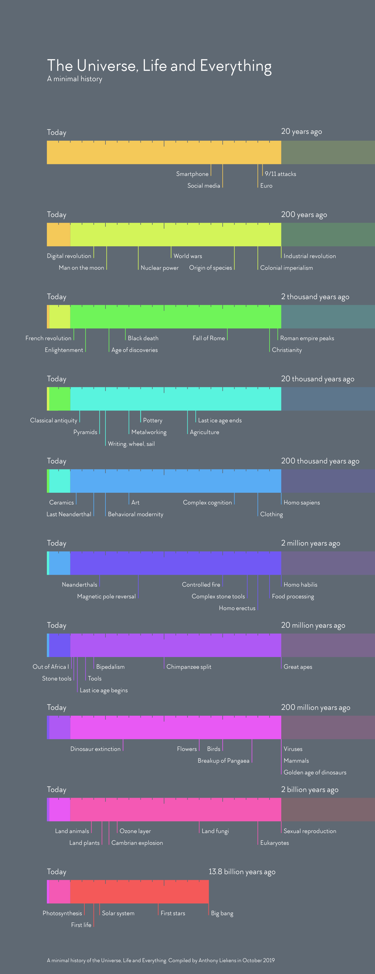
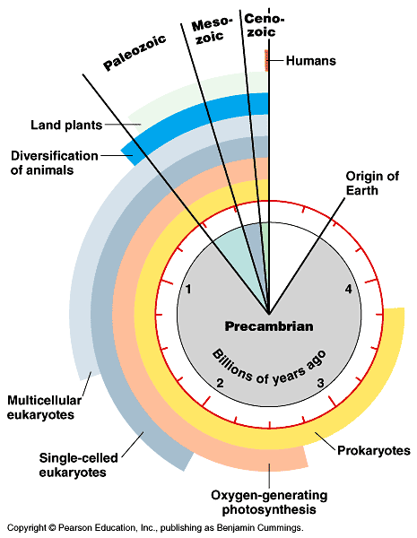
References
Arendt, D., Tosches, M. A., & Marlow, H. (2016). From nerve net to nerve ring, nerve cord and brain — evolution of the nervous system. Nature Reviews Neuroscience, 17(1), 61–72. https://doi.org/10.1038/nrn.2015.15
Baumann, N., & Pham-Dinh, D. (2001). Biology of oligodendrocyte and myelin in the mammalian central nervous system. Physiological Reviews, 81(2), 871–927. https://doi.org/10.1152/physrev.2001.81.2.871
Cao, M., Huang, H., & He, Y. (2017). Developmental connectomics from infancy through early childhood. Trends in Neuroscience, 40(8), 494–506. https://doi.org/10.1016/j.tins.2017.06.003
Chi, J. G., Dooling, E. C., & Gilles, F. H. (1977). Gyral development of the human brain. Ann. Neurol., 1(1), 86–93. https://doi.org/10.1002/ana.410010109
Dobzhansky, T. (1973). Nothing in biology makes sense except in the light of evolution. The American Biology Teacher, 35(3), pp. 125–129. Retrieved from http://www.jstor.org/stable/4444260
Gogtay, N., Giedd, J. N., Lusk, L., Hayashi, K. M., Greenstein, D., Vaituzis, A. C., … Thompson, P. M. (2004). Dynamic mapping of human cortical development during childhood through early adulthood. Proc. Natl. Acad. Sci. U. S. A., 101(21), 8174–8179. https://doi.org/10.1073/pnas.0402680101
Götz, M., & Huttner, W. B. (2005). The cell biology of neurogenesis. Nat. Rev. Mol. Cell Biol., 6(10), 777–788. https://doi.org/10.1038/nrm1739
Hagmann, P., Sporns, O., Madan, N., Cammoun, L., Pienaar, R., Wedeen, V. J., … Grant, P. E. (2010). White matter maturation reshapes structural connectivity in the late developing human brain. Proceedings of the National Academy of Sciences, 107(44), 19067–19072. https://doi.org/10.1073/pnas.1009073107
Herculano-Houzel, S. (2012). The remarkable, yet not extraordinary, human brain as a scaled-up primate brain and its associated cost. Proceedings of the National Academy of Sciences of the United States of America, 109 Suppl 1, 10661–10668. https://doi.org/10.1073/pnas.1201895109
Herculano-Houzel, S. (2016). The human advantage: A new understanding of how our brain became remarkable. MIT Press. Retrieved from https://market.android.com/details?id=book-DMqpCwAAQBAJ
Herculano-Houzel, S. (2017). Numbers of neurons as biological correlates of cognitive capability. Current Opinion in Behavioral Sciences, 16(Supplement C), 1–7. https://doi.org/10.1016/j.cobeha.2017.02.004
Hofman, M. A. (2014). Evolution of the human brain: When bigger is better. Frontiers in Neuroanatomy, 8. https://doi.org/10.3389/fnana.2014.00015
Irimia, A., & Van Horn, J. (2014). Systematic network lesioning reveals the core white matter scaffold of the human brain. Frontiers in Human Neuroscience, 8, 51. https://doi.org/10.3389/fnhum.2014.00051
Johnson, M. H. (2001). Functional brain development in humans. Nat. Rev. Neurosci., 2(7), 475–483. https://doi.org/10.1038/35081509
Kang, H. J., Kawasawa, Y. I., Cheng, F., Zhu, Y., Xu, X., Li, M., … Šestan, N. (2011). Spatio-temporal transcriptome of the human brain. Nature, 478(7370), 483–489. https://doi.org/10.1038/nature10523
Knickmeyer, R. C., Gouttard, S., Kang, C., Evans, D., Wilber, K., Smith, J. K., … Gilmore, J. H. (2008). A structural MRI study of human brain development from birth to 2 years. J. Neurosci., 28(47), 12176–12182. https://doi.org/10.1523/JNEUROSCI.3479-08.2008
Konner, M. (2011). The Evolution of Childhood. Belknap Press of Harvard University Press. Retrieved from http://www.hup.harvard.edu/catalog.php?isbn=9780674062016
Kuzawa, C. W., Chugani, H. T., Grossman, L. I., Lipovich, L., Muzik, O., Hof, P. R., … Lange, N. (2014). Metabolic costs and evolutionary implications of human brain development. Proc. Natl. Acad. Sci. U. S. A., 111(36), 13010–13015. https://doi.org/10.1073/pnas.1323099111
Miller, J. D., Scott, E. C., & Okamoto, S. (2006). Public acceptance of evolution. SCIENCE-NEW YORK THEN WASHINGTON-, 313(5788), 765. https://doi.org/10.1126/science.1126746
Northcutt, R. G. (2002). Understanding vertebrate brain evolution. Integr. Comp. Biol., 42(4), 743–756. https://doi.org/10.1093/icb/42.4.743
Petrican, R., Taylor, M. J., & Grady, C. L. (2017). Trajectories of brain system maturation from childhood to older adulthood: Implications for lifespan cognitive functioning. Neuroimage. https://doi.org/10.1016/j.neuroimage.2017.09.025
Rakic, P. (2009). Evolution of the neocortex: A perspective from developmental biology. Nature Reviews Neuroscience, 10(10), 724–735.
Shaw, P., Kabani, N. J., Lerch, J. P., Eckstrand, K., Lenroot, R., Gogtay, N., … Others. (2008). Neurodevelopmental trajectories of the human cerebral cortex. Journal of Neuroscience, 28(14), 3586–3594. https://doi.org/10.1523/JNEUROSCI.5309-07.2008
Wrangham, R. (2009). Catching fire: How cooking made us human. Basic Books. Retrieved from https://market.android.com/details?id=book-ebEOupKz-rMC