3Blue1Brown. (2018).
But what is the Fourier Transform? A visual introduction. You
Tube. Retrieved from
https://www.youtube.com/watch?v=spUNpyF58BY&t=487s
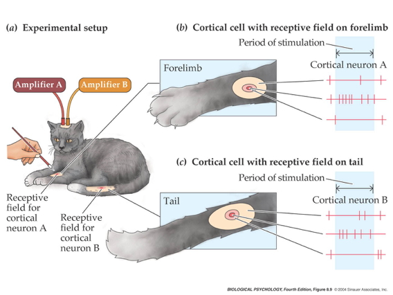
Alonso, J.-M., & Chen, Y. (2009). Receptive field.
Scholarpedia Journal,
4, 5393.
https://doi.org/10.4249/scholarpedia.5393
Ayzenberg, V., & Behrmann, M. (2022). Does the brain’s ventral visual pathway compute object shape?
Trends in Cognitive Sciences,
26, 1119–1132.
https://doi.org/10.1016/j.tics.2022.09.019
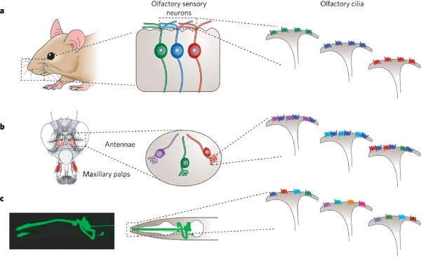
Bargmann, C. I. (2006). Comparative chemosensation from receptors to ecology.
Nature,
444, 295–301.
https://doi.org/10.1038/nature05402
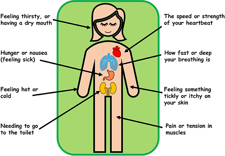
Barker, M., Brewer, R., & Murphy, J. (2021). What is interoception and why is it important?
Frontiers for Young Minds,
9.
https://doi.org/10.3389/frym.2021.558246
Bucalo, P. (2015).
Falcon belly dance. You
Tube. Retrieved from
https://www.youtube.com/watch?v=JGArTWOJtXs
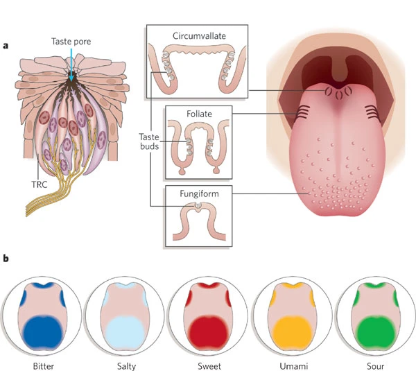
Chandrashekar, J., Hoon, M. A., Ryba, N. J. P., & Zuker, C. S. (2006). The receptors and cells for mammalian taste.
Nature,
444, 288–294.
https://doi.org/10.1038/nature05401
Deflorio, D., Di Luca, M., & Wing, A. M. (2022). Skin and mechanoreceptor contribution to tactile input for perception: A review of simulation models.
Frontiers in Human Neuroscience,
16, 862344.
https://doi.org/10.3389/fnhum.2022.862344
Dougherty, R. F., Koch, V. M., Brewer, A. A., Fischer, B., Modersitzki, J., & Wandell, B. A. (2003). Visual field representations and locations of visual areas
V1/2/3 in human visual cortex.
Journal of Vision,
3(10), 1–1.
https://doi.org/10.1167/3.10.1
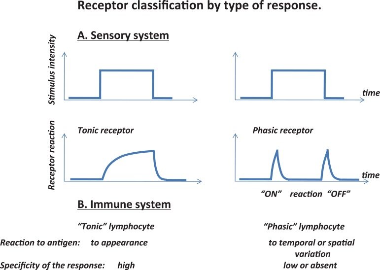
Dozmorov, I. (2011). Immune system as sensory system.
International Journal of Biomedical Science,
6, 167–175.
https://doi.org/10.59566/IJBS.2010.6167
ewakili. (2021).
Foveated rendering shown by meta’s oculus, reducing compute load by 95%. You
Tube. Retrieved from
https://www.youtube.com/watch?v=NPK8eQ4o8Pk
Gibson, J. J. (1966).
The senses considered as perceptual systems. doi.apa.org. Retrieved from
http://doi.apa.org/psycinfo/1966-35026-000
Herman, J. P. (2013). Neural control of chronic stress adaptation.
Frontiers in Behavioral Neuroscience,
7, 61.
https://doi.org/10.3389/fnbeh.2013.00061
Humphries, C., Liebenthal, E., & Binder, J. R. (2010). Tonotopic organization of human auditory cortex.
NeuroImage,
50, 1202–1211.
https://doi.org/10.1016/j.neuroimage.2010.01.046
Lester, P. (2009).
Hubel and wiesel cat experiment. You
Tube. Retrieved from
https://www.youtube.com/watch?v=IOHayh06LJ4&source_ve_path=MTc4NDI0
Namkung, H., Kim, S.-H., & Sawa, A. (2017). The insula: An underestimated brain area in clinical neuroscience, psychiatry, and neurology.
Trends in Neurosciences,
40(4), 200–207.
https://doi.org/10.1016/j.tins.2017.02.002
OTGeddie. (2017).
Tactile localization. You
Tube. Retrieved from
https://www.youtube.com/watch?v=t97QiEiKjD8
Panichello, M. F., Cheung, O. S., & Bar, M. (2013). Predictive feedback and conscious visual experience.
Perception Science,
3, 620.
https://doi.org/10.3389/fpsyg.2012.00620
Randeberg, L. (2005). Diagnostic applications of diffuse reflectance spectroscopy. Retrieved from
https://www.semanticscholar.org/paper/ec9450b79923e2e2152b54ab9241b60bc5374944
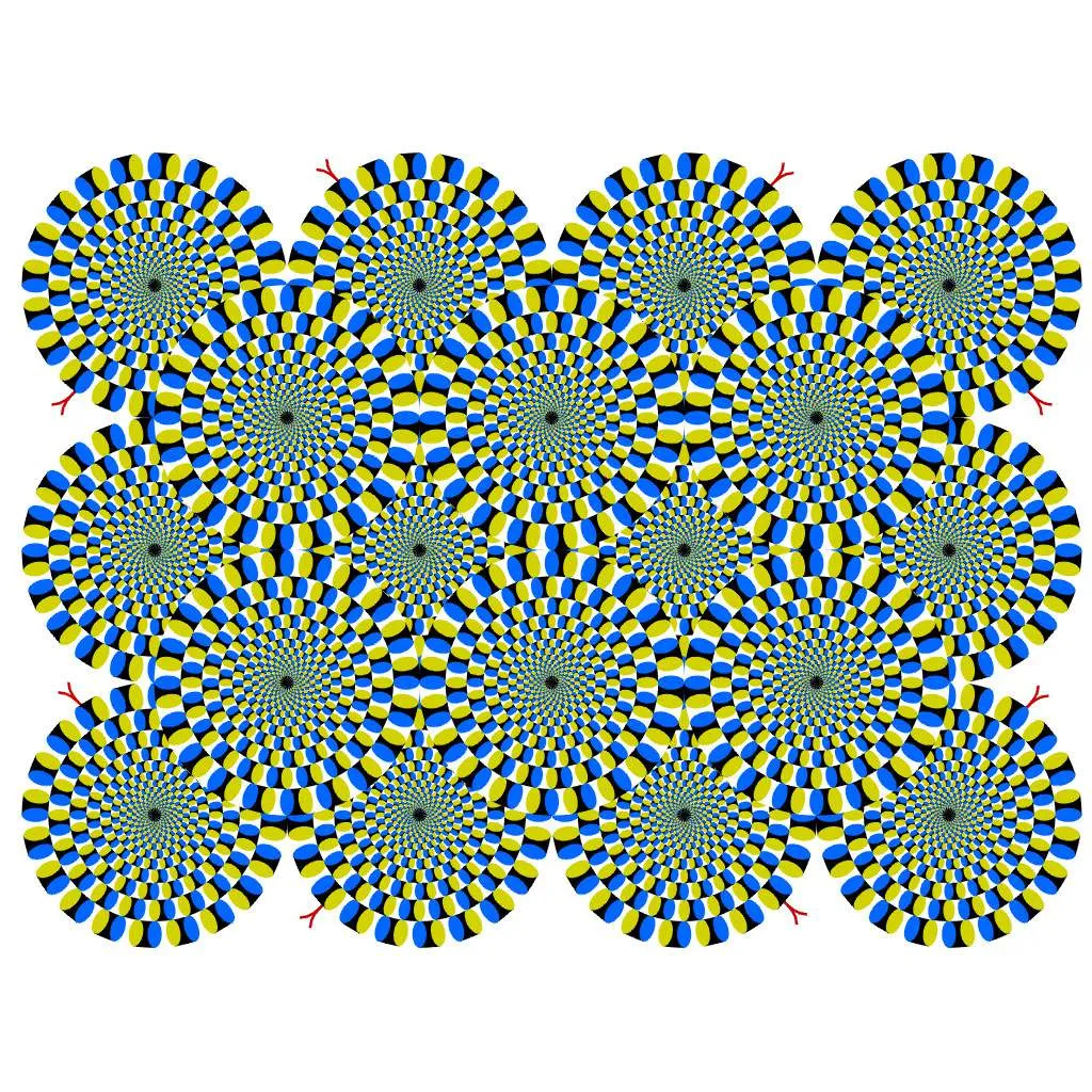
rasmusab. (2013).
My cat can see the rotating snake illusion! You
Tube. Retrieved from
https://www.youtube.com/watch?v=CcXXQ6GCUb8
Riding light. (n.d.). Retrieved from
https://vimeo.com/117815404
Roark, M. W., & Stringham, J. M. (2019). Visual performance in the
“real world”: Contrast sensitivity, visual acuity, and effects of macular carotenoids.
Molecular Nutrition & Food Research,
63(15), e1801053.
https://doi.org/10.1002/mnfr.201801053
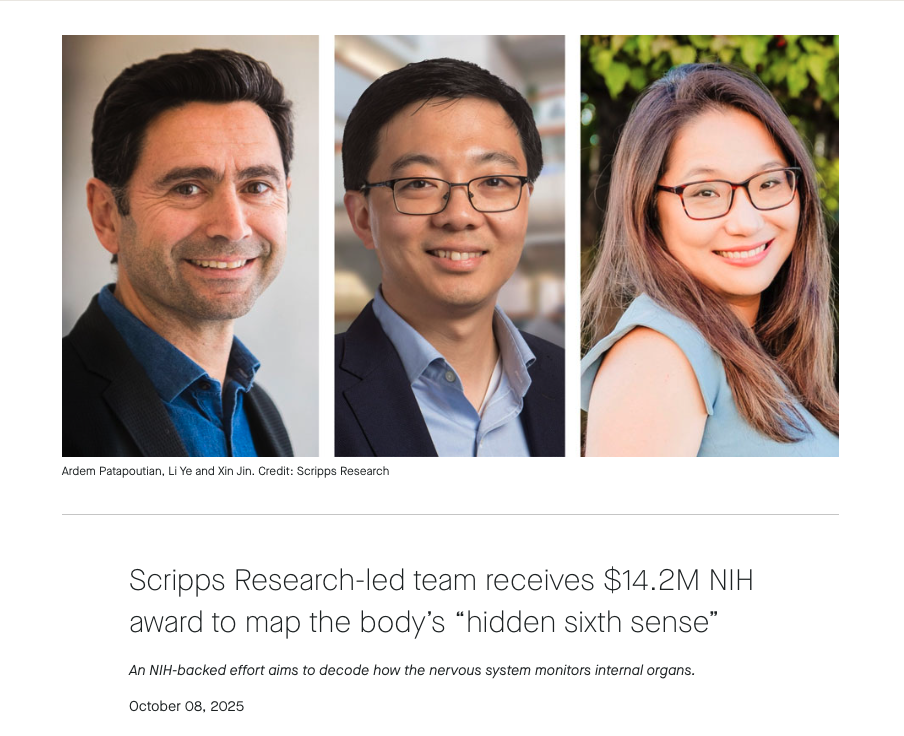
Scripps research-led team receives $14.
2M NIH award to map the body’s
“hidden sixth sense.” (2025, October 8). Retrieved November 13, 2025, from
https://www.scripps.edu/news-and-events/press-room/2025/20251008-nih-award.html
Smith, G. E., Chouinard, P. A., & Byosiere, S.-E. (2021). If
I fits
I sits: A citizen science investigation into illusory contour susceptibility in domestic cats (felis silvestris catus).
Applied Animal Behaviour Science,
240, 105338.
https://doi.org/10.1016/j.applanim.2021.105338
Stereo3DMovies. (2010).
Cloudy with a chance of meatballs 3D snippet (yt3d:enable=true). You
Tube. Retrieved from
https://www.youtube.com/watch?v=KjAQdc29vF8
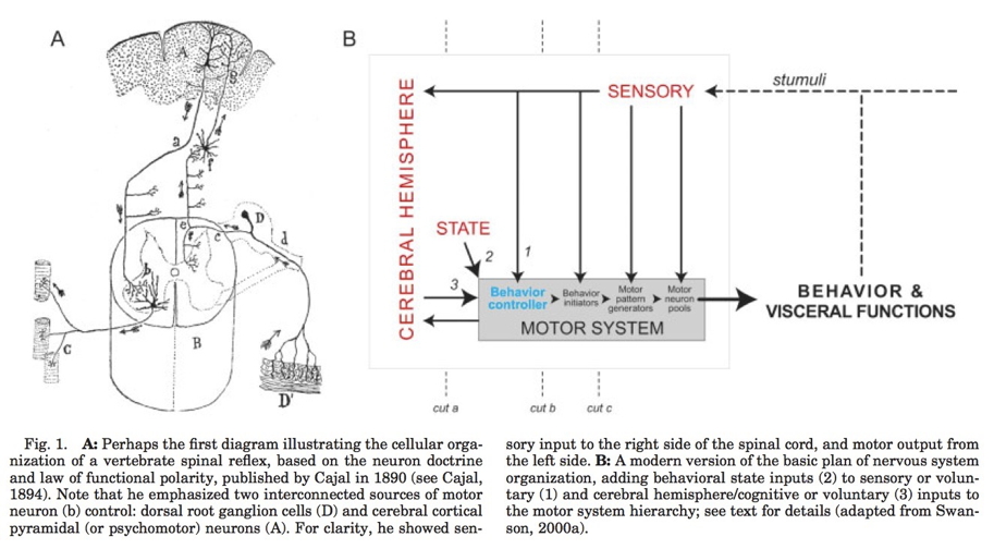
Swanson, L. W. (2005). Anatomy of the soul as reflected in the cerebral hemispheres: Neural circuits underlying voluntary control of basic motivated behaviors.
Journal of Comparative Neurology,
493(1), 122–131.
https://doi.org/10.1002/cne.20733
Swanson, L. W. (2012). Brain architecture: Understanding the basic plan. Oxford University Press.
TheWhoVEVO. (2016).
The who - pinball wizard (live at the isle of wight, 1970). You
Tube. Retrieved from
https://www.youtube.com/watch?v=-J03yCE15rg&list=RD-J03yCE15rg&start_radio=1
Wandell, B. (1995, October 26). Foundations of vision (1995). Retrieved February 5, 2026, from
https://wandell.github.io/FOV-1995/
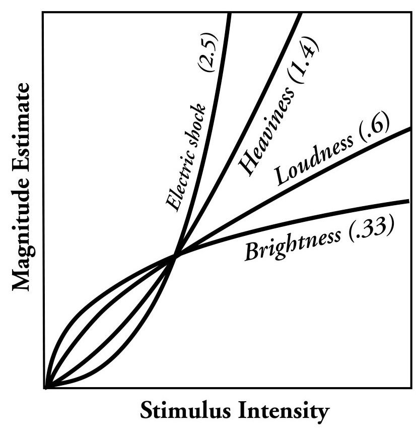
Wikipedia contributors. (2025a, January 30). Stevens’s power law. Retrieved from
https://en.wikipedia.org/wiki/Stevens%27s_power_law
Wikipedia contributors. (2025b, August 17). Psychophysics. Retrieved from
https://en.wikipedia.org/wiki/Psychophysics
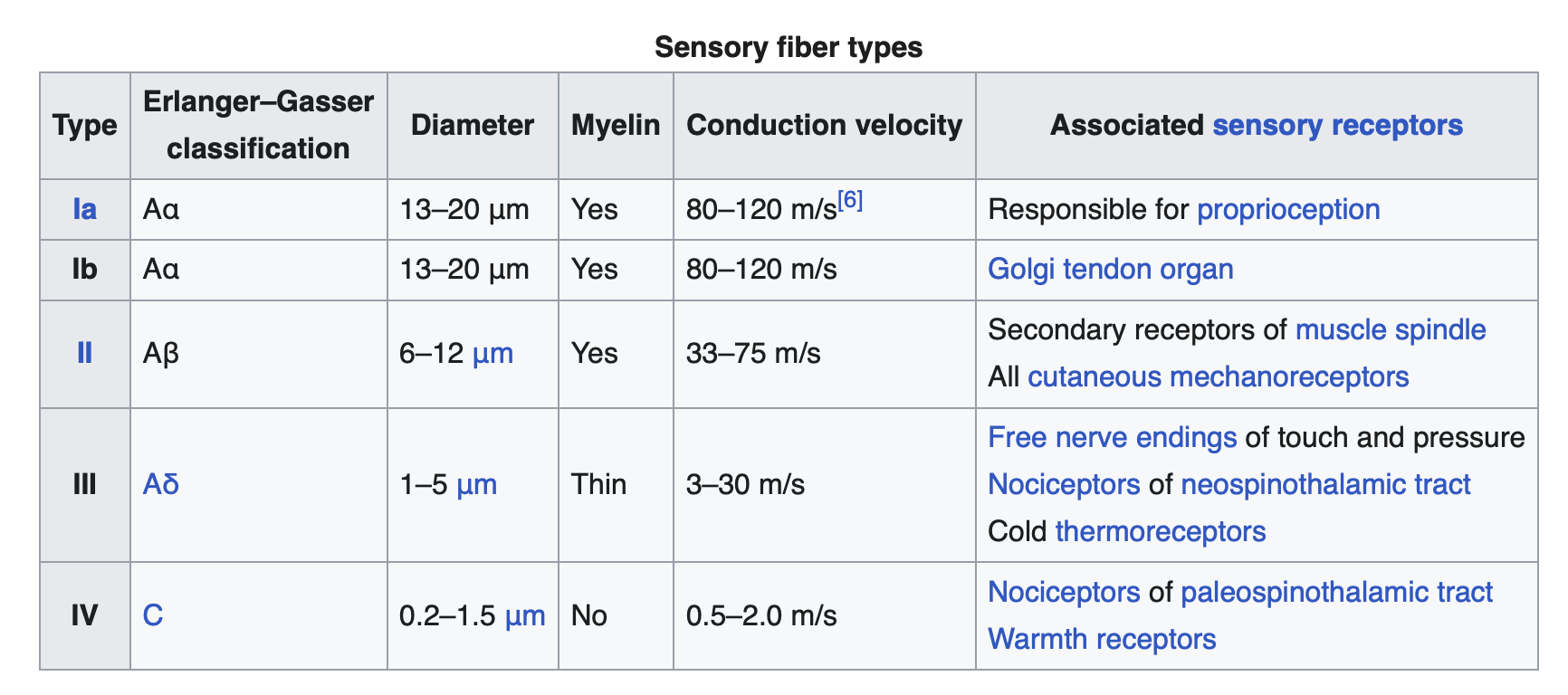
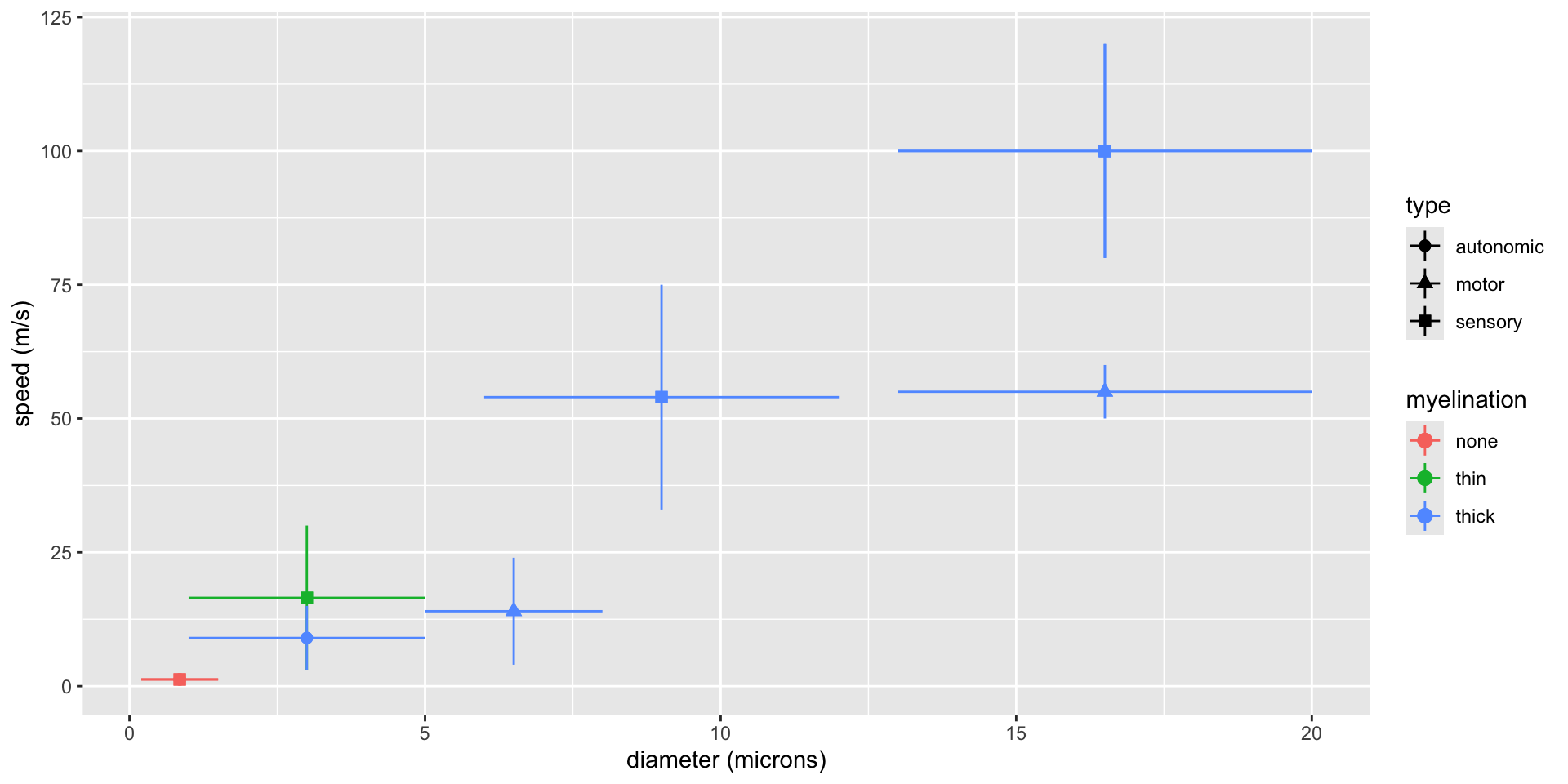
Wikipedia contributors. (2025c, August 30). Nerve conduction velocity. Retrieved from
https://en.wikipedia.org/wiki/Nerve_conduction_velocity