Learning & Memory

PSY 511.001 Spr 2026

Rick Gilmore

Department of Psychology

Overview

Prelude

timvitkuske (2014)

Announcements

Today’s topics

  • Big picture questions
  • Biological bases of
  • Disorders of memory
  • Working memory

Warm-up

Which of the following sensory systems have clear topographic maps in the cerebral cortex?

  • A. Vision
  • B. Audition
  • C. Somatosensation
  • D. Olfaction
  • E. A, B, & C.

Which of the following sensory systems have clear topographic maps in the cerebral cortex?

  • A. Vision
  • B. Audition
  • C. Somatosensation
  • D. Olfaction
  • E. A, B, & C.

Dougherty et al. (2003) Figure 1

Dougherty et al. (2003) Figure 1

Humphries, Liebenthal, & Binder (2010) Figure 3

Humphries et al. (2010) Figure 31

True or false: There is little evidence that psychological judgements of stimulus magnitude are linear.

  • A. True
  • B. False

True or false: There is little evidence that psychological judgements of stimulus magnitude are linear.

  • A. True
  • B. False

Humans can’t see or even imagine reddish-green because

  • A. Long (reddish) wavelength cones and medium (greenish) wavelength cones connect to downstream cells with opposing effects.
  • B. Rod photoreceptors are monochromatic.
  • C. Lights with both long and medium wavelengths appear yellow.
  • D. Color is a psychological categorization of continuous wavelength spectra.

http://cnx.org/content/col11496/1.6/

http://cnx.org/content/col11496/1.6/

Randeberg (2005) Figure 8

Randeberg (2005) Figure 8

Big picture questions

Memory capacity of the human brain?

  • 1e12 neurons
  • 1e3 synapses/neuron
  • 1e15 synapses or 1.25e14 bytes
  • 1e9 gigabyte, 1e12 terabyte, 1e15 petabyte
  • So 125 TB?

Reber (2010)

What is learning?

Learning is the process of acquiring new understanding, knowledge, behaviors, skills, values, attitudes, and preferences.

https://en.wikipedia.org/wiki/Learning

Types of learning?

  • Non-associative
    • Change in response to repeated encounters with same stimulus/event
    • Habituation -> weaker response
    • Sensitization -> stronger response

Types of learning

  • Associative
    • “Associates” or links two events/items with one another

Types of learning

  • Classical conditioning
    • Link event with physiological response
  • Operant/instrumental conditioning
    • Link behaviors with external events (rewards/punishments)

Associative learning

  • Types
    • Observational/imitative

Associative learning

  • Types
    • Sequence
      • A, B, C, D, …
    • Episodic
      • Mom’s 85th
    • Semantic
      • Thanksgiving is a holiday celebrated in Canada and the U.S…

Learning styles?

“Learning styles as a myth” (2017)

“Learning styles as a myth” (2017)

Are myths (Pashler, McDaniel, Rohrer, & Bjork, 2008)

Although the literature on learning styles is enormous, very few studies have even used an experimental methodology capable of testing the validity of learning styles applied to education. Moreover, of those that did use an appropriate method, several found results that flatly contradict the popular meshing hypothesis…

We conclude therefore, that at present, there is no adequate evidence base to justify incorporating learning-styles assessments into general educational practice.

Pashler et al. (2008)

How do machines “learn”?

  • Often, but not always, by imitating brains.

“Deep learning vs machine learning” (2022)

“Deep learning vs machine learning” (2022)

Neural networks

Tch (2017); from Veen (n.d.)

Tch (2017); from Veen (n.d.)

Network learning rules

  • How/when to change connections between network nodes
  • Types
    • Unsupervised (i.e., covariance/correlation)
    • Supervised (change based on ‘error’, external ‘supervision’)
    • Reinforcement (learn now)

What is memory?

  • A: Information encoding, storage, retrieval

Types?

  • Facts/events/places/feelings vs. skills
  • Short vs. long-term
    • Working memory ~ short-term maintenance for guiding action
  • Explicit (declarative: semantic vs. episodic) vs. implicit (procedural)
  • Retrospective (from the past) vs. prospective (to be remembered)
  • Recognition (judgment of familiarity or novelty) vs. recall

Squire (2004)

Squire (2004)

Computer memory vs. brains

Computers Brains
Separate memory and processing stores Processing & memory not separate; specialized regions
Specific addresses Distributed networks (no addresses?)
(Usually) non-volatile Volatile
All types–images, sounds, text, data–stored as binary sequences, e.g., 01101110 Stored patterns of synaptic connections and ???

Computer memory vs. brains

Computers Brains
Render binary sequences differently based on information about the type of data stored Retrieve different types based on ???
Inspired by mathematical models of neurons Can be simulated by mathematical models implemented in computers

Artificial neurons

McCulloch-Pitts artificial neuron

McCulloch-Pitts artificial neuron

Biological bases of…

Does learning require a network?

Gershman et al. (2021) Figure 1

Gershman et al. (2021) Figure 12

Gershman et al. (2021) from B. Gelber (1952)

Gershman et al. (2021)3 from B. Gelber (1952)

This paper presents a new approach to behavioral problems which might be called molecular biopsychology… Simply stated, it is hypothesized that the memory engram must be coded in macromolecule…As the geneticist studies the inherited characteristics of an organism the psychologist studies the modification of this inherited matrix by interaction with the environment.

Beatrice Gelber (1962)

Possibly the biochemical and cellular physiological processes which encode new responses are continuous throughout the phyla (as genetic codes are) and therefore would be reasonably similar for a protozoan and a mammal.

Beatrice Gelber (1962)

Taking stock, we believe that Gelber’s experiments, though not without their limitations, convincingly demonstrated Pavlovian conditioning in Paramecia. Sadly, her critics seem to have won in the long term. Most reviews of the literature, if they mention Gelber’s work at all, quickly dismiss it…

Gershman et al. (2021)

If single cells can learn then they must be using a non-synaptic form of memory storage. The idea that intracellular molecules store memories has a long history, mainly in the study of multicellular organisms.

Gershman et al. (2021)

Learning in nervous systems

The idea that memory is stored as enduring changes in the brain dates back at least to the time of Plato and Aristotle (circa 350 BCE), but its scientific articulation emerged in the 20th century when Richard Semon introduced the term “engram” to describe the neural substrate for storing and recalling memories.

Josselyn & Tonegawa (2020)

Mechanism(s)

Essentially, Semon proposed that an experience activates a population of neurons that undergo persistent chemical and/or physical changes to become an engram. Subsequent reactivation of the engram by cues available at the time of the experience induces memory retrieval.

Josselyn & Tonegawa (2020)

Neural substrates (engrams) consist of

  • Changes in patterns of neural activity
  • Changes in connectivity
    • New synapses
    • Altered synapses (strengthened or weakened)

Donald Hebb

  • Canadian clinical neuropsychologist
  • Author of The Organization of Behavior (1949)

Hebbian learning

When an axon of cell A is near enough to excite cell B and repeatedly or persistently takes part in firing it, some growth process or metabolic change takes place in one or both cells such that A’s efficacy, as on of the cells firing B, is increased.

Hebb (1949)

Hebbian learning

Neurons that fire together wire together.

Löwel & Singer (1992)

Hebbian learning is associative

  • Neuron A active &
  • Neuron B active \(\rightarrow\)

flowchart LR
  A --> B

  • Strengthen link (synapse) between them

flowchart LR
  A ==> B

How synapses strengthen

  • Long-term potentiation (LTP)
  • Make more potent (stengthen) synapse based on recent co-activity
  • Change at synapse == physical basis of one type of Hebbian learning

LTP discovery

  • Bliss & Lømo (1973)
  • Granule cell neurons in hippocampus dentate gyrus (DG)
  • \(\theta\) band (10–20 Hz) stim for 10–15 sec, or 100 Hz stim for 3–4 sec

Wikipedia

Wikipedia

LTP discovery

  • shortened response latency, increased EPSP, increased population response over minutes or hours

Wikipedia

Wikipedia

Mechanism(s) of LTP

  • effectiveness of postsynaptic response
    • post-synaptic, early LTP
  • number of synaptic receptors
    • post-synaptic, late LTP
  • quantity of NT released
    • pre-synaptic, late LTP

Mechanism

NMDA-R from “Pitt medical neuroscience” (n.d.)

NMDA-R from “Pitt medical neuroscience” (n.d.)
  • N-methyl-D-aspartate receptor (NMDA-R)
    • Name derived from agonist molecule that selectively binds to the receptor
  • ‘Coincidence’ detector
    • Sending cell has released NT
    • Receiving cell is/has been recently active

Pathways to plasticity

  • Glutamate release activates
    • ionotropic AMPA Glu receptors
    • metabotropic Glu receptors
    • N-methyl-D-aspartate (NMDA) Glu receptors (NMDA-R)

NMDA-R

NMDA-R from “Pitt medical neuroscience” (n.d.)

NMDA-R from “Pitt medical neuroscience” (n.d.)
  • Chemically-gated
    • Binds glutamate
      • When nearby neuron releases
    • Binds glycine (co-factor/co-agonist)
    • So, ligand-gated

NMDA-R

NMDA-R from “Pitt medical neuroscience” (n.d.)

NMDA-R from “Pitt medical neuroscience” (n.d.)
  • Voltage-gated
    • Receiving cell depolarizes (generates excitatory post-synaptic potential or EPSP)
    • \(Zn^{++}\) or \(Mg^{++}\) ion ‘plug’ repelled under depolarization
    • \(Na^+\) & \(Ca^{++}\) influx; \(K^+\) outflux

https://i2.wp.com/www.gatewaypsychiatric.com/wp-content/uploads/2015/02/NMDA-Receptor-and-Depression.jpg?ssl=1

https://i2.wp.com/www.gatewaypsychiatric.com/wp-content/uploads/2015/02/NMDA-Receptor-and-Depression.jpg?ssl=1

Pathways to plasticity

  • \(Ca^{++}\) entry via NMDA receptor activates protein kinases (CaMKII and PKAII)
  • Early LTP (up to few hours)
    • protein kinases phosphorylate (add \(P\) group to) postsynaptic AMPA receptors
    • Increase current flow through AMPA (Glu) receptors

Pathways to plasticity

  • Late LTP
    • depends on protein synthesis to generate new AMPA receptor
    • insertion of new AMPA receptors into postsynaptic membrane
  • Retrograde signal generator influences presynaptic response

Schematic

flowchart LR
  A -->|Glu| B

flowchart LR
  A -->|Glu| C[AMPA-R]
  A -->|Glu| B[NMDA-R]
  C --> |generates EPSP| B
  B --> |Ca++ enters| D:::hidden
  
  %% Clickable links
  click C "https://en.wikipedia.org/wiki/AMPA_receptor"
  click B "https://en.wikipedia.org/wiki/NMDA_receptor"

  • Ca++ entry strengthens synapse
  • Modulate existing AMPA-R; synthesize new AMPA-R

NMDA-Rs & associative learning

  • Receptors associate (link)
    • Concept A -> Concept B
    • Neuron A -> Neuron B
  • I say Donald…you say…

Wikipedia

Wikipedia

LTP

LTP

Aplysia

Aplysia

Eric R. Kandel

Eric R. Kandel

NMDA-R clinical significance

  • Memantine (Alzheimer’s Disease treatment)
    • NMDA-R antagonist
    • Controls over-activation and \(Ca^{++}\) excitotoxicity?
  • Implicated in effects of phencylidine (PCP)
    • Link to Glutamate hypothesis of schizophrenia?

NMDA-R clinical significance

  • Ketamine is NMDA-R antagonist4
    • anesthesia, sedation pain relief
    • possible short-term relief for depression
  • Linked to analgesic effects of nitrous oxide (laughing gas; \(NO\))
  • Alcohol (ethanol) inhibits (Ron & Wang, 2011)

Spike-timing-dependent plasticity

Caporale & Dan (2008) Figure 1

Caporale & Dan (2008) Figure 15

Caporale & Dan (2008) Figure 2

Caporale & Dan (2008) Figure 26
  • A before B: strengthen \(A \rightarrow B\)
  • A after B: weaken \(A \rightarrow B\)
    • Long-term Depression or LTD
  • Neural Plasticity
    • Lasting changes in neural firing, connectivity
  • NMDA-R a molecular mechanism for implementing LTP and spike-timing-dependent plasticity

Summary

  • Learning and memory involve changes in neural firing, circuitry
    • And probably molecular changes inside neurons
  • Hebbian learning a type of associative learning
  • NMDA receptors implement coincidence detectors that undergird associative learning at the synapse

Memory systems

Lashley’s search for the ‘engram’

K. S. Lashley & Clark (1946)

K. S. Lashley & Clark (1946)

the area subdivisions are in large part anatomically meaningless and misleading as to the presumptive functional divisions of the cortex

K. S. Lashley & Clark (1946)

  • Memory not localized in the rat cerebral cortex!

Modern views

  • Cerebral cortex less central to “engram-like” memory than other areas

Squire (2004)

Squire (2004)

Hippocampus

https://upload.wikimedia.org/wikipedia/commons/5/5b/Hippocampus_and_seahorse_cropped.JPG

https://upload.wikimedia.org/wikipedia/commons/5/5b/Hippocampus_and_seahorse_cropped.JPG

Santiago Ramon Y Cajal; Source: Wikipedia

Santiago Ramon Y Cajal; Source: Wikipedia

Hippocampus

  • Dense in NMDA receptors
  • Central “hub” in network?
  • Formation, storage, consolidation of long-term episodic or declarative memories

Hippocampus

Kjelstrup et al. (2008) Figure 1

Kjelstrup et al. (2008) Figure 17

Memory-taxing behaviors alter hippocampus

  • London taxi drivers (a high memory demand profession) have specialized hippocampi (Maguire et al., 2000)
  • Not due to self-motion, driving experience, or stress per se

Woollett & Maguire (2011) Figure 1

Woollett & Maguire (2011) Figure 1

Gray matter volume differences in the hippocampus relative to controls have been reported to accompany this expertise. While these gray matter differences could result from using and updating spatial representations, they might instead be influenced by factors such as self-motion, driving experience, and stress. We examined the contribution of these factors by comparing London taxi drivers with London bus drivers, who were matched for driving experience and levels of stress, but differed in that they follow a constrained set of routes.

Maguire, Woollett, & Spiers (2006)

We found that compared with bus drivers, taxi drivers had greater gray matter volume in mid-posterior hippocampi and less volume in anterior hippocampi. Furthermore, years of navigation experience correlated with hippocampal gray matter volume only in taxi drivers, with right posterior gray matter volume increasing and anterior volume decreasing with more navigation experience. This suggests that spatial knowledge, and not stress, driving, or self-motion, is associated with the pattern of hippocampal gray matter volume in taxi drivers.

Maguire et al. (2006)

Figure 2. Gray Matter Volume Changes between T1 and T2 in Qualified Trainees

Figure 3. Plot of Gray Matter Intensities across Groups and Time
Figure 1

Evidence from other species

  • Birds whose ethologies have high memory demands have larger hippocampi.

Sherry, Vaccarino, Buckenham, & Herz (1989)

Sherry et al. (1989)

  • Birds with high memory caching demands use sparse (“bar-code”-like ) representations in hippocampus

Chettih, Mackevicius, Hale, & Aronov (2024)

Chettih et al. (2024)

Cerebral cortex

Steel et al. (2024) Figure 1

Steel et al. (2024) Figure 18

Steel et al. (2024) Figure 2

Steel et al. (2024) Figure 29

Together with recent reports showing retinotopic coding persisting as far as the ‘cortical apex’ [15,16,31], including the DMN [15,16], and the hippocampus [31,32], our findings challenge conventional views of brain organization, which generally assume that retinotopic coding is replaced by abstract amodal coding as information propagates through the visual hierarchy [7,8,9,10] toward memory structures [11,12,13,14]

Steel et al. (2024)

Cerebellum

Disorders of memory

Amnesia

  • Acquired loss of memory
  • ≠ normal forgetting
  • Retrograde (‘backwards’ in time)
    • Damage to information acquired pre-injury
    • Temporally graded
  • Anterograde (‘forward’ in time)
    • Damage to information acquired/experienced post-injury

Patient HM10

  • Intractable/untreatable epilepsy
  • Bilateral resection of medial temporal lobe (1953)
  • Epilepsy now treatable
  • But, memory impaired

Grafe (2019)

HM’s amnesia

  • Retrograde amnesia
    • Can’t remember 10 yrs before operation
    • Distant past better than more recent
  • Severe, global anterograde amnesia
    • Impaired learning of new facts, events, people
  • But, skills (mirror learning) intact

Every day is alone in itself, whatever enjoyment I’ve had, and whatever sorrow I’ve had…Right now, I’m wondering, have I done or said anything amiss? You see at this moment, everything looks clear to me, but what happened just before? That’s what worries me. It’s like waking from a dream. I just don’t remember.

Other causes of amnesia

  • Disease
    • Alzheimer’s, herpes virus
  • Korsakoff’s syndrome
    • Result of severe alcoholism
    • Impairs medial thalamus & mammillary bodies

Patient NA

  • Fencing accident
  • Damage to medial thalamus
  • Anterograde + graded retrograde amnesia
  • Are thalamus & medial temporal region connected?

Anagnostaras (2014)

Spared skills in amnesia

  • Skill-learning
  • Mirror-reading, writing
  • Short-term memory
  • “Cognitive” skills
  • Priming

Learning from amnesia

  • Long-term memory for facts, events, people
  • ≠ Short-term memory
  • ≠ Long-term memory for “skills”
  • Separate memory systems in the brain?

Squire (2004)

Squire (2004)

Alzheimer’s Disease (AD)

  • Chronic, neurodegenerative disease affecting ~6.7 M Americans
  • Cognitive dysfunction (memory loss, language difficulties, planning, coordination)
  • Psychiatric symptoms and behavioral disturbances
  • Difficulties with daily living
  • Burns & Iliffe (2009)
  • Build up of amyloid \(\beta\) protein plaques in brain tissue
  • APOE e4 gene variant increases risk 2-12x

Progression

Burns & Iliffe (2009) Figure 1

Burns & Iliffe (2009) Figure 1

In the brain

  • Post-mortem exams show \(\beta\) amyloid plaques and neurofibrillary tangles in hippocampus + other brain areas

AD Treatments

  • Drugs that address amyloid \(\beta\) don’t work especially well
  • Acetylcholinesterase (AChE) inhibitors (e.g. Aricept)
    • ACh a neuromodulator in the brain
    • AChE inhibitors boost/prolong ACh levels

AD Treatments

  • AD the result of disordered immune response (Jevtic, Sengar, Salter, & McLaurin, 2017)?
    • Promising prospective therapy using microglia to remove amyloid \(\beta\) (Hou et al., 2024)
    • Link between genetic factor (APOE4/4 gene) and fatty build-up in microglia (Haney et al., 2024)
  • NMDA-R partial antagonists (e.g., Memantine)
    • Slow/impede formation of disordered new memories to keep established ones intact?

Alzheimer’s Disease (AD)

GBD 2019 Dementia Forecasting Collaborators (2022) Figure 3. Percentage change between 2019 and 2050 in all-age number of individuals with dementia by country.

GBD 2019 Dementia Forecasting Collaborators (2022) Figure 3. Percentage change between 2019 and 2050 in all-age number of individuals with dementia by country.

The other end of the spectrum…

60 Minutes Australia (2018)

Working memory

D’Esposito & Postle (2015)

  • LTM representations of target items + attention -> elevated activation
    • Semantic items
    • Sensorimotor items
  • Capacity for attended items (in Focus of Attention or FoA) limited ~ 4

  • Neural basis
    • sustained activation in PFC
    • subthreshold activation in areas where items are stored
  • Individual differences in visual WM

Luck & Vogel (2013) Figure 2

Luck & Vogel (2013) Figure 2

Wrap-up

Main points

  • Multiple types of learning & memory
  • Learning & memory distributed across the brain
    • Specializations relate to location in the network? (input/output/in-between)
  • Hippocampus + PFC critical areas binding together sensory/semantic info stored elsewhere
  • Changes in synaptic #, strength, connectivity provide cellular basis

Main points

  • Many questions remain
    • How does reinforcement learning work? Where does the “learn-now” signal come from?
      • Reward Prediction Errors (RPE) and dopamine signalling?
    • What about supervised (error-correction) learning?
  • What does Gemini have to say?

Next time

  • Language

Resources

About

This talk was produced using Quarto, using the RStudio Integrated Development Environment (IDE), version 2026.1.0.392.

The source files are in R and R Markdown, then rendered to HTML using the revealJS framework. The HTML slides are hosted in a GitHub repo and served by GitHub pages: https://psu-psychology.github.io/psy-511-scan-fdns-2026-spring/

References

60 Minutes Australia. (2018). People who remember every second of their life | 60 minutes australia. YouTube. Retrieved from https://www.youtube.com/watch?v=hpTCZ-hO6iI
Anagnostaras, S. (2014). Larry squire’s amnesic patient NA. YouTube. Retrieved from https://www.youtube.com/watch?v=1GfFopZSyj8
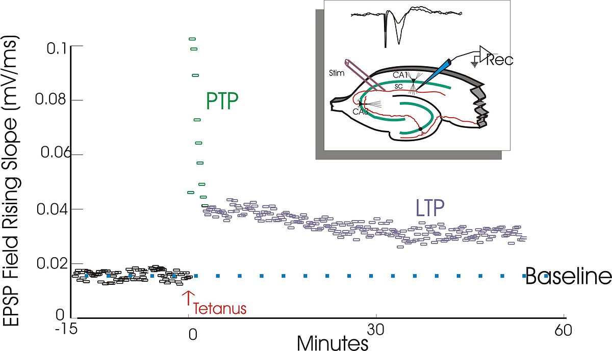
Bliss, T. V. P., & Lømo, T. (1973). Long-lasting potentiation of synaptic transmission in the dentate area of the anaesthetized rabbit following stimulation of the perforant path. J. Physiol., 232(2), 331–356. Retrieved from http://onlinelibrary.wiley.com/doi/10.1113/jphysiol.1973.sp010273/full
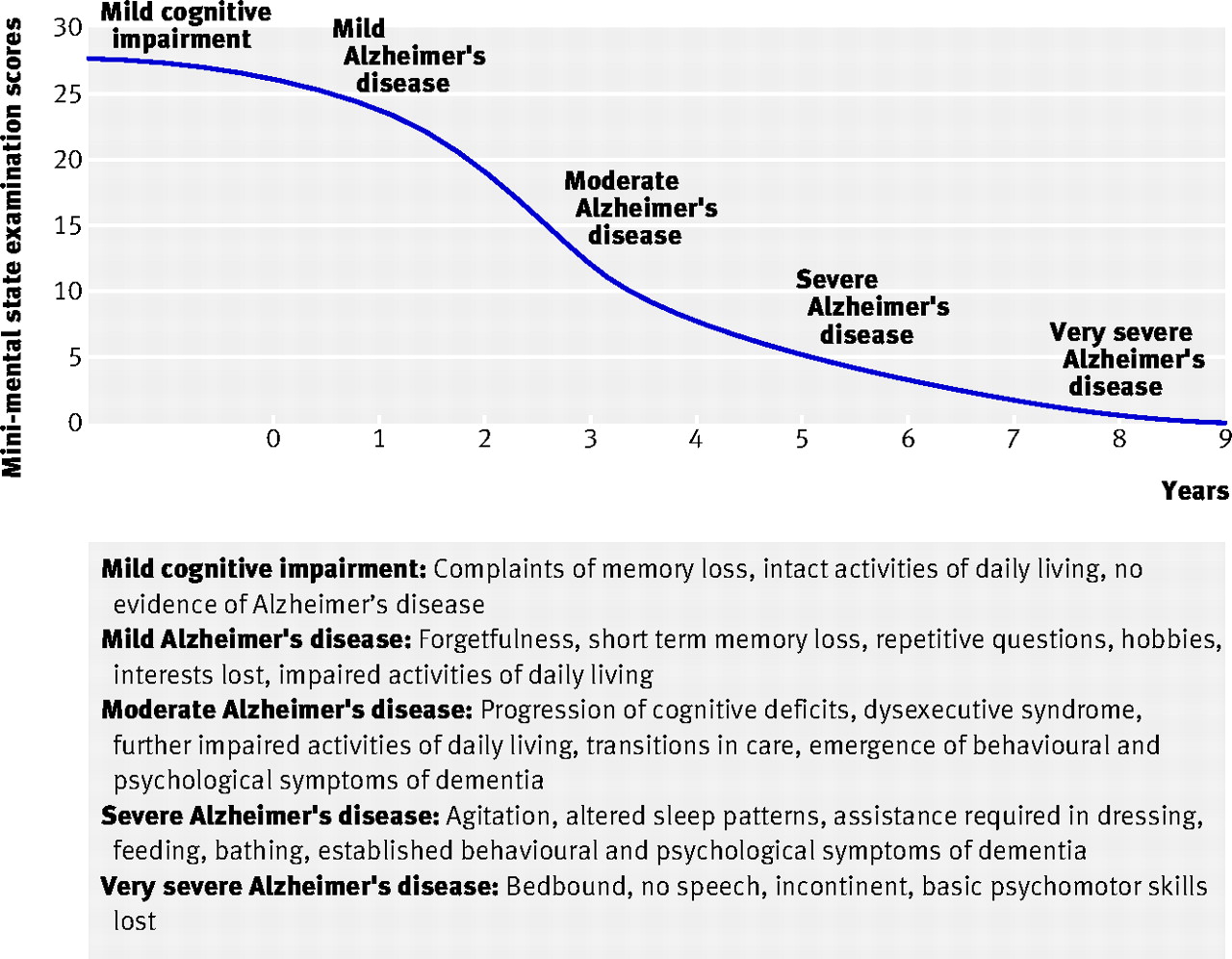
Burns, A., & Iliffe, S. (2009). Alzheimer’s disease. BMJ, 338, b158. https://doi.org/10.1136/bmj.b158
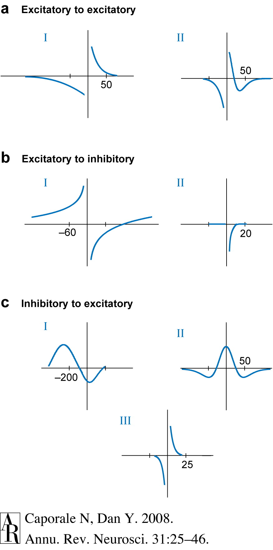
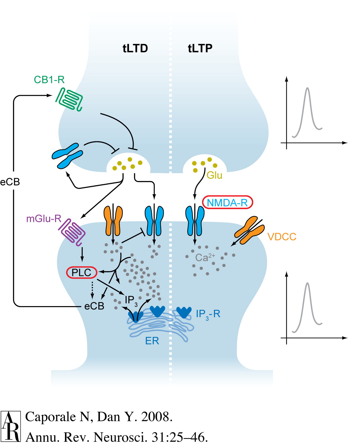
Caporale, N., & Dan, Y. (2008). Spike timing-dependent plasticity: A hebbian learning rule. Annu. Rev. Neurosci., 31, 25–46. https://doi.org/10.1146/annurev.neuro.31.060407.125639
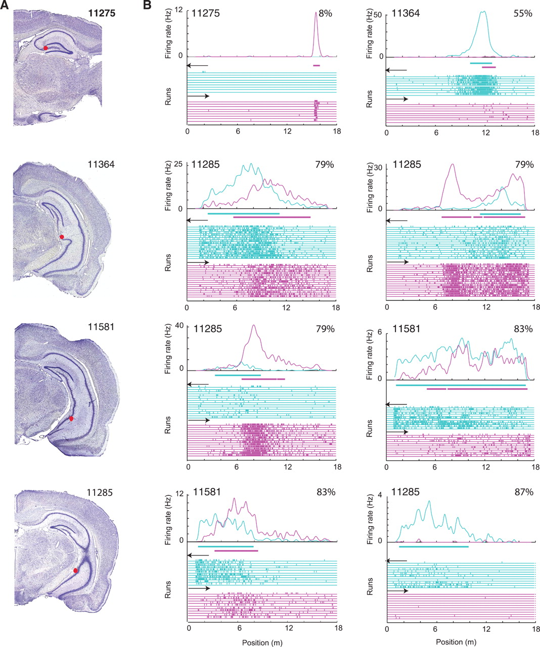
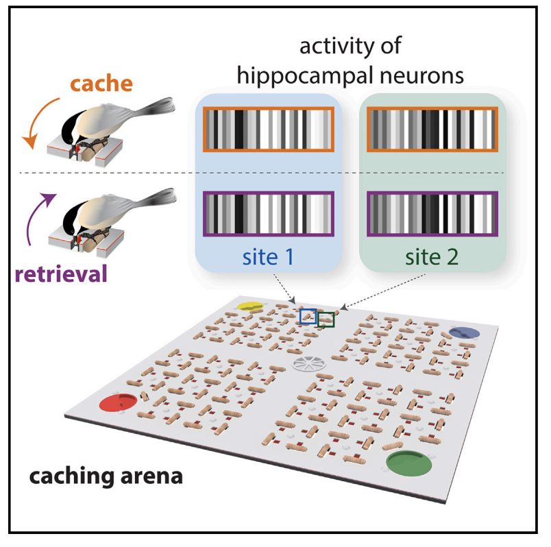
Chettih, S. N., Mackevicius, E. L., Hale, S., & Aronov, D. (2024). Barcoding of episodic memories in the hippocampus of a food-caching bird. Cell. https://doi.org/10.1016/j.cell.2024.02.032
D’Esposito, M., & Postle, B. R. (2015). The cognitive neuroscience of working memory. Annu. Rev. Psychol., 66, 115–142. https://doi.org/10.1146/annurev-psych-010814-015031
Deep learning vs machine learning: The ultimate battle. (2022, May). https://www.turing.com/kb/ultimate-battle-between-deep-learning-and-machine-learning; Turing Enterprises Inc. Retrieved from https://www.turing.com/kb/ultimate-battle-between-deep-learning-and-machine-learning
Dougherty, R. F., Koch, V. M., Brewer, A. A., Fischer, B., Modersitzki, J., & Wandell, B. A. (2003). Visual field representations and locations of visual areas V1/2/3 in human visual cortex. Journal of Vision, 3(10), 1–1. https://doi.org/10.1167/3.10.1
GBD 2019 Dementia Forecasting Collaborators. (2022). Estimation of the global prevalence of dementia in 2019 and forecasted prevalence in 2050: An analysis for the global burden of disease study 2019. The Lancet. Public Health, 7, e105–e125. https://doi.org/10.1016/S2468-2667(21)00249-8
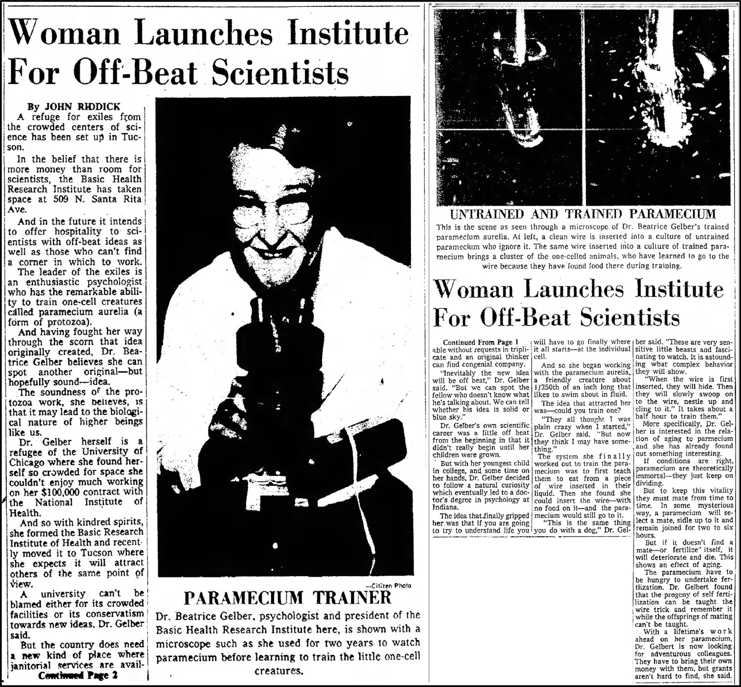
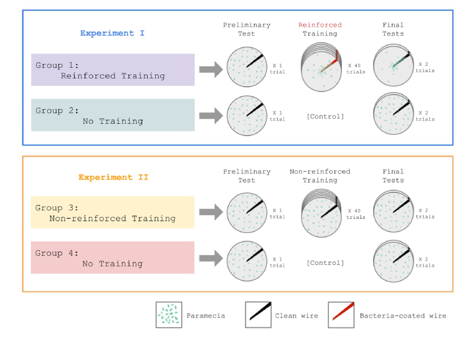
Gelber, B. (1952). Investigations of the behavior of paramecium aurelia. I. Modification of behavior after training with reinforcement. Journal of Comparative and Physiological Psychology, 45(1), 58–65. https://doi.org/10.1037/h0063093
Gelber, Beatrice. (1962). Reminiscence and the trend of retention in paramecium aurelia. The Psychological Record, 12(2), 179–192. https://doi.org/10.1007/BF03393455
Gershman, S. J., Balbi, P. E., Gallistel, C. R., & Gunawardena, J. (2021). Reconsidering the evidence for learning in single cells. eLife, 10, e61907. https://doi.org/10.7554/eLife.61907
Grafe, L. (2019). Brenda milner on HM. YouTube. Retrieved from https://www.youtube.com/watch?v=aw6JmZuLhfA
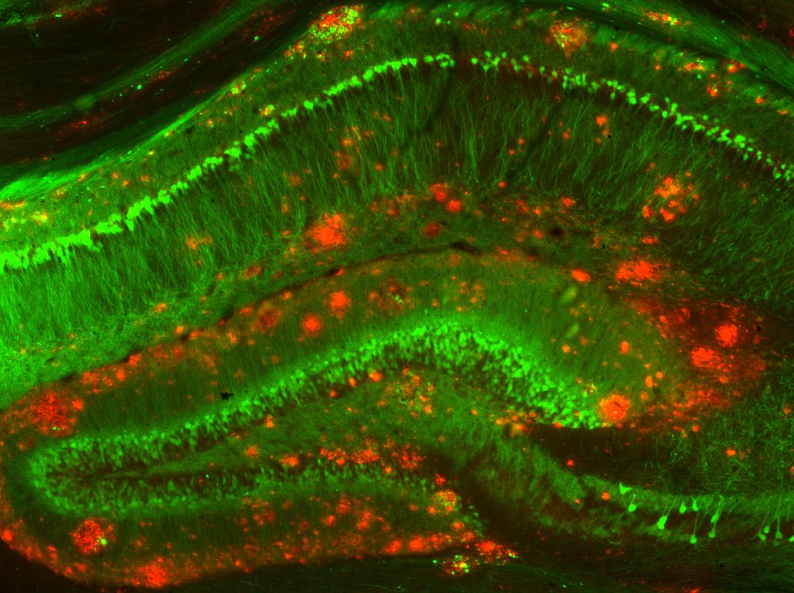
Haney, M. S., Pálovics, R., Munson, C. N., Long, C., Johansson, P. K., Yip, O., … Wyss-Coray, T. (2024). APOE4/4 is linked to damaging lipid droplets in alzheimer’s disease microglia. Nature, 628(8006), 154–161. https://doi.org/10.1038/s41586-024-07185-7
Hebb, D. O. (1949). The organization of behavior; A neuropsychological theory (Vol. 335). Oxford, England: Wiley. Retrieved from https://psycnet.apa.org/fulltext/1950-02200-000.pdf
Hou, J., Chen, Y., Cai, Z., Heo, G. S., Yuede, C. M., Wang, Z., … Colonna, M. (2024). Antibody-mediated targeting of human microglial leukocyte ig-like receptor B4 attenuates amyloid pathology in a mouse model. Science Translational Medicine, 16(741), eadj9052. https://doi.org/10.1126/scitranslmed.adj9052
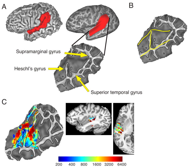
Humphries, C., Liebenthal, E., & Binder, J. R. (2010). Tonotopic organization of human auditory cortex. NeuroImage, 50, 1202–1211. https://doi.org/10.1016/j.neuroimage.2010.01.046
Jevtic, S., Sengar, A. S., Salter, M. W., & McLaurin, J. (2017). The role of the immune system in alzheimer disease: Etiology and treatment. Ageing Research Reviews, 40, 84–94. https://doi.org/10.1016/j.arr.2017.08.005
Jirenhed, D.-A., Rasmussen, A., Johansson, F., & Hesslow, G. (2017). Learned response sequences in cerebellar purkinje cells. Proceedings of the National Academy of Sciences of the United States of America, 114(23), 6127–6132. https://doi.org/10.1073/pnas.1621132114
Josselyn, S. A., & Tonegawa, S. (2020). Memory engrams: Recalling the past and imagining the future. Science. https://doi.org/10.1126/science.aaw4325
Kitamura, T., Ogawa, S. K., Roy, D. S., Okuyama, T., Morrissey, M. D., Smith, L. M., … Tonegawa, S. (2017). Engrams and circuits crucial for systems consolidation of a memory. Science, 356(6333), 73–78. https://doi.org/10.1126/science.aam6808
Kjelstrup, K. B., Solstad, T., Brun, V. H., Hafting, T., Leutgeb, S., Witter, M. P., … Moser, M.-B. (2008). Finite Scale of Spatial Representation in the Hippocampus. Science, 321(5885), 140–143. https://doi.org/10.1126/science.1157086
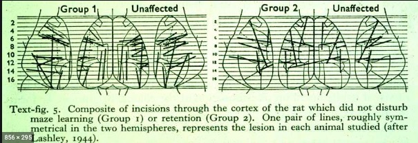
Lashley, Karl S. (1944). Studies of cerebral function in learning. XIII. Apparent absence of transcortical association in maze learning. Journal of Comparative Neurology. Retrieved from https://onlinelibrary.wiley.com/doi/abs/10.1002/cne.900800207
Lashley, K. S., & Clark, G. (1946). The cytoarchitecture of the cerebral cortex of ateles; a critical examination of architectonic studies. The Journal of Comparative Neurology, 85(2), 223–305. https://doi.org/10.1002/cne.900850207
Learning styles as a myth. (2017, June). https://poorvucenter.yale.edu/LearningStylesMyth. Retrieved from https://poorvucenter.yale.edu/LearningStylesMyth
Löwel, S., & Singer, W. (1992). Selection of intrinsic horizontal connections in the visual cortex by correlated neuronal activity. Science, 255, 209–212. https://doi.org/10.1126/science.1372754
Luck, S. J., & Vogel, E. K. (2013). Visual working memory capacity: From psychophysics and neurobiology to individual differences. Trends Cogn. Sci., 17(8), 391–400. https://doi.org/10.1016/j.tics.2013.06.006
Maguire, E. A., Gadian, D. G., Johnsrude, I. S., Good, C. D., Ashburner, J., Frackowiak, R. S., & Frith, C. D. (2000). Navigation-related structural change in the hippocampi of taxi drivers. Proceedings of the National Academy of Sciences, 97(8), 4398–4403. https://doi.org/10.1073/pnas.070039597
Maguire, E. A., Woollett, K., & Spiers, H. J. (2006). London taxi drivers and bus drivers: A structural MRI and neuropsychological analysis. Hippocampus, 16(12), 1091–1101. https://doi.org/10.1002/hipo.20233
McCulloch, W. S., & Pitts, W. (1943). A logical calculus of the ideas immanent in nervous activity. The Bulletin of Mathematical Biophysics, 5, 115–133. https://doi.org/10.1007/BF02478259
Mišić, B., Goñi, J., Betzel, R. F., Sporns, O., & McIntosh, A. R. (2014). A network convergence zone in the hippocampus. PLoS Comput. Biol., 10(12), e1003982. https://doi.org/10.1371/journal.pcbi.1003982
Pashler, H., McDaniel, M., Rohrer, D., & Bjork, R. (2008). Learning styles: Concepts and evidence. Psychological Science in the Public Interest: A Journal of the American Psychological Society, 9(3), 105–119. https://doi.org/10.1111/j.1539-6053.2009.01038.x
Pitt medical neuroscience. (n.d.). Retrieved April 19, 2023, from http://pittmedneuro.com/glutamate.html
Randeberg, L. (2005). Diagnostic applications of diffuse reflectance spectroscopy. Retrieved from https://www.semanticscholar.org/paper/ec9450b79923e2e2152b54ab9241b60bc5374944
Reber, P. (2010, May 1). What is the memory capacity of the human brain? Retrieved December 3, 2025, from https://www.scientificamerican.com/article/what-is-the-memory-capacity/
Ron, D., & Wang, J. (2011). The NMDA receptor and alcohol addiction. In A. M. Van Dongen (Ed.), Biology of the NMDA receptor. Boca Raton (FL): CRC Press/Taylor & Francis. Retrieved from https://www.ncbi.nlm.nih.gov/pubmed/21204417
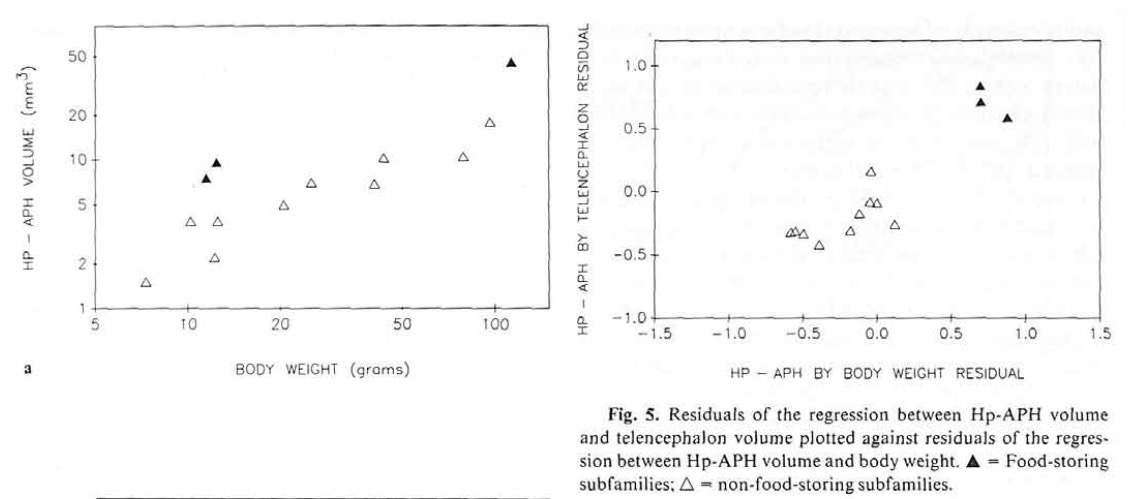
Sherry, D. F., Vaccarino, A. L., Buckenham, K., & Herz, R. S. (1989). The Hippocampal Complex of Food-Storing Birds. Brain, Behavior and Evolution, 34(5), 308–317. https://doi.org/10.1159/000116516
Squire, L. R. (2004). Memory systems of the brain: A brief history and current perspective. Neurobiology of Learning and Memory, 82(3), 171–177. https://doi.org/10.1016/j.nlm.2004.06.005
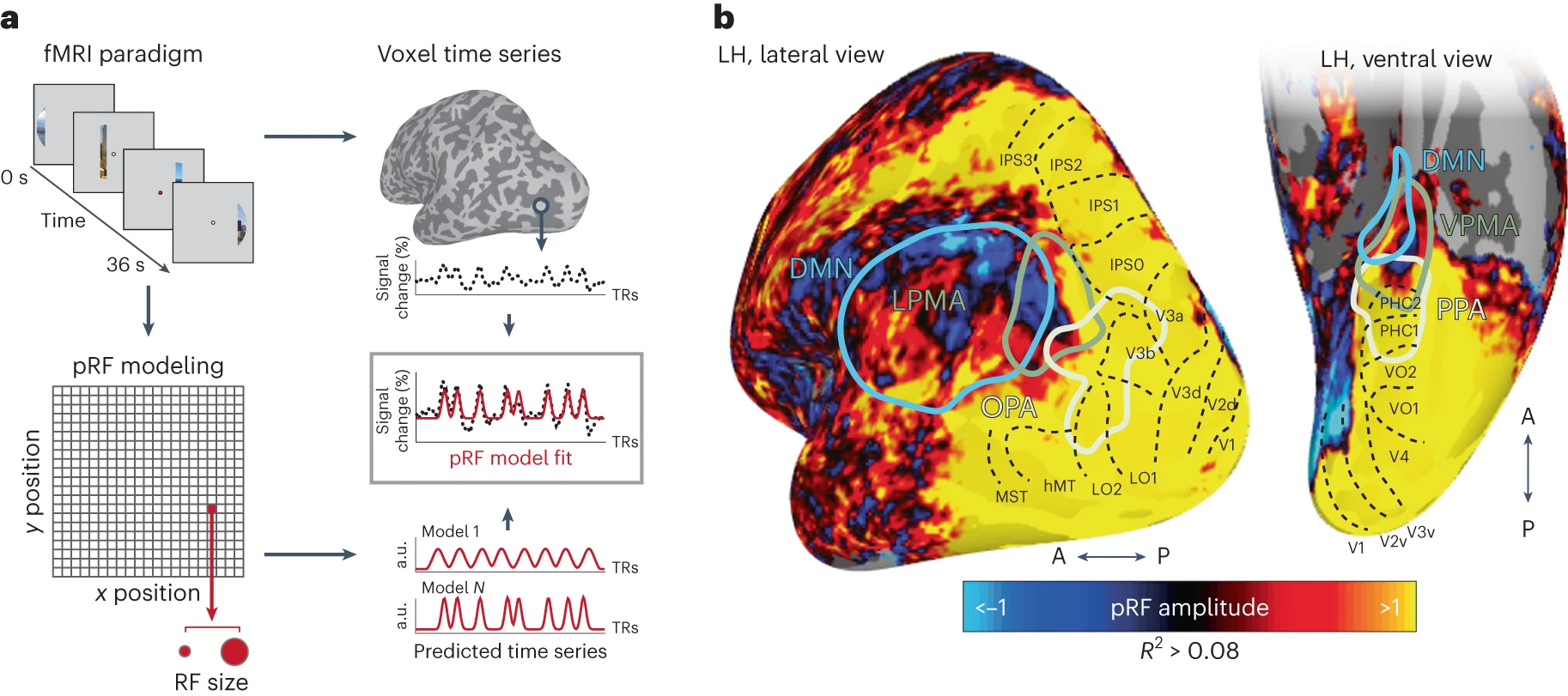
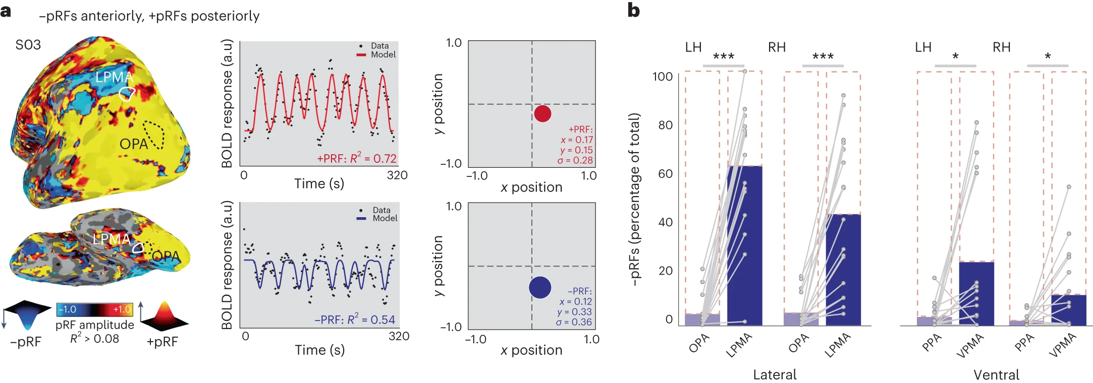
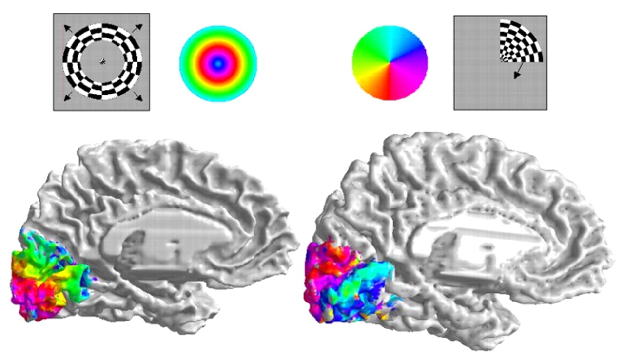
Steel, A., Silson, E. H., Garcia, B. D., & Robertson, C. E. (2024). A retinotopic code structures the interaction between perception and memory systems. Nature Neuroscience. https://doi.org/10.1038/s41593-023-01512-3
Tch, A. (2017, August). The mostly complete chart of neural networks, explained. https://towardsdatascience.com/the-mostly-complete-chart-of-neural-networks-explained-3fb6f2367464. Retrieved from https://towardsdatascience.com/the-mostly-complete-chart-of-neural-networks-explained-3fb6f2367464
timvitkuske. (2014, May). Betty buckley - memory (1983 tony awards). Youtube. Retrieved from https://www.youtube.com/watch?v=5mlllRdIfqw
Veen, F. van. (n.d.). Neural network zoo prequel: Cells and layers. https://www.asimovinstitute.org/author/fjodorvanveen/. Retrieved from https://www.asimovinstitute.org/author/fjodorvanveen/
Woollett, K., & Maguire, E. A. (2011). Acquiring “the knowledge” of london’s layout drives structural brain changes. Current Biology: CB, 21(24), 2109–2114. https://doi.org/10.1016/j.cub.2011.11.018

Footnotes

  1. Fig. 3. Flattened surface representation. (A) Derivation of a flattened patch for a single subject, showing the corresponding area on the original surface. On the patch, gyri are represented with lighter shading and sulci with darker shading. (B) Hand drawn boundaries used for group alignment. (C) Left hemisphere tonotopy map for a single subject projected on a flattened surface and in volumetric space.

  2. News story about Beatrice Gelber from the Tucson Daily Citizen October 19, 1960. © 1960, USA TODAY NETWORK. This image is not covered by the CC-BY 4.0 licence and may not be separated from the article.

  3. In Experiment 1 (top), one group of Paramecia was exposed to intermittent training trials in which a wire was coated with bacteria (every 3rd trial during the training phase). This group acquired a conditioned response to the clean wire, as measured by adherence to the wire in final test trials. In contrast, an untrained group did not show a conditioned response. Experiment 2 (bottom) demonstrated that the wire by itself did not drive conditioned responding.

  4. Blocks ion pore. See https://en.wikipedia.org/wiki/Ketamine

  5. Recent studies have further characterized the mechanism and function of STDP in both in vitro and in vivo preparations, addressing the following questions: Which cellular mechanisms determine the STDP window, and how similar are they to the mechanisms underlying LTP and LTD induced by HFS and LFS, respectively? Does the window depend on the dendritic location of the input, and can it be regulated by neuromodulatory inputs? Does a similar learning rule apply to the inhibitory circuits? Can we observe the consequences of the asymmetric window in vivo, and can it account for the synaptic modifications induced by complex, naturalistic spike trains? In this review we summarize recent progress in these areas.

  6. Balanced excitation and inhibition are crucial for normal brain functions (Shu et al. 2003) and for regulating experience-dependent developmental plasticity (Hensch 2005). Although the strength of excitatory synapses can be modified through STDP, an important question is whether and how correlated pre- and postsynaptic activity affects inhibitory circuits. Inhibition in a network depends on both the excitatory synapses onto inhibitory neurons and the inhibitory synapses themselves. Spike timing–dependent plasticity has been studied at both of these synapses.

  7. Fig. 1. Place fields of eight pyramidal cells recorded at different longitudinal levels of CA3 during animals’ running on a linear 18-m track. (A) Nissl-stained sections showing recording locations in four animals. Individual rat numbers are indicated. (B) Place fields on the 18-m track. Each panel shows one cell. Rat numbers refer to (A). Percentages indicate location along the dorsoventral axis. For rat 11285, only the 87% location is shown in (A). (Top of each panel) Smoothed spike density function indicates firing rate as a function of position. Horizontal bar indicates estimated place field. Left runs, red; right runs, green. (Bottom of each panel) Raster plot showing density of spikes on individual laps. Each vertical tic indicates one spike and each horizontal line shows one lap (right side, blue-green; left, red). See fig. S5 for complete cell samples. Note 5- to 10-m-long place fields in ventral CA3 (colored horizontal bars; left runs, red; right runs, green).

  8. a, PRF paradigm and modeling. In Exp. 1, participants underwent pRF mapping. Participants viewed visual scenes through a bar aperture that gradually traversed the visual field. Each visual-field traversal lasted 36 s (18 × 2 s positions), and the bar made eight traversals per run. The direction of motion varied between traversals. To estimate the pRF for each voxel, a synthetic time series is generated for 400 visual-field locations (200 x and y positions) and 100 sizes (sigma). This results in four million possible time series that are fit to each voxel’s activity. The fit results in four parameters describing each receptive field: x, y, sigma and amplitude. b, Negative-amplitude pRFs fall anterior to the cortex typically considered visual (beyond known retinotopic maps). Group average (n = 17) pRF amplitude map (threshold at explained variance R-squared (R2) > 0.08) is shown on partially inflated representations of the left hemisphere, alongside ROIs: SPAs (OPA, PPA), PMAs (LPMA, VPMA) (localized in an independent group of participants24) and the DMN22. The known retinotopic maps in the posterior cortex (black dotted outlines21) contain exclusively positive-amplitude pRFs (hot colors), as visual stimulation evokes positive retinotopically specific BOLD responses. Negative-amplitude pRFs (cool colors), where visual stimulation evokes a negative spatially specific BOLD response, arise anterior to these retinotopic maps in the PMAs and DMN (see Fig. 2a for example time series from a representative subject).

  9. a, PRF modeling reveals posterior–anterior inversion of pRF amplitude in individual participants. Left, PRF amplitude for a representative participant overlaid onto a lateral view of the left hemisphere (threshold at R2 > 0.15; see Extended Data Fig. 1 for example ventral and lateral surface pRF amplitude maps from all participants and Extended Data Fig. 3 for amplitude maps with default mode parcellation overlaid). Posterior visual cortex is dominated by positive-amplitude pRFs (hot colors), while cortex anterior to regions classically considered visual exhibits a high concentration of negative-amplitude pRFs (cold colors). This individual’s OPA and LPMA are shown in white. Both the SPAs and PMAs contain pRFs (Extended Data Fig. 2). Right, time-series, model fits and reconstructed pRFs for two surface vertices in this subject. Top, example prototypical positive-amplitude pRF from the lateral SPA (OPA) in the left and right hemispheres (LH, RH). Bottom, example negative-amplitude population receptive field from the LPMA. b, Memory areas (PMAs) contain a larger percentage of negative pRFs compared to perceptual areas (SPAs) (repeated measures ANOVA, Bonferroni-corrected t-tests). Blue bars depict percentage of negative pRFs from individually localized SPAs and PMAs compared to total pRFs in the area (dotted outline). On the ventral and lateral surfaces, SPAs are dominated by positive pRFs, whereas a transition from positive to negative pRFs is evident within PMAs. Individual participant data points overlaid and connected in gray. P two-tailed < 0.05, **P two-tailed < 0.001. NS, not significant.

  10. Henry G. Molaison was identified with his permission after his death in 2008.