Neural communication

PSY 511.001 Spr 2026

Rick Gilmore

Department of Psychology

Prelude

For fun

Figure 1: Musikladen (2020)

Announcements

Today’s topics

  • Neural communication
    • The action potential
    • Synaptic transmission

Warm-up

The action potential

What is it?

  • Rapid rise, fall of membrane potential
Figure 2: Wikipedia contributors (2026d)1

Components

  • Threshold of excitation
  • Increase (rising phase/depolarization)
  • Peak
    • at positive voltage
  • Decline (falling phase/repolarization)
  • Return to resting potential (refractory period)
Figure 3: Wikipedia contributors (2026d)2

Reviewing the resting potential

  • The \(K^+\) story
  • The \(Na^+\) story

The \(K^+\) story

  • \(Na^+\)/\(K^+\) pump pulls \(K^+\) in
  • \([K^+]_{in}\) (~150 mM) >> \([K^+]_{out}\) (~4 mM)
  • Force of diffusion
    • Outward flow of \(K^+\) through passive/leak channels
    • Would stop if/when \([K^+]_{in}\) = \([K^+]_{out}\)
    • Never gets there

The \(K^+\) story

  • \(K^+\) a charged particle
    • moving charged particles create a current
    • current charges membrane like a capacitor
    • charging capacitor has increasing voltage
  • Outflow would stop when membrane potential, \(V_m\) = equilibrium potential for \(K^+\)

Equilibrium potential

  • Voltage (\(V_{K}\)) that keeps system in equilibrium
    • \([K^+]_{in}\) >> \([K^+]_{out}\)
  • Nernst equation
    • \(V_{K}\) = \(\frac{RT}{(+1)F}ln(\frac{[K^+]_{out}}{[K^+]_{in}})\)
    • \(V_{K}\) = ~ -90 mV
    • Negative in/positive out keeps in/out concentration gradient

Equilibrium potential

  • \(K^+\) flows out through passive/leak channels
    • most ions remain near membrane
  • Separation from \(A^-\) creates charge \(\frac{K+K+K+K+K+}{A-A-A-A-A-}\) along capacitor-like membrane
  • \(V_m \rightarrow V_{K}\)
    • Doesn’t quite get there

The \(Na^+\) story

  • \(Na^+\)/\(K^+\) pump pushes \(Na^+\) out
  • \([Na^+]_{in}\) (~10 mM) << \([Na^+]_{out}\) (~140 mM)
  • Equilibrium potential for \(Na^+\), \(V_{Na}\) = ~ +55 mV
    • Inside positive/outside negative to maintain outside > inside concentration gradient
  • If \(Na^+\) alone, \(V_m \rightarrow V_{Na}\) (~ +55 mV)

“Resting” potential

  • Sum of outward \(K^+\) and inward \(Na^+\)
    • Membrane more permeable to \(K^+\) than \(Na^+\), \(p_{K^+}\)>\(p_{Na^+}\)
    • Outward flow of \(K^+\) > inward flow of \(Na^+\)
    • Resting potential (\(\approx\) -60-70 mV) closer to \(V_{K}\) (-90 mV) than \(V_{Na}\) (+55 mV)
  • Goldman-Hodgkin-Katz equation
    • \(V_m = \frac{RT}{F}ln(\frac{p_{K^+}[K^+]_{out}+p_{Na^+}[Na^+]_{out}}{p_{K^+}[K^+]_{in}+p_{Na^+}[Na^+]_{in}})\)

Driving force

  • Difference between \(V_m\) and \(V_{eq}\) for a given ion.

Your turn

Which ion, \(K^+\) or \(Na^+\) has the strongest driving force when the neuron is at resting potential?

Figure 4: Tug-o-war illustrating a balance among forces.

Action potential components

Phase Neuron State
Resting potential Passive \(K^+\) allow outward flow; passive \(Na^+\) allow inward flow; \(Na^+\)/\(K^+\) moves \(K^+\) in and \(Na^+\) out
Rise to threshold + input makes membrane potential more +

Action potential components

Phase Neuron State
Depolarization3 Voltage-gated \(Na^+\) channels open, \(Na^+\) enters
Peak Voltage-gated \(Na^+\) channels close and deactivate; voltage-gated \(K^+\) channels open

Action potential components

Phase Neuron State
Repolarization4 \(K^+\) exits
Refractory period \(Na^+\)/\(K^+\) pump restores [\(Na^+\)], [\(K^+\)]; voltage-gated \(K^+\) channels close

Action potential components

Phase Neuron State
Resting potential Passive \(K^+\) allow outward flow; passive \(Na^+\) allow inward flow; \(Na^+\)/\(K^+\) moves \(K^+\) in and \(Na^+\) out

Action potential components

Driving force @ resting potential

  • Driving force on \(K^+\) weakly outward
    • -70 mV - (-90 mV) = +20 mV
  • Driving force on \(Na^+\) strongly inward
    • -70 mV - (+55 mV) = -125 mV

Neuron at rest

  • \(Na^+\)/\(K^+\) pump maintains concentrations (\(Na^2\) out; \(K^+\) in)
    • \([K^+]_{i} >> [K^+]_{o}\)
    • \([Na^+]_{i} << [Na^+]_{o}\)
Phase Ion Driving force Flow magnitude Flow direction
Rest \(K^+\) \(\approx\) +20 mV small5 out
\(Na^+\) \(\approx\) -125 mV small6 in

Depolarization (rising)

  • Voltage-gated \(Na^+\) channels open
  • Membrane permeability to \(Na^+\) increases
    • \(Na^+\) inflow through passive + voltage-gated channels
    • continued \(K^+\) outflow through passive channels

Phase Ion Driving force Flow magnitude Flow direction
Rising \(K^+\) growing growing out
\(Na^+\) shrinking high in

Peak

  • Membrane permeability to \(Na^+\) reverts to resting state
    • Voltage-gated \(Na^+\) channels close & inactivate7
    • Slow inflow due to small driving force (+30 mV - 55mV = -25 mv)

Peak

  • Membrane permeability to \(K^+\) increases
    • Voltage-gated \(K^+\) channels open
    • Fast outflow due to strong driving force (+30 mv - (-90 mv) = +120 mV)

Phase Ion Driving force Flow magnitude Flow direction
Peak \(K^+\) 120 mV high out
\(Na^+\) 20 mV small in

Repolarization (falling)

  • \(K^+\) outflow
    • Through voltage-gated \(K^+\) and passive \(K^+\) channels
  • \(Na^+\) inflow
    • Through passive channels only

Phase Ion Driving force Flow magnitude Flow direction
Falling \(K^+\) shrinking high out
\(Na^+\) growing small in

Refractory phase(s)

  • Absolute
  • Relative

Absolute

  • Cannot generate action potential (AP) no matter the size of the stimulus
  • Membrane potential more negative (-90 mV) than at rest (-70 mV)

Absolute

  • Voltage-gated \(Na^+\) channels still inactivated
    • Driving force on \(Na^+\) high (-90 mv - 55 mV = -145 mV), but too bad

Absolute

  • Voltage-gated \(K^+\) channels closing
    • Driving force on \(K^+\) tiny or absent
  • \(Na^+\)/\(K^+\) pump restoring concentration balance

Relative

  • Can generate AP with larg(er) stimulus
  • Some voltage-gated \(Na^+\) ‘de-inactivate’, can open if
    • Larger input
    • Membrane potential is more negative than resting potential

Phase Ion Driving force Flow magnitude Flow direction
Refractory \(K^+\) ~0 mV small out
\(Na^+\) 145 mV small in

APs and Information Processing

  • AP amplitudes don’t vary (much)
    • All or none
    • \(V_{K}\) and \(V_{Na}\) don’t vary much b/c \(Na^+\)/\(K^+\) pump always working
  • AP frequency and timing vary
    • Rate vs. timing codes
    • Same rates, but different timing
    • “Grandmother” cells and single spikes

Eyherabide et al. (2009)

Eyherabide et al. (2009)

Generating action potentials

  • Axon hillock
    • Portion of soma adjacent to axon
    • Integrates/sums input to soma

Wikipedia

Wikipedia

Generating action potentials

  • Axon initial segment
    • Umyelinated portion of axon adjacent to soma
    • Voltage-gated \(Na^+\) and \(K^+\) channels exposed
    • If sum of input to soma > threshold, voltage-gated \(Na^+\) channels open

Wikipedia

Wikipedia

Propagation

  • Propagation
    • move down axon, away from soma, toward axon terminals.
  • Unmyelinated axon
    • Each segment “excites” the next

Wikipedia contributors (2026d)

Wikipedia contributors (2026d)8
Figure 6
  • Myelinated axon
    • AP “jumps” between Nodes of Ranvier –> saltatory conduction
    • Nodes of Ranvier == unmyelinated sections of axon
    • voltage-gated \(Na^+\), \(K^+\) channels exposed
    • Current flows through myelinated segments
  • Why does AP flow in one direction, away from soma?
    • Soma does not have (many) voltage-gated \(Na^+\) channels.
    • Soma is not myelinated.
    • Refractory periods mean polarization only in one direction.

Conduction velocities

Figure 7: Wikipedia contributors (2025c)

Hodgkin-Huxley Equations

Figure 8: Wikipedia contributors (2026c)

Synaptic transmission

Synaptic transmission

  • Synapse permits neuron to pass electrical or chemical messages to another neuron or target cell (muscle, gland, etc.)

Synapse Types & Locations

  • Electrical
    • Gap junctions
    • Cytosol (and ionic current) flows through adjacent neurons
  • Chemical
    • Ligand9 binds to receptor

https://commons.wikimedia.org/wiki/File:Gap_cell_junction-en.svg#/media/File:Gap_cell_junction-en.svg

https://commons.wikimedia.org/wiki/File:Gap_cell_junction-en.svg#/media/File:Gap_cell_junction-en.svg

Steps in chemical transmission

  • At axon terminal/terminal button/synaptic terminal
  • Voltage-gated calcium Ca++ channels open
  • \(Ca^{++}\) influx
    • causes synaptic vesicles to bind with presynaptic membrane, fuse with membrane, spill contents via exocytosis

Figure 1 from Haucke, Neher, & Sigrist (2011)

Figure 1 from Haucke et al. (2011)

Hastoy, Clark, Rorsman, & Lang (2017)

Hastoy et al. (2017)

Hastoy et al. (2017)

Hastoy et al. (2017)

Steps in chemical communication

  • NTs diffuse across synaptic cleft
  • NTs bind with receptors on postsynaptic membrane
    • Cause some post-synaptic effect
  • NTs unbind from receptor

Wikipedia contributors (2026a)

Wikipedia contributors (2026a)10

Steps in chemical communication

  • NTs inactivated
  • NTs diffuse along concentration gradient

Wikipedia contributors (2026a)

Wikipedia contributors (2026a)11

Chemical communication

Relative sizes

  • Neural membrane ~8 nm
  • Synaptic vesicles ~40-60 or ~90-120 nm
  • Synaptic cleft ~20-50 nm
  • Cleft small relative to vesicles

Receptor types

  • Transporters/exchangers
    • Ionic
      • \(Na^+\)/\(K^+\) ATP-ase/pump
    • Chemical
      • e.g., Dopamine transporter (DAT)

Receptor types

  • Ligand-gated (chemically gated)
    • Ionotropic
    • Metabotropic

Ionotropic receptor

  • Opens/closes channel
  • Ions flow in/out depending on membrane voltage and ion type
  • Fast-responding (< 2 ms), but short-duration effects (< 100 ms)

Wikipedia contributors (2025d)

Wikipedia contributors (2025d)12

Metabotropic receptor

  • Ligand binds to receptor (outside)
  • G-proteins13 (inside)
    • Trigger 2nd messengers
    • Open/close adjacent channels, change metabolism

Wikipedia contributors (2026b)

Wikipedia contributors (2026b)14

Receptors generate postsynaptic potentials (PSPs)

  • Small voltage changes
  • Amplitude scales with # of receptors activated
  • Short duration

Wikipedia contributors (2025b)

Wikipedia contributors (2025b)15

PSPs

  • Excitatory PSPs (EPSPs)
    • Depolarize neuron (make more +)
  • Inhibitory (IPSPs)
    • Hyperpolarize neuron (make more -)

Wikipedia contributors (2025a)

Wikipedia contributors (2025a)16

NTs inactivated

  • Diffusion
  • Glial reuptake
  • Neuronal reuptake via transporters
    • e.g., serotonin via serotonin transporter (SERT)
  • Enzymatic degradation
    • e.g., acetylcholinesterase (AChE) breaks down acetylcholine (ACh)

Questions to ponder

Your turn

  • Why do NTs diffuse from pre- to post-synaptic membrane?
  • Why must NTs be inactivated?
  • What sort of PSP would opening a \(Na^+\) channel produce?
  • What sort of PSP would opening a \(Cl^-\) channel produce?
  • What sort of PSP would closing a channel \(K^+\) produce?

Synapse location and function

  • on dendrites
    • usually excitatory
  • on cell bodies
    • usually inhibitory
  • on axons
    • usually modulatory (change p(fire))

Henley (2021)

Henley (2021)17

Why should (psychologists) care?

  • Modulate the synapse \(\rightarrow\) change processing
  • (Most) psychoactive substances affect synaptic transmission

Wrap-up

Main points

  • Action potential disrupts the equilibrium forces that create the resting potential.
    • Depolarizing (net +) input opens voltage-gated \(Na^+\) channels.
  • Depolarization: \(Na^+\) inflow dominates
    • Strong driving force
  • Repolarization: \(K^+\) outflow dominates
    • Strong driving force

Main points

  • Propagation via
    • Umyelinated segments of axon (slower)
    • Saltatory conduction across myelinated segments (faster)

Main points

  • Synaptic communication
    • Electrical (rare)
    • Chemical (common)

Main points

  • Neurotransmitters
    • Released by presynaptic terminal; diffuse
    • Bind to receptors on postsynaptic terminal
      • Ionotropic
      • Metabotropic
    • Cause PSPs
      • Excitatory
      • Inhibitory

Main points

  • Neurotransmitters are inactivated
    • Reuptake
      • Glia
      • Neurons
    • Enzymatic degradation
    • Diffusion

Next time

  • Neurochemistry
    • Neurotransmitters
    • Hormones
  • Neurocomputing

Resources

About

This talk was produced using Quarto, using the RStudio Integrated Development Environment (IDE), version 2026.1.0.392.

The source files are in R and R Markdown, then rendered to HTML using the revealJS framework. The HTML slides are hosted in a GitHub repo and served by GitHub pages: https://psu-psychology.github.io/psy-511-scan-fdns-2026-spring/

References

Anderson, C. M., & Swanson, R. A. (2000). Astrocyte glutamate transport: Review of properties, regulation, and physiological functions. Glia, 32(1), 1–14. https://doi.org/10.1002/1098-1136(200010)32:1<1::AID-GLIA10>3.0.CO;2-W
Eyherabide, H. G., Rokem, A., Herz, A. V. M., Samengo, I., Eyherabide, H. G., Rokem, A., … Samengo, I. (2009). Bursts generate a non-reducible spike-pattern code. Frontiers in Neuroscience, 3, 1. https://doi.org/10.3389/neuro.01.002.2009
Hastoy, B., Clark, A., Rorsman, P., & Lang, J. (2017). Fusion pore in exocytosis: More than an exit gate? A \(\beta\)-cell perspective. Cell Calcium, 68, 45–61. https://doi.org/10.1016/j.ceca.2017.10.005
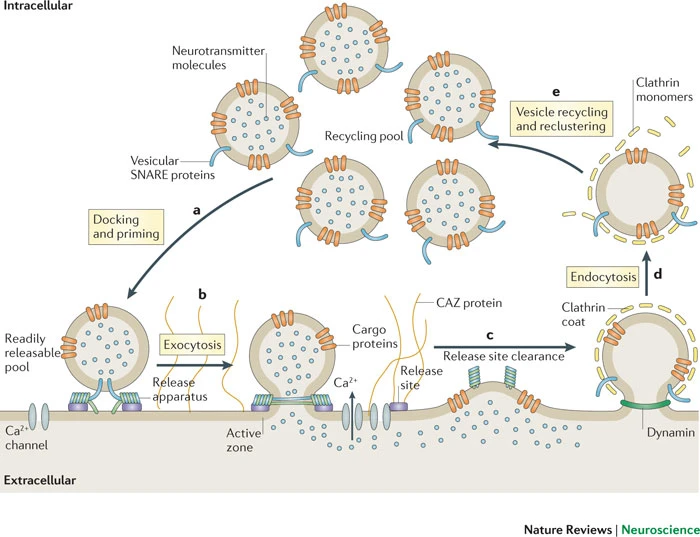
Haucke, V., Neher, E., & Sigrist, S. J. (2011). Protein scaffolds in the coupling of synaptic exocytosis and endocytosis. Nature Reviews. Neuroscience, 12(3), 127–138. https://doi.org/10.1038/nrn2948
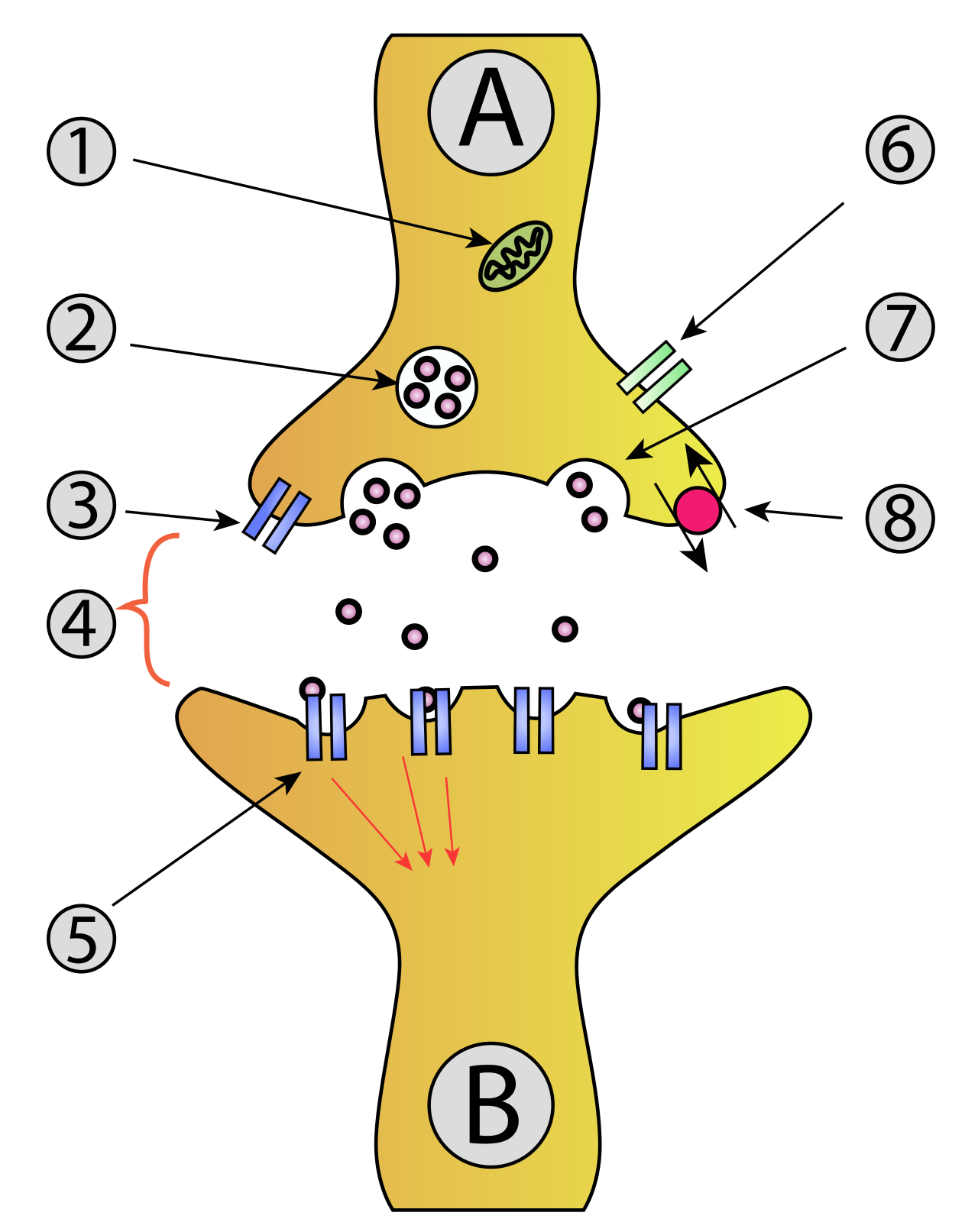
Henley, C. (2021). Synapse structure. In Foundations of neuroscience. Michigan State University Libraries. Retrieved from https://openbooks.lib.msu.edu/neuroscience/chapter/synapse-structure/
Musikladen. (2020). Stevie Wonder - Superstition (1974). YouTube. Retrieved from https://www.youtube.com/watch?v=97hwNY3ni10
Wikipedia contributors. (2025a, February 7). Inhibitory postsynaptic potential. Retrieved from https://en.wikipedia.org/wiki/Inhibitory_postsynaptic_potential
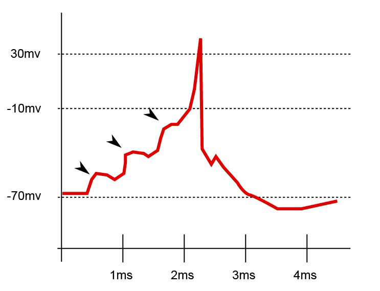
Wikipedia contributors. (2025b, July 19). Excitatory postsynaptic potential. Retrieved from https://en.wikipedia.org/wiki/Excitatory_postsynaptic_potential
Wikipedia contributors. (2025c, August 30). Nerve conduction velocity. Retrieved from https://en.wikipedia.org/wiki/Nerve_conduction_velocity
Wikipedia contributors. (2025d, October 27). Ligand-gated ion channel. Retrieved from https://en.wikipedia.org/wiki/Ligand-gated_ion_channel
Wikipedia contributors. (2026a, March 19). Synaptic vesicle. Retrieved from https://en.wikipedia.org/wiki/Synaptic_vesicle
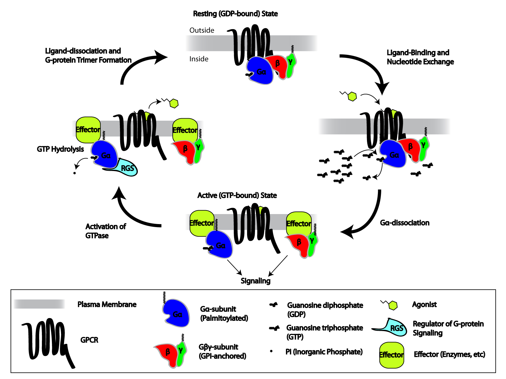
Wikipedia contributors. (2026b, March 23). G protein-coupled receptor. Retrieved from https://en.wikipedia.org/wiki/G_protein-coupled_receptor
Wikipedia contributors. (2026c, March 26). Hodgkin–Huxley model. Retrieved from https://en.wikipedia.org/wiki/Hodgkin%E2%80%93Huxley_model
Wikipedia contributors. (2026d, March 30). Action potential. Retrieved from https://en.wikipedia.org/wiki/Action_potential

Footnotes

  1. “Shape of a typical action potential. The membrane potential remains near a baseline level until at some point in time, it abruptly spikes upward and then rapidly falls. Approximate plot of a typical action potential shows its various phases as the action potential passes a point on a cell membrane. The membrane potential starts out at approximately −70 mV at time zero. A stimulus is applied at time = 1 ms, which raises the membrane potential above −55 mV (the threshold potential). After the stimulus is applied, the membrane potential rapidly rises to a peak potential of +40 mV at time = 2 ms. Just as quickly, the potential then drops and overshoots to −90 mV at time = 3 ms, and finally the resting potential of −70 mV is reestablished at time = 5 ms. By Original by en:User:Chris 73, updated by en:User:Diberri, converted to SVG by tiZom - Own work, CC BY-SA 3.0, https://commons.wikimedia.org/w/index.php?curid=2241513”

  2. “Approximate plot of a typical action potential shows its various phases as the action potential passes a point on a cell membrane. The membrane potential starts out at approximately −70 mV at time zero. A stimulus is applied at time = 1 ms, which raises the membrane potential above −55 mV (the threshold potential). After the stimulus is applied, the membrane potential rapidly rises to a peak potential of +40 mV at time = 2 ms. Just as quickly, the potential then drops and overshoots to −90 mV at time = 3 ms, and finally the resting potential of −70 mV is reestablished at time = 5 ms. By Original by en:User:Chris 73, updated by en:User:Diberri, converted to SVG by tiZom - Own work, CC BY-SA 3.0, https://commons.wikimedia.org/w/index.php?curid=2241513”

  3. Rising phase

  4. Falling phase

  5. Approaching V_K.

  6. Membrane not very permeable.

  7. Can’t be opened for a period of time.

  8. “As an action potential (nerve impulse) travels down an axon, there is a change in electric polarity across the membrane of the axon. In response to a signal from another neuron, sodium- (Na+) and potassium- (K+)–gated ion channels open and close as the membrane reaches its threshold potential. Na+ channels open at the beginning of the action potential, and Na+ moves into the axon, causing depolarization. Repolarization occurs when K+ channels open and K+ moves out of the axon, creating a change in electric polarity between the outside of the cell and the inside. The impulse travels down the axon in one direction only, to the axon terminal where it signals other neurons. By Laurentaylorj - Own work, CC BY-SA 3.0, https://commons.wikimedia.org/w/index.php?curid=26311114”

  9. https://en.wikipedia.org/wiki/Ligand

  10. “Synaptical transmission (chemical). A: Neuron (Presynaptic) B: Neuron (Postsynaptic) Mitochondria Synaptic vesicle full of neurotransmitter Autoreceptor Synaptic cleft Neurotransmitter receptor Calcium Channel Fused vesicle releasing neurotransmitter Neurotransmitter re-uptake pump. By vectorization: Mouagip (talk)Synapse_diag1.png: Drawn by fr:Utilisateur:DakeCorrections of original PNG by en:User:NretsThis W3C-unspecified vector image was created with Adobe Illustrator. - Synapse_diag1.png, CC BY-SA 3.0, https://commons.wikimedia.org/w/index.php?curid=11438067”

  11. “Synaptical transmission (chemical). A: Neuron (Presynaptic) B: Neuron (Postsynaptic) Mitochondria Synaptic vesicle full of neurotransmitter Autoreceptor Synaptic cleft Neurotransmitter receptor Calcium Channel Fused vesicle releasing neurotransmitter Neurotransmitter re-uptake pump. By vectorization: Mouagip (talk)Synapse_diag1.png: Drawn by fr:Utilisateur:DakeCorrections of original PNG by en:User:NretsThis W3C-unspecified vector image was created with Adobe Illustrator. - Synapse_diag1.png, CC BY-SA 3.0, https://commons.wikimedia.org/w/index.php?curid=11438067”

  12. “Ion-channel-linked receptor Ions Ligand (such as acetylcholine) When ligands bind to the receptor, the ion channel portion of the receptor opens, allowing ions to pass across the cell membrane. By Isaac Webb - Own work, CC BY-SA 3.0, https://commons.wikimedia.org/w/index.php?curid=32932033.”

  13. Guanine nucleotide-binding proteins

  14. “Cartoon depicting the heterotrimeric G-protein activation/deactivation cycle in the context of GPCR signaling. By Repapetilto - Own work, CC BY-SA 3.0, https://commons.wikimedia.org/w/index.php?curid=10714351”

  15. “The summation of these three EPSPs generates an action potential. By Laurentaylorj - Own work, CC BY-SA 3.0, https://commons.wikimedia.org/w/index.php?curid=26311114”

  16. “Graph displaying an EPSP, an IPSP, and the summation of an EPSP and an IPSP. When the two are summed together the potential is still below the action potential threshold. By Gth768r, CC BY-SA 3.0, https://commons.wikimedia.org/w/index.php?curid=22926898.”

  17. “Figure 8.3. Types of chemical synapses by location. A) Axodendritic synapses contact the dendrites of the postsynaptic neuron. B) Axosomatic synapses contact the cell body. C) Axoaxonic synapses contact the axon. The synaptic contact sites are highlighted in green. ‘Chemical Synapse Types’ by Casey L. Henley (CC-BY-NC-SA). View detailed alternative text.”