Neurochemistry

PSY 511.001 Spr 2026

Rick Gilmore

Department of Psychology

Prelude

Monoamines

Figure 1: Songs (2018)

See also https://en.wikipedia.org/wiki/Mah_Nà_Mah_Nà

Monoamine Song

Monoamines, do-do do do-do
Monoamines, do do do-do
Monoamines, do do do do-do do do-do do do-do do do do do-do do

Monoamine Song

Monoamines, do-pa-mine is one
Monoamines, norepi, too
Monoamines, sero-tonin e-pinephrine, dop-a-mine, nor-epinephrine, melatonin, whoo!

Monoamine Song

Monoamines, mod-u-late neurons
Monoamines, throughout the brain
Monoamines, keep people happy, brains snappy, not sleepy, not sappy, do-do do-do do-do do

Announcements

Today’s topics

  • Neurochemistry
    • Neurotransmitters
    • Hormones

Warm-up

What causes voltage-gated Na channels to open?

  • A. The channels bind a ligand like glutamate.
  • B. The Na/K pump slows down.
  • C. Cl- entry causes the cell to hyperpolarize.
  • D. An accumulation of EPSPs depolarizes the membrane.

Answer

  • A. The channels bind a ligand like glutamate.
  • B. The Na/K pump slows down.
  • C. Cl- entry causes the cell to hyperpolarize.
  • D. An accumulation of EPSPs depolarizes the membrane.

All of the following events occur near the peak of the action potential except:

  • A. Voltage-gated \(Na^+\) channels close and inactivate.
  • B. Voltage-gated \(K^+\) channels open.
  • C. \(K^+\) ions flow out of the cell through passive/leak channels.
  • D. The membrane potential approaches the reversal/equilibrium potential for \(K^+\).

Answer

  • A. Voltage-gated \(Na^+\) channels close and inactivate.
  • B. Voltage-gated \(K^+\) channels open.
  • C. \(K^+\) ions flow out of the cell through passive/leak channels.
  • D. The membrane potential approaches the reversal/equilibrium potential for \(K^+\).

What factor(s) accelerate the speed of propagation along an axon?

  • A. Large diameter.
  • B. Small diameter.
  • C. Thick myelin.
  • D. Short axon length.
  • E. A and C.

Answer

  • A. Large diameter.
  • B. Small diameter.
  • C. Thick myelin.
  • D. Short axon length.
  • E. A and C.

Neurochemistry

Components

  • Neurotransmitters
  • Hormones

Neurotransmitters (NTs)

  • Chemicals produced by neurons
  • Released by neurons
  • Bound by neurons and other cells
  • Send messages (have physiological effect on target cells)
  • Inactivated after release

Things to know about NTs

  • Input from (soma & dendrite location)
  • Input to
  • What receptor(s) bind it
  • What effect(s)
  • How inactivated

Categories

  • Function/Mode of Action
    • Excitatory1
    • Inhibitory2
    • Modulatory
  • Chemical structure
    • Amino acids
    • Monoamines
    • Acetylcholine (ACh)
    • Gases, Neuropeptides, Purines, others

Amino acids

Glutamate

  • Widespread in CNS (~ 1/2 all synapses)
  • Primary excitatory NT in CNS
  • Role in learning (via NMDA-R)
  • Receptors on neurons and glia (astrocytes and oligodendrocytes)

Glutamate (Glu)

Glutamate receptor types

Type Receptor Esp Permeable to
Ionotropic AMPA \(Na^+\), \(K^+\)
Kainate \(Na^+\), \(K^+\)
NMDA \(Ca^{++}\), \(Na^+\), \(K^+\)
Metabotropic mGlu
Figure 2: http://pittmedneuro.com/glutamate.html
Figure 3: http://pittmedneuro.com/glutamate.html

\(\gamma\) aminobutyric acid (GABA)

  • Primary inhibitory NT in CNS
  • Excitatory in developing CNS
    • \([Cl^-]_{in} >> [Cl^-]_{out}\) out
  • Binding sites for benzodiazepines (BZD; e.g., Valium), barbiturates, ethanol, etc.
    • BZD affects subset of GABA-A receptors
    • Increase total Cl- influx

GABA receptor types

Type Receptor Esp Permeable to
Ionotropic GABA-A \(Cl^-\)
Metabotropic GABA-B \(K^+\)

GABA

http://pittmedneuro.com/inhibitory.html

Figure 4: http://pittmedneuro.com/inhibitory.html

GABA-A receptor

Figure 5: https://en.wikipedia.org/wiki/GABAA_receptor

Other amino acid NTs

  • Aspartate
    • Like Glu, stimulates NMDA receptor
  • Glycine
    • Spinal cord interneurons
    • Inhibitory
    • Opens Cl- channel

Acetylcholine (ACh)

  • Primary excitatory NT of CNS output

ACh in PNS (Somatic)

  • Motor neuron in spinal cord -> neuromuscular junction
Figure 6: Biological Psychology 4th ed.

ACh in PNS (Autonomic)

  • Sympathetic branch: preganglionic neuron
  • Parasympathetic branch: pre/postganglionic
Figure 7: https://imotions.com/blog/learning/research-fundamentals/nervous-system/

https://imotions.com/blog/learning/research-fundamentals/nervous-system/

https://imotions.com/blog/learning/research-fundamentals/nervous-system/

ACh receptor types

Type Receptor Esp Permeable to Blocked by
Ionotropic Nicotinic (nAChR) \(Na^+\), \(K^+\) e.g., Curare
Metabotropic Muscarinic (mAChR) \(K^+\) e.g., Atropine

Curare

  • Binds to Nicotinic ACh receptor in muscle
Figure 8: www.general-anaesthesia.com

Atropine

  • aka, nightshade or belladonna
  • inhibits (acts as an antagonist for) muscarinic ACh receptor
Figure 9: https://cdn.britannica.com/92/183192-050-1741C2F9/Belladonna-nightshade-leaves-berries-alkaloids-humans.jpg
Figure 10: Wikipedia contributors (2025c)3

ACh in CNS

  • ACh a neuromodulator (Picciotto, Higley, & Mineur, 2012)
    • Via cholinergic system
  • Neuromodulators
    • Longer-term effects
    • Wider-range effects
    • Change neuron responses to other inputs
    • Don’t generate short-duration EPSPs or IPSPs

Basal forebrain

Avram et al. (2021) Figure 1

Avram et al. (2021) Figure 14
Figure 11: Mesulam (2013) Figure 175
Figure 12: Avery & Krichmar (2017) Figure 86

Monoamine NTs

  • Catecholamines
    • Dopamine (DA)
    • Norepinephrine (NE)/Noradrenaline (NAd)
    • Epinephrine (Epi)/Adrenaline (Ad)

Synthesis pathways

  • L-tyrosine -> L-DOPA -> DA
  • DA -> NE/NAd -> Epi/Ad

Henley (2021) Figure 9.6

Henley (2021) Figure 9.67

Monoamine NTs

  • Indolamines
    • Serotonin (5-HT)
    • Melatonin
    • Histamine

Information processing

  • Point-to-point
    • One sender, small number of recipients
    • Glu, GABA
  • Broadcast
    • One sender, widespread recipients
    • DA, NE, 5-HT, melatonin, histamine

Receptor distribution

Figure 13: Vogelsang & D’Esposito (2018) Figure 48, adapted from Palomero-Gallagher, Amunts, & Zilles (2015)

DA release pathways

  • Substantia nigra -> striatum
    • meso-striatal projection
  • Ventral tegmental area (VTA) ->
    • nucleus accumbens, ventral striatum, hippocampus, amygdala, cortex
    • meso-limbo-cortical projection
Figure 14: The main dopaminergic pathways (Wikipedia)

DA clinical relevance

  • Parkinson’s Disease (mesostriatal)
    • DA agonists treat (agonists facilitate/increase transmission)
  • ADHD (mesolimbocortical)
  • Schizophrenia (mesolimbocortical)
    • DA antagonists treat
  • Addiction (mesolimbocortical)

DA inactivation

  • Enzymatic breakdown via monoamine oxidase (MAO)
    • MAO removes amine group
    • Monoamine Oxidase Inhibitors (MAO-Is) inhibit monoamine inactivation

Your turn

What is the effect of inhibiting neurotransmitter inactivation?

Answer

NT concentration \(\uparrow\), action prolonged.

DA inactivation

  • Reuptake via Dopamine transporter (DAT)
Figure 15: Overk & Mufson (2010)

DA receptor types

Type Receptor Comments
Metabotropic D1-like (D1 and D5) more prevalent
D2-like (D2, D3, D4) target of many antipsychotics

Norepinephrine (NE)

Figure 16

NE

  • In ANS, postganglionic sympathetic neurons contact target tissues

https://imotions.com/blog/learning/research-fundamentals/nervous-system/

https://imotions.com/blog/learning/research-fundamentals/nervous-system/

NE clinical relevance

  • ADHD, Alzheimer’s Disease, Parkinson’s Disease, depression
  • Role in arousal, mood, eating, sexual behavior

NE inactivation

  • Norepinephrine transporter (NET), aka noradrenaline transporter (NAT)
    • Contributes to DA uptake, too

NE inactivation

  • Enzymatic degradation via MAO
    • inactivates monoamines
    • Type A (MAO-A)
      • NE, epinephrine, 5-HT, melatonin, DA
    • Type B (MAO-B)
      • DA
    • Wikipedia contributors (2026a)

MAO-Is (Wikipedia contributors, 2026a)

  • MAOIs increase extracellular NE, DA, 5-HT
  • Earliest drug treatment for depression
  • Also used for panic disorder, social anxiety disorder, Parkinson’s Disease

Youdim, Edmondson, & Tipton (2006) Figure 1

Youdim et al. (2006) Figure 19

MAO-Is

  • Side effects include
    • High blood pressure
    • Withdrawal effects
  • Selective (MAO-A or B) vs. non-selective (MAO-A and B)

Figure 1 from Youdim et al. (2006) Figure 1

Figure 1 from Youdim et al. (2006) Figure 1

NE receptor types

Type Receptor Comments
Metabotropic \(\alpha\) (1,2) antagonists treat anxiety, panic
\(\beta\) (1,2,3) ‘beta blockers’ in cardiac disease

Adrenaline/Epinephrine

  • Synthesized from norepinephrine
  • Both NT and hormone
    • As NT: Released in small amounts by medulla oblongata
    • As hormone: Released by adrenal medulla
  • Binds to (\(\alpha_{1,2}\), \(\beta_{1,2,3}\)) receptors in blood vessels, cardiac muscle, lungs, eye muscles controlling pupil dilation, liver, pancreas, etc.

Adrenaline/Epinephrine

  • Release enhanced by cortisol from adrenal cortex
  • Unusual in NOT being part of negative feedback system controlling its own release
  • Role in mood, sleep, eating, pain, nausea, cognition, memory
  • Modulates release of other NTs

Adrenal (adjacent to the renal gland or kidney)

Figure 17: Wikipedia contributors (2025a)10

Serotonin (5-hydroxytryptamine or 5-HT)

  • Released by raphe nuclei in brainstem
  • Role in mood, sleep, eating, pain, nausea, cognition, memory
  • Modulates release of other NTs
Figure 18: Wikipedia

5-HT in the PNS

  • Most (90%, De Ponti, 2004) of body’s \(\approx\) 250K 5-HT neurons regulate digestion
  • via Enteric Nervous System (in PNS)
Figure 19: Furness (2012) Figure 111

5-HT in the CNS

  • Separate cortical, subcortical 5-HT projection pathways?
Figure 20: Ren et al. (2018)

5-HT receptors

  • Seven families (5-HT 1-7) with 14 types
  • All but one (5-HT3) metabotropic
    • 5-HT3 receptor antagonists (e.g., ondansetron) are antimimetics used in treating nausea

5-HT clinical relevance

  • Ecstasy (MDMA) disturbs serotonin
  • So does LSD
  • Fluoxetine (Prozac)
    • Selective Serotonin Reuptake Inhibitor (SSRI)
    • Treats depression, panic, eating disorders, others
Figure 21: Wikipedia contributors (2025d)12

5-HT clinical relevance

  • Different psychological roles (passive vs. active coping) associated with different 5-HT receptor subtypes? (Carhart-Harris & Nutt, 2017)
  • “…not consistent of there being an association between serotonin and depression…” (Moncrieff et al., 2022)

Wikipedia contributors (2025d)

Wikipedia contributors (2025d)13

Melatonin

  • Released by pineal gland (pine cone-like appearance)
  • Neurotransmitter & hormone
Figure 22: Wikipedia

Histamine (HA)

  • Released by hypothalamus, projects to whole brain.
  • \(H_1\)-\(H_4\) Metabotropic receptors, one ionotropic type in thalamus/hypothalamus
Figure 23: Haas & Panula (2003) Figure 114

Histamine (HA)

  • Role in arousal/sleep regulation
  • In body, part of immune/inflammatory response

Haas & Panula (2003) Figure 1

Haas & Panula (2003) Figure 115

Other NTs

  • Gases
    • Nitric Oxide (NO), carbon monoxide (CO)
  • Neuropeptides
    • Substance P and endorphins (endogenous morphine-like compounds) regulate pain
    • Orexin/hypocretin, project from lateral hypothalamus across brain, regulates appetite, arousal
    • Cholecystokinin (CCK) stimulates digestion

Other NTs

The chemical brain

giphy.com

giphy.com

giphy.com

giphy.com

Hormones

What are hormones?

  • Chemicals secreted into blood
  • Act on specific target tissues via receptors
  • Produce specific effects

Both NTs and hormones

  • Melatonin
  • Epinephrine/adrenaline
  • Orexin/hypocretin
  • Oxytocin
  • Arginine Vasopressin (AVP) or Anti-Diuretic Hormone (ADH)
  • Corticotropin Releasing Hormone (CRH)

Physiological responses and behaviors under hormonal influence

Ingestive (eating/drinking)

  • Fluid levels
  • Na, K, Ca levels
  • Digestion
  • Blood glucose levels

Reproduction

  • Sexual Maturation
  • Mating
  • Birth
  • Caregiving

Responses to threat/challenge

  • Metabolism
  • Heart rate, blood pressure
  • Digestion
  • Arousal

Common factors

  • Biological imperatives
  • Behaviors/responses proscribed in space and time
    • e.g., foraging/hunting
      • Find targets distributed in space, evaluate, act upon

Principles of hormonal action

  • Gradual action
  • Change intensity or probability of behavior
  • Behavior influences/influenced by hormones
    • +/- Feedback
  • Multiple effects on different tissues
  • Produced in small amounts; released in bursts

Principles of hormonal action

  • Levels vary daily, seasonally
    • or are triggered by specific external/internal events
  • Effect cellular metabolism
  • Influence only cells with receptors
  • Point to point vs.“broadcast”
    • Wider broadcast than neuromodulators

Similarities between neural and hormonal communication

  • Chemical messengers stored for later release
  • Release follows stimulation
  • Action depends on specific receptors
  • 2nd messenger systems common

Hormonal release sites

Figure 24

Release sites

  • CNS
    • Hypothalamus
    • Pituitary
      • Anterior
      • Posterior
    • Pineal gland
  • Rest of body
    • Thyroid
    • Adrenal (ad=adjacent, renal=kidney) gland
      • Adrenal cortex
      • Adrenal medulla
    • Gonads (testes/ovaries)

Hypothalamus

  • “Output” organ
    • Master gland16
  • Two release pathways via pituitary
    • Direct
    • Indirect

Direct release

  • Hypothalamus ->
  • Posterior pituitary
    • Oxytocin
    • Arginine Vasopressin (AVP, vasopressin), also known as anti-diuretic hormone (ADH)
Figure 25: Hacking, Gaillard, & Smith (2016)

Indirect release

  • Hypothalamus -> releasing hormones
  • Anterior pituitary -> tropic hormones
Figure 26: Wikipedia contributors (2026b)17

Indirect release

  • Blood stream ->
  • End organs
Figure 27: Biological Psychology 4e

Case studies

Responses to threat or challenge

Figure 28: Ulrich-Lai & Herman (2009) Figure 118

Responses to threat or challenge

  • Neural response
    • Sympathetic Adrenal Medulla (SAM) response
    • Sympathetic NS activation of adrenal medulla, other organs
    • Releases NE and Epi into bloodstream

Ulrich-Lai & Herman (2009) Figure 2}

Responses to threat or challenge

  • Endocrine response
    • Hypothalamic Pituitary Adrenal (HPA) axis
    • Adrenal hormones released
Figure 29: Deussing & Chen (2018) Figure 119

Endocrine responses

  • Hypothalamus
    • Corticotropin Releasing Hormone (CRH) or Corticotropin Releasing Factor (CRF)
    • Paraventricular nucleus (PVN)
  • Anterior pituitary
    • Adrenocorticotropic hormone (ACTH)
Figure 30: Ulrich-Lai & Herman (2009) Figure 320

Endocrine responses

  • Adrenal cortex
    • Glucocorticoids (e.g., cortisol)
    • Mineralocorticoids (e.g. aldosterone)

Ulrich-Lai & Herman (2009) Figure 3

Ulrich-Lai & Herman (2009) Figure 3

CRF/CRH receptors

  • Found throughout the brain
Figure 31: Deussing & Chen (2018) Figure 421

Adrenal hormones

  • Steroids
    • Derived from cholesterol
  • Aldosterone
    • Regulates Na (and water)
  • Cortisol (CORT)

Cortisol

  • increases blood glucose, aids in fat, protein, & carbohydrate metabolism
  • suppressess immune response, e.g., anti-inflammatory
  • in presence of Epi/Ad, role in memory formation
Figure 32: Andreadi et al. (2025)

Cortisol

  • Receptors found in cytosol of most cells; some on cell membranes
  • Regulates gene transcription
  • circadian rhythmicity: high in am, low in pm

Andreadi et al. (2025)

Andreadi et al. (2025)

Milk letdown reflex

  • Supraoptic nucleus & Paraventricular nucleus (PVN) of hypothalamus release oxytocin
    • Into bloodstream via posterior pituitary (endocrine)

https://64.media.tumblr.com/29ad3be13cc42500c5c0eb496b465745/tumblr_nr55r27dOB1tqg84mo1_640.png

https://64.media.tumblr.com/29ad3be13cc42500c5c0eb496b465745/tumblr_nr55r27dOB1tqg84mo1_640.png

Milk letdown reflex

  • In CNS (neurocrine)
    • nucleus accumbens, amygdala, brainstem

https://64.media.tumblr.com/29ad3be13cc42500c5c0eb496b465745/tumblr_nr55r27dOB1tqg84mo1_640.png

https://64.media.tumblr.com/29ad3be13cc42500c5c0eb496b465745/tumblr_nr55r27dOB1tqg84mo1_640.png

Milk letdown reflex

  • In breast tissue, oxytocin stimulates milk ducts

https://64.media.tumblr.com/29ad3be13cc42500c5c0eb496b465745/tumblr_nr55r27dOB1tqg84mo1_640.png

https://64.media.tumblr.com/29ad3be13cc42500c5c0eb496b465745/tumblr_nr55r27dOB1tqg84mo1_640.png

Oxytocin’s other roles

  • Sexual arousal
  • Released during orgasm
  • Stimulates uterine, vaginal contraction during labor
    • But mouse OXY knock-out model still engages in reproductive behavior and gives birth without incident

Oxytocin

Melatonin

  • Diurnal rhythm
  • Night time peak, early morning low
  • Secretion suppressed by short wavelength or “blue” light (< 460-480 nm)
  • Rhythm irregular until ~3 mos post-natal (Ardura, Gutierrez, Andres, & Agapito, 2003)
Figure 33: Zisapel (2018) Figure 222

Melatonin

Figure 34: Arendt & Aulinas (2000) Figure 223

Melatonin circuit

  • Suprachiasmatic nucleus (SCN) of the hypothalamus
  • Paraventricular nucleus of the hypothalamus
  • Spinal cord
  • Superior cervical ganglion (sympathetic NS)
  • Pineal gland
Figure 35

Melatonin vs. other rhythms

Figure 36: Arendt & Aulinas (2000) Figure 624

Neurocomputing

Targets of psychotropic drugs

  • Transporters
  • G-protein-linked (metabotropic) receptors
  • Enzymes
  • Ligand-gated channels
  • Voltage-gated (ionotropic receptors)
Figure 37: Source: https://stahlonline.cambridge.org/essential_4th_chapter.jsf?page=chapter2_summary.htm&name=Chapter%202&title=Summary

Comparing neuromodulators

  • Limited evidence for specific functions by neuromodulator
  • Same neuromodulators -> different effects on different target areas

Avery & Krichmar (2017) Figure 1

Avery & Krichmar (2017) Figure 1

Comparing neuromodulators

  • Many neuromodulators relate to attention and novelty detection
  • Neuromodulators interact with one another

Avery & Krichmar (2017) Figure 1

Avery & Krichmar (2017) Figure 1

On neurochemical influences

  • Measure hormones in blood, saliva, urine; can’t effectively measure NTs
  • Multivariate, nonlinear, mutually interacting
  • Varied time scales
    • Phasic (e.g., cortisol in response to challenge)
    • Periodic (e.g., melatonin; diurnal cortisol)

Thinking about neurochemical influences

  • Peripheral effects (e.g., autonomic) + neural feedback
  • State variables and behavior
  • What do/can you ignore? What do/can you control?

Wikipedia contributors (2025b)

Wikipedia contributors (2025b)25

Signal specificity

  • Network architecture
    • From X to Y
  • NTs released
  • Receptor types

Architecture

flowchart TD
  A["A"] --> B["B"]
  B --> C["C"]
  C --> D["D"] & E["E"]
Figure 38: A simple feedforward architecture.
flowchart TD 
  B --> C
  C --> D & E
  M["M"] -.-> B["B"] & C["C"] & D["D"] & E["E"]
Figure 39: Feedforward architecture with modulation.
flowchart TD 
  B --> C
  C --> D & E
  M -.-> B
  M["M"] -.-o C["C"]
  M -.-x D["D"] & E["E"]
Figure 40: Feedforward architecture with varied modulation (different effects of M).

Architecture

flowchart TD 
  B --> C
  C --> D & E
  M -.-> B
  M["M"] -.-o C["C"]
  M -.-x D["D"] & E["E"]
  E ==> M
Figure 41: Feedforward/feedback architecture with varied modulation.

Wrap-up

Main points

  • Control over the nervous system and body spans a wide range of spatial and temporal resolutions
  • Neurotransmitters
    • Excitatory & inhibitory
    • Modulatory
  • Hormones
    • CNS, PNS, rest of body

Main points

  • Amino acids + ACh26 are excitatory (Glu) & inhibitory (GABA, Glycine)
  • Monoamines27 are modulators
    • esp DA, NE, 5-HT
    • Brain stem nuclei \(\rightarrow\) CNS
  • Neuropeptides often NT & hormone

Main points

  • Hypothalamus (“master” gland) \(\rightarrow\) Pituitary
  • Direct release (OXY, AVP)
  • Indirect release (e.g., CRH \(\rightarrow\) ACTH \(\rightarrow\) CORT)
  • Behavior: Combined neural & endocrine influence

Next time

  • Disorder & disease I

Resources

About

This talk was produced using Quarto, using the RStudio Integrated Development Environment (IDE), version 2026.1.0.392.

The source files are in R and R Markdown, then rendered to HTML using the revealJS framework. The HTML slides are hosted in a GitHub repo and served by GitHub pages: https://psu-psychology.github.io/psy-511-scan-fdns-2026-spring/

References

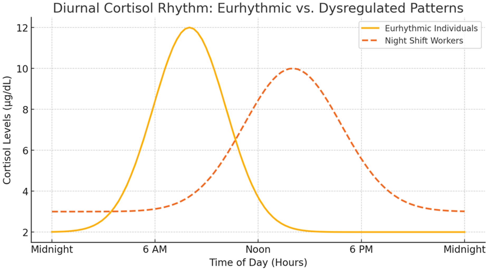
Andreadi, A., Andreadi, S., Todaro, F., Ippoliti, L., Bellia, A., Magrini, A., … Lauro, D. (2025). Modified cortisol circadian rhythm: The hidden toll of night-shift work. International Journal of Molecular Sciences, 26, 2090. https://doi.org/10.3390/ijms26052090
Ardura, J., Gutierrez, R., Andres, J., & Agapito, T. (2003). Emergence and evolution of the circadian rhythm of melatonin in children. Horm. Res., 59(2), 66–72. https://doi.org/68571
Arendt, J., & Aulinas, A. (2000). Physiology of the pineal gland and melatonin. In Endotext. South Dartmouth (MA): MDText.com, Inc. Retrieved from https://www.ncbi.nlm.nih.gov/books/NBK550972/
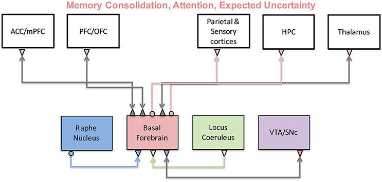
Avery, M. C., & Krichmar, J. L. (2017). Neuromodulatory systems and their interactions: A review of models, theories, and experiments. Frontiers in Neural Circuits, 11, 108. https://doi.org/10.3389/fncir.2017.00108
Avram, M., Grothe, M. J., Meinhold, L., Leucht, C., Leucht, S., Borgwardt, S., … Sorg, C. (2021). Lower cholinergic basal forebrain volumes link with cognitive difficulties in schizophrenia. Neuropsychopharmacology: Official Publication of the American College of Neuropsychopharmacology. https://doi.org/10.1038/s41386-021-01070-x
Carhart-Harris, R. L., & Nutt, D. J. (2017). Serotonin and brain function: A tale of two receptors. Journal of Psychopharmacology, 31(9), 1091–1120. https://doi.org/10.1177/0269881117725915
De Ponti, F. (2004). Pharmacology of serotonin: What a clinician should know. Gut, 53(10), 1520–1535. https://doi.org/10.1136/gut.2003.035568
Deussing, J. M., & Chen, A. (2018). The Corticotropin-Releasing factor family: Physiology of the stress response. Physiological Reviews, 98(4), 2225–2286. https://doi.org/10.1152/physrev.00042.2017
Domes, G., Heinrichs, M., Kumbier, E., Grossmann, A., Hauenstein, K., & Herpertz, S. C. (2013). Effects of intranasal oxytocin on the neural basis of face processing in autism spectrum disorder. Biological Psychiatry, 74(3), 164–171. https://doi.org/http://dx.doi.org/10.1016/j.biopsych.2013.02.007
Dopamine transporter. (n.d.). https://www.sciencedirect.com/topics/neuroscience/dopamine-transporter. Retrieved from https://www.sciencedirect.com/topics/neuroscience/dopamine-transporter
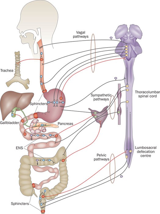
Furness, J. B. (2012). The enteric nervous system and neurogastroenterology. Nature Reviews. Gastroenterology & Hepatology, 9(5), 286–294. https://doi.org/10.1038/nrgastro.2012.32
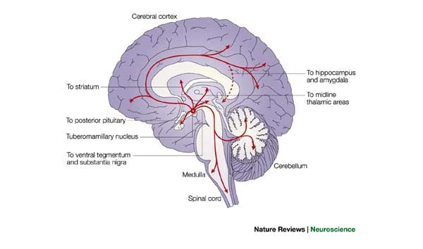
Haas, H., & Panula, P. (2003). The role of histamine and the tuberomamillary nucleus in the nervous system. Nature Reviews. Neuroscience, 4(2), 121–130. https://doi.org/10.1038/nrn1034
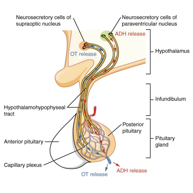
Hacking, C., Gaillard, F., & Smith, D. (2016, December 7). Posterior pituitary. In Radiopaedia.org. Radiopaedia.org. https://doi.org/10.53347/rid-49865
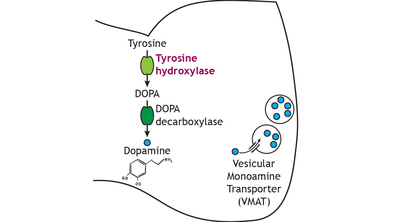
Henley, C. (2021). Neurotransmitter synthesis and storage. In Foundations of neuroscience. Michigan State University Libraries. Retrieved from https://openbooks.lib.msu.edu/neuroscience/chapter/neurotransmitter-synthesis-and-storage/
Małgorzata, P., Paweł, K., Iwona, M. L., Brzostek, T., & Andrzej, P. (2020). Glutamatergic dysregulation in mood disorders: Opportunities for the discovery of novel drug targets. Expert Opinion on Therapeutic Targets, 24(12), 1187–1209. https://doi.org/10.1080/14728222.2020.1836160
McCutcheon, R. A., Krystal, J. H., & Howes, O. D. (2020). Dopamine and glutamate in schizophrenia: Biology, symptoms and treatment. World Psychiatry: Official Journal of the World Psychiatric Association, 19(1), 15–33. https://doi.org/10.1002/wps.20693
Mesulam, M.-M. (2013). Cholinergic circuitry of the human nucleus basalis and its fate in alzheimer’s disease. The Journal of Comparative Neurology, 521(18), 4124–4144. https://doi.org/10.1002/cne.23415
Moncrieff, J., Cooper, R. E., Stockmann, T., Amendola, S., Hengartner, M. P., & Horowitz, M. A. (2022). The serotonin theory of depression: A systematic umbrella review of the evidence. Molecular Psychiatry. https://doi.org/10.1038/s41380-022-01661-0
Overk, C. R., & Mufson, E. J. (2010). Dopamine transporter: Aging and parkinson’s disease. In Encyclopedia of movement disorders (pp. 330–332). Elsevier. https://doi.org/10.1016/b978-0-12-374105-9.00237-9
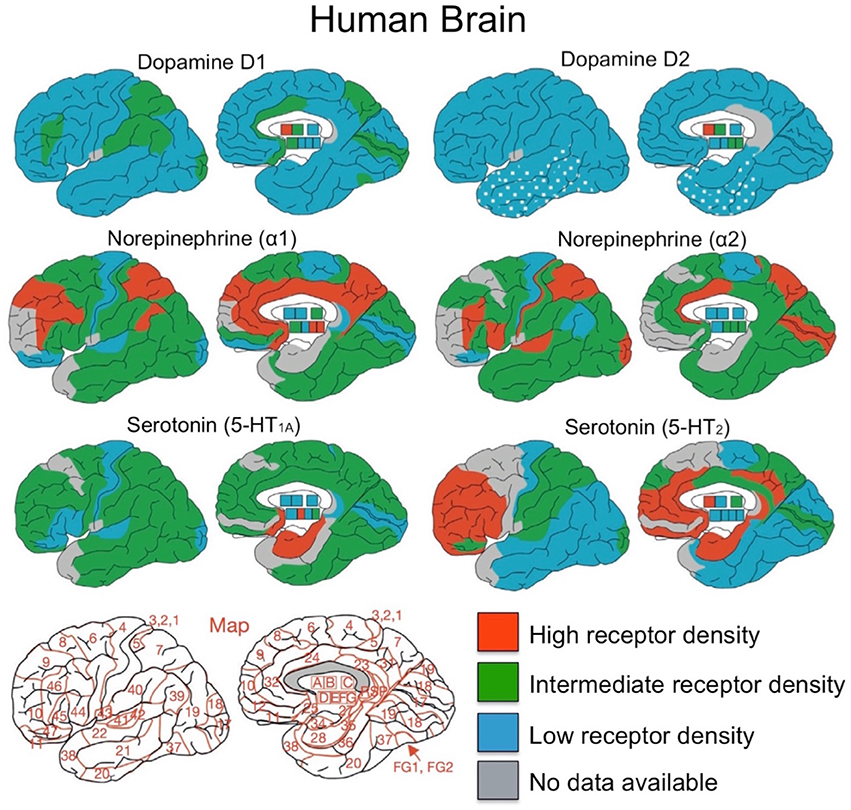
Palomero-Gallagher, N., Amunts, K., & Zilles, K. (2015). Transmitter receptor distribution in the human brain. In Brain mapping (pp. 261–275). Elsevier. https://doi.org/10.1016/b978-0-12-397025-1.00221-9
Picciotto, M. R., Higley, M. J., & Mineur, Y. S. (2012). Acetylcholine as a neuromodulator: Cholinergic signaling shapes nervous system function and behavior. Neuron, 76, 116–129. https://doi.org/10.1016/j.neuron.2012.08.036
Ren, J., Friedmann, D., Xiong, J., Liu, C. D., Ferguson, B. R., Weerakkody, T., … Luo, L. (2018). Anatomically defined and functionally distinct dorsal raphe serotonin sub-systems. Cell. https://doi.org/10.1016/j.cell.2018.07.043
Songs, M. (2018). Muppet songs: Mahna mahna (muppet show - 1976). YouTube. Retrieved from https://www.youtube.com/watch?v=TbZ_hTEOKZc
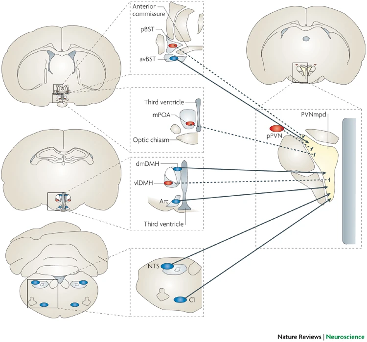
Ulrich-Lai, Y. M., & Herman, J. P. (2009). Neural regulation of endocrine and autonomic stress responses. Nature Reviews Neuroscience, 10(6), 397–409. https://doi.org/10.1038/nrn2647
Viviani, D., Charlet, A., Burg, E. van den, Robinet, C., Hurni, N., Abatis, M., … Stoop, R. (2011). Oxytocin selectively gates fear responses through distinct outputs from the central amygdala. Science, 333(6038), 104–107. https://doi.org/10.1126/science.1201043
Vogelsang, D. A., & D’Esposito, M. (2018). Is there evidence for a rostral-caudal gradient in fronto-striatal loops and what role does dopamine play? Frontiers in Neuroscience, 12, 242. https://doi.org/10.3389/fnins.2018.00242
Weisman, O., & Feldman, R. (2013). Oxytocin effects on the human brain: Findings, questions, and future directions. Biological Psychiatry, 74(3), 158–159. https://doi.org/http://dx.doi.org/10.1016/j.biopsych.2013.05.026
What is a neurotransmitter? (n.d.). Retrieved April 8, 2026, from https://nigms.nih.gov/biobeat/2024/08/what-is-a-neurotransmitter
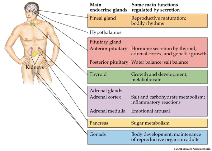
Wikipedia contributors. (2025a, September 28). Adrenal gland. Retrieved from https://en.wikipedia.org/wiki/Adrenal_gland
Wikipedia contributors. (2025b, October 25). Blind men and an elephant. Retrieved from https://en.wikipedia.org/wiki/Blind_men_and_an_elephant
Wikipedia contributors. (2025c, November 20). Cycloplegia. Retrieved from https://en.wikipedia.org/wiki/Cycloplegia#Cycloplegic_medication
Wikipedia contributors. (2025d, November 24). Raphe nuclei. Retrieved from https://en.wikipedia.org/wiki/Raphe_nuclei
Wikipedia contributors. (2026a, February 23). Monoamine oxidase inhibitor. Retrieved from https://en.wikipedia.org/wiki/Monoamine_oxidase_inhibitor
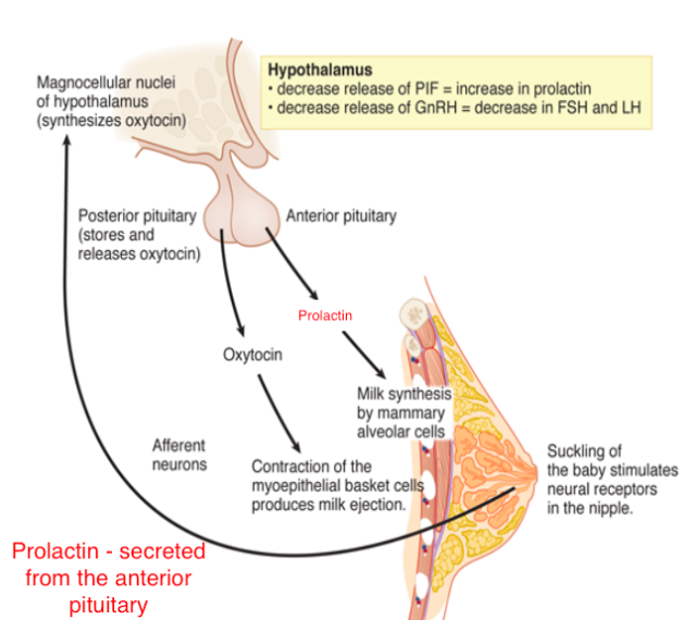
Wikipedia contributors. (2026b, March 26). Anterior pituitary. Retrieved from https://en.wikipedia.org/wiki/Anterior_pituitary
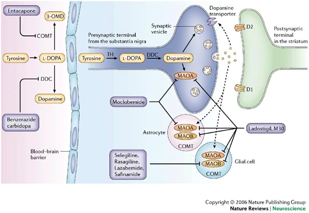
Youdim, M. B. H., Edmondson, D., & Tipton, K. F. (2006). The therapeutic potential of monoamine oxidase inhibitors. Nature Reviews. Neuroscience, 7(4), 295–309. https://doi.org/10.1038/nrn1883
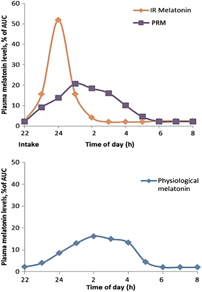
Zisapel, N. (2018). New perspectives on the role of melatonin in human sleep, circadian rhythms and their regulation: Melatonin in human sleep and circadian rhythms. British Journal of Pharmacology, 175, 3190–3199. https://doi.org/10.1111/bph.14116

Footnotes

  1. “Generates EPSP”

  2. “Generates IPSP”

  3. “Cycloplegia caused by Cyclopentolate 1% instilled in both eyes. By Ilovebaddies (talk) - Own work, CC BY-SA 3.0, https://commons.wikimedia.org/w/index.php?curid=112232286”

  4. “Fig. 1: Map of the cholinergic basal forebrain. The region of interest depicts the cholinergic basal forebrain, based on a cytoarchitectonic map of cholinergic nuclei, overlaid on a human brain template in Montreal Neurological Institute space. The BFCN mask is based on combined histology and postmortem MRI [63], containing several cholinergic subdivisions within the basal forebrain, including the medial septal nucleus, diagonal band of Broca, nucleus subputaminalis, the basal magnocellular complex, and nucleus basalis of Meynert (57, 72)”

  5. “Cholinergic circuitry of the human nucleus basalis. ACh, acetylcholine; DA, dopamine; EAA, excitatory amino acids; NE, norepinephrine; 5HT, serotonin. Question marks indicate that the connection has not been confirmed in the human brain.”

  6. “The cholinergic system and its functions. The cholinergic system originates in the basal forebrain and sends projections to many cortical and subcortical regions. As a result, it has been implicated in a variety of functions including memory, attention, and uncertainty computations. Activity of the basal forebrain is thought to be regulated by prefrontal cortices, as well as other neuromodulatory brain regions.”

  7. “Figure 9.6. Dopamine is synthesized in a two-step process. Tyrosine is converted into DOPA by tyrosine hydroxylase, the rate-limiting step in the pathway. Then dopamine is synthesized from DOPA by DOPA decarboxylase. Dopamine is then packaged into vesicles by vesicular monoamine transporter. ‘Dopamine Synthesis’ by Casey Henley is licensed under a Creative Commons Attribution Non-Commercial Share-Alike (CC BY-NC-SA) 4.0 International License.”

  8. “Overview of distributions for dopamine, norepinephrine and serotonin receptors. Brodmann area map is presented in bottom left corner. A, caudate-putamen; B, globus pallidus; C, diencephalon; D, amygdala; E, CA1 region of hippocampus; F, CA2/3 region of hippocampus; G, dentate gyrus. Figure adapted from Palomero et al. (2015) with permission from Elsevier Journals.”

  9. “The pathway of dopamine synthesis proceeds from tyrosine via tyrosine hydroxylase (TH) catalysis to levodopa (L-DOPA), and subsequent decarboxylation by dopa decarboxylase (DDC) to dopamine. Dopamine is metabolized by intraneuronal monoamine oxidase A (MAOA), and by glial and astrocyte MAOA and MAOB1,11. Selective inhibitors of MAOA (for example, moclobemide) and MAOB (selegiline, rasagiline and safinamide) do not alter the steady-state striatal dopamine levels, although chronic treatment with these drugs does enhance dopamine release, possibly due to the elevation of endogenous brain amines or receptor modulation93,194. However, non-selective MAOA/B inhibitors (such as ladostigil and those shown in Table 2) do induce highly significant increases in the levels of dopamine in the striatum and other regions93,194. Although dopamine does not pass the blood–brain barrier (BBB), L-DOPA can, and DDC inhibitors that do not pass the BBB, such as benzerazide (benserazide) and carbidopa, increase its availability to the brain. Inhibitors of catechol-O-methyltransferase (COMT), such as entacapone, also enhance L-DOPA availability and prevent the inactivation of dopamine by COMT. 3-OMD, 3-O-methyl dopa; D1, D2, dopamine receptors.”

  10. “Depiction of location of adrenal glands in human body; zoom to detail of adrenal gland; zoom to cross section of adrenal gland; cross section shows cortex, including connective tissue capsule, zona glomerulosa, zona fasciculata, and zona reticularis; also shows medulla. By Antinksčio sandara.png Author: EdgarasLe - https://commons.wikimedia.org/wiki/File:Antinks%C4%8Dio_sandara_esp.png, CC BY-SA 4.0, https://commons.wikimedia.org/w/index.php?curid=127157804”

  11. “The neural connections between the ENS and CNS, and neural connections between gastrointestinal organs, are quite different from those depicted in textbooks. The digestive system contains full reflex circuits of the ENS (motor neurons and interneurons in blue, sensory neurons in purple). Pathways from the gastrointestinal tract project outwards, via intestinofugal neurons (red), to the CNS (neurons in yellow), sympathetic ganglia, gallbladder and pancreas. Neurons in sympathetic prevertebral ganglia (green) receive both CNS and ENS inputs. Sensory information goes both to the ENS, via intrinsic primary afferent (sensory) neurons (purple) and to the CNS via extrinsic primary afferent neurons (also purple) that follow spinal and vagal afferent routes. Pathways from the CNS reach the ENS and gastrointestinal effector tissues through vagal, sympathetic and pelvic pathways. Abbreviations: CNS, central nervous system; ENS, enteric nervous system.”

  12. “In this drawing of the brain, the serotonergic system is red and the mesolimbic dopamine pathway is blue. There is one collection of serotonergic neurons in the upper brainstem that sends axons upwards to the whole cerebrum, and one collection next to the cerebellum that sends axons downward to the spinal cord. Slightly forward the upper serotonergic neurons is the ventral tegmental area (VTA), which contains dopaminergic neurons. These neurons’ axons then connect to the nucleus accumbens, hippocampus, and the frontal cortex. Over the VTA is another collection of dopaminergic cells, the substansia nigra, which send axons to the striatum.”

  13. “In this drawing of the brain, the serotonergic system is red and the mesolimbic dopamine pathway is blue. There is one collection of serotonergic neurons in the upper brainstem that sends axons upwards to the whole cerebrum, and one collection next to the cerebellum that sends axons downward to the spinal cord. Slightly forward the upper serotonergic neurons is the ventral tegmental area (VTA), which contains dopaminergic neurons. These neurons’ axons then connect to the nucleus accumbens, hippocampus, and the frontal cortex. Over the VTA is another collection of dopaminergic cells, the substansia nigra, which send axons to the striatum. By NIH - http://www.drugabuse.gov/pubs/teaching/largegifs/slide-2.gif, Public Domain, https://commons.wikimedia.org/w/index.php?curid=37682508.”

  14. “About 64,000 histamine-producing neurons, located in the tuberomamillary nucleus119 of the human brain, innervate all of the major parts of the cerebrum, cerebellum, posterior piuitary and the spinal cord.”

  15. “About 64,000 histamine-producing neurons, located in the tuberomamillary nucleus119 of the human brain, innervate all of the major parts of the cerebrum, cerebellum, posterior piuitary and the spinal cord.”

  16. Plus control over ANS

  17. “By OpenStax College - Anatomy & Physiology, Connexions Web site. http://cnx.org/contents/IgqATiKA@3/The-Pituitary-Gland-and-Hypoth, Jun 19, 2013., CC BY 3.0, https://commons.wikimedia.org/w/index.php?curid=30148144”

  18. “Figure 1: General scheme of brain acute-stress regulatory pathways. Stressors activate brainstem and/or forebrain limbic structures. The brainstem can generate rapid hypothalamic-pituitary-adrenal (HPA) axis and autonomic nervous system (ANS) responses through direct projections to hypophysiotrophic neurons in the paraventricular nucleus of the hypothalamus (PVN) or to preganglionic autonomic neurons (stress response triggers). By contrast, forebrain limbic regions have no direct connections with the HPA axis or the ANS and thus require intervening synapses before they can access autonomic or neuroendocrine neurons (top-down regulation). A high proportion of these intervening neurons are located in hypothalamic nuclei that are also responsive to homeostatic status, providing a mechanism by which the descending limbic information can be modulated according to the physiological status of the animal (‘middle management’). BST, bed nucleus of the stria terminalis; CVO, circumventricular organ; SAM, sympathoadrenomedullary system.”

  19. “FIGURE 1. Effector systems of the stress response. A stressor elicits rapid activation of the autonomic nervous system with its sympathoneuronal (SN) and sympatho-adrenomedullary (SAM) limbs releasing their main effectors, noradrenaline and adrenaline, respectively. Activation of the hypothalamic-pituitary-adrenocortical (HPA) axis results in synthesis and release of its main effector, cortisol or corticosterone, in rodents. ACTH, adrenocorticotropic hormone; CRF, corticotropin-releasing factor.”

  20. “Figure 3: The brain circuitry that regulates HPA axis stress responses. Stress-induced activation of the dorsal part of the medial parvocellular paraventricular nucleus of the hypothalamus (PVNmpd) originates in several brain regions (excitatory inputs are coloured blue with solid lines and inhibitory (GABA (γ-aminobutyric acid)-ergic) inputs are coloured red with dashed lines). The paraventricular nucleus of the hypothalamus (PVN) receives direct noradrenergic, adrenergic and peptidergic innervation from the nucleus of the solitary tract (NTS). The dorsomedial components of the dorsomedial hypothalamus (dmDMH) and the arcuate nucleus (Arc) provide intrahypothalamic stress excitation. The anterior part of the bed nucleus of the stria terminalis (BST), particularly the anteroventral nucleus of the BST (avBST), activates hypothalamic-pituitary-adrenocortical (HPA) axis stress responses. The PVN also receives a stress-excitatory drive from the dorsal raphe, the tuberomammillary nucleus, the supramammillary nucleus and the spinal cord, among others (omitted in the interest of space). Activation of the PVNmpd is inhibited by numerous hypothalamic circuits, including the medial preoptic area (mPOA), the ventrolateral component of the dorsomedial hypothalamus (vlDMH) and local neurons in the peri-PVN region (pPVN), encompassing the PVN surround and the subparaventricular zone. The posterior subregions of the bed nucleus of the stria terminalis (pBST) provide a prominent forebrain inhibition of HPA axis responses; most of these inputs are GABAergic. Brain sections are modified, with permission, from Ref. 154 © (1998) Academic Press.”

  21. “FIGURE 4. Distribution of mRNA expression of corticotropin-releasing factor (CRF)-related peptides in the rodent brain. Three-dimensional expression patterns of CRF-related peptide were collapsed onto a single sagittal brain section. Depicted are well-documented sites of high to moderate expression. Sites of expression are indicated by colored dots: CRF (orange), urocortin (UCN) 1 (green), UCN2 (light blue), UCN3 (purple). 7, Facial nerve; 12, hypoglossal nerve; Amb, ambiguous nucleus; AP, area postrema; arc, arcuate nucleus; Bar, Barrington’s nucleus; BLA, basolateral amygdala; BNST, bed nucleus of the stria terminalis (d, dorsal aspect; v, ventral aspect); CA1, cornu ammonis subfield 1; CA3, cornu ammonis subfield 3; CC, corpus callosum; CeA, central amygdala; Cereb, cerebellum; CingCx, cingulate cortex; CPu, caudate putamen; DeepN, deep nucleus of cerebellum; DG, dentate gyrus; EW, Edinger Westphal nucleus; FrCx, frontal cortex; GPe, external globus pallidus; Hip, hippocampus; IC, inferior colliculus; IO, inferior olive; IPN, interpeduncular nucleus; LC, locus coeruleus; LH, lateral hypothalamus; LS, lateral septum; LSO, lateral superior olive; LTDg, laterodorsal tegmental nucleus; MeA, medial amygdala; MePO, median preoptic area; MGN, medial geniculate nucleus; MS, medial septum; MVN, medial vestibular nucleus; NAc, nucleus accumbens; NTS, nucleus of the solitary tract; OB, olfactory bulb; OccCx, occipital cortex; OT, olfactory tubercle; PAG, periaqueductal gray; ParCx, parietal cortex; PB, parabrachial nucleus; PFA, perifornical area; PG, pontine gray; Pir, piriform cortex; Pit, pituitary (p, lobe, anterior lobe, intermediate, posterior lobe); PM, premammillary nucleus; PPTg, pedunculopontine tegmental nucleus; PVN, paraventricular nucleus of the hypothalamus; R, red nucleus; RN, raphe nuclei; RTB, reticular thalamic nucleus; SC, superior colliculus; SN, substantia nigra; Sp5n, spinal trigeminal nucleus; SPO, superior paraolivary nucleus; VMH, ventromedial hypothalamus, VTA, ventral tegmental area.”

  22. “Mean plasma levels of ingested (A) and endogenous (B) melatonin. (A) Pharmacokinetics of prolonged‐release (PRM) versus immediate‐release (IR) melatonin 2 mg formulations. Results are mean plasma melatonin levels following drug intake and expressed as % of the AUC (adapted from Zisapel, 2010). (B) Mean endogenous plasma melatonin levels (adapted from Zhdanova et al., 1998).”

  23. “Diagrammatic representation of the control of production and the functions of melatonin, regarding seasonal and circadian timing mechanisms. Abbreviations: SCN: suprachiasmatic nucleus, PVN: paraventricular nucleus, SCG: superior cervical ganglion, NA: norepinephrine (noradrenalin), RHT: retino-hypothalamic-tract, CCG: clock-controlled genes. Based on an original diagram by Dr Elisabeth Maywood, MRC Laboratory of Molecular Biology, Neurobiology Division, Hills Road Cambridge, CB2 2QH, UK.”

  24. “Relationship of plasma melatonin to other major circadian rhythms driven by the internal clock. Abbreviations: VAS: visual analogue scale. Reproduced from Rajaratnam SMW and Arendt J. Lancet 358:999-1005, 2001 by permission.”

  25. “Seven blind men and an elephant parable at a Jain temple. By romana klee from usa - sammati tarka prakarana, CC BY-SA 2.0, https://commons.wikimedia.org/w/index.php?curid=59461928”

  26. In PNS

  27. + ACh in CNS