2017-08-31 11:12:06
Prelude
Spatial and Temporal Resolution

Which of the following statements about the brain imaging technique used in this figure is FALSE?

Which of the following statements about the brain imaging technique used in this figure is FALSE?
- A. It is non-invasive.
- B. It provides information about brain structures.
- C. It provides information about rapid (millisecond-level) changes in brain activity.
- D. It cannot resolve details about individual neurons.
Anterograde tracing chemicals are injected in brain tissue in order to answer what question?
- A. What kinds of cells are prominent in a given area?
- B. The density of dendrites and axons in a given region?
- C. Where does a target region project to or connect with?
Anterograde tracing chemicals are injected in brain tissue in order to answer what question?
- A. What kinds of cells are prominent in a given area?
- B. The density of dendrites and axons in a given region?
- C. Where does a target region project to or connect with?
Diffusion Tensor Imaging (DTI) is one type of X imaging?
- A. functional PET
- B. structural CT
- C. structural MRI
- D. functional MRI (fMRI)
Diffusion Tensor Imaging (DTI) is one type of X imaging?
- A. functional PET
- B. structural CT
- C. structural MRI
- D. functional MRI (fMRI)
Today's topics
- Functional methods
Functional methods
- Recording from the brain
- Interfering with the brain
- Stimulating the brain
- Simulating the brain
Recording from the brain
- Single/multi unit recording
- Microelectrodes
- Small numbers of nerve cells
Single/multi-unit Recording

Single/multi-unit recording
- What does neuron X respond to?
- Great temporal (ms), spatial resolution (um)
- Invasive
- Rarely suitable for humans, but…
Electrocorticography (ECoG)

Positron Emission Tomography (PET)
Positron Emission Tomography (PET)
- Radioactive tracers (glucose, oxygen)
- Positron decay
- Experimental condition - control
- Average across individuals


PET
- Evaluating PET
- Temporal (~ s) and spatial (mm-cm) resolution worse than fMRI
- Radioactive exposures + mildly invasive
- Dose < airline crew exposure in 1 yr
Functional Magnetic Resonance Imaging (fMRI)
- Neural activity -> local \(O_2\) consumption increase
- Blood Oxygen Level Dependent (BOLD) response
- Oxygenated vs. deoxygenated hemoglobin creates magnetic contrast
- Do regional blood \(O_2\) levels (and flow) vary with behavior X?
fMRI
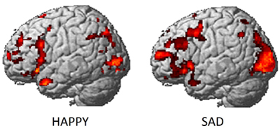
fMRI (Dougherty et al. 2003)

fMRI
- Evaluating
- Non-invasive, but expensive
- Moderate but improving (mm) spatial, temporal (~sec) resolution
- Indirect measure of brain activity
- Hemodynamic Response Function (HRF)
- 1s delay plus 3-6 s ramp-up
Hemodynamic Response Function (HRF)

Electroencephalography (EEG)
- How does it work?
- Electrodes on scalp or brain surface
- What do we measure?
- Combined activity of huge # of neurons
EEG

EEG
- High temporal, poor spatial resolution
- Analyze frequency bands
- LOW: deep sleep
- MIDDLE: Quiet, alert state
- HIGH:“Binding” information across senses
EEG Frequency

Event-related potentials (ERPs)
- EEGs time-locked to some event - Averaged over many trials
ERPs

Brain Computer Interface (BCI)

Magneto-encephalography (MEG)
- Like EEG, but measuring magnetic fields
- High temporal resolution, low spatial resolution
- Magnetic field propagates w/o distortion
MEG

Manipulating the brain
- Nature’s “experiments”
- Stroke, head injury, tumor
- Neuropsychology
- Remember Galen?
- Logic: damage impairs performance = region critical for behavior
- Poor spatial/temporal resolution, limited experimental control
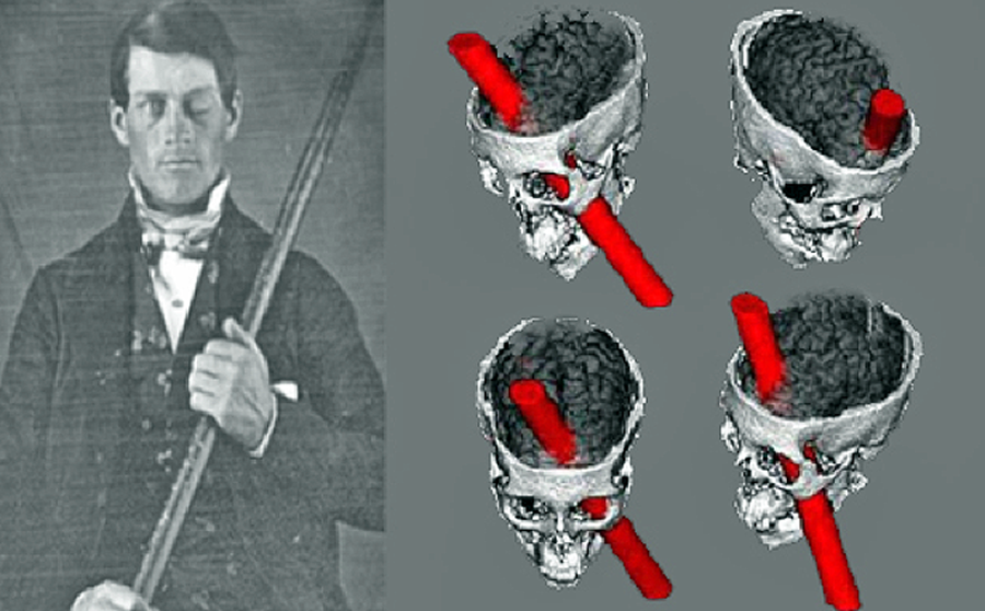
Phineas Gage
Stimulating the brain
- Pharmacological
- Electrical (transcranial Direct Current Stimulation - tDCS)
- Magnetic (Transcranial magnetic stimulation - TMS)
- Optically (optogenetics)
tDCS

TMS

Optogenetic stimulation
Evaluating stimulation methods
- Spatial/temporal resolution?
- Assume stimulation mimics natural activity?
- Optogenetic stimulation highly similar, others less so
- Deep brain stimulation as therapy
- Parkinson’s Disease
- Depression
- Epilepsy
Deep brain stimulation

Simulating the brain
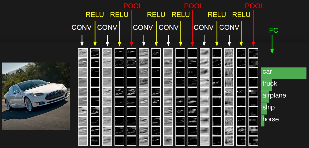
- Computer/mathematical models of brain function
- Example: neural networks
- Cheap, noninvasive, can be stimulated or “lesioned”
Spatial and Temporal Resolution
Next time…
- Tour of SLEIC
- Meet in Chandlee lobby
References
Dayan, Eran, Nitzan Censor, Ethan R. Buch, Marco Sandrini, and Leonardo G. Cohen. 2013. “Noninvasive Brain Stimulation: From Physiology to Network Dynamics and Back.” Nature Neuroscience 16 (7): 838–44. doi:10.1038/nn.3422.
Dougherty, R. F., V. M. Koch, A. A. Brewer, B. Fischer, J. Modersitzki, and B. A. Wandell. 2003. “Visual Field Representations and Locations of Visual Areas V1/2/3 in Human Visual Cortex.” Journal of Vision 3 (10): 1–1. doi:10.1167/3.10.1.
Han, Wenfei, Luis A. Tellez, Miguel J. Rangel, Simone C. Motta, Xiaobing Zhang, Isaac O. Perez, Newton S. Canteras, Sara J. Shammah-Lagnado, Anthony N. van den Pol, and Ivan E. de Araujo. 2017. “Integrated Control of Predatory Hunting by the Central Nucleus of the Amygdala.” Cell 168 (1): 311–324.e18. doi:10.1016/j.cell.2016.12.027.
Sejnowski, Terrence J, Patricia S Churchland, and J Anthony Movshon. 2014. “Putting Big Data to Good Use in Neuroscience.” Nature Neuroscience 17 (11). Nature Publishing Group: 1440–1. doi:10.1038/nn.3839.