2019-01-30 15:14:14
Prelude
How to play EyeWire (03:56)
Announcements
- Exam 1 next Thursday, 2/7
- 40 questions
- Complete 1 "component/section" in EyeWire, earn 5 extra credit points.
- Take screen shot, email to Courtney
- Due before Spring Break
Today's Topics
- Warm-up (Review Quiz 1)
- Cells of the nervous system
- Glia
- Neurons
- How do these cells communicate?
Warm-up (Review Quiz 1)

1. The image illustrates what type of slice?
- Sagittal
- Dorsal
- Coronal
- Axial
1. The image illustrates what type of slice?
- Sagittal
- Dorsal
- Coronal
- Axial
2. The figure illustrates the use of _________ magnetic resonance imaging (MRI), a technique with a ________ spatial resolution
- functional; fair (~ 3+ mm)
- structural; good (~ 1 mm)
- neural; excellent (~ 1 micron)
- diffusion tensor; poor (~ 1 cm)
2. The figure illustrates the use of _________ magnetic resonance imaging (MRI), a technique with a ________ spatial resolution
- functional; fair (~ 3 mm)
- structural; good (~ 1 mm)
- neural; excellent (~ 1 micron)
- diffusion tensor; poor (~ 1 cm)
3. Event-related potentials are detected using ___________; they measure ___________.
- positron emission tomography (PET); local metabolic rates
- magnetic resonance imaging (MRI); the integrity of white matter fiber tracts
- electroencephalography (EEG); the time-locked electrical activity of large number of neurons
- magnetoencephalography (MEG); average brain magnetic activity
4. All of these structures are components of the midbrain EXCEPT:
- Superior colliculus
- 3rd ventricle
- Tegmentum
- Inferior colliculus
4. All of these structures are components of the midbrain EXCEPT:
- Superior colliculus
- 3rd ventricle
- Tegmentum
- Inferior colliculus
5. Why does fMRI represent an indirect measure of brain activity?
- It measures brain structure, not function.
- It measures electrical activity, but neurons send chemical messages.
- It measures changes in blood oxygen and blood flow that follow neural activity.
- It has poor spatial resolution.
5. Why does fMRI represent an indirect measure of brain activity?
- It measures brain structure, not function.
- It measures electrical activity, but neurons send chemical messages.
- It measures changes in blood oxygen and blood flow that follow neural activity.
- It has poor spatial resolution.
6. Which philosopher believed that the pineal gland had a special role in both reflexive and voluntary action?
- Aristotle
- Galen
- Vesalius
- Descartes
6. Which philosopher believed that the pineal gland had a special role in both reflexive and voluntary action?
- Aristotle
- Galen
- Vesalius
- Descartes
7. Anterograde and retrograde histochemical tracers help neuroscientists determine:
- What stimuli best activate a brain region
- What connects where
- When to stimulate a brain region for maximum effect
- Whether a brain area is functioning normally
7. Anterograde and retrograde histochemical tracers help neuroscientists determine:
- What stimuli best activate a brain region
- What connects where
- When to stimulate a brain region for maximum effect
- Whether a brain area is functioning normally
8. Which of these landmarks separates the left from the right cerebral hemisphere?
- Lateral fissure
- Longitudinal fissure
- Anterior cingulate gyrus
- Central sulcus
8. Which of these landmarks separates the left from the right cerebral hemisphere?
- Lateral fissure
- Longitudinal fissure
- Anterior cingulate gyrus
- Central sulcus
9. Visual information enters the CNS via the 2nd (II) cranial or _______ nerve and projects to __________ of the thalamus:
- optic; lateral geniculate nucleus
- olfactory; striatum
- optic; substantia nigra
- oculomotor; superior colliculus
9. Visual information enters the CNS via the 2nd (II) cranial or _______ nerve and projects to __________ of the thalamus:
- optic; lateral geniculate nucleus
- olfactory; striatum
- optic; substantia nigra
- oculomotor; superior colliculus
10. All of these structures are components of the midbrain EXCEPT:
- Tectum
- Hippocampus
- Inferior colliculus
- Tegmentum
10. All of these structures are components of the midbrain EXCEPT:
- Tectum
- Hippocampus
- Inferior colliculus
- Tegmentum
11. This forebrain structure in the central diencephalon controls both the autonomic nervous system and the endocrine system:
- Hippocampus
- Thalamus
- Medulla
- Hypothalamus
11. This forebrain structure in the central diencephalon controls both the autonomic nervous system and the endocrine system:
- Hippocampus
- Thalamus
- Medulla
- Hypothalamus
12. The arrow in the figure on page 2 shows two parts of the brain that are structurally related and adjacent to one another, the _________ and the _________.
- lateral fissure; temporal lobes
- spinal cord; lateral ventricles
- amygdala; tectum
- basal ganglia; 4th ventricle
12. The arrow in the figure shows two parts of the brain that are structurally related and adjacent to one another, the _________ and the _________.
- lateral fissure; temporal lobes
- spinal cord; lateral ventricles
- amygdala; tectum
- basal ganglia; 4th ventricle
Cells of the nervous system
How many neurons and glia?
- Old "lore": ~100 billion neurons
- New estimate (Azevedo et al. 2009)
- ~86 +/- 8 billion neurons
- ~85 +/- 9 billion glia
- 100-500 trillion synapses, 1 billion/mm^3
Could you count to 170 billion?
- How many years to count to 170 billion?
- 60 s/min x 60 min/hr x 24 hrs/day x 365 days/ yr = 31,536,000 s/yr
- 1.7e11/31,536,000 = 5,390 years
Mass, Neurons, Non-Neurons

Neurons by brain mass

Non-neuronal cells by brain mass

How many neurons and glia?
"These findings challenge the common view that humans stand out from other primates in their brain composition and indicate that, with regard to numbers of neuronal and nonneuronal cells, the human brain is an isometrically scaled-up primate brain."
The Human Advantage

Glia (neuroglia)
- "Glia" means glue
- Functions
- Structural support
- Metabolic support
- Brain development
- Neural plasticity?
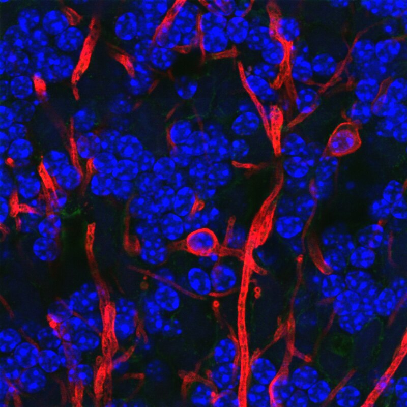
Astrocytes
- "Star-shaped"
- Physical and metabolic support
- Blood/brain barrier
- Ion (Ca++/K+) buffering
- Neurotransmitter (e.g., glutamate) buffering
Astrocytes
- Shape brain development, synaptic plasticity
- Regulate local blood flow
- Regulate/influence communication between neurons, (Bazargani and Attwell 2016)
- Disruption linked to cognitive impairment, disease (Chung et al. 2015)
Astrocytes

Myelinating cells
- Produce myelin or myelin sheath
- White, fatty substance
- Surrounds many neurons
- The "white" in white matter
- Provide electrical/chemical insulation
- Make neuronal messages faster, less susceptible to noise
Types of myelin-producing cells
- Oligodendrocytes
- In brain and spinal cord (CNS)
- 1:many neurons
- Schwann cells
- In PNS
- 1:1 neuron
- Facilitate neuro-regeneration
Oligodendrocytes
Schwann Cells

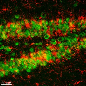
Microglia
- Phagocytosis
- Clean-up damaged, dead tissue
- Prune synapses in normal development and disease
- Disruptions in microglia pruning -> impaired functional brain connectivity and social behavior, (Zhan et al. 2014)
Microglia
Neurons

Fun facts about neurons
- Specialized for electrical & chemical communication
- Post-mitotic – don't divide
- Most born early in life, (Bhardwaj et al. 2006)
- Among longest-lived cells in body, may scale with organism lifespan (Magrassi, Leto, and Rossi 2013)
- Can extend over long distances
Macrostructure of neurons
Structure of neurons

Dendrites
- Branch-like "extrusions" from cell body
- Majority of input to neuron
- Cluster close to cell body/soma
- Usually receive info
- Passive (do not regenerate electrical signal) vs. active (regenerate signal)
- Spines
Dendrites

Dendritic Spines

Soma (cell body)
- Varied shapes
- Nucleus
- Chromosomes
- Organelles
- Mitochonrdria
- Smooth and Rough Endoplasmic reticulum (ER)
Soma
Axons
- Another branch-like "extrusion" from soma
- Extend farther than dendrites
- Usually transmit info
Axons
- Parts
- Initial segment (closest to soma, unmyelinated)
- Nodes of Ranvier (unmyelinated segments along axon)
- Terminals, axon terminals, terminal buttons, synaptic terminals, synaptic boutons
Axons

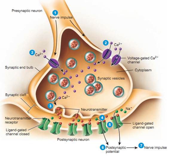
Synaptic bouton (terminal button)
- Synapse (~5-10K per neuron)
- Presynaptic membrane (sending cell) and postsynaptic (receiving cell) membrane
- Synaptic cleft – space between cells
- Synaptic vesicles
- Pouches of neurotransmitters
- Autoreceptors (detect NTs); transporters (transport NTs across membrane)
Synaptic bouton (terminal button)
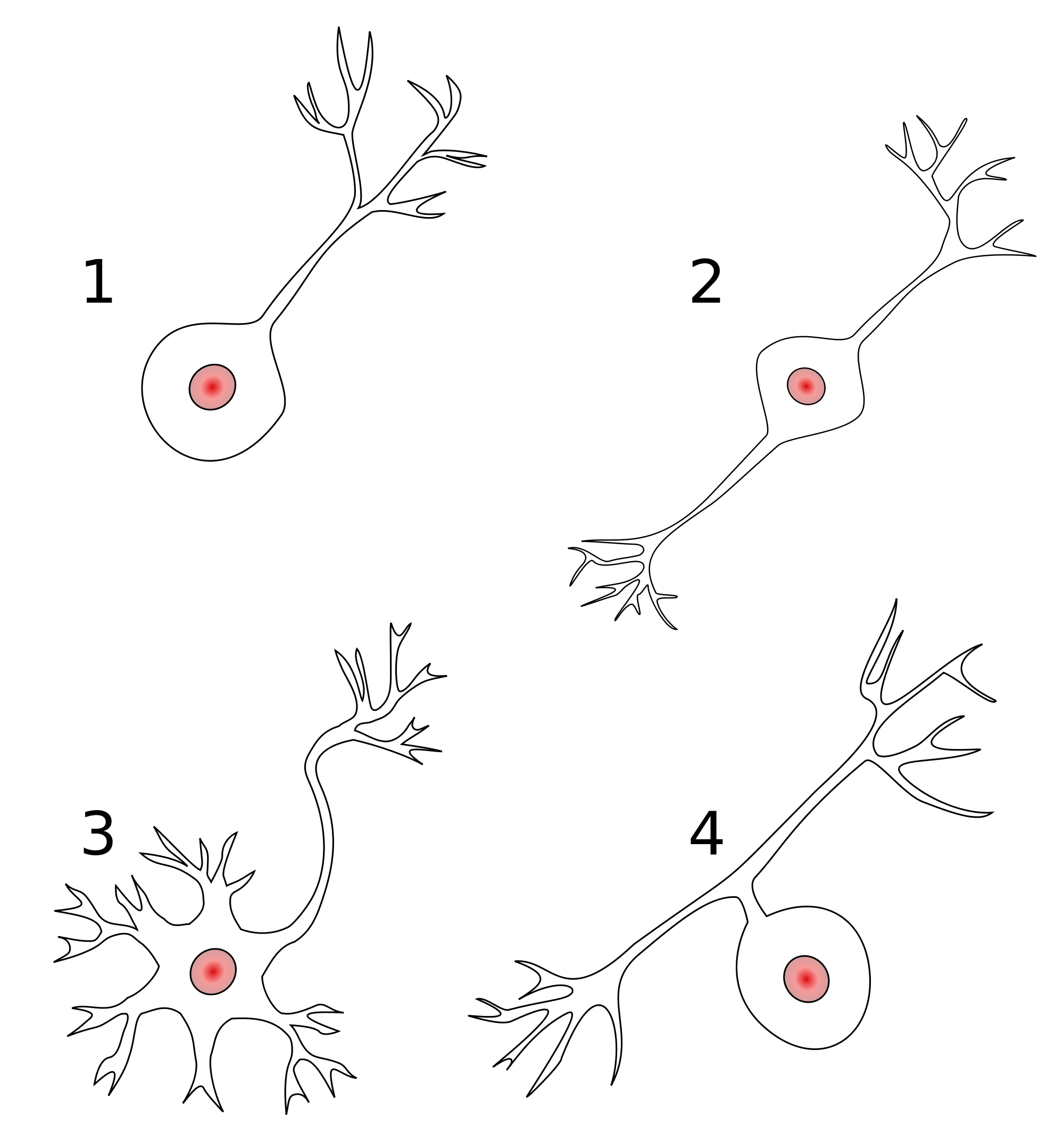
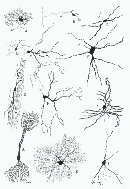
Classifying neurons
- Functional role
- Input (sensory), output (motor/secretory), interneurons
- Anatomy
- Unipolar
- Bipolar
- Multipolar
Classifying neurons
- By specific anatomy
- Pyramidal cells
- Stellate cells
- Purkinje cells
- Granule cells
Neurons by type
How neurons communicate
Neural communication
- Electrical
- Fast(er)
- Metabolically costly
- Within neurons
- Chemical
- Slow(er)
- Metabolically cheap
- Between neurons, & brain <-> body (via hormones)
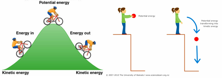
How are messages generated?
- Electrical potential (== voltage)
- Think of potential energy
- Voltage ~ pressure
- Energy that will be released if something changes
Potential energy
Types of neural electrical potentials
Resting potential
- How to measure
- Electrode on inside
- Electrode on outside
- Inside - Outside = potential
Resting potential
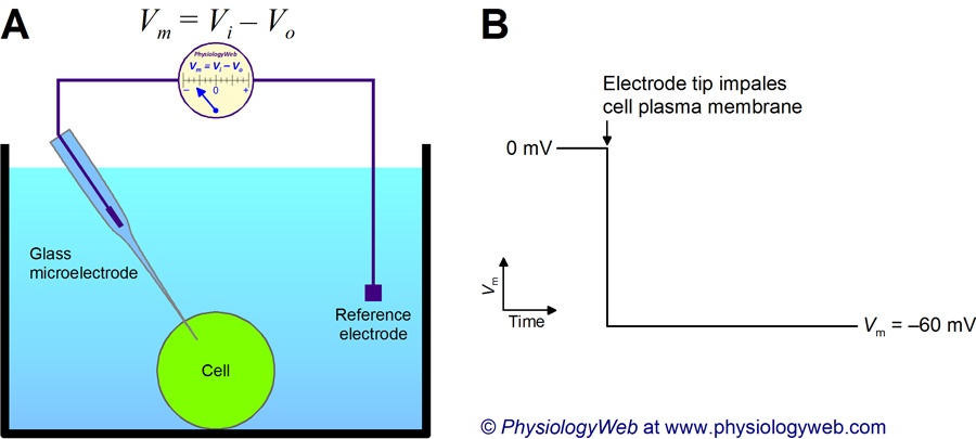
Resting potential
- Neuron (and other cells) have potential energy
- Inside neuron is -60-70 mV, with respect to outside
- About 1/20th typical AAA battery
- Like charges repel, opposites attract, so
- Positively charged particles pulled in
- Negatively charged particles pushed out
Where does the resting potential come from?
- Ions
- Ion channels
- Separation between charges
- A balance of forces
We are the champIONs, my friend
- Potassium, K+
- Sodium, Na+
- Chloride, Cl-
- Organic anions, A-
Resting potential arises from
- A balance of forces
- Force of diffusion
- Electrostatic force
- Forces cause ion flows across membrane
- Ion channels allow ion flow
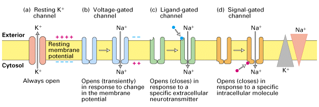
Ion channels
- Openings in neural membrane
- Selective
- Vary in permeability
Ion channel Types
- Passive/leak
- Always open
- Voltage-gated
- Open/close at certain voltages
- Ligand-gated (chemically-gated)
- Open/close in presence of special chemicals (ligands)
- Transporters/pumps
- Move (transport/pump) ions using metabolic energy
Ion channels
Neuron at rest permeable to K+
- Passive K+ channels open
- [K+] concentration inside >> outside
- K+ flows out
Force of diffusion
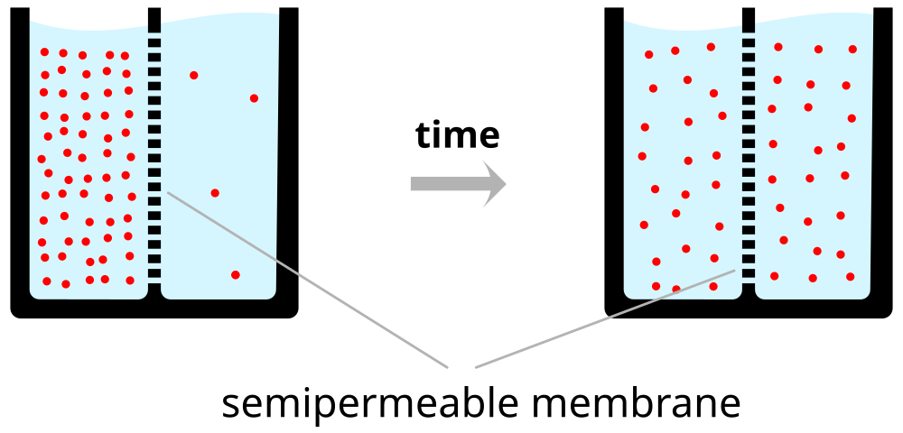
Force of diffusion

Neuron at rest permeable to K+
- Organic anions (A-) too large to move outside of cell
- A- and K+ largely in balance == no net internal charge
- K+ outflow creates charge separation: K+ <-> A-
- Charge separation creates a voltage
- Outside +/inside -
- Voltage build-up stops outflow of K+ (before inside/outside concentrations are ==)
The resting potential

Balance of forces in the neuron at rest
- Force of diffusion
- K+ moves from high concentration (inside) to low (outside)
Balance of forces in the neuron at rest
- Electrostatic force
- Positive K+ accumulate along outside
- Negative A- accumulate along inside
- K+ || A- along membrane creates battery-like voltage
- Voltage build-up stops K+ outflow
Electrostatic force
- Specific voltage called equilibrium potential for K+
- K+ positive, so equilibrium potential negative (w/ respect to outside)
- Equilibrium potential close to neuron resting potential
Equilibrium potentials calculated under typical conditions
| Ion | [inside] | [outside] | Voltage |
|---|---|---|---|
| K+ | ~150 mM | ~4 mM | ~ -90 mV |

Electrical circuit model

Resting potential ≠ K+ equilibrium potential
- Resting potential not just due to K+ flow
- Other ions flow
- Resting potential == net effects of all ion flows across membrane
Next time
- More on neural communication
- What are the other ions doing?
References
Azevedo, Frederico AC, Ludmila RB Carvalho, Lea T Grinberg, José Marcelo Farfel, Renata EL Ferretti, Renata EP Leite, Roberto Lent, Suzana Herculano-Houzel, and others. 2009. “Equal Numbers of Neuronal and Nonneuronal Cells Make the Human Brain an Isometrically Scaled-up Primate Brain.” Journal of Comparative Neurology 513 (5). Wiley Online Library: 532–41.
Bazargani, Narges, and David Attwell. 2016. “Astrocyte Calcium Signaling: The Third Wave.” Nature Neuroscience 19 (2): 182–89. doi:10.1038/nn.4201.
Bhardwaj, Ratan D., Maurice A. Curtis, Kirsty L. Spalding, Bruce A. Buchholz, David Fink, Thomas Björk-Eriksson, Claes Nordborg, et al. 2006. “Neocortical Neurogenesis in Humans Is Restricted to Development.” Proceedings of the National Academy of Sciences 103 (33): 12564–8. doi:10.1073/pnas.0605177103.
Chung, Won-Suk, Christina A. Welsh, Ben A. Barres, and Beth Stevens. 2015. “Do Glia Drive Synaptic and Cognitive Impairment in Disease?” Nature Neuroscience 18 (11): 1539–45. doi:10.1038/nn.4142.
Magrassi, L., K. Leto, and F. Rossi. 2013. “Lifespan of Neurons Is Uncoupled from Organismal Lifespan.” Proceedings of the National Academy of Sciences 110 (11): 4374–9. doi:10.1073/pnas.1217505110.
Zhan, Yang, Rosa C. Paolicelli, Francesco Sforazzini, Laetitia Weinhard, Giulia Bolasco, Francesca Pagani, Alexei L. Vyssotski, et al. 2014. “Deficient Neuron-Microglia Signaling Results in Impaired Functional Brain Connectivity and Social Behavior.” Nature Neuroscience 17 (3): 400–406. doi:10.1038/nn.3641.