2021-09-07 07:22:52
Prelude (2:01)
Today’s topics
- Announcement: Quiz 1 next Thursday (online via Canvas)
- Warm-up
- Wrap up on functional methods
- Neuroanatomy
- Through song and dance
Warm-up
What kind of brain imaging technique does this image represent?

What kind of structural brain imaging technique does this image represent?
- A. Magnetic Resonance Imaging (MRI)
- B. Positron Emission Tomography
- C. Event-related potentials (ERP)
What kind of structural brain imaging technique does this image represent?
- A. Magnetic Resonance Imaging (MRI)
- B.
Positron Emission Tomography - C.
Event-related potentials (ERP)
Which of the following methods has temporal resolution on the order of seconds?
- A. functional MRI
- B. EEG
- C. MEG
- D. single-unit recording
Which of the following methods has temporal resolution on the order of seconds?
- A. functional MRI
- B.
EEG - C.
MEG - D.
single-unit recording
Which of the following methods has high/fine spatial resolution?
- A. functional MRI
- B. PET
- C. EEG
- D. single-unit recording
Which of the following methods has high/fine spatial resolution?
- A.
functional MRI - B.
PET - C.
EEG - D. single-unit recording
Which measure(s) would you use to map connections between brain areas?
- A. retrograde/anterograde cell tracers
- B. diffusion tensor imaging (DTI)
- C. PET neuroimaging
- E. both A & B.
Which measure(s) would you use to measure connections between brain areas?
- A. retrograde/anterograde cell tracers
- B. diffusion tensor imaging (DTI)
- C.
PET neuroimaging - E. both A & B.
Wrap-up on functional methods
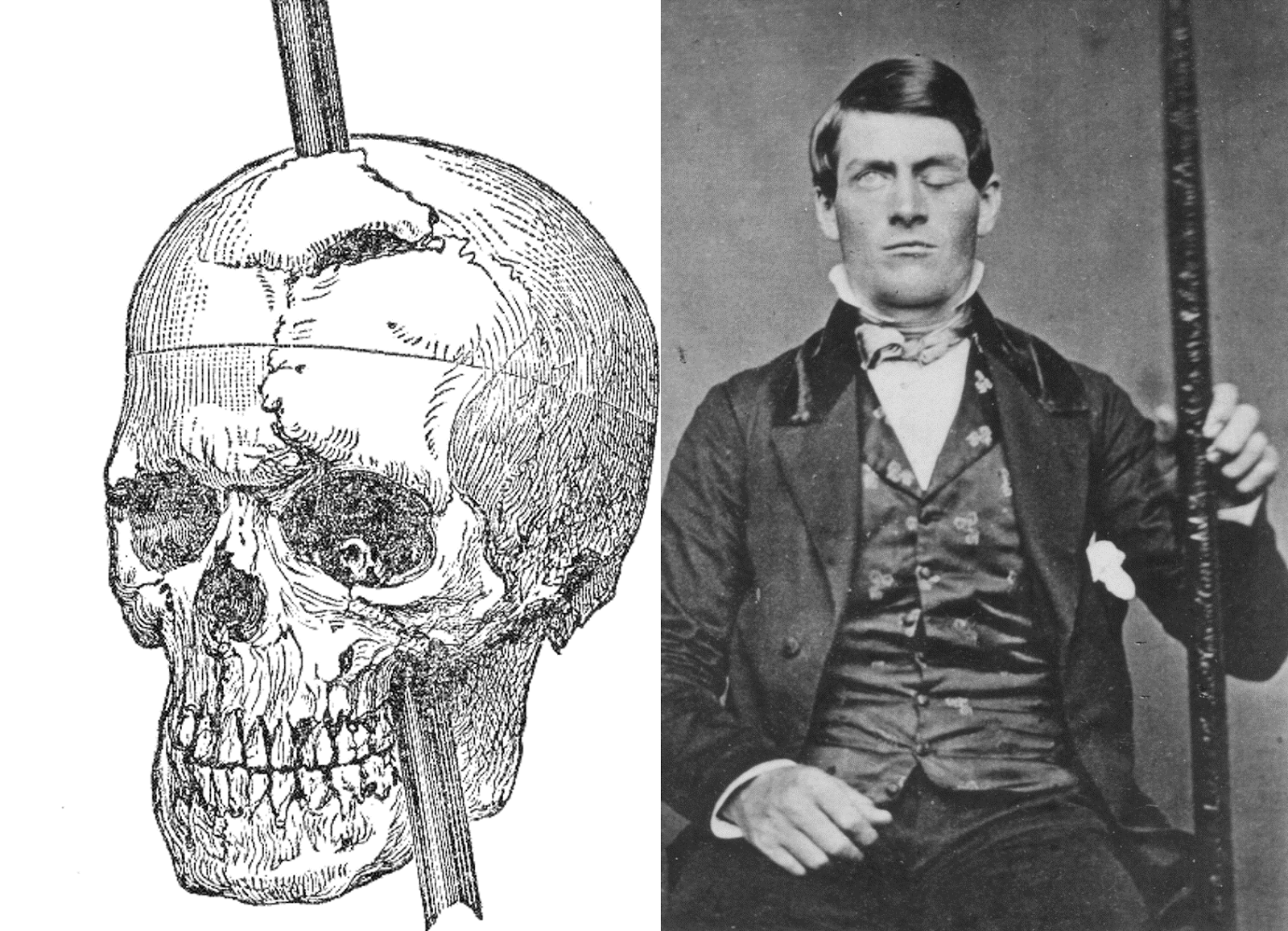
Manipulating the brain
- Nature’s “experiments”
- Stroke, head injury, tumor
- Neuropsychology
- If damage to X impairs performance on Y -> X critical for/controls Y
- Poor spatial/temporal resolution, limited experimental control
Phineas Gage

Stimulating the brain
- Pharmacological
- Electrical (transcranial Direct Current Stimulation - tDCS)
- Inject low levels of electric current
- Magnetic (Transcranial magnetic stimulation - TMS)
- Inject directed pulses of magnetic energy
- Optically (optogenetics)
- Light activates ion channels in neurons, causes current to flow
tDCS
TMS
Optogenetic stimulation
- Insert light-sensitive ion channels into neuronal membrane using genetic engineering
- Open/close channels (activate/inhibit neurons) with light
Evaluating stimulation methods
- Spatial/temporal resolution?
- Assume stimulation mimics natural activity. Does it?
- Optogenetic stimulation similar to natural stimulation, others less so
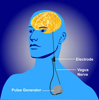
- Deep (electrical) brain stimulation as therapy
- Parkinson’s Disease
- Depression
- Epilepsy
Deep brain stimulation
Simulating the brain
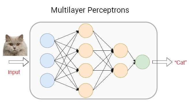
- Computer/mathematical models of brain function
- Example: neural networks
- Cheap, noninvasive, can be stimulated or “lesioned”
Application: AI
Spatial and Temporal Resolution
Bottom line…
- Neuroscientists…
- need to use many tools
- seek converging evidence
Neuroanatomy
Brain anatomy through dance
Finding our way around
Anterior/Posterior
Medial/Lateral
Superior/Inferior
Dorsal/Ventral
Rostral/Caudal
Finding our way around
Anterior/Posterior -> front/back
Medial/Lateral -> inside/outside
Superior/Inferior -> upward/downward
Dorsal/Ventral -> back-ward/belly-ward
Rostral/Caudal -> head-ward/tail-ward
Directional image

Wikipedia
Bipeds vs. quadripeds

Wikipedia
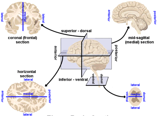
No matter how you slice it
Horizontal/Axial
Coronal/Transverse/Frontal
Sagittal (from the side)
Slice diagram
Supporting structures
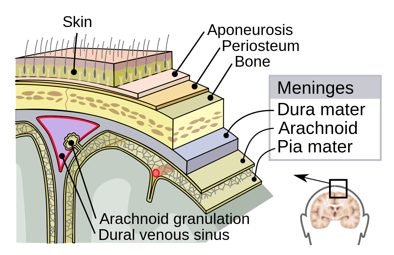
Meninges
Ventricular system
Blood supply
Meninges
Dura mater
Arachnoid membrane
Subarachnoid space
Pia mater
Meninges
Ventricular system
Ventricles
Lateral (1st & 2nd)
3rd
Cerebral aqueduct
4th
(are filled with) Cerebrospinal fluid (CSF)
Blood Supply
Blood Supply
Arteries
- external & internal carotid; vertebral -> basilar
- Circle of Willis
- anterior, middle, & posterior cerebral
Blood/brain barrier
- Isolates CNS from blood stream
- Active transport of molecules typically required
- (endothelial) cells forming blood vessel walls are tightly packed
Blood/brain barrier
exception is Area Postrema
- In brainstem
- Blood-brain barrier thin
- Detects toxins, evokes vomiting
Area Postrema
Organization of the Nervous System
Central Nervous System (CNS)
- Brain
- Spinal Cord
- Everything encased in bone
Peripheral Nervous System (PNS)
Organization of the brain
| Major division | Ventricular Landmark | Embryonic Division | Structure |
|---|---|---|---|
| Forebrain | Lateral | Telencephalon | Cerebral cortex |
| Basal ganglia | |||
| Hippocampus, amygdala | |||
| Third | Diencephalon | Thalamus | |
| Hypothalamus | |||
| Midbrain | Cerebral Aqueduct | Mesencephalon | Tectum, Tegmentum |
Organization of the brain
| Major division | Ventricular Landmark | Embryonic Division | Structure |
|---|---|---|---|
| Hindbrain | 4th | Rhombencephalon | Cerebellum, pons |
| – | Medulla oblongata |
Embryonic brain (~6 weeks gestation)
Next time…
- More neuroanatomy…
References
Abbott, N. J., Rönnbäck, L., & Hansson, E. (2006). Astrocyte-endothelial interactions at the blood-brain barrier. Nature Reviews. Neuroscience, 7(1), 41–53. https://doi.org/10.1038/nrn1824
Begg, D. P., & Woods, S. C. (2013). The endocrinology of food intake. Nature Reviews. Endocrinology, 9(10), 584–597. https://doi.org/10.1038/nrendo.2013.136
Dayan, E., Censor, N., Buch, E. R., Sandrini, M., & Cohen, L. G. (2013). Noninvasive brain stimulation: From physiology to network dynamics and back. Nature Neuroscience, 16(7), 838–844. https://doi.org/10.1038/nn.3422
Sejnowski, T. J., Churchland, P. S., & Movshon, J. A. (2014). Putting big data to good use in neuroscience. Nature Neuroscience, 17(11), 1440–1441. https://doi.org/10.1038/nn.3839
![[[@dayan_noninvasive_2013]](http://doi.org/10.1038/nn.3422)](https://media.springernature.com/full/springer-static/image/art%3A10.1038%2Fnn.3422/MediaObjects/41593_2013_Article_BFnn3422_Fig4_HTML.jpg?as=webp)
![[[@dayan_noninvasive_2013]](http://doi.org/10.1038/nn.3422)](https://media.springernature.com/full/springer-static/image/art%3A10.1038%2Fnn.3422/MediaObjects/41593_2013_Article_BFnn3422_Fig1_HTML.jpg?as=webp)
![[[@sejnowski2014putting]](https://doi.org/10.1038/nn.3839)](https://media.springernature.com/lw685/springer-static/image/art%3A10.1038%2Fnn.3839/MediaObjects/41593_2014_Article_BFnn3839_Fig1_HTML.jpg?as=webp)


![[[@Abbott2006-jw]](http://dx.doi.org/10.1038/nrn1824)](https://media.springernature.com/full/springer-static/image/art%3A10.1038%2Fnrn1824/MediaObjects/41583_2006_Article_BFnrn1824_Fig2_HTML.jpg?as=webp)
![[[@Abbott2006-jw]](http://dx.doi.org/10.1038/nrn1824)](https://media.springernature.com/full/springer-static/image/art%3A10.1038%2Fnrn1824/MediaObjects/41583_2006_Article_BFnrn1824_Fig3_HTML.jpg?as=webp)
![[[@Begg2013-fb]](http://dx.doi.org/10.1038/nrendo.2013.136)](https://media.springernature.com/lw685/springer-static/image/art%3A10.1038%2Fnrendo.2013.136/MediaObjects/41574_2013_Article_BFnrendo2013136_Fig2_HTML.jpg?as=webp)
