Development

2025-10-16

Rick Gilmore

Department of Psychology

Prelude

acapellascience (2017)

Today’s topics

  • Development of the nervous system

Warm-up

The human brain is special in which of the following ways?

  • A. It has a larger-than-expected cerebellum
  • B. It has more neurons in the cerebral cortex
  • C. It has an unusual shape
  • D. Its size is independent of body size

The human brain is special in which of the following ways?

  • A. It has a larger-than-expected cerebellum
  • B. It has more neurons in the cerebral cortex
  • C. It has an unusual shape
  • D. Its size is independent of body size

Vertebrate nervous systems first emerged

  • A. 4.5 billion years ago
  • B. 13.8 billion years ago
  • C. 250 thousand years ago
  • D. 500-600 million years ago

Vertebrate nervous systems first emerged

  • A. 4.5 billion years ago
  • B. 13.7 billion years ago
  • C. 200-300 thousand years ago
  • D. 500-600 million years ago

On family trees

The theory of evolution explains how life began on Earth.

  • A. True
  • B. False

The theory of evolution explains how life began on Earth.

  • A. True
  • B. False

The theory of evolution explains how life has changed since it emerged here ~4 billion years ago.

  • A. True
  • B. False

The theory of evolution explains how life has changed since it emerged here ~4 billion years ago.

  • A. True
  • B. False

Development of the nervous system

Recap

  • Human brain has large cerebral cortex with more neurons than comparable primates
  • How did the human brain get this way?
    • Building upon mammalian/primate norms
    • Specialized patterns of development

What must develop

What must develop

  • CNS among earliest-developing, last to finish organ systems
    • Prolonged developmental period (==childhood) makes CNS especially malleable

Neurons

Synapses

Axons

Timeline

Silbereis, Pochareddy, Zhu, Li, & Sestan (2016)

Prenatal milestones

  • Neuro- and glio-genesis
  • Migration
  • Synaptogenesis begins
  • Cell differentiation
  • Apoptosis
  • Myelination begins

Postnatal milestones

  • Synaptogenesis
  • Cortical expansion, activity-dependent change
    • Then cubic, quadratic, or linear declines in cortical thickness
  • Myelination
  • Prolonged period of postnatal/pre-reproductive development (Konner, 2011)
  • Neurogenesis in selected areas (cerebellum; basal ganglia; hippocampus)

Prenatal period

  • ~38 weeks from conception/fertilization on average
    • Embryonic period (weeks 1-8)
    • Fetal period (weeks 9+)
  • Divided into 3 12-13 week trimesters

Insemination

  • Can occur 3-4 days before or up to 1-2 days after…ovulation
  • Some animals signal ovulation, humans don’t

Fertilization

  • One sperm cell fuses with ovum
  • Within ~ 24 hrs of ovulation

Implantation

  • Fertilized ovum implants in wall of uterus
  • ~ 6 days after fertilization

Early embryogenesis

Khan Academy (n.d.)

Neural tube formation

Neural tube formation

  • Embryonic layers: ectoderm, mesoderm, endoderm
    • Neural tube forms from ectoderm
    • beginning ~ 23 pcd (postconceptual days) or week 3

Neural tube formation

  • Neural tube closes in middle
    • Moves toward rostral & caudal ends
    • closing by 29-30 pcd (week 4)

Neural tube formation

  • Failures of neural tube closure
    • Anencephaly (rostral neuraxis)
    • Spina bifida (caudal neuraxis)
  • Folic acid supplements can prevent

Neural tube formation

  • Neural tube becomes…
    • Ventricles & cerebral aqueduct
    • Central canal of spinal cord

Wikipedia

Neural tube

  • Rostro-caudal patterning via differential growth into vesicles
    • Forebrain (prosencephalon)
    • Midbrain (mesencephalon)
    • Hindbrain (rhombencephalon)

Wikipedia

Neurogenesis and gliogenesis

  • Neuroepithelium cell layer adjacent to neural tube
    • creates ventricular zone (VZ) and subventricular zone (SVZ)
  • Pluripotent stem and progenitor cells divide, produce new neurons & glia

Götz & Huttner (2005)

Neurogenesis and gliogenesis

  • Neurogenesis (of excitatory Glu neurons) observed by 27 pcd (4 pcw; post-conceptual week)
  • Most cortical and striatal neurons generated prenatally, but
    • Cerebellum continues neurogenesis ~ 18 mos postnatal mos

Götz & Huttner (2005)

Can ‘old’ brains make new neurons?

  • In some animals, yes == songbirds, birds that store food caches
  • Humans, on much more limited scale
    • hippocampus (especially dentate gyrus)
    • striatum
    • olfactory bulb (minimally)
    • not much, if any, in cerebral cortex
  • Most neurogenesis occurs near ventricles

Ernst & Frisén (2015)

Cell division & migration

  • Neural progenitor/stem cells
    • Undergo symmetric & asymmetric cell division
    • Generate glia, neurons, and basal progenitor cells
  • Radial glia aid cell migration

Cell division & migration

Cell division & migration

Bui (2006)

Bui (2006)

Cell division & migration

bbscottvids (2009)

Cell division & migration

  • Migration aided by axon growth cones
  • Growth cones guided by
    • Chemoattractants
      • e.g., Nerve Growth Factor (NGF)
    • Chemorepellents
    • Chemical receptors in growth cone detect spatial/temporal patterns

Cell division & migration

Moore (2009)

Cell division & migration

Figure 5 from Kang et al. (2011)

Cell division & migration

  • Glia migrate, too

Figure 4 from Baumann & Pham-Dinh (2001)

Cell differentiation

  • Neuron vs. glial cell
  • Cell type
    • myelin-producing vs. astrocyte vs. microglia
    • pyramidal cell vs. stellate vs. Purkinje vs. …
  • NTs released
  • Where to extend axons

Infancy & early childhood

Timeline of milestones

Silbereis et al. (2016)

Synaptogenesis

  • Begins prenatally (~ 18 pcw)
  • Peak density ~ 15 mos postnatal
  • Spine density in prefrontal cortex ~ 7 yrs postnatal
  • 700K synapses/s on average

Synaptogenesis

  • Synaptic proliferation, pruning
    • Early proliferation (make many synapses)
    • Later pruning
    • Rates, peaks differ by area

Apoptosis (programmed cell death)

  • 20-80% of all cells, varies by area
  • Spinal cord >> cortex
  • Quantity of nerve growth factor (NGF) influences survival

Synaptic rearrangement

  • New synapses added
  • Existing synapses altered
  • Progressive phase: growth rate >> loss rate
  • Regressive phase: growth rate << loss rate

Myelination

  • Neonatal brain largely unmyelinated
  • Gradual myelination, peaks in mid-20s
  • Non-uniform pattern
    • Spinal cord before brain
    • Sensory before motor

Baumann & Pham-Dinh (2001)

Myelination

  • Diffusion Tensor Imaging (DTI)
  • A structural MRI measure of white matter integrity
  • Shows significant developmental changes

Hagmann et al. (2010)

Structural/morphometric development

Figure 2 from Knickmeyer et al. (2008)

Gyri & sulci

Figure 1 from Chi, Dooling, & Gilles (1977)

Gyri & sulci

Figure 2 from Chi et al. (1977)

Gyri & sulci

Figure 3 from Chi et al. (1977)

Gyri & sulci

Figure 4 from Chi et al. (1977)

Changes in brain glucose use

Figure 1 from Kuzawa et al. (2014)

Gene expression across development

Kang et al. (2011)

Wrap-up

Main points

  • Human nervous system develops rapidly beginning about week 3, post-conception
  • Develops throughout lifespan
  • Development consists of progressive & regressive events

Milestone summary

Prenatal

  • Neuro- and gliogenesis
  • Migration
  • Synaptogenesis begins

Postnatal

  • Neurogenesis, but only in selected areas (cerebellum; basal ganglia; hippocampus)
  • Synaptogenesis
  • Cortical expansion, activity-dependent change

Milestone summary

Prenatal

  • Differentiation
  • Cell death
  • Myelination begins

Postnatal

  • Cortex thins
  • Myelination through 3rd decade
  • Prolonged period of postnatal/pre-reproductive development Konner (2011)

Next time

  • Review for Exam 2

Resources

About

This talk was produced using Quarto, using the RStudio Integrated Development Environment (IDE), version 2025.5.1.513.

The source files are in R and R Markdown, then rendered to HTML using the revealJS framework. The HTML slides are hosted in a GitHub repo and served by GitHub pages: https://psu-psychology.github.io/psych-260-2025-fall/

References

acapellascience. (2017, September). Evo-Devo (despacito biology parody) | a capella science. Youtube. Retrieved from https://www.youtube.com/watch?v=ydqReeTV_vk
Baumann, N., & Pham-Dinh, D. (2001). Biology of oligodendrocyte and myelin in the mammalian central nervous system. Physiological Reviews, 81(2), 871–927. https://doi.org/10.1152/physrev.2001.81.2.871
bbscottvids. (2009). Neuronal migration. Youtube. Retrieved from https://www.youtube.com/watch?v=t-8bxeWqSV4
Bui, B. (2006). Neuron migration. Youtube. Retrieved from https://www.youtube.com/watch?v=ZRF-gKZHINk
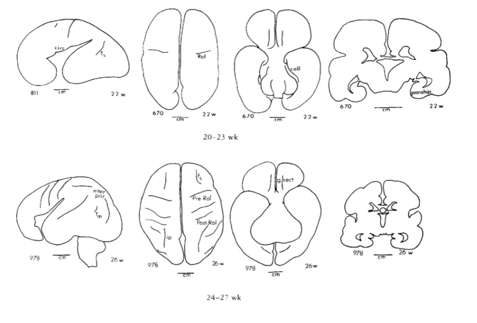
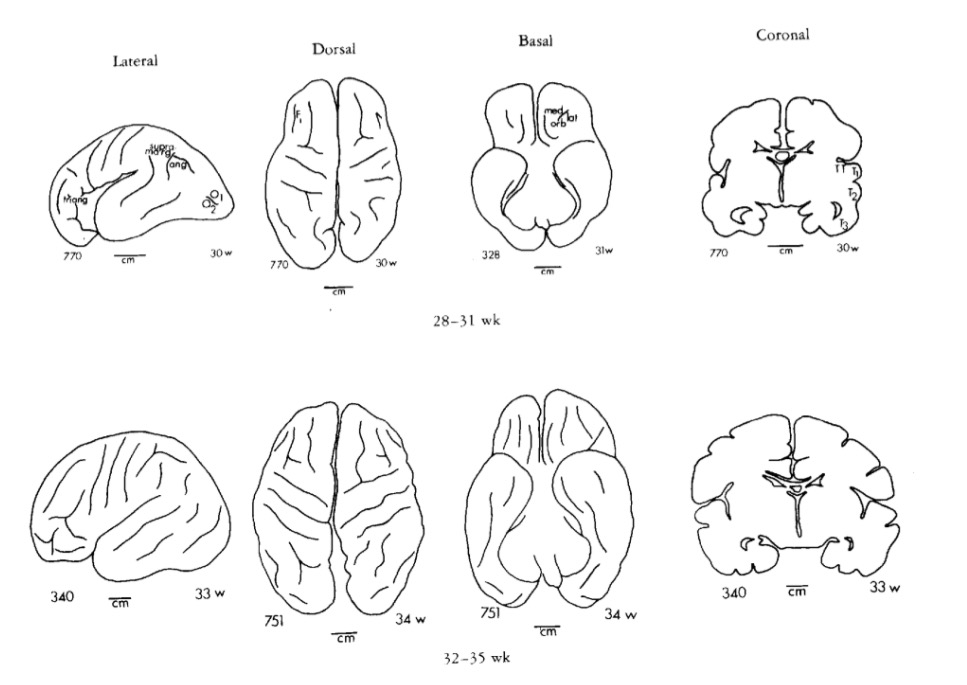
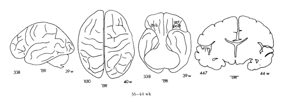
Chi, J. G., Dooling, E. C., & Gilles, F. H. (1977). Gyral development of the human brain. Ann. Neurol., 1(1), 86–93. https://doi.org/10.1002/ana.410010109
DeFelipe, J., Alonso-Nanclares, L., & Arellano, J. I. (2002). Microstructure of the neocortex: Comparative aspects. Journal of Neurocytology, 31(3-5), 299–316. https://doi.org/10.1023/a:1024130211265
Ernst, A., & Frisén, J. (2015). Adult neurogenesis in humans- common and unique traits in mammals. PLoS Biology, 13(1), e1002045. https://doi.org/10.1371/journal.pbio.1002045
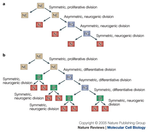
Götz, M., & Huttner, W. B. (2005). The cell biology of neurogenesis. Nat. Rev. Mol. Cell Biol., 6(10), 777–788. https://doi.org/10.1038/nrm1739
Hagmann, P., Sporns, O., Madan, N., Cammoun, L., Pienaar, R., Wedeen, V. J., … Grant, P. E. (2010). White matter maturation reshapes structural connectivity in the late developing human brain. Proceedings of the National Academy of Sciences, 107(44), 19067–19072. https://doi.org/10.1073/pnas.1009073107
Herculano-Houzel, S. (2016). The Human Advantage: A New Understanding of How Our Brain Became Remarkable. MIT Press. Retrieved from https://market.android.com/details?id=book-DMqpCwAAQBAJ
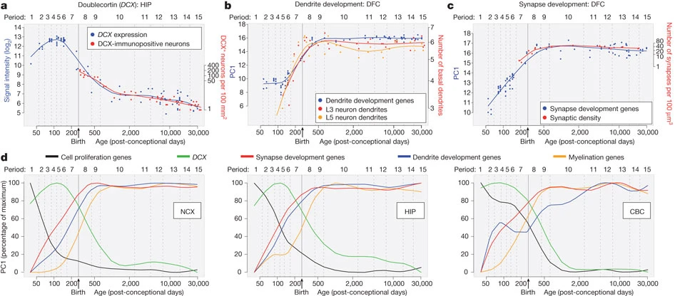
Kang, H. J., Kawasawa, Y. I., Cheng, F., Zhu, Y., Xu, X., Li, M., … Šestan, N. (2011). Spatio-temporal transcriptome of the human brain. Nature, 478(7370), 483–489. https://doi.org/10.1038/nature10523
Kety, S. S., & Schmidt, C. F. (1948). The Nitrous OXIDE METHOD FOR THE QUANTITATIVE DETERMINATION OF CEREBRAL BLOOD FLOW IN MAN: THEORY, PROCEDURE AND NORMAL VALUES. The Journal of Clinical Investigation, 27(4), 476–483. https://doi.org/10.1172/JCI101994
Khan Academy. (n.d.). Early embryogenesis. Youtube. Retrieved from https://www.youtube.com/embed/dAOWQC-OBv0
Knickmeyer, R. C., Gouttard, S., Kang, C., Evans, D., Wilber, K., Smith, J. K., … Gilmore, J. H. (2008). A structural MRI study of human brain development from birth to 2 years. J. Neurosci., 28(47), 12176–12182. https://doi.org/10.1523/JNEUROSCI.3479-08.2008
Konner, M. (2011). The Evolution of Childhood. Belknap Press of Harvard University Press. Retrieved from http://www.hup.harvard.edu/catalog.php?isbn=9780674062016
Kuzawa, C. W., Chugani, H. T., Grossman, L. I., Lipovich, L., Muzik, O., Hof, P. R., … Lange, N. (2014). Metabolic costs and evolutionary implications of human brain development. Proc. Natl. Acad. Sci. U. S. A., 111(36), 13010–13015. https://doi.org/10.1073/pnas.1323099111
Marner, L., Nyengaard, J. R., Tang, Y., & Pakkenberg, B. (2003). Marked loss of myelinated nerve fibers in the human brain with age. The Journal of Comparative Neurology, 462(2), 144–152. https://doi.org/10.1002/cne.10714
Moore, S. (2009). Growth cone filopodia. YouTube. Retrieved from https://www.youtube.com/watch?v=Fgmt2RBow0I
Silbereis, J. C., Pochareddy, S., Zhu, Y., Li, M., & Sestan, N. (2016). The cellular and molecular landscapes of the developing human central nervous system. Neuron, 89(2), 248–268. https://doi.org/10.1016/j.neuron.2015.12.008
The real cost of your AI use: Inside a power hungry data center. (n.d.). YouTube. Retrieved from https://youtube.com/shorts/Tx0uoiuLAZg?si=-btFR6AwcDm_OPbJ