Neuroanatomy

PSY 511.003 Spr 2025

Prelude

ctdalilah (2006)

Kids Learning Tube (2015)

Announcements

  • Exercise 02 due next Wednesday, January 29, 2025.

Today’s topics

Neuroanatomy

Why study?

  • Master basic vocabulary for reading literature

Information processing perspective

  • Inputs from
    • World
    • Body
    • Brain1
  • Outputs to
    • World
    • Body
    • Brain
  • What computations occur where

Functional anatomy perspective

  • Located where
  • Connected to
  • Involved in

Brain anatomy through dance

Directional terms

  • Anterior/Posterior
  • Medial/Lateral
  • Superior/Inferior
  • Dorsal/Ventral
  • Rostral/Caudal

Central vs. Peripheral Nervous System

Nervous system of Harriet Cole, see McNaughton (2018)

Organization of the CNS

Major division Ventricular Landmark Embryonic Division Structure
Forebrain Lateral Telencephalon Cerebral cortex
Basal ganglia
Hippocampus, amygdala
Major division Ventricular Landmark Embryonic Division Structure
Third Diencephalon Thalamus
Hypothalamus
Major division Ventricular Landmark Embryonic Division Structure
Midbrain Cerebral Aqueduct Mesencephalon Tectum, tegmentum
Major division Ventricular Landmark Embryonic Division Structure
Hindbrain 4th Metencephalon Cerebellum, pons
Mylencephalon Medulla oblongata

Forebrain, midbrain, hindbrain terminology derives from embryonic stages in CNS development.

Embryonic human brain from Wikipedia

Cerebrum

  • (Cerebral) cortex
  • Subcortical (below the cortex) structures

Labelled MRI including thalamus and hypothalamus

(Cerebral) cortex

  • Lobes, marked by sulci/fissures, adjacency to bones of skull
  • Functional areas marked by gyri & sulci
  • Brodmann Areas
    • Microstructural (cellular composition) features
  • Most lobes host primary sensory or motor area
    • Frontal: Motor cortex
    • Temporal: Auditory cortex
    • Parietal: Somatosensory cortex
    • Occipital: Visual cortex
    • Insula: Gustatory

Input/output

  • Via cranial nerves (in CNS)
  • Via spinal nerves (in PNS)

Cranial nerves from https://www.britannica.com/science/cranial-nerve#/media/1/141797/46720

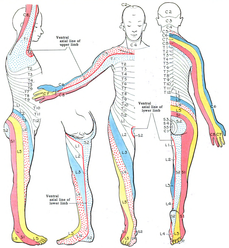
https://www.researchgate.net/profile/Saeede-Rahimi-Damirchi-Darasi/publication/324683974/figure/fig10/AS:753484697726976@1556656159277/Diagram-showing-the-relationship-between-spinal-nerve-roots-and-vertebrae-27.jp

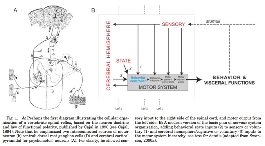
Multiple hierarchies

(Swanson, 2005, fig. 1)

Maps

  • Sensory & motor systems often use maps

https://www.researchgate.net/profile/Brian-Wandell/publication/8988812/figure/fig1/AS:280067633631238@1443784735289/The-Retinotopy-paradigm-Two-stimuli-are-used-to-measure-the-retinotopic-maps-in-cortex.png

Figure 1 from Saenz & Langers (2014)

Wikipedia

  • Geometric issues central for perceptual/motor behavior
  • Do other systems “map” function to structure?

Neuroanatomy lab

Overview

  • 3 groups
  • Rotate among stations every ~25 min
  • Identify as many structures as possible

Wrap-up

Main points

  • Directional terms
  • What is it
  • Where is it
    • Relative to other things
    • CNS/PNS
    • Forebrain/midbrain/hindbrain
  • Cerebral cortex and its subparts
  • Grey matter vs. white matter

Next time…

Resources

References

ctdalilah. (2006, October). Pinky and the brain-brainstem. Youtube. Retrieved from https://www.youtube.com/watch?v=snO68aJTOpM
Kids Learning Tube. (2015, October). Human body for Kids/Brain Song/Human body systems. Youtube. Retrieved from https://www.youtube.com/watch?v=Qw8E9WnZTQk
McNaughton, A. (2018, November 20). Dissecting harriet cole: Uncovering women’s history in the archives. Retrieved January 8, 2025, from https://drexel.edu/legacy-center/blog/overview/2018/november/dissecting-harriet-cole-uncovering-womens-history-in-the-archives/
Saenz, M., & Langers, D. R. M. (2014). Tonotopic mapping of human auditory cortex. Hearing Research, 307, 42–52. https://doi.org/10.1016/j.heares.2013.07.016
Swanson, L. W. (2005). Anatomy of the soul as reflected in the cerebral hemispheres: Neural circuits underlying voluntary control of basic motivated behaviors. Journal of Comparative Neurology, 493(1), 122–131. https://doi.org/10.1002/cne.20733