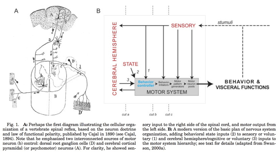
flowchart TD
  N[Nervous System] --> B[Body]
  B --> W[World]
  W --> B
  B --> N


Main points
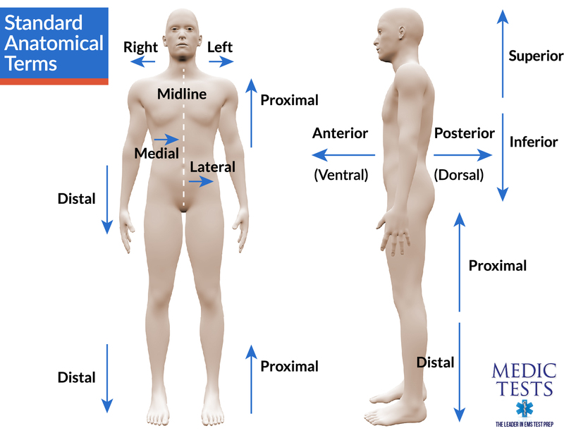
- Directional terms
- What is it
- Where is it
- Relative to other things
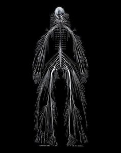
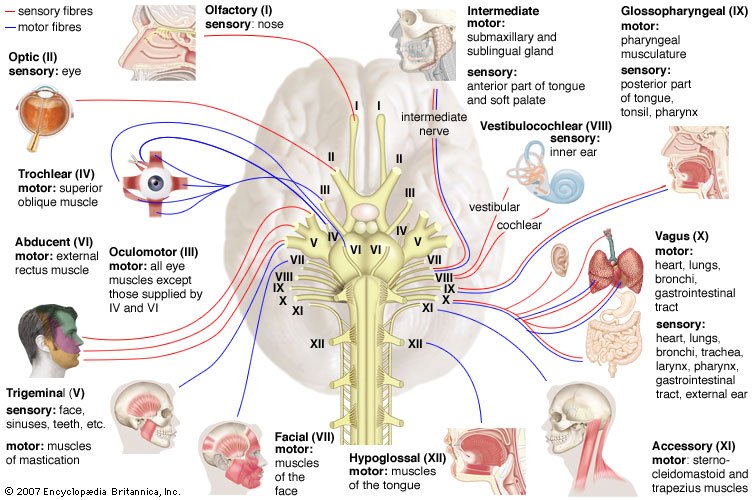
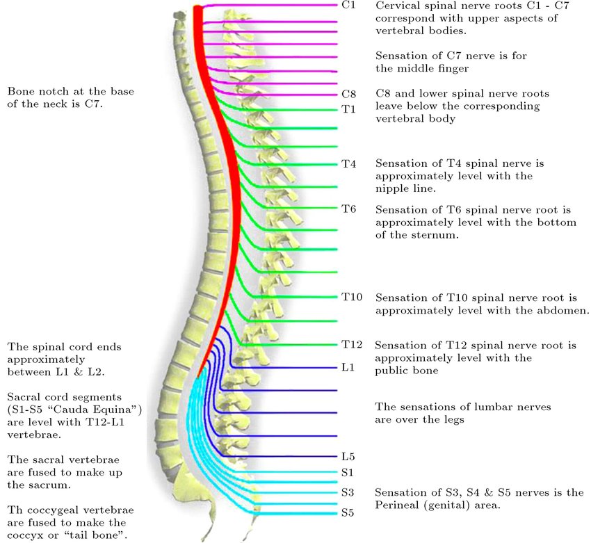
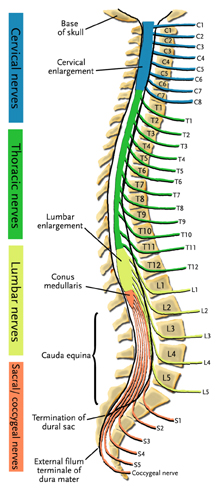
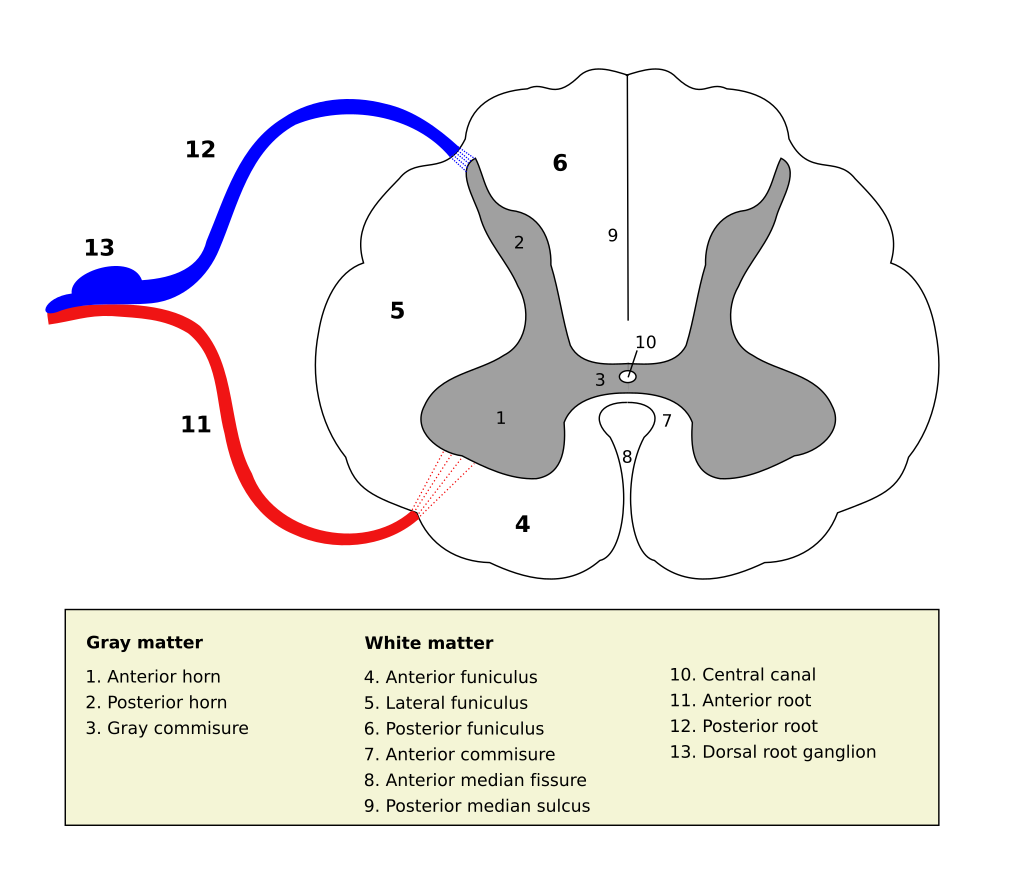
- CNS/PNS
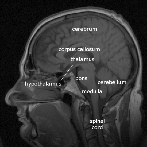
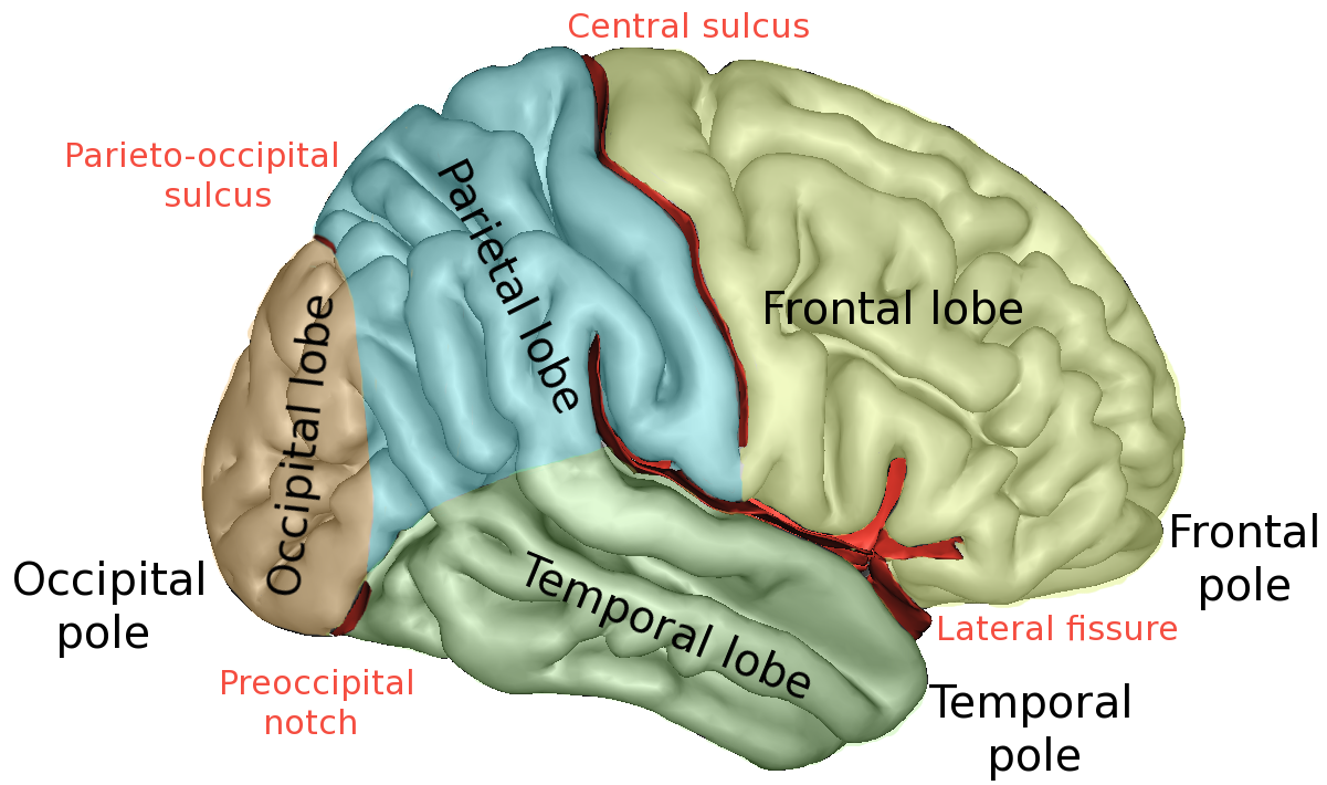
- Forebrain/midbrain/hindbrain
PSY 511.001 Spr 2026


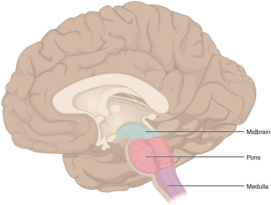


“By Original: OpenStax Vector: Pixelsquid🎱 - Own work based on: Human Neuroaxis-en.jpg:, CC BY 4.0, https://commons.wikimedia.org/w/index.php?curid=97420092”
By Own work, CC BY 3.0, https://commons.wikimedia.org/w/index.php?curid=10187018
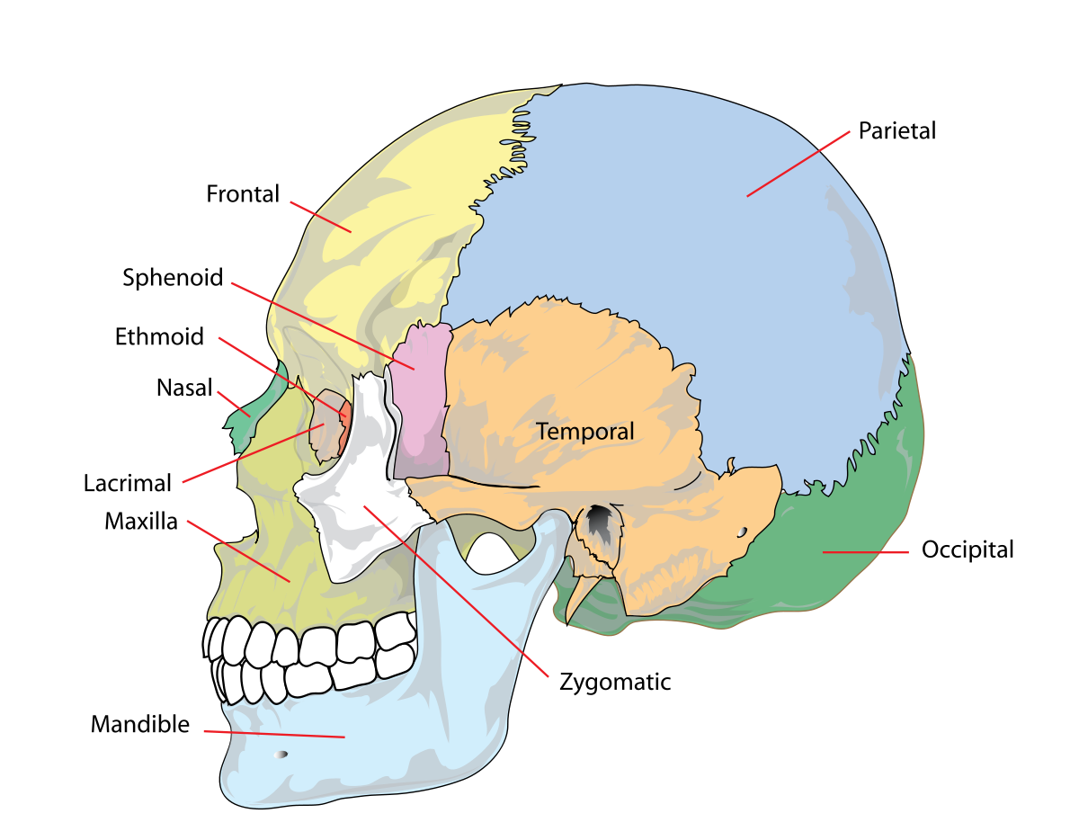
By Sebastian023, CC BY-SA 3.0, https://commons.wikimedia.org/w/index.php?curid=21020857
By Sebastian023, CC BY-SA 3.0, https://commons.wikimedia.org/w/index.php?curid=21020857
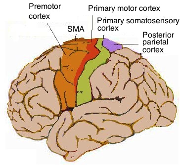
By Cortex sensorimoteur1.jpg: Pancratderivative work: Iamozy - Own work, This file was derived from: Cortex sensorimoteur1.jpg:, CC BY-SA 3.0, https://commons.wikimedia.org/w/index.php?curid=33981076
By Chittka L, Brockmann - Perception Space—The Final Frontier, A PLoS Biology Vol. 3, No. 4, e137 doi:10.1371/journal.pbio.0030137 ([1]/[2]), vectorised by Inductiveload, CC BY-SA 2.5, https://commons.wikimedia.org/w/index.php?curid=5958918
By Jimhutchins (talk) - The original image was uploaded on en.wikipedia as en:Image:Postcentral_gyrus.png, CC BY-SA 3.0, https://commons.wikimedia.org/w/index.php?curid=4481990
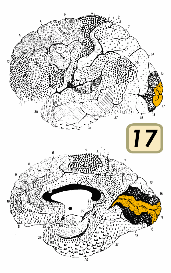
By Brodmann - File:Brodmann_Cytoarchitectonics.PNG, Public Domain, https://commons.wikimedia.org/w/index.php?curid=8221432
Figure 1. Anatomy of the Human Insula. The human insular cortex is bilaterally located deep within the lateral sulcus separating the temporal lobe from the parietal and frontal lobes. The insula is covered with folds of the adjacent frontal, parietal, and temporal opercula. The circumference of the insula is outlined by the circular sulcus, and the deep central sulcus of the insula separates the anterior and posterior parts. Three short insular gyri are found in the anterior insula (AI), whereas two long insular gyri lie in the posterior insula (PI). Cytoarchitecturally, the insula is roughly divided into anterior agranular and posterior granular sections with a transitional dysgranular mid-section.
By Own work, CC BY 3.0, https://commons.wikimedia.org/w/index.php?curid=10187018
By OpenStax College - Anatomy & Physiology, Connexions Web site. http://cnx.org/content/col11496/1.6/, Jun 19, 2013., CC BY 3.0, https://commons.wikimedia.org/w/index.php?curid=30148020
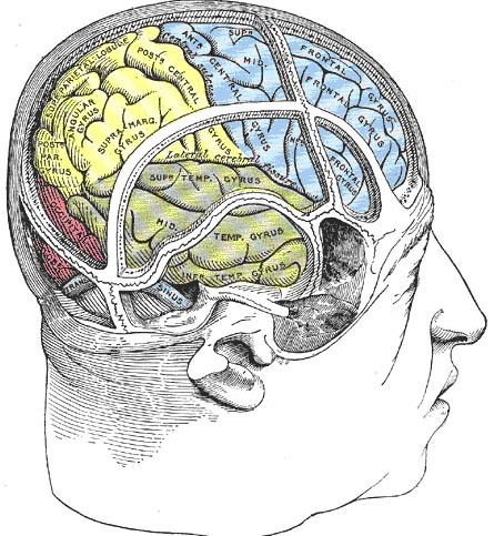
By Henry Vandyke Carter - Henry Gray (1918) Anatomy of the Human Body (See “Book” section below)Bartleby.com: Gray’s Anatomy, Plate 839, Public Domain, https://commons.wikimedia.org/w/index.php?curid=792179