2020-11-12 07:58:43
Traveling at Warp 1
Prelude 3:20
Prelude (2:03)
Announcements
- Exam 3 next Tuesday, 11/17
Today’s topics
Vision
How vision informs
- What’s out there?
- Shape, form, color
- Where is it?
- Position, orientation, motion
Electromagnetic (EM) radiation
Features of EM radiation
- Wavelength or frequency
- Intensity
- Location/position of source
- Reflects off some materials
- Refracted (bent) moving through other materials
EM radiation provides information across space (and time)
Reflectance spectra differ by surface
Reflectance spectra
Optic array specifies geometry of environment
Color == categories of wavelength
- Eyes categorize wavelength into relative intensities within wavelength bands
- RGB ~
Red,
Green,
Blue
- Long, medium, short wavelengths
- Color is a neural/psychological construct
RGB monitors
How a camera works
The biological camera
The biological camera
Parts of the eye
- Cornea - refraction (2/3 of total)
- Pupil - light intensity; diameter regulated by the Iris.
- Lens - refraction (remaining 1/3; variable focus)
Parts of the eye
- Retina - light detection
- ~ skin or organ of Corti
- Pigment epithelium - regenerate photopigment
- Muscles - move eye, reshape lens, change pupil diameter
Eye forms image on retina
- Image inverted (up/down)
- Image reverseed (left/right)
- Point-to-point map (retinotopic)
- Binocular and monocular zones
Retinal image
Eyes views overlap
The fovea
The fovea
- Central 1-2 deg of visual field
- ~ thumbnail @ arm’s length
- Aligned with visual axis; center of gaze
- Retinal ganglion cells pushed aside
- Highest acuity vision == best for details
Acuity varies across the retina
Acuity varies across the retina
What part of the skin is like the fovea?
Photoreceptors detect light
Photoreceptors detect light
- Rods
- ~120 M/eye
- Mostly in periphery
- Active in low light conditions
- One wavelength range
Photorceptors detect light
- Cones
- ~5 M/eye
- Mostly in center
- 3 wavelength ranges
Photoreceptors “specialize” in particular wavelengths
Anatomy & Physiology, Connexions Web site. http://cnx.org/content/col11496/1.6/, Jun 19, 2013.
How photoreceptors work
- Outer segment
- Membrane disks
- Photopigments
- Sense light, trigger chemical cascade
- Inner segment
- Synaptic terminal
- Light hyperpolarizes photoreceptor!
- The dark current
Retina
- Physiologically backwards
- How?
- Anatomically inside-out
- How?
Retina
- Physiologically backwards
- Dark current (more NT released in dark)
- Anatomically inside-out
- Photoreceptors at back of eye
Retinal layers
Retinal layers
- Bipolar cells
- Horizontal cells
- Retinal ganglion cells
- Amacrine cells
Center-surround receptive fields
Center-surround receptive fields
- Center region
- Excites (or inhibits)
- Surround region
- Does the opposite
- Bipolar cells & Retinal Ganglion cells ->
- Most activated by “donuts” of light/dark
- Local contrast (light/dark differences)
What’s a reddish-green look like?
What’s a reddish-green look like?
Opponent processing
Opponent processing
- Black vs. white (achromatic)
- Long ( red) vs. Medium ( green) wavelength cones
- (Long + Medium) vs. Short ( blue) cones
- Can’t really see reddish-green or bluish-yellow
From eye to brain
From eye to brain
- Retinal ganglion cells
- 2nd/II cranial (optic) nerve
- Optic chiasm
From eye to brain
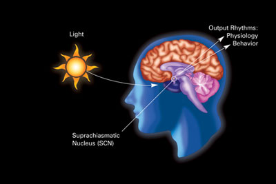
- Hypothalamus
- Suprachiasmatic nucleus
- Regulates circadian (day/night) rhythm via pineal gland
- Suprachiasmatic nucleus
From eye to brain
- Superior colliculus & brainstem
Lateral Geniculate Nucleus (LGN) of thalamus
- ~90% of axons from retina
LGN
- 6 layers + intralaminar zone
- Parvocellular (small cells): chromatic
- Magnocellular (big cells): achromatic
- Koniocellular (chromatic - short wavelength?)
- Retinotopic map of opposite visual field
From LGN to V1
From LGN to V1
- Via optic radiations
- Primary visual cortex (V1) in occipital lobe
Human V1
Measuring retinotopy in V1
Retinotopy in V1
- Fovea overrepresented
- Analogous to somatosensation
- High acuity in fovea vs. lower outside it
- Upper visual field/lower (ventral) V1 and vice versa
V1 has laminar, columnar organization
V1 has laminar, columnar organization
- 6 laminae (layers)
- Input: Layer 4
- Output: Layers 2-3 (to cortex), 5 (to brainstem), 6 (to LGN)
V1 has laminar, columnar organization
- Columns
- Orientation/angle
- Spatial frequency
Orientation/angle tuning
From center-surround receptive fields to line detection

Spatial frequency tuning
Low == gist || high == details
V1 has laminar, columnar organization
- Columns
- Color/wavelength
- Eye of origin, ocular dominance
Ocular dominance columns
Ocular dominance signals retinal disparity
Beyond V1
Beyond V1
- Larger, more complex receptive fields
- Dorsal stream (where/how)
- Toward parietal lobe
- Ventral stream (what)
What is vision for?
- What is it? (form perception)
- Where is it? (space perception)
- How do I get from here to there (action control)
- What time (or time of year) is it?
References
Dougherty, R. F., Koch, V. M., Brewer, A. A., Fischer, B., Modersitzki, J., & Wandell, B. A. (2003). Visual field representations and locations of visual areas V1/2/3 in human visual cortex. Journal of Vision, 3(10), 1–1. https://doi.org/10.1167/3.10.1
Panichello, M. F., Cheung, O. S., & Bar, M. (2013). Predictive feedback and conscious visual experience. Perception Science, 3, 620. https://doi.org/10.3389/fpsyg.2012.00620


