More on methods
2025-09-02
What method does the image reflect?

- A. Retrograde tract tracing
- B. Magnetic Resonance Imaging (MRI)
- C. Computed Tomography (CT)
- D. Nissl (whole cell) stain
What method does the image reflect?
A. Retrograde tract tracingB. Magnetic Resonance Imaging (MRI)C. Computed Tomography (CT)- D. Nissl (whole cell) stain
Single/multi-unit Recording
- What does neuron X respond to?
- High temporal (ms) & spatial resolution (um)
- Invasive
- Used in non-human animals for purely research purposes

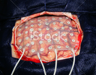
Electrocorticography (ECoG)
- Used in human neurosurgery
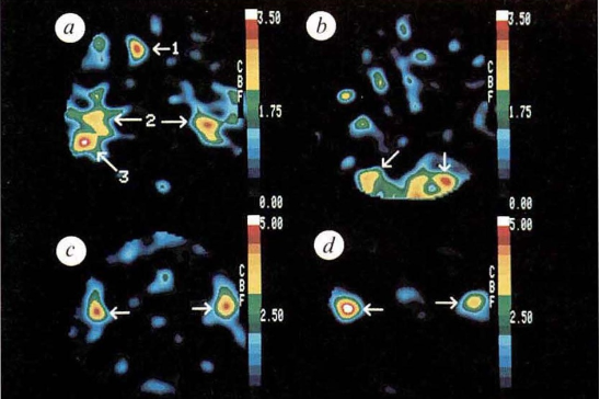
PET
- Temporal (~ s) and spatial (mm-cm) resolution worse than fMRI
- Radioactive exposures + mildly invasive
- Dose < airline crew exposure in 1 yr
Functional Magnetic Resonance Imaging (fMRI)
- Neural activity -> local \(O_2\) consumption increase
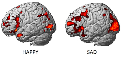
fMRI
- Blood Oxygen Level Dependent (BOLD) response
- Oxygenated vs. deoxygenated hemoglobin creates magnetic contrast
- Do regional blood \(O_2\) volumes (and flow) vary with behavior X?
Mapping visual cortex
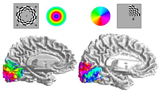
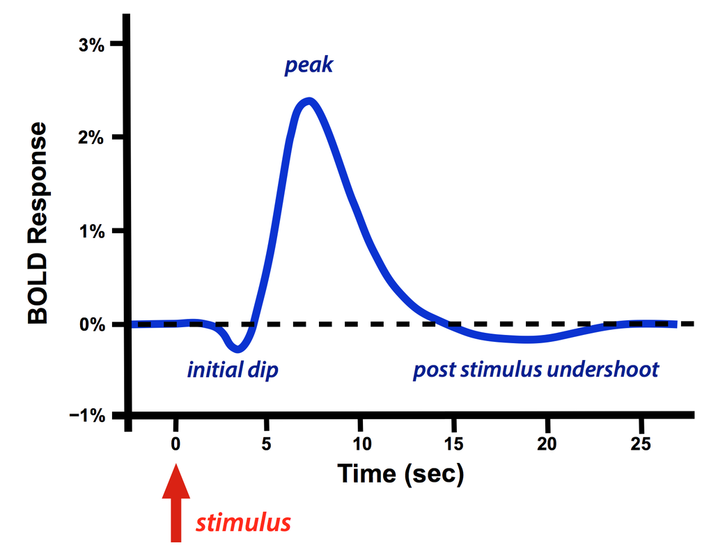
fMRI
- Hemodynamic Response Function (HRF)
- 1s delay plus 3-6 s ‘initial-dip’

Electroencephalography (EEG)
- How does it work?
- Electrodes on scalp or brain surface
- What do we measure?
- Combined activity of huge # of neurons
- High/fine temporal resolution (detect fast changes) but poor spatial resolution

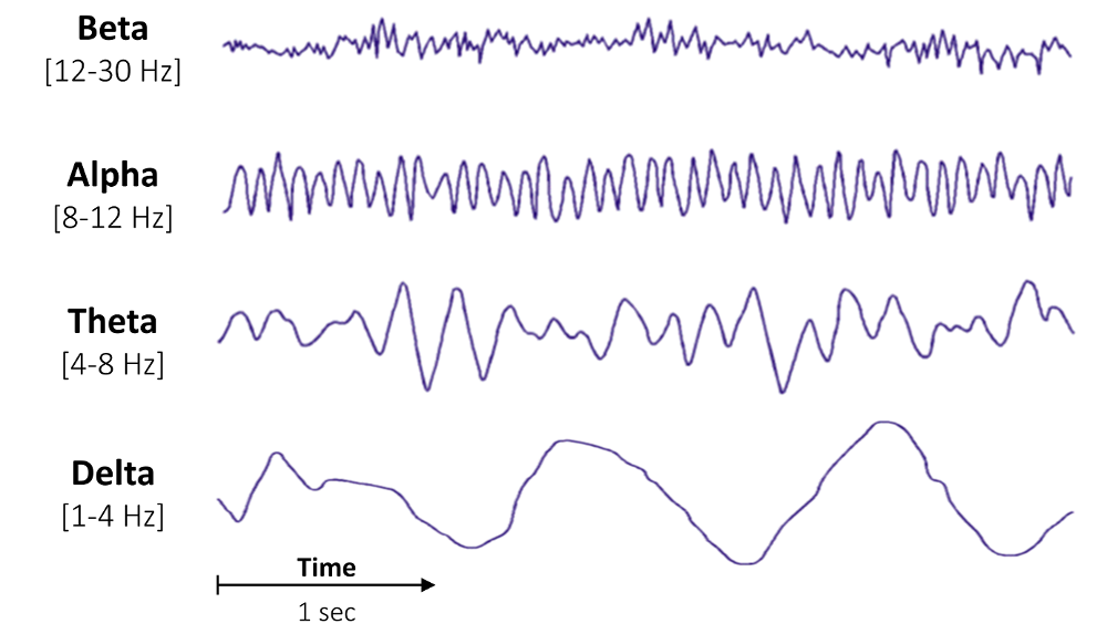
EEG
- Activity in different frequency bands
- LOW (slow changes): deep sleep
- MIDDLE: Quiet, alert state
- HIGH (fast changes): “Binding” information across senses
Event-related potentials (ERPs)
- EEGs related (in time) to some event
- Averaged over many repetitions of that event

Brain Computer Interface (BCI)
- Often based on EEG.

Magneto-encephalography (MEG)
- Like EEG, but measures magnetic fields
- High temporal resolution, low spatial resolution
- Magnetic field propagates with minimal distortion from brain/skull, unlike electric field

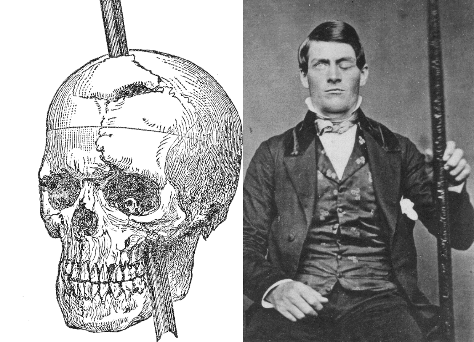
The case of Phineas Gage
Trans-cranial Direct Current Stimulation (tDCS)
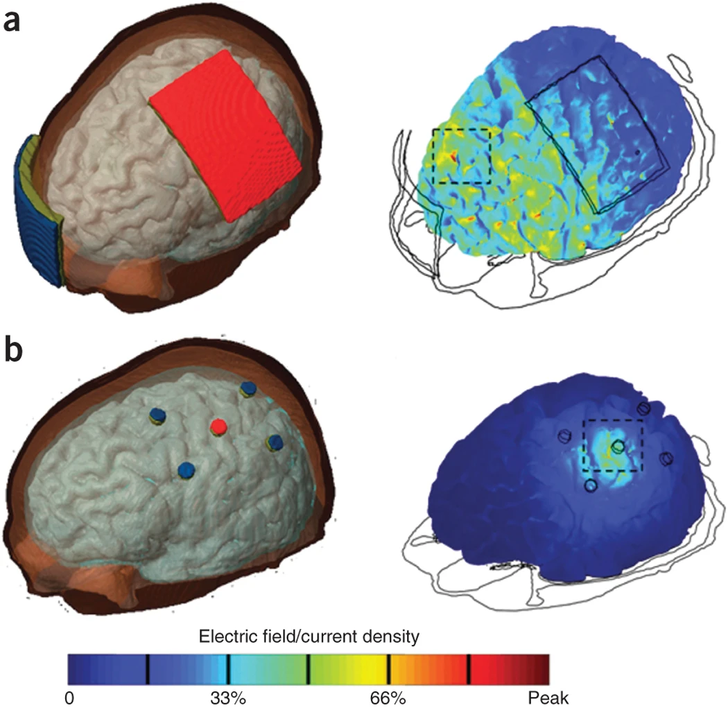
Dayan, Censor, Buch, Sandrini, & Cohen (2013)
Trans-cranial Magnetic Stimulation (TMS)
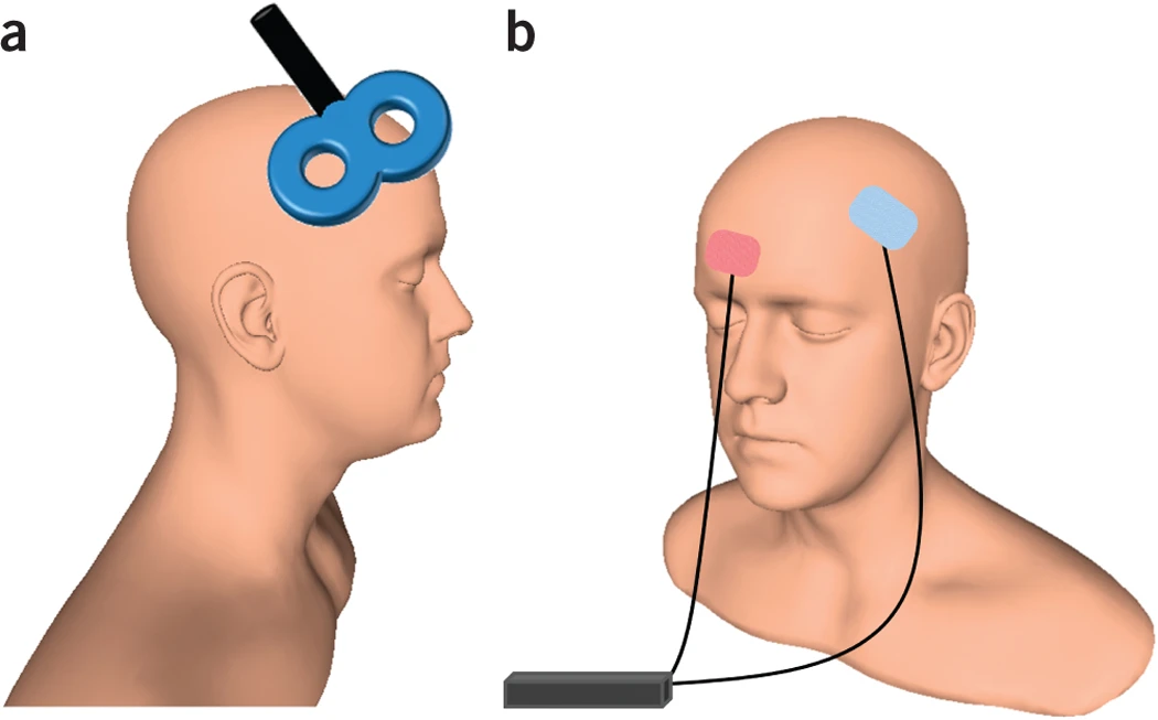
Dayan et al. (2013)
Deep brain stimulation (DBS)
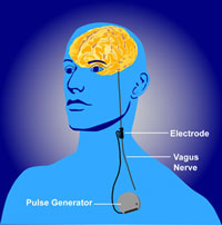
Understanding Animal Research (2009)
Application: AI
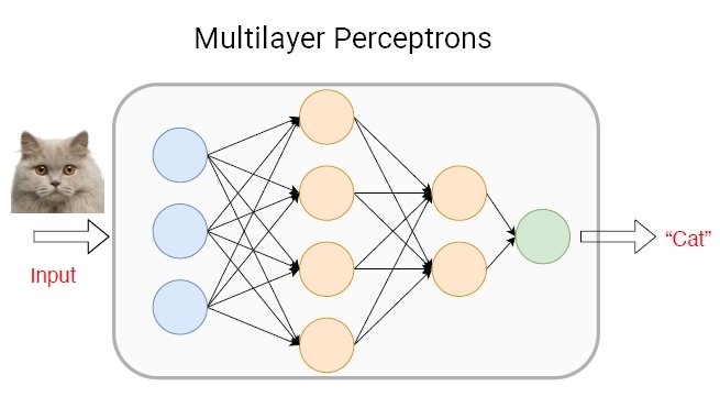
Redmon (2018)
Spatial and Temporal Resolution revisited
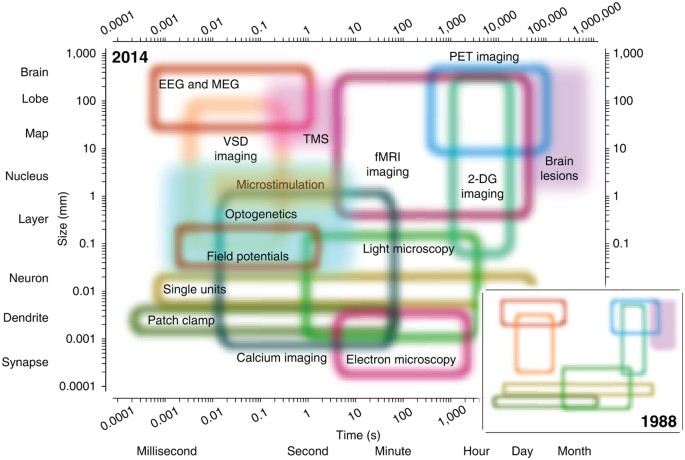
Sejnowski, Churchland, & Movshon (2014)